US11773171B2 - WNT surrogate molecules and uses thereof - Google Patents
WNT surrogate molecules and uses thereof Download PDFInfo
- Publication number
- US11773171B2 US11773171B2 US16/954,484 US201816954484A US11773171B2 US 11773171 B2 US11773171 B2 US 11773171B2 US 201816954484 A US201816954484 A US 201816954484A US 11773171 B2 US11773171 B2 US 11773171B2
- Authority
- US
- United States
- Prior art keywords
- fzd
- binding
- seq
- wnt
- antigen
- Prior art date
- Legal status (The legal status is an assumption and is not a legal conclusion. Google has not performed a legal analysis and makes no representation as to the accuracy of the status listed.)
- Active, expires
Links
Images


Classifications
-
- C—CHEMISTRY; METALLURGY
- C07—ORGANIC CHEMISTRY
- C07K—PEPTIDES
- C07K16/00—Immunoglobulins [IGs], e.g. monoclonal or polyclonal antibodies
- C07K16/18—Immunoglobulins [IGs], e.g. monoclonal or polyclonal antibodies against material from animals or humans
- C07K16/22—Immunoglobulins [IGs], e.g. monoclonal or polyclonal antibodies against material from animals or humans against growth factors ; against growth regulators
-
- A—HUMAN NECESSITIES
- A61—MEDICAL OR VETERINARY SCIENCE; HYGIENE
- A61K—PREPARATIONS FOR MEDICAL, DENTAL OR TOILETRY PURPOSES
- A61K39/00—Medicinal preparations containing antigens or antibodies
- A61K39/395—Antibodies; Immunoglobulins; Immune serum, e.g. antilymphocytic serum
- A61K39/39533—Antibodies; Immunoglobulins; Immune serum, e.g. antilymphocytic serum against materials from animals
- A61K39/3955—Antibodies; Immunoglobulins; Immune serum, e.g. antilymphocytic serum against materials from animals against proteinaceous materials, e.g. enzymes, hormones, lymphokines
-
- C—CHEMISTRY; METALLURGY
- C07—ORGANIC CHEMISTRY
- C07K—PEPTIDES
- C07K16/00—Immunoglobulins [IGs], e.g. monoclonal or polyclonal antibodies
- C07K16/18—Immunoglobulins [IGs], e.g. monoclonal or polyclonal antibodies against material from animals or humans
- C07K16/28—Immunoglobulins [IGs], e.g. monoclonal or polyclonal antibodies against material from animals or humans against receptors, cell surface antigens or cell surface determinants
- C07K16/2863—Immunoglobulins [IGs], e.g. monoclonal or polyclonal antibodies against material from animals or humans against receptors, cell surface antigens or cell surface determinants against receptors for growth factors, growth regulators
-
- C—CHEMISTRY; METALLURGY
- C07—ORGANIC CHEMISTRY
- C07K—PEPTIDES
- C07K16/00—Immunoglobulins [IGs], e.g. monoclonal or polyclonal antibodies
- C07K16/46—Hybrid immunoglobulins
- C07K16/468—Immunoglobulins having two or more different antigen binding sites, e.g. multifunctional antibodies
-
- A—HUMAN NECESSITIES
- A61—MEDICAL OR VETERINARY SCIENCE; HYGIENE
- A61K—PREPARATIONS FOR MEDICAL, DENTAL OR TOILETRY PURPOSES
- A61K39/00—Medicinal preparations containing antigens or antibodies
- A61K2039/505—Medicinal preparations containing antigens or antibodies comprising antibodies
-
- C—CHEMISTRY; METALLURGY
- C07—ORGANIC CHEMISTRY
- C07K—PEPTIDES
- C07K2317/00—Immunoglobulins specific features
- C07K2317/30—Immunoglobulins specific features characterized by aspects of specificity or valency
-
- C—CHEMISTRY; METALLURGY
- C07—ORGANIC CHEMISTRY
- C07K—PEPTIDES
- C07K2317/00—Immunoglobulins specific features
- C07K2317/30—Immunoglobulins specific features characterized by aspects of specificity or valency
- C07K2317/31—Immunoglobulins specific features characterized by aspects of specificity or valency multispecific
-
- C—CHEMISTRY; METALLURGY
- C07—ORGANIC CHEMISTRY
- C07K—PEPTIDES
- C07K2317/00—Immunoglobulins specific features
- C07K2317/30—Immunoglobulins specific features characterized by aspects of specificity or valency
- C07K2317/35—Valency
-
- C—CHEMISTRY; METALLURGY
- C07—ORGANIC CHEMISTRY
- C07K—PEPTIDES
- C07K2317/00—Immunoglobulins specific features
- C07K2317/50—Immunoglobulins specific features characterized by immunoglobulin fragments
- C07K2317/52—Constant or Fc region; Isotype
- C07K2317/524—CH2 domain
-
- C—CHEMISTRY; METALLURGY
- C07—ORGANIC CHEMISTRY
- C07K—PEPTIDES
- C07K2317/00—Immunoglobulins specific features
- C07K2317/50—Immunoglobulins specific features characterized by immunoglobulin fragments
- C07K2317/54—F(ab')2
-
- C—CHEMISTRY; METALLURGY
- C07—ORGANIC CHEMISTRY
- C07K—PEPTIDES
- C07K2317/00—Immunoglobulins specific features
- C07K2317/50—Immunoglobulins specific features characterized by immunoglobulin fragments
- C07K2317/55—Fab or Fab'
-
- C—CHEMISTRY; METALLURGY
- C07—ORGANIC CHEMISTRY
- C07K—PEPTIDES
- C07K2317/00—Immunoglobulins specific features
- C07K2317/50—Immunoglobulins specific features characterized by immunoglobulin fragments
- C07K2317/56—Immunoglobulins specific features characterized by immunoglobulin fragments variable (Fv) region, i.e. VH and/or VL
- C07K2317/569—Single domain, e.g. dAb, sdAb, VHH, VNAR or nanobody®
-
- C—CHEMISTRY; METALLURGY
- C07—ORGANIC CHEMISTRY
- C07K—PEPTIDES
- C07K2317/00—Immunoglobulins specific features
- C07K2317/60—Immunoglobulins specific features characterized by non-natural combinations of immunoglobulin fragments
- C07K2317/62—Immunoglobulins specific features characterized by non-natural combinations of immunoglobulin fragments comprising only variable region components
- C07K2317/622—Single chain antibody (scFv)
-
- C—CHEMISTRY; METALLURGY
- C07—ORGANIC CHEMISTRY
- C07K—PEPTIDES
- C07K2317/00—Immunoglobulins specific features
- C07K2317/60—Immunoglobulins specific features characterized by non-natural combinations of immunoglobulin fragments
- C07K2317/64—Immunoglobulins specific features characterized by non-natural combinations of immunoglobulin fragments comprising a combination of variable region and constant region components
-
- C—CHEMISTRY; METALLURGY
- C07—ORGANIC CHEMISTRY
- C07K—PEPTIDES
- C07K2317/00—Immunoglobulins specific features
- C07K2317/70—Immunoglobulins specific features characterized by effect upon binding to a cell or to an antigen
- C07K2317/71—Decreased effector function due to an Fc-modification
-
- C—CHEMISTRY; METALLURGY
- C07—ORGANIC CHEMISTRY
- C07K—PEPTIDES
- C07K2317/00—Immunoglobulins specific features
- C07K2317/70—Immunoglobulins specific features characterized by effect upon binding to a cell or to an antigen
- C07K2317/74—Inducing cell proliferation
-
- C—CHEMISTRY; METALLURGY
- C07—ORGANIC CHEMISTRY
- C07K—PEPTIDES
- C07K2317/00—Immunoglobulins specific features
- C07K2317/70—Immunoglobulins specific features characterized by effect upon binding to a cell or to an antigen
- C07K2317/75—Agonist effect on antigen
-
- C—CHEMISTRY; METALLURGY
- C07—ORGANIC CHEMISTRY
- C07K—PEPTIDES
- C07K2317/00—Immunoglobulins specific features
- C07K2317/90—Immunoglobulins specific features characterized by (pharmaco)kinetic aspects or by stability of the immunoglobulin
-
- C—CHEMISTRY; METALLURGY
- C07—ORGANIC CHEMISTRY
- C07K—PEPTIDES
- C07K2317/00—Immunoglobulins specific features
- C07K2317/90—Immunoglobulins specific features characterized by (pharmaco)kinetic aspects or by stability of the immunoglobulin
- C07K2317/92—Affinity (KD), association rate (Ka), dissociation rate (Kd) or EC50 value
-
- C—CHEMISTRY; METALLURGY
- C07—ORGANIC CHEMISTRY
- C07K—PEPTIDES
- C07K2319/00—Fusion polypeptide
- C07K2319/33—Fusion polypeptide fusions for targeting to specific cell types, e.g. tissue specific targeting, targeting of a bacterial subspecies
Definitions
- the present invention relates generally to Wnt signaling pathway agonist molecules, compositions, and methods of using the same. Such molecules are useful, for example, in modulating Wnt signaling pathways.
- Wnt (“Wingless-related integration site” or “Wingless and Int-1” or “Wingless-Int”) ligands and their signals play key roles in the control of development, homeostasis and regeneration of many essential organs and tissues, including bone, liver, skin, stomach, intestine, kidney, central nervous system, mammary gland, taste bud, ovary, cochlea and many other tissues (reviewed, e.g., by Clevers, Loh, and Nusse, 2014; 346:1248012). Modulation of Wnt signaling pathways has potential for treatment of degenerative diseases and tissue injuries.
- the present invention provides WNT surrogate molecules and related uses thereof.
- the disclosure provides a soluble, bivalent, bispecific Wnt surrogate molecule, wherein the Wnt surrogate molecule comprises: (i) one or more regions that specifically binds to one or more Frizzled (Fzd) receptor (a Fzd binding region); and (ii) one or more regions that specifically binds to a Low-density lipoprotein (LDL) receptor-related protein 5 (LRP5) and/or a Low-density lipoprotein (LDL) receptor-related protein 6 (LRP6) (a LRP5/6 binding region).
- LDL Low-density lipoprotein
- LRP6 Low-density lipoprotein 6
- the Wnt surrogate molecule comprises two or more Fzd binding regions and two or more LRP5/6 binding regions.
- one or more Fzd binding regions comprise one or more antigen-binding fragments of an antibody.
- one or more antigen-binding fragments are selected from the group consisting of: IgG, scFv, Fab, and VHH or sdAb
- any of the Fzd antigen-binding fragments comprise: (i) CDRH1, CDRH2 and CDRH3 sequences set forth for any of the antibodies of Tables 1A or 1B; and/or (ii) CDRL1, CDRL2 and CDRL3 sequences set forth for any of the antibodies of Tables 1A or 1B, or a variant of said Fzd binding region comprising one or more amino acid modifications, wherein said variant comprises less than 8 amino acid substitutions in said CDR sequences.
- any of the Fzd binding regions comprise an amino acid sequence having at least 90% identity to any of the sequences set forth in SEQ ID NOs:1-65 or 129-132, or an antigen-binding fragment thereof.
- any of the Fzd binding regions bind to one or more of Frizzled 1 (Fzd1), Frizzled 2 (Fzd2), Frizzled 3 (Fzd3), Frizzled 4 (Fzd4), Frizzled 5 (Fzd5), Frizzled 6 (Fzd6), Frizzled 7 (Fzd7), Frizzle 8 (Fzd8), Frizzled 9 (Fzd9), and Frizzled 10 (Fzd10).
- any of the Fzd binding region binds to two or more of Frizzled 1 (Fzd1), Frizzled 2 (Fzd2), Frizzled 3 (Fzd3), Frizzled 4 (Fzd4), Frizzled 5 (Fzd5), Frizzled 6 (Fzd6), Frizzled 7 (Fzd7), Frizzled 8 (Fzd8), Frizzled 9 (Fzd9), and Frizzled 10 (Fzd10).
- any of the Fzd binding region binds to: (i) Fzd1, Fzd2, Fzd7 and Fzd9; (ii) Fzd1, Fzd2 and Fzd7; (iii) Fzd5 and Fzd8; (iv) Fzd5, Fzd7 and Fzd8; (v) Fzd1, Fzd4, Fzd5 and Fzd8; (vi) Fzd1, Fzd2, Fzd5, Fzd7 and Fzd8; (vii) Fzd4 and Fzd9; (viii) Fzd9 and Fzd10; (ix) Fzd5, Fzd8 and Fzd10; or (x) Fzd4, Fzd5 and Fzd8; Fzd1, Fzd5, Fzd7 and Fzd8.
- any of the surrogate molecules comprise one or more LRP5/6 binding regions comprise one or more antigen-binding fragments of an antibody.
- the one or more antigen-binding fragments are selected from the group consisting of: IgG, scFv, Fab, and VHH or sdAb
- any of the one or more LRP5/6 binding regions or antigen-binding fragments comprise: (i) CDRH1, CDRH2 and CDRH3 sequences set forth for any of the antibodies of Table 2; and/or (ii) CDRL1, CDRL2 and CDRL3 sequences set forth for any of the antibodies of Table 2, or a variant of said LRP5/6 binding region comprising one or more amino acid modifications, wherein said variant comprises less than 8 amino acid substitutions in said CDR sequences.
- any of the one or more LRP5/6 binding regions comprise an amino acid sequence having at least 90% identity to any of the sequences set forth in SEQ ID NOs:66-88 or
- the Fzd binding region comprising a Fab
- the LRP5/6 binding region comprising a VHH or sdAb
- the Fab is present within a full immunoglobulin (Ig), optionally an IgG, comprising a light chain and a heavy chain.
- Ig immunoglobulin
- the LRP5/6 binding region is fused to the N-terminus or the C-terminus of the heavy chain.
- the LRP5/6 binding region is fused to the N-terminus or the C-terminus of the light chain.
- the LRP5/6 binding region is fused to the N-terminus of the heavy chain of the full Ig or the N-terminus of the light chain of the full Ig. In certain embodiments, the LRP5/6 binding region is fused to the C-terminus of the heavy chain of the full Ig or the C-terminus of the light chain of the full Ig. In certain embodiments, the variable light chain region of the LRP5/6 binding Fab is fused to the N-terminus of the variable heavy chain region of the full Ig.
- variable light chain region of the LRP5/6 binding Fab is fused to the N-terminus of the variable heavy chain region of the full Ig
- variable heavy chain region of the LRP5/6 binding Fab is fused to the N-terminus of the variable light chain region of the full IgG.
- any of the LRP5/6 binding region is fused to the heavy chain or the light chain via one or more linker moiety.
- the Fzd binding region comprises a VHH or sdAb and the LRP5/6 binding region comprises a Fab.
- the Fab is present within a full immunoglobulin (Ig), optionally an IgG, comprising a light chain and a heavy chain.
- Ig immunoglobulin
- the Fzd binding region is fused to the N-terminus or the C-terminus of the heavy chain.
- the Fzd binding region is fused to the N-terminus or the C-terminus of the light chain.
- the Fzd binding region is fused to the N-terminus of the heavy chain of the full Ig or the N-terminus of the light chain of the full Ig. In some embodiments, the Fzd binding region is fused to the C-terminus of the heavy chain of the full Ig or the C-terminus of the light chain of the full Ig. In some embodiments, the variable light chain region of the Fzd binding Fab is fused to the N-terminus of the variable heavy chain region of the full Ig.
- variable light chain region of the Fzd binding Fab is fused to the N-terminus of the variable heavy chain region of the full Ig, and the variable heavy chain region of the Fzd binding Fab is fused to the N-terminus of the variable light chain region of the full IgG.
- any of the Fzd binding region is fused to the heavy chain or the light chain via one or more linker moiety.
- any of the Fzd binding region comprises a Fab or Fv
- the LRP5/6 binding region comprises a Fab or Fv.
- the Fab of the Fzd binding region or the Fab or Fv of the LRP5/6 binding region is present within a full immunoglobulin (Ig), optionally an IgG, comprising a light chain and a heavy chain.
- Ig full immunoglobulin
- only one of the Fab of the Fzd binding region or the Fab of the LRPp5/6 binding region is present within the full immunoglobulin (Ig).
- the Fab or Fv of the Fzd binding region is present within the full Ig.
- the Fab or Fv of the LRP5/6 binding region is fused to the N-terminus of the Ig.
- the Fab of the LRP5/6 binding region is fused to the C-terminus of the Ig.
- the Fab of the LRP5/6 binding region is present within the full Ig.
- the Fab of the Fzd binding region is fused to the N-terminus of the Ig.
- the Fab of the Fzd binding region is fused to the C-terminus of the Ig.
- variable light chain region of the Fzd binding Fab is fused to the N-terminus of the variable heavy chain region of the full Ig, and the variable heavy chain region of the Fzd binding Fab is fused to the N-terminus of the variable light chain region of the full IgG.
- variable light chain region of the LRP5/6 binding Fv is fused to the N-terminus of the variable heavy chain region of the full Ig, and the variable heavy chain region of the LRP5/6 binding Fv is fused to the N-terminus of the variable light chain region of the full IgG.
- variable heavy chain region of the LRP5/6 binding Fv is fused to the N-terminus of the variable heavy chain region of the full Ig, and the variable light chain region of the LRP5/6 binding Fv is fused to the N-terminus of the variable light chain region of the full IgG.
- variable light chain region of the Fzd binding Fv is fused to the N-terminus of the variable heavy chain region of the full Ig, and the variable heavy chain region of the Fzd binding Fv is fused to the N-terminus of the variable light chain region of the full IgG.
- variable heavy chain region of the Fzd binding Fv is fused to the N-terminus of the variable heavy chain region of the full Ig
- variable light chain region of the Fzd binding Fv is fused to the N-terminus of the variable light chain region of the full IgG.
- any of the Fzd binding regions comprises a VHH or sdAb
- the LRP5/6 binding region comprises a VHH or sdAb.
- the Fzd binding region is fused to the Lrp5/6 binding region, and wherein the Fzd binding region or the LRP5/6 binding region is fused to an Fc region.
- the Fzd binding region is fused to the N-terminus of an Fc region, and wherein the LRP5/6 binding region is fused to the C-terminus of an Fc region.
- the Fzd binding region is fused to the C-terminus of an Fc region, and wherein the LRP5/6 binding region is fused to the N-terminus of an Fc region.
- any of the antibodies or one or more antigen-binding fragment thereof is humanized.
- any of the Wnt surrogate molecules binds to one or more Fzd receptor with a K D of 50 ⁇ M or lower.
- any of the Wnt surrogate molecules binds to LRP5 and/or LRP6 with a K D of 50 ⁇ M or lower.
- any of the Wnt surrogate molecule modulates a Wnt signaling pathway in a cell, optionally a mammalian cell.
- the Wnt surrogate molecule increases signaling via the Wnt signaling pathway in the cell.
- the Wnt signaling pathway is a canonical Wnt signaling pathway or a non-canonical Wnt signaling pathway.
- the present disclosure provides an isolated polynucleotide encoding a polypeptide sequence comprising one or more of the Fzd binding regions and/or one or more of the LRP5/6 binding regions of a Wnt surrogate molecule.
- the present disclosure provides an expression vector comprising the isolated polynucleotide.
- the present disclosure provides an isolated host cell comprising the expression vector.
- the present disclosure provides a pharmaceutical composition
- a pharmaceutical composition comprising a physiologically acceptable excipient, diluent, or carrier, and a therapeutically effective amount of any of the Wnt surrogate molecules disclosed herein.
- the present disclosure provides a method for agonizing a Wnt signaling pathway in a cell, comprising contacting the cell any of the Wnt surrogate molecules, wherein the Wnt surrogate molecule is an agonist of a Wnt signaling pathway.
- the present disclosure provides a method for treating a subject having a disease or disorder associated with reduced Wnt signaling, comprising administering to the subject an effective amount of the pharmaceutical composition, wherein the Wnt surrogate molecule is an agonist of a Wnt signaling pathway.
- the disease or disorder is selected from the group consisting of: bone fractures, osteoporosis (e.g., post-menopausal osteoporosis), osteoporotic fractures, spinal fusion, vertebral compression fracture, pre-operative spinal surgery optimization, osseointegration of orthopedic devices, tendon-bone integration, tooth growth and regeneration, dental implantation, periodontal diseases, maxillofacial reconstruction, osteonecrosis of the jaw, osteoarthritis (OA), muscular dystrophy, muscle atrophy resulting from sarcopenia or cachexia, alopecia, hearing loss, including regeneration of inner and outer auditory hair cells, vestibular hypofunction, macular degeneration, vitreoretinopathy, diseases of retinal degeneration, including diabetic retinopathy, diseases/disorders affecting the integrity of the blood brain barrier, Fuchs' dystrophy, stroke, traumatic brain injury, Alzheimer's disease, multiple sclerosis, spinal cord injuries, oral mucositis, short bowel syndrome, inflammatory endotrophy
- the disease or disorder is a bone disease or disorder.
- the disease or disorder is a bone disease or disorder, and the Wnt surrogate molecule binds Fzd1, Fzd2, and FZD7, and binds LRP5 and/or LRP6.
- the disease or disorder is a bone disease or disorder, and the Wnt surrogate molecule binds Fzd1, Fzd2, FZD7, Fzd5 and Fzd8, and also binds LRP5 and/or LRP6.
- the present disclosure provides a method for increasing bone mineral density, increasing bone volume, increasing bone cortical thickness, increasing bone mineral apposition rate, increasing bone stiffness, increasing bone biomechanical strength, increasing resistance to bone fracture, decreasing bone resorption, or decreasing bone loss associated with osteoporosis, in a subject in need thereof, comprising providing to the subject an effective amount of a pharmaceutical composition comprising a Wnt surrogate molecule, wherein the Wnt surrogate molecule is an agonist of a Wnt signaling pathway.
- the Wnt surrogate molecule binds Fzd1, Fzd2, and FZD7, and binds LRP5 and/or LRP6.
- the Wnt surrogate molecule binds Fzd1, Fzd2, FZD7, Fzd5 and Fzd8, and also binds LRP5 and/or LRP6.
- methods of the invention including those related to treating or preventing a bone disease or disorder, such as osteoporosis (e.g., post-menopausal osteoporosis), further comprise providing the subject an antiresorptive agent (in combination with a Wnt surrogate molecule).
- anti-resorptive agents include, but are not limited to, bisphosphonates or selective estrogen receptor modulators.
- Antiresorptive agents are used to increase bone strength in individuals with osteoporosis and include five principal classes of agents: bisphosphonates, estrogens, selective estrogen receptor modulators (SERMs), calcitonin and monoclonal antibodies such as denosumab, any of which may be used.
- antiresorptive agents include, but are not limited to: bisphosphonates, e.g., alendronate-generic medication (Brand name: FosamaxTM, FosamaxTM Plus D), risedronate (Brand name: ActonelTM, ActonelTM with Calcium), ibandronate (Brand name: BonivaTM), and zoledronic acid (Brand name: ReclastTM); other antiresorptives, e.g., estrogen therapy or hormone therapy, raloxifene (Brand name: EvistaTM), and denosumab (ProlialTM); and anabolic medication, e.g., teriparatide (ForteoTM).
- bisphosphonates e.g., alendronate-generic medication (Brand name: FosamaxTM, FosamaxTM Plus D), risedronate (Brand name: ActonelTM, ActonelTM with Calcium), ibandronate (Brand name: BonivaTM), and
- the present disclosure provides a method for increasing liver to body weight ratio, promoting liver regeneration, increasing liver cell proliferation or mitosis, decreasing liver fibrosis, optionally following a chronic liver injury, increasing hepatocyte function, or decreasing coagulation time in liver, in a subject in need thereof, comprising providing to the subject an effective amount of a pharmaceutical composition comprising a Wnt surrogate molecule, wherein the Wnt surrogate molecule is an agonist of a Wnt signaling pathway.
- FIGS. 1 A-D Schematic diagrams of illustrative formats of Wnt surrogate molecules.
- FIGS. 2 A- 2 D Characterization of a Wnt surrogate molecule, R2M3-26.
- FIGS. 3 A- 3 D Characterization of a Wnt surrogate molecule, R2M3-32.
- FIGS. 4 A- 4 B Graphs showing that R2M3-26 and R2M3-32 activities can be inhibited by soluble Fzd ECD and by R2M3 IgG alone without the Lrp binding arm.
- FIG. 5 Characterization of illustrative R2M3-Lrp6 binder fusions in 293, Huh7, A375, and BNL.CL2 Wnt dependent reporter assays.
- FIG. 6 Characterization of illustrative 18R5-Lrp6 binder fusions in 293, A375, and BNL.CL2 Wnt dependent reporter assays.
- FIG. 7 Characterization of illustrative 18R5-Lrp5 binder fusions in 293 Wnt dependent reporter assays.
- FIG. 8 A- 8 B Characterization of illustrative Fzd binders-Lrp6 binder 26 fusions in 293 Wnt dependent reporter assays.
- FIG. 9 SAR analysis of illustrative Wnt surrogate molecules in the IgG-Nab fusion format.
- FIG. 10 A- 10 B Characterization of R2M3-26 in the Fab format in 293 Wnt dependent reporter assays.
- FIG. 11 A- 11 B Characterization of R2M3-32 in the Fab format in 293 Wnt dependent reporter assays.
- FIG. 12 A- 12 B Characterization of R2M3-26 in the Hetero-Ig format in 293 Wnt dependent reporter assays.
- FIG. 13 Characterization of 26-17SB9 in the VHH/sdAb-VHH/sdAb format, in different tandem formats, and on different ends of the Fc fragment in 293 Wnt dependent reporter assays.
- FIG. 14 A- 14 H Characterization of 18R5-LRP6 Binder Fusions in tandem scFv formats in 293 Wnt dependent reporter assays.
- FIG. 15 A- 15 G Characterization of various Wnt Surrogate molecules in the Fab-IgG format in 293 Wnt dependent reporter assays.
- FIG. 16 A- 16 C Characterization of R2M3-26 in the F(ab′)2 format in 293 Wnt dependent reporter assays.
- FIG. 17 A- 17 H Characterization of additional Wnt surrogate molecules in 293 Wnt dependent reporter assays
- FIG. 18 A- 18 C A. Schematic diagram of the 2Fv-Ig format.
- B-C Characterization of the Wnt surrogate molecule, 10SG11-1RC07.
- FIG. 19 Sequences of polypeptide chains of illustrative Wnt surrogates molecules.
- FIG. 20 A- 20 B In vivo PK/PD characterization of R2M3-26
- FIGS. 21 A- 21 E Images and graphs showing that systemic expression of 18R5-DKK1c for 14 days results in increased bone mineral density. *P value ⁇ 0.05; ** P value ⁇ 0.0001.
- the bars from left to right are as follows: vehicle (diamond), romosozumab (square), AAV CAG-GFP (triangle), AAV ScFv (anti-GFP)-DKK1cF234K-Flag-His (inverted triangle), and AAV 18R5-DKK1c-FLagHis (circle).
- FIGS. 22 A- 22 D Images and graphs showing that systemic expression of 18R5-DKK1c for 14 days or 28 days results in increased bone volume. For each time point, the bars from left to right are as follows: vehicle, romosozumab, AAV CAG-GFP, AAV ScFv (anti-GFP)-DKK1cF234K-Flag-His, and AAV 18R5-DKK1c-FLagHis. *P value ⁇ 0.05; ** P value ⁇ 0.0001, ****P value ⁇ 0.0001.
- FIGS. 23 A- 23 B Graphs showing the dynamic parameters of bone formation based on fluorochrome labelling. For each time point, the bars from left to right are as follows: vehicle, romosozumab, AAV CAG-GFP, AAV ScFv (anti-GFP)-DKK1cF234K-Flag-His, and AAV 18R5-DKK1c-FlagHis.
- FIGS. 24 A- 24 D Graphs and images showing that systemic expression of 18R5-DKK1c results in increased osteoblast and reduced osteoclast on bone surface.
- the bars from left to right are as follows: vehicle, romosozumab, AAV CAG-GFP, AAV ScFv (anti-GFP)-DKK1cF234K-Flag-His, and AAV 18R5-DKK1c-FlagHis. ** P value ⁇ 0.05.
- FIGS. 25 A- 25 C Diagram of assay for bone stiffness and fracture and graphs showing ultimate load to failure and stiffness in mice treated as indicated.
- FIGS. 26 A- 26 D Graphs and images showing that systemic treatment with R2M3-26 results in rapid and sustained increase in bone after one week. For each timepoint, the bars from left to right correspond to the treatments indicated from top to bottom. **** indicates P value ⁇ 0.0001.
- FIGS. 27 A- 27 C Images and graphs showing that R2M3-26 treatment rapidly reverses the bone loss associated with ovariectomy-induced osteoporosis. For each time point, the bars from left to right correspond to the treatments indicated from top to bottom.
- FIGS. 28 A- 28 C Images and graphs showing that a single injection of R2M3-26 rapidly increases bone volume.
- FIGS. 29 A- 29 D Graphs showing that high doses of R2M3-26 and 1R-C07-26 significantly and rapidly increase bone formation in na ⁇ ve mice. For each timepoint, the bars from left to right correspond to the treatments indicated from top to bottom.
- FIG. 30 Graphs showing that R2M3-26 and 1R-C07-3 increase bone mineral density in na ⁇ ve mice. For each timepoint, the bars from left to right correspond to the treatments indicated from top to bottom.
- FIG. 31 Graph showing changes in whole body bone mineral density (BMD) measured weekly in ovariectomized mice as compared with na ⁇ ve and sham surgery operated mice.
- BMD whole body bone mineral density
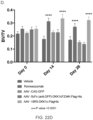
- FIG. 32 Changes in vertebral mineral density (image shown) and changes the vertebral resistance to compression fracture in vertebra isolated from mice after various treatments (bar graph) as measured in newton units of force (N) after 4 weeks of treatment.
- FIGS. 33 A-D Test of Wnt surrogate molecules in an Einhorn fracture model. Radiographs of the callus after 1 week (A) and 6 weeks (B) of treatment with Wnt surrogate molecules are shown. Graphs of changes in whole body bone mineral density (BMD) in contralateral femur are shown (C). Scatter plots showing changes in callus tissue volume, callus bone volume, bone volume/tissue volume ratio (BV/TV), and bone mineral content per millimeter (BMC/mm) as shown in (D) along with representative images of bone slices are shown.
- BMD whole body bone mineral density
- BMC/mm bone mineral content per millimeter
- FIG. 34 Graph with changes in whole body bone mineral density (BMD) measured weekly with different Wnt surrogate molecule dosing schedules is shown.
- FIG. 35 Graph with changes in whole body bone mineral density (BMD) measured weekly from mice treated with different Wnt surrogate molecule alone and in combination with Romosozumab is shown.
- FIG. 36 Levels of therapeutic molecules in serum as measured by ELISA. These data accompany gene expression data presented in Table 4.
- FIGS. 37 A- 37 C Liver (A), small intestine (B) and colon (C) to body weight ratio after treatment with AAV-delivered Wnt surrogates. (**) p ⁇ 0.01. For each graph, the treatments shown from left to right correspond to those in the legend from top to bottom.
- FIGS. 38 A- 38 B Body weight (A) and liver to body weight (B) ratio after treatment with recombinantly produced Wnt surrogate proteins. (*) p ⁇ 0.05. For each time point, the treatments shown from left to right correspond to those in the legend from top to bottom.
- FIGS. 39 A- 39 D Induction of proliferation markers in response to R2M3-26 and Rspo2 recombinant proteins.
- the treatments shown from left to right correspond to those in the legend from top to bottom.
- FIGS. 40 A- 40 H Efficacy of AAV-delivered Wnt surrogate and R-Spondin in a thioacetamide-induced chronic liver disease model.
- the treatments shown from left to right correspond to those in the legend from top to bottom (not including baseline).
- FIGS. 41 A- 41 N Efficacy of recombinantly produced Wnt surrogate and R-Spondin in a thioacetamide-induced chronic liver disease model.
- Study design (A) D-2, D0, D3, D7, D10, D14 represents days relative to the start of treatment with recombinant proteins.
- Liver to body weight ratio B, C
- liver axin2 mRNA D, E
- cyclinD1 mRNA F, G
- Ki67 mRNA H, I expression
- average count of PCNA J, K
- phospho-histone H3 L, M
- Rspo2 mono treatment B, D, F, H, J, L
- R2M3-26/Rspo2 combination treatment C, E, G, I, K, M
- Pro-thrombin time ratio relative to the average pro-thrombin time in plasma collected from control na ⁇ ve mice without TAA exposure (N).
- FIGS. 42 A- 42 C Efficacy of recombinantly produced Wnt surrogate and R-Spondin in a CC14-induced chronic liver disease model.
- Study design (A). Liver to body weight ratio (B) pro-thrombin time (C) and sirius red staining (D) in response to CC14 treatment, R2M3-26 and Rspo2. (*) p ⁇ 0.05, (**) p ⁇ 0.01, (***) p ⁇ 0.001, (****) p ⁇ 0.0001.
- the treatments shown from left to right correspond to those in the legend from top to bottom (not including baseline).
- FIGS. 43 A- 43 D Induction of proliferation markers in response to recombinantly produced Wnt surrogate in an acetaminophen-induced acute liver injury model.
- Study design A. Serum level of alanine transferase at 24 and 48 hours after treatment with acetaminophen (B). Relative cyclinD1 (C) and Ki67 (D) mRNA expression. (*) p ⁇ 0.05, (***) p ⁇ 0.001, (****) p ⁇ 0.0001.
- FIGS. 44 A- 44 D Induction of proliferation markers in response to R-Spondin in an acetaminophen-induced acute liver injury model.
- Study design A. Serum level of alanine transferase at 24 and 48 hours after treatment with acetaminophen (B). Relative cyclinD1 (C) and Ki67 (D) mRNA expression. (**) p ⁇ 0.01, (***) p ⁇ 0.001, (****) p ⁇ 0.0001.
- FIGS. 45 A- 45 D Induction of proliferation markers in response to Wnt surrogate and R-Spondin in an acetaminophen-induced acute liver injury model.
- Study design A. Serum level of alanine transferase at 24, 36, 48 and 60 hours after treatment with acetaminophen (B). Relative cyclinD1 (C) and Ki67 (D) mRNA expression. (*) p ⁇ 0.05, (****) p ⁇ 0.0001. For each time point, the treatments shown from left to right correspond to those in the legend from top to bottom.
- FIGS. 46 A- 46 D Efficacy of recombinantly produced Wnt surrogate and R-Spondin on the survival of mice after acetaminophen-induced liver injury.
- Study design A. Survival curve of mice treated with the control anti-eGFP control protein or R2M3-26 (B), Rspo2 (C) or a combination of R2M3-26 and Rspo2 (D) recombinant proteins.
- the present disclosure relates to Wnt surrogate molecules that bind to one or more Fzd receptor and one or more LRP5 or LRP6 receptor and modulate a downstream Wnt signaling pathway.
- the Wnt surrogate molecules activate a Wnt signaling pathway or increase signaling via a Wnt signaling pathway.
- the Wnt surrogate molecules disclosed herein comprise: (i) one or more antibodies or antigen-binding fragments thereof that specifically bind to one or more Fzd receptor, including antibodies or antigen-binding fragments thereof having particular Fzd receptor specificity and/or functional properties; and (ii) one or more antibodies or antigen-binding fragments thereof that specifically bind to LRP5 and/or LRP6. Certain embodiments encompass specific structural formats or arrangements of the Fzd binding region(s) and LRP5/6 binding region(s) of Wnt surrogate molecules advantageous in increasing downstream Wnt pathway signaling and related biological effects.
- Embodiments of the invention pertain to the use of Wnt surrogate molecules for the diagnosis, assessment and treatment of diseases and disorders associated with Wnt signaling pathways.
- the subject Wnt surrogate molecules are used to modulate a Wnt signaling pathway in a cell or tissue.
- the subject Wnt surrogate molecules are used in the treatment or prevention of diseases and disorders associated with aberrant or deregulated (e.g., reduced) Wnt signaling, or for which modulating, e.g., increasing, Wnt signaling would provide a therapeutic benefit.
- Standard techniques may be used for recombinant DNA, oligonucleotide synthesis, and tissue culture and transformation (e.g., electroporation, lipofection). Enzymatic reactions and purification techniques may be performed according to manufacturer's specifications or as commonly accomplished in the art or as described herein. These and related techniques and procedures may be generally performed according to conventional methods well known in the art and as described in various general and more specific references that are cited and discussed throughout the present specification. Unless specific definitions are provided, the nomenclature utilized in connection with, and the laboratory procedures and techniques of, molecular biology, analytical chemistry, synthetic organic chemistry, and medicinal and pharmaceutical chemistry described herein are those well-known and commonly used in the art. Standard techniques may be used for recombinant technology, molecular biological, microbiological, chemical syntheses, chemical analyses, pharmaceutical preparation, formulation, and delivery, and treatment of subjects.
- Embodiments of the present invention relate to antibodies and antigen-binding fragments thereof that bind to one or more Fzd receptor. Sequences of illustrative antibodies, or antigen-binding fragments, or complementarity determining regions (CDRs) thereof, are set forth in SEQ ID NOs:1-65 or 129-132, Tables 1A and 1B, and Table 3. Anti-Fzd antibodies and antigen-binding fragments there that may be used or present in the Wnt surrogate molecules disclosed herein include, but are not limited to, those described in the U.S. provisional application No. 62/607,877, titled Anti-Frizzled Antibodies and Methods of Use, filed on Dec. 19, 2017.
- Embodiments of the present invention relate to antibodies and antigen-binding fragments thereof that bind to LRP5 and/or LRP6. Sequences of illustrative antibodies, or antigen-binding fragments, or complementarity determining regions (CDRs) thereof, are set forth in SEQ ID NOs:66-88 or 133, Tables 2A and 2B, and Table 3. Anti-LRP5/6 antibodies and antigen-binding fragments there that may be used or present in the Wnt surrogate molecules disclosed herein include, but are not limited to, those described in the U.S. provisional application No. 62/607,879, titled Anti-LRP5/6 Antibodies and Methods of Use, filed on Dec. 19, 2017.
- an antibody is an immunoglobulin molecule capable of specific binding to a target, such as a carbohydrate, polynucleotide, lipid, polypeptide, etc., through at least one epitope recognition site, located in the variable region of the immunoglobulin molecule.
- the term encompasses not only intact polyclonal or monoclonal antibodies, but also fragments thereof (such as dAb, Fab, Fab′, F(ab′)2, Fv), single chain (scFv), VHH or sdAb, synthetic variants thereof, naturally occurring variants, fusion proteins comprising an antibody or an antigen-binding fragment thereof, humanized antibodies, chimeric antibodies, and any other modified configuration of the immunoglobulin molecule that comprises an antigen-binding site or fragment (epitope recognition site) of the required specificity.
- “Diabodies” or 2scFV-Ig antibodies are multivalent or multispecific fragments constructed by gene fusion (WO94/13804; P.
- an antigen-binding fragment refers to a polypeptide fragment that contains at least one CDR of an immunoglobulin heavy and/or light chain, or of a VHH or sdAb, that binds to the antigen of interest, in particular to one or more Fzd receptor or LRP5 or LRP6 receptor.
- an antigen-binding fragment of the herein described antibodies may comprise 1, 2, 3, 4, 5, or all 6 CDRs of a VH and VL sequence set forth herein from antibodies that bind one or more Fzd receptor or LRP5 and/or LRP6.
- an antigen-binding fragment may comprise all three VH CDRs or all three VL CDRs.
- an antigen binding fragment thereof may comprise all three CDRs of a VHH or sdAb.
- An antigen-binding fragment of a Fzd-specific antibody is capable of binding to a Fzd receptor.
- An antigen-binding fragment of a LRP5/6-specific antibody is capable of binding to a LRP5 and/or LRP6 receptor.
- the term encompasses not only isolated fragments but also polypeptides comprising an antigen-binding fragment of an antibody disclosed herein, such as, for example, fusion proteins comprising an antigen-binding fragment of an antibody disclosed herein, such as, e.g., a fusion protein comprising a VHH or sdAb that binds one or more Fzd receptors and a VHH or sdAb that binds LRP5 and/or LRP6.
- antigen refers to a molecule or a portion of a molecule capable of being bound by a selective binding agent, such as an antibody, and additionally capable of being used in an animal to produce antibodies capable of binding to an epitope of that antigen.
- a binding agent e.g., a Wnt surrogate molecule or binding region thereof
- Wnt surrogate molecule or binding region thereof is said to specifically bind an antigen when it preferentially recognizes its target antigen in a complex mixture of proteins and/or macromolecules.
- a Wnt surrogate molecule or binding region thereof e.g., an antibody or antigen-binding fragment thereof
- the equilibrium dissociation constant may be ⁇ 10 ⁇ 9 M or ⁇ 10 ⁇ 10 M.
- antibodies and antigen-binding fragments thereof as described herein include a heavy chain and a light chain CDR set, respectively interposed between a heavy chain and a light chain framework region (FR) set which provide support to the CDRs and define the spatial relationship of the CDRs relative to each other.
- CDR set refers to the three hypervariable regions of a heavy or light chain V region. Proceeding from the N-terminus of a heavy or light chain, these regions are denoted as “CDR1,” “CDR2,” and “CDR3” respectively.
- An antigen-binding site therefore, includes six CDRs, comprising the CDR set from each of a heavy and a light chain V region.
- a polypeptide comprising a single CDR (e.g., a CDR1, CDR2 or CDR3) is referred to herein as a “molecular recognition unit.” Crystallographic analysis of a number of antigen-antibody complexes has demonstrated that the amino acid residues of CDRs form extensive contact with bound antigen, wherein the most extensive antigen contact is with the heavy chain CDR3. Thus, the molecular recognition units are primarily responsible for the specificity of an antigen-binding site.
- FR set refers to the four flanking amino acid sequences which frame the CDRs of a CDR set of a heavy or light chain V region. Some FR residues may contact bound antigen; however, FRs are primarily responsible for folding the V region into the antigen-binding site, particularly the FR residues directly adjacent to the CDRs. Within FRs, certain amino residues and certain structural features are very highly conserved. In this regard, all V region sequences contain an internal disulfide loop of around 90 amino acid residues. When the V regions fold into a binding-site, the CDRs are displayed as projecting loop motifs which form an antigen-binding surface.
- immunoglobulin CDRs and variable domains may be determined by reference to Kabat, E. A. et al., Sequences of Proteins of Immunological Interest. 4th Edition. US Department of Health and Human Services. 1987, and updates thereof, now available on the Internet (immuno.bme.nwu.edu).
- CDRs may be determined by using “IMGT®, the international ImMunoGeneTics information System® available at www.imgt.org (see, e.g., Lefranc, M.-P. et al. (1999) Nucleic Acids Res., 27:209-212; Ruiz, M. et al.
- a “monoclonal antibody” refers to a homogeneous antibody population wherein the monoclonal antibody is comprised of amino acids (naturally occurring and non-naturally occurring) that are involved in the selective binding of an epitope. Monoclonal antibodies are highly specific, being directed against a single epitope.
- monoclonal antibody encompasses not only intact monoclonal antibodies and full-length monoclonal antibodies, but also fragments thereof (such as Fab, Fab′, F(ab′)2, Fv), single chain (scFv), VHH or sdAb, variants thereof, fusion proteins comprising an antigen-binding fragment of a monoclonal antibody, humanized monoclonal antibodies, chimeric monoclonal antibodies, and any other modified configuration of the immunoglobulin molecule that comprises an antigen-binding fragment (epitope recognition site) of the required specificity and the ability to bind to an epitope, including Wnt surrogate molecules disclosed herein.
- fragments thereof such as Fab, Fab′, F(ab′)2, Fv), single chain (scFv), VHH or sdAb, variants thereof, fusion proteins comprising an antigen-binding fragment of a monoclonal antibody, humanized monoclonal antibodies, chimeric monoclonal antibodies, and any other modified
- antibody it is not intended to be limited as regards the source of the antibody or the manner in which it is made (e.g., by hybridoma, phage selection, recombinant expression, transgenic animals, etc.).
- the term includes whole immunoglobulins as well as the fragments etc. described above under the definition of “antibody”.
- the proteolytic enzyme papain preferentially cleaves IgG molecules to yield several fragments, two of which (the F(ab) fragments) each comprise a covalent heterodimer that includes an intact antigen-binding site.
- the enzyme pepsin is able to cleave IgG molecules to provide several fragments, including the F(ab′)2 fragment which comprises both antigen-binding sites.
- An Fv fragment for use according to certain embodiments of the present invention can be produced by preferential proteolytic cleavage of an IgM, and on rare occasions of an IgG or IgA immunoglobulin molecule. Fv fragments are, however, more commonly derived using recombinant techniques known in the art.
- the Fv fragment includes a non-covalent V H ::V L heterodimer including an antigen-binding site which retains much of the antigen recognition and binding capabilities of the native antibody molecule.
- V H ::V L heterodimer including an antigen-binding site which retains much of the antigen recognition and binding capabilities of the native antibody molecule.
- single chain Fv or scFV antibodies are contemplated.
- Kappa bodies III et al., Prot. Eng. 10: 949-57 (1997); minibodies (Martin et al., EMBO J 13: 5305-9 (1994); diabodies (Holliger et al., PNAS 90: 6444-8 (1993); or Janusins (Traunecker et al., EMBO J 10: 3655-59 (1991) and Traunecker et al., Int. J. Cancer Suppl. 7: 51-52 (1992), may be prepared using standard molecular biology techniques following the teachings of the present application with regard to selecting antibodies having the desired specificity.
- bispecific or chimeric antibodies may be made that encompass the ligands of the present disclosure.
- a chimeric antibody may comprise CDRs and framework regions from different antibodies, while bispecific antibodies may be generated that bind specifically to one or more Fzd receptor through one binding domain and to a second molecule through a second binding domain.
- These antibodies may be produced through recombinant molecular biological techniques or may be physically conjugated together.
- a single chain Fv (scFv) polypeptide is a covalently linked V H ::V L heterodimer which is expressed from a gene fusion including V H - and V L -encoding genes linked by a peptide-encoding linker.
- a number of methods have been described to discern chemical structures for converting the naturally aggregated—but chemically separated—light and heavy polypeptide chains from an antibody V region into an scFv molecule which will fold into a three dimensional structure substantially similar to the structure of an antigen-binding site. See, e.g., U.S. Pat. Nos. 5,091,513 and 5,132,405, to Huston et al.; and U.S. Pat. No. 4,946,778, to Ladner et al.
- an antibody as described herein is in the form of a diabody.
- Diabodies are multimers of polypeptides, each polypeptide comprising a first domain comprising a binding region of an immunoglobulin light chain and a second domain comprising a binding region of an immunoglobulin heavy chain, the two domains being linked (e.g. by a peptide linker) but unable to associate with each other to form an antigen binding site: antigen binding sites are formed by the association of the first domain of one polypeptide within the multimer with the second domain of another polypeptide within the multimer (WO94/13804).
- a dAb fragment of an antibody consists of a V H domain (Ward, E. S. et al., Nature 341, 544-546 (1989)).
- bispecific antibodies may be conventional bispecific antibodies, which can be manufactured in a variety of ways (Holliger, P. and Winter G. Current Opinion Biotechnol. 4, 446-449 (1993)), e.g. prepared chemically or from hybrid hybridomas, or may be any of the bispecific antibody fragments mentioned above.
- Diabodies and scFv can be constructed without an Fc region, using only variable domains, potentially reducing the effects of anti-idiotypic reaction.
- Bispecific diabodies as opposed to bispecific whole antibodies, may also be particularly useful because they can be readily constructed and expressed in E. coli .
- Diabodies (and many other polypeptides such as antibody fragments) of appropriate binding specificities can be readily selected using phage display (WO94/13804) from libraries. If one arm of the diabody is to be kept constant, for instance, with a specificity directed against antigen X, then a library can be made where the other arm is varied and an antibody of appropriate specificity selected.
- Bispecific whole antibodies may be made by knobs-into-holes engineering (J. B. B. Ridgeway et al., Protein Eng., 9, 616-621, 1996).
- the antibodies described herein may be provided in the form of a UniBody®.
- a UniBody® is an IgG4 antibody with the hinge region removed (see GenMab Utrecht, The Netherlands; see also, e.g., US20090226421). This proprietary antibody technology creates a stable, smaller antibody format with an anticipated longer therapeutic window than current small antibody formats. IgG4 antibodies are considered inert and thus do not interact with the immune system. Fully human IgG4 antibodies may be modified by eliminating the hinge region of the antibody to obtain half-molecule fragments having distinct stability properties relative to the corresponding intact IgG4 (GenMab, Utrecht). Halving the IgG4 molecule leaves only one area on the UniBody® that can bind to cognate antigens (e.g., disease targets) and the UniBody® therefore binds univalently to only one site on target cells.
- the antibodies of the present disclosure may take the form of a VHH or sdAb.
- VHH or sdAb technology was originally developed following the discovery and identification that camelidae (e.g., camels and llamas) possess fully functional antibodies that consist of heavy chains only and therefore lack light chains.
- camelidae e.g., camels and llamas
- These heavy-chain only antibodies contain a single variable domain (V HH ) and two constant domains (C H 2, C H 3).
- V HH single variable domain
- C H 2, C H 3 constant domains
- the cloned and isolated single variable domains have full antigen binding capacity and are very stable.
- VHH or sdAb are encoded by single genes and are efficiently produced in almost all prokaryotic and eukaryotic hosts e.g. E. coli (see e.g. U.S. Pat. No. 6,765,087), molds (for example Aspergillus or Trichoderma ) and yeast (for example Saccharomyces, Kluyvermyces, Hansenula or Pichia (see e.g. U.S. Pat. No. 6,838,254).
- the production process is scalable and multi-kilogram quantities of VHH or sdAb have been produced.
- VHH or sdAb may be formulated as a ready-to-use solution having a long shelf life.
- VHH or sdAb method is a proprietary method for generating VHH or sdAb against a desired target, based on automated high-throughput selection of B-cells.
- VHH or sdAb are single-domain antigen-binding fragments of camelid-specific heavy-chain only antibodies.
- VHH antibodies or sdAb typically have a small size of around 15 kDa.
- the antibodies or antigen-binding fragments thereof as disclosed herein are humanized.
- the antigen-binding site may comprise either complete variable domains fused onto constant domains or only the CDRs grafted onto appropriate framework regions in the variable domains.
- Epitope binding sites may be wild type or modified by one or more amino acid substitutions.
- variable regions of both heavy and light chains contain three complementarity-determining regions (CDRs) which vary in response to the epitopes in question and determine binding capability, flanked by four framework regions (FRs) which are relatively conserved in a given species and which putatively provide a scaffolding for the CDRs.
- CDRs complementarity-determining regions
- FRs framework regions
- the variable regions can be “reshaped” or “humanized” by grafting CDRs derived from nonhuman antibody on the FRs present in the human antibody to be modified.
- humanized antibodies preserve all CDR sequences (for example, a humanized mouse antibody which contains all six CDRs from the mouse antibodies).
- humanized antibodies have one or more CDRs (one, two, three, four, five, six) which are altered with respect to the original antibody, which are also termed one or more CDRs “derived from” one or more CDRs from the original antibody.
- the antibodies of the present disclosure may be chimeric antibodies.
- a chimeric antibody is comprised of an antigen-binding fragment of an antibody operably linked or otherwise fused to a heterologous Fc portion of a different antibody.
- the heterologous Fc domain is of human origin.
- the heterologous Fc domain may be from a different Ig class from the parent antibody, including IgA (including subclasses IgA1 and IgA2), IgD, IgE, IgG (including subclasses IgG1, IgG2, IgG3, and IgG4), and IgM.
- the heterologous Fc domain may be comprised of CH2 and CH3 domains from one or more of the different Ig classes.
- the antigen-binding fragment of a chimeric antibody may comprise only one or more of the CDRs of the antibodies described herein (e.g., 1, 2, 3, 4, 5, or 6 CDRs of the antibodies described herein), or may comprise an entire variable domain (VL, VH or both).
- Wnt surrogate molecules that bind both one or more Fzd receptors and one or both of LRP5 and/or LRP6.
- Wnt surrogate molecules may also be referred to as “Wnt surrogates” or “Wnt mimetics.”
- the Wnt surrogate molecules bind one or more human Fzd receptors and one or both of a human LRP5 and/or a human LRP6.
- a Wnt surrogate molecule is capable of modulating or modulates Wnt signaling events in a cell contacted with the Wnt surrogate molecule.
- the Wnt surrogate molecule increases Wnt signaling, e.g., via the canonical Wnt/ ⁇ -catenin pathway.
- the Wnt surrogate molecule specifically modulates the biological activity of a human Wnt signaling pathway.
- Wnt surrogate molecules of the present invention are biologically active in binding to one or more Fzd receptor and to one or more of LRP5 and LRP6, and in activation of Wnt signaling, i.e., the Wnt surrogate molecule is a Wnt agonist.
- Wnt agonist activity refers to the ability of an agonist to mimic the effect or activity of a Wnt protein binding to a frizzled protein and/or LRP5 or LRP6.
- the ability of the Wnt surrogate molecules and other Wnt agonists disclosed herein to mimic the activity of Wnt can be confirmed by a number of assays.
- Wnt agonists typically initiate a reaction or activity that is similar to or the same as that initiated by the receptor's natural ligand.
- the Wnt agonists disclosed herein activate, enhance or increase the canonical Wnt/ ⁇ -catenin signaling pathway.
- the term “enhances” refers to a measurable increase in the level of Wnt/ ⁇ -catenin signaling compared with the level in the absence of a Wnt agonist, e.g., a Wnt surrogate molecule disclosed herein.
- the increase in the level of Wnt/ ⁇ -catenin signaling is at least 10%, at least 20%, at least 50%, at least two-fold, at least five-fold, at least 10-fold, at least 20-fold, at least 50-fold, or at least 100-fold as compared to the level of Wnt/ ⁇ -catenin signaling in the absence of the Wnt agonist, e.g., in the same cell type.
- Methods of measuring Wnt/ ⁇ -catenin signaling are known in the art and include those described herein.
- Wnt surrogate molecules disclosed herein are bispecific, i.e., they specifically bind to two or more different epitopes, e.g., one or more Fzd receptor, and LRP5 and/or LRP6.
- Wnt surrogate molecules disclosed herein are multivalent, e.g., they comprise two or more regions that each specifically bind to the same epitope, e.g., two or more regions that bind to an epitope within one or more Fzd receptor and/or two or more regions that bind to an epitope within LRP5 and/or LRP6. In particular embodiments, they comprise two or more regions that bind to an epitope within one or more Fzd receptor and two or more regions that bind to an epitope within LRP5 and/or LRP6.
- Wnt surrogate molecules comprise a ratio of the number of regions that bind one or more Fzd receptor to the number of regions that bind LRP5 and/or LRP6 of or about: 1:1, 2:1, 3:1, 4:1, 5:1, 6:1, 2:3, 2:5, 2:7, 7:2, 5:2, 3:2, 3:4, 3:5, 3:7, 3:8, 8:3, 7:3, 5:3, 4:3, 4:5, 4:7, 4:9, 9:4, 7:4, 5:4, 6:7, 7:6, 1:2, 1:3, 1:4, 1:5, or 1:6.
- Wnt surrogate molecules are bispecific and multivalent.
- Wnt surrogate molecules disclosed herein may have any of a variety of different structural formats or configurations.
- Wnt surrogate molecules may comprise polypeptides and/or non-polypeptide binding moieties, e.g., small molecules.
- Wnt surrogate molecules comprise both a polypeptide region and a non-polypeptide binding moiety.
- Wnt surrogate molecules may comprise a single polypeptide, or they may comprise two or more, three or more, or four or more polypeptides.
- one or more polypeptides of a Wnt surrogate molecule are antibodies or antigen-binding fragments thereof.
- Wnt surrogates comprise two antibodies or antigen binding fragments thereof, one that binds one or more Fzd and one that binds LRP5 and/or LRP6.
- the Wnt surrogates comprises one, two, three, or four polypeptides, e.g., linked or bound to each other or fused to each other.
- the Wnt surrogate molecules may be a fusion protein comprising one or more Fzd binding domain and one or more LRP5/6 binding domain.
- the binding domains may be directly fused or they may be connected via a linker, e.g., a polypeptide linker, including but not limited to any of those disclosed herein.
- the Wnt surrogate molecules comprise two or more polypeptides
- the polypeptides may be linked via covalent bonds, such as, e.g., disulfide bonds, and/or noncovalent interactions.
- covalent bonds such as, e.g., disulfide bonds, and/or noncovalent interactions.
- heavy chains of human immunoglobulin IgG interact at the level of their CH3 domains directly, whereas, at the level of their CH2 domains, they interact via the carbohydrate attached to the asparagine (Asn) N84.4 in the DE turn.
- the Wnt surrogate molecules comprise one or more regions derived from an antibody or antigen-binding fragment thereof, e.g., antibody heavy chains or antibody light chains or fragments thereof.
- a Wnt surrogate polypeptide comprises two antibody heavy chain regions (e.g., hinge regions) bound together via one or more disulfide bond.
- a Wnt surrogate polypeptide comprises an antibody light chain region (e.g., a CL region) and an antibody heavy chain region (e.g., a C H 1 region) bound together via one or more disulfide bond.
- Wnt surrogate polypeptides may be engineered to facilitate binding between two polypeptides.
- Knob-into-holes amino acid modifications may be introduced into two different polypeptides to facilitate their binding.
- Knobs-into-holes amino acid (AA) changes is a rational design strategy developed in antibody engineering, used for heterodimerization of the heavy chains, in the production of bispecific IgG antibodies. AA changes are engineered in order to create a knob on the C H 3 of the heavy chains from a first antibody and a hole on the C H 3 of the heavy chains of a second antibody.
- the knob may be represented by a tyrosine (Y) that belongs to the ‘very large’ IMGT volume class of AA, whereas the hole may be represented by a threonine (T) that belongs to the ‘small’ IMGT volume class.
- Y tyrosine
- T threonine
- Other means of introducing modifications into polypeptides to facilitate their binding are known and available in the art. For example, specific amino acids may be introduced and used for cross-linking, such as Cysteine to form an intermolecular disulfide bond.
- Wnt surrogate molecules may have a variety of different structural formats, including but not limited to those shown in FIG. 1 .
- a Wnt surrogate molecule comprises an scFv or antigen-binding fragment thereof fused to a VHH or sdAb or antigen-binding fragment thereof.
- the scFv specifically binds one or more Fzd receptor
- the VHH or sdAb specifically binds LRP5 and/or LRP6.
- the scFv specifically binds LRP5 and/or LRP6, and the VHH or sdAb specifically binds one or more Fzd receptor.
- the scFv or antigen-binding fragment thereof is fused directly to the VHH or sdAb or antigen-binding fragment thereof, whereas in other embodiments, the two binding regions are fused via a linker moiety.
- the VHH or sdAb is fused to the N-terminus of the scFV, while in other embodiments, the VHH or sdAb is fused to the C-terminus of the scFv.
- the scFv is described herein or comprises any of the CDR sets described herein.
- the VHH or sdAb is described herein or comprises any of the CDR sets disclosed herein.
- a Wnt surrogate molecule comprises one or more Fab or antigen-binding fragment thereof and one or more VHH or sdAb or antigen-binding fragment thereof (or alternatively, one or more scFv or antigen-binding fragment thereof).
- the Fab specifically binds one or more Fzd receptor
- the VHH or sdAb (or scFv) specifically binds LRP5 and/or LRP6.
- the Fab specifically binds LRP5 and/or LRP6, and the VHH or sdAb (or scFv) specifically binds one or more Fzd receptor.
- the VHH or sdAb (or scFv) is fused to the N-terminus of the Fab, while in some embodiments, the VHH or sdAb (or scFv) is fused to the C-terminus of the Fab.
- the Fab is present in a full IgG format, and the VHH or sdAb (or scFv) is fused to the N-terminus and/or C-terminus of the IgG light chain.
- the Fab is present in a full IgG format, and the VHH or sdAb (or scFv) is fused to the N-terminus and/or C-terminus of the IgG heavy chain.
- two or more VHH or sdAb (or scFvs) are fused to the IgG at any combination of these locations.
- Fabs may be converted into a full IgG format that includes both the Fab and Fc fragments, for example, using genetic engineering to generate a fusion polypeptide comprising the Fab fused to an Fc region, i.e., the Fab is present in a full IgG format.
- the Fc region for the full IgG format may be derived from any of a variety of different Fcs, including but not limited to, a wild-type or modified IgG1, IgG2, IgG3, IgG4 or other isotype, e.g., wild-type or modified human IgG1, human IgG2, human IgG3, human IgG4, human IgG4Pro (comprising a mutation in core hinge region that prevents the formation of IgG4 half molecules), human IgA, human IgE, human IgM, or the modified IgG1 referred to as IgG1 LALAPG.
- a wild-type or modified IgG1, IgG2, IgG3, IgG4 or other isotype e.g., wild-type or modified human IgG1, human IgG2, human IgG3, human IgG4, human IgG4Pro (comprising a mutation in core hinge region that prevents the formation of IgG4 half molecules), human IgA
- the L 235 A, P 329 G (LALA-PG) variant has been shown to eliminate complement binding and fixation as well as Fc- ⁇ dependent antibody-dependent cell-mediated cytotoxity (ADCC) in both murine IgG2a and human IgG1.
- ADCC Fc- ⁇ dependent antibody-dependent cell-mediated cytotoxity
- the IgG comprises one or more of the following amino acid substitutions: N297G, N297A, N297E, L234A, L235A, or P236G.
- Non-limiting examples of bivalent and bispecific Wnt surrogate molecules that are bivalent towards both the one or more Fzd receptor and the LRP5 and/or LRP6 are provided as the top four structures depicted in FIG. 1 A , where the VHH or sdAb or scFv is depicted in white, and the Fab or IgG is depicted in black. As shown, the VHH or sdAb (or scFvs) may be fused to the N-termini of both light chains, to the N-termini of both heavy chains, to the C-termini of both light chains, or to the C-termini of both heavy chains.
- VHH or sdAb could be fused to both the N-termini and C-termini of the heavy and/or light chains, to the N-termini of the light chains and the heavy chains, to the C-termini of the heavy and light chains, to the N-termini of the heavy chains and C-termini of the light chains, or to the C-termini of the heavy chains and the N-termini of the light chains.
- two or more VHH or sdAb (or scFvs) may be fused together, optionally via a linker moiety, and fused to the Fab or IgG at one or more of these locations.
- the Wnt surrogate molecule has a Hetero-IgG format, whereas the Fab is present as a half antibody, and one or more VHH or sdAb (or scFv) is fused to one or more of the N-terminus of the Fc, the N-terminus of the Fab, the C-terminus of the Fc, or the C-terminus of the Fab.
- VHH or sdAb or scFv
- the Fab or antigen-binding fragment (or IgG) thereof is fused directly to the VHH or sdAb (or scFv) or antigen-binding fragment thereof, whereas in other embodiments, the binding regions are fused via a linker moiety.
- the Fab is described herein or comprises any of the CDR sets described herein.
- the VHH or sdAb or scFv is described herein or comprises any of the CDR sets disclosed herein.
- a Wnt surrogate molecule comprises one or more Fab or antigen-binding fragment thereof that binds one or more Fzd receptor and one or more Fab or antigen-binding fragment thereof that binds LRP5 and/or LRP6. In certain embodiments, it comprises two Fab or antigen-binding fragments thereof that bind one or more Fzd receptor and/or two Fab or antigen-binding fragments thereof that bind LRP5 and/or LRP6. In particular embodiments, one or more of the Fab is present in a full IgG format, and in certain embodiments, both Fab are present in a full IgG format.
- the Fab in full IgG format specifically binds one or more Fzd receptor, and the other Fab specifically binds LRP5 and/or LRP6. In certain embodiments, the Fab specifically binds one or more Fzd receptor, and the Fab in full IgG format specifically binds LRP5 and/or LRP6. In certain embodiments, the Fab specifically binds LRP5 and/or LRP6, and the Fab in full IgG format specifically binds one or more Fzd receptor. In certain embodiments, the Fab is fused to the N-terminus of the IgG, e.g., to the heavy chain or light chain N-terminus, optionally via a linker.
- the Fab is fused to the N-terminus of the heavy chain of the IgG and not fused to the light chain.
- the two heavy chains can be fused together directly or via a linker.
- An example of such a bispecific and bivalent with respect to both receptors is shown at the top of FIG. 1 B .
- two or more VHH or sdAb may be fused together, optionally via a linker moiety, and fused to the Fab or IgG at one or more of these locations.
- the Wnt surrogate molecule has a Hetero-IgG format, whereas one of the Fab is present as a half antibody, and the other Fab is fused to one or more of the N-terminus of the Fc, the N-terminus of the Fab, or the C-terminus of the Fc.
- a bispecific but monovalent to each receptor version of this format is depicted at the bottom of FIG. 1 B .
- the Fab or antigen-binding fragment thereof is fused directly to the other Fab or IgG or antigen-binding fragment thereof, whereas in other embodiments, the binding regions are fused via a linker moiety.
- the one or both of the two Fabs are described herein or comprise any of the CDR sets described herein.
- Wnt surrogate molecules have a format as described in PCT Application Publication No. WO2017/136820, e.g., a Fabs-in-tandem IgG (FIT-IG) format. Shiyong Gong, Fang Ren, Danqing Wu, Xuan Wu & Chengbin Wu (2017).
- FIT-IG also include the formats disclosed in “Fabs-in-tandem immunoglobulin is a novel and versatile bispecific design for engaging multiple therapeutic targets” mAbs, 9:7, 1118-1128, DOI: 10.1080/19420862.2017.1345401.
- FIT-IGs combine the functions of two antibodies into one molecule by re-arranging the DNA sequences of two parental monoclonal antibodies into two or three constructs and co-expressing them in mammalian cells. Examples of FIT-IG formats and constructs are provided in FIGS. 1 A and 1 B and FIGS. 2 A and 2 B of PCT Application Publication No. WO2017/136820. In certain embodiments, FIT-IGs require no Fc mutation; no scFv elements; and no linker or peptide connector.
- Wnt surrogates comprises a Fab and an IgG.
- the Fab binder LC is fused to the HC of the IgG, e.g., by a linker of various length in between.
- the Fab binder HC can be fused or unfused to the LC of the IgG. A variation of this format has been called Fabs-in-tandem IgG (or FIT-Ig).
- Wnt surrogate molecules comprise two or more VHH or sdAb (or scFvs), including at least one that binds one or more Fzd receptor and at least one that binds LRP5 and/or LRP6.
- one of the binding regions is a VHH or sdAb and the other is an scFv.
- Wnt memetic molecules comprising two or more VHH or sdAb (or scFvs) may be formatted in a variety of configurations, including but not limited to those depicted in FIG. 1 C .
- two or more VHH or sdAb are fused in tandem or fused to two different ends of an Fc, optionally via one or more linkers.
- linkers are present, the linker and its length may be the same or different between the VHH or sdAb (or scFv) and the other VHH or sdAb (or scFv), or between the VHH or sdAb and Fc.
- the VHH or sdAb is fused to the N-terminus and/or C-terminus of the IgG heavy chain.
- two or more VHH or sdAb are fused to the IgG at any combination of these locations.
- Non-limiting examples of bivalent and bispecific Wnt surrogate molecules of this format are depicted as the top seven structures depicted in FIG. 1 C , where the first VHH or sdAb is depicted in white, the Fc or IgG is depicted in black, and the second VHH or sdAb is depicted as light gray.
- both VHH or sdAb may be fused to the N-termini of the Fc, to the C-termini of the Fc, or one or more VHH or sdAb may be fused to either or both of an N-terminus or C-terminus of the Fc.
- the Wnt surrogate molecule has a Hetero-IgG format, whereas one VHH or sdAb is present as a half antibody, and the other is fused to the N-terminus of the Fc or the C-terminus of the Fc.
- a bispecific but monovalent to each receptor version of this format is depicted at the bottom of FIG. 1 C .
- the VHH or sdAb is fused directly to the other VHH or sdAb whereas in other embodiments, the binding regions are fused via a linker moiety.
- the VHH or sdAb are described herein or comprises any of the CDR sets described herein. In various embodiments, any of these formats may comprise one or more scFvs in place of one or more VHH or sdAb.
- a Wnt surrogate molecule is formatted as a diabody.
- the binders against Fzd and LRP can also be linked together in a diabody (or DART) configuration.
- the diabody can also be in a single chain configuration. If the diabody is fused to an Fc, this will create a bivalent bispecific format. Without fusion to Fc, this would be a monovalent bispecific format.
- a diabody is a noncovalent dimer scFv fragment that consists of the heavy-chain variable (VH) and light-chain variable (VL) regions connected by a small peptide linker.
- Another form of diabody is a single-chain (Fv)2 in which two scFv fragments are covalently linked to each other.
- Wnt surrogate molecules in various embodiments, comprise one or more antibodies or antigen-binding fragments thereof disclosed herein.
- a Wnt surrogate comprises two polypeptides, wherein each polypeptide comprises an Nab or scFv that binds LRP5/6 and an Nab or scFv that binds one or more Wnts, optionally wherein one of the binding domains is an scFv and the other is an Nab.
- a Wnt surrogate comprises three polypeptides, wherein the first polypeptide comprises an antibody heavy chain and the second polypeptide comprises an antibody light chain, wherein the antibody heavy chain and light chain bind LRP5/6 or one or more Fzds, and wherein the third polypeptide comprises a VHH or sdAb fused to a heavy chain Fc region, wherein the VHH or sdAb binds to either LRP5/6 or one or more Fzds.
- Wnt polypeptides comprise four polypeptides, including two heavy chain polypeptides and two light chain polypeptides, wherein the two heavy chains and two light chains bind LRP5/6 or one or more Fzds, and further comprise one or more Nab or scFv fused to one or more of the heavy chains and/or light chains, wherein the Nab or scFv binds to LRP5/6 or one or more Fzds.
- a Wnt surrogate comprises at least four polypeptides, including two heavy chain polypeptides and two light chain polypeptides that bind either LRP5/6 or one or more Fzds, wherein the Wnt surrogate further comprises a Fab that binds either LRP5/6 or one or more Fzds.
- the Fab may comprise two polypeptides, each fused to one of the two heavy chain polypeptides, and two polypeptides, each fused to one of the two light chain polypeptides, or it may comprise two polypeptides each fused to one of the two heavy chain polypeptides and two additional polypeptides, each bound to one of the two polypeptides fused to the heavy chain polypeptides, thus making a second Fab.
- Other configurations may be used to produce the Wnt surrogates disclosed herein.
- a Wnt surrogate molecule comprises a Fzd binding region, e.g., an anti-Fzd antibody, or antigen-binding fragment thereof, fused or bound to a polypeptide that specifically binds to one or more Fzd receptor.
- the polypeptide that specifically binds to one or more Fzd receptor is an antibody or antigen-binding fragment thereof. If certain embodiments, it is an antibody or antigen-binding fragment thereof disclosed herein or in the U.S. provisional patent application Ser. No. 62/607,877, titled, “Anti-Frizzled antibodies and Methods of Use,” filed on Dec. 19, 2017, which is incorporated herein by reference in its entirety.
- the Fzd binding domain comprises the three heavy chain CDRs and/or the three light chain CDRs disclosed for any of the illustrative antibodies or fragments thereof that bind to one or more Fzd receptor provided in Table 1A.
- the Fzd binding domain comprises the three heavy chain CDRs and/or the three light chain CDRs disclosed for any of the illustrative antibodies or fragments thereof that bind to one or more Fzd receptor provided in Table 1A, wherein the CDRs collectively comprise one, two, three, four, five, six, seven, or eight amino acid modifications, e.g., substitutions, deletions, or additions.
- the Fzd binding domain is a VHH or sdAb or was derived from a VHH or sdAb, so Table 1A only includes the three heavy chain CDRs.
- the Fzd binding domain comprises the three CDR HC sequences provided in Table 1A or variants wherein the CDRs collectively comprise one, two, three, four, five, six, seven or eight amino acid modifications.
- the Fzd binding domain comprises the heavy chain fragment and/or light chain fragment of any of the illustrative antibodies or fragments thereof that bind to one or more Fzd receptor provided in Table 1B or SEQ ID NOs:1-65 or 129-132 (or an antigen-binding fragment or variant of either).
- the Fzd binding domain is an Fab or was derived from an Fab, so the heavy chain of Table 1B includes VH and CH1 sequence, but not CH2 or CH3 sequences.
- the Fzd binding domain is a VHH or sdAb or was derived from a VHH or sdAb, so Table 1B includes the VHH domain.
- the Fzd binding region is a polypeptide, e.g., an antibody or antigen-binding fragment thereof, that competes with any of these antibodies for binding to one or more Fzd receptor.
- 006S-H02 Fzd10 006S-A03 n.b. 006S-B03 n.b. 006S-C03 n.b. 014S-A01 Fzd1, 2, 7 014S-B02 n.b. 014S-G02 Fzd6 014S-B03 n.b. 014S-C03 Fzd1, 2, 7 014S-A04 n.b. 014S-B04 Fzd8 014S-B05 Fzd5, 8 014S-B06 Fzd9 014S-F06 n.s.
- the Fzd binding domain may be selected from any binding domain that binds a Fzd with an affinity of, e.g., a K D of at least about 1 ⁇ 10 ⁇ 4 M, at least about 1 ⁇ 10 ⁇ 5 M, at least about 1 ⁇ 10 ⁇ 6 M, at least about 1 ⁇ 10 ⁇ 7 M, at least about 1 ⁇ 10 ⁇ 8 M, at least about 1 ⁇ 10 ⁇ 9 M, or at least about 1 ⁇ 10 ⁇ 10 M.
- a K D of at least about 1 ⁇ 10 ⁇ 4 M, at least about 1 ⁇ 10 ⁇ 5 M, at least about 1 ⁇ 10 ⁇ 6 M, at least about 1 ⁇ 10 ⁇ 7 M, at least about 1 ⁇ 10 ⁇ 8 M, at least about 1 ⁇ 10 ⁇ 9 M, or at least about 1 ⁇ 10 ⁇ 10 M.
- the Fzd binding domain may be selected from any binding domain that binds one or more Fzd receptor at high affinity, e.g., a K D of less than about 1 ⁇ 10 ⁇ 7 M, less than about 1 ⁇ 10 ⁇ 8 M, less than about 1 ⁇ 10 ⁇ 9 M, or less than about 1 ⁇ 10 ⁇ 10 M.
- the Fzd binding domain may be selected from any binding domain that binds Fzd at high affinity, e.g.
- Suitable Fzd binding domains include, without limitation, de novo designed Fzd binding proteins, antibody derived binding proteins, e.g. scFv, Fab, etc. and other portions of antibodies that specifically bind to one or more Fzd proteins; VHH or single domain antibody derived binding domains; knottin-based engineered scaffolds; norrin and engineered binding fragments derived therefrom, naturally occurring Fzd binding domains, and the like.
- a Fzd binding domain may be affinity selected to enhance binding to a desired Fzd protein or plurality of Fzd proteins, e.g. to provide tissue selectivity.
- the Fzd binding domain binds to one, two, three, four, five or more different frizzled proteins, e.g. one or more of human frizzled proteins Fzd1, Fzd2, Fzd3, Fzd4, Fzd5, Fzd6, Fzd7, Fzd8, Fzd9, Fzd10.
- the Fzd binding domain binds to Fzd1, Fzd2, and Fzd 7.
- the Fzd binding domain binds to Fzd1, Fzd2, Fzd5, Fzd7 and Fzd8.
- the Fzd binding domain is selective for one or more frizzled protein of interest, e.g. having a specificity for the one or more desired frizzled protein of at least 10-fold, 25-fold, 50-fold, 100-fold, 200-fold or more relative to other frizzled proteins.
- the Fzd binding domain comprises the six CDR regions of the pan specific frizzled antibody OMP-18R5 (vantictumab). In certain embodiments, the Fzd binding domain is an scFv comprising the six CDR regions of the pan-specific frizzled antibody OMP-18R5 (vantictumab). See, for example, U.S. Pat. No. 8,507,442, herein specifically incorporated by reference.
- the CDR sequences of OMP-18R5 include a heavy chain CDR1 comprising GFTFSHYTLS (SEQ ID NO:270), a heavy chain CDR2 comprising VISGDGSYTYYADSVKG (SEQ ID NO:677), and a heavy chain CDR3 comprising NFIKYVFAN (SEQ ID NO:1033), and (ii) a light chain CDR1 comprising SGDKLGKKYAS (SEQ ID NO:1152) or SGDNIGSFYVH (SEQ ID NO:1153), a light chain CDR2 comprising EKDNRPSG (SEQ ID NO:1200) or DKSNRPSG (SEQ ID NO:1201), and a light chain CDR3 comprising SSFAGNSLE (SEQ ID NO:1435) or QSYANTLSL (SEQ ID NO:1436).
- a heavy chain CDR1 comprising GFTFSHYTLS (SEQ ID NO:270), a heavy chain CDR2 comprising VISGDGSYTYYAD
- the Fzd binding domain is an antibody or derivative thereof, including without limitation scFv, minibodies, VHH or single domain antibodies (sdAb) and various antibody mimetics comprising any of these CDR sequences.
- these CDR sequences comprise one or more amino acid modifications.
- the Fzd binding domain comprises a variable region sequence, or the CDRs thereof, from any of a number of frizzled specific antibodies, which are known in the art and are commercially available, or can be generated de novo. Any of the frizzled polypeptides can be used as an immunogen or in screening assays to develop an antibody.
- frizzled binding domains include antibodies available from Biolegend, e.g. Clone CH3A4A7 specific for human frizzled 4 (CD344); Clone W3C4E11 specific for human Fzd9 (CD349); antibodies available from Abcam, e.g.
- the Fzd binding region of a Wnt surrogate molecule may be an engineered protein that is selected for structural homology to the frizzled binding region of a wnt protein.
- Such proteins can be identified by screening a structure database for homologies.
- the initial protein thus identified, for example the microbial Bh1478 protein.
- the native protein is then engineered to provide amino acid substitutions that increase affinity, and may further be selected by affinity maturation for increased affinity and selectivity in binding to the desired frizzled protein.
- frizzled binding moieties include the Fz27 and Fz27-B12 proteins.
- a Wnt surrogate molecule comprises an LRP5/6 binding domain, e.g., an anti-LRP5/6 antibody, or antigen-binding fragment thereof, fused to a polypeptide that specifically binds to one or more Fzd receptors.
- the polypeptide that specifically binds to LRP5/6 is an antibody or antigen-binding fragment thereof. If certain embodiments, it is an antibody or antigen-binding fragment thereof disclosed in the U.S. provisional patent application No. 62/607,879, titled, “Anti-LR5/6 Antibodies and Methods of Use,” filed on Dec. 19, 2017, which is incorporated herein by reference in its entirety.
- the LRP5/6 binding domain comprises the three heavy chain CDRs and/or the three light chain CDRs disclosed for any of the illustrative antibodies or fragments thereof that bind to LRP5 and/or LRP6 provided in Table 2A.
- the LRP5/6 binding domain comprises the three heavy chain CDRs and/or the three light chain CDRs disclosed for any of the illustrative antibodies or fragments thereof that bind to one or more Fzd receptor provided in Table 2A, wherein the CDRs collectively comprise one, two, three, four, five, six, seven, or eight amino acid modifications, e.g., substitutions, deletions, or additions.
- the LRP5/6 binding domain is a VHH or sdAb or was derived from a VHH or sdAb, so Table 2A only includes the three heavy chain CDRs.
- the LRP5/6 biding domain comprises the three heavy chain CDRs shown in Table 2A or variants wherein the CDRs collectively comprise one, two, three, four, five, six, seven or eight amino acid modifications.
- the LRP5/6 binding domain comprises the heavy chain fragment and/or light chain fragment of any of the illustrative antibodies or fragments thereof that bind to LRP5 and/or LRP6 provided in Table 2B or SEQ ID NOs:66-88 or 133 (or an antigen-binding fragment or variant of either).
- the LRP5/6 binding domain is an Fab or was derived from an Fab, so Table 2B includes VH and CH1 sequence, but not CH2 or CH3 sequences.
- the LRP5/6 binding domain is a VHH or sdAb or was derived from a VHH or sdAb, so Table 2B includes the VHH domain.
- the LRP5/6 binding region is a polypeptide, e.g., an antibody or antigen-binding fragment thereof, that competes with one of these antibodies for binding to LRP5 and/or LRP6.
- the LRP5/6 binding domain may be selected from any binding domain that binds LRP5 or LRP6 with a K D of less than or equal to about 1 ⁇ 10 ⁇ 4 M, less than or equal to about 1 ⁇ 10 ⁇ 5 M, less than or equal to about 1 ⁇ 10 ⁇ 6 M, less than or equal to about 1 ⁇ 10 ⁇ 7 M, less than or equal to about 1 ⁇ 10 ⁇ 8 M, less than or equal to about 1 ⁇ 10 ⁇ 9 M, or less than or equal to about 1 ⁇ 10 ⁇ 10 M in the context of a Wnt surrogate molecule.
- the LRP5/6 binding domain may be selected from any binding domain that binds LRP5 or LRP6 with a K D of greater than or equal to about 1 ⁇ 10 ⁇ 4 M, greater than or equal to about 1 ⁇ 10 ⁇ 5 M, greater than or equal to about 1 ⁇ 10 ⁇ 6 M, greater than or equal to about 1 ⁇ 10 ⁇ 7 M, greater than or equal to about 1 ⁇ 10 ⁇ 8 M, greater than or equal to about 1 ⁇ 10 ⁇ 9 M, or greater than about 1 ⁇ 10 ⁇ 10 M in the context of a Wnt surrogate molecule.
- the LRP5/6 binding domain may be selected from any binding domain that binds LRP5 or LRP6 at high affinity, e.g. a K D of less than about 1 ⁇ 10 ⁇ 7 M, less than about 1 ⁇ 10 ⁇ 8 M, less than about 1 ⁇ 10 ⁇ 9 M, or less than about 1 ⁇ 10 ⁇ 10 M.
- LRP5/6 binding domains include, without limitation, de novo designed LRP5/6 binding proteins, antibody derived binding proteins, e.g. scFv, Fab, etc. and other portions of antibodies that specifically bind to one or more Fzd proteins; VHH or sdAb derived binding domains; knottin-based engineered scaffolds; naturally occurring LRP5/6, including without limitation, DKK1, DKK2, DKK3, DKK4, sclerostin; Wise; fusions proteins comprising any of the above; derivatives of any of the above; variants of any of the above; and biologically active fragments of any of the above, and the like.
- a LRP5/6 binding domain may be affinity selected to enhance binding.
- hDKKs 1-4 contain two distinct cysteine-rich domains in which the positions of 10 cysteine residues are highly conserved between family members.
- Exemplary sequences of human Dkk genes and proteins are publicly available, e.g., Genbank accession number NM_014419 (soggy-1); NM_014420 (DKK4); AF177394 (DKK-1); AF177395 (DKK-2); NM_015881 (DKK3); and NM_014421 (DKK2).
- the Lrp6 binding moiety is a DKK1 peptide, including without limitation the C-terminal domain of human DKK1.
- the C-terminal domain may comprise the sequence:
- the corresponding region of human DKK2 may comprise the sequence: KMSHIKGHEGDPCLRSSDCIEGFCCARHFWTKICKPVLHQGEVCTKQRKKGSH GLEIFQRCDCAKGLSCKVWKDATYSSKARLHVCQK (SEQ ID NO:2191) or a biologically active fragment thereof.
- LRP5 or LRP6 antibodies that specifically bind to LRP5 or LRP6 are known in the art and are commercially available, or can be generated de novo. LRP5, LRP6 or fragments thereof can be used as an immunogen or in screening assays to develop an antibody. Examples of known antibodies include, without limitation, those described in Gong et al. (2010) PLoS One. 5(9):e12682; Ettenberg et al. (2010) Proc Natl Acad Sci USA.
- Wnt surrogate molecules disclosed herein comprise one or more polypeptides comprising two or more binding regions.
- the two or more binding regions may be two or more Fzd binding regions or two or more LRP5/6 binding regions, or they may comprise one or more Fzd binding region and one or more LRP5/6 binding region.
- the binding regions may be directly joined or contiguous, or may be separated by a linker, e.g. a polypeptide linker, or a non-peptidic linker, etc.
- the length of the linker, and therefore the spacing between the binding domains can be used to modulate the signal strength, and can be selected depending on the desired use of the Wnt surrogate molecule.
- the enforced distance between binding domains can vary, but in certain embodiments may be less than about 100 angstroms, less than about 90 angstroms, less than about 80 angstroms, less than about 70 angstroms, less than about 60 angstroms, or less than about 50 angstroms.
- the linker is a rigid linker, in other embodiments the linker is a flexible linker.
- the linker may be from about 1, 2, 3, 4, 5, 6, 7, 8, 9, 10, 11, 12, 13, 14, 15, 16, 17, 18, 19, 20 21, 22, 23, 24, 25, 26, 27, 28, 29, 30 or more amino acids in length, and is of sufficient length and amino acid composition to enforce the distance between binding domains.
- the linker comprises or consists of one or more glycine and/or serine residues.
- a Wnt surrogate molecule comprises a polypeptide sequence having at least 90%, at least 95%, at least 98% or at least 99% identity to a polypeptide sequence disclosed in any of SEQ ID NOs:89-128 or 134-157, or having at least 90%, at least 95%, at least 98% or at least 99% identity to an antigen-binding fragment of a polypeptide sequence disclosed in any of SEQ ID NOs:89-128 or 134-157.
- the Wnt surrogate molecules comprises or consists of a polypeptide sequence set forth in any of SEQ ID NOs:89-128 or 134-157, or an antigen-binding fragment thereof.
- the antigen-binding fragment binds one or more Fzd receptors and also binds LRP5 and/or LRP6.
- Wnt surrogate molecule can be multimerized, e.g. through an Fc domain, by concatenation, coiled coils, polypeptide zippers, biotin/avidin or streptavidin multimerization, and the like.
- the Wnt surrogate molecules can also be joined to a moiety such as PEG, Fc, etc., as known in the art to enhance stability in vivo.
- a Wnt surrogate molecule directly activates canonical Wnt signaling through binding to one or more Fzd proteins and to LRP5/6, particularly by binding to these proteins on a cell surface, e.g., the surface of a human cell.
- the direct activation of Wnt signaling by a Wnt surrogate molecule is in contrast to potentiation of Writ signaling, which enhances activity only when native Wnt proteins are present.
- Wnt surrogate molecules may activate Wnt signaling, e.g., by mimicking the effect or activity of a Wnt protein binding to a frizzled protein.
- the ability of the Wnt surrogate molecules of the invention to mimic the activity of Wnt can be confirmed by a number of assays.
- the Wnt surrogate molecules typically initiate a reaction or activity that is similar to or the same as that initiated by the receptor's natural ligand.
- the Wnt surrogate molecules of the invention enhance the canonical Wnt/ ⁇ -catenin signaling pathway.
- the term “enhances” refers to a measurable increase in the level of Wnt/ ⁇ -catenin signaling compared with the level in the absence of a Wnt surrogate molecule of the invention.
- Wnt/ ⁇ -catenin signaling Various methods are known in the art for measuring the level of canonical Wnt/ ⁇ -catenin signaling. These include, but are not limited to assays that measure: Wnt/ ⁇ -catenin target gene expression; TCF reporter gene expression; ⁇ -catenin stabilization; LRP phosphorylation; Axin translocation from cytoplasm to cell membrane and binding to LRP.
- the canonical Wnt/ ⁇ -catenin signaling pathway ultimately leads to changes in gene expression through the transcription factors TCF7, TCF7L1, TCF7L2 (a.k.a., TCF4), and LEF.
- TCF7 transcription factors
- TCF7L1, TCF7L2 a.k.a., TCF4
- LEF LEF
- a TCF reporter assay assesses changes in the transcription of TCF/LEF controlled genes to determine the level of Wnt/ ⁇ -catenin signaling.
- a TCF reporter assay was first described by Korinek, V. et al., 1997. Also known as TOP/FOP this method involves the use of three copies of the optimal TCF motif CCTTTGATC, or three copies of the mutant motif CCTTTGGCC, upstream of a minimal c-Fos promoter driving luciferase expression (pTOPFI_ASH and pFOPFI_ASH, respectively) to determine the transactivational activity of endogenous p-catenin/TCF4. A higher ratio of these two reporter activities (TOP/FOP) indicates higher ⁇ -catenin/TCF4 activity, whereas a lower ratio of these two reporter activities indicates lower ⁇ -catenin/TCF4 activity.
- reporter transgenes that respond to Wnt signals exist intact in animals and therefore, effectively reflect endogenous Wnt signaling. These reporters are based on a multimerized TCF binding site, which drives expression of LacZ or GFP, which are readily detectable by methods known in the art. These reporter genes include: TOP-GAL, BAT-GAL, ins-TOPE FP, ins-TOPGAL, LEF-EGFP, Axin2-LacZ, Axin2-d2EGFP, Lgr5tm1 (cre/ERT2), TOPdGFP.
- measuring the level and location of ⁇ -catenin in a cell is a good reflection of the level of Wnt/ ⁇ -catenin signaling.
- a non-limiting example of such an assay is the “BioImage ⁇ -Catenin Redistribution Assay” (Thermo Scientific) which provides recombinant U20S cells that stably express human ⁇ -catenin fused to the C-terminus of enhanced green fluorescent protein (EGFP). Imaging and analysis is performed with a fluorescence microscope or HCS platform allowing the levels and distribution of EGFP- ⁇ -catenin to be visualized.
- EGFP enhanced green fluorescent protein
- Axin has been shown to bind preferentially to a phosphorylated form of the LRP tail. Visualization of Axin translocation, for example with a GFP-Axin fusion protein, is therefore another method for assessing levels of Wnt/ ⁇ -catenin signaling.
- a Wnt surrogate molecule enhances or increases canonical Wnt pathway signaling, e.g., ⁇ -catenin signaling, by at least 30%, 35%, 40%, 45%, 50%, 60%, 70%, 75%, 80%, 85%, 90%, 95%, 100%, 110%, 150%, 200%, 250%, 300%, 400% or 500%, as compared to the ⁇ -catenin signaling induced by a neutral substance or negative control as measured in an assay described above, for example as measured in the TOPFlash assay. A negative control may be included in these assays.
- Wnt surrogate molecules may enhance ⁇ -catenin signaling by a factor of 2 ⁇ , 5 ⁇ , 10 ⁇ , 100 ⁇ , 1000 ⁇ , 10000 ⁇ or more as compared to the activity in the absence of the Wnt surrogate molecule when measured in an assay described above, for example when measured in the TOPFlash assay, or any of the other assays mentioned herein.
- Wnt gene product or “Wnt polypeptide” when used herein encompass native sequence Writ polypeptides, Wnt polypeptide variants, Wnt polypeptide fragments and chimeric Wnt polypeptides.
- a Wnt polypeptide is a native human full length mature Wnt protein.
- human native sequence Wnt proteins of interest in the present application include the following: Wnt-1 (GenBank Accession No. NM_005430); Wnt-2 (GenBank Accession No. NM_003391); Wnt-2B (Wnt-13) (GenBank Accession No. NM_004185 (isoform 1), NM_024494.2 (isoform 2)), Wnt-3 (RefSeq.: NM_030753), Wnt3a (GenBank Accession No. NM_033131), Wnt-4 (GenBank Accession No. NM_030761), Wnt-5A (GenBank Accession No. NM_003392), Wnt-5B (GenBank Accession No.
- Wnt-6 (GenBank Accession No. NM_006522), Wnt-7A (GenBank Accession No. NM_004625), Wnt-7B (GenBank Accession No. NM_058238), Wnt-8A (GenBank Accession No. NM_058244), Wnt-8B (GenBank Accession No. NM_003393), Wnt-9A (Wnt-14) (GenBank Accession No. NM_003395), Wnt-9B (Wnt-15) (GenBank Accession No. NM_003396), Wnt-1 OA (GenBank Accession No. NM_025216), Wnt-10B (GenBank Accession No.
- NM_003394 Wnt-11 (GenBank Accession No. NM_004626), Wnt-16 (GenBank Accession No. NM_016087)).
- each member has varying degrees of sequence identity with the family, all encode small (i.e., 39-46 kD), acylated, palmitoylated, secreted glycoproteins that contain 23-24 conserved cysteine residues whose spacing is highly conserved (McMahon, A P et al., Trends Genet. 1992; 8: 236-242; Miller, J R. Genome Biol. 2002; 3(1): 3001.1-3001.15).
- Wnt polypeptides of interest include orthologs of the above from any mammal, including domestic and farm animals, and zoo, laboratory or pet animals, such as dogs, cats, cattle, horses, sheep, pigs, goats, rabbits, rats, mice, frogs, zebra fish, fruit fly, worm, etc.
- Wnt pathway signaling or “Wnt signaling” is used herein to refer to the mechanism by which a biologically active Wnt exerts its effects upon a cell to modulate a cell's activity.
- Wnt proteins modulate cell activity by binding to Wnt receptors, including proteins from the Frizzled (Fzd) family of proteins, proteins from the ROR family of proteins, the proteins LRP5, LRP6 from the LRP family of proteins, the protein FRL1/crypto, and the protein Derailed/Ryk. Once activated by Wnt binding, the Wnt receptor(s) will activate one or more intracellular signaling cascades.
- Wnt signaling pathway includes the canonical Wnt signaling pathway; the Wnt/planar cell polarity (Wnt/PCP) pathway; the Wnt-calcium (Wnt/Ca 2+ ) pathway (Giles, R H et al. (2003) Biochim Biophys Acta 1653, 1-24; Peifer, M. et al. (1994) Development 120: 369-380; Papkoff, J. et al (1996) Mol. Cell Biol. 16: 2128-2134; Veeman, M. T. et al. (2003) Dev. Cell 5: 367-377); and other Writ signaling pathways as is well known in the art.
- activation of the canonical Wnt signaling pathway results in the inhibition of phosphorylation of the intracellular protein ⁇ -catenin, leading to an accumulation of ⁇ -catenin in the cytosol and its subsequent translocation to the nucleus where it interacts with transcription factors, e.g. TCF/LEF, to activate target genes.
- Activation of the Wnt/PCP pathway activates RhoA, c-Jun N-terminal kinase (JNK), and nemo-like kinase (NLK) signaling cascades to control such biological processes as tissue polarity and cell movement.
- Activation of the Wnt/Ca 2+ by, for example, binding of Wnt-4, Wnt-5A or Wnt-11 elicits an intracellular release of calcium ions, which activates calcium sensitive enzymes like protein kinase C (PKC), calcium-calmodulin dependent kinase II (CamKII) or calcineurin (CaCN).
- PKC protein kinase C
- CaCN calcium-calmodulin dependent kinase II
- CaCN calcineurin
- Wnt surrogate molecules may be assessed using a variety of methods known to the skilled person, including e.g., affinity/binding assays (for example, surface plasmon resonance, competitive inhibition assays), cytotoxicity assays, cell viability assays, cell proliferation or differentiation assays in response to a Wnt, cancer cell and/or tumor growth inhibition using in vitro or in vivo models, including but not limited to any described herein.
- the Wnt surrogate molecules described herein may also be tested for effects on Fzd receptor internalization, in vitro and in vivo efficacy, etc.
- Such assays may be performed using well-established protocols known to the skilled person (see e.g., Current Protocols in Molecular Biology (Greene Publ. Assoc. Inc. & John Wiley & Sons, Inc., NY, NY); Current Protocols in Immunology (Edited by: John E. Coligan, Ada M. Kruisbeek, David H. Margulies, Ethan M. Shevach, Warren Strober 2001 John Wiley & Sons, NY, NY); or commercially available kits.
- a Fzd-binding region of a Wnt surrogate molecule comprises one or more of the CDRs of the anti-Fzd antibodies described herein.
- a LRP5/6-binding region of a Wnt surrogate molecule e.g., an antigen-binding fragment of an anti-LRP5/6 antibody
- Also disclosed herein is a method for obtaining an antibody or antigen binding domain specific for a Fzd receptor, the method comprising providing by way of addition, deletion, substitution or insertion of one or more amino acids in the amino acid sequence of a VH domain set out herein or a VH domain which is an amino acid sequence variant of the VH domain, optionally combining the VH domain thus provided with one or more VL domains, and testing the VH domain or VH/VL combination or combinations to identify a specific binding member or an antibody antigen binding domain specific for one or more Fzd receptor and optionally with one or more desired properties.
- the VL domains may have an amino acid sequence which is substantially as set out herein.
- An analogous method may be employed in which one or more sequence variants of a VL domain disclosed herein are combined with one or more VH domains.
- Wnt surrogate molecules are water soluble.
- water soluble it is meant a composition that is soluble in aqueous buffers in the absence of detergent, usually soluble at a concentration that provides a biologically effective dose of the polypeptide.
- Compositions that are water soluble form a substantially homogenous composition that has a specific activity that is at least about 5% that of the starting material from which it was purified, usually at least about 10%, 20%, or 30% that of the starting material, more usually about 40%, 50%, or 60% that of the starting material, and may be about 50%, about 90% or greater.
- Wnt surrogate molecules disclosed herein typically form a substantially homogeneous aqueous solution at concentrations of at least 25 ⁇ M and higher, e.g.
- Wnt surrogate molecules disclosed herein typically form a substantially homogeneous aqueous solution at concentrations of about 0.1 mg/ml, about 0.5 mg/ml, of about 1 mg/ml or more.
- an antigen or epitope that “specifically binds” or “preferentially binds” (used interchangeably herein) to an antibody or antigen-binding fragment thereof is a term well understood in the art, and methods to determine such specific or preferential binding are also well known in the art.
- a molecule e.g., a Wnt surrogate molecule, is said to exhibit “specific binding” or “preferential binding” if it reacts or associates more frequently, more rapidly, with greater duration and/or with greater affinity with a particular cell or substance than it does with alternative cells or substances.
- a molecule or binding region thereof e.g., a Wnt surrogate molecule or binding region thereof, “specifically binds” or “preferentially binds” to a target antigen, e.g., a Fzd receptor, if it binds with greater affinity, avidity, more readily, and/or with greater duration than it binds to other substances.
- a Wnt surrogate molecule or binding region thereof that specifically or preferentially binds to the Fzd1 receptor is an antibody that binds to the Fzd1 receptor with greater affinity, avidity, more readily, and/or with greater duration than it binds to other Fzd receptors or non-Fzd proteins.
- a Wnt surrogate molecule or binding region thereof that specifically or preferentially binds to a first target may or may not specifically or preferentially bind to a second target.
- “specific binding” or “preferential binding” does not necessarily require (although it can include) exclusive binding.
- reference to binding means preferential binding.
- any of the one or more Fzd binding region of a Wnt surrogate molecule binds to one, two, three, four, five or more different frizzled proteins, e.g., one or more of human frizzled proteins Fzd1, Fzd2, Fzd3, Fzd4, Fzd5, Fzd6, Fzd7, Fzd8, Fzd9, Fzd10.
- any of the Fzd binding regions binds to Fzd1, Fzd2, Fzd5, Fzd7 and Fzd8.
- any of the Fzd binding regions binds to: (i) Fzd1, Fzd2, Fzd7 and Fzd9; (ii) Fzd1, Fzd2 and Fzd7; (iii) Fzd5 and Fzd8; (iv) Fzd5, Fzd7 and Fzd8; (v) Fzd1, Fzd4, Fzd5 and Fzd8; (vi) Fzd1, Fzd2, Fzd5, Fzd7 and Fzd8; (vii) Fzd4 and Fzd9; (viii) Fzd9 and Fzd10; (ix) Fzd5, Fzd8 and Fzd10; or (x) Fzd4, Fzd5 and Fzd8; Fzd1, Fzd5, Fzd7 and Fzd8.
- the Fzd binding region is selective for one or more Fzd protein of interest, e.g. having a specificity for the one or more desired Fzd protein of at least 10-fold, 25-fold, 50-fold, 100-fold, 200-fold or more relative to other Fzd proteins.
- any of the one or more Fzd binding region of a Wnt surrogate molecule is monospecific and binds or specifically binds to only one of Fzd1, Fzd2, Fzd3, Fzd4, Fzd5, Fzd6, Fzd7, Fzd8, Fzd9, or Fzd10.
- any of the one or more LRP5/6 binding region of a Wnt surrogate molecule binds to one or both of LRP5/6.
- LRP5/6 is used to refer collectively to either or both of LRP5 and/or LRP6.
- Immunological binding generally refers to the non-covalent interactions of the type which occur between an immunoglobulin molecule and an antigen for which the immunoglobulin is specific, for example by way of illustration and not limitation, as a result of electrostatic, ionic, hydrophilic and/or hydrophobic attractions or repulsion, steric forces, hydrogen bonding, van der Waals forces, and other interactions.
- the strength, or affinity of immunological binding interactions can be expressed in terms of the dissociation constant (K d ) of the interaction, wherein a smaller K d represents a greater affinity.
- Immunological binding properties of selected polypeptides can be quantified using methods well known in the art.
- One such method entails measuring the rates of antigen-binding site/antigen complex formation and dissociation, wherein those rates depend on the concentrations of the complex partners, the affinity of the interaction, and on geometric parameters that equally influence the rate in both directions.
- both the “on rate constant” (K on ) and the “off rate constant” (K off ) can be determined by calculation of the concentrations and the actual rates of association and dissociation.
- the ratio of K off /K on enables cancellation of all parameters not related to affinity, and is thus equal to the dissociation constant K d . See, generally, Davies et al. (1990) Annual Rev. Biochem. 59:439-473.
- the Wnt surrogate molecules or binding regions thereof described herein have an affinity of less than about 10,000, less than about 1000, less than about 100, less than about 10, less than about 1 or less than about 0.1 nM, and in some embodiments, the antibodies may have even higher affinity for one or more Fzd receptor or LRP5 or LRP6 receptor.
- the constant regions of immunoglobulins show less sequence diversity than the variable regions, and are responsible for binding a number of natural proteins to elicit important biochemical events.
- immunoglobulins There are five different classes of antibodies including IgA (which includes subclasses IgA1 and IgA2), IgD, IgE, IgG (which includes subclasses IgG1, IgG2, IgG3, and IgG4), and IgM.
- IgA which includes subclasses IgA1 and IgA2
- IgD immunoglobulins
- IgE immunoglobulins
- IgG which includes subclasses IgG1, IgG2, IgG3, and IgG4
- IgM immunoglobulins
- the Fc region of an antibody interacts with a number of Fc receptors and ligands, imparting an array of important functional capabilities referred to as effector functions.
- the Fc region comprises Ig domains CH2 and CH3 and the N-terminal hinge leading into CH2.
- An important family of Fc receptors for the IgG class are the Fc gamma receptors (Fc ⁇ Rs). These receptors mediate communication between antibodies and the cellular arm of the immune system (Raghavan et al., 1996, Annu Rev Cell Dev Biol 12:181-220; Ravetch et al., 2001, Annu Rev Immunol 19:275-290).
- this protein family includes Fc ⁇ RI (CD64), including isoforms Fc ⁇ RIa, Fc ⁇ RIb, and Fc ⁇ RIc; Fc ⁇ RII (CD32), including isoforms Fc ⁇ RIIa (including allotypes H131 and R131), Fc ⁇ RIIb (including Fc ⁇ RIIb-1 and Fc ⁇ RIIb-2), and Fc ⁇ RIIc; and Fc ⁇ RIII (CD16), including isoforms Fc ⁇ RIIIa (including allotypes V158 and F158) and Fc ⁇ RIIIb (including allotypes Fc ⁇ RIIIb-NA1 and Fc ⁇ RIIIb-NA2) (Jefferis et al., 2002, Immunol Lett 82:57-65).
- These receptors typically have an extracellular domain that mediates binding to Fc, a membrane spanning region, and an intracellular domain that may mediate some signaling event within the cell.
- These receptors are expressed in a variety of immune cells including monocytes, macrophages, neutrophils, dendritic cells, eosinophils, mast cells, platelets, B cells, large granular lymphocytes, Langerhans' cells, natural killer (NK) cells, and T cells.
- NK natural killer
- ADCC antibody dependent cell-mediated cytotoxicity
- ADCP antibody dependent cell-mediated phagocytosis
- the different IgG subclasses have different affinities for the Fc ⁇ Rs, with IgG1 and IgG3 typically binding substantially better to the receptors than IgG2 and IgG4 (Jefferis et al., 2002, Immunol Lett 82:57-65). All Fc ⁇ Rs bind the same region on IgG Fc, yet with different affinities: the high affinity binder Fc ⁇ RI has a K d for IgG1 of 10 ⁇ 8 M ⁇ 1 , whereas the low affinity receptors Fc ⁇ RII and Fc ⁇ RIII generally bind at 10 ⁇ 6 and 10 ⁇ 5 respectively.
- Fc ⁇ RIIIa and Fc ⁇ RIIIb are 96% identical; however, Fc ⁇ RIIIb does not have an intracellular signaling domain.
- Fc ⁇ RI, Fc ⁇ RIIa/c, and Fc ⁇ RIIIa are positive regulators of immune complex-triggered activation, characterized by having an intracellular domain that has an immunoreceptor tyrosine-based activation motif (ITAM)
- Fc ⁇ RIIb has an immunoreceptor tyrosine-based inhibition motif (ITIM) and is therefore inhibitory.
- IITAM immunoreceptor tyrosine-based activation motif
- ITIM immunoreceptor tyrosine-based inhibition motif
- the receptors also differ in expression pattern and levels on different immune cells.
- V158 allotype respond favorably to rituximab treatment; however, subjects with the lower affinity F158 allotype respond poorly (Cartron et al., 2002, Blood 99:754-758).
- Approximately 10-20% of humans are V158/V158 homozygous, 45% are V158/F158 heterozygous, and 35-45% of humans are F158/F158 homozygous (Lehrbecher et al., 1999, Blood 94:4220-4232; Cartron et al., 2002, Blood 99:754-758).
- 80-90% of humans are poor responders, that is, they have at least one allele of the F158 Fc ⁇ RIIIa.
- the Fc region is also involved in activation of the complement cascade.
- C1 binds with its C1q subunits to Fc fragments of IgG or IgM, which has formed a complex with antigen(s).
- modifications to the Fc region comprise modifications that alter (either enhance or decrease) the ability of a Fzd-specific antibody as described herein to activate the complement system (see e.g., U.S. Pat. No. 7,740,847).
- CDC complement-dependent cytotoxicity
- the present invention provides anti-Fzd antibodies having a modified Fc region with altered functional properties, such as reduced or enhanced CDC, ADCC, or ADCP activity, or enhanced binding affinity for a specific Fc ⁇ R or increased serum half-life.
- modified Fc regions contemplated herein are described, for example, in issued U.S. Pat. Nos. 7,317,091; 7,657,380; 7,662,925; 6,538,124; 6,528,624; 7,297,775; 7,364,731; Published U.S. Applications US2009092599; US20080131435; US20080138344; and published International Applications WO2006/105338; WO2004/063351; WO2006/088494; WO2007/024249.
- Wnt surrogate molecules comprise antibody variable domains with the desired binding specificities fused to immunoglobulin constant domain sequences.
- the fusion is with an Ig heavy chain constant domain, comprising at least part of the hinge, C H 2, and C H 3 regions.
- Wnt surrogate molecules disclosed herein may also be modified to include an epitope tag or label, e.g., for use in purification or diagnostic applications.
- There are many linking groups known in the art for making antibody conjugates including, for example, those disclosed in U.S. Pat. No. 5,208,020 or EP Patent 0 425 235 B1, and Chari et al., Cancer Research 52: 127-131 (1992).
- the linking groups include disulfide groups, thioether groups, acid labile groups, photolabile groups, peptidase labile groups, or esterase labile groups, as disclosed in the above-identified patents, disulfide and thioether groups being preferred.
- anti-LRP5/6 antibodies and antigen-binding fragments thereof and/or anti-Fzd antibodies and antigen-binding fragments thereof present within a Wnt surrogate molecule are monoclonal. In certain embodiments, they are humanized.
- the present invention further provides in certain embodiments an isolated nucleic acid encoding a polypeptide present in a Wnt surrogate molecule disclosed herein.
- Nucleic acids include DNA and RNA. These and related embodiments may include polynucleotides encoding antibody fragments that bind one or more Fzd receptors and/or LRP5 or LRP6 as described herein.
- isolated polynucleotide shall mean a polynucleotide of genomic, cDNA, or synthetic origin, or some combination thereof, which by virtue of its origin, the isolated polynucleotide: (1) is not associated with all or a portion of a polynucleotide in which the isolated polynucleotide is found in nature; (2) is linked to a polynucleotide to which it is not linked in nature, or (3) does not occur in nature as part of a larger sequence.
- An isolated polynucleotide may include naturally occurring and/or artificial sequences.
- operably linked means that the components to which the term is applied are in a relationship that allows them to carry out their inherent functions under suitable conditions.
- a transcription control sequence “operably linked” to a protein coding sequence is ligated thereto so that expression of the protein coding sequence is achieved under conditions compatible with the transcriptional activity of the control sequences.
- control sequence refers to polynucleotide sequences that can affect expression, processing or intracellular localization of coding sequences to which they are ligated or operably linked. The nature of such control sequences may depend upon the host organism.
- transcription control sequences for prokaryotes may include a promoter, ribosomal binding site, and transcription termination sequence.
- transcription control sequences for eukaryotes may include promoters comprising one or a plurality of recognition sites for transcription factors, transcription enhancer sequences, transcription termination sequences and polyadenylation sequences.
- control sequences can include leader sequences and/or fusion partner sequences.
- polynucleotide as referred to herein means single-stranded or double-stranded nucleic acid polymers.
- the nucleotides comprising the polynucleotide can be ribonucleotides or deoxyribonucleotides or a modified form of either type of nucleotide.
- Said modifications include base modifications such as bromouridine, ribose modifications such as arabinoside and 2′,3′-dideoxyribose and internucleotide linkage modifications such as phosphorothioate, phosphorodithioate, phosphoroselenoate, phosphorodiselenoate, phosphoroanilothioate, phoshoraniladate and phosphoroamidate.
- base modifications such as bromouridine, ribose modifications such as arabinoside and 2′,3′-dideoxyribose and internucleotide linkage modifications such as phosphorothioate, phosphorodithioate, phosphoroselenoate, phosphorodiselenoate, phosphoroanilothioate, phoshoraniladate and phosphoroamidate.
- polynucleotide specifically includes single and double stranded forms of DNA.
- nucleotides includes deoxyribonucleotides and ribonucleotides.
- modified nucleotides includes nucleotides with modified or substituted sugar groups and the like.
- oligonucleotide linkages includes oligonucleotide linkages such as phosphorothioate, phosphorodithioate, phosphoroselenoate, phosphorodiselenoate, phosphoroanilothioate, phoshoraniladate, phosphoroamidate, and the like. See, e.g., LaPlanche et al., 1986, Nucl.
- An oligonucleotide can include a detectable label to enable detection of the oligonucleotide or hybridization thereof.
- vector is used to refer to any molecule (e.g., nucleic acid, plasmid, or virus) used to transfer coding information to a host cell.
- expression vector refers to a vector that is suitable for transformation of a host cell and contains nucleic acid sequences that direct and/or control expression of inserted heterologous nucleic acid sequences. Expression includes, but is not limited to, processes such as transcription, translation, and RNA splicing, if introns are present.
- polynucleotides may include genomic sequences, extra-genomic and plasmid-encoded sequences and smaller engineered gene segments that express, or may be adapted to express, proteins, polypeptides, peptides and the like. Such segments may be naturally isolated, or modified synthetically by the skilled person.
- polynucleotides may be single-stranded (coding or antisense) or double-stranded, and may be DNA (genomic, cDNA or synthetic) or RNA molecules.
- RNA molecules may include HnRNA molecules, which contain introns and correspond to a DNA molecule in a one-to-one manner, and mRNA molecules, which do not contain introns. Additional coding or non-coding sequences may, but need not, be present within a polynucleotide according to the present disclosure, and a polynucleotide may, but need not, be linked to other molecules and/or support materials.
- Polynucleotides may comprise a native sequence or may comprise a sequence that encodes a variant or derivative of such a sequence.
- nucleotide sequences that encodes an antibody as described herein. Some of these polynucleotides bear minimal sequence identity to the nucleotide sequence of the native or original polynucleotide sequence encoding a polypeptide within a Wnt surrogate molecule. Nonetheless, polynucleotides that vary due to differences in codon usage are expressly contemplated by the present disclosure. In certain embodiments, sequences that have been codon-optimized for mammalian expression are specifically contemplated.
- a mutagenesis approach such as site-specific mutagenesis, may be employed for the preparation of variants and/or derivatives of the polypeptides described herein.
- site-specific mutagenesis By this approach, specific modifications in a polypeptide sequence can be made through mutagenesis of the underlying polynucleotides that encode them.
- Site-specific mutagenesis allows the production of mutants through the use of specific oligonucleotide sequences which encode the DNA sequence of the desired mutation, as well as a sufficient number of adjacent nucleotides, to provide a primer sequence of sufficient size and sequence complexity to form a stable duplex on both sides of the deletion junction being traversed. Mutations may be employed in a selected polynucleotide sequence to improve, alter, decrease, modify, or otherwise change the properties of the polynucleotide itself, and/or alter the properties, activity, composition, stability, or primary sequence of the encoded polypeptide.
- the inventors contemplate the mutagenesis of the polynucleotide sequences that encode a polypeptide present in a Wnt surrogate molecule, to alter one or more properties of the encoded polypeptide, such as the binding affinity, or the function of a particular Fc region, or the affinity of the Fc region for a particular Fc ⁇ R.
- the techniques of site-specific mutagenesis are well-known in the art, and are widely used to create variants of both polypeptides and polynucleotides.
- site-specific mutagenesis is often used to alter a specific portion of a DNA molecule.
- a primer comprising typically about 14 to about 25 nucleotides or so in length is employed, with about 5 to about 10 residues on both sides of the junction of the sequence being altered.
- site-specific mutagenesis techniques have often employed a phage vector that exists in both a single stranded and double stranded form.
- Typical vectors useful in site-directed mutagenesis include vectors such as the M13 phage. These phages are readily commercially available and their use is generally well-known to those skilled in the art.
- Double-stranded plasmids are also routinely employed in site directed mutagenesis that eliminates the step of transferring the gene of interest from a plasmid to a phage.
- sequence variants of the selected peptide-encoding DNA segments using site-directed mutagenesis provides a means of producing potentially useful species and is not meant to be limiting as there are other ways in which sequence variants of peptides and the DNA sequences encoding them may be obtained.
- recombinant vectors encoding the desired peptide sequence may be treated with mutagenic agents, such as hydroxylamine, to obtain sequence variants.
- mutagenic agents such as hydroxylamine
- one or more nucleic acids encoding a polypeptide of a Wnt surrogate molecule are introduced directly into a host cell, and the cell incubated under conditions sufficient to induce expression of the encoded polypeptides.
- the Wnt surrogate polypeptides of this disclosure may be prepared using standard techniques well known to those of skill in the art in combination with the polypeptide and nucleic acid sequences provided herein.
- the polypeptide sequences may be used to determine appropriate nucleic acid sequences encoding the particular polypeptide disclosed thereby.
- the nucleic acid sequence may be optimized to reflect particular codon “preferences” for various expression systems according to standard methods well known to those of skill in the art.
- a recombinant host cell which comprises one or more constructs as described herein, e.g., a vector comprising a nucleic acid encoding a Wnt surrogate molecule or polypeptide thereof; and a method of production of the encoded product, which method comprises expression from encoding nucleic acid therefor.
- Expression may conveniently be achieved by culturing under appropriate conditions recombinant host cells containing the nucleic acid.
- an antibody or antigen-binding fragment thereof may be isolated and/or purified using any suitable technique, and then used as desired.
- Polypeptides, and encoding nucleic acid molecules and vectors may be isolated and/or purified, e.g. from their natural environment, in substantially pure or homogeneous form, or, in the case of nucleic acid, free or substantially free of nucleic acid or genes of origin other than the sequence encoding a polypeptide with the desired function.
- Nucleic acid may comprise DNA or RNA and may be wholly or partially synthetic. Reference to a nucleotide sequence as set out herein encompasses a DNA molecule with the specified sequence, and encompasses a RNA molecule with the specified sequence in which U is substituted for T, unless context requires otherwise.
- Suitable host cells include bacteria, mammalian cells, yeast and baculovirus systems.
- Mammalian cell lines available in the art for expression of a heterologous polypeptide include Chinese hamster ovary cells, HeLa cells, baby hamster kidney cells, NSO mouse melanoma cells and many others.
- a common, preferred bacterial host is E. coli.
- polypeptides e.g., antibodies and antigen-binding fragments thereof
- prokaryotic cells such as E. coli
- expression in eukaryotic cells in culture is also available to those skilled in the art as an option for production of antibodies or antigen-binding fragments thereof, see recent reviews, for example Ref, M. E. (1993) Curr. Opinion Biotech. 4: 573-576; Trill J. J. et al. (1995) Curr. Opinion Biotech 6: 553-560.
- Suitable vectors can be chosen or constructed, containing appropriate regulatory sequences, including promoter sequences, terminator sequences, polyadenylation sequences, enhancer sequences, marker genes and other sequences as appropriate.
- Vectors may be plasmids, viral e.g. phage, or phagemid, as appropriate.
- plasmids viral e.g. phage, or phagemid, as appropriate.
- Many known techniques and protocols for manipulation of nucleic acid for example in preparation of nucleic acid constructs, mutagenesis, sequencing, introduction of DNA into cells and gene expression, and analysis of proteins, are described in detail in Current Protocols in Molecular Biology, Second Edition, Ausubel et al. eds., John Wiley & Sons, 1992, or subsequent updates thereto.
- the term “host cell” is used to refer to a cell into which has been introduced, or which is capable of having introduced into it, a nucleic acid sequence encoding one or more of the herein described polypeptides, and which further expresses or is capable of expressing a selected gene of interest, such as a gene encoding any herein described polypeptide.
- the term includes the progeny of the parent cell, whether or not the progeny are identical in morphology or in genetic make-up to the original parent, so long as the selected gene is present. Accordingly there is also contemplated a method comprising introducing such nucleic acid into a host cell.
- the introduction may employ any available technique.
- suitable techniques may include calcium phosphate transfection, DEAE-Dextran, electroporation, liposome-mediated transfection and transduction using retrovirus or other virus, e.g. vaccinia or, for insect cells, baculovirus.
- suitable techniques may include calcium chloride transformation, electroporation and transfection using bacteriophage. The introduction may be followed by causing or allowing expression from the nucleic acid, e.g. by culturing host cells under conditions for expression of the gene.
- the nucleic acid is integrated into the genome (e.g. chromosome) of the host cell. Integration may be promoted by inclusion of sequences which promote recombination with the genome, in accordance-with standard techniques.
- the present invention also provides, in certain embodiments, a method which comprises using a construct as stated above in an expression system in order to express a particular polypeptide such as a Wnyt mimetic molecule as described herein.
- transduction is used to refer to the transfer of genes from one bacterium to another, usually by a phage. “Transduction” also refers to the acquisition and transfer of eukaryotic cellular sequences by retroviruses.
- transfection is used to refer to the uptake of foreign or exogenous DNA by a cell, and a cell has been “transfected” when the exogenous DNA has been introduced inside the cell membrane. A number of transfection techniques are well known in the art and are disclosed herein.
- transformation refers to a change in a cell's genetic characteristics, and a cell has been transformed when it has been modified to contain a new DNA.
- a cell is transformed where it is genetically modified from its native state.
- the transforming DNA may recombine with that of the cell by physically integrating into a chromosome of the cell, or may be maintained transiently as an episomal element without being replicated, or may replicate independently as a plasmid.
- a cell is considered to have been stably transformed when the DNA is replicated with the division of the cell.
- non-naturally occurring refers to materials which are found in nature and are not manipulated by a human.
- non-naturally occurring refers to a material that is not found in nature or that has been structurally modified or synthesized by a human.
- polypeptide protein and “peptide” and “glycoprotein” are used interchangeably and mean a polymer of amino acids not limited to any particular length. The term does not exclude modifications such as myristylation, sulfation, glycosylation, phosphorylation and addition or deletion of signal sequences.
- polypeptide or “protein” means one or more chains of amino acids, wherein each chain comprises amino acids covalently linked by peptide bonds, and wherein said polypeptide or protein can comprise a plurality of chains non-covalently and/or covalently linked together by peptide bonds, having the sequence of native proteins, that is, proteins produced by naturally-occurring and specifically non-recombinant cells, or genetically-engineered or recombinant cells, and comprise molecules having the amino acid sequence of the native protein, or molecules having deletions from, additions to, and/or substitutions of one or more amino acids of the native sequence.
- polypeptide and “protein” specifically encompass Wnt surrogate molecules, Fzd binding regions thereof, LRP5/6 binding regions thereof, antibodies and antigen-binding fragments thereof that bind to a Fzd receptor or a LRP5 or LRP6 receptor disclosed herein, or sequences that have deletions from, additions to, and/or substitutions of one or more amino acid of any of these polypeptides.
- a “polypeptide” or a “protein” can comprise one (termed “a monomer”) or a plurality (termed “a multimer”) of amino acid chains.
- isolated protein means that a subject protein, Wnt surrogate molecule, or antibody: (1) is free of at least some other proteins with which it would typically be found in nature; (2) is essentially free of other proteins from the same source, e.g., from the same species, (3) is expressed by a cell from a different species; (4) has been separated from at least about 50 percent of polynucleotides, lipids, carbohydrates, or other materials with which it is associated in nature; (5) is not associated (by covalent or noncovalent interaction) with portions of a protein with which the “isolated protein” is associated in nature; (6) is operably associated (by covalent or noncovalent interaction) with a polypeptide with which it is not associated in nature; or (7) does not occur in nature.
- an isolated protein can be encoded by genomic DNA, cDNA, mRNA or other RNA, or may be of synthetic origin, or any combination thereof.
- an isolated protein may comprise naturally-occurring and/or artificial polypeptide sequences.
- the isolated protein is substantially free from proteins or polypeptides or other contaminants that are found in its natural environment that would interfere with its use (therapeutic, diagnostic, prophylactic, research or otherwise).
- amino acid sequence modification(s) of any of the polypeptides e.g., Wnt surrogate molecules or Fzd binding regions or LRP5/6 binding regions thereof) described herein are contemplated.
- amino acid sequence variants of a Wnt surrogate molecule may be prepared by introducing appropriate nucleotide changes into a polynucleotide that encodes the antibody, or a chain thereof, or by peptide synthesis.
- modifications include, for example, deletions from, and/or insertions into and/or substitutions of, residues within the amino acid sequences of the antibody.
- Any combination of deletion, insertion, and substitution may be made to arrive at the final Wnt surrogate molecule, provided that the final construct possesses the desired characteristics (e.g., high affinity binding to one or more Fzd and/or LRP5/6 receptor).
- the amino acid changes also may alter post-translational processes of the antibody, such as changing the number or position of glycosylation sites. Any of the variations and modifications described above for polypeptides of the present invention may be included in antibodies of the present invention.
- a variant has at least 90%, at least 95%, at least 98%, or at least 99% identity to a polypeptide disclosed herein.
- such variant polypeptides bind to one or more Fzd receptor, and/or to one or more LRP5/6 receptor, at least about 50%, at least about 70%, and in certain embodiments, at least about 90% as well as a Wnt surrogate molecule specifically set forth herein.
- such variant Wnt surrogate molecules bind to one or more Fzd receptor, and/or to one or more LRP5/6 receptor, with greater affinity than the Wnt surrogate molecules set forth herein, for example, that bind quantitatively at least about 105%, 106%, 107%, 108%, 109%, or 110% as well as an antibody sequence specifically set forth herein.
- the Wnt surrogate molecule or a binding region thereof may comprise: a) a heavy chain variable region comprising: i. a CDR1 region that is identical in amino acid sequence to the heavy chain CDR1 region of a selected antibody described herein; ii. a CDR2 region that is identical in amino acid sequence to the heavy chain CDR2 region of the selected antibody; and iii. a CDR3 region that is identical in amino acid sequence to the heavy chain CDR3 region of the selected antibody; and/or b) a light chain variable domain comprising: i.
- a CDR1 region that is identical in amino acid sequence to the light chain CDR1 region of the selected antibody; ii. a CDR2 region that is identical in amino acid sequence to the light chain CDR2 region of the selected antibody; and iii. a CDR3 region that is identical in amino acid sequence to the light chain CDR3 region of the selected antibody; wherein the antibody specifically binds a selected target (e.g., one or more Fzd receptors or LRP5 or LRP6 receptors).
- a selected target e.g., one or more Fzd receptors or LRP5 or LRP6 receptors
- the antibody, or antigen-binding fragment thereof is a variant antibody or antigen-binding fragment thereof wherein the variant comprises a heavy and light chain identical to the selected antibody except for up to 8, 9, 10, 11, 12, 13, 14, 15, or more amino acid substitutions in the CDR regions of the VH and VL regions.
- the variant comprises a heavy and light chain identical to the selected antibody except for up to 8, 9, 10, 11, 12, 13, 14, 15, or more amino acid substitutions in the CDR regions of the VH and VL regions.
- substitutions may be in CDRs either in the VH and/or the VL regions. (See e.g., Muller, 1998, Structure 6:1153-1167).
- the Wnt surrogate molecule or a binding region thereof may have: a) a heavy chain variable region having an amino acid sequence that is at least 80% identical, at least 95% identical, at least 90%, at least 95% or at least 98% or 99% identical, to the heavy chain variable region of an antibody or antigen-binding fragments thereof described herein; and/or b) a light chain variable region having an amino acid sequence that is at least 80% identical, at least 85%, at least 90%, at least 95% or at least 98% or 99% identical, to the light chain variable region of an antibody or antigen-binding fragments thereof described herein.
- the amino acid sequence of illustrative antigen-binding fragments thereof are set forth in SEQ ID NOs:1-128.
- a polypeptide has a certain percent “sequence identity” to another polypeptide, meaning that, when aligned, that percentage of amino acids are the same when comparing the two sequences. Sequence similarity can be determined in a number of different manners. To determine sequence identity, sequences can be aligned using the methods and computer programs, including BLAST, available over the world wide web at ncbi.nlm.nih.gov/BLAST/. Another alignment algorithm is FASTA, available in the Genetics Computing Group (GCG) package, from Madison, Wis., USA, a wholly owned subsidiary of Oxford Molecular Group, Inc. Other techniques for alignment are described in Methods in Enzymology, vol.
- GCG Genetics Computing Group
- the program has default parameters determined by the sequences inputted to be compared.
- the sequence identity is determined using the default parameters determined by the program. This program is available also from Genetics Computing Group (GCG) package, from Madison, Wis., USA.
- GCG Genetics Computing Group
- FastDB is described in Current Methods in Sequence Comparison and Analysis, Macromolecule Sequencing and Synthesis, Selected Methods and Applications, pp. 127-149, 1988, Alan R. Liss, Inc. Percent sequence identity is calculated by FastDB based upon the following parameters:
- the Wnt surrogate molecule or a binding region thereof may comprise: a) a heavy chain variable region comprising: i. a CDR1 region that is identical in amino acid sequence to the heavy chain CDR1 region of a selected antibody described herein; ii. a CDR2 region that is identical in amino acid sequence to the heavy chain CDR2 region of the selected antibody; and iii. a CDR3 region that is identical in amino acid sequence to the heavy chain CDR3 region of the selected antibody; and b) a light chain variable domain comprising: i.
- the antibody, or antigen-binding fragment thereof is a variant antibody wherein the variant comprises a heavy and light chain identical to the selected antibody except for up to 8, 9, 10, 11, 12, 13, 14, 15, or more amino acid substitutions in the CDR regions of the VH and VL regions.
- substitutions may be in CDRs either in the VH and/or the VL regions. (See e.g., Muller, 1998, Structure 6:1153-1167).
- Determination of the three-dimensional structures of representative polypeptides may be made through routine methodologies such that substitution, addition, deletion or insertion of one or more amino acids with selected natural or non-natural amino acids can be virtually modeled for purposes of determining whether a so derived structural variant retains the space-filling properties of presently disclosed species. See, for instance, Donate et al., 1994 Prot. Sci. 3:2378; Bradley et al., Science 309: 1868-1871 (2005); Schueler-Furman et al., Science 310:638 (2005); Dietz et al., Proc. Nat. Acad.
- VMD is a molecular visualization program for displaying, animating, and analyzing large biomolecular systems using 3-D graphics and built-in scripting (see the website for the Theoretical and Computational Biophysics Group, University of Illinois at Urbana-Champagne, at ks.uiuc.edu/Research/vmd/.
- compositions comprising a Wnt surrogate molecule described herein and one or more pharmaceutically acceptable diluent, carrier, or excipient are also disclosed.
- the pharmaceutical composition further comprises one or more Wnt polypeptides or Norrin polypeptides.
- compositions comprising a polynucleotide comprising a nucleic acid sequence encoding a Wnt surrogate molecule described herein and one or more pharmaceutically acceptable diluent, carrier, or excipient are also disclosed.
- the pharmaceutical composition further comprises one or more polynucleotides comprising a nucleic acid sequence encoding a Wnt polypeptide or Norrin polypeptide.
- the polynucleotides are DNA or mRNA, e.g., a modified mRNA.
- the polynucleotides are modified mRNAs further comprising a 5′ cap sequence and/or a 3′ tailing sequence, e.g., a polyA tail.
- the polynucleotides are expression cassettes comprising a promoter operatively linked to the coding sequences.
- the nucleic acid sequence encoding the Wnt surrogate molecule and the nucleic acid sequence encoding the Wnt polypeptide or Norrin polypeptide are present in the same polynucleotide.
- compositions comprising an expression vector, e.g., a viral vector, comprising a polynucleotide comprising a nucleic acid sequence encoding a Wnt surrogate molecule described herein and one or more pharmaceutically acceptable diluent, carrier, or excipient are also disclosed.
- the pharmaceutical composition further comprises an expression vector, e.g., a viral vector, comprising a polynucleotide comprising a nucleic acid sequence encoding a Wnt polypeptide or Norrin polypeptide.
- nucleic acid sequence encoding the Wnt surrogate molecule and the nucleic acid sequence encoding the Wnt polypeptide or Norrin polypeptide are present in the same polynucleotide, e.g., expression cassette.
- the present invention further contemplates a pharmaceutical composition
- a pharmaceutical composition comprising a cell comprising an expression vector comprising a polynucleotide comprising a promoter operatively linked to a nucleic acid encoding a Wnt surrogate molecule and one or more pharmaceutically acceptable diluent, carrier, or excipient.
- the pharmaceutical composition further comprises a cell comprising an expression vector comprising a polynucleotide comprising a promoter operatively linked to a nucleic acid sequence encoding a Wnt polypeptide or a Norrin polypeptide.
- the nucleic acid sequence encoding the Wnt surrogate molecule and the nucleic acid sequence encoding the Wnt polypeptide or Norrin polypeptide are present in the same polynucleotide, e.g., expression cassette and/or in the same cell.
- the cell is a heterologous cell or an autologous cell obtained from the subject to be treated.
- the cell is a stem cell, e.g., an adipose-derived stem cell or a hematopoietic stem cell.
- compositions comprising a first molecule for delivery of a Wnt surrogate molecule as a first active agent and a second molecule for delivery of a Wnt polypeptide or Norrin polypeptide.
- the first and second molecule may be the same type of molecule or different types of molecules.
- the first and second molecule may each be independently selected from the following types of molecules: polypeptides, small organic molecules, nucleic acids encoding the first or second active agent (optionally DNA or mRNA, optionally modified RNA), vectors comprising a nucleic acid sequence encoding the first or second active agent (optionally expression vectors or viral vectors), and cells comprising a nucleic acid sequence encoding the first or second active agent (optionally an expression cassette).
- the subject molecules can be combined with pharmaceutically-acceptable carriers, diluents, excipients and reagents useful in preparing a formulation that is generally safe, non-toxic, and desirable, and includes excipients that are acceptable for mammalian, e.g., human or primate, use.
- excipients can be solid, liquid, semisolid, or, in the case of an aerosol composition, gaseous.
- carriers, diluents and excipients include, but are not limited to, water, saline, Ringer's solutions, dextrose solution, and 5% human serum albumin. Supplementary active compounds can also be incorporated into the formulations.
- Solutions or suspensions used for the formulations can include a sterile diluent such as water for injection, saline solution, fixed oils, polyethylene glycols, glycerine, propylene glycol or other synthetic solvents; antibacterial compounds such as benzyl alcohol or methyl parabens; antioxidants such as ascorbic acid or sodium bisulfite; chelating compounds such as ethylenediaminetetraacetic acid (EDTA); buffers such as acetates, citrates or phosphates; detergents such as Tween 20 to prevent aggregation; and compounds for the adjustment of tonicity such as sodium chloride or dextrose.
- the pH can be adjusted with acids or bases, such as hydrochloric acid or sodium hydroxide.
- the pharmaceutical compositions are sterile.
- compositions may further include sterile aqueous solutions or dispersions and sterile powders for the extemporaneous preparation of sterile injectable solutions or dispersion.
- suitable carriers include physiological saline, bacteriostatic water, or phosphate buffered saline (PBS).
- PBS phosphate buffered saline
- the composition is sterile and should be fluid such that it can be drawn into a syringe or delivered to a subject from a syringe. In certain embodiments, it is stable under the conditions of manufacture and storage and is preserved against the contaminating action of microorganisms such as bacteria and fungi.
- the carrier can be, e.g., a solvent or dispersion medium containing, for example, water, ethanol, polyol (for example, glycerol, propylene glycol, and liquid polyethylene glycol, and the like), and suitable mixtures thereof.
- the proper fluidity can be maintained, for example, by the use of a coating such as lecithin, by the maintenance of the required particle size in the case of dispersion and by the use of surfactants.
- Prevention of the action of microorganisms can be achieved by various antibacterial and antifungal agents, for example, parabens, chlorobutanol, phenol, ascorbic acid, thimerosal, and the like.
- isotonic agents for example, sugars, polyalcohols such as mannitol, sorbitol, sodium chloride in the composition.
- Prolonged absorption of the internal compositions can be brought about by including in the composition an agent which delays absorption, for example, aluminum monostearate and gelatin.
- Sterile solutions can be prepared by incorporating the anti-Fzd antibody or antigen-binding fragment thereof (or encoding polynucleotide or cell comprising the same) in the required amount in an appropriate solvent with one or a combination of ingredients enumerated above, as required, followed by filtered sterilization.
- dispersions are prepared by incorporating the active compound into a sterile vehicle that contains a basic dispersion medium and the required other ingredients from those enumerated above.
- methods of preparation are vacuum drying and freeze-drying that yields a powder of the active ingredient plus any additional desired ingredient from a previously sterile-filtered solution thereof.
- the pharmaceutical compositions are prepared with carriers that will protect the antibody or antigen-binding fragment thereof against rapid elimination from the body, such as a controlled release formulation, including implants and microencapsulated delivery systems.
- a controlled release formulation including implants and microencapsulated delivery systems.
- Biodegradable, biocompatible polymers can be used, such as ethylene vinyl acetate, polyanhydrides, polyglycolic acid, collagen, polyorthoesters, and polylactic acid. Methods for preparation of such formulations will be apparent to those skilled in the art. The materials can also be obtained commercially.
- Liposomal suspensions can also be used as pharmaceutically acceptable carriers. These can be prepared according to methods known to those skilled in the art.
- Dosage unit form refers to physically discrete units suited as unitary dosages for the subject to be treated; each unit containing a predetermined quantity of active antibody or antigen-binding fragment thereof calculated to produce the desired therapeutic effect in association with the required pharmaceutical carrier.
- the specification for the dosage unit forms are dictated by and directly dependent on the unique characteristics of the antibody or antigen-binding fragment thereof and the particular therapeutic effect to be achieved, and the limitations inherent in the art of compounding such an active antibody or antigen-binding fragment thereof for the treatment of individuals.
- compositions can be included in a container, pack, or dispenser, e.g. syringe, e.g. a prefilled syringe, together with instructions for administration.
- compositions of the invention encompass any pharmaceutically acceptable salts, esters, or salts of such esters, or any other compound which, upon administration to an animal comprising a human, is capable of providing (directly or indirectly) the biologically active antibody or antigen-binding fragment thereof.
- the present invention includes pharmaceutically acceptable salts of a Wnt surrogate molecule described herein.
- pharmaceutically acceptable salt refers to physiologically and pharmaceutically acceptable salts of the compounds of the invention: i.e., salts that retain the desired biological activity of the parent compound and do not impart undesired toxicological effects thereto.
- a variety of pharmaceutically acceptable salts are known in the art and described, e.g., in “Remington's Pharmaceutical Sciences”, 17th edition, Alfonso R.
- Pharmaceutically acceptable base addition salts are formed with metals or amines, such as alkali and alkaline earth metals or organic amines.
- Metals used as cations comprise sodium, potassium, magnesium, calcium, and the like.
- Amines comprise N—N′-dibenzylethylenediamine, chloroprocaine, choline, diethanolamine, dicyclohexylamine, ethylenediamine, N-methylglucamine, and procaine (see, for example, Berge et al., “Pharmaceutical Salts,” J. Pharma Sci., 1977, 66, 119).
- the base addition salts of said acidic compounds are prepared by contacting the free acid form with a sufficient amount of the desired base to produce the salt in the conventional manner.
- the free acid form may be regenerated by contacting the salt form with an acid and isolating the free acid in the conventional manner.
- the free acid forms differ from their respective salt forms somewhat in certain physical properties such as solubility in polar solvents, but otherwise the salts are equivalent to their respective free acid for purposes of the present invention.
- the pharmaceutical composition provided herein comprise a therapeutically effective amount of a Wnt surrogate molecule or pharmaceutically acceptable salt thereof in admixture with a pharmaceutically acceptable carrier, diluent and/or excipient, for example saline, phosphate buffered saline, phosphate and amino acids, polymers, polyols, sugar, buffers, preservatives and other proteins.
- a pharmaceutically acceptable carrier for example saline, phosphate buffered saline, phosphate and amino acids, polymers, polyols, sugar, buffers, preservatives and other proteins.
- Exemplary amino acids, polymers and sugars and the like are octylphenoxy polyethoxy ethanol compounds, polyethylene glycol monostearate compounds, polyoxyethylene sorbitan fatty acid esters, sucrose, fructose, dextrose, maltose, glucose, mannitol, dextran, sorbitol, inositol, galactitol, xylitol, lactose, trehalose, bovine or human serum albumin, citrate, acetate, Ringer's and Hank's solutions, cysteine, arginine, camitine, alanine, glycine, lysine, valine, leucine, polyvinylpyrrolidone, polyethylene and glycol.
- this formulation is stable for at least six months at 4° C.
- the pharmaceutical composition provided herein comprises a buffer, such as phosphate buffered saline (PBS) or sodium phosphate/sodium sulfate, tris buffer, glycine buffer, sterile water and other buffers known to the ordinarily skilled artisan such as those described by Good et al. (1966) Biochemistry 5:467.
- the pH of the buffer may be in the range of 6.5 to 7.75, preferably 7 to 7.5, and most preferably 7.2 to 7.4.
- the present disclosure also provides methods for using the Wnt surrogate molecules disclosed herein, e.g., to modulate a Wnt signaling pathway, e.g., to increase Wnt signaling, and the administration of a Wnt surrogate molecule disclosed herein in a variety of therapeutic settings.
- a Wnt surrogate molecule is provided to a subject having a disease involving inappropriate or deregulated Wnt signaling, e.g., reduced Wnt signaling.
- a Wnt surrogate molecule may be used to increase Wnt signaling in a tissue or cell.
- the present invention provides a method for increasing Wnt signaling or enhancing Wnt signaling in a tissue or cell, comprising contacting the tissue or cell with an effective amount of a Wnt surrogate molecule or pharmaceutically acceptable salt thereof disclosed herein, wherein the a Wnt surrogate molecule is a Wnt signaling pathway agonist.
- contacting occurs in vitro, ex vivo, or in vivo.
- the cell is a cultured cell, and the contacting occurs in vitro.
- the method comprises further contacting the tissue or cell with one or more Wnt polypeptides or Norrin polypeptides.
- the present invention provides a method for increasing Wnt signaling in a tissue or cell, comprising contacting the tissue or cell with an effective amount of a polynucleotide comprising a Wnt surrogate molecule disclosed herein.
- the target tissue or cell is also contacted with a polynucleotide comprising a nucleic acid sequence that encodes a Wnt polypeptide or a Norrin polypeptide.
- the polynucleotides are DNA or mRNA, e.g., a modified mRNA.
- the polynucleotides are modified mRNAs further comprising a 5′ cap sequence and/or a 3′ tailing sequence, e.g., a polyA tail.
- the polynucleotides are expression cassettes comprising a promoter operatively linked to the coding sequences.
- the nucleic acid sequence encoding the Wnt surrogate molecule and the nucleic acid sequence encoding the Wnt polypeptide or Norrin polypeptide are present in the same polynucleotide.
- the present invention provides a method for increasing Wnt signaling in a tissue or cell, comprising contacting the tissue or cell with an effective amount of a vector comprising a nucleic acid sequence encoding a Wnt surrogate molecule.
- the tissue or cell is also contacted with a vector comprising a nucleic acid sequence that encodes a Wnt polypeptide or a Norrin polypeptide.
- the vector is an expression vector, and may comprise a promoter operatively linked to the nucleic acid sequence.
- the vector is a viral vector.
- nucleic acid sequence encoding a Wnt surrogate molecule and the nucleic acid sequence encoding the Wnt polypeptide or Norrin polypeptide are present in the same vector, e.g., in the same expression cassette.
- the present invention provides a method for increasing Wnt signaling in a tissue, comprising contacting the tissue with an effective amount of a cell comprising a nucleic acid sequence encoding a Wnt surrogate molecule of the present invention.
- the tissue is also contacted with a cell comprising a nucleic acid sequence that encodes a Wnt polypeptide or Norrin polypeptide.
- the nucleic acid sequence encoding the Wnt surrogate molecule and the nucleic acid sequence encoding the Wnt polypeptide or Norrin polypeptide are present in the same cell.
- the cell is a heterologous cell or an autologous cell obtained from the subject to be treated.
- the cell was transduced with a vector comprising an expression cassette encoding the Wnt surrogate molecule or the Wnt polypeptide or Norrin polypeptide.
- the cell is a stem cell, e.g., an adipose-derived stem cell or a hematopoietic stem cell.
- Wnt surrogate molecules disclosed herein may be used in to treat a disease, disorder or condition, for example, by increasing Wnt signaling in a targeted cell, tissue or organ.
- the present invention provides a method for treating a disease or condition in a subject in need thereof, e.g., a disease or disorder associated with reduced Wnt signaling, or for which increased Wnt signaling would provide a therapeutic benefit, comprising contacting the subject with an effective amount of a composition of the present disclosure.
- the composition is a pharmaceutical composition comprising any of: a Wnt surrogate molecule; a polynucleotide comprising a nucleic acid sequence encoding a Wnt surrogate molecule, e.g., a DNA or mRNA, optionally a modified mRNA; a vector comprising a nucleic acid sequence encoding a Wnt surrogate molecule, e.g., an expression vector or viral vector; or a cell comprising a nucleic acid sequence encoding a Wnt surrogate molecule, e.g., a cell transduced with an expression vector or viral vector encoding a Wnt surrogate molecule.
- the disease or condition is a pathological disease or disorder, or an injury, e.g., an injury resulting from a wound.
- the wound may be the result of another therapeutic treatment.
- the disease or condition comprises impaired tissue repair, healing or regeneration, or would benefit from increased tissue repair, healing or regeneration.
- contacting occurs in vivo, i.e., the subject composition is administered to a subject.
- the method comprises further contacting the subject with a pharmaceutical composition comprising one or more Wnt polypeptides or Norrin polypeptides.
- a pharmaceutical composition comprising one or more Wnt polypeptides or Norrin polypeptides.
- the present disclosure contemplates contacting a subject with a first molecule for delivery of a Wnt surrogate molecule as a first active agent and a second molecule for delivery of a Wnt polypeptide or Norrin polypeptide.
- the first and second molecule may be the same type of molecule or different types of molecules.
- the first and second molecule may each be independently selected from the following types of molecules: polypeptides, small organic molecules, nucleic acids encoding the first or second active agent (optionally DNA or mRNA, optionally modified RNA), vectors comprising a nucleic acid sequence encoding the first or second active agent (optionally expression vectors or viral vectors), and cells comprising a nucleic acid sequence encoding the first or second active agent (optionally an expression cassette).
- the present invention provides a method for treating a disease or condition, e.g., a disease or disorder associated with reduced Wnt signaling, or for which increased Wnt signaling would provide a therapeutic benefit, comprising contacting a subject in need thereof with a pharmaceutical composition comprising an effective amount of a polynucleotide comprising a nucleic acid sequence encoding a Wnt surrogate molecule disclosed herein.
- a pharmaceutical composition comprising an effective amount of a polynucleotide comprising a nucleic acid sequence that encodes a Wnt polypeptide or a Norrin polypeptide.
- the polynucleotides are DNA or mRNA, e.g., a modified mRNA.
- the polynucleotides are modified mRNAs further comprising a 5′ cap sequence and/or a 3′ tailing sequence, e.g., a polyA tail.
- the polynucleotides are expression cassettes comprising a promoter operatively linked to the coding sequences.
- the nucleic acid sequence encoding the Wnt surrogate molecule and the nucleic acid sequence encoding the Wnt polypeptide or Norrin polypeptide are present in the same polynucleotide.
- the present invention provides a method for treating a disease or condition, e.g., a disease or disorder associated with reduced Wnt signaling, or for which increased Wnt signaling would provide a therapeutic benefit, comprising contacting a subject in need thereof with a pharmaceutical composition comprising an effective amount of a vector comprising a nucleic acid sequence encoding a Wnt surrogate molecule.
- a pharmaceutical composition comprising an effective amount of a vector comprising a nucleic acid sequence that encodes a Wnt polypeptide or a Norrin polypeptide.
- the vector is an expression vector, and may comprise a promoter operatively linked to the nucleic acid sequence.
- the vector is a viral vector.
- the nucleic acid sequence encoding the Wnt surrogate molecule and the nucleic acid sequence encoding the Wnt polypeptide or Norrin polypeptide are present in the same vector, e.g., in the same expression cassette.
- the present invention provides a method for treating a disease or condition, e.g., a disease or disorder associated with reduced Wnt signaling, or for which increased Wnt signaling would provide a therapeutic benefit, comprising contacting a subject in need thereof with a pharmaceutical composition comprising an effective amount of a cell comprising a nucleic acid sequence encoding a Wnt surrogate molecule.
- a pharmaceutical composition comprising an effective amount of a cell comprising a nucleic acid sequence encoding a Wnt surrogate molecule.
- the subject is also contacted with a cell comprising a nucleic acid sequence that encodes a Wnt polypeptide or a Norrin polypeptide.
- the nucleic acid sequence encoding the Wnt surrogate molecule and the nucleic acid sequence encoding the Wnt polypeptide or Norrin polypeptide are present in the same cell.
- the cell is a heterologous cell or an autologous cell obtained from the subject to be treated.
- the cell was transduced with a vector comprising an expression cassette encoding the Wnt surrogate molecule or the Wnt polypeptide or Norrin polypeptide.
- the cell is a stem cell, e.g., an adipose-derived stem cell or a hematopoietic stem cell.
- Wnt signaling plays key roles in the developmental process and maintenance of stem cells. Reactivation of Wnt signals is associated with regeneration and repair of most tissues after injuries and diseases. Wnt surrogate molecule molecules are expected to provide benefit of healing and tissue repair in response to injuries and diseases. Causes of tissue damage and loss include but are not limited to aging, degeneration, hereditary conditions, infection and inflammation, traumatic injuries, toxins/metabolic-induced toxicities, or other pathological conditions. Wnt signals and enhancers of Wnt signals have been shown to activate adult, tissue-resident stem cells. In some embodiments, the compounds of the invention are administered for use in treating diseased or damaged tissue, for use in tissue regeneration and for use in cell growth and proliferation, and/or for use in tissue engineering.
- compositions of the present invention may be used to promote or increase bone growth or regeneration, bone grafting, healing of bone fractures, treatment of osteoporosis and osteoporotic fractures, spinal fusion, spinal cord injuries, including vertebral compression fractures, pre-operative spinal surgery optimization, osseointegration of orthopedic devices, tendon-bone integration, tooth growth and regeneration, dental implantation, periodontal diseases, maxillofacial reconstruction, and osteonecrosis of the jaw. They may also be used in the treatment of alopecia; enhancing regeneration of sensory organs, e.g.
- compositions of this invention may also be used in treatment of oral mucositis, treatment of short bowel syndrome, inflammatory bowel diseases (IBD), including Crohn's disease (CD) and ulcerative colitis (UC), in particular CD with fistula formation, other gastrointestinal disorders; treatment of metabolic syndrome, dyslipidemia, treatment of diabetes, treatment of pancreatitis, conditions where exocrine or endocrine pancreas tissues are damaged; conditions where enhanced epidermal regeneration is desired, e.
- IBD inflammatory bowel diseases
- CD Crohn's disease
- UC ulcerative colitis
- compositions of the present invention may also be used in enhanced regeneration of liver cells, e.g.
- compositions of this invention may treat diseases and disorders including, without limitation, conditions in which regenerative cell growth is desired.
- Wnt signaling components Human genetics involving loss-of-function or gain-of-function mutations in Wnt signaling components show strong evidence supporting enhancing Wnt signals for bone growth. Conditions in which enhanced bone growth is desired may include, without limitation, fractures, grafts, ingrowth around prosthetic devices, osteoporosis, osteoporotic fractures, spinal fusion, vertebral compression fractures, pre-operative optimization for spinal surgeries, osteonecrosis of the jaw, dental implantation, periodontal diseases, maxillofacial reconstruction, and the like. Wnt surrogate molecules enhance and promotes Wnt signals which are critical in promoting bone regeneration. Methods for regeneration of bone tissues benefit from administration of the compounds of the invention, which can be systemic or localized. In some embodiments, bone marrow cells are exposed to molecules of the invention, such that stem cells within that marrow become activated.
- bone regeneration is enhanced by contacting a responsive cell population, e.g. bone marrow, bone progenitor cells, bone stem cells, etc. with an effective dose of a Wnt surrogate molecule disclosed herein.
- Methods for regeneration of bone tissues benefit from administration of the Wnt surrogate molecule which can be systemic or localized.
- the contacting is performed in vivo.
- the contacting is performed ex vivo.
- the molecule may be localized to the site of action, e.g. by loading onto a matrix, which is optionally biodegradable, and optionally provides for a sustained release of the active agent.
- Matrix carriers include, without limitation, absorbable collagen sponges, ceramics, hydrogels, polymeric microspheres, nanoparticles, bone cements, and the like.
- compositions comprising one or more Wnt surrogate molecule disclosed herein (or a polynucleotide encoding a Wnt surrogate molecule, or a vector or cell comprising a polynucleotide encoding a Wnt surrogate molecule) are used to treat or prevent a bone disease or disorder, including but not limited to any of the following, or to treat or prevent an injury associated with, but not limited to, any of the following: osteoporosis, osteoporotic fractures, bone fractures including vertebral compression fractures, non-union fractures, delayed union fractures, spinal fusion, osteonecrosis, osteonecrosis of the jaw, hip, femoral head, etc., osseointegration of implants (e.g., to accelerate recovery following partial or total knee or hip replacement), osteogenesis imperfecta, bone grafts, tendon repair, maxillofacial surgery, dental implant, all other bone disorders or defects resulting from genetic diseases, degeneration,
- Wnt surrogate molecules that bind Fzd1, Fzd 2, and Fzd 7, and also LRP5 and/or LRP6, are used to treat or prevent any bone disease or disorder.
- Wnt surrogate molecules that bind Fzd1, Fzd 2, Fzd 5, Fzd 7 and Fzd 8, and also LRP5 and/or LRP6, are used to treat or prevent any bone disease or disorder.
- Other Fzd molecules that bind to additional Fzd receptors can also be used with LRP5 and/or LRP6 binders.
- compositions and methods disclosed herein may be used to: increase bone mineral density, increase bone volume (e.g., tibia and/or femur bone volume), increase cortical thickness (e.g., in trabecular region or in femur mid-diaphysis), increase mineral apposition rate, increase the number of osteblasts and/or decrease the number of osteoclasts (e.g., in bone), increase bone stiffness, increase the ultimate load to fracture point, improve bone resistance to fracture, decrease bone resorption, decrease bone loss associated with osteoporosis, or increase biochemical strength of bone, in a subject.
- bone volume e.g., tibia and/or femur bone volume
- cortical thickness e.g., in trabecular region or in femur mid-diaphysis
- increase mineral apposition rate e.g., in trabecular region or in femur mid-diaphysis
- Wnt surrogate molecules that bind Fzd1, Fzd 2, and Fzd 7 are used for any of these indicated uses. In one embodiment, Wnt surrogate molecules that bind Fzd1, Fzd 2, Fzd 5, Fzd 7 and Fzd 8 are used for any of these indicated uses.
- Methods disclosed herein, including methods for treating or preventing a bone disease or disorder include methods that comprise providing to a subject in need thereof both a Wnt surrogate molecule and an antiresorptive agent.
- the methods are used for the treatment of osteoporosis, optionally post-menopausal osteoporosis.
- the disclosure also provides a method for inhibiting or reducing bone resorption in a subject in need thereof, comprising providing to the subject an effective amount of a Wnt surrogate molecule, wherein the Wnt surrogate molecule is an agonist of a Wnt signaling pathway.
- the method further comprises providing to the subject an antiresorptive agent.
- the subject has been diagnosed with or is at risk for osteoporosis, optionally postmenopausal osteoporosis.
- antiresorptive agents are known in the art and include, but are not limited to, those disclosed herein.
- the two agent may be provided in the same or different pharmaceutical compositions. They may be provided to the subject at the same time, at different times, e.g., simultaneously, consecutively, or during overlapping or non-overlapping time periods. In certain embodiments, the two agents are therapeutically active in the subject during an overlapping time period.
- Compositions comprising one or more Wnt surrogate molecule disclosed herein (or a polynucleotide encoding a Wnt surrogate molecule, or a vector or cell comprising a polynucleotide encoding a Wnt surrogate molecule) can be used for the in vivo treatment of skeletal tissue deficiencies.
- skeletal tissue deficiency it is meant a deficiency in bone or other skeletal connective tissue at any site where it is desired to restore the bone or connective tissue, no matter how the deficiency originated, e.g. whether as a result of surgical intervention, removal of tumor, ulceration, implant, fracture, or other traumatic or degenerative conditions.
- compositions of the present invention can be used as part of a regimen for restoring cartilage function to a connective tissue, for the repair of defects or lesions in cartilage tissue such as degenerative wear and arthritis, trauma to the tissue, displacement of torn meniscus, meniscectomy, a luxation of a joint by a torn ligament, malalignment of joints, bone fracture, or by hereditary disease.
- a Wnt surrogate molecule may also be used for treatment of periodontal diseases. Periodontal diseases are a leading cause of tooth loss and are linked to multiple systemic conditions.
- tooth or underlying bone regeneration is enhanced by contacting a responsive cell population.
- the contacting is performed in vivo.
- the contacting is performed ex vivo, with subsequent implantation of the activated stem or progenitor cells.
- the molecule may be localized to the site of action, e.g. by loading onto a matrix, which is optionally biodegradable, and optionally provides for a sustained release of the active agent.
- Matrix carriers include, without limitation, absorbable collagen sponges, ceramics, hydrogels, bone cements, polymeric microspheres, nanoparticles, and the like.
- compositions of the invention are capable of promoting sensory hair cell regeneration in the inner ear following injuries, aging, or degeneration. Loss of sensory hair cells in the inner ear involved in hearing loss or vestibular hypofunction may also benefit from the compositions of the invention.
- the auditory organ houses mechanosensitive hair cells required for translating sound vibration to electric impulses.
- the vestibular organs comprised of the semicircular canals (SSCs), the utricle, and the saccule, also contain sensory hair cells in order to detect head position and motion.
- Compositions of the present invention can be used, for example, in an infusion; in a matrix or other depot system; or other topical application to the ear for enhancement of auditory regeneration.
- a Wnt surrogate molecule may also be used in regeneration of retinal tissue.
- Muller glia cells are capable of regenerating retinal cells, including photoreceptors, for example after neurotoxic injury in vivo.
- Wnt signaling and enhancers of Wnt signals can promote proliferation of Muller glia-derived retinal progenitors after damage or during degeneration.
- the compositions of the invention may also be used in the regeneration of tissues and other cell types in the eye. For examples age-related macular degeneration (AMD), other retina degenerative diseases, cornea diseases, Fuchs' dystrophy, vitreoretinopathy, hereditary diseases, etc. can benefit from the compositions of the present inventions.
- AMD age-related macular degeneration
- AMD is characterized by progressively decreased central vision and visual acuity. Fuchs' dystrophy is characterized by progressive loss of cornea endothelial cells. Wnt signal and enhancing of Wnt signal can promote regeneration of cornea endothelium, retina epithelium, etc. in the eye tissue.
- compositions of the present invention can be used, for example, in an infusion; in a matrix or other depot system; or other topical application to the eye for retinal regeneration and treatment of macular degeneration.
- proliferating cells for homeostatic renewal of hepatocytes have been identified through lineage tracing studies, for example Axin2-positive cells in peri-central region.
- Lineage tracing studies also identified additional potential liver progenitor cells, including but not limited to Lgr-positive cells.
- the self-renewing liver cells and other populations of potential progenitor cells, including Lgr5-positive and Axin2-positive cells, are identified to be capable of regeneration responding to Wnt signals and/or R-spondins following injuries. Numerous preclinical models of acute liver injury and failure and chronic liver diseases showed recovery and regeneration of hepatocytes benefit from enhancing Wnt signals.
- compositions comprising a Wnt surrogate molecule disclosed herein (or a polynucleotide encoding a Wnt surrogate molecule, or a vector or cell comprising a polynucleotide encoding a Wnt surrogate molecule) are used to promote liver regeneration, reduce fibrosis, and/or improve liver function.
- compositions and methods disclosed herein are used to: increase liver weight, increase the liver to body weight ratio, increase the number of PCNA and pH3 positive nuclei in liver, increase expression of Ki67 and/or Cyclin D1 in liver, increase liver cell proliferation and/or mitosis, decrease fibrosis following chronic liver injury, or increase hepatocyte function.
- compositions of this invention may be used in treatment of acute liver failure, acute alcoholic liver injuries, treatment of chronic liver diseases with hepatitis C or B virus infection or post-antiviral drug therapies, chronic alcoholic liver diseases, alcoholic hepatitis, non-alcoholic fatty liver diseases and non-alcoholic steatohepatitis (NASH), treatment of cirrhosis and severe chronic liver diseases of all causes, and enhanced regeneration of liver cells.
- Methods for regeneration of liver tissue benefit from administration of the compounds of the invention, which can be systemic or localized. These include, but are not limited to, methods of systemic administration and methods of localized administration e.g. by injection into the liver tissue, by injection into veins or blood vessels leading into the liver, by implantation of a sustained release formulation, and the like.
- compositions comprising a Wnt surrogate molecule disclosed herein (or a polynucleotide encoding a Wnt surrogate molecule, or a vector or cell comprising a polynucleotide encoding a Wnt surrogate molecule) are used to treat or prevent a liver disease or disorder, including but not limited to, or to treat or prevent a liver injury or disorder resulting from any of the following: acute liver failure (all causes), chronic liver failure (all causes), cirrhosis, liver fibrosis (all causes), portal hypertension, alcoholic liver diseases including alcoholic hepatitis, nonalcoholic steatohepatisis (NASH), nonalcoholic fatty liver disease (NAFLD) (fatty liver), alcoholic hepatitis, hepatitis C virus-induced liver diseases (HCV), hepatitis B virus-induced liver diseases (HBV), other viral hepatitis (e.g., hepatitis A virus-induced liver diseases (HA
- Various epidermal conditions benefit from treatment with the compounds of the present invention. Mucositis occurs when there is a breakdown of the rapidly divided epithelial cells lining the gastro-intestinal tract, leaving the mucosal tissue open to ulceration and infection.
- the part of the epithelial lining that covers the mouth, called the oral mucosa is one of the most sensitive parts of the body and is particularly vulnerable to chemotherapy and radiation.
- Oral mucositis is probably the most common, debilitating complication of cancer treatments, particularly chemotherapy and radiation.
- the compositions of the invention may also benefit treatment of short bowel syndrome, inflammatory bowel diseases (IBD), or other gastrointestinal disorders.
- IBD inflammatory bowel diseases
- epidermal conditions include epidermal wound healing, diabetic foot ulcers, syndromes involving tooth, nail, or dermal hypoplasia, and the like. Molecules of the present invention may be used in all these conditions, where regenerative cells are contacted with compounds of the invention. Methods for regeneration of epithelial tissues benefit from administration of the compounds of the invention, which can be systemic or localized. Contacting can be, for example, topical, including intradermal, subdermal, in a gel, lotion, cream etc. applied at targeted site, etc.
- Wnt signals and enhancement and promotion of Wnt signals also play an important role in repair and regeneration of tissues including pancreas, kidney, and lung in preclinical models.
- a Wnt surrogate molecule may benefit various disease conditions involving exocrine and endocrine pancreas, kidney, or lung.
- the Wnt surrogate molecules may be used in treatment of metabolic syndrome; treatment of diabetes, treatment of acute or chronic pancreatitis, exocrine pancreatic insufficiency, treatment of acute kidney injuries, chronic kidney diseases, treatment of lung diseases, including but not limited to chronic obstructive pulmonary diseases (COPD), pulmonary fibrosis, in particular idiopathic pulmonary fibrosis (IPF), and other conditions that cause loss of lung epithelial tissues.
- COPD chronic obstructive pulmonary diseases
- IPF idiopathic pulmonary fibrosis
- Methods for regeneration of these tissues benefit from administration of the compounds of the invention, which can be systemic or localized.
- hair follicle regeneration is enhanced by contacting a responsive cell population with a molecule of the present invention.
- the contacting is performed in vivo.
- the contacting is performed ex vivo.
- the molecule may be localized to the site of action, e.g. topical lotions, gels, creams and the like.
- Stroke, traumatic brain injury, Alzheimer's disease, multiple sclerosis and other conditions affecting the blood brain barrier (BBB) may be treated with a Wnt surrogate molecule.
- Angiogenesis is critical to ensure the supply of oxygen and nutrients to many tissues throughout the body, and is especially important for the CNS as the neural tissue is extremely sensitive to hypoxia and ischemia.
- CNS endothelial cells which form the BBB differ from endothelial cells in non-neural tissue, in that they are highly polarized cells held together by tight junctions and express specific transporters. Wnt signaling regulates CNS vessel formation and/or function. Conditions in which the BBB is compromised can benefit from administration of the compounds of the invention, which can be systemic or localized e.g.
- compositions of the present invention may also be used in treatment of spinal cord injuries, other spinal cord diseases, stroke, traumatic brain injuries, etc.
- Wnt signals also play a role in angiogenesis.
- a Wnt surrogate molecule may benefit conditions where angiogenesis is beneficial, treatment of myocardial infarction, coronary artery disease, heart failure, diabetic retinopathy, etc., and conditions from hereditary diseases. Methods for regeneration of these tissues benefit from administration of the compounds of the invention, which can be systemic or localized.
- methods of the present invention promote tissue regeneration, e.g., in a tissue subjected to damage or tissue or cell reduction or loss.
- the loss or damage can be anything which causes the cell number to diminish, including diseases or injuries.
- an accident, an autoimmune disorder, a therapeutic side-effect or a disease state could constitute trauma.
- Tissue regeneration increases the cell number within the tissue and preferably enables connections between cells of the tissue to be re-established, and more preferably the functionality of the tissue to be regained.
- administering refers to delivery of a composition to a cell, to cells, tissues and/or organs of a subject, or to a subject. Such administering or introducing may take place in vivo, in vitro or ex vivo.
- a pharmaceutical composition is administered parenterally, e.g., intravenously, orally, rectally, or by injection. In some embodiments, it is administered locally, e.g., topically or intramuscularly.
- a composition is administered to target tissues, e.g., to bone, joints, ear tissue, eye tissue, gastrointestinal tract, skin, a wound site or spinal cord. Methods of the invention may be practiced in vivo or ex vivo. In some embodiments, the contacting of a target cell or tissue with a Wnt surrogate molecule is performed ex vivo, with subsequent implantation of the cells or tissues, e.g., activated stem or progenitor cells, into the subject. The skilled artisan can determine an appropriate site of and route of administration based on the disease or disorder being treated.
- the dose and dosage regimen may depend upon a variety of factors readily determined by a physician, such as the nature of the disease or disorder, the characteristics of the subject, and the subject's history.
- the amount of a Wnt surrogate molecule administered or provided to the subject is in the range of about 0.01 mg/kg to about 50 mg/kg, 0.1 mg/kg to about 500 mg/kg, or about 0.1 mg/kg to about 50 mg/kg of the subject's body weight.
- treatment generally mean obtaining a desired pharmacologic and/or physiologic effect.
- the effect may be prophylactic in terms of completely or partially preventing a disease or symptom thereof, e.g. reducing the likelihood that the disease or symptom thereof occurs in the subject, and/or may be therapeutic in terms of a partial or complete cure for a disease and/or adverse effect attributable to the disease.
- Treatment covers any treatment of a disease in a mammal, and includes: (a) preventing the disease from occurring in a subject which may be predisposed to the disease but has not yet been diagnosed as having it; (b) inhibiting the disease, i.e., arresting its development; or (c) relieving the disease, i.e., causing regression of the disease.
- the therapeutic agent e.g., a Wnt surrogate molecule
- the treatment of ongoing disease where the treatment stabilizes or reduces the undesirable clinical symptoms of the patient, is of particular interest. Such treatment is desirably performed prior to complete loss of function in the affected tissues.
- the subject therapy will desirably be administered during the symptomatic stage of the disease, and in some cases after the symptomatic stage of the disease.
- the subject method results in a therapeutic benefit, e.g., preventing the development of a disorder, halting the progression of a disorder, reversing the progression of a disorder, etc.
- the subject method comprises the step of detecting that a therapeutic benefit has been achieved. The ordinarily skilled artisan will appreciate that such measures of therapeutic efficacy will be applicable to the particular disease being modified, and will recognize the appropriate detection methods to use to measure therapeutic efficacy.
- Wnt surrogate molecules disclosed herein to promote or enhance the growth or proliferation of cells, tissues and organoids, for example, by contacting cells or tissue with one or more Wnt surrogate, optionally in combination with a Norrin or Rspondin polypeptide.
- the cells or tissue are contacted ex vivo, in vitro, or in vivo.
- Such methods may be used to generate cells, tissue or organoids for therapeutic use, e.g., to be transplanted or grafted into a subject. They may also be used to generate cells, tissue or organoids for research use.
- the Wnt surrogate molecules have widespread applications in non-therapeutic methods, for example in vitro research methods.
- the invention provides a method for tissue regeneration of damaged tissue, such as the tissues discussed above, comprising administering a Wnt surrogate molecule to cells.
- the Wnt surrogate molecule may be administered directly to the cells in vivo, administered to a subject orally, intravenously, or by other methods known in the art, or administered to ex vivo cells.
- these cells may be transplanted into a subject before, after or during administration of the Wnt surrogate molecule.
- Wnt signaling is a key component of stem cell culture.
- the stem cell culture media as described in WO2010/090513, WO2012/014076, Sato et al., 2011 (GASTROENTEROLOGY 2011; 141: 1762-1772) and Sato et al., 2009 (Nature 459, 262-5).
- the Wnt surrogate molecules disclosed herein are suitable alternatives to Rspondin for use in these stem cell culture media, or may be combined with Rspondin.
- the disclosure provides a method for enhancing the proliferation of stem cells comprising contacting stem cells with one or more Wnt surrogate molecules disclosed herein.
- the disclosure provides a cell culture medium comprising one or more Wnt surrogate molecules disclosed herein.
- the cell culture medium may be any cell culture medium already known in the art that normally comprises Wnt or Rspondin, but wherein the Wnt or Rspondin is replaced (wholly or partially) or supplemented by Wnt surrogate molecule(s) disclosed herein.
- the culture medium may be as described in as described in WO2010/090513, WO2012/014076, Sato et al., 2011 (GASTROENTEROLOGY 2011; 141: 1762-1772) and Sato et al., 2009 (Nature 459, 262-5), which are hereby incorporated by reference in their entirety.
- Stem cell culture media often comprise additional growth factors. This method may thus additionally comprise supplying the stem cells with a growth factor.
- Growth factors commonly used in cell culture medium include epidermal growth factor (EGF, (Peprotech), Transforming Growth Factor-alpha (TGF-alpha, Peprotech), basic Fibroblast Growth Factor (bFGF, Peprotech), brain-derived neurotrophic factor (BDNF, R&D Systems), Hepatocyte Growth Factor (HGF) and Keratinocyte Growth Factor (KGF, Peprotech, also known as FGF7).
- EGF epidermal growth factor
- TGF-alpha Transforming Growth Factor-alpha
- bFGF basic Fibroblast Growth Factor
- BDNF brain-derived neurotrophic factor
- HGF Hepatocyte Growth Factor
- KGF Keratinocyte Growth Factor
- EGF is a potent mitogenic factor for a variety of cultured ectodermal and mesodermal cells and has a profound effect on the differentiation of specific cells in vivo and in vitro and of some fibroblasts in cell culture.
- the EGF precursor exists as a membrane-bound molecule which is proteolytically cleaved to generate the 53-amino acid peptide hormone that stimulates cells.
- EGF or other mitogenic growth factors may thus be supplied to the stem cells.
- the mitogenic growth factor may be added to the culture medium every second day, while the culture medium is refreshed preferably every fourth day.
- a mitogenic factor is selected from the groups consisting of: i) EGF, TGF-alpha, and KGF, ii) EGF, TGF-alpha, and FGF7; iii) EGF, TGF-alpha, and FGF; iv) EGF and KGF; v) EGF and FGF7; vi) EGF and a FGF; vii) TGF-alpha and KGF; viii) TGF-alpha, and FGF7; ix) or from TGF-alpha and a FGF.
- the disclosure includes a stem cell culture media comprising a Wnt surrogate molecule disclosed herein, e.g., optionally in combination with one or more of the growth factors or combinations thereof described herein.
- the Wnt surrogate molecules are used to enhance stem cell regeneration.
- Illustrative stem cells of interest include but are not limited to: muscle satellite cells; hematopoietic stem cells and progenitor cells derived therefrom (U.S. Pat. No. 5,061,620); neural stem cells (see Morrison et al. (1999) Cell 96: 737-749); embryonic stem cells: mesenchymal stem cells; mesodermal stem cells; liver stem cells; adipose-tissue derived stem cells, etc.
- inventions of the present invention relate, in part, to diagnostic applications for detecting the presence of cells or tissues expressing one or more Fzd receptors or LRP5 or LRP6 receptors.
- the present disclosure provides methods of detecting one or more Fzd receptor or LRP5 or LRP6 receptor in a sample, such as detection of cells or tissues expressing Fzd1.
- Such methods can be applied in a variety of known detection formats, including, but not limited to immunohistochemistry (IHC), immunocytochemistry (ICC), in situ hybridization (ISH), whole-mount in situ hybridization (WISH), fluorescent DNA in situ hybridization (FISH), flow cytometry, enzyme immuno-assay (EIA), and enzyme linked immuno-assay (ELISA), e.g., by detecting binding of a Wnt surrogate molecule.
- IHC immunohistochemistry
- ICC immunocytochemistry
- ISH in situ hybridization
- WISH whole-mount in situ hybridization
- FISH fluorescent DNA in situ hybridization
- EIA enzyme immuno-assay
- ELISA enzyme linked immuno-assay
- ISH is a type of hybridization that uses a labeled complementary DNA or RNA strand (i.e., primary binding agent) to localize a specific DNA or RNA sequence in a portion or section of a cell or tissue (in situ), or if the tissue is small enough, the entire tissue (whole mount ISH).
- primary binding agent i.e., primary binding agent
- DNA ISH can be used on genomic DNA to determine the structure of chromosomes.
- Fluorescent DNA ISH (FISH) can, for example, be used in medical diagnostics to assess chromosomal integrity.
- RNA ISH hybridization histochemistry is used to measure and localize mRNAs and other transcripts within tissue sections or whole mounts.
- the Wnt surrogate molecules described herein are conjugated to a detectable label that may be detected directly or indirectly.
- an antibody “conjugate” refers to a Wnt surrogate molecule that is covalently linked to a detectable label.
- DNA probes, RNA probes, monoclonal antibodies, antigen-binding fragments thereof, and antibody derivatives thereof, such as a single-chain-variable-fragment antibody or an epitope tagged antibody may all be covalently linked to a detectable label.
- direct detection only one detectable antibody is used, i.e., a primary detectable antibody.
- direct detection means that the antibody that is conjugated to a detectable label may be detected, per se, without the need for the addition of a second antibody (secondary antibody).
- a “detectable label” is a molecule or material that can produce a detectable (such as visually, electronically or otherwise) signal that indicates the presence and/or concentration of the label in a sample.
- the detectable label can be used to locate and/or quantify the target to which the specific antibody is directed. Thereby, the presence and/or concentration of the target in a sample can be detected by detecting the signal produced by the detectable label.
- a detectable label can be detected directly or indirectly, and several different detectable labels conjugated to different specific-antibodies can be used in combination to detect one or more targets.
- detectable labels which may be detected directly, include fluorescent dyes and radioactive substances and metal particles.
- indirect detection requires the application of one or more additional antibodies, i.e., secondary antibodies, after application of the primary antibody.
- the detection is performed by the detection of the binding of the secondary antibody or binding agent to the primary detectable antibody.
- primary detectable binding agents or antibodies requiring addition of a secondary binding agent or antibody include enzymatic detectable binding agents and hapten detectable binding agents or antibodies.
- the detectable label is conjugated to a nucleic acid polymer which comprises the first binding agent (e.g., in an ISH, WISH, or FISH process). In other embodiments, the detectable label is conjugated to an antibody which comprises the first binding agent (e.g., in an IHC process).
- detectable labels which may be conjugated to Wnt surrogate molecules used in the methods of the present disclosure include fluorescent labels, enzyme labels, radioisotopes, chemiluminescent labels, electrochemiluminescent labels, bioluminescent labels, polymers, polymer particles, metal particles, haptens, and dyes.
- fluorescent labels include 5-(and 6)-carboxyfluorescein, 5- or 6-carboxyfluorescein, 6-(fluorescein)-5-(and 6)-carboxamido hexanoic acid, fluorescein isothiocyanate, rhodamine, tetramethylrhodamine, and dyes such as Cy2, Cy3, and Cy5, optionally substituted coumarin including AMCA, PerCP, phycobiliproteins including R-phycoerythrin (RPE) and allophycoerythrin (APC), Texas Red, Princeton Red, green fluorescent protein (GFP) and analogues thereof, and conjugates of R-phycoerythrin or allophycoerythrin, inorganic fluorescent labels such as particles based on semiconductor material like coated CdSe nanocrystallites.
- RPE R-phycoerythrin
- APC allophycoerythrin
- GFP green fluorescent protein
- polymer particle labels include micro particles or latex particles of polystyrene, PMMA or silica, which can be embedded with fluorescent dyes, or polymer micelles or capsules which contain dyes, enzymes or substrates.
- metal particle labels include gold particles and coated gold particles, which can be converted by silver stains.
- haptens include DNP, fluorescein isothiocyanate (FITC), biotin, and digoxigenin.
- enzymatic labels include horseradish peroxidase (HRP), alkaline phosphatase (ALP or AP), ⁇ -galactosidase (GAL), glucose-6-phosphate dehydrogenase, ⁇ -N-acetylglucosamimidase, ⁇ -glucuronidase, invertase, Xanthine Oxidase, firefly luciferase and glucose oxidase (GO).
- HRP horseradish peroxidase
- ALP or AP alkaline phosphatase
- GAL ⁇ -galactosidase
- glucose-6-phosphate dehydrogenase ⁇ -N-acetylglucosamimidase
- Examples of commonly used substrates for horseradishperoxidase include 3,3′-diaminobenzidine (DAB), diaminobenzidine with nickel enhancement, 3-amino-9-ethylcarbazole (AEC), Benzidine dihydrochloride (BDHC), Hanker-Yates reagent (HYR), Indophane blue (IB), tetramethylbenzidine (TMB), 4-chloro-1-naphtol (CN), .alpha.-naphtol pyronin (.alpha.-NP), o-dianisidine (OD), 5-bromo-4-chloro-3-indolylphosphate (BCIP), Nitro blue tetrazolium (NBT), 2-(p-iodophenyl)-3-p-nitrophenyl-5-phenyl tetrazolium chloride (INT), tetranitro blue tetrazolium (TNBT), 5-bromo-4-chloro-3-indoxyl-bet
- Examples of commonly used substrates for Alkaline Phosphatase include Naphthol-AS-B 1-phosphate/fast red TR (NABP/FR), Naphthol-AS-MX-phosphate/fast red TR (NAMP/FR), Naphthol-AS-B1-phosphate/-fast red TR (NABP/FR), Naphthol-AS-MX-phosphate/fast red TR (NAMP/FR), Naphthol-AS-B1-phosphate/new fuschin (NABP/NF), bromochloroindolyl phosphate/nitroblue tetrazolium (BCIP/NBT), 5-Bromo-4-chloro-3-indolyl-b-d-galactopyranoside (BCIG).
- BCIP/NBT bromochloroindolyl phosphate/nitroblue tetrazolium
- BCIG 5-Bromo-4-chloro-3-indolyl-b-d-galacto
- luminescent labels include luminol, isoluminol, acridinium esters, 1,2-dioxetanes and pyridopyridazines.
- electrochemiluminescent labels include ruthenium derivatives.
- radioactive labels include radioactive isotopes of iodide, cobalt, selenium, tritium, carbon, sulfur and phosphorous.
- Detectable labels may be linked to the antibodies described herein or to any other molecule that specifically binds to a biological marker of interest, e.g., an antibody, a nucleic acid probe, or a polymer.
- detectable labels can also be conjugated to second, and/or third, and/or fourth, and/or fifth binding agents or antibodies, etc.
- each additional binding agent or antibody used to characterize a biological marker of interest may serve as a signal amplification step.
- the biological marker may be detected visually using, e.g., light microscopy, fluorescent microscopy, electron microscopy where the detectable substance is for example a dye, a colloidal gold particle, a luminescent reagent.
- Visually detectable substances bound to a biological marker may also be detected using a spectrophotometer.
- the detectable substance is a radioactive isotope detection can be visually by autoradiography, or non-visually using a scintillation counter. See, e.g., Larsson, 1988, Immunocytochemistry: Theory and Practice, (CRC Press, Boca Raton, Fla.); Methods in Molecular Biology, vol. 80 1998, John D. Pound (ed.) (Humana Press, Totowa, N.J.).
- kits for detecting one or more Fzd or LRP5/6 receptor or cells or tissues expressing one or more Fzd or LRP5/6 receptors in a sample wherein the kits contain at least one antibody, polypeptide, polynucleotide, vector or host cell as described herein.
- a kit may comprise buffers, enzymes, labels, substrates, beads or other surfaces to which the antibodies of the invention are attached, and the like, and instructions for use.
- Wnt surrogates having a variety of different formats disclosed herein were made. These included the following illustrative formats, each comprising a binding domain (“binder”) that binds to one or more Fzd receptor and a binding domain (“binder”) that binds to an LRP5 and/or LRP6 receptor.
- binding domain that binds to one or more Fzd receptor
- binding domain that binds to an LRP5 and/or LRP6 receptor.
- a binder to one receptor is a Fab and to the other receptor is a Nab or scFv, they can be put together in several different configurations.
- the Fab binder first can be reformatted to a full IgG format, then the Nab binder can be fused to any of the 4 available termini of the IgG.
- the Nab can be fused to the N-terminus of the IgG light chain (LC, the fusion will be referred to as NL, shown in top left), N-terminus of the IgG heavy chain (HC, the fusion will be referred to as NH, shown in top right), the C-terminus of LC (the fusion will be referred to as CL, shown in middle right), and the C-terminus of HC (the fusion will be referred to as CH, shown in middle left).
- the linkers and the length of the linkers between the IgG and the Nab can be varied. These four formats are bispecific and bivalent, they are bivalent binders toward each of the receptors.
- a binder to one receptor is a Fab and to the other receptor is also a Fab, they can be put together in several different configurations.
- one Fab binder is first reformatted to a full IgG format (shown in top).
- the second Fab binder can be fused to the N-terminus of the IgG.
- the two HC can be fused together with a linker in between.
- the LCs can be fused or unfused.
- the linker and its length can be varied. This format is a bispecific and bivalent format.
- the second Fab binder LC can be fused to the HC of the IgG with a linker of various length in between.
- the second Fab binder HC can be fused or unfused to the LC of the IgG.
- a variation of this format has been called Fabs-in-tandem IgG (or FIT-Ig).
- the two binders can be brought together as a Hetero-Ig by mutations in the CH3 domain that favors the heterodimer assembly, the two arms will each bind to one receptor (shown in bottom).
- This format is a bispecific and monovalent binder.
- a binder to one receptor is a Nab and to the other receptor is also a Nab, they can be put together in several different configurations.
- the two Nab binders can be fused together in tandem (shown in top row) or fused to the two-different ends of the Fc (shown in middle row).
- the linker and its length between Nab and Nab or Nab and Fc can be varied.
- the two Nabs can be assembled together as Hetero-Ig to generate a bispecific and monovalent format (shown in bottom row).
- the Nab domains here can also be replaced by an scFv domain of a binder.
- the Nab and scFv can be mixed in certain combinations as well.
- the binders against Fzd and LRP can also be linked together in a diabody (or DART) configuration.
- the diabody can also be in a single chain configuration. If the diabody is fused to an Fc, this will create a bivalent bispecific format. Without fusion to Fc, this would be a monovalent bispecific format.
- Wnt surrogates representing different configurations were produced. These included the Wnt surrogates described in Table 3. These illustrative Wnt surrogates include one, two or three polypeptides, the sequences of which are provided as Sequence 1 , Sequence 2 , and/or Sequence 3 .
- the sequence may include a leader peptide sequence, a Nab sequence, a linker, and/or a heavy or light chain sequence.
- Annotated sequences are provided in FIG. 19 , in which the leader peptide sequence is italicized, the linker sequence is underlined, the Nab sequence is shown in bold, and the remaining sequence is the heavy chain or light chain sequence.
- the Fzd binder IDs and LRP binder IDs correspond to the clone numbers provided in Tables 1A-B and 2A-B for various Fzd binding or LRP5/6 binding antibodies or antigen-binding fragments thereof.
- the Wnt surrogates beginning with “R2M3” include different LRP6 binding domains fused to the N-terminus of a light chain region of the anti-Fzd antibody or antigen-binding fragment thereof named 001S-A04.
- the first six Wnt surrogates beginning with “18R5” in Table 3 include different LRP6 binding domains fused to the N-terminus of the anti-Fzd antibody or antigen-binding fragment thereof named 18R5.
- the Wnt surrogates beginning with “1R” include the anti-LRP6 antibody of antigen-binding fragment thereof named “009S-E04” fused to the N-terminus of different anti-Fzd antibodies or antigen-binding fragment thereof.
- the LRP6 binding region is fused to the C-terminus of the Fzd binding region.
- the LRP6 binding region is fused to the N-terminus of the Fzd binding region.
- the LRP6 binding region is fused to the C-terminus of the Fzd binding region.
- the LRP6 binding region is fused to the N-terminus of the Fzd binding region.
- the LRP6 binding region is fused to the N-terminus of the Fzd binding region.
- the LRP6 binding region is fused to the N-terminus of human Fc_hole, and paired with Fzd binder light chain and heavy chain human IgG1_knob.
- the Wnt surrogates beginning with “17SB9” include different LRP6 binding domains fused to the N-terminus of a light chain region of the anti-Fzd antibody or antigen-binding fragment thereof named 017S-B09.
- the Wnt surrogates beginning with “1R-C07” include different LRP6 binding domains fused to the N-terminus of a light chain region of the anti-Fzd antibody or antigen-binding fragment thereof named 001S-B03.
- the Wnt surrogates beginning with “R2M13” include different LRP6 binding domains fused to the N-terminus of a light chain region of the anti-Fzd antibody or antigen-binding fragment thereof named 004S-G06.
- the Wnt surrogates beginning with “3SD10” include different LRP6 binding domains fused to the N-terminus of a light chain region of the anti-Fzd antibody or antigen-binding fragment thereof named 003S-D10.
- the Wnt surrogates beginning with “4SD1” include different LRP6 binding domains fused to the N-terminus of a light chain region of the anti-Fzd antibody or antigen-binding fragment thereof named 004S-D01.
- the Wnt surrogates beginning with “14SB6” include different LRP6 binding domains fused to the N-terminus of a light chain region of the anti-Fzd antibody or antigen-binding fragment thereof named 014S-B06.
- the R2M3-26 molecule consists of a Fzd binder (R2M3) and a LRP6 binder (26).
- the LRP6 binder 26 was fused to the N-terminus of R2M3 LC with a 5-amino acid linker as depicted in FIG. 2 A .
- R2M3 was in the form of an IgG.
- the protein was purified by Protein A affinity column followed by a size-exclusion-chromatography (SEC) step. The absorbance trace from the SEC and the SDS-PAGE gels of the SEC fractions were shown in FIG. 2 B .
- the ability of R2M3-26 to activate canonical Wnt signaling was tested in a Wnt responding 293 reporter cell line (293STF).
- the 293STF reporter activity traces across the SEC fractions were shown in FIG. 2 B , the peak of the reporter activity correlated with the peak of the proteins.
- the peak fraction was further characterized by a dose response in 293STF cells in the absence and presence of R-spondin ( FIG. 2 D ).
- R2M3-26 induced reporter activity in a dose dependent manner and was enhanced by the presence of R-spondin similar to a natural Wnt ligand, while R2M3 IgG alone without the attachment of the LRP binding arm did not induce reporter activity.
- the ability of R2M3-26 to interact with its target, a Fzd1 ECD was performed in Octet interaction assay ( FIG. 2 C ), and the results showed that the fusion of the LRP6 binding arm, 26, did not affect R2M3 interaction with its target, Fzd.
- the R2M3-32 molecule consists of a Fzd binder (R2M3) and a LRP6 binder (32).
- the LRP6 binder 32 was fused to the N-terminus of R2M3 LC with a 5-amino acid linker as depicted in FIG. 3 A .
- R2M3 was in the form of an IgG.
- the protein was purified by Protein A affinity column followed by a size-exclusion-chromatography (SEC) step. The absorbance trace from the SEC and the SDS-PAGE gels of the SEC fractions were shown in FIG. 3 B .
- the ability of R2M3-32 to activate canonical Wnt signaling was tested in a Wnt responding 293 reporter cell line (293STF).
- the 293STF reporter activity traces across the SEC fractions were shown in FIG. 3 B , the peak of the reporter activity correlated with the peak of the proteins.
- the peak fraction was further characterized by a dose response in 293STF cells in the absence and presence of R-spondin ( FIG. 3 D ).
- R2M3-32 induced reporter activity in a dose dependent manner and was enhanced by the presence of R-spondin, while R2M3 IgG alone without the attachment of the LRP binding arm did not induce reporter activity.
- the ability of R2M3-32 to interact with its target, a Fzd1 extracellular domain (ECD) was performed in Octet interaction assay ( FIG. 3 C ). The results showed that the fusion of the LRP6 binding arm, 32, did not affect R2M3 interaction with its target, Fzd.
- R2M3-26 and R2M3-32 Activities can be Inhibited by Soluble Fzd ECD and by R2M3 IgG Alone without the LRP Binding Arm
- soluble Fzd extracellular domain (ECD) or R2M3 IgG alone was determined using the 293STF reporter assay.
- the Fzd1 ECD-Fc or R2M3 IgG was titrated into the 293STF reporter assay, at a fixed concentration of R2M3-26 or R2M3-32.
- both Fzd1 ECD-Fc and R2M3 IgG inhibited R2M3-26 ( FIG. 4 A ) and R2M3-32 ( FIG. 4 B ) induced reporter signaling, while the negative control molecule, Fc alone had no impact.
- the Fzd binder, R2M3, was fused to additional LRP6 binders, 23, 25, 26, 27, 28, 29, 31, 32, 33, and 36.
- the LRP6 binders were Nab and were fused to the N-terminus of R2M3 LC with a 5-amino acid linker. These proteins were purified by Protein A affinity column followed by a SEC step. The fusion proteins were tested in Wnt dependent reporter assays in 293, Huh7, A375, and BNL.CL2 cell lines, and activated Wnt signaling to various levels.
- R2M3 was also fused to two non-LRP6 binder Nabs, 24 and 34, in the same format as the Lrp6 binders.
- the Fzd binder, 18R5 was fused to LRP6 binders, 26, 28, 31, 32.
- the LRP6 binders were Nab and were fused to the N-terminus of 18R5 LC with a 5-amino acid linker.
- These proteins were purified by Protein A affinity column followed by a SEC step.
- the fusion proteins were tested in Wnt dependent reporter assays in 293, A375, and BNL.CL2 cell lines and demonstrated ability to activate Wnt signaling ( FIG. 6 ).
- the Fzd binder, 18R5 was fused to LRP5 binders, 5, 7, 8, 9.
- the LRP5 binders were Nab and were fused to the N-terminus of 18R5 LC with a 5-amino acid linker.
- These proteins were purified by Protein A affinity column followed by a SEC step.
- the fusion proteins were tested in a Wnt dependent reporter assays in 293 cells and were able to activate Wnt signaling ( FIG. 7 ).
- the various Fzd binders, 1R-B05, 1R-C01, 1R-C07, 1R-E01, 1R-E06, 1R-G05, 1R-G06, 1R-H04, in IgG format were fused to LRP6 binders, 26.
- the LRP6 binder Nab was fused to the N-terminus of various Fzd binder LC with a 5-amino acid linker.
- These proteins were purified by Protein A affinity column followed by a SEC step. The SDS-PAGE gel analysis of the SEC peak fractions were shown in FIG. 8 A .
- the fusion proteins were tested in a Wnt dependent reporter assays in 293 cells in the presence of Rspo and were able to activate Wnt signaling ( FIG. 8 B ).
- the molecule R2M3-26 Fab consists of a Fzd binder (R2M3) and a LRP6 binder (26).
- the LRP6 binder 26 was fused to the N-terminus of R2M3 LC with a 5-amino acid linker as depicted in FIG. 10 A .
- R2M3 was in the form of a Fab.
- the protein was purified by Ni-NTA affinity column followed by a size-exclusion-chromatography (SEC) step. The absorbance trace from the SEC and the SDS-PAGE gels of the SEC fractions are shown in FIG. 10 B .
- R2M3-26 as a Fab format was tested in a Wnt responding 293 reporter cells (293STF).
- the 293STF reporter activity traces across the SEC fractions are shown in FIG. 10 B .
- the peak of the reporter activity from the R2M3 in the Fab format did not correlate with the peak of the proteins.
- the molecule R2M3-32 Fab consists of a Fzd binder (R2M3) and a LRP6 binder (32).
- the Lrp6 binder 32 was fused to the N-terminus of R2M3 LC with a 5-amino acid linker as depicted in FIG. 11 A .
- R2M3 was in the form of a Fab.
- the protein was purified by Ni-NTA affinity column followed by a size-exclusion-chromatography (SEC) step. The absorbance trace from the SEC and the SDS-PAGE gels of the SEC fractions were shown in FIG. 11 B .
- R2M3-32 as a Fab format was tested in a Wnt responding 293 reporter cells (293STF).
- the 293STF reporter activity traces across the SEC fractions are shown in FIG. 11 B .
- the peak of the reporter activity from the R2M3 in the Fab format did not correlate with the peak of the proteins.
- the molecule R2M3-26 Heterolg consists of a Fzd binder (R2M3) and a LRP6 binder (26) as depicted in FIG. 12 A and described in FIG. 1 A .
- the protein was purified by Protein A affinity column followed by a size-exclusion-chromatography (SEC) step. The peak fraction from the SEC column was tested in a dose response in the Wnt responsive 293STF reporter cells in the absence or presence of R-spondin ( FIG. 12 B ).
- SEC size-exclusion-chromatography
- the molecule 26-17SB9 Nab-Fc-Nab consists of a LRP6 binder (26) and a Fzd binder (17SB9) as depicted in FIG. 13 A and described in FIG. 1 C .
- the protein was purified by Protein A affinity column followed by a size-exclusion-chromatography (SEC) step. The peak fraction from the SEC column was tested in a dose response in the Wnt responsive 293STF reporter cells in the absence or presence of R-spondin ( FIG. 13 B ). 26-17SB9 in the Nab-Fc-Nab format induced canonical Wnt signaling as detected in the 293 reporter assay.
- the Fzd binder 18R5, the LRP6E1E2 binder 1115.3 (as described in PCT Publication WO2009/064944), and the LRP6E3E4 binder YW211.31.57 (as described in PCT Publication WO2011/119661) were converted into scFv format.
- 1115.3_scFv or YW211.31.57_scFv is assembled to the N-terminus of 18R5_scFv with a 5, 10 or 15-amino acid linker and 18R5_scFv C-terminus is fused to a human Fc domain.
- 1115.3_scFv or YW211.31.57_scFv is assembled to the C-terminus of 18R5 scFv with a 5, 10 or 15-amino acid linker and human Fc domain is fused to the C-terminus of LRP binders.
- 18R5_scFv and LRP binder, 1115.3_scFv, or LRP binder, YW211.31.57_scFv were fused to the two ends of a human Fc domain (as depicted in FIG. 14 G right panel). These proteins were purified by Protein A affinity column followed by a SEC step.
- the fusion proteins were tested in a Wnt dependent reporter assays in 293 cells.
- 18R5_scFv-1115.3_scFv-Fc and 1115.3_scFv-18R5_scFv-Fc with 5, 10 or 15-mer linker were able to activate Wnt signaling ( FIGS. 14 A and 14 B ).
- 18R5_scFv-YW211.31.57_scFv-Fc and YW211.31.57_scFv-18R5_scFv-Fc with different linkers activated Wnt signaling ( FIGS. 14 C and 14 D ).
- 18R5_scFv and 1115.3_scFv or YW211.31.57 fused to the two ends of Fc also activated Wnt signaling ( FIGS. 14 E and F). While all of these scFv formats activated Wnt signaling, potency and overall maximal efficacy may differ depends on the binder combination, linker length, and relative orientation.
- 1115.3_scFv or YW211.31.57_scFv is assembled to the N-terminus or C-terminus of 18R5_scFv with a 5, 10 or 15-amino acid linker without the further fusion of Fc, to create a bispecific but monovalent binding to each of Fzd or LRP.
- the 1115.3_scFv and 18R5_scFv fusions were in effective in activating Wnt signaling in the presence of 20 nM R-spondin in 293 reporter cells.
- Wnt mimetic or surrogate molecules can be generated in various formats when both the FZD and LRP binders are Fabs.
- Various approaches such as charge paring, “knobs-in-holes”, crossover of the heavy and light chains of Fabs, can be employed to ensure proper heavy and light chain pairing. Two examples are given below.
- LRP6E1E2 binder 421.1 (as described in PCT Publication WO2009/064944) was fused to the N-terminus of anti-FZD binder, R2M3, using the charge paring approach to generate, 421.1-R2M3 cp. 421.1-R2M3 cp dose-dependently activated Wnt signaling in the 293 reporter assay ( FIG. 15 B ).
- An anti-FZD binder, 1RC07 was fused to the N-terminus of an anti-LRP binder, 10SA7, with 5, 10, or 15-mer linkers. All three fusion proteins activated Wnt signaling ( FIG. 15 C ).
- the anti-FZD binder, 1RC07 was further fused with the anti-LRP binder, 10SG7, either with 1RC07 in the Fab format fused to the N-terminus of 10SG7 in IgG format, or in the reverse order where 10SG7 as Fab fused to the N-terminus of 1RC07 as IgG with either 5, 10, or 15-mer amino acid linkers. All fusion molecules activated Wnt signaling while some preference of orientation and linker length was observed ( FIGS. 15 D and 15 E ).
- the HC-LC crossover Fab-IgG format was also tested.
- the anti-FZD binder 1RC07 LC was fused to the N-terminus of the anti-LRP6 binder 10SA7 HC to generate 1RC07-5: 10SA7 L ⁇ H.
- the LC of anti-LRP6E1E2 binder 1115.3 (as described in PCT Publication WO2009/064944) was fused to the N-terminus of the anti-FZD binder R2M3 HC with 5 or 10-mer linkers to generate 1115.3:5:R2M3 L ⁇ H or 1115.3:10:R2M3 L ⁇ H, respectively. These molecules also activated Wnt signaling ( FIGS. 15 F and 15 G ).
- R2M3-26 IgG1 was digested by IDES (Promega, WI) at 37° C. for 2 hours. Vast majority of the digested product was R2M3-26F(ab′)2 ( FIG. 16 A ), some partially digested product with one Fab still attached to Fc (designated as R2M3-26F(ab′)2-Fc here) was also detected, and no uncleaved R2M3-26 was detected. The cleaved product was purified by anti-Lambda resin to remove the Fc fragment, then a SEC polishing was followed to separate the R2M3-26F(ab′)2 from R2M3-26F(ab′)2-Fc.
- the SDS-PAGE gel of the final purified protein is shown in FIG. 16 B .
- the R2M3-26F(ab′)2 activity was measured in STF assay in HEK293 cells.
- R2M3-26F(ab′)2 was able to activate Wnt signaling ( FIG. 16 C ).
- the FZD binders were fused to LRP binders.
- the LRP5 or 6 binders were Nabs (or VHHs) in this example and were fused to the N-terminus of FZD binders LC with a 5-amino acid linker (as represented in FIG. 17 A ).
- These proteins were purified by Protein A affinity column followed by a SEC step. The purified proteins were tested in Wnt dependent reporter assay in 293 cells ( FIG. 17 B , C, D, H), or 293 cells co-transfected with FZD4 expression construct ( FIG. 17 E , F), or 293 cells co-transfected with FZD9 expression construct ( FIG. 17 G ) in the presence of 20 nM R-spondin. These molecules activated Wnt signaling with varying levels of potency and efficacy.
- the molecule 10SG11-1RC07 consists of an N-terminal LRP binder (10SG11) and a Fzd binder (1RC07).
- the Fv of 10SG11 was fused to the N-terminus of 1RC07 with a 5-amino acid linker as depicted in FIG. 18 A .
- 1RC07 was in the form of an IgG1 with the Fc mutations L234A/L235A/P329G.
- the protein was purified by a Protein A affinity column followed by an SEC step.
- the fusion protein was tested in Wnt dependent reporter assays in 293 cell lines and demonstrated the ability to activate Wnt signaling ( FIG. 18 B-C )
- mice Six-week old C57Bl/6J male mice were obtained from Jackson Laboratories (Bar Harbor, Me., USA) and were housed 3 per cage. All animal experimentation was in accordance with the criteria of the “Guide for the Care and Use of Laboratory Animals” prepared by the National Academy of Sciences. Protocols for animal experimentation were approved by the Surrozen Institutional Animal Care and Use Committee. Mice were acclimatized a minimum of two days prior to initiating experiments. Mice had unlimited access to purified, laboratory-grade acidified water and were fed ad libitum (2018 Teklad global 18% protein rodent diet) Mice were kept 12/12-hour light/dark cycle in a 30% to 70% humidity environment and room temperature ranging from 20° C. to 26° C.
- Mice were dosed with R2M3-26 (with effectorless Fc mutations) at 1 mg/kg (10 ml/kg in saline) either using intravenous (IV) or intraperitoneal (i.p.) injections.
- Mice were anesthetized with isoflurane and blood was removed from the retro-orbital plexus, tail vein or heart at 10 minutes, 30 minutes, 1, 4, 24, 72 or 144 hours after injection. Blood was allowed to coagulate at room temperature, followed by centrifugation for 7 minutes at 8,000 g. The serum was removed and stored at ⁇ 20° C. until the measurement of serum R2M3-26 concentrations by ELISA with Anti Human IgG Fc Fragment (Jackson Immuno Research Labs NC9747692).
- mice were injected i.p. with R2M3-26 at the indicated doses (10 ml/kg in saline). Control mice received saline only. Eight hours later, mice were anesthetized with isoflurane and the blood was collected by cardiac puncture. Blood was allowed to coagulate at room temperature, followed by centrifugation for 7 minutes at 10,000 g. The serum was removed and stored at ⁇ 20° C. until the measurement of serum R2M3-26 concentrations by ELISA. A portion of the left liver lobe was snap-frozen in liquid nitrogen and stored at ⁇ 80° C. for RNA analysis.
- R2M3-26 was stable, highly bioavailable and active in vivo, as shown by the induction of Axin2 mRNA expression.
- 18R5-DKK1c is a fusion protein containing the frizzled binding antibody, 18R5, in scFv format, fused to DKK1c, as described in PCT Publication WO2016/040895, e.g., FIG. 5 .
- mice were treated with vehicle only, romosozumab, an AAV vector that expressed green fluorescent protein (GFP) (AAV-CAG-GFP), or an AAV vector that expressed a fusion protein comprising an anti-GFP scFv fused to a mutant DKK1c (AAV-ScFv (anti-GFP)-DKK1cF234K-Flag-His). 28 days after infection, animals were sacrificed and bone mineral density, bone volume and other characteristics were measured. As shown in FIGS.
- GFP green fluorescent protein
- systemic expression of 18R5-DKK1c resulted in significantly increased bone mineral density as early as 14 days of 18R5-DKK1c systemic expression, as determined by dual X-ray absorptiometry (DEXA) scan.
- Systemic expression of 18R5-DKK1c increased bone mineral density (BMD) as measured by DEXA scan ( FIG. 21 A ) and increased levels of the serum P1NP bone formation marker in na ⁇ ve mice ( FIG. 21 C ).
- Serum levels of AAV-ScFv (anti-GFP)-DKK1cF234K and 18R5-DKK1c were detected in the serum and found to be well above the in vitro determined EC50 ( FIG. 21 B ).
- AAV-CAG-GFP and AAV-ScFv (anti-GFP)-DKK1cF234K were negative controls.
- Romosozumab was a positive control, and vehicle only was a negative control.
- 18R5-DKK1c also increase bone density in lumbar vertebra and whole body, as shown in FIGS. 20 D and 20 E , wherein * indicates P value ⁇ 0.05 and ** indicates P value ⁇ 0.0001.
- 18R5-DKK1c expression through AAV also increased bone volume in tibia and femur and cortical thickness in femur mid-diaphysis at 28 days after treatment in na ⁇ ve mice as measured by micro CT, as shown in FIGS. 22 A- 22 D , wherein **** indicates P value ⁇ 0.0001.
- 18R5-DKK1c treatment increased bone stiffness and ultimate load to fracture in biomechanical testing, suggesting improved resistance to fracture, as shown in FIGS. 25 A- 25 C .
- mice were treated with vehicle only (negative control), romosozumab (positive control), anti-Beta-Galactosidase (negative control), or IgG2-anti-GFP (negative control).
- Bone mineral density (BMD) measured by DEXA and bone volume measured by micro CT were monitored longitudinally at indicated time points. Four weeks after treatment, animals were sacrificed and bone characteristics were measured. For single injection of R2M3-26, the experimental data was monitored and is shown for two weeks after treatment.
- the ovariectomy induced osteoporosis model is a well-established high-hurdle model for determining the ability of an anabolic therapy to overcome the bone loss associated with hormone ablation (Zhou, S. et. al., Journal of Cellular Biochemistry, PMID: 11455579).
- Treatment with recombinant R2M3-26 treatment reversed bone loss in an ovariectomy-induced osteoporosis mouse model, as shown in FIGS. 27 A- 27 C .
- Increased cortical thickness was observed in the trabecular regions, suggesting increased compressive strength.
- R2M3-26 treatment increased femur mid-diaphysis cortical bone thickness after 42 days as measured by micro CT, as shown by FIG. 27 D .
- BMD was also increased by R2M3-26 as measured by DEXA, shown in FIG. 27 E .
- R2M3-26 A single injection of R2M3-26 was sufficient to induce rapid bone formation and bone volume within one week, as shown in FIGS. 28 A- 28 C , wherein * indicates P value ⁇ 0.05.
- BMD whole body bone mineral density
- DEXA dual-energy X-Ray absorptiometry
- Wnt surrogate molecules can not only reverse but even surpass the total BMD seen in na ⁇ ve or non-surgical animals.
- animals were assessed for the vertebral resistance to compression fracture.
- the dosing schedule was tested to determine whether Wnt surrogate molecule therapy can induce a significant bone anabolic effect, how long that effect persists, and after washout, to determine how the bone responds to additional treatment.
- Systemic skeletal effects were compared with variable dosing of 1RCO7-3 on anabolic effect, washout, and redosing after establishing baseline.
- romosozumab is an anti-sclerostin antibody (Saag et al., N Engl J Med. 2017 Oct. 12; 377(15):1417-1427; PMID:28892457) that may reverse the bone loss associated with osteoporosis).
- Animals in 1 RC07-3 treated groups had significant and rapid induction of bone formation by 14 days ( FIG. 34 ).
- One group received a second injection at day 14 to determine if the bone anabolic effect could be further enhanced.
- all treatment effects were reversed and normalized after 35 days. A 2-week period was allowed to return to baseline levels prior to a second round of treatment.
- romosozumab relies upon the stimulation of bone formation by removing an inhibitor (sclerostin) of endogenous Wnt signaling.
- An experiment was done treating animals with Wnt surrogate molecules plus romosozumab to determine if Wnt surrogate molecule treatment was capable of synergizing with romosozumab in a combination study.
- RNA from bone For purification of RNA from bone, the excised tibia from freshly sacrificed animals were processed as follows: the ends of the tibia were clipped to expose marrow cavity and marrow cavity was flushed with ice cold saline through 30 gauge needle, Steps were taken to ensure all muscle tissue and cartilage has been removed; bone appeared completely white with no red marrow component residual. Tibias were placed in 1.5 mL Eppendorf tubes and flash frozen in liquid nitrogen. To lyse, a single tissue lyser bead was placed in the tube with bone and Trizol was added directly to frozen bone and bead. Tissue lyser at high speed was used to completely homogenize. Homogenates were then subject to chloroform extraction to separate the nucleic acid phase. Further isolation and purification was carried out using an RNeasy mini kit (Qiagen).
- RNA Isolated from tibias was tested for relative transcript levels of Runt Related Transcription Factor 2 (RunX2), Collagen Type I Alpha 1 Chain (Col1A1), Dentin Matrix Acidic Phosphoprotein 1 (Dmp1), Alkaline phosphatase (Alp), Receptor activator of nuclear factor kappa-B ligand (RankL), Dickkopf WNT Signaling Pathway Inhibitor 1 (Dkk1), sclerostin (Sost), Cyclin D1 (Ccnd1), Axin2, and Ki67.
- UnX2 Runt Related Transcription Factor 2
- Cold1A1 Collagen Type I Alpha 1 Chain
- Dmp1 Dentin Matrix Acidic Phosphoprotein 1
- Alpha Alkaline phosphatase
- RankL Receptor activator of nuclear factor kappa-B ligand
- Dkk1 Dickkopf WNT Signaling Pathway Inhibitor 1
- Sost sclerostin
- Ccnd1
- 18R5-DKK1c is a fusion protein containing the frizzled binding antibody, 18R5, in scFv format fused to DKK1c, as described in PCT Publication WO2016/040895, e.g., FIG. 5 .
- mice were either injected sub-cutaneously with phosphate-buffered saline (PBS) only or romosozumab (10 mg/kg), or injected intravenously (IV) with an AAV vector that expressed green fluorescent protein (GFP) (AAV-CAG-GFP), or an AAV vector that expressed a fusion protein comprising an anti-GFP scFv fused to a mutant DKK1c (AAV-ScFv (anti-GFP)-DKK1cF234K-Flag-His). 28 days after infection, animals were weighed and sacrificed. The liver was weighed, and the liver to body weight ratio calculated. The content of the small intestine and colon was removed by flushing with phosphate-buffered saline and gentle pressure to expel the content. The small intestine and colon were then weighed.
- PBS phosphate-buffered saline
- romosozumab 10 mg/kg
- IV intravenously
- the Rspo2 protein is a fusion protein between a short splice variant of the Rspo2 gene and a human Fc fragment.
- mice were weighed at the beginning and throughout the treatment. None of the treatment with recombinant proteins affected total body weight significantly ( FIG. 38 A ). On day 28, the liver was weighed, and the liver to body weight ratio calculated ( FIG. 38 B ). The highest dose of R2M3-26 (10 mg/kg) resulted in a significant increase in liver to body weight ratio. None of the other treatment affected the liver weight significantly.
- mice were euthanized.
- a portion of the left liver lobe was snap-frozen in liquid nitrogen and stored at ⁇ 80° C. for RNA analysis.
- Cyclin D1 and Ki67 expression were measured by performing qPCR using the Mm00432359_m1 Ccnd1 probe and the Mm01278617_m1 Ki67 probe (Thermofisher, 4331182).
- An additional portion of the left liver lobe was fixed in formalin and embedded in paraffin for immunohistochemistry analysis. Sections were stained with the anti-proliferating cell nuclear antigen (PCNA) (Abcam, ab18197) or anti-phospho-histone H3 (pH3) rabbit antibodies (Abcam, ab47297). The number of positive nuclei were counted using the image processing software, Image J.
- PCNA anti-proliferating cell nuclear antigen
- pH3 anti-phospho-histone H3
- Rspo2 alone increased Ki67 ( FIG. 39 A ) and CyclinD1 ( FIG. 39 B ) mRNA expression. In combination with R2M3-26, Rspo2 increased Ki67 and CyclinD1 expression further than with Rspo2 alone at 24 and 48 hours after treatment with recombinant proteins.
- Rspo2 alone increased the number of PCNA ( FIG. 39 C ) and pH3 ( FIG. 39 D ) positive nuclei in liver sections. In combination with R2M3-26, Rspo2 increased the number of PCNA and pH3 positive nuclei further than with Rspo2 alone at 48 hours after treatment with recombinant proteins.
- TAA thioacetamide
- TAA thioacetamide
- CC14 liver cirrhosis mouse models
- TAA thioacetamide
- CC14 CC14-induced, to test the effect of AAV vectors expressing 18R5-DKK1c-FlagHis or Rspo2 protein on chronic liver injury.
- TAA supplementation was maintained in the drinking water of the remaining mice and they were injected IV with AAV vectors that expressed an enhanced green fluorescent protein (eGFP) (3e10 genomic particles (GC)), 18R5-DKK1c-FlagHis (3e10 or 1e11 GC) or Rspo2 protein (1e11 GC) or a combination of 18R5-DKK1c-FlagHis (3e10 GC) and Rspo2 (1e11 GC).
- eGFP enhanced green fluorescent protein
- GC genomic particles
- 18R5-DKK1c-FlagHis 3e10 or 1e11 GC
- Rspo2 protein (1e11 GC
- 5 age-matched na ⁇ ve animals were kept as a negative control.
- mice were weighed and euthanized. Livers were weighed and liver samples were collected for mRNA and histological analysis.
- Treatment with 18R5Dkk1FH or Rspo2 resulted in a significant increase of liver weight ( FIG. 40 C ) and liver to body weight ratio ( FIG. 40 E ) in mice undergoing a continuous exposure to TAA.
- Treatment with a combination of 18R5Dkk1FH and Rspo2 resulted in a further increase in liver weight and liver to body weight ratio beyond that observed with either treatment alone.
- the combination 18R5Dkk1FH and Rspo2 treatment resulted in a decrease of the fibrosis marker Col1a1 mRNA expression ( FIG. 40 G ). Histological liver sections were stained with Sirius red to visualize the accumulation of collagen in fibrotic area ( FIG. 40 H ).
- mice were exposed to TAA-supplemented water for 11 weeks, and were returned to standard drinking thereafter, two days prior to AAV treatment.
- mice were weighed and euthanized. Livers were weighed and liver samples were collected for mRNA and histological analysis.
- mice were injected i.p. with anti-eGFP (1 mg/kg) or Rspo2 (1 mg/kg) twice weekly.
- mice were injected i.p.
- Treatment with Rspo2 protein alone or in combination with R2M3-26 resulted in an increase in liver to body weight ratio ( FIGS. 41 B and 41 C ) and a transient stimulation of the Wnt signaling pathway as shown by an increase in Axin2 expression ( FIGS. 41 D and 41 E ).
- Treatment with Rspo2 protein alone or in combination with R2M3-26 induced the following proliferation markers: cyclinD1 ( FIGS. 41 F, 41 G ) and Ki67 ( FIGS. 41 H and 41 I ) mRNA expression, PCNA ( FIGS. 41 J, 41 K ) and pH3 ( FIGS. 41 L, 41 M ) positive nuclei.
- Coagulation time is impaired in TAA-induced cirrhosis model as exemplified by an increase in pro-thrombin (PT) test to normal value ratio in mice exposed to TAA when compared to normal mice without TAA exposure ( FIG. 41 N ).
- Treatment with Rspo2 (1 mg/kg) and R2M3-26 (0.3 mg/kg) reversed the prolongation in PT time as shown by the decrease in PT test /PT normal ratio at day 7 and day 14 after biweekly Rspo2 and R2M3-26 treatment.
- Rspo2 and R2M3-26 can stimulate liver cell proliferation and improve hepatocytes functional activity such as pro-thrombin time in a TAA-induced liver cirrhosis model. These results suggest that Rspo2 and R2M3-26 can promote liver tissue repair in chronic liver disease.
- mice were injected i.p. with 2 ml/kg CC14 in mineral oil, twice weekly for 8 weeks ( FIG. 42 A ).
- mice were injected i.p. twice weekly with the following recombinant proteins: anti- ⁇ -galactosidase (10 mg/kg), Rspo2 (1 or 10 mg/kg), or a combination of R2M3-26 (0.3 mg/kg) and Rspo2 (1 mg/kg).
- mice were weighed and sacrificed. Plasma was collected for pro-thrombin time measurement. Livers were weighed and liver samples were collected for histological analysis.
- Treatment with R2M3-26 and Rspo2 resulted in a significant increase in liver to body weight ratio when compared to treatment with anti- ⁇ -galactosidase negative control ( FIG. 42 B ).
- Treatment with Rspo2 (10 mg/kg) or a combination of R2M3-26 and Rspo2 resulted in a significant decrease in prothrombin time ( FIG. 42 C ).
- Treatment with Rspo2 (10 mg/kg) or a combination of R2M3-26 and Rspo2 resulted in a significant reversal in fibrotic area, induced by CC14 ( FIG. 42 D ).
- Rspo2 and R2M3-26 can induce an increase in liver weight, improve hepatocytes functional activity such as pro-thrombin time and reduce fibrosis markers in a CC14-induced liver cirrhosis model. These results suggest that Rspo2 and R2M3-26 can promote liver tissue repair in chronic liver disease.
- APAP Acetaminophen
- anti-eGFP 0.3 mpk
- R2M3-26 0.3 mpk
- Serum samples were collected at 24 and 48 hours after APAP injection for ALT measurements. Mice were sacrificed 48 hours after APAP injection and liver samples were collected for mRNA analysis.
- R2M3-26 treatment did not affect ALT level significantly ( FIG. 43 B ).
- Treatment with R2M3-26 induced cyclinD1 ( FIG. 43 C ) and Ki67 ( FIG. 43 D ) mRNA significantly, beyond the level induced by APAP treatment alone.
- anti-eGFP (0.56 mg/kg) or a combination of R2M3-26 (0.1 mg/kg) and Rspo2 (0.46 mg/kg) were injected i.p. 3 hours after APAP administration. Serum and liver samples were collected at 24, 36, 48 and 60 hours after APAP injection for ALT measurements and mRNA analysis.
- R2M3-26 and Rspo2 combination treatment did not affect ALT level significantly ( FIG. 45 B ).
Landscapes
- Health & Medical Sciences (AREA)
- Chemical & Material Sciences (AREA)
- Immunology (AREA)
- Organic Chemistry (AREA)
- Life Sciences & Earth Sciences (AREA)
- Medicinal Chemistry (AREA)
- General Health & Medical Sciences (AREA)
- Biochemistry (AREA)
- Biophysics (AREA)
- Genetics & Genomics (AREA)
- Molecular Biology (AREA)
- Proteomics, Peptides & Aminoacids (AREA)
- Bioinformatics & Cheminformatics (AREA)
- Veterinary Medicine (AREA)
- Endocrinology (AREA)
- Microbiology (AREA)
- Mycology (AREA)
- Pharmacology & Pharmacy (AREA)
- Epidemiology (AREA)
- Animal Behavior & Ethology (AREA)
- Public Health (AREA)
- Engineering & Computer Science (AREA)
- Peptides Or Proteins (AREA)
- Micro-Organisms Or Cultivation Processes Thereof (AREA)
- Medicines Containing Material From Animals Or Micro-Organisms (AREA)
- Medicines That Contain Protein Lipid Enzymes And Other Medicines (AREA)
- Medicines Containing Antibodies Or Antigens For Use As Internal Diagnostic Agents (AREA)
- Pharmaceuticals Containing Other Organic And Inorganic Compounds (AREA)
- Measuring Or Testing Involving Enzymes Or Micro-Organisms (AREA)
Priority Applications (1)
| Application Number | Priority Date | Filing Date | Title |
|---|---|---|---|
| US16/954,484 US11773171B2 (en) | 2017-12-19 | 2018-12-19 | WNT surrogate molecules and uses thereof |
Applications Claiming Priority (5)
| Application Number | Priority Date | Filing Date | Title |
|---|---|---|---|
| US201762607875P | 2017-12-19 | 2017-12-19 | |
| US201862641217P | 2018-03-09 | 2018-03-09 | |
| US201862680522P | 2018-06-04 | 2018-06-04 | |
| PCT/US2018/066616 WO2019126398A1 (en) | 2017-12-19 | 2018-12-19 | Wnt surrogate molecules and uses thereof |
| US16/954,484 US11773171B2 (en) | 2017-12-19 | 2018-12-19 | WNT surrogate molecules and uses thereof |
Related Parent Applications (1)
| Application Number | Title | Priority Date | Filing Date |
|---|---|---|---|
| PCT/US2018/066616 A-371-Of-International WO2019126398A1 (en) | 2017-12-19 | 2018-12-19 | Wnt surrogate molecules and uses thereof |
Related Child Applications (1)
| Application Number | Title | Priority Date | Filing Date |
|---|---|---|---|
| US18/361,782 Continuation US12297278B2 (en) | 2017-12-19 | 2023-07-28 | Wnt surrogate molecules and uses thereof |
Publications (2)
| Publication Number | Publication Date |
|---|---|
| US20200308287A1 US20200308287A1 (en) | 2020-10-01 |
| US11773171B2 true US11773171B2 (en) | 2023-10-03 |
Family
ID=66993916
Family Applications (3)
| Application Number | Title | Priority Date | Filing Date |
|---|---|---|---|
| US16/954,484 Active 2039-07-04 US11773171B2 (en) | 2017-12-19 | 2018-12-19 | WNT surrogate molecules and uses thereof |
| US18/361,782 Active US12297278B2 (en) | 2017-12-19 | 2023-07-28 | Wnt surrogate molecules and uses thereof |
| US19/093,023 Pending US20250223368A1 (en) | 2017-12-19 | 2025-03-27 | Wnt surrogate molecules and uses thereof |
Family Applications After (2)
| Application Number | Title | Priority Date | Filing Date |
|---|---|---|---|
| US18/361,782 Active US12297278B2 (en) | 2017-12-19 | 2023-07-28 | Wnt surrogate molecules and uses thereof |
| US19/093,023 Pending US20250223368A1 (en) | 2017-12-19 | 2025-03-27 | Wnt surrogate molecules and uses thereof |
Country Status (7)
Cited By (1)
| Publication number | Priority date | Publication date | Assignee | Title |
|---|---|---|---|---|
| US12297278B2 (en) | 2017-12-19 | 2025-05-13 | Surrozen Operating, Inc. | Wnt surrogate molecules and uses thereof |
Families Citing this family (22)
| Publication number | Priority date | Publication date | Assignee | Title |
|---|---|---|---|---|
| US11993645B2 (en) | 2017-01-11 | 2024-05-28 | The Board Of Trustees Of The Leland Stanford Junior University | Compositions comprising R-Spondin (RSPO) surrogate molecules |
| EP3574019A4 (en) | 2017-01-26 | 2021-03-03 | Surrozen, Inc. | TISSUE-SPECIFIC WNT SIGNAL ENHANCEMENT MOLECULES AND THEIR USES |
| EP3731867A4 (en) * | 2017-12-19 | 2022-04-06 | Surrozen Operating, Inc. | Anti-lrp5/6 antibodies and methods of use |
| CN117946267A (zh) | 2017-12-19 | 2024-04-30 | 瑟罗泽恩奥普瑞汀公司 | 抗卷曲蛋白抗体和使用方法 |
| SG11202007675VA (en) * | 2018-02-14 | 2020-09-29 | Antlera Therapeutics Inc | Multivalent binding molecules activating wnt signaling and uses thereof |
| JP7359785B2 (ja) * | 2018-06-05 | 2023-10-11 | 江蘇康寧杰瑞生物制薬有限公司 | 二量体及びその使用 |
| EP3820906A4 (en) * | 2018-07-05 | 2022-04-20 | Surrozen Operating, Inc. | MULTISPECIFIC WNT SUBSTITUTES AND USES THEREOF |
| US20220064337A1 (en) * | 2018-12-19 | 2022-03-03 | Surrozen, Inc. | Antigen binding formats for receptor complexes |
| JP7663501B2 (ja) * | 2019-02-11 | 2025-04-16 | スロゼン オペレーティング, インコーポレイテッド | 眼障害におけるwntシグナル伝達の調節 |
| US20220175884A1 (en) * | 2019-04-02 | 2022-06-09 | Surrozen Operating, Inc. | Modulation of wnt signaling in auditory disorders |
| CN114423784B (zh) * | 2019-06-11 | 2025-01-03 | 安托拉诊疗公司 | 多价fzd和wnt结合分子及其用途 |
| BR112022000876A2 (pt) | 2019-07-19 | 2022-04-26 | Oncoresponse Inc | Anticorpos imunomoduladores e métodos de uso destes |
| AU2021227662A1 (en) * | 2020-02-24 | 2022-09-01 | Surrozen Operating, Inc. | WNT super agonists |
| WO2021231237A2 (en) * | 2020-05-11 | 2021-11-18 | Augmenta Bioworks, Inc. | Antibodies for sars-cov-2 and uses thereof |
| CA3190604A1 (en) | 2020-08-15 | 2022-02-24 | Regeneron Pharmaceuticals, Inc. | Treatment of obesity in subjects having variant nucleic acid molecules encoding calcitonin receptor (calcr) |
| AU2021379770A1 (en) | 2020-11-16 | 2023-06-22 | Surrozen Operating, Inc. | Liver-specific wnt signal enhancing molecules and uses thereof |
| AU2022232380A1 (en) * | 2021-03-10 | 2023-09-21 | Surrozen Operating, Inc. | Modulation of wnt signaling in gastrointestinal disorders |
| CN113403277B (zh) * | 2021-06-28 | 2023-05-16 | 山东省第二人民医院(山东省耳鼻喉医院、山东省耳鼻喉研究所) | 一种分离内耳fzd10阳性胶质细胞的方法 |
| CN113651891B (zh) * | 2021-08-02 | 2023-08-04 | 苏州天梯生物医药有限公司 | 双特性抗体在制备治疗先天性α-肌营养不良症药物中的应用 |
| JP2025502773A (ja) * | 2021-12-31 | 2025-01-28 | スロゼン オペレーティング, インコーポレイテッド | Wntレセプター特異的化合物及びそれに関連する方法 |
| EP4508080A1 (en) * | 2022-04-11 | 2025-02-19 | Astrazeneca AB | T cell binding proteins |
| WO2024249444A2 (en) * | 2023-05-29 | 2024-12-05 | Surrozen Operating, Inc. | Wnt surrogate agents and methods for lacrimal gland regeneration |
Citations (131)
| Publication number | Priority date | Publication date | Assignee | Title |
|---|---|---|---|---|
| US4946778A (en) | 1987-09-21 | 1990-08-07 | Genex Corporation | Single polypeptide chain binding molecules |
| US5091513A (en) | 1987-05-21 | 1992-02-25 | Creative Biomolecules, Inc. | Biosynthetic antibody binding sites |
| US5132405A (en) | 1987-05-21 | 1992-07-21 | Creative Biomolecules, Inc. | Biosynthetic antibody binding sites |
| WO1996027011A1 (en) | 1995-03-01 | 1996-09-06 | Genentech, Inc. | A method for making heteromultimeric polypeptides |
| WO1998050431A2 (en) | 1997-05-02 | 1998-11-12 | Genentech, Inc. | A method for making multispecific antibodies having heteromultimeric and common components |
| US6005079A (en) | 1992-08-21 | 1999-12-21 | Vrije Universiteit Brussels | Immunoglobulins devoid of light chains |
| WO2002092635A2 (en) | 2001-05-01 | 2002-11-21 | The Regents Of The University Of California | Immunologic compositions and methods for studying and treating cancers expressing frizzled antigens |
| US6528624B1 (en) | 1998-04-02 | 2003-03-04 | Genentech, Inc. | Polypeptide variants |
| US6538124B1 (en) | 1998-04-02 | 2003-03-25 | Genentech, Inc. | Polypeptide variants |
| US20030099647A1 (en) | 2001-10-05 | 2003-05-29 | Deshpande Rajendra V. | Fully human antibody Fab fragments with human interferon-gamma neutralizing activity |
| US20030157109A1 (en) | 2002-01-07 | 2003-08-21 | Corvalan Jose R. F. | Antibodies directed to PDGFD and uses thereof |
| US20030165500A1 (en) | 2001-05-01 | 2003-09-04 | Regents Of The University Of California | Wnt and frizzled receptors as targets for immunotherapy in head and neck squamous cell carcinomas |
| US6765087B1 (en) | 1992-08-21 | 2004-07-20 | Vrije Universiteit Brussel | Immunoglobulins devoid of light chains |
| WO2004063351A2 (en) | 2003-01-09 | 2004-07-29 | Macrogenics, Inc. | IDENTIFICATION AND ENGINEERING OF ANTIBODIES WITH VARIANT Fc REGIONS AND METHODS OF USING SAME |
| US6838254B1 (en) | 1993-04-29 | 2005-01-04 | Conopco, Inc. | Production of antibodies or (functionalized) fragments thereof derived from heavy chain immunoglobulins of camelidae |
| WO2005032574A1 (en) | 2003-10-03 | 2005-04-14 | The Board Of Trustees Of The Leland Stanford Junior University | Modulation of gastrointestinal epithelium proliferation through the wnt signaling pathway |
| US20050261181A1 (en) | 2004-05-19 | 2005-11-24 | Dianqing Wu | Compositions and methods for the stimulation or enhancement of bone formation and the self-renewal of cells |
| WO2006040163A1 (de) | 2004-10-13 | 2006-04-20 | Eberhard-Karls-Universität Tübingen Universitätsklinikum | Monoklonaler antikörper gegen frizzled-rezeptoren |
| US20060127393A1 (en) | 2004-08-04 | 2006-06-15 | Amgen Inc. | Antibodies to Dkk-1 |
| WO2006079372A1 (en) | 2005-01-31 | 2006-08-03 | Ablynx N.V. | Method for generating variable domain sequences of heavy chain antibodies |
| WO2006088494A2 (en) | 2004-07-12 | 2006-08-24 | Macrogenics, Inc. | Identification and engineering of antibodies with variant fc regions and methods of using the same |
| WO2006105338A2 (en) | 2005-03-31 | 2006-10-05 | Xencor, Inc. | Fc VARIANTS WITH OPTIMIZED PROPERTIES |
| US20060263791A1 (en) | 2005-05-20 | 2006-11-23 | Moon Randall T | Methods for diagnosis of Alzheimer disease |
| WO2007012449A1 (de) | 2005-07-25 | 2007-02-01 | Eberhard-Karls-Universität Tübingen Universitätsklinicum | Frizzled 9 als tumormarker |
| WO2007024249A2 (en) | 2004-11-10 | 2007-03-01 | Macrogenics, Inc. | Engineering fc antibody regions to confer effector function |
| US20070196872A1 (en) | 2006-02-07 | 2007-08-23 | Wyeth | Materials and methods for identifying agents that modulate Norrin, Norrin mimetics, and agents identified thereby |
| US20070207522A1 (en) | 2004-05-13 | 2007-09-06 | Laurie Gordon W | Use of Lacritin in Promoting Ocular Cell Survival |
| WO2007146968A2 (en) | 2006-06-12 | 2007-12-21 | Trubion Pharmaceuticals, Inc. | Single-chain multivalent binding proteins with effector function |
| WO2007148417A1 (en) | 2006-06-21 | 2007-12-27 | Oncotherapy Science, Inc. | Tumor-targeting monoclonal antibodies to fzd10 and uses thereof |
| US7317091B2 (en) | 2002-03-01 | 2008-01-08 | Xencor, Inc. | Optimized Fc variants |
| WO2008068048A2 (en) | 2006-12-07 | 2008-06-12 | Istituto Superiore Di Sanita | A novel passive vaccine for candida infections |
| WO2008084402A2 (en) | 2007-01-11 | 2008-07-17 | Philipps-Universitaet Marburg | Diagnosis and treatment of alzheimer's and other neurodementing diseases |
| WO2008134632A1 (en) | 2007-04-26 | 2008-11-06 | President And Fellows Of Harvard College | Wnt ligands involved in blood-brain barrier development and uses therefor |
| US20080286261A1 (en) | 2000-03-31 | 2008-11-20 | Morgan Bruce A | Methods of modulating hair growth |
| US20090092599A1 (en) | 2002-09-27 | 2009-04-09 | Xencor, Inc. | Optimized Fc variants and methods for their generation |
| WO2009064944A2 (en) | 2007-11-16 | 2009-05-22 | Nuvelo, Inc. | Antibodies to lrp6 |
| WO2009080251A1 (en) | 2007-12-21 | 2009-07-02 | F. Hoffmann-La Roche Ag | Bivalent, bispecific antibodies |
| US20090291088A1 (en) | 2008-04-11 | 2009-11-26 | Biogen Idec Ma Inc. | Therapeutic combinations of anti-igf-1r antibodies and other compounds |
| EP1716181B1 (en) | 2004-02-19 | 2009-12-16 | Genentech, Inc. | Cdr-repaired antibodies |
| US20090311243A1 (en) | 2005-05-30 | 2009-12-17 | Astrazeneca Ab | Methods for Identifying FZD8 Modulators and the Use of such Modulators for Treating Osteoarthritis |
| US7657380B2 (en) | 2003-12-04 | 2010-02-02 | Xencor, Inc. | Methods of generating variant antibodies with increased host string content |
| WO2010016766A2 (en) | 2008-08-08 | 2010-02-11 | Koninklijke Nederlandse Akademie Van Wetenschappen | Antibodies recognizing endogenous human lgr5 and/or lgr6 |
| US7662925B2 (en) | 2002-03-01 | 2010-02-16 | Xencor, Inc. | Optimized Fc variants and methods for their generation |
| WO2010021697A2 (en) | 2008-08-18 | 2010-02-25 | Pfizer Inc. | Antibodies to ccr2 |
| WO2010037041A2 (en) | 2008-09-26 | 2010-04-01 | Oncomed Pharmaceuticals, Inc. | Frizzled-binding agents and uses thereof |
| WO2010054010A1 (en) | 2008-11-07 | 2010-05-14 | Fabrus Llc | Anti-dll4 antibodies and uses thereof |
| US20100129375A1 (en) | 2008-09-10 | 2010-05-27 | Genentech, Inc. | Methods for inhibiting ocular angiogenesis |
| WO2010090513A2 (en) | 2009-02-03 | 2010-08-12 | Koninklijke Nederlandse Akademie Van Wetenschappen | Culture medium for epithelial stem cells and organoids comprising said stem cells. |
| US20100254980A1 (en) | 2007-11-02 | 2010-10-07 | Novartis Ag | Molecules and methods for modulating low-density-lipoprotein receptor-related protein 6 (lrp6) |
| US20100254979A1 (en) | 2009-03-06 | 2010-10-07 | Cisthera, Incorporated | Humanized PAI-1 Antibodies and Uses Thereof |
| US20110105606A1 (en) | 2003-09-22 | 2011-05-05 | Elazar Rabbani | Compositions and methods affecting the signaling pathways of LRP receptors |
| US20110177073A1 (en) | 2002-07-18 | 2011-07-21 | Merus B.V. | Recombinant production of mixtures of antibodies |
| WO2011088226A2 (en) | 2010-01-13 | 2011-07-21 | Caris Life Sciences Luxembourg Holdings | Detection of gastrointestinal disorders |
| WO2011090762A1 (en) | 2009-12-29 | 2011-07-28 | Emergent Product Development Seattle, Llc | Heterodimer binding proteins and uses thereof |
| US20110223140A1 (en) | 2009-10-06 | 2011-09-15 | Snu R&Db Foundation | Method for differentiation into retinal cells from stem cells |
| WO2011119661A1 (en) | 2010-03-24 | 2011-09-29 | Genentech, Inc. | Anti-lrp6 antibodies |
| WO2011123785A2 (en) | 2010-04-01 | 2011-10-06 | Oncomed Pharmaceuticals, Inc. | Frizzled-binding agents and uses thereof |
| WO2011138392A1 (en) | 2010-05-06 | 2011-11-10 | Novartis Ag | Compositions and methods of use for therapeutic low density lipoprotein -related protein 6 (lrp6) antibodies |
| WO2012014076A2 (en) | 2010-07-29 | 2012-02-02 | Koninklijke Nederlandse Akademie Van Wetenschappen | Liver organoid, uses thereof and culture method for obtaining them |
| JP2012506568A (ja) | 2008-10-23 | 2012-03-15 | ケンブリッジ ディスプレイ テクノロジー リミテッド | ディスプレイ駆動部 |
| WO2012058768A1 (en) | 2010-11-05 | 2012-05-10 | Zymeworks Inc. | Stable heterodimeric antibody design with mutations in the fc domain |
| US8202966B2 (en) | 1997-04-16 | 2012-06-19 | Millennium Pharmaceuticals, Inc. | Human Dickkopf-related protein and nucleic acid molecules and uses therefor |
| WO2012103360A2 (en) | 2011-01-28 | 2012-08-02 | The Board Of Trustees Of The Leland Stanford Junior University | Wnt compositions and methods of use thereof |
| US20120237523A1 (en) | 2009-09-25 | 2012-09-20 | University Of Washington | Neutralizing antibodies to hiv-1 and their use |
| US20120322717A9 (en) | 2004-05-19 | 2012-12-20 | Dakai Liu | Compounds and assays for controlling Wnt activity |
| WO2012140274A9 (en) | 2011-04-14 | 2013-03-07 | Koninklijke Nederlandse Akademie Van Wetenschappen | Agonists and antagonists of the wnt pathway |
| US20130064823A1 (en) | 2010-05-06 | 2013-03-14 | Novartis Ag | Compositions and methods of use for therapeutic low density lipoprotein - related protein 6 (lrp6) multivalent antibodies |
| US20130095104A1 (en) | 2011-08-12 | 2013-04-18 | W. Jason Cummings | Anti-fzd10 monoclonal antibodies and methods for their use |
| US8461155B2 (en) | 2003-09-22 | 2013-06-11 | University Of Connecticut | Sclerostin and the inhibition of WNT signaling and bone formation |
| WO2013092001A1 (en) | 2011-12-19 | 2013-06-27 | Synimmune Gmbh | Bispecific antibody molecule |
| US20130183320A1 (en) | 2012-01-18 | 2013-07-18 | Genentech, Inc. | Anti-lrp5 antibodies and methods of use |
| US20130253172A1 (en) | 2009-04-27 | 2013-09-26 | Oncomed Pharmaceuticals, Inc. | Method for Making Heteromultimeric Molecules |
| US20130274215A1 (en) | 2010-04-08 | 2013-10-17 | Fate Therapeutics, Inc. | Pharmaceutical compositions to treat fibrosis |
| US8637506B2 (en) | 2003-09-22 | 2014-01-28 | Enzo Biochem, Inc. | Compositions and methods for bone formation and remodeling |
| WO2014029752A1 (en) | 2012-08-22 | 2014-02-27 | Glaxo Group Limited | Anti lrp6 antibodies |
| WO2014124326A1 (en) | 2013-02-08 | 2014-08-14 | Stem Centrx, Inc. | Novel multispecific constructs |
| US20140242078A1 (en) | 2013-02-04 | 2014-08-28 | Oncomed Pharmaceuticals, Inc. | Methods and monitoring of treatment with a wnt pathway inhibitor |
| WO2014159580A1 (en) | 2013-03-14 | 2014-10-02 | Oncomed Pharmaceuticals, Inc. | Met-binding agents and uses thereof |
| US8859736B2 (en) | 2010-02-19 | 2014-10-14 | The Board Of Regents Of The University Of Oklahoma | Monoclonal antibodies that inhibit the wnt signaling pathway and methods of production and use thereof |
| US8883735B2 (en) | 2011-11-04 | 2014-11-11 | Novartis Ag | Low density lipoprotein-related protein 6 (LRP6)-half life extender constructs |
| US20150010560A1 (en) | 2013-03-15 | 2015-01-08 | Amgen Inc. | Human pac1 antibodies |
| WO2015036582A2 (en) | 2013-09-16 | 2015-03-19 | Sergej Michailovic Kiprijanov | Tetravalent homodimeric antigen-binding proteins |
| WO2015063187A1 (en) | 2013-10-30 | 2015-05-07 | Sergej Michailovic Kiprijanov | Multivalent antigen-binding proteins |
| US20150196663A1 (en) | 2014-01-10 | 2015-07-16 | Wisconsin Alumni Research Foundation | Blood-Brain Barrier Targeting Antibodies |
| WO2015109212A1 (en) | 2014-01-17 | 2015-07-23 | Pfizer Inc. | Anti-il-2 antibodies and compositions and uses thereof |
| US20150209407A1 (en) | 2012-10-11 | 2015-07-30 | The Trustees Of The University Of Pennsylvania | Methods for the treatment and prevention of osteoporosis and bone-related diseases |
| EP2910550A2 (en) | 2006-11-14 | 2015-08-26 | Enzo Therapeutics, Inc. | Compositions and methods for bone formation and remodeling |
| US20150266947A1 (en) | 2012-10-12 | 2015-09-24 | Arizona Board Of Regents On Behalf Of Arizona State University | Antibody based reagents that specifically recognize toxic oligomeric forms of tau |
| US20150376252A1 (en) | 2013-02-21 | 2015-12-31 | Van Andel Research Institute | Norrin Mutant Polypeptides, Methods of Making and Uses Thereof |
| US20160002312A1 (en) | 2013-03-06 | 2016-01-07 | Hadasit Medical Research Services & Development Ltd. | Oral composition comprising a tnf antagonist and use thereof |
| US20160024196A1 (en) | 2013-03-15 | 2016-01-28 | Amgen Inc. | Secreted Frizzle-Related Protein 5 (SFRP5) Binding Proteins And Methods Of Treatment |
| WO2016023019A2 (en) | 2014-08-08 | 2016-02-11 | Alector Llc | Anti-trem2 antibodies and methods of use thereof |
| WO2016040895A1 (en) | 2014-09-12 | 2016-03-17 | xxTHE BOARD OF TRUSTEES OF THE LELAND STANFORD JUNIOR UNIVERSITY | Wnt signaling agonist molecules |
| US20160152947A1 (en) | 2014-12-02 | 2016-06-02 | The Board Of Regents Of The University Of Oklahoma | R-Spondin Variants, Compositions, and Methods of Use |
| US20160194394A1 (en) | 2013-08-14 | 2016-07-07 | The Governing Council Of The University Of Toronto | Antibodies against frizzled proteins and methods of use thereof |
| US20160264960A1 (en) | 2013-09-30 | 2016-09-15 | Chugai Seiyaku Kabushiki Kaisha | Method for producing antigen-binding molecule using modified helper phage |
| WO2016168607A1 (en) | 2015-04-15 | 2016-10-20 | University Of Tennessee Research Foundation | Compositions and methods of muc13 antibodies for cancer treatment and diagnosis |
| US20160312207A1 (en) | 2015-04-21 | 2016-10-27 | The Board Of Trustees Of The Leland Stanford Junior University | R-spondin antagonists and methods of treating cancer associated with aberrant activation of wnt signaling |
| WO2016205551A2 (en) | 2015-06-16 | 2016-12-22 | The Regents Of The University Of California | Fzd7 specific antibodies and vaccines to treat cancer and control stem cell function |
| WO2016205566A1 (en) | 2015-06-16 | 2016-12-22 | The Regents Of The University Of California | Fzd7 specific antibodies and vaccines to treat cancer and control stem cell function |
| US20170071937A1 (en) | 2014-09-03 | 2017-03-16 | Massachusetts Institute Of Technology | Compositions, Systems, and Methods for Generating Inner Ear Hair Cells for Treatment of Hearing Loss |
| US20170218078A1 (en) | 2016-02-03 | 2017-08-03 | Amgen Research (Munich) Gmbh | Bispecific t cell engaging antibody constructs |
| US20170218077A1 (en) | 2016-02-03 | 2017-08-03 | Amgen Research (Munich) Gmbh | Bcma and cd3 bispecific t cell engaging antibody constructs |
| WO2017127933A1 (en) | 2016-01-29 | 2017-08-03 | The Governing Council Of The University Of Toronto | Frizzled protein-binding agents and methods of use thereof |
| WO2017136820A2 (en) | 2016-02-06 | 2017-08-10 | Epimab Biotherapeutics, Inc. | Fabs-in-tandem immunoglobulin and uses thereof |
| US20170253670A1 (en) | 2014-11-20 | 2017-09-07 | Hoffmann-La Roche Inc. | Common light chains and methods of use |
| WO2017152102A2 (en) | 2016-03-04 | 2017-09-08 | Alector Llc | Anti-trem1 antibodies and methods of use thereof |
| US20170349659A1 (en) | 2016-06-03 | 2017-12-07 | The Board Of Trustees Of The Leland Stanford Junior University | Wnt signaling agonist molecules |
| US20170355756A1 (en) | 2014-12-05 | 2017-12-14 | UNIVERSITé LAVAL | TDP-43-Binding Polypeptides Useful for the Treatment of Neurodegenerative Diseases |
| US20180066067A1 (en) | 2011-10-14 | 2018-03-08 | Novartis Ag | Antibodies and methods for wnt pathway-related diseases |
| WO2018132572A1 (en) | 2017-01-11 | 2018-07-19 | The Board Of Trustees Of The Leland Stanford Junior University | R-spondin (rspo) surrogate molecules |
| WO2018140821A1 (en) | 2017-01-26 | 2018-08-02 | Surrozen, Inc. | Tissue-specific wnt signal enhancing molecules and uses thereof |
| WO2018220080A1 (en) | 2017-05-31 | 2018-12-06 | Boehringer Ingelheim International Gmbh | Polypeptides antagonizing wnt signaling in tumor cells |
| US20190093079A1 (en) | 2016-03-02 | 2019-03-28 | Frequency Therapeutics, Inc. | Methods for controlled proliferation of stem cells / generating inner ear hair cells using gsk-3-alpha inhibitors |
| US20190093072A1 (en) | 2015-10-21 | 2019-03-28 | Indiana University Research And Technology Corporation | Methods of generating human inner ear sensory epithelia and sensory neurons |
| WO2019126399A1 (en) | 2017-12-19 | 2019-06-27 | Surrozen, Inc. | Anti-frizzled antibodies and methods of use |
| WO2019126398A1 (en) | 2017-12-19 | 2019-06-27 | Surrozen, Inc. | Wnt surrogate molecules and uses thereof |
| WO2019126401A1 (en) | 2017-12-19 | 2019-06-27 | Surrozen, Inc. | Anti-lrp5/6 antibodies and methods of use |
| WO2019159084A1 (en) | 2018-02-14 | 2019-08-22 | Angers Stephane | Multivalent binding molecules activating wnt signaling and uses thereof |
| WO2020010308A1 (en) | 2018-07-05 | 2020-01-09 | Surrozen, Inc. | Multi-specific wnt surrogate molecules and uses thereof |
| WO2020014271A1 (en) | 2018-07-09 | 2020-01-16 | Surrozen, Inc. | Tissue-specific wnt signal enhancing molecules and uses |
| WO2020132356A1 (en) | 2018-12-19 | 2020-06-25 | Surrozen, Inc. | Antigen binding formats for receptor complexes |
| WO2020167848A1 (en) | 2019-02-11 | 2020-08-20 | Surrozen, Inc. | Modulation of wnt signalling in ocular disorders |
| WO2020185960A1 (en) | 2019-03-11 | 2020-09-17 | Surrozen, Inc. | Modulation of wnt signaling in gastrointestinal disorders |
| WO2020206005A1 (en) | 2019-04-02 | 2020-10-08 | Surrozen, Inc. | Modulation of wnt signaling in auditory disorders |
| WO2021003054A1 (en) | 2019-07-01 | 2021-01-07 | Rampart Communications, Inc. | Systems, methods and apparatuses for modulation-agnostic unitary braid division multiplexing signal transformation |
| WO2021003416A1 (en) | 2019-07-02 | 2021-01-07 | Surrozen, Inc. | Monospecific anti-frizzled antibodies and methods of use |
| WO2021173726A1 (en) | 2020-02-24 | 2021-09-02 | Surrozen, Inc. | Wnt super agonists |
| WO2022104280A1 (en) | 2020-11-16 | 2022-05-19 | Surrozen Operating, Inc. | Liver-specific wnt signal enhancing molecules and uses thereof |
| WO2022192445A1 (en) | 2021-03-10 | 2022-09-15 | Surrozen Operating, Inc. | Modulation of wnt signaling in gastrointestinal disorders |
| WO2023044348A1 (en) | 2021-09-14 | 2023-03-23 | Surrozen Operating, Inc. | Modulation of wnt signalling in pulmonary disorders |
Family Cites Families (24)
| Publication number | Priority date | Publication date | Assignee | Title |
|---|---|---|---|---|
| CA2026147C (en) | 1989-10-25 | 2006-02-07 | Ravi J. Chari | Cytotoxic agents comprising maytansinoids and their therapeutic use |
| US5208020A (en) | 1989-10-25 | 1993-05-04 | Immunogen Inc. | Cytotoxic agents comprising maytansinoids and their therapeutic use |
| US5061620A (en) | 1990-03-30 | 1991-10-29 | Systemix, Inc. | Human hematopoietic stem cell |
| US5151510A (en) | 1990-04-20 | 1992-09-29 | Applied Biosystems, Inc. | Method of synethesizing sulfurized oligonucleotide analogs |
| EP0672142B1 (en) | 1992-12-04 | 2001-02-28 | Medical Research Council | Multivalent and multispecific binding proteins, their manufacture and use |
| BRPI0514068B8 (pt) | 2004-08-04 | 2021-05-25 | Applied Molecular Evolution Inc | anticorpo anti-cd20, e, composição farmacêutica |
| US7462697B2 (en) | 2004-11-08 | 2008-12-09 | Epitomics, Inc. | Methods for antibody engineering |
| WO2007059782A1 (en) | 2005-11-28 | 2007-05-31 | Genmab A/S | Recombinant monovalent antibodies and methods for production thereof |
| AU2012308476B2 (en) * | 2011-09-16 | 2016-04-14 | Fate Therapeutics, Inc. | Wnt compositions and therapeutic uses of such compositions |
| US20160022769A1 (en) | 2013-03-15 | 2016-01-28 | Retinal Solutions Llc | Compostions and methods for treating retinal disease |
| US9209965B2 (en) | 2014-01-14 | 2015-12-08 | Microsemi Semiconductor Ulc | Network interface with clock recovery module on line card |
| SI3383425T1 (sl) | 2015-12-04 | 2020-11-30 | Boehringer Ingelheim International Gmbh | Biparatopični polipeptidi, ki antagonizirajo WNT-signaliziranje v tumorskih celicah |
| JP7311425B2 (ja) | 2017-11-08 | 2023-07-19 | 協和キリン株式会社 | CD40とEpCAMに結合するバイスペシフィック抗体 |
| CN116942851A (zh) | 2018-03-23 | 2023-10-27 | 布鲁塞尔自由大学 | Wnt信号传递激动剂分子 |
| EP3823661A4 (en) | 2018-07-16 | 2022-05-11 | The Board of Trustees of the Leland Stanford Junior University | WNT AGONISTS AND ANTAGONISTS SPECIFIC TO FRIZZLED PROTEINS |
| US20210348234A1 (en) | 2020-02-24 | 2021-11-11 | The Board Of Regents Of The University Of Texas System | Molecular biomarkers and targets for fuches' endothelial corneal dystrophy and glaucoma |
| EP4263617A4 (en) | 2020-12-18 | 2025-01-01 | Antlera Therapeutics Inc. | Tetravalent fzd and wnt co-receptor binding antibody molecules and uses thereof |
| AU2022313786B2 (en) | 2021-07-20 | 2024-10-10 | Boston Scientific Medical Device Limited | Over the scope clip with repositional capability |
| CN118510540A (zh) | 2021-12-17 | 2024-08-16 | 瑟罗泽恩奥普瑞汀公司 | Wnt替代剂和用于泪腺再生的方法 |
| JP2025502773A (ja) | 2021-12-31 | 2025-01-28 | スロゼン オペレーティング, インコーポレイテッド | Wntレセプター特異的化合物及びそれに関連する方法 |
| AU2023289335A1 (en) | 2022-06-21 | 2025-01-02 | Surrozen Operating, Inc. | Modulation of wnt signalling in corneal disorders |
| WO2024040118A2 (en) | 2022-08-16 | 2024-02-22 | Surrozen Operating, Inc. | Cell targeting multicomponent polypeptide |
| US20240262918A1 (en) | 2022-12-30 | 2024-08-08 | Surrozen Operating, Inc. | Fc-modified wnt surrogate molecules and uses thereof |
| WO2024249444A2 (en) | 2023-05-29 | 2024-12-05 | Surrozen Operating, Inc. | Wnt surrogate agents and methods for lacrimal gland regeneration |
-
2018
- 2018-12-19 EP EP18891763.7A patent/EP3732201A4/en active Pending
- 2018-12-19 CN CN202410400405.8A patent/CN118271444A/zh active Pending
- 2018-12-19 US US16/954,484 patent/US11773171B2/en active Active
- 2018-12-19 CN CN201880086850.4A patent/CN111727203B/zh active Active
- 2018-12-19 CA CA3085782A patent/CA3085782A1/en active Pending
- 2018-12-19 JP JP2020533715A patent/JP7330977B2/ja active Active
- 2018-12-19 WO PCT/US2018/066616 patent/WO2019126398A1/en unknown
- 2018-12-19 AU AU2018393073A patent/AU2018393073A1/en active Pending
-
2023
- 2023-06-15 JP JP2023098502A patent/JP7604560B2/ja active Active
- 2023-07-28 US US18/361,782 patent/US12297278B2/en active Active
-
2025
- 2025-03-27 US US19/093,023 patent/US20250223368A1/en active Pending
Patent Citations (183)
| Publication number | Priority date | Publication date | Assignee | Title |
|---|---|---|---|---|
| US5091513A (en) | 1987-05-21 | 1992-02-25 | Creative Biomolecules, Inc. | Biosynthetic antibody binding sites |
| US5132405A (en) | 1987-05-21 | 1992-07-21 | Creative Biomolecules, Inc. | Biosynthetic antibody binding sites |
| US4946778A (en) | 1987-09-21 | 1990-08-07 | Genex Corporation | Single polypeptide chain binding molecules |
| US6005079A (en) | 1992-08-21 | 1999-12-21 | Vrije Universiteit Brussels | Immunoglobulins devoid of light chains |
| US6765087B1 (en) | 1992-08-21 | 2004-07-20 | Vrije Universiteit Brussel | Immunoglobulins devoid of light chains |
| US6838254B1 (en) | 1993-04-29 | 2005-01-04 | Conopco, Inc. | Production of antibodies or (functionalized) fragments thereof derived from heavy chain immunoglobulins of camelidae |
| WO1996027011A1 (en) | 1995-03-01 | 1996-09-06 | Genentech, Inc. | A method for making heteromultimeric polypeptides |
| US8202966B2 (en) | 1997-04-16 | 2012-06-19 | Millennium Pharmaceuticals, Inc. | Human Dickkopf-related protein and nucleic acid molecules and uses therefor |
| WO1998050431A2 (en) | 1997-05-02 | 1998-11-12 | Genentech, Inc. | A method for making multispecific antibodies having heteromultimeric and common components |
| US7297775B2 (en) | 1998-04-02 | 2007-11-20 | Genentech, Inc. | Polypeptide variants |
| US6528624B1 (en) | 1998-04-02 | 2003-03-04 | Genentech, Inc. | Polypeptide variants |
| US6538124B1 (en) | 1998-04-02 | 2003-03-25 | Genentech, Inc. | Polypeptide variants |
| US7364731B2 (en) | 1998-04-02 | 2008-04-29 | Genentech, Inc. | Polypeptide variants |
| US20080286261A1 (en) | 2000-03-31 | 2008-11-20 | Morgan Bruce A | Methods of modulating hair growth |
| US20030044409A1 (en) | 2001-05-01 | 2003-03-06 | Carson Dennis A. | Immunologic compositions and methods for studying and treating cancers expressing frizzled antigens |
| WO2002092635A2 (en) | 2001-05-01 | 2002-11-21 | The Regents Of The University Of California | Immunologic compositions and methods for studying and treating cancers expressing frizzled antigens |
| US20030165500A1 (en) | 2001-05-01 | 2003-09-04 | Regents Of The University Of California | Wnt and frizzled receptors as targets for immunotherapy in head and neck squamous cell carcinomas |
| US20030099647A1 (en) | 2001-10-05 | 2003-05-29 | Deshpande Rajendra V. | Fully human antibody Fab fragments with human interferon-gamma neutralizing activity |
| US20030157109A1 (en) | 2002-01-07 | 2003-08-21 | Corvalan Jose R. F. | Antibodies directed to PDGFD and uses thereof |
| US7317091B2 (en) | 2002-03-01 | 2008-01-08 | Xencor, Inc. | Optimized Fc variants |
| US7662925B2 (en) | 2002-03-01 | 2010-02-16 | Xencor, Inc. | Optimized Fc variants and methods for their generation |
| US20110177073A1 (en) | 2002-07-18 | 2011-07-21 | Merus B.V. | Recombinant production of mixtures of antibodies |
| US20090092599A1 (en) | 2002-09-27 | 2009-04-09 | Xencor, Inc. | Optimized Fc variants and methods for their generation |
| WO2004063351A2 (en) | 2003-01-09 | 2004-07-29 | Macrogenics, Inc. | IDENTIFICATION AND ENGINEERING OF ANTIBODIES WITH VARIANT Fc REGIONS AND METHODS OF USING SAME |
| US20080138344A1 (en) | 2003-01-09 | 2008-06-12 | Macrogenics, Inc. | Identification and Engineering of Antibodies with Variant Fc Regions and Methods of Using Same |
| US20080131435A1 (en) | 2003-01-09 | 2008-06-05 | Macrogenics, Inc. | Identification and Engineering of Antibodies With Variant Fc Regions and Methods of Using Same |
| US8637506B2 (en) | 2003-09-22 | 2014-01-28 | Enzo Biochem, Inc. | Compositions and methods for bone formation and remodeling |
| US20110105606A1 (en) | 2003-09-22 | 2011-05-05 | Elazar Rabbani | Compositions and methods affecting the signaling pathways of LRP receptors |
| US8461155B2 (en) | 2003-09-22 | 2013-06-11 | University Of Connecticut | Sclerostin and the inhibition of WNT signaling and bone formation |
| WO2005032574A1 (en) | 2003-10-03 | 2005-04-14 | The Board Of Trustees Of The Leland Stanford Junior University | Modulation of gastrointestinal epithelium proliferation through the wnt signaling pathway |
| US7657380B2 (en) | 2003-12-04 | 2010-02-02 | Xencor, Inc. | Methods of generating variant antibodies with increased host string content |
| EP1716181B1 (en) | 2004-02-19 | 2009-12-16 | Genentech, Inc. | Cdr-repaired antibodies |
| US20070207522A1 (en) | 2004-05-13 | 2007-09-06 | Laurie Gordon W | Use of Lacritin in Promoting Ocular Cell Survival |
| US20120322717A9 (en) | 2004-05-19 | 2012-12-20 | Dakai Liu | Compounds and assays for controlling Wnt activity |
| US8343922B2 (en) | 2004-05-19 | 2013-01-01 | Enzo Biochem, Inc. | Compositions and methods for the stimulation or enhancement of bone formation and the self-renewal of cells |
| US20050261181A1 (en) | 2004-05-19 | 2005-11-24 | Dianqing Wu | Compositions and methods for the stimulation or enhancement of bone formation and the self-renewal of cells |
| WO2006088494A2 (en) | 2004-07-12 | 2006-08-24 | Macrogenics, Inc. | Identification and engineering of antibodies with variant fc regions and methods of using the same |
| US20060127393A1 (en) | 2004-08-04 | 2006-06-15 | Amgen Inc. | Antibodies to Dkk-1 |
| US20080038272A1 (en) | 2004-10-13 | 2008-02-14 | Hans-Joerg Buehring | Monoclonal antibody against frizzled receptors |
| WO2006040163A1 (de) | 2004-10-13 | 2006-04-20 | Eberhard-Karls-Universität Tübingen Universitätsklinikum | Monoklonaler antikörper gegen frizzled-rezeptoren |
| WO2007024249A2 (en) | 2004-11-10 | 2007-03-01 | Macrogenics, Inc. | Engineering fc antibody regions to confer effector function |
| WO2006079372A1 (en) | 2005-01-31 | 2006-08-03 | Ablynx N.V. | Method for generating variable domain sequences of heavy chain antibodies |
| WO2006105338A2 (en) | 2005-03-31 | 2006-10-05 | Xencor, Inc. | Fc VARIANTS WITH OPTIMIZED PROPERTIES |
| US20060263791A1 (en) | 2005-05-20 | 2006-11-23 | Moon Randall T | Methods for diagnosis of Alzheimer disease |
| US20090311243A1 (en) | 2005-05-30 | 2009-12-17 | Astrazeneca Ab | Methods for Identifying FZD8 Modulators and the Use of such Modulators for Treating Osteoarthritis |
| WO2007012449A1 (de) | 2005-07-25 | 2007-02-01 | Eberhard-Karls-Universität Tübingen Universitätsklinicum | Frizzled 9 als tumormarker |
| US20080267955A1 (en) | 2005-07-25 | 2008-10-30 | Eberhard-Karls-Universitaet Tuebingen Universitaetsklinikum | Frizzled 9 as tumor marker |
| US20070196872A1 (en) | 2006-02-07 | 2007-08-23 | Wyeth | Materials and methods for identifying agents that modulate Norrin, Norrin mimetics, and agents identified thereby |
| WO2007146968A2 (en) | 2006-06-12 | 2007-12-21 | Trubion Pharmaceuticals, Inc. | Single-chain multivalent binding proteins with effector function |
| US20140154252A1 (en) | 2006-06-12 | 2014-06-05 | Emergent Product Development Seattle, Llc | Single-chain multivalent binding proteins with effector function |
| US8221751B2 (en) | 2006-06-21 | 2012-07-17 | Oncotherapy Science, Inc. | Tumor-targeting monoclonal antibodies to FZD10 and uses thereof |
| WO2007148417A1 (en) | 2006-06-21 | 2007-12-27 | Oncotherapy Science, Inc. | Tumor-targeting monoclonal antibodies to fzd10 and uses thereof |
| US20130230521A1 (en) | 2006-06-21 | 2013-09-05 | Yusuke Nakamura | Tumor-Targeting Monoclonal Antibodies to FZD10 and Uses Thereof |
| EP2910550A2 (en) | 2006-11-14 | 2015-08-26 | Enzo Therapeutics, Inc. | Compositions and methods for bone formation and remodeling |
| WO2008068048A2 (en) | 2006-12-07 | 2008-06-12 | Istituto Superiore Di Sanita | A novel passive vaccine for candida infections |
| US20090028869A1 (en) | 2007-01-11 | 2009-01-29 | Philipps-Universitaet Marburg | Diagnosis and treatment of alzheimer's and other neurodementing diseases |
| WO2008084402A2 (en) | 2007-01-11 | 2008-07-17 | Philipps-Universitaet Marburg | Diagnosis and treatment of alzheimer's and other neurodementing diseases |
| WO2008134632A1 (en) | 2007-04-26 | 2008-11-06 | President And Fellows Of Harvard College | Wnt ligands involved in blood-brain barrier development and uses therefor |
| US20100254980A1 (en) | 2007-11-02 | 2010-10-07 | Novartis Ag | Molecules and methods for modulating low-density-lipoprotein receptor-related protein 6 (lrp6) |
| JP2011503025A (ja) | 2007-11-02 | 2011-01-27 | ノバルティス アーゲー | 低比重リポタンパク質受容体関連タンパク質6(lrp6)を調節するための分子および方法 |
| WO2009064944A2 (en) | 2007-11-16 | 2009-05-22 | Nuvelo, Inc. | Antibodies to lrp6 |
| US8715941B2 (en) | 2007-11-16 | 2014-05-06 | Arca Biopharma, Inc. | Antibodies to LRP6 |
| WO2009080251A1 (en) | 2007-12-21 | 2009-07-02 | F. Hoffmann-La Roche Ag | Bivalent, bispecific antibodies |
| US20090291088A1 (en) | 2008-04-11 | 2009-11-26 | Biogen Idec Ma Inc. | Therapeutic combinations of anti-igf-1r antibodies and other compounds |
| WO2010016766A2 (en) | 2008-08-08 | 2010-02-11 | Koninklijke Nederlandse Akademie Van Wetenschappen | Antibodies recognizing endogenous human lgr5 and/or lgr6 |
| WO2010021697A2 (en) | 2008-08-18 | 2010-02-25 | Pfizer Inc. | Antibodies to ccr2 |
| US20100129375A1 (en) | 2008-09-10 | 2010-05-27 | Genentech, Inc. | Methods for inhibiting ocular angiogenesis |
| US8507442B2 (en) | 2008-09-26 | 2013-08-13 | Oncomed Pharmaceuticals, Inc. | Methods of use for an antibody against human frizzled receptors 1, 2. 5, 7 or 8 |
| US20130295105A1 (en) | 2008-09-26 | 2013-11-07 | Oncomed Pharmaceuticals, Inc. | Frizzled-binding agents and uses thereof |
| JP2012503990A (ja) | 2008-09-26 | 2012-02-16 | オンコメッド ファーマシューティカルズ インコーポレイテッド | frizzled結合剤およびその使用 |
| US7982013B2 (en) | 2008-09-26 | 2011-07-19 | Oncomed Pharmaceuticals, Inc. | Frizzled-binding agents and uses thereof |
| WO2010037041A2 (en) | 2008-09-26 | 2010-04-01 | Oncomed Pharmaceuticals, Inc. | Frizzled-binding agents and uses thereof |
| US9573998B2 (en) | 2008-09-26 | 2017-02-21 | Oncomed Pharmaceuticals, Inc. | Antibodies against human FZD5 and FZD8 |
| US8975044B2 (en) | 2008-09-26 | 2015-03-10 | Oncomed Pharmaceuticals, Inc. | Polynucleotides encoding for frizzled-binding agents and uses thereof |
| JP2012506568A (ja) | 2008-10-23 | 2012-03-15 | ケンブリッジ ディスプレイ テクノロジー リミテッド | ディスプレイ駆動部 |
| WO2010054010A1 (en) | 2008-11-07 | 2010-05-14 | Fabrus Llc | Anti-dll4 antibodies and uses thereof |
| JP2012516685A (ja) | 2009-02-03 | 2012-07-26 | コーニンクレッカ ネザーランド アカデミー ヴァン ウェテンシャッペン | 上皮幹細胞および該幹細胞を含むオルガノイドのための培養培地 |
| WO2010090513A2 (en) | 2009-02-03 | 2010-08-12 | Koninklijke Nederlandse Akademie Van Wetenschappen | Culture medium for epithelial stem cells and organoids comprising said stem cells. |
| US20100254979A1 (en) | 2009-03-06 | 2010-10-07 | Cisthera, Incorporated | Humanized PAI-1 Antibodies and Uses Thereof |
| US20130253172A1 (en) | 2009-04-27 | 2013-09-26 | Oncomed Pharmaceuticals, Inc. | Method for Making Heteromultimeric Molecules |
| US20120237523A1 (en) | 2009-09-25 | 2012-09-20 | University Of Washington | Neutralizing antibodies to hiv-1 and their use |
| US20110223140A1 (en) | 2009-10-06 | 2011-09-15 | Snu R&Db Foundation | Method for differentiation into retinal cells from stem cells |
| US20130095097A1 (en) | 2009-12-29 | 2013-04-18 | Emergent Product Development Seattle, Llc | Polypeptide Heterodimers and Uses Thereof |
| WO2011090762A1 (en) | 2009-12-29 | 2011-07-28 | Emergent Product Development Seattle, Llc | Heterodimer binding proteins and uses thereof |
| WO2011088226A2 (en) | 2010-01-13 | 2011-07-21 | Caris Life Sciences Luxembourg Holdings | Detection of gastrointestinal disorders |
| US8859736B2 (en) | 2010-02-19 | 2014-10-14 | The Board Of Regents Of The University Of Oklahoma | Monoclonal antibodies that inhibit the wnt signaling pathway and methods of production and use thereof |
| US8846041B2 (en) | 2010-03-24 | 2014-09-30 | Genentech, Inc. | Anti-LRP6 antibodies |
| US20140363439A1 (en) | 2010-03-24 | 2014-12-11 | Genentech, Inc. | Anti-lrp6 antibodies |
| WO2011119661A1 (en) | 2010-03-24 | 2011-09-29 | Genentech, Inc. | Anti-lrp6 antibodies |
| US20140105917A1 (en) | 2010-04-01 | 2014-04-17 | Oncomed Pharmaceuticals, Inc. | Frizzled-binding agents and uses thereof |
| WO2011123785A2 (en) | 2010-04-01 | 2011-10-06 | Oncomed Pharmaceuticals, Inc. | Frizzled-binding agents and uses thereof |
| US20130274215A1 (en) | 2010-04-08 | 2013-10-17 | Fate Therapeutics, Inc. | Pharmaceutical compositions to treat fibrosis |
| US20130064823A1 (en) | 2010-05-06 | 2013-03-14 | Novartis Ag | Compositions and methods of use for therapeutic low density lipoprotein - related protein 6 (lrp6) multivalent antibodies |
| US20130058934A1 (en) | 2010-05-06 | 2013-03-07 | Novartis Ag | Compositions and methods of use for therapeutic low density lipoprotein - related protein 6 (lrp6) multivalent antibodies |
| JP2013527761A (ja) | 2010-05-06 | 2013-07-04 | ノバルティス アーゲー | 治療的低密度リポタンパク質関連タンパク質6(lrp6)多価抗体の組成物および使用方法 |
| WO2011138392A1 (en) | 2010-05-06 | 2011-11-10 | Novartis Ag | Compositions and methods of use for therapeutic low density lipoprotein -related protein 6 (lrp6) antibodies |
| WO2012014076A2 (en) | 2010-07-29 | 2012-02-02 | Koninklijke Nederlandse Akademie Van Wetenschappen | Liver organoid, uses thereof and culture method for obtaining them |
| WO2012058768A1 (en) | 2010-11-05 | 2012-05-10 | Zymeworks Inc. | Stable heterodimeric antibody design with mutations in the fc domain |
| WO2012103360A2 (en) | 2011-01-28 | 2012-08-02 | The Board Of Trustees Of The Leland Stanford Junior University | Wnt compositions and methods of use thereof |
| US20140200179A1 (en) | 2011-01-28 | 2014-07-17 | The Board Of The Trustees Of The Leland Stanford Junior University | Wnt compositions and methods of use thereof |
| US20140044713A1 (en) | 2011-04-14 | 2014-02-13 | Koninklijke Nederlandse Akademie Van Wetenschappen | Compounds |
| WO2012140274A9 (en) | 2011-04-14 | 2013-03-07 | Koninklijke Nederlandse Akademie Van Wetenschappen | Agonists and antagonists of the wnt pathway |
| US20130095104A1 (en) | 2011-08-12 | 2013-04-18 | W. Jason Cummings | Anti-fzd10 monoclonal antibodies and methods for their use |
| US20180066067A1 (en) | 2011-10-14 | 2018-03-08 | Novartis Ag | Antibodies and methods for wnt pathway-related diseases |
| US8883735B2 (en) | 2011-11-04 | 2014-11-11 | Novartis Ag | Low density lipoprotein-related protein 6 (LRP6)-half life extender constructs |
| WO2013092001A1 (en) | 2011-12-19 | 2013-06-27 | Synimmune Gmbh | Bispecific antibody molecule |
| US20150119555A1 (en) | 2011-12-19 | 2015-04-30 | Synimmune Gmbh | Bispecific antibody molecule |
| US20130183320A1 (en) | 2012-01-18 | 2013-07-18 | Genentech, Inc. | Anti-lrp5 antibodies and methods of use |
| WO2013109819A1 (en) | 2012-01-18 | 2013-07-25 | Genentech, Inc. | Anti-lrp5 antibodies and methods of use |
| WO2014029752A1 (en) | 2012-08-22 | 2014-02-27 | Glaxo Group Limited | Anti lrp6 antibodies |
| US20150209407A1 (en) | 2012-10-11 | 2015-07-30 | The Trustees Of The University Of Pennsylvania | Methods for the treatment and prevention of osteoporosis and bone-related diseases |
| US20150266947A1 (en) | 2012-10-12 | 2015-09-24 | Arizona Board Of Regents On Behalf Of Arizona State University | Antibody based reagents that specifically recognize toxic oligomeric forms of tau |
| US20140242078A1 (en) | 2013-02-04 | 2014-08-28 | Oncomed Pharmaceuticals, Inc. | Methods and monitoring of treatment with a wnt pathway inhibitor |
| US9359444B2 (en) | 2013-02-04 | 2016-06-07 | Oncomed Pharmaceuticals Inc. | Methods and monitoring of treatment with a Wnt pathway inhibitor |
| WO2014124326A1 (en) | 2013-02-08 | 2014-08-14 | Stem Centrx, Inc. | Novel multispecific constructs |
| US20150376252A1 (en) | 2013-02-21 | 2015-12-31 | Van Andel Research Institute | Norrin Mutant Polypeptides, Methods of Making and Uses Thereof |
| US20160002312A1 (en) | 2013-03-06 | 2016-01-07 | Hadasit Medical Research Services & Development Ltd. | Oral composition comprising a tnf antagonist and use thereof |
| WO2014159580A1 (en) | 2013-03-14 | 2014-10-02 | Oncomed Pharmaceuticals, Inc. | Met-binding agents and uses thereof |
| US20150010560A1 (en) | 2013-03-15 | 2015-01-08 | Amgen Inc. | Human pac1 antibodies |
| US20160024196A1 (en) | 2013-03-15 | 2016-01-28 | Amgen Inc. | Secreted Frizzle-Related Protein 5 (SFRP5) Binding Proteins And Methods Of Treatment |
| US20160194394A1 (en) | 2013-08-14 | 2016-07-07 | The Governing Council Of The University Of Toronto | Antibodies against frizzled proteins and methods of use thereof |
| WO2015036582A2 (en) | 2013-09-16 | 2015-03-19 | Sergej Michailovic Kiprijanov | Tetravalent homodimeric antigen-binding proteins |
| US20160264960A1 (en) | 2013-09-30 | 2016-09-15 | Chugai Seiyaku Kabushiki Kaisha | Method for producing antigen-binding molecule using modified helper phage |
| WO2015063187A1 (en) | 2013-10-30 | 2015-05-07 | Sergej Michailovic Kiprijanov | Multivalent antigen-binding proteins |
| US20150196663A1 (en) | 2014-01-10 | 2015-07-16 | Wisconsin Alumni Research Foundation | Blood-Brain Barrier Targeting Antibodies |
| WO2015109212A1 (en) | 2014-01-17 | 2015-07-23 | Pfizer Inc. | Anti-il-2 antibodies and compositions and uses thereof |
| US20170240631A1 (en) | 2014-08-08 | 2017-08-24 | Alector Llc | Anti-trem2 antibodies and methods of use thereof |
| WO2016023019A2 (en) | 2014-08-08 | 2016-02-11 | Alector Llc | Anti-trem2 antibodies and methods of use thereof |
| US20170071937A1 (en) | 2014-09-03 | 2017-03-16 | Massachusetts Institute Of Technology | Compositions, Systems, and Methods for Generating Inner Ear Hair Cells for Treatment of Hearing Loss |
| EP3191526A4 (en) | 2014-09-12 | 2018-03-28 | The Board of Trustees of The Leland Stanford Junior University | Wnt signaling agonist molecules |
| US11142577B2 (en) | 2014-09-12 | 2021-10-12 | The Board Of Trustees Of The Leland Stanford Junior University | WNT signaling agonist molecules |
| US20170306029A1 (en) * | 2014-09-12 | 2017-10-26 | The Board Of Trustees Of The Leland Stanford Junior University | Wnt signaling agonist molecules |
| JP2017530099A (ja) | 2014-09-12 | 2017-10-12 | ザ ボード オブ トラスティーズ オブ ザ レランド スタンフォード ジュニア ユニバーシティー | Wntシグナリングアゴニスト分子 |
| WO2016040895A1 (en) | 2014-09-12 | 2016-03-17 | xxTHE BOARD OF TRUSTEES OF THE LELAND STANFORD JUNIOR UNIVERSITY | Wnt signaling agonist molecules |
| EP3191526A1 (en) | 2014-09-12 | 2017-07-19 | The Board of Trustees of The Leland Stanford Junior University | Wnt signaling agonist molecules |
| US20210403578A1 (en) | 2014-09-12 | 2021-12-30 | The Board Of Trustees Of The Leland Stanford Junior University | Wnt signaling agonist molecules |
| US20170253670A1 (en) | 2014-11-20 | 2017-09-07 | Hoffmann-La Roche Inc. | Common light chains and methods of use |
| US20160152947A1 (en) | 2014-12-02 | 2016-06-02 | The Board Of Regents Of The University Of Oklahoma | R-Spondin Variants, Compositions, and Methods of Use |
| US20170355756A1 (en) | 2014-12-05 | 2017-12-14 | UNIVERSITé LAVAL | TDP-43-Binding Polypeptides Useful for the Treatment of Neurodegenerative Diseases |
| WO2016168607A1 (en) | 2015-04-15 | 2016-10-20 | University Of Tennessee Research Foundation | Compositions and methods of muc13 antibodies for cancer treatment and diagnosis |
| US20160312207A1 (en) | 2015-04-21 | 2016-10-27 | The Board Of Trustees Of The Leland Stanford Junior University | R-spondin antagonists and methods of treating cancer associated with aberrant activation of wnt signaling |
| WO2016205566A1 (en) | 2015-06-16 | 2016-12-22 | The Regents Of The University Of California | Fzd7 specific antibodies and vaccines to treat cancer and control stem cell function |
| WO2016205551A2 (en) | 2015-06-16 | 2016-12-22 | The Regents Of The University Of California | Fzd7 specific antibodies and vaccines to treat cancer and control stem cell function |
| US20190093072A1 (en) | 2015-10-21 | 2019-03-28 | Indiana University Research And Technology Corporation | Methods of generating human inner ear sensory epithelia and sensory neurons |
| WO2017127933A1 (en) | 2016-01-29 | 2017-08-03 | The Governing Council Of The University Of Toronto | Frizzled protein-binding agents and methods of use thereof |
| US20170218077A1 (en) | 2016-02-03 | 2017-08-03 | Amgen Research (Munich) Gmbh | Bcma and cd3 bispecific t cell engaging antibody constructs |
| US20170218078A1 (en) | 2016-02-03 | 2017-08-03 | Amgen Research (Munich) Gmbh | Bispecific t cell engaging antibody constructs |
| WO2017136820A2 (en) | 2016-02-06 | 2017-08-10 | Epimab Biotherapeutics, Inc. | Fabs-in-tandem immunoglobulin and uses thereof |
| US20190093079A1 (en) | 2016-03-02 | 2019-03-28 | Frequency Therapeutics, Inc. | Methods for controlled proliferation of stem cells / generating inner ear hair cells using gsk-3-alpha inhibitors |
| WO2017152102A2 (en) | 2016-03-04 | 2017-09-08 | Alector Llc | Anti-trem1 antibodies and methods of use thereof |
| US20170349659A1 (en) | 2016-06-03 | 2017-12-07 | The Board Of Trustees Of The Leland Stanford Junior University | Wnt signaling agonist molecules |
| US20200199237A1 (en) | 2016-06-03 | 2020-06-25 | The Board Of Trustees Of The Leland Stanford Junior University | Wnt signaling agonist molecules |
| US20200199238A1 (en) | 2016-06-03 | 2020-06-25 | The Board Of Trustees Of The Leland Stanford Junior University | Wnt signaling agonist molecules |
| WO2018132572A1 (en) | 2017-01-11 | 2018-07-19 | The Board Of Trustees Of The Leland Stanford Junior University | R-spondin (rspo) surrogate molecules |
| US20200024338A1 (en) | 2017-01-11 | 2020-01-23 | The Board Of Trustees Of The Leland Stanford Junior University | R-Spondin (RSPO) Surrogate Molecules |
| WO2018140821A1 (en) | 2017-01-26 | 2018-08-02 | Surrozen, Inc. | Tissue-specific wnt signal enhancing molecules and uses thereof |
| US20200048324A1 (en) | 2017-01-26 | 2020-02-13 | Surrozen, Inc. | Tissue-specific wnt signal enhancing molecules and uses thereof |
| WO2018220080A1 (en) | 2017-05-31 | 2018-12-06 | Boehringer Ingelheim International Gmbh | Polypeptides antagonizing wnt signaling in tumor cells |
| WO2019126401A1 (en) | 2017-12-19 | 2019-06-27 | Surrozen, Inc. | Anti-lrp5/6 antibodies and methods of use |
| US20210079089A1 (en) | 2017-12-19 | 2021-03-18 | Surrozen, Inc. | Anti-lrp5/6 antibodies and methods of use |
| WO2019126398A1 (en) | 2017-12-19 | 2019-06-27 | Surrozen, Inc. | Wnt surrogate molecules and uses thereof |
| WO2019126399A1 (en) | 2017-12-19 | 2019-06-27 | Surrozen, Inc. | Anti-frizzled antibodies and methods of use |
| US20210087280A1 (en) | 2017-12-19 | 2021-03-25 | Surrozen, Inc. | Anti-frizzled antibodies and methods of use |
| WO2019159084A1 (en) | 2018-02-14 | 2019-08-22 | Angers Stephane | Multivalent binding molecules activating wnt signaling and uses thereof |
| WO2020010308A1 (en) | 2018-07-05 | 2020-01-09 | Surrozen, Inc. | Multi-specific wnt surrogate molecules and uses thereof |
| US20210292422A1 (en) | 2018-07-05 | 2021-09-23 | Surrozen, Inc. | Multi-specific wnt surrogate molecules and uses thereof |
| US20210380678A1 (en) | 2018-07-09 | 2021-12-09 | Surrozen, Inc. | Tissue-specific wnt signal enhancing molecules and uses |
| WO2020014271A1 (en) | 2018-07-09 | 2020-01-16 | Surrozen, Inc. | Tissue-specific wnt signal enhancing molecules and uses |
| US20220064337A1 (en) | 2018-12-19 | 2022-03-03 | Surrozen, Inc. | Antigen binding formats for receptor complexes |
| WO2020132356A1 (en) | 2018-12-19 | 2020-06-25 | Surrozen, Inc. | Antigen binding formats for receptor complexes |
| WO2020167848A1 (en) | 2019-02-11 | 2020-08-20 | Surrozen, Inc. | Modulation of wnt signalling in ocular disorders |
| US20220112278A1 (en) | 2019-02-11 | 2022-04-14 | Surrozen, Inc. | Modulation of wnt signalling in ocular disorders |
| WO2020185960A1 (en) | 2019-03-11 | 2020-09-17 | Surrozen, Inc. | Modulation of wnt signaling in gastrointestinal disorders |
| US20220195053A1 (en) | 2019-03-11 | 2022-06-23 | Surrozen Operating Inc. | Modulation of wnt signaling in gastrointestinal disorders |
| WO2020206005A1 (en) | 2019-04-02 | 2020-10-08 | Surrozen, Inc. | Modulation of wnt signaling in auditory disorders |
| US20220175884A1 (en) | 2019-04-02 | 2022-06-09 | Surrozen Operating, Inc. | Modulation of wnt signaling in auditory disorders |
| WO2021003054A1 (en) | 2019-07-01 | 2021-01-07 | Rampart Communications, Inc. | Systems, methods and apparatuses for modulation-agnostic unitary braid division multiplexing signal transformation |
| WO2021003416A1 (en) | 2019-07-02 | 2021-01-07 | Surrozen, Inc. | Monospecific anti-frizzled antibodies and methods of use |
| US20220275095A1 (en) | 2019-07-02 | 2022-09-01 | Surrozen Operating, Inc. | Monospecific anti-frizzled antibodies and methods of use |
| WO2021173726A1 (en) | 2020-02-24 | 2021-09-02 | Surrozen, Inc. | Wnt super agonists |
| WO2022104280A1 (en) | 2020-11-16 | 2022-05-19 | Surrozen Operating, Inc. | Liver-specific wnt signal enhancing molecules and uses thereof |
| WO2022192445A1 (en) | 2021-03-10 | 2022-09-15 | Surrozen Operating, Inc. | Modulation of wnt signaling in gastrointestinal disorders |
| WO2023044348A1 (en) | 2021-09-14 | 2023-03-23 | Surrozen Operating, Inc. | Modulation of wnt signalling in pulmonary disorders |
Non-Patent Citations (253)
| Title |
|---|
| "GenVec Provides Investor Update: Highlights ongoing Initiatives Involving the AdenoVerse Gene Delivery Platform" GenVec Press Release, Jan. 20, 2016, 1 page. |
| "GenVec Provides Update on Hearing Loss Clinical Program: Data Safety Monitoring Board Recommends Trial Continue" GenVec Press Release, May 2, 2016, 1 page. |
| Adams, T.S. et al. (Jul. 2020) Single-cell RNA-seq reveals ectopic and aberrant lung-resident cell populations in idiopathic pulmonary fibrosis. Sci Adv, 6:eaba1983, 16 pages. |
| Ahn, V. E., et al. (2011) "Structural basis of Wnt signaling inhibition by Dickkopf binding to LRP5/6" Developmental cell, 21(5):862-873. |
| Aihara, E. et al. (2017) "Trefoil factor peptides and gastrointestinal function" Annual Review of Physiology, 79:357-380. |
| Akhmetshina, A. et al. (Mar. 2012) Activation of canonical Wnt signalling is required for TGF-β-mediated fibrosis. Nature Communications, 3:735; DOI:10.1038/ncomms1734, 12 pages. |
| Alsafadi, H. et al. (Mar. 2017) An ex vivo model to induce early fibrosis-like changes in human precision-cut lung slices. Am J Physiol Lung Cell Mol Physiol, 312:L896-L902. |
| Antoni, L. et al. (2014) "Intestinal barrier in inflammatory bowel disease" World Journal of Gastroenterology: WJG, 20(5):1165-1179. |
| Aran et al. (2019) "Reference-based analysis of lung single-cell sequencing reveals a transitional profibrotic macrophage". Nature Immunology, 20(2): 163-172. |
| Arike, L. et al. (2017) "Intestinal Muc2 mucin O-glycosylation is affected by microbiota and regulated by differential expression of glycosyltranferases" Glycobiology, 27(4):318-328. |
| Atkinson, P.J., et al. (2014) "Hair cell regeneration after ATOH1 gene therapy in the cochlea of profoundly deaf adult guinea pigs." PLoS ONE 9(7):e102077. |
| Baarsma, H. et al. (2017) Noncanonical WNT-5A signaling impairs endogenous lung repair in COPD. J Exp Med, 214:143-163. |
| Baarsma, H.A., and M. Kbnigshoff (2017). ‘WNT-eris coming’ : WNT signalling in chronic lung diseases. Thorax, 72:746-759. |
| Bafico, A. et al. (Jul. 2001) "Novel mechanism of Wnt signalling inhibition mediated by Dickkopf-1 interaction with LRP6/Arrow" Nature Cell Biology, 3(7):683-686. |
| Barbas et al., "Human antibody recognition of DNA." Proceedings of the National Academy of Sciences (1995); 92(7): 2529-2533. |
| Barbas et al., "Recognition of DNA by synthetic antibodies." Journal of the American Chemical Society (1994); 116.5: 2161-2162. |
| Barkauskas et al. (2017) "Lung organoids: current uses and future promise". Development, 144(6): 986-997. |
| Barkauskas, C.E. et al. (Jul. 2013) Type 2 alveolar cells are stem cells in adult lung. Journal of Clinical Investigation 123(7):3025-3036. |
| Barker et al. (2007). "Identification of stem cells in small intestines and colon by marker gene Lgr5." Nature Publishing Group. vol. 449, No. 25 1003-7. |
| Barker et al. (2009) Crypt stem cells as the cells-of-origin of intestinal cancer. Nature, 457(7229):608-611, Methods, 1 page; doi:10.1038/nature07602. |
| Barker et al. (2010). "Lgr5-'-ve stem cells drive self-renewal in the stomach and build long-lived gastric units in vitro." Cell Stem Cell. vol. 6, 25-36. |
| Barnes et al. (2015) "Chronic obstructive pulmonary disease". Nature Reviews, Disease Primer, 1: 1-21. |
| Basil et al., (2022) "Human distal airways contain a multipotent secretory cell that can regenerate alveoli". Nature, 604(7904): 120-126. |
| Bergström, J.H. et al. (2014) "AGR2, an endoplasmic reticulum protein, is secreted into the gastrointestinal mucus" PLoS One, 9(8):e104186. |
| Bergström, J.H. et al. (Nov. 2016) "Gram-positive bacteria are held at a distance in the colon mucus by the lectin-like protein ZG16" Proceedings of the National Academy of Sciences, 113(48):13833-13838. |
| Beumer, J. et al. (2016) "Regulation and plasticity of intestinal stem cells during homeostasis and regeneration" Development, 143(20):3639-3649. |
| Bhalla, P. et al. (Apr. 2015) Disseminated, persistent, and fatal infection due to the vaccine strain of varicella-zoster virus in an adult following stem cell transplantation.Clin Infect Dis, 60(7):1068-1074. doi: 10.1093/cid/ciu970. |
| Bird R.E., et al., "Single-Chain Antigen-Binding Proteins," Science, Oct. 21, 1988, vol. 242, No. 4877, pp. 423-426. |
| Blagodatski et al. (2014) "Targeting the Wnt pathways for therapies". Molecular Cell Therapy 2(28): 15 pages. |
| Bohne, B.A. et al. (1976) Irreversible Inner Ear Damage From Rock Music. Trans Sect Otolaryngol Am Acad Ophthalmol Otolaryngol. 82(1):50-59. |
| Bourhis, E. et al., "Reconstitution of a Frizzled8.Wnt3a.LRP6 Signaling Complex Reveals Multiple Wnt and Dkk1 Binding Sites on LRP6", The Journal of Biological Chemistry, vol. 285, No. 12, pp. 9172-9179. |
| Bradley et al., Science, 309:1868-1871 (2005). |
| Bramhall et al. (2014). "Lgr5-Positive Supporting Cells Generate New Hair Cells in the Postnatal Cochlea." Stem Cell Reports. 2(3): 311-322. |
| Bramhall, N.F. et al. (2017) Auditory Brainstem Response Altered in Humans With Noise Exposure Despite Normal Outer Hair Cell Function. Ear Hear, 38(1):e1-e12. U.S. Department of Veterans Affairs Public Access Author Manuscript, 27 pages. |
| Brinkmann, U. et al. (2017) The making of bispecific antibodies. MAbs, 9(2):182-212. |
| Cao et al. (2016) "Targeting of the pulmonary capillary vascular niche promotes lung alveolar repair and ameliorates fibrosis". Nature Medicine, 22(2): 154-162. |
| Cao, H. et al. (2018) Inhibition of Wnt/β-catenin signaling suppresses myofibroblast differentiation of lung resident mesenchymal stem cells and pulmonary fibrosis. Scientific Reports, 8:13644; DOI.10.1038/s41598-018-28968-9, 14 pages. |
| Carlier, F.M. et al. (2020). Canonical WNT pathway is activated in the airway epithelium in chronic obstructive pulmonary disease. EBioMedicine, 61:103034; https://doi.org/10.1016/j.ebiom.2020.103034, 17 pages. |
| Chai et al. (2011). "Dynamic Expression of Lgr5, a Wnt Target Gene, in the Developing and Mature Mouse Cochlea." J. Assoc. Res. Otolaryngology. 12(4): 455-469. |
| Chang et al. (2015) "Structure and functional properties of Norrin mimic Wnt for signaling with Frizzled4, Lrp5/6, and proteoglycane" Life 4 1-27. |
| Chen et al. (2009). "Aminoglycoside-induced histone deacetylation and hair cell death in the mouse cochlea," J. Neurochem., 108(5): 1226-1236. |
| Chen, C. et al. (1995), "Enhancement and destruction of antibody function by somatic mutation: unequal occurrence is controlled by V gene combinatorial associations," The EMBO Journal, vol. 14, No. 12, pp. 2784-2794. |
| Chen, H. et al. (2020) "Development of Potent, Selective Surrogate WNT Molecules and Their Application in Defining Frizzled Requirements." Cell Chem Biol 27:598-609, e594. |
| Chen, M. et al. (Jul. 2017) Acute inflammation regulates neuroregeneration through the NF-KB pathway in olfactory epithelium. Proceedings of the National Academy of Sciences, 114(30):8089-8094. https://doi.org/10.1073/pnas.1620664114. |
| Chen, S. et al. (Nov. 15, 2011) Structural and functional studies of LRP6 ectodomain reveal a platform for Wnt signaling. Dev Cell., 21(5):848-861. doi: 10.1016/j.devcel.2011.09.007. Epub Oct. 13, 2011. |
| Chen, X. et al. (2018). The hedgehog and Wnt/β-catenin system machinery mediate myofibroblast differentiation of LR-MSCs in pulmonary fibrogenesis. Cell Death & Disease, 9:639; DOI:10.1038/s11419-018-0692-9, 15 pages. |
| Chen, X. et al. (Aug. 2016). Inhibition of Wnt/β-catenin signaling suppresses bleomycin-induced pulmonary fibrosis by attenuating the expression of TGF-β1 and FGF-2. Experimental and Molecular Pathology, 101(1):22-30. HHS Public Access Author Manuscript, 17 pages. |
| Cheng, H. et al. (1974) Origin, differentiation and renewal of the four main epithelial cell types in the mouse small intestine. V. Unitarian theory of the origin of the four epithelial cell types. American Journal of Anatomy, 141(4):537-561. |
| Cheng, Z. et al. (Oct. 2011) Crystal structures of the extracellular domain of LRP6 and its complex with DKK1. Nat Struct Mol Biol, 18(11):1204-1210. NIH Public Access Author Manuscript; 20 pages. |
| Chilosi et al. (2003) "Aberrant Wnt/β-catenin pathway activation in idiopathic pulmonary fibrosis". The American Journal of Pathology, 162(5): 1495-1502. |
| Clevers et al (2012) "Wnt/b-Catenin signaling and disease" Cell, 149:1192-1205. |
| Conlon, T.M. et al. (Dec. 2020). Inhibition of LTβR signalling activates WNT-induced regeneration in lung. Nature, 588(7836):151-156. HHS Public Access Author Manuscript, 50 pages. |
| Conte et al. (2014) "Effect of pirfenidone on proliferation, TGF-β-induced myofibroblast differentiation and fibrogenic activity of primary human lung fibroblasts". European Journal of Pharmaceutical Sciences, 58: 13-19. |
| Cooper, H.S. et al. (1993) "Clinicopathologic study of dextran sulfate sodium experimental murine colitis" Laboratory Investigation, 69(2):238-249. |
| Co-pending U.S. Appl. No. 17/429,584, filed Feb. 11, 2020. |
| Cox et al. (2014). "Spontaneous Hair Cell Regeneration in the Neonatal Mouse Cochlea in Vivo." Development. vol. 141, No. 4, pp. 816-829. |
| Davidson, G. (2010) "The Cell Cycle and Wnt." Cell Cycle, 9(9):1667-1668. |
| De Lau, W. et al. (Aug. 2011) "Lgr5 homologues associate with Wnt receptors and mediate R-spondin signalling" Nature, 476(7360):293-297. |
| De Visser, K., et al. (2012) Developmental stage-specific contribution of LGR5+ cells to basal and luminal epithelial lineages in the postnatal mammary gland. J Pathol, 228:300-309. |
| Degryse, A. et al. (2010) Repetitive intratracheal bleomycin models several features of idiopathic pulmonary fibrosis. Am J Physiol Lung Cell Mol Physiol, 299:L442-L452. |
| Deng, S. et al (2019) "Bitter peptides increase engulf of phagocytes in vitro and inhibit oxidation of myofibrillar protein in peeled shrimp (Litopenaeus vannamei) during chilled storage." Aquaculture Reports, 15:100234. 8 pages. |
| Desai et al. (2014) "Alveolar progenitor and stem cells in lung development, renewal and cancer". Nature, 507(7491): 190-194. |
| Deshaies, R. J. (Apr. 2020) "Multispecific drugs herald a new era of biopharmaceutical innovation." Nature, 580(7803):329-338. |
| Dijksterhuis et al. (2015) "Systematic mapping of Wnt-Fzd protein interactions reveals functional selectivity by distinct Wnt-Fzd pairs" The Journal of Biology Chemist 290:11 6789-6798. |
| Dorofeyev, A.E., et al. (2013) "Mucosal Barrier in Ulcerative Colitis and Crohn's Disease" Gastroenterology Research and Practice, 2013:431231, 9 pages. |
| Drucker, D. (1999) "Glucagon-like Peptide 2" TEM, 10(4):153-156. |
| Ettenberg S.A. et al. (Aug. 31, 2010) "Inhibition of tumorigenesis driven by different Wnt proteins requires blockade of distinct ligand-binding regions by LRP6 antibodies" Proc Natl Acad Sci USA, 107(35):15473-15478. |
| Farin et al. (2012). "Redundant sources of Wnt regulate intestinal stem cells and promote formation ofPaneth cells," Gastroenterology, 143: 1518-1529. |
| Fedi, P. et al. (Jul. 1999) "Isolation and Biochemical Characterization of the Human Dkk-1 Homologue, a Novel Inhibitor of Mammalian Wnt Signaling." Journal of Biological Chemistry, 274(27):19465-19472. |
| Fong, Y.W. et al. (Nov. 2014) "The dyskerin ribonucleoprotein complex as an OCT4/SOX2 coactivator in embryonic stem cells" eLife, 3:e03573, 30 pages. |
| Fowler, T. W et al. (2021) "Development of selective bispecific Wnt mimetics for bone loss and repair." Nature Communications, 12(1):3247, pp. 1-13. https://doi.org/10.1038/s41467-021-23374-8. |
| Frank et al. (2016) "Emergence of a wave of Wnt signaling that regulates lung alveologenesis by controlling epithelial self-renewal and differentiation". Cell Reports, 17(9): 2312-2325. |
| Fuerer, C. and R. Nusse (2010) "Lentiviral Vectors to Probe and Manipulate the Wnt Signaling Pathway" PLoS One 5z92):e9370, 7 pages. |
| Fujii, M. et al. (Dec. 2018) "Human Intestinal Organoids Maintain Self-Renewal Capacity and Cellular Diversity in Niche-Inspired Culture Condition" Cell Stem Cell, 23:787-793. |
| Fujioka et al. (2015). "Manipulating cell fate in the cochlea: a feasible therapy for hearing loss." Trends Neurosci. 38, 139-44. |
| Gadkar, K. et al. (2015) "Design and pharmacokinetic characterization of novel antibody formats for ocular therapeutics." Investigative Ophthalmology & Visual Science, 56(9):5390-5400. |
| GenBank Accession No. AF177394.2 "Homo sapiens dickkopf-1 (DKK-1) mRNA, complete cds" Dec. 20, 2016, 2 pages. |
| GenBank Accession No. AF177395.1 "Homo sapiens dickkopf-2 (DKK-2) mRNA, complete cds" Dec. 20, 2016, 2 pages. |
| GenBank Accession No. NM_014419.4 "Homo sapiens dickkopf like acrosomal protein 1 (DKKL1), transcript variant 1, mRNA" Feb. 18, 2021, 4 pages. |
| GenBank Accession No. NM_014420.3 "Homo sapiens dickkopf WNT signaling pathway inhibitor 4 (DKK4), mRNA" Feb. 15, 2021, 4 pages. |
| GenBank Accession No. NM_014421.3 "Homo sapiens dickkopf WNT signaling pathway inhibitor 2 (DKK2), mRNA" Feb. 13, 2021, 4 pages. |
| GenBank Accession No. NM_015881.6 "Homo sapiens dickkopf WNT signaling pathway inhibitor 3 (DKK3), transcript variant 1, mRNA" Feb. 23, 2021, 5 pages. |
| GenBank Accession No. NP_004054.3 "cadherin-17 precursor [Homo sapiens]" May 25, 2022, 4 pages. |
| GenBank Accession No. NP_005805.1 "cell surface A33 antigen precursor [Homo sapiens]" Jun. 3, 2022, 4 pages. |
| GenBank Accession No. NP_036374.1 "dickkopf-related protein 1 precursor [Homo sapiens]" Mar. 3, 2021, 3 pages. |
| GenBank Accession No. NP_055236.1 "dickkopf-related protein 2 precursor [Homo sapiens]" Feb. 13, 2021, 3 pages. |
| GenBank Accession No. NP_149038.3 "mucin-13 precursor [Homo sapiens]" Oct. 17, 2021, 4 pages. |
| Getz, J.A., et al., "Protease-resistant Peptide Ligands From a Knottin Scaffold Library," ACS Chemical Biology, Aug. 19, 2011, vol. 6(8), pp. 837-844. |
| Ghossaini, S. N., et al. (2013) "Round window membrane permeability to golimumab in guinea pigs: a pilot study." The Laryngoscope 123(11):2840-2844. |
| Gibbs, S. et al. (1993) Molecular Characterization and Evolution of the SPRR Family of Keratinocyte Differentiation Markers Encoding Small Proline-Rich Proteins. Genomics, 16:630-637. |
| Giotti et al. (2019) "Assembly of a parts list of the human mitotic cell cycle machinery" Journal of Molecular Cell Biology, 11(8):703-718. |
| Glinka et al. (1998) "Dickkopf-1 is a member of a new family of secreted proteins and functions in head induction." Nature, 391(6665):357-362. |
| Golde, et al. (2013) "γ-Secretase inhibitors and modulators." Biochimica et Biophysica Acta (BBA)-Biomembranes, 1828(12):2898-2907. |
| Gong, S. et al. (2017) "Fabs-in-tandem immunoglobulin is a novel and versatile bispecific design for engaging multiple therapeutic targets" mAbs, 9(7): 1118-1128. |
| Gong, Y. et al. (2010) "Wnt Isoform-Specific Interactions with Coreceptor Specify Inhibition or Potentiation of Signaling by LRP6 Antibodies" PLoS One, 5(9):e12682, doi:10.1371/journal.pone.0012682; 17 pages. |
| Gougelet, A., et al. (2014) "T-cell factor 4 and β-catenin chromatin occupancies pattern zonal liver metabolism in mice." Hepatology, 59(6):2344-2357. https://doi.org/10.1002/hep.26924. |
| Gubbels et al. (2008) "Functional auditory hair cells produced in the mammalian cochlea by in utero gene transfer." Nature, 455(7212):537-541. |
| Gulati S. et al. Nat Commun 9 1996 (2018). |
| Guo, L. et al. (2016) WNT/β-catenin signaling regulates cigarette smoke-induced airway inflammation via the PPARd/p38 pathway. Lab Invest, 96:218-229. |
| Gurney et al. (2012) "Wnt pathway inhibition via the targeting of Frizzled receptors results in decreased growth and tumorigenicity of human tumors", PNAS, 11717-11722. |
| Haas, M. et al. (2019) DeltaN-Tp63 Mediates Wnt/β-Catenin-Induced Inhibition of Differentiation in Basal Stem Cells of Mucociliary Epithelia. Cell Reports, 28:3338-3352. |
| Habermann, A.C. et al. (Jul. 2020) Single-cell RNA sequencing reveals profibrotic roles of distinct epithelial and mesenchymal lineages in pulmonary fibrosis. Sci Adv, 6:eaba1972, 15 pages. |
| Haegebarth et al. (2009). "Wnt Signaling, Lgr5, and Stem Cells in the Intestine and Skin." The American Jounral of Pathology. vol. 174, No. 3, pp. 715-721. |
| Hao, H-X. et al. (May 10, 2012) "ZNRF3 promotes Wnt receptor turnover in an R-spondin-sensitive manner" Nature, 485(7397):195-200. |
| Hawkins et al. (1976) "Hearing Loss and Cochlear Pathology in Monkeys After Noise Exposure" Acta Oto-Laryngologica 81(3-6):337-343. |
| Head et al. (2013) "Activation of canonical Wnt/β-catenin signaling stimulates proliferation in neuromasts in the zebrafish posterior lateral line." Developmental Dynamics 242(7):832-846. |
| Henderson et al. (2010) "Inhibition of Wnt/β-catenin/CREB binding protein (CBP) signaling reverses pulmonary fibrosis". Proceedings of the National Academy of Sciences, 107(32): 14309-14314. |
| Hirata et al. (2013) "Dose-dependent roles for canonical Wnt signalling in de novo crypt formation and cell cycle properties of the colonic epithelium" Development and Stem Cells, 140:66-75. |
| Ho et al. (2006) "Cysteine-Rich Domains of Muc3 Intestinal Mucin Promote Cell Migration, Inhibit Apoptosis, and Accelerate Wound Healing" Gastroenterology, 131:1501-1517. |
| Hollnagel, et al. (1999) "Id genes are direct targets of bone morphogenetic protein induction in embryonic stem cells." Journal of Biological Chemistry 274(28):19838-19845. |
| Holmen et al., "Wnt-independent activation of p. catenin mediated by a Dkk1-Fz5 fusion protein", Biochem Biophys Res Commun., Mar. 11, 2005, p. 533-539, 328(2), Elsevier, Amsterdam, Netherlands. |
| Hu et al., (2018) "Long-Term Expansion of Functional Mouse and Human Hepatocytes as 3D Organoids." Cell, 175:1591-1606, e19. doi:10.1016/j.cell.2018.11.013. |
| Huch et al., (2013) "In vitro expansion of single Lgr5+ liver stem cells induced by Wnt-driven regeneration." Nature 494(7436):247-250. |
| International Preliminary Report on Patentability for International Patent Application No. PCT/US2018/066616, dated Jun. 23, 2020, 8 pages. |
| International Search Report and Written Opinion for International Patent Application No. PCT/US2018/066616, dated May 1, 2019, 12 pages. |
| Izumikawa et al. (2005). "Auditory Hair Cell Replacement and Hearing Improvement by Atohl Gene Therapy in Deaf Mammals." Nat Med., 11(3): 271-276. |
| Izumikawa et al. (2008) "Response of the flat cochlear epithelium to forced expression of Atoh1." Hearing Research, 240(1-2):52-56. |
| Jacques, B.E. et al. (2013) A dual function for canonical Wnt/β-catenin signaling in the developing mammalian cochlea. Development, 139:4395-4404. Erratum, Development 140:247. |
| Janda et al. (2012) "Structural basis of Wnt recognition by Frizzled" Science; 337:59-64. |
| Janda, C.Y. et al. (May 11, 2017) "Surrogate Wnt agonists that phenocopy canonical Wnt and β-catenin signaling" Nature, 545(7653):234-237. HHS Public Access Author Manuscript, 35 pages. |
| Jiang et al. (2016) "A chronic obstructive pulmonary disease susceptibility gene, FAM13A, regulates protein stability of β-catenin". American Journal of Respiratory and Critical Care Medicine, 194(2): 185-197. |
| Jiang, X. et al., (2015) "Dishevelled promotes Wnt receptor degradation through recruitment of ZNRF3/RNF43 E3 ubiquitin ligases." Molecular Cell, 58(3):522-533. |
| Jin, Y-R. and J.K. Yoon (Dec. 2012) "The R-spondin family of proteins: Emerging regulators of WNT signaling" Int J Biochem Cell Biol, 44(12):2278-2287, doi: 10.1016/j.biocel.2012.09.006. |
| Joiner, D.M. et al. (Jan. 2013) "LRP5 and LRP6 in development and disease" Trends in Endocronology and Metabolism, 24(1):31-39. |
| Kahn et al. "Can we safely target the WNT pathway?", Nature Reviews. Drug Discovery, vol. 13, No. 7, Jul. 1, 2014 (Jul. 1, 2014), pp. 513-532. |
| Katoh, M. et al. (Sep. 2017) "Molecular genetics and targeted therapy of WNT-related human diseases (Review)" Intl J Mol Med, 40(3):587-606. |
| Kawamoto et al. (2003). "Math1 gene transfer generates new cochlear hair cells in mature guinea pigs in vivo." Journal of Neuroscience. 23(11): 4395-400. |
| Ke et al. (2013) "Structure and function of Norrin in assembly and activation of a Frizzled 4-Lrp5/6 complex" Genes and Development 27(21):2305-2319; Supplement Material. |
| Kechai, et al. (2015) "Recent advances in local drug delivery to the inner ear." International journal of pharmaceutics, 494(1):83-101. |
| Kelley, M.W. (Oct. 2007) Has hair cell loss MET its match? Proc Natl Acad Sci USA, 104(42):16400-16401. |
| Kelly et al. (2012) "Contractility in type III cochlear fibrocytes is dependent on non-muscle myosin II and intercellular gap junctional coupling." Journal of the Association for Research in Otolaryngology, 13(4):473-484. |
| Kim, H.-T. et al. (Dec. 2019) WNT/RYK signaling restricts goblet cell differentiation during lung development and repair. Proc Natl Acad Sci USA, 116(51):25697-25706. |
| Kim, K. A. et al. (2005) "Mitogenic Influence of Human R-Spondin1 on the Intestinal Epithelium." Science, 309(5738):1256-1259. https://doi.org/10.1126/science.1112521. |
| Kim, T.H. et al. (2011). Blockade of the Wnt/β-Catenin Pathway Attenuates Bleomycin-Induced Pulmonary Fibrosis. Tohoku Journal of Experimental Medicine, 223:45-54. |
| Kim, Y. S. et al. (2010) "Intestinal Goblet Cells and Mucins in Health and Disease: Recent Insights and Progress" Current Gastroenterology Rep, 12:319-330. |
| Kinchen et al. (2018) "Structural remodeling of the human colonic mesenchyme in inflammatory bowel disease." Cell, 175(2):372-386. |
| King et al. (2011) Idiopathic pulmonary fibrosis:. The Lancet, 378(9807): 1949-1961. |
| Kipp, A., et al. (2007)."Activation of the glutathione peroxidase 2 (GPx2) promoter by β-catenin." Biological Chemistry, 388(10):1027-1033. https://doi.0rg/10.1515/BC.2007.137. |
| Kneidinger et al. (2011) "Activation of the WNT/β-catenin pathway attenuates experimental emphysema". American Journal of Respiratory and Critical Care Medicine, 183(6): 723-733. |
| Knight, M.N. and K. Hankenson (2014) "R-spondins: Novel matricellular regulators of the skeleton" Matrix Biology, 37:157-161. |
| Kobayashi et al. (2020) "Persistence of a regeneration-associated, transitional alveolar epithelial cell state in pulmonary fibrosis". Nature Cell Biology, 22(8): 934-946. |
| Koenig "Mutational landscape of antibody variable domains reveals a switch modulating the interdomain conformational dynamics and antigen binding" PNAS E486-E495 (Year: 2017). |
| Königshoff, M. et al. (May 2008) Functional Wnt Signaling Is Increased in Idiopathic Pulmonary Fibrosis. PLoS ONE, 3(5):e2142; doi:10.1371/journal.pone.0002142, 12 pages. |
| Koo, B.K. et al. (2012) "Tumour suppressor RNF43 is a stem-cell E3 ligase that induces endocytosis of Wnt receptors." Nature, 488:665-669. |
| Kraft et al. (2013) "Atoh1 induces auditory hair cell recovery in mice after ototoxic injury." The Laryngoscope, 123(4):992-999. |
| Krausova et al. (2014) "Wnt signaling in adult intestinal stem cells and cancer." Cellular Signalling, 26(3):570-579. https://doi.org/10.1016/j.cellsig.2013.11.032. |
| Kruis, W. et al. (2019) Budesonide Suppositories Are Effective and Safe for Treating Acute Ulcerative Proctitis. Clin Gastroenterol Hepatol, 17:98-106. |
| Krupnik, V.E. et al. (1999) "Functional and structural diversity of the human Dickkopf gene family" Gene, 238(2):301-313. |
| Kumagai, K. et al. (Dec. 2010) Up-regulation of EGF receptor and its ligands, AREG, EREG, and HB-EGF in oral lichen planus. Oral Surg Oral Med Oral Pathol Oral Radiol Endod, 110(6):748-754. |
| Kuo et al. (2015). "In Vivo Cochlear Hair Cell Generation and Survival by Coactivation of beta-Catenin and Atohl." Journal of Neuroscience, vol. 35, No. 30, p. 10786-10798. |
| Kussie et al. (1994), "A single engineered amino acid substitution changes antibody fine specificity", The Journal of Immunology, 152(1): 146-152. |
| Kyritsis, N. et al. (2012) "Acute Inflammation Initiates the Regenerative Response in the Adult Zebrafish Brain." Science, 338(6112):1353-1356. https://doi.org/10.1126/science.1228773. |
| Lam et al. (2014) "Wnt coreceptor Lrp5 is a driver of idiopathic pulmonary fibrosis". American Journal of Respiratory and Critical Care Medicine, 190(2): 185-195. |
| Lebensohn et al. (2018) "R-spondins can potentiate WNT signaling without LGRs." Elife 7:e33126, 1-18 pages. |
| Lehrnbecher, et al., "Variant Genotypes of the Low-Affinity Fc Receptors in Two Control Populations and a Review of Low-Affinity Fc Receptor Polymorphisms in Control and Disease Populations." Blood (Dec. 1999); 94(12): 4220-4232. |
| Lim, X, et al., (2013) "Interfollicular epidermal stem cells self-renew via autocrine Wnt signaling." Science, 342(6163):1226-1230. |
| Liu et al. (2012) "Age-dependent in vivo conversion of mouse cochlear pillar and Deiters' cells to immature hair cells by Atoh1 ectopic expression." Journal of Neuroscience. 32(19):6600-6610. |
| Liu et al. (2014) "In vivo generation of immature inner hair cells in neonatal mouse cochleae by ectopic Atoh1 expression." PloS one, 9(2):e89377, 12 pages. |
| Liu J et al, "A small-molecule agonist of the Wnt signaling pathway", Angewandte Chemie International Edition, Verlag Chemie, (Mar. 18, 2005), vol. 44, No. 13, doi:10.1002/ANIE.200462552, ISSN 1433-7851, pp. 1987-1990, XP002495891. |
| Mah, A.T. et al. (2016). Wnt pathway regulation of intestinal stem cells. Journal of Physiology, 594(17):4837-4847. https://doi.org/10.1113/JP271754. |
| Mahtouk, K. et al. (2005). Expression of EGF-family receptors and amphiregulin in multiple myeloma. Amphiregulin is a growth factor for myeloma cells. Oncogene, 24:3512-3524. |
| Mao, B. et al. (Jun. 2002) Kremen proteins are Dickkopf receptors that regulate Wnt/beta-catenin signalling. Nature, 417:664-667. |
| Mao, B. et al. (May 2001) LDL-receptor-related protein 6 is a receptor for Dickkopf proteins. Nature 411:321-325. |
| Markovic, M.A. and P.L. Brubaker (2019). The roles of glucagon-like peptide-2 and the intestinal epithelial insulin-like growth factor-1 receptor in regulating microvillus length. Scientific Reports, 9:13010, 13 pages. |
| McCann, K.L. et al. (2020). H/ACA snoRNA levels are regulated during stem cell differentiation. Nucleic Acids Research, 48(15):8686-8703. https://doi.org/10.1093/nar/gkaa612. |
| McLean et al. (2017). "Clonal Expansion of Lgr5-Positive Cells from Mammalian Cochlea and High-Purity Generation of Sensory Hair Cells." Cell Reports,vol. 18, No. 8, p. 1917-1929. |
| Meteoglu, I. et al. (2008) Id-I: Regulator of EGFR and VEGF and potential target for colorectal cancer therapy. J Exp Clin Cancer Res, 27:69, doi:10.1186/1756-9966-27-69; 7 pages. |
| Mikels et al., "Wnts as ligands: processing, secretion and reception", Oncogene, 2006, pp. 7461-7468, 25, Nature Publishing Group, London, United Kingdom. |
| Milstein, C. et al. (Oct. 6, 1983). "Hybrid Hybridomas and Their Use in Immunohistochemistry," Nature, vol. 305, pp. 537-539. |
| Minear Steven et al, "Wnt proteins promote bone regeneration.", Science Translational Medicine Apr. 28, 2010, (Apr. 28, 2010), vol. 2, No. 29, ISSN 1946-6242, p. 29ra30, XP055449646. |
| Mitchell et al (1989) "Alpha-smooth muscle actin in parenchymal cells of bleomycin-injured rat lung". Laboratory Investigation; A Journal of Technical Methods and Pathology, 60(5): 643-650. |
| Mizutari et al. (2014). "Spontaneous Recovery of Cochlear Fibrocytes After Severe Degeneration Caused by Acute Energy Failure." Frontiers in Phamcacology, vol. 5, No. 198, pp. 1-3. |
| Molenaar, M. et al. (Aug. 1996) XTcf-3 Transcription Factor Mediates beta-Catenin-Induced Axis Formation in Xenopus Embryos. Cell, 86:391-399. |
| Murthy et al. (2022) "Human distal lung maps and lineage hierarchies reveal a bipotent progenitor". Nature, 604(7904): 111-119. |
| Muyldermans, et al., "Nanobodies: Natural Single-Domain Antibodies," Annual Review of Biochemistry vol. 82:775-797 (Volume publication date Jun. 2013) First published online as a Review in Advance on Mar. 13, 2013 https://doi.org/10.1146/annurev-biochem-063011-092449. |
| Nabhan, A.N. et al. (Mar. 2018). A single cell Wnt signaling niches maintain sternness of alveolar type 2 cells. Science, 359(6380):1118-1123. HHS Public Access Author Manuscript, 28 pages. |
| Nishino, J. et al. (Oct. 2008) Hmga2 Promotes Neural Stem Cell Self-Renewal in Young but Not Old Mice by Reducing p16Ink4a and p19Arf Expression. Cell, 135(2):227-239. https://doi.org/10.1016/j.cell.2008.09.017. |
| Nusse, R. (2005) "Wnt signaling in disease and in development" Cell Research, 15(1):28-32. |
| Oesterle, E.C. et al. (2008) Sox2 and Jaggedl Expression in Normal and Drug-Damaged Adult Mouse Inner Ear. J Assoc Res Otolaryngol (JARO), 9(1):65-89. |
| Pan, S. et al. (Jun. 2013) Lentivirus carrying the Atoh1 gene infects normal rat cochlea. 8(17):1551-1559. |
| Parisi, S. et al. (2020). HMGA Proteins in Sternness and Differentiation of Embryonic and Adult Stem Cells. International Journal of Molecular Sciences, 21(1):362, 17 pages, https://doi.org/10.3390/ijms21010362. |
| Park, J-S. et al. (2014) Human AP Endonuclease 1: A Potential Marker for the Prediction of Environmental Carcinogenesis Risk. Oxidative Medicine and Cellular Longevity, 2014:730301, http://dx.doi.org/10.1155/2014/730301, 15 pages. |
| Park, S-W. et al. (Apr. 2009) The protein disulfide isomerase AGR2 is essential for production of intestinal mucus. PNAS USA, 106(17):6950-6955. |
| Pavlovic, Z. et al. (2018) A synthetic anti-Frizzled antibody engineered for broadened specificity exhibits enhanced anti-tumor properties. mAbs,10(8):1157-1167, DOI: 10.1080/19420862.2018.1515565. |
| Pinto, D. et al. (2003) Canonical Wnt signals are essential for homeostasis of the intestinal epithelium. Genes & Dev, 17:1709-1713. |
| Powell, A.E. et al. (Mar. 2012). The Pan-ErbB Negative Regulator Lrig1 Is an Intestinal Stem Cell Marker that Functions as a Tumor Suppressor. Cell, 149(1):146-158. https://doi.org/10.1016/j.cell.2012.02.042. |
| Rey Jean-Philippe et al., "Wnt Modulators in the Biotech Pipeline", Developmental Dynamics, Wiley-Liss, Inc., New York, NY, US, vol. 239, No. 1, doi:10.1002/DVDY.22182, ISSN 1058-8388, (Jan. 1, 2010), pp. 102-114, (Dec. 4, 2009), XP002694073. |
| Reyfman, P.A. et al. (Jun. 2019) Single-Cell Transcriptomic Analysis of Human Lung Provides Insights into the Pathobiology of Pulmonary Fibrosis. American Journal of Respiratory and Critical Care Medicine, 199(12):1517-1536. |
| Rock et al. (2011) "Multiple stromal populations contribute to pulmonary fibrosis without evidence for epithelial to mesenchymal transition". Proceedings of the National Academy of Sciences, 108(52): E1475-E1483. |
| Rong, Chen (2016) "Research progress of Wnt/ β-Catenin signaling pathwy-specific molecular targeted drugs". J. Mod. Med. Health, 32(5):700-702, with Google Translation. doi: 10.3969/j.issn.1009-5519. May 22, 2016. |
| Ruzinova, M.B. and R. Benezra (Aug. 2003) Id proteins in development, cell cycle and cancer. TRENDS Cell Biol, 13(8):410-418. |
| Safdari Y., et al., "Antibody Humanization Methods—A Review and Update," Biotechnology and Genetic Engineering Reviews, Aug. 2013, vol. 29, No. 2, pp. 175-186. |
| Santos, A.J.M. et al. (Dec. 2018) The Intestinal Stem Cell Niche: Homeostasis and Adaptations. Trends in Cell Biol, 28(12):1062-1078, https://doi.org/10.1016/j.tcb.2018.08.001. |
| Sato et al. (2011). "Long-term expansion of epithelial organoids from human colon, adenoma, adenocarcinoma, and Barrett's epithelium," Gastroenterology, 141: 1762-1772. |
| Sato, T. et al. (May 14, 2009) "Single Lgr5 stem cells build crypt-villus structures in vitro without a mesenchymal niche" Nature, 459:262-265, www.nature.com/doifinder/10.1038/nature07935; with "Methods", 1 page. |
| Schaefer, W. et al. (Jul. 2011) Immunoglobulin domain crossover as a generic approach for the produciton of bispecific IgG antibodies. Proc Natl Acad Sci USA, 108(27):11187-11192. |
| Schmid, A. et al. (2017) Modulation of Wnt signaling is essential for the differentiation of ciliated epithelial cells in human airways. FEBS Lett, 591:3493-3506. |
| Schuijers et al. (2012) Adult mammalian stem cells: the role of Wnt, Lgr5 and R-spondins. EMBO J, 31:2685-2696. |
| Schutgens, F. et al. (Mar. 2019) Tubuloids derived from human adult kidney and urine for personalized disease modeling. Nat Biotechnol, 37(3):303-313; doi: 10.1038/S41587-019-0048-8. Epub Mar. 4, 2019. PMID: 30833775. |
| Schwitalla, S. et al. (Jan. 2013) Intestinal Tumorigenesis Initiated by Dedifferentiation and Acquisition of Stem-Cell-like Properties. Cell, 152(1-2):25-38; https://doi.org/10.1016/j.cell.2012.12.012. |
| ScienceDaily (Aug. 27, 2019) "Researchers engineer antibodies that unlock body's regenerative potential" University of Toronto—Leslie Dan Faculty of Pharmacy [online], Retrieved Mar. 28, 2021 from: www.sciencedaily.com/releases/2019/08/190827084747.htm, 3 pages. |
| Sebastian et al. (2017) "Wnt co-receptors Lrp5 and Lrp6 differentially mediate Wnt3a signaling in osteoblasts" PLoS One 12:11 1-19. |
| Semenov, M.V. et al. (2001) Head inducer Dickkopf-1 is a ligand for Wnt coreceptor LRP6. Curr Biol, 11:951-961. |
| Shi, F. et al. (Jul. 2012) Wnt-Responsive Lgr5-Expresesing Stem Cells Are Hair Cell Progenitors in the Cochlea. J Neurosci, 32(28):9639-9648. |
| Shi, J. et al. (2017). Distinct Roles of Wnt/ β-Catenin Signaling in the Pathogenesis of Chronic Obstructive Pulmonary Disease and Idiopathic Pulmonary Fibrosis. Mediators of Inflammation, vol. 2017, Article ID 3520581, 16 pages. |
| Shi, S.Y., et al., "A biparatopic agonistic antibody that mimics fibroblast growth factor 21 ligand activity" Journal of Biological Chemistry (2018) 293(16):5909-5919. |
| Simillie, B. et al. (Jul. 2019). Intra- and Inter-cellular Rewiring of the Human Colon during Ulcerative Colitis. Cell, 178(3):714-730; https://doi.org/10.1016/j.cell.2019.06.029. |
| Skronska-Wasek, W. et al. (Jul. 2017). Reduced Frizzled Receptor 4 Expression Prevents WNT/β-Catenin-driven Alveolar Lung Repair in Chronic Obstructive Pulmonary Disease. American Journal of Respiratory and Critical Care Medicine, 196(2):172-185. |
| Spanjer et al. (2016) "TGF-β-induced profibrotic signaling is regulated in part by the WNT receptor Frizzled-8". The FASEB Journal, 30(5): 1823-1835. |
| Spiess, C. et al., "Alternative molecular formats and therapeutic applications for bispecific antibodies," Molecular Immunology, Oct. 2015, 67:95-106. |
| Staerz, U.D. et al. Apr. 1985) Hybrid antibodies can target sites for attack by T cells. Nature, 314(6012):628-631. |
| Steinhart, Z. et al. (Jan. 2017) "Genome-wide CRISPR screens reveal a Wnt-FZD5 signaling circuit as a druggable vulnerability of RNF43-mutant pancreatic tumors" Nat Med, 23(1):60-68. |
| Strunz, M. et al. (2020). Alveolar regeneration through a Krt8+ transitional stem cell state that persists in human lung fibrosis. Nat Commun, 11:3559; https://doi.org/10.1038/s41467-020-17358-3, 20 pages. |
| Svensson, F. et al. (2018). The central exons of the human MUC2 and MUC6 mucins are highly repetitive and variable inn sequence between individuals. Scientific Reports, 8:17503, DOI:10.1038/S41598-015-35499-w; 10 pages. |
| Takahashi et al. (2020). Stem Cell Signaling Pathways in the Small Intestine. Int J Mol Sci, 21:2032, doi:10.3390/ijms21062032; 18 pages. |
| Tao, Y. et al. (2019) "Tailored tetravalent antibodies potently and specifically activate Wnt/Frizzled pathways in cells, organoids and mice" eLife, 8:046134, DOI: https://doi.org/10.7554/eLife.46134, 16 pages. |
| Tian, H. et al. (Oct. 2011) A reserve stem cell population in small intestine renders Lgr5-positive cells dispensable. Nature, 478:255-259, with Methods, 1 page; Corrigendum, 482:120 (Feb. 2012). |
| Tomita, H. et al. (2016) Aldehyde dehydrogenase 1A1 in stem cells and cancer. Oncotarget, 7(10):11018-11032. |
| Tu, S. et al. (2018) The role of Foxq1 in proliferation of human dental pulp stem cell. Biochem Biophys Res Commun, 497:543-549. |
| U.S. Appl. No. 62/641,217, filed Mar. 9, 2018. |
| U.S. Appl. No. 62/680,508, filed Jun. 4, 2018. |
| U.S. Appl. No. 62/680,515, filed Jun. 4, 2018. |
| U.S. Appl. No. 62/680,522, filed Jun. 4, 2018. |
| Ulsamer, A. et al. (Feb. 2012) Axin Pathway Activity Regulates in Vivo pY654-β-catenin Accumulation and Pulmonary Fibrosis. J Biol Chem, 287(7):5164-5172. |
| Van Der Post, S. et al. (2019) Structural weakening of the colonic mucus barrier is an early event in ulcerative colitis pathogenesis. 68:2142-2151. |
| Vincke, C. and S. Muyldermans (2012) "Introduction to heavy chain antibodies and derived Nanobodies" Methods Mol Biol, 911:15-26, doi: 10.1007/978-1-61779-968-6_2. |
| Vincke, C. et al., "General strategy to humanize a camelid single-domain antibody and identification of a universal humanized nanobody scaffold", J Biol Chem. (2009); 284(5):3273-3284. |
| Wang, R. et al. (2011) Down-Regulation of the Canonical Wnt β-Catenin Pathway in the Airway Epithelium of Healthy Smokers and Smokers with COPD. PLoS ONE, 6(4):e14793; doi:10.1371/journal.pone.0014793. |
| Wang, T. et al. (Apr. 2015) Lgr5+ cells regenerate hair cells via proliferation and direct transdifferentiation in damaged neonatal mouse utricle. Nat Commun, 6:6613; DOI: 10.1038/ncomms7613, 15 pages. |
| Wang, X. et al. (2015) Blocking the Wnt/β-Catenin Pathway by Lentivirus-Mediated Short Hairpin RNA Targeting β-Catenin Gene Suppresses Silica-Induced Lung Fibrosis in Mice. Int J Environ Res Public Health, 12:10739-10754. |
| Wang, X. et al. (2018) IgG Fc engineering to modulate antibody effector functions. Protein Cell, 9:63-73. |
| Wang, Z. et al. (2019) Wnt Signaling in vascular eye diseases. Prog Retin Eye Res, 70:110-133. |
| Wehkamp, J. et al. (2007) "The Paneth cell alpha-defensin deficiency of ileal Crohn's disease is linked to Wnt/Tcf-4" J Immunol, 179:3109-3118. |
| Whyte "Wnt signaling and injury repair" Cold Spring Harb Perspect Biol 2012;4:a008078 (Year: 2012). |
| Wirtz, S. et al. (2017). Chemically induced mouse models of acute and chronic intestinal inflammation. Nature Protocols, 12(7), 1295-1309. https://doi.org/10.1038/nprot.2017.044. |
| Wong et al. (2015). "Mechanisms of sensorineural cell damage, death and survival in the cochlea." Frontiers in Aging Neuroscience. vol. 7, Article 58, pp. 1-15. |
| Wu et al. (2013). "In vivo delivery of Atoh1 gene to rat cochlea using a dendrimer-based nanocarrier." Journal of biomedical nanotechnology. 9(10): 1736-45. |
| Wu, C. et al. "Simultaneous targeting of multiple disease mediators by a dual-variable-domain immunoglobulin", Nat Biotechnol. Nov. 2007;25(11):1290-7. |
| Xiao, Sa, et al., "Establishment of long-term serum-free culture for lacrimal gland stem cells aiming at lacrimal gland repair," Stem Cell Research & Therapy, (Jan. 8, 2020), 11:20, 13 pages. |
| Xie, Y. et al. (Oct. 2013) "Interaction with both ZNRF3 and LGR4 is required for the signalling activity of R-spondin" EMBO Reports, 14(12):1120-1126. |
| Xu et al. (2016) "Single-cell RNA sequencing identifies diverse roles of epithelial cells in idiopathic pulmonary fibrosis". JCI Insight, 1(20): 1-19. |
| Yan, K.S. et al. (May 2017). Non-equivalence of Wnt and R-spondin ligands during Lgr5+ intestinal stem-cell self-renewal. Nature, 545(7653):238-242. doi:10.1038/nature22313; HHS Public Access Author Manuscript, 36 pages. |
| Zacharias et al. (2018) "Regeneration of the lung alveolus by an evolutionarily conserved epithelial progenitor". Nature, 555(7695): 251-255. |
| Zapata, G. et al. (1995) Engineering linear F(ab')2 fragments for efficient production in Escherichia coli and enhanced antiproferative activity. Protein Eng. 8( 10): 1057-1062. |
| Zatorski, H. et al. (2019) Role of glucagon-like peptides in inflammatory bowel diseases-current knowledge and future perspectives. Nauryn-Schmiedeberg's Archives of Pharmacology, 392:1321-1330. |
| Zepp et al. (2017) "Distinct mesenchymal lineages and niches promote epithelial self-renewal and myofibrogenesis in the lung". Cell, 170(6): 1134-1148. |
| Zhang et al. (2014) "3D structural fluctuation of IgG1 antibody revealed by individual particle electron tomography" Scientific Reports 5:09803 1-13. |
| Zhang, M. et al. (Mar. 2020) Targeting the Wnt signaling pathway through R-spondin 3 identifies an anti-fibrosis treatment strategy for multiple organs. PLoS ONE, 15(3):e0229445. |
| Zhang, S. et al. (Jul. 2019) Frizzled-9+ Supporting Cells Are Progenitors for the Generation of Hair Cells in the Postnatal Mouse Cochlea. Front Mol Neurosci, 12:184, doi:10.3389/fnmol.2019.00184, 11 pages. |
| Zhang, T. et al. (2018) Overexpression of FOXQ1 enhances anti-senescence and migration effects of human umbilical cord mesenchymal stem cells in vitro and in vivo. Cell and Tissue Research, 373:379-393. |
| Zhao, J. et al. (2007). R-spondin1, A Novel Intestinotrophic Mitogen, Ameliorates Experimental Colitis in Mice. Gastroenterology, 132(4):1331-1343. https://doi.org/10.1053/j.gastro.2007.02.001. |
| Zheng, W. et al. (2006) Evaluation of AGR2 and AGR3 as cadidate genes for inflammatory bowel disease. Genes and Immunity, 7:11-18. |
| Zhou, B. et al. (Nov. 2020) The angiocrine Rspondin3 instructs interstitial macrophage transition via metabolic-epigenetic reprogramming and resolves inflammatory injury. Nat Immunol, 21:1430-1443, with Methods, 14 pages. |
Cited By (1)
| Publication number | Priority date | Publication date | Assignee | Title |
|---|---|---|---|---|
| US12297278B2 (en) | 2017-12-19 | 2025-05-13 | Surrozen Operating, Inc. | Wnt surrogate molecules and uses thereof |
Also Published As
| Publication number | Publication date |
|---|---|
| JP7330977B2 (ja) | 2023-08-22 |
| JP7604560B2 (ja) | 2024-12-23 |
| US20200308287A1 (en) | 2020-10-01 |
| US20240141049A1 (en) | 2024-05-02 |
| CN118271444A (zh) | 2024-07-02 |
| JP2024057106A (ja) | 2024-04-23 |
| EP3732201A4 (en) | 2022-04-20 |
| US20250223368A1 (en) | 2025-07-10 |
| AU2018393073A1 (en) | 2020-07-02 |
| JP2021506297A (ja) | 2021-02-22 |
| CA3085782A1 (en) | 2019-06-27 |
| EP3732201A1 (en) | 2020-11-04 |
| CN111727203A (zh) | 2020-09-29 |
| JP2023113950A (ja) | 2023-08-16 |
| US12297278B2 (en) | 2025-05-13 |
| CN111727203B (zh) | 2024-04-26 |
| WO2019126398A1 (en) | 2019-06-27 |
Similar Documents
| Publication | Publication Date | Title |
|---|---|---|
| US12297278B2 (en) | Wnt surrogate molecules and uses thereof | |
| CN112566940B (zh) | 多特异性wnt替代分子和其用途 | |
| JP7667823B2 (ja) | 抗lrp5/6抗体及び使用方法 | |
| JP7671326B2 (ja) | 抗フリズルド抗体及び使用方法 | |
| US20220275095A1 (en) | Monospecific anti-frizzled antibodies and methods of use | |
| JP6863892B2 (ja) | 脳卒中を治療又は予防する方法 | |
| US20240262918A1 (en) | Fc-modified wnt surrogate molecules and uses thereof | |
| JP7737494B2 (ja) | Wntサロゲート分子及びその使用 | |
| CN120623353A (zh) | 多特异性wnt替代分子和其用途 |
Legal Events
| Date | Code | Title | Description |
|---|---|---|---|
| FEPP | Fee payment procedure |
Free format text: ENTITY STATUS SET TO UNDISCOUNTED (ORIGINAL EVENT CODE: BIG.); ENTITY STATUS OF PATENT OWNER: LARGE ENTITY |
|
| FEPP | Fee payment procedure |
Free format text: ENTITY STATUS SET TO SMALL (ORIGINAL EVENT CODE: SMAL); ENTITY STATUS OF PATENT OWNER: LARGE ENTITY |
|
| STPP | Information on status: patent application and granting procedure in general |
Free format text: APPLICATION DISPATCHED FROM PREEXAM, NOT YET DOCKETED |
|
| AS | Assignment |
Owner name: SURROZEN, INC., CALIFORNIA Free format text: ASSIGNMENT OF ASSIGNORS INTEREST;ASSIGNORS:LI, YANG;YUAN, TOM ZHIYE;SATO, AARON KEN;AND OTHERS;SIGNING DATES FROM 20181221 TO 20190114;REEL/FRAME:054353/0326 |
|
| STPP | Information on status: patent application and granting procedure in general |
Free format text: DOCKETED NEW CASE - READY FOR EXAMINATION |
|
| AS | Assignment |
Owner name: SURROZEN OPERATING, INC., CALIFORNIA Free format text: CHANGE OF NAME;ASSIGNOR:SURROZEN, INC.;REEL/FRAME:058763/0137 Effective date: 20210810 |
|
| STPP | Information on status: patent application and granting procedure in general |
Free format text: NON FINAL ACTION MAILED |
|
| STPP | Information on status: patent application and granting procedure in general |
Free format text: RESPONSE TO NON-FINAL OFFICE ACTION ENTERED AND FORWARDED TO EXAMINER |
|
| STPP | Information on status: patent application and granting procedure in general |
Free format text: NOTICE OF ALLOWANCE MAILED -- APPLICATION RECEIVED IN OFFICE OF PUBLICATIONS |
|
| FEPP | Fee payment procedure |
Free format text: ENTITY STATUS SET TO UNDISCOUNTED (ORIGINAL EVENT CODE: BIG.); ENTITY STATUS OF PATENT OWNER: LARGE ENTITY |
|
| STPP | Information on status: patent application and granting procedure in general |
Free format text: PUBLICATIONS -- ISSUE FEE PAYMENT VERIFIED |
|
| STCF | Information on status: patent grant |
Free format text: PATENTED CASE |