WO2011123785A2 - Frizzled-binding agents and uses thereof - Google Patents
Frizzled-binding agents and uses thereof Download PDFInfo
- Publication number
- WO2011123785A2 WO2011123785A2 PCT/US2011/030950 US2011030950W WO2011123785A2 WO 2011123785 A2 WO2011123785 A2 WO 2011123785A2 US 2011030950 W US2011030950 W US 2011030950W WO 2011123785 A2 WO2011123785 A2 WO 2011123785A2
- Authority
- WO
- WIPO (PCT)
- Prior art keywords
- polypeptide
- seq
- agent
- certain embodiments
- antibody
- Prior art date
- Legal status (The legal status is an assumption and is not a legal conclusion. Google has not performed a legal analysis and makes no representation as to the accuracy of the status listed.)
- Ceased
Links
Classifications
-
- C—CHEMISTRY; METALLURGY
- C07—ORGANIC CHEMISTRY
- C07K—PEPTIDES
- C07K14/00—Peptides having more than 20 amino acids; Gastrins; Somatostatins; Melanotropins; Derivatives thereof
- C07K14/435—Peptides having more than 20 amino acids; Gastrins; Somatostatins; Melanotropins; Derivatives thereof from animals; from humans
- C07K14/705—Receptors; Cell surface antigens; Cell surface determinants
-
- A—HUMAN NECESSITIES
- A61—MEDICAL OR VETERINARY SCIENCE; HYGIENE
- A61P—SPECIFIC THERAPEUTIC ACTIVITY OF CHEMICAL COMPOUNDS OR MEDICINAL PREPARATIONS
- A61P13/00—Drugs for disorders of the urinary system
- A61P13/12—Drugs for disorders of the urinary system of the kidneys
-
- C—CHEMISTRY; METALLURGY
- C07—ORGANIC CHEMISTRY
- C07K—PEPTIDES
- C07K16/00—Immunoglobulins [IG], e.g. monoclonal or polyclonal antibodies
- C07K16/18—Immunoglobulins [IG], e.g. monoclonal or polyclonal antibodies against material from animals or humans
- C07K16/28—Immunoglobulins [IG], e.g. monoclonal or polyclonal antibodies against material from animals or humans against receptors, cell surface antigens or cell surface determinants
- C07K16/30—Immunoglobulins [IG], e.g. monoclonal or polyclonal antibodies against material from animals or humans against receptors, cell surface antigens or cell surface determinants from tumour cells
-
- A—HUMAN NECESSITIES
- A61—MEDICAL OR VETERINARY SCIENCE; HYGIENE
- A61K—PREPARATIONS FOR MEDICAL, DENTAL OR TOILETRY PURPOSES
- A61K38/00—Medicinal preparations containing peptides
- A61K38/16—Peptides having more than 20 amino acids; Gastrins; Somatostatins; Melanotropins; Derivatives thereof
- A61K38/17—Peptides having more than 20 amino acids; Gastrins; Somatostatins; Melanotropins; Derivatives thereof from animals; from humans
-
- A—HUMAN NECESSITIES
- A61—MEDICAL OR VETERINARY SCIENCE; HYGIENE
- A61P—SPECIFIC THERAPEUTIC ACTIVITY OF CHEMICAL COMPOUNDS OR MEDICINAL PREPARATIONS
- A61P35/00—Antineoplastic agents
-
- C—CHEMISTRY; METALLURGY
- C07—ORGANIC CHEMISTRY
- C07K—PEPTIDES
- C07K14/00—Peptides having more than 20 amino acids; Gastrins; Somatostatins; Melanotropins; Derivatives thereof
- C07K14/435—Peptides having more than 20 amino acids; Gastrins; Somatostatins; Melanotropins; Derivatives thereof from animals; from humans
- C07K14/46—Peptides having more than 20 amino acids; Gastrins; Somatostatins; Melanotropins; Derivatives thereof from animals; from humans from vertebrates
- C07K14/47—Peptides having more than 20 amino acids; Gastrins; Somatostatins; Melanotropins; Derivatives thereof from animals; from humans from vertebrates from mammals
- C07K14/4701—Peptides having more than 20 amino acids; Gastrins; Somatostatins; Melanotropins; Derivatives thereof from animals; from humans from vertebrates from mammals not used
- C07K14/4702—Regulators; Modulating activity
-
- C—CHEMISTRY; METALLURGY
- C07—ORGANIC CHEMISTRY
- C07K—PEPTIDES
- C07K14/00—Peptides having more than 20 amino acids; Gastrins; Somatostatins; Melanotropins; Derivatives thereof
- C07K14/435—Peptides having more than 20 amino acids; Gastrins; Somatostatins; Melanotropins; Derivatives thereof from animals; from humans
- C07K14/705—Receptors; Cell surface antigens; Cell surface determinants
- C07K14/71—Receptors; Cell surface antigens; Cell surface determinants for growth factors; for growth regulators
-
- C—CHEMISTRY; METALLURGY
- C07—ORGANIC CHEMISTRY
- C07K—PEPTIDES
- C07K16/00—Immunoglobulins [IG], e.g. monoclonal or polyclonal antibodies
- C07K16/18—Immunoglobulins [IG], e.g. monoclonal or polyclonal antibodies against material from animals or humans
- C07K16/28—Immunoglobulins [IG], e.g. monoclonal or polyclonal antibodies against material from animals or humans against receptors, cell surface antigens or cell surface determinants
-
- G—PHYSICS
- G01—MEASURING; TESTING
- G01N—INVESTIGATING OR ANALYSING MATERIALS BY DETERMINING THEIR CHEMICAL OR PHYSICAL PROPERTIES
- G01N33/00—Investigating or analysing materials by specific methods not covered by groups G01N1/00 - G01N31/00
- G01N33/48—Biological material, e.g. blood, urine; Haemocytometers
- G01N33/50—Chemical analysis of biological material, e.g. blood, urine; Testing involving biospecific ligand binding methods; Immunological testing
- G01N33/53—Immunoassay; Biospecific binding assay; Materials therefor
-
- C—CHEMISTRY; METALLURGY
- C07—ORGANIC CHEMISTRY
- C07K—PEPTIDES
- C07K2317/00—Immunoglobulins specific features
- C07K2317/30—Immunoglobulins specific features characterized by aspects of specificity or valency
- C07K2317/34—Identification of a linear epitope shorter than 20 amino acid residues or of a conformational epitope defined by amino acid residues
-
- C—CHEMISTRY; METALLURGY
- C07—ORGANIC CHEMISTRY
- C07K—PEPTIDES
- C07K2317/00—Immunoglobulins specific features
- C07K2317/50—Immunoglobulins specific features characterized by immunoglobulin fragments
- C07K2317/55—Fab or Fab'
-
- C—CHEMISTRY; METALLURGY
- C07—ORGANIC CHEMISTRY
- C07K—PEPTIDES
- C07K2317/00—Immunoglobulins specific features
- C07K2317/70—Immunoglobulins specific features characterized by effect upon binding to a cell or to an antigen
- C07K2317/76—Antagonist effect on antigen, e.g. neutralization or inhibition of binding
-
- C—CHEMISTRY; METALLURGY
- C07—ORGANIC CHEMISTRY
- C07K—PEPTIDES
- C07K2317/00—Immunoglobulins specific features
- C07K2317/90—Immunoglobulins specific features characterized by (pharmaco)kinetic aspects or by stability of the immunoglobulin
- C07K2317/92—Affinity (KD), association rate (Ka), dissociation rate (Kd) or EC50 value
Definitions
- the field of this invention generally relates to polypeptides and other agents that bind to human frizzled receptor(s), as well as to methods of using the polypeptides or other agents for the treatment of diseases, such as cancer.
- Cancer is one of the leading causes of death in the developed world, with over one million people diagnosed with cancer and 500,000 deaths per year in the United States alone. Overall it is estimated that more than 1 in 3 people will develop some form of cancer during their lifetime. There are more than 200 different types of cancer, four of which— breast, lung, colorectal, and prostate— account for over half of all new cases (Jemal et al., 2003, Cancer J. Clin. 53:5-26).
- the Wnt signaling pathway has been identified as a potential target for cancer therapy.
- the Wnt signaling pathway is one of several critical regulators of embryonic pattern formation, post-embryonic tissue maintenance, and stem cell biology. More specifically, Wnt signaling plays an important role in the generation of cell polarity and cell fate specification including self-renewal by stem cell populations. Unregulated activation of the Wnt pathway is associated with numerous human cancers where it can alter the developmental fate of tumor cells to maintain them in an undifferentiated and proliferative state.
- Wnt genes encode secreted lipid-modified glycoproteins of which 19 have been identified in mammals. These secreted ligands activate a receptor complex consisting of a Frizzled (Fzd) receptor family member and low-density lipoprotein (LDL) receptor-related protein 5 or 6 (LPR5/6).
- the Fzd receptors are seven transmembrane domain proteins of the G-protein coupled receptor (GPCR) superfamily and contain a large extracellular N-terminal ligand binding domain with 10 conserved cysteines, known as a cysteine-rich domain (CRD) or Fri domain.
- GPCR G-protein coupled receptor
- CCD cysteine-rich domain
- Fri domain There are ten human FZD receptors: FZDl-10.
- Fzd CRDs have different binding affinities for specific Wnts (Wu & Nusse, 2002, J Biol. Chem. 277:41762-9), and Fzd receptors have been grouped into those that activate the canonical ⁇ -catenin pathway and those that activate non-canonical pathways described below (Miller et al., 1999, Oncogene 18:7860-72).
- FZD receptors interact with LRP5/6, single pass transmembrane proteins with four extracellular EGF-like domains separated by six YWTD amino acid repeats (Johnson et al., 2004, J Bone Mineral Res. 19: 1749).
- the canonical Wnt signaling pathway activated upon receptor binding is mediated by the cytoplasmic protein Dishevelled (Dsh) interacting directly with the Fzd receptor and results in the cytoplasmic stabilization and accumulation of ⁇ -catenin.
- Dsh Dishevelled
- ⁇ -catenin is localized to a cytoplasmic destruction complex that includes the tumor suppressor proteins adenomatous polyposis coli (APC) and Axin. These proteins function as critical scaffolds to allow glycogen synthase kinase (GSK)-3p to bind and phosphorylate ⁇ -catenin, marking it for degradation via the ubiquitin/proteasome pathway.
- Activation of Dsh results in phophorylation of ⁇ 8 ⁇ 3 ⁇ and the dissociation of the destruction complex.
- Accumulated cytoplasmic ⁇ -catenin is then transported into the nucleus where it interacts with the DNA-binding proteins of the Tcf/Lef family to activate transcription.
- Wnt ligands In addition to the canonical signaling pathway, Wnt ligands also activate ⁇ - catenin-independent pathways (Veeman et al., 2003, Dev. Cell 5:367-77).
- Non-canonical Wnt signaling has been implicated in numerous processes but most convincingly in gastrulation movements via a mechanism similar to the Drosophila planar cell polarity (PCP) pathway.
- PCP Drosophila planar cell polarity
- Other potential mechanisms of non-canonical Wnt signaling include calcium flux, JNK, and both small and heterotrimeric G-proteins. Antagonism is often observed between the canonical and non-canonical pathways, and some evidence indicates that non-canonical signaling can suppress cancer formation (Olson & Gibo, 1998, Exp.
- Fzd receptors act as negative regulators of the canonical Wnt signaling pathway.
- FZD6 represses Wnt-3a-induced canonical signaling when co-expressed with FZD1 via the TAKl -NLK pathway (Golan et al., 2004, JBC 279: 14879-88).
- Fzd2 antagonized canonical Wnt signaling in the presence of Wnt-5a via the TAKl -NLK MAPK cascade (Ishitani et al., 2003, Mol. Cell. Biol. 23: 131-9).
- the canonical Wnt signaling pathway also plays a central role in the maintenance of stem cell populations in the small intestine and colon, and the inappropriate activation of this pathway plays a prominent role in colorectal cancers (Reya & Clevers, 2005, Nature 434:843).
- the absorptive epithelium of the intestines is arranged into villi and crypts.
- Stem cells reside in the crypts and slowly divide to produce rapidly proliferating cells that give rise to all the differentiated cell populations that move up out of the crypts to occupy the intestinal villi.
- the Wnt signaling cascade plays a dominant role in controlling cell fates along the crypt-villi axis and is essential for the maintenance of the stem cell population.
- FAP familial adenomatous polyposis
- APC adenomatous polyposis coli
- Mutations have also been identified in other Wnt pathway components including Axin and ⁇ -catenin.
- Individual adenomas are clonal outgrowths of epithelial cell containing a second inactivated allele, and the large number of FAP adenomas inevitably results in the development of adenocarcinomas through addition mutations in oncogenes and/or tumor suppressor genes.
- Wnt signaling pathway activation of the Wnt signaling pathway, including gain-of-function mutations in APC and ⁇ -catenin, can induce hyperplastic development and tumor growth in mouse models (Oshima et al., 1997, Cancer Res. 57:1644-9; Harada et al., ⁇ 999, EMBO J. 18:5931-42).
- a role for Wnt signaling in cancer was first uncovered with the identification of Wntl (originally intl) as an oncogene in mammary tumors transformed by the nearby insertion of a murine virus (Nusse & Varmus, 1982, Cell 31 :99-109). Additional evidence for the role of Wnt signaling in breast cancer has since accumulated.
- transgenic overexpression of ⁇ -catenin in the mammary glands results in hyperplasias and adenocarcinomas (Imbert et al., 2001, J Cell Biol. 153:555-68; Michaelson & Leder, 2001 , Oncogene 20:5093-9) whereas loss of Wnt signaling disrupts normal mammary gland development (Tepera et al., 2003, J Cell Sci. 1 16: 1 137-49; Hatsell et al., 2003, J Mammary Gland Biol. Neoplasia 8: 145-58). More recently mammary stem cells have been shown to be activated by Wnt signaling (Liu et al., 2004, Proc.
- FZD10, FZD8, FZD7, FZD4, and FZD5 are five of ten identified human Wnt receptors.
- Fzdl O is co-expressed with Wnt7b in the lungs, and cell transfection studies have demonstrated that the Fzdl0/LRP5 co-receptor activates the canonical Wnt signaling pathway in response to Wnt7b (Wang et al., 2005, Mol. Cell Biol. 25:5022-30).
- FZD10 mRNA is upregulated in numerous cancer cell lines, including cervical, gastric, and glioblastoma cell lines, and in primary cancers including approximately 40% of primary gastric cancers, colon cancers, and synovial sarcomas (Saitoh et al., 2002, Int. J, Oncol. 20: 1 17-20; Terasaki et al., 2002, Int. J Mol. Med. 9:107-12; Nagayama et al., 2005, Oncogene 1-12).
- FZD8 is upregulated in several human cancer cell lines, primary gastric cancers, and renal carcinomas (Saitoh et al., 2001 , Int. J Oncol.
- FZD7 is expressed throughout the gastrointestinal tract and is up-regulated in one out of six cases of human primary gastric cancer (Kirikoshi et al., 2001, Int. J. Oncol. 19: 1 1 1-5). Expression of the FZD7 ectodomain by a colon cancer cell line induced morphological changes and decreased tumor growth in a xenograft model (Vincan et al., 2005, Differentiation 73:142-53).
- FZD5 plays an essential role in yolk sac and placental angiogenesis (Ishikawa et al., 2001, Dev. 128:25-33) and is upregulated in renal carcinomas in association with activation of Wnt/ -catenin signaling (Janssens et al., 2004, Tumor Biology 25:161-71).
- FZD4 is highly expressed in intestinal crypt epithelial cells and is one of several factors that display differential expression in normal versus neoplastic tissue (Gregorieff et al., 2005, Gastroenterology 129:626-38). The identification of FZD receptors as markers of cancer stem cells thus makes these proteins ideal targets for cancer therapeutics.
- the present invention provides novel agents that bind to one or more human frizzled receptors (FZDs), including, but not limited to, antibodies or other agents that bind two or more human frizzled receptors, and methods of using the agents.
- the present invention further provides novel polypeptides, such as antibodies that bind one or more human frizzled receptors, fragments of such antibodies, and other polypeptides related to such antibodies.
- the agent, antibodies, other polypeptides, or agents that bind a FZD bind to a region of the FZD referred to herein as the Biological Binding Site (BBS) that the inventors have now for the first time identified as a target for inhibiting Wnt signaling and/or tumor growth.
- BBS Biological Binding Site
- Antibodies and other polypeptides that comprise an antigen- binding site that binds more than one FZD are also provided.
- Polynucleotides comprising nucleic acid sequences encoding the polypeptides are also provided, as are vectors comprising the polynucleotides.
- Cells comprising the polypeptides and/or polynucleotides of the invention are further provided.
- Compositions e.g., pharmaceutical compositions
- methods of making and using the novel FZD-binding agents or antibodies are also provided, such as methods of using the novel FZD-binding agents or antibodies to inhibit tumor growth and/or treat cancer.
- the invention provides an agent that specifically binds a human frizzled receptor.
- the agent inhibits the binding of a ligand (e.g., a Wnt) to the Biological Binding Site (BBS) of the human frizzled receptor.
- a ligand e.g., a Wnt
- BBS Biological Binding Site
- the agent binds to at least part of the Biological Binding Site (BBS) within the human frizzled receptor.
- the binding of the agent to the BBS results in an inhibition of Wnt signaling and/or tumor growth.
- the human frizzled receptor is FZD8 and the agent binds to at least a part of (a) a conformational epitope of FZD8 formed by amino acids 72(F), 74-75(PL), 78(1), 92(Y), 121-122(LM), and 129- 132(WPDR (SEQ ID NO:70)); (b) a region of FZD 8 consisting of the sequence QDEAGLEVHQFWPL (SEQ ID NO:67); and/or (c) a region of FZD8 consisting of the sequence QYGFA (SEQ ID NO:66).
- the human frizzled receptor bound by the agent is selected from the group consisting of FZDl, FZD2, FZD5, FZD7, or FZD8, and the agent binds to at least part of the sequence Q(DE/ED)AGLEVHQF(Y/W)PL (SEQ ID NO:24) within the human frizzled receptor.
- the human frizzled receptor is FZD8 and the agent binds to at least part of the sequence QDEAGLEVHQFWPL (SEQ ID NO: 67) within FZD8.
- the agent binds to at least part of the sequence GLEVHQ (SEQ ID NO:25).
- the human frizzled receptor is FZDl, FZD2, FZD3, FZD4, FZD5, FZD6, FZD7, FZD9, or FZD10
- the agent binds to at least part of a region of the human frizzled receptor corresponding to the region of FZD8 consisting of QDEAGLEVHQFWPL (SEQ ID NO:67).
- the agent binds to at least part of a sequence (K/Q)(F/Y)GF(Q/A) (SEQ ID NO:69) within FZDl , FZD2, FZD5, FZD7, and/or FZD8.
- the human frizzled receptor is FZD8 and the agent binds to at least part of a sequence QYGFA (SEQ ID NO:66) within FZD8.
- the human frizzled receptor is FZDl , FZD2, FZD3, FZD4, FZD5, FZD6, FZD7, FZD9, or FZD10, and the agent binds to at least part of a region of the human frizzled receptor corresponding to the region of FZD8 consisting of QYGFA (SEQ ID NO:66).
- the agent specifically binds to two or more, three or more, or four or more human frizzled receptors.
- the agent specifically binds to human frizzled receptors comprising FZD5 and FZD8.
- the FZD-binding agent is an agent that binds a human FZD receptor (e.g., FZD8) in the cleft of its BBS.
- the agent may bind one or more cleft residues listed in Table 16.
- the agent binds L75 of FZD8.
- the agent comprises a polypeptide comprising a sequence having at least about 80% sequence identity to a sequence selected from the group consisting of SEQ ID NOs:98-l l l and SEQ ID NOs: 1 13-1 16.
- the agent comprises SEQ ID NO: 1 12.
- the agent may comprise SEQ ID NO: 108.
- the invention provides an agent that competes for specific binding to a human frizzled receptor with an antibody (e.g., in an in vitro competitive binding assay), wherein the antibody comprises a heavy chain variable region comprising SEQ ID NO: 10 and a light chain variable region comprising SEQ ID NO: 12 or SEQ ID NO: 14.
- the antibody comprises a heavy chain variable region comprising SEQ ID NO: 10 and a light chain variable region comprising SEQ ID NO: 14.
- the agent competes for specific binding to two or more, three or more, or four or more human frizzled receptors.
- the agent competes for specific binding to FZD1, FZD2, FZD5, FZD7, or FZD8.
- the invention provides an agent that competes for specific binding to a human FZD5 and/or FZD8 with an antibody that comprises a heavy chain variable region comprising SEQ ID NO:85 and a light chain variable region comprising SEQ ID NO:86.
- the invention provides a FZD-binding agent that (a) competes for binding to the BBS of FZD8 with a polypeptide and/or (b) is capable of displacing from FZD8 a polypeptide that is bound to the BBS of FZD8.
- the polypeptide comprises a sequence having at least 80% identity with a sequence selected from the group consisting of SEQ ID NOs:98-l l l and SEQ ID NOs: 1 13- 1 16.
- the polypeptide comprises the sequence of SEQ ID NO: 1 12.
- the invention provides an agent that specifically binds to two or more human frizzled receptors.
- the two or more frizzled receptors comprise: (a) FZD1 and a second frizzled receptor selected from the group consisting of FZD2, FZD5, FZD7, and FZD8; (b) FZD2 and a second frizzled receptor selected from the group consisting of FZD5, FZD7, and FZD8; (c) FZD5 and FZD7; or (d) FZD7 and FZD8.
- the agent specifically binds three or more (i.e., 3, 4, or 5) human frizzled receptors, wherein the three or more human frizzled receptors comprise FZD1, FZD2, FZD5, FZD7, and/or FZD8.
- the three or more human receptors comprise FZD5 and FZD8.
- the three or more human frizzled receptors further comprise FZD3, FZD4, FZD6, FZD9, and/or FZD10.
- the invention provides a polypeptide that specifically binds a human frizzled receptor, wherein the polypeptide comprises a heavy chain variable region comprising: (a) a heavy chain CDR1 comprising GFTFSHYTLS (SEQ ID NO: l), or a variant thereof comprising 1, 2, 3, or 4 amino acid substitutions; (b) a heavy chain CDR2 comprising VISGDGSYTYYADSVKG (SEQ ID NO:2), or a variant thereof comprising 1 , 2, 3, or 4 amino acid substitutions; and/or (c) a heavy chain CDR3 comprising NFIKYVFAN (SEQ ID NO:3), or a variant thereof comprising 1, 2, 3, or 4 amino acid substitutions.
- a heavy chain CDR1 comprising GFTFSHYTLS (SEQ ID NO: l), or a variant thereof comprising 1, 2, 3, or 4 amino acid substitutions
- a heavy chain CDR2 comprising VISGDGSYTYYADSVKG (SEQ ID NO:2), or a variant thereof comprising
- the polypeptide specifically binds FZD1, FZD2, FZD5, FZD7, and/or FZD8. In certain embodiments, the polypeptide specifically binds two or more human frizzled receptors including FZD5 and FZD8. In certain embodiments, the amino acid substitutions are conservative substitutions.
- the invention provides a polypeptide that specifically binds a human frizzled receptor, wherein the polypeptide comprises a light chain variable region comprising: (a) a light chain CDR1 comprising SGDKLGKKYAS (SEQ ID NO:4) or SGDNIGSFYVH (SEQ ID NO:7), or a variant of SEQ ID NO:4 or SEQ ID NO:7 comprising 1 , 2, 3, or 4 amino acid substitutions; (b) a light chain CDR2 comprising EKDNRPSG (SEQ ID NO:5) or DKSNRPSG (SEQ ID NO:8), or a variant of SEQ ID NO:5 or SEQ ID NO:8 comprising 1, 2, 3, or 4 amino acid substitutions; and/or (c) a light chain CDR3 comprising SSFAGNSLE (SEQ ID NO:6) or QSYANTLSL (SEQ ID NO:9), or a variant of SEQ ID NO:6 or SEQ ID NO:9 comprising 1 , 2,
- the polypeptide specifically binds FZD1, FZD2, FZD5, FZD7, and/or FZD8. In certain embodiments, the polypeptide specifically binds two or more human frizzled receptors including FZD5 and FZD8. In certain embodiments, the amino acid substitutions are conservative substitutions.
- the invention provides a polypeptide comprising: (a) a heavy chain CDR1 comprising GFTFSHYTLS (SEQ ID NO: l), a heavy chain CDR2 comprising VISGDGSYTYYADSVKG (SEQ ID NO:2), and a heavy chain CDR3 comprising NFIKYVFAN (SEQ ID NO:3); and/or (b) a light chain CDR1 comprising SGDKLGKKYAS (SEQ ID NO:4) or SGDNIGSFYVH (SEQ ID NO:7), a light chain CDR2 comprising EKDNRPSG (SEQ ID NO:5) or DKSNRPSG (SEQ ID NO:8), and a light chain CDR3 comprising SSFAGNSLE (SEQ ID NO:6) or QSYANTLSL (SEQ ID NO:9).
- a heavy chain CDR1 comprising GFTFSHYTLS (SEQ ID NO: l), a heavy chain CDR2 comprising VISGDGSYTYYADSVKG (S
- the polypeptide specifically binds a human frizzled receptor. In certain embodiments, the polypeptide specifically binds two or more (e.g., at least FZD5 and FZD8), three or more, or four or more human frizzled receptors.
- the invention provides an antibody that specifically binds a human frizzled receptor selected from the group consisting of FZD1 , FZD2, FZD5, FZD7, and PZD8, wherein the antibody comprises a heavy chain variable region comprising: (a) a heavy chain CDR1 comprising GFTFSHYTLS (SEQ ID NO: l), or a variant thereof comprising 1, 2, 3, or 4 amino acid substitutions; a heavy chain CDR2 comprising VISGDGSYTYYADSVKG (SEQ ID NO:2), or a variant thereof comprising 1, 2, 3, or 4 amino acid substitutions; and a heavy chain CDR3 comprising NFIKYVFAN (SEQ ID NO:3), or a variant thereof comprising 1, 2, 3, or 4 amino acid substitutions; and/or (b) a light chain CDR1 comprising SGDKLGKKYAS (SEQ ID NO:4), SGDNIGSFYVH (SEQ ID NO:7), or a variant of either SEQ ID NO:
- the antibody comprises (a) a heavy chain CDR1 comprising GFTFSHYTLS (SEQ ID NO: l), a heavy chain CDR2 comprising VISGDGSYTYYADSVKG (SEQ ID NO:2), and a heavy chain CDR3 comprising NFIKYVFAN (SEQ ID NO:3); and/or (b) a light chain CDR1 comprising SGDKLGKKYAS (SEQ ID NO:4) or SGDNIGSFYVH (SEQ ID NO:7), a light chain CDR2 comprising EKDNRPSG (SEQ ID NO:5) or DKSNRPSG (SEQ ID NO:8), and a light chain CDR3 comprising SSFAGNSLE (SEQ ID NO:6) or QSYANTLSL (SEQ ID NO:9).
- the antibody comprises (a) a heavy chain CDR1 comprising GFTFSHYTLS (SEQ ID NO: l), a heavy chain CDR2 comprising VISGDGSYTYYADSVKG (SEQ ID NO:2), and a heavy chain CDR3 comprising NFIKYVFAN (SEQ ID NO:3); and/or (b) a light chain CDR1 comprising SGDNIGSFYVH (SEQ ID NO:7), a light chain CDR2 comprising DKSNRPSG (SEQ ID NO:8), and a light chain CDR3 comprising QSYANTLSL (SEQ ID NO:9).
- the invention provides a polypeptide comprising (a) a polypeptide having at least about 80% sequence identity to SEQ ID NO: 10; and/or (b) a polypeptide having at least about 80% sequence identity to SEQ ID NO: 12 or SEQ ID NO: 14.
- the invention provides a polypeptide comprising (a) a polypeptide having at least about 80% sequence identity to SEQ ID NO: 10; and/or (b) a polypeptide having at least about 80% sequence identity to SEQ ID NO: 14.
- the polypeptide specifically binds a human frizzled receptor.
- the polypeptide specifically binds two or more, three or more, or four or more human frizzled receptors.
- the human frizzled receptor(s) are selected from the group consisting of FZD1, FZD2, FZD5, FZD7, and FZD8.
- the invention provides an agent such as an antibody that specifically binds human FZD5 and/or FZD8, wherein the antibody comprises: (a) a heavy chain CDR1 comprising GFTFSSYYIT (SEQ ID NO:77), or a variant thereof comprising 1 , 2, 3, or 4 conservative amino acid substitutions; a heavy chain CDR2 comprising TIS YS S SNT YYADS VKG (SEQ ID NO:78), or a variant thereof comprising 1, 2, 3, or 4 conservative amino acid substitutions; and a heavy chain CDR3 comprising SIVFDY (SEQ ID NO:79), or a variant thereof comprising 1, 2, 3, or 4 conservative amino acid substitutions; and/or (b) a light chain CDR1 comprising SGDALGNRYVY (SEQ D NO:80), or a variant thereof comprising 1, 2, 3, or 4 conservative amino acid substitutions; a light chain CDR2 comprising SG (SEQ ID NO:81), or a variant thereof comprising 1, 2, 3, or 4
- the antibody comprises: (a) a heavy chain CDR1 comprising GFTFSSYYIT (SEQ ID NO:77), a heavy chain CDR2 comprising TISYSSSNTYYADSVKG (SEQ ID NO:78), and a heavy chain CDR3 comprising SIVFDY (SEQ ID NO:79); and/or (b) a light chain CDR1 comprising SGDALGNRYVY (SEQ ID NO:80), a light chain CDR2 comprising SG (SEQ ID NO:81), and a light chain CDR3 comprising GSWDTRPYPKY (SEQ ID NO: 82).
- the invention provides a polypeptide that specifically binds FZD5 and/or FZD8, wherein said polypeptide comprises: (a) a polypeptide having at least about 80% identity to SEQ .D NO:85; and/or (b) a polypeptide having at least about 80% identity to SEQ ID NO: 86.
- the invention provides an agent that competes for specific binding to human FZD1 , FZD2, FZD5, FZD7, and/or FZD8 with any one of the following IgG antibodies: 18R8, 18R5, 18R4605, and 18R4805.
- the invention provides an agent that competes for specific binding to human FZD5 and/or FZD8 with the anti-FZD IgG antibody 44R24.
- the agent or polypeptide is an antibody. In certain alternative embodiments, the agent is not an antibody.
- the agent or polypeptide or antibody specifically binds to the extracellular domain (ECD) of the human frizzled receptor or receptors to which it binds. In certain embodiments of each of the aforementioned aspects, as well as other aspects described herein, the agent or polypeptide or antibody specifically binds to the Fri domain (Fri) of the human frizzled receptor or receptors to which it binds.
- ECD extracellular domain
- Fri domain Fri domain
- an individual antigen-binding site of the antibody or other polypeptide specifically binds (or is capable of binding) more than one human frizzled receptor.
- the agent or polypeptide or antibody inhibits binding of a ligand to the human frizzled receptor(s).
- the ligand is a Wnt, including, but not limited to Wnt3a
- the agent or polypeptide or antibody that binds to the FZD(s) is an antagonist of the FZD(s).
- the agent or polypeptide or antibody inhibits Wnt signaling.
- the Wnt signaling that is inhibited is canonical Wnt signaling.
- the Wnt signaling that is inhibited by the FZD-binding agent is non-canonical Wnt signaling.
- the Wnt signaling is non-canonical Wnt signaling.
- the FZD-binding agent or polypeptide or antibody inhibits tumor growth.
- the invention further provides the antibodies 18R8, 18R5, 18R4605, 44R24, and 18R4805, as well as fragments thereof.
- compositions such as pharmaceutical compositions, comprising a FZD-binding agent, polypeptide, or antibody.
- Methods of inhibiting Wnt signaling e.g., canonical Wnt signaling
- administering a therapeutically effective amount of the FZD-binding agent or polypeptide or antibody are provided.
- Methods of reducing the tumorigenicity of a tumor that comprises cancer stem cells comprise administering a therapeutically effective amount of the FZD-binding agent or polypeptide or antibody to a subject comprising the tumor.
- the frequency of cancer stem cells in the tumor is reduced by administration of the antibody.
- administration of the FZD-binding agent results in the differentiation of tumorigenic cells in the tumor to a non-tumorigenic state.
- Methods of treating cancer in a subject comprising administering a therapeutically effective amount of the FZD-binding agent, polypeptide, or antibody to the subject are further provided.
- methods of reducing myofibroblast activation in the stroma of a solid tumor comprising contacting the stroma with an effective amount of the FZD-binding agent, polypeptide, or antibody are also provided.
- the methods comprise administering to a subject in need thereof an effective amount of a FZD-binding agent, polypeptide, or antibody.
- the disease is cancer.
- the disease is polycystic kidney disease.
- the FZD-binding agent is a polypeptide that binds to the BBS of one or more human FZD receptors.
- the FZD-binding polypeptide comprises a sequence having at least 80% identity with a sequence selected from the group consisting of SEQ ID NOs:98-l l l and SEQ ID NOs: 1 13-1 16.
- the FZD-binding polypeptide comprises the sequence of SEQ ID NO: l 12.
- the FZD-binding polypeptide is 18115.
- the methods comprising administration of the FZD- binding agent, polypeptide, or antibody further comprise administering a second anti-cancer agent (e.g., a chemotherapeutic agent) to the subject.
- a second anti-cancer agent e.g., a chemotherapeutic agent
- the second agent is gemcitabine, irinotecan, or paclitaxel.
- the second agent is an angiogenesis inhibitor and/or an inhibitor of Notch signaling.
- the invention provides a polypeptide comprising a sequence selected from the group consisting of SEQ ID NOs: 10-15.
- Polypeptides comprising a sequence selected from the group consisting of SEQ ID NOs: 85-86 are likewise provided.
- Polynucleotides comprising nucleic acid sequences encoding such polypeptides are also provided.
- the invention provides a polynucleotide comprising a sequence selected from the group consisting of SEQ ID NOs: 17-22.
- Polynucleotides comprising a sequence selected from the group consisting of SEQ ID NOs: 87-90, 92, and 94-95 are further provided.
- the invention provides a polynucleotide that comprises a polynucleotide that hybridizes to a polynucleotide selected from the group consisting of SEQ ID NOs: 17, 19, 21, 87-90, 92, and 94-95, or a polynucleotide encoding a polypeptide selected from the group consisting of SEQ ID NOs: 10, 12, 14, and 85-86 under conditions of high stringency.
- the invention comprises a polynucleotide that hybridizes to a polynucleotide consisting of a sequence SEQ ID NOs: 17, 19, or 21, or a polynucleotide encoding SEQ ID NOs: 10, 12, or 14 under conditions of high stringency.
- agent or polypeptide or antibody or polynucleotide is isolated, in certain embodiments, the agent or polypeptide or antibody or polynucleotide is substantially pure.
- the present invention further provides a Wnt gene signature useful for the identification of tumors and/or patients likely to respond to treatment with a FZD-binding agent (e.g., an antagonist of a human frizzled receptor and/or an inhibitor of Wnt signaling) or other inhibitors of Wnt signaling.
- a FZD-binding agent e.g., an antagonist of a human frizzled receptor and/or an inhibitor of Wnt signaling
- Methods of using the Wnt gene signature to select patients for treatment with a FZD-binding agent or other inhibitor of Wnt signaling are also provided.
- the methods involve the assessment of the level of one or more genes in the Wnt gene signature.
- Methods of screening drug candidates against tumors identified using the Wnt gene signature are also provided.
- Arrays, kits, and other compositions useful in the methods are also provided.
- the present invention also provides methods of screening for inhibitors of
- Wnt signaling, potential drug candidates, or other agents include, but are not limited to, methods comprising comparing the levels of one or more differentiation markers in a first solid tumor that has been exposed to the agent relative to the levels of the one or more differentiation markers in a second solid tumor that has not been exposed to the agent.
- these methods include comprising (a) exposing a first solid tumor, but not a second solid tumor, to the agent; (b) assessing the levels of one or more differentiation markers in the first and second solid tumors; and (c) comparing the levels of the one or more differentiation markers in the first and second solid tumors.
- the methods involve the use of FZD receptors or derivatives of FZD receptors which comprise one or more substitutions in the BBS or polypeptides or other agents which have already been identified as binding to portions of the BBS.
- Agents identified using the screening methods, or having the characteristics of agents identified using the screening methods, are likewise provided, as are methods of using such agents to inhibit Wnt signaling and/or in therapy.
- the invention provides a method of screening a test compound for activity or possible activity as an inhibitor of Wnt signaling.
- the method comprises comparing (a) the binding affinity of the test compound for a first polypeptide comprising the Fri domain of a human FZD receptor (e.g., FZD8) having one or more substitutions in its BBS with (b) the binding affinity of the compound for a second polypeptide comprising the wild-type Fri domain. If the test compound has greater affinity for the second polypeptide than the first polypeptide, the compound is identified as an inhibitor of Wnt signaling or a possible inhibitor of Wnt signaling.
- a human FZD receptor e.g., FZD8
- the method further comprises the additional steps of first determining the binding affinity of the test compound for the first and second polypeptides.
- Reagents useful in such methods including the mutant Fri domain are also provided.
- Agents identified as inhibitors of Wnt signaling (or at least potential Wnt inhibitors) by such methods are likewise provided.
- the invention provides a method of screening a test compound for activity or possible activity as an inhibitor of Wnt signaling that comprises determining whether the test compound can compete with a FZD-binding agent (that binds the BBS) for binding to a human FZD receptor.
- a FZD-binding agent that binds the BBS
- the ability of the compound to compete for binding with the polypeptide indicates the compound has activity or possible activity as an inhibitor of Wnt signaling.
- the invention provides alternative methods of screening a test compound for activity or possible activity as an inhibitor of Wnt signaling.
- the method comprises determining whether the test compound can displace a FZD-binding agent (that binds the BBS) from the human FZD receptor to which it is bound. If the test compound can displace the FZD-binding agent, the compound has activity or possible activity as an inhibitor of Wnt signaling.
- Reagents useful in the above-indicated competitive binding and displacement assays including, without limitation, polypeptides comprising a sequence that is at least about 80% identical to a sequence selected from the group consisting of SEQ ID NOs:98-l l l and SEQ ID NOs: 1 13-1 16, are provided.
- Agents identified as inhibitors of Wnt signaling (or at least potential Wnt inhibitors) by such competitive binding and displacement assays are likewise provided.
- the present invention encompasses not only the entire group listed as a whole, but each member of the group individually and all possible subgroups of the main group, but also the main group absent one or more of the group members.
- the present invention also envisages the explicit exclusion of one or more of any of the group members in the claimed invention.
- FIG. 18R8 binds multiple human frizzled receptors. FACS analysis demonstrated that 18R8 binds to FZDl , FZD2, FZD5, FZD7, and FZD8 sequences on transiently transfected HEK293 cells. FACS plots are shown for the binding of 18R8 to HEK293 cells transfected with expression vectors encoding the indicated FZD and an expression vector for GFP. 18R8 binding is indicated by elevated staining within the co- transfected (GFP positive) cell population. FACS plots for FZDl, FZD2, FZD 5, FZD7 and FZD8 are boxed with thick line to highlight the FZDs showing binding to 18R8.
- Anti-FZD antibody 18R5 binds multiple human frizzled receptors on cells. FACS analysis demonstrated that 18R5, like 18R8, binds to FZDl, FZD2, FZD5, FZD7, and FZD8 sequences on cells. Antibodies 18R8 and 18R5 were incubated at a range of concentrations with the HEK293 cells overexpressing the indicated FZD and antibody binding was assessed by flow cytometry. While both 18R8 and 18R5 bind to FZDl , FZD2, FZD5, FZD7 and FZD8, 18R5 binds with greater affinity.
- FIG. 3 Luciferase reporter assays were carried out in STF293 cells that stably express 8xTCF promoter element linked to luciferase. Cells were treated with conditioned medium containing Wnt3A as well as a range of concentrations of 18R8 and 18R5 and then assayed 18 hours later using the Dual-Glo luciferase assay reporter system (Promega). The results demonstrate that both 18R8 and 18R5 inhibit Wnt signaling and that 18R5 binds with greater affinity than 18R8.
- FIG. 18R8 blocks TCF signaling by multiple Wnts.
- Luciferase reporter assays were carried out in STF293 cells that stably express 8xTCF promoter element linked to luciferase.
- Various Wnt-overexpressing cells were generated by transfecting HEK293 cells (ATCC) with expression vectors encoding indicated Wnt proteins using Fugene 6 (Roche).
- STF293 cells were treated with 18R8 and added Wnts overexpressed HEK293 cells and assayed 18 hours later using the Dual-Glo luciferase assay reporter system.
- FIG. 18R8 directly inhibits Wnt binding to FZD.
- Luciferase reporter assays were carried out in STF293 cells that stably express 8xTCF promoter element. A mixture containing Wnt3A conditioned medium, purified FZD8-Fc, and/or 18R8 were co- incubated as indicated for 2 hrs at 4°C with/without protein A sepharose beads. After the incubation, the protein A sepharose beads were removed and added to STF293 cells. The treated STF293 cells were assayed 18 hours later using the Dual-Glo luciferase assay reporter system.
- FIG. 6 FACS analysis of binding of 18R8 to mutant FZD8 compared to wild-type FZD8.
- epitope mapping studies were performed. An expression construct that enabled the expression of the Fri domain of human FZD8 with an N-terminal FLAG tag, and a C-terminal CD4 transmembrane domain and intracellular domain was used in transient transfection studies together with an expression vector encoding GFP. Variants of this expression vector were also prepared which contained selected amino acid substitutions within the FZD8 sequence to encode the amino acids at the corresponding position within the Fri domain of other particular FZDs not bound by 18R8.
- FIG. 7 18R5 and 18R8 are shown to have a similar binding epitope on human FZD8.
- the ability of 18R5 to bind to a similar epitope as 18R8 was assessed by flow cytometry using a series of amino acid variants previous shown to disrupt binding of 18R8. Positions including amino acids 66-71 and 126-127 of FZD8 were found to be required for binding both 18R8 and 18R5 as indicated by reduced staining within the co-transfected (GFP positive) cell population.
- the region of the FACS plot showing binding of 18R8 to the cotransfected cell population is highlighted by a box and the box is shown in thick lines for those amino acid substitutions showing markedly reduced 18R8 and 18R5 binding.
- FIG. 8 Comparison of the amino acid sequences of portions of the Fri domain sequences of the human frizzled receptors. Sites with conserved residues are shaded in black; sites with similar amino acid residues are shaded in gray.
- the FZD epitope of 18R8 and 18R5 contains the regions denoted by underline and labeled as "top edge” and "bottom edge.” The terms “top edge” and “bottom edge” reflect the recognition, based on examination of Fri domain crystal structure, that these regions flank a cleft on the surface of the FZD protein. This cleft contains multiple highly conserved residues. The amino acids that comprise this cleft are highlighted by carrot symbols above each corresponding position within the alignment. This region has not previously been ascribed specific function. The discovery of antibodies that bind to this region and the discovery that these antibodies inhibit Wnt binding and Wnt signaling as well as our recognition of the conserved nature of this cleft have enabled us to identify this region as a key functional aspect of the FZD proteins.
- FIG. 9 The Biological Binding Site (BBS) of FZD. Shown are images of the structure of a Fzd fri domain. The images are based on analysis of the previously reported crystal structure of mouse FZD 8 (Dann CE et al., Nature 412 (6842) 86-90, 2001) and analysis done using the software program Pymol. Shown in the upper left image is a surface view of the FZD Fri domain with the region of the Fzd protein comprising the biological binding site (BBS) that the inventors have discovered (encircled by a white oval). This is the region bound by the antibodies 18R8 and 18R5.
- BBS biological Binding Site
- This region contains structural elements we term the "top edge” the "bottom edge” and the "cleft.” Each of these is highlighted in darker surface coloration in separate images at the bottom of the panel.
- the upper right image highlights in darker surface the residues that are conserved in nine or ten of the ten human Fzd family members and highlights the recognition that a distinct grouping of these residues occurs within the center of the "cleft" region flanked by the epitope that binds antibodies that inhibit Fzd function.
- Figure 10 Prevention of Wnt-dependent tumor growth by anti-FZD mAb.
- NOD/SCID mice injected with 50,000 MMTV WNT1 tumor derived cells and tumor growth was monitored weekly until growth was detected, then tumor growth was measured twice a week.
- Ten mice with established tumors were treated with either 18R8 or a control antibody as a control. Tumor growth in animals treated with 18R8 was virtually eliminated compared to that observed in animals treated with control antibody.
- FIG. 1 Reduction of OMP-C28 xenograft tumor growth by combination treatment of 18R5 and irinotecan.
- NOD/SCID mice were injected with 10,000 OMP-C28 colon tumor cells, and on day 24, mice with tumors of average volume of 129 mm were randomized and the indicated treatments were initiated. Tumor growth was monitored weekly. Tumor growth in animals treated with 18R5 was significantly reduced. Further, the combination of 18R5 and irinotecan was significantly reduced over treatment with either agent alone.
- FIG. 12 Reduction of OMP-PN4 xenograft tumor growth by combination treatment of 18R5 and gemcitabine.
- Figure 14 Nucleotide sequences encoding the heavy chain and VH sequences of 18R8 and 18R5.
- Figure 15 Nucleotide sequences encoding the light chain and VL sequences of l 8R8 and 18R5.
- Figure 16 Amino acid sequence of FZD7 ECD Fc protein and nucleotide sequence encoding same.
- FIG. 17 Amino acid sequences of human FZDi (SEQ ID NO:26), the extracellular domain (ECD) of FZDI (SEQ ID NO:27, shown as underlined amino acids 1 -
- FIG. Amino acid sequences of human FZD2 (SEQ ID NO:30), the exiracellular domain (ECD) of FZD2 (SEQ ID NO:31, shown as underlined amino acids 1-
- Figure 19 Nucleotide sequence encoding human FZDI .
- Figure 20 Nucleotide sequence encoding human FZD2.
- FIG. 21 Amino acid sequences of human FZD3 (SEQ ID NO:34), the extracellular domain (ECD) of FZD3 (SEQ ID N():35, shown as underlined amino acids l -
- FIG. 22 Amino acid sequences of human FZD4 (SEQ ID NO:38), the extracellular domain (ECD) of FZD4 (SEQ ID NO:39, shown as underlined amino acids ⁇ -
- Figure 23 Nucleotide sequence encoding human FZD3.
- Figure 24 Nucleotide sequence encoding human 1 /1 ) 4.
- FIG. 25 Amino acid sequences of human FZD5 (SEQ ID NO:42), the extracellular domain (ECD) of FZD5 (SEQ ID NO:43, shown as underlined amino acids 1-
- FIG. Amino acid sequences of human FZD6 (SEQ ID NO:46). the extracellular domain (ECD) of FZD6 (SEQ ID NO:47, shown as underlined amino acids 1-
- Figure 27 Nucleotide sequence encoding human FZD5.
- Figure 28 Nucleotide sequence encoding human FZD6, U 2011/030950
- FIG. 50 Amino acid sequences of human FZD7 (SEQ ID NO:50), the extracellular domain (ECD) of FZD7 (SEQ ID NO:51, shown as underlined amino acids 1-
- FIG. 30 Amino acid sequences of human FZD8 (SEQ ID NO:54), the extracellular domain (ECD) of FZD8 (SEQ ID NO:55, shown as underlined amino acids 1-
- Figure 31 Nucleotide sequence encoding human FZD7.
- Figure 32 Nucleotide sequence encoding human FZD8.
- FIG. Amino acid sequences of human FZD9 (SEQ ID NO:58), the extracellular domain (ECD) of FZD9 (SEQ ID NO:59, shown as underlined amino acids 1-
- FIG. 34 Amino acid sequences of human FZD 10 (SEQ ID NO: 62), the extracellular domain (ECD) of FZD10 (SEQ ID NO:63, shown as underlined amino acids 1-
- Figure 35 Nucleotide sequence encoding human FZD9.
- Figure 36 Nucleotide sequence encoding human FZD 10.
- Figure 37 Reduction of PE-13 breast tumor growth with combination treatment of 18R5 antibody and paclitaxel. NOD/SCID mice were injected with 10,000 PE-
- mice with tumors of average volume of about 120 mm were randomized. The mice were then treated with either a control antibody ("Control Ab"),
- 18R5 antibody (“Anti-FZD”), paclitaxel (“Taxol”), or a combination of 18R5 antibody plus paclitaxel (“Anti-FZD+Taxol”). Treatment with 18R5 antibody in combination with paclitaxel resulted in anti-tumor activity.
- Figure 38 Breast tumor growth in individual animals treated with the combination of 18R5 antibody plus paclitaxel. Treatment with 18R5 antibody in combination with paclitaxel resulted in regression of established breast tumors.
- Figure 39 Flow cytometry analysis of colorectal tumor cells following treatment with control antibody, 18R5 antibody, irinotecan, or both 18R5 antibody and irinotecan.
- Figure 40 Tumor growth in mice following implantation of 30, 90, 270, or
- FIG. 41 Cancer stem cell (CSC) frequency in PN-4 pancreatic tumors following treatment with control antibody ("Control Ab”), 18R5 antibody alone (“Anti- FZD”), gemcitabine alone (“Gemcitabine”), or the combination of 18R5 antibody and gemcitabine (“Combination”), as determined by limiting dilution analysis.
- Control Ab control antibody
- Anti- FZD 18R5 antibody alone
- Gemcitabine gemcitabine alone
- Combination the combination of 18R5 antibody and gemcitabine
- FIG 42 Chromatogram gene expression in pancreatic tumor cells that have been treated with control antibody (“LZ-1”), 18R5 antibody alone (“18R5"), gemcitabine alone (“Gem”), or a combination of gemcitabine and 18R5 (“Combo”).
- FIG. 43 Treatment with anti-FZD antibody 18R5 promotes differentiation of tumor cells into non-proliferative mucinous cells.
- NOD/SCID mice were injected with 50,000 OMP-PN13 pancreatic tumor cells and on day 23, mice with tumors of average volume of about 107 mm were randomized, and treatment with control antibody or 18R5 was initiated four days later. After 20 days tumors were collected and sectioned. Tumor sections from 18R5 treated mice or control antibody treated mice were stained with alcian blue stain to reveal mucinous cells and by immunohistochemistry with antibody to ki67 to reveal proliferative cells. Exemplary mucinous cells and proliferative cells are highlighted by arrows.
- Taxol® (paclitaxel), inhibits OMP-LU24 xenograft tumor growth. Mice bearing OMP-LU24 human lung tumors were treated with either control antibody ("Control Ab”), anti-FZD 18R5 ("18R5"), Taxol® (“Taxol”) or the combination of 18R5 plus Taxol® ("18R5+Taxol”).
- Avastin® (bevacizumab), inhibits OMP-LU33 xenograft tumor growth. Mice bearing OMP- LU33 human lung tumors were treated with either control antibody (squares), Avastin® (triangles pointing up), anti-FZD 18R5 (triangles pointing down), or the combination of 18R5 plus Avastin® (circles).
- FIG 46 The combination of anti-FZD antibody 18R5 with Herceptin® (trastuzumab) inhibits the growth of T3 xenograft tumor growth. Mice carrying T3 human breast tumors were treated with either control antibody (squares), anti-FZD 18R5 (triangles), Herceptin® (filled circles), or the combination of 18R5 plus Herceptin® (open circles).
- Figure 47 18R5 and 44R24 Fzd binding profiles. Dose-reponse curve representing the binding of each mAb to Fzdl, 2, 5, 7 and 8.
- Figure 49 Inhibition of basal level of axin2 gene expression by 18R5 and 44R24.
- Figure 50 IHC detection of Mucl6 in 18R5 and 44R24-treated OMP-PN13 tumors.
- Figure 51 Inhibition of smooth muscle actin in 18R5-treated pancreatic tumor.
- A ACTA2 gene expression levels as detected by microarray.
- B SMA detection on control mAb (upper panel) and 18R5 (lower panel) -treated OMP-PN4 tumors.
- FIG. 52 Testing the tumorgenicity of 18R5-induced Mucl 6+ OMP-PN13 cells.
- A FACS plot of lin-depleted OMP-PN13 tumor cells stained for Mucl 6. The 2 sorted populations are circled.
- B Representative pictures of tumors resulting from the injection of
- Mucl6- (upper panel) and Mucl 6+ (lower panel) cells.
- C Tumor growth curves.
- Figure 53 Inhibition of tumor recurrence by anti-FZD mAb 18R5 in PE13 breast tumor xenograft.
- FIG. 54 Reduction of breast cancer stem cell frequency by anti-FZD mAbl 8R5.
- Figure 55 Inhibition of tumor recurrence by anti-FZD mAb 18R5 in PN4 pancreatic tumor xenograft.
- Figure 56 Inhibition of tumor growth by anti-FZD mAb 44R24 in combination with gemcitabine in PN4 pancreatic tumor xenograft.
- FIG. 57 FACS analysis of binding of anti-FZD mAb 44R24 to mutant FZD8 relative to wild-type FZD8.
- the region of the FACS plot showing binding of 44R24 to the cotransfected cell population is highlighted by a box.
- the box for those amino acid substitutions showing markedly reduced 44R24 binding is marked with an arrow.
- Figure 59 Induction of cytokeratin 7 expression in C28 colon tumor xenografts treated with anti-FZD antibodies 44R24 or 18R5.
- FIG. 60 Residues within the OMP-18R5 binding site impact Wnt binding.
- A Shown are images of the structure of a Fzd fri domain. The images are based on analysis of the previously reported crystal structure of mouse FZD8 (Dann CE et al., Nature 412 (6842) 86-90, 2001) and analysis done using the software program Pymol. This region contains structural elements we term the "top edge” the "bottom edge” and the "cleft”. Shown in the upper left image is a surface view of the FZD fri domain with the region of the Fzd protein highlighting residues conserved in nine or ten of the human frizzled receptors. 11 030950
- FIG. 61 Identification of peptides that bind to FZD. Shown is ELISA data testing the ability of individual phage clones expressing peptides to bind to FZD8-Fc or a negative control protein, DLL4-Fc. Arrows highlight several positive clones by way of example.
- FIG. 62 Analysis of FZD binding peptide sequences. Shown are peptide sequences found to bind specifically to FZD. Both cyclic and linear peptide sequences were identified. The cyclic peptides show a clear sequence similarity enabling the definition of a consensus sequence. Because the peptide library is unlikely to contain all possible amino acid variations related to this consensus, it is likely that additional variants outside of this consensus would also be capable of binding to FZD. The linear peptides did not display strong consensus similarity. However, several of these sequences (e.g.
- clone#2 DTLSALIERGLM (SEQ ID NO: 112) were potent FZD binders to the 18R5 binding region of FZD and bound within the cleft region found to be important for Wnt-FZD interaction.
- FIG. 63 FZD binding peptides bind within the "cleft" in 18R5 binding/Wnt binding region.
- ELISA was conducted testing the ability of individual phage clones expressing peptides to bind to FZD8-FC or a FZD8-Fc variant (Mut2) containing an amino acid substitution within the cleft, or a negative control protein, DLL4- Fc.
- Many of the peptides bound the FZD8-Mut2 protein much less well than wild-type FZD8.
- FIG. 64 Inhibition of Wnt signaling by peptides that bind to FZD. Shown are the results of Wnt dependent luciferase reporter studies examining the ability of two peptides (clones #2 and #24) to inhibit Wnt signaling. The two peptides were generated by chemical synthesis.
- the peptides were dissolved into dimethyl sulfoxide (DMSO) and then diluted into the reported assay at the concentrations indicated.
- the reporter assay utilized HEK293 cells transfected with a plasmid containing the Wnt responsive 8XTCF luciferase reporter. Cells were exposed to Wnt3 A and the indicated peptide and then incubated for 16 hours. Wnt activity was determined using the Dual-glo luciferase assay (Promega).
- Anti-FZD antibody 18R5 inhibits the ability of several Wnts to induce canonical signaling.
- the present invention provides novel agents, including, but not limited to polypeptides such as antibodies, that bind to one or more human frizzled receptors (FZDs).
- FZDs human frizzled receptors
- Related polypeptides and polynucleotides, compositions comprising the FZD-binding agents, and methods of making the FZD-binding agents are also provided.
- Methods of using the novel FZD-binding agents, such as methods of inhibiting tumor growth and/or treating cancer, are further provided.
- the invention is based, in part, on the identification of a region within human frizzled receptors that is a suitable target for FZD-binding, anti-cancer agents.
- Two anti-FZD antibodies, 18R8 and 18R5 were found to specifically bind to FZD7, but also to cross-react with FZDl, FZD2, FZDS, and FZD8 (Examples 1 and 2, below).
- the 18R8 antibody indicated that the antibody is capable of inhibiting Wnt signaling (Example 3, below) and inhibiting binding of Wnt ligands to FZD8 (Example 4, below).
- the 18R5 antibody has also been demonstrated to likewise be capable of inhibiting Wnt signaling in cell-based assays (Examples 3 and 20, below). In vivo experiments with the 18R5 antibody demonstrated that the antibody is capable of inhibiting tumor growth or recurrence (Examples 7, 17, and 23, below). The inventors have also shown the anti-FZD antibody 18R5 to be capable of reducing the frequency of cancer stem cells in tumors (Examples, 8 and 23, below) and inducing the differentiation and/or reducing the tumorigenicity of tumor cells (Examples 16, 21, 22, and 25, below).
- a third antibody, 44R24 was found to specifically bind to human FZD5 and FZD8 (Example 19, below). This antibody has also been shown to be capable of inhibiting Wnt signaling in cell-based assays (Example 20, below) and of anti-tumor efficacy in vivo (Examples 23 and 25, below). Like treatment with the anti-FZD antibodies 18R8 and 18R5, treatment of a tumor with 44R24 resulted in increased levels of a differentiation marker in the tumor (Example 25, below). Epitope mapping has also shown that the epitope of the anti-FZD antibody 44R24 overlaps with that of the anti-FZD antibodies 18R8 and 18R5. More specifically, 44R24 has been shown to bind to at least part of the region YGFA (SEQ ID NO:74) in the BBS (Example 24, below).
- antibody means an immunoglobulin molecule that recognizes and specifically binds to a target, such as a protein, polypeptide, peptide, carbohydrate, polynucleotide, lipid, or combinations of the foregoing through at least one antigen recognition site within the variable region of the immunoglobulin molecule.
- antibody encompasses intact polyclonal antibodies, intact monoclonal antibodies, antibody fragments (such as Fab, Fab', F(ab')2, and Fv fragments), single chain Fv (scFv) mutants, multispecific antibodies such as bispecific antibodies generated from at least two intact antibodies, chimeric antibodies, humanized antibodies, human antibodies, fusion T U 2011/030950 proteins comprising an antigen determination portion of an antibody, and any other modified immunoglobulin molecule comprising an antigen recognition site so long as the antibodies exhibit the desired biological activity.
- antibody fragments such as Fab, Fab', F(ab')2, and Fv fragments
- scFv single chain Fv mutants
- multispecific antibodies such as bispecific antibodies generated from at least two intact antibodies, chimeric antibodies, humanized antibodies, human antibodies, fusion T U 2011/030950 proteins comprising an antigen determination portion of an antibody, and any other modified immunoglobulin molecule comprising an antigen recognition site so long as the antibodies exhibit the desired biological activity.
- An antibody can be of any the five major classes of immunoglobulins: IgA, IgD, IgE, IgG, and IgM, or subclasses (isotypes) thereof (e.g. IgGl , IgG2, IgG3, IgG4, IgAl and IgA2), based on the identity of their heavy-chain constant domains referred to as alpha, delta, epsilon, gamma, and mu, respectively.
- the different classes of immunoglobulins have different and well known subunit structures and three- dimensional configurations.
- Antibodies can be naked or conjugated to other molecules such as toxins, radioisotopes, etc.
- antibody fragment refers to a portion of an intact antibody and refers to the antigenic determining variable regions of an intact antibody.
- antibody fragments include, but are not limited to Fab, Fab', F(ab')2, and Fv fragments, linear antibodies, single chain antibodies, and multispecific antibodies formed from antibody fragments.
- a “monoclonal antibody” refers to a homogeneous antibody population involved in the highly specific recognition and binding of a single antigenic determinant, or epitope. This is in contrast to polyclonal antibodies that typically include different antibodies directed against different antigenic determinants.
- the term “monoclonal antibody” encompasses both intact and full-length monoclonal antibodies as well as antibody fragments (such as Fab, Fab', F(ab')2, Fv), single chain (scFv) mutants, fusion proteins comprising an antibody portion, and any other modified immunoglobulin molecule comprising an antigen recognition site.
- “monoclonal antibody” refers to such antibodies made in any number of manners including but not limited to by hybridoma, phage selection, recombinant expression, and transgenic animals.
- humanized antibody refers to forms of non-human (e.g. murine) antibodies that are specific immunoglobulin chains, chimeric immunoglobulins, or fragments thereof that contain minimal non-human (e.g., murine) sequences.
- humanized antibodies are human immunoglobulins in which residues from the complementary determining region (CDR) are replaced by residues from the CDR of a non-human species (e.g.
- the Fv framework region (FR) residues of a human immunoglobulin are replaced with the corresponding residues in an antibody from a non-human species that has the desired specificity, affinity, and capability.
- the humanized antibody can be further modified by the substitution of additional residues either in the Fv framework region and/or within the replaced non-human residues to refine and optimize antibody specificity, affinity, and/or capability.
- the humanized antibody will comprise substantially all of at least one, and typically two or three, variable domains containing all or substantially all of the CDR regions that correspond to the non-human immunoglobulin whereas all or substantially all of the FR regions are those of a human immunoglobulin consensus sequence.
- the humanized antibody can also comprise at least a portion of an immunoglobulin constant region or domain (Fc), typically that of a human immunoglobulin. Examples of methods used to generate humanized antibodies are described in U.S. Pat. 5,225,539.
- human antibody means an antibody produced by a human or an antibody having an amino acid sequence corresponding to an antibody produced by a human made using any technique known in the art. This definition of a human antibody includes intact or full-length antibodies, fragments thereof, and/or antibodies comprising at least one human heavy and/or light chain polypeptide such as, for example, an antibody comprising murine light chain and human heavy chain polypeptides.
- chimeric antibodies refers to antibodies wherein the amino acid sequence of the immunoglobulin molecule is derived from two or more species.
- the variable region of both light and heavy chains corresponds to the variable region of antibodies derived from one species of mammals (e.g. mouse, rat, rabbit, etc) with the desired specificity, affinity, and capability while the constant regions are homologous to the sequences in antibodies derived from another (usually human) to avoid eliciting an immune response in that species.
- epitopes or "antigenic determinant” are used interchangeably herein and refer to that portion of an antigen capable of being recognized and specifically bound by a particular antibody.
- the antigen is a polypeptide
- epitopes can be formed both from contiguous amino acids and noncontiguous amino acids juxtaposed by tertiary folding of a protein. Epitopes formed from contiguous amino acids are typically retained upon protein denaturing, whereas epitopes formed by tertiary folding are typically lost upon protein denaturing.
- An epitope typically includes at least 3, and more usually, at least 5 or 8-10 amino acids in a unique spatial conformation.
- That an antibody “specifically binds" to an epitope or protein means that the antibody reacts or associates more frequently, more rapidly, with greater duration, with greater affinity, or with some combination of the above to an epitope or protein than with 0950 alternative substances, including unrelated proteins.
- “specifically binds” means, for instance, that an antibody binds to a protein with a KQ of about 0.1 mM or less, but more usually less than about 1 ⁇ .
- “specifically binds” means that an antibody binds to a protein at times with a 3 ⁇ 4 of at least about 0.1 ⁇ or less, and at other times at least about 0.01 ⁇ or less.
- specific binding can include an antibody that recognizes a particular protein such as a frizzled receptor in more than one species.
- FZD receptors e.g., FZD5 and FZD8
- specific binding can include an antibody (or other polypeptide or agent) that recognizes more than one frizzled receptor.
- an antibody or binding moiety that specifically binds to a first target may or may not specifically bind to a second target.
- “specific binding” does not necessarily require (although it can include) exclusive binding, i.e. binding to a single target.
- an antibody may, in certain embodiments, specifically bind to more than one target (e.g., human FZD1, FZD2, FZD5, FZD7, and/or FZD8).
- the multiple targets may be bound by the same antigen-binding site on the antibody.
- an antibody may, in certain instances, comprise two identical antigen-binding sites, each of which specifically binds two or more human frizzled receptors (e.g., human FZD1, FZD2, FZD5, FZD7, and/or FZD8).
- an antibody may be bispecific and comprise at least two antigen-binding sites with differing specificities.
- a bispecific antibody may comprise one antigen-binding site that recognizes an epitope on one frizzled receptor, such as human FZD5, and further comprises a second, different antigen-binding site that recognizes a different epitope on a second frizzled receptor, such as human FZD8.
- reference to binding means specific binding.
- isolated is a polypeptide, antibody, polynucleotide, vector, cell, or composition which is in a form not found in nature.
- Isolated polypeptides, antibodies, polynucleotides, vectors, cell or compositions include those which have been purified to a degree that they are no longer in a form in which they are found in nature.
- an antibody, polynucleotide, vector, cell, or composition which is isolated is substantially pure.
- substantially pure refers to material which is at least 50% pure (i.e., free from contaminants), more preferably at least 90% pure, more preferably at least 95% pure, more preferably at least 98% pure, more preferably at least 99% pure.
- cancer and “cancerous” refer to or describe the physiological condition in mammals in which a population of cells are characterized by unregulated cell growth. Examples of cancer include, but are not limited to, carcinoma, lymphoma, blastoma, sarcoma, and leukemia.
- cancers include squamous cell cancer, small-cell lung cancer, non-small cell lung cancer, adenocarcinoma of the lung, squamous carcinoma of the lung, cancer of the peritoneum, hepatocellular cancer, gastrointestinal cancer, pancreatic cancer, glioblastoma, cervical cancer, ovarian cancer, liver cancer, bladder cancer, hepatoma, breast cancer, colon cancer, colorectal cancer, endometrial or uterine carcinoma, salivary gland carcinoma, kidney cancer, liver cancer, prostate cancer, vulval cancer, thyroid cancer, hepatic carcinoma and various types of head and neck cancers.
- Tumor and “neoplasm” refer to any mass of tissue that result from excessive cell growth or proliferation, either benign (noncancerous) or malignant (cancerous) including pre-cancerous lesions.
- cancer stem cell refers to a population of cells from a solid tumor that: (1) have extensive proliferative capacity; 2) are capable of asymmetric cell division to generate one or more kinds of differentiated progeny with reduced proliferative or developmental potential; and (3) are capable of symmetric cell divisions for self-renewal or self- maintenance.
- cancer stem cells undergo self-renewal versus differentiation in a chaotic manner to form tumors with abnormal cell types that can change over time as mutations occur.
- cancer cell refers to the total population of cells derived from a tumor or a pre-cancerous lesion, including both non- tumorigenic cells, which comprise the bulk of the tumor cell population, and tumorigenic stem cells (cancer stem cells).
- tumorigenic stem cells cancer stem cells.
- tumorigenic stem cells cancer stem cells.
- tumorigenic stem cells will be modified by the term “non-tumorigenic” when referring solely to those tumor cells lacking the capacity to renew and differentiate to distinguish those tumor cells from cancer stem cells.
- tumorigenic refers to the functional features of a solid tumor stem cell including the properties of self-renewal (giving rise to additional tumorigenic cancer stem cells) and proliferation to generate all other tumor cells (giving rise to differentiated and thus non-tumorigenic tumor cells) that allow solid tumor stem cells to form a tumor.
- These 2011/030950 properties of self-renewal and proliferation to generate all other tumor cells confer on cancer stem cells the ability to form palpable tumors upon serial transplantation into an immunocompromised mouse compared to non-tumorigenic tumor cells, which are unable to form tumors upon serial transplantation.
- non-tumorigenic tumor cells may form a tumor upon primary transplantation into an immunocompromised mouse after obtaining the tumor cells from a solid tumor, but those non-tumorigenic tumor cells do not give rise to a tumor upon serial transplantation.
- subject refers to any animal (e.g., a mammal), including, but not limited to humans, non-human primates, rodents, and the like, which is to be the recipient of a particular treatment.
- subject and patient are used interchangeably herein in reference to a human subject.
- “Pharmaceutically acceptable salt” refers to a salt of a compound that is pharmaceutically acceptable and that possesses the desired pharmacological activity of the parent compound.
- “Pharmaceutically acceptable excipient, carrier or adjuvant” refers to an excipient, carrier or adjuvant that can be administered to a subject, together with at least one antibody of the present disclosure, and which does not destroy the pharmacological activity thereof and is nontoxic when administered in doses sufficient to deliver a therapeutic amount of the compound.
- “Pharmaceutically acceptable vehicle” refers to a diluent, adjuvant, excipient, or carrier with which at least one antibody of the present disclosure is administered.
- the term "therapeutically effective amount” refers to an amount of an antibody, polypeptide, polynucleotide, small organic molecule, or other drug effective to "treat” a disease or disorder in a subject or mammal.
- the therapeutically effective amount of the drug can reduce the number of cancer cells; reduce the tumor size; inhibit or stop cancer cell infiltration into peripheral organs including, for example, the spread of cancer into soft tissue and bone; inhibit and stop tumor metastasis; inhibit and stop tumor growth; relieve to some extent one or more of the symptoms associated with the cancer; reduce morbidity and mortality; improve quality of life; decrease tumorigenicity, tumorgenic frequency, or tumorgenic capacity of a tumor; reduce the number or frequency of cancer stem cells in a tumor; differentiate tumorigenic cells to a non-tumorigenic state; or a combination of such effects.
- cytostatic and/or cytotoxic refer to both 1) therapeutic measures that cure, slow down, lessen symptoms of, and/or halt progression of a diagnosed pathologic condition or disorder and 2) prophylactic or preventative measures that prevent and/or slow the development of a targeted pathologic condition or disorder.
- those in need of treatment include those already with the disorder; those prone to have the disorder; and those in whom the disorder is to be prevented.
- a subject is successfully "treated” for cancer according to the methods of the present invention if the patient shows one or more of the following: a reduction in the number of or complete absence of cancer cells; a reduction in the tumor size; inhibition of or an absence of cancer cell infiltration into peripheral organs including, for example, the spread of cancer into soft tissue and bone; inhibition of or an absence of tumor metastasis; inhibition or an absence of tumor growth; relief of one or more symptoms associated with the specific cancer; reduced morbidity and mortality; improvement in quality of life; reduction in tumorigenicity, tumorgenic frequency, or tumorgenic capacity, of a tumor; reduction in the number or frequency of cancer stem cells in a tumor; differentiation of tumorigenic cells- to a non-tumorigenic state; or some combination of effects.
- Polynucleotide refers to polymers of nucleotides of any length, and include DNA and RNA.
- the nucleotides can be deoxyribonucleotides, ribonucleotides, modified nucleotides or bases, and/or their analogs, or any substrate that can be incorporated into a polymer by DNA or RNA polymerase.
- a polynucleotide may comprise modified nucleotides, such as methylated nucleotides and their analogs. If present, modification to the nucleotide structure may be imparted before or after assembly of the polymer.
- the sequence of nucleotides may be interrupted by non-nucleotide components.
- a polynucleotide may be further modified after polymerization, such as by conjugation with a labeling component.
- Other types of modifications include, for example, "caps", substitution of one or more of the naturally occurring nucleotides with an analog, internucleotide modifications such as, for example, those with uncharged linkages (e.g., methyl phosphonates, phosphotriesters, phosphoamidates, cabamates, etc.) and with charged linkages (e.g., phosphorothioates, phosphorodithioates, etc.), those containing pendant moieties, such as, for example, proteins (e.g., nucleases, toxins, antibodies, signal peptides, ply-L-lysine, etc.), those with intercalators (e.g., acridine, psoralen, etc.), those containing chelators (e.g
- any of the hydroxyl groups ordinarily present in the sugars may be replaced, for example, by phosphonate groups, phosphate groups, protected by standard protecting groups, or activated to prepare additional linkages to additional nucleotides, or may be conjugated to solid supports.
- the 5' and 3' terminal OH can be phosphorylated or substituted with amines or organic capping group moieties of from 1 to 20 carbon atoms.
- Other hydroxyls may also be derivatized to standard protecting groups.
- Polynucleotides can also contain analogous forms of ribose or deoxyribose sugars that are generally known in the art, including, for example, 2'-0-methyl-, 2'-0-allyl, 2'-fluoro- or 2'- azido-ribose, carbocyclic sugar analogs, .alpha. -anomeric sugars, epimeric sugars such as arabinose, xyloses or lyxoses, pyranose sugars, furanose sugars, sedoheptuloses, acyclic analogs and abasic nucleoside analogs such as methyl riboside.
- One or more phosphodiester linkages may be replaced by alternative linking groups.
- linking groups include, but are not limited to, embodiments wherein phosphate is replaced by P(0)S ("thioate”), P(S)S ("dithioate”), "(0)NR ("amidate”), P(0)R, P(0)OR', CO or CH 2 (“formacetal”), in which each R or R' is independently H or substituted or unsubstituted alkyl (1-20 C) optionally containing an ether ( ⁇ 0 ⁇ ) linkage, aryl, alkenyl, cycloalkyl, cycloalkenyl or araldyl. Not all linkages in a polynucleotide need be identical. The preceding description applies to all polynucleotides referred to herein, including RNA and DNA.
- variable region of an antibody refers to the variable region of the antibody light chain or the variable region of the antibody heavy chain, either alone or in combination.
- the variable regions of the heavy and light chain each consist of four framework regions (FR) connected by three complementarity determining regions (CDRs) also known as hypervariable regions.
- the CDRs in each chain are held together in close proximity by the FRs and, with the CDRs from the other chain, contribute to the formation of the antigen- binding site of antibodies.
- CDRs complementarity determining regions
- vector means a construct, which is capable of delivering, and preferably expressing, one or more gene(s) or sequence(s) of interest in a host cell.
- vectors include, but are not limited to, viral vectors, naked DNA or RNA expression vectors, plasmid, cosmid or phage vectors, DNA or RNA expression vectors associated with cationic condensing agents, DNA or RNA expression vectors encapsulated in liposomes, and certain eukaryotic cells, such as producer cells.
- polypeptide refers to polymers of amino acids of any length.
- the polymer may be linear or branched, it may comprise modified amino acids, and it may be interrupted by non-amino acids.
- the terms also encompass an amino acid polymer that has been modified naturally or by intervention; for example, disulfide bond formation, glycosylation, lipidation, acetylation, phosphorylation, or any other manipulation or modification, such as conjugation with a labeling component.
- polypeptides containing one or more analogs of an amino acid including, for example, unnatural amino acids, etc.
- the polypeptides of this invention are based upon antibodies, in certain embodiments, the polypeptides can occur as single chains or associated chains.
- nucleic acids or polypeptides refer to two or more sequences or subsequences that are the same or have a specified percentage of nucleotides or amino acid residues that are the same, when compared and aligned (introducing gaps, if necessary) for maximum correspondence, not considering any conservative amino acid substitutions as part of the sequence identity.
- the percent identity may be measured using sequence comparison software or algorithms or by visual inspection.
- sequence comparison software or algorithms or by visual inspection.
- Various algorithms and software are known in the art that may be used to obtain alignments of amino acid or nucleotide sequences.
- One such non-limiting example of a sequence alignment algorithm is the algorithm described in Karlin et al, 1990, Proc. Natl. Acad.
- Gapped BLAST may be used as described in Altschul et al., 1997, Nucleic Acids Res. 25:3389-3402.
- BLAST-2 Altschul et al., 1996, Methods in Enzymology, 266:460-480
- ALIGN ALIGN-2
- Megalign Megalign
- the percent identity between two nucleotide sequences is determined using the GAP program in GCG software (e.g., using a NWSgapdna.CMP matrix and a gap weight of 40, 50, 60, 70, or 90 and a length weight of 1 , 2, 3, 4, 5, or 6).
- the GAP program in the GCG software package which incorporates the algorithm of Needleman and Wunsch (J Mol. Biol. (48):444-453 (1970)) may be used to determine the percent identity between two amino acid sequences (e.g., using either a Blossum 62 matrix or a PAM250 matrix, and a gap weight of 16, 14, 12, 10, 8, 6, or 4 and a length weight of 1 , 2, 3, 4, 5).
- the percent identity between nucleotide or amino acid sequences is determined using the algorithm of Myers and Miller (CABIOS, 4:1 1-17 (1989)).
- the percent identity may be determined using the ALIGN program (version 2.0) and using a PAM120 with residue table, a gap length penalty of 12 and a gap penalty of 4. Appropriate parameters for maximal alignment by particular alignment software can be determined by one skilled in the art. In certain embodiments, the default parameters of the alignment software are used. In certain embodiments, the percentage identity "X" of a first amino acid sequence to a second sequence amino acid is calculated as 100 x (Y/Z), where Y is the number of amino acid residues scored as identical matches in the alignment of the first and second sequences (as aligned by visual inspection or a particular sequence alignment program) and Z is the total number of residues in the second sequence. If the length of a first sequence is longer than the second sequence, the percent identity of the first sequence to the second sequence will be longer than the percent identity of the second sequence to the first sequence.
- whether any particular polynucleotide has a certain percentage sequence identity can, in certain embodiments, be determined using the Bestfit program (Wisconsin Sequence Analysis Package, Version 8 for Unix, Genetics Computer Group, University Research Park, 575 Science Drive, Madison, WI 53711). Bestfit uses the local homology algorithm of Smith and Waterman, Advances in Applied Mathematics 2: 482 489 (1981), to find the best segment of homology between two sequences.
- the parameters are set such that the percentage of identity is calculated over the full length of the reference nucleotide sequence and that gaps in homology of up to 5% of the total number of nucleotides in the reference sequence are allowed.
- two nucleic acids or polypeptides of the invention are substantially identical, meaning they have at least 70%, at least 75%, preferably at least 80%, more preferably at least 85%, more preferably at least 90%, and in some embodiments at least 95%, 96%, 97%, 98%, 99% nucleotide or amino acid residue identity, when compared and aligned for maximum correspondence, as measured using a sequence comparison algorithm or by visual inspection.
- identity exists over a region of the sequences that is at least about 10, preferably about 20, more preferable about 40-60 residues in length or any integral value therebetween, preferably over a longer region than 60-80 residues, more preferably at least about 90-100 residues, and most preferably the sequences are substantially identical over the full length of the sequences being compared, such as the coding region of a nucleotide sequence for example.
- a "conservative amino acid substitution” is one in which one amino acid residue is replaced with another amino acid residue having a similar side chain.
- Families of amino acid residues having similar side chains have been defined in the art, including basic side chains (e.g., lysine, arginine, histidine), acidic side chains (e.g., aspartic acid, glutamic acid), uncharged polar side chains (e.g., glycine, asparagine, glutamine, serine, threonine, tyrosine, cysteine), nonpolar side chains (e.g., alanine, valine, leucine, isoleucine, proline, phenylalanine, methionine, tryptophan), beta-branched side chains (e.g., threonine, valine, isoleucine) and aromatic side chains (e.g., tyrosine, phenylalanine, tryptophan, histidine).
- basic side chains e
- substitution of a phenylalanine for a tyrosine is a conservative substitution.
- conservative substitutions in the sequences of the polypeptides and antibodies of the invention do not abrogate the binding of the polypeptide or antibody containing the amino acid sequence, to the antigen(s), i.e., the one or more human frizzled receptors to which the polypeptide or antibody binds.
- Methods of identifying nucleotide and amino acid conservative substitutions which do not eliminate antigen binding are well- known in the art (see, e.g., Brummell et al., Biochem. 32: 1180-1 187 (1993); Kobayashi et al. Protein Eng. 12(10):879-884 (1999); and Burks et al. Proc. Natl. Acad, Sci. USA 94:.412-417 (1997)).
- Constants of high stringency may be identified by those that: (1) employ low ionic strength and high temperature for washing, for example 0.015 M sodium chloride/0.0015 M sodium citrate/0.1% sodium dodecyl sulfate at 50°C; (2) employ during hybridization a denaturing agent, such as formamide, for example, 50% (v/v) formamide with 0.1% bovine serum albumin/0.1%) Ficoll/0.1% polyvinylpyrrolidone/50 mM sodium phosphate buffer at pH 6.5 with 750 mM sodium chloride, 75 mM sodium citrate at 42°C; or (3) employ 50% formamide, 5xSSC (0.75 M NaCl, 0.075 M sodium citrate), 50 mM sodium phosphate (pH 6.8), 0.1% sodium pyrophosphate, 5x Denhardt's solution, sonicated salmon sperm DNA (50 ⁇ ), 0.1% SDS, and 10% dextran sulfate at 42°C, with washes
- the present invention provides agents that specifically bind one or more human frizzled receptors (FZDs). These agents are referred to herein as "FZD-binding agents.” In certain embodiments, the agents specifically bind two, three, four, five, six, seven, eight, nine, or ten frizzled receptors.
- the human frizzled receptor or receptors bound by the agent may be selected from the group consisting of FZD1, FZD2, FZD3, FZD4, FZD5, FZD6, FZD7, FZD8, FZD9, and FZD10.
- the one or more human frizzled receptors comprise FZD1, FZD2, FZD5, FZD7, and/or FZD8.
- the one or more human frizzled receptors comprise FZD7. In certain embodiments, the one or more human frizzled receptors comprise FZD5 and/or FZD8. In certain embodiments, the agent specifically binds FZD1, FZD2, FZD5, FZD7, and FZD8.
- the full-length amino acid (aa) and nucleotide (nt) sequences for FZDl-10 are known in the art and also provided herein as SEQ ID NO:26 (FZD1 aa), SEQ ID NO:30 (FZD2 aa), SEQ ID NO:34 (FZD3 aa), SEQ ID NO:38 (FZD4 aa), SEQ ID NO:42 (FZD5 aa), SEQ ID NO:46 (FZD6 aa), SEQ ID NO:50 (FZD7 aa), SEQ ID NO:54 (FZD8 aa), SEQ ID NO:58 (FZD9 aa), SEQ ID NO:62 (FZDIO aa), SEQ ID NO:29 (FZD1 nt), SEQ ID NO:33 (FZD2 nt), SEQ ID NO:37 (FZD3 nt), SEQ ID NO:41 (FZD4 nt), SEQ ID NO:
- the antibody or other polypeptide or agent described herein specifically binds FZD7.
- that antibody, polypeptide, or agent may further specifically bind or cross-react with one or more additional human frizzled receptors.
- the antibody or other polypeptide or agent described herein specifically binds FZD5.
- that antibody, polypeptide, or agent may further specifically bind or cross-react with one or more additional human frizzled receptors.
- the agent specifically binds to two or more human frizzled receptors.
- the two or more human frizzled receptors are selected from the group consisting of FZD2, FZD5, FZD7, and FZD8.
- the two or more frizzled receptors comprise FZDl and a second frizzled receptor selected from the group consisting of FZD2, FZD5, FZD7, and FZD8.
- the two or more frizzled receptors comprise FZD2 and a second frizzled receptor selected from the group consisting of FZDl, FZD5, FZD7, and FZD8.
- the two or more frizzled receptors comprise FZD5 and a second frizzled receptor selected from the group consisting of FZDl, FZD2, FZD7, and FZD8. In certain embodiments, the two or more frizzled receptors comprise both FZD5 and FZD8. In certain embodiments, the two or more frizzled receptors comprise FZD7 and a second frizzled receptor selected from the group consisting of FZDl, FZD2, FZD5, and FZD8.
- the agent specifically binds to three or more human frizzled receptors.
- the three or more human frizzled receptors comprise three or more frizzled receptors selected from the group consisting of FZDl, FZD2, FZD5, FZD7, and FZD8.
- the agent further specifically binds to one or more additional human frizzled receptors.
- the agent or antibody specifically binds to the extracellular domain (ECD) within the one or more human frizzled receptors to which it binds.
- ECD extracellular domain
- Sequences of the extracellular domain of each of the human frizzled receptors are known in the art and are also provided as SEQ ID NO:27 (FZDl ECD), SEQ ID NO:31 (FZD2 ECD), SEQ ID NO:35 (FZD3 ECD), SEQ ID NO:39 (FZD4 ECD), SEQ ID NO:43 (FZD5 ECD), SEQ ID NO:47 (FZD6 ECD), SEQ ID NO:51 (FZD7 ECD), SEQ ID NO:55 (FZD8 ECD), SEQ ID NO:59 (FZD9 ECD), and SEQ ID NO:63 (FZD10 ECD).
- the agent or antibody specifically binds to the Fri domain (FRI) (also known as the cysteine-rich domain (CRD)) within the human frizzled receptor(s) to which it binds.
- FI Fri domain
- Sequences of the Fri domain of each of the human frizzled receptors are known in the art and are also provided as SEQ ID NO:28 (FZDl FRI), SEQ ID NO:32 (FZD2 FRI), SEQ ID NO:36 (FZD3 FRI), SEQ ID NO:40 (FZD4 FRI), SEQ ID NO:44 (FZD5 FRI), SEQ ID NO:48 (FZD6 FRI), SEQ ID NO:52 (FZD7 FRI), SEQ ID NO:56 (FZD8 FRI), SEQ ID NO:60 (FZD9 FRI), and SEQ ID NO:64 (FZD10 FRI).
- an individual antigen-binding site of a FZD-binding antibody or polypeptide described herein is capable of binding (or binds) the one, two, three, four, or five (or more) human frizzled receptors.
- an individual antigen-binding site of the FZD-binding antibody or polypeptide is capable of specifically binding one, two, three, four, or five human frizzled receptors selected from the group consisting of FZD1, FZD2, FZD5, FZD7, and FZD8.
- an individual binding site of the antibody or polypeptide specifically binds to at least FZD5 and FZD8.
- the FZD-binding agent or antibody binds to one or more (for example, two or more, three or more, or four or more) human frizzled receptors with a dissociation constant (3 ⁇ 4) of about 1 ⁇ or less, about 100 nM or less, about 40 nM or less, about 20 nM or less, or about 10 nM or less.
- a FZD-binding agent or antibody described herein that binds to more than one FZD binds to those FZDs with a KD of about 100 nM or less, about 20 nM or less, or about 10 nM or less.
- the FZD-binding agent or antibody binds to each of one or more (e.g., 1, 2, 3, 4, or 5) of the following FZDs with a dissociation constant of about 40 nM or less: FZD1, FZD2, FZD5, FZD7, and FZD8. In certain embodiments, the FZD-binding agent or antibody binds to each of one or more of the following FZDs with a dissociation constant of about 10 nM or less: FZD1, FZD2, FZD5, FZD7, and FZD8.
- the FZD-binding agent or antibody binds to each of the following FZDs with a dissociation constant of about 10 nM or less: FZD1, FZD2, FZD5, FZD7, and FZD8.
- the dissociation constant of the agent or antibody to a particular FZD is the dissociation constant determined using an FZD-Fc fusion protein comprising the FZD extracellular domain or Fri domain immobilized on a Biacore chip.
- the FZD-binding agent or antibody binds to each of one or more of the following FZDs with a dissociation constant between about InM and about ⁇ : FZD1, FZD2, FZD5, FZD7, and FZD8.
- the FZD-binding agent or antibody binds to two or more (or three or more) of the following FZDs with a dissociation constant between about InM and about 10 nM: FZD1, FZD2, FZD 5, FZD7, and FZD8.
- the FZD-binding agent or antibody binds to each of both FZD5 and FZD8 with a dissociation constant between about InM and about ⁇ ⁇ ⁇ .
- the FZD-binding agent or antibody binds to FZD8 with a dissociation contant of at least about 3nM.
- the FZD-binding agent or antibody binds to FZD8 with a dissociation constant between about 3nM and 30nM.
- the dissociation constant for binding of FZD8 may be between about 3nM and 10 nM.
- the FZD-binding agent or antibody binds to one or more (for example, two or more, three or more, or four or more) human frizzled receptors with an EC 0 of about 1 ⁇ or less, about 100 nM or less, about 40 nM or less, about 20 nM or less, about 10 nM or less, or about 1 nM or less.
- a FZD-binding agent or antibody described herein that binds to more than one FZD has an EC 5 o of about 40 nM or less, about 20 nM or less, or about 10 nM or less, with respect to those FZDs.
- the FZD-binding agent or antibody has an EC50 of about 20 nM or less with respect to one or more (e.g., 1, 2, 3, 4, or 5) of the following FZDs: FZD1, FZD2, FZD5, FZD7, and FZD 8.
- the FZD-binding agent or antibody has an EC 5 0 of about 10 nM or less with respect to one or more (e.g., 1 , 2, 3, 4, or 5) of the following FZDs: FZD1, FZD2, FZD5, FZD7, and FZD8.
- the FZD-binding agent or antibody has an EC50 of about 40 nM or less or 20 nM or less with respect to binding of FZD5 and/or FZD8.
- the FZD-binding agent binds to the same epitope as or binds to an epitope that overlaps with the epitope of an antibody comprising a heavy chain variable region comprising SEQ ID NO: 10 and a light chain variable region comprising SEQ ID NO: 12 or SEQ ID NO: 14 (e.g., the 18R5 or 18R8 IgG antibody).
- the FZD-binding agent or antibody binds to the same epitope as or binds to an epitope that overlaps with the epitope of an antibody comprising a heavy chain comprising the sequence of SEQ ID NO: 1 1 and a light chain comprising the sequence of SEQ ID NO: 13 or SEQ ID NO: 15.
- the FZD-binding agent binds to the same epitope as or binds to an epitope that overlaps with the epitope of an antibody comprising a heavy chain variable region comprising SEQ ID NO:85 and a light chain variable region comprising SEQ ID NO:86 (e.g., the 44R24 IgG antibody).
- the FZD-binding agent competes for specific binding to a human frizzled receptor with an antibody in a competitive binding assay, wherein the antibody comprises a heavy chain variable region comprising SEQ ID NO: 10 and a light chain variable region comprising SEQ ID NO: 12 or SEQ ID NO: 14.
- the FZD-binding agent competes for specific binding to a human frizzled receptor with an 0 antibody comprising a heavy chain comprising the sequence of SEQ ID NO: l 1 and a light chain comprising the sequence of SEQ ID NO: 13 or SEQ ID NO: 15.
- the antibody with which the agent competes for specific binding to the human frizzled receptor is an 18R5 IgG antibody. In certain alternative embodiments, the antibody is an 18R8 IgG antibody.
- the FZD-binding agent competes for specific binding to a human frizzled receptor with an antibody in a competitive binding assay, wherein the antibody comprises a heavy chain variable region comprising SEQ ID NO:85 and a light chain variable region comprising SEQ ID NO:86.
- the FZD-binding agent directly competes for binding or directly inhibits binding of Wnt on the FZD(s).
- the FZD-binding agent or antibody binds to at least part of a region of a human frizzled receptor designated by the inventors as the Biological Binding Site (BBS) ( Figure 9, Example 6).
- BBS Biological Binding Site
- the BBS consists of the following: (a) a conformational epitope consisting of amino acids 72(F), 74- 75(PL), 78(1), 92(Y), 121-122(LM), and 129-132(WPDR (SEQ ID NO:70)) (the "cleft" of the BBS shown in Figures 8 and 9); (b) a region of FZD8 consisting of the sequence GLEVHQ (SEQ ID NO:25) (the "top edge" of the BBS shown in Figures 8 and 9); and (c) a region of FZD8 consisting of the sequence YGFA (SEQ ID NO:74) (the "bottom edge" of the BBS shown in Figures 8 and 9
- an agent that blocks binding of a ligand e.g., a Wnt
- the agents which bind to at least part of the BBS may also bind one or more regions elsewhere (i.e., outside of the BBS) on the human frizzled receptor.
- the epitope to which the FZD-binding agent or antibody binds is a region within the extracellular domain of the FZD receptor that overlaps with the BBS, but is not entirely contained within the BBS.
- the epitope to which the FZD-binding agent or antibody binds is entirely contained within the BBS (i.e., the BBS comprises the entire epitope to which the FZD binding antibody or other agent binds).
- the BBS is believed to comprise a possible ligand binding site, such as a binding site for Wnt.
- this possible ligand binding site comprises the conformational epitope formed by amino acids 72(F), 74-75(PL), 78(1), 92(Y), 121-122(LM), and 129-132(WPDR (SEQ ID NO:70)) (the "cleft" of the BBS shown in Figures 8 and 9).
- the corresponding residues of the possible ligand binding site on FZD1- 7, FZD9, and FZD10 are shown in the alignment of sequences in Figure 8.
- an agent that blocks binding of a ligand (e.g, a Wnt) to the BBS inhibits the binding of the ligand to this conformational epitope. It is understood that in certain embodiments, the agents which bind to at least a part of this ligand binding site may also bind to a region elsewhere (e.g., outside of the BBS) on the human frizzled receptor. In certain alternative embodiments, the agent does not bind to any portion of the FZD outside of the conformational epitope.
- the agent binds to at least part of the sequence QDEAGLEVHQFWPL (SEQ ID NO:67) within the human frizzled receptor if the human frizzled receptor is FZD8, or the corresponding sequence if the human frizzled receptor is FZD1-7, FZD9, or FZD10.
- This region on the FZDs comprises the "top edge" of the BBS identified in Figures 8 and 9.
- the sequences corresponding to the epitope QDEAGLEVHQFWPL (SEQ ID NO:67) of FZD8 in the various frizzled receptors are identified in Table 2, below, and are also apparent from the alignment in Figure 8.
- the agent specifically binds to a human frizzled receptor selected from the group consisting of FZD 1, FZD2, FZD5, FZD7, and FZD8, and the agent binds to at least part of the sequence Q(DE/ED)AGLEVHQF(Y/W)PL (SEQ ID NO:24) within the human frizzled receptor.
- the agent specifically binds to at least part of the sequence AGLEVHQF (SEQ ID NO:68) within the human frizzled receptor(s) FZD1 , FZD2, FZDS, FZD7, and/or FZD8.
- the agent binds to at least part of a sequence in FZD3, FZD4, FZD6, FZD9, and/or FZD 10 that corresponds to the sequence AGLEVHQF (SEQ ID NO:68) in FZD 8.
- the agent specifically binds to at least part of the sequence GLEVHQ (SEQ ID NO: 25) within the human frizzled receptor(s) FZD1, FZD2, FZD5, FZD7, and/or FZD 8. This sequence is the "top edge" of the BBS shown in Figure 9.
- the agent binds to at least part of the sequence in FZD3, FZD4, FZD6, FZD9, and/or FZD10 that corresponds to the sequence GLEVHQ (SEQ ID NO: 25) in FZD8.
- Sequences which correspond to the sequence GLEVHQ (SEQ ID NO: 25) of FZD8 are underlined in the second and third columns of Table 2 below and are apparent from the sequence alignment in Figure 8.
- an agent that binds to at least part of SEQ ID NO:67 or 68 in FZD8, or to a corresponding epitope in another FZD inhibits binding of a ligand (e.g., a Wnt) to the FZD (e.g., to the BBS of the FZD).
- a ligand e.g., a Wnt
- the agents which bind to the above indicated regions may also bind to additional regions elsewhere (i.e., outside of the above- specified regions) on the human frizzled receptor.
- the FZD-binding agent binds to at least part of a region consisting of the sequence QYGFA (SEQ ID if the human frizzled receptor is FZD8, which comprises the "bottom edge" of the BBS, or the corresponding sequence if the human frizzled receptor is FZD1-7, FZD9, or FZD10.
- the sequences corresponding to the region QYGFA SEQ ID in the various frizzled receptors are identified in Figure 8 and Table above.
- the FZD-binding agent binds to at least part of a region consisting of the sequence YGFA (SEQ ID :74), the "bottom edge" of the BBS, if the human frizzled receptor is FZD8, or the corresponding sequence if the human frizzled receptor is FZD1-7, FZD9, or FZD10.
- the sequences corresponding to the region YGFA (SEQ ID NO: 74) in the various frizzled receptors are identified in Figure 8 and underlined in the fourth column of Table 2 above.
- an agent that binds to at least part of SEQ ID NO:66 or SEQ ID NO:74 in FZD8, or to its corresponding sequence in another FZD inhibits binding of a ligand (e.g., a Wnt) to the FZD (e.g., the BBS of the FZD).
- a ligand e.g., a Wnt
- the agents which bind to this region may also bind to one or more amino acid residues elsewhere (i.e., outside of this region) on the human frizzled receptor.
- FZD-binding agent binds at least part of the region forming the "top edge" of the BBS, as well as at least a part of the region forming the "bottom edge" of the BBS.
- the FZD binding agent binds to at least part of the sequence GLEVHQ (SEQ ID NO:25) within FZD8, as well as to at least part of the sequence YGFA (SEQ ID NO:74) within FZD8.
- the FZD-binding agent binds to at least part of a region of FZD 1-7, FZD9, or FZD 10 corresponding to the sequence GLEVHQ (SEQ ID NO:25) in FZD8, as well as to at least part of a region of FZD1-7, FZD9, or FZD10 corresponding to the sequence YGFA (SEQ ID NO:74) in FZD8.
- the FZD-binding agent that binds to the indicated sequences also binds to one or more sequences elsewhere within the human frizzled receptor(s) to which it binds.
- the epitope to which the FZD-binding agent or antibody binds is a region within the FZD extracellular domain that overlaps only partially with the above- indicated sequences.
- the entire epitope to which the FZD-binding agent binds is entirely contained within the above-indicated sequences.
- the FZD-binding agent binds a human FZD receptor (e.g., FZD8) in the cleft of its BBS. See, e.g., the cleft residues listed in Table 16 in the Examples below.
- L75 of FZD8 is necessary for binding of the agent.
- the FZD-binding agent which binds in the cleft of the BBS may have a molecular weight of less than about 2000 Daltons or less than about 1000 Daltons.
- the FZD-binding agent may be a polypeptide, a lipid, an aptamer, a small molecule or other compound.
- the FZD-binding agent comprises a polypeptide comprising about 100 amino acids or less. In certain embodiments, the polypeptide comprises less than about 20 amino acids. The polypeptide may be modified or unmodified. In certain embodiments, the agent comprises a polypeptide comprising a sequence having at least about 80% sequence identity to a sequence selected from the group consisting of SEQ ID NOs: 98-11 1 and 1 13-1 16. In certain embodiments, the agent comprises SEQ ID NO:l 12. In some instances, the agent may comprise SEQ ID NO: 108.
- the agent is a polypeptide.
- the agent or polypeptide is an antibody.
- the antibody is an IgGl antibody or an IgG2 antibody.
- the antibody is a monoclonal antibody.
- the antibody is a human antibody or a humanized antibody.
- the antibody is an antibody fragment.
- the antibodies or other agents of the present invention can be assayed for specific binding by any method known in the art.
- the immunoassays which can be used include, but are not limited to, competitive and non-competitive assay systems using techniques such as BIAcore analysis, FACS analysis, immunofluorescence, immunocytochemistry, Western blots, radioimmunoassays, ELISA, "sandwich” immunoassays, immunoprecipitation assays, precipitation reactions, gel diffusion precipitin reactions, immunodiffusion assays, agglutination assays, complement-fixation assays, immunoradiometric assays, fluorescent immunoassays, and protein A immunoassays.
- an ELISA assay comprises preparing antigen, coating wells of a 96 well microtiter plate with antigen, adding the FZD-binding antibody or other FZD-binding agent conjugated to a detectable compound such as an enzymatic substrate (e.g. horseradish peroxidase or alkaline phosphatase) to the well, incubating for a period of time and detecting the presence of the antigen.
- a detectable compound such as an enzymatic substrate (e.g. horseradish peroxidase or alkaline phosphatase)
- the FZD-binding antibody or agent is not conjugated to a detectable compound, but instead a second conjugated antibody that recognizes the FZD-binding antibody or agent is added to the well.
- the FZD-binding antibody or agent can be coated to the well and a second antibody conjugated to a detectable compound can be added following the addition of the antigen to the coated well.
- a detectable compound can be added following the addition of the antigen to the coated well.
- the binding affinity of an antibody or other agent to a human frizzled receptor and the off-rate of an antibody-antigen interaction can be determined by competitive binding assays.
- a competitive binding assay is a radioimmunoassay comprising the incubation of labeled antigen (e.g. 3 H or 125 I), or fragment or variant thereof, with the antibody of interest in the presence of increasing amounts of unlabeled antigen followed by the detection of the antibody bound to the labeled antigen.
- labeled antigen e.g. 3 H or 125 I
- the affinity of the antibody against a frizzled receptor and the binding off-rates can be determined from the data by scatchard plot analysis.
- BIAcore kinetic analysis is used to determine the binding on and off rates of antibodies or agents that bind one or more human frizzled receptors.
- BIAcore kinetic analysis comprises analyzing the binding and dissociation of antibodies from chips with immobilized FZD antigens on their surface.
- the agent e.g., antibody
- the agent is an antagonist of at least one human frizzled receptor (i.e., 1, 2, 3, 4, 5, 6, 7, 8, 9, or 10 FZDs) bound by the agent.
- the agent inhibits at least about 10%, at least about 20%, at least about 30%, at least about 50%, at least about 75%, at least about 90%, or about 100% of one or more activity of the bound human frizzled receptor.
- the FZD-binding agent inhibits binding of a ligand to the at least one human frizzled receptor. In certain embodiments, the FZD-binding agent inhibits binding of a ligand to the Biological Binding site (BBS) of the human frizzled receptor. In certain embodiments, the ligand is a human Wnt protein.
- WNT1, WNT2, WNT2B/13 WNT3, WNT3A, WNT4, WNT5A, WNT5B, WNT6, WNT7A, WNT7B, WNT8A, WNT8B, WNT9A (previously WNT 14), WNT9B (previously WNT15), WNT10A, WNT10B, WNT1 1, and WNT16.
- the agent inhibits binding of WNT3A to FZD8.
- the inhibition of binding of a particular ligand to a particular human frizzled protein provided by the FZD-binding agent is at least about 10%, at least about 25%, at least about 50%, at least about 75%, at least about 90%, or at least about 95%.
- an agent that inhibits binding of a ligand such as a Wnt to a FZD further inhibits Wnt signaling (e.g., inhibits canonical Wnt signaling).
- the FZD-binding agent inhibits Wnt signaling.
- a FZD-binding agent that inhibits Wnt signaling may, in certain embodiments, inhibit signaling by one or more Wnts, but not necessarily by all Wnts. In certain alternative embodiments, signaling by all human Wnts may be inhibited.
- the Wnt signaling that is inhibited is signaling by WNT1, WNT2, WNT3, WNT3A, WNT7a, WNT7b, and/or WNT 1 OB.
- the agent inhibits signaling by (at least) WNTl, WNT3A, WNT7b, and WNTl OB. In particular embodiments, the agent inhibits signaling by (at least) WNT3A.
- the inhibition of signaling by a Wnt provided by the FZD-binding agent is a reduction in the level of signaling by the Wnt of least about 10%, at least about 25%, at least about 50%, at least about 75%, at least about 90%, or at least about 95%.
- the Wnt signaling that is inhibited is canonical Wnt signaling.
- FZD-binding agent or candidate FZD-binding agent
- cell- based, luciferase reporter assays utilizing a TCF/Luc reporter vector containing multiple copies of the TCF-binding domain upstream of a firefly luciferase reporter gene may be used to measure canonical Wnt signaling levels in vitro (Gazit et al, 1999, Oncogene 18; 5959- 66).
- the level of Wnt signaling in the presence of one or more Wnts e.g., Wnt(s) expressed by transfected cells or provided by Wnt-conditioned media
- Wnts e.g., Wnt(s) expressed by transfected cells or provided by Wnt-conditioned media
- Wnt-binding agent e.g., Wnt(s) expressed by transfected cells or provided by Wnt-conditioned media
- Non- limiting, specific examples of the use of such a luciferase reporter assay to assess inhibition of canonical Wnt signaling are provided in Examples 3 and 1 1, below.
- the effect of a FZD-binding agent (or candidate agent) on canonical Wnt signaling may be measured in vitro or in vivo by measuring the effect of the agent on the level of expression of beta-catenin regulated genes, such as c-myc (He et al., Science, 281 :1509-12 (1998)), cyclin Dl (Tetsu et al., Nature, 398:422-6 (1999)) and/or fibronectin (Gradl et al. Mol. Cell Biol., 19:5576-87 (1999)).
- beta-catenin regulated genes such as c-myc (He et al., Science, 281 :1509-12 (1998)), cyclin Dl (Tetsu et al., Nature, 398:422-6 (1999)) and/or fibronectin (Gradl et al. Mol. Cell Biol., 19:5576-87 (1999)).
- the effect of an agent on Wnt signaling may also be assessed by measuring the effect of the agent on the phosphorylation state of Dishevelled- 1, Dishevelled-2, Dishevelled-3, LRP5, LRP6, and/or beta-catenin.
- the effect of a FZD-binding agent on Wnt signaling is determined by assessing the impact of the FZD-binding agent on the expression level of one or more genes in a Wnt signature.
- the FZD-binding agents have one or more of the following effects: inhibit proliferation of tumor cells, reduce the tumorigenicity of a tumor by reducing the frequency of cancer stem cells in the tumor, inhibit tumor growth, increase survival, trigger cell death of tumor cells, differentiate tumorigenic cells to a non-tumorigenic state, or prevent metastasis of tumor cells.
- antibodies or other agents that specifically bind one or more human frizzled receptors trigger cell death via a conjugated toxin, chemo therapeutic agent, radioisotope, or other such agent.
- a conjugated toxin chemo therapeutic agent, radioisotope, or other such agent.
- an antibody to a human frizzled antibody is conjugated to a toxin that is activated in tumor cells expressing the FZD by protein internalization.
- the agent or antibody is not conjugated to a toxin, chemotherapeutic agent, or radioisotope.
- the FZD-binding agents are capable of inhibiting tumor growth. In certain embodiments, the FZD-binding agents are capable of inhibiting tumor growth in vivo (e.g., in a xenograft mouse model and/or in a human having cancer).
- the FZD-binding agents are capable of reducing the tumorigenicity of a tumor.
- the agent or antibody is capable of reducing the tumorigenicity of a tumor comprising cancer stem cells in an animal model, such as a mouse xenograft model.
- the number or frequency of cancer stem cells in a tumor is reduced by at least about two-fold, about three-fold, about five-fold, about ten- fold, about 50-fold, about 100- fold, or about 1000- fold.
- the reduction in the number or frequency of cancer stem cells is determined by limiting dilution assay using an animal model.
- Example 8 An example of a limiting dilution assay used to test the efficacy of an anti-FZD antibody is provided in Example 8, below. Additional examples and guidance regarding the use of limiting dilution assays to determine a reduction in the number or frequency of cancer stem cells in a tumor can be found, e.g., in International Publication Number WO 2008/042236, U.S. Patent Application Publication No. 2008/0064049, and U.S. Patent Application Publication No. 2008/0178305, each of which is incorporated by reference herein in its entirety.
- ADCC antibody-dependent cellular cytotoxicity
- antibodies to one or more FZDs trigger cell death of a cell expressing the FZD protein(s) by activating complement-dependent cytotoxicity (CDC).
- CDC involves binding of serum complement to the Fc portion of an antibody and subsequent activation of the complement protein cascade, resulting in cell membrane damage and eventual cell death.
- Biological activity of antibodies is known to be determined, to a large extent, by the constant domains or Fc region of the antibody molecule (Uananue and Benacerraf, Textbook of Immunology, 2nd Edition, Williams & Wilkins, p. 218 (1984)).
- Antibodies of different classes and subclasses differ in this respect, as do antibodies of the same subclass but from different species.
- IgM is the most efficient class of antibodies to bind complement, followed by IgGl, IgG3, and IgG2 whereas IgG4 appears quite deficient in activating the complement cascade (Dillman, 1994, J. Clin. Oncol. 12: 1497; Jefferis et al., 1998, Immunol. Rev. 163:59-76).
- antibodies of those classes having the desired biological activity are prepared.
- any particular antibody against one or more FZDs to mediate lysis of the target cell by complement activation and/or ADCC can be assayed.
- the cells of interest are grown and labeled in vitro; the antibody is added to the cell culture in combination with either serum complement or immune cells which can be activated by the antigen antibody complexes. Cytolysis of the target cells is detected, for example, by the release of label from the lysed cells.
- antibodies can be screened using the patient's own serum as a source of complement and/or immune cells. The antibody that is capable of activating complement or mediating ADCC in the in vitro test can then be used therapeutically in that particular patient.
- the invention provides polypeptides, including, but not limited to, antibodies that specifically bind to one or more human frizzled receptors, that comprise one, two, three, four, five and/or six of the CDRs of 18R5 and/or 18R8 (see Table 4 of Example 1 below) with up to four (i.e., 0, 1, 2, 3, or 4) conservative amino acid substitutions per CDR.
- the invention provides polypeptides, including, but not limited to, antibodies that specifically bind to one or more human frizzled receptors that comprise one, two, three, four, five and/or six of the CDRs of 18R5 and/or 18R8.
- the polypeptides comprise the heavy chain CDR3 of 18R8 and/or the light chain CDR3 of 18R5 or 18R8. In certain embodiments, the heavy chain CDR(s) are contained within a heavy chain variable region and/or the light chain CDR(s) are contained within a light chain variable region.
- the invention provides a polypeptide (e.g., an antibody) that specifically binds a human frizzled receptor, wherein the polypeptide comprises a heavy chain variable region comprising: (a) a heavy chain CDRl comprising GFTFSHYTLS (SEQ ID NO: l), or a variant thereof comprising 1 , 2, 3, or 4 amino acid substitutions; (b) a heavy chain CDR2 comprising VISGDGSYTYYADSVKG (SEQ ID NO:2), or a variant thereof comprising 1 , 2, 3, or 4 amino acid substitutions; and/or (c) a heavy chain CDR3 comprising NFIKYVFAN (SEQ ID NO:3), or a variant thereof comprising 1, 2, 3, or 4 amino acid substitutions.
- a heavy chain CDRl comprising GFTFSHYTLS (SEQ ID NO: l), or a variant thereof comprising 1 , 2, 3, or 4 amino acid substitutions
- a heavy chain CDR2 comprising VISGDGSYTYYADSVKG (S
- the polypeptide further comprises a light chain variable region comprising: (a) a light chain CDRl comprising SGDKLGKKYAS (SEQ ID NO:4) or SGDNIGSFYVH (SEQ ID NO:7), or a variant of SEQ ID NO:4 or SEQ ID NO:7 comprising 1 , 2, 3, or 4 amino acid substitutions; (b) a light chain CDR2 comprising EKDNRPSG (SEQ ID NO:5) or DKSNRPSG (SEQ ID NO:8), or a variant of SEQ ID NO:5 or SEQ ID NO:8 comprising 1, 2, 3, or 4 amino acid substitutions; and/or (c) a light chain CDR3 comprising SSFAGNSLE (SEQ ID NO:6) or QSYANTLSL (SEQ ID NO:9), or a variant of SEQ ID NO:6 or SEQ ID NO:9 comprising 1, 2, 3, or 4 amino acid substitutions.
- the amino acid substitutions are conservative substitutions.
- the invention provides polypeptides or antibodies that comprise a heavy chain CDRl comprising GFTFSHYTLS (SEQ ID NO:l), a heavy chain CDR2 comprising VISGDGSYTYYADSVKG (SEQ ID NO:2), and/or a heavy chain CDR3 comprising NFIKYVFAN (SEQ ID NO:3).
- the light chain CDR(s) are contained within a variable region of an antibody heavy chain.
- the polypeptide or antibody comprising the one or more of heavy chain CDRs specifically binds one or more human frizzled receptors.
- the CDR(s) have been modified with 1, 2, 3, or 4 conservative amino acid substitutions.
- each of the heavy chain CDR(s) has been modified by no more than 1 -2 conservative amino acid substitutions.
- the invention also provides an antibody that specifically binds a human frizzled receptor, wherein the antibody comprises a heavy chain variable region comprising: (a) a heavy chain CDRl comprising GFTFSHYTLS (SEQ ID NO:l), or a variant thereof comprising 1 , 2, 3, or 4 amino acid substitutions; (b) a heavy chain CDR2 comprising VISGDGSYTYYADSVKG (SEQ ID NO:2), or a variant thereof comprising 1 , 2, 3, or 4 amino acid substitutions; and (c) a heavy chain CDR3 comprising NFIKYVFAN (SEQ ID NO:3), or a variant thereof comprising 1, 2, 3, or 4 amino acid substitutions.
- a heavy chain CDRl comprising GFTFSHYTLS (SEQ ID NO:l), or a variant thereof comprising 1 , 2, 3, or 4 amino acid substitutions
- a heavy chain CDR2 comprising VISGDGSYTYYADSVKG (SEQ ID NO:2), or a variant thereof comprising 1 , 2,
- the antibody further comprises a light chain variable region comprising: (a) a light chain CDR1 comprising SGDKLGKKYAS (SEQ ID NO:4) or SGDNIGSFYVH (SEQ ID NO:7), or a variant of SEQ ID NO:4 or SEQ ID NO:7 comprising 1, 2, 3, or 4 amino acid substitutions; (b) a light chain CDR2 comprising EKDNRPSG (SEQ ID NO:5) or DKSNRPSG (SEQ ID NO:8), or a variant of SEQ ID NO:5 or SEQ ID NO:8 comprising 1 , 2, 3, or 4 amino acid substitutions; and (c) a light chain CDR3 comprising SSFAGNSLE (SEQ ID NO:6) or QSYANTLSL (SEQ ID NO:9), or a variant of SEQ ID NO:6 or SEQ ID NO:9 comprising 1, 2, 3, or 4 amino acid substitutions.
- a light chain CDR1 comprising SGDKLGKKYAS (SEQ ID NO:4) or SG
- the antibody instead further comprises a light chain variable region comprising: (a) a light chain CDRl comprising SGDKLGKKYAS (SEQ ID NO:4) or SGDNIGSFYVH (SEQ ID NO:7); (b) a light chain CDR2 comprising EKDNRPSG (SEQ ID NO:5) or DKSNRPSG (SEQ ID NO:8); and (c) a light chain CDR3 comprising SSFAGNSLE (SEQ ID NO:6) or QSYANTLSL (SEQ ID NO:9).
- the antibody specifically binds FZD1 , FZD2, FZD5, FZD7, and/or FZD8.
- the antibody specifically binds two or more human frizzled receptors including FZD5 and FZD8.
- the amino acid substitutions are conservative substitutions.
- the invention further provides a polypeptide (e.g., an antibody) that specifically binds a human frizzled receptor, wherein the polypeptide comprises a light chain variable region comprising: (a) a light chain CDRl comprising SGDKLGKKYAS (SEQ ID NO:4) or SGDNiGSFYVH (SEQ ID NO:7), or a variant of SEQ ID NO:4 or SEQ ID NO:7 comprising 1 , 2, 3, or 4 amino acid substitutions; (b) a light chain CDR2 comprising EKDNRPSG (SEQ ID NO:5) or DKSNRPSG (SEQ ID NO:8), or a variant of SEQ ID NO:5 or SEQ ID NO:8 comprising 1 , 2, 3, or 4 amino acid substitutions; and/or (c) a light chain CDR3 comprising SSFAGNSLE (SEQ ID NO:6) or QSYANTLSL (SEQ ID NO:9), or a variant of SEQ ID NO:6 or SEQ ID NO
- polypeptides or antibodies that comprise (a) a light chain CDRl comprising the sequence SGD(K/N)(L/I)G(K/S)(K/F)Y(A/V)(S/H) (SEQ ID NO:71) or the sequence of SEQ ID NO:71 with up to four (i.e., 0, 1, 2, 3, or 4) conservative amino acid substitutions, (b) a light chain CDR2 comprising the sequence (E/D)K(D/S)NRPSG (SEQ ID NO: 72) or the sequence of SEQ ID NO: 72 with up to four conservative amino acid substitutions, and/or (c) a light chain CDR3 comprising the sequence (S/Q)S(F/Y)A(G/N)(N/T)(no aa/L)SL(E/no aa) (where "no aa/L” indicates either L or no amino acid and "E/no aa” indicates either E or no amino acid; SGD(K/N)
- the invention also provides polypeptides or antibodies that comprise a light chain CDRl comprising SGDKLGKKYAS (SEQ ID NO:4) or SGDNIGSFYVH (SEQ ID NO:7), a light chain CDR2 comprising EKDNRPSG (SEQ ID NO:5) or DKSNRPSG (SEQ ID NO:8), and/or a light chain CDR3 comprising SSFAGNSLE (SEQ ID NO:6) or QSYANTLSL (SEQ ID NO:9).
- a light chain CDRl comprising SGDKLGKKYAS (SEQ ID NO:4) or SGDNIGSFYVH (SEQ ID NO:7)
- a light chain CDR2 comprising EKDNRPSG (SEQ ID NO:5) or DKSNRPSG (SEQ ID NO:8)
- a light chain CDR3 comprising SSFAGNSLE (SEQ ID NO:6) or QSYANTLSL (SEQ ID NO:9).
- the polypeptide or antibody comprises a light chain CD l comprising SGDNIGSFYVH (SEQ ID NO:7), a light chain CDR2 comprising DKSNRPSG (SEQ ID NO:8), and a light chain CDR3 comprising QSYANTLSL (SEQ ID NO:9).
- the polypeptide or antibody comprises a light chain CDRl comprising SGDKLGKKYAS (SEQ ID NO:4), a light chain CDR2 comprising EKDNRPSG (SEQ ID NO:5), and a light chain CDR3 comprising SSFAGNSLE (SEQ ID NO:6).
- the light chain CDR(s) are contained within a variable region of an antibody light chain.
- the polypeptide or antibody specifically binds one or more human frizzled receptors. In certain embodiments, the polypeptide or antibody comprising the one or more of light chain CDRs specifically binds one or more human frizzled receptors. In certain embodiments, the CDR(s) have been modified with 1, 2, 3, or 4 conservative modifications. In certain embodiments, each of the light chain CDR(s) has been modified by no more than 1-2 conservative amino acid substitutions.
- the antibody comprises (a) a heavy chain CDRl comprising GFTFSHYTLS (SEQ ID NO: l), a heavy chain CDR2 comprising VISGDGSYTYYADSVKG (SEQ ID NG:2), and a heavy chain CDR3 comprising NFIKYVFAN (SEQ ID NO:3); and/or (b) a light chain CDRl comprising SGDKLGKKYAS (SEQ ID NO:4) or SGDNIGSFYVH (SEQ ID NO:7), a light chain CDR2 comprising EKDNRPSG (SEQ ID NO:5) or DKSNRPSG (SEQ ID NO:8), and a light chain CDR3 comprising SSFAGNSLE (SEQ ID NO:6) or QSYANTLSL (SEQ ID NO:9).
- the antibody comprises (a) a heavy chain CDRl comprising GFTFSHYTLS (SEQ ID NO: l), a heavy chain CDR2 comprising VISGDGSYTYYADSVKG (SEQ ID NO:2), and a heavy chain CDR3 comprising NFIKYVFAN (SEQ ID NO:3); and (b) a light chain CDRl comprising SGDKLGKKYAS (SEQ ID NO:4), a light chain CDR2 comprising EKDNRPSG (SEQ ID NO:5), and a light chain CDR3 comprising SSFAGNSLE (SEQ ID NO:6).
- the antibody comprises (a) a heavy chain CDRl comprising GFTFSHYTLS (SEQ ID NO:l), a heavy chain CDR2 comprising VISGDGSYTYYADSVKG (SEQ ID NO:2), and a heavy chain CDR3 comprising NFIKYVFAN (SEQ ID NO:3) and (b) a light chain CDRl comprising SGDNIGSFYVH (SEQ ID NO:7), a light chain CDR2 comprising DKSNRPSG (SEQ ID NO:8), and a light chain CDR3 comprising QSYANTLSL (SEQ ID NO:9).
- the CDR(s) have been modified with 1, 2, 3, or 4 conservative amino acid substitutions.
- each of the CDR(s) has been modified by no more than 1-2 conservative amino acid substitutions.
- the invention further provides polypeptides, including, but not limited to, antibodies that specifically bind to one or more human frizzled receptors, that comprise one, two, three, four, five and/or six of the CDRs of the anti-FZD antibody 44R24 (see Table 7 of Example 18 below) with up to four (i.e., 0, 1, 2, 3, or 4) conservative amino acid substitutions per CDR.
- polypeptides including, but not limited to, antibodies that specifically bind to one or more human frizzled receptors that comprise one, two, three, four, five and/or six of the CDRs of 44R24.
- the polypeptides comprise the heavy chain CDR3 of 44R24 and/or the light chain CDR3 of 44R24.
- the heavy chain CDR(s) are contained within a heavy chain variable region and/or the light chain CDR(s) are contained within a light chain variable region.
- the invention also provides a polypeptide (e.g. an antibody) that specifically binds human FZD5 and/or FZD8, wherein the antibody comprises: (a) a heavy chain CDR1 comprising GFTFSSYYIT (SEQ ID NO:77), or a variant thereof comprising 1 , 2, 3, or 4 conservative amino acid substitutions; a heavy chain CDR2 comprising TISYSSSNTYYADSVKG (SEQ ID NO:78), or a variant thereof comprising 1, 2, 3, or 4 conservative amino acid substitutions; and a heavy chain CDR3 comprising SIVFDY (SEQ ID NO:79), or a variant thereof comprising 1 , 2, 3, or 4 conservative amino acid substitutions; and/or (b) a light chain CDR1 comprising SGDALGNRYVY (SEQ ID NO:80), or a variant thereof comprising 1, 2, 3, or 4 conservative amino acid substitutions; a light chain CDR2 comprising SG (SEQ ID NO:81), or a variant thereof comprising 1 , 2, 3, or 4
- the antibody comprises: (a) a heavy chain CDR1 comprising GFTFSSYYIT (SEQ ID NO:77), a heavy chain CDR2 comprising TISYSSSNTYYADSVKG (SEQ ID NO:78), and a heavy chain CDR3 comprising SIVFDY (SEQ ID NO:79); and/or (b) a light chain CDR1 comprising SGDALGNRYVY (SEQ ID NO:80), a light chain CDR2 comprising SG (SEQ ID NO:81), and a light chain CDR3 comprising GSWDTRPYPKY (SEQ ID NO:82). 0950
- Polypeptides comprising one of the individual light chains or heavy chains described herein, as well as polypeptides (e.g., antibodies) comprising both a light chain and a heavy chain are also provided.
- polypeptides that comprise: (a) a polypeptide having at least about 80% sequence identity to SEQ ID NO: 10; and/or (b) a polypeptide having at least about 80% sequence identity to SEQ ID NO: 12 or SEQ ID NO: 14.
- the polypeptide comprises a polypeptide having at least about 85%, at least about 90%, at least about 95%, at least about 97%, or at least about 99% sequence identity to SEQ ID NOs: 10, 12, or 14.
- the polypeptide comprises (a) a polypeptide having at least about 95% sequence identity to SEQ ID NO: 10, and/or (b) a polypeptide having at least about 95% sequence identity to SEQ ID NO: 12 or 14.
- the polypeptide comprises (a) a polypeptide having the amino acid sequence of SEQ ID NO: 10; and/or (b) a polypeptide having the amino acid sequence of SEQ ID NO: 12 or SEQ ID NO: 14.
- the polypeptide comprises (a) a polypeptide having the amino acid sequence of SEQ ID NO: 11 ; and/or (b) a polypeptide having the amino acid sequence of SEQ ID NO: 13 or SEQ ID NO: 15.
- the polypeptide is an antibody and/or the polypeptide specifically binds one or more human frizzled receptors (e.g., FZD1 , FZD2, FZD5, FZD7 and/or FZD8).
- the invention provides an antibody that specifically binds a human frizzled receptor that comprises (a) a polypeptide having the amino acid sequence of SEQ ID NO: 10; and (b) a polypeptide having the amino acid sequence of SEQ ID NO: 14.
- the polypeptide comprising SEQ ID NO: 10 is a heavy chain variable region.
- the polypeptide comprising SEQ ID NO: 12 or 14 is a light chain variable region.
- the polypeptide having a certain percentage of sequence identity to SEQ ID NO: 10, 12, or 14 differs from SEQ ID NO: 10, 12, or 14 by conservative amino acid substitutions only.
- the polypeptide or antibody comprises: (a) SEQ ID NO: 10 and SEQ ID NO: 12; (b) SEQ ID NO: 10 and SEQ ID NO: 14; (c) SEQ ID NO: l 1 and SEQ ID NO: 13; or (d) SEQ ID NO: 1 1 and SEQ ID NO: 15.
- the invention further provides an antibody or other polypeptide that specifically binds to FZD5 and/or FZD8 and comprises: (a) a polypeptide having at least about 80%, at least about 85%, at least about 90%, at least about 95%, at least about 97%, or at least about 99% identity to SEQ ID NO:85; and/or (b) a polypeptide having at least about 80%), at least about 85%, at least about 90%, at least about 95%, at least about 97%, or at least about 99% identity to SEQ ID NO:86.
- the polypeptide or antibody comprises SEQ ID NO:85 and/or SEQ ID NO:86.
- the FZD-binding agent comprises, consists essentially of, or consists of an anti-FZD antibody selected from the group consisting of 18R8, 18R5, 18R4605, 18R4805, and 44R24 IgG antibodies.
- the FZD-binding agent is not the 44M1 , 44M2, 44M13, 44M15, 44M17, 44M18, 18M4, 18M6, 18M17, 18M18, 54M1 , 54M2, 54M3, 54M4, 54M5, 54M6, or 54M7 antibody reported in U.S. Patent Application Publication No. US 2007/01 16701, which is incorporated by reference herein in its entirety.
- the FZD-binding agent is an antibody that binds FZD5, but is not 44M13.
- the FZD-binding agent comprises the heavy chains and light chains of the 18R8 IgG2 antibody (with or without the leader sequence).
- the FZD-binding agent is the 18R8 IgG2 antibody.
- DNA encoding the heavy chains and light chains of the 18R8 IgG2 antibody was deposited with the American Type Culture Collection (ATCC), 10801 University Boulevard, Manassas, VA, USA, under the conditions of the Budapest Treaty on September 29, 2008, and assigned ATCC deposit designation number PTA-9540.
- the FZD-binding agent comprises the heavy chains and light chains of the 18R5 IgG2 antibody (with or without the leader sequence).
- the FZD-binding agent is the 18R5 IgG2 antibody.
- DNA encoding the heavy chains and light chains of the 18R5 IgG2 antibody was deposited with the ATCC, under the conditions of the Budapest Treaty on September 29, 2008, and assigned ATCC deposit designation number PTA-9541.
- the FZD-binding agent is an IgG antibody encoded by the plasmid deposited with the ATCC on August 26, 2009, and assigned deposit designation number PTA-10307, PTA-10309, or PTA-10311.
- the FZD-binding agent is an agent that competes for specific binding to FZD1, FZD2, FZD5, FZD7, and/or FZD8 with an antibody encoded by the plasmid having ATCC deposit designation number PTA-9540, PTA-9541, PTA-10307, or PTA-10309 (e.g., in a competitive binding assay).
- the FZD-binding agent is an agent that competes for specific binding to FZD5 and/or FZD8 with an antibody encoded by the plasmid having ATCC deposit designation number PTA- 1031 1.
- the FZD-binding agent has a circulating half-life in mice, cynomologous monkeys, or humans of at least about 10 hours, at least about 24 hours, at least about 3 days, at least about 1 week, or at least about 2 weeks.
- the FZD-binding agent is an IgG (e.g., IgGl or IgG2) antibody that has a circulating half-life in mice, cynomologous monkeys, or humans of at least about 10 hours, at least about 24 hours, at least about 3 days, at least about 1 week, or at least about 2 weeks.
- known methods of increasing the circulating half-life of IgG antibodies include the introduction of mutations in the Fc region which increase the pH- dependent binding of the antibody to the neonatal Fc receptor (FcRn) at pH 6.0 (see, e.g., U.S. Pat. Pub. Nos. 2005/0276799, 2007/0148164, and 2007/0122403).
- Known methods of increasing the circulating half-life of antibody fragments lacking the Fc region include such techniques as PEGylation.
- Polyclonal antibodies can be prepared by any known method. Polyclonal antibodies are raised by immunizing an animal (e.g. a rabbit, rat, mouse, donkey, etc) by multiple subcutaneous or intraperitoneal injections of the relevant antigen (a purified peptide fragment, full-length recombinant protein, fusion protein, etc) optionally conjugated to keyhole limpet hemocyanin (KLH), serum albumin, etc. diluted in sterile saline and combined with an adjuvant (e.g. Complete or incomplete Freund's Adjuvant) to form a stable emulsion. The polyclonal antibody is then recovered from blood, ascites and the like, of an animal so immunized.
- an adjuvant e.g. Complete or incomplete Freund's Adjuvant
- the polyclonal antibodies can be purified from serum or ascites according to standard methods in the art including affinity chromatography, ion-exchange chromatography, gel electrophoresis, dialysis, etc.
- Monoclonal antibodies can be prepared using hybridoma methods, such as those described by Kohler and Milstein (1975) Nature 256:495.
- a mouse, hamster, or other appropriate host animal is immunized as described above to elicit the production by lymphocytes of antibodies that will specifically bind to an immunizing antigen.
- Lymphocytes can also be immunized in vitro. Following immunization, the lymphocytes are isolated and fused with a suitable myeloma cell line using, for example, polyethylene glycol, to form hybridoma cells that can then be selected away from unfused lymphocytes and myeloma cells.
- Hybridomas that produce monoclonal antibodies directed specifically against a chosen antigen as determined by immunoprecipitation, immunoblotting, or by an in vitro binding assay e.g. radioimmunoassay (RIA); enzyme-linked immunosorbent assay (ELISA)
- an in vitro binding assay e.g. radioimmunoassay (RIA); enzyme-linked immunosorbent assay (ELISA)
- RIA radioimmunoassay
- ELISA enzyme-linked immunosorbent assay
- the monoclonal antibodies can then be purified from the culture medium or ascites fluid as described for polyclonal antibodies above.
- monoclonal antibodies can also be made using recombinant DNA methods as described in U.S. Patent 4,816,567.
- the polynucleotides encoding a monoclonal antibody are isolated from mature B-cells or hybridoma cell, such as by RT-PCR using oligonucleotide primers that specifically amplify the genes encoding the heavy and light chains of the antibody, and their sequence is determined using conventional procedures.
- the isolated polynucleotides encoding the heavy and light chains are then cloned into suitable expression vectors, which when transfected into host cells such as E.
- monoclonal antibodies are generated by the host cells.
- recombinant monoclonal antibodies or fragments thereof of the desired species can be isolated from phage display libraries expressing CDRs of the desired species as described (McCafferty et al., 1990, Nature, 348:552-554; Clackson et al, 1991 , Nature, 352:624-628; and Marks et al., 1991, J. Mol. Biol., 222:581-597).
- the polynucleotide(s) encoding a monoclonal antibody can further be modified in a number of different manners using recombinant DNA technology to generate alternative antibodies.
- the constant domains of the light and heavy chains of, for example, a mouse monoclonal antibody can be substituted 1 ) for those regions of, for example, a human antibody to generate a chimeric antibody or 2) for a non- immunoglobulin polypeptide to generate a fusion antibody.
- the constant regions are truncated or removed to generate the desired antibody fragment of a monoclonal antibody. Site-directed or high-density mutagenesis of the variable region can be used to optimize specificity, affinity, etc. of a monoclonal antibody.
- the monoclonal antibody against the human frizzled receptor(s) is a humanized antibody.
- such antibodies are used therapeutically to reduce antigenicity and HAMA (human anti-mouse antibody) responses when administered to a human subject.
- Humanized antibodies can be produced using various techniques known in the art.
- the antibody to the human frizzled receptor(s) is a human antibody.
- the agent is an antibody, such as a monoclonal antibody, that is not a murine antibody.
- Human antibodies can be directly prepared using various techniques known in the art. Immortalized human B lymphocytes immunized in vitro or isolated from an immunized individual that produce an antibody directed against a target antigen can be generated (See, e.g., Cole et al., Monoclonal Antibodies and Cancer Therapy, Alan R. Liss, p. 77 (1985); Boemer et al., 1991 , J. Immunol., 147 (l):86-95; and U.S. Patent 5,750,373). Also, the human antibody can be selected from a phage library, where that phage library expresses human antibodies, as described, for example, in Vaughan et al., 1996, Nat.
- Humanized antibodies can also be made in transgenic mice containing human immunoglobulin loci that are capable upon immunization of producing the full repertoire of human antibodies in the absence of endogenous immunoglobulin production. This approach is described in U.S. Patents 5,545,807; 5,545,806; 5,569,825; 5,625,126; 5,633,425; and 5,661 ,016.
- This invention also encompasses bispecific antibodies that specifically recognize a human frizzled receptor.
- Bispecific antibodies are antibodies that are capable of specifically recognizing and binding at least two different epitopes.
- the different epitopes can either be within the same molecule (e.g. the same human frizzled receptor) or on different molecules such that both, for example, the antibodies can specifically recognize and bind a human frizzled receptor as well as, for example, 1) an effector molecule on a leukocyte such as a T-cell receptor (e.g. CD3) or Fc receptor (e.g. CD64, CD32, or CD16) or 2) a cytotoxic agent as described in detail below.
- T-cell receptor e.g. CD3
- Fc receptor e.g. CD64, CD32, or CD16
- the bispecific antibody specifically binds at least one human frizzled receptor, as well as either VEGF, a Notch ligand, such as a delta-like ligand (for example, DLL4) or jagged, or at least one Notch receptor selected from the group consisting of Notch 1, Notch 2, Notch 3, and Notch 4.
- Bispecific antibodies can be intact antibodies or antibody fragments.
- Exemplary bispecific antibodies can bind to two different epitopes, at least one of which originates in a polypeptide of the invention.
- an anti-antigenic arm of an immunoglobulin molecule can be combined with an arm which binds to a triggering molecule on a leukocyte such as a T cell receptor molecule (e.g. CD2, CD3, CD28, or B7), or Fc receptors for IgG so as to focus cellular defense mechanisms to the eel! expressing the particular antigen.
- Bispecific antibodies can also be used to direct cytotoxic agents to cells which express a particular antigen.
- bispecific antibodies possess an antigen-binding arm and an arm which binds a cytotoxic agent or a radionuclide chelator, such as EOTUBE, DPTA, DOTA, or TETA.
- a cytotoxic agent or a radionuclide chelator such as EOTUBE, DPTA, DOTA, or TETA.
- Techniques for making bispecific antibodies are common in the art (Millstein et al., 1983, Nature 305:537-539; Brennan et al., 1985, Science 229:81 ; Suresh et al, 1986, Methods in Enzymol. 121 : 120; Traunecker et al., 1991, EMBO J. 10:3655-3659; Shalaby et al., 1992, J. Exp. Med. 175:217-225; Kostelny et al., 1992, J.
- Antibodies with more than two valencies are also contemplated.
- trispecific antibodies can be prepared (Tutt et al., J. Immunol. 147:60 (1991)).
- the antibodies to human frizzled receptor(s) are multispecific.
- the FZD-binding agents of the invention are not bispecific antibodies.
- each of the antigen- binding sites of the antibody is identical to the other(s).
- an individual antigen-binding site binds to a region that is homologous between two or more frizzled receptors (e.g., FZD1, FZD2, FZD5, FZD7, and/or FZD8). In certain embodiments, an antigen-binding site binds to a region in the BBS that is homologous between two or more frizzled receptors.
- frizzled receptors e.g., FZD1, FZD2, FZD5, FZD7, and/or FZD8.
- the antibodies (or other polypeptides) described herein may be monospecific.
- each of the one or more antigen-binding sites that an antibody contains is capable of binding (or binds) the same one or more human FZD receptors (e.g., FZD1, FZD2, FZD5, FZD7, or FZD8, or a homologous epitope on some combination of the FZDs).
- an antigen-binding site of a monospecific antibody described herein is capable of binding (or binds) one, two, three, four, or five (or more) human frizzled receptors.
- an antibody fragment to, for example, increase tumor penetration.
- Various techniques are known for the production of antibody fragments. Traditionally, these fragments are derived via proteolytic digestion of intact antibodies (for example Morimoto et al., 1993, Journal of Biochemical and Biophysical Methods 24: 107-1 17; Brennan et al, 1985, Science, 229:81).
- antibody fragments are produced recombinantly. Fab, Fv, and scFv antibody fragments can all be expressed in and secreted from E. coli or other host cells, thus allowing the production of large amounts of these fragments.
- Such antibody fragments can also be isolated from the antibody phage libraries discussed above.
- the antibody fragment can also be linear antibodies as described in U.S. Patent 5,641,870, for example, and can be monospecific or bispecific. Other techniques for the production of antibody fragments will be apparent to the skilled practitioner.
- techniques can be adapted for the production of single-chain antibodies specific to one or more human frizzled receptors (see U.S. Pat. No. 4,946,778).
- methods can be adapted for the construction of Fab expression libraries (Huse, et al., Science 246: 1275-1281 (1989)) to allow rapid and effective identification of monoclonal Fab fragments with the desired specificity for a FZD receptor, or derivatives, fragments, analogs or homologs thereof.
- Antibody fragments may be produced by techniques in the art including, but not limited to: (a) a F(ab')2 fragment produced by pepsin digestion of an antibody molecule; (b) a Fab fragment generated by reducing the disulfide bridges of an F(ab')2 fragment, (c) a Fab fragment generated by the treatment of the antibody molecule with papain and a reducing agent, and (d) Fv fragments.
- Heteroconjugate antibodies are also within the scope of the present invention.
- Heteroconjugate antibodies are composed of two covalently joined antibodies. Such antibodies have, for example, been proposed to target immune cells to unwanted cells (U.S. Pat. No. 4,676,980).
- the antibodies can be prepared in vitro using known methods in synthetic protein chemistry, including those involving crosslinking agents.
- immunotoxins can be constructed using a disulfide exchange reaction or by forming a thioether bond. Examples of suitable reagents for this purpose include iminothiolate and methyl-4-mercaptobutyrimidate.
- modified antibodies can comprise any type of variable region that provides for the association of the antibody with the polypeptides of a human FZD receptor.
- the variable region may comprise or be derived from any type of mammal that can be induced to mount a humoral response and generate immunoglobulins against the desired tumor associated antigen.
- the variable region of the modified antibodies can be, for example, of human, murine, non-human primate (e.g. cynomolgus monkeys, macaques, etc.) or lupine origin. In some embodiments both the variable and constant regions of the modified immunoglobulins are human.
- variable regions of compatible antibodies can be engineered or specifically tailored to improve the binding properties or reduce the immunogenicity of the molecule.
- variable regions useful in the present invention can be humanized or otherwise altered through the inclusion of imported amino acid sequences.
- variable domains in both the heavy and light chains are altered by at least partial replacement of one or more CDRs and, if necessary, by partial framework region replacement and sequence changing.
- the CDRs may be derived from an antibody of the same class or even subclass as the antibody from which the framework regions are derived, it is envisaged that the CDRs will be derived from an antibody of different class and preferably from an antibody from a different species. It may not be necessary to replace all of the CDRs with the complete CDRs from the donor variable region to transfer the antigen binding capacity of one variable domain to another. Rather, it may only be necessary to transfer those residues that are necessary to maintain the activity of the antigen binding site. Given the explanations set forth in U.S. Pat. Nos. 5,585,089, 5,693,761 and 5,693,762, it will be well within the competence of those skilled in the art, either by carrying out routine experimentation or by trial and error testing to obtain a functional antibody with reduced immunogenicity.
- the modified antibodies of this invention will comprise antibodies (e.g., full- length antibodies or immunoreactive fragments thereof) in which at least a fraction of one or more of the constant region domains has been deleted or otherwise altered so as to provide desired biochemical characteristics such as increased tumor localization or reduced serum half-life when compared with an antibody of approximately the same immunogenicity comprising a native or unaltered constant region.
- the constant region of the modified antibodies will comprise a human constant region.
- Modifications to the constant region compatible with this invention comprise additions, deletions or substitutions of one or more amino acids in one or more domains.
- the modified antibodies disclosed herein may comprise alterations or modifications to one or more of the three heavy chain constant domains (CHI , CH2 or CH3) and/or to the light chain constant domain (CL).
- modified constant regions wherein one or more domains are partially or entirely deleted are contemplated.
- the modified antibodies will 0950 comprise domain deleted constructs or variants wherein the entire CH2 domain has been removed (ACH2 constructs).
- the omitted constant region domain will be replaced by a short amino acid spacer (e.g. 10 residues) that provides some of the molecular flexibility typically imparted by the absent constant region.
- the constant region mediates several effector functions.
- binding of the CI component of complement to antibodies activates the complement system.
- Activation of complement is important in the opsonisation and lysis of cell pathogens.
- the activation of complement also stimulates the inflammatory response and can also be involved in autoimmune hypersensitivity.
- antibodies bind to cells via the Fc region, with a Fc receptor site on the antibody Fc region binding to a Fc receptor (FcR) on a cell.
- FcR Fc receptor
- Fc receptors which are specific for different classes of antibody, including IgG (gamma receptors), IgE (eta receptors), IgA (alpha receptors) and IgM (mu receptors). Binding of antibody to Fc receptors on cell surfaces triggers a number of important and diverse biological responses including engulfment and destruction of antibody-coated particles, clearance of immune complexes, lysis of antibody-coated target cells by killer cells (called antibody-dependent cell-mediated cytotoxicity, or ADCC), release of inflammatory mediators, placental transfer and control of immunoglobulin production.
- ADCC antibody-dependent cell-mediated cytotoxicity
- the FZD-binding antibodies provide for altered effector functions that, in turn, affect the biological profile of the administered antibody.
- the deletion or inactivation (through point mutations or other means) of a constant region domain may reduce Fc receptor binding of the circulating modified antibody thereby increasing tumor localization.
- constant region modifications consistent with this invention, moderate complement binding and thus reduce the serum half life and nonspecific association of a conjugated cytotoxin.
- modifications of the constant region may be used to eliminate disulfide linkages or oligosaccharide moieties that allow for enhanced localization due to increased antigen specificity or antibody flexibility.
- modifications to the constant region in accordance with this invention may easily be made using well known biochemical or molecular engineering techniques well within the purview of the skilled artisan.
- a FZD-binding agent that is an antibody does not have one or more effector functions.
- the antibody has no antibody-dependent cellular cytoxicity (ADCC) activity and/or no complement-dependent cytoxicity (CDC) activity.
- ADCC antibody-dependent cellular cytoxicity
- CDC complement-dependent cytoxicity
- the antibody does not bind to an Fc receptor and/or complement factors.
- the antibody has no effector function.
- the modified antibodies may be engineered to fuse the CH3 domain directly to the hinge region of the respective modified antibodies.
- compatible constructs could be expressed wherein the CH2 domain has been deleted and the remaining CH3 domain (modified or unmodified) is joined to the hinge region with a 5-20 amino acid spacer.
- Such a spacer may be added, for instance, to ensure that the regulatory elements of the constant domain remain free and accessible or that the hinge region remains flexible.
- amino acid spacers can, in some cases, prove to be immunogenic and elicit an unwanted immune response against the construct. Accordingly, in certain embodiments, any spacer added to the construct will be relatively non-immunogenic, or even omitted altogether, so as to maintain the desired biochemical qualities of the modified antibodies.
- the antibodies of the present invention may be provided by the partial deletion or substitution of a few or even a single amino acid.
- the mutation of a single amino acid in selected areas of the CH2 domain may be enough to substantially reduce Fc binding and thereby increase tumor localization.
- Such partial deletions of the constant regions may improve selected characteristics of the antibody (serum half-life) while leaving other desirable functions associated with the subject constant region domain intact.
- the constant regions of the disclosed antibodies may be modified through the mutation or substitution of one or more amino acids that enhances the profile of the resulting construct.
- a conserved binding site e.g. Fc binding
- Certain embodiments can comprise the addition of one or more amino acids to the constant region to enhance desirable characteristics such as decreasing or increasing effector function or provide for more cytotoxin or carbohydrate attachment. In such embodiments it can be desirable to insert or replicate specific sequences derived from selected constant region domains.
- the present invention further embraces variants and equivalents which are substantially homologous to the chimeric, humanized and human antibodies, or antibody fragments thereof, set forth herein.
- These can contain, for example, conservative substitution mutations, i.e. the substitution of one or more amino acids by similar amino acids.
- conservative substitution refers to the substitution of an amino acid with another within the same general class such as, for example, one acidic amino acid with another acidic amino acid, one basic amino acid with another basic amino acid or one neutral amino acid by another neutral amino acid. What is intended by a conservative amino acid substitution is well known in the art.
- the invention also pertains to immunoconjugates comprising an antibody conjugated to a cytotoxic agent.
- Cytotoxic agents include chemotherapeutic agents, growth inhibitory agents, toxins (e.g., an enzymatically active toxin of bacterial, fungal, plant, or animal origin, or fragments thereof), radioactive isotopes (i.e., a radioconjugate), etc.
- Chemotherapeutic agents useful in the generation of such immunoconjugates include, for example, methotrexate, adriamicin, doxorubicin, melphalan, mitomycin C, chlorambucil, daunorubicin or other intercalating agents.
- Enzymatically active toxins and fragments thereof that can be used include diphtheria A chain, nonbinding active fragments of diphtheria toxin, exotoxin A chain, ricin A chain, abrin A chain, modeccin A chain, alpha-sarcin, Aleurites fordii proteins, dianthin proteins, Phytolaca americana proteins (PAPI, PAPII, and PAP-S), momordica charantia inhibitor, curcin, crotin, sapaonaria officinalis inhibitor, gelonin, mitogellin, restrictocin, phenomycin, enomycin, and the tricothecenes.
- radionuclides are available for the production of radioconjugated antibodies including 212Bi, 1311, 131In, 90Y, and 186Re.
- Conjugates of the antibody and cytotoxic agent are made using a variety of bifunctional protein-coupling agents such as N-succinimidyl-3-(2- pyridyidithiol) propionate (SPDP), iminothiolane (IT), bifunctional derivatives of imidoesters (such as dimethyl adipimidate HCL), active esters (such as disuecinimidyl suberate), aldehydes (such as glutareldehyde), bis-azido compounds (such as bis(p-azidobenzoyl) hexanediamine), bis-diazonium derivatives (such as bis-(p-diazoniumbenzoyl)- ethylenediamine), diisocyanates (such as tolyene 2,6-diisocyanate), and bis-
- Conjugates of an antibody and one or more small molecule toxins such as a calicheamicin, maytansinoids, a trichothene, and CC1065, and the derivatives of these toxins that have toxin activity, can also be used.
- Conjugate antibodies are composed of two covalently joined antibodies. Such antibodies have, for example, been proposed to target immune cells to unwanted cells (U.S. Pat. No. 4,676,980). It is contemplated that the antibodies can be prepared in vitro using known methods in synthetic protein chemistry, including those involving crosslinking agents. For example, immunotoxins can be constructed using a disulfide exchange reaction or by forming a thioether bond. Examples of suitable reagents for this purpose include iminothiolate and methyl-4-mercaptobutyrimidate.
- the antibodies of the present invention can be used in any one of a number of conjugated (i.e. an immunoconjugate) or unconjugated forms.
- the antibodies of this invention can be used in a nonconjugated or "naked" form.
- the antibodies are used in nonconjugated form to harness the subject's natural defense mechanisms including complement-dependent cytotoxicity (CDC) and antibody dependent cellular toxicity (ADCC) to eliminate the malignant cells.
- CDC complement-dependent cytotoxicity
- ADCC antibody dependent cellular toxicity
- the antibodies can be conjugated to radioisotopes, such as 90 Y, 125 I, 131 I, 123 I, m In, 105 Rh, I53 Sm, 67 Cu, 67 Ga, 166 Ho, , 77 Lu, , 86 Re and 188 Re using anyone of a number of well known chelators or direct labeling.
- the disclosed compositions can comprise antibodies coupled to drugs, prodrugs or biological response modifiers such as methotrexate, adriamycin, and lymphokines such as interferon.
- Still other embodiments of the present invention comprise the use of antibodies conjugated to specific biotoxins such as ricin or diptheria toxin.
- the modified antibodies can be complexed with other immunologically active ligands (e.g. antibodies or fragments thereof) wherein the resulting molecule binds to both the neoplastic cell and an effector cell such as a T cell.
- immunologically active ligands e.g. antibodies or fragments thereof
- the resulting molecule binds to both the neoplastic cell and an effector cell such as a T cell.
- polypeptides of the present invention can be recombinant polypeptides, natural polypeptides, or synthetic polypeptides comprising an antibody, or fragment thereof, against a human FZD receptor. It will be recognized in the art that some amino acid sequences of the invention can be varied without significant effect of the structure or function of the protein. Thus, the invention further includes variations of the polypeptides which show substantial activity or which include regions of an antibody, or fragment thereof, against a human FZD receptor protein. Such mutants include deletions, insertions, inversions, repeats, and type substitutions.
- polypeptides and analogs can be further modified to contain additional chemical moieties not normally part of the protein.
- Those derivatized moieties can improve the solubility, the biological half life or absorption of the protein.
- the moieties can also reduce or eliminate any desirable side effects of the proteins and the like. An overview for those moieties can be found in REMINGTON'S PHARMACEUTICAL SCIENCES, 20th ed., Mack Publishing Co., Easton, PA (2000).
- the isolated polypeptides described herein can be produced by any suitable method known in the art. Such methods range from direct protein synthetic methods to constructing a DNA sequence encoding isolated polypeptide sequences and expressing those sequences in a suitable transformed host.
- a DNA sequence is constructed using recombinant technology by isolating or synthesizing a DNA sequence encoding a wild-type protein of interest.
- the sequence can be mutagenized by site-specific mutagenesis to provide functional analogs thereof. See, e.g. Zoeller et al., Proc. Nat'l. Acad. Sci. USA 81 :5662-5066 (1984) and U.S. Pat. No. 4,588,585.
- a DNA sequence encoding a polypeptide of interest would be constructed by chemical synthesis using an oligonucleotide synthesizer.
- Such oligonucleotides can be designed based on the amino acid sequence of the desired polypeptide and selecting those codons that are favored in the host cell in which the recombinant polypeptide of interest will be produced. Standard methods can be applied to synthesize an isolated polynucleotide sequence encoding an isolated polypeptide of interest. For example, a complete amino acid sequence can be used to construct a back-translated gene.
- a DNA oligomer containing a nucleotide sequence coding for the particular isolated polypeptide can be synthesized. For example, several small oligonucleotides coding for portions of the desired polypeptide can be synthesized and then ligated. The individual oligonucleotides typically contain 5' or 3' overhangs for complementary assembly.
- the polynucleotide sequences encoding a particular isolated polypeptide of interest will be inserted into an expression vector and operatively linked to an expression control sequence appropriate for expression of the protein in a desired host. Proper assembly can be confirmed by nucleotide sequencing, restriction mapping, and expression of a biologically active polypeptide in a suitable host. As is well known in the art, in order to obtain high expression levels of a transfected gene in a host, the gene must be operatively linked to transcriptional and translational expression control sequences that are functional in the chosen expression host.
- recombinant expression vectors are used to amplify and express DNA encoding antibodies, or fragments thereof, against human frizzled receptors.
- Recombinant expression vectors are replicable DNA constructs which have synthetic or cDNA-derived DNA fragments encoding a polypeptide chain of an anti-FZD antibody, or fragment thereof, operatively linked to suitable transcriptional or translational regulatory elements derived from mammalian, microbial, viral or insect genes.
- a transcriptional unit generally comprises an assembly of (1) a genetic element or elements having a regulatory role in gene expression, for example, transcriptional promoters or enhancers, (2) a structural or coding sequence which is transcribed into mRNA and translated into protein, and (3) appropriate transcription and translation initiation and termination sequences, as described in detail below.
- Such regulatory elements can include an operator sequence to control transcription.
- the ability to replicate in a host, usually conferred by an origin of replication, and a selection gene to facilitate recognition of transformants can additionally be incorporated.
- DNA regions are operatively linked when they are functionally related to each other.
- DNA for a signal peptide is operatively linked to DNA for a polypeptide if it is expressed as a precursor which participates in the secretion of the polypeptide; a promoter is operatively linked to a coding sequence if it controls the transcription of the sequence; or a ribosome binding site is operatively linked to a coding sequence if it is positioned so as to permit translation.
- Structural elements intended for use in yeast expression systems include a leader sequence enabling extracellular secretion of translated protein by a host cell.
- recombinant protein is expressed without a leader or transport sequence, it can include an N-terminal methionine residue. This residue can optionally be subsequently cleaved from the expressed recombinant protein to provide a final product.
- Useful expression vectors for eukaryotic hosts include, for example, vectors comprising expression control sequences from SV40, bovine papilloma virus, adenovims and cytomegalovirus.
- Useful expression vectors for bacterial hosts include known bacterial plasmids, such as plasmids from Esherichia coli, including pCR 1, pBR322, pMB9 and their derivatives, wider host range plasmids, such as Ml 3 and filamentous single-stranded DNA phages.
- Suitable host cells for expression of a FZD-binding polypeptide or antibody include prokaryotes, yeast, insect or higher eukaryotic cells under the control of appropriate promoters.
- Prokaryotes include gram negative or gram positive organisms, for example E. coli or bacilli.
- Higher eukaryotic cells include established cell lines of mammalian origin as described below. Cell-free translation systems could also be employed.
- Appropriate cloning and expression vectors for use with bacterial, fungal, yeast, and mammalian cellular hosts are described by Pouwels et al.
- Suitable mammalian host cell lines include the COS-7 lines of monkey kidney cells, described by Gluzman (Cell 23: 175, 1981), and other cell lines capable of expressing an appropriate vector including, for example, L cells, CI 27, 3T3, Chinese hamster ovary (CHO), HeLa and BHK cell lines.
- Mammalian expression vectors can comprise nontranscribed elements such as an origin of replication, a suitable promoter and enhancer linked to the gene to be expressed, and other 5' or 3' flanking nontranscribed sequences, and 5' or 3' nontranslated sequences, such as necessary ribosome binding sites, a polyadenylation site, splice donor and acceptor sites, and transcriptional termination sequences.
- nontranscribed elements such as an origin of replication, a suitable promoter and enhancer linked to the gene to be expressed, and other 5' or 3' flanking nontranscribed sequences, and 5' or 3' nontranslated sequences, such as necessary ribosome binding sites, a polyadenylation site, splice donor and acceptor sites, and transcriptional termination sequences.
- the proteins produced by a transformed host can be purified according to any suitable method.
- standard methods include chromatography (e.g., ion exchange, affinity and sizing column chromatography), centrifugation, differential solubility, or by any other standard technique for protein purification.
- Affinity tags such as hexahistidine, maltose binding domain, influenza coat sequence and glutathione- S-transferase can be attached to the protein to allow easy purification by passage over an appropriate affinity column.
- Isolated proteins can also be physically characterized using such techniques as proteolysis, nuclear magnetic resonance and x-ray crystallography.
- supernatants from systems which secrete recombinant protein into culture media can be first concentrated using a commercially available protein concentration filter, for example, an Amicon or Millipore Pellicon ultrafiltration unit. Following the concentration step, the concentrate can be applied to a suitable purification matrix.
- a suitable purification matrix for example, an anion exchange resin can be employed, for example, a matrix or substrate having pendant diethylaminoethyl (DEAE) groups.
- the matrices can be acrylamide, agarose, dextran, cellulose or other types commonly employed in protein purification.
- a cation exchange step can be employed. Suitable cation exchangers include various insoluble matrices comprising sulfopropyl or carboxymethyl groups.
- RP- HPLC reversed-phase high performance liquid chromatography
- hydrophobic RP-HPLC media e.g., silica gel having pendant methyl or other aliphatic groups
- FZD-binding agent e.g., silica gel having pendant methyl or other aliphatic groups
- Recombinant protein produced in bacterial culture can be isolated, for example, by initial extraction from cell pellets, followed by one or more concentration, salting-out, aqueous ion exchange or size exclusion chromatography steps. High performance liquid chromatography (HPLC) can be employed for final purification steps.
- Microbial cells employed in expression of a recombinant protein can be disrupted by any convenient method, including freeze-thaw cycling, sonication, mechanical disruption, or use of cell lysing agents.
- Methods known in the art for purifying antibodies and other proteins also include, for example, those described in U.S. Patent Publication No. 2008/0312425, 2008/0177048, and 2009/0187005, each of which is hereby incorporated by reference herein in its entirety.
- the FZD-binding agent is a polypeptide that is not an antibody.
- a variety of methods for identifying and producing non-antibody polypeptides that bind with high affinity to a protein target are known in the art. See, e.g., Skerra, Curr. Opin. Biotechnol., 18:295-304 (2007), Hosse et al., Protein Science, 15: 14-27 (2006), Gill et al., Curr. Opin. Biotechnol., 17:653-658 (2006), Nygren, FEBS J., 275:2668-76 (2008), and Skerra, FEBS J., 275:2677-83 (2008), each of which is incorporated by reference herein in its entirety
- phage display technology has been used to identify/produce the FZD-binding polypeptide.
- the polypeptide comprises a protein scaffold of a type selected from the group consisting of protein A, a lipocalin, a fribronectin domain, an ankyrin consensus repeat domain, and thioredoxin.
- the polypeptide has been naturally or unnaturally modified.
- the polypeptide may be labeled.
- the polypeptide is glycosylated, pegylated, phosphorylated, or acetylated, amidated.
- the modifications increase stability and/or the in vivo half-life of the polypeptide, in certain embodiments, the polypeptides are cyclic. In certain further embodiments, the polypeptides comprise one or more N-methyl amino acids.
- the FZD-binding agent is (or comprises) a polypeptide comprising (a) a sequence selected from the group consisting of SEQ ID NOs:98-H 2 or (b) a sequence having at least about 80% amino acid sequence identity to a sequence selected from the group consisting of SEQ ID NOs:98-l 1 1.
- the polypeptide comprises a sequence having at least about 85%, at least about 88%, or at least about 90% amino acid sequence identity to a sequence selected from the group consisting of SEQ ID NOs:98-l l l .
- polypeptides comprise, consist essentially of, or consist of a cyclic peptide selected from the group consisting of SEQ ID NOs:98-1 12.
- sequence is SEQ ID NO:108.
- sequence is SEQ ID NO:l 12.
- the FZD-binding agent is (or comprises) a polypeptide having at least about 80%, at least about 85%, at least about 88%, or at least about 90% amino acid sequence identity to a sequence selected from the group consisting of SEQ ID NOs:1 13-1 16.
- the FZD-binding agent is (or comprises) a polypeptide comprising, consisting essentially of, or consisting of a sequence selected from the group consisting of SEQ ID NOs:l 13-1 16.
- the FZD-binding polypeptide may be less than about 500 amino acids in length, less than about 200 amino acids in length, less than about 100 amino acids in length, less than about 50 amino acids in length, less than about 20 amino acids amino acids in length, or less than about 15 amino acids in length.
- Such polypeptides may be at least about 3, at least about 5, or at least about 7 amino acids in length. Accordingly, in certain embodiments the polypeptide may be between about 5 and about 20 amino acids in length. In some embodiments, the polypeptide is between about 7 and about 15 amino acids in length.
- the agent is a non-protein molecule.
- the agent is a small molecule.
- Combinatorial chemistry libraries and techniques useful in the identification of non-protein FZD-binding agents are known to those skilled in the art. See, e.g., Kennedy et al., J. Comb. Chem, 10:345-354 (2008), Dolle et al, J. Comb. Chem., 9:855-902 (2007), and Bhattacharyya, Curr. Med. Chem., 8:1383-404 (2001), each of which is incorporated by reference herein in its entirety.
- the agent is a carbohydrate, a glycosaminoglycan, a glycoprotein, or a proteoglycan.
- the agent comprises a lipid.
- the agent may comprise a fatty acid or a derivative thereof.
- the fatty acid is myristoleic acid, palmitoleic acid, sapienic acid, oleic acid, linoleic acid, a-linolenic acid, arachidonic acid, eicosapentaenoic acid, erucic acid, or docosahexaenoic acid. Because the cleft of the BBS is hydrophobic, agents comprising one or more lipids are particularly suitable for targeting the cleft.
- the agent is a nucleic acid aptamer.
- Aptamers are polynucleotide molecules that have been selected (e.g., from random or mutagenized pools) on the basis of their ability to bind to another molecule.
- the aptamer comprises a DNA polynucleotide.
- the aptamer comprises an RNA polynucleotide.
- the aptamer comprises one or more modified nucleic acid residues.
- the agent has a molecular weight of at least about 100 daltons. In certain embodiments, the agent has a molecular weight of at least about 1000 daltons. In certain embodiments, the agent has a molecular weight of less than about 2000 daltons or less than about 1000 daltons.
- the present invention further provides methods of screening agents for efficacy in inhibiting Wnt signaling, for anti-tumor efficacy, and/or efficacy against cancer stem cells.
- methods include, but are not limited to, methods comprising comparing the levels of one or more differentiation markers in a first solid tumor that has been exposed to the agent relative to the levels of the one or more differentiation markers in a second solid tumor that has not been exposed to the agent.
- these methods include (a) exposing a first solid tumor, but not a second solid tumor, to the agent; (b) assessing the levels of one or more differentiation markers in the first and second solid tumors; and (c) comparing the levels of the one or more differentiation markers in the first and second solid tumors.
- the agent is an inhibitor of the canonical Wnt signaling pathway, and/or inhibits binding of one or more human Wnt proteins to one or more human frizzled receptors.
- the agent is an antibody that specifically binds to one or more human frizzled receptor.
- increased levels of one or more differentiation markers in the first solid tumor relative to the second solid tumor indicates efficacy against solid tumor stem cells.
- decreased levels of one or more differentiation markers (i.e., negative markers for differentiation) in the first solid tumor relative to the second solid tumor indicates efficacy against solid tumor stem cells.
- the solid tumor is a pancreatic tumor.
- the solid tumor is a pancreatic tumor and the one or more differentiation markers may comprise one or more mucins (e.g., Mucl 6) and/or chromogranin A (CHGA).
- the solid tumor is a colon tumor.
- the solid tumor is a colon tumor and the one or more differentiation markers comprise cytokeratin 7.
- Other potential differentiation markers for pancreas and colon as well as other tumor types are known to those skilled in the art.
- the usefulness of potential differentiation markers in a screening method can be readily assessed by one skilled in the art by treating the desired tumor type with one or more of the anti-FZD antibodies disclosed herein such as 18R5 and/or 44R24 and then assessing for changes in expression of the marker by the treated tumor relative to control.
- Non-limiting examples of such methods can for instance, be found in the specific Examples below.
- Useful reagents for additional screening assays include mutant FZD derivatives.
- BBS Biological Binding Site
- the mutant polypeptides exhibit a reduced binding affinity to a Wnt (e.g., Wnt3a), relative to the binding affinity that a polypeptide comprising the wild-type Fri domain of the frizzled receptor has for the Wnt.
- the wild-type Fri domain comprises a minimum Fri domain sequence such as a sequence selected from the group consisting of SEQ ID NOs. 1 17-126.
- the wild-type Fri domain comprises a polypeptide having a FZD Fri domain sequence selected from the group consisting of SEQ ID NOs. 28, 32, 36, 40, 44, 48, 52, 56, 60, and 64.
- one or more of the amino acid substitutions can be made at the site of a cleft residue selected from Table 16 and/or Table 17.
- the Fri domain is a FZD4, FZD5, or FZD8 Fri domain.
- the Fri domain is a FZD8 Fri domain comprising one or more substitutions at F72, L75, 178, or L121.
- the FZD8 Fri domain comprising a mutation at L75.
- an amino acid substitution(s) in the cleft of the BBS may, for example, be an amino acid residue(s) that is less hydrophobic than the amino acid residue at that same site in the wild- type FZD sequence.
- the one or more substitutions may be made in the top overhang and/or the bottom overhang of the BBS.
- the mutant FZD polypeptide is soluble.
- the mutant FZD polypeptide is a soluble FZD receptor and may comprise an Fc region, such as a human Fc region.
- the invention provides a polypeptide comprising the Fri domain of FZD8 in which one or more substitutions have been made in the BBS which impair binding of FZD8 to Wnt.
- the one or more substitutions may have been made in the cleft of the BBS.
- one or more substitutions have been made at F72, L75, 178, and/or L121.
- Non-limiting examples of possible substitutions include F72Y, L75S, I78S, or L121 S.
- a substitution has been made at L75 (e.g., L75S).
- Assays for screening for agents which bind to the BBS of a FZD receptor e.g., a human FZD receptor
- inhibit Wnt signaling and/or can be used as therapeutic agents include assays in which a compound is screened for preferential binding to a wild-type FZD Fri domain sequence relative to a mutant FZD polypeptide in which one or more amino acid substitutions have been made in the BBS.
- the invention provides a method of screening a compound for possible activity as an inhibitor of Wnt signaling.
- the method comprises determining the binding affinity (qualitatively or quantitatively) of a compound for a first polypeptide comprising the Fri domain of a frizzled receptor, wherein the Fri domain comprises one or more substitutions in the Biological Binding Site (BBS).
- BBS Biological Binding Site
- the method further comprises determining the binding affinity of the compound for a second polypeptide comprising the wild-type sequence of the Fri domain of the human Frizzled receptor of the first polypeptide, and then comparing the binding affinity of the compound for the first polypeptide with the binding affinity of the compound for the second polypeptide. If the compound has greater affinity for the second (wild-type FZD Fri domain) polypeptide than the first (mutant BBS) polypeptide, the compound is identified as a possible inhibitor of Wnt signaling (and candidate anti-cancer agent).
- the FZD receptors in the above-described assays or other assays described herein are human FZD receptors.
- a human FZD receptor may be FZD1, FZD2, FZD3, FZD4, FZD5, FZD6, FZD7. FZD 8, FZD9, or FZD10.
- the Wnt protein is a human Wnt protein.
- the Wnt protein is human Wnt3a, or another human Wnt protein described elsewhere herein.
- the invention further provides additional polypeptides (e.g., FZD-binding polypeptides) useful in screening assays.
- polypeptides comprising a sequence selected from the group consisting of SEQ ID NOs:98-l 12.
- the polypeptides comprise, consist essentially of, or consist of a cyclic peptide selected from the group consisting of SEQ ID NOs:98-1 12 .
- the invention also provides isolated polypeptides comprising, consisting essentially of, or consisting of a sequence selected from the group consisting of SEQ ID NOs:1 13-116.
- the sequence is SEQ ID NO: 108.
- the sequence is SEQ ID NO: 1 12.
- the sequence is SEQ ID NO: l 13.
- the polypeptides used in the competitive binding assays and/or displacement assays are, in some instances, labeled or modified.
- any of the FZD-binding agents described herein can be used in such assays.
- the FZD-binding polypeptide used in a competitive binding assay is the antibody 18R5.
- a method of screening a compound for possible activity as an inhibitor of Wnt signaling comprises determining whether the compound can compete with a FZD-binding polypeptide for binding to a human FZD receptor (and/or displace the polypeptide from the FZD receptor to which it is bound.).
- the ability of the compound to compete for binding with the polypeptide (and/or displace the polypeptide from the FZD receptor to which it is bound) indicates the compound has possible activity as an inhibitor of Wnt signaling.
- the FZD receptor is FZD8.
- the human FZD receptor molecule actually used in the screening assay is in a soluble form (e.g., a FZD-Fc molecule such as FZD8Fc, which comprises the extracellular domain or at least the Fri domain of the FZD protein fused to an Fc region).
- the screening assays and/or other assays described herein have been used to identify the FZD-binding agents.
- the agent may have been identified by a competitive binding or displacement assay using a polypeptide comprising one of SEQ ID NOs:98-1 16.
- the agent may have, for example been identified by reduced binding to a FZD containing one or more substitutions in the BBS relative to the wild-type FZD.
- the invention encompasses polynucleotides comprising polynucleotides that encode a polypeptide that specifically binds a human FZD receptor or a fragment of such a polypeptide.
- the invention provides a polynucleotide comprising a nucleic acid sequence that encodes an antibody to a human frizzled receptor or encodes a fragment of such an antibody.
- the polynucleotides of the invention can be in the form of RNA or in the form of DNA.
- DNA includes cDNA, genomic DNA, and synthetic DNA; and can be double-stranded or single-stranded, and if single stranded can be the coding strand or non-coding (anti-sense) strand.
- the polynucleotides are isolated. In certain embodiments, the polynucleotides are substantially pure.
- the invention provides a polynucleotide comprising a polynucleotide encoding a polypeptide comprising a sequence selected from the group consisting of SEQ ID NOs:10, 12, 14.
- the invention further provides a polynucleotide comprising a polynucleotide encoding a polypeptide comprising a sequence selected from the group consisting of SEQ ID NOs: 85-86.
- a polynucleotide comprising polynucleotides encoding a polypeptide comprising SEQ ID NOs: 1 1, 13, or 15 is also provided.
- polynucleotide comprising a polynucleotide that encodes a polypeptide comprising a sequence selected from the group consisting of SEQ ID NOs:98- 1 11 and SEQ ID NOs:1 13-1 16.
- the polynucleotide comprises a polynucleotide that encodes SEQ ID NO: 108.
- the invention further provides a polynucleotide comprising a sequence selected from the group consisting of SEQ ID NOs: 17, 19, and 21.
- the polynucleotide may comprise a sequence selected from the group consisting of SEQ ID NOs: 87-90, 92, and 94-95.
- Polynucleotide sequences comprising SEQ ID NO: 18, 20, or 22 are also provided.
- polynucleotide that comprises a polynucleotide that hybridizes to a polynucleotide having the sequence of SEQ ID NO: 17, 19, or 21 and/or to a polynucleotide encoding a polypeptide having the sequence of SEQ ID NO:10, 12, or 14. Also provided is a polynucleotide that comprises a polynucleotide that hybridizes to a polynucleotide having a sequence selected from the group consisting of SEQ ID NOs: 87-90, 2011/030950
- the hybridization is under conditions of high stringency.
- the polynucleotides comprise the coding sequence for the mature polypeptide fused in the same reading frame to a polynucleotide which aids, for example, in expression and secretion of a polypeptide from a host cell (e.g. a leader sequence which functions as a secretory sequence for controlling transport of a polypeptide from the cell).
- the polypeptide having a leader sequence is a preprotein and can have the leader sequence cleaved by the host cell to form the mature form of the polypeptide.
- the polynucleotides can also encode for a proprotein which is the mature protein plus additional 5' amino acid residues.
- a mature protein having a prosequence is a proprotein and is an inactive form of the protein. Once the prosequence is cleaved an active mature protein remains.
- the polynucleotides comprise the coding sequence for the mature polypeptide fused in the same reading frame to a marker sequence that allows, for example, for purification of the encoded polypeptide.
- the marker sequence can be a hexa-hisiidine tag supplied by a pQE-9 vector to provide for purification of the mature polypeptide fused to the marker in the case of a bacterial host, or the marker sequence can be a hemagglutinin (HA) tag derived from the influenza hemagglutinin protein when a mammalian host (e.g. COS-7 cells) is used.
- a mammalian host e.g. COS-7 cells
- the present invention further relates to variants of the hereinabove described polynucleotides encoding, for example, fragments, analogs, and derivatives.
- the present invention provides isolated polynucleotides comprising polynucleotides having a nucleotide sequence at least 80% identical, at least 85% identical, at least 90% identical, at least 95% identical, and in some embodiments, at least 96%, 97%, 98% or 99% identical to a polynucleotide encoding a polypeptide comprising an antibody, or fragment thereof, to a human FZD receptor described herein.
- a polynucleotide having a nucleotide sequence at least, for example, 95% "identical" to a reference nucleotide sequence is intended that the nucleotide sequence of the polynucleotide is identical to the reference sequence except that the polynucleotide sequence can include up to five point mutations per each 100 nucleotides of the reference nucleotide sequence.
- a polynucleotide having a nucleotide sequence at least 95% identical to a reference nucleotide sequence up to 5% of the nucleotides in the reference sequence can be deleted or substituted with another nucleotide, or a number of nucleotides up to 5% of the total nucleotides in the reference sequence can be inserted into the reference sequence.
- These mutations of the reference sequence can occur at the amino- or carboxy- terminal positions of the reference nucleotide sequence or anywhere between those terminal positions, interspersed either individually among nucleotides in the reference sequence or in one or more contiguous groups within the reference sequence.
- the polynucleotide variants can contain alterations in the coding regions, non- coding regions, or both. In some embodiments the polynucleotide variants contain alterations which produce silent substitutions, additions, or deletions, but do not alter the properties or activities of the encoded polypeptide. In some embodiments, nucleotide variants are produced by silent substitutions due to the degeneracy of the genetic code. Polynucleotide variants can be produced for a variety of reasons, e.g., to optimize codon expression for a particular host (change codons in the human mR A to those preferred by a bacterial host such as E. coli).
- the FZD-binding agents (including polypeptides and antibodies) of the invention are useful in a variety of applications including, but not limited to, therapeutic treatment methods, such as the treatment of cancer.
- the agents are useful for inhibiting Wnt signaling (e.g., canonical Wnt signaling), inhibiting tumor growth, inducing differentiation, reducing tumor volume, and/or reducing the tumorigenicity of a tumor.
- Wnt signaling e.g., canonical Wnt signaling
- the methods of use may be in vitro, ex vivo, or in vivo methods.
- the FZD-binding agent or polypeptide or antibody is an antagonist of the one or more human frizzled receptors to which it binds.
- the FZD-binding agents or antagonists are used in the treatment of a disease associated with Wnt signaling activation.
- the methods comprise administering an effective amount of the FZD-binding agent to a subject.
- the disease is cancer or polycystic kidney disease.
- the disease is a disease dependent upon Wnt signaling.
- the Wnt signaling is canonical Wnt signaling.
- the FZD-binding agents or antagonists are used in the treatment of disorders characterized by increased levels of stem cells and/or progenitor cells.
- the disease treated with the FZD-binding agent or antagonist is a cancer.
- the cancer is characterized by Wnt-dependent tumors.
- the cancer is characterized by tumors expressing one or frizzled receptors to which the FZD-binding agent (e.g., antibody) binds.
- the cancer is characterized by tumors expressing one or more genes in a Wnt gene signature.
- the disease treated with the FZD-binding agent or antagonist is not a cancer.
- the disease may be a metabolic disorder such as obesity or diabetes (e.g., type II diabetes) (Jin T., Diabetologia, 2008 Oct;51(10): 1771-80).
- the disease may be a bone disorder such as osteoporosis, osteoarthritis, or rheumatoid arthritis (Corr M, Nat Clin Pract Rheumatol, 2008 Oct;4(10):550-6; Day et al, Bone Joint Surg Am, 2008 Feb;90 Suppl 1 :19-24).
- the disease may also be a kidney disorder, such as a polycystic kidney disease (Harris et al., Annu Rev Med, 2008 Oct. 23; Schmidt-Ott et al., Kidney Int, 2008 Oct;74(8): 1004-8; Benzing et al., J Am Soc Nephrol, 2007 May; 18(5).T 389-98).
- a kidney disorder such as a polycystic kidney disease (Harris et al., Annu Rev Med, 2008 Oct. 23; Schmidt-Ott et al., Kidney Int, 2008 Oct;74(8): 1004-8; Benzing et al., J Am Soc Nephrol, 2007 May; 18(5).T 389-98).
- eye disorders including, but not limited to, macular degeneration and familial exudative vitreoretinopathy may be treated (Lad et al., Stem Cells Dev, 2008 Aug. 8).
- Cardiovascular disorders including myocardial infarction, atherosclerosis, valve disorders, and cardiac hypertrophy may also be treated (Al-Aly Z., Transl Res, 2008 May;151(5):233-9; Kobayashi et al., Nat Cell Biol, 2009 Jan;l l(l ):46-55; van Gijn et al., Cardiovasc Res, 2002 Jul;55(l): 16-24; Christman et al., Am J Physiol Heart Circ Physiol, 2008 Jun;294(6):H2864-70; Malekar et al, Hypertension, 2010 Apr; 55 (4): 939- 45 (Epub 2010 Feb 22).).
- the disease is a pulmonary disorder such as idiopathic pulmonary arterial hypertension or pulmonary fibrosis (Laumanns et al, Am J Respir Cell Mol Biol, 2008 Nov 21; Konigshoff et al., PLoS ONE, 2008 May 14;3(5):e2142).
- the disease treated with the FZD-binding agent is a liver disease, such as cirrhosis or liver fibrosis (Cheng et al., Am J Physiol Gastrointest Liver Physiol, 2008 Jan;294(l):G39-49).
- the present invention provides for methods of treating polycystic kidney disease comprising administering to a subject in need thereof an effective amount of a FZD-binding agent, polypeptide, or antibody.
- the FZD-binding agent, polypeptide, or antibody binds to the BBS of a human FZD receptor (e.g., FZD8).
- the FZD-binding polypeptide comprises a sequence having at least 80% identity with a sequence selected from the group consisting of SEQ ID NOs:98-l 1 1 and S£Q ID NOs:113-1 16.
- the polypeptide comprises the sequence of SEQ ID NO: 1 12.
- the FZD-binding agent is 18R5.
- the present invention provides for methods of treating cancer comprising administering a therapeutically effective amount of a FZD-binding agent to a subject (e.g., a subject in need of treatment).
- the cancer is a cancer selected from the group consisting of colorectal cancer, pancreatic cancer, lung cancer, ovarian cancer, liver cancer, breast cancer, kidney cancer, prostate cancer, gastrointestinal cancer, melanoma, cervical cancer, bladder cancer, glioblastoma, and head and neck cancer.
- the cancer is pancreatic cancer.
- the cancer is colorectal cancer.
- the subject is a human.
- the present invention further provides methods for inhibiting tumor growth using the antibodies or other agents described herein.
- the method of inhibiting the tumor growth comprises contacting a tumor or tumor cells with an effective amount of a FZD-binding agent (e.g., an antibody or other polypeptide) in vitro.
- a FZD-binding agent e.g., an antibody or other polypeptide
- an immortalized cell line or a cancer cell line that expresses the targeted FZD(s) is cultured in medium to which is added the antibody or other agent to inhibit tumor growth.
- tumor cells are isolated from a patient sample such as, for example, a tissue biopsy, pleural effusion, or blood sample and cultured in medium to which is added an FZD-binding agent to inhibit tumor growth.
- the method of inhibiting tumor growth comprises contacting the tumor or tumor cells with an effective amount of the FZD-binding agent (e.g., antibody) in vivo.
- the FZD-binding agent e.g., antibody
- contacting a tumor or tumor cell with a FZD- binding agent is undertaken in an animal model.
- FZD-binding agents may be administered to xenografts expressing one or more FZDs that have been grown in immunocompromised mice (e.g. NOD/SCID mice) to inhibit tumor growth.
- cancer stem cells are isolated from a patient sample such as, for example, a tissue biopsy, pleural effusion, or blood sample and injected into immunocompromised mice that are then administered a FZD-binding agent to inhibit tumor cell growth.
- the FZD-binding agent is administered at the same time or shortly after introduction of tumorigenic cells into the animal to prevent tumor growth.
- the FZD-binding agent is administered as a therapeutic after the tumorigenic cells have grown to a specified size.
- the method of inhibiting tumor growth comprises administering to a subject a therapeutically effective amount of a FZD-binding agent.
- the subject is a human.
- the subject has a tumor or has had a tumor removed.
- the tumor is a tumor in which Wnt signaling is active.
- the Wnt signaling that is active is canonical Wnt signaling.
- the tumor is a Wnt-dependent tumor.
- the tumor is sensitive to axin over-expression.
- the tumor does not comprise an inactivating mutation (e.g., a truncating mutation) in the adenomatous polyposis coli (APC) tumor suppressor gene or an activating mutation in the beta-catenin gene.
- the tumor expresses one or more genes in a Wnt gene signature.
- the cancer for which a subject is being treated involves such a tumor.
- the tumor expresses the one or more human frizzled receptors to which the FZD-binding agent or antibody binds. In certain embodiments, the tumor overexpresses the human frizzled receptor(s).
- the tumor is a tumor selected from the group consisting of colorectal tumor, pancreatic tumor, lung tumor, ovarian tumor, liver tumor, breast tumor, kidney tumor, prostate tumor, gastrointestinal tumor, melanoma, cervical tumor, bladder tumor, glioblastoma, and head and neck tumor.
- the tumor is a colorectal tumor.
- the tumor is a pancreatic tumor.
- the invention also provides a method of inhibiting Wnt signaling in a cell comprising contacting the cell with an effective amount of a FZD-binding agent.
- the cell is a tumor cell.
- the method is an in vivo method wherein the step of contacting the cell with the agent comprises administering a therapeutically effective amount of the agent to the subject.
- the method is an in vitro or ex vivo method.
- the Wnt signaling that is inhibited is canonical Wnt signaling.
- the Wnt signaling is signaling by WNT1, WNT2, WNT3, WNT3A, WNT7a, WNT7b, and/or WNT10B.
- the Wnt signaling is signaling by WNT1, WNT3A, WNT7b, and/or WNT10B.
- the invention provides a method of reducing the tumorigenicity of a tumor in a subject, comprising contacting the tumor with an effective amount of a FZD- binding agent or administering a therapeutically effective amount of a FZD-binding agent to the subject.
- the tumor comprises cancer stem cells.
- the frequency of cancer stem cells in the tumor is reduced by administration of the agent.
- the invention also provides a method of reducing the frequency of cancer stem cells in a tumor, comprising contacting the tumor with an effective amount of a FZD-binding agent (e.g., an anti-FZD antibody or other FZD-binding polypeptide).
- the invention further provides methods of differentiating tumorigenic cells into non-tumorigenic cells comprising contacting the tumorigenic cells with a FZD-binding agent (for example, by administering the FZD-binding agent to a subject that has a tumor comprising the tumorigenic cells or that has had such a tumor removed.
- the tumorigenic cells are pancreatic tumor cells.
- the tumorigenic cells are colon tumor cells.
- FZD-binding agents, polypeptides, or antibodies described herein to induce the differentiation of cells, including, but not limited to tumor cells.
- methods of inducing cells to differentiate comprising contacting the cells with an effective amount of a FZD-binding agent (e.g., an anti-FZD antibody or other FZD-binding polypeptide) described herein are envisioned.
- Methods of inducing cells in a tumor in a subject to differentiate comprising administering a therapeutically effective amount of a FZD-binding agent, polypeptide, or antibody to the subject are also provided.
- the tumor is a pancreatic tumor.
- the tumor is a colon tumor.
- Methods of treating a disease or disorder in a subject wherein the disease or disorder is associated with Wnt signaling activation and/or is characterized by an increased level of stem cells and/or progenitor cells are further provided.
- the treatment methods comprise administering a therapeutically effective amount of the FZD- binding agent, polypeptide, or antibody to the subject.
- the Wnt signaling is canonical Wnt signaling.
- the present invention further provides methods of reducing myofibrolblast activation in the stroma of a solid tumor, comprising contacting the stroma with an effective amount of the FZD-binding agent, polypeptide or antibody.
- the present invention further provides pharmaceutical compositions comprising one or more of the FZD-binding agents described herein.
- the pharmaceutical compositions further comprise a pharmaceutically acceptable vehicle. These pharmaceutical compositions find use in inhibiting tumor growth and treating cancer in human patients.
- formulations are prepared for storage and use by combining a purified antibody or agent of the present invention with a pharmaceutically 2011/030950 acceptable vehicle (e.g. carrier, excipient) (Remington, The Science and Practice of Pharmacy 20th Edition Mack Publishing, 2000).
- a pharmaceutically 2011/030950 acceptable vehicle e.g. carrier, excipient
- suitable pharmaceutically acceptable vehicles include, but are not limited to, nontoxic buffers such as phosphate, citrate, and other organic acids; salts such as sodium chloride; antioxidants including ascorbic acid and methionine; preservatives (e.g.
- octadecyldimethylbenzyl ammonium chloride hexamethonium chloride; benzalkonium chloride; benzethonium chloride; phenol, butyl or benzyl alcohol; alkyl parabens, such as methyl or propyl paraben; catechol; resorcinol; cyclohexanol; 3-pentanoI; and m-cresol); low molecular weight polypeptides (e.g.
- proteins such as serum albumin, gelatin, or immunoglobulins
- hydrophilic polymers such as polyvinylpyrrolidone
- amino acids such as glycine, glutamine, asparagine, histidine, arginine, or lysine
- carbohydrates such as monosacchandes, disaccharides, glucose, mannose, or dextrins
- chelating agents such as EDTA
- sugars such as sucrose, mannitol, trehalose or sorbitol
- salt-forming counter-ions such as sodium
- metal complexes e.g. Zn-protein complexes
- non-ionic surfactants such as TWEEN or polyethylene glycol (PEG).
- the pharmaceutical composition is frozen. In certain alternative embodiments, the pharmaceutical composition is lyophilized.
- the invention provides formulations for the antibodies described herein, such as 18R5, which comprise one or more (or all) of the following components: 30-50 mM Histidine, 70-1 10 mM Sodium Chloride, 60-100 mM Sucrose, and/or 0.01%-0.04% Polysorbate-20, with a pH of 5.5 to 5.9.
- the formulation comprises 40 mM Histidine, 86 mM Sodium Chloride, 82 mM Sucrose, and 0.02% Polysorbate-20, pH 5.7.
- an 18R5 formulation comprises 5-20 mg/mL 18R5, 40 mM Histidine, 86 mM Sodium Chloride, 82 mM Sucrose, and 0.02% Polysorbate-20, pH 5.7.
- an 18R5 formulation comprises 5-20 mg/mL 18R5, 56 mM Histidine, 86 mM Sodium Chloride, 82 mM Sucrose, and 0.02% Polysorbate-20, pH 5.7.
- the concentration of 18R5 in the formulation is lOmg/mL.
- the pharmaceutical composition is frozen.
- compositions of the present invention can be administered in any number of ways for either local or systemic treatment.
- Administration can be topical (such as to mucous membranes including vaginal and rectal delivery) such as transdermal patches, ointments, lotions, creams, gels, drops, suppositories, sprays, liquids and powders; pulmonary (e.g., by inhalation or insufflation of powders or aerosols, including by nebulizer; intratracheal, intranasal, epidermal and transdermal); oral; or parenteral including intravenous, intraarterial, subcutaneous, intraperitoneal or intramuscular injection or infusion; or intracranial (e.g., intrathecal or intraventricular) administration.
- topical such as to mucous membranes including vaginal and rectal delivery
- transdermal patches such as transdermal patches, ointments, lotions, creams, gels, drops, suppositories, sprays, liquids and powder
- the therapeutic formulation can be in unit dosage form.
- Such formulations include tablets, pills, capsules, powders, granules, solutions or suspensions in water or nonaqueous media, or suppositories for oral, parenteral, or rectal administration or for administration by inhalation.
- solid compositions such as tablets the principal active ingredient is mixed with a pharmaceutical carrier.
- Conventional tableting ingredients include corn starch, lactose, sucrose, sorbitol, talc, stearic acid, magnesium stearate, dicalcium phosphate or gums, and other diluents (e.g.
- a solid preformulation composition containing a homogeneous mixture of a compound of the present invention, or a non-toxic pharmaceutically acceptable salt thereof.
- the solid preformulation composition is then subdivided into unit dosage forms of the type described above.
- the tablets, pills, etc of the novel composition can be coated or otherwise compounded to provide a dosage form affording the advantage of prolonged action.
- the tablet or pill can comprise an inner composition covered by an outer component.
- the two components can be separated by an enteric layer that serves to resist disintegration and permits the inner component to pass intact through the stomach or to be delayed in release.
- enteric layers or coatings such materials including a number of polymeric acids and mixtures of polymeric acids with such materials as shellac, cetyl alcohol and cellulose acetate.
- microcapsules can also be entrapped in microcapsules.
- microcapsules are prepared, for example, by coacervation techniques or by interfacial polymerization, for example, hydroxymethylcellulose or gelatin-microcapsules and poly- (methylmethacylate) microcapsules, respectively, in colloidal drug delivery systems (for example, liposomes, albumin microspheres, microemulsions, nano-particles and nanocapsules) or in macroemulsions as described in Remington, The Science and Practice of Pharmacy 20th Ed. Mack Publishing (2000).
- colloidal drug delivery systems for example, liposomes, albumin microspheres, microemulsions, nano-particles and nanocapsules
- macroemulsions as described in Remington, The Science and Practice of Pharmacy 20th Ed. Mack Publishing (2000).
- pharmaceutical formulations include antibodies or other agents of the present invention complexed with liposomes (Epstein, et al., 1985, Proc. Natl. Acad. Sci. USA 82:3688; Hwang, et al., 1980, Proc. Natl. Acad. Sci. USA 77:4030; and U.S. Patent 4,485,045 and 4,544,545).
- Liposomes with enhanced circulation time are disclosed in U.S. Patent 5,013,556.
- Some liposomes can be generated by the reverse phase evaporation with a lipid composition comprising phosphatidylcholine, cholesterol, and PEG- 11 030950 derivatized phosphatidylethanolamine (PEG-PE). Liposomes are extruded through filters of defined pore size to yield liposomes with the desired diameter.
- sustained-release preparations can be prepared.
- suitable examples of sustained-release preparations include semipermeable matrices of solid hydrophobic polymers containing the antibody, which matrices are in the form of shaped articles (e.g. films, or microcapsules).
- sustained-release matrices include polyesters, hydrogels such as poly(2-hydroxyethyl-methacrylate) or poly(v nylalcohol), polylactides (U.S.
- the method or treatment in addition to administering the FZD-binding agent, further comprises administering a second anti-cancer agent (prior to, concurrently with, and/or subsequently to administration of the FZD-binding agent).
- a second anti-cancer agent prior to administering the FZD-binding agent
- Pharmaceutical compositions comprising the FZD-binding agent and the second anti-cancer agent are also provided.
- the administration of the combination of the FZD-binding agent and a second anti-cancer agent has a synergistic effect, such as a synergistic effect on the frequency of cancer stem cells.
- the combination of a FZD-binding agent and a second anti-cancer agent may be administered in any order or concurrently.
- the FZD-binding agents will be administered to patients that have previously undergone treatment with the second anti-cancer agent.
- the FZD-binding agent and the second anti-cancer agent will be administered substantially simultaneously or concurrently.
- a subject may be given the FZD-binding agent while undergoing a course of treatment with the second anti-cancer agent (e.g., chemotherapy).
- the FZD-binding agent will be administered within 1 year of the treatment with the second anti-cancer agent.
- the FZD-binding agent will be administered within 10, 8, 6, 4, or 2 months of any treatment with the second anti-cancer agent. In certain other embodiments, the FZD-binding agent will be administered within 4, 3, 2, or 1 week of any treatment with the second anti-cancer agent. In some embodiments, the FZD-binding agent will be administered within 5, 4, 3, 2, or 1 days of any treatment with the second anti-cancer agent. It will further be appreciated that the two agents or treatment may be administered to the subject within a matter of hours or minutes (i.e., substantially simultaneously).
- anti-cancer agents include, for example, antitubulin agents, auristatins, DNA minor groove binders, DNA replication inhibitiors, alkylating agents (e.g., platinum complexes such as cis-platin, mono(platinum), bis(platinum) and tri-nuclear platinum complexes and carboplatin), anthracyclines, antibiotics antifolates, antimetabolites, chemotherapy sensitizers, duocarmycins, etoposides, fluorinated pyrimidines, ionophores, lexitropsins, nitrosoureas, platinols, performing compounds, purine antimetabolites, puromycins, radiation sensitizers, steroids, taxanes, topoisomerase inhibitors, vinca alkaloids, or the like.
- the second anti-cancer agent is an antimetabolite, an antimitotic, a topoisomerase inhibitor, or an angio
- Anticancer agents that may be administered in combination with the FZD- binding agents include chemotherapeutic agents.
- the method or treatment involves the combined administration of an antibody or agent of the present invention and a chemotherapeutic agent or cocktail of multiple different chemotherapeutic agents. Treatment with an antibody can occur prior to, concurrently with, or subsequent to administration of chemotherapies.
- Chemotherapies contemplated by the invention include chemical substances or drugs which are known in the art and are commercially available, such as Gemcitabine, Irinotecan, Doxorubicin, 5-Fluorouracil, Cytosine arabinoside ("Ara- C"), Cyclophosphamide, Thiotepa, Busulfan, Cytoxin, TAXOL, Methotrexate, Cisplatin, Melphalan, Vinblastine and Carboplatin.
- Combined administration can include coadministration, either in a single pharmaceutical formulation or using separate formulations, or consecutive administration in either order but generally within a time period such that all active agents can exert their biological activities simultaneously.
- Preparation and dosing schedules for such chemotherapeutic agents can be used according to manufacturers' instructions or as determined empirically by the skilled practitioner. Preparation and dosing schedules for such chemotherapy are also described in Chemotherapy Service Ed., M. C. Perry, Williams & Wilkins, Baltimore, Md. (1992).
- Chemotherapeutic agents useful in the instant invention also include, but are not limited to, alkylating agents such as thiotepa and cyclosphosphamide (CYTOXAN); alkyl sulfonates such as busulfan, improsulfan and piposulfan; aziridines such as benzodopa, carboquone, meturedopa, and uredopa; ethylenimines and methylamelamines including altretamine, triethylenemelamine, trietylenephosphoramide, triethylenethiophosphaoramide and trimethylolomelamime nitrogen mustards such as chlorambucil, chlornaphazine, cholophosphamide, estramustine, ifosfamide, mechlorethamine, mechlorethamine oxide hydrochloride, melphalan, novembichin, phenesterine, prednimustine, trofosfamide, uracil
- paclitaxel TAXOL, Bristol-Myers Squibb Oncology, Princeton, N.J.
- doxetaxel TAXOTERE, Rhone- Poulenc Rorer, Antony, France
- chlorambucil gemcitabine
- 6-thioguanine mercaptopurine
- methotrexate platinum analogs such as cisplatin and carboplatin; vinblastine; platinum; etoposide (VP- 16); ifosfamide; mitomycin C; mitoxantrone; vincristine; vinorelbine; navelbine; novantrone; teniposide; daunomycin; aminopterin; xeloda; ibandronate; CPT1 1 ; topoisomerase inhibitor RFS 2000; difluoromethylornithine (DMFO); retinoic acid; esperamicins; capecitabine; and pharmaceutically acceptable salts, acids or derivatives of any of the
- Chemotherapeutic agents also include anti-hormonal agents that act to regulate or inhibit hormone action on tumors such as anti-estrogens including for example tamoxifen, raloxifene, aromatase inhibiting 4(5)-imidazoles, 4-hydroxytamoxifen, trioxifene, keoxifene, LY1 17018, onapristone, and toremifene (Fareston); and antiandrogens such as flutamide, 1 030950 nilutamide, bicalutamide, leuprolide, and goserelin; and pharmaceutically acceptable salts, acids or derivatives of any of the above.
- anti-estrogens including for example tamoxifen, raloxifene, aromatase inhibiting 4(5)-imidazoles, 4-hydroxytamoxifen, trioxifene, keoxifene, LY1 17018, onapristone, and toremifene (Fareston); and antiand
- the chemotherapeutic agent is a topoisomerase inhibitor.
- Topoisomerase inhibitors are chemotherapy agents that interfere with the action of a topoisomerase enzyme (e.g., topoisomerase I or II).
- Topoisomerase inhibitors include, but are not limited to, doxorubicin HCL, daunorubicin citrate, mitoxantrone HCL, actinomycin D, etoposide, Topotecan HCL, teniposide (VM-26), and irinotecan.
- the second anticancer agent is irinotecan.
- the tumor to be treated is a colorectal tumor and the second anticancer agent is a topoisomerase inhibitor, such as irinotecan.
- the chemotherapeutic agent is an anti -metabolite.
- An anti-metabolite is a chemical with a structure that is similar to a metabolite required for normal biochemical reactions, yet different enough to interfere with one or more normal functions of cells, such as cell division.
- Anti-metabolites include, but are not limited to, gemcitabine, fluorouracil, capecitabine, methotrexate sodium, ralitrexed, Pemetrexed, tegafur, cytosine arabinoside, THIOGUANINE (GlaxoSmithKline), 5-azacytidine, 6- mercaptopurine, azathioprine, 6-thioguanine, pentostatin, fludarabine phosphate, and cladribine, as well as pharmaceutically acceptable salts, acids, or derivatives of any of these.
- the second anticancer agent is gemcitabine.
- the tumor to be treated is a pancreatic tumor and the second anticancer agent is an antimetabolite (e.g., gemcitabine).
- the chemotherapeutic agent is an antimitotic agent, including, but not limited to, agents that bind tubulin.
- the agent comprises a taxane.
- the agent comprises paclitaxel or docetaxel, or a pharmaceutically acceptable salt, acid, or derivative of paclitaxel or docetaxel.
- the agent is paclitaxel (TAXOL), docetaxel (TAXOTERE), albumin- bound paclitaxel (e.g., ABRAXANE), DHA-paclitaxel, or PG-paclitaxel.
- the antimitotic agent comprises a vinka alkaloid, such as vincristine, binblastine, vinorelbine, or vindesine, or pharmaceutically acceptable salts, acids, or derivatives thereof.
- the antimitotic agent is an inhibitor of Eg5 kinesin or an inhibitor of a mitotic kinase such as Aurora A or Plkl .
- the chemotherapeutic agent administered in combination with the FZD-binding agent or polypeptide or antibody comprises an antimitotic agent
- the cancer or tumor being treated is breast cancer or a breast tumor.
- the treatment involves the combined administration of an antibody (or other agent) of the present invention and radiation therapy. Treatment with the antibody (or agent) can occur prior to, concurrently with, or subsequent to administration of radiation therapy. Any dosing schedules for such radiation therapy can be used as determined by the skilled practitioner.
- the second anti-cancer agent comprises an antibody.
- treatment can involve the combined administration of antibodies (or other agents) of the present invention with other antibodies against additional tumor-associated antigens including, but not limited to, antibodies that bind to EGFR, ErbB2, HER2, DLL4, Notch and/or VEGF.
- additional tumor-associated antigens including, but not limited to, antibodies that bind to EGFR, ErbB2, HER2, DLL4, Notch and/or VEGF.
- anti-DLL4 antibodies are described, for example, in U.S. Patent Application Publication No. US 2008/0187532, incorporated by reference herein in its entirety.
- the second anti-cancer agent is an antibody that is an angiogenesis inhibitor (e.g., an anti-VEGF antibody). Additional anti-DLL4 antibodies are described in, e.g., International Patent Publication Nos.
- the second anti-cancer agent is an antibody that is an angiogenesis inhibitor (e.g., an anti-VEGF antibody). In certain embodiments, the second anti-cancer agent is an inhibitor of Notch signaling.
- the second anti-cancer agent is AVASTIN (Bevacizumab), Herceptin (Trastuzumab), VECTIBIX (Panitumumab), or Erbitux (Cetuximab).
- Combined administration can include coadministration, either in a single pharmaceutical formulation or using separate formulations, or consecutive administration in either order but generally within a time period such that all active agents can exert their biological activities simultaneously.
- treatment can include administration of one or more cytokines (e.g., lymphokines, interleukins, tumor necrosis factors, and/or growth factors) or can be accompanied by surgical removal of cancer cells or any other therapy deemed necessary by a treating physician.
- cytokines e.g., lymphokines, interleukins, tumor necrosis factors, and/or growth factors
- the appropriate dosage of an antibody or agent of the present invention depends on the type of disease to be treated, the severity and course of the disease, the responsiveness of the disease, whether the antibody or agent is administered for therapeutic or preventative purposes, previous therapy, patient's clinical history, and so on all at the discretion of the treating physician.
- the antibody or agent can be administered one time or over a series of treatments lasting from several days to several months, or until a cure is effected or a diminution of the disease state is achieved (e.g. reduction in tumor size).
- Optimal dosing schedules can be calculated from measurements of drug accumulation in the body of the patient and will vary depending on the relative potency of an individual antibody or agent.
- the administering physician can easily determine optimum dosages, dosing methodologies and repetition rates.
- dosage is from 0.01 ⁇ g to 100 mg per kg of body weight, and can be given once or more daily, weekly, monthly or yearly.
- the antibody or other FZD- binding agent is given once every two weeks or once every three weeks.
- the dosage of the antibody or other FZD-binding agent is from about 0.1 mg to about 20 mg per kg of body weight.
- the treating physician can estimate repetition rates for dosing based on measured residence times and concentrations of the drug in bodily fluids or tissues.
- the present invention further provides a Wnt gene signature, a gene signature indicative of Wnt signaling activity in tumors, which may be used in the selection of tumors, patients, and/or therapy.
- the Wnt gene signature comprises the differential expression of a set of genes in tumors in which Wnt signaling is activated (and/or which are dependent upon Wnt signaling), relative to tumors in which Wnt signaling is not activated.
- the Wnt signaling is canonical Wnt signaling.
- the Wnt gene signature is useful for the identification of tumors and/or patients likely to respond to treatment with an inhibitor of Wnt signaling (e.g, a FZD-binding agent that is an antagonist of at least one human frizzled receptor and/or an inhibitor of Wnt signaling).
- the Wnt gene signature comprises one or more genes (i.e., 1, 2, 3, 4, 5, 6, 7, 8, 9, 10, 1 1 , 12, 13, 14, 15, 16, 17, 18, or 19 genes) listed in Table 3, below.
- the "Probe Set ID” numbers are the probe set ID numbers for the GeneChip® Human Genome U133 Plus 2.0 Array ("HG_U133_Plusschreib 2"; Affymetrix, Santa Clara, CA).
- HG_U133_Plusschreib 2 Affymetrix, Santa Clara, CA.
- the expression level(s) of the gene(s) in Table 3 that comprise the Wnt gene signature are elevated relative to tumors in which Wnt signaling is not active.
- the Wnt gene signature comprises two or more genes listed in Table 3, below. In some embodiments, the Wnt gene signature comprises three or more, four or more, five or more, six or more, seven or more, eight or more, nine or more, ten or more, eleven or more, twelve or more, thirteen or more, fourteen or more, fifteen or more, sixteen or more, seventeen or more, eighteen or more, or nineteen of the genes listed in Table 3, below.
- the tumor being assessed for expression levels of the one or more genes in Table 3 is a colorectal tumor.
- the Wnt gene signature comprises AXij T2 and/or FOXQ1.
- the tumor is a colorectal tumor and the Wnt gene signature comprises AXIN2, LGR5, and/or FOXQ1.
- the Wnt signaling inhibitor is a FZD-binding agent, such as an antagonistic FZD antibody.
- a patient may be identified as being suitable for treatment with a FZD-binding agent (or FZD-binding agents) by determining whether a tumor in the patient or that has been removed from the patient exhibits a Wnt gene signature.
- detecting the Wnt gene signature comprises assessing the expression level of one or more genes in Table 3 in the tumor.
- the patient is identified as being suitable for treatment with a FZD-binding agent, such as an anti- FZD antibody that inhibits Wnt signaling.
- a FZD-binding agent such as an anti- FZD antibody that inhibits Wnt signaling.
- the invention provides a method of treating cancer in a patient having a tumor or from whom a tumor has been removed, comprising (a) providing the expression level of one or more genes in Table 3 in the tumor (b) selecting the patient for beginning or continuing treatment with a FZD-binding agent based on the expression level of the one or more genes, and (c) administering the FZD-binding agent to the patient.
- the method comprises measuring the expression level of the one or more genes in the tumor.
- the expression level of the one or more genes is compared to a control or reference level.
- the inhibitor of Wnt signaling is a FZD-binding agent.
- the methods comprise testing the tumor for a Wnt gene signature. In certain embodiments, the methods comprise assessing the expression level of one or more genes in Table 3 in the tumor.
- the drug candidates are Wnt signaling inhibitors. Such drug candidates are preferably tested for efficacy on those tumors in which Wnt signaling is active and/or which are dependent upon Wnt signaling.
- the present invention also provides a method of screening a drug candidate comprising assessing the expression level of one or more genes in Table 3 in a tumor (b) selecting the tumor for testing with the drug candidate based (at least in part) on the expression level of the one or more genes, and (c) testing the effect of the drug candidate on the tumor.
- the effect of a drug on the Wnt gene signature may be determined and used to assess the efficacy of a treatment of a tumor in which Wnt signaling is active. In certain embodiments, this provides a method of monitoring treatment of a patient. In some alternative embodiments, this provides a method of assessing the efficacy of a drug candidate. In certain embodiments, a decrease in the expression levels of one or more genes in Table 3 (i.e., a reduction in or elimination of the Wnt gene signature) indicates efficacy of the treatment.
- assessing the level of one or more genes in a Wnt gene signature comprises determining the expression levels of polynucleotides of the one or more genes.
- detecting a Wnt gene signature comprises detecting mRNA expression of polynucleotides of the one or more genes that comprise the signature.
- the detection of mRNA expression is via Northern blot.
- the detection of mRNA expression is via RT-PCR, real-time PCR or quantitative PCR using primer sets that specifically amplify the polynucleotides comprising the cancer stem cell signature.
- the detection of mRNA comprises exposing a sample to nucleic acid probes complementary to polynucleotides comprising a cancer stem cell gene signature.
- the mRNA of the sample is converted to cDNA prior to detection.
- the detection of mRNA is via microarrays that comprise polynucleotides that hybridize to one or more genes in the Wnt gene signature. [00337J
- assessing the level of one or more genes in a Wnt gene signature comprises detecting polypeptides encoded by the one or more genes.
- the assessment of levels of the polypeptide expression products of the one or more genes comprises exposing a sample to antibodies specific to the polypeptides and detecting the binding of the antibodies to the polypeptides by, for example, quantitative immunofluorescence, or ELISA.
- Other detection means are known to one of ordinary skill in the art see e.g., U.S. Patent No. 6,057, 105.
- An array comprising polynucleotides that hybridize under stringent conditions to one or more genes in Table 3 are also provided.
- a kit comprising the array is also provided.
- Kits comprising antibodies that bind the expression product of one or more genes in Table 3 are also provided.
- kits that comprise the antibodies or other agents described herein and that can be used to perform the methods described herein.
- a kit comprises at least one purified antibody against one or more human frizzled receptors in one or more containers.
- the kits contain all of the components necessary and/or sufficient to perform a detection assay, including all controls, directions for performing assays, and any necessary software for analysis and presentation of results.
- the disclosed antibodies or agents of the present invention can be readily incorporated into one of the established kit formats which are well known in the art.
- kits comprising a FZD-binding agent (e.g., a FZD- binding antibody), as well as a second anti-cancer agent.
- the second anti-cancer agent is a chemotherapeutic agent (e.g., gemcitabine or irinotecan).
- the second anti-cancer agent is an angiogenesis inhibitor.
- the second anti-cancer agent is an inhibitor of Notch signaling (e.g., an anti- DLL4 or anti-Notch antibody).
- kits comprising a FZD-binding agent and a reagent or reagents for assessing the expression of one or more gene in Table 3, above ("Exemplary Wnt gene signature genes"). 2011/030950
- Embodiments of the present disclosure can be further defined by reference to the following non-limiting examples, which describe in detail preparation of certain antibodies of the present disclosure and methods for using antibodies of the present disclosure. It will be apparent to those skilled in the art that many modifications, both to materials and methods, may be practiced without departing from the scope of the present disclosure.
- Human antibodies that specifically recognize one or more human Frizzled receptors can be isolated using phage display.
- a synthetic antibody library containing human antibody variable domains may be panned for specific and high affinity recognition of the extracellular domain of the human FZD7 receptor.
- the human variable regions of the Fab are then cloned into an Ig expression vector containing human IgG2 heavy-chain and light-chain (kappa or lambda) for expression of human antibodies in CHO cells.
- Phage display was used to identify a specific Fab, 18R8, that binds to the extracellular domain of FZD7.
- 2x10 13 Fab displaying phage particles from a human Fab phage library were incubated with passively immobilized recombinant FZD7 ECD Fc protein. The non-specific phage were washed off, and then specific phage were eluted with DTT. The eluted output was used to infect TGI F+ bacteria, rescued with helper phage.
- Fab display was then induced with IPTG (0.25 mM). The output of this rescued round one served as the starting point for further selection rounds. The selections were continued to round 3, and then the output was screened in ELISA for specific Fabs to recombinant FZD7 ECD Fc protein. A Fab that specifically bound to human FZD7 was identified.
- the sequences of the variable regions of the identified Fab were obtained.
- An N-linked glycosylation site was removed from the parent sequence through site-directed mutagenesis.
- the N-linked glycosylation site, Asn, in the heavy chain CDR1 was changed to His. This mutation was made to prevent glycosylation during expression in mammalian systems.
- the resulting Fab was designated 18R8.
- the heavy chain and light chain CDR sequences of 18R8 are shown below in Table 4, below.
- the VH and VL sequences of 18R8 are provided in SEQ ID NO: 10 and SEQ ID NO: 12, respectively.
- Anti-FZD Fab 18R5 was generated by associating the VH-CH1 chains of 18R8 Fab with a variety of VL-CL chains from the original Fab phage library from which 18R8 was identified. 18R5 was isolated from the library after three rounds of panning with immobilized recombinant FZD7 ECD Fc protein. The sequences of the CDRs of 18R5 are shown in Table 4, above. The VL of the 18R5 antibody has the sequence shown in SEQ ID NO: 14. The heavy chain CDRs and the VH of the 18R5 antibody are identical to that of the 18R8 antibody.
- the human variable regions of the 18R8 and 18R5 Fabs were cloned into Ig expression vector containing human IgG2 heavy-chain and light-chain (lambda) for expression in CHO cells.
- the amino acid sequence of the heavy chain and light chain of the 18R8 IgG antibody (including signal sequences) are provided in SEQ ID NO: 1 1 and SEQ ID NO: 13, respectively.
- the signal sequence at the N-terminus of the amino acid sequence of each of the chains is cleaved upon secretion.
- the nucleic acid sequences encoding the heavy and light chains of the 18R8 IgG antibody are provided in SEQ ID NO: 18 and SEQ ID NO:20, respectively.
- the amino acid sequence of the heavy chain and light chain of the 18R5 IgG antibody are provided in SEQ ID NO: 1 1 and SEQ ID NO: 15, respectively. (Again, the signal sequence at the N-terminus of the amino acid sequence of each of the chains is cleaved upon secretion.)
- the nucleic acid sequences encoding the heavy and light chains of the 18R5 IgG antibody are provided in SEQ ID NO: 18 and SEQ ID NO:22, respectively. Protein A purification was used to purify the antibodies. [00350]
- the KQS of 18R8 and 18R5 antibodies were determined using the Biacore 2000 system from Biacore Lifescience (GE Healthcare).
- purified anti-Fzd7 antibodies were serially diluted in 2-fold increments from 100 to 0.78 nM in HBS-P (0.01 M HEPES pH 7.4, 0.15 M NaCl, 0.005% v/v Surfactant P20). Each dilution was tested against recombinant Fzd Fc proteins immobilized onto a CM5 Biacore chip. The association and dissociation rates were measured and KD values determined using the Biaevaluation software program (Table 5, below).
- mammalian expression plasmids comprising a CMV promoter upstream of polynucleotides that encode FZD were generated using standard recombinant DNA technology (such constructs were termed "FL no FLAG”). Similar expression plasmids were generated for each of the ten human frizzled proteins.
- Alternative versions of the FZD expression vectors were also prepared in which polynucleotides encoding an N-terminal signal sequence-FLAG epitope tag fused to the N-terminus of the mature FZD protein were also generated by standard recombinant technology (such constructs were termed "FL flag").
- expression plasmids were designed which encoded chimeric proteins comprised of either the CRD domain (also referred to as the "fri” domain) of the FZD or the entire N-terminal extracellular domain of the FZD protein fused to an N-terminal signal sequence-FLAG epitope (termed “fri flag” and “ECD flag” respectively), as well as a C-terminal section encoding the transmembrane and cytoplasmic domain of human CD4 protein.
- CRD domain also referred to as the "fri” domain
- ECD flag N-terminal signal sequence-FLAG epitope
- both 18R8 and 18R5 bind to multiple FZD including FZD1, FZD2, FZD5, FZD7, and FZD 8.
- FZD1 and Figure 2 bind to multiple FZD including FZD1, FZD2, FZD5, FZD7, and FZD 8.
- Figure 2 titration analysis was conducted wherein the amount of antibody in the binding reaction was varied ( Figure 2). This analysis demonstrated that 18R5 displayed greater binding potency to each of the FZD receptors (FZD1, FZD2, FZD5, FZD7, and FZD8) than 18R8.
- STF293 cells were cultured in DMEM supplemented with antibiotics and 10% FCS.
- the STF293 cells are 293 cells in which the following have been stably integrated: (1) an 8xTCF Luc reporter vector containing seven copies of the TCF binding site linked to a promoter upstream of a firefly luciferase reporter gene to measure canonical Wnt signaling levels (Gazit et al., 1999, Oncogene 18:5959-66) and (2) a Renilla luciferase reporter (Promega; Madison, WI) as an internal control for transfection efficiency.
- the cells were added to cultures plates.
- the FZD antibodies to be tested (or no antibodies) were added.
- the cells were then incubated in the presence or absence of Wnt3A-conditioned medium that had been prepared from L cells that stably express Wnt3a (ATCC CRL-2647) or control conditioned media from L cells not overexpressing Wnt3A (ATCC cell line CRL-2648). After overnight incubation, luciferase levels were measured using a dual luciferase assay kit (Promega; Madison, WI) with firefly luciferase activity normalized to Renilla luciferase activity. [00357] The ability of the 18R8 and 18R5 antibodies to inhibit Wnt-induced pathway activation was thus determined.
- STF293 cells were treated with different concentrations of 18R8 or 18R5 IgG antibodies and Wnt3 A-conditioned medium was added. The cells were assayed 18 hours later using the dual luciferase assay kit. The results are shown in Figure 3. Greater inhibition of the TCF signaling of the Wnt3a pathway was observed with the anti- FZD antibody 18R5.
- HEK 293 cells were transfected with Wntl, Wnt2, Wnt2b2, Wnt3, Wnt3a, Wnt7a, Wnt7b, Wnt8a, Wnt9b, and WntlOb for forty eight hours by Fugene 6 (Roche).
- Wnt3A conditioned medium (“WNT3ACM") was used as a positive control of activation.
- STF293 cells were cultured in DMEM supplemented with antibiotics and 10% FCS and treated with 20 ⁇ g/ml 18R8 antibody or no antibody.
- the Wnt-overexpressing HEK293 cells were then added. Eighteen hours following treatment, luciferase levels were measured using a dual luciferase assay kit. The results are shown in Figure 4.
- the anti-FZD antibody 18R8 was shown to inhibit signaling of a variety of Wnts, including inhibiting Wntl, Wnt2, Wnt3, Wnt7A, Wnt7B, and Wntl 0B, in addition to Wnt3A.
- the protein A beads (and any proteins complexed to the protein A beads) in each sample were removed by spinning and the supernatant was assayed for ability to induce 8xTCF luciferase activity.
- the supernatant was added to STF293 cells, which stably express 8xTCF (eight copies of the TCF-binding domain upstream of a firefly luciferase reporter gene) to measure canonical Wnt signaling levels and which were cultured in DMEM supplemented with antibiotics and 10% FCS. Eighteen hours following treatment of the STF293 cells, luciferase levels were measured using a dual luciferase assay kit (Promega; Madison, WI).
- HEK293 cells were co-transfected with expression vectors encoding the FZD and the transfection marker GFP. Forty-eight hours post-transfection, cells were collected in suspension and incubated on ice with anti-FZD antibody or control IgG to detect background antibody binding. The cells were washed and primary antibodies detected with anti-antibody secondary antibodies conjugated to a fluorescent chromophore. Labeled cells were then analyzed by flow cytometry to measure the binding of anti-FZD antibody to cell surface FZD.
- the antibody 18R8 was determined to bind to an epitope comprising amino acids 66-71 GLEVHQ (SEQ ID NO:25) of FZD 8 and amino acids 124-128 GF of human FZD8.
- This epitope region is well conserved across the human frizzled receptors to which FZD8 binds (i.e., FZD1, FZD2, FZD5, FZD7, and FZD8), and not highly conserved in those human frizzled receptors to which FZD 8 does not bind (i.e., FZD3, FZD4, FZD6, FZD9, or FZD10).
- FIG. 9 Shown in Figure 9 are images of the structure of a Fzd Fri domain based on analysis of the previously reported crystal structure of mouse FZD8 (Dann CE et al., Nature 412 (6842) 86-90, 2001) and analysis done using the software program Pymol.
- Shown in the upper left image is a surface view of the FZD Fri domain with the region of the Fzd protein comprising the biological binding site (BBS) indicated by a white circle.
- the regions designated as the "top edge”, “bottom edge” and the “cleft” of the BBS are each highlighted in darker surface coloration in separate images at the bottom of the panel.
- the upper right image of Figure 9 highlights in a darker surface the residues that are conserved in 9 or 10 of ten human Fzd family members.
- mice Female NOD/SCID mice were injected at age 5-7 weeks with 50,000 mouse mammary tumor virus (MMTV)-WNTl tumor derived cells in the upper right mammary fat pad.
- Transgenic (MMTV)-Wnt-l mice exhibit discrete steps of mammary tumorigenesis, including hyperplasia, invasive ductal carcinoma, and distant metastasis, and thus this mouse model of breast cancer provides a useful tool for analyzing the role of Wnts in tumor formation and growth (Nusse and Varmus (1982) Cell 31 :99-109). Tumors from these mice were dissociated and these dissociated tumor cells used for tumor propagation purposes. Mice with tumor cells implanted in the mammary fat pad were monitored twice a week.
- anti-FZD antibodies were analyzed for their ability to reduce the growth of OMP-C28 colon tumor xenografts.
- Dissociated human OMP-C28 cells (10,000 per animal) were injected subcutaneously into 6-8 week old male NOD/SCID mice. Tumor growth was monitored weekly and tumor measurements were initiated once tumors were palpable. On day 24 mice with average tumor volume of 129 mm 3 were randomized into 4 groups of 10 animals each. Animals were injected with either control antibody, or 18R5 IgG antibody (10 mg/kg), or irinotecan (7.5 mg/kg) or combination of both 18R5 and irinotecan.
- anti-FZD antibodies were analyzed for their ability to reduce the growth of OMP-Pn4 pancreatic tumor xenografts.
- NOD/SCID mice were purchased from Harlan (Indianapolis, Indiana) and allowed to acclimate for several days prior to the studies. The establishment and characterization of in vivo cancer stem cell-driven pancreas xenograft models were described previously (Li et al., Cancer Res., 67:1030-7, 2007).
- O P-Pn4 human pancreatic tumor cells were dissociated into single cell suspensions, resuspended in 1 :1 (v/v) mixture of FACS buffer (Hank's balanced salt solution [HBSS] supplemented with 2% heat-inactivated fetal bovine serum and 20 mM Hepes) and Matrigel (BD Bioscience, San Jose, CA) and implanted subcutaneously into the right flank region of 6-7 weeks old male NOD/SCID mice with a 25-gauge needle containing 50,000 cells/ ⁇ ,. Tumor growth was monitored weekly and tumor measurements were initiated once tumors were palpable.
- mice were euthanized using a C0 2 chamber followed by cervical dislocation. Tumors were collected for RNA and histologic analysis. The remaining tumors were transferred into cold medium 199 for processing into single cell suspensions for analysis of cancer stem cell frequency.
- NOD-SCID mice NOD-SCID mice and allowed to grow for 22 days until they reached an average volume of approximately 120 mm 3 .
- the animals were then randomized into 4 groups of 10 animals each and dosed with either a control antibody, anti-FZD 18R5, paclitaxel (TAXOL®), or 18R5 plus paclitaxel.
- Figure 37 shows the average tumor volumes of the mice dosed with control antibody, anti-FZD 18R5, paclitaxel, or 18R5 plus paclitaxel.
- Antibodies were dosed at 10 mg/kg, IP, twice per week.
- Paclitaxel was dosed at 10 mg/kg, IP, once per week. Tumors were measured on the days indicated in Figure 37.
- Figure 38 shows the tumor growth of the individual animals in the 18R5 plus paclitaxel group. 18R5 plus paclitaxel treatment was shown to result in anti-tumor activity and regression of established breast tumors.
- Limiting dilution assays can be used to assess the effect of a FZD- binding agent or antibody on solid tumor cancer stem cells and on the tumorigenicity of a tumor comprising the cancer stem cells.
- the assays can be used to determine the frequency of cancer stem cells in tumors from animals treated with the FZD-binding antibody or other agent and to compare that frequency to the frequency of cancer stem cells in tumors from control animals.
- Control and treated tumors from the OMP-C28 xenograft study described above were harvested at the end of the study (day 48). The tumors were processed and dissociated into single cells. Tumor cells were then incubated with biotinylated mouse antibodies (a-mouse CD45-biotin 1 :200 dilution and rat a-mouse H2Kd-biotin 1 : 100 dilution, BioLegend, San Diego, CA) on ice for 30 min followed by addition of streptavidin- labeled magnetic beads (Invitrogen, Carlsbad, CA) to remove mouse cells with the aid of a magnet. 0950
- the human cells in the suspension were harvested, counted, and appropriate cell doses (5, 25, and 125 cells) in FACS buffer were mixed in a 1 :1 mixture with
- the FZD antibody treated tumor cells and compared to the percentage of mice with detectable tumors in the controls. For example, the number of mice injected with 125 control-treated tumor cells that have detectable tumors is determined and compared to the number of mice injected with 125 FZD-antibody treated tumor cells that have detectable tumors.
- the cancer stem cell frequency is then calculated using L-CalcTM software (StemCell Technologies).
- the single tumor cell suspension was stained with anti-ESA (Biomeda) and anti-CD44 (BD Biosciences) antibodies which were directly conjugated to fluorochromes. Dead cells were excluded by using the viability dye
- DAPI Flow cytometry was performed using a FACS Aria (Becton Dickinson). Side scatter and forward scatter profiles were used to eliminate cell clumps. Analysis of the tumors treated with control antibody revealed that 64% of the bulk tumor population expressed both
- Control and treated tumors from the OMP-Pn4 xenograft study described above were harvested at the end of 41 days treatment.
- the tumors were processed and dissociated into single cells.
- Tumor cells were then incubated with biotinylated mouse antibodies (a-mouse CD45-biotin 1 :200 dilution and rat a-mouse H2Kd-biotin 1 : 100 dilution, BioLegend, San Diego, CA) on ice for 30 min followed by addition of streptavidin- labeled magnetic beads (Invitrogen, Carlsbad, CA) to remove mouse cells.
- the remaining human cells in the suspension were collected, counted and diluted to appropriate cell doses (30, 90, 270 and 810 cells), mixed in the mixture of 1 : 1 (v/v) FACS buffer and Matrigel and injected subcutaneously in NOD/SCID mice (10 mice per cell dose per treatment group). Tumors were allowed to grow for 75 days as shown in Figure 40. Each dot in Figure 40 0950 represents the tumor volume of an individual mouse. The percentage of mice with detectable tumors was determined in all groups injected with anti-FZD antibody treated tumor cells and compared to the percentage of mice with detectable tumors in the controls.
- the number of mice injected with 810 control -treated tumor cells that have detectable tumors was determined and compared to the number of mice injected with 810 FZD-antibody treated tumor cells that have detectable tumors.
- the tumor growth frequency was used to calculate the cancer stem cell frequency using L-CalcTM software.
- the calculated cancer stem cell frequencies for each of the treatment groups are shown in Figure 41. Treatment with 18R5 alone and treatment with 18R5 in combination with gemcitabine reduced cancer stem cell frequency, while treatment with gemcitabine alone had no effect.
- Recombinant polypeptide fragments of the extracellular domain (ECD) or Fri domain (Fri) of human FZD receptors (FZDs) are generated as antigens for antibody production.
- Standard recombinant DNA technology is used to isolate polynucleotides encoding the amino acids of these domains of the desired human frizzled receptor or receptors. These polynucleotides are ligated in-frame N-terminal to either a human Fc-tag or histidine-tag and cloned into a transfer plasmid vector for baculovirus mediated expression in insect cells.
- Antigen protein is purified from insect cell conditioned medium using Protein A and Ni++-chelate affinity chromatography. Purified antigen protein is dialyzed against PBS (pH ⁇ 7), concentrated to approximately 1 mg/ml, and sterile filtered in preparation for immunization.
- mice are immunized with purified FZD antigen protein using standard techniques. Blood from individual mice are screened approximately 70 days after initial immunization for antigen recognition using ELISA and FACS analysis (described in detail below). The two animals with the highest antibody titers are selected for final antigen boost after which spleen cells are isolated for hybridoma production. Hybridoma cells are plated at 1 cell per well in 96 well plates, and the supernatant from each well are screened by ELISA and FACS analysis against antigen protein. Several hybridomas with high antibody titer are selected and scaled up in static flask culture. Antibodies are purified from the hybridoma supernatant using protein A or protein G agarose chromatography. Purified monoclonal antibodies are again tested by FACS and are isotyped to select for IgG and IgM antibodies.
- FZD extracellular domain including the cysteine-rich domain
- epitope mapping is performed.
- Mammalian expression plasmid vectors comprising a CMV promoter upstream of polynucleotides that encode fragments of the extracellular FZD domain are generated using standard recombinant DNA technology.
- Recombinant proteins are then expressed in cultured mammalian cells by transient transfection. Twenty-four to 48 hours following transfection, cells are harvested and cell lysate protein separated on SDS-PAGE acrylamide gels for Western blotting using antibodies from mice immunized with FZD antigen.
- Antibodies that recognize the ligand binding domain of FZD can be further analyzed for competitive binding with Wnt proteins by ELISA.
- HRP horseradish peroxidase
- FACs analysis is used.
- HE 293 cells are transfected with an expression vector encoding a full-length cDNA clone of the corresponding FZD either alone or co-transfected with a vector expressing GFP.
- a Flag epitope tag may be introduced at the amino-terminus, which allows verification of expression 2011/030950 of the tagged FZD receptors on the cell surface. Twenty-four to 48-hours post-transfection, cells are collected in suspension and incubated on ice with anti-FZD antibodies, FLAG antibodies, immune serum (for FZD5 expressing cells), or control IgG to detect background antibody binding.
- the cells are washed and primary antibodies detected with anti-mouse secondary antibodies conjugated to a fluorescent ehromophore. Labeled cells are then sorted by FACS to identify anti-FZD antibodies that specifically recognize cell surface expression of the corresponding FZD receptor. Antibodies that recognize the desired human frizzled receptor(s) are identified.
- variable regions of the heavy-chain and light-chain of the selected monoclonal antibody are isolated by RT-PCR from hybridoma cells and ligated in-frame to human IgGl heavy-chain and kappa light chain constant regions, respectively, in mammalian expression vectors.
- a human Ig expression vector such as TCAE 5.3 is used that contains the human IgGl heavy-chain and kappa light-chain constant region genes on the same plasmid (Preston et al., 1998, Infection & Immunity 66:4137-42). Expression vectors encoding chimeric heavy- and light-chains are then co-transfected into Chinese hamster ovary (CHO) cells for chimeric antibody production. Immunoreactivity and affinity of chimeric antibodies are compared to parental murine antibodies by ELISA and FACS.
- chimeric antibodies against a FZD receptor can undergo further humanization.
- HACA human anti-chimeric antibody
- key aspects of the specificity determining motifs of the antibody potentially including elements from both the three short hypervariable sequences, or complementary determining regions (CDRs), and/or the framework regions required to correctly position the CDR regions of the antibody heavy- and light-chain variable domains described above are engineered using recombinant DNA technology into the germline DNA sequences of human heavy- and light-chain antibody genes, respectively, and then cloned into a mammalian expression vector for expression in CHO cells.
- the immunoreactivity and affinity of the humanized antibodies are compared to parental chimeric antibodies by ELISA and FACS. Additionally, site-directed or high-density mutagenesis of the variable region can be used to optimize specificity, affinity, etc. of the humanized antibody.
- human antibodies that specifically recognize the extracellular domain of a FZD receptor are isolated using phage display technology.
- a phage display antibody library containing human antibody variable domains displayed as single chain Fv or as fab domains is screened for specific and high affinity recognition of a FZD receptor antigen described above.
- the identified variable domain antibody sequences are then reformatted into an Ig expression vector containing human IgG 1 heavy-chain and kappa light-chain for expression of human antibodies in CHO cells.
- antibodies recognizing functional epitopes of FZD receptors are generated by immunizing mice with one or more of the FZD receptor antigens. Mice are immunized with the purified FZD antigen protein using standard techniques. In certain embodiments mice are immunized sequentially with distinct FZD receptor antigens. Blood from individual mice are screened approximately 70 days after initial immunization. Animals with the high antibody titer for the FZD antigen are selected for final antigen boost after which spleen cells are isolated for hybridoma production. Hybridoma cells are plated at 1 cell per well in 96 well plates, and the supernatants from each well are screened by ELISA and flow cytometry analysis.
- the hybridoma supernatant is screened both for antibody binding to the desired FZD(s) and for failure to bind to FZD that have specific amino acid substitutions within the desired specific epitope (e.g. the BBS).
- a phage display library may be used to identify antibodies that recognize the desired epitopes of the FZD receptors (e.g., epitopes common to multiple FZD and/or epitopes within or overlapping with the BBS or a portion thereof). For example, the Fri domain of a selected FZD is expressed as recombinant protein and coated on an appropriate 11 030950 surface at 10 ⁇ / ⁇ .
- a human phage library is then panned through two or more rounds of enrichment (See e.g., Griffiths et aL EMBO J. 12:715-34). Optionally, the subsequent rounds of panning may be performed using distinct FZD proteins.
- each round of the panning may be performed in the presence of decoy soluble FZD protein containing specific amino acid substitutions within the desired target epitope region (e.g. including the epitopes within the Biological Binding Site (BBS)), individual clones of the output from the panning selections are then screened for the ability to bind to desired FZD protein(s) by ELISA or flow cytometry analysis and binding to a desired epitope is assessed by lack of binding to FZD protein containing specific amino acid substitutions within the desired target epitope.
- Genes encoding the antigen binding domain are then recovered from the phage and used to construct a complete human antibody molecule by joining the antigen binding domain with constant regions for expression in a suitable host cell line. Antibodies are identified and tested for the ability to prevent tumor cell growth as described elsewhere herein.
- This example describes representative in vitro assays to test the activity of antibodies generated against a FZD receptor on cell proliferation, pathway activation, and cytotoxicity.
- FZD receptor The expression of a FZD receptor by different cancer cell lines is quantified using Taqman analysis.
- Cell lines identified as expressing a FZD receptor are plated at a density of 104 cell per well in 96- well tissue culture microplates and allowed to spread for 24 hours. Subsequently cells are cultured for an additional 12 hours in fresh DMEM with 2% FCS at which point anti-FZD antibodies versus control antibodies are added to the culture medium in the presence of 10 ⁇ /L BrdU.
- the culture media is removed, and the cells fixed at room temperature for 30 minutes in ethanol and reacted for 90 minutes with peroxidase-conjugated monoclonal anti-BrdU antibody (clone BMG 6H8, Fab fragments).
- the substrate is developed in a solution containing tetramethylbenzidine and stopped after 15 minutes with 25 ⁇ of 1 mol/L H 2 S0 4 .
- the color reaction is measured with an automatic ELISA plate reader using a 450 nm filter (UV Microplate Reader; Bio-Rad Laboratories, Richmond, CA). All experiments are performed in triplicate.
- the ability of anti-FZD antibodies to inhibit cell proliferation compared to control antibodies is determined. 0950
- the ability of antibodies against a FZD receptor to block activation of the Wnt signaling pathway is determined in vitro.
- HEK 293 cells cultured in DMEM supplemented with antibiotics and 10% FCS are co-transfected with 1) Wnt7B and FZD 10 expression vectors to activate the Wnt signaling pathway; 2) a TCF/Luc wild-type or mutant reporter vector containing three or eight copies of the TCF- binding domain upstream of a firefly luciferase reporter gene to measure canonical Wnt signaling levels (Gazit et al., 1999, Oncogene 18:5959-66); and 3) a Renilla luciferase reporter (Promega; Madison, WI) as an internal control for transfection efficiency.
- Anti- FZD10 and control antibodies are then added to the cell culture medium. Forty-eight hours following transfection, luciferase levels are measured using a dual luciferase assay kit (Promega; Madison, WI) with firefly luciferase activity normalized to Renilla luciferase activity. Three independent experiments are preformed in triplicate. The ability of the FZD antibodies to inhibit Wnt pathway activation is thus determined.
- cancer cell lines expressing a FZD receptor or cancer stem cells isolated from a patient sample passaged as a xenograft in immunocompromised mice are used to measure complement dependent cytotoxicity (CDC) mediated by an antibody against a FZD receptor.
- CDC complement dependent cytotoxicity
- Cells are suspended in 200 ⁇ RPMI 1640 culture medium supplemented with antibiotics and 5% FBS at 106 cells/ml. Suspended cells are then mixed with 200 ⁇ serum or heat- inactivated serum with antibodies against a FZD receptor or control antibodies in triplicate. Cell mixtures are incubated for 1 to 4 hours at 37°C in 5% C0 2 .
- Treated cells are then collected, resuspended in 100 ⁇ FITC-labeled annexin V diluted in culture medium and incubated at room temperature for 10 minutes.
- a propidium iodide solution 25 ⁇ iglra ⁇
- HBSS HBSS
- Cells are collected, resuspended in culture medium and analyzed by flow cytometry.
- Flow cytometry of FITC stained cells provides total cell counts, and propidium iodide uptake by dead cells as a percentage of total cell numbers is used to measure cell death in the presence of serum and antibodies against a FZD compared to heat- inactivated serum and control antibodies. The ability of anti-FZD antibodies to mediate complement-dependent cytotoxicity is thus determined.
- Cancer cell lines expressing a FZD receptor or cancer stem cells isolated from a patient's sample passaged as a xenograft in immunocompromised mice may be used to measure antibody dependent cellular cytotoxicity (ADCC) mediated by an antibody against a FZD receptor.
- ADCC antibody dependent cellular cytotoxicity
- Cells are suspended in 200 ⁇ phenol red-free RPMI 1640 culture medium supplemented with antibiotics and 5% FBS at 106 cells/ml.
- Peripheral blood mononuclear cells are isolated from heparinized peripheral blood by Ficoll-Paque density gradient centrifugation for use as effector cells.
- Target cells are then mixed with PBMC effector cells (E) at E/T ratios of 25:1 , 10: 1 , and 5:1 in 96-well plates in the presence of at least one FZD receptor antibody or a control antibody.
- Controls include incubation of target cells alone and effector cells alone in the presence of antibody.
- Cell mixtures are incubated for 1 to 6 hours at 37°C in 5% C02.
- Released lactate dehydrogenase (LDH) a stable cytosolic enzyme released upon cell lysis, is then measured by a colorimetric assay (CytoTox96 Non-radioactive Cytotoxicity Assay; Promega; Madison, WI).
- % cytotoxicity 100 x (experimental LDH release - effector spontaneous LDH release - target spontaneous LDH release) / (target maximal LDH release - target spontaneous LDH release). The ability of antibodies against a FZD receptor to mediate antibody dependent cellular cytotoxicity is thus determined.
- tumor cells from a patient sample solid tumor biopsy or pleural effusion
- Tumor tissue is removed under sterile conditions, cut up into small pieces, minced completely using sterile blades, and single cell suspensions obtained by enzymatic digestion and mechanical disruption.
- pleural effusion cells or the resulting tumor pieces are mixed with ultra-pure collagenase III in culture medium (200-250 units of collagenase per mL) and incubated at 37°C for 3-4 hours with pipetting up and down through a 10-mL pipette every 15-20 minutes.
- Digested cells are filtered through a 45 ⁇ nylon mesh, washed with RPMI/20% FBS, and washed twice with HBSS.
- Dissociated tumor cells are then injected subcutaneously into the mammary fat pads of NOD/SCID mice to elicit tumor growth.
- dissociated tumor cells are first sorted into tumorigenic and non-tumorigenic cells based on cell surface markers before injection into experimental animals. Specifically, tumor cells dissociated as described above are washed twice with Hepes buffered saline solution (HBSS) containing 2% heat-inactivated calf serum (HICS) and resuspended at 106 cells per 100 ⁇ . Antibodies are added and the cells incubated for 20 minutes on ice followed by two washes with HBSS/2% HICS.
- HBSS Hepes buffered saline solution
- HICS heat-inactivated calf serum
- Antibodies include anti-ESA (Biomeda, Foster City, CA), anti-CD44, anti-CD24, and Lineage markers anti-CD2, -CD3, -CD 10, -CD 16, -CD 18, -CD31 , -CD64, and -CD 140b (collectively referred to as Lin; PharMingen, San Jose, CA). Antibodies are directly conjugated to fluorochromes to positively or negatively select cells expressing these markers. Mouse cells are eliminated by selecting against H2Kd+ cells, and dead cells are eliminated by using the viability dye 7AAD. Flow cytometry is performed on a FACSVantage (Becton Dickinson, Franklin Lakes, NJ). Side scatter and forward scatter profiles are used to eliminate cell clumps. Isolated ESA+, CD44+, CD24-/low, Lin- tumorigenic cells are then injected subcutaneously into NOD/SCID mice to elicit tumor growth.
- Lin Lineage markers anti-CD2, -CD3, -CD 10, -CD 16,
- anti-FZD antibodies are analyzed for their ability to reduce the growth of tumor cells.
- Dissociated tumor cells 10,000 per animal are injected subcutaneously into the flank region of 6-8 week old NOD/SCID mice.
- animals are injected intraperitoneal (i.p.) with 10 mg/kg either anti-FZD antibodies two times per week. Tumor growth is monitored weekly until growth is detected, after which point tumor growth is measured twice weekly for a total of 8 weeks.
- FZD- binding antibodies which significantly reduce tumor growth as compared to PBS injected controls are thus identified.
- tumor cells from a patient sample that have been passaged as a xenograft in mice are prepared for repassaging into experimental animals. Tumor tissue is removed, cut up into small pieces, minced completely using sterile blades, and single cell suspensions obtained by enzymatic digestion and mechanical disruption. Dissociated tumor cells are then injected subcutaneously either into the mammary fat pads, for breast tumors, or into the flank, for non-breast tumors, of NOD/SCID mice to elicit tumor growth.
- ESA+, CD44+, CD24-/low, Lin- tumorigenic tumor cells are isolated as described above and injected.
- tumor cell injection animals are monitored for tumor growth. Once tumors reach an average size of approximately 150 to 200 mm, antibody treatment begins. Each animal receives 100 ⁇ g FZD receptor antibodies or control antibodies i.p. two to five times per week for a total of 6 weeks. Tumor size is assessed twice a week during these 6 weeks. The ability of FZD receptor antibodies to prevent further tumor growth or to reduce tumor size compared to control antibodies is thus determined.
- tumors are harvested for further analysis.
- a portion of the tumor is analyzed by immunofluorescence to assess antibody penetration into the tumor and tumor response.
- a portion of each harvested tumor from anti-FZD receptor treated and control antibody treated mice is fresh-frozen in liquid nitrogen, embedded in O.C.T., and cut on a cryostat as 10 ⁇ sections onto glass slides.
- a portion of each tumor is formalin-fixed, paraffin-embedded, and cut on a microtome as 10 ⁇ section onto glass slides. Sections are post-fixed and incubated with chromophore labeled antibodies that specifically recognize injected antibodies to detect anti-FZD receptor or control antibodies present in the tumor biopsy.
- antibodies that detect different tumor and tumor-recruited cell types such as, for example, anti-VE cadherin (CD 144) or anti-PECAM-1 (CD31) antibodies to detect vascular endothelial cells, anti-smooth muscle alpha-actin antibodies to detect vascular smooth muscle cells, anti-Ki67 antibodies to detect proliferating cells, TUNEL assays to detect dying cells, anti-P-catenin antibodies to detect Wnt signaling, and anti-intracellular domain (ICD) Notch fragment antibodies to detect Notch signaling can be used to assess the effects of antibody treatment on, for example, angiogenesis, tumor growth and tumor morphology.
- ICD intracellular domain
- the effect of anti-FZD receptor antibody treatment on tumor cell gene expression is also assessed.
- Total RNA is extracted from a portion of each harvested tumor from FZD antibody treated and control antibody treated mice and used for quantitative RT-PCR.
- Expression levels of FZD receptors, components of Wnt signaling pathway including, for example, Wntl and ⁇ -catenin, as well as addition cancer stem cell markers previously identified (e.g. CD44) are analyzed relative to the house-keeping gene GAPDH as an internal control. Changes in tumor cell gene expression upon FZD receptor antibody treatment are thus determined.
- tumor samples from FZD versus control antibody treated mice are cut up into small pieces, minced completely using sterile blades, and single cell suspensions obtained by enzymatic digestion and mechanical disruption. Dissociated tumor cells are then analyzed by FACS analysis for the presence of tumorigenic cancer stem cells based on ESA+, CD44+, CD24-/low, Lin- surface cell marker expression as described in detail above.
- ESA+, CD44+, CD24-/low, Lin- expression following anti-FZD antibody treatment can then assessed.
- ESA+, CD44+, CD24- /low, Lin- cancer stem cells isolated from FZD antibody treated versus control antibody treated mice are re-injected subcutaneously into the mammary fat pads of NOD/SCID mice.
- the tumorigenicity of cancer stem cells based on the number of injected cells required for consistent tumor formation is then determined.
- Axin is an important regulator of the canonical Wnt pathway. It is part of the multiprotein complex that triggers ⁇ -catenin degradation, thus keeping the pathway silent in the absence of Wnt. This effect is reversed by Wnt, which removes axin from the destruction complex, allowing for ⁇ -catenin translocation and TCF-mediated activation of specific target genes. Both exogenous axin over-expression and expression of a dominant negative truncated form of TCF (DNTCF4) represent well-characterized means to block the Wnt signaling pathway.
- DNTCF4 dominant negative truncated form of TCF
- the Wnt signaling pathway is constitutively activated in many tumor types. In most colon tumors this activation is due to truncating mutation of APC or activating mutations of ⁇ -catenin. Such mutations have not been reported for other tissues, in which the Wnt signaling pathway could be activated through another set of mutations or an autocrine mechanism. In those tumors where the Wnt signaling pathway remains responsive to autocrine stimuli, blocking the pathway using extracellular means such as antibodies or other soluble protein inhibitors should be feasible and impact tumor development. Identifying such Wnt-dependent tumors would be helpful in developing anti-Wnt agents and defining tumor types to target in the clinic.
- OMP-Cl 1 tumors were processed to a single cell suspension and depleted from the mouse lineage cells.
- the lin-depleted cells were infected with LOM91 (axin) or 92 (control) lentiviral vectors using a multiplicity of infection of 2.5, maintained in culture for 3- 4 days and sorted for GFP expression.
- Total RNA was extracted from each sample of sorted cells.
- the RNAs were analyzed on the GeneChip® Human Genome U133 Plus 2.0 microarray (Affymetrix, Santa Clara, California). The experiment was repeated twice.
- a gene signature containing a core set of genes regulated by the Wnt pathway was generated by analysis of the genes that are differentially expressed following axin treatment and that also exhibit correlation with the expression of axin2 across a panel of normal and malignant colon tumor samples. Genes were identified from the axin microarray experiment (above) that showed down regulation in response to axis overexpression. The cutoff for this selection was down-regulation by 50% or more in Axinl over-expressing samples comparing to control samples (log2ratio of Axinl over-expressing samples over control samples has to be -1 or smaller), with T test p value smaller than 0.1.
- Axinl is a known Wnt pathway inhibitor
- genes down-regulated by Axinl over expression will be direct or indirect Wnt pathway targets. This selection was then further refined by identification of those genes which showed high correlation (correlation value > 0.3) with axin2 among a set of colon/intestine/other digestive tissue malignant tumor samples (232 samples). Since Axin2 is a known Wnt target, genes showing similar expression pattern as Axin2 will likely be Wnt target as well.
- This analysis produced a gene signature for Wnt pathway activity (Table 6). The expression levels of the genes in this signature can be used to assess whether individual tumor samples or different types of tumors show evidence of altered Wnt pathway signaling.
- This example describes methods for treating cancer using antibodies against a FZD receptor to target tumors comprising cancer stem cells and/or tumor cells in which FZD receptor expression has been detected and/or tumor cells having a Wnt gene signature indicating that they are responsive to inhibition of Wnt signaling (e.g., the Wnt gene signature of Example 14).
- the presence of cancer stem cell marker or FZD receptor or the expression of one or more genes in a Wnt gene signature can first be determined from a tumor biopsy.
- Tumor cells from a biopsy from a patient diagnosed with cancer are removed under sterile conditions.
- the tissue biopsy is fresh-frozen in liquid nitrogen, embedded in O.C.T., and cut on a cryostat as 10 ⁇ sections onto glass slides.
- the tissue biopsy is formalin-fixed, paraffin-embedded, and cut on a microtome as 10 ⁇ section onto glass slides.
- Sections are incubated with antibodies against a FZD receptor to detect FZD protein expression.
- sections can be analyzed for the presence of one or more genes in the Wnt gene signature as described in Example 14.
- the presence of cancer stem cells also may be determined. Tissue biopsy samples are cut up into small pieces, minced completely using sterile blades, and cells subject to enzymatic digestion and mechanical disruption to obtain a single cell suspension. Dissociated tumor cells are then incubated with anti-ESA, -CD44, -CD24, -Lin, and -FZD antibodies to detect cancer stem cells, and the presence of ESA+, CD44+, CD24-/low, Lin-, FZD+ tumor stem cells is determined by flow cytometry as described in detail above.
- Cancer patients whose tumors are diagnosed as expressing a FZD receptor and/or one or more genes in the Wnt gene signature are treated with anti-FZD receptor antibodies.
- humanized or human monoclonal anti-FZD receptor antibodies generated as described above are purified and formulated with a suitable pharmaceutical vehicle for injection.
- patients are treated with the FZD antibodies at least once a month for at least 10 weeks.
- patients are treated with the FZD antibodies at least once a week for at least about 14 weeks.
- Each administration of the antibody should be a pharmaceutically effective dose. In some embodiments, between about 2 to about 100 mg/ml of an anti-FZD antibody is administered.
- an anti-FZD antibody is administered.
- the antibody can be administered prior to, concurrently with, or after standard radiotherapy regimens or chemotherapy regimens using one or more chemotherapeutic agent, such as oxaliplatin, fluorouracil, leucovorin, or streptozocin.
- chemotherapeutic agent such as oxaliplatin, fluorouracil, leucovorin, or streptozocin.
- Patients are monitored to determine whether such treatment has resulted in an anti-tumor response, for example, based on tumor regression, reduction in the incidences of new tumors, lower tumor antigen expression, decreased numbers of cancer stem cells, or other means of evaluating disease prognosis.
- pancreatic tumor cells Following treatment with 18R5 and gemcitabine
- PN4 xenograft tumors treated with either control Ab, 18R5 IgG antibody, gemcitabine, or the combination of gemcitabine and 18R5 IgG antibody as described above were analyzed for expression of chromogranin A (CHGA) by quantitative PCR analysis.
- CHGA is well known to be a marker for neuroendocrine differentiation of various tumors including breast, colon, lung and pancreatic tumors and elevated expression of CHGA in pancreatic tumors has been found to be associated with improved survival (Tezel et al. 2000. Cancer 89, 2230-6).
- Biosystems Taqman® inventoried probes according to standard protocols.
- the probe-primer set (HsOOl 54441 ml) used for analysis of CHGA included a FAM-dye labeled probe and the following primer: 5 '-CGCTCTCCAAGGCGCCAAGGAGAGG-3 ' (SEQ ID NO:75).
- Gus B was used as internal control. Briefly, RT was done at 48°C for 30 min, initial denaturation at 95°C for 10 min, followed by 40 cycles of denaturation at 95°C for 15 seconds, and extension at 60°C for 1 min and the amplification/ incorporation of fluorescent probes was observed real-time.
- the islet beta cell marker CHGA was elevated significantly only in samples from mice treated with both gemcitabine and 18R5. Tumors from the control Ab, 18R5, and gemcitibine alone groups expressed similar levels of CHGA RNA while tumors from the combination group showed a clear increase in CHGA expression. CHGA levels were elevated 10-fold and 7-fold in two experiments in tumors treated with both 18R5 and gemcitabine. The results of a representative experiment are shown in Figure 42.
- CHGA CHGA-positive cells
- endocrine, secretory or ductal cells Another characteristic of endocrine, secretory or ductal cells is the production of mucin and these cells can be detected by alcian blue staining (van Es et al. 2005. Nature 435 959-63). It was observed during harvesting and processing of tumors that the 18R5 treated tumors were much more mucinous than control treated tumors. Therefore, PN4 tumors sections from mice treated with control antibody, gemcitabine alone, 18R5 alone, or 18R5 plus gemcitabine were stained with alcian blue. The 18R5 treated tumors showed a clear increase in alcian blue staining in both the 18R5 alone and the 18R5 plus gemcitabine groups relative to controls and the gemcitabine alone group (data not shown).
- the increased expression of CHGA, the increased production of mucins as evidenced by alcian blue staining, and the production of non-proliferative progeny as evidenced by staining with antibody to ki67 are consistent with a model that inhibiting Wnt-FZD signaling with 18R5 treatment promotes the differentiation of pancreatic tumor cells towards multiple distinct cell types with features characteristic of non-proliferative differentiated cells.
- NOD-SCID mice Tumors were allowed to grow for 30 days until they reached an average volume of 124 mm 3 .
- Heavy chain and light chain CDRs of anti-FZD antibodies are provided in Tables 7 and 8, below, respectively.
- the heavy chain variable regions (VH) and light chain variable regions (VL) of the anti-FZD antibodies and their coding sequences are identified in Table 9, below.
- Table 9 are provided in Figures 13-15 or Tables 10 and 1 1, below. Sequences encoding the heavy chain or light chains of the anti-FZD antibodies are provided in Figures 14-15 or Table 12, below.
- TCCGGCTCC ⁇ ACTCC «iCAACACCGC ⁇ 3 ⁇ 4CCCTGACCATCTCCGGCACCCAGGCOGAGGAC
- GAGGCCGACTACTAC GCTCCTCCTTCGCCGGCAACTCCCTGGAAGTGTTCGGCGGAGGC
- Plasmids isolated from £, coli encoding the anti-FZD igO antibodies 18R4605 (ATCC deposit no. PTA-10307), 38R4805 (ATCC deposit no. PTA-10309), and 44R24 (ATCC deposit no, PTA- 10311) were deposited with the American Type Culture Collection (ATCC), 10801 University Boulevard, Manassas, VA, USA, under the conditions of the Budapest Treaty on August 26, 2009.
- FACS analysis was used to characterize the FZD1 , 2, 5, 7 and 8 binding profiles of anti-FZD monoclonal antibodies (mAbs).
- HEK293 cells were co -transfected with a plasmid DNA expressing full-length FZD 1 , 2, 5, 7 or 8 along with another plasmid expressing the reporter gene GFP used as a transfection marker. Fugene 6 (Roche) was used as transfection reagent according to the manufacturer directions. Transfected cells were incubated twenty-four to forty-eight hours at 37°C and 5% C0 2 . The anti-FZD mAbs were then diluted in a final volume of 50 ⁇ 1 starting with a concentration of 20 ⁇ g/ml and serially diluted 4-fold for a total of 8 dilutions.
- Each FZD/GFP 293 transiently tranfectant pool was collected in suspension and 100,000 transfected cells were incubated on ice 30-60 minutes with the diluted anti-FZD mAb to be tested. The cells were washed and bound anti-Fzd antibodies were detected with a secondary anti-human antibody conjugated to a fluorescent chromophore. Labeled cells were then detected and counted by FACS. The FACS data generated were expressed in Mean Fluorescence Intensity (MFI) units. GraphPad Prism software was used to graph and analyze the data. MFIs were plotted as a function of Ab concentration to establish dose-response curves. A non-linear regression was applied to the numbers to fit the curve and calculate EC50s.
- MFI Mean Fluorescence Intensity
- the binding profiles for the mAbs 18R5 and 44R24 were determined and compared.
- the dose-reponse curve representing the binding of each of 18R5 and 44R24 to Fzdl , 2, 5, 7 and 8 is shown in Figure 47.
- the EC50s (nM) calculated for the two mAbs is shown in Table 13. 44R24 bound Fzd5 and Fzd8 with good affinity. A sigmoidal dose- response curve could not be established for the other 3 Fzd receptors, suggesting that 44R24 does not bind Fzdl , 2 and 7. High affinity binding of Fzdl , 2, 5 and 7 was confirmed for 18R5.
- STF cells are Human Embryonic Kidney (HEK)-293 cells stably transfected with the Super Top Flash (STF) reporter cassette in which the expression of the Luciferase (Luc) reporter gene is regulated by multiple copies of the TCF binding site upstream of a minimal promoter.
- HEK Human Embryonic Kidney
- STF Super Top Flash
- a low basal Luc expression can be induced 30-60 fold in response to Wnt3a, providing a large window to assess the inhibitory activity of the anti-Fzd Abs.
- STF-293 cells were grown in DMEM-10% FBS. On day 1 , 10,000 cells were plated per well in 96-well Optical Bottom White plates (Nunc #165306). The cells were incubated O.N. at 37°C and 5% C0 2 . On day 2, the Abs to be tested were diluted to a final concentration of 40 ⁇ g/ ⁇ l using culture medium. Seven 5-fold serial dilutions were performed. The STF-293 cells culture medium was replaced with a mixture containing 50 ⁇ Ab dilution, 25 ⁇ Wnt3a-conditioned medium from Wnt3a stable L-cells and 25 ⁇ DMEM-10% FBS.
- the final concentrations tested were 20, 4, 0.8, 0.16, 0.03, 0.006, 0.0013, 0.0003 ⁇ g/ml.
- Each Ab concentration was tested in triplicate.
- a human anti-hapten Ab, LZ1 was used as negative control Ab.
- a non-Wnt3a-conditioned medium from parental L-cells was used as negative control inducer.
- the plates were returned to the incubator.
- Luciferase activity was measured on day 3, using Promega Steady Glo kit (VWR # PAE2550-A) according to the manufacturer specifications. The results were expressed in photons per sec.
- GraphPad Prism software was used to graph and analyze the data. Luciferase activities were plotted as a function of Ab concentration to establish dose- response curves. A non-linear regression was applied to the numbers to fit the curve and calculate iC50s.
- A549 cells are human lung carcinoma cells in which the Axin2 gene is highly expressed, translating endogenous activity of Wnt signaling.
- Axin2 is a well know Wnt target gene that responds to activation of the pathway by up-regulating its transcription and eventually down-regulating Wnt signaling through a feed-back loop mechanism. This system was used to test the impact of the anti-Fzd Abs on Axin2 mRNA levels by qPCR.
- Axin2 is known to be a robust target gene in Wnt signaling and its expression level was examined by doing a Taqman relative expression (AACT) assay using an Applied Biosystems 7900 HT machine. 50 ng of RNA was used per point in triplicate and a GUSB probe was used for endogenous control. All results were normalized to Axin2 levels in the LZ1 control sample.
- Mucl6 detection was performed by immunohistochemical (IHC) to monitor the appearance of Mucin-producing cells.
- IHC immunohistochemical
- Mucins are specific for a subtype of differentiated cells in the pancreas, and as such are used as differentiation markers in the tumor model.
- FFPE paraffin-embedded
- the slides were first de-parrafinized by sequential treatment with xylene twice for 5 minutes each.
- the tissue was then rehydrated by immersion in an ethanol series of 100% twice for 3 minutes each, 90% once for 1 minute, 80% once for 1 minute, and 70% once 1 minute in water.
- the tissue was washed with flowing distilled water for 1 minute.
- Mucin 16 antibody (clone X325 from AbCAM, catalog abl 0033) was used for IHC detection of mucin 16 expressing cells in FFPE tissue sections. Heat-induced antigen retrieval was carried out with lOmM citrate buffer pH 6.0 in an autoclave. The slides were then kept at room temperature and the proteins allowed to recover antigenicity slowly (approximately 2 hours).
- Tissue sections were blocked with 3% hydrogen peroxide solution in water, washed , and then blocked again using Normal Horse serum Blocking solution (for 50 mL; PBS (38.5 mL),10% NHS (5 mL),l% BSA (5 mL), 0.1% gelatin (500 ⁇ ,), 0.1% Tx-100 (500 ⁇ ), 0.05% NaN3 (500 ⁇ ) for 1 hour at room temperature. Sections were then stained with 1 :200 dilution of Mucl 6 primary antibody in Da Vinci Green Diluent pH 7.3 (PD 900, Biocare Medical) for 1 hour at room temperature followed by three washes using phosphate buffered saline containing 0.1% triton X-100.
- Normal Horse serum Blocking solution for 50 mL; PBS (38.5 mL),10% NHS (5 mL),l% BSA (5 mL), 0.1% gelatin (500 ⁇ ,), 0.1% Tx-100 (500 ⁇ ), 0.05% NaN3 (500 ⁇ ) for
- Sections were then stained with 3 drops of ImmPress anti-mouse IgG HRP-conjugate (Catalog 101098-260, VWR) for 30 minutes at room temperature followed by three washes using phosphate buffered saline containing 0.1% triton X-100. Slides were placed in petridishes and developed using Vector NovaRed kit (SK4800, Vector labs) for 1-2 minutes. Reaction was stopped by adding distilled water. Slides were rinsed thoroughly under flowing distilled water. Tissue sections were then counterstained using Hematoxylin (Catalog H3401, Vector Labs Gill's formula) for 1 minute, washed, and then neutralized using blueing solution for 30 seconds. Slides were left to dry overnight and mounted using VectaMount (Vector Labs).
- ImmPress anti-mouse IgG HRP-conjugate Catalog 101098-260, VWR
- 44R24 are shown in Figure 50. While LZ1 was associated with low intensity staining, higher levels of staining with Mucl6 antibody were detected in the tumors treated with 18R5. This suggests that tumor cells were induced to differentiate towards the mucin-producing cell lineage by 18R5. Levels of Mucl6 staining in the 44R24-treated tumors were more moderate than in the 18R5 treated tumors but still appeared to be slightly higher than in the LZ1 -treated tumors in this experiment.
- RNAs were also extracted from tumor, duodenum and liver for Wnt target gene expression analyses using qPCR.
- RNAlater QIAGEN
- RNA was extracted using the QIAGEN RNeasy for Fibrous Tissue mini kit according to the manufacturer instructions. 50 ng total RNA were submitted to gene expression analyses using ABl one-step RT-PCR protocol and reagents. GusB gene expression was used as endogenous control. Triplicates were setup for each sample. All 5 tumors of each treatment group were analyzed. ABl 7900 TaqMan machine was used to run the experiments. ABl SDS 2.2.1 software was used to analyze the data and calculate DeltaCt values that were converted into relative quantities. The triplicate values of all 5 tumors were averaged for each treatment group. Fold inhibition factors were then calculated relative to control antibody (LZ1).
- OMP-PN4 tumor-bearing NOD/SCID mice were treated with control Ab (LZ-1), 18R5, gemcitabine, or the combination of 18R5 and gemcitabine once a week for 6 weeks.
- the antibodies were administered at a concentration of 10 mg/kg. After they were harvested, the tumors from that experiment were analyzed for the expression of Wnt target genes at both the RNA and protein levels, using microarray and IHC, respectively.
- Affymetrix HG-U133 plus 2 or MG 430 2.0 oligonucleotide microarrays were used in these experiments (performed at Almac Diagnostics, Durham, NC).
- Gene Chips were washed, stained, and scanned according to the manufacturer's instructions (Affymetrix, Santa Clara, CA). The quality of the cDNA and the fragmented cDNA were assessed by spectrophotometer and the Bioanalyzer before the array hybridization. The scanned raw chip data were quantified and scaled using the GCOS software package (Affymetrix) and subjected to a comprehensive assessment of Gene Chip Quality Control recommended by Affymetrix to detect any chips defects and outliers, which were excluded from the subsequent data analysis.
- Affymetrix Santa Clara, CA
- the quality of the cDNA and the fragmented cDNA were assessed by spectrophotometer and the Bioanalyzer before the array hybridization.
- the scanned raw chip data were quantified and scaled using the GCOS software package (Affymetrix) and subjected to a comprehensive assessment of Gene Chip Quality Control recommended by Affymetrix to detect any chips defects and outliers, which were excluded from the subsequent data analysis.
- SMAa Smooth Muscle Actin alpha immunofluorescence
- tumor tissue was frozen using OCT. 4 micron sections were obtained and stored frozen at -80°C.
- tissue was fixed using chilled acetone at - 20°C for 15 minutes and then allowed to dry and come to room temperature and then marked using a hydrophobic PAP pen. Slides were then washed using phosphate buffered saline (PBS). Tissue was blocked using normal horse serum R.T.U. (Vector Labs) for 2 hours at room temperature.
- PBS phosphate buffered saline
- Figure 51 A shows the ACTA2 gene expression levels as detected by microarray. Tumors treated with the anti-FZD antibody 18R5 showed decreased levels of ACTA2 expression.
- Figure 5 IB shows the results of the Smooth Muscle Actin alpha (SMAa) immunofluorescence on control mAb (upper panel) and 18R5 (lower panel) -treated OMP-PN4 tumors. Reduced amounts of SMAa was detected on the 18R5-treated tumors. The expression of ACTA2 and the amount of SMA were dramatically reduced in the tumor stroma in response to 18R5, suggesting that Wnt blockade (i) impacts this tumor compartment and (ii) does so by reducing a well established tumorgenicity marker. These results suggest that reduction of myofibroblast activation may be one of 18R5's anti-tumor mechanisms of action.
- 18R5 treatment induced gene expression and cell phenotype changes in pancreatic tumors, including increased mucin expression.
- IHC performed on treated PN-13 tumors revealed an increased number of Mucl 6-positive cells. Mucl 6 gene expression levels were also higher in treated tumors than in control tumors.
- the tumorigenicity of the 18R5-induced Mucl 6-positive cells was assessed to test the hypothesis that the 18R5-induced Mucl 6-positive cells are representative of a differentiated tumor cell sub-population.
- OMP-PN 13 -bearing mice were treated with 18R5 according to the protocol described in Example 21. Mice were euthanized 12 days after initiation of the Ab treatment. Tumors were harvested and processed to obtain a single cell suspension using collagenase III to digest the tissues. Mouse stromal cells were stained with a biotinylated anti-H-2Kd antibody and a biotinylated anti-CD45 antibody. They were then incubated with magnetic beads conjugated to streptavidin (Thermo MagnaBind) and depleted using a Dynal magnet. The resulting lin-depleted tumor cells were stained with an anti-Mucl6 mAb detected with a PE-conjugated secondary Ab.
- streptavidin Thermo MagnaBind
- Mucl 6-positive and Mucl6-negative cells were sorted using an ARIA FACS machine ran by the DIVA software. See Figure 52A. Cells were re-injected subcutaneously in the left flank of NOD/SCID mice for comparison of their tumorgenic potentials. Each cell type was injected into 10 mice. Each mouse received 75 cells. Representative pictures of tumors resulting from the injection of Mucl 6- (upper panel) and Mucl 6+ (lower panel) cells are shown in Figure 52B. The growth curves for the Mucl 6- and Mucl 6+ tumors following injection into the mice are shown in Figure 52C. No tumor grew after injection of the Mucl 6+ cells, while 7 of the 10 mice injected with the Mucl6- cells developed tumors. The data suggest that 18R5-induced Mucl 6+ cells are non- tumorgenic, supporting induction of differentiation as an underlying mechanism of 18R5 anti-tumor activity.
- Taxol was dosed at 15 mg/kg twice per week and the antibodies were dosed 20 mg/kg once per week. Tumors were harvested and the human tumor cells were purified by lin depletion. 50, 150, or 500 tumor cells were injected into a new cohort of mice (nTMT0 per cell dose). The results are shown in Figure 54. Tumor growth frequency was monitored after 59 days and used to calculate the CSC frequency (L-calc).
- PN4 pancreatic tumor recurrence study with 18R5 mAb PN4 pancreatic tumor cells were injected into Nod-Scid mice and allowed to grow until the tumors had reached approximately 250 mm 3 . The animals were given gemcitabine (75 mg/kg, once per week) for 5 weeks until tumors had regressed. The animals were randomized into two groups and given control antibody (black squares) or anti-FZD 18R5 (gray open circles). Antibodies were dosed at 10 mg/kg once per week. The results are shown in Figure 55. 18R5 was observed to delay tumor recurrence after gemcitabine treatment.
- Epitope mapping for the anti-FZD antibody 44R24 was performed in a manner similar to that described above in Example 5 for the antibodies 18R8 and 18R5.
- the ability of 44R24 to bind to a similar epitope as 18R8 was assessed by flow cytometry using a series of amino acid variants of FZD8 previously shown to disrupt binding of 18R8 (see Example 5 and Figures 6 and 7).
- Amino acids 126-127 of FZD8 were found to be required for binding 44R24 as indicated by reduced staining within the co-transfected (GFP positive) cell population.
- the results of the FACS experiments are shown in Figure 57. These results show that 44R24 binds to an epitope that overlaps with the epitope of 18R8 and comprises shared amino acids 126-127.
- a CHO-derived recombinant cell line expressing the 18R5 antibody was produced using standard methods known to one of skill in the art.
- the cell line was cultured using a fed-batch bioreactor cell culture process and was grown in chemically-defined and serum-free media. The cell culture process was run for about 10-15 days and included a temperature shift from 37° C to 34° C at approximately 5 days post-inoculation.
- the 18R5 antibody was purified in a multistep purification scheme.
- harvested cell culture fluid (HCCF) from the bioreactor(s) was purified by affinity chromatography using MabSelect SuReTM (GE Healthcare Life Sciences).
- the HCCF was applied to the MabSelect SuReTM column at 25-30 grams of protein per liter of resin and the column was subsequently washed three times.
- Bound 18R5 antibody was eluted from the column with lOOmM glycine-HCl (pH 3.2).
- the 18R5 -containing eluate was held at low pH for 1 hour at room temperature for viral inactivation, followed by an adjustment of the pH to about 6.5 using 1M Bis-Tris buffer (pH 8.5).
- the eluate was then filtered through a 0.2 micron filter and stored at 2-8° C until the next purification step.
- the 18 R5 -containing eluate was subjected to anion exchange chromatography using Capto Q resin (GE Healthcare Life Sciences).
- Capto Q resin GE Healthcare Life Sciences
- the 18R5 antibody flowed through the Capto Q column and was collected, while impurities bound to the column and were removed from the 18R5 pool.
- the flow-through pool containing the 18R5 antibody was filtered through a 0.2 micron filter and stored until the next purification step.
- the 18R5-containing flow-through pool was subjected to ceramic hydroxyapatite (CHT) chromatography on a packed CHT type I column (BioRad Laboratories).
- CHT ceramic hydroxyapatite
- the 18R5 antibody and remaining impurities were bound to the CHT column.
- the column was washed two times with low salt buffers (0-100 mM NaCl) containing 5-10% polyethylene glycol (PEG).
- the 18R5 antibody was eluted from the column with a high salt buffer (400-1000 mM NaCl) containing 5-10% PEG and collected. Tangential flow ultrafiltration and diafiltration was used to exchange the high salt elution buffer with a buffer comprising histidine, NaCl and sucrose.
- the purified 18R5 antibody was filtered through a 0.2 micron filter and stored at 2-8° C for future use.
- the 18R5 antibody binds to the Frizzled receptors by interaction via a discontinuous epitope.
- the surface of the FZD receptor within the 18R5 binding area contains a cleft, apparent in the crystal structure of FZD ( Figure 9 and Figure 60A).
- the amino acid residues within the cleft of the BBS of each of the human FZD receptors are provided in Table 16. The residues that line the base of this cleft exhibit particularly high conservation between FZD family members suggesting functional importance.
- 18R5 binding to FZD blocks the interaction of Wnt to FZD demonstrating that the region is functionally relevant for Wnt signaling.
- Wnt reporter assays were conducted. Two peptides were chemically synthesized containing the sequence of clones #2 and #24. The Wnt reporter assay utilized an 8xTCP luciferase reporter that is inducible by exogenous Wnt proteins that activate canonical Wnt signaling. As shown in Figure 64, small peptides that bind to FZD protein were able to inhibit the ability of ⁇ 293 cells to respond to Wnt3 A.
- the peptide can be labeled with a convenient reporter (e.g. radiolabel, fluorescent tag, or FRET tag). The labeled peptide can then be allowed to bind to FZD protein.
- a convenient reporter e.g. radiolabel, fluorescent tag, or FRET tag.
- the peptide-FZD complex can then be allowed to interact with a test compound from a small molecule library and the ability of the small molecule to displace the peptide from the FZD receptor or compete with the peptide for binding to the FZD protein can be measured. In such a manner one is able to identify small molecules that bind specifically to the FZD protein within the desired binding region.
- mice that possess altered expression of PKDl or PKD2 are subsequently prone to develop PKD
- 18R5 is administered via IP injection at a dose of 15 mg/kg twice per week.
- This improved function is manifested by decreased size and abundance of kidney cysts, reduced proliferation of kidney epithelial cells as measurable by ki-67 IHC staining of tissue sections, and improved kidney function as measured by reduced blood urea concentrations.
- Relevant references include Happe et al., Hum Mol Genet. 2009 Jul 15;18(14):2532-42, Kim et al., J Am Soc Nephrol. 2009 Dec;20(12):2556-69, and Kurbegovic et al., Hum Mol Genet. 2010 Apr 1 ;19(7): 1 174-89, each of which is hereby incorporated by reference herein in its entirety.
- a CHO-derived recombinant cell line expressing the 18R5 antibody was produced using standard methods known to one of skill in the art.
- the cell line was cultured using a fed-batch bioreactor cell culture process and was grown in chemically-defined and serum-free media. The cell culture process was run for about 10-15 days and included a temperature shift from 37° C to 34° C at approximately 5 days post-inoculation.
- the 18R5 antibody was purified in a multistep purification scheme.
- harvested cell culture fluid (HCCF) from the bioreactor(s) was purified by affinity chromatography using MabSelect SuReTM (GE Healthcare Life Sciences).
- the HCCF was applied to the MabSelect SuRe column at 25-30 grams of protein per liter of resin and the column was subsequently washed three times.
- Bound 18R5 antibody was eluted from the column with lOOmM glycine-HCl (pH 3.2).
- the 18R5 -containing eluate was held at low pH for 1 hour at room temperature for viral inactivation, followed by an adjustment of the pH to about 6.75-6.85 using 0.5- 1M Bis-Tris buffer (pH 8.5).
- the eluate was then filtered through a 0.2 micron filter and stored at 2-8° C until the next purification step.
- the 18R5 -containing eluate was subjected to anion exchange chromatography using Capto Q resin (GE Healthcare Life Sciences).
- Capto Q resin GE Healthcare Life Sciences
- the 18R5 antibody flowed through the Capto Q column and was collected, while impurities, such as aggregate, bound to the column and were removed from the 18R5 pool.
- the flow-through pool containing the 18R5 antibody was filtered through a 0.2 micron filter and stored until the next purification step.
- the 18R5 -containing flow-through pool was subjected to ceramic hydroxyapatite (CHT) chromatography on a packed CHT type I column (BioRad Laboratories).
- CHT ceramic hydroxyapatite
- the 18R5 antibody and remaining impurities, such as aggregates, were bound to the CHT column.
- the column was washed two times with low salt buffers (0-lOOmM NaCl) containing 0-10% polyethylene glycol (PEG).
- the 18R5 antibody was eluted from the column with a high salt buffer (400-1000mM NaCl, such as, for example, 546mM) containing 10-13% PEG (such as, for example, 12.5% PEG) and collected.
- Tangential flow ultrafiltration and diafiltration were used to exchange the high salt elution buffer with a buffer comprising histidine and NaCl.
- the 18R5 -containing solution was diluted with an excipient buffer comprising histidine, NaCl, sucrose and polysorbate-20.
- the purified 18R5 antibody was filtered through a 0.2 micron filter and stored at 2-8° C, or frozen at -65° C or below, for future use.
Landscapes
- Health & Medical Sciences (AREA)
- Chemical & Material Sciences (AREA)
- Life Sciences & Earth Sciences (AREA)
- Organic Chemistry (AREA)
- Immunology (AREA)
- General Health & Medical Sciences (AREA)
- Medicinal Chemistry (AREA)
- Molecular Biology (AREA)
- Proteomics, Peptides & Aminoacids (AREA)
- Biochemistry (AREA)
- Genetics & Genomics (AREA)
- Biophysics (AREA)
- Gastroenterology & Hepatology (AREA)
- Zoology (AREA)
- Cell Biology (AREA)
- Toxicology (AREA)
- Engineering & Computer Science (AREA)
- Urology & Nephrology (AREA)
- Pharmacology & Pharmacy (AREA)
- Animal Behavior & Ethology (AREA)
- Veterinary Medicine (AREA)
- Public Health (AREA)
- Nuclear Medicine, Radiotherapy & Molecular Imaging (AREA)
- General Chemical & Material Sciences (AREA)
- Chemical Kinetics & Catalysis (AREA)
- Bioinformatics & Cheminformatics (AREA)
- Hematology (AREA)
- Biomedical Technology (AREA)
- Biotechnology (AREA)
- Epidemiology (AREA)
- Microbiology (AREA)
- Food Science & Technology (AREA)
- Physics & Mathematics (AREA)
- Analytical Chemistry (AREA)
- General Physics & Mathematics (AREA)
- Pathology (AREA)
- Peptides Or Proteins (AREA)
- Medicines That Contain Protein Lipid Enzymes And Other Medicines (AREA)
- Medicines Containing Antibodies Or Antigens For Use As Internal Diagnostic Agents (AREA)
- Investigating Or Analysing Biological Materials (AREA)
Abstract
Description


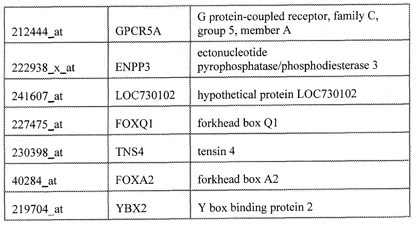


Claims
Priority Applications (8)
| Application Number | Priority Date | Filing Date | Title |
|---|---|---|---|
| EP11763516.9A EP2552953B1 (en) | 2010-04-01 | 2011-04-01 | Frizzled-binding agents and uses thereof |
| JP2013502890A JP2013530929A (en) | 2010-04-01 | 2011-04-01 | Frizzled binders and uses thereof |
| KR1020127028585A KR20130043102A (en) | 2010-04-01 | 2011-04-01 | Frizzled-binding agents and uses thereof |
| AU2011235904A AU2011235904B2 (en) | 2010-04-01 | 2011-04-01 | Frizzled-binding agents and uses thereof |
| NZ602700A NZ602700A (en) | 2010-04-01 | 2011-04-01 | Frizzled-binding agents and uses thereof |
| CN201180024546.5A CN102971337B (en) | 2010-04-01 | 2011-04-01 | FZ combines medicament and application thereof |
| CA2794674A CA2794674A1 (en) | 2010-04-01 | 2011-04-01 | Frizzled-binding agents and uses thereof |
| MX2012011323A MX347515B (en) | 2010-04-01 | 2011-04-01 | Frizzled-binding agents and uses thereof. |
Applications Claiming Priority (2)
| Application Number | Priority Date | Filing Date | Title |
|---|---|---|---|
| US32017510P | 2010-04-01 | 2010-04-01 | |
| US61/320,175 | 2010-04-01 |
Publications (2)
| Publication Number | Publication Date |
|---|---|
| WO2011123785A2 true WO2011123785A2 (en) | 2011-10-06 |
| WO2011123785A3 WO2011123785A3 (en) | 2011-12-22 |
Family
ID=44712856
Family Applications (1)
| Application Number | Title | Priority Date | Filing Date |
|---|---|---|---|
| PCT/US2011/030950 Ceased WO2011123785A2 (en) | 2010-04-01 | 2011-04-01 | Frizzled-binding agents and uses thereof |
Country Status (10)
| Country | Link |
|---|---|
| US (2) | US8551789B2 (en) |
| EP (1) | EP2552953B1 (en) |
| JP (1) | JP2013530929A (en) |
| KR (1) | KR20130043102A (en) |
| CN (1) | CN102971337B (en) |
| AU (1) | AU2011235904B2 (en) |
| CA (1) | CA2794674A1 (en) |
| MX (1) | MX347515B (en) |
| NZ (1) | NZ602700A (en) |
| WO (1) | WO2011123785A2 (en) |
Cited By (16)
| Publication number | Priority date | Publication date | Assignee | Title |
|---|---|---|---|---|
| US8324361B2 (en) | 2005-10-31 | 2012-12-04 | Oncomed Pharmaceuticals, Inc. | Nucleic acid molecules encoding soluble frizzled (FZD) receptors |
| US8507442B2 (en) | 2008-09-26 | 2013-08-13 | Oncomed Pharmaceuticals, Inc. | Methods of use for an antibody against human frizzled receptors 1, 2. 5, 7 or 8 |
| US8551789B2 (en) | 2010-04-01 | 2013-10-08 | OncoMed Pharmaceuticals | Frizzled-binding agents and their use in screening for WNT inhibitors |
| WO2014141038A2 (en) | 2013-03-11 | 2014-09-18 | Irm Llc | Markers associated with wnt inhibitors |
| JP2015502958A (en) * | 2011-12-09 | 2015-01-29 | オンコメッド ファーマシューティカルズ インコーポレイテッド | Combination therapy for the treatment of cancer |
| US9157904B2 (en) | 2010-01-12 | 2015-10-13 | Oncomed Pharmaceuticals, Inc. | Wnt antagonists and methods of treatment and screening |
| CN105073195A (en) * | 2013-02-04 | 2015-11-18 | 昂科梅德制药有限公司 | Methods of treatment with Wnt pathway inhibitors and monitoring of the treatment |
| JP2015536933A (en) * | 2012-10-23 | 2015-12-24 | オンコメッド ファーマシューティカルズ インコーポレイテッド | Methods of treating neuroendocrine tumors using Wnt pathway binding agents |
| US9352039B2 (en) | 2012-02-09 | 2016-05-31 | The Regents Of The University Of Michigan | Method of reducing the number of EMT and MET type breast cancer stem cells |
| EP2968554A4 (en) * | 2013-03-14 | 2016-10-26 | Oncomed Pharm Inc | C-MET PROTEIN BINDING AGENTS AND USES THEREOF |
| US9850311B2 (en) | 2005-10-31 | 2017-12-26 | Oncomed Pharmaceuticals, Inc. | Compositions and methods for diagnosing and treating cancer |
| US10689442B2 (en) | 2013-08-14 | 2020-06-23 | Sachdev Sidhu | Antibodies against Frizzled receptor |
| US11746150B2 (en) | 2017-12-19 | 2023-09-05 | Surrozen Operating, Inc. | Anti-LRP5/6 antibodies and methods of use |
| US11773171B2 (en) | 2017-12-19 | 2023-10-03 | Surrozen Operating, Inc. | WNT surrogate molecules and uses thereof |
| US12006368B2 (en) | 2017-12-19 | 2024-06-11 | Surrozen Operating, Inc. | Anti-frizzled antibodies and methods of use |
| EP4671270A3 (en) * | 2013-12-27 | 2026-03-18 | Chugai Seiyaku Kabushiki Kaisha | Method for purifying antibody having low isoelectric point |
Families Citing this family (16)
| Publication number | Priority date | Publication date | Assignee | Title |
|---|---|---|---|---|
| CA2812058C (en) * | 2010-09-20 | 2019-04-16 | Bio-Rad Laboratories, Inc. | Dissociation of product-complexed contaminants in chromatography |
| AU2012271413B2 (en) | 2011-06-17 | 2017-07-13 | President And Fellows Of Harvard College | Frizzled 2 as a target for therapeutic antibodies in the treatment of cancer |
| US9168300B2 (en) | 2013-03-14 | 2015-10-27 | Oncomed Pharmaceuticals, Inc. | MET-binding agents and uses thereof |
| BR112017000130A2 (en) | 2014-07-11 | 2018-01-09 | Genentech Inc | method for mitigating toxicity associated with notch pathway inhibition and cancer treatment method |
| MA40364A (en) * | 2014-08-27 | 2016-03-03 | Oncomed Pharm Inc | Combination therapy for treatment of cancer |
| US11142577B2 (en) | 2014-09-12 | 2021-10-12 | The Board Of Trustees Of The Leland Stanford Junior University | WNT signaling agonist molecules |
| JP2017116513A (en) * | 2015-12-26 | 2017-06-29 | 二郎 藤本 | Anticancer agents and radiosensitivity test methods for cancer stem cells using experimental animals |
| EP3407907A4 (en) * | 2016-01-28 | 2019-09-04 | The Board of Trustees of the Leland Stanford Junior University | WNT COMPOSITIONS AND METHODS OF SYNTHESIZING THE SAME WITHOUT SERUM |
| US10919968B2 (en) * | 2016-01-29 | 2021-02-16 | Modmab Therapeutics Corporation | Frizzled5 protein-binding agents |
| WO2018049275A1 (en) * | 2016-09-09 | 2018-03-15 | Genentech, Inc. | Selective peptide inhibitors of frizzled |
| US11993645B2 (en) | 2017-01-11 | 2024-05-28 | The Board Of Trustees Of The Leland Stanford Junior University | Compositions comprising R-Spondin (RSPO) surrogate molecules |
| JP7305543B2 (en) | 2017-01-26 | 2023-07-10 | スロゼン オペレーティング, インコーポレイテッド | Tissue-specific Wnt signal enhancing molecules and uses thereof |
| US20210292422A1 (en) | 2018-07-05 | 2021-09-23 | Surrozen, Inc. | Multi-specific wnt surrogate molecules and uses thereof |
| WO2020014271A1 (en) | 2018-07-09 | 2020-01-16 | Surrozen, Inc. | Tissue-specific wnt signal enhancing molecules and uses |
| JP2023549854A (en) | 2020-11-16 | 2023-11-29 | スロゼン オペレーティング, インコーポレイテッド | Liver-specific Wnt signal enhancing molecules and their uses |
| CN115724977B (en) * | 2022-11-04 | 2025-06-13 | 中国科学技术大学 | Monoclonal antibodies against human frizzled receptor and their uses |
Family Cites Families (232)
| Publication number | Priority date | Publication date | Assignee | Title |
|---|---|---|---|---|
| US3687808A (en) | 1969-08-14 | 1972-08-29 | Univ Leland Stanford Junior | Synthetic polynucleotides |
| US4109496A (en) | 1977-12-20 | 1978-08-29 | Norris Industries | Trapped key mechanism |
| US4323546A (en) | 1978-05-22 | 1982-04-06 | Nuc Med Inc. | Method and composition for cancer detection in humans |
| US4411990A (en) | 1979-06-13 | 1983-10-25 | University Patents, Inc. | Primary bioassay of human tumor stem cells |
| US4873191A (en) | 1981-06-12 | 1989-10-10 | Ohio University | Genetic transformation of zygotes |
| US4612282A (en) | 1981-12-15 | 1986-09-16 | The United States Of America As Represented By The Secretary Of The Department Of Health And Human Services | Monoclonal antibodies reactive with human breast cancer |
| US4670393A (en) | 1982-03-22 | 1987-06-02 | Genentech, Inc. | DNA vectors encoding a novel human growth hormone-variant protein |
| US5019497A (en) | 1984-11-09 | 1991-05-28 | Lennart Olsson | Human squamous lung carcinoma cell specific antigens and antibodies |
| US5034506A (en) | 1985-03-15 | 1991-07-23 | Anti-Gene Development Group | Uncharged morpholino-based polymers having achiral intersubunit linkages |
| US5225539A (en) | 1986-03-27 | 1993-07-06 | Medical Research Council | Recombinant altered antibodies and methods of making altered antibodies |
| US5080891A (en) | 1987-08-03 | 1992-01-14 | Ddi Pharmaceuticals, Inc. | Conjugates of superoxide dismutase coupled to high molecular weight polyalkylene glycols |
| US5087570A (en) | 1988-05-10 | 1992-02-11 | Weissman Irving L | Homogeneous mammalian hematopoietic stem cell composition |
| US4981785A (en) | 1988-06-06 | 1991-01-01 | Ventrex Laboratories, Inc. | Apparatus and method for performing immunoassays |
| US4968103A (en) | 1988-07-22 | 1990-11-06 | Photofinish Cosmetics Inc. | Method of making a brush |
| US5223409A (en) | 1988-09-02 | 1993-06-29 | Protein Engineering Corp. | Directed evolution of novel binding proteins |
| US5994617A (en) | 1988-09-19 | 1999-11-30 | Hsc Research Development Corporation | Engraftment of immune-deficient mice with human cells |
| US5688666A (en) | 1988-10-28 | 1997-11-18 | Genentech, Inc. | Growth hormone variants with altered binding properties |
| US5534617A (en) | 1988-10-28 | 1996-07-09 | Genentech, Inc. | Human growth hormone variants having greater affinity for human growth hormone receptor at site 1 |
| US5530101A (en) | 1988-12-28 | 1996-06-25 | Protein Design Labs, Inc. | Humanized immunoglobulins |
| GB8901778D0 (en) | 1989-01-27 | 1989-03-15 | Univ Court Of The University O | Manipulatory technique |
| US6583115B1 (en) | 1989-10-12 | 2003-06-24 | Ohio University/Edison Biotechnology Institute | Methods for treating acromegaly and giantism with growth hormone antagonists |
| US5061620A (en) | 1990-03-30 | 1991-10-29 | Systemix, Inc. | Human hematopoietic stem cell |
| US5614396A (en) | 1990-06-14 | 1997-03-25 | Baylor College Of Medicine | Methods for the genetic modification of endogenous genes in animal cells by homologous recombination |
| US5489677A (en) | 1990-07-27 | 1996-02-06 | Isis Pharmaceuticals, Inc. | Oligonucleoside linkages containing adjacent oxygen and nitrogen atoms |
| US5602240A (en) | 1990-07-27 | 1997-02-11 | Ciba Geigy Ag. | Backbone modified oligonucleotide analogs |
| DE69129154T2 (en) | 1990-12-03 | 1998-08-20 | Genentech, Inc., South San Francisco, Calif. | METHOD FOR ENRICHING PROTEIN VARIANTS WITH CHANGED BINDING PROPERTIES |
| US5856441A (en) | 1991-05-03 | 1999-01-05 | Yale University | Serrate fragments and derivatives |
| IL101728A (en) | 1991-05-03 | 2007-08-19 | Univ Yale | Human notch and delta, binding domains in toporythmic proteins, and methods based thereon |
| IE20030749A1 (en) | 1991-05-03 | 2003-11-12 | Indiana University Foundation | Human notch and delta binding domains in torporythmic proteins, and methods based thereon |
| US6429186B1 (en) | 1991-05-10 | 2002-08-06 | Genentech, Inc. | Ligand antagonists for treatment of breast cancer |
| US5714331A (en) | 1991-05-24 | 1998-02-03 | Buchardt, Deceased; Ole | Peptide nucleic acids having enhanced binding affinity, sequence specificity and solubility |
| US5539082A (en) | 1993-04-26 | 1996-07-23 | Nielsen; Peter E. | Peptide nucleic acids |
| US5719262A (en) | 1993-11-22 | 1998-02-17 | Buchardt, Deceased; Ole | Peptide nucleic acids having amino acid side chains |
| WO1994004679A1 (en) | 1991-06-14 | 1994-03-03 | Genentech, Inc. | Method for making humanized antibodies |
| US5750376A (en) | 1991-07-08 | 1998-05-12 | Neurospheres Holdings Ltd. | In vitro growth and proliferation of genetically modified multipotent neural stem cells and their progeny |
| ES2136092T3 (en) | 1991-09-23 | 1999-11-16 | Medical Res Council | PROCEDURES FOR THE PRODUCTION OF HUMANIZED ANTIBODIES. |
| US5753229A (en) | 1991-09-25 | 1998-05-19 | Mordoh; Jose | Monoclonal antibodies reactive with tumor proliferating cells |
| US6353150B1 (en) | 1991-11-22 | 2002-03-05 | Hsc Research And Development Limited Partnership | Chimeric mammals with human hematopoietic cells |
| US5869282A (en) | 1991-12-11 | 1999-02-09 | Imperial Cancer Research Technology, Ltd. | Nucleotide and protein sequences of the serrate gene and methods based thereon |
| US6004924A (en) | 1991-12-11 | 1999-12-21 | Imperial Cancer Research Technology, Ltd. | Protein sequences of serrate gene products |
| EP0552108B1 (en) | 1992-01-17 | 1999-11-10 | Lakowicz, Joseph R. | Energy transfer phase-modulation fluoro-immunoassay |
| US5376313A (en) | 1992-03-27 | 1994-12-27 | Abbott Laboratories | Injection molding a plastic assay cuvette having low birefringence |
| US5786158A (en) | 1992-04-30 | 1998-07-28 | Yale University | Therapeutic and diagnostic methods and compositions based on notch proteins and nucleic acids |
| WO1994002593A1 (en) | 1992-07-27 | 1994-02-03 | California Institute Of Technology | Mammalian multipotent neural stem cells |
| US5589376A (en) | 1992-07-27 | 1996-12-31 | California Institute Of Technology | Mammalian neural crest stem cells |
| US5693482A (en) | 1992-07-27 | 1997-12-02 | California Institute Of Technology | Neural chest stem cell assay |
| US5849553A (en) | 1992-07-27 | 1998-12-15 | California Institute Of Technology | Mammalian multipotent neural stem cells |
| US5654183A (en) | 1992-07-27 | 1997-08-05 | California Institute Of Technology | Genetically engineered mammalian neural crest stem cells |
| DE69333969T2 (en) | 1992-10-30 | 2006-09-14 | The General Hospital Corp., Boston | Interactive trap system for the isolation of proteins |
| GB9223084D0 (en) | 1992-11-04 | 1992-12-16 | Imp Cancer Res Tech | Compounds to target cells |
| JP3106021B2 (en) | 1992-11-30 | 2000-11-06 | キヤノン株式会社 | Pattern data compression method and apparatus and output method and apparatus |
| EP0685234B1 (en) | 1993-02-19 | 2000-05-10 | Nippon Shinyaku Company, Limited | Drug composition containing nucleic acid copolymer |
| US5643765A (en) | 1993-04-06 | 1997-07-01 | University Of Rochester | Method for quantitative measurement of gene expression using multiplex competitive reverse transcriptase-polymerase chain reaction |
| US5639606A (en) | 1993-04-06 | 1997-06-17 | The University Of Rochester | Method for quantitative measurement of gene expression using multiplex competitive reverse transcriptase-polymerase chain reaction |
| US5876978A (en) | 1993-04-06 | 1999-03-02 | Medical College Of Ohio | Method for quantitative measurement of gene expression using multiplex competitive reverse transcriptase-polymerase chain reaction |
| US5538848A (en) | 1994-11-16 | 1996-07-23 | Applied Biosystems Division, Perkin-Elmer Corp. | Method for detecting nucleic acid amplification using self-quenching fluorescence probe |
| US5599677A (en) | 1993-12-29 | 1997-02-04 | Abbott Laboratories | Immunoassays for prostate specific antigen |
| ATE336587T1 (en) | 1994-06-10 | 2006-09-15 | Genvec Inc | ADENOVIRUS VECTOR SYSTEMS AND CELL LINES |
| US6156305A (en) | 1994-07-08 | 2000-12-05 | Baxter International Inc. | Implanted tumor cells for the prevention and treatment of cancer |
| US5650317A (en) | 1994-09-16 | 1997-07-22 | Michigan State University | Human breast epithelial cell type with stem cell and luminal epithelial cell characteristics |
| PT787200E (en) | 1994-10-28 | 2005-08-31 | Univ Pennsylvania | IMPROVED ADENOVIRUS AND METHODS FOR THEIR USE |
| US6218166B1 (en) | 1994-12-09 | 2001-04-17 | John Wayne Cancer Institute | Adjuvant incorporation into antigen carrying cells: compositions and methods |
| US5736396A (en) | 1995-01-24 | 1998-04-07 | Case Western Reserve University | Lineage-directed induction of human mesenchymal stem cell differentiation |
| US5872154A (en) | 1995-02-24 | 1999-02-16 | The Trustees Of The University Of Pennsylvania | Method of reducing an immune response to a recombinant adenovirus |
| US5707618A (en) | 1995-03-24 | 1998-01-13 | Genzyme Corporation | Adenovirus vectors for gene therapy |
| US5633161A (en) | 1995-03-29 | 1997-05-27 | Millennium Pharmaceuticals, Inc. | Murine gene fomy030 coding for tumor progression inhibitor |
| US5821108A (en) | 1995-04-07 | 1998-10-13 | The Board Of Trustees Of The Leland Stanford Junior University | Enrichment for a thymocyte subset having progenitor cell activity using c-kit as a selection marker |
| US5739277A (en) | 1995-04-14 | 1998-04-14 | Genentech Inc. | Altered polypeptides with increased half-life |
| US6019978A (en) | 1995-06-05 | 2000-02-01 | The Wistar Institute Of Anatomy And Biology | Replication-defective adenovirus human type 5 recombinant as a vaccine carrier |
| ATE445705T1 (en) | 1995-06-15 | 2009-10-15 | Crucell Holland Bv | PACKAGING SYSTEMS FOR HUMAN RECOMBINANT ADENOVIRUSES FOR GENE THERAPY |
| US6117985A (en) | 1995-06-16 | 2000-09-12 | Stemcell Technologies Inc. | Antibody compositions for preparing enriched cell preparations |
| ATE442373T1 (en) | 1995-06-28 | 2009-09-15 | Imp Cancer Res Tech | NUCLEOTIDE AND PROTEIN SEQUENCES OF VERTEBRATE DELTA GENES AND METHODS BASED THEREOF |
| JP3628333B2 (en) | 1995-09-21 | 2005-03-09 | ジェネンテック インコーポレーテッド | Human growth hormone mutant |
| US5780300A (en) | 1995-09-29 | 1998-07-14 | Yale University | Manipulation of non-terminally differentiated cells using the notch pathway |
| US5986170A (en) | 1995-11-13 | 1999-11-16 | Corixa Corporation | Murine model for human carcinoma |
| US6337387B1 (en) | 1995-11-17 | 2002-01-08 | Asahi Kasei Kabushiki Kaisha | Differentiation-suppressive polypeptide |
| CA2239957A1 (en) | 1995-12-22 | 1997-07-03 | Abbott Laboratories | Fluorescence polarization immunoassay diagnostic method |
| US5994316A (en) | 1996-02-21 | 1999-11-30 | The Immune Response Corporation | Method of preparing polynucleotide-carrier complexes for delivery to cells |
| AU2160297A (en) | 1996-03-30 | 1997-10-22 | Boehringer Mannheim Gmbh | Method for the production of activated marked tumor-specific t cells and use thereof in treatment of tumors |
| US5753506A (en) | 1996-05-23 | 1998-05-19 | Cns Stem Cell Technology, Inc. | Isolation propagation and directed differentiation of stem cells from embryonic and adult central nervous system of mammals |
| JP2000512134A (en) | 1996-05-31 | 2000-09-19 | ザ ナショナル アメリカン レッド クロス | Methods and compositions for therapeutic and diagnostics based on Jagged / Notch proteins and nucleic acids |
| US6716974B1 (en) | 1996-05-31 | 2004-04-06 | Maine Medical Center Research Institute | Therapeutic and diagnostic methods and compositions based on jagged/notch proteins and nucleic acids |
| US5885529A (en) | 1996-06-28 | 1999-03-23 | Dpc Cirrus, Inc. | Automated immunoassay analyzer |
| FR2751986B1 (en) | 1996-08-01 | 1998-12-31 | Inst Nat Sante Rech Med | GENE INVOLVED IN CADASIL, DIAGNOSTIC METHOD AND THERAPEUTIC APPLICATION |
| CA2263883C (en) | 1996-08-29 | 2003-07-08 | The Regents Of The University Of California | Kuz, a novel family of metalloproteases |
| WO1998013493A2 (en) | 1996-09-24 | 1998-04-02 | Lxr Biotechnology, Inc. | A family of genes encoding apoptosis-related peptides, peptides encoded thereby and methods of use thereof |
| CA2267157C (en) | 1996-10-11 | 2005-05-31 | The Regents Of The University Of California | Cancer immunotherapy using tumor cells combined with mixed lymphocytes |
| US5994132A (en) | 1996-10-23 | 1999-11-30 | University Of Michigan | Adenovirus vectors |
| DE69739673D1 (en) | 1996-11-27 | 2009-12-31 | Genentech Inc | Affinity Purification of Polypeptide Proteins on a Protein A Matrix |
| US5859535A (en) | 1997-02-12 | 1999-01-12 | The United States Of America As Represented By The Secretary Of The Navy | System for determining size and location of defects in material by use of microwave radiation |
| WO1998039636A1 (en) | 1997-03-07 | 1998-09-11 | Clare Chemical Research Llc | Fluorometric detection using visible light |
| US6080912A (en) | 1997-03-20 | 2000-06-27 | Wisconsin Alumni Research Foundation | Methods for creating transgenic animals |
| US5861832A (en) | 1997-03-27 | 1999-01-19 | Lucent Technologies Inc. | Analog-to-digital converter having amplifier and comparator stages |
| US20020137890A1 (en) | 1997-03-31 | 2002-09-26 | Genentech, Inc. | Secreted and transmembrane polypeptides and nucleic acids encoding the same |
| AU6952498A (en) | 1997-04-04 | 1998-10-30 | Millenium Pharmaceuticals, Inc. | Novel human delta3 compositions and therapeutic and diagnostic uses therefor |
| US20030180784A1 (en) | 1997-04-04 | 2003-09-25 | Millennium Pharmaceuticals, Inc. | Novel human Delta3 compositions and therapeutic and diagnostic uses therefor |
| US6121045A (en) | 1997-04-04 | 2000-09-19 | Millennium Biotherapeutics, Inc. | Human Delta3 nucleic acid molecules |
| US5830730A (en) | 1997-05-08 | 1998-11-03 | The Regents Of The University Of California | Enhanced adenovirus-assisted transfection composition and method |
| EP1004669B1 (en) | 1997-05-14 | 2007-04-18 | Asahi Kasei Kabushiki Kaisha | Novel differentiation inhibitor |
| AU8162898A (en) | 1997-06-18 | 1999-01-04 | Trustees Of Columbia University In The City Of New York, The | Angiogenic modulation by notch signal transduction |
| US6379925B1 (en) | 1997-06-18 | 2002-04-30 | The Trustees Of Columbia University In The City Of New York | Angiogenic modulation by notch signal transduction |
| US6136952A (en) | 1997-06-25 | 2000-10-24 | University Of Washington | Human jagged polypeptide, encoding nucleic acids and methods of use |
| CA2290736A1 (en) | 1997-07-11 | 1999-01-21 | Introgene B.V. | Interleukin-3 gene therapy for cancer |
| US6004528A (en) | 1997-09-18 | 1999-12-21 | Bergstein; Ivan | Methods of cancer diagnosis and therapy targeted against the cancer stemline |
| US7361336B1 (en) | 1997-09-18 | 2008-04-22 | Ivan Bergstein | Methods of cancer therapy targeted against a cancer stem line |
| US6197523B1 (en) | 1997-11-24 | 2001-03-06 | Robert A. Levine | Method for the detection, identification, enumeration and confirmation of circulating cancer and/or hematologic progenitor cells in whole blood |
| US6506559B1 (en) | 1997-12-23 | 2003-01-14 | Carnegie Institute Of Washington | Genetic inhibition by double-stranded RNA |
| US6551795B1 (en) * | 1998-02-18 | 2003-04-22 | Genome Therapeutics Corporation | Nucleic acid and amino acid sequences relating to pseudomonas aeruginosa for diagnostics and therapeutics |
| US5981225A (en) | 1998-04-16 | 1999-11-09 | Baylor College Of Medicine | Gene transfer vector, recombinant adenovirus particles containing the same, method for producing the same and method of use of the same |
| US6252050B1 (en) | 1998-06-12 | 2001-06-26 | Genentech, Inc. | Method for making monoclonal antibodies and cross-reactive antibodies obtainable by the method |
| WO2000006726A2 (en) | 1998-07-27 | 2000-02-10 | Amgen Inc. | Delta-related polypeptides |
| CA2340682A1 (en) | 1998-08-14 | 2000-02-24 | Aventis Pharmaceuticals Products Inc. | Adenovirus formulations for gene therapy |
| JP2002523105A (en) | 1998-08-27 | 2002-07-30 | アバンテイス・フアルマ・エス・アー | Targeted adenovirus vectors for delivery of heterologous genes |
| GB9819681D0 (en) | 1998-09-09 | 1998-11-04 | Smithkline Beecham Plc | Novel compounds |
| US6660843B1 (en) | 1998-10-23 | 2003-12-09 | Amgen Inc. | Modified peptides as therapeutic agents |
| EP1133519B1 (en) | 1998-11-02 | 2008-08-13 | Curis, Inc. | Functional antagonists of hedgehog activity |
| US6291516B1 (en) | 1999-01-13 | 2001-09-18 | Curis, Inc. | Regulators of the hedgehog pathway, compositions and uses related thereto |
| US20030003572A1 (en) | 1999-03-05 | 2003-01-02 | David J. Anderson | Isolation and enrichment of neural stem cells from uncultured tissue based on cell-surface marker expression |
| US20030119029A1 (en) | 1999-04-30 | 2003-06-26 | Regents Of The University Of Michigan | Compositions and methods relating to novel benzodiazepine compounds and targets thereof |
| AU782493B2 (en) | 1999-06-01 | 2005-08-04 | Curis, Inc. | Polymer conjugates of hedgehog proteins and uses |
| WO2001002568A2 (en) | 1999-07-02 | 2001-01-11 | Chiron Corporation | Human genes and gene expression products |
| US6703221B1 (en) | 1999-08-19 | 2004-03-09 | Chiron Corporation | Notch receptor ligands and uses thereof |
| AU7721500A (en) * | 1999-09-29 | 2001-04-30 | Human Genome Sciences, Inc. | Colon and colon cancer associated polynucleotides and polypeptides |
| AU1194501A (en) | 1999-10-14 | 2001-04-23 | Wistar Institute, The | Inhibition of tumorigenic properties of melanoma cells |
| US6380362B1 (en) | 1999-12-23 | 2002-04-30 | Genesis Research & Development Corporation Ltd. | Polynucleotides, polypeptides expressed by the polynucleotides and methods for their use |
| US20040048249A1 (en) | 2000-01-21 | 2004-03-11 | Tang Y. Tom | Novel nucleic acids and secreted polypeptides |
| AU779875B2 (en) | 2000-01-24 | 2005-02-17 | Merck Sharp & Dohme Limited | Gamma-secretase inhibitors |
| DK1252192T3 (en) | 2000-02-11 | 2006-11-20 | Merck Patent Gmbh | Enhancement of the serum half-life of antibody-based fusion proteins |
| US7115653B2 (en) | 2000-03-30 | 2006-10-03 | Curis, Inc. | Small organic molecule regulators of cell proliferation |
| US7060436B2 (en) | 2000-06-17 | 2006-06-13 | Third Wave Technologies, Inc. | Nucleic acid accessible hybridization sites |
| WO2001098354A2 (en) | 2000-06-21 | 2001-12-27 | Incyte Genomics, Inc. | Human receptors |
| US6632620B1 (en) | 2000-06-22 | 2003-10-14 | Andrew N. Makarovskiy | Compositions for identification and isolation of stem cells |
| DE10031380A1 (en) | 2000-06-28 | 2002-01-10 | Merck Patent Gmbh | Process for the transfer of alkylidene groups to organic compounds |
| GB0016681D0 (en) | 2000-07-06 | 2000-08-23 | Merck Sharp & Dohme | Therapeutic compounds |
| AU2001278076A1 (en) | 2000-07-26 | 2002-02-05 | Applied Genomics, Inc. | Bstp-5 proteins and related reagents and methods of use thereof |
| US20050089518A1 (en) | 2001-12-07 | 2005-04-28 | Clarke Michael F. | Prospective identification and characterization of breast cancer stem cells |
| US6984522B2 (en) | 2000-08-03 | 2006-01-10 | Regents Of The University Of Michigan | Isolation and use of solid tumor stem cells |
| AU2001288628A1 (en) | 2000-08-31 | 2002-03-13 | Loyola University Chicago | Method and reagents for treatment of skin disorders by modulating the notch pathway |
| US6689744B2 (en) | 2000-09-22 | 2004-02-10 | Genentech, Inc. | Notch receptor agonists and uses |
| BR0116138A (en) | 2000-12-13 | 2003-09-23 | Borean Pharma As | Protein combinatorial libraries having the scaffold structure of lectin type c domains |
| US20020169300A1 (en) | 2001-01-30 | 2002-11-14 | Waterman Marian L. | Method of detection and treatment of colon cancer by analysis of beta-catenin-sensitive isoforms of lymphoid enhancer factor-1 |
| US7413873B2 (en) | 2001-01-30 | 2008-08-19 | The Regents Of The University Of California | Method of detection and treatment of colon cancer |
| US20030064384A1 (en) | 2001-04-02 | 2003-04-03 | Mien-Chie Hung | Beta-catenin is a strong and independent prognostic factor for cancer |
| IL142481A0 (en) | 2001-04-05 | 2002-03-10 | Yissum Res Dev Co | A method for identifying consitutively active mutants of mitogen activated protein kinases (mapk) and uses thereof |
| DE60237969D1 (en) | 2001-04-24 | 2010-11-25 | Bayer Corp | HUMAN ANTIBODIES AGAINST TIMP-1 |
| AU2002308557A1 (en) | 2001-05-01 | 2002-11-11 | The Regents Of The University Of California | Wnt and frizzled receptors as targets for immunotherapy in head and neck squamous cell carcinomas |
| US7713526B2 (en) | 2001-05-01 | 2010-05-11 | The Regents Of The University Of California | Wnt and frizzled receptors as targets for immunotherapy in head and neck squamous cell carcinomas |
| US20030044409A1 (en) | 2001-05-01 | 2003-03-06 | Carson Dennis A. | Immunologic compositions and methods for studying and treating cancers expressing frizzled antigens |
| US7514594B2 (en) | 2001-05-11 | 2009-04-07 | Wyeth | Transgenic animal model of bone mass modulation |
| JP2005500303A (en) | 2001-06-15 | 2005-01-06 | ジェネンテック・インコーポレーテッド | Human growth hormone antagonist |
| US7171311B2 (en) | 2001-06-18 | 2007-01-30 | Rosetta Inpharmatics Llc | Methods of assigning treatment to breast cancer patients |
| US20040023244A1 (en) | 2001-06-21 | 2004-02-05 | Griffin Jennifer A | Receptors |
| ES2292772T3 (en) | 2001-06-22 | 2008-03-16 | Stemcells, Inc. | LIVER GRAFT CELLS, ANALYSIS, AND ITS USES. |
| WO2003000893A2 (en) | 2001-06-26 | 2003-01-03 | Decode Genetics Ehf. | Nucleic acids encoding g protein-coupled receptors |
| JP2004536045A (en) | 2001-07-02 | 2004-12-02 | タシ、シナン | Use of cyclopamine in the treatment of basal cell carcinoma and other tumors |
| EP1411938B1 (en) | 2001-07-02 | 2005-07-06 | Tas, Sinan | Use of cyclopamine for the manufacture of a medicament for the treatemnt of psoriasis |
| AU2002317105A1 (en) | 2001-07-05 | 2003-01-21 | University Of British Columbia | Methods for identifying therapeutic agents for treating diseases involving wnt polypeptides and wnt receptors |
| CA2453810A1 (en) * | 2001-07-18 | 2003-07-03 | American Red Cross | Mutant proteinase inhibitors and uses thereof |
| AU2002355477B2 (en) | 2001-08-03 | 2008-09-25 | Medical Research Council | Method of identifying a consensus sequence for intracellular antibodies |
| ATE515495T1 (en) | 2001-08-03 | 2011-07-15 | Schering Corp | GAMMA SECRETASE INHIBITORS |
| US7138370B2 (en) | 2001-10-11 | 2006-11-21 | Amgen Inc. | Specific binding agents of human angiopoietin-2 |
| AU2002339157A1 (en) | 2001-11-14 | 2003-05-26 | Lorantis Limited | Inhibitors of the notch signalling pathway for use in the treatment of cancer |
| US7053547B2 (en) | 2001-11-29 | 2006-05-30 | Universal Display Corporation | Increased emission efficiency in organic light-emitting devices on high-index substrates |
| US20070237770A1 (en) | 2001-11-30 | 2007-10-11 | Albert Lai | Novel compositions and methods in cancer |
| JP2005514030A (en) | 2001-12-26 | 2005-05-19 | イミューノメディクス、インコーポレイテッド | Method for producing multispecific multivalent drugs from VH and VL domains |
| US6713206B2 (en) | 2002-01-14 | 2004-03-30 | Board Of Trustees Of University Of Illinois | Electrochemical cells comprising laminar flow induced dynamic conducting interfaces, electronic devices comprising such cells, and methods employing same |
| GB0201674D0 (en) | 2002-01-25 | 2002-03-13 | Lorantis Ltd | Medical treatment |
| US20030185829A1 (en) | 2002-03-12 | 2003-10-02 | Erich Koller | Jagged 2 inhibitors for inducing apoptosis |
| CN1305905C (en) | 2002-03-22 | 2007-03-21 | Aprogen株式会社 | Humanized antibody and process for preparing same |
| EP1554392A4 (en) | 2002-05-06 | 2007-08-08 | Us Gov Health & Human Serv | IDENTIFICATION OF WIDE-NEUTRALIZING HUMAN MONOCLONAL ANTIBODIES WITH CROSS-REACTION USING SEQUENTIAL PLATE ADHESION METHOD OF ANTIGENS OF PHAGE PRESENTATION BANKS |
| WO2003099781A2 (en) | 2002-05-24 | 2003-12-04 | Boehringer Ingelheim Pharmaceuticals, Inc. | METHODS FOR THE IDENTIFICATION OF IKKα FUNCTION AND OTHER GENES USEFUL FOR TREATMENT OF INFLAMMATORY DISEASES |
| AU2003278161A1 (en) | 2002-06-21 | 2004-01-06 | Johns Hopkins University School Of Medicine | Membrane associated tumor endothelium markers |
| US7803370B2 (en) | 2002-08-30 | 2010-09-28 | Oncotherapy Science, Inc. | Method for treating synovial sarcoma |
| AU2003256079A1 (en) | 2002-08-30 | 2004-03-19 | Japan As Represented By President Of The University Of Tokyo | Method for treating synovial sarcoma |
| GB0221952D0 (en) | 2002-09-20 | 2002-10-30 | Novartis Forschungsstiftung | Wnt mediated ErbB1 signalling,compositions and uses related thereto |
| US20040058217A1 (en) | 2002-09-20 | 2004-03-25 | Ohlsen Leroy J. | Fuel cell systems having internal multistream laminar flow |
| US20040247593A1 (en) | 2002-10-04 | 2004-12-09 | Regents Of The University Of California, | Methods for treating cancer by inhibiting Wnt signaling |
| CA2507581A1 (en) | 2002-12-06 | 2004-06-24 | The Board Of Trustees Of The Leland Stanford Junior University | Protection of stem cells from cytotoxic agents by modulation of .beta.-catenin signaling pathways |
| US7803783B2 (en) | 2002-12-06 | 2010-09-28 | The Board Of Trustees Of The Leland Stanford Junior University | Use of WNT inhibitors to augment therapeutic index of chemotherapy |
| US20040219579A1 (en) | 2003-02-19 | 2004-11-04 | Natasha Aziz | Methods of diagnosis of cancer, compositions and methods of screening for modulators of cancer |
| TWI353991B (en) | 2003-05-06 | 2011-12-11 | Syntonix Pharmaceuticals Inc | Immunoglobulin chimeric monomer-dimer hybrids |
| EP2003196A3 (en) | 2003-06-09 | 2009-01-07 | The Regents of the University of Michigan | Compositions and methods for treating and diagnosing cancer |
| ATE545658T1 (en) | 2003-07-11 | 2012-03-15 | Oncotherapy Science Inc | METHOD FOR TREATING SYNOVIAL SARCOMA |
| US20050130199A1 (en) | 2003-09-22 | 2005-06-16 | Regents Of The University Of California | Mutations in WNT-frizzled signaling pathways associated with osteoarthritis |
| US6894522B2 (en) | 2003-10-06 | 2005-05-17 | International Business Machines Corporation | Specific site backside underlaying and micromasking method for electrical characterization of semiconductor devices |
| CN1981054A (en) | 2004-05-14 | 2007-06-13 | 加利福尼亚大学董事会 | Methods for treating cancer using anti-WNT2 monoclonal antibodies and sirna |
| WO2006036175A2 (en) | 2004-09-21 | 2006-04-06 | Rhode Island Hospital, A Lifespan-Partner | Wnt proteins and detection and treatment of cancer |
| WO2006036179A2 (en) | 2004-09-21 | 2006-04-06 | Rhode Island Hospital, A Lifespan-Partner | Frizzled proteins and detection and treatment of cancer |
| JP5025475B2 (en) | 2004-09-21 | 2012-09-12 | ロード アイランド ホスピタル エー ライフスパン−パートナー | Methods for Wnt protein and cancer detection and treatment |
| CN101103045B (en) | 2004-09-24 | 2015-11-25 | 安姆根有限公司 | The Fc molecule modified |
| DE102004050620A1 (en) | 2004-10-13 | 2006-04-20 | Eberhard-Karls-Universität Tübingen | Monoclonal antibody against Frizzled receptors |
| WO2006055635A2 (en) | 2004-11-15 | 2006-05-26 | Mount Sinai School Of Medicine Of New York University | Compositions and methods for altering wnt autocrine signaling |
| ES2350231T3 (en) | 2004-11-24 | 2011-01-20 | F. Hoffmann-La Roche Ag | METHOD FOR DETERMINING GENOTOXICITY. |
| US7635530B2 (en) | 2005-03-21 | 2009-12-22 | The Board Of Trustees Of The University Of Illinois | Membraneless electrochemical cell and microfluidic device without pH constraint |
| US20090311243A1 (en) | 2005-05-30 | 2009-12-17 | Astrazeneca Ab | Methods for Identifying FZD8 Modulators and the Use of such Modulators for Treating Osteoarthritis |
| US20070014776A1 (en) * | 2005-06-09 | 2007-01-18 | Gimeno Ruth E | Identification of adiponutrin-related proteins as esterases and methods of use for the same |
| US20070072239A1 (en) | 2005-09-26 | 2007-03-29 | Wyeth | Pharmaceutical compositions and methods of using secreted frizzled related protein |
| US20070238658A1 (en) | 2005-09-28 | 2007-10-11 | Levin Steven D | IL-17A and IL-17F Antagonists and Methods of Using the Same |
| US7723477B2 (en) | 2005-10-31 | 2010-05-25 | Oncomed Pharmaceuticals, Inc. | Compositions and methods for inhibiting Wnt-dependent solid tumor cell growth |
| PT2500360E (en) | 2005-10-31 | 2015-10-15 | Oncomed Pharm Inc | Compositions and methods for diagnosing and treating cancer |
| DOP2006000277A (en) | 2005-12-12 | 2007-08-31 | Bayer Pharmaceuticals Corp | ANTI MN ANTIBODIES AND METHODS FOR USE |
| AR060017A1 (en) | 2006-01-13 | 2008-05-21 | Novartis Ag | COMPOSITIONS AND METHODS OF USE FOR DICKKOPF -1 ANTIBODIES |
| CA2641315A1 (en) | 2006-02-06 | 2007-08-16 | Tethys Bioscience, Inc. | Osteoporosis associated markers and methods of use thereof |
| GB0603683D0 (en) | 2006-02-23 | 2006-04-05 | Novartis Ag | Organic compounds |
| MX2008014910A (en) | 2006-05-24 | 2009-01-23 | Bayer Schering Pharma Ag | HIGH AFFINITY HUMAN AND HUMANIZED ANTI-alpha5beta1 INTEGRIN FUNCTION BLOCKING ANTIBODIES WITH REDUCED IMMUNOGENICITY. |
| CN101573383B (en) | 2006-06-21 | 2015-05-13 | 肿瘤疗法科学股份有限公司 | Tumor-targeting monoclonal antibody against frizzled homologue 10 and use thereof |
| TW200815469A (en) | 2006-06-23 | 2008-04-01 | Astrazeneca Ab | Compounds |
| EP2526969A1 (en) | 2006-09-07 | 2012-11-28 | Stemline Therapeutics, Inc. | Monitoring cancer stem cells |
| CN101600449A (en) * | 2006-09-08 | 2009-12-09 | 健泰科生物技术公司 | WNT antagonists and their use in the diagnosis and treatment of WNT-mediated disorders |
| US7947277B2 (en) | 2006-09-08 | 2011-05-24 | Genentech, Inc. | Wnt antagonists and their use in the diagnosis and treatment of Wnt-mediated disorders |
| EP2084296B1 (en) | 2006-09-29 | 2015-08-05 | Agendia N.V. | High-throughput diagnostic testing using arrays |
| EP2083861A4 (en) | 2006-11-07 | 2010-11-24 | Merck Sharp & Dohme | Antagonists of pcsk9 |
| WO2008061020A2 (en) | 2006-11-14 | 2008-05-22 | Novartis Ag | Methods of treating, diagnosing or detecting cancer |
| ES2614402T3 (en) | 2007-09-26 | 2017-05-31 | Intrexon Corporation | 5'UTR synthetics, expression vectors and methods to increase transgenic expression |
| TWI489993B (en) | 2007-10-12 | 2015-07-01 | Novartis Ag | Compositions and methods of use for antibodies against sclerostin |
| US20110020368A1 (en) | 2008-03-25 | 2011-01-27 | Nancy Hynes | Treating cancer by down-regulating frizzled-4 and/or frizzled-1 |
| DE202008004821U1 (en) | 2008-04-07 | 2008-06-12 | Heil, Roland | screen printing forme |
| BRPI0911676A2 (en) * | 2008-04-11 | 2016-11-29 | Cbio Ltd | cpn10 isolated polypeptide, isolated nucleic acid, expression construct, host cell, antibody, pharmaceutical composition, method for treating or preventing a disease, method for modulating prr signaling, method for modulating the production and / or secretion of one or more immunomodulators, method for inhibiting production and / or secretion of one or more immunomodulators, method of identifying a compound that binds to a polypeptide, method for screening a coprote and a prr ligand and functional variant, derivative, homologue, analog or polypeptide fragment |
| FR2936255B1 (en) | 2008-09-19 | 2010-10-15 | Galderma Res & Dev | MODULATORS OF FZD2 IN THE TREATMENT OF ALOPECIA |
| ES2663536T3 (en) | 2008-09-26 | 2018-04-13 | Oncomed Pharmaceuticals, Inc. | Agents that bind to frizzled receptors and uses thereof |
| US20130295106A1 (en) | 2008-09-26 | 2013-11-07 | Oncomed Pharmaceuticals, Inc. | Frizzled-Binding Agents And Uses Thereof |
| WO2010038756A1 (en) | 2008-09-30 | 2010-04-08 | 協和発酵キリン株式会社 | PHARMACEUTICAL COMPOSITION FOR TREATING BONE DISEASES WHICH COMPRISES PROTEIN COMPRISING Frizzled1, Frizzled2 OR Frizzled7 EXTRACELLULAR CYSTEINE-RICH DOMAIN |
| US20100169025A1 (en) | 2008-10-10 | 2010-07-01 | Arthur William T | Methods and gene expression signature for wnt/b-catenin signaling pathway |
| TWI535445B (en) | 2010-01-12 | 2016-06-01 | 安可美德藥物股份有限公司 | Wnt antagonists and methods of treatment and screening |
| UY33227A (en) | 2010-02-19 | 2011-09-30 | Novartis Ag | PIRROLOPIRIMIDINE COMPOUNDS AS INHIBITORS OF THE CDK4 / 6 |
| CN103038364A (en) | 2010-03-09 | 2013-04-10 | 达纳-法伯癌症研究所公司 | Methods of diagnosing and treating cancer in patients having or developing resistance to a first cancer therapy |
| CN102971337B (en) | 2010-04-01 | 2016-09-21 | 昂考梅德药品有限公司 | FZ combines medicament and application thereof |
| US8431532B2 (en) | 2010-06-28 | 2013-04-30 | Five Prime Therepeutics, Inc. | FZD8 extracellular domains and FZD8 extracellular domain fusion molecules and treatments using same |
| UY33469A (en) | 2010-06-29 | 2012-01-31 | Irm Llc Y Novartis Ag | COMPOSITIONS AND METHODS TO MODULATE THE WNT SIGNALING ROAD |
| ES2863626T3 (en) | 2010-10-27 | 2021-10-11 | Amgen Inc | DKK1 antibodies and methods of use |
| HK1212216A1 (en) | 2012-10-23 | 2016-06-10 | Oncomed Pharmaceuticals, Inc. | Methods of treating neuroendocrine tumors using wnt pathway-binding agents |
| JP2016510411A (en) | 2013-02-04 | 2016-04-07 | オンコメッド ファーマシューティカルズ インコーポレイテッド | Methods and monitoring of treatment with WNT pathway inhibitors |
-
2011
- 2011-04-01 CN CN201180024546.5A patent/CN102971337B/en not_active Expired - Fee Related
- 2011-04-01 JP JP2013502890A patent/JP2013530929A/en active Pending
- 2011-04-01 AU AU2011235904A patent/AU2011235904B2/en not_active Ceased
- 2011-04-01 CA CA2794674A patent/CA2794674A1/en not_active Abandoned
- 2011-04-01 KR KR1020127028585A patent/KR20130043102A/en not_active Withdrawn
- 2011-04-01 US US13/078,728 patent/US8551789B2/en active Active
- 2011-04-01 MX MX2012011323A patent/MX347515B/en active IP Right Grant
- 2011-04-01 EP EP11763516.9A patent/EP2552953B1/en active Active
- 2011-04-01 NZ NZ602700A patent/NZ602700A/en not_active IP Right Cessation
- 2011-04-01 WO PCT/US2011/030950 patent/WO2011123785A2/en not_active Ceased
-
2013
- 2013-08-23 US US13/974,792 patent/US9499630B2/en active Active
Non-Patent Citations (2)
| Title |
|---|
| None |
| See also references of EP2552953A4 |
Cited By (27)
| Publication number | Priority date | Publication date | Assignee | Title |
|---|---|---|---|---|
| US8324361B2 (en) | 2005-10-31 | 2012-12-04 | Oncomed Pharmaceuticals, Inc. | Nucleic acid molecules encoding soluble frizzled (FZD) receptors |
| US9850311B2 (en) | 2005-10-31 | 2017-12-26 | Oncomed Pharmaceuticals, Inc. | Compositions and methods for diagnosing and treating cancer |
| US9732139B2 (en) | 2005-10-31 | 2017-08-15 | Oncomed Pharmaceuticals, Inc. | Methods of treating cancer by administering a soluble receptor comprising a human Fc domain and the Fri domain from human frizzled receptor |
| US8765913B2 (en) | 2005-10-31 | 2014-07-01 | Oncomed Pharmaceuticals, Inc. | Human frizzled (FZD) receptor polypeptides and methods of use thereof for treating cancer and inhibiting growth of tumor cells |
| US8975044B2 (en) | 2008-09-26 | 2015-03-10 | Oncomed Pharmaceuticals, Inc. | Polynucleotides encoding for frizzled-binding agents and uses thereof |
| US9273139B2 (en) | 2008-09-26 | 2016-03-01 | Oncomed Pharmaceuticals, Inc. | Monoclonal antibodies against frizzled |
| US8507442B2 (en) | 2008-09-26 | 2013-08-13 | Oncomed Pharmaceuticals, Inc. | Methods of use for an antibody against human frizzled receptors 1, 2. 5, 7 or 8 |
| US9573998B2 (en) | 2008-09-26 | 2017-02-21 | Oncomed Pharmaceuticals, Inc. | Antibodies against human FZD5 and FZD8 |
| US9157904B2 (en) | 2010-01-12 | 2015-10-13 | Oncomed Pharmaceuticals, Inc. | Wnt antagonists and methods of treatment and screening |
| US9579361B2 (en) | 2010-01-12 | 2017-02-28 | Oncomed Pharmaceuticals, Inc. | Wnt antagonist and methods of treatment and screening |
| US9499630B2 (en) | 2010-04-01 | 2016-11-22 | Oncomed Pharmaceuticals, Inc. | Frizzled-binding agents and uses thereof |
| US8551789B2 (en) | 2010-04-01 | 2013-10-08 | OncoMed Pharmaceuticals | Frizzled-binding agents and their use in screening for WNT inhibitors |
| JP2015502958A (en) * | 2011-12-09 | 2015-01-29 | オンコメッド ファーマシューティカルズ インコーポレイテッド | Combination therapy for the treatment of cancer |
| US9352039B2 (en) | 2012-02-09 | 2016-05-31 | The Regents Of The University Of Michigan | Method of reducing the number of EMT and MET type breast cancer stem cells |
| JP2015536933A (en) * | 2012-10-23 | 2015-12-24 | オンコメッド ファーマシューティカルズ インコーポレイテッド | Methods of treating neuroendocrine tumors using Wnt pathway binding agents |
| JP2016510411A (en) * | 2013-02-04 | 2016-04-07 | オンコメッド ファーマシューティカルズ インコーポレイテッド | Methods and monitoring of treatment with WNT pathway inhibitors |
| EP2950885A4 (en) * | 2013-02-04 | 2016-09-14 | Oncomed Pharm Inc | METHODS AND SURVEILLANCE OF TREATMENT BY AN INHIBITOR OF THE WNT PATH |
| US9987357B2 (en) | 2013-02-04 | 2018-06-05 | Oncomed Pharmaceuticals, Inc. | Methods and monitoring of treatment with a WNT pathway inhibitor |
| CN105073195A (en) * | 2013-02-04 | 2015-11-18 | 昂科梅德制药有限公司 | Methods of treatment with Wnt pathway inhibitors and monitoring of the treatment |
| WO2014141038A2 (en) | 2013-03-11 | 2014-09-18 | Irm Llc | Markers associated with wnt inhibitors |
| EP2968554A4 (en) * | 2013-03-14 | 2016-10-26 | Oncomed Pharm Inc | C-MET PROTEIN BINDING AGENTS AND USES THEREOF |
| US10689442B2 (en) | 2013-08-14 | 2020-06-23 | Sachdev Sidhu | Antibodies against Frizzled receptor |
| EP4671270A3 (en) * | 2013-12-27 | 2026-03-18 | Chugai Seiyaku Kabushiki Kaisha | Method for purifying antibody having low isoelectric point |
| US11746150B2 (en) | 2017-12-19 | 2023-09-05 | Surrozen Operating, Inc. | Anti-LRP5/6 antibodies and methods of use |
| US11773171B2 (en) | 2017-12-19 | 2023-10-03 | Surrozen Operating, Inc. | WNT surrogate molecules and uses thereof |
| US12006368B2 (en) | 2017-12-19 | 2024-06-11 | Surrozen Operating, Inc. | Anti-frizzled antibodies and methods of use |
| US12297278B2 (en) | 2017-12-19 | 2025-05-13 | Surrozen Operating, Inc. | Wnt surrogate molecules and uses thereof |
Also Published As
| Publication number | Publication date |
|---|---|
| NZ602700A (en) | 2014-10-31 |
| JP2013530929A (en) | 2013-08-01 |
| WO2011123785A3 (en) | 2011-12-22 |
| MX347515B (en) | 2017-04-28 |
| AU2011235904B2 (en) | 2015-10-08 |
| EP2552953A2 (en) | 2013-02-06 |
| US20140105917A1 (en) | 2014-04-17 |
| KR20130043102A (en) | 2013-04-29 |
| EP2552953A4 (en) | 2013-09-04 |
| US9499630B2 (en) | 2016-11-22 |
| US20120027778A1 (en) | 2012-02-02 |
| MX2012011323A (en) | 2012-11-23 |
| CA2794674A1 (en) | 2011-10-06 |
| CN102971337A (en) | 2013-03-13 |
| AU2011235904A1 (en) | 2012-10-25 |
| US8551789B2 (en) | 2013-10-08 |
| EP2552953B1 (en) | 2017-05-17 |
| CN102971337B (en) | 2016-09-21 |
Similar Documents
| Publication | Publication Date | Title |
|---|---|---|
| EP2552953B1 (en) | Frizzled-binding agents and uses thereof | |
| US9573998B2 (en) | Antibodies against human FZD5 and FZD8 | |
| US20170234854A1 (en) | Frizzled-binding agents and uses thereof | |
| AU2011205409B2 (en) | Wnt-binding agents and uses thereof | |
| AU2013204146B2 (en) | Frizzled-binding agents and uses thereof | |
| HK1181401A (en) | Frizzled-binding agents and uses thereof | |
| HK1183435B (en) | Frizzled-binding agents and uses thereof |
Legal Events
| Date | Code | Title | Description |
|---|---|---|---|
| WWE | Wipo information: entry into national phase |
Ref document number: 201180024546.5 Country of ref document: CN |
|
| 121 | Ep: the epo has been informed by wipo that ep was designated in this application |
Ref document number: 11763516 Country of ref document: EP Kind code of ref document: A2 |
|
| ENP | Entry into the national phase |
Ref document number: 2794674 Country of ref document: CA |
|
| WWE | Wipo information: entry into national phase |
Ref document number: 2013502890 Country of ref document: JP Ref document number: MX/A/2012/011323 Country of ref document: MX |
|
| NENP | Non-entry into the national phase |
Ref country code: DE |
|
| WWE | Wipo information: entry into national phase |
Ref document number: 3102/KOLNP/2012 Country of ref document: IN |
|
| REEP | Request for entry into the european phase |
Ref document number: 2011763516 Country of ref document: EP |
|
| WWE | Wipo information: entry into national phase |
Ref document number: 2011763516 Country of ref document: EP |
|
| ENP | Entry into the national phase |
Ref document number: 2011235904 Country of ref document: AU Date of ref document: 20110401 Kind code of ref document: A |
|
| ENP | Entry into the national phase |
Ref document number: 20127028585 Country of ref document: KR Kind code of ref document: A |