US11248058B2 - Chimeric antigen receptors and uses thereof - Google Patents
Chimeric antigen receptors and uses thereof Download PDFInfo
- Publication number
- US11248058B2 US11248058B2 US15/552,071 US201615552071A US11248058B2 US 11248058 B2 US11248058 B2 US 11248058B2 US 201615552071 A US201615552071 A US 201615552071A US 11248058 B2 US11248058 B2 US 11248058B2
- Authority
- US
- United States
- Prior art keywords
- cells
- car
- domain
- cell
- target
- Prior art date
- Legal status (The legal status is an assumption and is not a legal conclusion. Google has not performed a legal analysis and makes no representation as to the accuracy of the status listed.)
- Active, expires
Links
- 108010019670 Chimeric Antigen Receptors Proteins 0.000 title claims abstract description 363
- 210000001744 T-lymphocyte Anatomy 0.000 claims abstract description 282
- 239000000427 antigen Substances 0.000 claims abstract description 147
- 108091007433 antigens Proteins 0.000 claims abstract description 146
- 102000036639 antigens Human genes 0.000 claims abstract description 146
- 230000001086 cytosolic effect Effects 0.000 claims abstract description 134
- 230000027455 binding Effects 0.000 claims abstract description 93
- 238000009739 binding Methods 0.000 claims abstract description 92
- 101000914514 Homo sapiens T-cell-specific surface glycoprotein CD28 Proteins 0.000 claims abstract description 87
- 102100027213 T-cell-specific surface glycoprotein CD28 Human genes 0.000 claims abstract description 87
- 102100027207 CD27 antigen Human genes 0.000 claims abstract description 53
- 101000914511 Homo sapiens CD27 antigen Proteins 0.000 claims abstract description 53
- 239000000203 mixture Substances 0.000 claims abstract description 44
- 102100024222 B-lymphocyte antigen CD19 Human genes 0.000 claims abstract description 37
- 101000980825 Homo sapiens B-lymphocyte antigen CD19 Proteins 0.000 claims abstract description 37
- 230000011664 signaling Effects 0.000 claims description 58
- 210000002865 immune cell Anatomy 0.000 claims description 41
- 101000946843 Homo sapiens T-cell surface glycoprotein CD8 alpha chain Proteins 0.000 claims description 22
- 102100034922 T-cell surface glycoprotein CD8 alpha chain Human genes 0.000 claims description 22
- 239000012634 fragment Substances 0.000 claims description 22
- 210000000822 natural killer cell Anatomy 0.000 claims description 19
- 125000006850 spacer group Chemical group 0.000 claims description 14
- 230000019491 signal transduction Effects 0.000 claims description 11
- 108010053727 Interleukin-15 Receptor alpha Subunit Proteins 0.000 claims description 3
- 238000000034 method Methods 0.000 abstract description 97
- 101000851370 Homo sapiens Tumor necrosis factor receptor superfamily member 9 Proteins 0.000 abstract description 82
- 102100036856 Tumor necrosis factor receptor superfamily member 9 Human genes 0.000 abstract description 82
- 101000851376 Homo sapiens Tumor necrosis factor receptor superfamily member 8 Proteins 0.000 abstract description 44
- 102100036857 Tumor necrosis factor receptor superfamily member 8 Human genes 0.000 abstract description 44
- 210000004027 cell Anatomy 0.000 description 509
- 206010028980 Neoplasm Diseases 0.000 description 110
- 108091007741 Chimeric antigen receptor T cells Proteins 0.000 description 107
- 230000002147 killing effect Effects 0.000 description 85
- 230000003834 intracellular effect Effects 0.000 description 82
- 239000013598 vector Substances 0.000 description 77
- 230000014509 gene expression Effects 0.000 description 73
- 108090000623 proteins and genes Proteins 0.000 description 66
- 239000012636 effector Substances 0.000 description 51
- 239000011324 bead Substances 0.000 description 49
- 201000008968 osteosarcoma Diseases 0.000 description 49
- 201000011510 cancer Diseases 0.000 description 48
- 238000003556 assay Methods 0.000 description 45
- 102100035932 Cocaine- and amphetamine-regulated transcript protein Human genes 0.000 description 42
- 101000715592 Homo sapiens Cocaine- and amphetamine-regulated transcript protein Proteins 0.000 description 42
- 102000004127 Cytokines Human genes 0.000 description 41
- 108090000695 Cytokines Proteins 0.000 description 41
- 238000000684 flow cytometry Methods 0.000 description 40
- 201000000582 Retinoblastoma Diseases 0.000 description 39
- 230000006870 function Effects 0.000 description 38
- 238000011282 treatment Methods 0.000 description 38
- 150000007523 nucleic acids Chemical class 0.000 description 37
- 102000017420 CD3 protein, epsilon/gamma/delta subunit Human genes 0.000 description 36
- 108050005493 CD3 protein, epsilon/gamma/delta subunit Proteins 0.000 description 36
- XJMOSONTPMZWPB-UHFFFAOYSA-M propidium iodide Chemical compound [I-].[I-].C12=CC(N)=CC=C2C2=CC=C(N)C=C2[N+](CCC[N+](C)(CC)CC)=C1C1=CC=CC=C1 XJMOSONTPMZWPB-UHFFFAOYSA-M 0.000 description 36
- 208000037265 diseases, disorders, signs and symptoms Diseases 0.000 description 35
- 238000010186 staining Methods 0.000 description 34
- 201000010099 disease Diseases 0.000 description 32
- 108020004414 DNA Proteins 0.000 description 31
- 239000002245 particle Substances 0.000 description 31
- 150000001413 amino acids Chemical group 0.000 description 29
- 108010002350 Interleukin-2 Proteins 0.000 description 28
- 102000000588 Interleukin-2 Human genes 0.000 description 28
- 238000003501 co-culture Methods 0.000 description 28
- 108010043121 Green Fluorescent Proteins Proteins 0.000 description 26
- 230000001965 increasing effect Effects 0.000 description 26
- 102000039446 nucleic acids Human genes 0.000 description 26
- 108020004707 nucleic acids Proteins 0.000 description 26
- 108090000172 Interleukin-15 Proteins 0.000 description 24
- 239000003795 chemical substances by application Substances 0.000 description 24
- 150000002632 lipids Chemical class 0.000 description 24
- 210000001519 tissue Anatomy 0.000 description 24
- 102000003812 Interleukin-15 Human genes 0.000 description 23
- 102100024216 Programmed cell death 1 ligand 1 Human genes 0.000 description 23
- 238000001727 in vivo Methods 0.000 description 23
- 102000004169 proteins and genes Human genes 0.000 description 23
- 108010074708 B7-H1 Antigen Proteins 0.000 description 22
- 102100040678 Programmed cell death protein 1 Human genes 0.000 description 21
- 230000004913 activation Effects 0.000 description 21
- 230000006907 apoptotic process Effects 0.000 description 21
- 108090000765 processed proteins & peptides Proteins 0.000 description 21
- 210000004881 tumor cell Anatomy 0.000 description 21
- 210000004369 blood Anatomy 0.000 description 20
- 239000008280 blood Substances 0.000 description 20
- 102000004144 Green Fluorescent Proteins Human genes 0.000 description 19
- 238000005516 engineering process Methods 0.000 description 19
- 239000005090 green fluorescent protein Substances 0.000 description 19
- 108090000672 Annexin A5 Proteins 0.000 description 18
- 102000004121 Annexin A5 Human genes 0.000 description 18
- 230000030833 cell death Effects 0.000 description 18
- 230000000694 effects Effects 0.000 description 18
- 210000003719 b-lymphocyte Anatomy 0.000 description 17
- LOKCTEFSRHRXRJ-UHFFFAOYSA-I dipotassium trisodium dihydrogen phosphate hydrogen phosphate dichloride Chemical compound P(=O)(O)(O)[O-].[K+].P(=O)(O)([O-])[O-].[Na+].[Na+].[Cl-].[K+].[Cl-].[Na+] LOKCTEFSRHRXRJ-UHFFFAOYSA-I 0.000 description 17
- 230000000670 limiting effect Effects 0.000 description 17
- 239000002953 phosphate buffered saline Substances 0.000 description 17
- 108091028043 Nucleic acid sequence Proteins 0.000 description 16
- 239000000523 sample Substances 0.000 description 16
- 230000000638 stimulation Effects 0.000 description 16
- 108060003951 Immunoglobulin Proteins 0.000 description 15
- 206010025323 Lymphomas Diseases 0.000 description 15
- 101710089372 Programmed cell death protein 1 Proteins 0.000 description 15
- 102000018679 Tacrolimus Binding Proteins Human genes 0.000 description 15
- 108010027179 Tacrolimus Binding Proteins Proteins 0.000 description 15
- 238000004458 analytical method Methods 0.000 description 15
- 238000001514 detection method Methods 0.000 description 15
- 102000018358 immunoglobulin Human genes 0.000 description 15
- 238000001802 infusion Methods 0.000 description 15
- 239000002502 liposome Substances 0.000 description 15
- 108091033319 polynucleotide Proteins 0.000 description 15
- 102000040430 polynucleotide Human genes 0.000 description 15
- 239000002157 polynucleotide Substances 0.000 description 15
- 102000004196 processed proteins & peptides Human genes 0.000 description 15
- 230000001225 therapeutic effect Effects 0.000 description 15
- 239000000872 buffer Substances 0.000 description 14
- 230000000139 costimulatory effect Effects 0.000 description 14
- 230000009089 cytolysis Effects 0.000 description 14
- 238000011534 incubation Methods 0.000 description 14
- 239000000463 material Substances 0.000 description 14
- 238000000338 in vitro Methods 0.000 description 13
- 229920001184 polypeptide Polymers 0.000 description 13
- 239000000047 product Substances 0.000 description 13
- 108091003079 Bovine Serum Albumin Proteins 0.000 description 12
- 208000017604 Hodgkin disease Diseases 0.000 description 12
- 208000021519 Hodgkin lymphoma Diseases 0.000 description 12
- 208000010747 Hodgkins lymphoma Diseases 0.000 description 12
- 241000124008 Mammalia Species 0.000 description 12
- 108700008625 Reporter Genes Proteins 0.000 description 12
- 230000010261 cell growth Effects 0.000 description 12
- 238000002474 experimental method Methods 0.000 description 12
- 239000012091 fetal bovine serum Substances 0.000 description 12
- 230000001939 inductive effect Effects 0.000 description 12
- 208000032839 leukemia Diseases 0.000 description 12
- 230000036210 malignancy Effects 0.000 description 12
- 210000004986 primary T-cell Anatomy 0.000 description 12
- UCSJYZPVAKXKNQ-HZYVHMACSA-N streptomycin Chemical compound CN[C@H]1[C@H](O)[C@@H](O)[C@H](CO)O[C@H]1O[C@@H]1[C@](C=O)(O)[C@H](C)O[C@H]1O[C@@H]1[C@@H](NC(N)=N)[C@H](O)[C@@H](NC(N)=N)[C@H](O)[C@H]1O UCSJYZPVAKXKNQ-HZYVHMACSA-N 0.000 description 12
- 108091032973 (ribonucleotides)n+m Proteins 0.000 description 11
- -1 EGFRviii Proteins 0.000 description 11
- 101000716102 Homo sapiens T-cell surface glycoprotein CD4 Proteins 0.000 description 11
- 241000699666 Mus <mouse, genus> Species 0.000 description 11
- 239000012980 RPMI-1640 medium Substances 0.000 description 11
- 102100036011 T-cell surface glycoprotein CD4 Human genes 0.000 description 11
- 239000003814 drug Substances 0.000 description 11
- MHMNJMPURVTYEJ-UHFFFAOYSA-N fluorescein-5-isothiocyanate Chemical group O1C(=O)C2=CC(N=C=S)=CC=C2C21C1=CC=C(O)C=C1OC1=CC(O)=CC=C21 MHMNJMPURVTYEJ-UHFFFAOYSA-N 0.000 description 11
- 210000003819 peripheral blood mononuclear cell Anatomy 0.000 description 11
- 230000004044 response Effects 0.000 description 11
- 239000000243 solution Substances 0.000 description 11
- 102000015696 Interleukins Human genes 0.000 description 10
- 108010063738 Interleukins Proteins 0.000 description 10
- 108091008874 T cell receptors Proteins 0.000 description 10
- 102000016266 T-Cell Antigen Receptors Human genes 0.000 description 10
- 241000700605 Viruses Species 0.000 description 10
- 230000000259 anti-tumor effect Effects 0.000 description 10
- 230000022534 cell killing Effects 0.000 description 10
- 238000010276 construction Methods 0.000 description 10
- 239000013604 expression vector Substances 0.000 description 10
- 230000004068 intracellular signaling Effects 0.000 description 10
- 239000002773 nucleotide Substances 0.000 description 10
- 125000003729 nucleotide group Chemical group 0.000 description 10
- 230000002688 persistence Effects 0.000 description 10
- 230000008685 targeting Effects 0.000 description 10
- 208000024893 Acute lymphoblastic leukemia Diseases 0.000 description 9
- 208000014697 Acute lymphocytic leukaemia Diseases 0.000 description 9
- 206010073478 Anaplastic large-cell lymphoma Diseases 0.000 description 9
- IAZDPXIOMUYVGZ-UHFFFAOYSA-N Dimethylsulphoxide Chemical compound CS(C)=O IAZDPXIOMUYVGZ-UHFFFAOYSA-N 0.000 description 9
- 208000032004 Large-Cell Anaplastic Lymphoma Diseases 0.000 description 9
- 241000713666 Lentivirus Species 0.000 description 9
- 208000006664 Precursor Cell Lymphoblastic Leukemia-Lymphoma Diseases 0.000 description 9
- 244000195452 Wasabia japonica Species 0.000 description 9
- 235000000760 Wasabia japonica Nutrition 0.000 description 9
- 238000002617 apheresis Methods 0.000 description 9
- 230000001640 apoptogenic effect Effects 0.000 description 9
- 230000008034 disappearance Effects 0.000 description 9
- 230000028993 immune response Effects 0.000 description 9
- 230000007774 longterm Effects 0.000 description 9
- 210000004698 lymphocyte Anatomy 0.000 description 9
- 230000001404 mediated effect Effects 0.000 description 9
- 239000002609 medium Substances 0.000 description 9
- 230000004936 stimulating effect Effects 0.000 description 9
- 208000023275 Autoimmune disease Diseases 0.000 description 8
- 108010047041 Complementarity Determining Regions Proteins 0.000 description 8
- 101001023379 Homo sapiens Lysosome-associated membrane glycoprotein 1 Proteins 0.000 description 8
- 102100035133 Lysosome-associated membrane glycoprotein 1 Human genes 0.000 description 8
- 108010047620 Phytohemagglutinins Proteins 0.000 description 8
- 150000001875 compounds Chemical class 0.000 description 8
- 230000003247 decreasing effect Effects 0.000 description 8
- 238000010586 diagram Methods 0.000 description 8
- 230000003993 interaction Effects 0.000 description 8
- 230000001885 phytohemagglutinin Effects 0.000 description 8
- 230000035755 proliferation Effects 0.000 description 8
- 238000003753 real-time PCR Methods 0.000 description 8
- 102000005962 receptors Human genes 0.000 description 8
- 108020003175 receptors Proteins 0.000 description 8
- 230000028327 secretion Effects 0.000 description 8
- 210000002966 serum Anatomy 0.000 description 8
- 238000012546 transfer Methods 0.000 description 8
- 102100022005 B-lymphocyte antigen CD20 Human genes 0.000 description 7
- 108020004705 Codon Proteins 0.000 description 7
- 101000897405 Homo sapiens B-lymphocyte antigen CD20 Proteins 0.000 description 7
- 108010002586 Interleukin-7 Proteins 0.000 description 7
- 102000000704 Interleukin-7 Human genes 0.000 description 7
- 239000012270 PD-1 inhibitor Substances 0.000 description 7
- 239000012668 PD-1-inhibitor Substances 0.000 description 7
- 239000012271 PD-L1 inhibitor Substances 0.000 description 7
- 230000008901 benefit Effects 0.000 description 7
- BQRGNLJZBFXNCZ-UHFFFAOYSA-N calcein am Chemical compound O1C(=O)C2=CC=CC=C2C21C1=CC(CN(CC(=O)OCOC(C)=O)CC(=O)OCOC(C)=O)=C(OC(C)=O)C=C1OC1=C2C=C(CN(CC(=O)OCOC(C)=O)CC(=O)OCOC(=O)C)C(OC(C)=O)=C1 BQRGNLJZBFXNCZ-UHFFFAOYSA-N 0.000 description 7
- 101150058049 car gene Proteins 0.000 description 7
- 238000004113 cell culture Methods 0.000 description 7
- 230000007423 decrease Effects 0.000 description 7
- 229940079593 drug Drugs 0.000 description 7
- 208000015181 infectious disease Diseases 0.000 description 7
- 229940121655 pd-1 inhibitor Drugs 0.000 description 7
- 229940121656 pd-l1 inhibitor Drugs 0.000 description 7
- 239000013641 positive control Substances 0.000 description 7
- 230000001105 regulatory effect Effects 0.000 description 7
- 241000894007 species Species 0.000 description 7
- 238000002560 therapeutic procedure Methods 0.000 description 7
- 238000013518 transcription Methods 0.000 description 7
- 230000035897 transcription Effects 0.000 description 7
- 241001430294 unidentified retrovirus Species 0.000 description 7
- 239000013603 viral vector Substances 0.000 description 7
- 102100038080 B-cell receptor CD22 Human genes 0.000 description 6
- HEDRZPFGACZZDS-UHFFFAOYSA-N Chloroform Chemical compound ClC(Cl)Cl HEDRZPFGACZZDS-UHFFFAOYSA-N 0.000 description 6
- PMATZTZNYRCHOR-CGLBZJNRSA-N Cyclosporin A Chemical compound CC[C@@H]1NC(=O)[C@H]([C@H](O)[C@H](C)C\C=C\C)N(C)C(=O)[C@H](C(C)C)N(C)C(=O)[C@H](CC(C)C)N(C)C(=O)[C@H](CC(C)C)N(C)C(=O)[C@@H](C)NC(=O)[C@H](C)NC(=O)[C@H](CC(C)C)N(C)C(=O)[C@H](C(C)C)NC(=O)[C@H](CC(C)C)N(C)C(=O)CN(C)C1=O PMATZTZNYRCHOR-CGLBZJNRSA-N 0.000 description 6
- 108010036949 Cyclosporine Proteins 0.000 description 6
- 101000884305 Homo sapiens B-cell receptor CD22 Proteins 0.000 description 6
- OKKJLVBELUTLKV-UHFFFAOYSA-N Methanol Chemical compound OC OKKJLVBELUTLKV-UHFFFAOYSA-N 0.000 description 6
- 229930182555 Penicillin Natural products 0.000 description 6
- JGSARLDLIJGVTE-MBNYWOFBSA-N Penicillin G Chemical compound N([C@H]1[C@H]2SC([C@@H](N2C1=O)C(O)=O)(C)C)C(=O)CC1=CC=CC=C1 JGSARLDLIJGVTE-MBNYWOFBSA-N 0.000 description 6
- QJJXYPPXXYFBGM-LFZNUXCKSA-N Tacrolimus Chemical compound C1C[C@@H](O)[C@H](OC)C[C@@H]1\C=C(/C)[C@@H]1[C@H](C)[C@@H](O)CC(=O)[C@H](CC=C)/C=C(C)/C[C@H](C)C[C@H](OC)[C@H]([C@H](C[C@H]2C)OC)O[C@@]2(O)C(=O)C(=O)N2CCCC[C@H]2C(=O)O1 QJJXYPPXXYFBGM-LFZNUXCKSA-N 0.000 description 6
- 108700019146 Transgenes Proteins 0.000 description 6
- 230000015572 biosynthetic process Effects 0.000 description 6
- 230000004663 cell proliferation Effects 0.000 description 6
- 208000032852 chronic lymphocytic leukemia Diseases 0.000 description 6
- 229960001265 ciclosporin Drugs 0.000 description 6
- 229930182912 cyclosporin Natural products 0.000 description 6
- 206010012818 diffuse large B-cell lymphoma Diseases 0.000 description 6
- 238000001943 fluorescence-activated cell sorting Methods 0.000 description 6
- 210000003958 hematopoietic stem cell Anatomy 0.000 description 6
- 239000003446 ligand Substances 0.000 description 6
- 239000003550 marker Substances 0.000 description 6
- 229940049954 penicillin Drugs 0.000 description 6
- 239000013612 plasmid Substances 0.000 description 6
- 238000003752 polymerase chain reaction Methods 0.000 description 6
- 230000003389 potentiating effect Effects 0.000 description 6
- 208000017426 precursor B-cell acute lymphoblastic leukemia Diseases 0.000 description 6
- 230000001177 retroviral effect Effects 0.000 description 6
- 229960005322 streptomycin Drugs 0.000 description 6
- 230000004083 survival effect Effects 0.000 description 6
- QJJXYPPXXYFBGM-SHYZHZOCSA-N tacrolimus Natural products CO[C@H]1C[C@H](CC[C@@H]1O)C=C(C)[C@H]2OC(=O)[C@H]3CCCCN3C(=O)C(=O)[C@@]4(O)O[C@@H]([C@H](C[C@H]4C)OC)[C@@H](C[C@H](C)CC(=C[C@@H](CC=C)C(=O)C[C@H](O)[C@H]2C)C)OC QJJXYPPXXYFBGM-SHYZHZOCSA-N 0.000 description 6
- 208000025321 B-lymphoblastic leukemia/lymphoma Diseases 0.000 description 5
- 208000031671 Large B-Cell Diffuse Lymphoma Diseases 0.000 description 5
- 241001465754 Metazoa Species 0.000 description 5
- 241000699660 Mus musculus Species 0.000 description 5
- FAPWRFPIFSIZLT-UHFFFAOYSA-M Sodium chloride Chemical compound [Na+].[Cl-] FAPWRFPIFSIZLT-UHFFFAOYSA-M 0.000 description 5
- 230000006044 T cell activation Effects 0.000 description 5
- 206010042971 T-cell lymphoma Diseases 0.000 description 5
- 230000000735 allogeneic effect Effects 0.000 description 5
- 210000001185 bone marrow Anatomy 0.000 description 5
- 239000011575 calcium Substances 0.000 description 5
- 238000006243 chemical reaction Methods 0.000 description 5
- 239000003153 chemical reaction reagent Substances 0.000 description 5
- 238000002512 chemotherapy Methods 0.000 description 5
- 230000001461 cytolytic effect Effects 0.000 description 5
- 230000003013 cytotoxicity Effects 0.000 description 5
- 231100000135 cytotoxicity Toxicity 0.000 description 5
- 238000013461 design Methods 0.000 description 5
- 238000011156 evaluation Methods 0.000 description 5
- 238000001476 gene delivery Methods 0.000 description 5
- 230000003053 immunization Effects 0.000 description 5
- 238000002649 immunization Methods 0.000 description 5
- 238000009169 immunotherapy Methods 0.000 description 5
- 230000008569 process Effects 0.000 description 5
- 108091008146 restriction endonucleases Proteins 0.000 description 5
- 230000009870 specific binding Effects 0.000 description 5
- 239000006228 supernatant Substances 0.000 description 5
- 238000011830 transgenic mouse model Methods 0.000 description 5
- 230000003827 upregulation Effects 0.000 description 5
- IJGRMHOSHXDMSA-UHFFFAOYSA-N Atomic nitrogen Chemical compound N#N IJGRMHOSHXDMSA-UHFFFAOYSA-N 0.000 description 4
- 208000004736 B-Cell Leukemia Diseases 0.000 description 4
- OYPRJOBELJOOCE-UHFFFAOYSA-N Calcium Chemical compound [Ca] OYPRJOBELJOOCE-UHFFFAOYSA-N 0.000 description 4
- 229930105110 Cyclosporin A Natural products 0.000 description 4
- 238000001712 DNA sequencing Methods 0.000 description 4
- 241000702421 Dependoparvovirus Species 0.000 description 4
- 239000006144 Dulbecco’s modified Eagle's medium Substances 0.000 description 4
- KCXVZYZYPLLWCC-UHFFFAOYSA-N EDTA Chemical compound OC(=O)CN(CC(O)=O)CCN(CC(O)=O)CC(O)=O KCXVZYZYPLLWCC-UHFFFAOYSA-N 0.000 description 4
- WQZGKKKJIJFFOK-GASJEMHNSA-N Glucose Natural products OC[C@H]1OC(O)[C@H](O)[C@@H](O)[C@@H]1O WQZGKKKJIJFFOK-GASJEMHNSA-N 0.000 description 4
- 208000002250 Hematologic Neoplasms Diseases 0.000 description 4
- 101000934338 Homo sapiens Myeloid cell surface antigen CD33 Proteins 0.000 description 4
- 101000934341 Homo sapiens T-cell surface glycoprotein CD5 Proteins 0.000 description 4
- 108010021625 Immunoglobulin Fragments Proteins 0.000 description 4
- 102000008394 Immunoglobulin Fragments Human genes 0.000 description 4
- 108090001005 Interleukin-6 Proteins 0.000 description 4
- 102000004889 Interleukin-6 Human genes 0.000 description 4
- 108010044023 Ki-1 Antigen Proteins 0.000 description 4
- 241000699670 Mus sp. Species 0.000 description 4
- 102100025243 Myeloid cell surface antigen CD33 Human genes 0.000 description 4
- 206010029260 Neuroblastoma Diseases 0.000 description 4
- 108010038807 Oligopeptides Proteins 0.000 description 4
- 102000015636 Oligopeptides Human genes 0.000 description 4
- 206010065857 Primary Effusion Lymphoma Diseases 0.000 description 4
- 208000027585 T-cell non-Hodgkin lymphoma Diseases 0.000 description 4
- 102100025244 T-cell surface glycoprotein CD5 Human genes 0.000 description 4
- 108060008682 Tumor Necrosis Factor Proteins 0.000 description 4
- 102100022153 Tumor necrosis factor receptor superfamily member 4 Human genes 0.000 description 4
- 238000010317 ablation therapy Methods 0.000 description 4
- 230000001154 acute effect Effects 0.000 description 4
- 239000012491 analyte Substances 0.000 description 4
- 230000009286 beneficial effect Effects 0.000 description 4
- WQZGKKKJIJFFOK-VFUOTHLCSA-N beta-D-glucose Chemical compound OC[C@H]1O[C@@H](O)[C@H](O)[C@@H](O)[C@@H]1O WQZGKKKJIJFFOK-VFUOTHLCSA-N 0.000 description 4
- 229910052791 calcium Inorganic materials 0.000 description 4
- 229940112129 campath Drugs 0.000 description 4
- 230000006037 cell lysis Effects 0.000 description 4
- 238000002659 cell therapy Methods 0.000 description 4
- 238000005119 centrifugation Methods 0.000 description 4
- 230000002354 daily effect Effects 0.000 description 4
- 230000001419 dependent effect Effects 0.000 description 4
- 238000007710 freezing Methods 0.000 description 4
- 230000008014 freezing Effects 0.000 description 4
- 239000001963 growth medium Substances 0.000 description 4
- 239000012642 immune effector Substances 0.000 description 4
- 229940072221 immunoglobulins Drugs 0.000 description 4
- 229940121354 immunomodulator Drugs 0.000 description 4
- 230000001976 improved effect Effects 0.000 description 4
- 230000000977 initiatory effect Effects 0.000 description 4
- 230000005291 magnetic effect Effects 0.000 description 4
- 238000013507 mapping Methods 0.000 description 4
- 239000000693 micelle Substances 0.000 description 4
- HPNSFSBZBAHARI-UHFFFAOYSA-N micophenolic acid Natural products OC1=C(CC=C(C)CCC(O)=O)C(OC)=C(C)C2=C1C(=O)OC2 HPNSFSBZBAHARI-UHFFFAOYSA-N 0.000 description 4
- HPNSFSBZBAHARI-RUDMXATFSA-N mycophenolic acid Chemical compound OC1=C(C\C=C(/C)CCC(O)=O)C(OC)=C(C)C2=C1C(=O)OC2 HPNSFSBZBAHARI-RUDMXATFSA-N 0.000 description 4
- 239000013642 negative control Substances 0.000 description 4
- 230000037361 pathway Effects 0.000 description 4
- 239000008194 pharmaceutical composition Substances 0.000 description 4
- ZAHRKKWIAAJSAO-UHFFFAOYSA-N rapamycin Natural products COCC(O)C(=C/C(C)C(=O)CC(OC(=O)C1CCCCN1C(=O)C(=O)C2(O)OC(CC(OC)C(=CC=CC=CC(C)CC(C)C(=O)C)C)CCC2C)C(C)CC3CCC(O)C(C3)OC)C ZAHRKKWIAAJSAO-UHFFFAOYSA-N 0.000 description 4
- 230000010076 replication Effects 0.000 description 4
- 229960002930 sirolimus Drugs 0.000 description 4
- QFJCIRLUMZQUOT-HPLJOQBZSA-N sirolimus Chemical compound C1C[C@@H](O)[C@H](OC)C[C@@H]1C[C@@H](C)[C@H]1OC(=O)[C@@H]2CCCCN2C(=O)C(=O)[C@](O)(O2)[C@H](C)CC[C@H]2C[C@H](OC)/C(C)=C/C=C/C=C/[C@@H](C)C[C@@H](C)C(=O)[C@H](OC)[C@H](O)/C(C)=C/[C@@H](C)C(=O)C1 QFJCIRLUMZQUOT-HPLJOQBZSA-N 0.000 description 4
- 210000000130 stem cell Anatomy 0.000 description 4
- 239000000126 substance Substances 0.000 description 4
- 238000012360 testing method Methods 0.000 description 4
- 230000002463 transducing effect Effects 0.000 description 4
- 238000010361 transduction Methods 0.000 description 4
- 230000026683 transduction Effects 0.000 description 4
- 238000001890 transfection Methods 0.000 description 4
- 238000012384 transportation and delivery Methods 0.000 description 4
- 239000011534 wash buffer Substances 0.000 description 4
- 238000005406 washing Methods 0.000 description 4
- BGFTWECWAICPDG-UHFFFAOYSA-N 2-[bis(4-chlorophenyl)methyl]-4-n-[3-[bis(4-chlorophenyl)methyl]-4-(dimethylamino)phenyl]-1-n,1-n-dimethylbenzene-1,4-diamine Chemical compound C1=C(C(C=2C=CC(Cl)=CC=2)C=2C=CC(Cl)=CC=2)C(N(C)C)=CC=C1NC(C=1)=CC=C(N(C)C)C=1C(C=1C=CC(Cl)=CC=1)C1=CC=C(Cl)C=C1 BGFTWECWAICPDG-UHFFFAOYSA-N 0.000 description 3
- 102100031585 ADP-ribosyl cyclase/cyclic ADP-ribose hydrolase 1 Human genes 0.000 description 3
- 208000016683 Adult T-cell leukemia/lymphoma Diseases 0.000 description 3
- 102000006306 Antigen Receptors Human genes 0.000 description 3
- 108010083359 Antigen Receptors Proteins 0.000 description 3
- 108010008014 B-Cell Maturation Antigen Proteins 0.000 description 3
- 102000006942 B-Cell Maturation Antigen Human genes 0.000 description 3
- 208000010839 B-cell chronic lymphocytic leukemia Diseases 0.000 description 3
- 102100037086 Bone marrow stromal antigen 2 Human genes 0.000 description 3
- 102100031650 C-X-C chemokine receptor type 4 Human genes 0.000 description 3
- 102100024217 CAMPATH-1 antigen Human genes 0.000 description 3
- 108010065524 CD52 Antigen Proteins 0.000 description 3
- 102000014914 Carrier Proteins Human genes 0.000 description 3
- CMSMOCZEIVJLDB-UHFFFAOYSA-N Cyclophosphamide Chemical compound ClCCN(CCCl)P1(=O)NCCCO1 CMSMOCZEIVJLDB-UHFFFAOYSA-N 0.000 description 3
- 238000002965 ELISA Methods 0.000 description 3
- 101150029707 ERBB2 gene Proteins 0.000 description 3
- 108010017213 Granulocyte-Macrophage Colony-Stimulating Factor Proteins 0.000 description 3
- 102100039620 Granulocyte-macrophage colony-stimulating factor Human genes 0.000 description 3
- 102100030595 HLA class II histocompatibility antigen gamma chain Human genes 0.000 description 3
- 102100031573 Hematopoietic progenitor cell antigen CD34 Human genes 0.000 description 3
- 241000282412 Homo Species 0.000 description 3
- 101000777636 Homo sapiens ADP-ribosyl cyclase/cyclic ADP-ribose hydrolase 1 Proteins 0.000 description 3
- 101000740785 Homo sapiens Bone marrow stromal antigen 2 Proteins 0.000 description 3
- 101000922348 Homo sapiens C-X-C chemokine receptor type 4 Proteins 0.000 description 3
- 101001082627 Homo sapiens HLA class II histocompatibility antigen gamma chain Proteins 0.000 description 3
- 101000777663 Homo sapiens Hematopoietic progenitor cell antigen CD34 Proteins 0.000 description 3
- 101000581981 Homo sapiens Neural cell adhesion molecule 1 Proteins 0.000 description 3
- 101000874179 Homo sapiens Syndecan-1 Proteins 0.000 description 3
- 101000934346 Homo sapiens T-cell surface antigen CD2 Proteins 0.000 description 3
- 101000851007 Homo sapiens Vascular endothelial growth factor receptor 2 Proteins 0.000 description 3
- 108091006905 Human Serum Albumin Proteins 0.000 description 3
- 102000008100 Human Serum Albumin Human genes 0.000 description 3
- 241000725303 Human immunodeficiency virus Species 0.000 description 3
- 108700005091 Immunoglobulin Genes Proteins 0.000 description 3
- 108010017535 Interleukin-15 Receptors Proteins 0.000 description 3
- 102000004556 Interleukin-15 Receptors Human genes 0.000 description 3
- 239000000232 Lipid Bilayer Substances 0.000 description 3
- 208000031422 Lymphocytic Chronic B-Cell Leukemia Diseases 0.000 description 3
- 108010052285 Membrane Proteins Proteins 0.000 description 3
- 229930191564 Monensin Natural products 0.000 description 3
- GAOZTHIDHYLHMS-UHFFFAOYSA-N Monensin A Natural products O1C(CC)(C2C(CC(O2)C2C(CC(C)C(O)(CO)O2)C)C)CCC1C(O1)(C)CCC21CC(O)C(C)C(C(C)C(OC)C(C)C(O)=O)O2 GAOZTHIDHYLHMS-UHFFFAOYSA-N 0.000 description 3
- 102000003729 Neprilysin Human genes 0.000 description 3
- 108090000028 Neprilysin Proteins 0.000 description 3
- 102100027347 Neural cell adhesion molecule 1 Human genes 0.000 description 3
- 229930040373 Paraformaldehyde Natural products 0.000 description 3
- 238000011529 RT qPCR Methods 0.000 description 3
- 239000006146 Roswell Park Memorial Institute medium Substances 0.000 description 3
- 102100035721 Syndecan-1 Human genes 0.000 description 3
- 230000006052 T cell proliferation Effects 0.000 description 3
- 102100025237 T-cell surface antigen CD2 Human genes 0.000 description 3
- 102000000852 Tumor Necrosis Factor-alpha Human genes 0.000 description 3
- 101710165473 Tumor necrosis factor receptor superfamily member 4 Proteins 0.000 description 3
- 102100033177 Vascular endothelial growth factor receptor 2 Human genes 0.000 description 3
- 230000003213 activating effect Effects 0.000 description 3
- 230000006786 activation induced cell death Effects 0.000 description 3
- 201000006966 adult T-cell leukemia Diseases 0.000 description 3
- 238000004873 anchoring Methods 0.000 description 3
- 230000000719 anti-leukaemic effect Effects 0.000 description 3
- 230000000890 antigenic effect Effects 0.000 description 3
- 238000013459 approach Methods 0.000 description 3
- 239000012298 atmosphere Substances 0.000 description 3
- 108091008324 binding proteins Proteins 0.000 description 3
- 229960000074 biopharmaceutical Drugs 0.000 description 3
- 229960000455 brentuximab vedotin Drugs 0.000 description 3
- 230000020411 cell activation Effects 0.000 description 3
- 210000000170 cell membrane Anatomy 0.000 description 3
- 238000001516 cell proliferation assay Methods 0.000 description 3
- 108700010039 chimeric receptor Proteins 0.000 description 3
- 230000004940 costimulation Effects 0.000 description 3
- 230000016396 cytokine production Effects 0.000 description 3
- 231100000433 cytotoxic Toxicity 0.000 description 3
- 230000001472 cytotoxic effect Effects 0.000 description 3
- 210000004443 dendritic cell Anatomy 0.000 description 3
- 238000011161 development Methods 0.000 description 3
- 230000018109 developmental process Effects 0.000 description 3
- 239000008121 dextrose Substances 0.000 description 3
- 238000010790 dilution Methods 0.000 description 3
- 239000012895 dilution Substances 0.000 description 3
- 208000035475 disorder Diseases 0.000 description 3
- 210000001671 embryonic stem cell Anatomy 0.000 description 3
- 229960000390 fludarabine Drugs 0.000 description 3
- GIUYCYHIANZCFB-FJFJXFQQSA-N fludarabine phosphate Chemical compound C1=NC=2C(N)=NC(F)=NC=2N1[C@@H]1O[C@H](COP(O)(O)=O)[C@@H](O)[C@@H]1O GIUYCYHIANZCFB-FJFJXFQQSA-N 0.000 description 3
- 238000010230 functional analysis Methods 0.000 description 3
- 239000003102 growth factor Substances 0.000 description 3
- 210000004408 hybridoma Anatomy 0.000 description 3
- 210000000987 immune system Anatomy 0.000 description 3
- 238000003364 immunohistochemistry Methods 0.000 description 3
- 238000002347 injection Methods 0.000 description 3
- 239000007924 injection Substances 0.000 description 3
- 238000001990 intravenous administration Methods 0.000 description 3
- 238000002955 isolation Methods 0.000 description 3
- 238000002372 labelling Methods 0.000 description 3
- 230000021633 leukocyte mediated immunity Effects 0.000 description 3
- 239000011777 magnesium Substances 0.000 description 3
- FVVLHONNBARESJ-NTOWJWGLSA-H magnesium;potassium;trisodium;(2r,3s,4r,5r)-2,3,4,5,6-pentahydroxyhexanoate;acetate;tetrachloride;nonahydrate Chemical compound O.O.O.O.O.O.O.O.O.[Na+].[Na+].[Na+].[Mg+2].[Cl-].[Cl-].[Cl-].[Cl-].[K+].CC([O-])=O.OC[C@@H](O)[C@@H](O)[C@H](O)[C@@H](O)C([O-])=O FVVLHONNBARESJ-NTOWJWGLSA-H 0.000 description 3
- 239000012528 membrane Substances 0.000 description 3
- 230000004048 modification Effects 0.000 description 3
- 238000012986 modification Methods 0.000 description 3
- 238000010369 molecular cloning Methods 0.000 description 3
- 229960005358 monensin Drugs 0.000 description 3
- GAOZTHIDHYLHMS-KEOBGNEYSA-N monensin A Chemical compound C([C@@](O1)(C)[C@H]2CC[C@@](O2)(CC)[C@H]2[C@H](C[C@@H](O2)[C@@H]2[C@H](C[C@@H](C)[C@](O)(CO)O2)C)C)C[C@@]21C[C@H](O)[C@@H](C)[C@@H]([C@@H](C)[C@@H](OC)[C@H](C)C(O)=O)O2 GAOZTHIDHYLHMS-KEOBGNEYSA-N 0.000 description 3
- 210000001616 monocyte Anatomy 0.000 description 3
- 201000005962 mycosis fungoides Diseases 0.000 description 3
- 210000000056 organ Anatomy 0.000 description 3
- 229920002866 paraformaldehyde Polymers 0.000 description 3
- 230000036961 partial effect Effects 0.000 description 3
- 238000000746 purification Methods 0.000 description 3
- 230000005855 radiation Effects 0.000 description 3
- 238000001959 radiotherapy Methods 0.000 description 3
- 230000000717 retained effect Effects 0.000 description 3
- 230000002269 spontaneous effect Effects 0.000 description 3
- 239000000725 suspension Substances 0.000 description 3
- 230000002459 sustained effect Effects 0.000 description 3
- 208000024891 symptom Diseases 0.000 description 3
- 238000003786 synthesis reaction Methods 0.000 description 3
- 238000011277 treatment modality Methods 0.000 description 3
- 241000701161 unidentified adenovirus Species 0.000 description 3
- 239000003981 vehicle Substances 0.000 description 3
- 230000003612 virological effect Effects 0.000 description 3
- FWMNVWWHGCHHJJ-SKKKGAJSSA-N 4-amino-1-[(2r)-6-amino-2-[[(2r)-2-[[(2r)-2-[[(2r)-2-amino-3-phenylpropanoyl]amino]-3-phenylpropanoyl]amino]-4-methylpentanoyl]amino]hexanoyl]piperidine-4-carboxylic acid Chemical compound C([C@H](C(=O)N[C@H](CC(C)C)C(=O)N[C@H](CCCCN)C(=O)N1CCC(N)(CC1)C(O)=O)NC(=O)[C@H](N)CC=1C=CC=CC=1)C1=CC=CC=C1 FWMNVWWHGCHHJJ-SKKKGAJSSA-N 0.000 description 2
- 108010013238 70-kDa Ribosomal Protein S6 Kinases Proteins 0.000 description 2
- 108091008875 B cell receptors Proteins 0.000 description 2
- 241000894006 Bacteria Species 0.000 description 2
- 208000003174 Brain Neoplasms Diseases 0.000 description 2
- 238000011357 CAR T-cell therapy Methods 0.000 description 2
- 108010005327 CD19-specific chimeric antigen receptor Proteins 0.000 description 2
- 210000001266 CD8-positive T-lymphocyte Anatomy 0.000 description 2
- 102000004631 Calcineurin Human genes 0.000 description 2
- 108010042955 Calcineurin Proteins 0.000 description 2
- 102000004039 Caspase-9 Human genes 0.000 description 2
- 102100026550 Caspase-9 Human genes 0.000 description 2
- 108090000566 Caspase-9 Proteins 0.000 description 2
- 108091026890 Coding region Proteins 0.000 description 2
- UHDGCWIWMRVCDJ-CCXZUQQUSA-N Cytarabine Chemical compound O=C1N=C(N)C=CN1[C@H]1[C@@H](O)[C@H](O)[C@@H](CO)O1 UHDGCWIWMRVCDJ-CCXZUQQUSA-N 0.000 description 2
- 241000701022 Cytomegalovirus Species 0.000 description 2
- 102000053602 DNA Human genes 0.000 description 2
- 101150066002 GFP gene Proteins 0.000 description 2
- 102000010956 Glypican Human genes 0.000 description 2
- 108050001154 Glypican Proteins 0.000 description 2
- 108050007237 Glypican-3 Proteins 0.000 description 2
- 208000035186 Hemolytic Autoimmune Anemia Diseases 0.000 description 2
- 101000983523 Homo sapiens Caspase-9 Proteins 0.000 description 2
- 101000599940 Homo sapiens Interferon gamma Proteins 0.000 description 2
- 101001043809 Homo sapiens Interleukin-7 receptor subunit alpha Proteins 0.000 description 2
- 101000917858 Homo sapiens Low affinity immunoglobulin gamma Fc region receptor III-A Proteins 0.000 description 2
- 101000917839 Homo sapiens Low affinity immunoglobulin gamma Fc region receptor III-B Proteins 0.000 description 2
- 101000914496 Homo sapiens T-cell antigen CD7 Proteins 0.000 description 2
- 229940076838 Immune checkpoint inhibitor Drugs 0.000 description 2
- 206010061598 Immunodeficiency Diseases 0.000 description 2
- 108010067060 Immunoglobulin Variable Region Proteins 0.000 description 2
- 102000017727 Immunoglobulin Variable Region Human genes 0.000 description 2
- 108010074328 Interferon-gamma Proteins 0.000 description 2
- 102100021593 Interleukin-7 receptor subunit alpha Human genes 0.000 description 2
- FBOZXECLQNJBKD-ZDUSSCGKSA-N L-methotrexate Chemical compound C=1N=C2N=C(N)N=C(N)C2=NC=1CN(C)C1=CC=C(C(=O)N[C@@H](CCC(O)=O)C(O)=O)C=C1 FBOZXECLQNJBKD-ZDUSSCGKSA-N 0.000 description 2
- QIVBCDIJIAJPQS-VIFPVBQESA-N L-tryptophane Chemical compound C1=CC=C2C(C[C@H](N)C(O)=O)=CNC2=C1 QIVBCDIJIAJPQS-VIFPVBQESA-N 0.000 description 2
- KZSNJWFQEVHDMF-BYPYZUCNSA-N L-valine Chemical compound CC(C)[C@H](N)C(O)=O KZSNJWFQEVHDMF-BYPYZUCNSA-N 0.000 description 2
- 102100029185 Low affinity immunoglobulin gamma Fc region receptor III-B Human genes 0.000 description 2
- 241000829100 Macaca mulatta polyomavirus 1 Species 0.000 description 2
- FYYHWMGAXLPEAU-UHFFFAOYSA-N Magnesium Chemical compound [Mg] FYYHWMGAXLPEAU-UHFFFAOYSA-N 0.000 description 2
- 206010027476 Metastases Diseases 0.000 description 2
- 206010049567 Miller Fisher syndrome Diseases 0.000 description 2
- NWIBSHFKIJFRCO-WUDYKRTCSA-N Mytomycin Chemical compound C1N2C(C(C(C)=C(N)C3=O)=O)=C3[C@@H](COC(N)=O)[C@@]2(OC)[C@@H]2[C@H]1N2 NWIBSHFKIJFRCO-WUDYKRTCSA-N 0.000 description 2
- RJKFOVLPORLFTN-LEKSSAKUSA-N Progesterone Chemical compound C1CC2=CC(=O)CC[C@]2(C)[C@@H]2[C@@H]1[C@@H]1CC[C@H](C(=O)C)[C@@]1(C)CC2 RJKFOVLPORLFTN-LEKSSAKUSA-N 0.000 description 2
- 201000004681 Psoriasis Diseases 0.000 description 2
- 108020004511 Recombinant DNA Proteins 0.000 description 2
- 108010017324 STAT3 Transcription Factor Proteins 0.000 description 2
- 102100024040 Signal transducer and activator of transcription 3 Human genes 0.000 description 2
- PXIPVTKHYLBLMZ-UHFFFAOYSA-N Sodium azide Chemical compound [Na+].[N-]=[N+]=[N-] PXIPVTKHYLBLMZ-UHFFFAOYSA-N 0.000 description 2
- 238000000692 Student's t-test Methods 0.000 description 2
- 230000005867 T cell response Effects 0.000 description 2
- 102100027208 T-cell antigen CD7 Human genes 0.000 description 2
- 208000031981 Thrombocytopenic Idiopathic Purpura Diseases 0.000 description 2
- 239000007983 Tris buffer Substances 0.000 description 2
- GLNADSQYFUSGOU-GPTZEZBUSA-J Trypan blue Chemical compound [Na+].[Na+].[Na+].[Na+].C1=C(S([O-])(=O)=O)C=C2C=C(S([O-])(=O)=O)C(/N=N/C3=CC=C(C=C3C)C=3C=C(C(=CC=3)\N=N\C=3C(=CC4=CC(=CC(N)=C4C=3O)S([O-])(=O)=O)S([O-])(=O)=O)C)=C(O)C2=C1N GLNADSQYFUSGOU-GPTZEZBUSA-J 0.000 description 2
- 108060008683 Tumor Necrosis Factor Receptor Proteins 0.000 description 2
- KZSNJWFQEVHDMF-UHFFFAOYSA-N Valine Natural products CC(C)C(N)C(O)=O KZSNJWFQEVHDMF-UHFFFAOYSA-N 0.000 description 2
- 230000002159 abnormal effect Effects 0.000 description 2
- 239000000654 additive Substances 0.000 description 2
- 238000009175 antibody therapy Methods 0.000 description 2
- 239000012736 aqueous medium Substances 0.000 description 2
- 201000003710 autoimmune thrombocytopenic purpura Diseases 0.000 description 2
- LMEKQMALGUDUQG-UHFFFAOYSA-N azathioprine Chemical compound CN1C=NC([N+]([O-])=O)=C1SC1=NC=NC2=C1NC=N2 LMEKQMALGUDUQG-UHFFFAOYSA-N 0.000 description 2
- 229960002170 azathioprine Drugs 0.000 description 2
- 239000012148 binding buffer Substances 0.000 description 2
- 239000012472 biological sample Substances 0.000 description 2
- 238000001574 biopsy Methods 0.000 description 2
- 210000000601 blood cell Anatomy 0.000 description 2
- 210000001772 blood platelet Anatomy 0.000 description 2
- 230000037396 body weight Effects 0.000 description 2
- 238000010322 bone marrow transplantation Methods 0.000 description 2
- 239000001506 calcium phosphate Substances 0.000 description 2
- 229910000389 calcium phosphate Inorganic materials 0.000 description 2
- 235000011010 calcium phosphates Nutrition 0.000 description 2
- 238000002619 cancer immunotherapy Methods 0.000 description 2
- 239000000969 carrier Substances 0.000 description 2
- 150000001768 cations Chemical class 0.000 description 2
- 239000006285 cell suspension Substances 0.000 description 2
- 230000005754 cellular signaling Effects 0.000 description 2
- 230000008859 change Effects 0.000 description 2
- 239000013626 chemical specie Substances 0.000 description 2
- 239000012829 chemotherapy agent Substances 0.000 description 2
- HVYWMOMLDIMFJA-DPAQBDIFSA-N cholesterol Chemical compound C1C=C2C[C@@H](O)CC[C@]2(C)[C@@H]2[C@@H]1[C@@H]1CC[C@H]([C@H](C)CCCC(C)C)[C@@]1(C)CC2 HVYWMOMLDIMFJA-DPAQBDIFSA-N 0.000 description 2
- 210000004748 cultured cell Anatomy 0.000 description 2
- 229960004397 cyclophosphamide Drugs 0.000 description 2
- 229940119744 dextran 40 Drugs 0.000 description 2
- RNPXCFINMKSQPQ-UHFFFAOYSA-N dicetyl hydrogen phosphate Chemical compound CCCCCCCCCCCCCCCCOP(O)(=O)OCCCCCCCCCCCCCCCC RNPXCFINMKSQPQ-UHFFFAOYSA-N 0.000 description 2
- 230000004069 differentiation Effects 0.000 description 2
- 239000003085 diluting agent Substances 0.000 description 2
- BFMYDTVEBKDAKJ-UHFFFAOYSA-L disodium;(2',7'-dibromo-3',6'-dioxido-3-oxospiro[2-benzofuran-1,9'-xanthene]-4'-yl)mercury;hydrate Chemical compound O.[Na+].[Na+].O1C(=O)C2=CC=CC=C2C21C1=CC(Br)=C([O-])C([Hg])=C1OC1=C2C=C(Br)C([O-])=C1 BFMYDTVEBKDAKJ-UHFFFAOYSA-L 0.000 description 2
- 230000003828 downregulation Effects 0.000 description 2
- 229960000284 efalizumab Drugs 0.000 description 2
- 210000003162 effector t lymphocyte Anatomy 0.000 description 2
- 238000004520 electroporation Methods 0.000 description 2
- 230000008029 eradication Effects 0.000 description 2
- 210000003743 erythrocyte Anatomy 0.000 description 2
- 238000002710 external beam radiation therapy Methods 0.000 description 2
- 238000009472 formulation Methods 0.000 description 2
- 108020001507 fusion proteins Proteins 0.000 description 2
- 102000037865 fusion proteins Human genes 0.000 description 2
- 150000002270 gangliosides Chemical class 0.000 description 2
- 230000002496 gastric effect Effects 0.000 description 2
- 238000012239 gene modification Methods 0.000 description 2
- 238000001415 gene therapy Methods 0.000 description 2
- 230000002068 genetic effect Effects 0.000 description 2
- 230000005017 genetic modification Effects 0.000 description 2
- 235000013617 genetically modified food Nutrition 0.000 description 2
- 210000003714 granulocyte Anatomy 0.000 description 2
- 230000012010 growth Effects 0.000 description 2
- 201000009277 hairy cell leukemia Diseases 0.000 description 2
- 238000011134 hematopoietic stem cell transplantation Methods 0.000 description 2
- 230000006801 homologous recombination Effects 0.000 description 2
- 238000002744 homologous recombination Methods 0.000 description 2
- 230000005746 immune checkpoint blockade Effects 0.000 description 2
- 239000012274 immune-checkpoint protein inhibitor Substances 0.000 description 2
- 230000016784 immunoglobulin production Effects 0.000 description 2
- 239000003018 immunosuppressive agent Substances 0.000 description 2
- 229940125721 immunosuppressive agent Drugs 0.000 description 2
- 230000006698 induction Effects 0.000 description 2
- 239000003112 inhibitor Substances 0.000 description 2
- NOESYZHRGYRDHS-UHFFFAOYSA-N insulin Chemical compound N1C(=O)C(NC(=O)C(CCC(N)=O)NC(=O)C(CCC(O)=O)NC(=O)C(C(C)C)NC(=O)C(NC(=O)CN)C(C)CC)CSSCC(C(NC(CO)C(=O)NC(CC(C)C)C(=O)NC(CC=2C=CC(O)=CC=2)C(=O)NC(CCC(N)=O)C(=O)NC(CC(C)C)C(=O)NC(CCC(O)=O)C(=O)NC(CC(N)=O)C(=O)NC(CC=2C=CC(O)=CC=2)C(=O)NC(CSSCC(NC(=O)C(C(C)C)NC(=O)C(CC(C)C)NC(=O)C(CC=2C=CC(O)=CC=2)NC(=O)C(CC(C)C)NC(=O)C(C)NC(=O)C(CCC(O)=O)NC(=O)C(C(C)C)NC(=O)C(CC(C)C)NC(=O)C(CC=2NC=NC=2)NC(=O)C(CO)NC(=O)CNC2=O)C(=O)NCC(=O)NC(CCC(O)=O)C(=O)NC(CCCNC(N)=N)C(=O)NCC(=O)NC(CC=3C=CC=CC=3)C(=O)NC(CC=3C=CC=CC=3)C(=O)NC(CC=3C=CC(O)=CC=3)C(=O)NC(C(C)O)C(=O)N3C(CCC3)C(=O)NC(CCCCN)C(=O)NC(C)C(O)=O)C(=O)NC(CC(N)=O)C(O)=O)=O)NC(=O)C(C(C)CC)NC(=O)C(CO)NC(=O)C(C(C)O)NC(=O)C1CSSCC2NC(=O)C(CC(C)C)NC(=O)C(NC(=O)C(CCC(N)=O)NC(=O)C(CC(N)=O)NC(=O)C(NC(=O)C(N)CC=1C=CC=CC=1)C(C)C)CC1=CN=CN1 NOESYZHRGYRDHS-UHFFFAOYSA-N 0.000 description 2
- 230000010354 integration Effects 0.000 description 2
- FZWBNHMXJMCXLU-BLAUPYHCSA-N isomaltotriose Chemical compound O[C@@H]1[C@@H](O)[C@H](O)[C@@H](CO)O[C@@H]1OC[C@@H]1[C@@H](O)[C@H](O)[C@@H](O)[C@@H](OC[C@@H](O)[C@@H](O)[C@H](O)[C@@H](O)C=O)O1 FZWBNHMXJMCXLU-BLAUPYHCSA-N 0.000 description 2
- 210000000265 leukocyte Anatomy 0.000 description 2
- 238000001638 lipofection Methods 0.000 description 2
- 239000007788 liquid Substances 0.000 description 2
- 210000001165 lymph node Anatomy 0.000 description 2
- 229920002521 macromolecule Polymers 0.000 description 2
- 229910052749 magnesium Inorganic materials 0.000 description 2
- 238000002595 magnetic resonance imaging Methods 0.000 description 2
- 238000005259 measurement Methods 0.000 description 2
- 108020004999 messenger RNA Proteins 0.000 description 2
- 229960000485 methotrexate Drugs 0.000 description 2
- 238000012544 monitoring process Methods 0.000 description 2
- 230000035772 mutation Effects 0.000 description 2
- 229940014456 mycophenolate Drugs 0.000 description 2
- 229960000951 mycophenolic acid Drugs 0.000 description 2
- 229960005027 natalizumab Drugs 0.000 description 2
- 230000001338 necrotic effect Effects 0.000 description 2
- 229910052757 nitrogen Inorganic materials 0.000 description 2
- 238000005457 optimization Methods 0.000 description 2
- 230000002018 overexpression Effects 0.000 description 2
- 230000005298 paramagnetic effect Effects 0.000 description 2
- 239000008188 pellet Substances 0.000 description 2
- 210000005259 peripheral blood Anatomy 0.000 description 2
- 239000011886 peripheral blood Substances 0.000 description 2
- 230000002093 peripheral effect Effects 0.000 description 2
- 230000002085 persistent effect Effects 0.000 description 2
- 239000012071 phase Substances 0.000 description 2
- 150000003904 phospholipids Chemical class 0.000 description 2
- RXWNCPJZOCPEPQ-NVWDDTSBSA-N puromycin Chemical compound C1=CC(OC)=CC=C1C[C@H](N)C(=O)N[C@H]1[C@@H](O)[C@H](N2C3=NC=NC(=C3N=C2)N(C)C)O[C@@H]1CO RXWNCPJZOCPEPQ-NVWDDTSBSA-N 0.000 description 2
- 238000004445 quantitative analysis Methods 0.000 description 2
- 210000003289 regulatory T cell Anatomy 0.000 description 2
- 230000003252 repetitive effect Effects 0.000 description 2
- 238000012552 review Methods 0.000 description 2
- 229960004641 rituximab Drugs 0.000 description 2
- 229960003452 romidepsin Drugs 0.000 description 2
- OHRURASPPZQGQM-GCCNXGTGSA-N romidepsin Chemical compound O1C(=O)[C@H](C(C)C)NC(=O)C(=C/C)/NC(=O)[C@H]2CSSCC\C=C\[C@@H]1CC(=O)N[C@H](C(C)C)C(=O)N2 OHRURASPPZQGQM-GCCNXGTGSA-N 0.000 description 2
- 230000009131 signaling function Effects 0.000 description 2
- 239000011780 sodium chloride Substances 0.000 description 2
- DAEPDZWVDSPTHF-UHFFFAOYSA-M sodium pyruvate Chemical compound [Na+].CC(=O)C([O-])=O DAEPDZWVDSPTHF-UHFFFAOYSA-M 0.000 description 2
- 238000011476 stem cell transplantation Methods 0.000 description 2
- 150000003431 steroids Chemical class 0.000 description 2
- 238000006467 substitution reaction Methods 0.000 description 2
- 230000009885 systemic effect Effects 0.000 description 2
- 230000001988 toxicity Effects 0.000 description 2
- 231100000419 toxicity Toxicity 0.000 description 2
- QORWJWZARLRLPR-UHFFFAOYSA-H tricalcium bis(phosphate) Chemical compound [Ca+2].[Ca+2].[Ca+2].[O-]P([O-])([O-])=O.[O-]P([O-])([O-])=O QORWJWZARLRLPR-UHFFFAOYSA-H 0.000 description 2
- 102000003298 tumor necrosis factor receptor Human genes 0.000 description 2
- 210000003171 tumor-infiltrating lymphocyte Anatomy 0.000 description 2
- 239000004474 valine Substances 0.000 description 2
- XLYOFNOQVPJJNP-UHFFFAOYSA-N water Substances O XLYOFNOQVPJJNP-UHFFFAOYSA-N 0.000 description 2
- DGVVWUTYPXICAM-UHFFFAOYSA-N β‐Mercaptoethanol Chemical compound OCCS DGVVWUTYPXICAM-UHFFFAOYSA-N 0.000 description 2
- FFILOTSTFMXQJC-QCFYAKGBSA-N (2r,4r,5s,6s)-2-[3-[(2s,3s,4r,6s)-6-[(2s,3r,4r,5s,6r)-5-[(2s,3r,4r,5r,6r)-3-acetamido-4,5-dihydroxy-6-(hydroxymethyl)oxan-2-yl]oxy-2-[(2r,3s,4r,5r,6r)-4,5-dihydroxy-2-(hydroxymethyl)-6-[(e)-3-hydroxy-2-(octadecanoylamino)octadec-4-enoxy]oxan-3-yl]oxy-3-hy Chemical compound O[C@@H]1[C@@H](O)[C@H](OCC(NC(=O)CCCCCCCCCCCCCCCCC)C(O)\C=C\CCCCCCCCCCCCC)O[C@H](CO)[C@H]1O[C@H]1[C@H](O)[C@@H](O[C@]2(O[C@@H]([C@@H](N)[C@H](O)C2)C(O)C(O)CO[C@]2(O[C@@H]([C@@H](N)[C@H](O)C2)C(O)C(O)CO)C(O)=O)C(O)=O)[C@@H](O[C@H]2[C@@H]([C@@H](O)[C@@H](O)[C@@H](CO)O2)NC(C)=O)[C@@H](CO)O1 FFILOTSTFMXQJC-QCFYAKGBSA-N 0.000 description 1
- 108020005029 5' Flanking Region Proteins 0.000 description 1
- 206010069754 Acquired gene mutation Diseases 0.000 description 1
- 102000007469 Actins Human genes 0.000 description 1
- 108010085238 Actins Proteins 0.000 description 1
- 208000026872 Addison Disease Diseases 0.000 description 1
- 102000002260 Alkaline Phosphatase Human genes 0.000 description 1
- 108020004774 Alkaline Phosphatase Proteins 0.000 description 1
- 208000032671 Allergic granulomatous angiitis Diseases 0.000 description 1
- 206010002412 Angiocentric lymphomas Diseases 0.000 description 1
- 102000000412 Annexin Human genes 0.000 description 1
- 108050008874 Annexin Proteins 0.000 description 1
- 101100301218 Arabidopsis thaliana RDR5 gene Proteins 0.000 description 1
- 206010003445 Ascites Diseases 0.000 description 1
- 206010003827 Autoimmune hepatitis Diseases 0.000 description 1
- 208000000659 Autoimmune lymphoproliferative syndrome Diseases 0.000 description 1
- 206010069002 Autoimmune pancreatitis Diseases 0.000 description 1
- 208000031212 Autoimmune polyendocrinopathy Diseases 0.000 description 1
- 241000714230 Avian leukemia virus Species 0.000 description 1
- 208000032568 B-cell prolymphocytic leukaemia Diseases 0.000 description 1
- 206010003908 B-cell small lymphocytic lymphoma Diseases 0.000 description 1
- 208000023328 Basedow disease Diseases 0.000 description 1
- 208000027496 Behcet disease Diseases 0.000 description 1
- 102100026189 Beta-galactosidase Human genes 0.000 description 1
- 208000008439 Biliary Liver Cirrhosis Diseases 0.000 description 1
- 208000033222 Biliary cirrhosis primary Diseases 0.000 description 1
- 241000283690 Bos taurus Species 0.000 description 1
- 206010006187 Breast cancer Diseases 0.000 description 1
- 208000026310 Breast neoplasm Diseases 0.000 description 1
- 208000011691 Burkitt lymphomas Diseases 0.000 description 1
- 102100037904 CD9 antigen Human genes 0.000 description 1
- 101100463133 Caenorhabditis elegans pdl-1 gene Proteins 0.000 description 1
- UXVMQQNJUSDDNG-UHFFFAOYSA-L Calcium chloride Chemical compound [Cl-].[Cl-].[Ca+2] UXVMQQNJUSDDNG-UHFFFAOYSA-L 0.000 description 1
- 241000282472 Canis lupus familiaris Species 0.000 description 1
- 190000008236 Carboplatin Chemical compound 0.000 description 1
- 102100025466 Carcinoembryonic antigen-related cell adhesion molecule 3 Human genes 0.000 description 1
- 108010076667 Caspases Proteins 0.000 description 1
- 206010057248 Cell death Diseases 0.000 description 1
- 206010008342 Cervix carcinoma Diseases 0.000 description 1
- 108010035563 Chloramphenicol O-acetyltransferase Proteins 0.000 description 1
- 208000006344 Churg-Strauss Syndrome Diseases 0.000 description 1
- VWFCHDSQECPREK-LURJTMIESA-N Cidofovir Chemical compound NC=1C=CN(C[C@@H](CO)OCP(O)(O)=O)C(=O)N=1 VWFCHDSQECPREK-LURJTMIESA-N 0.000 description 1
- 108091062157 Cis-regulatory element Proteins 0.000 description 1
- 208000015943 Coeliac disease Diseases 0.000 description 1
- 208000011038 Cold agglutinin disease Diseases 0.000 description 1
- 206010009868 Cold type haemolytic anaemia Diseases 0.000 description 1
- 206010009900 Colitis ulcerative Diseases 0.000 description 1
- 206010009944 Colon cancer Diseases 0.000 description 1
- 208000001333 Colorectal Neoplasms Diseases 0.000 description 1
- 102100032768 Complement receptor type 2 Human genes 0.000 description 1
- 102000004420 Creatine Kinase Human genes 0.000 description 1
- 108010042126 Creatine kinase Proteins 0.000 description 1
- 208000011231 Crohn disease Diseases 0.000 description 1
- FBPFZTCFMRRESA-KVTDHHQDSA-N D-Mannitol Chemical compound OC[C@@H](O)[C@@H](O)[C@H](O)[C@H](O)CO FBPFZTCFMRRESA-KVTDHHQDSA-N 0.000 description 1
- WQZGKKKJIJFFOK-QTVWNMPRSA-N D-mannopyranose Chemical compound OC[C@H]1OC(O)[C@@H](O)[C@@H](O)[C@@H]1O WQZGKKKJIJFFOK-QTVWNMPRSA-N 0.000 description 1
- 102000004163 DNA-directed RNA polymerases Human genes 0.000 description 1
- 108090000626 DNA-directed RNA polymerases Proteins 0.000 description 1
- 229920002307 Dextran Polymers 0.000 description 1
- GZDFHIJNHHMENY-UHFFFAOYSA-N Dimethyl dicarbonate Chemical compound COC(=O)OC(=O)OC GZDFHIJNHHMENY-UHFFFAOYSA-N 0.000 description 1
- 206010061818 Disease progression Diseases 0.000 description 1
- 206010014954 Eosinophilic fasciitis Diseases 0.000 description 1
- 208000018428 Eosinophilic granulomatosis with polyangiitis Diseases 0.000 description 1
- 241000206602 Eukaryota Species 0.000 description 1
- 206010051841 Exposure to allergen Diseases 0.000 description 1
- 108010087819 Fc receptors Proteins 0.000 description 1
- 102000009109 Fc receptors Human genes 0.000 description 1
- 241000282326 Felis catus Species 0.000 description 1
- 238000012413 Fluorescence activated cell sorting analysis Methods 0.000 description 1
- 241000233866 Fungi Species 0.000 description 1
- 108700028146 Genetic Enhancer Elements Proteins 0.000 description 1
- 108700039691 Genetic Promoter Regions Proteins 0.000 description 1
- 208000007465 Giant cell arteritis Diseases 0.000 description 1
- BCCRXDTUTZHDEU-VKHMYHEASA-N Gly-Ser Chemical compound NCC(=O)N[C@@H](CO)C(O)=O BCCRXDTUTZHDEU-VKHMYHEASA-N 0.000 description 1
- DHMQDGOQFOQNFH-UHFFFAOYSA-N Glycine Chemical compound NCC(O)=O DHMQDGOQFOQNFH-UHFFFAOYSA-N 0.000 description 1
- 208000024869 Goodpasture syndrome Diseases 0.000 description 1
- 206010072579 Granulomatosis with polyangiitis Diseases 0.000 description 1
- 102000001398 Granzyme Human genes 0.000 description 1
- 108060005986 Granzyme Proteins 0.000 description 1
- 208000015023 Graves' disease Diseases 0.000 description 1
- 208000035895 Guillain-Barré syndrome Diseases 0.000 description 1
- 102000006354 HLA-DR Antigens Human genes 0.000 description 1
- 108010058597 HLA-DR Antigens Proteins 0.000 description 1
- 206010066476 Haematological malignancy Diseases 0.000 description 1
- 208000016905 Hashimoto encephalopathy Diseases 0.000 description 1
- 208000030836 Hashimoto thyroiditis Diseases 0.000 description 1
- 102000001554 Hemoglobins Human genes 0.000 description 1
- 108010054147 Hemoglobins Proteins 0.000 description 1
- 241000238631 Hexapoda Species 0.000 description 1
- 102100026122 High affinity immunoglobulin gamma Fc receptor I Human genes 0.000 description 1
- 208000017605 Hodgkin disease nodular sclerosis Diseases 0.000 description 1
- 101000738354 Homo sapiens CD9 antigen Proteins 0.000 description 1
- 101000914337 Homo sapiens Carcinoembryonic antigen-related cell adhesion molecule 3 Proteins 0.000 description 1
- 101000941929 Homo sapiens Complement receptor type 2 Proteins 0.000 description 1
- 101000913074 Homo sapiens High affinity immunoglobulin gamma Fc receptor I Proteins 0.000 description 1
- 101001057504 Homo sapiens Interferon-stimulated gene 20 kDa protein Proteins 0.000 description 1
- 101001002657 Homo sapiens Interleukin-2 Proteins 0.000 description 1
- 101001055144 Homo sapiens Interleukin-2 receptor subunit alpha Proteins 0.000 description 1
- 101000777628 Homo sapiens Leukocyte antigen CD37 Proteins 0.000 description 1
- 101000946889 Homo sapiens Monocyte differentiation antigen CD14 Proteins 0.000 description 1
- 101000738771 Homo sapiens Receptor-type tyrosine-protein phosphatase C Proteins 0.000 description 1
- 101000980827 Homo sapiens T-cell surface glycoprotein CD1a Proteins 0.000 description 1
- 101000716149 Homo sapiens T-cell surface glycoprotein CD1b Proteins 0.000 description 1
- 101000716124 Homo sapiens T-cell surface glycoprotein CD1c Proteins 0.000 description 1
- 101000914484 Homo sapiens T-lymphocyte activation antigen CD80 Proteins 0.000 description 1
- 101000801234 Homo sapiens Tumor necrosis factor receptor superfamily member 18 Proteins 0.000 description 1
- 101000997835 Homo sapiens Tyrosine-protein kinase JAK1 Proteins 0.000 description 1
- 241000701044 Human gammaherpesvirus 4 Species 0.000 description 1
- 229920001612 Hydroxyethyl starch Polymers 0.000 description 1
- 206010021245 Idiopathic thrombocytopenic purpura Diseases 0.000 description 1
- 208000001718 Immediate Hypersensitivity Diseases 0.000 description 1
- 208000029462 Immunodeficiency disease Diseases 0.000 description 1
- 102000004877 Insulin Human genes 0.000 description 1
- 108090001061 Insulin Proteins 0.000 description 1
- 102000008070 Interferon-gamma Human genes 0.000 description 1
- 108010066719 Interleukin Receptor Common gamma Subunit Proteins 0.000 description 1
- 102000018682 Interleukin Receptor Common gamma Subunit Human genes 0.000 description 1
- 108010002352 Interleukin-1 Proteins 0.000 description 1
- 102000003814 Interleukin-10 Human genes 0.000 description 1
- 108090000174 Interleukin-10 Proteins 0.000 description 1
- 102000013462 Interleukin-12 Human genes 0.000 description 1
- 108010065805 Interleukin-12 Proteins 0.000 description 1
- 102100026878 Interleukin-2 receptor subunit alpha Human genes 0.000 description 1
- 108010002386 Interleukin-3 Proteins 0.000 description 1
- 108090000978 Interleukin-4 Proteins 0.000 description 1
- 102000004388 Interleukin-4 Human genes 0.000 description 1
- 108091092195 Intron Proteins 0.000 description 1
- SHGAZHPCJJPHSC-NUEINMDLSA-N Isotretinoin Chemical compound OC(=O)C=C(C)/C=C/C=C(C)C=CC1=C(C)CCCC1(C)C SHGAZHPCJJPHSC-NUEINMDLSA-N 0.000 description 1
- 208000008839 Kidney Neoplasms Diseases 0.000 description 1
- 102100020880 Kit ligand Human genes 0.000 description 1
- PWKSKIMOESPYIA-BYPYZUCNSA-N L-N-acetyl-Cysteine Chemical compound CC(=O)N[C@@H](CS)C(O)=O PWKSKIMOESPYIA-BYPYZUCNSA-N 0.000 description 1
- ROHFNLRQFUQHCH-YFKPBYRVSA-N L-leucine Chemical compound CC(C)C[C@H](N)C(O)=O ROHFNLRQFUQHCH-YFKPBYRVSA-N 0.000 description 1
- COLNVLDHVKWLRT-QMMMGPOBSA-N L-phenylalanine Chemical compound OC(=O)[C@@H](N)CC1=CC=CC=C1 COLNVLDHVKWLRT-QMMMGPOBSA-N 0.000 description 1
- OUYCCCASQSFEME-QMMMGPOBSA-N L-tyrosine Chemical compound OC(=O)[C@@H](N)CC1=CC=C(O)C=C1 OUYCCCASQSFEME-QMMMGPOBSA-N 0.000 description 1
- ROHFNLRQFUQHCH-UHFFFAOYSA-N Leucine Natural products CC(C)CC(N)C(O)=O ROHFNLRQFUQHCH-UHFFFAOYSA-N 0.000 description 1
- 102100031586 Leukocyte antigen CD37 Human genes 0.000 description 1
- 108060001084 Luciferase Proteins 0.000 description 1
- 239000005089 Luciferase Substances 0.000 description 1
- 206010058467 Lung neoplasm malignant Diseases 0.000 description 1
- 208000008771 Lymphadenopathy Diseases 0.000 description 1
- 229930195725 Mannitol Natural products 0.000 description 1
- 208000025205 Mantle-Cell Lymphoma Diseases 0.000 description 1
- 102000018697 Membrane Proteins Human genes 0.000 description 1
- 208000003250 Mixed connective tissue disease Diseases 0.000 description 1
- 102100035877 Monocyte differentiation antigen CD14 Human genes 0.000 description 1
- 241000713333 Mouse mammary tumor virus Species 0.000 description 1
- 241001529936 Murinae Species 0.000 description 1
- 241000714177 Murine leukemia virus Species 0.000 description 1
- 102000003505 Myosin Human genes 0.000 description 1
- 108060008487 Myosin Proteins 0.000 description 1
- 206010029461 Nodal marginal zone B-cell lymphomas Diseases 0.000 description 1
- 238000000636 Northern blotting Methods 0.000 description 1
- 108091005461 Nucleic proteins Proteins 0.000 description 1
- 108091034117 Oligonucleotide Proteins 0.000 description 1
- 108700026244 Open Reading Frames Proteins 0.000 description 1
- 206010033128 Ovarian cancer Diseases 0.000 description 1
- 206010061535 Ovarian neoplasm Diseases 0.000 description 1
- 206010061902 Pancreatic neoplasm Diseases 0.000 description 1
- 206010034277 Pemphigoid Diseases 0.000 description 1
- 201000011152 Pemphigus Diseases 0.000 description 1
- KHGNFPUMBJSZSM-UHFFFAOYSA-N Perforine Natural products COC1=C2CCC(O)C(CCC(C)(C)O)(OC)C2=NC2=C1C=CO2 KHGNFPUMBJSZSM-UHFFFAOYSA-N 0.000 description 1
- 208000031845 Pernicious anaemia Diseases 0.000 description 1
- 208000037581 Persistent Infection Diseases 0.000 description 1
- 208000002151 Pleural effusion Diseases 0.000 description 1
- 206010065159 Polychondritis Diseases 0.000 description 1
- 108010039918 Polylysine Proteins 0.000 description 1
- 208000012654 Primary biliary cholangitis Diseases 0.000 description 1
- 206010036711 Primary mediastinal large B-cell lymphomas Diseases 0.000 description 1
- 208000035416 Prolymphocytic B-Cell Leukemia Diseases 0.000 description 1
- 206010060862 Prostate cancer Diseases 0.000 description 1
- 208000000236 Prostatic Neoplasms Diseases 0.000 description 1
- 102000003923 Protein Kinase C Human genes 0.000 description 1
- 108090000315 Protein Kinase C Proteins 0.000 description 1
- 201000001263 Psoriatic Arthritis Diseases 0.000 description 1
- 208000036824 Psoriatic arthropathy Diseases 0.000 description 1
- 241000700159 Rattus Species 0.000 description 1
- 101150002130 Rb1 gene Proteins 0.000 description 1
- 102100037422 Receptor-type tyrosine-protein phosphatase C Human genes 0.000 description 1
- 206010038389 Renal cancer Diseases 0.000 description 1
- 208000016624 Retinal neoplasm Diseases 0.000 description 1
- 206010067015 Retroperitoneal lymphadenopathy Diseases 0.000 description 1
- 241000712907 Retroviridae Species 0.000 description 1
- 241000714474 Rous sarcoma virus Species 0.000 description 1
- 240000004808 Saccharomyces cerevisiae Species 0.000 description 1
- 241000700584 Simplexvirus Species 0.000 description 1
- 208000021386 Sjogren Syndrome Diseases 0.000 description 1
- 208000000453 Skin Neoplasms Diseases 0.000 description 1
- 206010041067 Small cell lung cancer Diseases 0.000 description 1
- 238000002105 Southern blotting Methods 0.000 description 1
- 229910000831 Steel Inorganic materials 0.000 description 1
- 108010039445 Stem Cell Factor Proteins 0.000 description 1
- CZMRCDWAGMRECN-UGDNZRGBSA-N Sucrose Chemical compound O[C@H]1[C@H](O)[C@@H](CO)O[C@@]1(CO)O[C@@H]1[C@H](O)[C@@H](O)[C@H](O)[C@@H](CO)O1 CZMRCDWAGMRECN-UGDNZRGBSA-N 0.000 description 1
- 229930006000 Sucrose Natural products 0.000 description 1
- 230000033540 T cell apoptotic process Effects 0.000 description 1
- 108700042075 T-Cell Receptor Genes Proteins 0.000 description 1
- 102100024219 T-cell surface glycoprotein CD1a Human genes 0.000 description 1
- 102100027222 T-lymphocyte activation antigen CD80 Human genes 0.000 description 1
- 239000004098 Tetracycline Substances 0.000 description 1
- 102000006601 Thymidine Kinase Human genes 0.000 description 1
- 108020004440 Thymidine kinase Proteins 0.000 description 1
- QIVBCDIJIAJPQS-UHFFFAOYSA-N Tryptophan Natural products C1=CC=C2C(CC(N)C(O)=O)=CNC2=C1 QIVBCDIJIAJPQS-UHFFFAOYSA-N 0.000 description 1
- 102100040247 Tumor necrosis factor Human genes 0.000 description 1
- 102100033728 Tumor necrosis factor receptor superfamily member 18 Human genes 0.000 description 1
- 206010045240 Type I hypersensitivity Diseases 0.000 description 1
- 102100033438 Tyrosine-protein kinase JAK1 Human genes 0.000 description 1
- 101150013568 US16 gene Proteins 0.000 description 1
- 201000006704 Ulcerative Colitis Diseases 0.000 description 1
- 208000025851 Undifferentiated connective tissue disease Diseases 0.000 description 1
- 208000017379 Undifferentiated connective tissue syndrome Diseases 0.000 description 1
- 208000024780 Urticaria Diseases 0.000 description 1
- 208000006105 Uterine Cervical Neoplasms Diseases 0.000 description 1
- 206010047115 Vasculitis Diseases 0.000 description 1
- ATBOMIWRCZXYSZ-XZBBILGWSA-N [1-[2,3-dihydroxypropoxy(hydroxy)phosphoryl]oxy-3-hexadecanoyloxypropan-2-yl] (9e,12e)-octadeca-9,12-dienoate Chemical compound CCCCCCCCCCCCCCCC(=O)OCC(COP(O)(=O)OCC(O)CO)OC(=O)CCCCCCC\C=C\C\C=C\CCCCC ATBOMIWRCZXYSZ-XZBBILGWSA-N 0.000 description 1
- PRSMTOHTFYVJSQ-UHFFFAOYSA-N [Ca].[Pb] Chemical compound [Ca].[Pb] PRSMTOHTFYVJSQ-UHFFFAOYSA-N 0.000 description 1
- HOIFJKIBWQLJOC-MITYKIOYSA-N [H][C@@]1([C@H](O)C(O)CO)O[C@@](O[C@H](CO)[C@@H](O)[C@]2([H])O[C@@](O[C@H]3[C@@H](O[C@@H]4O[C@H](CO)[C@H](O)[C@H](O)[C@H]4NC(C)=O)[C@@H](CO)O[C@@H](O[C@@H]4[C@@H](CO)O[C@@H](OC[C@H](NC(=O)CCCCCCCCCCCCCCCCC)[C@@H](O)/C=C/CCCCCCCCCCCCC)[C@H](O)[C@H]4O)[C@@H]3O)(C(=O)O)C[C@H](O)[C@H]2NC(C)=O)(C(=O)O)C[C@H](O)[C@H]1CC(C)=O Chemical compound [H][C@@]1([C@H](O)C(O)CO)O[C@@](O[C@H](CO)[C@@H](O)[C@]2([H])O[C@@](O[C@H]3[C@@H](O[C@@H]4O[C@H](CO)[C@H](O)[C@H](O)[C@H]4NC(C)=O)[C@@H](CO)O[C@@H](O[C@@H]4[C@@H](CO)O[C@@H](OC[C@H](NC(=O)CCCCCCCCCCCCCCCCC)[C@@H](O)/C=C/CCCCCCCCCCCCC)[C@H](O)[C@H]4O)[C@@H]3O)(C(=O)O)C[C@H](O)[C@H]2NC(C)=O)(C(=O)O)C[C@H](O)[C@H]1CC(C)=O HOIFJKIBWQLJOC-MITYKIOYSA-N 0.000 description 1
- 230000003187 abdominal effect Effects 0.000 description 1
- 230000001594 aberrant effect Effects 0.000 description 1
- 229960004308 acetylcysteine Drugs 0.000 description 1
- 239000012190 activator Substances 0.000 description 1
- 208000002552 acute disseminated encephalomyelitis Diseases 0.000 description 1
- 239000002671 adjuvant Substances 0.000 description 1
- 239000000443 aerosol Substances 0.000 description 1
- 230000004520 agglutination Effects 0.000 description 1
- 150000001298 alcohols Chemical class 0.000 description 1
- 150000001299 aldehydes Chemical class 0.000 description 1
- 150000001338 aliphatic hydrocarbons Chemical class 0.000 description 1
- 208000004631 alopecia areata Diseases 0.000 description 1
- AWUCVROLDVIAJX-UHFFFAOYSA-N alpha-glycerophosphate Natural products OCC(O)COP(O)(O)=O AWUCVROLDVIAJX-UHFFFAOYSA-N 0.000 description 1
- WNROFYMDJYEPJX-UHFFFAOYSA-K aluminium hydroxide Chemical compound [OH-].[OH-].[OH-].[Al+3] WNROFYMDJYEPJX-UHFFFAOYSA-K 0.000 description 1
- 150000001412 amines Chemical class 0.000 description 1
- 229940024606 amino acid Drugs 0.000 description 1
- 150000001414 amino alcohols Chemical class 0.000 description 1
- 239000003242 anti bacterial agent Substances 0.000 description 1
- 230000003466 anti-cipated effect Effects 0.000 description 1
- 230000000690 anti-lymphoma Effects 0.000 description 1
- 230000000840 anti-viral effect Effects 0.000 description 1
- 229940088710 antibiotic agent Drugs 0.000 description 1
- 239000000611 antibody drug conjugate Substances 0.000 description 1
- 230000005875 antibody response Effects 0.000 description 1
- 229940049595 antibody-drug conjugate Drugs 0.000 description 1
- 210000000612 antigen-presenting cell Anatomy 0.000 description 1
- 239000003963 antioxidant agent Substances 0.000 description 1
- 239000003443 antiviral agent Substances 0.000 description 1
- 239000007864 aqueous solution Substances 0.000 description 1
- 239000000823 artificial membrane Substances 0.000 description 1
- 208000010216 atopic IgE responsiveness Diseases 0.000 description 1
- 230000001363 autoimmune Effects 0.000 description 1
- 201000004984 autoimmune cardiomyopathy Diseases 0.000 description 1
- 201000000448 autoimmune hemolytic anemia Diseases 0.000 description 1
- 201000004339 autoimmune neuropathy Diseases 0.000 description 1
- 201000005011 autoimmune peripheral neuropathy Diseases 0.000 description 1
- 201000011385 autoimmune polyendocrine syndrome Diseases 0.000 description 1
- 206010071572 autoimmune progesterone dermatitis Diseases 0.000 description 1
- 201000004982 autoimmune uveitis Diseases 0.000 description 1
- 230000001580 bacterial effect Effects 0.000 description 1
- 210000003651 basophil Anatomy 0.000 description 1
- 108010005774 beta-Galactosidase Proteins 0.000 description 1
- 239000003012 bilayer membrane Substances 0.000 description 1
- 239000011230 binding agent Substances 0.000 description 1
- 230000003115 biocidal effect Effects 0.000 description 1
- 230000004071 biological effect Effects 0.000 description 1
- 239000013060 biological fluid Substances 0.000 description 1
- 238000010170 biological method Methods 0.000 description 1
- 230000033228 biological regulation Effects 0.000 description 1
- 210000002459 blastocyst Anatomy 0.000 description 1
- 210000000988 bone and bone Anatomy 0.000 description 1
- 238000007469 bone scintigraphy Methods 0.000 description 1
- 210000005013 brain tissue Anatomy 0.000 description 1
- 229960005520 bryostatin Drugs 0.000 description 1
- MJQUEDHRCUIRLF-TVIXENOKSA-N bryostatin 1 Chemical compound C([C@@H]1CC(/[C@@H]([C@@](C(C)(C)/C=C/2)(O)O1)OC(=O)/C=C/C=C/CCC)=C\C(=O)OC)[C@H]([C@@H](C)O)OC(=O)C[C@H](O)C[C@@H](O1)C[C@H](OC(C)=O)C(C)(C)[C@]1(O)C[C@@H]1C\C(=C\C(=O)OC)C[C@H]\2O1 MJQUEDHRCUIRLF-TVIXENOKSA-N 0.000 description 1
- MUIWQCKLQMOUAT-AKUNNTHJSA-N bryostatin 20 Natural products COC(=O)C=C1C[C@@]2(C)C[C@]3(O)O[C@](C)(C[C@@H](O)CC(=O)O[C@](C)(C[C@@]4(C)O[C@](O)(CC5=CC(=O)O[C@]45C)C(C)(C)C=C[C@@](C)(C1)O2)[C@@H](C)O)C[C@H](OC(=O)C(C)(C)C)C3(C)C MUIWQCKLQMOUAT-AKUNNTHJSA-N 0.000 description 1
- 239000007975 buffered saline Substances 0.000 description 1
- 210000004899 c-terminal region Anatomy 0.000 description 1
- 239000001110 calcium chloride Substances 0.000 description 1
- 235000011148 calcium chloride Nutrition 0.000 description 1
- 229910001628 calcium chloride Inorganic materials 0.000 description 1
- 239000003710 calcium ionophore Substances 0.000 description 1
- 150000001720 carbohydrates Chemical class 0.000 description 1
- 235000014633 carbohydrates Nutrition 0.000 description 1
- 229960004562 carboplatin Drugs 0.000 description 1
- 125000002091 cationic group Chemical group 0.000 description 1
- 230000006721 cell death pathway Effects 0.000 description 1
- 230000024245 cell differentiation Effects 0.000 description 1
- 230000032823 cell division Effects 0.000 description 1
- 239000002458 cell surface marker Substances 0.000 description 1
- 229940030156 cell vaccine Drugs 0.000 description 1
- 230000001413 cellular effect Effects 0.000 description 1
- 201000010881 cervical cancer Diseases 0.000 description 1
- 238000012512 characterization method Methods 0.000 description 1
- 239000002738 chelating agent Substances 0.000 description 1
- 239000003638 chemical reducing agent Substances 0.000 description 1
- 230000000973 chemotherapeutic effect Effects 0.000 description 1
- 235000012000 cholesterol Nutrition 0.000 description 1
- 208000025302 chronic primary adrenal insufficiency Diseases 0.000 description 1
- 229960000724 cidofovir Drugs 0.000 description 1
- 238000010367 cloning Methods 0.000 description 1
- 238000012761 co-transfection Methods 0.000 description 1
- 238000001246 colloidal dispersion Methods 0.000 description 1
- 239000000084 colloidal system Substances 0.000 description 1
- 230000000295 complement effect Effects 0.000 description 1
- 239000002299 complementary DNA Substances 0.000 description 1
- 238000003271 compound fluorescence assay Methods 0.000 description 1
- 238000002591 computed tomography Methods 0.000 description 1
- 230000003750 conditioning effect Effects 0.000 description 1
- 239000000562 conjugate Substances 0.000 description 1
- 238000011254 conventional chemotherapy Methods 0.000 description 1
- 230000002596 correlated effect Effects 0.000 description 1
- 230000000875 corresponding effect Effects 0.000 description 1
- 230000009260 cross reactivity Effects 0.000 description 1
- 238000005138 cryopreservation Methods 0.000 description 1
- 238000012258 culturing Methods 0.000 description 1
- 229960000684 cytarabine Drugs 0.000 description 1
- 102000003675 cytokine receptors Human genes 0.000 description 1
- 108010057085 cytokine receptors Proteins 0.000 description 1
- 206010052015 cytokine release syndrome Diseases 0.000 description 1
- 210000000805 cytoplasm Anatomy 0.000 description 1
- 230000010013 cytotoxic mechanism Effects 0.000 description 1
- 230000006378 damage Effects 0.000 description 1
- 238000007405 data analysis Methods 0.000 description 1
- 230000034994 death Effects 0.000 description 1
- 230000007123 defense Effects 0.000 description 1
- 230000008260 defense mechanism Effects 0.000 description 1
- 238000012217 deletion Methods 0.000 description 1
- 230000037430 deletion Effects 0.000 description 1
- 238000000432 density-gradient centrifugation Methods 0.000 description 1
- 230000000779 depleting effect Effects 0.000 description 1
- 201000001981 dermatomyositis Diseases 0.000 description 1
- 206010012601 diabetes mellitus Diseases 0.000 description 1
- 238000003745 diagnosis Methods 0.000 description 1
- 238000000502 dialysis Methods 0.000 description 1
- 229940093541 dicetylphosphate Drugs 0.000 description 1
- 235000014113 dietary fatty acids Nutrition 0.000 description 1
- 238000007865 diluting Methods 0.000 description 1
- BPHQZTVXXXJVHI-UHFFFAOYSA-N dimyristoyl phosphatidylglycerol Chemical compound CCCCCCCCCCCCCC(=O)OCC(COP(O)(=O)OCC(O)CO)OC(=O)CCCCCCCCCCCCC BPHQZTVXXXJVHI-UHFFFAOYSA-N 0.000 description 1
- 230000006806 disease prevention Effects 0.000 description 1
- 230000005750 disease progression Effects 0.000 description 1
- 238000010494 dissociation reaction Methods 0.000 description 1
- 230000005593 dissociations Effects 0.000 description 1
- 239000003937 drug carrier Substances 0.000 description 1
- 229950009791 durvalumab Drugs 0.000 description 1
- 239000000975 dye Substances 0.000 description 1
- 239000000839 emulsion Substances 0.000 description 1
- 238000005538 encapsulation Methods 0.000 description 1
- 239000003623 enhancer Substances 0.000 description 1
- 230000002255 enzymatic effect Effects 0.000 description 1
- 238000006911 enzymatic reaction Methods 0.000 description 1
- CJAONIOAQZUHPN-KKLWWLSJSA-N ethyl 12-[[2-[(2r,3r)-3-[2-[(12-ethoxy-12-oxododecyl)-methylamino]-2-oxoethoxy]butan-2-yl]oxyacetyl]-methylamino]dodecanoate Chemical compound CCOC(=O)CCCCCCCCCCCN(C)C(=O)CO[C@H](C)[C@@H](C)OCC(=O)N(C)CCCCCCCCCCCC(=O)OCC CJAONIOAQZUHPN-KKLWWLSJSA-N 0.000 description 1
- 210000003527 eukaryotic cell Anatomy 0.000 description 1
- 230000017188 evasion or tolerance of host immune response Effects 0.000 description 1
- 230000003203 everyday effect Effects 0.000 description 1
- 238000011124 ex vivo culture Methods 0.000 description 1
- 208000024519 eye neoplasm Diseases 0.000 description 1
- 229930195729 fatty acid Natural products 0.000 description 1
- 239000000194 fatty acid Substances 0.000 description 1
- 150000004665 fatty acids Chemical class 0.000 description 1
- 210000004700 fetal blood Anatomy 0.000 description 1
- 230000001605 fetal effect Effects 0.000 description 1
- 239000012997 ficoll-paque Substances 0.000 description 1
- 239000007850 fluorescent dye Substances 0.000 description 1
- 201000003444 follicular lymphoma Diseases 0.000 description 1
- 230000037406 food intake Effects 0.000 description 1
- 239000012595 freezing medium Substances 0.000 description 1
- 238000002825 functional assay Methods 0.000 description 1
- 238000002523 gelfiltration Methods 0.000 description 1
- 238000012252 genetic analysis Methods 0.000 description 1
- 210000004602 germ cell Anatomy 0.000 description 1
- 239000003862 glucocorticoid Substances 0.000 description 1
- 239000008103 glucose Substances 0.000 description 1
- RWSXRVCMGQZWBV-WDSKDSINSA-N glutathione Chemical compound OC(=O)[C@@H](N)CCC(=O)N[C@@H](CS)C(=O)NCC(O)=O RWSXRVCMGQZWBV-WDSKDSINSA-N 0.000 description 1
- 230000003394 haemopoietic effect Effects 0.000 description 1
- 238000003306 harvesting Methods 0.000 description 1
- 210000002443 helper t lymphocyte Anatomy 0.000 description 1
- 210000003494 hepatocyte Anatomy 0.000 description 1
- 229940064366 hespan Drugs 0.000 description 1
- 210000003630 histaminocyte Anatomy 0.000 description 1
- 239000005556 hormone Substances 0.000 description 1
- 229940088597 hormone Drugs 0.000 description 1
- 210000005260 human cell Anatomy 0.000 description 1
- 235000020256 human milk Nutrition 0.000 description 1
- 210000004251 human milk Anatomy 0.000 description 1
- 125000001165 hydrophobic group Chemical group 0.000 description 1
- 230000003463 hyperproliferative effect Effects 0.000 description 1
- 230000002519 immonomodulatory effect Effects 0.000 description 1
- 230000005934 immune activation Effects 0.000 description 1
- 230000005965 immune activity Effects 0.000 description 1
- 230000001900 immune effect Effects 0.000 description 1
- 230000007813 immunodeficiency Effects 0.000 description 1
- 230000002163 immunogen Effects 0.000 description 1
- 230000005847 immunogenicity Effects 0.000 description 1
- 230000001024 immunotherapeutic effect Effects 0.000 description 1
- 238000002513 implantation Methods 0.000 description 1
- 230000006872 improvement Effects 0.000 description 1
- 238000000099 in vitro assay Methods 0.000 description 1
- 230000005917 in vivo anti-tumor Effects 0.000 description 1
- 238000005462 in vivo assay Methods 0.000 description 1
- 238000011065 in-situ storage Methods 0.000 description 1
- 210000004263 induced pluripotent stem cell Anatomy 0.000 description 1
- 239000000411 inducer Substances 0.000 description 1
- 230000002458 infectious effect Effects 0.000 description 1
- 201000006747 infectious mononucleosis Diseases 0.000 description 1
- 230000002757 inflammatory effect Effects 0.000 description 1
- 230000002401 inhibitory effect Effects 0.000 description 1
- 230000005764 inhibitory process Effects 0.000 description 1
- 238000011221 initial treatment Methods 0.000 description 1
- 229940125396 insulin Drugs 0.000 description 1
- 229960003130 interferon gamma Drugs 0.000 description 1
- 230000019697 interleukin-15 production Effects 0.000 description 1
- 102000008616 interleukin-15 receptor activity proteins Human genes 0.000 description 1
- 108040002039 interleukin-15 receptor activity proteins Proteins 0.000 description 1
- 229940100601 interleukin-6 Drugs 0.000 description 1
- 229940047122 interleukins Drugs 0.000 description 1
- 238000010212 intracellular staining Methods 0.000 description 1
- 238000007918 intramuscular administration Methods 0.000 description 1
- 208000026876 intravascular large B-cell lymphoma Diseases 0.000 description 1
- 238000010253 intravenous injection Methods 0.000 description 1
- 230000009545 invasion Effects 0.000 description 1
- PGHMRUGBZOYCAA-UHFFFAOYSA-N ionomycin Natural products O1C(CC(O)C(C)C(O)C(C)C=CCC(C)CC(C)C(O)=CC(=O)C(C)CC(C)CC(CCC(O)=O)C)CCC1(C)C1OC(C)(C(C)O)CC1 PGHMRUGBZOYCAA-UHFFFAOYSA-N 0.000 description 1
- PGHMRUGBZOYCAA-ADZNBVRBSA-N ionomycin Chemical compound O1[C@H](C[C@H](O)[C@H](C)[C@H](O)[C@H](C)/C=C/C[C@@H](C)C[C@@H](C)C(/O)=C/C(=O)[C@@H](C)C[C@@H](C)C[C@@H](CCC(O)=O)C)CC[C@@]1(C)[C@@H]1O[C@](C)([C@@H](C)O)CC1 PGHMRUGBZOYCAA-ADZNBVRBSA-N 0.000 description 1
- 229960005386 ipilimumab Drugs 0.000 description 1
- 229960005280 isotretinoin Drugs 0.000 description 1
- 238000005304 joining Methods 0.000 description 1
- 230000000366 juvenile effect Effects 0.000 description 1
- 201000010982 kidney cancer Diseases 0.000 description 1
- 201000007270 liver cancer Diseases 0.000 description 1
- 208000014018 liver neoplasm Diseases 0.000 description 1
- 230000004807 localization Effects 0.000 description 1
- 210000004072 lung Anatomy 0.000 description 1
- 201000005202 lung cancer Diseases 0.000 description 1
- 208000020816 lung neoplasm Diseases 0.000 description 1
- 206010025135 lupus erythematosus Diseases 0.000 description 1
- 210000004324 lymphatic system Anatomy 0.000 description 1
- 208000018555 lymphatic system disease Diseases 0.000 description 1
- 208000006116 lymphomatoid granulomatosis Diseases 0.000 description 1
- 201000007919 lymphoplasmacytic lymphoma Diseases 0.000 description 1
- 230000002934 lysing effect Effects 0.000 description 1
- 238000012423 maintenance Methods 0.000 description 1
- 230000003211 malignant effect Effects 0.000 description 1
- 208000015486 malignant pancreatic neoplasm Diseases 0.000 description 1
- 210000004962 mammalian cell Anatomy 0.000 description 1
- 238000007726 management method Methods 0.000 description 1
- 239000000594 mannitol Substances 0.000 description 1
- 235000010355 mannitol Nutrition 0.000 description 1
- 238000004519 manufacturing process Methods 0.000 description 1
- 201000007924 marginal zone B-cell lymphoma Diseases 0.000 description 1
- 208000021937 marginal zone lymphoma Diseases 0.000 description 1
- 230000007246 mechanism Effects 0.000 description 1
- 210000003071 memory t lymphocyte Anatomy 0.000 description 1
- 230000009401 metastasis Effects 0.000 description 1
- MYWUZJCMWCOHBA-VIFPVBQESA-N methamphetamine Chemical compound CN[C@@H](C)CC1=CC=CC=C1 MYWUZJCMWCOHBA-VIFPVBQESA-N 0.000 description 1
- 238000000520 microinjection Methods 0.000 description 1
- 239000004005 microsphere Substances 0.000 description 1
- 229960004857 mitomycin Drugs 0.000 description 1
- 210000003097 mucus Anatomy 0.000 description 1
- 201000006417 multiple sclerosis Diseases 0.000 description 1
- 206010028417 myasthenia gravis Diseases 0.000 description 1
- 208000025113 myeloid leukemia Diseases 0.000 description 1
- 239000002088 nanocapsule Substances 0.000 description 1
- 230000001537 neural effect Effects 0.000 description 1
- 201000002120 neuroendocrine carcinoma Diseases 0.000 description 1
- 230000007935 neutral effect Effects 0.000 description 1
- 229960003301 nivolumab Drugs 0.000 description 1
- 201000008106 ocular cancer Diseases 0.000 description 1
- MQAYFGXOFCEZRW-UHFFFAOYSA-N oxane-2-carboxylic acid Chemical compound OC(=O)C1CCCCO1 MQAYFGXOFCEZRW-UHFFFAOYSA-N 0.000 description 1
- 201000002528 pancreatic cancer Diseases 0.000 description 1
- 208000008443 pancreatic carcinoma Diseases 0.000 description 1
- 239000012188 paraffin wax Substances 0.000 description 1
- 244000052769 pathogen Species 0.000 description 1
- 230000001717 pathogenic effect Effects 0.000 description 1
- 229960002621 pembrolizumab Drugs 0.000 description 1
- 201000001976 pemphigus vulgaris Diseases 0.000 description 1
- 229930192851 perforin Natural products 0.000 description 1
- 210000005105 peripheral blood lymphocyte Anatomy 0.000 description 1
- 102000013415 peroxidase activity proteins Human genes 0.000 description 1
- 108040007629 peroxidase activity proteins Proteins 0.000 description 1
- 230000003094 perturbing effect Effects 0.000 description 1
- 238000002823 phage display Methods 0.000 description 1
- 239000000546 pharmaceutical excipient Substances 0.000 description 1
- COLNVLDHVKWLRT-UHFFFAOYSA-N phenylalanine Natural products OC(=O)C(N)CC1=CC=CC=C1 COLNVLDHVKWLRT-UHFFFAOYSA-N 0.000 description 1
- WTJKGGKOPKCXLL-RRHRGVEJSA-N phosphatidylcholine Chemical compound CCCCCCCCCCCCCCCC(=O)OC[C@H](COP([O-])(=O)OCC[N+](C)(C)C)OC(=O)CCCCCCCC=CCCCCCCCC WTJKGGKOPKCXLL-RRHRGVEJSA-N 0.000 description 1
- 238000000053 physical method Methods 0.000 description 1
- 230000004962 physiological condition Effects 0.000 description 1
- 108010058237 plasma protein fraction Proteins 0.000 description 1
- 208000007525 plasmablastic lymphoma Diseases 0.000 description 1
- 229940081858 plasmalyte a Drugs 0.000 description 1
- 229940002993 plasmanate Drugs 0.000 description 1
- 229920000656 polylysine Polymers 0.000 description 1
- 229920000642 polymer Polymers 0.000 description 1
- 208000005987 polymyositis Diseases 0.000 description 1
- 238000001556 precipitation Methods 0.000 description 1
- 239000003755 preservative agent Substances 0.000 description 1
- 230000002265 prevention Effects 0.000 description 1
- 238000012545 processing Methods 0.000 description 1
- 239000000186 progesterone Substances 0.000 description 1
- 229960003387 progesterone Drugs 0.000 description 1
- 238000004393 prognosis Methods 0.000 description 1
- 230000002250 progressing effect Effects 0.000 description 1
- 230000002062 proliferating effect Effects 0.000 description 1
- 230000002035 prolonged effect Effects 0.000 description 1
- 230000000069 prophylactic effect Effects 0.000 description 1
- 238000011321 prophylaxis Methods 0.000 description 1
- 229950010131 puromycin Drugs 0.000 description 1
- 238000011127 radiochemotherapy Methods 0.000 description 1
- 230000009257 reactivity Effects 0.000 description 1
- 238000010188 recombinant method Methods 0.000 description 1
- 238000011084 recovery Methods 0.000 description 1
- 230000011514 reflex Effects 0.000 description 1
- 230000008929 regeneration Effects 0.000 description 1
- 238000011069 regeneration method Methods 0.000 description 1
- 208000009169 relapsing polychondritis Diseases 0.000 description 1
- 230000003362 replicative effect Effects 0.000 description 1
- 238000011160 research Methods 0.000 description 1
- 230000000241 respiratory effect Effects 0.000 description 1
- 210000001525 retina Anatomy 0.000 description 1
- 208000024725 retina neoplasm Diseases 0.000 description 1
- 201000008933 retinal cancer Diseases 0.000 description 1
- 230000002207 retinal effect Effects 0.000 description 1
- 238000003757 reverse transcription PCR Methods 0.000 description 1
- 201000003068 rheumatic fever Diseases 0.000 description 1
- 206010039073 rheumatoid arthritis Diseases 0.000 description 1
- 210000003296 saliva Anatomy 0.000 description 1
- 238000012216 screening Methods 0.000 description 1
- 238000010187 selection method Methods 0.000 description 1
- 238000000926 separation method Methods 0.000 description 1
- 238000012163 sequencing technique Methods 0.000 description 1
- 238000004904 shortening Methods 0.000 description 1
- 201000000849 skin cancer Diseases 0.000 description 1
- 208000000587 small cell lung carcinoma Diseases 0.000 description 1
- 229940054269 sodium pyruvate Drugs 0.000 description 1
- 239000002904 solvent Substances 0.000 description 1
- 230000037439 somatic mutation Effects 0.000 description 1
- 238000004611 spectroscopical analysis Methods 0.000 description 1
- 210000000952 spleen Anatomy 0.000 description 1
- 230000010473 stable expression Effects 0.000 description 1
- 238000012289 standard assay Methods 0.000 description 1
- 238000010561 standard procedure Methods 0.000 description 1
- 238000011272 standard treatment Methods 0.000 description 1
- 238000007619 statistical method Methods 0.000 description 1
- 239000010959 steel Substances 0.000 description 1
- 239000011550 stock solution Substances 0.000 description 1
- 238000003860 storage Methods 0.000 description 1
- 238000007920 subcutaneous administration Methods 0.000 description 1
- 238000010254 subcutaneous injection Methods 0.000 description 1
- 239000007929 subcutaneous injection Substances 0.000 description 1
- 239000005720 sucrose Substances 0.000 description 1
- 230000001629 suppression Effects 0.000 description 1
- 238000002198 surface plasmon resonance spectroscopy Methods 0.000 description 1
- 239000004094 surface-active agent Substances 0.000 description 1
- 238000001356 surgical procedure Methods 0.000 description 1
- 238000012385 systemic delivery Methods 0.000 description 1
- 238000012353 t test Methods 0.000 description 1
- 210000001138 tear Anatomy 0.000 description 1
- 206010043207 temporal arteritis Diseases 0.000 description 1
- 229930101283 tetracycline Natural products 0.000 description 1
- 229960002180 tetracycline Drugs 0.000 description 1
- 235000019364 tetracycline Nutrition 0.000 description 1
- 150000003522 tetracyclines Chemical class 0.000 description 1
- 229940124597 therapeutic agent Drugs 0.000 description 1
- 230000004797 therapeutic response Effects 0.000 description 1
- 210000001541 thymus gland Anatomy 0.000 description 1
- 238000003325 tomography Methods 0.000 description 1
- 230000002103 transcriptional effect Effects 0.000 description 1
- 230000009261 transgenic effect Effects 0.000 description 1
- 238000003146 transient transfection Methods 0.000 description 1
- 238000013519 translation Methods 0.000 description 1
- 102000035160 transmembrane proteins Human genes 0.000 description 1
- 108091005703 transmembrane proteins Proteins 0.000 description 1
- 238000002054 transplantation Methods 0.000 description 1
- 208000009174 transverse myelitis Diseases 0.000 description 1
- LENZDBCJOHFCAS-UHFFFAOYSA-N tris Chemical compound OCC(N)(CO)CO LENZDBCJOHFCAS-UHFFFAOYSA-N 0.000 description 1
- 230000004614 tumor growth Effects 0.000 description 1
- OUYCCCASQSFEME-UHFFFAOYSA-N tyrosine Natural products OC(=O)C(N)CC1=CC=C(O)C=C1 OUYCCCASQSFEME-UHFFFAOYSA-N 0.000 description 1
- 238000002604 ultrasonography Methods 0.000 description 1
- 230000004222 uncontrolled growth Effects 0.000 description 1
- 241001529453 unidentified herpesvirus Species 0.000 description 1
- 241001515965 unidentified phage Species 0.000 description 1
- 238000012762 unpaired Student’s t-test Methods 0.000 description 1
- 238000011144 upstream manufacturing Methods 0.000 description 1
- 230000002568 urticarial effect Effects 0.000 description 1
- 229960005486 vaccine Drugs 0.000 description 1
- 239000012808 vapor phase Substances 0.000 description 1
- 230000035899 viability Effects 0.000 description 1
- 239000011782 vitamin Substances 0.000 description 1
- 229940088594 vitamin Drugs 0.000 description 1
- 229930003231 vitamin Natural products 0.000 description 1
- 235000013343 vitamin Nutrition 0.000 description 1
- 238000001262 western blot Methods 0.000 description 1
- 239000012224 working solution Substances 0.000 description 1
Images


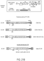


Classifications
-
- C—CHEMISTRY; METALLURGY
- C07—ORGANIC CHEMISTRY
- C07K—PEPTIDES
- C07K16/00—Immunoglobulins [IGs], e.g. monoclonal or polyclonal antibodies
- C07K16/18—Immunoglobulins [IGs], e.g. monoclonal or polyclonal antibodies against material from animals or humans
- C07K16/28—Immunoglobulins [IGs], e.g. monoclonal or polyclonal antibodies against material from animals or humans against receptors, cell surface antigens or cell surface determinants
- C07K16/30—Immunoglobulins [IGs], e.g. monoclonal or polyclonal antibodies against material from animals or humans against receptors, cell surface antigens or cell surface determinants from tumour cells
- C07K16/3076—Immunoglobulins [IGs], e.g. monoclonal or polyclonal antibodies against material from animals or humans against receptors, cell surface antigens or cell surface determinants from tumour cells against structure-related tumour-associated moieties
-
- A—HUMAN NECESSITIES
- A61—MEDICAL OR VETERINARY SCIENCE; HYGIENE
- A61K—PREPARATIONS FOR MEDICAL, DENTAL OR TOILETRY PURPOSES
- A61K39/00—Medicinal preparations containing antigens or antibodies
- A61K39/0005—Vertebrate antigens
- A61K39/0011—Cancer antigens
-
- A—HUMAN NECESSITIES
- A61—MEDICAL OR VETERINARY SCIENCE; HYGIENE
- A61K—PREPARATIONS FOR MEDICAL, DENTAL OR TOILETRY PURPOSES
- A61K39/00—Medicinal preparations containing antigens or antibodies
- A61K39/0005—Vertebrate antigens
- A61K39/0011—Cancer antigens
- A61K39/001102—Receptors, cell surface antigens or cell surface determinants
- A61K39/001111—Immunoglobulin superfamily
- A61K39/001112—CD19 or B4
-
- A—HUMAN NECESSITIES
- A61—MEDICAL OR VETERINARY SCIENCE; HYGIENE
- A61K—PREPARATIONS FOR MEDICAL, DENTAL OR TOILETRY PURPOSES
- A61K39/00—Medicinal preparations containing antigens or antibodies
- A61K39/0005—Vertebrate antigens
- A61K39/0011—Cancer antigens
- A61K39/001169—Tumor associated carbohydrates
- A61K39/001171—Gangliosides, e.g. GM2, GD2 or GD3
-
- A—HUMAN NECESSITIES
- A61—MEDICAL OR VETERINARY SCIENCE; HYGIENE
- A61K—PREPARATIONS FOR MEDICAL, DENTAL OR TOILETRY PURPOSES
- A61K39/00—Medicinal preparations containing antigens or antibodies
- A61K39/46—Cellular immunotherapy
- A61K39/461—Cellular immunotherapy characterised by the cell type used
- A61K39/4611—T-cells, e.g. tumor infiltrating lymphocytes [TIL], lymphokine-activated killer cells [LAK] or regulatory T cells [Treg]
-
- A—HUMAN NECESSITIES
- A61—MEDICAL OR VETERINARY SCIENCE; HYGIENE
- A61K—PREPARATIONS FOR MEDICAL, DENTAL OR TOILETRY PURPOSES
- A61K39/00—Medicinal preparations containing antigens or antibodies
- A61K39/46—Cellular immunotherapy
- A61K39/463—Cellular immunotherapy characterised by recombinant expression
- A61K39/4631—Chimeric Antigen Receptors [CAR]
-
- A—HUMAN NECESSITIES
- A61—MEDICAL OR VETERINARY SCIENCE; HYGIENE
- A61K—PREPARATIONS FOR MEDICAL, DENTAL OR TOILETRY PURPOSES
- A61K39/00—Medicinal preparations containing antigens or antibodies
- A61K39/46—Cellular immunotherapy
- A61K39/464—Cellular immunotherapy characterised by the antigen targeted or presented
- A61K39/4643—Vertebrate antigens
- A61K39/4644—Cancer antigens
- A61K39/464402—Receptors, cell surface antigens or cell surface determinants
- A61K39/464411—Immunoglobulin superfamily
- A61K39/464412—CD19 or B4
-
- A—HUMAN NECESSITIES
- A61—MEDICAL OR VETERINARY SCIENCE; HYGIENE
- A61K—PREPARATIONS FOR MEDICAL, DENTAL OR TOILETRY PURPOSES
- A61K39/00—Medicinal preparations containing antigens or antibodies
- A61K39/46—Cellular immunotherapy
- A61K39/464—Cellular immunotherapy characterised by the antigen targeted or presented
- A61K39/4643—Vertebrate antigens
- A61K39/4644—Cancer antigens
- A61K39/464402—Receptors, cell surface antigens or cell surface determinants
- A61K39/464416—Receptors for cytokines
- A61K39/464417—Receptors for tumor necrosis factors [TNF], e.g. lymphotoxin receptor [LTR], CD30
-
- A—HUMAN NECESSITIES
- A61—MEDICAL OR VETERINARY SCIENCE; HYGIENE
- A61K—PREPARATIONS FOR MEDICAL, DENTAL OR TOILETRY PURPOSES
- A61K39/00—Medicinal preparations containing antigens or antibodies
- A61K39/46—Cellular immunotherapy
- A61K39/464—Cellular immunotherapy characterised by the antigen targeted or presented
- A61K39/4643—Vertebrate antigens
- A61K39/4644—Cancer antigens
- A61K39/464469—Tumor associated carbohydrates
- A61K39/464471—Gangliosides, e.g. GM2, GD2 or GD3
-
- A—HUMAN NECESSITIES
- A61—MEDICAL OR VETERINARY SCIENCE; HYGIENE
- A61P—SPECIFIC THERAPEUTIC ACTIVITY OF CHEMICAL COMPOUNDS OR MEDICINAL PREPARATIONS
- A61P35/00—Antineoplastic agents
-
- C—CHEMISTRY; METALLURGY
- C07—ORGANIC CHEMISTRY
- C07K—PEPTIDES
- C07K14/00—Peptides having more than 20 amino acids; Gastrins; Somatostatins; Melanotropins; Derivatives thereof
- C07K14/435—Peptides having more than 20 amino acids; Gastrins; Somatostatins; Melanotropins; Derivatives thereof from animals; from humans
- C07K14/705—Receptors; Cell surface antigens; Cell surface determinants
- C07K14/70503—Immunoglobulin superfamily
- C07K14/7051—T-cell receptor (TcR)-CD3 complex
-
- C—CHEMISTRY; METALLURGY
- C07—ORGANIC CHEMISTRY
- C07K—PEPTIDES
- C07K14/00—Peptides having more than 20 amino acids; Gastrins; Somatostatins; Melanotropins; Derivatives thereof
- C07K14/435—Peptides having more than 20 amino acids; Gastrins; Somatostatins; Melanotropins; Derivatives thereof from animals; from humans
- C07K14/705—Receptors; Cell surface antigens; Cell surface determinants
- C07K14/70503—Immunoglobulin superfamily
- C07K14/70517—CD8
-
- C—CHEMISTRY; METALLURGY
- C07—ORGANIC CHEMISTRY
- C07K—PEPTIDES
- C07K14/00—Peptides having more than 20 amino acids; Gastrins; Somatostatins; Melanotropins; Derivatives thereof
- C07K14/435—Peptides having more than 20 amino acids; Gastrins; Somatostatins; Melanotropins; Derivatives thereof from animals; from humans
- C07K14/705—Receptors; Cell surface antigens; Cell surface determinants
- C07K14/70503—Immunoglobulin superfamily
- C07K14/70521—CD28, CD152
-
- C—CHEMISTRY; METALLURGY
- C07—ORGANIC CHEMISTRY
- C07K—PEPTIDES
- C07K14/00—Peptides having more than 20 amino acids; Gastrins; Somatostatins; Melanotropins; Derivatives thereof
- C07K14/435—Peptides having more than 20 amino acids; Gastrins; Somatostatins; Melanotropins; Derivatives thereof from animals; from humans
- C07K14/705—Receptors; Cell surface antigens; Cell surface determinants
- C07K14/70578—NGF-receptor/TNF-receptor superfamily, e.g. CD27, CD30, CD40, CD95
-
- C—CHEMISTRY; METALLURGY
- C07—ORGANIC CHEMISTRY
- C07K—PEPTIDES
- C07K14/00—Peptides having more than 20 amino acids; Gastrins; Somatostatins; Melanotropins; Derivatives thereof
- C07K14/435—Peptides having more than 20 amino acids; Gastrins; Somatostatins; Melanotropins; Derivatives thereof from animals; from humans
- C07K14/705—Receptors; Cell surface antigens; Cell surface determinants
- C07K14/715—Receptors; Cell surface antigens; Cell surface determinants for cytokines; for lymphokines; for interferons
- C07K14/7155—Receptors; Cell surface antigens; Cell surface determinants for cytokines; for lymphokines; for interferons for interleukins [IL]
-
- C—CHEMISTRY; METALLURGY
- C07—ORGANIC CHEMISTRY
- C07K—PEPTIDES
- C07K16/00—Immunoglobulins [IGs], e.g. monoclonal or polyclonal antibodies
- C07K16/18—Immunoglobulins [IGs], e.g. monoclonal or polyclonal antibodies against material from animals or humans
- C07K16/28—Immunoglobulins [IGs], e.g. monoclonal or polyclonal antibodies against material from animals or humans against receptors, cell surface antigens or cell surface determinants
- C07K16/2803—Immunoglobulins [IGs], e.g. monoclonal or polyclonal antibodies against material from animals or humans against receptors, cell surface antigens or cell surface determinants against the immunoglobulin superfamily
-
- C—CHEMISTRY; METALLURGY
- C07—ORGANIC CHEMISTRY
- C07K—PEPTIDES
- C07K16/00—Immunoglobulins [IGs], e.g. monoclonal or polyclonal antibodies
- C07K16/18—Immunoglobulins [IGs], e.g. monoclonal or polyclonal antibodies against material from animals or humans
- C07K16/28—Immunoglobulins [IGs], e.g. monoclonal or polyclonal antibodies against material from animals or humans against receptors, cell surface antigens or cell surface determinants
- C07K16/2878—Immunoglobulins [IGs], e.g. monoclonal or polyclonal antibodies against material from animals or humans against receptors, cell surface antigens or cell surface determinants against the NGF-receptor/TNF-receptor superfamily, e.g. CD27, CD30, CD40, CD95
-
- C—CHEMISTRY; METALLURGY
- C07—ORGANIC CHEMISTRY
- C07K—PEPTIDES
- C07K16/00—Immunoglobulins [IGs], e.g. monoclonal or polyclonal antibodies
- C07K16/18—Immunoglobulins [IGs], e.g. monoclonal or polyclonal antibodies against material from animals or humans
- C07K16/28—Immunoglobulins [IGs], e.g. monoclonal or polyclonal antibodies against material from animals or humans against receptors, cell surface antigens or cell surface determinants
- C07K16/30—Immunoglobulins [IGs], e.g. monoclonal or polyclonal antibodies against material from animals or humans against receptors, cell surface antigens or cell surface determinants from tumour cells
- C07K16/3076—Immunoglobulins [IGs], e.g. monoclonal or polyclonal antibodies against material from animals or humans against receptors, cell surface antigens or cell surface determinants from tumour cells against structure-related tumour-associated moieties
- C07K16/3084—Immunoglobulins [IGs], e.g. monoclonal or polyclonal antibodies against material from animals or humans against receptors, cell surface antigens or cell surface determinants from tumour cells against structure-related tumour-associated moieties against tumour-associated gangliosides
-
- C—CHEMISTRY; METALLURGY
- C12—BIOCHEMISTRY; BEER; SPIRITS; WINE; VINEGAR; MICROBIOLOGY; ENZYMOLOGY; MUTATION OR GENETIC ENGINEERING
- C12N—MICROORGANISMS OR ENZYMES; COMPOSITIONS THEREOF; PROPAGATING, PRESERVING, OR MAINTAINING MICROORGANISMS; MUTATION OR GENETIC ENGINEERING; CULTURE MEDIA
- C12N15/00—Mutation or genetic engineering; DNA or RNA concerning genetic engineering, vectors, e.g. plasmids, or their isolation, preparation or purification; Use of hosts therefor
- C12N15/09—Recombinant DNA-technology
- C12N15/63—Introduction of foreign genetic material using vectors; Vectors; Use of hosts therefor; Regulation of expression
- C12N15/79—Vectors or expression systems specially adapted for eukaryotic hosts
- C12N15/85—Vectors or expression systems specially adapted for eukaryotic hosts for animal cells
-
- A—HUMAN NECESSITIES
- A61—MEDICAL OR VETERINARY SCIENCE; HYGIENE
- A61K—PREPARATIONS FOR MEDICAL, DENTAL OR TOILETRY PURPOSES
- A61K39/00—Medicinal preparations containing antigens or antibodies
- A61K2039/51—Medicinal preparations containing antigens or antibodies comprising whole cells, viruses or DNA/RNA
- A61K2039/515—Animal cells
- A61K2039/5156—Animal cells expressing foreign proteins
-
- A—HUMAN NECESSITIES
- A61—MEDICAL OR VETERINARY SCIENCE; HYGIENE
- A61K—PREPARATIONS FOR MEDICAL, DENTAL OR TOILETRY PURPOSES
- A61K39/00—Medicinal preparations containing antigens or antibodies
- A61K2039/51—Medicinal preparations containing antigens or antibodies comprising whole cells, viruses or DNA/RNA
- A61K2039/515—Animal cells
- A61K2039/5158—Antigen-pulsed cells, e.g. T-cells
-
- A—HUMAN NECESSITIES
- A61—MEDICAL OR VETERINARY SCIENCE; HYGIENE
- A61K—PREPARATIONS FOR MEDICAL, DENTAL OR TOILETRY PURPOSES
- A61K2239/00—Indexing codes associated with cellular immunotherapy of group A61K39/46
- A61K2239/10—Indexing codes associated with cellular immunotherapy of group A61K39/46 characterized by the structure of the chimeric antigen receptor [CAR]
- A61K2239/22—Intracellular domain
-
- C—CHEMISTRY; METALLURGY
- C07—ORGANIC CHEMISTRY
- C07K—PEPTIDES
- C07K2317/00—Immunoglobulins specific features
- C07K2317/20—Immunoglobulins specific features characterized by taxonomic origin
- C07K2317/24—Immunoglobulins specific features characterized by taxonomic origin containing regions, domains or residues from different species, e.g. chimeric, humanized or veneered
-
- C—CHEMISTRY; METALLURGY
- C07—ORGANIC CHEMISTRY
- C07K—PEPTIDES
- C07K2317/00—Immunoglobulins specific features
- C07K2317/50—Immunoglobulins specific features characterized by immunoglobulin fragments
- C07K2317/56—Immunoglobulins specific features characterized by immunoglobulin fragments variable (Fv) region, i.e. VH and/or VL
- C07K2317/565—Complementarity determining region [CDR]
-
- C—CHEMISTRY; METALLURGY
- C07—ORGANIC CHEMISTRY
- C07K—PEPTIDES
- C07K2317/00—Immunoglobulins specific features
- C07K2317/60—Immunoglobulins specific features characterized by non-natural combinations of immunoglobulin fragments
- C07K2317/62—Immunoglobulins specific features characterized by non-natural combinations of immunoglobulin fragments comprising only variable region components
- C07K2317/622—Single chain antibody (scFv)
-
- C—CHEMISTRY; METALLURGY
- C07—ORGANIC CHEMISTRY
- C07K—PEPTIDES
- C07K2317/00—Immunoglobulins specific features
- C07K2317/70—Immunoglobulins specific features characterized by effect upon binding to a cell or to an antigen
- C07K2317/73—Inducing cell death, e.g. apoptosis, necrosis or inhibition of cell proliferation
-
- C—CHEMISTRY; METALLURGY
- C07—ORGANIC CHEMISTRY
- C07K—PEPTIDES
- C07K2319/00—Fusion polypeptide
-
- C—CHEMISTRY; METALLURGY
- C07—ORGANIC CHEMISTRY
- C07K—PEPTIDES
- C07K2319/00—Fusion polypeptide
- C07K2319/01—Fusion polypeptide containing a localisation/targetting motif
- C07K2319/03—Fusion polypeptide containing a localisation/targetting motif containing a transmembrane segment
-
- C—CHEMISTRY; METALLURGY
- C12—BIOCHEMISTRY; BEER; SPIRITS; WINE; VINEGAR; MICROBIOLOGY; ENZYMOLOGY; MUTATION OR GENETIC ENGINEERING
- C12N—MICROORGANISMS OR ENZYMES; COMPOSITIONS THEREOF; PROPAGATING, PRESERVING, OR MAINTAINING MICROORGANISMS; MUTATION OR GENETIC ENGINEERING; CULTURE MEDIA
- C12N2740/00—Reverse transcribing RNA viruses
- C12N2740/00011—Details
- C12N2740/10011—Retroviridae
- C12N2740/15011—Lentivirus, not HIV, e.g. FIV, SIV
- C12N2740/15041—Use of virus, viral particle or viral elements as a vector
- C12N2740/15043—Use of virus, viral particle or viral elements as a vector viral genome or elements thereof as genetic vector
Definitions
- Chimeric Antigen Receptors are engineered T cell receptors displaying specificity against target antigens based on a single chain FV (scFv) antibody moiety.
- scFv single chain FV
- the disclosure relates to chimeric antigen receptors (CARs) comprising an antigen binding domain (e.g., anti-CD19, anti-CD30, anti-GD2), and/or a cytoplasmic domain of a interleukin (IL)-15-receptor ⁇ , and/or a chimeric cytoplasmic domain including a CD27 cytoplasmic domain fused to a 4-1BB cytoplasmic domain.
- an antigen binding domain e.g., anti-CD19, anti-CD30, anti-GD2
- IL interleukin
- a chimeric cytoplasmic domain including a CD27 cytoplasmic domain fused to a 4-1BB cytoplasmic domain.
- aspects of the disclosure relate to CARs comprising a cytoplasmic domain of a interleukin(IL)-15-receptor ⁇ , and uses of such CARs to produce CAR T-cells (CARTs) and/or CAR-modified immune cells such as NK (natural killer) cells, which can be used in various methods, such as treatment methods, or compositions.
- CARTs CAR T-cells
- NK natural killer
- IL-15R ⁇ IL-15-receptor ⁇
- aspects of the disclosure relate to a chimeric antigen receptor (CAR) comprising an antigen binding domain; a transmembrane domain; and a cytoplasmic domain containing an interleukin 15-receptor ⁇ (IL-15R ⁇ ) cytoplasmic domain.
- the transmembrane domain is a CD28 or CD8 transmembrane domain.
- the cytoplasmic domain further comprises a CD3zeta signal transduction domain.
- the cytoplasmic domain further comprises a CD27 signaling domain.
- the antigen binding domain is a single-chain variable fragment (scFv). In some embodiments, the antigen binding domain is specific for CD19.
- aspects of the disclosure relate to CARs comprising a chimeric cytoplasmic domain including a CD27 cytoplasmic domain fused to a 4-1BB cytoplasmic domain, and uses of such CARs to produce CARTs and/or CAR-modified immune cells such as NK (natural killer) cells, which can be used in various methods, such as treatment methods, or compositions.
- CARs comprising a chimeric cytoplasmic domain including a CD27 cytoplasmic domain fused to a 4-1BB cytoplasmic domain
- the disclosure relates to a chimeric antigen receptor (CAR) comprising: an antigen binding domain; a transmembrane domain; and a cytoplasmic domain containing a CD27 intracellular domain.
- CAR chimeric antigen receptor
- the transmembrane domain is a CD28 or CD8 transmembrane domain.
- the cytoplasmic domain further comprises a 4-1BB intracellular domain.
- the cytoplasmic domain further comprises a CD3zeta signal transduction domain.
- the cytoplasmic domain further comprises an iCasp9 domain and/or a FKBP domain.
- the antigen binding domain is a single-chain variable fragment (scFv).
- a CAR comprises a CD28 transmembrane domain, cytoplasmic domain comprising a CD27 intracellular domain and a 4-1BB intracellular domain, and a CD3zeta signal transduction domain.
- a CAR further comprises an iCasp9 domain and/or a FKBP domain.
- a CAR further comprises a first spacer between the CD28 transmembrane domain and the cytoplasmic domain comprising a CD27 intracellular domain and a 4-1BB intracellular domain, and a second spacer between the cytoplasmic domain comprising a CD27 intracellular domain and a 4-1BB intracellular domain and the CD3zeta signal transduction domain.
- the CAR further comprises a third spacer between the CD3zeta signal transduction domain and the iCasp9 domain and/or a FKBP domain.
- the antigen binding domain of a CAR is specific for CD30.
- aspects of the disclosure relate to CARs comprising an antigen-binding domain specific for Disialoganglioside 2 (GD2), and uses of such CARs to produce CAR-modified immune cells such as T cells (also referred to herein as CARTs) or NK (natural killer) cells, which can be used in various methods, such as treatment methods, or compositions.
- CARs are used to treat retinoblastoma or osteosarcoma, such as a juvenile subject having retinoblastoma or osteosarcoma.
- GD2-specific CAR-expressing T cells could kill both osteosarcoma and retinoblastoma cells, both of which express GD2.
- GD2-specific CARs will be useful for treatment of both osteosarcoma and retinoblastoma.
- a chimeric antigen receptor comprising an antigen binding domain specific for GD2; a transmembrane domain; and a cytoplasmic domain containing one or more (e.g., one, two, or three) of a CD27 signaling domain, a 4-1BB intracellular domain, and a CD3zeta signal transduction domain.
- the transmembrane domain is a CD28 or CD8 transmembrane domain.
- the antigen binding domain is a single-chain variable fragment (scFv).
- nucleic acid comprising a sequence that encodes the CAR of any one of the above embodiments or as otherwise described herein.
- an immune cell comprising a CAR of any one of the above embodiments or as otherwise described herein and/or a nucleic acid of any one of the above embodiments or as otherwise described herein.
- the immune cell is a T cell or NK cell. In some embodiments, the immune cell is a T cell.
- compositions comprising a plurality of the immune cell of any one of the above embodiments or as otherwise described herein.
- the composition comprises a pharmaceutically acceptable carrier.
- Another aspect of the disclosure relates to a method of generating a plurality of CAR-modified immune cells, the method comprising introducing a lentiviral vector comprising a nucleic acid of any one of the above embodiments or as otherwise described herein into a plurality of immune cells.
- the immune cells are T cells.
- aspects of the disclosure relate to a method of treating a subject having a disease, the method comprising administering an immune cell of any one of the above embodiments or as otherwise described herein, the composition of any one of the above embodiments or as otherwise described herein, or the plurality of immune cells produced by a method of any one of the above embodiments or as otherwise described herein into a subject having a disease or at risk of having a disease.
- the disease is cancer, an autoimmune disease or an infection. In some embodiments, the disease is cancer. In some embodiments, the disease is a CD30+ cancer. In some embodiments, the cancer is osteosarcoma or retinoblastoma.
- the method further comprises administering a PD-L1 or PD1 inhibitor to the subject.
- aspects of the disclosure relate to a method of treating a subject having cancer, the method comprising administering (a) an immune cell expressing a CAR that targets GD2 and (b) a PD-L1 or PD1 inhibitor to a subject having cancer.
- the cancer is osteosarcoma or retinoblastoma.
- the immune cell is a T cell.
- the immune cell is a plurality of immune cells in a composition.
- FIG. 1 is a diagram showing an exemplary Chimeric Antigen Receptor (CAR) basic structure including the antigen binding scFv domain and the various co-stimulatory domains.
- CAR Chimeric Antigen Receptor
- FIG. 2 is a diagram showing an exemplary simple killing assay—Co-culture of green fluorescent protein wasabi (GFP)-labeled target with the CART effector cells and flow cytometry after annexinV/PI staining.
- GFP green fluorescent protein wasabi
- FIG. 3 is a series of plots and two photographs showing that exemplary lentiviral gene transfer into human T cells is very high.
- the X-axis in each plot shows CD3 staining and the Y-axis in each plot shows GFP staining.
- FIG. 4 is a series of photographs showing exemplary primary T cell killing efficiency demonstrated by disappearance of the green (fluorescent) target cells as seen under the fluorescent microscope. Both bright-field and fluorescent images are shown for each sample.
- FIG. 5A is a series of plots and a graph showing exemplary healthy donor derived CART cells—Flow data of Killing in terms of % early apoptotic cells with annexinV positive and PI negative staining seen after 1-2 hours of co-culture.
- FIG. 5B is two graphs showing exemplary % total cell death (top graph) for each CAR and % specific lysis (bottom graph) for each CAR. This was done in triplicate wells to acquire mean values and statistical significance obtained using student t test (p ⁇ 0.05 considered significant). *P ⁇ 0.01 (comparing with No CAR), #P ⁇ 0.01 (comparing with a control CART), ⁇ circumflex over ( ) ⁇ P ⁇ 0.05 (comparing 153z with 19z and 273z).
- FIG. 6A is a series of graphs showing exemplary flow data of killing after overnight incubation in terms of % early and late apoptotic cells (both annexinV+PI ⁇ and double positive for AnnexinV and PI)
- FIG. 6B is a series of plots and graphs showing exemplary long term killing after 3 days of co-coculture of patient derived CART cells with green (fluorescent) target cells. Killing was indicated by disappearance of the green target cells.
- FIG. 6C is a series of photographs and a graph showing exemplary long term killing after 3 days of co-coculture of patient derived CART cells with green (fluorescent) target cells. Killing was indicated by disappearance of the green (fluorescent) target cells.
- FIGS. 7A and 7B show a series of plots and graphs of exemplary patient ALL cells labeled with Calcein AM and patient derived CART cells incubated in 5:1 ratio.
- Flow data shows killing at 1 hour ( FIG. 7A ) and 1 day ( FIG. 7B ) in terms of early apoptosis with % annexinV positive and PI positive cells.
- FIG. 8 is a series of plots showing exemplary retargeting of CART effector cells with green (fluorescent) target. Killing seen as indicated by disappearance of target in flow cytometry.
- FIG. 9 is a series of plots and a graph showing exemplary CART cell proliferation by a CFSE method. Proliferation peak and % seen after CART cells stimulation with target cells.
- FIGS. 10A and 10B are a series of graphs showing exemplary intracellular effector function analysis flow data of IL-2, IFN-g and CD107a produced after the CART cells are co-cultured with the target cells—RS4-11.
- Positive control (PC) is PMA/ionomycin stimulation.
- FIG. 10C is a graph showing the exemplary data summarized from FIGS. 10A and 10B in bar graph form. Maximum IL-2, IFN-g and CD107a release was seen with 153z CART cells.
- FIG. 11 shows flow cytometry analysis of CD30 surface expression of a primary B cell tumor line.
- FIG. 12 shows an illustration of the lentiviral vector system.
- FIG. 13 shows diagrams illustrating embodiments of CAR molecules, including one embodiment of a 4th generation CD30-CAR structure and components.
- FIG. 14 shows an illustration of a rapid CAR T target killing assay used for the evaluation of CAR functions.
- FIG. 15 shows data related to a target killing assay of two different CD30-CAR T cells based on 5F11 and AC10 scFv CD30-CARs.
- FIG. 16 shows flow cytometry target killing assay of two different CD30-CAR T cells based on 5F11 and AC10 scFv CD30-CARs.
- FIG. 17 shows the clinical course of a Hodgkin's lymphoma patient treated with CD30-CAR T cells.
- the infusion time point was depicted by arrows.
- Three batches of CAR T cells were prepared and infused as indicated.
- CAR-positive T cells were detected by qPCR detection of CAR DNA in the genome of blood cells.
- Clinical evidence of tumor regression was evident at day 40 after the first infusion.
- FIG. 18 shows data related to detection of IL-6 cytokine response after CD30 CAR T infusion.
- FIG. 19 shows data related to detection of IFN-gamma cytokine response after CD30 CAR T infusion.
- FIGS. 20A-20C shows graphs of exemplary GD2 surface expression in osteosarcoma (OS) cell lines.
- Flow cytometry histogram overlays show surface expression of GD2 in ( FIG. 20A ) HOS ( FIG. 20B ) U2OS and ( FIG. 20C ) OS156 cells.
- the dark line represents GD2 positive and the shadowed grey line represents isotype control.
- Inner number indicates percent positive population and mean fluorescence intensity (MFI) of the positive population
- FIG. 21A is a schematic showing an exemplary CAR that targets GD2 binding to a cell expressing GD2.
- FIG. 21B is a schematic that shows embodiments of CAR molecules, including one embodiment of GD2 CAR construction and 4th generation CAR configurations.
- FIG. 22 is a diagram that shows an exemplary CAR-JK/T cell killing assay.
- FIGS. 23A-23D are a series of plots and graphs that show short term GD2 JK killing. JK cells were used for CAR construction as a preliminary determination of GD2 CAR killing ability.
- FIGS. 24A-24G show exemplary CAR-Primary T cell killing of OS target. The killing ability of GD2 CAR in primary T cells were determined.
- FIG. 24A Flow cytometry analysis of U2OSw cells by GD2 CAR-T cells after co-culture at E:T ratio of 4:1, 2:1, 1:1, 1:2, * indicates significant different from controls (p ⁇ 0.05).
- FIG. 24B Percent specific lyses of U2OSw cells after 3 day co-culture, * indicates significant different from control T cells (p ⁇ 0.05).
- FIG. 24C Flow cytometry analysis of HOSw cells by GD2 CAR-T cells after co-culture at E:T ratio of 4:1, 2:1, 1:1, 1:2, * indicates significant different from controls (p ⁇ 0.05).
- FIG. 24A Flow cytometry analysis of U2OSw cells by GD2 CAR-T cells after co-culture at E:T ratio of 4:1, 2:1, 1:1, 1:2, * indicates significant different from controls (p ⁇ 0.05).
- FIG. 24A Flow cyto
- FIG. 24D Percent specific lyses of HOSw cells after 3 day co-culture, * indicates significant different from control T cells (p ⁇ 0.05).
- FIG. 24E Percent specific lyses of OS156 cells after 3 day co-culture, * indicates significant different from control T cells (p ⁇ 0.05).
- FIG. 24F Target cells (green, fluorescent) and effecter cells (no color) under fluorescence microscope after 4 days co-culture.
- FIG. 24G Target cells (green, fluorescent) and effecter cells (no color) under fluorescence microscope after 3, 7 or 12 days co-culture.
- FIGS. 25A-25D show exemplary PDL-1 expression in OS cells after incubation with CAR T cells.
- FIG. 25A ) and FIG. 25B Flow cytometry histogram overlays show surface expression of PD-L1 in U2OS and HOS cell. The shadowed area represents isotype control. Inner number indicates percent positive population and MFI of the positive population.
- FIG. 25C ) and FIG. 25D MFI analysis of PD-L1 expression in OS cells after CAR T co-culture.
- FIGS. 26A-26F show exemplary PD-1 expression in CAR T cells after incubation with OS cells.
- PD1 expression in primary CAR T cells after 1 day co-culture with target cells (E:T ratio 2:1), U2OSw ( FIGS. 26A-26B ) and HOSw ( FIGS. 26D-26E ).
- a bar graph displays fold increase of PD1 expression from background expression level. The number above the bars show fold increase of PD1 expression.
- FIG. 26C ) and FIG. 26F Increased CAR T cell death after PD-1 up-regulation. Percent CAR T cell death after 1 day co-culture with U2OSw (C) and HOSw (D) cells, # indicates significant different at p ⁇ 0.05.
- FIG. 27A is a series of photographs that shows immunohistochemistry analyses of GD2 expression in tumor samples from RB patients. Paraffin-embedded retinal tumor samples were stained for GD2 expression by using antibody against human GD2 protein.
- FIG. 27B is a series of graphs that shows surface staining of GD2 expression on Y79 retinoblastoma (RB) cell line. Flow cytometric staining for GD2 and CD19 proteins (black line) or an IgG control (filled histogram) on the surface of Y79 RB cells. Upper right number indicates cell count and the normalized mean fluorescence intensities (nMFI) are shown in parenthesis.
- nMFI normalized mean fluorescence intensities
- FIG. 28A is a diagram of exemplary GD2 CAR constructs.
- FIG. 28B is a diagram showing an exemplary cell killing assay for RB cells.
- Y79 RB cells were engineered to express wasort, a green fluorescent protein (GFP), using lentiviral transduction.
- GFP green fluorescent protein
- Y79 RB wasort cells were co-cultured with control T cells or CD19 T cells, or GD2 CAR T cells. After 24 hr of co-cultivation, apoptosis of tumor cells were examined by using AnnexinV/PI staining and analyzed by flow cytometry.
- FIGS. 29A-29D show an exemplary short-term killing assay on Y79 RB using CAR T-cells.
- FIG. 29A shows analysis of cell death by using AnnexinV/PI staining. After 24 hr of co-culture experiment, the suspension cells were harvested and stained with AnnexinV-PE/PI dye followed by flow cytometric analysis.
- FIG. 29B shows the percent cell death at 1 day after co-culture.
- FIG. 29C shows the % of specific lysis.
- FIG. 29D is a series of photographs showing fluorescence microscopic monitoring of tumor cell lysis.
- Y79 RB wasort cells (1 ⁇ 105) were cultured with control T cells or CD19 CAR T cells, or GD2 CAR T cells at the effector/target ratio (1:1) for 24 h, and the lysis of tumor cells were monitored under fluorescence microscope.
- FIG. 30 is a series of photographs that shows exemplary 2 nd round killing of target RB cells by GD2-CAR T cells at day 6 or day 14 of co-culture.
- FIGS. 31A and 31B are a series of graphs that show down-regulation of GD2 in RB tumor upon GD2-CAR T targeting.
- FIG. 31C is a series of graphs that shows upregulation of PD-L1 expression upon GD2-CAR T cell targeting.
- FIG. 31D is a series of graphs that shows down-regulation of GD2 expression upon GD2-CAR T cell targeting.
- the disclosure relates to chimeric antigen receptors (CARs) comprising an antigen binding domain (e.g., anti-CD19, anti-CD30, anti-GD2), and/or a cytoplasmic domain of a interleukin(IL)-15-receptor ⁇ , and/or a chimeric cytoplasmic domain including a CD27 cytoplasmic domain fused to a 4-1BB cytoplasmic domain.
- an antigen binding domain e.g., anti-CD19, anti-CD30, anti-GD2
- IL interleukin
- the invention relates, in some embodiments, to chimeric antigen receptors (CARs) and uses thereof in T cells (e.g., to make CAR T cells), methods, nucleic acids, compositions, kits and the like.
- CARs are molecules that combine antibody-based specificity for a desired antigen (e.g., tumor antigen) with a T cell receptor-activating intracellular domain to generate a chimeric protein that exhibits a specific anti-tumor cellular immune activity. It should be appreciated that, in some embodiments, exemplary, non-limiting arrangements of CARs are described from left to right, N-terminus to C-terminus of the CAR.
- the disclosure relates to CARs having an interleukin 15-receptor ⁇ (IL-15R ⁇ ) cytoplasmic domain.
- CARs containing this domain had several advantageous and surprising features including greater expansion potential upon antigen stimulation, higher killing efficacy, maintained killing efficacy upon repetitive addition of excess target cells, and an increased amount of effector cytokines compared to CARs not containing this domain.
- the present invention relates, in part, to the use of T cells genetically modified to stably express a desired CAR, e.g., containing a IL-15R ⁇ cytoplasmic domain, containing a CD30 antigen binding domain, containing a GD2 antigen binding domain, etc.
- T cells expressing a CAR are referred to herein as CAR T cells, CARTs, or CAR modified T cells.
- the cell can be genetically modified to stably express an antibody binding domain on its surface, conferring novel antigen specificity that is MHC independent.
- the T cell is genetically modified to stably express a CAR that combines an antigen recognition domain of a specific antibody with an transmembrane domain and a cytoplasmic domain into a single chimeric protein.
- a CAR that combines an antigen recognition domain of a specific antibody with an transmembrane domain and a cytoplasmic domain into a single chimeric protein.
- two CAR proteins dimerize (e.g., form homo- or heterodimers) in vivo.
- a CAR comprises an antigen binding domain, a transmembrane domain, a cytoplasmic domain comprising an IL-15R ⁇ cytoplasmic domain, optionally further comprising a CD3 zeta (CD3z) signaling domain and/or a CD27 signaling domain.
- the arrangement of the elements of the CAR is selected from one of the following exemplary, non-limiting arrangements (from N-terminus to C-terminus):
- the disclosure relates to CARs having a CD30 antigen binding domain and an intracellular domain comprising a 4-1BB intracellular domain and a CD27 intracellular domain.
- the CARs further comprise a self-destructive domain (e.g., an iCasp9-FKBP domain) that further improves safety.
- CARs containing these domains had several surprising features including an acceptable safety profile, and mediate effective killing of CD30 + cancer cells both in vitro and in vivo.
- a CAR (e.g., CAR having a CD30 antigen binding domain) comprises an antigen binding domain, a transmembrane domain, a cytoplasmic domain comprising a 4-1BB intracellular domain and a CD27 intracellular domain, optionally further comprising a CD3 zeta (CD3z) signaling domain, and/or an apoptosis-inducing iCasp9-FKBP domain.
- the arrangement of the elements of the CAR is selected from one of the following exemplary, non-limiting arrangements (from N-terminus to C-terminus):
- the disclosure relates to the discovery that GD2 was expressed on both osteosarcoma and retinoblastoma cells and that GD2-specific CAR-expressing T cells could kill both osteosarcoma and retinoblastoma cells.
- CARs e.g., GD2-specific CARs
- GD2-specific CARs contain an antigen binding domain (such as an scFV) specific for GD2.
- the arrangement of the elements of the CAR is selected from one of the following exemplary, non-limiting arrangements (from N-terminus to C-terminus):
- the present invention relates, in part, to a CAR that incorporates a series of domains that provide different functional aspects that may synergistically work together to improve efficacy.
- the CAR may include one or more of: an antigen binding domain, a hinge domain, an antigen co-signaling domain that stimulates activity (e.g., an IL-15R ⁇ cytoplasmic domain, a 4-1BB cytoplasmic domain, or another cytoplasmic domain), a survival domain that increases T-cell or immune effector cell survival, a T-cell or immune effector cell memory domain, and an effector activating domain.
- the CAR may further include a domain that induces safety (e.g., an apoptosis-inducing iCasp9-FKBP domain).
- a CAR of the invention comprises an extracellular domain having an antigen binding domain, a transmembrane domain, and a multi-functional cytoplasmic domain.
- the CAR comprises a fully human antibody or antibody fragment.
- the transmembrane domain that naturally is associated with one of the domains in the CAR is used.
- the transmembrane domain can be selected or modified by amino acid substitution to avoid binding of such domains to the transmembrane domains of the same or different surface membrane proteins to minimize interactions with other members of the receptor complex.
- the CAR T cells or other CAR-modified immune cells (e.g., CAR-modified NK cells) of the invention are generated by introducing a lentiviral vector comprising a nucleic acid that encodes a desired CAR into T cells or other immune cells (e.g., into NK cells).
- the lentiviral vector comprises a nucleic acid that encodes a CAR comprising an antigen binding domain (e.g., that targets CD19), a transmembrane domain, and a cytoplasmic domain.
- the CAR T cells or other CAR-modified immune cells (e.g., NK cells) of the invention are able to replicate in vivo resulting in long-term persistence that can lead to sustained tumor control.
- the CAR T cell or other CAR-modified immune cells of the invention can be generated by transfecting a transposon or RNA encoding the desired CAR, into the T cells or other immune cells.
- the CAR is transiently expressed in the genetically modified CAR T cell or other CAR-modified immune cells.
- the invention relates to administering a genetically modified immune cell (e.g., a genetically modified T cell or NK cell) expressing a CAR for the treatment of a patient having cancer or at risk of having cancer, or having an autoimmune disease or at risk of having an autoimmune disease, e.g., using lymphocyte infusion.
- a genetically modified immune cell e.g., a genetically modified T cell or NK cell
- a CAR e.g., a genetically modified T cell or NK cell
- autologous lymphocyte infusion is used in the treatment.
- autologous PBMCs are collected from a patient in need of treatment and immune cells (e.g., T cells or NK cells) are activated and expanded using the methods described herein and known in the art and then infused back into the patient.
- the cancer is a CD30+ cancer (e.g., a cancer expressing CD30).
- cancers expressing CD30 include Hodgkin's lymphoma (HL), anaplastic large cell lymphoma (ALCL), diffuse large B cell lymphoma, primary effusion lymphoma, adult T-cell leukemia/lymphoma, mycosis fungoides, extranodal natural killer/T-cell lymphoma and peripheral T/NK cell lymphoma.
- the cancer is a GD2-expressing cancer, such as retinoblastoma or osteosarcoma.
- Activation refers to the state of a T cell that has been sufficiently stimulated to induce detectable cellular proliferation. Activation can also be associated with induced cytokine production, and detectable effector functions.
- the term “activated T cells” refers to, among other things, T cells that are undergoing cell division.
- antibody refers to an immunoglobulin molecule which specifically binds with an antigen.
- Antibodies can be intact immunoglobulins derived from natural sources or from recombinant sources and can be immunoreactive portions of intact immunoglobulins. Antibodies are typically tetramers of immunoglobulin molecules.
- the antibodies in the present invention may exist in a variety of forms including, for example, polyclonal antibodies, monoclonal antibodies, Fv, Fab and F(ab) 2 , as well as single chain antibodies, human antibodies, and humanized antibodies (Harlow et al., 1999, In: Using Antibodies: A Laboratory Manual, Cold Spring Harbor Laboratory Press, NY; Harlow et al., 1989, In: Antibodies: A Laboratory Manual, Cold Spring Harbor, N.Y.; Houston et al., 1988, Proc. Natl. Acad. Sci. USA 85:5879-5883; Bird et al., 1988, Science 242:423-426).
- antibody fragment refers to a portion of an intact antibody and refers to the antigenic determining variable regions of an intact antibody.
- antibody fragments include, but are not limited to, Fab, Fab′, F(ab′) 2 , and Fv fragments, linear antibodies, scFv antibodies, and multispecific antibodies formed from antibody fragments.
- an “antibody heavy chain,” as used herein, refers to the larger of the two types of polypeptide chains present in all antibody molecules in their naturally occurring conformations.
- antibody light chain refers to the smaller of the two types of polypeptide chains present in all antibody molecules in their naturally occurring conformations, ⁇ and ⁇ light chains refer to the two major antibody light chain isotypes.
- synthetic antibody as used herein, is meant an antibody which is generated using recombinant DNA technology, such as, for example, an antibody expressed by a bacteriophage as described herein.
- the term should also be construed to mean an antibody which has been generated by the synthesis of a DNA molecule encoding the antibody and which DNA molecule expresses an antibody protein, or an amino acid sequence specifying the antibody, wherein the DNA or amino acid sequence has been obtained using synthetic DNA or amino acid sequence technology which is available and well known in the art.
- antigen or “Ag” as used herein is defined as a molecule that provokes an immune response. This immune response may involve either antibody production, or the activation of specific immunologically-competent cells, or both.
- any macromolecule including virtually all proteins or peptides, can serve as an antigen.
- antigens can be derived from recombinant or genomic DNA. A skilled artisan will understand that any DNA, which comprises a nucleotide sequences or a partial nucleotide sequence encoding a protein that elicits an immune response therefore encodes an “antigen” as that term is used herein.
- an antigen need not be encoded solely by a full length nucleotide sequence of a gene. It is readily apparent that the present invention includes, but is not limited to, the use of partial nucleotide sequences of more than one gene and that these nucleotide sequences are arranged in various combinations to elicit the desired immune response. Moreover, a skilled artisan will understand that an antigen need not be encoded by a “gene” at all. It is readily apparent that an antigen can be generated synthesized or can be derived from a biological sample. Such a biological sample can include, but is not limited to a tissue sample, a tumor sample, a cell or a biological fluid.
- tumor antigen refers to an antigen associated with a cancer cell.
- the tumor antigen is CD19, which is associated with B cell malignancies.
- the tumor antigen is CD30, which is typically associated with lymphomas (e.g., HL, ALCL, diffuse large cell B-lymphoma, etc.
- lymphomas e.g., HL, ALCL, diffuse large cell B-lymphoma, etc.
- tumor antigens include, but are not limited to, CD2, CD5, CD10, CD20, CD22, CD30, CD33, CD38, CD52, CD56, CD74, CD138, CD317, Her2, VEGFR2, EGFRviii, CXCR4, BCMA, GD2, GD3, and any other antigens over-expressed in tumor cells.
- the tumor antigen is GD2.
- GD2 is a disialoganglioside that is expressed on cancer cells, such as retinoblastoma or osteosarcoma.
- An exemplary structure of GD2 is as follows:
- the IUPAC name for GD2 is (2R,4R,5S,6S)-2-[3-[(2S,3S,4R,6S)-6-[(2S,3R,4R,5S,6R)-5-[(2S,3R,4R,5R,6R)-3-acetamido-4,5-dihydroxy-6-(hydroxymethyl)oxan-2-yl]oxy-2-[(2R,3S,4R,5R,6R)-4,5-dihydroxy-2-(hydroxymethyl)-6-RE)-3-hydroxy-2-(octadecanoylamino)octadec-4-enoxy]oxan-3-yl]oxy-3-hydroxy-6-(hydroxymethyl)oxan-4-yl]oxy-3-amino-6-carboxy-4-hydroxyoxan-2-yl]-2,3-dihydroxypropoxyl-5-amino-4-hydroxy-6-(1,2,3-trihydroxypropyl)oxane
- target antigen refers to an antigen associated with a disease-associated target cell.
- target antigens include but are not limited to CD2, CD5, CD7, CD10, CD19, CD20, CD22, CD30, CD33, CD38, CD52, CD56, CD74, CD138, CD317, Her2, VEGFR2, EGFRviii, CXCR4, BCMA, GD2, GD3, and any other antigens over-expressed in target cells or diseased cells.
- the diseased cell is a B cell that produces auto-antibodies.
- anti-tumor effect refers to a biological effect which can be manifested by a decrease in tumor volume, a decrease in the number of tumor cells, a decrease in the number of metastases, an increase in life expectancy, or amelioration of various physiological symptoms associated with the cancerous condition.
- An “anti-tumor effect” can also be manifested by the ability of the peptides, polynucleotides, cells and antibodies of the invention in prevention of the occurrence of tumor in the first place.
- autologous is meant to refer to any material derived from the same individual to which it is later to be re-introduced into the individual.
- Allogeneic refers to a graft derived from a different animal of the same species.
- Xenogeneic refers to a graft derived from an animal of a different species.
- cancer as used herein is defined as disease characterized by the rapid and uncontrolled growth of aberrant cells. Cancer cells can spread locally or through the bloodstream and lymphatic system to other parts of the body. Examples of various cancers include, but are not limited to, breast cancer, prostate cancer, ovarian cancer, cervical cancer, skin cancer, pancreatic cancer, colorectal cancer, renal cancer, liver cancer, brain cancer, lymphoma, leukemia (e.g., chronic lymphocytic leukemia, acute lymphoblastic leukemia, pediatric acute B cell leukemia, or post hematopoietic stem cell transplant relapsed leukemia), lung cancer and the like.
- leukemia e.g., chronic lymphocytic leukemia, acute lymphoblastic leukemia, pediatric acute B cell leukemia, or post hematopoietic stem cell transplant relapsed leukemia
- cancer refers to B-cell related malignancies (e.g., B-cell chronic lymphocytic leukemia, acute lymphoblastic leukemia, B-cell prolymphocytic leukemia, precursor B lymphoblastic leukemia, Hairy cell leukemia, diffuse large B cell lymphoma, follicular lymphoma, marginal zone lymphoma, small cell lymphocytic lymphoma, mantle cell lymphoma, Burkitt lymphoma, primary mediastinal (thymic) large B-cell lymphoma, lymphoplasmacytic lymphoma, nodal marginal zone B cell lymphoma, intravascular large B-cell lymphoma, primary effusion lymphoma, lymphomatoid granulomatosis, or plasmablastic lymphoma).
- B-cell related malignancies e.g., B-cell chronic lymphocytic leukemia, acute lymphoblastic leukemia, B-cell prolymphocytic leukemia, precursor
- cancer refers to CD30+ cancers (e.g., Hodgkin's lymphoma (HL), anaplastic large cell lymphoma (ALCL), diffuse large B cell lymphoma, and peripheral T/NK cell lymphoma, primary effusion lymphoma, adult T-cell leukemia/lymphoma, mycosis fungoides, and extranodal natural killer/T-cell lymphoma).
- HL Hodgkin's lymphoma
- ALCL anaplastic large cell lymphoma
- diffuse large B cell lymphoma diffuse large B cell lymphoma
- peripheral T/NK cell lymphoma e.g., primary effusion lymphoma, adult T-cell leukemia/lymphoma, mycosis fungoides, and extranodal natural killer/T-cell lymphoma.
- the cancer is a GD2-expressing cancer.
- the cancer is retinoblastoma or osteosarcoma.
- Retinoblastoma is a cancer that develops from cells of the retina and is the most common malignant tumor in children.
- Retinoblastoma can be identified by the skilled practitioner, e.g., using techniques known in the art including red reflex, Hirschberg test, an eye examination, computerized tomography (CT), magnetic resonance imaging (MRI), or ultrasound. Genetic testing, e.g., mutations in the RB1 gene may also be used to identify subjects.
- Osteosarcoma is a tumor that occurs in the bone which is of mesenchymal origin.
- Osteosarcoma occurs most often in children and young adults. Osteosarcoma can be identified by the skilled practitioner, e.g., using techniques known in the art including X-ray, CT scan, PET scan, bone scan, MRI, or biopsy.
- autoimmune disease refers to a disease characterized by an abnormal immune response of the body against the body's own cells and tissues.
- the immune response may be systemic or may be restricted to certain tissue types or organs.
- autoimmune diseases include, but are not limited to, acute disseminated encephalomyelitis, Addison's disease, alopecia areata, autoimmune cardiomyopathy, autoimmune hemolytic anemia, autoimmune hepatitis, autoimmune lymphoproliferative syndrome, autoimmune peripheral neuropathy, autoimmune pancreatitis, autoimmune polyendocrine syndrome, autoimmune progesterone dermatitis, autoimmune thrombocytopenic purpura, autoimmune urticarial, autoimmune uveitis, Behçet's disease, Celiac disease, Churg-Strauss syndrome, cold agglutinin disease, Crohn's disease, dermatomyositis, diabetes mellitus type 1, eosinophilic fasciitis,
- infection refers to an invasion of a host's cells or tissues with an infectious organism, such as a bacteria, virus, or fungus.
- an “effective amount” as used herein means an amount which provides a therapeutic or prophylactic benefit.
- exogenous refers to any material introduced from or produced outside an organism, cell, tissue or system.
- “Expression vector” refers to a vector comprising a recombinant polynucleotide comprising expression control sequences operatively linked to a nucleotide sequence to be expressed.
- An expression vector comprises sufficient cis-acting elements for expression; other elements for expression can be supplied by the host cell or in an in vitro expression system.
- Expression vectors include all those known in the art, such as cosmids, plasmids (e.g., naked or contained in liposomes) and viruses (e.g., lentiviruses, retroviruses, adenoviruses, and adeno-associated viruses) that incorporate the recombinant polynucleotide.
- immunoglobulin or “Ig,” as used herein is defined as a class of proteins, which function as antibodies. Antibodies expressed by B cells are sometimes referred to as the BCR (B cell receptor) or antigen receptor. The five members included in this class of proteins are IgA, IgG, IgM, IgD, and IgE.
- IgA is the primary antibody that is present in body secretions, such as saliva, tears, breast milk, gastrointestinal secretions and mucus secretions of the respiratory and genitourinary tracts.
- IgG is the most common circulating antibody.
- IgM is the main immunoglobulin produced in the primary immune response in most subjects.
- IgD is the immunoglobulin that has no known antibody function, but may serve as an antigen receptor.
- IgE is the immunoglobulin that mediates immediate hypersensitivity by causing release of mediators from mast cells and basophils upon exposure to allergen.
- isolated means altered or removed from the natural state.
- a nucleic acid or a peptide naturally present in a living animal is not “isolated,” but the same nucleic acid or peptide partially or completely separated from the coexisting materials of its natural state is “isolated.”
- An isolated nucleic acid or protein can exist in substantially purified form, or can exist in a non-native environment such as, for example, a host cell.
- nucleotide sequence or nucleic acid encoding an amino acid sequence includes all nucleotide sequences that are degenerate versions of each other and that encode the same amino acid sequence.
- the phrase nucleotide sequence that encodes a protein or an RNA may also include introns to the extent that the nucleotide sequence encoding the protein may in some version contain an intron(s).
- a “lentivirus” as used herein refers to a genus of the Retroviridae family. Lenti viruses are unique among the retroviruses in being able to infect non-dividing cells; they can deliver a significant amount of genetic information into the DNA of the host cell, so they are one of the most efficient methods of a gene delivery vector. HIV, SIV, and FIV are all examples of lenti viruses. Vectors derived from lenti viruses offer the means to achieve significant levels of gene transfer in vivo.
- moduleating mediating a detectable increase or decrease in the level of a response in a subject compared with the level of a response in the subject in the absence of a treatment or compound, and/or compared with the level of a response in an otherwise identical but untreated subject.
- the term encompasses perturbing and/or affecting a native signal or response thereby mediating a beneficial therapeutic response in a subject, preferably, a human.
- Codon-optimized means that codons relating to a specific amino acid are optimized for translational efficiency of a gene of interest. Codon optimization typically involves evaluating the gene or sequence of interest and substituting the codon with a more prevalent or common codon used for the same amino acid in a specific cell or species. Programs used by those in the art to evaluate codon optimization include those provided by Integrated DNA Technologies, EnCor Biotechnology, Inc., JCat, OptimumGeneTM (GenScript USA, Inc., Pisataway, N.J. 08854), etc. The sequences encoding the CAR embodiments described herein may be codon-optimized, which can increase their translational efficiency.
- operably linked refers to functional linkage between a regulatory sequence and a heterologous nucleic acid sequence resulting in expression of the latter.
- a first nucleic acid sequence is operably linked with a second nucleic acid sequence when the first nucleic acid sequence is placed in a functional relationship with the second nucleic acid sequence.
- a promoter is operably linked to a coding sequence if the promoter affects the transcription or expression of the coding sequence.
- operably linked DNA sequences are contiguous and, where necessary to join two protein coding regions, in the same reading frame.
- overexpressed or “overexpression” is intended to indicate an abnormal level of expression (e.g., of the tumor antigen) in a cell from a disease area (e.g., a solid tumor within a specific tissue or organ of the patient) relative to the level of expression in a normal cell from that tissue or organ.
- a disease area e.g., a solid tumor within a specific tissue or organ of the patient
- Patients having solid tumors or a hematological malignancy characterized by overexpression of the tumor antigen can be determined by standard assays known in the art.
- parenteral administration of an immunogenic composition includes, e.g., subcutaneous (s.c), intravenous (i.v.), intramuscular (i.m.), or intrasternal injection, or infusion techniques.
- patient refers to any animal, or cells thereof whether in vitro or in situ, amenable to the methods described herein.
- the patient, subject or individual is a human.
- the patient, subject or individual is a child (such as 18 years of age or younger).
- promoter as used herein is defined as a DNA sequence recognized by the synthetic machinery of the cell, or introduced synthetic machinery, required to initiate the specific transcription of a polynucleotide sequence.
- promoter/regulatory sequence means a nucleic acid sequence which is required for expression of a gene product operably linked to the promoter/regulatory sequence. In some instances, this sequence may be the core promoter sequence and in other instances, this sequence may also include an enhancer sequence and other regulatory elements which are required for expression of the gene product.
- the promoter/regulatory sequence may, for example, be one which expresses the gene product in a tissue specific manner.
- a “constitutive” promoter is a nucleotide sequence which, when operably linked with a polynucleotide which encodes or specifies a gene product, causes the gene product to be produced in a cell under most or all physiological conditions of the cell.
- an “inducible” promoter is a nucleotide sequence which, when operably linked with a polynucleotide which encodes or specifies a gene product, causes the gene product to be produced in a cell substantially only when an inducer which corresponds to the promoter is present in the cell.
- tissue-specific promoter is a nucleotide sequence which, when operably linked with a polynucleotide encodes or specified by a gene, causes the gene product to be produced in a cell substantially only if the cell is a cell of the tissue type corresponding to the promoter.
- an antibody which recognizes a specific antigen, but does not substantially recognize or bind other molecules in a sample.
- an antibody that specifically binds to an antigen from one species may also bind to that antigen from one or more species. But, such cross-species reactivity does not itself alter the classification of an antibody as specific.
- an antibody that specifically binds to an antigen may also bind to different allelic forms of the antigen. However, such cross reactivity does not itself alter the classification of an antibody as specific.
- the terms “specific binding” or “specifically binding,” can be used in reference to the interaction of an antibody, a protein, or a peptide with a second chemical species, to mean that the interaction is dependent upon the presence of a particular structure (e.g., an antigenic determinant or epitope) on the chemical species; for example, an antibody recognizes and binds to a specific protein structure rather than to proteins generally. If an antibody is specific for epitope “A”, the presence of a molecule containing epitope A (or free, unlabeled A), in a reaction containing labeled “A” and the antibody, will reduce the amount of labeled A bound to the antibody.
- a particular structure e.g., an antigenic determinant or epitope
- subject is intended to include living organisms in which an immune response can be elicited (e.g., mammals). Examples of subjects include humans, dogs, cats, mice, rats, and transgenic species thereof.
- terapéutica as used herein means a treatment and/or prophylaxis.
- a therapeutic effect is obtained by suppression, remission, or eradication of a disease state.
- therapeutically effective amount refers to the amount of the subject compound that will elicit the biological or medical response of a tissue, system, or subject that is being sought by the researcher, veterinarian, medical doctor or other clinician.
- therapeutically effective amount includes that amount of a compound that, when administered, is sufficient to prevent development of, or alleviate to some extent, one or more of the signs or symptoms of the disorder or disease being treated.
- the therapeutically effective amount will vary depending on the compound, the disease and its severity and the age, weight, etc., of the subject to be treated.
- transfected or “transformed” or “transduced” as used herein refers to a process by which exogenous nucleic acid is transferred or introduced into the host cell.
- a “transfected” or “transformed” or “transduced” cell is one which has been transfected, transformed or transduced with exogenous nucleic acid.
- the cell includes the primary subject cell and its progeny.
- a “vector” is a composition of matter which comprises an isolated nucleic acid and which can be used to deliver the isolated nucleic acid to the interior of a cell.
- vectors are known in the art including, but not limited to, linear polynucleotides, polynucleotides associated with ionic or amphiphilic compounds, plasmids, and viruses.
- the term “vector” includes an autonomously replicating plasmid or a virus.
- the term should also be construed to include non-plasmid and non-viral compounds which facilitate transfer of nucleic acid into cells, such as, for example, polylysine compounds, liposomes, and the like.
- viral vectors include, but are not limited to, adenoviral vectors, adeno-associated virus vectors, retroviral vectors, and the like.
- the present invention provides a chimeric antigen receptor (CAR) comprising (a) an extracellular domain comprising an antigen binding domain, (b) a transmembrane domain and (c) a cytoplasmic domain.
- CAR molecules described by the following exemplary, non-limiting arrangements are from left to right, N-terminus to C-terminus of the CAR.
- a CAR molecule as described by the disclosure may comprise or further comprise any other combination of elements as described herein.
- a CAR as described by the disclosure is fully human.
- a CAR has a cytoplasmic domain comprising, a IL-15R ⁇ cytoplasmic domain in combination with one or more other cytoplasmic domains described herein, e.g., a CD3 zeta domain.
- the arrangement of the elements of a CAR is selected from one of the following exemplary, non-limiting arrangements:
- a CAR has a cytoplasmic domain comprising, a CD27 cytoplasmic domain in combination with one or more other cytoplasmic domains described herein, e.g., a 4-1BB intracellular domain and/or a CD3 zeta domain.
- the cytoplasmic domain further comprises a safety-enhancing domain, e.g., an apoptosis-inducing iCasp9-FKBP domain.
- the arrangement of the elements of a CAR is selected from one of the following exemplary, non-limiting arrangements:
- a CAR described by the disclosure comprises an antigen binding domain specific for GD2.
- the arrangement of the elements of the CAR is selected from one of the following exemplary, non-limiting arrangements:
- spacer domain generally means any oligo- or polypeptide that functions to link the transmembrane domain to the extracellular domain and/or the cytoplasmic domain in the polypeptide chain.
- a hinge domain generally means any oligo- or polypeptide that functions to provide flexibility to the CAR, or domains thereof, and/or prevent steric hindrance of the CAR, or domains thereof.
- a spacer or hinge domain may comprise up to 300 amino acids, preferably 10 to 100 amino acids and most preferably 5 to 20 amino acids. It also should be appreciated that one or more spacer domains may be included in other regions of a CAR, as aspects of the disclosure are not limited in this respect.
- a CAR can include a region (e.g., an antigen binding domain, a transmembrane domain, a cytoplasmic domain, a signaling domain, a safety domain, and/or a linker, or any combination thereof) having a sequence provided herein or a variant thereof or a fragment of either one thereof (e.g., a variant and/or fragment that retains the function required for the CAR activity) can be included in a CAR protein as described herein.
- a variant has 1, 2, 3, 4, 5, 6, 7, 8, 9, or 10 amino acid changes relative to the illustrated sequence.
- a variant has a sequence that is at least 80%, at least 85%, at least 90%, 90%-95%, at least 95% or at least 99% identical to the illustrated sequence.
- a fragment is 1-5, 5-10, 10-20, 20-30, 30-40, or 40-50 amino acids shorter than a sequence provided herein.
- a fragment is shorter at the N-terminal, C-terminal, or both terminal regions of the sequence provided.
- a fragment contains 80%-85%, 85%-90%, 90%-95%, or 95%-99% of the number of amino acids in a sequence provided herein.
- the spacer and/or hinge sequences of the CAR are selected from one or more of the following exemplary sequences:
- VEPKSCDKTHTCPPCP C233S (SEQ ID NO: 8) LDPKSSDKTHTCPPCP C233P: (SEQ ID NO: 9)
- VEPKSPDKTHTCPPCP Delta5 (SEQ ID NO: 10) LDKTHTCPPCP Antigen Binding Domains
- the CAR of the invention comprises an antigen binding domain.
- the choice of binding domain depends upon the type and number of ligands that define the surface of a target cell.
- the antigen binding domain may be chosen to recognize a ligand that acts as a cell surface marker on target cells associated with a particular disease state, such as cancer or an autoimmune disease.
- cell surface markers that may act as ligands for the antigen binding domain in the CAR of the invention include those associated with cancer cells and other forms of diseased cells, for example, autoimmune disease cells and pathogen infected cells.
- the CAR of the invention is engineered to target a tumor antigen of interest by way of engineering a desired antigen binding domain that specifically binds to an antigen on a tumor cell.
- tumor antigen refers to antigens that are common to specific hyperproliferative disorders such as cancer.
- the antigens discussed herein are merely included by way of example. The list is not intended to be exclusive and further examples will be readily apparent to those of skill in the art.
- the antigen binding domain of the CAR may target, for example, CD19, CD30, or GD2.
- target antigens include, but are not limited, to CD2, CD5, CD7, CD10, CD19, CD20, CD22, CD30, CD33, CD38, CD52, CD56, CD74, CD138, CD317, Her2, VEGFR2, EGFRviii, CXCR4, BCMA, GD2, GD3, and any other antigens over-expressed in target or diseased cells.
- Other antigens specific for cancer that may be targeted at taught in PCT publication No. WO2013/123061 (page 20), which is incorporated herein by reference with respect to the antigens recited therein.
- the antigen binding domain can be any domain that binds to the antigen including but not limited to monoclonal antibodies, scFvs, polyclonal antibodies, synthetic antibodies, human antibodies, humanized antibodies, and fragments thereof.
- the antigen binding domain of the CAR may be beneficial for the antigen binding domain of the CAR to comprise a human antibody or fragment thereof.
- the antigen binding domain comprises a human antibody or a fragment thereof.
- An antigen binding domain e.g., an scFV
- a molecule is said to exhibit “specific binding” if it reacts or associates more frequently, more rapidly, with greater duration and/or with greater affinity with a particular target antigen than it does with alternative targets.
- An antibody “specifically binds” to a target antigen if it binds with greater affinity, avidity, more readily, and/or with greater duration than it binds to other substances.
- an antigen binding domain e.g., an scFV that specifically binds to GD2 or an epitope therein is an antibody that binds this target antigen with greater affinity, avidity, more readily, and/or with greater duration than it binds to other antigens or other epitopes in the same antigen.
- an antigen binding domain e.g., an scFV
- an antigen binding domain that specifically binds to a first target antigen may or may not specifically bind to a second target antigen.
- “specific binding” does not necessarily require (although it can include) exclusive binding.
- reference to binding means specific binding.
- antigen binding domains e.g., scFVs
- binding affinity refers to the apparent association constant or K A .
- the K A is the reciprocal of the dissociation constant (K D ).
- the antibody described herein may have a binding affinity (K A ) of at least 10 5 , 10 6 , 10 7 , 10 8 , 10 9 , 10 10 M, or higher.
- An increased binding affinity corresponds to a decreased K D .
- Higher affinity binding of an antibody to a first target relative to a second target can be indicated by a higher K A (or a smaller numerical value K D ) for binding the first target than the K A (or numerical value K D ) for binding the second target.
- the antibody has specificity for the first target relative to the second target. Differences in binding affinity (e.g., for specificity or other comparisons) can be at least 1.5, 2, 3, 4, 5, 10, 15, 20, 37.5, 50, 70, 80, 91, 100, 500, 1000, 10,000 or 10 5 fold.
- Binding affinity can be determined by a variety of methods including equilibrium dialysis, equilibrium binding, gel filtration, ELISA, surface plasmon resonance, or spectroscopy (e.g., using a fluorescence assay).
- Exemplary conditions for evaluating binding affinity are in, e.g., TRIS-buffer (50 mM TRIS, 150 mM NaCl, 5 mM CaCl2 at pH7.5). These techniques can be used to measure the concentration of bound binding protein as a function of target protein concentration.
- human antibodies For in vivo use of antibodies in humans, it may be preferable to use human antibodies. Completely human antibodies are particularly desirable for therapeutic treatment of human subjects. Human antibodies can be made by a variety of methods known in the art including phage display methods using antibody libraries derived from human immunoglobulin sequences, including improvements to these techniques. See, also, U.S. Pat. Nos. 4,444,887 and 4,716,111; and PCT publications WO 98/46645, WO 98/50433, WO 98/24893, W098/16654, WO 96/34096, WO 96/33735, and WO91/10741; each of which is incorporated herein by reference in its entirety.
- a human antibody can also be an antibody wherein the heavy and light chains are encoded by a nucleotide sequence derived from one or more sources of human DNA.
- Human antibodies can also be produced using transgenic mice which are incapable of expressing functional endogenous immunoglobulins, but which can express human immunoglobulin genes.
- the human heavy and light chain immunoglobulin gene complexes may be introduced randomly or by homologous recombination into mouse embryonic stem cells.
- the human variable region, constant region, and diversity region may be introduced into mouse embryonic stem cells in addition to the human heavy and light chain genes.
- the mouse heavy and light chain immunoglobulin genes may be rendered non-functional separately or simultaneously with the introduction of human immunoglobulin loci by homologous recombination.
- the modified embryonic stem cells are expanded and microinjected into blastocysts to produce chimeric mice.
- the chimeric mice are then bred to produce homozygous offspring which express human antibodies.
- the transgenic mice are immunized in the normal fashion with a selected antigen, e.g., all or a portion of a polypeptide of the invention.
- Antibodies directed against an antigen can be obtained from the immunized, transgenic mice using conventional hybridoma technology.
- the human immunoglobulin transgenes harbored by the transgenic mice rearrange during B cell differentiation, and subsequently undergo class switching and somatic mutation.
- IgG, IgA, IgM and IgE antibodies including, but not limited to, IgG1 (gamma 1) and IgG3.
- IgG1 gamma 1
- IgG3 for a detailed discussion of this technology for producing human antibodies and human monoclonal antibodies and protocols for producing such antibodies, see, e.g., PCT Publication Nos.
- a “humanized” antibody retains a similar antigenic specificity as the original antibody, i.e., in the present invention, the ability to bind an antigen described herein, for example, CD19, CD30, or GD2.
- the antigen binding domain of the CAR of the invention targets CD19.
- the antigen binding moiety portion in the CAR of the invention is a fully human anti-CD19 scFV.
- the anti-CD19 scFV comprises the sequence below, or the complementarity determining regions (CDRs, underlined below and numbered CDR1-3) contained within the sequence below.
- Light chain (SEQ ID NO: 11) DIQMTQTTSSLSASLGDRVTISC RASQDISKYLN (CDR 1) WYQQKPDGTVKLLIY HTSRLHS (CDR2) GVPSRFSGSGSGTDYSLTISNLEQEDIATYFC QQGNTLPYT (CDR3) FGGGTKLEIT Heavy chain: (SEQ ID NO: 12) EVKLQESGPGLVAPSQSLSVTCTVSGVSLP DYGVS (CDR1) WIRQPPRKGLEWLG VIWGSETTY (CDR2) YNSALKSRLTIIKDNSKSQVFLKMNSLQTDDTAIYYCAK HYYYGGSYAMDY (CDR3) WGQGTSVTVSS
- the antigen binding domain of the CAR of the disclosure targets CD30.
- the antigen binding moiety portion in the CAR of the disclosure is a fully human anti-CD30 scFV.
- the anti-CD30 scFV comprises a sequence below, or the complementarity determining regions (CDRs, underlined below and numbered CDR1-3) contained within the sequences below:
- AC10 scFv Light chain (SEQ ID NO: 13) DIVMTQSPDSLAVSLGERATINC KASQSVDFDGDSYMN (CDR1) WYQQKPGQPPKLLIY AASNLES (CDR2) GVPDRFSGSGSGTDFTLTISSLQAEDVAVYYCQQSNEDPWT (CDR3) FGQGTKVEIK AC10 scFv Heavy chain: (SEQ ID NO: 14) VHQIQLVQSGAEVKKPGASVKVSCKAS GYTFTDYYIT (CDR1) WVRQAPGQGLEWMG WIYPGSGNTKYNEKFKG (CDR2) RVTMTRDTSISTAYMELSRLRSDDTAVYYCANYGNYWFAY (CDR3) WGQGTLVTVSS 5F11 scFv Light Chain: (SEQ ID NO: 15) DIQMTQSPTSLSASVGDRVTITC RASQGISSWLT (CDR1) WYQQKPEKAP
- the antigen binding domain of the CAR of the invention is specific for GD2.
- the antigen binding moiety portion in the CAR of the invention is an anti-GD2 scFV, such as a fully human anti-GD2 scFV.
- the anti-GD2 scFV comprises the sequence(s) of the light and/or heavy chain variable regions of hu3F8, c.60C3 or hu14.18, or the complementarity determining regions (CDRs) contained within the light and/or heavy chain variable regions of hu3F8, c.60C3 or hu14.18 (see, e.g., Yu, A.
- the anti-GD2 scFV comprises the variable heavy chain (VH) and variable light chain (VL) sequences below, or the complementarity determining regions (CDRs, underlined below) contained within the sequences below.
- VH (CDR regions underlined) (SEQ ID NO: 17) QVQLVESGPGVVQPGRSLRISCAVSGFSVT NYGVH WVRQPPGKGLEWLG VIWAGGITNYNSAFMS RLTISKDNSKNTVYLQMNS LRAEDTAMYYCAS RGGHYGYALDY WGQGTLVTVSS VL: (CDR regions underlined) (SEQ ID NO: 18) EIVMTQTPATLSVSAGERVTITC KASQSVSNDVT WYQQKPGQAPRLLIY SASNRYS GVPARFSGSGYGTEFTISSVQSEDFAVYFC QQDYSS FGQGTKLEIK Other exemplary_anti-GD2 scFV sequences are shown below with an exemplary 218S linker sequence underlined.
- hu3F8 scFv (VH and VL linked by 218S linker) (SEQ ID NO: 19) QVQLVESGPGVVQPGRSLRISCAVSGFSVTNYGVHWVRQPPGKGLEWLGVIW AGGITNYNSAFMSRLTISKDNSKNTVYLQMNSLRAEDTAMYYCASRGGHYGY ALDYWGQGTLVTVSS GSTSGSGKPGSSEGSTKG EIVMTQTPATLSVSAGERV TITCKASQSVSNDVTWYQQKPGQAPRLLIYSASNRYSGVPARFSGSGYGTEF TFTISSVQSEDFAVYFCQQDYSSFGQGTKLEIK C60c3 ScFv: (VH and VL linked by 218S linker) (SEQ ID NO: 20) EVKLVESGGGLVLPGDSLRLSCATSEFTFTDYYMTWVRQPPRKALEWLGFIR NRANGYTTEYNPSVKGRFTISRDNSQSIL
- the CAR is designed to include an extracellular T cell co-stimulatory domain such as CD28 extracellular domain, or a portion thereof.
- the extracellular domain may serve as a hinge domain or T cell activation domain. Examples include the CD28 extracellular domain, which has 50 amino acids.
- An exemplary sequence of the CD28 extracellular domain is:
- the CAR can be designed to comprise a transmembrane domain that is fused to the extracellular domain (e.g., the antigen binding domain) of the CAR.
- Any transmembrane domain is contemplated for use herein as long as the domain is capable of anchoring a CAR comprising the domain to a cell membrane.
- the transmembrane domain that naturally is associated with one of the domains in the CAR is used.
- the transmembrane domain can be selected or modified by amino acid substitution to avoid binding of such domains to the transmembrane domains of the same or different surface membrane proteins to minimize interactions with other members of the receptor complex.
- transmembrane domain or portion thereof, is implemented with the cytoplasmic domain, or a portion thereof.
- the transmembrane and cytoplasmic domains used would be contiguous portions of the CD28 sequence.
- the transmembrane domain may be derived either from a natural or from a synthetic source. Where the source is natural, the domain may be derived from any membrane-bound or transmembrane protein.
- Transmembrane domains of particular use in this invention may be derived from (e.g., comprise at least the transmembrane domain(s) of) the alpha, beta or zeta chain of the T-cell receptor, CD28, CD3 epsilon, CD45, CD4, CD5, CD8, CD9, CD16, CD22, CD33, CD37, CD64, CD80, CD86, CD134, CD137, and CD154.
- Transmembrane domains can be identified using any method known in the art or described herein, e.g., by using the UniProt Database.
- the transmembrane domain may be synthetic, in which case it will comprise predominantly hydrophobic residues such as leucine and valine.
- a triplet of phenylalanine, tryptophan and valine will be found at each end of a synthetic transmembrane domain.
- a short oligo- or polypeptide linker preferably between 2 and 10 amino acids in length may form the linkage between the transmembrane domain and the cytoplasmic signaling domain of the CAR.
- a glycine-serine doublet provides a particularly suitable linker.
- the transmembrane domain in the CAR of the invention is the CD8 transmembrane domain. Sequences of CD8 for this purposes are taught in PCT pub no. WO2014/055771.
- the transmembrane domain in the CAR of the invention is a CD28 transmembrane domain.
- An exemplary sequence of CD28 is provided below, as well as an exemplary transmembrane domain sequence.
- the CD28 transmembrane domain comprises the exemplary transmembrane domain sequence below, or a fragment or variant thereof that is capable of anchoring a CAR comprising the sequence to a cell membrane.
- CD28 (amino acids 19-220) (SEQ ID NO: 23) NKILVKQSPMLVAYDNAVNLSCKYSYNLFSREFRASLHKGLDSAVEVC VVYGNYSQQLQVYSKTGFNCDGKLGNESVTFYLQNLYVNQTDIYFCKI EVMYPPPYLDNEKSNGTIIHVKGKHLCPSPLFPGPSKPFWVLVVVGGV LACYSLLVTVAFIIFWVRSKRSRLLHSDYMNMTPRRPGPTRKHYQPYA PPRDFAAYRS CD28 (amino acids 153-179, transmembrane domain) (SEQ ID NO: 24) FWVLVVVGGVLACYSLLVTVAFIIFWV
- the CAR of the invention is comprises a region of CD28 that contains all or part of an extracellular domain, all or part of a transmembrane domain and all or part of a cytoplasmic domain.
- An exemplary sequence of a region of CD28 for inclusion in a CAR is provided below.
- the CD28 transmembrane domain comprises the exemplary transmembrane domain sequence below, or a fragment or variant thereof that is capable of anchoring a CAR comprising the sequence to a cell membrane.
- CD28 region (SEQ ID NO: 25) IEVMYPPPYLDNEKSNGTIIHVKGKHLCPSPLFPGPSKPFWVLVVVGG VLACYSLLVTVAFIIFWVRSKRSRLLHSDYMNMTPRRPGPTRKHYQPY APPRDFAAYRSAS
- the transmembrane domain of the CAR of the invention comprises a hinge domain such as a CD8 hinge domain.
- a hinge domain such as a CD8 hinge domain.
- An exemplary CD8 hinge domain sequence is provided below.
- the CD8 hinge domain comprises the exemplary sequence below, or a fragment or variant thereof that is capable of providing flexibility to or preventing steric hindrance of the CAR or the domain(s) attached to the hinge domain.
- a variety of human hinges can be employed as well including the human Ig (immunoglobulin) hinge.
- CD8 hinge domain (SEQ ID NO: 26) AKPTTTPAPRPPTPAPTIASQPLSLRPEACRPAAGGAVHTRGLDFACD Cytoplasmic Domain
- the cytoplasmic domain or otherwise the intracellular signaling domain of the CAR of the invention is responsible for activation of at least one of the normal effector functions of the immune cell in which the CAR has been placed in.
- effector function refers to a specialized function of a cell. Effector function of a T cell, for example, may be cytolytic activity or helper activity including the secretion of cytokines.
- intracellular signaling domain refers to the portion of a protein which transduces the effector function signal and directs the cell to perform a specialized function. While usually the entire intracellular signaling domain can be employed, in many cases it is not necessary to use the entire domain.
- intracellular signaling domain is thus meant to include any truncated portion of the intracellular signaling domain sufficient to transduce the effector function signal.
- the cytoplasmic domain comprises an IL-15R ⁇ cytoplasmic domain.
- the intracellular IL-15R ⁇ cytoplasmic domain displays effector signaling function that enhances immune effector activities including, but not limited to cell proliferation and cytokine production.
- An exemplary IL-15R ⁇ cytoplasmic domain sequence is provided below.
- the IL-15R ⁇ cytoplasmic domain comprises the exemplary sequence below, or a fragment or variant thereof that, when included in a CAR, has the same or an improved function (such as cytolytic activity, cell proliferation or secretion of cytokines) compared to a CAR comprising the exemplary sequence below. The function may be tested using a method provided herein, such as the method provided in Example 1.
- the cytoplasmic domain comprises a CD27 intracellular domain (e.g., CD27 cytoplasmic domain).
- the intracellular CD27 cytoplasmic domain displays effector signaling function that enhances immune effector activities including, but not limited to cell proliferation and cytokine production.
- An exemplary CD27 cytoplasmic domain sequence is provided below.
- the CD27 cytoplasmic domain comprises the exemplary sequence below, or a fragment or variant thereof that, when included in a CAR, has the same or an improved function (such as cytolytic activity, cell proliferation or secretion of cytokines) compared to a CAR comprising the exemplary sequence below. The function may be tested using any suitable method known in the art.
- CD27 intracellular domain SEQ ID NO: 38
- intracellular signaling domains for use in the CAR of the invention include the cytoplasmic sequences of the T cell receptor (TCR) and co-receptors that act in concert to initiate signal transduction following antigen receptor engagement, as well as any fragment or variant of these sequences and any synthetic sequence that has the same functional capability.
- TCR T cell receptor
- T cell activation can be mediated by two distinct classes of cytoplasmic signaling sequences: those that initiate antigen-dependent primary activation through the TCR (primary cytoplasmic signaling sequences) and those that act in an antigen-independent manner to provide a secondary or co-stimulatory signal (secondary cytoplasmic signaling sequences).
- primary cytoplasmic signaling sequences those that initiate antigen-dependent primary activation through the TCR
- secondary cytoplasmic signaling sequences those that act in an antigen-independent manner to provide a secondary or co-stimulatory signal
- Primary cytoplasmic signaling sequences regulate primary activation of the TCR complex either in a stimulatory way, or in an inhibitory way.
- Primary cytoplasmic signaling sequences that act in a stimulatory manner may contain signaling motifs which are known as immunoreceptor tyrosine-based activation motifs or ITAMs.
- ITAM containing primary cytoplasmic signaling sequences include those derived from TCR zeta, FcR gamma, FcR beta, CD3 gamma, CD3 delta, CD3 epsilon, CD5, CD22, CD79a, CD79b, and CD66d.
- cytoplasmic signaling molecule in the CAR of the invention comprises a cytoplasmic signaling sequence derived from CD3zeta.
- exemplary CD3 zeta domain sequences are provided below.
- the CD3zeta signaling domain comprises one of the exemplary sequences below, or a fragment or variant thereof that, when included in a CAR, has the same or an improved function (such as cytolytic activity or secretion of cytokines) compared to a CAR comprising the exemplary sequence below.
- the function may be tested using a method provided herein, such as the method provided in Example 1.
- CD3 zeta signaling domain (SEQ ID NO: 39) RVKFSRSADAPAYKQGQNQLYNELNLGRREEYDVLDKRRGRDPEMGGK PRRKNPQEGLYNELQKDKMAEAYSEIGMKGERRRGKGHDGLYQGLSTA TKDTYDALHMQALPPR
- CD3 zeta signaling domain (SEQ ID NO: 40) RVKFSRSADAPAYQQGQNQLYNELNLGRREEYDVLDKRRGRDPEMGGK PRRKNPQEGLYNELQKDKMAEAYSEIGMKGERRRGKGHDGLYQGLSTA TKDTYDALHMQALPPR
- the cytoplasmic domain of the CAR can be designed to comprise a CD3-zeta signaling domain combined with any other desired cytoplasmic domain(s) useful in the context of the CAR of the invention.
- the cytoplasmic domain of the CAR can comprise a CD3 zeta domain and a costimulatory signaling region.
- the costimulatory signaling region refers to a portion of the CAR comprising the intracellular domain of a costimulatory molecule.
- co-stimulatory signaling regions include 4-1BB, CD21, CD28, CD27, CD127, ICOS, IL-15R ⁇ , and OX40.
- the cytoplasmic domain of a CAR can be designed to comprise an IL-15R ⁇ cytoplasmic domain and a CD3-zeta signaling domain combined with any other desired cytoplasmic domain(s) useful in the context of the CAR of the invention.
- the cytoplasmic domain of the CAR can comprise an IL-15R ⁇ cytoplasmic domain, a CD3 zeta domain and a costimulatory signaling region.
- the costimulatory signaling region refers to a portion of the CAR comprising the intracellular domain of a costimulatory molecule.
- the cytoplasmic domain of the CAR can be designed to comprise CD27 cytoplasmic domain and a CD3-zeta signaling domain combined with any other desired cytoplasmic domain(s) useful in the context of the CAR of the disclosure.
- the cytoplasmic domain of the CAR can comprise CD27 cytoplasmic domain, a CD3 zeta domain and a costimulatory signaling region.
- the costimulatory signaling region refers to a portion of the CAR comprising the intracellular domain of a costimulatory molecule.
- CD28 (amino acids 180-220, cytoplasmic domain) (SEQ ID NO: 41) RSKRSRLLHSDYMNMTPRRPGPTRKHYQPYAPPRDFAAYRS 4-1BB (CD137) intracellular TRAF binding domain (SEQ ID NO: 42) KRGRKKLLYIFKQPFMRPVQTTQEEDGCSCRFPEEEEGGCEL ICOS intracellular domain (SEQ ID NO: 43) CWLTKKKYSSSVHDPNGEYMFMRAVNTAKKSRLTDVTL OX40 intracellular domain (SEQ ID NO: 44) ALYLLRRDQRLPPDAHKPPGGGSFRTPIQEEQADAHSTLAKI CD27 intracellular domain (SEQ ID NO: 38) QRRKYRSNKGESPVEPAEPCHYSCPREEEGSTIPIQEDYRKPEPACSP CD127 intracellular domain (SEQ ID NO: 45) KRIKPIVWPSLPDHKKTLEHLCKKPRKNLNVSFNPESFLDCQIHRVDD IQARDEV
- CAR of the invention comprises the apoptosis inducing gene Casp9 or a domain or truncated version thereof.
- An exemplary Casp9 sequence and truncated sequence is below.
- the CAR comprises a 2A peptide linker between a CD3 zeta domain and Casp9.
- the CAR further comprises a mutated FK506 binding protein (e.g., FKBPf36v) motif.
- a mutated FK506 binding protein e.g., FKBPf36v
- An exemplary mutated FK506 binding protein motif is provided below.
- FKBP f36v amino acid sequence (SEQ ID NO: 30) MGVQVETISPGDGRTFPKRGQTCVVHYTGMLEDGKKVDSSRDRNKPFK FMLGKQEVIRGWEEGVAQMSVGQRAKLTISPDYAYGATGHPGIIPPHA TLVFDVELLKLE
- cytoplasmic signaling sequences within the cytoplasmic signaling portion of the CAR of the invention may be linked to each other in a random or specified order.
- a short oligo- or polypeptide linker or spacer preferably between 5 and 20 amino acids in length may be inserted between cytoplasmic domains.
- a GGGGS (SEQ ID NO: 1) or (GGGGS) ⁇ 3 (SEQ ID NO: 3) provides a particularly suitable linker.
- a CAR comprises or consists of the sequence below, which is broken down by exemplary domains included therein (domain names appear in bold after each domain in the exemplary CARs):
- a CAR comprises or consists of the sequence below, which is broken down by exemplary domains included therein (domain names appear in bold after each domain in the exemplary CARs):
- the above exemplary, non-limiting arrangements are from left to right, N-terminus to C-terminus of the CAR.
- the CAR may comprise or further comprise any other combination of elements as described herein.
- a CAR comprises or consists of one the exemplary sequences below, which is broken down by exemplary domains included therein (domain names appear in bold after each domain in the exemplary CARs):
- CAR 1 (GD2 scFv heavy chain) QVQLVESGPGVVQPGRSLRISCAVSGFSVTNYGVHWVRQPPGKGLEWL GVIWAGGITNYNSAFMSRLTISKDNSKNTVYLQMNSLRAEDTAMYYCA SRGGHYGYALDYWGQGTLVTVSSASTKGPSVFPLAPSSKSTSGGTAAL GCLVKDYFPEPVTVSWNSGALTSGVHTFPAVLQSSGLYSLSSVVTVPS SSLGTQTYICNVNHKPSNTKVDKRVEPKSCDKTHTCPPCPAPELLGGP SVFLFPPKPKDTLMISRTPEVTCVVVDVSHEDPEVKFNWYVDGVEVHN AKTKPREEQYNSTYRVVSVLTVLHQDWLNGKEYKCKVSNKALPAPIEK TISKAKGQPREPQVYTLPPSRDELTKNQVSLTCLVKGFYPSDIAVEWE SNGQPENNYKTTPPVLDSD
- the above exemplary, non-limiting arrangements are from left to right, N-terminus to C-terminus of the CAR.
- the CAR may comprise or further comprise any other combination of elements as described herein.
- the present invention encompasses a DNA construct comprising sequences of a CAR, wherein the sequence comprises the nucleic acid sequence of an antigen binding domain operably linked to the nucleic acid sequence of transmembrane domain and a cytoplasmic domain.
- An exemplary cytoplasmic domain that can be used in a CAR of the invention includes but is not limited to the cytoplasmic domain of IL-15R ⁇ and the signaling domain of CD3-zeta.
- a CAR comprises the intracellular domain of CD28, 4-1BB, and/or CD27 and the signaling domain of CD3-zeta.
- a CAR can further comprise the apoptosis inducing gene Casp9.
- the arrangement of the elements of the CAR is selected from one of the following exemplary, non-limiting arrangements:
- the above exemplary, non-limiting arrangements are from left to right, N-terminus to C-terminus of the CAR.
- the CAR may comprise or further comprise any other combination of elements as described herein.
- nucleic acid sequences coding for the desired molecules can be obtained using recombinant methods known in the art, such as, for example by screening libraries from cells expressing the gene, by deriving the gene from a vector known to include the same, or by isolating directly from cells and tissues containing the same, using standard techniques.
- the gene of interest can be produced synthetically, rather than cloned.
- the expression of natural or synthetic nucleic acids encoding CARs is typically achieved by operably linking a nucleic acid encoding the CAR polypeptide or portions thereof to a promoter, and incorporating the construct into an expression vector.
- the vectors can be suitable for replication and integration into eukaryotes. Typical cloning vectors contain transcription and translation terminators, initiation sequences, and promoters useful for regulation of the expression of the desired nucleic acid sequence.
- the expression constructs of the present invention may also be used for nucleic acid immunization and gene therapy, using standard gene delivery protocols. Methods for gene delivery are known in the art. See, e.g., U.S. Pat. Nos. 5,399,346, 5,580,859, 5,589,466, incorporated by reference herein in their entireties.
- the invention provides a gene therapy vector.
- the nucleic acid can be cloned into a number of types of vectors.
- the nucleic acid can be cloned into a vector including, but not limited to a plasmid, a phagemid, a phage derivative, an animal virus, and a cosmid.
- Vectors of particular interest include expression vectors, replication vectors, probe generation vectors, and sequencing vectors.
- the expression vector may be provided to a cell in the form of a viral vector.
- Viral vector technology is well known in the art and is described, for example, in Sambrook et al. (2001, Molecular Cloning: A Laboratory Manual, Cold Spring Harbor Laboratory, New York), and in other virology and molecular biology manuals.
- Viruses, which are useful as vectors include, but are not limited to, retroviruses, adenoviruses, adeno-associated viruses, herpes viruses, and lentiviruses.
- a suitable promoter is the immediate early cytomegalovirus (CMV) promoter sequence. This promoter sequence is a strong constitutive promoter sequence capable of driving high levels of expression of any polynucleotide sequence operatively linked thereto.
- CMV immediate early cytomegalovirus
- EF-1a Elongation Factor-1a
- Reporter genes are used for identifying potentially transfected cells and for evaluating the functionality of regulatory sequences.
- a reporter gene is a gene that is not present in or expressed by the recipient organism or tissue and that encodes a polypeptide whose expression is manifested by some easily detectable property, e.g., enzymatic activity, antibiotic resistance or fluorescence. Expression of the reporter gene is assayed at a suitable time after the DNA has been introduced into the recipient cells.
- Suitable reporter genes may include genes encoding luciferase, beta-galactosidase, chloramphenicol acetyl transferase, secreted alkaline phosphatase, or the green fluorescent protein gene (e.g., Ui-Tei et al., 2000 FEBS Letters 479: 79-82).
- Suitable expression systems are well known and may be prepared using known techniques or obtained commercially.
- the construct with the minimal 5′ flanking region showing the highest level of expression of reporter gene is identified as the promoter.
- Such promoter regions may be linked to a reporter gene and used to evaluate agents for the ability to modulate promoter-driven transcription.
- the vector can be readily introduced into a host cell, e.g., mammalian, bacterial, yeast, or insect cell by any method in the art.
- the expression vector can be transferred into a host cell by physical, chemical, or biological means.
- the host cell is a T cell.
- Physical methods for introducing a polynucleotide into a host cell include calcium phosphate precipitation, lipofection, particle bombardment, microinjection, electroporation, and the like. Methods for producing cells comprising vectors and/or exogenous nucleic acids are well-known in the art. See, for example, Sambrook et al. (2001, Molecular Cloning: A Laboratory Manual, Cold Spring Harbor Laboratory, New York). A preferred method for the introduction of a polynucleotide into a host cell is calcium phosphate transfection.
- Biological methods for introducing a polynucleotide of interest into a host cell include the use of DNA and RNA vectors.
- Viral vectors, and especially retroviral vectors have become the most widely used method for inserting genes into mammalian, e.g., human cells.
- Other viral vectors can be derived from lentivirus, poxviruses, herpes simplex virus I, adenoviruses and adeno-associated viruses, and the like. See, for example, U.S. Pat. Nos. 5,350,674 and 5,585,362.
- Chemical means for introducing a polynucleotide into a host cell include colloidal dispersion systems, such as macromolecule complexes, nanocapsules, microspheres, beads, and lipid-based systems including oil-in-water emulsions, micelles, mixed micelles, and liposomes.
- colloidal dispersion systems such as macromolecule complexes, nanocapsules, microspheres, beads, and lipid-based systems including oil-in-water emulsions, micelles, mixed micelles, and liposomes.
- An exemplary colloidal system for use as a delivery vehicle in vitro and in vivo is a liposome (e.g., an artificial membrane vesicle).
- an exemplary delivery vehicle is a liposome.
- lipid formulations is contemplated for the introduction of the nucleic acids into a host cell (in vitro, ex vivo or in vivo).
- the nucleic acid may be associated with a lipid.
- the nucleic acid associated with a lipid may be encapsulated in the aqueous interior of a liposome, interspersed within the lipid bilayer of a liposome, attached to a liposome via a linking molecule that is associated with both the liposome and the oligonucleotide, entrapped in a liposome, complexed with a liposome, dispersed in a solution containing a lipid, mixed with a lipid, combined with a lipid, contained as a suspension in a lipid, contained or complexed with a micelle, or otherwise associated with a lipid.
- Lipid, lipid/DNA or lipid/expression vector associated compositions are not limited to any particular structure in solution.
- Lipids are fatty substances which may be naturally occurring or synthetic lipids.
- lipids include the fatty droplets that naturally occur in the cytoplasm as well as the class of compounds which contain long-chain aliphatic hydrocarbons and their derivatives, such as fatty acids, alcohols, amines, amino alcohols, and aldehydes.
- Liposome is a generic term encompassing a variety of single and multilamellar lipid vehicles formed by the generation of enclosed lipid bilayers or aggregates. Liposomes can be characterized as having vesicular structures with a phospholipid bilayer membrane and an inner aqueous medium. Multilamellar liposomes have multiple lipid layers separated by aqueous medium. They form spontaneously when phospholipids are suspended in an excess of aqueous solution.
- compositions that have different structures in solution than the normal vesicular structure are also encompassed.
- the lipids may assume a micellar structure or merely exist as nonuniform aggregates of lipid molecules.
- lipofectamine-nucleic acid complexes are also contemplated.
- assays include, for example, “molecular biological” assays well known to those of skill in the art, such as Southern and Northern blotting, RT-PCR and PCR; “biochemical” assays, such as detecting the presence or absence of a particular peptide, e.g., by immunological means (ELISAs and Western blots) or by assays described herein to identify agents falling within the scope of the invention.
- molecular biological assays well known to those of skill in the art, such as Southern and Northern blotting, RT-PCR and PCR
- biochemical assays such as detecting the presence or absence of a particular peptide, e.g., by immunological means (ELISAs and Western blots) or by assays described herein to identify agents falling within the scope of the invention.
- the genetically modified T cells of the invention are modified through the introduction of RNA (e.g., an mRNA comprises a sequence encoding a CAR as described herein).
- an in vitro transcribed RNA CAR can be introduced to a cell as a form of transient transfection.
- the RNA is produced by in vitro transcription using a polymerase chain reaction (PCR)-generated template.
- PCR polymerase chain reaction
- DNA of interest from any source can be directly converted by PCR into a template for in vitro mRNA synthesis using appropriate primers and RNA polymerase.
- the source of the DNA can be, for example, genomic DNA, plasmid DNA, phage DNA, cDNA, synthetic DNA sequence or any other appropriate source of DNA.
- the template for the RNA CAR comprises an extracellular domain comprising an anti-CD19 scFv; a transmembrane domain (such as the transmembrane domain of CD28); and a cytoplasmic domain comprises the signaling domain of CD3-zeta and the cytoplasmic domain of IL-15R ⁇ .
- the template for the RNA CAR comprises an extracellular domain comprising an anti-CD30 scFv; a transmembrane domain (such as the transmembrane domain of CD28); and a cytoplasmic domain comprises a CD27 intracellular domain, a 4-1BB intracellular domain, and the signaling domain of CD3-zeta.
- the template for the RNA CAR comprises an extracellular domain comprising an anti-GD2 scFv; a transmembrane domain (such as the transmembrane domain of CD28); and a cytoplasmic domain comprises a CD27 intracellular domain, a 4-1BB intracellular domain, and the signaling domain of CD3-zeta.
- RNA can be introduced into target cells using any of a number of different methods, for instance, commercially available methods which include, but are not limited to, electroporation (Amaxa Nucleofector-II (Amaxa Biosystems, Cologne, Germany)), (ECM 830 (BTX) (Harvard Instruments, Boston, Mass.) or the Gene Pulser II (BioRad, Denver, Colo.), Multiporator (Eppendort, Hamburg Germany), cationic liposome mediated transfection using lipofection, polymer encapsulation, peptide mediated transfection, or biolistic particle delivery systems such as “gene guns” (see, for example, Nishikawa, et al. Hum Gene Ther., 12(8):861-70 (2001).
- the CAR sequence(s) are delivered into cells (e.g., T cells or NK cells) using a retroviral or lentiviral vector.
- the arrangement of the elements of the CAR encoded by the CAR sequence(s) is selected from one of the following exemplary, non-limiting arrangements:
- the above exemplary, non-limiting arrangements are from left to right, N-terminus to C-terminus of the CAR.
- the CAR may comprise or further comprise any other combination of elements as described herein.
- CAR-expressing retroviral and lentiviral vectors can be delivered into different types of eukaryotic cells as well as into tissues and whole organisms using transduced cells as carriers or cell-free local or systemic delivery of encapsulated, bound or naked vectors.
- the method used can be for any purpose where stable expression is required or sufficient.
- the desired CAR can be expressed in the cells (e.g., T cells or NK cells) by way of transposons.
- the disclosed methods can be applied to the modulation of immune cell (e.g., T cell or NK cell) activity in basic research and therapy, in the fields of cancer, stem cells, acute and chronic infections, and autoimmune diseases, including the assessment of the ability of the genetically modified T cell or NK cell to kill a target cell, e.g., a target cancer cell.
- immune cell e.g., T cell or NK cell
- the methods also provide the ability to control the level of expression over a wide range by changing, for example, the promoter or the amount of input vector, making it possible to individually regulate the expression level. For example, varying of different intracellular effector/costimulator domains on multiple chimeric receptors in the same cell allows determination of the structure of the receptor combinations which assess the highest level of cytotoxicity against multi-antigenic targets, and at the same time lowest cytotoxicity toward normal cells.
- a source of immune cells e.g., T cells
- Immune cells can be obtained from a number of sources, including peripheral blood mononuclear cells, bone marrow, lymph node tissue, cord blood, thymus tissue, tissue from a site of infection, ascites, pleural effusion, spleen tissue, and tumors.
- the immune cells e.g., T cells
- immune cells e.g., T cells
- T cells can be obtained from a unit of blood collected from a subject using any number of techniques known to the skilled artisan, such as FicollTM separation.
- cells from the circulating blood of an individual are obtained by apheresis.
- the apheresis product typically contains lymphocytes, including T cells, monocytes, granulocytes, B cells, NK cells, other nucleated white blood cells, red blood cells, and platelets.
- the cells collected by apheresis may be washed to remove the plasma fraction and to place the cells in an appropriate buffer or media for subsequent processing steps.
- the cells are washed with phosphate buffered saline (PBS).
- PBS phosphate buffered saline
- the wash solution lacks calcium and may lack magnesium or may lack many if not all divalent cations. Again, surprisingly, initial activation steps in the absence of calcium lead to magnified activation.
- a washing step may be accomplished by methods known to those in the art, such as by using a semi-automated “flow-through” centrifuge (for example, the Cobe 2991 cell processor, the Baxter CytoMate, or the Haemonetics Cell Saver 5) according to the manufacturer's instructions.
- a semi-automated “flow-through” centrifuge for example, the Cobe 2991 cell processor, the Baxter CytoMate, or the Haemonetics Cell Saver 5
- the cells may be resuspended in a variety of biocompatible buffers, such as, for example, Ca 2+ -free, Mg 2+ -free PBS, PlasmaLyte A, or other saline solution with or without buffer.
- the undesirable components of the apheresis sample may be removed and the cells directly resuspended in culture media.
- immune cells are isolated from peripheral blood lymphocytes by lysing the red blood cells and depleting the monocytes, for example, by centrifugation through a PERCOLLTM gradient or by counterflow centrifugal elutriation.
- a specific subpopulation of T cells such as CD3 + , CD28 + , CD4 + , CD8 + , CD45RA + , and CD45RO + T cells, can be further isolated by positive or negative selection techniques.
- T cells are isolated by incubation with anti-CD3/anti-CD28 (i.e., 3 ⁇ 28)-conjugated beads, such as DYNABEADS® M-450 CD3/CD28 T, for a time period sufficient for positive selection of the desired T cells.
- the time period is about 30 minutes.
- the time period ranges from 30 minutes to 36 hours or longer and all integer values there between.
- the time period is at least 1, 2, 3, 4, 5, or 6 hours.
- the time period is 10 to 24 hours.
- the incubation time period is 24 hours.
- TIL tumor infiltrating lymphocytes
- subpopulations of T cells can be preferentially selected for or against at culture initiation or at other desired time points.
- multiple rounds of selection can also be used in the context of this invention. In certain embodiments, it may be desirable to perform the selection procedure and use the “unselected” cells in the activation and expansion process. “Unselected” cells can also be subjected to further rounds of selection.
- Enrichment of a T cell population by negative selection can be accomplished with a combination of antibodies directed to surface markers unique to the negatively selected cells.
- One method is cell sorting and/or selection via negative magnetic immunoadherence or flow cytometry that uses a cocktail of monoclonal antibodies directed to cell surface markers present on the cells negatively selected.
- a monoclonal antibody cocktail typically includes antibodies to CD14, CD20, CD1 1b, CD16, HLA-DR, and CD8.
- T regulatory cells are depleted by anti-C25 conjugated beads or other similar method of selection.
- the concentration of cells and surface can be varied.
- it may be desirable to significantly decrease the volume in which beads and cells are mixed together i.e., increase the concentration of cells, to ensure maximum contact of cells and beads.
- a concentration of 2 billion cells/ml is used.
- a concentration of 1 billion cells/ml is used.
- greater than 100 million cells/ml is used.
- a concentration of cells of 10, 15, 20, 25, 30, 35, 40, 45, or 50 million cells/ml is used.
- a concentration of cells from 75, 80, 85, 90, 95, or 100 million cells/ml is used. In further embodiments, concentrations of 125 or 150 million cells/ml can be used.
- concentrations can result in increased cell yield, cell activation, and cell expansion.
- use of high cell concentrations allows more efficient capture of cells that may weakly express target antigens of interest, such as CD28-negative T cells, or from samples where there are many tumor cells present (e.g., leukemic blood, tumor tissue, etc.). Such populations of cells may have therapeutic value and would be desirable to obtain. For example, using high concentration of cells allows more efficient selection of CD8 + T cells that normally have weaker CD28 expression.
- the concentration of cells used is 5 ⁇ 10 6 /ml. In other embodiments, the concentration used can be from about 1 ⁇ 10 5 /ml to 1 ⁇ 10 6 /ml, and any integer value in between.
- the cells may be incubated on a rotator for varying lengths of time at varying speeds at either 2-10° C. or at room temperature.
- T cells for stimulation can also be frozen after a washing step.
- the freeze and subsequent thaw step provides a more uniform product by removing granulocytes and to some extent monocytes in the cell population.
- the cells may be suspended in a freezing solution.
- one method involves using PBS containing 20% DMSO and 8% human serum albumin, or culture media containing 10% Dextran 40 and 5% Dextrose, 20% Human Serum Albumin and 7.5% DMSO, or 31.25% Plasmalyte-A, 31.25% Dextrose 5%, 0.45% NaCl, 10% Dextran 40 and 5% Dextrose, 20% Human Serum Albumin, and 7.5% DMSO or other suitable cell freezing media containing for example, Hespan and PlasmaLyte A, the cells then are frozen to ⁇ 80° C. at a rate of 1° per minute and stored in the vapor phase of a liquid nitrogen storage tank.
- cryopreserved cells are thawed and washed as described herein and allowed to rest for one hour at room temperature prior to activation using the methods of the present invention.
- a blood sample or an apheresis product is taken from a generally healthy subject.
- a blood sample or an apheresis is taken from a generally healthy subject who is at risk of developing a disease, but who has not yet developed a disease, and the cells of interest are isolated and frozen for later use.
- the T cells may be expanded, frozen, and used at a later time.
- samples are collected from a patient shortly after diagnosis of a particular disease as described herein but prior to any treatments.
- the cells are isolated from a blood sample or an apheresis from a subject prior to any number of relevant treatment modalities, including but not limited to treatment with agents such as natalizumab, efalizumab, antiviral agents, chemotherapy, radiation, immunosuppressive agents, such as cyclosporin, azathioprine, methotrexate, mycophenolate, and FK506, antibodies, or other immunoablative agents such as CAMPATH, anti-CD3 antibodies, Cytoxan, fludarabine, cyclosporin, FK506, rapamycin, mycophenolic acid, steroids, FR901228, and irradiation.
- agents such as natalizumab, efalizumab, antiviral agents, chemotherapy, radiation, immunosuppressive agents, such as cyclosporin, azathioprine, methotrexate, mycophenolate, and FK506, antibodies, or other immunoablative agents such as CAMPATH, anti-CD3
- the cells are isolated for a patient and frozen for later use in conjunction with (e.g., before, simultaneously or following) bone marrow or stem cell transplantation, T cell ablative therapy using either chemotherapy agents such as, fludarabine, external-beam radiation therapy (XRT), cyclophosphamide, or antibodies such as OKT3 or CAMPATH.
- chemotherapy agents such as, fludarabine, external-beam radiation therapy (XRT), cyclophosphamide, or antibodies such as OKT3 or CAMPATH.
- the cells are isolated prior to and can be frozen for later use for treatment following B-cell ablative therapy such as agents that react with CD20, e.g., Rituxan.
- T cells are obtained from a patient directly following treatment.
- the quality of T cells obtained may be optimal or improved for their ability to expand ex vivo.
- these cells may be in a preferred state for enhanced engraftment and in vivo expansion.
- mobilization for example, mobilization with GM-CSF
- conditioning regimens can be used to create a condition in a subject wherein repopulation, recirculation, regeneration, and/or expansion of particular cell types is favored, especially during a defined window of time following therapy.
- Illustrative cell types include T cells, B cells, dendritic cells, and other cells of the immune system.
- the T cells can be activated and expanded generally using methods as described, for example, in U.S. Pat. Nos. 6,352,694 and 6,534,055.
- the T cells of the invention are expanded by contact with a surface having attached thereto an agent that stimulates a CD3/TCR complex associated signal and a ligand that stimulates a co-stimulatory molecule on the surface of the T cells.
- T cell populations may be stimulated as described herein, such as by contact with an anti-CD3 antibody, or antigen-binding fragment thereof, or an anti-CD2 antibody immobilized on a surface, or by contact with a protein kinase C activator (e.g., bryostatin) in conjunction with a calcium ionophore.
- a ligand that binds the accessory molecule is used for co-stimulation of an accessory molecule on the surface of the T cells.
- a population of T cells can be contacted with an anti-CD3 antibody and an anti-CD28 antibody, under conditions appropriate for stimulating proliferation of the T cells.
- an anti-CD3 antibody and an anti-CD28 antibody can be used as can other methods commonly known in the art (Berg et al., Transplant Proc. 30(8):3975-3977, 1998; Haanen et al., J. Exp. Med. 190(9): 13191328, 1999; Garland et al., J. Immunol Meth. 227(1-2):53-63, 1999).
- the primary stimulatory signal and the co-stimulatory signal for the T cell may be provided by different protocols.
- the agents providing each signal may be in solution or coupled to a surface. When coupled to a surface, the agents may be coupled to the same surface (i.e., in “cis” formation) or to separate surfaces (i.e., in “trans” formation). Alternatively, one agent may be coupled to a surface and the other agent in solution.
- the agent providing the co-stimulatory signal is bound to a cell surface and the agent providing the primary activation signal is in solution or coupled to a surface. In certain embodiments, both agents can be in solution.
- the agents may be in soluble form, and then cross-linked to a surface, such as a cell expressing Fc receptors or an antibody or other binding agent which will bind to the agents.
- a surface such as a cell expressing Fc receptors or an antibody or other binding agent which will bind to the agents.
- the two agents are immobilized on beads, either on the same bead, i.e., “cis,” or to separate beads, i.e., “trans.”
- the agent providing the primary activation signal is an anti-CD3 antibody or an antigen-binding fragment thereof and the agent providing the co-stimulatory signal is an anti-CD28 antibody or antigen-binding fragment thereof; and both agents are co-immobilized to the same bead in equivalent molecular amounts.
- a 1:1 ratio of each antibody bound to the beads for CD4 + T cell expansion and T cell growth is used.
- a ratio of anti CD3:CD28 antibodies bound to the beads is used such that an increase in T cell expansion is observed as compared to the expansion observed using a ratio of 1:1. In one particular embodiment an increase of from about 1 to about 3 fold is observed as compared to the expansion observed using a ratio of 1:1.
- the ratio of CD3:CD28 antibody bound to the beads ranges from 100:1 to 1:100 and all integer values there between. In one aspect of the present invention, more anti-CD28 antibody is bound to the particles than anti-CD3 antibody, i.e., the ratio of CD3:CD28 is less than one. In certain embodiments of the invention, the ratio of anti CD28 antibody to anti CD3 antibody bound to the beads is greater than 2:1.
- a 1:100 CD3:CD28 ratio of antibody bound to beads is used.
- a 1:75 CD3:CD28 ratio of antibody bound to beads is used.
- a 1:50 CD3:CD28 ratio of antibody bound to beads is used.
- a 1:30 CD3:CD28 ratio of antibody bound to beads is used.
- a 1:10 CD3:CD28 ratio of antibody bound to beads is used.
- a 1:3 CD3:CD28 ratio of antibody bound to the beads is used.
- a 3:1 CD3:CD28 ratio of antibody bound to the beads is used.
- Ratios of particles to cells from 1:500 to 500:1 and any integer values in between may be used to stimulate T cells or other target cells.
- the ratio of particles to cells may depend on particle size relative to the target cell. For example, small sized beads could only bind a few cells, while larger beads could bind many.
- the ratio of cells to particles ranges from 1:100 to 100:1 and any integer values in-between and in further embodiments the ratio comprises 1:9 to 9:1 and any integer values in between, can also be used to stimulate T cells.
- the ratio of anti-CD3- and anti-CD28-coupled particles to T cells that result in T cell stimulation can vary as noted above, however certain preferred values include 1:100, 1:50, 1:40, 1:30, 1:20, 1:10, 1:9, 1:8, 1:7, 1:6, 1:5, 1:4, 1:3, 1:2, 1:1, 2:1, 3:1, 4:1, 5:1, 6:1, 7:1, 8:1, 9:1, 10:1, and 15:1 with one preferred ratio being at least 1:1 particles per T cell.
- a ratio of particles to cells of 1:1 or less is used.
- a preferred particle:cell ratio is 1:5.
- the ratio of particles to cells can be varied depending on the day of stimulation.
- the ratio of particles to cells is from 1:1 to 10:1 on the first day and additional particles are added to the cells every day or every other day thereafter for up to 10 days, at final ratios of from 1:1 to 1:10 (based on cell counts on the day of addition).
- the ratio of particles to cells is 1:1 on the first day of stimulation and adjusted to 1:5 on the third and fifth days of stimulation.
- particles are added on a daily or every other day basis to a final ratio of 1:1 on the first day, and 1:5 on the third and fifth days of stimulation.
- the ratio of particles to cells is 2:1 on the first day of stimulation and adjusted to 1:10 on the third and fifth days of stimulation.
- particles are added on a daily or every other day basis to a final ratio of 1:1 on the first day, and 1:10 on the third and fifth days of stimulation.
- ratios will vary depending on particle size and on cell size and type.
- the cells such as T cells
- the cells are combined with agent-coated beads, the beads and the cells are subsequently separated, and then the cells are cultured.
- the agent-coated beads and cells prior to culture, are not separated but are cultured together.
- the beads and cells are first concentrated by application of a force, such as a magnetic force, resulting in increased ligation of cell surface markers, thereby inducing cell stimulation.
- cell surface proteins may be ligated by allowing paramagnetic beads to which anti-CD3 and anti-CD28 are attached (3 ⁇ 28 beads) to contact the T cells.
- the cells for example, 10 4 to 10 9 T cells
- beads for example, DYNABEADS® M-450 CD3/CD28 T paramagnetic beads at a ratio of 1:1
- a buffer preferably PBS (without divalent cations such as, calcium and magnesium).
- the target cell may be very rare in the sample and comprise only 0.01% of the sample or the entire sample (i.e., 100%) may comprise the target cell of interest.
- any cell number is within the context of the present invention.
- it may be desirable to significantly decrease the volume in which particles and cells are mixed together i.e., increase the concentration of cells, to ensure maximum contact of cells and particles.
- a concentration of about 2 billion cells/ml is used. In another embodiment, greater than 100 million cells/ml is used. In a further embodiment, a concentration of cells of 10, 15, 20, 25, 30, 35, 40, 45, or 50 million cells/ml is used. In yet another embodiment, a concentration of cells from 75, 80, 85, 90, 95, or 100 million cells/ml is used. In further embodiments, concentrations of 125 or 150 million cells/ml can be used.
- Using high concentrations can result in increased cell yield, cell activation, and cell expansion. Further, use of high cell concentrations allows more efficient capture of cells that may weakly express target antigens of interest, such as CD28-negative T cells. Such populations of cells may have therapeutic value and would be desirable to obtain in certain embodiments. For example, using high concentration of cells allows more efficient selection of CD8+ T cells that normally have weaker CD28 expression.
- the mixture may be cultured for several hours (about 3 hours) to about 14 days or any hourly integer value in between. In another embodiment, the mixture may be cultured for 21 days. In some embodiments of the invention the beads and the T cells are cultured together for about eight days. In another embodiment, the beads and T cells are cultured together for 2-3 days. Several cycles of stimulation may also be desired such that culture time of T cells can be 60 days or more.
- Conditions appropriate for T cell culture include an appropriate media (e.g., Minimal Essential Media or RPMI Media 1640 or, X-vivo 15, (Lonza)) that may contain factors necessary for proliferation and viability, including serum (e.g., fetal bovine or human serum), interleukin-2 (IL-2), insulin, IFN ⁇ , IL-4, IL-7, GM-CSF, IL-10, IL-12, IL-15, TGFp, and TNF-a or any other additives for the growth of cells known to the skilled artisan.
- Other additives for the growth of cells include, but are not limited to, surfactant, plasmanate, and reducing agents such as N-acetyl-cysteine and 2-mercaptoethanol.
- Media can include RPMI 1640, AIM-V, DMEM, MEM, a-MEM, F-12, X-Vivo 15, and X-Vivo 20, Optimizer, with added amino acids, sodium pyruvate, and vitamins, either serum-free or supplemented with an appropriate amount of serum (or plasma) or a defined set of hormones, and/or an amount of cytokine(s) sufficient for the growth and expansion of T cells.
- Antibiotics e.g., penicillin and streptomycin, are included only in experimental cultures, not in cultures of cells that are to be infused into a subject.
- the target cells are maintained under conditions necessary to support growth, for example, an appropriate temperature (e.g., 37° C.) and atmosphere (e.g., air plus 5% CO 2 ).
- T cells that have been exposed to varied stimulation times may exhibit different characteristics.
- typical blood or apheresed peripheral blood mononuclear cell products have a helper T cell population (3 ⁇ 4, CD4 + ) that is greater than the cytotoxic or suppressor T cell population (T c , CD8 + ).
- T c , CD8 + the cytotoxic or suppressor T cell population
- Ex vivo expansion of T cells by stimulating CD3 and CD28 receptors produces a population of T cells that prior to about days 8-9 consists predominately of 3 ⁇ 4 cells, while after about days 8-9, the population of T cells comprises an increasingly greater population of Tc cells.
- infusing a subject with a T cell population comprising predominately of T H cells may be advantageous.
- an antigen-specific subset of Tc cells has been isolated it may be beneficial to expand this subset to a greater degree.
- CD4 and CD 8 markers vary significantly, but in large part, reproducibly during the course of the cell expansion process. Thus, such reproducibility enables the ability to tailor an activated T cell product for specific purposes.
- the present invention provides a cell (e.g., T cell) modified to express a CAR comprises an antigen binding domain, a transmembrane domain (such as CD28 transmembrane domain), and a cytoplasmic domain comprising an IL-15R ⁇ cytoplasmic domain, optionally combined with CD3-zeta and/or any other cytoplasmic domains described herein.
- a cell e.g., T cell
- a cell e.g., T cell
- a cell e.g., T cell
- a transmembrane domain such as CD28 transmembrane domain
- a cytoplasmic domain comprising an IL-15R ⁇ cytoplasmic domain, optionally combined with CD3-zeta and/or any other cytoplasmic domains described herein.
- a cell is modified to express a CAR comprising an antigen binding domain, a transmembrane domain (such as CD28 transmembrane domain), and a cytoplasmic domain having a CD27 intracellular domain and a 4-1BB intracellular domain, optionally combined with CD3-zeta and/or any other cytoplasmic domains (e.g., an iCasp9-FKBP domain) described herein.
- a cell is modified to express a CAR comprising an antigen binding domain (e.g., a scFV specific for GD2), a transmembrane domain (such as CD28 transmembrane domain), and a cytoplasmic domain. Therefore, in some instances, the transduced T cell can elicit a CAR-mediated T-cell response.
- the invention provides the use of a CAR to redirect the specificity of a primary T cell to an antigen, such as a tumor antigen.
- the present invention also provides a method for stimulating a T cell-mediated immune response to a target cell population or tissue in a mammal comprising the step of administering to the mammal a T cell that expresses a CAR, wherein the CAR comprises an antigen binding domain (e.g., CD19 scFV), a transmembrane domain (such as CD28 transmembrane domain), and a cytoplasmic domain comprising an IL-15R ⁇ cytoplasmic domain, optionally combined with CD3-zeta and/or any other cytoplasmic domains described herein.
- an antigen binding domain e.g., CD19 scFV
- a transmembrane domain such as CD28 transmembrane domain
- cytoplasmic domain comprising an IL-15R ⁇ cytoplasmic domain, optionally combined with CD3-ze
- a method for stimulating a T cell-mediated immune response to a target cell population or tissue in a mammal comprising the step of administering to the mammal a T cell that expresses a CAR, wherein the CAR comprises an antigen binding domain (e.g., CD30 scFV), a transmembrane domain (such as CD28 transmembrane domain), and a cytoplasmic domain having a CD27 intracellular domain and a 4-1BB intracellular domain, optionally combined with CD3-zeta and/or any other cytoplasmic domains (e.g., iCasp9 domain and FKBP domain) described herein.
- an antigen binding domain e.g., CD30 scFV
- a transmembrane domain such as CD28 transmembrane domain
- cytoplasmic domain having a CD27 intracellular domain and a 4-1BB intracellular domain, optionally combined with CD3-zeta and/or any other cytoplasmic domains
- the disclosure provides a method for stimulating a T cell-mediated immune response to a target cell population or tissue in a mammal comprising the step of administering to the mammal a T cell that expresses a CAR, wherein the CAR comprises an antigen binding domain (e.g., GD2 scFV), a transmembrane domain (such as CD28 transmembrane domain), and a cytoplasmic domain (e.g., comprising a CD28, 4-1BB, and/or CD27 intracellular domain combined with CD3-zeta and/or any other cytoplasmic domains described herein.
- an antigen binding domain e.g., GD2 scFV
- a transmembrane domain such as CD28 transmembrane domain
- a cytoplasmic domain e.g., comprising a CD28, 4-1BB, and/or CD27 intracellular domain combined with CD3-zeta and/or any other cytoplasmic domains described herein
- the present invention includes a type of cellular therapy where T cells are genetically modified to express a CAR and the CAR T cell is infused to a recipient in need thereof.
- the infused cell is able to kill cells expressing the antigen, e.g., tumor cells, in the recipient.
- CAR T cells are able to replicate in vivo resulting in long-term persistence that can lead to sustained tumor control.
- the CAR T cells of the invention can undergo robust in vivo T cell expansion and can persist for an extended amount of time.
- lentiviral vector comprising (1) anti-CD19 scFv, a CD28 transmembrane domain, a IL-15R ⁇ cytoplasmic domain and a CD3-zeta signaling domain, (2) anti-CD30 scFv, a CD28 transmembrane domain, a cytoplasmic domain having a CD27 intracellular domain and a 4-1BB intracellular domain, and a CD3-zeta signaling domain, and (3) anti-GD2 scFv, a CD28 transmembrane domain, a cytoplasmic domain having a CD27 intracellular domain and a 4-1BB intracellular domain, and a CD3-zeta signaling domain, the invention should be construed to include any number of variations for each of the components of the construct as described elsewhere herein.
- the invention includes the use of any antigen binding domain in the CAR to generate a CAR-mediated T-cell response specific to the antigen binding domain.
- the antigen binding domain in the CAR of the invention can target a tumor antigen for the purposes of treat cancer.
- the antigen bind domain portion of the CAR of the invention is designed to treat a particular cancer.
- the CAR may be designed to target CD19 for treating B cell malignancies, or CD30 for treating Hodgkin's lymphoma or certain T cell lymphoma, or GD2 for treating small cell neuroendocrine cancer or small cell lung cancer, and neuronal cancer.
- the CAR-modified T cells of the invention may also serve as a type of vaccine for ex vivo immunization and/or in vivo therapy in a mammal.
- the mammal is a human.
- cells are isolated from a mammal (preferably a human) and genetically modified (e.g., transduced or transfected in vitro) with a vector expressing a CAR disclosed herein.
- the CAR-modified cell can be administered to a mammalian recipient to provide a therapeutic benefit.
- the mammalian recipient may be a human and the CAR-modified cell can be autologous with respect to the recipient.
- the cells can be allogeneic, syngeneic or xenogeneic with respect to the recipient.
- ex vivo culture and expansion of T cells comprises: (1) collecting CD34+ hematopoietic stem and progenitor cells from a mammal from peripheral blood harvest or bone marrow explants; and (2) expanding such cells ex vivo.
- ex vivo culture and expansion of T cells comprises: (1) collecting CD34+ hematopoietic stem and progenitor cells from a mammal from peripheral blood harvest or bone marrow explants; and (2) expanding such cells ex vivo.
- the present invention also provides compositions and methods for in vivo immunization to elicit an immune response directed against an antigen in a patient.
- the cells activated and expanded as described herein may be utilized in the treatment and prevention of diseases that arise in individuals who are immunocompromised, such as individuals having cancer.
- the CAR-modified immune cells (e.g., CAR T cells) of the present invention may be administered either alone, or as a composition (e.g., a pharmaceutical composition) in combination with diluents and/or with other components such as IL-2 or other cytokines or cell populations.
- a composition e.g., a pharmaceutical composition
- pharmaceutical compositions of the present invention may comprise a target cell population as described herein, in combination with one or more pharmaceutically or physiologically acceptable carriers, diluents or excipients.
- compositions may comprise buffers such as neutral buffered saline, phosphate buffered saline and the like; carbohydrates such as glucose, mannose, sucrose or dextrans, mannitol; proteins; polypeptides or amino acids such as glycine; antioxidants; chelating agents such as EDTA or glutathione; adjuvants ⁇ e.g., aluminum hydroxide); and preservatives.
- compositions of the present invention are preferably formulated for intravenous administration.
- compositions of the present invention may be administered in a manner appropriate to the disease to be treated (or prevented).
- the quantity and frequency of administration will be determined by such factors as the condition of the patient, and the type and severity of the patient's disease, although appropriate dosages may be determined by clinical trials.
- compositions of the present invention to be administered can be determined by a physician with consideration of individual differences in age, weight, tumor size, extent of infection or metastasis, and condition of the patient (subject). It can generally be stated that a pharmaceutical composition comprising the CAR-modified immune cells (e.g., CAR T cells) described herein may be administered at a dosage of 10 4 to 10 9 cells/kg body weight, preferably 10 5 to 10 6 cells/kg body weight, including all integer values within those ranges. T cell compositions may also be administered multiple times at these dosages.
- the cells can be administered by using infusion techniques that are commonly known in immunotherapy (see, e.g., Rosenberg et al., New Eng. J. of Med. 319: 1676, 1988).
- the optimal dosage and treatment regime for a particular patient can readily be determined by one skilled in the art of medicine by monitoring the patient for signs of disease and adjusting the treatment accordingly.
- T cells activated immune
- T cells can be activated from blood draws of from 10 cc to 400 cc.
- T cells are activated from blood draws of 20 cc, 30 cc, 40 cc, 50 cc, 60 cc, 70 cc, 80 cc, 90 cc, or 100 cc.
- using this multiple blood draw/multiple reinfusion protocol may serve to select out certain populations of T cells.
- compositions described herein may be administered to a patient subcutaneously, intradermally, intratumorally, intranodally, intramedullary, intramuscularly, by intravenous (i.v.) injection, or intraperitoneally.
- the T cell compositions of the present invention are administered to a patient by intradermal or subcutaneous injection.
- the T cell compositions of the present invention are preferably administered by i.v. injection.
- the compositions of T cells may be injected directly into a tumor, lymph node, or site of infection.
- cells activated and expanded using the methods described herein, or other methods known in the art where T cells are expanded to therapeutic levels are administered to a patient in conjunction with (e.g., before, simultaneously or following) any number of relevant treatment modalities, including but not limited to treatment with agents such as antiviral therapy, cidofovir and interleukin-2, Cytarabine (also known as ARA-C) or natalizumab treatment for MS patients or efalizumab treatment for psoriasis patients or other treatments for PML patients.
- agents such as antiviral therapy, cidofovir and interleukin-2, Cytarabine (also known as ARA-C) or natalizumab treatment for MS patients or efalizumab treatment for psoriasis patients or other treatments for PML patients.
- the T cells of the invention may be used in combination with chemotherapy, radiation, immunosuppressive agents, such as cyclosporin, azathioprine, methotrexate, mycophenolate, and FK506, antibodies, or other immunoablative agents such as CAM PATH, anti-CD3 antibodies or other antibody therapies, cytoxin, fludaribine, cyclosporin, FK506, rapamycin, mycophenolic acid, steroids, FR901228, cytokines, and irradiation.
- immunosuppressive agents such as cyclosporin, azathioprine, methotrexate, mycophenolate, and FK506, antibodies
- other immunoablative agents such as CAM PATH, anti-CD3 antibodies or other antibody therapies
- cytoxin fludaribine
- cyclosporin FK506, rapamycin
- mycophenolic acid steroids
- steroids FR901228
- cytokines cytokines
- irradiation irradiation
- the cell compositions of the present invention are administered to a patient in conjunction with (e.g., before, simultaneously or following) bone marrow transplantation, T cell ablative therapy using either chemotherapy agents such as, fludarabine, external-beam radiation therapy (XRT), cyclophosphamide, or antibodies such as OKT3 or CAMPATH.
- the cell compositions of the present invention are administered following B-cell ablative therapy such as agents that react with CD20, e.g., Rituxan.
- subjects may undergo standard treatment with high dose chemotherapy followed by peripheral blood stem cell transplantation.
- subjects receive an infusion of the expanded immune cells of the present invention.
- expanded cells are administered before or following surgery.
- cells activated and expanded using the methods described herein, or other methods known in the art where T cells are expanded to therapeutic levels, or any other compositions described herein are administered to a patient in conjunction with (e.g., before, simultaneously or following) any number of relevant treatment modalities, including checkpoint inhibitors, such as PD-L1 inhibitors or PD1 inhibitors.
- checkpoint inhibitors such as PD-L1 inhibitors or PD1 inhibitors.
- the PD-L1 inhibitors or PD1 inhibitors are PD-L1-specific antibodies or PD1-specific antibodies.
- Exemplary checkpoint inhibitors include, e.g., pembrolizumab (Merck), ipilimumab (Bristol-Myers Squibb), nivolumab (Bristol-Myers Squibb), MPDL3280A (Roche), MEDI4736 (AstraZeneca), MEDI0680 (AstraZeneca), BMS-936559/MDX-1105 (Bristol-Myers Squibb) and MSB0010718C (Merck).
- Other PD-L1 and PD1 inhibitors are known in the art (see, e.g., Dolan et al. PD-1 pathway inhibitors: changing the landscape of cancer immunotherapy. Cancer Control. 2014 July; 21(3):231-7).
- compositions described herein are administered in conjunction with (e.g., before, simultaneously or following) chemotherapy and/or radiotherapy.
- the dosage of the above treatments to be administered to a patient will vary with the precise nature of the condition being treated and the recipient of the treatment.
- the scaling of dosages for human administration can be performed according to art-accepted practices.
- the dose for CAMPATH for example, will generally be in the range 1 to about 100 mg for an adult patient, usually administered daily for a period between 1 and 30 days.
- the preferred daily dose is 1 to 10 mg per day although in some instances larger doses of up to 40 mg per day may be used (described in U.S. Pat. No. 6,120,766).
- Strategies for CAR T cell dosing and scheduling have been discussed (Ertl et al, 2011, Cancer Res, 71:3175-81; Junghans, 2010, Journal of Translational Medicine, 8:55).
- Interleukin 15R ⁇ as Co-Stimulatory Domain in CD19 CART Cells Enhances the Anti-Leukemia Efficacy
- Chimeric Antigen Receptor T cells are engineered T cells displaying specificity against tumor antigens, usually based on one or more single chain Fv (scFv) antibody moieties.
- the initial CAR design consisted of a receptor complex that combined an antigen binding single chain antibody Fv domain (scFv) and a signal transduction domain of T cells (usually CD3 ⁇ ) ⁇ Eshhar Z, 1993, Specific activation and targeting of cytotoxic lymphocytes through chimeric single chains consisting of antibody-binding domains and gamma or zeta subunits of the immunoglobulins or T cell receptors ⁇ .
- first generation CARs have only limited anti-tumor activities and in vivo survival ⁇ Kowolik CM1, 2006, CD28 costimulation provided through a CD19-specific chimeric antigen receptor enhances in vivo persistence and antitumor efficacy of adoptively transferred T cells; Hwu P1, 1995, In vivo antitumor activity of T cells redirected with chimeric antibody/T-cell receptor genes. ⁇ .
- CD28 costimulation provided through a CD19-specific chimeric antigen receptor enhances in vivo persistence and antitumor efficacy of adoptively transferred T cells; Savoldo B, 2011, CD28 costimulation improves expansion and persistence of chimeric antigen receptor-modified T cells in lymphoma patients; Song D G, 2011, In vivo persistence’, tumor localization’, and antitumor activity of CAR-engineered T cells is enhanced by costimulatory signaling through CD137 (4-1BB); Milone M C, 2009, Chimeric receptors containing CD137 signal transduction domains mediate enhanced survival of T cells and increased antileukemic efficacy in vivo. ⁇ .
- CD19 has proved to be an effective tumor antigen to target B cell malignancies using CARTs.
- the first success with CD19 targeting CARTs was reported in chronic lymphocytic leukemia patients and adults with refractory B cell leukemias ⁇ Porter D L, 2011, Chimeric antigen receptor-modified T cells in chronic lymphoid leukemia; Brentjens R J, 2013, CD19-targeted T cells rapidly induce molecular remissions in adults with chemotherapy-refractory acute lymphoblastic leukemia. ⁇ .
- the field is moving extremely fast and recently found to be useful in pediatric acute B cell leukemia ⁇ Grupp S A, 2013, Chimeric Antigen Receptor-Modified T cells for Acute Lymphoid Leukemia. ⁇ .
- Interleukin-15 is a T cell growth factor which shares many similarities with the more ubiquitously known interleukin-2 (IL-2) ⁇ Steel, 2012, Interleukin-15 biology and its therapeutic implications in cancer ⁇ . It has a trimeric receptor consisting of an IL-15R ⁇ chain which is unique to IL-15 and the IL-15 ⁇ (commonly designated as IL-2/15 ⁇ ) along with common gamma chain ( ⁇ C) which it shares with IL-2. Both these interleukins are involved in T cell growth, expansion, activation and survival, but there are specific differences between the two.
- IL-2 interleukin-2
- ⁇ C common gamma chain
- IL-15 does not cause activation induced cell death (AICD) unlike IL-2 ⁇ Munger, 1995, Studies evaluating the antitumor activity and toxicity of interleukin-15′, a new T cell growth factor: comparison with interleukin-2; Marks-Konczalik, 2000, IL-2-induced activation-induced cell death is inhibited in IL-15 transgenic mice ⁇ .
- IL-15 is required for differentiation of NK cells, CD8 cells and maintain memory phenotype CD8 cells which is the main defense mechanism against cancer cells ⁇ Jakobisiak, 2011, Interleukin 15 as a promising candidate for tumor immunotherapy ⁇ .
- IL-15R ⁇ Its receptor IL-15R ⁇ is involved in high affinity binding of IL-15 which presents it to IL-2/IL-15 ⁇ which in turn leads to the signaling mediated through various pathways including JAK1/3 and STAT3/5 pathways ⁇ Budagian, 2006, IL-15/IL-15 receptor biology: a guided tour through an expanding universe; Okada, 2015, STAT3 signaling contributes to the high effector activities of interleukin-15-derived dendritic cells ⁇ .
- RS4;11 and MV4-11 were CD19 positive acute lymphoblastic leukemia and CD19 negative acute mono-myelocytic leukemia cell lines, respectively, which were obtained from American Type Culture Collection (ATCC).
- Jurkat T cells were purchased from ATCC. All cell lines were maintained in RPMI1640 medium (Life Technologies, Inc., Grand Island, N.Y.) with 10% fetal bovine serum (FBS, Atlanta Biologicals, Inc. Norcross, Ga.) and supplemented with penicillin (100 units) and streptomycin (100 ⁇ g).
- Target cancer cell lines were transduced with lentiviral vectors expressing a green fluorescent protein (wasabi GFP) and the reporter gene positive cells were sorted by flow cytometry.
- Jurkat T cells were transduced with lentiviral CAR vectors and the copy number of CAR per cell was determined by quantitative PCR using genomic DNA.
- PBMCs Blood donors and primary T cell culture. Buffy coats from anonymous healthy donors (HDs) were purchased from LifeSouth Civitan Blood Center (Gainesville, Fla., USA). PBMCs were isolated from buffy coats by gradient density centrifugation in Ficoll-Hypaque (GE Healthcare Bio-Sciences AB, Piscataway, N.J., USA) as previously described ⁇ Chang, 1995, Infection and replication of Tat-human immunodeficiency viruses: genetic analyses of LTR and tat mutations in primary and long-term human lymphoid cells ⁇ . Blood and bone marrow samples of children with newly diagnosed acute lymphoblastic leukemia were obtained with the assistance of a Hematological Malignancies Bank.
- T cells were activated using anti-CD3 and anti-CD28 antibody-conjugated magnetic beads or phytohemagglutinin (PHA).
- PHA phytohemagglutinin
- the T cells were maintained in TexMACS medium (Miltenyi Biotec Inc, San Diego, Calif.) supplemented with interleukin-2, -7 and -15 as previously described ⁇ Okada, 2015 #33 ⁇ . Phenotype of the activated cells was verified to confirm T cell purity. After expansion for two to six days, the T cells were transduced with lentiviral CAR vectors.
- Lentivectors were generated using the NHP/TYF lentivector system as previously described ⁇ Chang, 2005 #181; Wang, 2006 #19 ⁇ .
- CAR DNA was chemically synthesized and cloned into pTYF transducing vector behind human EF1 ⁇ promoter.
- the final lenti-CAR vectors were verified by restriction enzyme mapping and DNA sequencing.
- CAR transgene copy numbers in CARTs were determined by quantitative SYBR green real time PCR (qRT-PCR) as previously described ⁇ Okada, 2015 #33 ⁇ . Genomic DNA was harvested from CARTs using Promega Wizard genomic DNA purification kit (Promega Corp. Madison, Wis.). Conditions for the qRT-PCR reaction was conducted as suggested by SABioscience using the MX3000P qPCR system (Stratagene, Agilent Technologies, Santa Clara, Calif.).
- Target cells and CARTs were counted using tryptan blue staining and a hemocytometer.
- the cells were co-cultured in 96U plate in effector:target ratios of 1-2:1, or as indicated in each experiment.
- CART numbers ranged from 5 ⁇ 10 4 to 2 ⁇ 10 5 .
- the coculture was incubated in TexMACS medium without cytokines. Cells were incubated for 1-2 hours and overnight for short term assays.
- CART-targeted killing was recorded by quantitative analysis of the shifted side scattered (SSC) population, and early and late apoptotic (annexin V-stained) and Propidium Iodide (PI)-stained (dead) cells ⁇ Zhang, 1997, Early detection of apoptosis using a fluorescent conjugate of annexin V ⁇ . Samples were run in the LSRII flow cytometer using the DiVa software (BD Biosciences) and results were analyzed using the FlowJo software.
- SSC shifted side scattered
- PI Propidium Iodide
- CFSE labeled T cells were stimulated with anti-CD3 and anti-CD28 Abs with cytokine medium, and for negative control, CFSE labeled T cells were treated with mitomycin C. The cells were analyzed by flow for proliferation indicated by CFSE dilution on Day 2 or 3 after surface staining for CD3 to gate the T cells only.
- Fluorochrome-conjugated mAbs against human IFN ⁇ B27, APC
- IL-2 MQ1-17H12, PE
- CD8 SK1, APC-Cy7
- CD4 RPA-T4, PB
- CD22 S-HCL-1, APC
- CD27 M-T271, APC
- CD28 L-293, PerCp-Cy5.5
- CD34 8G12, PE-Cy7
- PD1 EH12.1, PE-Cy7
- CD107a H4A3, FITC
- Anti-CD19 mAb SJ25C1, PE
- Anti-CD127 mAb RDR5, APC-eflour
- the CARTs were mixed with target in E/T ratio of 1:3 overnight with the addition of 1.50 of FITC-conjugated anti-CD107a Ab.
- Positive control used T cells (without CAR) stimulated with PMA (1 ⁇ g/ ⁇ l) and Ionomysin (1 ⁇ g/ ⁇ l) for 1 hour.
- the intracellular cytokines were immobilized using monensin (6 ⁇ g/ ⁇ l) for 6 hours.
- the BD CBATM Human Soluble Protein Flex Set System was used to detect concentrations of cytokines IL-2, IL-6, TNF ⁇ and IFN ⁇ in the supernatants collected from the CART killing assays on day 1 or 2 of incubation.
- the CBA system captures a soluble analyte or set of analytes with beads of known size and fluorescence, making it possible to detect analytes using flow cytometry. Each capture bead was coated with a capture antibody specific for a soluble protein.
- the detection reagent was a mixture of PE-conjugated Abs, which provided a fluorescent signal in proportion to the amount of bound analyte. First 10 tubes of 50 ⁇ l set standard dilutions were prepared.
- the amino acid sequence of the IL-15R ⁇ CD19 CAR tested in this study is as follows, broken down by the domains included in the CAR:
- the CD19 CAR (19z) sequence was codon optimized and chemically synthesized based on the anti-CD19 scFv of mouse hybridoma FMC63 ⁇ Nicholson, 1997, Construction and characterization of a functional CD19 specific single chain Fv fragment for immunotherapy of B lineage leukaemia and lymphoma ⁇ .
- the parental 19z CAR (3 rd generation) contained a chimeric intracellular signaling element derived from CD28 trans-membrane and cytoplasmic domain, the co-stimulatory 4-1BB intracellular TRAF binding domain and the CD3 ⁇ chain intracellular domain, analogous to the CAR design of Kochenderfer et al.
- a simplified killing assay was developed based on CAR-modified Jurkat T cells and target cells that were genetically engineered to express a green fluorescent protein (wasabi) or labeled with calcein AM.
- the Jurkat-CARTs effectors
- the cultured cells were stained with annexin V and PI and analyzed by flow cytometry.
- FIG. 1 The diagram in FIG. 1
- FIG. 2 illustrates the killing assay
- representative flow cytometry graphs demonstrate the ratio change of the green target cells (%), the altered target cell morphology shown by increased side scatter (SSC) reflecting apoptosis, and the populations of early and late apoptotic cells (annexin V-stained) and dead cells (PI-stained). All CAR constructs were verified for specific target killing activities prior to the primary T cell gene transfer studies.
- Jurkat CARTs are convenient for short term killing assessment. However, Jurkat CARTs lack long term killing function due to high PD-1 up-regulation after target engagement. Therefore, T cells from healthy donors were transduced with lenti-CAR vectors and tested for CART target killing activities. The efficiency of lentivector modification of primary T cells is very high ( FIG. 3 ). Healthy donor T cells were transduced with 19z, 273z OX40z, ICOSz, and 153z lenti-CAR vectors. As control, GD2-CARTs expressing the ganglioside antigen GD2-specific CAR were included. RS4;11w, a CD19+ve B-ALL cell line expressing wasabi GFP gene, was used as a specific target.
- Both target and effector cells were co-cultured in a ratio of 1:2 in a 96 U shaped well plate. Killing was assessed at 24-48 hr and pictures were obtained under fluorescent microscope. Representative results are illustrated in FIG. 4 , which shows almost complete disappearance of the green target cells in the 153z coculture well, and decreased target in 19z, 273z, OX40z and ICOSz groups compared to target only and the non-specific GD2-CART groups. Repeated assays showed consistently that 153z had a rapid killing kinetics compared to the others.
- the RS4;11w target was cocultured with healthy donor PBMC-derived 19z, 273z, 153z and control GD2z CARTs at 1:1 ratio for one hour, and cells were stained with annexin V and PI and analyzed by flow cytometry.
- the result confirmed that 153z CARTs killed the target at the highest efficiency quickly based on the analysis of early apoptotic cells by annexin V staining (annexin V positive and PI negative, FIG. 5A ).
- This assay was repeated in triplicates and both percentages of total cell death and specific target lysis were analyzed, and statistical significance was obtained as shown in FIG. 5B .
- RS4;11wasort target killing assay was performed using CARTs generated from B-ALL patients' T cells. 19z, 273z and 153z CARs were compared in three leukemia patients' T cells in a short term assay as described above. The results again confirmed that 153z displayed the fastest killing kinetics as illustrated by annexin V staining for early apoptotic cells in 1-2 hours ( FIG. 6A ). This was evident after 1-3 day coculture when cell death was analyzed by annexin V/PI staining and the disappearance of GFP+ target cells examined by flow cytometry and under fluorescent microscope ( FIGS. 6B, 6C ).
- B-ALL cell line RS4;11 used B-ALL cell line RS4;11 as target cells.
- B-ALL patients' leukemic cells were obtained from bone marrow of two patients. These cells were labeled with green fluorescent dye calcein AM and co-cultured with autologous CARTs of 19z, 273z and 153z from the two B-ALL patients at effector:target ratio of 1:5. Target cell death was examined after 1 hour and 1 day.
- FIG. 7 demonstrates that 153z CARTs displayed the same trend of high target killing activities as compared with 19z and 273z in 1 hr ( FIG. 7A ) and 1 day ( FIG. 7B ) co-cultures of patients' own leukemic cells. Similar results were obtained for the second patient (not shown).
- T cell proliferation assays were performed based on CFSE labeling of T cells. Control T cells or CARTs were labeled with CFSE and cocultured with leukemic target cells for 3 days. Flow cytometry analysis revealed that 153z CARTs displayed the highest proliferation rate as compared with 19z and 273z CARTs ( FIG. 9 ). For the effector function analysis, CARTs were stained for intracellular IL-2, IFN ⁇ and degranulated CD107a after incubation with target cells.
- 153z CARTs displayed the highest effector cytokine expression when co-cultured with target ( FIGS. 10A-10C ).
- CBA flow cytometry-based cytokine bead assay
- T cells without CAR and with no target added and T cells stimulated with PMA and ionomysin were included as negative and positive controls, respectively.
- Supernatants from 153z CART co-culture collected on 48 hr contained the highest amount of IFN ⁇ and IL-2 compared to 19z and 273z CART co-cultures.
- T cells have shown great promise in the treatment of B cell malignancies in the last five years.
- CD19 or CD20 has been an effective target ⁇ Porter D L, 2011, Chimeric antigen receptor-modified T cells in chronic lymphoid leukemia; Brentjens R J, 2013, CD19-targeted T cells rapidly induce molecular remissions in adults with chemotherapy-refractory acute lymphoblastic leukemia; Kochenderfer J N, 2013, Donor-derived CD19-targeted T cells cause regression of malignancy persisting after allogeneic hematopoietic stem cell transplantation; Grupp S A, 2013, Chimeric Antigen Receptor-Modified T cells for Acute Lymphoid Leukemia. ⁇ .
- CART efficacy Several variables are still being investigated in determining the CART efficacy and persistence in vivo.
- One of the important determinants of CART efficacy is the choice of activation or co-stimulatory signals in the CAR structure.
- the study described herein made an attempt to further perfect the CAR design by exploring the potent activating signal of IL-15R ⁇ (153z CAR) as a co-stimulatory domain and found that these CARTs have rapid reaction kinetics and high targeting efficacy.
- an efficient lentiviral vector gene delivery system has been applied and a rapid cytotoxic assay without pre-selection of CAR transduced T cells has been developed.
- the different CARTs were transduced with lenti-CAR vectors at similar efficiencies based on quantitative PCR confirming the CAR transgene copy number in the cells.
- the 153z CARTs killed target CD19+ cells very rapidly as compared to 19z and 273z CARTs in short term assays from 30 min to two days.
- To assess long term killing effects more than ten times of target cells were repetitively added to the CARTs, and interestingly, the CARTs became more effective after the first round of killing, likely due to selective expansion of functional CARTs. The latter is similar to the findings of Henderson et al.
- Chimeric antigen receptor-redirected T cells display multifunctional capacity and enhanced tumor-specific cytokine secretion upon secondary ligation of chimeric receptor ⁇ . This is also consistent with in vivo observation that infused CARTs can expand more than 1,000 fold in leukemia patients ⁇ Porter D L, 2011, Chimeric antigen receptor-modified T cells in chronic lymphoid leukemia; Brentjens R J, 2013, CD19-targeted T cells rapidly induce molecular remissions in adults with chemotherapy-refractory acute lymphoblastic leukemia. ⁇ .
- a much higher ratio of target:effector such as 10:1 was tested to closely parallel the in vivo situation where tumor burden may be much higher compared to the infused CART cells. Effective killing was observed even in this setting.
- the CFSE-based proliferation assay indicates that the 153z CARTs proliferated to the greatest extent compared to the 19z and 273z CARTs.
- One of the factors contributing to the enhanced killing seen with 153z CARTs might be the rapid expansion of these cells when they see target.
- 153z CARTs secreted increased amount of effector cytokines such as IFN ⁇ , TNF ⁇ and IL-2 upon target engagement, as compared with 19z and 273z CARTs. Therefore, without wishing to be bound by theory, the enhanced target killing of 153z CARTs is conceivably a result of both rapid response kinetics and high effector activities.
- 153z cells killed more rapidly and efficiently when they encountered target, so they could exhaust rapidly as well.
- Re-targeting experiments were performed by counting the cells from the co-culture after all the target was gone and adding more target in a ratio of 10:1 target to effector to a 48 well plate. This retargeting demonstrated that the target was killed more rapidly as the T cells were already in an activated, effector state.
- the 153z cells retained their capacity to kill as effectively as the 19z and 273z cells in the retargeting experiment.
- the increased activated state of 153z CART was associated with decreased PD-1 expression on these cells after the first round killing, suggesting that initial expansion of 153z CARTs is not very exhaustive.
- CD107a degranulation is another marker for T cell effector function associated with cytolytic activities ⁇ Betts, 2004, Detection of T-cell degranulation: CD107a and b. Methods in Molecular Medicine ⁇ .
- the IL-15R ⁇ endodomain confers a potent stimulus to the CART when it encounters target, leading to enhanced proliferation, effector activities and target killing.
- CARTs with rapid killing efficacy could be advantageous and once this effect is seen, maintenance CARTs may be applied for persistence.
- the rapid 153z CAR effector kinetics may achieve better therapeutic outcomes in the more difficult to target tumors such as solid tumors.
- the leukocyte activation marker CD30 (TNFRSF8) is a 120 kDa type I transmembrane cytokine receptor from the tumor necrosis factor receptor (TNFR) superfamily.
- CD30 is consistently expressed in Hodgkin lymphoma (HL), anaplastic large cell lymphoma (ALCL), and variably expressed in other B and T cell lymphomas, including diffuse large B-cell lymphoma (DLBCL), primary effusion lymphoma, adult T-cell leukemia/lymphoma, mycosis fungoides, and extranodal natural killer/T-cell lymphoma.
- HL Hodgkin lymphoma
- ALCL anaplastic large cell lymphoma
- DLBCL diffuse large B-cell lymphoma
- primary effusion lymphoma adult T-cell leukemia/lymphoma
- mycosis fungoides mycosis fungoides
- extranodal natural killer/T-cell lymphoma A large number of CD
- SGN-35 is an antibody-drug conjugate directed against the CD30 antigen expressed on lymphoma cells, which has been approved by U.S. Food and Drug Administration (FDA) for the treatment of relapsed or refractory classical HL and systemic ALCL.
- FDA U.S. Food and Drug Administration
- SGN-35 is not available or approved in many countries. Therefore, although CD30 represents an attractive target for immunotherapy, there remains a need to develop alternative CD30-targeting therapeutics.
- Described in this example are a experiments related to a chimeric antigen receptor (CAR) that targets CD30 and a clinical trial of autologous T cells expressing this CAR.
- CAR chimeric antigen receptor
- Pre-clinical study results and the preliminary outcome of one patient enrolled in a clinical trial of anti-CD30 CAR T cells for the management of relapsed and refractory CD30 positive lymphomas are reported.
- CD30-positive target cells were used for the CAR T cell target killing assays.
- Primary CD30+ ALCL cells and B lymphoma cells were established in the laboratory. These cells were maintained in RPMI1640 medium (Life Technologies, Inc. Grand Island, N.Y.) supplemented with 10% fetal bovine serum (Atlanta Biologicals, Inc. Norcross, Ga.), penicillin (100 units/ml) and streptomycin (100 ug/ml).
- Target cell lines were transduced with lentiviral vectors expressing a green fluorescent protein (wasabi GFP) and the reporter gene positive cells were sorted by flow cytometry (see FIG. 11 ).
- T cells were transduced with lentiviral CD30 CAR or control CAR vectors and the copy number of CAR per cell was determined by quantitative PCR using genomic DNA.
- Buffy coats from healthy donors were purchased from LifeSouth Civitan Blood Center (Gainesville, Fla., USA) with approval of Institutional Review Board (IRB-01) of University of Florida.
- PBMCs were isolated from buffy coats by gradient density centrifugation in Ficoll-Hypaque (GE Healthcare Bio-Sciences AB, Piscataway, N.J., USA).
- Blood and bone marrow samples of Hodgkin's lymphoma patients were obtained from Peking University Cancer Hospital with IRB approved protocol.
- T cells were activated using anti-CD3 and anti-CD28 antibodies or phytohemagglutinin (PHA).
- the T cells were maintained in TexMACS medium (Miltenyi Biotec Inc, San Diego, Calif.) supplemented with interleukin-2, -7 and -15. Phenotype analysis of the activated cells by flow cytometry was performed to confirm T cell purity. After expansion for two to six days, the T cells were transduced with lentiviral CAR vectors.
- CD30 CAR Synthesis, Lentiviral Vector Construction and CAR Gene Transduction
- the CD30 CAR sequence was codon optimized and chemically synthesized based on two published mAb clones, AC10 (a murine hybridoma IgG2b clone, J. Immunol. 1993, Dec. 1; 151(11): 5896-5906), and a humanized mAb clone 5F11.
- AC10 a murine hybridoma IgG2b clone, J. Immunol. 1993, Dec. 1; 151(11): 5896-5906
- the amino acid sequences of the two scFv clones used for CD30 CAR engineering are listed below.
- the CAR contains a chimeric intracellular signaling element derived from CD28 trans-membrane and cytoplasmic domain, the co-stimulatory 4-1BB intracellular TRAF binding domain, a CD27 cytoplasmic domain, and the CD3 ⁇ chain intracellular domain.
- the CAR gene was cloned into the lentiviral vector pTYF and packaged with NHP/VSVG lentivector system, as shown in FIG. 12 .
- Lentiviral vectors were generated based on the NHP/TYF lentiviral vector system.
- CAR DNA was chemically synthesized and cloned into pTYF transducing vector behind human EF1 ⁇ promoter.
- the final lentiviral-CAR constructs were verified by restriction enzyme mapping and DNA sequencing.
- the amino acid sequence of the CD30 CAR tested in this study is shown as follows, broken down by the domains included in the CAR.
- a schematic of a CD30 CAR is shown in FIG. 13 :
- the CAR transgene copy numbers in CART cells were determined by quantitative SYBR green real time PCR (qRT-PCR). Genomic DNA was harvested from CART cells using Promega Wizard genomic DNA purification kit (Promega Corp. Madison, Wis.). The qRT-PCR reaction condition was as suggested by SABioscience and data collected using MX3000P (Stratagene, Agilent Technologies, Santa Clara, Calif.).
- Fresh or thawed tumor cells were first washed with RPMI1640 medium. Then 1 ⁇ 10 6 cells were suspended in 1960 of RPMI1640 in a tube, and 40 of Calcein AM (Life Technologies Corp., 20 ⁇ M) was added to get a final concentration of 0.4 ⁇ M. The cells were incubated at 37° C. for 1 hour and washed twice with RPMI containing fetal bovine serum before used in co-culture experiments.
- Target cells and CAR T cells were counted using trypan blue staining and a hemocytometer.
- the cells were co-cultured in 96U plate in effector:target ratios of 1-2:1, or as indicated in each experiment.
- CAR T cell numbers ranged from 5 ⁇ 10 4 to 2 ⁇ 10 5 .
- the coculture was incubated in TexMACS medium without cytokines. Cells were incubated for 1-2 hours or overnight for short term assays.
- CAR T-targeted killing was recorded by quantitative analysis of the shifted side scattered (SSC) population, and early and late apoptotic (annexin V-stained) and Propidium Iodide (PI)-stained (dead) cells.
- Samples were run in the LSRII flow cytometer using the DiVa software (BD Biosciences) and results were analyzed using the FlowJo software. An example of results obtained using this assay is shown in FIG. 14 .
- Fluorochrome-conjugated monoclonal antibodies against human IFN ⁇ (clone B27, APC), IL-2 (clone MQ1-17H12, PE) CD8 (clone SK1, APC-Cy7), CD4 (clone RPA-T4, PB), CD30 (clone 50614, FITC), CD27 (clone M-T271, APC), CD 28 (clone L-293, PerCp-Cy5.5), CD34 (clone 8G12, PE-Cy7), PD1 (clone EH12.1, PE-Cy7), CD107a (clone H4A3, FITC) were purchased from BD Biosciences (San Diego, Calif.). Anti-CD19 (clone SJ25C1, PE) and anti-CD127 (clone eBio-RDR5, APC-eflour) antibodies were purchased from Caltag Laboratories (Life technologies Inc) and eBiosciences, respectively.
- the CART cells were mixed with target cells in an E:T ratio of 1:3 overnight with the addition of 1.5 ⁇ l of FITC-conjugated anti-CD107a Ab.
- Positive control used T cells (without CAR) stimulated with PMA (1 ⁇ g/ ⁇ l) and Ionomysin (1 ng/ ⁇ l) for 1 hour.
- the intracellular cytokines were immobilized after treated with monensin (6 ⁇ g/ ⁇ l) for 6 hours.
- the patient is a 22 year-old male. He was diagnosed with stage III Hodgkin's lymphoma (Nodular Sclerosis), and was treated with six cycles of ABVD and radiation to the residual disease. The response to this treatment was complete remission. Subsequently, the disease relapsed confirmed by biopsy of the left supraclavicular lymphadenopathy. After two cycles of COEP, disease kept progressing. The patient then received two cycles of ICE followed by partial remission. The patient was later treated with auto-transplant. This treatment resulted in complete remission. The disease relapsed again, with nodules in lung and mediastinal, abdominal, retroperitoneal lymphadenopathy. Subsequently, the patient was enrolled in a clinical trial of anti-CD30 chimeric antigen receptor-modified T cells.
- FIG. 15 shows a fluorescent image of target killing assay of two different CD30-CAR T cells based on 5F11 and AC10 scFv CD30-CARs.
- the primary lymphoma cell line was labeled with green fluorescent protein and used as target cells.
- CAR T cells and target cells were coincubated in a 96 U shaped well at 1:1 ratio for 12 days. The disappearance of green fluorescent indicates target cell killing by CAR T cells.
- the AC10 CD30-CAR displayed the best target killing activity.
- FIG. 16 shows flow cytometry target killing assay of two different CD30-CAR T cells based on 5F11 and AC10 scFv CD30-CARs.
- the primary lymphoma cell line was labeled with green fluorescent protein and used as target cells.
- CAR T cells and target cells were coincubated in a 96 U shaped well at 1:1 ratio for 12 days.
- the disappearance of green fluorescent cells detected by flow cytometry indicates target cell killing by CAR T cells.
- the AC10 CD30-CAR displayed the best target killing activity.
- CAR T cells Quantitative polymerase chain reaction was performed to detect CAR T cells in blood.
- the CD30-CAR T cells were detected on day 14 after first infusion.
- CAR T cells reached its peak level accounting for more than 20% of circulating lymphocytes. This peak level of CAR T cells coincided with the observed disease remission and results of serum cytokine levels.
- CAR T cells decreased 80 days after cell infusion, which paralleled with disease progression.
- CAR T cells increased to ⁇ 8% again on day 140 indicating in vivo response to tumor relapse ( FIG. 17 ).
- Serum cytokines were measured and showed evidence of immune activation.
- the patient's disease remission was accompanied by an increase in levels of inflammatory cytokines, with levels of interferon- ⁇ and interleukin-6 peaking around 40 days after first cell infusion.
- the increase in serum cytokines paralleled with disease remission ( FIGS. 18-19 ).
- OS cell lines U2OS, HOS, and a primary human OS cell line highly express the GD2 antigen (>80%), and that GD2 CAR modified T cells were highly efficacious for inducing tumor cell death.
- OS cells were induced to up-regulate expression of PD-L1 upon interaction with GD2 CAR modified T cells, and the specific interaction induced CAR T cells to overexpress the exhaustion marker PD-1 along with increased CAR T cell apoptosis.
- checkpoint blockade along the PD-L1/PD-1 axis can synergize with CAR T cell therapy.
- conventional chemotherapy in combination with CAR T cell therapy can also synergize the effects showing an increased target cell killing activities.
- HOS and U2OS cells were cultured at 37° C. in 4% CO2 atmosphere in Dulbecco's modified eagle medium; DMEM (GIBCO®, Life technologies, CA, USA) supplemented with 10% fetal bovine serum, 1% penicillin and 1% streptomycin.
- Jurkat cells were cultured at 37° C. in 4% CO2 atmosphere in RPMI-1640 medium (GIBCO, Life Technologies) supplemented with 10% fetal bovine serum, 1% penicillin and 1% streptomycin.
- the ⁇ 80% confluent cells were harvested using 2.5 mM EDTA for HOS and 5 mM EDTA for U2OS. The cells were sampled, mixed with trypan blue and counted.
- 10 5 cells were collected and washed twice with PBS and blocked with 10% human-mouse (1:1) serum in FACS buffer (2% FBS, 0.1% NaN3 in PBS) at 4° C. for 30 minutes.
- the cells were washed with FACS buffer and stained with PE conjugated anti-GD2 Ab (BD biosciences, CA, USA) and kept in the dark at 4° C. for 30 minutes.
- the cells were washed with FACS buffer and fixed with 1% paraformaldehyde in PBS.
- GD2 surface expression were detected by BD LSRII flow cytometer and analyzed with FlowJo. Percentage of GD2 expression and MFI were determined by subtraction background obtained from isotype control (PE conjugated mouse IgG2a, BD Pharmingen, CA, USA).
- Lentiviral vectors were generated using the NHP/TYF lentiviral vector system as previously described [16, 17]. CAR DNA was chemically synthesized and cloned into pTYF transducing vector behind human EF1 ⁇ promoter using standard molecular cloning approaches. The final lentiviral-constructs were verified by restriction enzyme mapping and DNA sequencing.
- T cells were activated using anti-CD3 and anti-CD28 antibody-conjugated magnetic beads or phytohemagglutinin (PHA).
- PHA phytohemagglutinin
- the T cells were maintained in TexMACS (Miltenyi Biotec Inc, San Diego, Calif.) or AIM-V (Invitrogen) supplemented with interleukin-2, -7 and -15 as previously described [18]. Phenotype analysis of the activated cells were verified to confirm T cell purity. After expansion for two to six days, the T cells were transduced with lentiviral CAR vectors and evaluated killing function.
- GFP-positive OS cells were prepared as single cell suspension as mentioned above.
- the cells were resuspended in DMEM growth medium at 1.5 ⁇ 10 5 cells/ml and 200 ⁇ l of the OS cells were seeded into 48-well plate and cultured at 37° C. in 5% CO2 for two hours.
- CAR-modified T cells primary T cells or Jurkat cells
- E:T effecter to target
- the cells were monitored periodically under fluorescence microscope (Zeiss Axiovert 25) and photographed. For flow cytometer analysis of cell death, the cells were harvested using EDTA as described.
- the harvested cells were washed once with PBS and stained with Annexin V-V450 (BD Biosciences) and propidium iodide (Sigma) for 10 minutes at room temperature. After staining, the cells were resuspended in 1% paraformaldehyde in PBS. Data were collected by BD LSRII flow cytometer and analyzed with FlowJo. The difference of cell death between GD2 CAR-modified T cells and control-CAR T cells was analyzed by independent t-test with P value ⁇ 0.05 being significant.
- % specific lysis (% apoptosis of target cell ⁇ % spontaneous cell apoptosis)/(100% ⁇ % spontaneous cell apoptosis) ⁇ 100 PD-1 and PD-L1 Surface Staining of the Co-Cultured Cells
- the co-cultured OS cells and CAR-modified T cells were harvested and blocked with 10% human-mouse (1:1) serum at 4° C. for 30 minutes.
- the cells were washed and stained with PE-Cy7 conjugated anti-PD-L1 (eBiosciences, CA, USA) or PE-Cy7 conjugated anti-PD-1 (BD Biosciences) at 4° C. for 30 minutes in the dark.
- the cells were washed with FACS buffer and fixed with 1% paraformaldehyde in PBS.
- PD-1 or PD-L1 surface expression was analyzed using BD LSRII flow cytometer and FlowJo software.
- FITC negative T cells were gated while for PD-L1 expression the FITC positive cells were gated.
- Percentage of PD-1 or PD-L1 expression and mean fluorescence index (MFI) were determined by subtraction of background isotype control (PE-Cy7 conjugated mouse IgG1 ⁇ ; BD Biosciences).
- OS cell lines including HOS and U2OS cells were analyzed for expression of GD2 by flow cytometry.
- GD2 expression was found on both OS cell lines, 80.1% and 99% on U2OS and HOS cells, with MFI of 7,066 and 26,796, respectively ( FIG. 20 ).
- GD2-specific CARs To engineer GD2-specific CARs, three humanized GD2-specific scFv clones, hu3F8, c.60C3 and hu14.18[15, 20, 21] were selected. These GD2 CAR sequences were then human codon-optimized and chemically synthesized.
- To establish 4th generation CARs several intracellular T cell signaling motifs were incorporated in the CARs including CD28 transmembrane and cytoplasmic domain, the co-stimulatory 4-1BB intracellular TRAF binding domain, the CD27 cytoplasmic domain, and the CD3 ⁇ chain intracellular domain as illustrated in FIG. 21 . These CAR genes were cloned into the lentiviral vector pTYF and packaged into lentiviral particles for gene transfer.
- hu3F8 scFv (VH and VL linked by 218S linker which is underlined) (SEQ ID NO: 19) QVQLVESGPGVVQPGRSLRISCAVSGFSVTNYGVHWVRQPPGKGLEWLGVIW AGGITNYNSAFMSRLTISKDNSKNTVYLQMNSLRAEDTAMYYCASRGGHYGY ALDYWGQGTLVTVSS GSTSGSGKPGSSEGSTKG EIVMTQTPATLSVSAGERV TITCKASQSVSNDVTWYQQKPGQAPRLLIYSASNRYSGVPARFSGSGYGTEF TFTISSVQSEDFAVYFCQQDYSSFGQGTKLEIK C60c3 ScFv: (VH and VL linked by 218S linker which is underlined (SEQ ID NO: 20) EVKLVESGGGLVLPGDSLRLSCATSEFTFTDYYMTWVRQPPRKALEWLGFIR NRANGYTTEYNP
- a simple and quick killing assay was developed to demonstrate GD2-CAR-mediated specific killing of OS cells.
- the assay was based on co-incubation of CAR-modified T cells and green fluorescent (wasabi GFP) GD2-positive OS cells.
- CAR T cells effectors
- PI propidium iodide
- FIG. 22 demonstrates the killing assay; the target cell death is detected by disappearance of green cells shown by change in E/T ratios and increased apoptotic and PI-stained dead cells.
- the hu3F8 CAR was chosen as the focus for the following experiments.
- OS156 cells established from an OS patient, was tested for GD2-CAR T killing assay.
- a green fluorescent wasabi gene modified OS156 cell line, OS156w was generated and used as target cells.
- GD2 CAR-T cells After 1 day co-cultured at E:T ratio 1:1, GD2 CAR-T cells also efficiently killed the primary OS156w cells as illustrated by specific lysis of target cells and decreased green fluorescent target cells ( FIGS. 24E, 24F ).
- the results showed that GD2 CAR-T cells, but not control T cells or non-specific CD19-CAR T cells, were able to killed HOSw cells even with the increased tumor ratio after 12 days in culture ( FIG. 24G ).
- PD-L1 expression is a key immune evasion mechanism of tumor cells[22].
- Flow cytometry histogram overlays revealed basal expression of PD-L1 on surface of both HOS and U2OS cells ( FIGS. 25A,25B ).
- up-regulation of PD-L1 in the co-cultured OS cells was observed as illustrated by increased MFI for PD-L1 signal, as compared with the cells co-cultured with control T cells and CD19 CAR-T cells ( FIGS. 25C,25D ).
- PD-L1 Functions as a Ligand of PD-1 Related to Immune Checkpoint Inhibition of T Cells Functions.
- GD2 Disialoganglioside
- Retinoblastoma is an aggressive eye cancer that is the most common malignant in infant and children. Decreased doses of chemotherapeutic treatments in infant are considered, however, Rb tumors trend to increase resistant to the drug.
- One of the alternative approaches is immunotherapy based on using chimeric antigen receptor (CAR)-engineered T cells targeting tumor-specific antigens that are highly expressed on Rb tumor, but are present at low levels or not at all in normal tissues.
- CAR chimeric antigen receptor
- GD2 Disialoganglioside 2
- this study aims to investigate the killing efficiency of GD2-specific CAR T cells to eradicate Rb tumor cells in vitro.
- the human retinoblastoma cell line Y79 RB cells were cultured in suspension in RPMI-1640 medium (Gibco® Life Technologies, USA) supplemented with 15% (v/v) fetal bovine serum (FBS) and 1% Penicillin-Streptomycin solution, and maintained in a 37° C. incubator with 5% CO 2 .
- Cytokines recombinant human interleukin (IL)-2, IL-7, and IL-15 ( ⁇ g/ml) were purchased from a commercial vendor.
- AnnexinV-PE and propidium iodide (PI) were purchased from BD Biosciences (BD, USA).
- Carboplatin was purchased from Sigma.
- GD2-specific CARs To engineer GD2-specific CARs, the humanized GD2-specific scFv clone, hu3F8 [15] was selected. The GD2 CAR sequence were then human codon-optimized and chemically synthesized. To establish 4th generation CARs, several intracellular T cell signaling motifs were incorporated in the CAR including CD28 transmembrane and cytoplasmic domain, the co-stimulatory 4-1BB intracellular TRAF binding domain, the CD27 cytoplasmic domain, and the CD3 chain intracellular domain as illustrated in FIG. 12 . The CAR gene was cloned into the lentiviral vector pTYF and packaged into lentiviral particles for gene transfer.
- hu3F8 scFv (VH and VL linked by 218S linker which is underlined) (SEQ ID NO: 19)
- GVIWAGGITNYNSAFMSRLTISKDNSKNTVYLQMNSLRAEDTAMYYCA SRGGHYGYALDYWGQGTLVTVSS GSTSGSGKPGSSEGSTKG EIVMTQT
- PATLSVSAGERVTITCKASQSVSNDVTWYQQKPGQAPRLLIYSASNRY SGVPARFSGSGYGTEFTISSVQSEDFAVYFCQQDYSSFGQGTKLEI K
- the above scFv domain was linked to the following CAR-inducible caspase 9 sequence (domain names are listed after each domain in bold);
- Y79RB a GD2 positive retinoblastoma cell line
- Jurkat T cell line were obtained from American Type Culture Collection (ATCC). These cell lines were maintained in RPMI1640 medium (Life Technologies, Inc. Grand Island, N.Y.) supplemented with 10% fetal bovine serum (Atlanta Biologicals, Inc. Norcross, Ga.), penicillin (100 units/ml) and streptomycin (100 ug/ml).
- Target cancer cell lines were transduced with lentiviral vectors expressing a green fluorescent protein (wasabi GFP gene) and the reporter gene positive cells were sorted by flow cytometry or selected with puromycin.
- Jurkat T cells were transduced with lentiviral CAR vectors and the copy number of CAR per cell was determined by quantitative PCR using genomic DNA.
- Buffy coats from healthy donors were purchased from LifeSouth Civitan Blood Center (Gainesville, Fla., USA) with approval of an Institutional Review Board.
- PBMCs were isolated from buffy coats by gradient density centrifugation in Ficoll-Hypaque (GE Healthcare Bio-Sciences AB, Piscataway, N.J., USA) as previously described [21].
- T cells were activated using anti-CD3 and anti-CD28 antibodies.
- the T cells were maintained in TexMACS medium (Miltenyi Biotec Inc, San Diego, Calif.) supplemented with interleukin-2, -7 and -15 as previously described 20 . Phenotype analysis of the activated cells by flow cytometry was performed to confirm T cell purity. After expansion for two to six days, the T cells were transduced with lentiviral CAR vectors.
- Lentiviral vectors were generated based on the NHP/TYF lentiviral vector system as previously described [22,23]. CAR DNA was chemically synthesized and cloned into pTYF transducing vector behind human EF1 ⁇ promoter. The final lentiviral-CAR constructs were verified by restriction enzyme mapping and DNA sequencing.
- the CAR transgene copy numbers in CART cells were determined by quantitative SYBR green real time PCR (qRT-PCR) as previously described [20]. Genomic DNA was harvested from CART cells using Promega Wizard genomic DNA purification kit (Promega Corp. Madison, Wis.). The qRT-PCR reaction condition was as suggested by SABioscience and data collected using MX3000P (Stratagene, Agilent Technologies, Santa Clara, Calif.).
- PBMCs Peripheral blood mononuclear cells
- Lymphocytes were stimulated by phytohemagglutinin (PHA) treatment for 2-3 days and maintained in TexMACSTM medium (Miltenyi Biotec, CA, USA).
- PHA phytohemagglutinin
- Activated lymphocytes were transduced with CD19- or GD2-specific lentiviral particles and maintained in TexMACSTM medium supplemented with human IL-2 (40 U/ml), IL-7 (20 U/ml), and IL-15 (10 U/ml) for 3-4 days.
- Tumor samples from RB patients were fixed and embedded in paraffin blocks.
- Y79 RB wasort cells (target (T) cells) were co-cultured with effector (E) T cells at various effector:target ratios (E:T) in the wells of 96-well flat-bottom plate at 37° c. for 24 hours.
- Target cell lysis was monitored under a fluorescence microscope and the cells were collected. Cell death was analyzed using AnnexinV/PI staining by flow cytometry. Specific cell lysis was calculated as [(apoptosis of target cells in co-culture ⁇ spontaneous target cells apoptosis)/(100 ⁇ spontaneous target cells apoptosis)] ⁇ 100.
- Spontaneous target cell death was considered as the percentage target cell death that cultured in T cell media without effector T cells. The results were representative of three independent experiments.
- GD2 on the surface of primary neuroblastoma (NB8858) cells and Y79 RB cells
- the cells were stained with PE-conjugated mouse anti-human GD2 mAb clone 14.G2a (BD Biosciences, CA, USA).
- Antibody staining was monitored with a BD LSRII flow cytometer. Data analysis was carried out using FlowJo software (Tree Star Inc., Ashland, Oreg.).
- PD-L1 expression cells were stained with anti-human CD274 or PD-L1 conjugated with PE-Cyanine7 monoclonal antibody clone MIHI (eBiosciences, CA, USA).
- the CART cells were mixed with target cells in an E/:T ratio of 1:1 overnight with the addition of 1.5 ⁇ l of FITC-conjugated anti-CD107a Ab.
- Positive control used T cells (without CAR) stimulated with PMA (1 ⁇ g/ ⁇ l) and Ionomysin (1 ng/ ⁇ l) for 1 hour.
- the intracellular cytokines were immobilized after treated with monensin (6 ⁇ g/ ⁇ l) for 6 hours.
- BD CBATM Human Soluble Protein Flex Set System was used to detect concentrations of cytokines IL-2, IL-6, TNF ⁇ and IFN ⁇ in the supernatants collected from the CART cell killing assays on day 1 or 2 of incubation.
- the CBA system captures a soluble analyte or set of analytes with beads of known size and fluorescence, making it possible to detect analytes using flow cytometry. Each capture bead is coated with an antibody specific for a soluble protein.
- the detection reagent is a mixture of PE-conjugated antibodies, which provide a fluorescent signal in proportion to the amount of bound analyte. First, 10 tubes of 50 ⁇ l set standard dilutions were prepared.
- GD2 Protein is Highly Expressed in Retinoblastoma Tumor Samples and Y79 RB Cell Line
- GD2 is a ganglioside protein that is highly expressed on the cell surface.
- Retinal tissue sample from eight Rb patients were examined for GD2 expression by using immunohistochemistry. Paraffin-embedded tissues were immunostained with anti-human GD2 antibody and detected by the standard peroxidase enzymatic method.
- FIG. 27A showed a strong positive signal of GD2 expression in all eight Rb samples and one of brain tumor, while there was no signal in normal brain tissue sample.
- the expression of GD2 on Y79 retinoblastoma cell line was also investigated by flow cytometry. The results demonstrated that almost 100% of Y79 RB cells expressed GD2 ( FIG. 27B ).
- GD2 low expression of GD2 was found in primary neuroblastoma cells as previously reported. Expression of CD19 protein was not found on Y79 cells and thus the CD19 protein was used as a negative control along with CD19-specific CAR T cells for comparison with the GD2-specific CAR T cells.
- a lentiviral vector was generated encoding a GD2-specific CAR (GD2-CAR), which consisted of (a) anti-GD2 scFv, (b) the hinge and transmembrane regions of the CD8 molecule, (c) the CD28, 4-1BB and CD27 costimulatory signaling moieties, and (d) the cytoplasmic component of CD3 ⁇ molecule ( FIG. 28A ).
- Activated lymphocytes from a healthy donor were transduced with lentiviral particles encoding the GD2-CAR or the CD19-CAR, which were used to generate negative control T cells. To determine whether GD2-specific CARs were successfully transferred, the transduced cells were detected for CAR expression on cell surface of T-cells by using antibody. It was found that the CARs were expressed on the T-cells transduced with the lentiviral particles.
- GD2-specific CAR T cells were co-cultured with CD19- or GD2-specific CAR T cells (E).
- Cell death was analyzed by using AnnexinV/PI staining as demonstrated in schematic diagram FIG. 28B .
- Co-culture experiment was performed at various E:T ratio to determine killing efficiency of CAR T cells. It was found that GD2-specific CAR T cells induced cell death ( FIG. 29A, 29B ) and specific lysis of the target cells ( FIG. 29C ). Killing of target Rb cells was observed both on day 1 of co-culture and day 3 of co-culture with the GD2-CAR T cells ( FIG. 29D ). Some killing was also observed at day 6 and day 14 of co-culture, although the degree of killing was less than on day 1 or day 3 ( FIG. 30 ). Overall, the GD2-CAR T cells were able to effectively kill the Rb cells.
- GD2 was downregulated in tumor cells upon GD2-CAR T cell co-culture ( FIG. 31A ) and that GD2-resistant Rb cells developed ( FIG. 31B ). It was also shown that PD-L1 expression increased upon GD2-CAR T cell co-culture ( FIG. 31C ). This may indicate that treatment with PD-L1 inhibitors or PD1 inhibitors may increase efficacy of the GD2-CAR T cells. Overall, however, the GD2-CAR T cells were able to effectively kill the Rb cells, indicating that such T cells are useful for treating Rb, either alone or potentially in combination with a PD-L1 or PD1 inhibitor.
- inventive embodiments are presented by way of example only and that, within the scope of the appended claims and equivalents thereto, inventive embodiments may be practiced otherwise than as specifically described and claimed.
- inventive embodiments of the present disclosure are directed to each individual feature, system, article, material, kit, and/or method described herein.
- a reference to “A and/or B”, when used in conjunction with open-ended language such as “comprising” can refer, in one embodiment, to A only (optionally including elements other than B); in another embodiment, to B only (optionally including elements other than A); in yet another embodiment, to both A and B (optionally including other elements); etc.
- the phrase “at least one,” in reference to a list of one or more elements, should be understood to mean at least one element selected from any one or more of the elements in the list of elements, but not necessarily including at least one of each and every element specifically listed within the list of elements and not excluding any combinations of elements in the list of elements.
- This definition also allows that elements may optionally be present other than the elements specifically identified within the list of elements to which the phrase “at least one” refers, whether related or unrelated to those elements specifically identified.
- “at least one of A and B” can refer, in one embodiment, to at least one, optionally including more than one, A, with no B present (and optionally including elements other than B); in another embodiment, to at least one, optionally including more than one, B, with no A present (and optionally including elements other than A); in yet another embodiment, to at least one, optionally including more than one, A, and at least one, optionally including more than one, B (and optionally including other elements); etc.
Landscapes
- Health & Medical Sciences (AREA)
- Life Sciences & Earth Sciences (AREA)
- Chemical & Material Sciences (AREA)
- Immunology (AREA)
- Organic Chemistry (AREA)
- General Health & Medical Sciences (AREA)
- Medicinal Chemistry (AREA)
- Genetics & Genomics (AREA)
- Molecular Biology (AREA)
- Biochemistry (AREA)
- Biophysics (AREA)
- Cell Biology (AREA)
- Proteomics, Peptides & Aminoacids (AREA)
- Zoology (AREA)
- Toxicology (AREA)
- Gastroenterology & Hepatology (AREA)
- Veterinary Medicine (AREA)
- Animal Behavior & Ethology (AREA)
- Pharmacology & Pharmacy (AREA)
- Public Health (AREA)
- Microbiology (AREA)
- Mycology (AREA)
- Epidemiology (AREA)
- Oncology (AREA)
- Engineering & Computer Science (AREA)
- Wood Science & Technology (AREA)
- Biomedical Technology (AREA)
- Biotechnology (AREA)
- General Engineering & Computer Science (AREA)
- Bioinformatics & Cheminformatics (AREA)
- Nuclear Medicine, Radiotherapy & Molecular Imaging (AREA)
- General Chemical & Material Sciences (AREA)
- Chemical Kinetics & Catalysis (AREA)
- Physics & Mathematics (AREA)
- Plant Pathology (AREA)
- Micro-Organisms Or Cultivation Processes Thereof (AREA)
- Medicines Containing Material From Animals Or Micro-Organisms (AREA)
- Peptides Or Proteins (AREA)
Priority Applications (1)
| Application Number | Priority Date | Filing Date | Title |
|---|---|---|---|
| US15/552,071 US11248058B2 (en) | 2015-02-19 | 2016-02-19 | Chimeric antigen receptors and uses thereof |
Applications Claiming Priority (6)
| Application Number | Priority Date | Filing Date | Title |
|---|---|---|---|
| US201562118080P | 2015-02-19 | 2015-02-19 | |
| US201562134444P | 2015-03-17 | 2015-03-17 | |
| US201562152792P | 2015-04-24 | 2015-04-24 | |
| US201562165836P | 2015-05-22 | 2015-05-22 | |
| US15/552,071 US11248058B2 (en) | 2015-02-19 | 2016-02-19 | Chimeric antigen receptors and uses thereof |
| PCT/US2016/018716 WO2016134284A1 (fr) | 2015-02-19 | 2016-02-19 | Récepteurs antigéniques chimériques et leurs utilisations |
Related Parent Applications (1)
| Application Number | Title | Priority Date | Filing Date |
|---|---|---|---|
| PCT/US2016/018716 A-371-Of-International WO2016134284A1 (fr) | 2015-02-19 | 2016-02-19 | Récepteurs antigéniques chimériques et leurs utilisations |
Related Child Applications (1)
| Application Number | Title | Priority Date | Filing Date |
|---|---|---|---|
| US17/575,762 Division US20220135699A1 (en) | 2015-02-19 | 2022-01-14 | Chimeric antigen receptor comprising interleukin-15 intracellular domain and uses thereof |
Publications (2)
| Publication Number | Publication Date |
|---|---|
| US20180142034A1 US20180142034A1 (en) | 2018-05-24 |
| US11248058B2 true US11248058B2 (en) | 2022-02-15 |
Family
ID=56689460
Family Applications (2)
| Application Number | Title | Priority Date | Filing Date |
|---|---|---|---|
| US15/552,071 Active 2037-04-25 US11248058B2 (en) | 2015-02-19 | 2016-02-19 | Chimeric antigen receptors and uses thereof |
| US17/575,762 Pending US20220135699A1 (en) | 2015-02-19 | 2022-01-14 | Chimeric antigen receptor comprising interleukin-15 intracellular domain and uses thereof |
Family Applications After (1)
| Application Number | Title | Priority Date | Filing Date |
|---|---|---|---|
| US17/575,762 Pending US20220135699A1 (en) | 2015-02-19 | 2022-01-14 | Chimeric antigen receptor comprising interleukin-15 intracellular domain and uses thereof |
Country Status (3)
| Country | Link |
|---|---|
| US (2) | US11248058B2 (fr) |
| EP (1) | EP3259352A4 (fr) |
| WO (1) | WO2016134284A1 (fr) |
Cited By (1)
| Publication number | Priority date | Publication date | Assignee | Title |
|---|---|---|---|---|
| US11634498B2 (en) | 2016-04-29 | 2023-04-25 | University Of Florida Research Foundation, Incorporated | Chimeric antigen receptors and uses thereof |
Families Citing this family (27)
| Publication number | Priority date | Publication date | Assignee | Title |
|---|---|---|---|---|
| US11041021B2 (en) | 2014-05-23 | 2021-06-22 | University Of Florida Research Foundation, Incorporated | Car based immunotherapy |
| US10647778B2 (en) | 2015-02-09 | 2020-05-12 | University Of Florida Research Foundation, Incorporated | Bi-specific chimeric antigen receptor and uses thereof |
| BR112018006995A2 (pt) * | 2015-10-15 | 2018-10-30 | Us Health | receptores anti-cd30 de antígeno quimérico |
| GB201518816D0 (en) | 2015-10-23 | 2015-12-09 | Autolus Ltd | Receptor |
| US11446398B2 (en) | 2016-04-11 | 2022-09-20 | Obsidian Therapeutics, Inc. | Regulated biocircuit systems |
| KR20240042177A (ko) * | 2016-10-05 | 2024-04-01 | 유니버시티 오브 센트럴 플로리다 리서치 파운데이션, 인코포레이티드 | Nk 세포 및 항-pdl1 암 요법과 관련된 방법 및 조성물 |
| WO2018161026A1 (fr) * | 2017-03-03 | 2018-09-07 | Obsidian Therapeutics, Inc. | Compositions d'il-15 et méthodes pour immunothérapie |
| AU2018227583B2 (en) * | 2017-03-03 | 2023-06-01 | Obsidian Therapeutics, Inc. | CD19 compositions and methods for immunotherapy |
| US11629340B2 (en) | 2017-03-03 | 2023-04-18 | Obsidian Therapeutics, Inc. | DHFR tunable protein regulation |
| AU2018243664B2 (en) * | 2017-03-31 | 2024-02-01 | The Board Of Trustees Of The Leland Stanford Junior University | Methods of treating T cell exhaustion by inhibiting or modulating T cell receptor signaling |
| IT201800003464A1 (it) * | 2018-03-13 | 2019-09-13 | Ospedale Pediatrico Bambino Gesu | Cellule T CAR-CD30 per il trattamento di tumori CD30+ |
| US20220403001A1 (en) | 2018-06-12 | 2022-12-22 | Obsidian Therapeutics, Inc. | Pde5 derived regulatory constructs and methods of use in immunotherapy |
| CN110615843B (zh) * | 2018-06-20 | 2023-05-09 | 上海隆耀生物科技有限公司 | 一种包含第三信号受体的嵌合抗原受体及其应用 |
| CN108948211B (zh) * | 2018-07-24 | 2021-08-20 | 北京美康基免生物科技有限公司 | 一种基于靶向gd2的嵌合抗原受体及其应用 |
| CA3155291A1 (fr) | 2018-09-24 | 2020-04-02 | The Medical College Of Wisconsin, Inc. | Anticorps anti-cd30 et procedes d'utilisation |
| US20220144960A1 (en) * | 2018-12-26 | 2022-05-12 | Nanjing Legend Biotech Co., Ltd. | Cd30-binding moieties, chimeric antigen receptors, and uses thereof |
| CN109734814A (zh) * | 2019-02-12 | 2019-05-10 | 南京卡提医学科技有限公司 | 具有免疫受体的工程化t细胞治疗癌症的用途 |
| WO2020165402A1 (fr) | 2019-02-14 | 2020-08-20 | Westfälische Wilhelms-Universität Münster | Régulation positive du gd2 par inhibition d'ezh2 dans le traitement du cancer |
| WO2021020564A1 (fr) * | 2019-08-01 | 2021-02-04 | 国立大学法人三重大学 | Molécule se liant au gd2 |
| US20220332780A1 (en) | 2019-09-10 | 2022-10-20 | Obsidian Therapeutics, Inc. | Ca2-il15 fusion proteins for tunable regulation |
| CN113402612A (zh) | 2020-03-17 | 2021-09-17 | 西比曼生物科技(香港)有限公司 | 靶向cd19和cd20的联合嵌合抗原受体及其应用 |
| JP2023539761A (ja) * | 2020-09-01 | 2023-09-19 | ザ トラスティーズ オブ ザ ユニバーシティ オブ ペンシルバニア | コカルエンベロープを使用したt細胞形質導入のためのレンチウイルスベクターの生成の改善 |
| CN114438034A (zh) * | 2020-11-06 | 2022-05-06 | 上海赛比曼生物科技有限公司 | 一种基因修饰的细胞制备方法 |
| EP4251741A1 (fr) | 2020-11-30 | 2023-10-04 | CRISPR Therapeutics AG | Cellules tueuses naturelles génétiquement modifiées |
| EP4263600A1 (fr) | 2020-12-18 | 2023-10-25 | Century Therapeutics, Inc. | Systèmes récepteurs antigéniques chimériques ayant une spécificité de récepteur adaptable |
| WO2023197014A2 (fr) * | 2022-04-08 | 2023-10-12 | University Of Central Florida Research Foundation, Inc. | Polythérapie comprenant une cellule nk à pd-l1 inactivé et des anticorps anti-pdl1 |
| WO2024049971A2 (fr) * | 2022-09-02 | 2024-03-07 | Baylor College Of Medicine | Modulation du gène de translocation de lymphocytes b 1 (btg1) destinée à être utilisée dans une thérapie cellulaire adoptive |
Citations (26)
| Publication number | Priority date | Publication date | Assignee | Title |
|---|---|---|---|---|
| US20050113564A1 (en) | 2003-11-05 | 2005-05-26 | St. Jude Children's Research Hospital | Chimeric receptors with 4-1BB stimulatory signaling domain |
| WO2012033885A1 (fr) | 2010-09-08 | 2012-03-15 | Baylor College Of Medicine | Immunothérapie des cancers utilisant des lymphocytes t génétiquement modifiés, spécifiques de gd2 |
| WO2012058460A2 (fr) | 2010-10-27 | 2012-05-03 | Baylor College Of Medicine | Récepteurs cd27 chimères utilisés pour rediriger des lymphocytes t vers des tumeurs malignes positives pour cd70 |
| WO2013074916A1 (fr) | 2011-11-18 | 2013-05-23 | Board Of Regents, The University Of Texas System | Lymphocytes t car+ génétiquement modifiés pour éliminer l'expression du récepteur des lymphocytes t et/ou le système hla |
| WO2013123061A1 (fr) | 2012-02-13 | 2013-08-22 | Seattle Children's Hospital D/B/A Seattle Children's Research Institute | Récepteurs d'antigène chimères bispécifiques et utilisations thérapeutiques de ceux-ci |
| WO2013126729A1 (fr) | 2012-02-22 | 2013-08-29 | The Trustees Of The University Of Pennsylvania | Utilisation du domaine de signalisation de cd2 dans des récepteurs d'antigène chimère de deuxième génération |
| US20130280220A1 (en) * | 2012-04-20 | 2013-10-24 | Nabil Ahmed | Chimeric antigen receptor for bispecific activation and targeting of t lymphocytes |
| WO2014039523A1 (fr) | 2012-09-04 | 2014-03-13 | Cellectis | Récepteur d'antigène chimérique multicaténaire et utilisations de celui-ci |
| WO2014055657A1 (fr) | 2012-10-05 | 2014-04-10 | The Trustees Of The University Of Pennsylvania | Utilisation d'une approche trans-signalisation dans des récepteurs d'antigènes chimériques |
| WO2014055771A1 (fr) | 2012-10-05 | 2014-04-10 | The Trustees Of The University Of Pennsylvania | Récepteur d'antigène chimérique de récepteur alpha du folate humain |
| WO2014124143A1 (fr) | 2013-02-06 | 2014-08-14 | Anthrogenesis Corporation | Lymphocytes t modifiés ayant une spécificité améliorée |
| US20140301993A1 (en) * | 2011-10-28 | 2014-10-09 | The Trustees Of The University Of Pennsylvania | Fully human, anti-mesothelin specific chimeric immune receptor for redirected mesothelin-expressing cell targeting |
| WO2014165707A2 (fr) | 2013-04-03 | 2014-10-09 | Memorial Sloan-Kettering Cancer Center | Génération efficace de lymphocytes t ciblant une tumeur dérivés de cellules souches pluripotentes |
| US20140322183A1 (en) | 2013-03-15 | 2014-10-30 | The Trustees Of The University Of Pennsylvania | Targeting cytotoxic cells with chimeric receptors for adoptive immunotherapy |
| US20150023937A1 (en) | 2013-07-18 | 2015-01-22 | Baylor College Of Medicine | Histone deacetylase (hdac) inhibitor up-regulates car expression and targeted antigen intensity, increasing antitumor efficacy |
| US20150118252A1 (en) | 2012-06-15 | 2015-04-30 | ImaginAb. Inc. | Antigen binding constructs to cd3 |
| US9089520B2 (en) | 2010-05-21 | 2015-07-28 | Baylor College Of Medicine | Methods for inducing selective apoptosis |
| WO2015142675A2 (fr) | 2014-03-15 | 2015-09-24 | Novartis Ag | Traitement du cancer au moyen d'un récepteur antigénique chimérique |
| US20150329640A1 (en) | 2012-12-20 | 2015-11-19 | Bluebird Bio, Inc. | Chimeric antigen receptors and immune cells targeting b cell malignancies |
| WO2015179801A1 (fr) | 2014-05-23 | 2015-11-26 | University Of Florida Research Foundation, Inc. | Immunothérapie à base de car |
| US20160046724A1 (en) | 2014-07-21 | 2016-02-18 | The Trustees Of The University Of Pennsylvania | Treatment of cancer using humanized anti-bcma chimeric antigen receptor |
| US20160058857A1 (en) | 2014-09-02 | 2016-03-03 | Bellicum Pharmaceuticals, Inc. | Costimulation of chimeric antigen receptors by myd88 and cd40 polypeptides |
| WO2016130598A1 (fr) | 2015-02-09 | 2016-08-18 | University Of Florida Research Foundation, Inc. | Récepteur antigénique chimérique bispécifique et ses utilisations |
| US20170226216A1 (en) | 2014-07-24 | 2017-08-10 | Bluebird Bio, Inc. | Bcma chimeric antigen receptors |
| US10117932B2 (en) | 2011-12-08 | 2018-11-06 | Biotest Ag | Uses of immunoconjugates targeting CD138 |
| US20190153109A1 (en) | 2016-04-29 | 2019-05-23 | University Of Florida Research Foundation, Incorporated | Chimeric antigen receptors and uses thereof |
-
2016
- 2016-02-19 US US15/552,071 patent/US11248058B2/en active Active
- 2016-02-19 EP EP16753166.4A patent/EP3259352A4/fr not_active Withdrawn
- 2016-02-19 WO PCT/US2016/018716 patent/WO2016134284A1/fr active Application Filing
-
2022
- 2022-01-14 US US17/575,762 patent/US20220135699A1/en active Pending
Patent Citations (29)
| Publication number | Priority date | Publication date | Assignee | Title |
|---|---|---|---|---|
| US20050113564A1 (en) | 2003-11-05 | 2005-05-26 | St. Jude Children's Research Hospital | Chimeric receptors with 4-1BB stimulatory signaling domain |
| US9089520B2 (en) | 2010-05-21 | 2015-07-28 | Baylor College Of Medicine | Methods for inducing selective apoptosis |
| WO2012033885A1 (fr) | 2010-09-08 | 2012-03-15 | Baylor College Of Medicine | Immunothérapie des cancers utilisant des lymphocytes t génétiquement modifiés, spécifiques de gd2 |
| WO2012058460A2 (fr) | 2010-10-27 | 2012-05-03 | Baylor College Of Medicine | Récepteurs cd27 chimères utilisés pour rediriger des lymphocytes t vers des tumeurs malignes positives pour cd70 |
| US20140301993A1 (en) * | 2011-10-28 | 2014-10-09 | The Trustees Of The University Of Pennsylvania | Fully human, anti-mesothelin specific chimeric immune receptor for redirected mesothelin-expressing cell targeting |
| WO2013074916A1 (fr) | 2011-11-18 | 2013-05-23 | Board Of Regents, The University Of Texas System | Lymphocytes t car+ génétiquement modifiés pour éliminer l'expression du récepteur des lymphocytes t et/ou le système hla |
| US10117932B2 (en) | 2011-12-08 | 2018-11-06 | Biotest Ag | Uses of immunoconjugates targeting CD138 |
| WO2013123061A1 (fr) | 2012-02-13 | 2013-08-22 | Seattle Children's Hospital D/B/A Seattle Children's Research Institute | Récepteurs d'antigène chimères bispécifiques et utilisations thérapeutiques de ceux-ci |
| WO2013126729A1 (fr) | 2012-02-22 | 2013-08-29 | The Trustees Of The University Of Pennsylvania | Utilisation du domaine de signalisation de cd2 dans des récepteurs d'antigène chimère de deuxième génération |
| US20130280220A1 (en) * | 2012-04-20 | 2013-10-24 | Nabil Ahmed | Chimeric antigen receptor for bispecific activation and targeting of t lymphocytes |
| US20150118252A1 (en) | 2012-06-15 | 2015-04-30 | ImaginAb. Inc. | Antigen binding constructs to cd3 |
| WO2014039523A1 (fr) | 2012-09-04 | 2014-03-13 | Cellectis | Récepteur d'antigène chimérique multicaténaire et utilisations de celui-ci |
| WO2014055657A1 (fr) | 2012-10-05 | 2014-04-10 | The Trustees Of The University Of Pennsylvania | Utilisation d'une approche trans-signalisation dans des récepteurs d'antigènes chimériques |
| WO2014055771A1 (fr) | 2012-10-05 | 2014-04-10 | The Trustees Of The University Of Pennsylvania | Récepteur d'antigène chimérique de récepteur alpha du folate humain |
| US20150329640A1 (en) | 2012-12-20 | 2015-11-19 | Bluebird Bio, Inc. | Chimeric antigen receptors and immune cells targeting b cell malignancies |
| WO2014124143A1 (fr) | 2013-02-06 | 2014-08-14 | Anthrogenesis Corporation | Lymphocytes t modifiés ayant une spécificité améliorée |
| US20140322183A1 (en) | 2013-03-15 | 2014-10-30 | The Trustees Of The University Of Pennsylvania | Targeting cytotoxic cells with chimeric receptors for adoptive immunotherapy |
| WO2014165707A2 (fr) | 2013-04-03 | 2014-10-09 | Memorial Sloan-Kettering Cancer Center | Génération efficace de lymphocytes t ciblant une tumeur dérivés de cellules souches pluripotentes |
| US20150023937A1 (en) | 2013-07-18 | 2015-01-22 | Baylor College Of Medicine | Histone deacetylase (hdac) inhibitor up-regulates car expression and targeted antigen intensity, increasing antitumor efficacy |
| WO2015142675A2 (fr) | 2014-03-15 | 2015-09-24 | Novartis Ag | Traitement du cancer au moyen d'un récepteur antigénique chimérique |
| WO2015179801A1 (fr) | 2014-05-23 | 2015-11-26 | University Of Florida Research Foundation, Inc. | Immunothérapie à base de car |
| US20170137515A1 (en) | 2014-05-23 | 2017-05-18 | University Of Florida Research Foundation, Inc. | Car based immunotherapy |
| US20160046724A1 (en) | 2014-07-21 | 2016-02-18 | The Trustees Of The University Of Pennsylvania | Treatment of cancer using humanized anti-bcma chimeric antigen receptor |
| US20170226216A1 (en) | 2014-07-24 | 2017-08-10 | Bluebird Bio, Inc. | Bcma chimeric antigen receptors |
| US20160058857A1 (en) | 2014-09-02 | 2016-03-03 | Bellicum Pharmaceuticals, Inc. | Costimulation of chimeric antigen receptors by myd88 and cd40 polypeptides |
| WO2016130598A1 (fr) | 2015-02-09 | 2016-08-18 | University Of Florida Research Foundation, Inc. | Récepteur antigénique chimérique bispécifique et ses utilisations |
| US20180022815A1 (en) | 2015-02-09 | 2018-01-25 | University Of Florida Research Foundation, Inc. | Bi-specific chimeric antigen receptor and uses thereof |
| US10647778B2 (en) | 2015-02-09 | 2020-05-12 | University Of Florida Research Foundation, Incorporated | Bi-specific chimeric antigen receptor and uses thereof |
| US20190153109A1 (en) | 2016-04-29 | 2019-05-23 | University Of Florida Research Foundation, Incorporated | Chimeric antigen receptors and uses thereof |
Non-Patent Citations (43)
| Title |
|---|
| Callahan et al., At the bedside: CTLA-4- and PD-1-blocking antibodies in cancer immunotherapy. J Leukoc Biol. Jul. 2013;94(1):41-53. doi: 10.1189/jlb.1212631. Epub May 10, 2013. |
| Di Stasi et al., Inducible Apoptosis as a Safety Switch for Adoptive Cell Therapy. The New England Journal of Medicine. Nov. 3, 2011;365(18):1673-83. |
| EP 16749750.2, Jun. 12, 2018, Supplementary European Search Report. |
| EP 16753166.4, Jul. 27, 2018, Partial Supplementary European Search Report. |
| EP 16753166.4, Nov. 6, 2018, Supplementary European Search Report. |
| Gargett et al., The inducible caspase-9 suicide gene system as a "safety switch" to limit on-target, off-tumor toxicities of chimeric antigen receptor T cells. Frontiers in Pharmacology. Oct. 2014;5:1-7. |
| GENBANK Submission; "AAAT18_RS09785 hypothetical protein [Rhodococcus aetherivorans]", Gene ID: 29568086, updated on Apr. 20, 2017. 2 pages. |
| GENBANK Submission; NIH/NCBI, Accession No. NP_001183.2: " TNFRSF17 TNF receptor superfamily member 17[Homo sapiens]", Gene ID: 608. Last updated on Sep. 30, 2018. 5 pages. |
| GENBANK Submission; NIH/NCBI, Accession No. NP_002988.2: "syndecan-1 precursor [Homo sapiens]", Nov. 5, 2002. 2 pages. |
| Gill et al (EOBT, 14(1):37-49, 2014). * |
| Heczey et al., Invariant NKT cells with chimeric antigen receptor provide a novel platform for safe and effective cancer immunotherapy. Blood. Oct. 30, 2014;124(18):2824-33. doi: 10.1182/blood-2013-11-541235. Epub Jul. 21, 2014. |
| Highfill et al., Anti-PD1 Therapy for Pediatric Sarcomas. Retrieved from the Internet. http://sarcomahelp.org/research/immuntherapy-pediatric-sarcomas.html#tpm2_1. May 10, 2016. |
| International Preliminary Report on Patentability dated Aug. 24, 2017 for Application No. PCT/US2016/017219. |
| International Preliminary Report on Patentability dated Aug. 31, 2017 for Application No. PCT/US2016/018716. |
| International Preliminary Report on Patentability dated Dec. 8, 2016 for Application No. PCT/US2015/032245. |
| International Preliminary Report on Patentability dated Nov. 8, 2018 for Application No. PCT/US2017/030279. |
| International Search Report and Written Opinion dated Jun. 17, 2016 for Application No. PCT/US2016/018716. |
| International Search Report and Written Opinion dated May 20, 2016 for Application No. PCT/US2016/017219. |
| International Search Report and Written Opinion dated Sep. 14, 2015 for Application No. PCT/US2015/032245. |
| International Search Report and Written Opinion dated Sep. 6, 2017 for Application No. PCT/US2017/030279. |
| Lee et al., The future is now: chimeric antigen receptors as new targeted therapies for childhood cancer. Clin Cancer Res. May 15, 2012;18(10):2780-90. doi: 10.1158/1078-0432.CCR-11-1920. |
| Novak et al., Expression of BCMA, TACI, and BAFF-R in multiple myeloma: a mechanism for growth and survival. Blood. Jan. 2004;103(2):689-694. |
| Partial Supplementary European Search Report dated Jul. 27, 2018 for Application No. EP 16753166.4. |
| PCT/US2015/032245, Dec. 8, 2016, International Preliminary Report on Patentability. |
| PCT/US2015/032245, Sep. 14, 2015, International Search Report and Written Opinion. |
| PCT/US2016/017219, Aug. 24, 2017, International Preliminary Report on Patentability. |
| PCT/US2016/017219, May 20, 2016, International Search Report and Written Opinion. |
| PCT/US2016/018716, Aug. 31, 2017, International Preliminary Report on Patentability. |
| PCT/US2016/018716, Jun. 17, 2016, International Search Report and Written Opinion. |
| PCT/US2017/030279, Nov. 8, 2018, International Preliminary Report on Patentability. |
| PCT/US2017/030279, Sep. 6, 2017, International Search Report and Written Opinion. |
| Prosser et al., Tumor PD-L1 co-stimulates primary human CD8(+) cytotoxic T cells modified to express a PD1:CD28 chimeric receptor. Mol Immunol. Jul. 2012;51(3-4):263-72. doi: 10.1016/j.molimm.2012.03.023. Epub Apr. 11, 2012. |
| Rappl et al., The CD3-Zeta Chimeric Antigen Receptor Overcomes TCR Hypo-Responsiveness of Human Terminal Late-Stage T Cells. PLoS One. 2012;7(1):e30713:1-10. |
| Sadelain et al., The Basic Principles of Chimeric Antigen Receptor Design. Cancer Discovery. Apr. 2013;3(4):388-98. |
| Sherbenou et al., The development of potential antibody-based therapies for myeloma. Blood Rev. Mar. 2015;29(2):81-91. doi: 10.1016/j.blre.2014.09.011. Epub Sep. 28, 2014. |
| Song et al., CD27 costimulation augments the survival and antitumor activity of redirected human T cells in vivo. Blood. Jan. 19, 2012;119(3):696-706. doi: 10.1182/blood-2011-03-344275. Epub Nov. 23, 2011. |
| Straathof et al., An inducible caspase 9 safety switch for T-cell therapy. Blood. Jun. 1, 2005;105(11):4247-54. doi: 10.1182/blood-2004-11-4564. Epub Feb. 22, 2005. |
| Supplementary European Search Report dated Jun. 12, 2018 for Application No. EP 16749750.2. |
| Supplementary European Search Report dated Nov. 6, 2018 for Application No. EP 16753166.4. |
| U.S. Appl. No. 15/312,370, filed Nov. 18, 2016, Chang et al. |
| U.S. Appl. No. 15/549,961, filed Aug. 9, 2017, Chang. |
| U.S. Appl. No. 16/097,437, filed Oct. 29, 2018, Rivkees et al. |
| Wu et al., The IL-15 receptor α chain cytoplasmic domain is critical for normal IL-15R α function but is not required for trans-presentation. Blood. Sep. 2008;112(12):4411-4419. |
Cited By (1)
| Publication number | Priority date | Publication date | Assignee | Title |
|---|---|---|---|---|
| US11634498B2 (en) | 2016-04-29 | 2023-04-25 | University Of Florida Research Foundation, Incorporated | Chimeric antigen receptors and uses thereof |
Also Published As
| Publication number | Publication date |
|---|---|
| WO2016134284A1 (fr) | 2016-08-25 |
| EP3259352A4 (fr) | 2018-12-05 |
| US20180142034A1 (en) | 2018-05-24 |
| US20220135699A1 (en) | 2022-05-05 |
| EP3259352A1 (fr) | 2017-12-27 |
Similar Documents
| Publication | Publication Date | Title |
|---|---|---|
| US20220135699A1 (en) | Chimeric antigen receptor comprising interleukin-15 intracellular domain and uses thereof | |
| JP7221338B2 (ja) | Cs1特異的キメラ抗原受容体を遺伝子操作した免疫エフェクター細胞 | |
| JP7291396B2 (ja) | 融合タンパク質を用いたtcrの再プログラミングのための組成物及び方法 | |
| US10647778B2 (en) | Bi-specific chimeric antigen receptor and uses thereof | |
| CN107074929B (zh) | 嵌合自身抗体受体t细胞的组合物和方法 | |
| US20190030073A1 (en) | Cancer immunotherapy with highly enriched cd8+ chimeric antigen receptor t cells | |
| US20220025001A1 (en) | Nucleic acid constructs for co-expression of chimeric antigen receptor and transcription factor, cells containing and therapeutic use thereof | |
| JP2019531056A (ja) | 融合タンパク質を使用したtcrの再プログラム化のための組成物及び方法 | |
| JP2018515123A (ja) | 融合タンパク質を用いたtcrの再プログラミングのための組成物及び方法 | |
| WO2018119298A1 (fr) | Lymphocytes t modifiés pour le traitement du cancer | |
| US20230295319A1 (en) | Chimeric antigen receptors and uses thereof | |
| WO2015179801A1 (fr) | Immunothérapie à base de car | |
| EP3752170A1 (fr) | Récepteurs d'antigènes chimériques ciblant le micro-environnement | |
| CA3098865A1 (fr) | Compositions et methodes de recepteurs d'auto-anticorps chimeriques et procedes afferents | |
| TW202026006A (zh) | 使用融合蛋白進行tcr再程式化之組成物及方法 | |
| WO2015179833A1 (fr) | Compositions et méthodes de traitement d'une résistance aux anticorps | |
| AU2022330127A1 (en) | Compositions and methods for chimeric antigen receptors specific to b cell receptors |
Legal Events
| Date | Code | Title | Description |
|---|---|---|---|
| FEPP | Fee payment procedure |
Free format text: ENTITY STATUS SET TO SMALL (ORIGINAL EVENT CODE: SMAL); ENTITY STATUS OF PATENT OWNER: SMALL ENTITY |
|
| AS | Assignment |
Owner name: UNIVERSITY OF FLORIDA RESEARCH FOUNDATION, INCORPORATED, FLORIDA Free format text: ASSIGNMENT OF ASSIGNORS INTEREST;ASSIGNOR:CHANG, LUNG-JI;REEL/FRAME:044652/0477 Effective date: 20160527 Owner name: UNIVERSITY OF FLORIDA RESEARCH FOUNDATION, INCORPO Free format text: ASSIGNMENT OF ASSIGNORS INTEREST;ASSIGNOR:CHANG, LUNG-JI;REEL/FRAME:044652/0477 Effective date: 20160527 |
|
| STPP | Information on status: patent application and granting procedure in general |
Free format text: DOCKETED NEW CASE - READY FOR EXAMINATION |
|
| STPP | Information on status: patent application and granting procedure in general |
Free format text: NON FINAL ACTION MAILED |
|
| STPP | Information on status: patent application and granting procedure in general |
Free format text: RESPONSE TO NON-FINAL OFFICE ACTION ENTERED AND FORWARDED TO EXAMINER |
|
| STPP | Information on status: patent application and granting procedure in general |
Free format text: FINAL REJECTION MAILED |
|
| STPP | Information on status: patent application and granting procedure in general |
Free format text: FINAL REJECTION MAILED |
|
| STPP | Information on status: patent application and granting procedure in general |
Free format text: DOCKETED NEW CASE - READY FOR EXAMINATION |
|
| STPP | Information on status: patent application and granting procedure in general |
Free format text: NOTICE OF ALLOWANCE MAILED -- APPLICATION RECEIVED IN OFFICE OF PUBLICATIONS |
|
| STPP | Information on status: patent application and granting procedure in general |
Free format text: PUBLICATIONS -- ISSUE FEE PAYMENT RECEIVED |
|
| STPP | Information on status: patent application and granting procedure in general |
Free format text: PUBLICATIONS -- ISSUE FEE PAYMENT VERIFIED |
|
| STCF | Information on status: patent grant |
Free format text: PATENTED CASE |