US10100331B2 - Reagent for gene-drug therapeutics - Google Patents
Reagent for gene-drug therapeutics Download PDFInfo
- Publication number
- US10100331B2 US10100331B2 US14/439,330 US201314439330A US10100331B2 US 10100331 B2 US10100331 B2 US 10100331B2 US 201314439330 A US201314439330 A US 201314439330A US 10100331 B2 US10100331 B2 US 10100331B2
- Authority
- US
- United States
- Prior art keywords
- cells
- cell
- transfection
- agent
- pdna
- Prior art date
- Legal status (The legal status is an assumption and is not a legal conclusion. Google has not performed a legal analysis and makes no representation as to the accuracy of the status listed.)
- Active
Links
Images


Classifications
-
- C—CHEMISTRY; METALLURGY
- C12—BIOCHEMISTRY; BEER; SPIRITS; WINE; VINEGAR; MICROBIOLOGY; ENZYMOLOGY; MUTATION OR GENETIC ENGINEERING
- C12N—MICROORGANISMS OR ENZYMES; COMPOSITIONS THEREOF; PROPAGATING, PRESERVING, OR MAINTAINING MICROORGANISMS; MUTATION OR GENETIC ENGINEERING; CULTURE MEDIA
- C12N15/00—Mutation or genetic engineering; DNA or RNA concerning genetic engineering, vectors, e.g. plasmids, or their isolation, preparation or purification; Use of hosts therefor
- C12N15/09—Recombinant DNA-technology
- C12N15/63—Introduction of foreign genetic material using vectors; Vectors; Use of hosts therefor; Regulation of expression
- C12N15/79—Vectors or expression systems specially adapted for eukaryotic hosts
- C12N15/85—Vectors or expression systems specially adapted for eukaryotic hosts for animal cells
-
- A—HUMAN NECESSITIES
- A61—MEDICAL OR VETERINARY SCIENCE; HYGIENE
- A61K—PREPARATIONS FOR MEDICAL, DENTAL OR TOILETRY PURPOSES
- A61K31/00—Medicinal preparations containing organic active ingredients
- A61K31/70—Carbohydrates; Sugars; Derivatives thereof
- A61K31/7088—Compounds having three or more nucleosides or nucleotides
- A61K31/711—Natural deoxyribonucleic acids, i.e. containing only 2'-deoxyriboses attached to adenine, guanine, cytosine or thymine and having 3'-5' phosphodiester links
-
- A—HUMAN NECESSITIES
- A61—MEDICAL OR VETERINARY SCIENCE; HYGIENE
- A61K—PREPARATIONS FOR MEDICAL, DENTAL OR TOILETRY PURPOSES
- A61K39/00—Medicinal preparations containing antigens or antibodies
- A61K39/0005—Vertebrate antigens
- A61K39/0011—Cancer antigens
-
- A—HUMAN NECESSITIES
- A61—MEDICAL OR VETERINARY SCIENCE; HYGIENE
- A61K—PREPARATIONS FOR MEDICAL, DENTAL OR TOILETRY PURPOSES
- A61K47/00—Medicinal preparations characterised by the non-active ingredients used, e.g. carriers or inert additives; Targeting or modifying agents chemically bound to the active ingredient
- A61K47/30—Macromolecular organic or inorganic compounds, e.g. inorganic polyphosphates
- A61K47/34—Macromolecular compounds obtained otherwise than by reactions only involving carbon-to-carbon unsaturated bonds, e.g. polyesters, polyamino acids, polysiloxanes, polyphosphazines, copolymers of polyalkylene glycol or poloxamers
-
- A—HUMAN NECESSITIES
- A61—MEDICAL OR VETERINARY SCIENCE; HYGIENE
- A61K—PREPARATIONS FOR MEDICAL, DENTAL OR TOILETRY PURPOSES
- A61K48/00—Medicinal preparations containing genetic material which is inserted into cells of the living body to treat genetic diseases; Gene therapy
-
- A—HUMAN NECESSITIES
- A61—MEDICAL OR VETERINARY SCIENCE; HYGIENE
- A61K—PREPARATIONS FOR MEDICAL, DENTAL OR TOILETRY PURPOSES
- A61K9/00—Medicinal preparations characterised by special physical form
- A61K9/10—Dispersions; Emulsions
- A61K9/127—Synthetic bilayered vehicles, e.g. liposomes or liposomes with cholesterol as the only non-phosphatidyl surfactant
- A61K9/1271—Non-conventional liposomes, e.g. PEGylated liposomes or liposomes coated or grafted with polymers
- A61K9/1272—Non-conventional liposomes, e.g. PEGylated liposomes or liposomes coated or grafted with polymers comprising non-phosphatidyl surfactants as bilayer-forming substances, e.g. cationic lipids or non-phosphatidyl liposomes coated or grafted with polymers
-
- A—HUMAN NECESSITIES
- A61—MEDICAL OR VETERINARY SCIENCE; HYGIENE
- A61P—SPECIFIC THERAPEUTIC ACTIVITY OF CHEMICAL COMPOUNDS OR MEDICINAL PREPARATIONS
- A61P35/00—Antineoplastic agents
-
- A—HUMAN NECESSITIES
- A61—MEDICAL OR VETERINARY SCIENCE; HYGIENE
- A61P—SPECIFIC THERAPEUTIC ACTIVITY OF CHEMICAL COMPOUNDS OR MEDICINAL PREPARATIONS
- A61P43/00—Drugs for specific purposes, not provided for in groups A61P1/00-A61P41/00
-
- C—CHEMISTRY; METALLURGY
- C12—BIOCHEMISTRY; BEER; SPIRITS; WINE; VINEGAR; MICROBIOLOGY; ENZYMOLOGY; MUTATION OR GENETIC ENGINEERING
- C12N—MICROORGANISMS OR ENZYMES; COMPOSITIONS THEREOF; PROPAGATING, PRESERVING, OR MAINTAINING MICROORGANISMS; MUTATION OR GENETIC ENGINEERING; CULTURE MEDIA
- C12N15/00—Mutation or genetic engineering; DNA or RNA concerning genetic engineering, vectors, e.g. plasmids, or their isolation, preparation or purification; Use of hosts therefor
- C12N15/09—Recombinant DNA-technology
- C12N15/87—Introduction of foreign genetic material using processes not otherwise provided for, e.g. co-transformation
-
- A—HUMAN NECESSITIES
- A61—MEDICAL OR VETERINARY SCIENCE; HYGIENE
- A61K—PREPARATIONS FOR MEDICAL, DENTAL OR TOILETRY PURPOSES
- A61K39/00—Medicinal preparations containing antigens or antibodies
- A61K2039/51—Medicinal preparations containing antigens or antibodies comprising whole cells, viruses or DNA/RNA
- A61K2039/515—Animal cells
- A61K2039/5152—Tumor cells
-
- A—HUMAN NECESSITIES
- A61—MEDICAL OR VETERINARY SCIENCE; HYGIENE
- A61K—PREPARATIONS FOR MEDICAL, DENTAL OR TOILETRY PURPOSES
- A61K39/00—Medicinal preparations containing antigens or antibodies
- A61K2039/51—Medicinal preparations containing antigens or antibodies comprising whole cells, viruses or DNA/RNA
- A61K2039/515—Animal cells
- A61K2039/5156—Animal cells expressing foreign proteins
-
- A—HUMAN NECESSITIES
- A61—MEDICAL OR VETERINARY SCIENCE; HYGIENE
- A61K—PREPARATIONS FOR MEDICAL, DENTAL OR TOILETRY PURPOSES
- A61K39/00—Medicinal preparations containing antigens or antibodies
- A61K2039/555—Medicinal preparations containing antigens or antibodies characterised by a specific combination antigen/adjuvant
- A61K2039/55511—Organic adjuvants
- A61K2039/55522—Cytokines; Lymphokines; Interferons
Definitions
- the present disclosure relates to composition for transfecting a cell with a genetic material.
- the invention relates to transfecting a cell using comprising a first agent capable of directing the genetic material away from the acidic compartments in the cell and a second agent capable of stabilizing the microtubule or a network thereof.
- Gene delivery of pDNA, antisense oligonucleotides and shRNA offers the potential for the treatment of devastating disorders including neurodegenerative diseases and spinal cord injuries, for which there are currently few treatment options.
- nucleic acid-based therapeutics is still in the early stages of development as a new category of biologics.
- the efficacy of gene delivery requires delivery of these molecules to the interior of the cell, presenting significant challenges for delivery strategies.
- non-viral gene delivery serves as an attractive alternative due to reduced immunogenicity, the ability to accommodate large size of transgenes, improved safety, and ease of manufacturing.
- LPEI Polyethylenimine
- the present invention provides a composition for transfecting a cell with a genetic material comprising a first agent capable of directing the genetic material away from an acidic compartment in the cell and a second agent capable of stabilizing the microtubule or a network thereof.
- the present invention provides a method of delivering a genetic material into a cell comprising the step of administering the genetic material with a composition.
- the present invention provide a use of a composition in the manufacture of a medicament for treating a disease, selected from a group consisting of cancer, SMA, bone cancers, leukemia, blood cancers, sickle cell disease, Wiskott-Aldrich Syndrome, HIV, genetic disease, diabetes, cardiac disease and neurodegenerative diseases
- a disease selected from a group consisting of cancer, SMA, bone cancers, leukemia, blood cancers, sickle cell disease, Wiskott-Aldrich Syndrome, HIV, genetic disease, diabetes, cardiac disease and neurodegenerative diseases
- composition comprising a first agent, as described herein, and a second agent, as described herein.
- the present invention provides a kit according comprising a composition as described herein and instructions for use.
- FIG. 1 shows a schematic of the possible effects of TrafEnTM. It contains an optimized mix of chemoRe-router that specifically re-directs polyplexes away from the acidic compartment (step 1 ) and re-routing onto microtubular networks stabilized by the use of chemosensitizers (step 2 ) (e.g., histone deacetylase inhibitor (HDACi), taxanes).
- chemosensitizers e.g., histone deacetylase inhibitor (HDACi), taxanes.
- HDACi histone deacetylase inhibitor
- TrafEnTM may also be used to improve intracellular trafficking of therapeutic material.
- FIG. 2 shows a schematic of the composition of TrafEnTM, which contains an optimized mix of chemoRe-router and microtubule targeting chemosensitizer. TrafEnTM is shown to enhance transfection efficiency and synergize ⁇ with the gene product to achieve beneficial therapeutic effects.
- FIG. 3 shows the effects of a microtubular network modifier.
- A shows that microtubule dynamicity is inhibited by tubulin acetylation or tubulin binding agents (TBA).
- TBA tubulin acetylation or tubulin binding agents
- HDACi histone deacetylation inhibitors
- FIG. 4 shows a schematic depicting the synergistic effect of TrafEnTM and its use as a novel gene-drug therapy combinatorial therapeutic.
- the components that make up TrafEnTM are a microtubule targeting chemosensitizer and a chemoRe-router. Both these components interact to aid and enhance a gene transfer process, which could result in an up- or down-regulation of gene expression of a targeted gene.
- This regulation of gene expression, together with the chemosensitizer may result in a desired therapeutic effect, which could be, but is not limited to, cell reprogramming, stem cell differentiation, pro-apoptotic effect, anti-angiogenic effect, anti-inflammatory effect and/or a neuroprotective effect.
- FIG. 5 shows data that aggregated polyplex transfected native, but not differentiated neuronal cells efficiently.
- A. Cells were differentiated by 50 ng/ml of GDNF, 10 ⁇ M of all trans Retinoic acid (RA), or 10 ⁇ M Forskolin (FSK) for 48 h prior to transfection.
- B. Representative fluorescent images (bottom rows), acquired 48 h post transfection.
- FIG. 6 shows micrographs and histograms showing that the internalization of polyplex was unaffected after differentiation.
- FIG. 7 shows data showing that PKC was involved in the uptake of polyplex in native but not differentiated neuronal cells.
- FIG. 8 shows micrograph images and column graphs, showing that polyplex localized differentially in native and differentiated neuronal cells.
- Percentage of cells associated with labeled pDNA was acquired after quenching of extracellular fluorescence with EtBr or trypan blue, at 4 or 24 h post transfection. Ratios of FITC/Rhodamine (FITC/Rho) in native and differentiated Neuro2A cells were calculated.
- FIG. 9 visualises data showing that endosomal escape of polyplex, facilitated by DOPE/CHEMS, greatly enhanced transfection.
- FIG. 10 shows histograms and micrographs, showing data that enhanced trafficking following endosomal escape of polyplex greatly improved transfection.
- Tubastatin A 5 ⁇ M was added 1 h post transfection and further incubated for 24 h.
- native Neuro2A black bar
- the whole cell population was counted.
- differentiated Neuro2A grey bar
- cells bearing neurites twice the cell body length were counted.
- FIG. 11 shows histograms visualising that increasing N/P ratios reduced cell viability.
- FIG. 13 shows, using a scatter plot and micrographs, that polyplexes aggregated and deposited in DMEM.
- A. Kinetic analysis of polyplex (N/P 20) size growth in HEPES or DMEM as measured by Dynamic Light Scattering over a period of 25 min.
- B. LPEI/FITC-pDNA (N/P 20) was prepared and incubated in either HEPES or, DMEM for 15 min. In the absence or presence of Neuro2A cells, the transfection mixtures were centrifuged. Representative merged images of DIC and fluorescence were presented. Bars represent 10 ⁇ M.
- FIG. 14 shows micrograph depicting data that mild centrifugation and short incubation durations resulted in high transfection efficiency at low toxicity.
- Native and differentiated A. Neuro2A and B. NG-108 cells were transfected with LPEI/pDNA at various N/P ratios using the centrifugation transfection procedure.
- FIG. 15 shows column graphs and an image of a gel electrophoresis that pAA/urea lysis buffer efficiently dissociated polyplexes.
- Plasmid (10 6 copies) in different solutions water, pAA, urea lysis buffer and pAA/urea lysis buffer
- the lysis solution used did not affect the amplification efficiency of qPCR.
- FIG. 16 shows histograms showing that the cellular binding of polyplexes was not affected after neuronal differentiation.
- FIG. 17 shows bright field images that trypan blue efficiently quenched surface bound LPEI-Rho labelled DNA complexes.
- FIG. 18 Data shown as column graphs and fluorescent images show that DNAse efficiently removed surface bound polyplexes.
- FIG. 19 shows fluorescent images showing that PKC inhibitor substantially reduced transfection.
- Neuro2A cells were treated with G ⁇ 6983 (2 ⁇ M) for 45 min prior transfection.
- FIG. 20 visualises data, here as histograms and fluorescent images that shows that PKC involved in the uptake of polyplexes in native but not differentiated neuronal cells.
- FIG. 21 shows micrographs images depicting that the fluorescence of FITC-pDNA was quenched in differentiated, but not native neuronal cells.
- B Cells were transfected by pre-complexed LPEi/FITC- or Rhodamine-pDNA.
- FIG. 22 shows data depicting that the fluorescence of FITC-pDNA was quenched in primary cortical neurons.
- B. Cells were transfected by pre-complexed LPEI/FITC- or Rhodamine-pDNA. Percentage of cells associated with labelled pDNA was acquired after quenching of extracellular fluorescence with EtBr or trypan blue, at 4 or 24 h post transfection. Ratio of FITC/Rho in primary cortical neurons was calculated. The data shown were the mean ⁇ s.e.m., n 20.
- FIG. 24 shows histograms visualising the amount of acidic compartment increased after neuronal differentiation.
- Native and differentiated (10 ⁇ M RA) Neuro2A was incubated in PBS containing Lysotracker Green.
- FIG. 25 shows micrograph images, depicting that the endosomal release of labeled pDNA happened after addition of DOPE/CHEMS.
- FIG. 27 shows column graphs, the data of which indicates that commercial reagents did not show cumulative enhancement of transfection.
- FIG. 28 shows column graphs, showing that LPEI/fusogenic reagent-pDNA complexes did not yield a significant improvement of transfection.
- FIG. 29 shows, as column graphs, that a minimal duration of incubation with DOPE/CHEMS and Tubastatin A is required for high transfection efficiency.
- FIG. 30 shows histograms depicting the data that DOPE/CHEMS and Tubastatin A enhanced transfection efficiency was mediated by some but not all cationic carriers.
- XTREMEGENE HP/pDNA 3 ⁇ L: 1 ⁇ g of pDNA
- Fugene HD 1.5 ⁇ L: 1 ⁇ g of pDNA
- Cells were treated with DOPE/CHEMS, Tubastatin A (10 ⁇ M) or DOPE/CHEMS and Tubastatin A (10 ⁇ M) for 12 h. Control indicates cells exposed to DNA complexes only.
- FIG. 31 shows a graph depicting the data that Trichostatin A enhanced transfection.
- Trichostatin A 50 and 100 nM was added 1 h post transfection.
- FIG. 32 shows histograms representing the data showing that DOPE/CHEMS and Tubastatin A led to high transfection efficiency and low toxicity in primary cortical neurons.
- FIG. 33 shows micrographs of HDAC6 inhibition and that treatment with paclitaxel resulted in tubulin acetylation.
- Differentiated Neuro2A cells pre-treated with 10 ⁇ M RA
- HDAC inhibitors such as Tubastatin A (10 ⁇ M), TSA (50 nM), Paclitaxel (25 nM), SAHA (5 ⁇ M), Entinostat (10 ⁇ M), or Tacedinaline (50 ⁇ M) for 2 h.
- HDAC inhibitors such as Tubastatin A (10 ⁇ M), TSA (50 nM), Paclitaxel (25 nM), SAHA (5 ⁇ M), Entinostat (10 ⁇ M), or Tacedinaline (50 ⁇ M) for 2 h.
- cells were fixed with 4% formaldehyde and co-stained for acetylated ⁇ -tubulin and ⁇ -tubulin. Confocal images of the individual and merged channels are shown. Bar represents 20 ⁇ m. Further, a table
- FIG. 34 presents data showing that treatment with DOPE/CHEMS and HDAC6 targeting inhibitors resulted in unprecedented transfection efficiency.
- FIG. 35 depicts micrographs showing the transient effect of Tubastatin A on tubulin acetylation.
- Neuro2a cells were treated with Tubastatin A (10 ⁇ M) for 12 hours. Then, chemicals were removed by replacement with complete media and cells were further incubated for 48 h. At various time points, cells were fixed with 4% formaldehyde and stained for acetylated ⁇ -tubulin (Green) and nucleus (Hoechst stain, Blue). Representative images were shown. Bar represents 20 ⁇ m.
- FIG. 36 shows data indicating that neurite outgrowth was not affected by LPEI mediated transfection.
- A. shows micrographs of representative images. Bar represents 50 ⁇ m.
- FIG. 37 shows various line graphs, depicting the recovery of global metabolism after washout of DOPE/CHEMS and Tubastatin A.
- FIG. 38 shows a histogram visualising the data pertaining to polyplex sedimentation.
- FIG. 39 shows a schematic of the experiment design used in testing the contribution of aggregated polyplexes to transfection.
- FIG. 40 shows column graphs depicting that aggregated polyplexes mediated efficient transfection.
- FIG. 41 shows a data that aggregated polyplexes were removed by 0.22 ⁇ M filter.
- FIG. 42 shows data, in form of a histogram, indicating that LPEI/pDNA aggregated extensively DMEM.
- HEPES or DMEM Absolute copy number of pDNA, with or without filtration
- HEPES+18 ⁇ DMEM were quantified by qPCR after treatment with pAA/urea lysis buffer.
- FIG. 43 shows a histogram, whereby the data indicates that the pDNA had not efficiently released from the surface bound aggregate.
- cells were harvested by trypsinization and supernatant collected.
- Samples were then treated with pAA/urea lysis buffer.
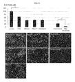
- FIG. 44 demonstrates the effect of TrafEnTM in enhancing polymer-based transfection. This was tested on different cells and cell types, for example human glioma/breast cancer cell lines, human mesenchymal stem cells (hMSCs), murine Abelson leukemia virus induced tumour cells (RAW 264.7), human prostate cancer cells (PC3), human melanoma cells (M14) and mouse embryonic fibroblast cells (MEFs). Cells were transfected with PEIMAX/2 ⁇ g PMAXGFP (LONZA). After transfection, cells were incubated in culture media in the presence or absence of the TrafEnTM reagent.
- hMSCs human mesenchymal stem cells
- RAW 264.7 murine Abelson leukemia virus induced tumour cells
- PC3 human prostate cancer cells
- M14 human melanoma cells
- MEFs mouse embryonic fibroblast cells
- GFP+ cells were analysed by FACS analysis or semi-automated cell count using Image J.
- the histograms present percentage of GFP+ cells and error bar represent S.E.M of the biological triplicate and technical duplicates. Representative images are presented (Blue, Nucleus; Green, GFP).
- FIG. 45 shows the effect of TrafEnTM in the enhancement of polymer-based transfection.
- MEF and M14 cells were transfected with EV71-pIRES-eGFP expression vector complexed with various transfection reagents. Transfections were conducted using 2 protocols—MI and IP.
- MI refers to the manufacture's instruction.
- IP refers to an in-house protocol, which is the deposit mediated transfection described in the specification, for example in FIG. 39 .
- IP+ TrafEnTM refers to the addition of TrafEnTM reagents in the culture media after transfection. The histograms here represent the percentage (%) of GFP+ cells for each procedure, each using a different transfection reagent.
- FIG. 46 shows histograms depicting the efficient knockdown of PTB1.
- HeLa cells were transfected with Scramble 30012 (Control), and PTB shRNA namely TR302218A 8865 (1), TR302218B 8866 (2), TR302218C 8867 (3), or TR302218D 8868 (4).
- Three days post treatment cells were harvested for qPCR analysis. Graph present fold change of PTB1 to control after normalization to GAPDH.
- FIG. 47 shows micrographs depicting the efficient knockdown of PTB, resulting in rapid transdifferentiation.
- HeLa cells were transfected with Scramble 30012 (Control), PMAXGFP (Control), and TR302218C 8867.
- Post transfection cells were treated with Puromycin and culture in N3 media (Media for neuronal cells). Images were captured up to 8 days. Then, cells were fixed with 4% Formaldehyde and stained with Tuj antibody.
- FIG. 49 shows fluorescent micrographs and histograms showing the synergistic effect of p53 and transfection enhancers.
- Cells were transfected with 1.5 ⁇ g of PMAXGFP or pEGFP-N1-p53 (p53).
- Transfection enhancers DOPE/CHEMS [D] and 5 ⁇ M SAHA
- culture media were replaced with fresh media with or without SAHA and further incubated for 48 h.
- cells were fixed with 4% Formaldehyde and stained with Hoechst 33342. Fluorescent images were captured and nucleus number was counted with Image J. Percentage of viable cells represents % of cell to negative control ( ⁇ ve).
- FIG. 50 shows a graph depicting the percentage (%) of viable cells, as well as fluorescent images of these cells, after prolonged incubation of U251MG cells with SAHA, showing that this did not increase cell death.
- Cells were transfected with pEGFP-N1-p53 (p53).
- Transfection enhancers DOPE/CHEMS and 5 ⁇ M SAHA
- the cells were further treated with SAHA for 48, 72 and 96 h.
- cells were fixed with 4% Formaldehyde and stained with Hoechst 33342. Fluorescent images were captured and nucleus number was counted with Image J. Percentage of viable cells represents % of cell over cells transfected with p53 only.
- the lower panel contains representative images from each condition. Unpaired, student t-test was performed to examine statistical significance between cells treated with SAHA for 48 h and 72 or 96 h. **, p ⁇ 0.001.
- FIG. 51 shows a graph and accompanying fluorescent images, showing the synergistic cytotoxic effect of p53 with SAHA, but not Tubastatin A.
- Cells were transfected with PMAXGFP or pEGFP-N1-p53 (p53).
- Transfection enhancers DOPE/CHEMS [D], 10 ⁇ M Tubastatin A [T] and 5 ⁇ M SAHA
- Culture media were replaced with fresh media with or without Tubastatin A or SAHA and further incubated for 48 h. Then, cells were fixed with 4% Formaldehyde and stained with Hoechst 33342. Fluorescent images were captured and nucleus number was counted with Image J.
- Percentage of viable cells represents % of cell to cells transfected with PMAXGFP.
- FIG. 52 shows fluorescent micrographs and histograms depicting the synergistic effect of p53 with SAHA in inducing significant cell death in both TMZR-U251MG and GSC.
- TMZR-U251MG and GSC cells were transfected with PMAXGFP and pEGFP-N1-p53 (p53) in the presence of DOPE/CHEMS [D] and SAHA individually or in combination. Twenty-two hours post transfection, the cell media was replaced with DMEM/10% FBS/40 ⁇ M TMZ for TMZR-U251MG and DMEM/serum replacement for GSC, with or without SAHA. Cells were further incubated for 48 h.
- FIG. 53 shows fluorescent micrographs and histograms visualising the effect of DOPE/CHEMS and Trichostatin A (TSA) on the enhancement of transfection.
- FIG. 54 depicts histograms depicting expression levels of BMP2 in human MSC under various treatment conditions.
- Post transfection cells were treated with or without DOPE/CHEMS, 5 ⁇ M Tubastatin A, and 150 nM TSA in combination or individually. 24 hours post transfection, culture media was replaced with fresh media containing HDACi according. After 48 h of incubation, cells were trypsinized and total RNA were collected with RNeasy Mini Kit (Qiagen) to avoid DNA contamination.
- RNeasy Mini Kit Qiagen
- RNA was reversed transcribed and the expression of BMP2 was measured using qPCR.
- the threshold cycles (C t s) of BMP2 were normalized to the house-keeping gene GAPDH.
- Cells transfected with PEIMAX/PMAXGFP serves as a negative control (control).
- FIG. 55 shows bright field and fluorescent images of GFP Expression of in human MSC up to 21 days of incubation.
- FIG. 56 shows bright field images of MSC cells from 3 up to 21 days post transfection.
- MSC cells were transfected with PEIMAX mediated delivery of PMAXGFP or PMAXGFP-BMP2. After transfection, cells were treated with DOPE/CHEMS and 150 nM TSA individually or in combination. 72 hours post transfection, culture media was replaced with (A) expansion media alpha-MEM/10% FBS or (B) osteogenic differentiation media (alpha-MEM supplemented with 10% FBS, 10 mM ⁇ -glycerophosphate, 10 nM dexamethasone, and 0.2 mM ascorbic acid. Cells were then further incubated and the respective media was replaced every 3 days.
- FIG. 57 shows micrograph images (bright field and fluorescent), as well as column graphs and FACS analysis graphs visualising the combinatorial effect of DOPE/CHEMS and SAHA or Tubastatin A in the enhancement of transfection.
- FIG. 58 shows histograms depicting the expression levels of GM-CSF in PC3 cells under various treatment conditions.
- Post transfection cells were treated with or without DOPE/CHEMS [D] and 5 ⁇ m SAHA individually or in combination [TrafEnTM]. 24 hours post transfection, culture media was replaced with fresh media containing HDACi accordingly. After 48 h of incubation, cells were trypsinized and the total RNA was collected individually using the RNeasy Mini Kit (Qiagen) to avoid DNA contamination.
- RNeasy Mini Kit Qiagen
- RNA was reverse-transcribed and the expression of GM-CSF was measured using qPCR.
- the threshold cycles (C t s) of GM-CSF were normalized to the house-keeping gene GAPDH. Untransfected cells served as controls (Ctrl).
- FIG. 59 shows histograms visualising that SAHA induced an up-regulation of MICA and NKG2D in PC3 cells.
- Human PC3 (prostate cancer) cells were treated with 5 ⁇ M SAHA for 8, 20 and 48 hours (hr). At the end of the incubation, the cells were collected and subjected to PCR analysis. Cells with no exposure to SAHA (Negative 8 hr) served as a control. The histogram here represented fold change to control after normalization with GAPDH.
- FIG. 60 show a high N/P ratio for transfection of HaF.
- FIG. 61 show data showing that a high transfection resulted in efficient reprogramming of HaF.
- the transfection mixture was replaced with culture media with or without TrafEnTM (DOPE/CHEMS+10 ⁇ M Tubastatin A). Two days later, the media were replaced with fresh culture media (10% FBS/DMEM). After 24 h incubation, total RNA were isolated with Qiagen RNAeasy kit and subjected to qPCR analysis. Graph presents expression of OSKM to control (cells transfected with PMAXGFP) after normalization to GAPDH. For a second set of experiment, cells were further incubated for 7 days. At the end of experiment, cells were fixed with 4% formaldehyde and stained with Oct4 antibody.
- FIG. 62 shows histograms showing that VPA induced up-regulation of KLF4 and c-Myc in HaF.
- Fibroblast cells were treated with 1 mM VPA for up to 3 days. At the end of incubation, the cells were collected and subjected to PCR analysis. Cells with no exposure to VPA (3 days) serve as control. Graphs present fold change to control after normalization with GAPDH.
- nucleic acid designates a molecule comprising one or more nucleotides, or an oligonucleotide, or a fragment thereof, including, without limitation, ribonucleic acid (RNA), messenger RNA (mRNA), DNA/RNA hybrids, non-natural or synthetic nucleic acids, short interfering RNA (siRNA), short hairpin RNA (shRNA), deoxyribonucleic acid (DNA), plasmid DNA (pDNA), antisense and sense oligonucleotides, nucleotides or combinations thereof.
- the nucleic acid may be single-stranded, or partially or completely double-stranded (duplex).
- Duplex nucleic acids may be homoduplex or heteroduplex.
- ribonucleic acid refers to biomolecules that play an important role in the regulation, coding, decoding and expression of genes.
- Each ribonucleic acid consists of a nucleotide, either adenine (A), cytosine (C), guanine (G) or uracil (U), and a ribose sugar.
- a ribonucleic acid sequence comprises of a chain of these nucleic acids, resulting in a sugar-phosphate backbone.
- DNA deoxyribonucleic acid
- A adenine
- C cytosine
- G guanine
- T thymidine
- isolated means that a nucleotide sequence, for example a gene, primer, or oligonucleotide or other sequence is substantially or essentially free from other nucleic acids or other impurities.
- amplicon refers to a product of an amplification reaction.
- An example of an amplicon is a nucleotide sequence produced as a result of PCR, real-time PCR, reverse transcription-PCR, competitive RT-PCR, ligase chain reaction (LCR), gap LCR, strand displacement amplification (SDA), nucleic acid sequence based amplification (NASBA), transcription-mediated amplification (TMA), rolling circle amplification (RCA) or the like.
- primer is used herein to mean any single-stranded oligonucleotide sequence capable of being used as a primer in, for example, PCR or RCA technology.
- a “primer” refers to a single-stranded oligonucleotide sequence that is capable of acting as a point of initiation for synthesis of a primer extension product that is substantially identical to the nucleic acid strand to be copied (for a forward primer) or substantially the reverse complement of the nucleic acid strand to be copied (for a reverse primer).
- a primer may be suitable for use in, for example, PCR technology.
- Single-stranded includes, for example, hairpin structures formed by single-stranded nucleotide sequences. The design of a primer, for example its length and specific sequence, depends on the nature of the target nucleotide sequence and on the conditions at which the primer is used, for example, temperature and ionic strength.
- the primers may consist of the nucleotide sequences described herein, or may be 10, 15, 20, 25, 30, 35, 40, 45, 50, 75, 100 or more nucleotides which comprise or fall within the sequences described herein, provided they are suitable for specifically binding a target nucleic acid sequence, under stringent conditions.
- the primer sequence is less than 35 nucleotides in length, for example the primer sequence is less than 34, 33, 32, 31, 30, 29, 28, 27, 26, 25, 24, 23, 22 21 20 19 18 17 16 15 14 13 12 11 or 10 nucleotides in length. Slight modifications of the primers or probes, in length or in sequence, can be carried out to maintain the specificity and sensitivity required under the given circumstances.
- probes and/or primers described herein may be extended in length by 1, 2, 3, 4 or 5 nucleotides or reduced in length by 1, 2, 3, 4 or 5 nucleotides, for example, in either direction.
- Primer sequences can be synthesized using any methods well known in the art.
- amplification refers to an amplification reaction, for example an enzyme-mediated reaction used to amplify a specific target nucleotide sequence. By amplifying the target nucleotide sequence, the reaction produces many more copies of the target nucleotide sequence to produce an amplicon, amplified product or amplification product.
- amplification reaction is a “polymerase chain reaction′ (PCR)”. PCR is carried out with the aid of thermal cycler in a mixture containing a polymerase enzyme, a set of primers, for example a set of forward and reverse primers and any additional primers that may be required and four deoxynucleotide triphosphates—(dNTPs).
- the terms “therapeutic gene” and gene therapy refer to a therapy for genetic disorders, often similar to therapy for other disorders.
- Gene therapy may involve insertion of normal copies of a gene into the cells of people that is in vivo, with a specific genetic disorder. This therapy may involve replacing a deficient compound or blocking an overactive pathway. Gene therapy may also involve turning off genes.
- Genetic modification may also be used in ex vivo gene therapy.
- human stem cells, immune cells or cancer cells can be genetically modified for various applications. Cells are modified to induce differentiation, transdifferentiation or reprogramming. Also, cells may be modified to serve as vehicle to deliver therapeutic protein. For example, mesenchymal stem cells may be modified to overexpress BMP2.
- polyplex refers to complexes of genetic material and a cationic species.
- the ratio of genetic material to cationic species may be selected from the group consisting of from about 0 to about 1000, about 0 to about 900, about 0 to about 800, about 0 to about 700, about 0 to about 600, about 0 to about 500, about 0 to about 400, about 0 to about 300, about 0 to about 200, about 0 to about 100, about 0 to about 75, about 0 to about 50, about 0 to about 25, about 0 to about 20, about 0 to about 15, about 0 to about 10, and about 0 to about 5.
- anti-cancer drugs also known as chemotherapeutic agents, refers to agents which used to treat cancers of the human body.
- chemotherapeutics which are defined into groups based on their method of action. Examples of the different groups of chemotherapeutics known in the art are as follows: alkylating antineoplastic agents, anti-metabolites, anti-microtubule agents, topoisomerase inhibitors and cytotoxic antibiotics.
- the agents can be administered to the patient intravenously, orally or intrathecally. Isolated limb perfusion is also a known delivery method for chemotherapeutics in certain cases.
- Taxanes refers to a class of are diterpenes produced by the plants of the genus Taxus (yews). Taxane based chemotherapeutic regimes are widely prescribed for cancer patients. Taxanes, which comprise of paclitaxel and docetaxel, promote microtubule stabilization, and disrupt transition from metaphase to anaphase. This blocks progression of cell division and prolonged activation of the mitotic checkpoint induces apoptosis or reversion to the G-phase, eventually causes cell death.
- Taxanes may be selected from a group consisting of cremophor EL® Taxoprexin®(Docosahexaenoic acid-paclitaxel), XytotaxTM (paclitaxel polyglumex), TOCOSOL® paclitaxel, BMS-184476, DJ-927, BMS-275183, RPR 109881A, Ortataxel, Genexol (co-polymer combination), LEP (liposomal-encapsulated paclitaxel) and taxol in vitamin E emulsion.
- epothilone represents an emerging class of drugs for cancer treatment.
- the mechanism of action for epothilone class is similar to taxanes, which is the blockage of mitosis and induction of apoptosis. Nevertheless, Epithilones were shown to be more potent and milder side effects than taxanes. Additionally, their better water solubility characteristic enables the replacement of cremophors (solubilizing agents of paclitaxel) which was shown to affect cardiac function.
- HDACi histone deacetylase inhibitor
- HDACs are also known to be involved in the maintenance and function of chromatin via regulation of acetylation state of histone.
- HDACi influences a broad repertoire of physiological processes, including transcription of genes involved in proliferation, differentiation, survival and DNA repair.
- TrafEnTM stands for trafficking enhancer and relates to two agents directing the genetic material or complex containing genetic material to a productive pathway for efficient transfection.
- TrafEnTM relates to transfecting a cell using comprising a first agent capable of directing the genetic material away from the acidic compartments and a second agent capable of stabilizing the microtubule or a network thereof.
- the application of TrafEnTM may be extended further to chemosensitize cells by rationally designing the composition to achieve a specific therapeutic effect.
- the first agent as defined above, may also be termed “chemoRe-router”.
- a “subject” or an “individual” is a living multi-cellular vertebrate organism.
- the subject can be an experimental subject, such as a non-human animal, e.g., a mouse, a cotton rat, or a non-human primate.
- the subject can be a human subject.
- biological material refers to any material or sample, which includes an analyte as defined herein.
- samples may, for example, include samples derived from or comprising stool, whole blood, serum, plasma, bone marrow, tears, saliva, nasal fluid, sputum, ear fluid, genital fluid, breast fluid, milk, colostrum, placental fluid, amniotic fluid, perspiration, synovial fluid, ascites fluid, cerebrospinal fluid, bile, gastric fluid, aqueous humor, vitreous humor, gastrointestinal fluid, exudate, transudate, pleural fluid, pericardial fluid, semen, upper airway fluid, peritoneal fluid, fluid harvested from a site of an immune response, fluid harvested from a pooled collection site, bronchial lavage, urine, biopsy material, e.g. from all suitable organs, e.g. the lung, the muscle, brain, liver, skin, pancreas, stomach,
- treatment refers to both therapeutic treatment and prophylactic or preventative measures, wherein the aim is to try and prevent or slow down (lessen) the targeted pathologic condition or disorder.
- the treatment may directly decrease the pathology of tumor cells, or render the tumor cells more susceptible to treatment by other therapeutic agents, e.g., radiation and/or chemotherapy.
- the aim or result of tumor treatment may include, for example, one or more of the following: (1) inhibition (i.e., reduction, slowing down or complete stopping) of tumor growth; (2) reduction or elimination of symptoms or tumor cells; (3) reduction in tumor size; (4) inhibition of tumor cell infiltration into adjacent peripheral organs and/or tissues; (5) inhibition of metastasis; (6) enhancement of anti-tumor immune response, which may, but does not have to, result in tumor regression or rejection; (7) increased survival time; and (8) decreased mortality at a given point of time following treatment.
- Treatment may entail treatment with a single agent or with a combination (more than two) of agents.
- An “agent” is used herein broadly to refer to, for example, a drug/compound or other means for treatment e.g.
- treatment or surgery.
- treatments include surgical intervention, liver transplantation, immunotherapy, chemotherapy with a given drug or drug combination, radiation therapy, neo-adjuvant treatment, diet, vitamin therapy, hormone therapies, gene therapy, cell therapy, antibody therapy etc.
- treatment also includes experimental treatment e.g. during drug screening or clinical trials.
- Attaining high transfection efficiencies when delivering genetic material offers the potential for the treatment of a myriad of devastating disorders including, but not limited to, cancers, neurodegenerative diseases and inflammatory disease, for which there are currently few treatment options.
- low transfection and delivery efficiencies limit the application of drug-gene therapeutics.
- galenics and methods for using this technology to enhance gene delivery ex vivo and in vivo represent an unmet need in this industry.
- the present disclosure provides a unique composition of biocompatible reagents (TrafEnTM), designed to drastically enhance the gene delivery and simultaneously chemosensitize the many types of hard-to-infect cells.
- the drug-gene combination described herein relates to a strategy, whereby a class of microtubule targeting chemosensitizers and a chemoRe-router increase the delivery of a therapeutic gene.
- the synergistic effect of TrafEnTM and the therapeutic gene are thought to result in a superior therapeutic effect.
- chemosensitizers may contain an optimized mix of fusogenic molecules that specifically redirect carrier/DNA complexes away from the non-productive acidic compartment, re-rerouting them onto then microtubular networks stabilized by the use of chemosensitizers.
- the present disclosure provides a composition for transfecting a cell with a genetic material, comprising a first agent capable of directing the genetic material away from an acidic compartment in a cell and a second agent capable of stabilizing the microtubule or a network thereof.
- the first agent may be capable of directing genetic material away from a non-productive acidic compartment of the cell.
- the first agent may be, but is not limited to, a lipid, a peptide fusiongenic agent or a combination thereof.
- the lipid fusogenic agent may be selected from DOPE, CHEMS, DPPC and DOPC and combinations thereof.
- the peptide fusogenic agent may be at least any one of, but not limited to, haemagglutinin (HA2-peptide), influenza-derived fusogenic peptide diINF-7, T domain of Diphtheria toxin and polycationic peptides, such as polylysine and polyarginine, or combinations thereof.
- haemagglutinin HA2-peptide
- influenza-derived fusogenic peptide diINF-7 influenza-derived fusogenic peptide diINF-7
- T domain of Diphtheria toxin and polycationic peptides, such as polylysine and polyarginine, or combinations thereof.
- the aforementioned peptide fusogenic agent may be chemically modified by attachment of a lipid.
- the aforementioned peptide may be chemically modified by attachment of a biomolecule, such as a nucleic acid or a synthetic carrier, such as a cationic polymer.
- the second agent may be capable of enhancing tubulin acetylation. In another embodiment, the second agent may be capable of enhancing the sensitivity of the cell to a therapeutic agent and/or may be capable of modifying the host genetic status.
- the aforementioned second agent may be selected from a histone deacetylase inhibitor (HDACi), a tubulin binding agent (TBA) and siRNA that is capable of directly or indirectly affecting the microtubule network stability.
- HDACi histone deacetylase inhibitor
- TAA tubulin binding agent
- siRNA that is capable of directly or indirectly affecting the microtubule network stability.
- the HDACi may be selected from Tubastatin A, belinostat, bufexamac, panobinostat, PCI-24781, SAHA (vorinostat), scriptaid, trichostatin A, valporic acid, B2, salermide, sirtinol and combinations thereof.
- the TBA of the second agent of the present invention may be selected from taxanes, epothilones, and a combination thereof.
- the taxanes may be paclitaxel, docetaxel or a combination thereof.
- the taxanes may be selected from a group consisting of cremophor EL® Taxoprexin® (Docosahexaenoic acid-paclitaxel), XytotaxTM (paclitaxel polyglumex), TOCOSOL® paclitaxel, BMS-184476, DJ-927, BMS-275183, RPR 109881A, Ortataxel, Genexol (co-polymer combination), LEP (liposomal-encapsulated paclitaxel) and taxol in vitamin E emulsion.
- cremophor EL® Taxoprexin® Docosahexaenoic acid-paclitaxel
- XytotaxTM paclitaxel polyglumex
- TOCOSOL® paclitaxel BMS-184476, DJ-927, BMS-275183, RPR 109881A, Ortataxel
- Genexol co-polymer combination
- LEP liposomal-encapsulated pac
- the epothilones may be patupilone, ixabepilone, BMS 310705, sagoilone, KOS-862, KOS-1584, or combinations thereof.
- the chemosensitizers may be histone deacetylase inhibitors (HDACi), tubulin binding agents, taxanes and siRNA, capable of directly or indirectly affecting the microtubule network stability.
- HDACi histone deacetylase inhibitors
- the chemosensitizers used in the present disclosure are known to have anti-neoplastic, anti-inflammatory, anti-angiogenic or neuroprotective effects in vivo.
- the genetic material may be coupled to at least one cationic species.
- the cationic species may be selected from polyethylene imine, polycationic amphiphiles, DEAE-dextran, cationic polymers, their derivatives and combinations thereof.
- the cationic species may be a cationic polymer such as a dendimer, branched-polyethylenimine (BPEI), linear-polyethylenimine (LPEI), Poly(amindoamine) (PAMAM), XTREMEGENE HP® (DNA transfection reagent), and combinations thereof.
- BPEI branched-polyethylenimine
- LPEI linear-polyethylenimine
- PAMAM Poly(amindoamine)
- XTREMEGENE HP® DNA transfection reagent
- the nucleic acid:polymer (N/P) ratio cationic species to the genetic material may be selected from the group consisting of from about 0 to about 1000, about 0 to about 900 about 0 to about 800 about 0 to about 700 about 0 to about 600 about 0 to about 500 about 0 to about 400 about 0 to about 300 about 0 to about 200 about 0 to about 100 about 0 to about 75 about 0 to about 50 about 0 to about 25 between about 0 to about 15, between about 0 to about 10, and between about 0 to about 5.
- the nucleic acid:polymer (N/P) ratio of genetic material to cationic species may be 20.
- the N/P ratio is the ratio of the cationic species to the genetic material forming polyplexes within the composition.
- the genetic material may be a nucleic acid sequence.
- nucleic acid sequence may be selected from the group consisting of DNA, RNA, mRNA, ribozymes, antisense oligonucleotides, modified polynucleotides and combinations thereof.
- the cell is a differentiated or undifferentiated cell.
- the cell may be selected from the group consisting of nervous systems cell, liver cell, hematopoiesis cell, peripheral blood cell, umbilical blood cell, bone marrow cell, tumour cell, ischemic tissue cell, T cells, B cells, skin cells and combinations thereof.
- the cell may be isolated from a biological sample.
- the biological material may be selected from the group consisting of a sample of fresh tissue, frozen tissue, paraffin-preserved tissue and/or ethanol preserved tissue.
- the biological material may be selected from the group consisting of whole blood or a component thereof, lymph, bile fluid, cerebrospinal fluid, bronchioalveolar lavage fluid, synovial fluid, semen, ascitic tumour fluid, breast milk, amniotic fluid, a buccal smear and pus.
- the first agent and the second agent may be provided to the cell simultaneously, separately or sequentially. In one embodiment, the first agent and the second agent may be provided within the first hour of transfection of the cell with the genetic material. Alternatively, the first agent and the second agent may be provided within about 2, 3, 4, 5, 6, 7, 8, 9, 10 hours of transfection of the cell with the genetic material
- the second agent may be provided after the first agent.
- the second agent may comprise a two or more therapeutic agents as described herein.
- composition as described herein may be used in gene therapy.
- the composition as described herein may further comprise a therapeutic agent.
- the therapeutic agent may be chemotherapeutic agent.
- the chemotherapeutic agent may be selected from the group consisting of taxanes, paclitaxel, docetaxel and epothilones, and combinations thereof.
- a method of treating a patient in need of gene therapy comprising administering a genetic material and a composition as described herein.
- compositions as described herein in the manufacture of a medicament for treating a disease, selected from a group consisting of cancer, SMA, bone cancers, leukemia, blood cancers, sickle cell disease, Wiskott-Aldrich Syndrome, HIV, genetic disease, diabetes, monogenic, infectious neurological, ocular, inflammatory, cardiac and neurodegenerative diseases.
- a disease selected from a group consisting of cancer, SMA, bone cancers, leukemia, blood cancers, sickle cell disease, Wiskott-Aldrich Syndrome, HIV, genetic disease, diabetes, monogenic, infectious neurological, ocular, inflammatory, cardiac and neurodegenerative diseases.
- composition as described herein may be used for gene marking.
- a method of delivering a genetic material into a cell comprising the step of administering the genetic material with the composition as described herein.
- the method may be performed in vitro or ex vivo or in situ.
- composition comprising a first agent, as defined herein, and a second agent, as defined herein.
- kits comprising a first agent, as defined herein, and a second agent, as defined herein, a therapeutic agent as defined herein and instructions for use.
- the present disclosure further encompasses therapeutic genes or genetic material.
- this genetic material may comprise of, but is not limited to, nucleic acid sequences, encoding for any one gene or part thereof, which upon entering the cell having then a therapeutic effect upon the target gene by possibly enhancing expression or by possibly decreasing expression by interacting with the target sequence.
- the genetic material, including therapeutic genes may differ from any exact sequences illustrated and described herein.
- the invention contemplates deletions, additions and substitutions to the sequences shown, so long as the sequences function in accordance with the methods of the invention.
- any method of quantifying genetic material is appropriate for use in the context of the invention and many are known in the art.
- the genetic material may be isolated using techniques known to the art, e.g. restriction enzymatic digest, polymerase chain reactions (PCR), agarose gel electrophoresis, size exclusion chromatography and many more.
- the isolated genetic material may be quantified using methods known in the art, that may be, but are not limited to, spectrophotometric analysis, fluorescent labelling, quantitative PCR (qPCR, also known as real-time PCR), and e.g. DNA microarray (CHIP) analysis for the determination of gene expression levels after transfection. All the methods disclosed may be used consecutively in any possible order.
- composition as described herein may be used in many different applications, all of which have/pertain to the use of TrafEnTM as a unique approach, wherein the simultaneous effects of both genes and drugs (e.g., HDACi) with microtubule modifying activities and/or epigenetic modifying activities may be useful for all the areas described below.
- genes and drugs e.g., HDACi
- microtubule modifying activities and/or epigenetic modifying activities may be useful for all the areas described below.
- Cell therapy refers to the process of introducing cells to restore normal function; which was lost due to age, disease, damage, or congenital defects.
- cell-based therapy including stem cells and immune cells and cancer cells.
- HDACi is known to effect processes in stem cell differentiation and reprogramming of somatic cells into induced pluripotent stem cells (iPSCs), broadening the application of TrafEnTM in cell based therapy.
- iPSCs induced pluripotent stem cells
- TrafEnTM in cell based therapy.
- These cell based therapies may be utilised, but are not limited to, the application of stem cells, e.g. negative or positive selection for the segregation of modified populations, and/or controlling the growth of stem cells and their progeny, e.g.
- stem cells population when inserting a suicide gene into the stem cells population to enable the use of external stimuli to eradicate uncontrolled cell proliferation.
- Further applications for this invention may also be modifying stem cells to delivery gene product, which depending on the application, may require long-term or transient expression of therapeutic gene. Cases like wound healing, bone regeneration, angiogenesis, and repair of central nervous system injury, transient expression would be preferable examples. On the other hand, to correct genetic disease, persistent gene expression is required.
- Another possible application of this invention is modulating differentiation in stem cells via a transfer of genes mediated by this invention.
- Another potential application of gene transfer to stem cells is to provide genetic signals, improving the outcome of differentiation protocols know in the art.
- compositions as described herein for the treatment and therapy e.g. cell based and gene therapy/therapeutics of any one of, but not limited to, the following diseases: SMA (via iPS cell based therapy), bone cancers, leukemia, blood cancers, cancers (anti-angiogenic, stopping cell growth), HSC therapies for sickle cell disease and Wiskott-Aldrich Syndrome, gene treatment for cancer, HIV infection and genetic disease, repair damaged tissues, diabetes (T1D and T2D), cardiac disease, neurodegenerative diseases, for example, Parkinson's disease.
- composition as described herein may also be used additionally as a method for reprogramming cells, and or transdifferentiation of cells. Genetic tools known in the art have been established to initiate reprogramming process in various cell types.
- HDACi may play a possible role in promoting transdifferentiation. It has been shown in the art that HDACi induced expression of SOX9 in hepatocytes, which normally lack SOX9. The aberrant expression of SOX9 induced expression of COL2A1 and COMP1, which is usually found during chondrogenesis. These observations demonstrate redeployment of a typical developmental process to an atypical setting may be initiated by treatment of cells with HDACi.
- Intracellular Plasmid DNA was Localized to Acidic Compartment in Differentiated Neuronal Cells
- chloroquine a lysosomotropic compound known to inhibit fusion of the endosome and reduce enzymatic degradation of DNA by buffering the vesicular interior, did not show cumulative enhancement of transfection. Similar observations were found with PLUSTM reagent and INF7 fusogenic peptide ( FIG. 27 ) when these reagents were added to the culture post-transfection. Additionally, LPEI mediated delivery of pDNA pre-complexed with PLUS reagents and INF7 fusogenic peptide did not improve transfection in differentiated neuronal cells ( FIG. 28 ).
- Microtubule mediated transportation is known to play a role in the polymer/pDNA trafficking to the nucleus.
- Tubastatin A a histone deacetylase-6 (HDAC6) inhibitor that enhances microtubule mediated intracellular transport of cargo.
- the co-administration of DOPE/CHEMS and Tubastatin A resulted in a highly significant increase in the number of EGFP positive differentiated Neuro2A ( ⁇ 70%) ( FIG. 10 a and b ).
- FIG. 32 a Remarkably, close to 75% of these post-mitotic cortical neurons were transfected ( FIG. 10 c and d ) without significant cytotoxicity ( FIG. 32 a ). It should be noted that optimal transfection occurred only when reagents were co-administered within the first hour of transfection ( FIG. 32 b ). To further confirm the significance of microtubular stabilization for efficient transfection, the effect of paclitaxel and various HDACi on tubulin acetylation and transfection was examined. In line with previous reports, HDAC6targeting small molecules (Tubastatin A, TSA, Varinostat/SAHA) and paclitaxel but not Entinostat, Tacedenaline markedly enhanced tubulin acetylation ( FIG.
- TrafEnTM Enhancement of transgene expression is achieved by TrafEnTM combinations acting on the endosomal trafficking and the stabilization of microtubular network.
- the effect of TrafEnTM on transfection enhancement has been examined in various conditions including—cell types, gene carriers, genetic material and HDACi.
- HDAC6 inhibitors such as Trichostatin A (TSA) and Varinostat (SAHA) enhance transfection by targeting microtubule and at the same time exert epigenetic modification activities.
- TSA Trichostatin A
- SAHA Varinostat
- HDACi has been used as chemosensitizer to increase cytotoxic effect of a therapeutic agent for cancer (A), to induce differentiation of stem cell (B), to increase immunogenicity of cancer vaccine (C) and to enhance reprogramming process (D).
- A cytotoxic effect of a therapeutic agent for cancer
- B stem cell
- C cancer vaccine
- D enhance reprogramming process
- DOPE/CHEMS and HDAC6 inhibitor work together to enhance transfection efficiency
- HDACi may augment the effect of the transgene through epigenetic modification ( 3 , FIG. 1 ).
- the gene-drug combinatorial strategy simultaneously increases transgene expression and the synergistic effect/s of HDACi may further accentuate the therapeutic effect.
- SAHA has shown moderate clinical benefits as monotherapies but has garnered much excitement in combination therapy.
- SAHA has been shown to exert potent anti-tumour effects in a broad variety of cancer cells at concentrations that have minimal toxic effects on normal cells.
- the underlying anticancer mechanisms of SAHA are still unclear, it is likely that it exerts epigenetic modification that altered gene expressions resulting in growth arrest, migration inhibition and cell death.
- a study has shown SAHA induced up-regulation of pro-apoptotic genes (Bax, Bim, Bmf, Bik, cytochrome C and Smac) and down-regulation of anti-apoptotic genes (XIAP and survivin).
- Other study has shown that the susceptibility of SAHA induced cell death was regulated by p53.
- Tubastatin A did not result in greater reduction of cell number as compared to DOPE/CHEMS.
- transfection of p53 with Tubastatin A and DOPE/CHEMS did not contribute to the drug-gene combinatorial effect as observed with the use of SAHA.
- glioma heterogeneity likely is the key reason to treatment failure.
- two subpopulations known as TMZ resistant cells and cancer stem cells. Both of the subpopulations have developed genetic mechanisms that exhibited aggressive cancer phenotypes such as migration and resistance to TMZ.
- the cancer stem cells hypothesis is well recognized as a challenge for treatment, due to resistance. These cells exhibit stem cell-like characteristics and are capable to generate heterogeneous tumour masses.
- Recent studies support the presence of small number of cancer stem cells in glioma (GSC). As these cells are resistant to radiotherapy and chemotherapy, they are sufficient to generate recurrent tumour.
- stem cells To realize the clinical application of stem cells, technologies must be established, to direct stem cells to differentiate in a regulated manner, to circumvent immunogenicity of non-autologous stem cells-derived cells and to function as drug delivery vehicles and to regulate the growth of cells post-transplantation. Gene delivery is a potential tool to deliver biological signals to address these challenges.
- the applications of TrafEnTM gene-drug combination in stem cells technologies was illustrated with mesenchymal stem cells (MSCs).
- MSCs mesenchymal stem cells
- TSA HDAC6 inhibitor and epigenetic modifier
- Cancer vaccine can be generated from the genetically modified tumour cells, either autologous (removed from patients during surgery) or allogeneic (established cancer cell lines). After genetic modification, cells are inactivated by radiation and injected to the patients subcutaneously or intradermally to induce recipient's immune response against the tumour cells. Utilization of allogeneic cells, such as existing cell lines, provides a sustained and unlimited source of well-characterized cells, which can be standardized for large-scale production. It may also provide single batch for clinical lot for comparative analysis of clinical result and eliminates the need to continuous harvest patient's cancer cell. Furthermore, there is no longer the requirement of tailor-made individual cancer vaccine, which may increase cost and labour.
- GVAX the most advanced cell line based cancer vaccine is GVAX, comprising PC3 cells modified with GM-CSF gene for metastatic prostate cancer treatment.
- Modified PC3 cells are able to activate antigen-presenting cells (APC) and induce immune response.
- APC antigen-presenting cells
- HDACi can augment the immunogenicity of tumour cells by increasing expression of major histocompatibility complex (MHC) class I and II proteins, co-stimulatory/adhesion molecules.
- MHC major histocompatibility complex
- SAHA and TSA has been found to associate with enhanced presentation of MICA and MICB on the surface of cancer cells but not normal cells.
- MICA and MICB induce activation of immunoreceptor, natural killer cell protein group 2D (NKG2D), resulting in the increased susceptibility of cancer cells to natural killer cells (NK cells; CD4 and CD8 T cells).
- NSG2D natural killer cell protein group 2D
- NK cells natural killer cells
- CD4 and CD8 T cells natural killer cells
- Reprogramming describes the process of cellular state conversions, including switch of differentiated cells into a less differentiated state.
- Genetic tools have been established to initiate reprogramming process in various cell types. For instance, the Yamanaka et al. successfully identified four transfection factors, Oct4, Sox2, Klf4, and cMyc, sufficiently generated iPSCs from fibroblast.
- the use of viral vectors may introduce risk of tumourigenesis and immunogenesis.
- Another concern is the risk of tumourigenesis induced by transgene integration.
- overexpression of c-Myc a well-known oncogene, and its reactivation could cause tumour formation. Removal of c-Myc from the reprogramming cocktail greatly reduces the reprogramming efficiency.
- HDACi Valproic acid
- Differentiated neurons are sensitive to physical stress and alterations in cellular environment, making transfection of these cells a significant challenge.
- transfection efficiency and cell viability were significantly increased.
- Mild centrifugation is thought to deposit aggregated polyplex, which is formed in the high salt media ( FIGS. 13 and 38-43 ). It is likely that polyplex is inherently unstable in the high salt media.
- the deposited polyplexes on the substrate was not released at significant levels into the media ( FIG. 43 ), an observation incongruent to previous reports.
- deposited polyplex may be taken up directly from cell surface or the cell culture substrate, consistent with recent results showing the critical involvements of endocytic processes in neuronal migration, motility and adhesion to substrates.
- the critical role of deposited aggregated polyplex in mediating efficient transfection was discussed under the supplementary section ( FIGS. 13 and 38-43 ).
- the uptake of DNA in the native but not differentiated neuronal cells was sensitivity to PKC, indicative of alterations in the uptake pathway/s and intracellular trafficking of polyplex, which may have contributed to the differential localization of polyplex into acidic compartment.
- LPEI is thought to exert “proton sponge effect” but LPEI-polyplex was found to be entrapped within the acidic compartment on neuronal differentiation and the addition of DOPE/CHEMS released the labelled DNA from this compartment.
- the dramatic enhancement of endosomal escape of labelled pDNA and transfection efficiency by DOPE/CHEMS is unexpected and is consistent with the idea that endosomal escape of polyplex is critical.
- the pH-dependent destabilization of DOPE/CHEMS within the acidic endosomal environment is thought to result in membrane phase transition resulting in membrane fusion and eventually releasing the contents into the cytosol. It is worthy to note that other commercial endosome targeting reagents including chloroquine have no effect on transfection efficiency.
- Fusogenic agents for example 1,2-Dioleoyl-sn-glycero-3-phosphoethanolamine (DOPE)/cholesteryl hemisuccinate (CHEMS), improved gene delivery. Fusogenic agents facilitate endosomal escape through the pH-dependent destabilization of DOPE/CHEMS within the acidic endosomal environment, resulting in membrane phase transition and membrane fusion. As shown in further examples, DOPE/CHEM specifically re-directs carrier/DNA complexes away from the non-productive acidic compartment, thus leading to enhanced transfection efficiency.
- DOPE 1,2-Dioleoyl-sn-glycero-3-phosphoethanolamine
- CHEMS cholesteryl hemisuccinate
- Fusogenic agents with properties similar to DOPE include Dipalmitoylphosphatidylcholine (DPPC) and 1,2-Dioleoyl-sn-glycero-3-Phosphatidylcholine (DOPC).
- DPPC Dipalmitoylphosphatidylcholine
- DOPC 1,2-Dioleoyl-sn-glycero-3-Phosphatidylcholine
- Some peptides were found to have fusogenic property as well, such as haemagglutinin (HA2-peptide), influenza-derived fusogenic peptide diINF-7, T domain of Diphtheria toxin and polycationic peptides, such as polylysine and polyarginine.
- microtubule-targeting agent and epigenetic modifiers as first- and second line treatments in patients with various diseases is known in the art.
- the disclosure further encompasses the use of microtubule-targeting agents and epigenetic modifiers.
- a microtubule-targeting chemosensitizer (Mt-C) is categorized as tubulin binding agents (TBA) and histone deacetylase inhibitors (HDACi) ( FIG. 3 ).
- TBA can suppress microtubule dynamics, leading to mitotic block and apoptosis.
- Taxanes may include paclitaxel (Taxol), docetaxel (Taxotere) or combinations thereof.
- Epothilones like taxanes, are known in the art to prevent cell division by inducing cell cycle arrest at the G2-M transition phase, resulting in cytotoxicity and, in due course, cell death.
- Epothilone analogues known in the art are patupilone, ixabepilone, BMS 310705, sagopilone, KOS-862, and KOS-1584.
- HDACi which have an inhibitory effect on HDAC/Sirtuin.
- HDACi as known in the art, have been shown to have microtubule-targeting and/or epigenetic modification activities.
- HDACi inhibit HDAC6 and Sirtuin2 to promote acetylation of microtubules, resulting in the stabilization of the microtubule network.
- Acetylation has been repeatedly alleged to be associated with stability of microtubules, where the phrase ‘acetylated microtubules’ is often used synonymously with ‘stable microtubules’.
- HDACi are known to arrest microtubule dynamics and exerts anti-cancer effect.
- HDACi like vorinostat, inhibit multiple HDAC (HDAC1, 2, 3 and 6) and exhibit various activities, such as immunomodulation and apoptosis.
- HDACs are involved in the maintenance and function of chromatin via regulation of acetylation state of histone. Additionally, it is known that HDACi are known to have diverse functions, as the HDAC family influences a broad repertoire of physiological processes, including transcription of genes involved in proliferation, differentiation, survival and DNA repair. More recently, HDACi have been demonstrated to promote self-renewal of hematopoietic stem cells, enhance differentiation of neural stem cells and increase efficiency of reprogramming of somatic cells.
- HDACi with inhibitory effect on HDAC/Sirtuin. Developmental status of the inhibitors on various diseases was presented.
- Targeted Targeted Developmental class of HDAC/ IC50 HDAC inhibitor Class Type of disease status HDAC Sirtuin (uM) Apicidin Peptide NA Tool compound Class I 1 0.02 2 0.02 3 0.02 Belinostat HA Cutaneous T-cell Phase II clinical Class I, IIb 1 0.25 (PXD101) lymphoma 2 0.25 Acute myeloid leukemia Phase I/II 3 0.2 6 0.41 BML-210 Benzamide NA tool compound Class I 1 14.99 2 10.91 3 2.62 CHR-2845 HA Hematological disease Phase I Class I NA NA Lymphoid malignancies Phase I Bufexamac Non- NA Approved drug Class I, IIb 3 341 steroidal (Paraderm ®, 6 10.7 Parfenac ®) 8 235 10 12.3 Dacinostat HA NA Phase I Class I, IIb 1 0.
- HDAC6 inhibition is known to affect microtubule dynamics in non-neuronal cells and neurite outgrowth in neuronal cells.
- Tubastatin A results in transient effect ( ⁇ 24 h) on tubulin acetylation ( FIG. 35 ).
- the transfection paradigm described herein did not result in sustained alteration on neurite outgrowth ( FIG. 36 ) and global metabolism ( FIG. 37 ) of the neuronal cells.
- MSC are a heterogeneous subset of adult stromal stem cells that can be isolated and expanded ex vivo, and can appropriately differentiate into cells of the residing tissues, repair the damaged tissues and restore normal functions. Other than their differentiation potential to osteogenic, chondrogenic, and adipogenic cell types, studies have demonstrated the capability of MSC to generate neurons, kidney and other cell types too. Viral mediated delivery of BMP2 expression vector have been developed to drive such stem cells into osteogenic cells by providing genetic signals to improve the outcome of differentiation protocol. In addition to the use of MSC as a cell source to generate differentiating cells, it has been tested to act as gene/drug delivery vehicles. MSCs were genetically modified with BMP2 to facilitate tissue repair.
- HDACi HDACi
- TSA TSA
- SAHA valproic acid
- Neuro2A (ATCC: CCL-131TM) stably expressed GFR ⁇ 2a cells and rat primary cortical neurons' were cultured and maintained as described previously (Yoong, L. F., G. Wan, and H. P. Too. Mol Cell Neurosci, 2009. 41(4): p. 464-73; Zhou, L. and H. P. Too. PLoS One, 2011. 6(6): p. e21680).
- Non-neuronal cells comprise ⁇ 0.5% of the cell population of neurons grow in Neurobasal-B27 (Brewer, G. J., et al. J Neurosci Res, 1993. 35(5): p. 567-76). On DIV 3 (3 days in vitro), primary neurons were transfected.
- Neuro2A and NG-108 cells were differentiated with 50 ng/ml glial cell-line derived neurotrophic factor (GDNF; Biosource, Camarillo, Calif.), 10 ⁇ M all trans retinoic acid (RA) or 10 ⁇ M Forskolin (FSK; Sigma, St. Louis, Mo.) in Dulbecco's Modified Eagle Media (DMEM) supplemented with 1% Fetal Bovine Serum (FBS) for 48 h prior transfection.
- GDNF glial cell-line derived neurotrophic factor
- RA trans retinoic acid
- FBS Fetal Bovine Serum
- the lipid components e.g. DOPE and CHEMS
- DOPE:CHEMS a certain ratio
- the solvent was then evaporated, thus facilitating the formation of a lipid film comprising DOPE/CHEMS at the bottom of glass tube.
- the lipid film was reconstituted in 25 mM HEPES buffer by vigorous shaking, after which the lipid solution was sonicated for 2 min in a bath-type sonicator. This lipid solution construes the first agent.
- the TrafEnTM composition is then prepared by combining the first agent and a second agent, as defined in the disclosure.
- the TrafEnTM composition referenced also envisions the usage of fusogenic peptides instead of fusogenic liposomes.
- Plasmid DNA expressing EGFP was purified according to manufacturer's instruction (Geneaid Biotech, Taiwan).
- LPEI 25-kDa; Polyscience, USA
- LPEI/pDNA complex was then added to complete media (1:10) to prepare the transfection mixture (pDNA at 2 ⁇ g/ml).
- For bulk transfection (with or without centrifugation), cells were seeded 24 h prior to transfection.
- the transfection mixture was added to cells in culture and centrifuged at 280 g for 5 min. The transfection mixture was then replaced with complete media and the cells were further incubated.
- transfection efficiency percentage of EGFP positive cells was quantified, either through manual counting or fluorescence-activated cell sorting (FACS) analysis after incubation for indicated periods.
- DOPE/CHEMS and Tubastatin A were used.
- Lipid film comprising DOPE/CHEMS (9:2 molar ratio) (Polar Avanti Lipid, Alabaster), was formed at the bottom of glass tube after evaporation of the solvent, chloroform.
- the lipid film was reconstituted in 25 mM HEPES buffer and sonicated for 2 min in a bath-type sonicator (Brandson 2200).
- the lipid solution was added to the cell culture at various indicated time post transfection.
- Tubastatin A (5 or 16 ⁇ M) was added to the culture media. The inhibitor containing media were replaced by fresh media 24 h later.
- the cells were trypsinized, centrifuged and re-suspended in PBS. Cell clumps were removed by filtering through a 40 ⁇ m mesh. The percentage of cells expressing EGFP was quantified by FACS analysis (BD FACSCanto, BD Biosciences) and the raw data analyzed using WinMDI (V2.9). At least 10,000 cells were analysed per sample.
- FITC- and Rhodamine-pDNA were prepared according to manufacturer recommendation (Minis Bio, USA). Expression of EGFP was observed in cells transfected with fluorescently labeled pDNA (data not shown). FITC- or Rhodamine-pDNA was used for visualization of internalized polyplex and extracellular fluorescence of the labeled pDNA was quenched with EtBr (20 ⁇ g/ml) or 0.4% trypan blue respectively 4 h post transfection. Cell images were taken before and after quenching with an inverted Zeiss microscope equipped with fluorescence detection (Zeiss cell observer Z1) and processed (Axio Vision Rel. 4.7). Quenching efficiency of trypan blue was examined (Supplementary FIG. 7 ).
- Rhodamine-pDNA was incubated with lysotracker green DND-26 (50 nM) for 5 min Image was captured with a Zeiss confocal microscopy (LSM710, Oberkochen, Germany). Co-localized pixel was analysed with Zeiss ZEN software (v2010).
- Neuro2A and NG-108 cells were differentiated with GDNF (50 ng/ml) or RA (10 ⁇ M) and Fsk (10 ⁇ M) in the 96-well plates 48 h prior transfection. Cells were then exposed to LPEI/pDNA (at various N/P ratios) for 15 min or 4 h. With or without centrifugation at 280 g for 5 min, transfection mixtures were replaced with complete medium and incubated for 48 h. Culture medium was replaced with 100 ⁇ l DMEM and 20 ⁇ l of CellTiter 96 Aqueous One Solution Cell Proliferation Assay (Promega, Singapore). Following 1 h incubation at 37°, the absorbance at 490 nm was recorded using a 96-well Microplate reader (Model 680, Biorad).
- the DNA was collected through the following procedures: (1) to quantify DNA in supernatant, the supernatant was treated with pAA/urea lysis buffer to release DNA; (2) to quantify DNA associated with cells, the supernatant was removed and the cells were washed once with 1 ⁇ PBS before being harvested by trypsinization and treated with pAA/urea lysis buffer; (3) to quantify DNA internalized by the cells, the supernatant was removed and the cells were washed once with 1 ⁇ PBS and incubated in DMEM containing pAA/DNAse for 2 h at 4° C. Next, the cells were trypsinized and treated at 95° C.
- pAA/urea lysis buffer-polyacrylic acid (pAA, Sigma, Mw: 8000; 10 ng of pAA/ng of pDNA; 32 carboxyl groups in pAA/1 phosphate group in pDNA), 0.5 M sodium chloride, 10 mM sodium phosphate and 4 M urea.
- pAA was used as a competitive reagent for displacement of pDNA from LPEI.
- the efficiency of pDNA dissociation was visualized/quantified using two different approaches—DNA retardation assay and qPCR. DNA complexes were electrophoresed (100 V, 20 min) in 0.8% agarose gel, stained with ethidium bromide and visualized on a UV transilluminator.
- the transfection mixture was transferred to the culture plate (pre-coated with complete media for 24 h) and incubated for the indicated periods (with or without centrifugation). The transfection mixture was removed and cells were seeded. Transfection efficiency (percentage of EGFP positive cells) was quantified, either through manual counting or through FACS analysis 48 h post transfection.
- Control and 10 ⁇ M differentiated Neuro2a Cells were fixed with 4% formaldehyde in 1 ⁇ PBS for 20 min at room temperature and subsequently permeabilized in 0.5% Triton-X100 in 1 ⁇ PBS. Then, the samples fixed cells were blocked with normal goat serum (1:10; Dako, Glostrup, Denmark) in 0.1% Triton X-100/1 ⁇ PBS for 30 min at 37° C. The cells were then incubated with primary antibodies against acetylated ⁇ -Tubulin (Sigma Aldrich T7451, 1:200 dilution) or TuJ (R&D Systems MAB1195, 1:50 dilution) in 0.1% TritonX-100/1% BSA/1 ⁇ PBS at 37° C.
- acetylated ⁇ -Tubulin Sigma Aldrich T7451, 1:200 dilution
- TuJ R&D Systems MAB1195, 1:50 dilution
- the samples were subjected to Mini-Beadbeater (Bio Spec Products Inc.) for 2 min. After which, the extracts were pelleted by centrifugation for 3 min at 16 000 g. The supernatants were transferred to autosampler vials (Agilent tech.) and assayed.
- Mini-Beadbeater Bio Spec Products Inc.
- the extracts were pelleted by centrifugation for 3 min at 16 000 g.
- the supernatants were transferred to autosampler vials (Agilent tech.) and assayed.
- the UPLC (Waters ACQUITY UPLC)—(TOF) MS (Bruker micrOTOF II) platform was used to analyse the metabolites.
- TOF Waters ACQUITY UPLC
- One bar of nebulizer gas was provided.
- the drying gas rate and dry gas temperature was adjusted at 9 mL/min and 200° C. respectively. From the acquired data (50-800 m/z), a range of m/z (0.06 m/z width, the average m/z distribution width with the MS instrument in use) was extracted). Subsequently, the retention time was determined for each intermediate and the peak area was integrated for each metabolite using manufacturer's software.
- ATP Relative amount of each metabolite to ATP was calculated.
- ATP was used as normalizer as it was the most abundant metabolite and stable across samples at each time point with coefficient variation less than 5%. After normalization, relative fold change of metabolite to the negative control at each time point was calculated.
Landscapes
- Health & Medical Sciences (AREA)
- Life Sciences & Earth Sciences (AREA)
- Chemical & Material Sciences (AREA)
- Genetics & Genomics (AREA)
- General Health & Medical Sciences (AREA)
- Engineering & Computer Science (AREA)
- Public Health (AREA)
- Medicinal Chemistry (AREA)
- Veterinary Medicine (AREA)
- Pharmacology & Pharmacy (AREA)
- Animal Behavior & Ethology (AREA)
- Molecular Biology (AREA)
- Organic Chemistry (AREA)
- Biotechnology (AREA)
- Bioinformatics & Cheminformatics (AREA)
- Epidemiology (AREA)
- Biomedical Technology (AREA)
- General Engineering & Computer Science (AREA)
- Wood Science & Technology (AREA)
- Zoology (AREA)
- Biophysics (AREA)
- Biochemistry (AREA)
- Microbiology (AREA)
- Physics & Mathematics (AREA)
- Plant Pathology (AREA)
- Proteomics, Peptides & Aminoacids (AREA)
- Dispersion Chemistry (AREA)
- Inorganic Chemistry (AREA)
- General Chemical & Material Sciences (AREA)
- Chemical Kinetics & Catalysis (AREA)
- Nuclear Medicine, Radiotherapy & Molecular Imaging (AREA)
- Pharmaceuticals Containing Other Organic And Inorganic Compounds (AREA)
- Acyclic And Carbocyclic Compounds In Medicinal Compositions (AREA)
- Medicines That Contain Protein Lipid Enzymes And Other Medicines (AREA)
- Micro-Organisms Or Cultivation Processes Thereof (AREA)
- Peptides Or Proteins (AREA)
- Oncology (AREA)
- Immunology (AREA)
- Mycology (AREA)
Priority Applications (1)
| Application Number | Priority Date | Filing Date | Title |
|---|---|---|---|
| US14/439,330 US10100331B2 (en) | 2012-10-29 | 2013-10-29 | Reagent for gene-drug therapeutics |
Applications Claiming Priority (3)
| Application Number | Priority Date | Filing Date | Title |
|---|---|---|---|
| US201261719908P | 2012-10-29 | 2012-10-29 | |
| PCT/SG2013/000464 WO2014070111A1 (en) | 2012-10-29 | 2013-10-29 | A novel reagent for gene-drug therapeutics |
| US14/439,330 US10100331B2 (en) | 2012-10-29 | 2013-10-29 | Reagent for gene-drug therapeutics |
Publications (2)
| Publication Number | Publication Date |
|---|---|
| US20150361449A1 US20150361449A1 (en) | 2015-12-17 |
| US10100331B2 true US10100331B2 (en) | 2018-10-16 |
Family
ID=50627830
Family Applications (1)
| Application Number | Title | Priority Date | Filing Date |
|---|---|---|---|
| US14/439,330 Active US10100331B2 (en) | 2012-10-29 | 2013-10-29 | Reagent for gene-drug therapeutics |
Country Status (7)
| Country | Link |
|---|---|
| US (1) | US10100331B2 (ja) |
| EP (1) | EP2911650B1 (ja) |
| JP (1) | JP6342910B2 (ja) |
| CN (1) | CN105101951B (ja) |
| DK (1) | DK2911650T3 (ja) |
| SG (1) | SG11201503020TA (ja) |
| WO (1) | WO2014070111A1 (ja) |
Families Citing this family (5)
| Publication number | Priority date | Publication date | Assignee | Title |
|---|---|---|---|---|
| WO2014070111A1 (en) | 2012-10-29 | 2014-05-08 | Agency For Science, Technology And Research | A novel reagent for gene-drug therapeutics |
| JP6231709B1 (ja) * | 2016-05-31 | 2017-11-15 | シスメックス株式会社 | 蛍光画像分析装置および分析方法 |
| CN108611355B (zh) * | 2018-05-04 | 2021-08-31 | 北京锦篮基因科技有限公司 | X染色体连锁肌小管肌病的基因药物 |
| SG10201902002SA (en) * | 2019-03-06 | 2020-10-29 | Nat Univ Singapore | Non-viral modification of mesenchymal stem cells |
| GB201903873D0 (en) * | 2019-03-21 | 2019-05-08 | Univ Nottingham | Enhanced transfection |
Citations (15)
| Publication number | Priority date | Publication date | Assignee | Title |
|---|---|---|---|---|
| WO2001093836A2 (en) | 2000-06-09 | 2001-12-13 | Teni Boulikas | Encapsulation of polynucleotides and drugs into targeted liposomes |
| US20050075268A1 (en) * | 2000-11-29 | 2005-04-07 | Kristian Berg | Photochemical internalization for delivery of molecules into the cytosol |
| WO2006007712A1 (en) | 2004-07-19 | 2006-01-26 | Protiva Biotherapeutics, Inc. | Methods comprising polyethylene glycol-lipid conjugates for delivery of therapeutic agents |
| JP2007289162A (ja) | 2006-03-29 | 2007-11-08 | Toyobo Co Ltd | 核酸導入時の細胞障害を低減する方法 |
| JP2008504827A (ja) | 2004-07-02 | 2008-02-21 | プロチバ バイオセラピューティクス インコーポレイティッド | 免疫賦活性siRNA分子およびその使用方法 |
| WO2008103431A2 (en) | 2007-02-23 | 2008-08-28 | Pronai Therapeutics, Inc. | Dnai - liposomes |
| US20100022637A1 (en) * | 2005-09-29 | 2010-01-28 | The Trustees Of Columbia University In The City Of New York | Identification of anti-cancer compounds and compounds for treating huntington's disease and methods of treatment thereof |
| WO2011097181A2 (en) | 2010-02-04 | 2011-08-11 | Vivoscript,Inc. | Compositions and methods for re-programming cells without genetic modification for treatment of neurological disorders |
| US20120046346A1 (en) * | 2010-04-16 | 2012-02-23 | Immune Disease Institute, Inc. | Sustained polypeptide expression from synthetic, modified rnas and uses thereof |
| KR20120047346A (ko) | 2010-10-22 | 2012-05-14 | 고마바이오텍(주) | 유전자 전달용 양이온성 센다이 f/hn 바이로좀 복합체 |
| US20120225115A1 (en) * | 2009-10-12 | 2012-09-06 | Au Jessie L-S | Methods and Compositions for Improved Deliver, Expression or Activity of RNA Interference Agents |
| WO2013052053A1 (en) | 2011-10-06 | 2013-04-11 | Empire Technology Development Llc | Transfection sheets and methods of use |
| US8486990B2 (en) * | 2003-09-12 | 2013-07-16 | Elixir Pharmaceuticals, Inc. | SirT inhibitors that bind to NAD |
| WO2013155572A1 (en) | 2012-04-20 | 2013-10-24 | Commonwealth Scientific And Industrial Research Organisation | Cell transfection method |
| WO2014070111A1 (en) | 2012-10-29 | 2014-05-08 | Agency For Science, Technology And Research | A novel reagent for gene-drug therapeutics |
Family Cites Families (4)
| Publication number | Priority date | Publication date | Assignee | Title |
|---|---|---|---|---|
| WO1993003709A1 (en) * | 1991-08-16 | 1993-03-04 | Vical, Inc. | Composition and method for treating cystic fibrosis |
| WO2006006561A1 (ja) * | 2004-07-12 | 2006-01-19 | Japan Science And Technology Agency | 核酸構造体 |
| EP1951752A4 (en) * | 2005-11-30 | 2008-12-31 | Jung Moon Kim | NON-ACTIVATED Wnt INHIBITOR POLYPEPTIDES AND PREPARATION METHOD THEREOF |
| JP2011519828A (ja) * | 2008-04-18 | 2011-07-14 | バクシネート コーポレーション | フラジェリンの欠失変異体と使用方法 |
-
2013
- 2013-10-29 WO PCT/SG2013/000464 patent/WO2014070111A1/en not_active Ceased
- 2013-10-29 JP JP2015539561A patent/JP6342910B2/ja active Active
- 2013-10-29 DK DK13850878T patent/DK2911650T3/da active
- 2013-10-29 US US14/439,330 patent/US10100331B2/en active Active
- 2013-10-29 CN CN201380068711.6A patent/CN105101951B/zh active Active
- 2013-10-29 SG SG11201503020TA patent/SG11201503020TA/en unknown
- 2013-10-29 EP EP13850878.3A patent/EP2911650B1/en active Active
Patent Citations (16)
| Publication number | Priority date | Publication date | Assignee | Title |
|---|---|---|---|---|
| JP2003535832A (ja) | 2000-06-09 | 2003-12-02 | ブリカス,テニ | ポリヌクレオチドおよび薬物の標的化リポソームへのカプセル化 |
| WO2001093836A2 (en) | 2000-06-09 | 2001-12-13 | Teni Boulikas | Encapsulation of polynucleotides and drugs into targeted liposomes |
| US20050075268A1 (en) * | 2000-11-29 | 2005-04-07 | Kristian Berg | Photochemical internalization for delivery of molecules into the cytosol |
| US8486990B2 (en) * | 2003-09-12 | 2013-07-16 | Elixir Pharmaceuticals, Inc. | SirT inhibitors that bind to NAD |
| JP2008504827A (ja) | 2004-07-02 | 2008-02-21 | プロチバ バイオセラピューティクス インコーポレイティッド | 免疫賦活性siRNA分子およびその使用方法 |
| WO2006007712A1 (en) | 2004-07-19 | 2006-01-26 | Protiva Biotherapeutics, Inc. | Methods comprising polyethylene glycol-lipid conjugates for delivery of therapeutic agents |
| US20100022637A1 (en) * | 2005-09-29 | 2010-01-28 | The Trustees Of Columbia University In The City Of New York | Identification of anti-cancer compounds and compounds for treating huntington's disease and methods of treatment thereof |
| JP2007289162A (ja) | 2006-03-29 | 2007-11-08 | Toyobo Co Ltd | 核酸導入時の細胞障害を低減する方法 |
| WO2008103431A2 (en) | 2007-02-23 | 2008-08-28 | Pronai Therapeutics, Inc. | Dnai - liposomes |
| US20120225115A1 (en) * | 2009-10-12 | 2012-09-06 | Au Jessie L-S | Methods and Compositions for Improved Deliver, Expression or Activity of RNA Interference Agents |
| WO2011097181A2 (en) | 2010-02-04 | 2011-08-11 | Vivoscript,Inc. | Compositions and methods for re-programming cells without genetic modification for treatment of neurological disorders |
| US20120046346A1 (en) * | 2010-04-16 | 2012-02-23 | Immune Disease Institute, Inc. | Sustained polypeptide expression from synthetic, modified rnas and uses thereof |
| KR20120047346A (ko) | 2010-10-22 | 2012-05-14 | 고마바이오텍(주) | 유전자 전달용 양이온성 센다이 f/hn 바이로좀 복합체 |
| WO2013052053A1 (en) | 2011-10-06 | 2013-04-11 | Empire Technology Development Llc | Transfection sheets and methods of use |
| WO2013155572A1 (en) | 2012-04-20 | 2013-10-24 | Commonwealth Scientific And Industrial Research Organisation | Cell transfection method |
| WO2014070111A1 (en) | 2012-10-29 | 2014-05-08 | Agency For Science, Technology And Research | A novel reagent for gene-drug therapeutics |
Non-Patent Citations (13)
Also Published As
| Publication number | Publication date |
|---|---|
| DK2911650T3 (da) | 2019-11-18 |
| US20150361449A1 (en) | 2015-12-17 |
| WO2014070111A1 (en) | 2014-05-08 |
| EP2911650B1 (en) | 2019-09-04 |
| CN105101951A (zh) | 2015-11-25 |
| JP6342910B2 (ja) | 2018-06-13 |
| SG11201503020TA (en) | 2015-05-28 |
| JP2016503290A (ja) | 2016-02-04 |
| EP2911650A4 (en) | 2016-05-04 |
| EP2911650A1 (en) | 2015-09-02 |
| CN105101951B (zh) | 2021-08-03 |
Similar Documents
| Publication | Publication Date | Title |
|---|---|---|
| US12509683B2 (en) | Methods and compositions for modulating gene expression | |
| Kim et al. | Neuroprotection by biodegradable PAMAM ester (e-PAM-R)-mediated HMGB1 siRNA delivery in primary cortical cultures and in the postischemic brain | |
| Zhang et al. | Sirtuin 6 protects the brain from cerebral ischemia/reperfusion injury through NRF2 activation | |
| Ma et al. | Self-assembled tetrahedral DNA nanostructures promote neural stem cell proliferation and neuronal differentiation | |
| US10100331B2 (en) | Reagent for gene-drug therapeutics | |
| MacLelland et al. | Therapeutic and diagnostic applications of antisense peptide nucleic acids | |
| EP4406541A2 (en) | Compositions and methods for decreasing tau expression | |
| JP7555542B2 (ja) | 統合失調症及び他の神経精神障害の治療方法 | |
| Perego et al. | Current understanding of and emerging treatment options for spinal muscular atrophy with respiratory distress type 1 (SMARD1) | |
| EP3368671A1 (en) | Methods and compositions for increasing smn expression | |
| JP2022538868A (ja) | ミセルナノ粒子及びその使用 | |
| Remant et al. | Cholesterol grafted cationic lipopolymers: Potential siRNA carriers for selective chronic myeloid leukemia therapy | |
| CN105008515A (zh) | 用于治疗线粒体疾病和用于使细胞分化成神经元的组合物和方法 | |
| JP7716987B2 (ja) | Tert活性化療法を伴う方法および組成物 | |
| WO2009104896A2 (ko) | 암 치료용 핵산 의약 조성물 | |
| US20230126157A1 (en) | Mirna-485 inhibitor for gene upregulation | |
| CA3163139A1 (en) | Compositions and methods for treating cancer | |
| US20250011793A1 (en) | Oligonucleotides for Modulating Synaptogyrin-3 Expression | |
| JP6795492B2 (ja) | CLCN7(ADO2 CLCN7依存性)遺伝子変異によって引き起こされる常染色体優性大理石骨病2型(ADO2)療法のための短鎖干渉RNA(siRNA) | |
| Taifour | Locus-selective targeting of the oncogenic fusion EWSR1-FLI1 in Ewing sarcoma by CRISPR/dCas9 epigenetic silencers | |
| US20260022377A1 (en) | Polynucleotide compositions and methods for treatment of neurodegenerative diseases | |
| Bortolotti | Preclinical evaluation of a MYCN-specific inhibitor for the treatment of small cell lung cancer | |
| JP2026504808A (ja) | 神経変性疾患の処置のためのポリヌクレオチド組成物および方法 | |
| Chen | New therapeutic approaches to study brain mechanisms: Utilizing stem cells and gene therapy to investigate brain diseases, learning, and memory | |
| WO2025088613A1 (en) | Targeting als-fus aggregation with proteasome inhibitors |
Legal Events
| Date | Code | Title | Description |
|---|---|---|---|
| AS | Assignment |
Owner name: AGENCY FOR SCIENCE, TECHNOLOGY AND RESEARCH, SINGA Free format text: ASSIGNMENT OF ASSIGNORS INTEREST;ASSIGNOR:TOO, HENG-PHON;REEL/FRAME:035604/0457 Effective date: 20131120 Owner name: NATIONAL UNIVERSITY OF SINGAPORE, SINGAPORE Free format text: ASSIGNMENT OF ASSIGNORS INTEREST;ASSIGNORS:HO, YOON KHEI;ZHOU, LIHAN;REEL/FRAME:035604/0288 Effective date: 20131120 |
|
| STCF | Information on status: patent grant |
Free format text: PATENTED CASE |
|
| MAFP | Maintenance fee payment |
Free format text: PAYMENT OF MAINTENANCE FEE, 4TH YEAR, LARGE ENTITY (ORIGINAL EVENT CODE: M1551); ENTITY STATUS OF PATENT OWNER: LARGE ENTITY Year of fee payment: 4 |