RU2404810C2 - Conjugates antibody-medicinal agent and methods - Google Patents
Conjugates antibody-medicinal agent and methods Download PDFInfo
- Publication number
- RU2404810C2 RU2404810C2 RU2006147264/15A RU2006147264A RU2404810C2 RU 2404810 C2 RU2404810 C2 RU 2404810C2 RU 2006147264/15 A RU2006147264/15 A RU 2006147264/15A RU 2006147264 A RU2006147264 A RU 2006147264A RU 2404810 C2 RU2404810 C2 RU 2404810C2
- Authority
- RU
- Russia
- Prior art keywords
- antibody
- drug
- cancer
- cells
- cell
- Prior art date
Links
- 238000000034 method Methods 0.000 title claims abstract description 98
- 239000003814 drug Substances 0.000 claims abstract description 125
- 201000011510 cancer Diseases 0.000 claims abstract description 120
- 150000001875 compounds Chemical class 0.000 claims abstract description 110
- 102100030086 Receptor tyrosine-protein kinase erbB-2 Human genes 0.000 claims abstract description 85
- 101001012157 Homo sapiens Receptor tyrosine-protein kinase erbB-2 Proteins 0.000 claims abstract description 77
- 239000000203 mixture Substances 0.000 claims abstract description 57
- 229960000575 trastuzumab Drugs 0.000 claims abstract description 26
- 239000008194 pharmaceutical composition Substances 0.000 claims abstract description 10
- 239000000611 antibody drug conjugate Substances 0.000 claims description 229
- 229940049595 antibody-drug conjugate Drugs 0.000 claims description 229
- 206010028980 Neoplasm Diseases 0.000 claims description 190
- 229940079593 drug Drugs 0.000 claims description 116
- -1 5 -FU Chemical compound 0.000 claims description 77
- 239000000562 conjugate Substances 0.000 claims description 59
- 102000005962 receptors Human genes 0.000 claims description 50
- 108020003175 receptors Proteins 0.000 claims description 50
- 206010006187 Breast cancer Diseases 0.000 claims description 32
- 239000003153 chemical reaction reagent Substances 0.000 claims description 32
- 210000004881 tumor cell Anatomy 0.000 claims description 32
- 208000026310 Breast neoplasm Diseases 0.000 claims description 31
- 230000002018 overexpression Effects 0.000 claims description 31
- 229940127089 cytotoxic agent Drugs 0.000 claims description 28
- 239000000047 product Substances 0.000 claims description 27
- 238000006243 chemical reaction Methods 0.000 claims description 26
- 239000002246 antineoplastic agent Substances 0.000 claims description 25
- 150000003839 salts Chemical class 0.000 claims description 16
- 238000004519 manufacturing process Methods 0.000 claims description 14
- 230000004614 tumor growth Effects 0.000 claims description 13
- 229940125644 antibody drug Drugs 0.000 claims description 10
- 101710100968 Receptor tyrosine-protein kinase erbB-2 Proteins 0.000 claims description 9
- 230000037396 body weight Effects 0.000 claims description 8
- 239000000546 pharmaceutical excipient Substances 0.000 claims description 8
- JJAHTWIKCUJRDK-UHFFFAOYSA-N succinimidyl 4-(N-maleimidomethyl)cyclohexane-1-carboxylate Chemical compound C1CC(CN2C(C=CC2=O)=O)CCC1C(=O)ON1C(=O)CCC1=O JJAHTWIKCUJRDK-UHFFFAOYSA-N 0.000 claims description 8
- 239000003085 diluting agent Substances 0.000 claims description 7
- HPJKCIUCZWXJDR-UHFFFAOYSA-N letrozole Chemical compound C1=CC(C#N)=CC=C1C(N1N=CN=C1)C1=CC=C(C#N)C=C1 HPJKCIUCZWXJDR-UHFFFAOYSA-N 0.000 claims description 7
- AAKJLRGGTJKAMG-UHFFFAOYSA-N erlotinib Chemical compound C=12C=C(OCCOC)C(OCCOC)=CC2=NC=NC=1NC1=CC=CC(C#C)=C1 AAKJLRGGTJKAMG-UHFFFAOYSA-N 0.000 claims description 6
- YLMAHDNUQAMNNX-UHFFFAOYSA-N imatinib methanesulfonate Chemical compound CS(O)(=O)=O.C1CN(C)CCN1CC1=CC=C(C(=O)NC=2C=C(NC=3N=C(C=CN=3)C=3C=NC=CC=3)C(C)=CC=2)C=C1 YLMAHDNUQAMNNX-UHFFFAOYSA-N 0.000 claims description 6
- 239000012453 solvate Substances 0.000 claims description 6
- 239000005551 L01XE03 - Erlotinib Substances 0.000 claims description 5
- 239000002552 dosage form Substances 0.000 claims description 5
- XGALLCVXEZPNRQ-UHFFFAOYSA-N gefitinib Chemical compound C=12C=C(OCCCN3CCOCC3)C(OC)=CC2=NC=NC=1NC1=CC=C(F)C(Cl)=C1 XGALLCVXEZPNRQ-UHFFFAOYSA-N 0.000 claims description 5
- DJQYYYCQOZMCRC-UHFFFAOYSA-N 2-aminopropane-1,3-dithiol Chemical compound SCC(N)CS DJQYYYCQOZMCRC-UHFFFAOYSA-N 0.000 claims description 4
- 239000005411 L01XE02 - Gefitinib Substances 0.000 claims description 4
- 238000001802 infusion Methods 0.000 claims description 4
- QFJCIRLUMZQUOT-HPLJOQBZSA-N sirolimus Chemical compound C1C[C@@H](O)[C@H](OC)C[C@@H]1C[C@@H](C)[C@H]1OC(=O)[C@@H]2CCCCN2C(=O)C(=O)[C@](O)(O2)[C@H](C)CC[C@H]2C[C@H](OC)/C(C)=C/C=C/C=C/[C@@H](C)C[C@@H](C)C(=O)[C@H](OC)[C@H](O)/C(C)=C/[C@@H](C)C(=O)C1 QFJCIRLUMZQUOT-HPLJOQBZSA-N 0.000 claims description 4
- LLDWLPRYLVPDTG-UHFFFAOYSA-N vatalanib succinate Chemical compound OC(=O)CCC(O)=O.C1=CC(Cl)=CC=C1NC(C1=CC=CC=C11)=NN=C1CC1=CC=NC=C1 LLDWLPRYLVPDTG-UHFFFAOYSA-N 0.000 claims description 4
- MLDQJTXFUGDVEO-UHFFFAOYSA-N BAY-43-9006 Chemical compound C1=NC(C(=O)NC)=CC(OC=2C=CC(NC(=O)NC=3C=C(C(Cl)=CC=3)C(F)(F)F)=CC=2)=C1 MLDQJTXFUGDVEO-UHFFFAOYSA-N 0.000 claims description 3
- VWUXBMIQPBEWFH-WCCTWKNTSA-N Fulvestrant Chemical compound OC1=CC=C2[C@H]3CC[C@](C)([C@H](CC4)O)[C@@H]4[C@@H]3[C@H](CCCCCCCCCS(=O)CCCC(F)(F)C(F)(F)F)CC2=C1 VWUXBMIQPBEWFH-WCCTWKNTSA-N 0.000 claims description 3
- 239000002147 L01XE04 - Sunitinib Substances 0.000 claims description 3
- 229960000397 bevacizumab Drugs 0.000 claims description 3
- 229960002584 gefitinib Drugs 0.000 claims description 3
- 229960003685 imatinib mesylate Drugs 0.000 claims description 3
- BCFGMOOMADDAQU-UHFFFAOYSA-N lapatinib Chemical compound O1C(CNCCS(=O)(=O)C)=CC=C1C1=CC=C(N=CN=C2NC=3C=C(Cl)C(OCC=4C=C(F)C=CC=4)=CC=3)C2=C1 BCFGMOOMADDAQU-UHFFFAOYSA-N 0.000 claims description 3
- 229960003881 letrozole Drugs 0.000 claims description 3
- LBWFXVZLPYTWQI-IPOVEDGCSA-N n-[2-(diethylamino)ethyl]-5-[(z)-(5-fluoro-2-oxo-1h-indol-3-ylidene)methyl]-2,4-dimethyl-1h-pyrrole-3-carboxamide;(2s)-2-hydroxybutanedioic acid Chemical compound OC(=O)[C@@H](O)CC(O)=O.CCN(CC)CCNC(=O)C1=C(C)NC(\C=C/2C3=CC(F)=CC=C3NC\2=O)=C1C LBWFXVZLPYTWQI-IPOVEDGCSA-N 0.000 claims description 3
- ZAHRKKWIAAJSAO-UHFFFAOYSA-N rapamycin Natural products COCC(O)C(=C/C(C)C(=O)CC(OC(=O)C1CCCCN1C(=O)C(=O)C2(O)OC(CC(OC)C(=CC=CC=CC(C)CC(C)C(=O)C)C)CCC2C)C(C)CC3CCC(O)C(C3)OC)C ZAHRKKWIAAJSAO-UHFFFAOYSA-N 0.000 claims description 3
- 229960002930 sirolimus Drugs 0.000 claims description 3
- 229960003787 sorafenib Drugs 0.000 claims description 3
- 229940034785 sutent Drugs 0.000 claims description 3
- VVIAGPKUTFNRDU-UHFFFAOYSA-N 6S-folinic acid Natural products C1NC=2NC(N)=NC(=O)C=2N(C=O)C1CNC1=CC=C(C(=O)NC(CCC(O)=O)C(O)=O)C=C1 VVIAGPKUTFNRDU-UHFFFAOYSA-N 0.000 claims description 2
- 239000005511 L01XE05 - Sorafenib Substances 0.000 claims description 2
- 239000002136 L01XE07 - Lapatinib Substances 0.000 claims description 2
- GXJABQQUPOEUTA-RDJZCZTQSA-N bortezomib Chemical compound C([C@@H](C(=O)N[C@@H](CC(C)C)B(O)O)NC(=O)C=1N=CC=NC=1)C1=CC=CC=C1 GXJABQQUPOEUTA-RDJZCZTQSA-N 0.000 claims description 2
- 229960001467 bortezomib Drugs 0.000 claims description 2
- 229960001433 erlotinib Drugs 0.000 claims description 2
- VVIAGPKUTFNRDU-ABLWVSNPSA-N folinic acid Chemical compound C1NC=2NC(N)=NC(=O)C=2N(C=O)C1CNC1=CC=C(C(=O)N[C@@H](CCC(O)=O)C(O)=O)C=C1 VVIAGPKUTFNRDU-ABLWVSNPSA-N 0.000 claims description 2
- 235000008191 folinic acid Nutrition 0.000 claims description 2
- 239000011672 folinic acid Substances 0.000 claims description 2
- 229960002258 fulvestrant Drugs 0.000 claims description 2
- 239000013067 intermediate product Substances 0.000 claims description 2
- 229960004891 lapatinib Drugs 0.000 claims description 2
- 229960001691 leucovorin Drugs 0.000 claims description 2
- DHMTURDWPRKSOA-RUZDIDTESA-N lonafarnib Chemical compound C1CN(C(=O)N)CCC1CC(=O)N1CCC([C@@H]2C3=C(Br)C=C(Cl)C=C3CCC3=CC(Br)=CN=C32)CC1 DHMTURDWPRKSOA-RUZDIDTESA-N 0.000 claims description 2
- DWAFYCQODLXJNR-BNTLRKBRSA-L oxaliplatin Chemical compound O1C(=O)C(=O)O[Pt]11N[C@@H]2CCCC[C@H]2N1 DWAFYCQODLXJNR-BNTLRKBRSA-L 0.000 claims description 2
- 229960001756 oxaliplatin Drugs 0.000 claims description 2
- 229950001750 lonafarnib Drugs 0.000 claims 1
- 230000000694 effects Effects 0.000 abstract description 31
- 239000000126 substance Substances 0.000 abstract description 20
- 239000003795 chemical substances by application Substances 0.000 abstract description 14
- 230000004565 tumor cell growth Effects 0.000 abstract 1
- 210000004027 cell Anatomy 0.000 description 230
- 102000001301 EGF receptor Human genes 0.000 description 156
- 108060006698 EGF receptor Proteins 0.000 description 151
- 230000027455 binding Effects 0.000 description 94
- 239000000427 antigen Substances 0.000 description 92
- 108091007433 antigens Proteins 0.000 description 92
- 102000036639 antigens Human genes 0.000 description 92
- 108090000623 proteins and genes Proteins 0.000 description 69
- 125000005647 linker group Chemical group 0.000 description 67
- 238000011282 treatment Methods 0.000 description 65
- 208000037265 diseases, disorders, signs and symptoms Diseases 0.000 description 59
- 241000282414 Homo sapiens Species 0.000 description 54
- 235000018102 proteins Nutrition 0.000 description 53
- 102000004169 proteins and genes Human genes 0.000 description 53
- 108090000765 processed proteins & peptides Proteins 0.000 description 49
- 239000003446 ligand Substances 0.000 description 45
- 102000004196 processed proteins & peptides Human genes 0.000 description 40
- 241001465754 Metazoa Species 0.000 description 37
- 241000699666 Mus <mouse, genus> Species 0.000 description 37
- 229920001184 polypeptide Polymers 0.000 description 34
- 201000010099 disease Diseases 0.000 description 33
- 238000004458 analytical method Methods 0.000 description 30
- 108060003951 Immunoglobulin Proteins 0.000 description 29
- 102000018358 immunoglobulin Human genes 0.000 description 29
- 125000003275 alpha amino acid group Chemical group 0.000 description 27
- 238000003556 assay Methods 0.000 description 27
- 208000035475 disorder Diseases 0.000 description 26
- 239000012634 fragment Substances 0.000 description 26
- 238000000338 in vitro Methods 0.000 description 26
- 230000001965 increasing effect Effects 0.000 description 25
- 230000004913 activation Effects 0.000 description 24
- 150000007523 nucleic acids Chemical class 0.000 description 24
- 241000699670 Mus sp. Species 0.000 description 23
- 238000001727 in vivo Methods 0.000 description 23
- 241000700159 Rattus Species 0.000 description 22
- 230000015572 biosynthetic process Effects 0.000 description 22
- 230000014509 gene expression Effects 0.000 description 22
- 102000039446 nucleic acids Human genes 0.000 description 22
- 108020004707 nucleic acids Proteins 0.000 description 22
- 210000002966 serum Anatomy 0.000 description 22
- 230000002401 inhibitory effect Effects 0.000 description 20
- 230000001988 toxicity Effects 0.000 description 20
- 231100000419 toxicity Toxicity 0.000 description 20
- 108010021625 Immunoglobulin Fragments Proteins 0.000 description 19
- 102000008394 Immunoglobulin Fragments Human genes 0.000 description 19
- 125000004432 carbon atom Chemical group C* 0.000 description 18
- 102400000058 Neuregulin-1 Human genes 0.000 description 17
- 108090000556 Neuregulin-1 Proteins 0.000 description 17
- 230000012010 growth Effects 0.000 description 17
- 230000001225 therapeutic effect Effects 0.000 description 17
- 230000009261 transgenic effect Effects 0.000 description 17
- 230000030833 cell death Effects 0.000 description 16
- 231100000433 cytotoxic Toxicity 0.000 description 15
- 230000001472 cytotoxic effect Effects 0.000 description 15
- 229940002612 prodrug Drugs 0.000 description 15
- 239000000651 prodrug Substances 0.000 description 15
- XJMOSONTPMZWPB-UHFFFAOYSA-M propidium iodide Chemical compound [I-].[I-].C12=CC(N)=CC=C2C2=CC=C(N)C=C2[N+](CCC[N+](C)(CC)CC)=C1C1=CC=CC=C1 XJMOSONTPMZWPB-UHFFFAOYSA-M 0.000 description 15
- 230000010056 antibody-dependent cellular cytotoxicity Effects 0.000 description 14
- 230000000269 nucleophilic effect Effects 0.000 description 14
- 238000012216 screening Methods 0.000 description 14
- 241000124008 Mammalia Species 0.000 description 13
- 108700020796 Oncogene Proteins 0.000 description 13
- 230000004663 cell proliferation Effects 0.000 description 13
- LOKCTEFSRHRXRJ-UHFFFAOYSA-I dipotassium trisodium dihydrogen phosphate hydrogen phosphate dichloride Chemical compound P(=O)(O)(O)[O-].[K+].P(=O)(O)([O-])[O-].[Na+].[Na+].[Cl-].[K+].[Cl-].[Na+] LOKCTEFSRHRXRJ-UHFFFAOYSA-I 0.000 description 13
- 229940088597 hormone Drugs 0.000 description 13
- 239000005556 hormone Substances 0.000 description 13
- 239000002609 medium Substances 0.000 description 13
- 239000002953 phosphate buffered saline Substances 0.000 description 13
- 238000002360 preparation method Methods 0.000 description 13
- 238000011160 research Methods 0.000 description 13
- 101000628535 Homo sapiens Metalloreductase STEAP2 Proteins 0.000 description 12
- 102100026711 Metalloreductase STEAP2 Human genes 0.000 description 12
- 239000002253 acid Substances 0.000 description 12
- 125000000217 alkyl group Chemical group 0.000 description 12
- 229910052799 carbon Inorganic materials 0.000 description 12
- 229940022353 herceptin Drugs 0.000 description 12
- 210000001519 tissue Anatomy 0.000 description 12
- 125000003118 aryl group Chemical group 0.000 description 11
- 210000003719 b-lymphocyte Anatomy 0.000 description 11
- 230000004071 biological effect Effects 0.000 description 11
- 230000008859 change Effects 0.000 description 11
- 125000005842 heteroatom Chemical group 0.000 description 11
- 231100000682 maximum tolerated dose Toxicity 0.000 description 11
- 239000012528 membrane Substances 0.000 description 11
- 239000002207 metabolite Substances 0.000 description 11
- 230000026731 phosphorylation Effects 0.000 description 11
- 238000006366 phosphorylation reaction Methods 0.000 description 11
- 229910052717 sulfur Inorganic materials 0.000 description 11
- 239000003981 vehicle Substances 0.000 description 11
- 206010009944 Colon cancer Diseases 0.000 description 10
- OUYCCCASQSFEME-QMMMGPOBSA-N L-tyrosine Chemical compound OC(=O)[C@@H](N)CC1=CC=C(O)C=C1 OUYCCCASQSFEME-QMMMGPOBSA-N 0.000 description 10
- 208000000236 Prostatic Neoplasms Diseases 0.000 description 10
- 102100032780 Semaphorin-5B Human genes 0.000 description 10
- 230000021615 conjugation Effects 0.000 description 10
- 238000011161 development Methods 0.000 description 10
- 230000018109 developmental process Effects 0.000 description 10
- 230000013595 glycosylation Effects 0.000 description 10
- 238000006206 glycosylation reaction Methods 0.000 description 10
- 238000006467 substitution reaction Methods 0.000 description 10
- 102100033620 Calponin-1 Human genes 0.000 description 9
- 102000004190 Enzymes Human genes 0.000 description 9
- 108090000790 Enzymes Proteins 0.000 description 9
- 101000945318 Homo sapiens Calponin-1 Proteins 0.000 description 9
- 101000652736 Homo sapiens Transgelin Proteins 0.000 description 9
- 206010060862 Prostate cancer Diseases 0.000 description 9
- 125000000539 amino acid group Chemical group 0.000 description 9
- 230000000259 anti-tumor effect Effects 0.000 description 9
- 239000000969 carrier Substances 0.000 description 9
- 238000003776 cleavage reaction Methods 0.000 description 9
- 235000018417 cysteine Nutrition 0.000 description 9
- 239000002254 cytotoxic agent Substances 0.000 description 9
- 229940088598 enzyme Drugs 0.000 description 9
- 150000002430 hydrocarbons Chemical class 0.000 description 9
- 229940127121 immunoconjugate Drugs 0.000 description 9
- 239000000543 intermediate Substances 0.000 description 9
- 210000004185 liver Anatomy 0.000 description 9
- 210000002307 prostate Anatomy 0.000 description 9
- 230000002829 reductive effect Effects 0.000 description 9
- OUYCCCASQSFEME-UHFFFAOYSA-N tyrosine Natural products OC(=O)C(N)CC1=CC=C(O)C=C1 OUYCCCASQSFEME-UHFFFAOYSA-N 0.000 description 9
- CIWBSHSKHKDKBQ-JLAZNSOCSA-N Ascorbic acid Chemical compound OC[C@H](O)[C@H]1OC(=O)C(O)=C1O CIWBSHSKHKDKBQ-JLAZNSOCSA-N 0.000 description 8
- 102100027203 B-cell antigen receptor complex-associated protein beta chain Human genes 0.000 description 8
- 102000000844 Cell Surface Receptors Human genes 0.000 description 8
- 108010001857 Cell Surface Receptors Proteins 0.000 description 8
- 108010023729 Complement 3d Receptors Proteins 0.000 description 8
- 102000011412 Complement 3d Receptors Human genes 0.000 description 8
- 102000004127 Cytokines Human genes 0.000 description 8
- 108090000695 Cytokines Proteins 0.000 description 8
- 101000654679 Homo sapiens Semaphorin-5B Proteins 0.000 description 8
- 101000835745 Homo sapiens Teratocarcinoma-derived growth factor 1 Proteins 0.000 description 8
- 102100026404 Teratocarcinoma-derived growth factor 1 Human genes 0.000 description 8
- 102400001320 Transforming growth factor alpha Human genes 0.000 description 8
- 101800004564 Transforming growth factor alpha Proteins 0.000 description 8
- 239000008186 active pharmaceutical agent Substances 0.000 description 8
- 230000006907 apoptotic process Effects 0.000 description 8
- 210000000349 chromosome Anatomy 0.000 description 8
- 231100000599 cytotoxic agent Toxicity 0.000 description 8
- 230000001419 dependent effect Effects 0.000 description 8
- 229940088679 drug related substance Drugs 0.000 description 8
- 150000002148 esters Chemical class 0.000 description 8
- 238000002347 injection Methods 0.000 description 8
- 239000007924 injection Substances 0.000 description 8
- 230000003834 intracellular effect Effects 0.000 description 8
- 238000005259 measurement Methods 0.000 description 8
- 230000004048 modification Effects 0.000 description 8
- 238000012986 modification Methods 0.000 description 8
- 210000000822 natural killer cell Anatomy 0.000 description 8
- 229910052760 oxygen Inorganic materials 0.000 description 8
- 150000003254 radicals Chemical class 0.000 description 8
- 230000007017 scission Effects 0.000 description 8
- 238000012360 testing method Methods 0.000 description 8
- 125000003396 thiol group Chemical group [H]S* 0.000 description 8
- 239000003053 toxin Substances 0.000 description 8
- 231100000765 toxin Toxicity 0.000 description 8
- 108700012359 toxins Proteins 0.000 description 8
- 108020004414 DNA Proteins 0.000 description 7
- NKANXQFJJICGDU-QPLCGJKRSA-N Tamoxifen Chemical compound C=1C=CC=CC=1C(/CC)=C(C=1C=CC(OCCN(C)C)=CC=1)/C1=CC=CC=C1 NKANXQFJJICGDU-QPLCGJKRSA-N 0.000 description 7
- 102100029690 Tumor necrosis factor receptor superfamily member 13C Human genes 0.000 description 7
- 239000004480 active ingredient Substances 0.000 description 7
- 210000004369 blood Anatomy 0.000 description 7
- 239000008280 blood Substances 0.000 description 7
- 208000029742 colonic neoplasm Diseases 0.000 description 7
- 230000000295 complement effect Effects 0.000 description 7
- 230000007423 decrease Effects 0.000 description 7
- 230000004069 differentiation Effects 0.000 description 7
- 238000005516 engineering process Methods 0.000 description 7
- 238000002474 experimental method Methods 0.000 description 7
- 230000006870 function Effects 0.000 description 7
- 239000003102 growth factor Substances 0.000 description 7
- 229940072221 immunoglobulins Drugs 0.000 description 7
- 238000003364 immunohistochemistry Methods 0.000 description 7
- 230000001939 inductive effect Effects 0.000 description 7
- 210000003734 kidney Anatomy 0.000 description 7
- 230000036210 malignancy Effects 0.000 description 7
- 230000036961 partial effect Effects 0.000 description 7
- 239000000243 solution Substances 0.000 description 7
- 238000010186 staining Methods 0.000 description 7
- VBEQCZHXXJYVRD-GACYYNSASA-N uroanthelone Chemical compound C([C@@H](C(=O)N[C@H](C(=O)N[C@@H](CS)C(=O)N[C@@H](CC(N)=O)C(=O)N[C@@H](CS)C(=O)N[C@H](C(=O)N[C@@H]([C@@H](C)CC)C(=O)NCC(=O)N[C@@H](CC=1C=CC(O)=CC=1)C(=O)N[C@@H](CO)C(=O)NCC(=O)N[C@@H](CC(O)=O)C(=O)N[C@@H](CCCNC(N)=N)C(=O)N[C@@H](CS)C(=O)N[C@@H](CCC(N)=O)C(=O)N[C@@H]([C@@H](C)O)C(=O)N[C@@H](CCCNC(N)=N)C(=O)N[C@@H](CC(O)=O)C(=O)N[C@@H](CC(C)C)C(=O)N[C@@H](CCCNC(N)=N)C(=O)N[C@@H](CC=1C2=CC=CC=C2NC=1)C(=O)N[C@@H](CC=1C2=CC=CC=C2NC=1)C(=O)N[C@@H](CCC(O)=O)C(=O)N[C@@H](CC(C)C)C(=O)N[C@@H](CCCNC(N)=N)C(O)=O)C(C)C)[C@@H](C)O)NC(=O)[C@H](CO)NC(=O)[C@H](CC(O)=O)NC(=O)[C@H](CC(C)C)NC(=O)[C@H](CO)NC(=O)[C@H](CCC(O)=O)NC(=O)[C@@H](NC(=O)[C@H](CC=1NC=NC=1)NC(=O)[C@H](CCSC)NC(=O)[C@H](CS)NC(=O)[C@@H](NC(=O)CNC(=O)CNC(=O)[C@H](CC(N)=O)NC(=O)[C@H](CC(C)C)NC(=O)[C@H](CS)NC(=O)[C@H](CC=1C=CC(O)=CC=1)NC(=O)CNC(=O)[C@H](CC(O)=O)NC(=O)[C@H](CC=1C=CC(O)=CC=1)NC(=O)[C@H](CO)NC(=O)[C@H](CO)NC(=O)[C@H]1N(CCC1)C(=O)[C@H](CS)NC(=O)CNC(=O)[C@H]1N(CCC1)C(=O)[C@H](CC=1C=CC(O)=CC=1)NC(=O)[C@H](CO)NC(=O)[C@@H](N)CC(N)=O)C(C)C)[C@@H](C)CC)C1=CC=C(O)C=C1 VBEQCZHXXJYVRD-GACYYNSASA-N 0.000 description 7
- XLYOFNOQVPJJNP-UHFFFAOYSA-N water Substances O XLYOFNOQVPJJNP-UHFFFAOYSA-N 0.000 description 7
- 241000282693 Cercopithecidae Species 0.000 description 6
- BWGNESOTFCXPMA-UHFFFAOYSA-N Dihydrogen disulfide Chemical compound SS BWGNESOTFCXPMA-UHFFFAOYSA-N 0.000 description 6
- AOJJSUZBOXZQNB-TZSSRYMLSA-N Doxorubicin Chemical compound O([C@H]1C[C@@](O)(CC=2C(O)=C3C(=O)C=4C=CC=C(C=4C(=O)C3=C(O)C=21)OC)C(=O)CO)[C@H]1C[C@H](N)[C@H](O)[C@H](C)O1 AOJJSUZBOXZQNB-TZSSRYMLSA-N 0.000 description 6
- 238000002965 ELISA Methods 0.000 description 6
- 206010014759 Endometrial neoplasm Diseases 0.000 description 6
- 101800003838 Epidermal growth factor Proteins 0.000 description 6
- 102100031511 Fc receptor-like protein 2 Human genes 0.000 description 6
- 108010087819 Fc receptors Proteins 0.000 description 6
- 102000009109 Fc receptors Human genes 0.000 description 6
- 102000003886 Glycoproteins Human genes 0.000 description 6
- 108090000288 Glycoproteins Proteins 0.000 description 6
- 101000914491 Homo sapiens B-cell antigen receptor complex-associated protein beta chain Proteins 0.000 description 6
- 206010025323 Lymphomas Diseases 0.000 description 6
- 241000699660 Mus musculus Species 0.000 description 6
- 206010061902 Pancreatic neoplasm Diseases 0.000 description 6
- 102100033237 Pro-epidermal growth factor Human genes 0.000 description 6
- 102000004022 Protein-Tyrosine Kinases Human genes 0.000 description 6
- 108090000412 Protein-Tyrosine Kinases Proteins 0.000 description 6
- 101710100969 Receptor tyrosine-protein kinase erbB-3 Proteins 0.000 description 6
- 101710100963 Receptor tyrosine-protein kinase erbB-4 Proteins 0.000 description 6
- 238000011579 SCID mouse model Methods 0.000 description 6
- 150000001299 aldehydes Chemical class 0.000 description 6
- 235000001014 amino acid Nutrition 0.000 description 6
- 150000001721 carbon Chemical group 0.000 description 6
- 230000010261 cell growth Effects 0.000 description 6
- 239000000539 dimer Substances 0.000 description 6
- VHJLVAABSRFDPM-QWWZWVQMSA-N dithiothreitol Chemical compound SC[C@@H](O)[C@H](O)CS VHJLVAABSRFDPM-QWWZWVQMSA-N 0.000 description 6
- 239000012636 effector Substances 0.000 description 6
- 230000002255 enzymatic effect Effects 0.000 description 6
- 229940116977 epidermal growth factor Drugs 0.000 description 6
- 210000001035 gastrointestinal tract Anatomy 0.000 description 6
- 206010073071 hepatocellular carcinoma Diseases 0.000 description 6
- 125000004446 heteroarylalkyl group Chemical group 0.000 description 6
- 210000004408 hybridoma Anatomy 0.000 description 6
- 229910052739 hydrogen Inorganic materials 0.000 description 6
- 125000002887 hydroxy group Chemical group [H]O* 0.000 description 6
- 230000002147 killing effect Effects 0.000 description 6
- 125000002496 methyl group Chemical group [H]C([H])([H])* 0.000 description 6
- 150000002482 oligosaccharides Polymers 0.000 description 6
- 210000001672 ovary Anatomy 0.000 description 6
- 150000003573 thiols Chemical class 0.000 description 6
- 229960005486 vaccine Drugs 0.000 description 6
- 108010088751 Albumins Proteins 0.000 description 5
- 102000009027 Albumins Human genes 0.000 description 5
- UHOVQNZJYSORNB-UHFFFAOYSA-N Benzene Chemical compound C1=CC=CC=C1 UHOVQNZJYSORNB-UHFFFAOYSA-N 0.000 description 5
- 239000004215 Carbon black (E152) Substances 0.000 description 5
- 201000009030 Carcinoma Diseases 0.000 description 5
- 208000001333 Colorectal Neoplasms Diseases 0.000 description 5
- 206010014733 Endometrial cancer Diseases 0.000 description 5
- 101000871708 Homo sapiens Proheparin-binding EGF-like growth factor Proteins 0.000 description 5
- 208000008839 Kidney Neoplasms Diseases 0.000 description 5
- WHUUTDBJXJRKMK-VKHMYHEASA-N L-glutamic acid Chemical compound OC(=O)[C@@H](N)CCC(O)=O WHUUTDBJXJRKMK-VKHMYHEASA-N 0.000 description 5
- 102100029185 Low affinity immunoglobulin gamma Fc region receptor III-B Human genes 0.000 description 5
- KDXKERNSBIXSRK-UHFFFAOYSA-N Lysine Natural products NCCCCC(N)C(O)=O KDXKERNSBIXSRK-UHFFFAOYSA-N 0.000 description 5
- 229930126263 Maytansine Natural products 0.000 description 5
- 206010033128 Ovarian cancer Diseases 0.000 description 5
- 206010061535 Ovarian neoplasm Diseases 0.000 description 5
- 206010035226 Plasma cell myeloma Diseases 0.000 description 5
- 239000002202 Polyethylene glycol Substances 0.000 description 5
- 102100033762 Proheparin-binding EGF-like growth factor Human genes 0.000 description 5
- 102100036735 Prostate stem cell antigen Human genes 0.000 description 5
- 102100029986 Receptor tyrosine-protein kinase erbB-3 Human genes 0.000 description 5
- 102100029981 Receptor tyrosine-protein kinase erbB-4 Human genes 0.000 description 5
- 208000005718 Stomach Neoplasms Diseases 0.000 description 5
- 229930006000 Sucrose Natural products 0.000 description 5
- CZMRCDWAGMRECN-UGDNZRGBSA-N Sucrose Chemical compound O[C@H]1[C@H](O)[C@@H](CO)O[C@@]1(CO)O[C@@H]1[C@H](O)[C@@H](O)[C@H](O)[C@@H](CO)O1 CZMRCDWAGMRECN-UGDNZRGBSA-N 0.000 description 5
- 208000024770 Thyroid neoplasm Diseases 0.000 description 5
- 208000007097 Urinary Bladder Neoplasms Diseases 0.000 description 5
- 239000013543 active substance Substances 0.000 description 5
- 125000002947 alkylene group Chemical group 0.000 description 5
- 229940024606 amino acid Drugs 0.000 description 5
- 150000001413 amino acids Chemical class 0.000 description 5
- 125000003277 amino group Chemical group 0.000 description 5
- 230000003321 amplification Effects 0.000 description 5
- 125000003710 aryl alkyl group Chemical group 0.000 description 5
- 125000004429 atom Chemical group 0.000 description 5
- 210000000481 breast Anatomy 0.000 description 5
- 239000000872 buffer Substances 0.000 description 5
- 238000002512 chemotherapy Methods 0.000 description 5
- 239000002299 complementary DNA Substances 0.000 description 5
- XUJNEKJLAYXESH-UHFFFAOYSA-N cysteine Natural products SCC(N)C(O)=O XUJNEKJLAYXESH-UHFFFAOYSA-N 0.000 description 5
- 230000001086 cytosolic effect Effects 0.000 description 5
- 230000001085 cytostatic effect Effects 0.000 description 5
- 238000002509 fluorescent in situ hybridization Methods 0.000 description 5
- 238000009472 formulation Methods 0.000 description 5
- 125000000524 functional group Chemical group 0.000 description 5
- 210000004907 gland Anatomy 0.000 description 5
- 125000001072 heteroaryl group Chemical group 0.000 description 5
- 229930195733 hydrocarbon Natural products 0.000 description 5
- 125000004435 hydrogen atom Chemical group [H]* 0.000 description 5
- 230000001900 immune effect Effects 0.000 description 5
- 238000003780 insertion Methods 0.000 description 5
- 230000037431 insertion Effects 0.000 description 5
- 230000000670 limiting effect Effects 0.000 description 5
- 208000020816 lung neoplasm Diseases 0.000 description 5
- WKPWGQKGSOKKOO-RSFHAFMBSA-N maytansine Chemical compound CO[C@@H]([C@@]1(O)C[C@](OC(=O)N1)([C@H]([C@@H]1O[C@@]1(C)[C@@H](OC(=O)[C@H](C)N(C)C(C)=O)CC(=O)N1C)C)[H])\C=C\C=C(C)\CC2=CC(OC)=C(Cl)C1=C2 WKPWGQKGSOKKOO-RSFHAFMBSA-N 0.000 description 5
- 238000002703 mutagenesis Methods 0.000 description 5
- 231100000350 mutagenesis Toxicity 0.000 description 5
- 201000000050 myeloid neoplasm Diseases 0.000 description 5
- 238000003199 nucleic acid amplification method Methods 0.000 description 5
- 229920001542 oligosaccharide Polymers 0.000 description 5
- 229910052698 phosphorus Inorganic materials 0.000 description 5
- 238000003752 polymerase chain reaction Methods 0.000 description 5
- 239000003909 protein kinase inhibitor Substances 0.000 description 5
- 238000000746 purification Methods 0.000 description 5
- 230000002285 radioactive effect Effects 0.000 description 5
- 230000009467 reduction Effects 0.000 description 5
- 230000004044 response Effects 0.000 description 5
- 125000002914 sec-butyl group Chemical group [H]C([H])([H])C([H])([H])C([H])(*)C([H])([H])[H] 0.000 description 5
- 238000000926 separation method Methods 0.000 description 5
- 239000002904 solvent Substances 0.000 description 5
- 239000005720 sucrose Substances 0.000 description 5
- 239000000725 suspension Substances 0.000 description 5
- 238000003786 synthesis reaction Methods 0.000 description 5
- 101100067974 Arabidopsis thaliana POP2 gene Proteins 0.000 description 4
- IJGRMHOSHXDMSA-UHFFFAOYSA-N Atomic nitrogen Chemical compound N#N IJGRMHOSHXDMSA-UHFFFAOYSA-N 0.000 description 4
- 102100024220 CD180 antigen Human genes 0.000 description 4
- 102100032768 Complement receptor type 2 Human genes 0.000 description 4
- 108010047041 Complementarity Determining Regions Proteins 0.000 description 4
- 102100031968 Ephrin type-B receptor 2 Human genes 0.000 description 4
- 101800000155 Epiregulin Proteins 0.000 description 4
- GHASVSINZRGABV-UHFFFAOYSA-N Fluorouracil Chemical compound FC1=CNC(=O)NC1=O GHASVSINZRGABV-UHFFFAOYSA-N 0.000 description 4
- AEMRFAOFKBGASW-UHFFFAOYSA-N Glycolic acid Chemical compound OCC(O)=O AEMRFAOFKBGASW-UHFFFAOYSA-N 0.000 description 4
- 101000980829 Homo sapiens CD180 antigen Proteins 0.000 description 4
- 101100118549 Homo sapiens EGFR gene Proteins 0.000 description 4
- 101000846911 Homo sapiens Fc receptor-like protein 2 Proteins 0.000 description 4
- 101000623901 Homo sapiens Mucin-16 Proteins 0.000 description 4
- 101000834948 Homo sapiens Tomoregulin-2 Proteins 0.000 description 4
- 101000844504 Homo sapiens Transient receptor potential cation channel subfamily M member 4 Proteins 0.000 description 4
- OAKJQQAXSVQMHS-UHFFFAOYSA-N Hydrazine Chemical compound NN OAKJQQAXSVQMHS-UHFFFAOYSA-N 0.000 description 4
- 108010073807 IgG Receptors Proteins 0.000 description 4
- 108010054477 Immunoglobulin Fab Fragments Proteins 0.000 description 4
- 102000001706 Immunoglobulin Fab Fragments Human genes 0.000 description 4
- 102000004310 Ion Channels Human genes 0.000 description 4
- 108090000862 Ion Channels Proteins 0.000 description 4
- ZDXPYRJPNDTMRX-VKHMYHEASA-N L-glutamine Chemical compound OC(=O)[C@@H](N)CCC(N)=O ZDXPYRJPNDTMRX-VKHMYHEASA-N 0.000 description 4
- KDXKERNSBIXSRK-YFKPBYRVSA-N L-lysine Chemical compound NCCCC[C@H](N)C(O)=O KDXKERNSBIXSRK-YFKPBYRVSA-N 0.000 description 4
- FBOZXECLQNJBKD-ZDUSSCGKSA-N L-methotrexate Chemical compound C=1N=C2N=C(N)N=C(N)C2=NC=1CN(C)C1=CC=C(C(=O)N[C@@H](CCC(O)=O)C(O)=O)C=C1 FBOZXECLQNJBKD-ZDUSSCGKSA-N 0.000 description 4
- 102100038204 Large neutral amino acids transporter small subunit 1 Human genes 0.000 description 4
- 102100023123 Mucin-16 Human genes 0.000 description 4
- 241001529936 Murinae Species 0.000 description 4
- 108091007491 NSP3 Papain-like protease domains Proteins 0.000 description 4
- UFWIBTONFRDIAS-UHFFFAOYSA-N Naphthalene Chemical compound C1=CC=CC2=CC=CC=C21 UFWIBTONFRDIAS-UHFFFAOYSA-N 0.000 description 4
- 102100037603 P2X purinoceptor 5 Human genes 0.000 description 4
- 229910019142 PO4 Inorganic materials 0.000 description 4
- 108091000080 Phosphotransferase Proteins 0.000 description 4
- 229920001213 Polysorbate 20 Polymers 0.000 description 4
- 102100025498 Proepiregulin Human genes 0.000 description 4
- 108010029485 Protein Isoforms Proteins 0.000 description 4
- 102000001708 Protein Isoforms Human genes 0.000 description 4
- 108091006232 SLC7A5 Proteins 0.000 description 4
- 101100123851 Saccharomyces cerevisiae (strain ATCC 204508 / S288c) HER1 gene Proteins 0.000 description 4
- 102100038437 Sodium-dependent phosphate transport protein 2B Human genes 0.000 description 4
- 102100026160 Tomoregulin-2 Human genes 0.000 description 4
- 108060008682 Tumor Necrosis Factor Proteins 0.000 description 4
- 102000000852 Tumor Necrosis Factor-alpha Human genes 0.000 description 4
- 102000005789 Vascular Endothelial Growth Factors Human genes 0.000 description 4
- 108010019530 Vascular Endothelial Growth Factors Proteins 0.000 description 4
- 230000035508 accumulation Effects 0.000 description 4
- 238000009825 accumulation Methods 0.000 description 4
- 230000009471 action Effects 0.000 description 4
- 239000004037 angiogenesis inhibitor Substances 0.000 description 4
- 230000002491 angiogenic effect Effects 0.000 description 4
- 238000010171 animal model Methods 0.000 description 4
- MWPLVEDNUUSJAV-UHFFFAOYSA-N anthracene Chemical compound C1=CC=CC2=CC3=CC=CC=C3C=C21 MWPLVEDNUUSJAV-UHFFFAOYSA-N 0.000 description 4
- 230000000890 antigenic effect Effects 0.000 description 4
- 229940045988 antineoplastic drug protein kinase inhibitors Drugs 0.000 description 4
- 229940120638 avastin Drugs 0.000 description 4
- 125000002619 bicyclic group Chemical group 0.000 description 4
- 230000024245 cell differentiation Effects 0.000 description 4
- 238000001516 cell proliferation assay Methods 0.000 description 4
- 210000001072 colon Anatomy 0.000 description 4
- 150000001945 cysteines Chemical class 0.000 description 4
- 230000006378 damage Effects 0.000 description 4
- 238000012217 deletion Methods 0.000 description 4
- 230000037430 deletion Effects 0.000 description 4
- 238000003745 diagnosis Methods 0.000 description 4
- ZUOUZKKEUPVFJK-UHFFFAOYSA-N diphenyl Chemical compound C1=CC=CC=C1C1=CC=CC=C1 ZUOUZKKEUPVFJK-UHFFFAOYSA-N 0.000 description 4
- 238000012377 drug delivery Methods 0.000 description 4
- 229940087476 femara Drugs 0.000 description 4
- 229960002949 fluorouracil Drugs 0.000 description 4
- 238000013467 fragmentation Methods 0.000 description 4
- 238000006062 fragmentation reaction Methods 0.000 description 4
- 206010017758 gastric cancer Diseases 0.000 description 4
- 125000000623 heterocyclic group Chemical group 0.000 description 4
- 230000007062 hydrolysis Effects 0.000 description 4
- 238000006460 hydrolysis reaction Methods 0.000 description 4
- 238000011534 incubation Methods 0.000 description 4
- 239000003112 inhibitor Substances 0.000 description 4
- 230000005764 inhibitory process Effects 0.000 description 4
- 230000003993 interaction Effects 0.000 description 4
- JVTAAEKCZFNVCJ-UHFFFAOYSA-N lactic acid Chemical compound CC(O)C(O)=O JVTAAEKCZFNVCJ-UHFFFAOYSA-N 0.000 description 4
- 208000014018 liver neoplasm Diseases 0.000 description 4
- 210000004072 lung Anatomy 0.000 description 4
- 235000018977 lysine Nutrition 0.000 description 4
- 125000003588 lysine group Chemical group [H]N([H])C([H])([H])C([H])([H])C([H])([H])C([H])([H])C([H])(N([H])[H])C(*)=O 0.000 description 4
- 230000003211 malignant effect Effects 0.000 description 4
- 208000015486 malignant pancreatic neoplasm Diseases 0.000 description 4
- 230000001404 mediated effect Effects 0.000 description 4
- 230000002503 metabolic effect Effects 0.000 description 4
- 229960000485 methotrexate Drugs 0.000 description 4
- 239000003094 microcapsule Substances 0.000 description 4
- 125000004108 n-butyl group Chemical group [H]C([H])([H])C([H])([H])C([H])([H])C([H])([H])* 0.000 description 4
- 239000013642 negative control Substances 0.000 description 4
- 238000011580 nude mouse model Methods 0.000 description 4
- 210000000496 pancreas Anatomy 0.000 description 4
- 208000008443 pancreatic carcinoma Diseases 0.000 description 4
- 230000001575 pathological effect Effects 0.000 description 4
- 235000021317 phosphate Nutrition 0.000 description 4
- 102000020233 phosphotransferase Human genes 0.000 description 4
- 239000000256 polyoxyethylene sorbitan monolaurate Substances 0.000 description 4
- 235000010486 polyoxyethylene sorbitan monolaurate Nutrition 0.000 description 4
- 230000035755 proliferation Effects 0.000 description 4
- 230000000069 prophylactic effect Effects 0.000 description 4
- 238000003156 radioimmunoprecipitation Methods 0.000 description 4
- 238000012552 review Methods 0.000 description 4
- JQWHASGSAFIOCM-UHFFFAOYSA-M sodium periodate Chemical compound [Na+].[O-]I(=O)(=O)=O JQWHASGSAFIOCM-UHFFFAOYSA-M 0.000 description 4
- 239000007787 solid Substances 0.000 description 4
- 241000894007 species Species 0.000 description 4
- 206010041823 squamous cell carcinoma Diseases 0.000 description 4
- 210000002784 stomach Anatomy 0.000 description 4
- 201000011549 stomach cancer Diseases 0.000 description 4
- 238000007920 subcutaneous administration Methods 0.000 description 4
- 125000001424 substituent group Chemical group 0.000 description 4
- 125000004434 sulfur atom Chemical group 0.000 description 4
- 239000003826 tablet Substances 0.000 description 4
- 238000002560 therapeutic procedure Methods 0.000 description 4
- 210000001685 thyroid gland Anatomy 0.000 description 4
- WYWHKKSPHMUBEB-UHFFFAOYSA-N tioguanine Chemical compound N1C(N)=NC(=S)C2=C1N=CN2 WYWHKKSPHMUBEB-UHFFFAOYSA-N 0.000 description 4
- 230000005945 translocation Effects 0.000 description 4
- 238000013414 tumor xenograft model Methods 0.000 description 4
- 125000001493 tyrosinyl group Chemical group [H]OC1=C([H])C([H])=C(C([H])=C1[H])C([H])([H])C([H])(N([H])[H])C(*)=O 0.000 description 4
- 230000004580 weight loss Effects 0.000 description 4
- HDTRYLNUVZCQOY-UHFFFAOYSA-N α-D-glucopyranosyl-α-D-glucopyranoside Natural products OC1C(O)C(O)C(CO)OC1OC1C(O)C(O)C(O)C(CO)O1 HDTRYLNUVZCQOY-UHFFFAOYSA-N 0.000 description 3
- IADUWZMNTKHTIN-MLSWMBHTSA-N (2,5-dioxopyrrolidin-1-yl) 4-[[3-[3-[[(2S)-1-[[(1S,2R,3S,5S,6S,16E,18E,20R,21S)-11-chloro-21-hydroxy-12,20-dimethoxy-2,5,9,16-tetramethyl-8,23-dioxo-4,24-dioxa-9,22-diazatetracyclo[19.3.1.110,14.03,5]hexacosa-10,12,14(26),16,18-pentaen-6-yl]oxy]-1-oxopropan-2-yl]-methylamino]-3-oxopropyl]sulfanyl-2,5-dioxopyrrolidin-1-yl]methyl]cyclohexane-1-carboxylate Chemical compound CO[C@@H]1\C=C\C=C(C)\Cc2cc(OC)c(Cl)c(c2)N(C)C(=O)C[C@H](OC(=O)[C@H](C)N(C)C(=O)CCSC2CC(=O)N(CC3CCC(CC3)C(=O)ON3C(=O)CCC3=O)C2=O)[C@]2(C)O[C@H]2[C@H](C)[C@@H]2C[C@@]1(O)NC(=O)O2 IADUWZMNTKHTIN-MLSWMBHTSA-N 0.000 description 3
- MTCFGRXMJLQNBG-REOHCLBHSA-N (2S)-2-Amino-3-hydroxypropansäure Chemical compound OC[C@H](N)C(O)=O MTCFGRXMJLQNBG-REOHCLBHSA-N 0.000 description 3
- QTBSBXVTEAMEQO-UHFFFAOYSA-M Acetate Chemical compound CC([O-])=O QTBSBXVTEAMEQO-UHFFFAOYSA-M 0.000 description 3
- 102100038778 Amphiregulin Human genes 0.000 description 3
- 108010033760 Amphiregulin Proteins 0.000 description 3
- 108050008874 Annexin Proteins 0.000 description 3
- 102000000412 Annexin Human genes 0.000 description 3
- 108010046304 B-Cell Activation Factor Receptor Proteins 0.000 description 3
- 102100038080 B-cell receptor CD22 Human genes 0.000 description 3
- WVDDGKGOMKODPV-UHFFFAOYSA-N Benzyl alcohol Chemical compound OCC1=CC=CC=C1 WVDDGKGOMKODPV-UHFFFAOYSA-N 0.000 description 3
- 206010005003 Bladder cancer Diseases 0.000 description 3
- 102100025473 Carcinoembryonic antigen-related cell adhesion molecule 6 Human genes 0.000 description 3
- 108090000994 Catalytic RNA Proteins 0.000 description 3
- 102000053642 Catalytic RNA Human genes 0.000 description 3
- KRKNYBCHXYNGOX-UHFFFAOYSA-K Citrate Chemical class [O-]C(=O)CC(O)(CC([O-])=O)C([O-])=O KRKNYBCHXYNGOX-UHFFFAOYSA-K 0.000 description 3
- 206010010356 Congenital anomaly Diseases 0.000 description 3
- 241000699802 Cricetulus griseus Species 0.000 description 3
- CMSMOCZEIVJLDB-UHFFFAOYSA-N Cyclophosphamide Chemical compound ClCCN(CCCl)P1(=O)NCCCO1 CMSMOCZEIVJLDB-UHFFFAOYSA-N 0.000 description 3
- IAZDPXIOMUYVGZ-UHFFFAOYSA-N Dimethylsulphoxide Chemical compound CS(C)=O IAZDPXIOMUYVGZ-UHFFFAOYSA-N 0.000 description 3
- 102100020743 Dipeptidase 1 Human genes 0.000 description 3
- 239000006144 Dulbecco’s modified Eagle's medium Substances 0.000 description 3
- 241000588724 Escherichia coli Species 0.000 description 3
- LFQSCWFLJHTTHZ-UHFFFAOYSA-N Ethanol Chemical compound CCO LFQSCWFLJHTTHZ-UHFFFAOYSA-N 0.000 description 3
- XEKOWRVHYACXOJ-UHFFFAOYSA-N Ethyl acetate Chemical compound CCOC(C)=O XEKOWRVHYACXOJ-UHFFFAOYSA-N 0.000 description 3
- WSFSSNUMVMOOMR-UHFFFAOYSA-N Formaldehyde Chemical compound O=C WSFSSNUMVMOOMR-UHFFFAOYSA-N 0.000 description 3
- DHMQDGOQFOQNFH-UHFFFAOYSA-N Glycine Chemical compound NCC(O)=O DHMQDGOQFOQNFH-UHFFFAOYSA-N 0.000 description 3
- 102000009465 Growth Factor Receptors Human genes 0.000 description 3
- 108010009202 Growth Factor Receptors Proteins 0.000 description 3
- 101000884305 Homo sapiens B-cell receptor CD22 Proteins 0.000 description 3
- 101000914326 Homo sapiens Carcinoembryonic antigen-related cell adhesion molecule 6 Proteins 0.000 description 3
- 101001064462 Homo sapiens Ephrin type-B receptor 2 Proteins 0.000 description 3
- 101000851181 Homo sapiens Epidermal growth factor receptor Proteins 0.000 description 3
- 108010000521 Human Growth Hormone Proteins 0.000 description 3
- 102000002265 Human Growth Hormone Human genes 0.000 description 3
- 239000000854 Human Growth Hormone Substances 0.000 description 3
- 108090000144 Human Proteins Proteins 0.000 description 3
- 102000003839 Human Proteins Human genes 0.000 description 3
- DGAQECJNVWCQMB-PUAWFVPOSA-M Ilexoside XXIX Chemical compound C[C@@H]1CC[C@@]2(CC[C@@]3(C(=CC[C@H]4[C@]3(CC[C@@H]5[C@@]4(CC[C@@H](C5(C)C)OS(=O)(=O)[O-])C)C)[C@@H]2[C@]1(C)O)C)C(=O)O[C@H]6[C@@H]([C@H]([C@@H]([C@H](O6)CO)O)O)O.[Na+] DGAQECJNVWCQMB-PUAWFVPOSA-M 0.000 description 3
- 108010067060 Immunoglobulin Variable Region Proteins 0.000 description 3
- 102000017727 Immunoglobulin Variable Region Human genes 0.000 description 3
- XUJNEKJLAYXESH-REOHCLBHSA-N L-Cysteine Chemical compound SC[C@H](N)C(O)=O XUJNEKJLAYXESH-REOHCLBHSA-N 0.000 description 3
- ROHFNLRQFUQHCH-YFKPBYRVSA-N L-leucine Chemical compound CC(C)C[C@H](N)C(O)=O ROHFNLRQFUQHCH-YFKPBYRVSA-N 0.000 description 3
- 239000005517 L01XE01 - Imatinib Substances 0.000 description 3
- 108060001084 Luciferase Proteins 0.000 description 3
- 239000005089 Luciferase Substances 0.000 description 3
- 206010058467 Lung neoplasm malignant Diseases 0.000 description 3
- 239000004472 Lysine Substances 0.000 description 3
- OKKJLVBELUTLKV-UHFFFAOYSA-N Methanol Chemical compound OC OKKJLVBELUTLKV-UHFFFAOYSA-N 0.000 description 3
- 101100042271 Mus musculus Sema3b gene Proteins 0.000 description 3
- 101800000675 Neuregulin-2 Proteins 0.000 description 3
- 101800000673 Neuregulin-3 Proteins 0.000 description 3
- 101800002641 Neuregulin-4 Proteins 0.000 description 3
- 102100022668 Pro-neuregulin-2, membrane-bound isoform Human genes 0.000 description 3
- 102100022659 Pro-neuregulin-3, membrane-bound isoform Human genes 0.000 description 3
- 102100022658 Pro-neuregulin-4, membrane-bound isoform Human genes 0.000 description 3
- 206010038389 Renal cancer Diseases 0.000 description 3
- 108010071390 Serum Albumin Proteins 0.000 description 3
- 102000007562 Serum Albumin Human genes 0.000 description 3
- FAPWRFPIFSIZLT-UHFFFAOYSA-M Sodium chloride Chemical compound [Na+].[Cl-] FAPWRFPIFSIZLT-UHFFFAOYSA-M 0.000 description 3
- 238000002105 Southern blotting Methods 0.000 description 3
- 241000187747 Streptomyces Species 0.000 description 3
- FOCVUCIESVLUNU-UHFFFAOYSA-N Thiotepa Chemical compound C1CN1P(N1CC1)(=S)N1CC1 FOCVUCIESVLUNU-UHFFFAOYSA-N 0.000 description 3
- 150000007513 acids Chemical class 0.000 description 3
- 230000003213 activating effect Effects 0.000 description 3
- 125000003342 alkenyl group Chemical group 0.000 description 3
- 125000000304 alkynyl group Chemical group 0.000 description 3
- HDTRYLNUVZCQOY-LIZSDCNHSA-N alpha,alpha-trehalose Chemical compound O[C@@H]1[C@@H](O)[C@H](O)[C@@H](CO)O[C@@H]1O[C@@H]1[C@H](O)[C@@H](O)[C@H](O)[C@@H](CO)O1 HDTRYLNUVZCQOY-LIZSDCNHSA-N 0.000 description 3
- JKOQGQFVAUAYPM-UHFFFAOYSA-N amifostine Chemical compound NCCCNCCSP(O)(O)=O JKOQGQFVAUAYPM-UHFFFAOYSA-N 0.000 description 3
- 239000003098 androgen Substances 0.000 description 3
- 230000003388 anti-hormonal effect Effects 0.000 description 3
- 239000003963 antioxidant agent Substances 0.000 description 3
- 235000006708 antioxidants Nutrition 0.000 description 3
- 239000007900 aqueous suspension Substances 0.000 description 3
- 239000003886 aromatase inhibitor Substances 0.000 description 3
- 235000010323 ascorbic acid Nutrition 0.000 description 3
- 239000011668 ascorbic acid Substances 0.000 description 3
- QVGXLLKOCUKJST-UHFFFAOYSA-N atomic oxygen Chemical compound [O] QVGXLLKOCUKJST-UHFFFAOYSA-N 0.000 description 3
- 238000001574 biopsy Methods 0.000 description 3
- 238000004113 cell culture Methods 0.000 description 3
- 238000012054 celltiter-glo Methods 0.000 description 3
- 229920001577 copolymer Polymers 0.000 description 3
- 125000000151 cysteine group Chemical group N[C@@H](CS)C(=O)* 0.000 description 3
- 238000009826 distribution Methods 0.000 description 3
- 125000002228 disulfide group Chemical group 0.000 description 3
- 231100000673 dose–response relationship Toxicity 0.000 description 3
- 238000001962 electrophoresis Methods 0.000 description 3
- 230000002357 endometrial effect Effects 0.000 description 3
- 210000004696 endometrium Anatomy 0.000 description 3
- 125000001495 ethyl group Chemical group [H]C([H])([H])C([H])([H])* 0.000 description 3
- 239000012737 fresh medium Substances 0.000 description 3
- 230000004927 fusion Effects 0.000 description 3
- 230000002518 glial effect Effects 0.000 description 3
- 210000003714 granulocyte Anatomy 0.000 description 3
- 210000002216 heart Anatomy 0.000 description 3
- 230000002440 hepatic effect Effects 0.000 description 3
- 238000004128 high performance liquid chromatography Methods 0.000 description 3
- 238000013537 high throughput screening Methods 0.000 description 3
- 238000011065 in-situ storage Methods 0.000 description 3
- 238000001990 intravenous administration Methods 0.000 description 3
- 125000000959 isobutyl group Chemical group [H]C([H])([H])C([H])(C([H])([H])[H])C([H])([H])* 0.000 description 3
- 238000002955 isolation Methods 0.000 description 3
- 125000001449 isopropyl group Chemical group [H]C([H])([H])C([H])(*)C([H])([H])[H] 0.000 description 3
- 150000002576 ketones Chemical class 0.000 description 3
- 201000010982 kidney cancer Diseases 0.000 description 3
- 210000000265 leukocyte Anatomy 0.000 description 3
- 150000002632 lipids Chemical class 0.000 description 3
- 239000002502 liposome Substances 0.000 description 3
- 239000007788 liquid Substances 0.000 description 3
- 238000007449 liver function test Methods 0.000 description 3
- 238000011068 loading method Methods 0.000 description 3
- 230000004807 localization Effects 0.000 description 3
- 201000005202 lung cancer Diseases 0.000 description 3
- 210000002540 macrophage Anatomy 0.000 description 3
- 210000004962 mammalian cell Anatomy 0.000 description 3
- 238000004949 mass spectrometry Methods 0.000 description 3
- 239000000463 material Substances 0.000 description 3
- 230000007246 mechanism Effects 0.000 description 3
- 230000011278 mitosis Effects 0.000 description 3
- KKZJGLLVHKMTCM-UHFFFAOYSA-N mitoxantrone Chemical compound O=C1C2=C(O)C=CC(O)=C2C(=O)C2=C1C(NCCNCCO)=CC=C2NCCNCCO KKZJGLLVHKMTCM-UHFFFAOYSA-N 0.000 description 3
- 229940126619 mouse monoclonal antibody Drugs 0.000 description 3
- 230000035772 mutation Effects 0.000 description 3
- JZZFDCXSFTVOJY-UHFFFAOYSA-N n-[4-(3-chloro-4-fluoroanilino)-7-(3-morpholin-4-ylpropoxy)quinazolin-6-yl]prop-2-enamide;hydron;dichloride Chemical compound Cl.Cl.C1=C(Cl)C(F)=CC=C1NC1=NC=NC2=CC(OCCCN3CCOCC3)=C(NC(=O)C=C)C=C12 JZZFDCXSFTVOJY-UHFFFAOYSA-N 0.000 description 3
- 125000000740 n-pentyl group Chemical group [H]C([H])([H])C([H])([H])C([H])([H])C([H])([H])C([H])([H])* 0.000 description 3
- 125000004123 n-propyl group Chemical group [H]C([H])([H])C([H])([H])C([H])([H])* 0.000 description 3
- QZGIWPZCWHMVQL-UIYAJPBUSA-N neocarzinostatin chromophore Chemical compound O1[C@H](C)[C@H](O)[C@H](O)[C@@H](NC)[C@H]1O[C@@H]1C/2=C/C#C[C@H]3O[C@@]3([C@@H]3OC(=O)OC3)C#CC\2=C[C@H]1OC(=O)C1=C(O)C=CC2=C(C)C=C(OC)C=C12 QZGIWPZCWHMVQL-UIYAJPBUSA-N 0.000 description 3
- 210000000440 neutrophil Anatomy 0.000 description 3
- 208000002154 non-small cell lung carcinoma Diseases 0.000 description 3
- 230000003647 oxidation Effects 0.000 description 3
- 238000007254 oxidation reaction Methods 0.000 description 3
- 239000001301 oxygen Substances 0.000 description 3
- 201000002528 pancreatic cancer Diseases 0.000 description 3
- 238000007911 parenteral administration Methods 0.000 description 3
- 238000010647 peptide synthesis reaction Methods 0.000 description 3
- NBIIXXVUZAFLBC-UHFFFAOYSA-K phosphate Chemical class [O-]P([O-])([O-])=O NBIIXXVUZAFLBC-UHFFFAOYSA-K 0.000 description 3
- 239000010452 phosphate Chemical class 0.000 description 3
- 230000036470 plasma concentration Effects 0.000 description 3
- 229920001223 polyethylene glycol Polymers 0.000 description 3
- 229920000036 polyvinylpyrrolidone Polymers 0.000 description 3
- 235000013855 polyvinylpyrrolidone Nutrition 0.000 description 3
- 238000003259 recombinant expression Methods 0.000 description 3
- 108091092562 ribozyme Proteins 0.000 description 3
- 210000003079 salivary gland Anatomy 0.000 description 3
- 239000000523 sample Substances 0.000 description 3
- 229910052708 sodium Inorganic materials 0.000 description 3
- 239000011734 sodium Substances 0.000 description 3
- 229940074404 sodium succinate Drugs 0.000 description 3
- ZDQYSKICYIVCPN-UHFFFAOYSA-L sodium succinate (anhydrous) Chemical compound [Na+].[Na+].[O-]C(=O)CCC([O-])=O ZDQYSKICYIVCPN-UHFFFAOYSA-L 0.000 description 3
- 239000007790 solid phase Substances 0.000 description 3
- 239000000758 substrate Substances 0.000 description 3
- KDYFGRWQOYBRFD-UHFFFAOYSA-L succinate(2-) Chemical compound [O-]C(=O)CCC([O-])=O KDYFGRWQOYBRFD-UHFFFAOYSA-L 0.000 description 3
- 235000000346 sugar Nutrition 0.000 description 3
- 150000008163 sugars Chemical class 0.000 description 3
- 239000000375 suspending agent Substances 0.000 description 3
- 208000024891 symptom Diseases 0.000 description 3
- 238000001308 synthesis method Methods 0.000 description 3
- 230000009885 systemic effect Effects 0.000 description 3
- 229960001603 tamoxifen Drugs 0.000 description 3
- 229940120982 tarceva Drugs 0.000 description 3
- 230000008685 targeting Effects 0.000 description 3
- 125000000999 tert-butyl group Chemical group [H]C([H])([H])C(*)(C([H])([H])[H])C([H])([H])[H] 0.000 description 3
- 201000002510 thyroid cancer Diseases 0.000 description 3
- 238000011830 transgenic mouse model Methods 0.000 description 3
- 201000005112 urinary bladder cancer Diseases 0.000 description 3
- 229950009268 zinostatin Drugs 0.000 description 3
- BMKDZUISNHGIBY-ZETCQYMHSA-N (+)-dexrazoxane Chemical compound C([C@H](C)N1CC(=O)NC(=O)C1)N1CC(=O)NC(=O)C1 BMKDZUISNHGIBY-ZETCQYMHSA-N 0.000 description 2
- PUPZLCDOIYMWBV-UHFFFAOYSA-N (+/-)-1,3-Butanediol Chemical compound CC(O)CCO PUPZLCDOIYMWBV-UHFFFAOYSA-N 0.000 description 2
- MZOFCQQQCNRIBI-VMXHOPILSA-N (3s)-4-[[(2s)-1-[[(2s)-1-[[(1s)-1-carboxy-2-hydroxyethyl]amino]-4-methyl-1-oxopentan-2-yl]amino]-5-(diaminomethylideneamino)-1-oxopentan-2-yl]amino]-3-[[2-[[(2s)-2,6-diaminohexanoyl]amino]acetyl]amino]-4-oxobutanoic acid Chemical compound OC[C@@H](C(O)=O)NC(=O)[C@H](CC(C)C)NC(=O)[C@H](CCCN=C(N)N)NC(=O)[C@H](CC(O)=O)NC(=O)CNC(=O)[C@@H](N)CCCCN MZOFCQQQCNRIBI-VMXHOPILSA-N 0.000 description 2
- GVJHHUAWPYXKBD-UHFFFAOYSA-N (±)-α-Tocopherol Chemical compound OC1=C(C)C(C)=C2OC(CCCC(C)CCCC(C)CCCC(C)C)(C)CCC2=C1C GVJHHUAWPYXKBD-UHFFFAOYSA-N 0.000 description 2
- XQUPVDVFXZDTLT-UHFFFAOYSA-N 1-[4-[[4-(2,5-dioxopyrrol-1-yl)phenyl]methyl]phenyl]pyrrole-2,5-dione Chemical compound O=C1C=CC(=O)N1C(C=C1)=CC=C1CC1=CC=C(N2C(C=CC2=O)=O)C=C1 XQUPVDVFXZDTLT-UHFFFAOYSA-N 0.000 description 2
- HZAXFHJVJLSVMW-UHFFFAOYSA-N 2-Aminoethan-1-ol Chemical compound NCCO HZAXFHJVJLSVMW-UHFFFAOYSA-N 0.000 description 2
- 125000004398 2-methyl-2-butyl group Chemical group CC(C)(CC)* 0.000 description 2
- 125000004918 2-methyl-2-pentyl group Chemical group CC(C)(CCC)* 0.000 description 2
- 125000004922 2-methyl-3-pentyl group Chemical group CC(C)C(CC)* 0.000 description 2
- KRTGJZMJJVEKRX-UHFFFAOYSA-N 2-phenylethan-1-yl Chemical group [CH2]CC1=CC=CC=C1 KRTGJZMJJVEKRX-UHFFFAOYSA-N 0.000 description 2
- 125000001494 2-propynyl group Chemical group [H]C#CC([H])([H])* 0.000 description 2
- NDMPLJNOPCLANR-UHFFFAOYSA-N 3,4-dihydroxy-15-(4-hydroxy-18-methoxycarbonyl-5,18-seco-ibogamin-18-yl)-16-methoxy-1-methyl-6,7-didehydro-aspidospermidine-3-carboxylic acid methyl ester Natural products C1C(CC)(O)CC(CC2(C(=O)OC)C=3C(=CC4=C(C56C(C(C(O)C7(CC)C=CCN(C67)CC5)(O)C(=O)OC)N4C)C=3)OC)CN1CCC1=C2NC2=CC=CC=C12 NDMPLJNOPCLANR-UHFFFAOYSA-N 0.000 description 2
- 125000004917 3-methyl-2-butyl group Chemical group CC(C(C)*)C 0.000 description 2
- 125000004919 3-methyl-2-pentyl group Chemical group CC(C(C)*)CC 0.000 description 2
- 125000004921 3-methyl-3-pentyl group Chemical group CC(CC)(CC)* 0.000 description 2
- 125000004920 4-methyl-2-pentyl group Chemical group CC(CC(C)*)C 0.000 description 2
- HBAQYPYDRFILMT-UHFFFAOYSA-N 8-[3-(1-cyclopropylpyrazol-4-yl)-1H-pyrazolo[4,3-d]pyrimidin-5-yl]-3-methyl-3,8-diazabicyclo[3.2.1]octan-2-one Chemical class C1(CC1)N1N=CC(=C1)C1=NNC2=C1N=C(N=C2)N1C2C(N(CC1CC2)C)=O HBAQYPYDRFILMT-UHFFFAOYSA-N 0.000 description 2
- 208000030507 AIDS Diseases 0.000 description 2
- QTBSBXVTEAMEQO-UHFFFAOYSA-N Acetic acid Chemical compound CC(O)=O QTBSBXVTEAMEQO-UHFFFAOYSA-N 0.000 description 2
- BFYIZQONLCFLEV-DAELLWKTSA-N Aromasine Chemical compound O=C1C=C[C@]2(C)[C@H]3CC[C@](C)(C(CC4)=O)[C@@H]4[C@@H]3CC(=C)C2=C1 BFYIZQONLCFLEV-DAELLWKTSA-N 0.000 description 2
- 230000024704 B cell apoptotic process Effects 0.000 description 2
- 108091008875 B cell receptors Proteins 0.000 description 2
- 108010028006 B-Cell Activating Factor Proteins 0.000 description 2
- 102100027205 B-cell antigen receptor complex-associated protein alpha chain Human genes 0.000 description 2
- 101710166261 B-cell antigen receptor complex-associated protein beta chain Proteins 0.000 description 2
- 102100025218 B-cell differentiation antigen CD72 Human genes 0.000 description 2
- 101710129514 B-cell differentiation antigen CD72 Proteins 0.000 description 2
- 230000003844 B-cell-activation Effects 0.000 description 2
- BPYKTIZUTYGOLE-IFADSCNNSA-N Bilirubin Chemical compound N1C(=O)C(C)=C(C=C)\C1=C\C1=C(C)C(CCC(O)=O)=C(CC2=C(C(C)=C(\C=C/3C(=C(C=C)C(=O)N\3)C)N2)CCC(O)=O)N1 BPYKTIZUTYGOLE-IFADSCNNSA-N 0.000 description 2
- 102000004506 Blood Proteins Human genes 0.000 description 2
- 108010017384 Blood Proteins Proteins 0.000 description 2
- 102100027052 Bone morphogenetic protein receptor type-1B Human genes 0.000 description 2
- 206010055113 Breast cancer metastatic Diseases 0.000 description 2
- 102100032312 Brevican core protein Human genes 0.000 description 2
- 102100031658 C-X-C chemokine receptor type 5 Human genes 0.000 description 2
- 108050009459 C2 domains Proteins 0.000 description 2
- 102000002110 C2 domains Human genes 0.000 description 2
- 208000010667 Carcinoma of liver and intrahepatic biliary tract Diseases 0.000 description 2
- 108091005462 Cation channels Proteins 0.000 description 2
- 206010008342 Cervix carcinoma Diseases 0.000 description 2
- 108010008955 Chemokine CXCL13 Proteins 0.000 description 2
- 102000006574 Chemokine CXCL13 Human genes 0.000 description 2
- VEXZGXHMUGYJMC-UHFFFAOYSA-M Chloride anion Chemical class [Cl-] VEXZGXHMUGYJMC-UHFFFAOYSA-M 0.000 description 2
- 108091026890 Coding region Proteins 0.000 description 2
- 241000254173 Coleoptera Species 0.000 description 2
- 102000007644 Colony-Stimulating Factors Human genes 0.000 description 2
- 108010071942 Colony-Stimulating Factors Proteins 0.000 description 2
- 102100030886 Complement receptor type 1 Human genes 0.000 description 2
- XZMCDFZZKTWFGF-UHFFFAOYSA-N Cyanamide Chemical compound NC#N XZMCDFZZKTWFGF-UHFFFAOYSA-N 0.000 description 2
- 230000004568 DNA-binding Effects 0.000 description 2
- 102100026245 E3 ubiquitin-protein ligase RNF43 Human genes 0.000 description 2
- KCXVZYZYPLLWCC-UHFFFAOYSA-N EDTA Chemical compound OC(=O)CN(CC(O)=O)CCN(CC(O)=O)CC(O)=O KCXVZYZYPLLWCC-UHFFFAOYSA-N 0.000 description 2
- 102000010180 Endothelin receptor Human genes 0.000 description 2
- 108050001739 Endothelin receptor Proteins 0.000 description 2
- 102000003951 Erythropoietin Human genes 0.000 description 2
- 108090000394 Erythropoietin Proteins 0.000 description 2
- IAYPIBMASNFSPL-UHFFFAOYSA-N Ethylene oxide Chemical compound C1CO1 IAYPIBMASNFSPL-UHFFFAOYSA-N 0.000 description 2
- 108010021468 Fc gamma receptor IIA Proteins 0.000 description 2
- 108010021472 Fc gamma receptor IIB Proteins 0.000 description 2
- 102100031517 Fc receptor-like protein 1 Human genes 0.000 description 2
- 101710120224 Fc receptor-like protein 1 Proteins 0.000 description 2
- 102100031507 Fc receptor-like protein 5 Human genes 0.000 description 2
- 102000012673 Follicle Stimulating Hormone Human genes 0.000 description 2
- 108010079345 Follicle Stimulating Hormone Proteins 0.000 description 2
- 108010014612 Follistatin Proteins 0.000 description 2
- 102000016970 Follistatin Human genes 0.000 description 2
- 108091006027 G proteins Proteins 0.000 description 2
- 102000030782 GTP binding Human genes 0.000 description 2
- 108091000058 GTP-Binding Proteins 0.000 description 2
- 206010017993 Gastrointestinal neoplasms Diseases 0.000 description 2
- 108010010803 Gelatin Proteins 0.000 description 2
- WQZGKKKJIJFFOK-GASJEMHNSA-N Glucose Natural products OC[C@H]1OC(O)[C@H](O)[C@@H](O)[C@@H]1O WQZGKKKJIJFFOK-GASJEMHNSA-N 0.000 description 2
- WHUUTDBJXJRKMK-UHFFFAOYSA-N Glutamic acid Natural products OC(=O)C(N)CCC(O)=O WHUUTDBJXJRKMK-UHFFFAOYSA-N 0.000 description 2
- 208000031886 HIV Infections Diseases 0.000 description 2
- 206010073069 Hepatic cancer Diseases 0.000 description 2
- 206010019851 Hepatotoxicity Diseases 0.000 description 2
- 241000282412 Homo Species 0.000 description 2
- 101000914489 Homo sapiens B-cell antigen receptor complex-associated protein alpha chain Proteins 0.000 description 2
- 101000984546 Homo sapiens Bone morphogenetic protein receptor type-1B Proteins 0.000 description 2
- 101000922405 Homo sapiens C-X-C chemokine receptor type 5 Proteins 0.000 description 2
- 101000727061 Homo sapiens Complement receptor type 1 Proteins 0.000 description 2
- 101000941929 Homo sapiens Complement receptor type 2 Proteins 0.000 description 2
- 101000692702 Homo sapiens E3 ubiquitin-protein ligase RNF43 Proteins 0.000 description 2
- 101100119857 Homo sapiens FCRL2 gene Proteins 0.000 description 2
- 101000846908 Homo sapiens Fc receptor-like protein 5 Proteins 0.000 description 2
- 101000576802 Homo sapiens Mesothelin Proteins 0.000 description 2
- 101000628547 Homo sapiens Metalloreductase STEAP1 Proteins 0.000 description 2
- 101001024605 Homo sapiens Next to BRCA1 gene 1 protein Proteins 0.000 description 2
- 101000604039 Homo sapiens Sodium-dependent phosphate transport protein 2B Proteins 0.000 description 2
- 206010020853 Hypertonic bladder Diseases 0.000 description 2
- 208000028482 Hypothalamic disease Diseases 0.000 description 2
- 208000025282 Hypothalamo-pituitary disease Diseases 0.000 description 2
- 102000015696 Interleukins Human genes 0.000 description 2
- 108010063738 Interleukins Proteins 0.000 description 2
- KFZMGEQAYNKOFK-UHFFFAOYSA-N Isopropanol Chemical compound CC(C)O KFZMGEQAYNKOFK-UHFFFAOYSA-N 0.000 description 2
- PWKSKIMOESPYIA-BYPYZUCNSA-N L-N-acetyl-Cysteine Chemical compound CC(=O)N[C@@H](CS)C(O)=O PWKSKIMOESPYIA-BYPYZUCNSA-N 0.000 description 2
- DCXYFEDJOCDNAF-REOHCLBHSA-N L-asparagine Chemical compound OC(=O)[C@@H](N)CC(N)=O DCXYFEDJOCDNAF-REOHCLBHSA-N 0.000 description 2
- CKLJMWTZIZZHCS-REOHCLBHSA-N L-aspartic acid Chemical compound OC(=O)[C@@H](N)CC(O)=O CKLJMWTZIZZHCS-REOHCLBHSA-N 0.000 description 2
- AYFVYJQAPQTCCC-GBXIJSLDSA-N L-threonine Chemical compound C[C@@H](O)[C@H](N)C(O)=O AYFVYJQAPQTCCC-GBXIJSLDSA-N 0.000 description 2
- ROHFNLRQFUQHCH-UHFFFAOYSA-N Leucine Natural products CC(C)CC(N)C(O)=O ROHFNLRQFUQHCH-UHFFFAOYSA-N 0.000 description 2
- 108010000817 Leuprolide Proteins 0.000 description 2
- 206010067125 Liver injury Diseases 0.000 description 2
- 102100029204 Low affinity immunoglobulin gamma Fc region receptor II-a Human genes 0.000 description 2
- 102100029205 Low affinity immunoglobulin gamma Fc region receptor II-b Human genes 0.000 description 2
- 102000009151 Luteinizing Hormone Human genes 0.000 description 2
- 108010073521 Luteinizing Hormone Proteins 0.000 description 2
- 108010074338 Lymphokines Proteins 0.000 description 2
- 102000008072 Lymphokines Human genes 0.000 description 2
- 102000004083 Lymphotoxin-alpha Human genes 0.000 description 2
- 108090000542 Lymphotoxin-alpha Proteins 0.000 description 2
- 241000282567 Macaca fascicularis Species 0.000 description 2
- 102000018697 Membrane Proteins Human genes 0.000 description 2
- 108010052285 Membrane Proteins Proteins 0.000 description 2
- 102000003735 Mesothelin Human genes 0.000 description 2
- 108090000015 Mesothelin Proteins 0.000 description 2
- 102100025096 Mesothelin Human genes 0.000 description 2
- 102100026712 Metalloreductase STEAP1 Human genes 0.000 description 2
- 102000029749 Microtubule Human genes 0.000 description 2
- 108091022875 Microtubule Proteins 0.000 description 2
- 101710167839 Morphogenetic protein Proteins 0.000 description 2
- ZDZOTLJHXYCWBA-VCVYQWHSSA-N N-debenzoyl-N-(tert-butoxycarbonyl)-10-deacetyltaxol Chemical compound O([C@H]1[C@H]2[C@@](C([C@H](O)C3=C(C)[C@@H](OC(=O)[C@H](O)[C@@H](NC(=O)OC(C)(C)C)C=4C=CC=CC=4)C[C@]1(O)C3(C)C)=O)(C)[C@@H](O)C[C@H]1OC[C@]12OC(=O)C)C(=O)C1=CC=CC=C1 ZDZOTLJHXYCWBA-VCVYQWHSSA-N 0.000 description 2
- 238000005481 NMR spectroscopy Methods 0.000 description 2
- 108091028043 Nucleic acid sequence Proteins 0.000 description 2
- 108091005461 Nucleic proteins Chemical group 0.000 description 2
- 108091034117 Oligonucleotide Proteins 0.000 description 2
- 241000283973 Oryctolagus cuniculus Species 0.000 description 2
- 101710189969 P2X purinoceptor 5 Proteins 0.000 description 2
- ZJOKWAWPAPMNIM-UHFFFAOYSA-N PD-153035 hydrochloride Chemical compound Cl.C=12C=C(OC)C(OC)=CC2=NC=NC=1NC1=CC=CC(Br)=C1 ZJOKWAWPAPMNIM-UHFFFAOYSA-N 0.000 description 2
- ISWSIDIOOBJBQZ-UHFFFAOYSA-N Phenol Natural products OC1=CC=CC=C1 ISWSIDIOOBJBQZ-UHFFFAOYSA-N 0.000 description 2
- 108091007643 Phosphate carriers Proteins 0.000 description 2
- 102000004160 Phosphoric Monoester Hydrolases Human genes 0.000 description 2
- 108090000608 Phosphoric Monoester Hydrolases Proteins 0.000 description 2
- 229920003171 Poly (ethylene oxide) Polymers 0.000 description 2
- 108010067787 Proteoglycans Proteins 0.000 description 2
- 108010080192 Purinergic Receptors Proteins 0.000 description 2
- 108020004511 Recombinant DNA Proteins 0.000 description 2
- 230000018199 S phase Effects 0.000 description 2
- 108050003452 SH2 domains Proteins 0.000 description 2
- 102000014400 SH2 domains Human genes 0.000 description 2
- 108091006576 SLC34A2 Proteins 0.000 description 2
- 208000004337 Salivary Gland Neoplasms Diseases 0.000 description 2
- 206010061934 Salivary gland cancer Diseases 0.000 description 2
- 206010039491 Sarcoma Diseases 0.000 description 2
- 101710199399 Semaphorin-5B Proteins 0.000 description 2
- MTCFGRXMJLQNBG-UHFFFAOYSA-N Serine Natural products OCC(N)C(O)=O MTCFGRXMJLQNBG-UHFFFAOYSA-N 0.000 description 2
- 206010040844 Skin exfoliation Diseases 0.000 description 2
- 206010041067 Small cell lung cancer Diseases 0.000 description 2
- PZBFGYYEXUXCOF-UHFFFAOYSA-N TCEP Chemical compound OC(=O)CCP(CCC(O)=O)CCC(O)=O PZBFGYYEXUXCOF-UHFFFAOYSA-N 0.000 description 2
- 102000003618 TRPM4 Human genes 0.000 description 2
- 102000036693 Thrombopoietin Human genes 0.000 description 2
- 108010041111 Thrombopoietin Proteins 0.000 description 2
- 102000002938 Thrombospondin Human genes 0.000 description 2
- 108060008245 Thrombospondin Proteins 0.000 description 2
- 102000011923 Thyrotropin Human genes 0.000 description 2
- 108010061174 Thyrotropin Proteins 0.000 description 2
- 101710183280 Topoisomerase Proteins 0.000 description 2
- IWEQQRMGNVVKQW-OQKDUQJOSA-N Toremifene citrate Chemical compound OC(=O)CC(O)(C(O)=O)CC(O)=O.C1=CC(OCCN(C)C)=CC=C1C(\C=1C=CC=CC=1)=C(\CCCl)C1=CC=CC=C1 IWEQQRMGNVVKQW-OQKDUQJOSA-N 0.000 description 2
- 102000009618 Transforming Growth Factors Human genes 0.000 description 2
- 108010009583 Transforming Growth Factors Proteins 0.000 description 2
- 102100031228 Transient receptor potential cation channel subfamily M member 4 Human genes 0.000 description 2
- HDTRYLNUVZCQOY-WSWWMNSNSA-N Trehalose Natural products O[C@@H]1[C@@H](O)[C@@H](O)[C@@H](CO)O[C@@H]1O[C@@H]1[C@H](O)[C@@H](O)[C@@H](O)[C@@H](CO)O1 HDTRYLNUVZCQOY-WSWWMNSNSA-N 0.000 description 2
- GLNADSQYFUSGOU-GPTZEZBUSA-J Trypan blue Chemical compound [Na+].[Na+].[Na+].[Na+].C1=C(S([O-])(=O)=O)C=C2C=C(S([O-])(=O)=O)C(/N=N/C3=CC=C(C=C3C)C=3C=C(C(=CC=3)\N=N\C=3C(=CC4=CC(=CC(N)=C4C=3O)S([O-])(=O)=O)S([O-])(=O)=O)C)=C(O)C2=C1N GLNADSQYFUSGOU-GPTZEZBUSA-J 0.000 description 2
- 108090000704 Tubulin Proteins 0.000 description 2
- 102000004243 Tubulin Human genes 0.000 description 2
- 102100036922 Tumor necrosis factor ligand superfamily member 13B Human genes 0.000 description 2
- 101710178300 Tumor necrosis factor receptor superfamily member 13C Proteins 0.000 description 2
- 208000006105 Uterine Cervical Neoplasms Diseases 0.000 description 2
- 108010073929 Vascular Endothelial Growth Factor A Proteins 0.000 description 2
- 206010047741 Vulval cancer Diseases 0.000 description 2
- 208000004354 Vulvar Neoplasms Diseases 0.000 description 2
- 108010046516 Wheat Germ Agglutinins Proteins 0.000 description 2
- 108010023617 abarelix Proteins 0.000 description 2
- AIWRTTMUVOZGPW-HSPKUQOVSA-N abarelix Chemical compound C([C@@H](C(=O)N[C@H](CC(N)=O)C(=O)N[C@@H](CC(C)C)C(=O)N[C@@H](CCCCNC(C)C)C(=O)N1[C@@H](CCC1)C(=O)N[C@H](C)C(N)=O)N(C)C(=O)[C@H](CO)NC(=O)[C@@H](CC=1C=NC=CC=1)NC(=O)[C@@H](CC=1C=CC(Cl)=CC=1)NC(=O)[C@@H](CC=1C=C2C=CC=CC2=CC=1)NC(C)=O)C1=CC=C(O)C=C1 AIWRTTMUVOZGPW-HSPKUQOVSA-N 0.000 description 2
- 229960002184 abarelix Drugs 0.000 description 2
- 230000002159 abnormal effect Effects 0.000 description 2
- 229960004308 acetylcysteine Drugs 0.000 description 2
- 230000007059 acute toxicity Effects 0.000 description 2
- 231100000403 acute toxicity Toxicity 0.000 description 2
- 108700025316 aldesleukin Proteins 0.000 description 2
- 125000004419 alkynylene group Chemical group 0.000 description 2
- HSFWRNGVRCDJHI-UHFFFAOYSA-N alpha-acetylene Natural products C#C HSFWRNGVRCDJHI-UHFFFAOYSA-N 0.000 description 2
- 150000001408 amides Chemical class 0.000 description 2
- 229960001097 amifostine Drugs 0.000 description 2
- ROBVIMPUHSLWNV-UHFFFAOYSA-N aminoglutethimide Chemical compound C=1C=C(N)C=CC=1C1(CC)CCC(=O)NC1=O ROBVIMPUHSLWNV-UHFFFAOYSA-N 0.000 description 2
- 229960003437 aminoglutethimide Drugs 0.000 description 2
- 201000007538 anal carcinoma Diseases 0.000 description 2
- YBBLVLTVTVSKRW-UHFFFAOYSA-N anastrozole Chemical compound N#CC(C)(C)C1=CC(C(C)(C#N)C)=CC(CN2N=CN=C2)=C1 YBBLVLTVTVSKRW-UHFFFAOYSA-N 0.000 description 2
- 239000003817 anthracycline antibiotic agent Substances 0.000 description 2
- 239000003242 anti bacterial agent Substances 0.000 description 2
- 230000002280 anti-androgenic effect Effects 0.000 description 2
- 230000001093 anti-cancer Effects 0.000 description 2
- 229940046836 anti-estrogen Drugs 0.000 description 2
- 230000001833 anti-estrogenic effect Effects 0.000 description 2
- 229940088710 antibiotic agent Drugs 0.000 description 2
- 238000009175 antibody therapy Methods 0.000 description 2
- 229940041181 antineoplastic drug Drugs 0.000 description 2
- 230000001640 apoptogenic effect Effects 0.000 description 2
- 238000013459 approach Methods 0.000 description 2
- 239000007864 aqueous solution Substances 0.000 description 2
- 229940046844 aromatase inhibitors Drugs 0.000 description 2
- 150000005840 aryl radicals Chemical class 0.000 description 2
- 229960005070 ascorbic acid Drugs 0.000 description 2
- 230000003140 astrocytic effect Effects 0.000 description 2
- 230000001363 autoimmune Effects 0.000 description 2
- 239000008228 bacteriostatic water for injection Substances 0.000 description 2
- WPYMKLBDIGXBTP-UHFFFAOYSA-N benzoic acid Chemical compound OC(=O)C1=CC=CC=C1 WPYMKLBDIGXBTP-UHFFFAOYSA-N 0.000 description 2
- 239000012148 binding buffer Substances 0.000 description 2
- 230000033228 biological regulation Effects 0.000 description 2
- 235000010290 biphenyl Nutrition 0.000 description 2
- 239000004305 biphenyl Substances 0.000 description 2
- 201000000053 blastoma Diseases 0.000 description 2
- 230000000903 blocking effect Effects 0.000 description 2
- 210000004204 blood vessel Anatomy 0.000 description 2
- 210000000988 bone and bone Anatomy 0.000 description 2
- 230000005907 cancer growth Effects 0.000 description 2
- 229950002826 canertinib Drugs 0.000 description 2
- 125000000837 carbohydrate group Chemical group 0.000 description 2
- 150000001720 carbohydrates Chemical group 0.000 description 2
- CREMABGTGYGIQB-UHFFFAOYSA-N carbon carbon Chemical compound C.C CREMABGTGYGIQB-UHFFFAOYSA-N 0.000 description 2
- 239000011203 carbon fibre reinforced carbon Substances 0.000 description 2
- 125000003178 carboxy group Chemical group [H]OC(*)=O 0.000 description 2
- 239000006143 cell culture medium Substances 0.000 description 2
- 230000022131 cell cycle Effects 0.000 description 2
- 210000000170 cell membrane Anatomy 0.000 description 2
- 230000003833 cell viability Effects 0.000 description 2
- 230000001413 cellular effect Effects 0.000 description 2
- 230000036755 cellular response Effects 0.000 description 2
- 201000010881 cervical cancer Diseases 0.000 description 2
- 238000012512 characterization method Methods 0.000 description 2
- 239000003638 chemical reducing agent Substances 0.000 description 2
- 230000000973 chemotherapeutic effect Effects 0.000 description 2
- 238000004587 chromatography analysis Methods 0.000 description 2
- 238000011260 co-administration Methods 0.000 description 2
- 229940047120 colony stimulating factors Drugs 0.000 description 2
- 230000024203 complement activation Effects 0.000 description 2
- 230000004540 complement-dependent cytotoxicity Effects 0.000 description 2
- 230000000536 complexating effect Effects 0.000 description 2
- 238000004590 computer program Methods 0.000 description 2
- 239000000356 contaminant Substances 0.000 description 2
- 238000007796 conventional method Methods 0.000 description 2
- 230000009260 cross reactivity Effects 0.000 description 2
- PSNOPSMXOBPNNV-VVCTWANISA-N cryptophycin 1 Chemical compound C1=C(Cl)C(OC)=CC=C1C[C@@H]1C(=O)NC[C@@H](C)C(=O)O[C@@H](CC(C)C)C(=O)O[C@H]([C@H](C)[C@@H]2[C@H](O2)C=2C=CC=CC=2)C/C=C/C(=O)N1 PSNOPSMXOBPNNV-VVCTWANISA-N 0.000 description 2
- PSNOPSMXOBPNNV-UHFFFAOYSA-N cryptophycin-327 Natural products C1=C(Cl)C(OC)=CC=C1CC1C(=O)NCC(C)C(=O)OC(CC(C)C)C(=O)OC(C(C)C2C(O2)C=2C=CC=CC=2)CC=CC(=O)N1 PSNOPSMXOBPNNV-UHFFFAOYSA-N 0.000 description 2
- VFLDPWHFBUODDF-FCXRPNKRSA-N curcumin Chemical compound C1=C(O)C(OC)=CC(\C=C\C(=O)CC(=O)\C=C\C=2C=C(OC)C(O)=CC=2)=C1 VFLDPWHFBUODDF-FCXRPNKRSA-N 0.000 description 2
- 235000012754 curcumin Nutrition 0.000 description 2
- 229960004397 cyclophosphamide Drugs 0.000 description 2
- 210000000805 cytoplasm Anatomy 0.000 description 2
- OPTASPLRGRRNAP-UHFFFAOYSA-N cytosine Chemical compound NC=1C=CNC(=O)N=1 OPTASPLRGRRNAP-UHFFFAOYSA-N 0.000 description 2
- 239000000824 cytostatic agent Substances 0.000 description 2
- 230000003013 cytotoxicity Effects 0.000 description 2
- 231100000135 cytotoxicity Toxicity 0.000 description 2
- 230000007123 defense Effects 0.000 description 2
- 230000007812 deficiency Effects 0.000 description 2
- 230000017858 demethylation Effects 0.000 description 2
- 238000010520 demethylation reaction Methods 0.000 description 2
- 238000000326 densiometry Methods 0.000 description 2
- 235000014113 dietary fatty acids Nutrition 0.000 description 2
- VFLDPWHFBUODDF-UHFFFAOYSA-N diferuloylmethane Natural products C1=C(O)C(OC)=CC(C=CC(=O)CC(=O)C=CC=2C=C(OC)C(O)=CC=2)=C1 VFLDPWHFBUODDF-UHFFFAOYSA-N 0.000 description 2
- 230000009266 disease activity Effects 0.000 description 2
- 239000003534 dna topoisomerase inhibitor Substances 0.000 description 2
- 229960004679 doxorubicin Drugs 0.000 description 2
- 230000004064 dysfunction Effects 0.000 description 2
- VLCYCQAOQCDTCN-UHFFFAOYSA-N eflornithine Chemical compound NCCCC(N)(C(F)F)C(O)=O VLCYCQAOQCDTCN-UHFFFAOYSA-N 0.000 description 2
- 229940120655 eloxatin Drugs 0.000 description 2
- 201000008184 embryoma Diseases 0.000 description 2
- 201000003914 endometrial carcinoma Diseases 0.000 description 2
- 229940082789 erbitux Drugs 0.000 description 2
- GTTBEUCJPZQMDZ-UHFFFAOYSA-N erlotinib hydrochloride Chemical compound [H+].[Cl-].C=12C=C(OCCOC)C(OCCOC)=CC2=NC=NC=1NC1=CC=CC(C#C)=C1 GTTBEUCJPZQMDZ-UHFFFAOYSA-N 0.000 description 2
- 229940105423 erythropoietin Drugs 0.000 description 2
- 229940011871 estrogen Drugs 0.000 description 2
- 239000000262 estrogen Substances 0.000 description 2
- 239000000328 estrogen antagonist Substances 0.000 description 2
- 125000005677 ethinylene group Chemical group [*:2]C#C[*:1] 0.000 description 2
- 229940043168 fareston Drugs 0.000 description 2
- 229930195729 fatty acid Natural products 0.000 description 2
- 239000000194 fatty acid Substances 0.000 description 2
- 150000004665 fatty acids Chemical class 0.000 description 2
- 239000010685 fatty oil Substances 0.000 description 2
- 238000001943 fluorescence-activated cell sorting Methods 0.000 description 2
- 229960002074 flutamide Drugs 0.000 description 2
- MKXKFYHWDHIYRV-UHFFFAOYSA-N flutamide Chemical compound CC(C)C(=O)NC1=CC=C([N+]([O-])=O)C(C(F)(F)F)=C1 MKXKFYHWDHIYRV-UHFFFAOYSA-N 0.000 description 2
- OVBPIULPVIDEAO-LBPRGKRZSA-N folic acid Chemical compound C=1N=C2NC(N)=NC(=O)C2=NC=1CNC1=CC=C(C(=O)N[C@@H](CCC(O)=O)C(O)=O)C=C1 OVBPIULPVIDEAO-LBPRGKRZSA-N 0.000 description 2
- 229940028334 follicle stimulating hormone Drugs 0.000 description 2
- CHPZKNULDCNCBW-UHFFFAOYSA-N gallium nitrate Chemical compound [Ga+3].[O-][N+]([O-])=O.[O-][N+]([O-])=O.[O-][N+]([O-])=O CHPZKNULDCNCBW-UHFFFAOYSA-N 0.000 description 2
- 239000008273 gelatin Substances 0.000 description 2
- 229920000159 gelatin Polymers 0.000 description 2
- 235000019322 gelatine Nutrition 0.000 description 2
- 235000011852 gelatine desserts Nutrition 0.000 description 2
- 229960003297 gemtuzumab ozogamicin Drugs 0.000 description 2
- 229940020967 gemzar Drugs 0.000 description 2
- 238000001415 gene therapy Methods 0.000 description 2
- 238000010353 genetic engineering Methods 0.000 description 2
- 208000005017 glioblastoma Diseases 0.000 description 2
- ZDXPYRJPNDTMRX-UHFFFAOYSA-N glutamine Natural products OC(=O)C(N)CCC(N)=O ZDXPYRJPNDTMRX-UHFFFAOYSA-N 0.000 description 2
- 235000004554 glutamine Nutrition 0.000 description 2
- 150000004676 glycans Chemical class 0.000 description 2
- 230000009422 growth inhibiting effect Effects 0.000 description 2
- 239000001963 growth medium Substances 0.000 description 2
- 230000003394 haemopoietic effect Effects 0.000 description 2
- 150000004820 halides Chemical class 0.000 description 2
- 208000014829 head and neck neoplasm Diseases 0.000 description 2
- 230000036541 health Effects 0.000 description 2
- 230000002489 hematologic effect Effects 0.000 description 2
- 231100000234 hepatic damage Toxicity 0.000 description 2
- 231100000304 hepatotoxicity Toxicity 0.000 description 2
- 230000007686 hepatotoxicity Effects 0.000 description 2
- 108010084091 heregulin beta1 Proteins 0.000 description 2
- 230000002209 hydrophobic effect Effects 0.000 description 2
- NPZTUJOABDZTLV-UHFFFAOYSA-N hydroxybenzotriazole Chemical class O=C1C=CC=C2NNN=C12 NPZTUJOABDZTLV-UHFFFAOYSA-N 0.000 description 2
- 229960001101 ifosfamide Drugs 0.000 description 2
- HOMGKSMUEGBAAB-UHFFFAOYSA-N ifosfamide Chemical compound ClCCNP1(=O)OCCCN1CCCl HOMGKSMUEGBAAB-UHFFFAOYSA-N 0.000 description 2
- 210000002865 immune cell Anatomy 0.000 description 2
- 230000002163 immunogen Effects 0.000 description 2
- 230000002998 immunogenetic effect Effects 0.000 description 2
- 230000016784 immunoglobulin production Effects 0.000 description 2
- 238000001114 immunoprecipitation Methods 0.000 description 2
- 238000005462 in vivo assay Methods 0.000 description 2
- 230000002757 inflammatory effect Effects 0.000 description 2
- NOESYZHRGYRDHS-UHFFFAOYSA-N insulin Chemical compound N1C(=O)C(NC(=O)C(CCC(N)=O)NC(=O)C(CCC(O)=O)NC(=O)C(C(C)C)NC(=O)C(NC(=O)CN)C(C)CC)CSSCC(C(NC(CO)C(=O)NC(CC(C)C)C(=O)NC(CC=2C=CC(O)=CC=2)C(=O)NC(CCC(N)=O)C(=O)NC(CC(C)C)C(=O)NC(CCC(O)=O)C(=O)NC(CC(N)=O)C(=O)NC(CC=2C=CC(O)=CC=2)C(=O)NC(CSSCC(NC(=O)C(C(C)C)NC(=O)C(CC(C)C)NC(=O)C(CC=2C=CC(O)=CC=2)NC(=O)C(CC(C)C)NC(=O)C(C)NC(=O)C(CCC(O)=O)NC(=O)C(C(C)C)NC(=O)C(CC(C)C)NC(=O)C(CC=2NC=NC=2)NC(=O)C(CO)NC(=O)CNC2=O)C(=O)NCC(=O)NC(CCC(O)=O)C(=O)NC(CCCNC(N)=N)C(=O)NCC(=O)NC(CC=3C=CC=CC=3)C(=O)NC(CC=3C=CC=CC=3)C(=O)NC(CC=3C=CC(O)=CC=3)C(=O)NC(C(C)O)C(=O)N3C(CCC3)C(=O)NC(CCCCN)C(=O)NC(C)C(O)=O)C(=O)NC(CC(N)=O)C(O)=O)=O)NC(=O)C(C(C)CC)NC(=O)C(CO)NC(=O)C(C(C)O)NC(=O)C1CSSCC2NC(=O)C(CC(C)C)NC(=O)C(NC(=O)C(CCC(N)=O)NC(=O)C(CC(N)=O)NC(=O)C(NC(=O)C(N)CC=1C=CC=CC=1)C(C)C)CC1=CN=CN1 NOESYZHRGYRDHS-UHFFFAOYSA-N 0.000 description 2
- 229940047122 interleukins Drugs 0.000 description 2
- 229940084651 iressa Drugs 0.000 description 2
- 229940043355 kinase inhibitor Drugs 0.000 description 2
- 239000004310 lactic acid Substances 0.000 description 2
- 235000014655 lactic acid Nutrition 0.000 description 2
- 239000010410 layer Substances 0.000 description 2
- 208000032839 leukemia Diseases 0.000 description 2
- GFIJNRVAKGFPGQ-LIJARHBVSA-N leuprolide Chemical compound CCNC(=O)[C@@H]1CCCN1C(=O)[C@H](CCCNC(N)=N)NC(=O)[C@H](CC(C)C)NC(=O)[C@@H](CC(C)C)NC(=O)[C@@H](NC(=O)[C@H](CO)NC(=O)[C@H](CC=1C2=CC=CC=C2NC=1)NC(=O)[C@H](CC=1N=CNC=1)NC(=O)[C@H]1NC(=O)CC1)CC1=CC=C(O)C=C1 GFIJNRVAKGFPGQ-LIJARHBVSA-N 0.000 description 2
- 229960004338 leuprorelin Drugs 0.000 description 2
- 108020001756 ligand binding domains Proteins 0.000 description 2
- 201000007270 liver cancer Diseases 0.000 description 2
- 201000002250 liver carcinoma Diseases 0.000 description 2
- 230000008818 liver damage Effects 0.000 description 2
- 230000007774 longterm Effects 0.000 description 2
- 238000004020 luminiscence type Methods 0.000 description 2
- 201000005249 lung adenocarcinoma Diseases 0.000 description 2
- 201000005243 lung squamous cell carcinoma Diseases 0.000 description 2
- RVFGKBWWUQOIOU-NDEPHWFRSA-N lurtotecan Chemical compound O=C([C@]1(O)CC)OCC(C(N2CC3=4)=O)=C1C=C2C3=NC1=CC=2OCCOC=2C=C1C=4CN1CCN(C)CC1 RVFGKBWWUQOIOU-NDEPHWFRSA-N 0.000 description 2
- 229950002654 lurtotecan Drugs 0.000 description 2
- 229940040129 luteinizing hormone Drugs 0.000 description 2
- 210000004698 lymphocyte Anatomy 0.000 description 2
- 230000000527 lymphocytic effect Effects 0.000 description 2
- RLSSMJSEOOYNOY-UHFFFAOYSA-N m-cresol Chemical compound CC1=CC=CC(O)=C1 RLSSMJSEOOYNOY-UHFFFAOYSA-N 0.000 description 2
- 125000005439 maleimidyl group Chemical group C1(C=CC(N1*)=O)=O 0.000 description 2
- 210000005075 mammary gland Anatomy 0.000 description 2
- 229960004961 mechlorethamine Drugs 0.000 description 2
- HAWPXGHAZFHHAD-UHFFFAOYSA-N mechlorethamine Chemical compound ClCCN(C)CCCl HAWPXGHAZFHHAD-UHFFFAOYSA-N 0.000 description 2
- 210000003593 megakaryocyte Anatomy 0.000 description 2
- GLVAUDGFNGKCSF-UHFFFAOYSA-N mercaptopurine Chemical compound S=C1NC=NC2=C1NC=N2 GLVAUDGFNGKCSF-UHFFFAOYSA-N 0.000 description 2
- 230000004060 metabolic process Effects 0.000 description 2
- 125000001360 methionine group Chemical group N[C@@H](CCSC)C(=O)* 0.000 description 2
- 125000000325 methylidene group Chemical group [H]C([H])=* 0.000 description 2
- 244000005700 microbiome Species 0.000 description 2
- 239000004005 microsphere Substances 0.000 description 2
- 230000005012 migration Effects 0.000 description 2
- 238000013508 migration Methods 0.000 description 2
- 229960001156 mitoxantrone Drugs 0.000 description 2
- 230000003020 moisturizing effect Effects 0.000 description 2
- 150000002772 monosaccharides Chemical class 0.000 description 2
- 210000003205 muscle Anatomy 0.000 description 2
- 230000002107 myocardial effect Effects 0.000 description 2
- OWIUPIRUAQMTTK-UHFFFAOYSA-M n-aminocarbamate Chemical compound NNC([O-])=O OWIUPIRUAQMTTK-UHFFFAOYSA-M 0.000 description 2
- 125000001280 n-hexyl group Chemical group C(CCCCC)* 0.000 description 2
- 239000002105 nanoparticle Substances 0.000 description 2
- 229940086322 navelbine Drugs 0.000 description 2
- 230000001537 neural effect Effects 0.000 description 2
- 230000004766 neurogenesis Effects 0.000 description 2
- 230000007935 neutral effect Effects 0.000 description 2
- 229910052757 nitrogen Inorganic materials 0.000 description 2
- 231100000252 nontoxic Toxicity 0.000 description 2
- 230000003000 nontoxic effect Effects 0.000 description 2
- 239000012038 nucleophile Substances 0.000 description 2
- ZQPPMHVWECSIRJ-KTKRTIGZSA-N oleic acid Chemical compound CCCCCCCC\C=C/CCCCCCCC(O)=O ZQPPMHVWECSIRJ-KTKRTIGZSA-N 0.000 description 2
- 238000011275 oncology therapy Methods 0.000 description 2
- 210000000056 organ Anatomy 0.000 description 2
- 150000002894 organic compounds Chemical class 0.000 description 2
- 150000002923 oximes Chemical class 0.000 description 2
- 230000007310 pathophysiology Effects 0.000 description 2
- 208000030940 penile carcinoma Diseases 0.000 description 2
- 201000008174 penis carcinoma Diseases 0.000 description 2
- AQIXEPGDORPWBJ-UHFFFAOYSA-N pentan-3-ol Chemical compound CCC(O)CC AQIXEPGDORPWBJ-UHFFFAOYSA-N 0.000 description 2
- 210000003819 peripheral blood mononuclear cell Anatomy 0.000 description 2
- 230000002093 peripheral effect Effects 0.000 description 2
- 201000002628 peritoneum cancer Diseases 0.000 description 2
- 230000035699 permeability Effects 0.000 description 2
- 238000002823 phage display Methods 0.000 description 2
- 230000003285 pharmacodynamic effect Effects 0.000 description 2
- 239000012071 phase Substances 0.000 description 2
- BASFCYQUMIYNBI-UHFFFAOYSA-N platinum Chemical compound [Pt] BASFCYQUMIYNBI-UHFFFAOYSA-N 0.000 description 2
- 229920001282 polysaccharide Polymers 0.000 description 2
- 239000005017 polysaccharide Substances 0.000 description 2
- 229920000136 polysorbate Polymers 0.000 description 2
- 239000001267 polyvinylpyrrolidone Substances 0.000 description 2
- 239000013641 positive control Substances 0.000 description 2
- OXCMYAYHXIHQOA-UHFFFAOYSA-N potassium;[2-butyl-5-chloro-3-[[4-[2-(1,2,4-triaza-3-azanidacyclopenta-1,4-dien-5-yl)phenyl]phenyl]methyl]imidazol-4-yl]methanol Chemical compound [K+].CCCCC1=NC(Cl)=C(CO)N1CC1=CC=C(C=2C(=CC=CC=2)C2=N[N-]N=N2)C=C1 OXCMYAYHXIHQOA-UHFFFAOYSA-N 0.000 description 2
- 230000003389 potentiating effect Effects 0.000 description 2
- 239000002243 precursor Substances 0.000 description 2
- 239000003755 preservative agent Substances 0.000 description 2
- 230000002265 prevention Effects 0.000 description 2
- 125000002924 primary amino group Chemical group [H]N([H])* 0.000 description 2
- 230000008569 process Effects 0.000 description 2
- 238000012545 processing Methods 0.000 description 2
- 229940087463 proleukin Drugs 0.000 description 2
- QELSKZZBTMNZEB-UHFFFAOYSA-N propylparaben Chemical compound CCCOC(=O)C1=CC=C(O)C=C1 QELSKZZBTMNZEB-UHFFFAOYSA-N 0.000 description 2
- 208000023958 prostate neoplasm Diseases 0.000 description 2
- 230000005180 public health Effects 0.000 description 2
- 150000003230 pyrimidines Chemical class 0.000 description 2
- 238000001959 radiotherapy Methods 0.000 description 2
- 238000011552 rat model Methods 0.000 description 2
- 230000009257 reactivity Effects 0.000 description 2
- GHMLBKRAJCXXBS-UHFFFAOYSA-N resorcinol Chemical compound OC1=CC=CC(O)=C1 GHMLBKRAJCXXBS-UHFFFAOYSA-N 0.000 description 2
- CVHZOJJKTDOEJC-UHFFFAOYSA-N saccharin Chemical compound C1=CC=C2C(=O)NS(=O)(=O)C2=C1 CVHZOJJKTDOEJC-UHFFFAOYSA-N 0.000 description 2
- 229940081974 saccharin Drugs 0.000 description 2
- 235000019204 saccharin Nutrition 0.000 description 2
- 239000000901 saccharin and its Na,K and Ca salt Substances 0.000 description 2
- 229940095743 selective estrogen receptor modulator Drugs 0.000 description 2
- 239000000333 selective estrogen receptor modulator Substances 0.000 description 2
- 230000035945 sensitivity Effects 0.000 description 2
- 230000019491 signal transduction Effects 0.000 description 2
- 208000000587 small cell lung carcinoma Diseases 0.000 description 2
- 238000002415 sodium dodecyl sulfate polyacrylamide gel electrophoresis Methods 0.000 description 2
- 239000001488 sodium phosphate Substances 0.000 description 2
- 229910000162 sodium phosphate Inorganic materials 0.000 description 2
- 239000003381 stabilizer Substances 0.000 description 2
- 230000000638 stimulation Effects 0.000 description 2
- UCSJYZPVAKXKNQ-HZYVHMACSA-N streptomycin Chemical compound CN[C@H]1[C@H](O)[C@@H](O)[C@H](CO)O[C@H]1O[C@@H]1[C@](C=O)(O)[C@H](C)O[C@H]1O[C@@H]1[C@@H](NC(N)=N)[C@H](O)[C@@H](NC(N)=N)[C@H](O)[C@H]1O UCSJYZPVAKXKNQ-HZYVHMACSA-N 0.000 description 2
- PVYJZLYGTZKPJE-UHFFFAOYSA-N streptonigrin Chemical compound C=1C=C2C(=O)C(OC)=C(N)C(=O)C2=NC=1C(C=1N)=NC(C(O)=O)=C(C)C=1C1=CC=C(OC)C(OC)=C1O PVYJZLYGTZKPJE-UHFFFAOYSA-N 0.000 description 2
- 238000013268 sustained release Methods 0.000 description 2
- 239000012730 sustained-release form Substances 0.000 description 2
- 230000005062 synaptic transmission Effects 0.000 description 2
- 201000000596 systemic lupus erythematosus Diseases 0.000 description 2
- RCINICONZNJXQF-MZXODVADSA-N taxol Chemical compound O([C@@H]1[C@@]2(C[C@@H](C(C)=C(C2(C)C)[C@H](C([C@]2(C)[C@@H](O)C[C@H]3OC[C@]3([C@H]21)OC(C)=O)=O)OC(=O)C)OC(=O)[C@H](O)[C@@H](NC(=O)C=1C=CC=CC=1)C=1C=CC=CC=1)O)C(=O)C1=CC=CC=C1 RCINICONZNJXQF-MZXODVADSA-N 0.000 description 2
- 208000001608 teratocarcinoma Diseases 0.000 description 2
- 229940124597 therapeutic agent Drugs 0.000 description 2
- SRVJKTDHMYAMHA-WUXMJOGZSA-N thioacetazone Chemical compound CC(=O)NC1=CC=C(\C=N\NC(N)=S)C=C1 SRVJKTDHMYAMHA-WUXMJOGZSA-N 0.000 description 2
- 229960001196 thiotepa Drugs 0.000 description 2
- 229960003087 tioguanine Drugs 0.000 description 2
- 229940044693 topoisomerase inhibitor Drugs 0.000 description 2
- VZCYOOQTPOCHFL-UHFFFAOYSA-N trans-butenedioic acid Natural products OC(=O)C=CC(O)=O VZCYOOQTPOCHFL-UHFFFAOYSA-N 0.000 description 2
- 230000001052 transient effect Effects 0.000 description 2
- 238000013519 translation Methods 0.000 description 2
- RYFMWSXOAZQYPI-UHFFFAOYSA-K trisodium phosphate Chemical compound [Na+].[Na+].[Na+].[O-]P([O-])([O-])=O RYFMWSXOAZQYPI-UHFFFAOYSA-K 0.000 description 2
- 230000005740 tumor formation Effects 0.000 description 2
- 208000029729 tumor suppressor gene on chromosome 11 Diseases 0.000 description 2
- 238000011144 upstream manufacturing Methods 0.000 description 2
- 210000003932 urinary bladder Anatomy 0.000 description 2
- 206010046766 uterine cancer Diseases 0.000 description 2
- 208000012991 uterine carcinoma Diseases 0.000 description 2
- 229960000241 vandetanib Drugs 0.000 description 2
- 229950000578 vatalanib Drugs 0.000 description 2
- 239000013598 vector Substances 0.000 description 2
- 229960004355 vindesine Drugs 0.000 description 2
- UGGWPQSBPIFKDZ-KOTLKJBCSA-N vindesine Chemical compound C([C@@H](C[C@]1(C(=O)OC)C=2C(=CC3=C([C@]45[C@H]([C@@]([C@H](O)[C@]6(CC)C=CCN([C@H]56)CC4)(O)C(N)=O)N3C)C=2)OC)C[C@@](C2)(O)CC)N2CCC2=C1N=C1[C]2C=CC=C1 UGGWPQSBPIFKDZ-KOTLKJBCSA-N 0.000 description 2
- CILBMBUYJCWATM-PYGJLNRPSA-N vinorelbine ditartrate Chemical compound OC(=O)[C@H](O)[C@@H](O)C(O)=O.OC(=O)[C@H](O)[C@@H](O)C(O)=O.C1N(CC=2C3=CC=CC=C3NC=22)CC(CC)=C[C@H]1C[C@]2(C(=O)OC)C1=CC([C@]23[C@H]([C@@]([C@H](OC(C)=O)[C@]4(CC)C=CCN([C@H]34)CC2)(O)C(=O)OC)N2C)=C2C=C1OC CILBMBUYJCWATM-PYGJLNRPSA-N 0.000 description 2
- 201000005102 vulva cancer Diseases 0.000 description 2
- PFTAWBLQPZVEMU-DZGCQCFKSA-N (+)-catechin Chemical compound C1([C@H]2OC3=CC(O)=CC(O)=C3C[C@@H]2O)=CC=C(O)C(O)=C1 PFTAWBLQPZVEMU-DZGCQCFKSA-N 0.000 description 1
- ZIUSSTSXXLLKKK-KOBPDPAPSA-N (1e,4z,6e)-5-hydroxy-1,7-bis(4-hydroxy-3-methoxyphenyl)hepta-1,4,6-trien-3-one Chemical compound C1=C(O)C(OC)=CC(\C=C\C(\O)=C\C(=O)\C=C\C=2C=C(OC)C(O)=CC=2)=C1 ZIUSSTSXXLLKKK-KOBPDPAPSA-N 0.000 description 1
- JKHVDAUOODACDU-UHFFFAOYSA-N (2,5-dioxopyrrolidin-1-yl) 3-(2,5-dioxopyrrol-1-yl)propanoate Chemical compound O=C1CCC(=O)N1OC(=O)CCN1C(=O)C=CC1=O JKHVDAUOODACDU-UHFFFAOYSA-N 0.000 description 1
- JWDFQMWEFLOOED-UHFFFAOYSA-N (2,5-dioxopyrrolidin-1-yl) 3-(pyridin-2-yldisulfanyl)propanoate Chemical compound O=C1CCC(=O)N1OC(=O)CCSSC1=CC=CC=N1 JWDFQMWEFLOOED-UHFFFAOYSA-N 0.000 description 1
- PVGATNRYUYNBHO-UHFFFAOYSA-N (2,5-dioxopyrrolidin-1-yl) 4-(2,5-dioxopyrrol-1-yl)butanoate Chemical compound O=C1CCC(=O)N1OC(=O)CCCN1C(=O)C=CC1=O PVGATNRYUYNBHO-UHFFFAOYSA-N 0.000 description 1
- BQWBEDSJTMWJAE-UHFFFAOYSA-N (2,5-dioxopyrrolidin-1-yl) 4-[(2-iodoacetyl)amino]benzoate Chemical compound C1=CC(NC(=O)CI)=CC=C1C(=O)ON1C(=O)CCC1=O BQWBEDSJTMWJAE-UHFFFAOYSA-N 0.000 description 1
- PMJWDPGOWBRILU-UHFFFAOYSA-N (2,5-dioxopyrrolidin-1-yl) 4-[4-(2,5-dioxopyrrol-1-yl)phenyl]butanoate Chemical compound O=C1CCC(=O)N1OC(=O)CCCC(C=C1)=CC=C1N1C(=O)C=CC1=O PMJWDPGOWBRILU-UHFFFAOYSA-N 0.000 description 1
- VLARLSIGSPVYHX-UHFFFAOYSA-N (2,5-dioxopyrrolidin-1-yl) 6-(2,5-dioxopyrrol-1-yl)hexanoate Chemical compound O=C1CCC(=O)N1OC(=O)CCCCCN1C(=O)C=CC1=O VLARLSIGSPVYHX-UHFFFAOYSA-N 0.000 description 1
- WCMOHMXWOOBVMZ-UHFFFAOYSA-N (2,5-dioxopyrrolidin-1-yl) 6-[3-(2,5-dioxopyrrol-1-yl)propanoylamino]hexanoate Chemical compound O=C1CCC(=O)N1OC(=O)CCCCCNC(=O)CCN1C(=O)C=CC1=O WCMOHMXWOOBVMZ-UHFFFAOYSA-N 0.000 description 1
- IHVODYOQUSEYJJ-UHFFFAOYSA-N (2,5-dioxopyrrolidin-1-yl) 6-[[4-[(2,5-dioxopyrrol-1-yl)methyl]cyclohexanecarbonyl]amino]hexanoate Chemical compound O=C1CCC(=O)N1OC(=O)CCCCCNC(=O)C(CC1)CCC1CN1C(=O)C=CC1=O IHVODYOQUSEYJJ-UHFFFAOYSA-N 0.000 description 1
- LNAZSHAWQACDHT-XIYTZBAFSA-N (2r,3r,4s,5r,6s)-4,5-dimethoxy-2-(methoxymethyl)-3-[(2s,3r,4s,5r,6r)-3,4,5-trimethoxy-6-(methoxymethyl)oxan-2-yl]oxy-6-[(2r,3r,4s,5r,6r)-4,5,6-trimethoxy-2-(methoxymethyl)oxan-3-yl]oxyoxane Chemical compound CO[C@@H]1[C@@H](OC)[C@H](OC)[C@@H](COC)O[C@H]1O[C@H]1[C@H](OC)[C@@H](OC)[C@H](O[C@H]2[C@@H]([C@@H](OC)[C@H](OC)O[C@@H]2COC)OC)O[C@@H]1COC LNAZSHAWQACDHT-XIYTZBAFSA-N 0.000 description 1
- FLWWDYNPWOSLEO-HQVZTVAUSA-N (2s)-2-[[4-[1-(2-amino-4-oxo-1h-pteridin-6-yl)ethyl-methylamino]benzoyl]amino]pentanedioic acid Chemical compound C=1N=C2NC(N)=NC(=O)C2=NC=1C(C)N(C)C1=CC=C(C(=O)N[C@@H](CCC(O)=O)C(O)=O)C=C1 FLWWDYNPWOSLEO-HQVZTVAUSA-N 0.000 description 1
- XMQUEQJCYRFIQS-YFKPBYRVSA-N (2s)-2-amino-5-ethoxy-5-oxopentanoic acid Chemical compound CCOC(=O)CC[C@H](N)C(O)=O XMQUEQJCYRFIQS-YFKPBYRVSA-N 0.000 description 1
- OOIBFPKQHULHSQ-UHFFFAOYSA-N (3-hydroxy-1-adamantyl) 2-methylprop-2-enoate Chemical compound C1C(C2)CC3CC2(O)CC1(OC(=O)C(=C)C)C3 OOIBFPKQHULHSQ-UHFFFAOYSA-N 0.000 description 1
- CGMTUJFWROPELF-YPAAEMCBSA-N (3E,5S)-5-[(2S)-butan-2-yl]-3-(1-hydroxyethylidene)pyrrolidine-2,4-dione Chemical compound CC[C@H](C)[C@@H]1NC(=O)\C(=C(/C)O)C1=O CGMTUJFWROPELF-YPAAEMCBSA-N 0.000 description 1
- WCGUUGGRBIKTOS-GPOJBZKASA-N (3beta)-3-hydroxyurs-12-en-28-oic acid Chemical compound C1C[C@H](O)C(C)(C)[C@@H]2CC[C@@]3(C)[C@]4(C)CC[C@@]5(C(O)=O)CC[C@@H](C)[C@H](C)[C@H]5C4=CC[C@@H]3[C@]21C WCGUUGGRBIKTOS-GPOJBZKASA-N 0.000 description 1
- TVIRNGFXQVMMGB-OFWIHYRESA-N (3s,6r,10r,13e,16s)-16-[(2r,3r,4s)-4-chloro-3-hydroxy-4-phenylbutan-2-yl]-10-[(3-chloro-4-methoxyphenyl)methyl]-6-methyl-3-(2-methylpropyl)-1,4-dioxa-8,11-diazacyclohexadec-13-ene-2,5,9,12-tetrone Chemical compound C1=C(Cl)C(OC)=CC=C1C[C@@H]1C(=O)NC[C@@H](C)C(=O)O[C@@H](CC(C)C)C(=O)O[C@H]([C@H](C)[C@@H](O)[C@@H](Cl)C=2C=CC=CC=2)C/C=C/C(=O)N1 TVIRNGFXQVMMGB-OFWIHYRESA-N 0.000 description 1
- LTDQGCFMTVHZKP-UHFFFAOYSA-N (4-bromophenyl)-(4,6-dimethoxy-3-methyl-1-benzofuran-2-yl)methanone Chemical compound O1C2=CC(OC)=CC(OC)=C2C(C)=C1C(=O)C1=CC=C(Br)C=C1 LTDQGCFMTVHZKP-UHFFFAOYSA-N 0.000 description 1
- NMWKYTGJWUAZPZ-WWHBDHEGSA-N (4S)-4-[[(4R,7S,10S,16S,19S,25S,28S,31R)-31-[[(2S)-2-[[(1R,6R,9S,12S,18S,21S,24S,27S,30S,33S,36S,39S,42R,47R,53S,56S,59S,62S,65S,68S,71S,76S,79S,85S)-47-[[(2S)-2-[[(2S)-4-amino-2-[[(2S)-2-[[(2S)-2-[[(2S)-2-[[(2S)-2-[[(2S)-2-amino-3-methylbutanoyl]amino]-3-methylbutanoyl]amino]-3-hydroxypropanoyl]amino]-3-(1H-imidazol-4-yl)propanoyl]amino]-3-phenylpropanoyl]amino]-4-oxobutanoyl]amino]-3-carboxypropanoyl]amino]-18-(4-aminobutyl)-27,68-bis(3-amino-3-oxopropyl)-36,71,76-tribenzyl-39-(3-carbamimidamidopropyl)-24-(2-carboxyethyl)-21,56-bis(carboxymethyl)-65,85-bis[(1R)-1-hydroxyethyl]-59-(hydroxymethyl)-62,79-bis(1H-imidazol-4-ylmethyl)-9-methyl-33-(2-methylpropyl)-8,11,17,20,23,26,29,32,35,38,41,48,54,57,60,63,66,69,72,74,77,80,83,86-tetracosaoxo-30-propan-2-yl-3,4,44,45-tetrathia-7,10,16,19,22,25,28,31,34,37,40,49,55,58,61,64,67,70,73,75,78,81,84,87-tetracosazatetracyclo[40.31.14.012,16.049,53]heptaoctacontane-6-carbonyl]amino]-3-methylbutanoyl]amino]-7-(3-carbamimidamidopropyl)-25-(hydroxymethyl)-19-[(4-hydroxyphenyl)methyl]-28-(1H-imidazol-4-ylmethyl)-10-methyl-6,9,12,15,18,21,24,27,30-nonaoxo-16-propan-2-yl-1,2-dithia-5,8,11,14,17,20,23,26,29-nonazacyclodotriacontane-4-carbonyl]amino]-5-[[(2S)-1-[[(2S)-1-[[(2S)-3-carboxy-1-[[(2S)-1-[[(2S)-1-[[(1S)-1-carboxyethyl]amino]-4-methyl-1-oxopentan-2-yl]amino]-4-methyl-1-oxopentan-2-yl]amino]-1-oxopropan-2-yl]amino]-1-oxopropan-2-yl]amino]-3-(1H-imidazol-4-yl)-1-oxopropan-2-yl]amino]-5-oxopentanoic acid Chemical compound CC(C)C[C@H](NC(=O)[C@H](CC(C)C)NC(=O)[C@H](CC(O)=O)NC(=O)[C@H](C)NC(=O)[C@H](Cc1c[nH]cn1)NC(=O)[C@H](CCC(O)=O)NC(=O)[C@@H]1CSSC[C@H](NC(=O)[C@@H](NC(=O)[C@@H]2CSSC[C@@H]3NC(=O)[C@H](Cc4ccccc4)NC(=O)[C@H](CCC(N)=O)NC(=O)[C@@H](NC(=O)[C@H](Cc4c[nH]cn4)NC(=O)[C@H](CO)NC(=O)[C@H](CC(O)=O)NC(=O)[C@@H]4CCCN4C(=O)[C@H](CSSC[C@H](NC(=O)[C@@H](NC(=O)CNC(=O)[C@H](Cc4c[nH]cn4)NC(=O)[C@H](Cc4ccccc4)NC3=O)[C@@H](C)O)C(=O)N[C@@H](CCCNC(N)=N)C(=O)N[C@@H](Cc3ccccc3)C(=O)N[C@@H](CC(C)C)C(=O)N[C@@H](C(C)C)C(=O)N[C@@H](CCC(N)=O)C(=O)N[C@@H](CCC(O)=O)C(=O)N[C@@H](CC(O)=O)C(=O)N[C@@H](CCCCN)C(=O)N3CCC[C@H]3C(=O)N[C@@H](C)C(=O)N2)NC(=O)[C@H](CC(O)=O)NC(=O)[C@H](CC(N)=O)NC(=O)[C@H](Cc2ccccc2)NC(=O)[C@H](Cc2c[nH]cn2)NC(=O)[C@H](CO)NC(=O)[C@@H](NC(=O)[C@@H](N)C(C)C)C(C)C)[C@@H](C)O)C(C)C)C(=O)N[C@@H](Cc2c[nH]cn2)C(=O)N[C@@H](CO)C(=O)NCC(=O)N[C@@H](Cc2ccc(O)cc2)C(=O)N[C@@H](C(C)C)C(=O)NCC(=O)N[C@@H](C)C(=O)N[C@@H](CCCNC(N)=N)C(=O)N1)C(=O)N[C@@H](C)C(O)=O NMWKYTGJWUAZPZ-WWHBDHEGSA-N 0.000 description 1
- OMJKFYKNWZZKTK-POHAHGRESA-N (5z)-5-(dimethylaminohydrazinylidene)imidazole-4-carboxamide Chemical compound CN(C)N\N=C1/N=CN=C1C(N)=O OMJKFYKNWZZKTK-POHAHGRESA-N 0.000 description 1
- INAUWOVKEZHHDM-PEDBPRJASA-N (7s,9s)-6,9,11-trihydroxy-9-(2-hydroxyacetyl)-7-[(2r,4s,5s,6s)-5-hydroxy-6-methyl-4-morpholin-4-yloxan-2-yl]oxy-4-methoxy-8,10-dihydro-7h-tetracene-5,12-dione;hydrochloride Chemical compound Cl.N1([C@H]2C[C@@H](O[C@@H](C)[C@H]2O)O[C@H]2C[C@@](O)(CC=3C(O)=C4C(=O)C=5C=CC=C(C=5C(=O)C4=C(O)C=32)OC)C(=O)CO)CCOCC1 INAUWOVKEZHHDM-PEDBPRJASA-N 0.000 description 1
- RCFNNLSZHVHCEK-IMHLAKCZSA-N (7s,9s)-7-(4-amino-6-methyloxan-2-yl)oxy-6,9,11-trihydroxy-9-(2-hydroxyacetyl)-4-methoxy-8,10-dihydro-7h-tetracene-5,12-dione;hydrochloride Chemical compound [Cl-].O([C@H]1C[C@@](O)(CC=2C(O)=C3C(=O)C=4C=CC=C(C=4C(=O)C3=C(O)C=21)OC)C(=O)CO)C1CC([NH3+])CC(C)O1 RCFNNLSZHVHCEK-IMHLAKCZSA-N 0.000 description 1
- NOPNWHSMQOXAEI-PUCKCBAPSA-N (7s,9s)-7-[(2r,4s,5s,6s)-4-(2,3-dihydropyrrol-1-yl)-5-hydroxy-6-methyloxan-2-yl]oxy-6,9,11-trihydroxy-9-(2-hydroxyacetyl)-4-methoxy-8,10-dihydro-7h-tetracene-5,12-dione Chemical compound N1([C@H]2C[C@@H](O[C@@H](C)[C@H]2O)O[C@H]2C[C@@](O)(CC=3C(O)=C4C(=O)C=5C=CC=C(C=5C(=O)C4=C(O)C=32)OC)C(=O)CO)CCC=C1 NOPNWHSMQOXAEI-PUCKCBAPSA-N 0.000 description 1
- FPVKHBSQESCIEP-UHFFFAOYSA-N (8S)-3-(2-deoxy-beta-D-erythro-pentofuranosyl)-3,6,7,8-tetrahydroimidazo[4,5-d][1,3]diazepin-8-ol Natural products C1C(O)C(CO)OC1N1C(NC=NCC2O)=C2N=C1 FPVKHBSQESCIEP-UHFFFAOYSA-N 0.000 description 1
- GHOKWGTUZJEAQD-ZETCQYMHSA-N (D)-(+)-Pantothenic acid Chemical compound OCC(C)(C)[C@@H](O)C(=O)NCCC(O)=O GHOKWGTUZJEAQD-ZETCQYMHSA-N 0.000 description 1
- WRIDQFICGBMAFQ-UHFFFAOYSA-N (E)-8-Octadecenoic acid Natural products CCCCCCCCCC=CCCCCCCC(O)=O WRIDQFICGBMAFQ-UHFFFAOYSA-N 0.000 description 1
- DNIAPMSPPWPWGF-GSVOUGTGSA-N (R)-(-)-Propylene glycol Chemical compound C[C@@H](O)CO DNIAPMSPPWPWGF-GSVOUGTGSA-N 0.000 description 1
- LKJPYSCBVHEWIU-KRWDZBQOSA-N (R)-bicalutamide Chemical compound C([C@@](O)(C)C(=O)NC=1C=C(C(C#N)=CC=1)C(F)(F)F)S(=O)(=O)C1=CC=C(F)C=C1 LKJPYSCBVHEWIU-KRWDZBQOSA-N 0.000 description 1
- AGNGYMCLFWQVGX-AGFFZDDWSA-N (e)-1-[(2s)-2-amino-2-carboxyethoxy]-2-diazonioethenolate Chemical compound OC(=O)[C@@H](N)CO\C([O-])=C\[N+]#N AGNGYMCLFWQVGX-AGFFZDDWSA-N 0.000 description 1
- 125000003088 (fluoren-9-ylmethoxy)carbonyl group Chemical group 0.000 description 1
- WNXJIVFYUVYPPR-UHFFFAOYSA-N 1,3-dioxolane Chemical compound C1COCO1 WNXJIVFYUVYPPR-UHFFFAOYSA-N 0.000 description 1
- MYBLAOJMRYYKMS-RTRLPJTCSA-N 1-(2-chloroethyl)-1-nitroso-3-[(3r,4r,5s,6r)-2,4,5-trihydroxy-6-(hydroxymethyl)oxan-3-yl]urea Chemical compound OC[C@H]1OC(O)[C@H](NC(=O)N(CCCl)N=O)[C@@H](O)[C@@H]1O MYBLAOJMRYYKMS-RTRLPJTCSA-N 0.000 description 1
- SGVWDRVQIYUSRA-UHFFFAOYSA-N 1-[2-[2-(2,5-dioxopyrrol-1-yl)ethyldisulfanyl]ethyl]pyrrole-2,5-dione Chemical compound O=C1C=CC(=O)N1CCSSCCN1C(=O)C=CC1=O SGVWDRVQIYUSRA-UHFFFAOYSA-N 0.000 description 1
- DIYPCWKHSODVAP-UHFFFAOYSA-N 1-[3-(2,5-dioxopyrrol-1-yl)benzoyl]oxy-2,5-dioxopyrrolidine-3-sulfonic acid Chemical compound O=C1C(S(=O)(=O)O)CC(=O)N1OC(=O)C1=CC=CC(N2C(C=CC2=O)=O)=C1 DIYPCWKHSODVAP-UHFFFAOYSA-N 0.000 description 1
- CULQNACJHGHAER-UHFFFAOYSA-N 1-[4-[(2-iodoacetyl)amino]benzoyl]oxy-2,5-dioxopyrrolidine-3-sulfonic acid Chemical compound O=C1C(S(=O)(=O)O)CC(=O)N1OC(=O)C1=CC=C(NC(=O)CI)C=C1 CULQNACJHGHAER-UHFFFAOYSA-N 0.000 description 1
- IXPNQXFRVYWDDI-UHFFFAOYSA-N 1-methyl-2,4-dioxo-1,3-diazinane-5-carboximidamide Chemical compound CN1CC(C(N)=N)C(=O)NC1=O IXPNQXFRVYWDDI-UHFFFAOYSA-N 0.000 description 1
- DGHHQBMTXTWTJV-BQAIUKQQSA-N 119413-54-6 Chemical class Cl.C1=C(O)C(CN(C)C)=C2C=C(CN3C4=CC5=C(C3=O)COC(=O)[C@]5(O)CC)C4=NC2=C1 DGHHQBMTXTWTJV-BQAIUKQQSA-N 0.000 description 1
- NHBKXEKEPDILRR-UHFFFAOYSA-N 2,3-bis(butanoylsulfanyl)propyl butanoate Chemical compound CCCC(=O)OCC(SC(=O)CCC)CSC(=O)CCC NHBKXEKEPDILRR-UHFFFAOYSA-N 0.000 description 1
- BTOTXLJHDSNXMW-POYBYMJQSA-N 2,3-dideoxyuridine Chemical compound O1[C@H](CO)CC[C@@H]1N1C(=O)NC(=O)C=C1 BTOTXLJHDSNXMW-POYBYMJQSA-N 0.000 description 1
- WXTMDXOMEHJXQO-UHFFFAOYSA-N 2,5-dihydroxybenzoic acid Chemical compound OC(=O)C1=CC(O)=CC=C1O WXTMDXOMEHJXQO-UHFFFAOYSA-N 0.000 description 1
- HIXDQWDOVZUNNA-UHFFFAOYSA-N 2-(3,4-dimethoxyphenyl)-5-hydroxy-7-methoxychromen-4-one Chemical compound C=1C(OC)=CC(O)=C(C(C=2)=O)C=1OC=2C1=CC=C(OC)C(OC)=C1 HIXDQWDOVZUNNA-UHFFFAOYSA-N 0.000 description 1
- QEVGZEDELICMKH-UHFFFAOYSA-L 2-(carboxylatomethoxy)acetate Chemical compound [O-]C(=O)COCC([O-])=O QEVGZEDELICMKH-UHFFFAOYSA-L 0.000 description 1
- JKMHFZQWWAIEOD-UHFFFAOYSA-N 2-[4-(2-hydroxyethyl)piperazin-1-yl]ethanesulfonic acid Chemical compound OCC[NH+]1CCN(CCS([O-])(=O)=O)CC1 JKMHFZQWWAIEOD-UHFFFAOYSA-N 0.000 description 1
- RTQWWZBSTRGEAV-PKHIMPSTSA-N 2-[[(2s)-2-[bis(carboxymethyl)amino]-3-[4-(methylcarbamoylamino)phenyl]propyl]-[2-[bis(carboxymethyl)amino]propyl]amino]acetic acid Chemical compound CNC(=O)NC1=CC=C(C[C@@H](CN(CC(C)N(CC(O)=O)CC(O)=O)CC(O)=O)N(CC(O)=O)CC(O)=O)C=C1 RTQWWZBSTRGEAV-PKHIMPSTSA-N 0.000 description 1
- QCXJFISCRQIYID-IAEPZHFASA-N 2-amino-1-n-[(3s,6s,7r,10s,16s)-3-[(2s)-butan-2-yl]-7,11,14-trimethyl-2,5,9,12,15-pentaoxo-10-propan-2-yl-8-oxa-1,4,11,14-tetrazabicyclo[14.3.0]nonadecan-6-yl]-4,6-dimethyl-3-oxo-9-n-[(3s,6s,7r,10s,16s)-7,11,14-trimethyl-2,5,9,12,15-pentaoxo-3,10-di(propa Chemical compound C[C@H]1OC(=O)[C@H](C(C)C)N(C)C(=O)CN(C)C(=O)[C@@H]2CCCN2C(=O)[C@H](C(C)C)NC(=O)[C@H]1NC(=O)C1=C(N=C2C(C(=O)N[C@@H]3C(=O)N[C@H](C(N4CCC[C@H]4C(=O)N(C)CC(=O)N(C)[C@@H](C(C)C)C(=O)O[C@@H]3C)=O)[C@@H](C)CC)=C(N)C(=O)C(C)=C2O2)C2=C(C)C=C1 QCXJFISCRQIYID-IAEPZHFASA-N 0.000 description 1
- QKNYBSVHEMOAJP-UHFFFAOYSA-N 2-amino-2-(hydroxymethyl)propane-1,3-diol;hydron;chloride Chemical compound Cl.OCC(N)(CO)CO QKNYBSVHEMOAJP-UHFFFAOYSA-N 0.000 description 1
- JUIKUQOUMZUFQT-UHFFFAOYSA-N 2-bromoacetamide Chemical compound NC(=O)CBr JUIKUQOUMZUFQT-UHFFFAOYSA-N 0.000 description 1
- FDAYLTPAFBGXAB-UHFFFAOYSA-N 2-chloro-n,n-bis(2-chloroethyl)ethanamine Chemical compound ClCCN(CCCl)CCCl FDAYLTPAFBGXAB-UHFFFAOYSA-N 0.000 description 1
- 125000004493 2-methylbut-1-yl group Chemical group CC(C*)CC 0.000 description 1
- LGEXGKUJMFHVSY-UHFFFAOYSA-N 2-n,4-n,6-n-trimethyl-1,3,5-triazine-2,4,6-triamine Chemical compound CNC1=NC(NC)=NC(NC)=N1 LGEXGKUJMFHVSY-UHFFFAOYSA-N 0.000 description 1
- JIZRGGUCOQKGQD-UHFFFAOYSA-N 2-nitrothiophene Chemical compound [O-][N+](=O)C1=CC=CS1 JIZRGGUCOQKGQD-UHFFFAOYSA-N 0.000 description 1
- AOPRXJXHLWYPQR-UHFFFAOYSA-N 2-phenoxyacetamide Chemical class NC(=O)COC1=CC=CC=C1 AOPRXJXHLWYPQR-UHFFFAOYSA-N 0.000 description 1
- LSBDFXRDZJMBSC-UHFFFAOYSA-N 2-phenylacetamide Chemical class NC(=O)CC1=CC=CC=C1 LSBDFXRDZJMBSC-UHFFFAOYSA-N 0.000 description 1
- 125000003903 2-propenyl group Chemical group [H]C([*])([H])C([H])=C([H])[H] 0.000 description 1
- KGIGUEBEKRSTEW-UHFFFAOYSA-N 2-vinylpyridine Chemical compound C=CC1=CC=CC=N1 KGIGUEBEKRSTEW-UHFFFAOYSA-N 0.000 description 1
- LQJBNNIYVWPHFW-UHFFFAOYSA-N 20:1omega9c fatty acid Natural products CCCCCCCCCCC=CCCCCCCCC(O)=O LQJBNNIYVWPHFW-UHFFFAOYSA-N 0.000 description 1
- YIMDLWDNDGKDTJ-QLKYHASDSA-N 3'-deamino-3'-(3-cyanomorpholin-4-yl)doxorubicin Chemical compound N1([C@H]2C[C@@H](O[C@@H](C)[C@H]2O)O[C@H]2C[C@@](O)(CC=3C(O)=C4C(=O)C=5C=CC=C(C=5C(=O)C4=C(O)C=32)OC)C(=O)CO)CCOCC1C#N YIMDLWDNDGKDTJ-QLKYHASDSA-N 0.000 description 1
- PWMYMKOUNYTVQN-UHFFFAOYSA-N 3-(8,8-diethyl-2-aza-8-germaspiro[4.5]decan-2-yl)-n,n-dimethylpropan-1-amine Chemical compound C1C[Ge](CC)(CC)CCC11CN(CCCN(C)C)CC1 PWMYMKOUNYTVQN-UHFFFAOYSA-N 0.000 description 1
- ALKYHXVLJMQRLQ-UHFFFAOYSA-N 3-Hydroxy-2-naphthoate Chemical compound C1=CC=C2C=C(O)C(C(=O)O)=CC2=C1 ALKYHXVLJMQRLQ-UHFFFAOYSA-N 0.000 description 1
- RJCFTKZFLQWQQX-UHFFFAOYSA-N 3-[3-(methylamino)propylamino]propylsulfanylphosphonic acid Chemical compound CNCCCNCCCSP(O)(O)=O RJCFTKZFLQWQQX-UHFFFAOYSA-N 0.000 description 1
- PBVAJRFEEOIAGW-UHFFFAOYSA-N 3-[bis(2-carboxyethyl)phosphanyl]propanoic acid;hydrochloride Chemical compound Cl.OC(=O)CCP(CCC(O)=O)CCC(O)=O PBVAJRFEEOIAGW-UHFFFAOYSA-N 0.000 description 1
- FEWJPZIEWOKRBE-UHFFFAOYSA-M 3-carboxy-2,3-dihydroxypropanoate Chemical compound OC(=O)C(O)C(O)C([O-])=O FEWJPZIEWOKRBE-UHFFFAOYSA-M 0.000 description 1
- AOJJSUZBOXZQNB-VTZDEGQISA-N 4'-epidoxorubicin Chemical compound O([C@H]1C[C@@](O)(CC=2C(O)=C3C(=O)C=4C=CC=C(C=4C(=O)C3=C(O)C=21)OC)C(=O)CO)[C@H]1C[C@H](N)[C@@H](O)[C@H](C)O1 AOJJSUZBOXZQNB-VTZDEGQISA-N 0.000 description 1
- UPMLOUAZCHDJJD-UHFFFAOYSA-N 4,4'-Diphenylmethane Diisocyanate Chemical compound C1=CC(N=C=O)=CC=C1CC1=CC=C(N=C=O)C=C1 UPMLOUAZCHDJJD-UHFFFAOYSA-N 0.000 description 1
- CLPFFLWZZBQMAO-UHFFFAOYSA-N 4-(5,6,7,8-tetrahydroimidazo[1,5-a]pyridin-5-yl)benzonitrile Chemical compound C1=CC(C#N)=CC=C1C1N2C=NC=C2CCC1 CLPFFLWZZBQMAO-UHFFFAOYSA-N 0.000 description 1
- DODQJNMQWMSYGS-QPLCGJKRSA-N 4-[(z)-1-[4-[2-(dimethylamino)ethoxy]phenyl]-1-phenylbut-1-en-2-yl]phenol Chemical compound C=1C=C(O)C=CC=1C(/CC)=C(C=1C=CC(OCCN(C)C)=CC=1)/C1=CC=CC=C1 DODQJNMQWMSYGS-QPLCGJKRSA-N 0.000 description 1
- ZMRMMAOBSFSXLN-UHFFFAOYSA-N 4-[4-(2,5-dioxopyrrol-1-yl)phenyl]butanehydrazide Chemical compound C1=CC(CCCC(=O)NN)=CC=C1N1C(=O)C=CC1=O ZMRMMAOBSFSXLN-UHFFFAOYSA-N 0.000 description 1
- XRYJULCDUUATMC-CYBMUJFWSA-N 4-[4-[[(1r)-1-phenylethyl]amino]-7h-pyrrolo[2,3-d]pyrimidin-6-yl]phenol Chemical compound N([C@H](C)C=1C=CC=CC=1)C(C=1C=2)=NC=NC=1NC=2C1=CC=C(O)C=C1 XRYJULCDUUATMC-CYBMUJFWSA-N 0.000 description 1
- FJKROLUGYXJWQN-UHFFFAOYSA-M 4-hydroxybenzoate Chemical compound OC1=CC=C(C([O-])=O)C=C1 FJKROLUGYXJWQN-UHFFFAOYSA-M 0.000 description 1
- RONQPWQYDRPRGG-UHFFFAOYSA-N 5,6-bis(4-fluoroanilino)isoindole-1,3-dione Chemical compound C1=CC(F)=CC=C1NC(C(=C1)NC=2C=CC(F)=CC=2)=CC2=C1C(=O)NC2=O RONQPWQYDRPRGG-UHFFFAOYSA-N 0.000 description 1
- IDPUKCWIGUEADI-UHFFFAOYSA-N 5-[bis(2-chloroethyl)amino]uracil Chemical compound ClCCN(CCCl)C1=CNC(=O)NC1=O IDPUKCWIGUEADI-UHFFFAOYSA-N 0.000 description 1
- NMUSYJAQQFHJEW-KVTDHHQDSA-N 5-azacytidine Chemical compound O=C1N=C(N)N=CN1[C@H]1[C@H](O)[C@H](O)[C@@H](CO)O1 NMUSYJAQQFHJEW-KVTDHHQDSA-N 0.000 description 1
- FHIDNBAQOFJWCA-UAKXSSHOSA-N 5-fluorouridine Chemical compound O[C@@H]1[C@H](O)[C@@H](CO)O[C@H]1N1C(=O)NC(=O)C(F)=C1 FHIDNBAQOFJWCA-UAKXSSHOSA-N 0.000 description 1
- 125000006043 5-hexenyl group Chemical group 0.000 description 1
- WYXSYVWAUAUWLD-SHUUEZRQSA-N 6-azauridine Chemical compound O[C@@H]1[C@H](O)[C@@H](CO)O[C@H]1N1C(=O)NC(=O)C=N1 WYXSYVWAUAUWLD-SHUUEZRQSA-N 0.000 description 1
- STQGQHZAVUOBTE-UHFFFAOYSA-N 7-Cyan-hept-2t-en-4,6-diinsaeure Natural products C1=2C(O)=C3C(=O)C=4C(OC)=CC=CC=4C(=O)C3=C(O)C=2CC(O)(C(C)=O)CC1OC1CC(N)C(O)C(C)O1 STQGQHZAVUOBTE-UHFFFAOYSA-N 0.000 description 1
- ZGXJTSGNIOSYLO-UHFFFAOYSA-N 88755TAZ87 Chemical compound NCC(=O)CCC(O)=O ZGXJTSGNIOSYLO-UHFFFAOYSA-N 0.000 description 1
- QSBYPNXLFMSGKH-UHFFFAOYSA-N 9-Heptadecensaeure Natural products CCCCCCCC=CCCCCCCCC(O)=O QSBYPNXLFMSGKH-UHFFFAOYSA-N 0.000 description 1
- 244000215068 Acacia senegal Species 0.000 description 1
- ZOZKYEHVNDEUCO-DKXJUACHSA-N Aceglatone Chemical compound O1C(=O)[C@H](OC(C)=O)[C@H]2OC(=O)[C@@H](OC(=O)C)[C@H]21 ZOZKYEHVNDEUCO-DKXJUACHSA-N 0.000 description 1
- 241000186046 Actinomyces Species 0.000 description 1
- 108010059616 Activins Proteins 0.000 description 1
- 208000031261 Acute myeloid leukaemia Diseases 0.000 description 1
- 208000010507 Adenocarcinoma of Lung Diseases 0.000 description 1
- 102100036475 Alanine aminotransferase 1 Human genes 0.000 description 1
- 108010082126 Alanine transaminase Proteins 0.000 description 1
- 108010012934 Albumin-Bound Paclitaxel Proteins 0.000 description 1
- CEIZFXOZIQNICU-UHFFFAOYSA-N Alternaria alternata Crofton-weed toxin Natural products CCC(C)C1NC(=O)C(C(C)=O)=C1O CEIZFXOZIQNICU-UHFFFAOYSA-N 0.000 description 1
- 102100039160 Amiloride-sensitive amine oxidase [copper-containing] Human genes 0.000 description 1
- 108090000644 Angiozyme Proteins 0.000 description 1
- 108090000672 Annexin A5 Proteins 0.000 description 1
- 102000004121 Annexin A5 Human genes 0.000 description 1
- DGBBXVWXOHSLTG-UHFFFAOYSA-N Ansamitocin P2 Natural products CC1C2OC2(C)C(OC(=O)CC)CC(=O)N(C)C(C(=C(OC)C=2)Cl)=CC=2CC(C)=CC=CC(OC)C2(O)NC(=O)OC1C2 DGBBXVWXOHSLTG-UHFFFAOYSA-N 0.000 description 1
- 108010083359 Antigen Receptors Proteins 0.000 description 1
- 102000006306 Antigen Receptors Human genes 0.000 description 1
- 108020000948 Antisense Oligonucleotides Proteins 0.000 description 1
- 239000004475 Arginine Substances 0.000 description 1
- 102000014654 Aromatase Human genes 0.000 description 1
- 108010078554 Aromatase Proteins 0.000 description 1
- 229940122815 Aromatase inhibitor Drugs 0.000 description 1
- DCXYFEDJOCDNAF-UHFFFAOYSA-N Asparagine Natural products OC(=O)C(N)CC(N)=O DCXYFEDJOCDNAF-UHFFFAOYSA-N 0.000 description 1
- 108010003415 Aspartate Aminotransferases Proteins 0.000 description 1
- 102000004625 Aspartate Aminotransferases Human genes 0.000 description 1
- 206010003694 Atrophy Diseases 0.000 description 1
- 208000023275 Autoimmune disease Diseases 0.000 description 1
- 102100022005 B-lymphocyte antigen CD20 Human genes 0.000 description 1
- 241000894006 Bacteria Species 0.000 description 1
- 231100000699 Bacterial toxin Toxicity 0.000 description 1
- VGGGPCQERPFHOB-MCIONIFRSA-N Bestatin Chemical compound CC(C)C[C@H](C(O)=O)NC(=O)[C@@H](O)[C@H](N)CC1=CC=CC=C1 VGGGPCQERPFHOB-MCIONIFRSA-N 0.000 description 1
- 208000017283 Bile Duct disease Diseases 0.000 description 1
- 229940122361 Bisphosphonate Drugs 0.000 description 1
- 108010006654 Bleomycin Proteins 0.000 description 1
- 108091003079 Bovine Serum Albumin Proteins 0.000 description 1
- 101710140080 Brevican core protein Proteins 0.000 description 1
- CPELXLSAUQHCOX-UHFFFAOYSA-M Bromide Chemical class [Br-] CPELXLSAUQHCOX-UHFFFAOYSA-M 0.000 description 1
- MBABCNBNDNGODA-LTGLSHGVSA-N Bullatacin Natural products O=C1C(C[C@H](O)CCCCCCCCCC[C@@H](O)[C@@H]2O[C@@H]([C@@H]3O[C@H]([C@@H](O)CCCCCCCCCC)CC3)CC2)=C[C@H](C)O1 MBABCNBNDNGODA-LTGLSHGVSA-N 0.000 description 1
- KGGVWMAPBXIMEM-ZRTAFWODSA-N Bullatacinone Chemical compound O1[C@@H]([C@@H](O)CCCCCCCCCC)CC[C@@H]1[C@@H]1O[C@@H]([C@H](O)CCCCCCCCCC[C@H]2OC(=O)[C@H](CC(C)=O)C2)CC1 KGGVWMAPBXIMEM-ZRTAFWODSA-N 0.000 description 1
- KGGVWMAPBXIMEM-JQFCFGFHSA-N Bullatacinone Natural products O=C(C[C@H]1C(=O)O[C@H](CCCCCCCCCC[C@H](O)[C@@H]2O[C@@H]([C@@H]3O[C@@H]([C@@H](O)CCCCCCCCCC)CC3)CC2)C1)C KGGVWMAPBXIMEM-JQFCFGFHSA-N 0.000 description 1
- COVZYZSDYWQREU-UHFFFAOYSA-N Busulfan Chemical compound CS(=O)(=O)OCCCCOS(C)(=O)=O COVZYZSDYWQREU-UHFFFAOYSA-N 0.000 description 1
- FERIUCNNQQJTOY-UHFFFAOYSA-N Butyric acid Natural products CCCC(O)=O FERIUCNNQQJTOY-UHFFFAOYSA-N 0.000 description 1
- KLWPJMFMVPTNCC-UHFFFAOYSA-N Camptothecin Natural products CCC1(O)C(=O)OCC2=C1C=C3C4Nc5ccccc5C=C4CN3C2=O KLWPJMFMVPTNCC-UHFFFAOYSA-N 0.000 description 1
- GAGWJHPBXLXJQN-UORFTKCHSA-N Capecitabine Chemical compound C1=C(F)C(NC(=O)OCCCCC)=NC(=O)N1[C@H]1[C@H](O)[C@H](O)[C@@H](C)O1 GAGWJHPBXLXJQN-UORFTKCHSA-N 0.000 description 1
- GAGWJHPBXLXJQN-UHFFFAOYSA-N Capecitabine Natural products C1=C(F)C(NC(=O)OCCCCC)=NC(=O)N1C1C(O)C(O)C(C)O1 GAGWJHPBXLXJQN-UHFFFAOYSA-N 0.000 description 1
- 208000005623 Carcinogenesis Diseases 0.000 description 1
- 206010007559 Cardiac failure congestive Diseases 0.000 description 1
- 208000031229 Cardiomyopathies Diseases 0.000 description 1
- DLGOEMSEDOSKAD-UHFFFAOYSA-N Carmustine Chemical compound ClCCNC(=O)N(N=O)CCCl DLGOEMSEDOSKAD-UHFFFAOYSA-N 0.000 description 1
- 108010076667 Caspases Proteins 0.000 description 1
- 102000011727 Caspases Human genes 0.000 description 1
- 241000700199 Cavia porcellus Species 0.000 description 1
- 238000003734 CellTiter-Glo Luminescent Cell Viability Assay Methods 0.000 description 1
- JWBOIMRXGHLCPP-UHFFFAOYSA-N Chloditan Chemical compound C=1C=CC=C(Cl)C=1C(C(Cl)Cl)C1=CC=C(Cl)C=C1 JWBOIMRXGHLCPP-UHFFFAOYSA-N 0.000 description 1
- 108010009685 Cholinergic Receptors Proteins 0.000 description 1
- 102100021809 Chorionic somatomammotropin hormone 1 Human genes 0.000 description 1
- 108020004705 Codon Proteins 0.000 description 1
- 108020004635 Complementary DNA Proteins 0.000 description 1
- 241000699800 Cricetinae Species 0.000 description 1
- 229920002785 Croscarmellose sodium Polymers 0.000 description 1
- 229930188224 Cryptophycin Natural products 0.000 description 1
- UHDGCWIWMRVCDJ-CCXZUQQUSA-N Cytarabine Chemical compound O=C1N=C(N)C=CN1[C@H]1[C@@H](O)[C@H](O)[C@@H](CO)O1 UHDGCWIWMRVCDJ-CCXZUQQUSA-N 0.000 description 1
- 101710112752 Cytotoxin Proteins 0.000 description 1
- FBPFZTCFMRRESA-FSIIMWSLSA-N D-Glucitol Natural products OC[C@H](O)[C@H](O)[C@@H](O)[C@H](O)CO FBPFZTCFMRRESA-FSIIMWSLSA-N 0.000 description 1
- IGXWBGJHJZYPQS-SSDOTTSWSA-N D-Luciferin Chemical compound OC(=O)[C@H]1CSC(C=2SC3=CC=C(O)C=C3N=2)=N1 IGXWBGJHJZYPQS-SSDOTTSWSA-N 0.000 description 1
- FBPFZTCFMRRESA-KVTDHHQDSA-N D-Mannitol Chemical compound OC[C@@H](O)[C@@H](O)[C@H](O)[C@H](O)CO FBPFZTCFMRRESA-KVTDHHQDSA-N 0.000 description 1
- 150000008574 D-amino acids Chemical group 0.000 description 1
- ZZZCUOFIHGPKAK-UHFFFAOYSA-N D-erythro-ascorbic acid Natural products OCC1OC(=O)C(O)=C1O ZZZCUOFIHGPKAK-UHFFFAOYSA-N 0.000 description 1
- FBPFZTCFMRRESA-JGWLITMVSA-N D-glucitol Chemical compound OC[C@H](O)[C@@H](O)[C@H](O)[C@H](O)CO FBPFZTCFMRRESA-JGWLITMVSA-N 0.000 description 1
- RGHNJXZEOKUKBD-SQOUGZDYSA-M D-gluconate Chemical compound OC[C@@H](O)[C@@H](O)[C@H](O)[C@@H](O)C([O-])=O RGHNJXZEOKUKBD-SQOUGZDYSA-M 0.000 description 1
- WQZGKKKJIJFFOK-QTVWNMPRSA-N D-mannopyranose Chemical compound OC[C@H]1OC(O)[C@@H](O)[C@@H](O)[C@@H]1O WQZGKKKJIJFFOK-QTVWNMPRSA-N 0.000 description 1
- XUIIKFGFIJCVMT-GFCCVEGCSA-N D-thyroxine Chemical compound IC1=CC(C[C@@H](N)C(O)=O)=CC(I)=C1OC1=CC(I)=C(O)C(I)=C1 XUIIKFGFIJCVMT-GFCCVEGCSA-N 0.000 description 1
- 108010092160 Dactinomycin Proteins 0.000 description 1
- WEAHRLBPCANXCN-UHFFFAOYSA-N Daunomycin Natural products CCC1(O)CC(OC2CC(N)C(O)C(C)O2)c3cc4C(=O)c5c(OC)cccc5C(=O)c4c(O)c3C1 WEAHRLBPCANXCN-UHFFFAOYSA-N 0.000 description 1
- CYCGRDQQIOGCKX-UHFFFAOYSA-N Dehydro-luciferin Natural products OC(=O)C1=CSC(C=2SC3=CC(O)=CC=C3N=2)=N1 CYCGRDQQIOGCKX-UHFFFAOYSA-N 0.000 description 1
- 239000004375 Dextrin Substances 0.000 description 1
- 229920001353 Dextrin Polymers 0.000 description 1
- AUGQEEXBDZWUJY-ZLJUKNTDSA-N Diacetoxyscirpenol Chemical compound C([C@]12[C@]3(C)[C@H](OC(C)=O)[C@@H](O)[C@H]1O[C@@H]1C=C(C)CC[C@@]13COC(=O)C)O2 AUGQEEXBDZWUJY-ZLJUKNTDSA-N 0.000 description 1
- AUGQEEXBDZWUJY-UHFFFAOYSA-N Diacetoxyscirpenol Natural products CC(=O)OCC12CCC(C)=CC1OC1C(O)C(OC(C)=O)C2(C)C11CO1 AUGQEEXBDZWUJY-UHFFFAOYSA-N 0.000 description 1
- LTMHDMANZUZIPE-AMTYYWEZSA-N Digoxin Natural products O([C@H]1[C@H](C)O[C@H](O[C@@H]2C[C@@H]3[C@@](C)([C@@H]4[C@H]([C@]5(O)[C@](C)([C@H](O)C4)[C@H](C4=CC(=O)OC4)CC5)CC3)CC2)C[C@@H]1O)[C@H]1O[C@H](C)[C@@H](O[C@H]2O[C@@H](C)[C@H](O)[C@@H](O)C2)[C@@H](O)C1 LTMHDMANZUZIPE-AMTYYWEZSA-N 0.000 description 1
- 102000016607 Diphtheria Toxin Human genes 0.000 description 1
- 108010053187 Diphtheria Toxin Proteins 0.000 description 1
- ZQZFYGIXNQKOAV-OCEACIFDSA-N Droloxifene Chemical compound C=1C=CC=CC=1C(/CC)=C(C=1C=C(O)C=CC=1)\C1=CC=C(OCCN(C)C)C=C1 ZQZFYGIXNQKOAV-OCEACIFDSA-N 0.000 description 1
- 102000012545 EGF-like domains Human genes 0.000 description 1
- 108050002150 EGF-like domains Proteins 0.000 description 1
- 238000012286 ELISA Assay Methods 0.000 description 1
- 241000196324 Embryophyta Species 0.000 description 1
- HTIJFSOGRVMCQR-UHFFFAOYSA-N Epirubicin Natural products COc1cccc2C(=O)c3c(O)c4CC(O)(CC(OC5CC(N)C(=O)C(C)O5)c4c(O)c3C(=O)c12)C(=O)CO HTIJFSOGRVMCQR-UHFFFAOYSA-N 0.000 description 1
- OBMLHUPNRURLOK-XGRAFVIBSA-N Epitiostanol Chemical compound C1[C@@H]2S[C@@H]2C[C@]2(C)[C@H]3CC[C@](C)([C@H](CC4)O)[C@@H]4[C@@H]3CC[C@H]21 OBMLHUPNRURLOK-XGRAFVIBSA-N 0.000 description 1
- 102000056372 ErbB-3 Receptor Human genes 0.000 description 1
- 102000044591 ErbB-4 Receptor Human genes 0.000 description 1
- VGGSQFUCUMXWEO-UHFFFAOYSA-N Ethene Chemical compound C=C VGGSQFUCUMXWEO-UHFFFAOYSA-N 0.000 description 1
- JOYRKODLDBILNP-UHFFFAOYSA-N Ethyl urethane Chemical compound CCOC(N)=O JOYRKODLDBILNP-UHFFFAOYSA-N 0.000 description 1
- 239000005977 Ethylene Substances 0.000 description 1
- 108700024394 Exon Proteins 0.000 description 1
- 108010007457 Extracellular Signal-Regulated MAP Kinases Proteins 0.000 description 1
- 108010040476 FITC-annexin A5 Proteins 0.000 description 1
- 238000011771 FVB mouse Methods 0.000 description 1
- 102000018233 Fibroblast Growth Factor Human genes 0.000 description 1
- 108050007372 Fibroblast Growth Factor Proteins 0.000 description 1
- 102000003972 Fibroblast growth factor 7 Human genes 0.000 description 1
- 108090000385 Fibroblast growth factor 7 Proteins 0.000 description 1
- 108090000331 Firefly luciferases Proteins 0.000 description 1
- BJGNCJDXODQBOB-UHFFFAOYSA-N Fivefly Luciferin Natural products OC(=O)C1CSC(C=2SC3=CC(O)=CC=C3N=2)=N1 BJGNCJDXODQBOB-UHFFFAOYSA-N 0.000 description 1
- 229940123457 Free radical scavenger Drugs 0.000 description 1
- 230000010190 G1 phase Effects 0.000 description 1
- JRZJKWGQFNTSRN-UHFFFAOYSA-N Geldanamycin Natural products C1C(C)CC(OC)C(O)C(C)C=C(C)C(OC(N)=O)C(OC)CCC=C(C)C(=O)NC2=CC(=O)C(OC)=C1C2=O JRZJKWGQFNTSRN-UHFFFAOYSA-N 0.000 description 1
- 102100041003 Glutamate carboxypeptidase 2 Human genes 0.000 description 1
- 239000004471 Glycine Substances 0.000 description 1
- 102000006771 Gonadotropins Human genes 0.000 description 1
- 108010086677 Gonadotropins Proteins 0.000 description 1
- 108010069236 Goserelin Proteins 0.000 description 1
- BLCLNMBMMGCOAS-URPVMXJPSA-N Goserelin Chemical compound C([C@@H](C(=O)N[C@H](COC(C)(C)C)C(=O)N[C@@H](CC(C)C)C(=O)N[C@@H](CCCN=C(N)N)C(=O)N1[C@@H](CCC1)C(=O)NNC(N)=O)NC(=O)[C@H](CO)NC(=O)[C@H](CC=1C2=CC=CC=C2NC=1)NC(=O)[C@H](CC=1NC=NC=1)NC(=O)[C@H]1NC(=O)CC1)C1=CC=C(O)C=C1 BLCLNMBMMGCOAS-URPVMXJPSA-N 0.000 description 1
- 108010051696 Growth Hormone Proteins 0.000 description 1
- 241000288105 Grus Species 0.000 description 1
- 229920000084 Gum arabic Polymers 0.000 description 1
- 102100031546 HLA class II histocompatibility antigen, DO beta chain Human genes 0.000 description 1
- 206010019280 Heart failures Diseases 0.000 description 1
- VZJFGSRCJCXDSG-UHFFFAOYSA-N Hexamethonium Chemical compound C[N+](C)(C)CCCCCC[N+](C)(C)C VZJFGSRCJCXDSG-UHFFFAOYSA-N 0.000 description 1
- 102100028999 High mobility group protein HMGI-C Human genes 0.000 description 1
- 102000018713 Histocompatibility Antigens Class II Human genes 0.000 description 1
- 108010027412 Histocompatibility Antigens Class II Proteins 0.000 description 1
- 101000897405 Homo sapiens B-lymphocyte antigen CD20 Proteins 0.000 description 1
- 101000731086 Homo sapiens Brevican core protein Proteins 0.000 description 1
- 101000932213 Homo sapiens Dipeptidase 1 Proteins 0.000 description 1
- 101000892862 Homo sapiens Glutamate carboxypeptidase 2 Proteins 0.000 description 1
- 101000866281 Homo sapiens HLA class II histocompatibility antigen, DO beta chain Proteins 0.000 description 1
- 101000986379 Homo sapiens High mobility group protein HMGI-C Proteins 0.000 description 1
- 101001044893 Homo sapiens Interleukin-20 receptor subunit alpha Proteins 0.000 description 1
- 101000917826 Homo sapiens Low affinity immunoglobulin gamma Fc region receptor II-a Proteins 0.000 description 1
- 101000917824 Homo sapiens Low affinity immunoglobulin gamma Fc region receptor II-b Proteins 0.000 description 1
- 101000917858 Homo sapiens Low affinity immunoglobulin gamma Fc region receptor III-A Proteins 0.000 description 1
- 101000917839 Homo sapiens Low affinity immunoglobulin gamma Fc region receptor III-B Proteins 0.000 description 1
- 101100405240 Homo sapiens NRG1 gene Proteins 0.000 description 1
- 101000708573 Homo sapiens Y+L amino acid transporter 2 Proteins 0.000 description 1
- VSNHCAURESNICA-UHFFFAOYSA-N Hydroxyurea Chemical compound NC(=O)NO VSNHCAURESNICA-UHFFFAOYSA-N 0.000 description 1
- MPBVHIBUJCELCL-UHFFFAOYSA-N Ibandronate Chemical compound CCCCCN(C)CCC(O)(P(O)(O)=O)P(O)(O)=O MPBVHIBUJCELCL-UHFFFAOYSA-N 0.000 description 1
- XDXDZDZNSLXDNA-TZNDIEGXSA-N Idarubicin Chemical compound C1[C@H](N)[C@H](O)[C@H](C)O[C@H]1O[C@@H]1C2=C(O)C(C(=O)C3=CC=CC=C3C3=O)=C3C(O)=C2C[C@@](O)(C(C)=O)C1 XDXDZDZNSLXDNA-TZNDIEGXSA-N 0.000 description 1
- XDXDZDZNSLXDNA-UHFFFAOYSA-N Idarubicin Natural products C1C(N)C(O)C(C)OC1OC1C2=C(O)C(C(=O)C3=CC=CC=C3C3=O)=C3C(O)=C2CC(O)(C(C)=O)C1 XDXDZDZNSLXDNA-UHFFFAOYSA-N 0.000 description 1
- 102100026120 IgG receptor FcRn large subunit p51 Human genes 0.000 description 1
- 101710177940 IgG receptor FcRn large subunit p51 Proteins 0.000 description 1
- 102000009786 Immunoglobulin Constant Regions Human genes 0.000 description 1
- 108010009817 Immunoglobulin Constant Regions Proteins 0.000 description 1
- 102100026818 Inhibin beta E chain Human genes 0.000 description 1
- 108020005350 Initiator Codon Proteins 0.000 description 1
- 102000004877 Insulin Human genes 0.000 description 1
- 108090001061 Insulin Proteins 0.000 description 1
- 108090000723 Insulin-Like Growth Factor I Proteins 0.000 description 1
- 108090001117 Insulin-Like Growth Factor II Proteins 0.000 description 1
- 102100037852 Insulin-like growth factor I Human genes 0.000 description 1
- 102400000022 Insulin-like growth factor II Human genes 0.000 description 1
- 238000012695 Interfacial polymerization Methods 0.000 description 1
- 102000006992 Interferon-alpha Human genes 0.000 description 1
- 108010047761 Interferon-alpha Proteins 0.000 description 1
- 102000003996 Interferon-beta Human genes 0.000 description 1
- 108090000467 Interferon-beta Proteins 0.000 description 1
- 102000008070 Interferon-gamma Human genes 0.000 description 1
- 108010074328 Interferon-gamma Proteins 0.000 description 1
- 102000014150 Interferons Human genes 0.000 description 1
- 108010050904 Interferons Proteins 0.000 description 1
- 108010002352 Interleukin-1 Proteins 0.000 description 1
- 102000003814 Interleukin-10 Human genes 0.000 description 1
- 108090000174 Interleukin-10 Proteins 0.000 description 1
- 108090000177 Interleukin-11 Proteins 0.000 description 1
- 102000003815 Interleukin-11 Human genes 0.000 description 1
- 102000013462 Interleukin-12 Human genes 0.000 description 1
- 108010065805 Interleukin-12 Proteins 0.000 description 1
- 108010002350 Interleukin-2 Proteins 0.000 description 1
- 102000000588 Interleukin-2 Human genes 0.000 description 1
- 102100022706 Interleukin-20 receptor subunit alpha Human genes 0.000 description 1
- 108010002386 Interleukin-3 Proteins 0.000 description 1
- 102100039064 Interleukin-3 Human genes 0.000 description 1
- 108090000978 Interleukin-4 Proteins 0.000 description 1
- 102000004388 Interleukin-4 Human genes 0.000 description 1
- 108010002616 Interleukin-5 Proteins 0.000 description 1
- 102100039897 Interleukin-5 Human genes 0.000 description 1
- 108090001005 Interleukin-6 Proteins 0.000 description 1
- 102000004889 Interleukin-6 Human genes 0.000 description 1
- 108010002586 Interleukin-7 Proteins 0.000 description 1
- 102100021592 Interleukin-7 Human genes 0.000 description 1
- 108090001007 Interleukin-8 Proteins 0.000 description 1
- 102000004890 Interleukin-8 Human genes 0.000 description 1
- 108010002335 Interleukin-9 Proteins 0.000 description 1
- 102100026871 Interleukin-9 Human genes 0.000 description 1
- 241000764238 Isis Species 0.000 description 1
- QNAYBMKLOCPYGJ-REOHCLBHSA-N L-alanine Chemical compound C[C@H](N)C(O)=O QNAYBMKLOCPYGJ-REOHCLBHSA-N 0.000 description 1
- 125000000998 L-alanino group Chemical group [H]N([*])[C@](C([H])([H])[H])([H])C(=O)O[H] 0.000 description 1
- ODKSFYDXXFIFQN-BYPYZUCNSA-P L-argininium(2+) Chemical compound NC(=[NH2+])NCCC[C@H]([NH3+])C(O)=O ODKSFYDXXFIFQN-BYPYZUCNSA-P 0.000 description 1
- 229930182816 L-glutamine Natural products 0.000 description 1
- HNDVDQJCIGZPNO-YFKPBYRVSA-N L-histidine Chemical compound OC(=O)[C@@H](N)CC1=CN=CN1 HNDVDQJCIGZPNO-YFKPBYRVSA-N 0.000 description 1
- AGPKZVBTJJNPAG-WHFBIAKZSA-N L-isoleucine Chemical compound CC[C@H](C)[C@H](N)C(O)=O AGPKZVBTJJNPAG-WHFBIAKZSA-N 0.000 description 1
- FFEARJCKVFRZRR-BYPYZUCNSA-N L-methionine Chemical compound CSCC[C@H](N)C(O)=O FFEARJCKVFRZRR-BYPYZUCNSA-N 0.000 description 1
- LRQKBLKVPFOOQJ-YFKPBYRVSA-N L-norleucine Chemical compound CCCC[C@H]([NH3+])C([O-])=O LRQKBLKVPFOOQJ-YFKPBYRVSA-N 0.000 description 1
- FEWJPZIEWOKRBE-JCYAYHJZSA-L L-tartrate(2-) Chemical compound [O-]C(=O)[C@H](O)[C@@H](O)C([O-])=O FEWJPZIEWOKRBE-JCYAYHJZSA-L 0.000 description 1
- JVTAAEKCZFNVCJ-UHFFFAOYSA-M Lactate Chemical compound CC(O)C([O-])=O JVTAAEKCZFNVCJ-UHFFFAOYSA-M 0.000 description 1
- 108090001090 Lectins Proteins 0.000 description 1
- 102000004856 Lectins Human genes 0.000 description 1
- 229920001491 Lentinan Polymers 0.000 description 1
- 101710201625 Leucine-rich protein Proteins 0.000 description 1
- 229940123917 Lipid kinase inhibitor Drugs 0.000 description 1
- 206010024612 Lipoma Diseases 0.000 description 1
- GQYIWUVLTXOXAJ-UHFFFAOYSA-N Lomustine Chemical compound ClCCN(N=O)C(=O)NC1CCCCC1 GQYIWUVLTXOXAJ-UHFFFAOYSA-N 0.000 description 1
- DDWFXDSYGUXRAY-UHFFFAOYSA-N Luciferin Natural products CCc1c(C)c(CC2NC(=O)C(=C2C=C)C)[nH]c1Cc3[nH]c4C(=C5/NC(CC(=O)O)C(C)C5CC(=O)O)CC(=O)c4c3C DDWFXDSYGUXRAY-UHFFFAOYSA-N 0.000 description 1
- 102000043136 MAP kinase family Human genes 0.000 description 1
- 108091054455 MAP kinase family Proteins 0.000 description 1
- PEEHTFAAVSWFBL-UHFFFAOYSA-N Maleimide Chemical compound O=C1NC(=O)C=C1 PEEHTFAAVSWFBL-UHFFFAOYSA-N 0.000 description 1
- OFOBLEOULBTSOW-UHFFFAOYSA-L Malonate Chemical compound [O-]C(=O)CC([O-])=O OFOBLEOULBTSOW-UHFFFAOYSA-L 0.000 description 1
- 229930195725 Mannitol Natural products 0.000 description 1
- 241001441512 Maytenus serrata Species 0.000 description 1
- 206010027476 Metastases Diseases 0.000 description 1
- AFVFQIVMOAPDHO-UHFFFAOYSA-N Methanesulfonic acid Chemical compound CS(O)(=O)=O AFVFQIVMOAPDHO-UHFFFAOYSA-N 0.000 description 1
- VFKZTMPDYBFSTM-KVTDHHQDSA-N Mitobronitol Chemical compound BrC[C@@H](O)[C@@H](O)[C@H](O)[C@H](O)CBr VFKZTMPDYBFSTM-KVTDHHQDSA-N 0.000 description 1
- 229930192392 Mitomycin Natural products 0.000 description 1
- 108010050619 Monokines Proteins 0.000 description 1
- 102000013967 Monokines Human genes 0.000 description 1
- 101100165579 Mus musculus Bok gene Proteins 0.000 description 1
- 208000033776 Myeloid Acute Leukemia Diseases 0.000 description 1
- NWIBSHFKIJFRCO-WUDYKRTCSA-N Mytomycin Chemical compound C1N2C(C(C(C)=C(N)C3=O)=O)=C3[C@@H](COC(N)=O)[C@@]2(OC)[C@@H]2[C@H]1N2 NWIBSHFKIJFRCO-WUDYKRTCSA-N 0.000 description 1
- HSHXDCVZWHOWCS-UHFFFAOYSA-N N'-hexadecylthiophene-2-carbohydrazide Chemical compound CCCCCCCCCCCCCCCCNNC(=O)c1cccs1 HSHXDCVZWHOWCS-UHFFFAOYSA-N 0.000 description 1
- OVBPIULPVIDEAO-UHFFFAOYSA-N N-Pteroyl-L-glutaminsaeure Natural products C=1N=C2NC(N)=NC(=O)C2=NC=1CNC1=CC=C(C(=O)NC(CCC(O)=O)C(O)=O)C=C1 OVBPIULPVIDEAO-UHFFFAOYSA-N 0.000 description 1
- WHNWPMSKXPGLAX-UHFFFAOYSA-N N-Vinyl-2-pyrrolidone Chemical compound C=CN1CCCC1=O WHNWPMSKXPGLAX-UHFFFAOYSA-N 0.000 description 1
- 125000000729 N-terminal amino-acid group Chemical group 0.000 description 1
- 229910002651 NO3 Inorganic materials 0.000 description 1
- 206010028851 Necrosis Diseases 0.000 description 1
- 108010025020 Nerve Growth Factor Proteins 0.000 description 1
- 102000007072 Nerve Growth Factors Human genes 0.000 description 1
- NHNBFGGVMKEFGY-UHFFFAOYSA-N Nitrate Chemical class [O-][N+]([O-])=O NHNBFGGVMKEFGY-UHFFFAOYSA-N 0.000 description 1
- SYNHCENRCUAUNM-UHFFFAOYSA-N Nitrogen mustard N-oxide hydrochloride Chemical compound Cl.ClCC[N+]([O-])(C)CCCl SYNHCENRCUAUNM-UHFFFAOYSA-N 0.000 description 1
- 241000187654 Nocardia Species 0.000 description 1
- 241000238413 Octopus Species 0.000 description 1
- 239000005642 Oleic acid Substances 0.000 description 1
- ZQPPMHVWECSIRJ-UHFFFAOYSA-N Oleic acid Natural products CCCCCCCCC=CCCCCCCCC(O)=O ZQPPMHVWECSIRJ-UHFFFAOYSA-N 0.000 description 1
- 108020005187 Oligonucleotide Probes Proteins 0.000 description 1
- 102000043276 Oncogene Human genes 0.000 description 1
- 102000016979 Other receptors Human genes 0.000 description 1
- MUBZPKHOEPUJKR-UHFFFAOYSA-N Oxalic acid Chemical class OC(=O)C(O)=O MUBZPKHOEPUJKR-UHFFFAOYSA-N 0.000 description 1
- LSPANGZZENHZNJ-UHFFFAOYSA-N PD-153035 Chemical compound C=12C=C(OC)C(OC)=CC2=NC=NC=1NC1=CC=CC(Br)=C1 LSPANGZZENHZNJ-UHFFFAOYSA-N 0.000 description 1
- 229930012538 Paclitaxel Natural products 0.000 description 1
- 108090000526 Papain Proteins 0.000 description 1
- 102000003982 Parathyroid hormone Human genes 0.000 description 1
- 108090000445 Parathyroid hormone Proteins 0.000 description 1
- 229930182555 Penicillin Natural products 0.000 description 1
- JGSARLDLIJGVTE-MBNYWOFBSA-N Penicillin G Chemical compound N([C@H]1[C@H]2SC([C@@H](N2C1=O)C(O)=O)(C)C)C(=O)CC1=CC=CC=C1 JGSARLDLIJGVTE-MBNYWOFBSA-N 0.000 description 1
- 102000057297 Pepsin A Human genes 0.000 description 1
- 108090000284 Pepsin A Proteins 0.000 description 1
- 206010057249 Phagocytosis Diseases 0.000 description 1
- 108010003044 Placental Lactogen Proteins 0.000 description 1
- 239000000381 Placental Lactogen Substances 0.000 description 1
- 231100000742 Plant toxin Toxicity 0.000 description 1
- HFVNWDWLWUCIHC-GUPDPFMOSA-N Prednimustine Chemical compound O=C([C@@]1(O)CC[C@H]2[C@H]3[C@@H]([C@]4(C=CC(=O)C=C4CC3)C)[C@@H](O)C[C@@]21C)COC(=O)CCCC1=CC=C(N(CCCl)CCCl)C=C1 HFVNWDWLWUCIHC-GUPDPFMOSA-N 0.000 description 1
- 241000288906 Primates Species 0.000 description 1
- 102100022661 Pro-neuregulin-1, membrane-bound isoform Human genes 0.000 description 1
- 108010076181 Proinsulin Proteins 0.000 description 1
- 102000003946 Prolactin Human genes 0.000 description 1
- 108010057464 Prolactin Proteins 0.000 description 1
- 101710120463 Prostate stem cell antigen Proteins 0.000 description 1
- 239000004365 Protease Substances 0.000 description 1
- 102100024924 Protein kinase C alpha type Human genes 0.000 description 1
- 101710109947 Protein kinase C alpha type Proteins 0.000 description 1
- 108090000244 Rat Proteins Proteins 0.000 description 1
- 101100501698 Rattus norvegicus Erbb4 gene Proteins 0.000 description 1
- 101001010820 Rattus norvegicus Receptor tyrosine-protein kinase erbB-2 Proteins 0.000 description 1
- 102000004278 Receptor Protein-Tyrosine Kinases Human genes 0.000 description 1
- 108090000873 Receptor Protein-Tyrosine Kinases Proteins 0.000 description 1
- 108090000103 Relaxin Proteins 0.000 description 1
- 102000003743 Relaxin Human genes 0.000 description 1
- 206010038795 Reticulocytopenia Diseases 0.000 description 1
- OWPCHSCAPHNHAV-UHFFFAOYSA-N Rhizoxin Natural products C1C(O)C2(C)OC2C=CC(C)C(OC(=O)C2)CC2CC2OC2C(=O)OC1C(C)C(OC)C(C)=CC=CC(C)=CC1=COC(C)=N1 OWPCHSCAPHNHAV-UHFFFAOYSA-N 0.000 description 1
- 108010039491 Ricin Proteins 0.000 description 1
- 241000283984 Rodentia Species 0.000 description 1
- NSFWWJIQIKBZMJ-YKNYLIOZSA-N Roridin A Chemical compound C([C@]12[C@]3(C)[C@H]4C[C@H]1O[C@@H]1C=C(C)CC[C@@]13COC(=O)[C@@H](O)[C@H](C)CCO[C@H](\C=C\C=C/C(=O)O4)[C@H](O)C)O2 NSFWWJIQIKBZMJ-YKNYLIOZSA-N 0.000 description 1
- 241000277331 Salmonidae Species 0.000 description 1
- 239000002262 Schiff base Substances 0.000 description 1
- 150000004753 Schiff bases Chemical class 0.000 description 1
- 102000009203 Sema domains Human genes 0.000 description 1
- 108050000099 Sema domains Proteins 0.000 description 1
- 229920002684 Sepharose Polymers 0.000 description 1
- 108010089417 Sex Hormone-Binding Globulin Proteins 0.000 description 1
- 102100038803 Somatotropin Human genes 0.000 description 1
- 101000895926 Streptomyces plicatus Endo-beta-N-acetylglucosaminidase H Proteins 0.000 description 1
- QAOWNCQODCNURD-UHFFFAOYSA-L Sulfate Chemical class [O-]S([O-])(=O)=O QAOWNCQODCNURD-UHFFFAOYSA-L 0.000 description 1
- NINIDFKCEFEMDL-UHFFFAOYSA-N Sulfur Chemical compound [S] NINIDFKCEFEMDL-UHFFFAOYSA-N 0.000 description 1
- 108091008874 T cell receptors Proteins 0.000 description 1
- BXFOFFBJRFZBQZ-QYWOHJEZSA-N T-2 toxin Chemical compound C([C@@]12[C@]3(C)[C@H](OC(C)=O)[C@@H](O)[C@H]1O[C@H]1[C@]3(COC(C)=O)C[C@@H](C(=C1)C)OC(=O)CC(C)C)O2 BXFOFFBJRFZBQZ-QYWOHJEZSA-N 0.000 description 1
- 102000016266 T-Cell Antigen Receptors Human genes 0.000 description 1
- 210000001744 T-lymphocyte Anatomy 0.000 description 1
- 229920002253 Tannate Polymers 0.000 description 1
- 229940123237 Taxane Drugs 0.000 description 1
- CGMTUJFWROPELF-UHFFFAOYSA-N Tenuazonic acid Natural products CCC(C)C1NC(=O)C(=C(C)/O)C1=O CGMTUJFWROPELF-UHFFFAOYSA-N 0.000 description 1
- RYYWUUFWQRZTIU-UHFFFAOYSA-N Thiophosphoric acid Chemical class OP(O)(S)=O RYYWUUFWQRZTIU-UHFFFAOYSA-N 0.000 description 1
- AYFVYJQAPQTCCC-UHFFFAOYSA-N Threonine Natural products CC(O)C(N)C(O)=O AYFVYJQAPQTCCC-UHFFFAOYSA-N 0.000 description 1
- 239000004473 Threonine Substances 0.000 description 1
- 229920001615 Tragacanth Polymers 0.000 description 1
- 102000004357 Transferases Human genes 0.000 description 1
- 108090000992 Transferases Proteins 0.000 description 1
- 102000004887 Transforming Growth Factor beta Human genes 0.000 description 1
- 108090001012 Transforming Growth Factor beta Proteins 0.000 description 1
- UMILHIMHKXVDGH-UHFFFAOYSA-N Triethylene glycol diglycidyl ether Chemical compound C1OC1COCCOCCOCCOCC1CO1 UMILHIMHKXVDGH-UHFFFAOYSA-N 0.000 description 1
- FYAMXEPQQLNQDM-UHFFFAOYSA-N Tris(1-aziridinyl)phosphine oxide Chemical compound C1CN1P(N1CC1)(=O)N1CC1 FYAMXEPQQLNQDM-UHFFFAOYSA-N 0.000 description 1
- 229940122429 Tubulin inhibitor Drugs 0.000 description 1
- KZSNJWFQEVHDMF-UHFFFAOYSA-N Valine Chemical compound CC(C)C(N)C(O)=O KZSNJWFQEVHDMF-UHFFFAOYSA-N 0.000 description 1
- 241000251539 Vertebrata <Metazoa> Species 0.000 description 1
- JXLYSJRDGCGARV-WWYNWVTFSA-N Vinblastine Natural products O=C(O[C@H]1[C@](O)(C(=O)OC)[C@@H]2N(C)c3c(cc(c(OC)c3)[C@]3(C(=O)OC)c4[nH]c5c(c4CCN4C[C@](O)(CC)C[C@H](C3)C4)cccc5)[C@@]32[C@H]2[C@@]1(CC)C=CCN2CC3)C JXLYSJRDGCGARV-WWYNWVTFSA-N 0.000 description 1
- 229930003268 Vitamin C Natural products 0.000 description 1
- 229930003427 Vitamin E Natural products 0.000 description 1
- 102100032803 Y+L amino acid transporter 2 Human genes 0.000 description 1
- IFJUINDAXYAPTO-UUBSBJJBSA-N [(8r,9s,13s,14s,17s)-17-[2-[4-[4-[bis(2-chloroethyl)amino]phenyl]butanoyloxy]acetyl]oxy-13-methyl-6,7,8,9,11,12,14,15,16,17-decahydrocyclopenta[a]phenanthren-3-yl] benzoate Chemical compound C([C@@H]1[C@@H](C2=CC=3)CC[C@]4([C@H]1CC[C@@H]4OC(=O)COC(=O)CCCC=1C=CC(=CC=1)N(CCCl)CCCl)C)CC2=CC=3OC(=O)C1=CC=CC=C1 IFJUINDAXYAPTO-UUBSBJJBSA-N 0.000 description 1
- IHGLINDYFMDHJG-UHFFFAOYSA-N [2-(4-methoxyphenyl)-3,4-dihydronaphthalen-1-yl]-[4-(2-pyrrolidin-1-ylethoxy)phenyl]methanone Chemical compound C1=CC(OC)=CC=C1C(CCC1=CC=CC=C11)=C1C(=O)C(C=C1)=CC=C1OCCN1CCCC1 IHGLINDYFMDHJG-UHFFFAOYSA-N 0.000 description 1
- 229940028652 abraxane Drugs 0.000 description 1
- 230000009102 absorption Effects 0.000 description 1
- 238000010521 absorption reaction Methods 0.000 description 1
- 235000010489 acacia gum Nutrition 0.000 description 1
- 239000000205 acacia gum Substances 0.000 description 1
- VJHCJDRQFCCTHL-UHFFFAOYSA-N acetic acid 2,3,4,5,6-pentahydroxyhexanal Chemical compound CC(O)=O.OCC(O)C(O)C(O)C(O)C=O VJHCJDRQFCCTHL-UHFFFAOYSA-N 0.000 description 1
- DPXJVFZANSGRMM-UHFFFAOYSA-N acetic acid;2,3,4,5,6-pentahydroxyhexanal;sodium Chemical compound [Na].CC(O)=O.OCC(O)C(O)C(O)C(O)C=O DPXJVFZANSGRMM-UHFFFAOYSA-N 0.000 description 1
- 102000034337 acetylcholine receptors Human genes 0.000 description 1
- 230000002378 acidificating effect Effects 0.000 description 1
- 229930183665 actinomycin Natural products 0.000 description 1
- 239000000488 activin Substances 0.000 description 1
- 231100000215 acute (single dose) toxicity testing Toxicity 0.000 description 1
- 230000001154 acute effect Effects 0.000 description 1
- 230000010933 acylation Effects 0.000 description 1
- 238000005917 acylation reaction Methods 0.000 description 1
- 125000004423 acyloxy group Chemical group 0.000 description 1
- 125000005042 acyloxymethyl group Chemical group 0.000 description 1
- 239000002671 adjuvant Substances 0.000 description 1
- 210000004100 adrenal gland Anatomy 0.000 description 1
- 229940009456 adriamycin Drugs 0.000 description 1
- 230000002411 adverse Effects 0.000 description 1
- 230000002776 aggregation Effects 0.000 description 1
- 238000004220 aggregation Methods 0.000 description 1
- 235000004279 alanine Nutrition 0.000 description 1
- 238000012867 alanine scanning Methods 0.000 description 1
- 108010035879 albumin-bilirubin complex Proteins 0.000 description 1
- HAXFWIACAGNFHA-UHFFFAOYSA-N aldrithiol Chemical compound C=1C=CC=NC=1SSC1=CC=CC=N1 HAXFWIACAGNFHA-UHFFFAOYSA-N 0.000 description 1
- 150000001335 aliphatic alkanes Chemical class 0.000 description 1
- 125000004450 alkenylene group Chemical group 0.000 description 1
- 150000001345 alkine derivatives Chemical class 0.000 description 1
- 125000003545 alkoxy group Chemical group 0.000 description 1
- 125000003282 alkyl amino group Chemical group 0.000 description 1
- 229940045714 alkyl sulfonate alkylating agent Drugs 0.000 description 1
- 150000008052 alkyl sulfonates Chemical class 0.000 description 1
- 229940100198 alkylating agent Drugs 0.000 description 1
- 239000002168 alkylating agent Substances 0.000 description 1
- SHGAZHPCJJPHSC-YCNIQYBTSA-N all-trans-retinoic acid Chemical compound OC(=O)\C=C(/C)\C=C\C=C(/C)\C=C\C1=C(C)CCCC1(C)C SHGAZHPCJJPHSC-YCNIQYBTSA-N 0.000 description 1
- 229960000473 altretamine Drugs 0.000 description 1
- 230000009435 amidation Effects 0.000 description 1
- 238000007112 amidation reaction Methods 0.000 description 1
- 125000003368 amide group Chemical group 0.000 description 1
- 229960002749 aminolevulinic acid Drugs 0.000 description 1
- UMGDCJDMYOKAJW-UHFFFAOYSA-N aminothiocarboxamide Natural products NC(N)=S UMGDCJDMYOKAJW-UHFFFAOYSA-N 0.000 description 1
- 229960001220 amsacrine Drugs 0.000 description 1
- XCPGHVQEEXUHNC-UHFFFAOYSA-N amsacrine Chemical compound COC1=CC(NS(C)(=O)=O)=CC=C1NC1=C(C=CC=C2)C2=NC2=CC=CC=C12 XCPGHVQEEXUHNC-UHFFFAOYSA-N 0.000 description 1
- 229960002932 anastrozole Drugs 0.000 description 1
- BBDAGFIXKZCXAH-CCXZUQQUSA-N ancitabine Chemical compound N=C1C=CN2[C@@H]3O[C@H](CO)[C@@H](O)[C@@H]3OC2=N1 BBDAGFIXKZCXAH-CCXZUQQUSA-N 0.000 description 1
- 229950000242 ancitabine Drugs 0.000 description 1
- 229940030486 androgens Drugs 0.000 description 1
- 230000033115 angiogenesis Effects 0.000 description 1
- 239000002870 angiogenesis inducing agent Substances 0.000 description 1
- 125000002490 anilino group Chemical group [H]N(*)C1=C([H])C([H])=C([H])C([H])=C1[H] 0.000 description 1
- 238000005571 anion exchange chromatography Methods 0.000 description 1
- DGBBXVWXOHSLTG-UMDRASRXSA-N ansamitocin p 2 Chemical compound C([C@@H]([C@@]1(O[C@H]1[C@@H]1C)C)OC(=O)CC)C(=O)N(C)C(C(=C(OC)C=2)Cl)=CC=2C\C(C)=C\C=C\[C@@H](OC)[C@]2(O)NC(=O)O[C@H]1C2 DGBBXVWXOHSLTG-UMDRASRXSA-N 0.000 description 1
- 230000001772 anti-angiogenic effect Effects 0.000 description 1
- 230000000340 anti-metabolite Effects 0.000 description 1
- 230000001028 anti-proliverative effect Effects 0.000 description 1
- 230000000692 anti-sense effect Effects 0.000 description 1
- 239000000051 antiandrogen Substances 0.000 description 1
- 229940030495 antiandrogen sex hormone and modulator of the genital system Drugs 0.000 description 1
- 238000010913 antigen-directed enzyme pro-drug therapy Methods 0.000 description 1
- 239000013059 antihormonal agent Substances 0.000 description 1
- 238000009166 antihormone therapy Methods 0.000 description 1
- 239000003524 antilipemic agent Substances 0.000 description 1
- 229940100197 antimetabolite Drugs 0.000 description 1
- 239000002256 antimetabolite Substances 0.000 description 1
- 229940045687 antimetabolites folic acid analogs Drugs 0.000 description 1
- 229940045713 antineoplastic alkylating drug ethylene imines Drugs 0.000 description 1
- 230000003078 antioxidant effect Effects 0.000 description 1
- 239000003418 antiprogestin Substances 0.000 description 1
- 239000000074 antisense oligonucleotide Substances 0.000 description 1
- 238000012230 antisense oligonucleotides Methods 0.000 description 1
- 238000003782 apoptosis assay Methods 0.000 description 1
- NMYKBZSMOUFOJV-FJSWQEPZSA-N aprinocarsen Chemical compound O=C1NC(=O)C(C)=CN1[C@@H]1O[C@H](COP(O)(=S)O[C@@H]2[C@H](O[C@H](C2)N2C3=C(C(NC(N)=N3)=O)N=C2)COP(O)(=S)O[C@@H]2[C@H](O[C@H](C2)N2C3=C(C(NC(N)=N3)=O)N=C2)COP(O)(=S)O[C@@H]2[C@H](O[C@H](C2)N2C(NC(=O)C(C)=C2)=O)COP(O)(=S)O[C@@H]2[C@H](O[C@H](C2)N2C(N=C(N)C=C2)=O)COP(O)(=S)O[C@@H]2[C@H](O[C@H](C2)N2C3=C(C(NC(N)=N3)=O)N=C2)COP(O)(=S)O[C@@H]2[C@H](O[C@H](C2)N2C(N=C(N)C=C2)=O)COP(O)(=S)O[C@@H]2[C@H](O[C@H](C2)N2C(NC(=O)C(C)=C2)=O)COP(O)(=S)O[C@@H]2[C@H](O[C@H](C2)N2C(N=C(N)C=C2)=O)COP(O)(=S)O[C@@H]2[C@H](O[C@H](C2)N2C(NC(=O)C(C)=C2)=O)COP(O)(=S)O[C@@H]2[C@H](O[C@H](C2)N2C(NC(=O)C(C)=C2)=O)COP(O)(=S)O[C@@H]2[C@H](O[C@H](C2)N2C3=C(C(NC(N)=N3)=O)N=C2)CO)[C@@H](OP(O)(=S)OC[C@@H]2[C@H](C[C@@H](O2)N2C3=C(C(NC(N)=N3)=O)N=C2)OP(O)(=S)OC[C@@H]2[C@H](C[C@@H](O2)N2C3=NC=NC(N)=C3N=C2)OP(O)(=S)OC[C@@H]2[C@H](C[C@@H](O2)N2C3=C(C(NC(N)=N3)=O)N=C2)OP(O)(=S)OC[C@@H]2[C@H](C[C@@H](O2)N2C(NC(=O)C(C)=C2)=O)OP(O)(=S)OC[C@@H]2[C@H](C[C@@H](O2)N2C(NC(=O)C(C)=C2)=O)OP(O)(=S)OC[C@@H]2[C@H](C[C@@H](O2)N2C(NC(=O)C(C)=C2)=O)OP(O)(=S)OC[C@@H]2[C@H](C[C@@H](O2)N2C(N=C(N)C=C2)=O)OP(O)(=S)OC[C@@H]2[C@H](C[C@@H](O2)N2C3=NC=NC(N)=C3N=C2)O)C1 NMYKBZSMOUFOJV-FJSWQEPZSA-N 0.000 description 1
- 150000008209 arabinosides Chemical class 0.000 description 1
- ODKSFYDXXFIFQN-UHFFFAOYSA-N arginine Natural products OC(=O)C(N)CCCNC(N)=N ODKSFYDXXFIFQN-UHFFFAOYSA-N 0.000 description 1
- 235000009697 arginine Nutrition 0.000 description 1
- 229940078010 arimidex Drugs 0.000 description 1
- 229940087620 aromasin Drugs 0.000 description 1
- 125000000732 arylene group Chemical group 0.000 description 1
- 229940072107 ascorbate Drugs 0.000 description 1
- 229960001230 asparagine Drugs 0.000 description 1
- 235000009582 asparagine Nutrition 0.000 description 1
- 239000000305 astragalus gummifer gum Substances 0.000 description 1
- 238000011717 athymic nude mouse Methods 0.000 description 1
- 230000037444 atrophy Effects 0.000 description 1
- 108010044540 auristatin Proteins 0.000 description 1
- 229940090047 auto-injector Drugs 0.000 description 1
- 230000003305 autocrine Effects 0.000 description 1
- 238000011888 autopsy Methods 0.000 description 1
- 229960002756 azacitidine Drugs 0.000 description 1
- 150000001541 aziridines Chemical class 0.000 description 1
- 230000001580 bacterial effect Effects 0.000 description 1
- 239000000688 bacterial toxin Substances 0.000 description 1
- 230000008901 benefit Effects 0.000 description 1
- 229960000686 benzalkonium chloride Drugs 0.000 description 1
- 150000001555 benzenes Chemical class 0.000 description 1
- 229940077388 benzenesulfonate Drugs 0.000 description 1
- SRSXLGNVWSONIS-UHFFFAOYSA-M benzenesulfonate Chemical compound [O-]S(=O)(=O)C1=CC=CC=C1 SRSXLGNVWSONIS-UHFFFAOYSA-M 0.000 description 1
- UREZNYTWGJKWBI-UHFFFAOYSA-M benzethonium chloride Chemical compound [Cl-].C1=CC(C(C)(C)CC(C)(C)C)=CC=C1OCCOCC[N+](C)(C)CC1=CC=CC=C1 UREZNYTWGJKWBI-UHFFFAOYSA-M 0.000 description 1
- 229960001950 benzethonium chloride Drugs 0.000 description 1
- 235000019445 benzyl alcohol Nutrition 0.000 description 1
- 125000001797 benzyl group Chemical group [H]C1=C([H])C([H])=C(C([H])=C1[H])C([H])([H])* 0.000 description 1
- CADWTSSKOVRVJC-UHFFFAOYSA-N benzyl(dimethyl)azanium;chloride Chemical compound [Cl-].C[NH+](C)CC1=CC=CC=C1 CADWTSSKOVRVJC-UHFFFAOYSA-N 0.000 description 1
- 239000002876 beta blocker Substances 0.000 description 1
- 229940097320 beta blocking agent Drugs 0.000 description 1
- WQZGKKKJIJFFOK-VFUOTHLCSA-N beta-D-glucose Chemical compound OC[C@H]1O[C@@H](O)[C@H](O)[C@@H](O)[C@@H]1O WQZGKKKJIJFFOK-VFUOTHLCSA-N 0.000 description 1
- 229960000997 bicalutamide Drugs 0.000 description 1
- 239000011230 binding agent Substances 0.000 description 1
- 238000012742 biochemical analysis Methods 0.000 description 1
- 238000002306 biochemical method Methods 0.000 description 1
- 230000003115 biocidal effect Effects 0.000 description 1
- 239000013060 biological fluid Substances 0.000 description 1
- 239000012472 biological sample Substances 0.000 description 1
- 238000005460 biophysical method Methods 0.000 description 1
- 108010080308 biregulin Proteins 0.000 description 1
- 150000004663 bisphosphonates Chemical class 0.000 description 1
- 229960001561 bleomycin Drugs 0.000 description 1
- OYVAGSVQBOHSSS-UAPAGMARSA-O bleomycin A2 Chemical compound N([C@H](C(=O)N[C@H](C)[C@@H](O)[C@H](C)C(=O)N[C@@H]([C@H](O)C)C(=O)NCCC=1SC=C(N=1)C=1SC=C(N=1)C(=O)NCCC[S+](C)C)[C@@H](O[C@H]1[C@H]([C@@H](O)[C@H](O)[C@H](CO)O1)O[C@@H]1[C@H]([C@@H](OC(N)=O)[C@H](O)[C@@H](CO)O1)O)C=1N=CNC=1)C(=O)C1=NC([C@H](CC(N)=O)NC[C@H](N)C(N)=O)=NC(N)=C1C OYVAGSVQBOHSSS-UAPAGMARSA-O 0.000 description 1
- 239000002981 blocking agent Substances 0.000 description 1
- 230000036765 blood level Effects 0.000 description 1
- 238000009835 boiling Methods 0.000 description 1
- 108010006025 bovine growth hormone Proteins 0.000 description 1
- 229910052794 bromium Inorganic materials 0.000 description 1
- 229960005520 bryostatin Drugs 0.000 description 1
- MJQUEDHRCUIRLF-TVIXENOKSA-N bryostatin 1 Chemical compound C([C@@H]1CC(/[C@@H]([C@@](C(C)(C)/C=C/2)(O)O1)OC(=O)/C=C/C=C/CCC)=C\C(=O)OC)[C@H]([C@@H](C)O)OC(=O)C[C@H](O)C[C@@H](O1)C[C@H](OC(C)=O)C(C)(C)[C@]1(O)C[C@@H]1C\C(=C\C(=O)OC)C[C@H]\2O1 MJQUEDHRCUIRLF-TVIXENOKSA-N 0.000 description 1
- MUIWQCKLQMOUAT-AKUNNTHJSA-N bryostatin 20 Natural products COC(=O)C=C1C[C@@]2(C)C[C@]3(O)O[C@](C)(C[C@@H](O)CC(=O)O[C@](C)(C[C@@]4(C)O[C@](O)(CC5=CC(=O)O[C@]45C)C(C)(C)C=C[C@@](C)(C1)O2)[C@@H](C)O)C[C@H](OC(=O)C(C)(C)C)C3(C)C MUIWQCKLQMOUAT-AKUNNTHJSA-N 0.000 description 1
- 244000309464 bull Species 0.000 description 1
- MBABCNBNDNGODA-LUVUIASKSA-N bullatacin Chemical compound O1[C@@H]([C@@H](O)CCCCCCCCCC)CC[C@@H]1[C@@H]1O[C@@H]([C@H](O)CCCCCCCCCC[C@@H](O)CC=2C(O[C@@H](C)C=2)=O)CC1 MBABCNBNDNGODA-LUVUIASKSA-N 0.000 description 1
- 229960002092 busulfan Drugs 0.000 description 1
- DQXBYHZEEUGOBF-UHFFFAOYSA-N but-3-enoic acid;ethene Chemical compound C=C.OC(=O)CC=C DQXBYHZEEUGOBF-UHFFFAOYSA-N 0.000 description 1
- 235000019437 butane-1,3-diol Nutrition 0.000 description 1
- SNCZNSNPXMPCGN-UHFFFAOYSA-N butanediamide Chemical compound NC(=O)CCC(N)=O SNCZNSNPXMPCGN-UHFFFAOYSA-N 0.000 description 1
- LRHPLDYGYMQRHN-UHFFFAOYSA-N butyl alcohol Substances CCCCO LRHPLDYGYMQRHN-UHFFFAOYSA-N 0.000 description 1
- 125000000484 butyl group Chemical group [H]C([*])([H])C([H])([H])C([H])([H])C([H])([H])[H] 0.000 description 1
- 210000004899 c-terminal region Anatomy 0.000 description 1
- 108700002839 cactinomycin Proteins 0.000 description 1
- 229950009908 cactinomycin Drugs 0.000 description 1
- IVFYLRMMHVYGJH-PVPPCFLZSA-N calusterone Chemical compound C1C[C@]2(C)[C@](O)(C)CC[C@H]2[C@@H]2[C@@H](C)CC3=CC(=O)CC[C@]3(C)[C@H]21 IVFYLRMMHVYGJH-PVPPCFLZSA-N 0.000 description 1
- 229950009823 calusterone Drugs 0.000 description 1
- 229940127093 camptothecin Drugs 0.000 description 1
- VSJKWCGYPAHWDS-FQEVSTJZSA-N camptothecin Chemical compound C1=CC=C2C=C(CN3C4=CC5=C(C3=O)COC(=O)[C@]5(O)CC)C4=NC2=C1 VSJKWCGYPAHWDS-FQEVSTJZSA-N 0.000 description 1
- 230000036952 cancer formation Effects 0.000 description 1
- 229960004117 capecitabine Drugs 0.000 description 1
- 239000002775 capsule Substances 0.000 description 1
- 238000012511 carbohydrate analysis Methods 0.000 description 1
- 235000014633 carbohydrates Nutrition 0.000 description 1
- 125000002915 carbonyl group Chemical group [*:2]C([*:1])=O 0.000 description 1
- YAYRGNWWLMLWJE-UHFFFAOYSA-L carboplatin Chemical compound O=C1O[Pt](N)(N)OC(=O)C11CCC1 YAYRGNWWLMLWJE-UHFFFAOYSA-L 0.000 description 1
- 229960004562 carboplatin Drugs 0.000 description 1
- 239000001768 carboxy methyl cellulose Substances 0.000 description 1
- 231100000504 carcinogenesis Toxicity 0.000 description 1
- 230000001451 cardiotoxic effect Effects 0.000 description 1
- XREUEWVEMYWFFA-CSKJXFQVSA-N carminomycin Chemical compound C1[C@H](N)[C@H](O)[C@H](C)O[C@H]1O[C@@H]1C2=C(O)C(C(=O)C3=C(O)C=CC=C3C3=O)=C3C(O)=C2C[C@@](O)(C(C)=O)C1 XREUEWVEMYWFFA-CSKJXFQVSA-N 0.000 description 1
- 229930188550 carminomycin Natural products 0.000 description 1
- XREUEWVEMYWFFA-UHFFFAOYSA-N carminomycin I Natural products C1C(N)C(O)C(C)OC1OC1C2=C(O)C(C(=O)C3=C(O)C=CC=C3C3=O)=C3C(O)=C2CC(O)(C(C)=O)C1 XREUEWVEMYWFFA-UHFFFAOYSA-N 0.000 description 1
- 229960005243 carmustine Drugs 0.000 description 1
- 229950001725 carubicin Drugs 0.000 description 1
- BBZDXMBRAFTCAA-AREMUKBSSA-N carzelesin Chemical compound C1=2NC=C(C)C=2C([C@H](CCl)CN2C(=O)C=3NC4=CC=C(C=C4C=3)NC(=O)C3=CC4=CC=C(C=C4O3)N(CC)CC)=C2C=C1OC(=O)NC1=CC=CC=C1 BBZDXMBRAFTCAA-AREMUKBSSA-N 0.000 description 1
- 229950007509 carzelesin Drugs 0.000 description 1
- 238000012219 cassette mutagenesis Methods 0.000 description 1
- 230000015556 catabolic process Effects 0.000 description 1
- 238000006555 catalytic reaction Methods 0.000 description 1
- ADRVNXBAWSRFAJ-UHFFFAOYSA-N catechin Natural products OC1Cc2cc(O)cc(O)c2OC1c3ccc(O)c(O)c3 ADRVNXBAWSRFAJ-UHFFFAOYSA-N 0.000 description 1
- 235000005487 catechin Nutrition 0.000 description 1
- 230000025084 cell cycle arrest Effects 0.000 description 1
- 238000010822 cell death assay Methods 0.000 description 1
- 230000022534 cell killing Effects 0.000 description 1
- 230000006037 cell lysis Effects 0.000 description 1
- 230000015861 cell surface binding Effects 0.000 description 1
- 210000003169 central nervous system Anatomy 0.000 description 1
- 229960005395 cetuximab Drugs 0.000 description 1
- 239000002738 chelating agent Substances 0.000 description 1
- 125000003636 chemical group Chemical group 0.000 description 1
- 238000001311 chemical methods and process Methods 0.000 description 1
- 238000007385 chemical modification Methods 0.000 description 1
- 229960004630 chlorambucil Drugs 0.000 description 1
- JCKYGMPEJWAADB-UHFFFAOYSA-N chlorambucil Chemical compound OC(=O)CCCC1=CC=C(N(CCCl)CCCl)C=C1 JCKYGMPEJWAADB-UHFFFAOYSA-N 0.000 description 1
- 229910052801 chlorine Inorganic materials 0.000 description 1
- 150000001805 chlorine compounds Chemical class 0.000 description 1
- 125000002603 chloroethyl group Chemical group [H]C([*])([H])C([H])([H])Cl 0.000 description 1
- 229960001480 chlorozotocin Drugs 0.000 description 1
- 230000001684 chronic effect Effects 0.000 description 1
- 229950001002 cianidanol Drugs 0.000 description 1
- DQLATGHUWYMOKM-UHFFFAOYSA-L cisplatin Chemical compound N[Pt](N)(Cl)Cl DQLATGHUWYMOKM-UHFFFAOYSA-L 0.000 description 1
- 229960004316 cisplatin Drugs 0.000 description 1
- 229940001468 citrate Drugs 0.000 description 1
- 231100000313 clinical toxicology Toxicity 0.000 description 1
- 238000005354 coacervation Methods 0.000 description 1
- 239000003086 colorant Substances 0.000 description 1
- 238000002648 combination therapy Methods 0.000 description 1
- 238000011284 combination treatment Methods 0.000 description 1
- 230000000052 comparative effect Effects 0.000 description 1
- 230000004154 complement system Effects 0.000 description 1
- 238000009833 condensation Methods 0.000 description 1
- 230000005494 condensation Effects 0.000 description 1
- 239000007859 condensation product Substances 0.000 description 1
- 238000011109 contamination Methods 0.000 description 1
- 230000001276 controlling effect Effects 0.000 description 1
- 239000012084 conversion product Substances 0.000 description 1
- 229960005168 croscarmellose Drugs 0.000 description 1
- 238000004132 cross linking Methods 0.000 description 1
- 239000003431 cross linking reagent Substances 0.000 description 1
- 239000001767 crosslinked sodium carboxy methyl cellulose Substances 0.000 description 1
- 108010006226 cryptophycin Proteins 0.000 description 1
- 108010089438 cryptophycin 1 Proteins 0.000 description 1
- 108010090203 cryptophycin 8 Proteins 0.000 description 1
- 229940109262 curcumin Drugs 0.000 description 1
- 239000004148 curcumin Substances 0.000 description 1
- 125000004122 cyclic group Chemical group 0.000 description 1
- HPXRVTGHNJAIIH-UHFFFAOYSA-N cyclohexanol Chemical compound OC1CCCCC1 HPXRVTGHNJAIIH-UHFFFAOYSA-N 0.000 description 1
- 125000001511 cyclopentyl group Chemical group [H]C1([H])C([H])([H])C([H])([H])C([H])(*)C1([H])[H] 0.000 description 1
- 229960000684 cytarabine Drugs 0.000 description 1
- 108010057085 cytokine receptors Proteins 0.000 description 1
- 102000003675 cytokine receptors Human genes 0.000 description 1
- 230000009089 cytolysis Effects 0.000 description 1
- 229940104302 cytosine Drugs 0.000 description 1
- 239000002619 cytotoxin Substances 0.000 description 1
- 229960003901 dacarbazine Drugs 0.000 description 1
- STQGQHZAVUOBTE-VGBVRHCVSA-N daunorubicin Chemical compound O([C@H]1C[C@@](O)(CC=2C(O)=C3C(=O)C=4C=CC=C(C=4C(=O)C3=C(O)C=21)OC)C(C)=O)[C@H]1C[C@H](N)[C@H](O)[C@H](C)O1 STQGQHZAVUOBTE-VGBVRHCVSA-N 0.000 description 1
- 238000006298 dechlorination reaction Methods 0.000 description 1
- 230000008260 defense mechanism Effects 0.000 description 1
- 230000002950 deficient Effects 0.000 description 1
- 230000003412 degenerative effect Effects 0.000 description 1
- 239000007857 degradation product Substances 0.000 description 1
- 238000006731 degradation reaction Methods 0.000 description 1
- 230000000593 degrading effect Effects 0.000 description 1
- 230000003111 delayed effect Effects 0.000 description 1
- 238000004925 denaturation Methods 0.000 description 1
- 230000036425 denaturation Effects 0.000 description 1
- 238000013461 design Methods 0.000 description 1
- 230000035618 desquamation Effects 0.000 description 1
- 238000001514 detection method Methods 0.000 description 1
- 230000006866 deterioration Effects 0.000 description 1
- 238000011982 device technology Methods 0.000 description 1
- 229960000605 dexrazoxane Drugs 0.000 description 1
- 235000019425 dextrin Nutrition 0.000 description 1
- WVYXNIXAMZOZFK-UHFFFAOYSA-N diaziquone Chemical compound O=C1C(NC(=O)OCC)=C(N2CC2)C(=O)C(NC(=O)OCC)=C1N1CC1 WVYXNIXAMZOZFK-UHFFFAOYSA-N 0.000 description 1
- 235000005911 diet Nutrition 0.000 description 1
- 230000037213 diet Effects 0.000 description 1
- 238000009792 diffusion process Methods 0.000 description 1
- LTMHDMANZUZIPE-PUGKRICDSA-N digoxin Chemical compound C1[C@H](O)[C@H](O)[C@@H](C)O[C@H]1O[C@@H]1[C@@H](C)O[C@@H](O[C@@H]2[C@H](O[C@@H](O[C@@H]3C[C@@H]4[C@]([C@@H]5[C@H]([C@]6(CC[C@@H]([C@@]6(C)[C@H](O)C5)C=5COC(=O)C=5)O)CC4)(C)CC3)C[C@@H]2O)C)C[C@@H]1O LTMHDMANZUZIPE-PUGKRICDSA-N 0.000 description 1
- 229960005156 digoxin Drugs 0.000 description 1
- LTMHDMANZUZIPE-UHFFFAOYSA-N digoxine Natural products C1C(O)C(O)C(C)OC1OC1C(C)OC(OC2C(OC(OC3CC4C(C5C(C6(CCC(C6(C)C(O)C5)C=5COC(=O)C=5)O)CC4)(C)CC3)CC2O)C)CC1O LTMHDMANZUZIPE-UHFFFAOYSA-N 0.000 description 1
- 150000002016 disaccharides Chemical class 0.000 description 1
- 230000008034 disappearance Effects 0.000 description 1
- 150000002019 disulfides Chemical class 0.000 description 1
- VSJKWCGYPAHWDS-UHFFFAOYSA-N dl-camptothecin Natural products C1=CC=C2C=C(CN3C4=CC5=C(C3=O)COC(=O)C5(O)CC)C4=NC2=C1 VSJKWCGYPAHWDS-UHFFFAOYSA-N 0.000 description 1
- 229960003668 docetaxel Drugs 0.000 description 1
- AMRJKAQTDDKMCE-UHFFFAOYSA-N dolastatin Chemical compound CC(C)C(N(C)C)C(=O)NC(C(C)C)C(=O)N(C)C(C(C)C)C(OC)CC(=O)N1CCCC1C(OC)C(C)C(=O)NC(C=1SC=CN=1)CC1=CC=CC=C1 AMRJKAQTDDKMCE-UHFFFAOYSA-N 0.000 description 1
- 229930188854 dolastatin Natural products 0.000 description 1
- 231100000294 dose-dependent toxicity Toxicity 0.000 description 1
- 230000003828 downregulation Effects 0.000 description 1
- 229950004203 droloxifene Drugs 0.000 description 1
- NOTIQUSPUUHHEH-UXOVVSIBSA-N dromostanolone propionate Chemical compound C([C@@H]1CC2)C(=O)[C@H](C)C[C@]1(C)[C@@H]1[C@@H]2[C@@H]2CC[C@H](OC(=O)CC)[C@@]2(C)CC1 NOTIQUSPUUHHEH-UXOVVSIBSA-N 0.000 description 1
- 229950004683 drostanolone propionate Drugs 0.000 description 1
- 230000036267 drug metabolism Effects 0.000 description 1
- 229960005501 duocarmycin Drugs 0.000 description 1
- VQNATVDKACXKTF-XELLLNAOSA-N duocarmycin Chemical compound COC1=C(OC)C(OC)=C2NC(C(=O)N3C4=CC(=O)C5=C([C@@]64C[C@@H]6C3)C=C(N5)C(=O)OC)=CC2=C1 VQNATVDKACXKTF-XELLLNAOSA-N 0.000 description 1
- 229930184221 duocarmycin Natural products 0.000 description 1
- 230000008482 dysregulation Effects 0.000 description 1
- 229960002759 eflornithine Drugs 0.000 description 1
- XOPYFXBZMVTEJF-PDACKIITSA-N eleutherobin Chemical compound C(/[C@H]1[C@H](C(=CC[C@@H]1C(C)C)C)C[C@@H]([C@@]1(C)O[C@@]2(C=C1)OC)OC(=O)\C=C\C=1N=CN(C)C=1)=C2\CO[C@@H]1OC[C@@H](O)[C@@H](O)[C@@H]1OC(C)=O XOPYFXBZMVTEJF-PDACKIITSA-N 0.000 description 1
- XOPYFXBZMVTEJF-UHFFFAOYSA-N eleutherobin Natural products C1=CC2(OC)OC1(C)C(OC(=O)C=CC=1N=CN(C)C=1)CC(C(=CCC1C(C)C)C)C1C=C2COC1OCC(O)C(O)C1OC(C)=O XOPYFXBZMVTEJF-UHFFFAOYSA-N 0.000 description 1
- 230000008030 elimination Effects 0.000 description 1
- 238000003379 elimination reaction Methods 0.000 description 1
- 210000002472 endoplasmic reticulum Anatomy 0.000 description 1
- 230000003511 endothelial effect Effects 0.000 description 1
- 208000010227 enterocolitis Diseases 0.000 description 1
- 229960001904 epirubicin Drugs 0.000 description 1
- 229950002973 epitiostanol Drugs 0.000 description 1
- 229930013356 epothilone Natural products 0.000 description 1
- 150000003883 epothilone derivatives Chemical class 0.000 description 1
- 210000003743 erythrocyte Anatomy 0.000 description 1
- ITSGNOIFAJAQHJ-BMFNZSJVSA-N esorubicin Chemical compound O([C@H]1C[C@@](O)(CC=2C(O)=C3C(=O)C=4C=CC=C(C=4C(=O)C3=C(O)C=21)OC)C(=O)CO)[C@H]1C[C@H](N)C[C@H](C)O1 ITSGNOIFAJAQHJ-BMFNZSJVSA-N 0.000 description 1
- 229950002017 esorubicin Drugs 0.000 description 1
- 230000032050 esterification Effects 0.000 description 1
- 238000005886 esterification reaction Methods 0.000 description 1
- 229960001842 estramustine Drugs 0.000 description 1
- FRPJXPJMRWBBIH-RBRWEJTLSA-N estramustine Chemical compound ClCCN(CCCl)C(=O)OC1=CC=C2[C@H]3CC[C@](C)([C@H](CC4)O)[C@@H]4[C@@H]3CCC2=C1 FRPJXPJMRWBBIH-RBRWEJTLSA-N 0.000 description 1
- CCIVGXIOQKPBKL-UHFFFAOYSA-M ethanesulfonate Chemical compound CCS([O-])(=O)=O CCIVGXIOQKPBKL-UHFFFAOYSA-M 0.000 description 1
- RTZKZFJDLAIYFH-UHFFFAOYSA-N ether Substances CCOCC RTZKZFJDLAIYFH-UHFFFAOYSA-N 0.000 description 1
- QSRLNKCNOLVZIR-KRWDZBQOSA-N ethyl (2s)-2-[[2-[4-[bis(2-chloroethyl)amino]phenyl]acetyl]amino]-4-methylsulfanylbutanoate Chemical compound CCOC(=O)[C@H](CCSC)NC(=O)CC1=CC=C(N(CCCl)CCCl)C=C1 QSRLNKCNOLVZIR-KRWDZBQOSA-N 0.000 description 1
- ICLLFPCHNOECHF-UHFFFAOYSA-N ethyl 2-[2-[3,4-bis(2-hydroxyethoxy)oxolan-2-yl]-2-(2-hydroxyethoxy)ethoxy]dodecanoate Chemical compound CCCCCCCCCCC(OCC(OCCO)C1OCC(OCCO)C1OCCO)C(=O)OCC ICLLFPCHNOECHF-UHFFFAOYSA-N 0.000 description 1
- 239000005038 ethylene vinyl acetate Substances 0.000 description 1
- 229960005237 etoglucid Drugs 0.000 description 1
- VJJPUSNTGOMMGY-MRVIYFEKSA-N etoposide Chemical compound COC1=C(O)C(OC)=CC([C@@H]2C3=CC=4OCOC=4C=C3[C@@H](O[C@H]3[C@@H]([C@@H](O)[C@@H]4O[C@H](C)OC[C@H]4O3)O)[C@@H]3[C@@H]2C(OC3)=O)=C1 VJJPUSNTGOMMGY-MRVIYFEKSA-N 0.000 description 1
- 238000011156 evaluation Methods 0.000 description 1
- 229960000255 exemestane Drugs 0.000 description 1
- 239000013604 expression vector Substances 0.000 description 1
- 239000000284 extract Substances 0.000 description 1
- 229950011548 fadrozole Drugs 0.000 description 1
- 229940087861 faslodex Drugs 0.000 description 1
- 230000002349 favourable effect Effects 0.000 description 1
- 239000012894 fetal calf serum Substances 0.000 description 1
- 210000003754 fetus Anatomy 0.000 description 1
- 229940126864 fibroblast growth factor Drugs 0.000 description 1
- 238000001914 filtration Methods 0.000 description 1
- 239000000796 flavoring agent Substances 0.000 description 1
- XRECTZIEBJDKEO-UHFFFAOYSA-N flucytosine Chemical compound NC1=NC(=O)NC=C1F XRECTZIEBJDKEO-UHFFFAOYSA-N 0.000 description 1
- 229960004413 flucytosine Drugs 0.000 description 1
- 229960000390 fludarabine Drugs 0.000 description 1
- GIUYCYHIANZCFB-FJFJXFQQSA-N fludarabine phosphate Chemical compound C1=NC=2C(N)=NC(F)=NC=2N1[C@@H]1O[C@H](COP(O)(O)=O)[C@@H](O)[C@@H]1O GIUYCYHIANZCFB-FJFJXFQQSA-N 0.000 description 1
- 229910052731 fluorine Inorganic materials 0.000 description 1
- 235000019152 folic acid Nutrition 0.000 description 1
- 239000011724 folic acid Substances 0.000 description 1
- 229960000304 folic acid Drugs 0.000 description 1
- 150000002224 folic acids Chemical class 0.000 description 1
- 235000013355 food flavoring agent Nutrition 0.000 description 1
- 235000003599 food sweetener Nutrition 0.000 description 1
- 235000019256 formaldehyde Nutrition 0.000 description 1
- 229960004279 formaldehyde Drugs 0.000 description 1
- 229960004421 formestane Drugs 0.000 description 1
- OSVMTWJCGUFAOD-KZQROQTASA-N formestane Chemical compound O=C1CC[C@]2(C)[C@H]3CC[C@](C)(C(CC4)=O)[C@@H]4[C@@H]3CCC2=C1O OSVMTWJCGUFAOD-KZQROQTASA-N 0.000 description 1
- 229960004783 fotemustine Drugs 0.000 description 1
- YAKWPXVTIGTRJH-UHFFFAOYSA-N fotemustine Chemical compound CCOP(=O)(OCC)C(C)NC(=O)N(CCCl)N=O YAKWPXVTIGTRJH-UHFFFAOYSA-N 0.000 description 1
- VZCYOOQTPOCHFL-OWOJBTEDSA-L fumarate(2-) Chemical compound [O-]C(=O)\C=C\C([O-])=O VZCYOOQTPOCHFL-OWOJBTEDSA-L 0.000 description 1
- 238000010230 functional analysis Methods 0.000 description 1
- 230000002538 fungal effect Effects 0.000 description 1
- 108020001507 fusion proteins Proteins 0.000 description 1
- 102000037865 fusion proteins Human genes 0.000 description 1
- 229940044658 gallium nitrate Drugs 0.000 description 1
- 108010089491 gamma-heregulin Proteins 0.000 description 1
- WIGCFUFOHFEKBI-UHFFFAOYSA-N gamma-tocopherol Natural products CC(C)CCCC(C)CCCC(C)CCCC1CCC2C(C)C(O)C(C)C(C)C2O1 WIGCFUFOHFEKBI-UHFFFAOYSA-N 0.000 description 1
- 238000001502 gel electrophoresis Methods 0.000 description 1
- 238000005227 gel permeation chromatography Methods 0.000 description 1
- QTQAWLPCGQOSGP-GBTDJJJQSA-N geldanamycin Chemical compound N1C(=O)\C(C)=C/C=C\[C@@H](OC)[C@H](OC(N)=O)\C(C)=C/[C@@H](C)[C@@H](O)[C@H](OC)C[C@@H](C)CC2=C(OC)C(=O)C=C1C2=O QTQAWLPCGQOSGP-GBTDJJJQSA-N 0.000 description 1
- SDUQYLNIPVEERB-QPPQHZFASA-N gemcitabine Chemical compound O=C1N=C(N)C=CN1[C@H]1C(F)(F)[C@H](O)[C@@H](CO)O1 SDUQYLNIPVEERB-QPPQHZFASA-N 0.000 description 1
- 229960005277 gemcitabine Drugs 0.000 description 1
- 239000011521 glass Substances 0.000 description 1
- 229940080856 gleevec Drugs 0.000 description 1
- 229940050410 gluconate Drugs 0.000 description 1
- 239000008103 glucose Substances 0.000 description 1
- 229930195712 glutamate Natural products 0.000 description 1
- 229940049906 glutamate Drugs 0.000 description 1
- 229960002989 glutamic acid Drugs 0.000 description 1
- 229930182470 glycoside Natural products 0.000 description 1
- 239000002622 gonadotropin Substances 0.000 description 1
- 229960002913 goserelin Drugs 0.000 description 1
- 239000011544 gradient gel Substances 0.000 description 1
- 239000008187 granular material Substances 0.000 description 1
- 239000000122 growth hormone Substances 0.000 description 1
- 230000009036 growth inhibition Effects 0.000 description 1
- 229940093915 gynecological organic acid Drugs 0.000 description 1
- 229910052736 halogen Inorganic materials 0.000 description 1
- 150000002367 halogens Chemical group 0.000 description 1
- 201000010536 head and neck cancer Diseases 0.000 description 1
- 208000019622 heart disease Diseases 0.000 description 1
- 210000003958 hematopoietic stem cell Anatomy 0.000 description 1
- 210000003494 hepatocyte Anatomy 0.000 description 1
- 108010034429 heregulin alpha Proteins 0.000 description 1
- 125000005549 heteroarylene group Chemical group 0.000 description 1
- 150000002391 heterocyclic compounds Chemical class 0.000 description 1
- 239000000833 heterodimer Substances 0.000 description 1
- 239000008241 heterogeneous mixture Substances 0.000 description 1
- UUVWYPNAQBNQJQ-UHFFFAOYSA-N hexamethylmelamine Chemical compound CN(C)C1=NC(N(C)C)=NC(N(C)C)=N1 UUVWYPNAQBNQJQ-UHFFFAOYSA-N 0.000 description 1
- ALBYIUDWACNRRB-UHFFFAOYSA-N hexanamide Chemical compound CCCCCC(N)=O ALBYIUDWACNRRB-UHFFFAOYSA-N 0.000 description 1
- HNDVDQJCIGZPNO-UHFFFAOYSA-N histidine Natural products OC(=O)C(N)CC1=CN=CN1 HNDVDQJCIGZPNO-UHFFFAOYSA-N 0.000 description 1
- 235000014304 histidine Nutrition 0.000 description 1
- 238000007825 histological assay Methods 0.000 description 1
- 102000045108 human EGFR Human genes 0.000 description 1
- 102000051957 human ERBB2 Human genes 0.000 description 1
- 238000009396 hybridization Methods 0.000 description 1
- 239000000017 hydrogel Substances 0.000 description 1
- XMBWDFGMSWQBCA-UHFFFAOYSA-N hydrogen iodide Chemical class I XMBWDFGMSWQBCA-UHFFFAOYSA-N 0.000 description 1
- QAOWNCQODCNURD-UHFFFAOYSA-M hydrogensulfate Chemical class OS([O-])(=O)=O QAOWNCQODCNURD-UHFFFAOYSA-M 0.000 description 1
- 229920001477 hydrophilic polymer Polymers 0.000 description 1
- 229920001600 hydrophobic polymer Polymers 0.000 description 1
- 229960001330 hydroxycarbamide Drugs 0.000 description 1
- 229920003063 hydroxymethyl cellulose Polymers 0.000 description 1
- 229940031574 hydroxymethyl cellulose Drugs 0.000 description 1
- 125000004029 hydroxymethyl group Chemical group [H]OC([H])([H])* 0.000 description 1
- 239000001866 hydroxypropyl methyl cellulose Substances 0.000 description 1
- 235000010979 hydroxypropyl methyl cellulose Nutrition 0.000 description 1
- 229920003088 hydroxypropyl methyl cellulose Polymers 0.000 description 1
- 229940015872 ibandronate Drugs 0.000 description 1
- 229960001001 ibritumomab tiuxetan Drugs 0.000 description 1
- 229960000908 idarubicin Drugs 0.000 description 1
- 238000003384 imaging method Methods 0.000 description 1
- 125000000879 imine group Chemical group 0.000 description 1
- 239000012642 immune effector Substances 0.000 description 1
- 210000000987 immune system Anatomy 0.000 description 1
- 208000026278 immune system disease Diseases 0.000 description 1
- 230000003053 immunization Effects 0.000 description 1
- 238000002649 immunization Methods 0.000 description 1
- 238000003119 immunoblot Methods 0.000 description 1
- 238000002991 immunohistochemical analysis Methods 0.000 description 1
- 238000012151 immunohistochemical method Methods 0.000 description 1
- 238000013388 immunohistochemistry analysis Methods 0.000 description 1
- 229940121354 immunomodulator Drugs 0.000 description 1
- 239000002596 immunotoxin Substances 0.000 description 1
- DBIGHPPNXATHOF-UHFFFAOYSA-N improsulfan Chemical compound CS(=O)(=O)OCCCNCCCOS(C)(=O)=O DBIGHPPNXATHOF-UHFFFAOYSA-N 0.000 description 1
- 229950008097 improsulfan Drugs 0.000 description 1
- 230000006872 improvement Effects 0.000 description 1
- 238000010874 in vitro model Methods 0.000 description 1
- 239000000411 inducer Substances 0.000 description 1
- 230000006698 induction Effects 0.000 description 1
- 208000015181 infectious disease Diseases 0.000 description 1
- 230000008595 infiltration Effects 0.000 description 1
- 238000001764 infiltration Methods 0.000 description 1
- 239000004615 ingredient Substances 0.000 description 1
- 239000000893 inhibin Substances 0.000 description 1
- 108091008042 inhibitory receptors Proteins 0.000 description 1
- ZPNFWUPYTFPOJU-LPYSRVMUSA-N iniprol Chemical compound C([C@H]1C(=O)NCC(=O)NCC(=O)N[C@H]2CSSC[C@H]3C(=O)N[C@@H](CCCCN)C(=O)N[C@@H](C)C(=O)N[C@@H](CCCNC(N)=N)C(=O)N[C@H](C(N[C@H](C(=O)N[C@@H](CCCNC(N)=N)C(=O)N[C@@H](CC=4C=CC(O)=CC=4)C(=O)N[C@@H](CC=4C=CC=CC=4)C(=O)N[C@@H](CC=4C=CC(O)=CC=4)C(=O)N[C@@H](CC(N)=O)C(=O)N[C@@H](C)C(=O)N[C@@H](CCCCN)C(=O)N[C@@H](C)C(=O)NCC(=O)N[C@@H](CC(C)C)C(=O)N[C@@H](CSSC[C@H](NC(=O)[C@H](CC(O)=O)NC(=O)[C@H](CCC(O)=O)NC(=O)[C@H](C)NC(=O)[C@H](CO)NC(=O)[C@H](CCCCN)NC(=O)[C@H](CC=4C=CC=CC=4)NC(=O)[C@H](CC(N)=O)NC(=O)[C@H](CC(N)=O)NC(=O)[C@H](CCCNC(N)=N)NC(=O)[C@H](CCCCN)NC(=O)[C@H](C)NC(=O)[C@H](CCCNC(N)=N)NC2=O)C(=O)N[C@@H](CCSC)C(=O)N[C@@H](CCCNC(N)=N)C(=O)N[C@@H]([C@@H](C)O)C(=O)N[C@@H](CSSC[C@H](NC(=O)[C@H](CC=2C=CC=CC=2)NC(=O)[C@H](CC(O)=O)NC(=O)[C@H]2N(CCC2)C(=O)[C@@H](N)CCCNC(N)=N)C(=O)N[C@@H](CC(C)C)C(=O)N[C@@H](CCC(O)=O)C(=O)N2[C@@H](CCC2)C(=O)N2[C@@H](CCC2)C(=O)N[C@@H](CC=2C=CC(O)=CC=2)C(=O)N[C@@H]([C@@H](C)O)C(=O)NCC(=O)N2[C@@H](CCC2)C(=O)N3)C(=O)NCC(=O)NCC(=O)N[C@@H](C)C(O)=O)C(=O)N[C@@H](CCC(N)=O)C(=O)N[C@H](C(=O)N[C@@H](CC=2C=CC=CC=2)C(=O)N[C@H](C(=O)N1)C(C)C)[C@@H](C)O)[C@@H](C)CC)=O)[C@@H](C)CC)C1=CC=C(O)C=C1 ZPNFWUPYTFPOJU-LPYSRVMUSA-N 0.000 description 1
- 229940102223 injectable solution Drugs 0.000 description 1
- 229940102213 injectable suspension Drugs 0.000 description 1
- 125000001905 inorganic group Chemical group 0.000 description 1
- 229940125396 insulin Drugs 0.000 description 1
- 102000006495 integrins Human genes 0.000 description 1
- 108010044426 integrins Proteins 0.000 description 1
- 229940047124 interferons Drugs 0.000 description 1
- 230000009878 intermolecular interaction Effects 0.000 description 1
- 230000031891 intestinal absorption Effects 0.000 description 1
- 210000000936 intestine Anatomy 0.000 description 1
- 238000007918 intramuscular administration Methods 0.000 description 1
- 238000007912 intraperitoneal administration Methods 0.000 description 1
- 238000007913 intrathecal administration Methods 0.000 description 1
- 230000002601 intratumoral effect Effects 0.000 description 1
- 238000010253 intravenous injection Methods 0.000 description 1
- 229910052740 iodine Inorganic materials 0.000 description 1
- PGLTVOMIXTUURA-UHFFFAOYSA-N iodoacetamide Chemical compound NC(=O)CI PGLTVOMIXTUURA-UHFFFAOYSA-N 0.000 description 1
- KLEAIHJJLUAXIQ-JDRGBKBRSA-N irinotecan hydrochloride hydrate Chemical compound O.O.O.Cl.C1=C2C(CC)=C3CN(C(C4=C([C@@](C(=O)OC4)(O)CC)C=4)=O)C=4C3=NC2=CC=C1OC(=O)N(CC1)CCC1N1CCCCC1 KLEAIHJJLUAXIQ-JDRGBKBRSA-N 0.000 description 1
- 229940075525 iron chelating agent Drugs 0.000 description 1
- 239000000797 iron chelating agent Substances 0.000 description 1
- 230000001788 irregular Effects 0.000 description 1
- 239000012948 isocyanate Substances 0.000 description 1
- 150000002513 isocyanates Chemical class 0.000 description 1
- TWBYWOBDOCUKOW-UHFFFAOYSA-M isonicotinate Chemical compound [O-]C(=O)C1=CC=NC=C1 TWBYWOBDOCUKOW-UHFFFAOYSA-M 0.000 description 1
- QXJSBBXBKPUZAA-UHFFFAOYSA-N isooleic acid Natural products CCCCCCCC=CCCCCCCCCC(O)=O QXJSBBXBKPUZAA-UHFFFAOYSA-N 0.000 description 1
- 150000002540 isothiocyanates Chemical class 0.000 description 1
- 229960000194 kebuzone Drugs 0.000 description 1
- 108010011519 keratan-sulfate endo-1,4-beta-galactosidase Proteins 0.000 description 1
- 125000000468 ketone group Chemical group 0.000 description 1
- 239000002523 lectin Substances 0.000 description 1
- 229940115286 lentinan Drugs 0.000 description 1
- 230000003902 lesion Effects 0.000 description 1
- RGLRXNKKBLIBQS-XNHQSDQCSA-N leuprolide acetate Chemical compound CC(O)=O.CCNC(=O)[C@@H]1CCCN1C(=O)[C@H](CCCNC(N)=N)NC(=O)[C@H](CC(C)C)NC(=O)[C@@H](CC(C)C)NC(=O)[C@@H](NC(=O)[C@H](CO)NC(=O)[C@H](CC=1C2=CC=CC=C2NC=1)NC(=O)[C@H](CC=1N=CNC=1)NC(=O)[C@H]1NC(=O)CC1)CC1=CC=C(O)C=C1 RGLRXNKKBLIBQS-XNHQSDQCSA-N 0.000 description 1
- 238000004811 liquid chromatography Methods 0.000 description 1
- 239000007791 liquid phase Substances 0.000 description 1
- 239000012280 lithium aluminium hydride Substances 0.000 description 1
- 229960002247 lomustine Drugs 0.000 description 1
- YROQEQPFUCPDCP-UHFFFAOYSA-N losoxantrone Chemical compound OCCNCCN1N=C2C3=CC=CC(O)=C3C(=O)C3=C2C1=CC=C3NCCNCCO YROQEQPFUCPDCP-UHFFFAOYSA-N 0.000 description 1
- 229950008745 losoxantrone Drugs 0.000 description 1
- 210000005210 lymphoid organ Anatomy 0.000 description 1
- 239000008176 lyophilized powder Substances 0.000 description 1
- 238000012423 maintenance Methods 0.000 description 1
- 230000014759 maintenance of location Effects 0.000 description 1
- VZCYOOQTPOCHFL-UPHRSURJSA-N maleic acid Chemical compound OC(=O)\C=C/C(O)=O VZCYOOQTPOCHFL-UPHRSURJSA-N 0.000 description 1
- 239000000594 mannitol Substances 0.000 description 1
- 235000010355 mannitol Nutrition 0.000 description 1
- MQXVYODZCMMZEM-ZYUZMQFOSA-N mannomustine Chemical compound ClCCNC[C@@H](O)[C@@H](O)[C@H](O)[C@H](O)CNCCCl MQXVYODZCMMZEM-ZYUZMQFOSA-N 0.000 description 1
- 229950008612 mannomustine Drugs 0.000 description 1
- 230000008774 maternal effect Effects 0.000 description 1
- 230000010534 mechanism of action Effects 0.000 description 1
- 229940126601 medicinal product Drugs 0.000 description 1
- 239000013028 medium composition Substances 0.000 description 1
- RQZAXGRLVPAYTJ-GQFGMJRRSA-N megestrol acetate Chemical compound C1=C(C)C2=CC(=O)CC[C@]2(C)[C@@H]2[C@@H]1[C@@H]1CC[C@@](C(C)=O)(OC(=O)C)[C@@]1(C)CC2 RQZAXGRLVPAYTJ-GQFGMJRRSA-N 0.000 description 1
- 229960004296 megestrol acetate Drugs 0.000 description 1
- 229960001924 melphalan Drugs 0.000 description 1
- SGDBTWWWUNNDEQ-LBPRGKRZSA-N melphalan Chemical compound OC(=O)[C@@H](N)CC1=CC=C(N(CCCl)CCCl)C=C1 SGDBTWWWUNNDEQ-LBPRGKRZSA-N 0.000 description 1
- 238000002844 melting Methods 0.000 description 1
- 230000008018 melting Effects 0.000 description 1
- 229960001428 mercaptopurine Drugs 0.000 description 1
- 229910052751 metal Inorganic materials 0.000 description 1
- 239000002184 metal Chemical class 0.000 description 1
- 230000009401 metastasis Effects 0.000 description 1
- TWXDDNPPQUTEOV-FVGYRXGTSA-N methamphetamine hydrochloride Chemical compound Cl.CN[C@@H](C)CC1=CC=CC=C1 TWXDDNPPQUTEOV-FVGYRXGTSA-N 0.000 description 1
- 229930182817 methionine Natural products 0.000 description 1
- 229920000609 methyl cellulose Polymers 0.000 description 1
- 239000004292 methyl p-hydroxybenzoate Substances 0.000 description 1
- 235000010270 methyl p-hydroxybenzoate Nutrition 0.000 description 1
- 235000010981 methylcellulose Nutrition 0.000 description 1
- 239000001923 methylcellulose Substances 0.000 description 1
- 229960002900 methylcellulose Drugs 0.000 description 1
- 229960002216 methylparaben Drugs 0.000 description 1
- IUBSYMUCCVWXPE-UHFFFAOYSA-N metoprolol Chemical compound COCCC1=CC=C(OCC(O)CNC(C)C)C=C1 IUBSYMUCCVWXPE-UHFFFAOYSA-N 0.000 description 1
- 229960002237 metoprolol Drugs 0.000 description 1
- 239000004530 micro-emulsion Substances 0.000 description 1
- 210000004688 microtubule Anatomy 0.000 description 1
- 231100000324 minimal toxicity Toxicity 0.000 description 1
- 229960005485 mitobronitol Drugs 0.000 description 1
- 229960003539 mitoguazone Drugs 0.000 description 1
- MXWHMTNPTTVWDM-NXOFHUPFSA-N mitoguazone Chemical compound NC(N)=N\N=C(/C)\C=N\N=C(N)N MXWHMTNPTTVWDM-NXOFHUPFSA-N 0.000 description 1
- VFKZTMPDYBFSTM-GUCUJZIJSA-N mitolactol Chemical compound BrC[C@H](O)[C@@H](O)[C@@H](O)[C@H](O)CBr VFKZTMPDYBFSTM-GUCUJZIJSA-N 0.000 description 1
- 229950010913 mitolactol Drugs 0.000 description 1
- 229960004857 mitomycin Drugs 0.000 description 1
- 229960000350 mitotane Drugs 0.000 description 1
- 230000000394 mitotic effect Effects 0.000 description 1
- 238000002156 mixing Methods 0.000 description 1
- 238000002625 monoclonal antibody therapy Methods 0.000 description 1
- 125000002950 monocyclic group Chemical group 0.000 description 1
- 210000001616 monocyte Anatomy 0.000 description 1
- 230000000877 morphologic effect Effects 0.000 description 1
- 210000002161 motor neuron Anatomy 0.000 description 1
- 238000010172 mouse model Methods 0.000 description 1
- ZKKVUIPXPPDIRD-UHFFFAOYSA-N n-(3-chlorophenyl)quinazolin-4-amine Chemical compound ClC1=CC=CC(NC=2C3=CC=CC=C3N=CN=2)=C1 ZKKVUIPXPPDIRD-UHFFFAOYSA-N 0.000 description 1
- 239000002088 nanocapsule Substances 0.000 description 1
- 125000004923 naphthylmethyl group Chemical group C1(=CC=CC2=CC=CC=C12)C* 0.000 description 1
- 229930014626 natural product Natural products 0.000 description 1
- 230000017074 necrotic cell death Effects 0.000 description 1
- 229960002653 nilutamide Drugs 0.000 description 1
- XWXYUMMDTVBTOU-UHFFFAOYSA-N nilutamide Chemical compound O=C1C(C)(C)NC(=O)N1C1=CC=C([N+]([O-])=O)C(C(F)(F)F)=C1 XWXYUMMDTVBTOU-UHFFFAOYSA-N 0.000 description 1
- 229960001420 nimustine Drugs 0.000 description 1
- VFEDRRNHLBGPNN-UHFFFAOYSA-N nimustine Chemical compound CC1=NC=C(CNC(=O)N(CCCl)N=O)C(N)=N1 VFEDRRNHLBGPNN-UHFFFAOYSA-N 0.000 description 1
- 125000004433 nitrogen atom Chemical group N* 0.000 description 1
- OSTGTTZJOCZWJG-UHFFFAOYSA-N nitrosourea Chemical class NC(=O)N=NO OSTGTTZJOCZWJG-UHFFFAOYSA-N 0.000 description 1
- 229940085033 nolvadex Drugs 0.000 description 1
- 239000002736 nonionic surfactant Substances 0.000 description 1
- 239000002773 nucleotide Substances 0.000 description 1
- 125000003729 nucleotide group Chemical group 0.000 description 1
- 229940049964 oleate Drugs 0.000 description 1
- 239000002751 oligonucleotide probe Substances 0.000 description 1
- 230000003287 optical effect Effects 0.000 description 1
- 150000007524 organic acids Chemical class 0.000 description 1
- 235000005985 organic acids Nutrition 0.000 description 1
- 125000000962 organic group Chemical group 0.000 description 1
- 230000002138 osteoinductive effect Effects 0.000 description 1
- 239000007800 oxidant agent Substances 0.000 description 1
- 238000005895 oxidative decarboxylation reaction Methods 0.000 description 1
- LXCFILQKKLGQFO-UHFFFAOYSA-N p-hydroxybenzoic acid methyl ester Natural products COC(=O)C1=CC=C(O)C=C1 LXCFILQKKLGQFO-UHFFFAOYSA-N 0.000 description 1
- 238000004806 packaging method and process Methods 0.000 description 1
- 229960001592 paclitaxel Drugs 0.000 description 1
- 229960001972 panitumumab Drugs 0.000 description 1
- 229940014662 pantothenate Drugs 0.000 description 1
- 235000019161 pantothenic acid Nutrition 0.000 description 1
- 239000011713 pantothenic acid Substances 0.000 description 1
- 229940055729 papain Drugs 0.000 description 1
- 235000019834 papain Nutrition 0.000 description 1
- 239000000199 parathyroid hormone Substances 0.000 description 1
- 229960001319 parathyroid hormone Drugs 0.000 description 1
- 239000008010 parenteral excipient Substances 0.000 description 1
- 230000008506 pathogenesis Effects 0.000 description 1
- 230000007170 pathology Effects 0.000 description 1
- 230000037361 pathway Effects 0.000 description 1
- WVUNYSQLFKLYNI-AATRIKPKSA-N pelitinib Chemical compound C=12C=C(NC(=O)\C=C\CN(C)C)C(OCC)=CC2=NC=C(C#N)C=1NC1=CC=C(F)C(Cl)=C1 WVUNYSQLFKLYNI-AATRIKPKSA-N 0.000 description 1
- 239000008188 pellet Substances 0.000 description 1
- 229940049954 penicillin Drugs 0.000 description 1
- 125000003538 pentan-3-yl group Chemical group [H]C([H])([H])C([H])([H])C([H])(*)C([H])([H])C([H])([H])[H] 0.000 description 1
- 229960002340 pentostatin Drugs 0.000 description 1
- FPVKHBSQESCIEP-JQCXWYLXSA-N pentostatin Chemical compound C1[C@H](O)[C@@H](CO)O[C@H]1N1C(N=CNC[C@H]2O)=C2N=C1 FPVKHBSQESCIEP-JQCXWYLXSA-N 0.000 description 1
- 229940111202 pepsin Drugs 0.000 description 1
- 239000000813 peptide hormone Substances 0.000 description 1
- 210000005259 peripheral blood Anatomy 0.000 description 1
- 239000011886 peripheral blood Substances 0.000 description 1
- 230000008782 phagocytosis Effects 0.000 description 1
- 230000000144 pharmacologic effect Effects 0.000 description 1
- 238000001050 pharmacotherapy Methods 0.000 description 1
- 150000003904 phospholipids Chemical class 0.000 description 1
- 150000003013 phosphoric acid derivatives Chemical class 0.000 description 1
- 238000003566 phosphorylation assay Methods 0.000 description 1
- 239000003757 phosphotransferase inhibitor Substances 0.000 description 1
- DCWXELXMIBXGTH-UHFFFAOYSA-N phosphotyrosine Chemical compound OC(=O)C(N)CC1=CC=C(OP(O)(O)=O)C=C1 DCWXELXMIBXGTH-UHFFFAOYSA-N 0.000 description 1
- 230000000704 physical effect Effects 0.000 description 1
- 238000000053 physical method Methods 0.000 description 1
- 230000035790 physiological processes and functions Effects 0.000 description 1
- 230000006461 physiological response Effects 0.000 description 1
- NUKCGLDCWQXYOQ-UHFFFAOYSA-N piposulfan Chemical compound CS(=O)(=O)OCCC(=O)N1CCN(C(=O)CCOS(C)(=O)=O)CC1 NUKCGLDCWQXYOQ-UHFFFAOYSA-N 0.000 description 1
- 229950001100 piposulfan Drugs 0.000 description 1
- 229940068196 placebo Drugs 0.000 description 1
- 239000000902 placebo Substances 0.000 description 1
- 239000003123 plant toxin Substances 0.000 description 1
- 239000013612 plasmid Substances 0.000 description 1
- 229920003023 plastic Polymers 0.000 description 1
- 239000004033 plastic Substances 0.000 description 1
- 229910052697 platinum Inorganic materials 0.000 description 1
- 150000003057 platinum Chemical class 0.000 description 1
- 229920001983 poloxamer Polymers 0.000 description 1
- 229920001200 poly(ethylene-vinyl acetate) Polymers 0.000 description 1
- 229920000747 poly(lactic acid) Polymers 0.000 description 1
- 230000008488 polyadenylation Effects 0.000 description 1
- 108010054442 polyalanine Proteins 0.000 description 1
- 229920000728 polyester Polymers 0.000 description 1
- 239000008389 polyethoxylated castor oil Substances 0.000 description 1
- 229920002338 polyhydroxyethylmethacrylate Polymers 0.000 description 1
- 229920000642 polymer Polymers 0.000 description 1
- 238000006116 polymerization reaction Methods 0.000 description 1
- 239000000244 polyoxyethylene sorbitan monooleate Substances 0.000 description 1
- 235000010482 polyoxyethylene sorbitan monooleate Nutrition 0.000 description 1
- 229940068977 polysorbate 20 Drugs 0.000 description 1
- 229920000053 polysorbate 80 Polymers 0.000 description 1
- 229920002451 polyvinyl alcohol Polymers 0.000 description 1
- 229920002981 polyvinylidene fluoride Polymers 0.000 description 1
- 230000026341 positive regulation of angiogenesis Effects 0.000 description 1
- 230000004481 post-translational protein modification Effects 0.000 description 1
- 229940069328 povidone Drugs 0.000 description 1
- 239000000843 powder Substances 0.000 description 1
- 229960004694 prednimustine Drugs 0.000 description 1
- 229940071643 prefilled syringe Drugs 0.000 description 1
- FYPMFJGVHOHGLL-UHFFFAOYSA-N probucol Chemical compound C=1C(C(C)(C)C)=C(O)C(C(C)(C)C)=CC=1SC(C)(C)SC1=CC(C(C)(C)C)=C(O)C(C(C)(C)C)=C1 FYPMFJGVHOHGLL-UHFFFAOYSA-N 0.000 description 1
- 229960003912 probucol Drugs 0.000 description 1
- 230000003623 progesteronic effect Effects 0.000 description 1
- 230000005522 programmed cell death Effects 0.000 description 1
- 229940097325 prolactin Drugs 0.000 description 1
- 230000002035 prolonged effect Effects 0.000 description 1
- 238000011321 prophylaxis Methods 0.000 description 1
- 125000001436 propyl group Chemical group [H]C([*])([H])C([H])([H])C([H])([H])[H] 0.000 description 1
- 235000010232 propyl p-hydroxybenzoate Nutrition 0.000 description 1
- 239000004405 propyl p-hydroxybenzoate Substances 0.000 description 1
- 229960003415 propylparaben Drugs 0.000 description 1
- 108010087851 prorelaxin Proteins 0.000 description 1
- 125000006239 protecting group Chemical group 0.000 description 1
- 230000006337 proteolytic cleavage Effects 0.000 description 1
- WOLQREOUPKZMEX-UHFFFAOYSA-N pteroyltriglutamic acid Chemical compound C=1N=C2NC(N)=NC(=O)C2=NC=1CNC1=CC=C(C(=O)NC(CCC(=O)NC(CCC(=O)NC(CCC(O)=O)C(O)=O)C(O)=O)C(O)=O)C=C1 WOLQREOUPKZMEX-UHFFFAOYSA-N 0.000 description 1
- 150000003212 purines Chemical class 0.000 description 1
- 150000008518 pyridopyrimidines Chemical class 0.000 description 1
- 150000004944 pyrrolopyrimidines Chemical class 0.000 description 1
- 238000011002 quantification Methods 0.000 description 1
- 150000003246 quinazolines Chemical class 0.000 description 1
- 150000003252 quinoxalines Chemical class 0.000 description 1
- UOWVMDUEMSNCAV-WYENRQIDSA-N rachelmycin Chemical compound C1([C@]23C[C@@H]2CN1C(=O)C=1NC=2C(OC)=C(O)C4=C(C=2C=1)CCN4C(=O)C1=CC=2C=4CCN(C=4C(O)=C(C=2N1)OC)C(N)=O)=CC(=O)C1=C3C(C)=CN1 UOWVMDUEMSNCAV-WYENRQIDSA-N 0.000 description 1
- 230000005855 radiation Effects 0.000 description 1
- 239000002516 radical scavenger Substances 0.000 description 1
- 238000003127 radioimmunoassay Methods 0.000 description 1
- 229960004622 raloxifene Drugs 0.000 description 1
- GZUITABIAKMVPG-UHFFFAOYSA-N raloxifene Chemical compound C1=CC(O)=CC=C1C1=C(C(=O)C=2C=CC(OCCN3CCCCC3)=CC=2)C2=CC=C(O)C=C2S1 GZUITABIAKMVPG-UHFFFAOYSA-N 0.000 description 1
- 229940099538 rapamune Drugs 0.000 description 1
- BMKDZUISNHGIBY-UHFFFAOYSA-N razoxane Chemical compound C1C(=O)NC(=O)CN1C(C)CN1CC(=O)NC(=O)C1 BMKDZUISNHGIBY-UHFFFAOYSA-N 0.000 description 1
- 229960000460 razoxane Drugs 0.000 description 1
- 238000003753 real-time PCR Methods 0.000 description 1
- 238000010188 recombinant method Methods 0.000 description 1
- 230000006798 recombination Effects 0.000 description 1
- 238000005215 recombination Methods 0.000 description 1
- 238000011084 recovery Methods 0.000 description 1
- 238000006722 reduction reaction Methods 0.000 description 1
- 230000022983 regulation of cell cycle Effects 0.000 description 1
- 230000026267 regulation of growth Effects 0.000 description 1
- 230000001105 regulatory effect Effects 0.000 description 1
- 125000006853 reporter group Chemical group 0.000 description 1
- 210000001995 reticulocyte Anatomy 0.000 description 1
- 229930002330 retinoic acid Natural products 0.000 description 1
- 238000004007 reversed phase HPLC Methods 0.000 description 1
- 230000002441 reversible effect Effects 0.000 description 1
- OWPCHSCAPHNHAV-LMONGJCWSA-N rhizoxin Chemical compound C/C([C@H](OC)[C@@H](C)[C@@H]1C[C@H](O)[C@]2(C)O[C@@H]2/C=C/[C@@H](C)[C@]2([H])OC(=O)C[C@@](C2)(C[C@@H]2O[C@H]2C(=O)O1)[H])=C\C=C\C(\C)=C\C1=COC(C)=N1 OWPCHSCAPHNHAV-LMONGJCWSA-N 0.000 description 1
- 125000006413 ring segment Chemical group 0.000 description 1
- MBABCNBNDNGODA-WPZDJQSSSA-N rolliniastatin 1 Natural products O1[C@@H]([C@@H](O)CCCCCCCCCC)CC[C@H]1[C@H]1O[C@@H]([C@H](O)CCCCCCCCCC[C@@H](O)CC=2C(O[C@@H](C)C=2)=O)CC1 MBABCNBNDNGODA-WPZDJQSSSA-N 0.000 description 1
- IMUQLZLGWJSVMV-UOBFQKKOSA-N roridin A Natural products CC(O)C1OCCC(C)C(O)C(=O)OCC2CC(=CC3OC4CC(OC(=O)C=C/C=C/1)C(C)(C23)C45CO5)C IMUQLZLGWJSVMV-UOBFQKKOSA-N 0.000 description 1
- VHXNKPBCCMUMSW-FQEVSTJZSA-N rubitecan Chemical compound C1=CC([N+]([O-])=O)=C2C=C(CN3C4=CC5=C(C3=O)COC(=O)[C@]5(O)CC)C4=NC2=C1 VHXNKPBCCMUMSW-FQEVSTJZSA-N 0.000 description 1
- RCINICONZNJXQF-VAZQATRQSA-N s1150_selleck Chemical compound O([C@@H]1[C@@]2(C[C@@H](C(C)=C(C2(C)C)[C@H](C([C@]2(C)[C@@H](O)C[C@H]3OC[C@]3(C21)OC(C)=O)=O)OC(=O)C)OC(=O)[C@H](O)[C@@H](NC(=O)C=1C=CC=CC=1)C=1C=CC=CC=1)O)C(=O)C1=CC=CC=C1 RCINICONZNJXQF-VAZQATRQSA-N 0.000 description 1
- YGSDEFSMJLZEOE-UHFFFAOYSA-M salicylate Chemical compound OC1=CC=CC=C1C([O-])=O YGSDEFSMJLZEOE-UHFFFAOYSA-M 0.000 description 1
- 229960001860 salicylate Drugs 0.000 description 1
- 239000012723 sample buffer Substances 0.000 description 1
- 229930195734 saturated hydrocarbon Natural products 0.000 description 1
- 238000013391 scatchard analysis Methods 0.000 description 1
- 230000003248 secreting effect Effects 0.000 description 1
- 229940065287 selenium compound Drugs 0.000 description 1
- 150000003343 selenium compounds Chemical class 0.000 description 1
- 229950003647 semaxanib Drugs 0.000 description 1
- WUWDLXZGHZSWQZ-WQLSENKSSA-N semaxanib Chemical compound N1C(C)=CC(C)=C1\C=C/1C2=CC=CC=C2NC\1=O WUWDLXZGHZSWQZ-WQLSENKSSA-N 0.000 description 1
- 230000001953 sensory effect Effects 0.000 description 1
- 210000001044 sensory neuron Anatomy 0.000 description 1
- 238000012163 sequencing technique Methods 0.000 description 1
- 238000007493 shaping process Methods 0.000 description 1
- 231100000161 signs of toxicity Toxicity 0.000 description 1
- 229910052709 silver Inorganic materials 0.000 description 1
- 239000004332 silver Substances 0.000 description 1
- 239000002356 single layer Substances 0.000 description 1
- 210000002356 skeleton Anatomy 0.000 description 1
- 150000003384 small molecules Chemical class 0.000 description 1
- 235000010413 sodium alginate Nutrition 0.000 description 1
- 239000000661 sodium alginate Substances 0.000 description 1
- 229940005550 sodium alginate Drugs 0.000 description 1
- 235000019812 sodium carboxymethyl cellulose Nutrition 0.000 description 1
- 229920001027 sodium carboxymethylcellulose Polymers 0.000 description 1
- 239000011780 sodium chloride Substances 0.000 description 1
- MKNJJMHQBYVHRS-UHFFFAOYSA-M sodium;1-[11-(2,5-dioxopyrrol-1-yl)undecanoyloxy]-2,5-dioxopyrrolidine-3-sulfonate Chemical compound [Na+].O=C1C(S(=O)(=O)[O-])CC(=O)N1OC(=O)CCCCCCCCCCN1C(=O)C=CC1=O MKNJJMHQBYVHRS-UHFFFAOYSA-M 0.000 description 1
- ULARYIUTHAWJMU-UHFFFAOYSA-M sodium;1-[4-(2,5-dioxopyrrol-1-yl)butanoyloxy]-2,5-dioxopyrrolidine-3-sulfonate Chemical compound [Na+].O=C1C(S(=O)(=O)[O-])CC(=O)N1OC(=O)CCCN1C(=O)C=CC1=O ULARYIUTHAWJMU-UHFFFAOYSA-M 0.000 description 1
- VUFNRPJNRFOTGK-UHFFFAOYSA-M sodium;1-[4-[(2,5-dioxopyrrol-1-yl)methyl]cyclohexanecarbonyl]oxy-2,5-dioxopyrrolidine-3-sulfonate Chemical compound [Na+].O=C1C(S(=O)(=O)[O-])CC(=O)N1OC(=O)C1CCC(CN2C(C=CC2=O)=O)CC1 VUFNRPJNRFOTGK-UHFFFAOYSA-M 0.000 description 1
- MIDXXTLMKGZDPV-UHFFFAOYSA-M sodium;1-[6-(2,5-dioxopyrrol-1-yl)hexanoyloxy]-2,5-dioxopyrrolidine-3-sulfonate Chemical compound [Na+].O=C1C(S(=O)(=O)[O-])CC(=O)N1OC(=O)CCCCCN1C(=O)C=CC1=O MIDXXTLMKGZDPV-UHFFFAOYSA-M 0.000 description 1
- 239000000600 sorbitol Substances 0.000 description 1
- 230000009870 specific binding Effects 0.000 description 1
- 230000003595 spectral effect Effects 0.000 description 1
- 229950006315 spirogermanium Drugs 0.000 description 1
- 210000000952 spleen Anatomy 0.000 description 1
- 210000004988 splenocyte Anatomy 0.000 description 1
- ICXJVZHDZFXYQC-UHFFFAOYSA-N spongistatin 1 Natural products OC1C(O2)(O)CC(O)C(C)C2CCCC=CC(O2)CC(O)CC2(O2)CC(OC)CC2CC(=O)C(C)C(OC(C)=O)C(C)C(=C)CC(O2)CC(C)(O)CC2(O2)CC(OC(C)=O)CC2CC(=O)OC2C(O)C(CC(=C)CC(O)C=CC(Cl)=C)OC1C2C ICXJVZHDZFXYQC-UHFFFAOYSA-N 0.000 description 1
- 238000012453 sprague-dawley rat model Methods 0.000 description 1
- 230000006641 stabilisation Effects 0.000 description 1
- 238000011105 stabilization Methods 0.000 description 1
- 239000007858 starting material Substances 0.000 description 1
- SFVFIFLLYFPGHH-UHFFFAOYSA-M stearalkonium chloride Chemical compound [Cl-].CCCCCCCCCCCCCCCCCC[N+](C)(C)CC1=CC=CC=C1 SFVFIFLLYFPGHH-UHFFFAOYSA-M 0.000 description 1
- 238000011146 sterile filtration Methods 0.000 description 1
- 230000004936 stimulating effect Effects 0.000 description 1
- 201000000498 stomach carcinoma Diseases 0.000 description 1
- 238000003860 storage Methods 0.000 description 1
- 229960005322 streptomycin Drugs 0.000 description 1
- 229960001052 streptozocin Drugs 0.000 description 1
- ZSJLQEPLLKMAKR-GKHCUFPYSA-N streptozocin Chemical compound O=NN(C)C(=O)N[C@H]1[C@@H](O)O[C@H](CO)[C@@H](O)[C@@H]1O ZSJLQEPLLKMAKR-GKHCUFPYSA-N 0.000 description 1
- 150000005846 sugar alcohols Polymers 0.000 description 1
- 125000001273 sulfonato group Chemical group [O-]S(*)(=O)=O 0.000 description 1
- 150000003457 sulfones Chemical group 0.000 description 1
- 125000003375 sulfoxide group Chemical group 0.000 description 1
- 150000003462 sulfoxides Chemical group 0.000 description 1
- 239000011593 sulfur Substances 0.000 description 1
- 150000003467 sulfuric acid derivatives Chemical class 0.000 description 1
- 239000006228 supernatant Substances 0.000 description 1
- 230000001629 suppression Effects 0.000 description 1
- 239000004094 surface-active agent Substances 0.000 description 1
- 238000001356 surgical procedure Methods 0.000 description 1
- 230000004083 survival effect Effects 0.000 description 1
- 238000004114 suspension culture Methods 0.000 description 1
- 239000003765 sweetening agent Substances 0.000 description 1
- 238000010189 synthetic method Methods 0.000 description 1
- 238000007910 systemic administration Methods 0.000 description 1
- 229940095064 tartrate Drugs 0.000 description 1
- 230000009967 tasteless effect Effects 0.000 description 1
- RCINICONZNJXQF-XAZOAEDWSA-N taxol® Chemical compound O([C@@H]1[C@@]2(CC(C(C)=C(C2(C)C)[C@H](C([C@]2(C)[C@@H](O)C[C@H]3OC[C@]3(C21)OC(C)=O)=O)OC(=O)C)OC(=O)[C@H](O)[C@@H](NC(=O)C=1C=CC=CC=1)C=1C=CC=CC=1)O)C(=O)C1=CC=CC=C1 RCINICONZNJXQF-XAZOAEDWSA-N 0.000 description 1
- 229940063683 taxotere Drugs 0.000 description 1
- NRUKOCRGYNPUPR-QBPJDGROSA-N teniposide Chemical compound COC1=C(O)C(OC)=CC([C@@H]2C3=CC=4OCOC=4C=C3[C@@H](O[C@H]3[C@@H]([C@@H](O)[C@@H]4O[C@@H](OC[C@H]4O3)C=3SC=CC=3)O)[C@@H]3[C@@H]2C(OC3)=O)=C1 NRUKOCRGYNPUPR-QBPJDGROSA-N 0.000 description 1
- 229960001278 teniposide Drugs 0.000 description 1
- 229960005353 testolactone Drugs 0.000 description 1
- BPEWUONYVDABNZ-DZBHQSCQSA-N testolactone Chemical compound O=C1C=C[C@]2(C)[C@H]3CC[C@](C)(OC(=O)CC4)[C@@H]4[C@@H]3CCC2=C1 BPEWUONYVDABNZ-DZBHQSCQSA-N 0.000 description 1
- ZRKFYGHZFMAOKI-QMGMOQQFSA-N tgfbeta Chemical compound C([C@H](NC(=O)[C@H](C(C)C)NC(=O)CNC(=O)[C@H](CCC(O)=O)NC(=O)[C@H](CCCNC(N)=N)NC(=O)[C@H](CC(N)=O)NC(=O)[C@H](CC(C)C)NC(=O)[C@H]([C@@H](C)O)NC(=O)[C@H](CCC(O)=O)NC(=O)[C@H]([C@@H](C)O)NC(=O)[C@H](CC(C)C)NC(=O)CNC(=O)[C@H](C)NC(=O)[C@H](CO)NC(=O)[C@H](CCC(N)=O)NC(=O)[C@@H](NC(=O)[C@H](C)NC(=O)[C@H](C)NC(=O)[C@@H](NC(=O)[C@H](CC(C)C)NC(=O)[C@@H](N)CCSC)C(C)C)[C@@H](C)CC)C(=O)N[C@@H]([C@@H](C)O)C(=O)N[C@@H](C(C)C)C(=O)N[C@@H](CC=1C=CC=CC=1)C(=O)N[C@@H](C)C(=O)N1[C@@H](CCC1)C(=O)N[C@@H]([C@@H](C)O)C(=O)N[C@@H](CC(N)=O)C(=O)N[C@@H](CCC(O)=O)C(=O)N[C@@H](C)C(=O)N[C@@H](CC=1C=CC=CC=1)C(=O)N[C@@H](CCCNC(N)=N)C(=O)N[C@@H](C)C(=O)N[C@@H](CC(C)C)C(=O)N1[C@@H](CCC1)C(=O)N1[C@@H](CCC1)C(=O)N[C@@H](CCCNC(N)=N)C(=O)N[C@@H](CCC(O)=O)C(=O)N[C@@H](CCCNC(N)=N)C(=O)N[C@@H](CO)C(=O)N[C@@H](CCCNC(N)=N)C(=O)N[C@@H](CC(C)C)C(=O)N[C@@H](CC(C)C)C(O)=O)C1=CC=C(O)C=C1 ZRKFYGHZFMAOKI-QMGMOQQFSA-N 0.000 description 1
- 238000011287 therapeutic dose Methods 0.000 description 1
- 238000011285 therapeutic regimen Methods 0.000 description 1
- 239000002562 thickening agent Substances 0.000 description 1
- RSPCKAHMRANGJZ-UHFFFAOYSA-N thiohydroxylamine Chemical compound SN RSPCKAHMRANGJZ-UHFFFAOYSA-N 0.000 description 1
- CNHYKKNIIGEXAY-UHFFFAOYSA-N thiolan-2-imine Chemical compound N=C1CCCS1 CNHYKKNIIGEXAY-UHFFFAOYSA-N 0.000 description 1
- 206010043554 thrombocytopenia Diseases 0.000 description 1
- 229940034208 thyroxine Drugs 0.000 description 1
- XUIIKFGFIJCVMT-UHFFFAOYSA-N thyroxine-binding globulin Natural products IC1=CC(CC([NH3+])C([O-])=O)=CC(I)=C1OC1=CC(I)=C(O)C(I)=C1 XUIIKFGFIJCVMT-UHFFFAOYSA-N 0.000 description 1
- YFTWHEBLORWGNI-UHFFFAOYSA-N tiamiprine Chemical compound CN1C=NC([N+]([O-])=O)=C1SC1=NC(N)=NC2=C1NC=N2 YFTWHEBLORWGNI-UHFFFAOYSA-N 0.000 description 1
- 229950011457 tiamiprine Drugs 0.000 description 1
- 239000010936 titanium Substances 0.000 description 1
- YONPGGFAJWQGJC-UHFFFAOYSA-K titanium(iii) chloride Chemical compound Cl[Ti](Cl)Cl YONPGGFAJWQGJC-UHFFFAOYSA-K 0.000 description 1
- JOXIMZWYDAKGHI-UHFFFAOYSA-N toluene-4-sulfonic acid Chemical compound CC1=CC=C(S(O)(=O)=O)C=C1 JOXIMZWYDAKGHI-UHFFFAOYSA-N 0.000 description 1
- 229960005026 toremifene Drugs 0.000 description 1
- XFCLJVABOIYOMF-QPLCGJKRSA-N toremifene Chemical compound C1=CC(OCCN(C)C)=CC=C1C(\C=1C=CC=CC=1)=C(\CCCl)C1=CC=CC=C1 XFCLJVABOIYOMF-QPLCGJKRSA-N 0.000 description 1
- 231100000440 toxicity profile Toxicity 0.000 description 1
- 238000013417 toxicology model Methods 0.000 description 1
- 231100000041 toxicology testing Toxicity 0.000 description 1
- 238000013518 transcription Methods 0.000 description 1
- 230000035897 transcription Effects 0.000 description 1
- 230000002103 transcriptional effect Effects 0.000 description 1
- 230000014621 translational initiation Effects 0.000 description 1
- 102000027257 transmembrane receptors Human genes 0.000 description 1
- 108091008578 transmembrane receptors Proteins 0.000 description 1
- 230000032258 transport Effects 0.000 description 1
- 238000011269 treatment regimen Methods 0.000 description 1
- 229950001353 tretamine Drugs 0.000 description 1
- IUCJMVBFZDHPDX-UHFFFAOYSA-N tretamine Chemical compound C1CN1C1=NC(N2CC2)=NC(N2CC2)=N1 IUCJMVBFZDHPDX-UHFFFAOYSA-N 0.000 description 1
- 229960001727 tretinoin Drugs 0.000 description 1
- PYHOFAHZHOBVGV-UHFFFAOYSA-N triazane Chemical compound NNN PYHOFAHZHOBVGV-UHFFFAOYSA-N 0.000 description 1
- PXSOHRWMIRDKMP-UHFFFAOYSA-N triaziquone Chemical compound O=C1C(N2CC2)=C(N2CC2)C(=O)C=C1N1CC1 PXSOHRWMIRDKMP-UHFFFAOYSA-N 0.000 description 1
- 229960004560 triaziquone Drugs 0.000 description 1
- KVJXBPDAXMEYOA-CXANFOAXSA-N trilostane Chemical compound OC1=C(C#N)C[C@]2(C)[C@H]3CC[C@](C)([C@H](CC4)O)[C@@H]4[C@@H]3CC[C@@]32O[C@@H]31 KVJXBPDAXMEYOA-CXANFOAXSA-N 0.000 description 1
- 229960001670 trilostane Drugs 0.000 description 1
- 229950000212 trioxifene Drugs 0.000 description 1
- 229960000875 trofosfamide Drugs 0.000 description 1
- UMKFEPPTGMDVMI-UHFFFAOYSA-N trofosfamide Chemical compound ClCCN(CCCl)P1(=O)OCCCN1CCCl UMKFEPPTGMDVMI-UHFFFAOYSA-N 0.000 description 1
- 229950010147 troxacitabine Drugs 0.000 description 1
- RXRGZNYSEHTMHC-BQBZGAKWSA-N troxacitabine Chemical compound O=C1N=C(N)C=CN1[C@H]1O[C@@H](CO)OC1 RXRGZNYSEHTMHC-BQBZGAKWSA-N 0.000 description 1
- 230000005748 tumor development Effects 0.000 description 1
- 229940121358 tyrosine kinase inhibitor Drugs 0.000 description 1
- 229950009811 ubenimex Drugs 0.000 description 1
- 229930195735 unsaturated hydrocarbon Natural products 0.000 description 1
- 229960001055 uracil mustard Drugs 0.000 description 1
- 210000002700 urine Anatomy 0.000 description 1
- 229940096998 ursolic acid Drugs 0.000 description 1
- PLSAJKYPRJGMHO-UHFFFAOYSA-N ursolic acid Natural products CC1CCC2(CCC3(C)C(C=CC4C5(C)CCC(O)C(C)(C)C5CCC34C)C2C1C)C(=O)O PLSAJKYPRJGMHO-UHFFFAOYSA-N 0.000 description 1
- 230000002792 vascular Effects 0.000 description 1
- 230000035899 viability Effects 0.000 description 1
- 229960003048 vinblastine Drugs 0.000 description 1
- JXLYSJRDGCGARV-XQKSVPLYSA-N vincaleukoblastine Chemical compound C([C@@H](C[C@]1(C(=O)OC)C=2C(=CC3=C([C@]45[C@H]([C@@]([C@H](OC(C)=O)[C@]6(CC)C=CCN([C@H]56)CC4)(O)C(=O)OC)N3C)C=2)OC)C[C@@](C2)(O)CC)N2CCC2=C1NC1=CC=CC=C21 JXLYSJRDGCGARV-XQKSVPLYSA-N 0.000 description 1
- 229960004528 vincristine Drugs 0.000 description 1
- OGWKCGZFUXNPDA-XQKSVPLYSA-N vincristine Chemical compound C([N@]1C[C@@H](C[C@]2(C(=O)OC)C=3C(=CC4=C([C@]56[C@H]([C@@]([C@H](OC(C)=O)[C@]7(CC)C=CCN([C@H]67)CC5)(O)C(=O)OC)N4C=O)C=3)OC)C[C@@](C1)(O)CC)CC1=C2NC2=CC=CC=C12 OGWKCGZFUXNPDA-XQKSVPLYSA-N 0.000 description 1
- OGWKCGZFUXNPDA-UHFFFAOYSA-N vincristine Natural products C1C(CC)(O)CC(CC2(C(=O)OC)C=3C(=CC4=C(C56C(C(C(OC(C)=O)C7(CC)C=CCN(C67)CC5)(O)C(=O)OC)N4C=O)C=3)OC)CN1CCC1=C2NC2=CC=CC=C12 OGWKCGZFUXNPDA-UHFFFAOYSA-N 0.000 description 1
- GBABOYUKABKIAF-IELIFDKJSA-N vinorelbine Chemical compound C1N(CC=2C3=CC=CC=C3NC=22)CC(CC)=C[C@H]1C[C@]2(C(=O)OC)C1=CC([C@]23[C@H]([C@@]([C@H](OC(C)=O)[C@]4(CC)C=CCN([C@H]34)CC2)(O)C(=O)OC)N2C)=C2C=C1OC GBABOYUKABKIAF-IELIFDKJSA-N 0.000 description 1
- 229960002066 vinorelbine Drugs 0.000 description 1
- 125000000391 vinyl group Chemical group [H]C([*])=C([H])[H] 0.000 description 1
- 229920002554 vinyl polymer Polymers 0.000 description 1
- 230000003612 virological effect Effects 0.000 description 1
- 235000019154 vitamin C Nutrition 0.000 description 1
- 239000011718 vitamin C Substances 0.000 description 1
- 235000019165 vitamin E Nutrition 0.000 description 1
- 229940046009 vitamin E Drugs 0.000 description 1
- 239000011709 vitamin E Substances 0.000 description 1
- XLMPPFTZALNBFS-INIZCTEOSA-N vorozole Chemical compound C1([C@@H](C2=CC=C3N=NN(C3=C2)C)N2N=CN=C2)=CC=C(Cl)C=C1 XLMPPFTZALNBFS-INIZCTEOSA-N 0.000 description 1
- 238000005406 washing Methods 0.000 description 1
- 239000008215 water for injection Substances 0.000 description 1
- 230000003442 weekly effect Effects 0.000 description 1
- 238000005303 weighing Methods 0.000 description 1
- FBTUMDXHSRTGRV-ALTNURHMSA-N zorubicin Chemical compound O([C@H]1C[C@@](O)(CC=2C(O)=C3C(=O)C=4C=CC=C(C=4C(=O)C3=C(O)C=21)OC)C(\C)=N\NC(=O)C=1C=CC=CC=1)[C@H]1C[C@H](N)[C@H](O)[C@H](C)O1 FBTUMDXHSRTGRV-ALTNURHMSA-N 0.000 description 1
- 229960000641 zorubicin Drugs 0.000 description 1
- 150000003952 β-lactams Chemical class 0.000 description 1
Images


Classifications
-
- A—HUMAN NECESSITIES
- A61—MEDICAL OR VETERINARY SCIENCE; HYGIENE
- A61K—PREPARATIONS FOR MEDICAL, DENTAL OR TOILETRY PURPOSES
- A61K47/00—Medicinal preparations characterised by the non-active ingredients used, e.g. carriers or inert additives; Targeting or modifying agents chemically bound to the active ingredient
- A61K47/50—Medicinal preparations characterised by the non-active ingredients used, e.g. carriers or inert additives; Targeting or modifying agents chemically bound to the active ingredient the non-active ingredient being chemically bound to the active ingredient, e.g. polymer-drug conjugates
- A61K47/51—Medicinal preparations characterised by the non-active ingredients used, e.g. carriers or inert additives; Targeting or modifying agents chemically bound to the active ingredient the non-active ingredient being chemically bound to the active ingredient, e.g. polymer-drug conjugates the non-active ingredient being a modifying agent
- A61K47/68—Medicinal preparations characterised by the non-active ingredients used, e.g. carriers or inert additives; Targeting or modifying agents chemically bound to the active ingredient the non-active ingredient being chemically bound to the active ingredient, e.g. polymer-drug conjugates the non-active ingredient being a modifying agent the modifying agent being an antibody, an immunoglobulin or a fragment thereof, e.g. an Fc-fragment
- A61K47/6801—Drug-antibody or immunoglobulin conjugates defined by the pharmacologically or therapeutically active agent
- A61K47/6803—Drugs conjugated to an antibody or immunoglobulin, e.g. cisplatin-antibody conjugates
-
- A—HUMAN NECESSITIES
- A61—MEDICAL OR VETERINARY SCIENCE; HYGIENE
- A61K—PREPARATIONS FOR MEDICAL, DENTAL OR TOILETRY PURPOSES
- A61K47/00—Medicinal preparations characterised by the non-active ingredients used, e.g. carriers or inert additives; Targeting or modifying agents chemically bound to the active ingredient
- A61K47/50—Medicinal preparations characterised by the non-active ingredients used, e.g. carriers or inert additives; Targeting or modifying agents chemically bound to the active ingredient the non-active ingredient being chemically bound to the active ingredient, e.g. polymer-drug conjugates
-
- A—HUMAN NECESSITIES
- A61—MEDICAL OR VETERINARY SCIENCE; HYGIENE
- A61K—PREPARATIONS FOR MEDICAL, DENTAL OR TOILETRY PURPOSES
- A61K39/00—Medicinal preparations containing antigens or antibodies
- A61K39/395—Antibodies; Immunoglobulins; Immune serum, e.g. antilymphocytic serum
-
- A—HUMAN NECESSITIES
- A61—MEDICAL OR VETERINARY SCIENCE; HYGIENE
- A61K—PREPARATIONS FOR MEDICAL, DENTAL OR TOILETRY PURPOSES
- A61K47/00—Medicinal preparations characterised by the non-active ingredients used, e.g. carriers or inert additives; Targeting or modifying agents chemically bound to the active ingredient
- A61K47/50—Medicinal preparations characterised by the non-active ingredients used, e.g. carriers or inert additives; Targeting or modifying agents chemically bound to the active ingredient the non-active ingredient being chemically bound to the active ingredient, e.g. polymer-drug conjugates
- A61K47/51—Medicinal preparations characterised by the non-active ingredients used, e.g. carriers or inert additives; Targeting or modifying agents chemically bound to the active ingredient the non-active ingredient being chemically bound to the active ingredient, e.g. polymer-drug conjugates the non-active ingredient being a modifying agent
- A61K47/68—Medicinal preparations characterised by the non-active ingredients used, e.g. carriers or inert additives; Targeting or modifying agents chemically bound to the active ingredient the non-active ingredient being chemically bound to the active ingredient, e.g. polymer-drug conjugates the non-active ingredient being a modifying agent the modifying agent being an antibody, an immunoglobulin or a fragment thereof, e.g. an Fc-fragment
- A61K47/6835—Medicinal preparations characterised by the non-active ingredients used, e.g. carriers or inert additives; Targeting or modifying agents chemically bound to the active ingredient the non-active ingredient being chemically bound to the active ingredient, e.g. polymer-drug conjugates the non-active ingredient being a modifying agent the modifying agent being an antibody, an immunoglobulin or a fragment thereof, e.g. an Fc-fragment the modifying agent being an antibody or an immunoglobulin bearing at least one antigen-binding site
- A61K47/6875—Medicinal preparations characterised by the non-active ingredients used, e.g. carriers or inert additives; Targeting or modifying agents chemically bound to the active ingredient the non-active ingredient being chemically bound to the active ingredient, e.g. polymer-drug conjugates the non-active ingredient being a modifying agent the modifying agent being an antibody, an immunoglobulin or a fragment thereof, e.g. an Fc-fragment the modifying agent being an antibody or an immunoglobulin bearing at least one antigen-binding site the antibody being a hybrid immunoglobulin
-
- A—HUMAN NECESSITIES
- A61—MEDICAL OR VETERINARY SCIENCE; HYGIENE
- A61P—SPECIFIC THERAPEUTIC ACTIVITY OF CHEMICAL COMPOUNDS OR MEDICINAL PREPARATIONS
- A61P1/00—Drugs for disorders of the alimentary tract or the digestive system
- A61P1/04—Drugs for disorders of the alimentary tract or the digestive system for ulcers, gastritis or reflux esophagitis, e.g. antacids, inhibitors of acid secretion, mucosal protectants
-
- A—HUMAN NECESSITIES
- A61—MEDICAL OR VETERINARY SCIENCE; HYGIENE
- A61P—SPECIFIC THERAPEUTIC ACTIVITY OF CHEMICAL COMPOUNDS OR MEDICINAL PREPARATIONS
- A61P11/00—Drugs for disorders of the respiratory system
-
- A—HUMAN NECESSITIES
- A61—MEDICAL OR VETERINARY SCIENCE; HYGIENE
- A61P—SPECIFIC THERAPEUTIC ACTIVITY OF CHEMICAL COMPOUNDS OR MEDICINAL PREPARATIONS
- A61P13/00—Drugs for disorders of the urinary system
- A61P13/08—Drugs for disorders of the urinary system of the prostate
-
- A—HUMAN NECESSITIES
- A61—MEDICAL OR VETERINARY SCIENCE; HYGIENE
- A61P—SPECIFIC THERAPEUTIC ACTIVITY OF CHEMICAL COMPOUNDS OR MEDICINAL PREPARATIONS
- A61P13/00—Drugs for disorders of the urinary system
- A61P13/10—Drugs for disorders of the urinary system of the bladder
-
- A—HUMAN NECESSITIES
- A61—MEDICAL OR VETERINARY SCIENCE; HYGIENE
- A61P—SPECIFIC THERAPEUTIC ACTIVITY OF CHEMICAL COMPOUNDS OR MEDICINAL PREPARATIONS
- A61P13/00—Drugs for disorders of the urinary system
- A61P13/12—Drugs for disorders of the urinary system of the kidneys
-
- A—HUMAN NECESSITIES
- A61—MEDICAL OR VETERINARY SCIENCE; HYGIENE
- A61P—SPECIFIC THERAPEUTIC ACTIVITY OF CHEMICAL COMPOUNDS OR MEDICINAL PREPARATIONS
- A61P15/00—Drugs for genital or sexual disorders; Contraceptives
-
- A—HUMAN NECESSITIES
- A61—MEDICAL OR VETERINARY SCIENCE; HYGIENE
- A61P—SPECIFIC THERAPEUTIC ACTIVITY OF CHEMICAL COMPOUNDS OR MEDICINAL PREPARATIONS
- A61P35/00—Antineoplastic agents
-
- A—HUMAN NECESSITIES
- A61—MEDICAL OR VETERINARY SCIENCE; HYGIENE
- A61P—SPECIFIC THERAPEUTIC ACTIVITY OF CHEMICAL COMPOUNDS OR MEDICINAL PREPARATIONS
- A61P43/00—Drugs for specific purposes, not provided for in groups A61P1/00-A61P41/00
-
- A—HUMAN NECESSITIES
- A61—MEDICAL OR VETERINARY SCIENCE; HYGIENE
- A61P—SPECIFIC THERAPEUTIC ACTIVITY OF CHEMICAL COMPOUNDS OR MEDICINAL PREPARATIONS
- A61P5/00—Drugs for disorders of the endocrine system
- A61P5/14—Drugs for disorders of the endocrine system of the thyroid hormones, e.g. T3, T4
-
- C—CHEMISTRY; METALLURGY
- C07—ORGANIC CHEMISTRY
- C07D—HETEROCYCLIC COMPOUNDS
- C07D498/00—Heterocyclic compounds containing in the condensed system at least one hetero ring having nitrogen and oxygen atoms as the only ring hetero atoms
- C07D498/12—Heterocyclic compounds containing in the condensed system at least one hetero ring having nitrogen and oxygen atoms as the only ring hetero atoms in which the condensed system contains three hetero rings
- C07D498/18—Bridged systems
-
- C—CHEMISTRY; METALLURGY
- C07—ORGANIC CHEMISTRY
- C07K—PEPTIDES
- C07K16/00—Immunoglobulins [IGs], e.g. monoclonal or polyclonal antibodies
- C07K16/08—Immunoglobulins [IGs], e.g. monoclonal or polyclonal antibodies against material from viruses
- C07K16/10—Immunoglobulins [IGs], e.g. monoclonal or polyclonal antibodies against material from viruses from RNA viruses
- C07K16/1036—Retroviridae, e.g. leukemia viruses
- C07K16/1045—Lentiviridae, e.g. HIV, FIV, SIV
- C07K16/1063—Lentiviridae, e.g. HIV, FIV, SIV env, e.g. gp41, gp110/120, gp160, V3, PND, CD4 binding site
-
- C—CHEMISTRY; METALLURGY
- C07—ORGANIC CHEMISTRY
- C07K—PEPTIDES
- C07K16/00—Immunoglobulins [IGs], e.g. monoclonal or polyclonal antibodies
- C07K16/18—Immunoglobulins [IGs], e.g. monoclonal or polyclonal antibodies against material from animals or humans
- C07K16/28—Immunoglobulins [IGs], e.g. monoclonal or polyclonal antibodies against material from animals or humans against receptors, cell surface antigens or cell surface determinants
- C07K16/2803—Immunoglobulins [IGs], e.g. monoclonal or polyclonal antibodies against material from animals or humans against receptors, cell surface antigens or cell surface determinants against the immunoglobulin superfamily
- C07K16/283—Immunoglobulins [IGs], e.g. monoclonal or polyclonal antibodies against material from animals or humans against receptors, cell surface antigens or cell surface determinants against the immunoglobulin superfamily against Fc-receptors, e.g. CD16, CD32, CD64
-
- C—CHEMISTRY; METALLURGY
- C07—ORGANIC CHEMISTRY
- C07K—PEPTIDES
- C07K16/00—Immunoglobulins [IGs], e.g. monoclonal or polyclonal antibodies
- C07K16/18—Immunoglobulins [IGs], e.g. monoclonal or polyclonal antibodies against material from animals or humans
- C07K16/28—Immunoglobulins [IGs], e.g. monoclonal or polyclonal antibodies against material from animals or humans against receptors, cell surface antigens or cell surface determinants
- C07K16/2866—Immunoglobulins [IGs], e.g. monoclonal or polyclonal antibodies against material from animals or humans against receptors, cell surface antigens or cell surface determinants against receptors for cytokines, lymphokines, interferons
-
- C—CHEMISTRY; METALLURGY
- C07—ORGANIC CHEMISTRY
- C07K—PEPTIDES
- C07K16/00—Immunoglobulins [IGs], e.g. monoclonal or polyclonal antibodies
- C07K16/18—Immunoglobulins [IGs], e.g. monoclonal or polyclonal antibodies against material from animals or humans
- C07K16/28—Immunoglobulins [IGs], e.g. monoclonal or polyclonal antibodies against material from animals or humans against receptors, cell surface antigens or cell surface determinants
- C07K16/2896—Immunoglobulins [IGs], e.g. monoclonal or polyclonal antibodies against material from animals or humans against receptors, cell surface antigens or cell surface determinants against molecules with a "CD"-designation, not provided for elsewhere
-
- C—CHEMISTRY; METALLURGY
- C07—ORGANIC CHEMISTRY
- C07K—PEPTIDES
- C07K16/00—Immunoglobulins [IGs], e.g. monoclonal or polyclonal antibodies
- C07K16/18—Immunoglobulins [IGs], e.g. monoclonal or polyclonal antibodies against material from animals or humans
- C07K16/28—Immunoglobulins [IGs], e.g. monoclonal or polyclonal antibodies against material from animals or humans against receptors, cell surface antigens or cell surface determinants
- C07K16/30—Immunoglobulins [IGs], e.g. monoclonal or polyclonal antibodies against material from animals or humans against receptors, cell surface antigens or cell surface determinants from tumour cells
- C07K16/3046—Stomach, Intestines
-
- C—CHEMISTRY; METALLURGY
- C07—ORGANIC CHEMISTRY
- C07K—PEPTIDES
- C07K16/00—Immunoglobulins [IGs], e.g. monoclonal or polyclonal antibodies
- C07K16/18—Immunoglobulins [IGs], e.g. monoclonal or polyclonal antibodies against material from animals or humans
- C07K16/28—Immunoglobulins [IGs], e.g. monoclonal or polyclonal antibodies against material from animals or humans against receptors, cell surface antigens or cell surface determinants
- C07K16/30—Immunoglobulins [IGs], e.g. monoclonal or polyclonal antibodies against material from animals or humans against receptors, cell surface antigens or cell surface determinants from tumour cells
- C07K16/3069—Reproductive system, e.g. ovaria, uterus, testes, prostate
-
- C—CHEMISTRY; METALLURGY
- C07—ORGANIC CHEMISTRY
- C07K—PEPTIDES
- C07K16/00—Immunoglobulins [IGs], e.g. monoclonal or polyclonal antibodies
- C07K16/18—Immunoglobulins [IGs], e.g. monoclonal or polyclonal antibodies against material from animals or humans
- C07K16/32—Immunoglobulins [IGs], e.g. monoclonal or polyclonal antibodies against material from animals or humans against translation products of oncogenes
-
- A—HUMAN NECESSITIES
- A61—MEDICAL OR VETERINARY SCIENCE; HYGIENE
- A61K—PREPARATIONS FOR MEDICAL, DENTAL OR TOILETRY PURPOSES
- A61K39/00—Medicinal preparations containing antigens or antibodies
- A61K2039/505—Medicinal preparations containing antigens or antibodies comprising antibodies
-
- C—CHEMISTRY; METALLURGY
- C07—ORGANIC CHEMISTRY
- C07K—PEPTIDES
- C07K2317/00—Immunoglobulins specific features
- C07K2317/20—Immunoglobulins specific features characterized by taxonomic origin
- C07K2317/24—Immunoglobulins specific features characterized by taxonomic origin containing regions, domains or residues from different species, e.g. chimeric, humanized or veneered
Landscapes
- Health & Medical Sciences (AREA)
- Chemical & Material Sciences (AREA)
- Organic Chemistry (AREA)
- Immunology (AREA)
- Life Sciences & Earth Sciences (AREA)
- Medicinal Chemistry (AREA)
- General Health & Medical Sciences (AREA)
- Engineering & Computer Science (AREA)
- Bioinformatics & Cheminformatics (AREA)
- Biochemistry (AREA)
- Genetics & Genomics (AREA)
- Proteomics, Peptides & Aminoacids (AREA)
- Molecular Biology (AREA)
- Biophysics (AREA)
- Pharmacology & Pharmacy (AREA)
- Animal Behavior & Ethology (AREA)
- Public Health (AREA)
- Veterinary Medicine (AREA)
- General Chemical & Material Sciences (AREA)
- Chemical Kinetics & Catalysis (AREA)
- Nuclear Medicine, Radiotherapy & Molecular Imaging (AREA)
- Epidemiology (AREA)
- Cell Biology (AREA)
- Oncology (AREA)
- Virology (AREA)
- Reproductive Health (AREA)
- Urology & Nephrology (AREA)
- Pregnancy & Childbirth (AREA)
- Endocrinology (AREA)
- AIDS & HIV (AREA)
- Hematology (AREA)
- Gynecology & Obstetrics (AREA)
- Pulmonology (AREA)
- Diabetes (AREA)
- Microbiology (AREA)
- Mycology (AREA)
- Medicines Containing Antibodies Or Antigens For Use As Internal Diagnostic Agents (AREA)
- Peptides Or Proteins (AREA)
- Pharmaceuticals Containing Other Organic And Inorganic Compounds (AREA)
- Medicinal Preparation (AREA)
Abstract
Description
На основании 35 USC § 119(e) по настоящей заявке, которая не является предварительной и которая была подана на основании 37 CFR § 1.53(b), испрашивается приоритет предварительной заявки США № 60/576517, поданной 1 июня 2004 года, и предварительной заявки США № 60/616098, поданной 5 октября 2004 года, каждая из которых приведена в настоящем описании в качестве ссылки в полном объеме.Based on 35 USC § 119 (e) of this application, which is not provisional and which was filed under 37 CFR § 1.53 (b), the priority of provisional application US No. 60/576517, filed June 1, 2004, and provisional application is claimed. US No. 60/616098, filed October 5, 2004, each of which is given in the present description by reference in full.
ОБЛАСТЬ ИЗОБРЕТЕНИЯFIELD OF THE INVENTION
Изобретение в основном относится к соединениям с противоопухолевой активностью, и, более конкретно, к антителам, конъюгированным с химиотерапевтическими майтанзиноидными лекарственными средствами или токсинами. Изобретение также относится к способам применения соединений-конъюгатов антитело-лекарственное средство для in vitro, in situ и in vivo диагностики или обработки клеток млекопитающих или связанных с ними патологических состояний.The invention mainly relates to compounds with antitumor activity, and, more specifically, to antibodies conjugated with chemotherapeutic maytansinoid drugs or toxins. The invention also relates to methods of using antibody-drug conjugate compounds for in vitro , in situ and in vivo diagnosis or treatment of mammalian cells or related pathological conditions.
ПРЕДПОСЫЛКИ ИЗОБРЕТЕНИЯBACKGROUND OF THE INVENTION
Лечение антителами было разработано для направленного лечения пациентов со злокачественными опухолями, иммунологическими и ангиогенными нарушениями. Применение конъюгатов антитело-лекарственное средство (ADC), то есть иммуноконъюгатов, для локальной доставки цитотоксических или цитостатических средств, то есть лекарственных средств, для уничтожения или ингибирования опухолевых клеток при лечении злокачественной опухоли (Payne, G. (2003) Cancer Cell 3:207-212; Trail et al. (2003) Cancer Immunol. Immunother. 52:328-337; Syrigos and Epenetos (1999) Anticancer Research 19:605-614; Niculescu-Duvaz and Springer (1997) Adv. Drug Del. Rev. 26:151-172; патент США № 4975278) теоретически обеспечивает направленную доставку лекарственного вещества к опухолям и их накопление внутри клеток, в тех случаях, когда системное введение этих неконъюгированных лекарственных средств может привести к нежелательным условиям токсичности не только в опухолевых клетках, подлежащих уничтожению, но и в здоровых клетках (Baldwin et al., (1986) Lancet pp. (Mar. 15, 1986):603-05; Thorpe, (1985) «Antibody Carriers Of Cytotoxic Agents In Cancer Therapy: A Review,» in Monoclonal Antibodies '84: Biological And Clinical Applications, A. Pinchera et al. (eds), pp. 475-506). Таким образом, необходимо получить максимальную эффективность при минимальной токсичности. Попытки создания и усовершенствования ADC были сфокусированы на селективности моноклональных антител (mAb), а также на свойствах связывания лекарственных средств и высвобождения лекарственных средств. В качестве подходящих для этих стратегий были указаны и поликлональные антитела, и моноклональные антитела (Rowland et al., (1986) Cancer Immunol. Immunother. 21:183-87). Используемые в этих способах лекарственные средства включают дауномицин, доксорубицин, метотрексат, митомицин, неокарциностатин (Takahashi et al. (1988) Cancer 61:881-888) и виндезин (Rowland et al., (1986), supra). Токсины, используемые в конъюгатах антитело-токсин, включают бактериальные токсины, такие как дифтерийный токсин, токсины растений, такие как рицин (патент США № 4753894; патент США № 5629197; патент США № 4958009; патент США № 4956453), низкомолекулярные токсины, такие как гелданамицин (Mandler et al. (2000) J. of the из Nat. Cancer Inst. 92(19):1573-1581; Mandler et al. (2000) Bioorganic & Med. Chem. Letters 10:1025-1028; Mandler et al. (2002) Bioconjugate Chem. 13:786-791), майтанзиноиды (EP 1391213; Liu et al., (1996) Proc. Natl. Acad. Sci. USA 93:8618-8623), и калихимицин (Lode et al. (1998) Cancer Res. 58:2928; Hinman et al. (1993) Cancer Res. 53:3336-3342). Токсины могут осуществлять свои цитотоксические и цитостатические эффекты посредством механизмов, в том числе связывания тубулина, связывания ДНК или ингибирование топоизомеразы. Некоторые цитотоксические лекарственные средства при конъюгации с большими антителами или белковыми лигандами рецепторов, как правило, становятся неактивными или менее активными.Antibody treatment was developed for targeted treatment of patients with malignant tumors, immunological and angiogenic disorders. The use of antibody-drug conjugates (ADCs), i.e. immunoconjugates, for the local delivery of cytotoxic or cytostatic agents, i.e. drugs, for killing or inhibiting tumor cells in the treatment of a malignant tumor (Payne, G. (2003) Cancer Cell 3: 207 -212; Trail et al. (2003) Cancer Immunol. Immunother. 52: 328-337; Syrigos and Epenetos (1999) Anticancer Research 19: 605-614; Niculescu-Duvaz and Springer (1997) Adv. Drug Del. Rev. 26: 151-172; US Pat. No. 4,975,278) theoretically provides targeted drug delivery to tumors and their accumulation within cells, in those cases However, when systemic administration of these unconjugated drugs can lead to undesirable toxicity conditions not only in the tumor cells to be destroyed, but also in healthy cells (Baldwin et al., (1986) Lancet pp. (Mar. 15, 1986): 603 -05; Thorpe, (1985) Antibody Carriers Of Cytotoxic Agents In Cancer Therapy: A Review, in Monoclonal Antibodies '84: Biological And Clinical Applications, A. Pinchera et al. (eds), pp. 475-506). Thus, it is necessary to obtain maximum efficiency with minimal toxicity. Attempts to create and improve ADC have focused on the selectivity of monoclonal antibodies (mAb), as well as on the properties of drug binding and drug release. Both polyclonal antibodies and monoclonal antibodies (Rowland et al., (1986) Cancer Immunol. Immunother. 21: 183-87) were indicated as suitable for these strategies. Medicines used in these methods include daunomycin, doxorubicin, methotrexate, mitomycin, neocarcinostatin (Takahashi et al. (1988) Cancer 61: 881-888) and vindesine (Rowland et al. (1986), supra ). The toxins used in antibody-toxin conjugates include bacterial toxins such as diphtheria toxin, plant toxins such as ricin (US Patent No. 4,753,894; US Patent No. 5,629,197; US Patent No. 4,958,009; US Patent No. 4,956,453), low molecular weight toxins such as geldanamycin (Mandler et al. (2000) J. of the from Nat. Cancer Inst. 92 (19): 1573-1581; Mandler et al. (2000) Bioorganic & Med. Chem. Letters 10: 1025-1028; Mandler et al. (2002) Bioconjugate Chem. 13: 786-791), maytansinoids (EP 1391213; Liu et al., (1996) Proc. Natl. Acad. Sci. USA 93: 8618-8623), and calichimycin (Lode et al. (1998) Cancer Res. 58: 2928; Hinman et al. (1993) Cancer Res. 53: 3336-3342). Toxins can exert their cytotoxic and cytostatic effects through mechanisms, including tubulin binding, DNA binding, or inhibition of topoisomerase. Some cytotoxic drugs, when conjugated to large antibodies or protein ligands of receptors, tend to become inactive or less active.
ЗЕВАЛИН (ZEVALIN®) (ибритумомаб тиоксетан, Biogen/Idec), одобренный для применения конъюгат антитело-радиоактивный изотоп, состоящий из моноклонального антитела IgG1 каппа мыши против антигена CD20 и радиоактивного изотопа 111In или 90Y, связанных посредством тиокарбамидного линкера-хелатора (Wiseman et al. (2000) Eur. J. Nucl. Med. 27(7):766-77; Wiseman et al. (2002) Blood 99(12):4336-42; Witzig et al. (2002) J. Clin. Oncol. 20(10):2453-63; Witzig et al. (2002) J. Clin. Oncol. 20(15):3262-69). В 2000 году был одобрен МИЛОТАРГ (MYLOTARG™) (гемтузумаб озогамицин, Wyeth Pharmaceuticals), конъюгат антитело-лекарственное средство, состоящий из гуманизированного антитела к CD33, связанного с калихимицином, для лечения острого миелоидного лейкоза посредством инъекции (Drugs of the Future (2000) 25(7):686; патент США № 4970198; патент США № 5079233; патент США № 5585089; патент США № 5606040; патент США № 5693762; патент США № 5739116; патент США № 5767285; патент США № 5773001). Кантузумаб мертанзин (Immunogen, Inc.), конъюгат антитело-лекарственное средство, состоящий из антитела huC242, связанного посредством дисульфидного линкера SPP, связанного с майтаназоидным лекарственным веществом, DM1 (Xie et al. (2004) J. of Pharm. and Exp. Ther. 308(3):1073-1082; Tolcher et al. (2003) J. Clin. Oncology 21(2):211-222; патент США № 5208020), проходит I фазу испытаний для лечения злокачественных опухолей, экспрессирующих CanAg, таких как рак толстой кишки, рак поджелудочной железы, рак желудка и другие. В процессе разработки находится MLN-2704 (Millennium Pharm., BZL Biologies, Immunogen Inc.), который представляет собой конъюгат антитело-лекарственное средство, состоящий из моноклонального антитела к специфичному для предстательной железы мембранному антигену, связанного с майтаназоидным лекарственным веществом, DM1, и который предполагают использовать для лечения опухолей предстательной железы. То же майтаназоидное лекарственное вещество, DM1, связывали с мышиным моноклональным антителом, TA.1, посредством не являющегося дисульфидным линкера, SMCC (Chari et al. (1992) Cancer Research 52:127-131). Сообщалось, что такой конъюгат в 200 раз менее эффективен, чем соответствующий конъюгат с дисульфидным линкером. Было сделано предположение, что линкер SMCC является «нерасщепляемым» (также см. патент США № 4981979). Был описан ГЕРЦЕПТИН® (HERCEPTIN®) (трастузумаб), связанный посредством SMCC с DM1 (WO 2005/037992).ZEVALIN® (ibritumumab thioxetane, Biogen / Idec), an approved antibody-radioactive isotope conjugate consisting of mouse IgG1 monoclonal antibody kappa against the CD20 antigen and the 111 In or 90 Y radioactive isotope linked via a thiocarbamide linker (Wishel et al. (2000) Eur. J. Nucl. Med. 27 (7): 766-77; Wiseman et al. (2002) Blood 99 (12): 4336-42; Witzig et al. (2002) J. Clin Oncol. 20 (10): 2453-63; Witzig et al. (2002) J. Clin. Oncol. 20 (15): 3262-69). In 2000, MYLOTARG ™ was approved (gemtuzumab ozogamicin, Wyeth Pharmaceuticals), an antibody-drug conjugate consisting of a humanized anti-CD33 antibody associated with calichimycin for the treatment of acute myeloid leukemia by injection (Drugs of the Future (2000) 25 (7): 686; US patent No. 4970198; US patent No. 5079233; US patent No. 5585089; US patent No. 5606040; US patent No. 5693762; US patent No. 5739116; US patent No. 5767285; US patent No. 5773001). Cantuzumab mertansin (Immunogen, Inc.), an antibody drug conjugate consisting of an huC242 antibody linked through a disulfide linker SPP coupled to a maytanazoid drug substance, DM1 (Xie et al. (2004) J. of Pharm. And Exp. Ther. Ther. .308 (3): 1073-1082; Tolcher et al. (2003) J. Clin. Oncology 21 (2): 211-222; US Patent No. 5208020), undergoes Phase I trials for the treatment of cancers expressing CanAg, such like colon cancer, pancreatic cancer, stomach cancer and others. Under development is MLN-2704 (Millennium Pharm., BZL Biologies, Immunogen Inc.), which is an antibody-drug conjugate consisting of a monoclonal antibody to a prostate-specific membrane antigen associated with a maytanazoid drug, DM1, and which is supposed to be used to treat prostate tumors. The same maytanazoid drug substance, DM1, was coupled to a murine monoclonal antibody, TA.1, via a non-disulfide linker, SMCC (Chari et al. (1992) Cancer Research 52: 127-131). It was reported that such a conjugate is 200 times less effective than the corresponding conjugate with a disulfide linker. It has been suggested that the SMCC linker is “non-digestible” (also see US Pat. No. 4,981,979). HERCEPTIN® (trastuzumab) linked via SMCC to DM1 (WO 2005/037992) has been described.
С целью обнаружения эффективных клеточных мишеней для диагностики и лечения злокачественных опухолей исследователями были сделаны попытки идентифицировать трансмембранные или другим образом ассоциированные с опухолями полипептиды, которые специфически экспрессированы на поверхности одного или нескольких конкретных типов клеток злокачественных опухолей по сравнению с одной или несколькими здоровыми незлокачественными клетками. Как правило, такие ассоциированные с опухолями полипептиды экспрессируются в большом количестве на поверхности клеток злокачественной опухоли по сравнению с экспрессией на поверхности незлокачественных клеток. Идентификация таких антигенных полипептидов клеточной поверхности, ассоциированных с опухолью, то есть ассоциированных с опухолями антигенов (TAA), давала возможность специфического воздействия на клетки злокачественной опухоли для их разрушения посредством терапии антителами.In order to detect effective cellular targets for the diagnosis and treatment of malignant tumors, researchers have made attempts to identify transmembrane or otherwise associated with tumors polypeptides that are specifically expressed on the surface of one or more specific types of cancer cells compared to one or more healthy non-cancerous cells. Typically, such tumor-associated polypeptides are expressed in large numbers on the surface of cancer cells compared to expression on the surface of non-cancerous cells. The identification of such antigenic cell surface polypeptides associated with a tumor, i.e., antigens associated with tumors (TAA), made it possible to specifically attack cancer cells to destroy them by antibody therapy.
Терапия моноклональными антителами была разработана для направленного лечения больных раком, иммунологическими и ангиогенными заболеваниями. Примером успешной терапии антителами является терапия ГЕРЦЕПТИНом® (HERCEPTIN®) (трастузумаб), рекомбинантным гуманизированным моноклональным антителом, полученным из ДНК, который обладает высокоаффинной селективностью связывания в клеточном анализе (Kd=5нМ) с внеклеточным доменом белка рецептора 2 эпидермального фактора роста человека, HER2 (ErbB2) (патент США № 5821337; патент США № 6054297; патент США № 6407213; патент США № 6639055; Coussens L, et al. (1985) Science 230:1132-9; Slamon DJ, et al. (1989) Science 244:707-12). Трастузумаб представляет собой антитело IgG1 каппа, содержащее каркасные области человека с участками, определяющими антитела мыши (4D5), связывающееся с HER2. Трастузумаб связывается с антигеном HER2 и, таким образом, ингибирует рост злокачественных клеток. Так как трастузумаб является гуманизированным антителом, то он предельно уменьшает любой ответ HAMA у пациентов. Гуманизированное антитело против HER2 получают в суспензионной культуре клеток млекопитающих (яичника китайского хомячка, CHO). Протоонкоген HER2 (или c-erbB2) кодирует трансмембранный рецепторный белок массой 185 кДа, сходный по структуре с рецептором эпидермального фактора роста. Сверхэкспрессия белка HER2 обнаруживается в 25%-30% случаев первичного рака молочной железы и ее можно определить, используя иммуногистохимические способы оценки фиксированных образцов опухолей (Press MF, et al. (1993) Cancer Res 53:4960-70). Как в анализах in vitro, так и моделях животных было показано, что трастузумаб ингибирует пролиферацию человеческих опухолевых клеток, у которых сверхэкспрессирован HER2 (Hudziak RM, et al. (1989) Mol Cell Biol 9:1165-72; Lewis GD, et al. (1993) Cancer Immunol Immunother; 37:255-63; Baselga J, et al. (1998) Cancer Res. 58:2825-2831). Трастузумаб представляет собой медиатор антителозависимой клеточной цитотоксичности, ADCC (Hotaling TE, et al. (1996) [реферат]. Proc. Annual Meeting Am Assoc Cancer Res; 37:471; Pegram MD, et al. (1997) [реферат]. Proc. Am Assoc Cancer Res; 38:602; Sliwkowski et al. (1999) Seminars in Oncology 26(4), Suppl. 12:60-70; Yarden Y. и Sliwkowski, M. (2001) Nature Reviews: Molecular Cell Biology, Macmillan Magazines, Ltd., Vol. 2:127-137). ГЕРЦЕПТИН® клинически эффективен у пациентов со сверхэкспрессирующим ErbB2 метастазирующим раком молочной железы, которые продолжительно получали предварительное противоопухолевое лечение (Baselga et al. (1996) J. Clin. Oncol. 14:737-744). Хотя открытие герцептина является прорывом в лечении больных со сверхэкспрессирующими ErbB2 формами рака молочной железы, которые получали продолжительное предварительное противоопухолевое лечение, у большинства пациентов этой группы лечение герцептином не эффективно или слабо эффективно. Таким образом, существует значительная клиническая необходимость в разработке дополнительных способов лечения злокачественных опухолей, направленных на HER2, у пациентов со сверхэкспрессирующими HER2 опухолями или другими заболеваниями, ассоциированными с экспрессией HER2, у которых лечение герцептином не эффективно или слабо эффективно. Кроме HER2, при направленных способах лечения существует возможность использовать другие ассоциированные с опухолями антигены.Monoclonal antibody therapy has been developed for the targeted treatment of patients with cancer, immunological and angiogenic diseases. An example of successful antibody therapy is HERCEPTIN® therapy (HERCEPTIN®) (trastuzumab), a recombinant humanized monoclonal antibody derived from DNA that has high affinity binding selectivity in a cell assay (Kd = 5nM) with the extracellular domain of human epidermal
СУЩНОСТЬ ИЗОБРЕТЕНИЯSUMMARY OF THE INVENTION
Настоящее изобретение относится к новым соединениям, обладающим биологической активностью против клеток злокачественных опухолей. Соединения могут ингибировать рост опухолей у млекопитающих и могут использоваться для лечения людей, больных раком.The present invention relates to new compounds having biological activity against cancer cells. The compounds can inhibit tumor growth in mammals and can be used to treat people with cancer.
Настоящее изобретение относится к доставке, транспорту в клетки, накоплению и задержке в клетках терапевтических соединений-конъюгатов антитело-лекарственное средство (ADC). Изобретение, в частности, относится к получению высоких концентраций молекул активных метаболитов в клетках злокачественных опухолей. Внутриклеточное нацеливание может быть достигнуто способами и соединениями, делающими возможным накопление и удержание биологически активных средств внутри клеток. Такое эффективное нацеливание может использоваться в целом ряде терапевтических композиций и процедур.The present invention relates to the delivery, transport to cells, the accumulation and delay in cells of antibody-drug conjugate therapeutic compounds (ADCs). The invention, in particular, relates to the production of high concentrations of active metabolite molecules in malignant tumor cells. Intracellular targeting can be achieved by methods and compounds that make possible the accumulation and retention of biologically active agents within cells. Such effective targeting can be used in a variety of therapeutic compositions and procedures.
Было сделано неожиданное открытие, что конъюгаты антитело-лекарственное средство со стабильными линкерными группами, не являющимися дисульфидными, связывающими майтаназоидное лекарственное вещество с антителом, обуславливают увеличенную активность in vitro и эффективность in vivo. Кроме того, было показано, что конъюгаты антитело-лекарственное средство обладают неожиданным результатом, заключающимся в большей безопасности in vivo относительно известных конъюгатов с дисульфидными линкерами.An unexpected discovery was made that antibody-drug conjugates with stable non-disulfide linker groups linking the maytanazoid drug to antibody cause increased in vitro activity and in vivo efficacy. In addition, it was shown that the conjugates of the antibody-drug have an unexpected result, consisting in greater safety in vivo relative to the known conjugates with disulfide linkers.
Соединения-конъюгаты антитело-лекарственное средство (ADC) содержат антитело, ковалентно связанное линкером с одним или несколькими майтаназоидными лекарственными веществами. ADC можно представить в виде формулы I:Antibody drug conjugate compounds (ADCs) comprise an antibody covalently linked by a linker to one or more maytanazoid drug substances. ADC can be represented as formula I:
где одно или несколько майтаназоидных лекарственных веществ (D) ковалентно связаны с помощью L антителом (Ab). Ab представляет собой антитело, связывающееся с рецептором ErbB или связывающееся с одним или несколькими ассоциированными с опухолями антигенами или рецепторами на клеточной поверхности. Линкер L может быть стабильным вне клетки, то есть внеклеточно. Линкер L, майтаназоидное лекарственное средство D или линкер и майтаназоидное лекарственное средство, взятые вместе (L-D), не содержат дисульфидной группы.where one or more maytanazoid drug substances (D) are covalently linked using an L antibody (Ab). Ab is an antibody that binds to the ErbB receptor or binds to one or more tumor associated antigens or receptors on the cell surface. Linker L can be stable outside the cell, i.e. extracellularly. Linker L, Maytanazoid drug D or the linker and Maytanazoid drug taken together (L-D) do not contain a disulfide group.
В одном из вариантов осуществления существенное количество лекарственного вещества не отщепляется от антитела до тех пор, пока конъюгат антитело-лекарственное средство не попадет в клетку через рецептор на клеточной поверхности, специфичный для данного антитела конъюгата антитело-лекарственное средство, и когда конъюгат антитело-лекарственное средство попадает в клетку, лекарственное вещество отщепляется от антитела.In one embodiment, a substantial amount of a drug substance is not cleaved from the antibody until the antibody-drug conjugate enters the cell through a receptor on the cell surface specific for the antibody-drug conjugate antibody, and when the antibody-drug conjugate enters the cell, the drug substance is cleaved from the antibody.
В другом варианте осуществления ADC специфически связывается с рецептором, кодируемым геном ErbB, таким как EGFR, HER2, HER3 и HER4. ADC может специфически связываться с внеклеточным доменом рецептора HER2. ADC может ингибировать рост опухолевых клеток со сверхэкспрессией рецептора HER2.In another embodiment, ADC specifically binds to a receptor encoded by the ErbB gene, such as EGFR, HER2, HER3, and HER4. ADC can specifically bind to the extracellular domain of the HER2 receptor. ADC can inhibit the growth of tumor cells with overexpression of the HER2 receptor.
В другом варианте осуществления, антитело (Ab) формулы I представляет собой гуманизированное антитело, такое как huMAb4D5-1, huMAb4D5-2, huMAb4D5-3, huMAb4D5-4, huMAb4D5-5, huMAb4D5-6, huMAb4D5-7 или huMAb4D5-8 (трастузумаб).In another embodiment, an antibody (Ab) of formula I is a humanized antibody such as huMAb4D5-1, huMAb4D5-2, huMAb4D5-3, huMAb4D5-4, huMAb4D5-5, huMAb4D5-6, huMAb4D5-7 or huMAb4D5-8 ( trastuzumab).
В другом аспекте изобретение относится к фармацевтической композиции, содержащей соединение формулы I или ее фармацевтически приемлемые соль или сольват и фармацевтически приемлемые разбавитель, носитель или эксципиент.In another aspect, the invention relates to a pharmaceutical composition comprising a compound of formula I or a pharmaceutically acceptable salt or solvate thereof and a pharmaceutically acceptable diluent, carrier or excipient.
Другой аспект относится к фармацевтической композиции, содержащей соединение формулы I и второе соединение с противоопухолевыми свойствами или другими терапевтическими эффектами.Another aspect relates to a pharmaceutical composition comprising a compound of formula I and a second compound with antitumor properties or other therapeutic effects.
Еще один аспект относится к диагностическим и терапевтическим применениям соединений и композиций, описанных в данном документе.Another aspect relates to the diagnostic and therapeutic uses of the compounds and compositions described herein.
Другой аспект относится к способу уничтожения или ингибирования пролиферации опухолевых клеток или клеток злокачественных опухолей, включающему обработку клеток таким количеством конъюгата антитело-лекарственное средство, или его фармацевтически приемлемых соли или сольвата, которое эффективно для уничтожения или ингибирования пролиферации опухолевых клеток или раковых клеток.Another aspect relates to a method for killing or inhibiting the proliferation of tumor cells or cancer cells, comprising treating the cells with such an amount of an antibody-drug conjugate, or a pharmaceutically acceptable salt or solvate thereof, which is effective for killing or inhibiting the proliferation of tumor cells or cancer cells.
Другой аспект относится к способам лечения рака, включающим введение пациенту композиции соединения формулы I. Один из способов предназначен для лечения рака у млекопитающего, где рак отличается сверхэкспрессией рецептора ErbB. Лечение неконъюгированным антителом против ErbB у млекопитающего, необязательно, может быть не эффективно или слабо эффективно. Способ включает введение млекопитающему терапевтически эффективного количества соединения-конъюгата антитело-лекарственное средство.Another aspect relates to methods for treating cancer, comprising administering to a patient a composition of a compound of formula I. One method is for treating cancer in a mammal, where the cancer is overexpressed with an ErbB receptor. Treatment with an unconjugated anti-ErbB antibody in a mammal, optionally, may not be effective or poorly effective. The method comprises administering to the mammal a therapeutically effective amount of an antibody-drug conjugate compound.
Другой аспект относится к способу ингибирования роста опухолевых клеток, сверхэкспрессирующих рецептор фактора роста, выбранный из группы, состоящей из рецептора HER2 и рецептора EGF, где способ включает введение пациенту соединения-конъюгата антитело-лекарственное средство, специфически связывающегося с указанным рецептором фактора роста и химиотерапевтического средства, где указанный конъюгат антитело-лекарственное средство и указанное химиотерапевтическое средство вводят в количестве, эффективном для ингибирования роста опухолевых клеток у пациента.Another aspect relates to a method of inhibiting the growth of tumor cells overexpressing a growth factor receptor selected from the group consisting of an HER2 receptor and an EGF receptor, wherein the method comprises administering to a patient an antibody-drug conjugate compound specifically binding to said growth factor receptor and chemotherapeutic agent wherein said antibody-drug conjugate and said chemotherapeutic agent are administered in an amount effective to inhibit tumor growth cells in the patient.
Другой аспект относится к способу лечения человека, восприимчивого к заболеванию, отличительным признаком которого является сверхэкспрессия рецептора ErbB2, или у которого диагностировано такое заболевание, включающему введение сочетания соединения-конъюгата антитело-лекарственное средство формулы I и химиотерапевтического средства.Another aspect relates to a method of treating a person susceptible to a disease, the hallmark of which is overexpression of the ErbB2 receptor, or who has been diagnosed with such a disease, comprising administering a combination of an antibody-drug conjugate compound of formula I and a chemotherapeutic agent.
Другой аспект относится к способу обнаружения раковых клеток, включающему воздействие на клетки соединением-конъюгатом антитело-лекарственное средство и определение степени связывания соединения-конъюгата антитело-лекарственное средство с клетками.Another aspect relates to a method for detecting cancer cells, comprising exposing the cells to an antibody-drug conjugate compound and determining the degree of binding of the antibody-drug conjugate compound to the cells.
Другой аспект относится к способам скрининга вероятных лекарственных средств ADC для лечения заболевания или нарушения, отличительным признаком заболевания или нарушения является сверхэкспрессия HER2.Another aspect relates to methods for screening probable ADC drugs to treat a disease or disorder, the hallmark of the disease or disorder is overexpression of HER2.
Другой аспект относится к готовым изделиям, то есть наборам, содержащим конъюгат антитело-лекарственное средство, контейнер и листовку-вкладыш или этикетку с информацией о лечении.Another aspect relates to finished products, that is, kits containing an antibody-drug conjugate, container, and package leaflet or treatment information label.
Другой аспект относится к способам лечения заболевания или нарушения, отличительным признаком которого является сверхэкспрессия HER2, у пациента соединениями-конъюгатами антитело-лекарственное средство.Another aspect relates to methods of treating a disease or disorder, the hallmark of which is overexpression of HER2, in a patient, antibody-drug conjugate compounds.
Другой аспект относится к способам получения, способам изготовления, способам синтеза, способам конъюгации и способам очистки соединений-конъюгатов антитело-лекарственное средство и промежуточных соединений для изготовления, синтеза и конъюгации соединений-конъюгатов антитело-лекарственное средство.Another aspect relates to production methods, manufacturing methods, synthesis methods, conjugation methods and methods for purifying antibody-drug conjugate compounds and intermediates for the manufacture, synthesis and conjugation of antibody-drug conjugate compounds.
КРАТКОЕ ОПИСАНИЕ ЧЕРТЕЖЕЙBRIEF DESCRIPTION OF THE DRAWINGS
На фиг.1 показан анализ пролиферации клеток in vitro на клетках SK-BR-3, обработанных конъюгатами антитело-лекарственное средство: - □-трастузумаб-SPP-DM1, -Δ-трастузумаб-SPDP-DM1 и -o-трастузумаб-SMCC-DM1.Figure 1 shows an analysis of cell proliferation in vitro on SK-BR-3 cells treated with antibody-drug conjugates: - □ -trastuzumab-SPP-DM1, -Δ-trastuzumab-SPDP-DM1 and -o-trastuzumab-SMCC- DM1.
На фиг.2 показан анализ пролиферации клеток in vitro на клетках BT-474, обработанных конъюгатами антитело-лекарственное средство: -□-трастузумаб-SPP-DM1, -Δ-трастузумаб-SPDP-DM1 и -o-трастузумаб-SMCC-DM1.Figure 2 shows an analysis of cell proliferation in vitro on BT-474 cells treated with antibody-drug conjugates: -Trastuzumab-SPP-DM1, -Δ-trastuzumab-SPDP-DM1 and -o-trastuzumab-SMCC-DM1.
На фиг.3 показан анализ пролиферации клеток in vitro на клетках MCF7, обработанных конъюгатами антитело-лекарственное средство: -□-трастузумаб-SPP-DM1, -Δ-трастузумаб-SPDP-DM1 и -o-трастузумаб-SMCC-DM1.Figure 3 shows an analysis of cell proliferation in vitro on MCF7 cells treated with antibody-drug conjugates: -Trastuzumab-SPP-DM1, -Δ-trastuzumab-SPDP-DM1 and -o-trastuzumab-SMCC-DM1.
На фиг.4 показан анализ пролиферации клеток in vitro на клетках MDA-MB-468, обработанных конъюгатами антитело-лекарственное средство: -□-трастузумаб-SPP-DM1, -Δ-трастузумаб-SPDP-DM1 и -o-трастузумаб-SMCC-DM1.Figure 4 shows an analysis of cell proliferation in vitro on MDA-MB-468 cells treated with antibody-drug conjugates: - □ -trastuzumab-SPP-DM1, -Δ-trastuzumab-SPDP-DM1 and -o-trastuzumab-SMCC- DM1.
На фиг.5 показан сывороточный клиренс трастузумаба-SMCC-DM1 по сравнению с трастузумабом-SPP-DM1 у «голых» мышей с врожденным отсутствием NK-клеток, путем измерения сывороточной концентрации конъюгата и общей сывороточной концентрации антител в шести временных точках (5 минут, 1 час, 6 часов, 24 часа, 72, 168 часов после введения дозы) в течение 7 суток.Figure 5 shows the serum clearance of trastuzumab-SMCC-DM1 compared to trastuzumab-SPP-DM1 in nude mice with congenital absence of NK cells by measuring the serum concentration of the conjugate and the total serum concentration of antibodies at six time points (5 minutes, 1 hour, 6 hours, 24 hours, 72, 168 hours after dosing) for 7 days.
На фиг.6 показана стабильность конъюгатов: трастузумаб-SPDP-DM1, трастузумаб-SPP-DM1, трастузумаб-SPP-DM3, трастузумаб-SPP-DM4 и трастузумаб-SMCC-DM1, в динамике по времени у «голых» мышей без опухолей путем измерения сывороточной концентрации в шести временных точках (5 минут, 1 час, 6 часов, 24 часа, 72, 168 часов после введения дозы) в течение 7 суток.Figure 6 shows the stability of the conjugates: trastuzumab-SPDP-DM1, trastuzumab-SPP-DM1, trastuzumab-SPP-DM3, trastuzumab-SPP-DM4 and trastuzumab-SMCC-DM1, in the dynamics over time in "naked" mice without tumors by measuring serum concentration at six time points (5 minutes, 1 hour, 6 hours, 24 hours, 72, 168 hours after dosing) for 7 days.
На фиг.7 показаны размеры сывороточных концентраций общего трастузумаба/трастузумаба-SMCC-DM1 и общего трастузумаба/трастузумаба-SPP-DM1 у мышей с опухолями и без через 7 суток после введения.Figure 7 shows the serum concentrations of total trastuzumab / trastuzumab-SMCC-DM1 and total trastuzumab / trastuzumab-SPP-DM1 in mice with and without
На фиг.8 представлен анализ клиренса плазматической концентрации после введения 10 мг/кг трастузумаба-SPP-DM1 4 особям крыс. Измеряли общую концентрацию антител и концентрацию трастузумаба-SPP-DM1 (tr=трастузумаб).On Fig presents the analysis of clearance of plasma concentration after administration of 10 mg / kg trastuzumab-SPP-
На фиг.9 представлен анализ клиренса плазматической концентрации после введения 10 мг/кг трастузумаба-SMCC-DM1 4 особям крыс. Измеряли общую концентрацию антител и концентрацию трастузумаба-SMCC-DM1.Figure 9 presents the analysis of clearance of plasma concentration after administration of 10 mg / kg trastuzumab-SMCC-DM1 to 4 rats. The total antibody concentration and the concentration of trastuzumab-SMCC-DM1 were measured.
На фиг.10 показано среднее изменение размера опухоли в динамике по времени у мышей при введении дозы: носителя (PBS, pH 6,5), трастузумаба-SPP-DM1 (370 мкг DM1/м2) и трастузумаба-SMCC-DM1 (330 мкг DM1/м2), где доза является дозой вводимого DM1.Figure 10 shows the average change in tumor size over time in mice with a dose: vehicle (PBS, pH 6.5), trastuzumab-SPP-DM1 (370 μg DM1 / m 2 ) and trastuzumab-SMCC-DM1 (330 μg DM1 / m 2 ), where the dose is the dose of DM1 administered.
На фиг.11 показано среднее изменение размера опухоли в динамике по времени у бестимусных «голых» мышей с аллотрансплантатами опухоли Fo5 при введении на 0 день дозы: носителя (PBS, pH 6,5), 10 мг/кг трастузумаба-SIAB-DM1 (3,4 DM1/Ab; 168 мкг DM1/кг) и 10 мг/кг трастузумаба-SMCC-DM1 (3,2 DM1/Ab; 158 мкг DM1/кг), где дозой является доза вводимого конъюгата антитело-лекарственное средство.11 shows the average change in tumor size over time in athymic nude mice with Fo5 tumor allografts when a dose was administered on day 0: vehicle (PBS, pH 6.5), 10 mg / kg trastuzumab-SIAB-DM1 ( 3.4 DM1 / Ab; 168 μg DM1 / kg) and 10 mg / kg trastuzumab-SMCC-DM1 (3.2 DM1 / Ab; 158 μg DM1 / kg), where the dose is the dose of the administered antibody-drug conjugate.
На фиг.12 показано среднее изменение размера опухоли в динамике по времени у «голых» мышей с врожденным отсутствием NK-клеток MMTV-Her2 Fo 5 (по семь в каждой группе, все с опухолями, Ti=7) при однократной инъекции носителя (PBS, pH 6,5), 10 мг/кг трастузумаба-SPP-DM1, 10 мг/кг трастузумаба-SPP-DM4, 10 мг/кг трастузумаба-SPP-DM3 и 10 мг/кг трастузумаба-SMCC-DM1.On Fig shows the average change in tumor size over time in “naked” mice with a congenital absence of NK cells MMTV-Her2 Fo 5 (seven in each group, all with tumors, Ti = 7) with a single injection of the carrier (PBS , pH 6.5), 10 mg / kg of trastuzumab-SPP-DM1, 10 mg / kg of trastuzumab-SPP-DM4, 10 mg / kg of trastuzumab-SPP-DM3 and 10 mg / kg of trastuzumab-SMCC-DM1.
На фиг.13 показано время до удвоения объема опухоли и log-анализ клеточной гибели для носителя (PBS, pH 6,5), трастузумаба-SPP-DM1, трастузумаба-SPP-DM4, трастузумаба-SPP-DM3 и трастузумаба-SMCC-DM1 в опухолях HER2-Fo5.13 shows the time before doubling the tumor volume and the log analysis of cell death for the vehicle (PBS, pH 6.5), trastuzumab-SPP-DM1, trastuzumab-SPP-DM4, trastuzumab-SPP-DM3 and trastuzumab-SMCC-DM1 in HER2-Fo5 tumors.
На фиг.14 показано изменение массы тела крыс в динамике по времени при введении дозы: носителя (10 мМ сукцинат натрия, 100 мг/мл сахароза, 0,1% Tween 20, pH 5,0), трастузумаба-SPP-DM1 (1860 мкг DM1/м2), трастузумаба-SMCC-DM1 (1860 мкг DM1/м2), трастузумаба-SMCC-DM1 (3260 мкг DM1/м2) и свободного DM1 (650 мкг/м2).On Fig shows the change in body weight of rats in time over time with the introduction of the dose: carrier (10 mm sodium succinate, 100 mg / ml sucrose, 0.1
На фиг.15 показан функциональный анализ печени в единицах AST на литр в динамике по времени на модели крысы при введении дозы: носителя (10 мМ сукцинат натрия, 100 мг/мл сахароза, 0,1% Tween 20, pH 5,0), трастузумаба-SPP-DM1 (22,3 мг/кг), трастузумаба-SMCC-DM1 (10 мг/кг), трастузумаба-SMCC-DM1 (25 мг/кг), трастузумаба-SMCC-DM1 (50 мг/кг) и свободного DM1.On Fig shows a functional analysis of the liver in units of AST per liter in the dynamics over time in a rat model with the introduction of a dose: carrier (10 mm sodium succinate, 100 mg / ml sucrose, 0.1
На фиг.16 показаны показатели безопасности в единицах PLT в клетках на литр в динамике по времени на модели крысы с введением дозы: носителя (10 мМ сукцинат натрия, 100 мг/мл сахароза, 0,1% Tween 20, pH 5,0), трастузумаба-SPP-DM1 (22,3 мг/кг), трастузумаба-SMCC-DM1 (10 мг/кг), трастузумаба-SMCC-DM1 (25 мг/кг), трастузумаба-SMCC-DM1 (50 мг/кг) и свободного DM1.On Fig shows safety indicators in units of PLT in cells per liter in the dynamics over time in a rat model with the introduction of the dose: carrier (10 mm sodium succinate, 100 mg / ml sucrose, 0.1
На фиг.17 показан анализ пролиферации клеток in vitro на клетках HT1080EphB2 (C8), обработанных конъюгатами антитело-лекарственное средство: -▲- antiEphB2R 2H9-SPP-DM1 и -▼- antiEphB2R 2H9-SMCC-DM1.On Fig shows the analysis of cell proliferation in vitro on cells HT1080EphB2 (C8) treated with conjugates of the antibody-drug: - ▲ - antiEphB2R 2H9-SPP-DM1 and - ▼ - antiEphB2R 2H9-SMCC-DM1.
ПОДРОБНОЕ ОПИСАНИЕ ВАРИАНТОВ ОСУЩЕСТВЛЕНИЯDETAILED DESCRIPTION OF EMBODIMENTS
Теперь будет сделано подробное указание на конкретные варианты осуществления изобретения, примеры которых проиллюстрированы сопровождающимися структурами и формулами. Хотя изобретение будет описано в отношении перечисленных вариантов осуществления, понятно, что эти варианты осуществления не ограничивают настоящее изобретение. Напротив, изобретение включает все альтернативы, модификации и эквиваленты, которые можно включить в объем настоящего изобретения, определенный формулой изобретения.A detailed reference will now be made to specific embodiments of the invention, examples of which are illustrated by the accompanying structures and formulas. Although the invention will be described in relation to the listed embodiments, it is understood that these embodiments do not limit the present invention. On the contrary, the invention includes all alternatives, modifications and equivalents that can be included in the scope of the present invention defined by the claims.
Специалисту в данной области известно множество способов и веществ, сходных или эквивалентных описанным в настоящем документе способам и веществам, которые можно использовать в практике настоящего изобретения. Настоящее изобретение абсолютно не ограничено описанными способами и веществами.A person skilled in the art knows many methods and substances similar or equivalent to the methods and substances described herein that can be used in the practice of the present invention. The present invention is absolutely not limited to the described methods and substances.
Если не определено иначе, технические и научные термины, используемые в данном документе, имеют те же значения, известные специалисту в области, к которой принадлежит настоящее изобретение, и согласуются с Singleton et al., (1994) Dictionary of Microbiology and Molecular Biology, 2nd Ed., J. Wiley & Sons, New York, NY и Janeway, C., Travers, P., Wal.port, M., Shlomchik (2001) Immuno Biology, 5th Ed., Garland Publishing, New York.Unless otherwise specified, the technical and scientific terms used herein have the same meanings as are known to those skilled in the art to which the present invention belongs and are consistent with Singleton et al. , (1994) Dictionary of Microbiology and Molecular Biology , 2nd Ed ., J. Wiley & Sons, New York, NY and Janeway, C., Travers, P., Wal.port, M., Shlomchik (2001) Immuno Biology, 5th Ed. , Garland Publishing, New York.
ОПРЕДЕЛЕНИЯDEFINITIONS
Если не указано иначе, следующие термины и фразы, используемые в данном документе, имеют следующие значения:Unless otherwise specified, the following terms and phrases used in this document have the following meanings:
Если в данном документе используют товарные знаки, то заявители независимо охватывают композицию товарного знака, основное лекарственное средство и активный фармацевтический(-ие) ингредиент(ы) продукта товарного знака.If trademarks are used in this document, applicants will independently encompass the composition of the trademark, the main drug, and the active pharmaceutical ingredient (s) of the product of the trademark.
Термин «антитело» в данном документе используют в широком смысле, и, конкретно, он охватывает моноклональные антитела, поликлональные антитела, димеры, мультимеры, полиспецифичные антитела (например, биспецифические антитела) и фрагменты антител, при условии, что они проявляют желательную биологическую активность (Miller et al. (2003) Jour. of Immunology 170:4854-4861). Антитела могут быть мышиными, человеческими, гуманизированными, химерными или полученными из других видов. Антитело представляет собой белок, вырабатываемый иммунной системой, способный распознать и связывать специфический антиген (Janeway, C., Travers, P., Walport, M., Shlomchik (2001) Immuno Biology, 5th Ed., Garland Publishing, New York). Как правило, мишеневый антиген несет множество сайтов связывания, также называемых эпитопами, распознаваемых CDR различных антител. Каждое антитело, специфически связывающееся с различным эпитопом, отличается своей структурой. Таким образом, у одного антигена может быть более одного соответствующего антитела. Антитело включает полноразмерную молекулу иммуноглобулина или иммунологически активную часть полноразмерной молекулы иммуноглобулина, то есть молекулу, которая содержит антигенсвязывающий сайт, иммуноспецифически связывающийся с мишеневым антигеном или его частью, где такие мишени включают в качестве неограничивающих примеров раковые клетки или клетки, продуцирующие аутоиммунные антитела, ассоциированные с аутоиммунным заболеванием. Описанный в данном документе иммуноглобулин может быть любого типа (например, IgG, IgE, IgM, IgD и IgA), класса (например, IgG1, IgG2, IgG3, IgG4, IgA1 и IgA2) или подкласса молекул иммуноглобулинов. Иммуноглобулины можно получать из любых видов. Однако в одном из аспектов иммуноглобулины являются человеческого, мышиного или кроличьего происхождения.The term “antibody” is used herein in a broad sense, and specifically it encompasses monoclonal antibodies, polyclonal antibodies, dimers, multimers, multispecific antibodies (eg, bispecific antibodies), and antibody fragments, provided that they exhibit the desired biological activity ( Miller et al. (2003) Jour. Of Immunology 170: 4854-4861). Antibodies can be murine, human, humanized, chimeric, or derived from other species. An antibody is a protein produced by the immune system that can recognize and bind a specific antigen (Janeway, C., Travers, P., Walport, M., Shlomchik (2001) Immuno Biology, 5th Ed. , Garland Publishing, New York). Typically, a target antigen carries many binding sites, also called epitopes, recognized by the CDRs of various antibodies. Each antibody that specifically binds to a different epitope differs in its structure. Thus, a single antigen may have more than one corresponding antibody. An antibody includes a full-sized immunoglobulin molecule or an immunologically active portion of a full-sized immunoglobulin molecule, that is, a molecule that contains an antigen-binding site, immunospecifically binding to a target antigen or part thereof, where such targets include, but are not limited to, cancer cells or cells producing autoimmune antibodies associated with autoimmune disease. The immunoglobulin described herein may be of any type (e.g., IgG, IgE, IgM, IgD and IgA), a class (e.g., IgG1, IgG2, IgG3, IgG4, IgA1 and IgA2) or a subclass of immunoglobulin molecules. Immunoglobulins can be obtained from any species. However, in one aspect, the immunoglobulins are of human, mouse, or rabbit origin.
«Фрагменты антител» содержат часть полноразмерного антитела, как правило, его антигенсвязывающую или вариабельную область. Примеры фрагментов антител включают фрагменты Fab, Fab', F(ab')2, и Fv; димерные антитела; линейные антитела; фрагменты, получаемые с помощью экспрессионной библиотеки Fab, антиидиотипические (анти-Id) антитела, CDR (определяющая комплементарность область) и фрагменты, связывающие эпитопы, любого из указанного выше, которые специфически связываются с антигенами раковых клеток, вирусными антигенами или антигенами микроорганизмов, молекулы одноцепочечных антител; полиспецифические антитела, образуемые из фрагментов антител.“Antibody fragments” comprise a portion of a full-sized antibody, typically its antigen binding or variable region. Examples of antibody fragments include Fab, Fab ', F (ab') 2 , and Fv fragments; dimeric antibodies; linear antibodies; fragments obtained using the Fab expression library, anti-idiotypic (anti-Id) antibodies, CDR (complementarity determining region) and fragments that bind epitopes of any of the above that specifically bind to cancer cell antigens, viral antigens or antigens of microorganisms, single-chain molecules antibodies; polyspecific antibodies formed from antibody fragments.
Как используется в данном документе термин «моноклональное антитело» относится к антителу, полученному из популяции, по существу, гомогенных антител, то есть к отдельным антителам, составляющим популяцию, которые идентичны, за исключением возможных природных мутаций, которые могут присутствовать в незначительных количествах. Моноклональные антитела высокоспецифичны и направлены к одному антигенному сайту. Кроме того, в отличие от препаратов поликлональных антител, включающих в себя различные антитела, направленные к различным детерминантам (эпитопам), каждое моноклональное антитело направлено к одной детерминанте антигена. Кроме их специфичности, преимуществом моноклональных антител является возможность их синтеза без загрязнения другими антителами. Определение «моноклональное» означает свойство антитела как полученного, по существу, из гомогенной популяции антител и не предназначено как указание на конкретный способ получения антитела. Например, моноклональные антитела для применения по настоящему изобретению можно получить способом гибридом, впервые описанном Kohler et al. (1975) Nature 256:495, или их можно получить способами рекомбинантных ДНК (см., патент США № 4816567). Моноклональные антитела также можно выделять из фаговых библиотек антител, используя способы, описанные, например, Clackson et al. (1991) Nature, 352:624-628; Marks et al. (1991) J. Mol. Biol., 222:581-597.As used herein, the term “monoclonal antibody” refers to an antibody obtained from a population of substantially homogeneous antibodies, that is, individual antibodies that make up a population that are identical, with the exception of possible natural mutations that may be present in small amounts. Monoclonal antibodies are highly specific and are directed to a single antigenic site. In addition, unlike polyclonal antibody preparations, which include different antibodies directed to different determinants (epitopes), each monoclonal antibody is directed to one antigen determinant. In addition to their specificity, the advantage of monoclonal antibodies is the possibility of their synthesis without contamination with other antibodies. The definition of "monoclonal" means the property of an antibody as obtained essentially from a homogeneous population of antibodies and is not intended as an indication of a specific method of producing antibodies. For example, monoclonal antibodies for use in the present invention can be prepared by the hybridoma method first described by Kohler et al. (1975) Nature 256: 495, or they can be obtained by recombinant DNA methods (see, US patent No. 4816567). Monoclonal antibodies can also be isolated from phage antibody libraries using methods described, for example, by Clackson et al. (1991) Nature, 352: 624-628; Marks et al. (1991) J. Mol. Biol., 222: 581-597.
В частности, моноклональные антитела в данном документе включают «химерные» антитела, у которых часть тяжелой и/или легкой цепи идентична или гомологична соответствующим последовательностям антител, полученных из конкретного вида или принадлежащих конкретному классу или подклассу антител, тогда как остаток цепи(-ей) идентичен или гомологичен соответствующим последовательностям антител, полученных из другого вида или принадлежащих другому классу или подклассу антител, а также фрагменты таких антител, если они обладают желаемой биологической активностью (патент США № 4816567; и Morrison et al. (1984) Proc. Natl. Acad. Sci. USA, 81:6851-6855). В данном документе интересующие химерные антитела включают «приматизированные» антитела, содержащие антигенсвязывающие последовательности вариабельного домена, полученного из примата, не являющегося человеком (например, старосветская мартышка или человекообразная обезьяна), и последовательности константной области человека.In particular, monoclonal antibodies herein include “chimeric” antibodies in which a portion of the heavy and / or light chain is identical or homologous to the corresponding sequences of antibodies derived from a particular species or belonging to a particular class or subclass of antibodies, while the remainder of the chain (s) identical or homologous to the corresponding sequences of antibodies obtained from another species or belonging to another class or subclass of antibodies, as well as fragments of such antibodies, if they possess the desired biological matic activity (U.S. Patent № 4,816,567; and Morrison et al (1984) Proc Natl Acad Sci USA, 81: 6851-6855.....). Chimeric antibodies of interest herein include primatized antibodies containing antigen binding sequences of a variable domain derived from a non-human primate (eg, an old world monkey or anthropoid ape), and human constant region sequences.
В данном документе «интактное антитело» представляет собой антитело, содержащее домены VL и VH, а также константный домен легкой цепи (CL) и константные домены тяжелых цепей, CH1, CH2 и CH3. Константные домены могут быть константными доменами с природной последовательностью (например, константные домены человека с природной последовательностью) или вариантами их аминокислотной последовательности. Интактное антитело может обладать одной или несколькими «эффекторными функциями», которые относятся к видам биологической активности, присущей Fc-области (Fc-область с природной последовательностью или вариант аминокислотной последовательности Fc-области) антитела. Примеры эффекторных функций антител включают связывание C1q; комплемент-зависимую цитотоксичность; связывание Fc-рецептора; антителозависимую клеточную цитотоксичность (ADCC); фагоцитоз и отрицательную регуляцию рецепторов клеточной поверхности, таких как рецептор B-клеток и BCR.As used herein, an “intact antibody” is an antibody comprising the VL and VH domains, as well as the constant domain of the light chain (CL) and constant domains of the heavy chains, CH1, CH2 and CH3. The constant domains can be constant domains with a natural sequence (for example, human constant domains with a natural sequence) or variants of their amino acid sequence. An intact antibody may possess one or more “effector functions” that refer to the types of biological activity inherent in the Fc region (an Fc region with a natural sequence or an amino acid sequence variant of the Fc region) of an antibody. Examples of antibody effector functions include C1q binding; complement dependent cytotoxicity; Fc receptor binding; antibody dependent cell cytotoxicity (ADCC); phagocytosis and negative regulation of cell surface receptors, such as B-cell receptor and BCR.
В зависимости от аминокислотной последовательности константного домена своих тяжелых цепей интактные антитела можно отнести к различным «классам». Существует пять основных классов интактных антител: IgA, IgD, IgE, IgG и IgM, а некоторые из них можно дополнительно разделить на «подклассы» (изотипы), например IgG1, IgG2, IgG3, IgG4, IgA и IgA2. Константные домены тяжелых цепей, которые соответствуют различным классам антител, обозначают α, δ, ε, γ и μ соответственно. Структура субъединиц и трехмерные конфигурации различных классов иммуноглобулинов хорошо известны.Depending on the amino acid sequence of the constant domain of their heavy chains, intact antibodies can be assigned to various “classes”. There are five main classes of intact antibodies: IgA, IgD, IgE, IgG and IgM, and some of them can be further divided into “subclasses” (isotypes), for example, IgG1, IgG2, IgG3, IgG4, IgA and IgA2. The constant domains of heavy chains that correspond to different classes of antibodies denote α, δ, ε, γ, and μ, respectively. The structure of subunits and three-dimensional configurations of various classes of immunoglobulins are well known.
«Рецептор ErbB» представляет собой рецепторный белок тирозинкиназу, принадлежащий семейству рецепторов ErbB, являющихся важными медиаторами клеточного роста, дифференцировки и выживаемости. Семейство рецепторов ErbB включает четыре отличных друг от друга представителя, включая рецептор эпидермального фактора роста (EGFR, ErbB1, HER1), HER2 (ErbB2 или p185neu), HER3 (ErbB3) и HER4 (ErbB4 или tyro2). Группа антител против ErbB2 охарактеризована с помощью линии опухолевых клеток рака молочной железы человека SKBR3 (Hudziak et al. (1989) Mol. Cell. Biol. 9(3): 1165-1172). Максимальное ингибирование наблюдали у антитела, обозначенного как 4D5, которое ингибировало 56% клеточной пролиферации. Другие антитела в группе снижали клеточную пролиферацию в этом анализе в меньшей степени. Кроме того, было обнаружено, что антитело 4D5 повышает чувствительность линии опухолевых клеток рака молочной железы, сверхэкспрессирующих ErbB2, к цитотоксическим эффектам TNF-α (патент США № 5677171). Антитела против ErbB2, описанные Hudziak et al., дополнительно охарактеризованы Fendly et al. (1990) Cancer Research 50:1550-1558; Kotts et al. (1990) In Vitro 26(3):59A; Sarup et al. (1991) Growth Regulation 1:72-82; Shepard et al. (1991) J. Clin. Immunol. 11(3):117-127; Kumar et al. (1991) Mol. Cell. Biol. 11(2):979-986; Lewis et al. (1993) Cancer Immunol. Immunother. 37:255-263; Pietras et al. (1994) Oncogene 9:1829-1838; Vitetta et al. (1994) Cancer Research 54:5301-5309; Sliwkowski et al. (1994) J. Biol. Chem. 269(20):14661-14665; Scott et al. (1991) J. Biol. Chem. 266:14300-5; D'souza et al. Proc. Natl. Acad. Sci. (1994) 91:7202-7206; Lewis et al. (1996) Cancer Research 56:1457-1465; и Schaefer et al. (1997) Oncogene 15:1385-1394.The ErbB receptor is a tyrosine kinase receptor protein belonging to the ErbB family of receptors, which are important mediators of cell growth, differentiation and survival. The ErbB receptor family includes four distinct representatives, including the epidermal growth factor receptor (EGFR, ErbB1, HER1), HER2 (ErbB2 or p185 neu ), HER3 (ErbB3) and HER4 (ErbB4 or tyro2). The anti-ErbB2 antibody group was characterized using the human breast cancer cell line SKBR3 (Hudziak et al. (1989) Mol. Cell. Biol. 9 (3): 1165-1172). Maximum inhibition was observed in the antibody designated as 4D5, which inhibited 56% of cell proliferation. Other antibodies in the group reduced cell proliferation in this assay to a lesser extent. In addition, it was found that the 4D5 antibody increases the sensitivity of the line of breast cancer cells overexpressing ErbB2 to the cytotoxic effects of TNF-α (US patent No. 5677171). Antibodies against ErbB2 described by Hudziak et al. Are further characterized by Fendly et al. (1990) Cancer Research 50: 1550-1558; Kotts et al. (1990) In Vitro 26 (3): 59A; Sarup et al. (1991) Growth Regulation 1: 72-82; Shepard et al. (1991) J. Clin. Immunol. 11 (3): 117-127; Kumar et al. (1991) Mol. Cell. Biol. 11 (2): 979-986; Lewis et al. (1993) Cancer Immunol. Immunother. 37: 255-263; Pietras et al. (1994) Oncogene 9: 1829-1838; Vitetta et al. (1994) Cancer Research 54: 5301-5309; Sliwkowski et al. (1994) J. Biol. Chem. 269 (20): 14661-14665; Scott et al. (1991) J. Biol. Chem. 266: 14300-5; D'souza et al. Proc. Natl. Acad. Sci. (1994) 91: 7202-7206; Lewis et al. (1996) Cancer Research 56: 1457-1465; and Schaefer et al. (1997) Oncogene 15: 1385-1394.
Другие антитела против ErbB2 с различными свойствами описаны Franklin et al. (2004) Cancer Cell 5:317-328; Tagliabue et al. (1991) Int. J. Cancer 47:933-937; McKenzie et al. (1989) Oncogene 4:543-548; Maier et al. (1991) Cancer Res. 51:5361-5369; Bacus et al. (1990) Molecular Carcinogenesis 3:350-362; Stancovski et al. (1991) PNAS (USA) 88:8691-8695; Bacus et al. (1992) Cancer Research 52:2580-2589; Xu et al. (1993) Int. J. Cancer 53:401-408; WO 94/00136; Kasprzyk et al. (1992) Cancer Research 52:2771-2776; Hancock et al. (1991) Cancer Res. 51:4575-4580; Shawver et al. (1994) Cancer Res. 54:1367-1373; Arteaga et al. (1994) Cancer Res. 54:3758-3765; Harwerth et al. (1992) J. Biol. Chem. 267:15160-15167; патент США № 5783186; и Klapper et al. (1997) Oncogene 14:2099-2109.Other anti-ErbB2 antibodies with various properties are described by Franklin et al. (2004) Cancer Cell 5: 317-328; Tagliabue et al. (1991) Int. J. Cancer 47: 933-937; McKenzie et al. (1989) Oncogene 4: 543-548; Maier et al. (1991) Cancer Res. 51: 5361-5369; Bacus et al. (1990) Molecular Carcinogenesis 3: 350-362; Stancovski et al. (1991) PNAS (USA) 88: 8691-8695; Bacus et al. (1992) Cancer Research 52: 2580-2589; Xu et al. (1993) Int. J. Cancer 53: 401-408; WO 94/00136; Kasprzyk et al. (1992) Cancer Research 52: 2771-2776; Hancock et al. (1991) Cancer Res. 51: 4575-4580; Shawver et al. (1994) Cancer Res. 54: 1367-1373; Arteaga et al. (1994) Cancer Res. 54: 3758-3765; Harwerth et al. (1992) J. Biol. Chem. 267: 15160-15167; US patent No. 5783186; and Klapper et al. (1997) Oncogene 14: 2099-2109.
Скрининг идентичности последовательностей привел к идентификации двух других представителей семейства рецепторов ErbB; ErbB3 (патент США № 5183884; патент США № 5480968; Kraus et al. (1989) PNAS (USA) 86:9193-9197) и ErbB4 (EP 599274; Plowman et al. (1993) Proc. Natl. Acad. Sci. USA, 90:1746-1750; и Plowman et al. (1993) Nature 366:473-475). Оба этих рецептора проявляли повышенную экспрессию по меньшей мере в некоторых линиях опухолевых клеток рака молочной железы.Sequence identity screening led to the identification of two other members of the ErbB receptor family; ErbB3 (US patent No. 5183884; US patent No. 5480968; Kraus et al. (1989) PNAS (USA) 86: 9193-9197) and ErbB4 (EP 599274; Plowman et al. (1993) Proc. Natl. Acad. Sci. USA 90: 1746-1750; and Plowman et al. (1993) Nature 366: 473-475). Both of these receptors exhibited increased expression in at least some breast cancer cell lines.
Как правило, рецептор ErbB содержит внеклеточный домен, который может связываться с лигандом ErbB; липофильный трансмембранный домен; консервативный внутриклеточный тирозинкиназный домен и C-концевой сигнальный домен, несущий несколько тирозиновых остатков, которые могут быть фосфорилированы. Рецептор ErbB может представлять собой рецептор ErbB с «природной последовательностью» или его «вариант аминокислотной последовательности». Рецептор ErbB может представлять собой природную последовательность рецептора ErbB человека. Таким образом, «представителем семейства рецепторов ErbB» является EGFR (ErbB1), ErbB2, ErbB3, ErbB4 или любой другой рецептор ErbB, известный в настоящее время или который будет идентифицирован в будущем.Typically, an ErbB receptor contains an extracellular domain that can bind to an ErbB ligand; lipophilic transmembrane domain; a conserved intracellular tyrosine kinase domain and a C-terminal signal domain containing several tyrosine residues that can be phosphorylated. An ErbB receptor may be an “natural sequence” ErbB receptor or its “amino acid sequence variant”. The ErbB receptor may be a naturally occurring human ErbB receptor sequence. Thus, “representative of the ErbB receptor family” is EGFR (ErbB1), ErbB2, ErbB3, ErbB4, or any other ErbB receptor that is currently known or will be identified in the future.
Термины «ErbB1», «рецептор эпидермального фактора роста», «EGFR» и «HER1» в данном документе применяются взаимозаменяемо и относятся к EGFR, как описано, например, у Carpenter et al. (1987) Ann. Rev. BioChem. 56:881-914, включая природные мутантные формы (например, делеционный мутант EGFR, как у Humphrey et al., (1990) PNAS (USA), 87:4207-4211). Термин erbB1 относится к гену, кодирующему белковый продукт EGFR. Антитела против HER1 описаны, например, Murthy et al. (1987) Arch. BioChem. Biophys., 252:549-560 и в WO 95/25167.The terms “ErbB1”, “epidermal growth factor receptor”, “EGFR” and “HER1” are used interchangeably herein and refer to EGFR, as described, for example, by Carpenter et al. (1987) Ann. Rev. BioChem. 56: 881-914, including naturally occurring mutant forms (e.g., the deletion mutant EGFR, as in Humphrey et al., (1990) PNAS (USA), 87: 4207-4211). The term erbB1 refers to a gene encoding an EGFR protein product. Antibodies against HER1 are described, for example, Murthy et al. (1987) Arch. BioChem. Biophys., 252: 549-560 and in WO 95/25167.
Термин «ERRP», «белок рецептора EGF», «EGFR белок» и «белок рецептора эпидермального фактора роста белок» в данном документе используются взаимозаменяемо и относятся к ERRP, как описано, например, в патенте США № 6399743 и патенте США № 2003/0096373.The terms "ERRP", "EGF receptor protein", "EGFR protein" and "epidermal growth factor receptor protein protein" are used interchangeably herein and refer to ERRP as described, for example, in US Pat. No. 6,399,743 and US Pat. No. 2003 / 0096373.
Выражения «ErbB2» и «HER2» в данном документе используются взаимозаменяемо и относятся к белку HER2 человека, описанному, например, Semba et al. (1985) PNAS (USA), 82:6497-6501 и Yamamoto et al. (1986) Nature, 319:230-234 (инвентарный номер Genbank X03363). Термин «erbB2» относится к гену, кодирующему ErbB2 человека, а «neu» относится к гену, кодирующему p185neu крысы.The expressions “ErbB2” and “HER2” are used interchangeably herein and refer to the human HER2 protein described, for example, by Semba et al. (1985) PNAS (USA), 82: 6497-6501 and Yamamoto et al. (1986) Nature, 319: 230-234 (accession number Genbank X03363). The term "erbB2" refers to a gene encoding human ErbB2, and "neu" refers to a gene encoding rat p185neu.
«ErbB3» и «HER3» относятся к рецепторному полипептиду, описанному, например, в патенте США № 5183884; патенте США № 5480968; Kraus et al. (1989) PNAS (USA) 86:9193-9197. Антитела против ErbB3 известны в данной области (патент США № 5183884; патент США № 5480968; WO 97/35885)."ErbB3" and "HER3" refer to the receptor polypeptide described, for example, in US patent No. 5183884; U.S. Patent No. 5,480,968; Kraus et al. (1989) PNAS (USA) 86: 9193-9197. Antibodies against ErbB3 are known in the art (US Pat. No. 5,183,884; US Pat. No. 5,480,968; WO 97/35885).
Термины «ErbB4» и «HER4» в данном документе относятся к рецепторному полипептиду, описанному, например, в Европейской патентной заявке № 599274; Plowman et al., Proc. Natl. Acad. Sci. USA, 90:1746-1750 (1993) и Plowman et al., Nature, 366:473-475 (1993), включая их изоформы, например, описанные в WO 99/19488. Антитела против HER4 описаны, например, в WO 02/18444.The terms "ErbB4" and "HER4" in this document refer to the receptor polypeptide described, for example, in European patent application No. 599274; Plowman et al., Proc. Natl. Acad. Sci. USA, 90: 1746-1750 (1993) and Plowman et al., Nature, 366: 473-475 (1993), including their isoforms, for example, described in WO 99/19488. Antibodies against HER4 are described, for example, in WO 02/18444.
Антитела против рецепторов ErbB коммерчески доступны из ряда источников, в том числе, например, Santa Cruz Biotechnology, Inc., California, USA.Antibodies against ErbB receptors are commercially available from a number of sources, including, for example, Santa Cruz Biotechnology, Inc., California, USA.
Под «лигандом ErbB» понимают белок, который связывается и/или активирует рецептор ErbB. Лиганд ErbB может представлять собой природную последовательность лиганда ErbB человека, такого как эпидермальный фактор роста (EGF) (Savage et al. (1972) J. Biol. Chem., 247:7612-7621); трансформирующий фактор роста альфа (TGF-α) (Marquardt et al. (1984) Science 223:1079-1082); амфирегулин, также известный как аутокринный фактор роста шванном или кератиноцитов (Shoyab et al. (1989) Science 243:1074-1076; Kimura et al. (1990) Nature 348:257-260; и Cook et al. (1991) Mol. Cell. Biol., 11:2547-2557); бетацеллюлин (Shing et al. (1993) Science 259:1604-1607 и Sasada et al. (1993) BioChem. Biophys. Res. Commun. 190:1173); гепаринсвязывающий эпидермальный фактор роста (HB-EGF) (Higashiyama et al. (1991) Science 251:936-939); эпирегулин (Toyoda et al. (1995) J. Biol. Chem. 270:7495-7500 и Komurasaki et al. (1997) Oncogene 15:2841-2848); херегулин (смотри ниже); нейрегулин-2 (NRG-2) (Carraway et al., Nature, 387:512-516 (1997)); нейрегулин-3 (NRG-3) (Zhang et al. (1997) Proc. Natl. Acad. Sci., 94:9562-9567); нейрегулин-4 (NRG-4) (Harari et al. (1999) Oncogene, 18:2681-89) или крипто (CR-1) (Kannan et al. (1997) J. Biol. Chem., 272(6):3330-3335). Лиганды ErbB, связывающие EGFR, включают EGF, TGF-α, амфирегулин, бетацеллюлин, HB-EGF и эпирегулин. Лиганды ErbB, связывающие ErbB3, включают херегулины. Лиганды ErbB, способные связывать ErbB4, включают бетацеллюлин, эпирегулин, HB-EGF, NRG-2, NRG-3, NRG-4 и херегулины. Лиганд ErbB также может быть синтетическим лигандом ErbB. Синтетический лиганд может быть специфичным к конкретному рецептору ErbB или может распознавать конкретные рецепторные комплексы ErbB. Пример синтетического лиганда представляет собой синтетический химерный бирегулин херегулин/EGF (см., например, Jones et al. (1999) FEBS Letters, 447:227-231, приведенный в данном документе в качестве ссылки).By "ErbB ligand" is meant a protein that binds and / or activates the ErbB receptor. The ErbB ligand may be a naturally occurring human ErbB ligand sequence, such as epidermal growth factor (EGF) (Savage et al. (1972) J. Biol. Chem., 247: 7612-7621); transforming growth factor alpha (TGF-α) (Marquardt et al. (1984) Science 223: 1079-1082); amphiregulin, also known as autocrine schwannoe or keratinocyte growth factor (Shoyab et al. (1989) Science 243: 1074-1076; Kimura et al. (1990) Nature 348: 257-260; and Cook et al. (1991) Mol. Cell. Biol. 11: 2547-2557); betacellululin (Shing et al. (1993) Science 259: 1604-1607 and Sasada et al. (1993) BioChem. Biophys. Res. Commun. 190: 1173); heparin binding epidermal growth factor (HB-EGF) (Higashiyama et al. (1991) Science 251: 936-939); epiregulin (Toyoda et al. (1995) J. Biol. Chem. 270: 7495-7500 and Komurasaki et al. (1997) Oncogene 15: 2841-2848); heregulin (see below); neuregulin-2 (NRG-2) (Carraway et al., Nature, 387: 512-516 (1997)); neuregulin-3 (NRG-3) (Zhang et al. (1997) Proc. Natl. Acad. Sci., 94: 9562-9567); neuregulin-4 (NRG-4) (Harari et al. (1999) Oncogene, 18: 2681-89) or crypto (CR-1) (Kannan et al. (1997) J. Biol. Chem., 272 (6) : 3330-3335). Egfr binding ErbB ligands include EGF, TGF-α, amphiregulin, betacellululin, HB-EGF and epiregulin. ErbB ligands binding ErbB3 include heregulins. ErbB ligands capable of binding ErbB4 include betacelluline, epiregulin, HB-EGF, NRG-2, NRG-3, NRG-4, and heregulins. The ErbB ligand may also be a synthetic ErbB ligand. The synthetic ligand may be specific for a particular ErbB receptor or may recognize specific ErbB receptor complexes. An example of a synthetic ligand is the synthetic chimeric biregulin Heregulin / EGF (see, for example, Jones et al. (1999) FEBS Letters, 447: 227-231, incorporated herein by reference).
«Херегулин» (HRG) относится к полипептидному продукту, кодируемому геном херегулина, как описано в патенте США № 5641869 или Marchionni et al. (1993) Nature 362:312-318. Примеры херегулинов включают херегулин-α, херегулин-β1, херегулин-β2 и херегулин-β3 (Holmes et al. (1992) Science 256:1205-1210 и патент США № 5641869); фактор дифференцировки neu (NDF) (Peles et al. (1992) Cell 69: 205-216); индуктор активности ацетилхолинового рецептора (ARIA) (Falls et al. (1993) Cell 72:801-815); глиальные факторы роста (GGF) (Marchionni et al. (1993) Nature, 362:312-318); фактор, полученный из чувствительных и двигательных нейронов (SMDF) (Ho et al. (1995) J. Biol. Chem. 270:14523-14532); γ-херегулин (Schaefer et al. (1997) Oncogene, 15:1385-1394). Термин включает биологически активные фрагменты и/или варианты аминокислотной последовательности природной последовательности полипептида HRG, такие как фрагмент их EGF-подобный домена (например, HRGβ1177-244).“Heregulin” (HRG) refers to a polypeptide product encoded by a heregulin gene as described in US Pat. No. 5,641,869 or Marchionni et al. (1993) Nature 362: 312-318. Examples of heregulins include heregulin-α, heregulin-β1, heregulin-β2 and heregulin-β3 (Holmes et al. (1992) Science 256: 1205-1210 and US patent No. 5641869); differentiation factor neu (NDF) (Peles et al. (1992) Cell 69: 205-216); an acetylcholine receptor activity inducer (ARIA) (Falls et al. (1993) Cell 72: 801-815); glial growth factors (GGF) (Marchionni et al. (1993) Nature, 362: 312-318); a factor derived from sensory and motor neurons (SMDF) (Ho et al. (1995) J. Biol. Chem. 270: 14523-14532); γ-heregulin (Schaefer et al. (1997) Oncogene, 15: 1385-1394). The term includes biologically active fragments and / or amino acid sequence variants of the natural sequence of the HRG polypeptide, such as a fragment of their EGF-like domain (for example, HRGβ1177-244).
«Гетероолигомер ErbB» представляет собой нековалентно связанный олигомер, содержащий по меньшей мере два различных рецептора ErbB. «Димер ErbB» представляет собой нековалентно связанный олигомер, содержащий два различных рецептора ErbB. Такие комплексы могут образовываться при воздействии на клетку, экспрессирующую два или более рецепторов ErbB, лиганда ErbB. Олигомеры ErbB, такие как димеры ErbB, можно выделять с помощью иммунопреципитации и анализировать с помощью SDS-PAGE, например, как описано у Sliwkowski et al. (1994) J. Biol. Chem., 269(20):14661-14665. Примеры таких гетероолигомеров ErbB включают комплексы EGFR-ErbB2 (также называемый HER1/HER2), ErbB2-ErbB3 (HER2/HER3) и ErbB3-ErbB4 (HER3/HER4). Кроме того, гетероолигомер ErbB может содержать два или более рецепторов ErbB2 в сочетании с другим рецептором ErbB, таким как ErbB3, ErbB4 или EGFR (ErbB1). В гетеродимер могут быть включены другие белки, такие как субъединица цитокинового рецептора (например, gp130).An “ErbB hetero oligomer” is a non-covalently linked oligomer containing at least two different ErbB receptors. An “ErbB dimer” is a non-covalently linked oligomer containing two different ErbB receptors. Such complexes can form when exposed to a cell expressing two or more ErbB receptors, the ErbB ligand. ErbB oligomers, such as ErbB dimers, can be isolated by immunoprecipitation and analyzed by SDS-PAGE, for example, as described by Sliwkowski et al. (1994) J. Biol. Chem., 269 (20): 14661-14665. Examples of such ErbB hetero-oligomers include EGFR-ErbB2 (also called HER1 / HER2), ErbB2-ErbB3 (HER2 / HER3) and ErbB3-ErbB4 (HER3 / HER4) complexes. In addition, an ErbB hetero-oligomer may contain two or more ErbB2 receptors in combination with another ErbB receptor, such as ErbB3, ErbB4 or EGFR (ErbB1). Other proteins may be included in the heterodimer, such as a subunit of the cytokine receptor (e.g., gp130).
Под «активацией рецептора ErbB лигандом» понимают передачу сигнала (например, вызванную внутриклеточным киназным доменом рецептора ErbB с фосфорилированием тирозиновых остатков рецептора ErbB или полипептидного субстрата), опосредованную связыванием лиганда ErbB с гетероолигомером ErbB, содержащим представляющий интерес рецептор ErbB. Как правило, к ней относится связывание лиганда ErbB с гетероолигомером ErbB, который активирует киназный домен одного или нескольких рецепторов ErbB в гетероолигомере и тем самым приводит к фосфорилированию тирозиновых остатков на одном или нескольких рецепторах ErbB и/или фосфорилированию тирозиновых остатков дополнительного полипептидного субстрата(ов). Активацию рецептора ErbB можно определить количественно с помощью различных анализов фосфорилирования тирозина.By “ErbB receptor activation by a ligand” is meant signal transduction (for example, induced by the intracellular kinase domain of the ErbB receptor by phosphorylation of tyrosine residues of the ErbB receptor or polypeptide substrate) mediated by binding of the ErbB ligand to the ErbB hetero oligomer containing the ErbB receptor of interest. As a rule, this refers to the binding of the ErbB ligand to the ErbB hetero-oligomer, which activates the kinase domain of one or more ErbB receptors in the hetero-oligomer and thereby leads to phosphorylation of tyrosine residues on one or more ErbB receptors and / or phosphorylation of tyrosine residues of the additional polypeptide substrate ( . ErbB receptor activation can be quantified using various tyrosine phosphorylation assays.
Полипептид с «природной последовательностью» представляет собой полипептид с той же аминокислотной последовательностью, что и полипептид (например, рецептор ErbB или лиганд ErbB), полученный из природных условий. Такие полипептиды с природной последовательностью можно выделять из природных условий или получать рекомбинантными или синтетическими способами. Таким образом, полипептид с природной последовательностью может обладать аминокислотной последовательностью природного полипептида человека, полипептида мыши или полипептида другого вида млекопитающих.A “natural sequence” polypeptide is a polypeptide with the same amino acid sequence as the polypeptide (eg, ErbB receptor or ErbB ligand) obtained from natural conditions. Such naturally occurring polypeptides can be isolated from natural conditions or prepared by recombinant or synthetic methods. Thus, a naturally occurring polypeptide may have the amino acid sequence of a naturally occurring human polypeptide, mouse polypeptide, or another mammalian polypeptide.
Термин «вариант аминокислотной последовательности» относится к полипептидам с аминокислотными последовательностями, отличающимися от полипептида с природной последовательностью. Обычно последовательности вариантов аминокислотных последовательностей по меньшей мере приблизительно на 70% идентичны по меньшей мере одному рецептор-связывающему домену природного лиганда ErbB, или по меньшей мере одному лиганд-связывающему домену природного рецептора ErbB, или по меньшей мере приблизительно на 80% или по меньшей мере приблизительно на 90% гомологичны таким рецептор-связывающим или лиганд-связывающим доменам. Варианты аминокислотных последовательностей в определенных положениях аминокислотной последовательности природной аминокислотной последовательности несут замены, делеции и/или вставки.The term "amino acid sequence variant" refers to polypeptides with amino acid sequences that are different from a polypeptide with a natural sequence. Typically, the amino acid sequence variant sequences are at least about 70% identical to at least one natural ErbB ligand receptor binding domain, or at least one ErbB natural ligand binding domain, or at least about 80% or at least approximately 90% homologous to such receptor binding or ligand binding domains. Variants of amino acid sequences at certain positions of the amino acid sequence of the natural amino acid sequence carry substitutions, deletions and / or insertions.
«Идентичность последовательности» определяют как процент остатков в варианте аминокислотной последовательности, которые идентичны после выравнивания последовательностей и внесения, если необходимо, пропусков для достижения максимального процента идентичности последовательности. Способы и компьютерные программы для выравнивания хорошо известны в данной области. Одной из таких компьютерных программ является «Align 2», созданная Genentech, Inc., которая зарегистрирована вместе с документацией для пользователя в United States Copyright Office, Washington, DC 20559, 10 декабря 1991 года."Sequence identity" is defined as the percentage of residues in an amino acid sequence variant that are identical after alignment of sequences and introduction, if necessary, of omissions to achieve the maximum percentage of sequence identity. Alignment methods and computer programs are well known in the art. One such computer program is
«Антителозависимая клеточная цитотоксичность» и «ADCC» относится к клеточной реакции, при которой неспецифические цитотоксические клетки, экспрессирующие Fc-рецепторы (FcRs) (например, природные клетки-киллеры (NK), нейтрофилы и макрофаги), распознают связавшееся на клетке-мишени антитело и затем вызывают лизис клетки-мишени. Основные клетки, опосредующие ADCC, NK-клетки, экспрессируют только FcγRIII, тогда как моноциты экспрессируют FcγRI, FcγRII и FcγRIII. Экспрессия FcR на гематопоэтических клетках приведена в табл.3 на странице 464 Ravetch and Kinet, (1991) Annu. Rev. Immunol, 9:457-92. Для оценки активности ADCC представляющей интерес молекулы можно провести анализ ADCC in vitro (патент США № 55003621; патент США № 5821337). Эффективные для таких анализов эффекторные клетки включают мононуклеарные клетки периферической крови (PBMC) и природные клетки киллеры (NK). Альтернативно или дополнительно активность ADCC представляющей интерес молекулы можно оценить in vivo, например, на модели животных, например, так, как описано Clynes et al. (1998) PNAS (USA), 95:652-656.“Antibody-dependent cell cytotoxicity” and “ADCC” refers to a cell response in which non-specific cytotoxic cells expressing Fc receptors (FcRs) (eg, natural killer cells (NK), neutrophils and macrophages) recognize the antibody bound to the target cell and then cause lysis of the target cell. The main cells mediating ADCC, NK cells, express FcγRIII only, while monocytes express FcγRI, FcγRII and FcγRIII. Expression of FcR on hematopoietic cells is given in Table 3 on page 464 of Ravetch and Kinet, (1991) Annu. Rev. Immunol, 9: 457-92. In order to evaluate the ADCC activity of a molecule of interest, an in vitro ADCC assay can be performed (US Pat. No. 5,5003621; US Pat. No. 5,821,337). Effective effector cells for such assays include peripheral blood mononuclear cells (PBMC) and natural killer cells (NK). Alternatively or additionally, the ADCC activity of the molecule of interest can be evaluated in vivo , for example, in animal models, for example, as described by Clynes et al. (1998) PNAS (USA), 95: 652-656.
«Лекарственная группа майтаназина» обозначает субструктуру конъюгата антитело-лекарственное средство, которая имеет структуру соединения майтаназина. Майтаназин был впервые выделен из восточноафриканского кустарника Maytenus serrata (патент США № 3896111). Затем было обнаружено, что определенные микроорганизмы также продуцируют майтанзиноиды, такие как майтаназинол и C-3 майтаназиноловые сложные эфиры (патент США № 4151042). Были описаны синтетические майтаназинол и аналоги майтаназинола. См. патенты США №№ 4137230; 4248870; 4256746; 4260608; 4265814; 4294757; 4307016; 4308268; 4308269; 4309428; 4313946; 4315929; 4317821; 4322348; 4331598; 4361650; 4364866; 4424219; 4450254; 4362663 и 4371533 и Kawai et al. (1984) Chem. Pharm. Bull. 3441-3451), каждый из которых приведен в данном документе в качестве ссылки в полном объеме.“Maytanazine drug group” means an antibody-drug conjugate substructure that has the structure of a maytansine compound. Maytanazine was first isolated from the East African shrub Maytenus serrata (US patent No. 3896111). It was then discovered that certain microorganisms also produce maytansinoids, such as maytansine and C-3 maytansine esters (US Pat. No. 4,151,042). Synthetic maytanazinol and maytanazinol analogues have been described. See U.S. Patent Nos. 4,137,230; 4,248,870; 4,256,746; 4,260,608; 4,265,814; 4,294,757; 4,307,016; 4,308,268; 4,308,269; 4,309,428; 4,313,946; 4,315,929; 4,317,821; 4322348; 4,331,598; 4,316,650; 4,364,866; 4,424,219; 4,450,254; 4,362,663 and 4,371,533 and Kawai et al. (1984) Chem. Pharm. Bull. 3441-3451), each of which is incorporated herein by reference in full.
Термины «Fc-рецептор» или «FcR» применяют для обозначения рецептора, связывающего Fc-область антитела, например природной последовательности FcR человека. FcR может связывать антитело IgG (гамма-рецептор) и включает рецепторы подклассов FcγRI, FcγRII и FcγRIII, включая аллельные варианты и формы альтернативного сплайсинга этих рецепторов. Рецепторы FcγRII включают FcγRIIA («активирующий рецептор») и FcγRIIB («ингибирующий рецептор») со сходными аминокислотными последовательностями, которые отличаются в основном своими цитоплазматическими доменами. Активирующий рецептор FcγRIIA содержит в своем цитоплазматическом домене иммунорецепторный активационный мотив на основе тирозина (ITAM). Ингибирующий рецептор FcγRIIB содержит в своем цитоплазматическом домене иммунорецепторный ингибирующий мотив на основе тирозина (ITIM). (Смотри обзор M. in Daëron, Annu. Rev. Immunol., 15:203-234 (1997)). FcR рассмотрены Ravetch и Kinet, Annu. Rev. Immunol., 9:457-92 (1991); Capel et al. (1994) Immunomethods, 4:25-34 и de Haas et al. (1995) J. Lab. Clin. Med., 126:330-41. В настоящем описании термин “FcR” охватывает другие FcR, включая те, которые будут идентифицированы в будущем. Термин также включает неонатальный рецептор, FcRn, отвечающий за перенос материнских IgG плоду (Guyer et al. (1976) J. Immunol. 117:587 и Kim et al. (1994) J. Immunol., 24:249).The terms "Fc receptor" or "FcR" are used to refer to a receptor that binds the Fc region of an antibody, for example, the natural sequence of human FcR. FcR can bind an IgG antibody (gamma receptor) and includes receptors of the subclasses FcγRI, FcγRII and FcγRIII, including allelic variants and alternative splicing forms of these receptors. FcγRII receptors include FcγRIIA (an "activating receptor") and FcγRIIB (an "inhibiting receptor") with similar amino acid sequences, which differ mainly in their cytoplasmic domains. The activating receptor FcγRIIA contains a tyrosine immunoreceptor activation motif (ITAM) in its cytoplasmic domain. The inhibitory receptor FcγRIIB contains in its cytoplasmic domain a tyrosine-based immunoreceptor inhibitory motif (ITIM). (See review by M. in Daëron, Annu. Rev. Immunol., 15: 203-234 (1997)). FcR reviewed by Ravetch and Kinet, Annu. Rev. Immunol., 9: 457-92 (1991); Capel et al. (1994) Immunomethods, 4: 25-34 and de Haas et al. (1995) J. Lab. Clin. Med., 126: 330-41. As used herein, the term “FcR” encompasses other FcRs, including those that will be identified in the future. The term also includes a neonatal receptor, FcRn, responsible for transferring maternal IgG to the fetus (Guyer et al. (1976) J. Immunol. 117: 587 and Kim et al. (1994) J. Immunol., 24: 249).
«Комплемент-зависимая цитотоксичность», или «CDC», относится к способности молекулы лизировать мишень в присутствие комплемента. Путь активации комплемента инициируется связыванием первого компонента системы комплемента (C1q) с молекулой (например, антитела), образующей комплекс с распознаваемым антигеном. Для оценки активации комплемента можно проводить анализ CDC, например, как описано Gazzano-Santoro et al. (1996) J. Immunol. Methods, 202:163.“Complement dependent cytotoxicity,” or “CDC,” refers to the ability of a molecule to lyse a target in the presence of complement. The complement activation pathway is initiated by binding of the first component of the complement system (C1q) to a molecule (eg, an antibody) that forms a complex with a recognized antigen. To evaluate complement activation, a CDC assay can be performed, for example, as described by Gazzano-Santoro et al. (1996) J. Immunol. Methods, 202: 163.
«Природные антитела», как правило, представляют собой гетеротетрамерные гликопротеины массой приблизительно 150000 Дальтон, состоящие из двух идентичных легких (L) цепей и двух идентичных тяжелых (H) цепей. Каждая легкая цепь связана с тяжелой цепью одной ковалентной дисульфидной связью, тогда как количество дисульфидных связей между тяжелыми цепями у различных изотипов иммуноглобулинов варьирует. Каждая тяжелая и легкая цепь также несет расположенные с одинаковыми интервалами дисульфидные связи. Каждая тяжелая цепь на одном из концов несет вариабельный домен (VH), за которым следует ряд константных доменов. Каждая легкая цепь на одном конце несет вариабельный домен (VL), а на другом конце несет константный домен. Константный домен легкой цепи выровнен с первым константным доменом тяжелой цепи, а вариабельный домен легкой цепи выровнен с вариабельным доменом тяжелой цепи. Полагают, что конкретные аминокислотные остатки образуют взаимодействие между вариабельными доменами легкой цепи и тяжелой цепи.“Natural antibodies” are typically heterotetrameric glycoproteins weighing approximately 150,000 Daltons, consisting of two identical light (L) chains and two identical heavy (H) chains. Each light chain is linked to the heavy chain by one covalent disulfide bond, while the number of disulfide bonds between the heavy chains varies for different immunoglobulin isotypes. Each heavy and light chain also carries disulfide bonds arranged at equal intervals. Each heavy chain at one end carries a variable domain (VH), followed by a series of constant domains. Each light chain at one end carries a variable domain (VL), and at the other end carries a constant domain. The constant domain of the light chain is aligned with the first constant domain of the heavy chain, and the variable domain of the light chain is aligned with the variable domain of the heavy chain. It is believed that specific amino acid residues form an interaction between the variable domains of the light chain and heavy chain.
Термин «вариабельный» обозначает, что определенные части вариабельных доменов антител значительно отличаются по последовательности и используются для связывания и специфического узнавания каждого конкретного антитела своего конкретного антигена. Однако вариабельность неравномерно распределена в вариабельных доменах антител. Она концентрируется в трех сегментах вариабельных доменов легких цепей и тяжелых цепей, называемых гипервариабельными областями. Наиболее высококонсервативные части вариабельных доменов называют каркасными областями (FR). Каждый из вариабельных доменов природных тяжелых и легких цепей содержит четыре FR, в основном принимающих конфигурацию β-слоя, соединенные тремя гипервариабельными областями, образующими петли, соединяющие, а в некоторых случаях образующие, части структуры β-слоя. Гипервариабельные области каждой цепи удерживаются вместе в непосредственной близости благодаря FR, и вместе с гипервариабельными областями другой цепи вносят вклад в образование антигенсвязывающего участка антител (смотри Kabat et al. (1991) Sequences of Proteins of Immunological Interest, 5th Ed. Public Health Service, National Institutes of Health, Bethesda, MD). Константные домены напрямую не вовлечены в связывание антитела с антигеном, но выполняют различные эффекторные функции, например участие антитела в антителозависимой клеточной цитотоксичности (ADCC).The term "variable" means that certain parts of the variable domains of antibodies differ significantly in sequence and are used to bind and specifically recognize each specific antibody of its particular antigen. However, the variability is not uniformly distributed in the variable domains of antibodies. It is concentrated in three segments of the variable domains of light chains and heavy chains, called hypervariable regions. The most highly conserved parts of variable domains are called framework regions (FR). Each of the variable domains of natural heavy and light chains contains four FRs, basically taking the β-layer configuration, connected by three hypervariable regions, forming loops, connecting, and in some cases forming, parts of the β-layer structure. The hypervariable regions of each chain are held together in close proximity due to FR, and together with the hypervariable regions of the other chain contribute to the formation of the antigen-binding region of antibodies (see Kabat et al. (1991) Sequences of Proteins of Immunological Interest, 5th Ed. Public Health Service, National Institutes of Health, Bethesda, MD). The constant domains are not directly involved in the binding of an antibody to an antigen, but they perform various effector functions, for example, the participation of antibodies in antibody-dependent cellular cytotoxicity (ADCC).
В данном документе термин «гипервариабельная область» относится к аминокислотным остаткам антитела, ответственным за связывание антигена. Гипервариабельная область, как правило, содержит аминокислотные остатки «участка, определяющего комплементарность», или «CDR» (например, остатки 24-34 (L1), 50-56 (L2) и 89-97 (L3), в вариабельном домене легкой цепи и 31-35 (H1), 50-65 (H2) и 95-102 (H3) в вариабельном домене тяжелой цепи; Kabat et al. выше) и/или остатки «гипервариабельной петли» (например, остатки 26-32 (L1), 50-52 (L2) и 91-96 (L3) в вариабельном домене легкой цепи и 26-32 (H1), 53-55 (H2) и 96-101 (H3) в вариабельном домене тяжелой цепи; Chothia и Lesk (1987) J. Mol. Biol., 196:901-917). Остатки «каркасной области» или «FR» представляют собой остатки вариабельного домена, отличающиеся от остатков гипервариабельных областей, как определено в данном документе.As used herein, the term “hypervariable region” refers to the amino acid residues of an antibody responsible for antigen binding. The hypervariable region typically contains amino acid residues of a “complementarity determining region” or “CDR” (eg, residues 24-34 (L1), 50-56 (L2) and 89-97 (L3), in the variable domain of the light chain and 31-35 (H1), 50-65 (H2) and 95-102 (H3) in the variable domain of the heavy chain; Kabat et al. above) and / or residues of the "hypervariable loop" (for example, residues 26-32 (L1 ), 50-52 (L2) and 91-96 (L3) in the variable domain of the light chain and 26-32 (H1), 53-55 (H2) and 96-101 (H3) in the variable domain of the heavy chain; Chothia and Lesk (1987) J. Mol. Biol., 196: 901-917). The remains of the "frame region" or "FR" are residues of the variable domain, different from the residues of the hypervariable regions, as defined herein.
Расщепление антител папаином приводит к получению двух идентичных антигенсвязывающих фрагментов, называемых «Fab»-фрагментами, каждый из которых обладает одним антигенсвязывающим участком, и осадочного «Fc»-фрагмента, название которого отражает его способность легко кристаллизоваться. Обработка пепсином приводит к образованию фрагмента F(ab')2 с двумя антигенсвязывающими участками и способного к перекрестному связыванию антигена.The cleavage of antibodies by papain results in two identical antigen-binding fragments, called “Fab” fragments, each of which has one antigen-binding site, and a sedimentary “Fc” fragment, the name of which reflects its ability to crystallize easily. Pepsin treatment leads to the formation of a F (ab ') 2 fragment with two antigen-binding sites and capable of cross-linking the antigen.
«Fv» представляет собой минимальный фрагмент антитела, содержащий полный антигенраспознающий и антигенсвязывающий участок. Эта область состоит из димера одной тяжелой цепи и одного вариабельного домена легкой цепи в тесной нековалентной связи. Именно в этой конфигурации три гипервариабельные области каждого вариабельного домена взаимодействуют с образованием антигенсвязывающего участка на поверхности димера VH-VL. Все вместе шесть гипервариабельных областей обуславливают специфичность связывания антигена с антителом. Однако даже один вариабельный домен (или половина Fv, содержащая только три гипервариабельные области, специфичные для антигена) обладает способностью распознавать и связывать антиген, хотя и с меньшей аффинностью, чем целый участок связывания.“Fv” is a minimal antibody fragment containing a complete antigen-recognizing and antigen-binding site. This region consists of a dimer of one heavy chain and one variable domain of the light chain in close non-covalent bond. It is in this configuration that the three hypervariable regions of each variable domain interact with the formation of the antigen-binding region on the surface of the VH-VL dimer. Together, six hypervariable regions determine the specificity of binding of antigen to antibody. However, even one variable domain (or half of the Fv containing only three hypervariable regions specific for the antigen) has the ability to recognize and bind antigen, albeit with less affinity than the whole binding site.
Fab-фрагмент также содержит константный домен легкой цепи и первый константный домен (CH1) тяжелой цепи. Fab'-фрагменты отличаются от Fab-фрагментов несколькими дополнительными остатками на C-конце CH1-домена тяжелой цепи, включающих в себя один или несколько цистеинов шарнирной области антитела. В данном документе Fab'-SH обозначает Fab', в котором цистеиновый остаток(ки) константных доменов несет по меньшей мере одну свободную тиольную группу. Фрагменты антител F(ab')2 исходно образуются как пары Fab'-фрагментов, которые связаны между собой шарнирными цистеинами. Также известны другие способы химического связывания фрагментов антител.The Fab fragment also contains the constant domain of the light chain and the first constant domain (CH1) of the heavy chain. Fab'fragments differ from Fab fragments by several additional residues at the C-terminus of the CH1 domain of the heavy chain, including one or more cysteines of the hinge region of the antibody. As used herein, Fab'-SH denotes Fab 'in which the cysteine residue (s) of the constant domains carries at least one free thiol group. Antibody fragments F (ab ') 2 are initially formed as pairs of Fab'-fragments that are linked by articulated cysteines. Other methods for chemically binding antibody fragments are also known.
«Легкие цепи» антител любого вида позвоночных на основе аминокислотных последовательностей их константных доменов можно отнести к одному из двух четко различимых типов, обозначаемых каппа (κ) и лямбда (λ).The “light chains" of antibodies of any vertebrate species based on the amino acid sequences of their constant domains can be attributed to one of two clearly distinguishable types, designated kappa (κ) and lambda (λ).
«Одноцепочечный Fv» или «scFv» означают одноцепочечные фрагменты вариабельной области антитела, содержащие домены антитела VH и VL, где эти домены находятся на одной полипептидной цепи. Полипептид Fv между доменами VH и VL может дополнительно содержать полипептидный линкер, позволяющий scFv образовать желательную структуру для связывания антигена (Plückthun in The Pharmacology Monoclonal Antibodies, vol. 113, Rosenburg and Moore eds., Springer-Verlag, New York, pp. 269-315 (1994). Фрагменты scFv антител против ErbB2 описаны в WO 93/16185; патенте США № 5571894; патенте США № 5587458."Single-chain Fv" or "scFv" means single-chain fragments of the variable region of an antibody containing the antibody domains VH and VL, where these domains are on the same polypeptide chain. The Fv polypeptide between the VH and VL domains may further comprise a polypeptide linker allowing scFv to form the desired structure for antigen binding (Plückthun in The Pharmacology Monoclonal Antibodies , vol. 113, Rosenburg and Moore eds., Springer-Verlag, New York, pp. 269- pp. 269- 315 (1994) ScFv fragments of anti-ErbB2 antibodies are described in WO 93/16185; US Pat. No. 5,571,894; US Pat. No. 5,587,458.
Термин «димерные антитела» относится к небольшим фрагментам антител с двумя антигенсвязывающими участками, где эти фрагменты содержат вариабельный домен тяжелой цепи (VH), связанный с вариабельным доменом легкой цепи (VL) на одной полипептидной цепи (VH-VL). С помощью линкера, который слишком короткий, чтобы обеспечить спаривание двух доменов одной и той же цепи, домены невольно спариваются с комплементарными доменами другой цепи и образуют два антигенсвязывающих участка. Димерные антитела подробно описаны, например, в EP 404097; WO 93/11161 и Hollinger et al. (1993) Proc. Natl. Acad. Sci. USA 90:6444-6448.The term "dimeric antibodies" refers to small fragments of antibodies with two antigen-binding sites, where these fragments contain the variable domain of the heavy chain (VH) associated with the variable domain of the light chain (VL) on one polypeptide chain (VH-VL). Using a linker that is too short to allow pairing of two domains of the same chain, the domains involuntarily mate with the complementary domains of the other chain and form two antigen-binding sites. Dimeric antibodies are described in detail, for example, in EP 404097; WO 93/11161 and Hollinger et al. (1993) Proc. Natl. Acad. Sci. USA 90: 6444-6448.
«Гуманизированные» формы антител, не являющихся антителами человека (например, антитела крыс), представляют собой химерные антитела, содержащие минимальную последовательность, полученную из иммуноглобулина, не являющегося иммуноглобулином человека. В большей части гуманизированные антитела представляют собой иммуноглобулины человека (реципиентное антитело), в которых остатки, образующие гипервариабельную область реципиента, заменены остатками гипервариабельной области вида, не являющегося человеком (донорное антитело), например мыши, крысы, кролика или примата, не являющегося человеком, обладающими желательной специфичностью, аффинностью и способностью. В некоторых случаях остатки каркасной области (FR) иммуноглобулина человека замещают соответствующими остатками, не являющимися человеческими. Кроме того, гуманизированные антитела могут содержать остатки, отсутствующие в реципиентном антителе или донорном антителе. Эти модификации производят для дополнительного улучшения характеристик антитела. Как правило, гуманизированное антитело, по существу, содержит все по меньшей мере из одного, а обычно из двух вариабельных доменов, в которых все или по существу все гипервариабельные петли соответствуют гипервариабельным петлям иммуноглобулина, не являющегося иммуноглобулином человека, и все или по существу все FR представляют собой FR последовательности иммуноглобулина человека. Гуманизированное антитело, необязательно, содержит по меньшей мере часть константной области иммуноглобулина (Fc), как правило, иммуноглобулина человека. Более подробное описание смотри Jones et al. (1986) Nature, 321:522-525; Riechmann et al. (1988) Nature 332:323-329; и Presta, (1992) Curr. Op. Struct. Biol., 2:593-596.“Humanized” forms of non-human antibodies (eg, rat antibodies) are chimeric antibodies containing a minimal sequence derived from a non-human immunoglobulin immunoglobulin. For the most part, humanized antibodies are human immunoglobulins (recipient antibodies) in which the residues forming the reciprocal region of the recipient are replaced by residues of the hypervariable region of a non-human species (donor antibody), such as a mouse, rat, rabbit or non-human primate possessing the desired specificity, affinity and ability. In some instances, framework region (FR) residues of a human immunoglobulin are replaced with corresponding non-human residues. In addition, humanized antibodies may contain residues that are not present in the recipient antibody or donor antibody. These modifications are made to further improve antibody performance. Typically, a humanized antibody essentially contains all of at least one, and usually two, variable domains in which all or essentially all of the hypervariable loops correspond to the hypervariable loops of a non-human immunoglobulin and all or essentially all of FR are FR sequences of human immunoglobulin. A humanized antibody optionally contains at least a portion of an immunoglobulin constant region (Fc), typically a human immunoglobulin. For a more detailed description see Jones et al. (1986) Nature, 321: 522-525; Riechmann et al. (1988) Nature 332: 323-329; and Presta, (1992) Curr. Op. Struct. Biol., 2: 593-596.
Гуманизированные антитела против ErbB2 включают huMAb4D5-1, huMAb4D5-2, huMAb4D5-3, huMAb4D5-4, huMAb4D5-5, huMAb4D5-6, huMAb4D5-7 и huMAb4D5-8 (ГЕРЦЕПТИН®, трастузумаб), как описано в табл.3 патента США № 5821337, приведенного в данный документ в качестве ссылки в полном объеме; гуманизированные антитела 520C9 (WO 93/21319) и 2C4.Humanized anti-ErbB2 antibodies include huMAb4D5-1, huMAb4D5-2, huMAb4D5-3, huMAb4D5-4, huMAb4D5-5, huMAb4D5-6, huMAb4D5-7 and huMAb4D5-8 (HERCEPTIN®, Table 1, see Table 1). US No. 5821337, incorporated herein by reference in full; humanized antibodies 520C9 (WO 93/21319) and 2C4.
«Выделенное» антитело представляет собой антитело, идентифицированное и выделенное и/или восстановленное из компонентов природного окружения. Загрязняющие компоненты природного окружения представляют собой вещества, препятствующие диагностическому или терапевтическому применению антитела и могут включать в себя ферменты, гормоны и другие белковые и небелковые растворенные вещества. Антитела можно очищать (1) до более чем 95% мас. антитела, например, по методу Лоури, или более чем 99% мас. (2) до степени, достаточной для получения по меньшей мере 15 остатков N-концевой или внутренней аминокислотной последовательности, используя белковый секвестер с вращающимися чашками, или (3) до гомогенности, используя SDS-PAGE в восстанавливающих или невосстанавливающих условиях с окрашиванием Кумаси синим или серебром. К выделенному антителу относится антитело in situ в рекомбинантных клетках, так как отсутствует по меньшей мере один компонент природного окружения антитела. Однако обычно выделенное антитело получают посредством по меньшей мере одной стадии очистки.An “isolated” antibody is an antibody identified and isolated and / or recovered from components of a natural environment. Contaminants in the natural environment are substances that interfere with the diagnostic or therapeutic use of antibodies and may include enzymes, hormones, and other protein and non-protein dissolved substances. Antibodies can be cleaned (1) to more than 95% wt. antibodies, for example, by the method of Lowry, or more than 99% wt. (2) to a degree sufficient to obtain at least 15 residues of the N-terminal or internal amino acid sequence using a protein sequester with rotating cups, or (3) to homogeneity using SDS-PAGE under reducing or non-reducing conditions with Kumasi blue staining or in silver. An isolated antibody refers to an in situ antibody in recombinant cells, since at least one component of the natural environment of the antibody is missing. However, usually an isolated antibody is obtained through at least one purification step.
Антитело «связывающее» представляющий интерес антиген, например, антиген ErbB2, представляет собой антитело, способное связывать этот антиген с достаточной аффинностью так, чтобы это антитело можно было использовать для выявления клетки, экспрессирующей антиген. Когда антитело представляет собой антитело, связывающее ErbB2, то, как правило, оно предпочтительно связывает ErbB2, в отличие от других рецепторов ErbB, и может быть антителом, у которого отсутствует значительная перекрестная реактивность с другими белками, такими как EGFR, ErbB3 или ErbB4. В таких вариантах осуществления степень связывания антитела с белками, не являющимися ErbB2 (например, связывание клеточной поверхности с эндогенным рецептором), составляет меньше 10%, как, например, определяют анализом сортировки активированных флуоресценцией клеток (FACS) или радиоиммунопреципитацией (RIA). Иногда антитело против ErbB2 не обладает значительной перекрестной реактивностью с белком крысы neu, например, как описано Schecter et al. (1984) Nature 312:513 и Drebin et al. (1984) Nature, 312:545-548.An antibody “binding” an antigen of interest, for example, an ErbB2 antigen, is an antibody capable of binding this antigen with sufficient affinity so that the antibody can be used to detect a cell expressing the antigen. When the antibody is an ErbB2 binding antibody, it is generally preferred that it binds ErbB2, unlike other ErbB receptors, and may be an antibody that lacks significant cross-reactivity with other proteins, such as EGFR, ErbB3 or ErbB4. In such embodiments, the degree of binding of the antibody to non-ErbB2 proteins (e.g., cell surface binding to an endogenous receptor) is less than 10%, as, for example, is determined by a fluorescence activated cell sorting assay (FACS) or radioimmunoprecipitation (RIA). Sometimes an anti-ErbB2 antibody does not exhibit significant cross-reactivity with neu rat protein, for example, as described by Schecter et al. (1984) Nature 312: 513 and Drebin et al. (1984) Nature, 312: 545-548.
Антитело, которое «блокирует» активацию рецептора ErbB лигандом, снижает или предотвращает такую активацию, где антитело способно блокировать активацию рецептора ErbB лигандом значительно более эффективно, чем моноклональное антитело 4D5, например, приблизительно так же эффективно, как моноклональные антитела 7F3 или 2C4 или их Fab-фрагменты. Например, антитело, которое блокирует активацию рецептора ErbB лигандом, может представлять собой антитело, которое приблизительно на 50-100% более эффективно, блокирует формирование гетероолигомера, чем 4D5. Блокирование активации рецептора ErbB лигандом может происходить любыми способами, например, влияя на связывание лиганда с рецептором ErbB, образование комплекса ErbB, на тирозинкиназную активность рецептора ErbB в комплексе ErbB и/или на фосфорилирование тирозинкиназного остатка(ов) в рецепторе ErbB или рецептором ErbB.An antibody that “blocks” ErbB receptor activation by a ligand reduces or prevents activation where the antibody is capable of blocking ErbB receptor activation by a ligand much more efficiently than 4D5 monoclonal antibody, for example, approximately as effective as 7F3 or 2C4 monoclonal antibodies or their Fab fragments. For example, an antibody that blocks ErbB receptor activation by a ligand may be an antibody that is approximately 50-100% more effective that blocks the formation of a hetero-oligomer than 4D5. Blocking the activation of the ErbB receptor by the ligand can occur by any means, for example, by influencing the binding of the ligand to the ErbB receptor, the formation of the ErbB complex, the tyrosine kinase activity of the ErbB receptor in the ErbB complex and / or the phosphorylation of tyrosine kinase residue (s) in the ErbB receptor or ErbB receptor.
Антитело с «биологическим свойством» указанного антитела, такого как моноклональное антитело, обозначенное 2C4 (Omnitarg, Genentech, Inc.), представляет собой антитело, обладающее одним или несколькими биологическими свойствами этого антитела, которые отличают его от других антител, которые связывают тот же антиген (например, ErbB2). Например, антитело с биологическим свойством 2C4 может блокировать активацию HRG гетероолигомера ErbB, содержащего ErbB2 и ErbB3, ErbB1 или ErbB4; блокировать EGF, TGF-α, HB-EGF, эпирегулиновую и/или амфирегулиновую активацию рецептора ErbB, содержащего EGFR и ErbB2; блокировать опосредованную EGF, TGF-α и/или HRG активацию MAPK; и/или связывать на внеклеточном домене ErbB2 тот же эпитоп, который связывает 2C4 (например, которое блокирует связывание моноклонального антитела 2C4 с ErbB2).An antibody with a “biological property” of said antibody, such as a monoclonal antibody designated 2C4 (Omnitarg, Genentech, Inc.), is an antibody having one or more biological properties of this antibody that distinguishes it from other antibodies that bind the same antigen (e.g. ErbB2). For example, an antibody with the biological property of 2C4 may block the activation of the HRG ErbB hetero oligomer containing ErbB2 and ErbB3, ErbB1 or ErbB4; block EGF, TGF-α, HB-EGF, epiregulin and / or amphiregulin activation of the ErbB receptor containing EGFR and ErbB2; block mediated EGF, TGF-α and / or HRG activation of MAPK; and / or to bind on the extracellular domain of ErbB2 the same epitope that binds 2C4 (for example, which blocks the binding of 2C4 monoclonal antibody to ErbB2).
Если не указано иначе, выражение «моноклональное антитело 2C4» относится к антителу, несущему антигенсвязывающие остатки антитела 2C4 мыши или антигенсвязывающие остатки, полученные из антитела 2C4 мыши, из примеров ниже. Например, моноклональное антитело 2C4 может представлять собой мышиное моноклональное антитело 2C4 или его вариант, такое как гуманизированное антитело 2C4, несущее антигенсвязывающие аминокислотные остатки моноклонального антитела 2C4 мыши (WO 01/00245). Если не указано иначе, выражение «rhuMAb 2C4», используемое в настоящем документе, относится к антителу, содержащему последовательности вариабельного домена легкой цепи (VL) и вариабельного домена тяжелой цепи (VH) SEQ ID NO:3 и 4, соответственно, слитого с последовательностями константных областей тяжелых и легких цепей IgG1 человека (аллотип не-A), необязательно, экспрессируемых клетками яичника китайского хомяка (CHO) (WO 01/00245).Unless otherwise indicated, the expression “2C4 monoclonal antibody” refers to an antibody carrying antigen binding residues of a mouse 2C4 antibody or antigen binding residues derived from a mouse 2C4 antibody from the examples below. For example, a 2C4 monoclonal antibody may be a murine 2C4 monoclonal antibody, or a variant thereof, such as a humanized 2C4 antibody carrying the antigen-binding amino acid residues of a mouse monoclonal antibody 2C4 (WO 01/00245). Unless otherwise indicated, the expression "rhuMAb 2C4" as used herein refers to an antibody containing the sequences of the variable domain of the light chain (VL) and the variable domain of the heavy chain (VH) of SEQ ID NO: 3 and 4, respectively, fused with the sequences constant regions of the heavy and light chains of human IgG1 (non-A allotype), optionally expressed by Chinese hamster ovary (CHO) cells (WO 01/00245).
Если не указано иначе, термин «моноклональное антитело 4D5» относится к антителу, несущему антигенсвязывающие остатки антитела 4D5 мыши (ATCC CRL 10463). Например, моноклональное антитело 4D5 может представлять собой моноклональное антитело 4D5 мыши или его вариант, такой как гуманизированное антитело 4D5, несущее антигенсвязывающие аминокислотные остатки моноклонального антитела 4D5 мыши. Иллюстративные гуманизированные антитела 4D5 включают huMAb4D5-1, huMAb4D5-2, huMAb4D5-3, huMAb4D5-4, huMAb4D5-5, huMAb4D5-6, huMAb4D5-7 и huMAb4D5-8 (ГЕРЦЕПТИН®), как, например, в патенте США № 5821337.Unless otherwise indicated, the term “4D5 monoclonal antibody” refers to an antibody that carries the antigen-binding residues of mouse 4D5 antibody (ATCC CRL 10463). For example, a 4D5 monoclonal antibody may be a mouse monoclonal antibody 4D5 or a variant thereof, such as a humanized 4D5 antibody carrying the antigen-binding amino acid residues of a mouse 4D5 monoclonal antibody. Illustrative humanized 4D5 antibodies include huMAb4D5-1, huMAb4D5-2, huMAb4D5-3, huMAb4D5-4, huMAb4D5-5, huMAb4D5-6, huMAb4D5-7 and huMAb4D5-8 (HERCEPTIN®37), for example, US Pat. .
«Ингибирующее рост средство» относится к соединению или композиции, ингибирующему рост клетки, например раковых клеток, экспрессирующих ErbB, in vitro или in vivo. Таким образом, ингибирующее рост средство может представлять собой средство, значительно снижающее долю клеток, экспрессирующих ErbB, в S-фазе. Примеры ингибирующих рост средств включают средства, блокирующие прохождение клеточного цикла (на любой стадии, кроме S-фазы), такие как средства, индуцирующие остановку клеточного цикла в G1 и в M-фазе (The Molecular Basis of Cancer, Mendelsohn and Israel, eds., глава 1, озаглавленная «Cell cycle regulation, oncogenes, and antineoplastic drugs» под редакцией Murakami et al. (WB Saunders: Philadelphia, 1995), особенно стр. 13). Примеры «ингибирующих рост» антител представляют собой антитела, связывающиеся с ErbB2 и ингибирующие рост раковых клеток, сверхэкспрессирующих ErbB2. Ингибирующие рост антитела против ErbB2 могут ингибировать рост клеток опухоли молочной железы SK-BR-3 в клеточной культуре более чем на 20% или более чем на 50% (например, приблизительно от 50% до приблизительно 100%) при концентрации антитела приблизительно от 0,5 до 30 мкг/мл, где ингибирование роста определяют через шесть суток после воздействия антитела на клетки SK-BR-3 (патент США № 5677171).“Growth inhibitory agent” refers to a compound or composition that inhibits cell growth, for example, cancer cells expressing ErbB, in vitro or in vivo . Thus, the growth inhibitory agent can be an agent that significantly reduces the proportion of cells expressing ErbB in the S phase. Examples of growth inhibitory agents include cell cycle blocking agents (at any stage except the S phase), such as those that induce cell cycle arrest in G1 and M phase (The Molecular Basis of Cancer, Mendelsohn and Israel, eds. ,
Антитело, которое «индуцирует клеточную гибель», представляет собой антитело, являющееся причиной того, что жизнеспособные клетки становятся нежизнеспособными. Клетка, как правило, представляет собой клетку, экспрессирующую рецептор ErbB2, особенно клетку, сверхэкспрессирующую рецептор ErbB2. Клетка может представлять собой раковую клетку, например опухолевую клетку рака молочной железы, яичника, желудка, эндометрия, слюнной железы, легкого, почки, толстой кишки, щитовидной железы, поджелудочной железы или мочевого пузыря. In vitro клетка может представлять собой клетку SK-BR-3, BT474, Calu 3, MDA-MB-453, MDA-MB-361 или SKOV3. Клеточную гибель in vitro можно определять в отсутствие комплемента и эффекторных иммунных клеток для отличия гибели клеток, вызванной антитело-зависимой клеточной цитотоксичностью (ADCC) от гибели клеток, вызванной цитотоксичностью комплемента (CDC). Таким образом, анализ на клеточную гибель можно проводить с применением инактивированной нагреванием сыворотки (то есть в отсутствие комплемента) и в отсутствие эффекторных иммунных клеток. Для определения того, способно ли антитело индуцировать клеточную гибель можно оценивать нарушение целостности мембраны по захвату йодида пропидия (PI), трипанового синего (см. Moore et al. (1995) Cytotechnology, 17:1-11) или 7AAD по сравнению с необработанными клетками. Антитела, индуцирующие клеточную гибель, представляют собой антитела, которые индуцируют захват PI в анализе захвата PI клетками BT474 (см. ниже).An antibody that "induces cell death" is an antibody that causes viable cells to become nonviable. A cell is typically a cell expressing an ErbB2 receptor, especially a cell overexpressing an ErbB2 receptor. A cell may be a cancer cell, for example, a tumor cell of a breast, ovary, stomach, endometrium, salivary gland, lung, kidney, colon, thyroid, pancreas, or bladder cancer. An in vitro cell may be an SK-BR-3, BT474,
Антитело, «индуцирующее апоптоз», представляет собой антитело, индуцирующее программированную клеточную гибель, что определяют по связыванию аннексина V, фрагментации ДНК, сморщиванию клетки, расширению эндоплазматического ретикулума, фрагментации клетки и/или образованию мембранных везикул (называемых апоптозными тельцами). Как правило, клетка представляет собой клетку, сверхэкспрессирующую рецептор ErbB2, включая опухолевую клетку, например, молочной железы, яичника, желудка, эндометрия, слюнной железы, легкого, почки, толстой кишки, щитовидной железы, поджелудочной железы или мочевого пузыря. In vitro клетка может представлять собой клетку SK-BR-3, BT474, Calu 3, MDA-MB-453, MDA-MB-361 или SKOV3.An “apoptosis inducing antibody” is an antibody that induces programmed cell death, as determined by binding of annexin V, DNA fragmentation, cell shrinkage, expansion of the endoplasmic reticulum, cell fragmentation and / or formation of membrane vesicles (called apoptotic bodies). Typically, a cell is a cell overexpressing an ErbB2 receptor, including a tumor cell, for example, a breast, ovary, stomach, endometrium, salivary gland, lung, kidney, colon, thyroid, pancreas, or bladder. An in vitro cell may be an SK-BR-3, BT474,
Термины «лечить» или «лечение» относятся как к терапевтическому лечению, так и к профилактическим или превентивным мерам, где у индивидуума необходимо предотвратить или замедлить (уменьшить) нежелательное физиологическое изменение или нарушение, такое как развитие или распространение злокачественной опухоли. Для целей настоящего изобретения благоприятные или желательные клинические результаты включают в качестве неограничивающих примеров частичное снижение симптомов, уменьшение распространения заболевания, стабилизирование (то есть отсутствие ухудшения) состояния заболевания, задержку или замедление прогрессирования заболевания, улучшение или облегчение состояния заболевания и ремиссию (частичную или полную). «Лечение» также может означать увеличение продолжительности жизни по сравнению с ожидаемой продолжительностью жизни в отсутствие лечения. Индивидуумы, нуждающиеся в лечении, включают тех больных с соответствующим состоянием или нарушением, а также индивидуумов, которые предрасположены к возникновению состояния или нарушения, или индивидуумов, у которых требуется профилактика соответствующего состояния или нарушения.The terms “treat” or “treatment” refer to both therapeutic treatment and prophylactic or preventative measures, where an individual needs to prevent or slow down (reduce) an unwanted physiological change or disorder, such as the development or spread of a malignant tumor. For the purposes of the present invention, favorable or desirable clinical results include, but are not limited to, partial reduction of symptoms, reduction in the spread of the disease, stabilization (i.e. no deterioration) of the disease, delay or slow the progression of the disease, improvement or alleviation of the disease, and remission (partial or complete) . “Treatment” can also mean an increase in life expectancy compared to life expectancy in the absence of treatment. Individuals in need of treatment include those patients with an appropriate condition or disorder, as well as individuals who are predisposed to a condition or disorder, or individuals who require prophylaxis of the corresponding condition or disorder.
«Нарушение» представляет собой любое состояние, для которого было бы эффективно лечение по настоящему изобретению. Нарушение включает хронические и острые нарушения или заболевания, включающие в себя патологические состояния, которые предрасполагают млекопитающих к рассматриваемому заболеванию. Неограничивающие примеры нарушений, для которых в данном случае лечение могло бы иметь эффект, включают доброкачественные и злокачественные опухоли; лейкемические и лимфоидные злокачественные заболевания; в частности, рак молочной железы, яичника, желудка, эндометрия, слюнной железы, легкого, почки, толстой кишки, щитовидной железы, поджелудочной железы, предстательной железы или мочевого пузыря; нейрональные, глиальные, астроцитарные, гипоталамические нарушения и нарушения, связанные с другими железами, макрофагальные, эпителиальные, стромальные и бластные нарушения; и воспалительные, ангиогенные и иммунологические нарушения. Пример нарушения, для которого эффективно лечение по настоящему изобретению, представляет собой солидную злокачественную опухоль."Violation" is any condition for which the treatment of the present invention would be effective. The disorder includes chronic and acute disorders or diseases, including pathological conditions that predispose mammals to the disease in question. Non-limiting examples of disorders for which treatment could have an effect in this case include benign and malignant tumors; leukemic and lymphoid malignant diseases; in particular, breast, ovary, stomach, endometrial, salivary, lung, kidney, colon, thyroid, pancreas, prostate or bladder cancers; neuronal, glial, astrocytic, hypothalamic disorders and disorders associated with other glands, macrophage, epithelial, stromal and blast disorders; and inflammatory, angiogenic and immunological disorders. An example of a disorder for which the treatment of the present invention is effective is a solid malignant tumor.
Термин «терапевтически эффективное количество» относится к количеству лекарственного средства, эффективному для лечения заболевания или нарушения у млекопитающего. В случае рака терапевтически эффективное количество лекарственного средства может: (i) уменьшать число раковых клеток; (ii) уменьшать размер опухоли, (iii) ингибировать, тормозить, до некоторой степени замедлять и предпочтительно останавливать, инфильтрацию раковых клеток в периферические органы; (iv) ингибировать (то есть до некоторой степени замедлять и предпочтительно останавливать) метастазирование опухоли; (v) ингибировать рост опухоли и/или (vi) до некоторой степени ослаблять один или несколько симптомов, связанных с раком. По степени предотвращения роста клеток и/или уничтожения существующих клеток лекарственное средство может быть цитостатическим и/или цитотоксическим. В моделях на животных эффективность можно оценивать физическими измерениями опухоли в период введения ADC и определением частичной или полной ремиссии опухоли. Для терапии рака эффективность можно измерять, например, оценкой времени прогрессирования заболевания (TTP) и/или определением процента отклика (RR).The term “therapeutically effective amount” refers to an amount of a drug effective to treat a disease or disorder in a mammal. In the case of cancer, a therapeutically effective amount of a drug can: (i) reduce the number of cancer cells; (ii) reduce the size of the tumor, (iii) inhibit, inhibit, slow down to some extent, and preferably stop, the infiltration of cancer cells into peripheral organs; (iv) inhibit (that is, to some extent slow down and preferably stop) tumor metastasis; (v) inhibit the growth of the tumor; and / or (vi) to some extent relieve one or more symptoms associated with cancer. According to the degree of prevention of cell growth and / or destruction of existing cells, the drug can be cytostatic and / or cytotoxic. In animal models, efficacy can be assessed by physical measurements of the tumor during ADC administration and by determining partial or complete tumor remission. For cancer therapy, efficacy can be measured, for example, by estimating the time to progression of the disease (TTP) and / or determining the percentage of response (RR).
Термин «биодоступность» относится к системной доступности (то есть уровням в крови/плазме) данного количества лекарственного средства, вводимого пациенту. Биодоступность представляет собой безусловный термин, означающий измерение времени (скорости) и общего количества (объема), которое лекарственное средство достигает в общей циркуляции при вводимой лекарственной форме.The term “bioavailability” refers to systemic availability (ie, blood / plasma levels) of a given amount of a drug administered to a patient. Bioavailability is an unconditional term that means measuring the time (speed) and the total amount (volume) that the drug reaches in the general circulation when the dosage form is administered.
Термины «рак» и «раковый» относятся к или ими обозначают физиологическое состояние млекопитающих, которое, как правило, характеризуется как нерегулярным клеточным ростом. «Опухоль» содержит одну или несколько злокачественных клеток. Неограничивающие примеры рака включают карциному, лимфому, бластому, саркому и лейкемические или лимфоидные злокачественные заболевания. Более конкретные примеры таких злокачественных опухолей включают плоскоклеточный рак (например, эпителиальный плоскоклеточный рак), рак легких, включая мелкоклеточный рак легких, немелкоклеточный рак легких («NSCLC»), аденокарциному легких и плоскоклеточную карциному легкого, рак брюшины, гепатоцеллюлярный рак, рак желудка, включая рак желудочно-кишечного тракта, стромальную опухоль желудочно-кишечного тракта (GIST), рак поджелудочной железы, глиобластому, рак шейки матки, рак яичников, рак печени, рак мочевого пузыря, гепатому, рак молочной железы, рак толстой кишки, рак прямой кишки, колоректальный рак, эндометриальную или маточную карциному, карциному слюнной железы, рак почек, рак предстательной железы, рак вульвы, рак щитовидной железы, карциному печени, карциному ануса, карциному полового члена, а также рак головы и шеи.The terms “cancer” and “cancerous” refer to or denote the physiological state of mammals, which is typically characterized as irregular cell growth. A “tumor” contains one or more malignant cells. Non-limiting examples of cancer include carcinoma, lymphoma, blastoma, sarcoma, and leukemic or lymphoid malignancies. More specific examples of such malignancies include squamous cell carcinoma (eg, epithelial squamous cell carcinoma), lung cancer including small cell lung cancer, non-small cell lung cancer ("NSCLC"), lung adenocarcinoma and squamous cell carcinoma of the lung, peritoneal cancer, hepatocellular cancer, gastric cancer, including gastrointestinal cancer, stromal tumor of the gastrointestinal tract (GIST), pancreatic cancer, glioblastoma, cervical cancer, ovarian cancer, liver cancer, bladder cancer, hepatoma, breast cancer glands, colon cancer, colorectal cancer, colorectal cancer, endometrial or uterine carcinoma, salivary carcinoma, kidney cancer, prostate cancer, vulvar cancer, thyroid cancer, liver carcinoma, anus carcinoma, penile carcinoma, and head cancer and neck.
«Рак с экспрессией ErbB» представляет собой опухоль с клетками, на поверхности которых представлен белок ErbB. «Рак с экспрессией ErbB2» представляет собой рак с клетками, на поверхности которых продуцируются достаточные уровни ErbB2, так что антитело к ErbB2 может связываться с ними и оказывать терапевтический эффект в отношении злокачественной опухоли."ErbB expression cancer" is a tumor with cells on the surface of which an ErbB protein is present. “ErbB2 expression cancer” is cancer with cells on the surface of which sufficient levels of ErbB2 are produced, so that the anti-ErbB2 antibody can bind to them and have a therapeutic effect on the cancer.
Рак со «сверхэкспрессией» рецептора, например рецептора ErbB, представляет собой рак с достаточно высокими уровнями рецептора, такого как ErbB2, на поверхности его клеток по сравнению с незлокачественными клетками того же тканевого типа. Такая сверхэкспрессия может быть вызвана амплификацией гена или увеличенной транскрипцией или трансляцией. Сверхэкспрессия рецептора ErbB может быть определена в диагностическом или прогностическом анализе, оценивая увеличенные уровни белка ErbB на поверхности клетки (например, с помощью иммуногистохимического анализа; IHC). Альтернативно или дополнительно можно измерять уровни нуклеиновой кислоты, кодирующей ErbB, в клетке, например, с помощью флуоресцентной гибридизации in situ (FISH; см. WO 98/45479), способов саузерн-блоттинга или полимеразной цепной реакции (ПЦР), например количественной ПЦР в реальном времени (RT-PCR). Сверхэкспрессия лиганда ErbB может быть определена для диагностики, оценивая уровни лиганда (или кодирующей его нуклеиновой кислоты) у пациента, например в биопсии опухоли, или различными диагностическими анализами, такими как IHC, FISH, саузерн-блоттинг, ПЦР или описанными выше анализами in vivo. Также сверхэкспрессию рецептора ErbB можно исследовать, измеряя «слущивающийся» антиген (например, внеклеточный домен ErbB) в биологической жидкости, такой как сыворотка (см., например, патент США № 4933294; WO 91/05264; патент США № 5401638 и Sias et al. (1990) J. Immunol. Methods 132:73-80). Помимо указанных выше анализов специалистам в данной области доступны различные другие анализы in vivo. Например, можно воздействовать на клетки в организме пациента антителом, которое необязательно мечено детектируемой меткой, например радиоактивным изотопом, и у пациента можно оценивать связывание антитела с клетками, например, внешним сканированием радиоактивности или анализом биопсии, взятой у пациента, предварительно подвергнутого воздействию антитела.Cancer with "overexpression" of a receptor, such as an ErbB receptor, is a cancer with sufficiently high levels of a receptor, such as ErbB2, on the surface of its cells compared to non-cancerous cells of the same tissue type. Such overexpression may be caused by amplification of the gene or increased transcription or translation. Overexpression of the ErbB receptor can be determined in a diagnostic or prognostic assay by evaluating increased levels of ErbB protein on the cell surface (for example, by immunohistochemical analysis; IHC). Alternatively or additionally, it is possible to measure nucleic acid levels encoding ErbB in a cell, for example, by in situ fluorescence hybridization (FISH; see WO 98/45479), Southern blotting methods, or polymerase chain reaction (PCR), for example quantitative PCR in real time (RT-PCR). Overexpression of the ErbB ligand can be determined for diagnosis by assessing the levels of the ligand (or the nucleic acid encoding it) in a patient, for example, a tumor biopsy, or various diagnostic assays, such as IHC, FISH, southern blotting, PCR, or the in vivo assays described above. Overexpression of the ErbB receptor can also be investigated by measuring the “peeling” antigen (eg, the extracellular domain of ErbB) in a biological fluid such as serum (see, for example, US Pat. No. 4,933,294; WO 91/05264; US Pat. No. 5,401,638 and Sias et al. . (1990) J. Immunol. Methods 132: 73-80). In addition to the above assays, various other in vivo assays are available to those skilled in the art. For example, it is possible to affect cells in the patient’s body with an antibody that is optionally labeled with a detectable label, for example, a radioactive isotope, and the patient can evaluate the binding of the antibody to the cells, for example, an external radioactivity scan or biopsy analysis taken from a patient previously exposed to the antibody.
Опухоли со сверхэкспрессией HER2 по иммуногистохимическим оценкам классифицируют в соответствии с количеством копий молекул HER2, экспрессирующихся на клетку, и их можно определить биохимически: 0=0-10000 копий/клетку, 1+=по меньшей мере, приблизительно 200000 копий/клетку, 2+=по меньшей мере, приблизительно 500000 копий/клетку, 3+=приблизительно 1-2×10⁶ копий/клетку. Сверхэкспрессия HER2 на уровне 3+, приводящая к независимой от лиганда активации тирозинкиназы (Hudziak et al., (1987) Proc. Natl. Acad. Sci. USA 84:7159-7163), возникает приблизительно в 30% случаев рака молочной железы, и у этих пациентов интервал без рецидивов и общая продолжительность жизни уменьшена (Slamon et al. (1989) Science 244:707-712; Slamon et al. (1987) Science, 235:177-182). Напротив, рак “с отсутствием сверхэкспрессии рецептора ErbB2» представляет собой злокачественную опухоль, которая в диагностическом анализе по сравнению с незлокачественной клеткой того же тканевого типа не экспрессирует рецептор ErbB2 на уровнях, больших, чем нормальные. Моноклональное антитело мыши против HER2 ингибирует рост клеточных линий рака молочной железы со сверхэкспрессией HER2 на уровнях 2+ и 3+ (1-2×10⁶ рецепторов HER2 на клетку), но не проявляет активности в отношении клеток, экспрессирующих меньшие уровни HER2 (Lewis et al. (1993) Cancer Immunol. Immunother. 37:255-263). На основании этого наблюдения гуманизировали антитело 4D5 (huMAb4D5-8, rhuMAb HER2, патент США № 5821337; Carter et al. (1992) Proc. Natl. Acad. Sci. USA 89: 4285-4289) и оценивали у больных с раком молочной железы, опухоли которых сверхэкспрессировали HER2, но у которых наблюдали прогрессирование болезни после обычно химиотерапии (Cobleigh et al. (1999) J. Clin. Oncol. 17: 2639-2648). Опухоли у большинства пациентов в данном испытании экспрессировали HER2 на уровне 3+, но часть - на уровне 2+.HER2 overexpression tumors are classified according to immunohistochemistry according to the number of copies of HER2 molecules expressed per cell and can be biochemically determined: 0 = 0-10000 copies / cell, 1 + = at least about 200,000 copies / cell, 2+ = at least about 500,000 copies / cell; 3 + = about 1-2 × 10 6 copies / cell. Overexpression of HER2 at a 3+ level resulting in ligand-independent tyrosine kinase activation (Hudziak et al., (1987) Proc. Natl. Acad. Sci. USA 84: 7159-7163) occurs in approximately 30% of breast cancers, and in these patients, the relapse-free interval and overall life expectancy are reduced (Slamon et al. (1989) Science 244: 707-712; Slamon et al. (1987) Science, 235: 177-182). In contrast, cancer with “no overexpression of the ErbB2 receptor” is a malignant tumor that, in a diagnostic analysis, does not express an ErbB2 receptor at levels higher than normal in comparison with a non-cancerous cell of the same tissue type. Monoclonal mouse anti-HER2 antibody inhibits the growth of cell lines of breast cancer with overexpression of HER2 at
«Гормононезависимая» злокачественная опухоль представляет собой злокачественную опухоль, чей рост не зависит от присутствия гормона, связывающегося с рецептором, экспрессируемым раковыми клетками. Такие злокачественные опухоли не обнаруживают клинического регресса при проведении фармакологических или хирургических способов, уменьшающих концентрацию гормонов в опухоли или вокруг нее. Примеры гормононезависимых злокачественных опухолей включают рак предстательной железы, независимый от андрогенов, рак эндометрия, независимый от андрогенов, рак молочной железы, независимый от эстрогенов, рак эндометрия и рак яичников. Такие злокачественные опухоли в начале своего развития могут быть горомонозависимыми и прогрессировать от гормоночувствительной стадии до гормоноустойчивой опухоли после антигормональной терапии.A “hormone-independent” malignant tumor is a malignant tumor whose growth is independent of the presence of a hormone that binds to a receptor expressed by cancer cells. Such malignant tumors do not show clinical regression when conducting pharmacological or surgical methods that reduce the concentration of hormones in the tumor or around it. Examples of hormone-independent malignant tumors include androgen-independent prostate cancer, androgen-independent endometrial cancer, estrogen-independent breast cancer, endometrial cancer and ovarian cancer. Such malignant tumors at the beginning of their development can be hormone-dependent and progress from a hormone-sensitive stage to a hormone-resistant tumor after antihormonal therapy.
Как используют в данном документе термин «цитотоксическое средство» относится к веществу, ингибирующему или предотвращающему функционирование клеток и/или вызывающему разрушение клеток. Термин охватывает радиоактивные изотопы (например, 211At, 131I, 125I, 90Y, 186Re, 188Re, 153Sm, 212Bi, 32P, 60C и радиоактивные изотопы Lu), химиотерапевтические средства и токсины, такие как низкомолекулярные токсины или ферментативно активные токсины бактериального, грибкового, растительного или животного происхождения, включая их синтетические аналоги и производные.As used herein, the term “cytotoxic agent” refers to a substance that inhibits or prevents the functioning of cells and / or causes destruction of cells. The term covers radioactive isotopes (for example, 211 At, 131 I, 125 I, 90 Y, 186 Re, 188 Re, 153 Sm, 212 Bi, 32 P, 60 C and radioactive isotopes Lu), chemotherapeutic agents and toxins, such as low molecular weight toxins or enzymatically active toxins of bacterial, fungal, plant or animal origin, including their synthetic analogues and derivatives.
«Химиотерапевтическое средство» представляет собой химическое соединение, эффективное для лечения злокачественной опухоли. Примеры химиотерапевтических средств включают эрлотиниб (ТАРЦЕВА (TARCEVA®), Genentech/OSI Pharm.), бортезомиб (ВЕЛКЕЙД (VELCADE®), Millenium Pharm.), фулвестрант (ФАСЛОДЕКС (FASLODEX®), Astrazeneca), сутент (SU11248, Pfizer), летрозол (ФЕМАРА® (FEMARA®), Novartis), иматиниба мезилат (ГЛИВЕК® (GLEEVEC®), Novartis), PTK787/ZK 222584 (Novartis), оксалиплатин (Элоксатин (Eloxatin®), Sanofi), 5-FU (5-фторурацил), лейковорин, рапамицин (сиролимус, РАПАМУН (RAPAMUNE®), Wyeth), лапатиниб (GSK572016, GlaxoSmithKline), лонафарниб (SCH 66336), сорафениб (BAY43-9006, Bayer Labs.) и гефитиниб (ИРЕССА (IRESSA®), Astrazeneca), AG1478, AG1571 (SU 5271; Sugen), алкилирующие средства, такие как тиотепа и ЦИТОКСАН® (CYTOXAN®) циклофосфамид; алкилсульфонаты, такие как бусульфан, импросульфан и пипосульфан; азиридины, такие как бензодопа, карбоквон, метуредопа и уредопа; этиленимины и метиламеламины, включающие в себя альтретамин, триэтиленмеламин, триэтиленфосфорамид, триэтилентиофосфорамид и триметилмеламин; ацетогенины (особенно буллатацин и буллатацинон); камптотецин (включая синтетический аналог топотекан); бриостатин; каллистатин; CC-1065 (включая его адозелезиновые, карзелезиновые и бизелезиновые синтетические аналоги); криптофицины (особенно криптофицин 1 и криптофицин 8); доластатин; дуокармицин (включая синтетические аналог, KW-2189 и CB1-TM1); элеутеробин; панкратистатин; саркодиктин; спонгистатин; азотистые иприты, такие как хлорамбуцил, хлорнафазин, голофосфамид, эстрамустин, ифосфамид, мехлоретамин, гидрохлорид оксида мехлоретамина, мелфалан, новембихин, фенэстерин, преднимустин, трофосфамид, урациловый иприт; производные нитрозомочевины, такие как кармустин, хлорозотоцин, фотемустин, ломустин, нимустин и ранимнустин; антибиотики, такие как ендиеновые антибиотики (например, калихимицин, особенно калихимицин гамма1I и калихимицин омегаI1 (Angew Chem Intl. Ed. Engl. (1994) 33:183-186); динемицин, включая динемицин A; бисфосфонаты, такие как хлодронат; эсперамицин; а также хромофор неокарциностатин и родственные хромопротеиновые ендиеновые антибиотические хромофоры), аклациномизины, актиномицин, аутрамицин, азасерин, блеомицины, кактиномицин, карабицин, карминомицин, акрцинофилин, хромомицины, дактиномицин, даунорубицин, деторубицин, 6-диазо-5-оксо-L-норлейцин, доксорубицин АДРИАМИЦИН (ADRIAMYCIN®) (включая морфолинодоксорубицин, цианоморфолинодоксорубицин, 2-пирролинодоксорубицин и дезоксидоксорубицин), эпирубицин, эзорубицин, идарубицин, марцеломицин, митомицины, такие как митомицин C, микофеноловую кислоту, ногаламицин, оливомицины, пепломицин, потфиромицин, пуромицин, квеламицин, родорубицин, стрептонигрин, стрептозоцин, теберцидин, убенимекс, зиностатин, зорубицин; антиметаболиты, такие как метотрексат и 5-фторурацил (5-FU); аналоги фолиевой кислоты, такие как деноптерин, метотрексат, птероптерин, триметрексат; аналоги пурина, такие как флударабин, 6-меркаптопурин, тиамиприн, тиогуанин; аналоги пиримидина, такие как анцитабин, азацитидин, 6-азауридин, кармофур, цитарабин, дидезоксиуридин, доксифлуридин, эноцитабин, флоксуридин; андрогены, такие как калустерон, дромостанолона пропионат, эпитиостанол, мапистиостан, тестолактон; антиадреналовые средства, такие как аминоглутетимид, митотан, трилостан; компенсаторы фолиевой кислоты, такие как фролиновую кислоту; ацеглатон; альдофосфамид гликозид; аминолевулиновую кислоту; енилурацил; амсакрин; бестрабуцил; бизантрен; эдатраксат; дефофамин; демеколицин; диазиквон; эльфорнитин; эллиптиум ацетат; эпотилон; этоглюцид; нитрат галлия; гидроксимочевину; лентинан; лонидайнин; майтанзиноиды, такие как майтаназин и анзамитоцины; митогуазон; митоксантрон; мопиданмол; нитраерин; пентостатин; фенамет; пирарубицин; лозоксантрон; подофилиновую кислоту; 2-этилгидразид; прокарбацин; полисахаридный комплекс PSK® (JHS Natural Products, Eugene, OR); разоксан; ризоксин; сизофиран; спирогерманий; тенуазоновую кислоту; триазиквон; 2,2',2"-трихлортриэтиламин; трихотецены (особенно токсин T-2, верракурин A, роридин A и ангуидин); уретан; виндезин; дакарбазин; манномустин; митобронитол; митолактол; пипоброман; гацитозин; арабинозид («Ara-C»); циклофосфамид; тиотепа; таксоиды, например, паклитаксел ТАКСОЛ® (TAXOL®) (Bristol-Myers Squibb Oncology, Princeton, N.J.), АБРАКСАН (ABRAXANE™) без кремофора, препарат сконструированных на основе альбумина наночастиц паклитаксила (American Pharmaceutical Partners, Schaumberg, Illinois), и доцетаксел ТАКСОТЕР® (TAXOTERE®) (Rhone- Poulenc Rorer, Antony, France); хлоранбуцил; гемцитабин ГЕМЗАР® (GEMZAR®); 6-тиогуанин; меркаптопурин; метотрексат; аналоги платины, такие как цисплатин и карбоплатин; винбластин; платину; этопозид (VP-16); ифосфамид; митоксантрон; винкристин; винорелбин НАВЕЛЬБИН® (NAVELBINE®); новантрон; тенипозид; эдатрексат; дауномицин; аминоптерин; кселода; ибандронат; CPT-11; ингибитор топоизомеразы RFS 2000; дифторметилорнитин (DMFO); ретиноиды, такие как ретиноевая кислота; капецитабин и фармацевтически приемлемые соли, кислоты или производные любого из указанного выше.A “chemotherapeutic agent” is a chemical compound effective in treating a malignant tumor. Examples of chemotherapeutic agents include erlotinib (TARCEVA®, Genentech / OSI Pharm.), Bortezomib (WELCADE®, Millenium Pharm.), Fulvestrant (FASLODEX®, Astrazeneca), sutent (SU11248) letrozole (FEMARA® (FEMARA®), Novartis), imatinib mesylate (GLIVEC® (GLEEVEC®), Novartis), PTK787 / ZK 222584 (Novartis), oxaliplatin (Eloxatin (Eloxatin®), Sanofi), 5-FU (5- fluorouracil), leucovorin, rapamycin (sirolimus, RAPAMUN (RAPAMUNE®), Wyeth), lapatinib (GSK572016, GlaxoSmithKline), lonafarnib (SCH 66336), sorafenib (BAY43-9006, Bayer Labs. (IRESSA) and IEPITI Astrazeneca), AG1478, AG1571 (SU 5271; Sugen), alkylating agents such as thiotepa and CITOK SAN® (CYTOXAN®) cyclophosphamide; alkyl sulfonates such as busulfan, improsulfan and piposulfan; aziridines, such as benzodopa, carboxwon, meturedopa and uredopa; ethyleneimines and methylamelamines, including altretamine, triethylene melamine, triethylene phosphoramide, triethylene thiophosphoramide and trimethyl melamine; acetogenins (especially bullatacin and bullatacinone); camptothecin (including the synthetic analogue of topotecan); bryostatin; callistatin; CC-1065 (including its adoselesin, carzelesin and biselezin synthetic analogues); cryptophycin (especially cryptophycin 1 and cryptophycin 8); dolastatin; duocarmycin (including synthetic analogue, KW-2189 and CB1-TM1); eleutherobin; pankratistatin; sarcodictin; spongistatin; nitrogen mustards, such as chlorambucil, chlorofazine, holophosphamide, estramustine, ifosfamide, mechlorethamine, mechlorethamine oxide hydrochloride, melphalan, novembikhine, phenesterin, prednimustine, trophosphamide, uracil mustard; nitrosourea derivatives such as carmustine, chlorozotocin, fotemustine, lomustine, nimustine and ranimnustine; antibiotics such as enediene antibiotics (for example, calichimycin, especially calichimycin gamma-II and calimechimycin omegaI1 (Angew Chem Intl. Ed. Engl. (1994) 33: 183-186); dinemycin, including dinemycin A; bisphosphonates such as chlodronate; esperamycin; as well as the neocarcinostatin chromophore and related chromoprotein endienic antibiotic chromophores), aclacinomisins, actinomycin, autramycin, azaserin, bleomycin, cactinomycin, carabicin, carminomycin, acrcinoficin, lactin-dactinoficin, dactino-dicinomycin, dactino-dicinomycin, dactino-dicinomycin, dactino-dicinomycin, dactino-dicinubicin dicinubicinubicin doxorubicin AD IAMICIN (ADRIAMYCIN®) (including morpholinodoxorubicin, cyanomorpholinodoxorubicin, 2-pyrrolinodoxorubicin and deoxydoxorubicin), epirubicin, esorubicin, idarubicin, marcelomycinomycidomycomycinomycetomycinomycetomycinomycetomycinomycetomycinomycetomycinomycetomycinomycetomycinomycetomycinomycetomycinomycetomycinomycetomycinomycetomycinomycinomycin streptonigrin, streptozocin, tebercidin, ubenimex, zinostatin, zorubicin; antimetabolites such as methotrexate and 5-fluorouracil (5-FU); folic acid analogues such as denopterin, methotrexate, pteropterin, trimethrexate; purine analogs such as fludarabine, 6-mercaptopurine, thiamiprine, thioguanine; pyrimidine analogs such as ancitabine, azacitidine, 6-azauridine, karmofur, cytarabine, dideoxyuridine, doxyfluridine, enocytabine, phloxuridine; androgens such as calusterone, dromostanolone propionate, epithiostanol, mapistiostan, testolactone; antiadrenal agents such as aminoglutethimide, mitotan, trilostane; folic acid compensators, such as frolinic acid; Aceglaton; aldophosphamide glycoside; aminolevulinic acid; enyluracil; amsacrine; bestrabucil; bisanthrene; edatraxate; defofamine; demecolicin; diaziquon; elfornithine; elliptium acetate; epothilone; etoglucid; gallium nitrate; hydroxyurea; lentinan; lonidine; maytansinoids such as maytansine and ansamitocins; mitoguazone; mitoxantrone; mopidanmol; nitraerine; pentostatin; phenamet; pyrarubicin; losoxantrone; podophylic acid; 2-ethyl hydrazide; procarbacin; PSK® polysaccharide complex (JHS Natural Products, Eugene, OR); razoxane; rhizoxin; sisofiran; spiro germanium; tenuazonic acid; triaziquon; 2,2 ', 2 "trichlorotriethylamine; trichotecenes (especially T-2 toxin, verracurin A, roridin A and anguidine); urethane; vindesine; dacarbazine; mannomustine; mitobronitol; mitolactol; pipobromane; gacitosin; arabinoside" (Ara-ara-dinoside) ); cyclophosphamide; thiotepa; taxoids, for example, paclitaxel TAXOL® (TAXOL®) (Bristol-Myers Squibb Oncology, Princeton, NJ), ABRAXAN (ABRAXANE ™) without cremophor, a preparation of paclitaxyl nanoparticles (American Pharmaceutical Partners, Scholar) , Illinois), and docetaxel TAXOTER® (TAXOTERE®) (Rhone-Poulenc Rorer, Antony, France); chloranbucil; gemcitabine GEMZAR® (GEMZAR®); 6-thioguanine; measure topurin; methotrexate; platinum analogs such as cisplatin and carboplatin; vinblastine; platinum; etoposide (VP-16); ifosfamide; mitoxantrone; vincristine; vinorelbine NAVELBINE® (NAVELBINE®); novantrone; teniposide; edatrexen; aminodine; ibandronate; CPT-11; RFS 2000 topoisomerase inhibitor; difluoromethylornithine (DMFO); retinoids such as retinoic acid; capecitabine and pharmaceutically acceptable salts, acids or derivatives of any of the foregoing.
Также определение «химиотерапевтическое средство» охватывает: (i) антигормональные средства, которые регулируют или ингибируют действие гормонов на опухоли, такие как антиэстрогены и селективные модуляторы рецепторов эстрогенов (SERM), включая, например, тамоксифен (включая тамоксифен НОЛВАДЕКС (NOLVADEX®)), ралоксифен, дролоксифен, 4-гидрокситамоксифен, триоксифен, кеоксифен, LY117018, онапристон и торемифен ФАРЕСТОН (FARESTON); (ii) ингибиторы ароматазы, ингибирующие фермент ароматазу, регулирующий образование эстрогенов в надпочечниках, такие как, например, 4(5)-имидазолы, аминоглутэтимид, мегестрол ацетат МЕГАЗА (MEGASE®), экземестан АРОМАЗИН® (AROMASIN®), форместан, фадрозол, ворозол РИВИЗОР (RIVISOR®), летрозол ФЕМАРА® (FEMARA®) и анастрозол (АРИМИДЕКС®); (iii) антиандрогены, такие как флутамид, нилутамид, бикалутамид, леупролид и гозерелин; а также троксацитабин (1,3-диокслолановый аналог нуклеозина цитозина); (iv) ингибиторы ароматазы; (v) ингибиторы протеинкиназы; (vi) ингибиторы липидкиназы; (vii) антисмысловые олигонуклеотиды, в частности те, которые ингибируют экспрессию генов в путях передачи сигнала, вовлеченных в амплификацию аномальных клеток, такие как, например, PKC-альфа, Ralf и H-Ras; (viii) рибозимы, такие как ингибитор экспрессии VEGF (например, рибозим АНГИОЗИМ (ANGIOZYME®)) и ингибитор экспрессии HER2; (ix) вакцины, такие как генотерапевтические вакцины, например, вакцина АЛЛОВЕКТИН (ALLOVECTIN®), вакцина ЛЕЙВЕКТИН (LEUVECTIN®) и вакцина ВАКСИД (VAXID®); rIL-2 ПРОЛЕЙКИН (PROLEUKIN®); ингибитор топоизомеразы 1 ЛУРТОТЕКАН (LURTOTECAN®); rmRH АБАРЕЛИКС (ABARELIX®); (x) антиангиогенные средства, такие как бевацизумаб (АВАСТИН® (AVASTIN®), Genentech) и (xi) фармацевтически приемлемые соли, кислоты или производные любого из указанного выше.The definition of “chemotherapeutic agent” also includes: (i) anti-hormonal agents that regulate or inhibit the action of hormones on tumors, such as antiestrogens and selective estrogen receptor modulators (SERMs), including, for example, tamoxifen (including tamoxifen NOLVADEX®), raloxifene, droloxifene, 4-hydroxy tamoxifen, trioxifene, keoxifen, LY117018, onapriston and toremifene FARESTON (FARESTON); (ii) aromatase inhibitors that inhibit the aromatase enzyme that regulates the formation of estrogen in the adrenal glands, such as, for example, 4 (5) -imidazoles, aminoglutethimide, megestrol acetate MEGAZA (MEGASE®), exemestane AROMAZIN® (AROMASIN®), formestane, fadrozole, Vorozol RIVISOR (RIVISOR®), letrozole FEMARA® (FEMARA®) and anastrozole (ARIMIDEX®); (iii) antiandrogens such as flutamide, nilutamide, bicalutamide, leuprolide and goserelin; as well as troxacitabine (a 1,3-dioxolane cytosine nucleosine analogue); (iv) aromatase inhibitors; (v) protein kinase inhibitors; (vi) lipid kinase inhibitors; (vii) antisense oligonucleotides, in particular those that inhibit gene expression in signaling pathways involved in amplification of abnormal cells, such as, for example, PKC alpha, Ralf and H-Ras; (viii) ribozymes such as a VEGF expression inhibitor (e.g., ANGIOZYM ribozyme (ANGIOZYME®)) and an HER2 expression inhibitor; (ix) vaccines, such as gene therapy vaccines, for example, ALLOVECTIN® vaccine, LEUVECTIN® vaccine and WAXID® vaccine (VAXID®); rIL-2 PROLEUKIN (PROLEUKIN®); topoisomerase 1 inhibitor LURTOTECAN (LURTOTECAN®); rmRH ABARELIX (ABARELIX®); (x) anti-angiogenic agents such as bevacizumab (AVASTIN® (AVASTIN®), Genentech) and (xi) pharmaceutically acceptable salts, acids or derivatives of any of the foregoing.
Ингибиторы протеинкиназ включают ингибиторы протеинкиназ, до некоторой степени ингибирующие тирозинкиназную активность тирозинкиназы, например рецептора ErbB. Примеры ингибиторов протеинкиназ включают направленные на EGFR лекарственные средства, такие как: (i) антитела, связывающиеся с EGFR, включающие в себя MAb 579 (ATCC CRL HB 8506), MAb 455 (ATCC CRL HB8507), MAb 225 (ATCC CRL 8508), MAb 528 (ATCC CRL 8509) (см. патент США № 4943533, Mendelsohn et al.) и их варианты, такие как химеризованное антитело 225 (C225 или цетуксимаб; ЭРБИТУКС (ERBITUX®), Imclone) и реконструированное человеческое антитело 225 (H225) (WO 96/40210, Imclone Systems Inc.); антитела, связывающие мутантный EGFR типа II (патент США № 5212290); гуманизированные и химерные антитела, связывающие EGFR (патент США № 5891996), и человеческие антитела, связывающие EGFR, такие как ABX-EGF (WO 98/50433); (ii) антитело к EGFR, конъюгированное с цитотоксическим средством (EP 659439A2), и связывающиеся с EGFR низкомолекулярные вещества, включающие в себя ZD1839 или гефитиниб (ИРЕССА (IRESSA™); Astra Zeneca), эрлотиниб HCl (CP-358774, ТАРЦЕВА (TARCEVA™); Genentech/OSI) и AG1478, AG1571 (SU 5271; Sugen), хиназолины, такие как PD 153035, 4-(3-хлоранилино)хиназолин, пиридопиримидины, пиримидопиримидины, пирролопиримидины, 4-(фениламино)-7H-пирроло[2,3-d] пиримидины, куркумин (диферулоилметан, 4,5-бис-(4-фторанилино)фталимид), тирфостины, содержащие нитротиофеновые молекулы; PD-0183805 (Warner-Lambert); антисмысловые молекулы (например, молекулы, связывающие кодирующую ErbB нуклеиновую кислоту); хиноксалины (патент США № 5804396); трифостины (патент США № 5804396); ZD6474 (Astra Zeneca); PTK-787 (Novartis/Schering AG); ингибиторы всех видов ErbB, такие как CI-1033 (Pfizer); аффинитак (ISIS 3521; Isis/Lilly); иматиниба мезилат (Гливек; Novartis); PKI 166 (Novartis); GW2016 (Glaxo SmithKline); CI-1033 (Pfizer); EKB-569 (Wyeth); семаксаниб (Sugen); ZD6474 (AstraZeneca); PTK-787 (Novartis/Schering AG); INC-1C11 (Imclone) или, как описано в патенте США № 5804396; WO 99/09016 (American Cyanamid); WO 98/43960 (American Cyanamid); WO 97/38983 (Warner Lambert); WO 99/06378 (Warner Lambert); WO 99/06396 (Warner Lambert); WO 96/30347 (Pfizer, Inc); WO 96/33978 (Zeneca); WO 96/3397 (Zeneca) и WO 96/33980 (Zeneca).Protein kinase inhibitors include protein kinase inhibitors, to some extent inhibiting the tyrosine kinase activity of a tyrosine kinase, for example, an ErbB receptor. Examples of protein kinase inhibitors include targeted EGFR drugs, such as: (i) antibodies that bind to EGFR, including MAb 579 (ATCC CRL HB 8506), MAb 455 (ATCC CRL HB8507), MAb 225 (ATCC CRL 8508), MAb 528 (ATCC CRL 8509) (see US patent No. 4943533, Mendelsohn et al. ) And variants thereof, such as chimerized antibody 225 (C225 or cetuximab; ERBITUX (ERBITUX®), Imclone) and reconstructed human antibody 225 (H225) (WO 96/40210, Imclone Systems Inc.); antibodies that bind mutant EGFR type II (US patent No. 5212290); humanized and chimeric EGFR binding antibodies (US Pat. No. 5,891,996) and human EGFR binding antibodies such as ABX-EGF (WO 98/50433); (ii) an anti-EGFR antibody conjugated with a cytotoxic agent (EP 659439A2), and low molecular weight EGFR-binding substances including ZD1839 or gefitinib (IRESSA ™); Astra Zeneca), erlotinib HCl (CP-35877A, TARCEVCE ™); Genentech / OSI) and AG1478, AG1571 (SU 5271; Sugen), quinazolines such as PD 153035, 4- (3-chloroanilino) quinazoline, pyridopyrimidines, pyrimidopyrimidines, pyrrolopyrimidines, 4- (phenylamino) -7H-pyrrolo 2,3-d] pyrimidines, curcumin (diferuloylmethane, 4,5-bis- (4-fluoroanilino) phthalimide), tyrphostins containing nitrothiophene molecules; PD-0183805 (Warner-Lambert); antisense molecules (for example, ErbB-encoding nucleic acid binding molecules); quinoxalines (US patent No. 5804396); trifostins (US patent No. 5804396); ZD6474 (Astra Zeneca); PTK-787 (Novartis / Schering AG); inhibitors of all types of ErbB, such as CI-1033 (Pfizer); affinity (ISIS 3521; Isis / Lilly); imatinib mesylate (Glivec; Novartis); PKI 166 (Novartis); GW2016 (Glaxo SmithKline); CI-1033 (Pfizer); EKB-569 (Wyeth); semaxanib (Sugen); ZD6474 (AstraZeneca); PTK-787 (Novartis / Schering AG); INC-1C11 (Imclone) or, as described in US Patent No. 5804396; WO 99/09016 (American Cyanamid); WO 98/43960 (American Cyanamid); WO 97/38983 (Warner Lambert); WO 99/06378 (Warner Lambert); WO 99/06396 (Warner Lambert); WO 96/30347 (Pfizer, Inc); WO 96/33978 (Zeneca); WO 96/3397 (Zeneca) and WO 96/33980 (Zeneca).
«Антиангиогенное средство» относится к соединению, блокирующему или до некоторой степени препятствующему развитию кровеносных сосудов. Антиангиогенный фактор может быть, например, низкомолекулярным соединением или антителом, связывающимся с фактором роста или рецептором фактора роста, вовлеченным в активацию ангиогенеза. Иллюстративное антиангиогенное средство представляет собой антитело, связывающееся с фактором роста сосудистого эндотелия, такое как бевацизумаб (АВАСТИН® (AVASTIN®), Genentech)."Antiangiogenic agent" refers to a compound that blocks or to some extent prevents the development of blood vessels. The anti-angiogenic factor may be, for example, a low molecular weight compound or antibody that binds to a growth factor or growth factor receptor involved in the activation of angiogenesis. An exemplary antiangiogenic agent is an antibody that binds to vascular endothelial growth factor, such as bevacizumab (AVASTIN® (AVASTIN®), Genentech).
Термин «цитокин» является общим термином для белков, высвобождаемых одной клеточной группой, действующих на другие клетки в качестве межклеточных медиаторов. Примерами таких цитокинов являются лимфокины, монокины и обычные пептидные гормоны. В число цитокинов входят гормон роста, такой как гормон роста человека, N-метионил гормон роста человека и бычий гормон роста; паратиреоидный гормон; тироксин; инсулин; проинсулин; релаксин; прорелаксин; гликопротеиновые гормоны, такие как фоликулстимулирующий гормон (FSH), тиреотропный гормон (TSH) и лютеинизирующий гормон (LH); фактор роста печени; фактор роста фибробластов; пролактин; плацентарный лактоген; фактор некроза опухоли-α и -β; мюллерова ингибирующая субстанция; ассоциированный с мышиным гонадотропином пептид; ингибин; активин; фактор роста сосудистого эндотелия; интегрин; тромбопоэтин (TPO); факторы роста нервов, такие как NGF-β; фактор роста тромбоцитов; трансформирующие факторы роста (TGF), такие как TGF-α и TGF-β; инсулиноподобный фактор роста-I и -II; эритропоэтин (EPO); остеоиндуктивные факторы; интерфероны, такие как интерферон-α, -β и -γ; колониестимулирующие факторы (CSF), такие как макрофагальный-CSF (M-CSF); гранулоцитарно-макрофагальный-CSF (GM-CSF) и гранулоцитарный-CSF (G-CSF); интерлейкины (IL), такие как IL-1, IL-1α, IL-2, IL-3, IL-4, IL-5, IL-6, IL-7, IL-8, IL-9, IL-10, IL-11, IL-12; фактор некроза опухоли, такой как TNF-α или TNF-β, и другие полипептидные факторы, включая LTF и лиганд kit (KL). Использующийся в данном документе термин цитокин включает белки из природных источников или из рекомбинантной клеточной культуры и биологически активные эквиваленты природных последовательностей цитокинов.The term "cytokine" is a general term for proteins released by one cell group, acting on other cells as intercellular mediators. Examples of such cytokines are lymphokines, monokines, and common peptide hormones. Cytokines include growth hormone, such as human growth hormone, N-methionyl human growth hormone and bovine growth hormone; parathyroid hormone; thyroxine; insulin; proinsulin; relaxin; prorelaxin; glycoprotein hormones such as follicle-stimulating hormone (FSH), thyroid stimulating hormone (TSH) and luteinizing hormone (LH); liver growth factor; fibroblast growth factor; prolactin; placental lactogen; tumor necrosis factor-α and -β; Mueller inhibitory substance; murine gonadotropin-associated peptide; inhibin; activin; vascular endothelial growth factor; integrin; thrombopoietin (TPO); nerve growth factors such as NGF-β; platelet growth factor; transforming growth factors (TGF), such as TGF-α and TGF-β; insulin-like growth factor-I and -II; erythropoietin (EPO); osteoinductive factors; interferons, such as interferon-α, -β and -γ; colony stimulating factors (CSF) such as macrophage-CSF (M-CSF); granulocyte macrophage-CSF (GM-CSF) and granulocyte-CSF (G-CSF); interleukins (IL), such as IL-1, IL-1α, IL-2, IL-3, IL-4, IL-5, IL-6, IL-7, IL-8, IL-9, IL-10 , IL-11, IL-12; tumor necrosis factor such as TNF-α or TNF-β, and other polypeptide factors, including LTF and ligand kit (KL). As used herein, the term cytokine includes proteins from natural sources or from recombinant cell culture and biologically active equivalents of natural cytokine sequences.
Использующийся в данной заявке термин «пролекарственное средство» относится к предшественнику или производному фармацевтически активного вещества, который менее цитотоксичен для опухолевых клеток по сравнению с исходным лекарственным средством и способен ферментативно активироваться или преобразоваться в более активную исходную форму. См., например, Wilman, «Prodrugs in Cancer Chemotherapy» Biochemical Society Transactions, 14, pp. 375-382, 615th Meeting Belfast (1986) и Stella et al., «Prodrugs: A Chemical Approach to Targeted Drug Delivery,» Directed Drug Delivery, Borchardt et al., (ed.), pp. 247-267, Humana Press (1985). Пролекарственные средства по настоящему изобретению включают в качестве неограничивающих примеров пролекарственные средства, содержащие фосфаты, пролекарственные средства, содержащие тиофосфаты, пролекарственные средства, содержащие сульфаты, пролекарственные средства, содержащие пептиды, пролекарственные средства, модифицированные D-аминокислотами, гликозилированные пролекарственные средства, содержащие β-лактамы, пролекарственные средства, содержащие необязательно замещенные феноксиацетамиды, или пролекарственные средства, содержащие необязательно замещенные фенилацетамиды, пролекарственные средства с 5-фторцитозином или 5-фторуридином, которые можно преобразовывать в более активное цитотоксическое свободное лекарственное средство. Примеры цитотоксических лекарственных средств, которые можно преобразовывать в форму пролекарственного средства для применения по настоящему изобретению, включают в качестве неограничивающих примеров описанные выше химиотерапевтические средства.As used herein, the term “prodrug” refers to a precursor or derivative of a pharmaceutically active substance that is less cytotoxic to tumor cells than the parent drug and is capable of enzymatically activating or converting to a more active parent form. See, for example, Wilman, "Prodrugs in Cancer Chemotherapy" Biochemical Society Transactions, 14, pp. 375-382, 615th Meeting Belfast (1986) and Stella et al., "Prodrugs: A Chemical Approach to Targeted Drug Delivery," Directed Drug Delivery, Borchardt et al., (Ed.), Pp. 247-267, Humana Press (1985). The prodrugs of the present invention include, but are not limited to, prodrugs containing phosphates, prodrugs containing thiophosphates, prodrugs containing sulfates, prodrugs containing peptides, prodrugs modified with D-amino acids, glycosylated prodrugs containing β-lactams prodrugs containing optionally substituted phenoxyacetamides, or prodrugs, containing optionally substituted phenylacetamides, prodrugs with 5-fluorocytosine or 5-fluoruridine, which can be converted into a more active cytotoxic free drug. Examples of cytotoxic drugs that can be converted into a prodrug form for use in the present invention include, but are not limited to, the chemotherapeutic agents described above.
«Липосома» представляет собой небольшую везикулу, состоящую из липидов различных типов, фосфолипидов и/или поверхностно-активного вещества, которая может использоваться для доставки лекарственного средства (такого как антитела против ErbB2, описанные в данном документе и, необязательно, химиотерапевтическое средство) млекопитающему. Компоненты липосомы, как правило, упорядочены в структуру бислоя, сходны с расположением липидов в биологических мембранах.A “liposome” is a small vesicle composed of various types of lipids, phospholipids and / or surfactant that can be used to deliver a drug (such as the anti-ErbB2 antibodies described herein and, optionally, a chemotherapeutic agent) to a mammal. The components of liposomes are usually ordered into a bilayer structure, similar to the location of lipids in biological membranes.
Применяемый термин «листовка-вкладыш» относится к инструкциям, которые обычно включают в коммерческие упаковки терапевтических продуктов, инструкциям, которые содержат информацию о показаниях, применении, дозировке, введении, противопоказаниях и/или предостережениях, относящихся к применению таких терапевтических продуктов.The term “package leaflet” as used refers to instructions that are usually included in commercial packaging of therapeutic products, instructions that contain information about the indications, use, dosage, administration, contraindications and / or warnings regarding the use of such therapeutic products.
«Кардиопротектор» представляет собой соединение или композицию, предотвращающую или уменьшающую дисфункцию миокарда (то есть кардиомиопатию и/или застойную сердечную недостаточность), связанную с введением пациенту лекарственного средства, такого как антрациклиновый антибиотик и/или антитело против ErbB2. Например, кардиопротектор может блокировать или уменьшать кардиотоксический эффект, опосредованный свободными радикалами, и/или предотвращать или уменьшать повреждение при кислородном стрессе. Примеры кардиопротекторов, охватываемых настоящим определением, включают хелатирующее железо средство дексразоксан (ICRF-187) (Seifert et al., The Annals of Pharmacotherapy, 28:1063-1072 (1994)); средство, снижающее концентрацию липидов, и/или антиоксидант, такой как пробукол (Singal et al., J. Mol. Cell Cardiol., 27:1055-1063 (1995)); амифостин (аминотиоловый эфир 2-[(3-аминопропил)амино]-этантиолдигидрофосфата, также называемый WR-2721, и его дефосфорилированная форма, захватываемая клетками, называемая WR-1065) и S-3-(3-метиламинопропиламино)-пропилфосфоротиоловая кислота (WR-151327), см. Green et al. (1994) Cancer Research, 54:738-741; дигоксин (Bristow, M.R. ed. (1980) Drug-Induced Heart Disease. New York: Elsevier 191-215); бета-блокаторы, такие как метопролол (Hjalmarson et al. (1994) Drugs 47:Suppl 4:31-9; и Shaddy et al. (1995) Am. Heart J., 129:197-9); витамин E; аскорбиновая кислота (витамин C); акцепторы свободных радикалов, такие как олеинолевая кислота, урсоловая кислота и N-ацетилцистеин (NAC); соединения-спиновые ловушки, такие как альфа-фенил-трет-бутилнитрон (PBN); (Paracchini et al. (1993) Anticancer Res., 13:1607-1612); селеноорганические соединения, такие как P251 (Elbesen); и тому подобное.A “cardioprotector” is a compound or composition that prevents or reduces myocardial dysfunction (ie cardiomyopathy and / or congestive heart failure) associated with administering to a patient a medicament such as an anthracycline antibiotic and / or anti-ErbB2 antibody. For example, a cardioprotector can block or reduce the cardiotoxic effect mediated by free radicals and / or prevent or reduce damage under oxygen stress. Examples of cardioprotectors covered by this definition include iron chelating agent dexrazoxane (ICRF-187) (Seifert et al., The Annals of Pharmacotherapy , 28: 1063-1072 (1994)); a lipid-lowering agent and / or an antioxidant such as probucol (Singal et al., J. Mol. Cell Cardiol. , 27: 1055-1063 (1995)); amifostine (aminothiol ether 2 - [(3-aminopropyl) amino] ethanethiol dihydrogen phosphate, also called WR-2721, and its dephosphorylated form, captured by cells, called WR-1065) and S-3- (3-methylaminopropylamino) propylphosphorothiol acid WR-151327), see Green et al. (1994) Cancer Research, 54: 738-741; digoxin (Bristow, MR ed. (1980) Drug-Induced Heart Disease . New York: Elsevier 191-215); beta blockers, such as metoprolol (Hjalmarson et al. (1994) Drugs 47: Suppl 4: 31-9; and Shaddy et al. (1995) Am. Heart J., 129: 197-9); Vitamin E ascorbic acid (vitamin C); free radical scavengers such as oleinolic acid, ursolic acid and N-acetylcysteine (NAC); spin trap compounds such as alpha-phenyl-tert-butyl nitron (PBN); (Paracchini et al. (1993) Anticancer Res., 13: 1607-1612); organic selenium compounds such as P251 (Elbesen); etc.
Молекулы «выделенной» нуклеиновой кислоты представляют собой молекулы нуклеиновой кислоты, идентифицированной и отделенной по меньшей мере от одного вида загрязняющих молекул нуклеиновой кислоты, с которой она обычно связана в природном источнике нуклеиновой кислоты, кодирующей антитело. Выделенные молекулы нуклеиновой кислоты отличаются по форме и условиям, в которых они находятся в природе. Таким образом, выделенные молекулы нуклеиновой кислоты отличаются от молекул нуклеиновой кислоты в форме, в которой они существуют в природных клетках. Однако выделенные молекулы нуклеиновой кислоты включают молекулы нуклеиновой кислоты, содержащиеся в клетках, которые обычно экспрессируют антитело, где, например, молекулы нуклеиновой кислоты расположены на хромосоме в другом положении, чем в природных клетках.The "isolated" nucleic acid molecules are nucleic acid molecules identified and separated from at least one type of contaminating nucleic acid molecule with which it is usually associated in a natural source of nucleic acid encoding an antibody. Isolated nucleic acid molecules differ in form and in the conditions in which they are found in nature. Thus, the isolated nucleic acid molecules are different from the nucleic acid molecules in the form in which they exist in natural cells. However, the isolated nucleic acid molecules include nucleic acid molecules contained in cells that typically express an antibody, where, for example, nucleic acid molecules are located on the chromosome in a different position than in natural cells.
«Алкил» представляет собой C1-C18-углеводород, содержащий обычные, вторичные, третичные или циклические атомы углерода. Примеры алкильных радикалов включают C1-C8-углеводородные группы, в качестве неограничивающих примеров, такие как метильная (Me, -CH3), этильная (Et, -CH2CH3), 1-пропильная (n-Pr, н-пропил, -CH2CH2CH3), 2-пропильная (i-Pr, изопропил, -CH(CH3)2), 1-бутильная (n-Bu, н-бутил, -CH2CH2CH2CH3), 2-метил-1-пропильная (i-Bu, изобутил, -CH2CH(CH3)2), 2-бутильная (s-Bu, втор-бутил, -CH(CH3)CH2CH3), 2-метил-2-пропильная (t-Bu, трет-бутил, -C(CH3)3), 1-пентильная (н-пентил, -CH2CH2CH2CH2CH3), 2-пентильная (-CH(CH3)CH2CH2CH3), 3-пентил (-CH(CH2CH3)2), 2-метил-2-бутильная (-C(CH3)2CH2CH3), 3-метил-2-бутильная (-CH(CH3)CH(CH3)2), 3-метил-1-бутильная (-CH2CH2CH(CH3)2), 2-метил-1-бутильная (-CH2CH(CH3)CH2CH3), 1-гексильная (-CH2CH2CH2CH2CH2CH3), 2-гексильная (-CH(CH3)CH2CH2CH2CH3), 3-гексильная (-CH(CH2CH3)(CH2CH2CH3)), 2-метил-2-пентильная (-C(CH3)2CH2CH2CH3), 3-метил-2-пентильная (-CH(CH3)CH(CH3)CH2CH3), 4-метил-2-пентильная (-CH(CH3)CH2CH(CH3)2), 3-метил-3-пентильная (-C(CH3)(CH2CH3)2), 2-метил-3-пентильная (-CH(CH2CH3)CH(CH3)2), 2,3-диметил-2-бутильная (-C(CH3)2CH(CH3)2), 3,3-диметил-2-бутильная (-CH(CH3)C(CH3)3.“Alkyl” is a C 1 -C 18 hydrocarbon containing conventional, secondary, tertiary or cyclic carbon atoms. Examples of alkyl radicals include C 1 -C 8 hydrocarbon groups, but are not limited to methyl (Me, -CH 3 ), ethyl (Et, -CH 2 CH 3 ), 1-propyl (n-Pr, n- propyl, -CH 2 CH 2 CH 3 ), 2-propyl (i-Pr, isopropyl, -CH (CH 3 ) 2 ), 1-butyl (n-Bu, n-butyl, -CH 2 CH 2 CH 2 CH 3 ), 2-methyl-1-propyl (i-Bu, isobutyl, -CH 2 CH (CH 3 ) 2 ), 2-butyl (s-Bu, sec -butyl, -CH (CH 3 ) CH 2 CH 3 ), 2-methyl-2-propyl (t-Bu, tert -butyl, -C (CH 3 ) 3 ), 1-pentyl (n-pentyl, -CH 2 CH 2 CH 2 CH 2 CH 3 ), 2- pentyl (-CH (CH 3 ) CH 2 CH 2 CH 3 ), 3-pentyl (-CH (CH 2 CH 3 ) 2 ), 2-methyl-2-butyl (-C (CH 3 ) 2 CH 2 CH 3 ), 3-methyl-2-butyl (-CH (CH 3 ) CH (CH 3 ) 2 ), 3-me tyl-1-butyl (-CH 2 CH 2 CH (CH 3 ) 2 ), 2-methyl-1-butyl (-CH 2 CH (CH 3 ) CH 2 CH 3 ), 1-hexyl (-CH 2 CH 2 CH 2 CH 2 CH 2 CH 3 ), 2-hexyl (-CH (CH 3 ) CH 2 CH 2 CH 2 CH 3 ), 3-hexyl (-CH (CH 2 CH 3 ) (CH 2 CH 2 CH 3 ) ), 2-methyl-2-pentyl (-C (CH 3 ) 2 CH 2 CH 2 CH 3 ), 3-methyl-2-pentyl (-CH (CH 3 ) CH (CH 3 ) CH 2 CH 3 ), 4-methyl-2-pentyl (-CH (CH 3 ) CH 2 CH (CH 3 ) 2 ), 3-methyl-3-pentyl (-C (CH 3 ) (CH 2 CH 3 ) 2 ), 2-methyl -3-pentyl (-CH (CH 2 CH 3 ) CH (CH 3 ) 2 ), 2,3-dimethyl-2-butyl (-C (CH 3 ) 2 CH (CH 3 ) 2 ), 3.3- dimethyl-2-butyl (-CH (CH 3 ) C (CH 3 ) 3 .
«Алкенил» представляет собой C2-C18-углеводород, содержащий обычные, вторичные, третичные или циклические атомы углерода по меньшей мере с одной ненасыщенной связью, то есть двойной sp2-связью углерод-углерод. Примеры алкенильных радикалов включают C2-C8-углеводородные группы, в качестве неограничивающих примеров, такие как этилен или винил (-CH=CH2), аллил (-CH2CH=CH2), циклопентил (-C5H7) и 5-гексенил (-CH2CH2CH2CH2CH=CH2)."Alkenyl" is a C 2 -C 18 hydrocarbon containing conventional, secondary, tertiary or cyclic carbon atoms with at least one unsaturated bond, i.e. a carbon-carbon double sp 2 bond. Examples of alkenyl radicals include C 2 -C 8 hydrocarbon groups, but are not limited to ethylene or vinyl (-CH = CH 2 ), allyl (-CH 2 CH = CH 2 ), cyclopentyl (-C 5 H 7 ) and 5-hexenyl (-CH 2 CH 2 CH 2 CH 2 CH = CH 2 ).
«Алкинил» представляет собой C2-C18-углеводород, содержащий обычные, вторичные, третичные или циклические атомы углерода по меньшей мере с одной ненасыщенной связью, то есть тройной sp-связью углерод-углерод. Примеры алкинильных радикалов включают C2-C8-углеводородные группы, в качестве неограничивающих примеров, такие как ацетиленовая (-C≡CH) и пропаргиловая (-CH2C≡CH).“Alkynyl” is a C 2 -C 18 hydrocarbon containing ordinary, secondary, tertiary or cyclic carbon atoms with at least one unsaturated bond, i.e. a carbon-carbon triple sp bond. Examples of alkynyl radicals include C 2 -C 8 hydrocarbon groups, but are not limited to acetylene (-C≡CH) and propargyl (-CH 2 C≡CH).
«Алкилен» относится к насыщенным углеводородному радикалу, углеводородному радикалу с разветвленной или неразветвленной цепью или циклическому углеводородному радикалу из 1-18 атомов углерода и обладающему двумя одновалентными радикальными центрами, образуемыми удалением двух атомов водорода от одного или двух различных атомов углерода исходного алкана. Характерные алкиленовые радикалы включают C1-C8-углеводородные группы, в качестве неограничивающих примеров, такие как метиленовая (-CH2-) 1,2-этиленовая (-CH2CH2-), 1,3-пропиленовая (-CH2CH2CH2-), 1,4-бутиленовая (-CH2CH2CH2CH2-) и тому подобное.“Alkylene” refers to a saturated hydrocarbon radical, a branched or unbranched hydrocarbon radical, or a cyclic hydrocarbon radical of 1-18 carbon atoms and having two monovalent radical centers formed by the removal of two hydrogen atoms from one or two different carbon atoms of the starting alkane. Representative alkylene radicals include C 1 -C 8 hydrocarbon groups, but are not limited to methylene (-CH 2 -) 1,2-ethylene (-CH 2 CH 2 -), 1,3-propylene (-CH 2 CH 2 CH 2 -), 1,4-butylene (-CH 2 CH 2 CH 2 CH 2 -) and the like.
«Алкинилен» относится к ненасыщенным углеводородному радикалу, углеводородному радикалу с разветвленной или неразветвленной цепью или циклическому углеводородному радикалу из 2-18 атомов углерода и обладающему двумя одновалентными радикальными центрами, образуемыми удалением двух атомов водорода от одного или двух различных атомов углерода исходного алкина. Характерные алкиниленовые радикалы включают C2-C8-углеводородные группы, в качестве неограничивающих примеров, такие как ацетиленовую (-C≡C-), пропаргиловую (-CH2CHC-) и 4-пентиниловую (-CH2CH2CH2C≡C-).“Alkynylene” refers to an unsaturated hydrocarbon radical, a branched or unbranched chain hydrocarbon radical, or a cyclic hydrocarbon radical of 2-18 carbon atoms and having two monovalent radical centers formed by the removal of two hydrogen atoms from one or two different carbon atoms of the starting alkyne. Representative alkynylene radicals include C 2 -C 8 hydrocarbon groups, but not limited to acetylene (-C≡C-), propargyl (-CH 2 CHC-) and 4-pentinyl (-CH 2 CH 2 CH 2 C ≡C-).
«Арил», самостоятельно или в сочетании, означает одновалентный ароматический углеводородный радикал из 6-20 атомов углерода, получаемый удалением одного атома водорода от одного атома углерода исходной ароматической кольцевой системы. Арильный радикал может содержать одно, два или три кольца, где такие кольца могут быть соединены вместе в виде боковых заместителей, например бифенил, или могут являться конденсированными, например нафталин или антрацен. Некоторые арильные группы представлены в иллюстративных структурах как «Ar». Типичные арильные группы включают C6-C12-углеводородные группы, в качестве неограничивающих примеров, такие как радикалы, полученные из бензола, замещенного бензола, нафталина, антрацена, бифенила и тому подобное."Aryl", alone or in combination, means a monovalent aromatic hydrocarbon radical of 6-20 carbon atoms obtained by removing one hydrogen atom from one carbon atom of the original aromatic ring system. The aryl radical may contain one, two or three rings, where such rings can be joined together as side substituents, for example biphenyl, or can be fused, for example naphthalene or anthracene. Some aryl groups are represented in illustrative structures as “Ar”. Typical aryl groups include C 6 -C 12 hydrocarbon groups, as non-limiting examples, such as radicals derived from benzene, substituted benzene, naphthalene, anthracene, biphenyl and the like.
«Арилалкил» относится ациклическому алкильному радикалу, в котором один из атомов азота, связанных с атомом углерода, обычно концевым или sp3-атомом углерода, замещен арильным радикалом. Характерные арилалкильные группы включают в качестве неограничивающих примеров бензил, 2-фенилэтан-1-ил, 2-фенилэтен-1-ил, нафтилметил, 2-нафтилэтан-1-ил, 2-нафтилэтен-1-ил, нафтобенизл, 2-нафтофенилэтан-1-ил и тому подобное. Арилалкильная группа содержит от 6 до 20 атомов углерода, например алкильная группа, включающая в себя алкильную, алкенильную или алкинильную группы, из арилалкильной группы состоит из от 1 до 6 атомов углерода, а арильная группа состоит из от 5 до 14 атомов углерода.“Arylalkyl” refers to an acyclic alkyl radical in which one of the nitrogen atoms bonded to a carbon atom, usually a terminal or sp 3 carbon atom, is replaced by an aryl radical. Representative arylalkyl groups include, but are not limited to, benzyl, 2-phenylethan-1-yl, 2-phenylethan-1-yl, naphthylmethyl, 2-naphthylethan-1-yl, 2-naphthylethen-1-yl, naphthobenisyl, 2-naphthophenylethan- 1-yl and the like. An arylalkyl group contains from 6 to 20 carbon atoms, for example, an alkyl group comprising an alkyl, alkenyl or alkynyl group, an arylalkyl group consists of 1 to 6 carbon atoms, and an aryl group consists of 5 to 14 carbon atoms.
«Гетероарилалкил» относится к ациклическому алкильному радикалу, в котором один из атомов водорода, связанный с атомом углерода, обычно концевым или sp3-атомом углерода, замещен гетероарильным радикалом. Характерные гетероарилалкильные группы включают в качестве неограничивающих примеров 2-бензимидазолилметил, 2-фурилэтил и тому подобное. Гетероарилалкильная группа содержит от 6 до 20 атомов углерода, например алкильная группа, включающая в себя алкильную, алкенильную или алкинильную группы, гетероарилалкильной группы состоит из от 1 до 6 атомов углерода, а гетероарильная группа состоит из от 5 до 14 атомов углерода и от 1 до 3 гетероатомов выбраны из N, O, P и S. Гетероарильная группа гетероарилалкильной группы может представлять собой моноциклическую группу с количеством членов в кольце от 3 до 7 (от 2 до 6 атомов углерода) или бициклическую группу с количеством членов в кольцах от 7 до 10 (от 4 до 9 атомов углерода), и от 1 до 3 гетероатомов выбраны из N, O, P и S, например бициклическая [4,5], [5,5], [5,6] или [6,6] система.“Heteroarylalkyl” refers to an acyclic alkyl radical in which one of the hydrogen atoms bonded to a carbon atom, usually a terminal or sp 3 carbon atom, is substituted with a heteroaryl radical. Representative heteroarylalkyl groups include, but are not limited to, 2-benzimidazolylmethyl, 2-furylethyl, and the like. A heteroarylalkyl group contains from 6 to 20 carbon atoms, for example an alkyl group including an alkyl, alkenyl or alkynyl group, a heteroarylalkyl group consists of 1 to 6 carbon atoms, and a heteroaryl group consists of 5 to 14 carbon atoms and 1 to The 3 heteroatoms are selected from N, O, P, and S. The heteroaryl group of the heteroarylalkyl group may be a monocyclic group with 3 to 7 members in the ring (2 to 6 carbon atoms) or a bicyclic group with 7 members in the rings about 10 (from 4 to 9 carbon atoms), and from 1 to 3 heteroatoms selected from N, O, P and S, for example bicyclic [4,5], [5,5], [5,6] or [6, 6] system.
Алкильная, алкиленовая, арильная, арилалкильная и гетероарилалкильная группы могут быть замещенными, где один или несколько атомов водорода, каждый независимо, замещены заместителем. Типичные заместители включают в качестве неограничивающих примеров -X, -R, -O-, -OR, -SR, -S-, -NR2, -NR3, =NR, -CX3, -CN, -OCN, -SCN, -N=C=O, -NCS, -NO, -NO2, =N2, -N3, -NC(=O)R, -C(=O)R, -C(=O)NR2, -SO3 -, -SO3H, -S(=O)2R, -OS(=O)2OR, -S(=O)2NR, -S(=O)R, -OP(=O)(OR)2, -P(=O)(OR)2, -PO3 -, -PO3H2, -C(=O)R, -C(=O)X, -C(=S)R, -CO2R, -CO2 -, -C(=S)OR, -C(=O)SR, -C(=S)SR, -C(=O)NR2, -C(=S)NR2, -C(=NR)NR2, где каждый X независимо представляет собой галоген: F, Cl, Br или I; и каждый R независимо представляет собой H, C1-C18-алкил, C6-C20-арил, C3-C14-гетероцикл, защитную группу или пролекарственное вещество. Алкиленовые, алкениленовые и алкиниленовые группы, такие как описанные выше, также могут быть замещены аналогичным образом.Alkyl, alkylene, aryl, arylalkyl and heteroarylalkyl groups may be substituted, where one or more hydrogen atoms, each independently, are substituted with a substituent. Typical substituents include, but are not limited to, -X, -R, -O -, -OR , -SR, -S -, -NR 2, -NR 3, = NR, -CX 3, -CN, -OCN, -SCN , -N = C = O, -NCS, -NO, -NO 2 , = N 2 , -N 3 , -NC (= O) R, -C (= O) R, -C (= O) NR 2 , -SO 3 - , -SO 3 H, -S (= O) 2 R, -OS (= O) 2 OR, -S (= O) 2 NR, -S (= O) R, -OP (= O) (OR) 2 , -P (= O) (OR) 2 , -PO 3 - , -PO 3 H 2 , -C (= O) R, -C (= O) X, -C (= S ) R, -CO 2 R, -CO 2 - , -C (= S) OR, -C (= O) SR, -C (= S) SR, -C (= O) NR 2 , -C (= S) NR 2 , —C (= NR) NR 2 , where each X is independently halogen: F, Cl, Br or I; and each R independently represents H, C 1 -C 18 alkyl, C 6 -C 20 aryl, C 3 -C 14 heterocycle, a protective group or a prodrug. Alkylene, alkenylene and alkynylene groups, such as those described above, may also be substituted in a similar manner.
«Гетероарил», также называемый гетероциклом или гетероциклилом, относится к радикалу циклической системы, в котором один или несколько атомов кольца представляют собой гетероатом, например азот, кислород или серу. Гетероарильный радикал содержит от 5 до 14 атомов углерода и от 1 до 3 гетероатомов, выбранных из N, O, P и S. Гетероарил может представлять собой моноцикл с количеством членов в цикле от 3 до 7 (от 2 до 6 атомов углерода и от 1 до 3 гетероатомов, выбранных из N, O, P и S) или бициклическую систему с количеством членов в кольцах от 7 до 10 (от 4 до 9 атомов углерода и от 1 до 3 гетероатомов, выбранных из N, O, P и S), например бициклическая [4,5], [5,5], [5,6] или [6,6] система. Гетероарильные соединения описаны Paquette, Leo A.; «Principles of Modern Heterocyclic Chemistry» (W.A. Benjamin, New York, 1968), в частности, в главах 1, 3, 4, 6, 7 и 9; «The Chemistry of Heterocyclic Compounds, A series of Monographs» (John Wiley & Sons, New York, с 1950 до настоящего времени), в особенности в номерах 13, 14, 16, 19 и 28 и J. Am. Chem. Soc. (1960) 82:5566.“Heteroaryl,” also called heterocycle or heterocyclyl, refers to a radical of a cyclic system in which one or more ring atoms are a heteroatom, for example nitrogen, oxygen or sulfur. The heteroaryl radical contains from 5 to 14 carbon atoms and from 1 to 3 heteroatoms selected from N, O, P, and S. The heteroaryl may be a monocycle with 3 to 7 members in the cycle (2 to 6 carbon atoms and 1 up to 3 heteroatoms selected from N, O, P and S) or a bicyclic system with the number of members in the rings from 7 to 10 (from 4 to 9 carbon atoms and from 1 to 3 heteroatoms selected from N, O, P and S) for example a bicyclic [4,5], [5,5], [5,6] or [6,6] system. Heteroaryl compounds are described by Paquette, Leo A .; "Principles of Modern Heterocyclic Chemistry" (W.A. Benjamin, New York, 1968), in particular in
«Линкер» или «связь» означает химическую группу, содержащую ковалентную связь или цепочку атомов, ковалентно присоединяющую антитело к лекарственному веществу. В различных вариантах осуществления линкер указан как L. Линкеры включают бивалентный радикал, такой как алкилен, арилен, гетероарилен, группы, такие как -(CR2)nO(CR2)n-, повторяющиеся единицы алкилокси (например, полиэтиленокси, PEG, полиметиленокси) и алкиламино (например, полиэтиленамино, Jeffamine™) и сложные эфиры и амиды двухосновных кислот, включающих в себя сукцинат, сукцинамид, дигликолат, малонат и капроамид.“Linker” or “bond” means a chemical group containing a covalent bond or chain of atoms, covalently attaching an antibody to a drug substance. In various embodiments, the linker is indicated as L. Linkers include a bivalent radical such as alkylene, arylene, heteroarylene, groups such as - (CR 2 ) n O (CR 2 ) n -, repeating units of alkyloxy (e.g. polyethyleneoxy, PEG, polymethyleneoxy) and alkylamino (e.g., polyethyleneamino, Jeffamine ™) and dibasic esters and amides including succinate, succinamide, diglycolate, malonate and caproamide.
Термин «хиральный» относится к молекулам со свойством несовпадения со своим зеркальным отражением, тогда как термин «ахиральный» относится к молекулам, которые совпадают со своим зеркальным отражением.The term "chiral" refers to molecules with the property of mismatch with its mirror image, while the term "achiral" refers to molecules that coincide with its mirror image.
Термин «стереоизомеры» относится к соединениям с одинаковым химическим составом, но отличающимся пространственным расположением атомов или групп.The term “stereoisomers” refers to compounds with the same chemical composition but different spatial arrangement of atoms or groups.
«Диастереомер» относится к стереоизомеру с двумя или более центрами хиральности и чьи молекулы не являются зеркальными отражениями друг друга. Диастереомеры обладают различными физическими свойствами, например точками плавления, точками кипения, спектральными свойствами и реакционноспособностью. Смеси диастереомеров можно разделить посредством высокоразрешающих аналитических процедур, таких как электрофорез и хроматография."Diastereomer" refers to a stereoisomer with two or more centers of chirality and whose molecules are not mirror images of each other. Diastereomers have various physical properties, for example, melting points, boiling points, spectral properties and reactivity. Mixtures of diastereomers can be separated by high resolution analytical procedures such as electrophoresis and chromatography.
«Энантиомеры» относятся к двум стереоизомерам соединения, не совпадающим с зеркальными отражениями друг друга.“Enantiomers” refer to two stereoisomers of a compound that do not coincide with mirror images of each other.
Стереохимические определения и соглашения, используемые в данном документе, в основном соответствуют S. P. Parker, Ed., McGraw-Hill Dictionary of Chemical Terms (1984) McGraw-Hill Book Company, New York и Eliel, E. and Wilen, S., Stereochemistry of Organic Compounds (1994) John Wiley & Sons, Inc., New York. Многие органические соединения существуют в оптически активных формах, то есть они обладают способностью вращать плоскость плоскополяризованного света. В описании оптически активного соединения префиксы D и L или R и S применяют для обозначения абсолютной конфигурации молекулы относительно ее хирального центра(ов). Префиксы d и l или (+) и (-) используют для обозначения знака вращения соединением плоскополяризованного света, где (-) или l означает, что соединение является левовращающим. Соединение, которому предшествует (+) или d, является правовращающим. Для заданной химической структуры эти стереоизомеры являются идентичными за исключением того, что они являются зеркальными отражениями друг друга. Конкретный стереоизомер также можно назвать энантиомером, а смесь таких изомеров часто называют энантиомерной смесью. Смесь энантиомеров 50:50 называют рацемической смесью или рацематом, который образуется, когда в химической реакции или химическом процессе отсутствует стереоселекция или стереоспецифичность. Термины «рацемическая смесь» и «рацемат» относятся к эквимолярной смеси двух энантиомерных молекул, лишенной оптической активности.The stereochemical definitions and conventions used throughout this document are generally consistent with SP Parker, Ed., McGraw-Hill Dictionary of Chemical Terms (1984) McGraw-Hill Book Company, New York and Eliel, E. and Wilen, S., Stereochemistry of Organic Compounds (1994) John Wiley & Sons, Inc., New York. Many organic compounds exist in optically active forms, that is, they have the ability to rotate the plane of plane-polarized light. In the description of an optically active compound, the prefixes D and L or R and S are used to indicate the absolute configuration of the molecule relative to its chiral center (s). The prefixes d and l or (+) and (-) are used to indicate the sign of rotation of a compound of plane-polarized light, where (-) or l means that the compound is levorotatory. A compound preceded by (+) or d is dextrorotatory. For a given chemical structure, these stereoisomers are identical except that they are mirror images of each other. A particular stereoisomer can also be called an enantiomer, and a mixture of such isomers is often called an enantiomeric mixture. A 50:50 mixture of enantiomers is called a racemic mixture or racemate, which is formed when stereoselection or stereospecificity is absent in a chemical reaction or chemical process. The terms “racemic mixture” and “racemate” refer to an equimolar mixture of two enantiomeric molecules lacking optical activity.
Как используется в данном документе фраза «фармацевтически приемлемая соль» относится к фармацевтически приемлемым органическим или неорганическим солям ADC. Иллюстративные соли включают в качестве неограничивающих примеров, соли сульфат, цитрат, ацетат, оксалат, хлорид, бромид, иодид, нитрат, бисульфат, фосфат, кислый фосфат, изоникотинат, лактат, салицилат, кислый цитрат, тартрат, олеат, таннат, пантотенат, битартрат, аскорбат, сукцинат, малеат, гентизинат, фумарат, глюконат, глюкоуронат, сахаринат, формат, бензоат, глутамат, метансульфонат, этансульфонат, бензолсульфонат, п-толуолсульфонат и памоат (то есть 1,1'-метилен-бис-(2-гидрокси-3-нафтоат)). Фармацевтически приемлемая соль может включать в себя введение другой молекулы, такой как ацетат-ион или сукцинат-ион или другой противоион. Противоион может представлять собой любую органическую или неорганическую группу, которая стабилизирует заряд исходного соединения. Кроме того, в структуре фармацевтически приемлемой соли может присутствовать более одного заряженного атома. В тех случаях, когда частью фармацевтически приемлемой соли являются несколько заряженных атомов, могут присутствовать несколько противоионов. Таким образом, в фармацевтически приемлемой соли могут присутствовать один или несколько заряженных атомов и/или один или несколько противоионов.As used herein, the phrase “pharmaceutically acceptable salt” refers to pharmaceutically acceptable organic or inorganic salts of ADC. Illustrative salts include, but are not limited to, salts of sulfate, citrate, acetate, oxalate, chloride, bromide, iodide, nitrate, bisulfate, phosphate, acid phosphate, isonicotinate, lactate, salicylate, acid citrate, tartrate, oleate, tannate, pantothenate, bitartrate , ascorbate, succinate, maleate, gentisinate, fumarate, gluconate, glucouronate, saccharin, format, benzoate, glutamate, methanesulfonate, ethanesulfonate, benzenesulfonate, p-toluenesulfonate and pamoate (i.e. 1,1'-methylene bis (2-hydroxy-bis- (2-hydroxy -3-naphthoate)). A pharmaceutically acceptable salt may include administering another molecule, such as an acetate ion or succinate ion or another counterion. The counterion can be any organic or inorganic group that stabilizes the charge of the starting compound. In addition, more than one charged atom may be present in the structure of a pharmaceutically acceptable salt. In cases where several charged atoms are part of the pharmaceutically acceptable salt, several counterions may be present. Thus, in a pharmaceutically acceptable salt, one or more charged atoms and / or one or more counterions may be present.
«Фармацевтически приемлемый сольват» относится к ассоциации одного или нескольких молекул растворителя и ADC. Примеры растворителей, образующих фармацевтически приемлемые сольваты, включают в качестве неограничивающих примеров воду, изопропанол, этанол, метанол, DMSO, этилацетат, уксусную кислоту и этаноламин.A “pharmaceutically acceptable solvate” refers to the association of one or more solvent molecules and ADC. Examples of solvents forming pharmaceutically acceptable solvates include, but are not limited to, water, isopropanol, ethanol, methanol, DMSO, ethyl acetate, acetic acid, and ethanolamine.
КОНЪЮГАТЫ АНТИТЕЛО-ЛЕКАРСТВЕННОЕ СРЕДСТВОCONJUGATES ANTI-MEDICINAL PRODUCT
Соединения по настоящему изобретению включают соединения с противоопухолевой активностью. Конкретно, соединения включают антитело, конъюгированное, то есть ковалентно присоединенное с помощью линкера, к лекарственному веществу, где лекарственное средство, если не конъюгировано с антителом, обладает цитотоксическим или цитостатическим действием. Таким образом, биологическая активность лекарственного вещества конъюгацией с антителом модулируется. Конъюгаты антитело-лекарственное средство (ADC) по изобретению могут селективно доставлять эффективную дозу цитотоксического средства к опухолевой ткани благодаря большей селективности, то есть эффект может быть достигнут при меньшей дозе.Compounds of the present invention include compounds with antitumor activity. Specifically, the compounds include an antibody conjugated, i.e. covalently linked via a linker, to a drug substance, where the drug, if not conjugated to the antibody, has a cytotoxic or cytostatic effect. Thus, the biological activity of a drug substance by conjugation with an antibody is modulated. The antibody-drug conjugates (ADC) of the invention can selectively deliver an effective dose of a cytotoxic agent to the tumor tissue due to greater selectivity, i.e., the effect can be achieved with a lower dose.
В одном из вариантов осуществления увеличена биодоступность ADC или внутриклеточного метаболита ADC у млекопитающего по сравнению с одним соответствующим майтаназоидным соединением. Также увеличена биодоступность ADC или внутриклеточного метаболита ADC у млекопитающего по сравнению с одним соответствующим антителом (антитело ADC без лекарственного вещества или линкера).In one embodiment, the bioavailability of ADC or the intracellular metabolite of ADC in a mammal is increased compared to one corresponding maytanazoid compound. The bioavailability of ADC or the intracellular ADC metabolite in a mammal is also increased compared to one corresponding antibody (ADC antibody without drug or linker).
В одном из вариантов осуществления майтаназоидное лекарственное средство ADC не отщепляется от антитела до тех пор, пока конъюгат антитело-лекарственное средство не свяжется с рецептором на клеточной поверхности или не попадет в клетку вместе с рецептором на клеточной поверхности, специфичным для антитела конъюгата антитело-лекарственное средство. Лекарственное вещество может отщепляться от антитела после того, как конъюгат антитело-лекарственное средство попадет в клетку. Майтаназоидное лекарственное средство у млекопитающих может внутриклеточно отщепляться от антитела соединения или становиться внутриклеточным метаболитом соединения под действием ферментов посредством гидролиза, окисления или другого механизма. Например, но не ограничивая изобретение конкретным механизмом действия, атом серы майтаназоидного лекарственного средства ADC можно окислять до сульфоновой или сульфоксидной группы. Протоны на атомах углерода, связанных с сульфоном или сульфоксидом, могут удаляться в условиях общего или ферментативного катализа внутри клетки и приводить к фрагментации посредством бета-отщепления, отщепляющего и отделяющего лекарственное вещество от антитела из ADC. Альтернативно сходные механизмы фрагментации/отщепления внутри клетки могут осуществлять другие удаляющие электроны группы, такие как амиды в линкере.In one embodiment, the maytanazoid drug ADC does not cleave from the antibody until the antibody-drug conjugate binds to the receptor on the cell surface or enters the cell along with the receptor on the cell surface specific for the antibody-drug conjugate antibody . The drug substance can be cleaved from the antibody after the antibody drug conjugate enters the cell. The maytanazoid drug in mammals can be cleaved intracellularly from an antibody of a compound or become an intracellular metabolite of a compound by enzymes by hydrolysis, oxidation, or other mechanism. For example, but not limiting the invention to a specific mechanism of action, the sulfur atom of the maytanazoid drug ADC can be oxidized to a sulfonic or sulfoxide group. Protons on carbon atoms bound to sulfone or sulfoxide can be removed under conditions of general or enzymatic catalysis inside the cell and lead to fragmentation by beta-cleavage, which cleaves and separates the drug from the antibody from ADC. Alternatively, similar electron fragmentation / cleavage mechanisms within the cell may be carried out by other electron-removing groups, such as amides in the linker.
Конъюгаты антитело-лекарственное средство (ADC) могут быть представлены формулой I:Antibody drug conjugates (ADC) may be represented by formula I :
или ее фармацевтически приемлемыми солью или сольватом, гдеor its pharmaceutically acceptable salt or solvate, where
Ab представляет собой антитело, связывающееся с рецептором ErbB или связывающееся с одним или несколькими ассоциированными с опухолями антигенами или рецепторами на клеточной поверхности, выбранными из (1)-(36):Ab is an antibody that binds to the ErbB receptor or binds to one or more tumor associated antigens or cell surface receptors selected from (1) to (36):
(1) BMPR1B (рецептор морфогенетического белка кости типа IB, инвентарный номер Genbank NM_001203);(1) BMPR1B (receptor for morphogenetic protein of bone type IB, accession number Genbank NM_001203);
(2) E16 (LAT1, SLC7A5, инвентарный номер Genbank NM_003486);(2) E16 (LAT1, SLC7A5, Genbank Part Number NM_003486);
(3) STEAP1 (шестой трансмембранный антиген предстательной железы, инвентарный номер Genbank NM_012449);(3) STEAP1 (sixth transmembrane prostate antigen, Genbank accession number NM_012449);
(4) 0772P (CA125, MUC16, инвентарный номер Genbank AF361486);(4) 0772P (CA125, MUC16, Genbank Accession Number AF361486);
(5) MPF (MPF, MSLN, SMR, потенцирующий фактор мегакариоцитов, мезотелин, инвентарный номер Genbank NM_005823);(5) MPF (MPF, MSLN, SMR, potentiating factor of megakaryocytes, mesothelin, Genbank accession number NM_005823);
(6) Napi3b (NAPI-3B, NPTIIb, SLC34A2, семейство растворимых носителей 34 (фосфат натрия), представитель 2, натрийзависимый переносчик фосфата 3b типа II, инвентарный номер Genbank NM_006424);(6) Napi3b (NAPI-3B, NPTIIb, SLC34A2, family of soluble carriers 34 (sodium phosphate),
(7) Sema 5b (FLJ10372, KIAA1445, Mm.42015, SEMA5B, SEMAG, семафорин 5b Hlog, домен sema, семь тромбоспондиновых повторов (типа 1 и подобные типу 1), трансмембранный домен (TM) и короткий цитоплазматический домен, (семафорин) 5B, инвентарный номер Genbank AB040878);(7) Sema 5b (FLJ10372, KIAA1445, Mm.42015, SEMA5B, SEMAG, semaphorin 5b Hlog, domain sema, seven thrombospondin repeats (
(8) PSCA hlg (2700050C12Rik, C530008O16Rik, RIKEN кДНК 2700050C12, ген RIKEN кДНК 2700050C12, инвентарный номер Genbank AY358628);(8) PSCA hlg (2700050C12Rik, C530008O16Rik, RIKEN cDNA 2700050C12, RIKEN cDNA gene 2700050C12, Genbank accession number AY358628);
(9) ETBR (рецептор эндотелина типа B, инвентарный номер Genbank AY275463);(9) ETBR (type B endothelin receptor, Genbank accession number AY275463);
(10) MSG783 (RNF124, гипотетический белок FLJ20315, инвентарный номер Genbank NM_017763);(10) MSG783 (RNF124, hypothetical protein FLJ20315, Genbank accession number NM_017763);
(11) STEAP2 (HGNC_8639, IPCA-1, PCANAP1, STAMP1, STEAP2, STMP, ассоциированный со злокачественной опухолью предстательной железы ген 1, ассоциированный со злокачественной опухолью предстательной железы белок 1, шестой трансмембранный эпителиальный антиген предстательной железы 2, шестой трансмембранный эпителиальный белок предстательной железы, инвентарный номер Genbank AF455138);(11) STEAP2 (HGNC_8639, IPCA-1, PCANAP1, STAMP1, STEAP2, STMP,
(12) TrpM4 (BR22450, FLJ20041, TRPM4, TRPM4B, временный рецепторный потенциальный катионный канал, подсемейство M, представитель 4, инвентарный номер Genbank NM_017636);(12) TrpM4 (BR22450, FLJ20041, TRPM4, TRPM4B, transient potential receptor cation channel, subfamily M,
(13) CRIPTO (CR, CR1, CRGF, CRIPTO, TDGF1, полученный из тератокарциномы фактор роста, инвентарный номер Genbank NP_003203 или NM_003212);(13) CRIPTO (CR, CR1, CRGF, CRIPTO, TDGF1, growth factor derived from teratocarcinoma, Genbank accession number NP_003203 or NM_003212);
(14) CD21 (CR2 (рецептор комплемента 2) или C3DR (C3d/рецептор вируса Эпштейна-Барр) или Hs.73792, инвентарный номер Genbank M26004);(14) CD21 (CR2 (complement receptor 2) or C3DR (C3d / Epstein-Barr virus receptor) or Hs.73792, Genbank accession number M26004);
(15) CD79b (CD79B, CD79β, IGb (ассоциированный с иммуноглобулином бета), B29, инвентарный номер Genbank NM_000626);(15) CD79b (CD79B, CD79β, IGb (associated with immunoglobulin beta), B29, Genbank accession number NM_000626);
(16) FcRH2 (IFGP4, IRTA4, SPAP1A (содержащий домен SH2 фосфатазный якорный белок 1a), SPAP1B, SPAP1C, инвентарный номер Genbank NM_030764);(16) FcRH2 (IFGP4, IRTA4, SPAP1A (containing the SH2 domain phosphatase anchor protein 1a), SPAP1B, SPAP1C, Genbank accession number NM_030764);
(17) HER2 (инвентарный номер Genbank M11730);(17) HER2 (Genbank Inventory Number M11730);
(18) NCA (инвентарный номер Genbank M18728);(18) NCA (Genbank Accession Number M18728);
(19) MDP (инвентарный номер Genbank BC017023);(19) MDP (Genbank Accession Number BC017023);
(20) IL20Rα (инвентарный номер Genbank AF184971);(20) IL20Rα (Genbank Accession Number AF184971);
(21) Бревикан (инвентарный номер Genbank AF229053);(21) Brevikan (inventory number Genbank AF229053);
(22) EphB2R (инвентарный номер Genbank NM_004442);(22) EphB2R (Genbank Accession Number NM_004442);
(23) ASLG659 (инвентарный номер Genbank AX092328);(23) ASLG659 (Genbank Accession Number AX092328);
(24) PSCA (инвентарный номер Genbank AJ297436);(24) PSCA (Genbank Accession Number AJ297436);
(25) GEDA (инвентарный номер Genbank AY260763;(25) GEDA (Genbank Accession Number AY260763;
(26) BAFF-R (рецептор активирующего B-клетки фактора, рецептор BLyS 3, BR3, NP_443177.1);(26) BAFF-R (B-cell activating factor receptor,
(27) CD22 (изоформа B-клеточного рецептора CD22-B, NP-001762.1);(27) CD22 (isoform of the B-cell receptor CD22-B, NP-001762.1);
(28) CD79a (CD79A, CD79α, ассоциированный с иммуноглобулинами альфа, специфичный для B-клеток белок, ковалентно взаимодействующий с Ig бета (CD79B) и образующий комплекс на поверхности с молекулами IgM, переносит сигнал, вовлеченный в дифференцировку B-клеток, инвентарный номер Genbank NP_001774.1);(28) CD79a (CD79A, CD79α, immunoglobulin alpha-associated, B-cell specific protein, covalently interacting with Ig beta (CD79B) and complexing with IgM molecules on the surface, carries a signal involved in B-cell differentiation, inventory number Genbank NP_001774.1);
(29) CXCR5 (рецептор лимфомы Беркита 1, рецептор, связанный с G-белком, активирующийся хемокином CXCL13, действующий при миграции лимфоцита и в гуморальной защите, играющий роль в инфекции HIV-2 и, возможно, развитии СПИДа, лимфомы, миеломы и лейкоза, инвентарный номер Genbank NP_001707.1);(29) CXCR5 (
(30) HLA-DOB (бета-субъединица молекулы MHC класса II (антиген Ia), связывающая пептиды и представляющая их CD4+ T-лимфоцитам, инвентарный номер Genbank NP_002111.1);(30) HLA-DOB (beta subunit of the MHC class II molecule (antigen Ia), binding peptides and presenting them to CD4 + T lymphocytes, Genbank accession number NP_002111.1);
(31) P2X5 (пуринергический рецепторный P2X лигандзависимый ионный канал 5, ионный канал, управляемый внеклеточной АТФ, может быть вовлечен в синаптическую передачу и нейрогенез, дефицит может вносить вклад в патофизиологию идиопатической нестабильности детрузора, инвентарный номер Genbank NP_002552.2);(31) P2X5 (purinergic receptor P2X ligand-
(32) CD72 (антиген дифференцировки B-клеток CD72, Lyb-2, инвентарный номер Genbank NP_001773.1);(32) CD72 (B72 cell differentiation antigen CD72, Lyb-2, Genbank accession number NP_001773.1);
(33) LY64 (лимфоцитарный антиген 64 (RP105), мембранный белок семейства белков с богатыми лейцином повторами (LRR) типа I, регулирует активацию и апоптоз B-клеток, потеря функции ассоциирована с увеличенной активностью заболевания у пациентов с системной красной волчанкой, инвентарный номер Genbank NP_005573.1);(33) LY64 (lymphocytic antigen 64 (RP105), a membrane protein of the leucine-rich repeating protein family (LRR) type I, regulates B-cell activation and apoptosis, loss of function is associated with increased disease activity in patients with systemic lupus erythematosus, inventory number Genbank NP_005573.1);
(34) FcRH1 (белок подобный рецептору Fc 1, предполагаемый рецептор домена Fc иммуноглобулинов, содержащий подобные Ig и ITAM домены типа C2, может играть роль в дифференцировке B-лимфоцитов, инвентарный номер Genbank NP_443170.1);(34) FcRH1 (a protein similar to the
(35) IRTA2 (ассоциированный с транслокацией рецептор из иммуноглобулинового суперсемейства 2, предполагаемый иммунорецептор с возможной ролью в развитии B-клеток и лимфомагенезе; нарушение регуляции гена посредством транслокации происходит в некоторых B-клеточных злокачественных опухолях, инвентарный номер Genbank NP_112571.1); и(35) IRTA2 (a translocation-associated receptor from the
(36) TENB2 (возможный трансмембранный протеогликан, родственный семейству ростовых факторов EGF/херегулин и фоллистатину, инвентарный номер Genbank AF179274);(36) TENB2 (possible transmembrane proteoglycan, related to the family of growth factors EGF / heregulin and follistatin, Genbank accession number AF179274);
при условии, что антитело не представляет собой TA.1.provided that the antibody is not a TA.1.
L не является дисульфидным линкером. L в качестве неограничивающих примеров включает структуры:L is not a disulfide linker. L by way of non-limiting examples includes structures:

где волнистые линии указывают на ковалентные связи с Ab и D;where the wavy lines indicate covalent bonds with Ab and D;
X представляет собой:X represents:

Y представляет собойY represents

R независимо представляет собой H или C1-C6-алкил; а n равно от 1 до 12;R independently represents H or C 1 -C 6 alkyl; and n is from 1 to 12;
D представляет собой майтаназоидное лекарственное средство. Майтанзиноиды включают в качестве неограничивающих примеров структуру:D is a maytansinoid drug. Maytansinoids include, but are not limited to, the structure:

где волнистая линия обозначает ковалентную связь с L;where the wavy line indicates a covalent bond with L;
R независимо представляет собой H или C1-C6-алкил; иR independently represents H or C 1 -C 6 alkyl; and
m равно 1, 2 или 3.m is 1, 2 or 3.
Соотношение лекарства и антитела или нагрузки лекарственным средством для соединений формулы I представлено посредством p. Значение нагрузки лекарственным средством p составляет от 1 до 8. Соединения формулы I включают все смеси по разному нагруженных и присоединенных конъюгатов антитело-лекарственное средство, где к антителу ковалентно присоединены 1, 2, 3, 4, 5, 6, 7 и 8 лекарственных групп.The ratio of drug to antibody or drug loading for compounds of formula I is represented by p. The drug load value p is from 1 to 8. The compounds of formula I include all mixtures of differently loaded and attached antibody-drug conjugates, where 1, 2, 3, 4, 5, 6, 7, and 8 drug groups are covalently attached to the antibody .
В другом варианте осуществления Ab представляет собой антитело, связывающееся с одним или несколькими ассоциированными с опухолями антигенами или рецепторами на клеточной поверхности, выбранными из (1)-(16) и (18)-(36), то есть не с рецептором ErbB, включая HER2.In another embodiment, Ab is an antibody that binds to one or more tumor associated antigens or cell surface receptors selected from (1) to (16) and (18) to (36), i.e., not to an ErbB receptor, including HER2.
АНТИТЕЛАANTIBODIES
Часть из молекулы антитела (Ab-) формулы I включает в свои рамки любую молекулу антитела, которая связывает или реактивно связывается, или образует комплексы с рецептором, антигеном или другими реактивными компонентами, связанными с данной группой клеток-мишеней. Антитело может представлять собой любую белковую или подобную белковой молекулу, которая связывается, образует комплексы или реагирует с компонентом клеточной группы, которую рассматривают для терапевтической или другой биологической модификации. В одном из аспектов молекула антитела действует для доставки майтаназоидного лекарственного средства к конкретной группе клеток-мишеней, с которыми молекула антитела реагирует. Такие антитела включают в качестве неограничивающих примеров белки большой молекулярной массы, такие как полноразмерные антитела и фрагменты антител.Part of the antibody molecule (Ab-) of formula I includes within its scope any antibody molecule that binds or reactively binds, or forms complexes with a receptor, antigen, or other reactive components associated with this group of target cells. An antibody may be any protein or similar protein molecule that binds, complexes, or reacts with a component of a cell group that is considered for therapeutic or other biological modification. In one aspect, the antibody molecule acts to deliver the maytanazoid drug to a particular group of target cells with which the antibody molecule reacts. Such antibodies include, but are not limited to, high molecular weight proteins, such as full length antibodies and antibody fragments.
Содержащие антитела конъюгаты антитело-лекарственное средство по настоящему изобретению предпочтительно сохраняют антигенсвязывающую способность их природных эквивалентов дикого типа. Таким образом, антитела по настоящему изобретению способны связывать, предпочтительно специфически, антигены. Такие антигены включают, например, ассоциированные с опухолями антигены (TAA), рецепторные белки клеточной поверхности и другие молекулы клеточной поверхности, факторы, регулирующие жизнеспособность клеток, факторы, регулирующие клеточную пролиферацию, факторы, молекулы, связанные (например, известные или с предполагаемым функциональным вкладом) с развитием или дифференцировкой тканей, лимфокинами, цитокинами, молекулами, вовлеченными в регуляцию клеточного цикла, молекулами, вовлеченными в образование и развитие сосудов, и молекулы, связанные (например, известные или с предполагаемым функциональным вкладом) с ангиогенезом. Ассоциированный с опухолью антиген может представлять собой фактор кластерной дифференцировки (то есть CD-белок). Антиген, с которым способно связываться антитело по изобретению, может представлять собой представителя подмножества одной из указанных выше категорий, где другое подмножество(а) указанной категории содержит другие молекулы/антигены, которые обладают отличающейся характеристикой (по сравнению с представляющим интерес антигеном).Antibody-containing antibody-drug conjugates of the present invention preferably retain the antigen binding capacity of their natural wild-type equivalents. Thus, the antibodies of the present invention are capable of binding, preferably specifically, antigens. Such antigens include, for example, tumor associated antigens (TAA), cell surface receptor proteins and other cell surface molecules, factors that control cell viability, factors that control cell proliferation, factors, molecules related (for example, known or with an intended functional contribution ) with the development or differentiation of tissues, lymphokines, cytokines, molecules involved in the regulation of the cell cycle, molecules involved in the formation and development of blood vessels, and molecules related (for example, known or with an expected functional contribution) with angiogenesis. A tumor-associated antigen may be a cluster differentiation factor (i.e., a CD protein). The antigen with which the antibody of the invention is capable of binding can be a representative of a subset of one of the above categories, where the other subset (a) of the indicated category contains other molecules / antigens that have different characteristics (compared to the antigen of interest).
В одном из вариантов осуществления, антитело конъюгатов антитело-лекарственное средство (ADC) специфически связывается с рецептором, кодируемым геном ErbB. Антитело может специфически связываться с рецептором ErbB, выбранным из EGFR, HER2, HER3 и HER4. ADC может специфически связываться с внеклеточным доменом (ECD) рецептора HER2 и ингибировать рост опухолевых клеток, сверхэкспрессирующих рецептор HER2. Антитело ADC может представлять собой моноклональное антитело, например моноклональное антитело, химерное антитело или гуманизированное антитело. Гуманизированное антитело может представлять собой huMAb4D5-1, huMAb4D5-2, huMAb4D5-3, huMAb4D5-4, huMAb4D5-5, huMAb4D5-6, huMAb4D5-7 или huMAb4D5-8 (трастузумаб). Антитело может представлять собой фрагмент антитела, например Fab-фрагмент.In one embodiment, the antibody-drug conjugate antibody (ADC) specifically binds to a receptor encoded by the ErbB gene. An antibody may specifically bind to an ErbB receptor selected from EGFR, HER2, HER3, and HER4. ADC can specifically bind to the extracellular domain (ECD) of the HER2 receptor and inhibit the growth of tumor cells overexpressing the HER2 receptor. An ADC antibody may be a monoclonal antibody, for example a monoclonal antibody, a chimeric antibody, or a humanized antibody. The humanized antibody may be huMAb4D5-1, huMAb4D5-2, huMAb4D5-3, huMAb4D5-4, huMAb4D5-5, huMAb4D5-6, huMAb4D5-7 or huMAb4D5-8 (trastuzumab). An antibody may be an antibody fragment, for example a Fab fragment.
Антитела в конъюгатах антитело-лекарственное средство (ADC) формулы I, которые могут быть эффективны для лечения злокачественных опухолей, включают в качестве неограничивающих примеров антитела к рецепторам клеточной поверхности и ассоциированным с опухолями антигенам (TAA). Такие ассоциированные с опухолями антигены известны в данной области и могут быть получены для образования антител способами, известными в данной области, и информации, известной в данной области. Попытки исследователей обнаружить эффективные клеточные мишени для диагностики и лечения злокачественных опухолей были направлены на идентификацию трансмембранных или другим образом ассоциированных с опухолями полипептидов, специфически экспрессируемых на поверхности одного или нескольких конкретных типов клеток злокачественных опухолей по сравнению с одной или несколькими нормальными незлокачественными клетками. Часто экспрессия таких ассоциированных с опухолью полипептидов на поверхности клеток злокачественных опухолей выше, чем экспрессия на поверхности незлокачественных клеток. Идентификация таких ассоциированных с опухолью антигенных полипептидов клеточной поверхности дала возможность специфического нацеливания на клетки злокачественных опухолей для разрушения с помощью способов лечения, основанных на антителах.Antibodies in antibody-drug conjugate (ADC) formulas I that can be effective in the treatment of malignant tumors include, but are not limited to, antibodies to cell surface receptors and tumor associated antigens (TAA). Such tumor associated antigens are known in the art and can be obtained for antibody formation by methods known in the art and information known in the art. Attempts by researchers to find effective cellular targets for the diagnosis and treatment of malignant tumors have been aimed at identifying transmembrane or otherwise associated with tumors polypeptides that are specifically expressed on the surface of one or more specific types of cancer cells compared to one or more normal non-cancerous cells. Often the expression of such tumor associated polypeptides on the surface of malignant tumor cells is higher than expression on the surface of non-malignant cells. The identification of such tumor surface antigenic polypeptides of the cell surface has enabled the specific targeting of malignant tumor cells for destruction using antibody-based treatment methods.
Примеры TAA включают в качестве неограничивающих примеров перечисленные ниже антигены, ассоциированные с опухолями (1)-(36). Для удобства информация об этих антигенах, каждый из которых известны в данной области, перечислена ниже и включает название антигена, альтернативное название, инвентарный номер Genbank и первичную ссылку(и), в соответствии с правилами идентификации последовательностей нуклеиновых кислот и белков National Center for Biotechnology Information (NCBI). Последовательности нуклеиновых кислот и белков, соответствующие TAA (1)-(36), представлены в общедоступных базах данных, таких как GenBank. Ассоциированные с опухолями антигены, являющиеся мишенью антител, включают все варианты аминокислотных последовательностей и изоформ, которые по меньшей мере приблизительно на 70%, 80%, 85%, 90% или 95% идентичны последовательности, идентифицированной в цитируемых ссылках, или которые обладают, по существу, такими же биологическими свойствами и характеристиками, что и TAA с последовательностью, найденной в цитируемых ссылках. Например, TAA с вариантной последовательностью, как правило, способно специфически связываться с антителом, специфически связывающимся с TAA с соответствующей последовательностью в списке. Последовательности и описания ссылок, конкретно перечисленных в данном документе, приведены в качестве ссылки в полном объеме.Examples of TAA include, but are not limited to, the following antigens associated with tumors (1) to (36). For convenience, information about these antigens, each of which is known in the art, is listed below and includes the antigen name, alternative name, Genbank part number and primary link (s), in accordance with the rules for identifying nucleic acid and protein sequences of the National Center for Biotechnology Information (NCBI). Nucleic acid and protein sequences corresponding to TAA (1) to (36) are provided in publicly available databases such as GenBank. Tumor-associated antibody antigens that target antibodies include all variants of amino acid sequences and isoforms that are at least about 70%, 80%, 85%, 90%, or 95% identical to the sequence identified in the cited references, or that possess, by essentially the same biological properties and characteristics as TAA with the sequence found in the cited references. For example, a variant sequence TAA is typically capable of specifically binding to an antibody specifically binding to TAA with the corresponding sequence on the list. The sequences and descriptions of the references specifically listed herein are incorporated by reference in their entirety.
АССОЦИИРОВАННЫЕ С ОПУХОЛЯМИ АНТИГЕНЫ (1)-(36):ASSOCIATED WITH TUMORS ANTIGENS (1) - (36):
(1) BMPR1B (рецептор морфогенетического белка кости типа IB, инвентарный номер Genbank NM_001203);(1) BMPR1B (receptor for morphogenetic protein of bone type IB, accession number Genbank NM_001203);
ten Dijke,R, et al. Science 264 (5155):101-104 (1994), Oncogene 14 (11):1377-1382 (1997)); WO 2004063362 (пункт 2); WO 2003042661 (пункт 12); US 2003134790-A1 (страницы 38-39); WO 2002102235 (пункт 13; страница 296); WO 2003055443 (страница 91-92); WO 200299122 (пример 2; страница 528-530); WO 2003029421 (пункт 6); WO 2003024392 (пункт 2; фиг. 112); WO 200298358 (пункт 1; страница 183); WO 200254940 (страницы 100-101); WO 200259377(страницы 349-350); WO 200230268 (пункт 27; страница 376); WO 200148204 (пример; фиг. 4);ten Dijke, R, et al. Science 264 (5155): 101-104 (1994), Oncogene 14 (11): 1377-1382 (1997)); W02004063362 (Claim 2); W02003042661 (Claim 12); US2003134790-A1 (pages 38-39); WO2002102235 (Claim 13; Page 296); W02003055443 (Page 91-92); WO200299122 (Example 2; Page 528-530); W02003029421 (Claim 6); W02003024392 (
NP_001194 рецептор морфогенетического белка кости, тип IB /pid=NP_001194.1 -NP_001194 receptor for morphogenetic bone protein, type IB /pid=NP_001194.1 -
Перекрестные ссылки: MIM:603248; NP_001194.1; AY065994Cross reference: MIM: 603248; NP_001194.1; AY065994
(2) E16 (LAT1, SLC7A5, инвентарный номер Genbank NM_003486);(2) E16 (LAT1, SLC7A5, Genbank Part Number NM_003486);
Biochem. Biophys. Res. Commun. 255 (2), 283-288 (1999), Nature 395 (6699):288-291 (1998), Gaugitsch, H.W., et al. (1992) J. Biol. Chem. 267 (16):11267-11273); WO 2004048938 (пример 2); WO 2004032842 (пример IV); WO 2003042661 (пункт 12); WO 2003016475 (пункт 1); WO 200278524 (пример 2); WO 200299074 (пункт 19; страницы 127-129); WO 200286443 (пункт 27; страницы 222, 393); WO 2003003906 (пункт 10; страница 293); WO 200264798 (пункт 33; страницы 93-95); WO 200014228 (пункт 5; страницы 133-136); US2003224454 (фиг. 3); WO 2003025138 (пункт 12; страница 150); патент США № 20050107595; патент США № 20050106644;Biochem. Biophys. Res. Commun. 255 (2), 283-288 (1999), Nature 395 (6699): 288-291 (1998), Gaugitsch, H.W., et al. (1992) J. Biol. Chem. 267 (16): 11267-11273); W02004048938 (Example 2); W02004032842 (Example IV); W02003042661 (Claim 12); W02003016475 (Claim 1); WO 200278524 (Example 2); W0200299074 (Claim 19; Pages 127-129); W0200286443 (Claim 27; Pages 222, 393); W02003003906 (
NP_003477 семейство растворимых носителей 7 (катионный переносчик аминокислот, система y+), представитель 5 /pid=NP_003477.3 - Homo sapiensNP_003477 family of soluble carriers 7 (cationic amino acid transporter, y + system),
Перекрестные ссылки: MIM:600182; NP_003477.3; NM_015923; NM_003486_1Cross References: MIM: 600182; NP_003477.3; NM_015923; NM_003486_1
(3) STEAP1 (шестой трансмембранный антиген предстательной железы, инвентарный номер Genbank NM_012449);(3) STEAP1 (sixth transmembrane prostate antigen, Genbank accession number NM_012449);
Cancer Res. 61 (15), 5857-5860 (2001), Hubert, R.S., et al. (1999) Proc. Natl. Acad. Sci. USA 96 (25):14523-14528); WO 2004065577 (пункт 6); WO 2004027049 (фиг. 1L); EP 1394274 (пример 11); WO 2004016225 (пункт 2); WO 2003042661 (пункт 12); US 2003157089 (пример 5); US 2003185830 (пример 5); US 2003064397 (фиг. 2); WO200289747 (пример 5; страницы 618-619); WO 2003022995 (пример 9; фиг. 13A, пример 53; страница 173, пример 2; фиг. 2A);Cancer Res. 61 (15), 5857-5860 (2001), Hubert, R. S., et al. (1999) Proc. Natl. Acad. Sci. USA 96 (25): 14523-14528); W02004065577 (Claim 6); W02004027049 (Fig. 1L);
NP_036581 шестой трансмембранный антиген предстательной железыNP_036581 sixth transmembrane antigen of the prostate gland
Перекрестные ссылки: MIM:604415; NP_036581.1; NM_012449_1Cross Reference: MIM: 604415; NP_036581.1; NM_012449_1
(4) 0772P (CA125, MUC16, инвентарный номер Genbank AF361486)(4) 0772P (CA125, MUC16, Genbank Part Number AF361486)
J. Biol. Chem. 276 (29):27371-27375 (2001)); WO 2004045553 (пункт 14); WO 200292836 (пункт 6; фиг. 12); WO 200283866 (пункт 15; страница 116-121); US 2003124140 (пример 16); US 2003091580 (пункт 6); WO 200206317 (пункт 6; страницы 400-408);J. Biol. Chem. 276 (29): 27371-27375 (2001)); W02004045553 (Claim 14); W0200292836 (
Перекрестные ссылки: GI:34501467; AAK74120.3; AF361486_1Cross References: GI: 34501467; AAK74120.3; AF361486_1
(5) MPF (MPF, MSLN, SMR, потенцирующий фактор мегакариоцитов, мезотелин, инвентарный номер Genbank NM_005823);(5) MPF (MPF, MSLN, SMR, potentiating factor of megakaryocytes, mesothelin, Genbank accession number NM_005823);
Yamaguchi, N., et al. Biol. Chem. 269 (2), 805-808 (1994), Proc. Natl. Acad. Sci. USA 96 (20):11531-11536 (1999), Proc. Natl. Acad. Sci. USA 93 (1):136-140 (1996), J. Biol. Chem. 270 (37):21984-21990 (1995)); WO 2003101283 (пункт 14); (WO 2002102235 (пункт 13; страницы 287-288); WO 2002101075 (пункт 4; страницы 308-309); WO 200271928 (страницы 320-321); WO 9410312 (страницы 52-57);Yamaguchi, N., et al. Biol. Chem. 269 (2), 805-808 (1994), Proc. Natl. Acad. Sci. USA 96 (20): 11531-11536 (1999), Proc. Natl. Acad. Sci. USA 93 (1): 136-140 (1996), J. Biol. Chem. 270 (37): 21984-21990 (1995)); WO 2003101283 (Claim 14); (WO2002102235 (Clause 13; Pages 287-288); WO2002101075 (
Перекрестные ссылки: MIM:601051; NP_005814.2; NM_005823_1Cross reference: MIM: 601051; NP_005814.2; NM_005823_1
(6) Napi3b (NAPI-3B, NPTIIb, SLC34A2, семейство растворимых носителей 34 (фосфат натрия), представитель 2, натрийзависимый переносчик фосфата 3b типа II, инвентарный номер Genbank NM_006424);(6) Napi3b (NAPI-3B, NPTIIb, SLC34A2, family of soluble carriers 34 (sodium phosphate),
J. Biol. Chem. 277 (22):19665-19672 (2002), Genomics 62 (2):281-284 (1999), Feild, J.A., et al. (1999) Biochem. Biophys. Res. Commun. 258 (3):578-582); WO 2004022778 (пункт 2); EP 1394274 (пример 11); WO 2002102235 (пункт 13; страница 326); EP 875569 (пункт 1; страницы 17-19); WO 200157188 (пункт 20; страница 329); WO2004032842 (пример IV); WO 200175177 (пункт 24; страницы 139-140);J. Biol. Chem. 277 (22): 19665-19672 (2002), Genomics 62 (2): 281-284 (1999), Feild, J.A., et al. (1999) Biochem. Biophys. Res. Commun. 258 (3): 578-582); W02004022778 (Claim 2);
Перекрестные ссылки: MIM:604217; NP_006415.1; NM_006424_1Cross Reference: MIM: 604217; NP_006415.1; NM_006424_1
(7) Sema 5b (FLJ10372, KIAA1445, Mm.42015, SEMA5B, SEMAG, семафорин 5b Hlog, домен sema, семь тромбоспондиновых повторов (типа 1 и подобные типу 1), трансмембранный домен (TM) и короткий цитоплазматический домен, (семафорин) 5B, инвентарный номер Genbank AB040878)(7) Sema 5b (FLJ10372, KIAA1445, Mm.42015, SEMA5B, SEMAG, semaphorin 5b Hlog, sema domain, seven thrombospondin repeats (
Nagase T., et al. (2000) DNA Res. 7 (2):143-150); WO 2004000997 (пункт 1); WO 2003003984 (пункт 1); WO 200206339 (пункт 1; страница 50); WO 200188133 (пункт 1; страницы 41-43, 48-58); WO 2003054152 (пункт 20); WO 2003101400 (пункт 11);Nagase T., et al. (2000) DNA Res. 7 (2): 143-150); W02004000997 (Claim 1); W02003003984 (Claim 1); W0200206339 (
Доступ: Q9P283; EMBL; AB040878; BAA95969.1. Genew; HGNC:10737;Access: Q9P283; EMBL; AB040878; BAA95969.1. Genew HGNC: 10737;
(8) PSCA hlg (2700050C12Rik, C530008O16Rik, RIKEN кДНК 2700050C12, ген RIKEN кДНК 2700050C12, инвентарный номер Genbank AY358628);(8) PSCA hlg (2700050C12Rik, C530008O16Rik, RIKEN cDNA 2700050C12, RIKEN cDNA gene 2700050C12, Genbank accession number AY358628);
Ross et al. (2002) Cancer Res. 62:2546-2553; US 2003129192 (пункт 2); US 2004044180 (пункт 12); US 2004044179 (пункт 11); US 2003096961 (пункт 11); US 2003232056 (пример 5); WO 2003105758 (пункт 12); US 2003206918 (пример 5); EP 1347046 (пункт 1); WO 2003025148 (пункт 20);Ross et al. (2002) Cancer Res. 62: 2546-2553; US2003129192 (Claim 2); US2004044180 (Claim 12); US2004044179 (Claim 11); US2003096961 (Claim 11); US2003232056 (Example 5); W02003105758 (Claim 12); US2003206918 (Example 5); EP 1347046 (Claim 1); W02003025148 (Claim 20);
Перекрестные ссылки: GI:37182378; AAQ88991.1; AY358628_1Cross References: GI: 37182378; AAQ88991.1; AY358628_1
(9) ETBR (рецептор эндотелина типа B, инвентарный номер Genbank AY275463);(9) ETBR (type B endothelin receptor, Genbank accession number AY275463);
Nakamuta M., et al. Biochem. Biophys. Res. Commun. 177, 34-39, 1991; Ogawa Y., et al. Biochem. Biophys. Res. Commun. 178, 248-255, 1991; Arai H., et al. Jpn. Circ. J. 56, 1303-1307, 1992; Arai H., et al. J. Biol. Chem. 268, 3463-3470, 1993; Sakamoto A., Yanagisawa M., et al. Biochem. Biophys. Res. Commun. 178, 656-663, 1991; Elshourbagy N.A., et al. J. Biol. Chem. 268, 3873-3879, 1993; Haendler B., et al. J. Cardiovasc. Pharmacol. 20, s1-S4, 1992; Tsutsumi M., et al. Gene 228, 43-49, 1999; Strausberg R.L., et al. Proc. Natl. Acad. Sci. U.S.A. 99, 16899-16903, 2002; Bourgeois C, et al. J. Clin. Endocrinol. Metab. 82, 3116-3123, 1997; Okamoto Y., et al. Biol. Chem. 272, 21589-21596, 1997; Verheij J.B., et al. Am. J. Med. Genet. 108, 223-225, 2002; Hofstra R.M.W., et al. Eur. J. Hum. Genet. 5, 180-185, 1997; Puffenberger E.G., et al. Cell 79, 1257-1266, 1994; Attie T., et al., Hum. Mol. Genet. 4, 2407-2409, 1995; Auricchio A., et al. Hum. Mol. Genet. 5:351-354, 1996; Amiel J., et al. Hum. Mol. Genet. 5, 355-357, 1996; Hofstra R.M.W., et al. Nat. Genet. 12, 445-447, 1996; Svensson P.J., et al. Hum. Genet. 103, 145-148, 1998; Fuchs S., et al. Mol. Med. 7, 115-124, 2001; Pingault V., et al. (2002) Hum. Genet. 111, 198-206; WO 2004045516 (пункт 1); WO 2004048938 (пример 2); WO 2004040000 (пункт 151); WO 2003087768 (пункт 1); WO 2003016475 (пункт 1); WO 2003016475 (пункт 1); WO 200261087 (фиг. 1); WO 2003016494 (фиг. 6); WO 2003025138 (пункт 12; страница 144); WO 200198351 (пункт 1; страницы 124-125); EP 522868 (пункт 8; фиг. 2); WO 200177172 (пункт 1; страницы 297-299); US 2003109676; US 6518404 (фиг. 3); US 5773223 (пункт 1a; кол. 31-34); WO 2004001004;Nakamuta M., et al. Biochem. Biophys. Res. Commun. 177, 34-39, 1991; Ogawa Y., et al. Biochem. Biophys. Res. Commun. 178, 248-255, 1991; Arai H., et al. Jpn. Circ. J. 56, 1303-1307, 1992; Arai H., et al. J. Biol. Chem. 268, 3463-3470, 1993; Sakamoto A., Yanagisawa M., et al. Biochem. Biophys. Res. Commun. 178, 656-663, 1991; Elshourbagy N.A., et al. J. Biol. Chem. 268, 3873-3879, 1993; Haendler B., et al.
(10) MSG783 (RNF124, гипотетический белок FLJ20315, инвентарный номер Genbank NM_017763);(10) MSG783 (RNF124, hypothetical protein FLJ20315, Genbank accession number NM_017763);
WO 2003104275 (пункт 1); WO 2004046342 (пример 2); WO 2003042661 (пункт 12); WO 2003083074 (пункт 14; страница 61); WO 2003018621 (пункт 1); WO 2003024392 (пункт 2; фиг. 93); WO 200166689 (пример 6);W02003104275 (Claim 1); W02004046342 (Example 2); W02003042661 (Claim 12); W02003083074 (
Перекрестные ссылки: LocusID:54894; NP_060233.2; NM_017763_1Cross Reference: LocusID: 54894; NP_060233.2; NM_017763_1
(11) STEAP2 (HGNC_8639, IPCA-1, PCANAP1, STAMP1, STEAP2, STMP, ассоциированный со злокачественной опухолью предстательной железы ген 1, ассоциированный со злокачественной опухолью предстательной железы белок 1, шестой трансмембранный эпителиальный антиген предстательной железы 2, шестой трансмембранный эпителиальный белок предстательной железы, инвентарный номер Genbank AF455138);(11) STEAP2 (HGNC_8639, IPCA-1, PCANAP1, STAMP1, STEAP2, STMP,
Lab. Invest. 82 (11):1573-1582 (2002)); WO 2003087306; US 2003064397 (пункт 1; фиг. 1); WO 200272596 (пункт 13; страницы 54-55); WO 200172962 (пункт 1; фиг. 4B); WO 2003104270 (пункт 11); WO 2003104270 (пункт 16); US 2004005598 (пункт 22); WO 2003042661 (пункт 12); US 2003060612 (пункт 12; фиг. 10); WO 200226822 (пункт 23; фиг. 2); WO 200216429 (пункт 12; фиг. 10);Lab. Invest. 82 (11): 1573-1582 (2002)); W02003087306; US2003064397 (
Перекрестные ссылки: GI:22655488; AAN04080.1; AF455138_1Cross References: GI: 22655488; AAN04080.1; AF455138_1
(12) TrpM4 (BR22450, FLJ20041, TRPM4, TRPM4B, временный рецепторный потенциальный катионный канал, подсемейство M, представитель 4, инвентарный номер Genbank NM_017636);(12) TrpM4 (BR22450, FLJ20041, TRPM4, TRPM4B, transient potential receptor cation channel, subfamily M,
Xu, X.Z., et al. Proc. Natl. Acad. Sci. USA 98 (19):10692-10697 (2001), Cell 109 (3):397-407 (2002), J. Biol. Chem. 278 (33):30813-30820 (2003)); US 2003143557 (пункт 4); WO 200040614 (пункт 14; страница 100-103); WO 200210382 (пункт 1; фиг. 9A); WO 2003042661 (пункт 12); WO 200230268 (пункт 27; страница 391); US 2003219806 (пункт 4); WO 200162794 (пункт 14; фиг. 1A-D);Xu, X.Z., et al. Proc. Natl. Acad. Sci. USA 98 (19): 10692-10697 (2001), Cell 109 (3): 397-407 (2002), J. Biol. Chem. 278 (33): 30813-30820 (2003)); US2003143557 (Claim 4); WO 200040614 (
Перекрестные ссылки: MIM:606936; NP_060106.2; NM_017636_1Cross reference: MIM: 606936; NP_060106.2; NM_017636_1
(13) CRIPTO (CR, CR1, CRGF, CRIPTO, TDGF1, полученный из тератокарциномы фактор роста, инвентарный номер Genbank NP_003203 или NM_003212);(13) CRIPTO (CR, CR1, CRGF, CRIPTO, TDGF1, growth factor derived from teratocarcinoma, Genbank accession number NP_003203 or NM_003212);
Ciccodicola, A., et al. EMBO J. 8 (7): 1987-1991 (1989), Am. J. Hum. Genet. 49 (3):555-565 (1991)); US 2003224411 (пункт 1); WO 2003083041 (пример 1); WO 2003034984 (пункт 12); WO 200288170 (пункт 2; страницы 52-53); WO 2003024392 (пункт 2; фиг. 58); WO 200216413 (пункт 1; страницы 94-95, 105); WO 200222808 (пункт 2; фиг. 1); US 5854399 (пример 2; колонки 17-18); US 5792616 (фиг. 2);Ciccodicola, A., et al. EMBO J. 8 (7): 1987-1991 (1989), Am. J. Hum. Genet. 49 (3): 555-565 (1991)); US2003224411 (Claim 1); W02003083041 (Example 1); W02003034984 (Claim 12); W0200288170 (
Перекрестные ссылки: MIM:187395; NP_003203.1; NM_003212_1Cross References: MIM: 187395; NP_003203.1; NM_003212_1
(14) CD21 (CR2 (рецептор комплемента 2) или C3DR (C3d/рецептор вируса Эпштейна-Барр) или Hs.73792, инвентарный номер Genbank M26004);(14) CD21 (CR2 (complement receptor 2) or C3DR (C3d / Epstein-Barr virus receptor) or Hs.73792, Genbank accession number M26004);
Fujisaku et al. (1989) J. Biol. Chem. 264 (4):2118-2125); Weis J.J., et al. J. Exp. Med. 167, 1047-1066, 1988; Moore M., et al. Proc. Natl. Acad. Sci. USA 84, 9194-9198, 1987; Barel M., et al. Mol. Immunol. 35, 1025-1031, 1998; Weis J.J., et al. Proc. Natl. Acad. Sci. USA 83, 5639-5643, 1986; Sinha S.K., et al. (1993) J. Immunol. 150, 5311-5320; WO 2004045520 (пример 4); US 2004005538 (пример 1); WO 2003062401 (пункт 9); WO 2004045520 (пример 4); WO 9102536 (фиг. 9.1-9.9); WO 2004020595 (пункт 1);Fujisaku et al. (1989) J. Biol. Chem. 264 (4): 2118-2125); Weis J. J., et al. J. Exp. Med. 167, 1047-1066, 1988; Moore M., et al. Proc. Natl. Acad. Sci. USA 84,9194-9198, 1987; Barel M., et al. Mol. Immunol. 35, 1025-1031, 1998; Weis J. J., et al. Proc. Natl. Acad. Sci. USA 83, 5639-5643, 1986; Sinha S.K., et al. (1993) J. Immunol. 150, 5311-5320; W02004045520 (Example 4); US 2004005538 (Example 1); W02003062401 (Claim 9); W02004045520 (Example 4); WO 9102536 (Fig. 9.1-9.9); W02004020595 (Claim 1);
Доступ: P20023; Q13866; Q14212; EMBL; M26004; AAA35786.1.Access: P20023; Q13866; Q14212; EMBL; M26004; AAA35786.1.
(15) CD79b (CD79B, CD79β, IGb (ассоциированный с иммуноглобулином бета), B29, инвентарный номер Genbank NM_000626 или 11038674);(15) CD79b (CD79B, CD79β, IGb (associated with immunoglobulin beta), B29, Genbank accession number NM_000626 or 11038674);
Proc. Natl. Acad. Sci. U.S.A. (2003) 100 (7):4126-4131, Blood (2002) 100 (9):3068-3076, Muller et al. (1992) Eur. J. Immunol. 22 (6):1621-1625); WO 2004016225 (пункт 2, фиг. 140); WO 2003087768, US 2004101874 (пункт 1, страница 102); WO 2003062401 (пункт 9); WO 200278524 (пример 2); US 2002150573 (пункт 5, страница 15); US 5644033; WO 2003048202 (пункт 1, страницы 306 и 309); WO 99/558658, US 6534482 (пункт 13, фиг. 17A/B); WO 200055351 (пункт 11, страницы 1145-1146);Proc. Natl. Acad. Sci. U.S.A. (2003) 100 (7): 4126-4131, Blood (2002) 100 (9): 3068-3076, Muller et al. (1992) Eur. J. Immunol. 22 (6): 1621-1625); W02004016225 (
Перекрестные ссылки: MIM:147245; NP_000617.1; NM_000626_1Cross reference: MIM: 147245; NP_000617.1; NM_000626_1
(16) FcRH2 (IFGP4, IRTA4, SPAP1A (содержащий домен SH2 фосфатазный якорный белок 1a), SPAP1B, SPAP1C, инвентарные номера Genbank NM_030764, AY358130);(16) FcRH2 (IFGP4, IRTA4, SPAP1A (containing the SH2 domain phosphatase anchor protein 1a), SPAP1B, SPAP1C, Genbank accession numbers NM_030764, AY358130);
Genome Res. 13 (10):2265-2270 (2003), Immunogenetics 54 (2):87-95 (2002), Blood 99 (8):2662-2669 (2002), Proc. Natl. Acad. Sci. USA 98 (17):9772-9777 (2001), Xu, M.J., et al. (2001) Biochem. Biophys. Res. Commun. 280 (3):768-775; WO 2004016225 (пункт 2); WO 2003077836; WO 200138490 (пункт 5; фиг. 18D-1-18D-2); WO 2003097803 (пункт 12); WO 2003089624 (пункт 25);Genome Res. 13 (10): 2265-2270 (2003), Immunogenetics 54 (2): 87-95 (2002), Blood 99 (8): 2662-2669 (2002), Proc. Natl. Acad. Sci. USA 98 (17): 9772-9777 (2001), Xu, M.J., et al. (2001) Biochem. Biophys. Res. Commun. 280 (3): 768-775; W02004016225 (Claim 2); W02003077836; W0200138490 (
Перекрестные ссылки: MIM:606509; NP_110391.2; NM_030764_1Cross references: MIM: 606509; NP_110391.2; NM_030764_1
(17) HER2 (ErbB2, инвентарный номер Genbank M11730)(17) HER2 (ErbB2, Genbank Part Number M11730)
Coussens L., et al. Science (1985) 230(4730):1132-1139); Yamamoto T., et al. Nature 319, 230-234, 1986; Semba K., et al. Proc. Natl. Acad. Sci. USA 82, 6497-6501, 1985; Swiercz J.M., et al. J. Cell Biol. 165, 869-880, 2004; Kuhns J.J., et al. J. Biol. Chem. 274, 36422-36427, 1999; Cho H.-S., et al. Nature 421, 756-760, 2003; Ehsani A., et al. (1993) Genomics 15, 426-429; WO 2004048938 (пример 2); WO 2004027049 (фиг. 11); WO 2004009622; WO 2003081210; WO 2003089904 (пункт 9); WO 2003016475 (пункт 1); US 2003118592; WO 2003008537 (пункт 1); WO 2003055439 (пункт 29; фиг. 1A-B); WO 2003025228 (пункт 37; фиг. 5C); WO 200222636 (пример 13; страницы 95-107); WO 200212341 (пункт 68; фиг. 7); WO 200213847 (страницы 71-74); WO 200214503 (страницы 114-117); WO 200153463 (пункт 2; страницы 41-46); WO 200141787 (страница 15); WO 200044899 (пункт 52; фиг. 7); WO 200020579 (пункт 3; фиг. 2); US 5869445 (пункт 3; Col 31-38); WO 9630514 (пункт 2; страницы 56-61); EP 1439393 (пункт 7); WO 2004043361 (пункт 7); WO 2004022709; WO 200100244 (пример 3; фиг. 4);Coussens L., et al. Science (1985) 230 (4730): 1132-1139); Yamamoto T., et al. Nature 319, 230-234, 1986; Semba K., et al. Proc. Natl. Acad. Sci. USA 82, 6497-6501, 1985; Swiercz J.M., et al. J. Cell Biol. 165, 869-880, 2004; Kuhns J. J., et al. J. Biol. Chem. 274, 36422-36427, 1999; Cho H.-S., et al. Nature 421, 756-760, 2003; Ehsani A., et al. (1993)
Доступ: P04626; EMBL; M11767; AAA35808.1. EMBL; M11761; AAA35808.1.Access: P04626; EMBL; M11767; AAA35808.1. EMBL; M11761; AAA35808.1.
(18) NCA (CEACAM6, инвентарный номер Genbank M18728);(18) NCA (CEACAM6, Genbank Part Number M18728);
Barnett T., et al. Genomics 3, 59-66, 1988; Tawaragi Y., et al. Biochem. Biophys. Res. Commun. 150, 89-96, 1988; Strausberg R.L., et al. Proc. Natl. Acad. Sci. USA 99:16899-16903, 2002; WO 2004063709; EP 1439393 (пункт 7); WO 2004044178 (пример 4); WO 2004031238; WO 2003042661 (пункт 12); WO 200278524 (пример 2); WO 200286443 (пункт 27; страница 427); WO 200260317 (пункт 2);Barnett T., et al.
Доступ: P40199; Q14920; EMBL; M29541; AAA59915.1. EMBL; M18728;Access: P40199; Q14920; EMBL; M29541; AAA59915.1. EMBL; M18728;
(19) MDP (DPEP1, инвентарный номер Genbank BC017023)(19) MDP (DPEP1, Genbank Part Number BC017023)
Proc. Natl. Acad. Sci. USA 99 (26):16899-16903 (2002)); WO 2003016475 (пункт 1); WO 200264798 (пункт 33; страницы 85-87); JP 05003790 (фиг. 6-8); WO 9946284 (фиг. 9);Proc. Natl. Acad. Sci. USA 99 (26): 16899-16903 (2002)); W02003016475 (Claim 1); WO200264798 (Claim 33; Pages 85-87); JP 05003790 (Fig. 6-8); W09946284 (Fig. 9);
Перекрестные ссылки: MIM:179780; AAH17023.1; BC017023_1Cross reference: MIM: 179780; AAH17023.1; BC017023_1
(20) IL20Rα (IL20Ra, ZCYTOR7, инвентарный номер Genbank AF184971);(20) IL20Rα (IL20Ra, ZCYTOR7, Genbank Accession Number AF184971);
Clark H.F., et al. Genome Res. 13, 2265-2270, 2003; Mungall A.J., et al. Nature 425, 805-811, 2003; Blumberg H., et al. Cell 104, 9-19, 2001; Dumoutier L., et al. J. Immunol. 167, 3545-3549, 2001; Parrish-Novak J., et al. J. Biol. Chem. 277, 47517-47523, 2002; Pletnev S., et al. (2003) Biochemistry 42:12617-12624; Sheikh F., et al. (2004) J. Immunol. 172, 2006-2010; EP 1394274 (пример 11); US 2004005320 (пример 5); WO 2003029262 (страницы 74-75); WO 2003002717 (пункт 2; страница 63); WO 200222153 (страницы 45-47); US 2002042366 (страницы 20-21); WO 200146261 (страницы 57-59); WO 200146232 (страницы 63-65); WO 9837193 (пункт 1; страницы 55-59);Clark H.F., et al. Genome Res. 13, 2265-2270, 2003; Mungall A.J., et al. Nature 425, 805-811, 2003; Blumberg H., et al. Cell 104, 9-19, 2001; Dumoutier L., et al. J. Immunol. 167, 3545-3549, 2001; Parrish-Novak J., et al. J. Biol. Chem. 277, 47517-47523, 2002; Pletnev S., et al. (2003) Biochemistry 42: 12617-12624; Sheikh F., et al. (2004) J. Immunol. 172, 2006-2010;
Доступ: Q9UHF4; Q6UWA9; Q96SH8; EMBL; AF184971; AAF01320.1.Access: Q9UHF4; Q6UWA9; Q96SH8; EMBL; AF184971; AAF01320.1.
(21) Бревикан (BCAN, BEHAB, инвентарный номер Genbank AF229053)(21) Brevikan (BCAN, BEHAB, Genbank Part Number AF229053)
Gary S.C., et al. Gene 256, 139-147, 2000; Clark H.F., et al. Genome Res. 13, 2265-2270, 2003; Strausberg R.L., et al. Proc. Natl. Acad. Sci. USA 99, 16899-16903, 2002; US 2003186372 (пункт 11); US 2003186373 (пункт 11); US 2003119131 (пункт 1; фиг. 52); US 2003119122 (пункт 1; фиг. 52); US 2003119126 (пункт 1); US 2003119121 (пункт 1; фиг. 52); US 2003119129 (пункт 1); US 2003119130 (пункт 1); US 2003119128 (пункт 1; фиг. 52); US 2003119125 (пункт 1); WO 2003016475 (пункт 1); WO 200202634 (пункт 1);Gary S.C., et al. Gene 256, 139-147, 2000; Clark H.F., et al. Genome Res. 13, 2265-2270, 2003; Strausberg R. L., et al. Proc. Natl. Acad. Sci. USA 99, 16899-16903, 2002; US2003186372 (Claim 11); US2003186373 (Claim 11); US2003119131 (
(22) EphB2R (DRT, ERK, Hek5, EPHT3, Tyro5, инвентарный номер Genbank NM_004442)(22) EphB2R (DRT, ERK, Hek5, EPHT3, Tyro5, Genbank Part Number NM_004442)
Chan, J. и Watt, V.M., Oncogene 6 (6), 1057-1061 (1991) Oncogene 10 (5):897-905 (1995), Annu. Rev. Neurosci. 21:309-345 (1998), Int. Rev. Cytol. 196:177-244 (2000)); WO 2003042661 (пункт 12); WO 200053216 (пункт 1; страница 41); WO 2004065576 (пункт 1); WO 2004020583 (пункт 9); WO 2003004529 (страницы 128-132); WO 200053216 (пункт 1; страница 42);Chan, J. and Watt, V. M., Oncogene 6 (6), 1057-1061 (1991) Oncogene 10 (5): 897-905 (1995), Annu. Rev. Neurosci. 21: 309-345 (1998), Int. Rev. Cytol. 196: 177-244 (2000)); W02003042661 (Claim 12); WO 200053216 (
Перекрестные ссылки: MIM:600997; NP_004433.2; NM_004442_1Cross reference: MIM: 600997; NP_004433.2; NM_004442_1
(23) ASLG659 (B7h, инвентарный номер Genbank AX092328)(23) ASLG659 (B7h, Genbank Part Number AX092328)
US 20040101899 (пункт 2); WO 2003104399 (пункт 11); WO 2004000221 (фиг. 3); US 2003165504 (пункт 1); US 2003124140 (пример 2); US 2003065143 (фиг. 60); WO 2002102235 (пункт 13; страница 299); US 2003091580 (пример 2); WO 200210187 (пункт 6; фиг. 10); WO 200194641 (пункт 12; фиг. 7b); WO 200202624 (пункт 13; фиг. 1A-1B); US 2002034749 (пункт 54; страницы 45-46); WO 200206317 (пример 2; страница 320-321, пункт 34; страницы 321-322); WO 200271928 (страницы 468-469); WO 200202587 (пример 1; фиг. 1); WO 200140269 (пример 3; страницы 190-192); WO 200036107 (пример 2; страницы 205-207); WO 2004053079 (пункт 12); WO 2003004989 (пункт 1); WO 200271928 (страница 233-234, 452-453); WO0116318;US20040101899 (Claim 2); W02003104399 (Claim 11); W02004000221 (Fig. 3); US2003165504 (Claim 1); US2003124140 (Example 2); US2003065143 (Fig. 60); WO2002102235 (Claim 13; Page 299); US2003091580 (Example 2); W0200210187 (
(24) PSCA (предшественник антигена стволовой клетки предстательной железы, инвентарный номер Genbank AJ297436)(24) PSCA (Prostate Stem Cell Antigen Precursor, Genbank Accession Number AJ297436)
Reiter R.E., et al. Proc. Natl. Acad. Sci. USA 95, 1735-1740, 1998; Gu Z., et al. Oncogene 19, 1288-1296, 2000; Biochem. Biophys. Res. Commun. (2000) 275(3):783-788; WO 2004022709; EP 1394274 (пример 11); US 2004018553 (пункт 17); WO 2003008537 (пункт 1); WO 200281646 (пункт 1; страница 164); WO 2003003906 (пункт 10; страница 288); WO 200140309 (пример 1; фиг. 17); US 2001055751 (пример 1; фиг. 1b); WO 200032752 (пункт 18; фиг. 1); WO 9851805 (пункт 17; страница 97); WO 9851824 (пункт 10; страница 94); WO 9840403 (пункт 2; фиг. 1B);Reiter R.E., et al. Proc. Natl. Acad. Sci. USA 95, 1735-1740, 1998; Gu Z., et al. Oncogene 19, 1288-1296, 2000; Biochem. Biophys. Res. Commun. (2000) 275 (3): 783-788; W02004022709;
Доступ: О43653; EMBL; AF043498; AAC39607.1.Access: O43653; EMBL; AF043498; AAC39607.1.
(25) GEDA (инвентарный номер Genbank AY260763);(25) GEDA (Genbank Accession Number AY260763);
белок подобный партнеру по слиянию AAP14954 липомы HMGIC /pid=AAP14954.1 - Homo sapiens; Вид: Homo sapiens (человек) WO 2003054152 (пункт 20); WO 2003000842 (пункт 1); WO 2003023013 (пример 3, пункт 20); US 2003194704 (пункт 45);protein similar to the fusion partner AAP14954 lipoma HMGIC /pid=AAP14954.1 - Homo sapiens; View: Homo sapiens (human) WO 2003054152 (Clause 20); W02003000842 (Claim 1); W02003023013 (Example 3, Claim 20); US2003194704 (Claim 45);
Перекрестные ссылки: GI:30102449; AAP14954.1; AY260763_1Cross reference: GI: 30102449; AAP14954.1; AY260763_1
(26) BAFF-R (рецептор активирующего B-клетки фактора, рецептор BLyS 3, BR3, инвентарный номер Genbank AF116456); рецептор BAFF /pid=NP_443177.1 - Homo sapiens(26) BAFF-R (B-cell activating factor receptor,
Thompson, J.S., et al. Science 293 (5537), 2108-2111 (2001); WO 2004058309; WO 2004011611; WO 2003045422 (пример; страницы 32-33); WO 2003014294 (пункт 35; фиг. 6B); WO 2003035846 (пункт 70; страница 615-616); WO 200294852 (Col 136-137); WO 200238766 (пункт 3; страница 133); WO 200224909 (пример 3; фиг. 3);Thompson, J. S., et al. Science 293 (5537), 2108-2111 (2001); W02004058309; W02004011611; W02003045422 (example; pages 32-33); WO2003014294 (
Перекрестные ссылки: MIM:606269; NP_443177.1; NM_052945_1; AF132600Cross references: MIM: 606269; NP_443177.1; NM_052945_1; AF132600
(27) CD22 (изоформа B-клеточного рецептора CD22-B, BL-CAM, Lyb-8, Lyb8, SIGLEC-2, FLJ22814, инвентарный номер Genbank AK026467);(27) CD22 (isoform of the B-cell receptor CD22-B, BL-CAM, Lyb-8, Lyb8, SIGLEC-2, FLJ22814, Genbank accession number AK026467);
Wilson et al. (1991) J. Exp. Med. 173:137-146; WO 2003072036 (пункт 1; фиг. 1);Wilson et al. (1991) J. Exp. Med. 173: 137-146; W02003072036 (
Перекрестные ссылки: MIM:107266; NP_001762.1; NM_001771_1Cross references: MIM: 107266; NP_001762.1; NM_001771_1
(28) CD79a (CD79A, CD79α, ассоциированный с иммуноглобулинами альфа, специфичный для B-клеток белок, ковалентно взаимодействующий с Ig бета (CD79B) и образующий комплекс на поверхности с молекулами IgM, переносит сигнал, вовлеченный в дифференцировку B-клеток)(28) CD79a (CD79A, CD79α, immunoglobulin alpha-associated, B-cell specific protein, covalently interacting with Ig beta (CD79B) and complexing with IgM molecules on the surface, carries a signal involved in B-cell differentiation)
ПОСЛЕДОВАТЕЛЬНОСТЬ БЕЛКА Полная mpggpgv...dvqlekp (1..226; 226 а./к.), pI: 4.84, MW: 25028 TM: 2 [P] Хромосома генома: 19q13.2, инвентарный номер Genbank NP_001774.10)PROTEIN SEQUENCE Complete mpggpgv ... dvqlekp (1..226; 226 a./c.), PI: 4.84, MW: 25028 TM: 2 [P] Genome chromosome: 19q13.2, Genbank accession number NP_001774.10)
WO 2003088808, US 20030228319; WO 2003062401 (пункт 9); US 2002150573 (пункт 4, страницы 13-14); WO 9958658 (пункт 13, фиг. 16); WO 9207574 (фиг. 1); US 5644033; Ha et al. (1992) J. Immunol. 148(5):1526-1531; Mueller et al. (1992) Eur. J. Biochem. 22:1621-1625; Hashimoto et al. (1994) Immunogenetics 40(4):287-295; Preud'homme et al. (1992) Clin. Exp. Immunol. 90(1):141-146; Yu et al. (1992) J. Immunol. 148(2) 633-637; Sakaguchi et al. (1988) EMBO J. 7(11):3457-3464;W02003088808; US20030228319; W02003062401 (Claim 9); US2002150573 (
(29) CXCR5 (рецептор лимфомы Беркита 1, рецептор, связанный с G-белком, активирующийся хемокином CXCL13, действующий при миграции лимфоцита и при гуморальной защите, играющий роль в инфекции HIV-2 и, возможно, в развитии СПИДа, лимфомы, миеломы и лейкоза)(29) CXCR5 (
ПОСЛЕДОВАТЕЛЬНОСТЬ БЕЛКА Полная mnypltl..atslttf (1..372; 372 а./к.), pI: 8.54 MW: 41959 TM: 7 [P] Хромосома генома: 11q23.3, инвентарный номер Genbank NP_001707.1)PROTEIN SEQUENCE Complete mnypltl..atslttf (1..372; 372 a./c.), PI: 8.54 MW: 41959 TM: 7 [P] Genome chromosome: 11q23.3, Genbank accession number NP_001707.1)
WO 2004040000; WO 2004015426; US 2003105292 (пример 2); US 6555339 (пример 2); WO 200261087 (фиг. 1); WO 200157188 (пункт 20, страница 269); WO 200172830 (страницы 12-13); WO 200022129 (пример 1, страницы 152-153, пример 2, страницы 254-256); WO 9928468 (пункт 1, страница 38); US 5440021 (пример 2, col 49-52); WO 9428931 (страницы 56-58); WO 9217497 (пункт 7, фиг. 5); Dobner et al. (1992) Eur. J. Immunol. 22:2795-2799; Barella et al. (1995) Biochem. J. 309:773-779;W02004040000; W02004015426; US2003105292 (Example 2); US 6555339 (Example 2); W0200261087 (Fig. 1); W0200157188 (
(31) P2X5 (пуринергический рецепторный P2X лигандзависимый ионный канал 5, ионный канал, управляемый внеклеточной АТФ, может быть вовлечен в синаптическую передачу и нейрогенез, дефицит может вносить вклад в патофизиологию идиопатической нестабильности детрузора) ПОСЛЕДОВАТЕЛЬНОСТЬ БЕЛКА Полная mgqagck..lephrst (1..422; 422 а./к.), pI: 7.63, MW: 47206 TM: 1 [P] Хромосома генома: 17p13.3, инвентарный номер Genbank NP_002552.2)(31) P2X5 (Purinergic receptor P2X ligand-
Le et al. (1997) FEBS Lett. 418(1-2):195-199; WO 2004047749; WO 2003072035 (пункт 10); Touchman et al. (2000) Genome Res. 10:165-173; WO 200222660 (пункт 20); WO 2003093444 (пункт 1); WO 2003087768 (пункт 1); WO 2003029277 (страница 82);Le et al. (1997) FEBS Lett. 418 (1-2): 195-199; W02004047749; W02003072035 (Claim 10); Touchman et al. (2000) Genome Res. 10: 165-173; W0200222660 (Claim 20); W02003093444 (Claim 1); W02003087768 (Claim 1); W02003029277 (Page 82);
(32) CD72 (антиген дифференцировки B-клеток CD72, Lyb-2) ПОСЛЕДОВАТЕЛЬНОСТЬ БЕЛКА Полная maeaity..tafrfpd (1..359; 359 а./к.), pI: 8.66, MW: 40225 TM: 1 [P] Хромосома генома: 9p13.3, инвентарный номер Genbank NP_001773.1)(32) CD72 (B72 cell differentiation antigen CD72, Lyb-2) PROTEIN SEQUENCE Complete maeaity..tafrfpd (1..359; 359 a./c.), PI: 8.66, MW: 40225 TM: 1 [P] Genome chromosome: 9p13.3, Genbank accession number NP_001773.1)
WO 2004042346 (пункт 65); WO 2003026493 (страницы 51-52, 57-58); WO 200075655 (страницы 105-106); Von Hoegen et al. (1990) J. Immunol. 144(12):4870-4877; Strausberg et al. (2002) Proc. Natl. Acad. Sci USA 99:16899-16903;W02004042346 (Claim 65); W02003026493 (pages 51-52, 57-58); WO 200075655 (pages 105-106); Von Hoegen et al. (1990) J. Immunol. 144 (12): 4870-4877; Strausberg et al. (2002) Proc. Natl. Acad. Sci USA 99: 16899-16903;
(33) LY64 (лимфоцитарный антиген 64 (RP105), мембранный белок семейства белков с богатыми лейцином повторами (LRR) типа I, регулирует активацию и апоптоз B-клеток, потеря функции ассоциирована с увеличенной активностью заболевания у пациентов с системной красной волчанкой)(33) LY64 (lymphocytic antigen 64 (RP105), a membrane protein of the leucine-rich protein family of proteins (LRR) type I, regulates B-cell activation and apoptosis, loss of function is associated with increased disease activity in patients with systemic lupus erythematosus)
ПОСЛЕДОВАТЕЛЬНОСТЬ БЕЛКА Полная mafdvsc.rwkyqhi (1..661; 661 а./к.), pI: 6.20, MW: 74147 TM: 1 [P] Хромосома генома: 5q12, инвентарный номер Genbank NP_005573.1)PROTEIN SEQUENCE Complete mafdvsc.rwkyqhi (1..661; 661 a./c.), PI: 6.20, MW: 74147 TM: 1 [P] Genome chromosome: 5q12, Genbank accession number NP_005573.1)
US 2002193567; WO 9707198 (пункт 11, страницы 39-42); Miura et al. (1996) Genomics 38(3):299-304; Miura et al. (1998) Blood 92:2815-2822; WO 2003083047; WO 9744452 (пункт 8, страницы 57-61); WO 200012130 (страницы 24-26);US2002193567; WO 9707198 (Claim 11, Pages 39-42); Miura et al. (1996) Genomics 38 (3): 299-304; Miura et al. (1998) Blood 92: 2815-2822; W02003083047; WO 9744452 (
(34) FcRH1 (белок подобный рецептору Fc 1, предполагаемый рецептор домена Fc иммуноглобулинов, содержащий подобные Ig и ITAM домены типа C2, может играть роль в дифференцировке B-лимфоцитов, инвентарный номер Genbank NP_443170.1) ПОСЛЕДОВАТЕЛЬНОСТЬ БЕЛКА Полная mlprlll..vdyedam (1..429; 429 а./к.), pI: 5.28, MW: 46925 TM: 1 [P] Хромосома генома: 1q21-1q22, инвентарный номер Genbank NP_443170.1)(34) FcRH1 (a protein similar to the
WO 2003077836; WO 200138490 (пункт 6, фиг. 18E-1-18-E-2); Davis et al. (2001) Proc. Natl. Acad. Sci USA 98(17):9772-9777; WO 2003089624 (пункт 8); EP 1347046 (пункт 1); WO 2003089624 (пункт 7);W02003077836; W0200138490 (
(35) IRTA2 (ассоциированный с транслокацией рецептор из иммуноглобулинового суперсемейства 2, предполагаемый иммунорецептор с возможной ролью в развитии B-клеток и лимфомагенезе; нарушение регуляции гена посредством транслокации происходит в некоторых B-клеточных злокачественных опухолях) ПОСЛЕДОВАТЕЛЬНОСТЬ БЕЛКА Полная mllwvil..assaphr (1..977; 977 а./к.), pI: 6.88 MW: 106468 TM: 1 [P] Хромосома генома: 1q21, инвентарный номер Genbank Человек:AF343662, AF343663, AF343664, AF343665, AF369794, AF397453, AK090423, AK090475, AL834187, AY358085; Мышь:AK089756, AY158090, AY506558; NP_112571.1(35) IRTA2 (a translocation-associated receptor from the
WO 2003024392 (пункт 2, фиг. 97); Nakayama et al. (2000) Biochem. Biophys. Res. Commun. 277(1):124-127; WO 2003077836; WO 200138490 (пункт 3, фиг. 18B-1-18B-2);W02003024392 (
(36) TENB2 (возможный трансмембранный протеогликан, родственный семейству ростовых факторов EGF/херегулин и фоллистатину)(36) TENB2 (a possible transmembrane proteoglycan related to the family of growth factors EGF / heregulin and follistatin)
ПОСЛЕДОВАТЕЛЬНОСТЬ БЕЛКА Полная mvlwesp..rastrli (1..374; 374 а./к., Доступ NCBI: AAD55776, AAF91397, AAG49451, NCBI RefSeq: NP_057276; NCBI Gene: 23671; OMTM: 605734; SwissProt Q9UIK5; инвентарный номер Genbank AF179274; AY358907, CAF85723, CQ782436PROTEIN SEQUENCE Complete mvlwesp..rastrli (1..374; 374 a.k.a. ; AY358907, CAF85723, CQ782436
WO 2004074320 (SEQ ID NO 810); JP 2004113151 (SEQ ID NOS 2, 4, 8); WO 2003042661 (SEQ ID NO 580); WO 2003009814 (SEQ ID NO 411); EP 1295944 (страницы 69-70); WO 200230268 (страница 329); WO 200190304 (SEQ ID NO 2706); US 2004249130; US 2004022727; WO 2004063355; US 2004197325; US 2003232350; US 2004005563; US 2003124579; патент США № 6410506; US 66420061; Horie et al. (2000) Genomics 67:146-152; Uchida et al. (1999) Biochem. Biophys. Res. Commun. 266:593-602; Liang et al. (2000) Cancer Res. 60:4907-12; Glynne-Jones et al. (2001) Int J Cancer. Oct 15;94(2):178-84.W02004074320 (SEQ ID NO 810); JP 2004113151 (
ПОЛУЧЕНИЕ АНТИТЕЛOBTAINING ANTIBODIES
Для получения моноклональных антител (MAb) использовали различные способы. В гибридомной технологии, которая относится к клонированной клеточной линии, продуцирующей антитела одного типа, используют клетки различных видов, включая клетки мышей, хомяков, крыс и человека. В других способах получения MAb, включая химерные и гуманизированные антитела, используют генную инженерию, то есть технологию рекомбинантных ДНК.Various methods have been used to obtain monoclonal antibodies (MAb). In hybridoma technology, which refers to a cloned cell line producing the same type of antibody, cells of various types are used, including cells of mice, hamsters, rats and humans. Other methods for producing MAb, including chimeric and humanized antibodies, use genetic engineering, that is, recombinant DNA technology.
Поликлональные антитела можно индуцировать множественными подкожными (sc) или внутрибрюшинным (ip) инъекциями соответствующего антигена и адъюванта. Моноклональные антитела получают из группы, по существу, гомогенных антител, то есть индивидуальные антитела, составляющие группу, являются идентичными, исключая возможные природные мутации, которые могут присутствовать в небольших количествах.Polyclonal antibodies can be induced by multiple subcutaneous (sc) or intraperitoneal (ip) injections of the corresponding antigen and adjuvant. Monoclonal antibodies are obtained from the group of essentially homogeneous antibodies, that is, the individual antibodies that make up the group are identical, excluding possible natural mutations that may be present in small amounts.
Также для получения человеческих моноклональных антител описаны клеточные линии человеческой миеломы и гетеромиеломы мыши-человека (Kozbor, (1984) J. Immunol., 133:3001 и Brodeur et al., Monoclonal Antibody Production Techniques and Applications, pp. 51-63 (Marcel Dekker, Inc., New York, 1987)). Культуральную среду, в которой растут гибридомные клетки, оценивают на продукцию моноклональных антител против данного антигена. Специфичность связывания моноклональных антител, продуцируемых гибридомными клетками можно определять с помощью иммунопреципитации или с помощью анализа связывания in vitro, такого как радиоиммунологический анализ (RIA) или твердофазный иммуноферментный анализ (ELISA). Аффинность связывания моноклонального антитела можно определять, например, анализом Скэтчарда, Munson et al. (1980) Anal. Biochem. 107:220.Also for the production of human monoclonal antibodies, cell lines of human myeloma and mouse mouse heteromyelomas are described (Kozbor, (1984) J. Immunol., 133: 3001 and Brodeur et al., Monoclonal Antibody Production Techniques and Applications , pp. 51-63 (Marcel Dekker, Inc., New York, 1987)). The culture medium in which the hybridoma cells grow is evaluated for the production of monoclonal antibodies against this antigen. The specificity of the binding of monoclonal antibodies produced by hybridoma cells can be determined by immunoprecipitation or by in vitro binding analysis, such as radioimmunoassay (RIA) or enzyme-linked immunosorbent assay (ELISA). The binding affinity of a monoclonal antibody can be determined, for example, by Scatchard analysis, Munson et al. (1980) Anal. Biochem. 107: 220.
Кодирующую моноклональные антитела ДНК легко выделить и секвенировать с помощью обычных процедур (например, с помощью олигонуклеотидных зондов, способных специфически связываться с генами, кодирующими тяжелые и легкие цепи антител мыши). Источником такой ДНК служат гибридомные клетки. После выделения ДНК можно помещать в экспрессирующие векторы, которые затем трансфицируют в клетки-хозяева, такие как клетки E.coli, обезьяньи клетки COS, клетки яичника китайского хомяка (CHO) или миеломные клетки, которые другим путем не продуцируют белок антитела, для обеспечения синтеза моноклональных антител в рекомбинантных клетках-хозяевах (патент США № 2005/0048572; патент США № 2004/0229310). Обзорные статьи о рекомбинантной экспрессии в бактериях ДНК, кодирующей антитело, включают Skerra et al. (1993) Curr. Opinion in Immunol. 5:256-262 и Plückthun (1992) Immunol. Revs. 130:151-188.Monoclonal antibody-encoding DNA antibodies are readily isolated and sequenced using conventional procedures (e.g., using oligonucleotide probes capable of specifically binding to genes encoding the heavy and light chains of mouse antibodies). The source of such DNA are hybridoma cells. After isolation, DNA can be placed in expression vectors, which are then transfected into host cells, such as E. coli cells, monkey COS cells, Chinese hamster ovary (CHO) cells, or myeloma cells that do not otherwise produce an antibody protein to allow synthesis monoclonal antibodies in recombinant host cells (US patent No. 2005/0048572; US patent No. 2004/0229310). Review articles on recombinant expression in bacteria of DNA encoding an antibody include Skerra et al. (1993) Curr. Opinion in Immunol. 5: 256-262 and Plückthun (1992) Immunol. Revs. 130: 151-188.
В дополнительном варианте осуществления моноклональные антитела или фрагменты антител можно выделять из фаговых библиотек антител, полученных способами, описанными McCafferty et al. (1990) Nature 348:552-554; Clackson et al. (1991) Nature 352:624-628; и в статье Marks et al. (1991) J. Mol. Biol., 222:581-597, описано выделение антител мыши и человека, соответственно, с применением фаговых библиотек. В следующих публикациях описано получение высокоаффинных (диапазон нМ) антител человека посредством перестановки цепей (Marks et al. (1992) Bio/Technology 10:779-783), а также комбинационная инфекция и рекомбинация in vivo, как стратегия конструирования очень больших фаговых библиотеке (Waterhouse et al. (1993) Nuc. Acids. Res. 21:2265-2266). Таким образом, эти способы являются альтернативными традиционными способами гибридом моноклональных антител для выделения моноклональных антител.In a further embodiment, monoclonal antibodies or antibody fragments can be isolated from phage libraries of antibodies obtained by the methods described by McCafferty et al. (1990) Nature 348: 552-554; Clackson et al. (1991) Nature 352: 624-628; and in an article by Marks et al. (1991) J. Mol. Biol., 222: 581-597, describes the isolation of mouse and human antibodies, respectively, using phage libraries. The following publications describe the preparation of high affinity (nM range) human antibodies through chain permutation (Marks et al. (1992) Bio / Technology 10: 779-783), as well as combination infection and in vivo recombination, as a strategy for constructing very large phage libraries ( Waterhouse et al. (1993) Nuc. Acids. Res. 21: 2265-2266). Thus, these methods are alternative traditional methods for hybridizing monoclonal antibodies to isolate monoclonal antibodies.
ДНК также можно модифицировать, например, замещая кодирующими последовательностями константных доменов тяжелых человека и легких цепей гомологичных последовательностей мыши (патент США № 4816567); и Morrison et al. (1984) Proc. Natl Acad. Sci. USA 81:6851), или ковалентным связыванием с кодирующей последовательностью иммуноглобулина всей или части кодирующей последовательности полипептида, не являющегося иммуноглобулином.DNA can also be modified, for example, by substituting coding sequences for the constant domains of heavy human and light chains of homologous mouse sequences (US Pat. No. 4,816,567); and Morrison et al. (1984) Proc. Natl Acad. Sci. USA 81: 6851), or by covalent binding to an immunoglobulin coding sequence of all or part of the coding sequence of a non-immunoglobulin polypeptide.
Как правило, такими полипептидами, не являющимися иммуноглобулинами, замещают константные домены антитела или ими замещают вариабельные домены одного антигенсвязывающего участка антитела для получения химерного бивалентного антитела, содержащего один антигенсвязывающий участок со специфичностью к одному антигену и другой антигенсвязывающий участок со специфичностью к другому антигену.Typically, these non-immunoglobulin polypeptides are substituted for the constant domains of an antibody, or they are substituted for the variable domains of one antigen binding site of an antibody to produce a chimeric bivalent antibody containing one antigen binding site with specificity for one antigen and another antigen binding site with specificity for another antigen.
Приведенное ниже описание является иллюстрацией способов получения антител (Ab), используемых в конъюгатах антитело-лекарственное средство (ADC) по настоящему изобретению. Получение антител представлено для антитела против ErbB2, но специалистам в данной области будет ясно, что подобным способом можно получать и модифицировать антитела против других представителей семейства рецепторов ErbB, а также против любого другого рецептора или ассоциированного с опухолью антигена или мишени.The following description is an illustration of the methods for producing antibodies (Ab) used in the antibody-drug (ADC) conjugates of the present invention. Antibody production is provided for an anti-ErbB2 antibody, but it will be clear to those skilled in the art that antibodies can be prepared and modified in a similar manner against other members of the ErbB receptor family, as well as against any other receptor or tumor-associated antigen or target.
Антиген ErbB2, используемый для получения антител, может представлять собой, например, растворимую форму внеклеточного домена ErbB2 или его часть, содержащую желательный эпитоп. Альтернативно, для получения антител можно использовать клетки, экспрессирующие на своей клеточной поверхности ErbB2, например, клетки NIH-3T3, трансобразованные для сверхэкспрессии ErbB2, или карциномную клеточную линию, такую как клетки SK-BR-3 (Stancovski et al. (1991) PNAS (USA) 88:8691-8695). Специалистам в данной области понятно, что для получения антител могут использоваться другие пригодные формы ErbB2.The ErbB2 antigen used to produce the antibodies can be, for example, a soluble form of the extracellular domain of ErbB2 or part thereof containing the desired epitope. Alternatively, for the production of antibodies, cells expressing ErbB2 on their cell surface, for example, NIH-3T3 cells transformed for overexpression of ErbB2, or a carcinoma cell line, such as SK-BR-3 cells (Stancovski et al. (1991) PNAS, can be used (USA) 88: 8691-8695). Those skilled in the art will recognize that other suitable forms of ErbB2 can be used to produce antibodies.
В примере 1 описано получение иллюстративного гуманизированного антитела против ErbB2. Гуманизированное антитело может, например, содержать не являющиеся человеческими остатки гипервариабельной области, встроенные в вариабельный домен тяжелой цепи человека, и дополнительно может содержать замену в каркасной области (FR) в положении выбранном из группы, состоящей из 69H, 71H и 73H, используя систему нумерации вариабельных доменов установленную Kabat et al., Sequences of Proteins of Immunological Interest, 5th Ed. Public Health Service, National Institutes of Health, Bethesda, MD (1991). В одном из вариантов осуществления гуманизированное антитело содержит замены в FR по двум или всем положениям 69H, 71H и 73H.Example 1 describes the preparation of an exemplary humanized anti-ErbB2 antibody. A humanized antibody may, for example, contain non-human residues of the hypervariable region embedded in the variable domain of the human heavy chain, and may further comprise a substitution in the frame region (FR) at a position selected from the group consisting of 69H, 71H and 73H using a numbering system variable domains established by Kabat et al., Sequences of Proteins of Immunological Interest , 5th Ed. Public Health Service, National Institutes of Health, Bethesda, MD (1991). In one embodiment, the humanized antibody comprises substitutions in FR at two or all of the positions 69H, 71H and 73H.
В качестве альтернативы гуманизированию можно получать антитела человека. Например, в настоящее время можно получать трансгенных животных (например, мышей), способных после иммунизации продуцировать полных набор человеческих антител в отсутствие эндогенной продукции иммуноглобулинов (Jakobovits et al. (1993) Proc. Natl. Acad. Sci. USA, 90:2551; Jakobovits et al. (1993) Nature 362:255-258; Bruggermann et al. (1993) Year in Immuno. 7:33; и патент США № 5591669; патент США № 5589369; патент США № 5545807).As an alternative to humanization, human antibodies can be prepared. For example, it is now possible to obtain transgenic animals (eg, mice) capable of producing a complete set of human antibodies after immunization in the absence of endogenous production of immunoglobulins (Jakobovits et al. (1993) Proc. Natl. Acad. Sci. USA, 90: 2551; Jakobovits et al. (1993) Nature 362: 255-258; Bruggermann et al. (1993) Year in Immuno. 7:33; and US patent No. 5591669; US patent No. 5589369; US patent No. 5545807).
Альтернативно, для получения антител человека и фрагментов антител in vitro из наборов генов вариабельных (V) доменов иммуноглобулинов от неиммунизированных доноров можно применять технологию фагового дисплея (McCafferty et al. (1990) Nature 348:552-553) (Johnson, Kevin S. и Chiswell, David J. (1993) Current Opinion in Structural Biology 3:564-571). Можно сконструировать набор генов V от неиммунизированных человеческих доноров и, в принципе, можно выделять антитела против разнообразного набора антигенов (включая собственные антигены) (Marks et al. (1991) J. Mol. Biol. 222:581-597; Griffith et al. (1993) EMBO J. 12:725-734; патент США № 5565332; патент США № 5573905). Антитела человека также можно получать из B-клеток, активированных in vitro (патент США № 5567610; патент США № 5229275). Антитела человека против ErbB2 описаны (патент США № 5772997 и WO 97/00271.Alternatively, phage display technology (McCafferty et al. (1990) Nature 348: 552-553) (Johnson, Kevin S., and others) can be used to produce human antibodies and in vitro antibodies from in vitro sets of immunoglobulin variable (V) domain genes from non-immunized donors. Chiswell, David J. (1993) Current Opinion in Structural Biology 3: 564-571). A set of V genes from non-immunized human donors can be constructed and, in principle, antibodies can be isolated against a diverse set of antigens (including native antigens) (Marks et al. (1991) J. Mol. Biol. 222: 581-597; Griffith et al. (1993) EMBO J. 12: 725-734; US patent No. 5565332; US patent No. 5573905). Human antibodies can also be obtained from in vitro activated B cells (US Pat. No. 5,567,610; US Pat. No. 5,229,275). Human antibodies against ErbB2 are described (US Patent No. 5772997 and WO 97/00271.
Для получения фрагментов антител разработаны различные способы. Как правило, такие фрагменты получают протеолитическим расщеплением интактных антител (см. Morimoto et al. (1992) Journal of Biochemical and Biophysical Methods 24:107-117 и Brennan et al. (1985) Science 229:81). Фрагменты антител также можно получать непосредственно с помощью рекомбинантных клеток-хозяев и фаговых библиотек антител, описанных выше. Фрагменты Fab'-SH можно восстанавливать непосредственно из E.coli и химически связывать с образованием фрагментов F(ab')2 (Carter et al. (1992) Bio/Technology 10:163-167). Согласно еще одном подходу подходу фрагменты F(ab')2 можно выделять непосредственно из культуры рекомбинантных клеток-хозяев. Специалистам в данной области известны другие способы получения фрагментов антител. В других вариантах осуществления выбранное антитело представляет собой одноцепочечный фрагмент Fv (scFv). Смотри WO 93/16185; патент США № 5571894 и патент США № 5587458. Фрагмент антитела также может представлять собой «линейное антитело», например, как описано в патенте США № 5641870. Такие линейные фрагменты антител могут быть моноспецифическими или биспецифическими.Various methods have been developed to obtain antibody fragments. Typically, such fragments are obtained by proteolytic cleavage of intact antibodies (see Morimoto et al. (1992) Journal of Biochemical and Biophysical Methods 24: 107-117 and Brennan et al. (1985) Science 229: 81). Antibody fragments can also be obtained directly using recombinant host cells and phage antibody libraries described above. Fab'-SH fragments can be reduced directly from E. coli and chemically coupled to form F (ab ') 2 fragments (Carter et al. (1992) Bio / Technology 10: 163-167). According to another approach, F (ab ′) 2 fragments can be isolated directly from a culture of recombinant host cells. Other methods for producing antibody fragments are known to those skilled in the art. In other embodiments, the selected antibody is a single chain Fv fragment (scFv). See WO 93/16185; US patent No. 5571894 and US patent No. 5587458. The antibody fragment may also be a "linear antibody", for example, as described in US patent No. 5641870. Such linear fragments of antibodies can be monospecific or bespecifically.
Биспецифические антитела со специфичностью связывания по меньшей мере к двум различным эпитопам (Millstein et al. (1983), Nature 305:537-539) могут связывать два различных эпитопа белка ErbB2. Другие такие антитела могут сочетать участок связывания ErbB2 с участком(амии) связывания EGFR, ErbB3 и/или ErbB4. Альтернативно, ветвь против ErbB2 можно сочетать с ветвью, связывающей стимулирующую молекулу на лейкоците, такую как молекулу T-клеточного рецептора (например, CD2 или CD3) или Fc-рецепторы для IgG (FcγR), такие как FcγRI (CD64), FcγRII (CD32) и FcγRIII (CD16), с тем, чтобы сфокусировать механизмы клеточной защиты на экспрессирующей ErbB2 клетке. Биспецифические антитела также можно применять для локализации цитотоксических средств клеток, экспрессирующих ErbB2 (WO 96/16673; патент США № 5837234; WO 98/02463; патент США № 5821337). Способы очистки биспецифических антител описаны (WO 93/08829; Traunecker et al. (1991) EMBO J. 10:3655-3659; WO 94/04690; Suresh et al. (1986) Methods in Enzymology 121:210; патент США № 5731168). Биспецифические антитела можно получать с применением лейциновых застежек (Kostelny et al. (1992) J. Immunol, 148(5):1547-1553) и димеров одноцепочечных Fv (sFv) (Gruber et al. (1994) J. Immunol. 152:5368).Bespecific antibodies with binding specificity for at least two different epitopes (Millstein et al. (1983), Nature 305: 537-539) can bind two different epitopes of the ErbB2 protein. Other such antibodies may combine the ErbB2 binding site with the EGFR, ErbB3 and / or ErbB4 binding site (s). Alternatively, the anti-ErbB2 branch can be combined with a branch linking a stimulatory molecule on a white blood cell, such as a T-cell receptor (e.g., CD2 or CD3) or Fc receptors for IgG (FcγR), such as FcγRI (CD64), FcγRII (CD32 ) and FcγRIII (CD16) in order to focus cell defense mechanisms on an ErbB2 expressing cell. Bespecifically antibodies can also be used to localize the cytotoxic agents of cells expressing ErbB2 (WO 96/16673; US patent No. 5837234; WO 98/02463; US patent No. 5821337). Methods for purifying bispecific antibodies are described (WO 93/08829; Traunecker et al. (1991) EMBO J. 10: 3655-3659; WO 94/04690; Suresh et al. (1986) Methods in Enzymology 121: 210; US patent No. 5731168 ) Bispecific antibodies can be prepared using leucine zippers (Kostelny et al. (1992) J. Immunol, 148 (5): 1547-1553) and single chain Fv dimers (sFv) (Gruber et al. (1994) J. Immunol. 152: 5368).
Также описаны способы получения биспецифических антител из фрагментов антител, например, химическое связывание, где интактные антитела протеолитически расщепляют с получением фрагментов F(ab')2 (Brennan et al. (1985) Science 229:81). Для образования биспецифических антител из E.coli можно восстановить фрагменты Fab'-SH и химически связать (Shalaby et al. (1992) J. Exp. Med. 175:217-225). Альтернативный способ получения биспецифических фрагментов антител предоставляет технология «димерных антител» (Hollinger et al. (1993) Proc. Natl. Acad. Sci. USA 90:6444-6448).Also described are methods for producing bispecific antibodies from antibody fragments, for example, chemical binding, where intact antibodies are proteolytically cleaved to produce F (ab ′) 2 fragments (Brennan et al. (1985) Science 229: 81). To form bispecific antibodies from E. coli , Fab'-SH fragments can be reduced and chemically coupled (Shalaby et al. (1992) J. Exp. Med. 175: 217-225). An alternative method for producing bispecific antibody fragments is provided by the “dimeric antibody” technology (Hollinger et al. (1993) Proc. Natl. Acad. Sci. USA 90: 6444-6448).
В рамках настоящего изобретения предусмотрены антитела более чем с двумя валентностями. Поливалентные «осьминоговые» антитела с тремя и более антигенсвязывающими участками и двумя или более вариабельными доменами можно легко получить рекомбинантной экспрессией нуклеиновой кислоты, кодирующей полипептидные цепи антитела (патент США № 2002/0004586; WO 01/77342). Например, можно получать триспецифические антитела (Tutt et al. (1991) J. Immunol. 147:60).Antibodies with more than two valencies are provided within the scope of the present invention. Polyvalent octopus antibodies with three or more antigen-binding sites and two or more variable domains can be easily obtained by recombinant expression of a nucleic acid encoding the antibody polypeptide chains (US Patent No. 2002/0004586; WO 01/77342). For example, trispecific antibodies can be prepared (Tutt et al. (1991) J. Immunol. 147: 60).
В рамках настоящего изобретения предусмотрена модификация(и) аминокислотной последовательности антител. Например, для повышения аффинности связывания и/или улучшения других биологических характеристик антитела могут быть получены мутанты и различные изоформы антител, связывающих ассоциированные с опухолями антигены. Варианты аминокислотных последовательностей антитела получают внесением соответствующих нуклеотидных замен в кодирующую антитело нуклеиновую кислоту или с помощью пептидного синтеза. Такие модификации, например, включают делеции остатков из аминокислотных последовательностей антитела, и/или вставки, и/или замены остатков. Для получения конечной конструкции можно производить любую комбинацию делеций, вставок и замен, при условии, что конечная конструкция будет обладать желательными свойствами. Замены аминокислот также могут изменять посттрансляционную модификацию антитела, например, изменение количества или положения участков гликозилирования.In the framework of the present invention provides for the modification (s) of the amino acid sequence of antibodies. For example, to increase the affinity of binding and / or improve other biological characteristics of an antibody, mutants and various isoforms of antibodies that bind tumor associated antigens can be obtained. Variants of the amino acid sequences of antibodies are obtained by introducing appropriate nucleotide substitutions into the nucleic acid encoding the antibody or by peptide synthesis. Such modifications, for example, include deletion of residues from the amino acid sequences of an antibody and / or insertion and / or substitution of residues. To obtain the final construct, any combination of deletions, insertions, and substitutions can be made, provided that the final construct has the desired properties. Amino acid substitutions can also alter the post-translational modification of an antibody, for example, a change in the number or position of glycosylation sites.
Эффективным способом идентификации определенных остатков или областей антитела, которые являются предпочтительными участками мутагенеза, является «сканирующий аланином мутагенез» (Cunningham и Wells (1989) Science 244:1081-1085), где для оптимизации взаимодействия аминокислот с антигеном идентифицируют аминокислотный остаток или группу мишеневых остатков (например, заряженные остатки, такие как arg, asp, his, lys и glu) и заменяют нейтральной и отрицательно заряженной аминокислотой, такой как аланин или полиаланин. Вставки включают в себя слияние с N- и/или C-концом аминокислотных последовательностей в диапазоне от одного остатка до полипептидов, содержащих сто или более остатков, а также вставки одного или несколько аминокислотных остатков внутрь последовательности. Примеры концевых вставок включают антитело против ErbB2 с N-концевым остатком метионина или антитело, слитое с цитотоксическим полипептидом. Другие варианты антитела против молекулы ErbB2 со вставками включают слияние N- или C-конца антитела против ErbB2 с ферментом (например, для ADEPT: Tietze et al. (2003) Current Pharm. Design 9:2155-2175) или полипептидом, увеличивающим время полужизни антитела в сыворотке, таким как альбуминсвязывающий пептид.An effective way to identify specific residues or regions of an antibody that are preferred sites for mutagenesis is “alanine scanning mutagenesis” (Cunningham and Wells (1989) Science 244: 1081-1085), where an amino acid residue or group of target residues is identified to optimize the interaction of amino acids with antigen (for example, charged residues such as arg, asp, his, lys and glu) and are replaced with a neutral and negatively charged amino acid, such as alanine or polyalanine. Insertions include fusion with the N- and / or C-terminus of amino acid sequences ranging from one residue to polypeptides containing one hundred or more residues, as well as insertions of one or more amino acid residues into the sequence. Examples of terminal inserts include an anti-ErbB2 antibody with an N-terminal methionine residue or an antibody fused to a cytotoxic polypeptide. Other variants of an antibody against an ErbB2 molecule with inserts include fusion of the N- or C-terminus of an anti-ErbB2 antibody with an enzyme (e.g. for ADEPT: Tietze et al. (2003) Current Pharm. Design 9: 2155-2175) or a polypeptide that increases half-life antibodies in serum, such as an albumin binding peptide.
Связывание плазматических белков может быть эффективным способом улучшения фармакокинетических свойств быстро разрушающихся молекул. Альбумин является наиболее встречающимся белком плазмы. Связывающие сывороточный альбумин пептиды (ABP) могут изменять фармакодинамику слитых белков с активными доменами, включая изменение захвата тканями, проницаемости и диффузии. Эти фармакодинамические параметры можно модулировать посредством специфического отбора соответствующей пептидной последовательности, связывающей сывороточный альбумин (патент США № 20040001827). Ряд связывающих альбумин пептидов идентифицирован скринированием фагового дисплея (Dennis et al. (2002) «Albumin Binding As A General Strategy For Improving The Pharmacokinetics Of Proteins» J Biol Chem. 277:35035-35043; WO 01/45746). Соединения по изобретению включают последовательности ABP, указанные: (i) Dennis et al. (2002) J Biol Chem. 277:35035-35043 в таблицах III и IV, страница 35038; (ii) патент США № 20040001827, абзац [0076] SEQ ID NO: 9-22, и (iii) WO 01/45746, страницы 12-13, SEQ ID NO: z1-z14; и каждый из которых приведен в данный документ в качестве ссылки.Plasma protein binding can be an effective way to improve the pharmacokinetic properties of rapidly degrading molecules. Albumin is the most common plasma protein. Serum albumin binding peptides (ABPs) can alter the pharmacodynamics of fusion proteins with active domains, including changes in tissue uptake, permeability, and diffusion. These pharmacodynamic parameters can be modulated by specific selection of the corresponding peptide sequence binding serum albumin (US patent No. 20040001827). A number of albumin binding peptides have been identified by screening for phage display (Dennis et al. (2002) "Albumin Binding As A General Strategy For Improving The Pharmacokinetics Of Proteins" J Biol Chem. 277: 35035-35043; WO 01/45746). Compounds of the invention include ABP sequences indicated by: (i) Dennis et al. (2002) J Biol Chem. 277: 35035-35043 in Tables III and IV, page 35038; (ii) US patent No. 20040001827, paragraph [0076] SEQ ID NO: 9-22, and (iii) WO 01/45746, pages 12-13, SEQ ID NO: z1-z14; and each of which is incorporated herein by reference.
Аминокислотную последовательность, как правило, изменяют посредством изменения кодирующей ее последовательности нуклеиновой кислоты. Молекулы нуклеиновой кислоты, кодирующие варианты аминокислотных последовательностей антитела, получают множеством способов, известных в данной области способов. Эти способы включают в качестве неограничивающих примеров выделение из природного источника (в случае природных вариантов аминокислотных последовательностей) или получение посредством опосредованного олигонуклеотидами (или сайт-специфического) мутагенеза, мутагенеза на основе ПЦР и кассетного мутагенеза ранее полученного варианта или безвариантной версии антитела. Интересующие участки для замещающего мутагенеза включают гипервариабельные области, но можно также изменить FR.Amino acid sequence, as a rule, is changed by changing the nucleic acid sequence encoding it. Nucleic acid molecules encoding variants of the amino acid sequences of an antibody are prepared by a variety of methods known in the art. These methods include, but are not limited to, isolation from a natural source (in the case of naturally occurring amino acid sequence variants) or production, through oligonucleotide-mediated (or site-specific) mutagenesis, PCR-based mutagenesis, and cassette mutagenesis of a previously obtained variant or an invariant version of an antibody. Places of interest for replacement mutagenesis include hypervariable regions, but FR can also be altered.
Значительных изменений биологических свойств антитела достигают посредством выбора замен, которые значительно отличаются по их действию на поддержание (a) структуры полипептидного каркаса в области замены, например, конформации листа или спирали, (b) заряда или гидрофобности молекулы в участке-мишени или (c) объема боковой цепи. Природные остатки разделяют на группы на основе общих свойств боковых цепей:Significant changes in the biological properties of antibodies are achieved by selecting substitutions that differ significantly in their effect on maintaining (a) the structure of the polypeptide backbone in the region of substitution, for example, conformation of a sheet or helix, (b) the charge or hydrophobicity of the molecule in the target region, or (c) side chain volume. Natural residues are divided into groups based on the common properties of the side chains:
(1) гидрофобные: норлейцин, met, ala, val, leu, ile;(1) hydrophobic: norleucine, met, ala, val, leu, ile;
(2) нейтральные гидрофильные: cys, ser, thr;(2) neutral hydrophilic: cys, ser, thr;
(3) кислые: asp, glu;(3) acidic: asp, glu;
(4) основные: asn, gln, his, lys, arg;(4) basic: asn, gln, his, lys, arg;
(5) остатки, влияющие на ориентацию цепи: gly, pro; и(5) residues affecting chain orientation: gly, pro; and
(6) ароматические: trp, tyr, phe.(6) aromatic: trp, tyr, phe.
Неконсервативные замены приводят к замене представителя одного из этих классов на представителя другого класса.Non-conservative substitutions lead to the replacement of a representative of one of these classes with a representative of another class.
Также можно замещать любой цистеиновый остаток, не вовлеченный в поддержание правильной конформации антитела, как правило, на серин, для увеличения устойчивости молекулы к окислению и предотвращения образования аномальных перекрестных связей. С другой стороны, в антитело можно вводить цистеиновую связь(и) для увеличения стабильности (особенно, когда антитело представляет собой фрагмент антитела, такой как фрагмент Fv).You can also replace any cysteine residue that is not involved in maintaining the correct conformation of the antibody, usually with serine, to increase the resistance of the molecule to oxidation and prevent the formation of abnormal cross-links. Alternatively, cysteine bond (s) can be introduced into the antibody to increase stability (especially when the antibody is an antibody fragment, such as an Fv fragment).
Для увеличения времени полужизни антитела в сыворотке в антитело (особенно в фрагмент антитела) можно ввести эпитоп связывания спасательного рецептора, например, как описано в патенте США № 5739277. Как используется в данном документе, термин «эпитоп связывания спасательного рецептора» относится к эпитопу Fc-области молекулы IgG (например, IgG1, IgG2, IgG3 или IgG4), ответственному за увеличение времени полужизни молекулы IgG из сыворотки in vivo (патент США № 2003/0190311, US 6821505; патент США № 6165745; патент США № 5624821; патент США № 5648260; патент США № 6165745; патент США № 5834597).To increase the half-life of an antibody in serum, an rescue receptor binding epitope can be introduced into an antibody (especially an antibody fragment), for example, as described in US Pat. No. 5,739,277. As used herein, the term “rescue receptor binding epitope” refers to an Fc- epitope the region of an IgG molecule (eg, IgG 1 , IgG 2 , IgG 3 or IgG 4 ), responsible for increasing the half-life of an IgG molecule from serum in vivo (US patent No. 2003/0190311, US 6821505; US patent No. 6165745; US patent No. 5624821 ; US patent No. 5648260; US patent No. 6165745; U.S. patent A No. 5834597).
К вариантам гликозилирования антител относятся варианты, в которых изменен профиль гликозилирования антитела. Под изменением подразумевают удаление одной или нескольких углеводных групп, найденных в антителе, добавление в антитело одной или нескольких углеводных групп, изменение структуры гликозилирования (профиля гликозилирования) или степени гликозилирования.Antibody glycosylation variants include variants in which the glycosylation profile of an antibody is altered. By change is meant the removal of one or more carbohydrate groups found in an antibody, the addition of one or more carbohydrate groups to an antibody, a change in glycosylation structure (glycosylation profile) or degree of glycosylation.
Антитела могут быть гликозилированы по консервативным положениям (N-связанное или O-связанное) в их константных областях (Hse et al. (1997) J. Biol. Chem. 272:9062-9070; Jefferis и Lund, (1997) Chem. Immunol. 65:111-128; Wright and Morrison, (1997) TibTECH 15:26-32). Олигосахаридные боковые цепи иммуноглобулинов влияют на функцию белков (Boyd et al. (1996) Mol. Immunol. 32:1311-1318; Wittwe и Howard, (1990) Biochem. 29:4175-4180) и межмолекулярное взаимодействие между частями гликопротеина, которое может влиять на конформацию и показанную трехмерную структуру поверхности гликопротеина (Hefferis и Lund, выше; Wyss and Wagner (1996) Current Opin. Biotech. 7:409-416). Олигосахариды также могут служить для нацеливания данного гликопротеина на определенные молекулы на основании специфических распознающих структур (Malhotra et al. (1995) Nature Med. 1:237-243; Umana et al. (1999) Nature Biotech. 17:176-180). Удаление олигосахаридов может оптимизировать связывание антигена и другие свойства антитела (Boyd et al. (1996) Mol. Immunol. 32:1311-1318).Antibodies can be glycosylated at conserved positions (N-linked or O-linked) in their constant regions (Hse et al. (1997) J. Biol. Chem. 272: 9062-9070; Jefferis and Lund, (1997) Chem. Immunol .65: 111-128; Wright and Morrison, (1997) TibTECH 15: 26-32). The oligosaccharide side chains of immunoglobulins affect the function of proteins (Boyd et al. (1996) Mol. Immunol. 32: 1311-1318; Wittwe and Howard, (1990) Biochem. 29: 4175-4180) and the intermolecular interaction between parts of the glycoprotein, which can affect the conformation and the shown three-dimensional surface structure of the glycoprotein (Hefferis and Lund, supra; Wyss and Wagner (1996) Current Opin. Biotech. 7: 409-416). Oligosaccharides can also serve to target a given glycoprotein to specific molecules based on specific recognition structures (Malhotra et al. (1995) Nature Med. 1: 237-243; Umana et al. (1999) Nature Biotech. 17: 176-180). Removal of oligosaccharides can optimize antigen binding and other antibody properties (Boyd et al. (1996) Mol. Immunol. 32: 1311-1318).
Факторы, влияющие на гликозилирование в течение рекомбинантного получения антител, включают способ роста, состав среды, плотность культуры, насыщенность кислородом, pH, схемы очистки и тому подобное (патент США № 5047335; патент США № 5510261; патент США № 5278299). Гликозилирование или определенные типы гликозилирования могут быть ферментативно удалены из гликопротеина с помощью эндогликозидазы H (Endo H). Кроме того, рекомбинантную клетку-хозяина можно сконструировать генетически, например, сделать дефектной по процессингу определенных типов полисахаридов. Эти и другие способы хорошо известны в данной области.Factors affecting glycosylation during recombinant production of antibodies include the growth method, medium composition, culture density, oxygen saturation, pH, purification schemes, and the like (US Patent No. 5,047,335; US Patent No. 5,510,261; US Patent No. 5,278,299). Glycosylation or certain types of glycosylation can be enzymatically removed from the glycoprotein using endoglycosidase H (Endo H). In addition, a recombinant host cell can be genetically engineered, for example, made defective in the processing of certain types of polysaccharides. These and other methods are well known in the art.
Структуру гликозилирования антител можно легко проанализировать обычными способами анализа углеводов, включая хроматографию с лектинами, ЯМР, масс-спектрометрию, ВЭЖХ, GPC, анализ моносахаридного состава, последовательное ферментативное отщепление и HPAEC-PAD, в которых используют анионообменную хроматографию при высоком pH для разделения олигосахаридов по заряду. Способы выделения олигосахаридов для аналитических целей также хорошо известны и включают, но ими не ограничиваются, ферментативную обработку (обычно проводимую с применением пептид-N-гдикозидазы F/эндо-β-галактозидазы), элиминацию, используя сильное щелочное окружение для высвобождения преимущественно O-связанных структур, и химические способы с применением безводного гидразина для высвобождения и N- и O-связанных олигосахаридов.Antibody glycosylation patterns can be easily analyzed by conventional carbohydrate analysis methods, including lectin chromatography, NMR, mass spectrometry, HPLC, GPC, monosaccharide composition analysis, sequential enzymatic cleavage and HPAEC-PAD, which use high pH anion exchange chromatography to separate oligosaccharides by separation charge. Methods for isolating oligosaccharides for analytical purposes are also well known and include, but are not limited to, enzymatic treatment (usually with peptide N-dicosidase F / endo-β-galactosidase), elimination using a strong alkaline environment to release predominantly O-linked structures and chemical methods using anhydrous hydrazine to release both N- and O-linked oligosaccharides.
МОЛЕКУЛЫ МАЙТАНАЗОИДНЫХ ЛЕКАРСТВЕННЫХ СРЕДСТВMAYTANAZOID MEDICINE MOLECULES
Майтаназиновые соединения ингибируют клеточную пролиферацию, ингибируя образование микротрубочек в процессе митоза посредством ингибирования полимеризации белка микротрубочек, тубулина (Remillard et al. (1975) Science 189:1002-1005; патент США № 5208020). Майтаназин и майтанзиноиды являются высокоцитотоксическими, но их клиническое применение для лечения злокачественных опухолей сильно ограничено из-за тяжелых системных побочных эффектов, главным образом, связанных с их слабой селективностью в отношении опухолей. Клинические испытания с майтаназином были прекращены вследствие серьезных побочных эффектов на центральную нервную систему и желудочно-кишечный тракт (Issel et al. (1978) Can. Treatment. Rev. 5:199-207).Maytanazine compounds inhibit cell proliferation by inhibiting microtubule formation during mitosis by inhibiting the polymerization of microtubule protein, tubulin (Remillard et al. (1975) Science 189: 1002-1005; US Patent No. 5208020). Maytanazine and maytansinoids are highly cytotoxic, but their clinical use for the treatment of malignant tumors is severely limited due to severe systemic side effects, mainly due to their poor tumor selectivity. Clinical trials with maytansine have been discontinued due to serious side effects on the central nervous system and gastrointestinal tract (Issel et al. (1978) Can. Treatment. Rev. 5: 199-207).
Молекулы майтаназоидных лекарственных средств могут быть эффективными молекулами лекарственных средств в конъюгатах антитело-лекарственное средство из-за того, что они: (i) относительно удобны для получения производных ферментативных продуктов путем ферментативной или химической модификации, (ii) их функциональные группы могут быть модифицированы для конъюгации с антителами посредством несульфидных линкеров, (iii) они стабильны в плазме и (iv) эффективны против множества опухолевых клеточных линий.Maytanazoid drug molecules can be effective drug molecules in antibody-drug conjugates due to the fact that they: (i) are relatively convenient for derivatized enzymatic products by enzymatic or chemical modification, (ii) their functional groups can be modified to conjugation with antibodies through non-sulfide linkers, (iii) they are stable in plasma and (iv) are effective against many tumor cell lines.
Майтаназиновые соединения, которые могут использоваться в качестве молекул майтаназоидных лекарственных средств, хорошо известны в данной области и их можно выделить известными способами из природных источников, получить способами генной инженерии (см. Yu et al. (2002) PNAS 99:7968-7973), или майтаназинол и аналоги майтаназинола известными способами получают синтетически.Maytanazine compounds that can be used as molecules of maytanazoid drugs are well known in the art and can be isolated by known methods from natural sources, obtained by genetic engineering (see Yu et al. (2002) PNAS 99: 7968-7973). or maytanazinol and maytanazinol analogs are synthetically prepared by known methods.
Иллюстративные молекулы майтаназоидных лекарственных средств включают молекулы с модифицированным ароматическим циклом, такие как C-19-дехлор (патент США № 4256746) (получаемые восстановлением ансамитоцина P2 с помощью алюмогидрида лития); C-20-гидрокси (или C-20-деметил) +/-C-19-дехлор (патенты США №№ 4361650 и 4307016) (получаемые путем деметилирования с применением Streptomyces или Actinomyces или дехлорирования с применением LAH) и C-20-деметокси, C-20-ацилокси (-OCOR), +/-дехлор (патент США № 4294757) (получаемые путем ацилирования с применением хлорангидридов) и молекулы с модификациями в других положениях.Illustrative molecules of maytanazoid drugs include molecules with a modified aromatic cycle, such as C-19-dechlor (US patent No. 4256746) (obtained by reducing ansamitocin P2 using lithium aluminum hydride); C-20-hydroxy (or C-20-demethyl) +/- C-19-dechlor (US Patent Nos. 4,361,650 and 4,307,016) (obtained by demethylation using Streptomyces or Actinomyces or dechlorination using LAH) and C-20- demethoxy, C-20-acyloxy (-OCOR), +/- dechlor (US Pat. No. 4,294,757) (obtained by acylation using acid chlorides) and molecules with modifications in other positions.
Иллюстративные молекулы майтаназоидных лекарственных средств также включают молекулы с такими модификациями, как C-9-SH, получаемой реакцией майтаназинола с H2S или P2S5 (патент США № 4424219); C-14-алкоксиметил(деметокси/CH2OR) (патент США № 4331598); C-14-гидроксиметил или ацилоксиметил (CH2OH или CH2OAc), получаемые из Nocardia (патент США № 4450254); C-15-гидрокси/ацилокси, получаемые преобразованием майтаназинола Streptomyces (патент США № 4364866); C-15-метокси, выделяемый из Trewia nudlflora (патент США № 4313946 и патент США № 4315929); C-18-N-деметил, получаемый деметилированием майтаназинола Streptomyces (патент США № 4362663 и патент США № 4322348) и 4,5-дезокси, получаемый восстановлением майтаназинола трихлоридом титана/LAH (патент США № 4371533).Illustrative molecules of maytanazoid drugs also include molecules with modifications such as C-9-SH obtained by the reaction of maytanazinol with H 2 S or P 2 S 5 (US Patent No. 4,424,219); C-14-alkoxymethyl (demethoxy / CH 2 OR) (US Pat. No. 4,331,598); C-14-hydroxymethyl or acyloxymethyl (CH 2 OH or CH 2 OAc), obtained from Nocardia (US patent No. 4450254); C-15-hydroxy / acyloxy obtained by the conversion of maytanazinol Streptomyces (US patent No. 4364866); C-15-methoxy isolated from Trewia nudlflora (US Patent No. 4,313,946 and US Patent No. 4,315,929); C-18-N-demethyl obtained by demethylation of maytanazinol Streptomyces (US patent No. 4362663 and US patent No. 4322348) and 4,5-deoxy obtained by reduction of maytanazinol with titanium trichloride / LAH (US patent No. 4371533).
Ряд положений на майтаназиновых соединениях известны как подходящие в качестве положения для связывания, в зависимости от типа связи. Например, для образования сложноэфирной связи подходят все положения C-3 с гидроксильной группой, положения C-14, модифицированное гидроксиметилом, положения C-15 с гидроксильной группой и положения C-20 с гидроксильной группой.A number of positions on maytanazine compounds are known to be suitable as binding positions, depending on the type of bond. For example, all C-3 positions with a hydroxyl group, C-14 positions modified with hydroxymethyl, C-15 positions with a hydroxyl group and C-20 positions with a hydroxyl group are suitable for forming an ester bond.
Молекулы майтаназоидных лекарственных средств (D) включают молекулы со структурой:The molecules of maytanazoid drugs (D) include molecules with the structure:

где волнистая линия указывает ковалентное связывание атома серы D с линкером (L) конъюгата антитело-лекарственное средство (ADC). R может независимо представлять собой H или C1-C6-алкил. Алкиленовая цепь, присоединяющая амидную группу к атому серы, может представлять собой метил, этил или пропил, то есть m равно 1, 2 или 3 (патенты США №№ 633410, 5208020, Chari et al. (1992) Cancer Res. 52:127-131; Liu et al. (1996) Proc. Natl. Acad. Sci 93:8618-8623).where the wavy line indicates covalent binding of the sulfur atom D to the linker (L) of the antibody-drug conjugate (ADC). R may independently be H or C 1 -C 6 alkyl. The alkylene chain connecting the amide group to the sulfur atom may be methyl, ethyl or propyl, i.e. m is 1, 2 or 3 (US Pat. Nos. 633,410, 5208020, Chari et al. (1992) Cancer Res. 52: 127 -131; Liu et al. (1996) Proc. Natl. Acad. Sci 93: 8618-8623).
Настоящее изобретение охватывает все стереоизомеры майтаназоидного лекарственного средства, то есть любое сочетание R- и S-конфигураций хиральных атомов углерода D. В одном из вариантов осуществления майтаназоидное лекарственное средство (D) обладает следующим стереохимическим строением:The present invention covers all stereoisomers of a maytanazoid drug, that is, any combination of the R and S configurations of the chiral carbon atoms D. In one embodiment, the maytanazoid drug (D) has the following stereochemical structure:

Варианты D включают:Options D include:
DM1 (N2,-деацетил-N2,-(3-меркапто-1-оксопропил)майтаназин), где (CR2)m=CH2CH2:DM1 (N 2, -deacetyl-N 2, - (3-mercapto-1-oxopropyl) maytansine), where (CR 2 ) m = CH 2 CH 2 :

DM3 (N2,-деацетил-N2,-(4-меркапто-1-оксопентил)майтаназин), где (CR2)m=CH2CH2CH(CH3):DM3 (N 2, -deacetyl-N 2, - (4-mercapto-1-oxopentyl) maytanazine), where (CR 2 ) m = CH 2 CH 2 CH (CH 3 ):

DM4 (N2'-деацетил-N2'-(4-метил-4-меркапто-1-оксопентил)-майтаназин), где (CR2)m=CH2CH2C(CH3)2:DM4 (N 2 '-deatsetil 2-N' - (4-methyl-4-mercapto-1-oxopentyl) -maytanazin), where (CR 2) m = CH 2 CH 2 C (CH 3) 2:

Стерическое затруднение, создаваемое алкильными группами, такими как метильные группы, для атома углерода, расположенного рядом с атомом серы DM3 и DM4, могут влиять на скорость внутриклеточного расщепления ADC (патент США № 2004/0235840 A1). Таким образом, различные алкильные единицы (CR2)m могут влиять на результативность, эффективность и безопасность/токсичность in vitro и in vivo.The steric hindrance created by alkyl groups, such as methyl groups, for a carbon atom adjacent to the sulfur atom DM3 and DM4 can affect the rate of intracellular cleavage of ADC (US Patent No. 2004/0235840 A1). Thus, various alkyl units (CR 2 ) m can affect the in vitro and in vivo efficacy, effectiveness and safety / toxicity.
ЛИНКЕРЫLINKERS
Линкер, L, связывает антитело с лекарственным веществом с помощью ковалентной связи(ей), не включающей дисульфидную группу. Линкер представляет собой бифункциональную или полифункциональную молекулу, которую можно использовать для связывания одной или нескольких молекул лекарственного вещества (D) с молекулой антитела (Ab) с образованием конъюгатов антитело-лекарственное средство (ADC) формулы I. Конъюгаты антитело-лекарственное средство (ADC) можно легко получить с применением линкера с реакционной способностью в отношении связывания с лекарственным средством и антителом. Тиольная группа цистеина или амина, например N-конец или боковая цепь аминокислоты, такой как лизин, антитела могут образовать связь с функциональной группой на линкерном реагенте, молекуле лекарственного вещества или реагенте лекарственное вещество-линкер.Linker, L, binds the antibody to the drug with a covalent bond (s) not including a disulfide group. The linker is a bifunctional or multifunctional molecule that can be used to bind one or more molecules of a drug substance (D) to an antibody molecule (Ab) to form antibody-drug conjugates (ADC) of formula I. Antibody-drug conjugates (ADC) can easy to obtain using a linker with reactivity for binding to the drug and antibody. The cysteine or amine thiol group, for example, the N-terminus or the side chain of an amino acid such as lysine, antibodies can form a bond with a functional group on a linker reagent, drug molecule or drug-linker reagent.
Предпочтительно, линкеры стабильны вне клеток. Перед транспортировкой или доставкой в клетку конъюгат антитело-лекарственное средство (ADC), предпочтительно, стабилен и остается интактным, то есть антитело остается связанным с молекулой лекарственного средства. Линкеры стабильны вне клетки-мишени и могут расщепляться с определенной эффективной скоростью внутри клетки. Эффективный линкер: (i) сохраняет свойства специфического связывания антитела; (ii) обеспечивает внутриклеточную доставку конъюгата или молекулы лекарственного средства; (iii) остается стабильным и неповрежденным, то есть не расщепляется, до тех пор, пока не осуществится доставка или транспортировка конъюгата к намеченному участку, и (iv) сохраняет цитотоксическое, уничтожающее клетки действие или цитостатическое действие майтаназоидного лекарственного средства. Стабильность ADC можно измерить стандартными аналитическими способами, такими как масс-спектрометрия, ВЭЖХ и способ разделения/анализа LC/MS.Preferably, the linkers are stable outside the cells. Before being transported or delivered to the cell, the antibody-drug conjugate (ADC) is preferably stable and intact, that is, the antibody remains bound to the drug molecule. Linkers are stable outside the target cell and can cleave at a certain effective speed inside the cell. An effective linker: (i) retains the specific binding properties of the antibody; (ii) provides intracellular delivery of a conjugate or drug molecule; (iii) remains stable and intact, that is, it does not break down until the conjugate is delivered or transported to the intended site, and (iv) retains the cytotoxic, cell-killing or cytostatic effect of the maytanazoid drug. The stability of ADC can be measured by standard analytical methods such as mass spectrometry, HPLC and the separation / analysis method LC / MS.
Для ковалентного связывания антитела и молекулы лекарственного средства необходимо, чтобы у линкера были две реакционные функциональные группы, то есть он был бивалентным с точки зрения взаимодействия. Бивалентные линкерные реагенты, которые могут использоваться для присоединения двух или более функциональных или биологически активных молекул, таких как пептиды, нуклеиновые кислоты, лекарственные средства, токсины, антитела, гаптены и репортерные группы, хорошо известны, и способы, приводящие к получению конъюгатов, описаны (Hermanson, G.T. (1996) Bioconjugate Techniques; Academic Press: New York, p234-242).For covalent binding of an antibody and a drug molecule, it is necessary that the linker has two reactive functional groups, that is, it is bivalent in terms of interaction. Bivalent linker reagents that can be used to attach two or more functional or biologically active molecules, such as peptides, nucleic acids, drugs, toxins, antibodies, haptens and reporter groups, are well known, and methods for producing conjugates are described ( Hermanson, GT (1996) Bioconjugate Techniques ; Academic Press: New York, p234-242).
Линкеры могут обладать структурами, выбранными из:Linkers may have structures selected from:

где волнистые линии указывают на ковалентную связь Ab и D в любой ориентации. X может обладать структурами в любой ориентации:where the wavy lines indicate the covalent bond of Ab and D in any orientation. X can have structures in any orientation:
где R независимо представляет собой H или C1-C6-алкил; и n равно от 1 до 12. Y может обладать структурами в любой ориентации:where R independently represents H or C 1 -C 6 -alkyl; and n is from 1 to 12. Y can have structures in any orientation:
где R независимо представляет собой H или C1-C6-алкил; и n равно от 1 до 12.where R independently represents H or C 1 -C 6 -alkyl; and n is from 1 to 12.
Например, линкер может обладать структурой, обозначенной как SMCC:For example, a linker may have a structure designated as SMCC:

В другом варианте осуществления, линкер (L) обладает структурой:In another embodiment, the linker (L) has the structure:

где волнистые линии указывают на ковалентную связь Ab и D в любой ориентации.where the wavy lines indicate the covalent bond of Ab and D in any orientation.
Например, линкер может обладать структурой, обозначенной как SLAB:For example, a linker may have a structure designated as SLAB:

В другом варианте осуществления, линкер (L) обладает структурой:In another embodiment, the linker (L) has the structure:

В другом варианте осуществления, линкер может быть замещен группами, модулирующими растворимость и реакционноспособность. Например, сульфонатный заместитель может увеличивать растворимость реагента в воде и облегчать реакцию присоединения линкерного реагента и антитела или молекулы лекарственного средства или облегчать реакцию связывания Ab-L с D или D-L с Ab, в зависимости от применяемого для получения ADC способа синтеза.In another embodiment, the linker may be substituted with groups modulating solubility and reactivity. For example, a sulfonate substituent can increase the solubility of the reagent in water and facilitate the addition of a linker reagent and an antibody or drug molecule or facilitate the binding of Ab-L to D or D-L to Ab, depending on the synthesis method used to produce ADC.
В другом варианте осуществления линкер обладает реакционноспособной функциональной группой, которая несет нуклеофильную группу, способную к реакции с электрофильной группой, находящейся на антителе. Подходящие электрофильные группы на антителе включают, но ими не ограничиваются, альдегидную и кетоновую карбонильные группы. Гетероатом нуклеофильной группы линкера может реагировать с электрофильной группой на антителе и образовать ковалентную связь с молекулой антитела. Подходящие нуклеофильные группы на линкере включают, но ими не ограничиваются, гидразид, оксим, амино, гидразин, тиосемикарбазон, гидразинкарбоксилат и арилгидразид. Электрофильная группа на антителе обеспечивает подходящий участок для присоединения линкера.In another embodiment, the linker has a reactive functional group that carries a nucleophilic group capable of reacting with an electrophilic group located on the antibody. Suitable electrophilic groups on an antibody include, but are not limited to, aldehyde and ketone carbonyl groups. The heteroatom of the nucleophilic group of the linker can react with the electrophilic group on the antibody and form a covalent bond with the antibody molecule. Suitable nucleophilic groups on the linker include, but are not limited to, hydrazide, oxime, amino, hydrazine, thiosemicarbazone, hydrazine carboxylate and aryl hydrazide. The electrophilic group on the antibody provides a suitable site for linker attachment.
Линкеры могут быть пептидными и содержать одну или несколько аминокислотных единиц. Пептидные линкерные реагенты можно получать способами твердофазного или жидкофазного синтеза (E. Schröder and K. Lübke, The Peptides, volume 1, pp.76-136 (1965) Academic Press), которые хорошо известны в области пептидной химии, включая химические реакции с t-BOC (Geiser et al. «Automation of solid-phase peptide synthesis» in Macromolecular Sequencing and Synthesis, Alan R. Liss, Inc., 1988, pp. 199-218) и химические реакции с Fmoc/HBTU (Fields, G. and Noble, R. (1990) «Solid phase peptide synthesis utilizing 9-fluoroenylmethoxycarbonyl amino acids», Int. J. Peptide Protein Res. 35:161-214), на автоматическом синтезаторе, таком как Rainin Symphony Peptide Synthesizer (Protein Technologies, Inc., Tucson, AZ), или Model 433 (Applied Biosystems, Foster City, CA).Linkers may be peptide and contain one or more amino acid units. Peptide linker reagents can be prepared by solid phase or liquid phase synthesis methods (E. Schröder and K. Lübke, The Peptides ,
Соединения, которые могут использоваться в качестве ADC, полученные с применением перекрестносшивающих реагентов включают, но ими не ограничиваются, BMPEO, BMPS, EMCS, GMBS, HBVS, LC-SMCC, MBS, MPBH, SBAP, SIA, SIAB, SMCC, SMPB, SMPH, сульфо-EMCS, сульфо-GMBS, сульфо-KMUS, сульфо-MBS, сульфо-SIAB, сульфо-SMCC и сульфо-SMPB и SVSB (сукцинимидил-(4-винилсульфон)бензоат), включая бис-малеимидные реагенты: DTME, BMB, BMDB, BMH, BMOE, BM(PEO)3 и BM(PEO)4, коммерчески доступные от Pierce Biotechnology, Inc., Customer Service Department, P.O. Box 117, Rockford, IL. 61105 USA, USA 1-800-874-3723, International +815-968-0747. Смотри страницы 467-498, 2003-2004 Applications Handbook and Catalog. Бис-малеимидные реагенты обеспечивают связывание свободной тиольной группы цистеинового остатка антитела с содержащей тиол молекулой лекарственного средства, меткой или линкерным промежуточным соединением, последовательным или одновременным способом. Другие функциональные группы кроме малеимида, которые способны взаимодействовать с тиольной группой антитела, майтаназоидного лекарственного средства или линкерным промежуточным соединением включают йодоацетамид, бромацетамид, винилпиридин, дисульфид, пиридилдисульфид, изоцианат и изотиоцианат.Compounds that can be used as ADC obtained using cross-linking reagents include, but are not limited to, BMPEO, BMPS, EMCS, GMBS, HBVS, LC-SMCC, MBS, MPBH, SBAP, SIA, SIAB, SMCC, SMPB, SMPH , sulfo-EMCS, sulfo-GMBS, sulfo-KMUS, sulfo-MBS, sulfo-SIAB, sulfo-SMCC and sulfo-SMPB and SVSB (succinimidyl- (4-vinylsulfone) benzoate), including bis-maleimide reagents: DTME, BMB , BMDB, BMH, BMOE, BM (PEO) 3 and BM (PEO) 4 , commercially available from Pierce Biotechnology, Inc., Customer Service Department, PO Box 117, Rockford, IL. 61105 USA, USA 1-800-874-3723, International + 815-968-0747. See pages 467-498, 2003-2004 Applications Handbook and Catalog. Bis-maleimide reagents bind the free thiol group of the cysteine residue of the antibody to a thiol-containing drug molecule, a label or linker intermediate, in a sequential or simultaneous manner. Other functional groups besides maleimide that are capable of interacting with the thiol group of an antibody, maytanazoid drug or linker intermediate include iodoacetamide, bromoacetamide, vinyl pyridine, disulfide, pyridyl disulfide, isocyanate and isothiocyanate.

Иллюстративные конъюгаты антитело-лекарственное средство, где DM1 связано через линкер BMPEO с тиольной группой трастузумаба, обладают структурой:Illustrative antibody-drug conjugates, where DM1 is linked via the BMPEO linker to the thiol group of trastuzumab, have the structure:

где Tr представляет собой трастузумаб; n равно 0, 1 или 2; а p равно 1, 2, 3 или 4.where Tr is trastuzumab; n is 0, 1 or 2; and p is 1, 2, 3, or 4.
Линкерные реагенты, которые могут использоваться, также можно получать из коммерческих источников, например, Molecular Biosciences Inc. (Boulder, CO) или синтезировать в соответствии со способами, описанными Toki et al. (2002) J. Org. Chem. 67:1866-1872; в патенте США № 6214345, выданном Firestone et al.; WO 02/088172; патенте США № 2003130189; патенте США № 2003096743; WO 03/026577; WO 03/043583 и WO 04/032828.Linker reagents that can be used can also be obtained from commercial sources, for example, Molecular Biosciences Inc. (Boulder, CO) or synthesized in accordance with methods described by Toki et al. (2002) J. Org. Chem. 67: 1866-1872; US Pat. No. 6,214,345 to Firestone et al .; WO 02/088172; U.S. Patent No. 2003130189; US patent No. 2003096743; WO 03/026577; WO 03/043583 and WO 04/032828.
Линкер может представлять собой линкер ветвящегося типа для ковалентного связывания с антителом более чем одной молекулы лекарственного средства посредством разветвленной, полифункциональной линкерной молекулы (Sun et al. (2002) Bioorganic & Medicinal Chemistry Letters 12:2213-2215; Sun et al. (2003) Bioorganic & Medicinal Chemistry 11:1761-1768; King et al. (2002) Tetrahedron Letters 43:1987-1990). Ветвящиеся линкеры могут увеличивать молярное соотношение лекарственного средства и антитела, то есть нагрузку, которая связана с эффективностью ADC. Таким образом, когда антитело несет только одну реакционную тиольную группу цистеина, несколько молекул лекарственного средства можно присоединить посредством ветвящегося линкера.The linker may be a branching type linker for covalently linking more than one drug molecule to an antibody via a branched, multifunctional linker molecule (Sun et al. (2002) Bioorganic & Medicinal Chemistry Letters 12: 2213-2215; Sun et al. (2003) Bioorganic & Medicinal Chemistry 11: 1761-1768; King et al. (2002) Tetrahedron Letters 43: 1987-1990). Branching linkers can increase the molar ratio of drug to antibody, that is, the load that is associated with the effectiveness of ADC. Thus, when an antibody carries only one cysteine thiol reaction group, several drug molecules can be attached via a branching linker.
Следующие иллюстративные варианты осуществления дендритных линкерных реагентов позволяют конъюгировать до 9 нуклеофильных молекул реагента лекарственного средства посредством реакции с хлорэтильными функциональными группами азотистого иприта:The following illustrative embodiments of dendritic linker reagents allow conjugation of up to 9 nucleophilic drug reagent molecules by reaction with chloroethyl functional groups of nitrogen mustard:

НАГРУЗКА ЛЕКАРСТВЕННЫМ СРЕДСТВОМDRUG LOAD
Нагрузка лекарственным средством на формуле молекулы I представлена посредством p, средним количеством молекул майтанзиноидных лекарственных средств на антитело. Нагрузка лекарственным средством может находиться в диапазоне от 1 до 8 молекул лекарственных средств (D) на антитело (Ab), то есть когда 1, 2, 3, 4, 5, 6, 7 и 8 молекул лекарственного средства ковалентно связаны с антителом. Композиции ADC формулы I включают наборы антител, конъюгированных с диапазоном молекул лекарственных средств от 1 до 8. Среднее число молекул лекарственных средств на антитело в препаратах ADC после реакций конъюгации можно охарактеризовать обычными способами, такими как масс-спектрометрия, анализ ELISA, электрофорез и ВЭЖХ. Также можно определить количественное распределение ADC в единицах p. Посредством ELISA можно определить среднее значение p в конкретном препарате ADC (Hamblett et al. (2004) Clinical Cancer Res. 10:7063-7070; Sanderson et al. (2005) Clinical Cancer Res. 11:843-852). Однако распределение значений p (лекарственного средства) не различимо из-за связывания антиген-антитело и ограничения определения ELISA. Также анализ ELISA для детекции конъюгатов антитело-лекарственное средство не определяет место присоединения молекулы лекарственного средства к антителу, такого как фрагменты тяжелой цепи или фрагменты легкой цепи или конкретные аминокислотные остатки. В некоторых случаях разделение, очистку и характеристику гомогенного ADC, где p представляет собой определенное значение на основе ADC с другими нагрузками лекарственным средством, можно оценить такими способами, как ВЭЖХ с обратной фазой или электрофорез.The drug load on the formula of molecule I is represented by p, the average number of maytansinoid drug molecules per antibody. The drug load can range from 1 to 8 drug molecules (D) per antibody (Ab), that is, when 1, 2, 3, 4, 5, 6, 7, and 8 drug molecules are covalently linked to the antibody. Compositions of ADCs of Formula I include antibody kits conjugated to a range of drug molecules from 1 to 8. The average number of drug molecules per antibody in ADC preparations after conjugation reactions can be characterized by conventional methods such as mass spectrometry, ELISA analysis, electrophoresis and HPLC. You can also determine the quantitative distribution of ADC in units of p. Using ELISA, the mean p value in a particular ADC preparation can be determined (Hamblett et al. (2004) Clinical Cancer Res. 10: 7063-7070; Sanderson et al. (2005) Clinical Cancer Res. 11: 843-852). However, the distribution of p (drug) values is not distinguishable due to antigen-antibody binding and limitation of ELISA determination. Also, an ELISA assay for detecting antibody-drug conjugates does not determine the site of attachment of a drug molecule to an antibody, such as heavy chain fragments or light chain fragments or specific amino acid residues. In some cases, the separation, purification and characterization of homogeneous ADC, where p is a specific value based on ADC with other drug loads, can be evaluated by methods such as reverse phase HPLC or electrophoresis.
Для некоторых конъюгатов антитело-лекарственное средство p может быть ограничено количеством участков присоединения на антителе. Например, когда участок присоединения представляет собой тиол цистеина, как в иллюстративных вариантах осуществления выше, на антителе могут присутствовать только одна или несколько тиольных групп цистеина или могут присутствовать только одна или несколько достаточно реакционноспособных тиольных групп, посредством которых может быть присоединен линкер. Большая нагрузка лекарственным средством, например p > 5, может вызвать агрегацию, нерастворимость, токсичность или потерю проницаемости в клетки для определенных конъюгатов антитело-лекарственное средство.For some conjugates, the antibody drug p may be limited by the number of attachment sites on the antibody. For example, when the attachment site is a cysteine thiol, as in the illustrative embodiments above, only one or more cysteine thiol groups may be present on the antibody, or only one or more sufficiently reactive thiol groups may be present through which the linker can be attached. A large drug load, such as p> 5, can cause aggregation, insolubility, toxicity, or loss of cell permeability for certain antibody-drug conjugates.
Как правило, в течение реакции конъюгации с антителом конъюгирует меньше молекул лекарственного средства, чем теоретический максимум. Антитело может содержать, например, множество остатков лизина, которые не реагируют с промежуточным соединением лекарственное средство-линкер (D-L) или линкерным реагентом. Только наиболее реакционноспособные лизиновые группы могут реагировать с амин-реактивным линкерным реагентом. Также только наиболее реакционноспособные тиольные группы цистеинов могут реагировать с тиол-реактивным линкерным реагентом. Как правило, антитела не содержат свободных и реакционноспособных тиольных групп цистеинов, которые могут связываться с молекулой лекарственного средства, или содержат их в незначительном количестве. Большинство тиольных остатков цистеинов в антителах соединений существуют в виде дисульфидных мостиков и их необходимо восстановить восстановителем, таким как дитиотреитол (DTT) или TCEP, в частичных или полностью восстанавливающих условиях. Кроме того, антитело необходимо поместить в денатурирующие условия для открытия реакционноспособных нуклеофильных групп, таких как лизин или цистеин. Нагрузку (отношение лекарственное средство/антитело) ADC можно контролировать несколькими различными способами, включающими: (i) ограничение молярного избытка промежуточного соединения лекарственное средство-линкер (D-L) или линкерного реагента относительно антитела, (ii) ограничение времени или температуры реакции конъюгации и (iii) частичные или ограниченные восстанавливающие условия для модификации тиолов цистеинов.As a rule, during the conjugation reaction with an antibody, fewer drug molecules conjugate than the theoretical maximum. An antibody may contain, for example, a plurality of lysine residues that do not react with the drug-linker (D-L) intermediate or linker reagent. Only the most reactive lysine groups can react with an amine-reactive linker reagent. Also, only the most reactive thiol groups of cysteines can react with a thiol-reactive linker reagent. As a rule, antibodies do not contain free and reactive thiol groups of cysteines that can bind to the drug molecule, or contain a small amount of them. Most thiol cysteine residues in antibody compounds exist as disulfide bridges and must be reduced with a reducing agent, such as dithiothreitol (DTT) or TCEP, under partial or fully reducing conditions. In addition, the antibody must be placed under denaturing conditions to open reactive nucleophilic groups such as lysine or cysteine. The load (drug / antibody ratio) of the ADC can be controlled in several different ways, including: (i) limiting the molar excess of the drug-linker (DL) intermediate or linker reagent relative to the antibody, (ii) limiting the time or temperature of the conjugation reaction, and (iii a) partial or limited reducing conditions for the modification of cysteine thiols.
Когда с промежуточным соединением лекарственное средство-линкер или линкерным реагентом с последующей молекулой реагента лекарственного вещества реагирует более чем одна нуклеофильная или электрофильная группа антитела, тогда получаемый продукт представляет собой смесь соединений ADC с распределением молекул лекарственного средства, присоединенных к антителу, например 1, 2, 3 и т.д. С помощью способов жидкостной хроматографии, таких как полимерная обратная фаза (PLRP) и гидрофобное взаимодействие (HIC), можно разделить соединения в смеси по значениям нагрузки лекарственным средством. Можно выделить препараты ADC с одним значением нагрузки лекарственным средством (p) («Effect of drug loading on the pharmacology, pharmacokinetics, and toxicity of an anti-CD30 antibody-drug conjugate», Hamblett, K.J., et al., Abstract No. 624, American Association for Cancer Research; 2004 Annual Meeting, March 27-31, 2004, Proceedings of the AACR, Volume 45, March 2004; «Controlling the Location of Drug Attachment in Antibody-Drug Conjugates», Alley, S.C., et al., Abstract No. 627, American Association for Cancer Research; 2004 Annual Meeting, March 27-31, 2004, Proceedings of the AACR, Volume 45, March 2004). Однако эти ADC с одинаковым значением нагрузки все еще могут оставаться гетерогенными смесями вследствие того, что молекулы лекарственного средства могут через линкер присоединяться по другим участкам антитела.When more than one nucleophilic or electrophilic group of an antibody reacts with an intermediate drug-linker or linker reagent followed by a drug reagent molecule, then the resulting product is a mixture of ADC compounds with a distribution of drug molecules attached to the antibody, for example 1, 2, 3 etc. Using liquid chromatography methods such as polymer reverse phase (PLRP) and hydrophobic interaction (HIC), it is possible to separate the compounds in the mixture according to the drug load values. We can distinguish ADC drugs with one drug load value (p) (“Effect of drug loading on the pharmacology, pharmacokinetics, and toxicity of an anti-CD30 antibody-drug conjugate”, Hamblett, KJ, et al., Abstract No. 624 , American Association for Cancer Research; 2004 Annual Meeting, March 27-31, 2004, Proceedings of the AACR, Volume 45, March 2004; “Controlling the Location of Drug Attachment in Antibody-Drug Conjugates,” Alley, SC, et al. , Abstract No. 627, American Association for Cancer Research; 2004 Annual Meeting, March 27-31, 2004, Proceedings of the AACR, Volume 45, March 2004). However, these ADCs with the same load value can still remain heterogeneous mixtures due to the fact that drug molecules can attach to other antibody sites via a linker.
ПОЛУЧЕНИЕ КОНЪЮГАТОВ АНТИТЕЛО-ЛЕКАРСТВЕННОЕ СРЕДСТВОOBTAINING ANTI-MEDICINAL CONJUGATES
ADC формулы I можно получать различными способами, применяя реакции органической химии, условия и реагенты, известные специалистам в данной области, включая: (1) реакцию нуклеофильной группы или электрофильной группы антитела с бивалентным линкерным реагентом с образованием посредством ковалентной связи промежуточного продукта антитело-линкер Ab-L с последующей реакцией с активированной молекулой лекарственного средства D; и (2) реакцию нуклеофильной группы или электрофильной группы молекулы лекарственного средства с линкерным реагентом с образованием посредством ковалентной связи промежуточного соединения лекарственное средство-линкер D-L с последующей реакцией с нуклеофильной группой или электрофильной группой антитела. Способы конъюгации (1) и (2) можно использовать для множества антител, молекул лекарственного средства и линкеров с получением конъюгатов антитело-лекарственное средство формулы I.An ADC of formula I can be prepared in various ways using organic chemistry reactions, conditions and reagents known to those skilled in the art, including: (1) reacting a nucleophilic group or an electrophilic group of an antibody with a bivalent linker reagent to form an antibody-linker Ab intermediate product by covalent bonding -L followed by reaction with an activated drug molecule D; and (2) the reaction of a nucleophilic group or an electrophilic group of a drug molecule with a linker reagent to form a drug-linker DL compound by covalent bonding, followed by reaction with an nucleophilic group or electrophilic group of an antibody. Conjugation methods (1) and (2) can be used for a variety of antibodies, drug molecules and linkers to produce antibody-drug conjugates of formula I.
Нуклеофильные группы на антителах включают, но ими не ограничиваются: (i) N-концевые аминогруппы, (ii) аминогруппы боковых цепей, например лизина, (iii) тиольные группы боковых цепей, например цистеина, и (iv) гидроксильные группы или аминогруппы сахаров, если антитело гликозилировано. Амино, тиольные и гидроксильные группы являются нуклеофильными и способны взаимодействовать с образованием ковалентной связи с электрофильными группами линкерных молекул и линкерных реагентов, включая: (i) активные сложные эфиры, такие как сложные эфиры NHS, сложные эфиры HOBt, галогенформиаты и галогенангидриды; (ii) алкил и бензилгалогениды, такие как галогенацетамиды; (iii) альдегидные, кетоновые, карбоксильные и малеимидные группы. Определенные антитела несут межцепочечные дисульфиды, которые могут быть восстановленными, то есть цистеиновые мостики. Антитела можно сделать реакционноспособными для конъюгации с линкерными реагентами посредством обработки восстановителем, таким как DTT (реагент Клеланда, дитиотреитол) или TCEP (трис(2-карбоксиэтил)фосфингидрохлорид; Getz et al. (1999) Anal. Biochem. Vol 273:73-80; Soltec Ventures, Beverly, MA). Таким образом, каждый цистеиновый дисульфидный мостик теоретически будет образовать два реакционноспособных тиольных нуклеофила. Дополнительные нуклеофильные группы могут быть введены в антитела с помощью реакции лизинов с 2-иминотиоланом (реагент Траута), что приводит к преобразованию амино в тиол.Nucleophilic groups on antibodies include, but are not limited to: (i) N-terminal amino groups, (ii) amino groups of side chains, for example lysine, (iii) thiol groups of side chains, for example cysteine, and (iv) hydroxyl groups or amino groups of sugars, if the antibody is glycosylated. Amino, thiol, and hydroxyl groups are nucleophilic and capable of interacting to form a covalent bond with electrophilic groups of linker molecules and linker reagents, including: (i) active esters, such as NHS esters, HOBt esters, haloformates and halides; (ii) alkyl and benzyl halides, such as haloacetamides; (iii) aldehyde, ketone, carboxyl and maleimide groups. Certain antibodies carry interchain disulfides that can be reduced, i.e. cysteine bridges. Antibodies can be made reactive for conjugation with linker reagents by treatment with a reducing agent such as DTT (Cleland's reagent, dithiothreitol) or TCEP (Tris (2-carboxyethyl) phosphine hydrochloride; Getz et al. (1999) Anal. Biochem. Vol 273: 73-80 ; Soltec Ventures, Beverly, MA). Thus, each cysteine disulfide bridge will theoretically form two reactive thiol nucleophiles. Additional nucleophilic groups can be introduced into antibodies by the reaction of lysines with 2-iminothiolane (Trout reagent), which leads to the conversion of amino to thiol.
Конъюгаты антитело-лекарственное средство также можно получать посредством модификации антитела с введением электрофильных групп, которые могут реагировать с нуклеофильными заместителями на линкерном реагенте или лекарственном средстве. На гликозилированных антителах можно окислять сахара, например, с применением окисляющих реагентов периодатов, с образованием альдегидных или кетоновых групп, которые могут реагировать с аминогруппой линкерных реагентов или молекул лекарственного средства. Полученные иминовые группы оснований Шиффа могут образовать стабильную связь или их можно восстанавливать, например, борогидридными реагентами с образованием стабильных аминных связей. В одном из вариантов осуществления реакция углеводной части гликозилированного антитела с галактозоксидазой или метапериодатом натрия может приводить к образованию в белке карбонильных (альдегидной или кетоновой) групп, которые могут взаимодействовать с подходящей группой на лекарственном средстве (Hermanson, G.T. (1996) Bioconjugate Techniques; Academic Press: New York, p.234-242). В другом варианте осуществления белки, содержащие N-концевые остатки серина или треонина, могут реагировать с метапериодатом натрия, что приводит к образованию альдегида вместо первой аминокислоты (Geoghegan & Stroh, (1992) Bioconjugate Chem. 3:138-146; патент США № 5362852). Такой альдегид может взаимодействовать с молекулой лекарственного средства или линкерным нуклеофилом.Antibody-drug conjugates can also be prepared by modifying the antibody by introducing electrophilic groups that can react with nucleophilic substituents on a linker reagent or drug. On glycosylated antibodies, sugars can be oxidized, for example, using periodizing oxidizing agents, to form aldehyde or ketone groups that can react with the amino group of linker reagents or drug molecules. The resulting imine groups of Schiff bases can form a stable bond or they can be reduced, for example, with borohydride reagents to form stable amine bonds. In one embodiment, the reaction of the carbohydrate moiety of a glycosylated antibody with galactosoxidase or sodium metaperiodate can result in the formation of carbonyl (aldehyde or ketone) groups in the protein that can interact with a suitable group on the drug (Hermanson, GT (1996) Bioconjugate Techniques ; Academic Press : New York, p. 234-242). In another embodiment, proteins containing the N-terminal residues of serine or threonine can react with sodium metaperiodate, which leads to the formation of an aldehyde instead of the first amino acid (Geoghegan & Stroh, (1992) Bioconjugate Chem. 3: 138-146; US patent No. 5362852 ) Such an aldehyde may interact with a drug molecule or linker nucleophile.
Подобным образом нуклеофильные группы молекулы лекарственного средства включают в качестве неограничивающих примеров: аминогруппу, тиольную, гидроксильную, гидразидную, оксимную, гидразиновую, тиосемикарбазоновую, гидразинкарбоксилатную и арилгидразидную группы, способные к взаимодействию с образованием ковалентных связей с электрофильными группами на линкерных молекулах и линкерных реагентах, включающими: (i) активные сложные эфиры, такие как сложные эфиры NHS, сложные эфиры HOBt, галогенформиаты и галогенангидриды; (ii) алкил- и бензилгалогениды, такие как галогенацетамиды; (iii) альдегидные, кетоновые, карбоксильные и малеимидные группы.Similarly, nucleophilic groups of a drug molecule include, but are not limited to, an amino group, thiol, hydroxyl, hydrazide, oxime, hydrazine, thiosemicarbazone, hydrazinecarboxylate and aryl hydrazide groups capable of interacting with the formation of covalent bonds with electrophilic groups on linker and linker molecular linker : (i) active esters, such as NHS esters, HOBt esters, haloformates and halides; (ii) alkyl and benzyl halides such as haloacetamides; (iii) aldehyde, ketone, carboxyl and maleimide groups.
Например, майтаназин можно преобразовывать в May-SSCH3, который может быть восстановлен до свободного тиола, May-SH, и взаимодействовать с модифицированным антителом (Chari et al. (1992) Cancer Research 52:127-131) с получением иммуноконъюгата майтаназоидное средство-антитело с дисульфидным линкером. Конъюгаты антитело-майтаназоидное средство с дисульфидными линкерами описаны (WO 04/016801; патент США № 6884874; патент США № 2004/039176 A1; WO 03/068144; патент США № 2004/001838 A1; патент США № 6441163; патент США № 5208020; патент США № 5416064; WO 01/024763). Дисульфидный линкер SPP состоит из линкерного реагента N-сукцинимидил 4-(2-пиридилтио)пентаноата. Конъюгаты антитело-SPP-DM1 представлены структурой:For example, maytanazine can be converted to May-SSCH 3 , which can be reduced to free thiol, May-SH, and interact with a modified antibody (Chari et al. (1992) Cancer Research 52: 127-131) to obtain the maytanazoid immunoconjugate - disulfide linker antibody. Antibody-maytanazoid conjugates with disulfide linkers are described (WO 04/016801; US patent No. 6884874; US patent No. 2004/039176 A1; WO 03/068144; US patent No. 2004/001838 A1; US patent No. 6441163; US patent No. 5208020 ; US patent No. 5416064; WO 01/024763). The disulfide linker SPP consists of the linker reagent N-succinimidyl 4- (2-pyridylthio) pentanoate. Antibody-SPP-DM1 conjugates are represented by the structure:
Конъюгаты антитело-лекарственное средство с дисульфидными (S-S) линкерами тестировали для сравнения с ADC без дисульфидных линкеров по изобретению. Трастузумаб-SPP-DM1 получали в соответствии с примером 3 (Ranson, M. and Sliwkowski M. (2002) Oncology 63 (suppl 1):17-24). Также тестировали дисульфидные конъюгаты антитело-лекарственное средство: трастузумаб-SPDP-DM1, трастузумаб-SPP-DM3 и трастузумаб-SPP-DM4, и они имеют ниже приведенные структуры:The antibody-drug conjugates with disulfide (S-S) linkers were tested for comparison with ADC without the disulfide linkers of the invention. Trastuzumab-SPP-DM1 was prepared according to Example 3 (Ranson, M. and Sliwkowski M. (2002) Oncology 63 (suppl 1): 17-24). Antibody drug disulfide conjugates were also tested: trastuzumab-SPDP-DM1, trastuzumab-SPP-DM3 and trastuzumab-SPP-DM4, and they have the following structures:
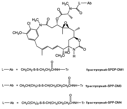
ADC по изобретению включают линкеры SMCC, а майтаназоидное лекарственное средство DM1, представлено как Ab-SMCC-DM1:The ADCs of the invention include SMCC linkers, and the maytanazoid drug DM1 is represented as Ab-SMCC-DM1:

В одном из вариантов осуществления Ab-SMCC-DM1 представляет собой трастузумаб-SMCC-DM1, где p равно 1, 2, 3 или 4 (Ab=трастузумаб, Tr, WO 2005/037992). В другом варианте осуществления ADC представляет собой трастузумаб-SIAB-DM1 (трастузумаб=Tr) со структурой:In one embodiment, Ab-SMCC-DM1 is trastuzumab-SMCC-DM1, where p is 1, 2, 3, or 4 (Ab = trastuzumab, Tr, WO 2005/037992). In another embodiment, the ADC is trastuzumab-SIAB-DM1 (trastuzumab = Tr) with the structure:

СКРИНИНГ НА КОНЪЮГАТЫ АНТИТЕЛО-ЛЕКАРСТВЕННОЕ СРЕДСТВО (ADC), ПРОТИВ АНТИГЕНОВ, АССОЦИИРОВАННЫХ С ОПУХОЛЯМИ, И РЕЦЕПТОРОВ КЛЕТОЧНОЙ ПОВЕРХНОСТИSCREENING FOR ANTIBODY MEDICINE (ADC) CONJUGATES AGAINST ANTIGENS ASSOCIATED WITH TUMORS AND CELL RECEPTORS
Для скрининга конъюгатов антитело-лекарственное средство (ADC), которые, вероятно, могут использоваться в качестве профилактических или терапевтических средств для лечения заболеваний или нарушений со сверхэкспрессией антигенов, ассоциированных с опухолями, и рецепторов на клеточной поверхности, например HER2, особенно удобно использовать трансгенных животных и клеточные линии (патент США № 6632979). Скрининг на подходящие ADC может включать в себя введение трансгенному животному ADC-кандидата в диапазоне доз и определение в различные моменты времени воздействие ADC на оцениваемое заболевание или нарушение. Альтернативно или дополнительно, если возможно, лекарственное средство можно вводить до воздействия средства, индуцирующего заболевание, или одновременно с ним. ADC-кандидат можно скринировать серийно или индивидуально или одновременно в условиях средне- или высокопроизводительного скрининга. Скорость, с которой ADC можно скринировать на эффективность для профилактического или терапевтического лечения заболеваний или нарушений, ограничена только скоростью синтеза и методологией скринирования, включая детекцию/измерение/анализ данных.For screening antibody-drug conjugates (ADCs), which are likely to be used as prophylactic or therapeutic agents for treating diseases or disorders with overexpression of antigens associated with tumors and cell surface receptors, for example HER2, it is especially convenient to use transgenic animals and cell lines (US patent No. 6632979). Screening for suitable ADCs may include administering a transgenic animal to the ADC candidate at a dose range and determining at various points in time the effect of ADC on the disease or disorder being assessed. Alternatively or additionally, if possible, the drug may be administered prior to or simultaneously with exposure to the disease inducing agent. An ADC candidate can be screened serially or individually, or at the same time under medium or high throughput screening. The rate at which ADC can be screened for efficacy for the prophylactic or therapeutic treatment of diseases or disorders is limited only by the rate of synthesis and screening methodology, including detection / measurement / analysis of data.
Один из вариантов осуществления относится к способу скринирования, который включает (a) трансплантацию клеток стабильной клеточной линии рака молочной железы животному, не являющемуся человеком, (b) введение животному, не являющемуся человеком вероятного лекарственного средства ADC и (c) определение его способности ингибировать образование опухолей из трансплантированной клеточной линии. Изобретение также относится к способу скрининга ADC-кандидатов на способность оказывать лечебный эффект на заболевание или нарушение, отличающееся сверхэкспрессией рецепторного белка, включающему (a) контактирование клеток стабильной клеточной линии рака молочной железы с возможным лекарственным средством и (b) оценку способности ADC-кандидата ингибировать рост стабильной клеточной линии.One embodiment relates to a screening method that includes (a) transplanting a stable breast cancer cell line cell into a non-human animal, (b) administering to the non-human animal a likely ADC drug, and (c) determining its ability to inhibit the formation of tumors from a transplanted cell line. The invention also relates to a method for screening ADC candidates for their ability to have a therapeutic effect on a disease or disorder characterized by overexpression of a receptor protein, comprising (a) contacting the cells of a stable breast cancer cell line with a possible drug and (b) evaluating the ability of the ADC candidate to inhibit stable cell line growth.
Один из вариантов осуществления относится к способу скрининга, который включает (a) контактирование клеток стабильной клеточной линии рака молочной железы с вероятным лекарственным средством ADC и (b) оценку способности ADC-кандидата блокировать активацию HER2 лигандом. В другом варианте осуществления оценивали способность ADC-кандидата блокировать связывание херегулина. В другом варианте осуществления оценивали способность ADC-кандидата блокировать стимулируемое лигандом фосфорилирование тирозина.One embodiment relates to a screening method that includes (a) contacting a stable breast cancer cell line with a likely ADC drug and (b) evaluating the ability of an ADC candidate to block HER2 activation by a ligand. In another embodiment, the ability of an ADC candidate to block heregulin binding was evaluated. In another embodiment, the ability of an ADC candidate to block ligand-stimulated tyrosine phosphorylation was evaluated.
Другой вариант осуществления представляет собой способ скрининга, включающий (a) контактирование клеток стабильной клеточной линии рака молочной железы с вероятным лекарственным средством ADC и (b) оценку способности ADC-кандидата индуцировать гибель клеток. В одном из вариантов осуществления оценивали способность ADC-кандидата индуцировать апоптоз.Another embodiment is a screening method comprising (a) contacting a stable breast cancer cell line with a likely ADC drug and (b) evaluating the ability of an ADC candidate to induce cell death. In one embodiment, the ability of an ADC candidate to induce apoptosis was evaluated.
Другой вариант осуществления относится к способу скрининга, который включает (a) введение вероятного лекарственного средства ADC трансгенному не являющемуся человеком млекопитающему со сверхэкспрессией, например, клетками молочной железы, природного белка человека, например HER2 или его фрагмента, где такое трансгенное животное несет стабильно интегрированную в его геном последовательность нуклеиновой кислоты, кодирующей природный белок человека или его фрагмент, с биологической активностью природного белка человека, функционально связанную с последовательностями, регулирующими транскрипцию, которые регулируют его экспрессию, и у которого развивается опухоль, например опухоль молочной железы, лечение антителами которой не эффективно или слабо эффективно, например, лечение антителом против HER2, или введение млекопитающему, не являющемуся человеком, с опухолью, трансплантированной от указанного трансгенного млекопитающего, не являющегося человеком; и (b) оценку эффекта ADC-кандидата в отношении исследуемого заболевания или нарушения. Без ограничений заболевание или нарушение может представлять собой рак со сверхэкспрессирующей HER2, такой как рак молочной железы яичника, желудка, эндометрия, слюнной железы, легкого, почки, толстой кишки, щитовидной железы, поджелудочной железы или мочевого пузыря. Раком может быть рак молочной железы, экспрессирующий HER2 по меньшей мере в количестве приблизительно 500000 копий на клетку или по меньшей мере приблизительно 2000000 копий на клетку. Например, вероятное лекарственное средство ADC можно оценивать по способности индуцировать клеточную гибель и/или апоптоз, используя способы, хорошо известные в данной области и описанные в данном документе далее.Another embodiment relates to a screening method that includes (a) administering a likely ADC medication to a transgenic non-human mammal overexpressing, for example, breast cells, a natural human protein, such as HER2, or a fragment thereof, where such a transgenic animal is stably integrated into its genome the nucleic acid sequence encoding a natural human protein or fragment thereof, with the biological activity of a natural human protein, is functionally linked associated with transcriptional regulatory sequences that regulate its expression and which develops a tumor, for example a breast tumor, whose antibody treatment is not effective or weakly effective, for example, treatment with an anti-HER2 antibody, or administration to a non-human mammal with a tumor, transplanted from the specified transgenic non-human mammal; and (b) assessing the effect of the ADC candidate with respect to the disease or disorder being studied. Without limitation, the disease or disorder may be cancer with overexpressing HER2, such as cancer of the ovary, stomach, endometrium, salivary gland, lung, kidney, colon, thyroid, pancreas, or bladder. The cancer may be breast cancer expressing HER2 in at least about 500,000 copies per cell or at least about 2,000,000 copies per cell. For example, the likely ADC drug can be assessed by its ability to induce cell death and / or apoptosis using methods well known in the art and described later in this document.
В одном из вариантов осуществления ADC-кандидат скринируют, вводя его трансгенному животному в различном диапазоне доз и оценивая физиологический ответ животного на соединения в динамике по времени. Введение может быть пероральным или посредством подходящей инъекции в зависимости от химической природы оцениваемого соединения. В некоторых случаях подходящим может быть введение соединения в сочетании с кофакторами, которые могли бы увеличить эффективность соединения. Если для скрининга соединения, которое может быть эффективно для лечения различных заболеваний, связанных со сверхэкспрессией определенных ассоциированных с опухолями антигенных белков или рецепторов клеточной поверхности, например со сверхэкспрессией HER2, применяют клеточные линии, полученные от исследуемых трансгенных животных, то тестируемые соединения в течение соответствующего времени добавляют к клеточной культуральной среде, и оценивают клеточный ответ на соединение в динамике по времени, используя соответствующие биохимические и/или гистологические способы анализа. В некоторых случаях подходящим может быть введение представляющего интерес соединения в культуральную среду в сочетании с кофакторами, которые могли бы увеличить эффективность соединения.In one embodiment, the ADC candidate is screened by administering it to a transgenic animal in a different dose range and evaluating the physiological response of the animal to the compounds over time. The administration may be oral or by suitable injection, depending on the chemical nature of the compound being evaluated. In some cases, it may be appropriate to administer the compound in combination with cofactors, which could increase the effectiveness of the compound. If for screening a compound that can be effective in treating various diseases associated with overexpression of certain tumor-associated antigenic proteins or cell surface receptors, for example, overexpression of HER2, cell lines obtained from the studied transgenic animals are used, then the tested compounds for the appropriate time added to the cell culture medium, and the cellular response to the compound is evaluated over time using the appropriate biochemistry skie and / or histological assays. In some cases, it may be appropriate to introduce the compound of interest into the culture medium in combination with cofactors that could increase the effectiveness of the compound.
Таким образом, настоящее изобретение относится к способам идентификации ADC, специфической мишенью которых является сверхэкспрессированный белок HER2 и которые связывают этот белок, присутствие которого коррелирует с патологической функцией клеток и играет роль в патогенезе клеточной пролиферации и/или дифференцировке молочной железы, что является причиной развития рака молочной железы.Thus, the present invention relates to methods for identifying ADCs whose specific target is overexpressed HER2 protein and which bind this protein, the presence of which correlates with the pathological function of cells and plays a role in the pathogenesis of cell proliferation and / or differentiation of the breast, which causes cancer the mammary gland.
Для идентификации ADC, блокирующих активацию лигандов рецептора ErbB (например, ErbB2), можно определять способность соединения блокировать связывание лиганда ErbB с клетками, эксперессирующими рецептор ErbB (ErbB2) (например, в сочетании с другим рецептором ErbB, с которым представляющий интерес рецептор ErbB образует гетероолигомер ErbB). Например, клетки, выделенные из трансгенного животного, сверхэкспрессирующие HER2 и трансфицированные с тем, чтобы экспрессировать ещё один рецептор ErbB (с которым HER2 образует гетероолигомер), можно инкубировать, то есть культивировать, с ADC, а затем подвергать воздействию меченого лиганда ErbB. Затем можно оценивать способность соединения блокировать связывание лиганда с рецептором ErbB в гетероолигомере ErbB.To identify ADCs that block the activation of ErbB receptor ligands (e.g. ErbB2), the ability of a compound to block the binding of ErbB ligand to cells expressing ErbB receptor (ErbB2) can be determined (for example, in combination with another ErbB receptor with which the ErbB receptor of interest forms a hetero oligomer ErbB). For example, cells isolated from a transgenic animal, overexpressing HER2 and transfected in order to express another ErbB receptor (with which HER2 forms a hetero-oligomer), can be incubated, i.e. cultured, with ADC and then exposed to the labeled ErbB ligand. You can then evaluate the ability of the compound to block the binding of the ligand to the ErbB receptor in the ErbB hetero-oligomer.
Например, ингибирующая активность ADC-кандидата в отношении связывания херегулина (HRG) с клеточными линиями рака молочной железы, сверхэкспрессирующими HER2 и полученными в данном документе из млекопитающих, не являющихся человеком (например, мышей), можно проводить с помощью монослойных культур на льду в 24-луночном планшете. Моноклональные антитела против ErbB2 добавляют к каждой лунке и инкубируют в течение 30 минут. Затем добавляют меченный 125I rHRGβ1177-224 (25000 имп./мин) и инкубацию продолжают в течение от 4 до 16 часов. Получают кривые доза-ответ и для представляющего интерес соединения рассчитывают значение IC50.For example, the inhibitory activity of an ADC candidate for binding of heregulin (HRG) to breast cancer cell lines overexpressing HER2 and derived from non-human mammals (e.g., mice) can be performed using monolayer ice cultures in 24 well tablet. Monoclonal antibodies against ErbB2 are added to each well and incubated for 30 minutes. Then labeled 125 I rHRGβ1 177-224 (25000 cpm) was added and incubation was continued for 4 to 16 hours. Dose response curves are obtained, and an IC 50 value is calculated for the compound of interest.
Альтернативно или дополнительно, можно оценивать способность ADC блокировать лиганд-стимулируемое фосфорилирование тирозина рецептора ErbB, находящегося в гетероолигомере ErbB. Например, клеточные линии, полученные из трансгенных животных, как описано в данном документе, можно инкубировать с тестируемым ADC и затем оценивать на активность лиганд-зависимого фосфорилирования тирозина ErbB с применением моноклонального антитела против фосфотирозина (которое, необязательно, конъюгировано с визуализируемой меткой). Также для определения активации рецептора ErbB и блокирования этой активности соединением можно использовать анализ активации киназного рецептора, описанный в патенте США № 5766863.Alternatively or additionally, the ability of ADC to block ligand-stimulated tyrosine phosphorylation of the ErbB receptor located in the ErbB hetero-oligomer can be evaluated. For example, cell lines derived from transgenic animals, as described herein, can be incubated with the test ADC and then evaluated for ligand-dependent ErbB tyrosine phosphorylation using a monoclonal anti-phosphotyrosine antibody (which is optionally conjugated to a visualized label). Also, the kinase receptor activation assay described in US Pat. No. 5,766,863 can be used to determine ErbB receptor activation and block this activity by the compound.
В одном из вариантов осуществления можно проводить скрининг на ADC, который ингибирует стимуляцию фосфорилирования тирозина p180 в клетках MCF7 с помощью HRG, по существу, так, как описано ниже. Например, клеточную линию, полученную от трансгенного по HER2 животного помещают в 24-луночные планшеты и к каждой лунке добавляют соединение и инкубируют 30 минут при комнатной температуре; затем к каждой лунке добавляют rHRGβ1177-244 в конечной концентрации 0,2 нМ и инкубацию продолжают в течение приблизительно 8 минут. Из каждой лунки отсасывают среду и реакцию останавливают, добавляя 100 мкл буфера для образцов с SDS (5% SDS, 25 мМ DTT, и 25 мМ Tris-HCl, pH 6,8). Каждый образец (25 мкл) подвергают электрофорезу на градиентном геле 4-12% (Novex), и затем электрофоретически переносят на поливинилидендифторидную мембрану. Получают иммуноблоты с антителами против фосфотирозина (при концентрации 1 мкг/мл) и посредством отражательной денситометрии количественно определяют интенсивность преобладающей реагирующей полосы с Mr=180000. Альтернативным способом оценки ингибирования фосфорилирования рецептора является анализ KIRA (активация киназного рецептора) (Sadick et al. (1998) Jour. of Pharm. and Biomed. Anal. 1-9). В качестве положительного контроля в этом анализе можно использовать некоторые из хорошо изученных моноклональных антител против HER2, которые, как известно, ингибируют стимуляцию фосфорилирования тирозина p180 посредством HRG,. Получают кривую доза-ответ для ингибирования стимуляции фосфорилирования тирозина p180 посредством HRG, определенную отражательной денситометрией и для представляющего интерес соединения рассчитывают IC50.In one embodiment, an ADC screening can be performed that inhibits the stimulation of tyrosine p180 phosphorylation in MCF7 cells using HRG, essentially as described below. For example, a cell line derived from an HER2 transgenic animal is placed in 24-well plates and a compound is added to each well and incubated for 30 minutes at room temperature; then rHRGβ1 177-244 was added to each well at a final concentration of 0.2 nM and incubation was continued for approximately 8 minutes. Medium was aspirated from each well and the reaction was stopped by adding 100 μl of sample buffer with SDS (5% SDS, 25 mM DTT, and 25 mM Tris-HCl, pH 6.8). Each sample (25 μl) was subjected to 4-12% gradient gel electrophoresis (Novex), and then transferred electrophoretically to a polyvinylidene difluoride membrane. Immunoblots are obtained with antibodies against phosphotyrosine (at a concentration of 1 μg / ml) and the intensity of the predominant reacting band with M r = 180,000 is quantified by reflective densitometry. An alternative way to evaluate the inhibition of receptor phosphorylation is by KIRA analysis (kinase receptor activation) (Sadick et al. (1998) Jour. Of Pharm. And Biomed. Anal. 1-9). As a positive control, some of the well-studied anti-HER2 monoclonal antibodies that are known to inhibit tyrosine p180 phosphorylation by HRG can be used as a positive control. A dose-response curve is obtained to inhibit the stimulation of tyrosine p180 phosphorylation by HRG, determined by reflectance densitometry, and an IC 50 is calculated for the compound of interest.
Также можно оценивать ингибирующие рост эффекты тестируемого ADC на клеточные линии, полученные от трансгенного по HER2 животного (Schaefer et al. (1997) Oncogene 15:1385-1394). В соответствии с этим анализом клетки обрабатывают тестируемым соединением в различных концентрациях в течение 4 суток и окрашивают кристаллическим фиолетовым или окислительно-восстановительным красителем Alamar Blue. Инкубация с соединением показывает ингибирующий рост эффект на эту клеточную линию подобно воздействию, демонстрируемому моноклональным антителом 2C4 на клетки MDA-MB-175 (Schaefer et al., выше). В дополнительном варианте осуществления экзогенный HRG изменяет значительно данное ингибирование.It is also possible to evaluate the growth inhibitory effects of the test ADC on cell lines obtained from an HER2 transgenic animal (Schaefer et al. (1997) Oncogene 15: 1385-1394). According to this assay, cells are treated with the test compound at various concentrations for 4 days and stained with Alamar Blue, a crystalline violet or redox dye. Incubation with the compound shows a growth inhibitory effect on this cell line, similar to the effect exhibited by 2C4 monoclonal antibody on MDA-MB-175 cells (Schaefer et al., Supra). In a further embodiment, exogenous HRG significantly alters this inhibition.
Для идентификации ингибирующих рост соединений ADC, которые специфически направлены против HER2, можно проводить скрининг на ADC, которое ингибирует рост сверхэкспрессирующих HER2 раковых клеток, полученных от трансгенных животных (патент США № 5677171). В соответствии с этим анализом сверхэкспрессирующие HER2 клетки растят в среде смеси F12 и DMEM 1:1, дополненной 10% эмбриональной телячьей сывороткой, глутамином и пенициллином со стрептомицином. Эти клетки в количестве 20000 клеток вносят в 35-мм чашку для клеточной культуры (2 мл/35 мм чашку) и добавляют тестируемое соединение в разных концентрациях. Через шесть суток с помощью электронного счетчика клеток COULTER™ подсчитывают количество клеток по сравнению с необработанными клетками. Те ADC, которые ингибируют клеточный рост приблизительно на 20-100% или приблизительно на 50-100% могут быть выбраны в качестве ингибирующих рост соединений.To identify growth inhibitory ADC compounds that are specifically directed against HER2, it is possible to screen for ADC, which inhibits the growth of overexpressing HER2 cancer cells obtained from transgenic animals (US patent No. 5677171). According to this analysis, overexpressing HER2 cells are grown in a 1: 1 F12 and DMEM mixture supplemented with 10% fetal calf serum, glutamine and penicillin with streptomycin. These cells in an amount of 20,000 cells are added to a 35 mm cell culture dish (2 ml / 35 mm dish) and the test compound is added in different concentrations. After six days, the number of cells compared to untreated cells was counted using a COULTER ™ electronic cell counter. Those ADCs that inhibit cell growth by approximately 20-100% or approximately 50-100% can be selected as growth inhibiting compounds.
Для отбора ADC, индуцирующих клеточную гибель, можно оценивать нарушение целостности мембраны, на что указывает, например, захват PI, трипанового синего или 7AAD, по срвнению с контролем. Для анализа захвата PI используют клетки, выделенные из ткани опухоли молочной железы трансгенного животного. В соответствии с этим анализом клетки культивируют в модифицированной Дульбекко среде Игла (D-MEM): F-12 Хама (50:50), дополненной 10% ЭТС, инактивированной нагреванием, (Hyclone) и 2 мМ L-глутамином. Таким образом, анализ проводят в отсутствие комплемента и иммунных эффекторных клеток. Клетки высевают при плотности 3 × 10⁶ на чашку в 100 × 20 мм чашки и оставляют на ночь для прикрепления. Затем среду удаляют и заменяют не свежую среду или свежую среду, содержащую различные концентрации соединения. Клетки инкубируют в течение 3 суток. После каждой обработки монослои отмывают PBS и проводят трипсинизацию для открепления клеток. Затем клетки центрифугируют при 1200 об/мин в течение 5 минут при 4°C, осадок ресуспендируют в 3 мл охлажденного буфера, связывающего Ca2+ (10 мМ Hepes, pH 7,4, 140 мМ NaCl, 2,5 мМ CaCl2) и отбирают аликвоты в покрытые 35 мм фильтром 12 × 75 мм пробирки (1 мл на пробирку, 3 пробирки на обработанную группу) для удаления клеточных скоплений. Затем в пробирки вносят PI (10 мкг/мл). Затем образцы анализируют с помощью проточного цитометра FACSCAN™ и программного обеспечения FACSCONVERT™ CellQuest (Becton Dickinson). Те соединения, которые индуцируют статистически значимые уровни клеточной гибели, которые определяют посредством захвата PI, отбирают в качестве индуцирующих клеточную гибель соединений.For the selection of cell death inducing ADCs, a violation of membrane integrity can be assessed, as indicated, for example, by uptake of PI, trypan blue or 7AAD compared to control. For analysis of PI capture, cells isolated from the tissue of a breast tumor of a transgenic animal are used. According to this assay, cells were cultured in Dulbecco's modified Eagle's medium (D-MEM): F-12 Hama (50:50), supplemented with 10% heat inactivated ETS (Hyclone) and 2 mM L-glutamine. Thus, the analysis is carried out in the absence of complement and immune effector cells. Cells are seeded at a density of 3 × 10 6 per cup in 100 × 20 mm plates and left overnight for attachment. The medium is then removed and non-fresh medium or fresh medium containing various concentrations of the compound is replaced. Cells are incubated for 3 days. After each treatment, the monolayers are washed with PBS and trypsinized to detach cells. Then the cells are centrifuged at 1200 rpm for 5 minutes at 4 ° C, the pellet is resuspended in 3 ml of chilled Ca 2+ binding buffer (10 mM Hepes, pH 7.4, 140 mM NaCl, 2.5 mM CaCl 2 ) and aliquots are taken into 12 × 75 mm tubes coated with a 35 mm filter (1 ml per tube, 3 tubes per treated group) to remove cell accumulations. Then, PI (10 μg / ml) was added to the tubes. Samples were then analyzed using a FACSCAN ™ flow cytometer and FACSCONVERT ™ CellQuest software (Becton Dickinson). Those compounds that induce statistically significant levels of cell death that are determined by uptake of PI are selected as compounds that induce cell death.
Для отбора соединений, индуцирующих апоптоз, проводят анализ связывания аннексина с применением клеток, полученных из ткани опухоли молочной железы трансгенного животного. Клетки культивируют и высевают в чашки, как описано в предыдущем параграфе. Затем среду удаляют и заменяют на свежую среду или среду, содержащую 10 мкг/мл конъюгата антитело-лекарственное средство (ADC). После инкубации в течение трех суток монослои отмывают PBS и проводят трипсинизацию. Для открепления клеток затем клетки центрифугируют и ресуспендируют в связывающем Ca2+ буфере и отбирают аликвоты в пробирки, как описано выше для анализа клеточной гибели. Затем в пробирки добавляют меченный аннексин (например, аннексин V-FITC) (1 мкг/мл). Затем образцы анализируют с помощью проточного цитометра FACSCAN™ и программного обеспечения FACSCONVERT™ CellQuest (Becton Dickinson). Те соединения, которые индуцируют статистически значимые уровни связывания аннексина по сравнению с контролем, выбирают в качестве индуцирующих апоптоз соединений.To select apoptosis inducing compounds, annexin binding analysis is performed using cells derived from the tissue of a breast tumor of a transgenic animal. Cells are cultured and plated in plates as described in the previous paragraph. The medium is then removed and replaced with fresh medium or medium containing 10 μg / ml antibody-drug conjugate (ADC). After incubation for three days, the monolayers are washed with PBS and trypsinized. To detach cells, the cells are then centrifuged and resuspended in Ca 2+ binding buffer and aliquots are taken into tubes as described above for cell death analysis. Then labeled annexin (e.g., annexin V-FITC) (1 μg / ml) is added to the tubes. Samples were then analyzed using a FACSCAN ™ flow cytometer and FACSCONVERT ™ CellQuest software (Becton Dickinson). Those compounds that induce statistically significant levels of annexin binding compared to control are selected as apoptosis inducing compounds.
АНАЛИЗЫ ПРОЛИФЕРАЦИИ КЛЕТОК IN VITRO ANALYSIS OF CELL PROLIFERATION IN VITRO
Как правило, цитотоксическую или цитостатическую активность конъюгата антитело-лекарственное средство (ADC) измеряют следующим образом: на клетки млекопитающих с антигенами, ассоциированными с опухолями, или с рецепторными белками воздействуют антителом ADC в клеточной культуральной среде; культивируют клетки в течение приблизительно от 6 часов до приблизительно 5 суток; измеряют жизнеспособность клеток. Клеточные анализы in vitro применяют для измерения жизнеспособности, то есть пролиферации (IC50), цитотоксичности (EC50) и индукции апоптоза (активация каспазы) ADC.Typically, the cytotoxic or cytostatic activity of an antibody-drug conjugate (ADC) is measured as follows: mammalian cells with antigens associated with tumors or receptor proteins are exposed to ADC antibody in a cell culture medium; cells are cultured for about 6 hours to about 5 days; measure cell viability. In vitro cell assays are used to measure viability, i.e., proliferation (IC 50 ), cytotoxicity (EC 50 ), and apoptosis induction (caspase activation) of ADC.
Эффективность конъюгатов антитело-лекарственное средство in vitro измеряли с помощью анализа пролиферации клеток (фиг. 1-4). Анализ CellTiter-Glo® Luminescent Cell Viability Assay представляет собой коммерчески доступный (Promega Corp., Madison, WI) способ гомогенного анализа на основе рекомбинантной экспрессии люциферазы Coleoptera (патент США № 5583024; патент США № 5674713; патент США № 5700670). В этом анализе пролиферации клеток определяют количество живых клеток в культуре на основании количественного определения присутствующей АТФ, индикатора метаболически активных клеток (Crouch et al. (1993) J. Immunol. Meth. 160:81-88; патент США № 6602677). Анализ CellTiter-Glo® Assay проводили в 96-луночном планшете, изменяя его для автоматизированного высокопроизводительного скрининга (HTS) (Cree et al. (1995) AntiCancer Drugs 6:398-404). Процедура гомогенного анализа включает добавление одного реагента (CellTiter-Glo® Reagent) непосредственно к клеткам, культивируемым в среде, дополненной сывороткой. Отмывка клеток, удаление среды и ряд стадий пипетирования не требуется. Система обнаруживает минимально 15 клеток/лунку в 384-луночном планшете через 10 минут после добавления реагента и смешивания.The efficacy of antibody-drug conjugates in vitro was measured using cell proliferation analysis (FIGS. 1-4). CellTiter-Glo® Luminescent Cell Viability Assay Assay is a commercially available (Promega Corp., Madison, WI) homogenous assay based on recombinant expression of Coleoptera luciferase (US Patent No. 5583024; US Patent No. 5674713; US Patent No. 5700670). In this cell proliferation assay, the number of living cells in a culture is determined based on the quantification of the present ATP, an indicator of metabolically active cells (Crouch et al. (1993) J. Immunol. Meth. 160: 81-88; US Patent No. 6602677). CellTiter-Glo® Assay analysis was performed in a 96-well plate, changing it for automated high throughput screening (HTS) (Cree et al. (1995) AntiCancer Drugs 6: 398-404). A homogeneous assay procedure involves the addition of a single reagent (CellTiter-Glo® Reagent) directly to cells cultured in serum supplemented medium. Cell washing, medium removal and a series of pipetting steps are not required. The system detects a minimum of 15 cells / well in a 384-
Гомогенная форма «добавление-смешивание-измерение» приводит к лизису клеток и генерации люминесцентного сигнала, пропорционального количеству присутствующей АТФ. Количество АТФ прямо пропорционально количеству присутствующих в культуре клеток. В анализе CellTiter-Glo® Assay при люциферазной реакции образуется люминесцентный сигнал «типа свечения», который обладает временем полужизни, как правило, более пяти часов в зависимости от типа клеток и используемой среды. Живые клетки выражают в относительных люминесцентных единицах (RLU). Субстрат, люциферин жука, подвергают окислительному декарбоксилированию посредством рекомбинантной люциферазы светляка, что сопровождается преобразованием АТФ в АМФ и образованием фотонов. Повышенное время полужизни устраняет необходимость в использовании инъекторов реагента и обеспечивает гибкость в отношении непрерывного или группового способа обработки нескольких планшетов. Этот анализ пролиферации клеток можно использовать в различных многолуночных форматах, например 96- или 384-луночном формате. Данные можно записывать посредством люминометра или устройства получения изображений с камерой CCD. Результат люминесценции представляют в виде относительных световых единиц (RLU), измеренных в течение времени.The homogeneous addition-mixing-measurement form leads to cell lysis and generation of a luminescent signal proportional to the amount of ATP present. The amount of ATP is directly proportional to the number of cells present in the culture. In the CellTiter-Glo® Assay assay, a luminescent “luminescence type” signal is generated during the luciferase reaction, which has a half-life, typically more than five hours, depending on the type of cells and the medium used. Living cells are expressed in relative luminescent units (RLU). The substrate, beetle luciferin, is subjected to oxidative decarboxylation by recombinant firefly luciferase, which is accompanied by the conversion of ATP to AMP and the formation of photons. The increased half-life eliminates the need for reagent injectors and provides flexibility for continuous or batch processing of multiple tablets. This cell proliferation assay can be used in various multi-well formats, for example, 96- or 384-well formats. Data can be recorded using a luminometer or an imaging device with a CCD camera. The result of luminescence is presented in the form of relative light units (RLU), measured over time.

Антипролиферативные эффекты трех конъюгатов антитело-лекарственное средство измеряли с помощью клеточной пролиферации, указанного выше анализа гибели клеток in vitro относительно четырех различных клеточных линий опухоли молочной железы (фиг.1-4). На фиг.1 показаны измерения результативности при увеличивающихся концентрациях трастузумаба-SPP-DM1, трастузумаба-SPDP-DM1 и трастузумаба-SMCC-DM1 через 3 суток обработки клеток опухоли молочной железы SK-BR-3 (HER2 3+). На фиг.2 показаны измерения результативности при увеличивающихся концентрациях трастузумаба-SPP-DM1, трастузумаба-SPDP-DM1 и трастузумаба-SMCC-DM1 через 3 суток обработки клеток опухоли молочной железы BT-474 (HER2 3+). На фиг.3 показаны измерения результативности при увеличивающихся концентрациях трастузумаба-SPP-DM1, трастузумаба-SPDP-DM1 и трастузумаба-SMCC-DM1 через 3 суток обработки клеток опухоли молочной железы MCF7 (низкое содержание HER2). На фиг.4 показаны измерения результативности при увеличивающихся концентрациях трастузумаба-SPP-DM1, трастузумаба-SPDP-DM1 и трастузумаба-SMCC-DM1 через 3 суток обработки клеток опухоли молочной железы MDA-MB-468 (отсутствие HER2).The antiproliferative effects of the three antibody-drug conjugates were measured using cell proliferation, the above analysis of in vitro cell death relative to four different breast tumor cell lines (FIGS. 1-4). Figure 1 shows the performance measurements with increasing concentrations of trastuzumab-SPP-DM1, trastuzumab-SPDP-DM1 and trastuzumab-SMCC-DM1 after 3 days of treatment of SK-BR-3 (
Значения IC50 определяли для SK-BR-3 и BT-474, которые, как известно, сверхэкспрессируют белок рецептора HER2. Большинство конъюгатов трастузумаб-SPP-DM1 с 2,8 DM1 на молекулу трастузумаба (лекарственное средство/Ab) давало среднее значение IC50 в размере 14,4 мкг/мл с интервалом от 9,1 до 22,3 мкг/мл для 6 экспериментов по сравнению с клетками SK-BR-3, и среднее значение IC50 в размере 51,7 мкг/мл с интервалом от 28,7 до 63,1 мкг/мл для 4 экспериментов по сравнению с клетками BT-474. Большинство конъюгатов трастузумаб-SMCC-DM1 с 2,7 DM1 на молекулу трастузумаба (лекарственное средство/Ab) давало среднее значение IC50 в размере 15,2 мкг/мл с интервалом от 12,6 до 18,8 мкг/мл для 4 экспериментов по сравнению с клетками SK-BR-3 и среднее значение IC50 в размере 94,9 мкг/мл с интервалом от 75,2 до 114,6 мкг/мл для 2 экспериментов по сравнению с клетками BT-474. Конъюгаты были неактивны в отношении клеток MCF7 и MDA-MB-468, у которых отсутствует сверхэкспрессия HER2.IC 50 values were determined for SK-BR-3 and BT-474, which are known to overexpress HER2 receptor protein. Most conjugates of trastuzumab-SPP-DM1 with 2.8 DM1 per trastuzumab molecule (drug / Ab) gave an average IC 50 of 14.4 μg / ml with an interval of 9.1 to 22.3 μg / ml for 6 experiments compared with SK-BR-3 cells, and an average IC 50 value of 51.7 μg / ml with an interval of 28.7 to 63.1 μg / ml for 4 experiments compared to BT-474 cells. Most conjugates of trastuzumab-SMCC-DM1 with 2.7 DM1 per trastuzumab molecule (drug / Ab) gave an average IC 50 of 15.2 μg / ml with an interval of 12.6 to 18.8 μg / ml for 4 experiments compared with SK-BR-3 cells and an average IC 50 value of 94.9 μg / ml with an interval of 75.2 to 114.6 μg / ml for 2 experiments compared to BT-474 cells. The conjugates were inactive against MCF7 and MDA-MB-468 cells that lack overexpression of HER2.
Конъюгат антитела против CD19-SMCC-DM1 продемонстрировал эффективное уничтожение клеток in vitro в отношении клеток Raji (IC50=<0,25 мкг/мл), при этом “голое” антитело и контрольный ADC, трастузумаб-SMCC-DM1, не показали эффекта. Конъюгаты антитела против CD79a-SMCC-DM1 и антитела к CD79b-SMCC-DM1 продемонстрировали эффективное уничтожение клеток in vitro в отношении клеток Ramos (IC50=<0,25 мкг/мл), при этом “голое” антитело и контрольный ADC, трастузумаб-SMCC-DM1 не показало эффекта.The anti-CD19-SMCC-DM1 antibody conjugate demonstrated effective in vitro killing of cells against Raji cells (IC 50 = <0.25 μg / ml), while the “naked” antibody and control ADC, trastuzumab-SMCC-DM1, showed no effect . Antibody anti-CD79a-SMCC-DM1 conjugates and anti-CD79b-SMCC-DM1 antibodies demonstrated in vitro effective killing of cells against Ramos cells (IC 50 = <0.25 μg / ml), with the “naked” antibody and control ADC, trastuzumab -SMCC-DM1 showed no effect.
На фиг.17 показан анализ пролиферации клеток in vitro (пример 5) с клетками HT1080EphB2 (C8), обработанными конъюгатами антитела против EphB2R 2H9-лекарственное средство: 2H9-SPP-DM1 (IC50 80 нг/мл) и 2H9-SMCC-DM1 (IC50 50 нг/мл).17 shows an in vitro cell proliferation assay (Example 5) with HT1080EphB2 (C8) cells treated with anti-EphB2R antibody conjugates 2H9 drug: 2H9-SPP-DM1 (IC 50 80 ng / ml) and 2H9-SMCC-DM1 (
СЫВОРОТОЧНЫЙ КЛИРЕНС И СТАБИЛЬНОСТЬ У МЫШЕЙ IN VIVO SERUM CLEARANCE AND STABILITY IN MICE IN VIVO
Сывороточный клиренс и стабильность ADC исследовали у “голых” мышей без воздействия (без опухолей, привнесенных посредством экзогенных трансплантатов). На фиг.5 показан сывороточный клиренс трастузумаба-SMCC-DM1 по сравнению с трастузумабом-SPP-DM1 у “голых” мышей с врожденным отсутствием NK-клеток без опухолей, с измерением сывороточной концентрации конъюгата и общего антитела в шести временных точках в течение 7 суток. Различие в количестве общего антитела и ADC указывает на расщепление линкера и отделение антитела от его части DM1. Как показано, SMCC-связанный ADC оставались интактными in vivo более длительный срок, чем связанные SPP конъюгаты, на что указывает более высокая сывороточная концентрация конъюгированного антитела через семь суток.Serum clearance and ADC stability were studied in nude mice without exposure (without tumors introduced through exogenous grafts). Figure 5 shows the serum clearance of trastuzumab-SMCC-DM1 compared with trastuzumab-SPP-DM1 in nude mice with congenital absence of NK cells without tumors, with a measurement of serum concentration of the conjugate and total antibody at six time points for 7 days . The difference in the amount of total antibody and ADC indicates the cleavage of the linker and the separation of the antibody from its part DM1. As shown, SMCC-bound ADCs remained intact in vivo for longer periods than SPP-bound conjugates, as indicated by a higher serum concentration of conjugated antibody after seven days.
На фиг.6 показана стабильность конъюгатов в динамике по времени у «голых» мышей без опухолей: трастузумаб-SPDP-DM1, трастузумаб-SPP-DM1, трастузумаб-SPP-DM3, трастузумаб-SPP-DM4 и трастузумаб-SMCC-DM1, с измерением сывороточной концентрации в шести временных точках в течение 7 суток. ADC с линкером SMCC были более стабильны in vivo, чем конъюгаты, связанные SPP или SPDP, хотя трастузумаб-SMCC-DM1 обладал сходной стабильностью, как и наиболее защищенный дисульфидный конъюгат, трастузумаб-SPP-DM4.Figure 6 shows the stability of the conjugates in time over time in "nude" tumor-free mice: trastuzumab-SPDP-DM1, trastuzumab-SPP-DM1, trastuzumab-SPP-DM3, trastuzumab-SPP-DM4 and trastuzumab-SMCC-DM1, c measuring serum concentration at six time points for 7 days. ADCs with SMCC linker were more stable in vivo than conjugates bound by SPP or SPDP, although trastuzumab-SMCC-DM1 had similar stability as the most protected disulfide conjugate, trastuzumab-SPP-DM4.
Показанные на фиг.5 и 6 эксперименты проводили на «голых» мышах без опухолей. Однако связанные SMCC конъюгаты у «голых» мышей с опухолями показали сходную увеличенную стабильность по сравнению со SPP-связанными конъюгатами. Как показано на фиг.7, в виде конъюгата оставалось приблизительно 72% исходного трастузумаб-SMCC-DM1 через 7 суток после обработки, тогда как ~10% исходного трастузумаб-SPP-DM1 оставались в виде конъюгата через 7 суток после обработки, что демонстрирует повышенную стабильность ферментативно не расщепляемых конъюгатов трастузумаб-SMCC-DM1 у «голых» мышей с опухолями.The experiments shown in FIGS. 5 and 6 were performed on “nude” mice without tumors. However linked SMCC conjugates in "naked" tumor-bearing mice showed similar increased stability compared to SPP-linked conjugates. As shown in Fig. 7, approximately 72% of the original trastuzumab-SMCC-DM1 remained in the form of a
СЫВОРОТОЧНЫЙ КЛИРЕНС И СТАБИЛЬНОСТЬ У КРЫС IN VIVO SERUM CLEARANCE AND STABILITY IN RATS IN VIVO
На фиг.8 и 9 показана сравнительная стабильность и профили клиренса ADC с дисульфидным линкером (трастузумаб-SPP-DM1 и ADC с линкером, не являющимся дисульфидным (трастузумаб-SMCC-DM1) у крыс. Параметры исследования включали в себя:Figures 8 and 9 show the comparative stability and clearance profiles of ADC with a disulfide linker (trastuzumab-SPP-DM1 and ADC with a non-disulfide linker (trastuzumab-SMCC-DM1) in rats. The study parameters included:
ADC с линкером, не являющимся дисульфидным, трастузумаб-SMCC-DM1 (фиг.9) продемонстрировал лучшую стабильность в сыворотке крыс, чем ADC с дисульфидным линкером, трастузумаб-SPP-DM1 (фиг.8).ADC with a non-disulfide linker, trastuzumab-SMCC-DM1 (FIG. 9) showed better serum stability in rats than ADC with a disulfide linker, trastuzumab-SPP-DM1 (FIG. 8).
ЭФФЕКТИВНОСТЬ IN VIVO EFFICIENCY IN VIVO
Эффективность конъюгатов антитело-лекарственное средство по изобретению можно измерять in vivo, пересаживая аллотрансплантаты или ксенотрансплантаты из клеток злокачественных опухолей у грызунов и обрабатывая опухоли ADC. Результаты различались в зависимости от клеточной линии, специфичности связывания антитела ADC с рецепторами, присутствующими на раковых клетках, режима дозирования и других факторов. Эффективность in vivo ADC против HER2 измеряли посредством мышиной модели трансгенного эксплантата с высокой экспрессией HER2. Аллотрансплантат происходил от трансгенной мыши Fo5 mmtv, терапия герцептином которой была неэффективной или слабоэффективной. Субъектам однократно вводили ADC и проводили наблюдение в течение 3-6 недель для измерения времени удвоения опухоли, логарифма клеточной гибели и уменьшения опухоли. Последовательно проводили эксперименты доза-ответ и множественного дозирования.The efficacy of the antibody-drug conjugates of the invention can be measured in vivo by transplanting allografts or xenografts from cancer cells in rodents and treating ADC tumors. The results varied depending on the cell line, the specificity of binding of the ADC antibody to receptors present on cancer cells, the dosage regimen, and other factors. In vivo efficacy of ADC against HER2 was measured using a mouse model of a transgenic explant with high HER2 expression. The allograft came from a Fo5 mmtv transgenic mouse, whose herceptin therapy was ineffective or poorly effective. Subjects were once administered ADC and monitored for 3-6 weeks to measure tumor doubling time, the logarithm of cell death, and tumor reduction. Dose-response and multiple-dose experiments were performed sequentially.
Опухоли легко возникают у трансгенных мышей, экспрессирующих форму neu, активированную мутациями, гомолог HER2 крысы, но HER2, сверхэкспессируемый при раке молочной железы, не является мутантным и образование опухоли у мышей, сверхэкспрессирующих немутантный HER2, менее устойчиво (Webster et al. (1994) Semin. Cancer Biol. 5:69-76).Tumors readily arise in transgenic mice expressing a mutation-activated neu form, the rat HER2 homolog, but HER2 overexpressed in breast cancer is not mutant, and tumor formation in mice overexpressing non-mutant HER2 is less stable (Webster et al. (1994) Semin. Cancer Biol. 5: 69-76).
Для улучшения образования опухоли с немутированным HER2, трансгенных мышей получали с помощью плазмиды с кДНК HER2, в которой был удален вышерасположенный ATG, находящийся в направлении 3'-5', для предотвращения трансляции с таких вышерасположенных кодонов ATG, которая могла бы уменьшить частоту инициации трансляции с аутентичных кодонов инициации HER2, расположенных в направлении 5'-3' (например, см. Child et al. (1999) J. Biol. Chem. 274: 24335-24341). Кроме того, на 5'-конце был добавлен химерный интрон, что также увеличивает уровень экспрессии, как было описано ранее (Neuberger and Williams (1988) Nucleic Acids Res. 16: 6713; Buchman and Berg (1988) Mol. Cell. Biol. 8:4395; Brinster et al. (1988) Proc. Natl. Acad. Sci. USA 85:836). Химерный интрон получали из вектора Promega, экспрессирующего вектора млекопитающих pCI-neo (п.н. 890-1022). кДНК 3'-конца фланкировали экзонами человеческого гормона роста 4 и 5 и последовательностями полиаденилирования. Кроме того, использовали мышей FVB, так как эта линия более чувствительна к развитию опухолей. Для обеспечения ткансспецифической экспрессии HER2 в молочной железе использовали промотор MMTV-LTR. Для увеличения чувствительности к образованию опухолей млекопитающих держали на диете ATN 76A (Rao et al. (1997) Breast Cancer Res. and Treatment 45:149-158).To improve tumor formation with non-mutated HER2, transgenic mice were obtained using a plasmid with HER2 cDNA in which the upstream ATG located in the 3'-5 'direction was removed to prevent translation from such upstream ATG codons, which could reduce the translation initiation frequency from authentic HER2 initiation codons located in the 5'-3 'direction (e.g. see Child et al. (1999) J. Biol. Chem. 274: 24335-24341). In addition, a chimeric intron was added at the 5'-end, which also increases expression, as previously described (Neuberger and Williams (1988) Nucleic Acids Res. 16: 6713; Buchman and Berg (1988) Mol. Cell. Biol. 8: 4395; Brinster et al. (1988) Proc. Natl. Acad. Sci. USA 85: 836). The chimeric intron was obtained from the Promega vector expressing the mammalian vector pCI-neo (bp 890-1022). 3'-end cDNAs were flanked by exons of
На фиг.10-13 показано, что ADC обладают сильной противоопухолевой активностью у аллотрансплантатах HER2-позитивных опухолей (Fo5), которые исходно возникали у трансгенных мышей MMTV-HER2. Только антитело (например, трастузумаб) в этой модели не обладало значимой противоопухолевой активностью (Erickson et al., патент США № 6632979). Как проиллюстрировано на фиг.10 и 11, рост опухолей при обработке ADC по сравнению с контрольным (носитель) уровнем роста замедлялся. Рост опухоли наиболее замедлялся при обработке конъюгатами трастузумаб-SMCC-DM1 и трастузумаб-SIAB-DM1. Как показано на фиг.10, 12 и 13, конъюгат трастузумаб-SMCC-DM1 больше замедлял рост опухоли, чем конъюгаты с линкерами SPP линкеры, то есть был более эффективным, что было измерено по времени удваивания опухолей у «голых» мышей или по логарифму гибели клеток, соответствующему показанным измерениям времени удваивания (фиг.13).Figure 10-13 shows that ADC have strong antitumor activity in allografts of HER2-positive tumors (Fo5), which originally occurred in transgenic MMTV-HER2 mice. Only an antibody (e.g., trastuzumab) in this model did not have significant antitumor activity (Erickson et al., US Patent No. 6,632979). As illustrated in FIGS. 10 and 11, tumor growth during ADC treatment was slowed down compared to a control (carrier) growth level. Tumor growth was most slowed when treated with trastuzumab-SMCC-DM1 and trastuzumab-SIAB-DM1 conjugates. As shown in figures 10, 12 and 13, the conjugate trastuzumab-SMCC-DM1 slowed down tumor growth more than conjugates with linkers SPP linkers, that is, it was more effective, which was measured by the time of doubling of tumors in nude mice or by logarithm cell death corresponding to the shown measurements of doubling time (Fig.13).
Эффективность in vivo ADC с антителом против CD22 измеряли на модели ксенотрансплантата опухоли мыши. Группам из восьми мышей SCID с 20 миллионами ксенотрансплантированных опухолевых клеток Bjab-luc (экспрессирующие люциферазу клетки BjaB) на мышь на 1-е сутки однократно вводили дозу (исключения отмечали) конъюгата антитело против CD22-лекарственное средство или “голого” антитела (пример 8).The efficacy of in vivo ADC with anti-CD22 antibody was measured in a mouse tumor xenograft model. Groups of eight SCID mice with 20 million xenograft Bjab-luc tumor cells (luciferase-expressing BjaB cells) per mouse on the 1st day were given a single dose (exceptions noted) of the anti-CD22 antibody drug conjugate or naked antibody (Example 8) .
Измеряли время удваивания размера опухоли (MTD, среднее время удваивания опухоли). Три “голых” антитела против CD22, по существу, продемонстрировали отсутствие эффективного действия по сравнению с неспецифически связанным ADC (трастузумаб-SMCC-DM1). Все соответствующие конъюгаты продемонстрировали эффект значительного замедления роста опухоли. Действие многократного введения дозы устанавливали с помощью RFB4-SMCC-DM1, где MTD для мышей с однократным введением дозы составляло 18 суток, тогда как MTD для мышей с трехкратным введением дозы, на 1, 7 и 14 сутки составляло 55 суток. Полное исчезновение опухоли происходило у всех 8 мышей в группе с трехкратным введением дозы. Другие конъюгаты антитело против CD22-SMCC-DM1, с антителами 12F7, 9A8, 8C9, 8G10, 3F11, 10D2, 6C9, 14D1 и 11H10, продемонстрировали уменьшение исходного объема опухоли или замедление роста опухоли относительно контроля (трастузумаб-SMCC-DM1) через 7 суток после введения первой дозы (400 мкг DM1/м2) у мышей SCID с 20 миллионами ксенотрансплантированных опухолевых клеток Bjab-luc на мышь. Конъюгаты с антителом против CD22, RFB4-SMCC-DM1, 5E8-SMCC-DM1 и 7A2-SMCC-DM1 также были эффективны для замедления роста опухоли относительно контроля (трастузумаб-SMCC-DM1) через 11 суток после первой дозы (200 мкг DM1/м2) у мышей SCID с 5 миллионами ксенотрансплантированных опухолевых клеток Ramos RA1 на мышь.Tumor size doubling time (MTD, average tumor doubling time) was measured. Three naked antibodies against CD22, essentially, showed a lack of effective action compared with non-specifically related ADC (trastuzumab-SMCC-DM1). All relevant conjugates showed the effect of significantly slowing tumor growth. The effect of multiple dosing was established using RFB4-SMCC-DM1, where the MTD for mice with a single dose was 18 days, while the MTD for mice with a three dose, on
Конъюгат RFB4-SMCC-DM1 исследовали при трех различных нагрузках лекарственным средством в группах из десяти мышей с ксенотрасплантатами Bjab-luc (пример 8). Низко (1,95) и средне (3,7) нагруженные лекарственным средством конъюгаты продемонстрировали эффект значительного замедления опухолевого роста с MTD в размере приблизительно 15 суток. Высоко нагруженный (6,75) конъюгат не продемонстрировал эффект, значительно отличающийся от контрольного конъюгата GP120-SMCC-DM1 или “голого” антитела REB4.The RFB4-SMCC-DM1 conjugate was tested at three different drug loads in groups of ten Bjab-luc xenograft mice (Example 8). Low (1.95) and medium (3.7) drug-loaded conjugates showed a significant tumor retardation effect with MTD of approximately 15 days. The highly loaded (6.75) conjugate did not show an effect significantly different from the control conjugate GP120-SMCC-DM1 or naked antibody REB4.
Конъюгаты антитело против CD19-SMCC-DM1 и антитело против CD22-SMCC-DM1 не продемонстрировали активности in vivo на модели ксенотрансплантата мышиной опухоли из клеток Raji. Другие конъюгаты с антителом против CD19 и антителом к CD22 могут обладать активностью in vivo для моделей опухолей с другими клетками злокачественных опухолей.The anti-CD19-SMCC-DM1 antibody and anti-CD22-SMCC-DM1 antibody conjugates did not demonstrate in vivo activity in a mouse tumor xenograft model from Raji cells. Other conjugates with anti-CD19 antibody and anti-CD22 antibody may have in vivo activity for tumor models with other cancer cells.
Эффективность ADC с антителом против CD79a (альфа) и антителом против CD79b (бета) in vivo измеряли на модели ксенотрансплантата мышиной опухоли. Группам из восьми мышей SCID с 20 миллионами ксенотрансплантированных опухолевых клеток Bjab-luc на мышь на 1-е сутки однократно вводили дозу образцов из таблицы ниже и после примера 8.The efficacy of ADC with an anti-CD79a antibody (alpha) and an anti-CD79b antibody (beta) in vivo was measured in a mouse tumor xenograft model. Groups of eight SCID mice with 20 million xenograft Bjab-luc tumor cells per mouse on the 1st day were dosed once with a dose of the samples from the table below and after Example 8.
Все конъюгаты SN8 против CD79b-SMCC-DM1, 17A7 против CD79b-SMCC-DM1 и 8H9 к CD79a-SMCC-DM1 продемонстрировали уменьшение исходного объема опухоли (среднее 160 мм3) через 7 суток. В группах из 8 мышей конъюгат SN8 против CD79b-SMCC-DM1 приводил к частичной ремиссии (PR) у 4 животных и полной ремиссии (CR) у 2 животных. Конъюгат 17A7 против CD79b-SMCC-DM1 приводил к CR у 1 животного. Конъюгат 8H9 против CD79a-SMCC-DM1 приводил к PR у 2 животных и CR у 1 животного. Другие конъюгаты антитело против CD79b-SMCC-DM1, с антителами 2F2, 5C3, 7H7, 8D11, 15E4 и 16C11 продемонстрировали задержку роста опухоли или уменьшение исходного объема опухоли относительно контроля (трастузумаб-SMCC-DM1) через 8 суток после однократной дозы (192 мкг DM1/м2) у мышей CB17 ICR SCID с 20 миллионами Bjab-luc ксенотрансплантированными опухолевыми клетками на мышь. У мышей, которым вводили антитело против CD79b-SMCC-DM1, измеряли влияние дозы на ответ. Группам из восьми мышей SCID с 20 миллионами ксенотрансплантированных опухолевых клеток Bjab-luc на мышь на сутки 1 вводили дозы образцов из таблицы ниже (пример 8). Антитело против CD79b-SMCC-DM1 вводили в дозе с уровнями 0,5, 2,0 и 3,64 мг Ab/кг мыши.All SN8 conjugates against CD79b-SMCC-DM1, 17A7 against CD79b-SMCC-DM1 and 8H9 to CD79a-SMCC-DM1 showed a decrease in the initial tumor volume (average 160 mm 3 ) after 7 days. In groups of 8 mice, the anti-CD79b-SMCC-DM1 conjugate SN8 resulted in partial remission (PR) in 4 animals and complete remission (CR) in 2 animals. Anti-CD79b-SMCC-DM1 conjugate 17A7 resulted in CR in 1 animal. The 8H9 anti-CD79a-SMCC-DM1 conjugate resulted in PR in 2 animals and CR in 1 animal. Other anti-CD79b-SMCC-DM1 antibody conjugates with 2F2, 5C3, 7H7, 8D11, 15E4, and 16C11 antibodies showed a delayed tumor growth or a decrease in the initial tumor volume relative to the control (trastuzumab-SMCC-DM1) 8 days after a single dose (192 μg DM1 / m 2 ) in CB17 ICR SCID mice with 20 million Bjab-luc xenograft tumor cells per mouse. In mice injected with anti-CD79b-SMCC-DM1 antibody, the effect of dose on response was measured. Groups of eight SCID mice with 20 million xenograft Bjab-luc tumor cells per mouse on

Эффективность ADC с антителом против TENB2 in vivo измеряли на модели ксенотрансплантата мышиной опухоли. TENB2 представляет собой опухолевый антиген, для которого показано, что он почти исключительно экспрессируется в предстательной железе человека и сверхэкспрессируется в опухолях предстательной железы человека (Glynne-Jones et al. (2001) Int J Cancer. Oct 15;94(2):178-84). PC3-TVA-919cv1:5 представляет собой клеточную линию рака предстательной железы человека, экспрессирующую высокий уровень TENB2.The in vivo efficacy of ADC with anti-TENB2 antibody was measured in a mouse tumor xenograft model. TENB2 is a tumor antigen for which it has been shown to be almost exclusively expressed in the human prostate gland and overexpressed in human prostate tumors (Glynne-Jones et al. (2001) Int J Cancer.
Конъюгаты антитела против TENB2-DM1 мыши продемонстрировали противоопухолевое действие против опухолей PC3-TENB2 относительно отрицательного контроля и контрольного носителя. Конъюгат 10H1 против NaPi3b-SMCC-DM1 мыши не продемонстрировал действия против PC3-NaPi3b относительно отрицательного контроля и контрольного носителя. Другие варианты антител конъюгатов с антителами против NaPi3b могут обладать активностью in vivo против опухолей PC3-NaPi3b или клеточных линей других злокачественных опухолей.Antibody anti-mouse TENB2-DM1 conjugates showed antitumor activity against PC3-TENB2 tumors relative to the negative control and control vehicle. The mouse anti-NaPi3b-SMCC-DM1 10H1 conjugate did not show anti-PC3-NaPi3b activity relative to the negative control and control vehicle. Other antibody variants of conjugates with anti-NaPi3b antibodies may have in vivo activity against PC3-NaPi3b tumors or cell lines of other malignant tumors.
ТОКСИЧНОСТЬ У КРЫСTOXICITY IN RATS
Конъюгаты антитело-лекарственное средство и контроль без ADC, «носитель» оценивали на модели острой токсичности у крыс. Токсичность ADC исследовали с помощью обработки самок крыс Sprague-Dawley ADC и последующего наблюдения и анализа воздействий на различные органы. На основе наблюдений за общей массой (масса тела), клинических патологических параметров (химический состав сыворотки и гематология) и гистопатологии можно наблюдать токсичность ADC, характеризовать ее и измерять. Было обнаружено, что на уровнях эквивалентных доз трастузумаб-SMCC-DM1 вызывал меньшую острую токсичность, чем трастузумаб-SPP-DM1.Antibody-drug conjugates and control without ADC, “vehicle”, were evaluated in a rat acute toxicity model. The toxicity of ADC was investigated by treating female Sprague-Dawley ADC rats and then observing and analyzing the effects on various organs. Based on observations of total weight (body weight), clinical pathological parameters (serum chemistry and hematology), and histopathology, ADC toxicity can be observed, characterized and measured. It was found that at equivalent dose levels, trastuzumab-SMCC-DM1 caused less acute toxicity than trastuzumab-SPP-DM1.
Клинический биохимический анализ, анализ сывороточных ферментов и гематологический анализ проводили на 3 и 5 сутки, завершая полным вскрытием трупа с гистопатологической оценкой. Показатели токсичности включали в себя клиническое наблюдение за потерей массы.Clinical biochemical analysis, analysis of serum enzymes and hematological analysis was performed on
Полагают, что потеря массы или изменение массы у животных, получавших только носитель, у животных после введения дозы ADC, является видимым и общим индикатором системной и локальной токсичности. На фиг.14 показано изменение массы тела (в граммах) в течение 5 суток. У крыс, получавших дисульфидный ADC, трастузумаб-SPP-DM1, показана заметная, зависимая от дозы токсичность, выявляемая по летальности при более высокой дозе и снижению массы тела при более низкой дозе. В отличие от этого у крыс, получающих трастузумаб-SMCC-DM1, масса увеличилась при том, что у крыс, получавших меньшую дозу, наблюдали отсутствие снижения в скорости увеличения массы относительно получавших плацебо в виде носителя крыс. У крыс, получавших высокие дозы трастузумаб-SMCC-DM1, также происходило уменьшение массы по сравнению с введением свободного цитотоксина DM1It is believed that weight loss or weight change in animals receiving only vehicle in animals after administration of a dose of ADC is a visible and general indicator of systemic and local toxicity. On Fig shows the change in body weight (in grams) over 5 days. In rats treated with disulfide ADC, trastuzumab-SPP-DM1, a noticeable dose-dependent toxicity was shown, which was detected by mortality at a higher dose and weight loss at a lower dose. In contrast, in rats receiving trastuzumab-SMCC-DM1, the weight increased, while in rats receiving a lower dose, there was no decrease in the rate of increase in weight relative to receiving placebo in the form of a rat vehicle. In rats treated with high doses of trastuzumab-SMCC-DM1, there was also a decrease in weight compared with the administration of free cytotoxin DM1
Гепатотоксичность измеряли по увеличенным уровням печеночных ферментов, увеличенному количеству фигур митоза или апоптоза и некрозу гепатоцитов. Гематолимфоидную токсичность наблюдали по уменьшению лейкоцитов, первичных гранулоцитов (нейтрофилов) и/или тромбоцитов и по вовлечению лимфоидных органов, то есть атрофии или апоптотической активности. Токсичность также регистрировали по очагам повреждения желудочно-кишечного тракта, таким как увеличенные количества фигур митоза и фигур апоптоза, и дегенеративному энтероколиту.Hepatotoxicity was measured by increased levels of hepatic enzymes, increased numbers of mitosis or apoptosis, and hepatocyte necrosis. Hematolymphoid toxicity was observed by a decrease in leukocytes, primary granulocytes (neutrophils) and / or platelets, and by the involvement of lymphoid organs, i.e., atrophy or apoptotic activity. Toxicity was also recorded by lesions of the gastrointestinal tract, such as increased numbers of mitosis and apoptosis, and degenerative enterocolitis.
Исследуемые ферменты, указывающие на повреждение печени включают:Test enzymes that indicate liver damage include:
AST (аспартатаминотрансферазу)AST (aspartate aminotransferase)
- Локализация: цитоплазма; печень, сердце, мышцы скелета, почки- Localization: cytoplasm; liver, heart, skeleton muscles, kidneys
- Отношение печень:плазма составляет 7000:1- The ratio of liver: plasma is 7000: 1
- T1/2: 17 часов- T1 / 2: 17 hours
ALT (аланинаминотрансфераза)ALT (Alanine Aminotransferase)
- Локализация: цитоплазма; печень, почки, сердце, мышцы скелета- Localization: cytoplasm; liver, kidneys, heart, skeleton muscles
- Отношение печень:плазма составляет 3000:1- The ratio of liver: plasma is 3000: 1
- T1/2: 42 часа; суточное изменение- T1 / 2: 42 hours; daily change
GGT (g-глутамилтрансфераза)GGT (g-glutamyl transferase)
- Локализация: плазматическая мембрана клеток с высокой секреторной и всасывающей способностью; печень, почки, кишечники- Localization: plasma membrane of cells with high secretory and absorption capacity; liver, kidneys, intestines
- Слабый показатель повреждения печени; обычно увеличен при нарушениях желчного протока.- A weak indicator of liver damage; usually enlarged with bile duct disorders.
Ни один из трех ферментов, измеряемых выше, не является специфичным для печени. Обнаружено, что ADC по настоящему изобретению вызывают временное, умеренное увеличение печеночных ферментов ALT и AST, с временной ретикулоцитопенией (фиг.15). Значимых воздействий на гранулоциты или тромбоциты периферической крови не наблюдали (фиг.16).None of the three enzymes measured above are liver specific. The ADCs of the present invention were found to cause a temporary, moderate increase in the hepatic enzymes ALT and AST, with temporary reticulocytopenia (Fig. 15). Significant effects on granulocytes or platelets of peripheral blood were not observed (Fig. 16).
На фиг.15 и 16 показано, что у крыс, которым вводили 22,3 мг/кг трастузумабома-SPP-DM1, (группа 2) наблюдали наиболее тяжелую клиническую токсичность в течение этих пяти суток исследования острой токсичности. У этих животных выявляли наиболее значительную потерю массы тела, увеличение в тестах функции печени, лейко- и тромбоцитопению и морфологические доказательства токсичности в отношении гематолимфоидных тканей. Степень токсичности была сходной со степенью токсичности, наблюдаемой в предыдущих исследованиях с применением дозы 25 мг/кг. В отличие от этого животные в группах 3 и 4, получавшие трастузумаб-SMCC-DM1 при 10 и 25 мг/кг, соответственно, не отличались от животных, которым вводили носитель, на основании данных клинической патологии и массы тела. Морфологически у этих животных наблюдали умеренно увеличенное количество фигур митоза в печени, однако периферические лимфоидные и гематопоэтические ткани находились в нормальных пределах.Figures 15 and 16 show that in rats administered 22.3 mg / kg of trastuzumabom-SPP-DM1 (group 2), the most severe clinical toxicity was observed during these five days of the acute toxicity study. The most significant weight loss, an increase in liver function tests, leuko- and thrombocytopenia, and morphological evidence of toxicity to hematolymphoid tissues were revealed in these animals. The degree of toxicity was similar to the degree of toxicity observed in previous studies using a dose of 25 mg / kg. In contrast, animals in
Животные из группы 5, получавшие 50 мг/кг трастузумаба-SMCC-DM1, продемонстрировали признаки токсичности. Однако за исключением одного теста функции печени (ALT), острота токсичности была ниже, чем у животных получавших 50% от дозы того же лекарственного средства в виде трастузумаба-SPP-DM1 (группа 2). Приблизительно при той же дозе трастузумаб-SMCC-DM1 (группа 4, 22,3 мг/кг) продемонстрировал приблизительно 25% от уровня трастузумаба-SPP-DM1 (группа 2, 25 мг/кг). На сутки 5 этого исследования, у животных группы 5 наблюдали увеличение массы тела (после временной потери в течение суток 3 и 4), уменьшение сывороточного билирубина и повышение количества тромбоцитов (фиг.15).
Животные, получавшие свободное майтаназоидное средство DM1 (группа 6), продемонстрировали такой же профиль токсичности, что и животные, получавшие конъюгаты трастузумаба. Эта доза свободного DM1 соответствовала такому количеству вводимого лекарственного средства как доза 10 мг/кг трастузумаба-SPP-DM1 в предыдущих исследованиях. Степень токсичности у животных группы 6 была меньше, чем наблюдали у животных в группе 2, но больше, чем у животных, наблюдавшихся ранее, получавших 10 мг/кг трастузумаба-SPP-DM1. Восстановление происходило довольно быстро: у животных, получавших свободный майтаназин, на срезах селезенки были видны увеличенные количества незрелых гематопоэтических элементов; также было показано, что параметры LFT и клинические гематологические параметры нормализовались у животных в группе 6 на 5 сутки.Animals treated with the free maytanazoid DM1 (group 6) showed the same toxicity profile as animals treated with trastuzumab conjugates. This dose of free DM1 corresponded to the amount of drug administered as the dose of 10 mg / kg trastuzumab-SPP-DM1 in previous studies. The degree of toxicity in animals of
ТОКСИЧНОСТЬ/БЕЗОПАСНОСТЬ У ДЛИННОХВОСТЫХ МАКАКTOXICITY / SAFETY IN LONG-TAIL MACACAS
Можно оценивать токсичность и безопасность ADC может быть оценена при введении длиннохвостым макакам. Исследование токсичности/безопасности конъюгата антитело-лекарственное средство, трастузумаб-SMCC-DM1, проводили на длиннохвостых макаках. Для оценки токсичности трастузумаба-SMCC-DM1, вводимого внутривенной инъекцией в увеличивающихся дозах, относительно контроля (носитель) исследовали три группы обезьян. Группе 1 (4 субъекта) вводили только носитель (PBS, pH 6,5, то есть состав без ADC) на 1 сутки и 22 сутки, с последующим вскрытием на 36 сутки. Группе 2 (4 субъекта) вводили трастузумаб-SMCC-DM1 4900 мкг/м2 на 1-е сутки и 22 сутки. Группе 3 (4 субъекта) вводили трастузумаб-SMCC-DM1 при 7200 мкг/м2 на 22 сутки.Can be evaluated for toxicity and safety ADC can be evaluated when administered with long-tailed macaques. The toxicity / safety study of the antibody-drug conjugate, trastuzumab-SMCC-DM1, was performed on long-tailed macaques. To assess the toxicity of trastuzumab-SMCC-DM1, administered by intravenous injection in increasing doses, three groups of monkeys were examined relative to the control (vehicle). Group 1 (4 subjects) was administered only the carrier (PBS, pH 6.5, that is, the composition without ADC) on
Делали заключение о гепатотоксичности при измерении повышенных уровней печеночных ферментов из исследования токсичности у крыс. Длиннохвостым обезьянам вводили носитель (группа 1) и трастузумаб-SMCC-DM1 (группа 2: 4900 мкг/м2; группа 3: 7200 мкг/м2). У длиннохвостых обезьян, которым вводили трастузумаб-SMCC-DM1, измеряли печеночный фермент AST, количество тромбоцитов, белых клеток крови, общих нейтрофилов, красных клеток крови, ретикулоцитов и проводили сравнение 2 в/в режимов дозирования (пример 10).A conclusion was made on hepatotoxicity when measuring elevated levels of liver enzymes from a toxicity study in rats. Long-tailed monkeys were administered vehicle (group 1) and trastuzumab-SMCC-DM1 (group 2: 4900 μg / m 2 ; group 3: 7200 μg / m 2 ). In the long-tailed monkeys that were injected with trastuzumab-SMCC-DM1, hepatic enzyme AST, platelet count, white blood cells, total neutrophils, red blood cells, reticulocytes were measured and 2 iv dosage regimens were compared (Example 10).
ВВЕДЕНИЕ ФАРМАЦЕВТИЧЕСКИХ СОСТАВОВ КОНЪЮГАТОВ АНТИТЕЛО-ЛЕКАРСТВЕННОЕ СРЕДСТВОINTRODUCTION OF PHARMACEUTICAL COMPOSITIONS OF ANTIBODIES AND MEDICINES
Терапевтические конъюгаты антитело-лекарственное средство (ADC) можно вводить любым способом, подходящим для состояния, на которое направлено лечение. Как правило, ADC вводят парентерально, то есть посредством инфузии, подкожной, внутримышечной, внутривенной, интрадермальной, интратекальной, болюсной, внутриопухолевой инъекции или эпидурально (Shire et al. (2004) J. Pharm. Sciences 93(6):1390-1402). Фармацевтические композиции терапевтических конъюгатов антитело-лекарственное средство (ADC), как правило, получают для парентерального введения с фармацевтически приемлемым парентеральным носителем и в стандартной лекарственной форме для инъекции. Конъюгат антитело-лекарственное средство (ADC) с желаемой степенью чистоты, необязательно, смешивают с фармацевтически приемлемыми разбавителями, носителями, эксципиентами или стабилизаторами в форме лиофилизированного композиции или водного раствора (Remington's Pharmaceutical Sciences (1980) 16th edition, Osol, A. Ed.).The antibody-drug therapeutic conjugates (ADCs) can be administered by any means suitable for the condition to which the treatment is directed. Typically, ADC is administered parenterally, i.e. by infusion, subcutaneous, intramuscular, intravenous, intradermal, intrathecal, bolus, intratumoral injection or epidural (Shire et al. (2004) J. Pharm. Sciences 93 (6): 1390-1402) . Pharmaceutical compositions of antibody-drug therapeutic conjugates (ADCs) are generally formulated for parenteral administration with a pharmaceutically acceptable parenteral carrier and in unit dosage form for injection. An antibody-drug conjugate (ADC) with a desired degree of purity is optionally mixed with pharmaceutically acceptable diluents, carriers, excipients or stabilizers in the form of a lyophilized composition or aqueous solution (Remington's Pharmaceutical Sciences (1980) 16th edition, Osol, A. Ed.) .
Приемлемые парентеральные наполнители, разбавители, носители, эксципиенты и стабилизаторы являются нетоксическими для реципиентов при применяемых дозах и концентрациях и включают буферы, такие как фосфат, цитрат и другие органические кислоты; антиоксиданты, включающие аскорбиновую кислоту и метионин; консерванты (такие как хлорид октадецилдиметилбензиламмония; хлорид гексония; хлорид бензалкония, хлорид бензетония; фенол, бутиловый и бензиловый спирт; алкилпарабены, такие как метил- или пропилпарабен; катехин; резорцин; циклогексанол; 3-пентанол и м-крезол); низкомолекулярные (менее чем приблизительно 10 остатков) полипептиды; белки, такие как сывороточный альбумин, желатин или иммуноглобулины; гидрофильные полимеры, такие как поливинилпирролидон; аминокислоты, такие как глицин, глутамин, аспарагин, гистидин, аргинин или лизин; моносахариды, дисахариды и другие углеводы, включающие глюкозу, маннозу или декстрины; хелаторы, такие как EDTA; сахара, такие как сахароза, маннит, трегалоза или сорбит; образующие соли противоионы, такие как натрий; комплексные соединения металлов (например, комплекс Zn-белок); и/или неионные поверхностно-активные вещества, такие как TWEEN™, PLURONICS™ или полиэтиленгликоль (PEG). Например, составы лиофилизированного антитела к ErbB2 описаны в WO 97/04801, явно включенном в данный документ в качестве ссылки. Иллюстративный состав ADC, такого как трастузумаб-SMCC-DM1, содержит приблизительно 100 мг/мл трегалозы (2-(гидроксиметил)-6-[3,4,5-тригидрокси-6-(гидроксиметил)тетрагидропиран-2-ил]окси-тетрагидропиран-3,4,5-триола; C12H22O11; CAS Number 99-20-7) и приблизительно 0,1% TWEEN™ 20 (полисорбат 20; 2-[2-[3,4-бис(2-гидроксиэтокси)тетрагидрофуран-2-ил]-2-(2-гидроксиэтокси)этокси]этиловый эфир додекановой кислоты; C26H50O10; CAS Number 9005-64-5) приблизительно при pH 6.Acceptable parenteral excipients, diluents, carriers, excipients and stabilizers are non-toxic to recipients at the applicable doses and concentrations and include buffers such as phosphate, citrate and other organic acids; antioxidants including ascorbic acid and methionine; preservatives (such as octadecyldimethylbenzylammonium chloride; hexonium chloride; benzalkonium chloride, benzethonium chloride; phenol, butyl and benzyl alcohol; alkyl parabens such as methyl or propyl paraben; catechin; resorcinol; cyclohexanol; 3-pentanol and m-cresol) low molecular weight (less than about 10 residues) polypeptides; proteins, such as serum albumin, gelatin or immunoglobulins; hydrophilic polymers such as polyvinylpyrrolidone; amino acids such as glycine, glutamine, asparagine, histidine, arginine or lysine; monosaccharides, disaccharides and other carbohydrates, including glucose, mannose or dextrins; chelators such as EDTA; sugars such as sucrose, mannitol, trehalose or sorbitol; salt forming counterions, such as sodium; metal complexes (e.g., Zn-protein complex); and / or non-ionic surfactants such as TWEEN ™, PLURONICS ™ or polyethylene glycol (PEG). For example, lyophilized anti-ErbB2 antibody formulations are described in WO 97/04801, expressly incorporated herein by reference. An exemplary composition of ADC, such as trastuzumab-SMCC-DM1, contains approximately 100 mg / ml trehalose (2- (hydroxymethyl) -6- [3,4,5-trihydroxy-6- (hydroxymethyl) tetrahydropyran-2-yl] oxy- tetrahydropyran-3,4,5-triol; C 12 H 22 O 11 ; CAS Number 99-20-7) and approximately 0.1% TWEEN ™ 20 (
Фармацевтические композиции терапевтического конъюгата антитело-лекарственное средство (ADC) могут содержать определенные количества непрореагировавших молекул лекарственного средства (D), промежуточных продуктов антитело-линкер (Ab-L), и/или промежуточного соединения лекарственное средство-линкер (D-L), как результат неполной очистки и отделения от избытка реагентов, загрязнений и субпродуктов в процессе получения ADC; или временного/температурного гидролиза или деградации при хранении большого количества ADC или составленной композиции ADC. Например, в композиции ADC, трастузумаб-SMCC-DM1, может содержаться обнаруживаемое количество свободного лекарственное средство DM1. Альтернативно или кроме того, в нем может содержаться обнаруживаемое количество промежуточного соединения лекарственное средство-линкер, DM1-SMCC. Альтернативно или кроме того, в нем может содержаться обнаруживаемое количество антитела трастузумаб. Иллюстративный состав трастузумаба-SMCC-DM1 может содержать до 10% молярного эквивалента DM1-SMCC. Неожиданно в анализе клеточной пролиферации in vitro (пример 5) было определено, что DM1-SMCC (IC50 0,05 мкм) приблизительно в 20 раз менее эффективен для уничтожения клеток, чем свободное лекарственное средство DM1 (IC50 0,0045 мкм) в отношении клеток рака молочной железы SK-BR-3 и BT-474.The pharmaceutical compositions of the antibody-drug therapeutic conjugate (ADC) may contain certain amounts of unreacted drug molecules (D), antibody-linker intermediates (Ab-L), and / or drug-linker (DL) intermediate, as a result of incomplete purification and separation of excess reagents, contaminants and offal in the process of obtaining ADC; or temporary / temperature hydrolysis or degradation during storage of a large amount of ADC or formulated ADC composition. For example, an ADC composition, trastuzumab-SMCC-DM1, may contain a detectable amount of free drug DM1. Alternatively or in addition, it may contain a detectable amount of a drug-linker intermediate, DM1-SMCC. Alternatively or in addition, a detectable amount of trastuzumab antibody may be contained therein. The exemplary composition of trastuzumab-SMCC-DM1 may contain up to 10% molar equivalent of DM1-SMCC. Unexpectedly, in an in vitro cell proliferation assay (Example 5), it was determined that DM1-SMCC (IC 50 0.05 μm) was about 20 times less effective at killing cells than the free drug DM1 (IC 50 0.0045 μm) in against breast cancer cells SK-BR-3 and BT-474.
Активные фармацевтические ингредиенты также можно помещать в микрокапсулы, полученные, например, способами коацервации или межфазной полимеризации, например, гидроксиметилцеллюлозные или желатиновые микрокапсулы и поли(метилметацилатные) микрокапсулы, соответственно, в коллоидных системах доставки лекарственного средства (например, липосомы, микросферы альбумина, микроэмульсии, наночастицы и нанокапсулы) или в микроэмульсиях. Такие способы описаны в Remington's Pharmaceutical Sciences 16th edition, Osol, A. Ed. (1980).Active pharmaceutical ingredients can also be placed in microcapsules obtained, for example, by coacervation or interfacial polymerization methods, for example, hydroxymethyl cellulose or gelatin microcapsules and poly (methyl methacylate) microcapsules, respectively, in colloidal drug delivery systems (e.g. liposomes, albumin microspheres, nanoparticles and nanocapsules) or in microemulsions. Such methods are described in Remington's Pharmaceutical Sciences 16th edition, Osol, A. Ed. (1980).
Можно получать препараты с замедленным высвобождением. Подходящие примеры препаратов с замедленным высвобождением включают полупроницаемые матриксы твердых гидрофобных полимеров, содержащих ADC, где матрицы находятся в виде имеющих определенную форму продуктов, например в виде пленок или микрокапсул. Примеры матриксов с замедленным высвобождением включают сложные полиэфиры, гидрогели (например, поли(2-гидроксиэтил-метакрилат) или поли(виниловый спирт)), полилактиды (патент США № 3773919), сополимеры L-глутаминовой кислоты и гамма-этил-L-глутамата, недеградируемый этиленвинилацетат, деградируемые сополимеры молочной кислоты и гликолевой кислоты, такие как LUPRON DEPOT™ (инъецируемые микросферы, состоящие из сополимера молочной кислоты и гликолевой кислоты и ацетата лейпролида) и поли-D-(-)-3-гидроксимаслянную кислоту.Slow-release preparations may be obtained. Suitable examples of sustained release formulations include semi-permeable matrices of solid hydrophobic polymers containing ADC, where the matrices are in the form of a specific product, for example in the form of films or microcapsules. Examples of sustained release matrices include polyesters, hydrogels (e.g., poly (2-hydroxyethyl-methacrylate) or poly (vinyl alcohol)), polylactides (US Pat. No. 3,773,919), copolymers of L-glutamic acid and gamma-ethyl-L-glutamate non-degradable ethylene vinyl acetate; degradable copolymers of lactic acid and glycolic acid, such as LUPRON DEPOT ™ (injectable microspheres consisting of a copolymer of lactic acid and glycolic acid and leuprolide acetate) and poly-D - (-) - 3-hydroxybutyric acid.
Композиции для введения in vivo должны быть стерильными, что легко осуществляется фильтрацией через стерильные фильтрационные мембраны.Compositions for in vivo administration must be sterile, which is easily accomplished by filtration through sterile filtration membranes.
Композиции включают композиции, пригодные для указанных выше способов введения. Композиции удобно получать в виде стандартной лекарственной формы и их можно получать любым хорошо известным в области фармации способом. Способы и композиции, как правило, представлены в Remington's Pharmaceutical Sciences (Mack Publishing Co., Easton, PA). Такие способы включают стадию объединения активного ингредиента с носителем, который содержит одно или несколько дополнительных ингредиентов. В основном композиции получают посредством однородного и тесного объединения активного ингредиента с жидкими носителями или мелкоизмельченными твердыми носителями или с обоими, а затем, если необходимо, придания продукту формы.Compositions include compositions suitable for the above methods of administration. The compositions are conveniently prepared in unit dosage form and can be prepared by any method well known in the art of pharmacy. Methods and compositions are typically presented in Remington's Pharmaceutical Sciences (Mack Publishing Co., Easton, PA). Such methods include the step of combining the active ingredient with a carrier that contains one or more additional ingredients. Generally, compositions are prepared by uniformly and closely combining the active ingredient with liquid carriers or finely divided solid carriers, or both, and then, if necessary, shaping the product.
Водные суспензии содержат активные вещества (ADC) в смеси с эксципиентами, пригодными для получения водных суспензий. Такие эксципиенты включают суспендирующее средство, такое как карбоксиметилцеллюлоза натрия, кроскармеллоза, повидон, метилцеллюлоза, гидроксипропилметилцеллюлоза, альгинат натрия, поливинилпирролидон, трагакантовую камедь и гуммиарабик, и диспергирующие или увлажняющие средства, такие как природные фосфолипиды (например, лецитин), продукт конденсации алкиленоксида с жирной кислотой (например, полиоксиэтиленстеарат), продукт конденсации этиленоксида с алифатическим спиртом с длинной цепью (например, гептадекаэтиленоксикетанол), продукт конденсации этиленоксида с неполным эфиром многоатомного спирта, полученным из житной кислоты и гекситангидрида (например, моноолеат полиоксиэтиленсорбитана). Водные суспензии также могут содержать один или несколько консервантов, таких как этил- или н-пропил или п-гидроксибензоат, один или несколько красителей, одну или несколько вкусовых добавок и одно или несколько подслащивающих веществ, таких как сахароза или сахарин.Aqueous suspensions contain the active substances (ADC) in a mixture with excipients suitable for the preparation of aqueous suspensions. Such excipients include a suspending agent such as sodium carboxymethyl cellulose, croscarmellose, povidone, methyl cellulose, hydroxypropyl methyl cellulose, sodium alginate, polyvinylpyrrolidone, tragacanth gum and gum arabic, and dispersing or moisturizing products such as fatty acids such as acid (e.g. polyoxyethylene stearate), a condensation product of ethylene oxide with a long chain aliphatic alcohol (e.g. heptadecaethylene ethoxy ketanol), etc. The product of the condensation of ethylene oxide with a partial ester of a polyhydric alcohol derived from zhitol acid and hexitanhydride (for example, polyoxyethylene sorbitan monooleate). Aqueous suspensions may also contain one or more preservatives, such as ethyl or n-propyl or p-hydroxybenzoate, one or more coloring agents, one or more flavoring agents, and one or more sweetening agents, such as sucrose or saccharin.
Фармацевтические композиции ADC могут находиться в форме стерильного препарата для инъекции, такого как стерильная водная или масляная суспензия для инъекции. Суспензию можно получать способами, известными в данной области, используя такие подходящие диспергирующие или увлажняющие средства и суспендирующие средства, которые указаны выше. Стерильный препарат для инъекции также может представлять собой стерильный раствор для инъекции или суспензию в нетоксическом приемлемом для парентерального приема разбавителе или растворителе, такой как раствор в 1,3-бутан-диоле, или может быть получен в виде лиофилизированного порошка. Приемлемые наполнители и растворители, которые можно применять, включают воду, раствор Рингера и изотонический раствор хлорида натрия. Кроме того, если удобно, в качестве растворителей или суспендирующей среды можно применять стерильные жирные масла. Для этой цели можно применять любое безвкусное жирное масло, включая синтетические моно- или диглицериды. Кроме того, для получения препаратов для инъекции аналогично можно использовать жирные кислоты, такие как олеиновая кислота.ADC pharmaceutical compositions may be in the form of a sterile injectable preparation, such as a sterile injectable aqueous or oleaginous suspension. A suspension may be prepared by methods known in the art using such suitable dispersing or moisturizing agents and suspending agents as described above. The sterile injectable preparation may also be a sterile injectable solution or suspension in a parenterally acceptable non-toxic diluent or solvent, such as a solution in 1,3-butane diol, or may be prepared as a lyophilized powder. Suitable excipients and solvents that may be used include water, Ringer's solution, and isotonic sodium chloride solution. In addition, if convenient, sterile fatty oils can be used as solvents or suspending medium. For this purpose, any tasteless fatty oil may be employed, including synthetic mono- or diglycerides. In addition, fatty acids such as oleic acid can likewise be used to prepare injection preparations.
Количество активного ингредиента, которое можно комбинировать с веществом носителя для получения стандартной лекарственной формы будет изменяться в зависимости от хозяина, лечение которого осуществляют, и конкретного способа введения. Например, для того чтобы инфузия подходящего объема могла происходить при скорости приблизительно 30 мл/ч, водный раствор, предназначенный для внутривенного введения, может содержать приблизительно от 3 до 500 мкг активного ингредиента на миллилитр раствора. Подкожное (болюсное) введение можно проводить приблизительно при общем объеме 1,5 мл или менее и концентрации ADC приблизительно 100 мг на мл. Для ADC, которые необходимо вводить часто и длительно, можно применять подкожный способ, такой как посредством предварительно заполненного шприца или технологии автоинжекторного устройства.The amount of active ingredient that can be combined with the substance of the carrier to obtain a standard dosage form will vary depending on the host, the treatment of which is carried out, and the specific route of administration. For example, in order for an infusion of a suitable volume to occur at a rate of approximately 30 ml / h, an aqueous solution intended for intravenous administration may contain from about 3 to 500 μg of active ingredient per milliliter of solution. Subcutaneous (bolus) administration can be carried out at approximately a total volume of 1.5 ml or less and an ADC concentration of approximately 100 mg per ml. For ADCs that need to be administered frequently and for a long time, a subcutaneous method, such as by means of a pre-filled syringe or auto-injector device technology, can be used.
В качестве общей схемы начальное фармацевтически эффективное количество ADC, вводимое на дозу, находится в диапазоне приблизительно 0,01-100 мг/кг, а именно приблизительно от 0,1 до 20 мг/кг массы тела пациента в сутки с обычным исходным диапазоном применяемого соединения от 0,3 до 15 мг/кг/сутки. Например, больным людям сначала можно назначать дозу приблизительно 1,5 мг ADC на кг массы тела пациента. Дозу можно увеличивать до максимальной переносимой дозы (MTD). Схема введения может быть предусмотрена на каждые 3 недели, но в соответствии с диагностированным состоянием или ответом схема может предусматривать более или менее частое введение. Дозу в течение курса лечения можно дополнительно регулировать с тем, чтобы она была на уровне или ниже MTD, которую можно безопасно вводить в течение нескольких циклов, например приблизительно 4 или более.As a general scheme, the initial pharmaceutically effective amount of ADC administered per dose is in the range of about 0.01-100 mg / kg, namely, about 0.1 to 20 mg / kg of patient body weight per day with the usual starting range of the compound used from 0.3 to 15 mg / kg / day. For example, sick people may first be given a dose of approximately 1.5 mg of ADC per kg of patient body weight. The dose can be increased to the maximum tolerated dose (MTD). An administration schedule may be provided for every 3 weeks, but according to the diagnosed condition or response, the administration may include more or less frequent administration. The dose during the course of treatment can be further adjusted so that it is at or below MTD, which can be safely administered over several cycles, for example, about 4 or more.
Композиции, подходящие для парентерального введения, включают водные и неводные стерильные растворы для инъекций, которые могут содержать антиоксиданты, буферы, бактериостатики и растворенные вещества, которые делают состав изотоническим с кровью намеченного реципиента, и водные и неводные стерильные суспензии, которые могут содержать суспендирующие средства и загустители.Compositions suitable for parenteral administration include aqueous and non-aqueous sterile injectable solutions, which may contain antioxidants, buffers, bacteriostats and dissolved substances that make the composition isotonic with the blood of the intended recipient, and aqueous and non-aqueous sterile suspensions, which may contain suspending agents and thickeners.
Хотя пероральное введение белковых терапевтических средств, как правило, не является предпочтительным ввиду плохой биодоступности из-за ограниченного всасывания, гидролиза или денатурации в кишечнике, можно получать составы ADC, пригодные для перорального введения, в виде дискретных единиц, таких как капсулы, саше или таблетки, где каждая содержит заранее определенное количество ADC.Although oral administration of protein therapeutic agents is generally not preferred due to poor bioavailability due to limited intestinal absorption, hydrolysis or denaturation, ADC formulations suitable for oral administration can be prepared in discrete units such as capsules, sachets or tablets where each contains a predetermined amount of ADC.
Композиции можно помещать в однодозовые или многодозовые контейнеры, например запаянные ампулы или флаконы, и можно хранить в высушенном замораживанием (лиофилизированном) состоянии, для которого требуется лишь добавление стерильного жидкого носителя, например воды для инъекций, непосредственно перед использованием. Приготовленные для немедленного приема растворы и суспензии получают из стерильных порошков, гранул и таблеток, такого типа, как описан ранее. Характерные составы для однократного дозирования содержат суточную дозу активного ингредиента или поддозу суточной единицы, или ее соответствующую часть.The compositions can be placed in single dose or multi-dose containers, for example, sealed ampoules or vials, and can be stored in a freeze-dried (lyophilized) state, which only requires the addition of a sterile liquid carrier, e.g., water for injection, immediately before use. Solutions and suspensions prepared for immediate administration are prepared from sterile powders, granules and tablets, of the type described above. Typical single-dose formulations contain a daily dose of the active ingredient or a sub-dose of a daily unit, or a corresponding portion thereof.
Изобретение дополнительно относится к ветеринарным композициям, содержащим по меньшей мере один активный ингредиент, как определено выше, но вместе с пригодным в ветеринарии носителем. Пригодные в ветеринарии носители представляют собой вещества, пригодные для введения композиции, и могут быть твердыми, жидкими или газообразными веществами, которые во всем остальном являются инертными или приемлемыми в области ветеринарии и сочетаются с активным ингредиентом. Данные ветеринарные композиции можно вводить парентерально, перорально или любым другим желаемым способом.The invention further relates to veterinary compositions comprising at least one active ingredient as defined above, but together with a carrier suitable for veterinary medicine. Suitable veterinary carriers are substances suitable for administering the composition, and may be solid, liquid or gaseous substances that are otherwise inert or acceptable in the field of veterinary medicine and combined with the active ingredient. These veterinary compositions may be administered parenterally, orally, or in any other desired manner.
СПОСОБЫ ЛЕЧЕНИЯ КОНЪЮГАТАМИ АНТИТЕЛО-ЛЕКАРСТВЕННОЕ СРЕДСТВОMETHODS OF TREATMENT BY CONJUGATES ANTIBODY MEDICINE
Предусмотрено, что конъюгаты антитело-лекарственное средство (ADC) по настоящему изобретению можно использовать для лечения различных заболеваний или нарушений, таких как рак и аутоиммунные состояния. Иллюстративные состояния или нарушения включают доброкачественные или злокачественные опухоли, лейкемические и лимфоидные злокачественные заболевания, другие нарушения, такие как нейрональные, глиальные, астроцитарные, гипоталамические нарушения и нарушения, связанные с другими железами, макрофагальные, эпителиальные, стромальные, бластные, воспалительные, ангиогенные и иммунологические нарушения. Злокачественная опухоль, чувствительная к лечению ADC, включает опухоли, которые характеризуются сверхэкспрессией определенных антигенов, ассоциированных с опухолями или рецепторов клеточной поверхности, например, HER2.It is contemplated that the antibody-drug conjugates (ADCs) of the present invention can be used to treat various diseases or disorders, such as cancer and autoimmune conditions. Illustrative conditions or disorders include benign or malignant tumors, leukemic and lymphoid malignancies, other disorders such as neuronal, glial, astrocytic, hypothalamic disorders and disorders associated with other glands, macrophage, epithelial, stromal, blast, inflammatory, angiogenic and immunological violations. A malignant tumor sensitive to ADC treatment includes tumors that are characterized by overexpression of certain antigens associated with tumors or cell surface receptors, for example, HER2.
Соединения ADC, которые идентифицированы на животных моделях и в клеточных анализах, можно дополнительно тестировать на высших приматах с опухолями и в клинических испытаниях на людях. Клинические испытания на людях можно спроектировать подобно клиническим испытаниям тестирования эффективности моноклонального антитела против HER2 ГЕРЦЕПТИНА у пациентов со сверхэкспрессирующим HER2 метастазирующим раком молочной железы, которые получали продолжительное предварительное противоопухолевое лечение, как описано Baselga et al. (1996) J. Clin. Oncol. 14:737-744. Можно спроектировать клиническое испытание для оценки эффективности ADC в сочетании с известными терапевтическими воздействиями, такими как радиация и/или химиотерапия, включая известные химиотерапевтический и/или цитотоксические средства (Pegramet al. (1999) Oncogene 18:2241-2251).ADC compounds that are identified in animal models and in cell assays can be further tested in higher primates with tumors and in clinical trials in humans. Human clinical trials can be designed similar to clinical trials for testing the efficacy of a monoclonal antibody against HER2 HERCEPTIN in patients with over-expressing HER2 metastatic breast cancer who received long-term pre-tumor treatment as described by Baselga et al. (1996) J. Clin. Oncol. 14: 737-744. A clinical trial can be designed to evaluate the effectiveness of ADC in combination with known therapeutic effects, such as radiation and / or chemotherapy, including known chemotherapeutic and / or cytotoxic agents (Pegramet al. (1999) Oncogene 18: 2241-2251).
Как правило, заболеванием или расстройством, для которого лечение может быть эффективно, является рак. Примеры рака, для которого лечение по настоящему изобретению может быть эффективно, включают, но ими не ограничиваются, карциному, лимфому, бластому, саркому и лейкемические или лимфоидные злокачественные заболевания. Конкретные примеры таких злокачественных опухолей включают плоскоклеточный рак (например, эпителиальный плоскоклеточный рак), рак легких, включая мелкоклеточный рак легких, немелкоклеточный рак легких, аденокарциному легких и плоскоклеточную карциному легкого, рак брюшины, гепатоцеллюлярный рак, рак желудка, включая рак желудочно-кишечного тракта, стромальную опухоль желудочно-кишечного тракта (GIST), рак поджелудочной железы, глиобластому, рак шейки матки, рак яичников, рак печени, рак мочевого пузыря, гепатому, рак молочной железы, рак толстой кишки, рак прямой кишки, колоректальный рак, карциному эндометрия или матки, карциному слюнной железы, рак почек, рак предстательной железы, рак вульвы, рак щитовидной железы, карциному печени, карциному ануса, карциному полового члена, а также рак головы и шеи.Typically, the disease or disorder for which treatment can be effective is cancer. Examples of cancer for which the treatment of the present invention can be effective include, but are not limited to, carcinoma, lymphoma, blastoma, sarcoma, and leukemic or lymphoid malignancies. Specific examples of such malignancies include squamous cell carcinoma (e.g., epithelial squamous cell carcinoma), lung cancer including small cell lung cancer, non-small cell lung cancer, lung adenocarcinoma and squamous cell carcinoma, peritoneal cancer, hepatocellular cancer, gastric cancer, including gastrointestinal cancer , stromal tumor of the gastrointestinal tract (GIST), pancreatic cancer, glioblastoma, cervical cancer, ovarian cancer, liver cancer, bladder cancer, hepatoma, breast cancer, cancer colon cancer, colorectal cancer, colorectal cancer, endometrial or uterine carcinoma, salivary carcinoma, kidney cancer, prostate cancer, vulvar cancer, thyroid cancer, liver carcinoma, anus carcinoma, penile carcinoma, and head and neck cancer.
Злокачественная опухоль для лечения в соответствии с данным документом может представлять собой опухоль, характеризующуюся избыточной активацией рецептора ErbB, например HER2. Такая избыточная активация может являться признаком сверхэкспрессии или повышенной продукции рецептора ErbB или лиганда ErbB. В одном из вариантов осуществления для определения того, характеризуется ли злокачественная опухоль у пациента избыточной активацией рецептора ErbB, проводят диагностический или прогностический анализ. Например, можно определить амплификацию гена ErbB и/или сверхэкспрессию рецептора ErbB в злокачественной опухоли. В данной области доступны различные анализы для определения такой амплификации/сверхэкспрессии и к ним относятся анализы IHC, FISH и анализ «слущивающихся» антигенов, описанные выше. Альтернативно или дополнительно известными способами можно определять уровни лиганда ErbB, такого как TGF-альфа, опухоли или ассоциированных с опухолью. С помощью таких анализов в образце для тестирования можно детектировать белок и/или кодирующую его нуклеиновую кислоту. В одном из вариантов осуществления уровни лиганда ErbB в опухоли можно определять с применением иммуногистохимии (IHC); см., например, Scher et al. (1995) Clin. Cancer Research 1:545-550. Альтернативно или дополнительно в образце для тестирования можно оценивать уровни нуклеиновой кислоты, кодирующей лиганд ErbB; например, способами FISH, саузерн-блоттингом или ПЦР. В одном из вариантов осуществления сверхэкспрессию ErbB2 можно анализировать с помощью IHC, например, с применением HERCEPTEST (Dako). Помещенные в парафин тканевые срезы биопсии опухоли можно подвергать анализу IHC и сравнивать интенсивность окрашивания белка ErbB2 со следующими критериями: балл 0, окрашивания не наблюдается или окрашивание мембраны наблюдают менее чем у 10% опухолевых клеток; балл 1+, слабо/с трудом обнаруживаемое окрашивание мембран наблюдается более чем у 10% опухолевых клеток, только часть мембран клеток окрашена; балл 2+, полное окрашивание мембран от слабого до умеренного, наблюдается более чем у 10% опухолевых клеток; балл 3+, полное окрашивание мембран от умеренного до сильного наблюдается более чем у 10% опухолевых клеток. Опухоли с баллами 0 или 1+ при оценке сверхэкспрессии ErbB2 можно охарактеризовать как необладающие сверхэкспрессией ErbB2, тогда как опухоли с баллами 2+ или 3+ можно охарактеризовать как сверхэкспрессирующие ErbB2.The malignant tumor for treatment in accordance with this document may be a tumor characterized by excessive activation of the ErbB receptor, for example HER2. Such excessive activation may be a sign of overexpression or increased production of the ErbB receptor or ErbB ligand. In one embodiment, a diagnostic or prognostic assay is performed to determine whether a patient’s cancer is characterized by excessive activation of the ErbB receptor. For example, amplification of the ErbB gene and / or overexpression of the ErbB receptor in a malignant tumor can be determined. Various assays are available in the art to determine such amplification / overexpression, and include the IHC, FISH, and desquamation antigens described above. Alternatively or additionally by known methods, levels of an ErbB ligand, such as TGF-alpha, a tumor, or a tumor associated with it can be determined. Using such assays, a protein and / or a nucleic acid encoding it can be detected in a test sample. In one embodiment, the implementation of the ErbB ligand levels in the tumor can be determined using immunohistochemistry (IHC); see, for example, Scher et al. (1995) Clin. Cancer Research 1: 545-550. Alternatively or additionally, in a test sample, levels of nucleic acid encoding an ErbB ligand can be evaluated; for example, by FISH, Southern blotting, or PCR methods. In one embodiment, the overexpression of ErbB2 can be analyzed using IHC, for example, using HERCEPTEST (Dako). Paraffin-embedded tissue sections of a tumor biopsy can be subjected to IHC analysis and the intensity of ErbB2 protein staining can be compared with the following criteria: score 0, no staining or membrane staining is observed in less than 10% of tumor cells; score 1+, poorly / hardly detectable staining of membranes is observed in more than 10% of tumor cells, only part of the cell membranes is stained; score 2+, complete staining of the membranes from mild to moderate, observed in more than 10% of tumor cells; score 3+, complete staining of the membranes from moderate to strong is observed in more than 10% of tumor cells. Tumors with scores of 0 or 1+ in evaluating overexpression of ErbB2 can be characterized as non-possessing overexpression of ErbB2, while tumors with scores of 2+ or 3+ can be characterized as overexpressing ErbB2.
Альтернативно или дополнительно на фиксированной формалином помещенной в парафин опухолевой ткани можно проводить анализы FISH, такие как INFORM™ (Ventana Co., Ariz.) или PATHVISION™ (Vysis, 111.) для определения степени (если есть) сверхэкспрессии ErbB2 в опухоли.Alternatively or additionally, FISH assays such as INFORM ™ (Ventana Co., Ariz.) Or PATHVISION ™ (Vysis, 111.) can be performed on formalin-fixed paraffin-embedded tumor tissue to determine the extent (if any) of overexpression of ErbB2 in the tumor.
Кроме того, сверхэкспрессию или амплификацию рецептора ErbB или лиганда ErbB можно оценивать с применением диагностического анализа in vivo, например, посредством введения молекулы (такой как антитело), которая связывает молекулу для обнаружения и помечена детектируемой меткой (например, радиоактивным изотопом), и наружного сканирования пациента на локализацию метки.In addition, overexpression or amplification of the ErbB receptor or ErbB ligand can be evaluated using an in vivo diagnostic assay, for example, by introducing a molecule (such as an antibody) that binds the molecule to be detected and is labeled with a detectable label (such as a radioactive isotope), and an external scan patient to localize the label.
Для профилактики или лечения заболевания соответствующая доза ADC будет зависеть от типа заболевания для лечения, как определено выше, тяжести и течения заболевания, вводится ли молекула для профилактических или терапевтических целей, предшествующей терапии, истории болезни пациента и ренакции на антитело и от решения лечащего врача. Таким образом, молекулу вводят пациенту однократно или в течение ряда курсов лечения. В зависимости от типа и тяжести заболевания исходная предположительная доза для введения пациенту составляет приблизительно от 1 мкг/кг до 15 мг/кг (например, 0,1-20 мг/кг) молекул, или в виде одного или нескольких отдельных введений, или посредством непрерывной инфузии. Обычная суточная доза может изменяться приблизительно от 1 мкг/кг до 100 мг/кг или более, в зависимости от указанных выше факторов. Характерная доза ADC для введения пациенту находится в диапазоне приблизительно от 0,1 до приблизительно 10 мг/кг массы пациента.For the prevention or treatment of a disease, the appropriate dose of ADC will depend on the type of disease to treat, as defined above, the severity and course of the disease, whether the molecule is administered for prophylactic or therapeutic purposes, prior therapy, the patient’s medical history and the response to the antibody, and the decision of the attending physician. Thus, the molecule is administered to the patient once or during a series of treatment courses. Depending on the type and severity of the disease, the initial estimated dose for administration to the patient is from about 1 μg / kg to 15 mg / kg (e.g. 0.1-20 mg / kg) of molecules, either as one or more separate administrations, or by continuous infusion. A typical daily dose may vary from about 1 μg / kg to 100 mg / kg or more, depending on the above factors. A representative dose of ADC for administration to a patient is in the range of about 0.1 to about 10 mg / kg of patient weight.
Для повторных введений в течение нескольких суток или более в зависимости от состояния лечение продолжают до тех пор, пока не произойдет желательного подавления симптомов заболевания. Характерный режим дозирования представляет собой введение начальной нагрузочной дозы приблизительно 4 мг/кг, с последующей еженедельной поддерживающей дозой приблизительно 2 мг/кг антитела к ErbB2. Могут использоваться другие режимы дозирования. Успехи такого лечения легко контролировать обычными способами и анализами.For repeated administrations for several days or more, depending on the condition, treatment is continued until the desired suppression of the symptoms of the disease occurs. A typical dosing regimen is the administration of an initial loading dose of approximately 4 mg / kg, followed by a weekly maintenance dose of approximately 2 mg / kg of an anti-ErbB2 antibody. Other dosage regimens may be used. The success of such treatment is easily controlled by conventional methods and analyzes.
КОМБИНИРОВАННОЕ ЛЕЧЕНИЕCOMBINED TREATMENT
Конъюгат антитело-лекарственное средство (ADC) можно комбинировать в фармацевтический комбинированный состав или в схеме дозирования в виде комбинированного лечения со вторым соединением с противоопухолевыми свойствами. Второе соединение фармацевтической комбинированной композиции или схемы дозирования обладает комплементарным к ADC из сочетания действием так, что они не оказывают неблагоприятного воздействия друг на друга.The antibody-drug conjugate (ADC) can be combined in a pharmaceutical combination formulation or in a dosing schedule as a combination treatment with a second compound with antitumor properties. The second compound of the pharmaceutical combination composition or dosage regimen has an action complementary to the combination of ADCs so that they do not adversely affect each other.
Второе соединение может представлять собой химиотерапевтическое средство, цитотоксическое средство, цитокин, ингибирующее рост средство, антигормональное свойство, ингибитор ароматазы, ингибитор протеинкиназы, ингибитор липидкиназы, антиандрогенное средство, антисмысловой олигонуклеотид, рибозим, генотерапевтическую вакцину, антиангиогенное средство и/или кардиопротектор. Таким образом, такие молекулы находятся в композиции в таких количествах, которые эффективны для намеченной цели. Фармацевтическая композиция, содержащая ADC, также может содержать терапевтически эффективное количество химиотерапевтического средства, такого как ингибитор образования тубулина, ингибитор топоизомеразы или связывающее ДНК средство.The second compound may be a chemotherapeutic agent, a cytotoxic agent, a cytokine, a growth inhibitory agent, an anti-hormonal property, an aromatase inhibitor, a protein kinase inhibitor, a lipid kinase inhibitor, an anti-sensory oligonucleotide, a ribozyme, a gene therapy vaccine, an anti-angiogenic agent and / or cardioprotector. Thus, such molecules are present in the composition in such quantities that are effective for the intended purpose. A pharmaceutical composition comprising ADC may also contain a therapeutically effective amount of a chemotherapeutic agent, such as a tubulin inhibitor, a topoisomerase inhibitor, or a DNA binding agent.
Альтернативно или дополнительно второе соединение может представлять собой антитело, связывающееся с ErbB2 и блокирующее активацию рецептора ErbB лигандом. Второе антитело может представлять собой моноклональное антитело 2C4 или гуманизированное 2C4 «Omnitarg» (WO 01/00245). Второе антитело может быть конъюгировано с цитотоксическим или химиотерапевтическим средством, например майтаназоидным средством, ауристатином, калихимицином или молекулой 1,8-бис-нафталимида. Например, может быть желательным в одной композиции или схеме дозирования дополнительно предоставить антитела, связывающиеся с EGFR, ErbB2, ErbB3, ErbB4 или фактором эндотелия сосудов (VEGF).Alternatively or additionally, the second compound may be an antibody that binds to ErbB2 and blocks the activation of the ErbB receptor ligand. The second antibody may be a 2C4 monoclonal antibody or a humanized 2C4 “Omnitarg” (WO 01/00245). The second antibody may be conjugated to a cytotoxic or chemotherapeutic agent, for example, a maytanazoid, auristatin, calichimycin or a 1.8-bis-naphthalimide molecule. For example, it may be desirable to additionally provide antibodies that bind to EGFR, ErbB2, ErbB3, ErbB4 or vascular endothelial factor (VEGF) in a single composition or dosage schedule.
С введением противоопухолевого средства, идентифицированного по данному изобретению, можно сочетать другие терапевтические схемы. Комбинированную терапию можно проводить в виде совместного или последовательного режима. При введении последовательно сочетание можно вводить с применением двух или более введений. Комбинированное введение включает совместное введение с использованием раздельных композиций в одном фармацевтическом составе и последовательное введение в любом порядке, где существует временной период, когда оба (или все) активные средства одновременно проявляют свою биологическую активность.Other therapeutic regimens may be combined with the administration of the antitumor agent identified by this invention. Combination therapy can be carried out in the form of a joint or sequential mode. When administered sequentially, the combination can be administered using two or more administrations. Combined administration includes co-administration using separate compositions in one pharmaceutical composition and sequential administration in any order where there is a time period when both (or all) active agents simultaneously exhibit their biological activity.
В одном из вариантов осуществления лечение ADC по настоящему изобретению включает комбинированное введение противоопухолевого средства, идентифицированного в данном документе, и одного или нескольких химиотерапевтических средств или ингибирующего рост средств, включая совместное введение смесей различных химиотерапевтических средств, необязательно, вместе с лечением антителом к ErbB2, таким как трастузумаб. Химиотерапевтические средства включают Эрлотиниб HCl (CP-358774, ТАРЦЕВА TARCEVA™; Genentech/OSI), таксаны (такие как паклитаксел и доксетаксел) и/или антрациклиновые антибиотики. Получение и режимы дозирования для таких химиотерапевтических средств можно применять по инструкциям производителя или как эмпирически определено лечащим врачом. Получение и схемы дозирования для такой химиотерапии также описаны в Chemotherapy Service Ed., M.C. Perry, Williams & Wilkins, Baltimore, Md. (1992).In one embodiment, the ADC treatment of the present invention comprises the combined administration of an antitumor agent identified herein and one or more chemotherapeutic agents or growth inhibitory agents, including co-administration of mixtures of various chemotherapeutic agents, optionally together with treatment with an anti-ErbB2 antibody, such like trastuzumab. Chemotherapeutic agents include Erlotinib HCl (CP-358774, TARCEVA TARCEVA ™; Genentech / OSI), taxanes (such as paclitaxel and doxetaxel) and / or anthracycline antibiotics. The preparation and dosage regimens for such chemotherapeutic agents can be applied according to the manufacturer's instructions or as empirically determined by the attending physician. The preparation and dosage regimens for such chemotherapy are also described in Chemotherapy Service Ed., M.C. Perry, Williams & Wilkins, Baltimore, Md. (1992).
Противоопухолевое средство можно комбинировать с антигормональным соединением, например антиэстрогеновым соединением, таким как тамоксифен; антипрогестероновым соединением, таким как онапристон (EP 616812), или антиандрогенным соединением, таким как флутамид, в дозах, известных для таких молекул. Когда рак, на который направлено лечение, представляет собой гормононезависимый рак, пациента предварительно можно подвергнуть антигормональной терапии, а после того как рак станет гормононезависимым, пациенту можно вводить антитело против ErbB2 (и, необязательно, другие описанные в данном документе средства). Может быть полезным также совместно вводить пациенту кардиопротектор (для профилактики или уменьшения дисфункции миокарда, связанной с таким лечением) и один или несколько цитокинов. В дополнение к указанным выше режимам лечения пациента можно подвергнуть хирургическому удалению раковых клеток и/или радиационной терапии.An antitumor agent can be combined with an anti-hormonal compound, for example, an antiestrogen compound such as tamoxifen; an anti-progesterone compound, such as onapriston (EP 616812), or an anti-androgenic compound, such as flutamide, in doses known to such molecules. When the cancer to which the treatment is directed is non-hormone-dependent cancer, the patient can be preliminarily treated with anti-hormone therapy, and after the cancer has become non-hormone-dependent, the anti-ErbB2 antibody (and, optionally, other agents described herein) can be administered to the patient. It may also be useful to co-administer to the patient a cardioprotector (to prevent or reduce myocardial dysfunction associated with such treatment) and one or more cytokines. In addition to the above treatment regimens, the patient can undergo surgical removal of cancer cells and / or radiation therapy.
Подходящие дозы для любого из указанных выше совместно вводимых средств представляют собой дозы, используемые в настоящее время, и их можно уменьшать вследствие сочетанного действия (синергии) нового идентифицированного средства и других химиотерапевтических средств или способов лечения.Suitable dosages for any of the above coadministered agents are those currently used and can be reduced due to the combined action (synergy) of the newly identified agent and other chemotherapeutic agents or treatments.
МЕТАБОЛИТЫ КОНЪЮГАТОВ АНТИТЕЛО-ЛЕКАРСТВЕННОЕ СРЕДСТВОMETABOLITES OF CONJUGATES ANTIBODY MEDICINE
Также данное изобретение относится к метаболическим продуктам in vivo описанных в данном документе соединений ADC в том случае, когда такие продукты являются новыми и неочевидными из предшествующей области. Такие продукты могут образовываться вследствие окисления, восстановления, гидролиза, амидирования, этерификации, ферментативного расщепления и т.п. вводимого соединения. Таким образом, изобретение относится к новым и неочевидным соединениям, получаемым в процессе, включающем в себя нахождение соединения по изобретению у млекопитающего в период времени, достаточный для образования продукта его метаболизма.The invention also relates to the in vivo metabolic products of the ADC compounds described herein when such products are new and not obvious from the foregoing field. Such products may result from oxidation, reduction, hydrolysis, amidation, esterification, enzymatic cleavage, and the like. administered compound. Thus, the invention relates to new and non-obvious compounds obtained in a process involving the presence of a compound of the invention in a mammal for a period of time sufficient to form a product of its metabolism.
Продукты метаболизма можно идентифицировать получением радиоактивно меченных (например, 14C или 3H) ADC, парентеральным их введением в обнаруживаемой дозе (например, большей чем приблизительно 0,5 мг/кг) животному, такому как крыса, мышь, морская свинка, обезьяна или человеку, отводя достаточное количество времени на метаболизм (обычно, приблизительно от 30 секунд до 30 часов) и выделяя продукты его превращения из мочи, крови или других биологических образцов. Эти продукты легко выделить, так как они помечены (другие выделяют с помощью антител, способных связывать эпитопы, оставшиеся в метаболите). Обычным способом, например, с помощью анализов MS, LC/MS или ЯМР, определяют структуры метаболитов. Обычно анализ метаболитов проводят таким же образом, как и обычные исследования метаболизма лекарственных средств, которые хорошо известны специалистам в данной области. Продукты конверсии, если они не обнаруживаются in vivo, могут использоваться в диагностических анализах для определения терапевтической дозы соединений ADC.Metabolic products can be identified by the production of radiolabeled (e.g., 14 C or 3 H) ADC, by parenteral administration at a detectable dose (e.g., greater than about 0.5 mg / kg) to an animal such as a rat, mouse, guinea pig, monkey or a person, allotting a sufficient amount of time for metabolism (usually from about 30 seconds to 30 hours) and isolating the products of its conversion from urine, blood or other biological samples. These products are easy to isolate because they are labeled (others are isolated using antibodies that can bind the epitopes remaining in the metabolite). In the usual way, for example, by analysis of MS, LC / MS or NMR, the structures of metabolites are determined. Typically, the analysis of metabolites is carried out in the same way as conventional studies of drug metabolism, which are well known to specialists in this field. Conversion products, if not detected in vivo , can be used in diagnostic assays to determine the therapeutic dose of ADC compounds.
Метаболиты включают продукты расщепления ADC in vivo, в которых происходит расщепление любой связи, присоединяющей молекулу лекарственного средства к антителу. Таким образом, результатом метаболического расщепления может являться “голое“ антитело или фрагмент антитела. Метаболит антитела может быть связан с частью линкера или со всем линкером. В результате метаболического расщепления также могут образовываться молекулы лекарственного средства или его части. Метаболит молекулы лекарственного средства может быть связан с частью линкера или со всем линкером.Metabolites include in vivo degradation products of ADC, in which any bond linking the drug molecule to the antibody is cleaved. Thus, metabolic cleavage may result in a “bare” antibody or antibody fragment. An antibody metabolite may be linked to part of the linker or to the entire linker. As a result of metabolic cleavage, drug molecules or parts thereof can also be formed. The metabolite of the drug molecule may be associated with a part of the linker or with the entire linker.
ГОТОВЫЕ ИЗДЕЛИЯFINISHED GOODS
Другой вариант осуществления относится к готовому изделию, или «набор», содержащий ADC и материалы, которые могут использоваться для лечения описанных выше заболеваний. Готовое изделие содержит контейнер и этикетку или листовку-вкладыш на контейнере или связанные с контейнером. Подходящие контейнеры включают, например, бутылки, флаконы, шприцы или блистерные упаковки. Контейнеры можно получать из различных материалов, таких как стекло или пластик. Контейнер может содержать композицию конъюгата антитело-лекарственное средство (ADC), которая эффективна для лечения состояния, и может иметь стерильное входное отверстие (например, контейнер может представлять собой резервуар для внутривенного раствора или флакон с пробкой, прокалываемые иглой для подкожной инъекции). По меньшей мере, одно активное средство в композиции представляет собой ADC. Этикетка или листовка-вкладыш указывает на то, что композиция применяется для лечения выбранного состояния, такого как рак. В одном из вариантов осуществления этикетка или листовки-вкладыши указывают на то, что композицию, содержащую антитело, связывающееся с ErbB2, можно использовать для лечения рака, экспрессирующего рецептор ErbB, выбранный из группы, содержащей рецептор эпидермального фактора роста (EGFR), ErbB2, ErbB3 и ErbB4. Кроме того, этикетка или листовка-вкладыш могут указывать на то, что пациентом, для которого лечение будет эффективно, является пациент, страдающий раком, отличающимся избыточной активацией рецептора ErbB, выбранного из EGFR, ErbB2, ErbB3 или ErbB4. Например, рак может представлять собой злокачественную опухоль, которая сверхэкспрессирует один из этих рецепторов и/или которая сверхэкспрессирует лиганд ErbB (такой, как TGF-α). Этикетка или листовка-вкладыш также могут указывать на то, что композицию можно использовать для лечения рака, где рак не характеризуется сверхэкспрессией рецептора ErbB2. В других вариантах осуществления, листовка-вкладыш может указывать на то, что композицию ADC также можно использовать для лечения гормононезависимой злокачественной опухоли, рака предстательной железы, рака толстой кишки или колоректального рака.Another embodiment relates to a finished product, or “kit”, containing ADC and materials that can be used to treat the diseases described above. The finished product contains a container and a label or leaflet on the container or associated with the container. Suitable containers include, for example, bottles, vials, syringes, or blister packs. Containers can be obtained from various materials, such as glass or plastic. The container may contain an antibody-drug conjugate (ADC) composition that is effective in treating the condition and may have a sterile inlet (for example, the container may be an intravenous solution reservoir or a vial with a stopper punctured by a hypodermic needle). At least one active agent in the composition is ADC. A label or package leaflet indicates that the composition is used to treat a selected condition, such as cancer. In one embodiment, the label or package insert indicates that the composition comprising an antibody binding to ErbB2 can be used to treat cancer expressing an ErbB receptor selected from the group comprising epidermal growth factor receptor (EGFR), ErbB2, ErbB3 and ErbB4. In addition, the label or package leaflet may indicate that the patient for whom the treatment will be effective is a patient suffering from cancer characterized by excessive activation of an ErbB receptor selected from EGFR, ErbB2, ErbB3 or ErbB4. For example, the cancer may be a malignant tumor that overexpresses one of these receptors and / or that overexpresses an ErbB ligand (such as TGF-α). A label or package leaflet may also indicate that the composition can be used to treat cancer where the cancer is not characterized by overexpression of the ErbB2 receptor. In other embodiments, an insert leaflet may indicate that the ADC composition can also be used to treat a hormone-independent cancer, prostate cancer, colon cancer, or colorectal cancer.
Готовое изделие может содержать (a) первый контейнер с содержащимся в нем соединением, где соединение содержит ADC по настоящему изобретению, где антитело из ADC представляет собой первое антитело, связывающее ErbB2 и ингибирующее рост раковых клеток, сверхэкспрессирующих ErbB2; и (b) второй контейнер с содержащимся в нем соединением, где соединение содержит второе антитело, связывающееся с ErbB2 и блокирующее активацию рецептора ErbB лигандом, или конъюгат этого второго антитела с майтаназоидным соединением. Готовое изделие в данном варианте осуществления может дополнительно содержать листовку-вкладыш, указывающую, что первое и второе соединения можно применять для лечения злокачественной опухоли. Альтернативно или дополнительно готовое изделие может содержать второй (или третий) контейнер, содержащий фармацевтически приемлемый буфер, такой как бактериостатическая вода для инъекций (BWFI), фосфатно-солевой буфер, раствор Рингера и раствор декстрозы. Он может дополнительно включать в себя другие материалы, желательные с коммерческой или потребительской точки зрения, включая другие буферы, разбавители, фильтры, иглы и шприцы.The finished product may contain (a) a first container containing the compound, where the compound contains the ADC of the present invention, where the antibody from ADC is the first antibody that binds ErbB2 and inhibits the growth of cancer cells overexpressing ErbB2; and (b) a second container with a compound contained therein, wherein the compound contains a second antibody that binds to ErbB2 and blocks the activation of the ErbB receptor by a ligand, or a conjugate of this second antibody with a maytanazoid compound. The finished product in this embodiment may further comprise an insert leaflet indicating that the first and second compounds can be used to treat a malignant tumor. Alternatively or additionally, the finished product may comprise a second (or third) container containing a pharmaceutically acceptable buffer, such as bacteriostatic water for injection (BWFI), phosphate-buffered saline, Ringer's solution and dextrose solution. It may further include other materials desirable from a commercial or consumer point of view, including other buffers, diluents, filters, needles and syringes.
ПРИМЕРЫEXAMPLES
Пример 1. Получение, характеристика и гуманизация моноклонального антитела 4D5 против ErbB2Example 1. Obtaining, characterization and humanization of monoclonal antibodies 4D5 against ErbB2
Моноклональное антитело 4D5 мыши, которое специфически связывает внеклеточный домен ErbB2, получали, как описано Fendly et al. (1990) Cancer Research 50:1550-1558. Кратко, клетки NIH 3T3/HER2-3400 (экспрессирующие приблизительно 1×10⁵ молекул ErbB2/клетку), полученных, как описано Hudziak et al. (1987) Proc. Natl. Acad. Sci. (USA) 84:7158-7163, собирали с помощью фосфатно-солевого буфера (PBS), содержащего 25 мМ EDTA и использовали для иммунизации мышей BALB/c. Мышам внутривенно вводили 107 клеток в 0,5 мл PBS на 0, 2, 5 и 7 недели. На 9 и 13 неделю мышам внутрибрюшинно вводили мембранные экстракты ErbB2, очищенные на агглютинине пророщенной пшеницы (WGA)-сефарозе, вместе с антисывороткой, которая иммунопреципитирует меченный 32P ErbB2. После этого внутривенно вводили инъекцию 0,1 мл препарата ErbB2 и спленоциты сливали с миеломной линией X63-Ag8.653 мыши. Супернатанты гибридомы скринировали на связывание ErbB2 посредством ELISA и радиоиммунопреципитации.Mouse monoclonal antibody 4D5, which specifically binds the extracellular domain of ErbB2, was prepared as described by Fendly et al. (1990) Cancer Research 50: 1550-1558. Briefly, cells are NIH 3T3 / HER2-3 400 (expressing approximately 1 × 10 May molecules ErbB2 / cell) produced as described in Hudziak et al. (1987) Proc. Natl. Acad. Sci. (USA) 84: 7158-7163, collected using phosphate buffered saline (PBS) containing 25 mM EDTA and used to immunize BALB / c mice. Mice were injected intravenously with 10 7 cells in 0.5 ml PBS at 0, 2, 5, and 7 weeks. At 9 and 13 weeks the mice were injected intraperitoneally with membrane extracts ErbB2, purified on the wheat germ agglutinin (WGA) Sepharose, together with antisera that immunoprecipitated labeled with 32 P ErbB2. After that, 0.1 ml of ErbB2 was injected intravenously and the splenocytes were fused to the mouse X63-Ag8.653 myeloma line. Hybridoma supernatants were screened for ErbB2 binding by ELISA and radioimmunoprecipitation.
Моноклональное антитело 4D5 мыши гуманизировали с применением стратегии «мутагенеза на основе конверсии генов», как описано в патенте США № 5821337, полное описание которого приведено в данном документе в качестве ссылки в полном объеме. Гуманизированное моноклональное антитело 4D5, используемое в следующих экспериментах, обозначали как huMAb4D5-8. Это антитело представляет собой изотип IgG1.Mouse 4D5 monoclonal antibody was humanized using the “gene conversion mutagenesis” strategy as described in US Pat. No. 5,821,337, the entire disclosure of which is incorporated herein by reference. The humanized 4D5 monoclonal antibody used in the following experiments was designated as huMAb4D5-8. This antibody is an IgG1 isotype.
Пример 2. Очистка трастузумабаExample 2. Purification of trastuzumab
Один флакон, содержащий 440 мг ГЕРЦЕПТИНА® (антитело huMAb4D5-8, rhuMAb HER2, патент США № 5821337) растворяли в 50 мл буфера MES (25 мМ MES, 50 мМ NaCl, pH 5,6) и наносили на катионообменную колонку (сефароза S, 15 см × 1,7 см), уравновешенную тем же буфером. Затем колонку промывали тем же буфером (5 объемов колонки). Трастузумаб элюировали, повышая концентрацию NaCl в буфере до 200 мМ. Фракции, содержащие антитело, объединяли, разводили до 10 мг/мл и подвергали диализу в буфер, содержащий 50 мм фосфата калия, 50 мМ NaCl, 2 мМ EDTA, pH 6,5.One vial containing 440 mg of HERCEPTIN® (huMAb4D5-8 antibody, rhuMAb HER2, US patent No. 5821337) was dissolved in 50 ml of MES buffer (25 mm MES, 50 mm NaCl, pH 5.6) and applied to a cation exchange column (Sepharose S , 15 cm × 1.7 cm) balanced by the same buffer. Then the column was washed with the same buffer (5 column volumes). Trastuzumab was suirable, increasing the concentration of NaCl in the buffer to 200 mm. Antibody containing fractions were combined, diluted to 10 mg / ml and dialyzed into a buffer containing 50 mm potassium phosphate, 50 mM NaCl, 2 mM EDTA, pH 6.5.
Пример 3. Получение конъюгатов антитело-лекарственное средство: трастузумаб-SPP-DM1:Example 3. Obtaining conjugates antibody-drug: trastuzumab-SPP-DM1:

Очищенный трастузумаб модифицировали N-сукцинимидил-4-(2-пиридилтио)пентаноатом, введя дитиопиридильные группы. Трастузумаб (376,0 мг, 8 мг/мл) в 44,7 мл 50 мМ фосфатно-калийного буфера (pH 6,5), содержащего NaCl (50 мМ) и EDTA (1 мМ) обрабатывали SPP (5,3 молярных эквивалентов в 2,3 мл этаноле). После инкубации в течение 90 минут в атмосфере аргона при температуре окружающей среды реакционную смесь подвергали гель-фильтрации через колонку с Sephadex G25, уравновешенную 35 мМ цитратом натрия, 154 мМ NaCl, 2 мМ EDTA. Содержащие антитело фракции объединяли и анализировали. Степень модификации антитела определяли как описано выше. Выход модифицированного антитела (трастузумаб-SPP-Py) составлял 337 мг (89,7%) с 4,5 высвобождаемых связанных 2-тиопиридиновых групп на антитело (p').Purified trastuzumab was modified with N-succinimidyl-4- (2-pyridylthio) pentanoate by the introduction of dithiopyridyl groups. Trastuzumab (376.0 mg, 8 mg / ml) in 44.7 ml of 50 mM potassium phosphate buffer (pH 6.5) containing NaCl (50 mM) and EDTA (1 mM) were treated with SPP (5.3 molar equivalents) in 2.3 ml of ethanol). After incubation for 90 minutes in an argon atmosphere at ambient temperature, the reaction mixture was subjected to gel filtration through a Sephadex G25 column equilibrated with 35 mM sodium citrate, 154 mM NaCl, 2 mM EDTA. Antibody-containing fractions were combined and analyzed. The degree of antibody modification was determined as described above. The yield of the modified antibody (trastuzumab-SPP-Py) was 337 mg (89.7%) from the 4.5 released bound 2-thiopyridine groups per antibody (p ').
Трастузумаб-SPP-Py (337,0 мг, 9,5 мкмоль высвобождаемых 2-тиопиридиновых групп) разбавляли с применением приблизительно 35 мМ цитратно-натриевого буфера, pH 6,5, до конечной концентрации 2,5 мг/мл. Затем к раствору антитела добавляли DM1 (N2'-деацетил-N2'-(3-меркапто-1-оксопропил)майтаназин), структура которого представлена на фиг.1 (1,7 эквивалентов, 16,1 мкмоль), в 3,0 мМ диметилацетамиде (DMA, 3% об./об. в конечной реакционной смеси). Реакцию проводили при температуре окружающей среды в атмосфере аргона в течение 20 часов.Trastuzumab-SPP-Py (337.0 mg, 9.5 μmol of the released 2-thiopyridine groups) was diluted using approximately 35 mM sodium citrate buffer, pH 6.5, to a final concentration of 2.5 mg / ml. Then, DM1 (N2′-deacetyl-N 2 ′ - (3-mercapto-1-oxopropyl) maytanazine) was added to the antibody solution, the structure of which is shown in FIG. 1 (1.7 equivalents, 16.1 μmol), in 3, 0 mM dimethylacetamide (DMA, 3% v / v in the final reaction mixture). The reaction was carried out at ambient temperature in an argon atmosphere for 20 hours.
Реакционную смесь наносили на колонку для гель-фильтрации с Sephacryl S300 (5,0 см × 90,0 см, 1,77 л), уравновешенную 35 мМ цитратом натрия, 154 мМ NaCl, pH 6,5. Скорость потока составляла 5,0 мл/мин и собирали 65 фракции (20,0 мл каждая). Основной пик находился в районе фракции № 47 (фиг.3). Основной пик содержит мономерный трастузумаб-SPP-DM1, фракции 44-51 объединяли и анализировали. Количество молекул лекарственного средства DM1, связанных на молекулу антитела (p'), определяли измерением поглощения при 252 нм и 280 нм и обнаружили, что оно составляет 3,7 молекул лекарственного средства на молекулу антитела.The reaction mixture was applied to a gel filtration column with Sephacryl S300 (5.0 cm × 90.0 cm, 1.77 L), equilibrated with 35 mM sodium citrate, 154 mM NaCl, pH 6.5. The flow rate was 5.0 ml / min and 65 fractions (20.0 ml each) were collected. The main peak was in the area of fraction No. 47 (figure 3). The main peak contains monomeric trastuzumab-SPP-DM1, fractions 44-51 were combined and analyzed. The number of drug molecules DM1 bound per antibody molecule (p ') was determined by measuring the absorbance at 252 nm and 280 nm and was found to be 3.7 drug molecules per antibody molecule.
Пример 4. Получение конъюгатов антитело-лекарственное средство: трастузумаб-SMCC-DM1Example 4. Obtaining conjugates antibody-drug: trastuzumab-SMCC-DM1

Очищенный трастузумаб модифицировали (сукцинимидил-4-(N-малеимидометил)циклогексан-1-карбоксилат, SMCC, Pierce Biotechnology, Inc), внося линкер SMCC. Трастузумаб очищали от ГЕРЦЕПТИНА®, как в примере 2, и проводили замену буфера при 20 мг/мл в 50 мМ фосфата калия/50 мМ хлорида натрия/2 мМ EDTA, pH 6,5 с 7,5 до 10 молярных эквивалентов SMCC (20 мМ в DMSO или DMA (диметилацетамид), 6,7 мг/мл). После перемешивания в течение от 2 до 4 часов в атмосфере азота при температуре окружающей среды реакционную смесь фильтровали через колонку с Sephadex G25, уравновешенную 50 мМ фосфат калия/50 мМ хлорида натрия/2 мМ EDTA, pH 6,5. Альтернативно реакционную смесь подвергали гель-фильтрации с 30 мМ цитрата и 150 мМ хлорида натрия при pH 6. Содержащие антитело фракции очищали и анализировали. Выход трастузумаба-SMCC составлял 88%.Purified trastuzumab was modified with (succinimidyl 4- (N-maleimidomethyl) cyclohexane-1-carboxylate, SMCC, Pierce Biotechnology, Inc), introducing the SMCC linker. Trastuzumab was purified from HERCEPTIN® as in Example 2, and the buffer was changed at 20 mg / ml in 50 mM potassium phosphate / 50 mM sodium chloride / 2 mM EDTA, pH 6.5 with 7.5 to 10 molar equivalents of SMCC (20 mM in DMSO or DMA (dimethylacetamide), 6.7 mg / ml). After stirring for 2 to 4 hours under nitrogen at ambient temperature, the reaction mixture was filtered through a Sephadex G25 column equilibrated with 50 mM potassium phosphate / 50 mM sodium chloride / 2 mM EDTA, pH 6.5. Alternatively, the reaction mixture was subjected to gel filtration with 30 mM citrate and 150 mM sodium chloride at
Промежуточное соединение лекарственное средство-линкер, трастузумаб-SMCC, полученное как указано выше, разбавляли 50 мМ фосфатом калия/50 мМ хлоридом натрия/2 мМ EDTA, pH 6,5, до конечной концентрации 10 мг/мл и подвергали реакции с 10 мМ раствором DM1 (1,7 эквивалентов на 5 SMCC/трастузумаб, 7,37 мг/мл) в диметилацетамиде. Реакционную смесь перемешивали при температуре окружающей среды в атмосфере аргона в течение от 4 до приблизительно 16 часов. Смесь после реакции конъюгации фильтровали через колонку для гель-фильтрации с Sephadex G25 (1,5 × 4,9 см) с 1 × PBS при pH 6,5. Альтернативно реакционную смесь подвергали гель-фильтрации с 10 мМ сукцинатом и 150 мМ хлоридом натрия при pH 5. Отношение DM1/трастузумаб (p) составляло 3,1, измеряя при поглощении при 252 нм и при 280 нм. Отношение лекарственного средства к антителу (p) можно также измерять посредством масс-спектрометрии. Конъюгацию также можно контролировать электрофорезом в полиакриламидном геле с SDS. Агрегацию можно оценить посредством рассеяния лазерного излучения.The drug-linker intermediate, trastuzumab-SMCC, prepared as above, was diluted with 50 mM potassium phosphate / 50 mM sodium chloride / 2 mM EDTA, pH 6.5, to a final concentration of 10 mg / ml and reacted with a 10 mM solution DM1 (1.7 equivalents per 5 SMCC / trastuzumab, 7.37 mg / ml) in dimethylacetamide. The reaction mixture was stirred at ambient temperature under argon for 4 to about 16 hours. The mixture after the conjugation reaction was filtered through a gel filtration column with Sephadex G25 (1.5 × 4.9 cm) with 1 × PBS at pH 6.5. Alternatively, the reaction mixture was subjected to gel filtration with 10 mM succinate and 150 mM sodium chloride at
Конъюгаты антитело-SMCC-DM1:Antibody-SMCC-DM1 Conjugates:
Следуя этому протоколу, получали другие конъюгаты антитело-лекарственное средство с линкером SMCC и молекулой лекарственного средства DM1, включающие:Following this protocol, other antibody-drug conjugates with the SMCC linker and the drug molecule DM1 were prepared, including:
Конъюгаты антитело-BMPEO-DM1.Antibody-BMPEO-DM1 Conjugates
Для конъюгации с цистеином антитело можно восстанавливать таким восстановителем как дитиотреитол (DTT) или TCEP в присутствие EDTA. После инкубации в течение одного часа при 37°C, pH доводят приблизительно до 7 с помощью 100 мМ фосфата калия. Восстановленное антитело модифицируют бисмалеимидным реагентом BM(PEO)4 (Pierce Chemical), удаляющим непрореагировавшие малеимидные группы на поверхности антитела. Модифицирование можно проводить, растворяя BM(PEO)4 в 50% смеси этанол/вода до концентрации 10 мМ и добавляя десятикратного молярного избытка к раствору, содержащему антитело в фосфатно-солевом буфере при концентрации приблизительно 1,6 мг/мл (10 мкм) и оставлять реагировать в течение 1 часа с образованием Ab-BMPEO. Избыток BM(PEO)4 удаляют гель-фильтрацией (колонка HiTrap, Pharmacia) в 30 мМ цитрате, pH 6 с 150 мМ буфера NaCl. Приблизительно десятикратный молярный избыток DM1 растворяют в диметилацетамиде (DMA) и добавляют к промежуточному соединению Ab-BMPEO. Для растворения реагента молекул лекарственного средства также можно применять диметилформамид (DMF). Перед гель-фильтрацией или диализом в PBS для удаления непрореагировавшего DM1 смесь оставляют реагировать в течение ночи. Для удаления высокомолекулярных агрегатов и получения очищенного Ab-BMPEO-DM1 использовали гель-фильтрацию на колонках S200 в PBS.For conjugation with cysteine, the antibody can be reduced with a reducing agent such as dithiothreitol (DTT) or TCEP in the presence of EDTA. After incubation for one hour at 37 ° C, the pH was adjusted to approximately 7 with 100 mM potassium phosphate. The recovered antibody is modified with BM (PEO) 4 bismaleimide reagent (Pierce Chemical), which removes unreacted maleimide groups on the surface of the antibody. Modification can be carried out by dissolving BM (PEO) 4 in a 50% ethanol / water mixture to a concentration of 10 mM and adding a ten-fold molar excess to a solution containing the antibody in phosphate-buffered saline at a concentration of approximately 1.6 mg / ml (10 μm) and Leave to react for 1 hour to form Ab-BMPEO. Excess BM (PEO) 4 was removed by gel filtration (HiTrap column, Pharmacia) in 30 mM citrate,
Следуя этому протоколу, получали другие конъюгаты антитело-лекарственное средство с линкером BMPEO и молекулой лекарственного средства DM1, включая:Following this protocol, other antibody-drug conjugates with a BMPEO linker and a drug molecule DM1 were prepared, including:
Пример 5. Анализ пролиферации клеток in vitro Example 5. Analysis of cell proliferation in vitro
Эффективность ADC измеряли в анализе пролиферации клеток, применяя следующий протокол (Promega Corp. Technical Bulletin TB288; Mendoza et al. (2002) Cancer Res. 62:5485-5488).The effectiveness of ADC was measured in a cell proliferation assay using the following protocol (Promega Corp. Technical Bulletin TB288; Mendoza et al. (2002) Cancer Res. 62: 5485-5488).
1. Аликвоту 100 мкл клеточной культур, содержащую приблизительно 104 клеток (SKBR-3, BT474, MCF7 или MDA-MB-468) в среде, вносили в каждую лунку 96-луночного планшета с непрозрачными стенками.1. An aliquot of 100 μl of cell culture containing approximately 10 4 cells (SKBR-3, BT474, MCF7 or MDA-MB-468) in the medium, was added to each well of a 96-well plate with opaque walls.
2. Получали контрольные лунки, содержащие среду и не содержащие клетки.2. Received control wells containing medium and not containing cells.
3. К экспериментальным лункам добавляли ADC и инкубировали в течение 3-5 суток.3. ADC was added to the experimental wells and incubated for 3-5 days.
4. Планшеты уравновешивали до клеточной температуры в течение приблизительно 30 минут.4. Plates were equilibrated to cell temperature for approximately 30 minutes.
5. В каждую лунку добавляли объем реагента CellTiter-Glo Reagent, равный объему находящейся в ней среды с клеточной культурой.5. A volume of CellTiter-Glo Reagent reagent equal to the volume of the cell culture medium contained therein was added to each well.
6. Содержимое перемешивали в течение 2 минут на орбитальной качалке для индукции лизиса клеток.6. The contents were mixed for 2 minutes on an orbital shaker to induce cell lysis.
7. Планшет инкубировали при комнатной температуре в течение 10 минут для стабилизации люминесцентного сигнала.7. The plate was incubated at room temperature for 10 minutes to stabilize the luminescent signal.
8. Люминесцентный сигнал записывали и наносили на диаграммы в виде RLU=относительные люминесцентные единицы.8. The luminescent signal was recorded and plotted in the form of RLU = relative luminescent units.
Пример 6. Сывороточный клиренс и стабильность у мышейExample 6. Serum clearance and stability in mice
Исследовали «голых» мышей с врожденным отсутствием NK-клеток, которых не подвергали воздействию (без опухолей) в шести группах из четырех животных. На 0 сутки каждой мыши вводили однократную дозу 2 мг/кг ADC в 200 мкл водного носителя, за исключением группы с носителем, которой вводили только носитель. Посредством сердечной пункции при анестезии собирали кровь в каждую временную точку (5 минут, 1 час, 6 часов, 24 часа, 72 часов и 168 часов) после введения дозы. Выделяли сыворотки и измеряли содержание антитела и ADC.Naked mice with the congenital absence of NK cells that were not exposed (without tumors) in six groups of four animals were examined. On
Группа 1: Носитель (PBS, pH 6,5, без ADC)Group 1: Carrier (PBS, pH 6.5, without ADC)
Группа 2: Трастузумаб-SMCC-DM1Group 2: Trastuzumab-SMCC-DM1
Группа 3: Трастузумаб-SPP-DM1Group 3: Trastuzumab-SPP-DM1
Группа 4: Трастузумаб-SPDP-DM1Group 4: Trastuzumab-SPDP-DM1
Группа 5: Трастузумаб-SPP-DM3Group 5: Trastuzumab-SPP-DM3
Группа 6: Трастузумаб-SPP-DM4Group 6: Trastuzumab-SPP-DM4
Пример 7. Стабильность в сыворотке у крысExample 7. Stability in rat serum
Исследовали шесть групп дозирования с 6 крысами Sprague-Dawley (100-125 грамм каждая) на группу. На 0 сутки животным вводили однократную внутривенную дозу носителя 10 мг/кг трастузумаба-SPP-DM1 или 10 мг/кг трастузумаба-SMCC-DM1 в объеме дозы 10 мл/кг через латеральную хвостовую вену. Приблизительно 300 мкл цельной крови собирали в каждую временную точку: 0 (перед дозой), 10 и 30 минут; 1, 2, 4, 8, 24 и 36 часов и 2, 3, 4, 7, 14, 21, 28 суток после дозы.Six dosing groups were studied with 6 Sprague-Dawley rats (100-125 grams each) per group. On
Мыши с высокоэкспрессирующим HER2 трансгенным эксплантатом.Mice with a highly expressive HER2 transgenic explant.
Животных, подходящих для трансгенных экспериментов, можно получить из стандартных коммерческих источников, таких как Taconic (Germantown, N.Y.). Подходят многие линии, но предпочтительными являются самки FVB из-за их высокой восприимчивости к образованию опухоли. Самцов FVB применяли для спаривания и вазэктомизированных племенных CD.1 применяли для стимуляции псевдобеременности. Вазэктомированных мышей можно получать у любого коммерческого поставщика. Основателей скрещивали либо с мышами FVB, либо с гетерозиготными мышами 129/BL6 x FVB p53. Мыши с гетерозиготностью по аллелям p53 применяли для потенциального увеличения образования опухолей. Однако было доказано, что это излишне. Таким образом, некоторые опухоли F1 являлись опухолями смешанных линий. Опухоли основателей были только FVB. Получили шесть основателей с некоторым развитием опухолей без потомства.Animals suitable for transgenic experiments can be obtained from standard commercial sources such as Taconic (Germantown, N.Y.). Many lines are suitable, but FVB females are preferred because of their high susceptibility to tumor formation. FVB males were used for mating and vasectomized tribal CD.1 were used to stimulate pseudopregnancy. Vasectomized mice can be obtained from any commercial supplier. The founders were crossed with either FVB mice or heterozygous 129 / BL6 x FVB p53 mice. Mice with heterozygosity for p53 alleles were used to potentially increase tumor formation. However, it was proved that this is unnecessary. Thus, some F1 tumors were mixed line tumors. The founders' tumors were only FVB. Six founders received with some development of tumors without offspring.
Животным с опухолями (аллотрансплантат, пересаженный от трансгенных мышей Fo5) вводили однократную инъекцию конъюгата трастузумаб-майтаназоидное средство DM1 (10 мг/кг/дозу) и объем опухоли оценивали в течение более 20 суток после инъекции.Animals with tumors (an allograft transplanted from transgenic Fo5 mice) were given a single injection of the trastuzumab-maytanazoid DM1 conjugate (10 mg / kg / dose) and the tumor volume was evaluated for more than 20 days after the injection.
Мыши SCID с ксенотрансплантатом от Bjab-luc.Bjab-luc xenograft SCID mice.
Группам мышей SCID в количестве от восьми до десяти с 20 миллионами ксенотрансплантированными опухолевыми клетками Bjab-luc на мышь на сутки 1 вводили дозу конъюгата антитело-лекарственное средство или «голое» антитело. В группе из восьми мышей каждую тестировали с ADC с антителом против CD22, «голыми» антителами против CD22 и контролем. Контрольный ADC (трастузумаб-SMCC-DM1; нагрузка: DM1/трастузумаб=3,2) не является специфическим связывающим средством и его вводили в количестве 200 мкг DM1/м2, 4,2 трастузумаб/кг/мышь, и он приводил к среднему времени удваивания опухоли приблизительно 3 суток. Конъюгаты с антителом к CD22 7A2-SMCC-DM1 (нагрузка: DM1/Ab=3,6), 5E8-SMCC-DM1 (нагрузка: DM1/Ab=3,6), и RFB4-SMCC-DM1 (нагрузка: DM1/Ab=4,3). Ненагруженные антитела, 7A2, 5E8 и RFB4 вводили в дозе 4 мг/кг/мышь.Eight to ten groups of SCID mice with 20 million xenograft Bjab-luc tumor cells per mouse on
Пример 9. Токсичность у крысExample 9. Toxicity in rats
Оценивали профиль острой токсичности трастузумаба-SPP-DM1 (дисульфидный линкер) по сравнению со свободным DM1 и трастузумабом-SMCC-DM1 (не дисульфидный линкер). Животным вводили инъекцию на 1 сутки, полные биохимические и гематологические профили получали на исходном уровне, на 3 сутки и на 5 сутки, а полную аутопсию проводили на 5 сутки, на трех случайных животных из каждой группы проводили обычный гистологический анализ для следующих тканей: грудинная кость, печень, почка, тимус, селезенка, толстый и тонкий кишечник. Экспериментальные группы были следующими:The acute toxicity profile of trastuzumab-SPP-DM1 (disulfide linker) was evaluated compared to free DM1 and trastuzumab-SMCC-DM1 (non-disulfide linker). The animals were injected on
Группа 1: Носитель (10 мМ сукцинат натрия, 100 мг/мл сахароза, 0,1% Tween 20, pH 5,0)Group 1: Carrier (10 mM sodium succinate, 100 mg / ml sucrose, 0.1
Группа 2: Трастузумаб-SPP-DM1, 22,3 мг/кгGroup 2: Trastuzumab-SPP-DM1, 22.3 mg / kg
Группа 3: Трастузумаб-SMCC-DM1, 10 мг/кгGroup 3: Trastuzumab-SMCC-DM1, 10 mg / kg
Группа 4: Трастузумаб-SMCC-DM1, 25 мг/кгGroup 4: Trastuzumab-SMCC-DM1, 25 mg / kg
Группа 5: Трастузумаб-SMCC-DM1, 50 мг/кгGroup 5: Trastuzumab-SMCC-DM1, 50 mg / kg
Группа 6: Свободный DM1, 160 мкг/кгGroup 6: Free DM1, 160 mcg / kg
Пример 10. Токсичность/безопасность у длиннохвостых макакExample 10. Toxicity / safety in long-tailed macaques
Исследовали три группы по четыре (2 самца, 2 самки), не обработанных Macaca fascicularis (длиннохвостых макака).Three groups of four (2 males, 2 females) not treated with Macaca fascicularis (long-tailed macaque) were examined.
Группа 1: (4 животных) получали только носитель (PBS, pH 6,5, то есть состав без ADC) на сутки 1 и сутки 22, с последующим вскрытием на сутки 36.Group 1: (4 animals) received only the carrier (PBS, pH 6.5, that is, the composition without ADC) on
Группа 2: (4 животные) получала трастузумаб-SMCC-DM1 4900 мкг/м2 на сутки 1 и сутки 22.Group 2: (4 animals) received trastuzumab-SMCC-DM1 4900 μg / m 2 on
Группа 3: (4 животные) получала трастузумаб-SMCC-DM1 7200 мкг/м2 на сутки 22.Group 3: (4 animals) received trastuzumab-SMCC-DM1 7200 μg / m 2 on day 22.
Дозы выражены в площади поверхности животного с тем, чтобы быть релевантными относительно других видов, то есть доза в мкг/м2 не зависит от вида и, таким образом, сравнима между видами. Составы трастузумаб-SMCC-DM1 для исследований группы 2 и группы 3 содержали PBS, 5,4 мМ фосфат натрия, 4,2 мМ фосфат калия, 140 мМ хлорид натрия, pH 6,5.Doses are expressed in surface area of the animal so as to be relevant relative to other species, that is, the dose in μg / m 2 is independent of the species and is thus comparable between species. The trastuzumab-SMCC-DM1 formulations for the studies of
Кровь для гематологического анализа собирали до первого введения дозы (период сбора) и на сутки 3, 7, 11 и 14 после первой дозы (группы 1 и 2) и на сутки 3, 7, 11, 14 и 21 после второй дозы (группы 1, 2 и 3). Подсчитывали количество эритроцитов (RBC)Blood was collected for hematological analysis before the first dose (collection period) and on
и тромбоцитов (PLT) способом светорассеяния. Количество лейкоцитов (WBC) подсчитывали способом пероксидаза/базофил. Количество ретикулоцитов подсчитывали способом светорассеяния с катионным красителем. Количество клеток подсчитывали на устройстве Advia 120. ALT (аланинаминотрансферазу) и AST (аспарагинаминотрансферазу) измеряли в Ед/л с помощью UV/NADH; способа IFCC на устройстве Olympus AU400 и с тестов применением Total Ab ELISA-ECD/GxhuFc-HRP. Conj. Ab ELISA-xDM1/ECD-Bio/SA-HRP.and platelets (PLT) by light scattering. The white blood cell count (WBC) was calculated by the peroxidase / basophil method. The number of reticulocytes was calculated by light scattering with a cationic dye. The number of cells was counted on an Advia 120 device. ALT (alanine aminotransferase) and AST (aspartic aminotransferase) were measured in U / L using UV / NADH; IFCC method on an Olympus AU400 device and with tests using Total Ab ELISA-ECD / GxhuFc-HRP. Conj. Ab ELISA-xDM1 / ECD-Bio / SA-HRP.
Все патенты, патентные заявки и ссылки, цитируемые в настоящем описании, приведены в качестве ссылки в полном объеме.All patents, patent applications and references cited in the present description are incorporated by reference in full.
Claims (20)
или его фармацевтически приемлемые соль или сольват,
где Ab представляет собой huMAb4D5-8 (трастузумаб);
L представляет собой сукцинимидил-4-(N-малеимидометил)циклогексан-1-карбоксилат) (SMCC) или ВМРЕО;
D представляет собой майтаназоидное лекарственное средство Ml следующей структуры:

or a pharmaceutically acceptable salt or solvate thereof,
where Ab is huMAb4D5-8 (trastuzumab);
L is succinimidyl-4- (N-maleimidomethyl) cyclohexane-1-carboxylate) (SMCC) or BMREO;
D is a maytanazoid drug Ml of the following structure:
соединение-конъюгат антитело-лекарственное средство по п.1;
контейнер и
листовку-вкладыш или этикетку, указывающие, что соединение можно применять для лечения злокачественной опухоли.17. The finished product containing
the antibody-drug conjugate compound of claim 1;
container and
a leaflet or label indicating that the compound can be used to treat a cancer.
проведение реакции Ab с линкерным реагентом с образованием промежуточного продукта антитело-линкер Ab-L, а затем проведения реакции Ab-L с молекулой лекарственного средства D с образованием конъюгата антитело-лекарственное средство; или проведение реакции молекулы лекарственного средства D с линкерным реагент с образованием промежуточного соединения лекарственное средство-линкер D-L, а затем проведение реакции D-L с Ab с образованием конъюгата антитело-лекарственное средство. 20. The method for producing the antibody-drug conjugate compound according to claim 1, wherein the method comprises:
carrying out the reaction of Ab with a linker reagent to form an intermediate product of the antibody-linker Ab-L, and then carrying out the reaction of Ab-L with a drug molecule D to form an antibody-drug conjugate; or a reaction of a drug D molecule with a linker reagent to form a drug-linker DL intermediate, and then a DL reaction with Ab to form an antibody-drug conjugate.
Applications Claiming Priority (5)
| Application Number | Priority Date | Filing Date | Title |
|---|---|---|---|
| US57651704P | 2004-06-01 | 2004-06-01 | |
| US60/576,517 | 2004-06-01 | ||
| US61609804P | 2004-10-05 | 2004-10-05 | |
| US60/616,098 | 2004-10-05 | ||
| PCT/US2005/018829 WO2005117986A2 (en) | 2004-06-01 | 2005-05-31 | Antibody drug conjugates and methods |
Publications (3)
| Publication Number | Publication Date |
|---|---|
| RU2006147264A RU2006147264A (en) | 2008-07-20 |
| RU2404810C2 true RU2404810C2 (en) | 2010-11-27 |
| RU2404810C9 RU2404810C9 (en) | 2015-06-20 |
Family
ID=35262141
Family Applications (1)
| Application Number | Title | Priority Date | Filing Date |
|---|---|---|---|
| RU2006147264/15A RU2404810C9 (en) | 2004-06-01 | 2005-05-31 | Conjugates antibody-medicinal agent and methods |
Country Status (14)
| Country | Link |
|---|---|
| US (4) | US20050276812A1 (en) |
| EP (2) | EP2286844A3 (en) |
| JP (2) | JP5234734B2 (en) |
| KR (2) | KR101200133B1 (en) |
| CN (2) | CN114053429A (en) |
| AU (1) | AU2005249490B2 (en) |
| BR (1) | BRPI0510883B8 (en) |
| CA (1) | CA2567520A1 (en) |
| IL (1) | IL179140A (en) |
| MX (1) | MXPA06014065A (en) |
| NO (1) | NO20066075L (en) |
| NZ (2) | NZ551180A (en) |
| RU (1) | RU2404810C9 (en) |
| WO (1) | WO2005117986A2 (en) |
Cited By (17)
| Publication number | Priority date | Publication date | Assignee | Title |
|---|---|---|---|---|
| RU2487877C2 (en) * | 2008-04-30 | 2013-07-20 | Иммьюноджен, Инк. | Potent conjugates and hydrophilic cross-linking agents (linkers) |
| RU2582259C2 (en) * | 2012-02-24 | 2016-04-20 | Алтеоген Инк. | Modified antibodies linked with motif including cysteine ??residue, conjugates of said modified antibody with drug substance and methods of producing same |
| RU2670748C2 (en) * | 2013-06-24 | 2018-10-25 | Аблбио | Antibody-drug conjugate having improved stability and use thereof |
| RU2687132C2 (en) * | 2013-06-24 | 2019-05-07 | Дженентек, Инк. | ANTI-FcRH5 ANTIBODIES |
| US10383878B2 (en) | 2014-04-10 | 2019-08-20 | Daiichi Sankyo Company, Limited | Anti-HER3 antibody-drug conjugate |
| US10729782B2 (en) | 2012-10-19 | 2020-08-04 | Daiichi Sankyo Company, Limited | Antibody-drug conjugate produced by binding through linker having hydrophilic structure |
| US10973924B2 (en) | 2012-10-11 | 2021-04-13 | Daiichi Sankyo Company, Limited | Antibody-drug conjugate |
| US11008398B2 (en) | 2013-12-25 | 2021-05-18 | Daiichi Sankyo Company, Limited | Anti-TROP2 antibody-drug conjugate |
| US11077202B2 (en) | 2017-05-15 | 2021-08-03 | Daiichi Sankyo Company, Limited | Anti-CDH6 antibody and anti-CDH6 antibody-drug conjugate |
| US11173213B2 (en) | 2015-06-29 | 2021-11-16 | Daiichi Sankyo Company, Limited | Method for selectively manufacturing antibody-drug conjugate |
| US11185594B2 (en) | 2014-04-10 | 2021-11-30 | Daiichi Sankyo Company, Limited | (Anti-HER2 antibody)-drug conjugate |
| US11273155B2 (en) | 2016-12-12 | 2022-03-15 | Daiichi Sankyo Company, Limited | Combination of antibody-drug conjugate and immune checkpoint inhibitor |
| US11318212B2 (en) | 2017-08-31 | 2022-05-03 | Daiichi Sankyo Company, Limited | Method for producing antibody-drug conjugate |
| US11584800B2 (en) | 2014-01-31 | 2023-02-21 | Daiichi Sankyo Company, Limited | Method of treating cancer comprising administration of anti-HER2 antibody-drug conjugate |
| RU2802211C2 (en) * | 2014-04-10 | 2023-08-23 | Дайити Санкио Компани, Лимитед | Anti-her3 conjugate antibody-drug |
| US11872289B2 (en) | 2018-05-18 | 2024-01-16 | Daiichi Sankyo Co., Ltd. | Anti-MUC1 antibody-drug conjugate |
| US11945882B2 (en) | 2017-08-31 | 2024-04-02 | Daiichi Sankyo Company, Limited | Method for producing antibody-drug conjugate |
Families Citing this family (328)
| Publication number | Priority date | Publication date | Assignee | Title |
|---|---|---|---|---|
| US6136311A (en) | 1996-05-06 | 2000-10-24 | Cornell Research Foundation, Inc. | Treatment and diagnosis of cancer |
| CA2385528C (en) | 1999-10-01 | 2013-12-10 | Immunogen, Inc. | Compositions and methods for treating cancer using immunoconjugates and chemotherapeutic agents |
| US7097840B2 (en) * | 2000-03-16 | 2006-08-29 | Genentech, Inc. | Methods of treatment using anti-ErbB antibody-maytansinoid conjugates |
| US20100056762A1 (en) | 2001-05-11 | 2010-03-04 | Old Lloyd J | Specific binding proteins and uses thereof |
| ES2552281T3 (en) | 2001-05-11 | 2015-11-26 | Ludwig Institute For Cancer Research Ltd. | Specific binding proteins and uses thereof |
| EP1992643A3 (en) | 2001-06-20 | 2008-12-10 | Genentech, Inc. | Compositions and methods for the diagnosis and treatment of tumor |
| US7803915B2 (en) | 2001-06-20 | 2010-09-28 | Genentech, Inc. | Antibody compositions for the diagnosis and treatment of tumor |
| US20050272120A1 (en) | 2001-06-20 | 2005-12-08 | Genentech, Inc. | Compositions and methods for the diagnosis and treatment of tumor |
| US20110045005A1 (en) | 2001-10-19 | 2011-02-24 | Craig Crowley | Compositions and methods for the treatment of tumor of hematopoietic origin |
| US20090068178A1 (en) * | 2002-05-08 | 2009-03-12 | Genentech, Inc. | Compositions and Methods for the Treatment of Tumor of Hematopoietic Origin |
| US8088387B2 (en) | 2003-10-10 | 2012-01-03 | Immunogen Inc. | Method of targeting specific cell populations using cell-binding agent maytansinoid conjugates linked via a non-cleavable linker, said conjugates, and methods of making said conjugates |
| TW200526684A (en) * | 2003-11-21 | 2005-08-16 | Schering Corp | Anti-IGFR1 antibody therapeutic combinations |
| EP1718667B1 (en) * | 2004-02-23 | 2013-01-09 | Genentech, Inc. | Heterocyclic self-immolative linkers and conjugates |
| SV2006002143A (en) * | 2004-06-16 | 2006-01-26 | Genentech Inc | USE OF AN ANTIBODY FOR THE TREATMENT OF CANCER RESISTANT TO PLATINUM |
| JO3000B1 (en) | 2004-10-20 | 2016-09-05 | Genentech Inc | Antibody Formulations. |
| US20070134243A1 (en) * | 2004-12-01 | 2007-06-14 | Gazzard Lewis J | Antibody drug conjugates and methods |
| US7947839B2 (en) | 2004-12-01 | 2011-05-24 | Genentech, Inc. | Heterocyclic-substituted bis-1,8 naphthalimide compounds, antibody drug conjugates, and methods of use |
| EP3698807A1 (en) * | 2005-01-21 | 2020-08-26 | Genentech, Inc. | Fixed dosing of her antibodies |
| AU2006213662B2 (en) * | 2005-02-11 | 2010-08-05 | Immunogen, Inc. | Process for preparing stable drug conjugates |
| US8735394B2 (en) | 2005-02-18 | 2014-05-27 | Abraxis Bioscience, Llc | Combinations and modes of administration of therapeutic agents and combination therapy |
| EP2301531B1 (en) | 2005-02-18 | 2018-06-06 | Abraxis BioScience, LLC | Combinations and modes of administration of therapeutic agents and combination therapy |
| US20070166388A1 (en) | 2005-02-18 | 2007-07-19 | Desai Neil P | Combinations and modes of administration of therapeutic agents and combination therapy |
| AU2006216732C1 (en) | 2005-02-23 | 2017-07-20 | Genentech, Inc. | Extending time to disease progression or survival in cancer patients using a HER dimerization inhibitor |
| JP2006316040A (en) | 2005-05-13 | 2006-11-24 | Genentech Inc | Herceptin(r) adjuvant treatment |
| TWI320788B (en) * | 2005-05-25 | 2010-02-21 | Hoffmann La Roche | Method for the purification of antibodies |
| USRE47223E1 (en) | 2005-06-20 | 2019-02-05 | Genentech, Inc. | Compositions and methods for the diagnosis and treatment of tumor |
| PE20070207A1 (en) | 2005-07-22 | 2007-03-09 | Genentech Inc | COMBINED TREATMENT OF TUMORS THAT EXPRESS HER |
| AU2006283726C1 (en) * | 2005-08-24 | 2015-05-07 | Immunogen, Inc. | Process for preparing maytansinoid antibody conjugates |
| EP1806365A1 (en) | 2006-01-05 | 2007-07-11 | Boehringer Ingelheim International GmbH | Antibody molecules specific for fibroblast activation protein and immunoconjugates containing them |
| WO2007090631A1 (en) * | 2006-02-07 | 2007-08-16 | Affectis Pharmaceuticals Ag | Methods for the diagnosis and treatment of affective disorders and cushing's syndromes |
| WO2007100634A2 (en) * | 2006-02-22 | 2007-09-07 | 3M Innovative Properties Company | Immune response modifier conjugates |
| AR059900A1 (en) | 2006-03-17 | 2008-05-07 | Genentech Inc | ANTI-TAT226 ANTIBODIES AND IMMUNOCATE PLAYERS |
| EP1996716B1 (en) | 2006-03-20 | 2011-05-11 | The Regents of the University of California | Engineered anti-prostate stem cell antigen (psca) antibodies for cancer targeting |
| AR060978A1 (en) * | 2006-05-30 | 2008-07-23 | Genentech Inc | ANTIBODIES AND IMMUNOCATE PLAYERS AND THEIR USES |
| EP1864682A1 (en) * | 2006-06-09 | 2007-12-12 | Sanofi-Aventis | Leptomycin derivatives |
| CN101622276B (en) * | 2006-07-18 | 2015-04-22 | 赛诺菲-安万特 | Antagonist antibody against EphA2 for the treatment of cancer |
| US7825267B2 (en) * | 2006-09-08 | 2010-11-02 | University Of Pittsburgh-Of The Commonwealth System Of Higher Education | Synthesis of FR901464 and analogs with antitumor activity |
| LT2117520T (en) | 2006-12-14 | 2018-12-10 | Abraxis Bioscience, Llc | Breast cancer therapy based on hormone receptor status with nanoparticles comprising taxane |
| US9090693B2 (en) | 2007-01-25 | 2015-07-28 | Dana-Farber Cancer Institute | Use of anti-EGFR antibodies in treatment of EGFR mutant mediated disease |
| CA2676766A1 (en) | 2007-02-09 | 2008-08-21 | Genentech, Inc. | Anti-robo4 antibodies and uses therefor |
| WO2008109440A2 (en) | 2007-03-02 | 2008-09-12 | Genentech, Inc. | Predicting response to a her dimerisation inhibitor based on low her3 expression |
| MX2009009782A (en) | 2007-03-15 | 2010-09-10 | Ludwig Inst Cancer Res | Treatment method using egfr antibodies and src inhibitors and related formulations. |
| CA2683568A1 (en) | 2007-05-08 | 2008-11-20 | Genentech, Inc. | Cysteine engineered anti-muc16 antibodies and antibody drug conjugates |
| JP5469600B2 (en) | 2007-07-16 | 2014-04-16 | ジェネンテック, インコーポレイテッド | Anti-CD79b antibody and immunoconjugate and method of use thereof |
| TW200918089A (en) * | 2007-07-16 | 2009-05-01 | Genentech Inc | Humanized anti-CD79b antibodies and immunoconjugates and methods of use |
| WO2009023265A1 (en) | 2007-08-14 | 2009-02-19 | Ludwig Institute For Cancer Research | Monoclonal antibody 175 targeting the egf receptor and derivatives and uses thereof |
| AU2013203264B2 (en) * | 2007-08-29 | 2016-07-07 | Sanofi | Humanized anti-CXCR5 antibodies, derivatives thereof and their uses |
| CA2698203C (en) * | 2007-08-29 | 2018-09-11 | Sanofi-Aventis | Humanized anti-cxcr5 antibodies, derivatives thereof and their use |
| JP6126773B2 (en) | 2007-09-04 | 2017-05-10 | ザ リージェンツ オブ ザ ユニバーシティ オブ カリフォルニア | High affinity anti-prostatic stem cell antigen (PSCA) antibody for cancer targeting and detection |
| CN101951946B (en) | 2007-10-01 | 2014-12-10 | 百时美施贵宝公司 | Human antibodies that bind mesothelin, and uses thereof |
| AU2008312457B2 (en) * | 2007-10-19 | 2014-04-17 | Genentech, Inc. | Cysteine engineered anti-TENB2 antibodies and antibody drug conjugates |
| CN106443002A (en) * | 2007-10-22 | 2017-02-22 | 贝克顿·迪金森公司 | Methods for evaluating the aggregation of a protein in a suspension including organic polysiloxane |
| WO2009054001A1 (en) * | 2007-10-22 | 2009-04-30 | Biocon Limited | A pharmaceutical composition and a process thereof |
| EP2215261B1 (en) * | 2007-10-23 | 2018-02-21 | Clinical Genomics Pty Ltd | A method of diagnosing neoplasms |
| PT2242772E (en) * | 2007-12-26 | 2015-02-09 | Biotest Ag | Immunoconjugates targeting cd138 and uses thereof |
| US9011864B2 (en) * | 2007-12-26 | 2015-04-21 | Biotest Ag | Method of decreasing cytotoxic side-effects and improving efficacy of immunoconjugates |
| EP2801584B1 (en) * | 2007-12-26 | 2019-07-10 | Biotest AG | Agents targeting CD138 and uses thereof |
| JP2011508738A (en) * | 2007-12-26 | 2011-03-17 | バイオテスト・アクチエンゲゼルシヤフト | Method and agent for improving targeting of CD138 expressing tumor cells |
| BRPI0907648B1 (en) | 2008-01-29 | 2022-01-11 | Institute Of Molecular Biology And Genetics National Academy Of Science Of Ukraine | ISOLATED HUMANIZED ANTIBODY, USES OF AN ANTIBODY, METHODS FOR PREPARING AN ANTIBODY, FOR GENERATING IMMUNOREATIVE ANTIBODIES WITH SLC34A2, AND FOR SCREENING OR IDENTIFYING AGENTS OR COMPOUNDS THAT MODULATE SLC34A2, COMPOSITIONS, ISOLATED NUCLEIC ACID, AND, HOST MICROORGANISM |
| TWI472339B (en) | 2008-01-30 | 2015-02-11 | Genentech Inc | Composition comprising antibody that binds to domain ii of her2 and acidic variants thereof |
| SI2657253T1 (en) | 2008-01-31 | 2017-10-30 | Genentech, Inc. | Anti-CD79b antibodies and immunoconjugates and methods of use |
| AU2009223124B2 (en) | 2008-03-14 | 2015-03-26 | Genentech, Inc. | Genetic variations associated with drug resistance |
| DK2644194T3 (en) * | 2008-03-18 | 2017-07-03 | Genentech Inc | Combinations of an anti-HER2 antibody-drug conjugate and docetaxel |
| AU2014240234B2 (en) * | 2008-03-18 | 2016-10-06 | Genentech, Inc. | Combinations of an anti-HER2 antibody-drug conjugate and chemotherapeutic agents, and methods of use |
| JP2011515497A (en) * | 2008-03-26 | 2011-05-19 | セレラント セラピューティクス インコーポレイテッド | Immunoglobulins and / or Toll-like receptor proteins associated with myeloid hematological proliferative diseases and uses thereof |
| ES2654937T3 (en) * | 2008-04-02 | 2018-02-15 | Macrogenics, Inc. | Specific antibodies for the BCR complex and procedures for their use |
| US8236319B2 (en) | 2008-04-30 | 2012-08-07 | Immunogen, Inc. | Cross-linkers and their uses |
| WO2009134870A1 (en) * | 2008-04-30 | 2009-11-05 | Immunogen, Inc. | Potent cell-binding agent drug conjugates |
| US20100092495A1 (en) * | 2008-04-30 | 2010-04-15 | Immunogen Inc. | Potent cell-binding agent drug conjugates |
| HUE025618T2 (en) * | 2008-05-13 | 2016-04-28 | Univ Yale | Chimeric small molecules for the recruitment of antibodies to cancer cells |
| US8852630B2 (en) | 2008-05-13 | 2014-10-07 | Yale University | Chimeric small molecules for the recruitment of antibodies to cancer cells |
| BRPI0812682A2 (en) | 2008-06-16 | 2010-06-22 | Genentech Inc | metastatic breast cancer treatment |
| AU2009270988A1 (en) | 2008-07-15 | 2010-01-21 | Genentech, Inc. | Anthracycline derivative conjugates, process for their preparation and their use as antitumor compounds |
| JP2011530535A (en) | 2008-08-07 | 2011-12-22 | セントローズ, エルエルシー | Glycoside compound and pharmaceutical composition thereof |
| WO2010055950A1 (en) | 2008-11-17 | 2010-05-20 | 財団法人ヒューマンサイエンス振興財団 | Novel cancer targeting therapy using complex of substance capable of binding specifically to constituent factor of cancer stroma and anti-tumor compound |
| RU2636046C2 (en) | 2009-01-12 | 2017-11-17 | Сайтомкс Терапьютикс, Инк | Modified antibodies composition, methods of production and application |
| EP3360879A1 (en) | 2009-02-05 | 2018-08-15 | ImmunoGen, Inc. | Benzodiazepine derivatives as cytotoxic agents |
| WO2010096486A1 (en) | 2009-02-17 | 2010-08-26 | Cornell Research Foundation, Inc. | Methods and kits for diagnosis of cancer and prediction of therapeutic value |
| US9238878B2 (en) | 2009-02-17 | 2016-01-19 | Redwood Bioscience, Inc. | Aldehyde-tagged protein-based drug carriers and methods of use |
| SG174930A1 (en) | 2009-04-01 | 2011-11-28 | Genentech Inc | Anti-fcrh5 antibodies and immunoconjugates and methods of use |
| AU2010236787A1 (en) | 2009-04-01 | 2011-11-10 | Genentech, Inc. | Anti-FcRH5 antibodies and immunoconjugates and methods of use |
| UY32560A (en) * | 2009-04-29 | 2010-11-30 | Bayer Schering Pharma Ag | IMMUNOCONJUGADOS OF ANTIMESOTELINA AND USES OF THE SAME |
| ES2726945T3 (en) | 2009-06-03 | 2019-10-10 | Immunogen Inc | Conjugation Methods |
| US9345661B2 (en) | 2009-07-31 | 2016-05-24 | Genentech, Inc. | Subcutaneous anti-HER2 antibody formulations and uses thereof |
| JP2013504585A (en) | 2009-09-09 | 2013-02-07 | セントローズ, エルエルシー | Extracellular targeted drug complex |
| AR078161A1 (en) | 2009-09-11 | 2011-10-19 | Hoffmann La Roche | VERY CONCENTRATED PHARMACEUTICAL FORMULATIONS OF AN ANTIBODY ANTI CD20. USE OF THE FORMULATION. TREATMENT METHOD |
| AR078470A1 (en) | 2009-10-02 | 2011-11-09 | Sanofi Aventis | ANTIBODIES THAT SPECIFICALLY JOIN THE EPHA2 RECEIVER |
| EP2486023A4 (en) * | 2009-10-06 | 2014-05-07 | Immunogen Inc | Potent conjugates and hydrophilic linkers |
| CA2782333C (en) | 2009-12-02 | 2019-06-04 | Imaginab, Inc. | J591 minibodies and cys-diabodies for targeting human prostate specific membrane antigen (psma) and methods for their use |
| TW201129380A (en) * | 2009-12-04 | 2011-09-01 | Genentech Inc | Methods of treating metastatic breast cancer with trastuzumab-MCC-DM1 |
| EP2510012B1 (en) * | 2009-12-09 | 2017-04-19 | Bayer Pharma Aktiengesellschaft | Anti-c4.4a antibodies and uses thereof |
| CN102933231B (en) | 2010-02-10 | 2015-07-29 | 伊缪诺金公司 | CD20 antibody and uses thereof |
| MX2012009215A (en) | 2010-02-23 | 2012-11-23 | Genentech Inc | Compositions and methods for the diagnosis and treatment of tumor. |
| US9725500B2 (en) * | 2010-03-02 | 2017-08-08 | Seattle Genetics, Inc. | Methods for screening antibodies |
| KR20130028727A (en) | 2010-03-29 | 2013-03-19 | 아브락시스 바이오사이언스, 엘엘씨 | Methods of enhancing drug delivery and effectiveness of therapeutic agents |
| WO2011123395A1 (en) | 2010-03-29 | 2011-10-06 | Abraxis Bioscience, Llc | Methods of treating cancer |
| EP2569334A4 (en) * | 2010-05-13 | 2014-01-22 | Fox Chase Cancer Ct | RECOMBINANTLY PRODUCED ANTIBODIES TARGETING ErbB SIGNALING MOLECULES AND METHODS OF USE THEREOF FOR THE DIAGNOSIS AND TREATMENT OF DISEASE |
| BR112012029866A2 (en) | 2010-06-03 | 2017-03-07 | Genentech Inc | method for determining the presence of a steap-1 protein |
| NZ604031A (en) | 2010-06-04 | 2015-05-29 | Abraxis Bioscience Llc | Methods of treatment of pancreatic cancer |
| CA3220104A1 (en) | 2010-06-08 | 2011-12-15 | Genentech, Inc. | Cysteine engineered antibodies and conjugates |
| JP6055404B2 (en) | 2010-06-15 | 2016-12-27 | ゲンマブ エー/エス | Human antibody drug conjugates against tissue factor |
| TW201302793A (en) | 2010-09-03 | 2013-01-16 | Glaxo Group Ltd | Novel antigen binding proteins |
| CN103298489A (en) | 2010-10-29 | 2013-09-11 | 伊缪诺金公司 | Novel EGFR-binding molecules and immunoconjugates thereof |
| EP2632947A4 (en) | 2010-10-29 | 2015-03-18 | Immunogen Inc | Non-antagonistic egfr-binding molecules and immunoconjugates thereof |
| AU2011323316B2 (en) * | 2010-11-03 | 2016-02-25 | Immunogen, Inc. | Cytotoxic agents comprising new ansamitocin derivatives |
| US20120121615A1 (en) * | 2010-11-17 | 2012-05-17 | Flygare John A | Alaninyl maytansinol antibody conjugates |
| EP2648719A1 (en) | 2010-12-09 | 2013-10-16 | F.Hoffmann-La Roche Ag | Treatment of her2-positive cancer with paclitaxel and trastuzumab-mcc-dm1 |
| MX345519B (en) | 2010-12-20 | 2017-02-01 | Genentech Inc | Anti-mesothelin antibodies and immunoconjugates. |
| JP6294076B2 (en) | 2011-01-06 | 2018-03-14 | ビオノール イミュノ エーエスBionor Immuno As | Multimeric peptide |
| RU2606016C2 (en) | 2011-01-14 | 2017-01-10 | Редвуд Байосайнс, Инк. | Aldehyde marked immunoglobulin polypeptides and methods of their application |
| EP2670426B1 (en) | 2011-01-31 | 2017-05-10 | The General Hospital Corporation | Multimodal trail molecules and uses in cellular therapies |
| MX346635B (en) | 2011-02-15 | 2017-03-27 | Immunogen Inc | Cytotoxic benzodiazepine derivatives. |
| WO2012135517A2 (en) | 2011-03-29 | 2012-10-04 | Immunogen, Inc. | Preparation of maytansinoid antibody conjugates by a one-step process |
| ME03353B (en) | 2011-03-29 | 2019-10-20 | Immunogen Inc | Preparation of maytansinoid antibody conjugates by a one-step process |
| WO2012145112A2 (en) * | 2011-04-18 | 2012-10-26 | Immunogen, Inc. | Novel maytansinoid derivatives with sulfoxide linker |
| CA2835203A1 (en) | 2011-05-09 | 2012-11-15 | University Of Virginia Patent Foundation | Compositions and methods for treating cancer |
| WO2012177837A2 (en) | 2011-06-21 | 2012-12-27 | Immunogen, Inc. | Novel maytansinoid derivatives with peptide linker and conjugates thereof |
| JP6297490B2 (en) | 2011-08-31 | 2018-03-20 | ジェネンテック, インコーポレイテッド | Diagnostic marker |
| MX353958B (en) | 2011-09-22 | 2018-02-07 | Amgen Inc | Cd27l antigen binding proteins. |
| DK2766040T3 (en) | 2011-10-14 | 2019-07-22 | Hoffmann La Roche | Pertuzumab, trastuzumab, docetaxel and carboplatin for the treatment of early breast cancer |
| AU2012328980A1 (en) | 2011-10-28 | 2014-04-24 | Genentech, Inc. | Therapeutic combinations and methods of treating melanoma |
| EP2589609A1 (en) * | 2011-11-03 | 2013-05-08 | Pierre Fabre Medicament | Antigen binding protein and its use as addressing product for the treatment of cancer |
| KR20140105765A (en) | 2011-11-21 | 2014-09-02 | 이뮤노젠 아이엔씨 | Method of treatment of tumors that are resistant to egfr therapies by egfr antibody cytotoxic agent conjugate |
| BR112014013694A2 (en) | 2011-12-08 | 2017-06-13 | Biotest Ag | method to treat a disease and kit |
| EP3296320A1 (en) | 2012-03-08 | 2018-03-21 | Halozyme, Inc. | Conditionally active anti-epidermal growth factor receptor antibodies and methods of use thereof |
| US9592289B2 (en) | 2012-03-26 | 2017-03-14 | Sanofi | Stable IgG4 based binding agent formulations |
| AR090549A1 (en) | 2012-03-30 | 2014-11-19 | Genentech Inc | ANTI-LGR5 AND IMMUNOCATE PLAYERS |
| RU2014148162A (en) | 2012-05-01 | 2016-06-20 | Дженентек, Инк. | ANTI-PMEL17 ANTIBODIES AND THEIR IMMUNO CONJUGATES |
| KR102433686B1 (en) | 2012-05-15 | 2022-08-19 | 씨젠 인크. | Self-stabilizing linker conjugates |
| US9504756B2 (en) | 2012-05-15 | 2016-11-29 | Seattle Genetics, Inc. | Self-stabilizing linker conjugates |
| JO3623B1 (en) | 2012-05-18 | 2020-08-27 | Amgen Inc | St2 antigen binding proteins |
| CA2872327A1 (en) | 2012-05-21 | 2013-11-28 | Genentech, Inc. | Anti-ly6e antibodies and immunoconjugates and methods of use |
| CA2874923C (en) | 2012-06-06 | 2021-08-31 | Bionor Immuno As | Peptides derived from viral proteins for use as immunogens and dosage reactants |
| MX2014014831A (en) | 2012-06-08 | 2015-02-12 | Hoffmann La Roche | Mutant selectivity and combinations of a phosphoinositide 3 kinase inhibitor compound and chemotherapeutic agents for the treatment of cancer. |
| EP2877572B1 (en) | 2012-07-24 | 2018-11-28 | The General Hospital Corporation | Oncolytic virus therapy for resistant tumors |
| US9334332B2 (en) | 2012-07-25 | 2016-05-10 | Kolltan Pharmaceuticals, Inc. | Anti-kit antibodies |
| EP2879711A4 (en) | 2012-08-02 | 2016-03-16 | Genentech Inc | Anti-etbr antibodies and immunoconjugates |
| WO2014022679A2 (en) | 2012-08-02 | 2014-02-06 | Genentech, Inc. | Anti-etbr antibodies and immunoconjugates |
| EP2887965A1 (en) | 2012-08-22 | 2015-07-01 | ImmunoGen, Inc. | Cytotoxic benzodiazepine derivatives |
| US9605074B2 (en) * | 2012-08-30 | 2017-03-28 | The General Hospital Corporation | Multifunctional nanobodies for treating cancer |
| MY172863A (en) | 2012-09-13 | 2019-12-13 | Bristol Myers Squibb Co | Fibronectin based scaffold domain proteins that bind to myostatin |
| NZ707091A (en) | 2012-10-04 | 2018-12-21 | Immunogen Inc | Use of a pvdf membrane to purify cell-binding agent cytotoxic agent conjugates |
| TW201425336A (en) | 2012-12-07 | 2014-07-01 | Amgen Inc | BCMA antigen binding proteins |
| US9771377B2 (en) | 2012-12-21 | 2017-09-26 | University of Pittsburgh—of the Commonwealth System of Higher Education | Synthesis of FR901464 and analogs with antitumor activity |
| CN103288957B (en) * | 2012-12-21 | 2015-01-28 | 百奥泰生物科技(广州)有限公司 | Antibody drug derivative inhibiting tumor growth, its preparation method and use |
| CN103333245B (en) * | 2012-12-21 | 2015-03-18 | 百奥泰生物科技(广州)有限公司 | Anti-cell-acceptor and anti-tumor-growth drug molecule, preparation method thereof and applications thereof |
| CN103933575B (en) * | 2013-01-23 | 2017-09-29 | 上海新理念生物医药科技有限公司 | A kind of three flute profile connexons and its application |
| ES2728936T3 (en) * | 2013-01-25 | 2019-10-29 | Amgen Inc | Antibodies directed against CDH19 for melanoma |
| JP6423804B2 (en) | 2013-02-28 | 2018-11-14 | イミュノジェン・インコーポレーテッド | Complex containing cell binding agent and cytotoxic agent |
| JP6494533B2 (en) | 2013-02-28 | 2019-04-03 | イミュノジェン・インコーポレーテッド | Complexes comprising maytansinoids as cell binding agents and cytotoxic agents |
| WO2014159835A1 (en) | 2013-03-14 | 2014-10-02 | Genentech, Inc. | Anti-b7-h4 antibodies and immunoconjugates |
| US9562099B2 (en) | 2013-03-14 | 2017-02-07 | Genentech, Inc. | Anti-B7-H4 antibodies and immunoconjugates |
| MY183572A (en) | 2013-03-15 | 2021-02-26 | Regeneron Pharma | Biologically active molecules, conjugates thereof, and therapeutic uses |
| AR095863A1 (en) | 2013-04-16 | 2015-11-18 | Genentech Inc | VARIATIONS OF PERTUZUMAB, ITS ASSESSMENT, METHOD OF TREATMENT, METHOD OF PREPARATION AND ARTICLE OF MANUFACTURE |
| GB2513405A (en) | 2013-04-26 | 2014-10-29 | Adc Biotechnology Ltd | Method of synthesising ADCs using affinity resins |
| EP2994164B1 (en) | 2013-05-08 | 2020-08-05 | Zymeworks Inc. | Bispecific her2 and her3 antigen binding constructs |
| EA201592285A1 (en) | 2013-05-30 | 2016-05-31 | Байоджен Ма Инк. | ANTIGENSYCLING PROTEINS TO ONCOSTATIN M RECEPTOR |
| WO2014194030A2 (en) | 2013-05-31 | 2014-12-04 | Immunogen, Inc. | Conjugates comprising cell-binding agents and cytotoxic agents |
| CA2921412A1 (en) | 2013-08-26 | 2015-03-05 | Regeneron Pharmaceuticals, Inc. | Pharmaceutical compositions comprising macrolide diastereomers, methods of their synthesis and therapeutic uses |
| RU2699289C2 (en) | 2013-08-26 | 2019-09-04 | Байонтек Рисерч Энд Дивелопмент, Инк. | Nucleic acids encoding human antibodies to sialyl-lewisa |
| GB201315486D0 (en) * | 2013-08-30 | 2013-10-16 | Ucb Pharma Sa | Antibodies |
| EP3043810B1 (en) | 2013-09-09 | 2019-10-23 | CanImGuide Therapeutics AB | Immune system modulators |
| MX2016003256A (en) | 2013-09-12 | 2016-06-07 | Halozyme Inc | Modified anti-epidermal growth factor receptor antibodies and methods of use thereof. |
| CN105518027A (en) | 2013-09-17 | 2016-04-20 | 豪夫迈·罗氏有限公司 | Methods of using anti-LGR5 antibodies |
| CN103483357B (en) * | 2013-10-12 | 2015-11-18 | 齐鲁制药有限公司 | Intermediate new crystal of a kind of antibody-maytenin conjugate and preparation method thereof |
| WO2015061209A1 (en) | 2013-10-21 | 2015-04-30 | Genentech, Inc. | ANTI-Ly6E ANTIBODIES AND METHODS OF USE |
| WO2015073721A1 (en) | 2013-11-13 | 2015-05-21 | Zymeworks Inc. | Monovalent antigen binding constructs targeting egfr and/or her2 and uses thereof |
| KR101854443B1 (en) * | 2013-11-19 | 2018-06-15 | 레메젠 리미티드 | Anti-her2 antibody and conjugate thereof |
| MX2016006572A (en) | 2013-11-27 | 2016-12-09 | Zymeworks Inc | Bispecific antigen-binding constructs targeting her2. |
| MA39095A1 (en) | 2013-12-13 | 2018-08-31 | Genentech Inc | Anti-cd33 antibodies and immunoconjugates |
| EP3082875B1 (en) | 2013-12-16 | 2020-11-25 | Genentech, Inc. | Peptidomimetic compounds and antibody-drug conjugates thereof |
| DK3083689T3 (en) | 2013-12-17 | 2020-08-03 | Genentech Inc | Anti-CD3 antibodies and methods of use |
| JP2015124162A (en) * | 2013-12-26 | 2015-07-06 | 日本化薬株式会社 | Active targeting type polymer derivative and application thereof of hsp90 inhibitor with phenolic hydroxyl group |
| CA2935077C (en) * | 2013-12-27 | 2022-03-15 | Geoffrey C. Winters | Sulfonamide-containing linkage systems for drug conjugates |
| WO2015112909A1 (en) | 2014-01-24 | 2015-07-30 | Genentech, Inc. | Methods of using anti-steap1 antibodies and immunoconjugates |
| US10562977B2 (en) | 2014-01-29 | 2020-02-18 | Shanghai Hengrui Pharmaceutical Co., Ltd. | Ligand-cytotoxic drug conjugate, preparation method thereof, and uses thereof |
| TW202214691A (en) | 2014-03-21 | 2022-04-16 | 美商艾伯維有限公司 | Anti-egfr antibodies and antibody drug conjugates |
| SI3134124T1 (en) * | 2014-04-25 | 2019-06-28 | Pierre Fabre Medicament | Igf-1r antibody-drug-conjugate and its use for the treatment of cancer |
| EP3134440A1 (en) | 2014-04-25 | 2017-03-01 | F. Hoffmann-La Roche AG | Methods of treating early breast cancer with trastuzumab-mcc-dm1 and pertuzumab |
| EP3138568B1 (en) | 2014-04-29 | 2019-04-17 | Genequantum Healthcare (Suzhou) Co., Ltd. | New stable antibody-drug conjugate, preparation method therefor, and use thereof |
| LT3137114T (en) * | 2014-04-30 | 2021-03-25 | Pfizer Inc. | Anti-ptk7 antibody-drug conjugates |
| MX2016015162A (en) | 2014-05-22 | 2017-03-03 | Genentech Inc | Anti-gpc3 antibodies and immunoconjugates. |
| CA2950155C (en) | 2014-06-02 | 2021-11-23 | Regeneron Pharmaceuticals, Inc. | Biologically active molecule conjugates, reagents and methods of manufacture, and therapeutic uses |
| PT3154583T (en) | 2014-06-04 | 2021-03-24 | Biontech Res And Development Inc | Human monoclonal antibodies to ganglioside gd2 |
| EP3151830A4 (en) * | 2014-06-06 | 2018-02-07 | Redwood Bioscience, Inc. | Anti-her2 antibody-maytansine conjugates and methods of use thereof |
| JP2017526618A (en) | 2014-06-11 | 2017-09-14 | ジェネンテック, インコーポレイテッド | Anti-LgR5 antibody and use thereof |
| EP3164420A4 (en) * | 2014-06-30 | 2018-05-23 | Tarveda Therapeutics, Inc. | Targeted conjugates and particles and formulations thereof |
| WO2016008112A1 (en) | 2014-07-16 | 2016-01-21 | Medshine Discovery Inc. | Linkers and application towards adc thereof |
| AU2015294389B2 (en) * | 2014-07-24 | 2021-04-29 | Genentech, Inc. | Methods of conjugating an agent to a thiol moiety in a protein that contains at least one trisulfide bond |
| WO2016020791A1 (en) | 2014-08-05 | 2016-02-11 | Novartis Ag | Ckit antibody drug conjugates |
| AU2015308818B2 (en) | 2014-08-28 | 2021-02-25 | Bioatla Llc | Conditionally active chimeric antigen receptors for modified T-cells |
| KR102626976B1 (en) | 2014-09-02 | 2024-01-18 | 이뮤노젠 아이엔씨 | Methods for formulating antibody drug conjugate compositions |
| US10988531B2 (en) | 2014-09-03 | 2021-04-27 | Immunogen, Inc. | Conjugates comprising cell-binding agents and cytotoxic agents |
| ES2815353T3 (en) | 2014-09-03 | 2021-03-29 | Immunogen Inc | Cytotoxic benzodiazepine derivatives |
| JP2017527562A (en) | 2014-09-03 | 2017-09-21 | イミュノジェン・インコーポレーテッド | Cytotoxic benzodiazepine derivatives |
| US9751946B2 (en) | 2014-09-12 | 2017-09-05 | Genentech, Inc. | Anti-CLL-1 antibodies and immunoconjugates |
| EA201790545A1 (en) | 2014-09-12 | 2017-07-31 | Дженентек, Инк. | ANTIBODIES AND IMMUNOCONJUGATES AGAINST HER2 |
| CA2957354A1 (en) | 2014-09-12 | 2016-03-17 | Genentech, Inc. | Cysteine engineered antibodies and conjugates |
| EP3191518B1 (en) | 2014-09-12 | 2020-01-15 | Genentech, Inc. | Anti-b7-h4 antibodies and immunoconjugates |
| JP6730261B2 (en) | 2014-09-17 | 2020-07-29 | ジェネンテック, インコーポレイテッド | Immune complex containing anti-HER2 antibody |
| PL3262071T3 (en) | 2014-09-23 | 2020-08-10 | F. Hoffmann-La Roche Ag | Method of using anti-cd79b immunoconjugates |
| GB201419185D0 (en) * | 2014-10-28 | 2014-12-10 | Adc Biotechnology Ltd | Method of synthesising ADCs using affinity resin |
| MA40934A (en) | 2014-11-19 | 2017-09-27 | Immunogen Inc | PROCESS FOR PREPARATION OF CELL BONDING AGENT-CYTOTOXIC AGENT CONJUGATES |
| US9975949B2 (en) | 2014-12-05 | 2018-05-22 | Genentech, Inc. | Anti-CD79b antibodies and methods of use |
| US10898582B2 (en) | 2014-12-11 | 2021-01-26 | University Of Utah Research Foundation | Bi-functional allosteric protein-drug molecules for targeted therapy |
| US20170369525A1 (en) * | 2014-12-31 | 2017-12-28 | Development Center For Biotechnology | Site-specific conjugation through glycoproteins linkage and method thereof |
| CA2973978A1 (en) | 2015-01-14 | 2016-07-21 | The Brigham And Women's Hospital, Inc. | Treatment of cancer with anti-lap monoclonal antibodies |
| WO2016144650A1 (en) | 2015-03-06 | 2016-09-15 | Canimguide Therapeutics Ab | Immune system modulators and compositions |
| RU2739617C2 (en) * | 2015-03-09 | 2020-12-28 | Эдженсис, Инк. | Antibody-drug conjugates (adc) which bind to flt3 proteins |
| EA034950B1 (en) | 2015-03-27 | 2020-04-09 | Регенерон Фармасьютикалз, Инк. | Maytansinoid derivatives, conjugates thereof, and methods of use |
| GB201506870D0 (en) | 2015-04-22 | 2015-06-03 | Ucb Biopharma Sprl | Method |
| EP3302551A2 (en) | 2015-05-30 | 2018-04-11 | H. Hoffnabb-La Roche Ag | Methods of treating her2-positive locally advanced or previously untreated metastatic breast cancer |
| JP6802840B2 (en) * | 2015-06-09 | 2020-12-23 | エックスディーシーエクスプローラー (シャンハイ) カンパニー リミテッド | Antibody-drug conjugates, intermediates, methods for their production, pharmaceutical compositions and applications |
| TWI731861B (en) | 2015-06-16 | 2021-07-01 | 美商建南德克公司 | HUMANIZED AND AFFINITY MATURED ANTIBODIES TO FcRH5 AND METHODS OF USE |
| LT3313845T (en) | 2015-06-29 | 2020-12-10 | Immunogen, Inc. | Conjugates of cysteine engineered antibodies |
| ES2731377T3 (en) | 2015-07-07 | 2019-11-15 | Hoffmann La Roche | Polytherapy with an anti-HER2-drug antibody conjugate and a bcl-2 inhibitor |
| EP3328393B1 (en) * | 2015-07-31 | 2023-12-20 | Centrose, Llc | Extracellular drug conjugates targeting cd20 |
| NZ739721A (en) | 2015-08-07 | 2019-09-27 | Imaginab Inc | Antigen binding constructs to target molecules |
| MA43345A (en) | 2015-10-02 | 2018-08-08 | Hoffmann La Roche | PYRROLOBENZODIAZEPINE ANTIBODY-DRUG CONJUGATES AND METHODS OF USE |
| MA43354A (en) | 2015-10-16 | 2018-08-22 | Genentech Inc | CONJUGATE DRUG CONJUGATES WITH CLOUDY DISULPHIDE |
| CA3001712A1 (en) | 2015-10-28 | 2017-05-04 | Tarveda Therapeutics, Inc. | Sstr-targeted conjugates and particles and formulations thereof |
| US10618935B2 (en) | 2015-11-03 | 2020-04-14 | Industrial Technology Research Institute | Antibody-drug conjugate (ADC) and method for forming the same |
| WO2017087280A1 (en) | 2015-11-16 | 2017-05-26 | Genentech, Inc. | Methods of treating her2-positive cancer |
| CN106729743B (en) * | 2015-11-23 | 2021-09-21 | 四川科伦博泰生物医药股份有限公司 | anti-ErbB 2 antibody-drug conjugate, and composition, preparation method and application thereof |
| TWI727380B (en) | 2015-11-30 | 2021-05-11 | 美商輝瑞股份有限公司 | Site specific her2 antibody drug conjugates |
| WO2017120589A1 (en) * | 2016-01-08 | 2017-07-13 | Washington University | Compositions comprising chemerin and methods of use thereof |
| CN114478801A (en) | 2016-01-25 | 2022-05-13 | 里珍纳龙药品有限公司 | Maytansinoid derivatives, conjugates thereof, and methods of use |
| CN108601848A (en) | 2016-02-05 | 2018-09-28 | 伊缪诺金公司 | It is used to prepare the effective ways of cell binding agent-cytotoxic agent conjugate |
| EP3413915A1 (en) | 2016-02-08 | 2018-12-19 | Synaffix B.V. | Antibody-conjugates with improved therapeutic index for targeting her2 tumours and method for improving therapeutic index of antibody-conjugates |
| EP3413922A1 (en) | 2016-02-08 | 2018-12-19 | SynAffix B.V. | Improved sulfamide linkers for use in bioconjugates |
| CN109152844B (en) | 2016-02-08 | 2022-11-22 | 西纳福克斯股份有限公司 | Bioconjugates containing sulfonamide linkers for use in therapy |
| WO2017137457A1 (en) * | 2016-02-08 | 2017-08-17 | Synaffix B.V. | Antibody-conjugates with improved therapeutic index for targeting cd30 tumours and method for improving therapeutic index of antibody-conjugates |
| ES2797298T3 (en) | 2016-02-10 | 2020-12-01 | Becton Dickinson France | Procedure to evaluate the stability of a protein-based formulation |
| WO2017147240A1 (en) | 2016-02-23 | 2017-08-31 | Tarveda Therapeutics, Inc. | Hsp90 targeted conjugates and particles and formulations thereof |
| US11786603B2 (en) | 2016-02-26 | 2023-10-17 | Regeneron Pharmaceuticals, Inc. | Optimized transglutaminase site-specific antibody conjugation |
| WO2017161206A1 (en) | 2016-03-16 | 2017-09-21 | Halozyme, Inc. | Conjugates containing conditionally active antibodies or antigen-binding fragments thereof, and methods of use |
| ES2929010T3 (en) * | 2016-04-06 | 2022-11-24 | Alteogen Inc | Antibody-drug conjugate comprising modified antibody |
| CA3021086C (en) | 2016-04-15 | 2023-10-17 | Bioatla, Llc | Anti-axl antibodies, antibody fragments and their immunoconjugates and uses thereof |
| CA3019398A1 (en) | 2016-04-26 | 2017-11-02 | R.P. Scherer Technologies, Llc | Antibody conjugates and methods of making and using the same |
| SI3455261T1 (en) | 2016-05-13 | 2023-01-31 | Bioatla, Inc. | Anti-ror2 antibodies, antibody fragments, their immunoconjugates and uses thereof |
| JP7022080B2 (en) | 2016-05-27 | 2022-02-17 | ジェネンテック, インコーポレイテッド | Biochemical analytical methods for the characterization of site-specific antibody-drug conjugates |
| WO2017214456A1 (en) | 2016-06-08 | 2017-12-14 | Abbvie Inc. | Anti-cd98 antibodies and antibody drug conjugates |
| EP3468599A2 (en) | 2016-06-08 | 2019-04-17 | AbbVie Inc. | Anti-cd98 antibodies and antibody drug conjugates |
| CN109563167A (en) | 2016-06-08 | 2019-04-02 | 艾伯维公司 | Anti- B7-H3 antibody and antibody drug conjugates |
| WO2017214335A1 (en) | 2016-06-08 | 2017-12-14 | Abbvie Inc. | Anti-b7-h3 antibodies and antibody drug conjugates |
| CA3027103A1 (en) | 2016-06-08 | 2017-12-14 | Abbvie Inc. | Anti-b7-h3 antibodies and antibody drug conjugates |
| TW201811376A (en) | 2016-07-01 | 2018-04-01 | 英商葛蘭素史密斯克藍智慧財產權有限公司 | Antibody-drug conjugates and therapeutic methods using the same |
| JP2019524706A (en) | 2016-07-08 | 2019-09-05 | ジェネンテック, インコーポレイテッド | Use of human epididymis protein 4 (HE4) to assess responsiveness of MUC16 positive cancer treatment |
| MA45945A (en) | 2016-08-09 | 2019-06-19 | Seattle Genetics Inc | CONJUGATES OF DRUGS WITH SELF-STABILIZING BODIES, WITH IMPROVED PHYSIOCHEMICAL PROPERTIES |
| JP2019532999A (en) | 2016-11-04 | 2019-11-14 | ジェネンテック, インコーポレイテッド | Treatment of HER2-positive breast cancer |
| WO2018089373A2 (en) | 2016-11-08 | 2018-05-17 | Regeneron Pharmaceuticals, Inc. | Steroids and protein-conjugates thereof |
| AU2017361887B2 (en) | 2016-11-21 | 2019-08-15 | Cureab Gmbh | Anti-GP73 antibodies and immunoconjugates |
| TWI755450B (en) | 2016-11-23 | 2022-02-21 | 美商伊繆諾金公司 | Selective sulfonation of benzodiazepine derivatives |
| MX2019006448A (en) | 2016-12-01 | 2020-02-05 | Regeneron Pharma | Radiolabeled anti-pd-l1 antibodies for immuno-pet imaging. |
| US10864279B2 (en) | 2016-12-16 | 2020-12-15 | Industrial Technology Research Institute | Linker-drug and antibody-drug conjugate (ADC) employing the same |
| CN110099926A (en) | 2016-12-28 | 2019-08-06 | 豪夫迈·罗氏有限公司 | The treatment of advanced stage HER2 expressivity cancer |
| RU2750821C2 (en) | 2017-01-17 | 2021-07-05 | Дженентек, Инк. | Anti-her2 antibody drugs for subcutaneous injection |
| WO2018147960A1 (en) | 2017-02-08 | 2018-08-16 | Imaginab, Inc. | Extension sequences for diabodies |
| JP7106560B2 (en) | 2017-02-28 | 2022-07-26 | イミュノジェン・インコーポレーテッド | Maytansinoid derivatives with self-immolative peptide linkers and their conjugates |
| FI3589661T3 (en) | 2017-03-02 | 2024-01-31 | Genentech Inc | Adjuvant treatment of her2-positive breast cancer |
| JP6931072B2 (en) * | 2017-03-24 | 2021-09-01 | モガム・インスティテュート・フォー・バイオメディカル・リサーチMogam Institute For Biomedical Research | Anti-CEACAM1 antibody and its use |
| WO2018181059A1 (en) | 2017-03-30 | 2018-10-04 | 日油株式会社 | Heterobifunctional monodispersed polyethylene glycol, and complex using same |
| EP3604384B1 (en) | 2017-03-30 | 2021-09-08 | NOF Corporation | Hydrophilic polymer derivative having self-immolative acetal linker and composite using same |
| US11932694B2 (en) | 2017-04-19 | 2024-03-19 | Bluefin Biomedicine, Inc. | Anti-VTCN1 antibodies and antibody drug conjugates |
| WO2018195243A1 (en) | 2017-04-20 | 2018-10-25 | Immunogen, Inc. | Cytotoxic benzodiazepine derivatives and conjugates thereof |
| CN110536969A (en) | 2017-04-24 | 2019-12-03 | 豪夫迈·罗氏有限公司 | ERBB2/HER2 mutation in cross-film or nearly film domain |
| JP7265788B2 (en) | 2017-05-09 | 2023-04-27 | シアノ バイオテック ゲーエムベーハー | Modified microcystin and nodularin |
| MX2019013690A (en) | 2017-05-18 | 2020-01-27 | Regeneron Pharma | Cyclodextrin protein drug conjugates. |
| JP7279026B2 (en) * | 2017-09-20 | 2023-05-22 | メルサナ セラピューティクス インコーポレイテッド | Compositions and methods for predicting response to NAPI2B-targeted therapy |
| WO2019075090A1 (en) | 2017-10-10 | 2019-04-18 | Tilos Therapeutics, Inc. | Anti-lap antibodies and uses thereof |
| US11707522B2 (en) | 2017-10-13 | 2023-07-25 | Boehringer Ingelheim International Gmbh | Human antibodies to Tn antigen |
| KR20200085807A (en) | 2017-11-07 | 2020-07-15 | 리제너론 파마슈티칼스 인코포레이티드 | Hydrophilic linker for antibody drug conjugates |
| EA202091161A1 (en) * | 2017-12-11 | 2020-09-09 | Трайфейз Ресерч Энд Дивелопмент Iii Корп. | ANTIBODY CONJUGATES AGAINST CD22 WITH MYTANZINE, THEIR COMBINATIONS AND METHODS OF APPLICATION |
| EP3724225A1 (en) | 2017-12-15 | 2020-10-21 | Juno Therapeutics, Inc. | Anti-cct5 binding molecules and methods of use thereof |
| WO2019133652A1 (en) | 2017-12-28 | 2019-07-04 | Immunogen, Inc. | Benzodiazepine derivatives |
| CA3093645A1 (en) | 2018-03-13 | 2019-09-19 | Nof Corporation | Heterobifunctional compound having monodispersed polyethylene glycol in main chain and side chain |
| TW202003561A (en) | 2018-03-13 | 2020-01-16 | 瑞士商赫孚孟拉羅股份公司 | Combination therapy with targeted 4-1BB (CD137) agonists |
| EP3560945A1 (en) | 2018-04-27 | 2019-10-30 | F. Hoffmann-La Roche AG | Methods for purification of polypeptides using polysorbates |
| CA3098453A1 (en) | 2018-05-09 | 2019-11-14 | Regeneron Pharmaceuticals, Inc. | Anti-msr1 antibodies and methods of use thereof |
| CN110507824A (en) * | 2018-05-21 | 2019-11-29 | 荣昌生物制药(烟台)有限公司 | A kind of Anti-mesothelin antibodies and its antibody drug conjugate |
| EP3802825A1 (en) | 2018-06-08 | 2021-04-14 | Intellia Therapeutics, Inc. | Compositions and methods for immunooncology |
| EP3808376A4 (en) | 2018-06-17 | 2021-09-01 | L&L Biopharma Co., Ltd. | Antibody targeting cldn18.2, bispecific antibody, adc, and car, and applications thereof |
| AR116109A1 (en) | 2018-07-10 | 2021-03-31 | Novartis Ag | DERIVATIVES OF 3- (5-AMINO-1-OXOISOINDOLIN-2-IL) PIPERIDINE-2,6-DIONA AND USES OF THE SAME |
| WO2020075817A1 (en) * | 2018-10-10 | 2020-04-16 | 武田薬品工業株式会社 | Method for manufacturing antibody-drug conjugate |
| JP2022504839A (en) | 2018-10-10 | 2022-01-13 | ティロス・セラピューティクス・インコーポレイテッド | Anti-LAP antibody mutants and their use |
| RS63865B1 (en) | 2018-10-15 | 2023-01-31 | Hoffmann La Roche | Methods of treating residual breast cancer with trastuzumab emtansine |
| MX2021006573A (en) | 2018-12-06 | 2021-07-15 | Genentech Inc | Combination therapy of diffuse large b-cell lymphoma comprising an anti-cd79b immunoconjugates, an alkylating agent and an anti-cd20 antibody. |
| CN113227119A (en) | 2018-12-10 | 2021-08-06 | 基因泰克公司 | Photocrosslinked peptides for site-specific conjugation to Fc-containing proteins |
| AU2019402189B2 (en) | 2018-12-20 | 2023-04-13 | Novartis Ag | Dosing regimen and pharmaceutical combination comprising 3-(1-oxoisoindolin-2-yl)piperidine-2,6-dione derivatives |
| US20210260208A1 (en) | 2018-12-21 | 2021-08-26 | Regeneron Pharmaceuticals, Inc. | Tubulysins and protein-tubulysin conjugates |
| WO2020146541A2 (en) | 2019-01-08 | 2020-07-16 | Regeneron Pharmaceuticals, Inc. | Traceless linkers and protein-conjugates thereof |
| AU2020222346B2 (en) | 2019-02-15 | 2021-12-09 | Novartis Ag | Substituted 3-(1-oxoisoindolin-2-yl)piperidine-2,6-dione derivatives and uses thereof |
| EA202192019A1 (en) | 2019-02-15 | 2021-11-02 | Новартис Аг | DERIVATIVES OF 3- (1-OXO-5- (PIPERIDIN-4-YL) ISOINDOLIN-2-YL) PIPERIDINE-2,6-DIONE AND ROUTES OF THEIR APPLICATION |
| CA3133155A1 (en) | 2019-03-19 | 2020-09-24 | Fundacio Privada Institut D'investigacio Oncologica De Vall Hebron | Combination therapy for the treatment for cancer |
| JP2022536602A (en) | 2019-05-14 | 2022-08-18 | ジェネンテック, インコーポレイテッド | Methods of using anti-CD79B immunoconjugates to treat follicular lymphoma |
| JPWO2021060439A1 (en) | 2019-09-26 | 2021-04-01 | ||
| JP7413519B2 (en) | 2019-10-18 | 2024-01-15 | ジェネンテック, インコーポレイテッド | Methods of using anti-CD79B immunoconjugates to treat diffuse large B-cell lymphoma |
| US20220387618A1 (en) | 2019-11-15 | 2022-12-08 | Seagen Inc. | Methods of treating her2 positive breast cancer with tucatinib in combination with an anti-her2 antibody-drug conjugate |
| WO2021127621A2 (en) * | 2019-12-19 | 2021-06-24 | Overa, Inc. | Compositions and methods for oocyte-specific contraception |
| WO2021123996A1 (en) | 2019-12-20 | 2021-06-24 | Novartis Ag | Uses of anti-tgf-beta antibodies and checkpoint inhibitors for the treatment of proliferative diseases |
| CN115551552A (en) | 2020-02-25 | 2022-12-30 | 祐方有限公司 | Camptothecin derivatives and conjugates thereof |
| CA3179154A1 (en) | 2020-04-16 | 2021-10-21 | Regeneron Pharmaceuticals, Inc. | Diels-alder conjugation methods |
| TW202206111A (en) | 2020-04-24 | 2022-02-16 | 美商建南德克公司 | Methods of using anti-cd79b immunoconjugates |
| WO2021260528A1 (en) | 2020-06-23 | 2021-12-30 | Novartis Ag | Dosing regimen comprising 3-(1-oxoisoindolin-2-yl)piperidine-2,6-dione derivatives |
| AU2021296449A1 (en) | 2020-06-24 | 2023-01-19 | Regeneron Pharmaceuticals, Inc. | Tubulysins and protein-tubulysin conjugates |
| JP2023534437A (en) | 2020-07-13 | 2023-08-09 | リジェネロン ファーマシューティカルズ,インク. | Camptothecin analogs conjugated to glutamine residues in proteins and uses thereof |
| CN116134027A (en) | 2020-08-03 | 2023-05-16 | 诺华股份有限公司 | Heteroaryl-substituted 3- (1-oxo-isoindolin-2-yl) piperidine-2, 6-dione derivatives and uses thereof |
| CN116847887A (en) | 2020-08-27 | 2023-10-03 | 伊诺西治疗公司 | Methods and compositions for treating autoimmune diseases and cancers |
| KR102320536B1 (en) * | 2020-09-04 | 2021-11-03 | 주식회사 베르티스 | Composition for detecting or measuring analytes |
| JP2023548975A (en) | 2020-11-10 | 2023-11-21 | レジェネロン ファーマシューティカルズ, インコーポレイテッド | Selenium antibody complex |
| BR112023014128A2 (en) | 2021-01-15 | 2023-10-31 | Seagen Inc | IMMUNOMODULATORY ANTIBODY-DRUG CONJUGATES |
| UY39610A (en) | 2021-01-20 | 2022-08-31 | Abbvie Inc | ANTI-EGFR ANTIBODY-DRUG CONJUGATES |
| WO2022170002A1 (en) | 2021-02-03 | 2022-08-11 | Seagen Inc. | Immunostimulatory compounds and conjugates |
| KR20230158004A (en) | 2021-03-08 | 2023-11-17 | 진콴텀 헬스케어 (쑤저우) 씨오., 엘티디. | Antibody immune agent conjugates and their applications |
| TW202304979A (en) | 2021-04-07 | 2023-02-01 | 瑞士商諾華公司 | USES OF ANTI-TGFβ ANTIBODIES AND OTHER THERAPEUTIC AGENTS FOR THE TREATMENT OF PROLIFERATIVE DISEASES |
| CN117177775A (en) | 2021-04-14 | 2023-12-05 | 启德医药科技(苏州)有限公司 | Linker, conjugate and uses thereof |
| TW202309294A (en) | 2021-04-27 | 2023-03-01 | 瑞士商諾華公司 | Viral vector production system |
| IL308351A (en) | 2021-05-12 | 2024-01-01 | Genentech Inc | Methods of using anti-cd79b immunoconjugates to treat diffuse large b-cell lymphoma |
| CA3223227A1 (en) * | 2021-05-21 | 2022-11-24 | NorthStar Medical Technologies, LLC | Urokinase plasminogen activator receptor-targeted radiopharmaceutical |
| CA3221398A1 (en) | 2021-05-28 | 2022-12-01 | Seagen Inc. | Anthracycline antibody conjugates |
| WO2023019092A1 (en) | 2021-08-07 | 2023-02-16 | Genentech, Inc. | Methods of using anti-cd79b immunoconjugates to treat diffuse large b-cell lymphoma |
| KR102604934B1 (en) * | 2021-10-27 | 2023-11-23 | 주식회사 베르티스 | Composition for detecting or measuring analytes |
| WO2023129518A1 (en) | 2021-12-29 | 2023-07-06 | Regeneron Pharmaceuticals, Inc. | Tubulysins and protein-tubulysin conjugates |
| WO2023137026A1 (en) | 2022-01-12 | 2023-07-20 | Regeneron Pharmaceuticals, Inc. | Camptothecin analogs conjugated to a glutamine residue in a protein, and their use |
| WO2023214325A1 (en) | 2022-05-05 | 2023-11-09 | Novartis Ag | Pyrazolopyrimidine derivatives and uses thereof as tet2 inhibitors |
| WO2023215740A1 (en) | 2022-05-06 | 2023-11-09 | Seagen Inc. | Immunomodulatory antibody-drug conjugates |
| US20240108744A1 (en) | 2022-07-27 | 2024-04-04 | Mediboston Limited | Auristatin derivatives and conjugates thereof |
| WO2024030577A1 (en) | 2022-08-03 | 2024-02-08 | Seagen Inc. | Immunostimulatory anti-pd-l1-drug conjugates |
Family Cites Families (388)
| Publication number | Priority date | Publication date | Assignee | Title |
|---|---|---|---|---|
| US3773919A (en) | 1969-10-23 | 1973-11-20 | Du Pont | Polylactide-drug mixtures |
| CU22545A1 (en) | 1994-11-18 | 1999-03-31 | Centro Inmunologia Molecular | OBTAINING A CHEMICAL AND HUMANIZED ANTIBODY AGAINST THE RECEPTOR OF THE EPIDERMAL GROWTH FACTOR FOR DIAGNOSTIC AND THERAPEUTIC USE |
| US3896111A (en) | 1973-02-20 | 1975-07-22 | Research Corp | Ansa macrolides |
| US4151042A (en) | 1977-03-31 | 1979-04-24 | Takeda Chemical Industries, Ltd. | Method for producing maytansinol and its derivatives |
| US4137230A (en) * | 1977-11-14 | 1979-01-30 | Takeda Chemical Industries, Ltd. | Method for the production of maytansinoids |
| US4265814A (en) * | 1978-03-24 | 1981-05-05 | Takeda Chemical Industries | Matansinol 3-n-hexadecanoate |
| US4307016A (en) * | 1978-03-24 | 1981-12-22 | Takeda Chemical Industries, Ltd. | Demethyl maytansinoids |
| IL55477A (en) * | 1978-09-01 | 1982-04-30 | Yeda Res & Dev | System for utilizing solar energy |
| JPS5562090A (en) | 1978-10-27 | 1980-05-10 | Takeda Chem Ind Ltd | Novel maytansinoid compound and its preparation |
| US5206018A (en) * | 1978-11-03 | 1993-04-27 | Ayerst, Mckenna & Harrison, Inc. | Use of rapamycin in treatment of tumors |
| JPS5566585A (en) * | 1978-11-14 | 1980-05-20 | Takeda Chem Ind Ltd | Novel maytansinoid compound and its preparation |
| JPS55164687A (en) | 1979-06-11 | 1980-12-22 | Takeda Chem Ind Ltd | Novel maytansinoid compound and its preparation |
| US4256746A (en) | 1978-11-14 | 1981-03-17 | Takeda Chemical Industries | Dechloromaytansinoids, their pharmaceutical compositions and method of use |
| JPS55102583A (en) * | 1979-01-31 | 1980-08-05 | Takeda Chem Ind Ltd | 20-acyloxy-20-demethylmaytansinoid compound |
| JPS55162791A (en) * | 1979-06-05 | 1980-12-18 | Takeda Chem Ind Ltd | Antibiotic c-15003pnd and its preparation |
| JPS55164685A (en) * | 1979-06-08 | 1980-12-22 | Takeda Chem Ind Ltd | Novel maytansinoid compound and its preparation |
| JPS55164686A (en) | 1979-06-11 | 1980-12-22 | Takeda Chem Ind Ltd | Novel maytansinoid compound and its preparation |
| US4309428A (en) | 1979-07-30 | 1982-01-05 | Takeda Chemical Industries, Ltd. | Maytansinoids |
| JPS5645483A (en) * | 1979-09-19 | 1981-04-25 | Takeda Chem Ind Ltd | C-15003phm and its preparation |
| JPS5645485A (en) * | 1979-09-21 | 1981-04-25 | Takeda Chem Ind Ltd | Production of c-15003pnd |
| EP0028683A1 (en) * | 1979-09-21 | 1981-05-20 | Takeda Chemical Industries, Ltd. | Antibiotic C-15003 PHO and production thereof |
| US4361658A (en) * | 1980-04-03 | 1982-11-30 | Exxon Research And Engineering Co. | Process for polymeric gelation |
| WO1982001188A1 (en) | 1980-10-08 | 1982-04-15 | Takeda Chemical Industries Ltd | 4,5-deoxymaytansinoide compounds and process for preparing same |
| US4450254A (en) | 1980-11-03 | 1984-05-22 | Standard Oil Company | Impact improvement of high nitrile resins |
| US4364533A (en) * | 1980-11-13 | 1982-12-21 | The Boeing Company | Sidewall panel window assembly and method of installing |
| US4313946A (en) * | 1981-01-27 | 1982-02-02 | The United States Of America As Represented By The Secretary Of Agriculture | Chemotherapeutically active maytansinoids from Trewia nudiflora |
| US4315929A (en) | 1981-01-27 | 1982-02-16 | The United States Of America As Represented By The Secretary Of Agriculture | Method of controlling the European corn borer with trewiasine |
| JPS57192389A (en) * | 1981-05-20 | 1982-11-26 | Takeda Chem Ind Ltd | Novel maytansinoid |
| US4816567A (en) | 1983-04-08 | 1989-03-28 | Genentech, Inc. | Recombinant immunoglobin preparations |
| JPS6098584A (en) | 1983-11-02 | 1985-06-01 | Canon Inc | United vtr provided with power saving mechanism |
| AU3934085A (en) | 1984-01-30 | 1985-08-09 | Icrf Patents Ltd. | Improvements relating to growth factors |
| US5169774A (en) | 1984-02-08 | 1992-12-08 | Cetus Oncology Corporation | Monoclonal anti-human breast cancer antibodies |
| US4753894A (en) | 1984-02-08 | 1988-06-28 | Cetus Corporation | Monoclonal anti-human breast cancer antibodies |
| US4943533A (en) | 1984-03-01 | 1990-07-24 | The Regents Of The University Of California | Hybrid cell lines that produce monoclonal antibodies to epidermal growth factor receptor |
| US4970198A (en) | 1985-10-17 | 1990-11-13 | American Cyanamid Company | Antitumor antibiotics (LL-E33288 complex) |
| US5583024A (en) | 1985-12-02 | 1996-12-10 | The Regents Of The University Of California | Recombinant expression of Coleoptera luciferase |
| US4956453A (en) | 1985-12-06 | 1990-09-11 | Cetus Corporation | Anti-human ovarian cancer immunotoxins and methods of use thereof |
| CA1289880C (en) | 1985-12-06 | 1991-10-01 | Jeffrey L. Winkelhake | Anti-human ovarian cancer immunotoxins and methods of use thereof |
| US5401638A (en) | 1986-06-04 | 1995-03-28 | Oncogene Science, Inc. | Detection and quantification of neu related proteins in the biological fluids of humans |
| US5567610A (en) | 1986-09-04 | 1996-10-22 | Bioinvent International Ab | Method of producing human monoclonal antibodies and kit therefor |
| IL85035A0 (en) | 1987-01-08 | 1988-06-30 | Int Genetic Eng | Polynucleotide molecule,a chimeric antibody with specificity for human b cell surface antigen,a process for the preparation and methods utilizing the same |
| US5079233A (en) | 1987-01-30 | 1992-01-07 | American Cyanamid Company | N-acyl derivatives of the LL-E33288 antitumor antibiotics, composition and methods for using the same |
| EP0307434B2 (en) | 1987-03-18 | 1998-07-29 | Scotgen Biopharmaceuticals, Inc. | Altered antibodies |
| US4975278A (en) | 1988-02-26 | 1990-12-04 | Bristol-Myers Company | Antibody-enzyme conjugates in combination with prodrugs for the delivery of cytotoxic agents to tumor cells |
| US4981979A (en) | 1987-09-10 | 1991-01-01 | Neorx Corporation | Immunoconjugates joined by thioether bonds having reduced toxicity and improved selectivity |
| US5606040A (en) | 1987-10-30 | 1997-02-25 | American Cyanamid Company | Antitumor and antibacterial substituted disulfide derivatives prepared from compounds possessing a methyl-trithio group |
| WO1989006692A1 (en) | 1988-01-12 | 1989-07-27 | Genentech, Inc. | Method of treating tumor cells by inhibiting growth factor receptor function |
| US5720937A (en) * | 1988-01-12 | 1998-02-24 | Genentech, Inc. | In vivo tumor detection assay |
| GB8823869D0 (en) | 1988-10-12 | 1988-11-16 | Medical Res Council | Production of antibodies |
| US5175384A (en) | 1988-12-05 | 1992-12-29 | Genpharm International | Transgenic mice depleted in mature t-cells and methods for making transgenic mice |
| US5047335A (en) | 1988-12-21 | 1991-09-10 | The Regents Of The University Of Calif. | Process for controlling intracellular glycosylation of proteins |
| US5530101A (en) | 1988-12-28 | 1996-06-25 | Protein Design Labs, Inc. | Humanized immunoglobulins |
| DE3920358A1 (en) | 1989-06-22 | 1991-01-17 | Behringwerke Ag | BISPECIFIC AND OLIGO-SPECIFIC, MONO- AND OLIGOVALENT ANTI-BODY CONSTRUCTS, THEIR PRODUCTION AND USE |
| AU6355090A (en) | 1989-08-23 | 1991-04-03 | Scripps Clinic And Research Foundation | Compositions and methods for detection and treatment of epstein-barr virus infection and immune disorders |
| CA2066428C (en) | 1989-09-08 | 2000-11-28 | Bert Vogelstein | Structural alterations of the egf receptor gene in human gliomas |
| DE68926248T2 (en) | 1989-09-29 | 1996-12-19 | Oncogene Science Inc | p100 "new" human protein and the use of this protein for the detection of preneoplasmic or neoplasmic in humans |
| US5208020A (en) | 1989-10-25 | 1993-05-04 | Immunogen Inc. | Cytotoxic agents comprising maytansinoids and their therapeutic use |
| CA2026147C (en) * | 1989-10-25 | 2006-02-07 | Ravi J. Chari | Cytotoxic agents comprising maytansinoids and their therapeutic use |
| US5183884A (en) | 1989-12-01 | 1993-02-02 | United States Of America | Dna segment encoding a gene for a receptor related to the epidermal growth factor receptor |
| US5229275A (en) | 1990-04-26 | 1993-07-20 | Akzo N.V. | In-vitro method for producing antigen-specific human monoclonal antibodies |
| DE4014540A1 (en) | 1990-05-07 | 1991-11-14 | Klaus Dr Tschaikowsky | Immune conjugates for prophylaxis and treatment of organ damage - caused by inflammation, esp. sepsis, contain monoclonal antibody, crosslinker and enzyme inhibitor of protein kinase C or calcium antagonist |
| US5256643A (en) | 1990-05-29 | 1993-10-26 | The Government Of The United States | Human cripto protein |
| AU9016591A (en) | 1990-10-25 | 1992-05-26 | Tanox Biosystems, Inc. | Glycoproteins associated with membrane-bound immunoglobulins as antibody targets on B cells |
| US5571894A (en) | 1991-02-05 | 1996-11-05 | Ciba-Geigy Corporation | Recombinant antibodies specific for a growth factor receptor |
| US5278299A (en) | 1991-03-18 | 1994-01-11 | Scripps Clinic And Research Foundation | Method and composition for synthesizing sialylated glycosyl compounds |
| CA2407304A1 (en) | 1991-03-29 | 1992-10-15 | Genentech, Inc. | Human pf4a receptors and their use |
| US5440021A (en) | 1991-03-29 | 1995-08-08 | Chuntharapai; Anan | Antibodies to human IL-8 type B receptor |
| US5543503A (en) | 1991-03-29 | 1996-08-06 | Genentech Inc. | Antibodies to human IL-8 type A receptor |
| IL101943A0 (en) | 1991-05-24 | 1992-12-30 | Genentech Inc | Structure,production and use of heregulin |
| WO1994004679A1 (en) | 1991-06-14 | 1994-03-03 | Genentech, Inc. | Method for making humanized antibodies |
| ES2206447T3 (en) | 1991-06-14 | 2004-05-16 | Genentech, Inc. | HUMANIZED ANTIBODY FOR HEREGULINE. |
| JP3050424B2 (en) | 1991-07-12 | 2000-06-12 | 塩野義製薬株式会社 | Human endothelin receptor |
| US5264557A (en) | 1991-08-23 | 1993-11-23 | The United States Of America As Represented By The Department Of Health And Human Services | Polypeptide of a human cripto-related gene, CR-3 |
| ES2136092T3 (en) | 1991-09-23 | 1999-11-16 | Medical Res Council | PROCEDURES FOR THE PRODUCTION OF HUMANIZED ANTIBODIES. |
| US5362852A (en) | 1991-09-27 | 1994-11-08 | Pfizer Inc. | Modified peptide derivatives conjugated at 2-hydroxyethylamine moieties |
| US5587458A (en) | 1991-10-07 | 1996-12-24 | Aronex Pharmaceuticals, Inc. | Anti-erbB-2 antibodies, combinations thereof, and therapeutic and diagnostic uses thereof |
| WO1993008829A1 (en) | 1991-11-04 | 1993-05-13 | The Regents Of The University Of California | Compositions that mediate killing of hiv-infected cells |
| US6153408A (en) | 1991-11-15 | 2000-11-28 | Institut Pasteur And Institut National De La Sante Et De La Recherche Medicale | Altered major histocompatibility complex (MHC) determinant and methods of using the determinant |
| US6011146A (en) | 1991-11-15 | 2000-01-04 | Institut Pasteur | Altered major histocompatibility complex (MHC) determinant and methods of using the determinant |
| WO1993010260A1 (en) | 1991-11-21 | 1993-05-27 | The Board Of Trustees Of The Leland Stanford Junior University | Controlling degradation of glycoprotein oligosaccharides by extracellular glycosisases |
| JPH07501451A (en) | 1991-11-25 | 1995-02-16 | エンゾン・インコーポレイテッド | Multivalent antigen binding protein |
| AU675929B2 (en) | 1992-02-06 | 1997-02-27 | Curis, Inc. | Biosynthetic binding protein for cancer marker |
| ATE244763T1 (en) | 1992-02-11 | 2003-07-15 | Cell Genesys Inc | ACHIEVEMENT OF HOMOZYGOTE THROUGH TARGETED GENETIC EVENTS |
| US5573905A (en) | 1992-03-30 | 1996-11-12 | The Scripps Research Institute | Encoded combinatorial chemical libraries |
| AU4025193A (en) | 1992-04-08 | 1993-11-18 | Cetus Oncology Corporation | Humanized C-erbB-2 specific antibodies |
| WO1993022332A2 (en) | 1992-04-24 | 1993-11-11 | Board Of Regents, The University Of Texas System | Recombinant production of immunoglobulin-like domains in prokaryotic cells |
| AU687346B2 (en) | 1992-06-30 | 1998-02-26 | Oncologix, Inc. | A combination of anti-erbB-2 monoclonal antibodies and method of using |
| ATE149570T1 (en) | 1992-08-17 | 1997-03-15 | Genentech Inc | BISPECIFIC IMMUNOADHESINS |
| IL107366A (en) | 1992-10-23 | 2003-03-12 | Chugai Pharmaceutical Co Ltd | Genes coding for megakaryocyte potentiator |
| CA2103323A1 (en) | 1992-11-24 | 1994-05-25 | Gregory D. Plowman | Her4 human receptor tyrosine kinase |
| US5644033A (en) | 1992-12-22 | 1997-07-01 | Health Research, Inc. | Monoclonal antibodies that define a unique antigen of human B cell antigen receptor complex and methods of using same for diagnosis and treatment |
| US5801005A (en) | 1993-03-17 | 1998-09-01 | University Of Washington | Immune reactivity to HER-2/neu protein for diagnosis of malignancies in which the HER-2/neu oncogene is associated |
| US5869445A (en) | 1993-03-17 | 1999-02-09 | University Of Washington | Methods for eliciting or enhancing reactivity to HER-2/neu protein |
| ATE186219T1 (en) | 1993-03-24 | 1999-11-15 | Berlex Biosciences | COMBINATION OF ANTIHORMONAL AND BINDING MOLECULES FOR CANCER TREATMENT |
| US6214345B1 (en) | 1993-05-14 | 2001-04-10 | Bristol-Myers Squibb Co. | Lysosomal enzyme-cleavable antitumor drug conjugates |
| US5773223A (en) | 1993-09-02 | 1998-06-30 | Chiron Corporation | Endothelin B1, (ETB1) receptor polypeptide and its encoding nucleic acid methods, and uses thereof |
| AU697142B2 (en) | 1993-11-23 | 1998-10-01 | Genentech Inc. | Protein tyrosine kinases named Rse |
| ATE207366T1 (en) | 1993-12-24 | 2001-11-15 | Merck Patent Gmbh | IMMUNOCONJUGATES |
| PT699237E (en) | 1994-03-17 | 2003-07-31 | Merck Patent Gmbh | ANTI-EGFR CHAIN FVS AND ANTI-EGFR ANTIBODIES |
| US5773001A (en) | 1994-06-03 | 1998-06-30 | American Cyanamid Company | Conjugates of methyltrithio antitumor agents and intermediates for their synthesis |
| BR9508409A (en) | 1994-07-21 | 1997-12-23 | Akzo Nobel Nv | Storage stable transportable peroxide composition and use of an organic peroxide formulation |
| WO2001016318A2 (en) | 1999-09-01 | 2001-03-08 | Genentech, Inc. | Secreted and transmembrane polypeptides and nucleic acids encoding the same |
| US5804396A (en) | 1994-10-12 | 1998-09-08 | Sugen, Inc. | Assay for agents active in proliferative disorders |
| US5750370A (en) | 1995-06-06 | 1998-05-12 | Human Genome Sciences, Inc. | Nucleic acid encoding human endothlein-bombesin receptor and method of producing the receptor |
| AU4289496A (en) | 1994-12-02 | 1996-06-19 | Chiron Corporation | Method of promoting an immune response with a bispecific antibody |
| US5731168A (en) | 1995-03-01 | 1998-03-24 | Genentech, Inc. | Method for making heteromultimeric polypeptides |
| EP2295415A1 (en) | 1995-03-30 | 2011-03-16 | OSI Pharmaceuticals, Inc. | Quinazoline derivatives |
| US5641870A (en) | 1995-04-20 | 1997-06-24 | Genentech, Inc. | Low pH hydrophobic interaction chromatography for antibody purification |
| JPH08336393A (en) | 1995-04-13 | 1996-12-24 | Mitsubishi Chem Corp | Production of optically active gamma-substituted-beta-hydroxybutyric ester |
| US5739277A (en) | 1995-04-14 | 1998-04-14 | Genentech Inc. | Altered polypeptides with increased half-life |
| GB9508538D0 (en) | 1995-04-27 | 1995-06-14 | Zeneca Ltd | Quinazoline derivatives |
| GB9508565D0 (en) | 1995-04-27 | 1995-06-14 | Zeneca Ltd | Quiazoline derivative |
| US6410506B1 (en) | 1995-05-19 | 2002-06-25 | Human Genome Sciences, Inc. | Transforming growth factor α HII |
| US5837234A (en) | 1995-06-07 | 1998-11-17 | Cytotherapeutics, Inc. | Bioartificial organ containing cells encapsulated in a permselective polyether suflfone membrane |
| CA2222231A1 (en) | 1995-06-07 | 1996-12-19 | Imclone Systems Incorporated | Antibody and antibody fragments for inhibiting the growth of tumors |
| US5977322A (en) | 1995-06-14 | 1999-11-02 | The Regents Of The University Of California | High affinity human antibodies to tumor antigens |
| US5707829A (en) | 1995-08-11 | 1998-01-13 | Genetics Institute, Inc. | DNA sequences and secreted proteins encoded thereby |
| JPH11510170A (en) | 1995-07-27 | 1999-09-07 | ジェネンテック インコーポレーテッド | Protein Formula |
| US20020193567A1 (en) | 1995-08-11 | 2002-12-19 | Genetics Institute, Inc. | Secreted proteins and polynucleotides encoding them |
| US5783186A (en) | 1995-12-05 | 1998-07-21 | Amgen Inc. | Antibody-induced apoptosis |
| JP3646191B2 (en) | 1996-03-19 | 2005-05-11 | 大塚製薬株式会社 | Human gene |
| PT896586E (en) | 1996-03-27 | 2007-01-31 | Genentech Inc | Erbb3 antibodies |
| PL190489B1 (en) | 1996-04-12 | 2005-12-30 | Warner Lambert Co | Irreversible inhibitors of tyrosine kinases |
| EP0910636A1 (en) | 1996-05-17 | 1999-04-28 | Schering Corporation | Human b-cell antigens, related reagents |
| US5834597A (en) | 1996-05-20 | 1998-11-10 | Protein Design Labs, Inc. | Mutated nonactivating IgG2 domains and anti CD3 antibodies incorporating the same |
| US5922845A (en) | 1996-07-11 | 1999-07-13 | Medarex, Inc. | Therapeutic multispecific compounds comprised of anti-Fcα receptor antibodies |
| US5945511A (en) | 1997-02-20 | 1999-08-31 | Zymogenetics, Inc. | Class II cytokine receptor |
| US20030185830A1 (en) | 1997-02-25 | 2003-10-02 | Corixa Corporation | Compositions and methods for the therapy and diagnosis of prostate cancer |
| US7033827B2 (en) | 1997-02-25 | 2006-04-25 | Corixa Corporation | Prostate-specific polynucleotide compositions |
| US6277375B1 (en) | 1997-03-03 | 2001-08-21 | Board Of Regents, The University Of Texas System | Immunoglobulin-like domains with increased half-lives |
| US6261791B1 (en) | 1997-03-10 | 2001-07-17 | The Regents Of The University Of California | Method for diagnosing cancer using specific PSCA antibodies |
| US6541212B2 (en) | 1997-03-10 | 2003-04-01 | The Regents Of The University Of California | Methods for detecting prostate stem cell antigen protein |
| NZ337413A (en) | 1997-03-10 | 2003-02-28 | Univ California | Antibodies that bind to Prostate Stem Cell Antigen (PSCA) to treat prostate cancer. |
| UA73073C2 (en) | 1997-04-03 | 2005-06-15 | Уайт Холдінгз Корпорейшн | Substituted 3-cyan chinolines |
| US5994071A (en) | 1997-04-04 | 1999-11-30 | Albany Medical College | Assessment of prostate cancer |
| US6555339B1 (en) | 1997-04-14 | 2003-04-29 | Arena Pharmaceuticals, Inc. | Non-endogenous, constitutively activated human protein-coupled receptors |
| US6319688B1 (en) | 1997-04-28 | 2001-11-20 | Smithkline Beecham Corporation | Polynucleotide encoding human sodium dependent phosphate transporter (IPT-1) |
| US6235883B1 (en) | 1997-05-05 | 2001-05-22 | Abgenix, Inc. | Human monoclonal antibodies to epidermal growth factor receptor |
| US6890749B2 (en) | 1997-05-15 | 2005-05-10 | Abbott Laboratories | Reagents and methods useful for detecting diseases of the prostate |
| WO1998051824A1 (en) | 1997-05-15 | 1998-11-19 | Abbott Laboratories | Reagents and methods useful for detecting disease of the urinary tract |
| ZA986729B (en) | 1997-07-29 | 1999-02-02 | Warner Lambert Co | Irreversible inhibitors of tyrosine kinases |
| ZA986732B (en) | 1997-07-29 | 1999-02-02 | Warner Lambert Co | Irreversible inhibitiors of tyrosine kinases |
| TW436485B (en) | 1997-08-01 | 2001-05-28 | American Cyanamid Co | Substituted quinazoline derivatives |
| US6602677B1 (en) | 1997-09-19 | 2003-08-05 | Promega Corporation | Thermostable luciferases and methods of production |
| AU9805398A (en) | 1997-10-15 | 1999-05-03 | Children's Medical Center Corporation | Novel human egf receptors and use thereof |
| US20030060612A1 (en) | 1997-10-28 | 2003-03-27 | Genentech, Inc. | Compositions and methods for the diagnosis and treatment of tumor |
| US20020034749A1 (en) | 1997-11-18 | 2002-03-21 | Billing-Medel Patricia A. | Reagents and methods useful for detecting diseases of the breast |
| US6110695A (en) | 1997-12-02 | 2000-08-29 | The Regents Of The University Of California | Modulating the interaction of the chemokine, B Lymphocyte Hemoattractant, and its Receptor, BLR1 |
| WO1999046284A2 (en) | 1998-03-13 | 1999-09-16 | The Burnham Institute | Molecules that home to various selected organs or tissues |
| AU4078599A (en) | 1998-05-13 | 1999-11-29 | Epimmune, Inc. | Expression vectors for stimulating an immune response and methods of using the same |
| US20020187472A1 (en) | 2001-03-09 | 2002-12-12 | Preeti Lal | Steap-related protein |
| US20030064397A1 (en) | 1998-05-22 | 2003-04-03 | Incyte Genomics, Inc. | Transmembrane protein differentially expressed in prostate and lung tumors |
| WO2002016429A2 (en) | 2000-08-24 | 2002-02-28 | Genentech, Inc. | Compositions and methods for the diagnosis and treatment of tumor |
| WO2000012130A1 (en) | 1998-08-27 | 2000-03-09 | Smithkline Beecham Corporation | Rp105 agonists and antagonists |
| JP4689781B2 (en) | 1998-09-03 | 2011-05-25 | 独立行政法人科学技術振興機構 | Amino acid transport protein and its gene |
| WO2000020579A1 (en) | 1998-10-02 | 2000-04-13 | Mcmaster University | Spliced form of erbb-2/neu oncogene |
| WO2001057188A2 (en) | 2000-02-03 | 2001-08-09 | Hyseq, Inc. | Novel nucleic acids and polypeptides |
| US6468546B1 (en) | 1998-12-17 | 2002-10-22 | Corixa Corporation | Compositions and methods for therapy and diagnosis of ovarian cancer |
| US6962980B2 (en) | 1999-09-24 | 2005-11-08 | Corixa Corporation | Compositions and methods for the therapy and diagnosis of ovarian cancer |
| US6858710B2 (en) | 1998-12-17 | 2005-02-22 | Corixa Corporation | Compositions and methods for the therapy and diagnosis of ovarian cancer |
| US20030091580A1 (en) | 2001-06-18 | 2003-05-15 | Mitcham Jennifer L. | Compositions and methods for the therapy and diagnosis of ovarian cancer |
| US20020119158A1 (en) | 1998-12-17 | 2002-08-29 | Corixa Corporation | Compositions and methods for the therapy and diagnosis of ovarian cancer |
| JP2002536966A (en) | 1998-12-30 | 2002-11-05 | ベス・イスラエル・ディーコニス・メディカル・センター・インコーポレーテッド | Characterization of the calcium channel family |
| US20030190669A1 (en) | 1998-12-30 | 2003-10-09 | Genentech, Inc. | Secreted and transmembrane polypeptides and nucleic acids encoding the same |
| CA2361009C (en) | 1999-01-29 | 2012-10-23 | Corixa Corporation | Her-2/neu fusion proteins |
| GB9905124D0 (en) | 1999-03-05 | 1999-04-28 | Smithkline Beecham Biolog | Novel compounds |
| AU3395900A (en) | 1999-03-12 | 2000-10-04 | Human Genome Sciences, Inc. | Human lung cancer associated gene sequences and polypeptides |
| US7074403B1 (en) * | 1999-06-09 | 2006-07-11 | Immunomedics, Inc. | Immunotherapy of autoimmune disorders using antibodies which target B-cells |
| US7304126B2 (en) | 1999-05-11 | 2007-12-04 | Genentech, Inc. | Secreted and transmembrane polypeptides and nucleic acids encoding the same |
| AU5003200A (en) | 1999-05-14 | 2000-12-05 | United States Of America As Represented By The Department Of Veterans Affairs, The | Isolation and characterization of epidermal growth factor related protein |
| US7049410B2 (en) | 1999-05-14 | 2006-05-23 | Majumdar Adhip P N | Antibodies to a novel EGF-receptor related protein (ERRP) |
| AU4952600A (en) | 1999-06-03 | 2000-12-28 | Takeda Chemical Industries Ltd. | Screening method with the use of cd100 |
| DK2283866T3 (en) * | 1999-06-25 | 2015-05-18 | Genentech Inc | METHODS OF TREATMENT USING ANTI-ERBB ANTIBODY-MAYTANSINOID CONJUGATES |
| EP1189641B1 (en) | 1999-06-25 | 2009-07-29 | Genentech, Inc. | HUMANIZED ANTI-ErbB2 ANTIBODIES AND TREATMENT WITH ANTI-ErbB2 ANTIBODIES |
| US20030119113A1 (en) | 1999-07-20 | 2003-06-26 | Genentech, Inc. | Secreted and transmembrane polypeptides and nucleic acids encoding the same |
| US7297770B2 (en) | 1999-08-10 | 2007-11-20 | Genentech, Inc. | PRO6496 polypeptides |
| US7294696B2 (en) | 1999-08-17 | 2007-11-13 | Genentech Inc. | PRO7168 polypeptides |
| US20030232056A1 (en) | 1999-09-10 | 2003-12-18 | Corixa Corporation | Compositions and methods for the therapy and diagnosis of ovarian cancer |
| US20030206918A1 (en) | 1999-09-10 | 2003-11-06 | Corixa Corporation | Compositions and methods for the therapy and diagnosis of ovarian cancer |
| US20030129192A1 (en) | 1999-09-10 | 2003-07-10 | Corixa Corporation | Compositions and methods for the therapy and diagnosis of ovarian cancer |
| EP1216258A1 (en) | 1999-09-28 | 2002-06-26 | Universität Zürich | Factors having prion-binding activity in serum and plasma and agents to detect transmissible spongiform encephalopathitis |
| CA2385528C (en) * | 1999-10-01 | 2013-12-10 | Immunogen, Inc. | Compositions and methods for treating cancer using immunoconjugates and chemotherapeutic agents |
| IL149116A0 (en) | 1999-10-29 | 2002-11-10 | Genentech Inc | Anti-prostate stem cell antigen (psca) antibody compositions and methods of use |
| EP2418212B1 (en) | 1999-11-29 | 2016-06-15 | The Trustees of Columbia University in the City of New York | Isolation of five novel genes coding for new fc receptors-type melanoma involved in the pathogenesis of lymphoma/melanoma |
| CA2392510A1 (en) | 1999-11-30 | 2001-06-07 | Corixa Corporation | Compositions and methods for therapy and diagnosis of breast cancer |
| EP1568373A3 (en) | 1999-12-10 | 2005-12-21 | Epimmune Inc. | Inducing cellular immune responses to HER2/neu using peptide and nucleic acid compositions |
| US6610286B2 (en) | 1999-12-23 | 2003-08-26 | Zymogenetics, Inc. | Method for treating inflammation using soluble receptors to interleukin-20 |
| JP2003535037A (en) | 1999-12-23 | 2003-11-25 | ザイモジェネティクス,インコーポレイティド | How to treat inflammation |
| NZ502058A (en) | 1999-12-23 | 2003-11-28 | Ovita Ltd | Isolated mutated nucleic acid molecule for regulation of ovulation rate |
| RU2279441C2 (en) | 1999-12-23 | 2006-07-10 | Займодженетикс, Инк. | Isolated soluble il-20 receptor (variants) |
| US20040001827A1 (en) | 2002-06-28 | 2004-01-01 | Dennis Mark S. | Serum albumin binding peptides for tumor targeting |
| ATE337403T1 (en) | 1999-12-24 | 2006-09-15 | Genentech Inc | METHOD AND COMPOUNDS FOR EXTENSING THE HALF-LIFE TIMES IN THE EXCRETION OF BIOACTIVE COMPOUNDS |
| US7294695B2 (en) | 2000-01-20 | 2007-11-13 | Genentech, Inc. | PRO10268 polypeptides |
| EP1252294A2 (en) | 2000-01-21 | 2002-10-30 | Corixa Corporation | Compounds and methods for prevention and treatment of her-2/neu associated malignancies |
| US20030224379A1 (en) | 2000-01-21 | 2003-12-04 | Tang Y. Tom | Novel nucleic acids and polypeptides |
| US20030186372A1 (en) | 2000-02-11 | 2003-10-02 | Genentech, Inc. | Secreted and transmembrane polypeptides and nucleic acids encoding the same |
| AU2001238596A1 (en) | 2000-02-22 | 2001-09-03 | Millennium Pharmaceuticals, Inc. | 18607, a novel human calcium channel |
| US20030219806A1 (en) | 2000-02-22 | 2003-11-27 | Millennium Pharmaceuticals, Inc. | Novel 18607, 15603, 69318, 12303, 48000, 52920, 5433, 38554, 57301, 58324, 55063, 52991, 59914, 59921 and 33751 molecules and uses therefor |
| US20040002068A1 (en) | 2000-03-01 | 2004-01-01 | Corixa Corporation | Compositions and methods for the detection, diagnosis and therapy of hematological malignancies |
| US20040005561A1 (en) | 2000-03-01 | 2004-01-08 | Corixa Corporation | Compositions and methods for the detection, diagnosis and therapy of hematological malignancies |
| WO2001066689A2 (en) | 2000-03-07 | 2001-09-13 | Hyseq, Inc. | Novel nucleic acids and polypeptides |
| US6632979B2 (en) * | 2000-03-16 | 2003-10-14 | Genentech, Inc. | Rodent HER2 tumor model |
| US7097840B2 (en) * | 2000-03-16 | 2006-08-29 | Genentech, Inc. | Methods of treatment using anti-ErbB antibody-maytansinoid conjugates |
| AU4941101A (en) | 2000-03-24 | 2001-10-08 | Fahri Saatcioglu | Novel prostate-specific or testis-specific nucleic acid molecules, polypeptides,and diagnostic and therapeutic methods |
| EP1268554A2 (en) | 2000-03-31 | 2003-01-02 | IPF Pharmaceuticals GmbH | Diagnostic and medicament for analysing the cell surface proteome of tumour and inflammatory cells and for treating tumorous and inflammatory diseases, preferably using a specific chemokine receptor analysis and the chemokine receptor-ligand interaction |
| AU2001253140A1 (en) | 2000-04-03 | 2001-10-15 | The Government Of The United States Of America, As Represented By The Secretary Of The Department Of Health And Human Services | Tumor markers in ovarian cancer |
| MXPA02009902A (en) | 2000-04-07 | 2003-06-17 | Arena Pharm Inc | Methods for screening and identifying host pathogen defense genes. |
| ES2528794T3 (en) | 2000-04-11 | 2015-02-12 | Genentech, Inc. | Multivalent antibodies and uses thereof |
| WO2001088133A2 (en) | 2000-05-18 | 2001-11-22 | Lexicon Genetics Incorporated | Human semaphorin homologs and polynucleotides encoding the same |
| AU2001274888A1 (en) | 2000-05-19 | 2001-12-03 | Human Genome Sciences, Inc. | Nucleic acids, proteins, and antibodies |
| WO2001094641A2 (en) | 2000-06-09 | 2001-12-13 | Idec Pharmaceuticals Corporation | Gene targets and ligands that bind thereto for treatment and diagnosis of ovarian carcinomas |
| AU2001268471A1 (en) | 2000-06-16 | 2002-01-02 | Incyte Genomics, Inc. | G-protein coupled receptors |
| US20020165347A1 (en) | 2000-06-30 | 2002-11-07 | Amgen, Inc. | B7-like molecules and uses thereof |
| WO2002002587A1 (en) | 2000-06-30 | 2002-01-10 | Human Genome Sciences, Inc. | B7-like polynucleotides, polypeptides, and antibodies |
| CA2413186A1 (en) | 2000-06-30 | 2002-01-10 | Incyte Genomics, Inc. | Extracellular matrix and cell adhesion molecules |
| WO2002006339A2 (en) | 2000-07-03 | 2002-01-24 | Curagen Corporation | Proteins and nucleic acids encoding same |
| US20040044179A1 (en) | 2000-07-25 | 2004-03-04 | Genentech, Inc. | Secreted and transmembrane polypeptides and nucleic acids encoding the same |
| WO2002010187A1 (en) | 2000-07-27 | 2002-02-07 | Mayo Foundation For Medical Education And Research | B7-h3 and b7-h4, novel immunoregulatory molecules |
| US7205108B2 (en) | 2000-07-28 | 2007-04-17 | Ulrich Wissenbach | Trp8, Trp9 and Trp10, novel markers for cancer |
| US7229623B1 (en) | 2000-08-03 | 2007-06-12 | Corixa Corporation | Her-2/neu fusion proteins |
| WO2002013847A2 (en) | 2000-08-14 | 2002-02-21 | Corixa Corporation | Methods for diagnosis and therapy of hematological and virus-associated malignancies |
| WO2002014503A2 (en) | 2000-08-14 | 2002-02-21 | Corixa Corporation | Compositions and methods for the therapy and diagnosis of her-2/neu-associated malignancies |
| US6333410B1 (en) * | 2000-08-18 | 2001-12-25 | Immunogen, Inc. | Process for the preparation and purification of thiol-containing maytansinoids |
| GB0020953D0 (en) | 2000-08-24 | 2000-10-11 | Smithkline Beecham Biolog | Vaccine |
| US20020119148A1 (en) | 2000-09-01 | 2002-08-29 | Gerritsen Mary E. | ErbB4 antagonists |
| AU2001290548A1 (en) | 2000-09-11 | 2002-03-26 | Hyseq, Inc. | Novel nucleic acids and polypeptides |
| AR030612A1 (en) | 2000-09-12 | 2003-08-27 | Smithkline Beecham Corp | PROCEDURE AND INTERMEDIATES |
| US6613567B1 (en) | 2000-09-15 | 2003-09-02 | Isis Pharmaceuticals, Inc. | Antisense inhibition of Her-2 expression |
| US7582293B2 (en) | 2000-09-15 | 2009-09-01 | Genentech, Inc. | Anti-PRO6308 antibodies |
| AU2001290837A1 (en) | 2000-09-15 | 2002-03-26 | Zymogenetics Inc. | Use of a polypeptide comprising the extracellular domains of il-20rb for the treatment of inflammation |
| AU2001292724B2 (en) | 2000-09-18 | 2005-05-26 | Biogen Idec Ma Inc. | CRIPTO mutant and uses thereof |
| UA83458C2 (en) | 2000-09-18 | 2008-07-25 | Байоджен Айдек Ма Інк. | The isolated polypeptide baff-r (the receptor of the factor of activation of b-cells of the family tnf) |
| MXPA03003151A (en) | 2000-10-13 | 2003-08-19 | Eos Biotechnology Inc | Methods of diagnosis of prostate cancer, compositions and methods of screening for modulators of prostate cancer. |
| US20030092164A1 (en) | 2000-11-07 | 2003-05-15 | Gross Jane A. | Human tumor necrosis factor receptor |
| US20020150573A1 (en) | 2000-11-10 | 2002-10-17 | The Rockefeller University | Anti-Igalpha-Igbeta antibody for lymphoma therapy |
| EP2341060B1 (en) | 2000-12-12 | 2019-02-20 | MedImmune, LLC | Molecules with extended half-lives, compositions and uses thereof |
| WO2002061087A2 (en) | 2000-12-19 | 2002-08-08 | Lifespan Biosciences, Inc. | Antigenic peptides, such as for g protein-coupled receptors (gpcrs), antibodies thereto, and systems for identifying such antigenic peptides |
| AU2002243495A1 (en) | 2001-01-12 | 2002-07-24 | University Of Medicine And Dentistry Of New Jersey | Bone morphogenetic protein-2 in the treatment and diagnosis of cancer |
| US20030119119A1 (en) | 2001-01-16 | 2003-06-26 | Genentech, Inc. | Secreted and transmembrane polypeptides and nucleic acids encoding the same |
| US20030119126A1 (en) | 2001-01-16 | 2003-06-26 | Genentech, Inc. | Secreted and transmembrane polypeptides and nucleic acids encoding the same |
| US7754208B2 (en) | 2001-01-17 | 2010-07-13 | Trubion Pharmaceuticals, Inc. | Binding domain-immunoglobulin fusion proteins |
| MXPA03006617A (en) | 2001-01-24 | 2004-12-02 | Protein Design Labs Inc | Methods of diagnosis of breast cancer, compositions and methods of screening for modulators of breast cancer. |
| US20030073144A1 (en) | 2001-01-30 | 2003-04-17 | Corixa Corporation | Compositions and methods for the therapy and diagnosis of pancreatic cancer |
| WO2002064798A1 (en) | 2001-02-12 | 2002-08-22 | Bionomics Limited | Dna sequences differentially expressed in tumour cell lines |
| US20030087250A1 (en) | 2001-03-14 | 2003-05-08 | Millennium Pharmaceuticals, Inc. | Nucleic acid molecules and proteins for the identification, assessment, prevention, and therapy of ovarian cancer |
| AU2002311787A1 (en) | 2001-03-28 | 2002-10-15 | Zycos Inc. | Translational profiling |
| WO2003008537A2 (en) | 2001-04-06 | 2003-01-30 | Mannkind Corporation | Epitope sequences |
| US6820011B2 (en) | 2001-04-11 | 2004-11-16 | The Regents Of The University Of Colorado | Three-dimensional structure of complement receptor type 2 and uses thereof |
| AU2002254615A1 (en) | 2001-04-17 | 2002-10-28 | The Board Of Trustees Of The University Of Arkansas | Repeat sequences of the ca125 gene and their use for diagnostic and therapeutic interventions |
| AU2002309583A1 (en) | 2001-04-18 | 2002-11-05 | Protein Desing Labs, Inc. | Methods of diagnosis of lung cancer, compositions and methods of screening for modulators of lung cancer |
| WO2003083041A2 (en) | 2002-03-22 | 2003-10-09 | Biogen, Inc. | Cripto-specific antibodies |
| PT1390389E (en) | 2001-04-26 | 2009-04-03 | Biogen Idec Inc | Cripto blocking antibodies and uses thereof |
| US6884869B2 (en) | 2001-04-30 | 2005-04-26 | Seattle Genetics, Inc. | Pentapeptide compounds and uses related thereto |
| WO2002089747A2 (en) | 2001-05-09 | 2002-11-14 | Corixa Corporation | Compositions and methods for the therapy and diagnosis of prostate cancer |
| WO2002092836A2 (en) | 2001-05-11 | 2002-11-21 | Sloan-Kettering Institute For Cancer Research | Nucleic acid sequence encoding ovarian antigen, ca125, and uses thereof |
| US20030103985A1 (en) * | 2001-05-18 | 2003-06-05 | Boehringer Ingelheim International Gmbh | Cytotoxic CD44 antibody immunoconjugates |
| SI1436003T1 (en) | 2001-05-24 | 2010-02-26 | Zymogenetics Inc | Taci-immunoglobulin fusion proteins |
| US6441163B1 (en) | 2001-05-31 | 2002-08-27 | Immunogen, Inc. | Methods for preparation of cytotoxic conjugates of maytansinoids and cell binding agents |
| WO2003000842A2 (en) | 2001-06-04 | 2003-01-03 | Curagen Corporation | Novel proteins and nucleic acids encoding same |
| AU2002314901A1 (en) | 2001-06-04 | 2002-12-16 | Eos Biotechnology, Inc. | Methods of diagnosis and treatment of androgen-dependent prostate cancer, prostate cancer undergoing androgen-withdrawal, and androgen-independent prostate cancer |
| WO2002098356A2 (en) | 2001-06-05 | 2002-12-12 | Exelixis Inc. | Ppp2cs as modifiers of the p53 pathway and methods of use |
| ATE483976T1 (en) | 2001-06-05 | 2010-10-15 | Exelixis Inc | GFATS AS P53 PATHWAY MODIFIERS AND METHODS OF USE |
| US7235358B2 (en) | 2001-06-08 | 2007-06-26 | Expression Diagnostics, Inc. | Methods and compositions for diagnosing and monitoring transplant rejection |
| WO2002101075A2 (en) | 2001-06-13 | 2002-12-19 | Millennium Pharmaceuticals, Inc. | Novel genes, compositions, kits, and methods for identification, assessment, prevention, and therapy of cervical cancer |
| US7189507B2 (en) | 2001-06-18 | 2007-03-13 | Pdl Biopharma, Inc. | Methods of diagnosis of ovarian cancer, compositions and methods of screening for modulators of ovarian cancer |
| CA2451465A1 (en) | 2001-06-18 | 2002-12-27 | Eos Biotechnology Inc. | Methods of diagnosis of ovarian cancer, compositions and methods of screening for modulators of ovarian cancer |
| US20050107595A1 (en) | 2001-06-20 | 2005-05-19 | Genentech, Inc. | Compositions and methods for the diagnosis and treatment of tumor |
| EP1992643A3 (en) * | 2001-06-20 | 2008-12-10 | Genentech, Inc. | Compositions and methods for the diagnosis and treatment of tumor |
| US7803915B2 (en) | 2001-06-20 | 2010-09-28 | Genentech, Inc. | Antibody compositions for the diagnosis and treatment of tumor |
| AU2002322280A1 (en) | 2001-06-21 | 2003-01-21 | Millennium Pharmaceuticals, Inc. | Compositions, kits, and methods for identification, assessment, prevention, and therapy of breast cancer |
| WO2003002717A2 (en) | 2001-06-28 | 2003-01-09 | Schering Corporation | Biological activity of ak155 |
| US20030120040A1 (en) | 2001-06-29 | 2003-06-26 | Genentech, Inc. | Secreted and Transmembrane polypeptides and nucleic acids encoding the same |
| AU2002314433A1 (en) | 2001-07-02 | 2003-01-21 | Licentia Ltd. | Ephrin-tie receptor materials and methods |
| US20040076955A1 (en) | 2001-07-03 | 2004-04-22 | Eos Biotechnology, Inc. | Methods of diagnosis of bladder cancer, compositions and methods of screening for modulators of bladder cancer |
| WO2003003984A2 (en) | 2001-07-05 | 2003-01-16 | Curagen Corporation | Novel proteins and nucleic acids encoding same |
| US7446185B2 (en) | 2001-07-18 | 2008-11-04 | The Regents Of The University Of California | Her2/neu target antigen and use of same to stimulate an immune response |
| US20030108963A1 (en) | 2001-07-25 | 2003-06-12 | Millennium Pharmaceuticals, Inc. | Novel genes, compositions, kit, and methods for identification, assessment, prevention and therapy of prostate cancer |
| WO2003014294A2 (en) | 2001-08-03 | 2003-02-20 | Genentech, Inc. | Tacis and br3 polypeptides and uses thereof |
| AU2002324700A1 (en) | 2001-08-14 | 2003-03-03 | Bayer Ag | Nucleic acid and amino acid sequences involved in pain |
| US20030092013A1 (en) | 2001-08-16 | 2003-05-15 | Vitivity, Inc. | Diagnosis and treatment of vascular disease |
| WO2003018621A2 (en) | 2001-08-23 | 2003-03-06 | Oxford Biomedica (Uk) Limited | Genes |
| AU2002357643A1 (en) | 2001-08-29 | 2003-04-14 | Vanderbilt University | The human mob-5 (il-24) receptors and uses thereof |
| US20030124579A1 (en) | 2001-09-05 | 2003-07-03 | Eos Biotechnology, Inc. | Methods of diagnosis of ovarian cancer, compositions and methods of screening for modulators of ovarian cancer |
| US20040235068A1 (en) * | 2001-09-05 | 2004-11-25 | Levinson Arthur D. | Methods for the identification of polypeptide antigens associated with disorders involving aberrant cell proliferation and compositions useful for the treatment of such disorders |
| AU2002336446B2 (en) | 2001-09-06 | 2008-03-06 | Agensys, Inc. | Nucleic acid and corresponding protein entitled STEAP-1 useful in treatment and detection of cancer |
| AU2002330039A1 (en) | 2001-09-17 | 2003-04-01 | Eos Biotechnology, Inc. | Methods of diagnosis of cancer compositions and methods of screening for modulators of cancer |
| WO2003024392A2 (en) * | 2001-09-18 | 2003-03-27 | Genentech, Inc. | Compositions and methods for the diagnosis and treatment of tumor |
| IL160933A0 (en) | 2001-09-18 | 2004-08-31 | Proteologics Inc | Methods and compositions for treating ?cap associated diseases |
| WO2003025148A2 (en) | 2001-09-19 | 2003-03-27 | Nuvelo, Inc. | Novel nucleic acids and polypeptides |
| WO2003026577A2 (en) | 2001-09-24 | 2003-04-03 | Seattle Genetics, Inc. | P-amidobenzylethers in drug delivery agents |
| US7091186B2 (en) | 2001-09-24 | 2006-08-15 | Seattle Genetics, Inc. | p-Amidobenzylethers in drug delivery agents |
| US7355012B2 (en) * | 2001-09-26 | 2008-04-08 | United States Of America As Represented By The Secretary Of The Department Of Health And Human Services | Mutated anti-CD22 antibodies with increased affinity to CD22-expressing leukemia cells |
| AU2002327792A1 (en) | 2001-09-28 | 2003-04-07 | Bing Yang | Diagnosis and treatment of diseases caused by mutations in cd72 |
| WO2003029277A2 (en) | 2001-10-03 | 2003-04-10 | Rigel Pharmaceuticals, Inc. | Modulators of lymphocyte activation and migration |
| US20040249144A1 (en) | 2001-10-03 | 2004-12-09 | Zairen Sun | Regulated breast cancer genes |
| WO2003088808A2 (en) | 2002-04-16 | 2003-10-30 | Genentech, Inc. | Compositions and methods for the diagnosis and treatment of tumor |
| JP2005522986A (en) | 2001-10-19 | 2005-08-04 | ジェネンテック・インコーポレーテッド | Compositions and methods for diagnosis and treatment of inflammatory bowel disease |
| US20050123925A1 (en) | 2002-11-15 | 2005-06-09 | Genentech, Inc. | Compositions and methods for the diagnosis and treatment of tumor |
| US6942972B2 (en) * | 2001-10-24 | 2005-09-13 | Beckman Coulter, Inc. | Efficient synthesis of protein-oligonucleotide conjugates |
| AU2002356858A1 (en) | 2001-10-24 | 2003-05-06 | National Jewish Medical And Research Center | Structure of tall-1 and its cognate receptor |
| KR20100013352A (en) | 2001-10-31 | 2010-02-09 | 알콘, 인코퍼레이티드 | Bone morphogenic proteins (bmp), bmp receptors and bmp binding proteins and their use in the diagnosis and treatment of glaucoma |
| WO2003042661A2 (en) | 2001-11-13 | 2003-05-22 | Protein Design Labs, Inc. | Methods of diagnosis of cancer, compositions and methods of screening for modulators of cancer |
| US20030232350A1 (en) | 2001-11-13 | 2003-12-18 | Eos Biotechnology, Inc. | Methods of diagnosis of cancer, compositions and methods of screening for modulators of cancer |
| WO2003043583A2 (en) | 2001-11-20 | 2003-05-30 | Seattle Genetics, Inc. | Treatment of immunological disorders using anti-cd30 antibodies |
| AU2002339691A1 (en) | 2001-11-29 | 2003-06-10 | Genset | Agonists and antagonists of prolixin for the treatment of metabolic disorders |
| WO2003048202A2 (en) | 2001-12-03 | 2003-06-12 | Asahi Kasei Pharma Corporation | Nf-kappab activating genes |
| AU2002366951A1 (en) | 2001-12-10 | 2003-07-09 | Nuvelo,Inc. | Novel nucleic acids and polypeptides |
| US6716821B2 (en) | 2001-12-21 | 2004-04-06 | Immunogen Inc. | Cytotoxic agents bearing a reactive polyethylene glycol moiety, cytotoxic conjugates comprising polyethylene glycol linking groups, and methods of making and using the same |
| US20030134790A1 (en) | 2002-01-11 | 2003-07-17 | University Of Medicine And Dentistry Of New Jersey | Bone Morphogenetic Protein-2 And Bone Morphogenetic Protein-4 In The Treatment And Diagnosis Of Cancer |
| US7452675B2 (en) | 2002-01-25 | 2008-11-18 | The Queen's Medical Center | Methods of screening for TRPM4b modulators |
| JP2005526501A (en) * | 2002-02-21 | 2005-09-08 | デューク・ユニヴァーシティ | Reagents and therapeutic methods for autoimmune diseases |
| CA2476518A1 (en) | 2002-02-22 | 2003-09-04 | Genentech, Inc. | Compositions and methods for the treatment of immune related diseases |
| US20030165966A1 (en) | 2002-03-01 | 2003-09-04 | Marcia Belvin | MSRAs as modifiers of the p53 pathway and methods of use |
| WO2003104399A2 (en) | 2002-06-07 | 2003-12-18 | Avalon Pharmaceuticals, Inc | Cancer-linked gene as target for chemotherapy |
| EP1572087B1 (en) * | 2002-03-08 | 2008-05-14 | PDL BioPharma, Inc. | Antibodies against cancer antigen tmeff2 and uses thereof |
| EP2258712A3 (en) | 2002-03-15 | 2011-05-04 | Multicell Immunotherapeutics, Inc. | Compositions and Methods to Initiate or Enhance Antibody and Major-histocompatibility Class I or Class II-restricted T Cell Responses by Using Immunomodulatory, Non-coding RNA Motifs |
| CA2486490A1 (en) | 2002-03-19 | 2003-12-31 | Curagen Corporation | Therapeutic polypeptides, nucleic acids encoding same, and methods of use |
| WO2003081210A2 (en) | 2002-03-21 | 2003-10-02 | Sunesis Pharmaceuticals, Inc. | Identification of kinase inhibitors |
| US7193069B2 (en) | 2002-03-22 | 2007-03-20 | Research Association For Biotechnology | Full-length cDNA |
| EP1490085A2 (en) | 2002-03-25 | 2004-12-29 | Uab Research Foundation | Fc receptor homolog, reagents, and uses thereof |
| WO2003083074A2 (en) | 2002-03-28 | 2003-10-09 | Idec Pharmaceuticals Corporation | Novel gene targets and ligands that bind thereto for treatment and diagnosis of colon carcinomas |
| US20030194704A1 (en) | 2002-04-03 | 2003-10-16 | Penn Sharron Gaynor | Human genome-derived single exon nucleic acid probes useful for gene expression analysis two |
| WO2003087306A2 (en) | 2002-04-05 | 2003-10-23 | Agensys, Inc. | Nucleic acid and corresponding protein entitled 98p4b6 useful in treatment and detection of cancer |
| AU2003223520A1 (en) | 2002-04-12 | 2003-10-27 | Mitokor | Targets for therapeutic intervention identified in the mitochondrial proteome |
| US20030224467A1 (en) | 2002-04-17 | 2003-12-04 | Osborne C. Kent | AIB1 as a prognostic marker and predictor of resistance to endocrine therapy |
| AU2003228869A1 (en) | 2002-05-03 | 2003-11-17 | Incyte Corporation | Transporters and ion channels |
| AU2003229294A1 (en) | 2002-05-15 | 2003-12-02 | Avalon Pharmaceuticals | Cancer-linked gene as target for chemotherapy |
| WO2003101388A2 (en) | 2002-05-30 | 2003-12-11 | Bristol-Myers Squibb Company | Human solute carrier family 7 member 11 (hslc7a11) |
| AU2003240495A1 (en) | 2002-06-04 | 2003-12-19 | Incyte Corporation | Diagnostics markers for lung cancer |
| EP1575492A4 (en) | 2002-06-04 | 2007-05-09 | Avalon Pharmaceuticals | Cancer-linked gene as target for chemotherapy |
| AU2003242633A1 (en) | 2002-06-06 | 2003-12-22 | Molecular Engines Laboratories | Dudulin genes, non-human animal model: uses in human hematological disease |
| BRPI0311822B8 (en) | 2002-06-06 | 2021-05-25 | Oncotherapy Science Inc | pharmaceutical composition for the treatment of cancer |
| AU2003245441A1 (en) | 2002-06-12 | 2003-12-31 | Avalon Pharmaceuticals, Inc. | Cancer-linked gene as target for chemotherapy |
| US7538195B2 (en) * | 2002-06-14 | 2009-05-26 | Immunogen Inc. | Anti-IGF-I receptor antibody |
| US20040249130A1 (en) | 2002-06-18 | 2004-12-09 | Martin Stanton | Aptamer-toxin molecules and methods for using same |
| US20040022727A1 (en) | 2002-06-18 | 2004-02-05 | Martin Stanton | Aptamer-toxin molecules and methods for using same |
| AU2003245615A1 (en) | 2002-06-20 | 2004-01-06 | The Regents Of The University Of California | Compositions and methods for modulating lymphocyte activity |
| JP2005530856A (en) | 2002-06-21 | 2005-10-13 | ジョンズ ホプキンス ユニバーシティー スクール オブ メディシン | Membrane-related tumor endothelial marker |
| JP2006502110A (en) * | 2002-07-03 | 2006-01-19 | イミュノジェン・インコーポレーテッド | Antibodies against non-released Muc1 and Muc16 and uses thereof |
| WO2004009622A2 (en) | 2002-07-19 | 2004-01-29 | Cellzome Ag | Protein complexes of cellular networks underlying the development of cancer and other diseases |
| KR100471357B1 (en) | 2002-07-24 | 2005-03-10 | 미래산업 주식회사 | Carrier Module for Semiconductor Test Handler |
| CA2492447A1 (en) | 2002-07-25 | 2004-02-05 | Genentech, Inc. | Taci antibodies and uses thereof |
| US20050180972A1 (en) | 2002-07-31 | 2005-08-18 | Wahl Alan F. | Anti-CD20 antibody-drug conjugates for the treatment of cancer and immune disorders |
| AU2003251471A1 (en) | 2002-08-06 | 2004-02-25 | Bayer Healthcare Ag | Diagnostics and therapeutics for diseases associated with human cxc chemokine receptor 5(cxcr5) |
| JP2004121218A (en) | 2002-08-06 | 2004-04-22 | Jenokkusu Soyaku Kenkyusho:Kk | Method for testing bronchial asthma or chronic obstructive pulmonary disease |
| EP1542723B1 (en) | 2002-08-16 | 2011-02-23 | ImmunoGen, Inc. | Cross-linkers with high reactivity and solubility and their use in the preparation of conjugates for targeted delivery of small molecule drugs |
| AU2003259913A1 (en) | 2002-08-19 | 2004-03-03 | Genentech, Inc. | Compositions and methods for the diagnosis and treatment of tumor |
| EP1391213A1 (en) | 2002-08-21 | 2004-02-25 | Boehringer Ingelheim International GmbH | Compositions and methods for treating cancer using maytansinoid CD44 antibody immunoconjugates and chemotherapeutic agents |
| US20040126379A1 (en) * | 2002-08-21 | 2004-07-01 | Boehringer Ingelheim International Gmbh | Compositions and methods for treating cancer using cytotoxic CD44 antibody immunoconjugates and chemotherapeutic agents |
| JP2006515742A (en) | 2002-08-27 | 2006-06-08 | ブリストル−マイヤーズ スクイブ カンパニー | Identification of polynucleotides to predict the activity of compounds that interact and / or modulate the protein tyrosine kinase and / or protein tyrosine kinase pathway in breast cancer cells |
| WO2004020595A2 (en) | 2002-08-29 | 2004-03-11 | Five Prime Therapeutics, Inc. | Novel human polypeptides encoded by polynucleotides |
| AU2002951346A0 (en) | 2002-09-05 | 2002-09-26 | Garvan Institute Of Medical Research | Diagnosis of ovarian cancer |
| CN1691964A (en) | 2002-09-06 | 2005-11-02 | 曼康公司 | Epitope sequences |
| JP2006513702A (en) | 2002-09-09 | 2006-04-27 | ヌラ インコーポレーティッド | G protein-coupled receptor and use thereof |
| JP2004113151A (en) | 2002-09-27 | 2004-04-15 | Sankyo Co Ltd | Oncogene and its application |
| AU2003278002A1 (en) | 2002-10-03 | 2004-04-23 | Mcgill Univeristy | Antibodies and cyclic peptides which bind cea (carcinoembryonic antigen) and their use as cancer therapeutics |
| CA2501131A1 (en) | 2002-10-04 | 2004-04-22 | Van Andel Research Institute | Molecular sub-classification of kidney tumors and the discovery of new diagnostic markers |
| US7608429B2 (en) | 2002-10-31 | 2009-10-27 | Genentech, Inc. | Methods and compositions for increasing antibody production |
| US20040120949A1 (en) * | 2002-11-08 | 2004-06-24 | Boehringer Ingelheim International Gmbh | Compositions and methods for treating cancer using cytotoxic CD44 antibody immunoconjugates and radiotherapy |
| CA2503748A1 (en) | 2002-11-08 | 2004-05-27 | Genentech, Inc. | Compositions and methods for the treatment of natural killer cell related diseases |
| WO2004044178A2 (en) | 2002-11-13 | 2004-05-27 | Genentech, Inc. | Methods and compositions for diagnosing dysplasia |
| AU2003294355A1 (en) | 2002-11-15 | 2004-06-15 | The Board Of Trustees Of The University Of Arkansas | Ca125 gene and its use for diagnostic and therapeutic interventions |
| EP2292259A3 (en) | 2002-11-15 | 2011-03-23 | MUSC Foundation For Research Development | Complement receptor 2 targeted complement modulators |
| US20080213166A1 (en) | 2002-11-20 | 2008-09-04 | Biogen Idec Inc. | Novel Gene Targets and Ligands that Bind Thereto for Treatment and Diagnosis of Colon Carcinomas |
| AU2003294462C1 (en) | 2002-11-21 | 2011-06-30 | University Of Utah Research Foundation | Purinergic modulation of smell |
| AU2003298786A1 (en) | 2002-11-26 | 2004-06-18 | Protein Design Labs, Inc. | Methods of detecting soft tissue sarcoma, compositions and methods of screening for soft tissue sarcoma modulators |
| EP1581641A4 (en) | 2002-12-06 | 2006-11-15 | Diadexus Inc | Compositions, splice variants and methods relating to ovarian specific genes and proteins |
| US20040157278A1 (en) | 2002-12-13 | 2004-08-12 | Bayer Corporation | Detection methods using TIMP 1 |
| WO2004058171A2 (en) | 2002-12-20 | 2004-07-15 | Protein Design Labs, Inc. | Antibodies against gpr64 and uses thereof |
| WO2004058309A1 (en) | 2002-12-23 | 2004-07-15 | Human Genome Sciences, Inc. | Neutrokine-alpha conjugate, neutrokine-alpha complex, and uses thereof |
| WO2004063709A2 (en) | 2003-01-08 | 2004-07-29 | Bristol-Myers Squibb Company | Biomarkers and methods for determining sensitivity to epidermal growth factor receptor modulators |
| US20050227301A1 (en) | 2003-01-10 | 2005-10-13 | Polgen | Cell cycle progression proteins |
| US20050181375A1 (en) | 2003-01-10 | 2005-08-18 | Natasha Aziz | Novel methods of diagnosis of metastatic cancer, compositions and methods of screening for modulators of metastatic cancer |
| US20040171823A1 (en) | 2003-01-14 | 2004-09-02 | Nadler Steven G. | Polynucleotides and polypeptides associated with the NF-kappaB pathway |
| JP2007520996A (en) | 2003-01-15 | 2007-08-02 | ミレニアム・ファーマシューティカルズ・インコーポレイテッド | 44390, 54181, 211, 5687, 884, 1405, 636, 4421, 5410, 30905, 2045, 16405, 18560, 2047, 33751, 52721, 14063, 20739, 32544, 43239, 44373, 51164, 53010, 16852, 1587, 2207, 22245, 2387, 52908, 69112, 14990, 18547, 115, 579, 15985, 15625, 760, 18603, 2395, 2554, 8675, 32720, 4809, 14303, 16816, 17827, 32620, 577, 619, 1423, 2158, 8263, 15402, 16209, 16386, 21165, 30911, 41897, 1643, 2543, 9626, 13 31,32409,84260,2882,8203,32678 or methods and compositions for treating urological disorders using 55,053 |
| CA2513113A1 (en) | 2003-01-23 | 2004-08-05 | Genentech, Inc. | Methods for producing humanized antibodies and improving yield of antibodies or antigen binding fragments in cell culture |
| EP1594893A2 (en) | 2003-02-14 | 2005-11-16 | Sagres Discovery, Inc. | Therapeutic targets in cancer |
| US20030224411A1 (en) | 2003-03-13 | 2003-12-04 | Stanton Lawrence W. | Genes that are up- or down-regulated during differentiation of human embryonic stem cells |
| US7755007B2 (en) * | 2003-04-17 | 2010-07-13 | K&H Manufacturing, Inc | Heated pet mat |
| JP4491576B2 (en) * | 2003-04-22 | 2010-06-30 | 株式会社 神崎高級工機製作所 | Hydraulic supply device |
| US8088387B2 (en) * | 2003-10-10 | 2012-01-03 | Immunogen Inc. | Method of targeting specific cell populations using cell-binding agent maytansinoid conjugates linked via a non-cleavable linker, said conjugates, and methods of making said conjugates |
| KR101424624B1 (en) * | 2003-05-14 | 2014-07-31 | 이뮤노젠 아이엔씨 | Drug Conjugate Composition |
| US7276497B2 (en) | 2003-05-20 | 2007-10-02 | Immunogen Inc. | Cytotoxic agents comprising new maytansinoids |
| ZA200601182B (en) * | 2003-10-10 | 2007-04-25 | Immunogen Inc | Method of targeting specific cell populations using cell-binding agent maytansinoid conjugates linked via a non-cleavable linker, said conjugates, and methods of making said conjugates |
| EP1689432B1 (en) * | 2003-11-17 | 2009-12-30 | Genentech, Inc. | Compositions and methods for the treatment of tumor of hematopoietic origin |
| EP1718667B1 (en) * | 2004-02-23 | 2013-01-09 | Genentech, Inc. | Heterocyclic self-immolative linkers and conjugates |
| CA2559870A1 (en) * | 2004-04-07 | 2005-10-27 | Genetech,Inc. | Mass spectrometry of antibody conjugates |
| DK2644194T3 (en) * | 2008-03-18 | 2017-07-03 | Genentech Inc | Combinations of an anti-HER2 antibody-drug conjugate and docetaxel |
-
2005
- 2005-05-31 NZ NZ551180A patent/NZ551180A/en unknown
- 2005-05-31 KR KR1020067025240A patent/KR101200133B1/en active Protection Beyond IP Right Term
- 2005-05-31 WO PCT/US2005/018829 patent/WO2005117986A2/en active Application Filing
- 2005-05-31 AU AU2005249490A patent/AU2005249490B2/en active Active
- 2005-05-31 EP EP10191429A patent/EP2286844A3/en not_active Withdrawn
- 2005-05-31 EP EP05804852A patent/EP1753463A2/en not_active Ceased
- 2005-05-31 MX MXPA06014065A patent/MXPA06014065A/en active IP Right Grant
- 2005-05-31 CN CN202111204919.9A patent/CN114053429A/en active Pending
- 2005-05-31 KR KR1020127010962A patent/KR20120064120A/en not_active Application Discontinuation
- 2005-05-31 CA CA002567520A patent/CA2567520A1/en not_active Abandoned
- 2005-05-31 CN CN2012104458306A patent/CN102973947A/en active Pending
- 2005-05-31 BR BRPI0510883A patent/BRPI0510883B8/en active IP Right Grant
- 2005-05-31 NZ NZ579482A patent/NZ579482A/en unknown
- 2005-05-31 JP JP2007515417A patent/JP5234734B2/en active Active
- 2005-05-31 RU RU2006147264/15A patent/RU2404810C9/en active
- 2005-05-31 US US11/141,344 patent/US20050276812A1/en not_active Abandoned
-
2006
- 2006-11-09 IL IL179140A patent/IL179140A/en active IP Right Grant
- 2006-12-29 NO NO20066075A patent/NO20066075L/en not_active Application Discontinuation
-
2008
- 2008-03-21 US US12/052,938 patent/US8652479B2/en active Active
- 2008-12-02 US US12/326,721 patent/US8142784B2/en active Active
-
2011
- 2011-09-22 JP JP2011207976A patent/JP2012036202A/en active Pending
-
2014
- 2014-01-08 US US14/149,979 patent/US20140128580A1/en not_active Abandoned
Non-Patent Citations (1)
| Title |
|---|
| CHARI R V J ET AL: "IMMUNOCONJUGATES CONTAINING NOVEL MAYTANSINOIDS: PROMISING ANTICANCER DRUGS" CANCER RESEARCH, AMERICAN ASSOCIATION FOR CANCER RESEARCH, BALTIMORE, MD, US, vol.52, no.1, January 1992 (1992-01), pages 127-131. TRAIL P A ET AL: "EFFECT OF LINKER VARIATION ON THE STABILITY, POTENCY, AND EFFICACY OF CARCINOMAREACTIVE BR64-DOXORUBICIN IMMUNOCONJUGATES" CANCER RESEARCH, AMERICAN ASSOCIATION FOR CANCER RESEARCH, BALTIMORE, MD, US, vol.57, 1 January 1997 (1997-01-01), pages 100-105. HU XIU FENG ET AL: "Discovery and validation of new molecular targets for ovarian cancer." CURRENT OPINION IN MOLECULAR THERAPEUTICS. DEC 2003, vol.5, no.6, December 2003 (2003-12), pages 625-630, XP009059821 ISSN: 1464-8431. CHU P G ET AL: "CD79: a review." APPLIED IMMUNOHISTOCHEMISTRY & MOLECULAR MORPHOLOGY: AIMM / OFFICIAL PUBLICATION OF THE SOCIETY FOR APPLIED IMMUNOHISTOCHEMISTRY. JUN 2001, vol.9, no.2 June 2001 (2001-06), pages 97-106. * |
Cited By (24)
| Publication number | Priority date | Publication date | Assignee | Title |
|---|---|---|---|---|
| RU2487877C2 (en) * | 2008-04-30 | 2013-07-20 | Иммьюноджен, Инк. | Potent conjugates and hydrophilic cross-linking agents (linkers) |
| RU2582259C2 (en) * | 2012-02-24 | 2016-04-20 | Алтеоген Инк. | Modified antibodies linked with motif including cysteine ??residue, conjugates of said modified antibody with drug substance and methods of producing same |
| US9814782B2 (en) | 2012-02-24 | 2017-11-14 | Alteogen Inc. | Modified antibody in which motif comprising cysteine residue is bound, modified antibody-drug conjugate comprising the modified antibody, and production method for same |
| US10973924B2 (en) | 2012-10-11 | 2021-04-13 | Daiichi Sankyo Company, Limited | Antibody-drug conjugate |
| US11633493B2 (en) | 2012-10-11 | 2023-04-25 | Daiichi Sankyo Company, Limited | Antibody-drug conjugate |
| US10729782B2 (en) | 2012-10-19 | 2020-08-04 | Daiichi Sankyo Company, Limited | Antibody-drug conjugate produced by binding through linker having hydrophilic structure |
| RU2670748C2 (en) * | 2013-06-24 | 2018-10-25 | Аблбио | Antibody-drug conjugate having improved stability and use thereof |
| RU2670748C9 (en) * | 2013-06-24 | 2018-12-13 | Аблбио | Antibody-drug conjugate having improved stability and use thereof |
| RU2687132C2 (en) * | 2013-06-24 | 2019-05-07 | Дженентек, Инк. | ANTI-FcRH5 ANTIBODIES |
| US11008398B2 (en) | 2013-12-25 | 2021-05-18 | Daiichi Sankyo Company, Limited | Anti-TROP2 antibody-drug conjugate |
| US11584800B2 (en) | 2014-01-31 | 2023-02-21 | Daiichi Sankyo Company, Limited | Method of treating cancer comprising administration of anti-HER2 antibody-drug conjugate |
| US11795236B2 (en) | 2014-01-31 | 2023-10-24 | Daiichi Sankyo Company, Limited | Method for treating cancer comprising administration of anti-HER2 antibody-drug conjugate |
| US10383878B2 (en) | 2014-04-10 | 2019-08-20 | Daiichi Sankyo Company, Limited | Anti-HER3 antibody-drug conjugate |
| US11185594B2 (en) | 2014-04-10 | 2021-11-30 | Daiichi Sankyo Company, Limited | (Anti-HER2 antibody)-drug conjugate |
| US11298359B2 (en) | 2014-04-10 | 2022-04-12 | Daiichi Sankyo Company, Limited | Anti-HER3 antibody-drug conjugate |
| RU2802211C2 (en) * | 2014-04-10 | 2023-08-23 | Дайити Санкио Компани, Лимитед | Anti-her3 conjugate antibody-drug |
| RU2711640C2 (en) * | 2014-04-10 | 2020-01-17 | Дайити Санкио Компани, Лимитед | Anti-her3 antibody-drug conjugate |
| US11173213B2 (en) | 2015-06-29 | 2021-11-16 | Daiichi Sankyo Company, Limited | Method for selectively manufacturing antibody-drug conjugate |
| US11273155B2 (en) | 2016-12-12 | 2022-03-15 | Daiichi Sankyo Company, Limited | Combination of antibody-drug conjugate and immune checkpoint inhibitor |
| US11446386B2 (en) | 2017-05-15 | 2022-09-20 | Daiichi Sankyo Company, Limited | Anti-CDH6 antibody and method of producing an anti-CDH6 antibody-drug conjugate |
| US11077202B2 (en) | 2017-05-15 | 2021-08-03 | Daiichi Sankyo Company, Limited | Anti-CDH6 antibody and anti-CDH6 antibody-drug conjugate |
| US11318212B2 (en) | 2017-08-31 | 2022-05-03 | Daiichi Sankyo Company, Limited | Method for producing antibody-drug conjugate |
| US11945882B2 (en) | 2017-08-31 | 2024-04-02 | Daiichi Sankyo Company, Limited | Method for producing antibody-drug conjugate |
| US11872289B2 (en) | 2018-05-18 | 2024-01-16 | Daiichi Sankyo Co., Ltd. | Anti-MUC1 antibody-drug conjugate |
Also Published As
Similar Documents
| Publication | Publication Date | Title |
|---|---|---|
| RU2404810C2 (en) | Conjugates antibody-medicinal agent and methods | |
| JP2008501029A5 (en) | ||
| US20090226465A1 (en) | Macrocyclic depsipeptide antibody-drug conjugates and methods | |
| ZA200610159B (en) | Antibody-drug conjugates and methods | |
| KR101270829B1 (en) | Cystein engineered antibodies and conjugates |
Legal Events
| Date | Code | Title | Description |
|---|---|---|---|
| TH4A | Reissue of patent specification | ||
| RZ4A | Other changes in the information about an invention |