JP7112790B2 - Monoclonal antibody against S protein of MERS coronavirus and use thereof - Google Patents
Monoclonal antibody against S protein of MERS coronavirus and use thereof Download PDFInfo
- Publication number
- JP7112790B2 JP7112790B2 JP2021507027A JP2021507027A JP7112790B2 JP 7112790 B2 JP7112790 B2 JP 7112790B2 JP 2021507027 A JP2021507027 A JP 2021507027A JP 2021507027 A JP2021507027 A JP 2021507027A JP 7112790 B2 JP7112790 B2 JP 7112790B2
- Authority
- JP
- Japan
- Prior art keywords
- mers
- monoclonal antibody
- antibody
- seq
- cov
- Prior art date
- Legal status (The legal status is an assumption and is not a legal conclusion. Google has not performed a legal analysis and makes no representation as to the accuracy of the status listed.)
- Active
Links
- 241000127282 Middle East respiratory syndrome-related coronavirus Species 0.000 title claims description 90
- 102100031673 Corneodesmosin Human genes 0.000 title description 32
- 101710139375 Corneodesmosin Proteins 0.000 title description 32
- 108090000765 processed proteins & peptides Proteins 0.000 claims description 59
- 230000027455 binding Effects 0.000 claims description 24
- 238000000034 method Methods 0.000 claims description 21
- 239000012634 fragment Substances 0.000 claims description 20
- 239000000427 antigen Substances 0.000 claims description 18
- 239000000203 mixture Substances 0.000 claims description 17
- 102000036639 antigens Human genes 0.000 claims description 16
- 108091007433 antigens Proteins 0.000 claims description 16
- 102000004196 processed proteins & peptides Human genes 0.000 claims description 15
- 208000001528 Coronaviridae Infections Diseases 0.000 claims description 11
- 239000000126 substance Substances 0.000 claims description 10
- 102000004190 Enzymes Human genes 0.000 claims description 9
- 108090000790 Enzymes Proteins 0.000 claims description 9
- 229920001184 polypeptide Polymers 0.000 claims description 9
- 238000009007 Diagnostic Kit Methods 0.000 claims description 5
- 238000002372 labelling Methods 0.000 claims description 4
- FWMNVWWHGCHHJJ-SKKKGAJSSA-N 4-amino-1-[(2r)-6-amino-2-[[(2r)-2-[[(2r)-2-[[(2r)-2-amino-3-phenylpropanoyl]amino]-3-phenylpropanoyl]amino]-4-methylpentanoyl]amino]hexanoyl]piperidine-4-carboxylic acid Chemical compound C([C@H](C(=O)N[C@H](CC(C)C)C(=O)N[C@H](CCCCN)C(=O)N1CCC(N)(CC1)C(O)=O)NC(=O)[C@H](N)CC=1C=CC=CC=1)C1=CC=CC=C1 FWMNVWWHGCHHJJ-SKKKGAJSSA-N 0.000 claims description 2
- 108060001084 Luciferase Proteins 0.000 claims description 2
- 239000004480 active ingredient Substances 0.000 claims description 2
- 239000006249 magnetic particle Substances 0.000 claims description 2
- 229940096437 Protein S Drugs 0.000 claims 1
- 101710198474 Spike protein Proteins 0.000 claims 1
- 230000002285 radioactive effect Effects 0.000 claims 1
- 208000025370 Middle East respiratory syndrome Diseases 0.000 description 27
- 238000002965 ELISA Methods 0.000 description 23
- 210000003501 vero cell Anatomy 0.000 description 22
- 241000699666 Mus <mouse, genus> Species 0.000 description 17
- 210000003719 b-lymphocyte Anatomy 0.000 description 16
- 210000004027 cell Anatomy 0.000 description 16
- 108090000623 proteins and genes Proteins 0.000 description 16
- 238000001514 detection method Methods 0.000 description 15
- 210000004408 hybridoma Anatomy 0.000 description 15
- 235000018102 proteins Nutrition 0.000 description 14
- 102000004169 proteins and genes Human genes 0.000 description 14
- 241000699670 Mus sp. Species 0.000 description 13
- 238000004519 manufacturing process Methods 0.000 description 13
- 238000001262 western blot Methods 0.000 description 12
- 238000004458 analytical method Methods 0.000 description 11
- 208000015181 infectious disease Diseases 0.000 description 10
- 239000006166 lysate Substances 0.000 description 9
- 241000283707 Capra Species 0.000 description 8
- 108010001336 Horseradish Peroxidase Proteins 0.000 description 8
- 238000003556 assay Methods 0.000 description 8
- 229940088598 enzyme Drugs 0.000 description 8
- 238000001114 immunoprecipitation Methods 0.000 description 8
- 238000011282 treatment Methods 0.000 description 8
- 102000008394 Immunoglobulin Fragments Human genes 0.000 description 7
- 108010021625 Immunoglobulin Fragments Proteins 0.000 description 7
- 102000000447 Peptide-N4-(N-acetyl-beta-glucosaminyl) Asparagine Amidase Human genes 0.000 description 7
- 108010055817 Peptide-N4-(N-acetyl-beta-glucosaminyl) Asparagine Amidase Proteins 0.000 description 7
- 239000013604 expression vector Substances 0.000 description 7
- 239000002609 medium Substances 0.000 description 7
- 239000008194 pharmaceutical composition Substances 0.000 description 7
- 238000011725 BALB/c mouse Methods 0.000 description 6
- 239000002299 complementary DNA Substances 0.000 description 6
- 238000011534 incubation Methods 0.000 description 6
- 239000012528 membrane Substances 0.000 description 6
- 239000000758 substrate Substances 0.000 description 6
- 238000012360 testing method Methods 0.000 description 6
- 108020004635 Complementary DNA Proteins 0.000 description 5
- 108020004414 DNA Proteins 0.000 description 5
- 241000700605 Viruses Species 0.000 description 5
- 239000013592 cell lysate Substances 0.000 description 5
- WLNARFZDISHUGS-MIXBDBMTSA-N cholesteryl hemisuccinate Chemical class C1C=C2C[C@@H](OC(=O)CCC(O)=O)CC[C@]2(C)[C@@H]2[C@@H]1[C@@H]1CC[C@H]([C@H](C)CCCC(C)C)[C@@]1(C)CC2 WLNARFZDISHUGS-MIXBDBMTSA-N 0.000 description 5
- 238000004624 confocal microscopy Methods 0.000 description 5
- 238000003745 diagnosis Methods 0.000 description 5
- 238000010166 immunofluorescence Methods 0.000 description 5
- 238000002347 injection Methods 0.000 description 5
- 239000007924 injection Substances 0.000 description 5
- 239000002479 lipoplex Substances 0.000 description 5
- INAAIJLSXJJHOZ-UHFFFAOYSA-N pibenzimol Chemical compound C1CN(C)CCN1C1=CC=C(N=C(N2)C=3C=C4NC(=NC4=CC=3)C=3C=CC(O)=CC=3)C2=C1 INAAIJLSXJJHOZ-UHFFFAOYSA-N 0.000 description 5
- 241000711573 Coronaviridae Species 0.000 description 4
- 239000006144 Dulbecco’s modified Eagle's medium Substances 0.000 description 4
- 241000315672 SARS coronavirus Species 0.000 description 4
- 101710137302 Surface antigen S Proteins 0.000 description 4
- 125000000539 amino acid group Chemical group 0.000 description 4
- 238000006243 chemical reaction Methods 0.000 description 4
- 230000009260 cross reactivity Effects 0.000 description 4
- 238000002474 experimental method Methods 0.000 description 4
- 230000001900 immune effect Effects 0.000 description 4
- 238000007912 intraperitoneal administration Methods 0.000 description 4
- 210000004379 membrane Anatomy 0.000 description 4
- 238000012216 screening Methods 0.000 description 4
- 239000007787 solid Substances 0.000 description 4
- 239000000243 solution Substances 0.000 description 4
- 210000004989 spleen cell Anatomy 0.000 description 4
- 229920000936 Agarose Polymers 0.000 description 3
- 206010003445 Ascites Diseases 0.000 description 3
- 241000894006 Bacteria Species 0.000 description 3
- 238000001712 DNA sequencing Methods 0.000 description 3
- 241000206602 Eukaryota Species 0.000 description 3
- 101710085938 Matrix protein Proteins 0.000 description 3
- 101710127721 Membrane protein Proteins 0.000 description 3
- 241001465754 Metazoa Species 0.000 description 3
- 102000003992 Peroxidases Human genes 0.000 description 3
- 238000002835 absorbance Methods 0.000 description 3
- 238000004440 column chromatography Methods 0.000 description 3
- 125000000151 cysteine group Chemical group N[C@@H](CS)C(=O)* 0.000 description 3
- 238000002405 diagnostic procedure Methods 0.000 description 3
- 239000003937 drug carrier Substances 0.000 description 3
- 235000013312 flour Nutrition 0.000 description 3
- 238000009472 formulation Methods 0.000 description 3
- 238000003317 immunochromatography Methods 0.000 description 3
- 108040007629 peroxidase activity proteins Proteins 0.000 description 3
- 238000002962 plaque-reduction assay Methods 0.000 description 3
- -1 polyethylene Polymers 0.000 description 3
- 239000000047 product Substances 0.000 description 3
- 238000004445 quantitative analysis Methods 0.000 description 3
- 230000009257 reactivity Effects 0.000 description 3
- 230000000241 respiratory effect Effects 0.000 description 3
- 238000002415 sodium dodecyl sulfate polyacrylamide gel electrophoresis Methods 0.000 description 3
- 208000024891 symptom Diseases 0.000 description 3
- 239000013598 vector Substances 0.000 description 3
- JKMHFZQWWAIEOD-UHFFFAOYSA-N 2-[4-(2-hydroxyethyl)piperazin-1-yl]ethanesulfonic acid Chemical compound OCC[NH+]1CCN(CCS([O-])(=O)=O)CC1 JKMHFZQWWAIEOD-UHFFFAOYSA-N 0.000 description 2
- HUDPLKWXRLNSPC-UHFFFAOYSA-N 4-aminophthalhydrazide Chemical class O=C1NNC(=O)C=2C1=CC(N)=CC=2 HUDPLKWXRLNSPC-UHFFFAOYSA-N 0.000 description 2
- CSCPPACGZOOCGX-UHFFFAOYSA-N Acetone Chemical compound CC(C)=O CSCPPACGZOOCGX-UHFFFAOYSA-N 0.000 description 2
- 229920001817 Agar Polymers 0.000 description 2
- 102000002260 Alkaline Phosphatase Human genes 0.000 description 2
- 108020004774 Alkaline Phosphatase Proteins 0.000 description 2
- 241000282832 Camelidae Species 0.000 description 2
- 208000000059 Dyspnea Diseases 0.000 description 2
- 206010013975 Dyspnoeas Diseases 0.000 description 2
- 239000007995 HEPES buffer Substances 0.000 description 2
- 239000000020 Nitrocellulose Substances 0.000 description 2
- 108090001074 Nucleocapsid Proteins Proteins 0.000 description 2
- 108700026244 Open Reading Frames Proteins 0.000 description 2
- 108090000526 Papain Proteins 0.000 description 2
- 108090000284 Pepsin A Proteins 0.000 description 2
- 201000003176 Severe Acute Respiratory Syndrome Diseases 0.000 description 2
- FAPWRFPIFSIZLT-UHFFFAOYSA-M Sodium chloride Chemical compound [Na+].[Cl-] FAPWRFPIFSIZLT-UHFFFAOYSA-M 0.000 description 2
- 108010001244 Tli polymerase Proteins 0.000 description 2
- 239000008272 agar Substances 0.000 description 2
- 210000003567 ascitic fluid Anatomy 0.000 description 2
- 102000005936 beta-Galactosidase Human genes 0.000 description 2
- 108010005774 beta-Galactosidase Proteins 0.000 description 2
- 239000000872 buffer Substances 0.000 description 2
- 239000000969 carrier Substances 0.000 description 2
- 230000007910 cell fusion Effects 0.000 description 2
- 239000001913 cellulose Substances 0.000 description 2
- 229920002678 cellulose Polymers 0.000 description 2
- 235000010980 cellulose Nutrition 0.000 description 2
- 238000012512 characterization method Methods 0.000 description 2
- 239000003153 chemical reaction reagent Substances 0.000 description 2
- 239000003795 chemical substances by application Substances 0.000 description 2
- 239000003593 chromogenic compound Substances 0.000 description 2
- 238000010367 cloning Methods 0.000 description 2
- 239000013078 crystal Substances 0.000 description 2
- 238000010790 dilution Methods 0.000 description 2
- 239000012895 dilution Substances 0.000 description 2
- 238000005516 engineering process Methods 0.000 description 2
- 239000012091 fetal bovine serum Substances 0.000 description 2
- 239000012530 fluid Substances 0.000 description 2
- 230000004927 fusion Effects 0.000 description 2
- 239000011521 glass Substances 0.000 description 2
- PCHJSUWPFVWCPO-UHFFFAOYSA-N gold Chemical compound [Au] PCHJSUWPFVWCPO-UHFFFAOYSA-N 0.000 description 2
- 238000010185 immunofluorescence analysis Methods 0.000 description 2
- 230000005764 inhibitory process Effects 0.000 description 2
- 239000003446 ligand Substances 0.000 description 2
- HQKMJHAJHXVSDF-UHFFFAOYSA-L magnesium stearate Chemical compound [Mg+2].CCCCCCCCCCCCCCCCCC([O-])=O.CCCCCCCCCCCCCCCCCC([O-])=O HQKMJHAJHXVSDF-UHFFFAOYSA-L 0.000 description 2
- 239000000463 material Substances 0.000 description 2
- 239000011859 microparticle Substances 0.000 description 2
- 229920001220 nitrocellulos Polymers 0.000 description 2
- 230000007505 plaque formation Effects 0.000 description 2
- 230000009465 prokaryotic expression Effects 0.000 description 2
- 238000000746 purification Methods 0.000 description 2
- 238000003118 sandwich ELISA Methods 0.000 description 2
- 230000009870 specific binding Effects 0.000 description 2
- 238000001228 spectrum Methods 0.000 description 2
- UCSJYZPVAKXKNQ-HZYVHMACSA-N streptomycin Chemical compound CN[C@H]1[C@H](O)[C@@H](O)[C@H](CO)O[C@H]1O[C@@H]1[C@](C=O)(O)[C@H](C)O[C@H]1O[C@@H]1[C@@H](NC(N)=N)[C@H](O)[C@@H](NC(N)=N)[C@H](O)[C@H]1O UCSJYZPVAKXKNQ-HZYVHMACSA-N 0.000 description 2
- 239000013589 supplement Substances 0.000 description 2
- 230000008685 targeting Effects 0.000 description 2
- 238000005406 washing Methods 0.000 description 2
- XLYOFNOQVPJJNP-UHFFFAOYSA-N water Substances O XLYOFNOQVPJJNP-UHFFFAOYSA-N 0.000 description 2
- LNAZSHAWQACDHT-XIYTZBAFSA-N (2r,3r,4s,5r,6s)-4,5-dimethoxy-2-(methoxymethyl)-3-[(2s,3r,4s,5r,6r)-3,4,5-trimethoxy-6-(methoxymethyl)oxan-2-yl]oxy-6-[(2r,3r,4s,5r,6r)-4,5,6-trimethoxy-2-(methoxymethyl)oxan-3-yl]oxyoxane Chemical compound CO[C@@H]1[C@@H](OC)[C@H](OC)[C@@H](COC)O[C@H]1O[C@H]1[C@H](OC)[C@@H](OC)[C@H](O[C@H]2[C@@H]([C@@H](OC)[C@H](OC)O[C@@H]2COC)OC)O[C@@H]1COC LNAZSHAWQACDHT-XIYTZBAFSA-N 0.000 description 1
- 102000040650 (ribonucleotides)n+m Human genes 0.000 description 1
- 108091032973 (ribonucleotides)n+m Proteins 0.000 description 1
- NHBKXEKEPDILRR-UHFFFAOYSA-N 2,3-bis(butanoylsulfanyl)propyl butanoate Chemical compound CCCC(=O)OCC(SC(=O)CCC)CSC(=O)CCC NHBKXEKEPDILRR-UHFFFAOYSA-N 0.000 description 1
- UAIUNKRWKOVEES-UHFFFAOYSA-N 3,3',5,5'-tetramethylbenzidine Chemical compound CC1=C(N)C(C)=CC(C=2C=C(C)C(N)=C(C)C=2)=C1 UAIUNKRWKOVEES-UHFFFAOYSA-N 0.000 description 1
- NNMALANKTSRILL-LXENMSTPSA-N 3-[(2z,5e)-2-[[3-(2-carboxyethyl)-5-[(z)-[(3e,4r)-3-ethylidene-4-methyl-5-oxopyrrolidin-2-ylidene]methyl]-4-methyl-1h-pyrrol-2-yl]methylidene]-5-[(4-ethyl-3-methyl-5-oxopyrrol-2-yl)methylidene]-4-methylpyrrol-3-yl]propanoic acid Chemical compound O=C1C(CC)=C(C)C(\C=C\2C(=C(CCC(O)=O)C(=C/C3=C(C(C)=C(\C=C/4\C(\[C@@H](C)C(=O)N\4)=C\C)N3)CCC(O)=O)/N/2)C)=N1 NNMALANKTSRILL-LXENMSTPSA-N 0.000 description 1
- YRNWIFYIFSBPAU-UHFFFAOYSA-N 4-[4-(dimethylamino)phenyl]-n,n-dimethylaniline Chemical compound C1=CC(N(C)C)=CC=C1C1=CC=C(N(C)C)C=C1 YRNWIFYIFSBPAU-UHFFFAOYSA-N 0.000 description 1
- 102100031126 6-phosphogluconolactonase Human genes 0.000 description 1
- 108010029731 6-phosphogluconolactonase Proteins 0.000 description 1
- FHVDTGUDJYJELY-UHFFFAOYSA-N 6-{[2-carboxy-4,5-dihydroxy-6-(phosphanyloxy)oxan-3-yl]oxy}-4,5-dihydroxy-3-phosphanyloxane-2-carboxylic acid Chemical compound O1C(C(O)=O)C(P)C(O)C(O)C1OC1C(C(O)=O)OC(OP)C(O)C1O FHVDTGUDJYJELY-UHFFFAOYSA-N 0.000 description 1
- 208000004998 Abdominal Pain Diseases 0.000 description 1
- 102000012440 Acetylcholinesterase Human genes 0.000 description 1
- 108010022752 Acetylcholinesterase Proteins 0.000 description 1
- 208000009304 Acute Kidney Injury Diseases 0.000 description 1
- GUBGYTABKSRVRQ-XLOQQCSPSA-N Alpha-Lactose Chemical compound O[C@@H]1[C@@H](O)[C@@H](O)[C@@H](CO)O[C@H]1O[C@@H]1[C@@H](CO)O[C@H](O)[C@H](O)[C@H]1O GUBGYTABKSRVRQ-XLOQQCSPSA-N 0.000 description 1
- 241000008904 Betacoronavirus Species 0.000 description 1
- 108091003079 Bovine Serum Albumin Proteins 0.000 description 1
- 241000282552 Chlorocebus aethiops Species 0.000 description 1
- 108700002099 Coronavirus Nucleocapsid Proteins Proteins 0.000 description 1
- 229920000742 Cotton Polymers 0.000 description 1
- 206010011224 Cough Diseases 0.000 description 1
- FBPFZTCFMRRESA-FSIIMWSLSA-N D-Glucitol Natural products OC[C@H](O)[C@H](O)[C@@H](O)[C@H](O)CO FBPFZTCFMRRESA-FSIIMWSLSA-N 0.000 description 1
- FBPFZTCFMRRESA-KVTDHHQDSA-N D-Mannitol Chemical compound OC[C@@H](O)[C@@H](O)[C@H](O)[C@H](O)CO FBPFZTCFMRRESA-KVTDHHQDSA-N 0.000 description 1
- FBPFZTCFMRRESA-JGWLITMVSA-N D-glucitol Chemical compound OC[C@H](O)[C@@H](O)[C@H](O)[C@H](O)CO FBPFZTCFMRRESA-JGWLITMVSA-N 0.000 description 1
- 229920002307 Dextran Polymers 0.000 description 1
- 206010012735 Diarrhoea Diseases 0.000 description 1
- 108090000204 Dipeptidase 1 Proteins 0.000 description 1
- KCXVZYZYPLLWCC-UHFFFAOYSA-N EDTA Chemical compound OC(=O)CN(CC(O)=O)CCN(CC(O)=O)CC(O)=O KCXVZYZYPLLWCC-UHFFFAOYSA-N 0.000 description 1
- 238000012286 ELISA Assay Methods 0.000 description 1
- 108010010803 Gelatin Proteins 0.000 description 1
- WQZGKKKJIJFFOK-GASJEMHNSA-N Glucose Natural products OC[C@H]1OC(O)[C@H](O)[C@@H](O)[C@@H]1O WQZGKKKJIJFFOK-GASJEMHNSA-N 0.000 description 1
- 108010015776 Glucose oxidase Proteins 0.000 description 1
- 239000004366 Glucose oxidase Substances 0.000 description 1
- 108010018962 Glucosephosphate Dehydrogenase Proteins 0.000 description 1
- 101710114810 Glycoprotein Proteins 0.000 description 1
- 229920000084 Gum arabic Polymers 0.000 description 1
- 206010019233 Headaches Diseases 0.000 description 1
- 241000282412 Homo Species 0.000 description 1
- 244000309467 Human Coronavirus Species 0.000 description 1
- 108060003951 Immunoglobulin Proteins 0.000 description 1
- GUBGYTABKSRVRQ-QKKXKWKRSA-N Lactose Natural products OC[C@H]1O[C@@H](O[C@H]2[C@H](O)[C@@H](O)C(O)O[C@@H]2CO)[C@H](O)[C@@H](O)[C@H]1O GUBGYTABKSRVRQ-QKKXKWKRSA-N 0.000 description 1
- 108010026217 Malate Dehydrogenase Proteins 0.000 description 1
- 102000013460 Malate Dehydrogenase Human genes 0.000 description 1
- 229930195725 Mannitol Natural products 0.000 description 1
- 229920000168 Microcrystalline cellulose Polymers 0.000 description 1
- 208000000112 Myalgia Diseases 0.000 description 1
- 206010028813 Nausea Diseases 0.000 description 1
- 240000007594 Oryza sativa Species 0.000 description 1
- 235000007164 Oryza sativa Nutrition 0.000 description 1
- 239000002033 PVDF binder Substances 0.000 description 1
- 229930040373 Paraformaldehyde Natural products 0.000 description 1
- 229930182555 Penicillin Natural products 0.000 description 1
- JGSARLDLIJGVTE-MBNYWOFBSA-N Penicillin G Chemical compound N([C@H]1[C@H]2SC([C@@H](N2C1=O)C(O)=O)(C)C)C(=O)CC1=CC=CC=C1 JGSARLDLIJGVTE-MBNYWOFBSA-N 0.000 description 1
- 108010053210 Phycocyanin Proteins 0.000 description 1
- 108010004729 Phycoerythrin Proteins 0.000 description 1
- 239000004698 Polyethylene Substances 0.000 description 1
- 239000004743 Polypropylene Substances 0.000 description 1
- 229920001213 Polysorbate 20 Polymers 0.000 description 1
- 239000004793 Polystyrene Substances 0.000 description 1
- 206010036790 Productive cough Diseases 0.000 description 1
- 229940124158 Protease/peptidase inhibitor Drugs 0.000 description 1
- 206010037660 Pyrexia Diseases 0.000 description 1
- 238000011530 RNeasy Mini Kit Methods 0.000 description 1
- 208000033626 Renal failure acute Diseases 0.000 description 1
- 208000036071 Rhinorrhea Diseases 0.000 description 1
- 206010039101 Rhinorrhoea Diseases 0.000 description 1
- 101710167605 Spike glycoprotein Proteins 0.000 description 1
- 229920002472 Starch Polymers 0.000 description 1
- 101710172711 Structural protein Proteins 0.000 description 1
- CZMRCDWAGMRECN-UGDNZRGBSA-N Sucrose Chemical compound O[C@H]1[C@H](O)[C@@H](CO)O[C@@]1(CO)O[C@@H]1[C@H](O)[C@@H](O)[C@H](O)[C@@H](CO)O1 CZMRCDWAGMRECN-UGDNZRGBSA-N 0.000 description 1
- 229930006000 Sucrose Natural products 0.000 description 1
- 239000013504 Triton X-100 Substances 0.000 description 1
- 229920004890 Triton X-100 Polymers 0.000 description 1
- 108700005077 Viral Genes Proteins 0.000 description 1
- 206010047700 Vomiting Diseases 0.000 description 1
- 239000000205 acacia gum Substances 0.000 description 1
- 235000010489 acacia gum Nutrition 0.000 description 1
- 229940022698 acetylcholinesterase Drugs 0.000 description 1
- DZBUGLKDJFMEHC-UHFFFAOYSA-N acridine Chemical class C1=CC=CC2=CC3=CC=CC=C3N=C21 DZBUGLKDJFMEHC-UHFFFAOYSA-N 0.000 description 1
- 201000011040 acute kidney failure Diseases 0.000 description 1
- 208000012998 acute renal failure Diseases 0.000 description 1
- 150000001299 aldehydes Chemical class 0.000 description 1
- 229940072056 alginate Drugs 0.000 description 1
- 235000010443 alginic acid Nutrition 0.000 description 1
- 229920000615 alginic acid Polymers 0.000 description 1
- 125000003275 alpha amino acid group Chemical group 0.000 description 1
- 235000001014 amino acid Nutrition 0.000 description 1
- 150000001413 amino acids Chemical class 0.000 description 1
- 208000022531 anorexia Diseases 0.000 description 1
- 230000000890 antigenic effect Effects 0.000 description 1
- 239000003443 antiviral agent Substances 0.000 description 1
- 239000012736 aqueous medium Substances 0.000 description 1
- 239000011324 bead Substances 0.000 description 1
- WQZGKKKJIJFFOK-VFUOTHLCSA-N beta-D-glucose Chemical compound OC[C@H]1O[C@@H](O)[C@H](O)[C@@H](O)[C@@H]1O WQZGKKKJIJFFOK-VFUOTHLCSA-N 0.000 description 1
- 108010051210 beta-Fructofuranosidase Proteins 0.000 description 1
- 102000006635 beta-lactamase Human genes 0.000 description 1
- 230000015572 biosynthetic process Effects 0.000 description 1
- 125000004057 biotinyl group Chemical class [H]N1C(=O)N([H])[C@]2([H])[C@@]([H])(SC([H])([H])[C@]12[H])C([H])([H])C([H])([H])C([H])([H])C([H])([H])C(*)=O 0.000 description 1
- 230000000740 bleeding effect Effects 0.000 description 1
- 230000037396 body weight Effects 0.000 description 1
- 239000001506 calcium phosphate Substances 0.000 description 1
- 229910000389 calcium phosphate Inorganic materials 0.000 description 1
- 235000011010 calcium phosphates Nutrition 0.000 description 1
- 239000000378 calcium silicate Substances 0.000 description 1
- 229910052918 calcium silicate Inorganic materials 0.000 description 1
- 235000012241 calcium silicate Nutrition 0.000 description 1
- OYACROKNLOSFPA-UHFFFAOYSA-N calcium;dioxido(oxo)silane Chemical compound [Ca+2].[O-][Si]([O-])=O OYACROKNLOSFPA-UHFFFAOYSA-N 0.000 description 1
- 238000004364 calculation method Methods 0.000 description 1
- 239000002775 capsule Substances 0.000 description 1
- 238000004737 colorimetric analysis Methods 0.000 description 1
- 238000009833 condensation Methods 0.000 description 1
- 230000005494 condensation Effects 0.000 description 1
- 238000000942 confocal micrograph Methods 0.000 description 1
- 239000013256 coordination polymer Substances 0.000 description 1
- 210000005220 cytoplasmic tail Anatomy 0.000 description 1
- 230000001086 cytosolic effect Effects 0.000 description 1
- 206010061428 decreased appetite Diseases 0.000 description 1
- 230000022811 deglycosylation Effects 0.000 description 1
- 230000001419 dependent effect Effects 0.000 description 1
- 239000003599 detergent Substances 0.000 description 1
- 239000008121 dextrose Substances 0.000 description 1
- 238000003748 differential diagnosis Methods 0.000 description 1
- 238000003113 dilution method Methods 0.000 description 1
- 239000000539 dimer Substances 0.000 description 1
- 201000010099 disease Diseases 0.000 description 1
- 208000037265 diseases, disorders, signs and symptoms Diseases 0.000 description 1
- 238000004090 dissolution Methods 0.000 description 1
- 239000012153 distilled water Substances 0.000 description 1
- 239000002552 dosage form Substances 0.000 description 1
- 231100000673 dose–response relationship Toxicity 0.000 description 1
- 238000002848 electrochemical method Methods 0.000 description 1
- 239000003995 emulsifying agent Substances 0.000 description 1
- 239000000839 emulsion Substances 0.000 description 1
- 230000029142 excretion Effects 0.000 description 1
- 239000000284 extract Substances 0.000 description 1
- 239000000796 flavoring agent Substances 0.000 description 1
- GNBHRKFJIUUOQI-UHFFFAOYSA-N fluorescein Chemical compound O1C(=O)C2=CC=CC=C2C21C1=CC=C(O)C=C1OC1=CC(O)=CC=C21 GNBHRKFJIUUOQI-UHFFFAOYSA-N 0.000 description 1
- MHMNJMPURVTYEJ-UHFFFAOYSA-N fluorescein-5-isothiocyanate Chemical compound O1C(=O)C2=CC(N=C=S)=CC=C2C21C1=CC=C(O)C=C1OC1=CC(O)=CC=C21 MHMNJMPURVTYEJ-UHFFFAOYSA-N 0.000 description 1
- 238000000799 fluorescence microscopy Methods 0.000 description 1
- 235000013305 food Nutrition 0.000 description 1
- 235000013355 food flavoring agent Nutrition 0.000 description 1
- 235000003599 food sweetener Nutrition 0.000 description 1
- 230000002496 gastric effect Effects 0.000 description 1
- 239000008273 gelatin Substances 0.000 description 1
- 229920000159 gelatin Polymers 0.000 description 1
- 235000019322 gelatine Nutrition 0.000 description 1
- 235000011852 gelatine desserts Nutrition 0.000 description 1
- 230000014509 gene expression Effects 0.000 description 1
- 230000002068 genetic effect Effects 0.000 description 1
- 239000003365 glass fiber Substances 0.000 description 1
- 229940116332 glucose oxidase Drugs 0.000 description 1
- 235000019420 glucose oxidase Nutrition 0.000 description 1
- 150000004676 glycans Chemical class 0.000 description 1
- 239000010931 gold Substances 0.000 description 1
- 229910052737 gold Inorganic materials 0.000 description 1
- 239000008187 granular material Substances 0.000 description 1
- 239000001963 growth medium Substances 0.000 description 1
- 238000001631 haemodialysis Methods 0.000 description 1
- 231100000869 headache Toxicity 0.000 description 1
- 230000000322 hemodialysis Effects 0.000 description 1
- 238000004128 high performance liquid chromatography Methods 0.000 description 1
- 238000002649 immunization Methods 0.000 description 1
- 230000003053 immunization Effects 0.000 description 1
- 238000003018 immunoassay Methods 0.000 description 1
- 238000003119 immunoblot Methods 0.000 description 1
- 230000002163 immunogen Effects 0.000 description 1
- 102000018358 immunoglobulin Human genes 0.000 description 1
- 239000003547 immunosorbent Substances 0.000 description 1
- 239000004615 ingredient Substances 0.000 description 1
- 230000002401 inhibitory effect Effects 0.000 description 1
- 238000010255 intramuscular injection Methods 0.000 description 1
- 239000007927 intramuscular injection Substances 0.000 description 1
- 239000007928 intraperitoneal injection Substances 0.000 description 1
- 238000010253 intravenous injection Methods 0.000 description 1
- 239000001573 invertase Substances 0.000 description 1
- 235000011073 invertase Nutrition 0.000 description 1
- 210000003292 kidney cell Anatomy 0.000 description 1
- 239000008101 lactose Substances 0.000 description 1
- 239000004816 latex Substances 0.000 description 1
- 229920000126 latex Polymers 0.000 description 1
- 239000002502 liposome Substances 0.000 description 1
- 239000000314 lubricant Substances 0.000 description 1
- KNJDBYZZKAZQNG-UHFFFAOYSA-N lucigenin Chemical compound [O-][N+]([O-])=O.[O-][N+]([O-])=O.C12=CC=CC=C2[N+](C)=C(C=CC=C2)C2=C1C1=C(C=CC=C2)C2=[N+](C)C2=CC=CC=C12 KNJDBYZZKAZQNG-UHFFFAOYSA-N 0.000 description 1
- 239000012139 lysis buffer Substances 0.000 description 1
- 235000019359 magnesium stearate Nutrition 0.000 description 1
- 239000000594 mannitol Substances 0.000 description 1
- 235000010355 mannitol Nutrition 0.000 description 1
- 229910052751 metal Inorganic materials 0.000 description 1
- 239000002184 metal Substances 0.000 description 1
- 229920000609 methyl cellulose Polymers 0.000 description 1
- 239000001923 methylcellulose Substances 0.000 description 1
- 235000010981 methylcellulose Nutrition 0.000 description 1
- LXCFILQKKLGQFO-UHFFFAOYSA-N methylparaben Chemical compound COC(=O)C1=CC=C(O)C=C1 LXCFILQKKLGQFO-UHFFFAOYSA-N 0.000 description 1
- 238000002493 microarray Methods 0.000 description 1
- 239000008108 microcrystalline cellulose Substances 0.000 description 1
- 235000019813 microcrystalline cellulose Nutrition 0.000 description 1
- 229940016286 microcrystalline cellulose Drugs 0.000 description 1
- 235000010446 mineral oil Nutrition 0.000 description 1
- 239000002480 mineral oil Substances 0.000 description 1
- 229940126619 mouse monoclonal antibody Drugs 0.000 description 1
- 208000013465 muscle pain Diseases 0.000 description 1
- 230000008693 nausea Effects 0.000 description 1
- 239000003921 oil Substances 0.000 description 1
- 229920002866 paraformaldehyde Polymers 0.000 description 1
- 238000007911 parenteral administration Methods 0.000 description 1
- 239000002245 particle Substances 0.000 description 1
- 229940049954 penicillin Drugs 0.000 description 1
- 239000000137 peptide hydrolase inhibitor Substances 0.000 description 1
- 210000003200 peritoneal cavity Anatomy 0.000 description 1
- 239000000546 pharmaceutical excipient Substances 0.000 description 1
- 229920002239 polyacrylonitrile Polymers 0.000 description 1
- 229920000728 polyester Polymers 0.000 description 1
- 229920000573 polyethylene Polymers 0.000 description 1
- 239000000256 polyoxyethylene sorbitan monolaurate Substances 0.000 description 1
- 235000010486 polyoxyethylene sorbitan monolaurate Nutrition 0.000 description 1
- 229920001155 polypropylene Polymers 0.000 description 1
- 229920001282 polysaccharide Polymers 0.000 description 1
- 239000005017 polysaccharide Substances 0.000 description 1
- 229920002223 polystyrene Polymers 0.000 description 1
- 229920002981 polyvinylidene fluoride Polymers 0.000 description 1
- 239000001267 polyvinylpyrrolidone Substances 0.000 description 1
- 235000013855 polyvinylpyrrolidone Nutrition 0.000 description 1
- 229920000036 polyvinylpyrrolidone Polymers 0.000 description 1
- 239000013641 positive control Substances 0.000 description 1
- 239000000843 powder Substances 0.000 description 1
- 238000002360 preparation method Methods 0.000 description 1
- 239000003755 preservative agent Substances 0.000 description 1
- 230000002265 prevention Effects 0.000 description 1
- QELSKZZBTMNZEB-UHFFFAOYSA-N propylparaben Chemical compound CCCOC(=O)C1=CC=C(O)C=C1 QELSKZZBTMNZEB-UHFFFAOYSA-N 0.000 description 1
- 229960003415 propylparaben Drugs 0.000 description 1
- 238000011002 quantification Methods 0.000 description 1
- 239000012857 radioactive material Substances 0.000 description 1
- 239000000941 radioactive substance Substances 0.000 description 1
- 108020003175 receptors Proteins 0.000 description 1
- 102000005962 receptors Human genes 0.000 description 1
- 230000029058 respiratory gaseous exchange Effects 0.000 description 1
- 238000004007 reversed phase HPLC Methods 0.000 description 1
- PYWVYCXTNDRMGF-UHFFFAOYSA-N rhodamine B Chemical compound [Cl-].C=12C=CC(=[N+](CC)CC)C=C2OC2=CC(N(CC)CC)=CC=C2C=1C1=CC=CC=C1C(O)=O PYWVYCXTNDRMGF-UHFFFAOYSA-N 0.000 description 1
- 235000009566 rice Nutrition 0.000 description 1
- 238000003345 scintillation counting Methods 0.000 description 1
- 230000035945 sensitivity Effects 0.000 description 1
- 238000013207 serial dilution Methods 0.000 description 1
- 210000002966 serum Anatomy 0.000 description 1
- 208000013220 shortness of breath Diseases 0.000 description 1
- 239000011780 sodium chloride Substances 0.000 description 1
- 239000000600 sorbitol Substances 0.000 description 1
- 235000010356 sorbitol Nutrition 0.000 description 1
- 210000000952 spleen Anatomy 0.000 description 1
- 208000024794 sputum Diseases 0.000 description 1
- 210000003802 sputum Anatomy 0.000 description 1
- 239000003381 stabilizer Substances 0.000 description 1
- 238000010186 staining Methods 0.000 description 1
- 239000008107 starch Substances 0.000 description 1
- 235000019698 starch Nutrition 0.000 description 1
- 239000012089 stop solution Substances 0.000 description 1
- 229960005322 streptomycin Drugs 0.000 description 1
- 238000010254 subcutaneous injection Methods 0.000 description 1
- 239000007929 subcutaneous injection Substances 0.000 description 1
- 238000006467 substitution reaction Methods 0.000 description 1
- 239000005720 sucrose Substances 0.000 description 1
- 239000006228 supernatant Substances 0.000 description 1
- 239000000375 suspending agent Substances 0.000 description 1
- 239000000725 suspension Substances 0.000 description 1
- 239000003765 sweetening agent Substances 0.000 description 1
- 238000003786 synthesis reaction Methods 0.000 description 1
- 239000006188 syrup Substances 0.000 description 1
- 235000020357 syrup Nutrition 0.000 description 1
- 239000003826 tablet Substances 0.000 description 1
- 239000000454 talc Substances 0.000 description 1
- 229910052623 talc Inorganic materials 0.000 description 1
- 235000012222 talc Nutrition 0.000 description 1
- 230000001225 therapeutic effect Effects 0.000 description 1
- 238000002560 therapeutic procedure Methods 0.000 description 1
- QORWJWZARLRLPR-UHFFFAOYSA-H tricalcium bis(phosphate) Chemical compound [Ca+2].[Ca+2].[Ca+2].[O-]P([O-])([O-])=O.[O-]P([O-])([O-])=O QORWJWZARLRLPR-UHFFFAOYSA-H 0.000 description 1
- 230000000007 visual effect Effects 0.000 description 1
- 230000008673 vomiting Effects 0.000 description 1
- 239000000080 wetting agent Substances 0.000 description 1
- DGVVWUTYPXICAM-UHFFFAOYSA-N β‐Mercaptoethanol Chemical compound OCCS DGVVWUTYPXICAM-UHFFFAOYSA-N 0.000 description 1
Images


Classifications
-
- C—CHEMISTRY; METALLURGY
- C07—ORGANIC CHEMISTRY
- C07K—PEPTIDES
- C07K16/00—Immunoglobulins [IGs], e.g. monoclonal or polyclonal antibodies
- C07K16/08—Immunoglobulins [IGs], e.g. monoclonal or polyclonal antibodies against material from viruses
- C07K16/10—Immunoglobulins [IGs], e.g. monoclonal or polyclonal antibodies against material from viruses from RNA viruses
- C07K16/1002—Coronaviridae
-
- G—PHYSICS
- G01—MEASURING; TESTING
- G01N—INVESTIGATING OR ANALYSING MATERIALS BY DETERMINING THEIR CHEMICAL OR PHYSICAL PROPERTIES
- G01N33/00—Investigating or analysing materials by specific methods not covered by groups G01N1/00 - G01N31/00
- G01N33/48—Biological material, e.g. blood, urine; Haemocytometers
- G01N33/50—Chemical analysis of biological material, e.g. blood, urine; Testing involving biospecific ligand binding methods; Immunological testing
- G01N33/53—Immunoassay; Biospecific binding assay; Materials therefor
- G01N33/569—Immunoassay; Biospecific binding assay; Materials therefor for microorganisms, e.g. protozoa, bacteria, viruses
- G01N33/56983—Viruses
-
- C—CHEMISTRY; METALLURGY
- C07—ORGANIC CHEMISTRY
- C07K—PEPTIDES
- C07K2317/00—Immunoglobulins specific features
- C07K2317/30—Immunoglobulins specific features characterized by aspects of specificity or valency
- C07K2317/34—Identification of a linear epitope shorter than 20 amino acid residues or of a conformational epitope defined by amino acid residues
-
- C—CHEMISTRY; METALLURGY
- C07—ORGANIC CHEMISTRY
- C07K—PEPTIDES
- C07K2317/00—Immunoglobulins specific features
- C07K2317/50—Immunoglobulins specific features characterized by immunoglobulin fragments
- C07K2317/55—Fab or Fab'
-
- C—CHEMISTRY; METALLURGY
- C07—ORGANIC CHEMISTRY
- C07K—PEPTIDES
- C07K2317/00—Immunoglobulins specific features
- C07K2317/50—Immunoglobulins specific features characterized by immunoglobulin fragments
- C07K2317/56—Immunoglobulins specific features characterized by immunoglobulin fragments variable (Fv) region, i.e. VH and/or VL
- C07K2317/565—Complementarity determining region [CDR]
-
- C—CHEMISTRY; METALLURGY
- C07—ORGANIC CHEMISTRY
- C07K—PEPTIDES
- C07K2317/00—Immunoglobulins specific features
- C07K2317/60—Immunoglobulins specific features characterized by non-natural combinations of immunoglobulin fragments
- C07K2317/62—Immunoglobulins specific features characterized by non-natural combinations of immunoglobulin fragments comprising only variable region components
- C07K2317/622—Single chain antibody (scFv)
-
- C—CHEMISTRY; METALLURGY
- C07—ORGANIC CHEMISTRY
- C07K—PEPTIDES
- C07K2317/00—Immunoglobulins specific features
- C07K2317/70—Immunoglobulins specific features characterized by effect upon binding to a cell or to an antigen
- C07K2317/76—Antagonist effect on antigen, e.g. neutralization or inhibition of binding
-
- C—CHEMISTRY; METALLURGY
- C07—ORGANIC CHEMISTRY
- C07K—PEPTIDES
- C07K2317/00—Immunoglobulins specific features
- C07K2317/90—Immunoglobulins specific features characterized by (pharmaco)kinetic aspects or by stability of the immunoglobulin
- C07K2317/92—Affinity (KD), association rate (Ka), dissociation rate (Kd) or EC50 value
-
- G—PHYSICS
- G01—MEASURING; TESTING
- G01N—INVESTIGATING OR ANALYSING MATERIALS BY DETERMINING THEIR CHEMICAL OR PHYSICAL PROPERTIES
- G01N2333/00—Assays involving biological materials from specific organisms or of a specific nature
- G01N2333/005—Assays involving biological materials from specific organisms or of a specific nature from viruses
- G01N2333/08—RNA viruses
- G01N2333/165—Coronaviridae, e.g. avian infectious bronchitis virus
Description
本発明は、MERSコロナウイルスのSタンパク質に対するモノクロナール抗体及びその用途に関する。 The present invention relates to a monoclonal antibody against the S protein of MERS coronavirus and uses thereof.
MERSコロナウイルス(Middle East respiratory syndrome coronavirus、MERS-CoV、中東呼吸器症候群コロナウイルス)は、2012年にサウジアラビアで最初に発見された後、中東地域で集中的に発生したウイルスであって、コロナウイルス(Coroviridae)群に属し、SARSコロナウイルス(SARS-CoV、重症急性呼吸器症候群コロナウイルス)と類似のウイルスとして知られている。 MERS-CoV (Middle East respiratory syndrome coronavirus, MERS-CoV, Middle East Respiratory Syndrome coronavirus) is a virus that has emerged intensively in the Middle East region after being first discovered in Saudi Arabia in 2012. It belongs to the (Coroviridae) group and is known as a virus similar to the SARS coronavirus (SARS-CoV, severe acute respiratory syndrome coronavirus).
MERS(中東呼吸器症候群)は、潜伏期が約1週間であり、発熱を伴う咳、呼吸困難、息切れ、痰などの呼吸器症状を主に起こし、その他にも、頭痛、悪寒、鼻水、筋肉痛だけでなく、食欲不振、吐き気、嘔吐、腹痛、下痢などの消化器症状も現れることがある。ただし、SARSとは異なり、急性腎不全を伴う。SARSよりも致死率が約6倍高いという調査結果が出るなど、さらに致命的な様相を見せている。年齢層に応じて、致死率は50%を超える。現在まで中東呼吸器症候群ウイルスの治療のための抗ウイルス剤は開発されておらず、症状に対する治療を主としており、重症の場合、人工呼吸器や人工血液透析などを受けなければならない場合もある。 MERS (Middle East Respiratory Syndrome) has an incubation period of about 1 week, and mainly causes respiratory symptoms such as cough with fever, dyspnea, shortness of breath, and sputum, as well as headache, chills, runny nose, and muscle pain. In addition, gastrointestinal symptoms such as anorexia, nausea, vomiting, abdominal pain, and diarrhea may also appear. However, unlike SARS, it is accompanied by acute renal failure. A study showed that the fatality rate is about six times higher than that of SARS, showing that it is even deadlier. Depending on the age group, the fatality rate exceeds 50%. Until now, no antiviral agent has been developed for the treatment of Middle East respiratory syndrome virus, and treatment is mainly for symptoms, and in severe cases, artificial respiration or artificial hemodialysis may be required.
明確な感染源及び感染経路は確認されていないが、中東地域のラクダとの接触を通じて感染する可能性が高く、人と人との間の密接接触による伝播が可能であると報告されている。 Although a clear source of infection and route of infection have not been confirmed, there is a high possibility of infection through contact with camels in the Middle East, and it has been reported that close contact between people is possible.
MERS-CoVは、コロナウイルスの中でもベータコロナウイルスの一種であって、遺伝子の長さは多様であるが、約30kbに該当し、11個のORF(open reading frame)を有する。MERS-CoVの構造タンパク質には、S(spike)、E(envelope)、M(membrane)及びNP(nucleocapsid)タンパク質が存在する。 MERS-CoV is a type of betacoronavirus among coronaviruses, and although its gene length varies, it corresponds to about 30 kb and has 11 ORFs (open reading frames). Structural proteins of MERS-CoV include S (spike), E (envelope), M (membrane) and NP (nucleocapsid) proteins.
MERS-CoVの診断は、遺伝子診断と抗原-抗体診断に分類され、現在、WHOが推奨する標準診断法は、鼻水(nasal swap)検体からのウイルス遺伝子特異的プライマー(primer)を用いたPCRや遺伝子の塩基配列の分析によるものである。MERS-CoVの抗体診断法は、NP抗原に対する抗体の診断ELISA(enzyme-linked immunosorbent assay、酵素免疫吸着法)法が実施されており、Euroimmun社(ドイツ)で開発された抗体診断キットは、ラクダのMERSCoVを診断する用途に使用されている。しかし、未だにヒト抗原-抗体診断キットとして開発された製品はない。 Diagnosis of MERS-CoV is classified into genetic diagnosis and antigen-antibody diagnosis. Currently, the standard diagnostic method recommended by WHO is PCR using viral gene-specific primers from nasal swap specimens and It is based on analysis of the base sequence of the gene. As an antibody diagnostic method for MERS-CoV, a diagnostic ELISA (enzyme-linked immunosorbent assay) method for antibodies against the NP antigen is implemented, and an antibody diagnostic kit developed by Euroimmune (Germany) is used for camels. has been used for diagnosing MERSCoV. However, there is still no product developed as a human antigen-antibody diagnostic kit.
韓国登録特許第1593641号には、‘中東呼吸器症候群コロナウイルスのヌクレオカプシドを認識する抗体及びその用途’が開示されており、韓国登録特許第0832870号には、‘SARSコロナウイルスのヌクレオカプシドタンパク質に対するモノクロナール抗体及びその用途’が開示されている。 Korean Patent No. 1593641 discloses 'an antibody that recognizes the nucleocapsid of the Middle East respiratory syndrome coronavirus and its use', and Korean Patent No. 0832870 discloses a 'monochrome antibody against the SARS coronavirus nucleocapsid protein'. Nar antibodies and uses thereof' are disclosed.
本発明は、上記の必要性によって案出されたものであって、本発明の目的は、MERS-CoVのSタンパク質に対して特異的な新規なモノクロナール抗体を提供することである。 The present invention was devised in response to the above needs and it is an object of the present invention to provide novel monoclonal antibodies specific for the S protein of MERS-CoV.
本発明の他の目的は、MERS-CoVのSタンパク質に対して特異的なモノクロナール抗体の製造方法を提供することである。 Another object of the present invention is to provide a method for producing monoclonal antibodies specific for the S protein of MERS-CoV.
上記の目的を達成するために、本発明は、MERSコロナウイルス(MERS-CoV)のタンパク質又はそのタンパク質の一部を特異的に認識するモノクロナール抗体、又はその機能的断片であって、 To achieve the above objects, the present invention provides a monoclonal antibody, or a functional fragment thereof, that specifically recognizes a protein of MERS coronavirus (MERS-CoV) or a portion of that protein,
前記モノクロナール抗体、又はその機能的断片は、下記のポリペプチド配列からなる群から選択されるいずれか1つのポリペプチド配列を含むことを特徴とするモノクロナール抗体、又はその機能的断片: A monoclonal antibody, or a functional fragment thereof, wherein the monoclonal antibody, or functional fragment thereof, comprises any one polypeptide sequence selected from the group consisting of the following polypeptide sequences:
配列番号1で記載されるCDR1領域、配列番号2で記載されるCDR2領域及び配列番号3で記載されるCDR3領域を含む重鎖と、 a heavy chain comprising the CDR1 region set forth in SEQ ID NO: 1, the CDR2 region set forth in SEQ ID NO: 2, and the CDR3 region set forth in SEQ ID NO: 3;
配列番号4で記載されるCDR1領域、配列番号5で記載されるCDR2領域及び配列番号6で記載されるCDR3領域を含む軽鎖とで構成されるモノクロナール抗体を提供する。 A monoclonal antibody is provided comprising a light chain comprising the CDR1 region set forth in SEQ ID NO:4, the CDR2 region set forth in SEQ ID NO:5, and the CDR3 region set forth in SEQ ID NO:6.
本発明の一実施例において、前記機能的断片は、単一の側鎖抗体(scFv)、抗原結合フラグメント(Fab)、又は本発明のCDR部位を含む軽鎖又は重鎖であることが好ましく、前記機能的断片は、前記本発明のCDR部位を含む可変ドメイン(Variable domain)であることが好ましいが、これに限定されない。 In one embodiment of the invention, said functional fragment is preferably a single side chain antibody (scFv), an antigen binding fragment (Fab), or a light or heavy chain comprising the CDR regions of the invention, The functional fragment is preferably, but not limited to, a variable domain containing the CDR region of the present invention.
本発明の他の実施例において、前記モノクロナール抗体は、配列番号7で記載されるポリペプチド配列を含む重鎖、及び配列番号8で記載されるポリペプチド配列を含む軽鎖であることが好ましいが、これに限定されない。 In another embodiment of the invention, said monoclonal antibody preferably has a heavy chain comprising the polypeptide sequence set forth in SEQ ID NO:7 and a light chain comprising the polypeptide sequence set forth in SEQ ID NO:8. but not limited to this.
本発明の更に他の実施例において、前記MERSコロナウイルス(MERS-CoV)のタンパク質はSタンパク質であることが好ましく、前記Sタンパク質の一部は、配列番号9で記載されるペプチド配列であることがさらに好ましいが、これに限定されない。 In yet another embodiment of the present invention, the MERS coronavirus (MERS-CoV) protein is preferably the S protein, and part of the S protein is the peptide sequence set forth in SEQ ID NO: 9. is more preferable, but not limited to this.
また、本発明は、配列番号9で記載されるペプチド配列をエピトープとして認識するモノクロナール抗体を提供する。 The present invention also provides a monoclonal antibody that recognizes the peptide sequence of SEQ ID NO:9 as an epitope.
また、本発明は、リポソーム内に同時にカプセル化されたMERSコロナウイルス由来のエピトープペプチド及びCpG-DNA複合体を動物に注射してMERSコロナウイルスを認識するモノクロナール抗体を製造する方法を提供する。 The present invention also provides a method for producing monoclonal antibodies that recognize MERS coronavirus by injecting an animal with an epitope peptide derived from MERS coronavirus and a CpG-DNA complex co-encapsulated in liposomes.
また、本発明は、前記本発明の製造方法によって製造されるモノクロナール抗体を提供する。 The present invention also provides a monoclonal antibody produced by the production method of the present invention.
また、本発明は、前記本発明のモノクロナール抗体に標識物質を接合したコンジュゲートを含むMERSコロナウイルス診断用組成物を提供する。 The present invention also provides a diagnostic composition for MERS coronavirus comprising a conjugate in which the monoclonal antibody of the present invention is conjugated with a labeling substance.
本発明の一実施例において、前記標識物質は、酵素、ルシフェラーゼ、磁性粒子、蛍光物質及び放射性同位元素からなる群から選択されるいずれか1つであることが好ましいが、これに限定されない。 In one embodiment of the present invention, the labeling substance is preferably one selected from the group consisting of enzymes, luciferases, magnetic particles, fluorescent substances and radioisotopes, but is not limited thereto.
また、本発明は、1)サンプルと本発明のモノクロナール抗体とを接触させるステップと、
2)前記モノクロナール抗体を試料サンプルと接触させて形成された抗原-抗体複合体を検出するステップとを含む、
The present invention also provides a step of: 1) contacting a sample with a monoclonal antibody of the present invention;
2) contacting said monoclonal antibody with a sample sample to detect antigen-antibody complexes formed;
対象体のMERSコロナウイルス感染の有無に関する情報提供方法を提供する。 Provided is a method of providing information regarding the presence or absence of MERS coronavirus infection in a subject.
また、本発明は、前記本発明のモノクロナール抗体;及び容器を含むMERSコロナウイルス診断用キットを提供する。 The present invention also provides a MERS coronavirus diagnostic kit comprising the monoclonal antibody of the present invention; and a container.
また、本発明は、前記本発明の組成物;及び容器を含むMERSコロナウイルス診断用キットを提供する。 The present invention also provides a MERS coronavirus diagnostic kit comprising the composition of the present invention; and a container.
本発明は、前記モノクロナール抗体を試料サンプルと接触させて形成された抗原-抗体複合体を検出するステップを含むMERSコロナウイルス検出方法を提供する。 The present invention provides a MERS coronavirus detection method comprising contacting said monoclonal antibody with a sample and detecting antigen-antibody complexes formed.
本発明において、用語「抗原-抗体複合体」とは、試料中のMERS-CoVのS抗原とこれを認識する本発明に係る単一クローン抗体又はその切片との結合物を意味し、このような抗原-抗体複合体は、比色法(colorimetric method)、電気化学法(electrochemical method)、蛍光法(fluorimetric method)、発光法(luminometry)、粒子計数法(particle counting method)、肉眼測定法(visual assessment)及び閃光計数法(scintillation counting method)からなる群から選択される任意の方法で検出することができる。しかし、必ずしもこれらに制限されるものではなく、様々な応用及び適用が可能である。 In the present invention, the term "antigen-antibody complex" means a binding product of the S antigen of MERS-CoV in a sample and a monoclonal antibody or a fragment thereof according to the present invention that recognizes this, such Such antigen-antibody complexes can be analyzed using colorimetric methods, electrochemical methods, fluorometric methods, luminometry methods, particle counting methods, macroscopic methods ( can be detected by any method selected from the group consisting of visual assessment and scintillation counting method. However, it is not necessarily limited to these, and various applications and applications are possible.
本発明では、抗原-抗体複合体を検出するためのものとして、様々な標識体を使用することができる。具体例としては、酵素、蛍光物、リガンド、発光物、微小粒子、及び放射性同位元素からなる群から選択することができる。しかし、必ずしもこれらに限定されるものではない。 A variety of labels can be used in the present invention to detect antigen-antibody complexes. Specific examples can be selected from the group consisting of enzymes, fluorescent substances, ligands, luminescent substances, microparticles and radioisotopes. However, it is not necessarily limited to these.
検出標識体として使用される好ましい酵素としては、アセチルコリンエステラーゼ、アルカリホスファターゼ、β-D-ガラクトシダーゼ、ホースラディッシュペルオキシダーゼ、又はβ-ラクタマーゼがあり、好ましい蛍光物としては、フルオレセイン、Eu3+、Eu3+キレート又はクリプタートがあり、好ましいリガンドとしては、ビオチン誘導体があり、好ましい発光物としては、アクリジニウムエステル又はイソルミノール誘導体があり、好ましい微小粒子としては、コロイド金又は着色されたラテックスがあり、好ましい放射性同位元素としては、57Co、3H、125I又は125I-ボルトン(Bolton)ハンター(Hunter)試薬があるが、これらに限定されるものではない。 Preferred enzymes for use as detection labels include acetylcholinesterase, alkaline phosphatase, β-D-galactosidase, horseradish peroxidase, or β-lactamase, and preferred fluorophores include fluorescein, Eu3+, Eu3+ chelates or cryptates. preferred ligands include biotin derivatives; preferred luminophores include acridinium esters or isoluminol derivatives; preferred microparticles include colloidal gold or colored latex; include, but are not limited to, 57Co, 3H, 125I or 125I-Bolton Hunter reagent.
抗原-抗体複合体を検出する方法は、好ましくは、酵素免疫吸着法(ELISA)を用いて検出することができるが、これに限定されるものではない。酵素免疫吸着法は、固体支持体に付着した抗原を認識する抗体を用いる直接ELISA、固体支持体に付着した抗原を認識する抗体の複合体において捕獲抗体を認識する標識された二次抗体を用いる間接ELISA、固体支持体に付着した抗体と抗原の複合体において抗原を認識する標識された別の抗体を用いる直接サンドイッチELISA、固体支持体に付着した抗体と抗原の複合体において抗原を認識する別の抗体と反応させた後、この抗体を認識する標識された二次抗体を用いる間接サンドイッチELISAなどの様々なELISA方法を含む。 The method for detecting antigen-antibody complexes is preferably, but not limited to, enzyme-linked immunosorbent assay (ELISA). Enzyme-linked immunosorbent assays use a direct ELISA using an antibody that recognizes the antigen attached to a solid support, a labeled secondary antibody that recognizes the capture antibody in a complex of antibodies that recognize the antigen attached to the solid support. indirect ELISA, direct sandwich ELISA using another labeled antibody that recognizes the antigen in an antibody-antigen complex attached to a solid support, another that recognizes the antigen in an antibody-antigen complex attached to a solid support and a labeled secondary antibody that recognizes the antibody after reacting with it, including various ELISA methods such as an indirect sandwich ELISA.
前記モノクロナール抗体又はその切片は検出標識体を有することができ、検出標識体を有さない場合は、これらのモノクロナール抗体又はその切片を捕獲することができ、検出標識体を有する他の抗体を処理して確認することができる。 The monoclonal antibodies or fragments thereof can have a detectable label, and if they do not have a detectable label, can capture these monoclonal antibodies or fragments thereof, and other antibodies that have a detectable label. can be processed and checked.
また、本発明は、前記モノクロナール抗体を含むMERSコロナウイルス検出用キットを提供する。 The present invention also provides a kit for detecting MERS coronavirus containing the monoclonal antibody.
本発明の検出用キットは、前記モノクロナール抗体、基質と反応して発色する標識体と縮合した抗体、及び発色基質を含む。本発明の検出用キットにおいて、前記モノクロナール抗体は基板に吸着された状態で提供され得る。前記基板としては、PVDF膜、プレート及びスライドを使用してもよいが、これらに限定されるものではない。標識体としてはHRP、発色基質としてはTMBが好ましいが、これらに限定されるものではない。また、前記検出用キットは、分析方法に適する1種類又はそれ以上の他の構成成分を有する組成物、溶液又は装置をさらに含んで構成されてもよい。本発明の製造方法によって製造されるMERSコロナウイルスのS抗原に特異的なモノクロナール抗体を用いてヒトからMERSを診断するための検出用キットを提供する。検出用キットの最終検出方法としては、免疫クロマトグラフィー法、ラピッド診断法、酵素免疫吸着法(ELISA)、又はウェスタンブロットが用いられてもよいが、これらに限定されるものではない。 The detection kit of the present invention includes the monoclonal antibody, an antibody condensed with a label that reacts with a substrate to develop a color, and a chromogenic substrate. In the detection kit of the present invention, the monoclonal antibody may be provided in a state adsorbed to a substrate. The substrate may be PVDF membrane, plate and slide, but is not limited to these. Although HRP is preferred as the label and TMB is preferred as the chromogenic substrate, the present invention is not limited to these. In addition, the detection kit may further comprise a composition, solution or device having one or more other components suitable for analytical methods. Provided is a detection kit for diagnosing MERS in humans using a monoclonal antibody specific to the S antigen of MERS coronavirus produced by the production method of the present invention. The final detection method of the detection kit may be immunochromatography, rapid diagnostics, enzyme-linked immunosorbent assay (ELISA), or Western blot, but is not limited to these.
好ましくは、本発明の検出用キットに使用される検出方法は、免疫クロマトグラフィー法(immunochromatography)及び酵素免疫吸着法であってもよい。 Preferably, the detection method used in the detection kit of the present invention may be immunochromatography and enzyme immunosorbent method.
前記免疫クロマトグラフィー法を用いたMERSコロナウイルス検出用キットは、試料を注入する試料注入部と;前記試料注入部から一定間隔離れた点に位置するMERSコロナウイルスのSタンパク質に特異的に結合できるモノクロナール抗体が結合した金縮合体を含む結合部と;前記結合部から一定間隔離れた位置にMERSコロナウイルスのSタンパク質に特異的に結合できるモノクロナール抗体が固定された検査線(test line)と;抗マウスIgGが固定された対照線(control line)が順次備えられることが好ましいが、これらに限定されるものではない。 The kit for detecting MERS coronavirus using the immunochromatographic method includes a sample injection part for injecting a sample; and can specifically bind to the S protein of MERS coronavirus located at a certain distance from the sample injection part. a binding portion containing a gold condensate to which a monoclonal antibody is bound; and a test line on which a monoclonal antibody capable of specifically binding to the S protein of MERS coronavirus is immobilized at a position spaced apart from the binding portion. and; and preferably, but not limited to, a control line to which anti-mouse IgG is immobilized.
本発明の一実施例において、前記検出用キットは、ストリップ状のニトロセルロースメンブレンにガラス繊維(glass fiber)、コットン(cotton)又はセルロース材質のパッドを結合させて、試料を投入できる試料注入部が備えられ、前記試料注入部から一定間隔を維持しながら、コロイド金(Colloidal gold)-MERSコロナウイルス抗体が縮合した複合体が乾燥された縮合パッド、前記MERSコロナウイルス抗体と同じ抗原を検出できる他の抗体が固定された検査線、及び前記縮合パッドに存在するモノクロナール抗体を検出できる二次抗体が固定された対照線が順次備えられた免疫ストリップであってもよい。 In one embodiment of the present invention, the detection kit comprises a strip-shaped nitrocellulose membrane and a pad made of glass fiber, cotton or cellulose, and has a sample injection part into which a sample can be injected. A condensed pad on which a colloidal gold-MERS coronavirus antibody condensed complex is dried while maintaining a constant distance from the sample injection part, and capable of detecting the same antigen as the MERS coronavirus antibody. and a control line immobilized with a secondary antibody capable of detecting monoclonal antibodies present in the condensation pad.
前記免疫クロマトグラフィー法の診断法は、前記MERSコロナウイルス検出用乾燥組成物を適用したキットを作製して使用するものであって、安定しているので長期間保管することが可能である。 The immunochromatography diagnostic method is to prepare and use a kit to which the dry composition for detecting MERS coronavirus is applied, and is stable and can be stored for a long period of time.
前記MERSコロナウイルス検出用キットに、MERSコロナウイルスを含有すると疑われる試料を投入して、前記テストストリップの検査線及び対照線に赤紫色の線が現れる場合、MERSコロナウイルス感染を陽性と判定し、対照線のみに赤紫色の線が現れる場合、MERSコロナウイルス感染を陰性と判定することができる。 A sample suspected of containing MERS coronavirus is put into the kit for detecting MERS coronavirus, and if a reddish-purple line appears in the test line and control line of the test strip, MERS coronavirus infection is determined to be positive. , if a red-purple line appears only in the control line, it can be determined as negative for MERS coronavirus infection.
また、交差反応(cross reaction)検査において、他の呼吸器ウイルスを適用して検査結果が陰性と出ることを読み取り、検査が他の呼吸器ウイルスとどれくらい交差性がないかを確認することができる。 Also, in a cross reaction test, other respiratory viruses can be applied to read negative test results to see how cross-reactive the test is with other respiratory viruses. .
酵素免疫抗体法を用いたキットの場合、本発明のモノクロナール抗体がコーティングされたプレートの各ウェルにMERSの疑いがある患者から採取した検体を反応させ、HRP(Horseradish peroxidase)-縮合MERS-CoVのS抗原特異モノクロナール抗体を添加した後、TMB(3,3’,5,5’-tetramethylbenzidine)基質溶液を各ウェルに処理して、発色反応を吸光度で測定するように構成される。好ましくは、本発明の検出用キットは、ヒトのコロナウイルスからMERSの鑑別診断のための特異的なモノクロナール抗体を用い、陽性対照群及び定量分析が可能なように組換えS抗原をさらに含むことができる。 In the case of a kit using an enzyme immunological antibody method, each well of a plate coated with the monoclonal antibody of the present invention is reacted with a sample collected from a patient suspected of having MERS, and HRP (Horseradish peroxidase)-condensed MERS-CoV After addition of S-antigen-specific monoclonal antibody, each well is treated with TMB (3,3',5,5'-tetramethylbenzidine) substrate solution, and the color reaction is determined by absorbance. Preferably, the detection kit of the present invention uses specific monoclonal antibodies for differential diagnosis of MERS from human coronaviruses, and further includes a positive control group and a recombinant S antigen to enable quantitative analysis. be able to.
本発明に係る前記診断用組成物は、本発明のモノクロナール抗体及び免疫学的分析に使用される試薬が含まれてもよい。免疫学的分析に使用される試薬としては、抗原-抗体結合を原理とする公知の全ての定量分析方法に使用される適切な担体、検出可能な信号を生成できる標識、溶解剤、洗浄剤が含まれる。前記定量分析方法の例としては、これに限定されないが、免疫ブロッティング、免疫沈降法、酵素免疫分析法、タンパク質チップ、ラピッドアッセイ及びマイクロアレイ方法などがある。 The diagnostic composition according to the invention may comprise the monoclonal antibody of the invention and reagents used for immunological analysis. Reagents used in immunological analysis include suitable carriers used in all known quantitative analysis methods based on the principle of antigen-antibody binding, labels capable of generating detectable signals, dissolution agents, and detergents. included. Examples of such quantitative analysis methods include, but are not limited to, immunoblotting, immunoprecipitation, enzyme immunoassays, protein chips, rapid assays and microarray methods.
上記の適切な担体としては、これに限定されないが、可溶性担体、例えば、当分野において公知の生理学的に許容される緩衝液のいずれか1つ(例えば、PBS)、又は不溶性担体、例えば、ポリスチレン、ポリエチレン、ポリプロピレン、ポリエステル、ポリアクリロニトリル、フッ素樹脂、架橋デキストラン、ポリサッカライド、ガラス、金属、アガロース及びこれらの組み合わせであってもよい。 Suitable carriers for the above include, but are not limited to, a soluble carrier such as any one of the physiologically acceptable buffers known in the art (eg PBS) or an insoluble carrier such as polystyrene , polyethylene, polypropylene, polyester, polyacrylonitrile, fluororesin, crosslinked dextran, polysaccharide, glass, metal, agarose and combinations thereof.
検出可能な信号を生成できる標識としては、酵素、蛍光物質、発光物質及び放射性物質などを使用することができる。酵素としては、過酸化酵素(peroxidase)、アルカリホスファターゼ(alkaline phosphatase)、β-D-ガラクトシダーゼ、グルコースオキシダーゼ、リンゴ酸デヒドロゲナーゼ、グルコース-6-リン酸デヒドロゲナーゼ、インベルターゼなどを使用することができ、蛍光物質としては、フルオレセインイソチオシアネート(fluorescein isothiocyanate)、フィコビリン(phycobilin)タンパク質、ローダミン(rhodamine)、フィコエリトリン(phycoerythrin)、フィコシアニン(phycocyanin)及びオルトフタルアルデヒド(orthophthalic aldehyde)などを使用することができる。発光物質としては、イソルミノール(isoluminol)、ルシゲニン(lucigenin)などを使用することができ、放射性物質としては、131I、14C、3Hなどを使用することができる。しかし、前記の例示されたもの以外に、免疫学的分析法に使用できるものであれば、いずれも使用可能である。 Enzymes, fluorescent substances, luminescent substances, radioactive substances and the like can be used as labels capable of generating detectable signals. As the enzyme, peroxidase, alkaline phosphatase, β-D-galactosidase, glucose oxidase, malate dehydrogenase, glucose-6-phosphate dehydrogenase, invertase, etc. can be used. Examples include fluorescein isothiocyanate, phycobilin protein, rhodamine, phycoerythrin, phycocyanin and orthophthalic aldehyde. Isoluminol, lucigenin, etc. may be used as the luminescent material, and 131I, 14C, 3H, etc. may be used as the radioactive material. However, any material other than those exemplified above can be used as long as it can be used for immunological analysis.
本発明の機能的抗体断片としては、軽鎖、重鎖、可変領域、Fab、Fab’、F(ab’)2、scFv、Diabody、Tribody、dsFv、CDRを含有するペプチドなどが挙げられる。 Functional antibody fragments of the present invention include light chains, heavy chains, variable regions, Fab, Fab', F(ab') 2 , scFv, Diabody, Tribody, dsFv, peptides containing CDRs, and the like.
Fabは、IgGをタンパク質分解酵素パパインで処理して得られる断片のうち(H鎖の224番目のアミノ酸残基で切断される)、H鎖のN末端側の約半分とL鎖全体がジスルフィド結合(S-S結合)で結合された分子量約5万の抗原結合活性を有する抗体断片である。 Fab is a fragment obtained by treating IgG with a proteolytic enzyme papain (cleaved at the 224th amino acid residue of the H chain), about half of the N-terminal side of the H chain and the entire L chain are disulfide bonds It is an antibody fragment having antigen-binding activity and having a molecular weight of about 50,000 bound by (SS bond).
本発明のFabは、本発明の抗体をタンパク質分解酵素パパインで処理して得ることができる。又は、当該抗体のFabをコードするDNAを原核生物用発現ベクター又は真核生物用発現ベクターに挿入し、当該ベクターを原核生物又は真核生物に導入することによって発現させてFabを製造することができる。 The Fab of the present invention can be obtained by treating the antibody of the present invention with the protease papain. Alternatively, the Fab can be produced by inserting the DNA encoding the Fab of the antibody into a prokaryotic expression vector or a eukaryotic expression vector, and introducing the vector into a prokaryotic or eukaryotic organism for expression. can.
F(ab’)2は、IgGをタンパク質分解酵素ペプシンで処理して得られる断片のうち(H鎖の234番目のアミノ酸残基で切断される)、Fabがヒンジ領域のS-S結合を介して結合されたものよりわずかに大きい分子量約10万の抗原結合活性を有する抗体断片である。 F(ab′) 2 is a fragment obtained by treating IgG with the protease pepsin (cleaved at the 234th amino acid residue of the H chain). It is an antibody fragment with an antigen-binding activity with a molecular weight of about 100,000, which is slightly larger than that bound by the antibody.
本発明のF(ab’)2は、本発明の抗体をタンパク質分解酵素ペプシンで処理して得ることができる。又は、下記のFab’をチオエーテル結合あるいはS-S結合させて作製することができる。Fab’は、前記F(ab’)2のヒンジ領域のS-S結合を切断した分子量約5万の抗原結合活性を有する抗体断片である。 The F(ab') 2 of the present invention can be obtained by treating the antibody of the present invention with the protease pepsin. Alternatively, it can be produced by binding the following Fab' to a thioether bond or S—S bond. Fab' is an antibody fragment having antigen-binding activity and having a molecular weight of about 50,000, obtained by cleaving the S—S bond of the hinge region of F(ab') 2 .
scFvは、1本のVHと1本のVLとを12残基以上の適当なペプチドリンカー(P)を使用して連結したVH-P-VL又はVL-P-VHポリペプチドであって、抗原結合活性を有する抗体断片である。 scFv is a VH-P-VL or VL-P-VH polypeptide in which one VH and one VL are linked using an appropriate peptide linker (P) of 12 residues or more, It is an antibody fragment with binding activity.
本発明のscFvは、本発明の抗体のVH及びVLをコードするcDNAを得て、scFvをコードするDNAを作製し、当該DNAを原核生物用発現ベクター又は真核生物用発現ベクターに挿入し、当該発現ベクターを原核生物又は真核生物に導入することによって発現させて製造することができる。 The scFv of the present invention is obtained by obtaining the cDNAs encoding the VH and VL of the antibody of the present invention, preparing the DNA encoding the scFv, inserting the DNA into a prokaryotic expression vector or a eukaryotic expression vector, It can be expressed and produced by introducing the expression vector into a prokaryote or eukaryote.
Diabodyは、抗原結合特異性が同一又は異なるscFvが二量体を形成した抗体断片であり、同じ抗原に対する2価の抗原結合活性、又は異なる抗原に対する二重特異的抗原結合活性を有する抗体断片である。 Diabody is an antibody fragment in which scFv having the same or different antigen-binding specificities form a dimer, and is an antibody fragment having bivalent antigen-binding activity against the same antigen or bispecific antigen-binding activity against different antigens. be.
本発明のDiabodyは、例えば、本発明の抗体のVH及びVLをコードするcDNAを得て、3~15残基のポリペプチドリンカーを有するscFvをコードするDNAを作製し、当該DNAを原核生物用発現ベクター又は真核生物用発現ベクターに挿入し、当該発現ベクターを原核生物又は真核生物に導入することによってDiabodyを発現させて製造することができる。 For the diabody of the present invention, for example, cDNA encoding VH and VL of the antibody of the present invention is obtained, DNA encoding scFv having a polypeptide linker of 3 to 15 residues is prepared, and the DNA is expressed for prokaryotes. Diabody can be expressed and produced by inserting it into a vector or an expression vector for eukaryotes and introducing the expression vector into prokaryotes or eukaryotes.
また、linker Pの長さが3~10であるときはtribodyが形成されて、tribodyとして含むことができる。 Also, when the length of linker P is 3 to 10, a tribody is formed and can be included as a tribody.
dsFvは、VH及びVL中のそれぞれ1つのアミノ酸残基をシステイン残基で置換したポリペプチドを、当該システイン残基間のS-S結合を介して結合させたものをいう。システイン残基で置換されるアミノ酸残基は、Reiterなどによって記載された方法[参照:Protein Engineering,7,697(1994)]によって、抗体の立体構造の予測に基づいて選択することができる。 A dsFv is a polypeptide obtained by substituting one amino acid residue in each of VH and VL with a cysteine residue and binding them via an S—S bond between the cysteine residues. Amino acid residues to be substituted with cysteine residues can be selected based on the prediction of antibody conformation by the method described by Reiter et al. [Ref: Protein Engineering, 7, 697 (1994)].
また、本発明は、前記本発明のモノクロナール抗体を有効成分として含むMERSコロナウイルス感染治療用医薬組成物を提供する。 The present invention also provides a pharmaceutical composition for treating MERS coronavirus infection, which comprises the monoclonal antibody of the present invention as an active ingredient.
本発明の医薬組成物に含まれる薬学的に許容される担体は、製剤時に一般的に用いられるものであって、ラクトース、デキストロース、スクロース、ソルビトール、マンニトール、デンプン、アカシアゴム、リン酸カルシウム、アルギン酸塩、ゼラチン、ケイ酸カルシウム、微結晶性セルロース、ポリビニルピロリドン、セルロース、水、シロップ、メチルセルロース、メチルヒドロキシベンゾエート、プロピルヒドロキシベンゾエート、タルク、ステアリン酸マグネシウム及びミネラルオイルなどを含むが、これらに限定されるものではない。本発明の医薬組成物は、前記成分以外に潤滑剤、湿潤剤、甘味剤、香味剤、乳化剤、懸濁剤、保存剤などをさらに含むことができる。適切な薬学的に許容される担体及び製剤は、Remington’s Pharmaceutical Sciences(19th ed.,1995)に詳細に記載されている。 Pharmaceutically acceptable carriers contained in the pharmaceutical composition of the present invention are those commonly used in formulations, and include lactose, dextrose, sucrose, sorbitol, mannitol, starch, acacia gum, calcium phosphate, alginate, Including, but not limited to, gelatin, calcium silicate, microcrystalline cellulose, polyvinylpyrrolidone, cellulose, water, syrup, methyl cellulose, methyl hydroxybenzoate, propyl hydroxybenzoate, talc, magnesium stearate and mineral oil. do not have. The pharmaceutical composition of the present invention may further contain lubricants, wetting agents, sweetening agents, flavoring agents, emulsifying agents, suspending agents, preservatives, etc., in addition to the above ingredients. Suitable pharmaceutically acceptable carriers and formulations are described in detail in Remington's Pharmaceutical Sciences (19th ed., 1995).
本発明の医薬組成物は、経口又は非経口で投与することができ、好ましくは非経口投与であり、非経口投与の場合には、静脈内注入、皮下注入、筋肉注入、腹腔注入、経皮投与などで投与することができる。 The pharmaceutical composition of the present invention can be administered orally or parenterally, preferably parenterally. For parenteral administration, intravenous injection, subcutaneous injection, intramuscular injection, intraperitoneal injection, transdermal It can be administered by administration or the like.
本発明の医薬組成物の適切な投与量は、製剤化方法、投与方式、患者の年齢、体重、性別、病的状態、食物、投与時間、投与経路、排泄速度、及び反応感応性のような要因によって多様に処方することができる。一方、本発明の医薬組成物の投与量は、好ましくは、1日当たり0.001~10,000mg/kg(体重)である。 Appropriate doses of the pharmaceutical composition of the present invention are determined by methods of formulation, mode of administration, patient age, weight, sex, medical condition, food, administration time, route of administration, excretion rate, and reaction sensitivity. Various prescriptions can be made depending on factors. On the other hand, the dosage of the pharmaceutical composition of the present invention is preferably 0.001-10,000 mg/kg body weight per day.
本発明の医薬組成物は、当該発明の属する技術分野における通常の知識を有する者が容易に実施できる方法によって、薬学的に許容される担体及び/又は賦形剤を用いて製剤化することによって、単位用量形態で製造されるか、又は多用量容器内に入れて製造されてもよい。この場合、剤形は、オイル又は水性媒質中の溶液、懸濁液又は乳化液の形態であるか、又はエキス剤、粉末剤、顆粒剤、錠剤又はカプセル剤の形態であってもよく、分散剤又は安定化剤をさらに含むことができる。 The pharmaceutical composition of the present invention can be formulated using a pharmaceutically acceptable carrier and/or excipient by a method that can be easily carried out by a person having ordinary knowledge in the technical field to which the invention belongs. , may be manufactured in unit dose form or in multi-dose containers. In this case, the dosage form may be in the form of a solution, suspension or emulsion in an oil or aqueous medium, or may be in the form of an extract, powder, granules, tablets or capsules, dispersed Further agents or stabilizers may be included.
以下、本発明を説明する。 The present invention will be described below.
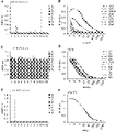
本発明者らは、代表的な大韓民国とサウジアラビアの菌株のMERS-CoVのSタンパク質(S protein)-RBD由来のB細胞エピトープ配列であるSpike-492及びSpike-492(L506F)を選択した。そのMERS-CoVのSタンパク質-RBDのB細胞エピトープペプチド配列であるSpike-492(L506F)は、L506F置換体をコードする。ここで、本発明者らは、B細胞エピトープペプチドとLipoplex(O)の複合体でマウスを免疫化して、MERS-CoVのSタンパク質に対して特異的なモノクロナール抗体506-2G10G5及び492-1G10E4E2を製造した。両モノクロナール抗体ともに、MERS-CoVのSタンパク質に対する強くて特異的な結合を示したが、506-2G10G5抗体は492-1G10E4E2と比較すると、Spike-492及びSpike-492(L506F)ペプチドに対するさらに高い結合親和性を示した。さらに、間接免疫蛍光アッセイ及び共焦点アッセイから、Sタンパク質に対する2つの抗体の特異性を確認した。重要なことに、506-2G10G5モノクロナール抗体の処理は、MERS-CoVに感染したプラークパーセントを、正常マウスIgG及び492-1G10E4E2の処理と比較すると効果的に減少させた。以上をまとめると、本発明のデータは、506-2G10G5モノクロナール抗体をMERS-CoV感染に対する診断及び治療に対して適用可能であるということが分かる。 We selected the B-cell epitope sequences Spike-492 and Spike-492 (L506F) from the S protein-RBD of MERS-CoV of representative Korean and Saudi strains. The MERS-CoV S protein-RBD B cell epitope peptide sequence Spike-492 (L506F) encodes the L506F substitution. Here, we immunized mice with a conjugate of a B-cell epitope peptide and Lipoplex(O) to generate monoclonal antibodies 506-2G10G5 and 492-1G10E4E2 specific for the S protein of MERS-CoV. manufactured. Both monoclonal antibodies showed strong and specific binding to the S protein of MERS-CoV, but the 506-2G10G5 antibody showed even higher binding to the Spike-492 and Spike-492 (L506F) peptides compared to 492-1G10E4E2. showed binding affinity. Additionally, indirect immunofluorescence and confocal assays confirmed the specificity of the two antibodies for the S protein. Importantly, treatment with the 506-2G10G5 monoclonal antibody effectively reduced the percentage of plaques infected with MERS-CoV compared to treatment with normal mouse IgG and 492-1G10E4E2. Taken together, the data of the present invention demonstrate the applicability of the 506-2G10G5 monoclonal antibody for diagnosis and therapy against MERS-CoV infection.
本発明を通じて分かるように、本発明の506-2G10G5モノクロナール抗体は、MERS-CoV感染に対する診断及び治療に適用することができる。 As can be seen through the present invention, the 506-2G10G5 monoclonal antibody of the present invention can be applied for diagnosis and treatment against MERS-CoV infection.
以下、非限定的な実施例により本発明をさらに詳細に説明する。但し、以下の実施例は、本発明を例示するための意図で記載されたものであって、本発明の範囲は、以下の実施例によって制限されるものと解釈されるべきではない。 The invention will now be described in more detail by means of non-limiting examples. However, the following examples are intended to illustrate the present invention, and the scope of the present invention should not be construed as being limited by the following examples.
本発明のアフリカミドリザル(African green monkey)の腎臓細胞であるVero細胞は、ATCC(American Type Culture Collection,Manassas,VA,USA)から購入した。Vero細胞を10%牛胎児血清(FBS,Thermo Fisher Scientific)、25mM HEPES、100U/mlペニシリン及び100μg/mlストレプトマイシンが補充されたDMEM(Dulbecco’s modified Eagle’s medium,Life Technologies,Thermo Fisher Scientific,Waltham,MA,USA)で維持した。その細胞を37℃、95%air及び5%CO2で培養した。MERS-CoV/KOR/KNIH/002_05_2015は大韓民国疾病管理本部から入手した(Permission No.1-001-MER-IS-2015001)。 Vero cells, kidney cells of African green monkeys of the present invention, were purchased from ATCC (American Type Culture Collection, Manassas, VA, USA). Vero cells were added to DMEM (Dulbecco's modified Eagle's medium, Life Technologies, Thermo Scientific) supplemented with 10% fetal bovine serum (FBS, Thermo Fisher Scientific), 25 mM HEPES, 100 U/ml penicillin and 100 μg/ml streptomycin. Waltham, Mass., USA). The cells were cultured at 37°C, 95% air and 5% CO2 . MERS-CoV/KOR/KNIH/002_05_2015 was obtained from the Korea Centers for Disease Control and Prevention (Permission No. 1-001-MER-IS-2015001).
実施例1:B細胞エピトープペプチドの製造
MERS-CoVのMタンパク質-特異的抗体を生産するために、本発明者らは、B細胞エピトープをエピトープの予測、表面接近性及び抗原性指数に基づいてMタンパク質を分析して選択した。MERS-CoVのMタンパク質(MERS-M158,158CDYDRLPNEVTVAKPNVLIALKMVK 182;配列番号9)に対するB細胞エピトープ配列を分析し、そのペプチドを自動化されたペプチド・シンセサイザー(Peptron III-R24,Peptron,Daejeon,Korea)で合成した。そのペプチドを逆相HPLC(Prominence HPLC,Shimadzu Corp.,Kyoto,Japan)で90%以上の純度に精製した。
Example 1: Preparation of B-cell epitope peptides To generate M protein-specific antibodies of MERS-CoV, we identified B-cell epitopes based on epitope prediction, surface accessibility and antigenicity index. The M protein was analyzed and selected. B-cell epitope sequences for the M protein of MERS-CoV (MERS-M158, 158 CDYDRLPNEVTVAKPNVLIALKMVK 182 ; SEQ ID NO: 9) were analyzed and the peptides were synthesized with an automated peptide synthesizer (Peptron III-R24, Peptron, Daejeon, Korea). Synthesized. The peptide was purified by reverse-phase HPLC (Prominence HPLC, Shimadzu Corp., Kyoto, Japan) to >90% purity.
MERS-CoVのSタンパク質のB細胞エピトープの選択、分析及び合成は、Park BK,Lee SI,Bae J-Y et al(2018)Int J Pept Res Ther https://doi.org/10.1007/s10989-018-9731-8に記載されているように行った。MERS-CoVのSタンパク質に対するB細胞エピトープペプチド配列は、MERS-CV菌株(MERS-CoV/KOR/KNIH/002_05_2015(GI:829021049))由来のSpike-492(492TKPLKYSYINKCSRLLSDDRTEVPQ 516;配列番号9)及びMERS-CV菌株(Spike glycoprotein universal sequence(GI:510785803))由来のSpike-492(L506F)(492TKPLKYSYINKCSRFLSDDRTEVPQ 516;配列番号10)を選択し、そのペプチドを自動化されたペプチド・シンセサイザー(Peptron III-R24,Peptron,Daejeon,Korea)で合成した。DOPE:CHEMS(Lipoplex(O)と命名)に同時にカプセル化されたB細胞エピトープペプチド及びCpG-DNA(MB-ODN 4531(O))複合体を製造した。 Selection, analysis and synthesis of B-cell epitopes of the S protein of MERS-CoV are described in Park BK, Lee SI, Bae JY et al (2018) Int J Pept Res Ther https://doi. org/10.1007/s10989-018-9731-8. The B-cell epitope peptide sequences for the S protein of MERS-CoV are Spike-492 ( 492 TKPLKYSYINKCSRLLSDDRTEVPQ 516 ; SEQ ID NO: 9) from the MERS-CV strain (MERS-CoV/KOR/KNIH/002_05_2015 (GI: 829021049)) and MERS - Spike-492 (L506F) ( 492 TKPLKYSYINKCSRFLSDDRTEVPQ 516 ; SEQ ID NO: 10) from the CV strain (Spike glycoprotein universal sequence (GI: 510785803)) was selected, and the peptide was synthesized in an automated peptide synthesizer (Peptron III, Reptron III). Peptron, Daejeon, Korea). B-cell epitope peptide and CpG-DNA (MB-ODN 4531(O)) complexes co-encapsulated in DOPE:CHEMS (termed Lipoplex(O)) were prepared.
実施例2:マウスの免疫化
BALB/c(4週齢、雌、H-2b)マウスをNara-Biotec(Seoul,Korea)から購入した。マウスを特定の無菌の条件下で翰林大学校の動物実験室で維持し、動物実験は、翰林大学校翰林大学校の動物実験倫理委員会(Permit number,Hallym2016-51)の承認を受けた。マウスに200μlのSpike-492ペプチド(50μg)又はSpike-492(L506F)ペプチド(50μg)及びLipoplex(O)複合体を、G.Wu,D.Kim,J.N.Kim,et al.,Theranostics,8(2018),pp.78-91に記載されているように、10日に3回腹腔内(i.p.)注射した。
Example 2: Immunization of Mice BALB/c (4 weeks old, female, H-2 b ) mice were purchased from Nara-Biotec (Seoul, Korea). Mice were maintained under specific sterile conditions in the Animal Laboratory of Hallym University, and animal experiments were approved by the Hallym University Animal Experiment Ethics Committee (Permit number, Hallym 2016-51). Mice received 200 μl of Spike-492 peptide (50 μg) or Spike-492 (L506F) peptide (50 μg) and Lipoplex(O) complex. Wu, D. Kim, J.; N. Kim, et al. , Theranostics, 8 (2018), pp. Intraperitoneal (ip) injections were given three times every 10 days as described in 78-91.
実施例3:マウスAnti-MERS-CoVのSタンパク質のモノクロナール抗体の生産
標準ハイブリドーマ技術によって、ハイブリドーマ細胞をanti-Spike-49二重特異的モノクロナール抗体(492-1G10E4E2)及びanti-Spike-492(L506F)-特異的なモノクロナール抗体(506-2G10G5)を生産するために選択した[G.Wu,D.Kim,J.N.Kim,et al.,A Theranostics,8(2018),pp.78-91;W.M.Yokoyama,M.Christensen,G.D.Santos,et al.,Curr.Protoc.Immunol.,Chapter2(2006),Unit2.5].
Example 3: Production of Monoclonal Antibodies to Mouse Anti-MERS-CoV S Protein Hybridoma cells were immunized with anti-Spike-49 bispecific monoclonal antibody (492-1G10E4E2) and anti-Spike-492 by standard hybridoma technology. (L506F)--selected to produce a specific monoclonal antibody (506-2G10G5) [G. Wu, D. Kim, J.; N. Kim, et al. , A Theranostics, 8 (2018), pp. 78-91; M. Yokoyama, M.; Christensen, G.; D. Santos, et al. , Curr. Protoc. Immunol. , Chapter 2 (2006), Unit 2.5].
10日間隔で3回、BALB/cマウスにi.p.でDOPE:CHEMS複合体に同時にカプセル化されたMERS-Spike-492ペプチド(50μg)(又はSpike-492(L506F)ペプチド)及びMB-ODN 4531(O)(50μg)を注射した。 BALB/c mice were injected i.p. three times at 10-day intervals. p. MERS-Spike-492 peptide (50 μg) (or Spike-492(L506F) peptide) and MB-ODN 4531(O) (50 μg) co-encapsulated in DOPE:CHEMS complexes were injected at .
免疫化されたマウスに由来した脾臓を、標準ハイブリドーマ技術によって融合に使用した。HAT培地及びHT培地でハイブリドーマクローンを標準リミッティング希釈プロトコルで選択して、クローン細胞群を得た。腹水を得るために、その選択されるハイブリドーマクローンをBALB/cマウスの腹腔に注射した。anti-Spike-49二重特異的モノクロナール抗体(492-1G10E4E2)及びanti-Spike-492(L506F)-特異的なモノクロナール抗体(506-2G10G5)を、タンパク質A カラムカラムクロマトグラフィーで腹水液から精製した。 Spleens from immunized mice were used for fusions by standard hybridoma techniques. Hybridoma clones were selected in HAT medium and HT medium with standard limiting dilution protocols to obtain clonal cell populations. To obtain ascites, the selected hybridoma clones were injected into the peritoneal cavity of BALB/c mice. Anti-Spike-49 bispecific monoclonal antibody (492-1G10E4E2) and anti-Spike-492 (L506F)-specific monoclonal antibody (506-2G10G5) were isolated from ascitic fluid by protein A column chromatography. Refined.
そのモノクロナール抗体のアイソタイプを決定するために、アイソタイピング(isotyping)キット(Southern Biotechnology Associates Inc,Birmingham,USA)を使用した。 An isotyping kit (Southern Biotechnology Associates Inc, Birmingham, USA) was used to determine the isotype of the monoclonal antibody.
実施例4:ELISAアッセイ
エピトープペプチド特異的抗体タイターを測定するために、5μg/ウェルのMERS-CoVのSpike-492又はSpike-492(L506F)ペプチドを96ウェル免疫プレート(Thermo Fisher Scientific)上にコーティングした後、4℃で一晩培養し、PBST(0.05% Tween-20が補充されたPBS)で洗浄した後、1%BSAを含むPBSTでブロッキングした。
Example 4:
マウス血清を眼窩出血(orbital bleeding)で集めて1:3の比率で連続的に希釈し、各プレートのウェルに添加した後、常温で2時間培養した。HRP(Horseraddish peroxidase)-付着したヤギ抗マウス(goat anti-mouse)IgG(Jackson ImmunoResearch laboratories,West Grove,PA,USA)を2次抗体として1時間使用した。 Mouse serum was collected by orbital bleeding, serially diluted at a ratio of 1:3, added to the wells of each plate, and incubated at room temperature for 2 hours. HRP (Horseradish peroxidase)-conjugated goat anti-mouse IgG (Jackson ImmunoResearch laboratories, West Grove, PA, USA) was used as a secondary antibody for 1 hour.
PBSTで洗浄した後、TMB(tetramethylbenidine)ペルオキシダーゼ(peroxidase)基質(KPL,SeraCare,Milford,MA,USA)を処理し、反応をTMB-stop溶液(KPL)で停止した。吸光度をSpectra Max 250マイクロプレートリーダ(Molecular Devices,San Jose,CA,USA)を使用して450nmで測定した。モノクロナール抗体のアイソタイプの同定のために、HRP-付着した抗マウス(anti-mouse)IgG(各isotype)抗体(Southern Biotech,Birmingham,AL,USA)を使用した。交差-反応性を検出するために、96ウェル免疫プレートをMERS-CoVのSpike-492又はSpike-492(L506F)ペプチドで前記のようにコーティングした。
After washing with PBST, TMB (tetramethylbenidine) peroxidase substrate (KPL, SeraCare, Milford, Mass., USA) was treated and the reaction was stopped with TMB-stop solution (KPL). Absorbance was measured at 450 nm using a
そのコーティングされたウェルを、2次抗体の培養の前に、492-1G10E4E2モノクロナール抗体又は506-2G10G5モノクロナール抗体で2時間培養した。 The coated wells were incubated with 492-1G10E4E2 monoclonal antibody or 506-2G10G5 monoclonal antibody for 2 hours prior to secondary antibody incubation.
実施例5:ELISAによる親和性定数の測定
Anti-Spike-49二重特異的モノクロナール抗体(492-1G10E4E2)及びanti-Spike-492(L506F)-特異的なモノクロナール抗体(506-2G10G5)の結合親和性を決定するために、5μg/ウェルのSpike-492及びSpike-492(L506F)エピトープペプチドを96ウェル免疫プレートにコーティングした後、1%BSAを含むPBSTでブロッキングした。
Example 5: Affinity constant determination by ELISA To determine binding affinities, 5 μg/well of Spike-492 and Spike-492 (L506F) epitope peptides were coated on 96-well immunoplates and then blocked with PBST containing 1% BSA.
そのモノクロナール抗体をPBSTで1:5継代希釈して各プレートに添加した後、常温で2時間培養した。PBSTで洗浄した後、ホースラディッシュペルオキシダーゼ(horseradish peroxidase)が付着した抗IgG抗体を各プレートに添加した。プレートで抗体の量を、テトラメチルベンジジン(TMB)ペルオキシダーゼ基質(KPL,SeraCare,Milford,MA,USA)で現像した。 The monoclonal antibody was serially diluted 1:5 with PBST, added to each plate, and incubated at room temperature for 2 hours. After washing with PBST, an anti-IgG antibody conjugated with horseradish peroxidase was added to each plate. Amounts of antibody on plates were developed with tetramethylbenzidine (TMB) peroxidase substrate (KPL, SeraCare, Milford, Mass., USA).
吸光度をSpectra Max 250マイクロプレートリーダ(Molecular Devices,San Jose,CA,USA)を用いて405nmで測定し、D.I.Kisiela,H.Avagyan,D.Friend,et al.,PLoS Pathog.,11(2015),pp.e1004857に記載されているように、SigmaPlotプログラムで計算して、EC50値を決定した。
Absorbance was measured at 405 nm using a
実施例6:ウェスタンブロッティング及び免疫沈降法
MERS-CoV-感染したVero細胞を細胞破砕バッファー(10mM HEPES、150mM NaCl、5mM EDTA、100mM NaF、2mM Na3VO4、プロテアーゼ阻害剤カクテル及び10%NP-40)で破砕した後、SDS-PAGE上にローディングした。その分離されたタンパク質をニトロセルロース(nitrocellulose)膜に移した後、3%BSAを含むPBSTでブロッキングした。その膜を、MERS-CoVのSpike-492モノクロナール抗体又はSpike-492(L506F)モノクロナール抗体で処理し、PBSTで洗浄し、HRP付着したヤギ抗マウス(goat anti-mouse)IgG抗体で処理した。
Example 6: Western blotting and immunoprecipitation MERS-CoV-infected Vero cells were treated with cell disruption buffer (10 mM HEPES, 150 mM NaCl, 5 mM EDTA, 100 mM NaF, 2 mM Na 3 VO 4 , protease inhibitor cocktail and 10% NP- 40) and loaded on SDS-PAGE. The separated proteins were transferred to a nitrocellulose membrane and blocked with PBST containing 3% BSA. The membrane was treated with MERS-CoV Spike-492 monoclonal antibody or Spike-492 (L506F) monoclonal antibody, washed with PBST, and treated with HRP-conjugated goat anti-mouse IgG antibody. .
その膜をケミルミネッセンス(chemiluminescence)溶液で検出し、ChemiDoc(Bio-Rad,Herculed,California,USA)で図案化した。免疫沈降のために、ウイルス感染した-Vero細胞破砕液をMERS-CoVのSpike-492モノクロナール抗体又はSpike-492(L506F)モノクロナール抗体を用いて4℃で一晩培養し、MERS-CoVのSタンパク質をプロテインAビーズ(Repligen,Waltham,MA,USA)で1時間沈降させた。その免疫沈降したタンパク質をMERS-CoVのSpike-492モノクロナール抗体又はSpike-492(L506F)モノクロナール抗体を使用してウェスタンブロッティングで分析した。 The membrane was detected with a chemiluminescence solution and patterned with ChemiDoc (Bio-Rad, Herculed, California, USA). For immunoprecipitation, virus-infected-Vero cell lysates were incubated overnight at 4° C. with Spike-492 monoclonal antibody or Spike-492 (L506F) monoclonal antibody for MERS-CoV. S protein was precipitated with protein A beads (Repligen, Waltham, Mass., USA) for 1 hour. The immunoprecipitated proteins were analyzed by Western blotting using the MERS-CoV Spike-492 monoclonal antibody or Spike-492 (L506F) monoclonal antibody.
実施例7:脱グリコシル化アッセイ(Deglycosylation Assay)
細胞破砕液をVero細胞及びMERS-CoV-感染したVero細胞から破砕バッファー(0.5%SDS、1%β-mercaptoethanol)を用いて取得し、その破砕液を100℃で10分間加熱した。その破砕液をペプチド-N-グリコシダーゼ(PNGase)F(Elpis Biotech,Daejeon,Korea)を用いて37℃で2時間処理した後、100℃で10分間加熱した。処理後に、そのサンプルをMERS-CoVのSpike-492モノクロナール抗体又はSpike-492(L506F)モノクロナール抗体でウェスタンブロットにより分析した。
Example 7: Deglycosylation Assay
Cell lysates were obtained from Vero cells and MERS-CoV-infected Vero cells using lysis buffer (0.5% SDS, 1% β-mercaptoethanol) and the lysates were heated at 100° C. for 10 minutes. The lysate was treated with peptide-N-glycosidase (PNGase) F (Elpis Biotech, Daejeon, Korea) at 37°C for 2 hours and then heated at 100°C for 10 minutes. After treatment, the samples were analyzed by Western blot with the MERS-CoV Spike-492 monoclonal antibody or Spike-492 (L506F) monoclonal antibody.
免疫沈降法のために、そのPNGase F処理されたサンプルをMERS-CoVのSpike-492モノクロナール抗体又はSpike-492(L506F)モノクロナール抗体を用いて4℃で一晩培養し、MERS-CoVのSpike-492モノクロナール抗体又はSpike-492(L506F)モノクロナール抗体でウェスタンブロッティングにより分析した。 For immunoprecipitation, the PNGase F-treated samples were incubated overnight at 4° C. with Spike-492 monoclonal antibody or Spike-492 (L506F) monoclonal antibody for MERS-CoV. Analyzed by Western blotting with Spike-492 monoclonal antibody or Spike-492 (L506F) monoclonal antibody.
実施例8:anti-MERS CoVのSタンパク質のモノクロナール抗体の重鎖及び軽鎖可変ドメインのクローニング
Anti-Spike-492ペプチド-特異的なモノクロナール抗体(492-1G10E4E2)及びanti-Spike-492(L506F)ペプチド-特異的なモノクロナール抗体(506-2G10G5)を生産するハイブリドーマ(Hybridoma)細胞(492-1G10E4E2クローン及び506-2G10G5)を培養し、マウスモノクロナール抗体アイソタイピングキット(Dipstick format,Bibco BRL or Roche,Mannheim,Germany)を使用してアイソタイピングした。総RNAsをハイブリドーマ細胞(492-1G10E4E2クローン及び506-2G10G5)からRNeasy Mini Kit(Qiagen)で抽出し、cDNAsを生成した。
Example 8: Cloning heavy and light chain variable domains of anti-MERS CoV S protein monoclonal antibody Anti-Spike-492 peptide-specific monoclonal antibody (492-1G10E4E2) and anti-Spike-492 ( L506F) Hybridoma cells (492-1G10E4E2 clone and 506-2G10G5) producing peptide-specific monoclonal antibodies (506-2G10G5) were cultured and analyzed using mouse monoclonal antibody isotyping kit (Dipstick format, Bibco BRL). or Roche, Mannheim, Germany). Total RNAs were extracted from hybridoma cells (492-1G10E4E2 clone and 506-2G10G5) with RNeasy Mini Kit (Qiagen) to generate cDNAs.
Anti-Spike-492ペプチド-特異的なモノクロナール抗体(492-1G10E4E2)の可変重鎖及び軽鎖ドメイン(VH及びVL)に対する配列をクローニングするために、そのcDNAsを、Ventポリメラーゼ(NEB)を使用して下記のプライマーセットを用いて増幅した。重鎖プライマーのために、IGG2A(5’-GGA AGA TCT CTT GAC CAG GCA TCC TAG AGT CA-3’;配列番号11)及び5’MH2(5’-CTT CCG GAA TTC SAR GTN MAG CTG SAG SAG TCW GG-3’;配列番号12)を使用した。 To clone the sequences for the variable heavy and light chain domains (V H and V L ) of the Anti-Spike-492 peptide-specific monoclonal antibody (492-1G10E4E2), the cDNAs were subjected to Vent polymerase (NEB). was amplified using the following primer set. For heavy chain primers, IGG2A (5′-GGA AGA TCT CTT GAC CAG GCA TCC TAG AGT CA-3′; SEQ ID NO: 11) and 5′MH2 (5′-CTT CCG GAA TTC SAR GTN MAG CTG SAG SAG TCW GG-3'; SEQ ID NO: 12) was used.
カッパ(kappa)鎖プライマーのために、3’Kc(5’-GGT GCA TGC GGA TAC AGT TGG TGC AGC ATC-3’;配列番号13)、及び5’Mk(5’-GG GAG CTC GAY ATT GTG MTS ACM CAR WCT MCA-3’;配列番号14)を使用した。 3′Kc (5′-GGT GCA TGC GGA TAC AGT TGG TGC AGC ATC-3′; SEQ ID NO: 13), and 5′Mk (5′-GG GAG CTC GAY ATT GTG) for kappa chain primers MTS ACM CAR WCT MCA-3'; SEQ ID NO: 14) was used.
anti-Spike-492(L506F)ペプチド-特異的なモノクロナール抗体(506-2G10G5)の可変重鎖及び軽鎖ドメイン(VH及びVL)に対する配列をクローニングするために、そのcDNAsを、Ventポリメラーゼ(NEB)を使用して下記のプライマーセットを用いて増幅した。重鎖プライマーのために、IGG1(5’-GGA AGA TCT ATA GAC AGA TGG GGG TGT CGT TTT GGC-3’;配列番号11)及び5’MH2(5’-CTT CCG GAA TTC SAR GTN MAG CTG SAG SAG TCW GG-3’;配列番号12)を使用した。 To clone the sequences for the variable heavy and light domains (V H and V L ) of the anti-Spike-492 (L506F) peptide-specific monoclonal antibody (506-2G10G5), the cDNAs were exposed to Vent polymerase. (NEB) and amplified using the following primer set. For heavy chain primers, IGG1 (5′-GGA AGA TCT ATA GAC AGA TGG GGG TGT CGT TTT GGC-3′; SEQ ID NO: 11) and 5′MH2 (5′-CTT CCG GAA TTC SAR GTN MAG CTG SAG SAG TCW GG-3'; SEQ ID NO: 12) was used.
カッパ(kappa)鎖プライマーのために、3’Kc(5’-GGT GCA TGC GGA TAC AGT TGG TGC AGC ATC-3’;配列番号13)、及び5’Mk(5’-GG GAG CTC GAY ATT GTG MTS ACM CAR WCT MCA-3’;配列番号14)を使用した。 3′Kc (5′-GGT GCA TGC GGA TAC AGT TGG TGC AGC ATC-3′; SEQ ID NO: 13), and 5′Mk (5′-GG GAG CTC GAY ATT GTG) for kappa chain primers MTS ACM CAR WCT MCA-3'; SEQ ID NO: 14) was used.
標準PCR反応を25サイクル行った。そのPCR産物を、直接pGEM-Tイージーベクター(easy vector)(Promega)にライゲーションした。クローンされたマウスIg挿入体をDNAシークエンシングで分析した。 A standard PCR reaction was performed for 25 cycles. The PCR product was directly ligated into the pGEM-T easy vector (Promega). Cloned mouse Ig inserts were analyzed by DNA sequencing.
実施例9:間接免疫蛍光アッセイ及び共焦点イメージ
間接免疫蛍光アッセイ(indirect immunofluorescence assay:IFA)を行うために、MERS-CoV感染したVero細胞及び-非感染のVero細胞の混合物(ratio 1:3)をスライドガラス上にプレーティングした。その混合した細胞をアセトンで固定化し、蒸留水で洗浄し、正常マウス(normal mouse)IgG、MERS-M158ペプチド-特異的なモノクロナール抗体を用いて37℃で培養した。2時間培養した後、そのスライドをPBS及びDWで洗浄した後、Alexa Flour 488-付着したヤギ抗マウスIgG抗体(Thermo Fisher Scientific,Waltham,MA,USA)で培養した。サンプルを取り付けた後、蛍光顕微鏡(IX70,Olympus,Tokyo,Japan)を使用して観察した。
Example 9: Mixture of MERS-CoV infected and -uninfected Vero cells (ratio 1:3) to perform indirect immunofluorescence assay and confocal image indirect immunofluorescence assay (IFA) was plated on a glass slide. The mixed cells were fixed with acetone, washed with distilled water and incubated at 37° C. with normal mouse IgG, MERS-M158 peptide-specific monoclonal antibody. After 2 hours of incubation, the slides were washed with PBS and DW and then incubated with Alexa Flour 488-conjugated goat anti-mouse IgG antibody (Thermo Fisher Scientific, Waltham, Mass., USA). After mounting the sample, it was observed using a fluorescence microscope (IX70, Olympus, Tokyo, Japan).
共焦点イメージを観察するために、Vero細胞(5×10⁴細胞)を12ウェル培養プレート内のカバーガラス上にプレーティングし、MERS-CoV(0.1MOI)で2日間感染させた。その細胞を4%パラホルムアルデヒド(paraformaldehyde)で固定化し、1%BSA及び0.1%triton X-100を含むPBSで30分間ブロッキングした。MERS-CoVのSpike-492モノクロナール抗体をプレートに添加し、2時間培養した後、Alexa488付着したヤギ抗マウスIgGで1時間培養した。核をHoechst 33258(Thermo Fisher Scientific)で染色した。そのスライドをCarl Zeiss LSM710(Carl Zeiss,Oberkochen,DE)で観察した。 To observe confocal images, Vero cells (5×10 4 cells) were plated on coverslips in 12-well culture plates and infected with MERS-CoV (0.1 MOI) for 2 days. The cells were fixed with 4% paraformaldehyde and blocked with PBS containing 1% BSA and 0.1% triton X-100 for 30 minutes. MERS-CoV Spike-492 monoclonal antibody was added to the plates and incubated for 2 hours followed by Alexa488-conjugated goat anti-mouse IgG for 1 hour. Nuclei were stained with Hoechst 33258 (Thermo Fisher Scientific). The slides were viewed with a Carl Zeiss LSM710 (Carl Zeiss, Oberkochen, Del.).
実施例10:プラーク(Plaque)減少アッセイ
6×10⁵ Vero細胞/ウェルを6ウェルプレート(Thermo Fisher Scientific)上にプレーティングし、12時間培養した。感染させる前に、MERS-CoVを2倍に連続希釈された正常マウスIgG、492-1G10E4E2モノクロナール抗体又は506-2G10G5で30分間、37℃で予め培養した。ウイルス-抗体混合物をVero細胞に500μlのPBSで添加した。1時間培養した後、上澄液を除去し、3mlの0.6%oxoidアガーを含むDMEM/F12培地(Thermo Fisher Scientific)を添加した。各ウェルで形成されたプラークを、培養4日後、クリスタルバイオレット(crystal violet)で染色した。そのプラークをカウントし、パーセントを計算した。
Example 10 : Plaque Reduction Assay 6 x 10⁵ Vero cells/well were plated on 6-well plates (Thermo Fisher Scientific) and cultured for 12 hours. Prior to infection, MERS-CoV was pre-incubated with 2-fold serially diluted normal mouse IgG, 492-1G10E4E2 monoclonal antibody or 506-2G10G5 for 30 minutes at 37°C. Virus-antibody mixture was added to Vero cells in 500 μl of PBS. After 1 hour of incubation, the supernatant was removed and 3 ml of DMEM/F12 medium (Thermo Fisher Scientific) containing 0.6% oxoid agar was added. Plaques formed in each well were stained with crystal violet after 4 days of culture. The plaques were counted and percentages calculated.
前記実施例の結果は、下記の通りである。 The results of the above examples are as follows.
B細胞エピトープの分析及びMERS-CoVのSタンパク質のエピトープをターゲッティングする抗体の生産
B細胞エピトープの同定及び選択は、エピトープに基づく抗体の生産に考慮すべき重要な事項の一つであるので、MERS-CoVのSタンパク質の明白なB細胞エピトープのアミノ酸配列をエピトープの予測、表面接近性及び抗原性スケールに基づいてIEDB(Immune Epitope Database and Analysis Resources)を用いて予測した。Sタンパク質内のRBDドメインは宿主への結合に関与するため、Sタンパク質内のRBDドメイン内に492-516アミノ酸を有するSpike-492及びSpike-492(L506F)ペプチド配列を選択して合成した(図1のA及びB)。B細胞エピトープとしての効率性を決定するために、各ペプチド及びLipoplex(O)複合体を製造してBALB/cマウスに免疫化した。抗体タイターをスクリーニングするために、ELISAを、免疫化された抗体の血清から行った。両ペプチドともに、ペプチド-特異IgGsの生産を誘導することができる(図1のC)。したがって、免疫原性エピトープが成功裏に考案され、生産された。
Analysis of B Cell Epitopes and Production of Antibodies Targeting S Protein Epitopes of MERS-CoV Identification and selection of B cell epitopes is one of the important considerations for the production of epitope-based antibodies. - Amino acid sequences of apparent B-cell epitopes of CoV S protein were predicted using IEDB (Immune Epitope Database and Analysis Resources) based on epitope prediction, surface accessibility and antigenic scale. Since the RBD domain within the S protein is involved in binding to the host, Spike-492 and Spike-492 (L506F) peptide sequences with 492-516 amino acids within the RBD domain within the S protein were selected and synthesized (Fig. 1 A and B). To determine their efficiency as B-cell epitopes, each peptide and Lipoplex(O) conjugate was prepared and immunized into BALB/c mice. To screen antibody titers, ELISA was performed from immunized antibody sera. Both peptides are able to induce the production of peptide-specific IgGs (Fig. 1C). Thus, an immunogenic epitope was successfully devised and produced.
MERS-CoVのSpike-492(L506F)又はSpike-492エピトープ-特異的なモノクロナール抗体の生産
MERS-CoVのSタンパク質のエピトープ-特異的なモノクロナール抗体を生産するために、脾臓細胞を、リポソーム(DOPE;CHEMS)内に同時にカプセル化されたSpike-492(L506F)又はSpike-492エピトープペプチド及びCpG-DNA複合体で免疫化されたマウスから集めた。そのマウスの脾臓細胞をSP2/0と融合し、Spike-492(L506F)エピトープペプチド-特異的抗体を生産する506-2G10G5クローンをHAT及びHT補充物として選択した(図2)。
Production of MERS-CoV Spike-492 (L506F) or Spike-492 Epitope-Specific Monoclonal Antibodies Collected from mice immunized with Spike-492 (L506F) or Spike-492 epitope peptide and CpG-DNA complex co-encapsulated in (DOPE; CHEMS). The spleen cells of the mouse were fused with SP2/0 and the 506-2G10G5 clone producing Spike-492 (L506F) epitope peptide-specific antibodies was selected as HAT and HT supplement (Fig. 2).
そのマウスの脾臓細胞をSP2/0と融合し、Spike-492エピトープペプチド-特異的抗体を生産する492-1G10E4E2クローンをHAT及びHT補充物として選択した(図3)。 The spleen cells of the mice were fused with SP2/0 and the 492-1G10E4E2 clone producing Spike-492 epitope peptide-specific antibodies was selected as HAT and HT supplement (Fig. 3).
MERS-CoVのSpike-492(L506F)又はSpike-492エピトープ-特異的なモノクロナール抗体の特性
多量のモノクロナール抗体を得るために、506-2G10G5クローン又は492-1G10E4E2クローンを腹水の生産のためにマウスの腹腔内に注射し、腹水液を506-2G10G5クローン又は492-1G10E4E2クローンで注射されたマウスから集めて精製した(図4のA及び図5のA)。次に、506-2G10G5又は492-1G10E4E2モノクロナール抗体のアイソタイプの分類をELISAで行い、それぞれIgG1及びIgG2aであることが判明した(図4のB及び図5のB)。Spike-492(L506F)又はSpike-492ペプチドをターゲッティングするモノクロナール抗体の結合親和性をSpike-492(L506F)ペプチド又はSpike-492 ELISAで決定し、506-2G10G5及び492-1G10E4E2モノクロナール抗体のEC50値は、それぞれ、178pM及び3.3nMであった(図4のC及び図5のC)。
Spike-492 (L506F) of MERS-CoV or Spike-492 Epitope-Specific Monoclonal Antibody Characteristics Mice were injected intraperitoneally and ascites fluid was collected and purified from mice injected with the 506-2G10G5 clone or the 492-1G10E4E2 clone (FIGS. 4A and 5A). The 506-2G10G5 or 492-1G10E4E2 monoclonal antibodies were then isotyped by ELISA and found to be IgG1 and IgG2a, respectively (FIGS. 4B and 5B). The binding affinities of monoclonal antibodies targeting Spike-492 (L506F) or Spike-492 peptide were determined by Spike-492 (L506F) peptide or Spike-492 ELISA and the EC of 506-2G10G5 and 492-1G10E4E2 monoclonal antibodies The 50 values were 178 pM and 3.3 nM, respectively (Fig. 4C and Fig. 5C).
MERS-CoVのSpike-492(L506F)又はSpike-492エピトープ-特異的なモノクロナール抗体によるMERS-CoVのSタンパク質の検出
追加的にモノクロナール抗体の特性を調査するために、本発明者らは、MERS-CoV感染及び非感染のVero細胞でウェスタンブロット及び免疫沈降を行った。ウェスタンブロッティングの結果(図4のD、図5のD)は、両モノクロナール抗体ともに、MERS-CoV-感染したVero細胞でSタンパク質に特異的なバンドを検出したが、非感染のVero細胞ではバンドが観察されなかった。PNGase(peptide-N-glycosidase)Fで処理は、未処理されたサンプルと比較して、Sタンパク質バンドの分子量の減少を引き起こし(図4のD及び図5のD)、これは、両モノクロナール抗体がいずれも、それのグリコシル化及び非グリコシル化形態のSタンパク質を認識できるということを示唆している。また、モノクロナール抗体が自然形態のSタンパク質を認識できるかを評価するために、免疫沈降を行った。本発明者らは、両方の抗体を、MERS-CoV-感染したVero細胞破砕液由来の自然型のSタンパク質で免疫沈降させた(図4のF及び図5のE)。各該当するエピトープで506-2G10G5及び492-1G10E4E2モノクロナール抗体の交差反応性を評価するために、ELISAを行った。506-2G10G5モノクロナール抗体は、Spike-492(L506F)に対する492-1G10E4E2モノクロナール抗体によって示したものと比較して、Spike-492ペプチドに対する著しい交差反応性を示した(図4のH及びI)。したがって、両方の抗体は、MERS-CoVのSタンパク質に対する特異的な結合を示した。
Detection of MERS-CoV S protein by MERS-CoV Spike-492 (L506F) or Spike-492 epitope-specific monoclonal antibodies To additionally investigate the properties of the monoclonal antibodies, we , Western blots and immunoprecipitations were performed on MERS-CoV infected and uninfected Vero cells. Western blotting results (Fig. 4D, Fig. 5D) showed that both monoclonal antibodies detected an S protein-specific band in MERS-CoV-infected Vero cells, but not in uninfected Vero cells. No bands were observed. Treatment with PNGase (peptide-N-glycosidase) F caused a decrease in the molecular weight of the S protein band compared to untreated samples (Fig. 4D and Fig. 5D), indicating that both monoclonal It suggests that both antibodies can recognize the S protein in its glycosylated and non-glycosylated forms. Immunoprecipitation was also performed to assess whether the monoclonal antibody could recognize the native form of the S protein. We immunoprecipitated both antibodies with native S protein from MERS-CoV-infected Vero cell lysates (Fig. 4F and Fig. 5E). An ELISA was performed to assess the cross-reactivity of the 506-2G10G5 and 492-1G10E4E2 monoclonal antibodies at each relevant epitope. The 506-2G10G5 monoclonal antibody showed significant cross-reactivity to the Spike-492 peptide compared to that shown by the 492-1G10E4E2 monoclonal antibody to Spike-492 (L506F) (H and I in Figure 4). . Thus, both antibodies showed specific binding to the S protein of MERS-CoV.
anti-Spike-492(L506F)ペプチド-特異的な、及びanti-Spike-492ペプチド-特異的なモノクロナール抗体の可変ドメインのクローニング
重鎖及び軽鎖可変ドメイン(VH及びVL)をコードするcDNA配列を、Spike-492(L506F)ペプチド-特異的なモノクロナール抗体を生産するハイブリドーマ細胞(506-2G10G5)から、通常の重鎖及び軽鎖プライマーを使用してクローニングした。DNAシークエンシングによって確認したその配列を図6に示した。
Cloning of Variable Domains of Anti-Spike-492 (L506F) Peptide-Specific and Anti-Spike-492 Peptide-Specific Monoclonal Antibodies Encoding Heavy and Light Chain Variable Domains (V H and V L ) A cDNA sequence was cloned from a hybridoma cell (506-2G10G5) producing a Spike-492 (L506F) peptide-specific monoclonal antibody using conventional heavy and light chain primers. Its sequence, confirmed by DNA sequencing, is shown in FIG.
重鎖及び軽鎖可変ドメイン(VH及びVL)をコードするcDNA配列を、Spike-492ペプチド-特異的なモノクロナール抗体を生産するハイブリドーマ細胞(492-1G10E4E2)から、通常の重鎖及び軽鎖プライマーを使用してクローニングした。DNAシークエンシングによって確認したその配列を図7に示した。 cDNA sequences encoding heavy and light chain variable domains (V H and V L ) were isolated from hybridoma cells (492-1G10E4E2) producing Spike-492 peptide-specific monoclonal antibodies to conventional heavy and light chains. cloned using strand primers. Its sequence, confirmed by DNA sequencing, is shown in FIG.
その配列を、タンパク質BLASTプログラムを使用して、公知の配列とのホモロジーを分析した。 The sequence was analyzed for homology with known sequences using the protein BLAST program.
506-2G10G5及び492-1G10E4E2重鎖及び軽鎖の可変ドメインをコードするcDNAは、それぞれ報告されている免疫グロブリン可変重鎖及び軽鎖ドメインと約80~95%及び93~98%のホモロジーを示した。 The cDNAs encoding the 506-2G10G5 and 492-1G10E4E2 heavy and light chain variable domains show approximately 80-95% and 93-98% homology with reported immunoglobulin variable heavy and light chain domains, respectively. rice field.
MERS-CoV-感染した細胞においてSタンパク質に対する506-2G10G5又は492-1G10E4E2モノクロナール抗体の反応性
追加的にIFA(indirect immunofluorescence assay)で506-2G10G5又は492-1G10E4E2モノクロナール抗体の反応性の幅を分析した。蛍光顕微鏡法は、両方の抗体で培養されたウイルス感染した細胞において強い蛍光信号を示したが、正常マウスIgGの場合には蛍光が観察されなかった(図8のA及び図9のA)。MERS-CoVのSタンパク質に対する両方の抗体の特異性を確認するために、共焦点顕微鏡法を行った。MERS-CoV-感染又は非感染のVero細胞は、正常マウスIgG又は506-2G10G5又は492-1G10E4E2モノクロナール抗体で染色された。共焦点顕微鏡法のイメージは、506-2G10G5又は492-1G10E4E2モノクロナール抗体で染色されたMERS-CoV感染した細胞の原形質部位内で蛍光信号が明確に示された。正常マウスIgGで培養された細胞では染色が観察されなかった(図8のB及び図9のB)。これらの結果は、MERS-CoV-感染した細胞でMERS-CoVのSタンパク質の効果的な検出において両モノクロナール抗体の特異性を示している。
Reactivity of 506-2G10G5 or 492-1G10E4E2 monoclonal antibody against S protein in MERS-CoV-infected cells analyzed. Fluorescence microscopy showed a strong fluorescence signal in virus-infected cells cultured with both antibodies, whereas no fluorescence was observed with normal mouse IgG (FIGS. 8A and 9A). To confirm the specificity of both antibodies for the MERS-CoV S protein, confocal microscopy was performed. MERS-CoV-infected or uninfected Vero cells were stained with normal mouse IgG or 506-2G10G5 or 492-1G10E4E2 monoclonal antibodies. Confocal microscopy images clearly showed fluorescent signals within the cytoplasmic sites of MERS-CoV infected cells stained with 506-2G10G5 or 492-1G10E4E2 monoclonal antibodies. No staining was observed in cells cultured with normal mouse IgG (FIGS. 8B and 9B). These results demonstrate the specificity of both monoclonal antibodies in effectively detecting the MERS-CoV S protein in MERS-CoV-infected cells.
506-2G10G5モノクロナール抗体はVero細胞のMERS-CoV感染を阻害する
追加的にプラーク減少アッセイにおいて、MERS-CoVに対する両モノクロナール抗体の阻害活性を調査した。このアッセイにおいて、両モノクロナール抗体は、正常マウスIgGと比較して、濃度依存的にプラークの形成を阻害した。しかし、506-2G10G5モノクロナール抗体で処理した場合に、492-1G10E4E2モノクロナール抗体と比較して、濃度依存的にプラークの形成のさらに高い阻害が観察された(図10のA及びB)。したがって、このような結果は、506-2G10G5モノクロナール抗体の効果がMERS-CoV感染に対する潜在的な治療的適用の可能性を示している。
506-2G10G5 Monoclonal Antibody Inhibits MERS-CoV Infection of Vero Cells Additionally, the inhibitory activity of both monoclonal antibodies against MERS-CoV was investigated in a plaque reduction assay. In this assay, both monoclonal antibodies inhibited plaque formation in a dose-dependent manner compared to normal mouse IgG. However, a concentration-dependent greater inhibition of plaque formation was observed when treated with the 506-2G10G5 monoclonal antibody compared to the 492-1G10E4E2 monoclonal antibody (FIGS. 10A and B). Therefore, such results indicate that the efficacy of the 506-2G10G5 monoclonal antibody has potential therapeutic applications against MERS-CoV infection.
Claims (9)
2)容器とを含む、MERSコロナウイルス診断用キット。 1) a composition according to claim 6 ;
2) a diagnostic kit for MERS coronavirus, comprising a container;
Applications Claiming Priority (3)
| Application Number | Priority Date | Filing Date | Title |
|---|---|---|---|
| KR10-2018-0096102 | 2018-08-17 | ||
| KR1020180096102A KR20200020411A (en) | 2018-08-17 | 2018-08-17 | A monoclonal antibody against S protein of MERS-CoV and use of the same |
| PCT/KR2019/010272 WO2020036403A1 (en) | 2018-08-17 | 2019-08-13 | Monoclonal antibody against s protein of mers-coronavirus, and use of same |
Publications (2)
| Publication Number | Publication Date |
|---|---|
| JP2021533175A JP2021533175A (en) | 2021-12-02 |
| JP7112790B2 true JP7112790B2 (en) | 2022-08-04 |
Family
ID=69525681
Family Applications (1)
| Application Number | Title | Priority Date | Filing Date |
|---|---|---|---|
| JP2021507027A Active JP7112790B2 (en) | 2018-08-17 | 2019-08-13 | Monoclonal antibody against S protein of MERS coronavirus and use thereof |
Country Status (4)
| Country | Link |
|---|---|
| US (1) | US20210238261A1 (en) |
| JP (1) | JP7112790B2 (en) |
| KR (1) | KR20200020411A (en) |
| WO (1) | WO2020036403A1 (en) |
Families Citing this family (8)
| Publication number | Priority date | Publication date | Assignee | Title |
|---|---|---|---|---|
| WO2021238854A1 (en) * | 2020-05-25 | 2021-12-02 | 南京金斯瑞生物科技有限公司 | Monoclonal antibody against sars-cov-2 spike protein, preparation method therefor, and application thereof |
| CN111781354B (en) * | 2020-09-04 | 2020-12-01 | 北京百普赛斯生物科技股份有限公司 | Novel coronavirus neutralizing antibody titer detection ELISA kit |
| CN114605527B (en) * | 2020-12-04 | 2023-07-04 | 中国科学院分子细胞科学卓越创新中心 | Antibody for resisting novel coronavirus SARS-CoV-2 and its preparation method and use |
| CN112940110B (en) * | 2021-04-14 | 2022-09-20 | 中山大学 | Anti-novel coronavirus N protein monoclonal antibody and application thereof |
| WO2023068343A1 (en) * | 2021-10-21 | 2023-04-27 | 美幸 徳田 | Food or beverage, food additive, supplement, cosmetic, antibody, pharmaceutical and production methods thereof |
| WO2023068282A1 (en) * | 2021-10-21 | 2023-04-27 | 美幸 徳田 | Food or drink, food additive, supplement, cosmetic, antibody, drug, and manufacturing method therefor |
| CN114989295B (en) * | 2022-06-17 | 2023-05-23 | 上海优晶生物科技有限公司 | anti-MERS-CoV monoclonal antibody and application thereof |
| CN115028715B (en) * | 2022-06-23 | 2023-08-18 | 生工生物工程(上海)股份有限公司 | Anti-novel coronavirus antibody or antigen binding fragment thereof, kit and application |
Citations (2)
| Publication number | Priority date | Publication date | Assignee | Title |
|---|---|---|---|---|
| WO2016138160A1 (en) | 2015-02-24 | 2016-09-01 | The United States Of America, As Represented By The Secretary, Department Of Health And Human Services | Middle east respiratory syndrome coronavirus immunogens, antibodies, and their use |
| JP2017524339A (en) | 2014-05-23 | 2017-08-31 | リジェネロン・ファーマシューティカルズ・インコーポレイテッドRegeneron Pharmaceuticals, Inc. | Middle East respiratory syndrome-human antibody against coronavirus spike protein |
Family Cites Families (3)
| Publication number | Priority date | Publication date | Assignee | Title |
|---|---|---|---|---|
| KR101425404B1 (en) * | 2009-07-17 | 2014-08-01 | 한림대학교 산학협력단 | Immunostimulatory Compositions Comprising Liposome―Encapsulated Oligonucleotides and Epitopes |
| KR20160145813A (en) * | 2014-04-25 | 2016-12-20 | 다나-파버 캔서 인스티튜트 인크. | Middle east respiratory syndrome coronavirus neutralizing antibodies and methods of use thereof |
| KR101828794B1 (en) * | 2016-11-14 | 2018-02-13 | (주)셀트리온 | A antibody able to neutralize middle east respiratory syndrome coronavirus |
-
2018
- 2018-08-17 KR KR1020180096102A patent/KR20200020411A/en not_active Application Discontinuation
-
2019
- 2019-08-13 WO PCT/KR2019/010272 patent/WO2020036403A1/en active Application Filing
- 2019-08-13 US US17/268,503 patent/US20210238261A1/en active Pending
- 2019-08-13 JP JP2021507027A patent/JP7112790B2/en active Active
Patent Citations (2)
| Publication number | Priority date | Publication date | Assignee | Title |
|---|---|---|---|---|
| JP2017524339A (en) | 2014-05-23 | 2017-08-31 | リジェネロン・ファーマシューティカルズ・インコーポレイテッドRegeneron Pharmaceuticals, Inc. | Middle East respiratory syndrome-human antibody against coronavirus spike protein |
| WO2016138160A1 (en) | 2015-02-24 | 2016-09-01 | The United States Of America, As Represented By The Secretary, Department Of Health And Human Services | Middle east respiratory syndrome coronavirus immunogens, antibodies, and their use |
Non-Patent Citations (1)
| Title |
|---|
| Byoung Kwon Park et al.,International Journal of Peptide Research and Therapeutics,2018年07月02日,Vol. 25,p. 819-826,https://doi.org/10.1007/s10989-018-9731-8 |
Also Published As
| Publication number | Publication date |
|---|---|
| US20210238261A1 (en) | 2021-08-05 |
| KR20200020411A (en) | 2020-02-26 |
| WO2020036403A1 (en) | 2020-02-20 |
| JP2021533175A (en) | 2021-12-02 |
Similar Documents
| Publication | Publication Date | Title |
|---|---|---|
| JP7112790B2 (en) | Monoclonal antibody against S protein of MERS coronavirus and use thereof | |
| KR102205028B1 (en) | A binding molecules able to neutralize SARS-CoV-2 | |
| EP3970798A1 (en) | Sars-cov-2-nanobodies | |
| JP7178224B2 (en) | Immunological detection method and kit for Mycoplasma pneumoniae | |
| JP7175147B2 (en) | Immunological detection method and kit for Mycoplasma pneumoniae | |
| WO2015196192A2 (en) | Methods and compositions relating to dengue virus | |
| WO2013043125A1 (en) | Enterovirus 71 specific antibodies and uses thereof | |
| CN110590943B (en) | Anti-dengue virus antibody and application thereof | |
| KR102012123B1 (en) | Hybridomas that produce specific antibodies that bind simultaneously to non-structural protein 1 of dengue virus and Zika virus and antibodies produced therefrom, and uses thereof | |
| KR102008609B1 (en) | Hybridomas that produce specific antibodies to non-structural protein 1 of Zika virus and antibodies produced therefrom, and uses thereof | |
| KR20220012810A (en) | SARS-CoV-2 Virus Neutralizing Binding Molecule Binding To A Epitopes Of Spike Protein Of SARS-CoV-2 Virus | |
| KR20220012826A (en) | SARS-CoV-2 NEUTRALIZING BINDING MOLECULE BINDING TO EPITOPES OF SARS-CoV-2 SPIKE PROTEIN | |
| US20220089691A1 (en) | Anti-sars-cov-2 antibodies and application thereof | |
| KR102623999B1 (en) | AN ANTIBODY SPECIFIC FOR N PROTEIN OF SARS-CoV-2 AND AN APPLICATION THEREOF | |
| KR20120118412A (en) | Human liver carboxylesterase 1-specific indicating monoclonal antibody, hybridoma cell line producing the same and use thereof | |
| KR20240054935A (en) | A monoclonal antibody against S protein of MERS-CoV and use of the same | |
| KR102227251B1 (en) | Monoclonal antibody with specificity for the envelope protein domain Ⅲ of Zika virus, hybridoma cell line producing the same and use thereof | |
| CN112912394B (en) | Monoclonal antibodies directed against the N antigen of HRSV useful for the treatment of infections, detection and diagnosis thereof | |
| KR102051052B1 (en) | An anti-MERS-CoV monoclonal antibody and use of the same | |
| KR20220136945A (en) | SARS-CoV-2 NEUTRALIZING BINDING MOLECULE BINDING TO EPITOPES OF SARS-CoV-2 SPIKE PROTEIN | |
| KR20220012800A (en) | SARS-CoV-2 Virus Neutralizing Binding Molecule Binding To A Epitopes Of Spike Protein Of SARS-CoV-2 Virus | |
| KR102202082B1 (en) | Monoclonal antibody with specificity for the envelope protein domain Ⅱ of chikungunya virus, hybridoma cell line producing the same and use thereof | |
| TW202000696A (en) | Anti-dengue virus antibodies and applications thereof | |
| KR102218561B1 (en) | Method of Detecting Dengue Virus Using Monoclonal Antibody Pair | |
| KR102227257B1 (en) | Monoclonal antibody with specificity for the envelope protein domain Ⅲ of flaviviruses, hybridoma cell line producing the same and use thereof |
Legal Events
| Date | Code | Title | Description |
|---|---|---|---|
| A621 | Written request for application examination |
Free format text: JAPANESE INTERMEDIATE CODE: A621 Effective date: 20210209 |
|
| A131 | Notification of reasons for refusal |
Free format text: JAPANESE INTERMEDIATE CODE: A131 Effective date: 20220222 |
|
| A521 | Request for written amendment filed |
Free format text: JAPANESE INTERMEDIATE CODE: A523 Effective date: 20220520 |
|
| TRDD | Decision of grant or rejection written | ||
| A01 | Written decision to grant a patent or to grant a registration (utility model) |
Free format text: JAPANESE INTERMEDIATE CODE: A01 Effective date: 20220712 |
|
| A61 | First payment of annual fees (during grant procedure) |
Free format text: JAPANESE INTERMEDIATE CODE: A61 Effective date: 20220715 |
|
| R150 | Certificate of patent or registration of utility model |
Ref document number: 7112790 Country of ref document: JP Free format text: JAPANESE INTERMEDIATE CODE: R150 |