JP6932636B2 - Gβγ interacting protein-based biosensor for monitoring G protein activation - Google Patents
Gβγ interacting protein-based biosensor for monitoring G protein activation Download PDFInfo
- Publication number
- JP6932636B2 JP6932636B2 JP2017520506A JP2017520506A JP6932636B2 JP 6932636 B2 JP6932636 B2 JP 6932636B2 JP 2017520506 A JP2017520506 A JP 2017520506A JP 2017520506 A JP2017520506 A JP 2017520506A JP 6932636 B2 JP6932636 B2 JP 6932636B2
- Authority
- JP
- Japan
- Prior art keywords
- protein
- fused
- ret
- βγip
- gpcr
- Prior art date
- Legal status (The legal status is an assumption and is not a legal conclusion. Google has not performed a legal analysis and makes no representation as to the accuracy of the status listed.)
- Active
Links
- 108090000623 proteins and genes Proteins 0.000 title claims description 441
- 102000004169 proteins and genes Human genes 0.000 title claims description 440
- 102000030782 GTP binding Human genes 0.000 title claims description 150
- 108091000058 GTP-Binding Proteins 0.000 title claims description 150
- 108091006027 G proteins Proteins 0.000 title claims description 149
- 230000004913 activation Effects 0.000 title description 127
- 238000012544 monitoring process Methods 0.000 title description 5
- 239000000370 acceptor Substances 0.000 claims description 166
- 239000012634 fragment Substances 0.000 claims description 138
- 102000003688 G-Protein-Coupled Receptors Human genes 0.000 claims description 102
- 108090000045 G-Protein-Coupled Receptors Proteins 0.000 claims description 102
- 210000000170 cell membrane Anatomy 0.000 claims description 76
- 238000000034 method Methods 0.000 claims description 69
- 101000751445 Homo sapiens Beta-adrenergic receptor kinase 1 Proteins 0.000 claims description 63
- 239000005089 Luciferase Substances 0.000 claims description 63
- 108060001084 Luciferase Proteins 0.000 claims description 62
- 102100021738 Beta-adrenergic receptor kinase 1 Human genes 0.000 claims description 61
- 239000003795 chemical substances by application Substances 0.000 claims description 58
- 230000008685 targeting Effects 0.000 claims description 57
- 238000012360 testing method Methods 0.000 claims description 52
- 102000005962 receptors Human genes 0.000 claims description 51
- 108020003175 receptors Proteins 0.000 claims description 51
- 229940125633 GPCR agonist Drugs 0.000 claims description 46
- 239000012528 membrane Substances 0.000 claims description 40
- 102000034287 fluorescent proteins Human genes 0.000 claims description 36
- 108091006047 fluorescent proteins Proteins 0.000 claims description 36
- 230000000694 effects Effects 0.000 claims description 33
- 230000035772 mutation Effects 0.000 claims description 30
- 101000806653 Homo sapiens Beta-adrenergic receptor kinase 2 Proteins 0.000 claims description 29
- 102100037281 Beta-adrenergic receptor kinase 2 Human genes 0.000 claims description 28
- 150000001413 amino acids Chemical class 0.000 claims description 23
- 239000012190 activator Substances 0.000 claims description 22
- 239000003112 inhibitor Substances 0.000 claims description 22
- 125000003275 alpha amino acid group Chemical group 0.000 claims description 17
- 102000006830 Luminescent Proteins Human genes 0.000 claims description 16
- 108010047357 Luminescent Proteins Proteins 0.000 claims description 16
- 108010022394 Threonine synthase Proteins 0.000 claims description 16
- 102000004419 dihydrofolate reductase Human genes 0.000 claims description 16
- 108010010909 olfactory G protein subunit alpha olf Proteins 0.000 claims description 14
- 229940079593 drug Drugs 0.000 claims description 12
- 239000003814 drug Substances 0.000 claims description 12
- 230000013823 prenylation Effects 0.000 claims description 12
- 102200121808 rs1085307177 Human genes 0.000 claims description 12
- 230000004927 fusion Effects 0.000 claims description 11
- 238000012546 transfer Methods 0.000 claims description 11
- 108010005774 beta-Galactosidase Proteins 0.000 claims description 8
- 102000005936 beta-Galactosidase Human genes 0.000 claims description 8
- 230000004952 protein activity Effects 0.000 claims description 8
- 230000007423 decrease Effects 0.000 claims description 6
- 229940125499 GPCR antagonist Drugs 0.000 claims description 4
- 239000005557 antagonist Substances 0.000 claims description 3
- 210000003527 eukaryotic cell Anatomy 0.000 claims 14
- 108090000204 Dipeptidase 1 Proteins 0.000 claims 7
- 102000006635 beta-lactamase Human genes 0.000 claims 7
- 235000018102 proteins Nutrition 0.000 description 331
- 210000004027 cell Anatomy 0.000 description 135
- 238000000225 bioluminescence resonance energy transfer Methods 0.000 description 110
- 239000005090 green fluorescent protein Substances 0.000 description 85
- 150000007523 nucleic acids Chemical class 0.000 description 45
- 102000034353 G alpha subunit Human genes 0.000 description 42
- 108010043121 Green Fluorescent Proteins Proteins 0.000 description 42
- 108091006099 G alpha subunit Proteins 0.000 description 41
- 238000003556 assay Methods 0.000 description 41
- 125000005647 linker group Chemical group 0.000 description 41
- 102000004144 Green Fluorescent Proteins Human genes 0.000 description 39
- 108090000765 processed proteins & peptides Proteins 0.000 description 39
- 108010008959 G-Protein-Coupled Receptor Kinases Proteins 0.000 description 38
- 102000006575 G-Protein-Coupled Receptor Kinases Human genes 0.000 description 38
- 238000006467 substitution reaction Methods 0.000 description 36
- 239000013598 vector Substances 0.000 description 34
- 108020004707 nucleic acids Proteins 0.000 description 33
- 102000039446 nucleic acids Human genes 0.000 description 33
- 239000003446 ligand Substances 0.000 description 30
- 102000004196 processed proteins & peptides Human genes 0.000 description 29
- 229920001184 polypeptide Polymers 0.000 description 28
- LQANGKSBLPMBTJ-BRSNVKEHSA-N (z)-7-[(1s,2s,3r,4r)-3-[(e,3s)-3-hydroxyoct-1-enyl]-5-oxabicyclo[2.2.1]heptan-2-yl]hept-5-enoic acid Chemical compound C1[C@@H]2CO[C@H]1[C@H](/C=C/[C@@H](O)CCCCC)[C@H]2C\C=C/CCCC(O)=O LQANGKSBLPMBTJ-BRSNVKEHSA-N 0.000 description 27
- 239000000556 agonist Substances 0.000 description 27
- 229940024606 amino acid Drugs 0.000 description 26
- 230000027455 binding Effects 0.000 description 26
- 235000001014 amino acid Nutrition 0.000 description 25
- 230000003993 interaction Effects 0.000 description 23
- 231100000673 dose–response relationship Toxicity 0.000 description 22
- 238000002474 experimental method Methods 0.000 description 22
- 108010042488 FR900359 Proteins 0.000 description 20
- YHIPILPTUVMWQT-UHFFFAOYSA-N Oplophorus luciferin Chemical compound C1=CC(O)=CC=C1CC(C(N1C=C(N2)C=3C=CC(O)=CC=3)=O)=NC1=C2CC1=CC=CC=C1 YHIPILPTUVMWQT-UHFFFAOYSA-N 0.000 description 20
- 108020004414 DNA Proteins 0.000 description 18
- 210000004899 c-terminal region Anatomy 0.000 description 18
- 241000242743 Renilla reniformis Species 0.000 description 17
- 239000000539 dimer Substances 0.000 description 16
- 102000034345 heterotrimeric G proteins Human genes 0.000 description 15
- 108091006093 heterotrimeric G proteins Proteins 0.000 description 15
- 230000011664 signaling Effects 0.000 description 14
- 102000037865 fusion proteins Human genes 0.000 description 13
- 108020001507 fusion proteins Proteins 0.000 description 13
- NOWKCMXCCJGMRR-UHFFFAOYSA-N Aziridine Chemical compound C1CN1 NOWKCMXCCJGMRR-UHFFFAOYSA-N 0.000 description 12
- 102000004190 Enzymes Human genes 0.000 description 12
- 108090000790 Enzymes Proteins 0.000 description 12
- 229940088598 enzyme Drugs 0.000 description 12
- 230000000670 limiting effect Effects 0.000 description 12
- 239000013612 plasmid Substances 0.000 description 12
- 238000002360 preparation method Methods 0.000 description 12
- 230000000638 stimulation Effects 0.000 description 12
- 230000009469 supplementation Effects 0.000 description 12
- 108091005957 yellow fluorescent proteins Proteins 0.000 description 12
- 230000033228 biological regulation Effects 0.000 description 11
- 238000006243 chemical reaction Methods 0.000 description 11
- 150000001875 compounds Chemical class 0.000 description 11
- 238000002866 fluorescence resonance energy transfer Methods 0.000 description 11
- 238000005259 measurement Methods 0.000 description 11
- 108091026890 Coding region Proteins 0.000 description 10
- XKMLYUALXHKNFT-UUOKFMHZSA-N Guanosine-5'-triphosphate Chemical compound C1=2NC(N)=NC(=O)C=2N=CN1[C@@H]1O[C@H](COP(O)(=O)OP(O)(=O)OP(O)(O)=O)[C@@H](O)[C@H]1O XKMLYUALXHKNFT-UUOKFMHZSA-N 0.000 description 10
- 108091028043 Nucleic acid sequence Proteins 0.000 description 10
- 230000008878 coupling Effects 0.000 description 10
- 238000010168 coupling process Methods 0.000 description 10
- 238000005859 coupling reaction Methods 0.000 description 10
- 108010082025 cyan fluorescent protein Proteins 0.000 description 10
- 238000013537 high throughput screening Methods 0.000 description 10
- 230000004044 response Effects 0.000 description 10
- CZGUSIXMZVURDU-JZXHSEFVSA-N Ile(5)-angiotensin II Chemical compound C([C@@H](C(=O)N[C@@H]([C@@H](C)CC)C(=O)N[C@@H](CC=1NC=NC=1)C(=O)N1[C@@H](CCC1)C(=O)N[C@@H](CC=1C=CC=CC=1)C([O-])=O)NC(=O)[C@@H](NC(=O)[C@H](CCCNC(N)=[NH2+])NC(=O)[C@@H]([NH3+])CC([O-])=O)C(C)C)C1=CC=C(O)C=C1 CZGUSIXMZVURDU-JZXHSEFVSA-N 0.000 description 9
- 238000013459 approach Methods 0.000 description 9
- 125000003118 aryl group Chemical group 0.000 description 9
- 239000013604 expression vector Substances 0.000 description 9
- 108010021843 fluorescent protein 583 Proteins 0.000 description 9
- 230000014509 gene expression Effects 0.000 description 9
- 230000026731 phosphorylation Effects 0.000 description 9
- 238000006366 phosphorylation reaction Methods 0.000 description 9
- 239000000758 substrate Substances 0.000 description 9
- 238000001890 transfection Methods 0.000 description 9
- QGWNDRXFNXRZMB-UUOKFMHZSA-N GDP Chemical compound C1=2NC(N)=NC(=O)C=2N=CN1[C@@H]1O[C@H](COP(O)(=O)OP(O)(O)=O)[C@@H](O)[C@H]1O QGWNDRXFNXRZMB-UUOKFMHZSA-N 0.000 description 8
- DHMQDGOQFOQNFH-UHFFFAOYSA-N Glycine Natural products NCC(O)=O DHMQDGOQFOQNFH-UHFFFAOYSA-N 0.000 description 8
- 102000052606 Gq-G11 GTP-Binding Protein alpha Subunits Human genes 0.000 description 8
- 108010081690 Pertussis Toxin Proteins 0.000 description 8
- 238000001514 detection method Methods 0.000 description 8
- QGWNDRXFNXRZMB-UHFFFAOYSA-N guanidine diphosphate Natural products C1=2NC(N)=NC(=O)C=2N=CN1C1OC(COP(O)(=O)OP(O)(O)=O)C(O)C1O QGWNDRXFNXRZMB-UHFFFAOYSA-N 0.000 description 8
- 230000001404 mediated effect Effects 0.000 description 8
- 230000001105 regulatory effect Effects 0.000 description 8
- KFQYTPMOWPVWEJ-INIZCTEOSA-N rotigotine Chemical compound CCCN([C@@H]1CC2=CC=CC(O)=C2CC1)CCC1=CC=CS1 KFQYTPMOWPVWEJ-INIZCTEOSA-N 0.000 description 8
- 229960003179 rotigotine Drugs 0.000 description 8
- 102000005862 Angiotensin II Human genes 0.000 description 7
- 101800000733 Angiotensin-2 Proteins 0.000 description 7
- 101000584612 Homo sapiens GTPase KRas Proteins 0.000 description 7
- 238000012408 PCR amplification Methods 0.000 description 7
- 102000002067 Protein Subunits Human genes 0.000 description 7
- 108010001267 Protein Subunits Proteins 0.000 description 7
- 102000010913 Type 1 Angiotensin Receptor Human genes 0.000 description 7
- 108010062481 Type 1 Angiotensin Receptor Proteins 0.000 description 7
- 229950006323 angiotensin ii Drugs 0.000 description 7
- 108091005948 blue fluorescent proteins Proteins 0.000 description 7
- -1 mECFP Proteins 0.000 description 7
- 230000000144 pharmacologic effect Effects 0.000 description 7
- 102000016914 ras Proteins Human genes 0.000 description 7
- UCTWMZQNUQWSLP-VIFPVBQESA-N (R)-adrenaline Chemical compound CNC[C@H](O)C1=CC=C(O)C(O)=C1 UCTWMZQNUQWSLP-VIFPVBQESA-N 0.000 description 6
- 229930182837 (R)-adrenaline Natural products 0.000 description 6
- 238000005516 engineering process Methods 0.000 description 6
- 229960005139 epinephrine Drugs 0.000 description 6
- 238000003259 recombinant expression Methods 0.000 description 6
- JWZZKOKVBUJMES-UHFFFAOYSA-N (+-)-Isoprenaline Chemical compound CC(C)NCC(O)C1=CC=C(O)C(O)=C1 JWZZKOKVBUJMES-UHFFFAOYSA-N 0.000 description 5
- 108091000080 Phosphotransferase Proteins 0.000 description 5
- 108010076504 Protein Sorting Signals Proteins 0.000 description 5
- 102000011287 RGS domains Human genes 0.000 description 5
- 108050001541 RGS domains Proteins 0.000 description 5
- 125000000539 amino acid group Chemical group 0.000 description 5
- 239000000872 buffer Substances 0.000 description 5
- 239000012636 effector Substances 0.000 description 5
- 238000003174 enzyme fragment complementation Methods 0.000 description 5
- 229940039009 isoproterenol Drugs 0.000 description 5
- 108091008880 orphan GPCRs Proteins 0.000 description 5
- 229960001802 phenylephrine Drugs 0.000 description 5
- SONNWYBIRXJNDC-VIFPVBQESA-N phenylephrine Chemical compound CNC[C@H](O)C1=CC=CC(O)=C1 SONNWYBIRXJNDC-VIFPVBQESA-N 0.000 description 5
- 102000020233 phosphotransferase Human genes 0.000 description 5
- 238000002165 resonance energy transfer Methods 0.000 description 5
- 230000019491 signal transduction Effects 0.000 description 5
- IGXWBGJHJZYPQS-SSDOTTSWSA-N D-Luciferin Chemical group OC(=O)[C@H]1CSC(C=2SC3=CC=C(O)C=C3N=2)=N1 IGXWBGJHJZYPQS-SSDOTTSWSA-N 0.000 description 4
- KDXKERNSBIXSRK-RXMQYKEDSA-N D-lysine Chemical compound NCCCC[C@@H](N)C(O)=O KDXKERNSBIXSRK-RXMQYKEDSA-N 0.000 description 4
- CYCGRDQQIOGCKX-UHFFFAOYSA-N Dehydro-luciferin Natural products OC(=O)C1=CSC(C=2SC3=CC(O)=CC=C3N=2)=N1 CYCGRDQQIOGCKX-UHFFFAOYSA-N 0.000 description 4
- 239000006144 Dulbecco’s modified Eagle's medium Substances 0.000 description 4
- BJGNCJDXODQBOB-UHFFFAOYSA-N Fivefly Luciferin Natural products OC(=O)C1CSC(C=2SC3=CC(O)=CC=C3N=2)=N1 BJGNCJDXODQBOB-UHFFFAOYSA-N 0.000 description 4
- 239000004471 Glycine Substances 0.000 description 4
- DDWFXDSYGUXRAY-UHFFFAOYSA-N Luciferin Natural products CCc1c(C)c(CC2NC(=O)C(=C2C=C)C)[nH]c1Cc3[nH]c4C(=C5/NC(CC(=O)O)C(C)C5CC(=O)O)CC(=O)c4c3C DDWFXDSYGUXRAY-UHFFFAOYSA-N 0.000 description 4
- 102000016978 Orphan receptors Human genes 0.000 description 4
- 108070000031 Orphan receptors Proteins 0.000 description 4
- 101150035095 RIC8 gene Proteins 0.000 description 4
- KAEGGIFPLJZUOZ-UHFFFAOYSA-N Renilla luciferin Chemical compound C1=CC(O)=CC=C1C(N1)=CN2C(=O)C(CC=3C=CC=CC=3)=NC2=C1CC1=CC=CC=C1 KAEGGIFPLJZUOZ-UHFFFAOYSA-N 0.000 description 4
- UCTWMZQNUQWSLP-UHFFFAOYSA-N adrenaline Chemical compound CNCC(O)C1=CC=C(O)C(O)=C1 UCTWMZQNUQWSLP-UHFFFAOYSA-N 0.000 description 4
- 238000005415 bioluminescence Methods 0.000 description 4
- 230000029918 bioluminescence Effects 0.000 description 4
- 238000012512 characterization method Methods 0.000 description 4
- 230000000295 complement effect Effects 0.000 description 4
- 230000001808 coupling effect Effects 0.000 description 4
- 230000034994 death Effects 0.000 description 4
- LOKCTEFSRHRXRJ-UHFFFAOYSA-I dipotassium trisodium dihydrogen phosphate hydrogen phosphate dichloride Chemical compound P(=O)(O)(O)[O-].[K+].P(=O)(O)([O-])[O-].[Na+].[Na+].[Cl-].[K+].[Cl-].[Na+] LOKCTEFSRHRXRJ-UHFFFAOYSA-I 0.000 description 4
- 238000010494 dissociation reaction Methods 0.000 description 4
- 230000005593 dissociations Effects 0.000 description 4
- UYTPUPDQBNUYGX-UHFFFAOYSA-N guanine Chemical group O=C1NC(N)=NC2=C1N=CN2 UYTPUPDQBNUYGX-UHFFFAOYSA-N 0.000 description 4
- 102000049555 human KRAS Human genes 0.000 description 4
- 210000005260 human cell Anatomy 0.000 description 4
- 230000006303 immediate early viral mRNA transcription Effects 0.000 description 4
- 230000005764 inhibitory process Effects 0.000 description 4
- 238000004020 luminiscence type Methods 0.000 description 4
- 230000004048 modification Effects 0.000 description 4
- 238000012986 modification Methods 0.000 description 4
- 239000002773 nucleotide Substances 0.000 description 4
- 125000003729 nucleotide group Chemical group 0.000 description 4
- 239000002953 phosphate buffered saline Substances 0.000 description 4
- 239000000126 substance Substances 0.000 description 4
- 239000000725 suspension Substances 0.000 description 4
- SFLSHLFXELFNJZ-QMMMGPOBSA-N (-)-norepinephrine Chemical compound NC[C@H](O)C1=CC=C(O)C(O)=C1 SFLSHLFXELFNJZ-QMMMGPOBSA-N 0.000 description 3
- 108060003345 Adrenergic Receptor Proteins 0.000 description 3
- 102000017910 Adrenergic receptor Human genes 0.000 description 3
- 102000053602 DNA Human genes 0.000 description 3
- 102100023685 G protein-coupled receptor kinase 5 Human genes 0.000 description 3
- 102100030708 GTPase KRas Human genes 0.000 description 3
- 101000829476 Homo sapiens G protein-coupled receptor kinase 5 Proteins 0.000 description 3
- 101000730606 Homo sapiens Pleckstrin homology domain-containing family G member 2 Proteins 0.000 description 3
- 108020004684 Internal Ribosome Entry Sites Proteins 0.000 description 3
- 108010029660 Intrinsically Disordered Proteins Proteins 0.000 description 3
- 241001421711 Mithras Species 0.000 description 3
- 102100032594 Pleckstrin homology domain-containing family G member 2 Human genes 0.000 description 3
- 102000010995 Pleckstrin homology domains Human genes 0.000 description 3
- 108050001185 Pleckstrin homology domains Proteins 0.000 description 3
- 108010029485 Protein Isoforms Proteins 0.000 description 3
- 102000001708 Protein Isoforms Human genes 0.000 description 3
- 102000007056 Recombinant Fusion Proteins Human genes 0.000 description 3
- 108010008281 Recombinant Fusion Proteins Proteins 0.000 description 3
- MTCFGRXMJLQNBG-UHFFFAOYSA-N Serine Natural products OCC(N)C(O)=O MTCFGRXMJLQNBG-UHFFFAOYSA-N 0.000 description 3
- 238000007792 addition Methods 0.000 description 3
- 230000001580 bacterial effect Effects 0.000 description 3
- 238000010378 bimolecular fluorescence complementation Methods 0.000 description 3
- 230000003197 catalytic effect Effects 0.000 description 3
- 239000003153 chemical reaction reagent Substances 0.000 description 3
- 125000000151 cysteine group Chemical group N[C@@H](CS)C(=O)* 0.000 description 3
- 238000012217 deletion Methods 0.000 description 3
- 230000037430 deletion Effects 0.000 description 3
- 238000010586 diagram Methods 0.000 description 3
- 239000003623 enhancer Substances 0.000 description 3
- 125000003630 glycyl group Chemical group [H]N([H])C([H])([H])C(*)=O 0.000 description 3
- 239000003550 marker Substances 0.000 description 3
- 239000000203 mixture Substances 0.000 description 3
- 229960002748 norepinephrine Drugs 0.000 description 3
- SFLSHLFXELFNJZ-UHFFFAOYSA-N norepinephrine Natural products NCC(O)C1=CC=C(O)C(O)=C1 SFLSHLFXELFNJZ-UHFFFAOYSA-N 0.000 description 3
- 230000002018 overexpression Effects 0.000 description 3
- 230000026792 palmitoylation Effects 0.000 description 3
- 230000008569 process Effects 0.000 description 3
- 235000004252 protein component Nutrition 0.000 description 3
- 230000002285 radioactive effect Effects 0.000 description 3
- 108010014186 ras Proteins Proteins 0.000 description 3
- 230000002829 reductive effect Effects 0.000 description 3
- 108091008146 restriction endonucleases Proteins 0.000 description 3
- 238000012216 screening Methods 0.000 description 3
- 230000007781 signaling event Effects 0.000 description 3
- 230000004936 stimulating effect Effects 0.000 description 3
- 210000001519 tissue Anatomy 0.000 description 3
- 238000004448 titration Methods 0.000 description 3
- 230000002103 transcriptional effect Effects 0.000 description 3
- 230000009466 transformation Effects 0.000 description 3
- 230000005945 translocation Effects 0.000 description 3
- ZSDSQXJSNMTJDA-UHFFFAOYSA-N trifluralin Chemical compound CCCN(CCC)C1=C([N+]([O-])=O)C=C(C(F)(F)F)C=C1[N+]([O-])=O ZSDSQXJSNMTJDA-UHFFFAOYSA-N 0.000 description 3
- 108091003079 Bovine Serum Albumin Proteins 0.000 description 2
- 125000001433 C-terminal amino-acid group Chemical group 0.000 description 2
- 102000003914 Cholinesterases Human genes 0.000 description 2
- 108090000322 Cholinesterases Proteins 0.000 description 2
- 241000699800 Cricetinae Species 0.000 description 2
- 101150049660 DRD2 gene Proteins 0.000 description 2
- 102220559716 Differentially expressed in FDCP 8 homolog_Y67G_mutation Human genes 0.000 description 2
- 102000004980 Dopamine D2 Receptors Human genes 0.000 description 2
- 108090001111 Dopamine D2 Receptors Proteins 0.000 description 2
- 241000257465 Echinoidea Species 0.000 description 2
- 241000196324 Embryophyta Species 0.000 description 2
- 108700002875 GFP10 Proteins 0.000 description 2
- 108010023085 GTP-Binding Protein Regulators Proteins 0.000 description 2
- 102000011102 GTP-Binding Protein Regulators Human genes 0.000 description 2
- 102100039788 GTPase NRas Human genes 0.000 description 2
- 108091006101 Gi proteins Proteins 0.000 description 2
- 102000034354 Gi proteins Human genes 0.000 description 2
- 108091006068 Gq proteins Proteins 0.000 description 2
- 108091006065 Gs proteins Proteins 0.000 description 2
- 101000744505 Homo sapiens GTPase NRas Proteins 0.000 description 2
- AYFVYJQAPQTCCC-GBXIJSLDSA-N L-threonine Chemical compound C[C@@H](O)[C@H](N)C(O)=O AYFVYJQAPQTCCC-GBXIJSLDSA-N 0.000 description 2
- 108091007491 NSP3 Papain-like protease domains Proteins 0.000 description 2
- 108091034117 Oligonucleotide Proteins 0.000 description 2
- 108700026244 Open Reading Frames Proteins 0.000 description 2
- 102000001253 Protein Kinase Human genes 0.000 description 2
- 241000283984 Rodentia Species 0.000 description 2
- FAPWRFPIFSIZLT-UHFFFAOYSA-M Sodium chloride Chemical compound [Na+].[Cl-] FAPWRFPIFSIZLT-UHFFFAOYSA-M 0.000 description 2
- 102100036704 Thromboxane A2 receptor Human genes 0.000 description 2
- 108090000300 Thromboxane Receptors Proteins 0.000 description 2
- 241000700605 Viruses Species 0.000 description 2
- QVYLWCAYZGFGNF-WBWCVGBTSA-N [(1r)-1-[(3s,6s,9s,12s,18r,21s,22r)-21-acetamido-18-benzyl-3-[(1r)-1-methoxyethyl]-4,9,10,12,16,22-hexamethyl-15-methylidene-2,5,8,11,14,17,20-heptaoxo-1,19-dioxa-4,7,10,13,16-pentazacyclodocos-6-yl]-2-methylpropyl] (2s,3r)-2-acetamido-3-hydroxy-4-methylp Chemical compound O1C(=O)[C@@H](NC(C)=O)[C@@H](C)OC(=O)[C@H]([C@@H](C)OC)N(C)C(=O)[C@H]([C@H](OC(=O)[C@@H](NC(C)=O)[C@H](O)C(C)C)C(C)C)NC(=O)[C@H](C)N(C)C(=O)[C@H](C)NC(=O)C(=C)N(C)C(=O)[C@H]1CC1=CC=CC=C1 QVYLWCAYZGFGNF-WBWCVGBTSA-N 0.000 description 2
- JLCPHMBAVCMARE-UHFFFAOYSA-N [3-[[3-[[3-[[3-[[3-[[3-[[3-[[3-[[3-[[3-[[3-[[5-(2-amino-6-oxo-1H-purin-9-yl)-3-[[3-[[3-[[3-[[3-[[3-[[5-(2-amino-6-oxo-1H-purin-9-yl)-3-[[5-(2-amino-6-oxo-1H-purin-9-yl)-3-hydroxyoxolan-2-yl]methoxy-hydroxyphosphoryl]oxyoxolan-2-yl]methoxy-hydroxyphosphoryl]oxy-5-(5-methyl-2,4-dioxopyrimidin-1-yl)oxolan-2-yl]methoxy-hydroxyphosphoryl]oxy-5-(6-aminopurin-9-yl)oxolan-2-yl]methoxy-hydroxyphosphoryl]oxy-5-(6-aminopurin-9-yl)oxolan-2-yl]methoxy-hydroxyphosphoryl]oxy-5-(6-aminopurin-9-yl)oxolan-2-yl]methoxy-hydroxyphosphoryl]oxy-5-(6-aminopurin-9-yl)oxolan-2-yl]methoxy-hydroxyphosphoryl]oxyoxolan-2-yl]methoxy-hydroxyphosphoryl]oxy-5-(5-methyl-2,4-dioxopyrimidin-1-yl)oxolan-2-yl]methoxy-hydroxyphosphoryl]oxy-5-(4-amino-2-oxopyrimidin-1-yl)oxolan-2-yl]methoxy-hydroxyphosphoryl]oxy-5-(5-methyl-2,4-dioxopyrimidin-1-yl)oxolan-2-yl]methoxy-hydroxyphosphoryl]oxy-5-(5-methyl-2,4-dioxopyrimidin-1-yl)oxolan-2-yl]methoxy-hydroxyphosphoryl]oxy-5-(6-aminopurin-9-yl)oxolan-2-yl]methoxy-hydroxyphosphoryl]oxy-5-(6-aminopurin-9-yl)oxolan-2-yl]methoxy-hydroxyphosphoryl]oxy-5-(4-amino-2-oxopyrimidin-1-yl)oxolan-2-yl]methoxy-hydroxyphosphoryl]oxy-5-(4-amino-2-oxopyrimidin-1-yl)oxolan-2-yl]methoxy-hydroxyphosphoryl]oxy-5-(4-amino-2-oxopyrimidin-1-yl)oxolan-2-yl]methoxy-hydroxyphosphoryl]oxy-5-(6-aminopurin-9-yl)oxolan-2-yl]methoxy-hydroxyphosphoryl]oxy-5-(4-amino-2-oxopyrimidin-1-yl)oxolan-2-yl]methyl [5-(6-aminopurin-9-yl)-2-(hydroxymethyl)oxolan-3-yl] hydrogen phosphate Polymers Cc1cn(C2CC(OP(O)(=O)OCC3OC(CC3OP(O)(=O)OCC3OC(CC3O)n3cnc4c3nc(N)[nH]c4=O)n3cnc4c3nc(N)[nH]c4=O)C(COP(O)(=O)OC3CC(OC3COP(O)(=O)OC3CC(OC3COP(O)(=O)OC3CC(OC3COP(O)(=O)OC3CC(OC3COP(O)(=O)OC3CC(OC3COP(O)(=O)OC3CC(OC3COP(O)(=O)OC3CC(OC3COP(O)(=O)OC3CC(OC3COP(O)(=O)OC3CC(OC3COP(O)(=O)OC3CC(OC3COP(O)(=O)OC3CC(OC3COP(O)(=O)OC3CC(OC3COP(O)(=O)OC3CC(OC3COP(O)(=O)OC3CC(OC3COP(O)(=O)OC3CC(OC3COP(O)(=O)OC3CC(OC3COP(O)(=O)OC3CC(OC3CO)n3cnc4c(N)ncnc34)n3ccc(N)nc3=O)n3cnc4c(N)ncnc34)n3ccc(N)nc3=O)n3ccc(N)nc3=O)n3ccc(N)nc3=O)n3cnc4c(N)ncnc34)n3cnc4c(N)ncnc34)n3cc(C)c(=O)[nH]c3=O)n3cc(C)c(=O)[nH]c3=O)n3ccc(N)nc3=O)n3cc(C)c(=O)[nH]c3=O)n3cnc4c3nc(N)[nH]c4=O)n3cnc4c(N)ncnc34)n3cnc4c(N)ncnc34)n3cnc4c(N)ncnc34)n3cnc4c(N)ncnc34)O2)c(=O)[nH]c1=O JLCPHMBAVCMARE-UHFFFAOYSA-N 0.000 description 2
- 239000002253 acid Substances 0.000 description 2
- 150000007513 acids Chemical class 0.000 description 2
- 230000009471 action Effects 0.000 description 2
- 230000002776 aggregation Effects 0.000 description 2
- 238000004220 aggregation Methods 0.000 description 2
- 230000004071 biological effect Effects 0.000 description 2
- 230000008859 change Effects 0.000 description 2
- 229940048961 cholinesterase Drugs 0.000 description 2
- 230000004186 co-expression Effects 0.000 description 2
- 238000000975 co-precipitation Methods 0.000 description 2
- 239000002299 complementary DNA Substances 0.000 description 2
- 235000018417 cysteine Nutrition 0.000 description 2
- XUJNEKJLAYXESH-UHFFFAOYSA-N cysteine Natural products SCC(N)C(O)=O XUJNEKJLAYXESH-UHFFFAOYSA-N 0.000 description 2
- VYFYYTLLBUKUHU-UHFFFAOYSA-N dopamine Chemical compound NCCC1=CC=C(O)C(O)=C1 VYFYYTLLBUKUHU-UHFFFAOYSA-N 0.000 description 2
- 239000003596 drug target Substances 0.000 description 2
- 238000000295 emission spectrum Methods 0.000 description 2
- 238000011156 evaluation Methods 0.000 description 2
- 230000005284 excitation Effects 0.000 description 2
- 239000012091 fetal bovine serum Substances 0.000 description 2
- 102000053346 human GRK2 Human genes 0.000 description 2
- 238000003780 insertion Methods 0.000 description 2
- 230000037431 insertion Effects 0.000 description 2
- 210000003292 kidney cell Anatomy 0.000 description 2
- 238000002372 labelling Methods 0.000 description 2
- 230000004807 localization Effects 0.000 description 2
- 210000004962 mammalian cell Anatomy 0.000 description 2
- 238000010369 molecular cloning Methods 0.000 description 2
- 238000002703 mutagenesis Methods 0.000 description 2
- 231100000350 mutagenesis Toxicity 0.000 description 2
- 230000007498 myristoylation Effects 0.000 description 2
- 229930014626 natural product Natural products 0.000 description 2
- 230000008759 noncanonical signaling Effects 0.000 description 2
- 238000005457 optimization Methods 0.000 description 2
- 230000036961 partial effect Effects 0.000 description 2
- 230000037361 pathway Effects 0.000 description 2
- 108010055896 polyornithine Proteins 0.000 description 2
- 239000013641 positive control Substances 0.000 description 2
- 239000012268 protein inhibitor Substances 0.000 description 2
- 229940121649 protein inhibitor Drugs 0.000 description 2
- 108060006633 protein kinase Proteins 0.000 description 2
- 102000006688 ral GTP-Binding Proteins Human genes 0.000 description 2
- 108010087304 ral GTP-Binding Proteins Proteins 0.000 description 2
- 102220032372 rs104895229 Human genes 0.000 description 2
- 238000007423 screening assay Methods 0.000 description 2
- 238000002864 sequence alignment Methods 0.000 description 2
- 125000003607 serino group Chemical group [H]N([H])[C@]([H])(C(=O)[*])C(O[H])([H])[H] 0.000 description 2
- UCSJYZPVAKXKNQ-HZYVHMACSA-N streptomycin Chemical compound CN[C@H]1[C@H](O)[C@@H](O)[C@H](CO)O[C@H]1O[C@@H]1[C@](C=O)(O)[C@H](C)O[C@H]1O[C@@H]1[C@@H](NC(N)=N)[C@H](O)[C@@H](NC(N)=N)[C@H](O)[C@H]1O UCSJYZPVAKXKNQ-HZYVHMACSA-N 0.000 description 2
- 238000013518 transcription Methods 0.000 description 2
- 230000035897 transcription Effects 0.000 description 2
- 125000001493 tyrosinyl group Chemical group [H]OC1=C([H])C([H])=C(C([H])=C1[H])C([H])([H])C([H])(N([H])[H])C(*)=O 0.000 description 2
- XLYOFNOQVPJJNP-UHFFFAOYSA-N water Substances O XLYOFNOQVPJJNP-UHFFFAOYSA-N 0.000 description 2
- XOFLBQFBSOEHOG-UUOKFMHZSA-N γS-GTP Chemical compound C1=2NC(N)=NC(=O)C=2N=CN1[C@@H]1O[C@H](COP(O)(=O)OP(O)(=O)OP(O)(O)=S)[C@@H](O)[C@H]1O XOFLBQFBSOEHOG-UUOKFMHZSA-N 0.000 description 2
- 108091032973 (ribonucleotides)n+m Proteins 0.000 description 1
- WKBPZYKAUNRMKP-UHFFFAOYSA-N 1-[2-(2,4-dichlorophenyl)pentyl]1,2,4-triazole Chemical compound C=1C=C(Cl)C=C(Cl)C=1C(CCC)CN1C=NC=N1 WKBPZYKAUNRMKP-UHFFFAOYSA-N 0.000 description 1
- YMHOBZXQZVXHBM-UHFFFAOYSA-N 2,5-dimethoxy-4-bromophenethylamine Chemical compound COC1=CC(CCN)=C(OC)C=C1Br YMHOBZXQZVXHBM-UHFFFAOYSA-N 0.000 description 1
- JKMHFZQWWAIEOD-UHFFFAOYSA-N 2-[4-(2-hydroxyethyl)piperazin-1-yl]ethanesulfonic acid Chemical compound OCC[NH+]1CCN(CCS([O-])(=O)=O)CC1 JKMHFZQWWAIEOD-UHFFFAOYSA-N 0.000 description 1
- 102000007469 Actins Human genes 0.000 description 1
- 108010085238 Actins Proteins 0.000 description 1
- 108700028369 Alleles Proteins 0.000 description 1
- 102000015427 Angiotensins Human genes 0.000 description 1
- 108010064733 Angiotensins Proteins 0.000 description 1
- 241000972773 Aulopiformes Species 0.000 description 1
- 108091005950 Azurite Proteins 0.000 description 1
- 235000017166 Bambusa arundinacea Nutrition 0.000 description 1
- 235000017491 Bambusa tulda Nutrition 0.000 description 1
- OYPRJOBELJOOCE-UHFFFAOYSA-N Calcium Chemical compound [Ca] OYPRJOBELJOOCE-UHFFFAOYSA-N 0.000 description 1
- UXVMQQNJUSDDNG-UHFFFAOYSA-L Calcium chloride Chemical compound [Cl-].[Cl-].[Ca+2] UXVMQQNJUSDDNG-UHFFFAOYSA-L 0.000 description 1
- 102000000584 Calmodulin Human genes 0.000 description 1
- 108010041952 Calmodulin Proteins 0.000 description 1
- 102000011068 Cdc42 Human genes 0.000 description 1
- 108050001278 Cdc42 Proteins 0.000 description 1
- 108091005944 Cerulean Proteins 0.000 description 1
- 102100021198 Chemerin-like receptor 2 Human genes 0.000 description 1
- 241000579895 Chlorostilbon Species 0.000 description 1
- 108091005943 CyPet Proteins 0.000 description 1
- 102000012410 DNA Ligases Human genes 0.000 description 1
- 108010061982 DNA Ligases Proteins 0.000 description 1
- 230000004543 DNA replication Effects 0.000 description 1
- 238000001712 DNA sequencing Methods 0.000 description 1
- 102000016928 DNA-directed DNA polymerase Human genes 0.000 description 1
- 108010014303 DNA-directed DNA polymerase Proteins 0.000 description 1
- 108010002156 Depsipeptides Proteins 0.000 description 1
- 229920002307 Dextran Polymers 0.000 description 1
- 108091005941 EBFP Proteins 0.000 description 1
- 108091005947 EBFP2 Proteins 0.000 description 1
- 108091005942 ECFP Proteins 0.000 description 1
- KCXVZYZYPLLWCC-UHFFFAOYSA-N EDTA Chemical compound OC(=O)CN(CC(O)=O)CCN(CC(O)=O)CC(O)=O KCXVZYZYPLLWCC-UHFFFAOYSA-N 0.000 description 1
- 108090000331 Firefly luciferases Proteins 0.000 description 1
- 102100029974 GTPase HRas Human genes 0.000 description 1
- WQZGKKKJIJFFOK-GASJEMHNSA-N Glucose Natural products OC[C@H]1OC(O)[C@H](O)[C@@H](O)[C@@H]1O WQZGKKKJIJFFOK-GASJEMHNSA-N 0.000 description 1
- 244000068988 Glycine max Species 0.000 description 1
- 235000010469 Glycine max Nutrition 0.000 description 1
- 102100023954 Guanine nucleotide-binding protein subunit alpha-15 Human genes 0.000 description 1
- 239000007995 HEPES buffer Substances 0.000 description 1
- 101000750094 Homo sapiens Chemerin-like receptor 2 Proteins 0.000 description 1
- 101000584633 Homo sapiens GTPase HRas Proteins 0.000 description 1
- 101000887490 Homo sapiens Guanine nucleotide-binding protein G(z) subunit alpha Proteins 0.000 description 1
- 101000904080 Homo sapiens Guanine nucleotide-binding protein subunit alpha-15 Proteins 0.000 description 1
- 101150117869 Hras gene Proteins 0.000 description 1
- 238000012404 In vitro experiment Methods 0.000 description 1
- 102000004310 Ion Channels Human genes 0.000 description 1
- 102100026517 Lamin-B1 Human genes 0.000 description 1
- 102100026519 Lamin-B2 Human genes 0.000 description 1
- 241000254158 Lampyridae Species 0.000 description 1
- GDBQQVLCIARPGH-UHFFFAOYSA-N Leupeptin Natural products CC(C)CC(NC(C)=O)C(=O)NC(CC(C)C)C(=O)NC(C=O)CCCN=C(N)N GDBQQVLCIARPGH-UHFFFAOYSA-N 0.000 description 1
- 241001465754 Metazoa Species 0.000 description 1
- 102000015695 Myristoylated Alanine-Rich C Kinase Substrate Human genes 0.000 description 1
- 108010063737 Myristoylated Alanine-Rich C Kinase Substrate Proteins 0.000 description 1
- 125000000729 N-terminal amino-acid group Chemical group 0.000 description 1
- 102000015636 Oligopeptides Human genes 0.000 description 1
- 108010038807 Oligopeptides Proteins 0.000 description 1
- 101100326677 Onchocerca volvulus crt-1 gene Proteins 0.000 description 1
- 108090000854 Oxidoreductases Proteins 0.000 description 1
- 102000004316 Oxidoreductases Human genes 0.000 description 1
- 108010011536 PTEN Phosphohydrolase Proteins 0.000 description 1
- 102000014160 PTEN Phosphohydrolase Human genes 0.000 description 1
- 229930182555 Penicillin Natural products 0.000 description 1
- JGSARLDLIJGVTE-MBNYWOFBSA-N Penicillin G Chemical compound N([C@H]1[C@H]2SC([C@@H](N2C1=O)C(O)=O)(C)C)C(=O)CC1=CC=CC=C1 JGSARLDLIJGVTE-MBNYWOFBSA-N 0.000 description 1
- 102000045595 Phosphoprotein Phosphatases Human genes 0.000 description 1
- 108700019535 Phosphoprotein Phosphatases Proteins 0.000 description 1
- 108010030688 Photinus luciferase Proteins 0.000 description 1
- 241000254064 Photinus pyralis Species 0.000 description 1
- 244000082204 Phyllostachys viridis Species 0.000 description 1
- 235000015334 Phyllostachys viridis Nutrition 0.000 description 1
- 102100030264 Pleckstrin Human genes 0.000 description 1
- 229920002873 Polyethylenimine Polymers 0.000 description 1
- 239000004721 Polyphenylene oxide Substances 0.000 description 1
- 201000007902 Primary cutaneous amyloidosis Diseases 0.000 description 1
- 108700008625 Reporter Genes Proteins 0.000 description 1
- 229940124639 Selective inhibitor Drugs 0.000 description 1
- 238000000692 Student's t-test Methods 0.000 description 1
- 229930006000 Sucrose Natural products 0.000 description 1
- CZMRCDWAGMRECN-UGDNZRGBSA-N Sucrose Chemical compound O[C@H]1[C@H](O)[C@@H](CO)O[C@@]1(CO)O[C@@H]1[C@H](O)[C@@H](O)[C@H](O)[C@@H](CO)O1 CZMRCDWAGMRECN-UGDNZRGBSA-N 0.000 description 1
- 239000004473 Threonine Substances 0.000 description 1
- 229940122618 Trypsin inhibitor Drugs 0.000 description 1
- 101710162629 Trypsin inhibitor Proteins 0.000 description 1
- 101710119636 Trypsin-5 Proteins 0.000 description 1
- 241000545067 Venus Species 0.000 description 1
- 238000000862 absorption spectrum Methods 0.000 description 1
- 230000006978 adaptation Effects 0.000 description 1
- 239000000695 adrenergic alpha-agonist Substances 0.000 description 1
- 125000001931 aliphatic group Chemical group 0.000 description 1
- 230000003281 allosteric effect Effects 0.000 description 1
- 238000000540 analysis of variance Methods 0.000 description 1
- 238000004458 analytical method Methods 0.000 description 1
- 239000012298 atmosphere Substances 0.000 description 1
- 239000011425 bamboo Substances 0.000 description 1
- 230000008901 benefit Effects 0.000 description 1
- PXXJHWLDUBFPOL-UHFFFAOYSA-N benzamidine Chemical compound NC(=N)C1=CC=CC=C1 PXXJHWLDUBFPOL-UHFFFAOYSA-N 0.000 description 1
- 230000005540 biological transmission Effects 0.000 description 1
- 230000015572 biosynthetic process Effects 0.000 description 1
- 210000004900 c-terminal fragment Anatomy 0.000 description 1
- 229910052791 calcium Inorganic materials 0.000 description 1
- 239000011575 calcium Substances 0.000 description 1
- 239000001110 calcium chloride Substances 0.000 description 1
- 229910001628 calcium chloride Inorganic materials 0.000 description 1
- 239000001506 calcium phosphate Substances 0.000 description 1
- 229910000389 calcium phosphate Inorganic materials 0.000 description 1
- 235000011010 calcium phosphates Nutrition 0.000 description 1
- 239000003054 catalyst Substances 0.000 description 1
- 238000004113 cell culture Methods 0.000 description 1
- 239000012578 cell culture reagent Substances 0.000 description 1
- 230000001413 cellular effect Effects 0.000 description 1
- 238000005119 centrifugation Methods 0.000 description 1
- 238000000546 chi-square test Methods 0.000 description 1
- 238000012761 co-transfection Methods 0.000 description 1
- 230000002860 competitive effect Effects 0.000 description 1
- 238000004590 computer program Methods 0.000 description 1
- 210000004748 cultured cell Anatomy 0.000 description 1
- 210000000805 cytoplasm Anatomy 0.000 description 1
- 230000001086 cytosolic effect Effects 0.000 description 1
- 230000003247 decreasing effect Effects 0.000 description 1
- 238000000586 desensitisation Methods 0.000 description 1
- 238000011161 development Methods 0.000 description 1
- 208000037265 diseases, disorders, signs and symptoms Diseases 0.000 description 1
- 229960003638 dopamine Drugs 0.000 description 1
- 229940000406 drug candidate Drugs 0.000 description 1
- 238000007876 drug discovery Methods 0.000 description 1
- 238000012912 drug discovery process Methods 0.000 description 1
- 230000009977 dual effect Effects 0.000 description 1
- 238000004520 electroporation Methods 0.000 description 1
- 239000010976 emerald Substances 0.000 description 1
- 229910052876 emerald Inorganic materials 0.000 description 1
- 239000006274 endogenous ligand Substances 0.000 description 1
- 108010048367 enhanced green fluorescent protein Proteins 0.000 description 1
- 230000007613 environmental effect Effects 0.000 description 1
- 230000005281 excited state Effects 0.000 description 1
- 230000007717 exclusion Effects 0.000 description 1
- 239000013613 expression plasmid Substances 0.000 description 1
- 239000000284 extract Substances 0.000 description 1
- LIYGYAHYXQDGEP-UHFFFAOYSA-N firefly oxyluciferin Natural products Oc1csc(n1)-c1nc2ccc(O)cc2s1 LIYGYAHYXQDGEP-UHFFFAOYSA-N 0.000 description 1
- 238000002189 fluorescence spectrum Methods 0.000 description 1
- 230000005714 functional activity Effects 0.000 description 1
- 230000002538 fungal effect Effects 0.000 description 1
- 230000005021 gait Effects 0.000 description 1
- 238000010353 genetic engineering Methods 0.000 description 1
- 239000008103 glucose Substances 0.000 description 1
- 230000005283 ground state Effects 0.000 description 1
- 150000002391 heterocyclic compounds Chemical class 0.000 description 1
- 238000000265 homogenisation Methods 0.000 description 1
- 102000052301 human GNAZ Human genes 0.000 description 1
- 102000050642 human GRK3 Human genes 0.000 description 1
- 230000007062 hydrolysis Effects 0.000 description 1
- 238000006460 hydrolysis reaction Methods 0.000 description 1
- MPWOBEOETVOESI-UHFFFAOYSA-N imidazo[4,5-b]pyrazin-2-one Chemical compound N1=CC=NC2=NC(=O)N=C21 MPWOBEOETVOESI-UHFFFAOYSA-N 0.000 description 1
- 238000010348 incorporation Methods 0.000 description 1
- 230000003834 intracellular effect Effects 0.000 description 1
- 230000001788 irregular Effects 0.000 description 1
- 230000007794 irritation Effects 0.000 description 1
- 210000005053 lamin Anatomy 0.000 description 1
- 108010052263 lamin B1 Proteins 0.000 description 1
- 108010052219 lamin B2 Proteins 0.000 description 1
- GDBQQVLCIARPGH-ULQDDVLXSA-N leupeptin Chemical compound CC(C)C[C@H](NC(C)=O)C(=O)N[C@@H](CC(C)C)C(=O)N[C@H](C=O)CCCN=C(N)N GDBQQVLCIARPGH-ULQDDVLXSA-N 0.000 description 1
- 108010052968 leupeptin Proteins 0.000 description 1
- 125000003473 lipid group Chemical group 0.000 description 1
- 150000002632 lipids Chemical class 0.000 description 1
- 238000001638 lipofection Methods 0.000 description 1
- 239000000463 material Substances 0.000 description 1
- 230000007246 mechanism Effects 0.000 description 1
- 230000010534 mechanism of action Effects 0.000 description 1
- 238000000520 microinjection Methods 0.000 description 1
- 102000035118 modified proteins Human genes 0.000 description 1
- 108091005573 modified proteins Proteins 0.000 description 1
- 238000001823 molecular biology technique Methods 0.000 description 1
- 108700043045 nanoluc Proteins 0.000 description 1
- 239000013642 negative control Substances 0.000 description 1
- 239000002547 new drug Substances 0.000 description 1
- 102000037979 non-receptor tyrosine kinases Human genes 0.000 description 1
- 108091008046 non-receptor tyrosine kinases Proteins 0.000 description 1
- 210000000633 nuclear envelope Anatomy 0.000 description 1
- 238000002515 oligonucleotide synthesis Methods 0.000 description 1
- 150000002894 organic compounds Chemical class 0.000 description 1
- JJVOROULKOMTKG-UHFFFAOYSA-N oxidized Photinus luciferin Chemical compound S1C2=CC(O)=CC=C2N=C1C1=NC(=O)CS1 JJVOROULKOMTKG-UHFFFAOYSA-N 0.000 description 1
- 239000005022 packaging material Substances 0.000 description 1
- 239000008188 pellet Substances 0.000 description 1
- 229940049954 penicillin Drugs 0.000 description 1
- 125000001147 pentyl group Chemical group C(CCCC)* 0.000 description 1
- 230000002093 peripheral effect Effects 0.000 description 1
- 238000005375 photometry Methods 0.000 description 1
- 108010026735 platelet protein P47 Proteins 0.000 description 1
- 230000008488 polyadenylation Effects 0.000 description 1
- 229920000570 polyether Polymers 0.000 description 1
- 108091033319 polynucleotide Proteins 0.000 description 1
- 102000040430 polynucleotide Human genes 0.000 description 1
- 239000002157 polynucleotide Substances 0.000 description 1
- 208000014670 posterior cortical atrophy Diseases 0.000 description 1
- 108010075398 prelamin A Proteins 0.000 description 1
- 125000002924 primary amino group Chemical group [H]N([H])* 0.000 description 1
- 238000000513 principal component analysis Methods 0.000 description 1
- 238000012545 processing Methods 0.000 description 1
- UFUASNAHBMBJIX-UHFFFAOYSA-N propan-1-one Chemical group CC[C]=O UFUASNAHBMBJIX-UHFFFAOYSA-N 0.000 description 1
- YIBNHAJFJUQSRA-YNNPMVKQSA-N prostaglandin H2 Chemical compound C1[C@@H]2OO[C@H]1[C@H](/C=C/[C@@H](O)CCCCC)[C@H]2C\C=C/CCCC(O)=O YIBNHAJFJUQSRA-YNNPMVKQSA-N 0.000 description 1
- 150000003180 prostaglandins Chemical class 0.000 description 1
- 238000003157 protein complementation Methods 0.000 description 1
- 238000001498 protein fragment complementation assay Methods 0.000 description 1
- 239000000018 receptor agonist Substances 0.000 description 1
- 229940044601 receptor agonist Drugs 0.000 description 1
- 230000007115 recruitment Effects 0.000 description 1
- 108010054624 red fluorescent protein Proteins 0.000 description 1
- 230000027425 release of sequestered calcium ion into cytosol Effects 0.000 description 1
- 230000010076 replication Effects 0.000 description 1
- 238000011160 research Methods 0.000 description 1
- 230000002441 reversible effect Effects 0.000 description 1
- 238000012552 review Methods 0.000 description 1
- 102220007372 rs111033718 Human genes 0.000 description 1
- 235000019515 salmon Nutrition 0.000 description 1
- 229910052594 sapphire Inorganic materials 0.000 description 1
- 239000010980 sapphire Substances 0.000 description 1
- 229920006395 saturated elastomer Polymers 0.000 description 1
- 230000035945 sensitivity Effects 0.000 description 1
- 238000000926 separation method Methods 0.000 description 1
- 238000002741 site-directed mutagenesis Methods 0.000 description 1
- 239000011780 sodium chloride Substances 0.000 description 1
- 241000894007 species Species 0.000 description 1
- 238000004611 spectroscopical analysis Methods 0.000 description 1
- 238000001228 spectrum Methods 0.000 description 1
- 238000010561 standard procedure Methods 0.000 description 1
- 238000007619 statistical method Methods 0.000 description 1
- 229960005322 streptomycin Drugs 0.000 description 1
- 239000005720 sucrose Substances 0.000 description 1
- 239000013589 supplement Substances 0.000 description 1
- 230000001502 supplementing effect Effects 0.000 description 1
- 230000002459 sustained effect Effects 0.000 description 1
- 238000003786 synthesis reaction Methods 0.000 description 1
- 230000002194 synthesizing effect Effects 0.000 description 1
- 230000001225 therapeutic effect Effects 0.000 description 1
- 229960002898 threonine Drugs 0.000 description 1
- 238000000954 titration curve Methods 0.000 description 1
- 239000011031 topaz Substances 0.000 description 1
- 229910052853 topaz Inorganic materials 0.000 description 1
- 230000001131 transforming effect Effects 0.000 description 1
- 230000001052 transient effect Effects 0.000 description 1
- 230000007704 transition Effects 0.000 description 1
- 238000013519 translation Methods 0.000 description 1
- QORWJWZARLRLPR-UHFFFAOYSA-H tricalcium bis(phosphate) Chemical compound [Ca+2].[Ca+2].[Ca+2].[O-]P([O-])([O-])=O.[O-]P([O-])([O-])=O QORWJWZARLRLPR-UHFFFAOYSA-H 0.000 description 1
- GWBUNZLLLLDXMD-UHFFFAOYSA-H tricopper;dicarbonate;dihydroxide Chemical compound [OH-].[OH-].[Cu+2].[Cu+2].[Cu+2].[O-]C([O-])=O.[O-]C([O-])=O GWBUNZLLLLDXMD-UHFFFAOYSA-H 0.000 description 1
- 239000002753 trypsin inhibitor Substances 0.000 description 1
- 238000005406 washing Methods 0.000 description 1
Images


Classifications
-
- G—PHYSICS
- G01—MEASURING; TESTING
- G01N—INVESTIGATING OR ANALYSING MATERIALS BY DETERMINING THEIR CHEMICAL OR PHYSICAL PROPERTIES
- G01N33/00—Investigating or analysing materials by specific methods not covered by groups G01N1/00 - G01N31/00
- G01N33/48—Biological material, e.g. blood, urine; Haemocytometers
- G01N33/50—Chemical analysis of biological material, e.g. blood, urine; Testing involving biospecific ligand binding methods; Immunological testing
- G01N33/53—Immunoassay; Biospecific binding assay; Materials therefor
- G01N33/566—Immunoassay; Biospecific binding assay; Materials therefor using specific carrier or receptor proteins as ligand binding reagents where possible specific carrier or receptor proteins are classified with their target compounds
-
- C—CHEMISTRY; METALLURGY
- C07—ORGANIC CHEMISTRY
- C07K—PEPTIDES
- C07K14/00—Peptides having more than 20 amino acids; Gastrins; Somatostatins; Melanotropins; Derivatives thereof
- C07K14/435—Peptides having more than 20 amino acids; Gastrins; Somatostatins; Melanotropins; Derivatives thereof from animals; from humans
- C07K14/46—Peptides having more than 20 amino acids; Gastrins; Somatostatins; Melanotropins; Derivatives thereof from animals; from humans from vertebrates
- C07K14/47—Peptides having more than 20 amino acids; Gastrins; Somatostatins; Melanotropins; Derivatives thereof from animals; from humans from vertebrates from mammals
- C07K14/4701—Peptides having more than 20 amino acids; Gastrins; Somatostatins; Melanotropins; Derivatives thereof from animals; from humans from vertebrates from mammals not used
- C07K14/4722—G-proteins
-
- C—CHEMISTRY; METALLURGY
- C07—ORGANIC CHEMISTRY
- C07K—PEPTIDES
- C07K14/00—Peptides having more than 20 amino acids; Gastrins; Somatostatins; Melanotropins; Derivatives thereof
- C07K14/435—Peptides having more than 20 amino acids; Gastrins; Somatostatins; Melanotropins; Derivatives thereof from animals; from humans
- C07K14/705—Receptors; Cell surface antigens; Cell surface determinants
- C07K14/72—Receptors; Cell surface antigens; Cell surface determinants for hormones
- C07K14/723—G protein coupled receptor, e.g. TSHR-thyrotropin-receptor, LH/hCG receptor, FSH receptor
-
- C—CHEMISTRY; METALLURGY
- C12—BIOCHEMISTRY; BEER; SPIRITS; WINE; VINEGAR; MICROBIOLOGY; ENZYMOLOGY; MUTATION OR GENETIC ENGINEERING
- C12N—MICROORGANISMS OR ENZYMES; COMPOSITIONS THEREOF; PROPAGATING, PRESERVING, OR MAINTAINING MICROORGANISMS; MUTATION OR GENETIC ENGINEERING; CULTURE MEDIA
- C12N9/00—Enzymes; Proenzymes; Compositions thereof; Processes for preparing, activating, inhibiting, separating or purifying enzymes
- C12N9/0004—Oxidoreductases (1.)
- C12N9/0069—Oxidoreductases (1.) acting on single donors with incorporation of molecular oxygen, i.e. oxygenases (1.13)
-
- C—CHEMISTRY; METALLURGY
- C12—BIOCHEMISTRY; BEER; SPIRITS; WINE; VINEGAR; MICROBIOLOGY; ENZYMOLOGY; MUTATION OR GENETIC ENGINEERING
- C12Q—MEASURING OR TESTING PROCESSES INVOLVING ENZYMES, NUCLEIC ACIDS OR MICROORGANISMS; COMPOSITIONS OR TEST PAPERS THEREFOR; PROCESSES OF PREPARING SUCH COMPOSITIONS; CONDITION-RESPONSIVE CONTROL IN MICROBIOLOGICAL OR ENZYMOLOGICAL PROCESSES
- C12Q1/00—Measuring or testing processes involving enzymes, nucleic acids or microorganisms; Compositions therefor; Processes of preparing such compositions
- C12Q1/66—Measuring or testing processes involving enzymes, nucleic acids or microorganisms; Compositions therefor; Processes of preparing such compositions involving luciferase
-
- C—CHEMISTRY; METALLURGY
- C12—BIOCHEMISTRY; BEER; SPIRITS; WINE; VINEGAR; MICROBIOLOGY; ENZYMOLOGY; MUTATION OR GENETIC ENGINEERING
- C12Y—ENZYMES
- C12Y113/00—Oxidoreductases acting on single donors with incorporation of molecular oxygen (oxygenases) (1.13)
- C12Y113/12—Oxidoreductases acting on single donors with incorporation of molecular oxygen (oxygenases) (1.13) with incorporation of one atom of oxygen (internal monooxygenases or internal mixed function oxidases)(1.13.12)
- C12Y113/12007—Photinus-luciferin 4-monooxygenase (ATP-hydrolysing) (1.13.12.7), i.e. firefly-luciferase
-
- G—PHYSICS
- G01—MEASURING; TESTING
- G01N—INVESTIGATING OR ANALYSING MATERIALS BY DETERMINING THEIR CHEMICAL OR PHYSICAL PROPERTIES
- G01N21/00—Investigating or analysing materials by the use of optical means, i.e. using sub-millimetre waves, infrared, visible or ultraviolet light
- G01N21/62—Systems in which the material investigated is excited whereby it emits light or causes a change in wavelength of the incident light
- G01N21/63—Systems in which the material investigated is excited whereby it emits light or causes a change in wavelength of the incident light optically excited
- G01N21/64—Fluorescence; Phosphorescence
- G01N21/6428—Measuring fluorescence of fluorescent products of reactions or of fluorochrome labelled reactive substances, e.g. measuring quenching effects, using measuring "optrodes"
-
- G—PHYSICS
- G01—MEASURING; TESTING
- G01N—INVESTIGATING OR ANALYSING MATERIALS BY DETERMINING THEIR CHEMICAL OR PHYSICAL PROPERTIES
- G01N33/00—Investigating or analysing materials by specific methods not covered by groups G01N1/00 - G01N31/00
- G01N33/48—Biological material, e.g. blood, urine; Haemocytometers
- G01N33/50—Chemical analysis of biological material, e.g. blood, urine; Testing involving biospecific ligand binding methods; Immunological testing
- G01N33/53—Immunoassay; Biospecific binding assay; Materials therefor
- G01N33/536—Immunoassay; Biospecific binding assay; Materials therefor with immune complex formed in liquid phase
- G01N33/542—Immunoassay; Biospecific binding assay; Materials therefor with immune complex formed in liquid phase with steric inhibition or signal modification, e.g. fluorescent quenching
-
- C—CHEMISTRY; METALLURGY
- C07—ORGANIC CHEMISTRY
- C07K—PEPTIDES
- C07K2319/00—Fusion polypeptide
-
- C—CHEMISTRY; METALLURGY
- C07—ORGANIC CHEMISTRY
- C07K—PEPTIDES
- C07K2319/00—Fusion polypeptide
- C07K2319/01—Fusion polypeptide containing a localisation/targetting motif
-
- C—CHEMISTRY; METALLURGY
- C07—ORGANIC CHEMISTRY
- C07K—PEPTIDES
- C07K2319/00—Fusion polypeptide
- C07K2319/60—Fusion polypeptide containing spectroscopic/fluorescent detection, e.g. green fluorescent protein [GFP]
-
- G—PHYSICS
- G01—MEASURING; TESTING
- G01N—INVESTIGATING OR ANALYSING MATERIALS BY DETERMINING THEIR CHEMICAL OR PHYSICAL PROPERTIES
- G01N21/00—Investigating or analysing materials by the use of optical means, i.e. using sub-millimetre waves, infrared, visible or ultraviolet light
- G01N21/62—Systems in which the material investigated is excited whereby it emits light or causes a change in wavelength of the incident light
- G01N21/63—Systems in which the material investigated is excited whereby it emits light or causes a change in wavelength of the incident light optically excited
- G01N21/64—Fluorescence; Phosphorescence
- G01N21/6428—Measuring fluorescence of fluorescent products of reactions or of fluorochrome labelled reactive substances, e.g. measuring quenching effects, using measuring "optrodes"
- G01N2021/6439—Measuring fluorescence of fluorescent products of reactions or of fluorochrome labelled reactive substances, e.g. measuring quenching effects, using measuring "optrodes" with indicators, stains, dyes, tags, labels, marks
-
- G—PHYSICS
- G01—MEASURING; TESTING
- G01N—INVESTIGATING OR ANALYSING MATERIALS BY DETERMINING THEIR CHEMICAL OR PHYSICAL PROPERTIES
- G01N2333/00—Assays involving biological materials from specific organisms or of a specific nature
- G01N2333/435—Assays involving biological materials from specific organisms or of a specific nature from animals; from humans
- G01N2333/46—Assays involving biological materials from specific organisms or of a specific nature from animals; from humans from vertebrates
- G01N2333/47—Assays involving proteins of known structure or function as defined in the subgroups
- G01N2333/4701—Details
- G01N2333/4719—G-proteins
-
- G—PHYSICS
- G01—MEASURING; TESTING
- G01N—INVESTIGATING OR ANALYSING MATERIALS BY DETERMINING THEIR CHEMICAL OR PHYSICAL PROPERTIES
- G01N2333/00—Assays involving biological materials from specific organisms or of a specific nature
- G01N2333/435—Assays involving biological materials from specific organisms or of a specific nature from animals; from humans
- G01N2333/705—Assays involving receptors, cell surface antigens or cell surface determinants
- G01N2333/72—Assays involving receptors, cell surface antigens or cell surface determinants for hormones
- G01N2333/726—G protein coupled receptor, e.g. TSHR-thyrotropin-receptor, LH/hCG receptor, FSH
-
- G—PHYSICS
- G01—MEASURING; TESTING
- G01N—INVESTIGATING OR ANALYSING MATERIALS BY DETERMINING THEIR CHEMICAL OR PHYSICAL PROPERTIES
- G01N2500/00—Screening for compounds of potential therapeutic value
- G01N2500/02—Screening involving studying the effect of compounds C on the interaction between interacting molecules A and B (e.g. A = enzyme and B = substrate for A, or A = receptor and B = ligand for the receptor)
Landscapes
- Health & Medical Sciences (AREA)
- Life Sciences & Earth Sciences (AREA)
- Chemical & Material Sciences (AREA)
- Engineering & Computer Science (AREA)
- Immunology (AREA)
- Organic Chemistry (AREA)
- Molecular Biology (AREA)
- General Health & Medical Sciences (AREA)
- Biochemistry (AREA)
- Zoology (AREA)
- Genetics & Genomics (AREA)
- Biomedical Technology (AREA)
- Medicinal Chemistry (AREA)
- Wood Science & Technology (AREA)
- Physics & Mathematics (AREA)
- Urology & Nephrology (AREA)
- Hematology (AREA)
- Biotechnology (AREA)
- Microbiology (AREA)
- Bioinformatics & Cheminformatics (AREA)
- Analytical Chemistry (AREA)
- Proteomics, Peptides & Aminoacids (AREA)
- Cell Biology (AREA)
- General Physics & Mathematics (AREA)
- Pathology (AREA)
- Biophysics (AREA)
- General Engineering & Computer Science (AREA)
- Food Science & Technology (AREA)
- Toxicology (AREA)
- Gastroenterology & Hepatology (AREA)
- Endocrinology (AREA)
- Nuclear Medicine, Radiotherapy & Molecular Imaging (AREA)
- Optics & Photonics (AREA)
- Chemical Kinetics & Catalysis (AREA)
- Measuring Or Testing Involving Enzymes Or Micro-Organisms (AREA)
- Investigating Or Analysing Biological Materials (AREA)
- Peptides Or Proteins (AREA)
- Micro-Organisms Or Cultivation Processes Thereof (AREA)
Description
関連出願の相互参照
本出願は、2014年10月14日に出願された米国特許仮出願番号第62/063,622号の利益を主張し、その内容全体を参照によって本願明細書に引用したものとする。
Cross-reference to related applications This application claims the interests of U.S. Patent Application No. 62 / 063,622 filed on October 14, 2014, the entire contents of which are cited herein by reference. And.
本開示は、Gタンパク質活性化を監視することに関し、より詳細には、Gタンパク質活性化を検出するためのシグナル伝達バイオセンサーに関する。 The present disclosure relates to monitoring G protein activation, and more particularly to signaling biosensors for detecting G protein activation.
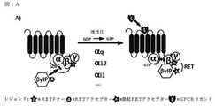
3つのサブユニットα、β、及びγからなるヘテロ三量体Gタンパク質は、Gタンパク質共役型レセプター(GPCR)によって提供された情報を種々の細胞内エフェクターに中継する。刺激が無い場合には、Gタンパク質のαサブユニットは、GDP(グアノシン二リン酸)分子と複合体を形成している。リガンドによるレセプターの活性化に伴う立体配座の変化は、GDP分子のGTP(グアノシン三リン酸)へのリン酸化を促進する。GTP結合Gαサブユニットは、Gβγサブユニットから解離した後、それらは双方とも、下流エフェクターと相互作用し、それらの活性を調節するのに利用できる。従って、Gタンパク質活性化は、Gβγ相互作用タンパク質(βγIP)を用いて、Gβγとの相互作用を介してそれらの下流エフェクターを分析することによって評価することができる。GαサブユニットによるGDPへのGTP加水分解後、Gβγに対するGα親和性は回復し、3つのサブユニットは再会合して、不活性ヘテロ三量体Gタンパク質を形成し、エフェクターの関与、従って、シグナル伝達が終了する(Gilman,1987)。 The heterotrimeric G protein, which consists of the three subunits α, β, and γ, relays the information provided by the G protein-coupled receptor (GPCR) to various intracellular effectors. In the absence of irritation, the alpha subunit of the G protein forms a complex with the GDP (guanosine diphosphate) molecule. Changes in conformation associated with receptor activation by ligands promote phosphorylation of GDP molecules to GTP (guanosine triphosphate). After dissociating the GTP-binding Gα subunit from the Gβγ subunit, both of them can be used to interact with downstream effectors and regulate their activity. Therefore, G protein activation can be assessed by using Gβγ interacting proteins (βγIP) and analyzing their downstream effectors via interaction with Gβγ. After GTP hydrolysis to GDP by the Gα subunit, Gα affinity for Gβγ is restored and the three subunits reassociate to form an inactive heterotrimeric G protein, with effector involvement and thus signaling. Transmission ends (Gilman, 1987).
GPCRによるGタンパク質の古典的な活性化に加えて、他のタンパク質は、これらのヘテロ三量体Gタンパク質、例えば、Gタンパク質シグナル伝達(RGS)の調節因子、Gタンパク質シグナル伝達(AGS)の活性化因子の活性、及びコリンエステラーゼ8タンパク質(Ric−8)のインヒビターへの耐性を調節することもできる。これらの非正準的シグナル伝達経路のいくつかでは、GPCRによって古典的に発揮されたグアニン交換因子(GEF)活性は、例えば、Ric−8などの別のタンパク質に置き換えられる(Boularan and Kehrl,2014)。
In addition to the classical activation of G proteins by GPCR, other proteins include the activity of these heterotrimeric G proteins, eg, regulators of G protein signaling (RGS), G protein signaling (AGS). It is also possible to regulate the activity of the chemogenetic factor and the resistance of the
レセプターの脱感作への役割について最初に特徴付けられたGタンパク質共役型レセプターキナーゼ(GRK)2及び3は、Gβγサブユニットとの相互作用を介して関与したエフェクターでもある。GRK2及びGRK3は、活性化したGTP結合Gαサブユニットから解離する際にGタンパク質のGβγサブユニットと相互作用するプレクストリン相同(PH)ドメイン含有(Pitcher,Ingleseら,1992)(Touhara,Ingleseら,1994)。その結果、GRK2及びGRK3などの、Gβγ(βγIP)と相互作用するタンパク質を使用して、GPCRまたは他のGタンパク質活性化因子によるGタンパク質活性化を直接研究することができる。 G protein-coupled receptor kinases (GRKs) 2 and 3, initially characterized for their role in receptor desensitization, are also effectors involved through interaction with the Gβγ subunit. GRK2 and GRK3 contain a Plextrin homologous (PH) domain that interacts with the Gβγ subunit of the G protein upon dissociation from the activated GTP-binding Gα subunit (Pitcher, Inglese et al., 1992) (Touhara, Inglese et al., 1994). As a result, proteins that interact with Gβγ (βγIP), such as GRK2 and GRK3, can be used to directly study G protein activation by GPCRs or other G protein activators.
いくつかのアプローチは、創薬業界において、GPCRの活性化、従って、レセプターによるGタンパク質の関与を評価するために現在使用されており、例えば、Gタンパク質によるGTPγS取り込みに基づいたカルシウム動員アッセイまたは放射性分析である。カルシウム動員アッセイは、下流のGq活性化で発生しているシグナル伝達事象を測定し、改変Gαサブユニットの使用により共役した場合にのみ、GiまたはGs共役レセプターに適用することができる。GTPγS取り込みアッセイの場合では、種々のヘテロ三量体Gタンパク質の活性化は、放射性GTPγ35Sを用いて細胞膜上で直接測定し、生きた細胞で行うことはできない。 Several approaches are currently used in the drug discovery industry to assess GPCR activation, and thus the involvement of G proteins by receptors, such as calcium mobilization assays based on GTPγS uptake by G proteins or radioactivity. It is an analysis. Calcium recruitment assays measure signaling events occurring in downstream Gq activation and can only be applied to Gi or Gs conjugated receptors when conjugated by the use of modified Gα subunits. In the case of GTPγS incorporation assays, the activation of various heterotrimeric G proteins, with a radioactive GTPy 35 S measured directly on the cell membrane, it can not be carried out in living cells.
Gタンパク質活性化因子またはGαサブユニットを改変することなく、生きた細胞中でのGタンパク質の活性化は、今までのところ研究が行われていない。さらに、公知の方法は、同じ検出パートナーを用いて全ての異なるGタンパク質を研究するには適切ではない。そのようなアッセイは、Gタンパク質カップリングプロファイルの特徴付けを可能にし、かつ、例えば、スクリーニングアッセイ及び構造−活性の相関研究に使用されるシグナル伝達特性を定義して新規化合物の同定を容易にすることによって、異なる段階の創薬プロセスで特に有用であろう。これは、処方された全ての薬剤の26%がGPCRを介して作用するため、薬剤標的としてGタンパク質活性化因子の重要性を考えると特に当てはまる(Garland 2013)。Gタンパク質活性化因子を標的化する新規の治療活性分子の開発を支援するのにいくつかのアプローチが利用可能であるが、新規薬剤の発見は、それらの化合物の正確な作用機序に利用できる情報が不足していることで制限されることが多い。 The activation of G proteins in living cells without modifying the G protein activator or Gα subunit has not been studied so far. Moreover, known methods are not suitable for studying all different G proteins using the same detection partner. Such assays allow the characterization of G protein coupling profiles and facilitate the identification of novel compounds, eg, defining signaling properties used in screening assays and structure-activity correlation studies. This will be particularly useful in different stages of the drug discovery process. This is especially true given the importance of G protein activators as drug targets, as 26% of all prescribed drugs act via GPCRs (Gard 2013). Although several approaches are available to support the development of new therapeutically active molecules that target G protein activators, the discovery of new drugs can be used for the exact mechanism of action of those compounds. Often limited by lack of information.
従って、Gタンパク質の活性化を評価する新規のツール及びアッセイが求められている。 Therefore, there is a need for new tools and assays to assess G protein activation.
本説明は、多くの文書を指し、その内容全体を参照によって本明細書に引用したものとする。 This description refers to many documents, the entire contents of which are hereby incorporated by reference.
本発明は、以下の項目1〜68を提供する:
1.(A)、または(B)で定義される要素を含む、Gタンパク質活性を検出するためのバイオセンサーシステムであって、
(A)
(a)共鳴エネルギー転移(RET)ドナー;(b)RETアクセプター、または(c)レポータータンパク質の第1の断片に融合したGβγ相互作用タンパク質(βγIP)を含む第1の成分;及び
融合Gβタンパク質または融合Gγタンパク質を含む第2の成分、前記Gβタンパク質または前記Gγタンパク質は、(a)RETドナー;(b)RETアクセプター、または(c)前記レポータータンパク質の第2の断片に融合している;
を含む、(i)第1のバイオセンサー、
(i)で定義される前記第1及び第2の成分;及び
組換えGαタンパク質を含む第3の成分;
(a)前記βγIPが前記RETドナーに融合している場合、前記GβまたはGγタンパク質は、前記RETアクセプターに融合しており;(b)前記βγIPが前記RETアクセプターに融合している場合、前記GβまたはGγタンパク質は、前記RETドナーに融合しており;及び(c)前記βγIPが前記レポータータンパク質の第1の断片に融合している場合、前記GβまたはGγタンパク質は、前記レポータータンパク質の第2の断片に融合している;
を含む、(ii)第2のバイオセンサー、または
(B)
(a)RETドナー;(b)RETアクセプター、または(c)レポータータンパク質の第1の断片に融合したGβγ相互作用タンパク質(βγIP)を含む第1の成分;
融合Gタンパク質共役型レセプター(GPCR)を含む第2の成分、前記GPCRは、そのC末端で(a)RETドナー;(b)RETアクセプター、または(c)前記レポータータンパク質の第2の断片に融合している;
組換えGαタンパク質を含む第3の成分;
(a)前記βγIPが前記RETドナーに融合している場合、前記GPCRは、前記RETアクセプターに融合しており;(b)前記βγIPが前記RETアクセプターに融合している場合、前記GPCRは、前記RETドナーに融合しており;及び(c)前記βγIPが前記レポータータンパク質の第1の断片に融合している場合、前記GPCRは、前記レポータータンパク質の第2の断片に融合している
を含む、(i)バイオセンサー
を含む、前記バイオセンサーシステム。
The present invention provides the following
1. 1. A biosensor system for detecting G protein activity, comprising the elements defined in (A) or (B).
(A)
(A) Resonant Energy Transfer (RET) Donor; (b) RET Acceptor, or (c) First Component Containing Gβγ Interaction Protein (βγIP) Fused to First Fragment of Reporter Protein; and Fused Gβ Protein or A second component comprising the fused Gγ protein, said Gβ protein or said Gγ protein, is fused to (a) a RET donor; (b) a RET acceptor, or (c) a second fragment of the reporter protein;
(I) First biosensor, including
The first and second components as defined in (i); and a third component containing a recombinant Gα protein;
(A) When the βγIP is fused to the RET donor, the Gβ or Gγ protein is fused to the RET acceptor; (b) When the βγIP is fused to the RET acceptor, the Gβ Alternatively, the Gγ protein is fused to the RET donor; and (c) if the βγIP is fused to the first fragment of the reporter protein, the Gβ or Gγ protein is a second of the reporter protein. Fused into fragments;
(Ii) a second biosensor, or (B)
A first component comprising (a) a RET donor; (b) a RET acceptor, or (c) a Gβγ interacting protein (βγIP) fused to a first fragment of a reporter protein;
A second component comprising a fused G protein-coupled receptor (GPCR), said GPCR, is fused at its C-terminus to (a) a RET donor; (b) a RET acceptor, or (c) a second fragment of the reporter protein. doing;
Third component containing recombinant Gα protein;
(A) When the βγIP is fused to the RET donor, the GPCR is fused to the RET acceptor; (b) When the βγIP is fused to the RET acceptor, the GPCR is said. Fusing to a RET donor; and (c) if the βγIP is fused to a first fragment of the reporter protein, the GPCR comprises fusing to a second fragment of the reporter protein. (I) The biosensor system comprising a biosensor.
2.前記Gγタンパク質が、前記RETドナー、RETアクセプター、または第2の断片に融合している、項目1に記載のバイオセンサーシステム。
2. The biosensor system of
3.前記RETドナー、RETアクセプター、または第2の断片が、前記GβまたはGγタンパク質のN末端で融合している、項目1または2に記載のバイオセンサーシステム。
3. 3. The biosensor system of
4.前記RETドナー、RETアクセプター、または第1の断片が、前記βγIPのC末端で融合している、項目1〜3のいずれか1項に記載のバイオセンサーシステム。
4. The biosensor system according to any one of
5.前記βγIPが、前記RETアクセプター及び前記Gβタンパク質に融合しており、Gγタンパク質またはGPCRが、前記RETドナーに融合している、項目1〜4のいずれか1項に記載のバイオセンサーシステム。
5. The biosensor system according to any one of
6.前記RETドナーが、生物発光タンパク質である、項目1〜5のいずれか1項に記載のバイオセンサーシステム。
6. The biosensor system according to any one of
7.前記生物発光タンパク質が、ルシフェラーゼである、項目6に記載のバイオセンサーシステム。
7. The biosensor system according to
8.前記ルシフェラーゼが、ウミシイタケ由来ルシフェラーゼである、項目7に記載のバイオセンサーシステム。
8. The biosensor system according to
9.前記RETアクセプターが、蛍光タンパク質である、項目1〜8のいずれか1項に記載のバイオセンサーシステム。
9. The biosensor system according to any one of
10.前記蛍光タンパク質が、GFPである、項目9に記載のバイオセンサーシステム。
10. 9. The biosensor system according to
11.前記βγIPが、前記第1の断片に融合しており、及び前記Gβタンパク質、Gγタンパク質またはGPCRが、前記第2の断片に融合している、項目1〜4のいずれか1項に記載のバイオセンサーシステム。
11. The bio according to any one of
12.前記レポータータンパク質が、生物発光タンパク質である、項目11に記載のバイオセンサーシステム。
12. The biosensor system according to
13.前記生物発光タンパク質が、ルシフェラーゼである、項目12に記載のバイオセンサーシステム。
13. The biosensor system according to
14.前記ルシフェラーゼが、ウミシイタケ由来ルシフェラーゼである、項目13に記載のバイオセンサーシステム。
14. The biosensor system according to
15.前記第1の断片が、ウミシイタケ由来ルシフェラーゼの約1〜110残基を含み、及び前記第2の断片が、ウミシイタケ由来ルシフェラーゼの約111〜311残基を含む、項目14に記載のバイオセンサーシステム。
15. The biosensor system according to
16.前記第1の成分が、前記βγIPまたは前記RETドナー、RETアクセプター、または第1の断片に融合した原形質膜(PM)標的化部分をさらに含む、項目1〜15のいずれか1項に記載のバイオセンサーシステム。
16. The item according to any one of
17.前記PM標的化部分が、前記RETドナー、RETアクセプター、または第1の断片のC末端で融合している、項目16に記載のバイオセンサーシステム。
17. 16. The biosensor system of
18.前記PM標的化部分が、プレニル化モチーフを含む、項目16または17に記載のバイオセンサーシステム。
18. The biosensor system according to
19.前記プレニル化モチーフが、ヒトKRASスプライス変異体bのプレニル化モチーフである、項目16に記載のバイオセンサーシステム。
19. The biosensor system according to
20.前記PM標的化部分が、アミノ酸配列KKKKKKSKTKCVIM(配列番号37)を含む、項目19に記載のバイオセンサーシステム。 20. 19. The biosensor system of item 19, wherein the PM targeting moiety comprises the amino acid sequence KKKKKKKSKTKCVIM (SEQ ID NO: 37).
21.(i)前記RETドナー、RETアクセプター、または第1の断片と(ii)前記PM標的化部分の間にフレキシブルリンカーをさらに含む、項目16〜20のいずれか1項に記載のバイオセンサーシステム。
21. (I) The biosensor system according to any one of
22.前記フレキシブルリンカーが、約50〜約500アミノ酸に対応する長さを有する、項目21に記載のバイオセンサーシステム。 22. 21. The biosensor system of item 21, wherein the flexible linker has a length corresponding to about 50 to about 500 amino acids.
23.前記フレキシブルリンカーが、約200アミノ酸に対応する長さを有する、項目22に記載のバイオセンサーシステム。 23. 22. The biosensor system of item 22, wherein the flexible linker has a length corresponding to about 200 amino acids.
24.前記組換えGαタンパク質が、ヒトGαq、Gαs、Gαi1、Gαi2、Gαi3、Gαt−cone、Gαt−rod、Gαt−gust、Gαz、GαoA、GαoB、Gαolf、Gα11、Gα12、Gα13、Gα14、及びGα15/Gα16タンパク質、またはそれらの無差別または非選択的Gα変異体、例えば、本明細書に記載されるヒトGαqタンパク質の残基66、67、及び/または75に対応する位置で突然変異を含む突然変異Gαポリペプチドである、項目1〜23のいずれか1項に記載のバイオセンサーシステム。
24. The recombinant Gα proteins are human Gα q , Gα s , Gα i1 , Gα i2 , Gα i3 , Gα t-cone , Gα t-rod , Gα t-gust , Gα z , Gα oA , Gα oB , Gα olf . Gα 11 , Gα 12 , Gα 13 , Gα 14 , and Gα 15 / Gα 16 proteins, or promiscuous or non-selective Gα variants thereof, eg,
25.前記βγIPが、GRK2またはGRK3である、項目1〜24のいずれか1項に記載のバイオセンサーシステム。
25. The biosensor system according to any one of
26.(i)前記第2の成分が融合Gβタンパク質を含む場合、前記第1及び第2のバイオセンサーは、組換えGγタンパク質をさらに含み、または(ii)前記第2の成分が融合Gγタンパク質を含む場合、前記第1及び第2のバイオセンサーは、組換えGβタンパク質をさらに含む、項目1〜25のいずれか1項に記載のバイオセンサーシステム。
26. (I) When the second component comprises a fused Gβ protein, the first and second biosensors further comprise a recombinant Gγ protein, or (ii) the second component comprises a fused Gγ protein. In the case, the biosensor system according to any one of
27.(A)で定義される前記バイオセンサーシステムが、Gタンパク質共役型レセプター(GPCR)をさらに含む、項目1〜25のいずれか1項に記載のバイオセンサーシステム。
27. The biosensor system according to any one of
28.(B)で定義される前記バイオセンサーシステムが、組換えGβタンパク質及び/または組換えGγタンパク質をさらに含む、項目1〜27のいずれか1項に記載のバイオセンサーシステム。
28. The biosensor system according to any one of
29.前記第1のバイオセンサーが、第1の細胞中に存在し、前記第2のバイオセンサーが、第2の細胞中に存在する、項目1〜28のいずれか1項に記載のバイオセンサーシステム。
29. The biosensor system according to any one of
30.(A)で定義される前記バイオセンサーシステムにおいて、前記第1のバイオセンサーが、第1の膜調製物中に存在し、及び前記第2のバイオセンサーが、第2の膜調製物中に存在する、項目1〜28のいずれか1項に記載のバイオセンサーシステム。
30. In the biosensor system defined in (A), the first biosensor is present in the first membrane preparation and the second biosensor is present in the second membrane preparation. The biosensor system according to any one of
31.(A)で定義される前記バイオセンサーシステムが、複数の第2のバイオセンサーを含み、前記第2のバイオセンサーの各々が、異なる組換えGαタンパク質を含む、項目1〜30のいずれか1項に記載のバイオセンサーシステム。
31.
32.前記異なる組換えGαタンパク質が、以下のGαタンパク質:Gαq、Gαs、Gαi1、Gαi2、Gαi3、Gαt−cone、Gαt−rod、Gαt−gust、Gαz、GαoA、GαoB、Gαolf、Gα11、Gα12、Gα13、Gα14、及びGα15/Gα16の少なくとも2つである、項目31に記載のバイオセンサーシステム。 32. The different recombinant Gα proteins include the following Gα proteins: Gα q , Gα s , Gα i1 , Gα i2 , Gα i3 , Gα t-cone , Gα t-rod , Gα t-gust , Gα z , Gα oA , Gα. 31. The biosensor system of item 31, wherein the biosensor system is at least two of oB , Gα olf , Gα 11 , Gα 12 , Gα 13 , Gα 14 , and Gα 15 / Gα 16.
33.項目1〜26のいずれか1項で定義される第1、第2、及び第3の成分をコードする配列を含む、核酸。 33. A nucleic acid comprising a sequence encoding the first, second, and third components as defined in any one of items 1-26.
34.Gγタンパク質またはGβタンパク質をコードする配列をさらに含む、項目33に記載の核酸。 34. 33. The nucleic acid of item 33, further comprising a sequence encoding a Gγ protein or Gβ protein.
35.1つ以上の翻訳調節配列をさらに含む、項目33または34に記載の核酸。
35. The nucleic acid of
36.前記1つ以上の翻訳調節配列が、内部リボソーム導入部位(IRES)である、項目35に記載の核酸。 36. 35. The nucleic acid of item 35, wherein the one or more translational regulatory sequences are internal ribosome entry sites (IRES).
37.Gタンパク質活性を検出するためのバイオセンサーであって、
(i)(a)RETドナー;(b)RETアクセプター、または(c)レポータータンパク質の第1の断片に融合したGβγ相互作用タンパク質(βγIP)を含む第1の成分;及び
(ii)融合原形質膜(PM)標的化部分を含む第2の成分、前記PM標的化部分は、(a)RETドナー;(b)RETアクセプター、または(c)前記レポータータンパク質の第2の断片に融合している;
を含み、
(a)前記βγIPが前記RETドナーに融合している場合、前記PM標的化部分は、前記RETアクセプターに融合しており;(b)前記βγIPが前記RETアクセプターに融合している場合、前記PM標的化部分は、前記RETドナーに融合しており;及び(c)前記βγIPが前記レポータータンパク質の第1の断片に融合している場合、前記PM標的化部分は、前記レポータータンパク質の第2の断片に融合している、前記バイオセンサー。
37. A biosensor for detecting G protein activity
(I) (a) RET donor; (b) RET acceptor, or (c) first component containing Gβγ interacting protein (βγIP) fused to the first fragment of the reporter protein; and (ii) fusion protoplasm A second component, including a membrane (PM) targeting moiety, said PM targeting moiety is fused to (a) a RET donor; (b) a RET acceptor, or (c) a second fragment of the reporter protein. ;
Including
(A) When the βγIP is fused to the RET donor, the PM targeting moiety is fused to the RET acceptor; (b) When the βγIP is fused to the RET acceptor, the PM The targeting moiety is fused to the RET donor; and (c) if the βγIP is fused to the first fragment of the reporter protein, the PM targeting moiety is the second of the reporter protein. The biosensor fused to the fragment.
38.前記PM標的化部分が、PMタンパク質または前記PMに局在化するその断片である、項目37に記載のバイオセンサー。 38. 37. The biosensor of item 37, wherein the PM targeting moiety is a PM protein or a fragment thereof localized to the PM.
39.前記PMタンパク質またはその断片が、(a)パルミトイル化、ミリストイル化、及び/またはプレニル化シグナル配列、及び/または(b)多塩基性配列を含む、項目38に記載のバイオセンサー。 39. 38. The biosensor of item 38, wherein the PM protein or fragment thereof comprises (a) palmitoylation, myristoylation, and / or prenylation signal sequences, and / or (b) polybasic sequences.
40.前記多塩基性配列及びプレニル化シグナル配列が、ヒトKRASスプライス変異体b由来である、項目39に記載のバイオセンサー。 40. 39. The biosensor of item 39, wherein the multibasic sequence and the prenylation signal sequence are derived from the human KRAS splice variant b.
41.前記PM標的化部分が、アミノ酸配列KKKKKKSKTKCVIM(配列番号37)を含む、項目40に記載のバイオセンサー。
41. The biosensor of
42.前記バイオセンサーが、組換えGαタンパク質を含む第3の成分をさらに含む、項目37〜41のいずれか1項に記載のバイオセンサー。 42. The biosensor according to any one of items 37 to 41, wherein the biosensor further comprises a third component containing a recombinant Gα protein.
43.前記組換えGαタンパク質が、Gqファミリーである、項目42に記載のバイオセンサー。 43. 42. The biosensor according to item 42, wherein the recombinant Gα protein is a Gq family.
44.前記組換えGαタンパク質が、GαqまたはGα11である、項目43に記載のバイオセンサー。
44. 43. The biosensor of
45.試験薬剤がGPCRの活性を調節するかどうかの決定方法であって、
(1)(A)、(B)、または(C)で定義される要素を含むバイオセンサーを提供すること:
(A)
(i)(a)RETドナー;(b)RETアクセプター、または(c)レポータータンパク質の第1の断片に融合したGβγ相互作用タンパク質(βγIP)を含む第1の成分;
(ii)融合Gβタンパク質または融合Gγタンパク質を含む第2の成分、前記Gβタンパク質または前記Gγタンパク質は、(a)RETドナー;(b)RETアクセプター、または(c)前記レポータータンパク質の第2の断片に融合している、
(a)前記βγIPが前記RETドナーに融合している場合、前記GβまたはGγタンパク質は、前記RETアクセプターに融合しており;(b)前記βγIPが前記RETアクセプターに融合している場合、前記GβまたはGγタンパク質は、前記RETドナーに融合しており;及び(c)前記βγIPが前記レポータータンパク質の第1の断片に融合している場合、前記GβまたはGγタンパク質は、前記レポータータンパク質の第2の断片に融合している;
(iii)組換えGαタンパク質を含む第3の成分;及び
(iv)前記GPCRを含む第4の成分;
(B)
(i)(a)RETドナー;(b)RETアクセプター、または(c)レポータータンパク質の第1の断片に融合したGβγ相互作用タンパク質(βγIP)を含む第1の成分;
(ii)そのC末端で(a)RETドナー;(b)RETアクセプター、または(c)前記レポータータンパク質の第2の断片に融合した前記GPCRを含む第2の成分;
(iii)組換えGαタンパク質を含む第3の成分;
(a)前記βγIPが前記RETドナーに融合している場合、前記GPCRは、前記RETアクセプターに融合しており;(b)前記βγIPが前記RETアクセプターに融合している場合、前記GPCRは、前記RETドナーに融合しており;及び(c)前記βγIPが前記レポータータンパク質の第1の断片に融合している場合、前記GPCRは、前記レポータータンパク質の第2の断片に融合している;または
(C)
(i)(a)RETドナー;(b)RETアクセプター、または(c)レポータータンパク質の第1の断片に融合したGβγ相互作用タンパク質(βγIP)を含む第1の成分;
(ii)融合原形質膜(PM)標的化部分を含む第2の成分、前記PM標的化部分は、(a)RETドナー;(b)RETアクセプター、または(c)前記レポータータンパク質の第2の断片に融合している;
(a)前記βγIPが前記RETドナーに融合している場合、前記PM標的化部分は、前記RETアクセプターに融合しており;(b)前記βγIPが前記RETアクセプターに融合している場合、前記PM標的化部分は、前記RETドナーに融合しており;及び(c)前記βγIPが前記レポータータンパク質の第1の断片に融合している場合、前記PM標的化部分は、前記レポータータンパク質の第2の断片に融合している;
(iii)組換えGαタンパク質を含む第3の成分;及び
(iv)前記GPCRを含む第4の成分;及び
(2)前記試験薬剤の存在下及び不在下において、前記RETアクセプターまたはレポータータンパク質が発したシグナルを測定すること;
前記薬剤の存在下において測定されたより高いシグナルは、前記試験薬剤が前記GPCRの活性を増加させることを示し、前記薬剤の存在下において測定されたより低いシグナルは、前記薬剤が前記GPCRの活性を阻害することを示す
を含む、前記方法。
45. A method of determining whether a test drug regulates GPCR activity.
(1) To provide a biosensor containing the elements defined in (A), (B), or (C):
(A)
(I) (a) RET donor; (b) a RET acceptor, or (c) a first component comprising a Gβγ interacting protein (βγIP) fused to a first fragment of a reporter protein;
(Ii) A second component comprising a fused Gβ protein or fused Gγ protein, said Gβ protein or said Gγ protein is (a) a RET donor; (b) a RET acceptor, or (c) a second fragment of the reporter protein. Is fused with
(A) When the βγIP is fused to the RET donor, the Gβ or Gγ protein is fused to the RET acceptor; (b) When the βγIP is fused to the RET acceptor, the Gβ Alternatively, the Gγ protein is fused to the RET donor; and (c) if the βγIP is fused to the first fragment of the reporter protein, the Gβ or Gγ protein is a second of the reporter protein. Fused into fragments;
(Iii) A third component containing a recombinant Gα protein; and (iv) a fourth component containing the GPCR;
(B)
(I) (a) RET donor; (b) a RET acceptor, or (c) a first component comprising a Gβγ interacting protein (βγIP) fused to a first fragment of a reporter protein;
(Ii) At its C-terminus, (a) a RET donor; (b) a RET acceptor, or (c) a second component containing the GPCR fused to a second fragment of the reporter protein;
(Iii) Third component containing recombinant Gα protein;
(A) When the βγIP is fused to the RET donor, the GPCR is fused to the RET acceptor; (b) When the βγIP is fused to the RET acceptor, the GPCR is said. Fused to a RET donor; and (c) if the βγIP is fused to a first fragment of the reporter protein, the GPCR is fused to a second fragment of the reporter protein; or ( C)
(I) (a) RET donor; (b) a RET acceptor, or (c) a first component comprising a Gβγ interacting protein (βγIP) fused to a first fragment of a reporter protein;
(Ii) A second component comprising a fusion plasma membrane (PM) targeting moiety, said PM targeting moiety is (a) a RET donor; (b) a RET acceptor, or (c) a second of the reporter protein. Fused into fragments;
(A) When the βγIP is fused to the RET donor, the PM targeting moiety is fused to the RET acceptor; (b) When the βγIP is fused to the RET acceptor, the PM The targeting moiety is fused to the RET donor; and (c) if the βγIP is fused to the first fragment of the reporter protein, the PM targeting moiety is the second of the reporter protein. Fused into fragments;
(Iii) A third component containing a recombinant Gα protein; and (iv) a fourth component containing the GPCR; and (2) the RET acceptor or reporter protein is emitted in the presence and absence of the test agent. To measure the signal
A higher signal measured in the presence of the agent indicates that the test agent increases the activity of the GPCR, and a lower signal measured in the presence of the agent inhibits the activity of the GPCR. The method described above, comprising:
46.前記バイオセンサーが、項目2〜32及び38〜44で定義される特徴の1つ以上を含む、項目44に記載の方法。
46. 44. The method of
47.Gαタンパク質がGPCRアゴニストによって活性化されるかどうかの決定方法であって、
(a)項目1〜32のいずれか1項に記載のバイオセンサーシステムの前記第1及び第2のバイオセンサー中で前記GPCRアゴニストの存在下及び不在下において、前記RETアクセプターまたはレポータータンパク質が発したシグナルを測定すること、及び
(b)前記RETアクセプターまたはレポータータンパク質が発したシグナルに基づいて、前記Gαタンパク質が前記GPCRアゴニストによって活性化されるかどうかを同定すること;
前記第1のバイオセンサーに対して前記第2のバイオセンサー中の前記GPCRアゴニストの存在下において測定されたシグナルのより高い増加は、前記Gαタンパク質が前記GPCRアゴニストによって活性化されることを示し、前記第1のバイオセンサーに対して前記第2のバイオセンサー中の前記GPCRアゴニストの存在下において測定されたシグナルの同様のまたはより低い増加、または減少は、前記Gαタンパク質が前記GPCRアゴニストによって活性化されないことを示す
を含む、前記方法。
47. A method of determining whether a Gα protein is activated by a GPCR agonist.
(A) The RET acceptor or reporter protein was emitted in the presence and absence of the GPCR agonist in the first and second biosensors of the biosensor system according to any one of items 1-32. Measuring the signal and (b) identifying whether the Gα protein is activated by the GPCR agonist based on the signal emitted by the RET acceptor or reporter protein;
A higher increase in signal measured in the presence of the GPCR agonist in the second biosensor relative to the first biosensor indicates that the Gα protein is activated by the GPCR agonist. A similar or lower increase or decrease in the signal measured in the presence of the GPCR agonist in the second biosensor relative to the first biosensor is that the Gα protein is activated by the GPCR agonist. Said method, comprising indicating that it is not.
48.Gαタンパク質がGPCRアゴニストによって活性化されるかどうかの決定方法であって、
(a)以下の(i)、及び(ii)を含む第1のバイオセンサー中の前記GPCRアゴニストの存在下及び不在下において、RETアクセプターまたはレポータータンパク質が発したシグナルを測定すること:
(i)(a)RETドナー;(b)RETアクセプター、または(c)レポータータンパク質の第1の断片に融合したGβγ相互作用タンパク質(βγIP)を含む第1の成分;及び
(ii)融合Gタンパク質共役型レセプター(GPCR)を含む第2の成分、前記GPCRは、そのC末端で(a)RETドナー;(b)RETアクセプター、または(c)前記レポータータンパク質の第2の断片に融合している;
(b)以下の(i)、及び(ii)を含む第2のバイオセンサー中の前記GPCRアゴニストの存在下及び不在下において、RETアクセプターまたはレポータータンパク質が発したシグナルを測定すること:
(i)(a)で定義される前記第1及び第2の成分;及び
(ii)前記Gαタンパク質の組換え形態を含む第3の成分;
(a)前記βγIPが前記RETドナーに融合している場合、前記GPCRは、前記RETアクセプターに融合しており;(b)前記βγIPが前記RETアクセプターに融合している場合、前記GPCRは、前記RETドナーに融合しており;及び(c)前記βγIPが前記レポータータンパク質の第1の断片に融合している場合、前記GPCRは、前記レポータータンパク質の第2の断片に融合している;
前記第1のバイオセンサーに対して前記第2のバイオセンサー中の前記GPCRアゴニストの存在下において測定されたシグナルのより高い増加は、前記Gαタンパク質が前記GPCRアゴニストによって活性化されることを示し、前記第1のバイオセンサーに対して前記第2のバイオセンサー中の前記GPCRアゴニストの存在下において測定されたシグナルの同様のまたはより低い増加、または減少は、前記Gαタンパク質が前記GPCRアゴニストによって活性化されないことを示す
を含む、前記方法。
48. A method of determining whether a Gα protein is activated by a GPCR agonist.
(A) To measure the signal emitted by a RET acceptor or reporter protein in the presence and absence of the GPCR agonist in a first biosensor comprising (i) and (ii) below:
(I) (a) RET donor; (b) a first component comprising a Gβγ interacting protein (βγIP) fused to a first fragment of a RET acceptor or (c) reporter protein; and (ii) fused G protein. A second component containing a conjugated receptor (GPCR), said GPCR, is fused at its C-terminus to (a) a RET donor; (b) a RET acceptor, or (c) a second fragment of the reporter protein. ;
(B) To measure the signal emitted by the RET acceptor or reporter protein in the presence and absence of the GPCR agonist in a second biosensor comprising (i) and (ii) below:
(I) The first and second components as defined in (a); and (ii) a third component containing a recombinant form of the Gα protein;
(A) When the βγIP is fused to the RET donor, the GPCR is fused to the RET acceptor; (b) When the βγIP is fused to the RET acceptor, the GPCR is said. Fused to a RET donor; and (c) if the βγIP is fused to a first fragment of the reporter protein, the GPCR is fused to a second fragment of the reporter protein;
A higher increase in signal measured in the presence of the GPCR agonist in the second biosensor relative to the first biosensor indicates that the Gα protein is activated by the GPCR agonist. A similar or lower increase or decrease in the signal measured in the presence of the GPCR agonist in the second biosensor relative to the first biosensor is that the Gα protein is activated by the GPCR agonist. Said method, comprising indicating that it is not.
49.前記バイオセンサーが、項目38〜44で定義される特徴の1つ以上を含む、項目47に記載の方法。
49. 47. The method of
50.(3)以下の(a)、(b)、または(c)中の前記RETアクセプターまたはレポータータンパク質が発したシグナルを測定すること
(a)項目1〜31のいずれか1項に記載の要素(A)で定義される第2のバイオセンサー(複数可)、
(b)項目1〜31のいずれか1項に記載の要素(B)で定義されるバイオセンサー、または
(c)項目42〜44のいずれか1項に記載のバイオセンサー、
試験薬剤の存在下及び不在下において、及びGPCRアゴニストの存在下において、前記組換えGαタンパク質は、前記GPCRにカップリングしている;及び
(4)前記試験薬剤が前記Gαタンパク質のインヒビターであるかどうかを決定すること;
前記試験薬剤の存在下において測定されたより低いシグナルは、前記試験薬剤が前記Gαタンパク質のインヒビターであることを示し、前記試験薬剤の存在下において測定された同様のまたはより高いシグナルは、前記試験薬剤が前記Gαタンパク質のインヒビターではないことを示す
をさらに含む、項目45に記載の方法。
50. (3) Measuring the signal emitted by the RET acceptor or reporter protein in (a), (b), or (c) below (a) The element according to any one of
(B) The biosensor defined by the element (B) according to any one of
In the presence and absence of the test agent, and in the presence of the GPCR agonist, the recombinant Gα protein is coupled to the GPCR; and (4) is the test agent an inhibitor of the Gα protein? Decide whether or not;
A lower signal measured in the presence of the test agent indicates that the test agent is an inhibitor of the Gα protein, and a similar or higher signal measured in the presence of the test agent is the test agent. 45. The method of item 45, further comprising indicating that is not an inhibitor of the Gα protein.
51.試験薬剤が目的Gαタンパク質のインヒビターであるかどうかの決定方法であって、
(1)
(a)項目1〜32のいずれか1項に記載の要素(A)で定義される第2のバイオセンサー(複数可)、
(b)項目1〜32のいずれか1項に記載の要素(B)で定義されるバイオセンサー、または
(c)項目42〜44のいずれか1項に記載のバイオセンサー
をGPCRアゴニストと接触させること、前記組換えGαタンパク質は、前記目的Gαタンパク質に対応する;
(2)前記試験薬剤の存在下及び不在下において、前記RETアクセプターまたはレポータータンパク質が発したシグナルを測定すること;及び
(c)前記試験薬剤が前記Gαタンパク質のインヒビターであるかどうかを決定すること
前記試験薬剤の存在下において測定されたより低いシグナルは、前記試験薬剤が前記目的Gαタンパク質のインヒビターであることを示し、前記試験薬剤の存在下において測定された同様のまたはより高いシグナルは、前記試験薬剤が前記目的Gαタンパク質のインヒビターではないことを示す
を含む、前記方法。
51. A method for determining whether a test drug is an inhibitor of a target Gα protein.
(1)
(A) A second biosensor (s) defined by the element (A) according to any one of
(B) The biosensor defined by the element (B) according to any one of
(2) Measuring the signal emitted by the RET acceptor or reporter protein in the presence and absence of the test agent; and (c) determining whether the test agent is an inhibitor of the Gα protein. A lower signal measured in the presence of the test agent indicates that the test agent is an inhibitor of the target Gα protein, and a similar or higher signal measured in the presence of the test agent is the test. The method comprising indicating that the agent is not an inhibitor of the Gα protein of interest.
52.試験薬剤が目的Gαタンパク質活性化因子であるかどうかの決定方法であって、
(1)
(a)項目1〜32のいずれか1項に記載の要素(A)で定義される第2のバイオセンサー(複数可)、
(b)項目1〜32のいずれか1項に記載の要素(B)で定義されるバイオセンサー、または
(c)項目42〜44のいずれか1項に記載のバイオセンサー
をGPCRアンタゴニストと接触させること、前記組換えGαタンパク質は、前記目的Gαタンパク質に対応する;
(2)前記試験薬剤の存在下及び不在下において、前記RETアクセプターまたはレポータータンパク質が発したシグナルを測定すること;及び
(3)前記試験薬剤が前記Gαタンパク質活性化因子であるかどうかを決定すること、
前記試験薬剤の存在下において測定されたより高いシグナルは、前記試験薬剤が前記目的Gαタンパク質活性化因子であることを示し、前記試験薬剤の存在下において測定された同様のまたはより低いシグナルは、前記試験薬剤が前記目的Gαタンパク質活性化因子ではないことを示す
を含む、前記方法。
52. A method for determining whether a test drug is a target Gα protein activator.
(1)
(A) A second biosensor (s) defined by the element (A) according to any one of
(B) Contact the biosensor defined by the element (B) according to any one of
(2) Measuring the signal emitted by the RET acceptor or reporter protein in the presence and absence of the test agent; and (3) determining whether the test agent is the Gα protein activator. matter,
A higher signal measured in the presence of the test agent indicates that the test agent is the target Gα protein activator, and a similar or lower signal measured in the presence of the test agent is said. The method comprising showing that the test agent is not the Gα protein activator of interest.
53.前記RETドナーが、生物発光タンパク質であり、前記方法が、前記バイオセンサーを前記ドナー生物発光タンパク質の基質で接触させることをさらに含む、項目45〜52のいずれか1項に記載の方法。 53. The method of any one of items 45-52, wherein the RET donor is a bioluminescent protein, further comprising contacting the biosensor with a substrate of the donor bioluminescent protein.
54.前記基質が、ルシフェリンである、項目53に記載の方法。 54. 53. The method of item 53, wherein the substrate is luciferin.
55.前記ルシフェリンが、セレンテラジンである、項目54に記載の方法。 55. 54. The method of item 54, wherein the luciferin is coelenterazine.
56.前記セレンテラジンが、セレンテラジン400Aである、項目55に記載の方法。 56. 55. The method of item 55, wherein the coelenterazine is coelenterazine 400A.
57.前記バイオセンサーが、RETドナー及びRETアクセプターを含み、前記方法が、(i)前記RETドナーが発したシグナルを測定すること、及び(ii)[RETアクセプターシグナル/RETドナーシグナル]の比を決定することをさらに含む、項目45〜56のいずれか1項に記載の方法。 57. The biosensor comprises a RET donor and a RET acceptor, the method (i) measuring the signal emitted by the RET donor, and (ii) determining the ratio of [RET acceptor signal / RET donor signal]. The method of any one of items 45-56, further comprising:
58.ヒトGαqタンパク質の残基67及び/または残基75に対応する位置で突然変異を含む、突然変異Gαポリペプチド。
58. A mutant Gα polypeptide comprising a mutation at a position corresponding to
59.前記突然変異が置換である、項目58に記載の突然変異Gαポリペプチド。
59. 58. The mutant Gα polypeptide according to
60.前記突然変異が、ヒトGαqタンパク質の残基67に対応する位置である、項目58または59に記載の突然変異Gαポリペプチド。
60. 58. The mutant Gα polypeptide according to
61.前記突然変異が、非芳香族残基の置換である、項目60に記載の突然変異Gαポリペプチド。
61. The mutant Gα polypeptide according to
62.非芳香族残基がシステインである、項目61に記載の突然変異Gαポリペプチド。 62. The mutant Gα polypeptide according to item 61, wherein the non-aromatic residue is cysteine.
63.前記突然変異が、ヒトGαqタンパク質の残基75に対応する位置である、項目58または59に記載の突然変異Gαポリペプチド。
63. 58. The mutant Gα polypeptide according to
64.前記突然変異が、非芳香族残基の置換である、項目63に記載の突然変異Gαポリペプチド。 64. 63. The mutant Gα polypeptide according to item 63, wherein the mutation is a substitution of a non-aromatic residue.
65.前記非芳香族残基がグリシンである、項目64に記載の突然変異Gαポリペプチド。 65. The mutant Gα polypeptide according to item 64, wherein the non-aromatic residue is glycine.
66.項目58〜65のいずれか1項に記載の突然変異Gαポリペプチドをコードする配列を含む、核酸。 66. A nucleic acid comprising a sequence encoding the mutant Gα polypeptide according to any one of items 58-65.
67.項目66に記載の核酸を含む、プラスミドまたはベクター。
67. A plasmid or vector comprising the nucleic acid of
68.項目65に記載の核酸または項目67に記載のプラスミドを含む、細胞。
68. A cell comprising the nucleic acid of item 65 or the plasmid of
本発明の他の目的、利点、及び特徴は、添付図面を参照しながら、例としてのみ提示された特定の実施形態についての以下の非限定的な説明を読めば、より明らかになろう。 Other objects, advantages, and features of the present invention will become more apparent when reading the following non-limiting description of a particular embodiment presented as an example only, with reference to the accompanying drawings.
本明細書で使用されている遺伝学、分子生物学、生化学、及び核酸化学の用語及び記号は、当該技術分野の標準的専門書及び教科書の記載に従うものとする:例えば、Kornberg and Baker,DNA Replication,第2版(W.H.Freeman,New York,1992);Lehninger,Biochemistry,第2版(Worth Publishers,New York,1975);Strachan and Read,Human Molecular Genetics,第2版(Wiley−Liss,New York,1999);Eckstein,editor,Oligonucleotides and Analogs:A Practical Approach(Oxford University Press,New York,1991);Gait,editor,Oligonucleotide Synthesis:A Practical Approach (IRL Press,Oxford,1984);などである。全ての用語は、該当技術分野で確立された典型的な意味を持つと理解すべきである。 The terms and symbols of genetics, molecular biology, biochemistry, and nucleic acid chemistry used herein are subject to the description in standard technical and textbooks of the art: for example, Kornberg and Baker, DNA Replication, 2nd Edition (WH Freeman, New York, 1992); Lehninger, Biochemistry, 2nd Edition (Worth Publicers, New York, 1975); Strathan and Molecular-Lead Read, Human Liss, New York, 1999); Eckstein, editor, Oligonucleotides and Analogs: A Practical Approach (Oxford University Press, New York, 1991); Gait, editor, Oligonucleotide Synthesis: A Practical Approach (IRL Press, Oxford, 1984); etc. Is. It should be understood that all terms have typical meanings established in the relevant technical field.
冠詞「a」及び「an」は、本明細書で使用する場合、当該冠詞の文法的な対象の1つまたは2つ以上(即ち、少なくとも1つ)を指す。例として、「要素」は、1つの要素または2つ以上の要素を意味する。本明細書を通して、文脈により他に必要とされない限り、語「含む(comprise)」、「含む(comprises)」、及び「含むこと(comprising)」は、言明されたステップもしくは要素、またはステップもしくは要素の群の包含を含意し、任意の他のステップもしくは要素、またはステップもしくは要素の群の除外を含意しないことが理解されるであろう。 The articles "a" and "an" as used herein refer to one or more (ie, at least one) grammatical objects of the article. As an example, "element" means one element or two or more elements. Throughout this specification, the terms "comprise," "comprises," and "comprising" are the stated steps or elements, or steps or elements, unless otherwise required by the context. It will be understood that it implies the inclusion of a group of, and does not imply any other step or element, or the exclusion of a group of steps or elements.
本明細書に記載された研究では、本発明者らは、βγIP競合ベースバイオセンサーを使用して、レセプター及び/またはGαサブユニットの改変を必要としないでGタンパク質活性化を監視することができることを示している。競合に基づいているため、全ての異なるGタンパク質を研究し、共形質移入したGαサブユニットに基づいてGタンパク質活性化/カップリングプロファイルを確立するために単一バイオセンサーが必要とされる。Gタンパク質活性化プロファイルは、レセプター及び薬剤標的を特徴付けるために重要であるだけでなく、治療効果や副作用の減少と関連付けたバイアスシグナル伝達特性を有するGPCRリガンドを同定し、特徴付け、最適化する創薬プロセスに有用でもあり得る。 In the studies described herein, we can use a βγIP competitive-based biosensor to monitor G protein activation without the need for modification of receptors and / or Gα subunits. Is shown. Being competitively based, a single biosensor is needed to study all different G proteins and establish a G protein activation / coupling profile based on co-transfected Gα subunits. G protein activation profiles are important for characterizing receptors and drug targets, but also identify, characterize, and optimize GPCR ligands with biased signaling properties associated with reduced therapeutic effect and side effects. It can also be useful in the drug process.
本開示は、Gαタンパク質サブユニットまたはGタンパク質活性化因子(例えば、Gタンパク質共役型レセプター(GPCR)、Gタンパク質シグナル伝達(AGS)の活性化因子、Gタンパク質シグナル伝達の調節因子、または他の化学的または生物学的物質)のいずれかを改変することなく、Gタンパク質活性化を監視するための万能バイオセンサーに関する。より具体的には、本開示は、種々のヘテロ三量体Gタンパク質の活性化を監視するためのGβγ相互作用タンパク質(βγIP)の使用に関する。本明細書で開示されたシグナル伝達バイオセンサーにより、Gタンパク質活性を標的化するリガンド(アゴニスト、アンタゴニスト、逆アゴニスト、アロステリック調節因子など)を同定するための大規模なスクリーニングアッセイ及び構造−活性の相関研究に使用できる高感度な定量分析が可能になることが有利である。さらに、本明細書で開示されたバイオセンサーは、Gタンパク質活性化プロファイルを評価するためのツールを表し、どの特異的Gタンパク質が刺激時に活性化されるかを対処することによって化合物プロファイリングが可能になる。 The present disclosure discloses Gα protein subunits or G protein activators (eg, G protein-coupled receptors (GPCRs), G protein signaling (AGS) activators, G protein signaling regulators, or other chemistries. It relates to a universal biosensor for monitoring G protein activation without modifying either the target or the biological substance. More specifically, the present disclosure relates to the use of Gβγ interacting proteins (βγIPs) to monitor the activation of various heterotrimeric G proteins. The signaling biosensors disclosed herein are extensive screening assays and structure-activity correlations to identify ligands that target G protein activity (agonists, antagonists, inverse agonists, allosteric modulators, etc.). It is advantageous to be able to perform highly sensitive quantitative analyzes that can be used in research. In addition, the biosensors disclosed herein represent a tool for assessing G protein activation profiles, allowing compound profiling by addressing which specific G proteins are activated upon stimulation. Become.
図1に示すように、本開示の実施形態によるシステムは、Gβγ二量体と結合するためのGαサブユニットとβγIPの間の競合に基づいている。不活性形態である間には、ヘテロ三量体Gタンパク質のGαサブユニットは、Gβγ二量体と堅く結合している。レセプターとリガンド結合する際に、Gαサブユニットは、GDP結合形態からGTP結合形態に切り替わり、Gβγサブユニットからの解離をもたらし、βγIPが遊離Gβγサブユニットに補充される。従って、βγIPとGβγの間の相互作用は、レセプターを刺激する際の特定のGタンパク質活性化を反映している。 As shown in FIG. 1, the system according to the embodiments of the present disclosure is based on the competition between the Gα subunit and βγIP for binding to the Gβγ dimer. While in the inactive form, the Gα subunit of the heterotrimeric G protein is tightly bound to the Gβγ dimer. Upon ligand binding to the receptor, the Gα subunit switches from the GDP-binding form to the GTP-binding form, resulting in dissociation from the Gβγ subunit, and βγIP is replenished to the free Gβγ subunit. Thus, the interaction between βγIP and Gβγ reflects specific G protein activation in stimulating the receptor.
本発明者らは、相補的BRETアクセプター(例えば、rGFP)で標識された原形質膜標的化部分を用いて、原形質膜(GPCRに結合したGβγ複合体と相互作用する)にてBRETドナー(例えば、RLuc)で標識されたβγIP(例えば、GRK)の補充/局在化を測定するバイオセンサーを用いて、Gタンパク質活性化を監視することが可能であることを示している。原形質膜でのβγIPの濃度/密度の増加(Gβγ複合体へのβγIPの補充の間接的測定)は、BRETシグナルの増加によって検出される。 We use a complementary BRET acceptor (eg, rGFP) -labeled plasma membrane targeting moiety on the plasma membrane (which interacts with a Gβγ complex bound to a GPCR) to a BRET donor (eg, RGFP). For example, it has been shown that G protein activation can be monitored using a biosensor that measures the replenishment / localization of RLuc) -labeled βγIP (eg, GRK). Increased βγIP concentration / density in the plasma membrane (indirect measurement of βγIP supplementation to the Gβγ complex) is detected by an increase in BRET signal.
本発明者らは、BRETドナー(例えば、RLuc)で標識されたβγIP(例えば、GRK)の、相補的BRETアクセプター(例えば、rGFP)で標識されたGPCRへの補充を測定するバイオセンサーを用いて、Gタンパク質活性化を監視することが可能であることもさらに示している(図15)。 We use a biosensor to measure the supplementation of a BRET donor (eg, RLuc) -labeled βγIP (eg, GRK) to a complementary BRET acceptor (eg, rGFP) -labeled GPCR. It is also shown that it is possible to monitor G protein activation (Fig. 15).
この文脈において、本開示は、βγIPベースのGタンパク質活性化バイオセンサーと、活性化因子によって促進される特異的Gタンパク質の活性化を評価するためにそのようなバイオセンサーを使用するシステムとに関する。本システムは、Gタンパク質活性化因子;Gαタンパク質;及び本明細書に記載されたバイオセンサーを含む。本開示はさらに、本明細書で開示されたシステムを用いて、Gタンパク質活性化を検出するための方法に関する。 In this context, the present disclosure relates to βγIP-based G protein activation biosensors and systems that use such biosensors to assess specific G protein activation promoted by activators. The system includes a G protein activator; a Gα protein; and the biosensors described herein. The disclosure further relates to methods for detecting G protein activation using the systems disclosed herein.
従って、本開示は、Gタンパク質活性を検出するためのバイオセンサーシステムに関し、前記バイオセンサーシステムは、(A)、または(B)で定義される要素を含む:
(A)
(a)共鳴エネルギー転移(RET)ドナー;(b)RETアクセプター、または(c)レポータータンパク質の第1の断片に融合したGβγ相互作用タンパク質(βγIP)を含む第1の成分;及び融合Gβタンパク質または融合Gγタンパク質を含む第2の成分、前記Gβタンパク質または前記Gγタンパク質は、(a)RETドナー;(b)RETアクセプター、または(c)前記レポータータンパク質の第2の断片に融合している;を含む、(i)第1のバイオセンサー、
(i)で定義される前記第1及び第2の成分;及び組換えGαタンパク質を含む第3の成分;(a)前記βγIPが前記RETドナーに融合している場合、前記GβまたはGγタンパク質は、前記RETアクセプターに融合しており;(b)前記βγIPが前記RETアクセプターに融合している場合、前記GβまたはGγタンパク質は、前記RETドナーに融合しており;及び(c)前記βγIPが前記レポータータンパク質の第1の断片に融合している場合、前記GβまたはGγタンパク質は、前記レポータータンパク質の第2の断片に融合している;を含む、(ii)第2のバイオセンサー、または
(B)
(a)RETドナー;(b)RETアクセプター、または(c)レポータータンパク質の第1の断片に融合したGβγ相互作用タンパク質(βγIP)を含む第1の成分;融合Gタンパク質共役型レセプター(GPCR)を含む第2の成分、前記GPCRは、そのC末端で(a)RETドナー;(b)RETアクセプター、または(c)前記レポータータンパク質の第2の断片に融合している;組換えGαタンパク質を含む第3の成分;(a)前記βγIPが前記RETドナーに融合している場合、前記GPCRは、前記RETアクセプターに融合しており;(b)前記βγIPが前記RETアクセプターに融合している場合、前記GPCRは、前記RETドナーに融合しており;及び(c)前記βγIPが前記レポータータンパク質の第1の断片に融合している場合、前記GPCRは、前記レポータータンパク質の第2の断片に融合している、を含む、(i)バイオセンサー。
Accordingly, the present disclosure relates to a biosensor system for detecting G protein activity, said biosensor system comprising elements as defined in (A), or (B):
(A)
(A) Resonant Energy Transfer (RET) Donor; (b) RET Acceptor, or (c) First Component Containing Gβγ Interaction Protein (βγIP) Fused to First Fragment of Reporter Protein; and Fused Gβ Protein or A second component comprising the fused Gγ protein, said Gβ protein or said Gγ protein, is fused to (a) a RET donor; (b) a RET acceptor, or (c) a second fragment of the reporter protein; Including, (i) first biosensor,
The first and second components as defined in (i); and the third component containing the recombinant Gα protein; (a) when the βγIP is fused to the RET donor, the Gβ or Gγ protein is , (B) if the βγIP is fused to the RET acceptor, the Gβ or Gγ protein is fused to the RET donor; and (c) the βγIP is fused to the RET donor. When fused to a first fragment of the reporter protein, the Gβ or Gγ protein is fused to a second fragment of the reporter protein; (ii) a second biosensor, or (B). )
A first component comprising (a) a RET donor; (b) a RET acceptor, or (c) a Gβγ interacting protein (βγIP) fused to a first fragment of a reporter protein; a fused G protein-coupled receptor (GPCR). A second component comprising the GPCR is fused at its C-terminus to (a) a RET donor; (b) a RET acceptor, or (c) a second fragment of the reporter protein; comprising a recombinant Gα protein. Third component; (a) when the βγIP is fused to the RET donor, the GPCR is fused to the RET acceptor; (b) when the βγIP is fused to the RET acceptor. The GPCR is fused to the RET donor; and (c) if the βγIP is fused to the first fragment of the reporter protein, the GPCR is fused to the second fragment of the reporter protein. (I) Biosensors, including.
従って、本開示は、(1)(a)RETドナー;(b)RETアクセプター、または(c)レポータータンパク質の第1の断片に融合したGβγ相互作用タンパク質(βγIP)を含む第1の成分;(2)融合Gβタンパク質または融合Gγタンパク質を含む第2の成分、前記Gβタンパク質または前記Gγタンパク質は、(a)RETドナー;(b)RETアクセプター、または(c)前記レポータータンパク質の第2の断片に融合している;(3)組換えGαタンパク質を含む第3の成分、前記組換えGαタンパク質は、無差別または非選択的Gαタンパク質、例えば、例えば、本明細書に記載されるヒトGαqの残基66、67、及び/または75に対応する位置で突然変異を含むGαタンパク質である、を含むバイオセンサーに関する。実施形態では、バイオセンサーは、GPCR(天然または組換え)、好ましくは、孤児GPCRをさらに含む。
Thus, the present disclosure describes a first component comprising (1) (a) RET donor; (b) RET acceptor, or (c) Gβγ interacting protein (βγIP) fused to a first fragment of a reporter protein; 2) A second component comprising a fused Gβ protein or fused Gγ protein, said Gβ protein or said Gγ protein, is (a) a RET donor; (b) a RET acceptor, or (c) a second fragment of the reporter protein. (3) A third component comprising a recombinant Gα protein, said recombinant Gα protein, is an indiscriminate or non-selective Gα protein, eg, a human Gα q described herein. With respect to a biosensor comprising a Gα protein containing a mutation at a position corresponding to
実施形態では、上記に定義されたバイオセンサーは、組換えGβタンパク質、及び/または組換えGγタンパク質をさらに含む。さらなる実施形態では、上記に定義されたバイオセンサーは、組換えGβタンパク質、及び組換えGγタンパク質をさらに含む。実施形態では、上記に定義されたバイオセンサーは、GPCRをさらに含み、さらなる実施形態では、組換えGPCRをさらに含む。 In embodiments, the biosensor defined above further comprises a recombinant Gβ protein and / or a recombinant Gγ protein. In a further embodiment, the biosensor defined above further comprises a recombinant Gβ protein, and a recombinant Gγ protein. In an embodiment, the biosensor defined above further comprises a GPCR, and in a further embodiment further comprises a recombinant GPCR.
別の態様では、従って、本開示は、(i)(a)RETドナー;(b)RETアクセプター、または(c)レポータータンパク質の第1の断片に融合したGβγ相互作用タンパク質(βγIP)を含む第1の成分;及び(ii)融合原形質膜(PM)標的化部分を含む第2の成分、前記PM標的化部分は、(a)RETドナー;(b)RETアクセプター、または(c)前記レポータータンパク質の第2の断片に融合している;(a)前記βγIPが前記RETドナーに融合している場合、前記PM標的化部分は、前記RETアクセプターに融合しており;(b)前記βγIPが前記RETアクセプターに融合している場合、前記PM標的化部分は、前記RETドナーに融合しており;及び(c)前記βγIPが前記レポータータンパク質の第1の断片に融合している場合、前記PM標的化部分は、前記レポータータンパク質の第2の断片に融合している、を含むバイオセンサーに関する。 In another aspect, therefore, the disclosure comprises (i) (a) a RET donor; (b) a RET acceptor, or (c) a Gβγ interacting protein (βγIP) fused to a first fragment of a reporter protein. 1 component; and (ii) a second component comprising a fusion plasma membrane (PM) targeting moiety, said PM targeting moiety being (a) a RET donor; (b) a RET acceptor, or (c) the reporter. Fused to a second fragment of protein; (a) if the βγIP is fused to the RET donor, the PM targeting moiety is fused to the RET acceptor; (b) the βγIP is fused. When fused to the RET acceptor, the PM targeting moiety is fused to the RET donor; and (c) when the βγIP is fused to a first fragment of the reporter protein, the PM. The targeting moiety relates to a biosensor comprising, fused to a second fragment of the reporter protein.
非限定的な一実施形態では、本明細書に記載されたバイオセンサーの活性は、共鳴エネルギー転移(RET)、例えば、生物発光共鳴エネルギー転移(BRET)、または蛍光共鳴エネルギー転移(FRET);タンパク質相補性アッセイまたはタンパク質−断片補体アッセイ(PCA)、例えば、酵素断片相補性(EFC)、または二分子蛍光相補性(BiFC)などから選択された技術に基づいて検出可能である(図1を参照)。そのような技術は、当該技術分野で公知であり、バイオセンサーのC末端、N末端、またはバイオセンサーのタンパク質要素内で融合し得る標識/部分を採用する。 In one non-limiting embodiment, the activity of the biosensors described herein is resonance energy transfer (RET), eg, bioluminescence resonance energy transfer (BRET), or fluorescence resonance energy transfer (FRET); protein. Detectable based on techniques selected from complementarity assay or protein-fragment complementation assay (PCA), such as enzyme fragment complementation (EFC), or dual fluorescence complementarity (BiFC) (FIG. 1). reference). Such techniques are known in the art and employ labels / moieties that can be fused within the C-terminus, N-terminus of the biosensor, or the protein element of the biosensor.
共鳴エネルギー転移アプローチでは、βγIP及びGβγを、エネルギードナー及びアクセプターで標識し、Gタンパク質を活性化する際に、RETシグナルの増加が観察される。タンパク質相補性アッセイの場合では、βγIP及びGβγを、蛍光タンパク質もしくは発光酵素などのレポータータンパク質の断片で標識し、Gタンパク質活性化後、2つの断片の相補性は、レポータータンパク質シグナル、例えば、蛍光シグナルまたは酵素活性の増加をもたらす。 In the resonant energy transfer approach, βγIP and Gβγ are labeled with energy donors and acceptors and an increase in RET signal is observed as the G protein is activated. In the case of a protein complementation assay, βγIP and Gβγ are labeled with a fragment of a reporter protein such as a fluorescent protein or luciferase, and after G protein activation, the complementarity of the two fragments is a reporter protein signal, eg, a fluorescent signal. Or it results in increased enzyme activity.
共鳴エネルギー転移(RETと略す)は、重複発光/吸収スペクトルを有する2つの発色団の間のエネルギー転移を記述するメカニズムである。2つの発色団(「ドナー」及び「アクセプター」)が互いに短い距離(例えば、10〜100オングストローム)内にあり、それらの遷移双極子が適切に配向している場合、ドナー発色団は、非放射性双極子−双極子カップリングを介してその励起状態エネルギーをアクセプター発色団に転移することができる。RETの1つの型は、ドナー生物発光団(ルシフェラーゼなどの生物発光酵素)とアクセプターフルオロフォア(例えば、GFPまたはYFP)との間のエネルギーの非放射性転移に基づいている生物発光共鳴エネルギー転移(BRET)である。RETの別の型は、励起ドナーフルオロフォアから隣接アクセプターフルオロフォアへのエネルギーの転移を伴う蛍光共鳴エネルギー転移(FRET)である。例えば、GFPの2つの色変異体であるCFP及びYFPは、それぞれ、ドナー及びアクセプターとして使用することができる。 Resonance energy transfer (abbreviated as RET) is a mechanism that describes the energy transfer between two chromophores with overlapping emission / absorption spectra. If the two chromophores (“donor” and “acceptor”) are within a short distance (eg, 10-100 angstroms) from each other and their transition dipoles are properly oriented, the donor chromophore is non-radioactive. Its excited state energy can be transferred to the acceptor chromophore via the dipole-dipole coupling. One type of RET is a bioluminescent resonance energy transfer based on a non-radioactive transfer of energy between a donor bioluminescent group (a bioluminescent enzyme such as luciferase) and an acceptor fluorophore (eg, GFP or YFP). BRET). Another type of RET is fluorescence resonance energy transfer (FRET) with the transfer of energy from the excited donor fluorophore to the adjacent acceptor fluorophore. For example, the two color variants of GFP, CFP and YFP, can be used as donors and acceptors, respectively.
本明細書で使用される場合、用語「蛍光タンパク質」は、適切な波長で励起した際に蛍光になる任意のタンパク質を指す。ほぼ全ての可視光線スペクトルに及ぶ蛍光発光スペクトルプロファイルを特色にする広範囲の蛍光タンパク質が開発されている。緑色蛍光タンパク質の非限定例としては、EGFP、GFP10、Emerald、Superfolder GFP、Azami Green、mWasabi、TagGFP、TurboGFP、AcGFP、ZsGreen、及びT−Sapphireが挙げられる。青色蛍光タンパク質の非限定例としては、EBFP、EBFP2、Azurite、及びmTagBFPが挙げられる。シアン蛍光タンパク質の非限定例としては、ECFP、mECFP、Cerulean、mTurquoise、CyPet、AmCyan1、Midori−Ishi Cyan、TagCFP、及びmTFP1(Teal)が挙げられる。黄色蛍光タンパク質の非限定例としては、EYFP、Topaz、Venus、mVenus、mCitrine、mAmetrine、YPet、TagYFP、PhiYFP、ZsYellow1、及びmBananaが挙げられる。橙色蛍光タンパク質の非限定例としては、Kusabira Orange、Kusabira Orange2、mOrange、mOrange2、dTomato、dTomato−Tandem、TagRFP、DsRed、DsRed2、DsRed−Express(T1)、DsRed−Monomer、及びmTangerineが挙げられる。赤色蛍光タンパク質の非限定例としては、mRuby、mApple、mStrawberry、AsRed2、mRFP1、JRed、mCherry、HcRed1、mRaspberry、dKeima−Tandem、HcRed−Tandem、mPlum、及びAQ143が挙げられる。 As used herein, the term "fluorescent protein" refers to any protein that becomes fluorescent when excited at the appropriate wavelength. A wide range of fluorescent proteins have been developed that feature a fluorescence emission spectrum profile that spans almost all visible light spectra. Non-limiting examples of green fluorescent protein include EGFP, GFP10, Emerald, Superfolder GFP, Azami Green, mWasabi, TagGFP, TurboGFP, AcGFP, ZsGreen, and T-Sapphire. Non-limiting examples of blue fluorescent protein include EBFP, EBFP2, Azurite, and mTagBFP. Non-limiting examples of cyan fluorescent proteins include ECFP, mECFP, Cerulean, mTurquoise, CyPet, AmCyan1, Midori-Ishi Cyan, TagCFP, and mTFP1 (Teal). Non-limiting examples of yellow fluorescent protein include EYFP, Topaz, Venus, mVenus, mCitrine, mAmetrine, YPet, TagYFP, PhiYFP, ZsYellow1, and MBANA. Non-limiting examples of the orange fluorescent protein include Kusabira Orange, Kusabira Orange2, mOrange, mOrange2, dTomato, dTomato-Tandem, TagRFP, DsRed, DsRed2, DsRed, DsRed2, DsRed, DsRed2, DsRed, DsRed2, DsRed. Non-limiting examples of the red fluorescent protein include mruby, mApple, mStrawbury, AsRed2, mRFP1, JRed, mCherry, HcRed1, mRaspbury, dKeima-Tandem, HcRed-Tandem, HcRed-Tandem, mP.
本発明の文脈で使用される「重複」は、ドナー蛍光タンパク質もしくは発光酵素(例えば、ルシフェラーゼ)から発せられた光を、通常は約10〜100Å(約1〜10nm)内に近接して配置されたフルオロフォア(アクセプター蛍光タンパク質)を励起することが可能な波長にする能力を指す。従って、ドナー蛍光または発光タンパク質及びアクセプター蛍光タンパク質は、第1及び第2の成分が(即ち、複合体の形態で、または原形質膜などの同じ細胞区画で)近接している場合、バイオセンサーの第1の成分に付着したドナー蛍光または発光タンパク質からバイオセンサーの第2の成分に付着したアクセプター蛍光タンパク質にエネルギーを転移することができるように選択される。そのようなエネルギー転移は、「蛍光(またはForster)共鳴エネルギー転移」または「FRET」(ドナータンパク質が蛍光タンパク質である場合)、または「生物発光共鳴エネルギー転移」または」BRET」(ドナータンパク質が生物発光タンパク質である場合)と一般に呼ばれる。従って、ドナー蛍光または発光タンパク質とアクセプター蛍光タンパク質との任意の組み合わせは、上の基準を満たす限りは本発明に従って使用することができる。そのような組み合わせは、典型的には、FRET対またはBRET対と呼ばれる。BRETアッセイに使用される適切なフルオロフォアの選択は、当業者に知られている。一実施形態では、フルオロフォアとしては、緑色蛍光タンパク質−野生型(GFP−wt)、黄色蛍光タンパク質(YFP)、Venus、Topaz、ZsYellow1、mOrange2、mKeima、青色蛍光タンパク質(BFP)、シアン蛍光タンパク質(CFP)、Tsapphire、mAmetrine、緑色蛍光タンパク質−2(GFP2)、renillGFP(rGFP)、及び緑色蛍光タンパク質−10(GFP10)、またはそれらの変異体が挙げられる。400nmに近い励起ピークを有する蛍光タンパク質は、特に適している。フルオロフォアのより具体的な例としては、mAmetrine、シアン蛍光タンパク質(CFP)、及びGFP10が挙げられる。FRET対の代表的な例としては、BFP/CFP、BFP/GFP、BFP/YFP、BFP/DsRed、CFP/GFP、CFP/YFP、CFP/mVenus、GFP/YFP、GFP2/YFP、GFP/DsRed、TagBFP/TagGFP2、TagGFP2/TagRFPなど(例えば、Mullerら、Front.Plant Sci.,4:413,2013を参照されたい)が挙げられる。BRET対の代表的な例としては、ルシフェラーゼ(Luc)/GFP、Luc/Venus、Luc/Topaz、Luc/GFP−10、Luc/GFP−2、Luc/YFP、Luc/rGFP、などが挙げられる。 As used in the context of the present invention, "duplication" means that the light emitted by a donor fluorescent protein or luminescent enzyme (eg, luciferase) is usually placed in close proximity within about 10-100 Å (about 1-10 nm). It refers to the ability to make the fluorophore (acceptor fluorescent protein) a wavelength that can be excited. Thus, donor fluorescent or photoprotein and acceptor fluorescent proteins are biosensory when the first and second components are in close proximity (ie, in the form of a complex or in the same cellular compartment, such as the progenitor membrane). It is selected so that energy can be transferred from the donor fluorescent or photoprotein attached to the first component to the acceptor fluorescent protein attached to the second component of the biosensor. Such energy transfers are "fluorescent (or Forester) resonance energy transfer" or "FRET" (if the donor protein is a fluorescent protein), or "bioluminescence resonance energy transfer" or "BRET" (where the donor protein is bioluminescent). (If it is a protein), it is commonly called. Thus, any combination of donor fluorescent or photoprotein and acceptor fluorescent protein can be used in accordance with the present invention as long as the above criteria are met. Such combinations are typically referred to as FRET pairs or BRET pairs. Selection of the appropriate fluorophore used for the BRET assay is known to those of skill in the art. In one embodiment, the fluorophore includes green fluorescent protein-wild type (GFP-wt), yellow fluorescent protein (YFP), Venus, Topaz, ZsYellow1, mOrange2, mKeima, blue fluorescent protein (BFP), cyanographic protein ( Included are CFP), Tsapfile, mametrine, green fluorescent protein-2 (GFP2), greenfluorGFP (rGFP), and green fluorescent protein-10 (GFP10), or variants thereof. Fluorescent proteins with excitation peaks close to 400 nm are particularly suitable. More specific examples of fluorophores include mAmetrine, cyan fluorescent protein (CFP), and GFP10. Typical examples of FRET pairs are BFP / CFP, BFP / GFP, BFP / YFP, BFP / DsRed, CFP / GFP, CFP / YFP, CFP / mVens, GFP / YFP, GFP2 / YFP, GFP / DsRed, Examples include TagBFP / TagGFP2, TagGFP2 / TagRFP, etc. (see, for example, Muller et al., Front. Plant Sci., 4: 413, 2013). Typical examples of BRET pairs include luciferase (Luc) / GFP, Luc / Venus, Luc / Topaz, Luc / GFP-10, Luc / GFP-2, Luc / YFP, Luc / rGFP, and the like.
本明細書で使用される場合、用語「ルシフェラーゼ」は、生物発光で使用され、発光タンパク質とは異なる酸化酵素のクラスを指す。一例は、ホタルフォチナス・ピラリス(P.pyralisルシフェラーゼ)由来のホタルルシフェラーゼ(EC1.13.12.7)である。また、レニラ・レニホルミス(GENBANK:AAA29804)由来のルシフェラーゼ及びその変異体(例えば、ウミシイタケ由来ルシフェラーゼの安定変異体、例えば、RlucII(GENBANK:AAV52877.1)、Rluc8(GENBANK:EF446136.1)ガウシアルシフェラーゼ(Gluc、GENBANK:AAG54095.1)、NanoLuc(登録商標)ルシフェラーゼ(Promega(登録商標))を含むいくつかの他の種由来のいくつかの組換えルシフェラーゼが市販されている。ルシフェリンなどのルシフェラーゼ基質を代謝することができる限りは、任意のルシフェラーゼを本発明に従って使用することができる。ルシフェリンは、ルシフェラーゼの存在下において酸化され、オキシルシフェリンと、光の形態のエネルギーとを生成する発光ヘテロ環状化合物のクラスである。ルシフェリンの非限定例としては、D−ルシフェリン、イミダゾピラジノンベース化合物、例えば、セレンテラジン(セレンテラジン400A(DeepBlueC(商標))、セレンテラジンH及びe−セレンテラジン誘導体、例えば、メトキシe−セレンテラジン(NanoLight Technology(登録商標)から得たProlume(登録商標)Purple I)、ViviRen(商標)(Promega(登録商標)から得た)、ラチアルシフェリン((E)−2−メチル−4−(2,6,6−トリメチル−1−シクロヘキサ−1−イル)−1−ブテン−1−オールギ酸塩)、細菌性ルシフェリン、渦鞭毛藻類ルシフェリンなどが挙げられる。ルシフェラーゼ基質は、わずかに異なる発光スペクトルを有し得るため、アクセプターへの最適なエネルギー転移に有利に働くように選択される。実施形態では、ルシフェラーゼは、野生型(または天然)ウミシイタケ由来ルシフェラーゼである。実施形態では、ルシフェラーゼは、ウミシイタケ由来ルシフェラーゼRluc8の安定変異体である。別の実施形態では、ルシフェラーゼは、ガウシアルシフェラーゼ(GLuc)である。特定の実施形態では、ルシフェラーゼは、ウミシイタケ由来ルシフェラーゼII(RlucII)であり、ルシフェリンは、セレンテラジン400Aである。 As used herein, the term "luciferase" is used in bioluminescence and refers to a class of oxidases that is different from photoproteins. One example is firefly luciferase (EC1.13.12.7) from firefly photinus luciferase (P. pyralis luciferase). In addition, luciferase derived from Renilla reniformis (GENBANK: AAA29804) and its mutants (for example, stable mutants of luciferase derived from sea pansy, for example, RlucII (GENBANK: AAV52877.1), Rluc8 (GENBANK: EF446136.1) Several recombinant luciferases from several other species are commercially available, including (Gluc, GENBANK: AAG54095.1), NanoLuc® luciferase (Promega®). Luciferase substrates such as luciferase. Any luciferase can be used in accordance with the present invention as long as it can mutate. Luciferase is a luminescent heterocyclic compound that is oxidized in the presence of luciferase to produce oxyluciferin and energy in the form of light. Non-limiting examples of luciferase include D-luciferase, imidazopyrazinone-based compounds, such as serenterazine (selentelladin 400A (DeepBlueC ™), serenterazine H and e-selenterazine derivatives, eg, methoxye-selenterazine). (Prolume® Purple I from NanoLight Technology®), ViviRen® (obtained from Promega®), latiarciferin ((E) -2-methyl-4- (2) , 6,6-trimethyl-1-cyclohexa-1-yl) -1-butene-1-olgitate), bacterial luciferin, luciferase aquatic luciferase, etc. Luciferase substrates have slightly different emission spectra. It is chosen to favor optimal energy transfer to the acceptor because it may have. In embodiments, the luciferase is a wild (or natural) sea pansy-derived luciferase. In embodiments, the luciferase is derived from sea pansy. A stable variant of luciferase Rluc8. In another embodiment, the luciferase is gauciferase (GLuc). In certain embodiments, the luciferase is a sea pansy-derived luciferase II (RlucII) and the luciferin is serenterazine. It is 400A.
実施形態では、以下のBRET構成の1つを本明細書に記載されたバイオセンサー及び方法で使用する:セレンテラジン−h(coel−h)、及びYFP(YFP)またはウミシイタケ由来のGFP(rGFP)を含むBRET1;セレンテラジン−400a(coel−400a)、及びUV励起(uvGFP)またはウミシイタケ由来のGFP(rGFP)を含むBRET2;または、coel−hまたはv−セレンテラジン(Nanolight Technology(登録商標)から得た)、及び単量体オレンジFP(mOrange)を含むBRET3。さらなる実施形態では、RLucIIを上述のBRET構成で使用する。別の実施形態では、以下のBRET構成の1つを本明細書に記載されたバイオセンサー及び方法で使用する:RlucII/coel−400a/強化青色(EB)FP2、RlucII/coel−400a/スーパーシアン蛍光タンパク質(SCFP3A)、RlucII/coel−400a/mAmetrine、またはRlucII/coel−400a/GFP10。実施形態では、BRETドナーは、ウミシイタケ由来ルシフェラーゼ(例えば、RLucII)であり、BRETアクセプターは、ウミシイタケ由来GFP(例えば、レニラ・レニホルミスGFP)である。 In embodiments, one of the following BRET configurations is used in the biosensors and methods described herein: coelenterazine-h (coel-h), and YFP (YFP) or GFP from the sea pansy (rGFP). BRET1 containing; coelenterazine-400a (coel-400a), and BRET2 containing UV excitation (uvGFP) or GFP (rGFP) from sea pansy; or coel-h or v-coelenterazine (obtained from Nanolite Technology®). , And BRET3 containing monomeric orange FP (mOrange). In a further embodiment, RLucII is used in the BRET configuration described above. In another embodiment, one of the following BRET configurations is used in the biosensors and methods described herein: RlucII / coal-400a / enhanced blue (EB) FP2, RlucII / coal-400a / supercyan. Fluorescent protein (SCFP3A), RlucII / coel-400a / mAmetrine, or RlucII / coel-400a / GFP10. In an embodiment, the BRET donor is a sea pansy-derived luciferase (eg, RLucII) and the BRET acceptor is a sea pansy-derived GFP (eg, Renilla-luciferin GFP).
PCAでは、タンパク質(例えば、βγIP及びGβ/Gγ、またはGPCR)の各々は、不完全レポータータンパク質の断片と共有結合しており、βγIPとGβ/Gγの間の相互作用により、レポータータンパク質の断片を十分に近接させて、その活性を測定することができる機能性レポータータンパク質を形成することができる。2つの部分に分割し、非共有的に再構成することができる任意のタンパク質を、PCAベースバイオセンサーで使用してもよい。用語「レポータータンパク質」は、(例えば、蛍光、分光法、光度分析などによって)容易に検出することができ、使用するシステムに通常(内因的に)存在しないタンパク質を指す。PCAで使用される典型的なレポータータンパク質としては、酵素(その活性は適切な基質を用いて測定できる)、例えば、ジヒドロ葉酸リダクターゼ(DHFR)、β−ラクタマーゼ、β−ガラクトシダーゼ、または、比色または蛍光シグナルを生じるタンパク質、例えば、ルシフェラーゼ(例えば、ウミシイタケ由来ルシフェラーゼ)、GFP、及びそれらの変異体が挙げられる。 In the PCA, each of the proteins (eg, βγIP and Gβ / Gγ, or GPCR) is covalently bound to a fragment of the incomplete reporter protein, and the interaction between βγIP and Gβ / Gγ causes the fragment of the reporter protein. A functional reporter protein whose activity can be measured can be formed in close proximity. Any protein that can be split into two parts and reconstituted non-covalently may be used in the PCA-based biosensor. The term "reporter protein" refers to a protein that can be easily detected (eg, by fluorescence, spectroscopy, photometric analysis, etc.) and is not normally (intrinsically) present in the system used. Typical reporter proteins used in PCA include enzymes (whose activity can be measured using suitable substrates), such as dihydrofolate reductase (DHFR), β-luciferase, β-galactosidase, or colorimetric or Proteins that produce a fluorescent signal, such as luciferase (eg, luciferase derived from sea urchin), GFP, and variants thereof.
非限定的な別の実施形態では、RETまたはPCA標識は、(i)βγIP及びGβタンパク質、または(ii)βγIP及びGγタンパク質上に位置する。さらに非限定的な実施形態では、βγIP及びGβまたはGγサブユニットは、それらのN末端、C末端、または、タンパク質内の任意の内部領域で標識される。一実施形態では、βγIP及びGβまたはGγサブユニットは、それらのN末端またはC末端で標識される。非限定的な一実施形態では、βγIP及びGβまたはGγサブユニットに付加された本明細書に記載されたPCA標識は、限定されることなく、蛍光タンパク質もしくは発光酵素の一部を含むフルオロフォア、ルシフェラーゼ、またはそれらの断片であってもよい。 In another non-limiting embodiment, the RET or PCA label is located on (i) βγIP and Gβ proteins, or (ii) βγIP and Gγ proteins. In a more non-limiting embodiment, βγIP and Gβ or Gγ subunits are labeled at their N-terminus, C-terminus, or any internal region within the protein. In one embodiment, βγIP and Gβ or Gγ subunits are labeled at their N-terminus or C-terminus. In one non-limiting embodiment, the PCA labeling described herein added to βγIP and Gβ or Gγ subunits is, without limitation, a fluorophore, which comprises a portion of a fluorescent protein or luciferase. It may be luciferase, or a fragment thereof.
「GPCR」は、全長天然GPCR分子、並びに突然変異体/変異体GPCR分子を指す。GPCRのリストは、参照によって本明細書に組み込まれるFoordら(2005)Pharmacol Rev.57,279−288に示されており、GPCRの更新リストは、IUPHAR−DBデータベースで入手可能である(Harmar AJら、(2009)IUPHAR−DB:Gタンパク質共役型レセプター及びイオンチャネルのIUPHARデータベース.Nucl.Acids Res.37(Database issue):D680−D685;Sharman JLら、(2013)IUPHAR−DB:更新されたデータベース内容及び新規特徴.Nucl.Acids Res.41(Database Issue):D1083−8;Alexander SPH,Benson HE,Faccenda E,Pawson AJ,Sharman JL,Spedding M,Peters JA and Harmar AJ,CGTP Collaborators.(2013)The Concise Guide to PHARMACOLOGY 2013/14:G Protein−Coupled Receptors.Br J Pharmacol.170:1459−1581)。実施形態では、GPCRは、孤児GPCRである。用語「孤児GPCR」は、本明細書で使用する場合、他の同定されたGPCRと類似の構造を有するが、その内因性リガンドはまだ同定されていない見掛けのレセプターを指す。GPCR孤児レセプターは、「GPR」の名称の後に数字、例えば、GPR1で与えられることが多い。孤児GPCRの更新リストは、上述のIUPHAR−DBデータベースで入手可能である。 "GPCR" refers to a full-length native GPCR molecule as well as a mutant / mutant GPCR molecule. A list of GPCRs is incorporated herein by reference from Foord et al. (2005) Pharmacol Rev. Shown in 57,279-288, an updated list of GPCRs is available in the IUPHAR-DB database (Harmar AJ et al., (2009) IUPHAR-DB: G protein-coupled receptor and ion channel IUPHAR database. Nucl. Acids Res. 37 (Database issue): D680-D685; Shahman JL et al., (2013) IUPHAR-DB: Updated database contents and new features. Nucl. Acids Res. 41 (Database Issue): D1083- Alexander SPH, Benson HE, Faccenda E, Pawson AJ, Harman JL, Spedding M, Peters JA and Harmar AJ, CGTP Collaborators. (2013) TheCollaborators. : 1459-1581). In an embodiment, the GPCR is an orphan GPCR. The term "orphan GPCR", as used herein, refers to an apparent receptor that has a structure similar to other identified GPCRs, but whose endogenous ligand has not yet been identified. GPCR orphan receptors are often given by a number after the name "GPR", eg, GPR1. An updated list of orphan GPCRs is available in the IUPHAR-DB database described above.
実施形態では、GPCRは、そのC末端でRETドナーまたはRETアクセプター、さらなる実施形態では、ルシフェラーゼ(RLuc)などのRETドナーに融合している。 In an embodiment, the GPCR is fused at its C-terminus to a RET donor or RET acceptor, and in a further embodiment, a RET donor such as luciferase (RLuc).
用語「組換え」は、本明細書で使用する場合、組換え核酸分子、即ち、分子生物学/遺伝子工学技術によって調製された核酸、例えば、(細胞によって自然に発現されるタンパク質とは対照的に)タンパク質をコードする核酸(例えば、ベクター中に存在)で細胞(またはその子孫)を形質移入/形質導入後に発現されるタンパク質から発現されるタンパク質分子を指す。 The term "recombinant" as used herein is in contrast to a recombinant nucleic acid molecule, i.e., a nucleic acid prepared by molecular biology / genetic engineering techniques, eg, a protein naturally expressed by a cell. Refers to a protein molecule expressed from a protein expressed after transfection / transfection of a cell (or its progeny) with a nucleic acid encoding the protein (eg, present in a vector).
用語「変異体(または突然変異体)」は、本明細書で使用する場合、対応する天然タンパク質に構造(アミノ酸配列)及び生物学的活性が実質的に類似のタンパク質を指す。変異体は、天然タンパク質の1つ以上のドメインを含む断片、並びに天然タンパク質またはその断片を含む融合タンパク質を含む。変異体は、特定の所望の特徴を有する、例えば、1つ以上のリガンドなどへの結合を構成的に活性に、不活性に、変更したタンパク質を生成するために、天然タンパク質に対して1つ以上の突然変異(置換、欠失、挿入)を含み得る。配列中の単一アミノ酸またはヌクレオチドまたはごく一部のアミノ酸またはヌクレオチドを変化、付加、または削除する個々の置換、欠失、または付加は、「保存的に修飾された変異体」を生成し、その変化は、アミノ酸を化学的に類似のアミノ酸への置換をもたらす。機能的に類似のアミノ酸を提供する保存的置換表は、当該技術分野で周知である。そのような保存的に修飾された変異体はさらに、本発明の多型変異体及び対立遺伝子を排除しない。 The term "mutant (or mutant)" as used herein refers to a protein that is substantially similar in structure (amino acid sequence) and biological activity to the corresponding intrinsic disordered protein. Variants include fragments containing one or more domains of an intrinsically disordered protein, as well as fusion proteins containing the intrinsically disordered protein or fragments thereof. The mutant is one relative to an intrinsically disordered protein to produce a modified protein that has certain desired characteristics, eg, binding to one or more ligands, etc., constitutively active or inactive. It may include the above mutations (substitutions, deletions, insertions). Individual substitutions, deletions, or additions that alter, add, or delete a single amino acid or nucleotide or a small portion of an amino acid or nucleotide in a sequence produce a "conservatively modified variant" thereof. The change results in the substitution of amino acids with chemically similar amino acids. Conservative substitution tables that provide functionally similar amino acids are well known in the art. Such conservatively modified variants do not further rule out polymorphic variants and alleles of the invention.
「相同性」または「同一性」及び「相同」または「同一」は、2つのポリペプチドまたは2つの核酸分子の間の配列及び/または構造的類似性を指す。相同性/同一性は、位置合わせした配列中の各位置を比較することによって決定することができる。核酸またはアミノ酸配列の間の相同性/同一性の程度は、配列が共有する位置で同一または適合するヌクレオチドまたはアミノ酸の数の関数である。この用語を本明細書で使用する場合、2つの配列が実質的に同一であり、配列の機能的活性が保存される場合、核酸配列は、別の配列に相同である(本明細書で使用する場合、用語「相同」は、進化的関連性を推論しない)。2つの核酸配列を(許容されるギャップに)最適に位置合わせした時に、少なくとも約50%の配列類似性または同一性を共有する場合には、またはこれらの配列が特定の機能性モチーフを共有する場合には、2つの核酸配列は、「実質的に同一」であると考えられる。別の実施形態では、最適に位置合わせされた実質的に同一の配列における配列類似性は、少なくとも60%、65%、70%、75%、80%、85%、90%、95%、96%、97%、98%または99%であってもよい。本明細書で使用される場合、配列間の相同性/同一性の所与のパーセンテージは、最適に位置合わせされた配列における配列同一性の程度を示す。「非関連」または「非相同性」配列は、本明細書に記載された任意の配列と40%未満の同一性、好ましくは約25%未満の同一性を共有する。 "Homology" or "identity" and "homology" or "identity" refer to sequence and / or structural similarity between two polypeptides or two nucleic acid molecules. Homology / identity can be determined by comparing each position in the aligned sequence. The degree of homology / identity between nucleic acid or amino acid sequences is a function of the number of nucleotides or amino acids that are identical or compatible at the positions shared by the sequences. As the term is used herein, a nucleic acid sequence is homologous to another sequence if the two sequences are substantially identical and the functional activity of the sequence is preserved (as used herein). If so, the term "homology" does not infer evolutionary relevance). If the two nucleic acid sequences share at least about 50% sequence similarity or identity when optimally aligned (in an acceptable gap), or these sequences share a particular functional motif. In some cases, the two nucleic acid sequences are considered to be "substantially identical." In another embodiment, the sequence similarity in optimally aligned substantially identical sequences is at least 60%, 65%, 70%, 75%, 80%, 85%, 90%, 95%, 96. It may be%, 97%, 98% or 99%. As used herein, a given percentage of homology / identity between sequences indicates the degree of sequence identity in the optimally aligned sequence. An "unrelated" or "non-homologous" sequence shares less than 40%, preferably less than about 25%, identity with any sequence described herein.
非限定的な一実施形態では、システムは、生きた細胞、膜調製物、または両方を含む。本明細書で定義されたシステムは、限定されないが、膜調製物及び前記βγIPは、膜標的化リンカー、例えば、原形質膜(PM)標的化ドメイン含有タンパク質/ペプチドリンカー(例えば、原形質膜固定シグナルペプチド)を介して膜に連結している。この原形質膜標的化ドメインは、これに限定されないが、パルミトイル化、ミリストイル化、またはプレニル化修飾(KRASからの膜固定シグナルとして、例えば(Hancock 2003))などのペプチド鎖と共有結合した脂質基、膜貫通ドメイン、または多塩基性領域(例えば、GRK5に存在するものとして)が挙げられる。 In one non-limiting embodiment, the system comprises living cells, membrane preparations, or both. The systems defined herein are not limited, but the membrane preparation and the βγIP are membrane targeting linkers, such as plasma membrane (PM) targeting domain-containing protein / peptide linkers (eg, plasma membrane fixation). It is linked to the membrane via a signal peptide). This plasma membrane targeting domain is a lipid group covalently linked to a peptide chain such as, but not limited to, palmitoylation, myristoylation, or prenylation modification (as a membrane fixation signal from KRAS, eg (Hancock 2003)). , Transmembrane domain, or multibasic region (eg, as present in GRK5).
実施形態では、PM標的化部分は、CAAXモチーフを含む(Cはシステイン残基であり、AAは2つの脂肪族残基であり、Xは任意のアミノ酸を表す。CAAXモチーフは、それらの翻訳後修飾を指向するC末端に特定のアミノ酸配列を有するタンパク質の群として定義される「CAAXタンパク質」に見られる。CAAXタンパク質は、プレラミンA、ラミンB1及びラミンB2などの核ラミン(中間径フィラメント)、Ras、Rho、Rac、及びCdc42などのRas及び多数のGTP結合タンパク質(Gタンパク質)、いくつかのタンパク質キナーゼ及びホスファターゼなどを含む広範囲の分子を包含する(例えば、Gaoら、Am J Transl Res.2009;1(3):312-325を参照されたい)。タンパク質を原形質膜または核膜へ移動させて、異なる機能を発揮させる前に、C末端の終わりにCAAXモチーフまたはボックスを有するタンパク質は、典型的には、プレニル化プロセスを必要とする。実施形態では、CAAXボックスは、ヒトRASファミリータンパク質、例えば、HRAS、NRAS、Ral−A、KRAS4A、またはKRAS4Bから導かれる。RAS、NRAS、KRAS4A、またはKRAS4bの最後のC末端残基(超可変領域またはHVRとも呼ばれる)は、推定最小原形質膜を標的化する領域をイタリック体で、CAAXボックスに下線を引いて以下に示す(例えば、Ahearnら、Nature Reviews Molecular Cell Biology 13:39−51、January 2012を参照されたい):

実施形態では、PM標的化部分は、KRAS4Bからの配列KKKKKKSKTKCVIM(配列番号37)を含む。別の実施形態では、PM標的化部分は、hRasからの原形質膜標的化パルミトイル化配列と、Ral−A/Ral1からのプレニル化シグナル配列(配列:CMSCKCCIL、配列番号45)とを含む。 In an embodiment, the PM targeting moiety comprises the sequence KKKKKKKSKTKCVIM (SEQ ID NO: 37) from KRAS4B. In another embodiment, the PM targeting moiety comprises a plasma membrane-targeted palmitoylation sequence from hRas and a prenylation signal sequence from Ral-A / Ral1 (SEQ ID NO: CMSCCKCCIL, SEQ ID NO: 45).
いくつかのタンパク質は、Ras小GTPアーゼ、ホスファターゼPTEN、非レセプター型チロシンキナーゼSrc、アクチン調節因子WASP及びMARCKSなどのPM、及びGRK5などのGタンパク質共役型レセプターキナーゼ(GRK)を標的化する非脂質性の多塩基性ドメインも含有する。実施形態では、多塩基性ドメインは、GRK5からであり、配列SPKKGLLQRLFKRQHQNNSKS(配列番号46)を含む。実施形態では、PM標的化部分は、RETドナーまたはアクセプター、さらなる実施形態では、GFPなどのRETアクセプター(例えば、rGFP)のC末端で融合している。別の実施形態では、PM標的化部分は、RETドナーまたはアクセプター、さらなる実施形態では、GFPなどのRETアクセプター(例えば、rGFP)のC末端で融合しており、RETドナーまたはアクセプターは、そのN末端で、GRKタンパク質などのβγIP、またはそのGβγ相互作用断片/変異体に融合している。 Some proteins are non-lipids that target Ras small GTPases, phosphatases PTEN, non-receptor tyrosine kinase Src, PMs such as actin regulators WASP and MARCKS, and G protein-coupled receptor kinases (GRKs) such as GRK5. It also contains a sex polybasic domain. In embodiments, the multibasic domain is from GRK5 and comprises the sequence SPKKGLQLQRLFKRQHQNNSKS (SEQ ID NO: 46). In an embodiment, the PM targeting moiety is fused at the C-terminus of a RET donor or acceptor, and in a further embodiment, a RET acceptor (eg, rGFP) such as GFP. In another embodiment, the PM targeting moiety is fused at the C-terminus of a RET donor or acceptor, and in a further embodiment, a RET acceptor (eg, rGFP) such as GFP, the RET donor or acceptor having its N-terminus. It is fused to βγIP such as GRK protein, or a Gβγ interacting fragment / mutant thereof.
本開示によれば、Gタンパク質活性化因子としては、限定されないが、これらのヘテロ三量体Gタンパク質、例えば、Gタンパク質シグナル伝達(RGS)の調節因子、Gタンパク質シグナル伝達(AGS)の活性化因子、及びコリンエステラーゼ8タンパク質(Ric−8)のインヒビターへの耐性などの活性を調節することもできるGPCR及び他のタンパク質によるGタンパク質の古典的活性化が挙げられる。これらの非正準的シグナル伝達経路のいくつかでは、GPCRによって古典的に発揮されたグアニン交換因子(GEF)活性は、例えば、Ric−8などの別のタンパク質に置き換えられる(Boularan and Kehrl,2014)。
According to the present disclosure, G protein activators include, but are not limited to, activation of these heterotrimeric G proteins, such as G protein signaling (RGS) regulators, G protein signaling (AGS). Examples include classical activation of G proteins by GPCRs and other proteins that can also regulate activity such as resistance to factors and inhibitors of the
一実施形態では、Gタンパク質活性化因子は、GPCRファミリーのメンバーである。 In one embodiment, the G protein activator is a member of the GPCR family.
本明細書で定義されたGαタンパク質サブユニットとしては、限定されないが、17個の異なる公知のアイソフォーム、それらのスプライス変異体、及び任意の突然変異Gαタンパク質、例えば、非選択的/無差別Gαをもたらすものが挙げられる。非限定的な一実施形態では、本明細書に記載されたGαタンパク質は、Gαq、Gαs、Gαi1、Gαi2、Gαi3、Gαt−cone、Gαt−rod、Gαt−gust、Gαz、GαoA、GαoB、Gαolf、Gα11、Gα12、Gα13、Gα14、及びGα15/16(GNA15に指定)を含む天然哺乳類Gαタンパク質、これらのアイソフォームのスプライス変異体、並びにそれらの機能的変異体のいずれかから選択される。実施形態では、Gαタンパク質サブユニットは、Giファミリーである。実施形態では、Gαタンパク質サブユニットは、Gsファミリーである。実施形態では、Gαタンパク質サブユニットは、Gqファミリーである。実施形態では、Gαタンパク質サブユニットは、G12/13ファミリーである。実施形態では、Gαタンパク質は、無差別または非選択的Gαタンパク質である。さらなる実施形態では、Gαタンパク質は、以下の位置、G66、Y67、F75のいずれか及びそれらの任意の組み合わせで置換を有する、または他のGαサブタイプで等価の保存置換を有する突然変異Gαタンパク質(例えば、Gαqタンパク質)であり、(即ち、「無差別」Gαタンパク質とも一般に呼ばれる特異的Gαタンパク質へのこれらのレセプターの優先的に天然のカップリングから独立してGPCRと相互作用することができる)孤児レセプターを含む、任意のGPCRによって活性化される非選択的Gαタンパク質をもたらし、これらも本開示に含まれる。実施形態では、本明細書に記載されたバイオセンサー/方法で使用される組換えGαタンパク質は、無差別Gαタンパク質であり、GPCRは、孤児GPCRである。 The Gα protein subunits defined herein include, but are not limited to, 17 different known isoforms, splice variants thereof, and any mutant Gα protein, such as non-selective / indiscriminate Gα. There are things that bring about. In one non-limiting embodiment, the Gα proteins described herein are Gα q , Gα s , Gα i1 , Gα i2 , Gα i3 , Gα t-cone , Gα t-rod , Gα t-gust , Natural mammalian Gα proteins, including Gα z , Gα oA , Gα oB , Gα olf , Gα 11 , Gα 12 , Gα 13 , Gα 14 and Gα 15/16 (designated GNA15), splice variants of these isoforms, Also selected from any of their functional variants. In embodiments, G.alpha protein subunit is a G i family. In embodiments, G.alpha protein subunit is a G s family. In embodiments, G.alpha protein subunit is a G q family. In embodiments, the Gα protein subunit is the G 12/13 family. In embodiments, the Gα protein is an indiscriminate or non-selective Gα protein. In a further embodiment, the Gα protein is a mutant Gα protein having substitutions at any of the following positions, G66, Y67, F75 and any combination thereof, or with equivalent conservative substitutions at other Gα subtypes. For example, Gα q proteins), which can interact with GPCRs (ie, independently of the preferential natural coupling of these receptors to specific Gα proteins, also commonly referred to as “indiscriminate” Gα proteins). ) It results in non-selective Gα proteins activated by any GPCR, including orphan receptors, which are also included in the present disclosure. In embodiments, the recombinant Gα protein used in the biosensors / methods described herein is an indiscriminate Gα protein and the GPCR is an orphan GPCR.
別の態様では、本開示は、ヒトGαqタンパク質の残基67及び/または残基75に対応する位置で突然変異を含む突然変異Gαポリペプチドに関する。前記突然変異は、挿入、欠失、または置換、例えば、非保存的置換であってもよい。図14は、本発明に従って突然変異し得る代表的なhGαタンパク質の配列のアラインメントを開示し、Gαqの67及び75に対応する位置は矢印で示される。当業者には、特定のGαにおけるGαqの67及び75に対応する位置のN末端残基数に応じて、残基の番号付けは変化することが理解されるであろう。例えば、hGα14では、残基67位に対応するGαqは、残基63(Y)である。同様に、hGα12では、残基の67位に対応するGαqは、残基85(F)である。従って、本発明は、例えば、63位で突然変異(例えば、非芳香族残基の置換)を含む突然変異Gα14ポリペプチドと、Gαqの67位での突然変異に対応する85位で突然変異(例えば、非芳香族残基の置換)を含む突然変異Gα12ポリペプチドとを包含する。ヒトGαqタンパク質の残基67及び/または残基75に対応する1つ以上の位置で突然変異を含む任意の突然変異Gαポリペプチドについても本開示で包含される。
In another aspect, the present disclosure relates to a mutant Gα polypeptide comprising a mutation at a position corresponding to
実施形態では、本発明は、配列番号1〜17に記載の配列のいずれかを含む突然変異Gαポリペプチドに関し、ヒトGαqタンパク質の残基67及び/または残基75に対応する残基が突然変異する。実施形態では、突然変異は、ヒトGαqタンパク質の残基67に対応する位置である。実施形態では、突然変異は、残基67に対応する位置であり、非芳香族残基、さらなる実施形態では、システインの置換である。別の実施形態では、突然変異は、ヒトGαqタンパク質の残基75に対応する位置であり、非芳香族残基の置換である。さらなる実施形態では、非芳香族残基は、グリシンである。そのような突然変異Gαポリペプチドは、本明細書に記載されたバイオセンサー及び/または方法のいずれかで使用してもよい。非限定的な一実施形態では、突然変異Gαqタンパク質は、以下の置換、GαqG66K、GαqY67C、及びGαqF75Gの1つを含み、これは、非選択的Gαタンパク質をもたらす。
In embodiments, the present invention relates to a mutant Gα polypeptide comprising any of the sequences set forth in SEQ ID NOs: 1-17, where the residue corresponding to
別の態様では、本開示は、上記に定義された突然変異Gαポリペプチドをコードする配列を含む核酸に関する。別の態様では、本開示は、上記に定義された核酸を含むプラスミドまたはベクターに関する。別の態様では、本開示は、上記に定義された核酸またはベクターを含む細胞(宿主細胞)に関する。別の態様では、本発明は、本明細書で定義された突然変異Gαポリペプチドをコードする核酸を含むキットを提供する。実施形態では、細胞は、本明細書で定義された突然変異Gαポリペプチドをコードする核酸で形質移入または形質転換されている。本発明はさらに、例えば、当該技術分野で周知の培地及び試薬を用いて、本明細書で定義された突然変異Gαポリペプチドを発現するための、上述のものなどの組換え発現システム、ベクター、及び細胞を提供する。細胞は、上記に定義された突然変異Gαポリペプチドを発現することができる任意の細胞であってもよい。タンパク質を発現するための適当な宿主細胞及び方法は、当該技術分野で周知である。上記に定義された突然変異Gαポリペプチドを発現することができる任意の細胞を使用してもよい。例えば、真核生物宿主細胞、例えば、哺乳類細胞を使用してもよい(例えば、マウス、ラット、及びハムスター細胞株などのげっ歯細胞、ヒト細胞/細胞株)。別の実施形態では、上述の細胞は、ヒト細胞株、例えば、胚腎臓細胞株(例えば、HEK293またはHEK293T細胞)である。 In another aspect, the disclosure relates to a nucleic acid comprising a sequence encoding a mutant Gα polypeptide as defined above. In another aspect, the disclosure relates to a plasmid or vector containing the nucleic acids defined above. In another aspect, the disclosure relates to a cell (host cell) containing the nucleic acid or vector defined above. In another aspect, the invention provides a kit comprising a nucleic acid encoding a mutant Gα polypeptide as defined herein. In embodiments, cells are transformed or transformed with a nucleic acid encoding a mutant Gα polypeptide as defined herein. The present invention further relates to recombinant expression systems, vectors, such as those described above, for expressing mutant Gα polypeptides as defined herein, for example, using media and reagents well known in the art. And provide cells. The cell may be any cell capable of expressing the mutant Gα polypeptide defined above. Suitable host cells and methods for expressing proteins are well known in the art. Any cell capable of expressing the mutant Gα polypeptide defined above may be used. For example, eukaryotic host cells, such as mammalian cells, may be used (eg, rodent cells such as mouse, rat, and hamster cell lines, human cell / cell lines). In another embodiment, the cell described above is a human cell line, eg, an embryonic kidney cell line (eg, HEK293 or HEK293T cells).
実施形態では、本明細書に記載されたGβタンパク質は、Gβ1、Gβ2、Gβ3(例えば、Gβ3の短い変異体、Gβ3sh)、Gβ4及びGβ5(Gβ5−SまたはGβ5−L)、これらのアイソフォームのスプライス変異体、及びそれらの機能的変異体を含む公知のGβタンパク質のいずれかから選択される。さらなる実施形態では、Gβタンパク質は、Gβ1である。別の実施形態では、Gβタンパク質は、Gβ3である。さらなる実施形態では、Gβタンパク質(例えば、Gβ1)は、GFPなどのBRETアクセプターでN末端的に標識される。 In embodiments, the Gβ proteins described herein are Gβ1, Gβ2, Gβ3 (eg, short variants of Gβ3, Gβ3sh), Gβ4 and Gβ5 (Gβ5-S or Gβ5-L), isoforms thereof. It is selected from any of the splice variants and known Gβ proteins, including their functional variants. In a further embodiment, the Gβ protein is Gβ1. In another embodiment, the Gβ protein is Gβ3. In a further embodiment, the Gβ protein (eg, Gβ1) is N-terminally labeled with a BRET acceptor such as GFP.
実施形態では、本明細書に記載されたGγタンパク質は、Gγ1、Gγ2、Gγ3、Gγ4、Gγ5、Gγ7、Gγ8、Gγ9、Gγ10、Gγ11、Gγ12、及びGγ13、並びにそれらの機能的変異体を含む公知のヒトGγタンパク質のいずれかから選択される。さらなる実施形態では、Gγタンパク質は、Gγ5である。さらなる実施形態では、Gγタンパク質(例えば、Gγ5)は、ルシフェラーゼなどのBRETドナーでN末端的に標識される。別の実施形態では、Gγタンパク質(例えば、Gγ5)は、GFPなどのBRETアクセプターでN末端的に標識される。別の実施形態では、Gγタンパク質(例えば、Gγ5)は、PCA相溶性レポータータンパク質、例えば、ルシフェラーゼ(例えば、ウミシイタケ由来ルシフェラーゼ)の第1のドメインでN末端的に標識される。 In embodiments, the Gγ proteins described herein are known to include Gγ1, Gγ2, Gγ3, Gγ4, Gγ5, Gγ7, Gγ8, Gγ9, Gγ10, Gγ11, Gγ12, and Gγ13, and functional variants thereof. It is selected from any of the human Gγ proteins of. In a further embodiment, the Gγ protein is Gγ5. In a further embodiment, the Gγ protein (eg, Gγ5) is N-terminally labeled with a BRET donor such as luciferase. In another embodiment, the Gγ protein (eg, Gγ5) is N-terminally labeled with a BRET acceptor such as GFP. In another embodiment, the Gγ protein (eg, Gγ5) is N-terminally labeled with a first domain of a PCA compatible reporter protein, such as a luciferase (eg, a sea pansy-derived luciferase).
実施形態では、本明細書に記載されたβγIPは、Gαβγヘテロ三量体が解離する際にGβγ二量体と相互作用し、Gタンパク質共役型レセプターキナーゼ(GRK)タンパク質(GRK2またはGRK3)などのプレクストリン相同(PH)ドメイン含有タンパク質、または、GRKタンパク質のC末端プレクストリン相同(PH)ドメイン(即ち、Gβγ二量体と相互作用する能力を維持する)を含むその機能的断片、プレクストリン相同ドメイン含有ファミリーG(RhoGefドメインを有する)メンバー2(PLEKHG2)である。GRK2、GRK3、及びPLEKHG2のアミノ酸配列を図17A〜Cに示し、PHドメインに下線を引いている。非限定的な一実施形態では、Gβγ二量体と相互作用する能力を維持する、本明細書に記載されたGRKタンパク質(GRK2またはGRK3)またはその断片(例えば、GRKのC末端プレクストリン相同(PH)ドメイン、例えば、配列番号50または51に記載の配列を含むC末端断片を含む)は、フルオロフォアなどのBRETアクセプターでC末端的に標識される。実施形態では、βγIPは、GRK2、またはGRK3、またはその変異体/断片であり、GFPなどのBRETアクセプターでC末端的に融合される。別の実施形態では、βγIPは、Gタンパク質シグナル伝達(RGS)ドメインの調節因子を不活性化する突然変異を含むGRKタンパク質の変異体(「RGS死」変異体)である。さらなる実施形態では、GRKタンパク質の「RGS死」変異体は、GRK2残基D110に対応する位置で突然変異、例えば、DからA置換を含む。天然のヒトGRK2(UniProtKB受託P25098)及びGRK3(UniProtKB受託P35626)のRGSドメインは、残基約54〜175に広がる。別の実施形態では、βγIPは、そのキナーゼドメインを不活性化する突然変異を含むGRKタンパク質の変異体(「キナーゼ死」変異体)である。さらなる実施形態では、GRKタンパク質の「キナーゼ死」変異体は、GRK2残基K220に対応する位置で突然変異(例えば、非保存的置換)、例えば、KからD置換を含む。GRK2(UniProtKB受託P25098)及びGRK3(UniProtKB受託P35626)のキナーゼドメインは、残基約191〜453に広がる。別の実施形態では、βγIPは、そのC末端ドメインに、例えば、最後の30個のC末端残基内に、突然変異を含むGRKタンパク質の変異体である。さらなる実施形態では、突然変異は、C末端ドメイン内に配置されたセリン残基であり、より詳細には、天然タンパク質においてリン酸化されてもよいセリンである。さらなる実施形態では、突然変異(例えば、非保存的置換)は、GRK2の残基S670、S676、及び/またはS685に対応する位置であり、例えば、SからA置換及び/またはSからD置換である。 In embodiments, the βγIPs described herein interact with the Gβγ dimer upon dissociation of the Gαβγ heterotrimer, such as the G protein-coupled receptor kinase (GRK) protein (GRK2 or GRK3). Pleckstrin homology, a functional fragment of a protein containing a pleckstrin homology (PH) domain, or a C-terminal pleckstrin homology (PH) domain of a GRK protein (ie, maintaining the ability to interact with a Gβγ dimer). It is a domain-containing family G (having a RhoGef domain) member 2 (PLEKHG2). The amino acid sequences of GRK2, GRK3, and PLEKHG2 are shown in FIGS. 17A-C, and the PH domain is underlined. In one non-limiting embodiment, the GRK protein (GRK2 or GRK3) described herein or a fragment thereof (eg, C-terminal plextrin homology of GRK) that maintains the ability to interact with the Gβγ dimer (eg, C-terminal plextrin homology of GRK). The PH) domain (including, for example, the C-terminal fragment comprising the sequence set forth in SEQ ID NO: 50 or 51) is C-terminally labeled with a BRET acceptor such as fluorophore. In embodiments, βγIP is GRK2, or GRK3, or a variant / fragment thereof, which is C-terminally fused with a BRET acceptor such as GFP. In another embodiment, βγIP is a variant of the GRK protein (“RGS death” variant) that contains a mutation that inactivates a regulator of the G protein signaling (RGS) domain. In a further embodiment, the "RGS death" variant of the GRK protein comprises a mutation at a position corresponding to GRK2 residue D110, eg, a D to A substitution. The RGS domains of native human GRK2 (UniProtKB commissioned P25098) and GRK3 (UniProtKB commissioned P35626) extend to residues approximately 54-175. In another embodiment, βγIP is a variant of the GRK protein (a “kinase dead” variant) that contains a mutation that inactivates its kinase domain. In a further embodiment, the "kinase dead" variant of the GRK protein comprises a mutation (eg, non-conservative substitution) at the position corresponding to the GRK2 residue K220, eg, a K to D substitution. The kinase domains of GRK2 (UniProtKB commissioned P25098) and GRK3 (UniProtKB commissioned P35626) extend to residues approximately 191 to 453. In another embodiment, βγIP is a variant of the GRK protein that contains a mutation in its C-terminal domain, eg, in the last 30 C-terminal residues. In a further embodiment, the mutation is a serine residue located within the C-terminal domain, more specifically a serine that may be phosphorylated in a native protein. In a further embodiment, the mutation (eg, non-conservative substitution) is the position corresponding to residues S670, S676, and / or S685 of GRK2, eg, with S to A substitution and / or S to D substitution. be.
実施形態では、本明細書に記載された融合分子のドメインは、(例えば、ペプチド結合を介して)直接的に、または、適切なリンカー部分、例えば、1つ以上のアミノ酸のリンカーまたは別の型の化学リンカー(例えば、炭水化物リンカー、脂質リンカー、脂肪酸リンカー、ポリエーテルリンカー、PEGなど)を介して「間接的」にのいずれかで共有結合してもよい。実施形態では、1つ以上のさらなるドメイン(複数可)を、上記に定義されたドメインの前(N末端)、間、または後(C末端)に挿入してもよい。実施形態では、融合分子のドメインは、ペプチド結合を介して共有結合される。別の実施形態では、融合分子の成分の1つ以上は、ペプチドリンカーを介して連結する。リンカーは、標識化合物内のBRET/FRET標識発色団の所望の配座、例えば、BRET/FRET対中の発色団間の分離をもたらすために採用してもよい。リンカーは、C末端、N末端、または中間位置で結合してもよい。一実施形態では、リンカーは、長さが2〜30アミノ酸、例えば、約5〜約20〜25アミノ酸の範囲のペプチドリンカーである。組成物及びリンカーの各々の長さは、柔軟性及び水溶解度などの種々の所望の特性に応じて選択してもよい。例えば、ペプチドリンカーは、限定されないが、グリシンを含む比較的小さいアミノ酸残基を含んでもよく;小アミノ酸残基は、立体的な嵩高さを減少させ、ペプチドリンカーの柔軟性を増加させてもよい。ペプチドリンカーは、限定されないが、セリンを含む極性アミノ酸を含んでもよい。極性アミノ酸残基は、ペプチドリンカー水溶解度を増加させ得る。さらに、Globplot 2.3(Lindingら、GlobPlot:exploring protein sequences for globularity and disorder,Nucleic Acid Res 2003−Vol.31,No.13,3701−8)などのプログラムを使用して、不規則性及び球状性の程度、従って、それらの柔軟性の程度の判断の一助になり得る。実施形態では、ペプチドリンカーは、以下の実施例で開示されたアミノ酸配列の1つ以上を含む。 In embodiments, the domain of the fusion molecule described herein is directly (eg, via a peptide bond) or a suitable linker moiety, eg, a linker of one or more amino acids or another type. They may be covalently covalently either "indirectly" via a chemical linker (eg, carbohydrate linker, lipid linker, amino acid linker, polyether linker, PEG, etc.). In embodiments, one or more additional domains (s) may be inserted before (N-terminus), between, or after (C-terminus) the domains defined above. In embodiments, the domain of the fusion molecule is covalently linked via a peptide bond. In another embodiment, one or more of the components of the fusion molecule are linked via a peptide linker. Linkers may be employed to provide the desired conformation of the BRET / FRET labeled chromophores within the labeled compound, eg, separation between the BRET / FRET vs. Chinese chromophores. The linker may bind at the C-terminus, N-terminus, or intermediate position. In one embodiment, the linker is a peptide linker that is 2 to 30 amino acids in length, eg, in the range of about 5 to about 20 to 25 amino acids. The length of each of the composition and the linker may be selected according to various desired properties such as flexibility and water solubility. For example, peptide linkers may contain relatively small amino acid residues, including, but not limited to, glycine; small amino acid residues may reduce steric bulk and increase the flexibility of the peptide linker. .. Peptide linkers may include polar amino acids, including but not limited to serine. Polar amino acid residues can increase the water solubility of the peptide linker. In addition, using a program such as Globprot 2.3 (Linding et al., GlobPlot: exploring protein seconds for globality and disorder, Nucleic Acid Res 2003-Vol.31, No. 13, 3701-8) It can help determine the degree of sex and therefore their degree of flexibility. In embodiments, the peptide linker comprises one or more of the amino acid sequences disclosed in the following examples.
非限定的な一実施形態では、図12Aに示すように、本明細書に記載された組換えβγIPベース構築物は、フルオロフォア、ルシフェラーゼ、または蛍光タンパク質もしくは発光酵素の一部を含むその断片で標識されたβγIP、リンカー、好ましくはフレキシブルポリペプチドリンカー、及びβγIPを膜に連結するための原形質膜(PM)固定/標的化ドメインまたはシグナルを含む。実施形態では、フレキシブルリンカーは、約50〜約1000、900、800、700、600、または500アミノ酸のランダムアミノ酸配列の長さに対応する長さ、例えば、約100〜約500、400、または300アミノ酸の長さ、好ましくは約200〜400、200〜300、または約200アミノ酸の長さを有する。さらなる実施形態では、フレキシブルリンカーは、約50〜約1000、900、800、700、600、または500アミノ酸、例えば、約100〜約500、400、または300アミノ酸の長さ、好ましくは約200〜400、200〜300、または200アミノ酸の長さのランダムアミノ酸配列を含む。フレキシブルアミノ酸リンカー、より具体的には、最小の球状性と最大の不規則性を有するリンカーを設計する方法は、当該技術分野で公知である。これは、例えば、上述のGlobplotプログラムを用いて達成してもよい。配列をさらに最適化して、推定凝集ホットスポット、局在化ドメイン、及び/または相互作用モチーフ及びリン酸化モチーフを排除してもよい。実施形態では、フレキシブルリンカーは、BRETドナーまたはアクセプター(例えば、RlucまたはGFP)と原形質膜標的化ドメインとの間に配置される。さらなる実施形態では、構築物は、以下の構成:βγIP(例えば、GRK2)−BRETアクセプター(例えば、GFP)−フレキシブルリンカー−PM 標的化ドメイン(例えば、CAAXドメイン)を有する。
In one non-limiting embodiment, as shown in FIG. 12A, the recombinant βγIP-based constructs described herein are labeled with a fluorophore, luciferase, or a fragment thereof comprising a portion of a fluorescent protein or luminescent enzyme. Contains the βγIP, the linker, preferably the flexible polypeptide linker, and the plasma membrane (PM) fixation / targeting domain or signal for linking the βγIP to the membrane. In embodiments, the flexible linker has a length corresponding to the length of the random amino acid sequence of about 50 to about 1000, 900, 800, 700, 600, or 500 amino acids, eg, about 100 to about 500, 400, or 300. It has an amino acid length, preferably about 200-400, 200-300, or about 200 amino acids. In a further embodiment, the flexible linker is about 50 to about 1000, 900, 800, 700, 600, or 500 amino acids, eg, about 100 to about 500, 400, or 300 amino acids in length, preferably about 200 to 400. , 200-300, or contains a random amino acid sequence of
一実施形態では、本開示は、GPCRと;以下のもの:Gαq、Gαs、Gαi1、Gαi2、Gαi3、Gαt−cone、Gαt−rod、Gαt−gust、Gαz、GαoA、GαoB、Gαolf、Gα11、Gα12、Gα13、Gα14、及びGα15/16から選択されたGαタンパク質、及び本明細書に記載される突然変異した非選択的Gαタンパク質と;フルオロフォア、ルシフェラーゼ、または蛍光タンパク質もしくは発光酵素の一部を含むその断片で標識された、GRKのC末端プレクストリン相同(PH)ドメインを含むGRKタンパク質(GRK2またはGRK3)またはその断片、Gβタンパク質、及びGγタンパク質、ここで、Gβタンパク質またはGγタンパク質は、フルオロフォア、ルシフェラーゼ、または蛍光タンパク質もしくは発光酵素の一部を含むその断片で標識される、を含むシグナル伝達バイオセンサーと、を含むシステムに関する。 In one embodiment, the present disclosure is with GPCR; the following: Gα q , Gα s , Gα i1 , Gα i2 , Gα i3 , Gα t-cone , Gα t-rod , Gα t-gust , Gα z , Gα With Gα proteins selected from oA, Gα oB , Gα olf , Gα 11 , Gα 12 , Gα 13 , Gα 14 , and Gα 15/16 , and the mutated non-selective Gα proteins described herein; A GRK protein (GRK2 or GRK3) or fragment thereof, Gβ protein, which contains a C-terminal Plextrin homologous (PH) domain of GRK, labeled with a fluorophore, luciferase, or a fragment thereof containing a fluorescent protein or part of a luminescent enzyme. And a system comprising a signal transduction biosensor comprising a Gγ protein, wherein the Gβ protein or Gγ protein is labeled with a fluorophore, a luciferase, or a fragment thereof comprising a fluorescent protein or a portion of a luminescent enzyme.
一実施形態では、本開示は、GPCRと;以下のもの:Gαq、Gαs、Gαi1、Gαi2、Gαi3、Gαt−cone、Gαt−rod、Gαt−gust、Gαz、GαoA、GαoB、Gαolf、Gα11、Gα12、Gα13、Gα14、及びGα15/16から選択されたGαタンパク質、及びGαqの位置のいずれかに対応する位置:G66、Y67、及び/またはF75で置換を有する突然変異Gαタンパク質と;フルオロフォア、ルシフェラーゼ、または蛍光タンパク質もしくは発光酵素の一部を含むその断片で標識された、GRKのC末端プレクストリン相同(PH)ドメインを含むGRKタンパク質(GRK2またはGRK3)またはその断片、Gβ1タンパク質、及びGγ5タンパク質、ここで、Gβタンパク質またはGγタンパク質は、フルオロフォア、ルシフェラーゼ、または蛍光タンパク質もしくは発光酵素の一部を含むその断片で標識される、を含むシグナル伝達バイオセンサーと、を含むシステムに関する。 In one embodiment, the present disclosure is with GPCR; the following: Gα q , Gα s , Gα i1 , Gα i2 , Gα i3 , Gα t-cone , Gα t-rod , Gα t-gust , Gα z , Gα A Gα protein selected from oA , Gα oB , Gα olf , Gα 11 , Gα 12 , Gα 13 , Gα 14 , and Gα 15/16 , and a position corresponding to any of the positions of Gα q: G66, Y67, and / Or with a mutant Gα protein having a substitution at F75; a GRK containing a C-terminal Plextrin homologous (PH) domain of GRK, labeled with a fluorophore, luciferase, or a fragment thereof containing a portion of a fluorescent protein or luminescent enzyme. A protein (GRK2 or GRK3) or a fragment thereof, a Gβ1 protein, and a Gγ5 protein, wherein the Gβ protein or Gγ protein is labeled with a fluorophore, a luciferase, or a fragment thereof comprising a portion of a fluorescent protein or luminescent enzyme. With respect to signaling biosensors, including, and systems including.
別の広範な非限定的な態様によれば、本開示は、リガンドのシグナル伝達サインを特徴付けるための、Gタンパク質活性の活性化因子;Gαタンパク質;及び本明細書に記載されるバイオセンサーまたはシステムを含むシステムに関する。 According to another broad, non-limiting aspect, the present disclosure is an activator of G protein activity; a Gα protein; and a biosensor or system described herein for characterizing a signaling sign of a ligand. Regarding the system including.
本開示は、限定されないが、本開示で定義されるタンパク質をコードするDNA分子、RNA分子、ウイルスまたはプラスミドであり得る核酸配列を含むシステムにも関する。実施形態では、本開示は、本明細書に記載されたバイオセンサーの1つ以上のタンパク質成分(例えば、融合タンパク質)をコードする配列を含む核酸にも関する。実施形態では、核酸は、(i)βγIP、(ii)第1のフルオロフォア、生物発光タンパク質、または蛍光タンパク質もしくは生物発光タンパク質の一部を含むその断片;(iii)Gγタンパク質;(iv)第2のフルオロフォア、生物発光タンパク質、または蛍光タンパク質もしくは生物発光タンパク質の一部を含むその断片、及び(v)Gβタンパク質をコードする配列を含む。さらなる実施形態では、核酸は、バイオセンサーの成分の間に配置された1つ以上のリンカーをコードする1つ以上の配列をさらに含む。さらなる実施形態では、核酸は、プロモーター、エンハンサー、及び/または他の調節配列などの1つ以上の転写調節配列(複数可)、及び/または翻訳調節に関与する1つ以上の配列、例えば、内部リボソーム侵入部位(IRES)配列(複数可)をさらに含む。 The present disclosure also relates to a system comprising a nucleic acid sequence which can be a DNA molecule, an RNA molecule, a virus or a plasmid encoding a protein as defined in the present disclosure, without limitation. In embodiments, the disclosure also relates to nucleic acids that include sequences encoding one or more protein components (eg, fusion proteins) of the biosensors described herein. In embodiments, the nucleic acid is (i) βγIP, (ii) a first fluorophore, a bioluminescent protein, or a fragment thereof comprising a fluorescent protein or a portion of a bioluminescent protein; (iii) Gγ protein; (iv) th. Includes a fluorophore of 2, a bioluminescent protein, or a fragment thereof comprising a fluorescent protein or a portion of a bioluminescent protein, and (v) a sequence encoding a Gβ protein. In a further embodiment, the nucleic acid further comprises one or more sequences encoding one or more linkers placed between the components of the biosensor. In a further embodiment, the nucleic acid is one or more transcriptional regulatory sequences (s) such as promoters, enhancers, and / or other regulatory sequences, and / or one or more sequences involved in translational regulation, eg, internal. It further comprises a ribosome entry site (IRES) sequence (s).
実施形態では、核酸は、ベクター/プラスミド、さらなる実施形態では、発現ベクター/プラスミドに存在する。そのようなベクターは、1つ以上の転写調節配列(複数可)に作動可能に連結された、本明細書に記載されたバイオセンサーの上記に定義された成分(例えば、融合タンパク質)をコードすることができる核酸配列を含む。 In the embodiment, the nucleic acid is present in the vector / plasmid, and in a further embodiment, in the expression vector / plasmid. Such a vector encodes a previously defined component (eg, a fusion protein) of a biosensor described herein operably linked to one or more transcriptional regulatory sequences (s). Contains nucleic acid sequences that can.
用語「ベクター」は、それらに連結された別の核酸を輸送することが可能な核酸分子を指す。好ましいベクターの1つの型は、エピソーム、即ち、染色体外複製が可能な核酸である。好ましいベクターは、それらに連結された核酸の自己複製及び/または発現が可能なものである。本明細書中では、それらに作動可能に連結された遺伝子の発現を指令することが可能なベクターを、「発現ベクター」と称する。本発明の組換え発現ベクターは、当業者に公知の標準技術によって構築され、例えば、Sambrookら、(1989)in Molecular Cloning:A Laboratory Manualに見られる。DNAの断片を結紮するために種々の戦略を利用することができ、その選択は、DNA断片の末端の性質に依存し、当業者によって容易に判断することができる。本発明のベクターは、細菌性細胞及び宿主細胞中のベクター繁殖及び選択を容易にするための他の配列要素も含有し得る。さらに、本発明のベクターは、1つ以上の制限エンドヌクレアーゼ部位のヌクレオチドの配列を含んでもよい。選択可能なマーカー及びレポーター遺伝子などのコード配列は、当業者に周知である。 The term "vector" refers to a nucleic acid molecule capable of transporting another nucleic acid linked to them. One type of preferred vector is episomes, ie nucleic acids capable of extrachromosomal replication. Preferred vectors are capable of self-renewal and / or expression of the nucleic acids linked to them. In the present specification, a vector capable of directing the expression of a gene operably linked to them is referred to as an "expression vector". Recombinant expression vectors of the present invention have been constructed by standard techniques known to those of skill in the art and are found, for example, in Sambrook et al. (1989) in Molecular Cloning: A Laboratory Manual. Various strategies can be utilized to ligate the DNA fragment, the choice of which depends on the nature of the end of the DNA fragment and can be readily determined by one of ordinary skill in the art. The vectors of the invention may also contain other sequence elements to facilitate vector propagation and selection in bacterial and host cells. In addition, the vectors of the invention may contain sequences of nucleotides at one or more restriction endonuclease sites. Code sequences such as selectable markers and reporter genes are well known to those of skill in the art.
本発明の核酸配列を含む組換え発現ベクターを、定義された組換え発現ベクターからタンパク質コード領域を発現することができる生きた細胞を含み得る細胞(宿主細胞)に導入してもよい。生きた細胞は、生きた組織内の培養細胞及び細胞の両方を含み得る。従って、本発明は、本発明の組換え発現ベクターを含有する宿主細胞も提供する。用語「細胞」、「宿主細胞」、及び「組換え宿主細胞」は、本明細書で交換可能に使用される。そのような用語は、特定の対象細胞のみならず、そのような細胞の子孫または可能性のある子孫のことも指す。突然変異または環境の影響のいずれかに起因して、ある特定の修飾が継代において生じ得ることから、そのような子孫は、実際には親細胞とは同一ではないかもしれないが、本明細書においては用語の範囲内に含まれる。 A recombinant expression vector containing the nucleic acid sequence of the present invention may be introduced into a cell (host cell) that may contain a living cell capable of expressing a protein coding region from a defined recombinant expression vector. Living cells can include both cultured cells and cells within living tissue. Therefore, the present invention also provides a host cell containing the recombinant expression vector of the present invention. The terms "cell", "host cell", and "recombinant host cell" are used interchangeably herein. Such terms refer not only to a particular cell of interest, but also to the progeny or potential progeny of such cells. Such progeny may not actually be identical to the parent cell, as certain modifications can occur during passage, either due to mutations or environmental influences. In the book, it is included within the scope of terms.
ベクターDNAは、従来の形質転換技術または形質移入技術を介して細胞中に導入することができる。用語「形質転換」及び「形質移入」は、宿主細胞中に外来性核酸を導入するための技術を指し、リン酸カルシウム共沈殿または塩化カルシウム共沈殿、DEAE−デキストラン−媒介性形質移入、リポフェクション、エレクトロポレーション、マイクロインジェクション、及びウイルス媒介性形質移入が挙げられる。宿主細胞を形質転換または形質移入するための適当な方法は、例えば、Sambrookら(Molecular Cloning:A Laboratory Manual,第2版,Cold Spring Harbor Laboratory press(1989))、及び他の実験マニュアルにおいて見出すことができる。「転写調節配列/要素」は、作動可能に連結されたタンパク質コード配列の転写を誘導または制御する開始及び終結シグナル、エンハンサー、及びプロモーター、スプライシングシグナル、ポリアデニル化シグナルなどのDNA配列を指す一般用語である。第1の核酸配列が第2の核酸配列と機能的関係で配置された場合に、第1の核酸配列は、第2の核酸配列と「作動可能に連結」される。例えば、プロモーターがコード配列の転写または発現に影響を及ぼす場合、プロモーターは、コード配列に作動可能に連結される。一般に、作動可能に連結されたDNA配列は近接しており、また2つのタンパク質コード領域が接合することが必要な場合にはリーディングフレーム内にある。しかしながら、プロモーターから数キロベース離れており、且つ、イントロン配列が種々の長さである場合にエンハンサーは一般的に機能するので、一部のポリヌクレオチド要素は、作動可能に連結されているが隣接していない場合がある。 Vector DNA can be introduced into cells via conventional transformation or transfection techniques. The terms "transformation" and "transfection" refer to techniques for introducing foreign nucleic acids into host cells, including calcium phosphate co-precipitation or calcium chloride co-precipitation, DEAE-dextran-mediated transfection, lipofection, electroporation. Includes ration, microinjection, and virus-mediated transformation. Suitable methods for transforming or transfecting host cells are found, for example, in Molecular Cloning: A Laboratory Manual, 2nd Edition, Cold Spring Harbor Laboratory press (1989), and other experimental manuals. Can be done. "Transcriptional regulatory sequence / element" is a general term that refers to DNA sequences such as start and end signals, enhancers, and promoters, splicing signals, polyadenylation signals that induce or control the transcription of operably linked protein coding sequences. be. When the first nucleic acid sequence is functionally located with the second nucleic acid sequence, the first nucleic acid sequence is "operably linked" to the second nucleic acid sequence. For example, if the promoter affects the transcription or expression of the coding sequence, the promoter is operably linked to the coding sequence. In general, operably linked DNA sequences are in close proximity and within the reading frame when two protein coding regions need to be joined. However, some polynucleotide elements are operably linked but flanking, as enhancers generally function when they are several kilobases away from the promoter and the intron sequences are of varying lengths. It may not be.
実施形態では、図11Aに示すように、核酸またはベクターは、本明細書に記載されたバイオセンサー(即ち、多シストロン性構築物)の2つ以上の成分(融合タンパク質)をコードする。実施形態では、多シストロン性構築物(例えば、DNA、ベクター)は、βγIP及びGγタンパク質をコードする核酸配列を含み、各々は、Gβタンパク質に加えて、適切なフルオロフォア、ルシフェラーゼ、または蛍光タンパク質もしくは発光酵素の一部を含むその断片で標識される。この多シストロン性構築物を、目的のGαタンパク質サブユニット及びGタンパク質活性化因子をコードする核酸配列を含むDNA分子と共形質移入することによって、本発明のシステムを再現することができる。 In embodiments, as shown in FIG. 11A, the nucleic acid or vector encodes two or more components (fusion proteins) of the biosensor (ie, polycistron construct) described herein. In embodiments, the polycistron construct (eg, DNA, vector) comprises nucleic acid sequences encoding βγIP and Gγ proteins, each of which, in addition to the Gβ protein, is a suitable fluorophore, luciferase, or fluorescent protein or luminescence. Labeled with its fragment containing a portion of the enzyme. The system of the present invention can be reproduced by co-transfecting this polycistron construct with a DNA molecule containing a nucleic acid sequence encoding a Gα protein subunit of interest and a G protein activator.
別の態様では、本発明は、本明細書で定義された核酸及び/またはベクターを含むキットを提供する。 In another aspect, the invention provides a kit comprising the nucleic acids and / or vectors defined herein.
別の態様では、本開示は、本明細書に記載されたバイオセンサーのいずれかのタンパク質成分(例えば、融合タンパク質、組換えタンパク質)のいずれかを含むかまたは発現している細胞(例えば、宿主細胞)も提供する。実施形態では、細胞は、本明細書で定義された突然変異Gαポリペプチドをコードする核酸で形質移入または形質転換されている。本発明はさらに、例えば、当該技術分野で周知の培地及び試薬を用いて、本明細書で定義された突然変異Gαポリペプチドを発現するための、上述のものなどの組換え発現システム、ベクター、及び細胞を提供する。細胞は、上記に定義された突然変異Gαポリペプチドを発現することができる任意の細胞であってもよい。タンパク質を発現するための適当な宿主細胞及び方法は、当該技術分野で周知である。上記に定義された突然変異Gαポリペプチドを発現することができる任意の細胞を使用してもよい。例えば、真核生物宿主細胞、例えば、哺乳類細胞を使用してもよい(例えば、マウス、ラット、及びハムスター細胞株などのげっ歯細胞、ヒト細胞/細胞株)。別の実施形態では、上述の細胞は、ヒト細胞株、例えば、胚腎臓細胞株(例えば、HEK293またはHEK293T細胞)である。別の態様では、本開示は、本明細書に記載されたバイオセンサーのいずれかのタンパク質成分(例えば、融合タンパク質、組換えタンパク質)のいずれか、さらなる実施形態では、膜に固定された融合タンパク質を含むかまたは発現している膜調製物も提供する。 In another aspect, the disclosure comprises cells containing or expressing any of the protein components of any of the biosensors described herein (eg, fusion proteins, recombinant proteins) (eg, hosts). Cells) are also provided. In embodiments, cells are transformed or transformed with a nucleic acid encoding a mutant Gα polypeptide as defined herein. The present invention further relates to recombinant expression systems, vectors, such as those described above, for expressing mutant Gα polypeptides as defined herein, for example, using media and reagents well known in the art. And provide cells. The cell may be any cell capable of expressing the mutant Gα polypeptide defined above. Suitable host cells and methods for expressing proteins are well known in the art. Any cell capable of expressing the mutant Gα polypeptide defined above may be used. For example, eukaryotic host cells, such as mammalian cells, may be used (eg, rodent cells such as mouse, rat, and hamster cell lines, human cell / cell lines). In another embodiment, the cell described above is a human cell line, eg, an embryonic kidney cell line (eg, HEK293 or HEK293T cells). In another aspect, the disclosure discloses any of the protein components of any of the biosensors described herein (eg, fusion proteins, recombinant proteins), or in a further embodiment, a fusion protein immobilized on a membrane. Also provided are membrane preparations containing or expressing.
本開示はさらに、第1の条件と第2の条件の間のGβγサブユニットへのGβγ相互作用タンパク質(βγIP)の補充における調節を評価するための方法に関し、前記方法は、本明細書で定義されたバイオセンサーを提供すること;前記第1及び第2の条件でBRETアクセプターシグナルを測定すること;を含み、前記第1及び第2の条件間のBRETシグナルの差は、第1の条件及び第2の条件間のGβγサブユニットへのGβγ相互作用タンパク質(βγIP)の補充における調節を示す。実施形態では、第1の条件は、試験薬剤の存在であり、第2の条件は、試験薬剤の不在であり、BRETシグナルの差は、試験薬剤がGβγサブユニットへのGβγ相互作用タンパク質(βγIP)の補充を調節(増加または減少)することを示している。GβγサブユニットへのGβγ相互作用タンパク質(βγIP)の補充は、GPCR及び/またはGタンパク質活性化の読み出しとして使用してもよい。 The present disclosure further relates to a method for assessing the regulation of Gβγ interacting protein (βγIP) supplementation to the Gβγ subunit between the first and second conditions, the method being defined herein. The difference in BRET signal between the first and second conditions includes providing the biosensor; measuring the BRET acceptor signal under the first and second conditions; the first condition. And the regulation in the supplementation of Gβγ interacting protein (βγIP) to the Gβγ subunit between the second condition is shown. In the embodiment, the first condition is the presence of the study agent, the second condition is the absence of the test agent, and the difference in BRET signal is that the test agent is a Gβγ interacting protein with the Gβγ subunit (βγIP). ) Indicates that the supplementation is regulated (increased or decreased). Supplementation of the Gβγ interacting protein (βγIP) to the Gβγ subunit may be used as a readout of GPCR and / or G protein activation.
本開示はさらに、本明細書に記載されたシステムを含むGタンパク質活性化を検出するための方法に関し、前記方法は、1)前記システムを、Gタンパク質を活性化する化合物と接触させること、及び2)バイオセンサーのシグナルを測定することによってGタンパク質の活性化を検出することを含む。方法は、3)シグナル伝達バイオセンサーのシグナルからGタンパク質機能性カップリング情報を導出する工程と、4)情報を処理して、Gタンパク質活性化因子のGタンパク質活性化プロファイル及び化合物のシグナル伝達サインを決定する工程とをさらに含み得る。各々のバイオセンサーが異なる組換えGαタンパク質を含む複数のバイオセンサーを含むバイオセンサーシステムを使用することで、図3A及び3Bに例示したように、任意のGPCR及び/またはGPCRリガンドのGタンパク質カップリングプロファイルを決定することが可能である。 The disclosure further relates to methods for detecting G protein activation, including the systems described herein, wherein the methods are: 1) contacting the system with a compound that activates a G protein, and. 2) Includes detection of G protein activation by measuring biosensor signals. The methods are 3) the step of deriving G protein functional coupling information from the signal of the signal transduction biosensor, and 4) the process of processing the information to obtain the G protein activation profile of the G protein activator and the signal transduction sign of the compound. It may further include a step of determining. G protein coupling of any GPCR and / or GPCR ligand, as illustrated in FIGS. 3A and 3B, by using a biosensor system containing multiple biosensors, each containing a different recombinant Gα protein. It is possible to determine the profile.
用語「化合物」、「薬剤」、「試験化合物」、または「試験薬剤」は、本発明の方法/バイオセンサーによってスクリーニングすることができ、且つ、合成または天然化合物のライブラリーを含む多様な源から得ることができる任意の分子(例えば、薬剤候補)を指す。例えば、多くの手段が、ランダム化オリゴペプチドの発現を含む、多様な有機化合物及び生体分子のランダム及び指向性合成に利用可能である。あるいは、細菌、真菌、植物、及び動物抽出物の形態での天然化合物のライブラリーが利用可能であり、または容易に生成される。さらに、天然のまたは合成的に生成されたライブラリー及び化合物は、従来の化学的、物理的、及び生化学的手段を通じて容易に修飾される。 The terms "compound", "drug", "test compound", or "test agent" can be screened by the methods / biosensors of the invention and come from a variety of sources, including libraries of synthetic or natural compounds. Refers to any molecule that can be obtained (eg, drug candidate). For example, many means are available for random and directional synthesis of various organic compounds and biomolecules, including expression of randomized oligopeptides. Alternatively, a library of natural compounds in the form of bacterial, fungal, plant and animal extracts is available or readily produced. In addition, naturally or synthetically produced libraries and compounds are readily modified through conventional chemical, physical and biochemical means.
本開示はさらに、試験薬剤がGPCRの活性を調節するかどうかの決定方法に関し、前記方法は、本明細書に記載されたバイオセンサーの1つにおける前記試験薬剤の存在下及び不在下において、RETアクセプターまたはレポータータンパク質が発したシグナルを測定することを含み、前記薬剤の存在下において測定されたより高いシグナルは、前記試験薬剤が前記GPCRの活性を増加させることを示し、前記薬剤の存在下において測定されたより低いシグナルは、前記薬剤が前記GPCRの活性を阻害することを示す。実施形態では、本方法は、
(1)(A)、(B)、または(C)で定義される要素を含むバイオセンサーを提供すること:
(A)(i)(a)RETドナー;(b)RETアクセプター、または(c)レポータータンパク質の第1の断片に融合したGβγ相互作用タンパク質(βγIP)を含む第1の成分;(ii)融合Gβタンパク質または融合Gγタンパク質を含む第2の成分、前記Gβタンパク質または前記Gγタンパク質は、(a)RETドナー;(b)RETアクセプター、または(c)前記レポータータンパク質の第2の断片に融合している、(a)前記βγIPが前記RETドナーに融合している場合、前記GβまたはGγタンパク質は、前記RETアクセプターに融合しており;(b)前記βγIPが前記RETアクセプターに融合している場合、前記GβまたはGγタンパク質は、前記RETドナーに融合しており;及び(c)前記βγIPが前記レポータータンパク質の第1の断片に融合している場合、前記GβまたはGγタンパク質は、前記レポータータンパク質の第2の断片に融合している;(iii)組換えGαタンパク質を含む第3の成分;及び(iv)前記GPCRを含む第4の成分;
(B)(i)(a)RETドナー;(b)RETアクセプター、または(c)レポータータンパク質の第1の断片に融合したGβγ相互作用タンパク質(βγIP)を含む第1の成分;(ii)そのC末端で(a)RETドナー;(b)RETアクセプター、または(c)前記レポータータンパク質の第2の断片に融合した前記GPCRを含む第2の成分;(iii)組換えGαタンパク質を含む第3の成分;(a)前記βγIPが前記RETドナーに融合している場合、前記GPCRは、前記RETアクセプターに融合しており;(b)前記βγIPが前記RETアクセプターに融合している場合、前記GPCRは、前記RETドナーに融合しており;及び(c)前記βγIPが前記レポータータンパク質の第1の断片に融合している場合、前記GPCRは、前記レポータータンパク質の第2の断片に融合している;または
(C)(i)(a)RETドナー;(b)RETアクセプター、または(c)レポータータンパク質の第1の断片に融合したGβγ相互作用タンパク質(βγIP)を含む第1の成分;(ii)融合原形質膜(PM)標的化部分を含む第2の成分、前記PM標的化部分は、(a)RETドナー;(b)RETアクセプター、または(c)前記レポータータンパク質の第2の断片に融合している;(a)前記βγIPが前記RETドナーに融合している場合、前記PM標的化部分は、前記RETアクセプターに融合しており;(b)前記βγIPが前記RETアクセプターに融合している場合、前記PM標的化部分は、前記RETドナーに融合しており;及び(c)前記βγIPが前記レポータータンパク質の第1の断片に融合している場合、前記PM標的化部分は、前記レポータータンパク質の第2の断片に融合している;(iii)組換えGαタンパク質を含む第3の成分;及び(iv)前記GPCRを含む第4の成分;及び
(2)前記試験薬剤の存在下及び不在下において、前記RETアクセプターまたはレポータータンパク質が発したシグナルを測定すること;前記薬剤の存在下において測定されたより高いシグナルは、前記試験薬剤が前記GPCRの活性を増加させることを示し、前記薬剤の存在下において測定されたより低いシグナルは、前記薬剤が前記GPCRの活性を阻害することを示す、を含む。
The disclosure further relates to a method of determining whether a test agent modulates the activity of a GPCR, said method being RET in the presence and absence of the test agent in one of the biosensors described herein. Higher signals measured in the presence of the agent, including measuring the signal emitted by the acceptor or reporter protein, indicate that the test agent increases the activity of the GPCR and are measured in the presence of the agent. The lower signal given indicates that the drug inhibits the activity of the GPCR. In the embodiment, the method is
(1) To provide a biosensor containing the elements defined in (A), (B), or (C):
(A) (i) (a) RET donor; (b) RET acceptor, or (c) first component containing Gβγ interacting protein (βγIP) fused to the first fragment of the reporter protein; (ii) fusion A second component comprising a Gβ protein or fused Gγ protein, said Gβ protein or said Gγ protein, is fused to (a) a RET donor; (b) a RET acceptor, or (c) a second fragment of the reporter protein. Yes, (a) if the βγIP is fused to the RET donor, the Gβ or Gγ protein is fused to the RET acceptor; (b) if the βγIP is fused to the RET acceptor. The Gβ or Gγ protein is fused to the RET donor; and (c) if the βγIP is fused to a first fragment of the reporter protein, the Gβ or Gγ protein is the first of the reporter protein. Fused into two fragments; (iii) a third component containing a recombinant Gα protein; and (iv) a fourth component containing said GPCR;
(B) (i) (a) RET donor; (b) a RET acceptor, or (c) a first component comprising a Gβγ interacting protein (βγIP) fused to a first fragment of a reporter protein; (ii) its At the C-terminus, (a) a RET donor; (b) a RET acceptor, or (c) a second component containing the GPCR fused to a second fragment of the reporter protein; (iii) a third containing a recombinant Gα protein. Components; (a) the GPCR is fused to the RET acceptor when the βγIP is fused to the RET donor; (b) the GPCR is fused to the RET acceptor when the βγIP is fused to the RET acceptor. Is fused to the RET donor; and (c) if the βγIP is fused to the first fragment of the reporter protein, the GPCR is fused to the second fragment of the reporter protein. Or (C) (i) (a) RET donor; (b) a RET acceptor, or (c) a first component comprising a Gβγ interacting protein (βγIP) fused to a first fragment of a reporter protein; (ii) ) A second component comprising a fusion progenitor membrane (PM) targeting moiety, said PM targeting moiety to (a) a RET donor; (b) a RET acceptor, or (c) a second fragment of the reporter protein. Fusing; (a) if the βγIP is fused to the RET donor, the PM targeting moiety is fused to the RET acceptor; (b) the βγIP is fused to the RET acceptor. If so, the PM-targeted moiety is fused to the RET donor; and (c) if the βγIP is fused to a first fragment of the reporter protein, the PM-targeted moiety is fused to the reporter. Fused to a second fragment of the protein; (iii) a third component containing a recombinant Gα protein; and (iv) a fourth component containing the GPCR; and (2) in the presence of the test agent and Measuring the signal emitted by the RET acceptor or reporter protein in the absence; a higher signal measured in the presence of the drug indicates that the test drug increases the activity of the GPCR and that of the drug. Lower signals measured in the presence include, indicating that the agent inhibits the activity of the GPCR.
実施形態では、上述の方法は、
(3)試験薬剤の存在下及び不在下において、及びGPCRアゴニストの存在下において、本明細書で定義されたバイオセンサー(複数可)で、前記RETアクセプターまたはレポータータンパク質が発したシグナルを測定すること、組換えGαタンパク質は、GPCRにカップリングしている(即ち、GPCRにカップリングまたは活性化されることが知られている);及び
(4)前記試験薬剤が前記Gαタンパク質のインヒビターであるかどうかを決定すること;試験薬剤の存在下において測定されたより低いシグナルは、試験薬剤がGαタンパク質のインヒビターであることを示し、試験薬剤の存在下において測定された同様のまたはより高いシグナルは、試験薬剤がGαタンパク質のインヒビターではないことを示す、をさらに含む。
In an embodiment, the method described above
(3) To measure the signal emitted by the RET acceptor or reporter protein with the biosensor (s) defined herein in the presence and absence of the test agent and in the presence of the GPCR agonist. , The recombinant Gα protein is coupled to the GPCR (ie, is known to be coupled or activated to the GPCR); and (4) is the test agent an inhibitor of the Gα protein? Determine if; a lower signal measured in the presence of the study agent indicates that the study agent is an inhibitor of the Gα protein, and a similar or higher signal measured in the presence of the study agent is the test. Further includes, indicating that the drug is not an inhibitor of a Gα protein.
実施形態では、用語「より高いシグナル」または「より低いシグナル」は、本明細書で使用する場合、試験薬剤の存在下において測定された参照シグナルと比較して少なくとも10、20、30、40、45または50%高い(または低い)シグナルを指す。別の実施形態では、「より高いシグナル」または「より低いシグナル」は、例えば、複数のサンプルで得られた結果を組み合わせることによって、試験薬剤の不在下と比較して存在下において測定されたシグナルにおける統計学的な有意差(適切な統計分析を用いて決定される)を示すことによって決定される。異なるデータセット間の有意差を決定するための統計分析(ANOVA、スチューデントのt検定、カイ2乗検定など)は、当該技術分野で公知であり、そのような分析は、適切なコンピュータープログラムを用いて行ってもよい。 In embodiments, the terms "higher signal" or "lower signal", as used herein, are at least 10, 20, 30, 40, as compared to a reference signal measured in the presence of a study agent. Refers to a signal that is 45 or 50% higher (or lower). In another embodiment, the "higher signal" or "lower signal" is the signal measured in the presence of the study agent compared to the absence of the study agent, eg, by combining the results obtained from multiple samples. Determined by showing statistically significant differences (determined using appropriate statistical analysis) in. Statistical analyzes to determine significant differences between different datasets (ANOVA, Student's t-test, chi-square test, etc.) are known in the art and such analyzes use appropriate computer programs. You may go there.
本開示はさらに、GPCRアゴニストによって活性化されたGαタンパク質(複数可)(アゴニストのGタンパク質プロファイリング/サイン)を同定する方法に関し、前記方法は、(i)本明細書で定義された複数のバイオセンサー中の前記GPCRアゴニストの存在下及び不在下において、前記RETアクセプターまたはレポータータンパク質が発したシグナルを測定すること、バイオセンサーの各々は、異なる組換えGαタンパク質を含む;(ii)前記GPCRアゴニストによって活性化されたGαタンパク質(複数可)を同定すること;を含み、組換えGαタンパク質を発現していない対応するバイオセンサーに対して、組換えGαタンパク質を含むバイオセンサー中のGPCRアゴニストの存在下において測定されたシグナルのより高い増加は、Gαタンパク質が前記GPCRアゴニストによって活性化されることを示し、組換えGαタンパク質を発現していない対応するバイオセンサーに対して、組換えGαタンパク質を含むバイオセンサー中のGPCRアゴニストの存在下において測定されたシグナルの同様のまたはより低い増加または減少は、Gαタンパク質が前記GPCRアゴニストによって活性化されないことを示す。実施形態では、方法は、(a)本明細書で定義されたバイオセンサーシステムの第1及び複数の第2のバイオセンサー中で前記GPCRアゴニストの存在下及び不在下において、前記RETアクセプターまたはレポータータンパク質が発したシグナルを測定すること、及び(b)前記GPCRアゴニストによって活性化されたGαタンパク質(複数可)を同定すること;を含み、前記第1のバイオセンサーに対して前記第2のバイオセンサー中のGPCRアゴニストの存在下において測定されたシグナルのより高い増加は、前記Gαタンパク質が前記GPCRアゴニストによって活性化されることを示し、前記第1のバイオセンサーに対して前記第2のバイオセンサー中の前記GPCRアゴニストの存在下において測定されたシグナルの同様のまたはより低い増加、または減少は、前記Gαタンパク質が前記GPCRアゴニストによって活性化されないことを示す。 The present disclosure further relates to a method for identifying a Gα protein (s) activated by a GPCR agonist (agonist G protein profiling / sign), wherein the method is: (i) a plurality of bios as defined herein. Measuring the signal emitted by the RET acceptor or reporter protein in the presence and absence of the GPCR agonist in the sensor, each of the biosensors comprises a different recombinant Gα protein; (ii) by the GPCR agonist. Identifying the activated Gα protein (s); in the presence of a GPCR agonist in the biosensor containing the recombinant Gα protein, as opposed to the corresponding biosensor that does not express the recombinant Gα protein. A higher increase in the signal measured in 1 indicates that the Gα protein is activated by said GPCR agonist, and for the corresponding biosensor that does not express the recombinant Gα protein, a bio containing the recombinant Gα protein. Similar or lower increases or decreases in the signal measured in the presence of the GPCR agonist in the sensor indicate that the Gα protein is not activated by the GPCR agonist. In embodiments, the method is: (a) the RET acceptor or reporter protein in the presence and absence of the GPCR agonist in the first and multiple second biosensors of the biosensor system defined herein. The second biosensor as opposed to the first biosensor, which comprises measuring the signal emitted by the Gα and (b) identifying the Gα protein (s) activated by the GPCR agonist; A higher increase in the signal measured in the presence of the GPCR agonist in indicates that the Gα protein is activated by the GPCR agonist and is in the second biosensor relative to the first biosensor. A similar or lower increase or decrease in the signal measured in the presence of the GPCR agonist indicates that the Gα protein is not activated by the GPCR agonist.
陽性対照及び陰性対照は、本明細書に記載された方法/アッセイで使用してもよい。対照及び試験サンプルは、統計学的に有意な結果を得るために複数回行ってもよい。 Positive and negative controls may be used in the methods / assays described herein. Control and test samples may be run multiple times to obtain statistically significant results.
実施形態では、上述の方法は、高スループット方法(高スループットスクリーニング、HTS)である。用語「高スループットスクリーニング」(HTS)は、本明細書で使用する場合、多数の化合物(数百、数千)の標的分子に対する結合活性または生物学的活性を迅速且つ並行してスクリーニングすることができる方法を指す。そのようなHTS方法は、典型的には、いくつかのウェル、例えば、384、1536、または3456ウェルを有するマイクロタイタープレートで行う。HTSでは、読み出しシグナルを高い感受性、精度、及び再現性で検出することが重要である。 In embodiments, the method described above is a high throughput method (high throughput screening, HTS). The term "high-throughput screening" (HTS), as used herein, can rapidly and in parallel screen the binding or biological activity of a large number of compounds (hundreds, thousands) to a target molecule. Refers to the method that can be done. Such HTS methods are typically performed on microtiter plates with several wells, such as 384, 1536, or 3456 wells. In HTS, it is important to detect the read signal with high sensitivity, accuracy, and reproducibility.
BRETシグナルを測定する方法及びデバイスは、当該技術分野で周知である。BRETシグナルは、例えば、BRETアクセプターシグナル(光強度)の強度を決定すること、及び/または、BRETドナー(BRET比)が発したシグナルまたは光強度に対するBRETアクセプターが発したシグナルまたは光強度の比率を算出することによって測定してもよい。BRETシグナルは、BRETドナー及び/またはBRETアクセプター発光を検出するための適切なフィルターセットを備えたマイクロプレートリーダーまたはマイクロスコープを用いて測定してもよい。 Methods and devices for measuring BRET signals are well known in the art. The BRET signal determines, for example, the intensity of the BRET acceptor signal (light intensity) and / or the ratio of the signal or light intensity emitted by the BRET acceptor to the signal or light intensity emitted by the BRET donor (BRET ratio). May be measured by calculating. BRET signals may be measured using a microplate reader or microscope equipped with a suitable set of filters to detect BRET donor and / or BRET acceptor luminescence.
本明細書に記載された特徴または実施形態の任意の組み合わせ/副組み合わせは、本明細書に記載されたバイオセンサー、システム、及び/または方法で提示または使用してもよいことを理解すべきである。 It should be understood that any combination / subcombination of features or embodiments described herein may be presented or used in the biosensors, systems, and / or methods described herein. be.
実施形態では、本明細書に記載されたバイオセンサー、システム、及び/または方法は、以下の実施例及び添付図面に記載した構築物/融合タンパク質及び/または組換えタンパク質、例えば、Rluc−Gγ1〜Gγ13、GRK−GFP、GRK−RlucF1、RlucF2−Gγ5、GRK2−GFP−膜、Rluc−GRK2、GFP−Gγ5、GFP−CAAX、またはGPCR−Rlucの1つ以上を含む。 In embodiments, the biosensors, systems, and / or methods described herein are construct / fusion proteins and / or recombinant proteins described in the following examples and accompanying drawings, such as Rluc-Gγ1-Gγ13. , GRK-GFP, GRK-RlucF1, RlucF2-Gγ5, GRK2-GFP-membrane, Rluc-GRK2, GFP-Gγ5, GFP-CAAX, or one or more of GPCR-Rluc.
本発明は、以下の非限定的な実施例によってさらに詳細に説明される。 The present invention will be described in more detail by the following non-limiting examples.
実施例1:材料及び方法
試薬.アンジオテンシンII(AngII;[Asp−Arg−Val−Tyr−Ile−His−Pro−Phe]、配列番号49)、ポリ−オルニチン、ポリ−D−リジン、イソプロテレノール、ロチゴチン、エピネフリン、ノルエピネフリン、フェニレフリン、及び百日咳毒素は、Sigma(登録商標)社製であった。u46619は、Cayman Chemical(登録商標)(アンナーバー、ミシガン州)社製であった。[Sar1、Ile8]−AngII(SI)、及び[Asp1,Val5、Gly8]−AngII(DVG)[Sar1−Val5−D−Phe8]AngII(SVdF)、及び[Sar1−D−Ala8]AngIIは、Universite de Sherbrooke(カナダ、ケベック州)で合成した。UBO−Qic(L−トレオニン,(3R)−N−アセチル−3−ヒドロキシ−L−ロイシル−(aR)−a−ヒドロキシベンゼンプロパノイル−2,3−イデヒドロ−N−メチルアラニル−L−アラニル−N−メチル−L−アラニル−(3R)−3−[[(2S,3R)−3−ヒドロキシ−4−メチル−1−オキソ−2−[(1−オキソプロピル)アミノ]ペンチル]オキシ]−L−ロイシル−N,O−ジメチル−,(7→1)−ラクトン(9CI))は、Institute for Pharmaceutical Biology of the University of Bonn(ドイツ)から入手した。ダルベッコ改変イーグル培地(DMEM)、ウシ胎児血清、OPTI−MEM(登録商標)、及び他の細胞培養試薬は、Invitrogen(登録商標)から購入した。セレンテラジン400a、セレンテラジンH及びProlume(登録商標)Purple Iは、Goldbio(登録商標)、Biotium(登録商標)、またはNanolight(登録商標)Technologyのいずれかから購入した。ポリエチレンイミン(PEI;25kDa直鎖)は、Polysciences(登録商標)(ウォリントン、米国ペンシルベニア州)から購入した。サケ精子DNAは、Lifetechnologies(ThermoFisher)から購入した。Phusion DNAポリメラーゼは、Thermo Scientific(登録商標)社製であった。制限酵素及びT4 DNAリガーゼは、NEB(登録商標)から入手した。突然変異誘発及びPCR用途のためのオリゴヌクレオチドは、BioCorp DNA(登録商標)で合成した。
Example 1: Materials and methods Reagents. Angiotensin II (AngII; [Asp-Arg-Val-Tyr-Ile-His-Pro-Phe], SEQ ID NO: 49), poly-ornithine, poly-D-lysine, isoproterenol, rotigotin, epinephrine, norepinephrine, phenylephrine, And pertussis toxin was manufactured by Sigma®. u46619 was manufactured by Cayman Chemical® (Annerber, Michigan). [Sar 1 , Ile 8 ] -AngII (SI), and [Asp 1 , Val 5 , Gly 8 ] -AngII (DVG) [Sar1-Val5-D-Phe8] AngII (SVdF), and [Sar1-D-Ala8] ] AngII was synthesized at the University de Sherbrooke (Quebec, Canada). UBO-Qic (L-threonine, (3R) -N-acetyl-3-hydroxy-L-leucyl- (aR) -a-hydroxybenzenepropanoyl-2,3-idehydro-N-methylalanyl-L-alanyl-N -Methyl-L-alanyl- (3R) -3-[[(2S, 3R) -3-hydroxy-4-methyl-1-oxo-2-[(1-oxopropyl) amino] pentyl] oxy] -L -Leucyl-N, O-dimethyl-, (7 → 1) -lactone (9CI)) was obtained from Institute for Pharmaceutical Biologic of the University of Bonn (Germany). Dulbecco's Modified Eagle's Medium (DMEM), fetal bovine serum, OPTI-MEM®, and other cell culture reagents were purchased from Invitrogen®. Coelenterazine 400a, coelenterazine H and Prolume® Purple I were purchased from either Goldbio®, Biotium®, or Nanolite® Technology. Polyethyleneimine (PEI; 25 kDa linear) was purchased from Polysciences® (Warrington, PA, USA). Salmon sperm DNA was purchased from Life Technologies (Thermo Fisher). The Phusion DNA polymerase was manufactured by Thermo Scientific®. Restriction enzymes and T4 DNA ligase were obtained from NEB®. Oligonucleotides for mutagenesis and PCR applications were synthesized with BioCorp DNA®.
発現ベクター:レセプター及びGタンパク質。AT1Rをコードするプラスミドは、Stephane Laporte(マギル大学、モントリオール、カナダ)からの寛大な贈り物であった。Gαq、Gα11、Gα12、Gα13、Gα14、Gα15/16、GαoA、GαoB、Gαz、Gαs、Gαi1、Gαi2、Gαi3、Gβ1、TPαR、D2R及びα1BARは、cDNAリソースセンター(cDNA.org)から入手した。突然変異体Gαタンパク質をコードするプラスミド、例えば、GαqG66K、GαqY67C、及びGαqF75Gは、表Iに示すプライマーを用いて、Gαq野生型タンパク質コード配列の部位特異的突然変異誘発(PCRオーバーラップ)によって得た。PCR断片をAcc65I+XhoI制限酵素で消化し、Acc65I+XhoIで消化したpCDNA3.1 Zeo(+)(Invitrogen(登録商標)社製、カールズバッド、カリフォルニア)でクローニングした。DNAシークエンシングを用いて、異なる構築物の妥当性を確認し、変性プライマーから生成された特定の置換を同定した。
発現ベクター:バイオセンサー構築物。Rluc−Gγ5、及びGFP−Gγ:融合タンパク質Rluc−Gγ1〜Gγ13及びGFP10−Gγ5をコードするプラスミドは、Gγコード配列のPCR増幅によって得た後、そのN末端で、pcDNA3.1ベクター(リンカー配列:GSAGT、配列番号33)中のヒト化ウミシイタケ由来ルシフェラーゼII(hRlucII)配列(先に報告されている(Leduc、Bretonら、2009)hRlucの変異体、配列番号39)、またはGFP10(先に報告されている(Mercier、Salahpourら、2002、配列番号38)緑色蛍光タンパク質(GFP)の変異形態)にフレームで融合させた。GRK2−GFP、及びGRK3−GFP:GRK2−GFP、GRK3−GFP、GRK2 C末端(配列番号50)−GFP、GRK3 C末端(配列番号51)−GFPをGRK2及びGRK3のPCR増幅によって生成した後、それらのC末端で、pcDNA3.1 Zeo(+)ベクター中のGFP10中のGFP10に融合し、11アミノ酸残基のリンカーをGRK及びGFP10タンパク質の間に生成した(リンカー配列:GSAGTGKLPAT、配列番号34)。GFP−GRK2及びGFP−GRK3:GRK2−GFP、GFP−GRK2 C末端(配列番号50)、GFP−GRK3 C末端(配列番号51)をGRK2及びGRK3のPCR増幅によって生成した後、それらのN末端で、pcDNA3.1 Zeo(+)ベクター中のGFP10(配列番号38)に融合し、7アミノ酸残基リンカーをGRK及びGFP10タンパク質の間に生成した(リンカー配列:GSAGTGG、配列番号52)。GFP−及びRlucII標識GRK2突然変異体は、同様の手順を用いてPCR特異的突然変異誘発によって生成した。GRK2−Rluc F1及びRluc F2−Gγ5:GRK2−Rluc F1は、配列番号39(Rluc F1)に記載のヒト化ウミシイタケ由来ルシフェラーゼII配列から残基1〜110のコード配列のPCR増幅によって得た。次に、これをpcDNA3.1 Zeo(+)ベクター中のGRK2タンパク質のC末端に融合し、18アミノ酸リンカーをRluc断片及びGRK2の間に生成した(リンカー配列:GSAGWGKLGSAGSGSAGS、配列番号35)。Rluc F2−Gγ5は、配列番号39に記載のヒト化ウミシイタケ由来ルシフェラーゼ配列(Rluc F2)から残基111〜311のコード配列のPCR増幅によって得た。次に、これをpcDNA3.1 Zeo(+)ベクター中のGγ5タンパク質のN末端のフレームに融合し、11アミノ酸残基リンカーをRluc断片及びGγ5の間に生成した(リンカー配列:GSAGTGSAGTT、配列番号36)。GRK2−GFP−膜:GRK2−GFPと、ヒトKRASタンパク質(プレニル化モチーフ:CAAX)の膜固定シグナル後の200アミノ酸残基フレキシブルリンカーとの間の融合タンパク質をコードするGRK2−GFP−膜構築物(Hancock 2003)は、以下のように生成した。まず、予測された不規則構造を有するリンカーを2000残基のランダム配列から作製した。この配列から、最小の球状性と最大の不規則指数を有する200残基のセグメントは、凝集ホットスポット、推定局在化、相互作用モチーフ及びリン酸化モチーフを排除した後に選択した。この200−アミノ酸フレキシブルリンカー(配列番号53)を直接合成した後、PCR増幅を用いて、ヒトKRASタンパク質スプライス変異体b(アミノ酸配列:KKKKKKSKTKCVIM、配列番号37)の膜固定シグナルのN末端で、フレームで融合した。次に、KRASプレニル化シグナル後のフレキシブルリンカーをGRK2−GFPタンパク質のC末端でGRK2−GFP pcDNA3.1 Zeo(+)ベクターにサブクローニングした。ポリシストロニックバイオセンサーベクター:GRK2−GFP、Rluc−Gγ5、及びGβ1をコードする多シストロン性ベクターは、WT及びD110A突然変異体GRK2−GFP10融合タンパク質をpLVXベクターにまずサブクローニングことによって開発した。次に、IRES−Gβ1のpcDNA3.1 Rluc−Gγ5へのサブクローニングを行い、pcDNA3.1 Rluc−Gγ5−IRES−Gβ1を得た。最後に、2つの構築物を組み立て、GRK2−GFP−IRES−Rluc−Gγ5−IRES−Gβ1を含有するpLVXベクターを生成した。rGFP−CAAX:融合タンパク質rGFP−CAAXをコードするプラスミドを、リンカー(配列:GSAGTMASNNTASG、配列番号47)をコードするリバースプライマーとKRASスプライス変異体b:−GKKKKKKSKTKCVIM(命名:CAAX、配列番号37)からの原形質膜標的化多塩基性配列及びプレニル化シグナル配列とを有するrGFPコード配列(配列番号46)のPCR増幅によって得た。CAAX原形質膜標的化配列は、rGFPコード配列のC末端でフレーム内である。PCR断片をpcDNA3.1(+)ベクターにサブクローニングした。RlucII−GRK2:GRK2 cDNAをPCR増幅し、リンカー:GGSGSGSGS(配列番号48)を有するpIREShyg3発現ベクター(Clonetech(登録商標)社製)中のそのN末端でRlucIIと共にサブクローニングした。 Expression vector: Biosensor construct. Rluc-Gγ5 and GFP-Gγ: The plasmid encoding the fusion proteins Rluc-Gγ1 to Gγ13 and GFP10-Gγ5 was obtained by PCR amplification of the Gγ coding sequence, and then at its N-terminal, the pcDNA3.1 vector (linker sequence: Humanized sea pansy-derived luciferase II (hRlucII) sequence in GSAGT, SEQ ID NO: 33) (previously reported (Leduc, Breton et al., 2009) hRluc variant, SEQ ID NO: 39), or GFP10 (previously reported). (Mercier, Salahpour et al., 2002, SEQ ID NO: 38) variant of green fluorescent protein (GFP)) was fused in frame. GRK2-GFP and GRK3-GFP: After generating GRK2-GFP, GRK3-GFP, GRK2 C-terminus (SEQ ID NO: 50) -GFP, GRK3 C-terminus (SEQ ID NO: 51) -GFP by PCR amplification of GRK2 and GRK3, At their C-terminus, they fused to GFP10 in GFP10 in the pcDNA3.1 Zeo (+) vector to generate a linker with 11 amino acid residues between the GRK and GFP10 proteins (linker sequence: GSAGTGGKLPAT, SEQ ID NO: 34). .. GFP-GRK2 and GFP-GRK3: GRK2-GFP, GFP-GRK2 C-terminus (SEQ ID NO: 50), GFP-GRK3 C-terminus (SEQ ID NO: 51) were generated by PCR amplification of GRK2 and GRK3, and then at their N-terminus. , PCC3.1 Zeo (+) vector fused to GFP10 (SEQ ID NO: 38) to generate a 7 amino acid residue linker between the GRK and GFP10 proteins (linker sequence: GSAGTGG, SEQ ID NO: 52). GFP- and RlucII-labeled GRK2 mutants were generated by PCR-specific mutagenesis using a similar procedure. GRK2-Rluc F1 and Rluc F2-Gγ5: GRK2-Rluc F1 was obtained by PCR amplification of the coding sequence of residues 1-110 from the humanized sea pansy-derived luciferase II sequence set forth in SEQ ID NO: 39 (Rluc F1). This was then fused to the C-terminus of the GRK2 protein in the pcDNA3.1 Zeo (+) vector to generate an 18 amino acid linker between the Rluc fragment and GRK2 (linker sequence: GSAGWGKLGSAGSGSAGS, SEQ ID NO: 35). Rluc F2-Gγ5 was obtained by PCR amplification of the coding sequence of residues 111-311 from the humanized sea pansy-derived luciferase sequence (Rluc F2) set forth in SEQ ID NO: 39. This was then fused to the N-terminal frame of the Gγ5 protein in the pcDNA3.1 Zeo (+) vector to generate an 11 amino acid residue linker between the Rluc fragment and Gγ5 (linker sequence: GSAGTGSAGTT, SEQ ID NO: 36). ). GRK2-GFP-Membrane: A GRK2-GFP-membrane construct (Hancock) that encodes a fusion protein between GRK2-GFP and a 200 amino acid residue flexible linker after a membrane fixation signal of human KRAS protein (prenylation motif: CAAX). 2003) was generated as follows. First, a linker with the predicted irregular structure was prepared from a random sequence of 2000 residues. From this sequence, 200-residue segments with minimal spheroidity and maximum irregularity index were selected after eliminating aggregation hotspots, putative localization, interaction motifs and phosphorylation motifs. After directly synthesizing this 200-amino acid flexible linker (SEQ ID NO: 53), PCR amplification was used to frame at the N-terminus of the membrane fixation signal of human KRAS protein splice variant b (amino acid sequence: KKKKKSKTKCVIM, SEQ ID NO: 37). Fused in. The flexible linker after the KRAS prenylation signal was then subcloned into the GRK2-GFP pcDNA3.1 Zeo (+) vector at the C-terminus of the GRK2-GFP protein. Polycistronic biosensor vector: A polycistron vector encoding GRK2-GFP, Rluc-Gγ5, and Gβ1 was developed by first subcloning the WT and D110A mutant GRK2-GFP10 fusion proteins into a pLVX vector. Next, IRES-Gβ1 was subcloned into pcDNA3.1 Rluc-Gγ5 to obtain pcDNA3.1 Rluc-Gγ5-IRES-Gβ1. Finally, the two constructs were assembled to generate a pLVX vector containing GRK2-GFP-IRES-Rluc-Gγ5-IRES-Gβ1. rGFP-CAAX: A plasmid encoding the fusion protein rGFP-CAAX from a reverse primer encoding a linker (SEQ ID NO: GSAGTMASNNTASG, SEQ ID NO: 47) and a KRAS splice variant b: -GKKKKKKSKTKCVIM (name: CAAX, SEQ ID NO: 37). It was obtained by PCR amplification of an rGFP coding sequence (SEQ ID NO: 46) having a progenitor membrane-targeted polybasic sequence and a prenylation signal sequence. The CAAX plasma membrane targeting sequence is within the frame at the C-terminus of the rGFP coding sequence. The PCR fragment was subcloned into the pcDNA3.1 (+) vector. RlucII-GRK2: GRK2 cDNA was PCR amplified and subcloned with RlucII at its N-terminus in a pIREShyg3 expression vector (manufactured by Clonetech®) having a linker: GGSGSGSGS (SEQ ID NO: 48).
細胞培養及び形質移入。ヒト胚腎臓293(HEK293)細胞は、5%CO2の加湿雰囲気中で37℃にて、10%ウシ胎児血清、100単位/mlペニシリン/ストレプトマイシンを補充したダルベッコ改変イーグル培地(DMEM)で維持した。実験2日前に、HEK293細胞を、形質移入剤(PEI/DNAが3対1の比率)としてポリ−エチレンイミン25kDa直鎖(PEI)を用いて示されたプラスミドで形質移入した後(Hamdan、Rochdiら、2007)、(生きた細胞におけるBRET及びPCAアッセイでは)ウェル当たり35,000細胞の密度でポリ−L−オルニチンヒドロブロミドまたはポリ−D−リジンで前処理した96ウェルプレート、または(膜調製物上のBRETアッセイでは)ウェル当たり1,000,000細胞の密度で6ウェルプレート中にて直接播種した。 Cell culture and transfection. Human embryo-kidney 293 (HEK293) cells were maintained in Dulbecco's Modified Eagle's Medium (DMEM) supplemented with 10% fetal bovine serum, 100 units / ml penicillin / streptomycin at 37 ° C. in a humid atmosphere of 5% CO 2. .. Two days before the experiment, HEK293 cells were transfected with the plasmid shown using poly-ethyleneimine 25 kDa linear (PEI) as a transfectant (PEI / DNA ratio of 3: 1) (Hamdan, Rochdi). Et al., 2007), 96-well plates pretreated with poly-L-ornithine hydrobromide or poly-D-lysine at a density of 35,000 cells per well (in BRET and PCA assays in living cells), or (membrane preparation). Directly seeded in a 6-well plate at a density of 1,000,000 cells per well (in the physical BRET assay).
生きた細胞におけるBRETアッセイ。96ウェルプレート中で播種した細胞をリン酸緩衝生理食塩水(PBS)で2回洗浄した後、タイロード緩衝液(組成物:137mM NaCl、0.9mM KCl、1mM MgCl2、11.9mM NaHCO3、3.6mM NaH2PO4、25mM HEPES、5.5mMグルコース及び1mM CaCl2、pH7.4)を添加した。次いで、細胞を異なるリガンドまたはビヒクルで示された回数処理した。Rluc基質、セレンテラジン400aを2.5μMの最終濃度で添加し、細胞をさらに5分間さらにインキュベートした。次いで、以下のフィルター:400nm±70nm(エネルギードナー)及び515nm±20nm(エネルギーレセプター)を備えたMithras(商標)LB940マルチモードマイクロプレートリーダーまたはTRISTAR(登録商標)LB942マルチモードマイクロプレートリーダーを用いて、BRET値を収集した。Rlucが発した光(400nm)に対するGFPが発した光(515nm)の比率を算出することによって、BRET値を決定した。活性化%(基底の%として刺激)を決定するため、BRET値は、アゴニスト処理細胞について得られ、ビヒクルで処理した対応する細胞で得られたBRET値のパーセンテージとして表わした。 BRET assay in living cells. Cells seeded in a 96-well plate were washed twice with phosphate buffered saline (PBS) and then Tyrode's buffer (composition: 137 mM NaCl, 0.9 mM KCl, 1 mM MgCl 2 , 11.9 mM NaHCO 3). 3.6 mM NaH 2 PO 4 , 25 mM HEPES, 5.5 mM glucose and 1 mM CaCl 2 , pH 7.4) were added. The cells were then treated with the indicated number of times with different ligands or vehicles. The Rluc substrate, coelenterazine 400a, was added at a final concentration of 2.5 μM and the cells were further incubated for an additional 5 minutes. Then, using the Mithras ™ LB940 multimode microplate reader or TRISTAR® LB942 multimode microplate reader with the following filters: 400 nm ± 70 nm (energy donor) and 515 nm ± 20 nm (energy receptor), BRET values were collected. The BRET value was determined by calculating the ratio of the light emitted by GFP (515 nm) to the light emitted by Rluc (400 nm). To determine% activation (stimulation as% basal), BRET values were obtained for agonist-treated cells and expressed as a percentage of BRET values obtained in corresponding vehicle-treated cells.
RlucII標識レセプターへのGRK2−GFP転座に対するBRETアッセイ(図15B及び15C):100ngのHA−TPαR−RlucII、750ngのGRK2−GFP10(図15BではWTまたは図15CではD110A突然変異体)、100ngの示されたGα、100ngのWT Gβ1、及び100ngのWT Gγ5、及びPEI:DNAが3:1の比でのPEIをHEK293SLの懸濁液(350,000細胞/ml)に添加する。細胞をポリ−D−リジン前処理プレート(96ウェルプレートのウェル当たり100μlの細胞/PEI/DNA懸濁液)上で播種した。形質移入48時間後、細胞を洗浄し、37℃で60分間タイロード+1mM CaCl2中で前インキュベートした。細胞を図15B及び15Cでは異なる用量のU−46619に37℃にて合計で15分間曝した。次に、セレンテラジン400aを、2.5μMの最終濃度で、刺激の最後の5分以内に添加した。BRETは、Tristar(登録商標)マイクロプレートリーダー(Berthold Technologies(登録商標))を用いて37℃で測定した。 BRET assay for GRK2-GFP translocation to RlucII-labeled receptor (FIGS. 15B and 15C): 100 ng of HA-TPαR-RlucII, 750 ng of GRK2-GFP10 (WT in FIG. 15B or D110A mutant in FIG. 15C), 100 ng. The indicated Gα, 100 ng WT Gβ1, and 100 ng WT Gγ5, and PEI in a ratio of PEI: DNA of 3: 1 are added to the suspension of HEK293SL (350,000 cells / ml). Cells were seeded on a poly-D-lysine pretreatment plate (100 μl cells / PEI / DNA suspension per well in a 96-well plate). After 48 hours of transfection, cells were washed and preincubated in Tyrode + 1 mM CaCl 2 for 60 minutes at 37 ° C. Cells were exposed to different doses of U-46619 at 37 ° C. for a total of 15 minutes in FIGS. 15B and 15C. Coelenterazine 400a was then added at a final concentration of 2.5 μM within the last 5 minutes of stimulation. BRET was measured at 37 ° C. using a Tristar® microplate reader (Berthold Technologies®).
rGFP−CAAX(Kras)で標識したRlucII−GRK2転座に対するBRETアッセイ(図16B及び16C):100ngのHA−TPαR、20ngのRlucII−GRK2、100ngの示されたGα、100ngのWT Gβ1、100ngのWT Gγ5、400ngのrGFP−CAAX(Kras)、180ngのssDNA、及びPEI:DNAが3:1の比でのPEIをHEK293SLの懸濁液(350,000細胞/ml)に添加する。細胞をポリ−D−リジン前処理プレート(96ウェルプレートのウェル当たり100μlの細胞/PEI/DNA懸濁液)上で播種した。形質移入48時間後、細胞を洗浄し、37℃で60分間タイロード+1mM CaCl2中で前インキュベートした。細胞を図16Bでは異なる用量のU−46619に合計で15分間曝し、またはZ’因子の決定(図16C)では、ビヒクルまたは100nM U46619のいずれかを37℃で96ウェルプレートの半分のウェルに添加した。次に、セレンテラジン400aを、2.5μMの最終濃度で、刺激の最後の5分以内に添加した。BRETは、Tristar(登録商標)マイクロプレートリーダー(Berthold Technologies(登録商標))を用いて37℃で測定した。Z’因子の決定は上述のように得た。 BRET assay for rGFP-CAAX (Kras) -labeled RlucII-GRK2 translocation (FIGS. 16B and 16C): 100 ng HA-TPαR, 20 ng RlucII-GRK2, 100 ng indicated Gα, 100 ng WT Gβ1, 100 ng. WT Gγ5, 400 ng of rGFP-CAAX (Kras), 180 ng of ssDNA, and PEI in a ratio of PEI: DNA of 3: 1 are added to the suspension of HEK293SL (350,000 cells / ml). Cells were seeded on a poly-D-lysine pretreatment plate (100 μl cells / PEI / DNA suspension per well in a 96-well plate). After 48 hours of transfection, cells were washed and preincubated in Tyrode + 1 mM CaCl 2 for 60 minutes at 37 ° C. Cells were exposed to different doses of U-46619 for a total of 15 minutes in FIG. 16B, or in determining Z'factor (FIG. 16C), either vehicle or 100 nM U46619 was added to half wells of a 96-well plate at 37 ° C. bottom. Coelenterazine 400a was then added at a final concentration of 2.5 μM within the last 5 minutes of stimulation. BRET was measured at 37 ° C. using a Tristar® microplate reader (Berthold Technologies®). The determination of the Z'factor was obtained as described above.
Gタンパク質インヒビター。BRETアッセイは、細胞を100ng/mlの百日咳毒素で37℃にて一晩、または、100nMのUbo−Qicで37℃にて20分間前処理した以外は、上述のように行った。 G protein inhibitor. The BRET assay was performed as described above, except that cells were pretreated with 100 ng / ml pertussis toxin at 37 ° C. overnight or with 100 nM Ubo-Qic at 37 ° C. for 20 minutes.
動態実験。BRETアッセイは、リガンド及びビヒクルをBRET測定の30秒後に細胞に注入しながら、BRET読み取りをセレンテラジン添加5分後に一定間隔で収集した以外は、上述のように行った。
Dynamic experiment. The BRET assay was performed as described above, except that the BRET readings were collected at
Z’因子決定。HEK293細胞は、示された構築物(図7A、7B、9A、10C、11D、及び16Cの説明を参照)で述べたように形質移入した。BRETアッセイは、上述のように行い、96ウェルプレートの半分は示されたアゴニストで処理し、プレートの残りの半分は対応するビヒクルで処理した。Z’因子は、Zhangら、(Zhang、Chungら、1999)に記載されるように算出した。0.4〜1のZ’因子は、ロバストアッセイと見なす。 Z'factor determination. HEK293 cells were transfected as described in the structures shown (see description in FIGS. 7A, 7B, 9A, 10C, 11D, and 16C). The BRET assay was performed as described above, with half of the 96-well plate treated with the indicated agonist and the other half of the plate treated with the corresponding vehicle. The Z'factor was calculated as described in Zhang et al. (Zhang, Chung et al., 1999). A factor of 0.4-1 Z'is considered a robust assay.
RlucII断片を用いたタンパク質相補性アッセイ。細胞をPBSで2回洗浄した後、タイロード緩衝液を添加した。次に、細胞を、Rluc基質、セレンテラジン400aで、2.5μMの最終濃度で37℃にて30分間前処理した。異なるリガンドまたはビヒクルをさらに10分間添加した。次に、任意のフィルターが無いMithras(商標)LB940マルチモードマイクロプレートリーダーを用いて発光値を収集した。 Protein complementarity assay with RlucII fragment. After washing the cells twice with PBS, Tyrode buffer was added. The cells were then pretreated with the Rluc substrate, coelenterazine 400a, at a final concentration of 2.5 μM at 37 ° C. for 30 minutes. Different ligands or vehicles were added for an additional 10 minutes. Emission values were then collected using a Mithras ™ LB940 multimode microplate reader without any filters.
膜調製物上のBRETアッセイ。6ウェルプレートに播種した細胞を収集し、溶解緩衝液(組成物:25mM Tris−HCl pH7.4、2mM EDTA、5mM MgCl2、27%スクロース、15μM GDP、2μM GTP、10μg/mlベンズアミジン、5μg/ml大豆トリプシンインヒビター及び5μg/mlロイペプチン)中で再懸濁し、ポリトロン均質化を施した。遠心分離工程後、膜ペレットを5mM MgCl2、15μM GDP、及び15μM GTPを補充したタイロード緩衝液中で再懸濁した。次に、ウェル当たり400μgの膜を用いて、BRET実験を上述のように行った。
BRET assay on membrane preparation. Cells seeded in a 6-well plate were collected and lysed buffer (composition: 25 mM Tryps-HCl pH 7.4, 2 mM EDTA, 5
BRET滴定(図2B及び2C):μg DNA対μl PEI1mg/mlの比が1μg:3μlに等しいPEIを用いて、HEK293細胞を一過性形質移入した。96ウェルプレートのウェル当たりの形質移入したDNAは、以下の通りである:図2A及び2Bでは:40ngのHA−TPαRまたはHA−β1AR、0.5ngのRlucII−Gγ5、10ngのGα11またはGα15、構築物をコードする10ngのGβ1、及びGFP10で標識された増加量(最大75ng)のGRK2構築物。BRETアッセイを形質移入2日後に行った;細胞をPBSで1回洗浄し、タイロード緩衝液中に置いた。細胞を室温で合計15分間、ビヒクルまたはアゴニスト薬、100nM U−46619(図2B)、または1μMイソプロテレノール(図2A)で処理した。次に、Rluc基質Coel−400aを、2.5μMの最終濃度で、刺激の最後の5分以内に添加した。次に、Mithras(登録商標)LB940マルチモードマイクロプレートリーダーを用いて、BRET値を収集し、RlucIIが発した光に対するレセプターが発した光の比率を算出することによって決定した。滴定曲線(図2B及び2C)は、RlucII構築物発現(生物発光で評価)に対するGFP構築物発現(蛍光で評価)の関数で得られたBRET比を表わす。 BRET titration (FIGS. 2B and 2C): HEK293 cells were transiently transfected using PEI with a ratio of μg DNA to μl PEI of 1 mg / ml equal to 1 μg: 3 μl. The DNA transfected per well of a 96-well plate is as follows: in FIGS. 2A and 2B: 40 ng HA-TPαR or HA-β1AR, 0.5 ng RlucII-Gγ5, 10 ng Gα 11 or Gα 15 , 10 ng of Gβ1 encoding the construct, and an increased amount (up to 75 ng) of GRK2 construct labeled with GFP10. The BRET assay was performed 2 days after transfection; cells were washed once with PBS and placed in Tyrode buffer. Cells were treated with vehicle or agonist, 100 nMU-46619 (FIG. 2B), or 1 μM isoproterenol (FIG. 2A) for a total of 15 minutes at room temperature. The Rluc substrate Coel-400a was then added at a final concentration of 2.5 μM within the last 5 minutes of stimulation. It was then determined by collecting BRET values using a Mithras® LB940 multimode microplate reader and calculating the ratio of light emitted by the receptor to light emitted by RlucII. Titration curves (FIGS. 2B and 2C) represent the BRET ratio obtained as a function of GFP construct expression (assessed by fluorescence) to RlucII construct expression (evaluated by bioluminescence).
実施例2:結果
GPCRによる特異的Gタンパク質の活性化を研究するため、アッセイは、Gβγサブユニットと結合するためのGαサブユニットとβγIPの間の競合に基づいて開発した。図1A及び1Bに示すように、レセプター活性化の不在下では、Gαサブユニットは、Gβγ二量体と堅く結合しており、βγIPとの会合を防ぐ。レセプター刺激後、GTP結合Gαは、Gβγ複合体から解離した後、βγIPと自由に相互作用する。従って、βγIPとGβγの間の相互作用は、Gタンパク質活性化を反映する。βγIP及びGβγ(各々を検出システムの2つの成分の1つで標識した)を共発現することによって、様々なサブタイプの非標識Gαで、活性化後の所与のレセプターのカップリングプロファイルを決定することが可能にされている。以下に示すように、共鳴エネルギー転移(RET)アプローチ(図1A:生物発光(BRET)、または蛍光共鳴エネルギー転移(FRET));または図1B:タンパク質相補性(PC)アッセイなどの様々な検出方法を使用して、βγIPとGβγの間の相互作用を評価してもよい。
Example 2: Results To study the activation of specific G proteins by GPCRs, the assay was developed based on the competition between the Gα subunit and βγIP to bind to the Gβγ subunit. As shown in FIGS. 1A and 1B, in the absence of receptor activation, the Gα subunit binds tightly to the Gβγ dimer and prevents association with βγIP. After receptor stimulation, GTP-binding Gα dissociates from the Gβγ complex and then freely interacts with βγIP. Therefore, the interaction between βγIP and Gβγ reflects G protein activation. Co-expression of βγIP and Gβγ (each labeled with one of the two components of the detection system) determines the coupling profile of a given receptor after activation with various subtypes of unlabeled Gα. It is possible to do. As shown below, various detection methods such as resonance energy transfer (RET) approach (FIG. 1A: bioluminescence (BRET), or fluorescence resonance energy transfer (FRET)); or FIG. 1B: protein complementarity (PC) assay. May be used to evaluate the interaction between βγIP and Gβγ.
RETを検出方法の一例とすると、Gタンパク質活性化のβγIPベースバイオセンサーを解釈するための可能なシナリオ及び対応する結果を図1Cに示す。2つのRETパートナーβγIP−A及びGβγ−Dで共形質移入した任意のGαサブユニットが無い場合には、基底RETシグナルは、βγIPとGβγ二量体の間の構成的相互作用により比較的高い。この場合、レセプター刺激後に測定したRETシグナルの調節は、内因性Gαサブユニットの活性化を反映している(模擬またはGα条件)。Gαサブユニットの共発現は、βγIP−AとGβγ−Dの間の基底相互作用を防ぎ、記録した基底RET反応の減少をもたらす(+Gα1及び+Gα2条件では白バー)。(例えば、適切なGPCRリガンドで)レセプターを刺激すると、レセプターが他のバイオセンサー成分(黒バー、+Gα2条件)と共発現した特異的Gαサブユニットと関与する(即ち、共役する)場合に限って、模擬条件と比較してRETシグナルの調節における有意な増加が観察される。しかしながら、過剰発現したGαサブユニットがレセプターに機能的に共役していない場合、BRETシグナルの有意な変化は、レセプターを刺激する際(黒バー、+Gα1条件)に検出されない。 Taking RET as an example of a detection method, FIG. 1C shows possible scenarios and corresponding results for interpreting a βγIP-based biosensor for G protein activation. In the absence of any Gα subunit co-transfected with the two RET partners βγIP-A and Gβγ-D, the basal RET signal is relatively high due to the constitutive interaction between βγIP and the Gβγ dimer. In this case, the regulation of the RET signal measured after receptor stimulation reflects activation of the endogenous Gα subunit (simulated or Gα condition). Co-expression of the Gα subunit prevents basal interactions between βγIP-A and Gβγ-D and results in a reduced basal RET response recorded (white bars under + Gα 1 and + Gα 2 conditions). Stimulation of the receptor (eg, with a suitable GPCR ligand) is limited to the involvement (ie, coupling) of the receptor with a specific Gα subunit co-expressed with other biosensor components (black bar, + Gα 2 condition). Therefore, a significant increase in the regulation of RET signals is observed compared to simulated conditions. However, if the overexpressed Gα subunit is not functionally conjugated to the receptor, no significant change in BRET signal is detected when stimulating the receptor (black bar, + Gα 1 condition).
図2A〜2Cは、βγIPベースのGタンパク質活性化バイオセンサーの最適化のために試験した異なる構築物のいくつかを提示する。GRK2及びGRK3に対する4つの異なるGFP標識構築物を試験した。そのうちの2つは、完全GRKコード配列に基づき、2つは、C末端PHドメイン/Gβ結合ドメインに基づき、GFPは、GRKのN末端またはC末端部分のいずれかである(図2A)。図2B及び2Cに提示される結果は、全てのGRK2構成物/構築物が検出可能なBRET反応(従って、バイオセンサーで使用され得る)を起こし、そのC末端にてBRETアクセプター(例えば、GFP)で標識された全長GRKは、より広範囲のドナー対アクセプター比にわたって、BRETシグナルの振幅及び反応の安定性について最良の動的ウィンドウを与えていることを示している。GFP標識GRK3構築物を用いて同様の結果が得られた。 Figures 2A-2C present some of the different constructs tested for optimization of βγIP-based G protein activation biosensors. Four different GFP-labeled constructs for GRK2 and GRK3 were tested. Two of them are based on the complete GRK coding sequence, two are based on the C-terminal PH domain / Gβ-binding domain, and GFP is either the N-terminal or C-terminal portion of GRK (FIG. 2A). The results presented in FIGS. 2B and 2C are such that all GRK2 constructs / constructs undergo a detectable BRET reaction (thus that can be used in a biosensor) at its C-terminus with a BRET acceptor (eg, GFP). The labeled full-length GRK indicates that it provides the best dynamic window for BRET signal amplitude and response stability over a wider range of donor-to-acceptor ratios. Similar results were obtained using the GFP-labeled GRK3 construct.
βγIPを用いてGタンパク質活性化を監視する実現可能性を評価するため、遊離Gβγ二量体と特異的に相互作用するGRK2タンパク質を代表的なβγIPとして選択し、そのC末端にてエネルギーレセプターGFP10(GFP)で標識することで、Gβγとの相互作用の読み出しとしてBRETを使用することができる。GRK2−GFP融合タンパク質は、N末端にてエネルギードナーウミシイタケ由来ルシフェラーゼ(Rluc)で標識されたGγ5サブユニット、並びに非標識のGβ1及びGαサブユニットと共発現した。バイオセンサー成分(GRK2−GFP及びRluc−Gγ5)Gβ1、及び種々のGαに加えて、細胞は、原型的なGPCRの一例として選択したトロンボキサンA2レセプター(TPαR)で共形質移入した。図3Aに示す実験では、TPαRのGタンパク質カップリングプロファイルは、種々のGαタンパク質、Gαq、Gα11、Gα12、Gα13、Gα14、Gα15、GαoA、GαoB、Gαz、Gαs、Gαi1、Gαi2、及びGαi3を共発現している細胞をTPαRアゴニストU−46619(エンドペルオキシドプロスタグランジンPGH2の安定な合成類似体)で刺激し、Gα過剰発現(模擬条件、左バー)の不在下で得られた結果と比較することによって決定した。Gα共形質移入が無い場合には、記録されたBRETシグナルは、比較的高く、U−46619で刺激した際に僅かに調節し、内因性Gαタンパク質の活性化を反映した。特異的Gαタンパク質を共発現する際に、BRETシグナルのアゴニスト誘発調節は、模擬条件またはGαiファミリーのGαサブユニットを過剰発現している細胞と比較して、Gαq、Gα11、Gα12、Gα13、Gα14、及びGα15/16を過剰発現している細胞で有意に高く、Gαq及びGα12ファミリーのGタンパク質の活性化に共役しているが、Gαiファミリーのものには共役していないことを示している(図3B)。 To assess the feasibility of monitoring G protein activation using βγIP, a GRK2 protein that specifically interacts with the free Gβγ dimer was selected as the representative βγIP and the energy receptor GFP10 at its C-terminus. By labeling with (GFP), BRET can be used as a readout of the interaction with Gβγ. The GRK2-GFP fusion protein was co-expressed at the N-terminus with the energy donor sea urchin bamboo-derived luciferase (Rluc) -labeled Gγ5 subunit, as well as the unlabeled Gβ1 and Gα subunits. In addition to the biosensor components (GRK2-GFP and Rluc-Gγ5) Gβ1 and various Gαs, cells were co-transfected with the thromboxane A2 receptor (TPαR) selected as an example of the archetypal GPCR. In the experiment shown in FIG. 3A, the G protein coupling profiles of TPαR were various Gα proteins, Gα q , Gα 11 , Gα 12 , Gα 13 , Gα 14 , Gα 15 , Gα oA , Gα oB , Gα z , Gα s. , Gα i1 , Gα i2 , and Gα i3 co-expressing cells were stimulated with the TPαR agonist U-46619 (a stable synthetic analog of endoperoxide prostaglandin PGH2) and Gα overexpressed (simulated condition, left bar). ) Was determined by comparison with the results obtained in the absence of. In the absence of Gα co-transfection, the recorded BRET signal was relatively high, slightly regulated when stimulated with U-46619, reflecting activation of the endogenous Gα protein. In co-expressing specific Gα proteins, agonist-induced regulation of BRET signals is performed under simulated conditions or as compared to cells overexpressing Gα subunits of the Gα i family, Gα q , Gα 11 , Gα 12 , Significantly higher in cells overexpressing Gα 13 , Gα 14 , and Gα 15/16 , and coupled to activation of Gα q and Gα 12 families of G proteins, but to those of the Gα i family. It shows that it is not (Fig. 3B).
野生型(天然)Gαタンパク質に加えて、3つのGαq突然変異体(GαqG66K、GαqY67C及びGαqF75Gは、TPαRで試験したGタンパク質のパネルでも使用した。Gαqタンパク質の66位でのグリシン残基の荷電残基(例えば、GqG66K)への置換は、非Gαq共役レセプターによっても活性化することができるため、無差別カップリング特性を有するGαqタンパク質突然変異体が得られるものとして前述されている(Heydorn、Wardら、2004)。図3A及び3Bから分かるように、上述のGαqG66K突然変異体、並びに本明細書に記載された新規のGαqY67C及びGαqF75Gは、TPαRによって活性化された。これらの無差別Gαタンパク質(またはこれらの位置で等価の突然変異を有する任意のGαタンパク質、図14を参照)は、βγベースバイオセンサーアッセイにおけるGPCR活性化の陽性対照として使用してもよく、それらのカップリング選好について限られた情報しか入手できないレセプター、例えば、孤児レセプターで特に有用である。次に、U−46619の用量反応曲線について、Gαq、Gα13、Gα14、Gα15/16、GαqG66K、及びGαqY67Cを選択した(図3C)。興味深いことに、G13の0.5nMからGα15/16の6.6nMの範囲の効力を測定し、βγIPベースバイオセンサーアッセイが、所与のレセプター/リガンド対に関与した各Gタンパク質に関連した活性化の特定の効力を検出することができるという妥当性を確認した。
In addition to the wild (natural) Gα protein, three Gα q mutants (Gα q G66K, Gα q Y67C and Gα q F75G were also used in the panel of G proteins tested with TPαR.
βγIPベースのGタンパク質活性化バイオセンサーを使用して、種々のGPCRのGタンパク質活性化の特異性を明らかにすることができることをさらに示すために、ドーパミンD2レセプター(D2R)、及びα1Bアドレナリンレセプター(α1BAR)の各々は、GRK2−GFP、Rluc−Gγ5、Gβ1、及び種々のGαを共発現し、それらの原型的なアゴニスト;D2Rにはロチゴチン、及びα1BARにはフェニルエピネフリンで刺激した。図4A及び4Cに示すように、各レセプターは、TPαRで観察されたもの(図3A及び3B)とは異なる特定のGタンパク質活性化プロファイルを示し;D2Rは、Gαiファミリーメンバー(GαoA、GαoB、Gαz、Gαi1、Gαi2、及びGαi3)にだけ共役しているが、α1BARは、Gαqファミリーメンバー(Gαq、Gα11、及びGα15/16)に独占的に共役している。無差別Gαq突然変異体GαqY67Cは、2つのレセプターによって活性化された(図4A及び4C)。用量反応曲線は、D2R(Gαi1及びGαqY67C)、及びα1BAR(Gαq及びGα11)によって活性化されたGタンパク質のいくつかについて得られた(図4B及び4D)。図4Eでは、Gαz活性化の用量反応曲線は、α2CAR、Gαz、GRK2−GFP、Rluc−Gγ5、及びGβ1を発現しているHEK293細胞から異なるアドレナリンアゴニスト:エピネフリン、ノルエピネフリン、フェニレフリン、及びイソプロテレノールについて得られた。βγIPベースのGタンパク質活性化バイオセンサーを使用して、GPCRのGタンパク質選好及び活性化/薬理学的プロファイル、並びに、それらの同族レセプターを介して種々のGタンパク質を活性化する所与のリガンドの効力及び効果に対処するための薬理学的ツールを確立し得ることをこれらの結果は示している。図4F〜4Jに示すように、別個のGタンパク質活性化プロファイルは、Gβ及びGγサブユニットの種々の組み合わせについて得られた。Gβ及びGγサブユニットの双方の組み合わせが、Gタンパク質活性化の別個の薬理学的プロファイルをもたらし得ることを結果は示している。これらの差は、少なくとも一部は、特異的集合のGαサブユニットだけでなく、種々の組み合わせ及びレベルのGβ及びGγサブユニットも発現している異なる細胞及び組織で観察された別個の薬理学的プロファイルに関連している。
βγIP using base G protein activation biosensor, to further demonstrate that it is possible to clarify the specificity of G protein activation of various GPCR, the dopamine D2 receptor (
Gタンパク質活性のインヒビター、例えば、百日咳毒素(PTX)及びUbo−Qic(環状デプシペプチドYM−254890に構造的に関連する)(それぞれ、Gαi及びGαq活性化を選択的に遮断する)は、レセプターのカップリング特性を特徴付けるためにGPCRの分野で広く使用されている(Takasaki、Saitoら、2004)。βγIPベースのGタンパク質活性化バイオセンサーを用いて、これらの選択的Gタンパク質インヒビターで実験を行い、得られたBRETシグナルの特異性について実証した。Gαq活性化に共役するTPαRでは、アゴニスト刺激後にβγIPベースバイオセンサーを用いて測定したBRET反応は、Ubo−Qic前処理時に完全に消失したが、PTX前処理は、この反応に全く影響を及ぼさなかった(図5A)。これに対し、Gαi共役型レセプターD2Rでは、PTX前処理は、アゴニスト培養後に検出されたバイオセンサーBRET反応を著しく減少させたが、GqインヒビターUbo−Qicによる前処理は、記録されたBRETシグナルに検出可能な効果を有しなかった(図5B)。また、TPαR媒介性Gタンパク質活性化を使用して、Ubo−Qicインヒビター選択性の妥当性を確認した。図5Cに提示される結果は、Gαqファミリー(Gαq、Gα11、Gα14、及びGα15/16)から、Gα15/16のみが、Ubo−Qicに非感受性であることを示している。また、Gα12及びGα13タンパク質は、Ubo−Qicに非感受性である。最後に、βγIPベースバイオセンサーを使用して、(67位または75位での−図14を参照)突然変異体Gαq活性化のUbo−Qic感受性を明らかにした。67位でのチロシン残基の置換(Y67C、Y67G、Y67S、及びY67L)は、Ubo−Qicへの耐性及び無差別特性をもたらした。これは、この残基がGタンパク質活性化を制御するのに重要であり得、Phe75残基の(無差別表現型と会合している−図4Aを参照)グリシンへの置換が、活性化(図5D)の部分的なUbo−Qic媒介性阻害をもたらすことを示した。従って、種々のGタンパク質について記録されたBRETシグナルの特異性を確認することに加えて、Gタンパク質の新規の選択的インヒビターを同定/開発するためのツールとして、本明細書に記載されたβγIPベースのGタンパク質活性化バイオセンサーを使用することを、これらの結果は支持している。
Inhibitors of G protein activity, such as pertussis toxin (PTX) and Ubo-Qic (structurally associated with the cyclic depsipeptide YM-254890), selectively block Gα i and Gα q activation, respectively, are receptors. Widely used in the field of GPCRs to characterize the coupling properties of (Takasaki, Saito et al., 2004). Experiments with these selective G protein inhibitors were performed using βγIP-based G protein activation biosensors to demonstrate the specificity of the resulting BRET signal. For TPαR conjugated to Gα q activation, the BRET response measured using a βγIP-based biosensor after agonist stimulation disappeared completely during Ubo-Qic pretreatment, but PTX pretreatment had no effect on this response. There was no (Fig. 5A). BRET contrast, in G.alpha i-coupled receptor D 2 R, PTX pretreatment, but significantly reduced the biosensor BRET reaction was detected after agonist culture, pretreatment with Gq inhibitor Ubo-QIC is recorded The signal had no detectable effect (Fig. 5B). We also validated Ubo-Qic inhibitor selectivity using TPαR-mediated G protein activation. The results presented in FIG. 5C show that only Gα 15/16 is insensitive to Ubo-Qic from the Gα q family (Gα q , Gα 11 , Gα 14 and Gα 15/16). .. Also, Gα 12 and Gα 13 proteins are insensitive to Ubo-Qic. Finally, a βγIP-based biosensor was used to reveal the Ubo-Qic susceptibility of mutant Gα q activation (at
βγIPベースのGタンパク質活性化バイオセンサーをさらに特徴付けるために、Gαi1(図6A)及びGα11(図6B)活性化の動態を、それぞれ、D2R及びTPαRのアゴニスト処理後に決定した。図6A及び6Bに示すように、活性化の同様の動態が、2つの異なるレセプター及びGタンパク質について得られ、最大の反応はリガンド付加のおおよそ30秒後に到達し、プラトー状態が最初の刺激後少なくとも30分間続いた。この持続反応は、高スループットスクリーニング(HTS)へのアッセイ適応に特に適している。 To further characterize the βγIP based G protein activation biosensor, G.alpha i1 kinetics (Figure 6A) and G.alpha 11 (FIG. 6B) activation, respectively, was determined after agonist treatment D 2 R and TParufaaru. Similar kinetics of activation was obtained for two different receptors and G proteins, as shown in FIGS. 6A and 6B, with maximal response reaching approximately 30 seconds after ligand addition and plateau state at least after initial stimulation. It lasted for 30 minutes. This sustained response is particularly suitable for assay adaptation to high-throughput screening (HTS).
アッセイのロバスト性を評価するため、典型的なGαi−(D2R、図7A)及びGαq−(TPαR、図7B)共役レセプターを介したGタンパク質活性化のZ’因子を決定した。アッセイは、D2R/Gαi1(図7A)及びTPαR/Gα11(図7B)について、それぞれ、0.79及び0.89のZ’因子で特にロバストである。このアッセイのロバスト性は、スクリーニング用途、特に、HTS用途の要件と適合する。 To assess the robustness of the assay, typical Gα i - (D 2 R, FIG. 7A) and Gα q - (TPαR, Figure 7B) were determined Z 'factor of G protein activation via coupled receptors. Assay for D 2 R / Gα i1 (FIG. 7A) and TPαR / Gα 11 (FIG. 7B), respectively, it is particularly robust and Z 'factor of 0.79 and 0.89. The robustness of this assay meets the requirements of screening applications, especially HTS applications.
レセプターのGタンパク質プロファイリング及びHTSにおけるβγベースバイオセンサーの上述の用途の可能性に加えて、リガンドの特徴付けは、このGタンパク質活性化バイオセンサーの別の用途を表す。GPCRは、異なるリガンドによる活性化時に異なるGタンパク質及びシグナル伝達経路に優先的に関与することができ、この現象は、GPCRのリガンドバイアスシグナル伝達として知られる(Galandrin,Oligny−Longpreら、2007)(Kenakin and Christopoulos 2013)。本明細書に記載されたバイオセンサーは、同じRETパートナーを用いて全てのGタンパク質サブタイプの活性を評価することができるため、リガンドプロファイリング実験を行うのに特に適している。代表的な例として、アンジオテンシンII型1レセプター(AT1R)の異なるリガンドを、βγIPベースのGタンパク質活性化バイオセンサーを用いてプロファイリングした(図8A〜8C)。第1セットの実験を行い、その天然リガンドであるアンジオテンシンIIを用いてレセプターのカップリング特性を決定した。図8Aに示すように、AT1Rは、タンパク質のGαq−、Gα12−、及びGαi−ファミリーのいくつかのメンバーに共役している。次に、アンジオテンシンIIの異なる類似体によるAT1Rを活性化した後にさらに特徴付けるために、Gαq、Gα11、及びGα12を選択した。図8B及び8Cに示すように、これらのアンジオテンシンII由来ペプチドは、異なるGタンパク質を種々の程度で刺激し、特異的Gタンパク質の方へのいくつかのリガンドの潜在的バイアスを明らかにした。例えば、DVGペプチドは、アンジオテンシンII反応に対して、GαqよりもGα12活性化のより良い効果を示した(図8C)。 In addition to the above-mentioned potential uses of βγ-based biosensors in G protein profiling of receptors and HTS, ligand characterization represents another use of this G protein activated biosensor. GPCRs can preferentially participate in different G proteins and signaling pathways when activated by different ligands, a phenomenon known as ligand-biased signaling of GPCRs (Galandrin, Oligny-Longpre et al., 2007) ( Kenakin and Cristopoulos 2013). The biosensors described herein are particularly suitable for performing ligand profiling experiments because they can assess the activity of all G protein subtypes using the same RET partner. As a representative example, different ligands for angiotensin type II 1 receptor (AT1R) were profiled using a βγIP-based G protein activation biosensor (FIGS. 8A-8C). The first set of experiments was performed and its natural ligand, angiotensin II, was used to determine the coupling properties of the receptor. As shown in FIG. 8A, AT1R is conjugated to several members of the Gα q −, Gα 12 −, and Gα i − families of proteins. Gα q , Gα 11 and Gα 12 were then selected for further characterization after activation of AT1R with different analogs of angiotensin II. As shown in FIGS. 8B and 8C, these angiotensin II-derived peptides stimulated different G proteins to varying degrees, revealing potential biases of some ligands towards specific G proteins. For example, the DVG peptide showed a better effect of Gα 12 activation than Gα q on the angiotensin II reaction (Fig. 8C).
次に、RETベースアッセイの代わりに、タンパク質相補性アッセイ(PCA)を使用して、βγIPとGβγサブユニットの間の相互作用を評価できるかどうかについて評価した(図1B)。二分子蛍光相補性(BiFC)または酵素断片相補性(EFC)などのPCAは、2つのタンパク質パートナーの間の相互作用を検出することができるため、βγIPベースのGタンパク質活性化バイオセンサーと両立できる。EFCベースアッセイ、より詳細には、RlucベースEFCアッセイ(Stefan、Aquinら、2007)を使用できるかどうか判断するため、βγIPベースのGタンパク質活性化バイオセンサーでは、2つの融合タンパク質を生成した:C末端におけるRlucのN末端部分で標識されたGRK2(GRK2−Rluc F1)、及びGγ5のN末端におけるRlucの相補的C末端部分(Rluc F2−Gγ5)。GRK2と遊離Gβγサブユニットの間の相互作用が生じると(Gタンパク質活性化後)、2つの相補的Rluc断片が再会合し、RLuc基質セレンテラジンの存在下で発光を測定することができる。GRK2−Rluc F1、Rluc F2−Gγ5、Gβ1、及びGα11と共にTPαRを共発現している細胞を用いて概念実証を行った。U−46619で刺激した後にロバストな発光シグナルを測定し、0.53のZ’因子(図9A)及び8.4nMのEC50(図9B)を有するG11タンパク質活性化が明らかになり、本明細書に記載されたβγIPベースのGタンパク質活性化バイオセンサーにおいてPCAを使用され得るという妥当性を確認した。 The protein complementarity assay (PCA) was then used instead of the RET-based assay to assess whether the interaction between βγIP and the Gβγ subunit could be assessed (FIG. 1B). PCAs such as bimolecular fluorescence complementation (BiFC) or enzyme fragment complementarity (EFC) are compatible with βγIP-based G protein activation biosensors because they can detect interactions between two protein partners. .. To determine if an EFC-based assay, more specifically the Rluc-based EFC assay (Stephan, Aquin et al., 2007), could be used, a βγIP-based G protein activation biosensor produced two fusion proteins: C. GRK2 (GRK2-Rluc F1) labeled at the N-terminus of Rluc at the terminal, and complementary C-terminus of Rluc at the N-terminus of Gγ5 (Rluc F2-Gγ5). Upon interaction between GRK2 and the free Gβγ subunit (after G protein activation), the two complementary Rluc fragments reassociate and luminescence can be measured in the presence of the RLuc substrate coelenterazine. GRK2-Rluc F1, Rluc F2- Gγ5, Gβ1, and proof of concept using cells co-expressing TPαR with G.alpha 11 was performed. Robust emission signal was measured after stimulation with U-46619, G11 protein activation revealed with 0.53 of Z 'factor (FIG. 9A) and 8.4nM of EC 50 (FIG. 9B), hereby We have validated that PCA can be used in the βγIP-based G protein activation biosensors described in the book.
次に、GRK2(例えば、GRK3)以外のβγIPを使用して、βγIPベースのGタンパク質活性化バイオセンサーにおけるGタンパク質活性化を監視することができるかどうか評価した。融合タンパク質をGRK3とエネルギーレセプターGFPの間で生成し、得られたGRK3−GFPをRluc−Gγ5、Gβ1、Gαi1、及びD2Rと共発現させて、ドーパミンの用量反応曲線を得た。図10Aで分かるように、GRK2またはGRK3ベースバイオセンサー(GRK2では96pM、GRK3では56pM)を用いて、同様の効力が観察した。GRK3ベースバイオセンサーの活性化の動態は、GRK2ベースバイオセンサーを用いて得られたもの(図6A及び6B)とも同様であり、最大の反応はおおよそ30秒で到達し、プラトー状態が少なくとも数分間続いた(図10B)。最後に、Z’因子もGRK3ベースバイオセンサーで生成した。D2R及びGαi1を用いて、0.71のZ’因子がGRK3−バイオセンサーで得られ、アッセイのロバスト性を確認した(図10C)。まとめると、異なるβγIPをβγIPベースのGタンパク質活性化バイオセンサーで使用して、Gタンパク質活性化を評価することができることを、これらのデータは示している。 Next, it was evaluated whether βγIP other than GRK2 (eg, GRK3) could be used to monitor G protein activation in βγIP-based G protein activation biosensors. The fusion protein was created between GRK3 and energy receptor GFP, resulting GRK3-GFP the Rluc-Gγ5, Gβ1, Gα i1 , and D 2 by R and co-expressed to obtain a dose response curve for dopamine. Similar efficacy was observed using a GRK2 or GRK3-based biosensor (96 pM for GRK2, 56 pM for GRK3), as can be seen in FIG. 10A. The kinetics of activation of the GRK3-based biosensor is similar to that obtained using the GRK2-based biosensor (FIGS. 6A and 6B), with maximum response reaching approximately 30 seconds and plateau state remaining for at least a few minutes. Then (Fig. 10B). Finally, the Z'factor was also generated with a GRK3-based biosensor. With D 2 R and G.alpha i1, Z 'factor 0.71 is obtained at GRK3- biosensor was confirmed the robustness of the assay (Figure 10C). Taken together, these data show that different βγIPs can be used in βγIP-based G protein activation biosensors to assess G protein activation.
βγIPベースのGタンパク質活性化バイオセンサーの使用を簡単にするため、GRK2−GFP、Rluc−Gγ5、及びGβ1をコードする多シストロン性ベクターを開発した(図11A)。これにより、バイオセンサーの成分が単一構築物から固定比率で発現され、実験間の変動性を最小限にできることが保証される。図11Dに示すように、0.8のZ’因子は、TPαR及びGα11をコードするプラスミドで共形質移入した多シストロン性構築物を用いて得られた。この結果は、個々のバイオセンサー成分をコードするプラスミドで形質移入した細胞で得られたZ’因子(図7B:Z’=0.89)と同程度である。この多シストロン性ベクターを用いて、用量反応曲線実験も行い、U−46619で刺激したTPαRについて4.3nMのEC50値が得られ(図11C)、Rluc−PCAベースGRK2バイオセンサーで使用した同じレセプター/リガンド対で測定した8.4nMのEC50に同様であった(図9B)。これらの結果により、バイオセンサーの異なる成分を多シストロン性ベクター中で発現している妥当性が確認され、ただ一つの選択マーカーを有する安定な細胞株を確立するために有利に使用することができるため、実験手順を簡素化し、再現性を改善させる可能性がある(例えば、実験間の変動性を最小限にする)。 To simplify the use of βγIP-based G protein-activated biosensors, we have developed a polycistron vector encoding GRK2-GFP, Rluc-Gγ5, and Gβ1 (FIG. 11A). This ensures that the components of the biosensor are expressed in fixed proportions from a single construct and that variability between experiments can be minimized. As shown in FIG. 11D, Z 'factor 0.8, it was obtained using the polycistronic constructs were co-transfected with a plasmid encoding TPαR and G.alpha 11. This result is comparable to the Z'factor (FIG. 7B: Z'= 0.89) obtained in cells transfected with plasmids encoding individual biosensor components. Using this polycistronic vectors, performed dose-response curve experiment, EC 50 value of 4.3nM was obtained for TPαR stimulated with U-46619 (Fig. 11C), the same used in Rluc-PCA-based GRK2 biosensor It was similar to the EC 50 for 8.4nM measured in receptor / ligand pair (Fig. 9B). These results confirm the relevance of expressing different components of the biosensor in a polycistron vector and can be advantageously used to establish stable cell lines with only one selectable marker. Therefore, it may simplify the experimental procedure and improve reproducibility (eg, minimize inter-experimental variability).
βγIPベースのGタンパク質活性化バイオセンサーの別の変異体を開発した。ここで、GRK2タンパク質は、原形質膜(PM)で連結している(図12A)。この構築物は、膜調製物上のインビトロ実験がスクリーニング用途などの全細胞実験に好ましいいくつかの特定用途に有用であり得る。このアプローチの妥当性を確認するため、TPαR、Gα11、Gβ1、Rluc−Gγ5、及び上で使用したGRK2−GFP(図12BではGRK2 wt)の細胞質型または原形質膜固定GRK2−GFP(GRK2−膜)のいずれかを発現している膜調製物上でBRET実験を行った。wt GRK2(図12B)と比較して膜連結GRK2ではBRETシグナルの良好な調節が観察され、リガンド刺激時には最低限度のBRET増加しか検出されなかった。これらの結果により、膜調製物上のGタンパク質活性化を測定するためにPM固定βγIPの使用の妥当性が確認された。 Another variant of the βγIP-based G protein activation biosensor was developed. Here, the GRK2 protein is linked by the plasma membrane (PM) (Fig. 12A). This construct may be useful in some specific applications where in vitro experiments on membrane preparations are preferred for whole cell experiments such as screening applications. To confirm the validity of this approach, the cytoplasmic type of TPαR, Gα 11 , Gβ1, Rluc-Gγ5, and the GRK2-GFP (GRK2 wt in FIG. 12B) used above or plasma membrane-fixed GRK2-GFP (GRK2-). BRET experiments were performed on membrane preparations expressing any of the membranes). Good regulation of the BRET signal was observed in the membrane-linked GRK2 compared to wt GRK2 (FIG. 12B), and only minimal BRET increase was detected during ligand stimulation. These results confirmed the validity of the use of PM-fixed βγIP to measure G protein activation on membrane preparations.
図13A〜13Cに示す結果は、RGSドメインにおけるD110A置換(RGSが死んだ突然変異体)、触媒ドメインにおけるK220R置換(触媒死突然変異体)、またはリン酸化によるその調節(S670A、S676A、及びS685A置換、または、それぞれ、ERK、PKA、及びCDK2−シクリンAによるGRK2のC末端結合ドメインのリン酸化を防いで模倣し、βγIPベースのGタンパク質活性化バイオセンサーを用いて評価した場合に活性化Gタンパク質への補充を防ぎもしないし、著しく促進もしないS670D、S676D、及びS685D置換)などのGRK2機能に影響を及ぼすと報告されている突然変異を示す。上述の突然変異を含むGRK2変異体は、天然GRK2(図13A〜13C)と同様の程度まで補充されるため、GβγへのGRK2補充が異なるシグナル伝達事象による調節に非感受性であり得るという証拠を提供している。アンジオテンシンIIによるAT1Rの活性化後にGRK2 D110A突然変異体で同様の結果が得られた。 The results shown in FIGS. 13A-13C show D110A substitutions in the RGS domain (RGS dead mutants), K220R substitutions in the catalytic domain (catalytic death mutants), or their regulation by phosphorylation (S670A, S676A, and S685A). Activation G when substituted or mimicked by preventing phosphorylation of the C-terminal binding domain of GRK2 by ERK, PKA, and CDK2-cyclin A, respectively, and evaluated using a βγIP-based G protein activation biosensor. It shows mutations that have been reported to affect GRK2 function, such as S670D, S676D, and S685D substitutions), which neither prevent nor significantly promote protein supplementation. GRK2 variants containing the mutations described above are replenished to the same extent as native GRK2 (FIGS. 13A-13C), providing evidence that GRK2 supplementation to Gβγ may be insensitive to regulation by different signaling events. providing. Similar results were obtained with the GRK2 D110A mutant after activation of AT1R by angiotensin II.
Gβγサブユニットと結合するためのGαサブユニットとβγIPの間の競合を測定するための別のバイオセンサーを開発した;図15Aは、そのようなバイオセンサーの構成及び原理を示す。バイオセンサーは、RETドナーまたはアクセプター(RETアクセプター(A)を示す)で標識されたβγIP(GRK)と、そのC末端にてRETドナーまたはアクセプター(RETドナー(D)を示す)で標識されたGPCRとを含む。不活性形態である間には、ヘテロ三量体Gタンパク質のGαサブユニットは、Gβγ二量体と堅く結合している。GPCRとリガンド(L)結合する際に、GαはGβγサブユニットから解離し、βγIPを遊離Gβγサブユニットに補充させ、GPCRに結合したBRETドナーにBRETアクセプターを近接させることによって、BRETシグナルを誘発/増加させる。図15B及び15Cは、野生型GRK2(図15A)またはRGSが死んだGRK2突然変異体(D110A)(図15B)を含む図15Aによるバイオセンサーで得られたGタンパク質活性化の用量反応曲線を示す。用量反応曲線は、図15B及び15Cと同様のプロファイルを示し、図11B及び13Aに提示される結果を確認し、且つ、異なるバイオセンサー構成を使用して、活性化Gタンパク質にβγIPを補充するのに機能性RGSは必要とされないことを示している。 We have developed another biosensor for measuring the competition between the Gα subunit and βγIP for binding to the Gβγ subunit; FIG. 15A shows the configuration and principle of such a biosensor. The biosensors are βγIP (GRK) labeled with a RET donor or acceptor (indicating RET acceptor (A)) and a GPCR labeled with a RET donor or acceptor (indicating RET donor (D)) at its C-terminus. And include. While in the inactive form, the Gα subunit of the heterotrimeric G protein is tightly bound to the Gβγ dimer. Upon binding of the ligand (L) to the GPCR, Gα dissociates from the Gβγ subunit, replenishes the free Gβγ subunit with βγIP, and induces a BRET signal by bringing the BRET acceptor close to the BRET donor bound to the GPCR / increase. 15B and 15C show the dose-response curve of G protein activation obtained by biosensor according to FIG. 15A containing wild-type GRK2 (FIG. 15A) or RGS dead GRK2 mutant (D110A) (FIG. 15B). .. Dose-response curves show similar profiles to FIGS. 15B and 15C, confirm the results presented in FIGS. 11B and 13A, and use different biosensor configurations to supplement activated G proteins with βγIP. Shows that functional RGS is not required.
Gβγサブユニットと結合するためのGαサブユニットとβγIPの間の競合を測定するための別のバイオセンサーを開発した;図16Aは、そのようなバイオセンサーの構成及び原理を示す。バイオセンサーは、RETドナーまたはアクセプター(RETドナー(D)を示す)で標識されたβγIP(GRK)と、RETドナーまたはアクセプター(RETアクセプター(A)を示す)で標識された原形質膜(PM)標的化ドメインとを含む。不活性形態である間には、ヘテロ三量体Gタンパク質のGαサブユニットは、Gβγ二量体と堅く結合している。GPCRとリガンド(L)結合する際に、GαはGβγサブユニットから解離し、βγIPを、PMで配置される遊離Gβγサブユニットに補充させ、PMに固定したRETアクセプターAにRETドナーDを近接させることによって、BRETシグナルを誘発/増加させる。図16Bは、図16Aによるバイオセンサーで得られたGタンパク質活性化の用量反応曲線を示す。TPαR、異なるGα(Gαq=黒四角、Gα11=黒三角、模擬条件(Gα無し)=白丸)、Gβ1、Gγ5、RlucII−GRK2、及びrGFP−CAAXを共発現しているHEK293細胞を使用して、U46619の用量を増加させながら刺激した。図16Bの用量反応曲線は、異なる構成(図3C、9B、及び11C)を有するバイオセンサーで得られたものと同様であり、PMでβγIP補充を測定するバイオセンサーが、PMに固定されている遊離GβγサブユニットへのβγIP補充を「間接的」に評価するのに適しているという証拠を提供している。 We have developed another biosensor for measuring the competition between the Gα subunit and βγIP for binding to the Gβγ subunit; FIG. 16A shows the configuration and principle of such a biosensor. Biosensors are βγIP (GRK) labeled with a RET donor or acceptor (indicating RET donor (D)) and plasma membrane (PM) labeled with a RET donor or acceptor (indicating RET acceptor (A)). Includes targeting domains. While in the inactive form, the Gα subunit of the heterotrimeric G protein is tightly bound to the Gβγ dimer. Upon binding to GPCR and ligand (L), Gα dissociates from the Gβγ subunit, replenishes βγIP with the free Gβγ subunit located at the PM, and brings the RET donor D close to the RET acceptor A immobilized on the PM. This induces / increases the BRET signal. FIG. 16B shows a dose-response curve of G protein activation obtained by the biosensor according to FIG. 16A. Using HEK293 cells co-expressing TPαR, different Gα (Gα q = black square, Gα 11 = black triangle, simulated condition (without Gα) = white circle), Gβ1, Gγ5, RlucII-GRK2, and rGFP-CAAX And stimulated while increasing the dose of U46619. The dose-response curve of FIG. 16B is similar to that obtained with biosensors with different configurations (FIGS. 3C, 9B, and 11C), with a biosensor measuring βγIP supplementation at PM fixed to PM. It provides evidence that βγIP supplementation to free Gβγ subunits is suitable for “indirect” evaluation.
本発明がその特定の実施形態によって上述されたが、添付の特許請求の範囲で定義された表題発明の精神及び性質から逸脱することなく修正することができる。特許請求の範囲において、「含む(comprising)」という単語は、制限のない用語として使用され、「含むが制限されない(including, but not limited to)」という表現と実質的に等しい。単数形態「a」、「an」、及び「the」は、文脈が異なるように明確に指示していなければ、対応する複数の局面を含む。 Although the present invention has been described above by its particular embodiment, it can be modified without departing from the spirit and nature of the title invention as defined in the appended claims. In the claims, the word "comprising" is used as an unrestricted term and is substantially equivalent to the expression "inclusion, but not limited to". The singular forms "a", "an", and "the" include a plurality of corresponding aspects unless explicitly indicated to be in different contexts.
参照文献
1. Boularan, C. and J. H. Kehrl (2014). “Implications of non−canonical G−protein signaling for the immune system.” Cell Signal 26(6): 1269−1282.
2. Galandrin, S., G. Oligny−Longpre and M. Bouvier (2007). “The evasive nature of drug efficacy: implications for drug discovery.” Trends in Pharmacological Sciences 28(8): 423−430.
3. Garland, S. L. (2013). “Are GPCRs still a source of new targets?” J Biomol Screen 18(9): 947−966.
4. Gilman, A. G. (1987). “G−proteins: Transducers of receptor−generated signals.” Annu Rev Biochem 56: 615−649.
5. Hamdan, F. F., M. D. Rochdi, B. Breton, D. Fessart, D. E. Michaud, P. G. Charest, S. A. Laporte and M. Bouvier (2007). “Unraveling G protein−coupled receptor endocytosis pathways using real−time monitoring of agonist−promoted interaction between beta−arrestins and AP−2.” J Biol Chem 282(40): 29089−29100.
6. Hancock, J. F. (2003). “Ras proteins: different signals from different locations.” Nat Rev Mol Cell Biol 4(5): 373−384.
7. Heydorn, A., R. J. Ward, R. Jorgensen, M. M. Rosenkilde, T. M. Frimurer, G. Milligan and E. Kostenis (2004). “Identification of a novel site within G protein alpha subunits important for specificity of receptor−G protein interaction.” Mol Pharmacol 66(2): 250−259.
8. Kenakin, T. and A. Christopoulos (2013). “Signalling bias in new drug discovery: detection, quantification and therapeutic impact.” Nat Rev Drug Discov 12(3): 205−216.
9. Leduc, M., B. Breton, C. Gales, C. Le Gouill, M. Bouvier, S. Chemtob and N. Heveker (2009). “Functional selectivity of natural and synthetic prostaglandin EP4 receptor ligands.” J Pharmacol Exp Ther 331(1): 297−307.
10. Mercier, J. F., A. Salahpour, S. Angers, A. Breit and M. Bouvier (2002). “Quantitative assessment of the beta 1 and beta 2−adrenergic receptor homo and hetero−dimerization by bioluminescence resonance energy transfer.” J Biol Chem 277: 44925−44931.
11. Pitcher, J. A., J. Inglese, J. B. Higgins, J. L. Arriza, P. J. Casey, C. Kim, J. L. Benovic, M. M. Kwatra, M. G. Caron and R. J. Lefkowitz (1992). “Role of beta gamma subunits of G proteins in targeting the beta−adrenergic receptor kinase to membrane−bound receptors.” Science 257(5074): 1264−1267.
12. Stefan, E., S. Aquin, N. Berger, C. R. Landry, B. Nyfeler, M. Bouvier and S. W. Michnick (2007). “Quantification of dynamic protein complexes using Renilla luciferase fragment complementation applied to protein kinase A activities in vivo.” Proc Natl Acad Sci U S A 104(43): 16916−16921.
13. Takasaki, J., T. Saito, M. Taniguchi, T. Kawasaki, Y. Moritani, K. Hayashi and M. Kobori (2004). “A novel Galphaq/11−selective inhibitor.” J Biol Chem 279(46): 47438−47445.
14. Touhara, K., J. Inglese, J. A. Pitcher, G. Shaw and R. J. Lefkowitz (1994). “Binding of G protein beta gamma−subunits to pleckstrin homology domains.” J Biol Chem 269(14): 10217−10220.
15. Zhang, J. H., T. D. Chung and K. R. Oldenburg (1999). “A Simple Statistical Parameter for Use in Evaluation and Validation of High Throughput Screening Assays.” J Biomol Screen 4(2): 67−73.
16. Congら、The Journal of Biological Chemistry, 276, 15192−15199.
17. Pitcherら、The Journal of Biological Chemistry, 274, 34531−34534.
18. Penelaら、PNAS, 107(3): 1118−1123.
19. Choudharyら、Mol Cell. 2009 36(2): 326−39.
20. Lindingら、 GlobPlot: exploring protein sequences for globularity and disorder, Nucleic Acid Res 2003 − Vol. 31, No.13, 3701−8.
2. Galandrin, S.M. , G. Oligny-Longpre and M. Bouvier (2007). “The active nature of drug efficacy: applications for drug discovery.” Trends in Pharmacological Sciences 28 (8): 423-430.
3. 3. Garland, S.M. L. (2013). “Are GPCRs still a source of new targets?” J Biomol Screen 18 (9): 947-966.
4. Gilman, A. G. (1987). “G-proteins: Transducers of receptor-generated signals.” Annu Rev Biochem 56: 615-649.
5. Hamdan, F.M. F. , M. D. Rochdi, B. Breton, D.M. Fesart, D.I. E. Michaud, P.M. G. Charest, S.A. A. Laporte and M. Laporte and M. Bouvier (2007). "Unraveling G protein-coupled receptor endocytosis pastways sing real-time mainist-promoted interaction of 29
6. Hancock, J.M. F. (2003). “Ras proteins: differential signals from differential locations.” Nat Rev Mol Cell Biol 4 (5): 373-384.
7. Heidorn, A. , R. J. Ward, R.M. Jo Jorgensen, M.D. M. Rosenkilde, T.M. M. Freemasonry, G.M. Milligan and E. Kostenis (2004). "Identification of a novel site within G protein subunits import for specialty of receptor-G protein interaction." Mol
8. Kenakin, T.M. and A. Christopoulos (2013). “Signaling bias in new drug discovery: detection, quantification and therapetic impact.” Nat Rev Drug Discovery 12 (3): 205-216.
9. Leduc, M.D. , B. Breton, C.I. Wales, C.I. Le Gowill, M.M. Bouvier, S.A. Chemtob and N. Heveker (2009). “Functional Selectivity of natural and synthetic prostaglandin EP4 receptor lids.” J Pharmacol Exp Ther 331 (1): 297-307.
10. Mercier, J.M. F. , A. Salahpour, S.A. Angers, A. Breit and M. Bouvier (2002). "Quantitative assert of the
11. Pitcher, J.M. A. , J. English, J.M. B. Higgins, J. et al. L. Arriza, P.M. J. Casey, C.I. Kim, J.M. L. Benovic, M. et al. M. Kwattra, M.D. G. Caron and R. J. Lefkouitz (1992). "Role of beta gamma subunits of G protein in targeting the beta-adrenergic receptor kinase to membrane-bound receptor." (74-267) Science25.
12. Stefan, E.I. , S. Aquin, N.M. Berger, C.I. R. Landry, B.I. Nyferer, M. et al. Bouvier and S. W. Michnick (2007). "Quantification of protein proteins using Renilla luciferase fragment compaction applied to protein kinase Kinase A activities in vivo."
13. Takasaki, J. et al. , T. Saito, M.M. Taniguchi, T.M. Kawasaki, Y.M. Moritani, K.K. Hayashi and M.M. Kobori (2004). "A novel Galphaq / 11-selective inhibitor." J Biol Chem 279 (46): 47438-47445.
14. Touhara, K.K. , J. English, J.M. A. Pitcher, G.M. Shaw and R. J. Lefkouitz (1994). “Binding of G protein beta gamma-subunits to pleckstrin homology domines.” J Biol Chem 269 (14): 10217-10220.
15. Zhang, J.M. H. , T. D. Chung and K. R. Oldenburg (1999). "A Simple Statistical Parameter for Use in Evolution and Validation of High Throughput Screening Assays." J Biomol Screen 4 (2): 67-73.
16. Kong et al., The Journal of Biological Chemistry, 276, 15192-15199.
17. Pitcher et al., The Journal of Biological Chemistry, 274, 34531-34534.
18. Penela et al., PNAS, 107 (3): 1118-1123.
19. Koudhary et al., Mol Cell. 2009 36 (2): 326-39.
20. Linding et al., GlobPlot: exploring protein secuenses for globality and distributor, Nucleic Acid Res 2003-Vol. 31, No. 13, 3701-8.
Claims (19)
(A)
(i)第1のバイオセンサーを含む第1の真核細胞であって、
(a)共鳴エネルギー転移(RET)ドナー、(b)RETアクセプター、または(c)レポータータンパク質の第1の断片に融合したGβγ相互作用タンパク質(βγIP)を含む第1の成分;
Gβタンパク質及びGγタンパク質を含む第2の成分、前記Gβタンパク質またはGγタンパク質の1つは、(a)RETドナー、(b)RETアクセプター、または(c)前記レポータータンパク質の第2の断片に融合しており、前記レポータータンパク質は、ジヒドロ葉酸リダクターゼ(DHFR)、β−ラクタマーゼ、β−ガラクトシダーゼ、ルシフェラーゼ、または蛍光タンパク質であり、そして、前記第1及び第2の断片は、近接させると、前記レポータータンパク質を非共有的に再構成する;及び
Gタンパク質共役型レセプター(GPCR)である第3の成分、
を含む、第1の真核細胞、
(ii)第2のバイオセンサーを含む第2の真核細胞であって、
(i)で定義される前記第1、第2及び第3の成分;及び
組換えGαタンパク質を含む第4の成分;
を含む、第2の真核細胞、
ここで、
・前記βγIPが前記RETドナーに融合している場合、前記GβまたはGγタンパク質は、前記RETアクセプターに融合しており;
・前記βγIPが前記RETアクセプターに融合している場合、前記GβまたはGγタンパク質は、前記RETドナーに融合しており;及び
・前記βγIPが前記レポータータンパク質の第1の断片に融合している場合、前記GβまたはGγタンパク質は、前記レポータータンパク質の第2の断片に融合している;または
(B)
Gβタンパク質及びGγタンパク質を発現し、かつバイオセンサーを含む真核細胞であって、前記バイオセンサーは、
(a)RETドナー、(b)RETアクセプター、または(c)レポータータンパク質の第1の断片に融合したGβγ相互作用タンパク質(βγIP)を含む第1の成分;
融合Gタンパク質共役型レセプター(GPCR)を含む第2の成分、前記GPCRは、そのC末端で(a)RETドナー、(b)RETアクセプター、または(c)前記レポータータンパク質の第2の断片に融合しており、前記レポータータンパク質は、ジヒドロ葉酸リダクターゼ(DHFR)、β−ラクタマーゼ、β−ガラクトシダーゼ、ルシフェラーゼ、または蛍光タンパク質であり、そして、前記第1及び第2の断片は、近接させると、前記レポータータンパク質を非共有的に再構成する;
組換えGαタンパク質を含む第3の成分;
を含み、
ここで、
・前記βγIPが前記RETドナーに融合している場合、前記GPCRは、前記RETアクセプターに融合しており;
・前記βγIPが前記RETアクセプターに融合している場合、前記GPCRは、前記RETドナーに融合しており;及び
・前記βγIPが前記レポータータンパク質の第1の断片に融合している場合、前記GPCRは、前記レポータータンパク質の第2の断片に融合している、
を含む、前記バイオセンサーシステム。 A biosensor system for detecting G protein activity, wherein the biosensor system is defined by the following elements (A) or (B):
(A)
(I) A first eukaryotic cell containing a first biosensor
A first component comprising (a) a resonant energy transfer (RET) donor, (b) a RET acceptor, or (c) a Gβγ interacting protein (βγIP) fused to a first fragment of a reporter protein;
A second component containing the Gβ protein and the Gγ protein, one of the Gβ proteins or the Gγ protein, is fused to (a) a RET donor, (b) a RET acceptor, or (c) a second fragment of the reporter protein. The reporter protein is dihydrofolate reductase (DHFR), β-lactamase, β-galactosidase, luciferase, or a fluorescent protein, and the first and second fragments are in close proximity to the reporter protein. , And a third component, which is a G-protein conjugated receptor (GPCR),
The first eukaryotic cell, including
(Ii) A second eukaryotic cell containing a second biosensor,
The first, second and third components as defined in (i); and a fourth component containing a recombinant Gα protein;
Second eukaryotic cell, including
here,
When the βγIP is fused to the RET donor, the Gβ or Gγ protein is fused to the RET acceptor;
When the βγIP is fused to the RET acceptor, the Gβ or Gγ protein is fused to the RET donor; and when the βγIP is fused to the first fragment of the reporter protein. The Gβ or Gγ protein is fused to a second fragment of the reporter protein; or (B)
A eukaryotic cell that expresses Gβ protein and Gγ protein and contains a biosensor, wherein the biosensor is
A first component comprising (a) a RET donor, (b) a RET acceptor, or (c) a Gβγ interacting protein (βγIP) fused to a first fragment of a reporter protein;
A second component comprising a fused G protein-coupled receptor (GPCR), said GPCR, is fused at its C-terminus to (a) a RET donor, (b) a RET acceptor, or (c) a second fragment of the reporter protein. The reporter protein is dihydrofolate reductase (DHFR), β-lactamase, β-galactosidase, luciferase, or a fluorescent protein, and the first and second fragments are in close proximity to the reporter. Non-covalently reconstitutes proteins;
Third component containing recombinant Gα protein;
Including
here,
When the βγIP is fused to the RET donor, the GPCR is fused to the RET acceptor;
When the βγIP is fused to the RET acceptor, the GPCR is fused to the RET donor; and when the βγIP is fused to the first fragment of the reporter protein, the GPCR is , Fused with a second fragment of the reporter protein,
The biosensor system comprising.
(i)(a)RETドナー、(b)RETアクセプター、または(c)レポータータンパク質の第1の断片に融合したGβγ相互作用タンパク質(βγIP)を含む第1の成分;
(ii)融合原形質膜(PM)標的化部分を含む第2の成分、前記PM標的化部分は、(a)RETドナー、(b)RETアクセプター、または(c)前記レポータータンパク質の第2の断片に融合しており、前記レポータータンパク質は、ジヒドロ葉酸リダクターゼ(DHFR)、β−ラクタマーゼ、β−ガラクトシダーゼ、ルシフェラーゼ、または蛍光タンパク質であり、そして、前記第1及び第2の断片は、近接させると、前記レポータータンパク質を非共有的に再構成する;
(iii)組換えGαタンパク質である第3の成分;
(iv)GPCRである第4の成分;
を含む真核細胞を含む、Gタンパク質活性を検出するためのバイオセンサーであって、
ここで、
・前記βγIPが前記RETドナーに融合している場合、前記PM標的化部分は、前記RETアクセプターに融合しており;
・前記βγIPが前記RETアクセプターに融合している場合、前記PM標的化部分は、前記RETドナーに融合しており;及び
・前記βγIPが前記レポータータンパク質の第1の断片に融合している場合、前記PM標的化部分は、前記レポータータンパク質の第2の断片に融合している、前記バイオセンサー。 Expressing Gβ protein and Gγ protein, and the following components:
A first component comprising (i) a RET donor, (b) a RET acceptor, or (c) a Gβγ interacting protein (βγIP) fused to a first fragment of a reporter protein;
(Ii) A second component comprising a fusion progenitor membrane (PM) targeting moiety, said PM targeting moiety being (a) a RET donor, (b) a RET acceptor, or (c) a second of the reporter protein. Fused to a fragment, the reporter protein is dihydrofolate reductase (DHFR), β-lactamase, β-galactosidase, luciferase, or fluorescent protein, and the first and second fragments are in close proximity. , Non-covalently reconstitutes the reporter protein;
(Iii) A third component that is a recombinant Gα protein;
(Iv) Fourth component, which is a GPCR;
A biosensor for detecting G protein activity, including eukaryotic cells containing
here,
When the βγIP is fused to the RET donor, the PM targeting moiety is fused to the RET acceptor;
When the βγIP is fused to the RET acceptor, the PM targeting moiety is fused to the RET donor; and when the βγIP is fused to a first fragment of the reporter protein. The biosensor, wherein the PM targeting moiety is fused to a second fragment of the reporter protein.
(1)以下の(A)、(B)、または(C)で定義される要素を含むバイオセンサーを提供すること:
(A)真核細胞であって、
(i)(a)RETドナー、(b)RETアクセプター、または(c)レポータータンパク質の第1の断片に融合したGβγ相互作用タンパク質(βγIP)を含む第1の成分;
(ii)Gβタンパク質またはGγタンパク質を含む第2の成分、前記Gβタンパク質またはGγタンパク質の1つは、(a)RETドナー、(b)RETアクセプター、または(c)前記レポータータンパク質の第2の断片に融合しており、前記レポータータンパク質は、ジヒドロ葉酸リダクターゼ(DHFR)、β−ラクタマーゼ、β−ガラクトシダーゼ、ルシフェラーゼ、または蛍光タンパク質であり、そして、前記第1及び第2の断片は、近接させると、前記レポータータンパク質を非共有的に再構成する、
ここで、
・前記βγIPが前記RETドナーに融合している場合、前記GβまたはGγタンパク質は、前記RETアクセプターに融合しており;
・前記βγIPが前記RETアクセプターに融合している場合、前記GβまたはGγタンパク質は、前記RETドナーに融合しており;及び
・前記βγIPが前記レポータータンパク質の第1の断片に融合している場合、前記GβまたはGγタンパク質は、前記レポータータンパク質の第2の断片に融合している;
(iii)組換えGαタンパク質を含む第3の成分;及び
(iv)前記GPCRを含む第4の成分、
を含む真核細胞;
(B)真核細胞であって、Gβタンパク質及びGγタンパク質を発現し、かつ
(i)(a)RETドナー、(b)RETアクセプター、または(c)レポータータンパク質の第1の断片に融合したGβγ相互作用タンパク質(βγIP)を含む第1の成分;
(ii)そのC末端で(a)RETドナー、(b)RETアクセプター、または(c)前記レポータータンパク質の第2の断片に融合した前記GPCRを含む第2の成分、ここで、前記レポータータンパク質は、ジヒドロ葉酸リダクターゼ(DHFR)、β−ラクタマーゼ、β−ガラクトシダーゼ、ルシフェラーゼ、または蛍光タンパク質であり、そして、前記第1及び第2の断片は、近接させると、前記レポータータンパク質を非共有的に再構成する;
(iii)組換えGαタンパク質を含む第3の成分;
ここで、
・前記βγIPが前記RETドナーに融合している場合、前記GPCRは、前記RETアクセプターに融合しており;
・前記βγIPが前記RETアクセプターに融合している場合、前記GPCRは、前記RETドナーに融合しており;及び
・前記βγIPが前記レポータータンパク質の第1の断片に融合している場合、前記GPCRは、前記レポータータンパク質の第2の断片に融合している、
を含む真核細胞;または
(C)真核細胞であって、Gβタンパク質及びGγタンパク質を発現し、かつ
(i)(a)RETドナー、(b)RETアクセプター、または(c)レポータータンパク質の第1の断片に融合したGβγ相互作用タンパク質(βγIP)を含む第1の成分;
(ii)融合原形質膜(PM)標的化部分を含む第2の成分、前記PM標的化部分は、(a)RETドナー、(b)RETアクセプター、または(c)前記レポータータンパク質の第2の断片に融合しており、前記レポータータンパク質は、ジヒドロ葉酸リダクターゼ(DHFR)、β−ラクタマーゼ、β−ガラクトシダーゼ、ルシフェラーゼ、または蛍光タンパク質であり、そして、前記第1及び第2の断片は、近接させると、前記レポータータンパク質を非共有的に再構成する;
ここで、
・前記βγIPが前記RETドナーに融合している場合、前記PM標的化部分は、前記RETアクセプターに融合しており;
・前記βγIPが前記RETアクセプターに融合している場合、前記PM標的化部分は、前記RETドナーに融合しており;及び
・前記βγIPが前記レポータータンパク質の第1の断片に融合している場合、前記PM標的化部分は、前記レポータータンパク質の第2の断片に融合している;
(iii)組換えGαタンパク質を含む第3の成分;及び
(iv)前記GPCRを含む第4の成分、
を含む真核細胞;及び
(2)前記試験薬剤の存在下及び不在下において、前記RETアクセプターまたはレポータータンパク質が発したシグナルを測定すること;
ここで、前記薬剤の存在下において測定されたより高いシグナルは、前記試験薬剤が前記GPCRの活性を増加させることを示すか、または、前記薬剤の存在下において測定されたより低いシグナルは、前記薬剤が前記GPCRの活性を阻害することを示す、
を含む、前記方法。 A method of determining whether a test drug regulates the activity of a GPCR, said method:
(1) To provide a biosensor containing the elements defined in (A), (B), or (C) below:
(A) Eukaryotic cells
A first component comprising (i) a RET donor, (b) a RET acceptor, or (c) a Gβγ interacting protein (βγIP) fused to a first fragment of a reporter protein;
(Ii) A second component comprising a Gβ protein or Gγ protein, one of the Gβ proteins or Gγ proteins, is (a) a RET donor, (b) a RET acceptor, or (c) a second fragment of the reporter protein. The reporter protein is dihydrofolate reductase (DHFR), β-lactamase, β-galactosidase, luciferase, or fluorescent protein, and the first and second fragments are in close proximity to Non-shared reconstruction of the reporter protein,
here,
When the βγIP is fused to the RET donor, the Gβ or Gγ protein is fused to the RET acceptor;
When the βγIP is fused to the RET acceptor, the Gβ or Gγ protein is fused to the RET donor; and when the βγIP is fused to the first fragment of the reporter protein. The Gβ or Gγ protein is fused to a second fragment of the reporter protein;
(Iii) a third component containing a recombinant Gα protein; and (iv) a fourth component containing the GPCR.
Eukaryotic cells containing;
Gβγ which is (B) eukaryotic cell and expresses Gβ protein and Gγ protein and is fused to (i) (a) RET donor, (b) RET acceptor, or (c) first fragment of reporter protein. First component containing interacting protein (βγIP);
(Ii) A second component comprising the GPCR fused to (a) a RET donor, (b) a RET acceptor, or (c) a second fragment of the reporter protein at its C-terminus, where the reporter protein is , Dihydrofolate reductase (DHFR), β-lactamase, β-galactosidase, luciferase, or fluorescent protein, and the first and second fragments, in close proximity, reconstitute the reporter protein non-covalently. do;
(Iii) Third component containing recombinant Gα protein;
here,
When the βγIP is fused to the RET donor, the GPCR is fused to the RET acceptor;
When the βγIP is fused to the RET acceptor, the GPCR is fused to the RET donor; and when the βγIP is fused to the first fragment of the reporter protein, the GPCR is , Fused with a second fragment of the reporter protein,
Eukaryotic cells comprising; or (C) eukaryotic cells expressing Gβ and Gγ proteins and (i) (a) RET donors, (b) RET acceptors, or (c) reporter proteins. First component containing Gβγ interacting protein (βγIP) fused to fragment 1;
(Ii) A second component comprising a fusion progenitor membrane (PM) targeting moiety, said PM targeting moiety being (a) a RET donor, (b) a RET acceptor, or (c) a second of the reporter protein. Fused to a fragment, the reporter protein is dihydrofolate reductase (DHFR), β-lactamase, β-galactosidase, luciferase, or fluorescent protein, and the first and second fragments are in close proximity. , Non-covalently reconstitutes the reporter protein;
here,
When the βγIP is fused to the RET donor, the PM targeting moiety is fused to the RET acceptor;
When the βγIP is fused to the RET acceptor, the PM targeting moiety is fused to the RET donor; and when the βγIP is fused to a first fragment of the reporter protein. The PM targeting moiety is fused to a second fragment of the reporter protein;
(Iii) a third component containing a recombinant Gα protein; and (iv) a fourth component containing the GPCR.
Eukaryotic cells containing; and (2) measuring the signal emitted by the RET acceptor or reporter protein in the presence and absence of the test agent;
Here, a higher signal measured in the presence of the agent indicates that the test agent increases the activity of the GPCR, or a lower signal measured in the presence of the agent is that the agent Shows that it inhibits the activity of the GPCR.
The method described above.
(A)
(a)請求項1〜15のいずれか1項に記載のバイオセンサーシステムの前記第1及び第2のバイオセンサー中で前記GPCRアゴニストの存在下及び不在下において、前記RETアクセプターまたはレポータータンパク質が発したシグナルを測定すること、及び
(b)前記RETアクセプターまたはレポータータンパク質が発したシグナルに基づいて、前記Gαタンパク質が前記GPCRアゴニストによって活性化されるかどうかを同定すること;
ここで、前記第1のバイオセンサーに対して前記第2のバイオセンサー中の前記GPCRアゴニストの存在下において測定されたシグナルのより高い増加は、前記Gαタンパク質が前記GPCRアゴニストによって活性化されることを示すか、または、前記第1のバイオセンサーに対して前記第2のバイオセンサー中の前記GPCRアゴニストの存在下において測定されたシグナルの同様のまたはより低い増加、または減少は、前記Gαタンパク質が前記GPCRアゴニストによって活性化されないことを示す;または
(B)
(a1)Gβタンパク質及びGγタンパク質を発現し、かつ以下の(i)、及び(ii)を含む第1のバイオセンサーを含む第1の真核細胞中の前記GPCRアゴニストの存在下及び不在下において、RETアクセプターまたはレポータータンパク質が発したシグナルを測定すること:
(i)(a)RETドナー、(b)RETアクセプター、または(c)レポータータンパク質の第1の断片に融合したGβγ相互作用タンパク質(βγIP)を含む第1の成分;及び
(ii)融合Gタンパク質共役型レセプター(GPCR)を含む第2の成分、前記GPCRは、そのC末端で(a)RETドナー、(b)RETアクセプター、または(c)前記レポータータンパク質の第2の断片に融合しており、前記レポータータンパク質は、ジヒドロ葉酸リダクターゼ(DHFR)、β−ラクタマーゼ、β−ガラクトシダーゼ、ルシフェラーゼ、または蛍光タンパク質であり、そして、前記第1及び第2の断片は、近接させると、前記レポータータンパク質を非共有的に再構成する;
(b1)Gβタンパク質及びGγタンパク質を発現し、かつ以下の(i)、及び(ii)を含む第2のバイオセンサーを含む第2の真核細胞中の前記GPCRアゴニストの存在下及び不在下において、RETアクセプターまたはレポータータンパク質が発したシグナルを測定すること:
(i)(a1)で定義される前記第1及び第2の成分;及び
(ii)前記Gαタンパク質の組換え形態を含む第3の成分;
ここで、
・前記βγIPが前記RETドナーに融合している場合、前記GPCRは、前記RETアクセプターに融合しており;
・前記βγIPが前記RETアクセプターに融合している場合、前記GPCRは、前記RETドナーに融合しており;及び
・前記βγIPが前記レポータータンパク質の第1の断片に融合している場合、前記GPCRは、前記レポータータンパク質の第2の断片に融合している;
を含み、
ここで:
前記第1のバイオセンサーに対して前記第2のバイオセンサー中の前記GPCRアゴニストの存在下において測定されたシグナルのより高い増加は、前記Gαタンパク質が前記GPCRアゴニストによって活性化されることを示すか、または、前記第1のバイオセンサーに対して前記第2のバイオセンサー中の前記GPCRアゴニストの存在下において測定されたシグナルの同様のまたはより低い増加、または減少は、前記Gαタンパク質が前記GPCRアゴニストによって活性化されないことを示す、前記方法。 A method of determining whether a Gα protein is activated by a GPCR agonist, said method:
(A)
(A) The RET acceptor or reporter protein is emitted in the presence and absence of the GPCR agonist in the first and second biosensors of the biosensor system according to any one of claims 1 to 15. And (b) identifying whether the Gα protein is activated by the GPCR agonist based on the signal emitted by the RET acceptor or reporter protein;
Here, a higher increase in the signal measured in the presence of the GPCR agonist in the second biosensor relative to the first biosensor is that the Gα protein is activated by the GPCR agonist. Or a similar or lower increase or decrease in the signal measured in the presence of the GPCR agonist in the second biosensor relative to the first biosensor is due to the Gα protein. Shows that it is not activated by the GPCR agonist; or (B)
(A1) In the presence and absence of the GPCR agonist in a first eukaryotic cell expressing a Gβ protein and a Gγ protein and comprising a first biosensor comprising the following (i) and (ii): To measure the signal emitted by a RET acceptor or reporter protein:
A first component comprising (i) a RET donor, (b) a RET acceptor, or (c) a Gβγ interacting protein (βγIP) fused to a first fragment of a reporter protein; and (ii) a fused G protein. A second component containing a conjugated receptor (GPCR), said GPCR, is fused at its C-terminus to (a) a RET donor, (b) a RET acceptor, or (c) a second fragment of the reporter protein. , The reporter protein is dihydrofolate reductase (DHFR), β-lactamase, β-galactosidase, luciferase, or a fluorescent protein, and the first and second fragments are in close proximity to the reporter protein. Reconstruct in a shared manner;
(B1) In the presence and absence of the GPCR agonist in a second eukaryotic cell expressing Gβ and Gγ proteins and comprising a second biosensor comprising the following (i) and (ii): To measure the signal emitted by a RET acceptor or reporter protein:
(I) The first and second components as defined in (a1); and (ii) a third component containing a recombinant form of the Gα protein;
here,
When the βγIP is fused to the RET donor, the GPCR is fused to the RET acceptor;
When the βγIP is fused to the RET acceptor, the GPCR is fused to the RET donor; and when the βγIP is fused to the first fragment of the reporter protein, the GPCR is , Fused with a second fragment of the reporter protein;
Including
here:
Does a higher increase in signal measured in the presence of the GPCR agonist in the second biosensor relative to the first biosensor indicate that the Gα protein is activated by the GPCR agonist? Or, a similar or lower increase or decrease in the signal measured in the presence of the GPCR agonist in the second biosensor relative to the first biosensor is that the Gα protein is the GPCR agonist. Said method, indicating that it is not activated by.
(1)
(a)請求項1〜15のいずれか1項に記載の要素(A)で定義される第2のバイオセンサー、または
(b)請求項1〜15のいずれか1項に記載の要素(B)で定義されるバイオセンサー
をGPCRアゴニストまたはアンタゴニストと接触させること、前記組換えGαタンパク質は、前記目的Gαタンパク質に対応する;
(2)前記試験薬剤の存在下及び不在下において、前記RETアクセプターまたはレポータータンパク質が発したシグナルを測定すること;及び
(3)前記試験薬剤が前記Gαタンパク質のインヒビターまたは活性化因子であるかどうかを決定すること、を含み、
ここで、
(i)前記GPCRアゴニストとの接触後に前記試験薬剤の存在下において測定されたより低いシグナルは、前記試験薬剤が前記目的Gαタンパク質のインヒビターであることを示し、前記GPCRアゴニストとの接触後に前記試験薬剤の存在下において測定された同様のまたはより高いシグナルは、前記試験薬剤が前記目的Gαタンパク質のインヒビターではないことを示す;または
(ii)前記GPCRアンタゴニストとの接触後に前記試験薬剤の存在下において測定されたより高いシグナルは、前記試験薬剤が前記目的Gαタンパク質活性化因子であることを示し、前記GPCRアンタゴニストとの接触後に前記試験薬剤の存在下において測定された同様のまたはより低いシグナルは、前記試験薬剤が前記目的Gαタンパク質活性化因子ではないことを示す、前記方法。 A method for determining whether a test agent is an inhibitor or activator of a Gα protein of interest, said method:
(1)
(A) A second biosensor defined by the element (A) according to any one of claims 1 to 15, or (b) an element (B) according to any one of claims 1 to 15. ) Is contacted with a GPCR agonist or antagonist, the recombinant Gα protein corresponds to the target Gα protein;
(2) Measuring the signal emitted by the RET acceptor or reporter protein in the presence and absence of the test agent; and (3) Whether the test agent is an inhibitor or activator of the Gα protein. Including, to determine
here,
(I) A lower signal measured in the presence of the test agent after contact with the GPCR agonist indicates that the test agent is an inhibitor of the target Gα protein, and the test agent after contact with the GPCR agonist. Similar or higher signals measured in the presence of the test agent indicate that the test agent is not an inhibitor of the target Gα protein; or (ii) measured in the presence of the test agent after contact with the GPCR antagonist. A higher signal given indicates that the test agent is the target Gα protein activator, and a similar or lower signal measured in the presence of the test agent after contact with the GPCR antagonist is the test. The method of indicating that the agent is not the target Gα protein activator.
Applications Claiming Priority (3)
| Application Number | Priority Date | Filing Date | Title |
|---|---|---|---|
| US201462063622P | 2014-10-14 | 2014-10-14 | |
| US62/063,622 | 2014-10-14 | ||
| PCT/CA2015/051032 WO2016058094A1 (en) | 2014-10-14 | 2015-10-14 | BIOSENSOR BASED ON GβΥ-INTERACTING PROTEINS TO MONITOR G-PROTEIN ACTIVATION |
Publications (3)
| Publication Number | Publication Date |
|---|---|
| JP2017536104A JP2017536104A (en) | 2017-12-07 |
| JP2017536104A5 JP2017536104A5 (en) | 2018-10-18 |
| JP6932636B2 true JP6932636B2 (en) | 2021-09-08 |
Family
ID=55745919
Family Applications (1)
| Application Number | Title | Priority Date | Filing Date |
|---|---|---|---|
| JP2017520506A Active JP6932636B2 (en) | 2014-10-14 | 2015-10-14 | Gβγ interacting protein-based biosensor for monitoring G protein activation |
Country Status (13)
| Country | Link |
|---|---|
| US (1) | US10877036B2 (en) |
| EP (1) | EP3207052B1 (en) |
| JP (1) | JP6932636B2 (en) |
| CN (1) | CN107074926B (en) |
| AU (1) | AU2015333543B2 (en) |
| CA (1) | CA2963579C (en) |
| DK (1) | DK3207052T3 (en) |
| ES (1) | ES2832331T3 (en) |
| IL (1) | IL250949B (en) |
| NZ (1) | NZ729735A (en) |
| PL (1) | PL3207052T3 (en) |
| SG (1) | SG11201701895VA (en) |
| WO (1) | WO2016058094A1 (en) |
Families Citing this family (7)
| Publication number | Priority date | Publication date | Assignee | Title |
|---|---|---|---|---|
| WO2019062744A1 (en) * | 2017-09-27 | 2019-04-04 | 北京大学 | Fusion polypeptide |
| WO2019076262A1 (en) * | 2017-10-18 | 2019-04-25 | 深圳华大生命科学研究院 | Biosensor for detecting target molecular compound, and system comprising biosensor |
| EP3697934B1 (en) * | 2017-10-18 | 2024-03-27 | Université de Montréal | Systems and methods for the assessment of g-protein activation |
| US20220002357A1 (en) * | 2018-12-04 | 2022-01-06 | The University Of North Carolina At Chapel Hill | G protein-coupled receptor screening systems |
| FR3092172B1 (en) * | 2019-01-30 | 2021-02-12 | Cisbio Bioassays | Method for measuring the modulation of activation of a G protein coupled receptor with GTP analogues |
| CN110981943B (en) * | 2019-12-02 | 2021-08-03 | 清华大学 | Polypeptide, application thereof in preparation of medicine and medicine |
| KR20240049746A (en) | 2022-10-07 | 2024-04-17 | 성균관대학교산학협력단 | Method for Real-time Structural Change Designation of Alpha-helical Domain of G protein Alpha Subunit and the System Using Thereof |
Family Cites Families (10)
| Publication number | Priority date | Publication date | Assignee | Title |
|---|---|---|---|---|
| WO2003087134A2 (en) * | 2002-04-12 | 2003-10-23 | Schering Corporation | G-protein coupled receptor ligands and methods |
| US20050042720A1 (en) | 2003-04-24 | 2005-02-24 | 7Tm Pharma A/S | Novel promiscuous G alpha protein mutants and their use |
| US7488583B2 (en) | 2003-09-25 | 2009-02-10 | Odyssey Thera, Inc. | Fragment complementation assays for G-protein-coupled receptors and their signaling pathways |
| WO2005121755A1 (en) | 2004-06-14 | 2005-12-22 | Commonwealth Scientific And Industrial Research Organisation | Cell free g-protein coupled receptor and ligand assay |
| GB0822259D0 (en) * | 2008-12-05 | 2009-01-14 | Ge Healthcare Uk Ltd | Methods for testing binding of a ligand to a G protein- coupled receptor |
| KR101834574B1 (en) * | 2009-01-29 | 2018-03-05 | 커먼웰쓰 사이언티픽 앤드 인더스트리얼 리서치 오가니제이션 | Measuring g protein coupled receptor activation |
| GB0905419D0 (en) * | 2009-03-30 | 2009-05-13 | Ge Healthcare Uk Ltd | Methods for testing ligand binding to G protein-coupled receptors |
| WO2012063832A1 (en) | 2010-11-11 | 2012-05-18 | 宇部興産株式会社 | Container for nonaqueous electrolyte solution, nonaqueous electrolyte solution to put in container, and method for storing nonaqueous electrolyte solution |
| WO2012098413A1 (en) * | 2011-01-21 | 2012-07-26 | Heptares Therapeutics Limited | Mutant g-protein coupled receptor proteins and methods for producing them |
| WO2015095973A1 (en) * | 2013-12-23 | 2015-07-02 | Université de Montréal | Systems and methods for the monitoring of protein complex formation in cells |
-
2015
- 2015-10-14 US US15/518,888 patent/US10877036B2/en active Active
- 2015-10-14 WO PCT/CA2015/051032 patent/WO2016058094A1/en active Application Filing
- 2015-10-14 JP JP2017520506A patent/JP6932636B2/en active Active
- 2015-10-14 SG SG11201701895VA patent/SG11201701895VA/en unknown
- 2015-10-14 EP EP15851395.2A patent/EP3207052B1/en active Active
- 2015-10-14 CA CA2963579A patent/CA2963579C/en active Active
- 2015-10-14 PL PL15851395T patent/PL3207052T3/en unknown
- 2015-10-14 ES ES15851395T patent/ES2832331T3/en active Active
- 2015-10-14 DK DK15851395.2T patent/DK3207052T3/en active
- 2015-10-14 CN CN201580055471.5A patent/CN107074926B/en active Active
- 2015-10-14 AU AU2015333543A patent/AU2015333543B2/en active Active
- 2015-10-14 NZ NZ729735A patent/NZ729735A/en unknown
-
2017
- 2017-03-05 IL IL250949A patent/IL250949B/en unknown
Also Published As
| Publication number | Publication date |
|---|---|
| NZ729735A (en) | 2023-05-26 |
| IL250949A0 (en) | 2017-04-30 |
| EP3207052A1 (en) | 2017-08-23 |
| CN107074926B (en) | 2021-09-21 |
| US10877036B2 (en) | 2020-12-29 |
| AU2015333543B2 (en) | 2021-03-04 |
| WO2016058094A1 (en) | 2016-04-21 |
| DK3207052T3 (en) | 2020-11-16 |
| PL3207052T3 (en) | 2021-02-08 |
| EP3207052B1 (en) | 2020-09-30 |
| AU2015333543A1 (en) | 2017-03-23 |
| IL250949B (en) | 2021-07-29 |
| US20170234870A1 (en) | 2017-08-17 |
| SG11201701895VA (en) | 2017-04-27 |
| CA2963579A1 (en) | 2016-04-21 |
| CN107074926A (en) | 2017-08-18 |
| EP3207052A4 (en) | 2018-04-25 |
| CA2963579C (en) | 2023-10-31 |
| JP2017536104A (en) | 2017-12-07 |
| ES2832331T3 (en) | 2021-06-10 |
Similar Documents
| Publication | Publication Date | Title |
|---|---|---|
| JP6932636B2 (en) | Gβγ interacting protein-based biosensor for monitoring G protein activation | |
| Donthamsetti et al. | Using Bioluminescence Resonance Energy Transfer (BRET) to characterize agonist‐induced arrestin recruitment to modified and unmodified G protein‐coupled receptors | |
| US20090298162A1 (en) | Biosensors for monitoring receptor-mediated g-protein activation | |
| JP4955391B2 (en) | Fragment complementation assay for G protein coupled receptor and its signaling pathway | |
| CN107533049B (en) | Biosensor for monitoring the localization and transport of biomolecules in cells | |
| JP5553326B2 (en) | Single molecule probe and use thereof | |
| EP2823044B1 (en) | Membrane span-kinase fusion protein and the uses thereof | |
| US10392649B2 (en) | Biosensors that detect NAD+ | |
| Wintgens et al. | Characterizing dynamic protein–protein interactions using the genetically encoded split biosensor assay technique split TEV | |
| US20110244487A1 (en) | Methods for testing binding of a ligand to a g protein-coupled receptor | |
| Perry-Hauser et al. | Assays for detecting arrestin interaction with GPCRs | |
| Saca et al. | GPCR Biosensors to Study Conformational Dynamics and Signaling in Drug Discovery | |
| EP2329268B1 (en) | Methods and compounds for testing binding of a ligand to a g protein-coupled receptor | |
| US20220326229A1 (en) | Assay for real-time simultaneous recruitment of arrestin isoforms to g protein-coupled receptors | |
| Alvarez-Curto et al. | Defining the functional equivalence of wild-type and chemically engineered G protein-coupled receptors | |
| Zheng et al. | In‐Cell Arrestin‐Receptor Interaction Assays | |
| US20120021440A1 (en) | Methods for testing ligand binding to g protein-coupled receptors | |
| CA2775278A1 (en) | Arrestin biosensor |
Legal Events
| Date | Code | Title | Description |
|---|---|---|---|
| A521 | Request for written amendment filed |
Free format text: JAPANESE INTERMEDIATE CODE: A523 Effective date: 20180904 |
|
| A621 | Written request for application examination |
Free format text: JAPANESE INTERMEDIATE CODE: A621 Effective date: 20180904 |
|
| A131 | Notification of reasons for refusal |
Free format text: JAPANESE INTERMEDIATE CODE: A131 Effective date: 20191029 |
|
| A521 | Request for written amendment filed |
Free format text: JAPANESE INTERMEDIATE CODE: A523 Effective date: 20200129 |
|
| A131 | Notification of reasons for refusal |
Free format text: JAPANESE INTERMEDIATE CODE: A131 Effective date: 20200714 |
|
| A521 | Request for written amendment filed |
Free format text: JAPANESE INTERMEDIATE CODE: A523 Effective date: 20200924 |
|
| A131 | Notification of reasons for refusal |
Free format text: JAPANESE INTERMEDIATE CODE: A131 Effective date: 20210302 |
|
| A521 | Request for written amendment filed |
Free format text: JAPANESE INTERMEDIATE CODE: A523 Effective date: 20210407 |
|
| TRDD | Decision of grant or rejection written | ||
| A01 | Written decision to grant a patent or to grant a registration (utility model) |
Free format text: JAPANESE INTERMEDIATE CODE: A01 Effective date: 20210803 |
|
| A61 | First payment of annual fees (during grant procedure) |
Free format text: JAPANESE INTERMEDIATE CODE: A61 Effective date: 20210818 |
|
| R150 | Certificate of patent or registration of utility model |
Ref document number: 6932636 Country of ref document: JP Free format text: JAPANESE INTERMEDIATE CODE: R150 |
|
| R250 | Receipt of annual fees |
Free format text: JAPANESE INTERMEDIATE CODE: R250 |