CN107074926B - Biosensor for monitoring G protein activation based on G beta gamma interaction protein - Google Patents
Biosensor for monitoring G protein activation based on G beta gamma interaction protein Download PDFInfo
- Publication number
- CN107074926B CN107074926B CN201580055471.5A CN201580055471A CN107074926B CN 107074926 B CN107074926 B CN 107074926B CN 201580055471 A CN201580055471 A CN 201580055471A CN 107074926 B CN107074926 B CN 107074926B
- Authority
- CN
- China
- Prior art keywords
- protein
- alpha
- bret
- biosensor
- fused
- Prior art date
- Legal status (The legal status is an assumption and is not a legal conclusion. Google has not performed a legal analysis and makes no representation as to the accuracy of the status listed.)
- Active
Links
Images


Classifications
-
- G—PHYSICS
- G01—MEASURING; TESTING
- G01N—INVESTIGATING OR ANALYSING MATERIALS BY DETERMINING THEIR CHEMICAL OR PHYSICAL PROPERTIES
- G01N33/00—Investigating or analysing materials by specific methods not covered by groups G01N1/00 - G01N31/00
- G01N33/48—Biological material, e.g. blood, urine; Haemocytometers
- G01N33/50—Chemical analysis of biological material, e.g. blood, urine; Testing involving biospecific ligand binding methods; Immunological testing
- G01N33/53—Immunoassay; Biospecific binding assay; Materials therefor
- G01N33/566—Immunoassay; Biospecific binding assay; Materials therefor using specific carrier or receptor proteins as ligand binding reagents where possible specific carrier or receptor proteins are classified with their target compounds
-
- C—CHEMISTRY; METALLURGY
- C07—ORGANIC CHEMISTRY
- C07K—PEPTIDES
- C07K14/00—Peptides having more than 20 amino acids; Gastrins; Somatostatins; Melanotropins; Derivatives thereof
- C07K14/435—Peptides having more than 20 amino acids; Gastrins; Somatostatins; Melanotropins; Derivatives thereof from animals; from humans
- C07K14/46—Peptides having more than 20 amino acids; Gastrins; Somatostatins; Melanotropins; Derivatives thereof from animals; from humans from vertebrates
- C07K14/47—Peptides having more than 20 amino acids; Gastrins; Somatostatins; Melanotropins; Derivatives thereof from animals; from humans from vertebrates from mammals
- C07K14/4701—Peptides having more than 20 amino acids; Gastrins; Somatostatins; Melanotropins; Derivatives thereof from animals; from humans from vertebrates from mammals not used
- C07K14/4722—G-proteins
-
- C—CHEMISTRY; METALLURGY
- C07—ORGANIC CHEMISTRY
- C07K—PEPTIDES
- C07K14/00—Peptides having more than 20 amino acids; Gastrins; Somatostatins; Melanotropins; Derivatives thereof
- C07K14/435—Peptides having more than 20 amino acids; Gastrins; Somatostatins; Melanotropins; Derivatives thereof from animals; from humans
- C07K14/705—Receptors; Cell surface antigens; Cell surface determinants
- C07K14/72—Receptors; Cell surface antigens; Cell surface determinants for hormones
- C07K14/723—G protein coupled receptor, e.g. TSHR-thyrotropin-receptor, LH/hCG receptor, FSH receptor
-
- C—CHEMISTRY; METALLURGY
- C12—BIOCHEMISTRY; BEER; SPIRITS; WINE; VINEGAR; MICROBIOLOGY; ENZYMOLOGY; MUTATION OR GENETIC ENGINEERING
- C12N—MICROORGANISMS OR ENZYMES; COMPOSITIONS THEREOF; PROPAGATING, PRESERVING, OR MAINTAINING MICROORGANISMS; MUTATION OR GENETIC ENGINEERING; CULTURE MEDIA
- C12N9/00—Enzymes; Proenzymes; Compositions thereof; Processes for preparing, activating, inhibiting, separating or purifying enzymes
- C12N9/0004—Oxidoreductases (1.)
- C12N9/0069—Oxidoreductases (1.) acting on single donors with incorporation of molecular oxygen, i.e. oxygenases (1.13)
-
- C—CHEMISTRY; METALLURGY
- C12—BIOCHEMISTRY; BEER; SPIRITS; WINE; VINEGAR; MICROBIOLOGY; ENZYMOLOGY; MUTATION OR GENETIC ENGINEERING
- C12Q—MEASURING OR TESTING PROCESSES INVOLVING ENZYMES, NUCLEIC ACIDS OR MICROORGANISMS; COMPOSITIONS OR TEST PAPERS THEREFOR; PROCESSES OF PREPARING SUCH COMPOSITIONS; CONDITION-RESPONSIVE CONTROL IN MICROBIOLOGICAL OR ENZYMOLOGICAL PROCESSES
- C12Q1/00—Measuring or testing processes involving enzymes, nucleic acids or microorganisms; Compositions therefor; Processes of preparing such compositions
- C12Q1/66—Measuring or testing processes involving enzymes, nucleic acids or microorganisms; Compositions therefor; Processes of preparing such compositions involving luciferase
-
- C—CHEMISTRY; METALLURGY
- C12—BIOCHEMISTRY; BEER; SPIRITS; WINE; VINEGAR; MICROBIOLOGY; ENZYMOLOGY; MUTATION OR GENETIC ENGINEERING
- C12Y—ENZYMES
- C12Y113/00—Oxidoreductases acting on single donors with incorporation of molecular oxygen (oxygenases) (1.13)
- C12Y113/12—Oxidoreductases acting on single donors with incorporation of molecular oxygen (oxygenases) (1.13) with incorporation of one atom of oxygen (internal monooxygenases or internal mixed function oxidases)(1.13.12)
- C12Y113/12007—Photinus-luciferin 4-monooxygenase (ATP-hydrolysing) (1.13.12.7), i.e. firefly-luciferase
-
- G—PHYSICS
- G01—MEASURING; TESTING
- G01N—INVESTIGATING OR ANALYSING MATERIALS BY DETERMINING THEIR CHEMICAL OR PHYSICAL PROPERTIES
- G01N21/00—Investigating or analysing materials by the use of optical means, i.e. using sub-millimetre waves, infrared, visible or ultraviolet light
- G01N21/62—Systems in which the material investigated is excited whereby it emits light or causes a change in wavelength of the incident light
- G01N21/63—Systems in which the material investigated is excited whereby it emits light or causes a change in wavelength of the incident light optically excited
- G01N21/64—Fluorescence; Phosphorescence
- G01N21/6428—Measuring fluorescence of fluorescent products of reactions or of fluorochrome labelled reactive substances, e.g. measuring quenching effects, using measuring "optrodes"
-
- G—PHYSICS
- G01—MEASURING; TESTING
- G01N—INVESTIGATING OR ANALYSING MATERIALS BY DETERMINING THEIR CHEMICAL OR PHYSICAL PROPERTIES
- G01N33/00—Investigating or analysing materials by specific methods not covered by groups G01N1/00 - G01N31/00
- G01N33/48—Biological material, e.g. blood, urine; Haemocytometers
- G01N33/50—Chemical analysis of biological material, e.g. blood, urine; Testing involving biospecific ligand binding methods; Immunological testing
- G01N33/53—Immunoassay; Biospecific binding assay; Materials therefor
- G01N33/536—Immunoassay; Biospecific binding assay; Materials therefor with immune complex formed in liquid phase
- G01N33/542—Immunoassay; Biospecific binding assay; Materials therefor with immune complex formed in liquid phase with steric inhibition or signal modification, e.g. fluorescent quenching
-
- C—CHEMISTRY; METALLURGY
- C07—ORGANIC CHEMISTRY
- C07K—PEPTIDES
- C07K2319/00—Fusion polypeptide
-
- C—CHEMISTRY; METALLURGY
- C07—ORGANIC CHEMISTRY
- C07K—PEPTIDES
- C07K2319/00—Fusion polypeptide
- C07K2319/01—Fusion polypeptide containing a localisation/targetting motif
-
- C—CHEMISTRY; METALLURGY
- C07—ORGANIC CHEMISTRY
- C07K—PEPTIDES
- C07K2319/00—Fusion polypeptide
- C07K2319/60—Fusion polypeptide containing spectroscopic/fluorescent detection, e.g. green fluorescent protein [GFP]
-
- G—PHYSICS
- G01—MEASURING; TESTING
- G01N—INVESTIGATING OR ANALYSING MATERIALS BY DETERMINING THEIR CHEMICAL OR PHYSICAL PROPERTIES
- G01N21/00—Investigating or analysing materials by the use of optical means, i.e. using sub-millimetre waves, infrared, visible or ultraviolet light
- G01N21/62—Systems in which the material investigated is excited whereby it emits light or causes a change in wavelength of the incident light
- G01N21/63—Systems in which the material investigated is excited whereby it emits light or causes a change in wavelength of the incident light optically excited
- G01N21/64—Fluorescence; Phosphorescence
- G01N21/6428—Measuring fluorescence of fluorescent products of reactions or of fluorochrome labelled reactive substances, e.g. measuring quenching effects, using measuring "optrodes"
- G01N2021/6439—Measuring fluorescence of fluorescent products of reactions or of fluorochrome labelled reactive substances, e.g. measuring quenching effects, using measuring "optrodes" with indicators, stains, dyes, tags, labels, marks
-
- G—PHYSICS
- G01—MEASURING; TESTING
- G01N—INVESTIGATING OR ANALYSING MATERIALS BY DETERMINING THEIR CHEMICAL OR PHYSICAL PROPERTIES
- G01N2333/00—Assays involving biological materials from specific organisms or of a specific nature
- G01N2333/435—Assays involving biological materials from specific organisms or of a specific nature from animals; from humans
- G01N2333/46—Assays involving biological materials from specific organisms or of a specific nature from animals; from humans from vertebrates
- G01N2333/47—Assays involving proteins of known structure or function as defined in the subgroups
- G01N2333/4701—Details
- G01N2333/4719—G-proteins
-
- G—PHYSICS
- G01—MEASURING; TESTING
- G01N—INVESTIGATING OR ANALYSING MATERIALS BY DETERMINING THEIR CHEMICAL OR PHYSICAL PROPERTIES
- G01N2333/00—Assays involving biological materials from specific organisms or of a specific nature
- G01N2333/435—Assays involving biological materials from specific organisms or of a specific nature from animals; from humans
- G01N2333/705—Assays involving receptors, cell surface antigens or cell surface determinants
- G01N2333/72—Assays involving receptors, cell surface antigens or cell surface determinants for hormones
- G01N2333/726—G protein coupled receptor, e.g. TSHR-thyrotropin-receptor, LH/hCG receptor, FSH
-
- G—PHYSICS
- G01—MEASURING; TESTING
- G01N—INVESTIGATING OR ANALYSING MATERIALS BY DETERMINING THEIR CHEMICAL OR PHYSICAL PROPERTIES
- G01N2500/00—Screening for compounds of potential therapeutic value
- G01N2500/02—Screening involving studying the effect of compounds C on the interaction between interacting molecules A and B (e.g. A = enzyme and B = substrate for A, or A = receptor and B = ligand for the receptor)
Abstract
Biosensors based on Resonance Energy Transfer (RET) or protein fragment complementation assays for assessing G protein activity are described. These biosensors are based on the competition between the G α subunit and the G β γ interacting protein for binding to the G β γ dimer. These biosensors include: (ii) β γ IP and (2) gp or gy protein fused to an appropriate RET or PCA tag; a GPCR; or a plasma membrane targeting domain. Also described are methods of using such sensors for various applications, including identification of agents that modulate G protein activity or identification of characterizations of GPCR signaling/modulation, such as G protein preference and activation profiles of GPCRs.
Description
Cross Reference to Related Applications
This application claims the benefit of U.S. provisional application No. 62/063,622 filed on 14/10/2014, which is incorporated herein by reference in its entirety.
Technical Field
The present invention relates to monitoring of G protein activation, and more particularly to signaling biosensors for detecting G protein activation.
Background
Heterotrimeric G proteins are composed of three subunits: α, β and γ, which conveys information provided by G-protein coupled receptors (GPCRs) to various intracellular effectors. In the absence of stimulation, the α -subunit of the G protein forms a complex with a GDP (guanosine diphosphate) molecule. The conformational change upon ligand activation of the receptor promotes phosphorylation of the GDP molecule to GTP (guanosine triphosphate). The GTP-bound G α subunit dissociates from the G β γ subunit, which are then able to interact with and modulate the activity of downstream effectors. Thus, using the G β γ interacting protein, downstream effectors are analyzed by their interaction with G β γ to assess activation of the G protein. After hydrolysis of GTP to GDP by the G α subunit, the affinity of G α for G β γ is restored and the three subunits recombine, forming an inactive heterotrimeric G protein, ending the involvement of the effector, and thus signaling occurs (Gilman 1987).
In addition to the classical activation of G proteins by GPCRs, other proteins may also modulate the activity of these heterotrimeric G proteins, such as G protein signaling Regulators (RGSs), G protein signaling Activators (AGSs), and anticholinesterase 8 protein inhibitors (resistance to inhibitors of cholinesterase 8 proteins, Ric-8). In some of these atypical signal transduction pathways, another protein (e.g., Ric-8) replaces the Guanine Exchange Factor (GEF) activity classically exerted by GPCRs (Boularan and Kehrl, 2014).
G protein-coupled receptor kinases (GRKs)2 and 3 are first characterized by their role in receptor desensitization, which are also effectors involved by interaction with the G β γ subunit. GRK2 and GRK3 contain Pleckstrin Homology (PH) domains that interact with G.beta.gamma.subunits of G proteins when they dissociate from activated GTP-binding G.alpha.subunits (Pitcher, Inglese et al, 1992) (Touhara, Inglese et al, 1994). Thus, G β γ interacting proteins (β γ IPs), such as GRK2 and GRK3, can be used to directly study G protein activation by GPCRs or other G protein activators.
Currently, several methods are used in the drug development industry to assess activation of GPCRs and thus receptor-linked G proteins, such as calcium mobilization assays or radioactive assays based on GTP γ S binding by G proteins. Calcium mobilization assays measure signaling events that occur downstream of Gq activation, and only when and using modified G.alpha.subunitsThe coupling can be applied to Gi-coupled receptors or Gs-coupled receptors. In the case of GTP γ S binding assays, radioactive GTP γ is used35S directly measures the activation of various heterotrimeric G proteins on the cell membrane, which cannot be performed in living cells.
Thus, activation of a G protein in living cells without modifying the activator or G.alpha.subunit of the G protein has not been explored to date. Furthermore, the known methods are not suitable for investigating all different G-proteins using the same detection partner. Such assays are particularly useful in different stages of the drug development process, for example, by enabling G-protein coupling profiling and facilitating the identification of new compounds with defined signaling properties for screening assays and structure-activity relationship studies. This is especially true because of the importance of the G protein activator as a drug target, which accounts for 26% of all prescription drugs that act through GPCRs (Garland 2013). Although several approaches are available to support the development of new therapeutically active molecules targeting G protein activators, the lack of available information on the precise mechanism of action of those compounds often limits the discovery of new drugs.
Thus, there is a need for new tools and assays to assess G protein activation.
The present specification makes reference to a number of documents, the contents of which are incorporated herein by reference in their entirety.
Disclosure of Invention
The present invention provides the following 1 to 68 items:
1. a biosensor system for detecting G protein activity, the biosensor system comprising the elements defined in (a) or (B):
(A)
(i) a first biosensor, comprising:
a first component comprising a fused to (a) a Resonance Energy Transfer (RET) donor; (b) RET receptor or (c) a G β γ interacting protein (β γ IP) of a first fragment of a reporter protein; and
a second component comprising a fused G β protein or a fused G γ protein, wherein said G β protein or said G γ protein is fused to (a) a RET donor; (b) a RET receptor or (c) a second fragment of said reporter protein;
(ii) a second biosensor, comprising:
(i) the first component and the second component defined in (1); and
a third module comprising a recombinant G.alpha.protein;
wherein (a) if the β γ IP is fused to the RET donor, the gp or gy protein is fused to the RET acceptor; (b) (ii) if the β γ IP is fused to the RET acceptor, the gp or gy protein is fused to the RET donor; (c) (ii) if the β γ IP is fused to a first fragment of the reporter protein, the gp or gy protein is fused to a second fragment of the reporter protein; or
(B)
(i) A biosensor, which comprises
A first component comprising a fusion to (a) a RET donor; (b) RET receptor or (c) a G β γ interacting protein (β γ IP) of a first fragment of a reporter protein;
a second component comprising a fused G protein-coupled receptor (GPCR), wherein said GPCR is fused at its C-terminus to (a) a RET donor; (b) a RET receptor or (c) a second fragment of said reporter protein;
a third component comprising a recombinant G.alpha.protein;
wherein (a) if said β γ IP is fused to said RET donor, said GPCR is fused to said RET acceptor; (b) (ii) if said β γ IP is fused to said RET acceptor, said GPCR is fused to said RET donor; (c) if the β γ IP is fused to a first fragment of the reporter protein, the GPCR is fused to the second fragment of the reporter protein.
2. The biosensor system of item 1, wherein the gamma protein is fused to the RET donor, RET acceptor, or second fragment.
3. The biosensor system of clauses 1 or 2, wherein the RET donor, RET acceptor, or second fragment is fused to the N-terminus of the gp or gy protein.
4. The biosensor system of any one of items 1 to 3, wherein the RET donor, RET acceptor or first fragment is fused to the C-terminus of the β γ IP.
5. The biosensor system of any one of items 1 to 4, wherein the β γ IP is fused to the RET acceptor and the G β protein, G γ protein, or GPCR is fused to the RET donor.
6. The biosensor system of any one of items 1 to 5, wherein the RET donor is a bioluminescent protein.
7. The biosensor system of item 6, wherein the bioluminescent protein is luciferase.
8. The biosensor system of item 7, wherein the luciferase is a Renilla (Renilla) luciferase.
9. The biosensor system of any one of items 1 to 8, wherein the RET receptor is a fluorescent protein.
10. The biosensor system of item 9, wherein the fluorescent protein is GFP.
11. The biosensor system of any one of items 1 to 4, wherein the β γ IP is fused to the first fragment and the G β protein, G γ protein, or GPCR is fused to the second fragment.
12. The biosensor system of item 11, wherein the reporter protein is a bioluminescent protein.
13. The biosensor system of item 12, wherein the bioluminescent protein is luciferase.
14. The biosensor system of clause 13, wherein the luciferase is renilla luciferase.
15. The biosensor system of clause 14, wherein the first fragment comprises about residues 1 to 110 of renilla luciferase and the second fragment comprises about residues 111 to 311 of renilla luciferase.
16. The biosensor system of any of items 1 to 15, wherein the first component further comprises a Plasma Membrane (PM) targeting moiety fused to the β γ IP or the RET donor, RET acceptor, or first fragment.
17. The biosensor system of clause 16, wherein the PM targeting moiety is fused to the C-terminus of the RET donor, RET acceptor, or first fragment.
18. The biosensor system of clauses 16 or 17, wherein the PM targeting moiety comprises an isoprenylation motif.
19. The biosensor system of clause 16, wherein the prenylation motif is the prenylation motif of human KRAS splice variant b.
20. The biosensor system of clause 19, wherein the PM targeting moiety comprises amino acid sequence KKKKKKSKTKCVIM (SEQ ID NO: 37).
21. The biosensor system of any of clauses 16 to 20, further comprising a flexible linker between (i) the RET donor, RET acceptor, or first fragment and (ii) the PM targeting moiety.
22. The biosensor system of clause 21, wherein the flexible linker has a length equivalent to about 50 to about 500 amino acids.
23. The biosensor system of item 22, wherein the flexible linker has a length that is equivalent to about 200 amino acids.
24. The biosensor system of any one of items 1 to 23, wherein the recombinant ga protein is human gaq、Gαs、Gαi1、Gαi2、Gαi3、Gαt-cone(Gαt-cone)、GαT-bar(Gαt-rod)、Gαt-flavor(Gαt-gust)、Gαz、GαoA、GαoB、Gαolf、Gα11、Gα12、Gα13、Gα14And G.alpha.15/Gα16The protein, or a promiscuous or non-selective ga variant thereof, e.g., a mutated ga polypeptide, includes a mutation at a position corresponding to residue 66, 67 and/or 75 of a human ga q protein described herein.
25. The biosensor system of any one of items 1 to 24, wherein the β γ IP is GRK2 or GRK 3.
26. The biosensor system of any one of items 1 to 25, wherein (i) if the second component comprises a fused gp protein, the first and second biosensors further comprise a recombinant gy protein, or (ii) if the second component comprises a fused gy protein, the first and second biosensors further comprise a recombinant gp protein.
27. The biosensor system of any one of items 1 to 25, wherein the biosensor system defined in (a) further comprises a G protein-coupled receptor (GPCR).
28. The biosensor system of any one of items 1 to 27, wherein the biosensor system defined in (B) further comprises a recombinant G β protein and/or a recombinant G γ protein.
29. The biosensor system of any of items 1 to 28, wherein the first biosensor is present in a first cell and the second biosensor is present in a second cell.
30. The biosensor system of any one of items 1 to 28, wherein, in the biosensor system defined in (a), the first biosensor is present in a first membrane preparation and the second biosensor is present in a second membrane preparation.
31. The biosensor system of any one of items 1 to 30, wherein the biosensor system defined in (a) comprises a plurality of second biosensors, wherein each of the second biosensors comprises a different recombinant ga protein.
32. The biosensor system of clause 31, wherein the different recombinant ga proteins are at least two of the following ga proteins: g alphaq、Gαs、Gαi1、Gαi2、Gαi3、Gαt-cone、GαT-bar、Gαt-flavor、Gαz、GαoA、GαoB、Gαolf、Gα11、Gα12、Gα13、Gα14And G.alpha.15/Gα16。
33. A nucleic acid comprising a sequence encoding a first, second and third module as defined in any one of items 1 to 26.
34. The nucleic acid of item 33, further comprising a sequence encoding a G γ protein or G β.
35. The nucleic acid of clause 33 or 34, further comprising one or more translation regulatory sequences.
36. The nucleic acid of item 35, wherein the one or more translation regulatory sequences is an Internal Ribosome Entry Site (IRES).
37. A biosensor for detecting G protein activity, comprising:
(i) a first component comprising a fusion to (a) a RET donor; (b) RET receptor or (c) a G β γ interacting protein (β γ IP) of a first fragment of a reporter protein; and
(ii) a second component comprising a fused Plasma Membrane (PM) targeting moiety, wherein the PM targeting moiety is fused to (a) a RET donor; (b) a RET receptor or (c) a second fragment of said reporter protein;
wherein (a) if the β γ IP is fused to the RET donor, the PM targeting moiety is fused to the RET acceptor; (b) (ii) if the β γ IP is fused to the RET acceptor, the PM targeting moiety is fused to the RET donor; and (c) if the β γ IP is fused to the first fragment of the reporter protein, then the PM targeting moiety is fused to the second fragment of the reporter protein.
38. The biosensor of clause 37, wherein the PM targeting moiety is a PM protein or a fragment thereof that localizes to PM.
39. The biosensor of clause 38, wherein the PM protein or fragment thereof comprises (a) a palmitoylation, myristoylation, and/or prenylation signal sequence and/or (b) a polybasic sequence.
40. The biosensor of clause 39, wherein the polybasic sequence and prenylated signal sequence are from human KRAS splice variant b.
41. The biosensor of clause 40, wherein the PM targeting moiety comprises amino acid sequence KKKKKKSKTKCVIM (SEQ ID NO: 37).
42. The biosensor of any one of items 37-41, wherein the biosensor further comprises a third component comprising a recombinant G.alpha.protein.
43. The biosensor of item 42, wherein the recombinant G.alpha.protein belongs to the Gq family.
44. The biosensor of item 43, wherein the recombinant G.alpha.protein is G.alpha.qOr G.alpha.11。
45. A method for determining whether a test agent modulates the activity of a GPCR, the method comprising:
(1) providing a biosensor comprising the elements defined in (a), (B) or (C):
(A)
(i) a first component comprising a fusion to (a) a RET donor; (b) RET receptor or (c) a G β γ interacting protein (β γ IP) of a first fragment of a reporter protein;
(ii) a second component comprising a fused G β protein or a fused G γ protein, wherein said G β protein or said G γ protein is fused to (a) a RET donor; (b) RET receptor or (c) a second fragment of said reporter protein,
wherein (a) if the β γ IP is fused to the RET donor, the gp or gy protein is fused to the RET acceptor; (b) (ii) if the β γ IP is fused to the RET acceptor, the gp or gy protein is fused to the RET donor; (c) (ii) if said β γ IP is fused to a first fragment of said reporter protein, said gp or gy is fused to a second fragment of said reporter protein;
(iii) a third component comprising a recombinant G.alpha.protein; and
(iv) a fourth component comprising the GPCR;
(B)
(i) a first component comprising a fusion to (a) a RET donor; (b) RET receptor or (c) a G β γ interacting protein (β γ IP) of a first fragment of a reporter protein;
(ii) a second component comprising fused at its C-terminus to (a) a RET donor; (b) a RET receptor or (c) a GPCR of a second fragment of said reporter protein;
(iii) a third component comprising a recombinant G.alpha.protein;
wherein (a) if said β γ IP is fused to said RET donor, said GPCR is fused to said RET acceptor; (b) (ii) if said β γ IP is fused to said RET acceptor, said GPCR is fused to said RET donor; (c) if the β γ IP is fused to a first fragment of the reporter protein, then the GPCR is fused to a second fragment of the reporter protein; or
(C)
(i) A first component comprising a fusion to (a) a RET donor; (b) RET receptor or (c) a G β γ interacting protein (β γ IP) of a first fragment of a reporter protein;
(ii) a second component comprising a fused Plasma Membrane (PM) targeting moiety, wherein the PM targeting moiety is fused to (a) a RET donor; (b) a RET receptor or (c) a second fragment of said reporter protein;
wherein (a) if the β γ IP is fused to the RET donor, the PM targeting moiety is fused to the RET acceptor; (b) (ii) if the β γ IP is fused to the RET acceptor, the PM targeting moiety is fused to the RET donor; (c) (ii) if the β γ IP is fused to a first fragment of the reporter protein, the PM targeting moiety is fused to a second fragment of the reporter protein;
(iii) a third component comprising a recombinant G.alpha.protein; and
(iv) a fourth component comprising the GPCR; and
(2) measuring a signal emitted by the RET receptor or reporter protein in the presence and absence of the test agent;
wherein a higher signal measured in the presence of an agent indicates that the test agent increases the activity of the GPCR, and a lower signal measured in the presence of an agent indicates that the agent inhibits the activity of the GPCR.
46. The method of item 44, wherein the biosensor comprises one or more of the features defined in items 2 to 32 and 38 to 44.
47. A method for determining whether a ga protein is activated by a GPCR agonist, the method comprising:
(a) measuring, in the first and second biosensors of the biosensor system of any one of items 1 to 32, a signal emitted by a RET receptor or a reporter protein in the presence and absence of the GPCR agonist, and
(b) identifying whether a ga protein is activated by the GPCR agonist based on a signal emitted by the RET receptor or reporter protein;
wherein a higher increase in signal measured in the second biosensor as compared to the first biosensor in the presence of a GPCR agonist indicates that the G.alpha.protein is activated by the GPCR agonist, and wherein an increase or decrease in signal, similar to or lower than the first biosensor, measured in the second biosensor in the presence of a GPCR agonist indicates that the G.alpha.protein is not activated by the GPCR agonist.
48. A method for determining whether a ga protein is activated by a GPCR agonist, the method comprising:
(a) measuring a signal emitted by a RET receptor or a reporter protein in the presence and absence of the GPCR agonist in a first biosensor comprising:
(i) a first component comprising a fusion to (a) a RET donor; (b) RET receptor or (c) a G β γ interacting protein (β γ IP) of a first fragment of a reporter protein; and
(ii) a second component comprising a fused G protein-coupled receptor (GPCR), wherein said GPCR is fused at its C-terminus to (a) a RET donor; (b) a RET receptor or (c) a second fragment of said reporter protein;
(b) measuring a signal emitted by a RET receptor or a reporter protein in the presence and absence of the GPCR agonist in a second biosensor comprising:
(i) the first component and the second component defined in (a); and
(ii) a third module comprising a recombinant form of the ga protein;
wherein (a) if said β γ IP is fused to said RET donor, said GPCR is fused to said RET acceptor; (b) (ii) if said β γ IP is fused to said RET acceptor, said GPCR is fused to said RET donor; (c) if the β γ IP is fused to a first fragment of the reporter protein, then the GPCR is fused to a second fragment of the reporter protein;
wherein a higher increase in signal measured in the second biosensor as compared to the first biosensor in the presence of a GPCR agonist indicates that the G.alpha.protein is activated by the GPCR agonist, and wherein an increase or decrease in signal, similar to or lower than the first biosensor, measured in the second biosensor in the presence of a GPCR agonist indicates that the G.alpha.protein is not activated by the GPCR agonist.
49. The method of item 47, wherein the biosensor comprises one or more of the features defined in items 38-44.
50. The method of item 45, further comprising:
(3) measuring the signal emitted by the RET receptor or reporter protein in the presence and absence of the test agent and GPCR agonist in the following biosensors:
(a) the second biosensor as defined in the element (A) of any one of items 1 to 31,
(b) the biosensor as defined in the element (B) of any one of items 1 to 31, or
(c) The biosensor of any one of items 42 to 44,
wherein the recombinant G.alpha.protein is coupled to the GPCR; and
(4) determining whether the test agent is an inhibitor of the ga protein;
wherein a lower signal measured in the presence of a test agent indicates that the test agent is an inhibitor of the G.alpha.protein, and a similar or higher signal measured in the presence of a test agent indicates that the test agent is not an inhibitor of the G.alpha.protein.
51. A method for determining whether a test agent is an inhibitor of a ga protein of interest, the method comprising:
(1) contacting the following biosensors with a GPCR agonist:
(a) the second biosensor as defined in the element (A) of any one of items 1 to 32,
(b) the biosensor as defined in the element (B) of any one of items 1 to 32, or
(c) The biosensor of any one of items 42 to 44;
wherein the recombinant G.alpha.protein corresponds to the G.alpha.protein of interest;
(2) measuring a signal emitted by the RET receptor or reporter protein in the presence and absence of the test agent; and
(3) determining whether the test agent is an inhibitor of the G.alpha.protein,
wherein a lower signal measured in the presence of a test agent indicates that the test agent is an inhibitor of the G.alpha.protein of interest, and a similar or higher signal measured in the presence of a test agent indicates that the test agent is not an inhibitor of the G.alpha.protein of interest.
52. A method for determining whether a test agent is an activator of a ga protein of interest, the method comprising:
(1) contacting the following biosensors with a GPCR agonist:
(a) the second biosensor as defined in the element (A) of any one of items 1 to 32,
(b) the biosensor as defined in the element (B) of any one of items 1 to 32, or
(c) The biosensor of any one of items 42 to 44;
wherein the recombinant G.alpha.protein corresponds to the G.alpha.protein of interest;
(2) measuring a signal emitted by the RET receptor or reporter protein in the presence and absence of the test agent; and
(3) determining whether said test agent is an activator of said G.alpha.protein,
wherein a higher signal measured in the presence of a test agent indicates that the test agent is an activator of the G.alpha.protein of interest, and a similar or lower signal measured in the presence of a test agent indicates that the test agent is not an activator of the G.alpha.protein of interest.
53. The method of any of clauses 45 to 52, wherein the RET donor is a bioluminescent protein, wherein the method further comprises contacting a biosensor with a substrate for the donor bioluminescent protein.
54. The method of item 53, wherein the substrate is fluorescein.
55. The method of clause 54, wherein the fluorescein is coelenterazine.
56. The method of clause 55, wherein the coelenterazine is coelenterazine 400A.
57. The method of any one of clauses 45 to 56, wherein the biosensor comprises a RET donor and a RET acceptor, wherein the method further comprises: (i) measuring the signal emitted by the RET donor, and (ii) determining the ratio [ RET acceptor signal/RET donor signal ].
58. Mutant G.alpha.polypeptides, which are included in a polypeptide corresponding to human G.alpha.qMutation at the position of residue 67 and/or residue 75 of the protein.
59. The mutant ga polypeptide of item 58, wherein the mutation is a substitution.
60. The mutant ga polypeptide of clauses 58 or 59, wherein the mutation corresponds to human gaqPosition of residue 67 of the protein.
61. The mutant ga polypeptide of item 60, wherein the mutation is a substitution of a non-aromatic residue.
62. The mutant ga polypeptide of item 61, wherein the non-aromatic residue is cysteine.
63. The mutant ga polypeptide of clauses 58 or 59, wherein the mutation corresponds to human gaqPosition of residue 75 of the protein.
64. The mutant ga polypeptide of item 63, wherein the mutation is a substitution of a non-aromatic residue.
65. The mutant ga polypeptide of item 64, wherein the non-aromatic residue is glycine.
66. A nucleic acid comprising a sequence encoding a mutant ga polypeptide of any one of items 58 to 65.
67. A plasmid or vector comprising the nucleic acid of item 66.
68. A cell comprising a nucleic acid according to item 65 or a plasmid according to item 67.
Other objects, advantages and features of the present invention will become more apparent upon reading of the following non-restrictive description of specific embodiments thereof, provided by way of example only with reference to the accompanying drawings.
Drawings
In the drawings:
fig. 1A to 1C show, as a schematic diagram, an example of a GPCR as a G protein activator as a basic principle of a β γ IP-based biosensor using G protein activation. The assay is based on competition between the ga subunit and the α 0 γ IP for binding to the ga 1 γ dimer. When the G β 0 subunit of the heterotrimeric G protein is in an inactive form, it binds tightly to the G α 2 γ dimer. When the ligand binds to the receptor, the G β 1 subunit is converted from a GDP-bound form to a GTP-bound form, resulting in dissociation of the G α subunit from the G α 3 γ subunit, allowing α 4 γ IP to be recruited to the free G α 5 γ subunit. Thus, when the receptor is stimulated, the interaction between α 6 γ IP and G α 7 γ will reflect activation of a particular G protein. The interaction between α 8 γ IP and G α 9 γ can be assessed using different detection methods, such as the Resonance Energy Transfer (RET) method (fig. 1A) or Protein Complementation (PC) assay (fig. 1B). In the resonance energy transfer method, β γ IP and G β 2 γ are labeled with an energy donor and acceptor, and when G protein is activated, an increase in RET signal is observed. In the case of a protein complementation assay, β 3 γ IP and G β γ are fused to fragments of a fluorescent protein or a luminogenic enzyme, and upon activation of the G protein, the complementation of the two fragments results in an increase in the fluorescent signal or the enzymatic activity. Figure 1C shows the theoretical scheme (scienios) and corresponding explanation for the results of a β γ IP-based biosensor for G protein activation. Using BRET as an example of an assay method, three different protocols are depicted in fig. 1C. In scheme 1 (left), cells were transfected with all components of the biosensor except the G α subunit of the heterotrimeric G protein. The absence of the alpha subunit results in an excess of the G.beta.gamma.subunit interacting with the ground state. beta.gamma.IP.In scheme 2 (middle), all components of the biosensor were transfected, but the Galpha subunit (Galpha) was overexpressed1) Not functionally coupled to the receptor of interest. Scheme 3 (right) shows that when accompanied by a G.alpha.subunit (G.alpha.subunit) appropriate for the receptor of interest2) And all the components of the biosensor are expressed, the typical response of the biosensor. In this case, receptor activation results in the enhancement of BRET signal, which is produced by recruitment of β γ IP labeled GFP to the G β γ subunit of labeled Rluc previously coupled to a particular G α subunit.
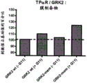
Figure 2 shows some different constructs tested for optimization of β γ IP based G protein activated biosensors. In FIG. 2A, the structure of GRK2/3 is presented. GRK2/3 includes (arbour) different functional domains: calmodulin binding domain (CAM); an RGS (G protein signaling modulator) domain that can be inactivated by D110A displacement described herein; a catalytic domain for GRK2/3 kinase activity and may be inactivated by K220R displacement as described herein; and pleckstrin homeodomain (PH domain) with PIP2And the G.beta.subunit of the heterotrimeric G protein. These interactions promote the transfer of GRK to the plasma membrane and its activation. Phosphorylation of the C-terminal portion of GRK (serine 670 and 685) has been reported to regulate its activity. Four different GFP-tagged constructs of GRK2 and GRK3 were tested, two based on the entire GRK coding sequence and two based on the C-terminal PH domain/gp binding domain, with GFP located either in the N-terminal or C-terminal portion of GRK. Both G β and G γ subunits were tested as fusions with BRET tags and both could be used to monitor GRK/G β γ interactions.
FIGS. 2B and 2C show the different scale tests (titrations) and the resulting pairs of G.alpha.for four different GRK constructs (FIG. 2A)15Beta of (A)1AR activation (FIG. 2B) and G.alpha.11The thromboxane a2 receptor (TP α R) -mediated activation (fig. 2C). Based on transfection of a receptor-encoding construct and G.alpha. (beta. in FIG. 2B)1AR/Gα15And TP α R/G α in FIG. 2C11) BRET donor to recipient titration was performed on HEK293 cells of GRK2 construct (up to 75 ng/well) labeled GFP10 in varying amounts, gbeta 1, RlucH-gay 5 (0.5 ng/well in 96-well plates). By means of carriersOr agonist treatment of cells for 15 minutes (1. mu.M isoproterenol and 100nM U-46619 treatment expressing beta respectively1AR and TP α R). The BRET ratio is reported to be functionally superior to the expression of the RlucII construct (evaluation of bioluminescence) in the expression of the GFP construct (evaluation of fluorescence). These results indicate that the full-length GRK, C-terminally labeled BRET donor (GFP), provides the best dynamic window, based on the amplitude of the BRET signal and the stability of the response to a wide range of donor-to-acceptor ratios.
Fig. 3A to 3C show G protein activation profiles of TP α R using β γ IP-based biosensors. FIG. 3A: HEK293 cells transiently expressing TP α R as well as GRK2-GFP, Rluc-Ggamma 5, G β 1 and the indicated G α were exposed to 100nM U-46619 or vector for 15min prior to BRET measurement. The simulated conditions did not over-express any ga subunit. FIG. 3B: the BRET values obtained for agonist-treated cells in fig. 3A are expressed as a percentage of the BRET values obtained for corresponding cells treated with vehicle. The simulated conditions were used to determine a threshold for a positive response. FIG. 3C: GRK 2-GFP/Rluc-Ggamma 5/Gbeta1 biosensor and the G alpha of TP alpha Rq、Gα13、Gα14、Gα15、GαqG66K and G alphaqDose response curve for Y67C activated agonist U-46619.
Fig. 4A to 4J show dopamine D2 receptor (D) using β γ IP-based biosensor2R)、α1B-adrenergic receptor (. alpha.)1BAR) and alpha2c-adrenergic receptor (. alpha.)2cAR) activation profile of G protein. Prior to BRET measurement, the following agonists were used: stimulation of transient expression of D by rotigotine (FIGS. 4A and 4B), phenylephrine (FIGS. 4C, 4D, 4E) or epinephrine (FIG. 4E)2R (FIGS. 4A and 4B), α1BAR (FIGS. 4C and 4D) or α2CAR (FIGS. 4E and 4F), and GRK2-GFP, Rluc-Ggamma 5, Gbeta 1, and HEK293 cells designated as G alpha for 15 min. Fig. 4A, 4C, and 4E: data are expressed as a percentage of BRET signal obtained in vector-treated cells. Mock conditions without any overexpression of the ga subunit were used to determine the threshold for positive response. As presented in FIGS. 4A, 4C and 4E, G proteins (e.g., G) with promiscuous activation (promiscuous activation) propertiesαqY67C) can be used to monitor receptor activation (see position and surrounding sequences in fig. 14). These promiscuous mutants of ga can be used as positive controls for receptor activation that can be used to characterize antagonists or to screen for orphan receptor (orphan receptor) agonists. FIG. 4B is a selection of G.alpha.proteins (G.alpha.protein) using a GRK 2-GFP/Rluc-G.gamma.5/G.beta.1 biosensori1And four miscellaneous G.alpha.qMutant: G66K, G66D, Y67C and F75G) with rotigotine, a D2R agonist. In FIG. 4D, a biosensor using GRK 2-GFP/Rluc-Ggamma 5/Gbeta1, alpha is shown1BAR and selected G.alpha.proteins (G.alpha.11And G.alpha.q) Dose response curves with phenylephrine, an alpha-adrenergic agonist. In FIG. 4E, the expression of α2CAR、GαzGRK2-GFP, Rluc-Ggamma 5 and G beta 1, and obtaining G alphazActivation with different adrenergic agonists (epinephrine, norepinephrine, phenylephrine, and isoproterenol). FIG. 4F shows the use of two different α' s2CAn AR agonist: alpha of adrenaline and phenylephrine2CG protein activation profile of AR. These results show that β γ IP based biosensors can be used to establish a pharmacological profile of G protein activation and different receptors and ligands. In FIGS. 4G through 4J, different combinations of G.beta.gamma.subunits were used to obtain epinephrine/alpha2CAR promotes G.alpha.zDose response curve for activation. By coding alpha2CAR、GαzHEK293 cells were transfected with constructs of GRK2-GFP, different markers Rluc for gama (fig. 4G and 4H) and gbeta (gbeta 1 in fig. 4G, gbeta 3 short variant (gbeta 3sh) in fig. 4H). In FIGS. 4I and 4J, the code α is used2CAR、GαzGRK2-GFP, Rluc-labeled gy (gy 1 in fig. 4I and gy 5 in fig. 4J), and different gbs. These results show that the combination of both G β and G γ subunits can lead to different pharmacological profiles of G protein activation. These differences can be related in part to the different pharmacological profiles observed with different cells and tissues that express not only a particular set of G α subunits, but also the G β subunit and the G γ subunitDifferent combinations and levels of radicals. These results show that β γ IP based biosensors can be used to study and better understand these differences.
Fig. 5A to 5D show that β γ IP-based biosensors can be used to characterize and validate the selectivity and mode of action of G protein modulators. FIGS. 5A and 5B show PTX (a G.alpha.i/GαoBlocking agent) to G.alpha.i1Ubo-Qic (a G.alpha.qInhibitor analogs: YM-254890) to G.alpha.qSelective inhibition of (3). Prior to recording BRET signals, TP α R and G α expression was pretreated with PTX, Ubo-Qic or vector (control)q(FIG. 5A) or D2R and G alphai1(FIG. 5B) and HEK293 cells of G β 1, Rluc-Gy 5 and GRK2-GFP, which HEK293 cells were then exposed to increasing concentrations of U-46619 (FIG. 5A) or rotigotine (FIG. 5B) for 15 minutes. In fig. 5C, TP α R mediated G protein activation was used to demonstrate the selectivity of Ubo-Qic inhibitors. Cells co-expressing TP α R and the G α subunit designated by the biosensor GRK2-GFP/Rluc-Gγ 5/G β 1+ were pre-treated with Ubo-Qic and then exposed to vector or agonist U-46619(100 nM). These results show that G.alpha.qFamily (G.alpha.q、Gα11、Gα14And G.alpha.15) In (1), only G.alpha.15Is insensitive to Ubo-Qic. G alpha12Proteins and G.alpha.13The protein was also insensitive to Ubo-Qic. FIG. 5D, beta Gamma IP based biosensor to reveal mutated G.alpha.qActivated Ubo-Qic sensitivity. At position 67, G.alpha.is introducedqDisplacement (see fig. 14). Substitutions of only this tyrosine residue (Y67C, Y67G, Y67S and Y67L) against inhibition by Ubo-Qic also showed promiscuous properties, suggesting that this residue is also important for controlling G protein activation. Substitution of the Phe75 residue for glycine resulted in only partial activation inhibition mediated by Ubo-Qic (fig. 5D) and also in a promiscuous phenotype (see fig. 4A).
Fig. 6A and 6B show the kinetics of β γ IP-based G protein activation biosensor response upon receptor activation. FIG. 6A: so as to transiently express D2R, and G alphai1HEK293 cells of G.beta.1, Rluc-G.gamma.5 and GRK2-GFP were exposed to 1. mu.M rotigotine or vector while BRET was measured at regular intervals. Drawing (A)6B: to transiently express TP α R, and G α11HEK293 cells of G.beta.1, Rluc-G.gamma.5 and GRK2-GFP were exposed to 100nM U-46619 or vehicle while BRET was measured at regular intervals. In both cases, the agonist and vehicle were added to the cells after 30 seconds of measurement.
Fig. 7A and 7B show Z' factor evaluation of β γ IP-based G protein activation biosensors. To make transient expression D2R and G alphai1(FIG. 7A) or TP α R and G α11(FIG. 7B), and HEK293 cells of G.beta.1, Rluc-Gy.5 and GRK2-GFP were exposed to 1. mu.M rotigotine (FIG. 7A), 100nM U-46619 (FIG. 7B) or vector (FIG. 7A, FIG. 7B) for 15 min. BRET ratio was expressed in each individual well of a 96-well plate. For these representative experiments, the Z' factors for D2R (fig. 7A) and TP α R (fig. 7B) were evaluated as 0.79 and 0.89, respectively.
Fig. 8A to 8C show ligand distribution maps of β γ IP-based G protein-activated biosensors. FIG. 8A: g protein activation profile of HEK293 cells transiently expressing angiotensin II type 1 receptor (AT1R), as well as G β 1, Rluc-G γ 5, GRK2-GFP and designated G α, were stimulated with 1 μ M angiotensin II for 15 minutes prior to BRET measurement. FIG. 8B: saturating concentrations of angiotensin II analog (1. mu.M) versus G.alpha.q、Gα11And G.alpha.12G protein activation profile of (a). The results in fig. 8A and 8B are expressed as a percentage of BRET signal obtained in vector-treated cells, used to determine the positive response threshold in the absence of any overexpression of the ga subunit under mock conditions. FIG. 8C: dose response curves obtained using GRK 2-GFP/Rluc-Ggamma 5/Gbeta 1 biosensors with AngII and DVG ligands for G.alpha.of AT1RqAnd G.alpha.12And (4) activating. Data are expressed as a percentage of the AngII response obtained for each G protein.
Fig. 9A and 9B show the assessment of G protein activation using a β γ IP-based biosensor using a protein complementation-based detection method; rluc protein complementation assay (Rluc-PCA). FIG. 9A: for TP α R, GRK2-RlucF1, RlucF2-G γ 5, G β 1 and G α11Subunit transfection and stimulation of Z' factor obtained from HEK293 cells with 100nM of U-46619 or vector for 10 min. With each of the 96-well platesThe individual holes represent the luminescence values. For this representative experiment, the Z' factor was evaluated to be 0.53. FIG. 9B: dose response curves using GRK2-RlucF1/RlucF 2-Ggamma 5/Gbeta1 biosensors and using agonist U-46619 for the G.alpha.of TP.alpha.R11And (4) activating.
Fig. 10A to 10C show the evaluation of G protein activation using GRK3 as β γ IP. FIG. 10A: dose response curves obtained from HEK293 cells transiently expressing D2R, and G.alpha.i1G.beta.1, Rluc-G.gamma.5 and GRK2-GFP (black circles) or GRK3-GFP (white triangles) and exposed to increasing concentrations of the agonist rotigotine for 15 minutes prior to BRET measurement. FIG. 10B: GRK 3-based biosensors respond to kinetics of HEK293 cells with D2R、Gαi1G.beta.1, Rluc-G.gamma.5 and GRK3-GFP were transfected and exposed to 1. mu.M rotigotine or vector, with BRET measured at regular intervals. After 30 seconds of measurement, the agonist and vehicle were injected into the cells. FIG. 10C: z' factor evaluation of GRK 3-based biosensors for HEK293 cells with D2R、Gαi1G.beta.1, Rluc-G.gamma.5 and GRK3-GFP were transfected and exposed to 1. mu.M rotigotine or vector for 15 min. BRET ratio was expressed for each individual well of a 96-well plate, and the Z' factor was evaluated to be 0.71 for this representative experiment.
Fig. 11A to 11D show the results of experiments performed with polycistronic vectors encoding β γ IP-based G protein-activated biosensors. FIG. 11A: a schematic diagram of a polycistronic construct encoding the following proteins is illustrated: GRK2-GFP, Rluc-G.gamma.5 and G.beta.1. The G protein activation profile of HEK293 cells encoding TP α R, G α (G α) with HEK293 cells is presented in FIG. 11Bq、Gα11、Gα12、Gα13、Gα14Or G.alpha.15/16(ii) a Mock conditions without G.alpha.) and a polycistronic construct (described in FIG. 11A) encoding the GFP-tagged WT GRK2 or the RGS-dead (dead) mutant of GRK2 (D110A). TP α R activation by its agonist (100nM U46619) results in similar results and profiles as the polycistronic construct, suggesting that a functional RGS domain is not recruited by GRK2The requirements are met. FIG. 11C: dose response curves using the polycistronic construct depicted in FIG. 11A (with WT GRK2), using agonist U-46619 for the G.alpha.R of TP. alpha.R11And (4) activating. In FIG. 11D, the Z' factor was obtained for HEK293 cells transfected as in FIG. 11C and stimulated with 100nM U-46619 or vehicle for 15 min. BRET ratio was expressed in each individual well of a 96-well plate. For this representative experiment, the Z' factor was evaluated to be 0.80.
Fig. 12A and 12B show membrane anchored β γ IP based G protein activated biosensors. FIG. 12A: a schematic diagram is illustrated as the basic principle of a biosensor using membrane-anchored based GRK2(GRK2-mem) and the related DNA construct encoding GRK 2-GFP-mem. FIG. 12B: membrane preparations were obtained from HEK293 cells with TP α R, G β 1, RlucII-Gγ 5, GRK2-GFP or GRK2-GFP-mem in the absence or presence of G α11Transfection in the case of (2) and stimulation with 100nM of U-46619 or vector for 15 min. BRET experiments were then performed based on the membrane preparations. Data are expressed as a percentage of BRET signal obtained in vector-treated cells.
Fig. 13A-13C show that replacement of the reported effects on GRK2 function (RGS and catalysis) or its regulation by phosphorylation did not prevent and significantly promote recruitment of GRK2 to the activated G protein. In FIG. 13A, co-expression of TP α R, G α was stimulated with increasing doses of U46619qHEK293 cells of G β 1, RlucII-gay 5 and GRK2-GFP (WT ═ solid squares, RGS-dead D110A mutant ═ open circles, and catalytic-dead K220R mutant ═ open triangles) variants. As shown in FIGS. 11B and 13A, the functional RGS domain is not G.alpha.as detected with the biosensorqRequired for response (which also does not facilitate the G.alpha.detected with the biosensor)qA response). Mutants of catalytic-dead GRK2 can also be used with this biosensor (fig. 13A and 13C) in the following two configurations: measured as increased BRET due to GRK2-GFP interaction with free G.beta.1/RlucII-Ggamma.5 (FIG. 13A), and increased G-protein activation due to RlucII-GRK2 interaction with free G.beta.1/GFP 10-Ggamma.5 (FIG. 13C). The use of these mutants allows the side effects of over-expression of functional kinasesWith minimization, the side effects are known to inhibit G.alpha.via the RGS domain of kinasesq-mediated PLC activation. The use of such mutants is advantageous for applications requiring the monitoring of multiple signalling pathways by multiplexing of sensors (multiplexing) or multiplexing of different assays. In fig. 13B, HEK293 cells were transfected as in fig. 13A, but were transfected with WT GRK2 (filled squares) or phosphorylated mutants that would prevent (S670A ═ open triangle, S676A ═ open diamond and S685A ═ open circle) or mimic (S670D ═ closed triangle, S676D ═ closed diamond and S685D ═ closed circle) their C-terminal binding domains. GRK2 is known to modulate The activity of ERK, PKA and CDK 2-cyclin A by phosphorylating these serine residues (Cong et al, Journal of biochemical Chemistry, 276, 15192-15199; Pitcher et al, Journal of biochemical Chemistry, 274, 34531-34534; Penela et al, PNAS, 107 (3): 1118-1123; Choudhary et al, Mol cell.200936 (2): 326-39). However, the results presented in fig. 13B provide the following evidence: the recruitment of GRK2 to G β γ may be insensitive to the regulation of different signaling events. In FIG. 13C, co-expression of TP α R, G α was stimulated with increasing doses of U46619qHEK293 cells of G β 1, GFP-G γ 5 and RlucII-GRK2 variants (WT ═ solid squares, and catalytic-dead K220R mutant ═ open triangles). Both configurations with BRET donor and acceptor labeled at either N-terminus (fig. 13C) or C-terminus (fig. 13A) of GRK2 resulted in similar results, providing evidence that the biosensor configuration was flexible.
FIG. 14 shows the sequence alignment of the human G protein alpha subunits (SEQ ID NOs: 1-17) and substitutions that result in promiscuous coupling properties. The G.alpha.subunits of the human heterotrimeric G proteins were aligned using the DIALIGN tool (http:// bibiserv. technfak. uni-bieleded. de/DIALIGN/submissions. html), formatted with the Boxshade tool (http:// www.ch.embnet.org/software/BOX _ form. html), and presented with a region centered at linker 1. Residues that show high conservation throughout the G α subunit are identified with black and gray backgrounds. Linker 1 and the alpha helix predicted by the second structure are also identified.
FIG. 15A shows a schematic representationSchematic representation of a biosensor comprising RET acceptor (a) labeled β γ ip (grk) and a GPCR labeled with RET donor (D) at the C-terminus. The assay is also based on competition between the ga subunit and the α 0 γ IP for binding to the G β γ dimer, which binds to the C-terminal portion of the GPCR. When in an inactive form, the G α 1 subunit of the heterotrimeric G protein binds tightly to the G β γ dimer. When the ligand binds to the GPCR, G α 2 dissociates from the G β γ subunit, allowing β γ IP to be recruited to the free G β γ subunit, and bringing the RET receptor into close proximity to the RET donor linked to the GPCR, thereby inducing/increasing BRET signaling. Fig. 15B and 15C show dose response curves for G protein activation obtained with the biosensor described in fig. 15A. Stimulation of Co-expression of TP α 3R-RlucII, different G α (G α) with increasing doses of U46619qSolid square, G.alpha.11Solid triangle, G.alpha.14Solid diamond and G α12Open circles), G β 1, G γ 5 and WT GRK2-GFP (fig. 15B) or mutated D110A GRK2-GFP (fig. 15C). Dose response curves show similar profiles in fig. 15B and 15C, which illustrate that functional RGS is not required for β γ IP recruitment to activated G proteins as in fig. 11B and 13A, but with different biosensor formulations.
Fig. 16A shows a schematic diagram illustrating a biosensor comprising β γ ip (grk) labeled with RET donor (D) and plasma membrane markers: RET receptors (A) labeled with plasma membrane targeting and anchoring sequences (e.g., CAAX domains). The assay is also based on competition between the ga subunit and the α 0 γ IP for binding to the G β γ dimer located at the plasma membrane. When in an inactive form, the G α 1 subunit of the heterotrimeric G protein binds tightly to the G β γ dimer. When the ligand binds to GPCR, G α 2 dissociates from G β γ subunit, allowing β γ IP to be recruited to G β γ subunit located in plasma membrane, which results in increased density of RET donor (β γ IP-D) and acceptor (plasma membrane marker, a-CAAX), thereby inducing/increasing BRET signal. Fig. 16B shows a dose response curve for G protein activation obtained by the biosensor described in fig. 16A. Stimulation of Co-expression of TP α 3R, different G α (G α) with increasing doses of U46619qSolid square, G.alpha.11Solid triangle, simulation condition (no G alpha) is hollow circle) HEK293 cells of G.beta.1, G.gamma.5, RlucII-GRK2 and rGFP-CAAX. The dose response curves in fig. 16B are similar to those obtained with differently configured biosensors in fig. 3C, 9B, and 11C. In FIG. 16C, the Z' factor was obtained for HEK293 cells transfected as in FIG. 16B and stimulated with 100nM U-46619 or vector for 15 min. Each individual well of the 96-well plate represents BRET ratio. For this representative experiment, the Z' factor was evaluated to be 0.89.
FIG. 17A shows the amino acid sequence of human GRK2(SEQ ID NO: 18) with the black portions at position D110, K220R, S670, S676 and S685 (mutations occurring in certain constructs described herein), the underlined portions as the putative PH domains, and the italic portions as the C-terminal portion of GRK2(GRK 2C-terminus, SEQ ID NO: 50) for use in certain constructs described herein.
FIG. 17B shows the amino acid sequence of human GRK3(SEQ ID NO: 19), with the underlined portion being the putative PH domain, and the italicized portion being the C-terminal portion of GRK3 (GRK 3C-terminus, SEQ ID NO: 51) used in certain constructs described herein.
FIG. 17C shows the amino acid sequence of PLEKHG2(SEQ ID NO: 20), with the putative PH domain underlined.
FIG. 17D shows the amino acid sequence of GFP10(SEQ ID NO: 38) used in the assays described herein.
FIG. 17E shows the amino acid sequence of Renilla reniformis (Renilla reniformis) GFP (rGFP, SEQ ID NO: 46) used in the assays described herein.
FIG. 17F shows the amino acid sequence of RlucII (SEQ ID NO: 39) used in the assays described herein.
Detailed Description
The terminology and notations of genetics, molecular biology, biochemistry and nucleic acids used herein follow those in the standard treatises and texts in the art, such as Kornberg and Baker, DNA replication, second edition (w.h.freeman, New York, 1992); lehninger, biochemistry, second edition (Worth Publishers, New York, 1975); strachan and Read, Human Molecular Genetics (Human Molecular Genetics), second edition (Wiley-loss, New York, 1999); eckstein, eds, oligonucleotides and analogs: practical methods (Oligonucleotides and Analogs: A Practical Approach) Oxford University Press (Oxford University Press, New York, 1991); and Gait, code, oligonucleotide synthesis: practical methods (Oligonucleotide Synthesis: A Practical Approach) (IRL Press, Oxford, 1984), and the like. All terms should be understood in the typical sense established in the art.
The articles "a" and "an" are used herein to refer to one or to more than one (e.g., to at least one) of the grammatical object of the article. For example, "an element" means one element or more than one element. Throughout this specification, unless the context requires otherwise, the words "comprise", "comprises" and "comprising" will be understood to imply the inclusion of a stated step or element or group of steps or elements but not the exclusion of any other step or element or group of steps or elements.
In the studies described herein, the present inventors have shown that biosensors based on β γ IP competition can be used to monitor G protein activation without the need to modify the receptor and/or the ga subunit. Since it is based on competition, a single biosensor is required to study all the different G proteins, as well as to establish a G protein activation/coupling profile based on co-transfected ga subunits. The G protein activation profile is not only important for characterizing receptor and drug targets, but also can be used in drug discovery methods for identifying, characterizing, and optimizing GPCRs ligands with biased signaling properties associated with therapeutic efficacy and reduced side effects.
The present disclosure relates to universal biosensors for monitoring G protein activation without modifying G α protein subunits or G protein activators (e.g., G protein-coupled receptors (GPCRs), G protein signaling Activators (AGS), G protein signaling modulators, or other chemical and biological entities). More specifically, the invention relates to the use of G.beta.gamma.interacting proteins (β. gamma.IP) to monitor the activation of various heterotrimeric G proteins. Advantageously, the signaling biosensors disclosed herein allow for sensitive and quantitative assays that can be used in large-scale screening assays and for identifying ligands (agonists, antagonists, inverse agonists, allosteric modulators, etc.) that target G protein activity structure-activity relationship studies. In addition, the biosensors disclosed herein represent tools for assessing the G protein activation profile, and for analyzing compounds by locating which particular G protein is activated upon stimulation.
As shown in fig. 1, a system according to embodiments of the present disclosure is based on competition between the ga subunit and β γ IP for binding to the G β γ dimer. When in an inactive form, the G α subunit of the heterotrimeric G protein binds tightly to the G β γ dimer. When the ligand binds to the receptor, the G α subunit switches from a GDP-bound form to a GTP-bound form, resulting in dissociation of the G α subunit from the G β γ subunit, allowing β γ IP to be recruited to the free G β γ subunit. Thus, when the receptor is stimulated, the interaction between β γ IP and G β γ will reflect activation of a particular G protein.
The inventors also demonstrated that it is possible to monitor G protein activation using a biosensor labeled with a complementary BRET receptor (e.g., rGFP) -labeled plasma membrane targeting moiety, measuring recruitment/localization of BRET donor (e.g., Rluc) -labeled β γ IP (e.g., GRK) at the plasma membrane where it interacts with the G β γ complex bound to the GPCR. The increase in concentration/density of β γ IP at the plasma membrane (an indirect measure of β γ IP recruitment to the G β γ complex) was detected by the enhancement of BRET signal.
The inventors further demonstrated that it is possible to monitor G protein activation using a biosensor that measures recruitment of BRET donor (e.g., Rluc) labeled β γ IP (e.g., GRK) to a complementary BRET receptor (e.g., rGFP) labeled GPCR (fig. 15).
In this context, the present invention relates to β γ IP-based G protein activation biosensors and systems for assessing specific G protein activation facilitated by specific G protein activators using such biosensors. The system includes a G protein activator; a G.alpha.protein; and biosensors described herein. The invention further relates to methods of detecting G protein activation using the systems of the present disclosure.
Thus, the present invention relates to a biosensor system for detecting G protein activity, comprising the elements defined in (a) or (B):
(A) (i) a first biosensor comprising: a first component comprising a fusion to (a) a RET donor; (b) RET receptor or (c) a G β γ interacting protein (β γ IP) of a first fragment of a reporter protein; and a second component comprising a fused G β protein or a fused G γ protein, wherein said G β protein or said G γ protein is fused to (a) a RET donor; (b) a RET receptor or (c) a second fragment of said reporter protein; (ii) a second biosensor, comprising: (i) the first component and the second component defined in (1); and a third module comprising a recombinant G.alpha.protein; wherein (a) if the β γ IP is fused to the RET donor, the gp or gy protein is fused to the RET acceptor; (b) (ii) if the β γ IP is fused to the RET acceptor, the gp or gy protein is fused to the RET donor; (c) (ii) if the β γ IP is fused to a first fragment of the reporter protein, the gp or gy protein is fused to a second fragment of the reporter protein; or
(B) (i) a biosensor comprising: a first component comprising a fusion to (a) a RET donor; (b) RET receptor or (c) a G β γ interacting protein (β γ IP) of a first fragment of a reporter protein; a second component comprising a fused G protein-coupled receptor (GPCR), wherein said GPCR is fused at its C-terminus to (a) a RET donor; (b) a RET receptor or (c) a second fragment of said reporter protein; a third component comprising a recombinant G.alpha.protein; wherein (a) if said β γ IP is fused to said RET donor, said GPCR is fused to said RET acceptor; (b) (ii) if said β γ IP is fused to said RET acceptor, said GPCR is fused to said RET donor; (c) if the β γ IP is fused to a first fragment of the reporter protein, the GPCR is fused to a second fragment of the reporter protein.
Thus, the present invention relates to a biosensor comprising: (1) a first component comprising a fusion to (a) a RET donor; (b) RET receptor or (c) a G β γ -interacting protein (β γ IP) of a first fragment of a reporter protein; (2) a second component comprising a fused G β protein or a fused G γ protein, wherein said G β protein or said G γ protein is fused to (a) a RET donor; (b) a RET receptor or (c) a second fragment of said reporter protein; (3) a third component comprising a recombinant ga protein, wherein the recombinant ga protein is a promiscuous or non-selective ga protein, e.g., a ga protein comprising a mutation at a position corresponding to residue 66, 67, and/or 75 of human ga q as described herein. In one embodiment, the biosensor further comprises a GPCR (natural or recombinant), preferably an orphan GPCR.
In one embodiment, the biosensor as defined above further comprises a recombinant G β protein and/or a recombinant G γ protein. In a further embodiment, the biosensor as defined above further comprises a recombinant G β protein and a recombinant G γ protein. In one embodiment, the biosensor as defined above further comprises a GPCR, in a further embodiment a recombinant GPCR.
In another aspect, the present disclosure thus further relates to a biosensor comprising: (i) a first component comprising a fusion to (a) a RET donor; (b) RET receptor or (c) a G β γ interacting protein (β γ IP) of a first fragment of a reporter protein; and (ii) a second component comprising a fused Plasma Membrane (PM) targeting moiety, wherein the PM targeting moiety is fused to (a) a RET donor; b) a RET receptor or (c) a second fragment of said reporter protein; wherein (a) if the β γ IP is fused to the RET donor, the PM targeting moiety is fused to the RET acceptor; (b) (ii) if the β γ IP is fused to the RET acceptor, the PM targeting moiety is fused to the RET donor; and (c) if the β γ IP is fused to the first fragment of the reporter protein, then the PM targeting moiety is fused to the second fragment of the reporter protein.
In one non-limiting embodiment, the activity of the biosensors described herein can be detected based on a technique selected from the group consisting of: resonance Energy Transfer (RET), such as Bioluminescence Resonance Energy Transfer (BRET) or Fluorescence Resonance Energy Transfer (FRET); protein complementation assay or protein fragment complementation assay (PCA), such as Enzyme Fragment Complementation (EFC) or bimolecular fluorescence complementation (BiFC), etc. (see fig. 1). Such techniques are known in the art, employing labels/moieties that can be fused to the C-terminus, N-terminus, or within a protein element of a biosensor.
In the resonance energy transfer method, β γ IP and G β γ are labeled with energy donors and acceptors, and upon activation of G protein, an increase in RET signal is observed. In the case of a protein complementation assay, β γ IP and G β γ are labeled with fragments of a reporter protein (e.g., a fluorescent protein or a light-emitting enzyme), and upon activation of the G protein, complementation of the two fragments results in an increase in the reporter protein signal (e.g., a fluorescent signal or an enzymatic activity).
Resonance energy transfer (abbreviated RET) is a mechanism that describes the energy transfer between two chromophores with overlapping emission/absorption spectra. When the two chromophores ("donor" and "acceptor") are at a short distance (e.g., 10-100 angstroms) from each other and their transition dipoles are properly oriented, the donor chromophore can transfer the excited state energy of the donor chromophore to the acceptor chromophore through non-radiative dipole-dipole coupling. One type of RET is Bioluminescence Resonance Energy Transfer (BRET), which is based on the non-radiative transfer of energy between a donor bioluminescent group (a bioluminescent enzyme such as luciferase) and an acceptor luminophore (such as GFP or YFP). Another type of RET is Fluorescence Resonance Energy Transfer (FRET), which involves the transfer of energy from an excited donor fluorophore to an adjacent acceptor fluorophore. For example, two color variants of GFP, CFP and YFP, can be used as donor and acceptor, respectively.
As used herein, the term "fluorescent protein" refers to any protein that fluoresces when excited at an appropriate wavelength. A wide range of fluorescent proteins have been developed that are characterized by a distribution of fluorescence emission spectra that spans almost the entire visible spectrum. Non-limiting examples of Green fluorescent proteins include EGFP, GFP10, Emerald (Emerald), Superfolder GFP, Azami Green, mWasabi, TagGFP, TurboGFP, AcGFP, ZsGreen, and T-Sapphire blue (T-Sapphire). Non-limiting examples of blue fluorescent proteins include EBFP, EBFP2, azurin (Azurite), and mTagBFP. Non-limiting examples of blue-green fluorescent proteins include ECFP, mECFP, Uloric blue (Cerulean), mTurquoise, CyPet, AmCyan1, Midori-Ishi Cyan, TagCFP, mTFP1 (Teal). Non-limiting examples of yellow fluorescent proteins include EYFP, Topaz, Venus, mVenus, mCitrine, mAmetrine, YPet, tagYFP, PhiYFP, ZsYellow1, and mBanana. Non-limiting examples of Orange fluorescent proteins include Kusabira Orange, Kusabira Orange2, mOrage 2, dTomato-Tandem, TagRFP, DsRed2, DsRed-Express (T1), DsRed-Monomer, and mTangerine. Non-limiting examples of red fluorescent proteins include mRuby, mApple, mStrawberry, AsRed2, mRFP1, JRed, mCheerry, HcRed1, mRaspberry, dKeima-Tandem, HcRed-Tandem, mGlum, and AQ 143.
"overlap" as used in the context of the present invention means that the emitted light from a donor fluorescent protein or a light-emitting enzyme (e.g., luciferase) has a property of exciting in close proximity (typically at about (about 1-10 nm) of the wavelength of the fluorophore (acceptor fluorescent protein) placed. Thus, the donor fluorescent protein or the luminescent protein and the acceptor fluorescent protein are selected such that energy can be transferred from the donor fluorescent protein or the luminescent protein attached to the first module of the biosensor to the acceptor fluorescent protein attached to the second module of the biosensor when the first module and the second module are in close proximity (i.e., in a complex or in the same cellular compartment, such as the plasma membrane). Such energy transfer is commonly referred to as "fluorescence" (or forster)
 ) Resonance energy transfer "or" FRET "(if the donor protein is a fluorescent protein), or" bioluminescence resonance energy transfer "or" BRET "(if the donor protein is a bioluminescent protein). Thus, any combination of donor fluorescent protein or photoprotein and acceptor fluorescent protein may be used according to the invention, as long as the above criteria are met. Such combinations are typically referred to as FRET or BRET pairs. The selection of suitable fluorophores for use in BRET assays is known to those skilled in the art. In one embodiment, the fluorophore comprises green fluorescent protein-wild type (GFP-wt), Yellow Fluorescent Protein (YFP), Venus, Topaz, ZsYellow1, mOrange2, mKeima, Blue Fluorescent Protein (BFP), blue-green fluorescent protein (CFP), Tsapphire, mAmitrine, green fluorescent protein-2 (GFP2), Renilla GFP (rGFP), and green fluorescent proteinFluorescent protein-10 (GFP10) or variants thereof. Fluorescent proteins with excitation peaks close to 400nm are particularly suitable. More specific examples of fluorophores include mAmetrine, blue-green fluorescent protein (CFP), and
 GFP 10. Typical examples of FRET pairs include BFP/CFP, BFP/GFP, BFP/YFP, BFP/DsRed, CFP/GFP, CFP/YFP, CFP/mVenus, GFP/YFP, GFP2/YFP, GFP/DsRed, TagBFP/TagGFP2, and TagGFP 2/TagGFP, etc. (see, e.g., Muller et al, Front. plant Sci., 4: 413, 2013). Representative examples of BRET pairs include luciferase (Luc)/GFP, Luc/Venus, Luc/Topaz, Luc/GFP-10, Luc/GFP-2, Luc/YFP, and Luc/rGFP, among others.
As used herein, the term "luciferase" refers to a class of oxidases for bioluminescence, which is distinct from photoproteins. An example is firefly luciferase (EC1.13.12.7) (firefly (p. pyralis) luciferase) from firefly (Photinus pyralis). Several recombinant luciferases from several other species are also commercially available, including Luciferase from Renilla (GENBANK: AAA29804) and variants thereof (e.g., stable variants of Renilla Luciferase, such as RlucII (GENBANK: AAV52877.1)), Rluc8 (GENBANK: EF446136.1), Gaussia Luciferase (Gaussia Luciferase) (Gluc, GENBANK: AAG54095.1), Luciferase enzyme
 Any luciferase may be used according to the present invention as long as it can metabolize a luciferase substrate (e.g., luciferin). Luciferin is a class of light-emitting heterocyclic compounds that are oxidized in the presence of luciferase to produce oxyluciferin and energy in the form of light. Non-limiting examples of fluorescein include D-fluorescein, imidazopyrazinone compounds such as coelenterazine (coelenterazine 400A (DeepBlueC)TM) Coelenterazine H and e-coelenterazine derivatives such as methoxy e-coelenterazine (from NanoLight)
 Is/are as follows
 Purple I))、ViviRenTM(from
 ) And Latia fluorescein ((E) -2-methyl-4- (2, 6, 6-trimethyl-1-cyclohex-1-yl) -1-buten-1-ol formate), bacterial fluorescein, Dinoflagellate (Dinoflagellate) fluorescein, and the like. The luciferase substrate may have a slightly different emission spectrum and therefore will be selected to favour optimal energy transfer to the receptor. In one embodiment, the luciferase is a wild-type (or native) renilla luciferase. In one embodiment, the luciferase is a stable variant of renilla luciferase Rluc 8. In another embodiment, the luciferase is a gaussian luciferase (GLuc). In a specific embodiment, the luciferase is renilla luciferase ii (rlucii) and the luciferin is coelenterazine 400A.
In one embodiment, one of the following BRET configurations is used in the biosensors and methods described herein: BRET1, comprising coelenterazine-h (coel-h) and yfp (yfp) or renilla-derived gfp (rgfp); BRET2, comprising coelenterazine-400 a (coel-400a) and UV-excited gfp (uvgfp) or gfp from renilla (rgfp); or BRET3, which includes coel-h or v-coelenterazine (from Nanolight) ) And monomeric orange fp (morange). In a further embodiment, RlucII is used in the BRET configuration mentioned above. In another embodiment, one of the following BRET configurations is used in the biosensors and methods described herein: RlucII/coel-400 a/Enhanced Blue (EB) FP2, RlucII/coel-400 a/super blue-green fluorescent protein (SCFP3A), RlucII/coel-400a/mAMETrine or RlucII/coel-400a/
GFP 10. In one embodiment, the BRET donor is a Renilla luciferase (e.g., RlucII) and the BRET recipient is Renilla (Renilla) GFP (e.g., Renilla reniformis) GFP).
In PCA, each protein (e.g., β γ IP and G β/G γ, or GPCR) is covalently linked to an incomplete fragment of the reporter protein, and the interaction of β γ IP and G β/G γ brings the reporter fragments close enough that they form a functional reporter protein whose activity can be measured. Any protein that can be split into two parts and recombined non-covalently can be used in PCA-based biosensors. The term "reporter protein" refers to a protein that can be readily detected (e.g., by fluorescence, spectroscopy, luminescence, etc.) and is not normally (endogenously) present in the system used. Typical reporter proteins used in PCA include enzymes (the activity of which can be measured with a suitable substrate), such as dihydrofolate reductase (DHFR), beta-lactamases, beta-galactosidases, or proteins that provide a colorimetric or fluorescent signal, such as luciferase (e.g., renilla luciferase), GFP, and variants thereof.
In another non-limiting embodiment, the RET or PCA markers are located at: (i) β γ IP and G β protein, or (ii) β γ IP and G γ protein. In further non-limiting embodiments, the β γ IP and gp or gy subunits are tagged at their N-terminus, C-terminus, or any internal region within the protein. In one embodiment, the β γ IP and gp or gy subunits are labeled at their N-terminus or C-terminus. In one non-limiting embodiment, the PCA marker described herein added to β γ IP and gp or gy subunits may be, but is not limited to: a fluorophore, a luciferase, or a fragment thereof that includes a portion of a fluorescent protein or a luciferase.
"GPCR" refers to full-length native GPCR molecules as well as mutant/variant GPCR molecules. A list of GPCRs is given in Foord et al (2005) Pharmacol Rev.57, 279-42-288 (which is incorporated herein by reference), a list of updated GPCRs is available from The IUPHAR-DB database (Harmar AJ, et al (2009) IUPHAR-DB: The IUPHAR database of G Protein-Coupled Receptors and ion channels (The IUPHAR database of G Protein-Coupled Receptors and ion channels). Nucl. acids Res.37 (database term) D85680-D685; Sharman JL, et al (2013) IUPHAR-DB: contents of updated databases and new characteristics (IUPHAR-DB: updated databases contents and new characteristics) Nucl. acids Res.41 (database term) D85680-D685; Sharman JL, et al (2013-8; Alrman-DB: updated databases contents and new characteristics of proteins) C.acids Res.41 (database term) C1083-D1088; Alrman J, S.C.D.D.35J.P.A. C.S. C.A. C. J.A.A.A.A.D.D.D.C.A.A.D.D.C.D.C.A. Proc.A.A.A.A.A.A.A.A.A. Proc.A.A.A.A.A.A.A.A.A.A.A.A.A.A.A.A.A.A.A.A.A.A.A.A.A.A.A.A.A.A.A.A.A.A.A.A.A.A.A.A.A.A.A.A.A.A.A.A.A.A.A.A.A.A.A.A.A.A.A.A.A.A.A.A.A.A.A.A.A.A.A.A.A.A.A.A.A.A.A.A.A.A.A.A.A.A.A.B.A.A.B.A.A.A.A.A.A.A.A.A.A.A.A.A.A.A.A.B.A.A.B.B.A.B.B.B.A.B.A.B.B.A.A.A.A.A.A.A.A.B.A.A.A.A.B.B.B.B.B.B.A.A.A.B.B.B.A.B.B.B.B.B.A.A.A.A.A.A.A.A.A.A.A.B.A.A.A.A.A.A.A.B.B.A.B.B.B.B.B.B.B.B.B.A.B.A.A.A.B.A.A.A.A.B.B.B.A.A.B.B.B.B.B.B.A.A.A.A.A.A.B.B.A.A.A.A.B.B.B.B.B.A.A.A.A.B.A.A.A.A.A.A.B.B.B.A.B.A.B.A.B.A.B.B.B.B.B.B.A.B.B.A.B.B.B.B.B.A.A.B.B.B.: 1459-1581). In one embodiment, the GPCR is an orphan GPCR. The term "orphan GPCR" as used herein refers to an apparent receptor (apparent receptor) that has a similar structure to other identified GPCRs, but whose endogenous ligand has not been identified. GPCR orphan receptors are often designated as "GPR" followed by numbers such as GPR 1. An updated list of orphan GPCRs can be obtained from the IUPHAR-DB database described above.
In one embodiment, the GPCR is fused at its C-terminus to a RET donor or RET acceptor, and in a further embodiment to a RET donor (e.g., luciferase (Rluc)) at its C-terminus.
The term "recombinant" as used herein refers to a protein molecule expressed by a recombinant nucleic acid molecule (i.e., a nucleic acid prepared by means of molecular biology/genetic engineering techniques), e.g., a protein expressed (as opposed to a protein naturally expressed by a cell) following transfection/transduction of the cell (or progeny thereof) with a nucleic acid encoding the protein (e.g., present in a vector).
The term variant (or mutant) as used herein refers to a protein that is substantially similar in structure (amino acid sequence) and biological activity to the corresponding native protein. It includes fragments containing one or more domains of the native protein, as well as fusion proteins containing the native protein or fragments thereof. To produce a protein with certain desired characteristics (e.g., ability to constitutively activate, inactivate, altered binding to one or more ligands, etc.), a variant may include one or more mutations (substitutions, deletions, insertions) relative to the native protein. Changes, insertions or deletions of a single amino acid or nucleotide or a small percentage of amino acids or individual substitutions, deletions or insertions of nucleotides in a sequence produce "conservatively modified variants", where changes result in amino acid substitutions to chemically similar amino acids. Conservative substitution tables providing functionally similar amino acids are well known in the art. In addition to such conservatively modified variants, polymorphic variants and alleles of the invention are not excluded.
"homology" or "identity" and "homologous" or "identical" refer to the similarity in sequence and/or structure between two polypeptides or between two nucleic acid molecules. Homology/identity can be determined by comparing each position in the aligned sequences. The degree of homology/identity between nucleic acids or between amino acid sequences is a function of the number of nucleotides or amino acids that are identical or matched at positions shared by the sequences. If two sequences are substantially identical and the functional activity of the sequences is conserved, then, as the term is used herein, the nucleic acid sequence is homologous to the other sequence (the term ` homologous ` as used herein does not infer evolutionary relatedness). When optimally aligned (with gaps (gaps) allowed), two nucleic acid sequences are considered substantially identical if they share at least about 50% sequence similarity or identity, or if they share defined functional motifs. In alternative embodiments, the sequence similarity may be at least 60%, 65%, 70%, 75%, 80%, 85%, 90%, 95%, 96%, 97%, 98%, or 99% in optimally aligned substantially identical sequences. As used herein, the percentage of homology/identity between given sequences represents the degree of sequence identity in the optimally aligned sequences. An "unrelated" or "non-homologous" sequence shares less than 40% identity with any of the sequences described herein, although less than about 25% identity is preferred.
In one non-limiting embodiment, the system comprises living cells, a membrane preparation, or both. The system defined herein is, but is not limited to, a membrane preparation, the β γ IP is attached to a (attached to) membrane via a membrane targeting linker, such as a protein/peptide linker comprising a Plasma Membrane (PM) targeting domain (e.g., a plasma membrane-anchor signal peptide). The plasma membrane targeting domain can be, but is not limited to, a lipid group covalently bound to a peptide chain (e.g., palmitoylation, myristoylation, or prenylation modifications as a membrane anchoring signal from KRAS, e.g., (Hancock2003)), a transmembrane domain, or a polybase region (as present in, e.g., GRK 5).
In one embodiment, the PM targeting moiety comprises a CAAX motif (C)Is a cysteine residue, AA is two aliphatic residues, X represents any amino acid). CAAX motifs are found in "CAAX proteins," which are defined as a group of proteins with specific amino acid sequences at the C-terminus that direct their post-translational modifications. CAAX proteins encompass a wide variety of molecules including lamins (intermediate filaments) (e.g., precore a, lamin B1, and lamin B2), Ras, and various GTP-binding proteins (G proteins) (e.g., Ras, Rho, Rac, and Cdc42), several protein kinases, phosphatases, and the like. (see, e.g., Gao et al, Am J Transl Res.2009; 1 (3): 312-. Proteins with a CAAX motif or cassette at the C-terminal end typically require a prenylation process before the protein migrates to the plasma or nuclear membrane and performs a different function. In one embodiment, the CAAX cassette is derived from a human RAS family protein, such as HRAS, NRAS, Ral-A, KRAS4A or KRAS 4B. The last C-terminal residue of RAS, NRAS, KRAS4A or KRAS4b (referred to as being the hypervariable region or HVR) is described below, with the putative minimal plasma membrane targeting region in italics, underlined to indicate the CAAX box (see, e.g., Ahearn et al, Nature review Molecular Cell Biology 13: 39-51, month 1 2012): HRAS: KLNPPDESGPGCMSCKCVLS;(SEQ ID NO:40);NRAS:KLNSSDDGTQGCMGLPCVVM;(SEQ ID NO:41);KRAS4A:KISKEEKTPGCVKIKKCIIM;(SEQ ID NO:42);KRAS4B:KMSKDGKKKKKKSKTKCVIM:(SEQ ID NO:43);Ral-A/Ral1:KNGKKKRKSLAKRIRERCCIL(SEQ ID NO: 44). In one embodiment, the membrane targeting moiety comprises the last 4 residues of the above sequence. In a further embodiment, the membrane targeting moiety comprises the last 10 residues of the above sequence. In one embodiment, the membrane targeting moiety comprises the C-terminal portion (e.g., about the last 10-30 or 15-25 amino acids) of a CAAX protein (e.g., a human RAS family protein), such as about the last 10-30, 15-25 or 20 amino acids of a human RAS family protein.
In one embodiment, the PM targeting moiety comprises sequence KKKKKKSKTKCVIM from KRAS4B (SEQ ID NO: 37). In another embodiment, the PM targeting moiety includes a plasma membrane targeted palmitoylation sequence from hRas and a prenylation signal sequence from Ral-A/Ral1 (sequence: CMSCKCCIL, SEQ ID NO: 45).
Several proteins also contain non-lipid polydomains (polybasic domains) that target PM, such as Ras small gtpase, phosphatase PTEN, non-receptor tyrosine kinase Src, actin regulatory factors WASP and MARCKS, and G protein-coupled receptor kinase (GRK) (e.g., GRK 5). In one embodiment, the multi-base domain is from GRK5 and comprises sequence SPKKGLLQRLFKRQHQNNSKS (SEQ ID NO: 46). In one embodiment, the PM targeting moiety is fused at the C-terminus of the RET donor or acceptor, and in a further embodiment, the PM targeting moiety is fused at the C-terminus of the RET acceptor, such as GFP (e.g., rGFP). In another embodiment, the PM targeting moiety is fused at the C-terminus of the RET donor or acceptor, and in a further embodiment, the PM targeting moiety is fused at the C-terminus of the RET acceptor (such as GFP (e.g., rGFP)) and the RET donor or acceptor is fused at its N-terminus to a β γ IP, e.g., a GRK protein or a G β γ interacting fragment/variant thereof.
According to the present disclosure, G protein activators include, but are not limited to, classical activation of G proteins by GPCRs, as well as other proteins that can also modulate the activity of these heterotrimeric G proteins, such as G protein signaling Regulators (RGSs), G protein signaling Activators (AGSs), and anticholinesterase 8 protein inhibitors (Ric-8). In some of these non-classical signaling pathways, the Guanine Exchange Factor (GEF) activity that GPCRs classically produce is replaced by another protein (e.g., Ric-8), e.g., (Boularan and Kehrl, 2014).
In one embodiment, the G protein activator is a member of the GPCR family.
The ga protein subunits as defined herein include, but are not limited to, 17 different known isoforms, their splice variants and any mutated ga protein, e.g. those resulting in non-selective/promiscuous ga. In one non-limiting embodiment, the G.alpha.proteins described herein are selected from any native mammalian G.alpha.protein, including G.alpha.proteinsq、Gαs、Gαi1、Gαi2、Gαi3、Gαt-cone、GαT-bar、Gαt-flavor、Gαz、GαoA、GαoB、Gαolf、Gα11、Gα12、Gα13、Gα14And G.alpha.15/16(as currently identified as GNA15), splice variants of these isoforms, and functional variants thereof. In one embodiment, the G.alpha.protein subunit belongs to GiA family. In one embodiment, the G.alpha.protein subunit belongs to GsA family. In one embodiment, the G.alpha.protein subunit belongs to GqA family. In one embodiment, the G.alpha.protein subunit belongs to G12/13A family. In one embodiment, the ga protein is a promiscuous or nonselective ga protein. In further embodiments, the ga protein is a mutant ga protein (e.g., a ga protein) having a substitution at any position of G66, Y67, F75, and combinations thereof, or an equivalent conservative substitution in other ga subtypesqProteins) that result in the production of non-selective ga proteins activated by any GPCR, including orphan receptors (i.e., ga proteins that are capable of interacting with GPCRs independently of the preferential native coupling of these receptors to specific ga proteins (often referred to as "promiscuous" ga proteins), are also encompassed by the present disclosure. In one embodiment, the recombinant ga protein for use in the biosensors/methods described herein is a promiscuous ga protein and the GPCR is an orphan GPCR.
In another aspect, the invention relates to mutant G.alpha.polypeptides, including those that are positioned to correspond to human G.alpha.qMutation at residue 67 and/or residue 75 of the protein. The mutation may be an insertion, deletion, or substitution (e.g., a non-conservative substitution). FIG. 14 discloses an alignment of sequences of representative hG.alpha.proteins that can be mutated according to the invention, wherein the corresponding G.alpha.is indicated by arrowsqAt bit 67 and/or at bit 75. The skilled person will understand that according to the corresponding G.alpha.q(in particular G.alpha.1) The number of residues at the N-terminal position of positions 67 and 75 may vary. For example, in hG α14In (1), corresponds to G.alpha.qResidue 67 of (2) is residue 63 (Y). Similarly, at hG α12In (1), corresponds to G.alpha.qResidue at position 67 of (2) is residue 85(F). Thus, the invention encompasses, for example, mutations of G.alpha.including a mutation at position 63 (e.g., substitution of a non-aromatic residue)14Polypeptides and mutant Galpha comprising a mutation at position 85 (e.g., substitution of a non-aromatic residue)12Polypeptides corresponding to the amino acid sequence at G.alpha.qA mutation at position 67 of (1). The disclosure covers the inclusion of G.alpha.in the context of a humanqAny mutant ga polypeptide that is mutated at one or more positions of residue 67 and/or residue 75 of the protein.
In one embodiment, the invention relates to a polypeptide comprising SEQ ID NO: 1-17, wherein the mutant ga polypeptide corresponds to human gaqThe residue at residue 67 and/or 75 of the protein is mutated. In one embodiment, the mutation corresponds to human gaqPosition of residue 67 of the protein. In one embodiment, the mutation is at a position corresponding to residue 67 and is a substitution for a non-aromatic residue, and in a further embodiment, a substitution for cysteine. In another embodiment, the mutation corresponds to human G.alpha.qAt residue 75 of the protein, and is a substitution for a non-aromatic residue, which in further embodiments is glycine. Such mutant ga polypeptides may be used in any of the biosensors and/or methods described herein. In one non-limiting embodiment, the mutated G.alpha.qThe protein comprises one of the following substitutions: g alphaqG66K、GαqY67C and G.alpha.qF75G, which results in the production of non-selective ga proteins.
In another aspect, the invention relates to a nucleic acid comprising a sequence encoding a mutant ga polypeptide as defined above. In another aspect, the invention relates to a plasmid or vector comprising a nucleic acid as defined above. In another aspect, the invention relates to a cell (host cell) comprising a nucleic acid or vector as defined above. In another aspect, the invention provides a kit comprising a nucleic acid encoding a mutant ga polypeptide as defined herein. In one embodiment, the cell has been transfected or transformed with a nucleic acid encoding a mutant ga polypeptide as defined herein. The present invention further provides recombinant expression systems, vectors, and cells, such as those described above, that express the mutant ga polypeptides defined herein using, for example, media and reagents well known in the art. The cell may be any cell capable of expressing a mutant ga polypeptide as defined above. Suitable host cells and methods for protein expression are well known in the art. Any cell capable of expressing a mutant ga polypeptide as defined above may be used. For example, eukaryotic host cells such as mammalian cells (e.g., rodent cells such as mouse, rat, and hamster cell lines, human cells/cell lines) can be used. In another embodiment, the cell referred to above is a human cell line, such as an embryonic kidney cell line (e.g., HEK293 or HEK293T cells).
In various embodiments, the G β proteins described herein are selected from any known G β protein, including G β 1, G β 2, G β 3 (e.g., short variant of G β 3, G β 3sh), G β 4, and G β 5(G β 5-S or G β 5-L), splice variants of these isoforms, and functional variants thereof. In a further embodiment, the G β protein is G β 1. In another embodiment, the G β protein is G β 3. In a further embodiment, the G β protein (e.g. G β 1) is labelled at the N-terminus with a BRET receptor, e.g. GFP.
In various embodiments, the G γ proteins described herein are selected from any known human G γ proteins, including G γ 1, G γ 2, G γ 3, G γ 4, G γ 5, G γ 7, G γ 8, G γ 9, G γ 10, G γ 11, G γ 12, and G γ 13, and functional variants thereof. In a further embodiment, the G γ protein is G γ 5. In a further embodiment, the G γ protein (e.g., G γ 5) is labeled at the N-terminus with a BRET donor, e.g., luciferase. In another embodiment, the G γ protein (e.g., G γ 5) is labeled with a BRET receptor, e.g., GFP, at the N-terminus. In another embodiment, a G γ protein (e.g., G γ 5) is labeled at the N-terminus with a first domain of a PCA compatibility reporter protein, such as a luciferase (e.g., renilla luciferase).
In one embodiment, the β γ IP described herein is a protein that interacts with the G β γ dimer upon dissociation of the G α β γ heterotrimer and includes the Pleckstrin Homology (PH) domain, such as a G protein-coupled receptor kinase (GRK) protein (GRK2 or GRK3) or a functional fragment thereof including the C-terminal Pleckstrin Homology (PH) domain of the GRK protein (i.e., maintaining the ability to interact with the G β γ dimer), a pleckstrin homology domain containing the G family (having the RhoGef domain) member 2 (plekkg 2). The amino acid sequences of GRK2, GRK3, and PLEKHG2 are depicted in FIGS. 17A-C, with the PH domains underlined. In one non-limiting embodiment, a GRK protein (GRK2 or GRK3) or fragment thereof that retains the ability to interact with the G β γ dimer (e.g., the fragment includes the C-terminal Pleckstrin Homology (PH) domain of GRK, such as a C-terminal fragment including the sequences set forth in SEQ ID NOs: 50 or 51) is labeled at the C-terminus with a BRET receptor (e.g., a fluorophore). In one embodiment, β γ IP is GRK2 or GRK3 or variants/fragments thereof, and is fused at the C-terminus to a BRET receptor (e.g., GFP). In another embodiment, β γ IP is a GRK protein variant ("RGS-death" variant) comprising a mutation that inactivates a regulator of the G protein signaling (RGS) domain. In a further embodiment, an "RGS-dead" variant of a GRK protein comprises a mutation at the position corresponding to residue D110 of GRK2, e.g., substitution of D to a. The RGS domains of native human GRK2(UniProtKB accession number P25098) and GRK3(UniProtKB accession number P35626) span about residues 54 to 175. In another embodiment, β γ IP is a GRK protein variant ("kinase-dead" variant) comprising a mutation that inactivates its kinase domain. In a further embodiment, a "kinase-dead" variant of a GRK protein comprises a mutation (e.g., a non-conservative substitution) at a position corresponding to residue K220 of GRK2, e.g., a substitution of K to D. The kinase domains of GRK2(UniProtKB accession number P25098) and GRK3(UniProtKB accession number P35626) span about residues 191 to 453. In another embodiment, β γ IP is a GRK protein variant comprising a mutation in its C-terminal domain (e.g., within the last 30C-terminal residues). In a further embodiment, the mutation is a serine residue located within the C-terminal domain, more particularly a serine which can be phosphorylated in the native protein. In further embodiments, the mutation (e.g., a non-conservative substitution) is at a position corresponding to residue S670, residue S676, and/or residue S685 of GRK2, e.g., substitution of S for a and/or substitution of S for D.
In various embodiments, the domains of the fusion molecules described herein may be covalently linked directly (e.g., by peptide bonds) or "indirectly" through a suitable linker moiety, such as a linker of one or more amino acids or another type of chemical linker (e.g., a carbohydrate linker, a lipid linker, a fatty acid linker, a polyether linker, PEG, etc.). In one embodiment, one or more additional fields may be inserted before (N-terminal), between or after (C-terminal) the above-defined fields. In one embodiment, the domains of the fusion molecule are covalently linked by peptide bonds. In another embodiment, one or more components of the fusion molecule are linked by a peptide linker. Linkers may be employed to provide a desired conformation of BRET/FRET labeled chromophores within the labeled compound, including, for example, separation between chromophores of a BRET/FRET pair. The linker may be attached to the C-terminus, the N-terminus, or at an intermediate position. In one embodiment, the linker is a peptide linker, typically 2 to 30 amino acids in length, for example about 5 to about 20-25 amino acids. The composition and length of each linker may be selected according to a variety of properties desired, such as flexibility and water solubility. For example, a peptide linker may include relatively small amino acid residues (including but not limited to glycine); small amino acid residues may reduce steric hindrance and increase the flexibility of the peptide linker. Peptide linkers may also include polar amino acids (including but not limited to serine). Polar amino acid residues may improve the water solubility of the peptide linker. In addition, programs such as Globplot 2.3(Linding et al, Globplot: exploring the globular and chaotic nature of protein sequences for globularity and dissorderer), Nucleic Acid Res 2003-Vol.31, No.13, 3701-8) can be used to help determine the degree of clutter and globular nature, and hence their flexibility. In one embodiment, the peptide linker comprises one or more amino acid sequences disclosed in the examples below.
In one non-limiting embodiment, as illustrated in fig. 12A, the recombinant β γ IP-based constructs described herein comprise: β γ IP labeled with a fluorophore, luciferase, or a fragment thereof including a portion of a fluorescent protein or a light-emitting enzyme; a linker, preferably a flexible polypeptide linker; and the Plasma Membrane (PM) anchoring/targeting domain or signal that binds β γ IP to the membrane. In one embodiment, the flexible linker has a length corresponding to the length of the random amino acid sequence of about 50 to 1000, 900, 800, 700, 600 or 500 amino acids, for example about 100 to about 500, 400 or 300 amino acids, preferably about 200 to 400, 200 to 300 or about 200 amino acids. In further embodiments, the flexible linker comprises a random amino acid sequence of about 50 to about 1000, 900, 800, 700, 600 or 500 amino acids, for example a length of about 100 to about 500, 400 or 300 amino acids, preferably a length of about 200 to 400, 200 to 300 or about 200 amino acids. Methods for designing flexible amino acid linkers, more particularly linkers with minimal globular shape and maximal scrambling, are known in the art. This can be achieved, for example, using the Globplot program described above. The sequences may be further optimized to eliminate putative aggregation hotspots (aggregation hotspots), localization domains (localization domains) and/or interaction and phosphorylation motifs. In one embodiment, the flexible linker is located between the BRET donor or acceptor (e.g., Rluc or GFP) and the plasma membrane targeting domain. In further embodiments, the construct has the following configuration: β γ IP (e.g., GRK2) -BRET receptor (e.g., GFP) -flexible linker-PM targeting domain (e.g., CAAX domain).
In one embodiment, the present disclosure is directed to a system comprising: a GPCR; a G α protein selected from: g alphaq、Gαs、Gαi1、Gαi2、Gαi3、Gαt-cone、GαT-bar、Gαt-flavor、Gαz、GαoA、GαoB、Gαolf、Gα11、Gα12、Gα13、Gα14And G.alpha.15/16And a mutated non-selective ga protein as described herein; a signaling biosensor, comprising: GRK protein (GRK2 or GRK3) labeled with a fluorophore, luciferase, or a fragment thereof comprising a portion of a fluorescent protein or luciferase, or a fragment thereof comprising the C-terminal Pleckstrin Homology (PH) domain of GRK, G β protein and G γ protein, wherein G β protein or G γ protein is labeled with a fluorophore, fluorescent protein, or a fragment thereofA protease or a fragment thereof comprising a portion of a fluorescent protein or a luminescent enzyme.
In one embodiment, the present disclosure is directed to a system comprising: a GPCR; a G α protein selected from: g alphaq、Gαs、Gαi1、Gαi2、Gαi3、Gαt-cone、GαT-bar、Gαt-flavor、Gαz、GαoA、GαoB、Gαolf、Gα11、Gα12、Gα13、Gα14And G.alpha.15/16And a mutant G.alpha.protein corresponding to any of the following G.alpha.proteinsqHas a mutation at the position of (a): g66, Y67 and/or F75; a signaling biosensor comprising a GRK protein (GRK2 or GRK3) or a fragment thereof comprising the C-terminal Pleckstrin Homology (PH) domain of GRK labeled with a fluorophore, luciferase, or a fragment thereof comprising a portion of a fluorescent protein or luciferase; a G β 1 protein and a G γ 5 protein, wherein the G β protein or the G γ protein is labeled with a fluorophore, a luciferase, or a fragment thereof including a portion of a fluorescent protein or a luciferase.
According to another broad, non-limiting aspect, the present disclosure is directed to a system for characterizing signaling characteristics of a ligand, the system comprising: an activator of G protein activity; a G.alpha.protein; and biosensors or systems described herein.
The present disclosure also relates to systems comprising a nucleic acid sequence encoding a protein as defined in the present disclosure, which may be, but is not limited to: a DNA molecule, an RNA molecule, a virus or a plasmid. In one embodiment, the disclosure also relates to a nucleic acid comprising a sequence encoding a protein component (e.g., a fusion protein) of one or more biosensors described herein. In one embodiment, the nucleic acid comprises a sequence encoding: (i) β γ IP, (ii) a first fluorophore, a bioluminescent protein or a fragment thereof comprising a portion of a fluorescent protein or a bioluminescent protein; (iii) a G gamma protein; (iv) a second fluorophore, a bioluminescent protein or a fragment thereof comprising a portion of a fluorescent protein or a bioluminescent protein; and (v) G.beta.protein. In further embodiments, the nucleic acid further comprises one or more sequences encoding one or more linkers located between components of the biosensor. In further embodiments, the nucleic acid further comprises one or more transcriptional regulatory sequences (e.g., promoters, enhancers, and/or other regulatory sequences) and/or one or more sequences involved in translational regulation (e.g., Internal Ribosome Entry Site (IRES) sequences).
In one embodiment, the nucleic acid is present in a vector/plasmid, and in a further embodiment, the nucleic acid is present in an expression vector/plasmid. Such vectors include a nucleic acid sequence encoding a component (e.g., a fusion protein) as defined above of a biosensor described herein, operably linked to one or more transcription regulatory sequences.
The term "vector" refers to a nucleic acid molecule capable of transporting another nucleic acid to which it has been linked. One preferred vector is an episome, i.e., a nucleic acid that is capable of replication extrachromosomally. Preferred vectors are those capable of autonomous replication and/or expression of the nucleic acid to which they are linked. A vector capable of directing the expression of a gene operably linked thereto is referred to herein as an "expression vector". Recombinant expression vectors of the invention can be constructed by standard techniques known to those skilled in the art and can be cloned, for example, in the Sambrook et al (1989) molecule: found in the Laboratory Manual (Molecular Cloning: A Laboratory Manual). Various strategies are available for ligation of DNA fragments, the choice of which depends on the nature of the DNA fragment ends and can be readily determined by one skilled in the art. The vectors of the present invention may also include other sequence elements that facilitate propagation and selection of the vector in bacteria and host cells. In addition, the vectors of the invention include nucleotide sequences for one or more restriction endonuclease sites. Coding sequences such as those used for selectable markers and reporter genes are well known to those skilled in the art.
Recombinant expression vectors comprising the nucleic acid sequences of the present invention may be introduced into cells (host cells), which may include living cells capable of expressing the protein coding region from the defined recombinant expression vector. Living cells may include cultured cells and cells within a living organism. Accordingly, the present invention also provides a host cell comprising the recombinant expression vector of the invention. The terms "cell," "host cell," and "recombinant host cell" are used interchangeably herein. Such terms refer not only to the particular subject cell, but also to the progeny or potential progeny of such a cell. Because certain modifications may occur in succeeding generations due to either mutation or environmental influences, such progeny may not, in fact, be identical to the parent cell, but are still included within the scope of the term as used herein.
Vector DNA can be introduced into cells by conventional transformation or transfection techniques. The terms "transformation" and "transfection" refer to techniques for introducing foreign nucleic acid into a host cell, including calcium phosphate or calcium chloride co-precipitation, DEAE-dextran mediated transfection, lipofection, electroporation, microinjection, and virus-mediated transfection. Suitable methods for transforming or transfecting host cells can be found, for example, in Sambrook et al (Molecular Cloning: A Laboratory Manual), second edition, Cold spring harbor Laboratory Press (1989) and other Laboratory manuals. "transcriptional regulatory sequence/element" is a generic term for DNA sequences that induce or control the transcription of a protein coding sequence to which it is operably linked, such as initiation and termination signals, enhancers and promoters, splicing signals, polyadenylation signals. A first nucleic acid sequence is "operably linked" to a second nucleic acid sequence when the first nucleic acid sequence is placed in a functional relationship with the second nucleic acid sequence. For example, a promoter is operably linked to a coding sequence if it affects the transcription or expression of the coding sequence. Generally, operably linked DNA sequences are contiguous and, where necessary, join two protein coding regions in reading frame. However, certain polynucleotide elements are operably linked but not contiguous, since enhancers generally function, for example, when they are separated from a promoter by thousands of bases and intron sequences of variable length.
In one embodiment, as depicted in fig. 11A, the nucleic acid or vector encodes more than one component (fusion protein) of the biosensors described herein (e.g., a polycistronic construct). In one embodiment, the polycistronic construct (e.g., DNA, vector) comprises, in addition to the G β protein, nucleic acid sequences encoding β γ IP and G γ proteins each labeled with a suitable fluorophore, luciferase, or a fragment thereof comprising a portion of a fluorescent protein or a luminogenic enzyme. The system of the invention can be regenerated by co-transfecting the polycistronic construct with a DNA molecule comprising a nucleic acid sequence encoding a G.alpha.protein subunit and a G protein activator of interest.
In another aspect, the invention provides a kit comprising a nucleic acid and/or vector as defined herein.
In another aspect, the disclosure also provides a cell (e.g., host cell) comprising or expressing any of the protein components (e.g., fusion proteins, recombinant proteins) of any of the biosensors described herein. In one embodiment, the cell has been transfected or transformed with a nucleic acid encoding a mutant ga polypeptide as defined herein. The present invention further provides recombinant expression systems, vectors, and cells, such as those described above, that express the mutant ga polypeptides defined herein using, for example, media and reagents well known in the art. The cell may be any cell capable of expressing a mutant ga polypeptide as defined above. Suitable host cells and methods for protein expression are well known in the art. Any cell capable of expressing a mutant ga polypeptide as defined above may be used. For example, eukaryotic host cells such as mammalian cells (e.g., rodent cells such as mouse, rat, and hamster cell lines, human cells/cell lines) can be used. In another embodiment, the cell referred to above is a human cell line, such as an embryonic kidney cell line (e.g., HEK293 or HEK293T cells). In another aspect, the disclosure also provides a membrane preparation comprising or expressing any of the protein components (e.g., fusion proteins, recombinant proteins) of any of the biosensors described herein, and in further embodiments, the disclosure also provides a membrane preparation comprising or expressing a membrane-anchored fusion protein.
The present disclosure further relates to a method for assessing a modulation of the recruitment of G β γ interacting protein (β γ IP) to G β γ subunits between a first condition and a second condition, the method comprising: providing a biosensor as defined herein; measuring BRET receptor signals under the first and second conditions; wherein the difference in BRET signal between the first and second conditions is indicative of a modulation of G β γ interacting protein (β γ IP) recruitment to the G β γ subunit between the first and second conditions. In one embodiment, the first condition is the presence of a test agent and the second condition is the absence of a test agent, wherein a difference in BRET signal indicates that the test agent modulates (increases or decreases) recruitment of G β γ interacting protein (β γ IP) to the G β γ subunit. Recruitment of G β γ interacting protein (β γ IP) to G β γ subunits can be used as a readout for GPCR and/or G protein activation.
The present disclosure further relates to a method for detecting G protein activation comprising the system described herein, the method comprising: 1) contacting the system with a compound that activates the G protein, and 2) detecting activation of the G protein by measuring a signal of the biosensor. The method may further comprise the steps of: 3) obtaining G-protein functional coupling information from signals of the signaling biosensor, and 4) processing the information to determine a G-protein activation profile of the G-protein activator and a signaling characteristic (signature) of the compound. It is possible to determine G-protein coupling profiles of any GPCR and/or GPCR ligand using a biosensor system comprising a plurality of biosensors, each comprising a different recombinant ga protein, as exemplified in fig. 3A and 3B.
The terms "compound", "agent", "assay compound" or "test agent" refer to any molecule (e.g., drug candidate) that can be screened by the methods/biosensors of the present invention, which can be obtained from any number of sources, including libraries of synthetic or natural compounds. For example, many approaches are available for random and directed synthesis of a variety of organic compounds and biomolecules, including expression of randomized oligonucleotides. Alternatively, libraries of natural compounds in the form of bacterial, fungal, plant and animal extracts are available or readily produced. In addition, natural or synthetically produced libraries and compounds are readily modified by conventional chemical, physical and biological means.
The present disclosure further relates to methods for determining whether a test agent modulates GPCR activity, the method comprising, in one of the biosensors described herein, measuring a signal emitted by a RET receptor or a reporter protein in the presence and absence of the test agent; wherein a higher signal measured in the presence of an agent indicates that the test agent increases the activity of the GPCR, and a lower signal measured in the presence of an agent indicates that the agent inhibits the activity of the GPCR. In one embodiment, a method comprises:
(1) providing a biosensor comprising the elements defined in (a), (B) or (C):
(A) (ii) (i) a first component comprising fusion to (a) a RET donor; (b) RET receptor or (c) a G β γ interacting protein (β γ IP) of a first fragment of a reporter protein; (ii) a second component comprising a fused G β protein or a fused G γ protein, wherein said G β protein or said G γ protein is fused to (a) a RET donor; (b) a RET receptor or (c) a second fragment of said reporter protein; wherein (a) if the β γ IP is fused to the RET donor, the gp or gy protein is fused to the RET acceptor; (b) (ii) if the β γ IP is fused to the RET acceptor, the gp or gy protein is fused to the RET donor; (c) (ii) if said β γ IP is fused to a first fragment of said reporter protein, said gp or gy is fused to a second fragment of said reporter protein; (iii) a third component comprising a recombinant G.alpha.protein; and (iv) a fourth component comprising the GPCR;
(B) (ii) (i) a first component comprising fusion to (a) a RET donor; (b) RET receptor or (c) a G β γ interacting protein (β γ IP) of a first fragment of a reporter protein; (ii) a second component comprising fused at its C-terminus to (a) a RET donor; (b) a RET receptor or (c) a second fragment of said reporter protein; (iii) a third component comprising a recombinant G.alpha.protein; wherein (a) if said β γ IP is fused to said RET donor, said GPCR is fused to said RET acceptor; (b) (ii) if said β γ IP is fused to said RET acceptor, said GPCR is fused to said RET donor; (c) if the β γ IP is fused to a first fragment of the reporter protein, then the GPCR is fused to a second fragment of the reporter protein; or
(C) (ii) (i) a first component comprising fusion to (a) a RET donor; (b) RET receptor or (c) a G β γ interacting protein (β γ IP) of a first fragment of a reporter protein;
(ii) a second component comprising a fused Plasma Membrane (PM) targeting moiety, wherein the PM targeting moiety is fused to (a) a RET donor; (b) a RET receptor or (c) a second fragment of said reporter protein; wherein (a) if the β γ IP is fused to the RET donor, the PM targeting moiety is fused to the RET acceptor; (b) (ii) if the β γ IP is fused to the RET acceptor, the PM targeting moiety is fused to the RET donor; (c) (ii) if the β γ IP is fused to a first fragment of the reporter protein, the PM targeting moiety is fused to a second fragment of the reporter protein;
(iii) a third component comprising a recombinant G.alpha.protein; and
(iv) a fourth component comprising the GPCR; and
(2) measuring a signal emitted by the RET receptor or reporter protein in the presence and absence of the test agent; wherein a higher signal measured in the presence of an agent indicates that the test agent increases the activity of the GPCR, and a lower signal measured in the presence of an agent indicates that the agent inhibits the activity of the GPCR.
In one embodiment, the above-mentioned method further comprises:
(3) measuring the signal emitted by the RET receptor or reporter protein in the presence and absence of a test agent and in the presence of a GPCR agonist in a biosensor as defined herein, wherein the recombinant ga protein is coupled to a GPCR (i.e. known to be coupled or activated by a GPCR); and
(4) determining whether the test agent is an inhibitor of the ga protein; wherein a lower signal measured in the presence of the test agent indicates that the test agent is an inhibitor of the G.alpha.protein and a similar or higher signal measured in the presence of the test agent indicates that the test agent is not an inhibitor of the G.alpha.protein.
In one embodiment, the term "higher signal" or "lower signal" as used herein refers to a signal that is at least 10%, 20%, 30%, 40%, 45%, or 50% higher (or lower) than a reference signal measured in the absence of a test agent. In another embodiment, a "higher signal" or "lower signal" is determined by showing a statistically significant difference (determined using an appropriate statistical analysis) in the signal measured in the presence of the test agent relative to the signal measured in the absence of the test agent, e.g., by combining the results obtained in multiple samples. Statistical analyses (ANOVA, student t-test, chi-square test, etc.) to determine significant differences between different sets of data are known in the art and such analyses can be performed using suitable computer programs.
The present disclosure further relates to a method of identifying a G α protein activated by a GPCR agonist (G protein profile/characteristic of an agonist), said method comprising (i) measuring the signal emitted by the RET receptor or reporter protein in the presence and absence of said GPCR agonist in a plurality of biosensors as defined herein, wherein each biosensor comprises a different recombinant G α protein; (ii) identifying a G.alpha.protein activated by a GPCR agonist; wherein a higher signal increase measured in the biosensor comprising the recombinant G.alpha.protein, in the presence of the GPCR agonist, relative to a corresponding biosensor not expressing the recombinant G.alpha.protein indicates that the G.alpha.protein is activated by the GPCR agonist, and wherein a lower signal increase or signal decrease measured in the biosensor comprising the recombinant G.alpha.protein, in the presence of the GPCR agonist, similar to or relative to a corresponding biosensor not expressing the recombinant G.alpha.protein, indicates that the G.alpha.protein is not activated by the GPCR agonist. In one embodiment, the method comprises: (a) measuring, in the first biosensor and the plurality of second biosensors of the biosensor system defined herein, a signal emitted by a RET receptor or a reporter protein in the presence and absence of said GPCR agonist, and (b) identifying a ga protein activated by a GPCR agonist; wherein a higher signal increase measured in the second biosensor relative to the first biosensor in the presence of a GPCR agonist indicates that G.alpha.protein is activated by the GPCR agonist, and wherein a signal increase or decrease measured in the second biosensor similar to or lower than the first biosensor in the presence of a GPCR agonist indicates that G.alpha.protein is not activated by the GPCR agonist.
Positive and negative controls may be used in the methods/assays described herein. Control and test samples can be run multiple times to obtain statistically significant results.
In one embodiment, the method mentioned above is a high throughput method (high throughput screening, HTS). The term "high throughput screening" (HTS) as used herein refers to a method that allows for the rapid and parallel screening of large numbers of compounds (hundreds, thousands) that have binding or biological activity to a target molecule. Such HTS methods are typically performed on microtiter plates having several wells, e.g., 384, 1536, or 3456 wells. It is important for HTS to detect the readout signal with high sensitivity, accuracy and reproducibility.
Methods and apparatus for measuring BRET signals are well known in the art. The BRET signal can be measured, for example, by determining the intensity of the BRET acceptor signal (light intensity), and/or by calculating the ratio of the signal or light intensity emitted by the BRET donor to the signal or light intensity emitted by the BRET acceptor (BRET ratio). To detect BRET donor and/or BRET acceptor light emissions, BRET signal can be measured using an enzyme reader or a microscope equipped with an appropriate filter kit.
It is to be understood that combinations/subcombinations of the features or embodiments described herein may be present or used in the biosensors, systems, and/or methods described herein.
In one embodiment, the biosensors, systems, and/or methods described herein include one or more of the constructs/fusion proteins and/or recombinant proteins described in the following examples and accompanying figures, e.g., Rluc-G γ 1 to Rluc-G γ 13, GRK-GFP, GRK-RlucF1, RlucF2-G γ 5, GRK2-GFP-mem, Rluc-GRK2, GFP-G γ 5, GFP-CAAX, or GPCR-Rluc.
Modes for carrying out the invention
The invention is illustrated in more detail by the following non-limiting examples
Example 1: materials and methods
Reagent: angiotensin II (AngII; [ Asp-Arg-Val-Tyr-Ile-His-Pro-Phe)]SEQ ID NO: 49) poly-ornithine, poly-D-lysine, isoproterenol, rotigotine, epinephrine, norepinephrine, phenylephrine and pertussis toxin from u46619 from Cayman
 (Ann Arbor,MI)。[Sar1、Ile8]-AngII (SI) and [ Asp ]1、Val5、Gly8]-AngII(DVG)[Sar1-Val5-D-Phe8]AngII (SVdF) and [ Sar1-D-Ala8]AngII was synthesized at the university of Skibueck (Canada, QC). UBO-Qic (L-threonine, (3R) -N-acetyl-3-hydroxy-L-leucinyl- (aR) -a-hydroxyphenylalanyl-2, 3-didehydro (idehydro) -N-methylalaninyl-L-alanyl-N-methyl-L-alanyl- (3R) -3- [ [ (2S, 3R) -3-hydroxy-4-methyl-1-oxo-2- [ (1-oxopropyl) amino]Pentyl radical]Oxy radical]-L-leucyl-N, O-dimethyl-, (7 → 1) -lactone (9CI)) was obtained from the Institute of Pharmaceutical Biology of the University of Bonn (Institute for Pharmaceutical Biology of the University of Bonn) (Germany). Du's modified Igor medium (DMEM), fetal bovine serum,
 And other cell culture reagents from
 Coelenterazine 400a, coelenterazine H and
 purple I purchased from
 Or
Or Technology. Polyethyleneimine (PEI; 25kDa linear type; purchased from
 (Wolington, PA, USA). Salmon sperm DNA was purchased from lifetechnologies (thermofisher). Phusion DNA polymerase from Thermo
 Restriction enzymes and T4 DNA ligase were obtained from
 Oligonucleotides for mutagenesis and PCR applications were synthesized in BioCorp
Expression vector: receptors and G proteins. The plasmid encoding AT1R was given by St alpha Laporte (university of Megill, Montreal, Canada). G alphaq、Gα11、Gα12、Gα13、Gα14、Gα15/16、GαoA、GαoB、Gαz、Gαs、Gαi1、Gαi2、Gαi3、Gβ1、TPαR、D2R and alpha1BAR was obtained from the cDNA resource center (cDNA. org). By using the primers described in Table I, G.alpha.qSite-directed mutagenesis (PCR overlap) of the wild-type protein coding sequence to obtain a coding sequence comprising G.alpha.qG66K、GαqY67C and G.alpha.qPlasmid of the mutant G.alpha.protein of F75G. The PCR fragment was digested with Acc65I + XhoI restriction enzymes and cloned into Acc65I + XhoI digested pCDNA3.1Zeo (+) (from) Carlsbad, california). DNA sequencing for validationIsomorphism and identification of degenerate primers generated by specific replacement.
Table I: primer sequences for use in the experiments described herein

Expression vector: a biosensor construct. Rluc-G γ 5 and GFP-G γ: plasmids encoding the fusion proteins Rluc-Ggamma 1 to Rluc-Ggamma 13 and GFP 10-Ggamma 5 were obtained by PCR amplification of the Ggamma coding sequence, which was then fused in frame at its N-terminus to the humanized renilla luciferase II (hRlucII) sequence in pcDNA3.1 vector (linker sequence: GSAGT, SEQ ID NO: 33) (variants of hRluc previously reported (Leduc, Breton et al 2009), SEQ ID NO: 39), or to GFP10 (variant form of Green Fluorescent Protein (GFP) previously reported (Mercer, Salahpoor et al 2002, SEQ ID NO: 38), GRK2-GFP and GRK 3-GFP) by PCR amplification of GRK2 and GRK3, which were then fused at their C-terminus to the GFP2, into pcDNA3.18293, and GRK 5631-GFP to generate the linker protein sequence between the amino acid residues of GRK2 and GRK 4933: 10 3 (+) (GFP) vector), to produce GRK2-GFP, GRK3-GFP, GRK 2C-terminal (SEQ ID NO: 50) -GFP, GRK 3C-terminal (SEQ ID NO: 51) -GFP. GFP-GRK2 and GFP-GRK 3: GRK2 and GRK3 were amplified by PCR, then GRK2 and GRK3 were fused at their N-terminus to GFP10(SEQ ID NO: 38) in pcDNA3.1Zeo (+) vector, creating a 7 amino acid residue linker (linker sequence: GSAGTGG, SEQ ID NO: 52) between the GRK and GFP10 proteins to create GRK2-GFP, GFP-GRK2C (SEQ ID NO: 50), GFP-GRK3C (SEQ ID NO: 51). Using a similar procedure, GFP-and RlucII-tagged GRK2 mutants were generated by PCR-directed mutagenesis. GRK2-Rluc F1 and Rluc F2-G γ 5: amplifying by PCR SEQ ID NO: 39(Rluc F1), which is then fused to the C-terminus of GRK2 protein in pcdna3.1zeo (+) vector, to create an 18 amino acid linker (linker sequence: GSAGWGKLGSAGSGSAGS, SEQ ID NO: 35) between the Rluc fragment and GRK2 to obtain GRK2-Rluc F1. Amplifying by PCR SEQ ID NO:39(Rluc F2), and then fused in frame to the N-terminus of G.gamma.5 protein in pcDNA3.1Zeo (+) vector, to generate a 11 amino acid linker (linker sequence: GSAGTGSAGTT, SEQ ID NO: 36) between the Rluc fragment and G.gamma.5 to obtain Rluc F2-G.gamma.5. GRK 2-GFP-mem: the GRK2-GFP-mem construct encoding a fusion sequence between GRK2-GFP and a flexible linker of 200 amino acid residues followed by the membrane anchoring signal (prenylation motif: CAXX) of the human KRAS protein (Hancock2003) was generated as follows: first, a linker with a predicted promiscuous structure was generated from a random sequence of 2000 residues. After elimination of aggregation hotspots, putative localization, interaction and phosphorylation motifs, a 200 residue fragment with the smallest globular and largest hash index was selected from the sequence. The 200 amino acid flexible linker (SEQ ID NO: 53) was synthesized directly and then fused in-frame to the N-terminus of the membrane anchoring signal (amino acid sequence: KKKKKKSKTKCVIM, SEQ ID NO:37) of human KRAS protein splice variant b using PCR amplification. A flexible linker followed by a KRAS prenylation signal was then subcloned into the GRK2-GFP pcDNA3.1Zeo (+) vector at the C-terminus of the GRK2-GFP protein. Polycistronic biosensor vector: polycistronic vectors encoding GRK2-GFP, Rluc-Ggamma 5 and G beta 1 were first generated by subcloning the WT and D110A mutated GRK2-GFP10 fusion proteins into pLVX vectors. Then, IRES-G.beta.1 was subcloned into pcDNA3.1Rluc-G.gamma.5 to obtain pcDNA3.1Rluc-G.gamma.5-IRES-G.beta.1. Finally, the two constructs were assembled to generate pLVX vector containing GRK 2-GFP-IRES-Rluc-Ggamma 5-IRES-G beta 1. rGFP-CAAX: the plasmid encoding the fusion protein rGFP-CAAX was obtained by PCR amplification of the rGFP coding sequence (SEQ ID NO: 46) with the reverse primer encoding the linker (sequence: GSAGTMASNNTASG, SEQ ID NO: 47), and by PCR amplification of the plasma membrane targeting polybase sequence and prenylated signal sequence from KRAS splice variant b (-GKKKKKKSKTKCVIM, nomenclature: CAAX, SEQ ID NO: 37). CAAX plasma membrane targeting sequence was fused in-frame to the C-terminus of the rGFP coding sequence. The PCR fragment was subcloned into pcDNA3.1(+) vector. RlucII-GRK 2: GRK2cDNA was PCR amplified and subcloned with linker GGSGSGSGS (SEQ ID NO: 48) and RlucII into pIREShygg 3 expression vector (from ) The N terminal of (1).
Cell culture and transfection: at 37 ℃ in the presence of 5% CO2Human embryonic kidney 293(HEK293) cells were maintained in modified eagle's medium (DMEM) supplemented with 10% fetal bovine serum, 100 units/ml penicillin/streptomycin. Two days prior to the experiment, HEK293 cells (Hamdan, Rochdi et al 2007) were transfected with the indicated plasmids using polyethyleneimine 25-kDa linear form (PEI) as transfection agent (at a ratio of PEI/DNA of 3 to 1) and then seeded directly in 96-well plates pretreated with poly-L-ornithine hydrobromide or poly-D-lysine at a density of 35,000 cells per well (for BRET and PCA assays on living cells) or in 6-well plates at a density of 1,000,000 cells per well (for BRET assays on membrane preparations).
BRET assay on live cells: cells seeded in a 96-well plate were washed twice with Phosphate Buffered Saline (PBS), and then a Tyrode buffer (composition: 137mM NaCl, 0.9mM KCl, 1mM MgCl) was added2、11.9mM NaHCO3、3.6mM NaH2PO425mM HEPES, 5.5mM glucose and 1mM CaCl2pH 7.4). The cells were then treated with different ligands or carriers a specified number of times. The Rluc substrate, coelenterazine 400a, was added at a final concentration of 2.5 μ M and the cells were further incubated for an additional 5 minutes. Then Mithras was usedTMLB940 multi-mode microplate reader or BRET values were collected by an LB942 multimode microplate reader equipped with the following filters: 400 nm. + -. 70nm (energy donor) and 515 nm. + -. 20nm (energy acceptor). BRET values were determined by calculating the ratio of light emitted by GFP (515nm) to light emitted by Rluc (400 nm). To determine the percentage of activation (stimulation value as a percentage of basal value), BRET values were obtained for agonist-treated cells, expressed as a percentage of BRET values obtained for vehicle-treated corresponding cells.
B transfer of GRK2-GFP to RlucII-tagged receptorRET assay (fig. 15B and 15C): 100ng HA-TP α R-RlucII, 750ng GRK2-GFP10 (WT in FIG. 15B or D110A mutant in FIG. 15C), 100ng designated G α, 100ng WT G β 1 and 100ng WT G γ 5 with PEI in a 3: 1 PEI to HEK293SL suspension (350,000 cells/ml) were added. Cells were seeded on poly-D-lysine pretreated plates (100. mu.l cell/PEI/DNA suspension per well of 96-well plate). 48 hours after transfection, the cells were washed with 1mM CaCl + Taiwanese's solution2Cells were washed and pre-incubated at 37 ℃ for 60 minutes. The cells were exposed to different doses of U-46619 in FIGS. 15B and 15C for a total of 15 minutes at 37 ℃. Coelenterazine 400a was then added at a final concentration of 2.5 μ M over the last 5 minutes of stimulation. At 37 deg.C Enzyme-linked immunosorbent assay (Berthold)
 ) BRET was measured.
BRET assay for transfer of RlucII-GRK2 to rGFP-caax (kras) -labeled plasma membrane (fig. 16B and fig. 16C): 100ng HA-TP α R, 20ng RlucII-GRK2, 100ng designated G α, 100ng WT G β 1, 100ng WT G γ 5, 400ng rGFP-CAAX (Kras), 180ng ssDNA and PEI were added to HEK293SL suspension (350,000 cells/ml) in a 3: 1 PEI to DNA ratio. Cells were seeded on poly-D-lysine pretreated plates (100. mu.l cell/PEI/DNA suspension per well of 96-well plate). 48 hours after transfection, the cells were washed with 1mM CaCl + Taiwanese's solution2Cells were washed and pre-incubated at 37 ℃ for 60 minutes. Cells were exposed to different doses of U-46619 in figure 16B for a total of 15 minutes at 37 ℃ or vehicle or 100nM U46619 was added to half of the wells of a 96-well plate at 37 ℃ for Z' factor assay (figure 16C). Coelenterazine 400a was then added at a final concentration of 2.5 μ M over the last 5 minutes of stimulation. At 37 deg.C Enzyme-linked immunosorbent assay (Berthold)
 ) MeasuringAnd (4) BRET. The Z' factor assay was obtained as described previously.
G protein inhibitor: BRET assays were performed as described previously except cells were pretreated with 100ng/ml pertussis toxin overnight at 37 ℃ or 100nM Ubo-Qic for 20 min at 37 ℃.
And (3) dynamic experiments: BRET assay was performed as described previously except that BRET readings were collected at regular intervals 5 minutes after addition of coelenterazine, while ligand and vehicle were injected into cells 30 seconds after BRET was measured.
Determination of the Z' factor: HEK293 cells were transfected as described with the indicated constructs (see the illustrations of fig. 7A, 7B, 9A, 10C, 11D and 16C). Half of the 96-well plates treated with the indicated agonists and the other half of the plates treated with the corresponding carriers were subjected to BRET assay as previously described. The Z' factor is calculated as described in Zhang et al (Zhang, Chung et al 1999). A Z' factor of 0.4 to 1 is considered a robust assay.
Protein complementation assay using RlucII fragment: cells were washed twice with PBS and then added with taiwanese buffer. Cells were then pretreated with the Rluc substrate, coelenterazine 400a, at a final concentration of 2.5. mu.M for 30 minutes at 37 ℃. The addition of the different ligands or carriers was carried out for an additional 10 minutes. Then Mithras without any filter was usedTMThe luminescence values were collected by an LB940 multi-mode microplate reader.
BRET assay was performed on membrane preparations: cells seeded in 6-well plates were collected and lysed in lysis buffer (composition: 25mM Tris-HCl pH 7.4, 2mM EDTA, 5mM MgCl)227% sucrose, 15. mu.M GDP, 2. mu.M GTP, 10. mu.g/ml benzamidine, 5. mu.g/ml soybean trypsin inhibitor and 5. mu.g/ml leupeptin) were resuspended and subjected to a banderole (polytron) homogenization treatment. After the centrifugation step, the membrane pellet was resuspended in a medium supplemented with 5mM MgCl215 μ M GDP and 15 μ M GTP in Taiwanese buffer. BRET experiments were performed as described previously using 400 μ g of membrane per well.
BRET titration (fig. 2B and fig. 2C): HEK293 cells were transiently transfected with PEI at a ratio of 1mg/ml PEI to μ g DNA to 3 μ l PEI. Each well in the DNA transfected 96-well plate was as follows: in fig. 2A and 2B: 40ngOf HA-TP α R or HA- β 1AR, 0.5ng of a construct encoding RlucII-Gy 5, 10ng of a construct encoding G α11Or G.alpha.1510ng of a construct encoding G.beta.1 and an increased amount of up to 75ng of a GRK2 construct labelled with GFP 10. BRET assay was performed 2 days after transfection; cells were washed once with PBS and placed in taiwanese buffer. Cells were treated with 100nM U-46619 (FIG. 2B) or 1 μ M isoproterenol (FIG. 2A) of the vehicle or agonist drug for a total of 15 minutes at room temperature. The Rluc substrate Coel-400a was then added at a final concentration of 2.5. mu.M over the last 5 minutes of stimulation. Then use BRET values were collected by LB940 multimode microplate reader and determined by calculating the ratio of light emitted by the receptor to light emitted by RlucII. Titration curves (fig. 2B and 2C) represent BRET ratios obtained as a function of GFP construct expression (assessed by fluorescence) versus RlucII construct expression (assessed by bioluminescence).
Example 2: results
To investigate the activation of a particular G protein by GPCRs, an assay was developed based on the competitive binding of the G β γ subunit between G α and β γ IP. As depicted in fig. 1A and 1B, in the absence of receptor activation, the G α subunit binds tightly to the G β γ dimer, preventing the G β γ dimer from binding to β γ IP. Upon receptor stimulation, GTP-bound G α dissociates from the G β γ complex, which then dissociates to interact with β γ IP. Thus, the interaction between β γ IP and G β γ reflects the activation of G protein. By co-expressing β γ IP and G β γ, each labeled with one of the two components of the detection system, with a different subtype of unlabeled ga, it is possible to determine the coupling profile of a given receptor after activation of that receptor. As shown below, the interaction between β γ IP and G β γ can be assessed using different detection methods, such as the Resonance Energy Transfer (RET) method (FIG. 1A: Bioluminescence Resonance Energy Transfer (BRET) or Fluorescence Resonance Energy Transfer (FRET)); or FIG. 1B: protein Complementation (PC) assay.
Using RET as an example of the detection method, the β γ IP-based G protein kinase is shown in FIG. 1CPossible scenarios for living biosensors and corresponding analysis of results. In the absence of any gp 0 subunit co-transfected with the two RET partners β γ IP- cA and G β γ -D, the basal RET signal is quite high due to the constitutive interaction between β γ IP and G β γ. In this case, the modulation of RET signal measured after receptor stimulation would reflect the activation of the endogenous ga subunit (either a simulated or-ga condition). Co-expression of the G.alpha.subunit prevents the fundamental interaction between β.gamma.IP-A and G.beta.gamma.D, resulting in cA decrease in the recorded basal RET response (+ G.alpha.1And + G alpha2White bars under conditions). When stimulating a receptor (e.g., with an appropriate GPCR ligand), only receptor binding (e.g., coupling) to a specific ga subunit co-expressed with other biosensor components, a significant increase in RET signal modulation compared to the simulated condition is observed (black bars, + ga)2Conditions). However, if the overexpressed G α subunit is not functionally coupled to the receptor, no significant change in BRET signal can be detected when the receptor is stimulated (black bar, + G α)1Conditions).
Fig. 2A to 2C present some different constructs tested for optimization of β γ IP based G protein activated biosensors. Four different GFP-tagged constructs were tested for GRK2 and GRK3, two based on the complete GRK coding series and two based on the C-terminal PH domain/gp binding domain, with GFP located either in the N-terminal or C-terminal portion of GRK (fig. 2A). The results presented in fig. 2B and 2C indicate that all GRK2 configurations/constructs give detectable BRET responses (and thus can be used in biosensors), and full-length GRK2 labeled with BRET acceptor (e.g., GFP) at the C-terminus provides the best dynamic window, in terms of both the amplitude of the BRET signal and stability over a wide range of donor-to-acceptor ratios. Similar results were obtained using GFP-tagged GRK3 constructs.
To evaluate the feasibility of using β γ IP to monitor G protein activation, GRK2 protein specifically interacting with free G β γ dimer was selected as a representative β γ IP, and the energy receptor GFP10(GFP) was labeled at the C-terminus of the GRK2 protein, thereby allowing the use of BRET as a readout for its interaction with G β γ. GRK2-GFP fusion protein and renilla luciferase with energy receptor marked at N end(Rluc) and coexpression with unlabeled G β 1 and G α subunits. In addition to the biosensor package (GRK2-GFP and Rluc-Ggamma 5), G.beta.1 and various G.alpha.cells were co-transfected with the thromboxane A2 receptor (TP. alpha.R) selected as an example of the prototype GPCR. In the experiment depicted in FIG. 3A, different G.alpha.proteins (G.alpha.proteins) were co-expressed by stimulationq、Gα11、Gα12、Gα13、Gα14、Gα15、GαoA、GαoB、Gαz、Gαs、Gαi1、Gαi2And G.alpha.i3) The G protein coupling profile of TP α R was determined in cells with TP α R agonist U-46619, a stable synthetic endoperoxide prostaglandin PGH2 analogue, and compared to results obtained in the absence of G α overexpression (mock conditions, left column). In the absence of G.alpha.co-transfection, the recorded BRET signal was relatively high and was only slightly modulated upon stimulation with U-46619, which reflects activation of the endogenous G.alpha.protein. Overexpression of G.alpha.proteinsq、Gα11、Gα12、Gα13、Gα14And G.alpha.15/16Is significantly higher than that of the mock condition or overexpression of G.alpha.iCells of the G.alpha.subunit of family, indicating that TP. alpha.R is coupled to G.alpha.qAnd G.alpha.12Family G proteins activate without coupling to G.alpha.iFamily G proteins were activated (fig. 3B).
In addition to the wild-type (native) G.alpha.protein, three G.alpha.proteinsqMutant (G.alpha.qG66K、GαqY67C and G.alpha.qF75G) was also used for the group of G proteins tested with TP α R. Replacement of G.alpha.with a charged residue (e.g., GqG66K) has been previously describedqGlycine residue at position 66 of the protein, resulting in G.alpha.qProtein mutants have promiscuous coupling properties, since they can also be substituted by non-G.alpha.qCoupled receptor activation (Heydorn, Ward et al, 2004). As can be seen in FIGS. 3A and 3B, TP α R activates G α described previouslyqG66K mutants and novel G alpha described hereinqY67C and G.alpha.qF75G. These promiscuous G.alpha.proteins (or having equivalents at these positions)Any of the mutant ga proteins, see fig. 14) can be used as a positive control for GPCR activation in a β γ -based biosensor assay, and are particularly useful for receptors where only limited information is available in coupling preferences, such as orphan receptors. Then selecting G alphaq、Gα13、Gα14、Gα15/16、GαqG66K and G alphaqY67C, dose response curve for U-46619 (FIG. 3C). Interestingly, 0.5nM to G.alpha.of G13 was measured15/16The potency range of 6.6nM, demonstrates that a β γ IP-based biosensor assay can detect the specific potency of activation associated with each G protein bound by a given receptor ligand pair.
To further illustrate that β γ IP-based G protein activation biosensors can be used to reveal the specificity of G protein activation for different GPCRs, the dopamine D2 receptor (D)2R) and alpha1B-adrenergic receptor (. alpha.)1BAR) were each co-expressed with GRK2-GFP, Rluc-Ggamma 5, G beta 1, and various G alpha, and stimulated by their prototypical agonists (D)2The prototype agonist of R is rotigotine, alpha1BThe prototype agonist of AR is phenylephrine). As shown in fig. 4A and 4C, each receptor showed a specific G protein activation profile, which is different from that observed with TP α R (fig. 3A and 3B); d2R is independently coupled to G.alpha.iFamily members (G.alpha.oA、GαoB、Gαz、Gαi1、Gαi2And G.alpha.i3) While being alpha1BAR is coupled exclusively to G.alpha.qFamily members (G.alpha.q、Gα11And G.alpha.15/16). Hybrid G alphaqMutant G alphaqY67C was activated by two receptors (fig. 4A and 4C). By D2R activated certain G proteins (G.alpha.)i1And G.alpha.qY67C) and α1BCertain G proteins activated by AR (G.alpha.)qAnd G.alpha.11) A dose response curve was obtained. In FIG. 4E, the expression of α2CAR、GαzHEK293 cells of GRK2-GFP, Rluc-Ggamma 5 and Gbeta 1, obtained G alpha for different adrenergic agonists (epinephrine, norepinephrine, phenylephrine and isoproterenol)zActivationDose response curve of (a). These results demonstrate that β γ IP-based G protein activation biosensors can be used to establish G protein preference and activation/pharmacological profiles of GPCRs, as well as pharmacological tools addressing the potency and efficacy of given ligands for activating various G proteins through their cognate receptors. As shown in fig. 4F to 4J, different G protein activation profiles were obtained with different combinations of G β and G γ subunits. The results show that the combination of both G β and G γ subunits can lead to different pharmacological profiles of G protein activation. These differences may, at least in part, be associated with different pharmacological profiles observed using different cells and tissues that express not only a particular set of ga subunits, but also different combinations and levels of G β and G γ subunits.
Inhibitors of G protein activity, e.g. selective blocking of G.alpha.respectivelyiAnd G.alpha.qActivated Pertussis Toxins (PTX) and Ubo-Qic (structurally related to the cyclic depsipeptide YM-254890 hong Kong) have been widely used in the GPCR field for characterization of receptor coupling properties (Takasaki, Saito et al 2004). Experiments were performed with those selective G-protein inhibitors using β γ IP-based G-protein activated biosensors to demonstrate the specificity of the BRET signal obtained. For coupling to G.alpha.qActivated TP α R, BRET response measured using β γ IP-based BRET biosensor after agonist stimulation was completely removed at Ubo-Qic pretreatment, while PTX pretreatment had no effect on the response (fig. 5A). In contrast, for G.alpha.iCoupled receptor D2R, PTX pretreatment significantly reduced the biosensor BRET response detected after agonist incubation, whereas pretreatment of Gq inhibitor Ubo-Qic had no detectable effect on the recorded BRET signal. TP α R mediated G protein activation was also used to validate the selectivity of Ubo-Qic inhibitors. The results presented in FIG. 5C show, G αqFamily (G.alpha.q、Gα11、Gα14And G.alpha.15/16) In (1), only G.alpha.15/16Is insensitive to Ubo-Qic. G alpha12And G.alpha.13The protein was also insensitive to Ubo-Qic. Finally, β γ IP-based biosensors were used to reveal mutated gaq(at position 67 or 75-see FIG. 14) activatedUbo-Qic sensitivity. Substitution of the tyrosine residue at position 67 (Y67C, Y67G, Y67S and Y67L) resulted in resistance to Ubo-Qic and promiscuous properties, indicating that this residue is important for controlling G protein activation, and substitution of the Phe75 residue to glycine (associated with promiscuous phenotype-see fig. 4A) resulted in partial Ubo-Qic-mediated inhibition of activation (fig. 5D). Thus, in addition to verifying the specificity of the recorded BRET signals for various G proteins, these results support the use of the β γ IP-based G protein-activated biosensors described herein as a tool for identifying/developing new G protein-selective inhibitors.
To further characterize the β γ IP-based G protein-activated biosensors, respectively at D2After R and TP α R agonist treatment, G α was determinedi1(FIG. 6A) and G.alpha.11(FIG. 6B) kinetics of activation. As shown in fig. 6A and 6B, similar activation kinetics were obtained for two different receptors and G proteins, with a maximal response reached about 30 seconds after ligand addition and a plateau following initial stimulation lasting at least 30 minutes. This sustained response is particularly suitable for assay adaptation in High Throughput Screening (HTS).
To evaluate the robustness of the assay, the assay was performed by a typical G.alpha.iCoupled receptors (D)2R, FIG. 7A) and G.alpha.qCoupled receptors (TP α R, fig. 7B) determine the Z' factor of G protein activation. D2R/Gαi1And TP α R/G α11The Z' factor of (a) is 0.79 and 0.89, respectively, the assay is particularly robust. The robustness of the assay is matched to the requirements of screening applications, especially HTS applications.
In addition to the previously described potential applications of β γ -based biosensors in receptor G protein analysis and HTS, ligand characterization presents another application of G protein-activated biosensors. The phenomenon known as ligand-preferred signaling of GPCRs (Galandrin, Oligny-Longpre et al, 2007) ((Kenakin and Christopoulos 2013) the biosensors described herein are particularly suitable for performing ligand analysis experiments, since it is possible to evaluate the activity of all G protein subtypes using the same RET partnerIn addition, angiotensin II type 1 receptor (AT1R) was analyzed using β γ IP-based G protein-activated biosensors (fig. 8A to 8C). A first set of experiments was performed to determine the coupling properties of the receptor using its natural ligand, angiotensin II. As shown in FIG. 8A, AT1R is coupled to the G.alpha.of a proteinq、Gα11And G.alpha.iSeveral members of the family. After activation of AT1R with different angiotensin II analogs, G.alpha.was selectedq、Gα11And G.alpha.12For further characterization. As shown in fig. 8B and 8C, those angiotensin II derived peptides stimulated different G proteins to varying degrees, revealing the potential preference of certain ligands for specific G proteins. For example, DVG peptides show a response to ga relative to angiotensin II12Activation ratio of G.alpha.qBetter effect (fig. 8C).
Next evaluated was whether a Protein Complementation Assay (PCA) instead of the RET-based assay could be used to evaluate the interaction between β γ IP and G β γ subunits (fig. 1B). PCA, such as biomolecule fluorescence complementation (BiFC) or Enzyme Fragment Complementation (EFC), allows detection of the interaction between two protein partners, making PCA compatible with β γ IP-based G protein-activated biosensors. To determine whether it is possible to use an EFC-based assay, more particularly an EFC-based assay based on Rluc, in a β γ IP-based G protein-activated biosensor, two fusion proteins were generated: GRK2 labeled at the C-terminus with the N-terminal portion of Rluc (GRK2-Rluc F1), and GRK2 labeled at the N-terminus of G γ 5 with the complementary C-terminal portion of Rluc (Rluc F2-G γ 5). If the interaction between GRK2 and the free G β γ subunit occurs (following G protein activation), the two complementary Rluc fragments will recombine and luminescence can be measured in the presence of the Rluc substrate coelenterazine. Co-expression of TP α R with GRK2-R1uc F1, Rluc F2-Gy 5, G β 1 and G α11The cells of (1) complete proof of concept. After stimulation with U-46619, a robust luminescent signal was measured, revealing activation of the G11 protein, with a Z' factor of 0.53 (FIG. 9A), EC50At 8.4nM (fig. 9B), it was verified that PCA can be used for the β γ IP-based G protein-activated biosensor described herein.
Next evaluated are β γ IP (e.g., other than GRK2)GRK3) can be used to monitor G protein activation in a β γ IP-based G protein activation biosensor. Fusion proteins were generated between GRK3 and energy receptor GFP, co-expressing the generated GRK3-GFP with Rluc-Ggamma 5, G beta 1, G alphai1And D2R to obtain a dose response curve to dopamine. As shown in figure 10A, similar efficacy was observed using biosensors based on GRK2 or GRK3 (GRK2 at 96pM, GRK3 at 56 pM). The kinetics of activation of GRK 3-based biosensors were also similar to that obtained using GRK 2-based biosensors (fig. 6A and 6B), where the maximum response was reached at about 30 seconds with a stabilization period of at least a few minutes (fig. 10B). Finally, the Z' factor was also generated with a GRK 3-based biosensor. Use of D2R and G alphai1The Z' factor obtained with the GRK3 biosensor was 0.71, which confirms the robustness of the assay (fig. 10C). Taken together, these data demonstrate that different β γ IPs can be used in β γ IP-based G protein activation biosensors to evaluate G protein activation.
To simplify the use of β γ IP-based G protein-activated biosensors, polycistronic vectors encoding GRK2-GFP, Rluc-Gγ 5 and G β 1 were developed (FIG. 11A). This ensures that the components of the biosensor are expressed from one construct and in a fixed ratio, which can minimize intra-experimental variability. As shown in FIG. 11D, TP α R and G α are encoded using11The polycistronic construct co-transfected with the plasmid of (1) gave a Z' factor of 0.8. This result is comparable to the Z' factor obtained from cells transfected with plasmids encoding the individual biosensor components (fig. 7B, Z ═ 0.89). Dose response curve experiments were also performed using the polycistronic vector to obtain EC for TP α R stimulated with U-4661950An EC of 4.3nM, similar to the 8.4nM measured for the same receptor/ligand pair used with the Rluc-PCA-based GRK2 biosensor50(FIG. 9B). These results confirm the effectiveness of expressing the different components of the biosensor in polycistronic vectors, which can be advantageously used to establish stable cell lines with only one selection marker, thereby simplifying the experimental steps and potentially improving reproducibility (e.g., minimizing intra-experimental variability).
Another variant of β γ IP-based G protein-activated biosensors was developed, in which GRK2 protein is attached to the Plasma Membrane (PM) (fig. 12A). The constructs may be used for certain specific applications, where in vitro experiments on membrane preparations are preferred for whole cell experiments, such as for screening applications. To validate this method, BRET experiments were performed on membrane preparations expressing TP α R, G α11G.beta.1, Rluc-G.gamma.5, and either the cytosolic form of GRK2-GFP (GRK2 wt in FIG. 12B) or plasma membrane anchored GRK2-GFP (GRK2-mem) previously used. The preferred modulation of BRET signal was observed with GRK2 adherent to the membrane, relative to the low BRET increase detected by wt GRK2 (fig. 12B) upon ligand stimulation. These results demonstrate the measurement of G protein activation using PM-anchored β γ IP on membrane preparations.
The results depicted in fig. 13A to 13C show reported mutations affecting GRK2 function, such as the D110A substitution in the RGS domain (RGS-death mutant) and the K220R substitution in the catalytic domain (catalytic-death mutant), or its modulation by phosphorylation, such as the S670A, S676A and S685A substitutions or the S670D, S676D and S685D substitutions, respectively, that prevent and mimic phosphorylation of the C-terminal binding domain of GRK2 by ERK, PKA and CDK 2-cyclin a, respectively, and do not prevent and also significantly promote its recruitment to the activated protein, as assessed using β γ IP-based G protein activation biosensors. GRK2 variants including the above-mentioned mutations were recruited to a similar extent as native GRK (fig. 13A to 13C), thereby providing evidence of: the recruitment of GRK to G β γ may be insensitive to the modulation of different signaling events. Similar results were obtained with the GRK 2D 110A variant after activation of AT1R with angiotensin II.
Another biosensor was developed that measures competitive binding between the ga subunit and β γ IP to the G β γ subunit; fig. 15A shows the configuration and principle of such a biosensor. The biosensor includes β γ ip (grk) labeled with RET donor or acceptor (RET acceptor (a) is illustrated) and GPCR labeled with RET donor or acceptor (RET donor (D) is illustrated) at C-terminal. When in an inactive form, the G α subunit of the heterotrimeric G protein binds tightly to the G β γ dimer. When ligand (L) binds to a GPCR, G α dissociates from G β γ subunits, allowing β γ IP to be recruited to free G β γ subunits, and bringing the RET receptor into close proximity to the RET donor linked to the GPCR, thereby inducing/increasing BRET signal. FIGS. 15B and 15C show dose response curves for G protein activation obtained with a biosensor including wild-type GRK2 (FIG. 15A) or RGS-dead GRK2 mutant (D110A) according to FIG. 15A (FIG. 15B). The dose response curves show similar spectra in fig. 15B and 15C, indicating that functional RGS is not required for recruitment of β γ IP to activated G-protein, confirming the results presented in fig. 11B and 13A using different biosensor configurations.
Biosensors have been developed that measure competitive binding between the ga subunit and β γ IP to the G β γ subunit; fig. 16A shows the configuration and principle of such a biosensor. The biosensor includes a β γ IP (e.g., GRK) labeled with a RET donor or acceptor (RET donor (D) is illustrated) and a Plasma Membrane (PM) targeting domain labeled with a RET donor or acceptor (RET acceptor (a) is illustrated). When in an inactive form, the G β 0 subunit of the heterotrimeric G protein binds tightly to the G β γ dimer. When ligand (L) binds to a GPCR, G β 1 dissociates from the G β γ subunit, allowing β γ IP to be recruited to the free G β γ subunit located on PM, and bringing RET donor D in close proximity to RET acceptor a anchored on PM, thereby inducing/increasing BRET signal. FIG. 16B shows co-expression of TP β 2R, different G α (G α) stimulated with increasing doses of U46619qSolid square, G.alpha.11HEK293 cells under simulated conditions (no ga) ═ open circles), G β 1, G γ 5, RlucII-GRK2 and rGFP-CAAX, filled triangles, and using the dose response curves for G protein activation obtained according to the biosensor in fig. 16A. The dose response curves in fig. 16B are similar to those obtained with biosensors having different configurations (fig. 3C, 9B, and 11C), providing the following evidence: biosensors that measure β γ IP recruitment on PM are suitable for "indirect" assessment of β γ IP recruitment to free G β γ subunit anchored on PM.
Although the present invention has been described hereinabove by way of specific embodiments, it can be modified without departing from the spirit and nature of the invention as defined in the appended claims. In the claims, the word "comprising" is used as an open-ended term that is substantially equivalent to the phrase "including, but not limited to. The singular forms "a", "an" and "the" include plural references unless the context clearly dictates otherwise.
Reference to the literature
Boularan, c, and j.h.kehrl (2014) "Implications of non-classical G-protein signaling by the immune system (" signaling of non-canonical G-protein for the immune system ")" Cell Signal (Cell Signal)26(6) ": 1269-1282.
Galandrin, s., g.oligny-Longpre and m.bouvier (2007) "evasive properties of drug efficacy: inspiration for drug discovery (The evasive nature of drug discovery: pharmaceuticals for drug discovery), "Pharmacological Trends (Trends in pharmaceutical Sciences)28 (8): 423-430.
Garland, s.l. (2013), "is GPCR still the source of a new target? (Are GPCRs still a source of new targets: 947-966.
Gilman, A.G, (1987) "G protein: transducers of receptor-generated signals, "Annu Rev Biochem 56: 615-649.
Hamdan, f.f., m.d.rochdi, b.breton, d.fessearch, d.e.michaud, p.g.charest, s.a.laporte and m.bouvier (2007), "using real-time monitoring of agonist-promoted interaction between β -arrestin and AP-2 to elucidate G-protein coupled receptor endocytosis pathway (using an innovative G protein-coupled receptor pathway-time monitoring of agonist-coupled interaction between β -arrestin AP-2)," J l Chem 282 (40): 29089-29100.
Hancock, J.F, (2003), "Ras protein: different signals from different positions (Ras proteins: differential signals from differential locations), "Nat Rev Mol Cell Biol 4 (5): 373-384.
Heydorn, A., R.J.Ward, R.Jorgensen, M.M.Rosenkilde, T.M.Frimurer, G.Milligan and E.Kostenis (2004), "Identification of novel sites within the alpha subunit of the G protein that are specifically important for receptor-G protein interactions (Identification of an innovative site with the G protein alpha subunit for specificity of receptor-G protein interaction" "Mol Pharmacol 66 (2)): 250-259.
"signaling preferences in new drug discovery" Kenakin, t, and a. christopoulos (2013): detection, quantification and treatment effects (Signalling bias in new Drug discovery: detection, quantification and therapeutic impact), "Nat Rev Drug discovery 12 (3): 205-216.
Leduc, m., b.breton, c.ages, c.le Gouill, m.bouvier, s.chemtob and n.heveker (2009). "Functional selectivity of natural and synthetic prostaglandin EP4 receptor ligands (Functional selectivity of natural and synthetic prostaglandin EP4 receptors)" J Pharmacol Exp Ther 331 (1): 297-307.
Mercier, j.f., a.salahpour, s.angers, a.breit and m.bouvier (2002), "Quantitative assessment of β 1 and β 2 adrenergic receptor homo-and heterodimerization by bioluminescence energy resonance transfer" (Quantitative assessment of the beta 1 and beta 2-adrenergic receptor homo-and hetero-dimerization by biolimulance) and "J Biol Chem 277: 44925-44931.
Pitcher, j.a., j.inglese, j.b.higgins, j.l.ariza, p.j.casey, c.kim, j.l.benovic, m.m.kwatra, m.g.caron and r.j.lefkowitz (1992), "Role of G protein β γ subunit in targeting β adrenergic receptor kinase to membrane-bound receptor" (Role of beta-adrenergic receptor kinase to membrane-bound receptors), "Science (Science)257 (5074): 1264-1267.
Stefan, e, s.aquin, n.berger, c.r.landry, b.nyfeler, m.bouvier and s.w.michnick (2007), "Quantification of dynamic protein complexes using Renilla luciferase fragment complementation for in vivo protein kinase a activity" (Quantification of renal protein complexes using mutated to protein kinase a activity.) "Proc nature Acad Sci S a 104 (43): 16916-16921.
Takasaki, J., t.saito, m.taniguchi, t.kawasaki, y.moritani, k.hayashi, and m.kobori (2004), "novel G α q/11 selective inhibitors (a novel Galphaq/11-selective inhibitor.)" J Biol Chem 279 (46): 47438-47445.
Touhara, k., j.inglese, j.a.pitcher, g.shaw and r.j.lefkowitz (1994), "Binding of G protein beta gamma-subunit to pleckstrin homology domains" "J Biol Chem 269 (14): 10217-10220.
Zhang, j.h., t.d.chung and k.r.oldenburg (1999) "Simple Statistical parameters for Evaluation and detection of High Throughput Screening Assays (a Simple Statistical Parameter for Use in Evaluation and Validation of High Throughput Screening Assays)" J Biomol Screen 4 (2): 67-73.
Cong et al, Journal of biochemistry (The Journal of Biological Chemistry), 276, 15192-15199.
Pitcher et al, Journal of biochemistry (The Journal of Biological Chemistry), 274, 34531-34534.
Penela et al, PNAS, 107 (3): 1118-1123.
Choudhury et al, Mol cell.200936 (2): 326-39.
Linding et al, GlobPlot: the globular nature and the disorganization of the protein sequence was explored (Globplot: expanding protein sequences for globuliarity and dissorder), Nucleic Acid Res 2003-Vol.31, No.13, 3701-8.


Claims (29)
1. A biosensor system for detecting G protein activity in a eukaryotic cell, the biosensor system comprising:
(i) a first biosensor, comprising:
a first component comprising a native GRK2 protein or GRK3 protein fused to the amino terminus of a Bioluminescence Resonance Energy Transfer (BRET) donor or BRET acceptor, the amino acid sequence of said native GRK2 protein being set forth in SEQ ID NO:18 and the amino acid sequence of said native GRK3 protein being set forth in SEQ ID NO: 19;
a second component comprising a G β protein and a G γ 5 protein, wherein the G γ 5 protein is fused to the carboxy terminus of the BRET donor or the BRET acceptor; and
a third component comprising a G protein-coupled receptor (GPCR);
(ii) a second biosensor, comprising:
(i) the first component, the second component and the third component defined in (1); and
a fourth module comprising a recombinant G.alpha.protein;
wherein, if the native GRK2 or GRK3 protein is fused to the BRET donor, the Gy 5 protein is fused to the BRET acceptor; or if the native GRK2 or GRK3 protein is fused to the BRET receptor, the Gy 5 protein is fused to the BRET donor.
2. The biosensor system of claim 1, wherein said native GRK2 or GRK3 protein is fused to said BRET receptor and said Gy 5 protein is fused to said BRET donor.
3. The biosensor system of claim 1 or 2, wherein said BRET donor is a bioluminescent protein.
4. The biosensor system of claim 3, wherein said bioluminescent protein is luciferase.
5. The biosensor system of claim 4, wherein said luciferase is a renilla luciferase.
6. The biosensor system of claim 1 or 2, wherein said BRET receptor is a fluorescent protein.
7. The biosensor system of claim 6, wherein said fluorescent protein is green fluorescent protein.
8. The biosensor system of claim 1 or 2, wherein said first component further comprises a plasma membrane targeting moiety fused to said native GRK2 or GRK3, or said BRET donor or BRET acceptor.
9. The biosensor system of claim 8, wherein said plasma membrane targeting moiety is fused to the C-terminus of said BRET donor or receptor.
10. The biosensor system of claim 8, wherein said plasma membrane targeting moiety comprises an isoprenylation motif.
11. The biosensor system of claim 10, wherein said prenylation motif is the prenylation motif of human KRAS splice variant b.
12. The biosensor system of claim 11, wherein said plasma membrane targeting moiety comprises amino acid sequence KKKKKKSKTKCVIM (SEQ ID NO: 37).
13. The biosensor system of claim 8, further comprising a flexible linker between said BRET donor or BRET acceptor and said plasma membrane targeting moiety.
14. The biosensor system of claim 13, wherein said flexible linker has a length of 50 to 500 amino acids.
15. The biosensor system of claim 14, wherein said flexible linker has a length of 200 amino acids.
16. The biosensor system of claim 1 or 2, wherein said recombinant G.alpha.protein is human G.alpha.q、Gαs、Gαi1、Gαi2、Gαi3、Gαt-cone、GαT-bar、Gαt-flavor、Gαz、GαoA、GαoB、Gαolf、Gα11、Gα12、Gα13、Gα14Or G.alpha.15Protein, or human G.alpha.qThe non-selective variant of (a), which is one of the following mutations: G66K, Y67C or F75G。
17. The biosensor system of claim 1 or 2, wherein the G β proteins in the first and second biosensors are recombinant G β proteins.
18. The biosensor system of claim 1 or 2, wherein the first biosensor is present in a first cell and the second biosensor is present in a second cell.
19. The biosensor system of claim 1 or 2, wherein the biosensor system comprises a plurality of second biosensors, wherein each of the second biosensors comprises a different recombinant ga protein.
20. The biosensor system of claim 19, wherein said different recombinant ga proteins are at least two of the following ga proteins: g alphaq、Gαs、Gαi1、Gαi2、Gαi3、Gαt-cone、GαT-bar、Gαt-flavor、Gαz、GαoA、GαoB、Gαolf、Gα11、Gα12、Gα13、Gα14And G.alpha.15。
21. A method for determining whether a test agent modulates the activity of a GPCR in a eukaryotic cell, the method comprising:
(1) providing a biosensor comprising:
(i) a first component comprising a native GRK2 protein or GRK3 protein fused to a BRET donor or BRET acceptor, wherein the amino acid sequence of the native GRK2 protein is shown in SEQ ID NO. 18, and the amino acid sequence of the native GRK3 protein is shown in SEQ ID NO. 19;
(ii) a second component comprising a G β protein and a G γ 5 protein, wherein the G γ 5 protein is fused to a BRET donor or a BRET acceptor,
wherein, if the native GRK2 or GRK3 protein is fused to the BRET donor, the Gy 5 protein is fused to the BRET acceptor; or if the native GRK2 or GRK3 protein is fused to the BRET receptor, the Gy 5 protein is fused to the BRET donor;
(iii) a third component comprising a recombinant G.alpha.protein; and
(iv) a fourth component comprising the GPCR; and
(2) measuring the signal emitted by the BRET receptor in the presence and absence of the test agent;
wherein a higher signal measured in the presence of the agent indicates that the test agent increases the activity of the GPCR, and a lower signal measured in the presence of the agent indicates that the agent inhibits the activity of the GPCR.
22. A method for determining whether a ga protein is activated by a GPCR agonist, the method comprising:
(a) measuring the signal emitted by the BRET receptor in the presence and absence of the GPCR agonist in the first and second biosensors of the biosensor system of any one of claims 1 to 20, and
(b) identifying whether a ga protein is activated by the GPCR agonist based on a signal emitted by the BRET receptor;
wherein a higher signal increase measured in the second biosensor relative to the first biosensor in the presence of a GPCR agonist indicates that the G.alpha.protein is activated by the GPCR agonist, and wherein a signal increase or decrease measured in the second biosensor similar to or lower than the first biosensor in the presence of a GPCR agonist indicates that the G.alpha.protein is not activated by the GPCR agonist.
23. A method for determining whether a test agent is an inhibitor of a ga protein of interest, the method comprising:
(1) contacting a second biosensor as defined in any of claims 1 to 20 with a GPCR agonist, wherein said recombinant ga protein corresponds to said ga protein of interest;
(2) measuring the signal emitted by the BRET receptor in the presence and absence of the test agent; and
(3) determining whether the test agent is an inhibitor of the G.alpha.protein,
wherein a lower signal measured in the presence of a test agent indicates that the test agent is an inhibitor of the G.alpha.protein of interest, and a similar or higher signal measured in the presence of a test agent indicates that the test agent is not an inhibitor of the G.alpha.protein of interest.
24. A method for determining whether a test agent is an activator of a ga protein of interest, the method comprising:
(1) contacting a second biosensor as defined in any of claims 1 to 20 with an agonist of the GPCR,
wherein the recombinant G.alpha.protein corresponds to the G.alpha.protein of interest;
(2) measuring the signal emitted by the BRET receptor in the presence and absence of the test agent; and
(3) determining whether said test agent is an activator of said G.alpha.protein,
wherein a higher signal measured in the presence of a test agent indicates that the test agent is an activator of the G.alpha.protein of interest, and a similar or lower signal measured in the presence of a test agent indicates that the test agent is not an activator of the G.alpha.protein of interest.
25. The method of any one of claims 21 to 24, wherein the BRET donor is a bioluminescent protein, and wherein the method further comprises contacting a biosensor with a substrate for the donor bioluminescent protein.
26. The method of claim 25, wherein the substrate is fluorescein.
27. The method of claim 26, wherein the fluorescein is coelenterazine.
28. The method of claim 27, wherein said coelenterazine is coelenterazine 400A.
29. The method of any one of claims 21 to 24, wherein the method further comprises: measuring the signal emitted by the BRET donor, and determining the ratio of [ BRET acceptor signal/BRET donor signal ].
Applications Claiming Priority (3)
| Application Number | Priority Date | Filing Date | Title |
|---|---|---|---|
| US201462063622P | 2014-10-14 | 2014-10-14 | |
| US62/063,622 | 2014-10-14 | ||
| PCT/CA2015/051032 WO2016058094A1 (en) | 2014-10-14 | 2015-10-14 | BIOSENSOR BASED ON GβΥ-INTERACTING PROTEINS TO MONITOR G-PROTEIN ACTIVATION |
Publications (2)
| Publication Number | Publication Date |
|---|---|
| CN107074926A CN107074926A (en) | 2017-08-18 |
| CN107074926B true CN107074926B (en) | 2021-09-21 |
Family
ID=55745919
Family Applications (1)
| Application Number | Title | Priority Date | Filing Date |
|---|---|---|---|
| CN201580055471.5A Active CN107074926B (en) | 2014-10-14 | 2015-10-14 | Biosensor for monitoring G protein activation based on G beta gamma interaction protein |
Country Status (13)
| Country | Link |
|---|---|
| US (1) | US10877036B2 (en) |
| EP (1) | EP3207052B1 (en) |
| JP (1) | JP6932636B2 (en) |
| CN (1) | CN107074926B (en) |
| AU (1) | AU2015333543B2 (en) |
| CA (1) | CA2963579C (en) |
| DK (1) | DK3207052T3 (en) |
| ES (1) | ES2832331T3 (en) |
| IL (1) | IL250949B (en) |
| NZ (1) | NZ729735A (en) |
| PL (1) | PL3207052T3 (en) |
| SG (1) | SG11201701895VA (en) |
| WO (1) | WO2016058094A1 (en) |
Families Citing this family (5)
| Publication number | Priority date | Publication date | Assignee | Title |
|---|---|---|---|---|
| US20200400567A1 (en) * | 2017-09-27 | 2020-12-24 | Peking University | Fusion polypeptide |
| CA3076672A1 (en) | 2017-10-18 | 2019-04-25 | Universite De Montreal | Systems and methods for the assessment of g-protein activation |
| WO2019076262A1 (en) * | 2017-10-18 | 2019-04-25 | 深圳华大生命科学研究院 | Biosensor for detecting target molecular compound, and system comprising biosensor |
| US20220002357A1 (en) * | 2018-12-04 | 2022-01-06 | The University Of North Carolina At Chapel Hill | G protein-coupled receptor screening systems |
| CN110981943B (en) * | 2019-12-02 | 2021-08-03 | 清华大学 | Polypeptide, application thereof in preparation of medicine and medicine |
Citations (4)
| Publication number | Priority date | Publication date | Assignee | Title |
|---|---|---|---|---|
| WO2005121755A1 (en) * | 2004-06-14 | 2005-12-22 | Commonwealth Scientific And Industrial Research Organisation | Cell free g-protein coupled receptor and ligand assay |
| WO2010063832A1 (en) * | 2008-12-05 | 2010-06-10 | Ge Healthcare Uk Limited | Methods for testing binding of a ligand to a g protein-coupled receptor |
| CN102439444A (en) * | 2009-01-29 | 2012-05-02 | 联邦科学技术研究组织 | Measuring g protein coupled receptor activation |
| WO2012098413A1 (en) * | 2011-01-21 | 2012-07-26 | Heptares Therapeutics Limited | Mutant g-protein coupled receptor proteins and methods for producing them |
Family Cites Families (6)
| Publication number | Priority date | Publication date | Assignee | Title |
|---|---|---|---|---|
| CN1324047C (en) * | 2002-04-12 | 2007-07-04 | 先灵公司 | G-protein coupled receptor ligands and methods |
| US20050042720A1 (en) | 2003-04-24 | 2005-02-24 | 7Tm Pharma A/S | Novel promiscuous G alpha protein mutants and their use |
| US7488583B2 (en) | 2003-09-25 | 2009-02-10 | Odyssey Thera, Inc. | Fragment complementation assays for G-protein-coupled receptors and their signaling pathways |
| GB0905419D0 (en) | 2009-03-30 | 2009-05-13 | Ge Healthcare Uk Ltd | Methods for testing ligand binding to G protein-coupled receptors |
| US9415892B2 (en) | 2010-11-11 | 2016-08-16 | Ube Industries, Ltd. | Container for nonaqueous electrolyte solution, nonaqueous electrolyte solution to put in container, and method for storing nonaqueous electrolyte solution |
| US10324085B2 (en) * | 2013-12-23 | 2019-06-18 | Université de Montréal | Systems and methods for the monitoring of protein complex formation in cells |
-
2015
- 2015-10-14 CN CN201580055471.5A patent/CN107074926B/en active Active
- 2015-10-14 NZ NZ729735A patent/NZ729735A/en unknown
- 2015-10-14 US US15/518,888 patent/US10877036B2/en active Active
- 2015-10-14 JP JP2017520506A patent/JP6932636B2/en active Active
- 2015-10-14 AU AU2015333543A patent/AU2015333543B2/en active Active
- 2015-10-14 ES ES15851395T patent/ES2832331T3/en active Active
- 2015-10-14 PL PL15851395T patent/PL3207052T3/en unknown
- 2015-10-14 WO PCT/CA2015/051032 patent/WO2016058094A1/en active Application Filing
- 2015-10-14 CA CA2963579A patent/CA2963579C/en active Active
- 2015-10-14 SG SG11201701895VA patent/SG11201701895VA/en unknown
- 2015-10-14 DK DK15851395.2T patent/DK3207052T3/en active
- 2015-10-14 EP EP15851395.2A patent/EP3207052B1/en active Active
-
2017
- 2017-03-05 IL IL250949A patent/IL250949B/en unknown
Patent Citations (4)
| Publication number | Priority date | Publication date | Assignee | Title |
|---|---|---|---|---|
| WO2005121755A1 (en) * | 2004-06-14 | 2005-12-22 | Commonwealth Scientific And Industrial Research Organisation | Cell free g-protein coupled receptor and ligand assay |
| WO2010063832A1 (en) * | 2008-12-05 | 2010-06-10 | Ge Healthcare Uk Limited | Methods for testing binding of a ligand to a g protein-coupled receptor |
| CN102439444A (en) * | 2009-01-29 | 2012-05-02 | 联邦科学技术研究组织 | Measuring g protein coupled receptor activation |
| WO2012098413A1 (en) * | 2011-01-21 | 2012-07-26 | Heptares Therapeutics Limited | Mutant g-protein coupled receptor proteins and methods for producing them |
Non-Patent Citations (9)
| Title |
|---|
| A biosensor to monitor dynamic regulation and function of tumour suppressor PTEN in living cells;Evelyne Lima-Fernandes等;《NATURE COMMUNICATIONS》;20140716;第5卷(第1期);第283-296页 * |
| Application of BRET for Studying G Protein-Coupled Receptors;Agnieszka A. Kaczor等;《Mini-Reviews in Medicinal Chemistry》;20140531;第14卷(第5期);第411-425页 * |
| DEMONSTRATION OF ANGIOTENSIN II-INDUCED RAS ACTIVATION IN THE TRANSGOLGI NETWORK AND THE ENDOPLASMIC RETICULUM USING BRET-BASED BIOSENSORS;András Balla等;《THE JOURNAL OF BIOLOGICAL CHEMISTRY》;20110218(第7期);摘要、材料与方法,图1-图5 * |
| G蛋白偶联受体与G蛋白相互作用的最新研究进展;姜云璐等;《生命科学》;20140215;第26卷(第2期);第181-187页 * |
| Oxyntomodulin Differentially Affects Glucagon-Like Peptide-1 Receptor β-Arrestin Recruitment and Signaling through Gαs;Rasmus Jorgensen等;《THE JOURNAL OF PHARMACOLOGY AND EXPERIMENTAL THERAPEUTICS》;20070731;第322卷(第1期);第148页右栏第2段第7-12行,第149页左栏第2段,第150页右栏第1段第5-7行,第152页左栏第3段第12-14行,图5 * |
| Rasmus Jorgensen等.Oxyntomodulin Differentially Affects Glucagon-Like Peptide-1 Receptor β-Arrestin Recruitment and Signaling through Gαs.《THE JOURNAL OF PHARMACOLOGY AND EXPERIMENTAL THERAPEUTICS》.2007,第322卷(第1期),第148-154页. * |
| Real-time monitoring of receptor and G-protein interactions in living cells;Celine Gales等;《NATURE METHODS》;20050301;第2卷(第3期);第178页左栏第2段到右栏第2段,图2 * |
| Simultaneous Visualization of Protumorigenic Src and MT1-MMP Activities with Fluorescence Resonance Energy Transfer;Mingxing Ouyang等;《Cancer Research》;20100315;第70卷(第6期);第2206页左栏第1段,第2207页左栏第1段,图1 * |
| 细胞表面的智能传感器:G 蛋白偶联受体;王昊等;《化学教育》;20130402(第4期);第1-5页 * |
Also Published As
| Publication number | Publication date |
|---|---|
| DK3207052T3 (en) | 2020-11-16 |
| AU2015333543A1 (en) | 2017-03-23 |
| AU2015333543B2 (en) | 2021-03-04 |
| IL250949A0 (en) | 2017-04-30 |
| CA2963579C (en) | 2023-10-31 |
| CA2963579A1 (en) | 2016-04-21 |
| EP3207052A1 (en) | 2017-08-23 |
| WO2016058094A1 (en) | 2016-04-21 |
| PL3207052T3 (en) | 2021-02-08 |
| NZ729735A (en) | 2023-05-26 |
| EP3207052B1 (en) | 2020-09-30 |
| JP6932636B2 (en) | 2021-09-08 |
| US20170234870A1 (en) | 2017-08-17 |
| CN107074926A (en) | 2017-08-18 |
| ES2832331T3 (en) | 2021-06-10 |
| US10877036B2 (en) | 2020-12-29 |
| JP2017536104A (en) | 2017-12-07 |
| EP3207052A4 (en) | 2018-04-25 |
| SG11201701895VA (en) | 2017-04-27 |
| IL250949B (en) | 2021-07-29 |
Similar Documents
| Publication | Publication Date | Title |
|---|---|---|
| CN107074926B (en) | Biosensor for monitoring G protein activation based on G beta gamma interaction protein | |
| Wehr et al. | Monitoring regulated protein-protein interactions using split TEV | |
| Wehr et al. | Split protein biosensor assays in molecular pharmacological studies | |
| AU2010224363B2 (en) | Protein fragment complementation assays for high-throughput and high-content screening | |
| Donthamsetti et al. | Using Bioluminescence Resonance Energy Transfer (BRET) to characterize agonist‐induced arrestin recruitment to modified and unmodified G protein‐coupled receptors | |
| Bolbat et al. | Recent developments of genetically encoded optical sensors for cell biology | |
| WO2006086883A1 (en) | Biosensors for monitoring receptor-mediated g-protein activation | |
| CN107533049B (en) | Biosensor for monitoring the localization and transport of biomolecules in cells | |
| Madiraju et al. | TR-FRET-based high-throughput screening assay for identification of UBC13 inhibitors | |
| Abida et al. | Receptor‐dependence of the transcription read‐out in a small‐molecule three‐hybrid system | |
| Kanno et al. | Intein-Mediated Reporter Gene Assay for Detecting Protein− Protein Interactions in Living Mammalian Cells | |
| Wade et al. | Inhibition of protein-protein interactions: cell-based assays | |
| Huang et al. | Genetically encoded fluorescent amino acid for monitoring protein Interactions through FRET | |
| Lin et al. | In vivo protein–protein interaction assays: beyond proteins | |
| Bondar et al. | Optical sensors of heterotrimeric G protein signaling | |
| Wintgens et al. | Characterizing dynamic protein–protein interactions using the genetically encoded split biosensor assay technique split TEV | |
| US10392649B2 (en) | Biosensors that detect NAD+ | |
| WO2022245922A1 (en) | Poly-adp ribose (par) tracker optimized split-protein reassembly par detection reagents | |
| JP2012127694A (en) | Detection method of intracellular ubiquitination | |
| Perry-Hauser et al. | Assays for detecting arrestin interaction with GPCRs | |
| US20110244487A1 (en) | Methods for testing binding of a ligand to a g protein-coupled receptor | |
| Zheng et al. | In‐Cell Arrestin‐Receptor Interaction Assays | |
| Alvarez-Curto et al. | Defining the functional equivalence of wild-type and chemically engineered G protein-coupled receptors | |
| EP2802600B1 (en) | Method to identify ligands for sigma-1 receptors | |
| US20220326229A1 (en) | Assay for real-time simultaneous recruitment of arrestin isoforms to g protein-coupled receptors |
Legal Events
| Date | Code | Title | Description |
|---|---|---|---|
| PB01 | Publication | ||
| PB01 | Publication | ||
| SE01 | Entry into force of request for substantive examination | ||
| SE01 | Entry into force of request for substantive examination | ||
| GR01 | Patent grant | ||
| GR01 | Patent grant |