JP6860623B2 - Treatment of cancer using chimeric antigen receptor - Google Patents
Treatment of cancer using chimeric antigen receptor Download PDFInfo
- Publication number
- JP6860623B2 JP6860623B2 JP2019126002A JP2019126002A JP6860623B2 JP 6860623 B2 JP6860623 B2 JP 6860623B2 JP 2019126002 A JP2019126002 A JP 2019126002A JP 2019126002 A JP2019126002 A JP 2019126002A JP 6860623 B2 JP6860623 B2 JP 6860623B2
- Authority
- JP
- Japan
- Prior art keywords
- cells
- antigen
- car
- cell
- domain
- Prior art date
- Legal status (The legal status is an assumption and is not a legal conclusion. Google has not performed a legal analysis and makes no representation as to the accuracy of the status listed.)
- Active
Links
- 108010019670 Chimeric Antigen Receptors Proteins 0.000 title claims description 335
- 206010028980 Neoplasm Diseases 0.000 title claims description 238
- 201000011510 cancer Diseases 0.000 title claims description 65
- 238000011282 treatment Methods 0.000 title claims description 41
- 210000004027 cell Anatomy 0.000 claims description 381
- 108091007433 antigens Proteins 0.000 claims description 220
- 102000036639 antigens Human genes 0.000 claims description 220
- 239000000427 antigen Substances 0.000 claims description 217
- 210000001744 T-lymphocyte Anatomy 0.000 claims description 152
- -1 CDK4 / 6 inhibitors Substances 0.000 claims description 114
- 230000011664 signaling Effects 0.000 claims description 114
- 230000027455 binding Effects 0.000 claims description 112
- 230000014509 gene expression Effects 0.000 claims description 79
- 108090000765 processed proteins & peptides Proteins 0.000 claims description 69
- 102000017420 CD3 protein, epsilon/gamma/delta subunit Human genes 0.000 claims description 68
- 108050005493 CD3 protein, epsilon/gamma/delta subunit Proteins 0.000 claims description 68
- 239000012642 immune effector Substances 0.000 claims description 68
- 229940121354 immunomodulator Drugs 0.000 claims description 68
- 102000004196 processed proteins & peptides Human genes 0.000 claims description 68
- 108090000623 proteins and genes Proteins 0.000 claims description 67
- 229920001184 polypeptide Polymers 0.000 claims description 66
- 230000004068 intracellular signaling Effects 0.000 claims description 60
- 208000037265 diseases, disorders, signs and symptoms Diseases 0.000 claims description 56
- 239000003795 chemical substances by application Substances 0.000 claims description 53
- 201000010099 disease Diseases 0.000 claims description 49
- 150000007523 nucleic acids Chemical class 0.000 claims description 45
- 230000002401 inhibitory effect Effects 0.000 claims description 43
- 102100040678 Programmed cell death protein 1 Human genes 0.000 claims description 42
- 101710089372 Programmed cell death protein 1 Proteins 0.000 claims description 42
- 239000003112 inhibitor Substances 0.000 claims description 42
- 125000003275 alpha amino acid group Chemical group 0.000 claims description 39
- 101000946843 Homo sapiens T-cell surface glycoprotein CD8 alpha chain Proteins 0.000 claims description 36
- 102100034922 T-cell surface glycoprotein CD8 alpha chain Human genes 0.000 claims description 36
- 102100024263 CD160 antigen Human genes 0.000 claims description 34
- 101000761938 Homo sapiens CD160 antigen Proteins 0.000 claims description 34
- 102000039446 nucleic acids Human genes 0.000 claims description 33
- 108020004707 nucleic acids Proteins 0.000 claims description 33
- 241000282414 Homo sapiens Species 0.000 claims description 32
- 230000000694 effects Effects 0.000 claims description 30
- 102000004169 proteins and genes Human genes 0.000 claims description 30
- 102100029215 Signaling lymphocytic activation molecule Human genes 0.000 claims description 28
- 230000000139 costimulatory effect Effects 0.000 claims description 28
- 230000003834 intracellular effect Effects 0.000 claims description 28
- 102000004127 Cytokines Human genes 0.000 claims description 27
- 108090000695 Cytokines Proteins 0.000 claims description 27
- 210000000822 natural killer cell Anatomy 0.000 claims description 27
- 229940043355 kinase inhibitor Drugs 0.000 claims description 26
- 239000003757 phosphotransferase inhibitor Substances 0.000 claims description 26
- 101000716102 Homo sapiens T-cell surface glycoprotein CD4 Proteins 0.000 claims description 25
- 102100036011 T-cell surface glycoprotein CD4 Human genes 0.000 claims description 25
- 102000008394 Immunoglobulin Fragments Human genes 0.000 claims description 24
- 101000914514 Homo sapiens T-cell-specific surface glycoprotein CD28 Proteins 0.000 claims description 23
- 102100027213 T-cell-specific surface glycoprotein CD28 Human genes 0.000 claims description 23
- 239000000203 mixture Substances 0.000 claims description 23
- 108010021625 Immunoglobulin Fragments Proteins 0.000 claims description 22
- 102100027207 CD27 antigen Human genes 0.000 claims description 21
- 101000914511 Homo sapiens CD27 antigen Proteins 0.000 claims description 21
- 101000633786 Homo sapiens SLAM family member 6 Proteins 0.000 claims description 21
- 102100032816 Integrin alpha-6 Human genes 0.000 claims description 21
- 102100029197 SLAM family member 6 Human genes 0.000 claims description 21
- 101001055144 Homo sapiens Interleukin-2 receptor subunit alpha Proteins 0.000 claims description 19
- 102100026878 Interleukin-2 receptor subunit alpha Human genes 0.000 claims description 19
- 108091008874 T cell receptors Proteins 0.000 claims description 19
- 102000016266 T-Cell Antigen Receptors Human genes 0.000 claims description 19
- 102000052116 epidermal growth factor receptor activity proteins Human genes 0.000 claims description 19
- 108700015053 epidermal growth factor receptor activity proteins Proteins 0.000 claims description 19
- YOHYSYJDKVYCJI-UHFFFAOYSA-N n-[3-[[6-[3-(trifluoromethyl)anilino]pyrimidin-4-yl]amino]phenyl]cyclopropanecarboxamide Chemical compound FC(F)(F)C1=CC=CC(NC=2N=CN=C(NC=3C=C(NC(=O)C4CC4)C=CC=3)C=2)=C1 YOHYSYJDKVYCJI-UHFFFAOYSA-N 0.000 claims description 19
- 102000005962 receptors Human genes 0.000 claims description 19
- 108020003175 receptors Proteins 0.000 claims description 19
- 102100024222 B-lymphocyte antigen CD19 Human genes 0.000 claims description 18
- 101000980825 Homo sapiens B-lymphocyte antigen CD19 Proteins 0.000 claims description 18
- 101000994375 Homo sapiens Integrin alpha-4 Proteins 0.000 claims description 18
- 101000935040 Homo sapiens Integrin beta-2 Proteins 0.000 claims description 18
- 102100032818 Integrin alpha-4 Human genes 0.000 claims description 18
- 102100025390 Integrin beta-2 Human genes 0.000 claims description 18
- 108091028043 Nucleic acid sequence Proteins 0.000 claims description 18
- 229940124302 mTOR inhibitor Drugs 0.000 claims description 18
- 239000003628 mammalian target of rapamycin inhibitor Substances 0.000 claims description 18
- 101001078158 Homo sapiens Integrin alpha-1 Proteins 0.000 claims description 17
- 102100025323 Integrin alpha-1 Human genes 0.000 claims description 17
- 102100038083 Endosialin Human genes 0.000 claims description 16
- 101000851370 Homo sapiens Tumor necrosis factor receptor superfamily member 9 Proteins 0.000 claims description 16
- 102100036856 Tumor necrosis factor receptor superfamily member 9 Human genes 0.000 claims description 16
- 101000971538 Homo sapiens Killer cell lectin-like receptor subfamily F member 1 Proteins 0.000 claims description 15
- 102100022338 Integrin alpha-M Human genes 0.000 claims description 15
- 102100021458 Killer cell lectin-like receptor subfamily F member 1 Human genes 0.000 claims description 15
- 102100038082 Natural killer cell receptor 2B4 Human genes 0.000 claims description 15
- 102100027744 Semaphorin-4D Human genes 0.000 claims description 15
- 108010074687 Signaling Lymphocytic Activation Molecule Family Member 1 Proteins 0.000 claims description 15
- 101000884275 Homo sapiens Endosialin Proteins 0.000 claims description 14
- 101000994365 Homo sapiens Integrin alpha-6 Proteins 0.000 claims description 14
- 101001035237 Homo sapiens Integrin alpha-D Proteins 0.000 claims description 14
- 101000935043 Homo sapiens Integrin beta-1 Proteins 0.000 claims description 14
- 101000633780 Homo sapiens Signaling lymphocytic activation molecule Proteins 0.000 claims description 14
- 102100039904 Integrin alpha-D Human genes 0.000 claims description 14
- 102100022297 Integrin alpha-X Human genes 0.000 claims description 14
- 102100025304 Integrin beta-1 Human genes 0.000 claims description 14
- 239000003814 drug Substances 0.000 claims description 14
- 208000031261 Acute myeloid leukaemia Diseases 0.000 claims description 13
- 102100028785 Tumor necrosis factor receptor superfamily member 14 Human genes 0.000 claims description 13
- 102100022153 Tumor necrosis factor receptor superfamily member 4 Human genes 0.000 claims description 13
- 102100025064 Cellular tumor antigen p53 Human genes 0.000 claims description 12
- 108010007712 Hepatitis A Virus Cellular Receptor 1 Proteins 0.000 claims description 12
- 102100034459 Hepatitis A virus cellular receptor 1 Human genes 0.000 claims description 12
- 101001046687 Homo sapiens Integrin alpha-E Proteins 0.000 claims description 12
- 101000971605 Homo sapiens Kita-kyushu lung cancer antigen 1 Proteins 0.000 claims description 12
- 101000633784 Homo sapiens SLAM family member 7 Proteins 0.000 claims description 12
- 108060003951 Immunoglobulin Proteins 0.000 claims description 12
- 102100022341 Integrin alpha-E Human genes 0.000 claims description 12
- 102100021533 Kita-kyushu lung cancer antigen 1 Human genes 0.000 claims description 12
- 102100032364 Pannexin-3 Human genes 0.000 claims description 12
- 101710165197 Pannexin-3 Proteins 0.000 claims description 12
- 102100029198 SLAM family member 7 Human genes 0.000 claims description 12
- 102000013532 Uroplakin II Human genes 0.000 claims description 12
- 108010065940 Uroplakin II Proteins 0.000 claims description 12
- 102000018358 immunoglobulin Human genes 0.000 claims description 12
- 102100026094 C-type lectin domain family 12 member A Human genes 0.000 claims description 11
- 102100038078 CD276 antigen Human genes 0.000 claims description 11
- 102100021197 G-protein coupled receptor family C group 5 member D Human genes 0.000 claims description 11
- 101000721661 Homo sapiens Cellular tumor antigen p53 Proteins 0.000 claims description 11
- 101001040713 Homo sapiens G-protein coupled receptor family C group 5 member D Proteins 0.000 claims description 11
- 101000878602 Homo sapiens Immunoglobulin alpha Fc receptor Proteins 0.000 claims description 11
- 102100038005 Immunoglobulin alpha Fc receptor Human genes 0.000 claims description 11
- 102000003812 Interleukin-15 Human genes 0.000 claims description 11
- 108090000172 Interleukin-15 Proteins 0.000 claims description 11
- 102100026181 Placenta-specific protein 1 Human genes 0.000 claims description 11
- 108050005093 Placenta-specific protein 1 Proteins 0.000 claims description 11
- 229940079593 drug Drugs 0.000 claims description 11
- 239000003446 ligand Substances 0.000 claims description 11
- 102100033793 ALK tyrosine kinase receptor Human genes 0.000 claims description 10
- 102100026423 Adhesion G protein-coupled receptor E5 Human genes 0.000 claims description 10
- 102100023003 Ankyrin repeat domain-containing protein 30A Human genes 0.000 claims description 10
- 102100031507 Fc receptor-like protein 5 Human genes 0.000 claims description 10
- 101000718243 Homo sapiens Adhesion G protein-coupled receptor E5 Proteins 0.000 claims description 10
- 101000757191 Homo sapiens Ankyrin repeat domain-containing protein 30A Proteins 0.000 claims description 10
- 101710196509 Leukocyte immunoglobulin-like receptor subfamily A member 2 Proteins 0.000 claims description 10
- 102100025586 Leukocyte immunoglobulin-like receptor subfamily A member 2 Human genes 0.000 claims description 10
- 102100032129 Lymphocyte antigen 6K Human genes 0.000 claims description 10
- 102100033486 Lymphocyte antigen 75 Human genes 0.000 claims description 10
- 101710157884 Lymphocyte antigen 75 Proteins 0.000 claims description 10
- 102100020718 Receptor-type tyrosine-protein kinase FLT3 Human genes 0.000 claims description 10
- 206010039491 Sarcoma Diseases 0.000 claims description 10
- 101710165473 Tumor necrosis factor receptor superfamily member 4 Proteins 0.000 claims description 10
- 239000002253 acid Substances 0.000 claims description 10
- 108010003374 fms-Like Tyrosine Kinase 3 Proteins 0.000 claims description 10
- 201000001441 melanoma Diseases 0.000 claims description 10
- 101710109924 A-kinase anchor protein 4 Proteins 0.000 claims description 9
- 102100037982 Alpha-1,6-mannosylglycoprotein 6-beta-N-acetylglucosaminyltransferase A Human genes 0.000 claims description 9
- 101000846908 Homo sapiens Fc receptor-like protein 5 Proteins 0.000 claims description 9
- 101000795169 Homo sapiens Tumor necrosis factor receptor superfamily member 13C Proteins 0.000 claims description 9
- 101000851376 Homo sapiens Tumor necrosis factor receptor superfamily member 8 Proteins 0.000 claims description 9
- 101001047681 Homo sapiens Tyrosine-protein kinase Lck Proteins 0.000 claims description 9
- 101000814512 Homo sapiens X antigen family member 1 Proteins 0.000 claims description 9
- 102100025128 Olfactory receptor 51E2 Human genes 0.000 claims description 9
- 101710187841 Olfactory receptor 51E2 Proteins 0.000 claims description 9
- 101710120463 Prostate stem cell antigen Proteins 0.000 claims description 9
- 102100036735 Prostate stem cell antigen Human genes 0.000 claims description 9
- 102100027610 Rho-related GTP-binding protein RhoC Human genes 0.000 claims description 9
- 108010017842 Telomerase Proteins 0.000 claims description 9
- 102100021393 Transcriptional repressor CTCFL Human genes 0.000 claims description 9
- 102100029690 Tumor necrosis factor receptor superfamily member 13C Human genes 0.000 claims description 9
- 102100036857 Tumor necrosis factor receptor superfamily member 8 Human genes 0.000 claims description 9
- 102100024036 Tyrosine-protein kinase Lck Human genes 0.000 claims description 9
- 102100039490 X antigen family member 1 Human genes 0.000 claims description 9
- 108010034034 alpha-1,6-mannosylglycoprotein beta 1,6-N-acetylglucosaminyltransferase Proteins 0.000 claims description 9
- 210000004698 lymphocyte Anatomy 0.000 claims description 9
- 108010073531 rhoC GTP-Binding Protein Proteins 0.000 claims description 9
- 108010088201 squamous cell carcinoma-related antigen Proteins 0.000 claims description 9
- 230000005945 translocation Effects 0.000 claims description 9
- 102100031585 ADP-ribosyl cyclase/cyclic ADP-ribose hydrolase 1 Human genes 0.000 claims description 8
- 102100026402 Adhesion G protein-coupled receptor E2 Human genes 0.000 claims description 8
- 101710145634 Antigen 1 Proteins 0.000 claims description 8
- 102100025218 B-cell differentiation antigen CD72 Human genes 0.000 claims description 8
- 102100038080 B-cell receptor CD22 Human genes 0.000 claims description 8
- 108010056102 CD100 antigen Proteins 0.000 claims description 8
- 101000777636 Homo sapiens ADP-ribosyl cyclase/cyclic ADP-ribose hydrolase 1 Proteins 0.000 claims description 8
- 101000718211 Homo sapiens Adhesion G protein-coupled receptor E2 Proteins 0.000 claims description 8
- 101000934359 Homo sapiens B-cell differentiation antigen CD72 Proteins 0.000 claims description 8
- 101000884305 Homo sapiens B-cell receptor CD22 Proteins 0.000 claims description 8
- 101000608769 Homo sapiens Galectin-8 Proteins 0.000 claims description 8
- 101001046686 Homo sapiens Integrin alpha-M Proteins 0.000 claims description 8
- 101001109503 Homo sapiens NKG2-C type II integral membrane protein Proteins 0.000 claims description 8
- 101001109501 Homo sapiens NKG2-D type II integral membrane protein Proteins 0.000 claims description 8
- 101000589305 Homo sapiens Natural cytotoxicity triggering receptor 2 Proteins 0.000 claims description 8
- 101000824971 Homo sapiens Sperm surface protein Sp17 Proteins 0.000 claims description 8
- 108010061593 Member 14 Tumor Necrosis Factor Receptors Proteins 0.000 claims description 8
- 102100022683 NKG2-C type II integral membrane protein Human genes 0.000 claims description 8
- 102100022680 NKG2-D type II integral membrane protein Human genes 0.000 claims description 8
- 108010004217 Natural Cytotoxicity Triggering Receptor 1 Proteins 0.000 claims description 8
- 108010004222 Natural Cytotoxicity Triggering Receptor 3 Proteins 0.000 claims description 8
- 102100032870 Natural cytotoxicity triggering receptor 1 Human genes 0.000 claims description 8
- 102100032851 Natural cytotoxicity triggering receptor 2 Human genes 0.000 claims description 8
- 102100032852 Natural cytotoxicity triggering receptor 3 Human genes 0.000 claims description 8
- 102100026547 Platelet-derived growth factor receptor beta Human genes 0.000 claims description 8
- 108010003723 Single-Domain Antibodies Proteins 0.000 claims description 8
- 102100031989 Transmembrane protease serine 2 Human genes 0.000 claims description 8
- 210000004369 blood Anatomy 0.000 claims description 8
- 239000008280 blood Substances 0.000 claims description 8
- 239000012636 effector Substances 0.000 claims description 8
- 230000002062 proliferating effect Effects 0.000 claims description 8
- FWMNVWWHGCHHJJ-SKKKGAJSSA-N 4-amino-1-[(2r)-6-amino-2-[[(2r)-2-[[(2r)-2-[[(2r)-2-amino-3-phenylpropanoyl]amino]-3-phenylpropanoyl]amino]-4-methylpentanoyl]amino]hexanoyl]piperidine-4-carboxylic acid Chemical compound C([C@H](C(=O)N[C@H](CC(C)C)C(=O)N[C@H](CCCCN)C(=O)N1CCC(N)(CC1)C(O)=O)NC(=O)[C@H](N)CC=1C=CC=CC=1)C1=CC=CC=C1 FWMNVWWHGCHHJJ-SKKKGAJSSA-N 0.000 claims description 7
- 208000024893 Acute lymphoblastic leukemia Diseases 0.000 claims description 7
- 208000014697 Acute lymphocytic leukaemia Diseases 0.000 claims description 7
- 102100032187 Androgen receptor Human genes 0.000 claims description 7
- 208000025324 B-cell acute lymphoblastic leukemia Diseases 0.000 claims description 7
- 108010017009 CD11b Antigen Proteins 0.000 claims description 7
- 102100038077 CD226 antigen Human genes 0.000 claims description 7
- 101150013553 CD40 gene Proteins 0.000 claims description 7
- 108010058905 CD44v6 antigen Proteins 0.000 claims description 7
- 102100027217 CD82 antigen Human genes 0.000 claims description 7
- 101710139831 CD82 antigen Proteins 0.000 claims description 7
- 101100518995 Caenorhabditis elegans pax-3 gene Proteins 0.000 claims description 7
- 102100027417 Cytochrome P450 1B1 Human genes 0.000 claims description 7
- 101100481408 Danio rerio tie2 gene Proteins 0.000 claims description 7
- 108010055196 EphA2 Receptor Proteins 0.000 claims description 7
- 102100030340 Ephrin type-A receptor 2 Human genes 0.000 claims description 7
- 101000585551 Equus caballus Pregnancy-associated glycoprotein Proteins 0.000 claims description 7
- 102000010449 Folate receptor beta Human genes 0.000 claims description 7
- 108050001930 Folate receptor beta Proteins 0.000 claims description 7
- 102000003817 Fos-related antigen 1 Human genes 0.000 claims description 7
- 108090000123 Fos-related antigen 1 Proteins 0.000 claims description 7
- 102100039554 Galectin-8 Human genes 0.000 claims description 7
- 101000884298 Homo sapiens CD226 antigen Proteins 0.000 claims description 7
- 101000884279 Homo sapiens CD276 antigen Proteins 0.000 claims description 7
- 101000725164 Homo sapiens Cytochrome P450 1B1 Proteins 0.000 claims description 7
- 101001046683 Homo sapiens Integrin alpha-L Proteins 0.000 claims description 7
- 101001046668 Homo sapiens Integrin alpha-X Proteins 0.000 claims description 7
- 101001015037 Homo sapiens Integrin beta-7 Proteins 0.000 claims description 7
- 101001043809 Homo sapiens Interleukin-7 receptor subunit alpha Proteins 0.000 claims description 7
- 101000934338 Homo sapiens Myeloid cell surface antigen CD33 Proteins 0.000 claims description 7
- 101001051490 Homo sapiens Neural cell adhesion molecule L1 Proteins 0.000 claims description 7
- 101000873418 Homo sapiens P-selectin glycoprotein ligand 1 Proteins 0.000 claims description 7
- 101001124867 Homo sapiens Peroxiredoxin-1 Proteins 0.000 claims description 7
- 101000692259 Homo sapiens Phosphoprotein associated with glycosphingolipid-enriched microdomains 1 Proteins 0.000 claims description 7
- 101001136981 Homo sapiens Proteasome subunit beta type-9 Proteins 0.000 claims description 7
- 101000633778 Homo sapiens SLAM family member 5 Proteins 0.000 claims description 7
- 101000934346 Homo sapiens T-cell surface antigen CD2 Proteins 0.000 claims description 7
- 101000596234 Homo sapiens T-cell surface protein tactile Proteins 0.000 claims description 7
- 101000638154 Homo sapiens Transmembrane protease serine 2 Proteins 0.000 claims description 7
- 101000679857 Homo sapiens Tumor necrosis factor receptor superfamily member 3 Proteins 0.000 claims description 7
- 101000851007 Homo sapiens Vascular endothelial growth factor receptor 2 Proteins 0.000 claims description 7
- 102100022339 Integrin alpha-L Human genes 0.000 claims description 7
- 108010041100 Integrin alpha6 Proteins 0.000 claims description 7
- 108010030465 Integrin alpha6beta1 Proteins 0.000 claims description 7
- 102100033016 Integrin beta-7 Human genes 0.000 claims description 7
- 102100021593 Interleukin-7 receptor subunit alpha Human genes 0.000 claims description 7
- 108700012912 MYCN Proteins 0.000 claims description 7
- 101150022024 MYCN gene Proteins 0.000 claims description 7
- 102000003735 Mesothelin Human genes 0.000 claims description 7
- 108090000015 Mesothelin Proteins 0.000 claims description 7
- 101100518997 Mus musculus Pax3 gene Proteins 0.000 claims description 7
- 101100481410 Mus musculus Tek gene Proteins 0.000 claims description 7
- 102100025243 Myeloid cell surface antigen CD33 Human genes 0.000 claims description 7
- 108700026495 N-Myc Proto-Oncogene Proteins 0.000 claims description 7
- 102100030124 N-myc proto-oncogene protein Human genes 0.000 claims description 7
- 101710141230 Natural killer cell receptor 2B4 Proteins 0.000 claims description 7
- 102100024964 Neural cell adhesion molecule L1 Human genes 0.000 claims description 7
- 102100034925 P-selectin glycoprotein ligand 1 Human genes 0.000 claims description 7
- 102100026066 Phosphoprotein associated with glycosphingolipid-enriched microdomains 1 Human genes 0.000 claims description 7
- 108091000080 Phosphotransferase Proteins 0.000 claims description 7
- 208000006664 Precursor Cell Lymphoblastic Leukemia-Lymphoma Diseases 0.000 claims description 7
- 102100035764 Proteasome subunit beta type-9 Human genes 0.000 claims description 7
- 102100029216 SLAM family member 5 Human genes 0.000 claims description 7
- 102100025237 T-cell surface antigen CD2 Human genes 0.000 claims description 7
- 102100035268 T-cell surface protein tactile Human genes 0.000 claims description 7
- 102100033733 Tumor necrosis factor receptor superfamily member 1B Human genes 0.000 claims description 7
- 101710187830 Tumor necrosis factor receptor superfamily member 1B Proteins 0.000 claims description 7
- 102100022156 Tumor necrosis factor receptor superfamily member 3 Human genes 0.000 claims description 7
- 102100040245 Tumor necrosis factor receptor superfamily member 5 Human genes 0.000 claims description 7
- 102100033177 Vascular endothelial growth factor receptor 2 Human genes 0.000 claims description 7
- 102100022748 Wilms tumor protein Human genes 0.000 claims description 7
- 101710127857 Wilms tumor protein Proteins 0.000 claims description 7
- 108010080146 androgen receptors Proteins 0.000 claims description 7
- 208000035475 disorder Diseases 0.000 claims description 7
- 125000002446 fucosyl group Chemical group C1([C@@H](O)[C@H](O)[C@H](O)[C@@H](O1)C)* 0.000 claims description 7
- 230000004927 fusion Effects 0.000 claims description 7
- PFJKOHUKELZMLE-VEUXDRLPSA-N ganglioside GM3 Chemical compound O[C@@H]1[C@@H](O)[C@H](OC[C@@H]([C@H](O)/C=C/CCCCCCCCCCCCC)NC(=O)CCCCCCCCCCCCC\C=C/CCCCCCCC)O[C@H](CO)[C@H]1O[C@H]1[C@H](O)[C@@H](O[C@]2(O[C@H]([C@H](NC(C)=O)[C@@H](O)C2)[C@H](O)[C@H](O)CO)C(O)=O)[C@@H](O)[C@@H](CO)O1 PFJKOHUKELZMLE-VEUXDRLPSA-N 0.000 claims description 7
- 102000020233 phosphotransferase Human genes 0.000 claims description 7
- 210000003289 regulatory T cell Anatomy 0.000 claims description 7
- 208000032791 BCR-ABL1 positive chronic myelogenous leukemia Diseases 0.000 claims description 6
- 108010062802 CD66 antigens Proteins 0.000 claims description 6
- 102100024533 Carcinoembryonic antigen-related cell adhesion molecule 1 Human genes 0.000 claims description 6
- 208000010833 Chronic myeloid leukaemia Diseases 0.000 claims description 6
- 102100035167 Coiled-coil domain-containing protein 54 Human genes 0.000 claims description 6
- 101000737052 Homo sapiens Coiled-coil domain-containing protein 54 Proteins 0.000 claims description 6
- 101000998120 Homo sapiens Interleukin-3 receptor subunit alpha Proteins 0.000 claims description 6
- 108010064593 Intercellular Adhesion Molecule-1 Proteins 0.000 claims description 6
- 102100037877 Intercellular adhesion molecule 1 Human genes 0.000 claims description 6
- 102100033493 Interleukin-3 receptor subunit alpha Human genes 0.000 claims description 6
- 206010025323 Lymphomas Diseases 0.000 claims description 6
- 208000033761 Myelogenous Chronic BCR-ABL Positive Leukemia Diseases 0.000 claims description 6
- 208000015914 Non-Hodgkin lymphomas Diseases 0.000 claims description 6
- 241000700605 Viruses Species 0.000 claims description 6
- 201000003444 follicular lymphoma Diseases 0.000 claims description 6
- 239000003934 phosphoprotein phosphatase inhibitor Substances 0.000 claims description 6
- 208000010839 B-cell chronic lymphocytic leukemia Diseases 0.000 claims description 5
- 102100022005 B-lymphocyte antigen CD20 Human genes 0.000 claims description 5
- 101710188619 C-type lectin domain family 12 member A Proteins 0.000 claims description 5
- 102100025570 Cancer/testis antigen 1 Human genes 0.000 claims description 5
- 102100039510 Cancer/testis antigen 2 Human genes 0.000 claims description 5
- 102100025137 Early activation antigen CD69 Human genes 0.000 claims description 5
- 108010021468 Fc gamma receptor IIA Proteins 0.000 claims description 5
- 102100041003 Glutamate carboxypeptidase 2 Human genes 0.000 claims description 5
- 102100029360 Hematopoietic cell signal transducer Human genes 0.000 claims description 5
- 101000897405 Homo sapiens B-lymphocyte antigen CD20 Proteins 0.000 claims description 5
- 101000934374 Homo sapiens Early activation antigen CD69 Proteins 0.000 claims description 5
- 101000892862 Homo sapiens Glutamate carboxypeptidase 2 Proteins 0.000 claims description 5
- 101000990188 Homo sapiens Hematopoietic cell signal transducer Proteins 0.000 claims description 5
- 101001057504 Homo sapiens Interferon-stimulated gene 20 kDa protein Proteins 0.000 claims description 5
- 101000809875 Homo sapiens TYRO protein tyrosine kinase-binding protein Proteins 0.000 claims description 5
- 101000655352 Homo sapiens Telomerase reverse transcriptase Proteins 0.000 claims description 5
- 101000648507 Homo sapiens Tumor necrosis factor receptor superfamily member 14 Proteins 0.000 claims description 5
- 102100039688 Insulin-like growth factor 1 receptor Human genes 0.000 claims description 5
- 102100029204 Low affinity immunoglobulin gamma Fc region receptor II-a Human genes 0.000 claims description 5
- 208000025205 Mantle-Cell Lymphoma Diseases 0.000 claims description 5
- 201000003793 Myelodysplastic syndrome Diseases 0.000 claims description 5
- MTCFGRXMJLQNBG-UHFFFAOYSA-N Serine Natural products OCC(N)C(O)=O MTCFGRXMJLQNBG-UHFFFAOYSA-N 0.000 claims description 5
- 101800001271 Surface protein Proteins 0.000 claims description 5
- 208000029052 T-cell acute lymphoblastic leukemia Diseases 0.000 claims description 5
- 102100038717 TYRO protein tyrosine kinase-binding protein Human genes 0.000 claims description 5
- 230000009977 dual effect Effects 0.000 claims description 5
- 230000031146 intracellular signal transduction Effects 0.000 claims description 5
- 230000003211 malignant effect Effects 0.000 claims description 5
- WEVYNIUIFUYDGI-UHFFFAOYSA-N 3-[6-[4-(trifluoromethoxy)anilino]-4-pyrimidinyl]benzamide Chemical compound NC(=O)C1=CC=CC(C=2N=CN=C(NC=3C=CC(OC(F)(F)F)=CC=3)C=2)=C1 WEVYNIUIFUYDGI-UHFFFAOYSA-N 0.000 claims description 4
- 101710185679 CD276 antigen Proteins 0.000 claims description 4
- 108010051152 Carboxylesterase Proteins 0.000 claims description 4
- 102000013392 Carboxylesterase Human genes 0.000 claims description 4
- 102100027816 Cytotoxic and regulatory T-cell molecule Human genes 0.000 claims description 4
- 101150029707 ERBB2 gene Proteins 0.000 claims description 4
- 108091006027 G proteins Proteins 0.000 claims description 4
- 102100022086 GRB2-related adapter protein 2 Human genes 0.000 claims description 4
- 102000030782 GTP binding Human genes 0.000 claims description 4
- 108091000058 GTP-Binding Proteins 0.000 claims description 4
- 208000017604 Hodgkin disease Diseases 0.000 claims description 4
- 208000010747 Hodgkins lymphoma Diseases 0.000 claims description 4
- 101000856237 Homo sapiens Cancer/testis antigen 1 Proteins 0.000 claims description 4
- 101000889345 Homo sapiens Cancer/testis antigen 2 Proteins 0.000 claims description 4
- 101000900690 Homo sapiens GRB2-related adapter protein 2 Proteins 0.000 claims description 4
- 101000777628 Homo sapiens Leukocyte antigen CD37 Proteins 0.000 claims description 4
- 101001047640 Homo sapiens Linker for activation of T-cells family member 1 Proteins 0.000 claims description 4
- 101001090688 Homo sapiens Lymphocyte cytosolic protein 2 Proteins 0.000 claims description 4
- 101001014223 Homo sapiens MAPK/MAK/MRK overlapping kinase Proteins 0.000 claims description 4
- 101000578784 Homo sapiens Melanoma antigen recognized by T-cells 1 Proteins 0.000 claims description 4
- 101000702132 Homo sapiens Protein spinster homolog 1 Proteins 0.000 claims description 4
- 101001012157 Homo sapiens Receptor tyrosine-protein kinase erbB-2 Proteins 0.000 claims description 4
- 101000738771 Homo sapiens Receptor-type tyrosine-protein phosphatase C Proteins 0.000 claims description 4
- 101001094545 Homo sapiens Retrotransposon-like protein 1 Proteins 0.000 claims description 4
- 108010031794 IGF Type 1 Receptor Proteins 0.000 claims description 4
- 102100034872 Kallikrein-4 Human genes 0.000 claims description 4
- 102100031586 Leukocyte antigen CD37 Human genes 0.000 claims description 4
- 102100024032 Linker for activation of T-cells family member 1 Human genes 0.000 claims description 4
- 102100034709 Lymphocyte cytosolic protein 2 Human genes 0.000 claims description 4
- 102100031520 MAPK/MAK/MRK overlapping kinase Human genes 0.000 claims description 4
- 108010010995 MART-1 Antigen Proteins 0.000 claims description 4
- 102000016200 MART-1 Antigen Human genes 0.000 claims description 4
- 102100022430 Melanocyte protein PMEL Human genes 0.000 claims description 4
- 102100028389 Melanoma antigen recognized by T-cells 1 Human genes 0.000 claims description 4
- 101100236305 Mus musculus Ly9 gene Proteins 0.000 claims description 4
- 102000048850 Neoplasm Genes Human genes 0.000 claims description 4
- 108700019961 Neoplasm Genes Proteins 0.000 claims description 4
- 208000006994 Precancerous Conditions Diseases 0.000 claims description 4
- 208000009052 Precursor T-Cell Lymphoblastic Leukemia-Lymphoma Diseases 0.000 claims description 4
- 108010025832 RANK Ligand Proteins 0.000 claims description 4
- 102100030086 Receptor tyrosine-protein kinase erbB-2 Human genes 0.000 claims description 4
- 102100037422 Receptor-type tyrosine-protein phosphatase C Human genes 0.000 claims description 4
- 102100021657 Tyrosine-protein phosphatase non-receptor type 6 Human genes 0.000 claims description 4
- 101710128901 Tyrosine-protein phosphatase non-receptor type 6 Proteins 0.000 claims description 4
- 101001038499 Yarrowia lipolytica (strain CLIB 122 / E 150) Lysine acetyltransferase Proteins 0.000 claims description 4
- 108010072917 class-I restricted T cell-associated molecule Proteins 0.000 claims description 4
- 230000004069 differentiation Effects 0.000 claims description 4
- 108010087914 epidermal growth factor receptor VIII Proteins 0.000 claims description 4
- 102000006815 folate receptor Human genes 0.000 claims description 4
- 108020005243 folate receptor Proteins 0.000 claims description 4
- 230000000968 intestinal effect Effects 0.000 claims description 4
- 108010024383 kallikrein 4 Proteins 0.000 claims description 4
- 208000011580 syndromic disease Diseases 0.000 claims description 4
- 210000001550 testis Anatomy 0.000 claims description 4
- 208000036170 B-Cell Marginal Zone Lymphoma Diseases 0.000 claims description 3
- 208000003950 B-cell lymphoma Diseases 0.000 claims description 3
- 102100032937 CD40 ligand Human genes 0.000 claims description 3
- 102100037904 CD9 antigen Human genes 0.000 claims description 3
- 229940124297 CDK 4/6 inhibitor Drugs 0.000 claims description 3
- 206010061850 Extranodal marginal zone B-cell lymphoma (MALT type) Diseases 0.000 claims description 3
- 102100026122 High affinity immunoglobulin gamma Fc receptor I Human genes 0.000 claims description 3
- 208000021519 Hodgkin lymphoma Diseases 0.000 claims description 3
- 101000738354 Homo sapiens CD9 antigen Proteins 0.000 claims description 3
- 101000913074 Homo sapiens High affinity immunoglobulin gamma Fc receptor I Proteins 0.000 claims description 3
- 101000917858 Homo sapiens Low affinity immunoglobulin gamma Fc region receptor III-A Proteins 0.000 claims description 3
- 101000917839 Homo sapiens Low affinity immunoglobulin gamma Fc region receptor III-B Proteins 0.000 claims description 3
- 101000934341 Homo sapiens T-cell surface glycoprotein CD5 Proteins 0.000 claims description 3
- 101000914484 Homo sapiens T-lymphocyte activation antigen CD80 Proteins 0.000 claims description 3
- 208000019758 Hypergammaglobulinemia Diseases 0.000 claims description 3
- 201000003791 MALT lymphoma Diseases 0.000 claims description 3
- 208000034578 Multiple myelomas Diseases 0.000 claims description 3
- 108091005804 Peptidases Proteins 0.000 claims description 3
- 206010035226 Plasma cell myeloma Diseases 0.000 claims description 3
- 239000004365 Protease Substances 0.000 claims description 3
- 229940116193 Protein phosphatase inhibitor Drugs 0.000 claims description 3
- 102100025244 T-cell surface glycoprotein CD5 Human genes 0.000 claims description 3
- 102100027222 T-lymphocyte activation antigen CD80 Human genes 0.000 claims description 3
- 102100033019 Tyrosine-protein phosphatase non-receptor type 11 Human genes 0.000 claims description 3
- 101710116241 Tyrosine-protein phosphatase non-receptor type 11 Proteins 0.000 claims description 3
- 230000008556 epithelial cell proliferation Effects 0.000 claims description 3
- 201000009277 hairy cell leukemia Diseases 0.000 claims description 3
- 201000007924 marginal zone B-cell lymphoma Diseases 0.000 claims description 3
- 208000021937 marginal zone lymphoma Diseases 0.000 claims description 3
- 238000012737 microarray-based gene expression Methods 0.000 claims description 3
- 238000012243 multiplex automated genomic engineering Methods 0.000 claims description 3
- 206010000830 Acute leukaemia Diseases 0.000 claims description 2
- UCTWMZQNUQWSLP-UHFFFAOYSA-N Adrenaline Natural products CNCC(O)C1=CC=C(O)C(O)=C1 UCTWMZQNUQWSLP-UHFFFAOYSA-N 0.000 claims description 2
- 108091007065 BIRCs Proteins 0.000 claims description 2
- 108091033409 CRISPR Proteins 0.000 claims description 2
- 238000010354 CRISPR gene editing Methods 0.000 claims description 2
- 102000014914 Carrier Proteins Human genes 0.000 claims description 2
- 108010067225 Cell Adhesion Molecules Proteins 0.000 claims description 2
- 102000000844 Cell Surface Receptors Human genes 0.000 claims description 2
- 108010001857 Cell Surface Receptors Proteins 0.000 claims description 2
- CMSMOCZEIVJLDB-UHFFFAOYSA-N Cyclophosphamide Chemical compound ClCCN(CCCl)P1(=O)NCCCO1 CMSMOCZEIVJLDB-UHFFFAOYSA-N 0.000 claims description 2
- 208000017815 Dendritic cell tumor Diseases 0.000 claims description 2
- 108010042407 Endonucleases Proteins 0.000 claims description 2
- 101710144543 Endosialin Proteins 0.000 claims description 2
- 102100031547 HLA class II histocompatibility antigen, DO alpha chain Human genes 0.000 claims description 2
- 101100118545 Holotrichia diomphalia EGF-like gene Proteins 0.000 claims description 2
- 101000866278 Homo sapiens HLA class II histocompatibility antigen, DO alpha chain Proteins 0.000 claims description 2
- 102100039615 Inactive tyrosine-protein kinase transmembrane receptor ROR1 Human genes 0.000 claims description 2
- 102000055031 Inhibitor of Apoptosis Proteins Human genes 0.000 claims description 2
- 108010017521 Interleukin-11 Receptors Proteins 0.000 claims description 2
- 102000004553 Interleukin-11 Receptors Human genes 0.000 claims description 2
- 206010029260 Neuroblastoma Diseases 0.000 claims description 2
- KUIFHYPNNRVEKZ-VIJRYAKMSA-N O-(N-acetyl-alpha-D-galactosaminyl)-L-threonine Chemical compound OC(=O)[C@@H](N)[C@@H](C)O[C@H]1O[C@H](CO)[C@H](O)[C@H](O)[C@H]1NC(C)=O KUIFHYPNNRVEKZ-VIJRYAKMSA-N 0.000 claims description 2
- 102100040891 Paired box protein Pax-3 Human genes 0.000 claims description 2
- 101710149060 Paired box protein Pax-3 Proteins 0.000 claims description 2
- 102100037504 Paired box protein Pax-5 Human genes 0.000 claims description 2
- 102100037891 Plexin domain-containing protein 1 Human genes 0.000 claims description 2
- 108050009432 Plexin domain-containing protein 1 Proteins 0.000 claims description 2
- 102100023832 Prolyl endopeptidase FAP Human genes 0.000 claims description 2
- 102100022441 Sperm surface protein Sp17 Human genes 0.000 claims description 2
- 108060008724 Tyrosinase Proteins 0.000 claims description 2
- 101710101493 Viral myc transforming protein Proteins 0.000 claims description 2
- HCHKCACWOHOZIP-UHFFFAOYSA-N Zinc Chemical compound [Zn] HCHKCACWOHOZIP-UHFFFAOYSA-N 0.000 claims description 2
- 108091008324 binding proteins Proteins 0.000 claims description 2
- 102000008395 cell adhesion mediator activity proteins Human genes 0.000 claims description 2
- 230000004663 cell proliferation Effects 0.000 claims description 2
- 229960004397 cyclophosphamide Drugs 0.000 claims description 2
- 230000003259 immunoinhibitory effect Effects 0.000 claims description 2
- 210000000265 leukocyte Anatomy 0.000 claims description 2
- 210000002569 neuron Anatomy 0.000 claims description 2
- 101710135378 pH 6 antigen Proteins 0.000 claims description 2
- 208000007525 plasmablastic lymphoma Diseases 0.000 claims description 2
- 108010013873 proacrosin Proteins 0.000 claims description 2
- 125000005630 sialyl group Chemical group 0.000 claims description 2
- 210000003556 vascular endothelial cell Anatomy 0.000 claims description 2
- 239000011701 zinc Substances 0.000 claims description 2
- 229910052725 zinc Inorganic materials 0.000 claims description 2
- 108010051118 Bone Marrow Stromal Antigen 2 Proteins 0.000 claims 2
- 102100037086 Bone marrow stromal antigen 2 Human genes 0.000 claims 2
- 102100029390 CMRF35-like molecule 1 Human genes 0.000 claims 2
- 102100038449 Claudin-6 Human genes 0.000 claims 2
- 108090000229 Claudin-6 Proteins 0.000 claims 2
- 101000990055 Homo sapiens CMRF35-like molecule 1 Proteins 0.000 claims 2
- 101000801234 Homo sapiens Tumor necrosis factor receptor superfamily member 18 Proteins 0.000 claims 2
- 102100020793 Interleukin-13 receptor subunit alpha-2 Human genes 0.000 claims 2
- 101710112634 Interleukin-13 receptor subunit alpha-2 Proteins 0.000 claims 2
- 101710164680 Platelet-derived growth factor receptor beta Proteins 0.000 claims 2
- 102100037486 Reverse transcriptase/ribonuclease H Human genes 0.000 claims 2
- 102100033728 Tumor necrosis factor receptor superfamily member 18 Human genes 0.000 claims 2
- BGFTWECWAICPDG-UHFFFAOYSA-N 2-[bis(4-chlorophenyl)methyl]-4-n-[3-[bis(4-chlorophenyl)methyl]-4-(dimethylamino)phenyl]-1-n,1-n-dimethylbenzene-1,4-diamine Chemical compound C1=C(C(C=2C=CC(Cl)=CC=2)C=2C=CC(Cl)=CC=2)C(N(C)C)=CC=C1NC(C=1)=CC=C(N(C)C)C=1C(C=1C=CC(Cl)=CC=1)C1=CC=C(Cl)C=C1 BGFTWECWAICPDG-UHFFFAOYSA-N 0.000 claims 1
- LKKMLIBUAXYLOY-UHFFFAOYSA-N 3-Amino-1-methyl-5H-pyrido[4,3-b]indole Chemical compound N1C2=CC=CC=C2C2=C1C=C(N)N=C2C LKKMLIBUAXYLOY-UHFFFAOYSA-N 0.000 claims 1
- 102000006942 B-Cell Maturation Antigen Human genes 0.000 claims 1
- 108010008014 B-Cell Maturation Antigen Proteins 0.000 claims 1
- 102100021663 Baculoviral IAP repeat-containing protein 5 Human genes 0.000 claims 1
- 108010071965 CD24 Antigen Proteins 0.000 claims 1
- 102000007645 CD24 Antigen Human genes 0.000 claims 1
- 101710178046 Chorismate synthase 1 Proteins 0.000 claims 1
- VYZAMTAEIAYCRO-UHFFFAOYSA-N Chromium Chemical compound [Cr] VYZAMTAEIAYCRO-UHFFFAOYSA-N 0.000 claims 1
- 108010025464 Cyclin-Dependent Kinase 4 Proteins 0.000 claims 1
- 102100036252 Cyclin-dependent kinase 4 Human genes 0.000 claims 1
- 101710152695 Cysteine synthase 1 Proteins 0.000 claims 1
- 102000012804 EPCAM Human genes 0.000 claims 1
- 101150084967 EPCAM gene Proteins 0.000 claims 1
- 102000004533 Endonucleases Human genes 0.000 claims 1
- 101150032879 Fcrl5 gene Proteins 0.000 claims 1
- 102000044445 Galectin-8 Human genes 0.000 claims 1
- 101710088083 Glomulin Proteins 0.000 claims 1
- 102100022132 High affinity immunoglobulin epsilon receptor subunit gamma Human genes 0.000 claims 1
- 101000914324 Homo sapiens Carcinoembryonic antigen-related cell adhesion molecule 5 Proteins 0.000 claims 1
- 101000914321 Homo sapiens Carcinoembryonic antigen-related cell adhesion molecule 7 Proteins 0.000 claims 1
- 101000954709 Homo sapiens Doublecortin domain-containing protein 2 Proteins 0.000 claims 1
- 101000985516 Homo sapiens Hermansky-Pudlak syndrome 5 protein Proteins 0.000 claims 1
- 101000824104 Homo sapiens High affinity immunoglobulin epsilon receptor subunit gamma Proteins 0.000 claims 1
- 101000840267 Homo sapiens Immunoglobulin lambda-like polypeptide 1 Proteins 0.000 claims 1
- 101001103039 Homo sapiens Inactive tyrosine-protein kinase transmembrane receptor ROR1 Proteins 0.000 claims 1
- 101000614481 Homo sapiens Kidney-associated antigen 1 Proteins 0.000 claims 1
- 101001065550 Homo sapiens Lymphocyte antigen 6K Proteins 0.000 claims 1
- 101001103036 Homo sapiens Nuclear receptor ROR-alpha Proteins 0.000 claims 1
- 101000601724 Homo sapiens Paired box protein Pax-5 Proteins 0.000 claims 1
- 101000617725 Homo sapiens Pregnancy-specific beta-1-glycoprotein 2 Proteins 0.000 claims 1
- 101000880770 Homo sapiens Protein SSX2 Proteins 0.000 claims 1
- 101000665137 Homo sapiens Scm-like with four MBT domains protein 1 Proteins 0.000 claims 1
- 101000772267 Homo sapiens Thyrotropin receptor Proteins 0.000 claims 1
- 102000004867 Hydro-Lyases Human genes 0.000 claims 1
- 108090001042 Hydro-Lyases Proteins 0.000 claims 1
- 102100029616 Immunoglobulin lambda-like polypeptide 1 Human genes 0.000 claims 1
- 102000004556 Interleukin-15 Receptors Human genes 0.000 claims 1
- 108010017535 Interleukin-15 Receptors Proteins 0.000 claims 1
- 102100029185 Low affinity immunoglobulin gamma Fc region receptor III-B Human genes 0.000 claims 1
- 208000031422 Lymphocytic Chronic B-Cell Leukemia Diseases 0.000 claims 1
- 208000028018 Lymphocytic leukaemia Diseases 0.000 claims 1
- 101100341510 Mus musculus Itgal gene Proteins 0.000 claims 1
- 101710163270 Nuclease Proteins 0.000 claims 1
- 102100022019 Pregnancy-specific beta-1-glycoprotein 2 Human genes 0.000 claims 1
- 102100032831 Protein ITPRID2 Human genes 0.000 claims 1
- 102100037686 Protein SSX2 Human genes 0.000 claims 1
- 102000014128 RANK Ligand Human genes 0.000 claims 1
- 241001522306 Serinus serinus Species 0.000 claims 1
- 108010002687 Survivin Proteins 0.000 claims 1
- 101150057140 TACSTD1 gene Proteins 0.000 claims 1
- 102100029337 Thyrotropin receptor Human genes 0.000 claims 1
- 102100039094 Tyrosinase Human genes 0.000 claims 1
- MXKCYTKUIDTFLY-ZNNSSXPHSA-N alpha-L-Fucp-(1->2)-beta-D-Galp-(1->4)-[alpha-L-Fucp-(1->3)]-beta-D-GlcpNAc-(1->3)-D-Galp Chemical compound O[C@H]1[C@H](O)[C@H](O)[C@H](C)O[C@H]1O[C@H]1[C@H](O[C@H]2[C@@H]([C@@H](NC(C)=O)[C@H](O[C@H]3[C@H]([C@@H](CO)OC(O)[C@@H]3O)O)O[C@@H]2CO)O[C@H]2[C@H]([C@H](O)[C@H](O)[C@H](C)O2)O)O[C@H](CO)[C@H](O)[C@@H]1O MXKCYTKUIDTFLY-ZNNSSXPHSA-N 0.000 claims 1
- 230000001413 cellular effect Effects 0.000 claims 1
- 229910052804 chromium Inorganic materials 0.000 claims 1
- 239000011651 chromium Substances 0.000 claims 1
- UIWYJDYFSGRHKR-UHFFFAOYSA-N gadolinium atom Chemical compound [Gd] UIWYJDYFSGRHKR-UHFFFAOYSA-N 0.000 claims 1
- 239000005556 hormone Substances 0.000 claims 1
- 229940088597 hormone Drugs 0.000 claims 1
- 208000003747 lymphoid leukemia Diseases 0.000 claims 1
- 210000005075 mammary gland Anatomy 0.000 claims 1
- 210000005134 plasmacytoid dendritic cell Anatomy 0.000 claims 1
- 229920001481 poly(stearyl methacrylate) Polymers 0.000 claims 1
- 101150047061 tag-72 gene Proteins 0.000 claims 1
- 230000002103 transcriptional effect Effects 0.000 claims 1
- 238000000034 method Methods 0.000 description 56
- 108091007741 Chimeric antigen receptor T cells Proteins 0.000 description 51
- 241000699670 Mus sp. Species 0.000 description 39
- 108020004999 messenger RNA Proteins 0.000 description 35
- 239000013598 vector Substances 0.000 description 31
- 235000018102 proteins Nutrition 0.000 description 27
- 108010002586 Interleukin-7 Proteins 0.000 description 25
- 102000000704 Interleukin-7 Human genes 0.000 description 25
- 239000012634 fragment Substances 0.000 description 25
- HKVAMNSJSFKALM-GKUWKFKPSA-N Everolimus Chemical compound C1C[C@@H](OCCO)[C@H](OC)C[C@@H]1C[C@@H](C)[C@H]1OC(=O)[C@@H]2CCCCN2C(=O)C(=O)[C@](O)(O2)[C@H](C)CC[C@H]2C[C@H](OC)/C(C)=C/C=C/C=C/[C@@H](C)C[C@@H](C)C(=O)[C@H](OC)[C@H](O)/C(C)=C/[C@@H](C)C(=O)C1 HKVAMNSJSFKALM-GKUWKFKPSA-N 0.000 description 23
- 238000005415 bioluminescence Methods 0.000 description 21
- 230000029918 bioluminescence Effects 0.000 description 21
- 229960005167 everolimus Drugs 0.000 description 21
- 230000001086 cytosolic effect Effects 0.000 description 19
- 238000004520 electroporation Methods 0.000 description 19
- 238000000684 flow cytometry Methods 0.000 description 19
- 230000000638 stimulation Effects 0.000 description 18
- 210000004881 tumor cell Anatomy 0.000 description 18
- 101001023379 Homo sapiens Lysosome-associated membrane glycoprotein 1 Proteins 0.000 description 16
- 102100035133 Lysosome-associated membrane glycoprotein 1 Human genes 0.000 description 16
- 230000004048 modification Effects 0.000 description 16
- 238000012986 modification Methods 0.000 description 16
- 229920002477 rna polymer Polymers 0.000 description 16
- 108010024755 CTL019 chimeric antigen receptor Proteins 0.000 description 15
- 241000699666 Mus <mouse, genus> Species 0.000 description 15
- 230000004044 response Effects 0.000 description 14
- 102100029822 B- and T-lymphocyte attenuator Human genes 0.000 description 13
- 102000011107 Diacylglycerol Kinase Human genes 0.000 description 13
- 108010062677 Diacylglycerol Kinase Proteins 0.000 description 13
- 101000864344 Homo sapiens B- and T-lymphocyte attenuator Proteins 0.000 description 13
- 241000571697 Icarus Species 0.000 description 13
- 230000006870 function Effects 0.000 description 13
- 108010021064 CTLA-4 Antigen Proteins 0.000 description 12
- 229940045513 CTLA4 antagonist Drugs 0.000 description 12
- 102100039498 Cytotoxic T-lymphocyte protein 4 Human genes 0.000 description 12
- 101000831007 Homo sapiens T-cell immunoreceptor with Ig and ITIM domains Proteins 0.000 description 12
- 102100024216 Programmed cell death 1 ligand 1 Human genes 0.000 description 12
- 108700015968 Slam family Proteins 0.000 description 12
- 102100024834 T-cell immunoreceptor with Ig and ITIM domains Human genes 0.000 description 12
- 238000002347 injection Methods 0.000 description 12
- 239000007924 injection Substances 0.000 description 12
- 229940090044 injection Drugs 0.000 description 12
- 108010074708 B7-H1 Antigen Proteins 0.000 description 11
- 102100035932 Cocaine- and amphetamine-regulated transcript protein Human genes 0.000 description 11
- 102000010451 Folate receptor alpha Human genes 0.000 description 11
- 108050001931 Folate receptor alpha Proteins 0.000 description 11
- 101000715592 Homo sapiens Cocaine- and amphetamine-regulated transcript protein Proteins 0.000 description 11
- 102000003911 Thyrotropin Receptors Human genes 0.000 description 11
- 108090000253 Thyrotropin Receptors Proteins 0.000 description 11
- 229940068196 placebo Drugs 0.000 description 11
- 239000000902 placebo Substances 0.000 description 11
- 238000010361 transduction Methods 0.000 description 11
- 230000026683 transduction Effects 0.000 description 11
- 102100023458 C-type lectin-like domain family 1 Human genes 0.000 description 10
- 102100025278 Coxsackievirus and adenovirus receptor Human genes 0.000 description 10
- 102000050627 Glucocorticoid-Induced TNFR-Related Human genes 0.000 description 10
- 101000906643 Homo sapiens C-type lectin-like domain family 1 Proteins 0.000 description 10
- 101000666896 Homo sapiens V-type immunoglobulin domain-containing suppressor of T-cell activation Proteins 0.000 description 10
- 229940076838 Immune checkpoint inhibitor Drugs 0.000 description 10
- 241000713666 Lentivirus Species 0.000 description 10
- 101710187882 Tumor necrosis factor receptor superfamily member 18 Proteins 0.000 description 10
- 102100038282 V-type immunoglobulin domain-containing suppressor of T-cell activation Human genes 0.000 description 10
- 235000001014 amino acid Nutrition 0.000 description 10
- IJJVMEJXYNJXOJ-UHFFFAOYSA-N fluquinconazole Chemical compound C=1C=C(Cl)C=C(Cl)C=1N1C(=O)C2=CC(F)=CC=C2N=C1N1C=NC=N1 IJJVMEJXYNJXOJ-UHFFFAOYSA-N 0.000 description 10
- 239000012274 immune-checkpoint protein inhibitor Substances 0.000 description 10
- 206010022000 influenza Diseases 0.000 description 10
- 238000004519 manufacturing process Methods 0.000 description 10
- 101710168331 ALK tyrosine kinase receptor Proteins 0.000 description 9
- 229940124291 BTK inhibitor Drugs 0.000 description 9
- 102100037850 Interferon gamma Human genes 0.000 description 9
- 108010074328 Interferon-gamma Proteins 0.000 description 9
- 230000000875 corresponding effect Effects 0.000 description 9
- 210000002865 immune cell Anatomy 0.000 description 9
- 230000004936 stimulating effect Effects 0.000 description 9
- 238000002255 vaccination Methods 0.000 description 9
- 102000010956 Glypican Human genes 0.000 description 8
- 108050001154 Glypican Proteins 0.000 description 8
- 108050007237 Glypican-3 Proteins 0.000 description 8
- 229940024606 amino acid Drugs 0.000 description 8
- 150000001413 amino acids Chemical class 0.000 description 8
- 230000000259 anti-tumor effect Effects 0.000 description 8
- 238000000338 in vitro Methods 0.000 description 8
- 238000001727 in vivo Methods 0.000 description 8
- 238000001802 infusion Methods 0.000 description 8
- 230000019491 signal transduction Effects 0.000 description 8
- LTHJXDSHSVNJKG-UHFFFAOYSA-N 2-[2-[2-[2-(2-methylprop-2-enoyloxy)ethoxy]ethoxy]ethoxy]ethyl 2-methylprop-2-enoate Chemical compound CC(=C)C(=O)OCCOCCOCCOCCOC(=O)C(C)=C LTHJXDSHSVNJKG-UHFFFAOYSA-N 0.000 description 7
- 241000282693 Cercopithecidae Species 0.000 description 7
- 102000053602 DNA Human genes 0.000 description 7
- 108020004414 DNA Proteins 0.000 description 7
- 101000894428 Homo sapiens Transcriptional repressor CTCFL Proteins 0.000 description 7
- 206010060862 Prostate cancer Diseases 0.000 description 7
- 208000000236 Prostatic Neoplasms Diseases 0.000 description 7
- 102100038098 Protein-glutamine gamma-glutamyltransferase 5 Human genes 0.000 description 7
- 241000283984 Rodentia Species 0.000 description 7
- 125000000539 amino acid group Chemical group 0.000 description 7
- 238000003556 assay Methods 0.000 description 7
- 239000011324 bead Substances 0.000 description 7
- 230000002950 deficient Effects 0.000 description 7
- 108020001507 fusion proteins Proteins 0.000 description 7
- 102000037865 fusion proteins Human genes 0.000 description 7
- 230000001965 increasing effect Effects 0.000 description 7
- 238000011081 inoculation Methods 0.000 description 7
- QFJCIRLUMZQUOT-HPLJOQBZSA-N sirolimus Chemical compound C1C[C@@H](O)[C@H](OC)C[C@@H]1C[C@@H](C)[C@H]1OC(=O)[C@@H]2CCCCN2C(=O)C(=O)[C@](O)(O2)[C@H](C)CC[C@H]2C[C@H](OC)/C(C)=C/C=C/C=C/[C@@H](C)C[C@@H](C)C(=O)[C@H](OC)[C@H](O)/C(C)=C/[C@@H](C)C(=O)C1 QFJCIRLUMZQUOT-HPLJOQBZSA-N 0.000 description 7
- 108010058721 transglutaminase 5 Proteins 0.000 description 7
- 101000912622 Homo sapiens C-type lectin domain family 12 member A Proteins 0.000 description 6
- 101000884271 Homo sapiens Signal transducer CD24 Proteins 0.000 description 6
- 101000714168 Homo sapiens Testisin Proteins 0.000 description 6
- 101100351020 Mus musculus Pax5 gene Proteins 0.000 description 6
- 208000033776 Myeloid Acute Leukemia Diseases 0.000 description 6
- 108010051742 Platelet-Derived Growth Factor beta Receptor Proteins 0.000 description 6
- 102100038081 Signal transducer CD24 Human genes 0.000 description 6
- 102100036494 Testisin Human genes 0.000 description 6
- 108060008682 Tumor Necrosis Factor Proteins 0.000 description 6
- 102100040247 Tumor necrosis factor Human genes 0.000 description 6
- 101100351021 Xenopus laevis pax5 gene Proteins 0.000 description 6
- OIRDTQYFTABQOQ-KQYNXXCUSA-N adenosine Chemical compound C1=NC=2C(N)=NC=NC=2N1[C@@H]1O[C@H](CO)[C@@H](O)[C@H]1O OIRDTQYFTABQOQ-KQYNXXCUSA-N 0.000 description 6
- 238000012217 deletion Methods 0.000 description 6
- 230000037430 deletion Effects 0.000 description 6
- 238000002474 experimental method Methods 0.000 description 6
- XYFPWWZEPKGCCK-GOSISDBHSA-N ibrutinib Chemical compound C1=2C(N)=NC=NC=2N([C@H]2CN(CCC2)C(=O)C=C)N=C1C(C=C1)=CC=C1OC1=CC=CC=C1 XYFPWWZEPKGCCK-GOSISDBHSA-N 0.000 description 6
- 230000028993 immune response Effects 0.000 description 6
- 238000011068 loading method Methods 0.000 description 6
- 230000035755 proliferation Effects 0.000 description 6
- ZAHRKKWIAAJSAO-UHFFFAOYSA-N rapamycin Natural products COCC(O)C(=C/C(C)C(=O)CC(OC(=O)C1CCCCN1C(=O)C(=O)C2(O)OC(CC(OC)C(=CC=CC=CC(C)CC(C)C(=O)C)C)CCC2C)C(C)CC3CCC(O)C(C3)OC)C ZAHRKKWIAAJSAO-UHFFFAOYSA-N 0.000 description 6
- 230000009467 reduction Effects 0.000 description 6
- 230000028327 secretion Effects 0.000 description 6
- 229960002930 sirolimus Drugs 0.000 description 6
- 238000010186 staining Methods 0.000 description 6
- 229940083347 Cyclin-dependent kinase 4 inhibitor Drugs 0.000 description 5
- 102100031968 Ephrin type-B receptor 2 Human genes 0.000 description 5
- 208000002250 Hematologic Neoplasms Diseases 0.000 description 5
- 108010038498 Interleukin-7 Receptors Proteins 0.000 description 5
- 102000010782 Interleukin-7 Receptors Human genes 0.000 description 5
- 108060001084 Luciferase Proteins 0.000 description 5
- 239000005089 Luciferase Substances 0.000 description 5
- 238000001994 activation Methods 0.000 description 5
- 230000004913 activation Effects 0.000 description 5
- 238000003776 cleavage reaction Methods 0.000 description 5
- 230000001472 cytotoxic effect Effects 0.000 description 5
- 230000007423 decrease Effects 0.000 description 5
- 229960003971 influenza vaccine Drugs 0.000 description 5
- 239000013642 negative control Substances 0.000 description 5
- 230000007017 scission Effects 0.000 description 5
- 238000007920 subcutaneous administration Methods 0.000 description 5
- YBJHBAHKTGYVGT-ZKWXMUAHSA-N (+)-Biotin Chemical compound N1C(=O)N[C@@H]2[C@H](CCCCC(=O)O)SC[C@@H]21 YBJHBAHKTGYVGT-ZKWXMUAHSA-N 0.000 description 4
- RJBDSRWGVYNDHL-XNJNKMBASA-N (2S,4R,5S,6S)-2-[(2S,3R,4R,5S,6R)-5-[(2S,3R,4R,5R,6R)-3-acetamido-4,5-dihydroxy-6-(hydroxymethyl)oxan-2-yl]oxy-2-[(2R,3S,4R,5R,6R)-4,5-dihydroxy-2-(hydroxymethyl)-6-[(E,2R,3S)-3-hydroxy-2-(octadecanoylamino)octadec-4-enoxy]oxan-3-yl]oxy-3-hydroxy-6-(hydroxymethyl)oxan-4-yl]oxy-5-amino-6-[(1S,2R)-2-[(2S,4R,5S,6S)-5-amino-2-carboxy-4-hydroxy-6-[(1R,2R)-1,2,3-trihydroxypropyl]oxan-2-yl]oxy-1,3-dihydroxypropyl]-4-hydroxyoxane-2-carboxylic acid Chemical compound CCCCCCCCCCCCCCCCCC(=O)N[C@H](CO[C@@H]1O[C@H](CO)[C@@H](O[C@@H]2O[C@H](CO)[C@H](O[C@@H]3O[C@H](CO)[C@H](O)[C@H](O)[C@H]3NC(C)=O)[C@H](O[C@@]3(C[C@@H](O)[C@H](N)[C@H](O3)[C@H](O)[C@@H](CO)O[C@@]3(C[C@@H](O)[C@H](N)[C@H](O3)[C@H](O)[C@H](O)CO)C(O)=O)C(O)=O)[C@H]2O)[C@H](O)[C@H]1O)[C@@H](O)\C=C\CCCCCCCCCCCCC RJBDSRWGVYNDHL-XNJNKMBASA-N 0.000 description 4
- 102100035793 CD83 antigen Human genes 0.000 description 4
- 238000002965 ELISA Methods 0.000 description 4
- 108010066687 Epithelial Cell Adhesion Molecule Proteins 0.000 description 4
- 102000018651 Epithelial Cell Adhesion Molecule Human genes 0.000 description 4
- 101100005713 Homo sapiens CD4 gene Proteins 0.000 description 4
- 101000946856 Homo sapiens CD83 antigen Proteins 0.000 description 4
- 102100031413 L-dopachrome tautomerase Human genes 0.000 description 4
- OUYCCCASQSFEME-QMMMGPOBSA-N L-tyrosine Chemical compound OC(=O)[C@@H](N)CC1=CC=C(O)C=C1 OUYCCCASQSFEME-QMMMGPOBSA-N 0.000 description 4
- 239000002177 L01XE27 - Ibrutinib Substances 0.000 description 4
- 108010077524 Peptide Elongation Factor 1 Proteins 0.000 description 4
- 102000010292 Peptide Elongation Factor 1 Human genes 0.000 description 4
- 230000006052 T cell proliferation Effects 0.000 description 4
- 230000000735 allogeneic effect Effects 0.000 description 4
- 210000003719 b-lymphocyte Anatomy 0.000 description 4
- 229960002685 biotin Drugs 0.000 description 4
- 239000011616 biotin Substances 0.000 description 4
- 238000003501 co-culture Methods 0.000 description 4
- 239000002299 complementary DNA Substances 0.000 description 4
- 230000003013 cytotoxicity Effects 0.000 description 4
- 231100000135 cytotoxicity Toxicity 0.000 description 4
- 108010051081 dopachrome isomerase Proteins 0.000 description 4
- 229960001507 ibrutinib Drugs 0.000 description 4
- 208000032839 leukemia Diseases 0.000 description 4
- 230000001404 mediated effect Effects 0.000 description 4
- 210000004985 myeloid-derived suppressor cell Anatomy 0.000 description 4
- 239000002773 nucleotide Substances 0.000 description 4
- 125000003729 nucleotide group Chemical group 0.000 description 4
- AHJRHEGDXFFMBM-UHFFFAOYSA-N palbociclib Chemical compound N1=C2N(C3CCCC3)C(=O)C(C(=O)C)=C(C)C2=CN=C1NC(N=C1)=CC=C1N1CCNCC1 AHJRHEGDXFFMBM-UHFFFAOYSA-N 0.000 description 4
- 229940126731 protein tyrosine phosphatase inhibitor Drugs 0.000 description 4
- 239000003806 protein tyrosine phosphatase inhibitor Substances 0.000 description 4
- 102000016914 ras Proteins Human genes 0.000 description 4
- 238000003753 real-time PCR Methods 0.000 description 4
- 230000002829 reductive effect Effects 0.000 description 4
- 241000894007 species Species 0.000 description 4
- 239000000126 substance Substances 0.000 description 4
- 238000013518 transcription Methods 0.000 description 4
- 230000035897 transcription Effects 0.000 description 4
- OUYCCCASQSFEME-UHFFFAOYSA-N tyrosine Natural products OC(=O)C(N)CC1=CC=C(O)C=C1 OUYCCCASQSFEME-UHFFFAOYSA-N 0.000 description 4
- 229960004441 tyrosine Drugs 0.000 description 4
- 230000003827 upregulation Effects 0.000 description 4
- 238000001262 western blot Methods 0.000 description 4
- DWZAEMINVBZMHQ-UHFFFAOYSA-N 1-[4-[4-(dimethylamino)piperidine-1-carbonyl]phenyl]-3-[4-(4,6-dimorpholin-4-yl-1,3,5-triazin-2-yl)phenyl]urea Chemical compound C1CC(N(C)C)CCN1C(=O)C(C=C1)=CC=C1NC(=O)NC1=CC=C(C=2N=C(N=C(N=2)N2CCOCC2)N2CCOCC2)C=C1 DWZAEMINVBZMHQ-UHFFFAOYSA-N 0.000 description 3
- 108020005345 3' Untranslated Regions Proteins 0.000 description 3
- 239000002126 C01EB10 - Adenosine Substances 0.000 description 3
- 241000282836 Camelus dromedarius Species 0.000 description 3
- 238000012413 Fluorescence activated cell sorting analysis Methods 0.000 description 3
- 241001272567 Hominoidea Species 0.000 description 3
- AGPKZVBTJJNPAG-WHFBIAKZSA-N L-isoleucine Chemical compound CC[C@H](C)[C@H](N)C(O)=O AGPKZVBTJJNPAG-WHFBIAKZSA-N 0.000 description 3
- COLNVLDHVKWLRT-QMMMGPOBSA-N L-phenylalanine Chemical compound OC(=O)[C@@H](N)CC1=CC=CC=C1 COLNVLDHVKWLRT-QMMMGPOBSA-N 0.000 description 3
- AYFVYJQAPQTCCC-GBXIJSLDSA-N L-threonine Chemical compound C[C@@H](O)[C@H](N)C(O)=O AYFVYJQAPQTCCC-GBXIJSLDSA-N 0.000 description 3
- KZSNJWFQEVHDMF-BYPYZUCNSA-N L-valine Chemical compound CC(C)[C@H](N)C(O)=O KZSNJWFQEVHDMF-BYPYZUCNSA-N 0.000 description 3
- 108700018351 Major Histocompatibility Complex Proteins 0.000 description 3
- 241001465754 Metazoa Species 0.000 description 3
- OKKJLVBELUTLKV-UHFFFAOYSA-N Methanol Chemical compound OC OKKJLVBELUTLKV-UHFFFAOYSA-N 0.000 description 3
- 206010061535 Ovarian neoplasm Diseases 0.000 description 3
- 239000004698 Polyethylene Substances 0.000 description 3
- 230000006044 T cell activation Effects 0.000 description 3
- 108090001012 Transforming Growth Factor beta Proteins 0.000 description 3
- 102000004887 Transforming Growth Factor beta Human genes 0.000 description 3
- 108060008683 Tumor Necrosis Factor Receptor Proteins 0.000 description 3
- 102100024568 Tumor necrosis factor ligand superfamily member 11 Human genes 0.000 description 3
- KZSNJWFQEVHDMF-UHFFFAOYSA-N Valine Natural products CC(C)C(N)C(O)=O KZSNJWFQEVHDMF-UHFFFAOYSA-N 0.000 description 3
- HJSSPYJVWLTYHG-UHFFFAOYSA-N XL765 Chemical compound COC1=CC(OC)=CC(NC=2C(=NC3=CC=CC=C3N=2)NS(=O)(=O)C=2C=CC(NC(=O)C=3C=C(OC)C(C)=CC=3)=CC=2)=C1 HJSSPYJVWLTYHG-UHFFFAOYSA-N 0.000 description 3
- 229960005305 adenosine Drugs 0.000 description 3
- 238000004458 analytical method Methods 0.000 description 3
- 230000004071 biological effect Effects 0.000 description 3
- 231100000433 cytotoxic Toxicity 0.000 description 3
- 230000001419 dependent effect Effects 0.000 description 3
- 238000001514 detection method Methods 0.000 description 3
- 230000002489 hematologic effect Effects 0.000 description 3
- 230000002434 immunopotentiative effect Effects 0.000 description 3
- 230000006698 induction Effects 0.000 description 3
- PGHMRUGBZOYCAA-UHFFFAOYSA-N ionomycin Natural products O1C(CC(O)C(C)C(O)C(C)C=CCC(C)CC(C)C(O)=CC(=O)C(C)CC(C)CC(CCC(O)=O)C)CCC1(C)C1OC(C)(C(C)O)CC1 PGHMRUGBZOYCAA-UHFFFAOYSA-N 0.000 description 3
- PGHMRUGBZOYCAA-ADZNBVRBSA-N ionomycin Chemical compound O1[C@H](C[C@H](O)[C@H](C)[C@H](O)[C@H](C)/C=C/C[C@@H](C)C[C@@H](C)C(/O)=C/C(=O)[C@@H](C)C[C@@H](C)C[C@@H](CCC(O)=O)C)CC[C@@]1(C)[C@@H]1O[C@](C)([C@@H](C)O)CC1 PGHMRUGBZOYCAA-ADZNBVRBSA-N 0.000 description 3
- 229960000310 isoleucine Drugs 0.000 description 3
- AGPKZVBTJJNPAG-UHFFFAOYSA-N isoleucine Natural products CCC(C)C(N)C(O)=O AGPKZVBTJJNPAG-UHFFFAOYSA-N 0.000 description 3
- 229920002521 macromolecule Polymers 0.000 description 3
- 238000010172 mouse model Methods 0.000 description 3
- 229960004390 palbociclib Drugs 0.000 description 3
- 230000002688 persistence Effects 0.000 description 3
- COLNVLDHVKWLRT-UHFFFAOYSA-N phenylalanine Natural products OC(=O)C(N)CC1=CC=CC=C1 COLNVLDHVKWLRT-UHFFFAOYSA-N 0.000 description 3
- 230000008569 process Effects 0.000 description 3
- 238000011002 quantification Methods 0.000 description 3
- 230000035945 sensitivity Effects 0.000 description 3
- 210000000952 spleen Anatomy 0.000 description 3
- 210000002536 stromal cell Anatomy 0.000 description 3
- 230000020382 suppression by virus of host antigen processing and presentation of peptide antigen via MHC class I Effects 0.000 description 3
- 230000004083 survival effect Effects 0.000 description 3
- 230000008685 targeting Effects 0.000 description 3
- 230000004614 tumor growth Effects 0.000 description 3
- 102000003298 tumor necrosis factor receptor Human genes 0.000 description 3
- 239000004474 valine Substances 0.000 description 3
- MTCFGRXMJLQNBG-REOHCLBHSA-N (2S)-2-Amino-3-hydroxypropansäure Chemical compound OC[C@H](N)C(O)=O MTCFGRXMJLQNBG-REOHCLBHSA-N 0.000 description 2
- MZOFCQQQCNRIBI-VMXHOPILSA-N (3s)-4-[[(2s)-1-[[(2s)-1-[[(1s)-1-carboxy-2-hydroxyethyl]amino]-4-methyl-1-oxopentan-2-yl]amino]-5-(diaminomethylideneamino)-1-oxopentan-2-yl]amino]-3-[[2-[[(2s)-2,6-diaminohexanoyl]amino]acetyl]amino]-4-oxobutanoic acid Chemical compound OC[C@@H](C(O)=O)NC(=O)[C@H](CC(C)C)NC(=O)[C@H](CCCN=C(N)N)NC(=O)[C@H](CC(O)=O)NC(=O)CNC(=O)[C@@H](N)CCCCN MZOFCQQQCNRIBI-VMXHOPILSA-N 0.000 description 2
- QLUYMIVVAYRECT-OCCSQVGLSA-N 2-(2-chlorophenyl)-5,7-dihydroxy-8-[(2r,3s)-2-(hydroxymethyl)-1-methylpyrrolidin-3-yl]chromen-4-one Chemical compound OC[C@@H]1N(C)CC[C@H]1C1=C(O)C=C(O)C2=C1OC(C=1C(=CC=CC=1)Cl)=CC2=O QLUYMIVVAYRECT-OCCSQVGLSA-N 0.000 description 2
- XDLYKKIQACFMJG-UHFFFAOYSA-N 2-amino-8-[4-(2-hydroxyethoxy)cyclohexyl]-6-(6-methoxypyridin-3-yl)-4-methylpyrido[2,3-d]pyrimidin-7-one Chemical compound C1=NC(OC)=CC=C1C(C1=O)=CC2=C(C)N=C(N)N=C2N1C1CCC(OCCO)CC1 XDLYKKIQACFMJG-UHFFFAOYSA-N 0.000 description 2
- OVPNQJVDAFNBDN-UHFFFAOYSA-N 4-(2,6-dichlorobenzamido)-N-(piperidin-4-yl)-pyrazole-3-carboxamide Chemical compound ClC1=CC=CC(Cl)=C1C(=O)NC1=CNN=C1C(=O)NC1CCNCC1 OVPNQJVDAFNBDN-UHFFFAOYSA-N 0.000 description 2
- VVLHQJDAUIPZFH-UHFFFAOYSA-N 4-[4-[[5-fluoro-4-[3-(prop-2-enoylamino)anilino]pyrimidin-2-yl]amino]phenoxy]-n-methylpyridine-2-carboxamide Chemical compound C1=NC(C(=O)NC)=CC(OC=2C=CC(NC=3N=C(NC=4C=C(NC(=O)C=C)C=CC=4)C(F)=CN=3)=CC=2)=C1 VVLHQJDAUIPZFH-UHFFFAOYSA-N 0.000 description 2
- QYBGBLQCOOISAR-UHFFFAOYSA-N 5-(8-methyl-2-morpholin-4-yl-9-propan-2-ylpurin-6-yl)pyrimidin-2-amine Chemical compound N1=C2N(C(C)C)C(C)=NC2=C(C=2C=NC(N)=NC=2)N=C1N1CCOCC1 QYBGBLQCOOISAR-UHFFFAOYSA-N 0.000 description 2
- 108010013238 70-kDa Ribosomal Protein S6 Kinases Proteins 0.000 description 2
- BUROJSBIWGDYCN-GAUTUEMISA-N AP 23573 Chemical compound C1C[C@@H](OP(C)(C)=O)[C@H](OC)C[C@@H]1C[C@@H](C)[C@H]1OC(=O)[C@@H]2CCCCN2C(=O)C(=O)[C@](O)(O2)[C@H](C)CC[C@H]2C[C@H](OC)/C(C)=C/C=C/C=C/[C@@H](C)C[C@@H](C)C(=O)[C@H](OC)[C@H](O)/C(C)=C/[C@@H](C)C(=O)C1 BUROJSBIWGDYCN-GAUTUEMISA-N 0.000 description 2
- 208000004736 B-Cell Leukemia Diseases 0.000 description 2
- UDFAXHFPCDXHHH-UHFFFAOYSA-N C1=NC2=NN=CC2=C(N)N1NC1=CC=C(F)C=C1 Chemical compound C1=NC2=NN=CC2=C(N)N1NC1=CC=C(F)C=C1 UDFAXHFPCDXHHH-UHFFFAOYSA-N 0.000 description 2
- 206010008342 Cervix carcinoma Diseases 0.000 description 2
- 108091033380 Coding strand Proteins 0.000 description 2
- 206010009944 Colon cancer Diseases 0.000 description 2
- 102000004190 Enzymes Human genes 0.000 description 2
- 108090000790 Enzymes Proteins 0.000 description 2
- DHMQDGOQFOQNFH-UHFFFAOYSA-N Glycine Chemical compound NCC(O)=O DHMQDGOQFOQNFH-UHFFFAOYSA-N 0.000 description 2
- 208000036066 Hemophagocytic Lymphohistiocytosis Diseases 0.000 description 2
- 208000032672 Histiocytosis haematophagic Diseases 0.000 description 2
- 241000701806 Human papillomavirus Species 0.000 description 2
- 108010002350 Interleukin-2 Proteins 0.000 description 2
- 102000000588 Interleukin-2 Human genes 0.000 description 2
- 108010043610 KIR Receptors Proteins 0.000 description 2
- 102100033627 Killer cell immunoglobulin-like receptor 3DL1 Human genes 0.000 description 2
- ONIBWKKTOPOVIA-BYPYZUCNSA-N L-Proline Chemical compound OC(=O)[C@@H]1CCCN1 ONIBWKKTOPOVIA-BYPYZUCNSA-N 0.000 description 2
- QNAYBMKLOCPYGJ-REOHCLBHSA-N L-alanine Chemical compound C[C@H](N)C(O)=O QNAYBMKLOCPYGJ-REOHCLBHSA-N 0.000 description 2
- HNDVDQJCIGZPNO-YFKPBYRVSA-N L-histidine Chemical compound OC(=O)[C@@H](N)CC1=CN=CN1 HNDVDQJCIGZPNO-YFKPBYRVSA-N 0.000 description 2
- ROHFNLRQFUQHCH-YFKPBYRVSA-N L-leucine Chemical compound CC(C)C[C@H](N)C(O)=O ROHFNLRQFUQHCH-YFKPBYRVSA-N 0.000 description 2
- FFEARJCKVFRZRR-BYPYZUCNSA-N L-methionine Chemical compound CSCC[C@H](N)C(O)=O FFEARJCKVFRZRR-BYPYZUCNSA-N 0.000 description 2
- QIVBCDIJIAJPQS-VIFPVBQESA-N L-tryptophane Chemical compound C1=CC=C2C(C[C@H](N)C(O)=O)=CNC2=C1 QIVBCDIJIAJPQS-VIFPVBQESA-N 0.000 description 2
- UVSVTDVJQAJIFG-VURMDHGXSA-N LFM-A13 Chemical compound C\C(O)=C(/C#N)C(=O)NC1=CC(Br)=CC=C1Br UVSVTDVJQAJIFG-VURMDHGXSA-N 0.000 description 2
- ROHFNLRQFUQHCH-UHFFFAOYSA-N Leucine Natural products CC(C)CC(N)C(O)=O ROHFNLRQFUQHCH-UHFFFAOYSA-N 0.000 description 2
- 102100029193 Low affinity immunoglobulin gamma Fc region receptor III-A Human genes 0.000 description 2
- 102000008135 Mechanistic Target of Rapamycin Complex 1 Human genes 0.000 description 2
- 108010035196 Mechanistic Target of Rapamycin Complex 1 Proteins 0.000 description 2
- 102000009308 Mechanistic Target of Rapamycin Complex 2 Human genes 0.000 description 2
- 108010034057 Mechanistic Target of Rapamycin Complex 2 Proteins 0.000 description 2
- OUSFTKFNBAZUKL-UHFFFAOYSA-N N-(5-{[(5-tert-butyl-1,3-oxazol-2-yl)methyl]sulfanyl}-1,3-thiazol-2-yl)piperidine-4-carboxamide Chemical compound O1C(C(C)(C)C)=CN=C1CSC(S1)=CN=C1NC(=O)C1CCNCC1 OUSFTKFNBAZUKL-UHFFFAOYSA-N 0.000 description 2
- 206010033128 Ovarian cancer Diseases 0.000 description 2
- 239000012823 PI3K/mTOR inhibitor Substances 0.000 description 2
- 206010061902 Pancreatic neoplasm Diseases 0.000 description 2
- ISWSIDIOOBJBQZ-UHFFFAOYSA-N Phenol Chemical compound OC1=CC=CC=C1 ISWSIDIOOBJBQZ-UHFFFAOYSA-N 0.000 description 2
- ONIBWKKTOPOVIA-UHFFFAOYSA-N Proline Natural products OC(=O)C1CCCN1 ONIBWKKTOPOVIA-UHFFFAOYSA-N 0.000 description 2
- 102000004245 Proteasome Endopeptidase Complex Human genes 0.000 description 2
- 108090000708 Proteasome Endopeptidase Complex Proteins 0.000 description 2
- 108020004511 Recombinant DNA Proteins 0.000 description 2
- 208000015634 Rectal Neoplasms Diseases 0.000 description 2
- 208000000453 Skin Neoplasms Diseases 0.000 description 2
- 102220497176 Small vasohibin-binding protein_T47D_mutation Human genes 0.000 description 2
- FAPWRFPIFSIZLT-UHFFFAOYSA-M Sodium chloride Chemical compound [Na+].[Cl-] FAPWRFPIFSIZLT-UHFFFAOYSA-M 0.000 description 2
- 238000010459 TALEN Methods 0.000 description 2
- AYFVYJQAPQTCCC-UHFFFAOYSA-N Threonine Natural products CC(O)C(N)C(O)=O AYFVYJQAPQTCCC-UHFFFAOYSA-N 0.000 description 2
- 239000004473 Threonine Substances 0.000 description 2
- QIVBCDIJIAJPQS-UHFFFAOYSA-N Tryptophan Natural products C1=CC=C2C(CC(N)C(O)=O)=CNC2=C1 QIVBCDIJIAJPQS-UHFFFAOYSA-N 0.000 description 2
- 208000006105 Uterine Cervical Neoplasms Diseases 0.000 description 2
- 208000002495 Uterine Neoplasms Diseases 0.000 description 2
- 241001193851 Zeta Species 0.000 description 2
- 210000001015 abdomen Anatomy 0.000 description 2
- 235000004279 alanine Nutrition 0.000 description 2
- FPIPGXGPPPQFEQ-OVSJKPMPSA-N all-trans-retinol Chemical compound OC\C=C(/C)\C=C\C=C(/C)\C=C\C1=C(C)CCCC1(C)C FPIPGXGPPPQFEQ-OVSJKPMPSA-N 0.000 description 2
- 230000001093 anti-cancer Effects 0.000 description 2
- 230000006023 anti-tumor response Effects 0.000 description 2
- 210000000612 antigen-presenting cell Anatomy 0.000 description 2
- 239000012472 biological sample Substances 0.000 description 2
- 235000020958 biotin Nutrition 0.000 description 2
- 230000006037 cell lysis Effects 0.000 description 2
- 230000005859 cell recognition Effects 0.000 description 2
- 238000002659 cell therapy Methods 0.000 description 2
- 230000005754 cellular signaling Effects 0.000 description 2
- 210000003169 central nervous system Anatomy 0.000 description 2
- 201000010881 cervical cancer Diseases 0.000 description 2
- 238000002983 circular dichroism Methods 0.000 description 2
- 238000002648 combination therapy Methods 0.000 description 2
- 239000012228 culture supernatant Substances 0.000 description 2
- 230000016396 cytokine production Effects 0.000 description 2
- 206010052015 cytokine release syndrome Diseases 0.000 description 2
- JOGKUKXHTYWRGZ-UHFFFAOYSA-N dactolisib Chemical compound O=C1N(C)C2=CN=C3C=CC(C=4C=C5C=CC=CC5=NC=4)=CC3=C2N1C1=CC=C(C(C)(C)C#N)C=C1 JOGKUKXHTYWRGZ-UHFFFAOYSA-N 0.000 description 2
- 238000010586 diagram Methods 0.000 description 2
- 238000010790 dilution Methods 0.000 description 2
- 239000012895 dilution Substances 0.000 description 2
- 238000006471 dimerization reaction Methods 0.000 description 2
- 238000009826 distribution Methods 0.000 description 2
- 238000005516 engineering process Methods 0.000 description 2
- 229940088598 enzyme Drugs 0.000 description 2
- 238000011156 evaluation Methods 0.000 description 2
- 230000012010 growth Effects 0.000 description 2
- 208000014752 hemophagocytic syndrome Diseases 0.000 description 2
- 206010073071 hepatocellular carcinoma Diseases 0.000 description 2
- HNDVDQJCIGZPNO-UHFFFAOYSA-N histidine Natural products OC(=O)C(N)CC1=CN=CN1 HNDVDQJCIGZPNO-UHFFFAOYSA-N 0.000 description 2
- 238000003384 imaging method Methods 0.000 description 2
- 210000000987 immune system Anatomy 0.000 description 2
- 230000001976 improved effect Effects 0.000 description 2
- 238000011534 incubation Methods 0.000 description 2
- 230000001939 inductive effect Effects 0.000 description 2
- 230000005764 inhibitory process Effects 0.000 description 2
- 238000010253 intravenous injection Methods 0.000 description 2
- 210000003734 kidney Anatomy 0.000 description 2
- 230000002147 killing effect Effects 0.000 description 2
- 239000007788 liquid Substances 0.000 description 2
- 201000007270 liver cancer Diseases 0.000 description 2
- 208000014018 liver neoplasm Diseases 0.000 description 2
- 238000004020 luminiscence type Methods 0.000 description 2
- 230000001589 lymphoproliferative effect Effects 0.000 description 2
- 210000002540 macrophage Anatomy 0.000 description 2
- 208000015486 malignant pancreatic neoplasm Diseases 0.000 description 2
- 238000005259 measurement Methods 0.000 description 2
- 229930182817 methionine Natural products 0.000 description 2
- MMNNTJYFHUDSKL-UHFFFAOYSA-N methyl n-[6-[2-(5-chloro-2-methylphenyl)-1-hydroxy-3-oxoisoindol-1-yl]-1h-benzimidazol-2-yl]carbamate Chemical compound C=1C=C2NC(NC(=O)OC)=NC2=CC=1C(C1=CC=CC=C1C1=O)(O)N1C1=CC(Cl)=CC=C1C MMNNTJYFHUDSKL-UHFFFAOYSA-N 0.000 description 2
- IJMHHZDBRUGXNO-UHFFFAOYSA-N n-[3-(8-anilinoimidazo[1,2-a]pyrazin-6-yl)phenyl]-4-tert-butylbenzamide Chemical compound C1=CC(C(C)(C)C)=CC=C1C(=O)NC1=CC=CC(C=2N=C(NC=3C=CC=CC=3)C3=NC=CN3C=2)=C1 IJMHHZDBRUGXNO-UHFFFAOYSA-N 0.000 description 2
- CDOOFZZILLRUQH-GDLZYMKVSA-N n-[3-[6-[4-[(2r)-1,4-dimethyl-3-oxopiperazin-2-yl]anilino]-4-methyl-5-oxopyrazin-2-yl]-2-methylphenyl]-4,5,6,7-tetrahydro-1-benzothiophene-2-carboxamide Chemical compound CN1CCN(C)C(=O)[C@H]1C(C=C1)=CC=C1NC1=NC(C=2C(=C(NC(=O)C=3SC=4CCCCC=4C=3)C=CC=2)C)=CN(C)C1=O CDOOFZZILLRUQH-GDLZYMKVSA-N 0.000 description 2
- 201000002528 pancreatic cancer Diseases 0.000 description 2
- 208000008443 pancreatic carcinoma Diseases 0.000 description 2
- 230000036961 partial effect Effects 0.000 description 2
- 210000005259 peripheral blood Anatomy 0.000 description 2
- 239000011886 peripheral blood Substances 0.000 description 2
- 210000003819 peripheral blood mononuclear cell Anatomy 0.000 description 2
- 102000040430 polynucleotide Human genes 0.000 description 2
- 108091033319 polynucleotide Proteins 0.000 description 2
- 239000002157 polynucleotide Substances 0.000 description 2
- 125000002924 primary amino group Chemical group [H]N([H])* 0.000 description 2
- 239000000047 product Substances 0.000 description 2
- 206010038038 rectal cancer Diseases 0.000 description 2
- 201000001275 rectum cancer Diseases 0.000 description 2
- 230000001105 regulatory effect Effects 0.000 description 2
- 230000001177 retroviral effect Effects 0.000 description 2
- 239000000523 sample Substances 0.000 description 2
- 229960001153 serine Drugs 0.000 description 2
- 201000000849 skin cancer Diseases 0.000 description 2
- 239000011780 sodium chloride Substances 0.000 description 2
- 239000007787 solid Substances 0.000 description 2
- 238000011476 stem cell transplantation Methods 0.000 description 2
- 238000006467 substitution reaction Methods 0.000 description 2
- 238000012360 testing method Methods 0.000 description 2
- 238000002560 therapeutic procedure Methods 0.000 description 2
- 229960002898 threonine Drugs 0.000 description 2
- 229960004799 tryptophan Drugs 0.000 description 2
- 230000010304 tumor cell viability Effects 0.000 description 2
- 210000003171 tumor-infiltrating lymphocyte Anatomy 0.000 description 2
- 206010046766 uterine cancer Diseases 0.000 description 2
- 230000003442 weekly effect Effects 0.000 description 2
- YOVVNQKCSKSHKT-HNNXBMFYSA-N (2s)-1-[4-[[2-(2-aminopyrimidin-5-yl)-7-methyl-4-morpholin-4-ylthieno[3,2-d]pyrimidin-6-yl]methyl]piperazin-1-yl]-2-hydroxypropan-1-one Chemical compound C1CN(C(=O)[C@@H](O)C)CCN1CC1=C(C)C2=NC(C=3C=NC(N)=NC=3)=NC(N3CCOCC3)=C2S1 YOVVNQKCSKSHKT-HNNXBMFYSA-N 0.000 description 1
- SVNJBEMPMKWDCO-KCHLEUMXSA-N (2s)-2-[[(2s)-3-carboxy-2-[[2-[[(2s)-5-(diaminomethylideneamino)-2-[[4-oxo-4-[[4-(4-oxo-8-phenylchromen-2-yl)morpholin-4-ium-4-yl]methoxy]butanoyl]amino]pentanoyl]amino]acetyl]amino]propanoyl]amino]-3-hydroxypropanoate Chemical compound C=1C(=O)C2=CC=CC(C=3C=CC=CC=3)=C2OC=1[N+]1(COC(=O)CCC(=O)N[C@@H](CCCNC(=N)N)C(=O)NCC(=O)N[C@@H](CC(O)=O)C(=O)N[C@@H](CO)C([O-])=O)CCOCC1 SVNJBEMPMKWDCO-KCHLEUMXSA-N 0.000 description 1
- 108091032973 (ribonucleotides)n+m Proteins 0.000 description 1
- 102000040650 (ribonucleotides)n+m Human genes 0.000 description 1
- YABJJWZLRMPFSI-UHFFFAOYSA-N 1-methyl-5-[[2-[5-(trifluoromethyl)-1H-imidazol-2-yl]-4-pyridinyl]oxy]-N-[4-(trifluoromethyl)phenyl]-2-benzimidazolamine Chemical compound N=1C2=CC(OC=3C=C(N=CC=3)C=3NC(=CN=3)C(F)(F)F)=CC=C2N(C)C=1NC1=CC=C(C(F)(F)F)C=C1 YABJJWZLRMPFSI-UHFFFAOYSA-N 0.000 description 1
- NFGXHKASABOEEW-UHFFFAOYSA-N 1-methylethyl 11-methoxy-3,7,11-trimethyl-2,4-dodecadienoate Chemical compound COC(C)(C)CCCC(C)CC=CC(C)=CC(=O)OC(C)C NFGXHKASABOEEW-UHFFFAOYSA-N 0.000 description 1
- GZCWLCBFPRFLKL-UHFFFAOYSA-N 1-prop-2-ynoxypropan-2-ol Chemical compound CC(O)COCC#C GZCWLCBFPRFLKL-UHFFFAOYSA-N 0.000 description 1
- DNDJHEWLYGJJCY-UHFFFAOYSA-N 1-pyridin-3-ylpiperazine Chemical compound C1CNCCN1C1=CC=CN=C1 DNDJHEWLYGJJCY-UHFFFAOYSA-N 0.000 description 1
- DBXGGXLBTWZXBB-MRXNPFEDSA-N 2-(6-fluorobenzimidazol-1-yl)-9-[(4r)-8-fluoro-3,4-dihydro-2h-chromen-4-yl]-7h-purin-8-one Chemical compound C1COC2=C(F)C=CC=C2[C@@H]1N(C1=N2)C(=O)NC1=CN=C2N1C=NC2=CC=C(F)C=C21 DBXGGXLBTWZXBB-MRXNPFEDSA-N 0.000 description 1
- 125000004182 2-chlorophenyl group Chemical group [H]C1=C([H])C(Cl)=C(*)C([H])=C1[H] 0.000 description 1
- WJRRGYBTGDJBFX-UHFFFAOYSA-N 4-(2-methyl-3-propan-2-yl-4-imidazolyl)-N-(4-methylsulfonylphenyl)-2-pyrimidinamine Chemical compound CC(C)N1C(C)=NC=C1C1=CC=NC(NC=2C=CC(=CC=2)S(C)(=O)=O)=N1 WJRRGYBTGDJBFX-UHFFFAOYSA-N 0.000 description 1
- HHFBDROWDBDFBR-UHFFFAOYSA-N 4-[[9-chloro-7-(2,6-difluorophenyl)-5H-pyrimido[5,4-d][2]benzazepin-2-yl]amino]benzoic acid Chemical compound C1=CC(C(=O)O)=CC=C1NC1=NC=C(CN=C(C=2C3=CC=C(Cl)C=2)C=2C(=CC=CC=2F)F)C3=N1 HHFBDROWDBDFBR-UHFFFAOYSA-N 0.000 description 1
- SEJLPXCPMNSRAM-GOSISDBHSA-N 6-amino-9-[(3r)-1-but-2-ynoylpyrrolidin-3-yl]-7-(4-phenoxyphenyl)purin-8-one Chemical compound C1N(C(=O)C#CC)CC[C@H]1N1C(=O)N(C=2C=CC(OC=3C=CC=CC=3)=CC=2)C2=C(N)N=CN=C21 SEJLPXCPMNSRAM-GOSISDBHSA-N 0.000 description 1
- YXHLJMWYDTXDHS-IRFLANFNSA-N 7-aminoactinomycin D Chemical compound C[C@H]1OC(=O)[C@H](C(C)C)N(C)C(=O)CN(C)C(=O)[C@@H]2CCCN2C(=O)[C@@H](C(C)C)NC(=O)[C@H]1NC(=O)C1=C(N)C(=O)C(C)=C2OC(C(C)=C(N)C=C3C(=O)N[C@@H]4C(=O)N[C@@H](C(N5CCC[C@H]5C(=O)N(C)CC(=O)N(C)[C@@H](C(C)C)C(=O)O[C@@H]4C)=O)C(C)C)=C3N=C21 YXHLJMWYDTXDHS-IRFLANFNSA-N 0.000 description 1
- 108700012813 7-aminoactinomycin D Proteins 0.000 description 1
- 102100029824 ADP-ribosyl cyclase/cyclic ADP-ribose hydrolase 2 Human genes 0.000 description 1
- KVLFRAWTRWDEDF-IRXDYDNUSA-N AZD-8055 Chemical compound C1=C(CO)C(OC)=CC=C1C1=CC=C(C(=NC(=N2)N3[C@H](COCC3)C)N3[C@H](COCC3)C)C2=N1 KVLFRAWTRWDEDF-IRXDYDNUSA-N 0.000 description 1
- 208000006678 Abdominal Neoplasms Diseases 0.000 description 1
- 108010051457 Acid Phosphatase Proteins 0.000 description 1
- 102000013563 Acid Phosphatase Human genes 0.000 description 1
- 108010085238 Actins Proteins 0.000 description 1
- 102000007469 Actins Human genes 0.000 description 1
- 102100040024 Adhesion G-protein coupled receptor G5 Human genes 0.000 description 1
- 241000059559 Agriotes sordidus Species 0.000 description 1
- 108010088751 Albumins Proteins 0.000 description 1
- 102000009027 Albumins Human genes 0.000 description 1
- 108700028369 Alleles Proteins 0.000 description 1
- 108090000672 Annexin A5 Proteins 0.000 description 1
- 102000004121 Annexin A5 Human genes 0.000 description 1
- 102000006306 Antigen Receptors Human genes 0.000 description 1
- 108010083359 Antigen Receptors Proteins 0.000 description 1
- 239000004475 Arginine Substances 0.000 description 1
- DCXYFEDJOCDNAF-UHFFFAOYSA-N Asparagine Natural products OC(=O)C(N)CC(N)=O DCXYFEDJOCDNAF-UHFFFAOYSA-N 0.000 description 1
- 208000023275 Autoimmune disease Diseases 0.000 description 1
- 102100027205 B-cell antigen receptor complex-associated protein alpha chain Human genes 0.000 description 1
- 102100027203 B-cell antigen receptor complex-associated protein beta chain Human genes 0.000 description 1
- YUXMAKUNSXIEKN-BTJKTKAUSA-N BGT226 Chemical compound OC(=O)\C=C/C(O)=O.C1=NC(OC)=CC=C1C1=CC=C(N=CC2=C3N(C=4C=C(C(N5CCNCC5)=CC=4)C(F)(F)F)C(=O)N2C)C3=C1 YUXMAKUNSXIEKN-BTJKTKAUSA-N 0.000 description 1
- 239000005711 Benzoic acid Substances 0.000 description 1
- 102000006734 Beta-Globulins Human genes 0.000 description 1
- 108010087504 Beta-Globulins Proteins 0.000 description 1
- 206010005003 Bladder cancer Diseases 0.000 description 1
- 206010005949 Bone cancer Diseases 0.000 description 1
- 208000018084 Bone neoplasm Diseases 0.000 description 1
- 208000003174 Brain Neoplasms Diseases 0.000 description 1
- 206010006143 Brain stem glioma Diseases 0.000 description 1
- 206010006187 Breast cancer Diseases 0.000 description 1
- 208000026310 Breast neoplasm Diseases 0.000 description 1
- 102100031151 C-C chemokine receptor type 2 Human genes 0.000 description 1
- 101710149815 C-C chemokine receptor type 2 Proteins 0.000 description 1
- 102100025238 CD302 antigen Human genes 0.000 description 1
- 102100036008 CD48 antigen Human genes 0.000 description 1
- 102100022436 CMRF35-like molecule 8 Human genes 0.000 description 1
- 241000282832 Camelidae Species 0.000 description 1
- 101710120600 Cancer/testis antigen 1 Proteins 0.000 description 1
- 101710120595 Cancer/testis antigen 2 Proteins 0.000 description 1
- BVKZGUZCCUSVTD-UHFFFAOYSA-L Carbonate Chemical compound [O-]C([O-])=O BVKZGUZCCUSVTD-UHFFFAOYSA-L 0.000 description 1
- 206010007953 Central nervous system lymphoma Diseases 0.000 description 1
- GEWLYFZWVLXQME-UHFFFAOYSA-N Cercosporamide Natural products CC12C(=O)C(C(=O)C)=C(O)C=C1OC1=C2C(O)=CC(O)=C1C(N)=O GEWLYFZWVLXQME-UHFFFAOYSA-N 0.000 description 1
- 102100035360 Cerebellar degeneration-related antigen 1 Human genes 0.000 description 1
- 101710181340 Chaperone protein DnaK2 Proteins 0.000 description 1
- 208000001333 Colorectal Neoplasms Diseases 0.000 description 1
- 108010047041 Complementarity Determining Regions Proteins 0.000 description 1
- 108010060385 Cyclin B1 Proteins 0.000 description 1
- 206010058314 Dysplasia Diseases 0.000 description 1
- 102100031334 Elongation factor 2 Human genes 0.000 description 1
- 208000001976 Endocrine Gland Neoplasms Diseases 0.000 description 1
- 206010014733 Endometrial cancer Diseases 0.000 description 1
- 206010014759 Endometrial neoplasm Diseases 0.000 description 1
- 102100031780 Endonuclease Human genes 0.000 description 1
- 102100023721 Ephrin-B2 Human genes 0.000 description 1
- 108010044090 Ephrin-B2 Proteins 0.000 description 1
- 208000000461 Esophageal Neoplasms Diseases 0.000 description 1
- 108700039887 Essential Genes Proteins 0.000 description 1
- 206010015548 Euthanasia Diseases 0.000 description 1
- 102100037362 Fibronectin Human genes 0.000 description 1
- 102000002090 Fibronectin type III Human genes 0.000 description 1
- 108050009401 Fibronectin type III Proteins 0.000 description 1
- 108010067306 Fibronectins Proteins 0.000 description 1
- 102100032340 G2/mitotic-specific cyclin-B1 Human genes 0.000 description 1
- 239000004471 Glycine Substances 0.000 description 1
- 102000003886 Glycoproteins Human genes 0.000 description 1
- 108090000288 Glycoproteins Proteins 0.000 description 1
- 108010017213 Granulocyte-Macrophage Colony-Stimulating Factor Proteins 0.000 description 1
- 102100039620 Granulocyte-macrophage colony-stimulating factor Human genes 0.000 description 1
- 102000001398 Granzyme Human genes 0.000 description 1
- 108060005986 Granzyme Proteins 0.000 description 1
- 102000009465 Growth Factor Receptors Human genes 0.000 description 1
- 108010009202 Growth Factor Receptors Proteins 0.000 description 1
- 206010069767 H1N1 influenza Diseases 0.000 description 1
- 102100030595 HLA class II histocompatibility antigen gamma chain Human genes 0.000 description 1
- 102100027529 Heat shock factor-binding protein 1 Human genes 0.000 description 1
- 102100031624 Heat shock protein 105 kDa Human genes 0.000 description 1
- 101710178419 Heat shock protein 70 2 Proteins 0.000 description 1
- 102100031573 Hematopoietic progenitor cell antigen CD34 Human genes 0.000 description 1
- 102100034458 Hepatitis A virus cellular receptor 2 Human genes 0.000 description 1
- 102000008949 Histocompatibility Antigens Class I Human genes 0.000 description 1
- 101000794082 Homo sapiens ADP-ribosyl cyclase/cyclic ADP-ribose hydrolase 2 Proteins 0.000 description 1
- 101000959600 Homo sapiens Adhesion G-protein coupled receptor G5 Proteins 0.000 description 1
- 101000914489 Homo sapiens B-cell antigen receptor complex-associated protein alpha chain Proteins 0.000 description 1
- 101000914491 Homo sapiens B-cell antigen receptor complex-associated protein beta chain Proteins 0.000 description 1
- 101000716130 Homo sapiens CD48 antigen Proteins 0.000 description 1
- 101000901669 Homo sapiens CMRF35-like molecule 8 Proteins 0.000 description 1
- 101001099051 Homo sapiens GPI inositol-deacylase Proteins 0.000 description 1
- 101001082627 Homo sapiens HLA class II histocompatibility antigen gamma chain Proteins 0.000 description 1
- 101001080298 Homo sapiens Heat shock factor-binding protein 1 Proteins 0.000 description 1
- 101000866478 Homo sapiens Heat shock protein 105 kDa Proteins 0.000 description 1
- 101000777663 Homo sapiens Hematopoietic progenitor cell antigen CD34 Proteins 0.000 description 1
- 101001068133 Homo sapiens Hepatitis A virus cellular receptor 2 Proteins 0.000 description 1
- 101001043807 Homo sapiens Interleukin-7 Proteins 0.000 description 1
- 101001018097 Homo sapiens L-selectin Proteins 0.000 description 1
- 101001137987 Homo sapiens Lymphocyte activation gene 3 protein Proteins 0.000 description 1
- 101000576894 Homo sapiens Macrophage mannose receptor 1 Proteins 0.000 description 1
- 101001014566 Homo sapiens Membrane-spanning 4-domains subfamily A member 3 Proteins 0.000 description 1
- 101000946889 Homo sapiens Monocyte differentiation antigen CD14 Proteins 0.000 description 1
- 101000931590 Homo sapiens Prostaglandin F2 receptor negative regulator Proteins 0.000 description 1
- 101001095320 Homo sapiens Serine/threonine-protein phosphatase PP1-beta catalytic subunit Proteins 0.000 description 1
- 101000863900 Homo sapiens Sialic acid-binding Ig-like lectin 5 Proteins 0.000 description 1
- 101000868472 Homo sapiens Sialoadhesin Proteins 0.000 description 1
- 101000709256 Homo sapiens Signal-regulatory protein beta-1 Proteins 0.000 description 1
- 101000709188 Homo sapiens Signal-regulatory protein beta-1 isoform 3 Proteins 0.000 description 1
- 101000835093 Homo sapiens Transferrin receptor protein 1 Proteins 0.000 description 1
- 101000801255 Homo sapiens Tumor necrosis factor receptor superfamily member 17 Proteins 0.000 description 1
- 101000749631 Homo sapiens Uromodulin-like 1 Proteins 0.000 description 1
- VEXZGXHMUGYJMC-UHFFFAOYSA-N Hydrochloric acid Chemical compound Cl VEXZGXHMUGYJMC-UHFFFAOYSA-N 0.000 description 1
- 206010020751 Hypersensitivity Diseases 0.000 description 1
- 108010067060 Immunoglobulin Variable Region Proteins 0.000 description 1
- 102000017727 Immunoglobulin Variable Region Human genes 0.000 description 1
- 102100022516 Immunoglobulin superfamily member 2 Human genes 0.000 description 1
- 101710184277 Insulin-like growth factor 1 receptor Proteins 0.000 description 1
- 108010017511 Interleukin-13 Receptors Proteins 0.000 description 1
- 102100039078 Interleukin-4 receptor subunit alpha Human genes 0.000 description 1
- 102000015696 Interleukins Human genes 0.000 description 1
- 108010063738 Interleukins Proteins 0.000 description 1
- 208000005016 Intestinal Neoplasms Diseases 0.000 description 1
- 208000008839 Kidney Neoplasms Diseases 0.000 description 1
- XUJNEKJLAYXESH-REOHCLBHSA-N L-Cysteine Chemical compound SC[C@H](N)C(O)=O XUJNEKJLAYXESH-REOHCLBHSA-N 0.000 description 1
- ODKSFYDXXFIFQN-BYPYZUCNSA-P L-argininium(2+) Chemical compound NC(=[NH2+])NCCC[C@H]([NH3+])C(O)=O ODKSFYDXXFIFQN-BYPYZUCNSA-P 0.000 description 1
- DCXYFEDJOCDNAF-REOHCLBHSA-N L-asparagine Chemical compound OC(=O)[C@@H](N)CC(N)=O DCXYFEDJOCDNAF-REOHCLBHSA-N 0.000 description 1
- CKLJMWTZIZZHCS-REOHCLBHSA-N L-aspartic acid Chemical compound OC(=O)[C@@H](N)CC(O)=O CKLJMWTZIZZHCS-REOHCLBHSA-N 0.000 description 1
- WHUUTDBJXJRKMK-VKHMYHEASA-N L-glutamic acid Chemical compound OC(=O)[C@@H](N)CCC(O)=O WHUUTDBJXJRKMK-VKHMYHEASA-N 0.000 description 1
- ZDXPYRJPNDTMRX-VKHMYHEASA-N L-glutamine Chemical compound OC(=O)[C@@H](N)CCC(N)=O ZDXPYRJPNDTMRX-VKHMYHEASA-N 0.000 description 1
- KDXKERNSBIXSRK-YFKPBYRVSA-N L-lysine Chemical compound NCCCC[C@H](N)C(O)=O KDXKERNSBIXSRK-YFKPBYRVSA-N 0.000 description 1
- 102100033467 L-selectin Human genes 0.000 description 1
- 239000002146 L01XE16 - Crizotinib Substances 0.000 description 1
- 102000017578 LAG3 Human genes 0.000 description 1
- 206010058467 Lung neoplasm malignant Diseases 0.000 description 1
- KDXKERNSBIXSRK-UHFFFAOYSA-N Lysine Natural products NCCCCC(N)C(O)=O KDXKERNSBIXSRK-UHFFFAOYSA-N 0.000 description 1
- 239000004472 Lysine Substances 0.000 description 1
- 108091054437 MHC class I family Proteins 0.000 description 1
- 102100025354 Macrophage mannose receptor 1 Human genes 0.000 description 1
- 102100032517 Membrane-spanning 4-domains subfamily A member 3 Human genes 0.000 description 1
- 206010027476 Metastases Diseases 0.000 description 1
- 102100035877 Monocyte differentiation antigen CD14 Human genes 0.000 description 1
- 101100407308 Mus musculus Pdcd1lg2 gene Proteins 0.000 description 1
- 206010028570 Myelopathy Diseases 0.000 description 1
- MEKASOQEXYKAKM-UHFFFAOYSA-N N-[[5-[3-(4,6-difluoro-1H-benzimidazol-2-yl)-1H-indazol-5-yl]-4-methylpyridin-3-yl]methyl]ethanamine Chemical compound CCNCC1=CN=CC(C=2C=C3C(C=4NC5=CC(F)=CC(F)=C5N=4)=NNC3=CC=2)=C1C MEKASOQEXYKAKM-UHFFFAOYSA-N 0.000 description 1
- UQPMANVRZYYQMD-UHFFFAOYSA-N N3-(4-fluorophenyl)-2H-pyrazolo[3,4-d]pyrimidine-3,4-diamine Chemical compound C=12C(N)=NC=NC2=NNC=1NC1=CC=C(F)C=C1 UQPMANVRZYYQMD-UHFFFAOYSA-N 0.000 description 1
- 108010052419 NF-KappaB Inhibitor alpha Proteins 0.000 description 1
- 102100039337 NF-kappa-B inhibitor alpha Human genes 0.000 description 1
- 102000027581 NK cell receptors Human genes 0.000 description 1
- 108091008877 NK cell receptors Proteins 0.000 description 1
- 206010061309 Neoplasm progression Diseases 0.000 description 1
- 102100037589 OX-2 membrane glycoprotein Human genes 0.000 description 1
- 206010030155 Oesophageal carcinoma Diseases 0.000 description 1
- 241000283973 Oryctolagus cuniculus Species 0.000 description 1
- 101710149067 Paired box protein Pax-5 Proteins 0.000 description 1
- 208000000821 Parathyroid Neoplasms Diseases 0.000 description 1
- 208000002471 Penile Neoplasms Diseases 0.000 description 1
- 206010034299 Penile cancer Diseases 0.000 description 1
- 102000035195 Peptidases Human genes 0.000 description 1
- 108010077519 Peptide Elongation Factor 2 Proteins 0.000 description 1
- 102000011755 Phosphoglycerate Kinase Human genes 0.000 description 1
- 208000007913 Pituitary Neoplasms Diseases 0.000 description 1
- 201000005746 Pituitary adenoma Diseases 0.000 description 1
- 206010061538 Pituitary tumour benign Diseases 0.000 description 1
- 108091036407 Polyadenylation Proteins 0.000 description 1
- 102100037935 Polyubiquitin-C Human genes 0.000 description 1
- 208000007541 Preleukemia Diseases 0.000 description 1
- 108700030875 Programmed Cell Death 1 Ligand 2 Proteins 0.000 description 1
- 101710094000 Programmed cell death 1 ligand 1 Proteins 0.000 description 1
- 102100024213 Programmed cell death 1 ligand 2 Human genes 0.000 description 1
- 102100020864 Prostaglandin F2 receptor negative regulator Human genes 0.000 description 1
- 241000700159 Rattus Species 0.000 description 1
- 108010006700 Receptor Tyrosine Kinase-like Orphan Receptors Proteins 0.000 description 1
- 101710148333 Regulator of G-protein signaling 13 Proteins 0.000 description 1
- 102100021035 Regulator of G-protein signaling 18 Human genes 0.000 description 1
- 206010038389 Renal cancer Diseases 0.000 description 1
- 108010034782 Ribosomal Protein S6 Kinases Proteins 0.000 description 1
- 102000009738 Ribosomal Protein S6 Kinases Human genes 0.000 description 1
- 240000004808 Saccharomyces cerevisiae Species 0.000 description 1
- 102100037764 Serine/threonine-protein phosphatase PP1-beta catalytic subunit Human genes 0.000 description 1
- 102100029957 Sialic acid-binding Ig-like lectin 5 Human genes 0.000 description 1
- 102100032855 Sialoadhesin Human genes 0.000 description 1
- 102100032770 Signal-regulatory protein beta-1 isoform 3 Human genes 0.000 description 1
- 102000008115 Signaling Lymphocytic Activation Molecule Family Member 1 Human genes 0.000 description 1
- 108091027967 Small hairpin RNA Proteins 0.000 description 1
- 108020004459 Small interfering RNA Proteins 0.000 description 1
- 208000021712 Soft tissue sarcoma Diseases 0.000 description 1
- 208000005718 Stomach Neoplasms Diseases 0.000 description 1
- 230000005867 T cell response Effects 0.000 description 1
- 206010042971 T-cell lymphoma Diseases 0.000 description 1
- 208000027585 T-cell non-Hodgkin lymphoma Diseases 0.000 description 1
- 210000000662 T-lymphocyte subset Anatomy 0.000 description 1
- CBPNZQVSJQDFBE-FUXHJELOSA-N Temsirolimus Chemical group C1C[C@@H](OC(=O)C(C)(CO)CO)[C@H](OC)C[C@@H]1C[C@@H](C)[C@H]1OC(=O)[C@@H]2CCCCN2C(=O)C(=O)[C@](O)(O2)[C@H](C)CC[C@H]2C[C@H](OC)/C(C)=C/C=C/C=C/[C@@H](C)C[C@@H](C)C(=O)[C@H](OC)[C@H](O)/C(C)=C/[C@@H](C)C(=O)C1 CBPNZQVSJQDFBE-FUXHJELOSA-N 0.000 description 1
- 208000024313 Testicular Neoplasms Diseases 0.000 description 1
- 206010057644 Testis cancer Diseases 0.000 description 1
- 101001099217 Thermotoga maritima (strain ATCC 43589 / DSM 3109 / JCM 10099 / NBRC 100826 / MSB8) Triosephosphate isomerase Proteins 0.000 description 1
- 208000024770 Thyroid neoplasm Diseases 0.000 description 1
- 101710128101 Transcriptional repressor CTCFL Proteins 0.000 description 1
- 102100026144 Transferrin receptor protein 1 Human genes 0.000 description 1
- 101710081844 Transmembrane protease serine 2 Proteins 0.000 description 1
- 102100033726 Tumor necrosis factor receptor superfamily member 17 Human genes 0.000 description 1
- 102000003425 Tyrosinase Human genes 0.000 description 1
- 108010056354 Ubiquitin C Proteins 0.000 description 1
- 208000007097 Urinary Bladder Neoplasms Diseases 0.000 description 1
- 208000006593 Urologic Neoplasms Diseases 0.000 description 1
- 102100040616 Uromodulin-like 1 Human genes 0.000 description 1
- BUBBEHCXSMCYNY-CVEARBPZSA-N [3-hydroxy-5-methyl-4-[(2S,3R)-2,3,4-trihydroxybutoxy]carbonylphenyl] 2,4-dihydroxy-6-methylbenzoate Chemical compound CC1=CC(O)=CC(O)=C1C(=O)OC1=CC(C)=C(C(=O)OC[C@H](O)[C@H](O)CO)C(O)=C1 BUBBEHCXSMCYNY-CVEARBPZSA-N 0.000 description 1
- 230000002159 abnormal effect Effects 0.000 description 1
- 230000002378 acidificating effect Effects 0.000 description 1
- 239000012190 activator Substances 0.000 description 1
- 238000007792 addition Methods 0.000 description 1
- 201000005188 adrenal gland cancer Diseases 0.000 description 1
- 208000024447 adrenal gland neoplasm Diseases 0.000 description 1
- 235000019169 all-trans-retinol Nutrition 0.000 description 1
- 239000011717 all-trans-retinol Substances 0.000 description 1
- 230000007815 allergy Effects 0.000 description 1
- 229940125528 allosteric inhibitor Drugs 0.000 description 1
- 229950010817 alvocidib Drugs 0.000 description 1
- BIIVYFLTOXDAOV-YVEFUNNKSA-N alvocidib Chemical compound O[C@@H]1CN(C)CC[C@@H]1C1=C(O)C=C(O)C2=C1OC(C=1C(=CC=CC=1)Cl)=CC2=O BIIVYFLTOXDAOV-YVEFUNNKSA-N 0.000 description 1
- 230000002491 angiogenic effect Effects 0.000 description 1
- 230000005809 anti-tumor immunity Effects 0.000 description 1
- 238000002617 apheresis Methods 0.000 description 1
- 230000001640 apoptogenic effect Effects 0.000 description 1
- ODKSFYDXXFIFQN-UHFFFAOYSA-N arginine Natural products OC(=O)C(N)CCCNC(N)=N ODKSFYDXXFIFQN-UHFFFAOYSA-N 0.000 description 1
- 125000003118 aryl group Chemical group 0.000 description 1
- 229960001230 asparagine Drugs 0.000 description 1
- 235000009582 asparagine Nutrition 0.000 description 1
- 235000003704 aspartic acid Nutrition 0.000 description 1
- 208000006673 asthma Diseases 0.000 description 1
- OQFSQFPPLPISGP-UHFFFAOYSA-N beta-carboxyaspartic acid Natural products OC(=O)C(N)C(C(O)=O)C(O)=O OQFSQFPPLPISGP-UHFFFAOYSA-N 0.000 description 1
- 230000031018 biological processes and functions Effects 0.000 description 1
- 230000015572 biosynthetic process Effects 0.000 description 1
- 210000000601 blood cell Anatomy 0.000 description 1
- 210000000988 bone and bone Anatomy 0.000 description 1
- 210000001185 bone marrow Anatomy 0.000 description 1
- 210000000481 breast Anatomy 0.000 description 1
- 125000000484 butyl group Chemical group [H]C([*])([H])C([H])([H])C([H])([H])C([H])([H])[H] 0.000 description 1
- 230000009702 cancer cell proliferation Effects 0.000 description 1
- 208000035269 cancer or benign tumor Diseases 0.000 description 1
- 230000003197 catalytic effect Effects 0.000 description 1
- 230000030833 cell death Effects 0.000 description 1
- 210000000170 cell membrane Anatomy 0.000 description 1
- 230000003833 cell viability Effects 0.000 description 1
- GEWLYFZWVLXQME-MRXNPFEDSA-N cercosporamide Chemical compound O=C([C@]12C)C(C(=O)C)=C(O)C=C1OC1=C2C(O)=CC(O)=C1C(N)=O GEWLYFZWVLXQME-MRXNPFEDSA-N 0.000 description 1
- XDLYKKIQACFMJG-WKILWMFISA-N chembl1234354 Chemical compound C1=NC(OC)=CC=C1C(C1=O)=CC2=C(C)N=C(N)N=C2N1[C@@H]1CC[C@@H](OCCO)CC1 XDLYKKIQACFMJG-WKILWMFISA-N 0.000 description 1
- JROFGZPOBKIAEW-HAQNSBGRSA-N chembl3120215 Chemical compound N1C=2C(OC)=CC=CC=2C=C1C(=C1C(N)=NC=NN11)N=C1[C@H]1CC[C@H](C(O)=O)CC1 JROFGZPOBKIAEW-HAQNSBGRSA-N 0.000 description 1
- 210000000349 chromosome Anatomy 0.000 description 1
- 208000024207 chronic leukemia Diseases 0.000 description 1
- 208000029742 colonic neoplasm Diseases 0.000 description 1
- 230000002596 correlated effect Effects 0.000 description 1
- KTEIFNKAUNYNJU-GFCCVEGCSA-N crizotinib Chemical compound O([C@H](C)C=1C(=C(F)C=CC=1Cl)Cl)C(C(=NC=1)N)=CC=1C(=C1)C=NN1C1CCNCC1 KTEIFNKAUNYNJU-GFCCVEGCSA-N 0.000 description 1
- 229960005061 crizotinib Drugs 0.000 description 1
- 238000012258 culturing Methods 0.000 description 1
- 230000001186 cumulative effect Effects 0.000 description 1
- 125000000113 cyclohexyl group Chemical group [H]C1([H])C([H])([H])C([H])([H])C([H])(*)C([H])([H])C1([H])[H] 0.000 description 1
- XUJNEKJLAYXESH-UHFFFAOYSA-N cysteine Natural products SCC(N)C(O)=O XUJNEKJLAYXESH-UHFFFAOYSA-N 0.000 description 1
- 235000018417 cysteine Nutrition 0.000 description 1
- 229960002433 cysteine Drugs 0.000 description 1
- 102000003675 cytokine receptors Human genes 0.000 description 1
- 108010057085 cytokine receptors Proteins 0.000 description 1
- 230000009089 cytolysis Effects 0.000 description 1
- 230000001461 cytolytic effect Effects 0.000 description 1
- 238000002784 cytotoxicity assay Methods 0.000 description 1
- 231100000263 cytotoxicity test Toxicity 0.000 description 1
- 229950006418 dactolisib Drugs 0.000 description 1
- 230000034994 death Effects 0.000 description 1
- 231100000517 death Toxicity 0.000 description 1
- 230000018044 dehydration Effects 0.000 description 1
- 238000006297 dehydration reaction Methods 0.000 description 1
- 210000004443 dendritic cell Anatomy 0.000 description 1
- 230000001687 destabilization Effects 0.000 description 1
- 238000011038 discontinuous diafiltration by volume reduction Methods 0.000 description 1
- 238000010494 dissociation reaction Methods 0.000 description 1
- 230000005593 dissociations Effects 0.000 description 1
- 230000003828 downregulation Effects 0.000 description 1
- 230000007783 downstream signaling Effects 0.000 description 1
- BUBBEHCXSMCYNY-UHFFFAOYSA-N erythrin Natural products CC1=CC(O)=CC(O)=C1C(=O)OC1=CC(C)=C(C(=O)OCC(O)C(O)CO)C(O)=C1 BUBBEHCXSMCYNY-UHFFFAOYSA-N 0.000 description 1
- 201000004101 esophageal cancer Diseases 0.000 description 1
- 230000001605 fetal effect Effects 0.000 description 1
- 108010072257 fibroblast activation protein alpha Proteins 0.000 description 1
- 238000002825 functional assay Methods 0.000 description 1
- 150000002270 gangliosides Chemical class 0.000 description 1
- 206010017758 gastric cancer Diseases 0.000 description 1
- 229950008209 gedatolisib Drugs 0.000 description 1
- 210000004392 genitalia Anatomy 0.000 description 1
- 229930195712 glutamate Natural products 0.000 description 1
- ZDXPYRJPNDTMRX-UHFFFAOYSA-N glutamine Natural products OC(=O)C(N)CCC(N)=O ZDXPYRJPNDTMRX-UHFFFAOYSA-N 0.000 description 1
- 229960002743 glutamine Drugs 0.000 description 1
- 239000005090 green fluorescent protein Substances 0.000 description 1
- 201000010536 head and neck cancer Diseases 0.000 description 1
- 208000014829 head and neck neoplasm Diseases 0.000 description 1
- 230000036541 health Effects 0.000 description 1
- 210000003630 histaminocyte Anatomy 0.000 description 1
- 108091008039 hormone receptors Proteins 0.000 description 1
- 102000052622 human IL7 Human genes 0.000 description 1
- RAXXELZNTBOGNW-UHFFFAOYSA-N imidazole Substances C1=CNC=N1 RAXXELZNTBOGNW-UHFFFAOYSA-N 0.000 description 1
- 230000008004 immune attack Effects 0.000 description 1
- 230000001900 immune effect Effects 0.000 description 1
- 230000036737 immune function Effects 0.000 description 1
- 230000016784 immunoglobulin production Effects 0.000 description 1
- 230000001506 immunosuppresive effect Effects 0.000 description 1
- 238000010348 incorporation Methods 0.000 description 1
- 208000015181 infectious disease Diseases 0.000 description 1
- 208000027866 inflammatory disease Diseases 0.000 description 1
- 239000004615 ingredient Substances 0.000 description 1
- 229940047122 interleukins Drugs 0.000 description 1
- 201000002313 intestinal cancer Diseases 0.000 description 1
- 239000007928 intraperitoneal injection Substances 0.000 description 1
- 201000010982 kidney cancer Diseases 0.000 description 1
- 210000003292 kidney cell Anatomy 0.000 description 1
- 230000003902 lesion Effects 0.000 description 1
- 230000004807 localization Effects 0.000 description 1
- 201000005202 lung cancer Diseases 0.000 description 1
- 208000020816 lung neoplasm Diseases 0.000 description 1
- 206010025135 lupus erythematosus Diseases 0.000 description 1
- 210000004324 lymphatic system Anatomy 0.000 description 1
- 230000002101 lytic effect Effects 0.000 description 1
- 238000007433 macroscopic evaluation Methods 0.000 description 1
- 230000014759 maintenance of location Effects 0.000 description 1
- 208000029559 malignant endocrine neoplasm Diseases 0.000 description 1
- 208000020984 malignant renal pelvis neoplasm Diseases 0.000 description 1
- 208000026045 malignant tumor of parathyroid gland Diseases 0.000 description 1
- 239000002609 medium Substances 0.000 description 1
- SGDBTWWWUNNDEQ-LBPRGKRZSA-N melphalan Chemical compound OC(=O)[C@@H](N)CC1=CC=C(N(CCCl)CCCl)C=C1 SGDBTWWWUNNDEQ-LBPRGKRZSA-N 0.000 description 1
- 229960001924 melphalan Drugs 0.000 description 1
- 230000001394 metastastic effect Effects 0.000 description 1
- 206010061289 metastatic neoplasm Diseases 0.000 description 1
- 125000000956 methoxy group Chemical group [H]C([H])([H])O* 0.000 description 1
- 125000004573 morpholin-4-yl group Chemical group N1(CCOCC1)* 0.000 description 1
- 231100000350 mutagenesis Toxicity 0.000 description 1
- 238000002703 mutagenesis Methods 0.000 description 1
- 230000035772 mutation Effects 0.000 description 1
- PWDYHMBTPGXCSN-VCBMUGGBSA-N n,n'-bis[3,5-bis[(e)-n-(diaminomethylideneamino)-c-methylcarbonimidoyl]phenyl]decanediamide Chemical compound NC(N)=N/N=C(\C)C1=CC(C(=N/N=C(N)N)/C)=CC(NC(=O)CCCCCCCCC(=O)NC=2C=C(C=C(C=2)C(\C)=N\N=C(N)N)C(\C)=N\N=C(N)N)=C1 PWDYHMBTPGXCSN-VCBMUGGBSA-N 0.000 description 1
- 208000002154 non-small cell lung carcinoma Diseases 0.000 description 1
- CGBJSGAELGCMKE-UHFFFAOYSA-N omipalisib Chemical compound COC1=NC=C(C=2C=C3C(C=4C=NN=CC=4)=CC=NC3=CC=2)C=C1NS(=O)(=O)C1=CC=C(F)C=C1F CGBJSGAELGCMKE-UHFFFAOYSA-N 0.000 description 1
- 210000001672 ovary Anatomy 0.000 description 1
- 230000037361 pathway Effects 0.000 description 1
- 210000001539 phagocyte Anatomy 0.000 description 1
- 229940043441 phosphoinositide 3-kinase inhibitor Drugs 0.000 description 1
- 230000026731 phosphorylation Effects 0.000 description 1
- 238000006366 phosphorylation reaction Methods 0.000 description 1
- 208000021310 pituitary gland adenoma Diseases 0.000 description 1
- 239000013612 plasmid Substances 0.000 description 1
- 229920000642 polymer Polymers 0.000 description 1
- 238000003752 polymerase chain reaction Methods 0.000 description 1
- 239000013641 positive control Substances 0.000 description 1
- 230000003389 potentiating effect Effects 0.000 description 1
- 238000011533 pre-incubation Methods 0.000 description 1
- 230000002265 prevention Effects 0.000 description 1
- 208000016800 primary central nervous system lymphoma Diseases 0.000 description 1
- 238000012545 processing Methods 0.000 description 1
- 230000000770 proinflammatory effect Effects 0.000 description 1
- 230000001737 promoting effect Effects 0.000 description 1
- 125000001436 propyl group Chemical group [H]C([*])([H])C([H])([H])C([H])([H])[H] 0.000 description 1
- 210000002307 prostate Anatomy 0.000 description 1
- 108020001580 protein domains Proteins 0.000 description 1
- 230000005180 public health Effects 0.000 description 1
- 125000000246 pyrimidin-2-yl group Chemical group [H]C1=NC(*)=NC([H])=C1[H] 0.000 description 1
- 230000000306 recurrent effect Effects 0.000 description 1
- 201000007444 renal pelvis carcinoma Diseases 0.000 description 1
- 230000000284 resting effect Effects 0.000 description 1
- 238000010839 reverse transcription Methods 0.000 description 1
- 230000002441 reversible effect Effects 0.000 description 1
- 229960001302 ridaforolimus Drugs 0.000 description 1
- 150000003839 salts Chemical class 0.000 description 1
- BTIHMVBBUGXLCJ-OAHLLOKOSA-N seliciclib Chemical compound C=12N=CN(C(C)C)C2=NC(N[C@@H](CO)CC)=NC=1NCC1=CC=CC=C1 BTIHMVBBUGXLCJ-OAHLLOKOSA-N 0.000 description 1
- 229950011005 semapimod Drugs 0.000 description 1
- 238000013207 serial dilution Methods 0.000 description 1
- 108091006024 signal transducing proteins Proteins 0.000 description 1
- 102000034285 signal transducing proteins Human genes 0.000 description 1
- 230000007781 signaling event Effects 0.000 description 1
- 238000002741 site-directed mutagenesis Methods 0.000 description 1
- 210000003491 skin Anatomy 0.000 description 1
- 239000004055 small Interfering RNA Substances 0.000 description 1
- YQDGWZZYGYKDLR-UZVLBLASSA-K sodium stibogluconate Chemical compound O.O.O.O.O.O.O.O.O.[Na+].[Na+].[Na+].O1[C@H]([C@H](O)CO)[C@H](O2)[C@H](C([O-])=O)O[Sb]21([O-])O[Sb]1(O)(O[C@H]2C([O-])=O)O[C@H]([C@H](O)CO)[C@@H]2O1 YQDGWZZYGYKDLR-UZVLBLASSA-K 0.000 description 1
- 229960001567 sodium stibogluconate Drugs 0.000 description 1
- 206010041823 squamous cell carcinoma Diseases 0.000 description 1
- 230000006641 stabilisation Effects 0.000 description 1
- 238000011105 stabilization Methods 0.000 description 1
- 238000010561 standard procedure Methods 0.000 description 1
- 201000011549 stomach cancer Diseases 0.000 description 1
- 239000000758 substrate Substances 0.000 description 1
- 201000010740 swine influenza Diseases 0.000 description 1
- 208000024891 symptom Diseases 0.000 description 1
- 206010042863 synovial sarcoma Diseases 0.000 description 1
- 238000003786 synthesis reaction Methods 0.000 description 1
- 230000002194 synthesizing effect Effects 0.000 description 1
- 230000002123 temporal effect Effects 0.000 description 1
- 229960000235 temsirolimus Drugs 0.000 description 1
- QFJCIRLUMZQUOT-UHFFFAOYSA-N temsirolimus Natural products C1CC(O)C(OC)CC1CC(C)C1OC(=O)C2CCCCN2C(=O)C(=O)C(O)(O2)C(C)CCC2CC(OC)C(C)=CC=CC=CC(C)CC(C)C(=O)C(OC)C(O)C(C)=CC(C)C(=O)C1 QFJCIRLUMZQUOT-UHFFFAOYSA-N 0.000 description 1
- 201000003120 testicular cancer Diseases 0.000 description 1
- ZRKFYGHZFMAOKI-QMGMOQQFSA-N tgfbeta Chemical compound C([C@H](NC(=O)[C@H](C(C)C)NC(=O)CNC(=O)[C@H](CCC(O)=O)NC(=O)[C@H](CCCNC(N)=N)NC(=O)[C@H](CC(N)=O)NC(=O)[C@H](CC(C)C)NC(=O)[C@H]([C@@H](C)O)NC(=O)[C@H](CCC(O)=O)NC(=O)[C@H]([C@@H](C)O)NC(=O)[C@H](CC(C)C)NC(=O)CNC(=O)[C@H](C)NC(=O)[C@H](CO)NC(=O)[C@H](CCC(N)=O)NC(=O)[C@@H](NC(=O)[C@H](C)NC(=O)[C@H](C)NC(=O)[C@@H](NC(=O)[C@H](CC(C)C)NC(=O)[C@@H](N)CCSC)C(C)C)[C@@H](C)CC)C(=O)N[C@@H]([C@@H](C)O)C(=O)N[C@@H](C(C)C)C(=O)N[C@@H](CC=1C=CC=CC=1)C(=O)N[C@@H](C)C(=O)N1[C@@H](CCC1)C(=O)N[C@@H]([C@@H](C)O)C(=O)N[C@@H](CC(N)=O)C(=O)N[C@@H](CCC(O)=O)C(=O)N[C@@H](C)C(=O)N[C@@H](CC=1C=CC=CC=1)C(=O)N[C@@H](CCCNC(N)=N)C(=O)N[C@@H](C)C(=O)N[C@@H](CC(C)C)C(=O)N1[C@@H](CCC1)C(=O)N1[C@@H](CCC1)C(=O)N[C@@H](CCCNC(N)=N)C(=O)N[C@@H](CCC(O)=O)C(=O)N[C@@H](CCCNC(N)=N)C(=O)N[C@@H](CO)C(=O)N[C@@H](CCCNC(N)=N)C(=O)N[C@@H](CC(C)C)C(=O)N[C@@H](CC(C)C)C(O)=O)C1=CC=C(O)C=C1 ZRKFYGHZFMAOKI-QMGMOQQFSA-N 0.000 description 1
- 230000001225 therapeutic effect Effects 0.000 description 1
- 206010043554 thrombocytopenia Diseases 0.000 description 1
- 201000002510 thyroid cancer Diseases 0.000 description 1
- 210000001519 tissue Anatomy 0.000 description 1
- 238000004448 titration Methods 0.000 description 1
- 230000002463 transducing effect Effects 0.000 description 1
- 238000001890 transfection Methods 0.000 description 1
- 238000012546 transfer Methods 0.000 description 1
- 238000013519 translation Methods 0.000 description 1
- 125000002023 trifluoromethyl group Chemical group FC(F)(F)* 0.000 description 1
- 230000005747 tumor angiogenesis Effects 0.000 description 1
- 230000004565 tumor cell growth Effects 0.000 description 1
- 230000005751 tumor progression Effects 0.000 description 1
- 208000029729 tumor suppressor gene on chromosome 11 Diseases 0.000 description 1
- 241001515965 unidentified phage Species 0.000 description 1
- 201000005112 urinary bladder cancer Diseases 0.000 description 1
- 206010046885 vaginal cancer Diseases 0.000 description 1
- 208000013139 vaginal neoplasm Diseases 0.000 description 1
- 230000003612 virological effect Effects 0.000 description 1
Images


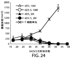


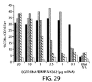


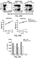


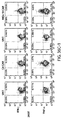


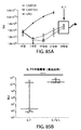


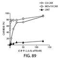


Classifications
-
- A—HUMAN NECESSITIES
- A61—MEDICAL OR VETERINARY SCIENCE; HYGIENE
- A61K—PREPARATIONS FOR MEDICAL, DENTAL OR TOILETRY PURPOSES
- A61K2239/00—Indexing codes associated with cellular immunotherapy of group A61K39/46
- A61K2239/10—Indexing codes associated with cellular immunotherapy of group A61K39/46 characterized by the structure of the chimeric antigen receptor [CAR]
- A61K2239/21—Transmembrane domain
-
- A—HUMAN NECESSITIES
- A61—MEDICAL OR VETERINARY SCIENCE; HYGIENE
- A61K—PREPARATIONS FOR MEDICAL, DENTAL OR TOILETRY PURPOSES
- A61K2239/00—Indexing codes associated with cellular immunotherapy of group A61K39/46
- A61K2239/10—Indexing codes associated with cellular immunotherapy of group A61K39/46 characterized by the structure of the chimeric antigen receptor [CAR]
- A61K2239/22—Intracellular domain
-
- A—HUMAN NECESSITIES
- A61—MEDICAL OR VETERINARY SCIENCE; HYGIENE
- A61K—PREPARATIONS FOR MEDICAL, DENTAL OR TOILETRY PURPOSES
- A61K2239/00—Indexing codes associated with cellular immunotherapy of group A61K39/46
- A61K2239/31—Indexing codes associated with cellular immunotherapy of group A61K39/46 characterized by the route of administration
-
- A—HUMAN NECESSITIES
- A61—MEDICAL OR VETERINARY SCIENCE; HYGIENE
- A61K—PREPARATIONS FOR MEDICAL, DENTAL OR TOILETRY PURPOSES
- A61K2239/00—Indexing codes associated with cellular immunotherapy of group A61K39/46
- A61K2239/38—Indexing codes associated with cellular immunotherapy of group A61K39/46 characterised by the dose, timing or administration schedule
-
- A—HUMAN NECESSITIES
- A61—MEDICAL OR VETERINARY SCIENCE; HYGIENE
- A61K—PREPARATIONS FOR MEDICAL, DENTAL OR TOILETRY PURPOSES
- A61K2239/00—Indexing codes associated with cellular immunotherapy of group A61K39/46
- A61K2239/46—Indexing codes associated with cellular immunotherapy of group A61K39/46 characterised by the cancer treated
- A61K2239/48—Blood cells, e.g. leukemia or lymphoma
-
- A—HUMAN NECESSITIES
- A61—MEDICAL OR VETERINARY SCIENCE; HYGIENE
- A61K—PREPARATIONS FOR MEDICAL, DENTAL OR TOILETRY PURPOSES
- A61K2239/00—Indexing codes associated with cellular immunotherapy of group A61K39/46
- A61K2239/46—Indexing codes associated with cellular immunotherapy of group A61K39/46 characterised by the cancer treated
- A61K2239/49—Breast
-
- A—HUMAN NECESSITIES
- A61—MEDICAL OR VETERINARY SCIENCE; HYGIENE
- A61K—PREPARATIONS FOR MEDICAL, DENTAL OR TOILETRY PURPOSES
- A61K2239/00—Indexing codes associated with cellular immunotherapy of group A61K39/46
- A61K2239/46—Indexing codes associated with cellular immunotherapy of group A61K39/46 characterised by the cancer treated
- A61K2239/58—Prostate
-
- A—HUMAN NECESSITIES
- A61—MEDICAL OR VETERINARY SCIENCE; HYGIENE
- A61K—PREPARATIONS FOR MEDICAL, DENTAL OR TOILETRY PURPOSES
- A61K2239/00—Indexing codes associated with cellular immunotherapy of group A61K39/46
- A61K2239/46—Indexing codes associated with cellular immunotherapy of group A61K39/46 characterised by the cancer treated
- A61K2239/59—Reproductive system, e.g. uterus, ovaries, cervix or testes
-
- A—HUMAN NECESSITIES
- A61—MEDICAL OR VETERINARY SCIENCE; HYGIENE
- A61K—PREPARATIONS FOR MEDICAL, DENTAL OR TOILETRY PURPOSES
- A61K38/00—Medicinal preparations containing peptides
- A61K38/16—Peptides having more than 20 amino acids; Gastrins; Somatostatins; Melanotropins; Derivatives thereof
- A61K38/17—Peptides having more than 20 amino acids; Gastrins; Somatostatins; Melanotropins; Derivatives thereof from animals; from humans
- A61K38/19—Cytokines; Lymphokines; Interferons
- A61K38/20—Interleukins [IL]
- A61K38/2086—IL-13 to IL-16
-
- A—HUMAN NECESSITIES
- A61—MEDICAL OR VETERINARY SCIENCE; HYGIENE
- A61K—PREPARATIONS FOR MEDICAL, DENTAL OR TOILETRY PURPOSES
- A61K39/00—Medicinal preparations containing antigens or antibodies
- A61K39/39—Medicinal preparations containing antigens or antibodies characterised by the immunostimulating additives, e.g. chemical adjuvants
-
- A—HUMAN NECESSITIES
- A61—MEDICAL OR VETERINARY SCIENCE; HYGIENE
- A61K—PREPARATIONS FOR MEDICAL, DENTAL OR TOILETRY PURPOSES
- A61K39/00—Medicinal preparations containing antigens or antibodies
- A61K39/395—Antibodies; Immunoglobulins; Immune serum, e.g. antilymphocytic serum
- A61K39/39533—Antibodies; Immunoglobulins; Immune serum, e.g. antilymphocytic serum against materials from animals
- A61K39/3955—Antibodies; Immunoglobulins; Immune serum, e.g. antilymphocytic serum against materials from animals against proteinaceous materials, e.g. enzymes, hormones, lymphokines
-
- A—HUMAN NECESSITIES
- A61—MEDICAL OR VETERINARY SCIENCE; HYGIENE
- A61K—PREPARATIONS FOR MEDICAL, DENTAL OR TOILETRY PURPOSES
- A61K39/00—Medicinal preparations containing antigens or antibodies
- A61K39/46—Cellular immunotherapy
- A61K39/461—Cellular immunotherapy characterised by the cell type used
- A61K39/4611—T-cells, e.g. tumor infiltrating lymphocytes [TIL], lymphokine-activated killer cells [LAK] or regulatory T cells [Treg]
-
- A—HUMAN NECESSITIES
- A61—MEDICAL OR VETERINARY SCIENCE; HYGIENE
- A61K—PREPARATIONS FOR MEDICAL, DENTAL OR TOILETRY PURPOSES
- A61K39/00—Medicinal preparations containing antigens or antibodies
- A61K39/46—Cellular immunotherapy
- A61K39/463—Cellular immunotherapy characterised by recombinant expression
- A61K39/4631—Chimeric Antigen Receptors [CAR]
-
- A—HUMAN NECESSITIES
- A61—MEDICAL OR VETERINARY SCIENCE; HYGIENE
- A61K—PREPARATIONS FOR MEDICAL, DENTAL OR TOILETRY PURPOSES
- A61K39/00—Medicinal preparations containing antigens or antibodies
- A61K39/46—Cellular immunotherapy
- A61K39/463—Cellular immunotherapy characterised by recombinant expression
- A61K39/4636—Immune checkpoint inhibitors
-
- A—HUMAN NECESSITIES
- A61—MEDICAL OR VETERINARY SCIENCE; HYGIENE
- A61K—PREPARATIONS FOR MEDICAL, DENTAL OR TOILETRY PURPOSES
- A61K39/00—Medicinal preparations containing antigens or antibodies
- A61K39/46—Cellular immunotherapy
- A61K39/464—Cellular immunotherapy characterised by the antigen targeted or presented
- A61K39/4643—Vertebrate antigens
- A61K39/4644—Cancer antigens
- A61K39/464402—Receptors, cell surface antigens or cell surface determinants
- A61K39/464403—Receptors for growth factors
- A61K39/464404—Epidermal growth factor receptors [EGFR]
-
- A—HUMAN NECESSITIES
- A61—MEDICAL OR VETERINARY SCIENCE; HYGIENE
- A61K—PREPARATIONS FOR MEDICAL, DENTAL OR TOILETRY PURPOSES
- A61K39/00—Medicinal preparations containing antigens or antibodies
- A61K39/46—Cellular immunotherapy
- A61K39/464—Cellular immunotherapy characterised by the antigen targeted or presented
- A61K39/4643—Vertebrate antigens
- A61K39/4644—Cancer antigens
- A61K39/464402—Receptors, cell surface antigens or cell surface determinants
- A61K39/464403—Receptors for growth factors
- A61K39/464406—Her-2/neu/ErbB2, Her-3/ErbB3 or Her 4/ ErbB4
-
- A—HUMAN NECESSITIES
- A61—MEDICAL OR VETERINARY SCIENCE; HYGIENE
- A61K—PREPARATIONS FOR MEDICAL, DENTAL OR TOILETRY PURPOSES
- A61K39/00—Medicinal preparations containing antigens or antibodies
- A61K39/46—Cellular immunotherapy
- A61K39/464—Cellular immunotherapy characterised by the antigen targeted or presented
- A61K39/4643—Vertebrate antigens
- A61K39/4644—Cancer antigens
- A61K39/464402—Receptors, cell surface antigens or cell surface determinants
- A61K39/464411—Immunoglobulin superfamily
- A61K39/464412—CD19 or B4
-
- A—HUMAN NECESSITIES
- A61—MEDICAL OR VETERINARY SCIENCE; HYGIENE
- A61K—PREPARATIONS FOR MEDICAL, DENTAL OR TOILETRY PURPOSES
- A61K39/00—Medicinal preparations containing antigens or antibodies
- A61K39/46—Cellular immunotherapy
- A61K39/464—Cellular immunotherapy characterised by the antigen targeted or presented
- A61K39/4643—Vertebrate antigens
- A61K39/4644—Cancer antigens
- A61K39/464466—Adhesion molecules, e.g. NRCAM, EpCAM or cadherins
- A61K39/464468—Mesothelin [MSLN]
-
- A—HUMAN NECESSITIES
- A61—MEDICAL OR VETERINARY SCIENCE; HYGIENE
- A61K—PREPARATIONS FOR MEDICAL, DENTAL OR TOILETRY PURPOSES
- A61K39/00—Medicinal preparations containing antigens or antibodies
- A61K39/46—Cellular immunotherapy
- A61K39/464—Cellular immunotherapy characterised by the antigen targeted or presented
- A61K39/4643—Vertebrate antigens
- A61K39/4644—Cancer antigens
- A61K39/464469—Tumor associated carbohydrates
- A61K39/46447—Mucins, e.g. MUC-1
-
- A—HUMAN NECESSITIES
- A61—MEDICAL OR VETERINARY SCIENCE; HYGIENE
- A61K—PREPARATIONS FOR MEDICAL, DENTAL OR TOILETRY PURPOSES
- A61K39/00—Medicinal preparations containing antigens or antibodies
- A61K39/46—Cellular immunotherapy
- A61K39/464—Cellular immunotherapy characterised by the antigen targeted or presented
- A61K39/4643—Vertebrate antigens
- A61K39/4644—Cancer antigens
- A61K39/464469—Tumor associated carbohydrates
- A61K39/464471—Gangliosides, e.g. GM2, GD2 or GD3
-
- A—HUMAN NECESSITIES
- A61—MEDICAL OR VETERINARY SCIENCE; HYGIENE
- A61K—PREPARATIONS FOR MEDICAL, DENTAL OR TOILETRY PURPOSES
- A61K39/00—Medicinal preparations containing antigens or antibodies
- A61K39/46—Cellular immunotherapy
- A61K39/464—Cellular immunotherapy characterised by the antigen targeted or presented
- A61K39/4643—Vertebrate antigens
- A61K39/4644—Cancer antigens
- A61K39/464493—Prostate associated antigens e.g. Prostate stem cell antigen [PSCA]; Prostate carcinoma tumor antigen [PCTA]; Prostatic acid phosphatase [PAP]; Prostate-specific G-protein-coupled receptor [PSGR]
- A61K39/464495—Prostate specific membrane antigen [PSMA]
-
- A—HUMAN NECESSITIES
- A61—MEDICAL OR VETERINARY SCIENCE; HYGIENE
- A61P—SPECIFIC THERAPEUTIC ACTIVITY OF CHEMICAL COMPOUNDS OR MEDICINAL PREPARATIONS
- A61P35/00—Antineoplastic agents
-
- A—HUMAN NECESSITIES
- A61—MEDICAL OR VETERINARY SCIENCE; HYGIENE
- A61P—SPECIFIC THERAPEUTIC ACTIVITY OF CHEMICAL COMPOUNDS OR MEDICINAL PREPARATIONS
- A61P35/00—Antineoplastic agents
- A61P35/02—Antineoplastic agents specific for leukemia
-
- A—HUMAN NECESSITIES
- A61—MEDICAL OR VETERINARY SCIENCE; HYGIENE
- A61P—SPECIFIC THERAPEUTIC ACTIVITY OF CHEMICAL COMPOUNDS OR MEDICINAL PREPARATIONS
- A61P43/00—Drugs for specific purposes, not provided for in groups A61P1/00-A61P41/00
-
- C—CHEMISTRY; METALLURGY
- C07—ORGANIC CHEMISTRY
- C07K—PEPTIDES
- C07K16/00—Immunoglobulins [IGs], e.g. monoclonal or polyclonal antibodies
- C07K16/18—Immunoglobulins [IGs], e.g. monoclonal or polyclonal antibodies against material from animals or humans
- C07K16/28—Immunoglobulins [IGs], e.g. monoclonal or polyclonal antibodies against material from animals or humans against receptors, cell surface antigens or cell surface determinants
- C07K16/2863—Immunoglobulins [IGs], e.g. monoclonal or polyclonal antibodies against material from animals or humans against receptors, cell surface antigens or cell surface determinants against receptors for growth factors, growth regulators
-
- C—CHEMISTRY; METALLURGY
- C07—ORGANIC CHEMISTRY
- C07K—PEPTIDES
- C07K16/00—Immunoglobulins [IGs], e.g. monoclonal or polyclonal antibodies
- C07K16/18—Immunoglobulins [IGs], e.g. monoclonal or polyclonal antibodies against material from animals or humans
- C07K16/28—Immunoglobulins [IGs], e.g. monoclonal or polyclonal antibodies against material from animals or humans against receptors, cell surface antigens or cell surface determinants
- C07K16/30—Immunoglobulins [IGs], e.g. monoclonal or polyclonal antibodies against material from animals or humans against receptors, cell surface antigens or cell surface determinants from tumour cells
-
- C—CHEMISTRY; METALLURGY
- C07—ORGANIC CHEMISTRY
- C07K—PEPTIDES
- C07K16/00—Immunoglobulins [IGs], e.g. monoclonal or polyclonal antibodies
- C07K16/18—Immunoglobulins [IGs], e.g. monoclonal or polyclonal antibodies against material from animals or humans
- C07K16/32—Immunoglobulins [IGs], e.g. monoclonal or polyclonal antibodies against material from animals or humans against translation products of oncogenes
-
- C—CHEMISTRY; METALLURGY
- C12—BIOCHEMISTRY; BEER; SPIRITS; WINE; VINEGAR; MICROBIOLOGY; ENZYMOLOGY; MUTATION OR GENETIC ENGINEERING
- C12N—MICROORGANISMS OR ENZYMES; COMPOSITIONS THEREOF; PROPAGATING, PRESERVING, OR MAINTAINING MICROORGANISMS; MUTATION OR GENETIC ENGINEERING; CULTURE MEDIA
- C12N5/00—Undifferentiated human, animal or plant cells, e.g. cell lines; Tissues; Cultivation or maintenance thereof; Culture media therefor
- C12N5/06—Animal cells or tissues; Human cells or tissues
- C12N5/0602—Vertebrate cells
- C12N5/0634—Cells from the blood or the immune system
- C12N5/0636—T lymphocytes
- C12N5/0638—Cytotoxic T lymphocytes [CTL] or lymphokine activated killer cells [LAK]
-
- A—HUMAN NECESSITIES
- A61—MEDICAL OR VETERINARY SCIENCE; HYGIENE
- A61K—PREPARATIONS FOR MEDICAL, DENTAL OR TOILETRY PURPOSES
- A61K39/00—Medicinal preparations containing antigens or antibodies
- A61K2039/505—Medicinal preparations containing antigens or antibodies comprising antibodies
-
- A—HUMAN NECESSITIES
- A61—MEDICAL OR VETERINARY SCIENCE; HYGIENE
- A61K—PREPARATIONS FOR MEDICAL, DENTAL OR TOILETRY PURPOSES
- A61K39/00—Medicinal preparations containing antigens or antibodies
- A61K2039/555—Medicinal preparations containing antigens or antibodies characterised by a specific combination antigen/adjuvant
- A61K2039/55511—Organic adjuvants
- A61K2039/55522—Cytokines; Lymphokines; Interferons
- A61K2039/55527—Interleukins
-
- C—CHEMISTRY; METALLURGY
- C07—ORGANIC CHEMISTRY
- C07K—PEPTIDES
- C07K14/00—Peptides having more than 20 amino acids; Gastrins; Somatostatins; Melanotropins; Derivatives thereof
- C07K14/435—Peptides having more than 20 amino acids; Gastrins; Somatostatins; Melanotropins; Derivatives thereof from animals; from humans
- C07K14/705—Receptors; Cell surface antigens; Cell surface determinants
- C07K14/70503—Immunoglobulin superfamily
- C07K14/7051—T-cell receptor (TcR)-CD3 complex
-
- C—CHEMISTRY; METALLURGY
- C07—ORGANIC CHEMISTRY
- C07K—PEPTIDES
- C07K2317/00—Immunoglobulins specific features
- C07K2317/20—Immunoglobulins specific features characterized by taxonomic origin
- C07K2317/22—Immunoglobulins specific features characterized by taxonomic origin from camelids, e.g. camel, llama or dromedary
-
- C—CHEMISTRY; METALLURGY
- C07—ORGANIC CHEMISTRY
- C07K—PEPTIDES
- C07K2317/00—Immunoglobulins specific features
- C07K2317/50—Immunoglobulins specific features characterized by immunoglobulin fragments
- C07K2317/56—Immunoglobulins specific features characterized by immunoglobulin fragments variable (Fv) region, i.e. VH and/or VL
- C07K2317/569—Single domain, e.g. dAb, sdAb, VHH, VNAR or nanobody®
-
- C—CHEMISTRY; METALLURGY
- C07—ORGANIC CHEMISTRY
- C07K—PEPTIDES
- C07K2317/00—Immunoglobulins specific features
- C07K2317/60—Immunoglobulins specific features characterized by non-natural combinations of immunoglobulin fragments
- C07K2317/62—Immunoglobulins specific features characterized by non-natural combinations of immunoglobulin fragments comprising only variable region components
- C07K2317/622—Single chain antibody (scFv)
-
- C—CHEMISTRY; METALLURGY
- C07—ORGANIC CHEMISTRY
- C07K—PEPTIDES
- C07K2317/00—Immunoglobulins specific features
- C07K2317/70—Immunoglobulins specific features characterized by effect upon binding to a cell or to an antigen
- C07K2317/77—Internalization into the cell
-
- C—CHEMISTRY; METALLURGY
- C07—ORGANIC CHEMISTRY
- C07K—PEPTIDES
- C07K2317/00—Immunoglobulins specific features
- C07K2317/90—Immunoglobulins specific features characterized by (pharmaco)kinetic aspects or by stability of the immunoglobulin
- C07K2317/92—Affinity (KD), association rate (Ka), dissociation rate (Kd) or EC50 value
-
- C—CHEMISTRY; METALLURGY
- C07—ORGANIC CHEMISTRY
- C07K—PEPTIDES
- C07K2319/00—Fusion polypeptide
-
- C—CHEMISTRY; METALLURGY
- C07—ORGANIC CHEMISTRY
- C07K—PEPTIDES
- C07K2319/00—Fusion polypeptide
- C07K2319/01—Fusion polypeptide containing a localisation/targetting motif
- C07K2319/03—Fusion polypeptide containing a localisation/targetting motif containing a transmembrane segment
-
- C—CHEMISTRY; METALLURGY
- C07—ORGANIC CHEMISTRY
- C07K—PEPTIDES
- C07K2319/00—Fusion polypeptide
- C07K2319/32—Fusion polypeptide fusions with soluble part of a cell surface receptor, "decoy receptors"
-
- C—CHEMISTRY; METALLURGY
- C12—BIOCHEMISTRY; BEER; SPIRITS; WINE; VINEGAR; MICROBIOLOGY; ENZYMOLOGY; MUTATION OR GENETIC ENGINEERING
- C12N—MICROORGANISMS OR ENZYMES; COMPOSITIONS THEREOF; PROPAGATING, PRESERVING, OR MAINTAINING MICROORGANISMS; MUTATION OR GENETIC ENGINEERING; CULTURE MEDIA
- C12N2510/00—Genetically modified cells
Landscapes
- Health & Medical Sciences (AREA)
- Life Sciences & Earth Sciences (AREA)
- Chemical & Material Sciences (AREA)
- Immunology (AREA)
- General Health & Medical Sciences (AREA)
- Medicinal Chemistry (AREA)
- Public Health (AREA)
- Animal Behavior & Ethology (AREA)
- Veterinary Medicine (AREA)
- Epidemiology (AREA)
- Microbiology (AREA)
- Pharmacology & Pharmacy (AREA)
- Cell Biology (AREA)
- Mycology (AREA)
- Organic Chemistry (AREA)
- Oncology (AREA)
- Engineering & Computer Science (AREA)
- Genetics & Genomics (AREA)
- Molecular Biology (AREA)
- Biochemistry (AREA)
- Biomedical Technology (AREA)
- Bioinformatics & Cheminformatics (AREA)
- Zoology (AREA)
- Proteomics, Peptides & Aminoacids (AREA)
- Wood Science & Technology (AREA)
- Biotechnology (AREA)
- Biophysics (AREA)
- Hematology (AREA)
- General Engineering & Computer Science (AREA)
- Chemical Kinetics & Catalysis (AREA)
- General Chemical & Material Sciences (AREA)
- Hospice & Palliative Care (AREA)
- Developmental Biology & Embryology (AREA)
- Endocrinology (AREA)
- Dermatology (AREA)
- Nuclear Medicine, Radiotherapy & Molecular Imaging (AREA)
- Reproductive Health (AREA)
- Pregnancy & Childbirth (AREA)
- Gynecology & Obstetrics (AREA)
- Gastroenterology & Hepatology (AREA)
Description
関連出願
本出願は、米国出願61/953,783号(2014年3月15日出願)、米国出願61/976,375号(2014年4月7日出願)、米国出願62/027,154(2014年7月21日出願)、米国出願62/076,146号(2014年11月6日出願)および米国出願62/097,286(2014年12月29日出願)に基づく優先権を主張する。これらの出願の内容の全体を引用により本明細書に包含させる。
Related Applications This application includes US
発明の分野
本発明は、概して腫瘍抗原の発現と関係する疾患を処置するための、キメラ抗原受容体(CAR)を発現するように操作した免疫エフェクター細胞(例えば、T細胞、NK細胞)の使用に関する。
Field of Invention The present invention uses immune effector cells (eg, T cells, NK cells) engineered to express a chimeric antigen receptor (CAR) for treating diseases that are generally associated with tumor antigen expression. Regarding.
発明の背景
自己T細胞、特にキメラ抗原受容体(CAR)を形質導入したT細胞を用いる養子細胞移入(ACT)治療は、血液癌の治験で見込みが示されている。
Background of the Invention Adoptive cell transfer (ACT) therapy using autologous T cells, especially T cells transduced with chimeric antigen receptor (CAR), has been shown to be promising in clinical trials for hematological malignancies.
発明の要約
本発明は、少なくとも一部、ここに記載する腫瘍抗原に結合するCARを発現するように操作した免疫エフェクター細胞(例えば、T細胞、NK細胞)の、該腫瘍抗原の発現と関係する癌の処置における使用に関する。
Abstract of the Invention The present invention relates, at least in part, to the expression of the tumor antigens of immune effector cells (eg, T cells, NK cells) engineered to express CARs that bind to the tumor antigens described herein. Regarding use in the treatment of cancer.
CARコード化核酸
したがって、ある面において、本発明は、キメラ抗原受容体(CAR)をコードする単離核酸分子に関し、ここで、CARは、ここに記載する腫瘍抗原に結合する抗原結合ドメイン(例えば、抗体または抗体フラグメント、TCRまたはTCRフラグメント)、膜貫通ドメイン(例えば、ここに記載する膜貫通ドメイン)および細胞内シグナル伝達ドメイン(例えば、ここに記載する細胞内シグナル伝達ドメイン)(例えば、共刺激ドメイン(例えば、ここに記載する共刺激ドメイン)および/または一次シグナル伝達ドメイン(例えば、ここに記載する一次シグナル伝達ドメインを含む細胞内シグナル伝達ドメイン)を含む。ある態様において、腫瘍抗原は、CD19;CD123;CD22;CD30;CD171;CS−1(別名CD2サブセット1、CRACC、SLAMF7、CD319および19A24);C型レクチン様分子−1(CLL−1またはCLECL1);CD33;上皮細胞増殖因子受容体変異体III(EGFRvIII);ガングリオシドG2(GD2);ガングリオシドGD3(aNeu5Ac(2−8)aNeu5Ac(2−3)bDGalp(1−4)bDGlcp(1−1)Cer);TNF受容体ファミリーメンバーB細胞成熟抗原(BCMA);Tn抗原((Tn Ag)または(GalNAcα−Ser/Thr));前立腺特異的膜抗原(PSMA);受容体チロシンキナーゼ様オーファン受容体1(ROR1);Fms様チロシンキナーゼ3(FLT3);腫瘍関連糖タンパク質72(TAG72);CD38;CD44v6;癌胎児性抗原(CEA);上皮性細胞接着分子(EPCAM);B7H3(CD276);KIT(CD117);インターロイキン−13受容体サブユニットアルファ−2(IL−13Ra2またはCD213A2);メソテリン;インターロイキン11受容体アルファ(IL−11Ra);前立腺幹細胞抗原(PSCA);プロテアーゼセリン21(精巣inまたはPRSS21);血管内皮細胞増殖因子受容体2(VEGFR2);ルイス(Y)抗原;CD24;血小板由来増殖因子受容体ベータ(PDGFR−ベータ);ステージ特異的胚性抗原−4(SSEA−4);CD20;葉酸受容体アルファ;受容体チロシン−タンパク質キナーゼERBB2(Her2/neu);ムチン1、細胞表面関連(MUC1);上皮細胞増殖因子受容体(EGFR);神経細胞接着分子(NCAM);プロスターゼ;前立腺酸ホスファターゼ(PAP);伸長因子2変異型(ELF2M);エフリンB2;線維芽細胞活性化タンパク質アルファ(FAP);インシュリン様増殖因子1受容体(IGF−I受容体)、炭酸脱水酵素IX(CAIX);プロテアソーム(プロソーム、マクロパイン)サブユニット、ベータ型、9(LMP2);糖タンパク質100(gp100);易切断領域(BCR)およびエーベルソンマウス白血病ウイルス癌遺伝子ホモログ1(Abl)からなる癌遺伝子融合タンパク質(bcr−abl);チロシナーゼ;エフリンA型受容体2(EphA2);フコシルGM1;シアリルルイス接着分子(sLe);ガングリオシドGM3(aNeu5Ac(2−3)bDGalp(1−4)bDGlcp(1−1)Cer);トランスグルタミナーゼ5(TGS5);高分子量黒色腫関連抗原(HMWMAA);o−アセチル−GD2ガングリオシド(OAcGD2);葉酸受容体ベータ;腫瘍内皮マーカー1(TEM1/CD248);腫瘍内皮マーカー7関連(TEM7R);クローディン6(CLDN6);甲状腺刺激ホルモン受容体(TSHR);Gタンパク質共役受容体クラスCグループ5、メンバーD(GPRC5D);染色体Xオープンリーディングフレーム61(CXORF61);CD97;CD179a;未分化リンパ腫キナーゼ(ALK);ポリシアル酸;胎盤特異的1(PLAC1);globoHグリコセラミド(GloboH)の六糖部分;乳腺分化抗原(NY−BR−1);ウロプラキン2(UPK2);A型肝炎ウイルス細胞性受容体1(HAVCR1);アドレナリン受容体ベータ3(ADRB3);パンネキシン3(PANX3);Gタンパク質共役受容体20(GPR20);リンパ球抗原6複合体、座位K9(LY6K);嗅覚受容体51E2(OR51E2);TCRガンマ選択的リーディングフレームタンパク質(TARP);ウィルムス腫瘍タンパク質(WT1);癌/精巣抗原1(NY−ESO−1);癌/精巣抗原2(LAGE−1a);黒色腫関連抗原1(MAGE−A1);染色体12pに位置するETS転座変異体遺伝子6(ETV6−AML);精子タンパク質17(SPA17);X抗原ファミリー、メンバー1A(XAGE1);アンギオポエチン結合細胞表面受容体2(Tie 2);黒色腫癌精巣抗原−1(MAD−CT−1);黒色腫癌精巣抗原−2(MAD−CT−2);Fos関連抗原1;腫瘍タンパク質p53(p53);p53変異体;プロテイン;サバイビン(surviving);テロメラーゼ;前立腺癌腫瘍抗原−1(PCTA−1またはガレクチン8)、T細胞1により認識される黒色腫抗原(メランAまたはMART1);ラット肉腫(Ras)変異体;ヒトテロメラーゼ逆転写酵素(hTERT);肉腫転座切断点;アポトーシスの黒色腫阻害剤(ML−IAP);ERG(膜貫通型プロテアーゼ、セリン2(TMPRSS2)ETS融合遺伝子);N−アセチルグルコサミニル−トランスフェラーゼV(NA17);ペアードボックスタンパク質Pax−3(PAX3);アンドロゲン受容体;サイクリンB1;v−mycトリ骨髄細胞腫ウイルス癌遺伝子神経芽腫由来ホモログ(MYCN);RasホモログファミリーメンバーC(RhoC);チロシナーゼ関連タンパク質2(TRP−2);チトクロムP450 1B1(CYP1B1);CCCTC結合因子(亜鉛フィンガータンパク質)様(BORISまたはBrother of the Regulator of Imprinted Sites)、T細胞3により認識される扁平上皮細胞癌抗原(SART3);ペアードボックスタンパク質Pax−5(PAX5);プロアクロシン結合タンパク質p32(OY−TES1);リンパ球特異的タンパク質チロシンキナーゼ(LCK);Aキナーゼアンカータンパク質4(AKAP−4);滑膜肉腫、X切断点2(SSX2);終末糖化産物受容体(RAGE−1);腎臓ユビキタス1(RU1);腎臓ユビキタス2(RU2);レグマイン;ヒト乳頭腫ウイルスE6(HPV E6);ヒト乳頭腫ウイルスE7(HPV E7);腸管カルボキシルエステラーゼ;ヒートショックタンパク質70−2変異型(mut hsp70−2);CD79a;CD79b;CD72;白血球関連免疫グロブリン様受容体1(LAIR1);IgA受容体のFcフラグメント(FCARまたはCD89);白血球免疫グロブリン様受容体サブファミリーAメンバー2(LILRA2);CD300分子様ファミリーメンバー(CD300LF);C型レクチンドメインファミリー12メンバーA(CLEC12A);骨髄間質細胞抗原2(BST2);EGF様モジュール含有ムチン様ホルモン受容体様2(EMR2);リンパ球抗原75(LY75);グリピカン−3(GPC3);Fc受容体様5(FCRL5);および免疫グロブリンラムダ様ポリペプチド1(IGLL1)から選択される1以上である。
CAR-encoded nucleic acids Thus, in one aspect, the present invention relates to isolated nucleic acid molecules encoding a chimeric antigen receptor (CAR), wherein CAR is an antigen-binding domain that binds to the tumor antigens described herein (eg, eg). , Antigen or antibody fragment, TCR or TCR fragment), transmembrane domain (eg, transmembrane domain described herein) and intracellular signaling domain (eg, intracellular signaling domain described herein) (eg, costimulatory) In some embodiments, the tumor antigen comprises a domain (eg, a costimulatory domain described herein) and / or a primary signal transduction domain (eg, an intracellular signal transduction domain comprising a primary signal transduction domain described herein). CD123; CD22; CD30; CD171; CS-1 (also known as
ある態様において、コード化CAR分子により結合される腫瘍抗原は、TSHR、CD171、CS−1、CLL−1、GD3、Tn Ag、FLT3、CD38、CD44v6、B7H3、KIT、IL−13Ra2、IL−11Ra、PSCA、PRSS21、VEGFR2、ルイスY、CD24、PDGFR−ベータ、SSEA−4、MUC1、EGFR、NCAM、CAIX、LMP2、EphA2、フコシルGM1、sLe、GM3、TGS5、HMWMAA、o−アセチル−GD2、葉酸受容体ベータ、TEM1/CD248、TEM7R、CLDN6、GPRC5D、CXORF61、CD97、CD179a、ALK、ポリシアル酸、PLAC1、GloboH、NY−BR−1、UPK2、HAVCR1、ADRB3、PANX3、GPR20、LY6K、OR51E2、TARP、WT1、ETV6−AML、精子タンパク質17、XAGE1、Tie 2、MAD−CT−1、MAD−CT−2、Fos関連抗原1、p53変異体、hTERT、肉腫転座切断点、ML−IAP、ERG(TMPRSS2 ETS融合遺伝子)、NA17、PAX3、アンドロゲン受容体、サイクリンB1、MYCN、RhoC、CYP1B1、BORIS、SART3、PAX5、OY−TES1、LCK、AKAP−4、SSX2、CD79a、CD79b、CD72、LAIR1、FCAR、LILRA2、CD300LF、CLEC12A、BST2、EMR2、LY75、GPC3、FCRL5およびIGLL1から選択される1以上である。
In some embodiments, the tumor antigens bound by the encoded CAR molecule are TSHR, CD171, CS-1, CLL-1, GD3, Tn Ag, FLT3, CD38, CD44v6, B7H3, KIT, IL-13Ra2, IL-11Ra. , PSCA, PRSS21, VEGFR2, Lewis Y, CD24, PDGFR-beta, SSEA-4, MUC1, EGFR, NCAM, CAIX, LMP2, EphA2, fucosyl GM1, sLe, GM3, TGS5, HMWMAA, o-acetyl-GD2, folic acid Receptor beta, TEM1 / CD248, TEM7R, CLDN6, GPRC5D, CXORF61, CD97, CD179a, ALK, polysialic acid, PLAC1, GloboH, NY-BR-1, UPK2, HAVCR1, ADRB3, PANX3, GPR20, LY6K, OR , WT1, ETV6-AML, sperm protein 17, XAGE1,
ある態様において、コード化CAR分子により結合される腫瘍抗原は、TSHR、CLDN6、GPRC5D、CXORF61、CD97、CD179a、ALK、ポリシアル酸、PLAC1、GloboH、NY−BR−1、UPK2、HAVCR1、ADRB3、PANX3、GPR20、LY6KおよびOR51E2から選択される1以上である。 In some embodiments, the tumor antigens bound by the encoded CAR molecule are TSHR, CLDN6, GPRC5D, CXORF61, CD97, CD179a, ALK, polysialic acid, PLAC1, GloboH, NY-BR-1, UPK2, HAVCR1, ADRB3, PANX3. , GPR20, LY6K and OR51E2, one or more selected from.
ある態様において、コード化CAR分子の抗原結合ドメインは、抗体、抗体フラグメント、scFv、Fv、Fab、(Fab’)2、単一ドメイン抗体(SDAB)、VHまたはVLドメインまたはラクダ類VHHドメインを含む。 In some embodiments, the antigen-binding domain of the encoded CAR molecule comprises an antibody, antibody fragment, scFv, Fv, Fab, (Fab') 2 , single domain antibody (SDAB), VH or VL domain or camel VHH domain. ..
ある態様において、コード化CAR分子の膜貫通ドメインは、T細胞受容体のアルファ鎖、ベータ鎖またはゼータ鎖、CD28、CD3イプシロン、CD45、CD4、CD5、CD8、CD9、CD16、CD22、CD33、CD37、CD64、CD80、CD86、CD134、CD137、CD154、KIRDS2、OX40、CD2、CD27、LFA−1(CD11a、CD18)、ICOS(CD278)、4−1BB(CD137)、GITR、CD40、BAFFR、HVEM(LIGHTR)、SLAMF7、NKp80(KLRF1)、CD160、CD19、IL2Rベータ、IL2Rガンマ、IL7Rα、ITGA1、VLA1、CD49a、ITGA4、IA4、CD49D、ITGA6、VLA−6、CD49f、ITGAD、CD11d、ITGAE、CD103、ITGAL、CD11a、LFA−1、ITGAM、CD11b、ITGAX、CD11c、ITGB1、CD29、ITGB2、CD18、LFA−1、ITGB7、TNFR2、DNAM1(CD226)、SLAMF4(CD244、2B4)、CD84、CD96(Tactile)、CEACAM1、CRTAM、Ly9(CD229)、CD160(BY55)、PSGL1、CD100(SEMA4D)、SLAMF6(NTB−A、Ly108)、SLAM(SLAMF1、CD150、IPO−3)、BLAME(SLAMF8)、SELPLG(CD162)、LTBR、PAG/Cbp、NKp44、NKp30、NKp46、NKG2Dおよび/またはNKG2Cの膜貫通ドメインから選択される膜貫通ドメインを含む。 In some embodiments, the transmembrane domain of the encoded CAR molecule is the alpha, beta or zeta chain of the T cell receptor, CD28, CD3 epsilon, CD45, CD4, CD5, CD8, CD9, CD16, CD22, CD33, CD37. , CD64, CD80, CD86, CD134, CD137, CD154, KIRDS2, OX40, CD2, CD27, LFA-1 (CD11a, CD18), ICOS (CD278), 4-1BB (CD137), GITR, CD40, BAFFR, HVEM ( LIGHTTR), SLAMF7, NKp80 (KLRF1), CD160, CD19, IL2R beta, IL2R gamma, IL7Rα, ITGA1, VLA1, CD49a, ITGA4, IA4, CD49D, ITGA6, VLA-6, CD49f, ITGAD, CD11d, ITGAE, CD ITGAL, CD11a, LFA-1, ITGAM, CD11b, ITGAX, CD11c, ITGB1, CD29, ITGB2, CD18, LFA-1, ITGB7, TNFR2, DNAM1 (CD226), SLAMF4 (CD244, 2B4), CD84, CD96 (Tactile) , CEACAM1, CRTAM, Ly9 (CD229), CD160 (BY55), PSGL1, CD100 (SEMA4D), SLAMF6 (NTB-A, Ly108), SLAM (SLAMF1, CD150, IPO-3), BLAME (SLAMF8), SELPLG (CD162) ), LTBR, PAG / Cbp, NKp44, NKp30, NKp46, NKG2D and / or includes a transmembrane domain selected from the transmembrane domains of NKG2C.
ある態様において、コード化膜貫通ドメインは、配列番号12のアミノ酸配列の少なくとも1、2または3の修飾を有するが、修飾が20、10または5を超えないCD8膜貫通ドメインのアミノ酸配列または配列番号12のアミノ酸配列と95〜99%同一性を有する配列を含む。ある態様において、コード化膜貫通ドメインは配列番号12の配列を含む。 In some embodiments, the encoded transmembrane domain has at least 1, 2 or 3 modifications of the amino acid sequence of SEQ ID NO: 12, but the amino acid sequence or SEQ ID NO of the CD8 transmembrane domain with no more than 20, 10 or 5 modifications. Includes a sequence that is 95-99% identical to the 12 amino acid sequences. In some embodiments, the encoding transmembrane domain comprises the sequence of SEQ ID NO: 12.
他の態様において、核酸分子は、例えば、配列番号13の配列またはそれと95〜99%同一性を有する配列を含む、CD8膜貫通ドメインのヌクレオチド配列を含む。 In another embodiment, the nucleic acid molecule comprises, for example, a nucleotide sequence of a CD8 transmembrane domain comprising the sequence of SEQ ID NO: 13 or a sequence having 95-99% identity with it.
ある態様において、コード化抗原結合ドメインは、ヒンジ領域により膜貫通ドメインに接続される。ある態様において、コード化ヒンジ領域は、CD8ヒンジのアミノ酸配列、例えば、配列番号2;またはIgG4ヒンジのアミノ酸配列、例えば、配列番号6または配列番号2または6と95〜99%同一性を有する配列を含む。他の態様において、ヒンジ領域をコードする核酸配列は、それぞれCD8ヒンジまたはIgG4ヒンジに対応する配列番号3または配列番号7の配列または配列番号3または7と95〜99%同一性を有する配列を含む。 In some embodiments, the encoding antigen binding domain is connected to the transmembrane domain by a hinge region. In some embodiments, the coding hinge region is a sequence having 95-99% identity with the amino acid sequence of a CD8 hinge, eg, SEQ ID NO: 2; or the amino acid sequence of an IgG4 hinge, eg, SEQ ID NO: 6 or SEQ ID NO: 2 or 6. including. In another embodiment, the nucleic acid sequence encoding the hinge region comprises a sequence of SEQ ID NO: 3 or SEQ ID NO: 7 corresponding to a CD8 or IgG4 hinge, respectively, or a sequence having 95-99% identity with SEQ ID NO: 3 or 7. ..
他の態様において、核酸分子は、一次シグナル伝達ドメインをコードする配列および/または共刺激シグナル伝達ドメインをコードする配列を含む細胞内シグナル伝達ドメインをコードする。ある態様において、細胞内シグナル伝達ドメインは、一次シグナル伝達ドメインをコードする配列を含む。ある態様において、細胞内シグナル伝達ドメインは、共刺激シグナル伝達ドメインをコードする配列を含む。ある態様において、細胞内シグナル伝達ドメインは、一次シグナル伝達ドメインをコードする配列および共刺激シグナル伝達ドメインをコードする配列を含む。 In another embodiment, the nucleic acid molecule encodes an intracellular signaling domain that includes a sequence that encodes a primary signaling domain and / or a sequence that encodes a co-stimulating signaling domain. In some embodiments, the intracellular signaling domain comprises a sequence encoding a primary signaling domain. In some embodiments, the intracellular signaling domain comprises a sequence encoding a co-stimulating signaling domain. In some embodiments, the intracellular signaling domain comprises a sequence encoding a primary signaling domain and a sequence encoding a co-stimulating signaling domain.
ある態様において、コード化一次シグナル伝達ドメインは、CD3ゼータ、CD3ガンマ、CD3デルタ、CD3イプシロン、共通FcRガンマ(FCER1G)、FcRベータ(FcイプシロンR1b)、CD79a、CD79b、FcガンマRIIa、DAP10およびDAP12からなる群から選択されるタンパク質の機能的シグナル伝達ドメインを含む。 In some embodiments, the encoded primary signaling domains are CD3 zeta, CD3 gamma, CD3 delta, CD3 epsilon, common FcR gamma (FCER1G), FcR beta (Fc epsilon R1b), CD79a, CD79b, Fc gamma RIIa, DAP10 and DAP12. Contains the functional signaling domains of proteins selected from the group consisting of.
ある態様において、コード化一次シグナル伝達ドメインは、CD3ゼータの機能的シグナル伝達ドメインを含む。コード化CD3ゼータ一次シグナル伝達ドメインは、配列番号18または配列番号20のアミノ酸配列の少なくとも1、2または3の修飾を有するが、修飾が20、10または5を超えないアミノ酸配列または配列番号18または配列番号20のアミノ酸配列と95〜99%同一性を有する配列を含み得る。ある態様において、コード化一次シグナル伝達ドメインは、配列番号18または配列番号20の配列を含む。他の態様において、一次シグナル伝達ドメインをコードする核酸配列は、配列番号19または配列番号21の配列の配列またはそれと95〜99%同一性を有する配列を含む。 In some embodiments, the encoded primary signaling domain comprises the functional signaling domain of the CD3 Zeta. The encoded CD3 zeta primary signaling domain has at least one, two or three modifications of the amino acid sequence of SEQ ID NO: 18 or SEQ ID NO: 20, but the modification does not exceed 20, 10 or 5 amino acid sequence or SEQ ID NO: 18 or It may comprise a sequence having 95-99% identity with the amino acid sequence of SEQ ID NO: 20. In some embodiments, the encoded primary signaling domain comprises the sequence of SEQ ID NO: 18 or SEQ ID NO: 20. In other embodiments, the nucleic acid sequence encoding the primary signaling domain comprises the sequence of SEQ ID NO: 19 or SEQ ID NO: 21 or a sequence having 95-99% identity with it.
ある態様において、コード化細胞内シグナル伝達ドメインは、共刺激シグナル伝達ドメインをコードする配列を含む。例えば、細胞内シグナル伝達ドメインは、一次シグナル伝達ドメインをコードする配列および共刺激シグナル伝達ドメインをコードする配列を含み得る。ある態様において、コード化共刺激シグナル伝達ドメインは、CD27、CD28、4−1BB(CD137)、OX40、CD30、CD40、PD−1、ICOS、リンパ球機能関連抗原−1(LFA−1)、CD2、CD7、LIGHT、NKG2C、B7−H3、CD83と特異的に結合するリガンド、CDS、ICAM−1、GITR、BAFFR、HVEM(LIGHTR)、SLAMF7、NKp80(KLRF1)、CD160、CD19、CD4、CD8アルファ、CD8ベータ、IL2Rベータ、IL2Rガンマ、IL7Rアルファ、ITGA4、VLA1、CD49a、ITGA4、IA4、CD49D、ITGA6、VLA−6、CD49f、ITGAD、CD11d、ITGAE、CD103、ITGAL、CD11a、LFA−1、ITGAM、CD11b、ITGAX、CD11c、ITGB1、CD29、ITGB2、CD18、LFA−1、ITGB7、TNFR2、TRANCE/RANKL、DNAM1(CD226)、SLAMF4(CD244、2B4)、CD84、CD96(Tactile)、CEACAM1、CRTAM、Ly9(CD229)、CD160(BY55)、PSGL1、CD100(SEMA4D)、CD69、SLAMF6(NTB−A、Ly108)、SLAM(SLAMF1、CD150、IPO−3)、BLAME(SLAMF8)、SELPLG(CD162)、LTBR、LAT、GADS、SLP−76、PAG/Cbp、NKp44、NKp30、NKp46またはNKG2Dから選択される1以上のタンパク質の機能的シグナル伝達ドメインを含む。 In some embodiments, the encoding intracellular signaling domain comprises a sequence encoding a co-stimulating signaling domain. For example, the intracellular signaling domain may include a sequence encoding a primary signaling domain and a sequence encoding a co-stimulating signaling domain. In some embodiments, the encoded costimulatory signaling domains are CD27, CD28, 4-1BB (CD137), OX40, CD30, CD40, PD-1, ICOS, lymphocyte function-related antigen-1 (LFA-1), CD2. , CD7, LIGHT, NKG2C, B7-H3, ligands that specifically bind to CD83, CDS, ICAM-1, GITR, BAFFR, HVEM (LIGHTR), SLAMF7, NKp80 (KLRF1), CD160, CD19, CD4, CD8 alpha , CD8 beta, IL2R beta, IL2R gamma, IL7R alpha, ITGA4, VLA1, CD49a, ITGA4, IA4, CD49D, ITGA6, VLA-6, CD49f, ITGAD, CD11d, ITGAE, CD103, ITGAL, CD11a, LFA-1, ITGAM , CD11b, ITGAX, CD11c, ITGB1, CD29, ITGB2, CD18, LFA-1, ITGB7, TNFR2, TRANSE / RANKL, DNAM1 (CD226), SLAMF4 (CD244, 2B4), CD84, CD96 (Tactile), CEACAM1 Ly9 (CD229), CD160 (BY55), PSGL1, CD100 (SEMA4D), CD69, SLAMF6 (NTB-A, Ly108), SLAM (SLAMF1, CD150, IPO-3), BLAME (SLAMF8), SELPLG (CD162), LTBR , LAT, GADS, SLP-76, PAG / Cbp, NKp44, NKp30, NKp46 or NKG2D comprises the functional signaling domain of one or more proteins.
ある態様において、コード化共刺激シグナル伝達ドメインは、配列番号14または配列番号16のアミノ酸配列の少なくとも1、2または3の修飾を有するが、修飾が20、10または5を超えないアミノ酸配列または配列番号14または配列番号16のアミノ酸配列と95〜99%同一性を有する配列を含む。ある態様において、コード化共刺激シグナル伝達ドメインは、配列番号14または配列番号16の配列を含む。他の態様において、共刺激シグナル伝達ドメインをコードする核酸配列は、配列番号15または配列番号17の配列またはそれと95〜99%同一性を有する配列を含む。
In some embodiments, the encoded costimulatory signaling domain has at least one, two or three modifications of the amino acid sequence of SEQ ID NO: 14 or SEQ ID NO: 16, but no more than 20, 10 or 5 modifications. Includes a sequence that is 95-99% identical to the amino acid sequence of
他の態様において、コード化細胞内ドメインは配列番号14または配列番号16の配列および配列番号18または配列番号20の配列を含み、ここで、細胞内シグナル伝達ドメインを含む配列は、同じフレーム内に、かつ単一ポリペプチド鎖として発現される。 In another embodiment, the encoded intracellular domain comprises the sequence of SEQ ID NO: 14 or SEQ ID NO: 16 and the sequence of SEQ ID NO: 18 or SEQ ID NO: 20, where the sequence comprising the intracellular signaling domain is within the same frame. And expressed as a single polypeptide chain.
ある態様において、細胞内シグナル伝達ドメインをコードする核酸配列は、配列番号15または配列番号17の配列またはそれと95〜99%同一性を有する配列および配列番号19または配列番号21の配列またはそれと95〜99%同一性を有する配列を含む。 In some embodiments, the nucleic acid sequence encoding the intracellular signaling domain is the sequence of SEQ ID NO: 15 or SEQ ID NO: 17 or a sequence having 95-99% identity with it and the sequence of SEQ ID NO: 19 or SEQ ID NO: 21 or 95- Contains sequences with 99% identity.
ある態様において、核酸分子は、さらにリーダー配列を含む。ある態様において、リーダー配列は、配列番号2の配列を含む。 In some embodiments, the nucleic acid molecule further comprises a leader sequence. In some embodiments, the leader sequence comprises the sequence of SEQ ID NO: 2.
ある態様において、コード化抗原結合ドメインは、10−4M〜10−8Mの結合親和性KDを有する。 In some embodiments, the encoded antigen binding domain has a binding affinity K D of 10 -4 M~10 -8 M.
ある態様において、コード化抗原結合ドメインは、ここに記載する抗原結合ドメイン、例えば、上に提供する標的に対するここに記載する抗原結合ドメインである。 In some embodiments, the encoded antigen binding domain is the antigen binding domain described herein, eg, the antigen binding domain described herein for the target provided above.
ある態様において、コード化CAR分子は、標的抗原に対して10−4M〜10−8M、例えば、10−5M〜10−7M、例えば、10−6Mまたは10−7Mの結合親和性KDを有する抗原結合ドメインである。ある態様において、抗原結合ドメインは、対照抗体、例えば、ここに記載する抗体より少なくとも5倍、10倍、20倍、30倍、50倍、100倍または1,000倍低い結合親和性を有する。ある態様において、コード化抗原結合ドメインは、対照抗体(例えば、抗原結合ドメインが由来する抗体)より少なくとも5倍低い結合親和性を有する。 In some embodiments, the encoded CAR molecule binds 10-4 M to 10-8 M, eg, 10-5 M-10-7 M, eg, 10-6 M or 10-7 M, to the target antigen. an antigen binding domain with an affinity K D. In some embodiments, the antigen-binding domain has at least 5-fold, 10-fold, 20-fold, 30-fold, 50-fold, 100-fold, or 1,000-fold lower binding affinity than a control antibody, eg, an antibody described herein. In some embodiments, the encoded antigen-binding domain has at least 5-fold lower binding affinity than a control antibody (eg, the antibody from which the antigen-binding domain is derived).
ある面において、本発明は、キメラ抗原受容体(CAR)をコードする単離核酸分子に関し、ここで、CARは、腫瘍支持抗原(例えば、ここに記載する腫瘍支持抗原)に結合する抗原結合ドメイン(例えば、抗体または抗体フラグメント、TCRまたはTCRフラグメント)、膜貫通ドメイン(例えば、ここに記載する膜貫通ドメイン)および細胞内シグナル伝達ドメイン(例えば、ここに記載する細胞内シグナル伝達ドメイン)(例えば、共刺激ドメイン(例えば、ここに記載する共刺激ドメイン)および/または一次シグナル伝達ドメイン(例えば、ここに記載する一次シグナル伝達ドメインを含む細胞内シグナル伝達ドメイン)を含む。ある態様において、腫瘍支持抗原は、間質細胞または骨髄球性由来サプレッサー細胞(MDSC)上に存在する抗原である。 In one aspect, the invention relates to an isolated nucleic acid molecule encoding a chimeric antigen receptor (CAR), wherein the CAR is an antigen binding domain that binds to a tumor-supporting antigen (eg, a tumor-supporting antigen described herein). (Eg, antibody or antibody fragment, TCR or TCR fragment), transmembrane domain (eg, transmembrane domain described herein) and intracellular signaling domain (eg, intracellular signal transduction domain described herein) (eg, eg. In some embodiments, a costimulatory domain (eg, a costimulatory domain described herein) and / or a primary signal transduction domain (eg, an intracellular signal transduction domain including the primary signal transduction domain described herein) is included. Is an antigen present on stromal cells or myeloid-derived suppressor cells (MDSCs).
ベクター
他の面において、本発明は、ここに記載するCARをコードする核酸配列を含むベクターに関する。ある態様において、ベクターは、DNAベクター、RNAベクター、プラスミド、レンチウイルスベクター、アデノウイルスベクターまたはレトロウイルスベクターから選択される。ある態様において、ベクターは、レンチウイルスベクターである。
Vectors In other respects, the invention relates to vectors containing nucleic acid sequences encoding the CARs described herein. In some embodiments, the vector is selected from a DNA vector, an RNA vector, a plasmid, a lentiviral vector, an adenoviral vector or a retroviral vector. In some embodiments, the vector is a lentiviral vector.
ある態様において、ベクターは、CAR、例えば、ここに記載するCARをコードする核酸配列およびinhKIR細胞質ドメイン;膜貫通ドメイン、例えば、KIR膜貫通ドメイン;および阻害剤細胞質ドメイン、例えば、ITIMドメイン、例えば、inhKIR ITIMドメインを含む阻害分子をコードする核酸配列を含む。ある態様において、阻害分子は、天然に存在するinhKIRまたは天然に存在するinhKIRと、少なくとも50%、60%、70%、80%、85%、90%、95%または99%相同性を有するまたは1、2、3、4、5、6、7、8、9、10、15または20残基を超えては異ならない配列である。 In some embodiments, the vector comprises a CAR, eg, a nucleic acid sequence encoding the CAR described herein and an inhKIR cytoplasmic domain; a transmembrane domain, eg, a KIR transmembrane domain; and an inhibitor cytoplasmic domain, eg, an ITIM domain, eg. Contains a nucleic acid sequence encoding an inhibitory molecule containing the inhKIR ITIM domain. In some embodiments, the inhibitory molecule has at least 50%, 60%, 70%, 80%, 85%, 90%, 95% or 99% homology with naturally occurring inhKIR or naturally occurring inhKIR. It is a sequence that does not differ beyond 1, 2, 3, 4, 5, 6, 7, 8, 9, 10, 15 or 20 residues.
ある態様において、阻害分子をコードする核酸配列は、SLAMファミリー細胞質ドメイン;膜貫通ドメイン、例えば、SLAMファミリー膜貫通ドメイン;および阻害剤細胞質ドメイン、例えば、SLAMファミリードメイン、例えば、SLAMファミリーITIMドメインを含む。ある態様において、阻害分子は、天然に存在するSLAMファミリーメンバーまたは天然に存在するSLAMファミリーメンバーと、少なくとも50%、60%、70%、80%、85%、90%、95%または99%相同性を有するまたは1、2、3、4、5、6、7、8、9、10、15または20残基を超えては異ならない配列である。 In some embodiments, the nucleic acid sequence encoding the inhibitory molecule comprises a SLAM family cytoplasmic domain; a transmembrane domain, eg, a SLAM family transmembrane domain; and an inhibitor cytoplasmic domain, eg, a SLAM family domain, eg, a SLAM family ITIM domain. .. In some embodiments, the inhibitory molecule is at least 50%, 60%, 70%, 80%, 85%, 90%, 95% or 99% homologous to a naturally occurring SLAM family member or a naturally occurring SLAM family member. A sequence having sex or not different beyond 1, 2, 3, 4, 5, 6, 7, 8, 9, 10, 15 or 20 residues.
ある態様において、ベクターは、さらにプロモーター。ある態様において、プロモーターは、EF−1プロモーター、CMV IE遺伝子プロモーター、EF−1αプロモーター、ユビキチンCプロモーターまたはホスホグリセリン酸キナーゼ(PGK)プロモーターから選択される。ある態様において、プロモーターは、EF−1プロモーターである。ある態様において、EF−1プロモーターは、配列番号1の配列を含む。 In some embodiments, the vector is further a promoter. In some embodiments, the promoter is selected from the EF-1 promoter, CMVIE gene promoter, EF-1α promoter, ubiquitin C promoter or phosphoglycerate kinase (PGK) promoter. In some embodiments, the promoter is the EF-1 promoter. In some embodiments, the EF-1 promoter comprises the sequence of SEQ ID NO: 1.
ある態様において、ベクターは、インビトロ転写ベクター、例えば、ここに記載する核酸分子のRNAを転写するベクターである。ある態様において、ベクター中の核酸配列は、さらに、ポリ(A)テイル、例えば、約150アデノシン塩基(配列番号33)を含む、例えば、ここに記載するポリAテイルを含む。ある態様において、ベクター中の核酸配列は、さらに、3’UTR、例えば、ヒトベータ−グロブリン由来の3’UTRの少なくとも1反復を含む、例えば、ここに記載する3’UTRを含む。ある態様において、ベクター中の核酸配列は、さらにプロモーター、例えば、T2プロモーターを含む。 In some embodiments, the vector is an in vitro transcription vector, eg, a vector that transcribes the RNA of the nucleic acid molecule described herein. In some embodiments, the nucleic acid sequence in the vector further comprises a poly (A) tail, eg, about 150 adenosine bases (SEQ ID NO: 33), eg, the poly A tail described herein. In some embodiments, the nucleic acid sequence in the vector further comprises a 3'UTR, eg, at least one repeat of a 3'UTR derived from human beta-globulin, eg, the 3'UTR described herein. In some embodiments, the nucleic acid sequence in the vector further comprises a promoter, such as the T2 promoter.
CARポリペプチド
他の面において、本発明は、抗原結合ドメイン、膜貫通ドメインおよび細胞内シグナル伝達ドメインを含む単離CARポリペプチド分子に関し、ここで、該抗原結合ドメインは、CD19、CD123、CD22、CD30、CD171、CS−1、CLL−1(CLECL1)、CD33、EGFRvIII、GD2、GD3、BCMA、Tn Ag、PSMA、ROR1、FLT3、TAG72、CD38、CD44v6、CEA、EPCAM、B7H3、KIT、IL−13Ra2、メソテリン、IL−11Ra、PSCA、PRSS21、VEGFR2、ルイスY、CD24、PDGFR−ベータ、SSEA−4、CD20、葉酸受容体アルファ、ERBB2(Her2/neu)、MUC1、EGFR、NCAM、プロスターゼ、PAP、ELF2M、エフリンB2、FAP、IGF−I受容体、CAIX、LMP2、gp100、bcr−abl、チロシナーゼ、EphA2、フコシルGM1、sLe、GM3、TGS5、HMWMAA、o−アセチル−GD2、葉酸受容体ベータ、TEM1/CD248、TEM7R、CLDN6、TSHR、GPRC5D、CXORF61、CD97、CD179a、ALK、ポリシアル酸、PLAC1、GloboH、NY−BR−1、UPK2、HAVCR1、ADRB3、PANX3、GPR20、LY6K、OR51E2、TARP、WT1、NY−ESO−1、LAGE−1a、MAGE−A1、MAGE A1、ETV6−AML、精子タンパク質17、XAGE1、Tie 2、MAD−CT−1、MAD−CT−2、Fos関連抗原1、p53、p53変異体、プロテイン、サバイビンおよびテロメラーゼ、PCTA−1/ガレクチン8、メランA/MART1、Ras変異体、hTERT、肉腫転座切断点、ML−IAP、ERG(TMPRSS2 ETS融合遺伝子)、NA17、PAX3、アンドロゲン受容体、サイクリンB1、MYCN、RhoC、TRP−2、CYP1B1、BORIS、SART3、PAX5、OY−TES1、LCK、AKAP−4、SSX2、RAGE−1、ヒトテロメラーゼ逆転写酵素、RU1、RU2、レグマイン、HPV E6,E7、腸管カルボキシルエステラーゼ、mut hsp70−2、CD79a、CD79b、CD72、LAIR1、FCAR、LILRA2、CD300LF、CLEC12A、BST2、EMR2、LY75、GPC3、FCRL5およびIGLL1から選択される1以上の腫瘍抗原に結合する。
CAR Polypeptide In other respects, the invention relates to an isolated CAR polypeptide molecule comprising an antigen binding domain, a transmembrane domain and an intracellular signaling domain, wherein the antigen binding domains are CD19, CD123, CD22, CD30, CD171, CS-1, CLL-1 (CLECL1), CD33, EGFRvIII, GD2, GD3, BCMA, Tn Ag, PSMA, ROR1, FLT3, TAG72, CD38, CD44v6, CEA, EPCAM, B7H3, KIT, IL- 13Ra2, Mesoterin, IL-11Ra, PSCA, PRSS21, VEGFR2, Lewis Y, CD24, PDGFR-Beta, SSEA-4, CD20, Folic Acid Receptor Alpha, ERBB2 (Her2 / neu), MUC1, EGFR, NCAM, Prostase, PAP , ELF2M, Efrin B2, FAP, IGF-I receptor, CAIX, LMP2, gp100, bcr-abl, telomerase, EphA2, fucosyl GM1, sLe, GM3, TGS5, HMWMAA, o-acetyl-GD2, folic acid receptor beta, TEM1 / CD248, TEM7R, CLDN6, TSHR, GPRC5D, CXORF61, CD97, CD179a, ALK, polysialic acid, PLAC1, GloboH, NY-BR-1, UPK2, HAVCR1, ADRB3, PANX3, GPR20, LY6K, OR , NY-ESO-1, LAGE-1a, MAGE-A1, MAGE A1, ETV6-AML, sperm protein 17, XAGE1, Tie 2, MAD-CT-1, MAD-CT-2, Fos-related antigen 1, p53, p53 variant, protein, surviving and telomerase, PCTA-1 / galectin8, melan A / MART1, Ras variant, hTERT, sarcoma translocation breakpoint, ML-IAP, ERG (TMPRSS2 ETS fusion gene), NA17, PAX3, Androgen receptor, Cyclone B1, MYCN, RhoC, TRP-2, CYP1B1, BORIS, SART3, PAX5, OY-TES1, LCK, AKAP-4, SSX2, RAGE-1, human telomerase reverse transcriptase, RU1, RU2, legmine , HPV E6, E7, intestinal carboxylesterase, mut hsp70-2, CD79a, CD79b, CD72, LAIR1, FCAR, LILRA2, CD300LF , CLEC12A, BST2, EMR2, LY75, GPC3, FCRL5 and IGLL1 to bind to one or more tumor antigens selected from.
ある態様において、CARポリペプチド分子の抗原結合ドメインは、TSHR、CD171、CS−1、CLL−1、GD3、Tn Ag、FLT3、CD38、CD44v6、B7H3、KIT、IL−13Ra2、IL−11Ra、PSCA、PRSS21、VEGFR2、ルイスY、CD24、PDGFR−ベータ、SSEA−4、MUC1、EGFR、NCAM、CAIX、LMP2、EphA2、フコシルGM1、sLe、GM3、TGS5、HMWMAA、o−アセチル−GD2、葉酸受容体ベータ、TEM1/CD248、TEM7R、CLDN6、GPRC5D、CXORF61、CD97、CD179a、ALK、ポリシアル酸、PLAC1、GloboH、NY−BR−1、UPK2、HAVCR1、ADRB3、PANX3、GPR20、LY6K、OR51E2、TARP、WT1、ETV6−AML、精子タンパク質17、XAGE1、Tie 2、MAD−CT−1、MAD−CT−2、Fos関連抗原1、p53変異体、hTERT、肉腫転座切断点、ML−IAP、ERG(TMPRSS2 ETS融合遺伝子)、NA17、PAX3、アンドロゲン受容体、サイクリンB1、MYCN、RhoC、CYP1B1、BORIS、SART3、PAX5、OY−TES1、LCK、AKAP−4、SSX2、CD79a、CD79b、CD72、LAIR1、FCAR、LILRA2、CD300LF、CLEC12A、BST2、EMR2、LY75、GPC3、FCRL5およびIGLL1から選択される1以上の腫瘍抗原に結合する。
In some embodiments, the antigen binding domain of the CAR polypeptide molecule is TSHR, CD171, CS-1, CLL-1, GD3, Tn Ag, FLT3, CD38, CD44v6, B7H3, KIT, IL-13Ra2, IL-11Ra, PSCA. , PRSS21, VEGFR2, Lewis Y, CD24, PDGFR-beta, SSEA-4, MUC1, EGFR, NCAM, CAIX, LMP2, EphA2, fucosyl GM1, sLe, GM3, TGS5, HMWMAA, o-acetyl-GD2, folic acid receptor Beta, TEM1 / CD248, TEM7R, CLDN6, GPRC5D, CXORF61, CD97, CD179a, ALK, polysialic acid, PLAC1, GloboH, NY-BR-1, UPK2, HAVCR1, ADRB3, PANX3, GPR20, LY6K, OR51 , ETV6-AML, sperm protein 17, XAGE1, Tie2, MAD-CT-1, MAD-CT-2, Fos-related
ある態様において、CARポリペプチド分子の抗原結合ドメインは、TSHR、CLDN6、GPRC5D、CXORF61、CD97、CD179a、ALK、ポリシアル酸、PLAC1、GloboH、NY−BR−1、UPK2、HAVCR1、ADRB3、PANX3、GPR20、LY6KおよびOR51E2のから選択される1以上の腫瘍抗原に結合する。 In some embodiments, the antigen-binding domain of the CAR polypeptide molecule is TSHR, CLDN6, GPRC5D, CXORF61, CD97, CD179a, ALK, polysialic acid, PLAC1, GloboH, NY-BR-1, UPK2, HAVCR1, ADRB3, PANX3, GPR20. , LY6K and OR51E2, bind to one or more tumor antigens selected from.
ある態様において、CARポリペプチド分子の抗原結合ドメインは、抗体、抗体フラグメント、scFv、Fv、Fab、(Fab’)2、単一ドメイン抗体(SDAB)、VHまたはVLドメインまたはラクダ類VHHドメインを含む。 In some embodiments, the antigen binding domain of a CAR polypeptide molecule comprises an antibody, antibody fragment, scFv, Fv, Fab, (Fab') 2 , single chain antibody (SDAB), VH or VL domain or camel VHH domain. ..
ある態様において、CARポリペプチド分子の抗原結合ドメインは、T細胞受容体のアルファ鎖、ベータ鎖またはゼータ鎖、CD28、CD3イプシロン、CD45、CD4、CD5、CD8、CD9、CD16、CD22、CD33、CD37、CD64、CD80、CD86、CD134、CD137、CD154、KIRDS2、OX40、CD2、CD27、LFA−1(CD11a、CD18)、ICOS(CD278)、4−1BB(CD137)、GITR、CD40、BAFFR、HVEM(LIGHTR)、SLAMF7、NKp80(KLRF1)、CD160、CD19、IL2Rベータ、IL2Rガンマ、IL7Rα、ITGA1、VLA1、CD49a、ITGA4、IA4、CD49D、ITGA6、VLA−6、CD49f、ITGAD、CD11d、ITGAE、CD103、ITGAL、CD11a、LFA−1、ITGAM、CD11b、ITGAX、CD11c、ITGB1、CD29、ITGB2、CD18、LFA−1、ITGB7、TNFR2、DNAM1(CD226)、SLAMF4(CD244、2B4)、CD84、CD96(Tactile)、CEACAM1、CRTAM、Ly9(CD229)、CD160(BY55)、PSGL1、CD100(SEMA4D)、SLAMF6(NTB−A、Ly108)、SLAM(SLAMF1、CD150、IPO−3)、BLAME(SLAMF8)、SELPLG(CD162)、LTBR、PAG/Cbp、NKp44、NKp30、NKp46、NKG2Dおよび/またはNKG2Cから選択されるタンパク質の膜貫通ドメインを含む。 In some embodiments, the antigen-binding domain of the CAR polypeptide molecule is the alpha, beta or zeta chain of the T cell receptor, CD28, CD3 epsilon, CD45, CD4, CD5, CD8, CD9, CD16, CD22, CD33, CD37. , CD64, CD80, CD86, CD134, CD137, CD154, KIRDS2, OX40, CD2, CD27, LFA-1 (CD11a, CD18), ICOS (CD278), 4-1BB (CD137), GITR, CD40, BAFFR, HVEM ( LIGHTTR), SLAMF7, NKp80 (KLRF1), CD160, CD19, IL2R beta, IL2R gamma, IL7Rα, ITGA1, VLA1, CD49a, ITGA4, IA4, CD49D, ITGA6, VLA-6, CD49f, ITGAD, CD11d, ITGAE, CD ITGAL, CD11a, LFA-1, ITGAM, CD11b, ITGAX, CD11c, ITGB1, CD29, ITGB2, CD18, LFA-1, ITGB7, TNFR2, DNAM1 (CD226), SLAMF4 (CD244, 2B4), CD84, CD96 (Protein) , CEACAM1, CRTAM, Ly9 (CD229), CD160 (BY55), PSGL1, CD100 (SEMA4D), SLAMF6 (NTB-A, Ly108), SLAM (SLAMF1, CD150, IPO-3), BLAME (SLAMF8), SELPLG (CD162) ), LTBR, PAG / Cbp, NKp44, NKp30, NKp46, NKG2D and / or contains the transmembrane domain of a protein selected from NKG2C.
他の態様において、CARポリペプチド分子の膜貫通ドメインは、CD8膜貫通ドメイン、例えば、配列番号12の少なくとも1、2または3の修飾を有するが、修飾が20、10または5を超えないアミノ酸配列または配列番号12のアミノ酸配列と95〜99%同一性を有する配列を含む。ある態様において、膜貫通ドメインは、配列番号12の配列を含む。 In another embodiment, the transmembrane domain of the CAR polypeptide molecule has a modification of at least 1, 2 or 3 of the CD8 transmembrane domain, eg, SEQ ID NO: 12, but no more than 20, 10 or 5 modifications. Alternatively, it comprises a sequence having 95-99% identity with the amino acid sequence of SEQ ID NO: 12. In some embodiments, the transmembrane domain comprises the sequence of SEQ ID NO: 12.
他の態様において、CARポリペプチド分子の抗原結合ドメインは、ヒンジ領域により膜貫通ドメインに接続される。ある態様において、コード化ヒンジ領域は、CD8ヒンジ、例えば、配列番号2のアミノ酸配列またはIgG4ヒンジ、例えば、配列番号6のアミノ酸配列またはそれと95〜99%同一性を有する配列を含む。 In another embodiment, the antigen binding domain of the CAR polypeptide molecule is connected to the transmembrane domain by a hinge region. In some embodiments, the coding hinge region comprises a CD8 hinge, eg, the amino acid sequence of SEQ ID NO: 2 or an IgG4 hinge, eg, the amino acid sequence of SEQ ID NO: 6, or a sequence having 95-99% identity with it.
他の態様において、CARポリペプチド分子の細胞内シグナル伝達ドメインは、一次シグナル伝達ドメインおよび/または共刺激シグナル伝達ドメインを含む。他の態様において、CARポリペプチド分子の細胞内シグナル伝達ドメインは、一次シグナル伝達ドメインを含む。他の態様において、CARポリペプチド分子の細胞内シグナル伝達ドメインは、共刺激シグナル伝達ドメインを含む。さらに別の態様において、CARポリペプチド分子の細胞内シグナル伝達ドメインは、一次シグナル伝達ドメインおよび共刺激シグナル伝達ドメインを含む。 In other embodiments, the intracellular signaling domain of a CAR polypeptide molecule comprises a primary signaling domain and / or a co-stimulating signaling domain. In another embodiment, the intracellular signaling domain of a CAR polypeptide molecule comprises a primary signaling domain. In another embodiment, the intracellular signaling domain of a CAR polypeptide molecule comprises a co-stimulating signaling domain. In yet another embodiment, the intracellular signaling domain of the CAR polypeptide molecule comprises a primary signaling domain and a co-stimulating signaling domain.
他の態様において、CARポリペプチド分子の一次シグナル伝達ドメインは、CD3ゼータ、CD3ガンマ、CD3デルタ、CD3イプシロン、共通FcRガンマ(FCER1G)、FcRベータ(FcイプシロンR1b)、CD79a、CD79b、FcガンマRIIa、DAP10およびDAP12からなる群から選択されるタンパク質の機能的シグナル伝達ドメインを含む。ある態様において、一次シグナル伝達ドメインは、CD3ゼータの機能的シグナル伝達ドメインを含む。CD3ゼータ一次シグナル伝達ドメインは、配列番号18または配列番号20のアミノ酸配列の少なくとも1、2または3の修飾を有するが、修飾が20、10または5を超えないアミノ酸配列または配列番号18または配列番号20のアミノ酸配列と95〜99%同一性を有する配列を含み得る。ある態様において、CARポリペプチド分子の一次シグナル伝達ドメインは、配列番号18または配列番号20の配列を含む。 In other embodiments, the primary signaling domains of the CAR polypeptide molecule are CD3 zeta, CD3 gamma, CD3 delta, CD3 epsilon, common FcR gamma (FCER1G), FcR beta (Fc epsilon R1b), CD79a, CD79b, Fc gamma RIIa. , Includes a functional signaling domain of a protein selected from the group consisting of DAP10 and DAP12. In some embodiments, the primary signaling domain comprises the functional signaling domain of the CD3 Zeta. The CD3 zeta primary signaling domain has at least one, two or three modifications of the amino acid sequence of SEQ ID NO: 18 or SEQ ID NO: 20, but the modification does not exceed 20, 10 or 5 amino acid sequence or SEQ ID NO: 18 or SEQ ID NO: It may include a sequence having 95-99% identity with 20 amino acid sequences. In some embodiments, the primary signaling domain of the CAR polypeptide molecule comprises the sequence of SEQ ID NO: 18 or SEQ ID NO: 20.
ある態様において、CARポリペプチド分子の細胞内シグナル伝達ドメインは、共刺激シグナル伝達ドメインをコードする配列を含む。例えば、細胞内シグナル伝達ドメインは、一次シグナル伝達ドメインをコードする配列および共刺激シグナル伝達ドメインをコードする配列を含み得る。ある態様において、コード化共刺激シグナル伝達ドメインは、CD27、CD28、4−1BB(CD137)、OX40、CD30、CD40、PD−1、ICOS、リンパ球機能関連抗原−1(LFA−1)、CD2、CD7、LIGHT、NKG2C、B7−H3、CD83と特異的に結合するリガンド、CDS、ICAM−1、GITR、BAFFR、HVEM(LIGHTR)、SLAMF7、NKp80(KLRF1)、CD160、CD19、CD4、CD8アルファ、CD8ベータ、IL2Rベータ、IL2Rガンマ、IL7Rアルファ、ITGA4、VLA1、CD49a、ITGA4、IA4、CD49D、ITGA6、VLA−6、CD49f、ITGAD、CD11d、ITGAE、CD103、ITGAL、CD11a、LFA−1、ITGAM、CD11b、ITGAX、CD11c、ITGB1、CD29、ITGB2、CD18、LFA−1、ITGB7、TNFR2、TRANCE/RANKL、DNAM1(CD226)、SLAMF4(CD244、2B4)、CD84、CD96(Tactile)、CEACAM1、CRTAM、Ly9(CD229)、CD160(BY55)、PSGL1、CD100(SEMA4D)、CD69、SLAMF6(NTB−A、Ly108)、SLAM(SLAMF1、CD150、IPO−3)、BLAME(SLAMF8)、SELPLG(CD162)、LTBR、LAT、GADS、SLP−76、PAG/Cbp、NKp44、NKp30、NKp46またはNKG2Dから選択される1以上のタンパク質の機能的シグナル伝達ドメインを含む。 In some embodiments, the intracellular signaling domain of a CAR polypeptide molecule comprises a sequence encoding a co-stimulating signaling domain. For example, the intracellular signaling domain may include a sequence encoding a primary signaling domain and a sequence encoding a co-stimulating signaling domain. In some embodiments, the encoded costimulatory signaling domains are CD27, CD28, 4-1BB (CD137), OX40, CD30, CD40, PD-1, ICOS, lymphocyte function-related antigen-1 (LFA-1), CD2. , CD7, LIGHT, NKG2C, B7-H3, ligands that specifically bind to CD83, CDS, ICAM-1, GITR, BAFFR, HVEM (LIGHTR), SLAMF7, NKp80 (KLRF1), CD160, CD19, CD4, CD8 alpha , CD8 beta, IL2R beta, IL2R gamma, IL7R alpha, ITGA4, VLA1, CD49a, ITGA4, IA4, CD49D, ITGA6, VLA-6, CD49f, ITGAD, CD11d, ITGAE, CD103, ITGAL, CD11a, LFA-1, ITGAM , CD11b, ITGAX, CD11c, ITGB1, CD29, ITGB2, CD18, LFA-1, ITGB7, TNFR2, TRANSE / RANKL, DNAM1 (CD226), SLAMF4 (CD244, 2B4), CD84, CD96 (Tactile), CEACAM1 Ly9 (CD229), CD160 (BY55), PSGL1, CD100 (SEMA4D), CD69, SLAMF6 (NTB-A, Ly108), SLAM (SLAMF1, CD150, IPO-3), BLAME (SLAMF8), SELPLG (CD162), LTBR , LAT, GADS, SLP-76, PAG / Cbp, NKp44, NKp30, NKp46 or NKG2D comprises the functional signaling domain of one or more proteins.
ある態様において、CARポリペプチド分子の共刺激シグナル伝達ドメインは、配列番号14または配列番号16のアミノ酸配列の少なくとも1、2または3の修飾を有するが、修飾が20、10または5を超えないアミノ酸配列または配列番号14または配列番号16のアミノ酸配列と95〜99%同一性を有する配列を含む。ある態様において、コード化共刺激シグナル伝達ドメインは、配列番号14または配列番号16の配列を含む。他の態様において、CARポリペプチド分子の細胞内ドメインは配列番号14または配列番号16の配列および配列番号18または配列番号20の配列を含み、ここで、細胞内シグナル伝達ドメインを含む配列は、同じフレーム内に、かつ単一ポリペプチド鎖として発現される。 In some embodiments, the costimulatory signaling domain of the CAR polypeptide molecule has at least one, two or three modifications of the amino acid sequence of SEQ ID NO: 14 or SEQ ID NO: 16, but no more than 20, 10 or 5 modifications. Includes a sequence or sequence having 95-99% identity with the amino acid sequence of SEQ ID NO: 14 or SEQ ID NO: 16. In some embodiments, the coding co-stimulation signaling domain comprises the sequence of SEQ ID NO: 14 or SEQ ID NO: 16. In another embodiment, the intracellular domain of the CAR polypeptide molecule comprises the sequence of SEQ ID NO: 14 or SEQ ID NO: 16 and the sequence of SEQ ID NO: 18 or SEQ ID NO: 20, where the sequence comprising the intracellular signaling domain is the same. It is expressed within the frame and as a single polypeptide chain.
ある態様において、CARポリペプチド分子は、さらにリーダー配列を含む。ある態様において、リーダー配列は、配列番号2の配列を含む。 In some embodiments, the CAR polypeptide molecule further comprises a leader sequence. In some embodiments, the leader sequence comprises the sequence of SEQ ID NO: 2.
ある態様において、CARポリペプチド分子の抗原結合ドメインは、10−4M〜10−8Mの結合親和性KDを有する。ある態様において、抗原結合ドメインはここに記載する抗原結合ドメイン、例えば、上に提供する標的に対するここに記載する抗原結合ドメインである。ある態様において、CAR分子は、標的抗原に対する10−4M〜10−8M、例えば、10−5M〜10−7M、例えば、10−6Mまたは10−7Mの結合親和性KDを有する抗原結合ドメインを含む。ある態様において、抗原結合ドメインは、対照抗体、例えば、ここに記載する抗体より少なくとも5倍、10倍、20倍、30倍、50倍、100倍または1,000倍低い結合親和性を有する。ある態様において、コード化抗原結合ドメインは、対照抗体(例えば、抗原結合ドメインが由来する抗体)より少なくとも5倍低い結合親和性を有する。 In some embodiments, the antigen binding domain of CAR polypeptide molecule has a binding affinity K D of 10 -4 M~10 -8 M. In some embodiments, the antigen binding domain is the antigen binding domain described herein, eg, the antigen binding domain described herein for the target provided above. In some embodiments, CAR molecule, 10 -4 M to -8 M for the target antigen, for example, 10 -5 M to -7 M, for example, 10 -6 M or 10 -7 M binding affinity K D of Includes an antigen binding domain with. In some embodiments, the antigen-binding domain has at least 5-fold, 10-fold, 20-fold, 30-fold, 50-fold, 100-fold, or 1,000-fold lower binding affinity than a control antibody, eg, an antibody described herein. In some embodiments, the encoded antigen-binding domain has at least 5-fold lower binding affinity than a control antibody (eg, the antibody from which the antigen-binding domain is derived).
他の面において、本発明は、抗原結合ドメイン、膜貫通ドメインおよび細胞内シグナル伝達ドメインを含む単離CARポリペプチド分子に関し、ここで、該抗原結合ドメインは腫瘍支持抗原(例えば、ここに記載する腫瘍支持抗原)に結合する。ある態様において、腫瘍支持抗原は、間質細胞または骨髄球性由来サプレッサー細胞(MDSC)上に存在する抗原である。 In another aspect, the invention relates to an isolated CAR polypeptide molecule comprising an antigen-binding domain, a transmembrane domain and an intracellular signaling domain, wherein the antigen-binding domain is described herein as a tumor-supporting antigen (eg, described herein). It binds to tumor-supporting antigens). In some embodiments, the tumor-supporting antigen is an antigen that is present on stromal cells or myeloid-derived suppressor cells (MDSCs).
CAR発現細胞
他の面において、本発明は、ここに記載する核酸分子、CARポリペプチド分子またはベクターを含む、細胞、例えば、免疫エフェクター細胞(例えば、細胞集団、例えば、免疫エフェクター細胞集団)に関する。
CAR Expressing Cells In other respects, the invention relates to cells, eg, immune effector cells (eg, cell populations, eg, immune effector cell populations), comprising the nucleic acid molecules, CAR polypeptide molecules or vectors described herein.
ある態様において、細胞はヒトT細胞である。ある態様において、細胞は、ここに記載する細胞、例えば、ヒトT細胞、例えば、ここに記載するヒトT細胞;またはヒトNK細胞、例えば、ここに記載するヒトNK細胞である。ある態様において、ヒトT細胞はCD8+ T細胞である。ある態様において、細胞はT細胞であり、T細胞は、ジアシルグリセロールキナーゼ(DGK)欠損である。ある態様において、細胞はT細胞であり、T細胞は、イカロス欠損である。ある態様において、細胞はT細胞であり、T細胞は、DGKおよびイカロスの両者が欠損している。 In some embodiments, the cell is a human T cell. In some embodiments, the cell is a cell described herein, eg, a human T cell, eg, a human T cell described herein; or a human NK cell, eg, a human NK cell described herein. In some embodiments, human T cells are CD8 + T cells. In some embodiments, the cells are T cells, which are diacylglycerol kinase (DGK) deficient. In some embodiments, the cells are T cells, which are Icarus deficient. In some embodiments, the cells are T cells, which are deficient in both DGK and Icarus.
他の態様において、ここに記載するCAR発現免疫エフェクター細胞他の薬剤、例えば、CAR発現細胞の活性を増強する薬剤をさらに発現できる。例えば、ある態様において、薬剤は、阻害分子を阻害する薬剤であり得る。阻害分子の例は、例えば、ここに記載するように、PD−1、PD−L1、CTLA−4、TIM−3、CEACAM(例えば、CEACAM−1、CEACAM−3および/またはCEACAM−5)、LAG−3、VISTA、BTLA、TIGIT、LAIR1、CD160、2B4およびTGFRベータを含む。ある態様において、阻害分子を阻害する薬剤は、細胞に正のシグナルを提供する第二ポリペプチド、例えば、ここに記載する細胞内シグナル伝達ドメインと結合した、第一ポリペプチド、例えば、阻害分子を含む。ある態様において、薬剤は、例えば、PD−1、PD−L1、CTLA−4、TIM−3、CEACAM(例えば、CEACAM−1、CEACAM−3および/またはCEACAM−5)、LAG−3、VISTA、BTLA、TIGIT、LAIR1、CD160、2B4またはTGFRベータのような阻害分子またはこれらのいずれかのフラグメントの第一ポリペプチドおよびここに記載する細胞内シグナル伝達ドメイン(例えば、共刺激ドメイン(例えば、ここに記載するように、例えば、41BB、CD27またはCD28を含む)および/または一次シグナル伝達ドメイン(例えば、ここに記載するCD3ゼータシグナル伝達ドメイン)である第二ポリペプチドを含む。ある態様において、薬剤は、PD−1またはそのフラグメントの第一ポリペプチドおよびここに記載する細胞内シグナル伝達ドメイン(例えば、ここに記載するCD28、CD27、OX40または4−IBBシグナル伝達ドメインおよび/またはここに記載するCD3ゼータシグナル伝達ドメイン)の第二ポリペプチドを含む。 In other embodiments, the CAR-expressing immune effector cells described herein and other agents, such as agents that enhance the activity of CAR-expressing cells, can be further expressed. For example, in some embodiments, the agent can be an agent that inhibits an inhibitory molecule. Examples of inhibitory molecules include, for example, PD-1, PD-L1, CTLA-4, TIM-3, CEACAM (eg, CEACAM-1, CEACAM-3 and / or CEACAM-5), as described herein. Includes LAG-3, VISTA, BTLA, TIGIT, LAIR1, CD160, 2B4 and TGFR beta. In some embodiments, an agent that inhibits an inhibitory molecule comprises a second polypeptide that provides a positive signal to the cell, eg, a first polypeptide that is bound to an intracellular signaling domain described herein, eg, an inhibitory molecule. Including. In some embodiments, the agent is, for example, PD-1, PD-L1, CTLA-4, TIM-3, CEACAM (eg, CEACAM-1, CEACAM-3 and / or CEACAM-5), LAG-3, VISTA, etc. First polypeptides of inhibitors molecules such as BTLA, TIGIT, LAIR1, CD160, 2B4 or TGFR beta or fragments of any of these and the intracellular signaling domains described herein (eg, costimulatory domains (eg, here). As described, in some embodiments, the agent comprises a secondary polypeptide that is, for example, 41BB, including CD27 or CD28) and / or a primary signaling domain (eg, the CD3 zeta signaling domain described herein). , PD-1 or fragments thereof and the intracellular signaling domains described herein (eg, CD28, CD27, OX40 or 4-IBB signaling domains described herein and / or the CD3 zetas described herein. Contains a second polypeptide of the signaling domain).
ある態様において、ここに記載するCAR発現免疫エフェクター細胞は、第二CAR、例えば、例えば、同じ標的(例えば、上記標的)または異なる標的に対する異なる抗原結合ドメインを含む第二CARをさらに含み得る。ある態様において、第二CARは、第一CARの標的と同じ癌細胞型上に発現される標的に対する抗原結合ドメインを含む。ある態様において、CAR発現免疫エフェクター細胞は、第一抗原を標的とし、共刺激シグナル伝達ドメインを有するが、一次シグナル伝達ドメインを有しない細胞内シグナル伝達ドメインを含む第一抗原および第二の、異なる抗原を標的とし、一次シグナル伝達ドメインを有するが、共刺激シグナル伝達ドメインを有しない細胞内シグナル伝達ドメインを含む第二CARを含む。 In some embodiments, the CAR-expressing immune effector cells described herein may further comprise a second CAR, eg, a second CAR containing different antigen binding domains for the same target (eg, said target) or different targets. In some embodiments, the second CAR comprises an antigen binding domain for a target expressed on the same cancer cell type as the target of the first CAR. In some embodiments, CAR-expressing immunoeffector cells are different from the first antigen, which comprises a subcellular signaling domain that targets the first antigen and has a costimulatory signaling domain but no primary signaling domain. Includes a secondary CAR that contains an intracellular signaling domain that targets the antigen and has a primary signaling domain but no costimulatory signaling domain.
理論に拘束されるべきでないが、共刺激シグナル伝達ドメイン、例えば、4−1BB、CD28、CD27またはOX−40の第一CARへのおよび一次シグナル伝達ドメイン、例えば、CD3ゼータの第二CARへの配置は、両標的が発現される場所である細胞へのCAR活性を限定できる。ある態様において、CAR発現免疫エフェクター細胞は、例えば、上記標的を標的とする抗原結合ドメイン、膜貫通ドメインおよび共刺激ドメインを含む第一CARおよび第一CARにより標的とされる抗原と異なる抗原(例えば、第一標的と同じ癌細胞型に発現される抗原)を標的とし、抗原結合ドメイン、膜貫通ドメインおよび一次シグナル伝達ドメインを含む第二CARを含む。他の態様において、CAR発現免疫エフェクター細胞は、例えば、上記標的を標的とする抗原結合ドメイン、膜貫通ドメインおよび一次シグナル伝達ドメインを含む第一CARおよび第一CARにより標的とされる抗原と異なる抗原(例えば、第一標的と同じ癌細胞型に発現される抗原)を標的とし、該抗原に結合する抗原結合ドメイン、膜貫通ドメインおよび共刺激シグナル伝達ドメインを含む第二CARを含む。 Although not bound by theory, co-stimulation signaling domains such as 4-1BB, CD28, CD27 or OX-40 to the first CAR and primary signaling domains such as the CD3 Zeta to the second CAR The placement can limit CAR activity to cells where both targets are expressed. In some embodiments, the CAR-expressing immune effector cell is an antigen different from the antigen targeted by the first CAR and the first CAR, including, for example, the antigen binding domain, the transmembrane domain and the co-stimulating domain that target the target. , Antigens expressed in the same cancer cell type as the primary target), including a secondary CAR containing an antigen binding domain, a transmembrane domain and a primary signaling domain. In another embodiment, the CAR-expressing immune effector cell is an antigen different from the antigen targeted by the first CAR and the first CAR, including, for example, the antigen-binding domain, the transmembrane domain and the primary signal transduction domain that target the target. It targets (eg, an antigen expressed in the same cancer cell type as the first target) and includes a second CAR that includes an antigen binding domain, a transmembrane domain, and a co-stimulation signaling domain that binds to the antigen.
ある態様において、CAR発現免疫エフェクター細胞は、ここに記載するCAR、例えば、上記標的に対するCARおよび阻害性CARを含む。ある態様において、阻害性CARは、正常細胞に見られるが、癌細胞に見られない抗原、例えば、標的をまた発現する正常細胞に結合する抗原結合ドメインを含む。ある態様において、阻害性CARは、抗原結合ドメイン、膜貫通ドメインおよび阻害分子の細胞内ドメインを含む。例えば、阻害性CARの細胞内ドメインは、PD1、PD−L1、CTLA−4、TIM−3、CEACAM(例えば、CEACAM−1、CEACAM−3および/またはCEACAM−5)、LAG−3、VISTA、BTLA、TIGIT、LAIR1、CD160、2B4またはTGFRベータの細胞内ドメインであり得る。 In some embodiments, CAR-expressing immune effector cells include the CARs described herein, such as CARs against the targets and inhibitory CARs. In some embodiments, the inhibitory CAR comprises an antigen that is found in normal cells but not found in cancer cells, eg, an antigen binding domain that binds to normal cells that also express the target. In some embodiments, the inhibitory CAR comprises an antigen binding domain, a transmembrane domain and an intracellular domain of the inhibitory molecule. For example, the intracellular domains of inhibitory CAR are PD1, PD-L1, CTLA-4, TIM-3, CEACAM (eg, CEACAM-1, CEACAM-3 and / or CEACAM-5), LAG-3, VISTA, It can be the intracellular domain of BTLA, TIGIT, LAIR1, CD160, 2B4 or TGFR beta.
ある態様において、免疫エフェクター細胞(例えば、T細胞、NK細胞)は、ここに記載する腫瘍抗原に結合する抗原結合ドメインを含む第一CARおよびPD1細胞外ドメインまたはそのフラグメントを含む第二CARを含む。 In some embodiments, immune effector cells (eg, T cells, NK cells) comprise a first CAR comprising an antigen binding domain that binds to the tumor antigens described herein and a second CAR comprising a PD1 extracellular domain or fragment thereof. ..
ある態様において、細胞は、さらに、inhKIR細胞質ドメイン;膜貫通ドメイン、例えば、KIR膜貫通ドメイン;および阻害剤細胞質ドメイン、例えば、ITIMドメイン、例えば、inhKIR ITIMドメインを含む阻害分子を含む。ある態様において、阻害分子は、天然に存在するinhKIRまたは天然に存在するinhKIRと、少なくとも50%、60%、70%、80%、85%、90%、95%または99%相同性を有するまたは1、2、3、4、5、6、7、8、9、10、15または20残基を超えては異ならない配列である。 In some embodiments, the cell further comprises an inhibitor molecule comprising an inhKIR cytoplasmic domain; a transmembrane domain, eg, a KIR transmembrane domain; and an inhibitor cytoplasmic domain, eg, an ITIM domain, eg, an inhKIR ITIM domain. In some embodiments, the inhibitory molecule has at least 50%, 60%, 70%, 80%, 85%, 90%, 95% or 99% homology with naturally occurring inhKIR or naturally occurring inhKIR. It is a sequence that does not differ beyond 1, 2, 3, 4, 5, 6, 7, 8, 9, 10, 15 or 20 residues.
ある態様において、細胞は、さらに、SLAMファミリー細胞質ドメイン;膜貫通ドメイン、例えば、SLAMファミリー膜貫通ドメイン;および阻害剤細胞質ドメイン、例えば、SLAMファミリードメイン、例えば、SLAMファミリーITIMドメインを含む阻害分子を含む。ある態様において、阻害分子は、天然に存在するSLAMファミリーメンバーまたは天然に存在するSLAMファミリーメンバーと、少なくとも50%、60%、70%、80%、85%、90%、95%または99%相同性を有するまたは1、2、3、4、5、6、7、8、9、10、15または20残基を超えては異ならない配列である。 In some embodiments, the cell further comprises an inhibitory molecule comprising a SLAM family cytoplasmic domain; a transmembrane domain, eg, a SLAM family transmembrane domain; and an inhibitor cytoplasmic domain, eg, a SLAM family domain, eg, a SLAM family ITIM domain. .. In some embodiments, the inhibitory molecule is at least 50%, 60%, 70%, 80%, 85%, 90%, 95% or 99% homologous to a naturally occurring SLAM family member or a naturally occurring SLAM family member. A sequence having sex or not different beyond 1, 2, 3, 4, 5, 6, 7, 8, 9, 10, 15 or 20 residues.
ある態様において、細胞における第二CARは阻害性CARであり、ここで、阻害性CARは、抗原結合ドメイン、膜貫通ドメインおよび阻害分子の細胞内ドメインを含む。阻害分子は、PD1、PD−L1、CTLA−4、TIM−3、LAG−3、VISTA、BTLA、TIGIT、LAIR1、CD160、2B4、TGFRベータ、CEACAM−1、CEACAM−3およびCEACAM−5の1以上から選択され得る。ある態様において、第二CAR分子は、PD1の細胞外ドメインまたはそのフラグメントを含む。 In some embodiments, the second CAR in the cell is an inhibitory CAR, wherein the inhibitory CAR comprises an antigen binding domain, a transmembrane domain and an intracellular domain of the inhibitory molecule. Inhibitor molecules are PD1, PD-L1, CTLA-4, TIM-3, LAG-3, VISTA, BTLA, TIGIT, LAIR1, CD160, 2B4, TGFR beta, CEACAM-1, CEACAM-3 and CEACAM-5. It can be selected from the above. In some embodiments, the second CAR molecule comprises the extracellular domain of PD1 or a fragment thereof.
ある態様において、細胞における第二CAR分子は、さらに、一次シグナル伝達ドメインおよび/または共刺激シグナル伝達ドメインを含む細胞内シグナル伝達ドメインを含む。 In some embodiments, the second CAR molecule in the cell further comprises an intracellular signaling domain that includes a primary signaling domain and / or a costimulatory signaling domain.
他の態様において、細胞における細胞内シグナル伝達ドメインは、CD3ゼータの機能的ドメインを含む一次シグナル伝達ドメインおよび4−1BBの機能的ドメインを含む共刺激シグナル伝達ドメインを含む。 In other embodiments, the intracellular signaling domain in the cell comprises a primary signaling domain comprising the functional domain of the CD3 zeta and a costimulatory signaling domain comprising the functional domain of 4-1BB.
ある態様において、細胞における第二CAR分子は、配列番号26のアミノ酸配列を含む。 In some embodiments, the second CAR molecule in the cell comprises the amino acid sequence of SEQ ID NO: 26.
ある態様において、第一CAR分子の抗原結合ドメインはscFvを含み、第二CAR分子の抗原結合ドメインはscFvを含まない。例えば、第一CAR分子の抗原結合ドメインはscFvを含み、第二CAR分子の抗原結合ドメインはラクダ類VHHドメインを含む。 In some embodiments, the antigen-binding domain of the first CAR molecule contains scFv and the antigen-binding domain of the second CAR molecule does not contain scFv. For example, the antigen-binding domain of the first CAR molecule contains scFv, and the antigen-binding domain of the second CAR molecule contains the camel VHH domain.
処置法/組み合わせ治療
他の面において、本発明は、CAR分子、例えば、ここに記載するCAR分子またはCAR分子、例えば、ここに記載するCAR分子をコードする核酸を含む細胞を投与することを含む、方法を提供する。ある態様において、対象はここに記載する障害を有し、例えば、対象は癌を有し、例えば、対象はここに記載する標的抗原を発現する癌を有する。ある態様において、対象はヒトである。
Treatment / Combination Therapy In other aspects, the invention comprises administering a cell containing a CAR molecule, eg, a CAR molecule described herein or a CAR molecule, eg, a nucleic acid encoding a CAR molecule described herein. , Provide a method. In some embodiments, the subject has the disorders described herein, eg, the subject has cancer, and, for example, the subject has cancer that expresses the target antigens described herein. In some embodiments, the subject is a human.
他の面において、本発明は、対象に、CAR分子、例えば、ここに記載するCAR分子を含む細胞の有効量を投与することを含む、ここに記載する癌関連抗原の発現と関係する疾患を有する対象を処置する方法に関する。 In another aspect, the invention relates to a disease associated with the expression of a cancer-related antigen described herein, comprising administering to the subject an effective amount of a CAR molecule, eg, a cell containing the CAR molecule described herein. It relates to a method of treating a subject having.
さらに別の面において、本発明は、対象に、CAR分子を含む細胞、例えば、免疫エフェクター細胞(例えば、免疫エフェクター細胞集団)の有効量を投与することを含む、腫瘍抗原の発現と関係する疾患を有する対象を処置する方法に関し、ここで、CAR分子は抗原結合ドメイン、膜貫通ドメインおよび細胞内ドメインを含み、該細胞内ドメインは共刺激ドメインおよび/または一次シグナル伝達ドメインを含み、ここで、該抗原結合ドメインは、疾患と関係する腫瘍抗原、例えばここに記載する腫瘍抗原と結合する。 In yet another aspect, the invention relates to diseases associated with tumor antigen expression, comprising administering to a subject an effective amount of a cell containing a CAR molecule, eg, an immune effector cell (eg, an immune effector cell population). Where the CAR molecule comprises an antigen-binding domain, a transmembrane domain and an intracellular domain, the intracellular domain comprises a co-stimulating domain and / or a primary signaling domain, wherein the CAR molecule comprises a subject having. The antigen-binding domain binds to a disease-related tumor antigen, such as the tumor antigens described herein.
関連する面において、本発明は、腫瘍抗原の発現と関係する疾患を有する対象を処置する方法に関する。本方法は、対象にCAR分子を含む細胞、例えば、免疫エフェクター細胞(例えば、免疫エフェクター細胞集団)の有効量を、免疫細胞の有効性を高める薬剤と組み合わせて投与することを含み、ここで、
(i)CAR分子は、抗原結合ドメイン、膜貫通ドメインならびに共刺激ドメインおよび/または一次シグナル伝達ドメインを含む細胞内ドメインを含み、ここで、該抗原結合ドメインは疾患と関係する腫瘍抗原、例えばここに開示する腫瘍抗原と結合し;そして
(ii)免疫細胞の有効性を高める薬剤は、
(i)タンパク質ホスファターゼ阻害剤;
(ii)キナーゼ阻害剤;
(iii)サイトカイン;
(iv)免疫阻害分子の阻害剤;または
(v)TREG細胞のレベルまたは活性を低下させる薬剤
の1以上が選択される。
In a related aspect, the invention relates to a method of treating a subject having a disease associated with the expression of a tumor antigen. The method comprises administering to the subject an effective amount of a cell containing a CAR molecule, eg, an immune effector cell (eg, an immune effector cell population), in combination with an agent that enhances the efficacy of the immune cell.
(i) CAR molecules include intracellular domains that include antigen binding domains, transmembrane domains and costimulatory domains and / or primary signaling domains, wherein the antigen binding domains are tumor antigens associated with the disease, eg, here. Binds to tumor antigens disclosed in; and
(ii) Drugs that enhance the effectiveness of immune cells
(i) Protein phosphatase inhibitor;
(ii) Kinase inhibitor;
(iii) Cytokines;
(iv) Inhibitors of immune-inhibiting molecules; or
(v) One or more agents that reduce the level or activity of TREG cells are selected.
関連する面において、本発明は、対象に、CAR分子を含む細胞、例えば、免疫エフェクター細胞(例えば、免疫エフェクター細胞集団)の有効量を投与することを含む、腫瘍抗原の発現と関係する疾患を有する対象を処置する方法に関し、ここで、
(i)CAR分子は、抗原結合ドメイン、膜貫通ドメインならびに共刺激ドメインおよび/または一次シグナル伝達ドメインを含む細胞内ドメインを含み、ここで、該抗原結合ドメインは疾患と関係する腫瘍抗原、例えば、ここに開示する腫瘍抗原と結合し;そして
(ii)CAR分子の抗原結合ドメインは、抗原結合ドメインが由来する抗体より少なくとも5倍低い結合親和性を有する。
In a related aspect, the invention relates to diseases associated with tumor antigen expression, comprising administering to a subject an effective amount of a cell containing a CAR molecule, eg, an immune effector cell (eg, an immune effector cell population). Here, regarding how to treat a subject having
(i) CAR molecules include intracellular domains that include antigen binding domains, transmembrane domains and costimulatory domains and / or primary signaling domains, where the antigen binding domains are tumor antigens associated with the disease, eg, Binds to the tumor antigens disclosed herein; and
(ii) The antigen-binding domain of a CAR molecule has at least 5-fold lower binding affinity than the antibody from which the antigen-binding domain is derived.
他の面において、本発明は、腫瘍抗原の発現と関係する疾患、例えば、ここに記載する障害を有する対象の処置に使用するための、CAR分子(例えば、ここに記載するCAR分子)を含む免疫エフェクター細胞(例えば、免疫エフェクター細胞集団)を含む組成物に関する。 In another aspect, the invention includes CAR molecules (eg, CAR molecules described herein) for use in the treatment of diseases associated with the expression of tumor antigens, eg, subjects with the disorders described herein. It relates to a composition comprising an immune effector cell (eg, a population of immune effector cells).
上記方法または使用のずれかのある態様において、腫瘍抗原、例えば、ここに記載する腫瘍抗原と関係する疾患は、癌または悪性腫瘍または骨髄異形成、骨髄異形成症候群もしくは前白血病状態のような前癌状態のような増殖性疾患から選択されるか、またはここに記載する腫瘍抗原の発現と関連する非癌関連徴候である。ある態様において、疾患は、ここに記載する癌、例えば、ここに記載する標的と関係するとしてここに記載する癌である。ある態様において、疾患は、血液癌である。ある態様において、血液癌は、白血病である。ある態様において、癌はB細胞性急性リンパ性白血病(“BALL”)、T細胞急性リンパ性白血病(“TALL”)、急性リンパ性白血病(ALL)を含むが、これらに限定されない1以上の急性白血病;慢性骨髄性白血病(CML)、慢性リンパ性白血病(CLL)を含むが、これらに限定されない1以上の慢性白血病;B細胞前リンパ球性白血病、芽球形質細胞様樹状細胞腫瘍、バーキットリンパ腫、汎発性大B細胞リンパ腫、濾胞性リンパ腫、ヘアリー細胞白血病、小細胞または大細胞濾胞性リンパ腫、悪性リンパ増殖性状態、MALTリンパ腫、マントル細胞リンパ腫、辺縁帯リンパ腫、多発性骨髄腫、骨髄異形成および骨髄異形成症候群、非ホジキンリンパ腫、ホジキンリンパ腫、形質芽球性リンパ腫、形質細胞様樹状細胞性腫瘍、ワルデンストレーム高ガンマグロブリン血症および骨髄球性血液細胞の産生不良(または異形成)によりまとめられる血液学的状態の多様なコレクションである“前白血病状態”を含むが、これらに限定されないさらなる血液癌または血液学的状態からなる群から選択され、ここに記載する腫瘍抗原の発現と関係する疾患は、ここに記載する腫瘍抗原を発現する非典型的および/または非古典的癌、悪性腫瘍、前癌状態または増殖性疾患;およびこれらの任意の組み合わせを含むが、これらに限定されない。他の態様において、ここに記載する腫瘍抗原と関係する疾患は、固形腫瘍である。 In some embodiments of the above methods or misuse, tumor antigens, eg, diseases associated with the tumor antigens described herein, are pre-cancerous or malignant tumors or myelodysplastic, myelodysplastic syndromes or pre-leukemic conditions. Non-cancer-related signs selected from proliferative disorders such as cancerous conditions or associated with the expression of tumor antigens described herein. In some embodiments, the disease is a cancer described herein, eg, a cancer described herein as being associated with a target described herein. In some embodiments, the disease is hematological malignancies. In some embodiments, the hematological malignancies are leukemias. In some embodiments, the cancer includes, but is not limited to, B-cell acute lymphocytic leukemia (“BALL”), T-cell acute lymphoblastic leukemia (“TALL”), acute lymphocytic leukemia (ALL). Leukemia; one or more chronic leukemias including, but not limited to, chronic myeloid leukemia (CML), chronic lymphocytic leukemia (CLL); pre-B cell lymphocytic leukemia, blastuloplasmic cell-like dendritic tumor, bar Kit lymphoma, generalized large B-cell lymphoma, follicular lymphoma, hairy cell leukemia, small or large cell follicular lymphoma, malignant lymphoproliferative status, MALT lymphoma, mantle cell lymphoma, marginal zone lymphoma, multiple myeloma , Myeloid dysplasia and myelopathy syndrome, non-Hodgkin's lymphoma, Hodgkin's lymphoma, plasmablastic lymphoma, plasmacytoid dendritic tumor, Waldenstreme hypergamma globulinemia and poor production of myeloid blood cells Selected and described herein from the group consisting of additional hematological cancer or hematological conditions, including, but not limited to, a diverse collection of hematological conditions summarized by (or dysplasia). Diseases associated with tumor antigen expression include atypical and / or nonclassical cancers, malignant tumors, precancerous or proliferative diseases that express the tumor antigens described herein; and any combination thereof. , Not limited to these. In another aspect, the disease associated with the tumor antigens described herein is a solid tumor.
上記方法または使用のずれかのある態様において、疾患と関係する腫瘍抗原は、CD19、CD123、CD22、CD30、CD171、CS−1、CLL−1(CLECL1)、CD33、EGFRvIII、GD2、GD3、BCMA、Tn Ag、PSMA、ROR1、FLT3、TAG72、CD38、CD44v6、CEA、EPCAM、B7H3、KIT、IL−13Ra2、メソテリン、IL−11Ra、PSCA、PRSS21、VEGFR2、ルイスY、CD24、PDGFR−ベータ、SSEA−4、CD20、葉酸受容体アルファ、ERBB2(Her2/neu)、MUC1、EGFR、NCAM、プロスターゼ、PAP、ELF2M、エフリンB2、FAP、IGF−I受容体、CAIX、LMP2、gp100、bcr−abl、チロシナーゼ、EphA2、フコシルGM1、sLe、GM3、TGS5、HMWMAA、o−アセチル−GD2、葉酸受容体ベータ、TEM1/CD248、TEM7R、CLDN6、TSHR、GPRC5D、CXORF61、CD97、CD179a、ALK、ポリシアル酸、PLAC1、GloboH、NY−BR−1、UPK2、HAVCR1、ADRB3、PANX3、GPR20、LY6K、OR51E2、TARP、WT1、NY−ESO−1、LAGE−1a、MAGE−A1、MAGE A1、ETV6−AML、精子タンパク質17、XAGE1、Tie 2、MAD−CT−1、MAD−CT−2、Fos関連抗原1、p53、p53変異体、プロテイン、サバイビンおよびテロメラーゼ、PCTA−1/ガレクチン8、メランA/MART1、Ras変異体、hTERT、肉腫転座切断点、ML−IAP、ERG(TMPRSS2 ETS融合遺伝子)、NA17、PAX3、アンドロゲン受容体、サイクリンB1、MYCN、RhoC、TRP−2、CYP1B1、BORIS、SART3、PAX5、OY−TES1、LCK、AKAP−4、SSX2、RAGE−1、ヒトテロメラーゼ逆転写酵素、RU1、RU2、レグマイン、HPV E6、E7、腸管カルボキシルエステラーゼ、mut hsp70−2、CD79a、CD79b、CD72、LAIR1、FCAR、LILRA2、CD300LF、CLEC12A、BST2、EMR2、LY75、GPC3、FCRL5およびIGLL1の1以上が選択される。
In some embodiments of the above method or misuse, the tumor antigens associated with the disease are CD19, CD123, CD22, CD30, CD171, CS-1, CLL-1 (CLECL1), CD33, EGFRvIII, GD2, GD3, BCMA. , Tn Ag, PSMA, ROR1, FLT3, TAG72, CD38, CD44v6, CEA, EPCAM, B7H3, KIT, IL-13Ra2, Mesoterin, IL-11Ra, PSCA, PRSS21, VEGFR2, Lewis Y, CD24, PDGFR-beta, SSEA -4, CD20, Folic Acid Receptor Alpha, ERBB2 (Her2 / neu), MUC1, EGFR, NCAM, Prostase, PAP, ELF2M, Efrin B2, FAP, IGF-I Receptor, CAIX, LMP2, gp100, bcr-abl, Telomerase, EphA2, Fucosyl GM1, sLe, GM3, TGS5, HMWMAA, o-acetyl-GD2, Folic acid receptor beta, TEM1 / CD248, TEM7R, CLDN6, TSHR, GPRC5D, CXORF61, CD97, CD179a, ALK , GloboH, NY-BR-1, UPK2, HAVCR1, ADRB3, PANX3, GPR20, LY6K, OR51E2, TARP, WT1, NY-ESO-1, LAGE-1a, MAGE-A1, MAGE A1, ETV6-AML, sperm protein. 17, XAGE1, Tie2, MAD-CT-1, MAD-CT-2, Fos-related
上記方法または使用のいずれかの他の態様において、疾患と関係する腫瘍抗原は、TSHR、TSHR、CD171、CS−1、CLL−1、GD3、Tn Ag、FLT3、CD38、CD44v6、B7H3、KIT、IL−13Ra2、IL−11Ra、PSCA、PRSS21、VEGFR2、ルイスY、CD24、PDGFR−ベータ、SSEA−4、MUC1、EGFR、NCAM、CAIX、LMP2、EphA2、フコシルGM1、sLe、GM3、TGS5、HMWMAA、o−アセチル−GD2、葉酸受容体ベータ、TEM1/CD248、TEM7R、CLDN6、GPRC5D、CXORF61、CD97、CD179a、ALK、ポリシアル酸、PLAC1、GloboH、NY−BR−1、UPK2、HAVCR1、ADRB3、PANX3、GPR20、LY6K、OR51E2、TARP、WT1、ETV6−AML、精子タンパク質17、XAGE1、Tie 2、MAD−CT−1、MAD−CT−2、Fos関連抗原1、p53変異体、hTERT、肉腫転座切断点、ML−IAP、ERG(TMPRSS2 ETS融合遺伝子)、NA17、PAX3、アンドロゲン受容体、サイクリンB1、MYCN、RhoC、CYP1B1、BORIS、SART3、PAX5、OY−TES1、LCK、AKAP−4、SSX2、CD79a、CD79b、CD72、LAIR1、FCAR、LILRA2、CD300LF、CLEC12A、BST2、EMR2、LY75、GPC3、FCRL5およびIGLL1から選択される1以上である。 In any other aspect of the method or use, the tumor antigens associated with the disease are TSHR, TSHR, CD171, CS-1, CLL-1, GD3, Tn Ag, FLT3, CD38, CD44v6, B7H3, KIT, IL-13Ra2, IL-11Ra, PSCA, PRSS21, VEGFR2, Lewis Y, CD24, PDGFR-beta, SSEA-4, MUC1, EGFR, NCAM, CAIX, LMP2, EphA2, Fucosyl GM1, sLe, GM3, TGS5, HMWMAA, o-Acetyl-GD2, Folic Acid Receptor Beta, TEM1 / CD248, TEM7R, CLDN6, GPRC5D, CXORF61, CD97, CD179a, ALK, Polysialic Acid, PLAC1, GloboH, NY-BR-1, UPK2, HAVCR1, ADRB3, PANX3, GPR20, LY6K, OR51E2, TARP, WT1, ETV6-AML, sperm protein 17, XAGE1, Tie 2, MAD-CT-1, MAD-CT-2, Fos-related antigen 1, p53 variant, hTERT, sarcoma translocation cleavage Dot, ML-IAP, ERG (TMPRSS2 ETS fusion gene), NA17, PAX3, Androgen receptor, Cyclone B1, MYCN, RhoC, CYP1B1, BORIS, SART3, PAX5, OY-TES1, LCK, AKAP-4, SSX2, CD79a , CD79b, CD72, LAIR1, FCAR, LILRA2, CD300LF, CLEC12A, BST2, EMR2, LY75, GPC3, FCRL5 and IGLL1.
上記方法または使用のいずれかの他の態様において、疾患と関係する腫瘍抗原は、TSHR、CLDN6、GPRC5D、CXORF61、CD97、CD179a、ALK、ポリシアル酸、PLAC1、GloboH、NY−BR−1、UPK2、HAVCR1、ADRB3、PANX3、GPR20、LY6KおよびOR51E2から選択される1以上である。 In any other aspect of the method or use, the tumor antigens associated with the disease are TSHR, CLDN6, GPRC5D, CXORF61, CD97, CD179a, ALK, polysialic acid, PLAC1, GloboH, NY-BR-1, UPK2, One or more selected from HAVCR1, ADRB3, PANX3, GPR20, LY6K and OR51E2.
ある態様において、方法または使用を、免疫エフェクター細胞の有効性を高める薬剤、例えば、ここに記載する薬剤と組み合わせて実施する。 In some embodiments, the method or use is performed in combination with an agent that enhances the efficacy of immune effector cells, eg, an agent described herein.
上記方法または使用のいずれかにおいて、腫瘍抗原の発現と関係する疾患は、腫瘍抗原の発現と関係する増殖性疾患、前癌状態、癌および非癌関連徴候からなる群から選択される。 In either of the above methods or uses, the disease associated with tumor antigen expression is selected from the group consisting of proliferative diseases associated with tumor antigen expression, precancerous conditions, cancer and non-cancer-related signs.
癌は、血液癌、例えば、慢性リンパ性白血病(CLL)、急性白血病、急性リンパ性白血病(ALL)、B細胞性急性リンパ性白血病(B−ALL)、T細胞急性リンパ性白血病(T−ALL)、慢性骨髄性白血病(CML)、B細胞前リンパ球性白血病、芽球形質細胞様樹状細胞腫瘍、バーキットリンパ腫、汎発性大B細胞リンパ腫、濾胞性リンパ腫、ヘアリー細胞白血病、小細胞または大細胞濾胞性リンパ腫、悪性リンパ増殖性状態、MALTリンパ腫、マントル細胞リンパ腫、辺縁帯リンパ腫、多発性骨髄腫、骨髄異形成および骨髄異形成症候群、非ホジキンリンパ腫、ホジキンリンパ腫、形質芽球性リンパ腫、形質細胞様樹状細胞性腫瘍、ワルデンストレーム高ガンマグロブリン血症または前白血病状態から選択される1以上の癌であり得る。 Cancers include hematological malignancies, such as chronic lymphocytic leukemia (CLL), acute leukemia, acute lymphocytic leukemia (ALL), B-cell acute lymphocytic leukemia (B-ALL), T-cell acute lymphocytic leukemia (T-ALL) ), Chronic myeloid leukemia (CML), pre-B cell lymphocytic leukemia, blastomaplasmic cell-like dendritic cell tumor, Berkit lymphoma, generalized large B cell lymphoma, follicular lymphoma, hairy cell leukemia, small cells Or large cell follicular lymphoma, malignant lymphoproliferative condition, MALT lymphoma, mantle cell lymphoma, marginal zone lymphoma, multiple myeloma, myelodystrophy and myelodystrophy syndrome, non-hodgkin lymphoma, hodgkin lymphoma, plasmablastic It can be one or more cancers selected from lymphoma, plasmacytoid dendritic tumor, Waldenstreme hypergamma globulinemia or pre-leukemia status.
癌は、結腸癌、直腸癌、腎臓細胞癌、肝臓癌、肺非小細胞癌、小腸癌、食道癌、黒色腫、骨癌、膵臓癌、皮膚癌、頭頸部癌、皮膚または眼内悪性黒色腫、子宮癌、卵巣癌、直腸癌、肛門領域の癌、胃癌、精巣癌、子宮癌、ファロピウス管癌、子宮内膜癌、子宮頸癌、膣の癌、外陰の癌、ホジキン病、非ホジキンリンパ腫、内分泌系癌、甲状腺癌、副甲状腺癌、副腎癌、軟組織の肉腫、尿道癌、陰茎癌、小児固形腫瘍、膀胱癌、腎臓または輸尿管癌、腎盂癌、中枢神経系(CNS)新生物、原発性CNSリンパ腫、腫瘍血管形成、脊髄軸腫瘍、脳幹神経膠腫、下垂体腺腫、カポジ肉腫、類表皮癌、扁平上皮細胞癌、T細胞リンパ腫、環境誘発癌、該癌の組み合わせおよび該癌の転移病巣からも選択できる。 Cancers include colon cancer, rectal cancer, kidney cell cancer, liver cancer, non-small cell lung cancer, small intestinal cancer, esophageal cancer, melanoma, bone cancer, pancreatic cancer, skin cancer, head and neck cancer, skin or intraocular malignant black Tumor, uterine cancer, ovarian cancer, rectal cancer, anal region cancer, gastric cancer, testicular cancer, uterine cancer, Faropius tube cancer, endometrial cancer, cervical cancer, vaginal cancer, genital cancer, Hodgkin's disease, non-hodgkin Lymphoma, endocrine cancer, thyroid cancer, parathyroid cancer, adrenal cancer, soft tissue sarcoma, urinary tract cancer, penis cancer, pediatric solid tumor, bladder cancer, kidney or transuremic tract cancer, renal pelvis cancer, central nervous system (CNS) neoplasm, Primary CNS lymphoma, tumor angiogenesis, spinal axis tumor, brain stem glioma, pituitary adenoma, capoic sarcoma, epidermoid cancer, squamous cell carcinoma, T-cell lymphoma, environment-induced cancer, combination of the cancer and the cancer You can also choose from metastatic lesions.
ここに記載する方法または使用のある態様において、CAR分子を、免疫エフェクター細胞の有効性を高める薬剤、例えば、タンパク質ホスファターゼ阻害剤、キナーゼ阻害剤、サイトカイン、免疫阻害分子の阻害剤の1以上;またはTREG細胞のレベルまたは活性を低下させる薬剤と組み合わせて投与する。 In certain embodiments of the methods or uses described herein, CAR molecules are one or more of agents that enhance the efficacy of immune effector cells, such as protein phosphatase inhibitors, kinase inhibitors, cytokines, inhibitors of immunoinhibitor molecules; or Administer in combination with agents that reduce the level or activity of TREG cells.
ここに記載する方法または使用のある態様において、タンパク質ホスファターゼ阻害剤は、SHP−1阻害剤および/またはSHP−2阻害剤である。 In some embodiments of the methods or uses described herein, protein phosphatase inhibitors are SHP-1 inhibitors and / or SHP-2 inhibitors.
ここに記載する方法または使用の他の態様において、キナーゼ阻害剤は、CDK4阻害剤、CDK4/6阻害剤(例えば、パルボシクリブ)、BTK阻害剤(例えば、イブルチニブまたはRN−486)、mTOR阻害剤(例えば、ラパマイシンまたはエベロリムス(RAD001))、MNK阻害剤またはデュアルP13K/mTOR阻害剤の1以上が選択される。ある態様において、BTK阻害剤は、インターロイキン−2誘導性キナーゼ(ITK)のキナーゼ活性を低下させないまたは阻止しない。 In other aspects of the methods or uses described herein, the kinase inhibitor is a CDK4 inhibitor, CDK4 / 6 inhibitor (eg, parvocyclib), BTK inhibitor (eg, ibrutinib or RN-486), mTOR inhibitor (eg, mTOR inhibitor). For example, one or more of rapamycin or everolimus (RAD001)), MNK inhibitor or dual P13K / mTOR inhibitor is selected. In some embodiments, the BTK inhibitor does not reduce or block the kinase activity of interleukin-2 inducible kinase (ITK).
ここに記載する方法または使用の他の態様において、免疫阻害分子を阻害する薬剤は、阻害分子の発現を阻害する抗体または抗体フラグメント、阻害性核酸、群生性等間隔短回文反復配列(CRISPR)、転写アクティベーター様エフェクターヌクレアーゼ(TALEN)または亜鉛フィンガーエンドヌクレアーゼ(ZFN)を含む。 In other aspects of the methods or uses described herein, agents that inhibit an immunoinhibitory molecule are antibodies or antibody fragments that inhibit the expression of the inhibitory molecule, inhibitory nucleic acids, clustered equidistant short sequence repeats (CRISPR). , Transcription activator-like effector nuclease (TALEN) or zinc finger endonuclease (ZFN).
ここに記載する方法または使用の他の態様において、TREG細胞のレベルまたは活性を低下させる薬剤は、シクロホスファミド、抗GITR抗体、CD25枯渇またはこれらの組み合わせから選択される。 In other aspects of the methods or uses described herein, the agent that reduces the level or activity of TREG cells is selected from cyclophosphamide, anti-GITR antibody, CD25 depletion or a combination thereof.
ここに記載する方法または使用のある態様において、免疫阻害分子は、PD1、PD−L1、CTLA−4、TIM−3、LAG−3、VISTA、BTLA、TIGIT、LAIR1、CD160、2B4、TGFRベータ、CEACAM−1、CEACAM−3およびCEACAM−5からなる群から選択される。 In some embodiments of the methods or uses described herein, the immune inhibitor molecules are PD1, PD-L1, CTLA-4, TIM-3, LAG-3, VISTA, BTLA, TIGIT, LAIR1, CD160, 2B4, TGFR beta, It is selected from the group consisting of CEACAM-1, CEACAM-3 and CEACAM-5.
他の態様において、阻害分子を阻害する薬剤は、阻害分子またはそのフラグメントを含む第一ポリペプチドおよび細胞に正のシグナルを提供する第二ポリペプチドを含み、ここで、該第一および第二ポリペプチドはCAR含有免疫細胞上に発現され、ここで、(i)第一ポリペプチドはPD1、PD−L1、CTLA−4、TIM−3、LAG3、VISTA、BTLA、TIGIT、LAIR1、CD160、2B4、TGFRベータ、CEACAM−1、CEACAM−3およびCEACAM−5またはそのフラグメントを含む;および/または(ii)第二ポリペプチドは一次シグナル伝達ドメインおよび/または共刺激シグナル伝達ドメインを含む細胞内シグナル伝達ドメインを含む。ある態様において、一次シグナル伝達ドメインは、CD3ゼータの機能的ドメインを含む;および/または共刺激シグナル伝達ドメインは、41BB、CD27およびCD28から選択されるタンパク質の機能的ドメインを含む。 In another embodiment, the agent that inhibits the inhibitory molecule comprises a first polypeptide comprising the inhibitory molecule or fragment thereof and a second polypeptide that provides a positive signal to the cell, wherein the first and second polypeptides. The peptides are expressed on CAR-containing immune cells, where (i) the first polypeptide is PD1, PD-L1, CTLA-4, TIM-3, LAG3, VISTA, BTLA, TIGIT, LAIR1, CD160, 2B4, Includes TGFR beta, CEACAM-1, CEACAM-3 and CEACAM-5 or fragments thereof; and / or (ii) the second polypeptide contains an intracellular signaling domain containing a primary signaling domain and / or a costimulatory signaling domain. including. In some embodiments, the primary signaling domain comprises the functional domain of the CD3 zeta; and / or the costimulatory signaling domain comprises the functional domain of a protein selected from 41BB, CD27 and CD28.
他の態様において、サイトカインは、IL−7、IL−15またはIL−21または両者から選択される。 In other embodiments, the cytokine is selected from IL-7, IL-15 and / or IL-21.
他の態様において、CAR分子を含む免疫エフェクター細胞および第二の、例えば、ここに開示する組み合わせ治療のいずれか(例えば、免疫エフェクター細胞の有効性を高める薬剤)は、実質的に同時にまたは逐次的に投与される。 In other embodiments, the immune effector cells containing the CAR molecule and a second, eg, any of the combination therapies disclosed herein (eg, an agent that enhances the efficacy of the immune effector cells), are substantially simultaneous or sequential. Is administered to.
他の態様において、CAR分子を含む免疫細胞を、GITRを標的とするおよび/またはGITR機能を調節する分子と組み合わせて投与する。ある態様において、GITRを標的とするおよび/またはGITR機能を調節する分子を、CAR発現細胞もしくは細胞集団の前にまたはアフェレシスの前に投与する。 In another embodiment, immune cells containing CAR molecules are administered in combination with molecules that target GITR and / or regulate GITR function. In some embodiments, molecules that target the GITR and / or regulate GITR function are administered prior to CAR-expressing cells or cell populations or prior to apheresis.
ある態様において、リンパ球注入、例えば同種リンパ球注入を癌の処置に使用し、ここで、リンパ球注入は、本発明の少なくとも1つのCAR発現細胞を含む。ある態様において、自己リンパ球注入を癌の処置に使用し、ここで、自己リンパ球注入は、ここに記載する少なくとも1つのCAR発現細胞を含む。 In some embodiments, lymphocyte infusion, eg, allogeneic lymphocyte infusion, is used in the treatment of cancer, where the lymphocyte infusion comprises at least one CAR-expressing cell of the invention. In some embodiments, autologous lymphocyte infusion is used in the treatment of cancer, wherein autologous lymphocyte infusion comprises at least one CAR expressing cell described herein.
ある態様において、細胞はT細胞であり、T細胞は、ジアシルグリセロールキナーゼ(DGK)欠損である。ある態様において、細胞はT細胞であり、T細胞は、イカロス欠損である。ある態様において、細胞はT細胞であり、T細胞は、DGKおよびイカロスの両者が欠損している。 In some embodiments, the cells are T cells, which are diacylglycerol kinase (DGK) deficient. In some embodiments, the cells are T cells, which are Icarus deficient. In some embodiments, the cells are T cells, which are deficient in both DGK and Icarus.
ある態様において、本方法は、ここに記載するCAR分子を発現する細胞とCAR発現細胞の活性を増強する薬剤を組み合わせて投与することを含み、ここで、該薬剤は、サイトカイン、例えば、IL−7、IL−15、IL−21またはこれらの組み合わせである。サイトカインを、CAR発現細胞の投与と組み合わせて、例えば、同時にまたは直後に送達できる。あるいは、サイトカインは、CAR発現細胞の投与後長期間の後、例えば、CAR発現細胞に対する対象の応答の評価後送達され得る。ある態様において、サイトカインを、対象に、上記細胞または細胞集団投与と同時に(例えば、同じ日に投与)または投与後間もなく(例えば、投与後1日、2日、3日、4日、5日、6日または7日後に投与)投与する。他の態様において、サイトカインを、対象に、上記細胞または細胞集団投与から長期間(例えば、例えば、少なくとも2週間、3週間、4週間、6週間、8週間、10週間またはそれ以上)の後または細胞に対する対象の応答の評価後に投与する。 In some embodiments, the method comprises administering in combination a cell expressing the CAR molecule described herein and an agent that enhances the activity of the CAR-expressing cell, wherein the agent is a cytokine, eg, IL-. 7, IL-15, IL-21 or a combination thereof. Cytokines can be delivered, for example, simultaneously or immediately after, in combination with administration of CAR-expressing cells. Alternatively, the cytokine can be delivered long after administration of the CAR-expressing cells, eg, after evaluation of the subject's response to the CAR-expressing cells. In some embodiments, the cytokine is administered to the subject at the same time (eg, on the same day) or shortly after administration (eg, 1 day, 2 days, 3 days, 4 days, 5 days after administration). Administer after 6 or 7 days) Administer. In other embodiments, the cytokine is administered to the subject after a long period of time (eg, at least 2 weeks, 3 weeks, 4 weeks, 6 weeks, 8 weeks, 10 weeks or more) after administration of the cells or cell population. Administer after evaluation of the subject's response to cells.
他の態様において、CAR分子を発現する細胞を、CAR分子を発現する細胞の投与と関係する副作用の1以上を軽減する薬剤と組み合わせて投与する。CAR発現細胞の投与と関係する副作用は、サイトカイン放出症候群(CRS)または血球貪食性リンパ組織球症(HLH)から選択され得る。 In another embodiment, cells expressing the CAR molecule are administered in combination with an agent that reduces one or more of the side effects associated with administration of the cells expressing the CAR molecule. Side effects associated with administration of CAR-expressing cells can be selected from cytokine release syndrome (CRS) or hemophagocytic lymphohistiocytosis (HLH).
前記方法または使用のいずれかの態様において、CAR分子を発現する細胞を、腫瘍抗原の発現と関係する疾患を処置する薬剤、例えば、ここに開示する第二または第三療法のいずれかと組み合わせて投与する。さらなる代表的組み合わせは、次の1以上を含む。 In any aspect of the method or use, cells expressing CAR molecules are administered in combination with agents that treat diseases associated with tumor antigen expression, eg, any of the second or third therapies disclosed herein. To do. Further representative combinations include one or more of the following:
他の態様において、例えば、ここに記載する、CAR分子を発現する細胞を、他の薬剤、例えば、ここに記載するキナーゼ阻害剤および/またはチェックポイント阻害剤と組み合わせて投与できる。ある態様において、CAR分子を発現する細胞は、さらに、他の薬剤、例えば、CAR発現細胞の活性を増強する薬剤を発現できる。 In other embodiments, for example, cells expressing the CAR molecule described herein can be administered in combination with other agents, eg, kinase inhibitors and / or checkpoint inhibitors described herein. In some embodiments, cells expressing a CAR molecule can further express other agents, such as agents that enhance the activity of CAR-expressing cells.
例えば、ある態様において、CAR発現細胞の活性を増強する薬剤は、阻害分子(例えば、免疫阻害剤分子)を阻害する薬剤であり得る。阻害分子の例は、PD1、PD−L1、CTLA−4、TIM−3、CEACAM(例えば、CEACAM−1、CEACAM−3および/またはCEACAM−5)、LAG−3、VISTA、BTLA、TIGIT、LAIR1、CD160、2B4およびTGFRベータを含む。 For example, in some embodiments, the agent that enhances the activity of CAR-expressing cells can be an agent that inhibits an inhibitory molecule (eg, an immune inhibitor molecule). Examples of inhibitory molecules are PD1, PD-L1, CTLA-4, TIM-3, CEACAM (eg CEACAM-1, CEACAM-3 and / or CEACAM-5), LAG-3, VISTA, BTLA, TIGIT, LAIR1. , CD160, 2B4 and TGFR beta.
ある態様において、阻害分子を阻害する薬剤は、dsRNA、siRNAまたはshRNAである阻害性核酸である。ある態様において、阻害性核酸は、CAR分子の要素をコードする核酸と連結される。例えば、阻害分子は、CAR発現細胞上に発現され得る。 In some embodiments, the agent that inhibits the inhibitory molecule is an inhibitory nucleic acid that is dsRNA, siRNA or shRNA. In some embodiments, the inhibitory nucleic acid is linked to a nucleic acid that encodes an element of the CAR molecule. For example, the inhibitory molecule can be expressed on CAR-expressing cells.
他の態様において、阻害分子を阻害する薬剤は、例えば、ここに記載する分子、例えば、細胞に正のシグナルを提供する第二ポリペプチド、例えば、ここに記載する細胞内シグナル伝達ドメインと結合した、第一ポリペプチド、例えば、阻害分子を含む薬剤である。ある態様において、本薬剤は、例えば、PD−1、PD−L1、CTLA−4、TIM−3、CEACAM(例えば、CEACAM−1、CEACAM−3および/またはCEACAM−5)、LAG−3、VISTA、BTLA、TIGIT、LAIR1、CD160、2B4またはTGFRベータのような阻害分子またはこれらのいずれかのフラグメントの第一ポリペプチド(例えば、これらのいずれかの少なくとも細胞外ドメインの部分)およびここに記載する細胞内シグナル伝達ドメイン(例えば、共刺激ドメイン(例えば、ここに記載するように、例えば、41BB、CD27またはCD28を含む)および/または一次シグナル伝達ドメイン(例えば、ここに記載するCD3ゼータシグナル伝達ドメイン)である第二ポリペプチドを含む。ある態様において、本薬剤は、PD1またはそのフラグメントの第一ポリペプチド(例えば、PD1の少なくとも細胞外ドメインの部分)およびここに記載する細胞内シグナル伝達ドメイン(例えば、ここに記載するCD28シグナル伝達ドメインおよび/またはここに記載するCD3ゼータシグナル伝達ドメイン)の第二ポリペプチドを含む。 In another embodiment, the agent that inhibits the inhibitory molecule is bound, for example, to a molecule described herein, eg, a second polypeptide that provides a positive signal to the cell, eg, an intracellular signaling domain described herein. , A drug comprising a first polypeptide, eg, an inhibitory molecule. In some embodiments, the agent is, for example, PD-1, PD-L1, CTLA-4, TIM-3, CEACAM (eg, CEACAM-1, CEACAM-3 and / or CEACAM-5), LAG-3, VISTA. , BTLA, TIGIT, LAIR1, CD160, 2B4 or a first polypeptide of an inhibitory molecule such as 2B4 or a fragment of any of these (eg, at least a portion of the extracellular domain of any of these) and described herein. Intracellular signaling domains (eg, including co-stimulating domains (eg, including, eg, 41BB, CD27 or CD28, as described herein)) and / or primary signaling domains (eg, CD3 zeta signaling domains described herein). ) In certain embodiments, the agent comprises the first polypeptide of PD1 or a fragment thereof (eg, at least a portion of PD1 in the extracellular domain) and the intracellular signaling domain described herein (eg,). For example, it comprises a second polypeptide of the CD28 signaling domain described herein and / or the CD3 zeta signaling domain described herein).
ある態様において、本発明のCAR発現免疫エフェクター細胞、例えば、T細胞またはNK細胞は、以前に幹細胞移植、例えば、自己幹細胞移植を受けている対象に投与される。 In some embodiments, the CAR-expressing immune effector cells of the invention, eg, T cells or NK cells, are administered to a subject who has previously undergone stem cell transplantation, eg, autologous stem cell transplantation.
ある態様において、本発明のCAR発現免疫エフェクター細胞、例えば、T細胞またはNK細胞は、以前にメルファランの投与を受けている対象に態様される。 In some embodiments, the CAR-expressing immune effector cells of the invention, such as T cells or NK cells, are embodiment to a subject who has previously been administered melphalan.
ある態様において、CAR分子、例えば、ここに記載するCAR分子を発現する細胞は、CAR分子を発現する細胞の有効性を高める薬剤、例えば、ここに記載する薬剤と組み合わせて投与される。 In some embodiments, the CAR molecule, eg, a cell expressing the CAR molecule described herein, is administered in combination with an agent that enhances the efficacy of the cell expressing the CAR molecule, eg, the agent described herein.
ある態様において、CAR分子、例えば、ここに記載するCAR分子を発現する細胞は、低い、免疫増強用量のmTOR阻害剤と組み合わせて投与される。理論に拘束されるべきでないが、低い、免疫増強用量(例えば、免疫系を完全に抑制するには不十分であるが、免疫機能を改善するには十分な用量)での処置は、PD−1陽性T細胞の減少またはPD−1陰性細胞の増加を伴うと考えられる。PD−1陰性T細胞ではなく、PD−1陽性T細胞は、PD−1リガンド、例えば、PD−L1またはPD−L2を発現する細胞との結合により疲弊され得る。 In some embodiments, cells expressing a CAR molecule, eg, a CAR molecule described herein, are administered in combination with a low, immunopotentiating dose of an mTOR inhibitor. Treatment with low, immunopotentiating doses (eg, insufficient doses to completely suppress the immune system but sufficient to improve immune function), which should not be bound by theory, is PD- It is thought to be accompanied by a decrease in 1-positive T cells or an increase in PD-1 negative cells. PD-1 positive T cells, rather than PD-1 negative T cells, can be exhausted by binding to cells expressing a PD-1 ligand, such as PD-L1 or PD-L2.
ある態様において、この方法を使用して、対象におけるここに記載するCAR細胞の性能を最適化できる。理論に拘束されるべきでないが、ある態様において、内在性、非修飾免疫エフェクター細胞、例えば、T細胞またはNK細胞の性能が改善されると考えられる。理論に拘束されるべきでないが、ある態様において、標的抗原CAR発現細胞の性能が改善されると考えられる。他の態様において、CARを発現するように操作されているまたは操作される細胞、例えば、T細胞またはNK細胞を、PD1陰性免疫エフェクター細胞、例えば、T細胞の数を増やすまたはPD1陰性免疫エフェクター細胞、例えば、T細胞/PD1陽性免疫エフェクター細胞、例えば、T細胞比を高める量のmTOR阻害剤と接触させることにより、エクスビボで処理できる。 In some embodiments, this method can be used to optimize the performance of the CAR cells described herein in a subject. Without being bound by theory, it is believed that in some embodiments, the performance of endogenous, unmodified immune effector cells, such as T cells or NK cells, is improved. Although not bound by theory, it is believed that in some embodiments, the performance of target antigen CAR-expressing cells is improved. In other embodiments, cells that have been engineered or manipulated to express CAR, such as T cells or NK cells, are combined with PD1-negative immune effector cells, such as increasing the number of T cells or PD1-negative immune effector cells. Can be treated with Exvivo, eg, by contact with T cell / PD1-positive immune effector cells, eg, an amount of mTOR inhibitor that increases the T cell ratio.
ある態様において、mTOR阻害剤、例えば、アロステリック阻害剤、例えば、RAD001または触媒的阻害剤の投与を、ここに記載するCAR発現細胞、例えば、T細胞またはNK細胞の投与前に開始する。ある態様において、CAR細胞を、PD1陰性免疫エフェクター細胞、例えば、T細胞またはNK細胞のレベルまたはPD1陰性免疫エフェクター細胞、例えば、T細胞/PD1陽性免疫エフェクター細胞、例えば、T細胞の比が、少なくとも一過性に、増加されているように、mTOR阻害剤の十分な時間または十分な投与の後に投与する。 In some embodiments, administration of an mTOR inhibitor, eg, an allosteric inhibitor, eg, RAD001 or a catalytic inhibitor, is initiated prior to administration of the CAR-expressing cells described herein, eg, T cells or NK cells. In some embodiments, the CAR cells are at least at the level of PD1-negative immune effector cells, eg, T cells or NK cells, or PD1-negative immune effector cells, eg, T cells / PD1-positive immune effector cells, eg, T cells. Transiently, as increased, administer the mTOR inhibitor for sufficient time or after sufficient administration.
ある態様において、CARを発現するように操作される細胞、例えば、T細胞またはNK細胞を、対象におけるまたは対象から採取した、PD1陰性免疫エフェクター細胞、例えば、T細胞のレベルまたはPD1陰性免疫エフェクター細胞、例えば、T細胞/PD1陽性免疫エフェクター細胞、例えば、T細胞の比が、少なくとも一過性に、増加されているように、低い、免疫増強用量のmTOR阻害剤の十分な時間または十分な投与の後に採取する。 In some embodiments, cells engineered to express CAR, such as T cells or NK cells, are in or taken from a subject and are PD1-negative immune effector cells, such as T cell levels or PD1-negative immune effector cells. For example, a low, immunopotentiating dose of mTOR inhibitor for sufficient time or sufficient administration, such as a T cell / PD1-positive immune effector cell, eg, T cell ratio being increased at least transiently. Collect after.
ある態様において、CAR分子、例えば、ここに記載するCAR分子を発現する細胞を、CAR分子を発現する細胞の投与と関係する副作用の1以上を軽減する薬剤、例えば、ここに記載する薬剤と組み合わせて投与する。 In some embodiments, CAR molecules, eg, cells expressing the CAR molecule described herein, are combined with agents that reduce one or more of the side effects associated with administration of the cells expressing the CAR molecule, eg, the agents described herein. And administer.
ある態様において、CAR分子、例えば、ここに記載するCAR分子を発現する細胞を、ここに記載する癌関連抗原と関係する疾患を処置する薬剤、例えば、ここに記載する薬剤と組み合わせて投与する。 In some embodiments, CAR molecules, eg, cells expressing the CAR molecules described herein, are administered in combination with agents that treat diseases associated with the cancer-related antigens described herein, eg, agents described herein.
ある態様において、例えば、ここに記載する、2以上のCAR分子を発現する細胞を、癌を処置するために、それを必要とする対象に投与する。ある態様において、例えば、ここに記載する、CAR発現細胞を含む細胞集団を、癌を処置するために、それを必要とする対象に投与する。 In some embodiments, for example, cells expressing two or more CAR molecules described herein are administered to a subject in need thereof to treat the cancer. In some embodiments, for example, a cell population comprising CAR-expressing cells, described herein, is administered to a subject in need thereof to treat the cancer.
ある態様において、CAR分子、例えば、ここに記載するCAR分子を発現する細胞を、ここに記載する用量および/または投与スケジュールで投与する。 In some embodiments, cells expressing a CAR molecule, eg, a CAR molecule described herein, are administered at the dose and / or dosing schedule described herein.
ある態様において、CAR分子を、例えば、インビトロ転写を使用して、免疫エフェクター細胞(例えば、T細胞、NK細胞)に導入し、対象(例えば、ヒト)は、CAR分子を含む細胞の最初の投与と、CAR分子を含む細胞のその後の1回以上の投与を受け、ここで、その後の1回以上の投与は、先の投与の後、15日、例えば、14日、13日、12日、11日、10日、9日、8日、7日、6日、5日、4日、3日または2日以内に行う。ある態様において、CAR分子を含む細胞の1回を超える投与を、対象(例えば、ヒト)に1週間当たりに行い、例えば、1週間あたりCAR分子を含む細胞を2回、3回または4回投与する。ある態様において、対象(例えば、ヒト対象)は、1週間あたりCAR分子を含む細胞の1回を超える投与を受け(例えば、1週間当たり2回、3回または4回投与)(ここではサイクルとも呼ぶ)、続いて1週間CAR分子を含む細胞の投与をせず、次いでCAR分子を含む細胞の1回を超えるさらなる投与(例えば、1週間あたりCAR分子を含む細胞の1回を超える投与)を対象に行う。他の態様において、対象(例えば、ヒト対象)はCAR分子を含む細胞を1サイクルを超えて受け、各サイクルの間の時間は10日、9日、8日、7日、6日、5日、4日または3日より短い。ある態様において、CAR分子を含む細胞を、隔日で1週間3回投与する。ある態様において、CAR分子を含む細胞を、少なくとも2週間、3週間、4週間、5週間、6週間、7週間、8週間またはそれ以上投与する。 In some embodiments, CAR molecules are introduced into immune effector cells (eg, T cells, NK cells) using, for example, in vitro transcription, and the subject (eg, human) receives the first dose of cells containing the CAR molecule. And one or more subsequent doses of cells containing the CAR molecule, where the subsequent one or more doses are 15 days after the previous dose, eg, 14 days, 13 days, 12 days. Perform within 11th, 10th, 9th, 8th, 7th, 6th, 5th, 4th, 3rd or 2nd. In some embodiments, more than one dose of cells containing CAR molecules is administered to a subject (eg, human) per week, eg, cells containing CAR molecules are administered twice, three or four times per week. To do. In some embodiments, a subject (eg, a human subject) receives more than one dose of cells containing CAR molecules per week (eg, twice, three or four times per week) (here with a cycle). (Call), followed by no administration of cells containing the CAR molecule for one week, followed by more than one additional dose of cells containing the CAR molecule (eg, more than one dose of cells containing the CAR molecule per week). Do to the target. In other embodiments, the subject (eg, a human subject) receives cells containing the CAR molecule for more than one cycle and the time between each cycle is 10 days, 9 days, 8 days, 7 days, 6 days, 5 days. Shorter than 4 or 3 days. In some embodiments, cells containing the CAR molecule are administered every other day three times a week. In some embodiments, cells containing the CAR molecule are administered for at least 2 weeks, 3 weeks, 4 weeks, 5 weeks, 6 weeks, 7 weeks, 8 weeks or more.
ある態様において、CAR分子、例えば、ここに記載するCAR分子を発現する細胞を、疾患、例えば、癌、例えば、ここに記載する癌の第一選択治療として投与する。他の態様において、CAR分子、例えば、ここに記載するCAR分子を発現する細胞を、疾患、例えば、癌、例えば、ここに記載する癌の第二、第三、第四選択治療として投与する。 In some embodiments, cells expressing a CAR molecule, eg, a CAR molecule described herein, are administered as a first-line treatment for a disease, eg, cancer, eg, a cancer described herein. In another embodiment, the CAR molecule, eg, a cell expressing the CAR molecule described herein, is administered as a second, third, or fourth selective treatment for a disease, eg, cancer, eg, cancer described herein.
ある態様において、ここに記載する細胞集団を投与する。 In some embodiments, the cell population described herein is administered.
他の面において、本発明は、医薬として使用するための、本発明のCARをコードする単離核酸分子、本発明のCARの単離ポリペプチド分子、本発明のCARを含むベクターおよび本発明のCARを含む細胞に関する。 In other respects, the invention is an isolated nucleic acid molecule encoding the CAR of the invention, an isolated polypeptide molecule of the CAR of the invention, a vector containing the CAR of the invention and the present invention for use as a pharmaceutical. Regarding cells containing CAR.
他の面において、本発明は、ここに記載する癌関連抗原を発現する疾患の処置に使用するための、本発明のCARをコードする単離核酸分子、本発明のCARの単離ポリペプチド分子、本発明のCARを含むベクターおよび本発明のCARを含む細胞に関する。 In another aspect, the invention is an isolated nucleic acid molecule encoding the CAR of the invention, an isolated polypeptide molecule of the CAR of the invention, for use in the treatment of diseases expressing the cancer-related antigens described herein. , The vector containing the CAR of the present invention and the cell containing the CAR of the present invention.
他の面において、本発明は、サイトカイン、例えば、ここに記載するIL−7、IL−15および/またはIL−21と組み合わせて医薬として使用するための、ここに記載するCAR分子を発現する細胞に関する。他の面において、本発明は、ここに記載するCAR分子を発現する細胞と組み合わせて医薬として使用するための、ここに記載するサイトカインに関する。 In another aspect, the invention is a cell expressing a CAR molecule described herein for use as a pharmaceutical in combination with a cytokine, eg, IL-7, IL-15 and / or IL-21 described herein. Regarding. In another aspect, the invention relates to the cytokines described herein for use as pharmaceuticals in combination with cells expressing the CAR molecules described herein.
他の面において、本発明は、と組み合わせて医薬として使用するための、キナーゼ阻害剤および/またはチェックポイント阻害剤ここに記載するCAR分子を発現する細胞に関するここに記載する。他の面において、本発明は、ここに記載するCAR分子を発現する細胞と組み合わせて医薬として使用するための、ここに記載するキナーゼ阻害剤および/またはチェックポイント阻害剤に関する。 In another aspect, the invention is described herein with respect to cells expressing the CAR molecules described herein as kinase inhibitors and / or checkpoint inhibitors for use as pharmaceuticals in combination with. In another aspect, the invention relates to the kinase inhibitors and / or checkpoint inhibitors described herein for use as pharmaceuticals in combination with cells expressing the CAR molecules described herein.
他の面において、本発明は、CARにより標的とされる腫瘍抗原を発現する疾患の処置における、サイトカイン、例えば、ここに記載するIL−7、IL−15および/またはIL−21と組み合わせて使用するための、ここに記載するCAR分子を発現する細胞に関する。他の面において、本発明は、CARにより標的とされる腫瘍抗原を発現する疾患の処置における、ここに記載するCAR分子を発現する細胞と組み合わせて使用するための、ここに記載するサイトカインに関する。 In other respects, the invention is used in combination with cytokines such as IL-7, IL-15 and / or IL-21 described herein in the treatment of diseases expressing tumor antigens targeted by CAR. For cells expressing the CAR molecules described herein. In another aspect, the invention relates to the cytokines described herein for use in combination with cells expressing the CAR molecules described herein in the treatment of diseases expressing tumor antigens targeted by CAR.
他の面において、本発明は、CARにより標的とされる腫瘍抗原を発現する疾患の処置における、ここに記載するキナーゼ阻害剤および/またはチェックポイント阻害剤と組み合わせて使用するための、ここに記載するCAR分子を発現する細胞に関する。他の面において、本発明は、CARにより標的とされる腫瘍抗原を発現する疾患の処置における、ここに記載するCAR分子を発現する細胞と組み合わせて使用するためのここに記載するキナーゼ阻害剤および/またはチェックポイント阻害剤に関する。 In another aspect, the invention is described herein for use in combination with the kinase inhibitors and / or checkpoint inhibitors described herein in the treatment of diseases expressing tumor antigens targeted by CAR. With respect to cells expressing the CAR molecule. In another aspect, the invention is described herein as a kinase inhibitor and for use in combination with cells expressing the CAR molecule described herein in the treatment of diseases expressing tumor antigens targeted by CAR. / Or regarding checkpoint inhibitors.
他の面において、本発明は、CAR分子、例えば、ここに記載するCAR分子またはCAR分子、例えば、ここに記載するCAR分子をコードする核酸を含む細胞を投与することを含む、方法を提供する。ある態様において、対象はここに記載する障害を有し、例えば、対象は癌を有し、例えば、対象は癌を有し、ここに記載する腫瘍支持抗原を発現する腫瘍支持細胞を有する。ある態様において、対象はヒトである。 In another aspect, the invention provides a method comprising administering a CAR molecule, eg, a CAR molecule described herein or a CAR molecule, eg, a cell containing a nucleic acid encoding a CAR molecule described herein. .. In some embodiments, the subject has the disorders described herein, eg, the subject has cancer, eg, the subject has cancer and has tumor-supporting cells expressing the tumor-supporting antigens described herein. In some embodiments, the subject is a human.
他の面において、本発明は、対象に、CAR分子、例えば、ここに記載するCAR分子を含む細胞の有効量を投与することを含む、ここに記載する腫瘍支持抗原の発現と関係する疾患を有する対象を処置する方法に関する。 In another aspect, the invention relates to a disease associated with the expression of a tumor-supporting antigen described herein, comprising administering to the subject an effective amount of a CAR molecule, eg, a cell containing the CAR molecule described herein. It relates to a method of treating a subject having.
さらに別の面において、本発明は、対象に、CAR分子を含む細胞、例えば、免疫エフェクター細胞(例えば、免疫エフェクター細胞集団)の有効量を投与することを含む、腫瘍支持抗原の発現と関係する疾患を有する対象を処置する方法に関し、ここで、CAR分子は抗原結合ドメイン、膜貫通ドメインおよび細胞内ドメインを含み、該細胞内ドメインは共刺激ドメインおよび/または一次シグナル伝達ドメインを含み、ここで、該抗原結合ドメインは、疾患と関係する腫瘍支持抗原、例えばここに記載する腫瘍支持抗原と結合する。 In yet another aspect, the invention relates to the expression of tumor-supporting antigens, comprising administering to the subject an effective amount of cells containing CAR molecules, eg, immune effector cells (eg, immune effector cell population). With respect to methods of treating subjects with the disease, where the CAR molecule comprises an antigen binding domain, a transmembrane domain and an intracellular domain, the intracellular domain comprises a costimulatory domain and / or a primary signaling domain, wherein the CAR molecule comprises a costimulatory domain and / or a primary signaling domain. , The antigen-binding domain binds to a disease-related tumor-supporting antigen, such as the tumor-supporting antigens described herein.
関連する面において、本発明は、腫瘍支持抗原の発現と関係する疾患を有する対象を処置する方法に関する。本方法は、対象に、CAR分子を含む細胞、例えば、免疫エフェクター細胞(例えば、免疫エフェクター細胞集団)の有効量を、免疫細胞の有効性を高める薬剤と組み合わせて投与することを含み、ここで、
(i)CAR分子は、抗原結合ドメイン、膜貫通ドメインならびに共刺激ドメインおよび/または一次シグナル伝達ドメインを含む細胞内ドメインを含み、ここで、該抗原結合ドメインは疾患と関係する腫瘍支持抗原、例えばここに記載する腫瘍支持抗原と結合し;そして
(ii)免疫細胞の有効性を高める薬剤は、
(i)タンパク質ホスファターゼ阻害剤;
(ii)キナーゼ阻害剤;
(iii)サイトカイン;
(iv)免疫阻害分子の阻害剤;または
(v)TREG細胞のレベルまたは活性を低下させる薬剤
の1以上が選択される。
In a related aspect, the invention relates to a method of treating a subject having a disease associated with the expression of a tumor-supporting antigen. The method comprises administering to a subject an effective amount of a cell containing a CAR molecule, eg, an immune effector cell (eg, an immune effector cell population), in combination with an agent that enhances the efficacy of the immune cell. ,
(i) CAR molecules include intracellular domains that include antigen binding domains, transmembrane domains and costimulatory domains and / or primary signaling domains, where the antigen binding domains are tumor-supporting antigens associated with the disease, eg, It binds to the tumor-supporting antigens listed here; and
(ii) Drugs that enhance the effectiveness of immune cells
(i) Protein phosphatase inhibitor;
(ii) Kinase inhibitor;
(iii) Cytokines;
(iv) Inhibitors of immune-inhibiting molecules; or
(v) One or more agents that reduce the level or activity of TREG cells are selected.
関連する面において、本発明は、対象に、CAR分子を含む細胞、例えば、免疫エフェクター細胞(例えば、免疫エフェクター細胞集団)の有効量を投与することを含む、腫瘍支持抗原の発現と関係する疾患を有する対象を処置する方法に関し、ここで、
(i)CAR分子は、抗原結合ドメイン、膜貫通ドメインならびに共刺激ドメインおよび/または一次シグナル伝達ドメインを含む細胞内ドメインを含み、ここで、該抗原結合ドメインは疾患と関係する腫瘍支持抗原、例えば、ここに記載する腫瘍支持抗原と結合し;そして
(ii)CAR分子の抗原結合ドメインは、抗原結合ドメインが由来する抗体より少なくとも5倍低い結合親和性を有する。
In a related aspect, the invention relates to diseases associated with the expression of tumor-supporting antigens, comprising administering to a subject an effective amount of a cell containing a CAR molecule, eg, an immune effector cell (eg, an immune effector cell population). Here, regarding how to treat a subject having
(i) CAR molecules include intracellular domains that include antigen binding domains, transmembrane domains and costimulatory domains and / or primary signaling domains, where the antigen binding domains are tumor-supporting antigens associated with the disease, eg, , Binds to the tumor-supporting antigens listed here; and
(ii) The antigen-binding domain of a CAR molecule has at least 5-fold lower binding affinity than the antibody from which the antigen-binding domain is derived.
他の面において、本発明は、腫瘍支持抗原の発現と関係する疾患、例えば、ここに記載する障害を有する対象の処置に使用するための、CAR分子(例えば、ここに記載するCAR分子)を含む免疫エフェクター細胞(例えば、免疫エフェクター細胞集団)を含む組成物に関する。 In another aspect, the invention presents CAR molecules (eg, CAR molecules described herein) for use in the treatment of diseases associated with the expression of tumor-supporting antigens, eg, subjects with the disorders described herein. Containing compositions comprising immune effector cells comprising (eg, a population of immune effector cells).
上記方法または使用のいずれかにおいて、腫瘍支持抗原の発現と関係する疾患は、腫瘍支持抗原の発現と関係する増殖性疾患、前癌状態、癌および非癌関連徴候からなる群から選択される。ある態様において、ここに記載する腫瘍支持抗原と関係する疾患は、固形腫瘍である。 In either of the above methods or uses, the disease associated with tumor-supporting antigen expression is selected from the group consisting of proliferative diseases associated with tumor-supporting antigen expression, precancerous status, cancer and non-cancer-related signs. In some embodiments, the disease associated with the tumor-supporting antigens described herein is a solid tumor.
ここに記載する方法または使用のある態様において、CAR分子を、他の薬剤と組み合わせて投与する。ある態様において、薬剤は、キナーゼ阻害剤、例えば、CDK4/6阻害剤、BTK阻害剤、mTOR阻害剤、MNK阻害剤またはデュアルPI3K/mTOR阻害剤およびそれらの組み合わせであり得る。ある態様において、キナーゼ阻害剤は、CDK4阻害剤、例えば、ここに記載するCDK4阻害剤、例えば、6−アセチル−8−シクロペンチル−5−メチル−2−(5−ピペラジン−1−イル−ピリジン−2−イルアミノ)−8H−ピリド[2,3−d]ピリミジン−7−オン、ヒドロクロライド(別名パルボシクリブまたはPD0332991)のような、例えば、CD4/6阻害剤である。ある態様において、キナーゼ阻害剤は、BTK阻害剤、例えば、イブルチニブのような、例えば、ここに記載するBTK阻害剤である。ある態様において、キナーゼ阻害剤は、mTOR阻害剤、例えば、ラパマイシン、ラパマイシンアナログ、OSI−027のような、例えば、ここに記載するmTOR阻害剤である。mTOR阻害剤は、例えば、mTORC1阻害剤および/またはmTORC2阻害剤、例えば、ここに記載するmTORC1阻害剤および/またはmTORC2阻害剤であり得る。ある態様において、キナーゼ阻害剤は、MNK阻害剤、例えば、4−アミノ−5−(4−フルオロアニリノ)−ピラゾロ[3,4−d]ピリミジンのような、例えば、ここに記載するMNK阻害剤である。MNK阻害剤は、例えば、MNK1a、MNK1b、MNK2aおよび/またはMNK2b阻害剤であり得る。デュアルPI3K/mTOR阻害剤は、例えば、PF−04695102であり得る。 In certain embodiments of the methods or uses described herein, the CAR molecule is administered in combination with other agents. In some embodiments, the agent can be a kinase inhibitor, eg, a CDK4 / 6 inhibitor, a BTK inhibitor, an mTOR inhibitor, an MNK inhibitor or a dual PI3K / mTOR inhibitor and combinations thereof. In some embodiments, the kinase inhibitor is a CDK4 inhibitor, eg, a CDK4 inhibitor described herein, eg, 6-acetyl-8-cyclopentyl-5-methyl-2- (5-piperazin-1-yl-pyridine-). 2-Ilamino) -8H-pyrido [2,3-d] pyrimidin-7-one, such as hydrochloride (also known as palbociclib or PD0332991), is, for example, a CD4 / 6 inhibitor. In some embodiments, the kinase inhibitor is a BTK inhibitor, eg, a BTK inhibitor described herein, such as ibrutinib. In some embodiments, the kinase inhibitor is an mTOR inhibitor, eg, an mTOR inhibitor described herein, such as rapamycin, rapamycin analog, OSI-027. The mTOR inhibitor can be, for example, an mTORC1 inhibitor and / or an mTORC2 inhibitor, such as the mTORC1 inhibitor and / or mTORC2 inhibitor described herein. In some embodiments, the kinase inhibitor is an MNK inhibitor, such as a 4-amino-5- (4-fluoroanilino) -pyrazolo [3,4-d] pyrimidine, eg, the MNK inhibitor described herein. It is an agent. MNK inhibitors can be, for example, MNK1a, MNK1b, MNK2a and / or MNK2b inhibitors. The dual PI3K / mTOR inhibitor can be, for example, PF-04695102.
ここに記載する方法または使用のある態様において、キナーゼ阻害剤は、アロイシンA;フラボピリドールまたはHMR−1275、2−(2−クロロフェニル)−5,7−ジヒドロキシ−8−[(3S,4R)−3−ヒドロキシ−1−メチル−4−ピペリジニル]−4−クロメノン;クリゾチニブ(PF−02341066);2−(2−クロロフェニル)−5,7−ジヒドロキシ−8−[(2R,3S)−2−(ヒドロキシメチル)−1−メチル−3−ピロリジニル]−4H−1−ベンゾピラン−4−オン、塩酸塩(P276−00);1−メチル−5−[[2−[5−(トリフルオロメチル)−1H−イミダゾール−2−イル]−4−ピリジニル]オキシ]−N−[4−(トリフルオロメチル)フェニル]−1H−ベンズイミダゾール−2−アミン(RAF265);インジスラム(E7070);ロスコビチン(CYC202);パルボシクリブ(PD0332991);ジナシクリブ(SCH727965);N−[5−[[(5−tert−ブチルオキサゾール−2−イル)メチル]チオ]チアゾール−2−イル]ピペリジン−4−カルボキサミド(BMS387032);4−[[9−クロロ−7−(2,6−ジフルオロフェニル)−5H−ピリミド[5,4−d][2]ベンズアゼピン−2−イル]アミノ]−安息香酸(MLN8054);5−[3−(4,6−ジフルオロ−1H−ベンズイミダゾール−2−イル)−1H−インダゾール−5−イル]−N−エチル−4−メチル−3−ピリジンメタンアミン(AG−024322);4−(2,6−ジクロロベンゾイルアミノ)−1H−ピラゾール−3−カルボン酸N−(ピペリジン−4−イル)アミド(AT7519);4−[2−メチル−1−(1−メチルエチル)−1H−イミダゾール−5−イル]−N−[4−(メチルスルホニル)フェニル]−2−ピリミジンアミン(AZD5438);およびXL281(BMS908662)から選択されるCDK4阻害剤である。 In some embodiments of the methods or uses described herein, the kinase inhibitor is alloyin A; flavopyridine or HMR-1275, 2- (2-chlorophenyl) -5,7-dihydroxy-8-[(3S, 4R)). -3-Hydroxy-1-methyl-4-piperidinyl] -4-chromenone; crizotinib (PF-023411066); 2- (2-chlorophenyl) -5,7-dihydroxy-8-[(2R, 3S) -2- (Hydroxymethyl) -1-methyl-3-pyrrolidinyl] -4H-1-benzopyran-4-one, hydrochloride (P276-00); 1-methyl-5-[[2- [5- (trifluoromethyl)) -1H-imidazol-2-yl] -4-pyridinyl] oxy] -N- [4- (trifluoromethyl) phenyl] -1H-benzimidazol-2-amine (RAF265); indislam (E7070); roscobitin (CYC202) ); Parvocyclib (PD0332991); Dinacyclib (SCH72765); N- [5-[[(5-tert-butyloxazole-2-yl) methyl] thio] thiazole-2-yl] piperidine-4-carboxamide (BMS387032); 4-[[9-Chloro-7- (2,6-difluorophenyl) -5H-pyrimid [5,4-d] [2] benzazepin-2-yl] amino] -benzoic acid (MLN8054); 5-[ 3- (4,6-difluoro-1H-benzimidazol-2-yl) -1H-indazole-5-yl] -N-ethyl-4-methyl-3-pyridinemethaneamine (AG-024322); 4- ( 2,6-Dichlorobenzoylamino) -1H-pyrazole-3-carboxylic acid N- (piperidine-4-yl) amide (AT7519); 4- [2-methyl-1- (1-methylethyl) -1H-imidazole A CDK4 inhibitor selected from -5-yl] -N- [4- (methylsulfonyl) phenyl] -2-pyrimidineamine (AZD5438); and XL281 (BMS908662).
ここに記載する方法または使用のある態様において、キナーゼ阻害剤はCDK4阻害剤、例えば、パルボシクリブ(PD0332991)であり、パルボシクリブを、一定期間、1日約50mg、60mg、70mg、75mg、80mg、90mg、100mg、105mg、110mg、115mg、120mg、125mg、130mg、135mg(例えば、75mg、100mgまたは125mg)の用量で、例えば、28日サイクルの14〜21日間連日または21日サイクルの7〜12日間連日投与する。ある態様において、1、2、3、4、5、6、7、8、9、10、11、12サイクルまたはそれ以上のパルボシクリブを投与する。 In some embodiments of the methods or uses described herein, the kinase inhibitor is a CDK4 inhibitor, such as palbociclib (PD0332991), which is palbociclib for a period of time approximately 50 mg, 60 mg, 70 mg, 75 mg, 80 mg, 90 mg, a day. Dosing at doses of 100 mg, 105 mg, 110 mg, 115 mg, 120 mg, 125 mg, 130 mg, 135 mg (eg, 75 mg, 100 mg or 125 mg), eg, 14-21 days daily in a 28-day cycle or 7-12 days daily in a 21-day cycle. To do. In some embodiments, palbociclib is administered for 1, 2, 3, 4, 5, 6, 7, 8, 9, 10, 11, 12 cycles or more.
ここに記載する方法または使用のある態様において、キナーゼ阻害剤は、イブルチニブ(PCI−32765);GDC−0834;RN−486;CGI−560;CGI−1764;HM−71224;CC−292;ONO−4059;CNX−774;およびLFM−A13から選択されるBTK阻害剤である。ある態様において、BTK阻害剤はインターロイキン−2誘導性キナーゼ(ITK)のキナーゼ活性を低下または阻害せず、GDC−0834;RN−486;CGI−560;CGI−1764;HM−71224;CC−292;ONO−4059;CNX−774;およびLFM−A13から選択される。 In some embodiments of the methods or uses described herein, the kinase inhibitor is ibrutinib (PCI-32765); GDC-0834; RN-486; CGI-560; CGI-1764; HM-71224; CC-292; ONO- A BTK inhibitor selected from 4059; CNX-774; and LFM-A13. In some embodiments, the BTK inhibitor does not reduce or inhibit the kinase activity of interleukin-2 inducible kinase (ITK), GDC-0834; RN-486; CGI-560; CGI-1764; HM-17224; CC- 292; ONO-4059; CNX-774; and LFM-A13.
ここに記載する方法または使用のある態様において、キナーゼ阻害剤はBTK阻害剤、例えば、イブルチニブ(PCI−32765)であり、イブルチニブを、一定期間、1日約250mg、300mg、350mg、400mg、420mg、440mg、460mg、480mg、500mg、520mg、540mg、560mg、580mg、600mg(例えば、250mg、420mgまたは560mg)の用量で、例えば、21日サイクルで連日または28日サイクル連日投与する。ある態様において、1、2、3、4、5、6、7、8、9、10、11、12サイクルまたはそれ以上のイブルチニブを投与する。 In some embodiments of the methods or uses described herein, the kinase inhibitor is a BTK inhibitor, such as ibrutinib (PCI-32765), which is ibrutinib for a period of time approximately 250 mg, 300 mg, 350 mg, 400 mg, 420 mg, per day. It is administered at doses of 440 mg, 460 mg, 480 mg, 500 mg, 520 mg, 540 mg, 560 mg, 580 mg, 600 mg (eg, 250 mg, 420 mg or 560 mg), eg, daily in a 21-day cycle or daily in a 28-day cycle. In some embodiments, ibrutinib is administered for 1, 2, 3, 4, 5, 6, 7, 8, 9, 10, 11, 12 cycles or more.
ここに記載する方法または使用のある態様において、キナーゼ阻害剤は、ITKのキナーゼ活性を阻害しないBTK阻害剤、例えば、RN−486であり、RN−486を、一定期間、1日約100mg、110mg、120mg、130mg、140mg、150mg、160mg、170mg、180mg、190mg、200mg、210mg、220mg、230mg、240mg、250mg(例えば、150mg、200mgまたは250mg)の用量で、例えば、28日サイクルで連日投与する。ある態様において、1、2、3、4、5、6、7サイクルまたはそれ以上のRN−486を投与する。 In some embodiments of the methods or uses described herein, the kinase inhibitor is a BTK inhibitor that does not inhibit the kinase activity of ITK, such as RN-486, with RN-486 at about 100 mg, 110 mg daily for a period of time. , 120 mg, 130 mg, 140 mg, 150 mg, 160 mg, 170 mg, 180 mg, 190 mg, 200 mg, 210 mg, 220 mg, 230 mg, 240 mg, 250 mg (eg, 150 mg, 200 mg or 250 mg), eg, administered daily in a 28-day cycle. .. In some embodiments, RN-486 is administered for 1, 2, 3, 4, 5, 6, 7 cycles or more.
ここに記載する方法または使用のある態様において、キナーゼ阻害剤は、テムシロリムス;AP23573およびMK8669としても知られるリダフォロリムス(1R,2R,4S)−4−[(2R)−2−[(1R,9S,12S,15R,16E,18R,19R,21R、23S,24E,26E,28Z,30S,32S,35R)−1,18−ジヒドロキシ−19,30−ジメトキシ−15,17,21,23、29,35−ヘキサメチル−2,3,10,14,20−ペンタオキソ−11,36−ジオキサ−4−アザトリシクロ[30.3.1.04,9]ヘキサトリアコンタ−16,24,26,28−テトラエン−12−イル]プロピル]−2−メトキシシクロヘキシルジメチルホスフィナート;エベロリムス(RAD001);ラパマイシン(AY22989);セマピモド;(5−{2,4−ビス[(3S)−3−メチルモルホリン−4−イル]ピリド[2,3−d]ピリミジン−7−イル}−2−メトキシフェニル)メタノール(AZD8055);2−アミノ−8−[trans−4−(2−ヒドロキシエトキシ)シクロヘキシル]−6−(6−メトキシ−3−ピリジニル)−4−メチル−ピリド[2,3−d]ピリミジン−7(8H)−オン(PF04691502);およびN2−[1,4−ジオキソ−4−[[4−(4−オキソ−8−フェニル−4H−1−ベンゾピラン−2−イル)モルホリニウム−4−イル]メトキシ]ブチル]−L−アルギニルグリシル−L−α−アスパルチルL−セリン−、分子内塩(SF1126);およびXL765から選択されるmTOR阻害剤である。 In some embodiments of the methods or uses described herein, the kinase inhibitor is temsirolimus; lidaphorolimus (1R, 2R, 4S) -4-[(2R) -2-[(1R, 1R,) also known as AP23573 and MK8669. 9S, 12S, 15R, 16E, 18R, 19R, 21R, 23S, 24E, 26E, 28Z, 30S, 32S, 35R) -1,18-dihydroxy-19,30-dimethoxy-15,17,21,23,29 , 35-Hexamethyl-2,3,10,14,20-Pentaoxo-11,36-dioxa-4-azatricyclo [30.3.1.0 4,9 ] Hexatoria Conta-16,24,26,28- Tetetraen-12-yl] propyl] -2-methoxycyclohexyldimethylphosphinate; everolimus (RAD001); rapamycin (AY22989); semapimod; (5- {2,4-bis [(3S) -3-methylmorpholin-4) -Il] pyrido [2,3-d] pyrimidin-7-yl} -2-methoxyphenyl) methanol (AZD8055); 2-amino-8- [trans-4- (2-hydroxyethoxy) cyclohexyl] -6- (6-Methoxy-3-pyridinyl) -4-methyl-pyrido [2,3-d] pyrimidin-7 (8H) -one (PF04691502); and N 2- [1,4-dioxo-4- [[4 -(4-Oxo-8-phenyl-4H-1-benzopyran-2-yl) morpholinium-4-yl] methoxy] butyl] -L-arginylglycyl-L-α-aspartyl L-serine-, intramolecular A salt (SF1126); and an mTOR inhibitor selected from XL765.
ここに記載する方法または使用のある態様において、キナーゼ阻害剤はmTOR阻害剤、例えば、ラパマイシンであり、ラパマイシンを、一定期間、1日約3mg、4mg、5mg、6mg、7mg、8mg、9mg、10mg(例えば、6mg)の用量で、例えば、21日サイクルで連日または28日サイクルで連日投与する。ある態様において、1、2、3、4、5、6、7、8、9、10、11、12サイクルまたはそれ以上のラパマイシンを投与する。ある態様において、キナーゼ阻害剤はmTOR阻害剤、例えば、エベロリムスであり、エベロリムスを、一定期間、1日約2mg、2.5mg、3mg、4mg、5mg、6mg、7mg、8mg、9mg、10mg、11mg、12mg、13mg、14mg、15mg(例えば、10mg)の用量で、例えば、28日サイクルで連日投与する。ある態様において、1、2、3、4、5、6、7、8、9、10、11、12サイクルまたはそれ以上のエベロリムスを投与する。 In some embodiments of the methods or uses described herein, the kinase inhibitor is an mTOR inhibitor, such as rapamycin, which is an inhibitor of rapamycin, about 3 mg, 4 mg, 5 mg, 6 mg, 7 mg, 8 mg, 9 mg, 10 mg daily for a period of time. Administer at a dose of (eg, 6 mg), eg, daily in a 21-day cycle or daily in a 28-day cycle. In some embodiments, rapamycin is administered for 1, 2, 3, 4, 5, 6, 7, 8, 9, 10, 11, 12 cycles or more. In some embodiments, the kinase inhibitor is an mTOR inhibitor, eg, everolimus, which is an everolimus, about 2 mg, 2.5 mg, 3 mg, 4 mg, 5 mg, 6 mg, 7 mg, 8 mg, 9 mg, 10 mg, 11 mg daily for a period of time. , 12 mg, 13 mg, 14 mg, 15 mg (eg, 10 mg) doses, eg, administered daily in a 28-day cycle. In some embodiments, everolimus is administered for 1, 2, 3, 4, 5, 6, 7, 8, 9, 10, 11, 12 cycles or more.
ここに記載する方法または使用のある態様において、キナーゼ阻害剤は、CGP052088;4−アミノ−3−(p−フルオロフェニルアミノ)−ピラゾロ[3,4−d]ピリミジン(CGP57380);セルコスポラミド;ETC−1780445−2;および4−アミノ−5−(4−フルオロアニリノ)−ピラゾロ[3,4−d]ピリミジンから選択されるMNK阻害剤である。 In some embodiments of the methods or uses described herein, the kinase inhibitor is CGP052088; 4-amino-3- (p-fluorophenylamino) -pyrazolo [3,4-d] pyrimidine (CGP57380); cercosporamide; ETC- It is an MNK inhibitor selected from 1780445-2; and 4-amino-5- (4-fluoroanilino) -pyrazolo [3,4-d] pyrimidines.
ここに記載する方法または使用のある態様において、キナーゼ阻害剤は、2−アミノ−8−[trans−4−(2−ヒドロキシエトキシ)シクロヘキシル]−6−(6−メトキシ−3−ピリジニル)−4−メチル−ピリド[2,3−d]ピリミジン−7(8H)−オン(PF−04691502);N−[4−[[4−(ジメチルアミノ)−1−ピペリジニル]カルボニル]フェニル]−N’−[4−(4,6−ジ−4−モルホリニル−1,3,5−トリアジン−2−イル)フェニル]尿素(PF−05212384、PKI−587);2−メチル−2−{4−[3−メチル−2−オキソ−8−(キノリン−3−イル)−2,3−ジヒドロ−1H−イミダゾ[4,5−c]キノリン−1−イル]フェニル}プロパンニトリル(BEZ−235);アピトリシブ(GDC−0980、RG7422);2,4−ジフルオロ−N−{2−(メチルオキシ)−5−[4−(4−ピリダジニル)−6−キノリニル]−3−ピリジニル}ベンゼンスルホンアミド(GSK2126458);8−(6−メトキシピリジン−3−イル)−3−メチル−1−(4−(ピペラジン−1−イル)−3−(トリフルオロメチル)フェニル)−1H−イミダゾ[4,5−c]キノリン−2(3H)−オンマレイン酸(NVP−BGT226);3−[4−(4−モルホリニルピリド[3’,2’:4,5]フロ[3,2−d]ピリミジン−2−イル]フェノール(PI−103);5−(9−イソプロピル−8−メチル−2−モルホリノ−9H−プリン−6−イル)ピリミジン−2−アミン(VS−5584、SB2343);およびN−[2−[(3,5−ジメトキシフェニル)アミノ]キノキサリン−3−イル]−4−[(4−メチル−3−メトキシフェニル)カルボニル]アミノフェニルスルホンアミド(XL765)から選択される、デュアルホスファチジルイノシトール3−キナーゼ(PI3K)およびmTOR阻害剤である。 In some embodiments of the methods or uses described herein, the kinase inhibitor is 2-amino-8- [trans-4- (2-hydroxyethoxy) cyclohexyl] -6- (6-methoxy-3-pyridinyl) -4. -Methyl-pyrido [2,3-d] pyrimidin-7 (8H) -one (PF-04691502); N- [4-[[4- (dimethylamino) -1-piperidinyl] carbonyl] phenyl] -N' -[4- (4,6-di-4-morpholinyl-1,3,5-triazine-2-yl) phenyl] urea (PF-05212384, PKI-587); 2-methyl-2- {4-[ 3-Methyl-2-oxo-8- (quinoline-3-yl) -2,3-dihydro-1H-imidazo [4,5-c] quinoline-1-yl] phenyl} propanenitrile (BEZ-235); Apitrisib (GDC-0980, RG7422); 2,4-difluoro-N- {2- (methyloxy) -5- [4- (4-pyridazinyl) -6-quinolinyl] -3-pyridinyl} benzenesulfonamide (GSK21264758) ); 8- (6-methoxypyridine-3-yl) -3-methyl-1- (4- (piperazin-1-yl) -3- (trifluoromethyl) phenyl) -1H-imidazole [4,5-imidazo] c] Kinolin-2 (3H) -ommaleic acid (NVP-BGT226); 3- [4- (4-morpholinyl pyridos [3', 2': 4,5] flow [3,2-d]] Pyrimidin-2-yl] phenol (PI-103); 5- (9-isopropyl-8-methyl-2-morpholino-9H-purine-6-yl) pyrimidin-2-amine (VS-5584, SB2343); and Selected from N- [2-[(3,5-dimethoxyphenyl) amino] quinoxalin-3-yl] -4-[(4-methyl-3-methoxyphenyl) carbonyl] aminophenyl sulfonamide (XL765), Dual phosphatidylinositol 3-kinase (PI3K) and mTOR inhibitor.
ここに記載する方法または使用のある態様において、ここに記載するCAR発現免疫エフェクター細胞を、対象に、タンパク質チロシンホスファターゼ阻害剤、例えば、ここに記載するタンパク質チロシンホスファターゼ阻害剤と組み合わせて投与する。ある態様において、タンパク質チロシンホスファターゼ阻害剤は、SHP−1阻害剤、例えば、スチボグルコン酸ナトリウムのような、例えば、ここに記載するSHP−1阻害剤である。ある態様において、タンパク質チロシンホスファターゼ阻害剤は、SHP−2阻害剤である。 In certain embodiments of the methods or uses described herein, the CAR-expressing immune effector cells described herein are administered to a subject in combination with a protein tyrosine phosphatase inhibitor, eg, a protein tyrosine phosphatase inhibitor described herein. In some embodiments, the protein tyrosine phosphatase inhibitor is an SHP-1 inhibitor, eg, an SHP-1 inhibitor described herein, such as sodium stibogluconate. In some embodiments, the protein tyrosine phosphatase inhibitor is a SHP-2 inhibitor.
ここに記載する方法または使用のある態様において、CAR分子を他の薬剤と組み合わせて投与し、該薬剤はサイトカインである。サイトカインは、例えば、IL−7、IL−15、IL−21またはこれらの組み合わせであり得る。他の態様において、CAR分子を、チェックポイント阻害剤、例えば、ここに記載するチェックポイント阻害剤と組み合わせて投与する。例えば、ある態様において、チェックポイント阻害剤は、PD−1、PD−L1、CTLA−4、TIM−3、CEACAM(例えば、CEACAM−1、CEACAM−3および/またはCEACAM−5)、LAG−3、VISTA、BTLA、TIGIT、LAIR1、CD160、2B4およびTGFRベータから選択される阻害分子を阻害する。 In certain embodiments of the methods or uses described herein, the CAR molecule is administered in combination with another drug, which drug is a cytokine. Cytokines can be, for example, IL-7, IL-15, IL-21 or a combination thereof. In another embodiment, the CAR molecule is administered in combination with a checkpoint inhibitor, eg, a checkpoint inhibitor described herein. For example, in some embodiments, the checkpoint inhibitors are PD-1, PD-L1, CTLA-4, TIM-3, CEACAM (eg, CEACAM-1, CEACAM-3 and / or CEACAM-5), LAG-3. , VISTA, BTLA, TIGIT, LAIR1, CD160, 2B4 and TGFR beta.
CAR発現細胞の製造法
他の面において、本発明は、細胞、例えば、ここに記載するT細胞またはNK細胞に、CAR、例えば、ここに記載するCARをコードする核酸を含むベクター;またはCAR分子、例えば、ここに記載するCARをコードする核酸を導入(例えば、形質導入)することを含む、細胞(例えば、免疫エフェクター細胞またはその集団)を産生する方法に関する。
Methods for Producing CAR-Expressing Cells In another aspect, the present invention comprises a cell, eg, a T cell or NK cell described herein, containing a CAR, eg, a nucleic acid encoding the CAR described herein; or a CAR molecule. For example, relating to a method of producing a cell (eg, an immune effector cell or a population thereof) comprising introducing (eg, transfecting) a nucleic acid encoding the CAR described herein.
本方法における細胞は、免疫エフェクター細胞(例えば、T細胞またはNK細胞またはこれらの組み合わせ)である。ある態様において、本方法における細胞はジアシルグリセロールキナーゼ(DGK)および/またはイカロス欠損である。 The cells in this method are immune effector cells (eg, T cells or NK cells or a combination thereof). In some embodiments, the cells in the method are deficient in diacylglycerol kinase (DGK) and / or Icarus.
ある態様において、CARをコードする核酸分子の導入はCARをコードする核酸分子を含むベクターの形質導入またはCARをコードする核酸分子のトランスフェクトを含み、ここで、核酸分子は、インビトロ転写RNAである。 In some embodiments, the introduction of a CAR-encoding nucleic acid molecule comprises transducing a vector containing the CAR-encoding nucleic acid molecule or transfecting the CAR-encoding nucleic acid molecule, wherein the nucleic acid molecule is an in vitro transcribed RNA. ..
ある態様において、本方法は、さらに、
a. 免疫エフェクター細胞集団(例えば、T細胞またはNK細胞)を提供し;そして
b. 集団からT制御性細胞を除去し、それによりT制御性枯渇細胞集団を提供し;
ここで、工程a)およびb)を、CARをコードする核酸を集団に導入する前に行う。
In some embodiments, the method further comprises
a. Provide an immune effector cell population (eg, T cells or NK cells); and b. Remove T-regulatory cells from the population, thereby providing a T-regulatory depleted cell population;
Here, steps a) and b) are performed before introducing the nucleic acid encoding CAR into the population.
方法の態様において、T制御性細胞はCD25+ T細胞を含み、抗CD25抗体またはそのフラグメントを使用して細胞集団から除去される。抗CD25抗体またはそのフラグメントは、基質、例えば、ビーズと結合され得る。 In aspects of the method, T-regulatory cells include CD25 + T cells and are removed from the cell population using an anti-CD25 antibody or fragment thereof. The anti-CD25 antibody or fragment thereof can be bound to a substrate, eg, beads.
他の態様において、工程(b)により提供されるT制御性枯渇細胞集団は、30%、25%、20%、15%、10%、5%、4%、3%、2%、1%未満のCD25+細胞を含む。 In another embodiment, the T-regulatory depleted cell population provided by step (b) is 30%, 25%, 20%, 15%, 10%, 5%, 4%, 3%, 2%, 1%. Contains less than CD25 + cells.
さらに別の態様において、本方法は、さらに
CARをコードする核酸を集団に導入する前に、T制御性枯渇および腫瘍抗原枯渇細胞集団を提供するために、CD25を含まない腫瘍抗原を発現する細胞を、集団から除去することを含む。腫瘍抗原は、CD19、CD30、CD38、CD123、CD20、CD14またはCD11bまたはこれらの組み合わせから選択できる。
In yet another embodiment, the method expresses CD25-free tumor antigens to provide a T-regulatory and tumor antigen-depleted cell population prior to further introduction of the CAR-encoding nucleic acid into the population. Includes removal from the population. Tumor antigens can be selected from CD19, CD30, CD38, CD123, CD20, CD14 or CD11b or combinations thereof.
他の態様において、本方法は、さらに
CARをコードする核酸を集団に導入する前に、T制御性枯渇および阻害分子枯渇細胞集団を提供するために、チェックポイント阻害剤を発現する細胞を集団から除去することを含む。チェックポイント阻害剤は、PD−1、LAG−3、TIM3、B7−H1、CD160、P1H、2B4、CEACAM(例えば、CEACAM−1、CEACAM−3および/またはCEACAM−5)、TIGIT、CTLA−4、BTLAおよびLAIR1から選択できる。
In another embodiment, the method further removes cells expressing a checkpoint inhibitor from the population to provide a T-regulated depletion and inhibitory molecule depleted cell population prior to introducing the CAR-encoding nucleic acid into the population. Including removing. Checkpoint inhibitors include PD-1, LAG-3, TIM3, B7-H1, CD160, P1H, 2B4, CEACAM (eg, CEACAM-1, CEACAM-3 and / or CEACAM-5), TIGIT, CTLA-4. , BTLA and LAIR1.
さらに、ここに開示する態様は、免疫エフェクター細胞集団の提供を包含する。提供される免疫エフェクター細胞集団は、CD3、CD28、CD4、CD8、CD45RAおよび/またはCD45ROの1以上の発現に基づき選択できる。ある態様において、提供される免疫エフェクター細胞集団は、CD3+および/またはCD28+である。 Further, the embodiments disclosed herein include providing an immune effector cell population. The immune effector cell population provided can be selected based on the expression of one or more of CD3, CD28, CD4, CD8, CD45RA and / or CD45RO. In some embodiments, the immune effector cell population provided is CD3 + and / or CD28 +.
本方法のある態様において、本方法は、さらにCARをコードする核酸分子が導入された後に、細胞集団を増殖することを含む。
ある態様において、細胞集団は、8日またはそれ以下の期間増殖される。
In some aspects of the method, the method further comprises proliferating a cell population after the nucleic acid molecule encoding CAR has been introduced.
In some embodiments, the cell population is grown for a period of 8 days or less.
ある態様において、細胞集団を、5日間の培養で増殖し、得られた細胞は、同じ培養条件で、9日間培養で増殖された同じ細胞よりも強力である。 In some embodiments, the cell population is grown in a 5-day culture and the resulting cells are more potent than the same cells grown in a 9-day culture under the same culture conditions.
他の態様において、細胞集団を5日の培養で増殖し、同じ培養条件下に9日間培養で増殖された同じ細胞と比較して、抗原刺激による細胞倍加の少なくとも1倍、2倍、3倍または4倍増加を示す。 In another embodiment, the cell population was grown in a 5-day culture and at least 1-fold, 2-fold, or 3-fold as compared to the same cells grown in a 9-day culture under the same culture conditions. Or it shows a 4-fold increase.
さらに別の態様において、細胞集団を、5日間の培養で増殖し、得られた細胞は、同じ培養条件下に9日間培養で増殖された同じ細胞と比較して、高い炎症誘発性IFN−γおよび/またはGM−CSFレベルを示す。 In yet another embodiment, the cell population was grown in a 5-day culture and the resulting cells were highly pro-inflammatory IFN-γ as compared to the same cells grown in a 9-day culture under the same culture conditions. And / or indicates the GM-CSF level.
他の態様において、細胞集団を、CD3/TCR複合体関連シグナルを刺激する薬剤および/または細胞表面上の共刺激分子を刺激するリガンドの存在下の細胞の培養により増殖させる。薬剤は、抗CD3抗体またはそのフラグメントおよび/または抗CD28抗体またはそのフラグメントと結合したビーズである。 In another embodiment, cell populations are grown by culturing cells in the presence of agents that stimulate CD3 / TCR complex-related signals and / or ligands that stimulate co-stimulatory molecules on the cell surface. The agent is an anti-CD3 antibody or fragment thereof and / or beads bound to an anti-CD28 antibody or fragment thereof.
他の態様において、細胞集団を、14日増殖期間にわたり、フローサイトメトリーで測定して細胞の少なくとも200倍、250倍、300倍または350倍増加をもたらす、1以上のインターロイキンを含む適切な培地で増殖させる。 In another embodiment, the cell population is measured by flow cytometry over a 14-day growth period, resulting in at least 200-fold, 250-fold, 300-fold or 350-fold increase in cells, a suitable medium containing one or more interleukins. Proliferate with.
他の態様において、細胞集団を、IL−15および/またはIL−7の存在下で増殖させる。 In another embodiment, the cell population is grown in the presence of IL-15 and / or IL-7.
ある態様において、本方法は、さらに、適切な増殖期間の後、細胞集団を凍結保存することを含む。 In some embodiments, the method further comprises cryopreserving the cell population after an appropriate growth period.
さらに別の態様において、ここに開示する細胞を製造する方法は、さらに、免疫エフェクター細胞集団と、テロメラーゼサブユニット、例えば、hTERTをコードする核酸を接触させることを含む。テロメラーゼサブユニットをコードする核酸は、DNAであり得る。 In yet another embodiment, the method of producing the cells disclosed herein further comprises contacting the immune effector cell population with a telomerase subunit, eg, a nucleic acid encoding hTERT. The nucleic acid encoding the telomerase subunit can be DNA.
本発明はまた、外来性RNAを一過性に発現する、RNA操作細胞、例えば、ここに記載する細胞、例えば、免疫エフェクター細胞(例えば、T細胞、NK細胞)の集団を産生する方法も提供する。本方法は、インビトロ転写RNAまたは合成RNAを細胞に導入することを含み、ここで、RNAは、ここに記載するCAR分子をコードする核酸を含む。 The invention also provides a method of producing a population of RNA-manipulated cells, eg, cells described herein, eg, immune effector cells (eg, T cells, NK cells) that transiently express exogenous RNA. To do. The method comprises introducing in vitro transcribed RNA or synthetic RNA into a cell, wherein the RNA comprises a nucleic acid encoding a CAR molecule described herein.
他の面において、本発明は、対象にCAR分子を含む細胞、例えば、ここに記載するCAR分子を発現する細胞の有効量を投与することを含む、対象に抗腫瘍免疫を提供する方法に関する。ある態様において、細胞は、自己T細胞またはNK細胞である。ある態様において、細胞は、同種T細胞またはNK細胞である。ある態様において、対象はヒトである。 In another aspect, the invention relates to a method of providing an anti-tumor immunity to a subject, comprising administering to the subject an effective amount of a cell containing a CAR molecule, eg, a cell expressing the CAR molecule described herein. In some embodiments, the cells are autologous T cells or NK cells. In some embodiments, the cells are allogeneic T cells or NK cells. In some embodiments, the subject is a human.
ある面において、本発明は、例えば、ここに記載する、CARをコードする核酸分子を含むベクターでトランスフェクトまたは形質導入された、自己細胞集団を含む。ある態様において、ベクターはレトロウイルスベクターである。ある態様において、ベクターは、本明細書の他で記載する自己不活性化レンチウイルスベクターである。ある態様において、ベクターを(例えば、トランスフェクトまたは電気穿孔により)細胞、例えば、T細胞またはNK細胞に送達し、ここで、ベクターは、ここに記載する本発明のCARをコードする核酸分子を含み、これはmRNA分子として転写され、本発明のCARはRNA分子から翻訳されて、細胞の表面に発現される。 In one aspect, the invention includes, for example, an autologous cell population transfected or transduced with a vector containing a nucleic acid molecule encoding CAR as described herein. In some embodiments, the vector is a retroviral vector. In some embodiments, the vector is a self-inactivating lentiviral vector described elsewhere herein. In some embodiments, the vector is delivered to cells (eg, by transfection or electroperforation), eg, T cells or NK cells, wherein the vector comprises a nucleic acid molecule encoding the CAR of the invention described herein. , It is transcribed as an mRNA molecule, and the CAR of the present invention is translated from the RNA molecule and expressed on the surface of the cell.
他の面において、本発明は、CAR発現細胞、例えば、CAR発現免疫エフェクター細胞(例えば、T細胞またはNK細胞)の集団を提供する。ある態様において、CAR発現細胞集団は、異なるCARを発現する細胞の混合物を含む。例えば、ある態様において、CAR発現免疫エフェクター細胞(例えば、T細胞またはNK細胞)の集団は、ここに記載する第一腫瘍抗原に結合する抗原結合ドメインを有するCARを発現する第一細胞およびここに記載する第二腫瘍抗原に結合する異なる抗原結合ドメインを有するCARを発現する第二細胞を含み得る。他の例として、CAR発現細胞集団は、ここに記載する腫瘍抗原に結合する抗原結合ドメインを含むCARを発現する第一細胞およびここに記載する腫瘍抗原以外を標的とするための抗原結合ドメインを含むCARを発現する第二細胞を含み得る。ある態様において、CAR発現細胞集団は、例えば、一次細胞内シグナル伝達ドメインを含むCARを発現する第一細胞および二次シグナル伝達ドメイン、例えば、共刺激シグナル伝達ドメインを含むCARを発現する第二細胞を含む。 In another aspect, the invention provides a population of CAR-expressing cells, such as CAR-expressing immune effector cells (eg, T cells or NK cells). In some embodiments, a CAR-expressing cell population comprises a mixture of cells expressing different CARs. For example, in some embodiments, a population of CAR-expressing immune effector cells (eg, T cells or NK cells) expresses CAR having an antigen-binding domain that binds to the first tumor antigen described herein and herein. It may include a second cell expressing CAR having a different antigen binding domain that binds to the second tumor antigen described. As another example, a CAR-expressing cell population includes a primary cell expressing CAR that comprises an antigen-binding domain that binds to a tumor antigen described herein and an antigen-binding domain for targeting other than the tumor antigen described herein. It may contain a second cell that expresses the containing CAR. In some embodiments, the CAR-expressing cell population is, for example, a primary cell expressing CAR containing a primary intracellular signaling domain and a secondary cell expressing a secondary signaling domain, eg, a CAR containing a co-stimulating signaling domain. including.
他の面において、本発明は、細胞集団を提供し、ここで、該集団における少なくとも一つの細胞は、ここに記載する腫瘍抗原に結合する抗原結合ドメインを有するCARを発現し、第二細胞は、他の薬剤、例えば、CAR発現細胞の活性を増強する薬剤を発現する。例えば、ある態様において、薬剤は、阻害分子を阻害する薬剤であり得る。阻害分子の例は、PD−1、PD−L1、CTLA−4、TIM−3、CEACAM(例えば、CEACAM−1、CEACAM−3および/またはCEACAM−5)、LAG−3、VISTA、BTLA、TIGIT、LAIR1、CD160、2B4およびTGFRベータを含む。ある態様において、阻害分子を阻害する薬剤は、例えば、ここに記載する分子、例えば、細胞に正のシグナルを提供する第二ポリペプチド、例えば、ここに記載する細胞内シグナル伝達ドメインと結合した、第一ポリペプチド、例えば、阻害分子を含む薬剤である。ある態様において、薬剤は、例えば、PD−1、LAG−3、CTLA−4、CD160、BTLA、LAIR1、TIM−3、CEACAM(例えば、CEACAM−1、CEACAM−3および/またはCEACAM−5)、2B4およびTIGITまたはこれらのいずれかのフラグメントのような阻害分子の第一ポリペプチドおよびここに記載する細胞内シグナル伝達ドメイン(例えば、共刺激ドメイン(例えば、ここに記載するように、例えば、41BB、CD27またはCD28を含む)および/または一次シグナル伝達ドメイン(例えば、ここに記載するCD3ゼータシグナル伝達ドメイン)である第二ポリペプチドを含む。ある態様において、薬剤は、PD−1またはそのフラグメントの第一ポリペプチドおよびここに記載する細胞内シグナル伝達ドメイン(例えば、ここに記載するCD28、CD27、OX40または4−IBBシグナル伝達ドメインおよび/またはここに記載するCD3ゼータシグナル伝達ドメイン)の第二ポリペプチドを含む。 In another aspect, the invention provides a cell population, where at least one cell in the population expresses CAR having an antigen binding domain that binds to the tumor antigens described herein, and the second cell. , Other agents, such as agents that enhance the activity of CAR-expressing cells. For example, in some embodiments, the agent can be an agent that inhibits an inhibitory molecule. Examples of inhibitory molecules are PD-1, PD-L1, CTLA-4, TIM-3, CEACAM (eg, CEACAM-1, CEACAM-3 and / or CEACAM-5), LAG-3, VISTA, BTLA, TIGIT. , LAIR1, CD160, 2B4 and TGFR beta. In some embodiments, an agent that inhibits an inhibitory molecule is associated, for example, with a molecule described herein, eg, a second polypeptide that provides a positive signal to a cell, eg, an intracellular signaling domain described herein. A drug comprising a first polypeptide, eg, an inhibitory molecule. In some embodiments, the agent is, for example, PD-1, LAG-3, CTLA-4, CD160, BTLA, LAIR1, TIM-3, CEACAM (eg, CEACAM-1, CEACAM-3 and / or CEACAM-5). First polypeptides of inhibitory molecules such as 2B4 and TIGIT or fragments thereof and intracellular signaling domains described herein (eg, costimulatory domains (eg, as described herein, eg, 41BB,). In some embodiments, the agent comprises a second polypeptide that is a primary signaling domain (eg, the CD3 zeta signaling domain described herein) and / or a sequence of PD-1 or a fragment thereof (including CD27 or CD28). One polypeptide and a second polypeptide of an intracellular signaling domain described herein (eg, a CD28, CD27, OX40 or 4-IBB signaling domain described herein and / or a CD3 zeta signaling domain described herein). including.
ある態様において、例えば、ここに記載する、本発明のCARをコードする核酸分子は、mRNA分子として発現される。ある態様において、遺伝子修飾された本発明のCARを発現する細胞、例えば、免疫エフェクター細胞(例えば、T細胞、NK細胞)は、細胞に所望のCAR(例えば、ベクター配列不含)をコードするRNA分子をトランスフェクトまたは電気穿孔することにより産生できる。ある態様において、本発明のCAR分子は、取り込まれたらRNA分子から翻訳され、組み換え細胞の表面に発現される。 In some embodiments, for example, the nucleic acid molecules encoding the CARs of the invention described herein are expressed as mRNA molecules. In some embodiments, a cell expressing a genetically modified CAR of the invention, eg, an immune effector cell (eg, T cell, NK cell), is an RNA encoding the desired CAR (eg, vector sequence free) in the cell. It can be produced by transfecting or electroporating a molecule. In some embodiments, the CAR molecules of the invention are translated from RNA molecules upon incorporation and expressed on the surface of recombinant cells.
図11B:ErbB2 mRNA電気穿孔K562細胞により刺激された一団のErbB2 CART細胞によるIFN−ガンマ分泌。K562細胞を、示すように、2μgまたは10μgErbB2 CAR mRNAで電気穿孔した。CAR T細胞を記載するK562標的と共培養し、IFN−ガンマ分泌を24時間後にELISAにより測定した。
FIG. 11B: IFN-gamma secretion by a group of ErbB2 CART cells stimulated by ErbB2 mRNA electroporation K562 cells. K562 cells were electroporated with 2 μg or 10 μg ErbB2 CAR mRNA as shown. CAR T cells were co-cultured with the described K562 target and IFN-gamma secretion was measured by
詳細な記載
定義
他に定義しない限り、ここで使用する全ての技術的および科学的用語は、本発明が属する技術分野の当業者に共通して理解されているのと同じ意味を有する。
Detailed Definitions Unless otherwise defined, all technical and scientific terms used herein have the same meaning as commonly understood by those skilled in the art to which the present invention belongs.
ここで使用する単数表現は、冠詞の文法的目的語が1または1を超えること(すなわち、少なくとも1)を意味する。例えば、“成分”は1の成分または1を超える成分を意味する。 The singular expression used here means that the grammatical object of the article exceeds 1 or 1 (ie, at least 1). For example, "ingredient" means one component or more than one component.
ここで使用する用語としての“約”は、量、期間などのような測定可能な値を記載するとき、特定値からの±20%またはいくつかの態様においては±10%またはいくつかの態様においては±5%またはいくつかの例においては±1%またはいくつかの態様においては±0.1%の変動を、このような変動が開示する方法の実施に適切である限り、包含することを意図する。 The term "about" as used herein refers to ± 20% from a particular value or in some embodiments ± 10% or in some embodiments when describing measurable values such as quantity, duration, etc. A variation of ± 5% in, or ± 1% in some examples, or ± 0.1% in some embodiments, is included as long as such variation is appropriate for the practice of the disclosed method. Intended.
用語“キメラ抗原受容体”またはあるいは“CAR”は、免疫エフェクター細胞におけるとき、標的細胞、概して癌細胞に対する特異性および細胞内シグナル産生を備えた細胞を提供する、概して最も単純な態様において2個の、一連のポリペプチドをいう。ある態様において、CARは、少なくとも細胞外抗原結合ドメイン、膜貫通ドメインおよび下に定義する刺激分子および/または共刺激分子由来の機能的シグナル伝達ドメインを含む細胞質シグナル伝達ドメイン(ここでは“細胞内シグナル伝達ドメイン”とも呼ぶ)を含む。ある面において、一連のポリペプチドは、互いに隣接している。ある態様において、一連のポリペプチドは、二量体化分子の存在下、ポリペプチドを互いに連結できる、例えば、抗原結合ドメインと細胞内シグナル伝達ドメインを連結できる、二量体化スイッチを含む。ある面において、刺激分子は、T細胞受容体複合体と結合するゼータ鎖である。ある面において、細胞質シグナル伝達ドメインは、さらに、下に定義する少なくとも1共刺激分子由来の1以上の機能的シグナル伝達ドメインを含む。ある面において、共刺激分子は、ここに記載する共刺激分子、例えば、4−1BB(すなわち、CD137)、CD27および/またはCD28から選択される。ある面において、CARは、細胞外抗原結合ドメイン、膜貫通ドメインおよび刺激分子由来の機能的シグナル伝達ドメインを含む細胞内シグナル伝達ドメインを含むキメラ融合タンパク質を含む。ある面において、CARは、細胞外抗原結合ドメイン、膜貫通ドメインならびに共刺激分子由来の機能的シグナル伝達ドメインおよび刺激分子由来の機能的シグナル伝達ドメインを含む細胞内シグナル伝達ドメインを含むキメラ融合タンパク質を含む。ある面において、CARは、細胞外抗原結合ドメイン、膜貫通ドメインならびに1以上の共刺激分子由来の2個の機能的シグナル伝達ドメインおよび刺激分子由来の機能的シグナル伝達ドメインを含む細胞内シグナル伝達ドメインを含むキメラ融合タンパク質を含む。ある面において、CARは、細胞外抗原結合ドメイン、膜貫通ドメインならびに少なくとも1以上の共刺激分子由来の2個の機能的シグナル伝達ドメインおよび刺激分子由来の機能的シグナル伝達ドメインを含む細胞内シグナル伝達ドメインを含むキメラ融合タンパク質を含む。ある面において、CARは、CAR融合タンパク質のアミノ末端(N−ter)に任意のリーダー配列を含む。ある面において、CARは、さらに、細胞外抗原結合ドメインのN末端にリーダー配列を含み、ここで、リーダー配列は、所望により、細胞プロセシングおよびCARの細胞膜への局在化の間に、抗原結合ドメイン(例えば、scFv)から開裂される。 The term "chimeric antigen receptor" or or "CAR", when in immune effector cells, is generally two in the simplest embodiment, which provides cells with specificity for target cells, generally cancer cells and intracellular signal production. Refers to a series of polypeptides. In some embodiments, the CAR is a cytoplasmic signaling domain that includes at least an extracellular antigen binding domain, a transmembrane domain and a functional signaling domain derived from a stimulating molecule and / or a costimulatory molecule as defined below (here, "intracellular signal". Also includes a "transduction domain"). In one aspect, the series of polypeptides are adjacent to each other. In some embodiments, the set of polypeptides comprises a dimerization switch capable of linking the polypeptides to each other in the presence of a dimerization molecule, eg, linking an antigen binding domain and an intracellular signaling domain. In one aspect, the stimulator is a zeta chain that binds to the T cell receptor complex. In one aspect, the cytoplasmic signaling domain further comprises one or more functional signaling domains derived from at least one co-stimulating molecule as defined below. In one aspect, the co-stimulatory molecule is selected from the co-stimulatory molecules described herein, such as 4-1BB (ie, CD137), CD27 and / or CD28. In one aspect, CAR comprises a chimeric fusion protein comprising an intracellular signaling domain including an extracellular antigen binding domain, a transmembrane domain and a functional signaling domain derived from a stimulatory molecule. In one aspect, CAR comprises a chimeric fusion protein comprising an extracellular antigen binding domain, a transmembrane domain and an intracellular signaling domain including a functional signaling domain derived from a co-stimulating molecule and a functional signaling domain derived from a stimulating molecule. Including. In one aspect, CAR is an intracellular signaling domain that includes an extracellular antigen binding domain, a transmembrane domain and two functional signaling domains derived from one or more costimulatory molecules and a functional signaling domain derived from a stimulating molecule. Includes a chimeric fusion protein containing. In one aspect, CAR is an intracellular signal transduction that includes an extracellular antigen binding domain, a transmembrane domain and two functional signaling domains from at least one co-stimulating molecule and a functional signaling domain from a stimulating molecule. Includes a chimeric fusion protein containing a domain. In one aspect, CAR comprises an arbitrary leader sequence at the amino terminus (N-ter) of the CAR fusion protein. In one aspect, the CAR further comprises a leader sequence at the N-terminus of the extracellular antigen binding domain, where the leader sequence optionally binds antigen during cell processing and localization of the CAR to the cell membrane. It is cleaved from the domain (eg, scFv).
ここに記載するもののような特異的腫瘍マーカーXを標的とする抗原結合ドメイン(例えば、scFvまたはTCR)を含むCARはまたXCARと呼ぶ。例えば、CD19を標的とする抗原結合ドメインを含むCARを、CD19CARと呼ぶ。 CARs containing antigen binding domains (eg, scFv or TCR) that target specific tumor markers X, such as those described herein, are also referred to as XCARs. For example, a CAR containing an antigen-binding domain that targets CD19 is called a CD19 CAR.
用語“シグナル伝達ドメイン”は、第二メッセンジャーを産生することによりまたはこのようなメッセンジャーに応答することによりエフェクターとして機能することにより、規定されたシグナル伝達経路を経て細胞活性を制御するために、細胞内で情報を伝達することにより作用する、タンパク質の機能的部分をいう。 The term "signal transduction domain" is used to control cell activity via defined signaling pathways by acting as an effector by producing a second messenger or in response to such a messenger. A functional part of a protein that acts by transmitting information within.
ここで使用する用語“抗体”は、抗原と特異的に結合する免疫グロブリン分子由来のタンパク質またはポリペプチド配列をいう。抗体は、ポリクローナルまたはモノクローナル、複数鎖または一本鎖または完全免疫グロブリンであってよく、天然源または組み換え源に由来し得る。抗体は、免疫グロブリン分子の四量体であり得る。 As used herein, the term "antibody" refers to a protein or polypeptide sequence derived from an immunoglobulin molecule that specifically binds to an antigen. The antibody may be polyclonal or monoclonal, multi-stranded or single-stranded or fully immunoglobulin and may be of natural or recombinant source. The antibody can be a tetramer of an immunoglobulin molecule.
用語“抗体フラグメント”は、抗原のエピトープと特異的に相互作用(例えば、結合、立体障害、安定化/脱安定化、空間分布により)する能力を維持する、抗体の少なくとも一つの部分をいう。抗体フラグメントの例は、Fab、Fab’、F(ab’)2、Fvフラグメント、scFv抗体フラグメント、ジスルフィド結合Fv(sdFv)、VHドメインおよびCH1ドメインからなるFdフラグメント、直線状抗体、sdAb(VLまたはVHのいずれか)のような単一ドメイン抗体、ラクダ類VHHドメイン、ヒンジ領域でジスルフィド架橋により連結した2Fabフラグメントを含む二価フラグメントのような抗体フラグメントから形成された多特異的抗体および単離CDRまたは抗体の他のエピトープ結合フラグメントを含むが、これらに限定されない。抗原結合フラグメントはまた単一ドメイン抗体、マキシボディ、ミニボディ、ナノボディ、イントラボディ、二重特異的抗体、トリアボディ、テトラボディ、v−NARおよびビス−scFvに取り込まれ得る(例えば、Hollinger and Hudson, Nature Biotechnology 23:1126-1136, 2005参照)。抗原結合フラグメントを、フィブロネクチンIII型(Fn3)のようなポリペプチドに基づき、足場に移植できる(フィブロネクチンポリペプチドミニボディを記載する、米国特許6,703,199参照)。 The term "antibody fragment" refers to at least one portion of an antibody that maintains its ability to specifically interact with an epitope of an antigen (eg, by binding, steric hindrance, stabilization / destabilization, spatial distribution). Examples of antibody fragments are Fab, Fab', F (ab') 2 , Fv fragment, scFv antibody fragment, disulfide bond Fv (sdFv), Fd fragment consisting of VH domain and CH1 domain, linear antibody, sdAb (VL or Single domain antibodies such as (any of VH), camels VHH domains, multispecific antibodies and isolated CDRs formed from antibody fragments such as bivalent fragments containing disulfide bridge linked 2Fab fragments in the hinge region. Alternatively, it includes, but is not limited to, other epitope-binding fragments of the antibody. Antigen binding fragments can also be incorporated into single domain antibodies, maxibodies, minibodies, nanobodies, intrabodies, bispecific antibodies, triabodies, tetrabodies, v-NAR and bis-scFv (eg, Hollinger and Hudson). , Nature Biotechnology 23: 1126-1136, 2005). Antigen-binding fragments can be transplanted into scaffolds based on polypeptides such as fibronectin type III (Fn3) (see US Pat. No. 6,703,199, which describes the fibronectin polypeptide minibody).
用語“scFv”は、軽鎖の可変領域を含む少なくとも1つの抗体フラグメントおよび重鎖の可変領域を含む少なくとも1つの抗体フラグメントを含む融合タンパク質をいい、ここで、軽鎖および重鎖可変領域は、例えば、合成リンカー、例えば、短可動性ポリペプチドリンカーを介して隣接して連結され、単一鎖ポリペプチドとして発現されることができ、scFvは、それが由来する完全抗体の特異性を維持する。断らない限り、ここで使用するscFvは、VLおよびVH可変領域をいずれの順番でもよく、例えば、ポリペプチドのN末端およびC末端に対して、scFvはVL−リンカー−VHを含んでよく、またはVH−リンカー−VLを含んでよい。 The term "scFv" refers to a fusion protein comprising at least one antibody fragment comprising a variable region of a light chain and at least one antibody fragment comprising a variable region of a heavy chain, wherein the light chain and the variable region of the heavy chain are referred to. For example, it can be linked adjacently via a synthetic linker, eg, a short-mobility polypeptide linker, and expressed as a single chain polypeptide, where scFv maintains the specificity of the complete antibody from which it is derived. Unless otherwise noted, the scFv used herein may be in any order of the VL and VH variable regions, eg, for the N-terminus and C-terminus of the polypeptide, the scFv may contain a VL-linker-VH, or VH-linker-VL may be included.
本発明のCARの抗体またはその抗体フラグメントを含む部分は、抗原結合ドメインが、例えば、単一ドメイン抗体フラグメント(sdAb)、一本鎖抗体(scFv)、ヒト化抗体または二重特異的抗体を含む、隣接するポリペプチド鎖の一部として発現される、多様な形態で存在し得る(Harlow et al., 1999, In: Using Antibodies: A Laboratory Manual, Cold Spring Harbor Laboratory Press, NY; Harlow et al., 1989, In: Antibodies: A Laboratory Manual, Cold Spring Harbor, New York; Houston et al., 1988, Proc. Natl. Acad. Sci. USA 85:5879-5883; Bird et al., 1988, Science 242:423-426)。ある面において、本発明のCAR組成物の抗原結合ドメインは、抗体フラグメントを含む。さらなる面において、CARは、scFvを含む抗体フラグメントを含む。あるCDRの正確なアミノ酸配列境界は、Kabat et al. (1991), “Sequences of Proteins of Immunological Interest,” 5th Ed. Public Health Service, National Institutes of Health, Bethesda, MD(“Kabat”番号付けスキーム)、Al-Lazikani et al., (1997) JMB 273,927-948(“Chothia”番号付けスキーム)に記載のものまたはこれらの組み合わせを含む、任意の数の周知のスキームを使用して、決定できる。 In the portion of the CAR antibody of the present invention or a portion containing an antibody fragment thereof, the antigen-binding domain comprises, for example, a single domain antibody fragment (sdAb), a single chain antibody (scFv), a humanized antibody or a bispecific antibody. , Expressed as part of adjacent polypeptide chains, can exist in a variety of forms (Harlow et al., 1999, In: Using Antibodies: A Laboratory Manual, Cold Spring Harbor Laboratory Press, NY; Harlow et al. , 1989, In: Antibodies: A Laboratory Manual, Cold Spring Harbor, New York; Houston et al., 1988, Proc. Natl. Acad. Sci. USA 85: 5879-5883; Bird et al., 1988, Science 242: 423-426). In one aspect, the antigen-binding domain of the CAR composition of the present invention comprises an antibody fragment. In a further aspect, CAR comprises an antibody fragment containing scFv. The exact amino acid sequence boundaries of a CDR are Kabat et al. (1991), “Sequences of Proteins of Immunological Interest,” 5th Ed. Public Health Service, National Institutes of Health, Bethesda, MD (“Kabat” numbering scheme). , Al-Lazikani et al., (1997) JMB 273,927-948 (“Chothia” numbering scheme), or any combination of these, can be determined using any number of well-known schemes.
ここで使用する用語“結合ドメイン”または“抗体分子”は、少なくとも1免疫グロブリン可変ドメイン配列を含む、タンパク質、例えば、免疫グロブリン鎖またはそのフラグメントをいう。用語“結合ドメイン”または“抗体分子”は、抗体および抗体フラグメントを含む。ある態様において、抗体分子は、多特異的抗体分子であり、例えば、複数の免疫グロブリン可変ドメイン配列を含み、ここで、複数のドメインの第一免疫グロブリン可変ドメイン配列は第一エピトープに結合特異性を有し、複数のドメインの第二免疫グロブリン可変ドメイン配列は第二エピトープに結合特異性を有する。ある態様において、多特異的抗体分子は、二重特異的抗体分子である。二重特異的抗体は、2を超える抗原に特異性を有しない。二重特異的抗体分子は、第一エピトープに結合特異性を有する第一免疫グロブリン可変ドメイン配列および第二エピトープに結合特異性を有する第二免疫グロブリン可変ドメイン配列により特徴付けられる。 As used herein, the term "binding domain" or "antibody molecule" refers to a protein, eg, an immunoglobulin chain or fragment thereof, comprising at least one immunoglobulin variable domain sequence. The term "binding domain" or "antibody molecule" includes antibodies and antibody fragments. In some embodiments, the antibody molecule is a multispecific antibody molecule, eg, comprising a plurality of immunoglobulin variable domain sequences, wherein the primary immunoglobulin variable domain sequences of the plurality of domains are binding specific to the first epitope. The second immunoglobulin variable domain sequence of multiple domains has binding specificity to the second epitope. In some embodiments, the multispecific antibody molecule is a bispecific antibody molecule. Bispecific antibodies have no specificity for more than two antigens. Bispecific antibody molecules are characterized by a first immunoglobulin variable domain sequence having binding specificity to the first epitope and a second immunoglobulin variable domain sequence having binding specificity to the second epitope.
本発明のCARの抗体またはその抗体フラグメントを含む部分は、抗原結合ドメインが、例えば、単一ドメイン抗体フラグメント(sdAb)、一本鎖抗体(scFv)、ヒト化抗体または二重特異的抗体を含む、隣接するポリペプチド鎖の一部として発現される、多様な形態で存在し得る(Harlow et al., 1999, In: Using Antibodies: A Laboratory Manual, Cold Spring Harbor Laboratory Press, NY; Harlow et al., 1989, In: Antibodies: A Laboratory Manual, Cold Spring Harbor, New York; Houston et al., 1988, Proc. Natl. Acad. Sci. USA 85:5879-5883; Bird et al., 1988, Science 242:423-426)。ある面において、本発明のCAR組成物の抗原結合ドメインは、抗体フラグメントを含む。さらなる面において、CARは、scFvを含む抗体フラグメントを含む。 In the portion of the CAR antibody of the present invention or a portion containing an antibody fragment thereof, the antigen-binding domain comprises, for example, a single domain antibody fragment (sdAb), a single chain antibody (scFv), a humanized antibody or a bispecific antibody. , Expressed as part of adjacent polypeptide chains, can exist in a variety of forms (Harlow et al., 1999, In: Using Antibodies: A Laboratory Manual, Cold Spring Harbor Laboratory Press, NY; Harlow et al. , 1989, In: Antibodies: A Laboratory Manual, Cold Spring Harbor, New York; Houston et al., 1988, Proc. Natl. Acad. Sci. USA 85: 5879-5883; Bird et al., 1988, Science 242: 423-426). In one aspect, the antigen-binding domain of the CAR composition of the present invention comprises an antibody fragment. In a further aspect, CAR comprises an antibody fragment containing scFv.
用語“抗体重鎖”は、抗体分子の天然に存在する立体構造において存在するポリペプチド鎖の2タイプの大きいほうをいい、通常抗体が属するクラスを決定する。 The term "antibody heavy chain" refers to the larger of the two types of polypeptide chains present in the naturally occurring conformation of an antibody molecule, usually determining the class to which the antibody belongs.
用語“抗体軽鎖”は、抗体分子の天然に存在する立体構造において存在するポリペプチド鎖の2タイプの小さいほうをいう。カッパ(κ)およびラムダ(λ)軽鎖は、2個の主用な抗体軽鎖アイソタイプをいう。 The term "antibody light chain" refers to the smaller of the two types of polypeptide chains present in the naturally occurring conformation of an antibody molecule. Kappa (κ) and lambda (λ) light chains refer to the two main antibody light chain isotypes.
用語“組み換え抗体”は、例えば、バクテリオファージまたは酵母発現系により発現された抗体のような、組み換えDNAテクノロジーを使用して産生される抗体をいう。本用語はまた、抗体をコードするDNA分子の合成により産生された抗体および該DNA分子が抗体タンパク質または抗体を特定化するアミノ酸配列を発現し、ここで、DNAまたはアミノ酸配列が、当分野で利用可能であり、周知である組み換えDNAまたはアミノ酸配列テクノロジーを利用して得られているものを意味すると解釈すべきである。 The term "recombinant antibody" refers to an antibody produced using recombinant DNA technology, such as, for example, an antibody expressed by a bacteriophage or yeast expression system. The term also expresses an antibody produced by the synthesis of a DNA molecule encoding an antibody and the amino acid sequence in which the DNA molecule specifies an antibody protein or antibody, wherein the DNA or amino acid sequence is utilized in the art. It should be construed to mean what is possible and obtained using well-known recombinant DNA or amino acid sequence technology.
用語“抗原”または“Ag”は、免疫応答を惹起する分子をいう。この免疫応答は、抗体産生または特定の免疫担当細胞の活性化または両者を含み得る。当業者は、実質的に全てのタンパク質またはペプチドを含むあらゆる巨大分子が抗原として役立ち得ることを理解する。さらに、抗原は、組み換えまたはゲノムDNA由来であり得る。当業者は、免疫応答を惹起するタンパク質をコードするヌクレオチド配列または部分的ヌクレオチド配列を含むあらゆるDNAが、したがって、ここで使用される用語としての“抗原”をコードすると理解する。さらに、当業者は、抗原がもっぱら遺伝子の完全長ヌクレオチド配列によってコードされる必要がないことを理解する。本発明は、1を超える遺伝子の部分的ヌクレオチド配列の使用およびこれらのヌクレオチド配列が所望の免疫応答を惹起するポリペプチドをコードするために多様な組み合わせで配置されることを含むが、これらに限定されないことは容易に明らかである。さらに、当業者は、抗原がそもそも“遺伝子”によりコードされる必要がないことを理解する。抗原は、合成により産生できまたは生物学的サンプルに由来できまたはポリペプチド以外の巨大分子であるはずであることは明らかである。このような生物学的サンプルは、組織サンプル、腫瘍サンプル、細胞または他の生物学的要素を伴う液体を含み得るが、これらに限定されない。 The term "antigen" or "Ag" refers to a molecule that elicits an immune response. This immune response may include antibody production or activation of specific immunocompetent cells, or both. Those skilled in the art will appreciate that virtually any macromolecule, including virtually any protein or peptide, can serve as an antigen. In addition, the antigen can be recombinant or derived from genomic DNA. Those skilled in the art will appreciate that any DNA, including nucleotide sequences or partial nucleotide sequences encoding proteins that elicit an immune response, therefore encodes the term "antigen" as used herein. Moreover, those skilled in the art will appreciate that the antigen does not need to be encoded exclusively by the full-length nucleotide sequence of the gene. The present invention includes, but is limited to, the use of partial nucleotide sequences of more than one gene and the arrangement of these nucleotide sequences in various combinations to encode a polypeptide that elicits the desired immune response. It is easily clear that it will not be done. Moreover, those skilled in the art will appreciate that the antigen does not need to be encoded by a "gene" in the first place. It is clear that the antigen can be produced synthetically or derived from a biological sample or should be a macromolecule other than a polypeptide. Such biological samples may include, but are not limited to, tissue samples, tumor samples, liquids with cells or other biological elements.
用語“抗癌効果”は、例えば、腫瘍体積減少、癌細胞数減少、転移数減少、余命延長、癌細胞増殖減少、癌細胞生存率低下または癌性状態と関係する多様な生理学的症状の改善を含むが、これらに限定されない多様な手段により顕在化され得る生物学的効果をいう。“抗癌効果”はまた、まず第一に癌の出現の予防におけるペプチド、ポリヌクレオチド、細胞および抗体の能力によっても顕在化され得る。用語“抗腫瘍効果”は、例えば、腫瘍体積減少、腫瘍細胞数減少、腫瘍細胞増殖減少または腫瘍細胞生存率低下を含むが、これらに限定されない多様な手段により顕在化され得る生物学的効果をいう。 The term "anticancer effect" refers, for example, to reducing tumor volume, reducing the number of cancer cells, reducing the number of metastases, prolonging life expectancy, reducing cancer cell proliferation, reducing cancer cell viability or improving various physiological symptoms associated with cancerous status. It refers to a biological effect that can be manifested by various means including, but not limited to,. The "anti-cancer effect" can also be manifested primarily by the ability of peptides, polynucleotides, cells and antibodies in the prevention of the appearance of cancer. The term "antitumor effect" refers to biological effects that can be manifested by a variety of means, including but not limited to, for example, tumor volume reduction, tumor cell number reduction, tumor cell growth reduction or tumor cell viability reduction. Say.
用語“自己”は、後に再導入される個体と同じ個体に由来するあらゆる物質をいう。 The term "self" refers to any substance derived from the same individual that is later reintroduced.
用語“同種”は、物質が導入される個体と同じ種の異なる個体由来のあらゆる物質をいう。一箇所以上の座位における遺伝子が同一でないとき、2つ以上の個体は、互いに同種異系ということができる。ある面において、同じ種の個体からの同種異系物質は遺伝的に充分異なり得て、抗原的に相互作用する。 The term "same species" refers to any substance derived from a different individual of the same species as the individual into which the substance is introduced. When genes in one or more loci are not the same, two or more individuals can be said to be allogeneic to each other. In some respects, allogeneic substances from individuals of the same species can be genetically sufficiently different and interact antigenically.
用語“異種”は、異なる種の動物由来の移植片を意味する。 The term "heterogeneous" means a graft of different species of animal origin.
用語“癌”は、異常な細胞の無制御の増殖により特徴付けられる疾患である。癌細胞は局所的にまたは血流およびリンパ系を通して体の他の部分に広がることができる。多様な癌の例をここに記載し、乳癌、前立腺癌、卵巣癌、子宮頸癌、皮膚癌、膵臓癌、結腸直腸癌、腎臓癌、肝臓癌、脳腫瘍、リンパ腫、白血病、肺癌などを含むが、これらに限定されない。用語“腫瘍”および“癌”はここでは交換可能に使用し、例えば、いずれの用語も固形および液体の、例えば、汎発性のまたは循環している、腫瘍を含む。ここで使用する用語“癌”または“腫瘍”は前悪性、ならびに悪性癌および腫瘍を含む。 The term "cancer" is a disease characterized by the uncontrolled proliferation of abnormal cells. Cancer cells can spread locally or through the bloodstream and lymphatic system to other parts of the body. Examples of various cancers are described here, including breast cancer, prostate cancer, ovarian cancer, cervical cancer, skin cancer, pancreatic cancer, colorectal cancer, kidney cancer, liver cancer, brain tumor, lymphoma, leukemia, lung cancer, etc. , Not limited to these. The terms "tumor" and "cancer" are used interchangeably herein and include, for example, both solid and liquid, eg, generalized or circulating tumors. As used herein, the term "cancer" or "tumor" includes premalignant, as well as malignant cancers and tumors.
“に由来する”は、該用語がここで使用されている限り、第一分子と第二分子の相関を示す。それは一般に第一分子と第二分子の構造的類似性をいい、第二分子に由来する第一分子に対する過程または起源の限定を示唆または包含しない。例えば、CD3ゼータ分子に由来する細胞内シグナル伝達ドメインの場合、細胞内シグナル伝達ドメインは、必要な機能、すなわち、適切な条件下でシグナルを産生する能力を有するように、十分なCD3ゼータ構造を保持する。それは、細胞内シグナル伝達ドメインを産生する特定の過程の限定を示唆または包含せず、例えば、それは、細胞内シグナル伝達ドメインを提供するために、該細胞内シグナル伝達ドメインに到達するために、CD3ゼータ配列から出発し、望まない配列を除去するかまたは変異を付与しなければならないことを意味しない。 “Derived from” indicates the correlation between the first molecule and the second molecule, as long as the term is used herein. It generally refers to the structural similarity between the first and second molecules and does not imply or include a limitation of the process or origin for the first molecule derived from the second molecule. For example, in the case of an intracellular signaling domain derived from a CD3 zeta molecule, the intracellular signaling domain has sufficient CD3 zeta structure to have the required function, i.e. the ability to produce signals under appropriate conditions. Hold. It does not suggest or include limitation of the particular process of producing an intracellular signaling domain, eg, it is CD3 to reach the intracellular signaling domain in order to provide the intracellular signaling domain. It does not mean that starting from the zeta sequence, unwanted sequences must be removed or mutated.
擁護“ここに記載する腫瘍抗原の発現と関係する疾患”は、例えば、癌または悪性腫瘍のような増殖性疾患または骨髄異形成、骨髄異形成症候群もしくは前白血病状態のような前癌状態を含み、ここに記載する腫瘍抗原の発現と関係する疾患またはここに記載する腫瘍抗原を発現する細胞と関係する状態;またはここに記載する腫瘍抗原を発現する細胞と関係する非癌関連徴候を含むが、これらに限定されない。ある面において、ここに記載する腫瘍抗原の発現と関係する癌は、血液癌である。ある面において、ここに記載する腫瘍抗原の発現と関係する癌は、固形癌である。さらにここに記載する腫瘍抗原の発現と関係する疾患は、例えば、非典型的および/または非古典的癌、悪性腫瘍、前癌状態または増殖性がここに記載する腫瘍抗原の発現と関係する疾患を含むが、これらに限定されない。ここに記載する腫瘍抗原の発現と関係する非癌関連徴候は、例えば、自己免疫性疾患、(例えば、狼瘡)、炎症性障害(アレルギーおよび喘息)および移植を含むが、これらに限定されない。ある態様において、腫瘍抗原発現細胞は、腫瘍抗原をコードするmRNAを発現するか、またはどの時点でも発現した。ある態様において、腫瘍抗原発現細胞は腫瘍抗原タンパク質(例えば、野生型または変異体)を産生し、腫瘍抗原タンパク質は、正常レベルでまたは低下したレベルで存在し得る。ある態様において、腫瘍抗原発現細胞は、検出可能なレベルの腫瘍抗原タンパク質をある点で産生し、その後、検出可能な腫瘍抗原タンパク質を実質的に産生しない。 Advocacy "diseases associated with the expression of the tumor antigens described herein" includes, for example, proliferative disorders such as cancer or malignant tumors or precancerous conditions such as myeloid dysplasia, myelodystrophy syndrome or pre-leukemic conditions. , Diseases associated with the expression of the tumor antigens described herein or conditions associated with cells expressing the tumor antigens described herein; or non-cancer-related signs associated with cells expressing the tumor antigens described herein. , Not limited to these. In one aspect, the cancer associated with the expression of the tumor antigens described herein is a hematological malignancies. In some respects, the cancers associated with the expression of the tumor antigens described herein are solid cancers. Further, diseases associated with the expression of tumor antigens described herein are, for example, atypical and / or non-classical cancers, malignant tumors, precancerous states or diseases in which proliferative status is associated with the expression of tumor antigens described herein. Including, but not limited to. Non-cancer-related signs associated with the expression of tumor antigens described herein include, but are not limited to, for example, autoimmune diseases (eg, lupus), inflammatory disorders (allergies and asthma) and transplants. In some embodiments, tumor antigen-expressing cells express or at any time the mRNA encoding the tumor antigen. In some embodiments, tumor antigen-expressing cells produce tumor antigen proteins (eg, wild-type or mutant), which can be present at normal or reduced levels. In some embodiments, tumor antigen-expressing cells produce detectable levels of tumor antigen protein at some point, and subsequently produce substantially no detectable tumor antigen protein.
用語“保存的配列修飾”は、該アミノ酸配列を含む抗体または抗体フラグメントの結合特性に顕著に影響しないまたは変えないアミノ酸修飾をいう。このような保存的修飾は、アミノ酸置換、付加および欠失を含む。修飾は、部位特異的変異誘発およびPCR介在変異誘発のような当分野で知られる標準法により、本発明の抗体または抗体フラグメントに導入できる。保存的アミノ酸置換は、アミノ酸残基が、類似する側鎖を有するアミノ酸残基に置き換えられるものである。類似する側鎖を有するアミノ酸残基のファミリーは、当分野で規定されている。これらのファミリーは、塩基性側鎖を有するアミノ酸(例えば、リシン、アルギニン、ヒスチジン)、酸性側鎖を有するアミノ酸(例えば、アスパラギン酸、グルタミン酸)、非荷電極性側鎖を有するアミノ酸(例えば、グリシン、アスパラギン、グルタミン、セリン、スレオニン、チロシン、システイン、トリプトファン)、非極性側鎖を有するアミノ酸(例えば、アラニン、バリン、ロイシン、イソロイシン、プロリン、フェニルアラニン、メチオニン)、ベータ分枝側鎖を有するアミノ酸(例えば、スレオニン、バリン、イソロイシン)および芳香族側鎖を有するアミノ酸(例えば、チロシン、フェニルアラニン、トリプトファン、ヒスチジン)を含む。それゆえに、本発明のCAR内の1以上のアミノ酸残基は、同じ側鎖ファミリー内の他のアミノ酸残基に置き換えることができ、改変されたCARを、ここに記載する機能的アッセイを使用して試験できる。 The term "conservative sequence modification" refers to an amino acid modification that does not significantly affect or alter the binding properties of an antibody or antibody fragment containing the amino acid sequence. Such conservative modifications include amino acid substitutions, additions and deletions. Modifications can be introduced into the antibodies or antibody fragments of the invention by standard methods known in the art such as site-directed mutagenesis and PCR-mediated mutagenesis. Conservative amino acid substitutions are those in which an amino acid residue is replaced with an amino acid residue having a similar side chain. A family of amino acid residues with similar side chains is defined in the art. These families include amino acids with basic side chains (eg, lysine, arginine, histidine), amino acids with acidic side chains (eg, aspartic acid, glutamate), amino acids with uncharged polar chains (eg, glycine, etc.). Asparagine, glutamine, serine, threonine, tyrosine, cysteine, tryptophan), amino acids with non-polar side chains (eg, alanine, valine, leucine, isoleucine, proline, phenylalanine, methionine), amino acids with beta branched side chains (eg, alanine, valine, leucine, isoleucine, proline, phenylalanine, methionine) , Threonine, valine, isoleucine) and amino acids with aromatic side chains (eg, tyrosine, phenylalanine, tryptophan, histidine). Therefore, one or more amino acid residues within the CAR of the present invention can be replaced with other amino acid residues within the same side chain family, and the modified CAR can be replaced using the functional assay described herein. Can be tested.
用語“刺激”は、刺激分子(例えば、TCR/CD3複合体またはCAR)とその同族リガンド(またはCARの場合腫瘍抗原)の結合により誘導され、それにより、TCR/CD3複合体を介するシグナル伝達または適切なNK受容体またはCARのシグナル伝達ドメインを介するシグナル伝達のような、しかし、これらに限定されないシグナル伝達事象に介在する、一次応答をいう。刺激は、ある分子の改変された発現に介在し得る。 The term "stimulation" is induced by the binding of a stimulating molecule (eg, TCR / CD3 complex or CAR) to its cognate ligand (or tumor antigen in the case of CAR), thereby signaling or signaling through the TCR / CD3 complex. A primary response that is mediated by, but is not limited to, signaling events such as, but not limited to, signaling through the signaling domain of the appropriate NK receptor or CAR. Stimulation can intervene in the modified expression of a molecule.
用語“刺激分子”は、免疫細胞シグナル伝達経路の少なくともある面について刺激方向に免疫細胞の活性化を盛業する、細胞質シグナル伝達配列を提供する免疫細胞(例えば、T細胞、NK細胞、B細胞)により発現される分子をいう。ある面において、シグナルは、例えば、TCR/CD3複合体と、ペプチドと充填されたMHC分子の結合により、開始され、増殖、活性化、分化などを含むが、これらに限定されないT細胞応答の仲介にいたる、一次シグナルである。刺激方向で作用する一次細胞質シグナル伝達配列(別名“一次シグナル伝達ドメイン”)は、免疫受容体チロシンベースの活性化モチーフまたはITAMとして知られる、シグナル伝達モチーフを含み得る。本発明において特に有用である細胞質シグナル伝達配列を含むITAMの例は、CD3ゼータ、共通FcRガンマ(FCER1G)、FcガンマRIIa、FcRベータ(FcイプシロンR1b)、CD3ガンマ、CD3デルタ、CD3イプシロン、CD79a、CD79b、DAP10およびDAP12由来のものを含むが、これらに限定されない。本発明の特異的CARにおいて、任意の1以上の本発明のCARにおける細胞内シグナル伝達ドメインは、細胞内シグナル伝達配列、例えば、CD3ゼータの一次シグナル伝達配列を含む。本発明の特異的CARにおいて、CD3ゼータの一次シグナル伝達配列は、配列番号18として提供される配列または非ヒト種、例えば、マウス、齧歯類、サル、類人猿などからの相当する残基である。本発明の特異的CARにおいて、CD3ゼータの一次シグナル伝達配列は、配列番号20として提供される配列または非ヒト種、例えば、マウス、齧歯類、サル、類人猿などからの相当する残基である。 The term "stimulatory molecule" refers to an immune cell (eg, T cell, NK cell, B cell) that provides a cytoplasmic signaling sequence that facilitates activation of immune cells in the stimulating direction for at least one aspect of the immune cell signaling pathway. Refers to a molecule expressed by. In one aspect, the signal is initiated by, for example, binding of the TCR / CD3 complex to a peptide-packed MHC molecule, including, but not limited to, proliferation, activation, differentiation, and mediation of the T cell response. It is a primary signal that leads to. Primary cytoplasmic signaling sequences that act in the direction of stimulation (also known as "primary signaling domains") can include immunoreceptor tyrosine-based activation motifs or signaling motifs known as ITAMs. Examples of ITAMs containing cytoplasmic signaling sequences that are particularly useful in the present invention are CD3 Zetas, Common FcR Gamma (FCER1G), Fc Gamma RIIa, FcR Beta (Fc Epsilon R1b), CD3 Gamma, CD3 Delta, CD3 Epsilon, CD79a. , CD79b, DAP10 and DAP12, but not limited to these. In a specific CAR of the invention, the intracellular signaling domain in any one or more CARs of the invention comprises an intracellular signaling sequence, eg, a primary signaling sequence of the CD3 zeta. In the specific CAR of the present invention, the primary signaling sequence of the CD3 zeta is the sequence provided as SEQ ID NO: 18 or a corresponding residue from a non-human species such as mouse, rodent, monkey, ape, etc. .. In the specific CAR of the present invention, the primary signaling sequence of the CD3 zeta is the sequence provided as SEQ ID NO: 20 or a corresponding residue from a non-human species such as mouse, rodent, monkey, ape, etc. ..
用語“抗原提示細胞”または“APC”は、主要組織適合遺伝子複合体(MHC)とその表面で複合体化した外来性抗原を呈示する、アクセサリー細胞(例えば、B細胞、樹状細胞など)のような免疫系細胞をいう。T細胞は、T細胞受容体(TCR)を使用してこれらの複合体を認識し得る。APCは抗原を処理し、それらをT細胞に提示させる。 The term "antigen-presenting cell" or "APC" refers to an accessory cell (eg, B cell, dendritic cell, etc.) that presents a major histocompatibility complex (MHC) and a foreign antigen complexed on its surface. Such as immune system cells. T cells can recognize these complexes using the T cell receptor (TCR). APCs process the antigens and cause them to be presented to T cells.
“細胞内シグナル伝達ドメイン”は、該用語がここで使用される限り、分子の細胞内部分をいう。細胞内シグナル伝達ドメインは、CAR含有細胞、例えば、CART細胞の免疫エフェクター機能を促進するシグナルを産生する。例えば、CART細胞における、免疫エフェクター機能の例は、サイトカインの分泌を含む、細胞溶解性活性およびヘルパー活性を含む。 "Intracellular signaling domain" refers to the intracellular portion of a molecule, as the term is used herein. The intracellular signaling domain produces signals that promote the immune effector function of CAR-containing cells, such as CART cells. For example, examples of immune effector function in CART cells include cytolytic and helper activities, including the secretion of cytokines.
ある態様において、細胞内シグナル伝達ドメインは、一次細胞内シグナル伝達ドメインを含み得る。代表的一次細胞内シグナル伝達ドメインは、一次刺激または抗原依存的刺激
を担う分子由来のものを含む。ある態様において、細胞内シグナル伝達ドメインは、共刺激性細胞内ドメインを含む。代表的共刺激性細胞内シグナル伝達ドメインは、共刺激シグナルまたは抗原非依存的刺激を担う分子由来のものを含む。例えば、CARTの場合、一次細胞内シグナル伝達ドメインはT細胞受容体の細胞質配列を含むことができ、共刺激性細胞内シグナル伝達ドメインは共受容体または共刺激分子からの細胞質配列を含むことができる。
In some embodiments, the intracellular signaling domain may include a primary intracellular signaling domain. Representative primary intracellular signaling domains include those derived from molecules responsible for primary or antigen-dependent stimulation. In some embodiments, the intracellular signaling domain comprises a co-stimulating intracellular domain. Representative costimulatory intracellular signaling domains include those derived from molecules responsible for co-stimulation signals or antigen-independent stimulation. For example, in the case of CART, the primary intracellular signaling domain can contain the cytoplasmic sequence of the T cell receptor, and the costimulatory intracellular signaling domain can contain the cytoplasmic sequence from the co-receptor or co-stimulatory molecule. it can.
一次細胞内シグナル伝達ドメインは、免疫受容体チロシンベースの活性化モチーフまたはITAMとして知られるシグナル伝達モチーフを含むことができる。ITAM含有一次細胞質シグナル伝達配列の例は、CD3ゼータ、共通FcRガンマ(FCER1G)、FcガンマRIIa、FcRベータ(FcイプシロンR1b)、CD3ガンマ、CD3デルタ、CD3イプシロン、CD79a、CD79b、DAP10およびDAP12由来のものを含むが、これらに限定されない。 The primary intracellular signaling domain can include an immunoreceptor tyrosine-based activation motif or a signaling motif known as ITAM. Examples of ITAM-containing primary cytoplasmic signaling sequences are derived from CD3 zeta, common FcR gamma (FCER1G), Fc gamma RIIa, FcR beta (Fc epsilon R1b), CD3 gamma, CD3 delta, CD3 epsilon, CD79a, CD79b, DAP10 and DAP12. Including, but not limited to.
用語“ゼータ”またはあるいは“ゼータ鎖”、“CD3ゼータ”または“TCR−ゼータ”は、GenBan Acc. No. BAG36664.1として提供されるタンパク質または非ヒト種、例えば、マウス、齧歯類、サル、類人猿などからの相当する残基として規定され、“ゼータ刺激ドメイン”またはあるいは“CD3ゼータ刺激ドメイン”または“TCR−ゼータ刺激ドメイン”は、T細胞活性化に必要な初期シグナルを機能的に伝達するのに十分な、ゼータ鎖またはその機能的誘導体の細胞質ドメインからのアミノ酸残基として規定される。ある面において、CD3ゼータの細胞質ドメインは、GenBank Acc. No. BAG36664.1の残基52〜164またはその機能的オルソログである非ヒト種、例えば、マウス、齧歯類、サル、類人猿などからの相当する残基を含む。ある面において、“ゼータ刺激ドメイン”または“CD3ゼータ刺激ドメイン”は配列番号18として提供される配列である。ある面において、“ゼータ刺激ドメイン”または“CD3ゼータ刺激ドメイン”は配列番号20として提供される配列である。 The term "zeta" or or "zeta chain", "CD3 zeta" or "TCR-zeta" is provided as GenBan Acc. No. BAG36664.1 for proteins or non-human species such as mice, rodents, monkeys. , Defined as corresponding residues from apes, etc., the "zeta-stimulated domain" or / or the "CD3 zeta-stimulated domain" or "TCR-zeta-stimulated domain" functionally conveys the initial signals required for T cell activation. Sufficient to do so, defined as amino acid residues from the cytoplasmic domain of the zeta chain or functional derivatives thereof. In some respects, the cytoplasmic domain of the CD3 zeta is from residues 52-164 of GenBank Acc. No. BAG36664.1 or non-human species that are functional orthologs thereof, such as mice, rodents, monkeys, apes, etc. Contains the corresponding residue. In one aspect, the "zeta-stimulated domain" or "CD3 zeta-stimulated domain" is the sequence provided as SEQ ID NO: 18. In one aspect, the "zeta-stimulated domain" or "CD3 zeta-stimulated domain" is the sequence provided as SEQ ID NO: 20.
用語“共刺激分子”は、共刺激リガンドと特異的に結合し、それにより、増殖のような、しかし、これに限定されないT細胞による共刺激応答を仲介する、T細胞上の同族結合パートナーをいう。共刺激分子は、効率的免疫応答に寄与する抗原受容体またはそのリガンド以外の細胞表面分子である。共刺激分子は、MHCクラスI分子、BTLAおよびTollリガンド受容体、ならびにOX40、CD27、CD28、CDS、ICAM−1、LFA−1(CD11a/CD18)、ICOS(CD278)および4−1BB(CD137)を含むが、これらに限定されない。このような共刺激分子のさらなる例は、CDS、ICAM−1、GITR、BAFFR、HVEM(LIGHTR)、SLAMF7、NKp80(KLRF1)、NKp44、NKp30、NKp46、CD160、CD19、CD4、CD8アルファ、CD8ベータ、IL2Rベータ、IL2Rガンマ、IL7Rアルファ、ITGA4、VLA1、CD49a、ITGA4、IA4、CD49D、ITGA6、VLA−6、CD49f、ITGAD、CD11d、ITGAE、CD103、ITGAL、CD11a、LFA−1、ITGAM、CD11b、ITGAX、CD11c、ITGB1、CD29、ITGB2、CD18、LFA−1、ITGB7、NKG2D、NKG2C、TNFR2、TRANCE/RANKL、DNAM1(CD226)、SLAMF4(CD244、2B4)、CD84、CD96(Tactile)、CEACAM1、CRTAM、Ly9(CD229)、CD160(BY55)、PSGL1、CD100(SEMA4D)、CD69、SLAMF6(NTB−A、Ly108)、SLAM(SLAMF1、CD150、IPO−3)、BLAME(SLAMF8)、SELPLG(CD162)、LTBR、LAT、GADS、SLP−76、PAG/Cbp、CD19aおよびCD83と特異的に結合するリガンドを含むが、これらに限定されない。 The term "co-stimulatory molecule" refers to a co-stimulatory partner on a T cell that specifically binds to a co-stimulatory ligand, thereby mediating a co-stimulatory response by the T cell, such as, but not limited to, proliferation. Say. Co-stimulatory molecules are cell surface molecules other than antigen receptors or their ligands that contribute to an efficient immune response. Co-stimulatory molecules are MHC class I molecules, BTLA and Toll ligand receptors, and OX40, CD27, CD28, CDS, ICAM-1, LFA-1 (CD11a / CD18), ICOS (CD278) and 4-1BB (CD137). Including, but not limited to. Further examples of such co-stimulatory molecules are CDS, ICAM-1, GITR, BAFFR, HVEM (LIGHTR), SLAMF7, NKp80 (KLRF1), NKp44, NKp30, NKp46, CD160, CD19, CD4, CD8 alpha, CD8 beta. , IL2R Beta, IL2R Gamma, IL7R Alpha, ITGA4, VLA1, CD49a, ITGA4, IA4, CD49D, ITGA6, VLA-6, CD49f, ITGAD, CD11d, ITGAE, CD103, ITGAL, CD11a, LFA-1, ITGAM, CD11b, ITGAX, CD11c, ITGB1, CD29, ITGB2, CD18, LFA-1, ITGB7, NKG2D, NKG2C, TNFR2, TRANSE / RANKL, DNAM1 (CD226), SLAMF4 (CD244, 2B4), CD84, CD96 (Tactile) , Ly9 (CD229), CD160 (BY55), PSGL1, CD100 (SEMA4D), CD69, SLAMF6 (NTB-A, Ly108), SLAM (SLAMF1, CD150, IPO-3), BLAME (SLAMF8), SELPLG (CD162), Includes, but is not limited to, ligands that specifically bind to LTBR, LAT, GADS, SLP-76, PAG / Cbp, CD19a and CD83.
共刺激性細胞内シグナル伝達ドメインは、共刺激分子の細胞内部分であり得る。共刺激分子は、次のタンパク質ファミリーで表される:TNF受容体タンパク質、免疫グロブリン様タンパク質、サイトカイン受容体、インテグリン、シグナル伝達リンパ性活性化分子(SLAMタンパク質)および活性化NK細胞受容体。このような分子の例は、CD27、CD28、4−1BB(CD137)、OX40、GITR、CD30、CD40、ICOS、BAFFR、HVEM、ICAM−1、リンパ球機能関連抗原−1(LFA−1)、CD2、CDS、CD7、CD287、LIGHT、NKG2C、NKG2D、SLAMF7、NKp80、NKp30、NKp44、NKp46、CD160、B7−H3およびCD83と特異的に結合するリガンドなどを含む。 The co-stimulatory intracellular signaling domain can be the intracellular portion of the co-stimulatory molecule. Co-stimulatory molecules are represented by the following protein families: TNF receptor proteins, immunoglobulin-like proteins, cytokine receptors, integrans, signaling lymphoid activators (SLAM proteins) and activated NK cell receptors. Examples of such molecules are CD27, CD28, 4-1BB (CD137), OX40, GITR, CD30, CD40, ICOS, BAFFR, HVEM, ICAM-1, lymphocyte function-related antigen-1 (LFA-1), Includes ligands that specifically bind to CD2, CDS, CD7, CD287, LIGHT, NKG2C, NKG2D, SLAMF7, NKp80, NKp30, NKp44, NKp46, CD160, B7-H3 and CD83.
細胞内シグナル伝達ドメインは、それが由来する分子またはその機能的フラグメントまたは誘導体の細胞内部分全体または天然の細胞内シグナル伝達ドメイン全体を含み得る。 The intracellular signaling domain can include the entire intracellular portion of the molecule from which it is derived or a functional fragment or derivative thereof, or the entire natural intracellular signaling domain.
用語“4−1BB”は、GenBank Acc. No. AAA62478.2として提供されるアミノ酸配列または非ヒト種、例えば、マウス、齧歯類、サル、類人猿などからの相当する残基を有する、TNFRスーパーファミリーのメンバーをいい、“4−1BB共刺激ドメイン”は、GenBank Acc. No. AAA62478.2のアミノ酸残基214〜255または非ヒト種、例えば、マウス、齧歯類、サル、類人猿などからの相当する残基として規定される。ある面において、“4−1BB共刺激ドメイン”は、配列番号14として提供される配列または非ヒト種、例えば、マウス、齧歯類、サル、類人猿などからの相当する残基である。 The term "4-1BB" has an amino acid sequence provided as GenBank Acc. No. AAA62478.2 or a corresponding residue from a non-human species such as mouse, rodent, monkey, ape, etc., TNFR super A member of the family, "4-1BB costimulatory domain" refers to amino acid residues 214-255 of GenBank Acc. No. AAA62478.2 or from non-human species such as mice, rodents, monkeys, apes, etc. Defined as a corresponding residue. In some respects, the "4-1BB co-stimulating domain" is the sequence provided as SEQ ID NO: 14 or a corresponding residue from a non-human species such as mouse, rodent, monkey, simian and the like.
“免疫エフェクター細胞”は、その用語がここで使用される限り、免疫応答、例えば、免疫エフェクター応答の促進に関与する細胞をいう。免疫エフェクター細胞の例は、T細胞、例えば、アルファ/ベータT細胞およびガンマ/デルタT細胞、B細胞、ナチュラルキラー(NK)細胞、ナチュラルキラーT(NKT)細胞、肥満細胞および骨髄系由来貪食細胞を含む。 "Immune effector cell", as the term is used herein, refers to a cell involved in promoting an immune response, eg, an immune effector response. Examples of immune effector cells are T cells such as alpha / beta T cells and gamma / delta T cells, B cells, natural killer (NK) cells, natural killer T (NKT) cells, mast cells and myeloid phagocytic cells. including.
“免疫エフェクター機能または免疫エフェクター応答”は、その用語がここで使用される限り、標的細胞の免疫攻撃を増強または促進する、例えば、免疫エフェクター細胞の、機能または応答をいう。例えば、免疫エフェクター機能または応答は、標的細胞の死滅を促進するまたは増殖もしくは拡大を阻止するT細胞またはNK細胞の特性をいう。T細胞の場合、一次刺激および共刺激が免疫エフェクター機能または応答の例である。 "Immune effector function or immune effector response", as the term is used herein, refers to the function or response of, for example, an immune effector cell that enhances or promotes the immune attack of a target cell. For example, immune effector function or response refers to the properties of T or NK cells that promote the death of target cells or prevent their proliferation or expansion. For T cells, primary and co-stimulation are examples of immune effector function or response.
用語“コードする”は、生物学的過程において、規定されたヌクレオチド配列(例えば、rRNA、tRNAおよびmRNA)または規定されたアミノ酸配列を有する他のポリマーおよび巨大分子を合成するための鋳型として働く、遺伝子、cDNAまたはmRNAのようなポリヌクレオチドにおける特異的ヌクレオチド配列の固有の特性およびそれに由来する生物学的特性をいう。それゆえに、遺伝子、cDNAまたはRNAは、その遺伝子に対応するmRNAの転写および翻訳が細胞または他の生物学的系においてタンパク質を産生するならば、タンパク質をコードする。ヌクレオチド配列がmRNA配列と同一であり、通常配列表に提供されるコード鎖および遺伝子またはcDNAの転写用鋳型として使用する非コード鎖は、その遺伝子またはcDNAのタンパク質または他の産物をコードするということができる。 The term "encoding" serves as a template for synthesizing defined nucleotide sequences (eg, rRNA, tRNA and mRNA) or other polymers and macromolecules having a defined amino acid sequence in a biological process. Refers to the unique properties of a specific nucleotide sequence in a polynucleotide such as a gene, cDNA or mRNA and the biological properties derived from it. Therefore, a gene, cDNA or RNA encodes a protein if the transcription and translation of the mRNA corresponding to that gene produces the protein in a cell or other biological system. The nucleotide sequence is identical to the mRNA sequence, and the coding strand normally provided in the sequence listing and the non-coding strand used as a transcription template for the gene or cDNA encode the protein or other product of the gene or cDNA. Can be done.
特に断らない限り、“アミノ酸配列をコードするヌクレオチド配列”は、互いの変性バージョンであるおよび同じアミノ酸配列をコードする全ヌクレオチド配列を含む。用語タンパク質またはRNAをコードするヌクレオチド配列はまたタンパク質をコードするヌクレオチド配列が、あるバージョンでイントロンを含み得る限り、イントロンも含み得る。 Unless otherwise stated, "nucleotide sequences encoding amino acid sequences" include all nucleotide sequences that are modified versions of each other and that encode the same amino acid sequence. The term protein or RNA-encoding nucleotide sequence can also include an intron as long as the protein-encoding nucleotide sequence can contain an intron in a certain version.
用語“有効量”または“治療有効量”はここでは交換可能に使用し、特定の生物学的結果を達成するのに有効な、ここに記載する化合物、製剤、物質または組成物の量をいう。 The term "effective amount" or "therapeutically effective amount" is used herein interchangeably to refer to the amount of compound, formulation, substance or composition described herein that is effective in achieving a particular biological result. ..
用語“内在性”は、生物、細胞、組織もしくは系内部からのまたは生物、細胞、組織もしくは系の内部で産生されたあらゆる物質をいう。 The term "intrinsic" refers to any substance produced from within an organism, cell, tissue or system or within an organism, cell, tissue or system.
用語“外来性”は、生物、細胞、組織もしくは系外部からのまたは生物、細胞、組織もしくは系の外部で産生されたあらゆる物質をいう。 The term "foreign" refers to any substance produced from or outside an organism, cell, tissue or system.
用語“発現”は、プロモーターにより駆動される特定のヌクレオチド配列の転写および/または翻訳をいう。 The term "expression" refers to the transcription and / or translation of a particular nucleotide sequence driven by a promoter.
用語“移送ベクター”は、単離核酸を含むおよび単離核酸の細胞への送達に使用できる、組成物をいう。直線状ポリヌクレオチド、イオン性または両親媒性化合物と結合したポリヌクレオチド、プラスミドおよびウイルスを含むが、これらに限定されない多数のベクターが当分野で知られる。それゆえに、用語“移送ベクター”は、自己複製するプラスミドまたはウイルスをいう。本用語は、例えば、ポリリシン化合物、リポソームなどのような、核酸の細胞への移入を促進する非プラスミドおよび非ウイルス化合物をさらに含むとも解釈すべきである。ウイルス移入ベクターの例は、アデノウイルスベクター、アデノ関連ウイルスベクター、レトロウイルスベクター、レンチウイルスベクターなどを含むが、これらに限定されない。 The term "transfer vector" refers to a composition that comprises an isolated nucleic acid and can be used for delivery of the isolated nucleic acid to a cell. Numerous vectors are known in the art, including, but not limited to, linear polynucleotides, polynucleotides bound to ionic or amphipathic compounds, plasmids and viruses. Therefore, the term "transfer vector" refers to a self-replicating plasmid or virus. The term should also be construed as further including non-plasmid and non-viral compounds that facilitate the transfer of nucleic acids into cells, such as polylysine compounds, liposomes, and the like. Examples of viral transfer vectors include, but are not limited to, adenovirus vectors, adeno-related viral vectors, retroviral vectors, lentiviral vectors, and the like.
用語“発現ベクター”は、発現させるべきヌクレオチド配列に操作可能に連結した発現制御配列を含む組み換えポリヌクレオチドを含む、ベクターをいう。発現ベクターは、発現のための十分なcis作用領域を含み、他の発現のための要素は、宿主細胞によりまたはインビトロ発現系において供給できる。発現ベクターは、組み換えポリヌクレオチドを取り込む、コスミド、プラスミド(例えば、ネイキッドのまたはリポソームに含まれた)およびウイルス(例えば、レンチウイルス、レトロウイルス、アデノウイルスおよびアデノ関連ウイルス)を含む、当分野で知られる全てのこのようなものを含む。 The term "expression vector" refers to a vector comprising a recombinant polynucleotide comprising an expression control sequence operably linked to a nucleotide sequence to be expressed. The expression vector contains a sufficient cis region of action for expression, and other elements for expression can be supplied by the host cell or in an in vitro expression system. Expression vectors are known in the art, including cosmids, plasmids (eg, contained in naked or liposomes) and viruses (eg, lentivirus, retrovirus, adenovirus and adeno-related viruses) that incorporate recombinant polynucleotides. Includes all such things that can be done.
用語“レンチウイルス”は、レトロウイルス科ファミリーをいう。レンチウイルスは、非分裂細胞に感染できる点でレトロウイルスの中で独特であり、相当量の遺伝的情報を宿主細胞のDNAに送達でき、それゆえに、遺伝子送達ベクターの最も効率的方法の一つである。HIV、SIVおよびFIVは、全てレンチウイルスの例である。 The term "wrench virus" refers to the family Retrovirus family. Lentiviruses are unique among retroviruses in that they can infect non-dividing cells and can deliver significant amounts of genetic information to host cell DNA and are therefore one of the most efficient methods of gene delivery vectors. Is. HIV, SIV and FIV are all examples of lentiviruses.
用語“レンチウイルスベクター”は、特にMilone et al., Mol. Ther. 17(8): 1453-1464 (2009)に提供されるような自己不活性化レンチウイルスベクターを含む、レンチウイルスゲノムの少なくとも一部由来のベクターをいう。臨床施設において使用し得るレンチウイルスベクターの他の例は、例えば、Oxford BioMedicaからのLENTIVECTOR(登録商標)遺伝子送達テクノロジー、LentigenからのLENTIMAXTMベクター系などを含むが、これらに限定されない。非臨床タイプのレンチウイルスベクターも利用可能であり、当業者に知られる。 The term "wrench viral vector" refers to at least the lentiviral genome, including, among other things, the self-inactivating lentiviral vector as provided in Milan et al., Mol. Ther. 17 (8): 1453-1464 (2009). A partially derived vector. Other examples of lentiviral vectors that can be used in clinical facilities include, but are not limited to, for example, LENTIVECTOR® gene delivery technology from Oxford BioMedica, LENTIMAX TM vector systems from Lentien, and the like. Non-clinical type lentiviral vectors are also available and known to those of skill in the art.
用語“相同”または“同一性”は、2重合分子間、例えば、2DNA分子または2RNA分子のような、2核酸分子間または2ポリペプチド分子間のサブユニット配列同一性をいう。2分子の両者におけるサブユニット位置が同じ単量体サブユニットにより占拠されているとき、例えば、2DNA分子の各々におけるある位置がアデニンで占拠されているならば、それらは、その位置について相同または同一である。2配列間の相同性は、整合または相同位置の数の直接の関数であり、例えば、2配列の位置の半分(例えば、10サブユニット長のポリマーにおける5位置)が相同であるならば、2配列は50%相同であり、位置の90%(例えば、10中9)が整合しているまたは相同であるならば、2配列は90%相同である。 The term "homology" or "identity" refers to subunit sequence identity between two polymer molecules, for example, between two nucleic acid molecules or between two polypeptide molecules, such as a 2DNA or 2RNA molecule. When the subunit positions in both of the two molecules are occupied by the same monomeric subunit, for example, if a position in each of the two DNA molecules is occupied by adenine, they are homologous or identical with respect to that position. Is. Homology between two sequences is a direct function of the number of matching or homologous positions, eg, 2 if half of the positions of the 2 sequences (eg, 5 positions in a 10 subunit length polymer) are homologous. The sequences are 50% homologous, and if 90% of the positions (eg, 9 out of 10) are consistent or homologous, then the two sequences are 90% homologous.
非ヒト(例えば、マウス)抗体の“ヒト化”形態は、非ヒト免疫グロブリンに由来する最小配列を含む、キメラ免疫グロブリン、免疫グロブリン鎖またはそのフラグメント(例えばFv、Fab、Fab’、F(ab’)2または抗体の他の抗原結合部分配列)である。ほとんどの部分について、ヒト化抗体および抗体そのフラグメントは、ヒト免疫グロブリン(レシピエント抗体または抗体フラグメント)であり、ここで、レシピエントの相補性決定領域(CDR)からの残基が、所望の特異性、親和性および能力を有するマウス、ラットまたはウサギのような非ヒト種(ドナー抗体)のCDRからの残基で置き換わっている。いくつかの例において、ヒト免疫グロブリンのFvフレームワーク領域(FR)残基が、対応する非ヒト残基により置き換わっている。さらに、ヒト化抗体/抗体フラグメントは、レシピエント抗体にも移入したCDRまたはフレームワーク配列にも見られない、残基を含む。これらの修飾は、抗体または抗体フラグメント性能をさらに精緻化し、最適化する。一般に、ヒト化抗体またはその抗体フラグメントは、少なくとも1および一般に2、可変ドメインの実質的に全てを含み、ここで、全てのまたは実質的に全てのCDR領域は非ヒト免疫グロブリンに対応し、FR領域の全てまたは相当部分はヒト免疫グロブリン配列のものである。ヒト化抗体または抗体フラグメントはまた免疫グロブリン定常領域(Fc)の少なくとも一部、一般にヒト免疫グロブリンのものも含み得る。さらなる詳細について、Jones et al., Nature, 321: 522-525, 1986; Reichmann et al., Nature, 332: 323-329, 1988; Presta, Curr. Op. Struct. Biol., 2: 593-596, 1992を参照のこと。 The "humanized" form of a non-human (eg, mouse) antibody comprises a chimeric immunoglobulin, an immunoglobulin chain or a fragment thereof (eg, Fv, Fab, Fab', F (ab), comprising a minimal sequence derived from a non-human immunoglobulin. ') 2 or another antigen-binding partial sequence of the antibody). For the most part, humanized antibodies and their fragments are human immunoglobulins (recipient antibodies or antibody fragments), where residues from the recipient's complementarity determining regions (CDRs) are the desired specificity. It is replaced by residues from the CDRs of non-human species (donor antibodies) such as mice, rats or rabbits with sex, affinity and competence. In some examples, the Fv framework region (FR) residues of human immunoglobulin are replaced by the corresponding non-human residues. In addition, humanized antibody / antibody fragments contain residues that are not found in either the recipient antibody or the transferred CDR or framework sequence. These modifications further refine and optimize antibody or antibody fragment performance. In general, a humanized antibody or antibody fragment thereof comprises at least 1 and generally 2, substantially all of the variable domains, where all or substantially all CDR regions correspond to non-human immunoglobulins and FR. All or equivalent of the regions are of human immunoglobulin sequences. Humanized antibodies or antibody fragments can also include at least a portion of an immunoglobulin constant region (Fc), generally that of human immunoglobulin. For more details, Jones et al., Nature, 321: 522-525, 1986; Reichmann et al., Nature, 332: 323-329, 1988; Presta, Curr. Op. Struct. Biol., 2: 593-596 See, 1992.
“完全ヒト”は、分子全体がヒト起源であるまたは抗体または免疫グロブリンのヒト形態と同一のアミノ酸配列からなる、抗体または抗体フラグメントのような免疫グロブリンをいう。 "Completely human" refers to an immunoglobulin such as an antibody or antibody fragment, the entire molecule of which is of human origin or consisting of an amino acid sequence identical to the human form of an antibody or immunoglobulin.
用語“単離されている”は、天然状態から改変されているまたは取り出されていることを意味する。例えば、生存動物に天然に存在する核酸またはペプチドは“単離”されていないが、その天然状態で共存する物質から部分的にまたは完全に分離された同じ核酸またはペプチドは“単離されている”。単離核酸またはタンパク質は実質的に純粋な形態で存在できまたは、例えば、宿主細胞のような非天然環境で存在できる。 The term "isolated" means that it has been modified or removed from its native state. For example, a nucleic acid or peptide that is naturally occurring in a living animal is not "isolated", but the same nucleic acid or peptide that is partially or completely separated from a substance that coexists in its native state is "isolated". ". The isolated nucleic acid or protein can exist in a substantially pure form or can exist in a non-natural environment such as, for example, a host cell.
本発明の文脈において、普通に存在する核酸塩基について、次の略語を使用する。“A”はアデノシンをいい、“C”はシトシンをいい、“G”はグアノシンをいい、“T”はチミジンをいい、“U”はウリジンをいう。 In the context of the present invention, the following abbreviations are used for commonly present nucleobases. "A" refers to adenosine, "C" refers to cytosine, "G" refers to guanosine, "T" refers to thymidine, and "U" refers to uridine.
用語“操作可能に連結”または“転写制御”は、異種核酸配列の発現を生じる、制御性配列と異種核酸配列の間の機能的結合をいう。例えば、第一核酸配列が、第二核酸配列と機能的関係に配置されているとき、第一核酸配列は第二核酸配列に操作可能に連結している。例えば、プロモーターがコード配列の転写または発現に影響するならば、プロモーターは、コード配列に操作可能に連結している。操作可能に連結したDNA配列は互いに隣接していてよく、例えば、2個のタンパク質コード領域を合わせるために必要であれば、同じリーディングフレーム内にある。 The term "operably linked" or "transcriptional control" refers to the functional binding between a regulatory sequence and a heterologous nucleic acid sequence that results in the expression of the heterologous nucleic acid sequence. For example, when the first nucleic acid sequence is arranged in a functional relationship with the second nucleic acid sequence, the first nucleic acid sequence is operably linked to the second nucleic acid sequence. For example, if the promoter affects the transcription or expression of the coding sequence, the promoter is operably linked to the coding sequence. The operably linked DNA sequences may be adjacent to each other, eg, within the same reading frame if necessary to align the two protein coding regions.
免疫原性組成物の“非経腸”投与なる用語は、例えば、皮下(s.c.)、静脈内(i.v.)、筋肉内(i.m.)または胸骨内、腫瘍内注射または点滴法をいう。 The term "non-enteral" administration of immunogenic compositions refers to, for example, subcutaneous (s.c.), intravenous (iv), intramuscular (im) or intrathoracic, intratumoral injection. Or the drip method.
用語“核酸”または“ポリヌクレオチド”は、一本鎖または二本鎖形態のデオキシリボ核酸(DNA)またはリボ核酸(RNA)およびそのポリマーをいう。特に限定しない限り、本用語は、対照核酸と類似する結合特性を有し、天然に存在するヌクレオチドに類似する方法で代謝される、天然ヌクレオチドの既知アナログを含む核酸をいう。特に断らない限り、特定の核酸配列はまたその保存的に修飾されたバリアント(例えば、縮重コドン置換)、アレル、オルソログ、SNPおよび相補的配列ならびに明示的に示される配列を包含するもとも含意される。特異的に、縮重コドン置換は、1以上の選択した(または全ての)コドンの3番目の位置が混合塩基および/またはデオキシイノシン残基で置換されている、配列の産生により達成され得る(Batzer et al., Nucleic Acid Res. 19:5081 (1991); Ohtsuka et al., J. Biol. Chem. 260:2605-2608 (1985); and Rossolini et al., Mol. Cell. Probes 8:91-98 (1994))。 The term "nucleic acid" or "polynucleotide" refers to a single-stranded or double-stranded form of deoxyribonucleic acid (DNA) or ribonucleic acid (RNA) and its polymers. Unless otherwise specified, the term refers to a nucleic acid containing a known analog of a naturally occurring nucleotide that has binding properties similar to that of a control nucleic acid and is metabolized in a manner similar to naturally occurring nucleotides. Unless otherwise stated, specific nucleic acid sequences also include and imply that conservatively modified variants (eg, degenerate codon substitutions), alleles, orthologs, SNPs and complementary sequences as well as the sequences explicitly shown. Will be done. Specifically, degenerate codon substitutions can be achieved by the production of sequences in which the third position of one or more selected (or all) codons is replaced with a mixed base and / or deoxyinosine residue (). Batzer et al., Nucleic Acid Res. 19: 5081 (1991); Ohtsuka et al., J. Biol. Chem. 260: 2605-2608 (1985); and Rossolini et al., Mol. Cell. Probes 8:91 -98 (1994)).
用語“ペプチド”、“ポリペプチド”および“タンパク質”は交換可能に使用し、ペプチド結合により共有結合的に連結されるアミノ酸残基からなる化合物をいう。タンパク質またはペプチドは、少なくとも2アミノ酸を含まなければならず、タンパク質またはペプチド配列を構成し得るアミノ酸の最大数に制限は設けられていない。ポリペプチドは、ペプチド結合により互いに結合した2以上のアミノ酸を含むあらゆるペプチドまたはタンパク質を含む。ここで使用する本用語は、例えば、当分野でペプチド、オリゴペプチドおよびオリゴマーとも一般に呼ばれる短鎖および当分野で一般にタンパク質と呼ばれ、多くのタイプが存在する長鎖の両者をいう。“ポリペプチド”は、とりわけ、例えば、生物学的活性フラグメント、実質的に相同なポリペプチド、オリゴペプチド、ホモ二量体、ヘテロ二量体、ポリペプチドのバリアント、修飾ポリペプチド、誘導体、アナログ、融合タンパク質を含む。ポリペプチドは、天然ペプチド、組み換えペプチドまたはこれらの組み合わせを含む。 The terms "peptide", "polypeptide" and "protein" are used interchangeably and refer to compounds consisting of amino acid residues that are covalently linked by peptide bonds. The protein or peptide must contain at least 2 amino acids and there is no limit to the maximum number of amino acids that can constitute a protein or peptide sequence. Polypeptides include any peptide or protein that contains two or more amino acids that are bound to each other by peptide bonds. The term used herein refers to, for example, both short chains, also commonly referred to in the art as peptides, oligopeptides and oligomers, and long chains, commonly referred to in the art as proteins, of which many types exist. A "polypeptide" is, among other things, a biologically active fragment, a substantially homologous polypeptide, an oligopeptide, a homodimer, a heterodimer, a variant of a polypeptide, a modified polypeptide, a derivative, an analog, etc. Contains fusion peptides. Polypeptides include native peptides, recombinant peptides or combinations thereof.
用語“プロモーター”は、細胞の合成機構または導入された合成機構により認識され、ポリヌクレオチド配列の特定の転写の開始に必要なDNA配列をいう。 The term "promoter" refers to a DNA sequence that is recognized by a cellular or introduced synthetic mechanism and is required to initiate a particular transcription of a polynucleotide sequence.
用語“プロモーター/制御配列”は、プロモーター/制御配列に操作可能に連結した遺伝子産物の発現に必要である核酸配列をいう。いくつかの例において、この配列はコアプロモーター配列であってよく、他の例において、この配列はまた遺伝子産物の発現に必要なエンハンサー配列および他の制御要素も含み得る。プロモーター/制御性配列は、例えば、組織特異的態様で遺伝子産物を発現するものであり得る。 The term "promoter / control sequence" refers to a nucleic acid sequence required for the expression of a gene product operably linked to a promoter / control sequence. In some examples, this sequence may be the core promoter sequence, and in other examples, this sequence may also include enhancer sequences and other regulatory elements required for gene product expression. The promoter / regulatory sequence can be, for example, one that expresses the gene product in a tissue-specific manner.
用語“構成的”プロモーターは、遺伝子産物をコードするまたは特定化するポリヌクレオチドと操作可能に連結したとき、細胞の大部分または全部の生理学的条件下で、細胞において遺伝子産物の産生をさせる、ヌクレオチド配列をいう。 The term "constitutive" promoter is a nucleotide that, when operably linked to a polynucleotide encoding or specifying a gene product, causes the cell to produce the gene product under the physiological conditions of most or all of the cell. Refers to an array.
用語“誘導性”プロモーターは、遺伝子産物をコードするまたは特定化するポリヌクレオチドと操作可能に連結し、実質的にプロモーターに対応するインデューサーが細胞に存在するときのみ、遺伝子産物の産生をさせる、ヌクレオチド配列をいう。 The term "inducible" promoter is operably linked to a polynucleotide encoding or specifying a gene product, allowing the production of the gene product only when an inducer substantially corresponding to the promoter is present in the cell. Refers to a nucleotide sequence.
用語“組織特異的”プロモーターは、遺伝子産物をコードするまたは特定化するポリヌクレオチドと操作可能に連結したとき、実質的に細胞がプロモーターに対応する組織タイプの細胞であるときのみ、遺伝子産物の産生をさせる、ヌクレオチド配列をいう。 The term "tissue-specific" promoter produces a gene product only when it is operably linked to a polynucleotide encoding or specifying the gene product and the cell is substantially a cell of the tissue type corresponding to the promoter. Refers to a nucleotide sequence that causes a promoter.
用語“癌関連抗原”または“腫瘍抗原”は、交換可能に、癌細胞の表面上に、全体的にまたはフラグメントとして発現し(例えば、MHC/ペプチド)、薬物が癌細胞を優先的にターゲティングするのに有用である、分子(一般にタンパク質、炭水化物または脂質)をいう。ある態様において、腫瘍抗原は、正常細胞および癌細胞の両者に発現するマーカー、例えば、細胞系譜マーカー、例えば、B細胞上のCD19である。ある態様において、腫瘍抗原は、正常細胞と比較して癌細胞で過発現している、例えば、正常細胞と比較して1倍過発現、2倍過発現、3倍過発現またはそれ以上過発現している細胞表面分子である。ある態様において、腫瘍抗原は、癌細胞で不適切に合成される細胞表面分子、例えば、正常細胞で発現する分子と比較して欠失、付加または変異を含む分子である。ある態様において、腫瘍抗原は、癌細胞の細胞表面上に排他的に、全体的にまたはフラグメントとして発現し(例えば、MHC/ペプチド)、正常細胞の表面上では合成されず、または発現しない。ある態様において、本発明のCARは、MHC提示ペプチドと結合する抗原結合ドメイン(例えば、抗体または抗体フラグメント)を含むCARを含む。通常、内在性タンパク質由来のペプチドは、主要組織適合抗原複合体(MHC)クラスI分子のポケットを満たし、CD8+ Tリンパ球上のT細胞受容体(TCR)により認識される。MHCクラスI複合体は、全有核細胞により構成的に発現する。癌において、ウイルス特異的および/または腫瘍特異的ペプチド/MHC複合体は、免疫療法のための細胞表面標的の独特なクラスを表す。ヒト白血球抗原(HLA)−A1またはHLA−A2の状況においてウイルスまたは腫瘍抗原由来のペプチドを標的化するTCR様抗体が記載されている(例えば、Sastry et al., J Virol. 2011 85(5):1935-1942; Sergeeva et al., Blood, 2011 117(16):4262-4272; Verma et al., J Immunol 2010 184(4):2156-2165; Willemsen et al., Gene Ther 2001 8(21):1601-1608; Dao et al., Sci Transl Med 2013 5(176):176ra33; Tassev et al., Cancer Gene Ther 2012 19(2):84-100)。例えば、TCR様抗体は、ヒトscFvファージディスプレイライブラリーのようなライブラリーのスクリーニングにより同定できる。
The terms "cancer-related antigen" or "tumor antigen" are interchangeably expressed on the surface of cancer cells, either globally or as fragments (eg, MHC / peptides), and the drug preferentially targets the cancer cells. Refers to a molecule (generally a protein, carbohydrate or lipid) that is useful for. In some embodiments, the tumor antigen is a marker expressed on both normal and cancer cells, such as a cell lineage marker, such as CD19 on B cells. In some embodiments, the tumor antigen is overexpressed in cancer cells as compared to normal cells, eg, 1-fold, 2-fold, 3-fold or more overexpressed as compared to normal cells. It is a cell surface molecule. In some embodiments, the tumor antigen is a cell surface molecule that is improperly synthesized in cancer cells, eg, a molecule that contains a deletion, addition, or mutation as compared to a molecule expressed in normal cells. In some embodiments, the tumor antigen is expressed exclusively on the cell surface of the cancer cell, either globally or as a fragment (eg, MHC / peptide) and is not synthesized or expressed on the surface of normal cells. In some embodiments, the CAR of the invention comprises a CAR comprising an antigen binding domain (eg, an antibody or antibody fragment) that binds to an MHC-presenting peptide. Normally, peptides derived from endogenous proteins fill the pockets of major histocompatibility complex (MHC) class I molecules and are recognized by the T cell receptor (TCR) on CD8 + T lymphocytes. The MHC class I complex is constitutively expressed by all nucleated cells. In cancer, virus-specific and / or tumor-specific peptide / MHC complexes represent a unique class of cell surface targets for immunotherapy. TCR-like antibodies that target peptides derived from viral or tumor antigens in the context of human leukocyte antigen (HLA) -A1 or HLA-A2 have been described (eg, Sastry et al., J Virol. 2011 85 (5)). : 1935-1942; Sergeeva et al., Blood, 2011 117 (16): 4262-4272; Verma et al., J Immunol 2010 184 (4): 2156-2165; Willemsen et al.,
用語“腫瘍支持抗原”または“癌支持抗原”は、細胞の表面上に発現される分子(一般にタンパク質、炭水化物または脂質)を交換可能に表し、これは、それ自体癌性ではないが、例えば、増殖または生存、例えば、免疫細胞に対する抵抗の増強により、癌細胞を支持する。このタイプの代表的細胞は、間質細胞および骨髄球由来サプレッサー細胞(MDSC)を含む。腫瘍支持抗原自体、抗原が癌細胞を支持する細胞上に呈示される以上、腫瘍細胞の支持において役割を果たす必要はない。 The term "tumor-supporting antigen" or "cancer-supporting antigen" interchangeably represents a molecule (generally a protein, carbohydrate or lipid) expressed on the surface of a cell, which is not cancerous in itself, but for example, for example. Supports cancer cells by proliferating or surviving, eg, increasing resistance to immune cells. Representative cells of this type include stromal cells and myelocyte-derived suppressor cells (MDSCs). The tumor-supporting antigen itself does not have to play a role in supporting the tumor cells as long as the antigen is presented on the cells that support the cancer cells.
scFvの文脈において使用する用語“可動性ポリペプチドリンカー”または“リンカー”は、可変重鎖領域と可変軽鎖領域を合わせるために、単独でまたは組み合わせて使用される、グリシンおよび/またはセリン残基のようなアミノ酸からなるペプチドリンカーをいう。ある態様において、可動性ポリペプチドリンカーはGly/Serリンカーであり、アミノ酸配列(Gly−Gly−Gly−Ser)n(式中、nは1以上の正の整数である)を含む。例えば、n=1、n=2、n=3。n=4、n=5およびn=6、n=7、n=8、n=9およびn=10(配列番号28)。ある態様において、可動性ポリペプチドリンカーは、(Gly4Ser)4(配列番号29)または(Gly4Ser)3(配列番号30)を含むが、これらに限定されない。他の態様において、リンカーは、(Gly2Ser)、(GlySer)または(Gly3Ser)の複数反復(配列番号31)を含む。WO2012/138475号(本明細書に引用により包含させる)に記載のリンカーも本発明の範囲内に包含される。 As used in the context of scFv, the term "mobile polypeptide linker" or "linker" is used alone or in combination to combine variable heavy and variable light chain regions of glycine and / or serine residues. A peptide linker consisting of such amino acids. In some embodiments, the mobile polypeptide linker is a Gly / Ser linker and comprises the amino acid sequence (Gly-Gly-Gly-Ser) n (where n is a positive integer greater than or equal to 1). For example, n = 1, n = 2, n = 3. n = 4, n = 5 and n = 6, n = 7, n = 8, n = 9 and n = 10 (SEQ ID NO: 28). In some embodiments, the mobile polypeptide linker includes, but is not limited to, (Gly4Ser) 4 (SEQ ID NO: 29) or (Gly4Ser) 3 (SEQ ID NO: 30). In other embodiments, the linker comprises multiple repeats (SEQ ID NO: 31) of (Gly2Ser), (GlySer) or (Gly3Ser). The linkers described in WO 2012/138475 (included herein by reference) are also included within the scope of the invention.
ここで使用する、5’キャップ(RNAキャップ、RNA 7−メチルグアノシンキャップまたはRNA m7Gキャップとも呼ぶ)は、転写開始点の直ぐ後の真核メッセンジャーRNAの“前面”または5’末端に付加されている、修飾グアニンヌクレオチドである。5’キャップは、最初の転写ヌクレオチドに連結する末端基からなる。その存在は、リボソームによる認識およびRNaseからの保護に重要である。キャップ付加は転写と連動し、各々が互いに影響し合うように共転写的に起こる。転写開始の直ぐ後、合成されているmRNAの5’末端は、RNAポリメラーゼと結合するキャップ合成複合体により結合される。この酵素複合体は、mRNAキャッピングに必要な化学反応である。合成は、多工程生化学反応として進行する。キャッピング部分は、安定性または翻訳効率のようなmRNAの機能性を調節するように修飾できる。 As used herein, 5 'cap (RNA cap, also referred to as RNA 7- methyl guanosine cap or RNA m 7 G cap) is "front" or 5 eukaryotic messenger RNA immediately after the transcription initiation point' added to the end It is a modified guanine nucleotide that has been modified. The 5'cap consists of a terminal group linked to the first transcribed nucleotide. Its presence is important for ribosome recognition and protection from RNase. Cap addition is interlocked with transcription and occurs co-transcriptionally so that each influences each other. Immediately after the initiation of transcription, the 5'end of the synthesized mRNA is bound by a cap synthesis complex that binds to RNA polymerase. This enzyme complex is a chemical reaction required for mRNA capping. The synthesis proceeds as a multi-step biochemical reaction. Capping moieties can be modified to regulate mRNA functionality such as stability or translation efficiency.
ここで使用する、“インビトロ転写RNA”は、インビトロで合成されているRNA、好ましくはmRNAである。一般に、インビトロ転写RNAは、インビトロ転写ベクターから産生される。インビトロ転写ベクターは、インビトロ転写RNAを産生するために使用される鋳型を含む。 As used herein, "in vitro transcribed RNA" is RNA synthesized in vitro, preferably mRNA. Generally, in vitro transcribed RNA is produced from an in vitro transcription vector. The in vitro transcription vector contains a template used to produce in vitro transcribed RNA.
ここで使用する、“ポリ(A)”は、mRNAにポリアデニル化により結合している一連のアデノシンである。一過性発現のための構築物の好ましい態様において、ポリAは50〜5000(配列番号34)であり、好ましくは64を超え、より好ましくは100を超え、最も好ましくは300または400を超える。ポリ(A)配列は、局在化、安定性または翻訳効率のようなmRNA機能性を調節するために、化学的または酵素的に修飾され得る。 As used herein, "poly (A)" is a series of adenosine that is bound to mRNA by polyadenylation. In a preferred embodiment of the construct for transient expression, poly A is 50-5000 (SEQ ID NO: 34), preferably greater than 64, more preferably greater than 100, and most preferably greater than 300 or 400. The poly (A) sequence can be chemically or enzymatically modified to regulate mRNA functionality such as localization, stability or translation efficiency.
ここで使用する、“ポリアデニル化”は、ポリアデニリル部分またはその修飾変異体の、メッセンジャーRNA分子への共有結合をいう。真核生物において、大部分のメッセンジャーRNA(mRNA)分子は、3’末端でポリアデニル化されている。3’ポリ(A)テイルは、酵素、ポリアデニル酸ポリメラーゼの作用を介してプレmRNAに付加された、アデニンヌクレオチドの長い配列(しばしば数百)である。高等真核生物において、ポリ(A)テイルは、特異的配列、ポリアデニル化シグナルを含む転写物上に付加される。ポリ(A)テイルおよびそれに結合したタンパク質は、mRNAをエキソヌクレアーゼによる分解から保護することを助ける。ポリアデニル化はまた転写終結、mRNAの核からの輸出および翻訳にも重要である。ポリアデニル化は、核内でDNAのRNAへの転写直後に起こるが、さらに後に細胞質でも起こり得る。転写が終結した後、mRNA鎖は、RNAポリメラーゼと結合したエンドヌクレアーゼ複合体の作用により開裂される。開裂部位は、通常開裂部位近くの塩基配列AAUAAAの存在により特徴付けられる。mRNAが開裂された後、アデノシン残基は、開裂部位の遊離3’末端に付加される。 As used herein, "polyadenylation" refers to the covalent binding of a polyadenylyl moiety or a modified variant thereof to a messenger RNA molecule. In eukaryotes, most messenger RNA (mRNA) molecules are polyadenylated at the 3'end. The 3'poly (A) tail is a long sequence (often hundreds) of adenine nucleotides added to the pre-mRNA via the action of the enzyme polyadenylate polymerase. In higher eukaryotes, the poly (A) tail is added on a transcript containing a specific sequence, a polyadenylation signal. The poly (A) tail and the proteins associated with it help protect the mRNA from degradation by exonucleases. Polyadenylation is also important for transcription termination, mRNA export and translation from the nucleus. Polyadenylation occurs immediately after transcription of DNA into RNA in the nucleus, but can also occur later in the cytoplasm. After transcription is terminated, the mRNA strand is cleaved by the action of an endonuclease complex bound to RNA polymerase. The cleavage site is usually characterized by the presence of the nucleotide sequence AAUAAA near the cleavage site. After the mRNA is cleaved, adenosine residues are added to the free 3'end of the cleavage site.
ここで使用する、“一過性”は、数時間、数日または数週の期間の非統合導入遺伝子の発現をいい、ここで、発現の期間は、ゲノムに統合されたまたは宿主細胞における安定なプラスミドレプリコン内に含まれるときの該遺伝子の発現期間より短い。 As used herein, "transient" refers to the expression of a non-integrated transgene for a period of hours, days or weeks, where the period of expression is integrated into the genome or stable in the host cell. Shorter than the expression period of the gene when contained in a plasmid replicon.
ここで使用する、用語“処置”および“処置する”は、1以上の治療(例えば、本発明のCARのような1以上の治療剤)の投与に起因する、増殖性障害の進行、重症度および/またはの減少または改善または増殖性障害の1以上の症状(好ましくは、1以上の認識できる症状)の改善をいう。特定の態様において、用語“処置”および“処置する”は、腫瘍の増殖のような、必ずしも患者により認識され得るものではない、増殖性障害の少なくとも1つの測定可能な身体的パラメーターの改善をいう。他の態様において、用語“処置”および“処置する”は、例えば、認識できる症状の安定化により、身体的に、例えば、身体的パラメーターの安定化により、生理学的にまたは両者による、増殖性障害の進行阻止をいう。他の態様において、用語“処置”および“処置する”は、腫瘍サイズまたは癌性細胞数の減少または安定化をいう。 As used herein, the terms "treatment" and "treat" are the progression and severity of proliferative disorders resulting from the administration of one or more treatments (eg, one or more therapeutic agents such as CAR of the present invention). And / or reduction or amelioration of or improvement of one or more symptoms (preferably one or more recognizable symptoms) of proliferative disorders. In certain embodiments, the terms "treatment" and "treat" refer to an improvement in at least one measurable physical parameter of a proliferative disorder that is not necessarily recognizable by the patient, such as tumor growth. .. In other embodiments, the terms "treatment" and "treat" are proliferative disorders, eg, by stabilizing recognizable symptoms, physically, by, for example, by stabilizing physical parameters, physiologically or both. It means to prevent the progress of. In other embodiments, the terms "treating" and "treating" refer to reducing or stabilizing tumor size or cancerous cell count.
用語“シグナル伝達経路”は、細胞のある部分から細胞の他の部分へのシグナルの伝達における役割を有する、多様なシグナル伝達分子間の生化学的関係をいう。用語“細胞表面受容体”は、細胞の膜を超えてシグナルを受け、かつシグナルを伝達できる、分子および分子の複合体をいう。 The term "signal transduction pathway" refers to the biochemical relationships between various signaling molecules that have a role in the transmission of signals from one part of a cell to another. The term "cell surface receptor" refers to a molecule and a complex of molecules capable of receiving and transmitting signals across cell membranes.
用語“対象”は、免疫応答が惹起され得る生存生物(例えば、哺乳動物、ヒト)を含むことを意図する。 The term "subject" is intended to include living organisms (eg, mammals, humans) from which an immune response can be elicited.
用語“実質的に精製された”細胞は、本質的に他の細胞型を含まない、細胞をいう。実質的に精製された細胞はまた、その天然に存在する状態で通常関係している他の細胞型と分離されている細胞もいう。いくつかの例において、実質的に精製された細胞集団は、同種の細胞集団をいう。他の場合において、本用語は、その天然状態で本来関係している細胞から分離されている、細胞を単にいう。ある面において、細胞はインビトロで培養される。他の面において、細胞はインビトロで培養されない。 The term "substantially purified" cell refers to a cell that is essentially free of other cell types. Substantially purified cells also refer to cells that have been isolated from other cell types normally associated with their naturally occurring state. In some examples, a substantially purified cell population refers to an allogeneic cell population. In other cases, the term simply refers to cells that have been isolated from the cells that are originally associated with them in their native state. In one aspect, cells are cultured in vitro. In other respects, cells are not cultured in vitro.
ここで使用する用語“治療”は、処置を意味する。治療効果は、疾患状態の軽減、抑制、寛解または根絶により得られる。 The term "treatment" as used herein means treatment. Therapeutic effects are obtained by alleviating, suppressing, ameliorating or eradicating the disease state.
ここで使用する用語“予防”は、疾患または疾患状態の予防または防御的処置をいう。 As used herein, the term "prevention" refers to the prophylactic or defensive treatment of a disease or condition.
本発明において、“腫瘍抗原”または“過増殖性障害抗原”または“過増殖性障害と関係する抗原”は、特定の過増殖性障害に共通する抗原をいう。ある面において、本発明の過増殖性障害抗原は、原発または転移黒色腫、胸腺腫、リンパ腫、肉腫、肺癌、肝臓癌、非ホジキンリンパ腫、ホジキンリンパ腫、白血病、子宮癌、子宮頸癌、膀胱癌、腎臓癌および乳癌、前立腺癌、卵巣癌、膵臓癌のような腺癌などを含むが、これらに限定されない癌由来である。 In the present invention, the "tumor antigen" or "hyperproliferative disorder antigen" or "antigen associated with hyperproliferative disorder" refers to an antigen common to a specific hyperproliferative disorder. In some respects, the hyperproliferative disorder antigens of the invention are primary or metastatic melanoma, thoracic adenoma, lymphoma, sarcoma, lung cancer, liver cancer, non-hodgkin lymphoma, hodgkin lymphoma, leukemia, uterine cancer, cervical cancer, bladder cancer. , Kidney cancer and cancer, including but not limited to adenocarcinoma such as prostate cancer, ovarian cancer, pancreatic cancer.
用語“トランスフェクト”または“形質転換”または“形質導入”は、外来性核酸を宿主細胞に移入または導入する過程をいう。“トランスフェクトされた”または“形質転換された”または“形質導入された”細胞は、外来性核酸がトランスフェクト、形質転換または形質導入されているものである。細胞は、初代対象細胞およびその子孫を含む。 The term "transfection" or "transformation" or "transduction" refers to the process of transferring or introducing a foreign nucleic acid into a host cell. A "transfected" or "transformed" or "transduced" cell is one in which an exogenous nucleic acid has been transfected, transformed or transduced. Cells include primary target cells and their progeny.
用語“特異的に結合する”は、サンプルに存在する結合パートナー(例えば、腫瘍抗原)タンパク質を認識し、結合するが、サンプルにおける他の分子を実質的に認識または結合しない抗体またはリガンドをいう。 The term "specifically binds" refers to an antibody or ligand that recognizes and binds to a binding partner (eg, tumor antigen) protein present in the sample, but does not substantially recognize or bind to other molecules in the sample.
“調節可能なキメラ抗原受容体(RCAR)”は、該用語がここで使用される限り、RCARX細胞において、RCARX細胞に標的細胞、一般に癌細胞に対する特異性および調節可能な細胞内シグナル産生または増殖を提供し、RCARX細胞の免疫エフェクター特性を最適化できるポリペプチドのセット、一般に最も単純な態様では2個のポリペプチドをいう。RCARX細胞は、抗原結合ドメインにより結合される抗原を含む標的細胞に対する特異性を提供するために、少なくとも一部、抗原結合ドメインに依存する。ある態様において、RCARは、二量体化分子の存在下、細胞内シグナル伝達ドメインを抗原結合ドメインに結合できる、二量体化スイッチを含む。 "Regulated Chimeric Antigen Receptor (RCAR)", as long as the term is used herein, refers to a target cell, generally a cancer cell, in RCARX cells for specificity and controllable intracellular signal production or proliferation. A set of polypeptides capable of optimizing the immune effector properties of RCARX cells, generally referring to two polypeptides in the simplest embodiment. RCARX cells rely, at least in part, on the antigen-binding domain to provide specificity for target cells, including antigens bound by the antigen-binding domain. In some embodiments, the RCAR comprises a dimerization switch capable of binding an intracellular signaling domain to an antigen binding domain in the presence of a dimerization molecule.
“膜アンカー”または“膜係留ドメイン”は、その用語がここで使用される限り、原形質膜に細胞外または細胞内ドメインを係留するのに十分なポリペプチドまたは基、例えば、ミリストイル基をいう。 "Membrane anchor" or "membrane anchorage domain", as the term is used herein, refers to a polypeptide or group sufficient to anchor the extracellular or intracellular domain to the plasma membrane, eg, a myritoyl group. ..
“スイッチドメイン”は、例えば、RCARをいうとき、その用語がここで使用される限り、二量体化分子の存在下、他のスイッチドメインと結合する物質、一般にポリペプチドベースの物質をいう。結合は、第一スイッチドメインに連結、例えば、融合した第一物質と第二スイッチドメインに連結、例えば、融合した第二物質の機能的カップリングをもたらす。第一および第二スイッチドメインは、集合的に二量体化スイッチと呼ぶ。ある態様において、第一および第二スイッチドメインは互いに同じであり、例えば、それらは、同じ一次アミノ酸配列を有するポリペプチドであり、集合的にホモ二量体化スイッチと呼ばれる。ある態様において、第一および第二スイッチドメインは互いに異なり、例えば、それらは、異なる一次アミノ酸配列を有するポリペプチドであり、集合的にヘテロ二量体化スイッチと呼ばれる。ある態様において、スイッチは細胞内である。ある態様において、スイッチは細胞外である。ある態様において、スイッチドメインはポリペプチドベースの物質、例えば、FKBPまたはFRBベースであり、二量体化分子は小分子、例えば、ラパログである。ある態様において、スイッチドメインはポリペプチドベースの物質、例えば、mycペプチドに結合するscFvであり、二量体化分子はポリペプチド、そのフラグメントまたはポリペプチドの多量体、例えば、1以上のmyc scFvに結合するmycリガンドまたはmycリガンドの多量体である。ある態様において、スイッチドメインはポリペプチドベースの物質、例えば、myc受容体であり、二量体化分子は抗体またはそのフラグメント、例えば、myc抗体である。 "Switch domain", for example, when referring to RCAR, as the term is used herein, refers to a substance that binds to another switch domain in the presence of a dimerized molecule, generally a polypeptide-based substance. Binding results in functional coupling of the first and second switch domains linked, eg, fused, to the first switch domain. The first and second switch domains are collectively referred to as dimerization switches. In some embodiments, the first and second switch domains are identical to each other, eg, they are polypeptides having the same primary amino acid sequence and are collectively referred to as homodimerized switches. In some embodiments, the first and second switch domains are different from each other, eg, they are polypeptides having different primary amino acid sequences and are collectively referred to as heterodimerized switches. In some embodiments, the switch is intracellular. In some embodiments, the switch is extracellular. In some embodiments, the switch domain is a polypeptide-based substance, eg, FKBP or FRB-based, and the dimerization molecule is a small molecule, eg, lapalog. In some embodiments, the switch domain is a scFv that binds to a polypeptide-based substance, eg, a myc peptide, and the dimerization molecule is to a polypeptide, a fragment thereof, or a multimer of the polypeptide, eg, one or more myc scFv. It is a myc ligand or a multimer of myc ligand that binds. In some embodiments, the switch domain is a polypeptide-based substance, such as a myc receptor, and the dimerization molecule is an antibody or fragment thereof, such as a myc antibody.
“二量体化分子”は、例えば、RCARをいうとき、その用語がここで使用される限り、第一スイッチドメインと第二スイッチドメインの結合を促進する分子をいう。ある態様において、二量体化分子は対象に天然に存在しないかまたは有意な二量体化をもたらす濃度で存在しない。ある態様において、二量体化分子は小分子、例えば、ラパマイシンまたはラパログ、例えば、RAD001である。 "Dimerized molecule", for example, when referring to RCAR, as long as the term is used herein, refers to a molecule that promotes the binding of the first and second switch domains. In some embodiments, the dimerization molecule is not naturally present in the subject or is not present at a concentration that results in significant dimerization. In some embodiments, the dimerization molecule is a small molecule, such as rapamycin or rapalog, such as RAD001.
用語“生物学的同等”は、対照化合物(例えば、RAD001)の対照用量または対照量により産生される効果と同等な効果を生じるのに必要な、対照化合物(例えば、RAD001)以外の薬剤の量のをいう。ある態様において、効果は、例えば、P70 S6キナーゼ阻害により測定して、例えば、インビボまたはインビトロアッセイにおいて評価して、例えば、ここに記載するアッセイ、例えば、Boulayアッセイにより測定して、mTOR阻害のレベルである。ある態様において、効果は、細胞選別により測定して、PD−1陽性/PD−1陰性T細胞比の改変である。ある態様において、mTOR阻害剤の生物学的同等量または用量は、対照化合物の対照用量または対照量と同じレベルのP70 S6キナーゼ阻害を達成する、量または用量である。ある態様において、mTOR阻害剤の生物学的同等量または用量は、対照化合物の対照用量または対照量と同じレベルのPD−1陽性/PD−1陰性T細胞比の改変を達成する量または用量である。 The term "bioequivalent" refers to the amount of agent other than the control compound (eg, RAD001) required to produce an effect equivalent to that produced by the control dose or control amount of the control compound (eg, RAD001). To say. In some embodiments, the effect is measured by, for example, P70 S6 kinase inhibition and evaluated, for example, in an in vivo or in vitro assay, and, for example, as measured by the assay described herein, eg, Boulay assay, the level of mTOR inhibition. Is. In some embodiments, the effect is a modification of the PD-1 positive / PD-1 negative T cell ratio, as measured by cell selection. In some embodiments, the bioequivalent amount or dose of the mTOR inhibitor is the control dose or dose of the control compound that achieves the same level of P70 S6 kinase inhibition as the control dose. In some embodiments, the bioequivalent or dose of the mTOR inhibitor is the control dose or dose of the control compound that achieves the same level of modification of the PD-1 positive / PD-1 negative T cell ratio as the control dose. is there.
用語“低い、免疫増強用量”は、mTOR阻害剤、例えば、アロステリックmTOR阻害剤、例えば、RAD001またはラパマイシンまたは触媒的mTOR阻害剤と組み合わせて使用したとき、例えば、P70 S6キナーゼ活性の阻害により測定して、mTOR活性を完全ではなく一部阻害する、mTOR阻害剤の用量をいう。例えば、P70 S6キナーゼの阻害により、mTOR活性を評価する方法はここに記載する。用量は、完全な免疫抑制を生じるには不十分であるが、免疫応答を増強するには十分である。ある態様において、低い、免疫増強用量のmTOR阻害剤は、PD−1陽性T細胞減少および/またはPD−1陰性T細胞数増加またはPD−1陰性T細胞/PD−1陽性T細胞比増加をもたらす。ある態様において、低い、免疫増強用量のmTOR阻害剤は、ナイーブT細胞数の増加をもたらす。ある態様において、低い、免疫増強用量のmTOR阻害剤は、次の一以上をもたらす:
例えば、記憶T細胞、例えば、記憶T細胞前駆体における、次のマーカーCD62L高、CD127高、CD27+およびBCL2の1以上の発現の増加;
例えば、記憶T細胞、例えば、記憶T細胞前駆体における、KLRG1の発現の減少;および
記憶T細胞前駆体、例えば、次のCD62L高増加、CD127高増加、CD27+増加、KLRG1減少およびBCL2増加の特性の任意の1つまたは組み合わせを有する細胞の数の増加;
ここで、上記変化のいずれかは、例えば、非処置対象と比較して、例えば、少なくとも一過性に生じる。
The term "low, immunopotentiating dose" is measured when used in combination with an mTOR inhibitor, eg, an allosteric mTOR inhibitor, eg, RAD001 or rapamycin or a catalytic mTOR inhibitor, eg, by inhibiting P70 S6 kinase activity. Refers to the dose of an mTOR inhibitor that partially but not completely inhibits mTOR activity. For example, a method for assessing mTOR activity by inhibiting P70 S6 kinase is described herein. The dose is insufficient to produce complete immunosuppression, but sufficient to enhance the immune response. In some embodiments, low, immunopotentiating doses of mTOR inhibitors decrease PD-1-positive T cells and / or increase PD-1-negative T cell numbers or increase PD-1-negative T cell / PD-1 positive T cell ratios. Bring. In some embodiments, low, immunopotentiating doses of mTOR inhibitors result in an increase in naive T cell count. In some embodiments, low, immunopotentiating doses of mTOR inhibitors result in one or more of the following:
For example, increased expression of one or more of the following markers CD62L high , CD127 high , CD27 + and BCL2 in memory T cells, such as memory T cell precursors;
For example, memory T cells, for example, in memory T cell precursors, decreased expression of KLRG1; and memory T cell precursors, for example, the following CD62L high increase, CD127 high increase, CD27 + increases, KLRG1 decreased and BCL2 increase Increased number of cells with any one or combination of properties;
Here, any of the above changes occur, for example, at least transiently as compared to, for example, an untreated subject.
ここで使用する“難治性”は、処置に応答しない疾患、例えば、癌をいう。ある態様において、難治性癌は、処置の前または開始時に該処置に耐性であり得る。他の態様において、難治性癌は、処置中に耐性となり得る。難治性癌はまた耐性癌とも呼ばれる。 As used herein, "refractory" refers to a disease that does not respond to treatment, such as cancer. In some embodiments, the refractory cancer can be resistant to the treatment before or at the beginning of the treatment. In other embodiments, the refractory cancer can become resistant during the procedure. Refractory cancers are also called resistant cancers.
ここで使用する“再発した”は、例えば、前の治療剤での治療、例えば、癌治療の後、一定期間の改善の後の、疾患(例えば、癌)または癌のような疾患の徴候および症状の回帰をいう。 As used herein, "recurrence" is a sign of a disease (eg, cancer) or a disease such as cancer, eg, after treatment with a previous therapeutic agent, eg, after cancer treatment, and after a period of improvement. Refers to the return of symptoms.
範囲:本明細書をとおして、本発明の多様な面を範囲の形式で示すことができる。範囲の形式での記載は単に便宜および簡潔さのためであり、本発明の範囲に対する確実な制限として解釈すべきではないと理解されるべきである。したがって、範囲の記載は、全ての可能な部分範囲ならびに該範囲内の個々の数値が具体的に開示されていると解釈すべきである。例えば、1〜6のような範囲の記載は、1〜3、1〜4、1〜5、2〜4、2〜6、3〜6などのような部分範囲ならびに該範囲内の個々の数、例えば、1、2、2.7、3、4、5、5.3および6が具体的に開示されていると解釈すべきである。他の例として、95〜99%同一性のような範囲は、95%、96%、97%、98%または99%同一性を有するなにかを含み、かつ、96〜99%、96〜98%、96〜97%、97〜99%、97〜98%および98〜99%同一性のような部分範囲を含む。これは、範囲の広さに関わらず、適用される。 Scope: Throughout the specification, various aspects of the invention can be presented in the form of ranges. It should be understood that the description in the form of a range is for convenience and brevity only and should not be construed as a definitive limitation on the scope of the invention. Therefore, the description of a range should be construed as specifically disclosing all possible subranges as well as individual numerical values within that range. For example, a description of a range such as 1-6 is a partial range such as 1-3, 1-4, 1-5, 2-4, 2-6, 3-6, etc. and an individual number within that range. , For example, 1, 2, 2.7, 3, 4, 5, 5.3 and 6 should be construed as specifically disclosed. As another example, a range such as 95-99% identity includes something having 95%, 96%, 97%, 98% or 99% identity and 96-99%, 96-98%. , 96-97%, 97-99%, 97-98% and 98-99% include subranges such as identity. This applies regardless of the breadth of the range.
概要
本発明のCARで操作された免疫エフェクター細胞(例えば、T細胞、NK細胞)を使用する癌のような疾患の処置に使用するための、組成物および方法がここで提供される。
Summary Compositions and methods for use in the treatment of diseases such as cancer using CAR-engineered immune effector cells (eg, T cells, NK cells) of the present invention are provided herein.
ある面において、本発明は、腫瘍抗原、例えば、ここに記載する腫瘍抗原に対する特異的結合のために操作された、抗原結合ドメイン(例えば、抗体または抗体フラグメント、TCRまたはTCRフラグメント)を含む、多数のキメラ抗原受容体(CAR)を提供する。ある面において、本発明は、CARを発現するように操作された免疫エフェクター細胞(例えば、T細胞、NK細胞)を提供し、ここで、操作された免疫エフェクター細胞は、抗癌特性を示す。ある面において、細胞はCARで形質転換され、CARは、細胞表面上に発現される。ある態様において、細胞(例えば、T細胞、NK細胞)は、CARをコードするウイルスベクターで形質導入される。ある態様において、ウイルスベクターはレトロウイルスベクターである。ある態様において、ウイルスベクターはレンチウイルスベクターである。あるこのような態様において、細胞はCARを安定に発現し得る。他の態様において、細胞(例えば、T細胞、NK細胞)は、CARをコードする核酸、例えば、mRNA、cDNA、DNAでトランスフェクトされる。あるこのような態様において、細胞は、CARを一過性に発現し得る。 In one aspect, the invention comprises a number of antigen binding domains (eg, antibodies or antibody fragments, TCRs or TCR fragments) engineered for specific binding to tumor antigens, eg, tumor antigens described herein. To provide a chimeric antigen receptor (CAR). In one aspect, the invention provides immune effector cells engineered to express CAR (eg, T cells, NK cells), where the engineered immune effector cells exhibit anticancer properties. In one aspect, cells are transformed with CAR, which is expressed on the cell surface. In some embodiments, cells (eg, T cells, NK cells) are transduced with a viral vector encoding CAR. In some embodiments, the viral vector is a retroviral vector. In some embodiments, the viral vector is a lentiviral vector. In certain such embodiments, the cells can stably express CAR. In another embodiment, cells (eg, T cells, NK cells) are transfected with a nucleic acid encoding CAR, such as mRNA, cDNA, DNA. In certain such embodiments, the cell may transiently express CAR.
ある面において、ここに記載するCARの抗原結合ドメインは、scFv抗体フラグメントである。ある面において、このような抗体フラグメントは、同等な結合親和性を保持する点で機能的であり、例えば、それらは、由来するIgG抗体と同じ抗原に同等な親和性で結合する。他の態様において、抗体フラグメントは低結合親和性を有し、例えば、同じ抗原に由来する抗体より低い結合親和性で結合するが、ここに記載する生物学的応答を提供する点で機能的である。ある態様において、CAR分子は、標的抗原に対して10−4M〜10−8M、例えば、10−5M〜10−7M、例えば、10−6Mまたは10−7Mの結合親和性KDを有する抗体フラグメントを含む。ある態様において、抗体フラグメントは、対照抗体、例えば、ここに記載する抗体より少なくとも5倍、10倍、20倍、30倍、50倍、100倍または1,000倍低い結合親和性を有する。 In one aspect, the antigen-binding domain of CAR described herein is an scFv antibody fragment. In some respects, such antibody fragments are functional in that they retain equivalent binding affinities, for example, they bind with equivalent affinity to the same antigen as the IgG antibody from which they are derived. In other embodiments, the antibody fragment has a low binding affinity, eg, binds with a lower binding affinity than an antibody derived from the same antigen, but is functional in providing the biological response described herein. is there. In some embodiments, the CAR molecule has a binding affinity of 10 -4 M to 10-8 M, eg, 10-5 M to 10-7 M, eg, 10-6 M or 10-7 M, to the target antigen. including antibody fragments with K D. In some embodiments, the antibody fragment has at least 5-fold, 10-fold, 20-fold, 30-fold, 50-fold, 100-fold or 1,000-fold lower binding affinity than a control antibody, eg, an antibody described herein.
ある面においてこのような抗体フラグメントは、当業者に理解されるように、免疫応答の活性化、その標的抗原から始まるシグナル伝達の阻害、キナーゼ活性の阻害などを含み得るが、これらに限定されない生物学的応答を提供する点で、機能的である。 In some respects, such antibody fragments may include, but are not limited to, activation of an immune response, inhibition of signal transduction originating from its target antigen, inhibition of kinase activity, as will be appreciated by those skilled in the art. It is functional in that it provides a scientific response.
ある面において、CARの抗原結合ドメインは、由来するscFvのマウス配列と比較してヒト化されている、scFv抗体フラグメントである。 In one aspect, the antigen-binding domain of CAR is an scFv antibody fragment that has been humanized compared to the mouse sequence of the scFv from which it was derived.
ある面において、本発明のCARの抗原結合ドメイン(例えば、scFv)は、配列が哺乳動物細胞において発現されるようにコドン最適化されている核酸分子によりコードされる。ある面において、本発明のCAR構築物全体が、配列全体が哺乳動物細胞における発現のためにコドン最適化されている、核酸分子によりコードされる。コドン最適化は、コードDNAにおける同義コドン(すなわち、同じアミノ酸をコードするコドン)の出現頻度が種毎に偏向しているとの発見をいう。このようなコドン同義性は、同一ポリペプチドが多様なヌクレオチド配列によりコードされることを可能とする。多様なコドン最適化方法が当分野で知られ、例えば、少なくとも米国特許5,786,464号および同第6,114,148号に開示された方法を含む。 In one aspect, the antigen-binding domain of the CAR of the invention (eg, scFv) is encoded by a nucleic acid molecule whose sequence has been codon-optimized for expression in mammalian cells. In one aspect, the entire CAR construct of the invention is encoded by a nucleic acid molecule in which the entire sequence is codon-optimized for expression in mammalian cells. Codon optimization refers to the discovery that the frequency of occurrence of synonymous codons (ie, codons encoding the same amino acid) in coding DNA is biased by species. Such codon synonyms allow the same polypeptide to be encoded by a variety of nucleotide sequences. A variety of codon optimization methods are known in the art, including at least those disclosed in US Pat. Nos. 5,786,464 and 6,114,148.
ある面において、本発明のCARは、特異的抗体の抗原結合ドメインと細胞内シグナル伝達分子を合わせる。例えば、ある面において、細胞内シグナル伝達分子は、CD3ゼータ鎖、4−1BBおよびCD28シグナル伝達モジュールおよびそれらの組み合わせを含むが、これらに限定されない。ある面において、抗原結合ドメインは、ここに記載する腫瘍抗原に結合する。 In one aspect, the CARs of the invention combine the antigen-binding domain of a specific antibody with an intracellular signaling molecule. For example, in some aspects, intracellular signaling molecules include, but are not limited to, CD3 zeta chains, 4-1BB and CD28 signaling modules and combinations thereof. In one aspect, the antigen-binding domain binds to the tumor antigens described herein.
さらに、本発明は、CARおよびCAR発現細胞および、数ある疾患の中で、癌またはここに記載する腫瘍抗原を発現する細胞または組織が関与するあらゆる悪性腫瘍もしくは自己免疫性疾患の処置のための医薬または方法におけるその使用を提供する。 Furthermore, the present invention is for the treatment of CAR and CAR-expressing cells and, among other diseases, any malignant tumor or autoimmune disease involving cancer or cells or tissues expressing the tumor antigens described herein. Provide its use in medicine or method.
ある面において、本発明のCARは、ここに記載する腫瘍抗原を発現する正常細胞を根絶でき、それにより細胞移植前の細胞コンディショニング治療としての使用のために適用可能である。ある面において、ここに記載する腫瘍抗原を発現する正常細胞は正常幹細胞であり、細胞移植は幹細胞移植である。 In one aspect, the CARs of the present invention can eradicate normal cells expressing the tumor antigens described herein, thereby being applicable for use as a cell conditioning therapy prior to cell transplantation. In one aspect, normal cells expressing the tumor antigens described herein are normal stem cells, and cell transplantation is stem cell transplantation.
ある面において、本発明は、キメラ抗原受容体(CAR)を発現するように操作された免疫エフェクター細胞(例えば、T細胞、NK細胞)を提供し、ここで、操作された免疫エフェクター細胞は抗腫瘍特性を示す。好ましい抗原は、ここに記載する癌関連抗原(すなわち、腫瘍抗原)である。ある面において、CARの抗原結合ドメインは、部分的にヒト化された抗体フラグメントを含む。ある面において、CARの抗原結合ドメインは、部分的にヒト化されたscFvを含む。したがって、本発明は、ヒト化抗原結合ドメインを含み、細胞、例えば、T細胞またはNK細胞に設計されたCARおよび養子治療のためのその使用法を提供する。 In one aspect, the invention provides immune effector cells that have been engineered to express a chimeric antigen receptor (CAR) (eg, T cells, NK cells), where the engineered immune effector cells are antibacterial. Shows tumor characteristics. Preferred antigens are the cancer-related antigens described herein (ie, tumor antigens). In one aspect, the antigen-binding domain of CAR comprises a partially humanized antibody fragment. In one aspect, the antigen-binding domain of CAR comprises a partially humanized scFv. Accordingly, the present invention provides CARs that include a humanized antigen binding domain and are designed for cells such as T cells or NK cells and their use for adoption therapy.
ある面において、本発明のCARは、CD137(4−1BB)シグナル伝達ドメイン、CD28シグナル伝達ドメイン、CD27シグナルドメイン、CD3ゼータシグナルドメインおよびこれらの任意の組み合わせからなる群から選択される少なくとも1個の細胞内ドメインを含む。ある面において、本発明のCARは、CD137(4−1BB)またはCD28以外である1以上の共刺激分子由来の、少なくとも1個の細胞内シグナル伝達ドメインを含む。 In one aspect, the CAR of the present invention is at least one selected from the group consisting of CD137 (4-1BB) signaling domain, CD28 signaling domain, CD27 signaling domain, CD3 zeta signaling domain and any combination thereof. Includes intracellular domain. In one aspect, the CAR of the present invention comprises at least one intracellular signaling domain derived from one or more costimulatory molecules other than CD137 (4-1BB) or CD28.
本発明のCARの多様な要素のいくつかの例の配列を表1に挙げ、ここで、aaはアミノ酸であり、naは対応するペプチドをコードする核酸である。

癌関連抗原
本発明は、免疫エフェクター細胞を癌に向けさせる1以上のCARを含むように操作された免疫エフェクター細胞(例えば、T細胞、NK細胞)を提供する。これは、癌関連抗原に特異的なCAR上の抗原結合ドメインにより達成される。本発明のCARにより標的となり得る2クラスの癌関連抗原(腫瘍抗原)がある。(1)癌細胞の表面上に発現される癌関連抗原;および(2)癌関連抗原であって、それ自体細胞内であるが、しかしながら、このような抗原(ペプチド)のフラグメントがMHC(主要組織適合抗原複合体)により癌細胞の表面上に提示される抗原。
Cancer-Related Antigens The present invention provides immune effector cells (eg, T cells, NK cells) engineered to contain one or more CARs that direct immune effector cells towards cancer. This is achieved by an antigen-binding domain on the CAR that is specific for cancer-related antigens. There are two classes of cancer-related antigens (tumor antigens) that can be targeted by the CAR of the present invention. (1) Cancer-related antigens expressed on the surface of cancer cells; and (2) Cancer-related antigens, which are themselves intracellular, however, fragments of such antigens (peptides) are MHC (major histocompatibility complex). Antigens presented on the surface of cancer cells by (histocompatibility complex).
したがって、本発明は、次の癌関連抗原(腫瘍抗原)を標的とするCARを提供する:CD19、CD123、CD22、CD30、CD171、CS−1、CLL−1(CLECL1)、CD33、EGFRvIII、GD2、GD3、BCMA、Tn Ag、PSMA、ROR1、FLT3、FAP、TAG72、CD38、CD44v6、CEA、EPCAM、B7H3、KIT、IL−13Ra2、メソテリン、IL−11Ra、PSCA、VEGFR2、ルイスY、CD24、PDGFR−ベータ、PRSS21、SSEA−4、CD20、葉酸受容体アルファ、ERBB2(Her2/neu)、MUC1、EGFR、NCAM、プロスターゼ、PAP、ELF2M、エフリンB2、IGF−I受容体、CAIX、LMP2、gp100、bcr−abl、チロシナーゼ、EphA2、フコシルGM1、sLe、GM3、TGS5、HMWMAA、o−アセチル−GD2、葉酸受容体ベータ、TEM1/CD248、TEM7R、CLDN6、TSHR、GPRC5D、CXORF61、CD97、CD179a、ALK、ポリシアル酸、PLAC1、GloboH、NY−BR−1、UPK2、HAVCR1、ADRB3、PANX3、GPR20、LY6K、OR51E2、TARP、WT1、NY−ESO−1、LAGE−1a、レグマイン、HPV E6,E7、MAGE−A1、MAGE A1、ETV6−AML、精子タンパク質17、XAGE1、Tie 2、MAD−CT−1、MAD−CT−2、Fos関連抗原1、p53、p53変異体、プロステイン、サバイビンおよびテロメラーゼ、PCTA−1/ガレクチン8、メランA/MART1、Ras変異体、hTERT、肉腫転座切断点、ML−IAP、ERG(TMPRSS2 ETS融合遺伝子)、NA17、PAX3、アンドロゲン受容体、サイクリンB1、MYCN、RhoC、TRP−2、CYP1B1、BORIS、SART3、PAX5、OY−TES1、LCK、AKAP−4、SSX2、RAGE−1、ヒトテロメラーゼ逆転写酵素、RU1、RU2、腸管カルボキシルエステラーゼ、mut hsp70−2、CD79a、CD79b、CD72、LAIR1、FCAR、LILRA2、CD300LF、CLEC12A、BST2、EMR2、LY75、GPC3、FCRL5およびIGLL1。 Therefore, the present invention provides CARs that target the following cancer-related antigens (tumor antigens): CD19, CD123, CD22, CD30, CD171, CS-1, CLL-1 (CLECL1), CD33, EGFRvIII, GD2. , GD3, BCMA, Tn Ag, PSMA, ROR1, FLT3, FAP, TAG72, CD38, CD44v6, CEA, EPCAM, B7H3, KIT, IL-13Ra2, Mesotelomerase, IL-11Ra, PSCA, VEGFR2, Lewis Y, CD24, PDGFR -Beta, PRSS21, SSEA-4, CD20, Folic Acid Receptor Alpha, ERBB2 (Her2 / neu), MUC1, EGFR, NCAM, Prostase, PAP, ELF2M, Efrin B2, IGF-I Receptor, CAIX, LMP2, gp100, bcr-abl, telomerase, EphA2, fucosyl GM1, sLe, GM3, TGS5, HMWMAA, o-acetyl-GD2, folic acid receptor beta, TEM1 / CD248, TEM7R, CLDN6, TSHR, GPRC5D, CXORF61, CD97, CD Polysialic acid, PLAC1, GloboH, NY-BR-1, UPK2, HAVCR1, ADRB3, PANX3, GPR20, LY6K, OR51E2, TARP, WT1, NY-ESO-1, LAGE-1a, Regmine, HPV E6, E7, MAGE- A1, MAGE A1, ETV6-AML, sperm protein 17, XAGE1, Tie 2, MAD-CT-1, MAD-CT-2, Fos-related antigens 1, p53, p53 variants, prostain, survivin and telomerase, PCTA- 1 / galectin 8, melan A / MART1, Ras mutant, hTERT, sarcoma translocation break point, ML-IAP, ERG (TMPRSS2 ETS fusion gene), NA17, PAX3, androgen receptor, cyclone B1, MYCN, RhoC, TRP -2, CYP1B1, BORIS, SART3, PAX5, OY-TES1, LCK, AKAP-4, SSX2, RAGE-1, human telomerase reverse transcriptase, RU1, RU2, intestinal carboxylase esterase, mut hsp70-2, CD79a, CD79b, CD72, LAIR1, FCAR, LILRA2, CD300LF, CLEC12A, BST2, EMR2, LY75, GPC3, FCRL5 and IGLL1.
腫瘍支持抗原
本発明のCARは、腫瘍支持抗原(例えば、腫瘍支持抗原)に結合する抗原結合ドメイン(例えば、抗体または抗体フラグメント、TCRまたはTCRフラグメント)を含み得る。ある態様において、腫瘍支持抗原は、間質細胞または骨髄球由来サプレッサー細胞(MDSC)上に存在する抗原である。間質細胞は、微小環境における細胞分裂を促進するために増殖因子を分泌できる。MDSC細胞はT細胞増殖および活性化を阻害できる。理論に縛られることを望まないが、ある態様において、CAR発現細胞は腫瘍支持細胞を破壊し、それにより間接的に腫瘍増殖または生存を阻害する。
Tumor-Supporting Antigens CARs of the invention may include antigen-binding domains (eg, antibodies or antibody fragments, TCRs or TCR fragments) that bind to tumor-supporting antigens (eg, tumor-supporting antigens). In some embodiments, the tumor-supporting antigen is an antigen that is present on stromal cells or myelocyte-derived suppressor cells (MDSCs). Stromal cells can secrete growth factors to promote cell division in the microenvironment. MDSC cells can inhibit T cell proliferation and activation. Without wishing to be bound by theory, in some embodiments, CAR-expressing cells destroy tumor-supporting cells, thereby indirectly inhibiting tumor growth or survival.
ある態様において、間質細胞抗原は骨髄間質細胞抗原2(BST2)、線維芽細胞活性化タンパク質(FAP)およびテネイシンの1以上から選択される。ある態様において、FAP特異的抗体は、シブロツズマブと結合について競合するかまたは同じCDRを有する。ある態様において、MDSC抗原は、CD33、CD11b、C14、CD15およびCD66bの1以上から選択される。したがって、ある態様において、腫瘍支持抗原は、骨髄間質細胞抗原2(BST2)、線維芽細胞活性化タンパク質(FAP)またはテネイシン、CD33、CD11b、C14、CD15およびCD66bの1以上から選択される。 In some embodiments, the stromal cell antigen is selected from one or more of bone marrow stromal cell antigen 2 (BST2), fibroblast activating protein (FAP) and tenascin. In some embodiments, the FAP-specific antibody competes with or has the same CDR for binding to cibrotuzumab. In some embodiments, the MDSC antigen is selected from one or more of CD33, CD11b, C14, CD15 and CD66b. Thus, in some embodiments, the tumor-supporting antigen is selected from one or more of bone marrow stromal cell antigen 2 (BST2), fibroblast-activated protein (FAP) or tenascin, CD33, CD11b, C14, CD15 and CD66b.
キメラ抗原受容体(CAR)
本発明は、CARをコードする配列を含む組み換えDNA構築物を包含し、ここで、CARは、ここに記載する癌関連抗原に特異的に結合する抗原結合ドメイン(例えば、抗体または抗体フラグメント、TCRまたはTCRフラグメント)を含み、ここで、抗原結合ドメインの配列は、細胞内シグナル伝達ドメインをコードする核酸配列と隣接し、かつ同じリーディングフレーム内である。細胞内シグナル伝達ドメインは共刺激シグナル伝達ドメインおよび/または一次シグナル伝達ドメイン、例えば、ゼータ鎖を含み得る。共刺激シグナル伝達ドメインは、共刺激分子の細胞内ドメインの少なくとも一部を含むCARの部分をいう。
Chimeric antigen receptor (CAR)
The present invention includes a recombinant DNA construct comprising a sequence encoding a CAR, where the CAR is an antigen binding domain (eg, an antibody or antibody fragment, TCR or) that specifically binds to the cancer-related antigens described herein. TCR fragment), where the sequence of the antigen binding domain is flanking and within the same reading frame as the nucleic acid sequence encoding the intracellular signaling domain. Intracellular signaling domains can include co-stimulating signaling domains and / or primary signaling domains, such as the zeta strand. The co-stimulation signaling domain refers to the portion of CAR that contains at least a portion of the intracellular domain of the co-stimulator molecule.
特定の面において、本発明のCAR構築物はscFvドメインを含み、ここで、scFvの前に、配列番号2に提供するような任意のリーダー配列があり、後ろに配列番号4または配列番号6または配列番号8または配列番号10に提供するような任意のヒンジ配列、配列番号12に提供するような膜貫通領域、配列番号14または配列番号16を含む細胞内シグナル伝達ドメインおよび配列番号18または配列番号20を含むCD3ゼータ配列があってよく、例えば、ここで、これらドメインは、隣接し、かつ同じリーディングフレーム内であり、単一融合タンパク質を形成する。 In certain aspects, the CAR constructs of the invention include the scFv domain, where scFv is preceded by any reader sequence as provided in SEQ ID NO: 2, followed by SEQ ID NO: 4 or SEQ ID NO: 6 or sequence. Any hinge sequence as provided in No. 8 or SEQ ID NO: 10, transmembrane region as provided in SEQ ID NO: 12, intracellular signaling domain including SEQ ID NO: 14 or SEQ ID NO: 16 and SEQ ID NO: 18 or SEQ ID NO: 20. There may be a CD3 zeta sequence containing, for example, where these domains are flanking and within the same reading frame, forming a single fusion protein.
ある面において、代表的CAR構築物は、任意のリーダー配列(例えば、ここに記載するリーダー配列)、細胞外抗原結合ドメイン(例えば、ここに記載する抗原結合ドメイン)、ヒンジ(例えば、ここに記載するヒンジ領域)、膜貫通ドメイン(例えば、ここに記載する膜貫通ドメイン)および細胞内刺激ドメイン(例えば、ここに記載する細胞内刺激ドメイン)を含む。ある面において、代表的CAR構築物は、任意のリーダー配列(例えば、ここに記載するリーダー配列)、細胞外抗原結合ドメイン(例えば、ここに記載する抗原結合ドメイン)、ヒンジ(例えば、ここに記載するヒンジ領域)、膜貫通ドメイン(例えば、ここに記載する膜貫通ドメイン)、細胞内共刺激シグナル伝達ドメイン(例えば、ここに記載する共刺激シグナル伝達ドメイン)および/または細胞内一次シグナル伝達ドメイン(例えば、ここに記載する一次シグナル伝達ドメイン)を含む。 In one aspect, a representative CAR construct is an arbitrary leader sequence (eg, a leader sequence described herein), an extracellular antigen binding domain (eg, an antigen binding domain described herein), a hinge (eg, described herein). Includes a transmembrane domain (eg, a transmembrane domain described herein) and an intracellular stimulation domain (eg, an intracellular stimulation domain described herein). In one aspect, a representative CAR construct is an arbitrary leader sequence (eg, a leader sequence described herein), an extracellular antigen binding domain (eg, an antigen binding domain described herein), a hinge (eg, described herein). A hinge region), a transmembrane domain (eg, a transmembrane domain described herein), an intracellular costimulatory signal transduction domain (eg, a costimulatory signal transduction domain described herein) and / or an intracellular primary signal transduction domain (eg, eg). , Primary signaling domains described herein).
代表的リーダー配列は配列番号2として提供される。代表的ヒンジ/スペーサー配列は、配列番号4または配列番号6または配列番号8または配列番号10として提供される。代表的膜貫通ドメイン配列は配列番号12として提供される。4−1BBタンパク質の細胞内シグナル伝達ドメインの代表的配列は、配列番号14として提供される。CD27の細胞内シグナル伝達ドメインの代表的配列は、配列番号16として提供される。代表的CD3ゼータドメイン配列は、配列番号18または配列番号20として提供される。 A representative reader sequence is provided as SEQ ID NO: 2. Representative hinge / spacer sequences are provided as SEQ ID NO: 4 or SEQ ID NO: 6 or SEQ ID NO: 8 or SEQ ID NO: 10. A representative transmembrane domain sequence is provided as SEQ ID NO: 12. A representative sequence of the intracellular signaling domain of 4-1BB protein is provided as SEQ ID NO: 14. A representative sequence of the intracellular signaling domain of CD27 is provided as SEQ ID NO: 16. Representative CD3 zeta domain sequences are provided as SEQ ID NO: 18 or SEQ ID NO: 20.
ある面において、本発明はCARをコードする核酸分子を含む組み換え核酸構築物を包含し、ここで、核酸分子は、細胞内シグナル伝達ドメインをコードする核酸配列に隣接し、かつ同じリーディングフレームにある、例えば、ここに記載する、抗原結合ドメインをコードする核酸配列を含む。 In one aspect, the invention comprises a recombinant nucleic acid construct comprising a nucleic acid molecule encoding CAR, wherein the nucleic acid molecule is flanked by and in the same reading frame as the nucleic acid sequence encoding the intracellular signaling domain. For example, it comprises a nucleic acid sequence encoding an antigen binding domain described herein.
ある面において、本発明はCARをコードする核酸分子を含む組み換え核酸構築物を包含し、ここで、核酸分子は、抗原結合ドメインをコードする核酸配列を含み、ここで、配列は、細胞内シグナル伝達ドメインをコードする核酸配列に隣接し、かつ同じリーディングフレーム内にある。CARにおいて使用できる代表的細胞内シグナル伝達ドメインは、例えば、CD3ゼータ、CD28、CD27、4−1BBなどの1以上の細胞内シグナル伝達ドメインを含むが、これらに限定されない。いくつかの例において、CARは、CD3ゼータ、CD28、4−1BBなどの任意の組み合わせを含み得る。 In one aspect, the invention comprises a recombinant nucleic acid construct comprising a nucleic acid molecule encoding CAR, wherein the nucleic acid molecule comprises a nucleic acid sequence encoding an antigen binding domain, where the sequence is intracellular signaling. Adjacent to the domain-encoding nucleic acid sequence and within the same reading frame. Representative intracellular signaling domains that can be used in CAR include, but are not limited to, one or more intracellular signaling domains such as, for example, CD3 Zetas, CD28, CD27, 4-1BB. In some examples, CAR may include any combination of CD3 zeta, CD28, 4-1BB and the like.
所望の分子をコードする核酸配列は、標準技術を使用する、例えば核酸分子を発現する細胞からのライブラリーのスクリーニング、それを含むことが知られるベクターからの核酸分子の導出またはそれを含む細胞および組織から直接単離のような、当分野で知られる組み換え方法を使用して得ることができる。あるいは、目的の核酸を、クローン化ではなくむしろ合成的に製造できる。 Nucleic acid sequences encoding the desired molecule use standard techniques, such as screening a library from cells expressing the nucleic acid molecule, deriving the nucleic acid molecule from a vector known to contain it or the cell containing it and It can be obtained using recombinant methods known in the art, such as isolation directly from tissue. Alternatively, the nucleic acid of interest can be produced synthetically rather than cloned.
本発明は、細胞に直接形質導入され得る、CARを発現するレトロウイルスおよびレンチウイルスベクター構築物を含む。 The present invention includes retrovirus and lentiviral vector constructs expressing CAR that can be transduced directly into cells.
本発明はまた、細胞に直接トランスフェクトされ得るRNA構築物も含む。トランスフェクションにおいて使用するためのmRNAを産生する方法は、一般に50〜2000塩基長(配列番号32)の、3’および5’非翻訳配列(“UTR”)(例えば、ここに記載する3’および/または5’UTR)、5’キャップ(例えば、ここに記載する5’キャップ)および/または配列内リボソーム進入部位(IRES)(例えば、ここに記載するIRES)、発現させるべき核酸およびポリAテイルを含む構築物を産生するための、特に設計したプライマーを用いる鋳型のインビトロ転写(IVT)、続くポリA付加を含む。このように産生したRNAは、異なる種の細胞を効率的にトランスフェクトできる。ある態様において、鋳型は、CARのための配列を含む。ある態様において、RNA CARベクターは、エレクトロポレーションにより、細胞、例えば、T細胞またはNK細胞に形質導入される。 The invention also includes RNA constructs that can be directly transfected into cells. Methods of producing mRNA for use in transfection are generally 50-2000 base length (SEQ ID NO: 32) 3'and 5'untranslated sequences (“UTR”) (eg, 3'and described herein). / Or 5'UTR), 5'cap (eg, 5'cap described herein) and / or in-sequence ribosome entry site (IRES) (eg, IRES described herein), nucleic acid to be expressed and poly A tail. Includes in vitro transcription (IVT) of the template with specially designed primers to produce constructs containing, followed by poly-A addition. RNA thus produced can efficiently transfect cells of different species. In some embodiments, the template comprises a sequence for CAR. In some embodiments, the RNA CAR vector is transduced into cells, such as T cells or NK cells, by electroporation.
抗原結合ドメイン
ある面において、本発明のCARは、抗原結合ドメインとも呼ぶ、標的特異的結合要素を含む。部分の選択は、標的細胞の表面を規定するリガンドのタイプおよび数による。例えば、抗原結合ドメインは、特定の疾患状態と関係する標的細胞上の細胞表面マーカーとして働くリガンドを認識するように選択し得る。それゆえに、本発明のCARにおける抗原結合ドメインのリガンドとして作用し得る細胞表面マーカーの例は、ウイルス、細菌および寄生虫感染、自己免疫性疾患および癌細胞と関係するものを含む。
Antigen Binding Domain In some respects, the CAR of the present invention comprises a target-specific binding element, also referred to as an antigen binding domain. The choice of moiety depends on the type and number of ligands that define the surface of the target cell. For example, the antigen-binding domain may be selected to recognize a ligand that acts as a cell surface marker on target cells associated with a particular disease state. Therefore, examples of cell surface markers that can act as ligands for antigen-binding domains in the CAR of the present invention include those associated with viral, bacterial and parasitic infections, autoimmune diseases and cancer cells.
ある面において、CAR介在T細胞応答は、所望の抗原と特異的に結合する抗原結合ドメインのCAR内への設計の手段により、目的の抗原に向けることができる。 In one aspect, the CAR-mediated T cell response can be directed to the antigen of interest by means of designing within the CAR of an antigen-binding domain that specifically binds to the desired antigen.
ある面において、CARの抗原結合ドメインを含む部分は、腫瘍抗原、例えば、ここに記載する腫瘍抗原を標的とする抗原結合ドメインを含む。 In one aspect, the portion of the CAR comprising an antigen-binding domain comprises a tumor antigen, eg, an antigen-binding domain that targets the tumor antigens described herein.
抗原結合ドメインは、モノクローナル抗体、ポリクローナル抗体、組み換え抗体、ヒト抗体、ヒト化抗体および、重鎖可変ドメイン(VH)、軽鎖可変ドメイン(VL)およびラクダ類由来ナノボディの可変ドメイン(VHH)のような単一ドメイン抗体を含むが、これらに限定されない機能的そのフラグメントを含むが、これらに限定されない抗原ならびに組み換えフィブロネクチンドメイン、T細胞受容体(TCR)またはそのフラグメント、例えば、一本鎖TCRなどのような当分野で抗原結合ドメインとして機能することが知られる代替スキャフォールドと結合するあらゆるドメインであり得る。いくつかの例において、抗原結合ドメインが、CARが最終的に使用されるのと同じ種に由来することが有益である。例えば、ヒトにおける使用のために、CARの抗原結合ドメインが、抗体または抗体フラグメントの抗原結合ドメインについてヒトまたはヒト化残基を含むことが有益であり得る。 Antigen binding domains include monoclonal antibodies, polyclonal antibodies, recombinant antibodies, human antibodies, humanized antibodies and variable domains of heavy chain variable domains (VH), light chain variable domains (VL) and camel-derived Nanobodies (VHH). Single domain antibodies, including but not limited to functional antigens and recombinant fibronectin domains, T cell receptors (TCRs) or fragments thereof, such as single-stranded TCRs. It can be any domain that binds to an alternative scaffold known to function as an antigen binding domain in the art. In some examples, it is beneficial that the antigen binding domain is derived from the same species in which CAR is ultimately used. For example, for use in humans, it may be beneficial for the antigen-binding domain of CAR to contain human or humanized residues for the antigen-binding domain of an antibody or antibody fragment.
ある態様において、CD22に対する抗原結合ドメインは、例えば、Haso et al., Blood, 121(7): 1165-1174 (2013); Wayne et al., Clin Cancer Res 16(6): 1894-1903 (2010); Kato et al., Leuk Res 37(1):83-88 (2013); Creative BioMart (creativebiomart.net): MOM-18047-S(P)に記載の抗体の、抗原結合部分、例えば、CDRである。 In some embodiments, the antigen-binding domain for CD22 is, for example, Haso et al., Blood, 121 (7): 1165-1174 (2013); Wayne et al., Clin Cancer Res 16 (6): 1894-1903 (2010). ); Kato et al., Leuk Res 37 (1): 83-88 (2013); Creative BioMart (creativebiomart.net): Antigen-binding portion of the antibody according to MOM-18047-S (P), eg, CDR Is.
ある態様において、CS−1に対する抗原結合ドメインは、エロツズマブ(BMS)の抗原結合部分、例えば、CDRであり、例えば、Tai et al., 2008, Blood 112(4):1329-37; Tai et al., 2007, Blood. 110(5):1656-63を参照のこと。 In some embodiments, the antigen binding domain for CS-1 is the antigen binding portion of elotuzumab (BMS), eg, CDR, eg, Tai et al., 2008, Blood 112 (4): 1329-37; Tai et al. ., 2007, Blood. 110 (5): 1656-63.
ある態様において、CLL−1に対する抗原結合ドメインは、R&D, ebiosciences, Abcam、例えば、PE-CLL1-hu Cat# 353604(BioLegend);およびPE−CLL1(CLEC12A) Cat# 562566(BD)から利用可能な抗体の抗原結合部分、例えば、CDRである。 In some embodiments, the antigen binding domain for CLL-1 is available from R & D, ebiosciences, Abcam, eg, PE-CLL1-hu Cat # 353604 (BioLegend); and PE-CLL1 (CLEC12A) Cat # 562566 (BD). The antigen binding portion of the antibody, eg, CDR.
ある態様において、CD33に対する抗原結合ドメインは、例えば、Bross et al., Clin Cancer Res 7(6):1490-1496 (2001)(ゲムツズマブ オゾガマイシン、hP67.6),Caron et al., Cancer Res 52(24):6761-6767 (1992)(リンツズマブ、HuM195), Lapusan et al., Invest New Drugs 30(3):1121-1131 (2012)(AVE9633)、Aigner et al., Leukemia 27(5): 1107-1115 (2013)(AMG330、CD33 BiTE), Dutour et al., Adv hematol 2012:683065 (2012)およびPizzitola et al., Leukemia doi:10.1038/Lue.2014.62 (2014)に記載の抗体の抗原結合部分、例えば、CDRである。 In some embodiments, the antigen-binding domain for CD33 is, for example, Bross et al., Clin Cancer Res 7 (6): 1490-1496 (2001) (Gemtuzumab ozogamicin, hP67.6), Caron et al., Cancer Res 52 ( 24): 6761-6767 (1992) (Linzzumab, HuM195), Lapusan et al., Invest New Drugs 30 (3): 1121-1131 (2012) (AVE9633), Aigner et al., Leukemia 27 (5): 1107 -1115 (2013) (AMG330, CD33 BiTE), Dutour et al., Adv hematol 2012: 683065 (2012) and Pizzitola et al., Leukemia doi: 10.1038 / Lue. 2014.62 (2014). , For example, CDR.
ある態様において、GD2に対する抗原結合ドメインは、例えば、Mujoo et al., Cancer Res. 47(4):1098-1104 (1987); Cheung et al., Cancer Res 45(6):2642-2649 (1985), Cheung et al., J Clin Oncol 5(9):1430-1440 (1987), Cheung et al., J Clin Oncol 16(9):3053-3060 (1998), Handgretinger et al., Cancer Immunol Immunother 35(3):199-204 (1992)に記載の抗体の抗原結合部分、例えば、CDRである。ある態様において、GD2に対する抗原結合ドメインは、mAb 14.18、14G2a、ch14.18、hu14.18、3F8、hu3F8、3G6、8B6、60C3、10B8、ME36.1および8H9から選択される抗体の抗原結合部分である、例えば、WO2012033885号、WO2013040371号、WO2013192294号、WO2013061273号、WO2013123061号、WO2013074916およびWO201385552号参照。ある態様において、GD2に対する抗原結合ドメインは、米国公開20100150910号またはPCT公開WO2011160119号に記載の抗体の抗原結合部分である。 In some embodiments, the antigen-binding domain for GD2 is, for example, Mujoo et al., Cancer Res. 47 (4): 1098-1104 (1987); Cheung et al., Cancer Res 45 (6): 2642-2649 (1985). ), Cheung et al., J Clin Oncol 5 (9): 1430-1440 (1987), Cheung et al., J Clin Oncol 16 (9): 3053-3060 (1998), Handgretinger et al., Cancer Immunol Immunother 35 (3): The antigen-binding portion of the antibody according to 199-204 (1992), eg, CDR. In some embodiments, the antigen binding domain for GD2 is the antigen of an antibody selected from mAb 14.18, 14G2a, ch14.18, hu14.18, 3F8, hu3F8, 3G6, 8B6, 60C3, 10B8, ME36.1 and 8H9. See, for example, WO201103885, WO2013304371, WO2013192294, WO2013061273, WO2013123061, WO2013074916 and WO201385552, which are the binding portions. In some embodiments, the antigen-binding domain for GD2 is the antigen-binding portion of an antibody described in US Publication No. 201015910 or PCT Publication No. WO2011100119.
ある態様において、BCMAに対する抗原結合ドメインは、例えば、WO2012163805号、WO200112812号およびWO2003062401号に記載の抗体の抗原結合部分、例えば、CDRである。 In some embodiments, the antigen-binding domain for BCMA is, for example, the antigen-binding portion of the antibody described in WO2012163805, WO200112812 and WO2003062401, such as a CDR.
ある態様において、Tn抗原に対する抗原結合ドメインは、例えば、US8,440,798号、Brooks et al., PNAS 107(22):10056-10061 (2010)およびStone et al., OncoImmunology 1(6):863-873(2012)に記載の抗体の抗原結合部分、例えば、CDRである。 In some embodiments, the antigen binding domain for the Tn antigen is, for example, US8,440,798, Brooks et al., PNAS 107 (22): 10067-10061 (2010) and Stone et al., OncoImmunology 1 (6): The antigen binding portion of the antibody according to 863-873 (2012), eg, CDR.
ある態様において、PSMAに対する抗原結合ドメインは、例えば、Parker et al., Protein Expr Purif 89(2):136-145 (2013)、US20110268656号(J591 ScFv);Frigerio et al, European J Cancer 49(9):2223-2232 (2013)(scFvD2B);WO2006125481号(mAb 3/A12、3/E7および3/F11)に記載の抗体の抗原結合部分、例えば、CDR、および一本鎖抗体フラグメント(scFv A5およびD7)である。
In some embodiments, the antigen binding domain for PSMA is, for example, Parker et al., Protein Expr Purif 89 (2): 136-145 (2013), US201110268656 (J591 ScFv); Frigerio et al, European J Cancer 49 (9). ): 2223-2232 (2013) (scFvD2B); the antigen-binding portion of the antibody according to WO200625481 (
ある態様において、ROR1に対する抗原結合ドメインは、例えば、Hudecek et al., Clin Cancer Res 19(12):3153-3164 (2013);WO2011159847号;およびUS20130101607号に記載の抗体の抗原結合部分、例えば、CDRである。 In some embodiments, the antigen-binding domain for ROR1 is, for example, the antigen-binding portion of the antibody according to Hudecek et al., Clin Cancer Res 19 (12): 3153-3164 (2013); WO2011159847; and US20130101607, eg, It is a CDR.
ある態様において、FLT3に対する抗原結合ドメインは、例えば、WO2011076922、US5777084、EP0754230、US20090297529に記載の抗体およびいくつかの市販のカタログ抗体(R&D, ebiosciences, Abcam)の抗原結合部分、例えば、CDRである。 In some embodiments, the antigen-binding domain for FLT3 is, for example, the antigen-binding portion of the antibodies described in WO2010076922, US5777784, EP0754230, US20090297529 and some commercially available catalog antibodies (R & D, ebiosciences, Abcam), such as CDRs.
ある態様において、TAG72に対する抗原結合ドメインは、例えば、Hombach et al., Gastroenterology 113(4):1163-1170 (1997)に記載の抗体;およびAbcam ab691の抗原結合部分、例えば、CDRである。 In some embodiments, the antigen-binding domain for TAG72 is, for example, the antibody described in Hombach et al., Gastroenterology 113 (4): 1163-1170 (1997); and the antigen-binding portion of Abcam ab691, eg, CDR.
ある態様において、FAPに対する抗原結合ドメインは、例えば、Ostermann et al., Clinical Cancer Research 14:4584-4592 (2008)(FAP5)、米国特許公開2009/0304718号;シブロツズマブ(例えば、Hofheinz et al., Oncology Research and Treatment 26(1), 2003);およびTran et al., J Exp Med 210(6):1125-1135 (2013)に記載の抗体の抗原結合部分、例えば、CDRである。 In some embodiments, the antigen-binding domain for FAP is, for example, Ostermann et al., Clinical Cancer Research 14: 4584-4592 (2008) (FAP5), US Patent Publication No. 2009/030471; Sibrotuzumab (eg, Hofheinz et al., Oncology Research and Treatment 26 (1), 2003); and Tran et al., J Exp Med 210 (6): 1125-1135 (2013), the antigen-binding portion of the antibody, eg, CDR.
ある態様において、CD38に対する抗原結合ドメインは、ダラツムマブ(例えば、Groen et al., Blood 116(21):1261-1262 (2010)参照);MOR202(例えば、US8,263,746参照);またはUS8,362,211号に記載の抗体の抗原結合部分、例えば、CDRである。 In some embodiments, the antigen-binding domain for CD38 is daratumumab (see, eg, Groen et al., Blood 116 (21): 1261-1262 (2010)); MOR202 (see, eg, US8,263,746); or US8, The antigen-binding portion of the antibody according to No. 362,211 is, for example, a CDR.
ある態様において、CD44v6に対する抗原結合ドメインは、例えば、Casucci et al., Blood 122(20):3461-3472 (2013)に記載の抗体の抗原結合部分、例えば、CDRである。 In some embodiments, the antigen binding domain for CD44v6 is, for example, the antigen binding portion of the antibody according to Casucci et al., Blood 122 (20): 3461-3472 (2013), eg, CDR.
ある態様において、CEAに対する抗原結合ドメインは、例えば、Chmielewski et al., Gastoenterology 143(4):1095-1107 (2012)に記載の抗体の抗原結合部分、例えば、CDRである。 In some embodiments, the antigen-binding domain for CEA is, for example, the antigen-binding portion of the antibody described in Chmielewski et al., Gastoenterology 143 (4): 1095-1107 (2012), eg, CDR.
ある態様において、EPCAMに対する抗原結合ドメインは、MT110、EpCAM−CD3二重特異的Ab(例えば、clinicaltrials.gov/ct2/show/NCT00635596参照);エドレコロマブ;3622W94;ING−1;およびアデカツムマブ(MT201)から選択される抗体の抗原結合部分、例えば、CDRである。 In some embodiments, the antigen binding domain for EPCAM is from MT110, EpCAM-CD3 bispecific Ab (see, eg, clinicaltrials.gov/ct2/show/NCT00635596); edrecolomab; 3622W94; ING-1; and adecatumumab (MT201). The antigen binding portion of the antibody of choice, eg, CDR.
ある態様において、PRSS21に対する抗原結合ドメインは、米国特許第8,080,650号に記載の抗体の抗原結合部分、例えば、CDRである。 In some embodiments, the antigen binding domain for PRSS21 is the antigen binding portion of the antibody described in US Pat. No. 8,080,650, eg, CDR.
ある態様において、B7H3に対する抗原結合ドメインは、抗体MGA271(Macrogenics)の抗原結合部分、例えば、CDRである。 In some embodiments, the antigen binding domain for B7H3 is the antigen binding portion of antibody MGA271 (Macrogenics), eg, CDR.
ある態様において、KITに対する抗原結合ドメインは、例えば、US7915391号、US20120288506号およびいくつかの市販のカタログ抗体に記載の抗体の抗原結合部分、例えば、CDRである。 In some embodiments, the antigen-binding domain for KIT is, for example, the antigen-binding portion of an antibody described in US7915391, US20120288506 and some commercially available catalog antibodies, such as a CDR.
ある態様において、IL−13Ra2に対する抗原結合ドメインは、例えば、WO2008/146911号、WO2004087758号、いくつかの市販のカタログ抗体およびWO2004087758号に記載の抗体の抗原結合部分、例えば、CDRである。 In some embodiments, the antigen-binding domain for IL-13Ra2 is, for example, the antigen-binding portion of WO2008 / 146911, WO2004087758, some commercially available catalog antibodies and the antibodies described in WO200407758, such as CDRs.
ある態様において、CD30に対する抗原結合ドメインは、例えば、US7090843B1号およびEP0805871号に記載の抗体の抗原結合部分、例えば、CDRである。 In some embodiments, the antigen binding domain for CD30 is, for example, the antigen binding portion of the antibody described in US709843B1 and EP0805871, eg, CDR.
ある態様において、GD3に対する抗原結合ドメインは、例えば、US7253263号;US8,207,308号;US20120276046号;EP1013761号;WO2005035577;およびUS6437098号に記載の抗体の抗原結合部分、例えば、CDRである。 In some embodiments, the antigen-binding domain for GD3 is, for example, the antigen-binding portion of the antibody described in US72532363; US8,207,308; US2020276046; EP1013761; WO20005035577; and US6437098, eg, CDR.
ある態様において、CD171に対する抗原結合ドメインは、例えば、Hong et al., J Immunother 37(2):93-104 (2014)に記載の抗体の抗原結合部分、例えば、CDRである。 In some embodiments, the antigen binding domain for CD171 is, for example, the antigen binding portion of the antibody according to Hong et al., J Immunother 37 (2): 93-104 (2014), eg, CDR.
ある態様において、IL−11Raに対する抗原結合ドメインは、Abcam(cat# ab55262)またはNovus Biologicals(cat# EPR5446)から入手可能な抗体の抗原結合部分、例えば、CDRである。IL−11Raに対する抗原結合ドメインは、ペプチドである、例えば、Huang et al., Cancer Res 72(1):271-281 (2012)参照。 In some embodiments, the antigen binding domain for IL-11Ra is the antigen binding portion of an antibody available from Abcam (cat # ab55262) or Novus Biologicals (cat # EPR5446), eg, a CDR. The antigen-binding domain for IL-11Ra is a peptide, see, eg, Huang et al., Cancer Res 72 (1): 271-281 (2012).
ある態様において、PSCAに対する抗原結合ドメインは、例えば、Morgenroth et al., Prostate 67(10):1121-1131 (2007)(scFv 7F5);Nejatollahi et al., J of Oncology 2013 (2013), article ID 839831(scFv C5−II);および米国特許公開20090311181号に記載の抗体の抗原結合部分、例えば、CDRである。 In some embodiments, the antigen-binding domain for PSCA is, for example, Morgenroth et al., Prostate 67 (10): 1121-1131 (2007) (scFv 7F5); Nejatollahi et al., J of Oncology 2013 (2013), article ID. 839831 (scFv C5-II); and the antigen-binding portion of the antibody described in US Patent Publication No. 20090311181, eg, CDR.
ある態様において、VEGFR2に対する抗原結合ドメインは、例えば、Chinnasamy et al., J Clin Invest 120(11):3953-3968 (2010)に記載の抗体の抗原結合部分、例えば、CDRである。 In some embodiments, the antigen-binding domain for VEGFR2 is, for example, the antigen-binding portion of the antibody described in Chinnasamy et al., J Clin Invest 120 (11): 3953-3968 (2010), eg, CDR.
ある態様において、ルイスYに対する抗原結合ドメインは、例えば、Kelly et al., Cancer Biother Radiopharm 23(4):411-423 (2008)(hu3S193 Ab (scFvs)); Dolezal et al., Protein Engineering 16(1):47-56 (2003)(NC10 scFv)に記載の抗体の抗原結合部分、例えば、CDRである。 In some embodiments, the antigen-binding domain for Lewis Y is, for example, Kelly et al., Cancer Biother Radiopharm 23 (4): 411-423 (2008) (hu3S193 Ab (scFvs)); Dolezal et al., Protein Engineering 16 ( 1): The antigen-binding portion of the antibody according to 47-56 (2003) (NC10 scFv), eg, CDR.
ある態様において、CD24に対する抗原結合ドメインは、例えば、Maliar et al., Gastroenterology 143(5):1375-1384(2012)に記載の抗体の抗原結合部分、例えば、CDRである。 In some embodiments, the antigen binding domain for CD24 is, for example, the antigen binding portion of the antibody described in Mariar et al., Gastroenterology 143 (5): 1375-1384 (2012), eg, CDR.
ある態様において、PDGFR−ベータ対する抗原結合ドメインは、抗体Abcam ab32570の抗原結合部分、例えば、CDRである。 In some embodiments, the antigen binding domain for PDGFR-beta is the antigen binding portion of antibody Abcam ab32570, eg, CDR.
ある態様において、SSEA−4に対する抗原結合ドメインは、抗体MC813(Cell Signaling)または他の商業的に入手可能な抗体の抗原結合部分、例えば、CDRである。 In some embodiments, the antigen binding domain for SSEA-4 is the antigen binding portion of antibody MC813 (Cell Signaling) or other commercially available antibody, eg, CDR.
ある態様において、CD20に対する抗原結合ドメインは、抗体リツキシマブ、オファツムマブ、オクレリズマブ、ベルツズマブまたはGA101の抗原結合部分、例えば、CDRである。 In some embodiments, the antigen-binding domain for CD20 is the antigen-binding portion of the antibody rituximab, ofatumumab, ocrelizumab, bellzzumab or GA101, eg, CDR.
ある態様において、葉酸受容体アルファに対する抗原結合ドメインは、抗体IMGN853またはUS20120009181号;US4851332号、LK26:US5952484号に記載する抗体の抗原結合部分、例えば、CDRである。 In some embodiments, the antigen-binding domain for folic acid receptor alpha is the antigen-binding portion of the antibody described in antibody IMGN853 or US2011209181; US48513332, LK26: US5952484, eg, CDR.
ある態様において、ERBB2(Her2/neu)に対する抗原結合ドメインは、抗体トラスツズマブまたはペルツズマブの抗原結合部分、例えば、CDRである。 In some embodiments, the antigen binding domain for ERBB2 (Her2 / neu) is the antigen binding portion of the antibody trastuzumab or pertuzumab, eg, CDR.
ある態様において、MUC1に対する抗原結合ドメインは、抗体SAR566658の抗原結合部分、例えば、CDRである。 In some embodiments, the antigen binding domain for MUC1 is the antigen binding portion of antibody SAR566658, eg, CDR.
ある態様において、EGFRに対する抗原結合ドメインは、抗体セツキシマブ、パニツムマブ、ザルツムマブ、ニモツズマブまたはマツズマブの抗原結合部分、例えば、CDRである。 In some embodiments, the antigen-binding domain for EGFR is the antigen-binding portion of the antibody cetuximab, panitumumab, saltumumab, nimotuzumab or pinezumab, eg, CDR.
ある態様において、NCAMに対する抗原結合ドメインは、抗体クローン2−2B:MAB5324(EMD Millipore)の抗原結合部分、例えば、CDRである In some embodiments, the antigen binding domain for NCAM is the antigen binding portion of antibody clone 2-2B: MAB5324 (EMD Millipore), eg, CDR.
ある態様において、エフリンB2に対する抗原結合ドメインは、例えば、Abengozar et al., Blood 119(19):4565-4576 (2012)に記載の抗体の抗原結合部分、例えば、CDRである。 In some embodiments, the antigen-binding domain for ephrin B2 is, for example, the antigen-binding portion of the antibody described in Abengozar et al., Blood 119 (19): 4565-4576 (2012), eg, CDR.
ある態様において、IGF−I受容体に対する抗原結合ドメインは、例えば、US8344112B2号;EP2322550A1号;WO2006/138315またはPCT/US2006/022995号に記載の抗体の抗原結合部分、例えば、CDRである。 In some embodiments, the antigen binding domain for the IGF-I receptor is, for example, the antigen binding portion of the antibody described in US8344112B2; EP232250A1; WO2006 / 138315 or PCT / US2006 / 022995, eg, CDR.
ある態様において、CAIXに対する抗原結合ドメインは、抗体クローン303123(R&D Systems)の抗原結合部分、例えば、CDRである。 In some embodiments, the antigen binding domain for CAIX is the antigen binding portion of antibody clone 303123 (R & D Systems), eg, CDR.
ある態様において、LMP2に対する抗原結合ドメインは、例えば、US7,410,640号またはUS20050129701号に記載の抗体の抗原結合部分、例えば、CDRである。 In some embodiments, the antigen-binding domain for LMP2 is, for example, the antigen-binding portion of the antibody described in US 7,410,640 or US20050129701, eg, CDR.
ある態様において、gp100に対する抗原結合ドメインは、抗体HMB45、NKIベータBまたはWO2013165940号またはUS20130295007号に記載の抗体の抗原結合部分、例えば、CDRである In some embodiments, the antigen binding domain for gp100 is the antigen binding portion of the antibody described in antibody HMB45, NKI beta B or WO2013165940 or US2013012595007, eg, CDR.
ある態様において、チロシナーゼに対する抗原結合ドメインは、例えば、US5843674号;またはUS19950504048号に記載の抗体の抗原結合部分、例えば、CDRである。 In some embodiments, the antigen binding domain for tyrosinase is, for example, US5843674; or the antigen binding portion of the antibody described in US19955004048, eg, CDR.
ある態様において、EphA2に対する抗原結合ドメインは、例えば、Yu et al., Mol Ther 22(1):102-111 (2014)に記載の抗体の抗原結合部分、例えば、CDRである。 In some embodiments, the antigen-binding domain for EphA2 is, for example, the antigen-binding portion of the antibody according to Yu et al., Mol Ther 22 (1): 102-111 (2014), eg, CDR.
ある態様において、GD3に対する抗原結合ドメインは、例えば、US7253263号;US8,207,308号;US20120276046号;EP1013761A3号;20120276046号;WO2005035577;またはUS6437098号に記載の抗体の抗原結合部分、例えば、CDRである。 In some embodiments, the antigen-binding domain for GD3 is, for example, in the antigen-binding portion of the antibody according to US7253263; US8,207,308; US20120276046; EP1013761A3; 20120276046; WO20005035577; or US6437098, eg, CDR. is there.
ある態様において、フコシルGM1に対する抗原結合ドメインは、例えば、US20100297138号;またはWO2007/067992号に記載の抗体の抗原結合部分、例えば、CDRである。 In some embodiments, the antigen-binding domain for fucosyl GM1 is, for example, the antigen-binding portion of the antibody described in US200200027138; or WO2007 / 067992, eg, CDR.
ある態様において、sLeに対する抗原結合ドメインは、抗体G193(ルイスYについて)の抗原結合部分、例えば、CDRである、Scott AM et al, Cancer Res 60: 3254-61 (2000)参照、またNeeson et al, J Immunol May 2013 190 (Meeting Abstract Supplement) 177.10にも記載されている。 In some embodiments, the antigen binding domain for sLe is the antigen binding portion of antibody G193 (for Lewis Y), eg, CDR, see Scott AM et al, Cancer Res 60: 3254-61 (2000), and Neeson et al. , J Immunol May 2013 190 (Meeting Abstract Supplement) 177.10.
ある態様において、GM3に対する抗原結合ドメインは、抗体CA 2523449(mAb 14F7)の抗原結合部分、例えば、CDRである。 In some embodiments, the antigen binding domain for GM3 is the antigen binding portion of antibody CA 2523449 (mAb 14F7), eg, CDR.
ある態様において、HMWMAAに対する抗原結合ドメインは、例えば、Kmiecik et al., Oncoimmunology 3(1):e27185 (2014) (PMID: 24575382)(mAb9.2.27);US6528481号;WO2010033866号;またはUS20140004124号に記載の抗体の抗原結合部分、例えば、CDRである。 In some embodiments, the antigen binding domain for HMWMAA is, for example, Kmiecik et al., Oncoimmunology 3 (1): e27185 (2014) (PMID: 24575382) (mAb9.2.27); US6528481; WO20100033866; or US20140004124. The antigen-binding portion of the antibody according to, for example, CDR.
ある態様において、o−アセチル−GD2に対する抗原結合ドメインは、抗体8B6の抗原結合部分、例えば、CDRである。 In some embodiments, the antigen binding domain for o-acetyl-GD2 is the antigen binding portion of antibody 8B6, eg, CDR.
ある態様において、TEM1/CD248に対する抗原結合ドメインは、例えば、Marty et al., Cancer Lett 235(2):298-308 (2006); Zhao et al., J Immunol Methods 363(2):221-232 (2011)に記載の抗体の抗原結合部分、例えば、CDRである。 In some embodiments, the antigen binding domain for TEM1 / CD248 is, for example, Marty et al., Cancer Lett 235 (2): 298-308 (2006); Zhao et al., J Immunol Methods 363 (2): 221-232. The antigen-binding portion of the antibody described in (2011), eg, CDR.
ある態様において、CLDN6に対する抗原結合ドメインは、抗体IMAB027(Ganymed Pharmaceuticals)の抗原結合部分、例えば、CDRであり、例えば、clinicaltrial.gov/show/NCT02054351参照。 In some embodiments, the antigen binding domain for CLDN6 is the antigen binding portion of antibody IMB027 (Ganymed Pharmaceuticals), eg, CDR, see, eg, clinicaltrial.gov/show/NCT02054351.
ある態様において、TSHRに対する抗原結合ドメインは、例えば、US8,603,466号;US8,501,415号;またはUS8,309,693号に記載の抗体の抗原結合部分、例えば、CDRである。 In some embodiments, the antigen-binding domain for TSHR is, for example, the antigen-binding portion of the antibody according to US8,603,466; US8,501,415; or US8,309,693, eg, CDR.
ある態様において、GPRC5Dに対する抗原結合ドメインは、抗体FAB6300A(R&D Systems);またはLS−A4180(Lifespan Biosciences)の抗原結合部分、例えば、CDRである。 In some embodiments, the antigen binding domain for GPRC5D is the antigen binding portion of antibody FAB6300A (R & D Systems); or LS-A4180 (Lifespan Biosciences), eg, CDR.
ある態様において、CD97に対する抗原結合ドメインは、例えば、US6,846,911号;de Groot et al., J Immunol 183(6):4127-4134 (2009)に記載の抗体;またはR&Dからの抗体:MAB3734の抗原結合部分、例えば、CDRである。 In some embodiments, the antigen binding domain for CD97 is, for example, US6,846,911; the antibody described in de Groot et al., J Immunol 183 (6): 4127-4134 (2009); or the antibody from R & D: The antigen binding portion of MAB3734, eg, CDR.
ある態様において、ALKに対する抗原結合ドメインは、例えば、Mino-Kenudson et al., Clin Cancer Res 16(5):1561-1571 (2010)に記載の抗体の抗原結合部分、例えば、CDRである。 In some embodiments, the antigen-binding domain for ALK is, for example, the antigen-binding portion of the antibody described in Mino-Kenudson et al., Clin Cancer Res 16 (5): 1561-1571 (2010), eg, CDR.
ある態様において、ポリシアル酸に対する抗原結合ドメインは、例えば、Nagae et al., J Biol Chem 288(47):33784-33796(2013)に記載の抗体の抗原結合部分、例えば、CDRである。 In some embodiments, the antigen-binding domain for polysialic acid is, for example, the antigen-binding portion of the antibody described in Nagae et al., J Biol Chem 288 (47): 33784-33796 (2013), eg, CDR.
ある態様において、PLAC1に対する抗原結合ドメインは、例えば、Ghods et al., Biotechnol Appl Biochem 2013 doi:10.1002/bab.1177に記載の抗体の抗原結合部分、例えば、CDRである。 In some embodiments, the antigen binding domain for PLAC1 is, for example, the antigen binding portion of the antibody described in Ghods et al., Biotechnol Appl Biochem 2013 doi: 10.1002 / bab.1177, eg, CDR.
ある態様において、GloboHに対する抗原結合ドメインは、抗体VK9;または例えば、Kudryashov V et al, Glycoconj J.15(3):243-9 ( 1998), Lou et al., Proc Natl Acad Sci USA 111(7):2482-2487 (2014); MBr1: Bremer E-G et al. J Biol Chem 259:14773-14777 (1984)に記載の抗原結合部分である。 In some embodiments, the antigen-binding domain for GloboH is antibody VK9; or, for example, Kudryashov V et al, Glycoconj J.15 (3): 243-9 (1998), Lou et al., Proc Natl Acad Sci USA 111 (7). ): 2482-2487 (2014); MBr1: Bremer EG et al. J Biol Chem 259: 14773-14777 (1984).
ある態様において、NY−BR−1に対する抗原結合ドメインは、例えば、Jager et al., Appl Immunohistochem Mol Morphol 15(1):77-83 (2007)に記載の抗体の抗原結合部分、例えば、CDRである。 In some embodiments, the antigen binding domain for NY-BR-1 is, for example, in the antigen binding portion of the antibody according to Jager et al., Appl Immunohistochem Mol Morphol 15 (1): 77-83 (2007), eg, CDR. is there.
ある態様において、WT−1に対する抗原結合ドメインは、例えば、Dao et al., Sci Transl Med 5(176):176ra33 (2013);またはWO2012/135854号に記載の抗体の抗原結合部分、例えば、CDRである。 In some embodiments, the antigen binding domain for WT-1 is, for example, Dao et al., Sci Transl Med 5 (176): 176ra33 (2013); or the antigen binding portion of the antibody according to WO2012 / 135854, eg, CDR. Is.
ある態様において、MAGE−A1に対する抗原結合ドメインは、例えば、Willemsen et al., J Immunol 174(12):7853-7858 (2005)(TCR様scFv)に記載の抗体の抗原結合部分、例えば、CDRである。 In some embodiments, the antigen-binding domain for MAGE-A1 is the antigen-binding portion of the antibody according to, for example, Willemsen et al., J Immunol 174 (12): 7853-7858 (2005) (TCR-like scFv), eg, CDR. Is.
ある態様において、精子タンパク質17に対する抗原結合ドメインは、例えば、Song et al., Target Oncol 2013 Aug 14 (PMID: 23943313); Song et al., Med Oncol 29(4):2923-2931 (2012)に記載の抗体の抗原結合部分、例えば、CDRである。 In some embodiments, the antigen-binding domain for sperm protein 17 is described, for example, in Song et al., Target Oncol 2013 Aug 14 (PMID: 23943313); Song et al., Med Oncol 29 (4): 2923-2931 (2012). The antigen binding portion of the described antibody, eg, CDR.
ある態様において、Tie 2に対する抗原結合ドメインは、抗体AB33(Cell Signaling Technology)の抗原結合部分、例えば、CDRである。
In some embodiments, the antigen binding domain for
ある態様において、MAD−CT−2に対する抗原結合ドメインは、例えば、PMID: 2450952;US7635753号に記載の抗体の抗原結合部分、例えば、CDRである。 In some embodiments, the antigen binding domain for MAD-CT-2 is, for example, the antigen binding portion of the antibody described in PMID: 2450952; US7635753, eg, CDR.
ある態様において、Fos関連抗原1に対する抗原結合ドメインは、抗体12F9(Novus Biologicals)の抗原結合部分、例えば、CDRである。
In some embodiments, the antigen binding domain for Fos-related
ある態様において、メランA/MART1に対する抗原結合ドメインは、EP2514766A2号;またはUS7,749,719号に記載の抗体の抗原結合部分、例えば、CDRである。 In some embodiments, the antigen binding domain for melan A / MART1 is the antigen binding portion of the antibody described in EP2514766A2; or US7,749,719, eg, CDR.
ある態様において、肉腫転座切断点に対する抗原結合ドメインは、例えば、Luo et al, EMBO Mol. Med. 4(6):453-461(2012)に記載の抗体の抗原結合部分、例えば、CDRである。 In some embodiments, the antigen binding domain for the sarcoma translocation cleavage point is, for example, in the antigen binding portion of the antibody according to Luo et al, EMBO Mol. Med. 4 (6): 453-461 (2012), eg, CDR. is there.
ある態様において、TRP−2に対する抗原結合ドメインは、例えば、Wang et al, J Exp Med. 184(6):2207-16 (1996)に記載の抗体の抗原結合部分、例えば、CDRである。 In some embodiments, the antigen binding domain for TRP-2 is, for example, the antigen binding portion of the antibody described in Wang et al, J Exp Med. 184 (6): 2207-16 (1996), eg, CDR.
ある態様において、CYP1B1に対する抗原結合ドメインは、例えば、Maecker et al, Blood 102 (9): 3287-3294 (2003)に記載の抗体の抗原結合部分、例えば、CDRである。 In some embodiments, the antigen-binding domain for CYP1B1 is, for example, the antigen-binding portion of the antibody according to Maecker et al, Blood 102 (9): 3287-3294 (2003), eg, CDR.
ある態様において、RAGE−1に対する抗原結合ドメインは、抗体MAB5328(EMD Millipore)の抗原結合部分、例えば、CDRである。 In some embodiments, the antigen binding domain for RAGE-1 is the antigen binding portion of antibody MAB5328 (EMD Millipore), eg, CDR.
ある態様において、ヒトテロメラーゼ逆転写酵素に対する抗原結合ドメインは、抗体カタログ番号LS−B95−100(Lifespan Biosciences)の抗原結合部分、例えば、CDRである。 In some embodiments, the antigen binding domain for human telomerase reverse transcriptase is the antigen binding portion of antibody catalog number LS-B95-100 (Lifespan Biosciences), eg, CDR.
ある態様において、腸管カルボキシルエステラーゼに対する抗原結合ドメインは、抗体4F12:カタログ番号LS−B6190−50(Lifespan Biosciences)の抗原結合部分、例えば、CDRである。 In some embodiments, the antigen binding domain for intestinal carboxylesterase is the antigen binding portion of antibody 4F12: Catalog No. LS-B6190-50 (Lifespan Biosciences), eg, CDR.
ある態様において、mut hsp70−2に対する抗原結合ドメインは、抗体Lifespan Biosciences:モノクローナル:カタログ番号LS−C133261−100(Lifespan Biosciences)の抗原結合部分、例えば、CDRである。 In some embodiments, the antigen binding domain for mut hsp70-2 is the antigen binding portion of antibody Lifespan Biosciences: Monoclonal: Catalog No. LS-C133261-100 (Lifespan Biosciences), eg, CDR.
ある態様において、CD79aに対する抗原結合ドメインは、Abcamから入手可能な抗体抗CD79a抗体[HM47/A9](ab3121);Cell Signaling Technologyから入手可能な抗体CD79A抗体#3351;または抗体HPA017748−Sigma Aldrichから入手可能なウサギで産生される抗CD79A抗体の抗原結合部分、例えば、CDRである。 In some embodiments, the antigen binding domain for CD79a is obtained from the antibody anti-CD79a antibody [HM47 / A9] (ab3121) available from Abcam; antibody CD79A antibody # 3351 available from Cell Signaling Technology; or antibody HPA017748-Sigma Aldrich. An antigen-binding portion of an anti-CD79A antibody produced in a possible rabbit, eg, CDR.
ある態様において、CD79bに対する抗原結合ドメインは、Dornan et al., “Therapeutic potential of an anti-CD79b antibody-drug conjugate, anti-CD79b-vc-MMAE, for the treatment of non-Hodgkin lymphoma” Blood. 2009 Sep 24;114(13):2721-9. doi: 10.1182/blood-2009-02-205500. Epub 2009 Jul 24に記載の抗CD79bである、抗体ポラツズマブ ベドチンまたは“4507 Pre-Clinical Characterization of T Cell-Dependent Bispecific Antibody Anti-CD79b/CD3 As a Potential Therapy for B Cell Malignancies” Abstracts of 56th ASH Annual Meeting and Exposition, San Francisco, CA December 6-9 2014に記載の二重特異的抗CD79b/CD3の抗原結合部分、例えば、CDRである。
In some embodiments, the antigen binding domain for CD79b is Dornan et al., “Therapeutic potential of an anti-CD79b antibody-drug conjugate, anti-CD79b-vc-MMAE, for the treatment of non-Hodgkin lymphoma” Blood. 2009
ある態様において、CD72に対する抗原結合ドメインは、Myers, and Uckun, “An anti-CD72 immunotoxin against therapy-refractory B-lineage acute lymphoblastic leukemia.” Leuk Lymphoma. 1995 Jun;18(1-2):119-22に記載の抗体J3−109またはPolson et al., “Antibody-Drug Conjugates for the Treatment of Non-Hodgkin's Lymphoma: Target and Linker-Drug Selection” Cancer Res March 15, 2009 69; 2358に記載の抗CD72(10D6.8.1、mIgG1)の抗原結合部分、例えば、CDRである。 In some embodiments, the antigen-binding domain for CD72 is Myers, and Uckun, “An anti-CD72 immunotoxin against therapy-refractory B-lineage acute lymphoblastic leukemia.” Leuk Lymphoma. 1995 Jun; 18 (1-2): 119-22 Antigen J3-109 or Polson et al., “Antibody-Drug Conjugates for the Treatment of Non-Hodgkin's Lymphoma: Target and Linker-Drug Selection” Cancer Res March 15, 2009 69; The antigen-binding portion of .8.1, mIgG1), eg, CDR.
ある態様において、LAIR1に対する抗原結合ドメインは、ProSpecから可能な抗体ANT−301 LAIR1抗体;またはBioLegendから入手可能な抗ヒトCD305(LAIR1)抗体の抗原結合部分、例えば、CDRである。 In some embodiments, the antigen binding domain for LAIR1 is the antigen-binding portion of the antibody ANT-301 LAIR1 antibody available from ProSpec; or the anti-human CD305 (LAIR1) antibody available from BioLegend, eg, CDR.
ある態様において、FCARに対する抗原結合ドメインは、Sino Biological Inc.から入手可能な抗体CD89/FCAR抗体(Catalog# 10414-H08H)の抗原結合部分、例えば、CDRである。 In some embodiments, the antigen binding domain for FCAR is the antigen binding portion of antibody CD89 / FCAR antibody (Catalog # 10414-H08H) available from Sino Biological Inc., eg, CDR.
ある態様において、LILRA2に対する抗原結合ドメインは、Abnovaから入手可能な抗体LILRA2モノクローナル抗体(M17)、クローン3C7またはLifespan Biosciencesから入手可能なマウス抗LILRA2抗体、モノクローナル(2D7)の抗原結合部分、例えば、CDRである。 In some embodiments, the antigen-binding domain for LILRA2 is the antigen-binding portion of the antibody LILRA2 monoclonal antibody (M17) available from Abnova, clone 3C7 or the mouse anti-LILRA2 antibody available from Lifespan Biosciences, a monoclonal (2D7), eg, CDR. Is.
ある態様において、CD300LFに対する抗原結合ドメインは、BioLegendから入手可能な抗体マウス抗CMRF35様分子1抗体、モノクローナル[UP−D2]またはR&D Systemsから入手可能なラット抗CMRF35様分子1抗体、モノクローナル[234903]の抗原結合部分、例えば、CDRである。
In some embodiments, the antigen binding domain for CD300LF is an antibody mouse anti-CMRF35-
ある態様において、CLEC12Aに対する抗原結合ドメインは、Noordhuis et al., “Targeting of CLEC12A In Acute Myeloid Leukemia by Antibody-Drug-Conjugates and Bispecific CLL-1xCD3 BiTE Antibody” 53rd ASH Annual Meeting and Exposition, December 10-13, 2011に記載の抗体二重特異的T細胞Engager(BiTE)scFv−抗体およびADCおよびMCLA−117(Merus)の抗原結合部分、例えば、CDRである。 In some embodiments, the antigen-binding domain for CLEC12A is Noordhuis et al., “Targeting of CLEC12A In Acute Myeloid Leukemia by Antibody-Drug-Conjugates and Bispecific CLL-1xCD3 BiTE Antibody” 53 rd ASH Annual Meeting and Exposition, December 10-13. , 2011. Antibodies bispecific T cell Engager (Bite) scFv-antibodies and antigen binding moieties of ADCs and MCLA-117 (Merus), eg, CDRs.
ある態様において、BST2(CD317とも呼ぶ)に対する抗原結合ドメインは、Antibodies-Onlineから入手可能な抗体マウス抗CD317抗体、モノクローナル[3H4]またはR&D Systemsから入手可能なマウス抗CD317抗体、モノクローナル[696739]の抗原結合部分、例えば、CDRである。 In some embodiments, the antigen binding domain for BST2 (also referred to as CD317) is an antibody mouse anti-CD317 antibody, monoclonal [3H4] available from Antibodies-Online or a mouse anti-CD317 antibody, monoclonal [696739] available from R & D Systems. The antigen binding moiety, eg, CDR.
ある態様において、EMR2(CD312とも呼ぶ)に対する抗原結合ドメインは、Lifespan Biosciencesから入手可能な抗体マウス抗CD312抗体、モノクローナル[LS−B8033]またはR&D Systemsから入手可能なマウス抗CD312抗体、モノクローナル[494025]の抗原結合部分、例えば、CDRである。 In some embodiments, the antigen binding domain for EMR2 (also referred to as CD312) is an antibody mouse anti-CD312 antibody, monoclonal [LS-B8033] available from Lifespan Biosciences or a mouse anti-CD312 antibody, monoclonal [494025] available from R & D Systems. Antigen binding portion of, eg, CDR.
ある態様において、LY75に対する抗原結合ドメインは、EMD Milliporeから入手可能な抗体マウス抗リンパ球抗原75抗体、モノクローナル[HD30]またはLife Technologiesから入手可能なマウス抗リンパ球抗原75抗体、モノクローナル[A15797]の抗原結合部分、例えば、CDRである。
In some embodiments, the antigen binding domain for LY75 is the antibody mouse
ある態様において、GPC3に対する抗原結合ドメインは、Nakano K, Ishiguro T, Konishi H, et al. Generation of a humanized anti-glypican 3 antibody by CDR grafting and stability optimization. Anticancer Drugs. 2010 Nov;21(10):907-916に記載の抗体hGC33または全3種ともFeng et al., “Glypican-3 antibodies: a new therapeutic target for liver cancer.” FEBS Lett. 2014 Jan 21;588(2):377-82に記載されているMDX−1414、HN3またはYP7の抗原結合部分、例えば、CDRである。
In some embodiments, the antigen-binding domain for GPC3 is Nakano K, Ishiguro T, Konishi H, et al. Generation of a humanized anti-glypican 3 antibody by CDR grafting and stability optimization. Anticancer Drugs. 2010 Nov; 21 (10): Antibodies hGC33 described in 907-916 or all three species described in Feng et al., “Glypican-3 antibodies: a new therapeutic target for liver cancer.” FEBS Lett. 2014
ある態様において、FCRL5に対する抗原結合ドメインは、Elkins et al., “FcRL5 as a target of antibody-drug conjugates for the treatment of multiple myeloma” Mol Cancer Ther. 2012 Oct;11(10):2222-32に記載の抗FcRL5抗体の抗原結合部分、例えば、CDRである。 In some embodiments, the antigen binding domain for FCRL5 is described in Elkins et al., “FcRL5 as a target of antibody-drug conjugates for the treatment of multiple myeloma” Mol Cancer Ther. 2012 Oct; 11 (10): 2222-32. Antigen-binding portion of the anti-FcRL5 antibody, eg, CDR.
ある態様において、IGLL1に対する抗原結合ドメインは、Lifespan Biosciencesから入手可能な抗体マウス抗免疫グロブリンラムダ様ポリペプチド1抗体、モノクローナル[AT1G4]、BioLegendから入手可能なマウス抗免疫グロブリンラムダ様ポリペプチド1抗体、モノクローナル[HSL11]の抗原結合部分、例えば、CDRである。
In some embodiments, the antigen-binding domain for IGLL1 is an antibody mouse anti-immunoglobulin lambda-
ある態様において、抗原結合ドメインは、上に挙げた抗体からの1、2、3(例えば、全3)の重鎖CDR、HC CDR1、HC CDR2およびHC CDR3および/または上に挙げた抗体からの1、2、3(例えば、全3)の軽鎖CDR、LC CDR1、LC CDR2およびLC CDR3を含む。ある態様において、抗原結合ドメインは、上に挙げた抗体の重鎖可変領域および/または可変軽鎖領域を含む。 In some embodiments, the antigen binding domain is from 1, 2, 3 (eg, all 3) heavy chain CDRs, HC CDR1, HC CDR2 and HC CDR3 from the antibodies listed above and / or the antibodies listed above. Includes 1, 2, 3 (eg, all 3) light chain CDRs, LC CDR1, LC CDR2 and LC CDR3. In some embodiments, the antigen binding domain comprises the heavy and / or variable light chain regions of the antibodies listed above.
他の面において、抗原結合ドメインは、ヒト化抗体または抗体フラグメントを含む。ある面において、非ヒト抗体はヒト化されており、ここで、抗体の特異的配列または領域は、ヒトにおいて天然に産生される抗体またはそのフラグメントに対する類似性を増加するように修飾されている。ある面において、抗原結合ドメインはヒト化されている。 In other aspects, the antigen binding domain comprises a humanized antibody or antibody fragment. In some respects, non-human antibodies have been humanized, where specific sequences or regions of the antibody are modified to increase similarity to naturally produced antibodies or fragments thereof in humans. In some respects, the antigen binding domain has been humanized.
ヒト化抗体は、CDR移植(例えば、各々を、その全体を引用により本明細書に包含させる欧州特許EP239,400号;国際公開WO91/09967号;および米国特許5,225,539号、5,530,101号および5,585,089号参照)、ベニアリングまたはリサーフェシング(例えば、各々を、その全体を引用により本明細書に包含させる欧州特許EP592,106号およびEP519,596号;Padlan, 1991, Molecular Immunology, 28(4/5):489-498; Studnicka et al., 1994, Protein Engineering, 7(6):805-814;およびRoguska et al., 1994, PNAS, 91:969-973)、鎖シャッフリング(例えば、その全体を引用により本明細書に包含させる米国特許5,565,332号参照)および例えば、各々を、その全体を引用により本明細書に包含させる米国特許出願公開US2005/0042664号、米国特許出願公開US2005/0048617号、米国特許6,407,213号、米国特許5,766,886号、国際公開WO9317105号、Tan et al., J. Immunol., 169:1119-25 (2002), Caldas et al., Protein Eng., 13(5):353-60 (2000), Morea et al., Methods, 20(3):267-79 (2000), Baca et al., J. Biol. Chem., 272(16):10678-84 (1997), Roguska et al., Protein Eng., 9(10):895-904 (1996), Couto et al., Cancer Res., 55 (23 Supp):5973s-5977s (1995), Couto et al., Cancer Res., 55(8):1717-22 (1995), Sandhu J S, Gene, 150(2):409-10 (1994)およびPedersen et al., J. Mol. Biol., 235(3):959-73 (1994)に開示された技術を含むが、これらに限定されない多様な技術を使用して産生できる。しばしば、フレームワーク領域内のフレームワーク残基は、抗原結合を改変する、例えば改善するために、CDRドナー抗体からの対応する残基で置き換えられる。これらのフレームワーク置換は当分野で周知の方法により、例えば、抗原結合に重要なフレームワーク残基を同定するためのCDRとフレームワーク残基の相互作用のモデリングおよび特定の位置の異常フレームワーク残基を同定するための配列比較により、同定される(例えば、引用によりその全体を本明細書に包含させるQueen et al.、米国特許5,585,089号;およびRiechmann et al., 1988, Nature, 332:323参照)。 The humanized antibody is a CDR transplant (eg, each of which is incorporated herein by reference in its entirety, European Patent EP239,400; WO 91/09967; and US Pat. No. 5,225,539, 5, 530,101 and 5,585,089), veneering or resurfacing (eg, European Patents EP592,106 and EP519,596, each of which is incorporated herein by reference in its entirety; Padlan, 1991 , Molecular Immunology, 28 (4/5): 489-498; Studnicka et al., 1994, Protein Engineering, 7 (6): 805-814; and Roguska et al., 1994, PNAS, 91: 969-973) , Chain shuffling (see, eg, US Pat. No. 5,565,332, which is incorporated herein by reference in its entirety) and, for example, US Patent Application Publication US2005 / US patent application publication US2005 / which includes each, in its entirety, herein by reference. 0042664, US Patent Application Publication US2005 / 0048617, US Patent 6,407,213, US Patent 5,766,886, International Publication WO9317105, Tan et al., J. Immunol., 169: 1119-25 (2002), Caldas et al., Protein Eng., 13 (5): 353-60 (2000), Morea et al., Methods, 20 (3): 267-79 (2000), Baca et al., J Biol. Chem., 272 (16): 10678-84 (1997), Roguska et al., Protein Eng., 9 (10): 895-904 (1996), Couto et al., Cancer Res., 55 ( 23 Supp): 5973s-5977s (1995), Couto et al., Cancer Res., 55 (8): 1717-22 (1995), Sandhu JS, Gene, 150 (2): 409-10 (1994) and Pedersen et al., J. Mol. Biol., 235 (3): 959-73 (1994), but not limited to, using a variety of techniques. Can grow. Often, framework residues within the framework region are replaced with corresponding residues from the CDR donor antibody to modify, eg, improve, antigen binding. These framework substitutions are performed by methods well known in the art, for example, modeling the interaction of CDR and framework residues to identify framework residues important for antigen binding and aberrant framework residues at specific locations. It is identified by sequence comparison to identify the group (eg, Queen et al., US Pat. No. 5,585,089; and Riechmann et al., 1988, Nature, which is incorporated herein by reference in its entirety. , 332: 323).
ヒト化抗体または抗体フラグメントは、非ヒトである供給源からの1以上のアミノ酸残基をその中に残す。これらの非ヒトアミノ酸残基は、しばしば“移入”残基と呼ばれ、これは、一般に“移入”可変ドメインから取られる。ここに提供されるとおり、ヒト化抗体または抗体フラグメントは、非ヒト免疫グロブリン分子およびフレームワーク領域からの1以上のCDRを含み、フレームワークを含むアミノ酸残基は、完全にまたは大部分ヒト生殖系列由来である。抗体または抗体フラグメントのヒト化のための多数の技術が当分野で周知であり、本質的にWinterら(Jones et al., Nature, 321:522-525 (1986); Riechmann et al., Nature, 332:323-327 (1988); Verhoeyen et al., Science, 239:1534-1536 (1988))の方法に従い、齧歯類CDRまたはCDR配列を、ヒト抗体の対応する配列に置き換えることにより、すなわち、CDR移植(その全体を引用により本明細書に包含させるEP239,400号;PCT公開番号WO91/09967号;および米国特許4,816,567号;6,331,415号;5,225,539号;5,530,101号;5,585,089号;6,548,640号)により、実施できる。このようなヒト化抗体および抗体フラグメントにおいて、実質的に完全ヒト可変ドメイン未満が非ヒト種からの対応する配列により置換されている。ヒト化抗体は、しばしば、あるCDR残基および恐らくあるフレームワーク(FR)残基が、齧歯類抗体における類似部位からの残基により置換されている、ヒト抗体である。抗体および抗体フラグメントのヒト化はまたベニアリングまたはリサーフェシング(EP592,106号;EP519,596号;Padlan, 1991, Molecular Immunology, 28(4/5):489-498; Studnicka et al., Protein Engineering, 7(6):805-814 (1994);およびRoguska et al., PNAS, 91:969-973 (1994))または鎖シャッフリング(米国特許5,565,332号)により達成でき、それらの各々を、その全体を引用により本明細書に包含させる。 A humanized antibody or antibody fragment leaves in it one or more amino acid residues from a non-human source. These non-human amino acid residues are often referred to as "introduced" residues, which are commonly taken from the "introduced" variable domain. As provided herein, a humanized antibody or antibody fragment comprises one or more CDRs from a non-human immunoglobulin molecule and framework region, and amino acid residues containing the framework are wholly or mostly human germline. It is the origin. Numerous techniques for humanization of antibodies or antibody fragments are well known in the art and are essentially Winter et al. (Jones et al., Nature, 321: 522-525 (1986); Riechmann et al., Nature, By replacing the rodent CDR or CDR sequence with the corresponding sequence of human antibody, ie, according to the method of 332: 323-327 (1988); Verhoeyen et al., Science, 239: 1534-1536 (1988)). , CDR transplantation (EP239,400, which is incorporated herein by reference in its entirety; PCT Publication No. WO 91/09966; and US Pat. No. 4,816,567; 6,331,415; 5,225,539. No. 5,530,101; No. 5,585,089; No. 6,548,640). In such humanized antibodies and antibody fragments, substantially less than fully human variable domains are replaced by corresponding sequences from non-human species. Humanized antibodies are often human antibodies in which certain CDR residues and possibly some framework (FR) residues are replaced by residues from similar sites in rodent antibodies. Humanization of antibodies and antibody fragments is also veneering or resurfacing (EP592,106; EP519,596; Padlan, 1991, Molecular Immunology, 28 (4/5): 489-498; Studnicka et al., Protein Engineering, 7 (6): 805-814 (1994); and Roguska et al., PNAS, 91: 969-973 (1994)) or chain shuffling (US Pat. No. 5,565,332), each of which can be achieved. , The whole of which is incorporated herein by reference.
ヒト化抗体の製造に使用する、軽および重両者のヒト可変ドメインは、抗原性を低減させるためである。いわゆる“最良適合”法に従い、齧歯類抗体の可変ドメインの配列を、既知ヒト可変ドメイン配列のライブラリー全体に対してスクリーニングする。齧歯類のものに最も近いヒト配列を、次いで、ヒト化抗体のためのヒトフレームワーク(FR)として受容する(内容をその全体を引用により本明細書に包含させる、Sims et al., J. Immunol., 151:2296 (1993); Chothia et al., J. Mol. Biol., 196:901 (1987))。他の方法は、軽鎖または重鎖の特定のサブグループの全ヒト抗体のコンセンサス配列に由来する特定のフレームワークを使用する。同じフレームワークを、いくつかの異なるヒト化抗体のために使用できる(例えば、内容を、その全体を引用により本明細書に包含させる、Nicholson et al. Mol. Immun. 34 (16-17): 1157-1165 (1997); Carter et al., Proc. Natl. Acad. Sci. USA, 89:4285 (1992); Presta et al., J. Immunol., 151:2623 (1993)参照)。ある態様において、重鎖可変領域のフレームワーク領域、例えば、全4のフレームワーク領域は、VH4_4−59生殖系列配列由来である。ある態様において、フレームワーク領域は、例えば、対応するマウス配列のアミノ酸から、1、2、3、4または5の修飾、例えば、置換を含み得る。ある態様において、軽鎖可変領域のフレームワーク領域、例えば、全4のフレームワーク領域は、VK3_1.25生殖系列配列由来である。ある態様において、フレームワーク領域は、例えば、対応するマウス配列のアミノ酸から、1、2、3、4または5の修飾、例えば、置換を含み得る。 Both light and heavy human variable domains used in the production of humanized antibodies are to reduce antigenicity. According to the so-called "best fit" method, the variable domain sequences of rodent antibodies are screened against the entire library of known human variable domain sequences. The human sequence closest to that of the rodent is then accepted as the Human Framework (FR) for humanized antibodies (the entire content of which is incorporated herein by reference, Sims et al., J. Immunol., 151: 2296 (1993); Chothia et al., J. Mol. Biol., 196: 901 (1987)). Other methods use a particular framework derived from the consensus sequence of all human antibodies in a particular subgroup of light or heavy chains. The same framework can be used for several different humanized antibodies (eg, the content of which is incorporated herein by reference in its entirety, Nicholson et al. Mol. Immun. 34 (16-17): 1157-1165 (1997); Carter et al., Proc. Natl. Acad. Sci. USA, 89: 4285 (1992); Presta et al., J. Immunol., 151: 2623 (1993)). In some embodiments, the framework regions of the heavy chain variable region, eg, all four framework regions, are derived from the VH4_4-59 germline sequence. In some embodiments, the framework region may include, for example, 1, 2, 3, 4 or 5 modifications, eg, substitutions, from the amino acids of the corresponding mouse sequence. In some embodiments, the framework regions of the light chain variable region, eg, all four framework regions, are derived from the VK3_1.25 germline sequence. In some embodiments, the framework region may include, for example, 1, 2, 3, 4 or 5 modifications, eg, substitutions, from the amino acids of the corresponding mouse sequence.
ある面において、本発明のCAR組成物の抗体フラグメントを含む部分は、標的抗原に対する高親和性および他の都合よい生物学的特性を保持しながらヒト化される。本発明のある面によって、ヒト化抗体および抗体フラグメントは、親配列の分析および親およびヒト化配列の三次元モデルを使用する多様な概念的ヒト化産物の工程により製造される。三次元免疫グロブリンモデルは市販されており、当業者に馴染み深い。選択した候補免疫グロブリン配列の推定三次元立体構造を模式化し、表示するコンピュータープログラムが利用可能である。これらのディスプレーの調査により、候補免疫グロブリン配列の機能における残基の可能性の高い役割の分析、例えば、候補免疫グロブリンが標的抗原に結合する能力に影響を与える残基の分析を可能にする。この方法で、FR残基を、標的抗原に対する親和性増加のような、所望の抗体または抗体フラグメント特徴が達成されるように、レシピエントおよび移入配列から選択し、組み合わせできる。一般に、CDR残基は直接的におよび最も実質的に抗原結合への影響に関与する。 In one aspect, the portion containing the antibody fragment of the CAR composition of the invention is humanized while retaining high affinity for the target antigen and other favorable biological properties. According to some aspects of the invention, humanized antibodies and antibody fragments are produced by the analysis of parental sequences and the process of various conceptual humanized products using a three-dimensional model of the parental and humanized sequences. Three-dimensional immunoglobulin models are commercially available and are familiar to those skilled in the art. A computer program is available that schematicallys and displays the estimated three-dimensional structure of the selected candidate immunoglobulin sequence. Examination of these displays allows analysis of the likely role of residues in the functioning of candidate immunoglobulin sequences, eg, residues that affect the ability of candidate immunoglobulins to bind to target antigens. In this way, FR residues can be selected and combined from recipient and transfer sequences to achieve the desired antibody or antibody fragment characteristics, such as increased affinity for the target antigen. In general, CDR residues are directly and most substantially involved in the effect on antigen binding.
ヒト化抗体または抗体フラグメントは、元の抗体と類似する抗原性特異性を、例えば、本発明において、ここに記載するヒト癌関連抗原と結合する能力を保持し得る。ある態様において、ヒト化抗体または抗体フラグメントは、ここに記載するヒト癌関連抗原の結合の親和性および/または特異性が改善され得る。 A humanized antibody or antibody fragment may retain antigenic specificity similar to that of the original antibody, eg, in the present invention, the ability to bind the human cancer-related antigens described herein. In some embodiments, the humanized antibody or antibody fragment may improve the affinity and / or specificity of the binding of the human cancer-related antigens described herein.
ある面において、本発明の抗原結合ドメインは、抗体または抗体フラグメントの特定の機能的特色または特性により特徴付けられる。例えば、ある面において、本発明のCAR組成物の抗原結合ドメインを含む部分は、ここに記載する腫瘍抗原と特異的に結合する。 In some respects, the antigen-binding domains of the invention are characterized by specific functional features or properties of the antibody or antibody fragment. For example, in one aspect, the portion of the CAR composition of the invention comprising an antigen binding domain specifically binds to the tumor antigens described herein.
ある面において、ここに記載するような抗癌関連抗原結合ドメインは、フラグメント、例えば、一本鎖可変フラグメント(scFv)である。ある面において、ここに記載するような抗癌関連抗原結合ドメインは、Fv、Fab、(Fab’)2または二機能性(例えば二重特異的)ハイブリッド抗体である(例えば、Lanzavecchia et al., Eur. J. Immunol. 17, 105 (1987))。ある面において、本発明の抗体およびそのフラグメントは、ここに記載するような癌関連抗原タンパク質と、野生型のまたは増強された親和性で結合する。 In one aspect, the anti-cancer-related antigen binding domain as described herein is a fragment, eg, a single chain variable fragment (scFv). In some respects, anti-cancer-related antigen-binding domains as described herein are Fv, Fab, (Fab') 2 or bifunctional (eg, bispecific) hybrid antibodies (eg, Lanzavecchia et al., Eur. J. Immunol. 17, 105 (1987)). In one aspect, the antibodies of the invention and fragments thereof bind to cancer-related antigenic proteins as described herein with wild-type or enhanced affinity.
いくつかの例において、scFvは、当分野で知られる方法に従い、製造できる(例えば、Bird et al., (1988) Science 242:423-426 and Huston et al., (1988) Proc. Natl. Acad. Sci. USA 85:5879-5883)。ScFv分子は、可動性ポリペプチドリンカーを使用して、VH領域とVL領域を連結することにより製造できる。scFv分子は、最適化された長さおよび/またはアミノ酸組成のリンカー(例えば、Ser−Glyリンカー)を含む。リンカー長は、scFvの可変領域がどのように折りたたまれ、相互作用するのかに影響する。実際、短ポリペプチドリンカーを用いたとき(例えば、5〜10アミノ酸)、鎖内折りたたみは阻止される。鎖間折りたたみも、機能的エピトープ結合部位を形成するために2可変領域を合わせるために必要である。リンカー配向およびサイズの例は、例えば、引用により本明細書に包含させるHollinger et al. 1993 Proc Natl Acad. Sci. U.S.A. 90:6444-6448、米国特許出願公開2005/0100543号、2005/0175606号、2007/0014794号およびPCT公開WO2006/020258号およびWO2007/024715号を参照のこと。 In some examples, scFv can be produced according to methods known in the art (eg, Bird et al., (1988) Science 242: 423-426 and Huston et al., (1988) Proc. Natl. Acad. . Sci. USA 85: 5879-5883). The ScFv molecule can be prepared by linking the VH region and the VL region using a mobile polypeptide linker. The scFv molecule comprises a linker of optimized length and / or amino acid composition (eg, Ser-Gly linker). The linker length affects how the variable regions of scFv are collapsed and interact. In fact, when short polypeptide linkers are used (eg, 5-10 amino acids), intrachain folding is blocked. Interchain folding is also required to align the two variable regions to form a functional epitope binding site. Examples of linker orientations and sizes are incorporated herein by reference, eg, Hollinger et al. 1993 Proc Natl Acad. Sci. USA 90: 6444-6448, US Patent Application Publication 2005/0100543, 2005/0175606, See 2007/0014794 and PCT Publications WO 2006/20258 and WO 2007/024715.
scFvは、VL領域とVH領域の間に、少なくとも1、2、3、4、5、6、7、8、9、10、11、12、13、14、15、16、17、18、19、20、25、30、35、40、45、50またはそれ以上のアミノ酸残基のリンカーを含み得る。リンカー配列は、あらゆる天然に存在するアミノ酸を含み得る。ある態様において、リンカー配列は、アミノ酸グリシンおよびセリンを含む。他の態様において、リンカー配列は、(Gly4Ser)n(式中、nは1以上の正の整数である)(配列番号22)のような数セットのグリシンおよびセリン反復を含む。ある態様において、リンカーは(Gly4Ser)4(配列番号29)または(Gly4Ser)3(配列番号30)であり得る。リンカー長の変動は、活性を保持または増強し、活性試験で優れた有効性をもたらし得る。 The scFv is at least 1, 2, 3, 4, 5, 6, 7, 8, 9, 10, 11, 12, 13, 14, 15, 16, 17, 18, 19 between the VL and VH regions. , 20, 25, 30, 35, 40, 45, 50 or more may contain a linker of amino acid residues. The linker sequence can include any naturally occurring amino acid. In some embodiments, the linker sequence comprises the amino acids glycine and serine. In other embodiments, the linker sequence comprises several sets of glycine and serine repeats, such as (Gly 4 Ser) n (where n is a positive integer greater than or equal to 1) (SEQ ID NO: 22). In some embodiments, the linker can be (Gly 4 Ser) 4 (SEQ ID NO: 29) or (Gly 4 Ser) 3 (SEQ ID NO: 30). Variations in linker length can retain or enhance activity and provide excellent efficacy in activity testing.
他の面において、抗原結合ドメインは、T細胞受容体(“TCR”)またはそのフラグメント、例えば、一本鎖TCR(scTCR)である。このようなTCRを製造する方法は当分野で知られる。例えば、Willemsen RA et al, Gene Therapy 7: 1369-1377 (2000); Zhang T et al, Cancer Gene Ther 11: 487-496 (2004); Aggen et al, Gene Ther. 19(4):365-74 (2012)参照(引用文献は、その全体を引用により本明細書に包含させる)。例えば、リンカー(例えば、可動性ペプチド)により連結されているT細胞クローンからのVαおよびVβ遺伝子を含む、scTCRを設計できる。この試みは、それ自体が細胞内であるが、しかしながら、このような抗原(ペプチド)のフラグメントは、MHCにより癌細胞表面に呈示されている癌関連標的には極めて有用である。 In other aspects, the antigen binding domain is a T cell receptor (“TCR”) or fragment thereof, such as a single-strand TCR (scTCR). Methods for producing such TCRs are known in the art. For example, Willemsen RA et al, Gene Therapy 7: 1369-1377 (2000); Zhang T et al, Cancer Gene Ther 11: 487-496 (2004); Aggen et al, Gene Ther. 19 (4): 365-74 (2012) See (the cited references are hereby incorporated by reference in their entirety). For example, an scTCR can be designed that contains the Vα and Vβ genes from T cell clones linked by a linker (eg, a mobile peptide). This attempt is intracellular in its own right, however, fragments of such antigens (peptides) are extremely useful for cancer-related targets presented on the surface of cancer cells by MHC.
二重特異的CAR
ある態様において多特異的抗体分子は、二重特異的抗体分子である。二重特異的抗体は2を超える抗原に特異性を有しない。二重特異的抗体分子は、第一エピトープに結合特異性を有する第一免疫グロブリン可変ドメイン配列および第二エピトープに結合特異性を有する第二免疫グロブリン可変ドメイン配列により特徴付けられる。ある態様において、第一エピトープおよび第二エピトープは同じ抗原上、例えば、同じタンパク質(または多量体タンパク質のサブユニット)である。ある態様において、第一エピトープおよび第二エピトープは重複する。ある態様において、第一エピトープおよび第二エピトープは重複しない。ある態様において、第一エピトープおよび第二エピトープは、異なる抗原上、例えば、異なるタンパク質(または多量体タンパク質の異なるサブユニット)である。ある態様において、二重特異的抗体分子は、第一エピトープに結合特異性を有する重鎖可変ドメイン配列および軽鎖可変ドメイン配列ならびに第二エピトープに結合特異性を有する重鎖可変ドメイン配列および軽鎖可変ドメイン配列を含む。ある態様において、二重特異的抗体分子は、第一エピトープに結合特異性を有する半抗体および第二エピトープに結合特異性を有する半抗体を含む。ある態様において、二重特異的抗体分子は、第一エピトープに結合特異性を有する半抗体またはそのフラグメントおよび第二エピトープに結合特異性を有する半抗体またはそのフラグメントを含む。ある態様において、二重特異的抗体分子は、第一エピトープに結合特異性を有するscFvまたはそのフラグメントおよび第二エピトープに結合特異性を有するscFvまたはそのフラグメントを含む。
Bispecific CAR
In some embodiments, the multispecific antibody molecule is a bispecific antibody molecule. Bispecific antibodies have no specificity for more than 2 antigens. Bispecific antibody molecules are characterized by a first immunoglobulin variable domain sequence having binding specificity to the first epitope and a second immunoglobulin variable domain sequence having binding specificity to the second epitope. In some embodiments, the first and second epitopes are on the same antigen, eg, the same protein (or subunit of a multimeric protein). In some embodiments, the first and second epitopes overlap. In some embodiments, the first and second epitopes do not overlap. In some embodiments, the first and second epitopes are on different antigens, eg, different proteins (or different subunits of a multimeric protein). In some embodiments, the bispecific antibody molecule is a heavy chain variable domain sequence and light chain variable domain sequence having binding specificity to the first epitope and a heavy chain variable domain sequence and light chain having binding specificity to the second epitope. Contains variable domain sequences. In some embodiments, the bispecific antibody molecule comprises a semi-antibody having binding specificity to the first epitope and a semi-antibody having binding specificity to the second epitope. In some embodiments, the bispecific antibody molecule comprises a half-antibody or fragment thereof having binding specificity to the first epitope and a half-antibody or fragment thereof having binding specificity to the second epitope. In some embodiments, the bispecific antibody molecule comprises a scFv or fragment thereof having binding specificity to a first epitope and a scFv or fragment thereof having binding specificity to a second epitope.
ある態様において、抗体分子は多特異的(例えば、二重特異的または三重特異的)抗体分子である。二重特異的またはヘテロ二量体抗体分子の製造法は当分野で知られ、例えば、US5731168号に記載されている、例えば、“ホール内のノブ”アプローチ;例えば、WO09/089004号、WO06/106905号およびWO2010/129304号に記載の、静電ステアリングFc対形成;例えば、WO07/110205号に記載の、鎖交換操作ドメイン(SEED)ヘテロ二量体形成;例えば、WO08/119353号、WO2011/131746号およびWO2013/060867号に記載のFabアーム交換;例えば、US4433059号に記載のような、アミン反応性基とスルフヒドリル反応性基を有するヘテロ二官能性反応材を使用して二重特異的構造を産生するための、例えば、抗体架橋による、二重抗体コンジュゲート;例えば、US4444878号に記載のような、2重鎖間のジスルフィド結合の還元および酸化のサイクルを経る、異なる抗体からの半抗体(重−軽鎖対またはFab)の組み換えにより産生された二重特異的抗体決定基;例えば、US5273743号に記載のような、三機能的抗体、例えば、スルフヒドリル反応性基により架橋された3Fab’フラグメント;例えば、US5534254号に記載のような生合成結合タンパク質、例えば、C末端テイル、好ましくは、ジスルフィドまたはアミン反応性化学的架橋を経て架橋されたscFvの対;例えば、US5582996号に記載のような、二機能性抗体、例えば、定常ドメインが置き換えられている、ロイシンジッパー(例えば、c−fosおよびc−jun)により二量体化された、異なる結合特異性を有するFabフラグメント;例えば、US5591828号に記載のような、二重特異的およびオリゴ特異的一価およびオリゴ価受容体、例えば、一方の抗体のCH1領域と他方の抗体の一般に結合した軽鎖を伴うVH領域の間のポリペプチドスペーサーを介して連結した2抗体(2Fabフラグメント)のVH−CH1領域;例えば、US5635602号に記載のような、二重特異的DNA−抗体コンジュゲート、例えば、DNAの二本鎖断片を介する抗体またはFabフラグメントの架橋;例えば、US5637481号に記載のような、二重特異的融合タンパク質、例えば、2scFvとその間の親水性らせんペプチドリンカーおよび完全定常領域を含む発現構築物;例えば、US5837242号に記載のような、多価および多特異的結合タンパク質、例えば、Ig重鎖可変領域の結合領域を伴う第一ドメインとIg軽鎖可変領域の結合領域を伴う第二ドメインを有するポリペプチドの二量体、一般に二重特異的抗体(二重特異的、三重特異的または四重特異的分子を創製するための高次構造も包含される);例えば、US5837821号に記載のような、二重特異的/多価分子を形成するために二量体化され得る、さらにペプチドスペーサーで抗体ヒンジ領域およびCH3領域に接続された連結したVL鎖およびVH鎖を有するミニボディ構築物;二重特異的ダイアボディを形成するために二量体を形成できる、短ペプチドリンカー(例えば、5または10アミノ酸)でまたはリンカー無しでいずれかの配向で連結したVHドメインおよびVLドメイン;例えば、US5844094号に記載のような、三量体および四量体;例えば、US5864019号に記載のような、一連のFV(またはscFv)を形成するために、VLドメインとさらに結合した、C末端で架橋可能基によりペプチド結合により接続した一連のVHドメイン(またはファミリーメンバーにおけるVLドメイン);および例えば、US5869620号に記載のような、scFVまたは二重特異的抗体タイプ形態の両者を使用して、例えば、ホモ二価、ヘテロ二価、三価および四価構造を形成するように、ペプチドリンカーにより連結されたVHドメインおよびVLドメインの両者が、非共有結合または化学的架橋を介して多価構造に合わされている一本鎖結合ポリペプチドを含むが、これらに限定されない。さらなる代表的な多特異的および二重特異的分子ならびにそれを製造する方法は、例えば、US5910573号、US5932448号、US5959083号、US5989830号、US6005079号、US6239259号、US6294353号、US6333396号、US6476198号、US6511663号、US6670453号、US6743896号、US6809185号、US6833441号、US7129330号、US7183076号、US7521056号、US7527787号、US7534866号、US7612181号、US2002004587A1号、US2002076406A1号、US2002103345A1号、US2003207346A1号、US2003211078A1号、US2004219643A1号、US2004220388A1号、US2004242847A1号、US2005003403A1号、US2005004352A1号、US2005069552A1号、US2005079170A1号、US2005100543A1号、US2005136049A1号、US2005136051A1号、US2005163782A1号、US2005266425A1号、US2006083747A1号、US2006120960A1号、US2006204493A1号、US2006263367A1号、US2007004909A1号、US2007087381A1号、US2007128150A1号、US2007141049A1号、US2007154901A1号、US2007274985A1号、US2008050370A1号、US2008069820A1号、US2008152645A1号、US2008171855A1号、US2008241884A1号、US2008254512A1号、US2008260738A1号、US2009130106A1号、US2009148905A1号、US2009155275A1号、US2009162359A1号、US2009162360A1号、US2009175851A1号、US2009175867A1号、US2009232811A1号、US2009234105A1号、US2009263392A1号、US2009274649A1号、EP346087A2号、WO0006605A2号、WO02072635A2号、WO04081051A1号、WO06020258A2号、WO2007044887A2号、WO2007095338A2号、WO2007137760A2号、WO2008119353A1号、WO2009021754A2号、WO2009068630A1号、WO9103493A1号、WO9323537A1号、WO9409131A1号、WO9412625A2号、WO9509917A1号、WO9637621A2号、WO9964460A1号に記載されている。上に引用した出願の内容を、その全体を引用により本明細書に包含させる。 In some embodiments, the antibody molecule is a multispecific (eg, bispecific or trispecific) antibody molecule. Methods for producing bispecific or heterodimer antibody molecules are known in the art and are described, for example, in US5731168, eg, a "knob in hole" approach; eg, WO09 / 089004, WO06 /. Electrostatic steering Fc pairing as described in 106905 and WO2010 / 129304; eg, chain exchange manipulation domain (SEED) heterodimer formation as described in WO07 / 110205; eg, WO08 / 119353, WO2011 / Fab arm replacements as described in 131746 and WO2013 / 060867; bispecific structures using heterobifunctional reactants with amine-reactive and sulfhydryl-reactive groups, such as those described in US443309. Double antibody conjugates for producing, eg, by antibody cross-linking; semi-antibodies from different antibodies that undergo a cycle of reduction and oxidation of disulfide bonds between double chains, eg, as described in US4444788. A bispecific antibody determinant produced by recombination of (heavy-light chain pair or Fab); for example, 3Fab'crosslinked with a trifunctional antibody, eg, a sulfhydryl reactive group, as described in US5273743. Fragments; eg biosynthetic binding proteins as described in US 5534254; eg C-terminal tails, preferably pairs of scFv crossed via disulfide or amine reactive chemical cross-linking; eg, as described in US 5582996. Fab fragments with different binding specificities, dimerized with leucine zippers (eg, c-fos and c-jun), with bifunctional antibodies, eg, constant domains replaced; eg, US5591828. Bispecific and oligo-specific monovalent and oligovalent receptors, such as those described in No., a polypeptide between the CH1 region of one antibody and the VH region with a generally bound light chain of the other antibody. VH-CH1 region of two antibodies (2Fab fragments) linked via spacers; eg, antibodies via bispecific DNA-antibody conjugates, eg, double-stranded fragments of DNA, as described in US5633562 or Cross-linking of Fab fragments; expression constructs containing, for example, bispecific fusion proteins such as those described in US5637481; eg, 2scFv and a hydrophilic spiral peptide linker in between and a fully constant region; eg, US5. Multivalent and multispecific binding proteins, such as those described in No. 837242, eg, polypeptides having a first domain with a binding region of the Ig heavy chain variable region and a second domain with a binding region of the Ig light chain variable region. Dimeric, generally bispecific antibodies (including higher-order structures for creating bispecific, trispecific or quadruplex specific molecules); eg, as described in US5837821, A minibody construct with linked VL and VH chains that can be dimerized to form bispecific / polyvalent molecules and further connected to the antibody hinge region and CH3 region with peptide spacers; bispecific VH and VL domains linked in either orientation with or without a short peptide linker (eg, 5 or 10 amino acids) capable of forming a dimer to form a target diabody; eg, US5844094. Trimeric and tetrameric, such as; eg, peptides with C-terminal crosslinkable groups further linked to the VL domain to form a series of FVs (or scFvs), such as those described in US5864019. A series of VH domains (or VL domains in family members) linked by binding; and using both scFV or bispecific antibody type forms, eg, as described in US586620, eg, homodivalent, A single in which both the VH domain and the VL domain linked by a peptide linker are combined into a multivalent structure via a non-covalent bond or a chemical cross-linking so as to form a heterodivalent, trivalent and tetravalent structure. Includes, but is not limited to, chain-binding polypeptides. Further representative multispecific and bispecific molecules and methods for producing them are, for example, US5910573, US5932448, US5959083, US5988830, US6005079, US623259, US6294353, US633336, US6476198, US6511663, US6670453, US6743896, US6809185, US6833441 , Nos. US2004220388A1, No. US2004242847A1, No. US2005003403A1, No. US2005004352A1, No. US2005069552A1, No. US2005079170A1, No. US2005100543A1, No. US2005136049A1, No. US2005136051A1, No. US2005163782A1, No. US2005266425A1, No. US2006083747A1, No. US2006120960A1, No. US2006204493A1, No. US2006263367A1, No. US2007004909A1, US2007087381A1 Patent, No. US2007128150A1, No. US2007141049A1, No. US2007154901A1, No. US2007274985A1, No. US2008050370A1, No. US2008069820A1, No. US2008152645A1, No. US2008171855A1, No. US2008241884A1, No. US2008254512A1, No. US2008260738A1, No. US2009130106A1, No. US2009148905A1, No. US2009155275A1, No. US2009162359A1, No. US2009162360A1, US20091758551A1, US2009175867A1, US20092732811A1, US2002234105A1, US2002933392A1, US20029764649A1, EP346087A2, WO00066 No. 05A2, No. WO02072635A2, No. WO04081051A1, No. WO06020258A2, Nos. WO2007044887A2, Nos. WO2007095338A2, Nos. WO2007137760A2, Nos. WO2008119353A1, Nos. WO2009021754A2, Nos. WO2009068630A1, No. WO9103493A1, No. WO9323537A1, No. WO9409131A1, No. WO9412625A2, No. WO9509917A1, No. WO9637621A2, No. WO9964460A1 It is described in. The contents of the application cited above are incorporated herein by reference in their entirety.
二重特異的抗体分子の各抗体または抗体フラグメント(例えば、scFv)内で、VHは、VLの上流でも下流でもよい。ある態様において、二重特異的抗体分子全体が配置VH1−VL1−VL2−VH2であるように、上流抗体または抗体フラグメント(例えば、scFv)は、そのVH(VH1)がそのVL(VL1)の上流に配置され、下流抗体または抗体フラグメント(例えば、scFv)は、VL(VL2)がそのVH(VH2)の上流に配置される。他の態様において、二重特異的抗体分子全体が配置VL1−VH1−VH2−VL2であるように、上流抗体または抗体フラグメント(例えば、scFv)は、VL(VL1)がそのVH(VH1)の上流に配置され、下流抗体または抗体フラグメント(例えば、scFv)は、VH(VH2)がそのVL(VL2)の上流に配置される。所望により、リンカーは、構築物がVH1−VL1−VL2−VH2として配置されるならば、2抗体または抗体フラグメント(例えば、scFv)の間、例えば、VL1とVL2の間または構築物がVL1−VH1−VH2−VL2として配置されるならば、VH1とVH2の間に配置される。リンカーは、ここに記載するリンカー、例えば、(Gly4−Ser)nリンカー(ここで、nは1、2、3、4、5または6、好ましくは4である)(配列番号65)であり得る。一般に、2scFv間のリンカーは、2scFvドメイン間の誤対合を避けるのに十分長くなければならない。所望により、リンカーは、第一scFvのVLとVHの間に配置される。所望により、リンカーは、第二scFvのVLとVHの間に配置される。複数リンカーを有する構築物において、任意の2以上のリンカーは同じでも異なってもよい。したがって、ある態様において、二重特異的CARは、VL、VHおよび所望により1以上のリンカーを、ここに記載する配置で含む。 Within each antibody or antibody fragment (eg, scFv) of the bispecific antibody molecule, the VH may be upstream or downstream of the VL. In some embodiments, an upstream antibody or antibody fragment (eg, scFv) has its VH (VH 1 ) its VL so that the entire bispecific antibody molecule is located VH 1- VL 1- VL 2- VH 2. A downstream antibody or antibody fragment (eg, scFv) is located upstream of (VL 1 ) and VL (VL 2 ) is located upstream of its VH (VH 2). In another embodiment, the upstream antibody or antibody fragment (eg, scFv) is VL (VL 1 ) such that the entire bispecific antibody molecule is located VL 1- VH 1- VH 2- VL 2. A downstream antibody or antibody fragment (eg, scFv) is located upstream of (VH 1 ), where VH (VH 2 ) is located upstream of its VL (VL 2). Optionally, a linker, if construct is arranged as VH 1 -VL 1 -VL 2 -VH 2 , 2 antibodies or antibody fragments (e.g., scFv) during, for example, during or construct of VL 1 and VL 2 There if it is arranged as VL 1 -VH 1 -VH 2 -VL 2 , is disposed between the VH 1 and VH 2. The linker is a linker described herein, eg, a (Gly 4- Ser) n linker (where n is 1, 2, 3, 4, 5 or 6, preferably 4,) (SEQ ID NO: 65). obtain. In general, the linker between 2 scFv must be long enough to avoid mismatching between 2 scFv domains. If desired, the linker is placed between the VL and VH of the first scFv. If desired, the linker is placed between the VL and VH of the second scFv. In a structure with multiple linkers, any two or more linkers may be the same or different. Thus, in some embodiments, the bispecific CAR comprises VL, VH and optionally one or more linkers in the arrangement described herein.
安定性および変異
ここに記載する癌関連抗原に対する抗原結合ドメイン、例えば、scFv分子(例えば、可溶性scFv)の安定性を、従来の対照scFv分子または完全長抗体の生物物理学的特性(例えば、熱安定性)を参照して評価できる。ある態様において、ヒト化scFvは、記載するアッセイにおいて、対照結合分子(例えば従来のscFv分子)よりも約0.1℃、約0.25℃、約0.5℃、約0.75℃、約1℃、約1.25℃、約1.5℃、約1.75℃、約2℃、約2.5℃、約3℃、約3.5℃、約4℃、約4.5℃、約5℃、約5.5℃、約6℃、約6.5℃、約7℃、約7.5℃、約8℃、約8.5℃、約9℃、約9.5℃、約10℃、約11℃、約12℃、約13℃、約14℃または約15℃大きい熱安定性を有する。
Stability and Mutation The stability of an antigen-binding domain to the cancer-related antigens described herein, eg, a scFv molecule (eg, soluble scFv), is determined by the biophysical properties of a conventional control scFv molecule or full-length antibody (eg, fever). It can be evaluated by referring to (stability). In some embodiments, the humanized scFv is about 0.1 ° C., about 0.25 ° C., about 0.5 ° C., about 0.75 ° C., than a control binding molecule (eg, a conventional scFv molecule) in the assay described. About 1 ° C, about 1.25 ° C, about 1.5 ° C, about 1.75 ° C, about 2 ° C, about 2.5 ° C, about 3 ° C, about 3.5 ° C, about 4 ° C, about 4.5 ℃, about 5 ℃, about 5.5 ℃, about 6 ℃, about 6.5 ℃, about 7 ℃, about 7.5 ℃, about 8 ℃, about 8.5 ℃, about 9 ℃, about 9.5 It has greater thermal stability at ° C., about 10 ° C., about 11 ° C., about 12 ° C., about 13 ° C., about 14 ° C. or about 15 ° C.
ここに記載する癌関連抗原に対する抗原結合ドメイン、例えば、scFvの熱安定性改善は、その後CAR構築物全体にもたらされ、CAR構築物の治療特性改善に至る。ここに記載する癌関連抗原の抗原結合ドメイン、例えば、scFvの熱安定性は、従来の抗体と比較して、少なくとも約2℃または3℃改善できる。ある態様において、ここに記載する癌関連抗原の抗原結合ドメイン、例えば、scFvは、従来の抗体と比較して、1℃改善された熱安定性を有する。他の態様において、ここに記載する癌関連抗原の抗原結合ドメイン、例えば、scFvは、従来の抗体と比較して、2℃改善された熱安定性を有する。他の態様において、scFvは、従来の抗体と比較して、4℃、5℃、6℃、7℃、8℃、9℃、10℃、11℃、12℃、13℃、14℃、15℃改善された熱安定性を有する。例えば、ここに開示するscFv分子と該scFv VHおよびVLが由来する抗体のscFv分子または抗体のFabフラグメントの間で、比較できる。熱安定性は、当分野で知られる方法を使用して測定できる。例えば、ある態様において、Tmを測定できる。Tmを測定する方法およびタンパク質安定性を決定する他の方法は、下にさらに詳細に記載する。 Improvements in the thermostability of antigen-binding domains for cancer-related antigens described herein, such as scFv, are subsequently brought to the entire CAR construct, leading to improved therapeutic properties of the CAR construct. The thermostability of the antigen-binding domain of the cancer-related antigens described herein, eg, scFv, can be improved by at least about 2 ° C or 3 ° C as compared to conventional antibodies. In some embodiments, the antigen-binding domains of the cancer-related antigens described herein, eg, scFv, have 1 ° C. improved thermal stability as compared to conventional antibodies. In another embodiment, the antigen-binding domain of the cancer-related antigens described herein, eg, scFv, has improved thermal stability by 2 ° C. as compared to conventional antibodies. In other embodiments, the scFv is 4 ° C., 5 ° C., 6 ° C., 7 ° C., 8 ° C., 9 ° C., 10 ° C., 11 ° C., 12 ° C., 13 ° C., 14 ° C., 15 as compared to conventional antibodies. ℃ Has improved thermal stability. For example, the scFv molecule disclosed herein can be compared between the scFv molecule of the antibody from which the scFv VH and VL are derived or the Fab fragment of the antibody. Thermal stability can be measured using methods known in the art. For example, in some embodiments, Tm can be measured. Methods for measuring Tm and other methods for determining protein stability are described in more detail below.
scFvにおける変異(可溶性scFvのヒト化または直接変異誘発に起因)は、scFvの安定性を変えることができ、scFvおよびCAR構築物の全体的安定性を改善する。ヒト化scFvの安定性を、Tm、変性温度および凝集温度のような測定を使用して、マウスscFvと比較する。 Mutations in scFv (due to humanization or direct mutagenesis of soluble scFv) can alter the stability of scFv and improve the overall stability of scFv and CAR constructs. The stability of humanized scFv is compared to mouse scFv using measurements such as Tm, denaturation temperature and aggregation temperature.
変異体cFvの結合能を、当分野で知られ、ここに記載するアッセイを使用して、決定できる。 The binding capacity of mutant cFv can be determined using the assays known in the art and described herein.
ある態様において、ここに記載する癌関連抗原の抗原結合ドメイン、例えば、scFvは、変異scFvがCAR構築物への安定性改善に寄与するように、ヒト化過程に起因する少なくとも1変異を含む。他の態様において、ここに記載する癌関連抗原の抗原結合ドメイン、例えば、scFvは、変異scFvがCAR構築物への安定性改善に寄与するように、ヒト化過程に起因する少なくとも1、2、3、4、5、6、7、8、9、10の変異を含む。 In some embodiments, the antigen-binding domain of the cancer-related antigen described herein, eg, scFv, comprises at least one mutation due to the humanization process such that the mutant scFv contributes to improved stability to the CAR construct. In other embodiments, the antigen-binding domains of the cancer-related antigens described herein, eg, scFv, are at least 1, 2, 3 due to the humanization process such that the mutant scFv contributes to improved stability to CAR constructs. Includes 4, 5, 6, 7, 8, 9, and 10 mutations.
タンパク質安定性を評価する方法
抗原結合ドメインの安定性を、例えば、下記方法を使用して、評価し得る。このような方法は、協同的に変性する多ドメイン単位(例えば、単一変性転移を示す多ドメインタンパク質)で最も不安定なドメインが最初に変性するかまたは全体的安定性閾値を制限する、多重熱変性転移の決定を可能にする。最も不安定なドメインは、多数の付加的方法により決定できる。変異誘発は、どのドメインが全体的安定性を制限するかを探索するために実施できる。さらに、多ドメインタンパク質のプロテアーゼ耐性を、最も不安定なドメインがDSCまたは他のスペクトル方法により内因的に変性することが知られる条件下で実施できる(Fontana, et al., (1997) Fold. Des., 2: R17-26; Dimasi et al. (2009) J. Mol. Biol. 393: 672-692)。最も不安定なドメインが同定されたら、そのドメインをコードする配列(またはその一部)を、本方法における試験配列として用い得る。
Methods for Evaluating Protein Stability The stability of an antigen-binding domain can be evaluated using, for example, the following methods. Such methods are multiple, in which the most unstable domain is first denatured in a multi-domain unit that denatures cooperatively (eg, a multi-domain protein exhibiting a single denatured metastasis) or limits the overall stability threshold. Allows determination of heat denaturation transition. The most unstable domain can be determined by a number of additional methods. Mutation induction can be performed to find out which domains limit overall stability. In addition, protease resistance of multidomain proteins can be performed under conditions where the most unstable domains are known to be endogenously denatured by DSC or other spectral methods (Fontana, et al., (1997) Fold. Des. ., 2: R17-26; Dimasi et al. (2009) J. Mol. Biol. 393: 672-692). Once the most unstable domain has been identified, the sequence encoding that domain (or part of it) can be used as the test sequence in this method.
a)熱安定性
組成物の熱安定性を、当分野で知られる多数の非限定的生物物理学的または生化学的技術を使用して分析し得る。ある態様において、熱安定性を、分析的スペクトロスコピーにより評価する。
a) Thermal stability The thermal stability of the composition can be analyzed using a number of non-limiting biophysical or biochemical techniques known in the art. In some embodiments, thermal stability is evaluated by analytical spectroscopy.
代表的分析的スペクトロスコピー法は示差走査熱量測定(DSC)である。DSCは、ほとんどのタンパク質またはタンパク質ドメインの変性に付随する熱吸収に感受性である熱量計を用いる(例えば、Sanchez-Ruiz, et al., Biochemistry, 27: 1648-52, 1988参照)。タンパク質の熱安定性を決定するために、タンパク質サンプルを熱量計に入れ、FabまたはscFvが変性するまで温度を上げる。タンパク質が変性する温度が全体的タンパク質安定性の指標である。 A typical analytical spectroscopy method is differential scanning calorimetry (DSC). DSC uses a calorimeter that is sensitive to heat absorption associated with the denaturation of most proteins or protein domains (see, eg, Sanchez-Ruiz, et al., Biochemistry, 27: 1648-52, 1988). To determine the thermal stability of the protein, place the protein sample in a calorimeter and raise the temperature until Fab or scFv is denatured. The temperature at which a protein denatures is an indicator of overall protein stability.
他の代表的分析的スペクトロスコピー法は、円二色性(CD)スペクトロスコピーである。CDスペクトロメトリーは、温度上昇の関数として組成物の光学活性を測定する。円二色性(CD)スペクトロスコピーは、構造非対称により生じる左回り偏光対右回り偏光の吸収の差異を測定する。乱れたまたは変性した構造は、CDスペクトルを、秩序だったまたは折りたたまれた構造のものと極めて異なるものとする。CDスペクトルは、温度上昇の変性効果に対するタンパク質の感受性を反映し、それゆえに、タンパク質の熱安定性の指標である(van Mierlo and Steemsma, J. Biotechnol., 79(3):281-98, 2000参照)。 Another typical analytical spectroscopy method is circular dichroism (CD) spectroscopy. CD spectrometry measures the optical activity of a composition as a function of temperature rise. Circular dichroism (CD) spectroscopy measures the difference in absorption of left-handed and right-handed polarized light caused by structural asymmetry. Disordered or denatured structures make the CD spectrum very different from those of ordered or folded structures. The CD spectrum reflects the protein's sensitivity to the denaturing effect of elevated temperature and is therefore an indicator of protein thermal stability (van Mierlo and Steemsma, J. Biotechnol., 79 (3): 281-98, 2000). reference).
熱安定性を測定するための他の代表的分析的スペクトロスコピー法は、蛍光発光スペクトロスコピーである(van Mierlo and Steemsma, supra参照)。熱安定性を測定するためのさらに別の代表的分析的スペクトロスコピー法は、核磁気共鳴(NMR)スペクトロスコピーである(例えばvan Mierlo and Steemsma, supra参照)。 Another typical analytical spectroscopy method for measuring thermal stability is fluorescence emission spectroscopy (see van Mierlo and Steemsma, supra). Yet another typical analytical spectroscopy method for measuring thermal stability is nuclear magnetic resonance (NMR) spectroscopy (see, eg, van Mierlo and Steemsma, supra).
組成物の熱安定性は、生化学的に測定できる。熱安定性を測定するための代表的生化学的方法は、熱負荷アッセイである。“熱負荷アッセイ”において、組成物を、一定期間、一定範囲の高温に付す。例えば、ある態様において、試験scFv分子またはscFv分子を含む分子を、例えば、1〜1.5時間、広範囲の温度上昇に付す。次いで、タンパク質の活性を、関連する生化学的アッセイによりアッセイする。例えば、タンパク質が結合タンパク質(例えばscFvまたはscFv含有ポリペプチド)であるならば、結合タンパク質の結合活性を、機能的または定量的ELISAにより決定できる。 The thermal stability of the composition can be measured biochemically. A typical biochemical method for measuring thermal stability is a heat loading assay. In a "heat load assay", the composition is exposed to a range of high temperatures for a period of time. For example, in some embodiments, the test scFv molecule or molecule comprising the scFv molecule is exposed to a wide range of temperature increases, eg, 1-1.5 hours. Protein activity is then assayed by the relevant biochemical assay. For example, if the protein is a binding protein (eg, a scFv or scFv-containing polypeptide), the binding activity of the binding protein can be determined by functional or quantitative ELISA.
このようなアッセイは、ハイスループット形式で行うことができ、大腸菌およびハイスループットスクリーニングを使用する実施例に開示のものである。抗原結合ドメインのライブラリー、例えば、ここに記載する癌関連抗原に対する抗原結合ドメイン、例えば、scFv変異体を含むものを、当分野で知られる方法を使用して、創製し得る。例えば、ここに記載する癌関連抗原に対する抗原結合ドメインの、例えば、scFvの、発現を誘発でき、例えば、ここに記載する癌関連抗原に対する抗原結合ドメイン、例えば、scFvを熱負荷に付し得る。負荷させた試験サンプルを、結合についてアッセイでき、安定であるここに記載する癌関連抗原に対する抗原結合ドメイン、例えば、scFvをスケールアップし、さらに特徴づけさせ得る。 Such assays can be performed in a high-throughput format and are disclosed in Examples using E. coli and high-throughput screening. A library of antigen-binding domains, eg, those containing antigen-binding domains for the cancer-related antigens described herein, eg, scFv variants, can be created using methods known in the art. For example, the expression of an antigen-binding domain for a cancer-related antigen described herein, eg, scFv, can be induced and, for example, an antigen-binding domain for a cancer-related antigen described herein, eg, scFv, can be subjected to heat loading. Loaded test samples can be assayed for binding and can scale up and further characterize antigen-binding domains, such as scFv, for the cancer-related antigens described herein that are stable.
熱安定性を、上記技術のいずれか(例えば分析的スペクトロスコピー技術)を使用する、組成物の融解温度(Tm)の測定により評価する。融解温度は、組成物の分子の50%が折りたたまれた状態にある、熱転移曲線の中点の温度である(例えば、Dimasi et al. (2009) J. Mol Biol. 393: 672-692参照)。ある態様において、ここに記載する癌関連抗原に対する抗原結合ドメイン、例えば、scFvのTm値は、約40℃、41℃、42℃、43℃、44℃、45℃、46℃、47℃、48℃、49℃、50℃、51℃、52℃、53℃、54℃、55℃、56℃、57℃、58℃、59℃、60℃、61℃、62℃、63℃、64℃、65℃、66℃、67℃、68℃、69℃、70℃、71℃、72℃、73℃、74℃、75℃、76℃、77℃、78℃、79℃、80℃、81℃、82℃、83℃、84℃、85℃、86℃、87℃、88℃、89℃、90℃、91℃、92℃、93℃、94℃、95℃、96℃、97℃、98℃、99℃、100℃である。ある態様において、IgGのTm値は約40℃、41℃、42℃、43℃、44℃、45℃、46℃、47℃、48℃、49℃、50℃、51℃、52℃、53℃、54℃、55℃、56℃、57℃、58℃、59℃、60℃、61℃、62℃、63℃、64℃、65℃、66℃、67℃、68℃、69℃、70℃、71℃、72℃、73℃、74℃、75℃、76℃、77℃、78℃、79℃、80℃、81℃、82℃、83℃、84℃、85℃、86℃、87℃、88℃、89℃、90℃、91℃、92℃、93℃、94℃、95℃、96℃、97℃、98℃、99℃、100℃である。ある態様において、多価抗体のTm値は、約40℃、41℃、42℃、43℃、44℃、45℃、46℃、47℃、48℃、49℃、50℃、51℃、52℃、53℃、54℃、55℃、56℃、57℃、58℃、59℃、60℃、61℃、62℃、63℃、64℃、65℃、66℃、67℃、68℃、69℃、70℃、71℃、72℃、73℃、74℃、75℃、76℃、77℃、78℃、79℃、80℃、81℃、82℃、83℃、84℃、85℃、86℃、87℃、88℃、89℃、90℃、91℃、92℃、93℃、94℃、95℃、96℃、97℃、98℃、99℃、100℃である。 Thermal stability is evaluated by measuring the melting temperature (Tm) of the composition using any of the above techniques (eg, analytical spectroscopy techniques). The melting temperature is the temperature at the midpoint of the thermal transition curve, where 50% of the molecules of the composition are in a folded state (see, eg, Dimasi et al. (2009) J. Mol Biol. 393: 672-692). ). In some embodiments, the Tm value of an antigen binding domain, eg, scFv, for the cancer-related antigens described herein is about 40 ° C, 41 ° C, 42 ° C, 43 ° C, 44 ° C, 45 ° C, 46 ° C, 47 ° C, 48. ℃, 49 ℃, 50 ℃, 51 ℃, 52 ℃, 53 ℃, 54 ℃, 55 ℃, 56 ℃, 57 ℃, 58 ℃, 59 ℃, 60 ℃, 61 ℃, 62 ℃, 63 ℃, 64 ℃, 65 ℃, 66 ℃, 67 ℃, 68 ℃, 69 ℃, 70 ℃, 71 ℃, 72 ℃, 73 ℃, 74 ℃, 75 ℃, 76 ℃, 77 ℃, 78 ℃, 79 ℃, 80 ℃, 81 ℃ , 82 ° C, 83 ° C, 84 ° C, 85 ° C, 86 ° C, 87 ° C, 88 ° C, 89 ° C, 90 ° C, 91 ° C, 92 ° C, 93 ° C, 94 ° C, 95 ° C, 96 ° C, 97 ° C, 98. ° C, 99 ° C, 100 ° C. In some embodiments, the Tm values of IgG are about 40 ° C, 41 ° C, 42 ° C, 43 ° C, 44 ° C, 45 ° C, 46 ° C, 47 ° C, 48 ° C, 49 ° C, 50 ° C, 51 ° C, 52 ° C, 53. ℃, 54 ℃, 55 ℃, 56 ℃, 57 ℃, 58 ℃, 59 ℃, 60 ℃, 61 ℃, 62 ℃, 63 ℃, 64 ℃, 65 ℃, 66 ℃, 67 ℃, 68 ℃, 69 ℃, 70 ℃, 71 ℃, 72 ℃, 73 ℃, 74 ℃, 75 ℃, 76 ℃, 77 ℃, 78 ℃, 79 ℃, 80 ℃, 81 ℃, 82 ℃, 83 ℃, 84 ℃, 85 ℃, 86 ℃ , 87 ° C, 88 ° C, 89 ° C, 90 ° C, 91 ° C, 92 ° C, 93 ° C, 94 ° C, 95 ° C, 96 ° C, 97 ° C, 98 ° C, 99 ° C, 100 ° C. In some embodiments, the Tm value of the multivalent antibody is about 40 ° C, 41 ° C, 42 ° C, 43 ° C, 44 ° C, 45 ° C, 46 ° C, 47 ° C, 48 ° C, 49 ° C, 50 ° C, 51 ° C, 52. ℃, 53 ℃, 54 ℃, 55 ℃, 56 ℃, 57 ℃, 58 ℃, 59 ℃, 60 ℃, 61 ℃, 62 ℃, 63 ℃, 64 ℃, 65 ℃, 66 ℃, 67 ℃, 68 ℃, 69 ° C, 70 ° C, 71 ° C, 72 ° C, 73 ° C, 74 ° C, 75 ° C, 76 ° C, 77 ° C, 78 ° C, 79 ° C, 80 ° C, 81 ° C, 82 ° C, 83 ° C, 84 ° C, 85 ° C. , 86 ° C, 87 ° C, 88 ° C, 89 ° C, 90 ° C, 91 ° C, 92 ° C, 93 ° C, 94 ° C, 95 ° C, 96 ° C, 97 ° C, 98 ° C, 99 ° C, 100 ° C.
熱安定性はまた、分析的熱量測定技術(例えばDSC)を使用する組成物の比熱または熱容量(Cp)の測定によっても評価される。組成物の比熱は、1molの水の温度を1℃上げるのに必要なエネルギー(例えばkcal/mol)である。大きなCpが変性したまたは不活性のタンパク質組成物の特徴である。組成物の熱容量変化(ΔCp)を、熱転移前後の組成物の比熱の決定により測定する。熱安定性はまた変性のギブズ自由エネルギー(ΔG)、変性のエンタルピー(ΔH)または変性のエントロピー(ΔS)を含む、熱力学的安定性の他のパラメーターの測定または決定によっても評価し得る。上記生化学的アッセイの1以上(例えば熱負荷アッセイ)を使用して、組成物の50%がその活性(例えば結合活性)を保持する温度(すなわちTC値)を決定する。 Thermal stability is also assessed by measuring the specific heat or heat capacity (Cp) of the composition using analytical calorimetry techniques (eg DSC). The specific heat of the composition is the energy (eg, kcal / mol) required to raise the temperature of 1 mol of water by 1 ° C. Large Cp is characteristic of denatured or inert protein compositions. The change in heat capacity (ΔCp) of the composition is measured by determining the specific heat of the composition before and after the thermal transition. Thermal stability can also be assessed by measuring or determining other parameters of thermodynamic stability, including the Gibbs free energy of denaturation (ΔG), the enthalpy of denaturation (ΔH) or the entropy of denaturation (ΔS). Using more than one of the above biochemical assays (e.g., thermal load assay) to determine the temperature at which 50% retain its activity (e.g., binding activity) of the composition (i.e. T C value).
さらに、ここに記載する癌関連抗原の抗原結合ドメイン、例えば、scFvへの変異を付して、未変異のここに記載する癌関連抗原の抗原結合ドメイン、例えば、scFvと比較して、ここに記載する癌関連抗原の抗原結合ドメイン、例えば、scFvの熱安定性を改変できる。ここに記載する癌関連抗原のヒト化抗原結合ドメイン、例えば、scFvをCAR構築物に取り込んだとき、ここに記載する癌関連抗原の抗原結合ドメイン、例えば、ヒト化scFvは、本発明のCAR全体的に熱安定性を与える。ある態様において、ここに記載する癌関連抗原に対する抗原結合ドメイン、例えば、scFvは、ここに記載する癌関連抗原の抗原結合ドメイン、例えば、scFvに熱安定性を与える一変異を含む。他の態様において、ここに記載する癌関連抗原に対する抗原結合ドメイン、例えば、scFvは、ここに記載する癌関連抗原に対する抗原結合ドメイン、例えば、scFvに熱安定性を与える複数変異を含む。ある態様において、ここに記載する癌関連抗原に対する抗原結合ドメイン、例えば、scFvにおける複数変異は、ここに記載する癌関連抗原に対する抗原結合ドメイン、例えば、scFvの熱安定性にさらなる効果を有する。 In addition, the antigen-binding domains of the cancer-related antigens described herein, eg, scFv, are added here and compared to the unmutated antigen-binding domains of the cancer-related antigens described herein, eg, scFv. The thermal stability of the antigen-binding domains of the cancer-related antigens described, such as scFv, can be modified. When a humanized antigen-binding domain of a cancer-related antigen described herein, eg, scFv, is incorporated into a CAR construct, the antigen-binding domain of a cancer-related antigen described herein, eg, humanized scFv, is the entire CAR of the present invention. Gives thermal stability to. In some embodiments, the antigen-binding domain to the cancer-related antigen described herein, eg, scFv, comprises a mutation that imparts thermal stability to the antigen-binding domain of the cancer-related antigen described herein, eg, scFv. In another embodiment, the antigen-binding domain for a cancer-related antigen described herein, eg, scFv, comprises a plurality of mutations that confer thermal stability on the antigen-binding domain, eg, scFv, for a cancer-related antigen described herein. In some embodiments, multiple mutations in an antigen-binding domain to a cancer-related antigen described herein, eg, scFv, have an additional effect on the thermal stability of the antigen-binding domain to a cancer-related antigen described herein, eg, scFv.
b)凝集%
組成物の安定性は、その凝集する能力の測定により決定できる。凝集は、多数の非限定的生化学的または生物物理学的技術により測定できる。例えば、組成物の凝集は、クロマトグラフィー、例えばサイズ排除クロマトグラフィー(SEC)を使用して、評価し得る。SECは、サイズに基づき分子を分ける。カラムに、イオンおよび小分子を内側に入れるが、大型のものは入れない重合ゲルの半固体ビーズを充填する。タンパク質組成物をカラムの上に載せたとき、小型の折りたたまれたタンパク質(すなわち非凝集タンパク質)は、大タンパク質凝集体よりも大量の溶媒に分散する。その結果、大凝集体はカラムをより迅速に移動し、この方法で混合物をその成分に分離または分画できる。各フラクションを、ゲルから溶出されるに従い、別々に定量化(例えば光散乱により)できる。したがって、組成物の凝集%を、フラクションの濃度とゲルに適用したタンパク質の総濃度の比較により決定できる。安定な組成物は、本質的に単一フラクションとしてカラムから溶出し、溶出プロファイルまたはクロマトグラムで本質的に単一ピークを有する。
b) Aggregation%
The stability of the composition can be determined by measuring its ability to agglutinate. Aggregation can be measured by a number of non-limiting biochemical or biophysical techniques. For example, agglutination of the composition can be evaluated using chromatography, such as size exclusion chromatography (SEC). SEC separates molecules based on size. The column is filled with semi-solid beads of polymerized gel that contain ions and small molecules inside but not large ones. When the protein composition is placed on a column, the small folded proteins (ie, non-aggregating proteins) disperse in a larger amount of solvent than the large protein aggregates. As a result, the large aggregates move the column more quickly and the mixture can be separated or fractionated into its components in this way. Each fraction can be quantified separately (eg by light scattering) as it elutes from the gel. Therefore, the% agglutination of the composition can be determined by comparing the concentration of fractions with the total concentration of protein applied to the gel. The stable composition elutes from the column as essentially a single fraction and has essentially a single peak in the elution profile or chromatogram.
c)結合親和性
組成物の安定性は、その標的結合親和性の決定により評価できる。結合親和性を決定するための多種多様な方法が当分野で知られる。代表的な結合親和性を測定するための方法は、表面プラズモン共鳴を用いる。表面プラズモン共鳴は、例えばBIAcore system(Pharmacia Biosensor AB, Uppsala, Sweden and Piscataway, N.J.)を使用する、バイオセンサーマトリクス内のタンパク質濃度の変化の検出により、実時間生体分子特異的相互作用の分析を可能にする光学現象である。さらなる記載について、Jonsson, U., et al. (1993) Ann. Biol. Clin. 51:19-26; Jonsson, U., i (1991) Biotechniques 11:620-627; Johnsson, B., et al. (1995) J. Mol. Recognit. 8:125-131; and Johnnson, B., et al. (1991) Anal. Biochem. 198:268-277参照。
c) Binding affinity The stability of the composition can be evaluated by determining its target binding affinity. A wide variety of methods for determining binding affinity are known in the art. Surface plasmon resonance is used as a typical method for measuring binding affinity. Surface plasmon resonance enables analysis of real-time biomolecule-specific interactions by detecting changes in protein concentration in biosensor matrices using, for example, the BIAcore system (Pharmacia Biosensor AB, Uppsala, Sweden and Piscataway, NJ). It is an optical phenomenon. For further description, Jonsson, U., et al. (1993) Ann. Biol. Clin. 51: 19-26; Jonsson, U., i (1991) Biotechniques 11: 620-627; Johnsson, B., et al. (1995) J. Mol. Recognit. 8: 125-131; and Johnnson, B., et al. (1991) Anal. Biochem. 198: 268-277.
ある面において、CARの抗原結合ドメインは、ここに記載する抗原結合ドメインアミノ酸配列と相同であるアミノ酸配列を有し、該抗原結合ドメインは、ここに記載する抗原結合ドメインの所望の機能的特性を維持する。 In one aspect, the antigen-binding domain of CAR has an amino acid sequence that is homologous to the antigen-binding domain amino acid sequence described herein, and the antigen-binding domain exhibits the desired functional properties of the antigen-binding domain described herein. maintain.
ある特定の面において、本発明のCAR組成物は、抗体フラグメントを含む。さらなる面において、抗体フラグメントはscFvを含む。 In certain aspects, the CAR composition of the present invention comprises an antibody fragment. In a further aspect, the antibody fragment comprises scFv.
多様な面において、CARの抗原結合ドメインは、一方または両方の可変領域内(例えば、VHおよび/またはVL)、例えば1以上のCDR領域内および/または1以上のフレームワーク領域内の1以上のアミノ酸の修飾により、操作される。ある特定の面において、本発明のCAR組成物は、抗体フラグメントを含む。さらなる面において、抗体フラグメントはscFvを含む。 In many respects, the antigen-binding domain of CAR is one or more within one or both variable regions (eg, VH and / or VL), such as one or more CDR regions and / or one or more framework regions. Manipulated by amino acid modifications. In certain aspects, the CAR composition of the present invention comprises an antibody fragment. In a further aspect, the antibody fragment comprises scFv.
本発明の抗体または抗体フラグメントは、アミノ酸配列が(例えば、野生型から)変えられるが、所望の活性が変えられないようにさらに修飾され得ることを当業者は理解する。例えば、“非必須”アミノ酸残基でのアミノ酸置換を生じるさらなるヌクレオチド置換を、タンパク質になし得る。例えば、分子内の非必須アミノ酸残基を、同じ側鎖ファミリーの他のアミノ酸残基と置き換え得る。他の態様において、アミノ酸鎖を、側鎖ファミリーメンバーの順番および/または組成が異なる構造的に類似する鎖に置き換えることができ、例えば、あるアミノ酸残基が類似する側鎖を有するアミノ酸残基に置き換えられる保存的置換をなし得る。 Those skilled in the art will appreciate that the antibodies or antibody fragments of the invention can be further modified such that the amino acid sequence is altered (eg, from the wild type) but the desired activity is not altered. For example, additional nucleotide substitutions that result in amino acid substitutions at "non-essential" amino acid residues can be made to the protein. For example, a non-essential amino acid residue in the molecule can be replaced with another amino acid residue in the same side chain family. In other embodiments, the amino acid chain can be replaced with a structurally similar chain in which the order and / or composition of the side chain family members is different, eg, an amino acid residue being an amino acid residue having a similar side chain. Can be a conservative replacement that is replaced.
類似する側鎖を有するアミノ酸残基のファミリーは、当分野で規定されており、塩基性側鎖(例えば、リシン、アルギニン、ヒスチジン)、酸性側鎖(例えば、アスパラギン酸、グルタミン酸)、無電荷極性側鎖(例えば、グリシン、アスパラギン、グルタミン、セリン、スレオニン、チロシン、システイン)、非極性側鎖(例えば、アラニン、バリン、ロイシン、イソロイシン、プロリン、フェニルアラニン、メチオニン、トリプトファン)、ベータ分枝側鎖(例えば、スレオニン、バリン、イソロイシン)および芳香族側鎖(例えば、チロシン、フェニルアラニン、トリプトファン、ヒスチジン)を含む。 A family of amino acid residues with similar side chains has been defined in the art: basic side chains (eg, lysine, arginine, histidine), acidic side chains (eg, aspartic acid, glutamate), uncharged polarities. Side chains (eg glycine, aspartin, glutamine, serine, threonine, tyrosine, cysteine), non-polar side chains (eg alanine, valine, leucine, isoleucine, proline, phenylalanine, methionine, tryptophan), beta branched side chains (eg For example, it contains threonine, valine, isoleucine) and aromatic side chains (eg, tyrosine, phenylalanine, tryptophan, histidine).
2以上の核酸またはポリペプチド配列の文脈における同一性パーセントは、同じである2以上の配列をいう。次の配列比較アルゴリズムの一つを使用してまたは手動アライメントおよび目視により測定して、比較窓または指定領域にわたり比較し、最大一致のために配列して、2配列が、同じであるアミノ酸残基またはヌクレオチドを特定のパーセンテージ(例えば、特定の領域にわたり、または、特定しないとき、全配列にわたり、60%同一性、所望により70%、71%。72%。73%、74%、75%、76%、77%、78%、79%、80%、81%、82%、83%、84%、85%、86%、87%、88%、89%、90%、91%、92%、93%、94%、95%、96%、97%、98%、99%同一性)で有するならば、2配列は“実質的に同一”である。所望により、同一性は、少なくとも約50ヌクレオチド(または10アミノ酸)長の領域またはより好ましくは100〜500または1000またはそれ以上のヌクレオチド(または20、50、200またはそれ以上のアミノ酸)長の領域にわたり存在する。 Percent identity in the context of two or more nucleic acid or polypeptide sequences refers to two or more sequences that are the same. Amino acid residues in which the two sequences are identical, measured using one of the following sequence comparison algorithms or by manual alignment and visual measurement, compared over a comparison window or specified region and sequenced for maximum match. Or a specific percentage of nucleotides (eg, over a specific region or, when not specified, over the entire sequence, 60% identity, optionally 70%, 71%. 72%. 73%, 74%, 75%, 76. %, 77%, 78%, 79%, 80%, 81%, 82%, 83%, 84%, 85%, 86%, 87%, 88%, 89%, 90%, 91%, 92%, If they have 93%, 94%, 95%, 96%, 97%, 98%, 99% identity), the two sequences are "substantially identical". If desired, the identity spans a region of at least about 50 nucleotides (or 10 amino acids) in length or more preferably 100-500 or 1000 or more nucleotides (or 20, 50, 200 or more amino acids) in length. Exists.
配列比較のために、一般に一方の配列は対照配列として働き、それに対して、試験配列を比較する。配列比較アルゴリズムを使用するとき、試験および対照配列をコンピューターに入力し、必要であれば、部分配列座標を指定し、配列アルゴリズムプログラムパラメーターを指定する。デフォルトプログラムパラメーターを使用できまたは代替パラメーターを指定できる。次いで、配列比較アルゴリズムは、プログラムパラメーターに基づき、対照配列に対する試験配列の配列同一性パーセントを計算する。比較のための配列のアライメントの方法は、当分野で周知である。比較のための配列の最適アライメントを、例えば、Smith and Waterman, (1970) Adv. Appl. Math. 2:482cの局所相同性アルゴリズム、Needleman and Wunsch, (1970) J. Mol. Biol. 48:443の相同性アライメントアルゴリズム、Pearson and Lipman, (1988) Proc. Nat'l. Acad. Sci. USA 85:2444の類似法による検索、これらのアルゴリズムのコンピュータ処理実行(Wisconsin Genetics Software Package, Genetics Computer Group, 575 Science Dr., Madison, WIにおけるGAP、BESTFIT、FASTAおよびTFASTA)または手動アライメントおよび目視(例えば、Brent et al., (2003) Current Protocols in Molecular Biology参照)により実施できる。 For sequence comparison, one sequence generally acts as a control sequence, whereas test sequences are compared. When using the sequence comparison algorithm, enter the test and control sequences into the computer, specify the subarray coordinates if necessary, and specify the sequence algorithm program parameters. Default program parameters can be used or alternative parameters can be specified. The sequence comparison algorithm then calculates the percent sequence identity of the test sequence relative to the control sequence based on the program parameters. Methods of sequence alignment for comparison are well known in the art. Optimal alignment of sequences for comparison, eg Smith and Waterman, (1970) Adv. Appl. Math. 2: 482c Local Homology Algorithm, Needleman and Wunsch, (1970) J. Mol. Biol. 48: 443 Homology Alignment Algorithms, Pearson and Lipman, (1988) Proc. Nat'l. Acad. Sci. USA 85: 2444 Similar Search, Computer Processing Execution of These Algorithms (Wisconsin Genetics Software Package, Genetics Computer Group, It can be performed by GAP, BESTFIT, FASTA and TFASTA in 575 Science Dr., Madison, WI) or by manual alignment and visual inspection (see, eg, Brent et al., (2003) Current Protocols in Molecular Biology).
配列同一性および配列類似性パーセントの決定に適するアルゴリズムの2つの例は、BLASTおよびBLAST 2.0アルゴリズムであり、これはAltschul et al., (1977) Nuc. Acids Res. 25:3389-3402; and Altschul et al., (1990) J. Mol. Biol. 215:403-410にそれぞれ記載される。BLAST分析を実施するためのソフトウェアはNational Center for Biotechnology Informationから公的に入手可能である。 Two examples of algorithms suitable for determining sequence identity and percent sequence similarity are the BLAST and BLAST 2.0 algorithms, which are Altschul et al., (1977) Nuc. Acids Res. 25: 3389-3402; and Altschul et al., (1990) J. Mol. Biol. 215: 403-410, respectively. Software for performing BLAST analysis is publicly available from the National Center for Biotechnology Information.
2アミノ酸配列間の同一性パーセントはまたE. Meyers and W. Miller, (1988) Comput. Appl. Biosci. 4:11-17)のアルゴリズムを決定しても使用でき、これは、ALIGNプログラム(version 2.0)に組み込まれ、PAM120荷重残基表、ギャップ長ペナルティ12およびギャップペナルティ4を使用する。さらに、2アミノ酸配列間の同一性パーセントは、Needleman and Wunsch (1970) J. Mol. Biol. 48:444-453アルゴリズムを使用して決定でき、これは、GCGソフトウェアパッケージにおけるGAPプログラムに組み込まれ(www.gcg.comで利用可能)、Blossom 62マトリクスまたはPAM250マトリクスおよびギャップ荷重16、14、12、10、8、6または4および長さ荷重1、2、3、4、5または6を使用する。
The percent identity between the two amino acid sequences can also be used to determine the algorithm of E. Meyers and W. Miller, (1988) Comput. Appl. Biosci. 4: 11-17), which is the ALIGN program (version). Incorporated in 2.0), the PAM120 load residue table,
ある面において、本発明は、機能的に等価な分子を産生する、出発抗体またはフラグメント(例えば、scFv)アミノ酸配列の修飾を意図する。例えば、CARに包含される、ここに記載する癌関連抗原に対する抗原結合ドメイン、例えば、scFvのVHまたはVLは、ここに記載する癌関連抗原に対する抗原結合ドメイン、例えば、scFvの出発VHまたはVLフレームワーク領域と少なくとも約70%、71%。72%。73%、74%、75%、76%、77%、78%、79%、80%、81%、82%、83%、84%、85%、86%、87%、88%、89%、90%、91%、92%、93%、94%、95%、96%、97%、98%、99%同一性を維持するように修飾できる。本発明は、機能的に等価な分子を産生するための、CAR構築物全体の修飾、例えば、CAR構築物の多様なドメインの1以上のアミノ酸配列における修飾を意図する。CAR構築物は、出発CAR構築物と少なくとも約70%、71%。72%。73%、74%、75%、76%、77%、78%、79%、80%、81%、82%、83%、84%、85%、86%、87%、88%、89%、90%、91%、92%、93%、94%、95%、96%、97%、98%、99%同一性を維持するように修飾できる。 In one aspect, the invention is intended to modify the starting antibody or fragment (eg, scFv) amino acid sequence to produce a functionally equivalent molecule. For example, the antigen-binding domain to the cancer-related antigen described herein, eg, VH or VL of scFv, contained herein is the starting VH or VL frame of the antigen-binding domain to the cancer-related antigen described herein, eg, scFv. Work area and at least about 70%, 71%. 72%. 73%, 74%, 75%, 76%, 77%, 78%, 79%, 80%, 81%, 82%, 83%, 84%, 85%, 86%, 87%, 88%, 89% , 90%, 91%, 92%, 93%, 94%, 95%, 96%, 97%, 98%, 99% can be modified to maintain identity. The present invention contemplates modifications of the entire CAR construct to produce functionally equivalent molecules, eg, modifications in one or more amino acid sequences of various domains of the CAR construct. CAR constructs are at least about 70% and 71% of the starting CAR constructs. 72%. 73%, 74%, 75%, 76%, 77%, 78%, 79%, 80%, 81%, 82%, 83%, 84%, 85%, 86%, 87%, 88%, 89% , 90%, 91%, 92%, 93%, 94%, 95%, 96%, 97%, 98%, 99% can be modified to maintain identity.
膜貫通ドメイン
膜貫通ドメインに関して、多様な態様において、CARは、CARの細胞外ドメインに結合する膜貫通ドメインを含むように設計できる。膜貫通ドメインは、膜貫通領域に隣接する1以上のさらなるアミノ酸、例えば、膜貫通ドメインが由来するタンパク質の細胞外領域と結合する1以上のアミノ酸(例えば、細胞外領域の1、2、3、4、5、6、7、8、9、10から最大15のアミノ酸)および/または膜貫通型タンパク質が由来するタンパク質の細胞内領域と結合する1以上のさらなるアミノ酸(例えば、細胞内領域の1、2、3、4、5、6、7、8、9、10から最大15のアミノ酸)を含むことができる。ある面において、膜貫通ドメインは、CARの他のドメインと結合しているもの、例えば、ある態様において、膜貫通ドメインは、シグナル伝達ドメイン、共刺激ドメインまたはヒンジドメインが由来するものと同じタンパク質に由来し得る。他の面において、膜貫通ドメインは、CARの他のいずれかのドメインが由来するのと同じタンパク質に由来しない。いくつかの例において、膜貫通ドメインは、例えば、受容体複合体の他のメンバーとの相互作用を最小化するため、このようなドメインの同じまたは異なる表面膜タンパク質の膜貫通ドメインへの結合を避けるため、選択できまたはアミノ酸置換により修飾できる。ある面において、膜貫通ドメインは、CAR発現細胞の細胞表面上の他のCARとホモ二量体化できる。異なる面において、膜貫通ドメインのアミノ酸配列は、同じCAR発現細胞に存在する天然の結合パートナーの結合ドメインとの相互作用を最小化するために、修飾または置換され得る。
Transmembrane Domain With respect to the transmembrane domain, in various embodiments, the CAR can be designed to include a transmembrane domain that binds to the extracellular domain of the CAR. A transmembrane domain is one or more additional amino acids flanking the transmembrane region, eg, one or more amino acids that bind to the extracellular region of the protein from which the transmembrane domain is derived (eg, 1, 2, 3, of the extracellular region, One or more additional amino acids that bind to the intracellular region of the protein from which 4, 5, 6, 7, 8, 9, 10 to up to 15 amino acids) and / or transmembrane proteins are derived (eg, 1 of the intracellular region). , 2, 3, 4, 5, 6, 7, 8, 9, 10 up to 15 amino acids). In some respects, the transmembrane domain is bound to another domain of CAR, eg, in some embodiments, the transmembrane domain is on the same protein from which the signaling domain, co-stimulation domain or hinge domain is derived. Can be derived. In other respects, the transmembrane domain is not derived from the same protein from which any other domain of CAR is derived. In some examples, transmembrane domains bind the same or different surface membrane proteins of such domains to transmembrane domains, eg, to minimize interaction with other members of the receptor complex. To avoid, it can be selected or modified by amino acid substitution. In one aspect, the transmembrane domain can be homodimerized with other CARs on the cell surface of CAR-expressing cells. In different aspects, the amino acid sequence of the transmembrane domain can be modified or substituted to minimize interaction with the binding domain of a naturally occurring binding partner present in the same CAR expressing cell.
膜貫通ドメインは、天然由来でも、組み換え供給源由来でもよい。供給源が天然であるとき、ドメインは、あらゆる膜結合または膜貫通タンパク質に由来し得る。ある面において、膜貫通ドメインは、CARが標的に結合するときは、細胞内ドメインにシグナル伝達できる。本発明で特に有用な膜貫通ドメインは、例えば、T細胞受容体のアルファ鎖、ベータ鎖またはゼータ鎖、CD28、CD27、CD3イプシロン、CD45、CD4、CD5、CD8、CD9、CD16、CD22、CD33、CD37、CD64、CD80、CD86、CD134、CD137、CD154の少なくとも膜貫通領域を含み得る。ある態様において、膜貫通ドメインは、例えば、KIRDS2、OX40、CD2、CD27、LFA−1(CD11a、CD18)、ICOS(CD278)、4−1BB(CD137)、GITR、CD40、BAFFR、HVEM(LIGHTR)、SLAMF7、NKp80(KLRF1)、NKp44、NKp30、NKp46、CD160、CD19、IL2Rベータ、IL2Rガンマ、IL7Rα、ITGA1、VLA1、CD49a、ITGA4、IA4、CD49D、ITGA6、VLA−6、CD49f、ITGAD、CD11d、ITGAE、CD103、ITGAL、CD11a、LFA−1、ITGAM、CD11b、ITGAX、CD11c、ITGB1、CD29、ITGB2、CD18、LFA−1、ITGB7、TNFR2、DNAM1(CD226)、SLAMF4(CD244、2B4)、CD84、CD96(Tactile)、CEACAM1、CRTAM、Ly9(CD229)、CD160(BY55)、PSGL1、CD100(SEMA4D)、SLAMF6(NTB−A、Ly108)、SLAM(SLAMF1、CD150、IPO−3)、BLAME(SLAMF8)、SELPLG(CD162)、LTBR、PAG/Cbp、NKG2D、NKG2Cの少なくとも膜貫通領域を含み得る。 The transmembrane domain may be of natural origin or of recombinant source. When the source is natural, the domain can be derived from any membrane-bound or transmembrane protein. In one aspect, the transmembrane domain can signal the intracellular domain when CAR binds to the target. Transmembrane domains that are particularly useful in the present invention are, for example, alpha, beta or zeta chains of T cell receptors, CD28, CD27, CD3 epsilon, CD45, CD4, CD5, CD8, CD9, CD16, CD22, CD33, It may include at least a transmembrane region of CD37, CD64, CD80, CD86, CD134, CD137, CD154. In some embodiments, the transmembrane domain is, for example, KIRDS2, OX40, CD2, CD27, LFA-1 (CD11a, CD18), ICOS (CD278), 4-1BB (CD137), GITR, CD40, BAFFR, HVEM (LIGHT). , SLAMF7, NKp80 (KLRF1), NKp44, NKp30, NKp46, CD160, CD19, IL2R beta, IL2R gamma, IL7Rα, ITGA1, VLA1, CD49a, ITGA4, IA4, CD49D, ITGA6, VLA-6, CD49f, ITGAD ITGAE, CD103, ITGAL, CD11a, LFA-1, ITGAM, CD11b, ITGAX, CD11c, ITGB1, CD29, ITGB2, CD18, LFA-1, ITGB7, TNFR2, DNAM1 (CD226), SLAMF4 (CD244, 2B4), CD84, CD96 (Tactile), CEACAM1, CRTAM, Ly9 (CD229), CD160 (BY55), PSGL1, CD100 (SEMA4D), SLAMF6 (NTB-A, Ly108), SLAM (SLAMF1, CD150, IPO-3), BLAME (SLAMF8) , SELPLG (CD162), LTBR, PAG / Cbp, NKG2D, NKG2C may include at least a transmembrane region.
いくつかの例において、膜貫通ドメインは、ヒンジ、例えば、ヒトタンパク質からのヒンジを介して、CARの細胞外領域、例えば、CARの抗原結合ドメインに結合され得る。例えば、ある態様において、ヒンジは、ヒトIg(免疫グロブリン)ヒンジ(例えば、IgG4ヒンジ、IgDヒンジ)、GSリンカー(例えば、ここに記載するGSリンカー)、KIR2DS2ヒンジまたはCD8aヒンジであり得る。ある態様において、ヒンジまたはスペーサーは、配列番号4のアミノ酸配列を含む(例えば、からなる)。ある面において、膜貫通ドメインは、配列番号12の膜貫通ドメインを含む(例えば、からなる)。 In some examples, the transmembrane domain can be bound to the extracellular region of CAR, eg, the antigen binding domain of CAR, via a hinge, eg, a hinge from a human protein. For example, in some embodiments, the hinge can be a human Ig (immunoglobulin) hinge (eg, IgG4 hinge, IgD hinge), a GS linker (eg, GS linker described herein), a KIR2DS2 hinge or a CD8a hinge. In some embodiments, the hinge or spacer comprises (eg, consists of) the amino acid sequence of SEQ ID NO: 4. In one aspect, the transmembrane domain comprises (eg, consists of) the transmembrane domain of SEQ ID NO: 12.
ある面において、ヒンジまたはスペーサーは、IgG4ヒンジを含む。例えば、ある態様において、ヒンジまたはスペーサーは、アミノ酸配列
ある面において、ヒンジまたはスペーサーは、IgDヒンジを含む。例えば、ある態様において、ヒンジまたはスペーサーは、アミノ酸配列
ある面において、膜貫通ドメインを組み換えてよく、この場合、主としてロイシンおよびバリンのような疎水性残基を含む。ある面においてフェニルアラニン、トリプトファンおよびバリンの三つ組が、組み換え膜貫通ドメインの各端に見られ得る。 In some aspects, the transmembrane domain may be recombined, in this case containing hydrophobic residues primarily such as leucine and valine. In some respects, phenylalanine, tryptophan and valine triplets can be found at each end of the recombinant transmembrane domain.
所望により、2〜10アミノ酸長の短オリゴまたはポリペプチドリンカーが、CARの膜貫通ドメインと細胞質領域の間の結合を形成し得る。グリシン−セリンダブレットは、特に適当なリンカーを提供する。例えば、ある面において、リンカーは、
![]()
![]()
ある面において、ヒンジまたはスペーサーは、KIR2DS2ヒンジを含む。
If desired, a short oligo or polypeptide linker 2-10 amino acids long can form a bond between the transmembrane domain of the CAR and the cytoplasmic region. Glycine-serine doublets provide a particularly suitable linker. For example, in some respects, the linker
![]()
![]()
In one aspect, the hinge or spacer comprises a KIR2DS2 hinge.
細胞質ドメイン
CARの細胞質ドメインまたは領域は、細胞内シグナル伝達ドメインを含む。細胞内シグナル伝達ドメインは、一般にCARが導入されている免疫細胞の正常エフェクター機能の少なくとも1つの活性化を担う。用語“エフェクター機能”は、細胞の専門化された機能をいう。T細胞のエフェクター機能は、例えば、サイトカインの分泌を含む細胞溶解性活性またはヘルパー活性であり得る。それゆえに、用語“細胞内シグナル伝達ドメイン”は、エフェクター機能シグナルを伝達し、細胞に専門化された機能の実施を指示するタンパク質の部分をいう。通常細胞内シグナル伝達ドメイン全体を用いることができるが、多くの場合、鎖全体を使用する必要はない。細胞内シグナル伝達ドメインの切断された部分を使用する限り、このような切断された部分は、エフェクター機能シグナルを伝達する限り、完全鎖の代わりに使用し得る。用語細胞内シグナル伝達ドメインは、それゆえに、エフェクター機能シグナルの伝達に十分な、細胞内シグナル伝達ドメインのあらゆる切断された部分を含むことを意図する。
Cytoplasmic Domain The cytoplasmic domain or region of CAR comprises an intracellular signaling domain. The intracellular signaling domain is responsible for activating at least one of the normal effector functions of immune cells that are generally CAR-introduced. The term "effector function" refers to the specialized function of a cell. The effector function of T cells can be, for example, cytolytic activity or helper activity, including the secretion of cytokines. Therefore, the term "intracellular signaling domain" refers to a portion of a protein that transmits an effector function signal and directs the cell to perform a specialized function. Usually the entire intracellular signaling domain can be used, but in many cases it is not necessary to use the entire chain. As long as the truncated portion of the intracellular signaling domain is used, such truncated portion can be used in place of the full strand as long as it transmits an effector function signal. The term intracellular signaling domain is therefore intended to include any truncated portion of the intracellular signaling domain that is sufficient for the transmission of effector function signals.
本発明のCARに使用するための細胞内シグナル伝達ドメインの例は、抗原受容体結合後のシグナル伝達開始に協調して働くT細胞受容体(TCR)と共受容体の細胞質配列ならびにこれらの配列のあらゆる誘導体または変異体および同じ機能的能力を有するあらゆる組み換え配列を含む。 Examples of intracellular signaling domains for use in the CAR of the present invention are the cytoplasmic sequences of T cell receptors (TCRs) and co-receptors that work in concert with the initiation of signaling after antigen receptor binding, as well as their sequences. Includes any derivative or variant of and any recombinant sequence having the same functional capacity.
TCRのみを介して産生されたシグナルがT細胞の完全活性化に不十分であり、二次的および/または共刺激シグナルも必要であることが知られる。それゆえに、T細胞活性化は、TCR(一次細胞内シグナル伝達ドメイン)を介する抗原依存性一次活性化を開始するものおよび二次的または共刺激シグナル(二次細胞質ドメイン、例えば、共刺激ドメイン)を提供するために抗原非依存的様式で作用するものの、2つの異なるクラスの細胞質シグナル伝達配列により介在されるということができる。 It is known that signals produced only through TCR are insufficient for complete activation of T cells, and secondary and / or co-stimulation signals are also required. Therefore, T cell activation initiates antigen-dependent primary activation via the TCR (primary intracellular signaling domain) and secondary or co-stimulating signals (secondary cytoplasmic domain, eg, co-stimulating domain). Although acting in an antigen-independent manner to provide, it can be said to be mediated by two different classes of cytoplasmic signaling sequences.
一次シグナル伝達ドメインは、刺激方向または阻害方向のいずれかで、TCR複合体の一次活性化を制御する。刺激様式で作用する一次細胞内シグナル伝達ドメインは、免疫受容体チロシンベースの活性化モチーフまたはITAMとして知られるシグナル伝達モチーフを含み得る。 The primary signaling domain controls the primary activation of the TCR complex in either the stimulatory or inhibitory direction. Primary intracellular signaling domains that act in a stimulatory manner can include immunoreceptor tyrosine-based activation motifs or signaling motifs known as ITAMs.
本発明において得に有用なITAM含有一次細胞内シグナル伝達ドメインの例は、CD3ゼータ、共通FcRガンマ(FCER1G)、FcガンマRIIa、FcRベータ(FcイプシロンR1b)、CD3ガンマ、CD3デルタ、CD3イプシロン、CD79a、CD79b、DAP10およびDAP12のものを含む。ある態様において、本発明のCARは、CD3ゼータの細胞内シグナル伝達ドメイン、例えば、一次シグナル伝達ドメインを含む。 Examples of ITAM-containing primary intracellular signaling domains that are particularly useful in the present invention are CD3 zeta, common FcR gamma (FCER1G), Fc gamma RIIa, FcR beta (Fc epsilon R1b), CD3 gamma, CD3 delta, CD3 epsilon, Includes those of CD79a, CD79b, DAP10 and DAP12. In some embodiments, the CAR of the present invention comprises the intracellular signaling domain of the CD3 zeta, eg, the primary signaling domain.
ある態様において、一次シグナル伝達ドメインは、天然のITAMドメインと比較して活性が改変(例えば、増加または低下)された修飾ITAMドメイン、例えば、変異ITAMドメインを含む。ある態様において、一次シグナル伝達ドメインは、修飾ITAM含有一次細胞内シグナル伝達ドメイン、例えば、最適化および/または切断したITAM含有一次細胞内シグナル伝達ドメインを含む。ある態様において、一次シグナル伝達ドメインは、1、2、3、4またはそれ以上のITAMモチーフを含む。 In some embodiments, the primary signaling domain comprises a modified ITAM domain whose activity has been modified (eg, increased or decreased) as compared to the native ITM domain, such as a mutant ITM domain. In some embodiments, the primary signaling domain comprises a modified ITAM-containing primary intracellular signaling domain, such as an optimized and / or truncated ITAM-containing primary intracellular signaling domain. In some embodiments, the primary signaling domain comprises one, two, three, four or more ITAM motifs.
CARの細胞内シグナル伝達ドメインは、CD3ゼータシグナル伝達ドメインを単独で含んでも、それを本発明の文脈においてのCARに有用な任意の他の所望の細胞内シグナル伝達ドメインと組み合わせてもよい。例えば、CARの細胞内シグナル伝達ドメインは、CD3ゼータ鎖部分および共刺激シグナル伝達ドメインを含み得る。共刺激シグナル伝達ドメインは、CARの共刺激分子の細胞内ドメインを含む部分をいう。共刺激分子は、抗原に対するリンパ球の効率的応答に必要な抗原受容体またはそのリガンド以外の細胞表面分子である。このような分子の例は、CD27、CD28、4−1BB(CD137)、OX40、CD30、CD40、PD−1、ICOS、リンパ球機能関連抗原−1(LFA−1)、CD2、CD7、LIGHT、NKG2C、B7−H3およびCD83と特異的に結合するリガンドなどを含む。例えば、CD27共刺激は、インビトロでヒトCART細胞の増殖、エフェクター機能および生存を増強し、インビボでヒトT細胞持続および抗腫瘍活性を増強することが示されている(Song et al. Blood. 2012; 119(3):696-706)。このような共刺激分子のさらなる例は、CDS、ICAM−1、GITR、BAFFR、HVEM(LIGHTR)、SLAMF7、NKp80(KLRF1)、NKp44、NKp30、NKp46、CD160、CD19、CD4、CD8アルファ、CD8ベータ、IL2Rベータ、IL2Rガンマ、IL7Rアルファ、ITGA4、VLA1、CD49a、ITGA4、IA4、CD49D、ITGA6、VLA−6、CD49f、ITGAD、CD11d、ITGAE、CD103、ITGAL、CD11a、LFA−1、ITGAM、CD11b、ITGAX、CD11c、ITGB1、CD29、ITGB2、CD18、LFA−1、ITGB7、TNFR2、TRANCE/RANKL、DNAM1(CD226)、SLAMF4(CD244、2B4)、CD84、CD96(Tactile)、NKG2D、CEACAM1、CRTAM、Ly9(CD229)、CD160(BY55)、PSGL1、CD100(SEMA4D)、CD69、SLAMF6(NTB−A、Ly108)、SLAM(SLAMF1、CD150、IPO−3)、BLAME(SLAMF8)、SELPLG(CD162)、LTBR、LAT、GADS、SLP−76、PAG/CbpおよびCD19aを含む。 The intracellular signaling domain of CAR may comprise the CD3 zeta signaling domain alone or may be combined with any other desired intracellular signaling domain useful for CAR in the context of the present invention. For example, the intracellular signaling domain of CAR may include a CD3 zeta chain portion and a co-stimulating signaling domain. The co-stimulation signaling domain refers to the portion of the CAR that contains the intracellular domain of the co-stimulation molecule. Co-stimulatory molecules are cell surface molecules other than antigen receptors or their ligands that are required for the efficient response of lymphocytes to antigens. Examples of such molecules are CD27, CD28, 4-1BB (CD137), OX40, CD30, CD40, PD-1, ICOS, lymphocyte function-related antigen-1 (LFA-1), CD2, CD7, Ligand, Includes ligands that specifically bind to NKG2C, B7-H3 and CD83. For example, CD27 co-stimulation has been shown to enhance human CART cell proliferation, effector function and survival in vitro and enhance human T cell persistence and antitumor activity in vivo (Song et al. Blood. 2012). 119 (3): 696-706). Further examples of such co-stimulatory molecules are CDS, ICAM-1, GITR, BAFFR, HVEM (LIGHTR), SLAMF7, NKp80 (KLRF1), NKp44, NKp30, NKp46, CD160, CD19, CD4, CD8 alpha, CD8 beta. , IL2R Beta, IL2R Gamma, IL7R Alpha, ITGA4, VLA1, CD49a, ITGA4, IA4, CD49D, ITGA6, VLA-6, CD49f, ITGAD, CD11d, ITGAE, CD103, ITGAL, CD11a, LFA-1, ITGAM, CD11b, ITGAX, CD11c, ITGB1, CD29, ITGB2, CD18, LFA-1, ITGB7, TNFR2, TRANSE / RANKL, DNAM1 (CD226), SLAMF4 (CD244, 2B4), CD84, CD96 (Tactile), NKG2D, CEACAL9, (CD229), CD160 (BY55), PSGL1, CD100 (SEMA4D), CD69, SLAMF6 (NTB-A, Ly108), SLAM (SLAMF1, CD150, IPO-3), BLAME (SLAMF8), SELPLG (CD162), LTBR, Includes LAT, GADS, SLP-76, PAG / Cbp and CD19a.
本発明のCARの細胞質部分内の細胞内シグナル伝達配列は、無作為にまたは特定の順番で互いに連結され得る。所望により、例えば、2〜10アミノ酸(例えば、2、3、4、5、6、7、8、9または10アミノ酸)長の、短オリゴまたはポリペプチドリンカーは、細胞内シグナル伝達配列間の結合を形成し得る。ある態様において、グリシン−セリンダブレットを適当なリンカーとして使用できる。ある態様において、単アミノ酸、例えば、アラニン、グリシンを適当なリンカーとして使用できる。 The intracellular signaling sequences within the cytoplasmic portion of the CAR of the present invention can be linked to each other at random or in a particular order. If desired, a short oligo or polypeptide linker, eg, 2-10 amino acids (eg, 2, 3, 4, 5, 6, 7, 8, 9 or 10 amino acids) long, binds between intracellular signaling sequences. Can form. In some embodiments, the glycine-serine doublet can be used as a suitable linker. In some embodiments, a single amino acid, such as alanine, glycine, can be used as a suitable linker.
ある面において、細胞内シグナル伝達ドメインは、2以上の、例えば、2、3、4、5またはそれ以上の共刺激シグナル伝達ドメインを含むように設計される。ある態様において、2以上の、例えば、2、3、4、5またはそれ以上の共刺激シグナル伝達ドメインは、リンカー分子、例えば、ここに記載するリンカー分子により離される。ある態様において、細胞内シグナル伝達ドメインは、2共刺激シグナル伝達ドメインを含む。ある態様において、リンカー分子はグリシン残基である。ある態様において、リンカーはアラニン残基である。 In one aspect, intracellular signaling domains are designed to include two or more, eg, 2, 3, 4, 5 or more costimulatory signaling domains. In some embodiments, two or more, eg, 2, 3, 4, 5 or more costimulatory signaling domains are separated by a linker molecule, eg, a linker molecule described herein. In some embodiments, the intracellular signaling domain comprises a bicostimulation signaling domain. In some embodiments, the linker molecule is a glycine residue. In some embodiments, the linker is an alanine residue.
ある面において、細胞内シグナル伝達ドメインは、CD3ゼータのシグナル伝達ドメインおよびCD28のシグナル伝達ドメインを含むように設計される。ある面において、細胞内シグナル伝達ドメインは、CD3ゼータのシグナル伝達ドメインおよび4−1BBのシグナル伝達ドメインを含むように設計される。ある面において、4−1BBのシグナル伝達ドメインは、配列番号14のシグナル伝達ドメインである。ある面において、CD3ゼータのシグナル伝達ドメインは、配列番号18のシグナル伝達ドメインである。 In one aspect, the intracellular signaling domain is designed to include the signaling domain of the CD3 zeta and the signaling domain of CD28. In one aspect, the intracellular signaling domain is designed to include the signaling domain of CD3 zeta and the signaling domain of 4-1BB. In one aspect, the signaling domain of 4-1BB is the signaling domain of SEQ ID NO: 14. In one aspect, the signaling domain of the CD3 Zeta is the signaling domain of SEQ ID NO: 18.
ある面において、細胞内シグナル伝達ドメインは、CD3ゼータのシグナル伝達ドメインおよびCD27のシグナル伝達ドメインを含むように設計される。ある面において、CD27のシグナル伝達ドメインは、
![]()
![]()
ある面において、ここに記載するCAR発現細胞は、さらに第二CAR、例えば、同じ標的または異なる標的(例えば、ここに記載する癌関連抗原以外の標的または異なるここに記載する癌関連抗原)に対する、例えば、異なる抗原結合ドメインを含む、第二CARを含む。ある態様において、第二CARは、癌関連抗原と同じ癌細胞型に発現される標的に対する抗原結合ドメインを含む。ある態様において、CAR発現細胞は、第一抗原を標的とし、共刺激シグナル伝達ドメインを有するが、一次シグナル伝達ドメインを有しない細胞内シグナル伝達ドメインを含む第一抗原および第二の、異なる抗原を標的とし、一次シグナル伝達ドメインを有するが、共刺激シグナル伝達ドメインを有しない細胞内シグナル伝達ドメインを含む第二CARを含む。理論に縛られることを望まないが、共刺激シグナル伝達ドメイン、例えば、4−1BB、CD28、CD27またはOX−40の第一CARへの配置および一次シグナル伝達ドメイン、例えば、CD3ゼータの第二CARへの配置は、両標的が発現される場所である細胞へのCAR活性を限定できる。ある態様において、CAR発現細胞は、ここに記載する標的抗原に結合する抗原結合ドメイン、膜貫通ドメインおよび共刺激ドメインを含む第一癌関連抗原CARおよび異なる標的抗原(例えば、第一標的抗原と同じ癌細胞型に発現される抗原)を標的とし、抗原結合ドメイン、膜貫通ドメインおよび一次シグナル伝達ドメインを含む第二CARを含む。他の態様において、CAR発現細胞は、ここに記載する標的抗原に結合する抗原結合ドメイン、膜貫通ドメインおよび一次シグナル伝達ドメインを含む第一CARおよび第一標的抗原以外の抗原(例えば、第一標的と同じ癌細胞型に発現される抗原抗原)を標的とし、該抗原に対する抗原結合ドメイン、膜貫通ドメインおよび共刺激シグナル伝達ドメインを含む第二CARを含む。 In one aspect, the CAR-expressing cells described herein are further directed to a second CAR, eg, a target other than the same target or a different target (eg, a target other than the cancer-related antigen described herein or a different cancer-related antigen described herein). For example, it includes a second CAR that contains different antigen binding domains. In some embodiments, the second CAR comprises an antigen binding domain for a target expressed in the same cancer cell type as a cancer-related antigen. In some embodiments, CAR-expressing cells target the first antigen and have a first antigen and a second, different antigen, including an intracellular signaling domain that has a costimulatory signaling domain but no primary signaling domain. Includes a second CAR that is targeted and contains an intracellular signaling domain that has a primary signaling domain but no costimulatory signaling domain. Without wishing to be bound by theory, the placement of co-stimulating signaling domains such as 4-1BB, CD28, CD27 or OX-40 in the first CAR and the primary signaling domains such as the second CAR of the CD3 zeta. Placement in can limit CAR activity to cells where both targets are expressed. In some embodiments, CAR-expressing cells are the same as the first cancer-related antigen CAR and different target antigens (eg, the same as the first target antigen), including antigen-binding domains, transmembrane domains and costimulatory domains that bind to the target antigens described herein. Targets antigens expressed in cancer cell types) and includes a secondary CAR containing an antigen binding domain, a transmembrane domain and a primary signaling domain. In another embodiment, the CAR-expressing cell comprises an antigen-binding domain, a transmembrane domain, and a primary signal transduction domain that bind to the target antigen described herein, and an antigen other than the first CAR and the first target antigen (eg, the first target). Targets an antigen antigen expressed in the same cancer cell type as above, and includes a second CAR containing an antigen-binding domain, a transmembrane domain, and a co-stimulation signaling domain for the antigen.
ある態様において、CAR発現細胞は、ここに記載するXCARおよび阻害性CARを含む。ある態様において、阻害性CARは、正常細胞、例えば、CLLを発現する正常細胞にも見られるが、癌細胞に見られない抗原と結合する抗原結合ドメインを含む。ある態様において、阻害性CARは、抗原結合ドメイン、膜貫通ドメインおよび阻害分子の細胞内ドメインを含む。例えば、阻害性CARの細胞内ドメインは、PD1、PD−L1、CTLA4、TIM3、CEACAM(例えば、CEACAM−1、CEACAM−3および/またはCEACAM−5)、LAG3、VISTA、BTLA、TIGIT、LAIR1、CD160、2B4またはTGFRベータの細胞内ドメインであり得る。 In some embodiments, CAR-expressing cells include the XCARs and inhibitory CARs described herein. In some embodiments, the inhibitory CAR comprises an antigen binding domain that binds to an antigen that is also found in normal cells, eg, normal cells expressing CLL, but not found in cancer cells. In some embodiments, the inhibitory CAR comprises an antigen binding domain, a transmembrane domain and an intracellular domain of the inhibitory molecule. For example, the intracellular domains of inhibitory CAR are PD1, PD-L1, CTLA4, TIM3, CEACAM (eg, CEACAM-1, CEACAM-3 and / or CEACAM-5), LAG3, VISTA, BTLA, TIGIT, LAIR1, It can be the intracellular domain of CD160, 2B4 or TGFR beta.
ある態様において、CAR発現細胞が2以上の異なるCARを含むとき、異なるCARの抗原結合ドメインは、抗原結合ドメインが互いに相互作用しないようなものであり得る。例えば、第一および第二CARを発現する細胞は、第一CARの抗原結合ドメインを、例えば、フラグメント、例えば、scFvとして有してよく、これは、第二CARの抗原結合ドメイン、例えば、VHHである第二CARの抗原結合ドメインと結合を形成しない。 In some embodiments, when CAR-expressing cells contain two or more different CARs, the antigen-binding domains of the different CARs can be such that the antigen-binding domains do not interact with each other. For example, cells expressing the first and second CARs may have the antigen binding domain of the first CAR, eg, as a fragment, eg, scFv, which is the antigen binding domain of the second CAR, eg, VHH. Does not form a binding with the antigen-binding domain of the second CAR.
ある態様において、抗原結合ドメインは、相補性決定領域が単一ドメインポリペプチドの一部である分子を含む、単一ドメイン抗原結合(SDAB)分子を含む。例は、重鎖可変ドメイン、天然に軽鎖を欠く結合分子、従来の4鎖抗体由来の単一ドメイン、操作されたドメインおよび抗体由来のもの以外の単一ドメインスキャフォールドを含むが、これらに限定されない。SDAB分子は現在のあらゆるまたは将来のあらゆる単一ドメイン分子であり得る。SDAB分子は、マウス、ヒト、ラクダ、ラマ、ヤツメウナギ、魚、サメ、ヤギ、ウサギおよびウシを含むが、これらに限定されないあらゆる種由来であり得る。この用語はまたラクダ科動物およびサメ以外の種由来の天然に存在する単一ドメイン抗体分子も含む。 In some embodiments, the antigen binding domain comprises a single domain antigen binding (SDAB) molecule, comprising a molecule in which the complementarity determining regions are part of a single domain polypeptide. Examples include heavy chain variable domains, binding molecules that naturally lack a light chain, single domains from conventional 4-chain antibodies, engineered domains and single domain scaffolds other than those derived from antibodies. Not limited. The SDAB molecule can be any single domain molecule now or in the future. SDAB molecules can be from any species including, but not limited to, mice, humans, camels, llamas, lampreys, fish, sharks, goats, rabbits and cows. The term also includes naturally occurring single domain antibody molecules from species other than camelids and sharks.
ある面において、SDAB分子は、例えば、サメの血清において見られる新規抗原受容体(NAR)として知られる免疫グロブリンアイソタイプ由来のもののような、魚に見られる免疫グロブリンの可変領域に由来し得る。NAR(“IgNAR”)の可変領域に由来する単一ドメイン分子を産生する方法は、WO03/014161号およびStreltsov (2005) Protein Sci. 14:2901-2909参照。 In one aspect, the SDAB molecule can be derived from a variable region of immunoglobulin found in fish, such as that derived from an immunoglobulin isotype known as a novel antigen receptor (NAR) found in shark sera. See WO 03/014161 and Streltsov (2005) Protein Sci. 14: 2901-2909 for methods of producing single domain molecules derived from the variable region of NAR (“IgNAR”).
他の面によって、SDAB分子は、軽鎖を欠く重鎖として知られる天然に存在する単一ドメイン抗原結合分子である。このような単一ドメイン分子は、例えば、WO9404678号およびHamers-Casterman, C. et al. (1993) Nature 363:446-448に開示されている。明確にするために、この天然に重鎖を欠く重鎖分子由来の可変ドメインを、4鎖免疫グロブリンの従来のVHと区別するために、ここではVHHまたはナノボディと記載する。このようなVHH分子は、ラクダ科、例えばラクダ、ラマ、ヒトコブラクダ、アルパカおよびグアナコ由来であり得る。ラクダ科以外の他の種も、天然に軽鎖を欠く重鎖分子を産生でき、このようなVHHは本発明の範囲内である。 In other respects, the SDAB molecule is a naturally occurring single domain antigen binding molecule known as a heavy chain lacking a light chain. Such single domain molecules are disclosed, for example, in WO 9404678 and Hamers-Casterman, C. et al. (1993) Nature 363: 446-448. For clarity, this variable domain derived from a heavy chain molecule that naturally lacks a heavy chain is referred to herein as VHH or Nanobody to distinguish it from the conventional VH of 4-chain immunoglobulins. Such VHH molecules can be from the Camelid family, such as camelids, llamas, dromedaries, alpaca and guanaco. Other species than Camelids can also naturally produce heavy chain molecules lacking a light chain, such VHHs are within the scope of the present invention.
SDAB分子は、組み換え体、CDR移植された、ヒト化された、ラクダ化された、脱免疫されたおよび/またはインビトロ産生された(例えば、ファージディスプレイにより選択)ものであり得る。 The SDAB molecule can be recombinant, CDR-transplanted, humanized, camelized, deimmunized and / or in vitro produced (eg, selected by phage display).
複数受容体の抗原結合ドメイン間で相互作用する抗原結合ドメインを含む複数のキメラ膜包埋受容体を有する細胞が、例えば、抗原結合ドメインの1以上がその同族抗原と結合する能力を阻害するため、望ましくないことがあることも発見された。したがって、このような相互作用を最小化する、抗原結合ドメインを含む、第一および第二の天然に存在しないキメラ膜包埋受容体を有する細胞がここに開示される。このような相互作用を最小化する、抗原結合ドメインを含む、第一および第二の天然に存在しないキメラ膜包埋受容体をコードする核酸ならびにこのような細胞および核酸を製造および使用する方法もここに開示される。ある態様において、該第一および第二の天然に存在しないキメラ膜包埋受容体の一方の抗原結合ドメインはscFvを含み、他方は単一VHドメイン、例えば、ラクダ類、サメまたはヤツメウナギ単一VHドメインまたはヒトまたはマウス配列由来の単一VHドメインを含む。 Because cells with multiple chimeric membrane-embedded receptors containing antigen-binding domains that interact between the antigen-binding domains of multiple receptors, for example, inhibit the ability of one or more of the antigen-binding domains to bind their cognate antigens. It was also discovered that there are some things that are not desirable. Thus, cells with first and second non-naturally occurring chimeric membrane embedding receptors containing antigen binding domains that minimize such interactions are disclosed herein. Nucleic acids encoding first and second non-naturally occurring chimeric membrane-embedded receptors, including antigen-binding domains, and methods for producing and using such cells and nucleic acids also minimize such interactions. Disclosed here. In some embodiments, one antigen-binding domain of the first and second non-naturally occurring chimeric membrane-embedded receptors comprises scFv and the other contains a single VH domain, such as a camel, shark or lamprey single VH. Includes a domain or a single VH domain from a human or mouse sequence.
ある態様において、本発明は、第一および第二CARを含み、ここで、該第一CARおよび該第二CARの一方の抗原結合ドメインは、可変軽ドメインおよび可変重ドメイメインを含まない。ある態様において、該第一CARおよび該第二CARの一方の抗原結合ドメインはscFvであり、他方はscFvではない。ある態様において、該第一CARおよび該第二CARの一方の抗原結合ドメインは単一VHドメイン、例えば、ラクダ類、サメまたはヤツメウナギ単一VHドメインまたはヒトまたはマウス配列由来の単一VHドメインを含む。ある態様において、該第一CARおよび該第二CARの一方の抗原結合ドメインはナノボディを含む。ある態様において、該第一CARおよび該第二CARの一方の抗原結合ドメインはラクダ類VHHドメインを含む。 In some embodiments, the invention comprises a first and second CAR, where the antigen binding domain of one of the first CAR and the second CAR does not include a variable light domain and a variable heavy domain. In some embodiments, one antigen-binding domain of the first CAR and the second CAR is scFv and the other is not scFv. In some embodiments, one antigen-binding domain of the first CAR and the second CAR comprises a single VH domain, such as a camel, shark or lamprey single VH domain or a single VH domain derived from a human or mouse sequence. .. In some embodiments, the antigen binding domain of one of the first CAR and the second CAR comprises Nanobodies. In some embodiments, the antigen binding domain of one of the first CAR and the second CAR comprises a camel VHH domain.
ある態様において、該第一CARおよび該第二CARの一方の抗原結合ドメインはscFvを含み、他方は単一VHドメイン、例えば、ラクダ類、サメまたはヤツメウナギ単一VHドメインまたはヒトまたはマウス配列由来の単一VHドメインを含む。ある態様において、該第一CARおよび該第二CARの一方の抗原結合ドメインはscFvを含み、他方はナノボディを含む。ある態様において、該第一CARおよび該第二CARの一方の抗原結合ドメインはscFvを含み、他方はラクダ類VHHドメインを含む。 In some embodiments, one antigen-binding domain of the first CAR and the second CAR comprises scFv and the other is derived from a single VH domain, such as a camel, shark or lamprey single VH domain or human or mouse sequence. Includes a single VH domain. In some embodiments, one antigen-binding domain of the first CAR and the second CAR comprises scFv and the other comprises Nanobodies. In some embodiments, one antigen-binding domain of the first CAR and the second CAR comprises a scFv and the other comprises a camel VHH domain.
ある態様において、細胞の表面上に存在するとき、該第一CARの抗原結合ドメインのその同族抗原への結合は、該二CARの存在により実質的に低減されない。ある態様において、該第二CAR存在下の該第一CARの抗原結合ドメインのその同族抗原への結合は、該第二CAR非存在下の該第一CARの抗原結合ドメインのその同族抗原への結合の85%、90%、95%、96%、97%、98%または99%である。 In some embodiments, the binding of the antigen-binding domain of the first CAR to its cognate antigen when present on the surface of the cell is not substantially reduced by the presence of the two CARs. In some embodiments, the binding of the antigen-binding domain of the first CAR to its cognate antigen in the presence of the second CAR is to the cognate antigen of the antigen-binding domain of the first CAR in the absence of the second CAR. 85%, 90%, 95%, 96%, 97%, 98% or 99% of the binding.
ある態様において、細胞の表面上に存在するとき、該第一CARおよび該第二CARの抗原結合ドメインは、両者がscFv抗原結合ドメインである場合より少なく互いに結合する。ある態様において、該第一CARおよび該第二CARの抗原結合ドメインは、両者がscFv抗原結合ドメインである場合より85%、90%、95%、96%、97%、98%または99%少なく相互に結合する。 In some embodiments, when present on the surface of a cell, the antigen-binding domains of the first and second CARs bind less to each other than if they were both scFv antigen-binding domains. In some embodiments, the antigen-binding domains of the first and second CARs are 85%, 90%, 95%, 96%, 97%, 98% or 99% less than if they were both scFv antigen-binding domains. Combine with each other.
他の面において、ここに記載するCAR発現細胞は、他の薬剤、例えば、CAR発現細胞の活性を増強する薬剤をさらに発現できる。例えば、ある態様において、薬剤は、阻害分子を阻害する薬剤であり得る。阻害分子、例えば、PD1は、ある態様において、CAR発現細胞が免疫エフェクター応答を開始する能力を低下させ得る。阻害分子の例は、PD1、PD−L1、CTLA4、TIM3、CEACAM(例えば、CEACAM−1、CEACAM−3および/またはCEACAM−5)、LAG3、VISTA、BTLA、TIGIT、LAIR1、CD160、2B4およびTGFRベータを含む。ある態様において、阻害分子を阻害する薬剤は、例えば、ここに記載する分子、例えば、細胞に正のシグナルを提供する第二ポリペプチド、例えば、ここに記載する細胞内シグナル伝達ドメインと結合した、第一ポリペプチド、例えば、阻害分子を含む薬剤である。ある態様において、薬剤は、例えば、PD1、PD−L1、CTLA4、TIM3、CEACAM(例えば、CEACAM−1、CEACAM−3および/またはCEACAM−5)、LAG3、VISTA、BTLA、TIGIT、LAIR1、CD160、2B4またはTGFRベータまたはこれらのいずれかのフラグメント(例えば、これらのいずれかの細胞外ドメインの少なくとも一部)のような阻害分子の第一ポリペプチドおよびここに記載する細胞内シグナル伝達ドメイン(例えば、共刺激ドメイン(例えば、ここに記載するように、例えば、41BB、CD27またはCD28を含む)および/または一次シグナル伝達ドメイン(例えば、ここに記載するCD3ゼータシグナル伝達ドメイン)である第二ポリペプチドを含む。ある態様において、薬剤は、PD1またはそのフラグメントの第一ポリペプチド(例えば、PD1の細胞外ドメインの少なくとも一部)およびここに記載する細胞内シグナル伝達ドメイン(例えば、ここに記載するCD28シグナル伝達ドメインおよび/またはここに記載するCD3ゼータシグナル伝達ドメイン)の第二ポリペプチドを含む。PD1は、CD28、CTLA−4、ICOSおよびBTLAも含む受容体のCD28ファミリーの阻害メンバーである。PD−1は、活性化B細胞、T細胞および骨髄性細胞に発現される(Agata et al. 1996 Int. Immunol 8:765-75)。PD1、PD−L1およびPD−L2に対する2リガンドが、PD1への結合によりT細胞活性化を下方制御することが示されている(Freeman et a. 2000 J Exp Med 192:1027-34; Latchman et al. 2001 Nat Immunol 2:261-8; Carter et al. 2002 Eur J Immunol 32:634-43)。PD−L1は、ヒト癌で豊富である(Dong et al. 2003 J Mol Med 81:281-7; Blank et al. 2005 Cancer Immunol. Immunother 54:307-314; Konishi et al. 2004 Clin Cancer Res 10:5094)。免疫抑制は、PD1とPD−L1の局所相互作用の阻害により逆転され得る。 In other respects, the CAR-expressing cells described herein can further express other agents, such as agents that enhance the activity of CAR-expressing cells. For example, in some embodiments, the agent can be an agent that inhibits an inhibitory molecule. Inhibitor molecules, such as PD1, can, in some embodiments, reduce the ability of CAR-expressing cells to initiate an immune effector response. Examples of inhibitory molecules are PD1, PD-L1, CTLA4, TIM3, CEACAM (eg CEACAM-1, CEACAM-3 and / or CEACAM-5), LAG3, VISTA, BTLA, TIGIT, LAIR1, CD160, 2B4 and TGFR. Including beta. In some embodiments, an agent that inhibits an inhibitory molecule is associated, for example, with a molecule described herein, eg, a second polypeptide that provides a positive signal to a cell, eg, an intracellular signaling domain described herein. A drug comprising a first polypeptide, eg, an inhibitory molecule. In some embodiments, the agents are, for example, PD1, PD-L1, CTLA4, TIM3, CEACAM (eg, CEACAM-1, CEACAM-3 and / or CEACAM-5), LAG3, VISTA, BTLA, TIGIT, LAIR1, CD160, First polypeptides of inhibitory molecules such as 2B4 or TGFR beta or fragments of any of these (eg, at least a portion of any of these extracellular domains) and the intracellular signaling domains described herein (eg, eg). Second polypeptides that are co-stimulating domains (eg, including, for example, 41BB, CD27 or CD28, as described herein) and / or primary signaling domains (eg, CD3 zeta signaling domains, as described herein). In certain embodiments, the agent comprises a first polypeptide of PD1 or a fragment thereof (eg, at least a portion of the extracellular domain of PD1) and an intracellular signaling domain described herein (eg, the CD28 signal described herein). Includes a second polypeptide of the transduction domain and / or the CD3 zeta signaling domain described herein) PD1 is an inhibitory member of the CD28 family of receptors, including CD28, CTLA-4, ICOS and BTLA. 1 is expressed in activated B cells, T cells and myeloid cells (Agata et al. 1996 Int. Immunol 8: 765-75). Two ligands for PD1, PD-L1 and PD-L2 to PD1 It has been shown that binding to T cell activation downregulates (Freeman et a. 2000 J Exp Med 192: 1027-34; Latchman et al. 2001 Nat Immunol 2: 261-8; Carter et al. 2002. Eur J Immunol 32: 634-43). PD-L1 is abundant in human cancer (Dong et al. 2003 J Mol Med 81: 281-7; Blank et al. 2005 Cancer Immunol. Immunother 54: 307-314) Konishi et al. 2004 Clin Cancer Res 10: 5094). Immunosuppression can be reversed by inhibition of local interaction between PD1 and PD-L1.
ある態様において、薬剤は、41BBおよびCD3ゼータのような膜貫通ドメインおよび細胞内シグナル伝達ドメインに融合した、阻害分子、例えば、プログラム細胞死1(PD1)の細胞外ドメイン(ECD)を含む(ここではPD1 CARとも呼ぶ)。ある態様において、PD1 CARは、ここに記載するXCARと組み合わせて使用したとき、T細胞の持続を改善する。ある態様において、CARは、配列番号26において下線で示すPD1の細胞外ドメインを含む、PD1 CARである。ある態様において、PD1 CARは、配列番号26のアミノ酸配列を含む。
ある態様において、PD1 CARは、下に示すアミノ酸配列を含む(配列番号39)。
ある態様において、薬剤は、PD1 CAR、例えば、ここに記載するPD1 CARをコードする核酸配列を含む。ある態様において、PD1 CARのための核酸配列を下に示し、PD1 ECDは、下の配列番号27で下線を引いている。
他の面において、本発明は、CAR発現細胞、例えば、CART細胞の集団を提供する。ある態様において、CAR発現細胞の集団は、異なるCARを発現する細胞の混合物を含む。例えば、ある態様において、CART細胞の集団は、ここに記載する癌関連抗原に対する抗原結合ドメインを有するCARを発現する第一細胞および異なる抗原結合ドメイン、例えば、異なるここに記載する癌関連抗原に対する抗原結合ドメイン、例えば、第一細胞により発現されるCARの抗原結合ドメインにより結合される癌関連抗原と異なるここに記載する癌関連抗原に対する抗原結合ドメインを有するCARを発現する第二細胞を含むことができる。他の例として、CAR発現細胞の集団は、ここに記載する癌関連抗原に対する抗原結合ドメインを含むCARを発現する第一細胞およびここに記載する癌関連抗原以外の標的に対する抗原結合ドメインを含むCARを発現する第二細胞を含み得る。ある態様において、CAR発現細胞の集団は、例えば、一次細胞内シグナル伝達ドメインを含むCARを発現する第一細胞および二次シグナル伝達ドメインを含むCARを発現する第二細胞を含む。 In another aspect, the invention provides a population of CAR-expressing cells, eg, CART cells. In some embodiments, a population of CAR-expressing cells comprises a mixture of cells expressing different CARs. For example, in some embodiments, a population of CART cells expresses CAR with an antigen-binding domain for the cancer-related antigen described herein and a different antigen-binding domain, eg, a different antigen for the cancer-related antigen described herein. It may include a second cell expressing CAR having a binding domain, eg, a CAR having an antigen binding domain for the cancer-related antigens described herein, which is different from the cancer-related antigen bound by the antigen binding domain of CAR expressed by the first cell. it can. As another example, a population of CAR-expressing cells includes a first cell that expresses a CAR that contains an antigen-binding domain for a cancer-related antigen described herein and a CAR that contains an antigen-binding domain for a target other than the cancer-related antigens described herein. Can include a second cell expressing. In some embodiments, the population of CAR-expressing cells includes, for example, a primary cell expressing CAR containing a primary intracellular signaling domain and a second cell expressing CAR containing a secondary signaling domain.
他の面において、本発明は細胞の集団を提供し、ここで、集団における少なくとも1つの細胞はここに記載する癌関連抗原に対する抗原結合ドメインを有するCARを発現し、第二細胞は他の薬剤、例えば、CAR発現細胞の活性を増強する薬剤を発現する。例えば、ある態様において、薬剤は、阻害分子を阻害する薬剤であり得る。阻害分子、例えば、PD−1は、ある態様において、CAR発現細胞が免疫エフェクター応答を開始する能力を低減させ得る。阻害分子の例は、PD−1、PD−L1、CTLA4、TIM3、CEACAM(例えば、CEACAM−1、CEACAM−3および/またはCEACAM−5)、LAG3、VISTA、BTLA、TIGIT、LAIR1、CD160、2B4およびTGFRベータを含む。ある態様において、阻害分子を阻害する薬剤は、例えば、ここに記載する分子、例えば、細胞に正のシグナルを提供する第二ポリペプチド、例えば、ここに記載する細胞内シグナル伝達ドメインと結合した、第一ポリペプチド、例えば、阻害分子を含む薬剤である。ある態様において、薬剤は、例えば、PD−1、PD−L1、CTLA4、TIM3、CEACAM(例えば、CEACAM−1、CEACAM−3および/またはCEACAM−5)、LAG3、VISTA、BTLA、TIGIT、LAIR1、CD160、2B4またはTGFRベータまたはこれらのいずれかのフラグメントのような阻害分子の第一ポリペプチドおよびここに記載する細胞内シグナル伝達ドメイン(例えば、共刺激ドメイン(例えば、ここに記載する、例えば、41BB、CD27、OX40またはCD28)および/または一次シグナル伝達ドメイン(例えば、ここに記載するCD3ゼータシグナル伝達ドメイン)を含む)である第二ポリペプチドを含む。ある態様において、薬剤は、PD−1またはそのフラグメントの第一ポリペプチドおよびここに記載する細胞内シグナル伝達ドメイン(例えば、ここに記載するCD28シグナル伝達ドメインおよび/またはここに記載するCD3ゼータシグナル伝達ドメイン)の第二ポリペプチドを含む。 In another aspect, the invention provides a population of cells, wherein at least one cell in the population expresses CAR having an antigen-binding domain for the cancer-related antigen described herein, and the second cell is another agent. For example, it expresses a drug that enhances the activity of CAR-expressing cells. For example, in some embodiments, the agent can be an agent that inhibits an inhibitory molecule. Inhibitor molecules, such as PD-1, can, in some embodiments, reduce the ability of CAR-expressing cells to initiate an immune effector response. Examples of inhibitory molecules are PD-1, PD-L1, CTLA4, TIM3, CEACAM (eg CEACAM-1, CEACAM-3 and / or CEACAM-5), LAG3, VISTA, BTLA, TIGIT, LAIR1, CD160, 2B4. And TGFR beta. In some embodiments, an agent that inhibits an inhibitory molecule is associated, for example, with a molecule described herein, eg, a second polypeptide that provides a positive signal to a cell, eg, an intracellular signaling domain described herein. A drug comprising a first polypeptide, eg, an inhibitory molecule. In some embodiments, the agents are, for example, PD-1, PD-L1, CTLA4, TIM3, CEACAM (eg, CEACAM-1, CEACAM-3 and / or CEACAM-5), LAG3, VISTA, BTLA, TIGIT, LAIR1, First polypeptides of inhibitory molecules such as CD160, 2B4 or TGFR beta or fragments thereof and intracellular signaling domains described herein (eg, costimulatory domains (eg, described herein, eg, 41BB)). , CD27, OX40 or CD28) and / or a second polypeptide that is a primary signaling domain (including, for example, the CD3 zeta signaling domain described herein). In some embodiments, the agent comprises the first polypeptide of PD-1 or a fragment thereof and the intracellular signaling domains described herein (eg, the CD28 signaling domains described herein and / or the CD3 zeta signaling domains described herein. Includes a second polypeptide of the domain).
ある面において、本発明は、CAR発現細胞、例えば、CART細胞の集団、例えば、異なるCARを発現する細胞の混合物を、他の薬剤、例えば、ここに記載するキナーゼ阻害剤のようなキナーゼ阻害剤と組み合わせて投与することを含む、方法を提供する。他の面において、本発明は、集団における少なくとも1つの細胞がここに記載する癌関連抗原の抗原結合ドメインを有するCARを発現し、第二細胞が他の薬剤、例えば、CAR発現細胞の活性を増強する薬剤を発現する細胞の集団を、他の薬剤、例えば、ここに記載するキナーゼ阻害剤のようなキナーゼ阻害剤と組み合わせて投与することを含む、方法を提供する。 In one aspect, the invention presents a population of CAR-expressing cells, eg, a population of CART cells, eg, a mixture of cells expressing different CARs, to other agents, eg, kinase inhibitors such as the kinase inhibitors described herein. Provided are methods, including administration in combination with. In another aspect, the invention expresses CAR in which at least one cell in the population has the antigen binding domain of the cancer-related antigen described herein, and the second cell activates the activity of another drug, eg, CAR-expressing cell. Provided are methods comprising administering a population of cells expressing an enhancing agent in combination with another agent, eg, a kinase inhibitor such as the kinase inhibitor described herein.
調節可能なキメラ抗原受容体
ある態様において、CAR活性が制御できる調節可能なCAR(RCAR)が、CAR治療の安全性および有効性を最適化するために望まれる。CAR活性を制御できる多くの方法がある。例えば、二量体化ドメインに融合したカスパーゼを使用する、例えば、誘導性アポトーシス(例えば、Di et al., N Egnl. J. Med. 2011 Nov. 3; 365(18):1673-1683参照)を、本発明のCAR治療における安全スイッチとして使用できる。ある面において、RCARは、ここに記載する標準的CARの成分、例えば、抗原結合ドメインおよび細胞内シグナル伝達ドメインが別々のポリペプチドまたはメンバーに配置されている、一般に最も単純な態様において2つの、一連のポリペプチドを含む。ある態様において、一連のポリペプチドは、二量体化分子の存在下、ポリペプチドを互いに連結できる、例えば、抗原結合ドメインと細胞内シグナル伝達ドメインを連結できる、二量体化スイッチを含む。
Modulated Chimeric Antigen Receptor In some embodiments, a regulated CAR (RCAR) with controllable CAR activity is desired to optimize the safety and efficacy of CAR treatment. There are many ways in which CAR activity can be controlled. For example, using a caspase fused to a dimerization domain, eg, inducible apoptosis (see, eg, Di et al., N Egnl. J. Med. 2011 Nov. 3; 365 (18): 1673-1683). Can be used as a safety switch in the CAR treatment of the present invention. In one aspect, RCARs are generally two in the simplest embodiment, in which the components of the standard CARs described herein, eg, the antigen binding domain and the intracellular signaling domain, are located on separate polypeptides or members. Contains a series of polypeptides. In some embodiments, the set of polypeptides comprises a dimerization switch capable of linking the polypeptides to each other in the presence of a dimerization molecule, eg, linking an antigen binding domain and an intracellular signaling domain.
ある面において、RCARは、1)細胞内シグナル伝達ドメイン、例えば、ここに記載する一次細胞内シグナル伝達ドメインおよび第一スイッチドメインを含む細胞内シグナル伝達メンバー;2)ここに記載するような、例えば、ここに記載する腫瘍抗原を標的とする抗原結合ドメインおよび第二スイッチドメインを含む抗原結合メンバーの2のポリペプチドまたはメンバーを含む。所望により、RCARは、ここに記載する膜貫通ドメインを含む。ある態様において、膜貫通ドメインは、細胞内シグナル伝達メンバー上、抗原結合メンバー上または両者の上に配置できる。(特に断らない限り、RCARのメンバーまたは要素がここに記載されているとき、順番は示されるとおりであってよいが、他の順番も同様に含まれる。換言すると、ある態様において、順番は本文に記載したとおりであるが、他の態様において、順番は異なり得る。例えば、膜貫通領域の一方の要素の順番は例と異なってよく、例えば、細胞内シグナル伝達ドメインに対するスイッチドメインの位置は、異なる、例えば、逆でよい)。 In one aspect, RCARs are: 1) intracellular signaling domains, eg, intracellular signaling members including the primary intracellular signaling domains and primary switch domains described herein; 2), eg, as described herein. Includes two polypeptides or members of antigen-binding members comprising an antigen-binding domain and a second switch domain that target the tumor antigens described herein. If desired, the RCAR comprises a transmembrane domain described herein. In some embodiments, the transmembrane domain can be located on an intracellular signaling member, an antigen binding member, or both. (Unless otherwise noted, when a member or element of a RPAR is described herein, the order may be as indicated, but other orders are included as well. In other words, in some embodiments, the order is the text. However, in other embodiments, the order may be different. For example, the order of one element of the transmembrane region may be different from the example, eg, the position of the switch domain with respect to the intracellular signaling domain. Different, eg the opposite).
ある態様において、第一および第二スイッチドメインは、細胞内または細胞外二量体化スイッチ由来であり得る。ある態様において、二量体化スイッチは、例えば、第一および第二スイッチドメインが同じであるホモ二量体化スイッチでも、例えば、第一および第二スイッチドメインが互いに異なるヘテロ二量体化スイッチでもよい。 In some embodiments, the first and second switch domains can be derived from the intracellular or extracellular dimerization switch. In some embodiments, the dimerization switch may be, for example, a homodimerization switch having the same first and second switch domains, but a heterodimerization switch having different first and second switch domains, for example. But it may be.
ある態様において、RCARは“マルチスイッチ”を含み得る。マルチスイッチは、ヘテロ二量体化スイッチドメインまたはホモ二量体化スイッチドメインを含み得る。マルチスイッチは、第一メンバー、例えば、抗原結合メンバーおよび第二メンバー、例えば、細胞内シグナル伝達メンバーと無関係に、複数の、例えば、2、3、4、5、6、7、8、9または10のスイッチドメインを含む。ある態様において、第一メンバーは複数の第一スイッチドメイン、例えば、FKBPベースのスイッチドメインを含んでよく、第二メンバーは、複数の第二スイッチドメイン、例えば、FRBベースのスイッチドメインを含んでよい。ある態様において、第一メンバーは、第一および第二スイッチドメイン、例えば、FKBPベースのスイッチドメインおよびFRBベースのスイッチドメインを含んでよく、第二メンバーは、第一および第二スイッチドメイン、例えば、FKBPベースのスイッチドメインおよびFRBベースのスイッチドメインを含んでよい。 In some embodiments, the RCAR may include a "multi-switch". The multiswitch may include a heterodimerized switch domain or a homodimerized switch domain. A multi-switch can be a plurality of, eg, 2, 3, 4, 5, 6, 7, 8, 9 or, independent of a first member, eg, an antigen binding member and a second member, eg, an intracellular signaling member. Includes 10 switch domains. In some embodiments, the first member may include multiple first switch domains, eg, FKBP-based switch domains, and the second member may include multiple second switch domains, eg, FRB-based switch domains. .. In some embodiments, the first member may include first and second switch domains, eg, FKBP-based switch domains and FRB-based switch domains, and the second member may include first and second switch domains, eg, FKBP-based switch domains and FRB-based switch domains may be included.
ある態様において、細胞内シグナル伝達メンバーは、1以上の細胞内シグナル伝達ドメイン、例えば、一次細胞内シグナル伝達ドメインおよび1以上の共刺激シグナル伝達ドメインを含む。 In some embodiments, the intracellular signaling member comprises one or more intracellular signaling domains, such as a primary intracellular signaling domain and one or more co-stimulating signaling domains.
ある態様において、抗原結合メンバーは、1以上の細胞内シグナル伝達ドメイン、例えば、1以上の共刺激シグナル伝達ドメインを含み得る。ある態様において、抗原結合メンバーは、複数の、例えば、2または3の、例えば、41BB、CD28、CD27、ICOSおよびOX40から選択されるここに記載する共刺激シグナル伝達ドメインを含み、ある態様において、一次細胞内シグナル伝達ドメインを含まない。ある態様において、抗原結合メンバーは、細胞外から細胞内方向で、次の共刺激シグナル伝達ドメインを含む:41BB−CD27;41BB−CD27;CD27−41BB;41BB−CD28;CD28−41BB;OX40−CD28;CD28−OX40;CD28−41BB;または41BB−CD28。このような態様において、細胞内結合メンバーは、CD3ゼータドメインを含む。一つのこのような態様において、RCARは、(1)抗原結合ドメイン、膜貫通ドメインおよび2共刺激ドメインを含む抗原結合メンバーおよび第一スイッチドメイン;および(2)膜貫通ドメインまたは膜係留ドメインおよび少なくとも1つの一次細胞内シグナル伝達ドメインを含む細胞内シグナル伝達ドメインおよび第二スイッチドメインを含む。 In some embodiments, the antigen-binding member may comprise one or more intracellular signaling domains, eg, one or more co-stimulating signaling domains. In some embodiments, the antigen-binding member comprises a plurality of, eg, 2 or 3, eg, 41BB, CD28, CD27, ICOS and OX40 of the co-stimulation signaling domains described herein. Does not contain the primary intracellular signaling domain. In some embodiments, the antigen-binding member comprises the following co-stimulating signaling domain from extracellular to intracellular: 41BB-CD27; 41BB-CD27; CD27-41BB; 41BB-CD28; CD28-41BB; OX40-CD28. CD28-OX40; CD28-41BB; or 41BB-CD28. In such an embodiment, the intracellular binding member comprises the CD3 zeta domain. In one such embodiment, the RCAR comprises (1) an antigen-binding member and a first switch domain, including an antigen-binding domain, a transmembrane domain and a bicostimulation domain; and (2) a transmembrane or transmembrane domain and at least. It includes an intracellular signal transduction domain containing one primary intracellular signal transduction domain and a second switch domain.
ある態様は、抗原結合メンバーがCAR細胞の表面上に係留されていないRCARを提供する。これは、細胞内シグナル伝達メンバーを有する細胞が、細胞を抗原結合メンバーをコードする配列でトランスフォームすることなく、1以上の抗原結合ドメインと好都合に対形成することを可能とする。このような態様において、RCARは、1)第一スイッチドメイン、膜貫通ドメイン、細胞内シグナル伝達ドメイン、例えば、一次細胞内シグナル伝達ドメインおよび第一スイッチドメインを含む細胞内シグナル伝達メンバー;および2)抗原結合ドメインおよび第二スイッチドメインを含む抗原結合メンバーを含み、ここで、抗原結合メンバーは膜貫通ドメインまたは膜係留ドメインを含まず、所望により、細胞内シグナル伝達ドメインを含まない。ある態様において、RCARは、さらに3)第二抗原結合ドメイン、例えば、抗原結合ドメインにより結合されるのと異なる抗原に結合する第二抗原結合ドメインを含む第二抗原結合メンバー;および第二スイッチドメインを含み得る。 One embodiment provides a RCAR in which antigen binding members are not moored on the surface of CAR cells. This allows cells with intracellular signaling members to conveniently pair with one or more antigen-binding domains without transforming the cell with a sequence encoding the antigen-binding member. In such embodiments, the RCAR is 1) an intracellular signaling member including a primary switch domain, a transmembrane domain, an intracellular signaling domain, eg, a primary intracellular signaling domain and a primary switching domain; and 2). Includes an antigen-binding member, including an antigen-binding domain and a second switch domain, where the antigen-binding member does not include a transmembrane domain or a membrane-tethered domain, and optionally does not include an intracellular signaling domain. In some embodiments, the RCAR further 3) a second antigen binding member comprising a second antigen binding domain that binds to an antigen different from that bound by the second antigen binding domain; and a second switch domain. Can include.
抗原結合メンバーが二重特異的活性化およびターゲティング能を含む、RCARもここで提供される。この態様において、抗原結合メンバーは、複数の、例えば、2、3、4または5の抗原結合ドメイン、例えば、scFvを含んでよく、ここで、各抗原結合ドメインが、標的抗原、例えば異なる抗原または同じ抗原、例えば、同じ抗原上の同じまたは異なるエピトープに結合する。ある態様において、複数の抗原結合ドメインは直列であり、所望により、リンカーまたはヒンジ領域が抗原結合ドメインの各々の間に配置される。適当なリンカーおよびヒンジ領域は、ここに記載される。 RCAR is also provided here, in which the antigen-binding member comprises bispecific activation and targeting ability. In this embodiment, the antigen binding member may comprise a plurality of, eg, 2, 3, 4 or 5 antigen binding domains, eg scFv, where each antigen binding domain is a target antigen, eg, a different antigen or It binds to the same antigen, eg, the same or different epitopes on the same antigen. In some embodiments, the plurality of antigen binding domains are in series, and optionally a linker or hinge region is located between each of the antigen binding domains. Suitable linker and hinge regions are described here.
ある態様は、増殖のスイッチングを可能にする配置を有するRCARを提供する。この態様において、RCARは、1)所望により、膜貫通ドメインまたは膜係留ドメイン;1以上の共刺激シグナル伝達ドメイン、例えば、41BB、CD28、CD27、ICOSおよびOX40から選択されるおよびスイッチドメインを含む細胞内シグナル伝達メンバー;および2)抗原結合ドメイン、膜貫通ドメインおよび一次細胞内シグナル伝達ドメイン、例えば、CD3ゼータドメインを含む抗原結合メンバーを含み、ここで、抗原結合メンバーはスイッチドメインを含まないか、または細胞内シグナル伝達メンバーのスイッチドメインと二量体化するスイッチドメインを含まない。ある態様において、抗原結合メンバーは、共刺激シグナル伝達ドメインを含まない。ある態様において、細胞内シグナル伝達メンバーは、ホモ二量体化スイッチからのスイッチドメインを含む。ある態様において、細胞内シグナル伝達メンバーはヘテロ二量体化スイッチの第一スイッチドメインを含み、RCARは、ヘテロ二量体化スイッチの第二スイッチドメインを含む第二細胞内シグナル伝達メンバーを含む。このような態様において、第二細胞内シグナル伝達メンバーは、細胞内シグナル伝達メンバーと同じ細胞内シグナル伝達ドメインを含む。ある態様において、二量体化スイッチは細胞内である。ある態様において、二量体化スイッチは細胞外である。 One embodiment provides a RCAR with an arrangement that allows switching of proliferation. In this embodiment, the RCAR is 1) a cell comprising a transmembrane domain or a membrane anchoring domain; optionally selected from one or more costimulatory signaling domains such as 41BB, CD28, CD27, ICOS and OX40 and a switch domain. Internal signaling members; and 2) include antigen-binding members, including antigen-binding domains, transmembrane domains and primary intracellular signaling domains, such as the CD3 zeta domain, where the antigen-binding member does not include a switch domain or Or it does not include a switch domain that dimerizes with the switch domain of an intracellular signaling member. In some embodiments, the antigen-binding member does not include a co-stimulation signaling domain. In some embodiments, the intracellular signaling member comprises a switch domain from a homodimerization switch. In some embodiments, the intracellular signaling member comprises a first switch domain of the heterodimerization switch and the RCAR comprises a second intracellular signaling member comprising a second switch domain of the heterodimerization switch. In such an embodiment, the second intracellular signaling member comprises the same intracellular signaling domain as the intracellular signaling member. In some embodiments, the dimerization switch is intracellular. In some embodiments, the dimerization switch is extracellular.
ここに記載するRCAR配置のいずれかにおいて、第一および第二スイッチドメインは、ここに記載するFKBP−FRBベースのスイッチを含む。 In any of the RCAR arrangements described herein, the first and second switch domains include the FKBP-FRB based switches described herein.
ここに記載するRCARを含む細胞もここで提供される。RCARを発現するように操作されたあらゆる細胞を、RCARX細胞として使用できる。ある態様において、RCARX細胞はT細胞であり、RCART細胞と呼ぶ。ある態様において、RCARX細胞はNK細胞であり、RCARN細胞と呼ぶ。 Cells containing the RCARs described herein are also provided herein. Any cell engineered to express RCAR can be used as a RCARX cell. In some embodiments, the RCARX cells are T cells and are referred to as RCART cells. In some embodiments, the RCARX cells are NK cells and are referred to as RCARN cells.
またここn提供されるのは、RCARコード化配列を含む核酸およびベクターである。RCARの多様な要素をコードする配列は、同じ核酸分子、例えば、同じプラスミドまたはベクター、例えば、ウイルスベクター、例えば、レンチウイルスベクターに配置できる。ある態様において、(i)抗原結合メンバーをコードする配列および(ii)細胞内シグナル伝達メンバーをコードする配列を、同じ核酸、例えば、ベクターに配置できる。対応するタンパク質の産生は、例えば、別のプロモーターの使用によりまたはバイシストロニック転写産物(単一翻訳産物の開裂によりまたは2つの別のタンパク質産物の翻訳により2つのタンパク質の産生を生じさせ得る)の使用により、達成できる。ある態様において、開裂可能ペプチドをコードする配列、例えば、P2AまたはF2A配列を、(i)と(ii)の間に配置する。ある態様において、IRES、例えば、EMCVまたはEV71 IRESをコードする配列を、(i)と(ii)の間に配置する。これらの態様において、(i)および(ii)は、単一RNAとして転写される。ある態様において、(i)および(ii)が別々のmRNAとして転写されるように、第一プロモーターを(i)に操作可能に連結し、第二プロモーターを(ii)に操作可能に連結する。 Also provided here are nucleic acids and vectors containing the RPAR-encoded sequence. The sequences encoding the various elements of RCAR can be placed on the same nucleic acid molecule, eg, the same plasmid or vector, eg, a viral vector, eg, a lentiviral vector. In some embodiments, (i) a sequence encoding an antigen-binding member and (ii) a sequence encoding an intracellular signaling member can be placed on the same nucleic acid, eg, a vector. The production of the corresponding protein can be, for example, by the use of another promoter or by the cleavage of a single translation product or by the translation of two different protein products, resulting in the production of two proteins. Can be achieved by use. In some embodiments, a sequence encoding a cleaving peptide, such as a P2A or F2A sequence, is placed between (i) and (ii). In some embodiments, a sequence encoding an IRES, eg, an EMCV or EV71 IRES, is placed between (i) and (ii). In these embodiments, (i) and (ii) are transcribed as a single RNA. In some embodiments, the first promoter is operably linked to (i) and the second promoter is operably linked to (ii) so that (i) and (ii) are transcribed as separate mRNAs.
あるいは、RCARの多様な要素をコードする配列を、異なる核酸分子、例えば、異なるプラスミドまたはベクター、例えば、ウイルスベクター、例えば、レンチウイルスベクターに配置できる。例えば、(i)抗原結合メンバーをコードする配列を第一核酸、例えば、第一ベクターに配置してよく、(ii)細胞内シグナル伝達メンバーをコードする配列を第二核酸、例えば、第二ベクターに配置してよい。 Alternatively, the sequences encoding the various elements of RCAR can be placed on different nucleic acid molecules, eg, different plasmids or vectors, eg, viral vectors, eg lentiviral vectors. For example, (i) a sequence encoding an antigen-binding member may be placed in a first nucleic acid, eg, a first vector, and (ii) a sequence encoding an intracellular signaling member may be placed in a second nucleic acid, eg, a second vector. May be placed in.
二量体化スイッチ
二量体化スイッチは、非共有結合でも共有結合でもよい。非共有結合二量体化スイッチにおいて、二量体化分子は、スイッチドメイン間の非共有結合相互作用を促進する。において、共有結合二量体化スイッチ、二量体化分子は、スイッチドメイン間の共有結合相互作用を促進する。
Dimerization switch The dimerization switch may be non-covalent or covalent. In non-covalent dimerization switches, dimerization molecules promote non-covalent interactions between switch domains. In, covalent dimerization switches, dimerization molecules promote covalent interaction between switch domains.
ある態様において、RCARは、FKBP/FRAPまたはFKBP/FRBベースの二量体化スイッチを含む。FKBP12(FKBPまたはFK506結合タンパク質)は、天然産物免疫抑制剤、ラパマイシンの初期細胞内標的として働く豊富な細胞質タンパク質である。ラパマイシンは、FKBPおよび大PI3KホモログFRAP(RAFT、mTOR)に結合する。FRBは、FKBP−ラパマイシン複合体の結合に十分なFRAPの93アミノ酸部分である(Chen, J., Zheng, X. F., Brown, E. J. & Schreiber, S. L. (1995) Identification of an 11-kDa FKBP12-rapamycin-binding domain within the 289-kDa FKBP12-rapamycin-associated protein and characterization of a critical serine residue. Proc Natl Acad Sci U S A 92: 4947-51)。 In some embodiments, the RCAR comprises an FKBP / FRAP or FKBP / FRB-based dimerization switch. FKBP12 (FKBP or FK506 binding protein) is a rich cytoplasmic protein that acts as an early intracellular target for the natural product immunosuppressant rapamycin. Rapamycin binds to FKBP and large PI3K homolog FRAP (RAFT, mTOR). The Fed is the 93 amino acid portion of the FRAP sufficient to bind the FKBP-rapamycin complex (Chen, J., Zheng, XF, Brown, EJ & Schreiber, SL (1995) Identification of an 11-kDa FKBP12-rapamycin- binding domain within the 289-kDa FKBP12-rapamycin-associated protein and characterization of a critical serine residues. Proc Natl Acad Sci USA 92: 4947-51).
ある態様において、FKBP/FRAP、例えば、FKBP/FRBベースのスイッチは、二量体化分子、例えば、ラパマイシンまたはラパマイシンアナログを使用できる。 In some embodiments, the FKBP / FRAP, eg, FKBP / FRB-based switch, can use a dimerized molecule, eg, rapamycin or rapamycin analog.
FKBPのアミノ酸配列は次のとおりである。
ある態様において、FKBPスイッチドメインは、ラパマイシンまたはラパログの存在下、FRBまたはそのフラグメントまたはアナログと結合する能力を有するFKBPのフラグメント、例えば、次のとおりである配列番号54の下線部分である。
FRBのアミノ酸配列は次のとおりである。
“FKBP/FRAP、例えば、FKBP/FRBベースのスイッチ”は、該用語がここで使用されている限り、ラパマイシンまたはラパログ、例えば、RAD001の存在下で、FRBまたはそのフラグメントまたはアナログと結合する能力を有するFKBPフラグメントまたはそのアナログを含み、配列番号54または55のFKBP配列と少なくとも70%、75%、80%、85%、90%、95%、96%、97%、98%または99%同一性を有するか、30、25、20、15、10、5、4、3、2または1アミノ酸残基を超えて異ならない第一スイッチドメイン;およびラパマイシンまたはラパログの存在下FRBまたはそのフラグメントまたはアナログと結合する能力を有するFRBフラグメントまたはそのアナログを含み、配列番号56のFRB配列と少なくとも70%、75%、80%、85%、90%、95%、96%、97%、98%または99%同一性を有するか、30、25、20、15、10、5、4、3、2または1アミノ酸残基を超えて異ならない第二スイッチドメインを含む、二量体化スイッチをいう。ある態様において、ここに記載するRCARは、配列番号54(または配列番号55)に開示するアミノ酸残基を含む一つのスイッチドメインおよび配列番号56に開示するアミノ酸残基を含む一つのスイッチドメインを含む。 "FKBP / FRAP, eg, FKBP / FRB-based switch", as far as the term is used herein, has the ability to bind FRB or fragments or analogs thereof in the presence of rapamycin or rapalog, eg RAD001. Contains at least 70%, 75%, 80%, 85%, 90%, 95%, 96%, 97%, 98% or 99% identity with the FKBP sequence of SEQ ID NO: 54 or 55, including the FKBP fragment having or an analog thereof. A first switch domain that has or does not differ beyond 30, 25, 20, 15, 10, 5, 4, 3, 2 or 1 amino acid residues; and with FRB or fragments or analogs thereof in the presence of rapamycin or rapalog. At least 70%, 75%, 80%, 85%, 90%, 95%, 96%, 97%, 98% or 99% with the FRB sequence of SEQ ID NO: 56, including the FRB fragment capable of binding or an analog thereof. A dimerized switch that has identity or contains a second switch domain that does not differ beyond 30, 25, 20, 15, 10, 5, 4, 3, 2 or one amino acid residue. In some embodiments, the RCAR described herein comprises one switch domain comprising the amino acid residues disclosed in SEQ ID NO: 54 (or SEQ ID NO: 55) and one switch domain comprising the amino acid residues disclosed in SEQ ID NO: 56. ..
ある態様において、FKBP/FRB二量体化スイッチは、FRBベースのスイッチドメイン、例えば、修飾FRBスイッチドメイン、FKBPベースのスイッチドメインと、二量体化分子、例えば、ラパマイシンまたはラパログ、例えば、RAD001の複合体形成が改変された、例えば、増強された、修飾FRBスイッチドメインを含む。ある態様において、修飾FRBスイッチドメインは、アミノ酸位置L2031、E2032、S2035、R2036、F2039、G2040、T2098、W2101、D2102、Y2105およびF2108での変異から選択される1以上の変異、例えば、2、3、4、5、6、7、8、9、10またはそれ以上の変異を含み、ここで、該野生型アミノ酸が任意の他の天然に存在するアミノ酸に変異されている。ある態様において、変異体FRBはE2032での変異を含み、ここで、E2032はフェニルアラニン(E2032F)、メチオニン(E2032M)、アルギニン(E2032R)、バリン(E2032V)、チロシン(E2032Y)、イソロイシン(E2032I)(例えば、配列番号57)またはロイシン(E2032L)(例えば、配列番号58)に変異されている。ある態様において、変異体FRBはT2098での変異を含み、ここで、T2098はフェニルアラニン(T2098F)またはロイシン(T2098L)(例えば、配列番号59)に変異されている。ある態様において、変異体FRBは、E2032およびT2098での変異を含み、ここで、E2032は任意のアミノ酸に変異され、T2098は任意のアミノ酸に変異され、例えば、配列番号60である。ある態様において、変異体FRBは、E2032IおよびT2098L変異(例えば、配列番号61)を含む。ある態様において、変異体FRBはE2032LおよびT2098L変異(例えば、配列番号62)を含む。 In some embodiments, the FKBP / FRB dimerization switch comprises a FRB-based switch domain, such as a modified FRB switch domain, an FKBP-based switch domain, and a dimerization molecule, such as rapamycin or rapalog, such as RAD001. Includes modified FRB switch domains with modified, eg enhanced, complex formation. In some embodiments, the modified FRB switch domain is one or more mutations selected from mutations at amino acid positions L2031, E2032, S2035, R2036, F2039, G2040, T2098, W2101, D2102, Y2105 and F2108, eg, 2,3. It contains 4, 5, 6, 7, 8, 9, 10 or more mutations, where the wild-type amino acid is mutated to any other naturally occurring amino acid. In some embodiments, the variant FRB comprises a mutation in E2032, where E2032 contains phenylalanine (E2032F), methionine (E2032M), arginine (E2032R), valine (E2032V), tyrosine (E2032Y), isoleucine (E2032I) ( For example, it has been mutated to SEQ ID NO: 57) or leucine (E2032L) (eg, SEQ ID NO: 58). In some embodiments, the variant FRB comprises a mutation at T2098, where T2098 is mutated to phenylalanine (T2098F) or leucine (T2098L) (eg, SEQ ID NO: 59). In some embodiments, the variant FRB comprises mutations in E2032 and T2098, where E2032 is mutated to any amino acid and T2098 is mutated to any amino acid, eg, SEQ ID NO: 60. In some embodiments, the variant FRB comprises an E2032I and T2098L mutation (eg, SEQ ID NO: 61). In some embodiments, the variant FRB comprises an E2032L and T2098L mutation (eg, SEQ ID NO: 62).
他の適当な二量体化スイッチは、GyrB−GyrBベースの二量体化スイッチ、ジベレリンベースの二量体化スイッチ、タグ/バインダー二量体化スイッチおよびハロタグ/snapタグ二量体化スイッチを含む。ここに提供する指針に従い、このようなスイッチおよび関連する二量体化分子は、当業者には明らかである。 Other suitable dimerization switches include GyrB-GyrB-based dimerization switches, gibberellin-based dimerization switches, tag / binder dimerization switches and halotag / snap tag dimerization switches. Including. According to the guidelines provided herein, such switches and related dimerization molecules will be apparent to those of skill in the art.
二量体化分子
スイッチドメイン間の結合は、二量体化分子により促進される。二量体化分子存在下、スイッチドメイン間の相互作用または結合は、第一スイッチドメインと結合した、例えば、融合したポリペプチドと第二スイッチドメインと結合した、例えば、融合したポリペプチドの間のシグナル伝達を可能にする。非律速レベルの二量体化分子存在下、シグナル伝達は、例えば、ここに記載する系において測定して、1.1倍、1.2倍、1.3倍、1.4倍、1.5倍、1.6倍、1.7倍、1.8倍、1.9倍、2倍、5倍、10倍、50倍、100倍増加する。
Dimericization Molecules Binding between switch domains is facilitated by the dimerization molecule. In the presence of the dimerized molecule, the interaction or binding between the switch domains is between a polypeptide bound to the first switch domain, eg, a fused polypeptide and a polypeptide bound to the second switch domain, eg, fused. Enables signal transduction. In the presence of non-rate-determining levels of dimerized molecules, signal transduction is measured, for example, in the systems described herein, 1.1-fold, 1.2-fold, 1.3-fold, 1.4-fold, 1. It increases by 5 times, 1.6 times, 1.7 times, 1.8 times, 1.9 times, 2 times, 5 times, 10 times, 50 times, and 100 times.
ラパマイシンおよびラパマイシンアナログ(ラパログと呼ぶこともある)、例えば、RAD001を、ここに記載するFKBP/FRBベースの二量体化スイッチにおける二量体化分子として使用できる。ある態様において、二量体化分子は、ラパマイシン(シロリムス)、RAD001(エベロリムス)、ゾタロリムス、テムシロリムス、AP−23573(リダフォロリムス)、バイオリムスおよびAP21967から選択できる。FKBP/FRBベースの二量体化スイッチと使用するのに適するさらなるラパマイシンアナログは、さらに“組み合わせ治療”なる表題の部分または表題“代表的mTOR阻害剤”の部分に記載する。 Rapamycin and rapamycin analogs (sometimes referred to as rapalogs), such as RAD001, can be used as dimerization molecules in the FKBP / FRB-based dimerization switches described herein. In some embodiments, the dimerization molecule can be selected from rapamycin (sirolimus), RAD001 (everolimus), zotarolimus, temsirolimus, AP-23573 (ridaphorolimus), biolimus and AP21967. Additional rapamycin analogs suitable for use with FKBP / FRB-based dimerization switches are further described in the title section "Combination Therapy" or the title "Representative mTOR Inhibitors" section.
分裂CAR
ある態様において、CAR発現細胞は、分裂CARを使用する。分裂CAR手法は、公開WO2014/055442号およびWO2014/055657号にさらに詳細に記載される。簡潔には、分裂CAR系は第一抗原結合ドメインおよび共刺激ドメイン(例えば、41BB)を有する第一CARを発現する細胞を含み、細胞はまた第二抗原結合ドメインおよび細胞内シグナル伝達ドメイン(例えば、CD3ゼータ)を有する第二CARを発現する。細胞が第一抗原と遭遇したとき、共刺激ドメインは活性化され、細胞が増殖する。細胞が第二抗原に遭遇したとき、細胞内シグナル伝達ドメインは活性化され、細胞死活性が開始する。それゆえに、CAR発現細胞は、両抗原の存在下でのみ完全活性化される。
Split CAR
In some embodiments, CAR-expressing cells use a dividing CAR. The split CAR technique is described in more detail in Publications WO2014 / 055442 and WO2014 / 055657. Briefly, the dividing CAR system comprises cells expressing the first CAR having a first antigen binding domain and a co-stimulating domain (eg 41BB), which also include a second antigen binding domain and an intracellular signaling domain (eg 41BB). , CD3 Zeta) expresses a second CAR. When the cell encounters the first antigen, the co-stimulation domain is activated and the cell proliferates. When a cell encounters a second antigen, the intracellular signaling domain is activated and cell death activity begins. Therefore, CAR-expressing cells are fully activated only in the presence of both antigens.
RNAトランスフェクション
インビトロ転写RNA CARを産生する方法がここに開示される。本発明はまた、細胞に直接トランスフェクトできるRNA構築物をコードするCARも含む。トランスフェクションに使用するためのmRNAを産生する方法は、一般に50〜2000塩基長(配列番号32)の、3’および5’非翻訳配列(“UTR”)、5’キャップおよび/または配列内リボソーム進入部位(IRES)、発現させるべき核酸およびポリAテイルを含む構築物を産生するための特別に設計されたプライマーを用いる鋳型のインビトロ転写(IVT)、続くポリA付加を含む。このように産生されたRNAは、異なる種の細胞を効率的にトランスフェクトできる。ある面において、鋳型はCARのための配列を含む。
RNA Transfection A method for producing in vitro transcribed RNA CAR is disclosed herein. The invention also includes CAR encoding an RNA construct that can be directly transfected into cells. Methods of producing mRNA for use in transfection are generally 50-2000 base length (SEQ ID NO: 32), 3'and 5'untranslated sequences (“UTR”), 5'caps and / or intrasequence ribosomes. Includes in vitro transcription (IVT) of the template with specially designed primers to produce constructs containing the site of entry (IRES), nucleic acid to be expressed and poly A tail, followed by poly A addition. RNA thus produced can efficiently transfect cells of different species. In one aspect, the template comprises a sequence for CAR.
ある面において、本発明のCARは、メッセンジャーRNA(mRNA)によりコードされる。ある面において、ここに記載するCARをコードするmRNAを、CAR発現細胞、例えば、CART細胞またはCAR NK細胞の産生のために免疫エフェクター細胞、例えば、T細胞またはNK細胞に導入する。 In one aspect, the CAR of the present invention is encoded by messenger RNA (mRNA). In one aspect, the mRNA encoding CAR described herein is introduced into an immune effector cell, eg, T cell or NK cell, for the production of CAR expressing cells, eg, CART cells or CAR NK cells.
ある態様において、インビトロ転写RNA CARを、一過性トランスフェクションの形で細胞に導入できる。RNAを、ポリメラーゼ連鎖反応(PCR)により産生した鋳型を使用してインビトロ転写により産生する。任意の供給源からの目的のDNAを、適切なプライマーおよびRNAポリメラーゼを使用して、PCRによりインビトロmRNA合成のための鋳型に直接変換できる。DNAの供給源は、例えば、ゲノムDNA、プラスミドDNA、ファージDNA、cDNA、合成DNA配列またはあらゆる他のDNAの適切な供給源であり得る。インビトロ転写のための所望の鋳型は、ここに記載するCARである。例えば、RNA CARのための鋳型は、ここに記載する腫瘍関連抗原に対する抗体の単一鎖可変ドメインを含む細胞外領域;ヒンジ領域(例えば、ここに記載するヒンジ領域)、膜貫通ドメイン(例えば、CD8aの膜貫通ドメインのようなここに記載する膜貫通ドメイン);および細胞内シグナル伝達ドメイン、例えば、ここに記載する細胞内シグナル伝達ドメインを含む、例えば、CD3ゼータのシグナル伝達ドメインおよび4−1BBのシグナル伝達ドメインを含む、細胞質領域を含む。 In some embodiments, in vitro transcribed RNA CAR can be introduced into cells in the form of transient transfection. RNA is produced by in vitro transcription using a template produced by the polymerase chain reaction (PCR). DNA of interest from any source can be directly converted by PCR into a template for in vitro mRNA synthesis using appropriate primers and RNA polymerase. The source of DNA can be, for example, a suitable source of genomic DNA, plasmid DNA, phage DNA, cDNA, synthetic DNA sequences or any other DNA. The desired template for in vitro transcription is the CAR described herein. For example, a template for RNA CAR is an extracellular region containing a single-stranded variable domain of an antibody against a tumor-related antigen described herein; a hinge region (eg, a hinge region described herein), a transmembrane domain (eg, eg). Transmembrane domains described herein, such as the transmembrane domain of CD8a); and intracellular signaling domains, eg, including intracellular signaling domains described herein, eg, CD3 zeta signaling domains and 4-1BB. Contains the cytoplasmic region, which contains the signaling domain of.
ある態様において、PCRに使用するDNAは、オープンリーディングフレームを含む。DNAは、生物のゲノムからの天然に存在するDNA配列由来であり得る。ある態様において、核酸は、5’および/または3’非翻訳領域(UTR)の一部または全てを含み得る。核酸はエクソンおよびイントロンを含み得る。ある態様において、PCRに使用するDNAはヒト核酸配列である。他の態様において、PCRに使用するDNAは5’UTRおよび3’UTRを含むヒト核酸配列である。DNAは、代替的に天然に存在する生物で通常発現されない人工DNA配列であり得る。代表的人工DNA配列は、融合タンパク質をコードするオープンリーディングフレームをコードするために一緒にライゲートされた遺伝子の部分を含むものである。一緒にライゲートされたDNAの部分は、単一生物由来でも、1種を超える生物由来でもよい。 In some embodiments, the DNA used for PCR comprises an open reading frame. DNA can be derived from naturally occurring DNA sequences from the genome of an organism. In some embodiments, the nucleic acid may comprise some or all of the 5'and / or 3'untranslated regions (UTRs). Nucleic acids can include exons and introns. In some embodiments, the DNA used for PCR is a human nucleic acid sequence. In another embodiment, the DNA used for PCR is a human nucleic acid sequence containing 5'UTR and 3'UTR. The DNA can be an alternative artificial DNA sequence that is not normally expressed in naturally occurring organisms. A representative artificial DNA sequence comprises a portion of a gene that has been ligated together to encode an open reading frame that encodes a fusion protein. The portion of the DNA ligated together may be from a single organism or from more than one organism.
PCRを使用して、トランスフェクションに使用するmRNAのインビトロ転写のための鋳型を産生する。PCRを実施する方法は、当分野で周知である。PCRにおいて使用するためのプライマーは、PCRで鋳型として使用するDNAの領域に実質的に相補的である領域を有するように設計する。ここで使用する“実質的に相補的”は、プライマー配列における塩基の大部分または全てが相補的であるかまたは1以上の塩基が非相補的または不適正である、ヌクレオチド配列をいう。実質的に相補的な配列は、PCRに使用するアニーリング条件下、意図するDNA標的とアニールまたはハイブリダイズできる。プライマーは、DNA鋳型の任意の部分と実質的に相補的であるように設計できる。例えば、プライマーは、5’UTRおよび3’UTRを含む、細胞において通常転写される(オープンリーディングフレーム)核酸の部分を増幅するように設計できる。プライマーはまた目的の特定のドメインをコードする核酸の部分を増幅するようにも設計できる。ある態様において、プライマーは、5’UTRおよび3’UTRの全てまたは一部を含む、ヒトcDNAのコード領域を増幅するように設計される。PCRに有用なプライマーは、当分野で周知である合成方法により産生できる。“順方向プライマー”は、増幅すべきDNA配列の上流にある、DNA鋳型上のヌクレオチドに実質的に相補的であるヌクレオチドの領域を含むプライマーである。“上流”は、コード鎖に対して増幅すべきDNA配列に対する5’位置をいう。“逆方向プライマー”は、増幅すべきDNA配列の下流である、二本鎖DNA鋳型に実質的に相補的なヌクレオチドの領域を含むプライマーである。“下流”は、コード鎖に対して増幅すべきDNA配列に対する3’位置をいう。 PCR is used to produce a template for in vitro transcription of mRNA used for transfection. Methods of performing PCR are well known in the art. Primers for use in PCR are designed to have regions that are substantially complementary to the region of DNA used as a template in PCR. As used herein, "substantially complementary" refers to a nucleotide sequence in which most or all of the bases in the primer sequence are complementary or one or more bases are non-complementary or inappropriate. Substantially complementary sequences can be annealed or hybridized to the intended DNA target under the annealing conditions used for PCR. Primers can be designed to be substantially complementary to any portion of the DNA template. For example, primers can be designed to amplify the portion of nucleic acid normally transcribed (open reading frame) in cells, including 5'UTRs and 3'UTRs. Primers can also be designed to amplify the portion of nucleic acid that encodes a particular domain of interest. In some embodiments, the primers are designed to amplify the coding region of the human cDNA, including all or part of the 5'UTR and 3'UTR. Primers useful for PCR can be produced by synthetic methods well known in the art. A "forward primer" is a primer that contains a region of nucleotides that is substantially complementary to the nucleotides on the DNA template, upstream of the DNA sequence to be amplified. “Upstream” refers to the 5'position with respect to the DNA sequence to be amplified with respect to the coding strand. A "reverse primer" is a primer that contains a region of nucleotides that is substantially complementary to a double-stranded DNA template, downstream of the DNA sequence to be amplified. “Downstream” refers to the 3'position with respect to the DNA sequence to be amplified with respect to the coding strand.
PCRに有用なあらゆるDNAポリメラーゼを、ここに開示する方法において使用できる。試薬およびポリメラーゼは、多数の供給源から商業的に入手可能である。 Any DNA polymerase useful for PCR can be used in the methods disclosed herein. Reagents and polymerases are commercially available from a number of sources.
安定性および/または翻訳効率を促進する能力を有する化学構造も使用し得る。RNAは、好ましくは5’UTRおよび3’UTRを有する。ある態様において、5’UTRは1〜3000ヌクレオチド長である。コード領域に付加すべき5’UTRおよび3’UTR配列の長さは、UTRの異なる領域にアニールするPCRのためのプライマーの設計を含むがこれらに限定されず、種々の方法により変えることができる。この試みを使用して、当業者は、転写RNAのトランスフェクション後の最適翻訳効率を達成するのに必要な5’UTRおよび3’UTR長を修飾できる。 Chemical structures capable of promoting stability and / or translation efficiency may also be used. RNA preferably has 5'UTRs and 3'UTRs. In some embodiments, the 5'UTR is 1 to 3000 nucleotides in length. The length of the 5'UTR and 3'UTR sequences to be added to the coding region includes, but is not limited to, the design of primers for PCR that anneal to different regions of the UTR and can be varied by various methods. .. Using this attempt, one of ordinary skill in the art can modify the 5'UTR and 3'UTR lengths required to achieve optimal post-transfection efficiency of transcribed RNA.
5’UTRおよび3’UTRは、目的の核酸の天然に存在する、内在性5’UTRおよび3’UTRであり得る。あるいは、目的の核酸に対して内在性ではないUTR配列を、UTR配列を順方向および逆方向プライマーに取り込むことによりまたは鋳型の任意の他の修飾により付加できる。目的の核酸に対して内在性ではないUTR配列の使用は、RNAの安定性および/または翻訳効率の修飾に有用であり得る。例えば、3’UTR配列におけるAUに富む配列がmRNAの安定性を低下させ得ることが知られる。それゆえに、3’UTRは、当分野で周知である、UTRの特性に基づき転写されたRNAの安定性を高めるように選択または設計できる。 The 5'UTRs and 3'UTRs can be the naturally occurring, endogenous 5'UTRs and 3'UTRs of the nucleic acid of interest. Alternatively, a UTR sequence that is not endogenous to the nucleic acid of interest can be added by incorporating the UTR sequence into forward and reverse primers or by any other modification of the template. The use of UTR sequences that are not endogenous to the nucleic acid of interest can be useful in modifying RNA stability and / or translation efficiency. For example, it is known that AU-rich sequences in the 3'UTR sequence can reduce the stability of mRNA. Therefore, the 3'UTR can be selected or designed to enhance the stability of the transcribed RNA based on the properties of the UTR, which is well known in the art.
ある態様において、5’UTRは、内在性核酸のコザック配列を含み得る。あるいは、目的の核酸に対して内在性ではない5’UTRが上記のようにPCRにより付加されるとき、コンセンサスコザック配列を、5’UTR配列の付加により再設計できる。コザック配列は、いくつかのRNA転写物の翻訳効率を高め得るが、効率的翻訳を可能にするために全てのRNAが必要とするとは考えられない。多くのmRNAについてのコザック配列の必要性は当分野で知られる。他の態様において、5’UTRは、RNAゲノムが細胞で安定であるRNAウイルスの5’UTRであり得る。他の態様において、多様なヌクレオチドアナログを、mRNAのエキソヌクレアーゼ分解を妨害するために3’または5’UTRに使用できる。 In some embodiments, the 5'UTR may comprise a Kozak sequence of endogenous nucleic acids. Alternatively, when a 5'UTR that is not endogenous to the nucleic acid of interest is added by PCR as described above, the consensus Kozak sequence can be redesigned by adding the 5'UTR sequence. Kozak sequences can increase the translation efficiency of some RNA transcripts, but it is unlikely that all RNA is required to allow efficient translation. The need for Kozak sequences for many mRNAs is known in the art. In other embodiments, the 5'UTR can be the 5'UTR of an RNA virus whose RNA genome is stable in the cell. In other embodiments, a variety of nucleotide analogs can be used in the 3'or 5'UTR to interfere with the exonuclease degradation of mRNA.
遺伝子クローニングの必要性なしにDNA鋳型からのRNAの合成を可能とするために、転写のプロモーターを、転写すべき配列の上流でDNA鋳型に結合しなければならない。RNAポリメラーゼのためのプロモーターとして機能する配列を順方向プライマーの5’末端に付加したとき、RNAポリメラーゼプロモーターは、転写すべきオープンリーディングフレームの上流のPCR産物に取り込まれるようになる。ある好ましい態様において、プロモーターは、本明細書の他の場所に記載するT7ポリメラーゼプロモーターである。他の有用なプロモーターは、T3およびSP6 RNAポリメラーゼプロモーターを含むが、これらに限定されない。T7、T3およびSP6プロモーターのコンセンサスヌクレオチド配列は当分野で知られる。 Transcription promoters must be attached to the DNA template upstream of the sequence to be transcribed in order to allow the synthesis of RNA from the DNA template without the need for gene cloning. When a sequence that acts as a promoter for RNA polymerase is added to the 5'end of the forward primer, the RNA polymerase promoter becomes incorporated into the PCR product upstream of the open reading frame to be transcribed. In certain preferred embodiments, the promoter is the T7 polymerase promoter described elsewhere herein. Other useful promoters include, but are not limited to, the T3 and SP6 RNA polymerase promoters. Consensus nucleotide sequences for the T7, T3 and SP6 promoters are known in the art.
好ましい態様において、mRNAは、細胞におけるmRNAのリボソーム結合、翻訳の開始および安定性を規定する、5’末端および3’ポリ(A)テイルの両者にキャップを有する。環状DNA鋳型、例えば、プラスミドDNAにおいて、RNAポリメラーゼは、真核細胞における発現に不適な、長いコンカテマー産物を産生する。3’UTRの末端で直線化されたプラスミドDNAの転写は、転写後にポリアデニル化されても、真核トランスフェクションに有効ではない通常サイズのmRNAをもたらす。 In a preferred embodiment, the mRNA has caps on both the 5'end and the 3'poly (A) tail, which define ribosome binding, translation initiation and stability of the mRNA in the cell. In circular DNA templates, such as plasmid DNA, RNA polymerase produces long concatemer products that are unsuitable for expression in eukaryotic cells. Transcription of plasmid DNA linearized at the end of the 3'UTR results in normal-sized mRNA that is polyadenylated after transcription but is not effective for eukaryotic transfection.
直線状DNA鋳型上で、ファージT7 RNAポリメラーゼは鋳型の最後の塩基を越えて転写物の3’末端を伸長できる(Schenborn and Mierendorf, Nuc Acids Res., 13:6223-36 (1985); Nacheva and Berzal-Herranz, Eur. J. Biochem., 270:1485-65 (2003))。 On a linear DNA template, phage T7 RNA polymerase can extend the 3'end of the transcript beyond the last base of the template (Schenborn and Mierendorf, Nuc Acids Res., 13: 6223-36 (1985); Nacheva and Berzal-Herranz, Eur. J. Biochem., 270: 1485-65 (2003)).
DNA鋳型へのポリA/Tストレッチの組込みの従来の方法は分子クローニングである。しかしながら、プラスミドDNAへのポリA/T配列の統合はプラスミドを不安定にする可能性があり、これが、細菌細胞から得たプラスミドDNA鋳型がしばしば欠失および他の異常により高度に汚染されている理由である。これは、クローニング法を面倒で時間がかかるものとするだけでなく、しばしば信頼できないものとする。これは、クローニングなしでポリA/T 3’ストレッチを伴うDNA鋳型の構築を可能とする方法が非常に望まれるかの理由である。 The conventional method of incorporating a poly A / T stretch into a DNA template is molecular cloning. However, integration of poly A / T sequences into plasmid DNA can destabilize the plasmid, which is that plasmid DNA templates obtained from bacterial cells are often highly contaminated with deletions and other abnormalities. That's the reason. This not only makes cloning methods cumbersome and time consuming, but also often makes them unreliable. This is the reason why a method that allows the construction of DNA templates with poly A / T 3'stretching without cloning is highly desired.
転写DNA鋳型のポリA/Tセグメントは、100Tテイル(配列番号35)(サイズは50〜5000T(配列番号36)であり得る)のようなポリTテイルを含む逆方向プライマーの使用によりPCR中にまたはDNAライゲーションまたはインビトロ組換えを含むが、これらに限定されない任意の他の方法によりPCR後に産生できる。ポリ(A)テイルはまたRNAに安定性を提供し、分解を減らす。一般に、ポリ(A)テイルの長さは、転写RNAの安定性と正に相関する。ある態様において、ポリ(A)テイルは100〜5000アデノシン(配列番号37)である。 The poly A / T segment of the transcribed DNA template is during PCR by the use of a reverse primer containing a poly T tail such as 100 T tail (SEQ ID NO: 35) (size can be 50-5000 T (SEQ ID NO: 36)). Alternatively, it can be produced after PCR by any other method including, but not limited to, DNA ligation or in vitro recombination. The poly (A) tail also provides stability to RNA and reduces degradation. In general, the length of the poly (A) tail is positively correlated with the stability of the transcribed RNA. In some embodiments, the poly (A) tail is 100-5000 adenosine (SEQ ID NO: 37).
RNAのポリ(A)テイルは、さらに大腸菌ポリAポリメラーゼ(E−PAP)のようなポリ(A)ポリメラーゼの使用により、インビトロ転写に転写できる。ある態様において、100ヌクレオチドから300〜400ヌクレオチド(配列番号38)へのポリ(A)テイルの長さの増加が、RNAの翻訳効率の約2倍増加をもたらす。さらに、異なる化学基の3’末端への付加は、mRNA安定性を増加できる。このような付加は、修飾/人工ヌクレオチド、アプタマーおよび他の化合物を含み得る。例えば、ATPアナログを、ポリ(A)ポリメラーゼを使用してポリ(A)テイルに取り込ませ得る。ATPアナログは、RNAの安定性をさらに増加できる。 The poly (A) tail of RNA can be further transcribed into in vitro transcription by the use of a poly (A) polymerase such as E. coli poly A polymerase (E-PAP). In some embodiments, increasing the length of the poly (A) tail from 100 nucleotides to 300-400 nucleotides (SEQ ID NO: 38) results in an approximately 2-fold increase in RNA translation efficiency. In addition, the addition of different chemical groups to the 3'end can increase mRNA stability. Such additions may include modified / artificial nucleotides, aptamers and other compounds. For example, ATP analogs can be incorporated into poly (A) tails using poly (A) polymerase. ATP analogs can further increase RNA stability.
5’キャップもまたRNA分子に安定性を提供する。好ましい態様において、ここに開示する方法により産生されたRNAは5’キャップを含む。5’キャップは当分野で知られ、ここに記載す技術を使用して提供される(Cougot, et al., Trends in Biochem. Sci., 29:436-444(2001); Stepinski, et al., RNA, 7:1468-95(2001); Elango, et al., Biochim. Biophys. Res. Commun., 330:958-966(2005))。 The 5'cap also provides stability to the RNA molecule. In a preferred embodiment, the RNA produced by the methods disclosed herein comprises a 5'cap. 5'caps are known in the art and are provided using the techniques described herein (Cougot, et al., Trends in Biochem. Sci., 29: 436-444 (2001); Stepinski, et al. , RNA, 7: 1468-95 (2001); Elango, et al., Biochim. Biophys. Res. Commun., 330: 958-966 (2005)).
ここに開示する方法により産生されたRNAはまた配列内リボソーム進入部位(IRES)配列を含み得る。IRES配列は、mRNAへのキャップ非依存的リボソーム結合を開始し、翻訳の開始を促進する、あらゆるウイルスの、染色体のまたは人工的に設計された配列であり得る。糖、ペプチド、脂質、タンパク質、抗酸化剤および界面活性剤のような細胞透過性および生存能促進する因子を含み得る、細胞エレクトロポレーションに適するあらゆる溶質が包含され得る。 RNA produced by the methods disclosed herein may also contain an internal ribosome entry site (IRES) sequence. The IRES sequence can be a chromosomal or artificially designed sequence of any virus that initiates cap-independent ribosome binding to mRNA and facilitates the initiation of translation. Any solute suitable for cell electroporation can be included, which may include factors that promote cell permeability and viability, such as sugars, peptides, lipids, proteins, antioxidants and detergents.
RNAは、エレクトロポレーション(Amaxa Nucleofector-II (Amaxa Biosystems, Cologne, Germany))、ECM 830 (BTX)(ECM 830 (BTX) (Harvard Instruments, Boston, Mass.)またはGene Pulser II(BioRad, Denver, Colo.)、Multiporator(Eppendort, Hamburg Germany)、リポフェクションを使用するカチオン性リポソーム介在トランスフェクション、ポリマー被包、ペプチド介在トランスフェクションまたは“遺伝子銃”のような微粒子銃送達系(例えば、Nishikawa, et al. Hum Gene Ther., 12(8):861-70 (2001)参照)を含むが、これらに限定されない多数の異なる方法、例えば、商業的に入手可能な方法のいずれかを使用して、標的細胞に導入できる。 RNA can be electroporation (Amaxa Nucleofector-II (Amaxa Biosystems, Cologne, Germany)), ECM 830 (BTX) (ECM 830 (BTX) (Harvard Instruments, Boston, Mass.) Or Gene Pulser II (BioRad, Denver,). Microscopic gun delivery systems such as Colo.), Multiporator (Eppendort, Hamburg Germany), cationic liposome-mediated transfection using lipofection, polymer encapsulation, peptide-mediated transfection or "gene guns" (eg Nishikawa, et al). Targeted using a number of different methods, including, but not limited to, Hum Gene Ther., 12 (8): 861-70 (2001)), eg, any commercially available method. Can be introduced into cells.
非ウイルス性送達法
ある面において、非ウイルス性の方法を使用して、ここに記載するCARをコードする核酸を細胞または組織または対象に送達できる。
Non-viral delivery method In some respects, non-viral delivery methods can be used to deliver the nucleic acids encoding the CARs described herein to cells or tissues or subjects.
ある態様において、非ウイルス性の方法は、トランスポゾン(転位因子とも呼ばれる)の使用を含む。ある態様において、トランスポゾンは、ゲノムにおけるある場所にそれ自体挿入できる一片のDNA、例えば、自己複製し、そのコピーをゲノムに挿入できる一片のDNAまたは長い核酸から切り出され、ゲノムの他の場所に挿入され得る一片のDNAである。例えば、トランスポゾンは、転位のための遺伝子に隣接した逆方向反復からなるDNA配列である。 In some embodiments, non-viral methods include the use of transposons (also called transposable elements). In some embodiments, a transposon is excised from a piece of DNA that can itself be inserted into a location in the genome, eg, a piece of DNA that can self-replicate and insert a copy into the genome, or a long nucleic acid and inserted elsewhere in the genome. A piece of DNA that can be made. For example, a transposon is a DNA sequence consisting of reverse repeats adjacent to a gene for transposition.
トランスポゾンを使用する核酸送達の代表的方法は、Sleeping Beautyトランスポゾン系(SBTS)およびpiggyBac(PB)トランスポゾン系を含む。例えば、全てを引用により本明細書に包含させる、Aronovich et al. Hum. Mol. Genet. 20.R1(2011):R14-20; Singh et al. Cancer Res. 15(2008):2961-2971; Huang et al. Mol. Ther. 16(2008):580-589; Grabundzija et al. Mol. Ther. 18(2010):1200-1209; Kebriaei et al. Blood. 122.21(2013):166; Williams. Molecular Therapy 16.9(2008):1515-16; Bell et al. Nat. Protoc. 2.12(2007):3153-65; and Ding et al. Cell. 122.3(2005):473-83参照。 Representative methods of nucleic acid delivery using transposons include the Sleeping Beauty transposon system (SBTS) and piggyBac (PB) transposon systems. For example, Aronovich et al. Hum. Mol. Genet. 20. R1 (2011): R14-20; Singh et al. Cancer Res. 15 (2008): 2961-2971; Huang et al. Mol. Ther. 16 (2008): 580-589; Grabundzija et al. Mol. Ther. 18 (2010): 1200-1209; Kebriaei et al. Blood. 122.21 (2013): 166; Williams. Molecular See Therapy 16.9 (2008): 1515-16; Bell et al. Nat. Protoc. 2.12 (2007): 3153-65; and Ding et al. Cell. 122.3 (2005): 473-83.
SBTSは、1)導入遺伝子を含むトランスポゾンおよび2)トランスポサーゼ酵素の供給源の2成分を含む。トランスポサーゼは、担体プラスミド(または他のドナーDNA)からのトランスポゾンを、宿主細胞染色体/ゲノムのような標的DNAに転位できる。例えば、トランスポサーゼは担体プラスミド/ドナーDNAに結合し、トランスポゾン(導入遺伝子を含む)をプラスミドから切り出し、それを宿主細胞のゲノムに導入する。例えば、Aronovich et al. supra参照。 SBTS contains two components: 1) a transposon containing a transgene and 2) a source of transposase enzyme. Transposases can transpose transposons from carrier plasmids (or other donor DNAs) into target DNAs such as host cell chromosomes / genomes. For example, the transposase binds to the carrier plasmid / donor DNA, excises the transposon (including the transgene) from the plasmid, and introduces it into the genome of the host cell. See, for example, Aronovich et al. Supra.
代表的トランスポゾンは、pT2ベースのトランスポゾンを含む。例えば、全てを引用により本明細書に包含させる、Grabundzija et al. Nucleic Acids Res. 41.3(2013):1829-47;およびSingh et al. Cancer Res. 68.8(2008): 2961-2971参照。代表的トランスポサーゼは、Tc1/marinerタイプトランスポサーゼ、例えば、SB10トランスポサーゼまたはSB11トランスポサーゼ(例えば、サイトメガロウイルスプロモーターから発現され得る、高活性トランスポサーゼ)を含む。例えば、全てを引用により本明細書に包含させる、Aronovich et al.; Kebriaei et al.;およびGrabundzija et al.参照。 Representative transposons include pT2-based transposons. See, for example, Grabundzija et al. Nucleic Acids Res. 41.3 (2013): 1829-47; and Singh et al. Cancer Res. 68.8 (2008): 2961-2971, which are incorporated herein by reference in their entirety. Representative transposases include Tc1 / mariner type transposases, such as SB10 transposase or SB11 transposase (eg, highly active transposases that can be expressed from the cytomegalovirus promoter). See, for example, Aronovich et al .; Kebriaei et al .; and Grabundzija et al., All incorporated herein by reference.
SBTSの使用は、導入遺伝子、例えば、ここに記載するCARをコードする核酸の効率的組込みおよび発現を可能にする。例えば、SBTSのようなトランスポゾン系を使用する、ここに記載するCARを安定に発現する細胞、例えば、T細胞またはNK細胞を産生する方法をここに提供する。 The use of SBTS allows for efficient integration and expression of transgenes, eg, nucleic acids encoding the CARs described herein. Provided herein is, for example, a method of producing cells that stably express the CARs described herein, eg, T cells or NK cells, using a transposon system such as SBTS.
ここに記載する方法によって、ある態様において、SBTS要素を含む1以上の核酸、例えば、プラスミドを、細胞(例えば、T細胞またはNK細胞)に送達する。例えば、核酸は、核酸(例えば、プラスミドDNA)送達の標準法、例えば、ここに記載する方法、例えば、エレクトロポレーション、トランスフェクションまたはリポフェクションにより送達される。ある態様において、核酸は、導入遺伝子、例えば、ここに記載するCARをコードする核酸を含むトランスポゾンを含む。ある態様において、核酸は、導入遺伝子(例えば、ここに記載するCARをコードする核酸)を含むトランスポゾンならびにトランスポサーゼ酵素をコードする核酸配列を含む。他の態様において、2核酸を伴う系、例えば、第一プラスミドが導入遺伝子を含むトランスポゾンを含み、第二プラスミドがトランスポサーゼ酵素をコードする核酸配列を含む、例えば、デュアル−プラスミド系を提供する。例えば、第一および第二核酸は、宿主細胞に共送達される。 In some embodiments, the method described herein delivers one or more nucleic acids, eg, plasmids, containing SBTS elements to cells (eg, T cells or NK cells). For example, nucleic acids are delivered by standard methods of nucleic acid (eg, plasmid DNA) delivery, eg, the methods described herein, eg, electroporation, transfection or lipofection. In some embodiments, the nucleic acid comprises a transposon containing a transgene, eg, a nucleic acid encoding the CAR described herein. In some embodiments, the nucleic acid comprises a transposon containing a transgene (eg, a nucleic acid encoding the CAR described herein) and a nucleic acid sequence encoding a transposase enzyme. In another aspect, a system with two nucleic acids, eg, a dual-plasmid system, wherein the first plasmid comprises a transposon containing a transgene and the second plasmid comprises a nucleic acid sequence encoding a transposase enzyme. .. For example, the first and second nucleic acids are co-delivered to the host cell.
ある態様において、ここに記載するCARを発現する細胞、例えば、T細胞またはNK細胞は、SBTSを使用する遺伝子挿入と、ヌクレアーゼ(例えば、亜鉛フィンガーヌクレアーゼ(ZFN)、転写アクティベーター様エフェクターヌクレアーゼ(TALEN)、CRISPR/Cas系または操作メガヌクレアーゼ再設計ホーミングエンドヌクレアーゼ)を使用する遺伝子エディティングの組み合わせを使用することにより、産生される。 In some embodiments, the CAR-expressing cells described herein, eg, T cells or NK cells, have gene insertions using SBTS and nucleases (eg, zinc finger nucleases (ZFNs), transcriptional activator-like effector nucleases (TALENs)). , CRISPR / Cas system or engineered meganuclease redesigned homing endonuclease) is produced by using a combination of gene editing.
ある態様において、非ウイルス性の送達法の使用は、細胞、例えば、T細胞またはNK細胞の再プログラムおよび対象への細胞の直接注入を可能にする。非ウイルスベクターの利点は、容易性および比較的低コストでの患者集団に見合う十分量の産生、貯蔵中の安定性および免疫原性欠如を含むが、これらに限定されない。 In some embodiments, the use of non-viral delivery methods allows for reprogramming of cells, such as T cells or NK cells, and direct injection of cells into a subject. Advantages of non-viral vectors include, but are not limited to, production, storage stability and lack of immunogenicity in sufficient quantities commensurate with the patient population for ease and relatively low cost.
CARをコードする核酸構築物
本発明はまた、1以上のここに記載するCAR構築物をコードする核酸分子を提供する。ある面において、核酸分子は、メッセンジャーRNA転写物として提供される。ある面において、核酸分子はDNA構築物として提供される。
Nucleic Acid Constructs Encoding CAR The present invention also provides one or more nucleic acid molecules encoding the CAR constructs described herein. In one aspect, the nucleic acid molecule is provided as a messenger RNA transcript. In some respects, nucleic acid molecules are provided as DNA constructs.
したがって、ある面において、本発明は、キメラ抗原受容体(CAR)をコードする核酸分子に関し、ここで、CARは、ここに記載する腫瘍抗原と結合する抗原結合ドメイン、膜貫通ドメイン(例えば、ここに記載する膜貫通ドメイン)および刺激ドメイン、例えば、共刺激シグナル伝達ドメイン(例えば、ここに記載する共刺激シグナル伝達ドメイン)および/または一次シグナル伝達ドメイン(例えば、ここに記載する一次シグナル伝達ドメイン、例えば、ここに記載するゼータ鎖)を含む細胞内シグナル伝達ドメイン(例えば、ここに記載する細胞内シグナル伝達ドメイン)を含む。ある態様において、膜貫通ドメインは、T細胞受容体のアルファ鎖、ベータ鎖またはゼータ鎖、CD28、CD3イプシロン、CD45、CD4、CD5、CD8、CD9、CD16、CD22、CD33、CD37、CD64、CD80、CD86、CD134、CD137およびCD154からなる群から選択されるタンパク質の膜貫通ドメインである。ある態様において、膜貫通ドメインは、例えば、KIRDS2、OX40、CD2、CD27、LFA−1(CD11a、CD18)、ICOS(CD278)、4−1BB(CD137)、GITR、CD40、BAFFR、HVEM(LIGHTR)、SLAMF7、NKp80(KLRF1)、NKp44、NKp30、NKp46、CD160、CD19、IL2Rベータ、IL2Rガンマ、IL7Rα、ITGA1、VLA1、CD49a、ITGA4、IA4、CD49D、ITGA6、VLA−6、CD49f、ITGAD、CD11d、ITGAE、CD103、ITGAL、CD11a、LFA−1、ITGAM、CD11b、ITGAX、CD11c、ITGB1、CD29、ITGB2、CD18、LFA−1、ITGB7、NKG2D、NKG2C、TNFR2、DNAM1(CD226)、SLAMF4(CD244、2B4)、CD84、CD96(Tactile)、CEACAM1、CRTAM、Ly9(CD229)、CD160(BY55)、PSGL1、CD100(SEMA4D)、SLAMF6(NTB−A、Ly108)、SLAM(SLAMF1、CD150、IPO−3)、BLAME(SLAMF8)、SELPLG(CD162)、LTBR、PAG/Cbpの、少なくとも膜貫通領域を含み得る。 Thus, in one aspect, the invention relates to a nucleic acid molecule encoding a chimeric antigen receptor (CAR), wherein the CAR is an antigen binding domain, a transmembrane domain (eg, here) that binds to the tumor antigens described herein. Transmembrane domains described in, eg, co-stimulation signaling domains (eg, co-stimulation signaling domains described herein) and / or primary signaling domains (eg, primary signaling domains described herein). For example, it includes an intracellular signaling domain (eg, an intracellular signaling domain described herein) that includes the zeta chain described herein. In some embodiments, the transmembrane domain is a T cell receptor alpha chain, beta chain or zeta chain, CD28, CD3 epsilon, CD45, CD4, CD5, CD8, CD9, CD16, CD22, CD33, CD37, CD64, CD80, A transmembrane domain of a protein selected from the group consisting of CD86, CD134, CD137 and CD154. In some embodiments, the transmembrane domain is, for example, KIRDS2, OX40, CD2, CD27, LFA-1 (CD11a, CD18), ICOS (CD278), 4-1BB (CD137), GITR, CD40, BAFFR, HVEM (LIGHT). , SLAMF7, NKp80 (KLRF1), NKp44, NKp30, NKp46, CD160, CD19, IL2R beta, IL2R gamma, IL7Rα, ITGA1, VLA1, CD49a, ITGA4, IA4, CD49D, ITGA6, VLA-6, CD49f, ITGAD ITGAE, CD103, ITGAL, CD11a, LFA-1, ITGAM, CD11b, ITGAX, CD11c, ITGB1, CD29, ITGB2, CD18, LFA-1, ITGB7, NKG2D, NKG2C, TNFR2, DNAM1 (CD226), SLAMF4 (CD244, 2B) ), CD84, CD96 (Tactile), CEACAM1, CRTAM, Ly9 (CD229), CD160 (BY55), PSGL1, CD100 (SEMA4D), SLAMF6 (NTB-A, Ly108), SLAM (SLAMF1, CD150, IPO-3), It may include at least a transmembrane region of BLAME (SLAMF8), SELPLG (CD162), LTBR, PAG / Cbp.
ある態様において、膜貫通ドメインは、配列番号12の配列またはそれと95〜99%同一性を有する配列を含む。ある態様において、抗原結合ドメインは、ヒンジ領域、例えば、ここに記載するヒンジにより膜貫通ドメインに接続される。ある態様において、ヒンジ領域は、配列番号4または配列番号6または配列番号8または配列番号10またはそれと95〜99%同一性を有する配列を含む。ある態様において、単離核酸分子は、さらに共刺激ドメインをコードする配列を含む。ある態様において、共刺激ドメインは、OX40、CD27、CD28、CDS、ICAM−1、LFA−1(CD11a/CD18)、ICOS(CD278)および4−1BB(CD137)からなる群から選択されるタンパク質の機能的シグナル伝達ドメインである。このような共刺激分子のさらなる例は、CDS、ICAM−1、GITR、BAFFR、HVEM(LIGHTR)、SLAMF7、NKp80(KLRF1)、NKp44、NKp30、NKp46、CD160、CD19、CD4、CD8アルファ、CD8ベータ、IL2Rベータ、IL2Rガンマ、IL7Rアルファ、ITGA4、VLA1、CD49a、ITGA4、IA4、CD49D、ITGA6、VLA−6、CD49f、ITGAD、CD11d、ITGAE、CD103、ITGAL、CD11a、LFA−1、ITGAM、CD11b、ITGAX、CD11c、ITGB1、CD29、ITGB2、CD18、LFA−1、ITGB7、NKG2D、NKG2C、TNFR2、TRANCE/RANKL、DNAM1(CD226)、SLAMF4(CD244、2B4)、CD84、CD96(Tactile)、CEACAM1、CRTAM、Ly9(CD229)、CD160(BY55)、PSGL1、CD100(SEMA4D)、CD69、SLAMF6(NTB−A、Ly108)、SLAM(SLAMF1、CD150、IPO−3)、BLAME(SLAMF8)、SELPLG(CD162)、LTBR、LAT、GADS、SLP−76およびPAG/Cbpを含む。ある態様において、共刺激ドメインは、配列番号16の配列またはそれと95〜99%同一性を有する配列を含む。ある態様において、細胞内シグナル伝達ドメインは、機能的4−1BBのシグナル伝達ドメインおよびCD3ゼータの機能的シグナル伝達ドメインを含む。ある態様において、細胞内シグナル伝達ドメインは、配列番号14または配列番号16の配列またはそれと95〜99%同一性を有する配列および配列番号18または配列番号20の配列またはそれと95〜99%同一性を有する配列を含み、ここで、細胞内シグナル伝達ドメインを含む配列は、同じフレーム内に、かつ単一ポリペプチド鎖として発現される。 In some embodiments, the transmembrane domain comprises the sequence of SEQ ID NO: 12 or a sequence having 95-99% identity with it. In some embodiments, the antigen binding domain is connected to the transmembrane domain by a hinge region, eg, a hinge described herein. In some embodiments, the hinge region comprises SEQ ID NO: 4 or SEQ ID NO: 6 or SEQ ID NO: 8 or SEQ ID NO: 10 or a sequence having 95-99% identity with it. In some embodiments, the isolated nucleic acid molecule further comprises a sequence encoding a co-stimulation domain. In some embodiments, the costimulatory domain is a protein selected from the group consisting of OX40, CD27, CD28, CDS, ICAM-1, LFA-1 (CD11a / CD18), ICOS (CD278) and 4-1BB (CD137). It is a functional signaling domain. Further examples of such co-stimulatory molecules are CDS, ICAM-1, GITR, BAFFR, HVEM (LIGHTR), SLAMF7, NKp80 (KLRF1), NKp44, NKp30, NKp46, CD160, CD19, CD4, CD8 alpha, CD8 beta. , IL2R Beta, IL2R Gamma, IL7R Alpha, ITGA4, VLA1, CD49a, ITGA4, IA4, CD49D, ITGA6, VLA-6, CD49f, ITGAD, CD11d, ITGAE, CD103, ITGAL, CD11a, LFA-1, ITGAM, CD11b, ITGAX, CD11c, ITGB1, CD29, ITGB2, CD18, LFA-1, ITGB7, NKG2D, NKG2C, TNFR2, TRANSE / RANKL, DNAM1 (CD226), SLAMF4 (CD244, 2B4), CD84, CD96 (Tactile) , Ly9 (CD229), CD160 (BY55), PSGL1, CD100 (SEMA4D), CD69, SLAMF6 (NTB-A, Ly108), SLAM (SLAMF1, CD150, IPO-3), BLAME (SLAMF8), SELPLG (CD162), Includes LTBR, LAT, GADS, SLP-76 and PAG / Cbp. In some embodiments, the co-stimulation domain comprises the sequence of SEQ ID NO: 16 or a sequence having 95-99% identity with it. In some embodiments, the intracellular signaling domain comprises a functional 4-1BB signaling domain and a CD3 zeta functional signaling domain. In some embodiments, the intracellular signaling domain has 95-99% identity with or 95-99% identity with the sequence of SEQ ID NO: 14 or 16 and 95-99% identity with that of SEQ ID NO: 18 or 20. The sequence comprising the sequence having, wherein the sequence comprising the intracellular signaling domain is expressed in the same frame and as a single polypeptide chain.
他の面において、本発明は、配列番号2のリーダー配列、ここに記載するscFvドメイン、配列番号4または配列番号6または配列番号8または配列番号10(またはそれと95〜99%同一性を有する配列)のヒンジ領域、配列番号12の配列(またはそれと95〜99%同一性を有する配列)を有する膜貫通ドメイン、配列番号14の配列を有する4−1BB共刺激ドメインまたは配列番号16の配列(またはそれと95〜99%同一性を有する配列)を有するCD27共刺激ドメインおよび配列番号18または配列番号20の配列(またはそれと95〜99%同一性を有する配列)を有するCD3ゼータ刺激ドメインを含む、単離CARをコードする核酸分子構築物に関する。 In other respects, the invention presents the reader sequence of SEQ ID NO: 2, the scFv domain described herein, SEQ ID NO: 4 or SEQ ID NO: 6 or SEQ ID NO: 8 or SEQ ID NO: 10 (or a sequence having 95-99% identity with it). ), The transmembrane domain having the sequence of SEQ ID NO: 12 (or a sequence having 95-99% identity with it), the 4-1BB costimulatory domain having the sequence of SEQ ID NO: 14 or the sequence of SEQ ID NO: 16 (or It simply comprises a CD27 costimulatory domain having 95-99% identity (or a sequence having 95-99% identity) and a CD3 zeta-stimulated domain having a sequence of SEQ ID NO: 18 or SEQ ID NO: 20 (or a sequence having 95-99% identity with it). It relates to a nucleic acid molecule construct encoding a detached CAR.
他の面において、本発明は、抗原結合ドメイン、膜貫通ドメインおよび刺激ドメインを含む細胞内シグナル伝達ドメインを含むキメラ抗原受容体(CAR)をコードする核酸分子分子に関し、ここで、該抗原結合ドメインは、CD19、CD123、CD22、CD30、CD171、CS−1、CLL−1(CLECL1)、CD33、EGFRvIII、GD2、GD3、BCMA、Tn Ag、PSMA、ROR1、FLT3、FAP、TAG72、CD38、CD44v6、CEA、EPCAM、B7H3、KIT、IL−13Ra2、メソテリン、IL−11Ra、PSCA、VEGFR2、ルイスY、CD24、PDGFR−ベータ、SSEA−4、CD20、葉酸受容体アルファ、ERBB2(Her2/neu)、MUC1、EGFR、NCAM、プロスターゼ、PRSS21、PAP、ELF2M、エフリンB2、IGF−I受容体、CAIX、LMP2、gp100、bcr−abl、チロシナーゼ、EphA2、フコシルGM1、sLe、GM3、TGS5、HMWMAA、o−アセチル−GD2、葉酸受容体ベータ、TEM1/CD248、TEM7R、CLDN6、TSHR、GPRC5D、CXORF61、CD97、CD179a、ALK、ポリシアル酸、PLAC1、GloboH、NY−BR−1、UPK2、HAVCR1、ADRB3、PANX3、GPR20、LY6K、OR51E2、TARP、WT1、NY−ESO−1、LAGE−1a、MAGE−A1、レグマイン、HPV E6,E7、MAGE A1、ETV6−AML、精子タンパク質17、XAGE1、Tie 2、MAD−CT−1、MAD−CT−2、Fos関連抗原1、p53、p53変異体、プロステイン、サバイビンおよびテロメラーゼ、PCTA−1/ガレクチン8、メランA/MART1、Ras変異体、hTERT、肉腫転座切断点、ML−IAP、ERG(TMPRSS2 ETS融合遺伝子)、NA17、PAX3、アンドロゲン受容体、サイクリンB1、MYCN、RhoC、TRP−2、CYP1B1、BORIS、SART3、PAX5、OY−TES1、LCK、AKAP−4、SSX2、RAGE−1、ヒトテロメラーゼ逆転写酵素、RU1、RU2、腸管カルボキシルエステラーゼ、mut hsp70−2、CD79a、CD79b、CD72、LAIR1、FCAR、LILRA2、CD300LF、CLEC12A、BST2、EMR2、LY75、GPC3、FCRL5およびIGLL1からなる群から選択される腫瘍抗原に結合する。 In another aspect, the invention relates to a nucleic acid molecule encoding a chimeric antigen receptor (CAR) comprising an intracellular signaling domain including an antigen binding domain, a transmembrane domain and a stimulating domain, wherein the antigen binding domain. CD19, CD123, CD22, CD30, CD171, CS-1, CLL-1 (CLELL1), CD33, EGFRvIII, GD2, GD3, BCMA, TnAg, PSMA, ROR1, FLT3, FAP, TAG72, CD38, CD44v6, CEA, EPCAM, B7H3, KIT, IL-13Ra2, Mesoterin, IL-11Ra, PSCA, VEGFR2, Lewis Y, CD24, PDGFR-beta, SSEA-4, CD20, Folic acid receptor alpha, ERBB2 (Her2 / neu), MUC1 , EGFR, NCAM, Prostase, PRSS21, PAP, ELF2M, Efrin B2, IGF-I receptor, CAIX, LMP2, gp100, bcr-abl, tyrosinase, EphA2, fucosyl GM1, sLe, GM3, TGS5, HMWMAA, o-acetyl -GD2, Folic Acid Receptor Beta, TEM1 / CD248, TEM7R, CLDN6, TSHR, GPRC5D, CXORF61, CD97, CD179a, ALK, Polysialic Acid, PLAC1, GloboH, NY-BR-1, UPK2, HAVCR1, ADRB3, PAX3 , LY6K, OR51E2, TARP, WT1, NY-ESO-1, LAGE-1a, MAGE-A1, Regmine, HPV E6, E7, MAGE A1, ETV6-AML, sperm protein 17, XAGE1, Tie 2, MAD-CT- 1, MAD-CT-2, Fos-related antigen 1, p53, p53 variant, prostain, survivin and telomerase, PCTA-1 / galectin 8, melan A / MART1, Ras variant, hTERT, sarcoma translocation break point, ML-IAP, ERG (TMPRSS2 ETS fusion gene), NA17, PAX3, Androgen receptor, Cyclone B1, MYCN, RhoC, TRP-2, CYP1B1, BORIS, SART3, PAX5, OY-TES1, LCK, AKAP-4, SSX2 , RAGE-1, human telomerase reverse transcriptase, RU1, RU2, intestinal carboxylesterase, mut hsp70-2, CD79a, CD79b, CD72, LAIR1, F It binds to tumor antigens selected from the group consisting of CAR, LILRA2, CD300LF, CLEC12A, BST2, EMR2, LY75, GPC3, FCRL5 and IGLL1.
ある態様において、コード化CAR分子は、さらに共刺激ドメインをコードする配列を含む。ある態様において、共刺激ドメインは、OX40、CD27、CD28、CDS、ICAM−1、LFA−1(CD11a/CD18)および4−1BB(CD137)からなる群から選択されるタンパク質の機能的シグナル伝達ドメインである。ある態様において、共刺激ドメインは、配列番号14の配列を含む。ある態様において、膜貫通ドメインは、T細胞受容体のアルファ鎖、ベータ鎖またはゼータ鎖、CD28、CD3イプシロン、CD45、CD4、CD5、CD8、CD9、CD16、CD22、CD33、CD37、CD64、CD80、CD86、CD134、CD137およびCD154からなる群から選択されるタンパク質の膜貫通ドメインである。ある態様において、膜貫通ドメインは、配列番号12の配列を含む。ある態様において、細胞内シグナル伝達ドメインは、機能的4−1BBのシグナル伝達ドメインおよびゼータの機能的シグナル伝達ドメインを含む。ある態様において、細胞内シグナル伝達ドメインは配列番号14の配列および配列番号18の配列を含み、ここで、細胞内シグナル伝達ドメインを含む配列は、同じフレーム内に、かつ単一ポリペプチド鎖として発現される。ある態様において、ここに記載する抗癌関連抗原結合ドメインは、ヒンジ領域により膜貫通ドメインに接続される。ある態様において、ヒンジ領域は配列番号4を含む。ある態様において、ヒンジ領域は配列番号6または配列番号8または配列番号10を含む。 In some embodiments, the encoded CAR molecule further comprises a sequence encoding a co-stimulation domain. In some embodiments, the costimulatory domain is a functional signaling domain of a protein selected from the group consisting of OX40, CD27, CD28, CDS, ICAM-1, LFA-1 (CD11a / CD18) and 4-1BB (CD137). Is. In some embodiments, the co-stimulation domain comprises the sequence of SEQ ID NO: 14. In some embodiments, the transmembrane domain is a T cell receptor alpha chain, beta chain or zeta chain, CD28, CD3 epsilon, CD45, CD4, CD5, CD8, CD9, CD16, CD22, CD33, CD37, CD64, CD80, A transmembrane domain of a protein selected from the group consisting of CD86, CD134, CD137 and CD154. In some embodiments, the transmembrane domain comprises the sequence of SEQ ID NO: 12. In some embodiments, the intracellular signaling domain comprises a functional 4-1BB signaling domain and a zeta functional signaling domain. In some embodiments, the intracellular signaling domain comprises the sequence of SEQ ID NO: 14 and the sequence of SEQ ID NO: 18, wherein the sequence comprising the intracellular signaling domain is expressed within the same frame and as a single polypeptide chain. Will be done. In some embodiments, the anti-cancer-related antigen binding domains described herein are connected to the transmembrane domain by a hinge region. In some embodiments, the hinge region comprises SEQ ID NO: 4. In some embodiments, the hinge region comprises SEQ ID NO: 6 or SEQ ID NO: 8 or SEQ ID NO: 10.
所望の分子をコードする核酸配列は、標準技術を使用する、例えば遺伝子を発現する細胞からのライブラリーのスクリーニング、それを含むことが知られるベクターからの遺伝子の導出またはそれを含む細胞および組織から直接の単離のような、当分野で知られる組み換え方法を使用して得ることができる。あるいは、目的の遺伝子を、クローン化ではなくむしろ合成的に製造できる。 Nucleic acid sequences encoding the desired molecule can be obtained using standard techniques, such as screening a library from cells expressing the gene, deriving the gene from a vector known to contain it, or from cells and tissues containing it. It can be obtained using recombinant methods known in the art, such as direct isolation. Alternatively, the gene of interest can be produced synthetically rather than cloned.
本発明はまた本発明のDNAが導入されている、ベクターも提供する。レンチウイルスのようなレトロウイルス由来のベクターは、導入遺伝子の長期の安定な組込みおよび娘細胞へのその伝播を可能にするため、長期遺伝子移入の達成に適当なツールである。レンチウイルスベクターは、肝細胞のような非増殖細胞に形質導入できるため、マウス白血病ウイルスのようなオンコレトロウイルス由来のベクターを超えるさらなる利点を有する。それらはまた低免疫原性のさらなる利点も有する。レトロウイルスベクターは、例えば、ガンマレトロウイルスベクターであり得る。ガンマレトロウイルスベクターは、例えば、プロモーター、パッケージングシグナル(ψ)、プライマー結合部位(PBS)、1以上の(例えば、2)末端反復配列(LTR)および目的の導入遺伝子、例えば、CARをコードする遺伝子を含み得る。ガンマレトロウイルスベクターは、gag、polおよびenvのようなウイルス構造遺伝子を欠き得る。代表的ガンマレトロウイルスベクターは、マウス白血病ウイルス(MLV)、脾フォーカス形成ウイルス(SFFV)および骨髄増殖性肉腫ウイルス(MPSV)およびそれ由来のベクターを含む。他のガンマレトロウイルスベクターは、例えば、Tobias Maetzig et al., “Gammaretroviral Vectors: Biology, Technology and Application” Viruses. 2011 Jun; 3(6): 677-713に記載される。 The present invention also provides a vector into which the DNA of the present invention has been introduced. Vectors derived from retroviruses, such as lentivirus, are suitable tools for achieving long-term gene transfer because they allow long-term stable integration of the transgene and its transmission into daughter cells. Lentiviral vectors have additional advantages over oncoretrovirus-derived vectors such as murine leukemia virus because they can be transduced into non-proliferative cells such as hepatocytes. They also have the additional advantage of low immunogenicity. The retrovirus vector can be, for example, a gammaretrovirus vector. The gammaretrovirus vector encodes, for example, a promoter, a packaging signal (ψ), a primer binding site (PBS), one or more (eg, 2) end repeats (LTR) and a transgene of interest, eg, CAR. Can contain genes. Gammaretrovirus vectors can lack viral structural genes such as gag, pol and env. Representative gammaretrovirus vectors include mice leukemia virus (MLV), splenic focus-forming virus (SFFV) and myeloproliferative sarcoma virus (MPSV) and vectors derived thereof. Other gammaretroviral vectors are described, for example, in Tobias Maetzig et al., “Gammaretroviral Vectors: Biology, Technology and Application” Viruses. 2011 Jun; 3 (6): 677-713.
他の態様において、所望の本発明のCARをコードする核酸を含むベクターは、アデノウイルスベクター(A5/35)である。他の態様において、CARをコードする核酸の発現は、sleeping beauty、crisper、CAS9および亜鉛フィンガーヌクレアーゼのようなトランスポゾンの使用により達成される。下記引用により包含するJune et al. 2009 Nature Reviews Immunology 9.10: 704-716参照。 In another embodiment, the vector containing the desired nucleic acid encoding the CAR of the present invention is an adenovirus vector (A5 / 35). In other embodiments, expression of the nucleic acid encoding CAR is achieved by the use of transposons such as sleeping beauty, crisper, CAS9 and zinc finger nucleases. See June et al. 2009 Nature Reviews Immunology 9.10: 704-716, cited below.
概要において、CARをコードする天然または合成核酸の発現は、一般にCARをコードする核酸ポリペプチドまたはその一部をプロモーターに操作可能に連結し、構築物を発現ベクターに取り込むことにより達成される。ベクターは、真核生物における複製および組込みに適当であり得る。典型的クローニングベクターは、所望の核酸配列の発現の制御に有用な転写および翻訳ターミネーター、開始配列およびプロモーターを含む。 In summary, expression of a CAR-encoding natural or synthetic nucleic acid is generally achieved by operably linking a CAR-encoding nucleic acid polypeptide or portion thereof to a promoter and incorporating the construct into an expression vector. Vectors may be suitable for replication and integration in eukaryotes. A typical cloning vector contains a transcription and translation terminator, initiation sequence and promoter useful for controlling the expression of the desired nucleic acid sequence.
本発明の発現構築物はまた標準的遺伝子送達プロトコールを使用する核酸免疫化および遺伝子治療にも使用し得る。遺伝子送達のための方法は当分野で知られる。例えば、引用によりその全体を本明細書に包含させる米国特許5,399,346号、5,580,859号、5,589,466号参照。他の態様において、本発明は、遺伝子治療ベクターを提供する。 Expression constructs of the invention can also be used for nucleic acid immunization and gene therapy using standard gene delivery protocols. Methods for gene delivery are known in the art. See, for example, U.S. Pat. Nos. 5,399,346, 5,580,859, 5,589,466, which is incorporated herein by reference in its entirety. In another aspect, the invention provides a gene therapy vector.
核酸を、多数のタイプのベクターにクローン化できる。例えば、核酸を、プラスミド、ファージミド、ファージ誘導体、動物ウイルスおよびコスミドを含むが、これらに限定されないベクターにクローン化できる。特に興味深いベクターは、発現ベクター、複製ベクター、プローブ産生ベクターおよびシークエンシングベクターを含む。 Nucleic acids can be cloned into many types of vectors. For example, nucleic acids can be cloned into vectors including, but not limited to, plasmids, phagemids, phage derivatives, animal viruses and cosmids. Vectors of particular interest include expression vectors, replication vectors, probe production vectors and sequencing vectors.
さらに、発現ベクターは、ウイルスベクターの形で細胞に提供され得る。ウイルスベクターテクノロジーは当分野で周知であり、例えば、Sambrook et al., 2012, MOLECULAR CLONING: A LABORATORY MANUAL, volumes 1-4, Cold Spring Harbor Press, NY)および他のウイルス学および分子生物学マニュアルに記載されている。ベクターとして有用なウイルスは、レトロウイルス、アデノウイルス、アデノ随伴ウイルス、ヘルペスウイルスおよびレンチウイルスを含むが、これらに限定されない。一般に、適当なベクターは、少なくとも1つの生物で機能的な複製起点、プロモーター配列、簡便な制限エンドヌクレアーゼ部位および1以上の選択可能マーカーを含む(例えば、WO01/96584号;WO01/29058号;および米国特許6,326,193号)。 In addition, expression vectors can be provided to cells in the form of viral vectors. Viral vector technology is well known in the art, for example in Sambrook et al., 2012, MOLECULAR CLONING: A LABORATORY MANUAL, volumes 1-4, Cold Spring Harbor Press, NY) and other virology and molecular biology manuals. Are listed. Viruses useful as vectors include, but are not limited to, retroviruses, adenoviruses, adeno-associated viruses, herpesviruses and lentiviruses. In general, a suitable vector comprises a functional origin of replication in at least one organism, a promoter sequence, a convenient restriction endonuclease site and one or more selectable markers (eg, WO01 / 96584; WO01 / 29058; and US Pat. No. 6,326,193).
多数のウイルスベースの系が、哺乳動物細胞への遺伝子移入のために開発されている。例えば、レトロウイルスは、遺伝子送達系のための簡便なプラットフォームを提供する。選択した遺伝子を、当分野で知られる技術を使用して、ベクターに挿入し、レトロウイルス粒子に包装できる。その後、組み換えウイルスを、単離し、インビボまたはエクスビボで対象の細胞に送達できる。多数のレトロウイルス系が当分野で知られる。ある態様において、アデノウイルスベクターを使用する。多数のアデノウイルスベクターが当分野で知られる。ある態様において、レンチウイルスベクターを使用する。 Numerous virus-based systems have been developed for introgression into mammalian cells. For example, retroviruses provide a convenient platform for gene delivery systems. The selected gene can be inserted into a vector and packaged in retroviral particles using techniques known in the art. The recombinant virus can then be isolated and delivered in vivo or ex vivo to cells of interest. Many retroviral systems are known in the art. In some embodiments, an adenovirus vector is used. Numerous adenovirus vectors are known in the art. In some embodiments, a lentiviral vector is used.
さらなるプロモーター要素、例えば、エンハンサーは、転写開始の頻度を調節する。一般に、これらは、開始部位の領域30〜110bp上流に位置するが、多数のプロモーターが、開始部位下流に同様に機能的要素を含むことが示されている。プロモーター要素間の間隙は、要素が互いに倒置または移動したときプロモーター機能が保持されるように、しばしば可動性である。チミジンキナーゼ(tk)プロモーターにおいて、プロモーター要素間の間隙は、活性が低下し始める前、50bp離れるまで増加できる。プロモーターによって、個々の要素が、転写を活性化するために協同的にまたは非依存的に機能できるように見える。代表的プロモーターは、CMV IE遺伝子、EF−1α、ユビキチンCまたはホスホグリセロキナーゼ(PGK)プロモーターを含む。 Additional promoter elements, such as enhancers, regulate the frequency of transcription initiation. Generally, they are located 30-110 bp upstream of the region of the initiation site, but many promoters have been shown to contain functional elements as well downstream of the initiation site. The gaps between promoter elements are often mobile so that promoter function is retained when the elements are inverted or moved from each other. In the thymidine kinase (tk) promoter, the gap between promoter elements can be increased up to 50 bp apart before activity begins to decline. Promoters appear to allow individual elements to function cooperatively or independently to activate transcription. Representative promoters include the CMV IE gene, EF-1α, ubiquitin C or phosphoglycerokinase (PGK) promoters.
哺乳動物T細胞においてCARコード化核酸分子の発現が可能なプロモーターの例は、EF1aプロモーターである。天然のEF1aプロモーターは、アミノアシルtRNAのリボソームへの酵素送達を担う伸長因子−1複合体のアルファサブユニットの発現である。EF1aプロモーターは、哺乳動物発現プラスミドで広く使用されており、レンチウイルスベクターにクローン化された核酸分子からのCAR発現の駆動に有効であることが示されている。例えば、Milone et al., Mol. Ther. 17 (8): 1453-1464 (2009)参照。ある面において、EF1aプロモーターは、配列番号1として提供する配列を含む。 An example of a promoter capable of expressing a CAR-encoded nucleic acid molecule in mammalian T cells is the EF1a promoter. The natural EF1a promoter is the expression of the alpha subunit of the elongation factor-1 complex responsible for the enzymatic delivery of aminoacyl-tRNA to the ribosome. The EF1a promoter has been widely used in mammalian expression plasmids and has been shown to be effective in driving CAR expression from nucleic acid molecules cloned into lentiviral vectors. See, for example, Milone et al., Mol. Ther. 17 (8): 1453-1464 (2009). In one aspect, the EF1a promoter comprises the sequence provided as SEQ ID NO: 1.
プロモーターの他の例は、最初期サイトメガロウイルス(CMV)プロモーター配列である。このプロモーター配列は、それと操作可能に連結したあらゆるポリヌクレオチド配列の高レベルの発現を駆動できる強い構成的プロモーター配列である。しかしながら、サルウイルス40(SV40)初期プロモーター、マウス乳癌ウイルス(MMTV)、ヒト免疫不全ウイルス(HIV)末端反復配列(LTR)プロモーター、MoMuLVプロモーター、トリ白血病ウイルスプロモーター、エプスタイン・バーウイルス最初期プロモーター、ラウス肉腫ウイルスプロモーター、ならびに、アクチンプロモーター、ミオシンプロモーター、伸長因子−1αプロモーター、ヘモグロビンプロモーターおよびクレアチンキナーゼプロモーターを含むが、これらに限定されないヒト遺伝子プロモーターを含むが、これらに限定されない他の構成的プロモーター配列も使用し得る。さらに、本発明は、構成的プロモーターの使用に制限されるべきではない。誘導性プロモーターも、本発明の一部として意図される。誘導性プロモーターの使用は、操作可能に連結したポリヌクレオチド配列の発現を、その発現が望まれるとき、オンにでき、または発現が望まれないとき、その発現をオフにできる分子スイッチを提供する。誘導性プロモーターの例は、メタロチオネインプロモーター、グルココルチコイドプロモーター、プロゲステロンプロモーターおよびテトラサイクリンプロモーターを含むが、これらに限定されない。 Another example of a promoter is the earliest cytomegalovirus (CMV) promoter sequence. This promoter sequence is a strong constitutive promoter sequence capable of driving high levels of expression of any polynucleotide sequence operably linked to it. However, monkeyvirus 40 (SV40) early promoter, mouse breast cancer virus (MMTV), human immunodeficiency virus (HIV) terminal repeat sequence (LTR) promoter, MoMuLV promoter, trileukemia virus promoter, Epstein-Barvirus earliest promoter, Raus Other constitutive promoter sequences including, but not limited to, the sarcoma virus promoter, as well as the actin promoter, myosin promoter, elongation factor-1α promoter, hemoglobin promoter and creatin kinase promoter, but not limited to these. Can be used. Moreover, the invention should not be restricted to the use of constitutive promoters. Inducible promoters are also intended as part of the present invention. The use of an inducible promoter provides a molecular switch that allows the expression of an operably linked polynucleotide sequence to be turned on when expression is desired, or turned off when expression is not desired. Examples of inducible promoters include, but are not limited to, the metallothionein promoter, the glucocorticoid promoter, the progesterone promoter and the tetracycline promoter.
ベクターはまた、例えば、分泌を促進するためのシグナル配列、ポリアデニル化シグナルおよび転写ターミネーター(例えば、ウシ成長ホルモン(BGH)遺伝子から)、エピソーム複製および原核生物における複製を可能にする要素(例えばSV40起源およびColE1または当分野で知られるその他)および/または選択を可能にする要素(例えば、アンピシリン耐性遺伝子および/またはゼオシンマーカー)も含み得る。 Vectors also include, for example, signal sequences to stimulate secretion, polyadenylation signals and transcription terminators (eg, from the bovine growth hormone (BGH) gene), elements that allow episomal replication and replication in prokaryotes (eg, SV40 origin). And ColE1 or others known in the art) and / or elements that allow selection (eg, ampicillin resistance genes and / or zeocin markers) may also be included.
CARポリペプチドまたはその一部の発現を評価するために、細胞に導入すべき発現ベクターはまた、ウイルスベクターによるトランスフェクトまたは感染が求められる細胞の集団からの、発現細胞の同定および選択を容易にするために、選択可能マーカー遺伝子またはレポーター遺伝子または両者も含み得る。他の面において、選択可能マーカーは、DNAの別々の断片上で実施でき、共トランスフェクション過程において使用し得る。選択可能マーカーおよびレポーター遺伝子の両者は、宿主細胞における発現を可能とするために適切な制御配列が隣接し得る。有用な選択可能マーカーは、例えば、neoのような抗生物質耐性遺伝子などを含む。 Expression vectors to be introduced into cells to assess the expression of CAR polypeptides or parts thereof also facilitate the identification and selection of expressing cells from a population of cells that require transfection or infection with a viral vector. It may also include a selectable marker gene and / or a reporter gene. In other aspects, selectable markers can be performed on separate fragments of DNA and can be used in the cotransfection process. Both the selectable marker and the reporter gene may be flanked by appropriate regulatory sequences to allow expression in the host cell. Useful selectable markers include, for example, antibiotic resistance genes such as neo.
レポーター遺伝子は、トランスフェクトされた可能性のある細胞の同定および制御配列の機能性の評価のために使用される。一般に、レポーター遺伝子は、レシピエント生物または組織に存在しないかまたはそれにより発現されず、発現がある容易に検出可能な特性、例えば、酵素活性により顕在化されるポリペプチドをコードする、遺伝子をいう。レポーター遺伝子の発現は、DNAがレシピエント細胞に導入された後適当な時点でアッセイする。適当なレポーター遺伝子は、ルシフェラーゼ、ベータ−ガラクトシダーゼ、クロラムフェニコールアセチルトランスフェラーゼ、分泌型アルカリホスファターゼまたは緑色蛍光タンパク質遺伝子をコードする遺伝子を含み得る(例えば、Ui-Tei et al., 2000 FEBS Letters 479: 79-82)。適当な発現系は周知であり、既知技術を使用して製造しても、商業的に得てもよい。一般に、レポーター遺伝子の最高レベルの発現を示す最小5’フランキング領域を伴う構築物を、プロモーターとして同定する。このようなプロモーター領域は、レポーター遺伝子に連結され、プロモーター駆動転写を調節する能力について薬剤の評価のために使用し得る。 Reporter genes are used to identify potentially transfected cells and evaluate the functionality of regulatory sequences. In general, a reporter gene refers to a gene that encodes a polypeptide that is absent or not expressed in a recipient organism or tissue and is expressed by an easily detectable property, eg, a polypeptide manifested by enzymatic activity. .. Expression of the reporter gene is assayed at the appropriate time after the DNA has been introduced into the recipient cell. Suitable reporter genes can include genes encoding luciferase, beta-galactosidase, chloramphenicol acetyltransferase, secretory alkaline phosphatase or the green fluorescent protein gene (eg, Ui-Tei et al., 2000 FEBS Letters 479: 79-82). Suitable expression systems are well known and may be manufactured using known techniques or obtained commercially. In general, constructs with a minimum of 5'flanking regions showing the highest levels of expression of the reporter gene are identified as promoters. Such promoter regions can be linked to reporter genes and used for drug evaluation for their ability to regulate promoter-driven transcription.
細胞に遺伝子を導入し、発現させる方法は当分野で知られる。発現ベクターの状況において、ベクターは、宿主細胞、例えば、哺乳動物、細菌、酵母または昆虫細胞に、当分野の任意の方法により容易に導入できる。例えば、発現ベクターは、物理的、化学的または生物学的手段により、宿主細胞に移入される。 Methods for introducing and expressing genes into cells are known in the art. In the context of expression vectors, the vector can be readily introduced into host cells, such as mammalian, bacterial, yeast or insect cells, by any method in the art. For example, the expression vector is transferred to the host cell by physical, chemical or biological means.
宿主細胞にポリヌクレオチドを導入するための物理的方法は、リン酸カルシウム沈殿、リポフェクション、微粒子銃、微量注入、エレクトロポレーションなどを含む。ベクターおよび/または外来性核酸を含む細胞を産生する方法は、当分野で周知である。例えば、Sambrook et al., 2012, MOLECULAR CLONING: A LABORATORY MANUAL, volumes 1-4, Cold Spring Harbor Press, NY参照。宿主細胞へのポリヌクレオチドの導入に好ましい方法は、リン酸カルシウムトランスフェクションである。 Physical methods for introducing polynucleotides into host cells include calcium phosphate precipitation, lipofection, microscopic guns, microinjection, electroporation and the like. Methods of producing cells containing vectors and / or exogenous nucleic acids are well known in the art. See, for example, Sambrook et al., 2012, MOLECULAR CLONING: A LABORATORY MANUAL, volumes 1-4, Cold Spring Harbor Press, NY. A preferred method for introducing polynucleotides into host cells is calcium phosphate transfection.
宿主細胞に目的のポリヌクレオチドを導入するための生物学的方法は、DNAおよびRNAベクターの使用を含む。ウイルスベクター、特にレトロウイルスベクターは、哺乳動物、例えば、ヒト細胞への遺伝子挿入に最も広く使用される方法となってきている。他のウイルスベクターは、レンチウイルス、ポックスウイルス、単純ヘルペスウイルスI、アデノウイルスおよびアデノ関連ウイルスなどに由来し得る。例えば、米国特許5,350,674号および5,585,362号参照。 Biological methods for introducing a polynucleotide of interest into a host cell include the use of DNA and RNA vectors. Viral vectors, especially retroviral vectors, have become the most widely used method for gene insertion into mammals, such as human cells. Other viral vectors may be derived from lentivirus, poxvirus, herpes simplex virus I, adenovirus and adeno-related viruses and the like. See, for example, US Pat. Nos. 5,350,674 and 5,585,362.
宿主細胞にポリヌクレオチドを導入するための化学的手段は、巨大分子複合体、ナノカプセル、マイクロスフェア、ビーズおよび水中油型エマルジョン、ミセル、混合ミセルおよびリポソームを含む脂質ベースの系のようなコロイド分散体系を含む。インビトロおよびインビボで送達媒体として使用するための代表的コロイド系は、リポソーム(例えば、人工膜小胞)である。標的化ナノ粒子または他の適当なサブミクロンサイズの送達系を用いるポリヌクレオチドの送達のような、最新式の核酸の標的化送達の他の方法が利用可能である。 Chemical means for introducing polynucleotides into host cells are colloidal dispersions such as lipid-based systems including macromolecular complexes, nanocapsules, microspheres, beads and oil-in-water emulsions, micelles, mixed micelles and liposomes. Including system. A typical colloidal system for use as a delivery medium in vitro and in vivo is liposomes (eg, artificial membrane vesicles). Other methods of targeted delivery of modern nucleic acids are available, such as delivery of polynucleotides using targeted nanoparticles or other suitable submicron-sized delivery systems.
非ウイルス送達系を利用する場合、代表的送達媒体はリポソームである。脂質製剤の使用は、宿主細胞への核酸の導入のために意図される(インビトロ、エクスビボまたはインビボ)。他の面において、核酸は、脂質と結合され得る。脂質と結合した核酸は、リポソームの水性内部への被包、リポソームの脂質二重層内への分散、リポソームとオリゴヌクレオチドの両者と結合する連結分子を介するリポソームへの結合、リポソームへの封入、リポソームとの複合体化、脂質含有溶液への分散、脂質との混合、脂質との組み合わせ、脂質中の懸濁液としての包含、ミセル内への包含または複合体化または他の方法で脂質と結合する。脂質、脂質/DNAまたは脂質/発現ベクター結合組成物は、溶液中の何らかの特定の構造に限定されない。例えば、それらは、二重層構造において、ミセルとしてまたは“崩壊”構造を有して存在し得る。それらはまた単に溶液に分散され、恐らく、サイズまたは形が均一ではない凝集体を形成し得る。脂質は、天然に存在するまたは合成脂質であり得る脂肪物質である。例えば、脂質は、細胞質に天然に生じる脂肪滴ならびに脂肪酸、アルコール、アミン、アミノアルコールおよびアルデヒドのような長鎖脂肪族炭化水素を含む化合物群およびその誘導体を含む。 When utilizing a non-viral delivery system, a typical delivery medium is a liposome. The use of lipid preparations is intended for the introduction of nucleic acids into host cells (in vitro, ex vivo or in vivo). In other aspects, nucleic acids can be attached to lipids. Lipid-bound nucleic acids are encapsulated in the aqueous interior of the liposome, dispersed in the lipid bilayer of the liposome, bound to the liposome via a linking molecule that binds to both the liposome and the oligonucleotide, encapsulated in the liposome, and the liposome. Complexation with, dispersion in lipid-containing solutions, mixing with lipids, combination with lipids, inclusion as suspensions in lipids, inclusion in micelles or complexing or otherwise binding to lipids To do. The lipid, lipid / DNA or lipid / expression vector binding composition is not limited to any particular structure in solution. For example, they can exist as micelles or with a "collapsed" structure in a bilayer structure. They can also be simply dispersed in solution, possibly forming aggregates that are not uniform in size or shape. Lipids are fatty substances that can be naturally occurring or synthetic lipids. For example, lipids include naturally occurring lipid droplets in the cytoplasm and compounds and derivatives thereof containing long-chain aliphatic hydrocarbons such as fatty acids, alcohols, amines, amino alcohols and aldehydes.
使用に適する脂質は、商業的供給源から得ることができる。例えば、ジミリスチルホスファチジルコリン(“DMPC”)は、Sigma, St. Louis, MOから得ることができ、リン酸ジセチル(“DCP”)はK & K Laboratories(Plainview, NY)から得ることができ、コレステロール(“Choi”)はCalbiochem-Behringから得ることができ、ジミリスチルホスファチジルグリセロール(“DMPG”)および他の脂質は、Avanti Polar Lipids, Inc.(Birmingham, AL.)から得ることができる。クロロホルムまたはクロロホルム/メタノール中の脂質の原液を、約−20℃で保存できる。クロロホルムを、メタノールよりも容易に揮発するために、唯一の溶媒として使用する。“リポソーム”は、封入された脂質二重層または凝集体の産生により形成される多様な単および多層状脂質媒体を包含する一般的用語である。リポソームは、リン脂質二重層膜および内部水性媒体を伴う小胞構造を有することにより特徴付けられる。多層状リポソームは、水性媒体により分離された、複数の脂質層を有する。それらは、リン脂質が過剰の水溶液に懸濁されたときに自然に形成される。脂質要素は、閉構造形成前に自己凝集し、水および溶解した溶質を脂質二重層の間に封入する(Ghosh et al., 1991 Glycobiology 5: 505-10)。しかしながら、通常の小胞構造と異なる構造を溶液で有する組成物も包含される。例えば、脂質は、ミセル構造であるかまたは単に脂質分子の不均一凝集体として存在すると考えられ得る。リポフェクタミン−核酸複合体もまた考慮される。 Lipids suitable for use can be obtained from commercial sources. For example, dipalmitoylphosphatidylcholine (“DMPC”) can be obtained from Sigma, St. Louis, MO, disetyl phosphate (“DCP”) can be obtained from K & K Laboratories (Plainview, NY), and cholesterol. (“Choi”) can be obtained from Calbiochem-Behring, and dipalmitoylphosphatidylglycerol (“DMPG”) and other lipids can be obtained from Avanti Polar Lipids, Inc. (Birmingham, AL.). Stock solutions of lipids in chloroform or chloroform / methanol can be stored at about −20 ° C. Chloroform is used as the only solvent because it volatilizes more easily than methanol. "Liposome" is a general term that includes a variety of mono and multilayer lipid media formed by the production of encapsulated lipid bilayers or aggregates. Liposomes are characterized by having a vesicle structure with a phospholipid bilayer membrane and an internal aqueous medium. Multilayer liposomes have multiple lipid layers separated by an aqueous medium. They form naturally when phospholipids are suspended in excess aqueous solution. Lipid elements self-aggregate prior to closed structure formation, encapsulating water and dissolved solutes between lipid bilayers (Ghosh et al., 1991 Glycobiology 5: 505-10). However, compositions having a solution having a structure different from the usual vesicle structure are also included. For example, lipids can be thought of as having a micellar structure or simply present as heterogeneous aggregates of lipid molecules. Lipofectamine-nucleic acid complexes are also considered.
外来性核酸を宿主細胞に導入するまたはそうでなければ細胞を本発明の阻害剤に曝す方法に関係なく、宿主細胞における組み換えDNA配列の存在を確認するために、多様なアッセイを行い得る。このようなアッセイは、例えば、サザンおよびノーザンブロッティング、RT−PCRおよびPCRのような当業者に周知の“分子生物学的”アッセイ、薬剤の同定に使用するための例えば、免疫学的手段(ELISAおよびウェスタンブロット)により、特定のペプチドの存在または不在を検出するような“生化学的”アッセイまたは本発明の範囲内に入るここに記載するアッセイを含む。 A variety of assays can be performed to confirm the presence of recombinant DNA sequences in a host cell, regardless of how the foreign nucleic acid is introduced into the host cell or otherwise the cell is exposed to the inhibitors of the invention. Such assays are, for example, "molecular biological" assays well known to those of skill in the art, such as Southern and Northern Brotting, RT-PCR and PCR, eg, immunological means (ELISA) for use in drug identification. And Western blots) include "biochemical" assays that detect the presence or absence of a particular peptide or assays described herein that fall within the scope of the invention.
本発明は、さらにCARコード化核酸分子を含むベクターを提供する。ある面において、CARベクターは、細胞、例えば、T細胞またはNK細胞に直接形質導入できる。ある面において、ベクターは、クローニングまたは発現ベクター、例えば、1以上のプラスミド(例えば、発現プラスミド、クローニングベクター、ミニサークル、ミニベクター、二重分染色体)、レトロウイルスおよびレンチウイルスベクター構築物を含むが、これらに限定されない、ベクターである。ある面において、ベクターは、哺乳動物免疫エフェクター細胞(例えば、T細胞、NK細胞)におけるCAR構築物の発現が可能である。ある面において、哺乳動物T細胞はヒトT細胞である。ある面において、哺乳動物NK細胞はヒトNK細胞である。 The present invention further provides a vector containing a CAR-encoding nucleic acid molecule. In one aspect, CAR vectors can be transduced directly into cells, such as T cells or NK cells. In one aspect, vectors include cloning or expression vectors, such as one or more plasmids (eg, expression plasmids, cloning vectors, minicircles, minivectors, bisegmental chromosomes), retroviral and lentiviral vector constructs. It is a vector not limited to these. In one aspect, the vector is capable of expressing CAR constructs in mammalian immune effector cells (eg, T cells, NK cells). In one aspect, mammalian T cells are human T cells. In one aspect, mammalian NK cells are human NK cells.
細胞の供給源
増殖および遺伝的修飾または他の修飾の前に、細胞、例えば、T細胞またはナチュラルキラー(NK)細胞の供給源を、対象から得ることができる。用語“対象”は、免疫応答が惹起できる生存生物(例えば、哺乳動物)を含むことを意図する。対象の例は、ヒト、サル、チンパンジー、イヌ、ネコ、マウス、ラットおよびそのトランスジェニック種を含む。T細胞は、末梢血単核細胞、骨髄、リンパ節組織、臍帯血、胸腺組織、感染部位からの組織、腹水、胸水、脾臓組織および腫瘍を含む、多数の供給源から得ることができる。
Sources of cells Sources of cells, such as T cells or natural killer (NK) cells, can be obtained from the subject prior to proliferation and genetic modification or other modification. The term "subject" is intended to include living organisms (eg, mammals) capable of eliciting an immune response. Examples of subjects include humans, monkeys, chimpanzees, dogs, cats, mice, rats and transgenic species thereof. T cells can be obtained from a number of sources, including peripheral blood mononuclear cells, bone marrow, lymph node tissue, cord blood, thymic tissue, tissue from the site of infection, ascites, pleural effusion, spleen tissue and tumors.
本開示のある面において、免疫エフェクター細胞、例えば、T細胞を、FicollTM分離のような、当業者に知られるあらゆる技術を使用して、対象から採取した血液の単位から得ることができる。ある好ましい面において、個体の循環血液からの細胞をアフェレシスにより得る。アフェレシス産物は、一般にT細胞、単球、顆粒球、B細胞、他の有核白血球を含む、リンパ球、赤血球および血小板を含む。ある面において、アフェレシスにより採取した細胞は血漿画分を除去するために洗浄し、所望により、細胞を続くプロセシング過程のために適切な緩衝液または媒体に入れることができる。ある態様において、細胞を、リン酸緩衝化食塩水(PBS)で洗浄する。別の態様において、洗浄溶液はカルシウムを欠き、マグネシウムを欠いてよく、または全てではないにしても多くの二価カチオンを欠いてよい。 In some aspects of the disclosure, immune effector cells, such as T cells, can be obtained from units of blood taken from a subject using any technique known to those of skill in the art, such as Ficoll TM isolation. In one preferred aspect, cells from the individual's circulating blood are obtained by apheresis. Apheresis products generally include lymphocytes, erythrocytes and platelets, including T cells, monocytes, granulocytes, B cells and other nucleated white blood cells. In one aspect, cells harvested by apheresis can be washed to remove the plasma fraction and optionally placed in a suitable buffer or medium for subsequent processing processes. In some embodiments, cells are washed with phosphate buffered saline (PBS). In another embodiment, the wash solution may lack calcium, magnesium, or many, if not all, divalent cations.
カルシウム非存在下の初期活性化過程は、活性化の増強に至り得る。当業者には容易に認識されるように、洗浄工程は、製造業者の指示に従う、半自動化“フロースルー”遠心分離(例えば、Cobe 2991 cell processor、Baxter CytoMateまたはHaemonetics Cell Saver 5)により達成され得る。洗浄後、細胞を、例えば、無Ca、無MgPBS、PlasmaLyte Aまたは他の緩衝剤を含むまたは含まない食塩水溶液のような、多様な生体適合性緩衝液に再懸濁し得る。あるいは、アフェレシスサンプルの望ましくない要素を除去し、細胞を培養培地に直接再懸濁し得る。 The initial activation process in the absence of calcium can lead to enhanced activation. As will be readily appreciated by those skilled in the art, the cleaning process can be accomplished by semi-automated "flow-through" centrifugation (eg, Cobe 2991 cell processor, Baxter CytoMate or Haemonetics Cell Saver 5) according to the manufacturer's instructions. .. After washing, cells can be resuspended in a variety of biocompatible buffers, such as aqueous saline with or without Ca, Mg PBS, PlasmaLite A or other buffers. Alternatively, the unwanted elements of the apheresis sample can be removed and the cells can be resuspended directly in culture medium.
本適用の方法は、5%以下、例えば2%の、ヒトAB血清を含む培養培地条件を利用でき、既知培養培地条件および組成物、例えばSmith et al., “Ex vivo expansion of human T cells for adoptive immunotherapy using the novel Xeno-free CTS Immune Cell Serum Replacement” Clinical & Translational Immunology (2015) 4, e31; doi:10.1038/cti.2014.31に記載のものを用いることができることは認識される。 The method of application can utilize culture medium conditions containing 5% or less, eg 2%, human AB serum and known culture medium conditions and compositions such as Smith et al., "Ex vivo expansion of human T cells for". It is recognized that adoptive immunotherapy using the novel Xeno-free CTS Immune Cell Serum Replacement ”Clinical & Translational Immunology (2015) 4, e31; doi: 10.1038 / cti.2014.31 can be used.
ある面において、T細胞は、赤血球を溶解し、例えば、PERCOLLTM勾配による遠心分離または対向流遠心溶出法により単球を枯渇させることにより、末梢血リンパ球から単離する。 In certain aspects, T cells, erythrocytes were lysed, for example, by depleting monocytes by centrifugation or countercurrent centrifugal elutriation by PERCOLL TM gradient, isolated from peripheral blood lymphocytes.
ここに記載する方法は、例えば、ここに記載する、例えば、陰性選択技術を使用する、例えば、T制御性細胞枯渇集団、CD25+ 枯渇細胞である、免疫エフェクター細胞の特異的亜集団、例えば、T細胞の選択を含み得る。好ましくは、T制御性枯渇細胞の集団は30%、25%、20%、15%、10%、5%、4%、3%、2%、1%未満のCD25+細胞を含む。 The methods described herein use, for example, the negative selection techniques described herein, eg, a T-regulatory cell depleted population, a specific subpopulation of immune effector cells, such as CD25 + depleted cells, eg, T. May include selection of cells. Preferably, the population of T-regulatory depleted cells comprises less than 30%, 25%, 20%, 15%, 10%, 5%, 4%, 3%, 2%, 1% CD25 + cells.
ある態様において、T制御性細胞、例えば、CD25+ T細胞を、抗CD25抗体またはそのフラグメントまたはCD25結合リガンド、IL−2を使用して集団から除去する。ある態様において、抗CD25抗体またはそのフラグメントまたはCD25結合リガンドを、基質、例えば、ビーズにコンジュゲートするかまたは他に基質、例えば、ビーズ上に被覆させる。ある態様において、抗CD25抗体またはそのフラグメントを、ここに記載の基質にコンジュゲートさせる。 In some embodiments, T-regulatory cells, such as CD25 + T cells, are removed from the population using an anti-CD25 antibody or fragment thereof or a CD25 binding ligand, IL-2. In some embodiments, the anti-CD25 antibody or fragment thereof or CD25 binding ligand is conjugated to or otherwise coated on a substrate, eg, beads. In some embodiments, the anti-CD25 antibody or fragment thereof is conjugated to a substrate described herein.
ある態様において、T制御性細胞、例えば、CD25+ T細胞を、MiltenyiTMからのCD25枯渇剤を使用して集団から除去する。ある態様において、細胞対CD25枯渇剤の比は1e7細胞対20μLまたは1e7細胞対15μLまたは1e7細胞対10μLまたは1e7細胞対5μLまたは1e7細胞対2.5μLまたは1e7細胞対1.25μLである。ある態様において、例えば、T制御性細胞、例えば、CD25+枯渇のために、5億細胞/mlを使用する。さらなる面において、6億、7億、8億または9億細胞/mlの細胞濃度を使用する。 In some embodiments, T-regulatory cells, such as CD25 + T cells, are removed from the population using a CD25 depleting agent from Miltenyi TM. In some embodiments, the cell-to-CD25 depleting agent ratio is 1e7 cell to 20 μL or 1e7 cell to 15 μL or 1e7 cell to 10 μL or 1e7 cell to 5 μL or 1e7 cell to 2.5 μL or 1e7 cell to 1.25 μL. In some embodiments, 500 million cells / ml are used, for example, for T-regulated cells, eg, CD25 + depletion. In a further aspect, cell concentrations of 600 million, 700 million, 800 million or 900 million cells / ml are used.
ある態様において、枯渇すべき免疫エフェクター細胞の集団は、約6×10⁹ CD25+ T細胞を含む。他の面において、枯渇すべき免疫エフェクター細胞の集団は、約1×10⁹〜1×10¹⁰(およびその間の任意の整数値)CD25+ T細胞を含む。ある態様において、得られたT制御性枯渇細胞集団は、2×10⁹ T制御性細胞、例えば、CD25+細胞またはそれ以下(例えば、1×10⁹、5×10⁸、1×10⁸、5×10⁷、1×10⁷またはそれ以下のCD25+細胞)を含む。 In some embodiments, the population of immune effector cells to be depleted comprises approximately 6 × 10 9 CD25 + T cells. In other respects, the population of immune effector cells to be depleted comprises approximately 1 × 10 9 to 1 × 10 10 (and any integer value in between) CD25 + T cells. In some embodiments, the resulting T-regulatory depleted cell population is 2 × 10 9 T-regulatory cells, eg, CD25 + cells or less (eg, 1 × 10 9 , 5 × 10 8 , 1 × 10 8 , 5). × 10 7 , 1 × 10 7 or less CD25 + cells).
ある態様において、T制御性細胞、例えば、CD25+細胞を、例えば、チュービング162−01のような、枯渇チュービングセットと共にCliniMAC系を使用して集団から除去する。ある態様において、CliniMAC系を、例えば、DEPLETION2.1のような、枯渇設定において動かす。 In some embodiments, T-regulatory cells, such as CD25 + cells, are removed from the population using the CliniMAC system with a depleted tubing set, such as, for example, tubing 162-01. In some embodiments, the CliniMAC system is run in a depletion setting, such as DEPLETION 2.1.
特定の理論に縛られることを望まないが、アフェレシス前またはCAR発現細胞産物の製造中の対象における免疫細胞の負のレギュレーターのレベルの減少(例えば、望まない免疫細胞、例えば、TREG細胞の数の減少)は、対象の再発リスクを減らし得る。例えば、TREG細胞を枯渇する方法は当分野で知られる。TREG細胞を減少させる方法は、シクロホスファミド、抗GITR抗体(抗GITRここに記載する抗体)、CD25枯渇およびそれらの組み合わせを含むが、これらに限定されない。 Not wishing to be bound by a particular theory, but a decrease in the level of negative regulators of immune cells in a subject before aferesis or during the production of CAR-expressing cell products (eg, the number of unwanted immune cells, eg, TREG cells). Can reduce the risk of recurrence in the subject. For example, methods of depleting TREG cells are known in the art. Methods of reducing TREG cells include, but are not limited to, cyclophosphamide, anti-GITR antibodies (antibodies described herein), CD25 depletion and combinations thereof.
ある態様において、製造法は、CAR発現細胞製造前のTREG細胞の数の減少(例えば、枯渇)を含む。例えば、製造法は、例えば、サンプル、例えば、アフェレシスサンプルと抗GITR抗体および/または抗CD25抗体(またはそのフラグメントまたはCD25結合リガンド)を接触させ、CAR発現細胞(例えば、T細胞、NK細胞)産物の製造前にTREG細胞を枯渇させることを含む。 In some embodiments, the production method comprises reducing the number of TREG cells (eg, depletion) prior to the production of CAR-expressing cells. For example, the production method involves contacting a sample, eg, an aferesis sample, with an anti-GITR antibody and / or an anti-CD25 antibody (or fragment thereof or CD25 binding ligand) to produce a CAR-expressing cell (eg, T cell, NK cell) product. Includes depleting T REG cells prior to production.
ある態様において、対象は、CAR発現細胞産物製造のための細胞採取前にTREG細胞を減らす1以上の治療で前処置されており、それにより対象がCAR発現細胞処置を再び必要とするリスクを軽減する。ある態様において、TREG細胞を低減する方法は、対象へのシクロホスファミド、抗GITR抗体、CD25枯渇またはこれらの組み合わせの1以上の投与を含むが、これらに限定されない。シクロホスファミド、抗GITR抗体、CD25枯渇またはこれらの組み合わせの1以上の投与は、CAR発現細胞産物注入前、注入中または注入後に行い得る。 In some embodiments, the subject is pretreated with one or more treatments that reduce TREG cells prior to cell harvesting for the production of CAR-expressing cell products, thereby risking the subject to re-need CAR-expressing cell treatment. Reduce. In some embodiments, methods of reducing TREG cells include, but are not limited to, administration of one or more of cyclophosphamide, anti-GITR antibody, CD25 depletion or a combination thereof to a subject. Administration of one or more of cyclophosphamide, anti-GITR antibody, CD25 depletion or a combination thereof may be performed before, during or after injection of the CAR-expressing cell product.
ある態様において、対象は、CAR発現細胞産物製造のための細胞の採取前にシクロホスファミドで前処置され、それにより対象がCAR発現細胞処置を再発するリスクを軽減する。ある態様において、対象は、CAR発現細胞産物製造のための細胞の採取前に抗GITR抗体で前処置され、それにより対象がCAR発現細胞処置を再発するリスクを軽減する。 In some embodiments, the subject is pretreated with cyclophosphamide prior to harvesting cells for the production of CAR-expressing cell products, thereby reducing the risk of the subject recurring CAR-expressing cell treatment. In some embodiments, the subject is pretreated with an anti-GITR antibody prior to harvesting cells for the production of CAR-expressing cell products, thereby reducing the risk of the subject recurring CAR-expressing cell treatment.
ある態様において、除去すべき細胞の集団は、制御性T細胞または腫瘍細胞ではなく、CART細胞の増殖および/または機能に他に負に影響する細胞、例えばCD14、CD11b、CD33、CD15または潜在的免疫抑制性細胞により発現される他のマーカーを発現する細胞である。ある態様において、このような細胞は、制御性T細胞および/または腫瘍細胞と同時にまたは該枯渇後にまたは他の順番で除去することが意図される。 In some embodiments, the population of cells to be removed is not regulatory T cells or tumor cells, but other cells that negatively affect the proliferation and / or function of CART cells, such as CD14, CD11b, CD33, CD15 or potential. Cells that express other markers expressed by immunosuppressive cells. In some embodiments, such cells are intended to be eliminated simultaneously with regulatory T cells and / or tumor cells or after their depletion or in other order.
ここに記載する方法は、1を超える選択工程、例えば、1を超える枯渇工程を含み得る。陰性選択によるT細胞集団の富化は、例えば、陰性に選択される細胞に独特な表面マーカーを指向する抗体の組み合わせにより達成できる。一つの方法は、陰性に選択される細胞に存在する細胞表面マーカーを指向するモノクローナル抗体のカクテルを使用する、陰性磁気免疫付着またはフローサイトメトリーを経る細胞選別および/または選択である。例えば、陰性選択によりCD4+細胞を富化するために、モノクローナル抗体カクテルはCD14、CD20、CD11b、CD16、HLA−DRおよびCD8に対する抗体を含み得る。 The methods described herein may include more than one selection step, eg, more than one depletion step. Enrichment of the T cell population by negative selection can be achieved, for example, by combining antibodies that direct surface markers unique to negatively selected cells. One method is cell selection and / or selection via negative magnetic immunoadhesion or flow cytometry using a cocktail of monoclonal antibodies that direct cell surface markers present in cells that are negatively selected. For example, to enrich CD4 + cells by negative selection, the monoclonal antibody cocktail may include antibodies against CD14, CD20, CD11b, CD16, HLA-DR and CD8.
ここに記載する方法は、さらに腫瘍抗原、例えば、CD25を含まない腫瘍抗原、例えば、CD19、CD30、CD38、CD123、CD20、CD14またはCD11bを発現する細胞の集団からの除去を含み、それによりCAR、例えば、ここに記載するCARの発現に適するT制御性枯渇、例えば、CD25+枯渇および腫瘍抗原枯渇細胞の集団を提供する。ある態様において、腫瘍抗原発現細胞は、T制御性、例えば、CD25+細胞と同時に除去される。例えば、抗CD25抗体またはそのフラグメントおよび抗腫瘍抗原抗体またはそのフラグメントを同じ基質、例えば、ビーズに結合でき、これを、細胞の除去に使用でき、または抗CD25抗体またはそのフラグメントおよび抗腫瘍抗原抗体またはそのフラグメントを別のビーズに結合でき、その混合物を細胞の除去に使用できる。他の態様において、T制御性細胞、例えば、CD25+細胞の除去および腫瘍抗原発現細胞の除去は逐次的であり、例えば、いずれの順番で生じてもよい。 The methods described herein further include removal from a population of cells expressing a tumor antigen, eg, a tumor antigen free of CD25, eg, CD19, CD30, CD38, CD123, CD20, CD14 or CD11b, thereby CAR. Provided, for example, a population of T-regulatory depletion, eg, CD25 + depleted and tumor antigen depleted cells suitable for the expression of CAR described herein. In some embodiments, tumor antigen-expressing cells are removed simultaneously with T-regulatory, eg, CD25 + cells. For example, an anti-CD25 antibody or fragment thereof and an anti-tumor antigen antibody or fragment thereof can be bound to the same substrate, eg, beads, which can be used for cell removal, or an anti-CD25 antibody or fragment thereof and an anti-tumor antigen antibody or The fragment can be attached to another bead and the mixture can be used for cell removal. In other embodiments, the removal of T-regulatory cells, such as CD25 + cells, and the removal of tumor antigen-expressing cells are sequential and may occur, for example, in any order.
チェックポイント阻害剤、例えば、ここに記載するチェックポイント阻害剤を発現する細胞、例えば、PD1+細胞、LAG3+細胞およびTIM3+細胞の1以上の集団からの除去を含み、それによりT制御性枯渇、例えば、CD25+枯渇細胞およびチェックポイント阻害剤枯渇細胞、例えば、PD1+、LAG3+および/またはTIM3+枯渇細胞の集団を提供する。代表的チェックポイント阻害剤は、B7−H1、B7−1、CD160、P1H、2B4、PD1、TIM3、CEACAM(例えば、CEACAM−1、CEACAM−3および/またはCEACAM−5)、LAG3、TIGIT、CTLA−4、BTLAおよびLAIR1を含む。ある態様において、チェックポイント阻害剤発現細胞は、T制御性、例えば、CD25+細胞と同時に除去される。例えば、抗CD25抗体またはそのフラグメントおよび抗チェックポイント阻害剤抗体またはそのフラグメントを同じビーズに結合でき、それを細胞の除去に使用でき、または抗CD25抗体またはそのフラグメントおよび抗チェックポイント阻害剤抗体またはそのフラグメントを別のビーズに結合でき、その混合物を細胞の除去に使用できる。他の態様において、T制御性細胞、例えば、CD25+細胞の除去およびチェックポイント阻害剤発現細胞の除去は逐次的であり、例えば、いずれ順序の順番で生じてもよい。 It involves removal of a checkpoint inhibitor, eg, a cell expressing the checkpoint inhibitor described herein, eg, one or more populations of PD1 + cells, LAG3 + cells and TIM3 + cells, thereby T-regulated depletion, eg, eg. A population of CD25 + depleted cells and checkpoint inhibitor depleted cells, such as PD1 +, LAG3 + and / or TIM3 + depleted cells, is provided. Typical checkpoint inhibitors are B7-H1, B7-1, CD160, P1H, 2B4, PD1, TIM3, CEACAM (eg, CEACAM-1, CEACAM-3 and / or CEACAM-5), LAG3, TIGIT, CTLA. -4, BTLA and LAIR1 are included. In some embodiments, checkpoint inhibitor-expressing cells are removed simultaneously with T regulatory, eg, CD25 + cells. For example, an anti-CD25 antibody or fragment thereof and an anti-checkpoint inhibitor antibody or fragment thereof can be bound to the same beads and used for cell removal, or an anti-CD25 antibody or fragment thereof and an anti-checkpoint inhibitor antibody or fragment thereof. The fragment can be attached to another bead and the mixture can be used for cell removal. In other embodiments, removal of T-regulatory cells, such as CD25 + cells, and removal of checkpoint inhibitor-expressing cells are sequential, eg, may occur in any order.
ここに記載する方法は、陽性選択工程を含み得る。例えば、T細胞は、DYNABEADS(登録商標)M−450 CD3/CD28 Tのような抗CD3/抗CD28(例えば、3x28)結合ビーズと、所望のT細胞の陽性選択に十分な時間インキュベーションすることにより単離できる。ある態様において、時間は約30分である。さらなる態様において、時間は、時間は30分〜36時間またはそれより長い範囲およびその間の任意の整数値である。さらなる態様において、時間は少なくとも1時間、2時間、3時間、4時間、5時間または6時間である。さらに他の態様において、時間は10〜24時間、例えば、24時間である。腫瘍組織または免疫無防備状態個体から腫瘍浸潤性リンパ球(TIL)を単離するような、他の細胞型と比較してT細胞が少ないあらゆる状況において、T細胞を単離するために長いインキュベーション時間が使用され得る。さらに、長いインキュベーション時間の使用は、CD8+ T細胞の捕獲効率を高め得る。それゆえに、単にT細胞をCD3/CD28ビーズと結合させる時間を短くするまたは長くするおよび/またはビーズ対T細胞比を増加または減少させる(さらにここに記載するとおり)ことにより、T細胞の亜集団が、培養開始時または工程中の他の時点で優先的に選択される。さらに、ビーズまたは他の表面上の抗CD3および/または抗CD28抗体比を増加または減少させることにより、T細胞の亜集団が、培養開始時または工程中の他の時点で優先的に選択される。 The method described herein may include a positive selection step. For example, T cells are incubated with anti-CD3 / anti-CD28 (eg, 3x28) -binding beads such as DYNABEADS® M-450 CD3 / CD28 T for sufficient time for positive selection of the desired T cells. Can be isolated. In some embodiments, the time is about 30 minutes. In a further embodiment, the time is in the range of 30 minutes to 36 hours or longer and any integer value in between. In a further embodiment, the time is at least 1 hour, 2 hours, 3 hours, 4 hours, 5 hours or 6 hours. In yet another embodiment, the time is 10 to 24 hours, for example 24 hours. Long incubation time to isolate T cells in all situations where there are fewer T cells compared to other cell types, such as isolating tumor infiltrating lymphocytes (TILs) from tumor tissue or immunocompromised individuals. Can be used. In addition, the use of longer incubation times can increase the efficiency of CD8 + T cell capture. Therefore, a subpopulation of T cells by simply shortening or lengthening the time that T cells bind to CD3 / CD28 beads and / or increasing or decreasing the bead-to-T cell ratio (as described further). Is preferentially selected at the start of culture or at other times during the process. In addition, by increasing or decreasing the anti-CD3 and / or anti-CD28 antibody ratio on the beads or other surface, subpopulations of T cells are preferentially selected at the start of culture or at other times during the process. ..
ある態様において、IFN−γ、TNFα、IL−17A、IL−2、IL−3、IL−4、GM−CSF、IL−10、IL−13、グランザイムBおよびパーフォリンまたは他の適切な分子、例えば、他のサイトカインの1以上を発現するT細胞集団を選択できる。細胞発現についてスクリーニングする方法は、例えば、PCT公開WO2013/126712号に記載する方法により決定できる。 In some embodiments, IFN-γ, TNFα, IL-17A, IL-2, IL-3, IL-4, GM-CSF, IL-10, IL-13, Granzyme B and perforin or other suitable molecules such as , T cell populations expressing one or more of other cytokines can be selected. The method for screening for cell expression can be determined, for example, by the method described in PCT Publication WO 2013/126712.
陽性または陰性選択により所望の細胞の集団を単離するために、細胞および表面(例えば、ビーズのような粒子)の濃度は変わり得る。ある面において、細胞とビーズの最大接触を確実にするために、ビーズと細胞を一緒に混合する体積を顕著に低下させる(例えば、細胞濃度を増加させる)ことが望まれ得る。例えば、ある面において、100億細胞/ml、90億/ml、80億/ml、70億/ml、60億/mlまたは50億/mlの濃度を使用する。ある面において、10億細胞/mlの濃度を使用する。さらなる面において、細胞の7500万、8000万、8500万、9000万、9500万または1億細胞/mlの濃度を使用する。さらなる面において、1億2500万または1億5000万細胞/mlの濃度を使用できる。 The concentration of cells and surfaces (eg, beads-like particles) can vary in order to isolate the desired population of cells by positive or negative selection. In some respects, it may be desirable to significantly reduce the volume at which the beads and cells are mixed together (eg, increase cell concentration) to ensure maximum cell-to-bead contact. For example, in one aspect, concentrations of 10 billion cells / ml, 9 billion / ml, 8 billion / ml, 7 billion / ml, 6 billion / ml or 5 billion / ml are used. In one aspect, a concentration of 1 billion cells / ml is used. In a further aspect, concentrations of 75 million, 80 million, 85 million, 90 million, 95 million or 100 million cells / ml of cells are used. In a further aspect, a concentration of 125 million or 150 million cells / ml can be used.
高濃度を使用して、細胞収率、細胞活性化および細胞増殖を増加できる。さらに、高細胞濃度の使用は、CD28陰性T細胞のような目的の標的抗原を弱く発現し得る細胞のまたは多くの腫瘍細胞が存在するサンプル(例えば、白血病性血液、腫瘍組織など)からのより効率的な捕獲を可能にする。このような細胞集団は、治療的価値を有しており、取得することが望ましい。例えば、高濃度細胞の使用は、通常CD28発現が弱いCD8+ T細胞のより効率的な選択を可能にする。 High concentrations can be used to increase cell yield, cell activation and cell proliferation. In addition, the use of high cell concentrations is more from a sample of cells that can weakly express the target antigen of interest, such as CD28-negative T cells, or in the presence of many tumor cells (eg, leukemic blood, tumor tissue, etc.). Enables efficient capture. Such cell populations have therapeutic value and are desirable to obtain. For example, the use of high concentration cells allows for more efficient selection of CD8 + T cells, which normally have weak CD28 expression.
関連する面において、低濃度の細胞を使用することが望ましいことがある。T細胞と表面(例えば、ビーズのような粒子)の混合物を顕著に希釈することにより、粒子と細胞の相互作用が最小化される。これは、粒子に結合する所望の抗原を高量で発現する細胞で選択する。例えば、CD4+ T細胞は、高レベルのCD28を発現し、希釈濃度でCD8+ T細胞より効率的に捕獲される。ある面において、使用する細胞の濃度は5×10⁶/mlである。他の面において、使用する濃度は約1×105/ml〜1×106/mlおよびその間の任意の整数値であり得る。 In related aspects, it may be desirable to use low concentrations of cells. By significantly diluting the mixture of T cells and the surface (eg, beads-like particles), the particle-cell interaction is minimized. It selects cells that express high amounts of the desired antigen that binds to the particles. For example, CD4 + T cells express high levels of CD28 and are more efficiently captured than CD8 + T cells at diluted concentrations. In one aspect, the concentration of cells used is 5 × 10 6 / ml. On the other side, the concentration used can be from about 1 × 10 5 / ml to 1 × 10 6 / ml and any integer value in between.
他の面において、細胞を、種々の長さの時間、種々の速度で、2〜10℃または室温でローテータでインキュベートし得る。 In other aspects, cells can be incubated in a rotator at different lengths of time, at different rates, at 2-10 ° C. or at room temperature.
刺激のためのT細胞はまた洗浄工程後凍結させ得る。理論に縛られることを望まないが、凍結と続く解凍工程は、細胞集団から顆粒球およびある程度単球を除去することにより、より均一な産物を提供する。血漿および血小板を除去する洗浄工程後、細胞を凍結溶液に懸濁し得る。多くの凍結溶液およびパラメーターが当分野で知られ、この状況において有用であるが、ある方法は、20%DMSOおよび8%ヒト血清アルブミンを含むPBSまたは10%Dextran 40および5%デキストロース、20%ヒト血清アルブミンおよび7.5%DMSOまたは31.25%Plasmalyte−A、31.25%デキストロース5%、0.45%NaCl、10%Dextran 40および5%デキストロース、20%ヒト血清アルブミンおよび7.5%DMSOを含む培養培地または例えば、HespanおよびPlasmaLyte Aを含む他の適当な細胞凍結媒体を使用し、次いで細胞を1°/分の速度で−80℃に凍結し、気相の液体窒素貯蔵タンクに保存する。制御凍結の他の方法ならびに直ぐに−20℃または液体窒素の非制御凍結も使用できる。
T cells for stimulation can also be frozen after the wash step. Without wishing to be bound by theory, freezing and subsequent thawing steps provide a more uniform product by removing granulocytes and some monocytes from the cell population. After a wash step to remove plasma and platelets, the cells can be suspended in frozen solution. Many frozen solutions and parameters are known in the art and are useful in this context, but one method is PBS containing 20% DMSO and 8% human serum glucose or 10
ある面において、凍結保存細胞をここに記載するように解凍し、洗浄し、本発明の方法を使用する活性化の前に室温で1時間休息させる。 In one aspect, cryopreserved cells are thawed as described herein, washed and rested at room temperature for 1 hour prior to activation using the methods of the invention.
本発明の文脈においてまた意図されるのは、ここに記載する増殖させた細胞が必要である時の前の時点での、対象からの血液サンプルまたはアフェレシス産物の採取である。したがって、増殖する細胞の供給源を、必要な任意の時点で採取でき、T細胞のような所望の細胞を、ここに記載するような免疫エフェクター細胞治療により利益を得るあらゆる疾患または状態のための免疫エフェクター細胞治療へのその後の使用のために単離および凍結できる。ある面において、血液サンプルまたはアフェレシスを、一般に健常対象から採取する。ある面において、血液サンプルまたはアフェレシスを、一般に、疾患を発症するリスクにあるが、まだ疾患を発症していない健常対象から採取し、目的の細胞をその後の使用のために単離および凍結する。ある面において、T細胞を、その後の時点で増殖、凍結および使用し得る。ある面において、サンプルを、ここに記載する特定の疾患の診断の直後に、しかし、何らかの処置前に患者から採取する。さらなる面において、細胞を、ナタリズマブ、エファリズマブ、抗ウイルス剤、化学療法剤、放射線、シクロスポリン、アザチオプリン、メトトレキサート、ミコフェノレートおよびFK506のような免疫抑制剤、抗体またはCAMPATH、抗CD3抗体、シトキサン、フルダラビン、シクロスポリン、FK506、ラパマイシン、ミコフェノール酸、ステロイド、FR901228および照射のような他の免疫除去剤のような、手段での処置を含むが、これらに限定されないあらゆる関連する処置モダリティーの前に、対象からの血液サンプルまたはアフェレシスから単離する。 Also intended in the context of the present invention is the collection of blood samples or apheresis products from a subject at a time prior to when the proliferated cells described herein are needed. Thus, a source of proliferating cells can be harvested at any time required and desired cells, such as T cells, for any disease or condition that benefits from immune effector cell therapy as described herein. Can be isolated and frozen for subsequent use in immune effector cell therapy. In some respects, blood samples or apheresis are generally taken from healthy subjects. In one aspect, blood samples or apheresis are generally taken from healthy subjects who are at risk of developing the disease but have not yet developed the disease, and the cells of interest are isolated and frozen for subsequent use. In one aspect, T cells can be proliferated, frozen and used at subsequent time points. In some respects, samples are taken from the patient immediately after diagnosis of the particular disease described herein, but prior to any treatment. In a further aspect, the cells were subjected to natalizumab, efarizumab, antiviral agents, chemotherapeutic agents, radiation, cyclosporine, azathioprine, methotrexate, mycophenolate and immunosuppressants such as FK506, antibodies or CAMPATH, anti-CD3 antibodies, cytoxane, fludarabin Before any related treatment modality, including, but not limited to, treatment by means, such as, cyclosporine, FK506, rapamycin, mycophenolic acid, steroids, FR901228 and other immunosuppressive agents such as irradiation. Isolate from blood samples from or aferesis.
本発明のさらなる面において、T細胞を、対象に機能的T細胞を残す処置の後、患者から直接得る。この点に関して、ある癌処置、特に免疫系を損傷させる薬物での処置後、処置の直後、患者が通常該処置から回復するまでの間、得られるT細胞の品質がエクスビボで増殖する能力について最適であるまたは改善されていることが観察されている。同様に、ここに記載する方法を使用するエクスビボ操作後、これらの細胞は、生着およびインビボ増殖増強について好ましい状態であり得る。それゆえに、本発明の状況において、T細胞、樹状細胞または造血細胞系譜の他の細胞を含む血液細胞をこの回復期に採取することが意図される。さらに、ある面において、可動化(例えば、GM−CSFでの可動化)およびコンディショニングレジメンを使用して、特定の細胞型の再増殖、再循環、再生および/または増殖に好都合である条件を、特に、治療後の定められた時間枠中に、対象に形成できる。細胞型の例には、T細胞、B細胞、樹状細胞および免疫系の他の細胞を含む。 In a further aspect of the invention, T cells are obtained directly from the patient after treatment that leaves functional T cells in the subject. In this regard, the quality of T cells obtained is optimal for the ability of Exvivo to proliferate after a cancer treatment, especially after treatment with a drug that damages the immune system, immediately after the treatment, until the patient usually recovers from the treatment. Is or has been observed to be improved. Similarly, after ex vivo manipulation using the methods described herein, these cells may be in a favorable state for engraftment and in vivo proliferation enhancement. Therefore, in the context of the present invention, it is intended to collect blood cells, including T cells, dendritic cells or other cells of the hematopoietic cell lineage, during this recovery phase. In addition, in some respects, mobilization (eg, mobilization in GM-CSF) and conditioning regimens are used to condition conditions that favor the regrowth, recirculation, regeneration and / or proliferation of certain cell types. In particular, it can be formed on the subject within a defined time frame after treatment. Examples of cell types include T cells, B cells, dendritic cells and other cells of the immune system.
ある態様において、免疫エフェクターCAR分子を発現する細胞、例えば、ここに記載するCAR分子は、低い、免疫増強用量のmTOR阻害剤を受けた対象から得る。ある態様において、CARを発現するように操作される免疫エフェクター細胞の集団、例えば、T細胞を、対象または対象から採取したPD1陰性免疫エフェクター細胞、例えば、T細胞のレベルまたはPD1陰性免疫エフェクター細胞、例えば、T細胞/PD1陽性免疫エフェクター細胞、例えば、T細胞の比が少なくとも一過性に増加するように、低い、免疫増強用量のmTOR阻害剤の投薬から十分な時間後または十分な用量の後に、採取する。 In some embodiments, cells expressing the immunoeffector CAR molecule, eg, the CAR molecule described herein, are obtained from a subject that has received a low, immunopotentiating dose of mTOR inhibitor. In some embodiments, a population of immune effector cells engineered to express CAR, eg, T cells, is a subject or PD1-negative immune effector cells harvested from a subject, eg, T cell levels or PD1-negative immune effector cells. For example, after sufficient time or sufficient dose from administration of a low, immunopotentiating dose of mTOR inhibitor such that the ratio of T cells / PD1-positive immune effector cells, eg, T cells, increases at least transiently. , Collect.
他の態様において、CARを発現するように操作されているまたは操作される免疫エフェクター細胞の集団、例えば、T細胞を、PD1陰性免疫エフェクター細胞、例えば、T細胞の数を増やすまたはPD1陰性免疫エフェクター細胞、例えば、T細胞/PD1陽性免疫エフェクター細胞、例えば、T細胞比を高める量のmTOR阻害剤と接触させることにより、エクスビボで処置できる。 In other embodiments, a population of immune effector cells that have been engineered or manipulated to express CAR, such as T cells, is associated with PD1-negative immune effector cells, such as increasing the number of T cells or PD1-negative immune effectors. It can be treated with Exvivo by contact with cells such as T cells / PD1-positive immune effector cells, eg, an amount of mTOR inhibitor that increases the T cell ratio.
ある態様において、T細胞集団は、ジアシルグリセロールキナーゼ(DGK)欠損である。DGK欠損細胞は、DGK RNAまたはタンパク質を発現しないかまたはDGK活性が低下したまたは阻害された細胞である。DGK欠損細胞は、DGK発現を低減または阻止するための遺伝的方法、例えば、RNA干渉剤、例えば、siRNA、shRNA、miRNAの投与により産生できる。あるいは、DGK欠損細胞は、ここに記載するDGK阻害剤での処置により産生できる。 In some embodiments, the T cell population is diacylglycerol kinase (DGK) deficient. DGK-deficient cells are cells that do not express DGK RNA or protein or have reduced or inhibited DGK activity. DGK-deficient cells can be produced by genetic methods for reducing or blocking DGK expression, such as administration of RNA interfering agents such as siRNA, shRNA, miRNA. Alternatively, DGK-deficient cells can be produced by treatment with the DGK inhibitors described herein.
ある態様において、T細胞集団は、イカロス欠損である。イカロス欠損細胞は、イカロス RNAまたはタンパク質を発現しないかまたはイカロス活性が低下したまたは阻害された細胞を含み、イカロス欠損細胞は、イカロス発現を低減または阻止するための遺伝的方法、例えば、RNA干渉剤、例えば、siRNA、shRNA、miRNAの投与により産生できる。あるいは、イカロス欠損細胞は、イカロス阻害剤、例えば、レナリドマイドでの処置により産生できる。 In some embodiments, the T cell population is Icarus deficient. Icarus-deficient cells include cells that do not express Icarus RNA or protein or have reduced or inhibited Icarus activity, and Icarus-deficient cells are genetic methods for reducing or blocking Icarus expression, such as RNA interfering agents. For example, it can be produced by administration of siRNA, shRNA, or miRNA. Alternatively, Icarus-deficient cells can be produced by treatment with an Icarus inhibitor, such as lenalidomide.
ある態様において、T細胞集団は、DGK欠損およびイカロス欠損であり、例えば、DGKおよびイカロスを発現せずまたはDGKおよびイカロス活性が低下または阻害されている。このようなDGKおよびイカロス欠損細胞は、ここに記載する方法のいずれかにより賛成できる。 In some embodiments, the T cell population is DGK-deficient and Icarus-deficient, eg, does not express DGK and Icarus, or has reduced or inhibited DGK and Icarus activity. Such DGK and Icarus-deficient cells can be favored by any of the methods described herein.
ある態様において、NK細胞を対象から得る。他の態様において、NK細胞は、NK細胞株、例えば、NK−92細胞株(Conkwest)である。 In some embodiments, NK cells are obtained from the subject. In another embodiment, the NK cell is an NK cell line, eg, an NK-92 cell line (Conkwest).
同種CAR
ここに記載する態様において、免疫エフェクター細胞は、同種免疫エフェクター細胞、例えば、T細胞またはNK細胞であり得る。例えば、細胞は、同種T細胞、例えば、機能的T細胞受容体(TCR)および/またはヒト白血球抗原(HLA)、例えば、HLAクラスIおよび/またはHLAクラスIIの発現を欠く同種T細胞である。
Homogeneous CAR
In the embodiments described herein, the immune effector cells can be allogeneic immune effector cells, such as T cells or NK cells. For example, the cell is an allogeneic T cell that lacks expression of a functional T cell receptor (TCR) and / or human leukocyte antigen (HLA), such as HLA class I and / or HLA class II. ..
機能的TCRを欠くT細胞は、例えば、その表面に何ら機能的TCRを発現しないように操作され、機能的TCRを含む1以上のサブユニットを発現しないように操作されまたはその表面に微少の機能的TCRを産生するように操作され得る。あるいは、T細胞は、例えば、TCRのサブユニットの1以上の変異したまたは切断型の発現により、実質的に障害されたTCRを発現する。用語“実質的に障害されたTCR”は、このTCRが、宿主で有害な免疫反応を惹起しないことを意味する。 T cells lacking a functional TCR are, for example, engineered to not express any functional TCR on their surface, manipulated to not express one or more subunits containing the functional TCR, or have minimal function on their surface. Can be engineered to produce a target TCR. Alternatively, T cells express a TCR that is substantially impaired, for example, by the expression of one or more mutated or truncated forms of the subunits of the TCR. The term "substantially impaired TCR" means that this TCR does not elicit a detrimental immune response in the host.
ここに記載するT細胞は、例えば、その表面に機能的HLAを発現しないように操作され得る。例えば、ここに記載するT細胞は、細胞表面発現HLA、例えば、HLAクラス1および/またはHLAクラスIIが下方制御されるように操作され得る。
The T cells described herein can be engineered, for example, to not express functional HLA on their surface. For example, the T cells described herein can be engineered to downregulate cell surface expression HLA, such as
ある態様において、T細胞は、機能的TCRおよび機能的HLA、例えば、HLAクラスIおよび/またはHLAクラスIIを欠き得る。 In some embodiments, T cells may lack functional TCR and functional HLA, such as HLA class I and / or HLA class II.
機能的TCRおよび/またはHLAの発現を欠く修飾T細胞は、TCRまたはHLAの1以上のサブユニットのノックアウトまたはノックダウンを含む、任意の適当な手段により得ることができる。例えば、T細胞は、siRNA、shRNA、群生性等間隔短回文反復配列(CRISPR)転写アクティベーター様エフェクターヌクレアーゼ(TALEN)または亜鉛フィンガーエンドヌクレアーゼ(ZFN)を使用して、TCRおよび/またはHLAのノックダウンを含み得る。 Modified T cells lacking expression of functional TCR and / or HLA can be obtained by any suitable means, including knockout or knockdown of one or more subunits of TCR or HLA. For example, T cells can be knocked on TCR and / or HLA using siRNA, shRNA, clustered equidistant short repetitive sequences (CRISPR) transcription activator-like effector nuclease (TALEN) or zinc finger endonuclease (ZFN). Can include down.
ある態様において、同種細胞は、例えば、ここに記載するいずれかの方法により、阻害分子を発現しないまたは阻害分子を低レベルで発現する細胞である。例えば、細胞は、例えば、CAR発現細胞が免疫エフェクター応答を開始する能力を低下できる、阻害分子を発現しないまたは阻害分子を低レベルで発現する細胞であり得る。阻害分子の例は、PD1、PD−L1、CTLA4、TIM3、CEACAM(例えば、CEACAM−1、CEACAM−3および/またはCEACAM−5)、LAG3、VISTA、BTLA、TIGIT、LAIR1、CD160、2B4およびTGFRベータを含む。例えば、DNA、RNAまたはタンパク質レベルでの阻害による、阻害分子の阻害はCAR発現細胞性能を最適化できる。ある態様において、例えば、ここに記載する、阻害性核酸、例えば、阻害性核酸、例えば、dsRNA、例えば、siRNAまたはshRNA、群生性等間隔短回文反復配列(CRISPR)、転写アクティベーター様エフェクターヌクレアーゼ(TALEN)または亜鉛フィンガーエンドヌクレアーゼ(ZFN)を使用できる。 In some embodiments, the allogeneic cell is a cell that does not express the inhibitory molecule or expresses the inhibitory molecule at low levels, for example by any of the methods described herein. For example, the cell can be, for example, a cell that does not express an inhibitory molecule or expresses an inhibitory molecule at a low level, which can reduce the ability of CAR expressing cells to initiate an immune effector response. Examples of inhibitory molecules are PD1, PD-L1, CTLA4, TIM3, CEACAM (eg CEACAM-1, CEACAM-3 and / or CEACAM-5), LAG3, VISTA, BTLA, TIGIT, LAIR1, CD160, 2B4 and TGFR. Including beta. Inhibition of the inhibitory molecule, for example by inhibition at the DNA, RNA or protein level, can optimize CAR-expressing cell performance. In some embodiments, for example, the inhibitory nucleic acids described herein, eg, inhibitory nucleic acids, eg dsRNAs, eg siRNA or shRNA, clustered equidistant short repetitive sequences (CRISPR), transcriptional activator-like effector nucleases (eg, TALEN) or zinc finger endonuclease (ZFN) can be used.
TCRまたはHLAを阻害するためのsiRNAおよびshRNA
ある態様において、TCR発現および/またはHLA発現を、T細胞におけるTCRおよび/またはHLAをコードする核酸を標的とするsiRNAまたはshRNAを使用して、阻害できる。
SiRNA and shRNA for inhibiting TCR or HLA
In some embodiments, TCR expression and / or HLA expression can be inhibited using siRNA or shRNA that targets the nucleic acid encoding TCR and / or HLA in T cells.
T細胞におけるsiRNAおよびshRNAの発現は、例えば、レンチウイルス発現系のような、任意の慣用の発現系を使用して達成できる。 Expression of siRNA and shRNA in T cells can be achieved using any conventional expression system, such as, for example, a lentivirus expression system.
TCRの要素の発現を下方制御する代表的shRNAは、例えば、米国公開2012/0321667号に開示されている。HLAクラスIおよび/またはHLAクラスII遺伝子の発現を下方制御する代表的siRNAおよびshRNAは、例えば、米国公開US2007/0036773号に開示されている。 Representative shRNAs that downregulate the expression of elements of the TCR are disclosed, for example, in US Publication No. 2012/0321667. Representative siRNAs and shRNAs that downregulate the expression of HLA class I and / or HLA class II genes are disclosed, for example, in US Publication US 2007/0036773.
TCRまたはHLAを阻害するためのCRISPR
ここで使用する“CRISPR”または“TCRおよび/またはHLAに対するCRISPR”または“TCRおよび/またはHLAを阻害するためのCRISPR”は、一連の群生性等間隔短回文反復配列またはこのような一連の反復を含む系をいう。ここで使用する“Cas”は、CRISPR関連タンパク質をいう。“CRISPR/Cas”系は、TCRおよび/またはHL遺伝子の発現抑制または変異に使用できる、CRISPRおよびCas由来の系をいう。
CRISPR for inhibiting TCR or HLA
As used herein, "CRISPR" or "CRISPR for TCR and / or HLA" or "CRISPR for inhibiting TCR and / or HLA" refers to a series of clustered equidistant short palindromic repeats or a series of such sequences. A system that includes repetitions. As used herein, "Cas" refers to a CRISPR-related protein. The "CRISPR / Cas" system refers to a system derived from CRISPR and Cas that can be used to suppress or mutate the expression of the TCR and / or HL gene.
天然に存在するCRISPR/Cas系は、配列決定された真正細菌ゲノムの約40%および配列決定された古細菌の90%に見られる。Grissa et al. (2007) BMC Bioinformatics 8: 172。この系は、プラスミドおよびファージのような外来遺伝的要素に対する抵抗性を付与する1種の原核生物免疫系であり、後天性免疫の形態を提供する。Barrangou et al. (2007) Science 315: 1709-1712; Marragini et al. (2008) Science 322: 1843-1845。 The naturally occurring CRISPR / Cas system is found in approximately 40% of sequenced eubacterial genomes and 90% of sequenced archaea. Grissa et al. (2007) BMC Bioinformatics 8: 172. This system is a prokaryotic immune system that confers resistance to foreign genetic elements such as plasmids and phages and provides a form of acquired immunity. Barrangou et al. (2007) Science 315: 1709-1712; Marragini et al. (2008) Science 322: 1843-1845.
CRISPR/Cas系は、マウスまたは霊長類のような真核生物における遺伝子エディティング(特定の遺伝子のサイレンシング、増強または変更)に使用するために修飾されている。Wiedenheft et al. (2012) Nature 482: 331-8。これは、真核細胞に、特別に設計したCRISPRおよび1以上の適切なCasを含むプラスミドを導入することにより達成される。 The CRISPR / Cas system has been modified for use in gene editing (silencing, enhancing or altering specific genes) in eukaryotes such as mice or primates. Wiedenheft et al. (2012) Nature 482: 331-8. This is achieved by introducing into eukaryotic cells a plasmid containing a specially designed CRISPR and one or more suitable Cass.
CRISPR座位と呼ばれることもあるCRISPR配列は、交互の反復およびスペーサーを含む。天然に存在するCRISPRにおいて、スペーサーは、通常細菌に対して外来であるプラスミドまたはファージ配列のような配列を含み、TCRおよび/またはHLA CRISPR/Cas系において、スペーサーはTCRまたはHLA遺伝子配列に由来する。 The CRISPR sequence, sometimes referred to as the CRISPR locus, contains alternating repeats and spacers. In naturally occurring CRISPR / spacers contain sequences such as plasmid or phage sequences that are normally foreign to bacteria, and in TCR and / or HLA CRISPR / Cas systems, spacers are derived from TCR or HLA gene sequences. ..
CRISPR座位からのRNAは構成的に発現され、Casタンパク質により小RNAに加工される。これらは、反復配列が隣接したスペーサーを含む。RNAは、他のCasタンパク質を、RNAまたはDNAレベルで外来性遺伝的要素に対して発現抑制するように導く。Horvath et al. (2010) Science 327: 167-170; Makarova et al. (2006) Biology Direct 1: 7。スペーサーは、それゆえに、siRNAに類似して、RNA分子の鋳型として働く。Pennisi (2013) Science 341: 833-836。 RNA from the CRISPR locus is constitutively expressed and processed into small RNA by the Cas protein. These include spacers adjacent to the repetitive sequences. RNA leads to suppression of expression of other Cas proteins against exogenous genetic components at the RNA or DNA level. Horvath et al. (2010) Science 327: 167-170; Makarova et al. (2006) Biology Direct 1: 7. Spacers therefore act as templates for RNA molecules, similar to siRNA. Pennisi (2013) Science 341: 833-836.
多くの異なるタイプの細菌で自然に生じるように、CRISPRの正確な配置および構造、Cas遺伝子の機能および数およびそれらの産物は種毎にいくぶん異なる。Haft et al. (2005) PLoS Comput. Biol. 1: e60; Kunin et al. (2007) Genome Biol. 8: R61; Mojica et al. (2005) J. Mol. Evol. 60: 174-182; Bolotin et al. (2005) Microbiol. 151: 2551-2561; Pourcel et al. (2005) Microbiol. 151: 653-663; and Stern et al. (2010) Trends. Genet. 28: 335-340。例えば、Cse(Casサブタイプ、大腸菌)タンパク質(例えば、CasA)は、機能的複合体、Cascadeを形成し、これは、CRISPR RNA転写物を、Cascadeが保持するスペーサー−反復単位に加工する。Brouns et al. (2008) Science 321: 960-964。他の原核生物において、Cas6は、CRISPR転写物を加工する。大腸菌におけるCRISPRベースのファージ不活性化はCascadeおよびCas3を必要とするが、Cas1またはCas2を必要としない。パイロコッカス・フリオサスおよび他の原核生物におけるCmr(Cas RAMPモジュール)タンパク質は、小CRISPR RNAと機能的複合体を形成し、これは相補的標的RNAを認識し、開裂する。より単純なCRISPR系は、二重らせんの各鎖のための2活性切断部位を有するヌクレアーゼであるタンパク質Cas9を頼る。Cas9と修飾CRISPR座位RNAの組み合わせを、遺伝子エディティングのための系に使用できる。Pennisi (2013) Science 341: 833-836。 As naturally occurring in many different types of bacteria, the exact arrangement and structure of CRISPR, the function and number of Cas genes and their products vary somewhat from species to species. Haft et al. (2005) PLoS Comput. Biol. 1: e60; Kunin et al. (2007) Genome Biol. 8: R61; Mojica et al. (2005) J. Mol. Evol. 60: 174-182; Bolotin et al. (2005) Microbiol. 151: 2551-2561; Pourcel et al. (2005) Microbiol. 151: 653-663; and Stern et al. (2010) Trends. Genet. 28: 335-340. For example, the Cse (Cas subtype, E. coli) protein (eg, CasA) forms a functional complex, Cascade, which processes CRISPR RNA transcripts into spacer-repetitive units held by Cascade. Brouns et al. (2008) Science 321: 960-964. In other prokaryotes, Cas6 processes CRISPR transcripts. CRISPR-based phage inactivation in E. coli requires Cascade and Cas3, but not Cas1 or Cas2. The Cmr (Cas RAMP module) protein in Pyrococcus furiosus and other prokaryotes forms a functional complex with small CRISPR RNA, which recognizes and cleaves complementary target RNA. A simpler CRISPR system relies on the protein Cas9, a nuclease with a biactive cleavage site for each strand of the double helix. A combination of Cas9 and modified CRISPR locus RNA can be used in the system for gene editing. Pennisi (2013) Science 341: 833-836.
それゆえに、CRISPR/Cas系を使用して、TCRおよび/またはHLA遺伝子を編集でき(塩基対の付加または欠失)または未成熟終止を導入でき、これは、そうしてTCRおよび/またはHLAの発現を減少させる。あるいはCRISPR/Cas系を、RNA干渉のように使用し、TCRおよび/またはHLA遺伝子を可逆性形式でオフにできる。哺乳動物細胞において、例えば、RNAは、Casタンパク質をTCRおよび/またはHLプロモーターに導くことができ、RNAポリメラーゼを立体的に遮断する。 Therefore, the CRISPR / Cas system can be used to edit the TCR and / or HLA gene (base pair addition or deletion) or to introduce immature termination, which is thus the TCR and / or HLA. Reduces expression. Alternatively, the CRISPR / Cas system can be used like RNA interference to turn off the TCR and / or HLA genes in a reversible manner. In mammalian cells, for example, RNA can direct the Cas protein to the TCR and / or HL promoter, sterically blocking RNA polymerase.
当分野で知られるテクノロジー、例えば、米国公開20140068797号およびCong (2013) Science 339: 819-823に記載のものを使用して、TCRおよび/またはHLAを阻害する人工CRISPR/Cas系を産生できる。例えば、Tsai (2014) Nature Biotechnol., 32:6 569-576、米国特許8,871,445号;8,865,406号;8,795,965号;8,771,945号;および8,697,359号に記載されるもののような、TCRおよび/またはHLAを阻害する当分野で知られる他の人工CRISPR/Cas系も産生できる。 Technologies known in the art, such as those described in US Publication No. 20140068977 and Cong (2013) Science 339: 819-823, can be used to produce artificial CRISPR / Cas systems that inhibit TCR and / or HLA. For example, Tsai (2014) Nature Biotechnol., 32: 6 569-576, U.S. Pat. Nos. 8,871,445; 8,865,406; 8,795,965; 8,771,945; and 8, Other artificial CRISPR / Cas systems known in the art that inhibit TCR and / or HLA, such as those described in 697,359, can also be produced.
TCRおよび/またはHLAを阻害するためのTALEN
“TALEN”または“HLAおよび/またはTCRに対するTALEN”または“HLAおよび/またはTCRを阻害するためのTALEN”は、HLAおよび/またはTCR遺伝子の編集に使用できる人工ヌクレアーゼである、転写アクティベーター様エフェクターヌクレアーゼをいう。
TALEN to inhibit TCR and / or HLA
"TALEN" or "TALEN against HLA and / or TCR" or "TALEN for inhibiting HLA and / or TCR" is a transcriptional activator-like effector nuclease that is an artificial nuclease that can be used to edit the HLA and / or TCR gene. To say.
TALENは、TALエフェクターDNA結合ドメインとDNA開裂ドメインを融合することにより人工的に産生される。転写アクティベーター様効果(TALE)は、HLAまたはTCR遺伝子の部分を含む、任意の所望のDNA配列に結合するように操作できる。操作されたTALEとDNA開裂ドメインを合わせることにより、HLAまたはTCR配列を含む、任意の所望のDNA配列に特異的な、制限酵素を産生できる。次いで、これらを細胞に導入でき、そこで、それらはゲノムエディティングのために使用され得る。Boch (2011) Nature Biotech. 29: 135-6;およびBoch et al. (2009) Science 326: 1509-12; Moscou et al. (2009) Science 326: 3501。 TALEN is artificially produced by fusing a TAL effector DNA binding domain and a DNA cleavage domain. The transcriptional activator-like effect (TALE) can be engineered to bind to any desired DNA sequence, including a portion of the HLA or TCR gene. By combining the engineered TALE with the DNA cleavage domain, restriction enzymes specific for any desired DNA sequence, including HLA or TCR sequences, can be produced. They can then be introduced into cells where they can be used for genomic editing. Boch (2011) Nature Biotech. 29: 135-6; and Boch et al. (2009) Science 326: 1509-12; Moscou et al. (2009) Science 326: 3501.
TALEは、ザントモナス細菌により分泌されるタンパク質である。DNA結合ドメインは、12番目および13番目のアミノ酸以外、反復した、高度に保存的33〜34アミノ酸配列を含む。これらの2箇所は高度に可変であり、特定のヌクレオチド認識と強い相関を示す。それらは、それゆえに、所望のDNA配列と結合するように操作され得る。 TALE is a protein secreted by the Zantomonas bacterium. The DNA binding domain contains a repetitive, highly conservative 33-34 amino acid sequence, except for the 12th and 13th amino acids. These two locations are highly variable and show a strong correlation with specific nucleotide recognition. They can therefore be engineered to bind the desired DNA sequence.
TALENを産生するために、TALEタンパク質を、野生型または変異FokIエンドヌクレアーゼであるヌクレアーゼ(N)と融合する。TALENにおいて使用するために、FokIについていくつかの変異が行われており、これらは、例えば、開裂特異性または活性の改善を含む。Cermak et al. (2011) Nucl. Acids Res. 39: e82; Miller et al. (2011) Nature Biotech. 29: 143-8; Hockemeyer et al. (2011) Nature Biotech. 29: 731-734; Wood et al. (2011) Science 333: 307; Doyon et al. (2010) Nature Methods 8: 74-79; Szczepek et al. (2007) Nature Biotech. 25: 786-793;およびGuo et al. (2010) J. Mol. Biol. 200: 96。 To produce TALEN, the TALE protein is fused with a wild-type or mutant FokI endonuclease, nuclease (N). Several mutations have been made to FokI for use in TALEN, including, for example, improved cleavage specificity or activity. Cermak et al. (2011) Nucl. Acids Res. 39: e82; Miller et al. (2011) Nature Biotech. 29: 143-8; Hockemeyer et al. (2011) Nature Biotech. 29: 731-734; Wood et al. (2011) Science 333: 307; Doyon et al. (2010) Nature Methods 8: 74-79; Szczepek et al. (2007) Nature Biotech. 25: 786-793; and Guo et al. (2010) J . Mol. Biol. 200: 96.
FokIドメインは、二量体として機能し、適切な配向および間隔を有する標的ゲノムにおける部位のための独特なDNA結合ドメインを有する2つの構築物を必要とする。TALE DNA結合ドメインとFokI開裂ドメインの間のアミノ酸残基数および2の個々のTALEN結合部位の間の塩基数の両者が、高レベルの活性を達成するために重要なパラメーターであると考えられる。Miller et al. (2011) Nature Biotech. 29: 143-8。 The FokI domain requires two constructs that function as dimers and have unique DNA binding domains for sites in the target genome with proper orientation and spacing. Both the number of amino acid residues between the TALE DNA binding domain and the FokI cleavage domain and the number of bases between the two individual TALEN binding sites are considered to be important parameters for achieving high levels of activity. Miller et al. (2011) Nature Biotech. 29: 143-8.
HLAまたはTCR TALENを細胞内で使用して、二重鎖切断(DSB)を産生できる。変異は、修復機構が切断を非相同末端結合により不適切に修復するならば、切断部位に導入できる。例えば、不適切な修復は、フレームシフト変異を導入し得る。あるいは、外来DNAを、TALENと共に細胞に導入でき、外来DNAの配列および染色体配列により、この方法はHLAまたはTCR遺伝子における欠損の補正またはwt HLAまたはTCR遺伝子におけるこのような欠損の導入に使用でき、そうして、HLAまたはTCRの発現を減少させる。 HLA or TCR TALEN can be used intracellularly to produce double-strand breaks (DSBs). Mutations can be introduced at the cleavage site if the repair mechanism improperly repairs the cleavage by non-homologous end binding. For example, improper repair can introduce frameshift mutations. Alternatively, foreign DNA can be introduced into cells with TALEN, and the sequence and chromosomal sequence of the foreign DNA allows this method to be used to correct for defects in the HLA or TCR gene or to introduce such defects in the wt HLA or TCR gene. Thus, it reduces the expression of HLA or TCR.
HLAまたはTCRにおける配列に特異的なTALENは、モジュラー要素を使用する多様なスキームを含む、当分野で知られる任意の方法を使用して構築できる。Zhang et al. (2011) Nature Biotech. 29: 149-53; Geibler et al. (2011) PLoS ONE 6: e19509。
Sequence-specific TALENs in HLA or TCR can be constructed using any method known in the art, including various schemes using modular elements. Zhang et al. (2011) Nature Biotech. 29: 149-53; Geibler et al. (2011) PLoS ONE 6:
HLAおよび/またはTCRを阻害するための亜鉛フィンガーヌクレアーゼ
“ZFN”または“亜鉛フィンガーヌクレアーゼ”または“HLAおよび/またはTCRに対するZFN”または“HLAおよび/またはTCRを阻害するためのZFN”は、HLAおよび/またはTCR遺伝子の編集に使用できる人工ヌクレアーゼである、亜鉛フィンガーヌクレアーゼをいう。
Zinc finger nucleases "ZFN" or "zinc finger nucleases" to inhibit HLA and / or TCR or "ZFNs to HLA and / or TCR" or "ZFNs to inhibit HLA and / or TCR" are HLA and / or TCR. / Or a zinc finger nuclease, an artificial nuclease that can be used to edit the TCR gene.
TALENと同様、ZFNは、DNA結合ドメインに融合したFokIヌクレアーゼドメイン(またはその誘導体)を含む。ZFNの場合、DNA結合ドメインは、1以上の亜鉛フィンガーを含む。Carroll et al. (2011) Genetics Society of America 188: 773-782;およびKim et al. (1996) Proc. Natl. Acad. Sci. USA 93: 1156-1160。 Like TALENs, ZFNs contain FokI nuclease domains (or derivatives thereof) fused to DNA binding domains. In the case of ZFNs, the DNA binding domain comprises one or more zinc fingers. Carroll et al. (2011) Genetics Society of America 188: 773-782; and Kim et al. (1996) Proc. Natl. Acad. Sci. USA 93: 1156-1160.
亜鉛フィンガーは、1以上の亜鉛イオンにより安定化される小タンパク質構造モチーフである。亜鉛フィンガーは、例えば、Cys2His2を含むことができ、約3bp配列を認識できる。既知特異性の多様な亜鉛フィンガーを合わせて、約6bp、9bp、12bp、15bpまたは18bp配列を認識する多フィンガーポリペプチドを産生できる。ファージディスプレイ、酵母ワンハイブリッド系、細菌ワンハイブリッドおよびツーハイブリッド系および哺乳動物細胞を含む、特異的配列を認識する亜鉛フィンガー(およびそれらの組み合わせ)を産生するための多様な選択およびモジュラーアセンブリー技術が利用可能である。 Zinc fingers are small protein structural motifs that are stabilized by one or more zinc ions. The zinc finger can include, for example, Cys2His2 and can recognize about 3 bp sequences. A variety of zinc fingers of known specificity can be combined to produce a multi-finger polypeptide that recognizes sequences of approximately 6 bp, 9 bp, 12 bp, 15 bp or 18 bp. Diverse selection and modular assembly techniques for producing zinc fingers (and combinations thereof) that recognize specific sequences, including phage display, yeast one-hybrid, bacterial one-hybrid and two-hybrid, and mammalian cells. It is available.
TALENと同様、ZFNは、DNAを開裂するために二量体化しなければならない。それゆえに、ZFNの対が、非パリンドロームDNA部位を標的とすることが必要である。2の々のZFNは、そのヌクレアーゼが適切に離れで、DNAの逆の鎖に結合しなければならない。Bitinaite et al. (1998) Proc. Natl. Acad. Sci. USA 95: 10570-5。 Like TALENs, ZFNs must be dimerized to cleave DNA. Therefore, it is necessary for ZFN pairs to target non-palindromic DNA sites. The two ZFNs must bind to the reverse strand of DNA, with their nucleases properly separated. Bitinaite et al. (1998) Proc. Natl. Acad. Sci. USA 95: 10570-5.
またTALENと同様、ZFNは、DNAに二重鎖切断を創製でき、これは、不適切に修復されたならばフレームシフト変異を創製でき、細胞におけるHLAおよび/またはTCRの発現および量の減少に至る。ZFNはまた、HLAまたはTCR遺伝子における変異のために相同組換えと使用もできる。 Also, like TALENs, ZFNs can create double-strand breaks in DNA, which can create frameshift mutations if improperly repaired, resulting in a decrease in HLA and / or TCR expression and levels in cells. To reach. ZFNs can also be used with homologous recombination for mutations in the HLA or TCR genes.
HLAおよび/またはTCRにおける配列に特異的なZFNは、当分野で知られる任意の方法を使用して構築できる。例えば、Provasi (2011) Nature Med. 18: 807-815; Torikai (2013) Blood 122: 1341-1349; Cathomen et al. (2008) Mol. Ther. 16: 1200-7; Guo et al. (2010) J. Mol. Biol. 400: 96;米国特許公開2011/0158957号;および米国特許公開2012/0060230号参照。
Sequence-specific ZFNs in HLA and / or TCR can be constructed using any method known in the art. For example, Provasi (2011) Nature Med. 18: 807-815; Torikai (2013) Blood 122: 1341-1349; Cathomen et al. (2008) Mol. Ther. 16: 1200-7; Guo et al. (2010) See J. Mol. Biol. 400: 96; US Patent Publication 2011/015897; and
テロメラーゼ発現
何らかの特定の理論に縛られることを望まないが、ある態様において、治療的T細胞は、T細胞における短縮されたテロメアのために、患者における残留性が短く、したがって、テロメラーゼ遺伝子を用いるトランスフェクションは、T細胞のテロメアを延長し、患者におけるT細胞の残留性を改善し得る。Carl June, “Adoptive T cell therapy for cancer in the clinic”, Journal of Clinical Investigation, 117:1466-1476 (2007)参照。それゆえに、ある態様において、免疫エフェクター細胞、例えば、T細胞、異所性にテロメラーゼサブユニットを、例えば、テロメラーゼの触媒的サブユニット、例えば、TERT、例えば、hTERT発現する。ある面において、本開示は、細胞とテロメラーゼサブユニット、例えば、テロメラーゼの触媒的サブユニット、例えば、TERT、例えば、hTERTをコードする核酸を接触させることを含む、CAR発現細胞を産生する方法を提供する。細胞を、CARをコードする構築物と接触させる前に、同時にまたは後に核酸と接触させ得る。
Telomerase expression We do not want to be bound by any particular theory, but in some embodiments, therapeutic T cells have short persistence in patients due to shortened telomeres in T cells and therefore transfections that use the telomerase gene. Transfection can prolong T cell telomerase and improve T cell persistence in patients. See Carl June, “Adoptive T cell therapy for cancer in the clinic”, Journal of Clinical Investigation, 117: 1466-1476 (2007). Therefore, in some embodiments, immune effector cells, such as T cells, ectopically express the telomerase subunit, eg, the catalytic subunit of telomerase, eg, TERT, eg, hTERT. In one aspect, the disclosure provides a method of producing CAR-expressing cells, comprising contacting a cell with a telomerase subunit, eg, a catalytic subunit of telomerase, eg, TERT, eg, a nucleic acid encoding hTERT. To do. Cells can be contacted with nucleic acids at the same time or after contact with the construct encoding CAR.
ある面において、本開示は、免疫エフェクター細胞の集団(例えば、T細胞、NK細胞)を製造する方法に関する。ある態様において、本方法は、免疫エフェクター細胞の集団(例えば、T細胞またはNK細胞)を提供し、免疫エフェクター細胞の集団とCARをコードする核酸を接触させ、そして、免疫エフェクター細胞の集団とテロメラーゼサブユニット、例えば、hTERTをコードする核酸を、CARおよびテロメラーゼ発現を可能とする条件下に接触させることを含む。 In one aspect, the disclosure relates to a method of producing a population of immune effector cells (eg, T cells, NK cells). In some embodiments, the method provides a population of immune effector cells (eg, T cells or NK cells), contacts the population of immune effector cells with a nucleic acid encoding CAR, and telomerases the population of immune effector cells. It involves contacting a nucleotide, eg, a nucleic acid encoding hTERT, with conditions that allow CAR and telomerase expression.
ある態様において、テロメラーゼサブユニットをコードする核酸はDNAである。ある態様において、テロメラーゼサブユニットをコードする核酸は、テロメラーゼサブユニットの発現を駆動できるプロモーターを含む。 In some embodiments, the nucleic acid encoding the telomerase subunit is DNA. In some embodiments, the nucleic acid encoding the telomerase subunit comprises a promoter capable of driving expression of the telomerase subunit.
ある態様において、hTERTは、GenBank Protein ID AAC51724.1のアミノ酸配列を有し(Meyerson et al., “hEST2, the Putative Human Telomerase Catalytic Subunit Gene, Is Up-Regulated in Tumor Cells and during Immortalization” Cell Volume 90, Issue 4, 22 August 1997, Pages 785-795)、次のとおりである。
ある態様において、hTERTは、配列番号63の配列と配列少なくとも80%、85%、90%、95%、96%、97%、98%または99%同一の配列を有する。ある態様において、hTERTは、配列番号63の配列を有する。ある態様において、hTERTは、N末端、C末端または両方に欠失(例えば、5、10、15、20または30アミノ酸を超えない)を含む。ある態様において、hTERTは、N末端、C末端または両方にトランスジェニックアミノ酸配列(例えば、5、10、15、20または30アミノ酸を超えない)を含む。 In some embodiments, hTERT has at least 80%, 85%, 90%, 95%, 96%, 97%, 98% or 99% identical sequence to the sequence of SEQ ID NO: 63. In some embodiments, hTERT has the sequence of SEQ ID NO: 63. In some embodiments, hTERT comprises a deletion at the N-terminus, C-terminus, or both (eg, no more than 5, 10, 15, 20 or 30 amino acids). In some embodiments, hTERT comprises a transgenic amino acid sequence at the N-terminus, C-terminus, or both (eg, no more than 5, 10, 15, 20 or 30 amino acids).
ある態様において、hTERTは、GenBank Accession No. AF018167の核酸配列によりコードされる(Meyerson et al., “hEST2, the Putative Human Telomerase Catalytic Subunit Gene, Is Up-Regulated in Tumor Cells and during Immortalization” Cell Volume 90, Issue 4, 22 August 1997, Pages 785-795)。
ある態様において、hTERTは、配列番号64の配列と少なくとも80%、85%、90%、95%、96、97%、98%または99%同一である配列を有する核酸によりコードされる。ある態様において、hTERTは、配列番号64の核酸によりコードされる。 In some embodiments, hTERT is encoded by a nucleic acid having a sequence that is at least 80%, 85%, 90%, 95%, 96, 97%, 98% or 99% identical to the sequence of SEQ ID NO: 64. In some embodiments, hTERT is encoded by the nucleic acid of SEQ ID NO: 64.
免疫エフェクター細胞(例えば、T細胞)の活性化および増殖
T細胞のような免疫エフェクター細胞は、例えば、米国特許6,352,694号;6,534,055号;6,905,680号;6,692,964号;5,858,358号;6,887,466号;6,905,681号;7,144,575号;7,067,318号;7,172,869号;7,232,566号;7,175,843号;5,883,223号;6,905,874号;6,797,514号;6,867,041号;および米国特許出願公開20060121005号に記載する方法を使用して、一般的に活性化および増殖できる。
Activation and Proliferation of Immune Effector Cells (eg, T Cells) Immune effector cells, such as T cells, are described, for example, in US Pat. Nos. 6,352,694; 6,534,055; 6,905,680; 6. , 692,964; 5,858,358; 6,887,466; 6,905,681; 7,144,575; 7,067,318; 7,172,869; 7, 232,566; 7,175,843; 5,883,223; 6,905,874; 6,797,514; 6,867,041; and U.S. Patent Application Publication No. 20060121005. Methods can generally be used for activation and proliferation.
一般に、免疫エフェクター細胞の集団、例えば、T制御性細胞枯渇細胞を、CD3/TCR複合体関連シグナルを刺激する薬剤およびT細胞の表面上の共刺激分子を刺激するリガンドが結合した表面と接触させることにより増殖させ得る。特に、T細胞集団は、表面上に固定された抗CD3抗体または抗原結合そのフラグメントまたは抗CD2抗体との接触またはカルシウムイオノフォアと組み合わせたタンパク質キナーゼCアクティベーター(例えば、ブリオスタチン)との接触により、ここに記載するように刺激し得る。T細胞表面上のアクセサリー分子の共刺激のために、アクセサリー分子と結合するリガンドを使用する。例えば、T細胞の集団を、T細胞の増殖刺激に適する条件下、抗CD3抗体および抗CD28抗体と接触させ得る。CD4+ T細胞またはCD8+ T細胞の増殖を刺激するために、抗CD3抗体および抗CD28抗体を使用できる。抗CD28抗体の例は9.3、B−T3、XR−CD28を含み(Diaclone, Besancon, France)、当分野で一般に知られる他の方法のように使用できる(Berg et al., Transplant Proc. 30(8):3975-3977, 1998; Haanen et al., J. Exp. Med. 190(9):13191328, 1999; Garland et al., J. Immunol Meth. 227(1-2):53-63, 1999)。 In general, a population of immune effector cells, such as T-regulatory cell-depleted cells, is contacted with a drug that stimulates CD3 / TCR complex-related signals and a ligand-bound surface that stimulates co-stimulatory molecules on the surface of T cells. Can be propagated by. In particular, the T cell population is here by contact with an anti-CD3 antibody or antigen-binding fragment thereof or anti-CD2 antibody immobilized on the surface or with a protein kinase C activator (eg, bryostatin) in combination with a calcium ionophore. Can be stimulated as described in. A ligand that binds to the accessory molecule is used for co-stimulation of the accessory molecule on the surface of the T cell. For example, a population of T cells can be contacted with anti-CD3 and anti-CD28 antibodies under conditions suitable for stimulating T cell proliferation. Anti-CD3 and anti-CD28 antibodies can be used to stimulate the proliferation of CD4 + T cells or CD8 + T cells. Examples of anti-CD28 antibodies include 9.3, BT3, XR-CD28 (Diaclone, Besancon, France) and can be used like other methods commonly known in the art (Berg et al., Transplant Proc. 30 (8): 3975-3977, 1998; Haanen et al., J. Exp. Med. 190 (9): 13191328, 1999; Garland et al., J. Immunol Meth. 227 (1-2): 53- 63, 1999).
ある面において、T細胞のための一次刺激シグナルおよび共刺激シグナルは、異なるプロトコールにより提供し得る。例えば、各シグナルを提供する薬剤は溶液中でも、表面と結合していてよい。表面に結合しているとき、薬剤は同じ表面(すなわち、“cis”形態)または別の表面(すなわち、“trans”形態)に結合し得る。あるいは、一方の薬剤が表面に結合し、他方のが溶液にあってよい。ある面において、共刺激シグナルを提供する薬剤は細胞表面に結合し、一次活性化シグナルを提供する薬剤は溶液中であるかまたは表面と結合する。ある面において、両剤は溶液中にあり得る。ある面において、薬剤は不溶性形態であり、Fc受容体または抗体または薬剤と結合する他の結合剤を発現する細胞のような表面に架橋され得る。この点に関して、例えば、本発明におけるT細胞の活性化および増殖のために意図される人工抗原提示細胞(aAPC)について、米国特許出願公開20040101519号および20060034810号を参照のこと。 In some respects, primary and co-stimulatory signals for T cells can be provided by different protocols. For example, the agent providing each signal may be bound to the surface in solution. When attached to a surface, the agent can bind to the same surface (ie, "cis" form) or another surface (ie, "trans" form). Alternatively, one drug may bind to the surface and the other may be in solution. In one aspect, the agent providing the co-stimulation signal binds to the cell surface and the agent providing the primary activation signal is in solution or binds to the surface. In some respects, both agents can be in solution. In some respects, the drug is in an insoluble form and can be crosslinked to a cell-like surface that expresses an Fc receptor or antibody or other binder that binds to the drug. In this regard, see, for example, US Patent Application Publication Nos. 20040101519 and 20060034810 for artificial antigen-presenting cells (aAPCs) intended for T cell activation and proliferation in the present invention.
ある面において、2剤は、ビーズに、同じビーズ、すなわち、“cis”または別のビーズ、すなわち、“trans”で固定化される。例として、一次活性化シグナルを提供する薬剤は抗CD3抗体または抗原結合そのフラグメントであり、共刺激シグナルを提供する薬剤は抗CD28抗体または抗原結合そのフラグメントであり、両剤が、等価な分子量で同じビーズに共固定化される。ある面において、CD4+ T細胞増殖およびT細胞増殖のためのビーズに結合した1:1比の各抗体を使用する。本発明のある面において、1:1比を使用して観察された増殖と比較して、T細胞増殖の増殖が観察されるように、ビーズに結合した抗CD3:CD28抗体の比を使用する。ある特定の面において、1:1比を使用して観察された増殖と比較して、約1〜約3倍増加が観察される。ある面において、ビーズに結合したCD3:CD28抗体比は100:1〜1:100およびその間の全ての整数値の範囲である。ある面において、抗CD3抗体より多くの抗CD28抗体が粒子に結合している、すなわち、CD3:CD28比が1未満である。ある面において、ビーズに結合した抗CD28抗体対抗CD3抗体比は2:1より大きい。ある特定の面において、ビーズに結合した1:100 CD3:CD28比の抗体を使用する。ある面において、ビーズに結合した1:75 CD3:CD28比の抗体を使用する。さらなる面において、ビーズに結合した1:50 CD3:CD28比の抗体を使用する。ある面において、ビーズに結合した1:30 CD3:CD28比の抗体を使用する。ある好ましい面において、ビーズに結合した1:10 CD3:CD28比の抗体を使用する。ある面において、ビーズに結合した1:3 CD3:CD28比の抗体を使用する。さらなる面において、ビーズに結合した3:1 CD3:CD28比の抗体を使用する。 In one aspect, the two agents are immobilized on the beads with the same beads, i.e. "cis" or another bead, i.e. "trans". As an example, the drug providing the primary activation signal is an anti-CD3 antibody or a fragment thereof, and the drug providing a co-stimulation signal is an anti-CD28 antibody or a fragment thereof, both agents having the same molecular weight. It is co-immobilized to the beads. In one aspect, each antibody in a 1: 1 ratio bound to beads for CD4 + T cell proliferation and T cell proliferation is used. In one aspect of the invention, the ratio of anti-CD3: CD28 antibody bound to the beads is used so that the proliferation of T cell proliferation is observed as compared to the proliferation observed using the 1: 1 ratio. .. In certain aspects, an increase of about 1 to about 3 times is observed as compared to the growth observed using the 1: 1 ratio. In one aspect, the CD3: CD28 antibody ratio bound to the beads is in the range of 100: 1-1: 100 and all integer values in between. In some respects, more anti-CD28 antibodies than anti-CD3 antibodies are bound to the particles, i.e. the CD3: CD28 ratio is less than one. In one aspect, the anti-CD28 antibody-to-CD3 antibody ratio bound to the beads is greater than 2: 1. In certain aspects, a 1: 100 CD3: CD28 ratio antibody bound to the beads is used. In one aspect, a 1:75 CD3: CD28 ratio antibody bound to the beads is used. In a further aspect, a 1:50 CD3: CD28 ratio antibody bound to the beads is used. In one aspect, a 1:30 CD3: CD28 ratio antibody bound to the beads is used. In one preferred aspect, a bead-bound 1:10 CD3: CD28 ratio antibody is used. In one aspect, a 1: 3 CD3: CD28 ratio antibody bound to the beads is used. In a further aspect, a bead-bound 3: 1 CD3: CD28 ratio antibody is used.
1:500〜500:1およびその間の任意の整数値の粒子対細胞比を、T細胞または他の標的細胞の刺激のために使用し得る。当業者には容易に認識され得るように、粒子対細胞比は、標的細胞に対する粒子径による。例えば、小さいサイズのビーズは少ない細胞しか結合できず、大きなビーズはたくさん結合できる。ある面において、細胞対粒子比は、1:100〜100:1およびその間の任意の整数値の範囲であり、さらなる面において1:9〜9:1およびその間の任意の整数値からなる比をT細胞刺激に使用し得る。T細胞刺激をもたらす抗CD3および抗CD28結合粒子対T細胞の比は、上記のように変わりうるが、しかしながらある好ましい値は1:100、1:50、1:40、1:30、1:20、1:10、1:9、1:8、1:7、1:6、1:5、1:4、1:3、1:2、1:1、2:1、3:1、4:1、5:1、6:1、7:1、8:1、9:1、10:1および15:1を含み、一つの好ましい比は少なくとも1:1粒子対T細胞である。ある面において、1:1以下の粒子対細胞比を使用する。ある特定の面において、好ましい粒子:細胞比は1:5である。さらなる面において、粒子対細胞比は、刺激の日により変わり得る。例えば、ある面において、粒子対細胞比は、1日目は1:1〜10:1であり、さらなる粒子をその後10日目まで連日または隔日で細胞に添加し、最終比1:1〜1:10である(添加の比の細胞数に基づく)。ある特定の面において、粒子対細胞比は刺激1日目に1:1であり、刺激3日目および5日目に1:5に調節する。ある面において、粒子を、1日目の1:1および刺激3日目および5日目の1:5の最終比に基づき、連日または隔日で添加する。ある面において、粒子対細胞比は、刺激1日目は2:1であり、刺激3日目および5日目に1:10に調節する。ある面において、粒子を、1日目の1:1および3日目および5日目の1:10の最終比に基づき、連日または隔日で添加する。当業者は、多様な他の比が本発明における使用に適し得ることを認識する。特に、比は、粒子径ならびに細胞サイズおよびタイプによる。ある面において、使用するための最も典型的な比は、1日目の1:1、2:1および3:1付近である。
Particle-to-cell ratios of 1: 500 to 500: 1 and any integer value in between can be used for stimulation of T cells or other target cells. The particle-to-cell ratio depends on the particle size relative to the target cell, as will be readily recognized by those of skill in the art. For example, small size beads can bind only a few cells and large beads can bind a lot. In one aspect, the cell-to-particle ratio ranges from 1: 100 to 100: 1 and any integer value in between, and in another aspect a ratio consisting of 1: 9 to 9: 1 and any integer value in between. Can be used for T cell stimulation. The ratio of anti-CD3 and anti-CD28 binding particles to T cells that results in T cell stimulation can vary as described above, however, some preferred values are 1: 100, 1:50, 1:40, 1:30, 1: 20, 1:10, 1: 9, 1: 8, 1: 7, 1: 6, 1: 5, 1: 4, 1: 3, 1: 2, 1: 1, 2: 1, 3: 1, It comprises 4: 1, 5: 1, 6: 1, 7: 1, 8: 1, 9: 1, 10: 1 and 15: 1, and one preferred ratio is at least 1: 1 particle to T cell. In one aspect, use a particle-to-cell ratio of 1: 1 or less. In certain aspects, the preferred particle: cell ratio is 1: 5. In a further aspect, the particle-to-cell ratio can vary from day to day of stimulation. For example, in one aspect, the particle-to-cell ratio was 1: 1 to 10: 1 on
さらなる面において、T細胞のような細胞を、薬剤被覆ビーズと組み合わせ、ビーズおよび細胞をその後分離し、次いで細胞を培養する。異なる面において、培養前に、薬剤被覆ビーズおよび細胞を分離するが、一緒に培養する。さらなる面において、ビーズおよび細胞を、磁気力のような力の適用によりまず濃縮し、細胞表面マーカーのライゲーションを増加させ、それにより細胞刺激を誘導する。 In a further aspect, cells such as T cells are combined with drug coated beads, the beads and cells are then separated, and then the cells are cultured. In different aspects, the drug-coated beads and cells are separated but cultured together prior to culturing. In a further aspect, beads and cells are first enriched by application of forces such as magnetic forces to increase ligation of cell surface markers, thereby inducing cell stimulation.
例として、細胞表面タンパク質を、抗CD3および抗CD28が結合した常磁性ビーズ(3x28ビーズ)とT細胞を接触させることにより、ライゲートし得る。ある面において、細胞(例えば、104〜109 T細胞)およびビーズ(例えば、DYNABEADS(登録商標)M−450 CD3/CD28 T常磁性ビーズを1:1比)を、緩衝液、例えばPBS(カルシウムおよびマグネシウムのような二価カチオン不含)中で合わせる。再び、任意の細胞濃度を使用し得ることは当業者には容易に認識され得る。例えば、標的細胞は、サンプル中に極めて稀であり、サンプルの0.01%しか含まれないかまたはサンプル全体(すなわち、100%)が目的の標的細胞で構成され得る。したがって、あらゆる細胞数が本発明の状況と一体となる。ある面において、細胞と粒子の最大接触を確実にするために、粒子および細胞を混合する体積を顕著に減少させる(すなわち、細胞の濃度を高める)ことが望ましいことがある。例えば、ある面において、約100億細胞/ml、90億/ml、80億/ml、70億/ml、60億/ml、50億/mlまたは20億細胞/mlの濃度を使用する。ある面において、1億細胞/ml超を使用する。さらなる面において、1000万、1500万、2000万、2500万、3000万、3500万、4000万、4500万または5000万細胞/mlの細胞濃度を使用する。さらなる面において、7500万、8000万、8500万、9000万、9500万または1億細胞/mlの細胞濃度を使用する。さらなる面において、1億2500万または1億5000万細胞/mlをの濃度使用できる。高濃度を使用して、細胞収率、細胞活性化および細胞増殖を増加できる。さらに、高細胞濃度の使用はCD28陰性T細胞のような目的の標的抗原を弱く発現し得る細胞のより効率的な捕獲を可能にする。このような細胞の集団は、治療的価値を有し得て、得ることが望まれる。例えば、高濃度の細胞の使用は、通常CD28発現が弱いCD8+ T細胞のより効率的な選択を可能にする。
As an example, cell surface proteins can be ligated by contacting T cells with paramagnetic beads (3x28 beads) to which anti-CD3 and anti-CD28 are bound. In certain aspects,
ある態様において、CAR、例えば、ここに記載するCARをコードする核酸が形質導入された細胞を、例えば、ここに記載する方法により増殖する。ある態様において、細胞を、数時間(例えば、約2時間、3時間、4時間、5時間、6時間、7時間、8時間、9時間、10時間、15時間、18時間、21時間)〜約14日(例えば、1日、2日、3日、4日、5日、6日、7日、8日、9日、10日、11日、12日、13日または14日)の期間の培養により増殖する。ある態様において、細胞は、4〜9日の期間増殖される。ある態様において、細胞は、8日以下、例えば、7日、6日または5日の期間増殖される。ある態様において、細胞、例えば、ここに記載するCD19 CAR細胞は、5日の培養により増殖され、得られた細胞は、同じ培養条件で、9日間培養で増殖された同じ細胞よりも強力である。効力は、例えば、多様なT細胞機能、例えば増殖、標的細胞死、サイトカイン産生、活性化、遊走またはそれらの組み合わせにより規定され得る。ある態様において、5日増殖された細胞、例えば、ここに記載するCD19 CAR細胞は、同じ培養条件下に9日間培養で増殖された同じ細胞と比較して、抗原刺激による細胞倍加の少なくとも1倍、2倍、3倍または4倍増加を示す。ある態様において、細胞、例えば、ここに記載するCD19 CARを発現する細胞は、5日の培養により増殖され、得られた細胞は、同じ培養条件下に9日間培養で増殖された同じ細胞と比較して、高い炎症誘発性サイトカイン産生、例えば、IFN−γおよび/またはGM−CSFレベルを示す。ある態様において、5日増殖された細胞、例えば、ここに記載するCD19 CAR細胞は、同じ培養条件下に9日間培養で増殖された同じ細胞と比較して、炎症誘発性サイトカイン産生、例えば、IFN−γおよび/またはGM−CSFレベルのpg/mlで、少なくとも1倍、2倍、3倍、4倍、5倍、10倍またはそれ以上増加する。 In some embodiments, cells transduced with CAR, eg, a nucleic acid encoding the CAR described herein, are propagated, eg, by the methods described herein. In some embodiments, the cells are subjected to several hours (eg, about 2 hours, 3 hours, 4 hours, 5 hours, 6 hours, 7 hours, 8 hours, 9 hours, 10 hours, 15 hours, 18 hours, 21 hours). Approximately 14 days (eg, 1st, 2nd, 3rd, 4th, 5th, 6th, 7th, 8th, 9th, 10th, 11th, 12th, 13th or 14th) Proliferate by culturing. In some embodiments, cells are grown for a period of 4-9 days. In some embodiments, cells are grown for a period of 8 days or less, eg, 7 days, 6 days or 5 days. In some embodiments, the cells, eg, the CD19 CAR cells described herein, are grown in a 5-day culture and the resulting cells are more potent than the same cells grown in a 9-day culture under the same culture conditions. .. Efficacy can be defined, for example, by a variety of T cell functions such as proliferation, target cell death, cytokine production, activation, migration or a combination thereof. In some embodiments, cells grown for 5 days, eg, CD19 CAR cells described herein, are at least one-fold of antigen-stimulated cell doubling as compared to the same cells grown in 9-day culture under the same culture conditions. It shows a 2-fold, 3-fold or 4-fold increase. In some embodiments, cells, eg, cells expressing the CD19 CAR described herein, are grown in a 5-day culture and the resulting cells are compared to the same cells grown in a 9-day culture under the same culture conditions. And show high pro-inflammatory cytokine production, eg, IFN-γ and / or GM-CSF levels. In some embodiments, cells grown for 5 days, eg, CD19 CAR cells described herein, produce pro-inflammatory cytokines, eg, IFN, as compared to the same cells grown in 9 days culture under the same culture conditions. At pg / ml of -γ and / or GM-CSF levels, there is an increase of at least 1-fold, 2-fold, 3-fold, 4-fold, 5-fold, 10-fold or more.
T細胞の培養期間が60日以上となるような、数サイクルの刺激も望まれ得る。T細胞培養に適する条件は、血清(例えば、胎児ウシまたはヒト血清)、インターロイキン−2(IL−2)、インスリン、IFN−γ、IL−4、IL−7、GM−CSF、IL−10、IL−12、IL−15、TGFβおよびTNF−αまたは当業者に知られる細胞増殖のための任意の他の添加剤を含む、増殖および生存能に必要な因子を含み得る、適切な培地(例えば、最小必須培地またはRPMI培地 1640またはX−vivo 15(Lonza))を含む。細胞の増殖のための他の添加剤は、界面活性剤、プラスマネートおよびN−アセチル−システインおよび2−メルカプトエタノールのような還元剤を含むが、これらに限定されない。培地は、無血清または適切な量の血清(または血漿)が添加されたまたは規定されたセットのホルモンおよび/またはT細胞の増殖および拡大に十分な量のサイトカインが添加されたRPMI 1640、AIM−V、DMEM、MEM、α−MEM、F−12、X−Vivo 15およびX−Vivo 20、Optimizerを含み得る。抗生物質、例えば、ペニシリンおよびストレプトマイシンは実験的培養においてのみ包含され、対象に注入すべき細胞の培養には含まれない。標的細胞は、増殖を支持するのに必要な条件、例えば、適切な温度(例えば、37℃)および雰囲気(例えば、空気+5%CO2)で維持される。 Several cycles of stimulation may also be desired such that the T cell culture period is 60 days or longer. Suitable conditions for T cell culture are serum (eg, fetal bovine or human serum), interleukin-2 (IL-2), insulin, IFN-γ, IL-4, IL-7, GM-CSF, IL-10. , IL-12, IL-15, TGFβ and TNF-α or any other additive for cell proliferation known to those of skill in the art, which may contain factors necessary for proliferation and viability. For example, minimal essential medium or RPMI medium 1640 or X-vivo 15 (Lonza)). Other additives for cell proliferation include, but are not limited to, detergents, plasmanates and reducing agents such as N-acetyl-cysteine and 2-mercaptoethanol. The medium is RPMI 1640, AIM-, which is serum-free or supplemented with an appropriate amount of serum (or plasma) or supplemented with a defined set of hormones and / or cytokines in sufficient amounts to grow and expand T cells. V, DMEM, MEM, α-MEM, F-12, X-Vivo 15 and X-Vivo 20, Optimizer may be included. Antibiotics such as penicillin and streptomycin are included only in experimental cultures and not in cultures of cells to be injected into the subject. Target cells are maintained under the conditions necessary to support proliferation, such as at the appropriate temperature (eg, 37 ° C.) and atmosphere (eg, air + 5% CO 2).
ある態様において、細胞は、例えば、フローサイトメトリーのようなここに記載する方法により測定して、14日の増殖期間にわたり、細胞の少なくとも200倍(例えば、200倍、250倍、300倍、350倍)増加をもたらす1以上のインターロイキンを含む、適切な培地(例えば、ここに記載する培地)で増殖させる。ある態様において、細胞は、IL−15および/またはIL−7(例えば、IL−15およびIL−7)の存在下増殖させる。 In some embodiments, cells are at least 200-fold (eg, 200-fold, 250-fold, 300-fold, 350-fold) of cells over a 14-day growth period, measured by methods described herein, such as flow cytometry. Grow on a suitable medium (eg, the medium described herein) containing one or more interleukins that result in a fold increase. In some embodiments, cells are grown in the presence of IL-15 and / or IL-7 (eg, IL-15 and IL-7).
ある態様において、ここに記載する方法、例えば、CAR発現細胞製造法は、T制御性細胞、例えば、CD25+ T細胞を、例えば、抗CD25抗体またはそのフラグメントまたはCD25結合リガンド、IL−2を使用して、細胞集団から除去することを含む。細胞集団からT制御性細胞、例えば、CD25+ T細胞を除去する方法は、ここに記載される。ある態様において、方法、例えば、製造法は、さらに細胞集団(例えば、CD25+ T細胞のようなT制御性細胞が枯渇されている細胞集団;または抗CD25抗体、そのフラグメントまたはCD25結合リガンドと前に接触された細胞集団)とIL−15および/またはIL−7の接触を含む。例えば、細胞集団(例えば、抗CD25抗体、そのフラグメントまたはCD25結合リガンドと先に接触されている)を、IL−15および/またはIL−7の存在下で増殖させる。 In some embodiments, the methods described herein, eg, CAR-expressing cell production methods, use T-regulatory cells, eg, CD25 + T cells, eg, anti-CD25 antibody or fragments thereof or CD25 binding ligand, IL-2. Includes removal from the cell population. Methods for removing T-regulatory cells, such as CD25 + T cells, from a cell population are described herein. In some embodiments, the method, eg, production method, is further preceded by a cell population (eg, a cell population depleted of T-regulatory cells such as CD25 + T cells; or an anti-CD25 antibody, fragment thereof or CD25 binding ligand. Includes contact with IL-15 and / or IL-7 (contacted cell population). For example, a cell population (eg, previously contacted with an anti-CD25 antibody, fragment thereof or CD25 binding ligand) is grown in the presence of IL-15 and / or IL-7.
ある態様において、ここに記載するCAR発現細胞を、インターロイキン−15(IL−15)ポリペプチド、インターロイキン−15受容体アルファ(IL−15Ra)ポリペプチドまたはIL−15ポリペプチドとIL−15Raポリペプチドの組み合わせ、例えば、hetIL−15を含む組成物を、CAR発現細胞の製造中、例えば、エクスビボで接触させることを含む。ある態様において、ここに記載するCAR発現細胞を、CAR発現細胞の製造中、例えば、エクスビボで、IL−15ポリペプチドを含む組成物と接触させる。ある態様において、ここに記載するCAR発現細胞を、CAR発現細胞の製造中、例えば、エクスビボで、IL−15ポリペプチドおよびIL−15Raポリペプチドの両者の組合せを含む組成物と接触させる。ある態様において、ここに記載するCAR発現細胞を、CAR発現細胞の製造中、例えば、エクスビボで、hetIL−15を含む組成物と接触させる。 In some embodiments, the CAR-expressing cells described herein are the interleukin-15 (IL-15) polypeptide, the interleukin-15 receptor alpha (IL-15Ra) polypeptide or the IL-15 polypeptide and the IL-15Ra poly. A combination of peptides, eg, a composition containing hetIL-15, is contacted during the production of CAR-expressing cells, eg, with Exvivo. In some embodiments, the CAR-expressing cells described herein are contacted with a composition comprising an IL-15 polypeptide during the production of the CAR-expressing cells, eg, at Exvivo. In some embodiments, the CAR-expressing cells described herein are contacted with a composition comprising a combination of both an IL-15 polypeptide and an IL-15Ra polypeptide during the production of the CAR-expressing cells, eg, at Exvivo. In some embodiments, the CAR-expressing cells described herein are contacted with a composition containing hetIL-15 during the production of the CAR-expressing cells, eg, at Exvivo.
ある態様において、ここに記載するCAR発現細胞を、エクスビボ増殖中、hetIL−15を含む組成物と接触させる。ある態様において、ここに記載するCAR発現細胞を、エクスビボ増殖中、IL−15ポリペプチドを含む組成物と接触させる。ある態様において、ここに記載するCAR発現細胞を、エクスビボ増殖中、IL−15ポリペプチドおよびIL−15Raポリペプチドの両者を含む組成物と接触させる。ある態様において、接触は、リンパ球亜集団、例えば、CD8+ T細胞の生存および増殖をもたらす。 In some embodiments, the CAR-expressing cells described herein are contacted with a composition containing hetIL-15 during Exvivo proliferation. In some embodiments, the CAR-expressing cells described herein are contacted with a composition comprising an IL-15 polypeptide during Exvivo proliferation. In some embodiments, the CAR-expressing cells described herein are contacted with a composition comprising both the IL-15 and IL-15Ra polypeptides during Exvivo proliferation. In some embodiments, contact results in the survival and proliferation of lymphocyte subpopulations, such as CD8 + T cells.
種々の刺激時間に曝されているT細胞は、異なる特徴を示し得る。例えば、典型的な血液またはアフェレシスした末梢血単核細胞産物は、細胞毒性またはサプレッサーT細胞集団(TC、CD8+)より大きいヘルパーT細胞集団(TH、CD4+)を有する。CD3およびCD28受容体での刺激によるT細胞のエクスビボ増殖は、約8〜9日目前まで主にTH細胞からなるT細胞の集団を産生するが、約8〜9日目後、T細胞の集団は、徐々に大きくなるTC細胞の集団を含む。したがって、処置の目的によって、対象に、主にTH細胞を含むT細胞集団を注入することは有利であり得る。同様に、TC細胞の抗原特異的サブセットが単離されているならば、このサブセットを大きな程度まで増殖させることが有利であり得る。 T cells exposed to different stimulation times can exhibit different characteristics. For example, a typical blood or apheretic peripheral blood mononuclear cell product has a cytotoxic or helper T cell population (TH, CD4 + ) that is larger than the suppressor T cell population (TC, CD8 +). Exvivo proliferation of T cells stimulated with CD3 and CD28 receptors produces a population of T cells consisting primarily of TH cells until about 8-9 days, but after about 8-9 days, the population of T cells. Contains a growing population of TC cells. Therefore, depending on the purpose of treatment, it may be advantageous to inject a T cell population, primarily containing TH cells, into the subject. Similarly, if an antigen-specific subset of TC cells has been isolated, it may be advantageous to grow this subset to a large extent.
さらに、CD4およびCD8マーカーに加えて、他の表現型マーカーは、細胞増殖過程の経過中に、顕著に、しかし大部分、再現性よく変化する。そうして、このような再現性は、特異的な目的のために活性化T細胞産物をあつらえる能力を可能とする。 Moreover, in addition to the CD4 and CD8 markers, other phenotypic markers change significantly, but mostly reproducibly, during the course of the cell proliferation process. Thus, such reproducibility enables the ability to customize activated T cell products for specific purposes.
ここに記載するCARが構築されたら、適切なインビトロおよび動物モデルにおける抗原刺激後にT細胞を増殖させる能力、再刺激の非存在下でのT細胞増殖の持続および抗癌活性のような、しかしこれらに限定されない分子の活性を評価するために、種々のアッセイが使用され得る。本発明のCARの効果を評価するためのアッセイは、下にさらに詳細に記載する。 Once the CARs described herein have been constructed, such as the ability to proliferate T cells after antigen stimulation in appropriate in vitro and animal models, sustained T cell proliferation and anticancer activity in the absence of restimulation, but these Various assays can be used to assess the activity of molecules, but not limited to. Assays for assessing the efficacy of CARs of the invention are described in more detail below.
初代T細胞におけるCAR発現のウェスタンブロット分析を、単量体および二量体の存在を検出するために使用できる。例えば、Milone et al., Molecular Therapy 17(8): 1453-1464 (2009)参照。極めて簡潔には、CARを発現するT細胞(CD4+およびCD8+ T細胞の1:1混合物)をインビトロで10日超増殖させ、続いて溶解および還元条件下のSDS−PAGEを行う。完全長TCR−ζ細胞質ドメインおよび内因性TCR−ζ鎖を含むCARを、TCR−ζ鎖に対する抗体を使用するウェスタンブロッティングにより検出する。同じT細胞サブセットを、共有結合性二量体形成の評価を可能とするために非還元条件下のSDS−PAGE分析に使用する Western blot analysis of CAR expression in primary T cells can be used to detect the presence of monomers and dimers. See, for example, Milone et al., Molecular Therapy 17 (8): 1453-1464 (2009). Very briefly, CAR-expressing T cells ( a 1: 1 mixture of CD4 + and CD8 + T cells) are grown in vitro for more than 10 days, followed by SDS-PAGE under lysing and reducing conditions. CARs containing the full-length TCR-ζ cytoplasmic domain and the endogenous TCR-ζ chain are detected by Western blotting using an antibody against the TCR-ζ chain. The same T cell subset is used for SDS-PAGE analysis under non-reducing conditions to allow assessment of covalent dimer formation.
抗原刺激後のCAR+ T細胞のインビトロ増殖を、フローサイトメトリーにより測定できる。例えば、CD4+およびCD8+ T細胞の混合物をαCD3/αCD28 aAPCで刺激し、続いて分析すべきプロモーターの制御下に、GFPを発現するレンチウイルスベクターで形質導入する。代表的プロモーターは、CMV IE遺伝子、EF−1α、ユビキチンCまたはホスホグリセロキナーゼ(PGK)プロモーターを含む。GFP蛍光を、CD4+および/またはCD8+ T細胞サブセットの培養6日目に、フローサイトメトリーにより評価する。例えば、Milone et al., Molecular Therapy 17(8): 1453-1464 (2009)参照。あるいは、CD4+とCD8+ T細胞の混合物を、0日目にαCD3/αCD28被覆磁気ビーズで刺激し、2Aリボソームスキッピング配列を使用するCARとeGFPを発現するバイシストロニックレンチウイルスベクターを使用して、1日目にCARを形質導入する。培養物を、洗浄後、ここに記載する癌関連抗原+ K562細胞(ここに記載する癌関連抗原を発現するK562)、野生型K562細胞(K562野生型)またはHCD32および4−1BBLを発現するK562細胞で、抗CD3および抗CD28抗体(K562−BBL−3/28)の存在下刺激する。外来性IL−2を、100IU/mlを隔日で培養物に添加する。GFP+ T細胞を、ビーズベースの計数を使用するフローサイトメトリーにより数える。例えば、Milone et al., Molecular Therapy 17(8): 1453-1464 (2009)参照。
In vitro proliferation of CAR + T cells after antigen stimulation can be measured by flow cytometry. For example, a mixture of CD4 + and CD8 + T cells is stimulated with αCD3 / αCD28 aAPC and then transduced with a GFP-expressing lentiviral vector under the control of a promoter to be analyzed. Representative promoters include the CMV IE gene, EF-1α, ubiquitin C or phosphoglycerokinase (PGK) promoters. GFP fluorescence is evaluated by flow cytometry on
再刺激非存在下の持続するCAR+ T細胞増殖も測定できる。例えば、Milone et al., Molecular Therapy 17(8): 1453-1464 (2009)参照。簡潔には、平均T細胞体積(fl)を、0日目のαCD3/αCD28被覆磁気ビーズでの刺激および1日目の記載するCARの形質導入後、培養8日目にCoulter Multisizer III粒子カウンター、Nexcelom Cellometer VisionまたはMillipore Scepterを使用して測定する。
Sustained CAR + T cell proliferation in the absence of restimulation can also be measured. See, for example, Milone et al., Molecular Therapy 17 (8): 1453-1464 (2009). Briefly, mean T cell volume (fl) was measured on
動物モデルも、CART活性の測定に使用できる。例えば、免疫欠損マウスにおける原発ヒトプレB ALLを処置するための、ここに記載するヒト癌関連抗原特異的CAR+ T細胞を使用する異種移植モデルを使用できる。例えば、Milone et al., Molecular Therapy 17(8): 1453-1464 (2009)参照。極めて簡潔にすれば、ALL確立後、マウスを処置群に無作為化する。種々の数の癌関連抗原特異的CARで操作されたT細胞を、1:1の等量比でB−ALLを担持するNOD−SCID−γ-/-マウスに共注射する。マウスからの脾臓DNAにおける癌関連抗原特異的CARベクターコピー数を、T細胞注射後、種々の時点で評価する。動物を、1週間間隔で白血病について評価する。末梢血における癌関連抗原+ B−ALL芽細胞数を、癌関連抗原−ζ CAR+ T細胞または偽形質導入T細胞を注射されたマウスで計数する。ログランク検定を使用して、群の生存曲線を比較する。さらに、NOD−SCID−γ-/-マウスへのT細胞注射4週間後の絶対的末梢血CD4+およびCD8+ T細胞数も分析する。マウスに、白血病性細胞を注射し、3週間後、eGFPに連結したCARをコードするバイシストロニックレンチウイルスベクターによりCARを発現するように操作されたT細胞を注射する。T細胞を、注入GFP+ T細胞の45〜50%に、注射前に偽形質導入細胞と混合することにより標準化し、フローサイトメトリーにより確認する。動物を、1週間間隔で白血病について評価する。CAR+ T細胞群の生存曲線を、ログランク検定を使用して比較する。 Animal models can also be used to measure CART activity. For example, a xenograft model using the human cancer-related antigen-specific CAR + T cells described herein can be used to treat primary human pre-BALL in immunodeficient mice. See, for example, Milone et al., Molecular Therapy 17 (8): 1453-1464 (2009). Very simply, after ALL is established, mice are randomized into treatment groups. Various numbers of cancer-related antigen-specific CAR-engineered T cells are co-injected into NOD-SCID-γ-/-mice carrying B-ALL in an equal 1: 1 ratio. The number of cancer-related antigen-specific CAR vector copies in spleen DNA from mice is evaluated at various time points after T cell injection. Animals are evaluated for leukemia at weekly intervals. The number of cancer-related antigen + B-ALL blasts in peripheral blood is counted in mice injected with cancer-related antigen-ζ CAR + T cells or pseudoplasmatized T cells. The log rank test is used to compare the survival curves of the groups. In addition, absolute peripheral blood CD4 + and CD8 + T cell counts 4 weeks after T cell injection into NOD-SCID-γ -/- mice are also analyzed. Mice are injected with leukemic cells and, after 3 weeks, T cells engineered to express CAR with a bicistronic lentiviral vector encoding CAR linked to eGFP. T cells are standardized by mixing 45-50% of injected GFP + T cells with pseudotransduced cells prior to injection and confirmed by flow cytometry. Animals are evaluated for leukemia at weekly intervals. Survival curves of CAR + T cell populations are compared using the log rank test.
用量依存的CAR処置応答を評価できる。例えば、Milone et al., Molecular Therapy 17(8): 1453-1464 (2009)参照。例えば、21日目にCAR T細胞、等しい数の偽形質導入T細胞で処置したまたはT細胞処置なしの、マウスにおいて白血病が確立した後35〜70日後に後、末梢血を得る。各群からのマウスを、ここに記載する末梢血癌関連抗原+ ALL芽細胞数の決定のために無作為に採血し、35日目および49日目に殺す。残りの動物を57日目および70日目に評価する。
Dose-dependent CAR treatment response can be evaluated. See, for example, Milone et al., Molecular Therapy 17 (8): 1453-1464 (2009). For example, peripheral blood is obtained 35 to 70 days after leukemia is established in mice treated with CAR T cells, an equal number of pseudotransduced T cells or without T cell treatment on
細胞増殖およびサイトカイン産生の評価は、先に、例えば、Milone et al., Molecular Therapy 17(8): 1453-1464 (2009)に記載されている。簡潔には、CAR介在増殖の評価を、洗浄したT細胞と、ここに記載する癌関連抗原(K19)またはCD32およびCD137(KT32−BBL)を発現するK562細胞と、最終T細胞:K562比2:1で混合することによりマイクロタイタープレートにおいて実施する。K562細胞を、使用前にガンマ線で照射する。抗CD3(クローンOKT3)および抗CD28(クローン9.3)モノクローナル抗体を、これらのシグナルがエクスビボでの長期CD8+ T細胞増殖を支持するため、刺激T細胞増殖について陽性対照として役立つKT32−BBL細胞の培養物に添加する。T細胞を、製造者により記載のとおり、CountBrightTM蛍光ビーズ(Invitrogen, Carlsbad, CA)およびフローサイトメトリーを使用して、培養において数える。CAR+ T細胞を、eGFP−2A連結CAR発現レンチウイルスベクターで操作されたT細胞を使用するGFP発現により同定する。GFPを発現しないCAR+ T細胞について、CAR+ T細胞を、ここに記載するビオチニル化組み換え癌関連抗原タンパク質および二次アビジン−PEコンジュゲートにより検出する。T細胞のCD4+およびCD8+発現も、特異的モノクローナル抗体(BD Biosciences)を使用して同時に検出する。サイトカイン測定を、製造業者の指示に従いヒトTH1/TH2サイトカイン細胞数測定ビーズアレイキット(BD Biosciences, San Diego, CA)を使用して再刺激24時間後に回収した上清で実施する。蛍光を、FACScaliburフローサイトメーターを使用して評価し、データを製造業者の指示に従い分析する。
Evaluations of cell proliferation and cytokine production have previously been described, for example, in Milkone et al., Molecular Therapy 17 (8): 1453-1464 (2009). Briefly, evaluation of CAR-mediated proliferation was performed with washed T cells and K562 cells expressing the cancer-related antigens (K19) or CD32 and CD137 (KT32-BBL) described herein, and final T cells:
細胞毒性を、標準的51Cr−放出アッセイにより評価できる。例えば、Milone et al., Molecular Therapy 17(8): 1453-1464 (2009)参照。簡潔には、標的細胞(K562株および初代プロB−ALL細胞)に、51Cr(NaCrO4として、New England Nuclear, Boston, MA)を、37℃で2時間、頻繁に撹拌しながら充填し、完全RPMIで2回洗浄し、マイクロタイタープレートに播種する。エフェクターT細胞を、ウェル中で完全RPMI中、標的細胞と種々のエフェクター細胞:標的細胞(E:T)比で混合する。さらなる培地のみ(自発的放出、SR)またはtriton−X 100洗剤1%溶液(全放出、TR)を含むウェルも調製する。37℃で4時間のインキュベーション後、各ウェルから上清を回収する。放出された51Crを、次いでガンマ粒子カウンター(Packard Instrument Co., Waltham, MA)を使用して測定する。各条件を少なくとも3回行い、溶解のパーセントを式:溶解%=(ER−SR)/(TR−SR)(式中、ERは各実験条件で放出された平均51Crを示す)を使用して計算する。
Cytotoxicity can be assessed by a standard 51Cr-release assay. See, for example, Milone et al., Molecular Therapy 17 (8): 1453-1464 (2009). Briefly, target cells (K562 strain and primary pro-B-ALL cells) were filled with 51Cr ( New England Nuclear, Boston, MA as NaCrO 4 ) at 37 ° C. for 2 hours with frequent stirring to complete. Wash twice with RPMI and sow on microtiter plates. Effector T cells are mixed with target cells in various effector cell: target cell (E: T) ratios in complete RPMI in wells. Wells containing additional medium only (spontaneous release, SR) or triton-
造影テクノロジーを使用して、腫瘍担持動物モデルにおけるCARの特異的輸送および増殖を評価できる。このようなアッセイは、例えば、Barrett et al., Human Gene Therapy 22:1575-1586 (2011)に記載されている。簡潔には、NOD/SCID/γc−/−(NSG)マウスに、Nalm−6細胞をIV注射し、7日後CAR構築物でエレクトロポレーション4時間後T細胞を注射する。T細胞は、ホタルルシフェラーゼを発現するようにレンチウイルス構築物で安定にトランスフェクトされ、マウスをバイオルミネセンスについて造影する。あるいは、Nalm−6異種移植モデルにおけるCAR+ T細胞の単回注射の治療有効性および特異性を、次のように測定できる。NSGマウスに、ホタルルシフェラーゼを安定に発現するように形質導入したNalm−6を注射し、続いて7日後に本発明のCARで電気穿孔したT細胞を1回尾静脈注射する。動物を、注射後種々の時点で造影する。例えば、5日目(処置2日前)および8日目(CAR+ PBL後24時間)に、代表的マウスにおけるホタルルシフェラーゼ陽性白血病の光子密度ヒートマップを作成できる。
Contrast-enhanced technology can be used to assess the specific transport and proliferation of CAR in tumor-bearing animal models. Such assays are described, for example, in Barrett et al., Human Gene Therapy 22: 1575-1586 (2011). Briefly, NOD / SCID / γc − / − (NSG) mice are IV injected with Nalm-6 cells and 7 days later with T cells injected with a
ここでの実施例部分に記載のものならびに当分野で知られるものを含む、他のアッセイも、ここに記載するCARの評価に使用できる。 Other assays, including those described in the Examples section herein as well as those known in the art, can also be used to evaluate the CARs described herein.
治療適用
ある面において、本発明は、ここに記載する癌関連抗原の発現と関係する疾患を処置する方法を提供する。
Therapeutic application In some aspects, the invention provides a method of treating a disease associated with the expression of the cancer-related antigens described herein.
ある面において、本発明は、XCARを発現するように操作された免疫エフェクター細胞(例えば、T細胞、NK細胞)を提供することによる、それを必要とする対象における癌を処置する方法を提供し、ここで、Xはここに記載する腫瘍抗原を表し、癌細胞は該X腫瘍抗原を発現する。 In one aspect, the invention provides a method of treating cancer in a subject in need thereof by providing immune effector cells (eg, T cells, NK cells) engineered to express XCAR. Here, X represents the tumor antigen described herein, and the cancer cell expresses the X tumor antigen.
ある面において、本発明は、ここに記載するXCARを発現するように操作された免疫エフェクター細胞(例えば、T細胞、NK細胞)を提供することによる、それを必要とする対象における癌を処置する方法を提供し、ここで、癌細胞はXを発現する。ある態様において、Xは正常細胞および癌細胞の両者で発現されるが、正常細胞では低レベルで発現される。ある態様において、本方法は、さらに、例えば、ここに記載するアッセイにより決定して、XCARがXを発現する癌細胞と結合し、殺すことを可能にするが、Xを発現する正常細胞の30%、25%、20%、15%、10%、5%以下が殺されることを可能にする親和性でXと結合するCARを選択することを含む。例えば、図13に記載するアッセイまたはCr51 CTLに基づくフローサイトメトリーのような細胞死アッセイを使用できる。ある態様において、選択したCARは、標的抗原に対する10−4M〜10−8M、例えば、10〜5M〜10−7M、例えば、10−6Mまたは10−7Mの結合親和性KDを有する抗原結合ドメインを有する。ある態様において、選択した抗原結合ドメインは、対照抗体、例えば、ここに記載する抗体より少なくとも5倍、10倍、20倍、30倍、50倍、100倍または1,000倍低い結合親和性を有する。 In one aspect, the invention treats cancer in a subject in need thereof by providing immune effector cells (eg, T cells, NK cells) engineered to express the XCAR described herein. A method is provided, where the cancer cells express X. In some embodiments, X is expressed in both normal and cancerous cells, but at low levels in normal cells. In some embodiments, the method further allows XCAR to bind to and kill X-expressing cancer cells, as determined by, for example, the assay described herein, but 30 of normal cells expressing X. Includes selecting CARs that bind X with an affinity that allows%, 25%, 20%, 15%, 10%, 5% or less to be killed. For example, the assay shown in FIG. 13 or a cell death assay such as flow cytometry based on Cr51 CTL can be used. In some embodiments, CAR was selected, 10 -4 M to -8 M for the target antigen, for example, 10~5M~10 -7 M, for example, 10 -6 M or 10 -7 M binding affinity K D of Has an antigen-binding domain with. In some embodiments, the selected antigen-binding domain has at least 5-fold, 10-fold, 20-fold, 30-fold, 50-fold, 100-fold, or 1,000-fold lower binding affinity than a control antibody, eg, an antibody described herein. Have.
ある態様において、本発明は、CD19 CARを発現するように操作された免疫エフェクター細胞(例えば、T細胞、NK細胞)を提供することによる、それを必要とする対象における癌を処置する方法を提供し、ここで、癌細胞はCD19を発現する。ある態様において、処置する癌はALL(急性リンパ芽球性白血病)、CLL(慢性リンパ性白血病)、DLBCL(汎発性大B細胞リンパ腫)、MCL(マントル細胞リンパ腫)またはMM(多発性骨髄腫)である。 In some embodiments, the invention provides a method of treating cancer in a subject in need thereof by providing immune effector cells (eg, T cells, NK cells) engineered to express CD19 CAR. And here, the cancer cells express CD19. In some embodiments, the cancer to be treated is ALL (acute lymphoblastic leukemia), CLL (chronic lymphocytic leukemia), DLBCL (diffuse large B-cell lymphoma), MCL (mantle cell lymphoma) or MM (multiple myeloma). ).
ある面において、本発明は、EGFRvIIICARを発現するように操作された免疫エフェクター細胞(例えば、T細胞、NK細胞)を提供することによる、それを必要とする対象における癌を処置する方法を提供し、ここで、癌細胞はEGFRvIIIを発現する。ある態様において、処置する癌は神経膠芽腫である。 In one aspect, the invention provides a method of treating cancer in a subject in need thereof by providing immune effector cells (eg, T cells, NK cells) engineered to express EGFRvIIICAR. Here, the cancer cells express EGFRvIII. In some embodiments, the cancer to be treated is glioblastoma.
ある面において、本発明は、メソテリンCARを発現するように操作された免疫エフェクター細胞(例えば、T細胞、NK細胞)を提供することによる、それを必要とする対象における癌を処置する方法を提供し、ここで、癌細胞はメソテリンを発現する。ある態様において、処置する癌は中皮腫、膵臓癌または卵巣癌である。 In one aspect, the invention provides a method of treating cancer in a subject in need thereof by providing immune effector cells (eg, T cells, NK cells) engineered to express mesothelin CAR. And here, the cancer cells express mesothelin. In some embodiments, the cancer to be treated is mesothelioma, pancreatic cancer or ovarian cancer.
ある面において、本発明は、CD123CARを発現するように操作された免疫エフェクター細胞(例えば、T細胞、NK細胞)を提供することによる、それを必要とする対象における癌を処置する方法を提供し、ここで、癌細胞はCD123を発現する。ある態様において、処置する癌はAMLである。 In one aspect, the invention provides a method of treating cancer in a subject in need thereof by providing immune effector cells (eg, T cells, NK cells) engineered to express CD123CAR. Here, the cancer cells express CD123. In some embodiments, the cancer to be treated is AML.
ある面において、本発明は、CD22CARを発現するように操作された免疫エフェクター細胞(例えば、T細胞、NK細胞)を提供することによる、それを必要とする対象における癌を処置する方法を提供し、ここで、癌細胞はCD22を発現する。ある態様において、処置する癌はB細胞悪性腫瘍である。 In one aspect, the invention provides a method of treating cancer in a subject in need thereof by providing immune effector cells (eg, T cells, NK cells) engineered to express CD22CAR. Here, the cancer cells express CD22. In some embodiments, the cancer to be treated is a B cell malignant tumor.
ある面において、本発明は、CS−1CARを発現するように操作された免疫エフェクター細胞(例えば、T細胞、NK細胞)を提供することによる、それを必要とする対象における癌を処置する方法を提供し、ここで、癌細胞はCS−1を発現する。ある態様において、処置する癌は多発性骨髄腫である。 In one aspect, the invention provides a method of treating cancer in a subject in need thereof by providing immune effector cells (eg, T cells, NK cells) engineered to express CS-1C AR. Provided, where the cancer cells express CS-1. In some embodiments, the cancer to be treated is multiple myeloma.
ある面において、本発明は、CLL−1CARを発現するように操作された免疫エフェクター細胞(例えば、T細胞、NK細胞)を提供することによる、それを必要とする対象における癌を処置する方法を提供し、ここで、癌細胞はCLL−1を発現する。ある態様において、処置する癌はAMLである。 In one aspect, the invention provides a method of treating cancer in a subject in need thereof by providing immune effector cells (eg, T cells, NK cells) engineered to express CLL-1CAR. Provided, where the cancer cells express CLL-1. In some embodiments, the cancer to be treated is AML.
ある面において、本発明は、CD33CARを発現するように操作された免疫エフェクター細胞(例えば、T細胞、NK細胞)を提供することによる、それを必要とする対象における癌を処置する方法を提供し、ここで、癌細胞はCD33を発現する。ある態様において、処置する癌はAMLである。 In one aspect, the invention provides a method of treating cancer in a subject in need thereof by providing immune effector cells (eg, T cells, NK cells) engineered to express CD33CAR. Here, the cancer cells express CD33. In some embodiments, the cancer to be treated is AML.
ある面において、本発明は、GD2CARを発現するように操作された免疫エフェクター細胞(例えば、T細胞、NK細胞)を提供することによる、それを必要とする対象における癌を処置する方法を提供し、ここで、癌細胞はGD2を発現する。ある態様において、処置する癌は神経芽腫である。 In one aspect, the invention provides a method of treating cancer in a subject in need thereof by providing immune effector cells (eg, T cells, NK cells) engineered to express GD2CAR. Here, the cancer cells express GD2. In some embodiments, the cancer to be treated is a neuroblastoma.
ある面において、本発明は、BCMACARを発現するように操作された免疫エフェクター細胞(例えば、T細胞、NK細胞)を提供することによる、それを必要とする対象における癌を処置する方法を提供し、ここで、癌細胞はBCMAを発現する。ある態様において、処置する癌は多発性骨髄腫である。 In one aspect, the invention provides a method of treating cancer in a subject in need thereof by providing immune effector cells (eg, T cells, NK cells) engineered to express BCMACAR. Here, the cancer cells express BCMA. In some embodiments, the cancer to be treated is multiple myeloma.
ある面において、本発明は、TnCARを発現するように操作された免疫エフェクター細胞(例えば、T細胞、NK細胞)を提供することによる、それを必要とする対象における癌を処置する方法を提供し、ここで、癌細胞はTn抗原を発現する。ある態様において、処置する癌は卵巣癌である。 In one aspect, the invention provides a method of treating cancer in a subject in need thereof by providing immune effector cells (eg, T cells, NK cells) engineered to express TnCAR. Here, the cancer cells express the Tn antigen. In some embodiments, the cancer to be treated is ovarian cancer.
ある面において、本発明は、PSMACARを発現するように操作された免疫エフェクター細胞(例えば、T細胞、NK細胞)を提供することによる、それを必要とする対象における癌を処置する方法を提供し、ここで、癌細胞はPSMAを発現する。ある態様において、処置する癌は前立腺癌である。 In one aspect, the invention provides a method of treating cancer in a subject in need thereof by providing immune effector cells (eg, T cells, NK cells) engineered to express PSMACAR. Here, the cancer cells express PSMA. In some embodiments, the cancer to be treated is prostate cancer.
ある面において、本発明は、ROR1CARを発現するように操作された免疫エフェクター細胞(例えば、T細胞、NK細胞)を提供することによる、それを必要とする対象における癌を処置する方法を提供し、ここで、癌細胞はROR1を発現する。ある態様において、処置する癌はB細胞悪性腫瘍である。 In one aspect, the invention provides a method of treating cancer in a subject in need thereof by providing immune effector cells (eg, T cells, NK cells) engineered to express ROR1CAR. Here, the cancer cells express ROR1. In some embodiments, the cancer to be treated is a B cell malignant tumor.
ある面において、本発明は、FLT3 CARを発現するように操作された免疫エフェクター細胞(例えば、T細胞、NK細胞)を提供することによる、それを必要とする対象における癌を処置する方法を提供し、ここで、癌細胞はFLT3を発現する。ある態様において、処置する癌はAMLである。 In one aspect, the invention provides a method of treating cancer in a subject in need thereof by providing immune effector cells (eg, T cells, NK cells) engineered to express FLT3 CAR. And here, the cancer cells express FLT3. In some embodiments, the cancer to be treated is AML.
ある面において、本発明は、TAG72CARを発現するように操作された免疫エフェクター細胞(例えば、T細胞、NK細胞)を提供することによる、それを必要とする対象における癌を処置する方法を提供し、ここで、癌細胞はTAG72を発現する。ある態様において、処置する癌は消化器癌である。 In one aspect, the invention provides a method of treating cancer in a subject in need thereof by providing immune effector cells (eg, T cells, NK cells) engineered to express TAG72CAR. Here, the cancer cells express TAG72. In some embodiments, the cancer to be treated is gastrointestinal cancer.
ある面において、本発明は、CD38CARを発現するように操作された免疫エフェクター細胞(例えば、T細胞、NK細胞)を提供することによる、それを必要とする対象における癌を処置する方法を提供し、ここで、癌細胞はCD38を発現する。ある態様において、処置する癌は多発性骨髄腫である。 In one aspect, the invention provides a method of treating cancer in a subject in need thereof by providing immune effector cells (eg, T cells, NK cells) engineered to express CD38CAR. Here, the cancer cells express CD38. In some embodiments, the cancer to be treated is multiple myeloma.
ある面において、本発明は、CD44v6CARを発現するように操作された免疫エフェクター細胞(例えば、T細胞、NK細胞)を提供することによる、それを必要とする対象における癌を処置する方法を提供し、ここで、癌細胞はCD44v6を発現する。ある態様において、処置する癌は子宮頸癌、AMLまたはMMである。 In one aspect, the invention provides a method of treating cancer in a subject in need thereof by providing immune effector cells (eg, T cells, NK cells) engineered to express CD44v6C AR. Here, the cancer cells express CD44v6. In some embodiments, the cancer to be treated is cervical cancer, AML or MM.
ある面において、本発明は、CEACARを発現するように操作された免疫エフェクター細胞(例えば、T細胞、NK細胞)を提供することによる、それを必要とする対象における癌を処置する方法を提供し、ここで、癌細胞はCEAを発現する。ある態様において、処置する癌は消化器癌または膵臓癌である。 In one aspect, the invention provides a method of treating cancer in a subject in need thereof by providing immune effector cells (eg, T cells, NK cells) engineered to express CEACAR. Here, the cancer cells express CEA. In some embodiments, the cancer to be treated is gastrointestinal cancer or pancreatic cancer.
ある面において、本発明は、EPCAMCARを発現するように操作された免疫エフェクター細胞(例えば、T細胞、NK細胞)を提供することによる、それを必要とする対象における癌を処置する方法を提供し、ここで、癌細胞はEPCAMを発現する。ある態様において、処置する癌は消化器癌である。 In one aspect, the invention provides a method of treating cancer in a subject in need thereof by providing immune effector cells (eg, T cells, NK cells) engineered to express EPCAMCAR. Here, the cancer cells express EPCAM. In some embodiments, the cancer to be treated is gastrointestinal cancer.
ある面において、本発明は、B7H3CARを発現するように操作された免疫エフェクター細胞(例えば、T細胞、NK細胞)を提供することによる、それを必要とする対象における癌を処置する方法を提供し、ここで、癌細胞はB7H3を発現する。 In one aspect, the invention provides a method of treating cancer in a subject in need thereof by providing immune effector cells (eg, T cells, NK cells) engineered to express B7H3CAR. Here, the cancer cells express B7H3.
ある面において、本発明は、KITCARを発現するように操作された免疫エフェクター細胞(例えば、T細胞、NK細胞)を提供することによる、それを必要とする対象における癌を処置する方法を提供し、ここで、癌細胞はKITを発現する。ある態様において、処置する癌は消化器癌である。 In one aspect, the invention provides a method of treating cancer in a subject in need thereof by providing immune effector cells (eg, T cells, NK cells) engineered to express KITCAR. Here, the cancer cells express KIT. In some embodiments, the cancer to be treated is gastrointestinal cancer.
ある面において、本発明は、IL−13Ra2CARを発現するように操作された免疫エフェクター細胞(例えば、T細胞、NK細胞)を提供することによる、それを必要とする対象における癌を処置する方法を提供し、ここで、癌細胞はIL−13Ra2を発現する。ある態様において、処置する癌は神経膠芽腫である。 In one aspect, the invention provides a method of treating cancer in a subject in need thereof by providing immune effector cells (eg, T cells, NK cells) engineered to express IL-13Ra2CAR. Provided, where the cancer cells express IL-13Ra2. In some embodiments, the cancer to be treated is glioblastoma.
ある面において、本発明は、PRSS21CARを発現するように操作された免疫エフェクター細胞(例えば、T細胞、NK細胞)を提供することによる、それを必要とする対象における癌を処置する方法を提供し、ここで、癌細胞はPRSS21である。ある態様において、処置する癌は卵巣癌、膵臓癌、肺癌および乳癌から選択される。 In one aspect, the invention provides a method of treating cancer in a subject in need thereof by providing immune effector cells (eg, T cells, NK cells) engineered to express PRSS21CAR. Here, the cancer cell is PRSS21. In some embodiments, the cancer to be treated is selected from ovarian cancer, pancreatic cancer, lung cancer and breast cancer.
ある面において、本発明は、CD30CARを発現するように操作された免疫エフェクター細胞(例えば、T細胞、NK細胞)を提供することによる、それを必要とする対象における癌を処置する方法を提供し、ここで、癌細胞はCD30を発現する。ある態様において、処置する癌はリンパ腫または白血病である。 In one aspect, the invention provides a method of treating cancer in a subject in need thereof by providing immune effector cells (eg, T cells, NK cells) engineered to express CD30CAR. Here, the cancer cells express CD30. In some embodiments, the cancer to be treated is lymphoma or leukemia.
ある面において、本発明は、GD3CARを発現するように操作された免疫エフェクター細胞(例えば、T細胞、NK細胞)を提供することによる、それを必要とする対象における癌を処置する方法を提供し、ここで、癌細胞はGD3を発現する。ある態様において、処置する癌は黒色腫である。 In one aspect, the invention provides a method of treating cancer in a subject in need thereof by providing immune effector cells (eg, T cells, NK cells) engineered to express GD3CAR. Here, the cancer cells express GD3. In some embodiments, the cancer to be treated is melanoma.
ある面において、本発明は、CD171CARを発現するように操作された免疫エフェクター細胞(例えば、T細胞、NK細胞)を提供することによる、それを必要とする対象における癌を処置する方法を提供し、ここで、癌細胞はCD171を発現する。ある態様において、処置する癌は神経芽腫、卵巣癌、黒色腫、乳癌、膵臓癌、結腸癌またはNSCLC(非小細胞性肺癌)である。 In one aspect, the invention provides a method of treating cancer in a subject in need thereof by providing immune effector cells (eg, T cells, NK cells) engineered to express CD171 CAR. Here, the cancer cells express CD171. In some embodiments, the cancer to be treated is neuroblastoma, ovarian cancer, melanoma, breast cancer, pancreatic cancer, colon cancer or NSCLC (non-small cell lung cancer).
ある面において、本発明は、IL−11RaCARを発現するように操作された免疫エフェクター細胞(例えば、T細胞、NK細胞)を提供することによる、それを必要とする対象における癌を処置する方法を提供し、ここで、癌細胞はIL−11Raを発現する。ある態様において、処置する癌は骨肉腫である。 In one aspect, the invention provides a method of treating cancer in a subject in need thereof by providing immune effector cells (eg, T cells, NK cells) engineered to express IL-11RaCAR. Provided, where the cancer cells express IL-11Ra. In some embodiments, the cancer to be treated is osteosarcoma.
ある面において、本発明は、PSCACARを発現するように操作された免疫エフェクター細胞(例えば、T細胞、NK細胞)を提供することによる、それを必要とする対象における癌を処置する方法を提供し、ここで、癌細胞はPSCAを発現する。ある態様において、処置する癌は前立腺癌である。 In one aspect, the invention provides a method of treating cancer in a subject in need thereof by providing immune effector cells (eg, T cells, NK cells) engineered to express PSCACAR. Here, the cancer cells express PSCA. In some embodiments, the cancer to be treated is prostate cancer.
ある面において、本発明は、VEGFR2CARを発現するように操作された免疫エフェクター細胞(例えば、T細胞、NK細胞)を提供することによる、それを必要とする対象における癌を処置する方法を提供し、ここで、癌細胞はVEGFR2を発現する。ある態様において、処置する癌は、固形腫瘍である。 In one aspect, the invention provides a method of treating cancer in a subject in need thereof by providing immune effector cells (eg, T cells, NK cells) engineered to express VEGFR2CAR. Here, the cancer cells express VEGFR2. In some embodiments, the cancer to be treated is a solid tumor.
ある面において、本発明は、ルイスYCARを発現するように操作された免疫エフェクター細胞(例えば、T細胞、NK細胞)を提供することによる、それを必要とする対象における癌を処置する方法を提供し、ここで、癌細胞はルイスYを発現する。ある態様において、処置する癌は卵巣癌またはAMLである。 In one aspect, the invention provides a method of treating cancer in a subject in need thereof by providing immune effector cells (eg, T cells, NK cells) engineered to express Lewis YCAR. And here, the cancer cells express Lewis Y. In some embodiments, the cancer to be treated is ovarian cancer or AML.
ある面において、本発明は、CD24CARを発現するように操作された免疫エフェクター細胞(例えば、T細胞、NK細胞)を提供することによる、それを必要とする対象における癌を処置する方法を提供し、ここで、癌細胞はCD24を発現する。ある態様において、処置する癌は膵臓癌である。 In one aspect, the invention provides a method of treating cancer in a subject in need thereof by providing immune effector cells (eg, T cells, NK cells) engineered to express CD24CAR. Here, the cancer cells express CD24. In some embodiments, the cancer to be treated is pancreatic cancer.
ある面において、本発明は、PDGFR−ベータCARを発現するように操作された免疫エフェクター細胞(例えば、T細胞、NK細胞)を提供することによる、それを必要とする対象における癌を処置する方法を提供し、ここで、癌細胞はPDGFR−ベータを発現する。ある態様において、処置する癌は乳癌、前立腺癌、GIST(消化器間質腫瘍)、CML、DFSP(隆起性皮膚線維肉腫)または神経膠腫である。 In one aspect, the invention is a method of treating cancer in a subject in need thereof by providing immune effector cells (eg, T cells, NK cells) engineered to express PDGFR-beta CAR. Where the cancer cells express PDGFR-beta. In some embodiments, the cancer to be treated is breast cancer, prostate cancer, GIST (gastrointestinal stromal tumor), CML, DFSP (elevated cutaneous fibrosarcoma) or glioma.
ある面において、本発明は、SSEA−4CARを発現するように操作された免疫エフェクター細胞(例えば、T細胞、NK細胞)を提供することによる、それを必要とする対象における癌を処置する方法を提供し、ここで、癌細胞はSSEA−4を発現する。ある態様において、処置する癌は神経膠芽腫、乳癌、肺癌または幹細胞癌である。 In one aspect, the invention provides a method of treating cancer in a subject in need thereof by providing immune effector cells (eg, T cells, NK cells) engineered to express SSEA-4CAR. Provided, where the cancer cells express SSEA-4. In some embodiments, the cancer to be treated is glioblastoma, breast cancer, lung cancer or stem cell cancer.
ある面において、本発明は、CD20CARを発現するように操作された免疫エフェクター細胞(例えば、T細胞、NK細胞)を提供することによる、それを必要とする対象における癌を処置する方法を提供し、ここで、癌細胞はCD20を発現する。ある態様において、処置する癌はB細胞悪性腫瘍である。 In one aspect, the invention provides a method of treating cancer in a subject in need thereof by providing immune effector cells (eg, T cells, NK cells) engineered to express CD20CAR. Here, the cancer cells express CD20. In some embodiments, the cancer to be treated is a B cell malignant tumor.
ある面において、本発明は、葉酸受容体アルファCARを発現するように操作された免疫エフェクター細胞(例えば、T細胞、NK細胞)を提供することによる、それを必要とする対象における癌を処置する方法を提供し、ここで、癌細胞は葉酸受容体アルファを発現する。ある態様において、処置する癌は卵巣癌、NSCLC、子宮内膜癌、腎臓癌または他の固形腫瘍である。 In one aspect, the invention treats cancer in a subject in need thereof by providing immune effector cells (eg, T cells, NK cells) engineered to express the folate receptor alpha CAR. A method is provided, where cancer cells express folic acid receptor alpha. In some embodiments, the cancer to be treated is ovarian cancer, NSCLC, endometrial cancer, kidney cancer or other solid tumor.
ある面において、本発明は、ERBB2CARを発現するように操作された免疫エフェクター細胞(例えば、T細胞、NK細胞)を提供することによる、それを必要とする対象における癌を処置する方法を提供し、ここで、癌細胞はERBB2(Her2/neu)を発現する。ある態様において、処置する癌は乳癌、胃癌、結腸直腸癌、肺癌または他の固形腫瘍である。 In one aspect, the invention provides a method of treating cancer in a subject in need thereof by providing immune effector cells (eg, T cells, NK cells) engineered to express ERBB2CAR. Here, the cancer cells express ERBB2 (Her2 / neu). In some embodiments, the cancer to be treated is breast cancer, gastric cancer, colorectal cancer, lung cancer or other solid tumor.
ある面において、本発明は、MUC1CARを発現するように操作された免疫エフェクター細胞(例えば、T細胞、NK細胞)を提供することによる、それを必要とする対象における癌を処置する方法を提供し、ここで、癌細胞はMUC1を発現する。ある態様において、処置する癌は乳癌、肺癌または他の固形腫瘍である。 In one aspect, the invention provides a method of treating cancer in a subject in need thereof by providing immune effector cells (eg, T cells, NK cells) engineered to express MUC1CAR. Here, the cancer cells express MUC1. In some embodiments, the cancer to be treated is breast cancer, lung cancer or other solid tumor.
ある面において、本発明は、EGFRCARを発現するように操作された免疫エフェクター細胞(例えば、T細胞、NK細胞)を提供することによる、それを必要とする対象における癌を処置する方法を提供し、ここで、癌細胞はEGFRを発現する。ある態様において、処置する癌は神経膠芽腫、SCLC(小細胞肺癌)、SCCHN(頭頸部扁平上皮細胞癌)、NSCLCまたは他の固形腫瘍である。 In one aspect, the invention provides a method of treating cancer in a subject in need thereof by providing immune effector cells (eg, T cells, NK cells) engineered to express EGFRCAR. Here, the cancer cells express EGFR. In some embodiments, the cancer to be treated is glioblastoma, SCLC (small cell lung cancer), SCCHN (head and neck squamous cell carcinoma), NSCLC or other solid tumor.
ある面において、本発明は、NCAMCARを発現するように操作された免疫エフェクター細胞(例えば、T細胞、NK細胞)を提供することによる、それを必要とする対象における癌を処置する方法を提供し、ここで、癌細胞はNCAMを発現する。ある態様において、処置する癌は神経芽腫または他の固形腫瘍である。 In one aspect, the invention provides a method of treating cancer in a subject in need thereof by providing immune effector cells (eg, T cells, NK cells) engineered to express NCAMCAR. Here, the cancer cells express NCAM. In some embodiments, the cancer to be treated is a neuroblastoma or other solid tumor.
ある面において、本発明は、CAIXCARを発現するように操作された免疫エフェクター細胞(例えば、T細胞、NK細胞)を提供することによる、それを必要とする対象における癌を処置する方法を提供し、ここで、癌細胞はCAIXを発現する。ある態様において、処置する癌は腎臓癌、CRC、子宮頸癌または他の固形腫瘍である。 In one aspect, the invention provides a method of treating cancer in a subject in need thereof by providing immune effector cells (eg, T cells, NK cells) engineered to express CAIXCAR. Here, the cancer cells express CAIX. In some embodiments, the cancer to be treated is kidney cancer, CRC, cervical cancer or other solid tumor.
ある面において、本発明は、EphA2CARを発現するように操作された免疫エフェクター細胞(例えば、T細胞、NK細胞)を提供することによる、それを必要とする対象における癌を処置する方法を提供し、ここで、癌細胞はEphA2を発現する。ある態様において、処置する癌はGBMである。 In one aspect, the invention provides a method of treating cancer in a subject in need thereof by providing immune effector cells (eg, T cells, NK cells) engineered to express EphA2CAR. Here, the cancer cells express EphA2. In some embodiments, the cancer to be treated is GBM.
ある面において、本発明は、GD3CARを発現するように操作された免疫エフェクター細胞(例えば、T細胞、NK細胞)を提供することによる、それを必要とする対象における癌を処置する方法を提供し、ここで、癌細胞はGD3を発現する。ある態様において、処置する癌は黒色腫である。 In one aspect, the invention provides a method of treating cancer in a subject in need thereof by providing immune effector cells (eg, T cells, NK cells) engineered to express GD3CAR. Here, the cancer cells express GD3. In some embodiments, the cancer to be treated is melanoma.
ある面において、本発明は、フコシルGM1CARを発現するように操作された免疫エフェクター細胞(例えば、T細胞、NK細胞)を提供することによる、それを必要とする対象における癌を処置する方法を提供し、ここで、癌細胞はフコシルGMを発現する。 In one aspect, the invention provides a method of treating cancer in a subject in need thereof by providing immune effector cells (eg, T cells, NK cells) engineered to express fucosyl GM1C AR. And here, the cancer cells express fucosyl GM.
ある面において、本発明は、sLeCARを発現するように操作された免疫エフェクター細胞(例えば、T細胞、NK細胞)を提供することによる、それを必要とする対象における癌を処置する方法を提供し、ここで、癌細胞はsLeを発現する。ある態様において、処置する癌はNSCLCまたはAMLである。 In one aspect, the invention provides a method of treating cancer in a subject in need thereof by providing immune effector cells (eg, T cells, NK cells) engineered to express sLeCAR. Here, the cancer cells express sLe. In some embodiments, the cancer to be treated is NSCLC or AML.
ある面において、本発明は、GM3CARを発現するように操作された免疫エフェクター細胞(例えば、T細胞、NK細胞)を提供することによる、それを必要とする対象における癌を処置する方法を提供し、ここで、癌細胞はGM3を発現する。 In one aspect, the invention provides a method of treating cancer in a subject in need thereof by providing immune effector cells (eg, T cells, NK cells) engineered to express GM3CAR. Here, the cancer cells express GM3.
ある面において、本発明は、TGS5CARを発現するように操作された免疫エフェクター細胞(例えば、T細胞、NK細胞)を提供することによる、それを必要とする対象における癌を処置する方法を提供し、ここで、癌細胞はTGS5を発現する。 In one aspect, the invention provides a method of treating cancer in a subject in need thereof by providing immune effector cells (eg, T cells, NK cells) engineered to express TGS5CAR. Here, the cancer cells express TGS5.
ある面において、本発明は、HMWMAACARを発現するように操作された免疫エフェクター細胞(例えば、T細胞、NK細胞)を提供することによる、それを必要とする対象における癌を処置する方法を提供し、ここで、癌細胞はHMWMAAを発現する。ある態様において、処置する癌は黒色腫、神経膠芽腫または乳癌である。 In one aspect, the invention provides a method of treating cancer in a subject in need thereof by providing immune effector cells (eg, T cells, NK cells) engineered to express HMWMAACAR. Here, the cancer cells express HMWMAA. In some embodiments, the cancer to be treated is melanoma, glioblastoma or breast cancer.
ある面において、本発明は、o−アセチル−GD2CARを発現するように操作された免疫エフェクター細胞(例えば、T細胞、NK細胞)を提供することによる、それを必要とする対象における癌を処置する方法を提供し、ここで、癌細胞はo−アセチル−GD2を発現する。ある態様において、処置する癌は神経芽腫または黒色腫である。 In one aspect, the invention treats cancer in a subject in need thereof by providing immune effector cells (eg, T cells, NK cells) engineered to express o-acetyl-GD2C AR. A method is provided, where the cancer cells express o-acetyl-GD2. In some embodiments, the cancer to be treated is a neuroblastoma or melanoma.
ある面において、本発明は、CD19CARを発現するように操作された免疫エフェクター細胞(例えば、T細胞、NK細胞)を提供することによる、それを必要とする対象における癌を処置する方法を提供し、ここで、癌細胞はCD19を発現する。ある態様において、処置する癌は葉酸受容体ベータAML、骨髄腫である。 In one aspect, the invention provides a method of treating cancer in a subject in need thereof by providing immune effector cells (eg, T cells, NK cells) engineered to express CD19CAR. Here, the cancer cells express CD19. In some embodiments, the cancer to be treated is folic acid receptor beta AML, myeloma.
ある面において、本発明は、TEM1/CD248CARを発現するように操作された免疫エフェクター細胞(例えば、T細胞、NK細胞)を提供することによる、それを必要とする対象における癌を処置する方法を提供し、ここで、癌細胞はTEM1/CD248を発現する。ある態様において、処置する癌は、固形腫瘍である。 In one aspect, the invention provides a method of treating cancer in a subject in need thereof by providing immune effector cells (eg, T cells, NK cells) engineered to express TEM1 / CD248CAR. Provided, where the cancer cells express TEM1 / CD248. In some embodiments, the cancer to be treated is a solid tumor.
ある面において、本発明は、TEM7RCARを発現するように操作された免疫エフェクター細胞(例えば、T細胞、NK細胞)を提供することによる、それを必要とする対象における癌を処置する方法を提供し、ここで、癌細胞はTEM7Rを発現する。ある態様において、処置する癌は固形腫瘍である。 In one aspect, the invention provides a method of treating cancer in a subject in need thereof by providing immune effector cells (eg, T cells, NK cells) engineered to express TEM7RCAR. Here, the cancer cells express TEM7R. In some embodiments, the cancer to be treated is a solid tumor.
ある面において、本発明は、CLDN6CARを発現するように操作された免疫エフェクター細胞(例えば、T細胞、NK細胞)を提供することによる、それを必要とする対象における癌を処置する方法を提供し、ここで、癌細胞はCLDN6を発現する。ある態様において、処置する癌は卵巣癌、肺癌または乳癌である。 In one aspect, the invention provides a method of treating cancer in a subject in need thereof by providing immune effector cells (eg, T cells, NK cells) engineered to express CLDN6CAR. Here, the cancer cells express CLDN6. In some embodiments, the cancer to be treated is ovarian cancer, lung cancer or breast cancer.
ある面において、本発明は、TSHRCARを発現するように操作された免疫エフェクター細胞(例えば、T細胞、NK細胞)を提供することによる、それを必要とする対象における癌を処置する方法を提供し、ここで、癌細胞はTSHRを発現する。ある態様において、処置する癌は甲状腺癌または多発性骨髄腫である。 In one aspect, the invention provides a method of treating cancer in a subject in need thereof by providing immune effector cells (eg, T cells, NK cells) engineered to express TSHRCAR. Here, the cancer cells express TSHR. In some embodiments, the cancer to be treated is thyroid cancer or multiple myeloma.
ある面において、本発明は、GPRC5DCARを発現するように操作された免疫エフェクター細胞(例えば、T細胞、NK細胞)を提供することによる、それを必要とする対象における癌を処置する方法を提供し、ここで、癌細胞はGPRC5Dを発現する。ある態様において、処置する癌は多発性骨髄腫である。 In one aspect, the invention provides a method of treating cancer in a subject in need thereof by providing immune effector cells (eg, T cells, NK cells) engineered to express GPRC5DCAR. Here, the cancer cells express GPRC5D. In some embodiments, the cancer to be treated is multiple myeloma.
ある面において、本発明は、CXORF61CARを発現するように操作された免疫エフェクター細胞(例えば、T細胞、NK細胞)を提供することによる、それを必要とする対象における癌を処置する方法を提供し、ここで、癌細胞はCXORF61を発現する。 In one aspect, the invention provides a method of treating cancer in a subject in need thereof by providing immune effector cells (eg, T cells, NK cells) engineered to express CXORF61CAR. Here, the cancer cells express CXORF61.
ある面において、本発明は、CD97CARを発現するように操作された免疫エフェクター細胞(例えば、T細胞、NK細胞)を提供することによる、それを必要とする対象における癌を処置する方法を提供し、ここで、癌細胞はCD97を発現する。ある態様において、処置する癌はB細胞悪性腫瘍、胃癌、膵臓癌、食道癌、神経膠芽腫、乳癌または結腸直腸癌である。 In one aspect, the invention provides a method of treating cancer in a subject in need thereof by providing immune effector cells (eg, T cells, NK cells) engineered to express CD97CAR. Here, the cancer cells express CD97. In some embodiments, the cancer to be treated is a B-cell malignant tumor, gastric cancer, pancreatic cancer, esophageal cancer, glioblastoma, breast cancer or colorectal cancer.
ある面において、本発明は、CD179aCARを発現するように操作された免疫エフェクター細胞(例えば、T細胞、NK細胞)を提供することによる、それを必要とする対象における癌を処置する方法を提供し、ここで、癌細胞はCD179aを発現する。ある態様において、処置する癌はB細胞悪性腫瘍である。 In one aspect, the invention provides a method of treating cancer in a subject in need thereof by providing immune effector cells (eg, T cells, NK cells) engineered to express CD179aCAR. Here, the cancer cells express CD179a. In some embodiments, the cancer to be treated is a B cell malignant tumor.
ある面において、本発明は、ALK CARを発現するように操作された免疫エフェクター細胞(例えば、T細胞、NK細胞)を提供することによる、それを必要とする対象における癌を処置する方法を提供し、ここで、癌細胞はALKを発現する。ある態様において、処置する癌はNSCLC、ALCL(未分化大細胞リンパ腫)、IMT(炎症性筋線維芽細胞腫)または神経芽腫である。 In one aspect, the invention provides a method of treating cancer in a subject in need thereof by providing immune effector cells (eg, T cells, NK cells) engineered to express ALK CAR. And here, the cancer cells express ALK. In some embodiments, the cancer to be treated is NSCLC, ALCL (anaplastic large cell lymphoma), IMT (inflammatory myofibroblastoma) or neuroblastoma.
ある面において、本発明は、ポリシアル酸CARを発現するように操作された免疫エフェクター細胞(例えば、T細胞、NK細胞)を提供することによる、それを必要とする対象における癌を処置する方法を提供し、ここで、癌細胞はポリシアル酸を発現する。ある態様において、処置する癌は小細胞肺癌である。 In one aspect, the invention provides a method of treating cancer in a subject in need thereof by providing immune effector cells (eg, T cells, NK cells) engineered to express polysialic acid CAR. Provided, where cancer cells express polysialic acid. In some embodiments, the cancer to be treated is small cell lung cancer.
ある面において、本発明は、PLAC1CARを発現するように操作された免疫エフェクター細胞(例えば、T細胞、NK細胞)を提供することによる、それを必要とする対象における癌を処置する方法を提供し、ここで、癌細胞はPLAC1を発現する。ある態様において、処置する癌はHCC(肝細胞癌)である。 In one aspect, the invention provides a method of treating cancer in a subject in need thereof by providing immune effector cells (eg, T cells, NK cells) engineered to express PLAC1CAR. Here, the cancer cells express PLAC1. In some embodiments, the cancer to be treated is HCC (hepatocellular carcinoma).
ある面において、本発明は、GloboHCARを発現するように操作された免疫エフェクター細胞(例えば、T細胞、NK細胞)を提供することによる、それを必要とする対象における癌を処置する方法を提供し、ここで、癌細胞はGloboHを発現する。ある態様において、処置する癌は卵巣癌、胃癌、前立腺癌、肺癌、乳癌または膵臓癌である。 In one aspect, the invention provides a method of treating cancer in a subject in need thereof by providing immune effector cells (eg, T cells, NK cells) engineered to express GloboHCAR. Here, the cancer cells express GloboH. In some embodiments, the cancer to be treated is ovarian cancer, gastric cancer, prostate cancer, lung cancer, breast cancer or pancreatic cancer.
ある面において、本発明は、NY−BR−1CARを発現するように操作された免疫エフェクター細胞(例えば、T細胞、NK細胞)を提供することによる、それを必要とする対象における癌を処置する方法を提供し、ここで、癌細胞はNY−BR−1を発現する。ある態様において、処置する癌は乳癌である。 In one aspect, the invention treats cancer in a subject in need thereof by providing immune effector cells (eg, T cells, NK cells) engineered to express NY-BR-1C AR. A method is provided, where the cancer cells express NY-BR-1. In some embodiments, the cancer to be treated is breast cancer.
ある面において、本発明は、UPK2CARを発現するように操作された免疫エフェクター細胞(例えば、T細胞、NK細胞)を提供することによる、それを必要とする対象における癌を処置する方法を提供し、ここで、癌細胞はUPK2を発現する。ある態様において、処置する癌は膀胱癌である。 In one aspect, the invention provides a method of treating cancer in a subject in need thereof by providing immune effector cells (eg, T cells, NK cells) engineered to express UPK2CAR. Here, the cancer cells express UPK2. In some embodiments, the cancer to be treated is bladder cancer.
ある面において、本発明は、HAVCR1CARを発現するように操作された免疫エフェクター細胞(例えば、T細胞、NK細胞)を提供することによる、それを必要とする対象における癌を処置する方法を提供し、ここで、癌細胞はHAVCR1を発現する。ある態様において、処置する癌は腎臓癌である。 In one aspect, the invention provides a method of treating cancer in a subject in need thereof by providing immune effector cells (eg, T cells, NK cells) engineered to express HAVCR1C AR. Here, the cancer cells express HAVCR1. In some embodiments, the cancer to be treated is kidney cancer.
ある面において、本発明は、ADRB3CARを発現するように操作された免疫エフェクター細胞(例えば、T細胞、NK細胞)を提供することによる、それを必要とする対象における癌を処置する方法を提供し、ここで、癌細胞はADRB3を発現する。ある態様において、処置する癌はユーイング肉腫である。 In one aspect, the invention provides a method of treating cancer in a subject in need thereof by providing immune effector cells (eg, T cells, NK cells) engineered to express ADRB3CAR. Here, the cancer cells express ADRB3. In some embodiments, the cancer to be treated is Ewing's sarcoma.
ある面において、本発明は、PANX3CARを発現するように操作された免疫エフェクター細胞(例えば、T細胞、NK細胞)を提供することによる、それを必要とする対象における癌を処置する方法を提供し、ここで、癌細胞はPANX3を発現する。ある態様において、処置する癌は骨肉腫である。 In one aspect, the invention provides a method of treating cancer in a subject in need thereof by providing immune effector cells (eg, T cells, NK cells) engineered to express PANX3CAR. Here, the cancer cells express PAX3. In some embodiments, the cancer to be treated is osteosarcoma.
ある面において、本発明は、GPR20CARを発現するように操作された免疫エフェクター細胞(例えば、T細胞、NK細胞)を提供することによる、それを必要とする対象における癌を処置する方法を提供し、ここで、癌細胞はGPR20を発現する。ある態様において、処置する癌はGISTである。 In one aspect, the invention provides a method of treating cancer in a subject in need thereof by providing immune effector cells (eg, T cells, NK cells) engineered to express GPR20CAR. Here, the cancer cells express GPR20. In some embodiments, the cancer to be treated is GIST.
ある面において、本発明は、LY6KCARを発現するように操作された免疫エフェクター細胞(例えば、T細胞、NK細胞)を提供することによる、それを必要とする対象における癌を処置する方法を提供し、ここで、癌細胞はLY6Kを発現する。ある態様において、処置する癌は乳癌、肺癌、卵巣癌または子宮頸癌である。 In one aspect, the invention provides a method of treating cancer in a subject in need thereof by providing immune effector cells (eg, T cells, NK cells) engineered to express LY6KCAR. Here, the cancer cells express LY6K. In some embodiments, the cancer to be treated is breast cancer, lung cancer, ovarian cancer or cervical cancer.
ある面において、本発明は、OR51E2CARを発現するように操作された免疫エフェクター細胞(例えば、T細胞、NK細胞)を提供することによる、それを必要とする対象における癌を処置する方法を提供し、ここで、癌細胞はOR51E2を発現する。ある態様において、処置する癌は前立腺癌である。 In one aspect, the invention provides a method of treating cancer in a subject in need thereof by providing immune effector cells (eg, T cells, NK cells) engineered to express OR51E2CAR. Here, the cancer cells express OR51E2. In some embodiments, the cancer to be treated is prostate cancer.
ある面において、本発明は、TARPCARを発現するように操作された免疫エフェクター細胞(例えば、T細胞、NK細胞)を提供することによる、それを必要とする対象における癌を処置する方法を提供し、ここで、癌細胞はTARPを発現する。ある態様において、処置する癌は前立腺癌である。 In one aspect, the invention provides a method of treating cancer in a subject in need thereof by providing immune effector cells (eg, T cells, NK cells) engineered to express TARPCAR. Here, the cancer cells express TARP. In some embodiments, the cancer to be treated is prostate cancer.
ある面において、本発明は、WT1CARを発現するように操作された免疫エフェクター細胞(例えば、T細胞、NK細胞)を提供することによる、それを必要とする対象における癌を処置する方法を提供し、ここで、癌細胞はWT1を発現する。 In one aspect, the invention provides a method of treating cancer in a subject in need thereof by providing immune effector cells (eg, T cells, NK cells) engineered to express WT1CAR. Here, the cancer cells express WT1.
ある面において、本発明は、NY−ESO−1CARを発現するように操作された免疫エフェクター細胞(例えば、T細胞、NK細胞)を提供することによる、それを必要とする対象における癌を処置する方法を提供し、ここで、癌細胞はNY−ESO−1を発現する。 In one aspect, the invention treats cancer in a subject in need thereof by providing immune effector cells (eg, T cells, NK cells) engineered to express NY-ESO-1C AR. A method is provided, where the cancer cells express NY-ESO-1.
ある面において、本発明は、LAGE−1a CARを発現するように操作された免疫エフェクター細胞(例えば、T細胞、NK細胞)を提供することによる、それを必要とする対象における癌を処置する方法を提供し、ここで、癌細胞はLAGE−1aを発現する。 In one aspect, the invention is a method of treating cancer in a subject in need thereof by providing immune effector cells (eg, T cells, NK cells) engineered to express LAGE-1a CAR. Where the cancer cells express LAGE-1a.
ある面において、本発明は、MAGE−A1CARを発現するように操作された免疫エフェクター細胞(例えば、T細胞、NK細胞)を提供することによる、それを必要とする対象における癌を処置する方法を提供し、ここで、癌細胞はMAGE−A1を発現する。ある態様において、処置する癌は黒色腫である。 In one aspect, the invention provides a method of treating cancer in a subject in need thereof by providing immune effector cells (eg, T cells, NK cells) engineered to express MAGE-A1CAR. Provided, where the cancer cells express MAGE-A1. In some embodiments, the cancer to be treated is melanoma.
ある面において、本発明は、MAGE A1CARを発現するように操作された免疫エフェクター細胞(例えば、T細胞、NK細胞)を提供することによる、それを必要とする対象における癌を処置する方法を提供し、ここで、癌細胞はMAGE A1を発現する。 In one aspect, the invention provides a method of treating cancer in a subject in need thereof by providing immune effector cells (eg, T cells, NK cells) engineered to express MAGE A1CAR. And here, the cancer cells express MAGE A1.
ある面において、本発明は、ETV6−AML CARを発現するように操作された免疫エフェクター細胞(例えば、T細胞、NK細胞)を提供することによる、それを必要とする対象における癌を処置する方法を提供し、ここで、癌細胞はETV6−AMLを発現する。 In one aspect, the invention is a method of treating cancer in a subject in need thereof by providing immune effector cells (eg, T cells, NK cells) engineered to express ETV6-AML CAR. Where the cancer cells express ETV6-AML.
ある面において、本発明は、精子タンパク質17 CARを発現するように操作された免疫エフェクター細胞(例えば、T細胞、NK細胞)を提供することによる、それを必要とする対象における癌を処置する方法を提供し、ここで、癌細胞は精子タンパク質17を発現する。ある態様において、処置する癌は卵巣癌、HCCまたはNSCLCである。 In one aspect, the invention is a method of treating cancer in a subject in need thereof by providing immune effector cells (eg, T cells, NK cells) engineered to express sperm protein 17 CAR. Where the cancer cells express sperm protein 17. In some embodiments, the cancer to be treated is ovarian cancer, HCC or NSCLC.
ある面において、本発明は、XAGE1CARを発現するように操作された免疫エフェクター細胞(例えば、T細胞、NK細胞)を提供することによる、それを必要とする対象における癌を処置する方法を提供し、ここで、癌細胞はXAGE1を発現する。ある態様において、処置する癌はユーイング肉腫またはラブド肉腫である。 In one aspect, the invention provides a method of treating cancer in a subject in need thereof by providing immune effector cells (eg, T cells, NK cells) engineered to express XAGE1CAR. Here, the cancer cells express XAGE1. In some embodiments, the cancer to be treated is Ewing's sarcoma or Labed's sarcoma.
ある面において、本発明は、Tie 2 CARを発現するように操作された免疫エフェクター細胞(例えば、T細胞、NK細胞)を提供することによる、それを必要とする対象における癌を処置する方法を提供し、ここで、癌細胞はTie 2を発現する。ある態様において、処置する癌は、固形腫瘍である。
In one aspect, the invention provides a method of treating cancer in a subject in need thereof by providing immune effector cells (eg, T cells, NK cells) engineered to express
ある面において、本発明は、MAD−CT−1CARを発現するように操作された免疫エフェクター細胞(例えば、T細胞、NK細胞)を提供することによる、それを必要とする対象における癌を処置する方法を提供し、ここで、癌細胞はMAD−CT−1を発現する。ある態様において、処置する癌は前立腺癌または黒色腫である。 In one aspect, the invention treats cancer in a subject in need thereof by providing immune effector cells (eg, T cells, NK cells) engineered to express MAD-CT-1C AR. A method is provided, where the cancer cells express MAD-CT-1. In some embodiments, the cancer to be treated is prostate cancer or melanoma.
ある面において、本発明は、MAD−CT−2CARを発現するように操作された免疫エフェクター細胞(例えば、T細胞、NK細胞)を提供することによる、それを必要とする対象における癌を処置する方法を提供し、ここで、癌細胞はMAD−CT−2を発現する。ある態様において、処置する癌は前立腺癌、黒色腫である。 In one aspect, the invention treats cancer in a subject in need thereof by providing immune effector cells (eg, T cells, NK cells) engineered to express MAD-CT-2C AR. A method is provided, where the cancer cells express MAD-CT-2. In some embodiments, the cancer to be treated is prostate cancer, melanoma.
ある面において、本発明は、Fos関連抗原1 CARを発現するように操作された免疫エフェクター細胞(例えば、T細胞、NK細胞)を提供することによる、それを必要とする対象における癌を処置する方法を提供し、ここで、癌細胞はFos関連抗原1を発現する。ある態様において、処置する癌は神経膠腫、扁平上皮細胞癌または膵臓癌である。
In one aspect, the invention treats cancer in a subject in need thereof by providing immune effector cells (eg, T cells, NK cells) engineered to express Fos-related
ある面において、本発明は、p53CARを発現するように操作された免疫エフェクター細胞(例えば、T細胞、NK細胞)を提供することによる、それを必要とする対象における癌を処置する方法を提供し、ここで、癌細胞はp53を発現する。 In one aspect, the invention provides a method of treating cancer in a subject in need thereof by providing immune effector cells (eg, T cells, NK cells) engineered to express p53CAR. Here, the cancer cells express p53.
ある面において、本発明は、プロステインCARを発現するように操作された免疫エフェクター細胞(例えば、T細胞、NK細胞)を提供することによる、それを必要とする対象における癌を処置する方法を提供し、ここで、癌細胞はプロステインを発現する。 In one aspect, the invention provides a method of treating cancer in a subject in need thereof by providing immune effector cells (eg, T cells, NK cells) engineered to express prostain CAR. Provided, where cancer cells express prostain.
ある面において、本発明は、サバイビンおよびテロメラーゼCARを発現するように操作された免疫エフェクター細胞(例えば、T細胞、NK細胞)を提供することによる、それを必要とする対象における癌を処置する方法を提供し、ここで、癌細胞はサバイビンおよびテロメラーゼを発現する。 In one aspect, the invention is a method of treating cancer in a subject in need thereof by providing immune effector cells (eg, T cells, NK cells) engineered to express survivin and telomerase CAR. Where the cancer cells express survivin and telomerase.
ある面において、本発明は、PCTA−1/ガレクチン8 CARを発現するように操作された免疫エフェクター細胞(例えば、T細胞、NK細胞)を提供することによる、それを必要とする対象における癌を処置する方法を提供し、ここで、癌細胞はPCTA−1/ガレクチン8を発現する。
In one aspect, the invention provides cancer in a subject in need thereof by providing immune effector cells (eg, T cells, NK cells) engineered to express PCTA-1 /
ある面において、本発明は、メランA/MART1CARを発現するように操作された免疫エフェクター細胞(例えば、T細胞、NK細胞)を提供することによる、それを必要とする対象における癌を処置する方法を提供し、ここで、癌細胞はメランA/MART1を発現する。 In one aspect, the invention is a method of treating cancer in a subject in need thereof by providing immune effector cells (eg, T cells, NK cells) engineered to express melan A / MART1CAR. Where the cancer cells express melan A / MART1.
ある面において、本発明は、Ras変異体CARを発現するように操作された免疫エフェクター細胞(例えば、T細胞、NK細胞)を提供することによる、それを必要とする対象における癌を処置する方法を提供し、ここで、癌細胞はRas変異体を発現する。 In one aspect, the invention is a method of treating cancer in a subject in need thereof by providing immune effector cells (eg, T cells, NK cells) engineered to express Ras variant CAR. Where the cancer cells express the Ras variant.
ある面において、本発明は、p53変異体CARを発現するように操作された免疫エフェクター細胞(例えば、T細胞、NK細胞)を提供することによる、それを必要とする対象における癌を処置する方法を提供し、ここで、癌細胞はp53変異体を発現する。 In one aspect, the invention is a method of treating cancer in a subject in need thereof by providing immune effector cells (eg, T cells, NK cells) engineered to express p53 variant CAR. Where the cancer cells express the p53 variant.
ある面において、本発明は、hTERT CARを発現するように操作された免疫エフェクター細胞(例えば、T細胞、NK細胞)を提供することによる、それを必要とする対象における癌を処置する方法を提供し、ここで、癌細胞はhTERTを発現する。 In one aspect, the invention provides a method of treating cancer in a subject in need thereof by providing immune effector cells (eg, T cells, NK cells) engineered to express hTERT CAR. And here, the cancer cells express hTERT.
ある面において、本発明は、肉腫転座切断点CARを発現するように操作された免疫エフェクター細胞(例えば、T細胞、NK細胞)を提供することによる、それを必要とする対象における癌を処置する方法を提供し、ここで、癌細胞は肉腫転座切断点を発現する。ある態様において、処置する癌は肉腫である。 In one aspect, the invention treats cancer in a subject in need thereof by providing immune effector cells (eg, T cells, NK cells) engineered to express a sarcoma translocation cleavage point CAR. Where the cancer cells express a sarcoma translocation break point. In some embodiments, the cancer to be treated is a sarcoma.
ある面において、本発明は、ML−IAP CARを発現するように操作された免疫エフェクター細胞(例えば、T細胞、NK細胞)を提供することによる、それを必要とする対象における癌を処置する方法を提供し、ここで、癌細胞はML−IAPを発現する。ある態様において、処置する癌は黒色腫である。 In one aspect, the invention is a method of treating cancer in a subject in need thereof by providing immune effector cells (eg, T cells, NK cells) engineered to express ML-IAP CAR. Where the cancer cells express ML-IAP. In some embodiments, the cancer to be treated is melanoma.
ある面において、本発明は、ERGCARを発現するように操作された免疫エフェクター細胞(例えば、T細胞、NK細胞)を提供することによる、それを必要とする対象における癌を処置する方法を提供し、ここで、癌細胞はERG(TMPRSS2 ETS融合遺伝子)を発現する。 In one aspect, the invention provides a method of treating cancer in a subject in need thereof by providing immune effector cells (eg, T cells, NK cells) engineered to express ERGCAR. Here, cancer cells express ERG (TMPRSS2 ETS fusion gene).
ある面において、本発明は、NA17CARを発現するように操作された免疫エフェクター細胞(例えば、T細胞、NK細胞)を提供することによる、それを必要とする対象における癌を処置する方法を提供し、ここで、癌細胞はNA17を発現する。ある態様において、処置する癌は黒色腫である。 In one aspect, the invention provides a method of treating cancer in a subject in need thereof by providing immune effector cells (eg, T cells, NK cells) engineered to express NA17CAR. Here, the cancer cells express NA17. In some embodiments, the cancer to be treated is melanoma.
ある面において、本発明は、PAX3CARを発現するように操作された免疫エフェクター細胞(例えば、T細胞、NK細胞)を提供することによる、それを必要とする対象における癌を処置する方法を提供し、ここで、癌細胞はPAX3を発現する。ある態様において、処置する癌は肺胞横紋筋肉腫である。 In one aspect, the invention provides a method of treating cancer in a subject in need thereof by providing immune effector cells (eg, T cells, NK cells) engineered to express PAX3CAR. Here, the cancer cells express PAX3. In some embodiments, the cancer to be treated is alveolar rhabdomyosarcoma.
ある面において、本発明は、アンドロゲン受容体CARを発現するように操作された免疫エフェクター細胞(例えば、T細胞、NK細胞)を提供することによる、それを必要とする対象における癌を処置する方法を提供し、ここで、癌細胞はアンドロゲン受容体を発現する。ある態様において、処置する癌は転移前立腺癌である。 In one aspect, the invention is a method of treating cancer in a subject in need thereof by providing immune effector cells (eg, T cells, NK cells) engineered to express the androgen receptor CAR. Where the cancer cells express the androgen receptor. In some embodiments, the cancer to be treated is metastatic prostate cancer.
ある面において、本発明は、サイクリンB1CARを発現するように操作された免疫エフェクター細胞(例えば、T細胞、NK細胞)を提供することによる、それを必要とする対象における癌を処置する方法を提供し、ここで、癌細胞はサイクリンB1を発現する。 In one aspect, the invention provides a method of treating cancer in a subject in need thereof by providing immune effector cells (eg, T cells, NK cells) engineered to express cyclin B1CAR. And here, the cancer cells express cyclin B1.
ある面において、本発明は、MYCNCARを発現するように操作された免疫エフェクター細胞(例えば、T細胞、NK細胞)を提供することによる、それを必要とする対象における癌を処置する方法を提供し、ここで、癌細胞はMYCNを発現する。 In one aspect, the invention provides a method of treating cancer in a subject in need thereof by providing immune effector cells (eg, T cells, NK cells) engineered to express MYCNCAR. Here, the cancer cells express MYCN.
ある面において、本発明は、RhoC CARを発現するように操作された免疫エフェクター細胞(例えば、T細胞、NK細胞)を提供することによる、それを必要とする対象における癌を処置する方法を提供し、ここで、癌細胞はRhoCを発現する。 In one aspect, the invention provides a method of treating cancer in a subject in need thereof by providing immune effector cells (eg, T cells, NK cells) engineered to express RhoC CAR. And here, the cancer cells express RhoC.
ある面において、本発明は、TRP−2CARを発現するように操作された免疫エフェクター細胞(例えば、T細胞、NK細胞)を提供することによる、それを必要とする対象における癌を処置する方法を提供し、ここで、癌細胞はTRP−2を発現する。ある態様において、処置する癌は黒色腫である。 In one aspect, the invention provides a method of treating cancer in a subject in need thereof by providing immune effector cells (eg, T cells, NK cells) engineered to express TRP-2C AR. Provided, where the cancer cells express TRP-2. In some embodiments, the cancer to be treated is melanoma.
ある面において、本発明は、CYP1B1CARを発現するように操作された免疫エフェクター細胞(例えば、T細胞、NK細胞)を提供することによる、それを必要とする対象における癌を処置する方法を提供し、ここで、癌細胞はCYP1B1を発現する。ある態様において、処置する癌は乳癌、結腸癌、肺癌、食道癌、皮膚癌、リンパ節癌、脳腫瘍または精巣癌である。 In one aspect, the invention provides a method of treating cancer in a subject in need thereof by providing immune effector cells (eg, T cells, NK cells) engineered to express CYP1B1CAR. Here, the cancer cells express CYP1B1. In some embodiments, the cancer to be treated is breast cancer, colon cancer, lung cancer, esophageal cancer, skin cancer, lymph node cancer, brain tumor or testicular cancer.
ある面において、本発明は、BORIS CARを発現するように操作された免疫エフェクター細胞(例えば、T細胞、NK細胞)を提供することによる、それを必要とする対象における癌を処置する方法を提供し、ここで、癌細胞はBORISを発現する。ある態様において、処置する癌は肺癌である。 In one aspect, the invention provides a method of treating cancer in a subject in need thereof by providing immune effector cells (eg, T cells, NK cells) engineered to express BORIS CAR. And here, the cancer cells express BORIS. In some embodiments, the cancer to be treated is lung cancer.
ある面において、本発明は、SART3CARを発現するように操作された免疫エフェクター細胞(例えば、T細胞、NK細胞)を提供することによる、それを必要とする対象における癌を処置する方法を提供し、ここで、癌細胞はSART3を発現する。 In one aspect, the invention provides a method of treating cancer in a subject in need thereof by providing immune effector cells (eg, T cells, NK cells) engineered to express SART3CAR. Here, the cancer cells express SART3.
ある面において、本発明は、PAX5CARを発現するように操作された免疫エフェクター細胞(例えば、T細胞、NK細胞)を提供することによる、それを必要とする対象における癌を処置する方法を提供し、ここで、癌細胞はPAX5を発現する。 In one aspect, the invention provides a method of treating cancer in a subject in need thereof by providing immune effector cells (eg, T cells, NK cells) engineered to express PAX5CAR. Here, the cancer cells express PAX5.
ある面において、本発明は、OY−TES1CARを発現するように操作された免疫エフェクター細胞(例えば、T細胞、NK細胞)を提供することによる、それを必要とする対象における癌を処置する方法を提供し、ここで、癌細胞はOY−TES1を発現する。 In one aspect, the invention provides a method of treating cancer in a subject in need thereof by providing immune effector cells (eg, T cells, NK cells) engineered to express OY-TES1CAR. Provided, where the cancer cells express OY-TES1.
ある面において、本発明は、LCK CARを発現するように操作された免疫エフェクター細胞(例えば、T細胞、NK細胞)を提供することによる、それを必要とする対象における癌を処置する方法を提供し、ここで、癌細胞はLCKを発現する。 In one aspect, the invention provides a method of treating cancer in a subject in need thereof by providing immune effector cells (eg, T cells, NK cells) engineered to express LCK CAR. And here, the cancer cells express LCK.
ある面において、本発明は、AKAP−4CARを発現するように操作された免疫エフェクター細胞(例えば、T細胞、NK細胞)を提供することによる、それを必要とする対象における癌を処置する方法を提供し、ここで、癌細胞はAKAP−4を発現する。 In one aspect, the invention provides a method of treating cancer in a subject in need thereof by providing immune effector cells (eg, T cells, NK cells) engineered to express AKAP-4CAR. Provided, where the cancer cells express AKAP-4.
ある面において、本発明は、SSX2CARを発現するように操作された免疫エフェクター細胞(例えば、T細胞、NK細胞)を提供することによる、それを必要とする対象における癌を処置する方法を提供し、ここで、癌細胞はSSX2を発現する。 In one aspect, the invention provides a method of treating cancer in a subject in need thereof by providing immune effector cells (eg, T cells, NK cells) engineered to express SSX2CAR. Here, the cancer cells express SSX2.
ある面において、本発明は、RAGE−1CARを発現するように操作された免疫エフェクター細胞(例えば、T細胞、NK細胞)を提供することによる、それを必要とする対象における癌を処置する方法を提供し、ここで、癌細胞はRAGE−1を発現する。ある態様において、処置する癌はRCC(腎臓細胞癌)または他の固形腫瘍である。 In one aspect, the invention provides a method of treating cancer in a subject in need thereof by providing immune effector cells (eg, T cells, NK cells) engineered to express RAGE-1C AR. Provided, where the cancer cells express RAGE-1. In some embodiments, the cancer to be treated is RCC (renal cell carcinoma) or other solid tumor.
ある面において、本発明は、ヒトテロメラーゼ逆転写酵素CARを発現するように操作された免疫エフェクター細胞(例えば、T細胞、NK細胞)を提供することによる、それを必要とする対象における癌を処置する方法を提供し、ここで、癌細胞はヒトテロメラーゼ逆転写酵素を発現する。ある態様において、処置する癌は固形腫瘍である。 In one aspect, the invention treats cancer in a subject in need thereof by providing immune effector cells (eg, T cells, NK cells) engineered to express the human telomerase reverse transcriptase CAR. Where the cancer cells express human telomerase reverse transcriptase. In some embodiments, the cancer to be treated is a solid tumor.
ある面において、本発明は、RU1CARを発現するように操作された免疫エフェクター細胞(例えば、T細胞、NK細胞)を提供することによる、それを必要とする対象における癌を処置する方法を提供し、ここで、癌細胞はRU1を発現する。 In one aspect, the invention provides a method of treating cancer in a subject in need thereof by providing immune effector cells (eg, T cells, NK cells) engineered to express RU1CAR. Here, the cancer cells express RU1.
ある面において、本発明は、RU2CARを発現するように操作された免疫エフェクター細胞(例えば、T細胞、NK細胞)を提供することによる、それを必要とする対象における癌を処置する方法を提供し、ここで、癌細胞はRU2を発現する。 In one aspect, the invention provides a method of treating cancer in a subject in need thereof by providing immune effector cells (eg, T cells, NK cells) engineered to express RU2CAR. Here, the cancer cells express RU2.
ある面において、本発明は、腸管カルボキシルエステラーゼCARを発現するように操作された免疫エフェクター細胞(例えば、T細胞、NK細胞)を提供することによる、それを必要とする対象における癌を処置する方法を提供し、ここで、癌細胞は腸管カルボキシルエステラーゼを発現する。ある態様において、処置する癌は甲状腺癌、RCC、CRC(結腸直腸癌)、乳癌または他の固形腫瘍である。 In one aspect, the invention is a method of treating cancer in a subject in need thereof by providing immune effector cells (eg, T cells, NK cells) engineered to express intestinal carboxylesterase CAR. Where the cancer cells express intestinal carboxylesterase. In some embodiments, the cancer to be treated is thyroid cancer, RCC, CRC (colorectal cancer), breast cancer or other solid tumor.
ある面において、本発明は、プロスターCARを発現するように操作された免疫エフェクター細胞(例えば、T細胞、NK細胞)を提供することによる、それを必要とする対象における癌を処置する方法を提供し、ここで、癌細胞はプロスターゼを発現する。 In one aspect, the invention provides a method of treating cancer in a subject in need thereof by providing immune effector cells (eg, T cells, NK cells) engineered to express Prostar CAR. And here, the cancer cells express prostase.
ある面において、本発明は、PAPCARを発現するように操作された免疫エフェクター細胞(例えば、T細胞、NK細胞)を提供することによる、それを必要とする対象における癌を処置する方法を提供し、ここで、癌細胞はPAPを発現する。 In one aspect, the invention provides a method of treating cancer in a subject in need thereof by providing immune effector cells (eg, T cells, NK cells) engineered to express PAPCAR. Here, the cancer cells express PAP.
ある面において、本発明は、IGF−I受容体CARを発現するように操作された免疫エフェクター細胞(例えば、T細胞、NK細胞)を提供することによる、それを必要とする対象における癌を処置する方法を提供し、ここで、癌細胞はIGF−I受容体を発現する。 In one aspect, the invention treats cancer in a subject in need thereof by providing immune effector cells (eg, T cells, NK cells) engineered to express the IGF-I receptor CAR. Where the cancer cells express the IGF-I receptor.
ある面において、本発明は、gp100 CARを発現するように操作された免疫エフェクター細胞(例えば、T細胞、NK細胞)を提供することによる、それを必要とする対象における癌を処置する方法を提供し、ここで、癌細胞はgp100を発現する。 In one aspect, the invention provides a method of treating cancer in a subject in need thereof by providing immune effector cells (eg, T cells, NK cells) engineered to express gp100 CAR. And here, the cancer cells express gp100.
ある面において、本発明は、bcr−abl CARを発現するように操作された免疫エフェクター細胞(例えば、T細胞、NK細胞)を提供することによる、それを必要とする対象における癌を処置する方法を提供し、ここで、癌細胞はbcr−ablを発現する。 In one aspect, the invention is a method of treating cancer in a subject in need thereof by providing immune effector cells (eg, T cells, NK cells) engineered to express bcr-abl CAR. Where the cancer cells express bcr-abl.
ある面において、本発明は、チロシナーCARを発現するように操作された免疫エフェクター細胞(例えば、T細胞、NK細胞)を提供することによる、それを必要とする対象における癌を処置する方法を提供し、ここで、癌細胞はチロシナーゼを発現する。 In one aspect, the invention provides a method of treating cancer in a subject in need thereof by providing immune effector cells (eg, T cells, NK cells) engineered to express tyrosiner CAR. And here, the cancer cells express tyrosinase.
ある面において、本発明は、フコシルGM1CARを発現するように操作された免疫エフェクター細胞(例えば、T細胞、NK細胞)を提供することによる、それを必要とする対象における癌を処置する方法を提供し、ここで、癌細胞はフコシルGM1を発現する。 In one aspect, the invention provides a method of treating cancer in a subject in need thereof by providing immune effector cells (eg, T cells, NK cells) engineered to express fucosyl GM1C AR. And here, the cancer cells express fucosyl GM1.
ある面において、本発明は、mut hsp70−2CARを発現するように操作された免疫エフェクター細胞(例えば、T細胞、NK細胞)を提供することによる、それを必要とする対象における癌を処置する方法を提供し、ここで、癌細胞はmut hsp70−2を発現する。ある態様において、処置する癌は黒色腫である。 In one aspect, the invention is a method of treating cancer in a subject in need thereof by providing immune effector cells (eg, T cells, NK cells) engineered to express mut hsp70-2C AR. Where the cancer cells express mut hsp70-2. In some embodiments, the cancer to be treated is melanoma.
ある面において、本発明は、CD79a CARを発現するように操作された免疫エフェクター細胞(例えば、T細胞、NK細胞)を提供することによる、それを必要とする対象における癌を処置する方法を提供し、ここで、癌細胞はCD79aを発現する。 In one aspect, the invention provides a method of treating cancer in a subject in need thereof by providing immune effector cells (eg, T cells, NK cells) engineered to express CD79a CAR. And here, the cancer cells express CD79a.
ある面において、本発明は、CD79b CARを発現するように操作された免疫エフェクター細胞(例えば、T細胞、NK細胞)を提供することによる、それを必要とする対象における癌を処置する方法を提供し、ここで、癌細胞はCD79bを発現する。 In one aspect, the invention provides a method of treating cancer in a subject in need thereof by providing immune effector cells (eg, T cells, NK cells) engineered to express CD79b CAR. And here, the cancer cells express CD79b.
ある面において、本発明は、CD72 CARを発現するように操作された免疫エフェクター細胞(例えば、T細胞、NK細胞)を提供することによる、それを必要とする対象における癌を処置する方法を提供し、ここで、癌細胞はCD72を発現する。 In one aspect, the invention provides a method of treating cancer in a subject in need thereof by providing immune effector cells (eg, T cells, NK cells) engineered to express CD72 CAR. And here, the cancer cells express CD72.
ある面において、本発明は、LAIR1 CARを発現するように操作された免疫エフェクター細胞(例えば、T細胞、NK細胞)を提供することによる、それを必要とする対象における癌を処置する方法を提供し、ここで、癌細胞はLAIR1を発現する。 In one aspect, the invention provides a method of treating cancer in a subject in need thereof by providing immune effector cells (eg, T cells, NK cells) engineered to express LAIR1 CAR. And here, the cancer cells express LAIR1.
ある面において、本発明は、FCAR CARを発現するように操作された免疫エフェクター細胞(例えば、T細胞、NK細胞)を提供することによる、それを必要とする対象における癌を処置する方法を提供し、ここで、癌細胞はFCARを発現する。 In one aspect, the invention provides a method of treating cancer in a subject in need thereof by providing immune effector cells (eg, T cells, NK cells) engineered to express FCAR CAR. And here, the cancer cells express FCAR.
ある面において、本発明は、LILRA2 CARを発現するように操作された免疫エフェクター細胞(例えば、T細胞、NK細胞)を提供することによる、それを必要とする対象における癌を処置する方法を提供し、ここで、癌細胞はLILRA2を発現する。 In one aspect, the invention provides a method of treating cancer in a subject in need thereof by providing immune effector cells (eg, T cells, NK cells) engineered to express LILRA2 CAR. And here, the cancer cells express LILRA2.
ある面において、本発明は、CD300LF CARを発現するように操作された免疫エフェクター細胞(例えば、T細胞、NK細胞)を提供することによる、それを必要とする対象における癌を処置する方法を提供し、ここで、癌細胞はCD300LFを発現する。 In one aspect, the invention provides a method of treating cancer in a subject in need thereof by providing immune effector cells (eg, T cells, NK cells) engineered to express CD300LF CAR. And here, the cancer cells express CD300LF.
ある面において、本発明は、CLEC12A CARを発現するように操作された免疫エフェクター細胞(例えば、T細胞、NK細胞)を提供することによる、それを必要とする対象における癌を処置する方法を提供し、ここで、癌細胞はCLEC12Aを発現する。 In one aspect, the invention provides a method of treating cancer in a subject in need thereof by providing immune effector cells (eg, T cells, NK cells) engineered to express CLEC12A CAR. And here, the cancer cells express CLEC12A.
ある面において、本発明は、BST2 CARを発現するように操作された免疫エフェクター細胞(例えば、T細胞、NK細胞)を提供することによる、それを必要とする対象における癌を処置する方法を提供し、ここで、癌細胞はBST2を発現する。 In one aspect, the invention provides a method of treating cancer in a subject in need thereof by providing immune effector cells (eg, T cells, NK cells) engineered to express BST2 CAR. And here, the cancer cells express BST2.
ある面において、本発明は、EMR2 CARを発現するように操作された免疫エフェクター細胞(例えば、T細胞、NK細胞)を提供することによる、それを必要とする対象における癌を処置する方法を提供し、ここで、癌細胞はEMR2を発現する。 In one aspect, the invention provides a method of treating cancer in a subject in need thereof by providing immune effector cells (eg, T cells, NK cells) engineered to express EMR2 CAR. And here, the cancer cells express EMR2.
ある面において、本発明は、LY75 CARを発現するように操作された免疫エフェクター細胞(例えば、T細胞、NK細胞)を提供することによる、それを必要とする対象における癌を処置する方法を提供し、ここで、癌細胞はLY75を発現する。 In one aspect, the invention provides a method of treating cancer in a subject in need thereof by providing immune effector cells (eg, T cells, NK cells) engineered to express LY75 CAR. And here, the cancer cells express LY75.
ある面において、本発明は、GPC3 CARを発現するように操作された免疫エフェクター細胞(例えば、T細胞、NK細胞)を提供することによる、それを必要とする対象における癌を処置する方法を提供し、ここで、癌細胞はGPC3を発現する。 In one aspect, the invention provides a method of treating cancer in a subject in need thereof by providing immune effector cells (eg, T cells, NK cells) engineered to express GPC3 CAR. And here, the cancer cells express GPC3.
ある面において、本発明は、FCRL5 CARを発現するように操作された免疫エフェクター細胞(例えば、T細胞、NK細胞)を提供することによる、それを必要とする対象における癌を処置する方法を提供し、ここで、癌細胞はFCRL5を発現する。 In one aspect, the invention provides a method of treating cancer in a subject in need thereof by providing immune effector cells (eg, T cells, NK cells) engineered to express FCRL5 CAR. And here, the cancer cells express FCRL5.
ある面において、本発明は、IGLL1 CARを発現するように操作された免疫エフェクター細胞(例えば、T細胞、NK細胞)を提供することによる、それを必要とする対象における癌を処置する方法を提供し、ここで、癌細胞はIGLL1を発現する。 In one aspect, the invention provides a method of treating cancer in a subject in need thereof by providing immune effector cells (eg, T cells, NK cells) engineered to express IGLL1 CAR. And here, the cancer cells express IGLL1.
ある面において、本発明は、癌性腫瘍の増殖が阻害されるように、PD1 CARを使用して、インビボで対象を処置する方法に関する。PD1 CARを、癌性腫瘍の増殖を阻害するために単独で使用し得る。あるいは、PD1 CARを他のCAR、免疫原性薬剤、標準的癌処置または他の抗体と組み合わせて使用し得る。ある態様において、対象を、PD1 CARおよびここに記載するXCARで処置する。ある態様において、PD1 CARを、他のCAR、例えば、ここに記載するCARおよびキナーゼ阻害剤、例えば、ここに記載するキナーゼ阻害剤と組み合わせて使用する。 In one aspect, the invention relates to a method of treating a subject in vivo using PD1 CAR so that the growth of a cancerous tumor is inhibited. PD1 CAR can be used alone to inhibit the growth of cancerous tumors. Alternatively, PD1 CAR can be used in combination with other CARs, immunogenic agents, standard cancer treatments or other antibodies. In some embodiments, the subject is treated with PD1 CAR and the XCAR described herein. In some embodiments, PD1 CAR is used in combination with other CARs, such as the CARs and kinase inhibitors described herein, eg, kinase inhibitors described herein.
他の面において、対象を処置する、例えば、対象における過増殖性状態または障害(例えば、癌)、例えば、固形腫瘍、軟組織腫瘍または転移病変を減少するまたは軽減する方法が提供される。ここで使用する用語“癌”は、病理組織学的タイプまたは侵襲性の段階と無関係に、全てのタイプの癌性増殖または発癌性過程、転移組織または悪性に形質転換した細胞、組織または臓器を含むことを意図する。固形腫瘍の例は、肝臓、肺、乳房、リンパ系、消化器(例えば、結腸)、尿生殖器(例えば、腎臓、尿路上皮性細胞)、前立腺および咽頭を冒すもの、多様な臓器系の悪性腫瘍、例えば、肉腫、腺癌および癌を含む。腺癌は、大部分の結腸癌、直腸癌、腎臓細胞癌、肝臓癌、肺非小細胞癌、小腸癌および食道癌のような悪性腫瘍を含む。ある態様において、癌は黒色腫、例えば、進行期黒色腫である。前記癌の転移病変も、本発明の方法および組成物を使用して処置または予防できる。処置できる他の癌の例は、骨癌、膵臓癌、皮膚癌、頭頸部癌、皮膚または眼内悪性黒色腫、子宮癌、卵巣癌、直腸癌、肛門領域癌、胃癌、精巣癌、、ファロピウス管癌、子宮内膜癌、子宮頸癌、膣癌、外陰癌、ホジキン疾患、非ホジキンリンパ腫、食道癌、小腸癌、内分泌系の癌、甲状腺癌、副甲状腺癌、副腎癌、軟組織肉腫、尿道癌、陰茎癌、急性骨髄球性白血病、慢性骨髄球性白血病、急性リンパ芽球性白血病、慢性リンパ性白血病を含む慢性または急性白血病、小児固形腫瘍、リンパ性リンパ腫、膀胱癌、腎臓または輸尿管癌、腎盂癌、中枢神経系(CNS)新生物、原発性CNSリンパ腫、腫瘍血管形成、脊髄軸腫瘍、脳幹神経膠腫、下垂体腺腫、カポジ肉腫、類表皮癌、扁平上皮細胞癌、T細胞リンパ腫、アスベストにより誘発されるものを含む環境誘発癌および該癌の組み合わせを含む。転移癌、例えば、PD−L1を発現する転移癌(Iwai et al. (2005) Int. Immunol. 17:133−144)の処置を、ここに記載する抗体分子を使用して実施できる。 In other aspects, there is provided a method of treating a subject, eg, reducing or alleviating a hyperproliferative condition or disorder (eg, cancer) in the subject, eg, a solid tumor, a soft tissue tumor or a metastatic lesion. The term "cancer" as used herein refers to all types of cancerous growth or carcinogenic processes, metastatic or malignantly transformed cells, tissues or organs, regardless of histopathological type or invasive stage. Intended to include. Examples of solid tumors include liver, lung, breast, lymphatic system, digestive organs (eg colon), urogenital organs (eg kidney, urinary tract epithelial cells), those affecting the prostate and pharynx, malignancies of various organ systems Includes tumors such as sarcoma, adenocarcinoma and cancer. Adenocarcinoma includes most malignant tumors such as colon cancer, rectal cancer, kidney cell cancer, liver cancer, lung non-small cell cancer, small intestine cancer and esophageal cancer. In some embodiments, the cancer is melanoma, eg, advanced melanoma. The metastatic lesions of the cancer can also be treated or prevented using the methods and compositions of the present invention. Examples of other cancers that can be treated include bone cancer, pancreatic cancer, skin cancer, head and neck cancer, malignant melanoma of the skin or eye, uterine cancer, ovarian cancer, rectal cancer, anal region cancer, gastric cancer, testicular cancer, Tube cancer, endometrial cancer, cervical cancer, vaginal cancer, genital cancer, Hodgkin's disease, non-hodgkin lymphoma, esophageal cancer, small intestinal cancer, endocrine cancer, thyroid cancer, parathyroid cancer, adrenal cancer, soft tissue sarcoma, urinary tract Cancer, penis cancer, acute myelocytic leukemia, chronic myelocytic leukemia, acute lymphoblastic leukemia, chronic or acute leukemia including chronic lymphocytic leukemia, pediatric solid tumors, lymphoma lymphoma, bladder cancer, kidney or ureteral cancer , Renal cancer, Central nervous system (CNS) neoplasm, Primary CNS lymphoma, Tumor angiogenesis, Spinal axis tumor, Brain stem glioma, Hydrus adenoma, Kaposi sarcoma, Epidermoid cancer, Squamous epithelial cell cancer, T-cell lymphoma , Includes environment-induced cancers, including those induced by asbestos, and combinations of said cancers. Treatment of metastatic cancers, such as metastatic cancers expressing PD-L1 (Iwai et al. (2005) Int. Immunol. 17: 133-144), can be performed using the antibody molecules described herein.
増殖が阻止され得る代表的癌は、一般に免疫療法に応答性の癌を含む。処置のための癌の非限定的例は、黒色腫(例えば、転移悪性黒色腫)、腎臓癌(例えば明細胞癌)、前立腺癌(例えばホルモン難治性前立腺腺癌)、乳癌、結腸癌および肺癌(例えば非小細胞性肺癌)を含む。さらに、難治性または再発性悪性腫瘍を、ここに記載する分子を使用して処置できる。 Representative cancers that can be blocked from growing generally include cancers that are responsive to immunotherapy. Non-limiting examples of cancers for treatment include melanoma (eg, metastatic melanoma), kidney cancer (eg, clear cell cancer), prostate cancer (eg, hormone-refractory prostate adenocarcinoma), breast cancer, colon cancer and lung cancer. Includes (eg, non-small cell lung cancer). In addition, refractory or recurrent malignancies can be treated using the molecules described herein.
ある面において、本発明は、哺乳動物免疫エフェクター細胞(例えば、T細胞、NK細胞)における発現のためにプロモーターに操作可能に連結したCARを含むベクターに関する。ある面において、本発明は、ここに記載する癌関連抗原を発現する癌の処置に使用するための、本発明のCARを発現する組み換え免疫エフェクター細胞を提供する。ある面において、本発明のCAR発現細胞を、CAR発現細胞が癌細胞を標的とし、癌の増殖が阻害されるように、その表面に少なくとも1癌関連抗原を発現する腫瘍細胞と接触させ得る。 In one aspect, the invention relates to a vector containing CAR operably linked to a promoter for expression in mammalian immune effector cells (eg, T cells, NK cells). In one aspect, the invention provides recombinant immune effector cells expressing the CAR of the invention for use in the treatment of cancers expressing the cancer-related antigens described herein. In one aspect, the CAR-expressing cells of the invention can be contacted with tumor cells expressing at least one cancer-related antigen on their surface such that the CAR-expressing cells target the cancer cells and inhibit the growth of the cancer.
ある面において、本発明は、CARTが抗原に応答して活性化され、癌細胞を標的とし、腫瘍の増殖が阻止されるように、癌細胞と本発明のCAR発現細胞を接触させることを含む、癌の増殖を阻止する方法に関する。 In one aspect, the invention comprises contacting a cancer cell with a CAR-expressing cell of the invention such that CART is activated in response to an antigen, targets the cancer cell, and inhibits tumor growth. , On how to prevent the growth of cancer.
ある面において、本発明は、対象における癌を処置する方法に関する。本方法は、癌が対象において処置されるように、対象に本発明のCAR発現細胞を投与することを含む。ある面において、ここに記載する癌関連抗原の発現と関係する癌は血液癌である。ある面において、血液癌は白血病またはリンパ腫である。ある面において、ここに記載する癌関連抗原の発現と関係する癌は、例えば、例えば、B細胞性急性リンパ性白血病(“BALL”)、T細胞急性リンパ性白血病(“TALL”)、急性リンパ性白血病(ALL)を含むが、これらに限定されない1以上の急性白血病;例えば、慢性骨髄性白血病(CML)、慢性リンパ系白血病(CLL)を含むが、これらに限定されない1以上の慢性白血病を含むが、これらに限定されない、癌および悪性腫瘍を含む。ここに記載する癌関連抗原の発現と関係するさらなる癌または血液学的状態は、例えば、B細胞前リンパ球性白血病、芽球形質細胞様樹状細胞腫瘍、バーキットリンパ腫、汎発性大B細胞リンパ腫、濾胞性リンパ腫、ヘアリー細胞白血病、小細胞または大細胞濾胞性リンパ腫、悪性リンパ増殖性状態、MALTリンパ腫、マントル細胞リンパ腫、辺縁帯リンパ腫、多発性骨髄腫、骨髄異形成および骨髄異形成症候群、非ホジキンリンパ腫、形質芽球性リンパ腫、形質細胞様樹状細胞性腫瘍、ワルデンストレーム高ガンマグロブリン血症および骨髄球性血液細胞の産生不良(または異形成)によりまとめられる血液学的状態の多様なコレクションである“前白血病状態”などを含むが、これらに限定されない。さらにここに記載する癌関連抗原の発現と関係する疾患は、例えば、ここに記載する癌関連抗原の発現と関係する非典型的および/または非古典的癌、悪性腫瘍、前癌状態または増殖性疾患を含むが、これらに限定されない。 In one aspect, the invention relates to a method of treating cancer in a subject. The method comprises administering to the subject the CAR-expressing cells of the invention such that the cancer is treated in the subject. In one aspect, the cancer associated with the expression of the cancer-related antigens described herein is a hematological malignancies. In some respects, hematological malignancies are leukemias or lymphomas. In some respects, cancers associated with the expression of the cancer-related antigens described herein include, for example, B-cell acute lymphocytic leukemia (“BALL”), T-cell acute lymphocytic leukemia (“TALL”), acute lymphoblastic. One or more acute leukemias including, but not limited to, sex leukemia (ALL); for example, one or more chronic leukemias including, but not limited to, chronic myelogenous leukemia (CML), chronic lymphocytic leukemia (CLL). Includes, but is not limited to, cancer and malignant tumors. Further cancer or hematological conditions associated with the expression of the cancer-related antigens described herein include, for example, pre-B cell lymphocytic leukemia, blastuloplasmic cell-like dendritic cell tumor, Berkit lymphoma, generalized large B. Cellular lymphoma, follicular lymphoma, hairy cell leukemia, small or large cell follicular lymphoma, malignant lymphoproliferative status, MALT lymphoma, mantle cell lymphoma, marginal zone lymphoma, multiple myeloma, myelodysplastic and myelodysplastic Hematology summarized by syndrome, non-Hodgkin's lymphoma, follicular lymphoma, plasmacytoid dendritic tumor, Waldenstreme hypergamma globulinemia and myelodysplastic blood cell production deficiency (or dysplasia) It includes, but is not limited to, a diverse collection of states, such as "pre-leukemic states". Further, diseases associated with the expression of the cancer-related antigens described herein are, for example, atypical and / or atypical cancers, malignant tumors, precancerous states or proliferative states associated with the expression of the cancer-related antigens described herein. Includes, but is not limited to, diseases.
ある態様において、本発明のCAR発現細胞により処置され得る癌は多発性骨髄腫である。多発性骨髄腫は、骨髄における血漿細胞クローンの蓄積により特徴付けられる、血液の癌である。多発性骨髄腫の現在の治療は、サリドマイドのアナログであるレナリドマイドでの処置を含むが、これらに限定されない。レナリドマイドは、抗腫瘍活性、血管形成阻害および免疫調節を含む活性を有する。一般に、骨髄腫細胞は、フローサイトメトリーにより、ここに記載する癌関連抗原発現は陰性であると考えられる。それゆえに、ある態様において、例えば、ここに記載する、CD19 CARを骨髄腫細胞の標的のために使用し得る。ある態様において、本発明のCAR治療を、1以上のさらなる治療、例えば、レナリドマイド処置と組み合わせて使用できる。 In some embodiments, the cancer that can be treated with the CAR-expressing cells of the invention is multiple myeloma. Multiple myeloma is a cancer of the blood characterized by the accumulation of plasma cell clones in the bone marrow. Current treatments for multiple myeloma include, but are not limited to, treatment with the analog thalidomide, lenalidomide. Lenalidomide has activities including antitumor activity, angioplasty inhibition and immunomodulation. In general, myeloma cells are considered to be negative for the cancer-related antigen expression described herein by flow cytometry. Therefore, in some embodiments, for example, the CD19 CAR described herein can be used for the targeting of myeloma cells. In some embodiments, the CAR treatment of the present invention can be used in combination with one or more additional treatments, such as lenalidomide treatment.
本発明は、免疫エフェクター細胞(例えば、T細胞、NK細胞)がキメラ抗原受容体(CAR)を発現するように遺伝子修飾され、CAR発現T細胞またはNK細胞がそれを必要とするレシピエントに注入される、ある種の細胞治療を含む。注入された細胞は、レシピエントにおける腫瘍細胞を殺すことができる。抗体治療と異なり、CAR修飾免疫エフェクター細胞(例えば、T細胞、NK細胞)は、インビボで複製でき、持続した腫瘍制御に至り得る長期持続をもたらす。多様な面において、患者に投与された免疫エフェクター細胞(例えば、T細胞、NK細胞)またはその子孫は、患者で、患者にT細胞またはNK細胞投与後少なくとも4ヶ月、5ヶ月、6ヶ月、7ヶ月、8ヶ月、9ヶ月、10ヶ月、11ヶ月、12ヶ月、13ヶ月、14ヶ月、15ヶ月、16ヶ月、17ヶ月、18ヶ月、19ヶ月、20ヶ月、21ヶ月、22ヶ月、23ヶ月、2年、3年、4年または5年持続する。 The present invention genetically modifies immune effector cells (eg, T cells, NK cells) to express a chimeric antigen receptor (CAR) and injects them into recipients that CAR-expressing T or NK cells require. Includes certain types of cell therapy. The injected cells can kill the tumor cells in the recipient. Unlike antibody therapy, CAR-modified immune effector cells (eg, T cells, NK cells) can replicate in vivo, resulting in long-term persistence leading to sustained tumor control. In many respects, immune effector cells (eg, T cells, NK cells) or their progeny administered to a patient are patients and at least 4 months, 5 months, 6 months, 7 months after administration of T cells or NK cells to the patient. Months, 8 months, 9 months, 10 months, 11 months, 12 months, 13 months, 14 months, 15 months, 16 months, 17 months, 18 months, 19 months, 20 months, 21 months, 22 months, 23 months, It lasts for 2 years, 3 years, 4 years or 5 years.
本発明はまた、免疫エフェクター細胞(例えば、T細胞、NK細胞)が、キメラ抗原受容体(CAR)を一過性に発現するように、例えば、インビトロ転写RNAにより修飾し、CAR T細胞またはNK細胞を、それを必要とするレシピエントに注入する、ある種の細胞治療を含む。注入した細胞は、レシピエントにおける腫瘍細胞を殺すことができる。それゆえに、多様な面において、患者に投与した免疫エフェクター細胞(例えば、T細胞、NK細胞)は、患者にT細胞またはNK細胞投与後、1ヶ月、例えば、3週間、2週間、1週間未満持続する。 The present invention also modifies immune effector cells (eg, T cells, NK cells) transiently with chimeric antigen receptor (CAR), eg, with in vitro transcribed RNA, and CAR T cells or NK Includes certain types of cell therapy in which cells are injected into recipients who need them. The injected cells can kill the tumor cells in the recipient. Therefore, in various aspects, the immune effector cells (eg, T cells, NK cells) administered to the patient are less than 1 month, eg, 3 weeks, 2 weeks, 1 week after administration of the T cells or NK cells to the patient. hold up.
何らかの特定の理論に縛られないが、CAR修飾免疫エフェクター細胞(例えば、T細胞、NK細胞)により惹起される抗腫瘍免疫応答は、能動的または受動的免疫応答であり得るかまたはあるいは直接的対間接的免疫応答によるものであり得る。ある面において、CAR形質導入免疫エフェクター細胞(例えば、T細胞、NK細胞)は、ここに記載する癌関連抗原を発現するヒト癌細胞に応答して特定の炎症誘発性サイトカイン分泌および強力な細胞溶解性活性を示し、可溶性の癌関連抗原阻害に抵抗し、バイスタンダー細胞死に介在し、確立されたヒト腫瘍の退縮に介在する。例えば、癌関連抗原発現腫瘍の不均一な場における低抗原腫瘍細胞は、近辺の抗原陽性癌細胞に対して先に反応している癌関連抗原の再指向免疫エフェクター細胞(例えば、T細胞、NK細胞)による間接的破壊を受け得る。 Without being bound by any particular theory, the antitumor immune response evoked by CAR-modified immune effector cells (eg, T cells, NK cells) can be an active or passive immune response, or a direct pair. It can be due to an indirect immune response. In one aspect, CAR-transfected immune effector cells (eg, T cells, NK cells) secrete specific pro-inflammatory cytokines and potent cell lysis in response to human cancer cells expressing the cancer-related antigens described herein. It exhibits sexual activity, resists soluble cancer-related antigen inhibition, mediates bystander cell death, and mediates established human tumor regression. For example, low antigen tumor cells in a heterogeneous field of cancer-related antigen-expressing tumors are redirected immune effector cells (eg, T cells, NK) of the cancer-related antigen that are reacting first to nearby antigen-positive cancer cells. Can be indirectly destroyed by cells).
ある面において、本発明の完全ヒトCAR修飾免疫エフェクター細胞(例えば、T細胞、NK細胞)は、哺乳動物におけるエクスビボ免疫化および/またはインビボ治療のための1種のワクチンである。ある面において、哺乳動物はヒトである。 In one aspect, the fully human CAR-modified immune effector cells of the invention (eg, T cells, NK cells) are a vaccine for exvivo immunization and / or in vivo therapy in mammals. In some respects, mammals are humans.
エクスビボ免疫化に関して、次の少なくとも1つが、哺乳動物に細胞を投与する前にインビトロで起こる。i)細胞の増殖、ii)細胞へのCARをコードする核酸の導入またはiii)細胞の凍結保存。 With respect to exvivo immunization, at least one of the following occurs in vitro prior to administration of cells to the mammal. i) Cell proliferation, ii) Introduction of nucleic acid encoding CAR into cells or iii) Cryopreservation of cells.
エクスビボ過程は当分野で周知であり、下によりより詳細に記載する。簡潔には、細胞を哺乳動物(例えば、ヒト)から単離し、ここに記載するCARを発現するベクターで遺伝子修飾(すなわち、インビトロで形質導入またはトランスフェクト)する。CAR修飾細胞を、哺乳動物レシピエントに投与して、治療効果を提供できる。哺乳動物レシピエントはヒトであってよく、CAR修飾細胞はレシピエントに対して自己であり得る。あるいは、細胞は、レシピエントに対して同種、同系または異種であり得る。 The Exvivo process is well known in the art and will be described in more detail below. Briefly, cells are isolated from mammals (eg, humans) and genetically modified (ie, transduced or transfected in vitro) with the CAR-expressing vectors described herein. CAR-modified cells can be administered to mammalian recipients to provide a therapeutic effect. The mammalian recipient can be human and the CAR modified cells can be self to the recipient. Alternatively, the cells can be allogeneic, allogeneic or heterologous to the recipient.
造血幹および前駆細胞細胞のエクスビボ増殖は、引用により本明細書に包含させる米国特許5,199,942号に記載され、本発明の細胞に適用できる。他の適当な方法は当分野で知られ、それゆえに本発明は、細胞のエクスビボ増殖なんらかの特定の方法に限定されない。簡潔には、免疫エフェクター細胞(例えば、T細胞、NK細胞)のエクスビボ培養および増殖は、(1)哺乳動物の採取した末梢血または骨髄外植片からCD34+造血幹および前駆細胞細胞を採取し;そして(2)このような細胞をエクスビボで増殖することを含む。米国特許5,199,942号に記載の細胞増殖因子に加えて、flt3−L、IL−1、IL−3およびc−kitリガンドのような他の因子を細胞の培養および増殖に使用し得る。 Exvivo proliferation of hematopoietic stem and progenitor cell cells is described in US Pat. No. 5,199,942, which is incorporated herein by reference, and is applicable to the cells of the invention. Other suitable methods are known in the art and therefore the present invention is not limited to any particular method of cell exvivo proliferation. Briefly, exvivo culture and proliferation of immune effector cells (eg, T cells, NK cells) involves: (1) collecting CD34 + hematopoietic stem and progenitor cell cells from peripheral blood or extramedullary implants collected from mammals; And (2) it involves proliferating such cells in Exvivo. In addition to the cell growth factors described in US Pat. No. 5,199,942, other factors such as flt3-L, IL-1, IL-3 and c-kit ligands may be used for cell culture and proliferation. ..
エクスビボ免疫化の点で細胞ベースのワクチンを使用するのに加えて、本発明はまた患者における抗原に対する免疫応答を惹起するための、インビボ免疫化のための組成物および方法も提供する。 In addition to using cell-based vaccines in terms of ex vivo immunization, the invention also provides compositions and methods for in vivo immunization to elicit an immune response against an antigen in a patient.
一般に、ここに記載する細胞活性化および増殖は、免疫無防備状態の個体で生じる疾患の処置および予防に利用し得る。特に、本発明のCAR修飾免疫エフェクター細胞(例えば、T細胞、NK細胞)を、ここに記載する癌関連抗原の発現と関係する疾患、障害および状態の処置に使用し得る。ある面において、本発明の細胞を、ここに記載する癌関連抗原の発現と関係する疾患、障害および状態を発症するリスクのある患者の処置に使用する。それゆえに、本発明は、治療有効量の本発明のCAR修飾免疫エフェクター細胞(例えば、T細胞、NK細胞)を、処置を必要とする対象に投与することを含む、ここに記載する癌関連抗原の発現と関係する疾患、障害および状態を処置または予防する方法を提供する。 In general, the cell activation and proliferation described herein can be utilized in the treatment and prevention of diseases that occur in immunocompromised individuals. In particular, the CAR-modified immune effector cells of the invention (eg, T cells, NK cells) can be used to treat diseases, disorders and conditions associated with the expression of the cancer-related antigens described herein. In some respects, the cells of the invention are used to treat patients at risk of developing diseases, disorders and conditions associated with the expression of the cancer-related antigens described herein. Therefore, the invention described herein comprises administering a therapeutically effective amount of a CAR-modified immune effector cell of the invention (eg, T cells, NK cells) to a subject in need of treatment. Provided are methods for treating or preventing diseases, disorders and conditions associated with the development of.
ある面において、本発明のCAR発現細胞を癌または悪性腫瘍のような増殖性疾患または骨髄異形成、骨髄異形成症候群もしくは前白血病状態のような前癌状態の処置に使用し得る。さらにここに記載する癌関連抗原の発現と関係する疾患は、例えば、ここに記載する癌関連抗原を発現する非典型的および/または非古典的癌、悪性腫瘍、前癌状態または増殖性疾患を含むが、これらに限定されない。ここに記載する癌関連抗原の発現と関係する非癌関連徴候は、例えば、自己免疫性疾患(例えば、狼瘡)、炎症性障害(アレルギーおよび喘息)および移植を含むが、これらに限定されない。 In some respects, the CAR-expressing cells of the invention can be used to treat proliferative disorders such as cancer or malignant tumors or precancerous conditions such as myelodysplastic syndromes or pre-leukemic conditions. Further, diseases associated with the expression of the cancer-related antigens described herein include, for example, atypical and / or non-classical cancers, malignant tumors, precancerous states or proliferative diseases expressing the cancer-related antigens described herein. Including, but not limited to. Non-cancer-related signs associated with the expression of cancer-related antigens described herein include, but are not limited to, for example, autoimmune diseases (eg, lupus), inflammatory disorders (allergies and asthma) and transplants.
本発明のCAR修飾免疫エフェクター細胞(例えば、T細胞、NK細胞)を、単独でまたは希釈剤および/またはIL−2または他のサイトカインまたは細胞集団のような他の成分と組み合わせて医薬組成物として投与し得る。 CAR-modified immune effector cells of the invention (eg, T cells, NK cells) alone or in combination with diluents and / or IL-2 or other components such as IL-2 or other cytokines or cell populations as pharmaceutical compositions. Can be administered.
血液癌
血液癌状態は、血液、骨髄およびリンパ系に影響する白血病、リンパ腫および悪性リンパ増殖性状態のようなタイプの癌である。
Hematological cancers Hematological cancer conditions are types of cancer such as leukemia, lymphoma, and malignant lymphoproliferative conditions that affect the blood, bone marrow, and lymphatic system.
白血病は急性白血病および慢性白血病に分類できる。急性白血病はさらに急性骨髄性白血病(AML)および急性リンパ性白血病(ALL)として分類され得る。慢性白血病は慢性骨髄性白血病(CML)および慢性リンパ系白血病(CLL)を含む。他の関連する状態は、骨髄球性血液細胞の産生不良(または異形成)によりまとめられる血液学的状態の多様なコレクションであり、AMLに癌化するリスクがある、骨髄異形成症候群(MDS、以前は“前白血病状態”として知られる)である。 Leukemia can be classified into acute leukemia and chronic leukemia. Acute leukemia can be further classified as acute myeloid leukemia (AML) and acute lymphocytic leukemia (ALL). Chronic leukemias include chronic myelogenous leukemia (CML) and chronic lymphoid leukemia (CLL). Another related condition is a diverse collection of hematological conditions organized by poor production (or dysplasia) of myeloid blood cells, which is at risk of developing cancer into AML, myelodysplastic syndrome (MDS,). Formerly known as "pre-leukemia").
リンパ腫は、リンパ球から生じる血液細胞腫瘍の群である。代表的リンパ腫は非ホジキンリンパ腫およびホジキンリンパ腫を含む。 Lymphoma is a group of blood cell tumors that arise from lymphocytes. Representative lymphomas include non-Hodgkin's lymphoma and Hodgkin's lymphoma.
本発明は、癌を処置するための組成物および方法を提供する。ある面において、癌は、白血病またはリンパ腫である血液学的癌を含むが、これらに限定されない血液癌である。ある面において、本発明のCAR発現細胞を、例えば、例えば、B細胞性急性リンパ性白血病(“BALL”)、T細胞急性リンパ性白血病(“TALL”)、急性リンパ性白血病(ALL)を含むが、これらに限定されない急性白血病;例えば、慢性骨髄性白血病(CML)、慢性リンパ性白血病(CLL)を含むが、これらに限定されない1以上の慢性白血病;例えば、B細胞前リンパ球性白血病、芽球形質細胞様樹状細胞腫瘍、バーキットリンパ腫、汎発性大B細胞リンパ腫、濾胞性リンパ腫、ヘアリー細胞白血病、小細胞または大細胞濾胞性リンパ腫、悪性リンパ増殖性状態、MALTリンパ腫、マントル細胞リンパ腫、辺縁帯リンパ腫、多発性骨髄腫、骨髄異形成および骨髄異形成症候群、非ホジキンリンパ腫、形質芽球性リンパ腫、形質細胞様樹状細胞性腫瘍、ワルデンストレーム高ガンマグロブリン血症および骨髄球性血液細胞の産生不良(または異形成)によりまとめられる血液学的状態の多様なコレクションである“前白血病状態”などを含むが、これらに限定されないさらなる血液癌または血液学的状態のような、しかし、これらに限定されない癌および悪性腫瘍の処置に使用し得る。さらにここに記載する癌関連抗原の発現と関係する疾患は、例えば、ここに記載する癌関連抗原を発現する非典型的および/または非古典的癌、悪性腫瘍、前癌状態または増殖性疾患を含むが、これらに限定されない。 The present invention provides compositions and methods for treating cancer. In some respects, cancers are hematological cancers, including, but not limited to, hematological cancers that are leukemias or lymphomas. In one aspect, CAR-expressing cells of the invention include, for example, B-cell acute lymphocytic leukemia (“BALL”), T-cell acute lymphocytic leukemia (“TALL”), acute lymphocytic leukemia (ALL). , But not limited to acute leukemias; eg, chronic myeloid leukemia (CML), chronic lymphocytic leukemia (CLL), but not limited to one or more chronic leukemias; eg, pre-B cell lymphocytic leukemia, Blast plasma cell-like dendritic cell tumor, Berkit lymphoma, generalized large B-cell lymphoma, follicular lymphoma, hairy cell leukemia, small or large cell follicular lymphoma, malignant lymphoproliferative state, MALT lymphoma, mantle cell Lymphoma, marginal zone lymphoma, multiple myeloma, myelodystrophy and myeloid dysplasia syndrome, non-hodgkin lymphoma, plasmablastic lymphoma, plasmacytoid dendritic tumor, Waldenstreme hypergamma globulinemia and Additional hematological or hematological conditions, including, but not limited to, a diverse collection of hematological conditions organized by poor production (or dysplasia) of myeloid blood cells, including, but not limited to, "pre-leukemic conditions". However, it can be used to treat cancers and malignant tumors, but not limited to these. Further, diseases associated with the expression of the cancer-related antigens described herein include, for example, atypical and / or non-classical cancers, malignant tumors, precancerous states or proliferative diseases expressing the cancer-related antigens described herein. Including, but not limited to.
本発明はまた、ここに記載する癌関連抗原発現細胞集団の増殖を阻止するかまたは減少するための方法も提供し、該方法は、ここに記載する癌関連抗原発現細胞を含む細胞の集団と、ここに記載する癌関連抗原発現細胞と結合する本発明のCAR発現T細胞またはNK細胞を接触させることを含む。具体的な面において、本発明は、ここに記載する癌関連抗原を発現する癌細胞の増殖を阻止するかまたは低下させるための方法を提供し、本方法は、ここに記載する癌関連抗原発現癌細胞集団とここに記載する癌関連抗原発現細胞と結合する本発明のCAR発現T細胞またはNK細胞を接触させることを含む。ある面において、本発明は、ここに記載する癌関連抗原を発現する癌細胞の増殖を阻止するかまたは減少するための方法を提供し、本方法は、ここに記載する癌関連抗原発現癌細胞集団とここに記載する癌関連抗原発現細胞と結合する本発明のCAR発現T細胞またはNK細胞を接触させることを含む。ある面において、本発明のCAR発現T細胞またはNK細胞は、骨髄球性白血病またはここに記載する癌関連抗原発現細胞と関係する他の癌を有する対象または動物モデルにおいて、陰性対照と比較して細胞および/または癌細胞の含量、数、量またはパーセンテージを少なくとも25%、少なくとも30%、少なくとも40%、少なくとも50%、少なくとも65%、少なくとも75%、少なくとも85%、少なくとも95%または少なくとも99%減少させる。ある面において、対象はヒトである。 The invention also provides methods for inhibiting or reducing the growth of the cancer-related antigen-expressing cell populations described herein, which methods with the population of cells comprising the cancer-related antigen-expressing cells described herein. Includes contacting CAR-expressing T cells or NK cells of the invention that bind to the cancer-related antigen-expressing cells described herein. In a specific aspect, the invention provides a method for inhibiting or reducing the growth of cancer cells expressing the cancer-related antigens described herein, the method comprising expressing the cancer-related antigens described herein. Includes contacting a cancer cell population with the CAR-expressing T cells or NK cells of the invention that bind to the cancer-related antigen-expressing cells described herein. In one aspect, the invention provides a method for inhibiting or reducing the growth of cancer cells expressing the cancer-related antigens described herein, the method of which is described herein. Includes contacting the population with the CAR-expressing T or NK cells of the invention that bind to the cancer-related antigen-expressing cells described herein. In some respects, CAR-expressing T cells or NK cells of the invention are compared to negative controls in subject or animal models with myeloid leukemia or other cancers associated with the cancer-related antigen-expressing cells described herein. The content, number, amount or percentage of cells and / or cancer cells should be at least 25%, at least 30%, at least 40%, at least 50%, at least 65%, at least 75%, at least 85%, at least 95% or at least 99%. Reduce. In some respects, the subject is human.
本発明はまた、ここに記載する癌関連抗原発現細胞と関係する疾患(例えば、ここに記載する癌関連抗原を発現する血液癌または非典型的癌)を予防、処置および/または管理する方法も提供し、本方法は、それを必要とする対象にここに記載する癌関連抗原発現細胞と結合する本発明のCAR T細胞またはNK細胞を投与することを含む。ある面において、対象はヒトである。ここに記載する癌関連抗原発現細胞と関係する障害の非限定的例は、自己免疫性障害(例えば狼瘡)、炎症性障害(例えばアレルギーおよび喘息)および癌(例えばここに記載する癌関連抗原を発現する血液癌または非典型的癌)を含む。 The invention also comprises methods for preventing, treating and / or managing diseases associated with the cancer-related antigen-expressing cells described herein (eg, hematological or atypical cancers expressing the cancer-related antigens described herein). Provided and the method comprises administering to a subject in need thereof a CART or NK cell of the invention that binds to the cancer-related antigen-expressing cells described herein. In some respects, the subject is human. Non-limiting examples of disorders associated with cancer-related antigen-expressing cells described herein include autoimmune disorders (eg, urticaria), inflammatory disorders (eg, allergies and asthma) and cancers (eg, cancer-related antigens described herein). Includes hematological or atypical cancers that develop.
本発明はまたここに記載する癌関連抗原発現細胞と関係する疾患を予防、処置および/または管理する方法を提供し、本方法は、それを必要とする対象にここに記載する癌関連抗原発現細胞と結合する本発明のCAR T細胞またはNK細胞を投与することを含む。ある面において、対象はヒトである。 The present invention also provides a method for preventing, treating and / or controlling a disease associated with a cancer-related antigen-expressing cell described herein, which method expresses the cancer-related antigen described herein in a subject in need thereof. Includes administration of the CAR T cells or NK cells of the invention that bind to cells. In some respects, the subject is human.
本発明は、ここに記載する癌関連抗原発現細胞と関係する癌の再発を予防する方法を提供し、本方法は、それを必要とする対象に、ここに記載する癌関連抗原発現細胞と結合する本発明のCAR T細胞またはNK細胞を投与することを含む。ある面において、本方法は、それを必要とする対象に、有効量のここに記載する癌関連抗原発現細胞と結合するここに記載するCAR発現T細胞またはNK細胞を、有効量の他の治療と組み合わせて投与することを含む。 The present invention provides a method for preventing the recurrence of cancer associated with the cancer-related antigen-expressing cells described herein, and the method binds the cancer-related antigen-expressing cells described herein to a subject in need thereof. Includes administration of the CAR T cells or NK cells of the invention. In one aspect, the method presents an effective amount of the CAR-expressing T cells or NK cells described herein to bind to an effective amount of the cancer-related antigen-expressing cells described herein in an effective amount of another treatment. Including administration in combination with.
組み合わせ治療
ここに記載するCAR発現細胞を、他の既知薬剤および治療と組み合わせて使用し得る。ここで使用する“組み合わせて”投与するは2(またはそれ以上)の異なる処置を、対象が障害に苦しんでいる過程の間に対象に送達することを意味し、例えば、2以上の処置を、対象が障害と診断された後にかつ障害が治癒または撲滅される前にまたは処置が他の理由で中止される前に送達される。ある態様において、投与期間の重複があるように、一方の処置の送達は、第二の処置の送達開始時にまだ行われている。これは、ここでは“同時の”または“一緒の送達”ということがある。他の態様において、一方の処置の送達は、他の処置の送達が始まる前に終わる。いずれかの場合のある態様において、処置は、組み合わせ投与のためにより有効である。例えば、第二処置はより有効である、例えば、等しい効果が少ない第二処置で見られるまたは第二処置が、第二処置を第一処置非存在下で投与したときに見られるよりも大きな程度で症状を軽減するまたは第一処置で同様な状況が見られる。ある態様において、送達は、障害と関係する症状または他のパラメーターの減少が、他方の処置の非存在下で一方の処置の送達で観察されるされるより大きい。2処置の効果は一部相加的、完全相加的または相加的より大きいものであり得る。送達は、送達した第一処置の効果が、第二剤の送達時になお検出可能であるようなものであり得る。
Combination Therapies The CAR-expressing cells described herein can be used in combination with other known agents and therapies. As used herein, "combined" administration means delivering two (or more) different treatments to a subject during the process in which the subject is suffering from a disorder, eg, two or more treatments. Delivered after a subject has been diagnosed with a disorder and before the disorder is cured or eradicated or before treatment is discontinued for any other reason. In some embodiments, delivery of one treatment is still taking place at the onset of delivery of the second treatment, as there are overlapping dosing periods. This may be referred to here as "simultaneous" or "co-delivery." In another embodiment, delivery of one treatment ends before delivery of the other treatment begins. In some embodiments of any case, the treatment is more effective for combination administration. For example, the second treatment is more effective, eg, to the extent that the second treatment is seen with less effective second treatment or the second treatment is more than seen when the second treatment is administered in the absence of the first treatment. Symptoms are alleviated or similar situations are seen with first treatment. In some embodiments, delivery is greater than the reduction of symptoms or other parameters associated with the disorder observed in the delivery of one treatment in the absence of the other treatment. 2 The effects of treatment can be partially additive, fully additive or greater than additive. Delivery can be such that the effect of the delivered first treatment is still detectable upon delivery of the second agent.
ここに記載するCAR発現細胞および少なくとも1つのさらなる治療剤を同時に、同じまたは別の組成物でまたは逐次的に投与できる。逐次投与について、ここに記載するCAR発現細胞をまず投与し、さらなる薬剤を次に投与するかまたは投与の順番は逆でよい。 The CAR-expressing cells described herein and at least one additional therapeutic agent can be administered simultaneously, in the same or different compositions, or sequentially. For sequential administration, the CAR-expressing cells described herein may be administered first, followed by additional agents, or the order of administration may be reversed.
CAR治療および/または他の治療剤、方法またはモダリティーを、活動性障害の期間または寛解または低活動性疾患の期間に投与できる。CAR治療を、他の処置の処置前、処置と同時、処置後または障害の寛解中に投与できる。 CAR treatment and / or other therapeutic agents, methods or modality can be administered during periods of active disability or remission or hypoactive disease. CAR treatment can be administered before, at the same time as, after, or during disability remission of other treatments.
組み合わせて投与するとき、CAR治療およびさらなる薬剤(例えば、第二または第三薬剤)または全てを、個々に、例えば、単剤療法として使用する各薬剤の量または投与より高い、低いまたは同じ量または用量で投与できる。ある態様において、CAR治療、さらなる薬剤(例えば、第二または第三薬剤)または全ての投与される量または用量は、個々に、例えば、単剤療法として使用する各薬剤の量または用量より低い(例えば、少なくとも20%、少なくとも30%、少なくとも40%または少なくとも50%)。他の態様において、所望の効果(例えば、癌の処置)を生じるCAR治療、さらなる薬剤(例えば、第二または第三薬剤)または全ての投与される量または用量は、同じ治療効果を達成するのに必要な、個々に、例えば、単剤療法として使用する各薬剤の量または用量より低い(例えば、少なくとも20%、少なくとも30%、少なくとも40%または少なくとも50%低い)。 When administered in combination, CAR therapy and additional drugs (eg, second or third drug) or all, individually, eg, higher, lower or the same amount or higher than the amount or administration of each drug used as monotherapy. It can be administered in doses. In some embodiments, the amount or dose of CAR treatment, additional agents (eg, second or third agent) or all administered is lower than the amount or dose of each agent used individually, eg, as monotherapy (eg, monotherapy). For example, at least 20%, at least 30%, at least 40% or at least 50%). In other embodiments, CAR treatments that produce the desired effect (eg, treatment of cancer), additional agents (eg, second or third agents) or all administered amounts or doses achieve the same therapeutic effect. Individually, eg, lower than the amount or dose of each drug used as monotherapy (eg, at least 20%, at least 30%, at least 40% or at least 50% lower).
さらなる面において、ここに記載するCAR発現細胞を、手術、化学療法剤、放射線、シクロスポリン、アザチオプリン、メトトレキサート、ミコフェノレートおよびFK506のような免疫抑制剤、抗体またはCAMPATH、抗CD3抗体または他の抗体治療のような他の免疫除去剤、サイトキシン、フルダラビン、シクロスポリン、FK506、ラパマイシン、ミコフェノール酸、ステロイド、FR901228、サイトカインおよび照射、Izumoto et al. 2008 J Neurosurg 108:963-971に記載されるようなペプチドワクチンと組み合わせる処置レジメンにおいて使用し得る。 In a further aspect, the CAR-expressing cells described herein can be used in surgery, chemotherapeutic agents, radiation, cyclosporine, azathioprine, methotrexate, mycophenolate and immunosuppressants such as FK506, antibodies or CAMPATH, anti-CD3 antibodies or other antibodies. Other immunosuppressive agents such as therapeutics, cytoxin, fludarabine, cyclosporine, FK506, rapamycin, mycophenolic acid, steroids, FR901228, cytokines and irradiation, Izumoto et al. 2008 J Neurosurg 108: 963-971. Can be used in treatment regimens in combination with various peptide vaccines.
ある態様において、ここに記載するCAR発現細胞を化学療法剤と組み合わせて使用し得る。代表的化学療法剤は、アントラサイクリン(例えば、ドキソルビシン(例えば、リポソームドキソルビシン))、ビンカアルカロイド(例えば、ビンブラスチン、ビンクリスチン、ビンデシン、ビノレルビン)、アルキル化剤(例えば、シクロホスファミド、ダカルバジン、メルファラン、イホスファミド、テモゾロミド)、免疫細胞抗体(例えば、アレムツズマブ、ゲムツズマブ、リツキシマブ、オファツムマブ、トシツモマブ、ブレンツキシマブ)、代謝拮抗剤(例えば、葉酸アンタゴニスト、ピリミジンアナログ、プリンアナログおよびアデノシンデアミナーゼ阻害剤(例えば、フルダラビン)を含む)、mTOR阻害剤、TNFRグルココルチコイド誘発TNFR関連タンパク質(GITR)アゴニスト、プロテアソーム阻害剤(例えば、アクラシノマイシンA、グリオトキシンまたはボルテゾミブ)、サリドマイドまたはサリドマイド誘導体(例えば、レナリドマイド)のような免疫調節剤を含む。 In some embodiments, the CAR-expressing cells described herein can be used in combination with a chemotherapeutic agent. Typical chemotherapeutic agents include anthracyclines (eg, doxorubicin (eg, liposome doxorubicin)), vinca alkaloids (eg, vinblastine, vincristine, bindesin, vinorelbine), alkylating agents (eg, cyclophosphamide, dacarbazine, merphalan). , Iphosphamide, temozoromide), immune cell antibodies (eg, alemtuzumab, gemtuzumab, rituximab, ofatumumab, toshitsumomab, brentuximab), antimetabolites (eg, folic acid antagonists, pyrimidine analogs, purine analogs and adenocindeaminase inhibitors (eg, fludarabin) ), MTOR inhibitors, TNFR glucocorticoid-induced TNFR-related protein (GITR) agonists, proteasome inhibitors (eg, acracinomycin A, gliotoxin or bortezomib), salidamide or salidamide derivatives (eg, renalidamide). Contains immunomodulators.
組み合わせ治療における使用に考慮される一般的化学療法剤は、アナストロゾール(アリミデックス(登録商標))、ビカルタミド(カソデックス(登録商標))、硫酸ブレオマイシン(Blenoxane(登録商標))、ブスルファン(マイレラン(登録商標))、ブスルファン注(ブスルフェクス(登録商標))、カペシタビン(ゼローダ(登録商標))、N4−ペントキシカルボニル−5−デオキシ−5−フルオロシチジン、カルボプラチン(パラプラチン(登録商標))、カルムスチン(BiCNU(登録商標))、クロラムブシル(リューケラン(登録商標))、シスプラチン(Platinol(登録商標))、クラドリビン(ロイスタチン(登録商標))、シクロホスファミド(シトキサン(登録商標)またはNeosar(登録商標))、シタラビン、シトシンアラビノシド(Cytosar−U(登録商標))、シタラビンリポソーム注射(DepoCyt(登録商標))、ダカルバジン(DTIC−Dome(登録商標))、ダクチノマイシン(アクチノマイシンD、Cosmegan)、塩酸ダウノルビシン(Cerubidine(登録商標))、クエン酸ダウノルビシンリポソーム注射(DaunoXome(登録商標))、デキサメサゾン、ドセタキセル(タキソテール(登録商標))、塩酸ドキソルビシン(アドリアマイシン(登録商標)、Rubex(登録商標))、エトポシド(ベプシド(登録商標))、リン酸フルダラビン(フルダラ(登録商標))、5−フルオロウラシル(アドルシル(登録商標)、エフデックス(登録商標))、フルタミド(Eulexin(登録商標))、テザシチビン、ゲムシタビン(ジフルオロデオキシシチジン)、ヒドロキシ尿素(ハイドレア(登録商標))、イダルビシン(イダマイシン(登録商標))、イホスファミド(IFEX(登録商標))、イリノテカン(Camptosar(登録商標))、L−アスパラギナーゼ(ELSPAR(登録商標))、ロイコボリンカルシウム、メルファラン(アルケラン(登録商標))、6−メルカプトプリン(ピュリネソール(登録商標))、メトトレキサート(Folex(登録商標))、ミトキサントロン(ノバントロン(登録商標))、マイロターグ、パクリタキセル(タキソール(登録商標))、phoenix(イットリウム90/MX−DTPA)、ペントスタチン、ポリフェプロザン20とカルムスチンインプラント(グリアデル(登録商標))、クエン酸タモキシフェン(ノルバデックス(登録商標))、テニポシド(Vumon(登録商標))、6−チオグアニン、チオテパ、チラパザミン(Tirazone(登録商標))、注射用塩酸トポテカン(Hycamptin(登録商標))、ビンブラスチン(Velban(登録商標))、ビンクリスチン(オンコビン(登録商標))およびビノレルビン(ナベルビン(登録商標))を含む。 Common chemotherapeutic agents considered for use in combination therapy are anastrosol (Arimidex®), bicartamide (Casodex®), bleomycin sulfate (Blenoxane®), busulfan (Myrelan®). Trademarks)), Busulfan Injection (Busulfex®), Capecitarabine (Xeloda®), N4-Pentoxycarbonyl-5-deoxy-5-fluorocitidine, Carboplatin (Paraplatin®), Carmustin (BiCNU) (Registered Trademarks)), Chlorambusil (Lukelan®), Sisplatin (Platinol®), Cladribine (Lyostatin®), Cyclophosphamide (Citoxan® or Neosar®) , Cytarabine, Cytocin arabinoside (Cytosar-U®), Cytarabine liposome injection (DepoCyt®), Daunorubicin (DTIC-Dome®), Daunorubicin (Actinomycin D, Cosmegan), Daunorubicin hydrochloride (Cerubine®), daunorubicin citrate liposome injection (DaunoXome®), dexamesazone, docetaxel (taxotere®), doxorubicin hydrochloride (adriamycin®, rubex®), Etoposide (Vepsid®), Fludarabin Phosphate (Fludara®), 5-Fluorouracil (Adolcil®, Fdex®), Flutamide (Eulexin®), Tezacitibin, Gemcytarabine (Difluorodeoxycitidine), hydroxyurea (Hydrea®), idarubicin (Idamicin®), phosphamide (IFEX®), irinotecan (Camptosar®), L-asparaginase (ELSPAR®) Trademarks)), leucovorin calcium, merphalan (alkeran®), 6-mercaptopurine (purinesol®), methotrexate (Folex®), mitoxanthron (novantron®), mylotarg , Paclitaxel (Taxol®), phoenix (Ittrium 90 / MX-DTPA), pentostatin, polyfeprozan 20 And carmustin implants (Griadel®), tamoxifen citrate (Nolvadex®), teniposide (Vumon®), 6-thioguanine, thiotepa, tirapazamin (Tirazone®), hydrochloric acid for injection. Includes topotecan (Hycamptin®), vinblastine (Velban®), vincristine (Oncobin®) and vinorelbine (Navelbine®).
代表的アルキル化剤は、窒素マスタード、エチレンイミン誘導体、アルキルスルホネート、ニトロソウレア類およびトリアゼン):ウラシルマスタード(アミノウラシルマスタード(登録商標)、Chlorethaminacil(登録商標)、Demethyldopan(登録商標)、Desmethyldopan(登録商標)、Haemanthamine(登録商標)、Nordopan(登録商標)、Uracil nitrogen mustard(登録商標)、Uracillost(登録商標)、Uracilmostaza(登録商標)、ウラムスチン(登録商標)、ウラムスチン(登録商標))、クロルメチン(Mustargen(登録商標))、シクロホスファミド(シトキサン(登録商標)、Neosar(登録商標)、Clafen(登録商標)、エンドキサン(登録商標)、Procytox(登録商標)、RevimmuneTM)、イホスファミド(Mitoxana(登録商標))、メルファラン(アルケラン(登録商標))、クロラムブシル(リューケラン(登録商標))、ピポブロマン(Amedel(登録商標)、Vercyte(登録商標))、トリエチレンメラミン(ヘメル(登録商標)、Hexalen(登録商標)、Hexastat(登録商標))、トリエチレンチオホスホロアミン、テモゾロミド(テモダール(登録商標))、チオテパ(Thioplex(登録商標))、ブスルファン(Busilvex(登録商標)、マイレラン(登録商標))、カルムスチン(BiCNU(登録商標))、ロムスチン(CeeNU(登録商標))、ストレプトゾシン(Zanosar(登録商標))およびダカルバジン(DTIC−Dome(登録商標))を含むが、これらに限定されない。さらなる代表的アルキル化剤は、オキサリプラチン(エロキサチン(登録商標));テモゾロミド(テモダール(登録商標)およびTemodal(登録商標));ダクチノマイシン(別名アクチノマイシン−D、Cosmegen(登録商標));メルファラン(別名L−PAM、L−サルコリシンおよびフェニルアラニンマスタード、アルケラン(登録商標));アルトレタミン(別名ヘキサメチルメラミン(HMM)、Hexalen(登録商標));カルムスチン(BiCNU(登録商標));ベンダムスチン(Treanda(登録商標));ブスルファン(ブスルフェクス(登録商標)およびマイレラン(登録商標));カルボプラチン(パラプラチン(登録商標));ロムスチン(別名CCNU、CeeNU(登録商標));シスプラチン(別名CDDP、Platinol(登録商標)およびPlatinol(登録商標)−AQ);クロラムブシル(リューケラン(登録商標));シクロホスファミド(シトキサン(登録商標)およびNeosar(登録商標));ダカルバジン(別名DTIC、DICおよびイミダゾールカルボキサミド、DTIC−Dome(登録商標));アルトレタミン(別名ヘキサメチルメラミン(HMM)、Hexalen(登録商標));イホスファミド(Ifex(登録商標));Prednumustine;プロカルバジン(Matulane(登録商標));メクロレタミン(別名窒素マスタード、ムスチンおよび塩酸メクロロエタミン、Mustargen(登録商標));ストレプトゾシン(Zanosar(登録商標));チオテパ(別名チオホスホアミド、TESPAおよびTSPA、Thioplex(登録商標));シクロホスファミド(エンドキサン(登録商標)、シトキサン(登録商標)、Neosar(登録商標)、Procytox(登録商標)、Revimmune(登録商標));およびベンダムスチンHCl(Treanda(登録商標))を含むが、これらに限定されない。 Representative alkylating agents are nitrogen mustard, ethyleneimine derivatives, alkylsulfonates, nitrosphamides and triazenes): urasyl mustard (aminourasyl mustard®, Chlorethaminecil®, Demethyldopan®, Desmethyldopan® Trademarks), Haemanthamine (registered trademark), Nordopan (registered trademark), Uramustine mutard (registered trademark), Uracillost (registered trademark), Uracylmostaza (registered trademark), Uramustine (registered trademark), Uramustine (registered trademark), Mustargen®), cyclophosphamide (Citoxan®, Neosar®, Crafen®, Endoxan®, Procytox®, Revimune TM ), Iphosphamide (Mitoxana (registered trademark)) (Registered Trademarks)), Melfaran (Alkeran®), Chloram Busul (Lukelan®), Pipobroman (Amedel®, Vercite®), Triethylene Melamine (Hemel®, Hexalen) (Registered trademark), Hexastat (registered trademark)), triethylenethiophosphoroamine, temozolomid (Temodar (registered trademark)), thiotepa (Thioplex (registered trademark)), busulfan (Busilvex (registered trademark), Myrelan (registered trademark)) ), Carmustin (BiCNU®), Romustin (CeeNU®), Streptozosine (Zanosar®) and Dacarbazine (DTIC-Dome®), but not limited to these. Further representative alkylating agents are oxaliplatin (eroxatin®); temozoromide (Temodar® and Temodal®); dacarbazine (also known as actinomycin-D, Cosmegen®); Melfaran (also known as L-PAM, L-sarcolicin and phenylalanine mustard, Alkeran®); Altretamine (also known as hexamethylmelamine (HMM), Hexalen®); Calmustin (BiCNU®); Bendamstin ( Trianda®); Busulfan (Busulfex® and Maileran®); Carboplatin (Paraplatin®); Romustin (also known as CCNU, CeeNU®); Sisplatin (also known as CDDP, Platinol (also known as CDDP, Platinol)) (Registered Trademarks) and Platinol®-AQ); Chlorambusil (Lukelan®); Cyclophosphamide (Citoxan® and Neosar®); Dacarbazine (also known as DTIC, DIC and Imidazole Carboxamide), DTIC-Dome®); Altretamine (also known as Hexamethylmelamine (HMM), Hexalen®); Cyclophosphamide (Ifex®); Prednumustine; Procarbazine (Matalane®); Mechloretamine (also known as Nitrogen) Mustard, mustin and mechloroethamine hydrochloride, Mustargen®; streptozosine (Zanosar®); thiotepa (also known as thiophosphoamide, TESPA and TSPA, Thioplex®); cyclophosphamide (endoxan®) , Citoxan®, Neosar®, Procytox®, Revimmine®); and Bendamstin HCl (Tranda®), but not limited to these.
ある態様において、ここに記載するCAR発現細胞を、対象にフルダラビン、シクロホスファミドおよび/またはリツキシマブと組み合わせて投与し得る。ある態様において、ここに記載するCAR発現細胞を、対象にフルダラビン、シクロホスファミドおよびリツキシマブ(FCR)と組み合わせて投与する。ある態様において、対象はCLLを有する。例えば、対象は、例えば、白血病性細胞において染色体17の短腕の欠損(del(17p))を有する。他の例において、対象はdel(17p)を有しない。ある態様において、対象は、免疫グロブリン重鎖可変領域(IgVH)遺伝子に変異を含む白血病性細胞を含む。他の態様において、対象は、免疫グロブリン重鎖可変領域(IgVH)遺伝子に変異を含む白血病性細胞を含まない。ある態様において、フルダラビンを、約10〜50mg/m2(例えば、約10〜15mg/m2、15〜20mg/m2、20〜25mg/m2、25〜30mg/m2、30〜35mg/m2、35〜40mg/m2、40〜45mg/m2または45〜50mg/m2)の用量で、例えば、静脈内に投与する。ある態様において、シクロホスファミドを、約200〜300mg/m2(例えば、約200〜225mg/m2、225〜250mg/m2、250〜275mg/m2または275〜300mg/m2)の用量で、例えば、静脈内に投与する。ある態様において、リツキシマブを、約400〜600mg/m2(例えば、400〜450mg/m2、450〜500mg/m2、500〜550mg/m2または550〜600mg/m2)の用量で、例えば、静脈内に投与する。
In some embodiments, the CAR-expressing cells described herein can be administered to a subject in combination with fludarabine, cyclophosphamide and / or rituximab. In some embodiments, the CAR-expressing cells described herein are administered to the subject in combination with fludarabine, cyclophosphamide and rituximab (FCR). In some embodiments, the subject has a CLL. For example, the subject has, for example, a defect in the short arm of chromosome 17 (del (17p)) in leukemic cells. In another example, the subject does not have a del (17p). In certain embodiments, the subject comprises a leukemic cells comprising a mutation in immunoglobulin heavy chain variable region (IgV H) gene. In other embodiments, the subject is free of leukemic cells comprising a mutated immunoglobulin heavy chain variable region (IgV H) gene. In some embodiments, the fludarabine, about 10 to 50 mg / m 2 (e.g., about 10~15mg / m 2, 15~20mg / m 2, 20~25mg /
ある態様において、ここに記載するCAR発現細胞を、対象に、ベンダムスチンおよびリツキシマブと組み合わせて投与する。ある態様において、対象はCLLを有する。例えば、対象は、染色体17の短腕の欠損(例えば、白血病性細胞におけるdel(17p))を有する。他の例において、対象はdel(17p)を有しない。ある態様において、対象は、免疫グロブリン重鎖可変領域(IgVH)遺伝子に変異を含む白血病性細胞を含む。他の態様において、対象は、免疫グロブリン重鎖可変領域(IgVH)遺伝子に変異を含む白血病性細胞を含まない。ある態様において、ベンダムスチンを、約70〜110mg/m2(例えば、70〜80mg/m2、80〜90mg/m2、90〜100mg/m2または100〜110mg/m2)の用量で、例えば、静脈内に投与する。ある態様において、リツキシマブを、約400〜600mg/m2(例えば、400〜450mg/m2、450〜500mg/m2、500〜550mg/m2または550〜600mg/m2)の用量で、例えば、静脈内に投与する。
In some embodiments, the CAR-expressing cells described herein are administered to a subject in combination with bendamustine and rituximab. In some embodiments, the subject has a CLL. For example, the subject has a defect in the short arm of chromosome 17 (eg, del (17p) in leukemic cells). In another example, the subject does not have a del (17p). In certain embodiments, the subject comprises a leukemic cells comprising a mutation in immunoglobulin heavy chain variable region (IgV H) gene. In other embodiments, the subject is free of leukemic cells comprising a mutated immunoglobulin heavy chain variable region (IgV H) gene. In some embodiments, bendamustine is administered at a dose of about 70-110 mg / m 2 (eg, 70-80 mg / m 2 , 80-90 mg / m 2 , 90-100 mg / m 2 or 100-110 mg / m 2 ), eg. , Administer intravenously. In some embodiments, rituximab at a dose of about 400-600 mg / m 2 (e.g., 400~450mg / m 2, 450~500mg /
ある態様において、ここに記載するCAR発現細胞を、対象にリツキシマブ、シクロホスファミド、ドキソルビシン、ビンクリスチンおよび/またはコルチコステロイド(例えば、プレドニゾン)と組み合わせて投与する。ある態様において、ここに記載するCAR発現細胞を、対象にリツキシマブ、シクロホスファミド、ドキソルビシン、ビンクリスチンおよびプレドニゾン(R−CHOP)と組み合わせて投与する。ある態様において、対象は、汎発性大B細胞リンパ腫(DLBCL)を有する。ある態様において、対象は、大きくない限定病期DLBCL(例えば、7cm未満のサイズ/直径を有する腫瘍を含む)を有する。ある態様において、対象を、R−CHOPと組み合わせた放射線で処置する。例えば、対象に、R−CHOP(例えば、1〜6サイクル、例えば、1サイクル、2サイクル、3サイクル、4サイクル、5サイクルまたは6サイクルのR−CHOP)、続いて放射線を投与する。ある場合、対象に、放射線後にR−CHOP(例えば、1〜6サイクル、例えば、1サイクル、2サイクル、3サイクル、4サイクル、5サイクルまたは6サイクルのR−CHOP)を投与する。 In some embodiments, the CAR-expressing cells described herein are administered to a subject in combination with rituximab, cyclophosphamide, doxorubicin, vincristine and / or a corticosteroid (eg, prednisone). In some embodiments, the CAR-expressing cells described herein are administered to a subject in combination with rituximab, cyclophosphamide, doxorubicin, vincristine and prednisone (R-CHOP). In some embodiments, the subject has generalized large B cell lymphoma (DLBCL). In some embodiments, the subject has a non-large limited stage DLBCL, including, for example, a tumor having a size / diameter of less than 7 cm. In some embodiments, the subject is treated with radiation in combination with R-CHOP. For example, the subject is administered R-CHOP (eg, 1-6 cycles, eg, 1 cycle, 2 cycles, 3 cycles, 4 cycles, 5 cycles or 6 cycles of R-CHOP) followed by radiation. In some cases, the subject is administered R-CHOP (eg, 1-6 cycles, eg, 1 cycle, 2 cycles, 3 cycles, 4 cycles, 5 cycles or 6 cycles of R-CHOP) after radiation.
ある態様において、ここに記載するCAR発現細胞を、対象にエトポシド、プレドニゾン、ビンクリスチン、シクロホスファミド、ドキソルビシンおよび/またはリツキシマブと組み合わせて投与する。ある態様において、ここに記載するCAR発現細胞を、対象にエトポシド、プレドニゾン、ビンクリスチン、シクロホスファミド、ドキソルビシンおよびリツキシマブ(EPOCH−R)と組み合わせて投与する。ある態様において、ここに記載するCAR発現細胞を、対象に用量調節EPOCH−R(DA−EPOCH−R)と組み合わせて投与する。ある態様において、対象は、B細胞リンパ腫、例えば、Myc再構成侵襲性B細胞リンパ腫を有する。 In some embodiments, the CAR-expressing cells described herein are administered to a subject in combination with etoposide, prednisone, vincristine, cyclophosphamide, doxorubicin and / or rituximab. In some embodiments, the CAR-expressing cells described herein are administered to a subject in combination with etoposide, prednisone, vincristine, cyclophosphamide, doxorubicin and rituximab (EPOCH-R). In some embodiments, the CAR-expressing cells described herein are administered to a subject in combination with dose-controlled EPOCH-R (DA-EPOCH-R). In some embodiments, the subject has a B-cell lymphoma, such as a Myc reconstituted invasive B-cell lymphoma.
ある態様において、ここに記載するCAR発現細胞を、対象にリツキシマブおよび/またはレナリドマイドと組み合わせて投与する。レナリドマイド((RS)−3−(4−アミノ−1−オキソ−1,3−ジヒドロ−2H−イソインドール−2−イル)ピペリジン−2,6−ジオン)は免疫調節剤である。ある態様において、ここに記載するCAR発現細胞を、対象にリツキシマブおよびレナリドマイドと組み合わせて投与する。ある態様において、対象は、濾胞性リンパ腫(FL)またはマントル細胞リンパ腫(MCL)を有する。ある態様において、対象はFLを有し、先に癌治療剤での処置を受けていない。ある態様において、レナリドマイドを、約10〜20mg(例えば、10〜15mgまたは15〜20mg)の用量で、例えば、連日投与する。ある態様において、リツキシマブを、約350〜550mg/m2(例えば、350〜375mg/m2、375〜400mg/m2、400〜425mg/m2、425〜450mg/m2、450〜475mg/m2または475〜500mg/m2)の用量で、例えば、静脈内に投与する。 In some embodiments, the CAR-expressing cells described herein are administered to a subject in combination with rituximab and / or lenalidomide. Lenalidomide ((RS) -3- (4-amino-1-oxo-1,3-dihydro-2H-isoindole-2-yl) piperidine-2,6-dione) is an immunomodulator. In some embodiments, the CAR-expressing cells described herein are administered to a subject in combination with rituximab and lenalidomide. In some embodiments, the subject has follicular lymphoma (FL) or mantle cell lymphoma (MCL). In some embodiments, the subject has FL and has not previously been treated with a cancer therapeutic agent. In some embodiments, lenalidomide is administered at a dose of about 10-20 mg (eg, 10-15 mg or 15-20 mg), eg, daily. In some embodiments, rituximab is about 350-550 mg / m 2 (eg, 350-375 mg / m 2 , 375-400 mg / m 2 , 400-425 mg / m 2 , 425-450 mg / m 2 , 450-475 mg / m). 2 or at a dose of 475-500 mg / m 2 ), eg, administered intravenously.
代表的mTOR阻害剤は、例えば、テムシロリムス;リダフォロリムス(以前はデフォロリムスとして知られる、(1R,2R,4S)−4−[(2R)−2−[(1R,9S,12S,15R,16E,18R,19R,21R、23S,24E,26E,28Z,30S,32S,35R)−1,18−ジヒドロキシ−19,30−ジメトキシ−15,17,21,23、29,35−ヘキサメチル−2,3,10,14,20−ペンタオキソ−11,36−ジオキサ−4−アザトリシクロ[30.3.1.04,9]ヘキサトリアコンタ−16,24,26,28−テトラエン−12−イル]プロピル]−2−メトキシシクロヘキシルジメチルホスフィナート、別名AP23573およびMK8669であり、PCT公開WO03/064383号に記載);エベロリムス(アフィニトール(登録商標)またはRAD001);ラパマイシン(AY22989、シロリムス(登録商標));セマピモド(CAS 164301−51−3);テムシロリムス(5−{2,4−ビス[(3S)−3−メチルモルホリン−4−イル]ピリド[2,3−d]ピリミジン−7−イル}−2−メトキシフェニル)メタノール(AZD8055);2−アミノ−8−[trans−4−(2−ヒドロキシエトキシ)シクロヘキシル]−6−(6−メトキシ−3−ピリジニル)−4−メチル−ピリド[2,3−d]ピリミジン−7(8H)−オン(PF04691502、CAS 1013101−36−4);およびN2−[1,4−ジオキソ−4−[[4−(4−オキソ−8−フェニル−4H−1−ベンゾピラン−2−イル)モルホリニウム−4−イル]メトキシ]ブチル]−L−アルギニルグリシル−L−α−アスパルチルL−セリン−、分子内塩(SF1126、CAS 936487−67−1)およびXL765を含む。 Typical mTOR inhibitors are, for example, temsirolimus; lidaphorolimus (formerly known as everolimus, (1R, 2R, 4S) -4-[(2R) -2-[(1R, 9S, 12S, 15R, 16E). , 18R, 19R, 21R, 23S, 24E, 26E, 28Z, 30S, 32S, 35R) -1,18-dihydroxy-19,30-dimethoxy-15,17,21,23,29,35-hexamethyl-2, 3,10,14,20-Pentaoxo-11,36-dioxa-4-azatricyclo [30.3.1.0 4,9 ] Hexatria Conta-16,24,26,28-Tetraene-12-yl] propyl ] -2-Methoxycyclohexyldimethylphosphinate, also known as AP23573 and MK8669, described in PCT Publication WO 03/064383); Everolimus (Affinitol® or RAD001); Rapamycin (AY22989, Sirolimus®); Semapimod (CAS 164301-51-3); temsirolimus (5- {2,4-bis [(3S) -3-methylmorpholin-4-yl] pyrido [2,3-d] pyrimidin-7-yl} -2 -Methoxyphenyl) methanol (AZD8055); 2-amino-8- [trans-4- (2-hydroxyethoxy) cyclohexyl] -6- (6-methoxy-3-pyridinyl) -4-methyl-pyrido [2,3 -D] Sirolimidin-7 (8H) -on (PF04691502, CAS 1013101-36-4); and N 2- [1,4-dioxo-4- [[4- (4-oxo-8-phenyl-4H-) 1-benzopyran-2-yl) morpholinium-4-yl] methoxy] butyl] -L-arginylglycyl-L-α-aspartyl L-serine-, intramolecular salts (SF1126, CAS 936487-67-1) and Includes XL765.
代表的免疫調節剤は、例えば、アフツズマブ(Roche(登録商標)から入手可能);ペグフィルグラスチム(Neulasta(登録商標));レナリドマイド(CC−5013、レブリミド(登録商標));サリドマイド(サロミド(登録商標))、actimid(CC4047);およびIRX−2(インターロイキン1、インターロイキン2およびインターフェロンγを含むヒトサイトカイン混合物、CAS 951209−71−5、IRX Therapeuticsから入手可能)を含む。
Representative immunomodulators are, for example, aftuzumab (available from Roche®); pegfilgrastim (Neulasta®); lenalidomide (CC-5013, levrimide®); thalidomide (thalidomide (salomid)). Includes registered trademarks)), immunomod (CC4047); and IRX-2 (a mixture of human
代表的アントラサイクリンは、例えば、ドキソルビシン(アドリアマイシン(登録商標)およびRubex(登録商標));ブレオマイシン(lenoxane(登録商標));ダウノルビシン(塩酸ダウノルビシン、ダウノマイシンおよび塩酸ルビドマイシン、Cerubidine(登録商標));ダウノルビシンリポソーム(クエン酸ダウノルビシンリポソーム、DaunoXome(登録商標));ミトキサントロン(DHAD、ノバントロン(登録商標));エピルビシン(EllenceTM);イダルビシン(イダマイシン(登録商標)、イダマイシンPFS(登録商標));マイトマイシンC(ムタマイシン(登録商標));ゲルダナマイシン;ハービマイシン;ラビドマイシン;およびデスアセチルラビドマイシンを含む。 Typical anthracyclins are, for example, doxorubicin (Adriamycin® and Rubex®); bleomycin (lenoxane®); daunorubicin (daunorubicin hydrochloride, daunomycin and rubidomycin hydrochloride, Cerubine®); daunorubicin. Liploids (daunorubicin citrate liposomes, DaunoXome®); mitoxanthrone (DHAD, Novantron®); epirubicin (Ellence TM ); idarubicin (idamycin®, idamycin PFS®); mitomycin Includes C (Mutamycin®); geldanamycin; herbimycin; ravidomycin; and desacetylrabidomycin.
代表的ビンカアルカロイドは、例えば、酒石酸ビノレルビン(ナベルビン(登録商標))、ビンクリスチン(オンコビン(登録商標))およびビンデシン(Eldisine(登録商標)));ビンブラスチン(別名硫酸ビンブラスチン、ビンカロイコブラスチンおよびVLB、Alkaban−AQ(登録商標)およびVelban(登録商標));およびビノレルビン(ナベルビン(登録商標))を含む。 Representative vinca alkaloids are, for example, vinorelbine tartrate (navelbine®, vincristine (oncovin®) and vindesine (Eldine®)); vinblastine (also known as vinblastine sulfate, vinca leukoblastin and VLB,). Includes Alkaban-AQ® and Velban®); and vinorelbine (Navelbine®).
代表的プロテオソーム阻害剤は、ボルテゾミブ(ベルケイド(登録商標));カーフィルゾミブ(PX−171−007、(S)−4−メチル−N−((S)−1−(((S)−4−メチル−1−((R)−2−メチルオキシラン−2−イル)−1−オキソペンタン−2−イル)アミノ)−1−オキソ−3−フェニルプロパン−2−イル)−2−((S)−2−(2−モルホリノアセトアミド)−4−フェニルブタンアミド)−ペンタンアミド);マリゾミブ(NPI−0052);イキサゾミブクエン酸エステル(MLN−9708);デランゾミブ(CEP−18770);およびO−メチル−N−[(2−メチル−5−チアゾリル)カルボニル]−L−セリル−O−メチル−N−[(1S)−2−[(2R)−2−メチル−2−オキシラニル]−2−オキソ−1−(フェニルメチル)エチル]−L−セリンアミド(ONX−0912)を含む。 Representative proteosome inhibitors are voltezomib (Velcade®); carfilzomib (PX-171-007, (S) -4-methyl-N-((S) -1-(((S) -4-methyl). -1-((R) -2-Methyloxylan-2-yl) -1-oxopentan-2-yl) amino) -1-oxo-3-phenylpropan-2-yl) -2-((S) -2- (2-morpholinoacetamide) -4-phenylbutaneamide) -pentanamide); marizomib (NPI-0052); ixazomib citrate ester (MLN-9708); delanzomib (CEP-18770); and O- Methyl-N-[(2-Methyl-5-thiazolyl) carbonyl] -L-ceryl-O-methyl-N-[(1S) -2-[(2R) -2-methyl-2-oxylanyl] -2- Includes oxo-1- (phenylmethyl) ethyl] -L-serine amide (ONX-0912).
ある態様において、ここに記載するCAR発現細胞を、対象にブレンツキシマブと組み合わせて投与する。ブレンツキシマブは、抗CD30抗体およびモノメチルオーリスタチンEの抗体−薬物コンジュゲートである。ある態様において、対象は、ホジキンリンパ腫(HL)、例えば、再発したまたは難治性HLを有する。ある態様において、対象は、CD30+ HLを含む。ある態様において、対象は、自己幹細胞移植(ASCT)を受けている。ある態様において、対象は、ASCTを受けていない。ある態様において、ブレンツキシマブを、約1〜3mg/kg(例えば、約1〜1.5mg/kg、1.5〜2mg/kg、2〜2.5mg/kgまたは2.5〜3mg/kg)の用量で、例えば、静脈内に、例えば、3週間毎に投与する。 In some embodiments, the CAR-expressing cells described herein are administered to a subject in combination with brentuximab. Brentuximab is an antibody-drug conjugate of anti-CD30 antibody and monomethyloristatin E. In some embodiments, the subject has Hodgkin lymphoma (HL), eg, relapsed or refractory HL. In some embodiments, the subject comprises CD30 + HL. In some embodiments, the subject is undergoing an autologous stem cell transplant (ASCT). In some embodiments, the subject has not undergone ASCT. In some embodiments, brentuximab is taken from about 1 to 3 mg / kg (eg, about 1 to 1.5 mg / kg, 1.5 to 2 mg / kg, 2 to 2.5 mg / kg or 2.5 to 3 mg / kg). ), For example, intravenously, for example, every 3 weeks.
ある態様において、ここに記載するCAR発現細胞を、対象にブレンツキシマブおよびダカルバジンと組み合わせてまたはブレンツキシマブおよびベンダムスチンと組み合わせて投与する。ダカルバジンは、化学名5−(3,3−ジメチル−1−トリアゼニル)イミダゾール−4−カルボキサミドを有するアルキル化剤である。ベンダムスチンは、化学名4−[5−[ビス(2−クロロエチル)アミノ]−1−メチルベンズイミダゾール−2−イル]ブタン酸を有するアルキル化剤である。ある態様において、対象は、ホジキンリンパ腫(HL)を有する。ある態様において、対象は、先に癌治療剤での処置を受けていない。ある態様において、対象は少なくとも60歳、例えば、60歳、65歳、70歳、75歳、80歳、85歳またはそれ以上である。ある態様において、ダカルバジンを、約300〜450mg/m2(例えば、約300〜325mg/m2、325〜350mg/m2、350〜375mg/m2、375〜400mg/m2、400〜425mg/m2または425〜450mg/m2)の用量で、例えば、静脈内に投与する。ある態様において、ベンダムスチンを、約75〜125mg/m2(例えば、75〜100mg/m2または100〜125mg/m2、例えば、約90mg/m2)の用量で、例えば、静脈内に投与する。ある態様において、ブレンツキシマブを、約1〜3mg/kg(例えば、約1〜1.5mg/kg、1.5〜2mg/kg、2〜2.5mg/kgまたは2.5〜3mg/kg)の用量で、例えば、静脈内に、例えば、3週間毎に投与する。
In some embodiments, the CAR-expressing cells described herein are administered to a subject in combination with brentuximab and dacarbazine or in combination with brentuximab and bendamustine. Dacarbazine is an alkylating agent having the chemical name 5- (3,3-dimethyl-1-triazenyl) imidazol-4-carboxamide. Bendamustine is an alkylating agent having the chemical name 4- [5- [bis (2-chloroethyl) amino] -1-methylbenzimidazol-2-yl] butanoic acid. In some embodiments, the subject has Hodgkin lymphoma (HL). In some embodiments, the subject has not previously been treated with a cancer therapeutic agent. In some embodiments, the subject is at least 60 years old, eg, 60 years old, 65 years old, 70 years old, 75 years old, 80 years old, 85 years old or older. In some embodiments, dacarbazine is added to about 300-450 mg / m 2 (eg, about 300-325 mg / m 2 , 325-350 mg / m 2 , 350-375 mg / m 2 , 375-400 mg /
ある態様において、ここに記載するCAR発現細胞を、対象にCD20阻害剤、例えば、抗CD20抗体(例えば、抗CD20単−または二重特異的抗体)またはそのフラグメントと組み合わせて投与する。代表的抗CD20抗体は、リツキシマブ、オファツムマブ、オクレリズマブ、ベルツズマブ、オビヌツズマブ、TRU−015(Trubion Pharmaceuticals)、オカラツズマブおよびPro131921(Genentech)を含むが、これらに限定されない。例えば、Lim et al. Haematologica. 95.1(2010):135-43参照。 In some embodiments, the CAR-expressing cells described herein are administered to a subject in combination with a CD20 inhibitor, eg, an anti-CD20 antibody (eg, an anti-CD20 mono- or bispecific antibody) or a fragment thereof. Representative anti-CD20 antibodies include, but are not limited to, rituximab, ofatumumab, ocrelizumab, beltuzumab, obinutuzumab, TRU-015 (Trubion Pharmaceuticals), okaratsuzumab and Pro131921 (Genentech). See, for example, Lim et al. Haematologica. 95.1 (2010): 135-43.
ある態様において、抗CD20抗体は、リツキシマブを含む。リツキシマブは、例えば、www.accessdata.fda.gov/drugsatfda_docs/label/2010/103705s5311lbl.pdfに記載のように、CD20に結合し、CD20発現細胞の細胞溶解を引き起こすキメラマウス/ヒトモノクローナル抗体IgG1カッパである。ある態様において、ここに記載するCAR発現細胞を、対象にリツキシマブと組み合わせて投与する。ある態様において、対象は、CLLまたはSLLを有する。 In some embodiments, the anti-CD20 antibody comprises rituximab. Rituximab is a chimeric mouse / human monoclonal antibody IgG1 kappa that binds to CD20 and causes cell lysis of CD20-expressing cells, as described, for example, at www.accessdata.fda.gov/drugsatfda_docs/label/2010/103705s5311lbl.pdf. is there. In some embodiments, the CAR-expressing cells described herein are administered to a subject in combination with rituximab. In some embodiments, the subject has a CLL or SLL.
ある態様において、リツキシマブを、静脈内に、例えば、静脈内点滴として投与する。例えば、各注入は、約500〜2000mg(例えば、約500〜550、550〜600、600〜650、650〜700、700〜750、750〜800mg、800〜850mg、850〜900mg、900〜950mg、950〜1000mg、1000〜1100mg、1100〜1200mg、1200〜1300mg、1300〜1400mg、1400〜1500mg、1500〜1600mg、1600〜1700mg、1700〜1800mg、1800〜1900mgまたは1900〜2000mg)のリツキシマブを提供する。ある態様において、リツキシマブを、150mg/m2〜750mg/m2、例えば、約150〜175mg/m2、175〜200mg/m2、200〜225mg/m2、225〜250mg/m2、250〜300mg/m2、300〜325mg/m2、325〜350mg/m2、350〜375mg/m2、375〜400mg/m2、400〜425mg/m2、425〜450mg/m2、450〜475mg/m2、475〜500mg/m2、500〜525mg/m2、525〜550mg/m2、550〜575mg/m2、575〜600mg/m2、600〜625mg/m2、625〜650mg/m2、650〜675mg/m2または675〜700mg/m2の用量で投与し、ここで、m2は対象の体表面積を示す。ある態様において、リツキシマブを、少なくとも4日、例えば、4日、7日、14日、21日、28日、35日またはそれ以上の投薬間隔を開けて投与する。例えば、リツキシマブを、少なくとも0.5週間、例えば、0.5週間、1週間、2週間、3週間、4週間、5週間、6週間、7週間、8週間またはそれ以上の投薬間隔を開けて投与する。ある態様において、リツキシマブを、ここに記載する用量および投薬間隔で、一定期間、例えば、少なくとも2週間、例えば、少なくとも2週間、3週間、4週間、5週間、6週間、7週間、8週間、9週間、10週間、11週間、12週間、13週間、14週間、15週間、16週間、17週間、18週間、19週間、20週間またはそれ以上投与する。例えば、リツキシマブを、ここに記載する用量および投薬間隔で、処置サイクルあたり合計少なくとも4投与で投与する(例えば、処置サイクルあたり少なくとも4、5、6、7、8、9、10、11、12、13、14、15、16またはそれ以上投与)。
In some embodiments, rituximab is administered intravenously, eg, as an intravenous drip. For example, each infusion is about 500-2000 mg (eg, about 500-550, 550-600, 600-650, 650-700, 700-750, 750-800 mg, 800-850 mg, 850-900 mg, 900-950 mg, Rituximab (950 to 1000 mg, 1000 to 1100 mg, 1100 to 1200 mg, 1200 to 1300 mg, 1300 to 1400 mg, 1400 to 1500 mg, 1500 to 1600 mg, 1600 to 1700 mg, 1700 to 1800 mg, 1800 to 1900 mg or 1900 to 2000 mg) is provided. In some embodiments, rituximab, 150mg / m 2 ~750mg / m 2, for example, about 150~175mg / m 2, 175~200mg / m 2, 200~225mg /
ある態様において、抗CD20抗体は、オファツムマブを含む。オファツムマブは、分子量約149kDaの抗CD20 IgG1κヒトモノクローナル抗体である。例えば、オファツムマブは、トランスジェニックマウスおよびハイブリドーマテクノロジーを使用して産生し、組み換えマウス細胞株(NS0)で発現され、精製される。例えば、www.accessdata.fda.gov/drugsatfda_docs/label/2009/125326lbl.pdf;および治験番号NCT01363128、NCT01515176、NCT01626352およびNCT01397591参照。ある態様において、ここに記載するCAR発現細胞を、対象にオファツムマブと組み合わせて投与する。ある態様において、対象は、CLLまたはSLLを有する。 In some embodiments, the anti-CD20 antibody comprises ofatumumab. Ofatumumab is an anti-CD20 IgG1κ human monoclonal antibody with a molecular weight of approximately 149 kDa. For example, ofatumumab is produced using transgenic mouse and hybridoma technology and is expressed and purified in a recombinant mouse cell line (NS0). See, for example, www.accessdata.fda.gov/drugsatfda_docs/label/2009/125326lbl.pdf; and clinical trial numbers NCT01363128, NCT01515176, NCT01626352 and NCT01397591. In some embodiments, the CAR-expressing cells described herein are administered to a subject in combination with ofatumumab. In some embodiments, the subject has a CLL or SLL.
ある態様において、オファツムマブを静脈内点滴として投与する。例えば、各注入は、約150〜3000mg(例えば、約150〜200mg、200〜250mg、250〜300mg、300〜350mg、350〜400mg、400〜450mg、450〜500mg、500〜550mg、550〜600mg、600〜650mg、650〜700mg、700〜750mg、750〜800mg、800〜850mg、850〜900mg、900〜950mg、950〜1000mg、1000〜1200mg、1200〜1400mg、1400〜1600mg、1600〜1800mg、1800〜2000mg、2000〜2200mg、2200〜2400mg、2400〜2600mg、2600〜2800mgまたは2800〜3000mg)のオファツムマブを提供する。ある態様において、オファツムマブを約300mgの出発用量で開始し、続いて2000mgで、例えば、約11回投与、例えば、24週間投与する。ある態様において、オファツムマブを、少なくとも4日、例えば、4日、7日、14日、21日、28日、35日またはそれ以上の投薬間隔を開けて投与する。例えば、オファツムマブを、少なくとも1週間、例えば、1週間、2週間、3週間、4週間、5週間、6週間、7週間、8週間、9週間、10週間、11週間、12週間、24週間、26週間、28週間、20週間、22週間、24週間、26週間、28週間、30週間またはそれ以上の投薬間隔で投与する。ある態様において、オファツムマブを、ここに記載する用量および投薬間隔で、一定期間、例えば、少なくとも1週間、例えば、1週間、2週間、3週間、4週間、5週間、6週間、7週間、8週間、9週間、10週間、11週間、12週間、13週間、14週間、15週間、16週間、17週間、18週間、19週間、20週間、22週間、24週間、26週間、28週間、30週間、40週間、50週間、60週間またはそれ以上または1ヶ月、2ヶ月、3ヶ月、4ヶ月、5ヶ月、6ヶ月、7ヶ月、8ヶ月、9ヶ月、10ヶ月、11ヶ月、12ヶ月またはそれ以上または1年、2年、3年、4年、5年またはそれ以上投与する。例えば、オファツムマブを、ここに記載する用量および投薬間隔で、処置サイクルあたり合計少なくとも2投与で投与する(例えば、処置サイクルあたり少なくとも2、3、4、5、6、7、8、9、10、11、12、13、14、15、16、18、20またはそれ以上投与)。 In some embodiments, ofatumumab is administered as an intravenous drip. For example, each infusion is about 150-3000 mg (eg, about 150-200 mg, 200-250 mg, 250-300 mg, 300-350 mg, 350-400 mg, 400-450 mg, 450-500 mg, 500-550 mg, 550-600 mg, 600-650 mg, 650-700 mg, 700-750 mg, 750-800 mg, 800-850 mg, 850-900 mg, 900-950 mg, 950-1000 mg, 1000-1200 mg, 1200-1400 mg, 1400-1600 mg, 1600-1800 mg, 1800- Ofatumumab (2000 mg, 2000-2200 mg, 2200-2400 mg, 2400-2600 mg, 2600-2800 mg or 2800-3000 mg) is provided. In some embodiments, ofatumumab is started at a starting dose of about 300 mg followed by administration at 2000 mg, eg, about 11 doses, eg, 24 weeks. In some embodiments, ofatumumab is administered at least 4 days apart, eg, 4 days, 7 days, 14 days, 21 days, 28 days, 35 days or more. For example, ofatumumab for at least 1 week, eg, 1 week, 2 weeks, 3 weeks, 4 weeks, 5 weeks, 6 weeks, 7 weeks, 8 weeks, 9 weeks, 10 weeks, 11 weeks, 12 weeks, 24 weeks, Administer at 26-week, 28-week, 20-week, 22-week, 24-week, 26-week, 28-week, 30-week or longer dosing intervals. In some embodiments, ofatumumab is administered at the doses and dosing intervals described herein for a period of time, eg, at least 1 week, eg, 1 week, 2 weeks, 3 weeks, 4 weeks, 5 weeks, 6 weeks, 7 weeks, 8 Weeks, 9 weeks, 10 weeks, 11 weeks, 12 weeks, 13 weeks, 14 weeks, 15 weeks, 16 weeks, 17 weeks, 18 weeks, 19 weeks, 20 weeks, 22 weeks, 24 weeks, 26 weeks, 28 weeks, 30 weeks, 40 weeks, 50 weeks, 60 weeks or more or 1 month, 2 months, 3 months, 4 months, 5 months, 6 months, 7 months, 8 months, 9 months, 10 months, 11 months, 12 months Or more or 1 year, 2 years, 3 years, 4 years, 5 years or more. For example, ofatumumab is administered at a total of at least 2 doses per treatment cycle at the doses and dosing intervals described herein (eg, at least 2, 3, 4, 5, 6, 7, 8, 9, 10, per treatment cycle, 11, 12, 13, 14, 15, 16, 18, 20 or more).
ある場合、抗CD20抗体はオクレリズマブを含む。オクレリズマブは、例えば、治験番号NCT00077870、NCT01412333、NCT00779220、NCT00673920、NCT01194570およびKappos et al. Lancet. 19.378 (2011):1779-87に記載される、ヒト化抗CD20モノクローナル抗体である。 In some cases, the anti-CD20 antibody comprises ocrelizumab. Ocrelizumab is a humanized anti-CD20 monoclonal antibody described, for example, in clinical trial numbers NCT000777870, NCT01412333, NCT00779220, NCT006793920, NCT01194570 and Kappos et al. Lancet. 19.378 (2011): 1779-87.
ある場合、抗CD20抗体はベルツズマブを含む。ベルツズマブはCD20に対するヒト化モノクローナル抗体である。例えば、治験番号NCT00547066、NCT00546793、NCT01101581およびGoldenberg et al. Leuk Lymphoma. 51(5)(2010):747-55参照。 In some cases, the anti-CD20 antibody comprises Bertuzumab. Bertzzumab is a humanized monoclonal antibody against CD20. See, for example, clinical trial numbers NCT00547066, NCT00546793, NCT01101581 and Goldenberg et al. Leuk Lymphoma. 51 (5) (2010): 747-55.
ある場合、抗CD20抗体はGA101を含む。GA101(別名オビヌツズマブまたはRO5072759)は、ヒト化および糖操作された抗CD20モノクローナル抗体である。例えば、Robak. Curr. Opin. Investig. Drugs. 10.6(2009):588-96; 治験番号NCT01995669、NCT01889797、NCT02229422およびNCT01414205;およびwww.accessdata.fda.gov/drugsatfda_docs/label/2013/125486s000lbl.pdf参照。 In some cases, the anti-CD20 antibody comprises GA101. GA101 (also known as obinutuzumab or RO5072759) is a humanized and sugar-engineered anti-CD20 monoclonal antibody. See, for example, Robak. Curr. Opin. Investig. Drugs. 10.6 (2009): 588-96; Study Numbers NCT01995669, NCT01889797, NCT02229422 and NCT01414205; and www.accessdata.fda.gov/drugsatfda_docs/label/2013/125486s000lbl.pdf ..
ある場合、抗CD20抗体はAME−133vを含む。AME−133v(別名LY2469298またはオカラツズマブ)は、リツキシマブと比較して、FcγRIIIa受容体に対する親和性が増加され、抗体依存性細胞傷害(ADCC)活性が増強されたCD20に対するヒト化IgG1モノクローナル抗体である。例えば、Robak et al. BioDrugs 25.1(2011):13-25;およびForero-Torres et al. Clin Cancer Res. 18.5(2012):1395-403参照。 In some cases, the anti-CD20 antibody comprises AME-133v. AME-133v (also known as LY2469298 or okalatsuzumab) is a humanized IgG1 monoclonal antibody against CD20 with increased affinity for the FcγRIIIa receptor and enhanced antibody-dependent cellular cytotoxicity (ADCC) activity compared to rituximab. See, for example, Robak et al. BioDrugs 25.1 (2011): 13-25; and Forero-Torres et al. Clin Cancer Res. 18.5 (2012): 1395-403.
ある場合、抗CD20抗体は、PRO131921を含む。PRO131921は、リツキシマブと比較して、FcγRIIIaへの結合が良好であり、ADCCが増強されるように操作されたヒト化抗CD20モノクローナル抗体である。例えば、Robak et al. BioDrugs 25.1(2011):13-25;およびCasulo et al. Clin Immunol. 154.1(2014):37-46;および治験番号NCT00452127参照。 In some cases, the anti-CD20 antibody comprises PRO131921. PRO131921 is a humanized anti-CD20 monoclonal antibody that binds better to FcγRIIIa and is engineered to enhance ADCC as compared to rituximab. See, for example, Robak et al. BioDrugs 25.1 (2011): 13-25; and Casulo et al. Clin Immunol. 154.1 (2014): 37-46; and study number NCT00452127.
ある場合、抗CD20抗体はTRU−015を含む。TRU−015は、CD20に対する抗体のドメイン由来の抗CD20融合タンパク質である。TRU−015はモノクローナル抗体より小さいが、Fc介在エフェクター機能を維持する。例えば、Robak et al. BioDrugs 25.1(2011):13-25参照。TRU−015は、ヒトIgG1ヒンジ、CH2およびCH3ドメインと連結した抗CD20一本鎖可変フラグメント(scFv)を含むが、CH1およびCLドメインを欠く。 In some cases, the anti-CD20 antibody comprises TRU-015. TRU-015 is an anti-CD20 fusion protein derived from the domain of antibodies against CD20. TRU-015 is smaller than a monoclonal antibody but retains Fc-mediated effector function. See, for example, Robak et al. BioDrugs 25.1 (2011): 13-25. TRU-015 contains an anti-CD20 single chain variable fragment (scFv) linked to the human IgG1 hinge, CH2 and CH3 domains, but lacks the CH1 and CL domains.
ある態様において、抗CD20ここに記載する抗体は、ここに記載する治療剤、例えば、化学療法剤(例えば、シトキサン、フルダラビン、ヒストンデアセチラーゼ阻害剤、デメチル化剤、ペプチドワクチン、抗腫瘍抗生物質、チロシンキナーゼ阻害剤、アルキル化剤、抗微小管または抗有糸分裂剤)、抗アレルギー剤、抗悪心剤(または鎮吐剤)、鎮痛剤または細胞保護剤とコンジュゲートしているかまたは他に結合している。 In some embodiments, the anti-CD20 antibody described herein is a therapeutic agent described herein, eg, a chemotherapeutic agent (eg, citoxane, fludarabine, histone deacetylase inhibitor, demethylating agent, peptide vaccine, antitumor antibiotic). , Tyrosine kinase inhibitors, alkylating agents, anti-microtubule or anti-mitotic agents), anti-allergic agents, anti-neopathic agents (or antiemetics), analgesics or cytoprotective agents, conjugated to or combined with others doing.
ある態様において、ここに記載するCAR発現細胞を、対象にB細胞リンパ腫2(BCL−2)阻害剤(例えば、ベネトクラックス、別名ABT−199またはGDC−0199)および/またはリツキシマブと組み合わせて投与する。ある態様において、ここに記載するCAR発現細胞を、対象にベネトクラックスおよびリツキシマブと組み合わせて投与する。ベネトクラックスは、抗アポトーシスタンパク質、BCL−2を阻害する小分子である。ベネトクラックス(4−(4−{[2−(4−クロロフェニル)−4,4−ジメチルシクロヘキシ−1−エン−1−イル]メチル}ピペラジン−1−イル)−N−({3−ニトロ−4−[(テトラヒドロ−2H−ピラン−4−イルメチル)アミノ]フェニル}スルホニル)−2−(1H−ピロロ[2,3−b]ピリジン−5−イルオキシ)ベンズアミド)の構造を下に示す。
ある態様において、対象はCLLを有する。ある態様において、対象は、再発したCLLを有し、例えば、対象は、先に癌治療剤を投与されている。ある態様において、ベネトクラックスを、約15〜600mg(例えば、15〜20mg、20〜50mg、50〜75mg、75〜100mg、100〜200mg、200〜300mg、300〜400mg、400〜500mgまたは500〜600mg)の用量で、例えば、連日投与する。ある態様において、リツキシマブを、約350〜550mg/m2(例えば、350〜375mg/m2、375〜400mg/m2、400〜425mg/m2、425〜450mg/m2、450〜475mg/m2または475〜500mg/m2)の用量で、静脈内に、例えば、月1回投与する。 In some embodiments, the subject has a CLL. In some embodiments, the subject has recurrent CLL, for example, the subject has been previously administered a cancer therapeutic agent. In some embodiments, venetocrax is about 15-600 mg (eg, 15-20 mg, 20-50 mg, 50-75 mg, 75-100 mg, 100-200 mg, 200-300 mg, 300-400 mg, 400-500 mg or 500-600 mg. ), For example, daily. In some embodiments, rituximab is about 350-550 mg / m 2 (eg, 350-375 mg / m 2 , 375-400 mg / m 2 , 400-425 mg / m 2 , 425-450 mg / m 2 , 450-475 mg / m). Administer intravenously, eg, once a month, at a dose of 2 or 475-500 mg / m 2).
ある態様において、ここに記載するCARを発現する細胞を、対象にTreg細胞集団を減少させる分子と組み合わせて投与する。Treg細胞の数を減らす(例えば、枯渇させる)方法は当分野で知られ、例えば、CD25枯渇、シクロホスファミド投与、GITR機能調節を含む。理論に縛られることを望まないが、アフェレシス前またはここに記載するCAR発現細胞の投与に対象におけるTreg細胞の数を減らすことは、腫瘍微小環境における望まない免疫細胞(例えば、Treg)の数を減らし、対象の再発リスクを減らすと考えられる。ある態様において、ここに記載するCARを発現する細胞を、対象に、制御性T細胞(Treg)を枯渇させるGITRアゴニストおよび/またはGITR抗体のようなGITRを標的とするおよび/またはGITR機能を調節する分子と組み合わせて投与する。ある態様において、ここに記載するCARを発現する細胞を、対象にシクロホスファミドと組み合わせて投与する。ある態様において、GITR結合分子および/またはGITR機能を調節する分子(例えば、GITRアゴニストおよび/またはTreg枯渇GITR抗体)を、CAR発現細胞の投与前に投与する。例えば、ある態様において、GITRアゴニストを、細胞のアフェレシス前に投与できる。ある態様において、シクロホスファミドを、対象にCAR発現細胞の投与(例えば、注入または再注入)前または細胞のアフェレシス前に投与する。ある態様において、シクロホスファミドおよび抗GITR抗体を、対象にCAR発現細胞の投与(例えば、注入または再注入)前または細胞のアフェレシス前に投与する。ある態様において、対象は癌(例えば、固形癌またはALLまたはCLLのような血液癌)を有する。ある態様において、対象はCLLを有する。ある態様において、対象はALLを有する。ある態様において、対象は固形癌、例えば、ここに記載する固形癌を有する。代表的GITRアゴニストは、例えば、米国特許6,111,090号、欧州特許090505B1号、米国特許8,586,023号、PCT公開WO2010/003118号および2011/090754号に記載のGITR融合タンパク質または例えば、米国特許7,025,962号、欧州特許1947183B1号、米国特許7,812,135号、米国特許8,388,967号、米国特許8,591,886号、欧州特許EP1866339号、PCT公開WO2011/028683号、PCT公開WO2013/039954号、PCT公開WO2005/007190号、PCT公開WO2007/133822号、PCT公開WO2005/055808号、PCT公開WO99/40196号、PCT公開WO2001/03720号、PCT公開WO99/20758号、PCT公開WO2006/083289号、PCT公開WO2005/115451号、米国特許7,618,632号およびPCT公開WO2011/051726号に記載の抗GITR抗体のような、例えば、GITR融合タンパク質および抗GITR抗体(例えば、二価抗GITR抗体)を含む。 In some embodiments, the CAR-expressing cells described herein are administered to the subject in combination with a molecule that reduces the Treg cell population. Methods for reducing (eg, depleting) the number of Treg cells are known in the art and include, for example, CD25 depletion, cyclophosphamide administration, and GITR function regulation. Although not bound by theory, reducing the number of Treg cells in a subject prior to aferesis or administration of the CAR-expressing cells described herein is of unwanted immune cells in the tumor microenvironment (eg, Treg ). It is thought that the number will be reduced and the risk of recurrence of the subject will be reduced. In some embodiments, the CAR-expressing cells described herein are subjected to GITR agonists and / or GITR antibodies such as GITR antibodies that deplete regulatory T cells (Treg) and / or GITR function. Administer in combination with regulatory molecules. In some embodiments, cells expressing the CARs described herein are administered to a subject in combination with cyclophosphamide. In some embodiments, a GITR-binding molecule and / or a molecule that regulates GITR function (eg, a GITR agonist and / or a Treg- depleted GITR antibody) is administered prior to administration of CAR-expressing cells. For example, in some embodiments, the GITR agonist can be administered prior to cell apheresis. In some embodiments, cyclophosphamide is administered to the subject prior to administration of CAR-expressing cells (eg, injection or reinjection) or prior to cell apheresis. In some embodiments, cyclophosphamide and anti-GITR antibodies are administered to the subject prior to administration of CAR-expressing cells (eg, injection or reinjection) or prior to cell apheresis. In some embodiments, the subject has cancer (eg, solid cancer or hematological malignancies such as ALL or CLL). In some embodiments, the subject has a CLL. In some embodiments, the subject has ALL. In some embodiments, the subject has a solid cancer, eg, the solid cancer described herein. Representative GITR agonists are, for example, the GITR fusion proteins described in US Pat. No. 6,111,090, European Patent No. 090505B1, US Pat. No. 8,586,023, PCT Publication WO 2010/003118 and 2011/090754 or, for example. , US Patent 7,025,962, European Patent 1947183B1, US Patent 7,812,135, US Patent 8,388,967, US Patent 8,591,886, European Patent EP18663339, PCT Publication WO 2011 / 028683, PCT published WO2013 / 039954, PCT published WO2005 / 007190, PCT published WO2007 / 133822, PCT published WO2005 / 055808, PCT published WO99 / 40196, PCT published WO2001 / 03720, PCT published WO99 / For example, GITR fusion proteins and anti-GITR, such as the anti-GITR antibodies described in 20758, PCT Publication WO 2006/083289, PCT Publication WO 2005/115451, US Pat. No. 7,618,632 and PCT Publication WO 2011/051726. Includes antibodies (eg, divalent anti-GITR antibodies).
ある態様において、ここに記載するCAR発現細胞を、対象にmTOR阻害剤、例えば、ここに記載するmTOR阻害剤、例えば、エベロリムスのようなラパログと組み合わせて投与する。ある態様において、mTOR阻害剤を、CAR発現細胞の前に投与する。例えば、ある態様において、mTOR阻害剤を、細胞のアフェレシス前に投与できる。ある態様において、対象はCLLを有する。 In some embodiments, the CAR-expressing cells described herein are administered to a subject in combination with an mTOR inhibitor, eg, an mTOR inhibitor described herein, eg, Lapalog, such as everolimus. In some embodiments, the mTOR inhibitor is administered prior to CAR-expressing cells. For example, in some embodiments, the mTOR inhibitor can be administered prior to cell apheresis. In some embodiments, the subject has a CLL.
ある態様において、ここに記載するCAR発現細胞を、対象にGITRアゴニスト、例えば、ここに記載するGITRアゴニストと組み合わせて投与する。ある態様において、GITRアゴニストを、CAR発現細胞の前に投与する。例えば、ある態様において、GITRアゴニストを、細胞のアフェレシス前に投与できる。ある態様において、対象はCLLを有する。 In some embodiments, the CAR-expressing cells described herein are administered to a subject in combination with a GITR agonist, eg, a GITR agonist described herein. In some embodiments, the GITR agonist is administered prior to CAR-expressing cells. For example, in some embodiments, the GITR agonist can be administered prior to cell apheresis. In some embodiments, the subject has a CLL.
ある態様において、ここに記載するCAR発現細胞を、キナーゼ阻害剤と組み合わせて使用できる。ある態様において、キナーゼ阻害剤は、CDK4阻害剤、例えば、ここに記載するCDK4阻害剤、例えば、6−アセチル−8−シクロペンチル−5−メチル−2−(5−ピペラジン−1−イル−ピリジン−2−イルアミノ)−8H−ピリド[2,3−d]ピリミジン−7−オン、塩酸塩(別名パルボシクリブまたはPD0332991)のような、例えば、CD4/6阻害剤である。ある態様において、キナーゼ阻害剤は、BTK阻害剤、例えば、イブルチニブのような、例えば、ここに記載するBTK阻害剤である。ある態様において、キナーゼ阻害剤は、mTOR阻害剤、例えば、ラパマイシン、ラパマイシンアナログ、OSI−027のような、例えば、ここに記載するmTOR阻害剤である。mTOR阻害剤は、例えば、mTORC1阻害剤および/またはmTORC2阻害剤、例えば、ここに記載するmTORC1阻害剤および/またはmTORC2阻害剤であり得る。ある態様において、キナーゼ阻害剤は、MNK阻害剤、例えば、4−アミノ−5−(4−フルオロアニリノ)−ピラゾロ[3,4−d]ピリミジンのような、例えば、ここに記載するMNK阻害剤である。MNK阻害剤は、例えば、MNK1a、MNK1b、MNK2aおよび/またはMNK2b阻害剤であり得る。ある態様において、キナーゼ阻害剤は、例えば、PF−04695102のような、ここに記載するデュアルPI3K/mTOR阻害剤である。 In some embodiments, the CAR-expressing cells described herein can be used in combination with a kinase inhibitor. In some embodiments, the kinase inhibitor is a CDK4 inhibitor, eg, a CDK4 inhibitor described herein, eg, 6-acetyl-8-cyclopentyl-5-methyl-2- (5-piperazin-1-yl-pyridine-). 2-Ilamino) -8H-pyrido [2,3-d] pyrimidin-7-one, such as hydrochloride (also known as palbociclib or PD0332991), is, for example, a CD4 / 6 inhibitor. In some embodiments, the kinase inhibitor is a BTK inhibitor, eg, a BTK inhibitor described herein, such as ibrutinib. In some embodiments, the kinase inhibitor is an mTOR inhibitor, eg, an mTOR inhibitor described herein, such as rapamycin, rapamycin analog, OSI-027. The mTOR inhibitor can be, for example, an mTORC1 inhibitor and / or an mTORC2 inhibitor, such as the mTORC1 inhibitor and / or mTORC2 inhibitor described herein. In some embodiments, the kinase inhibitor is an MNK inhibitor, such as a 4-amino-5- (4-fluoroanilino) -pyrazolo [3,4-d] pyrimidine, eg, the MNK inhibitor described herein. It is an agent. MNK inhibitors can be, for example, MNK1a, MNK1b, MNK2a and / or MNK2b inhibitors. In some embodiments, the kinase inhibitor is a dual PI3K / mTOR inhibitor described herein, such as, for example, PF-04695102.
ある態様において、キナーゼ阻害剤は、アロイシンA;フラボピリドールまたはHMR−1275、2−(2−クロロフェニル)−5,7−ジヒドロキシ−8−[(3S,4R)−3−ヒドロキシ−1−メチル−4−ピペリジニル]−4−クロメノン;クリゾチニブ(PF−02341066);2−(2−クロロフェニル)−5,7−ジヒドロキシ−8−[(2R,3S)−2−(ヒドロキシメチル)−1−メチル−3−ピロリジニル]−4H−1−ベンゾピラン−4−オン、ヒドロクロライド(P276−00);1−メチル−5−[[2−[5−(トリフルオロメチル)−1H−イミダゾール−2−イル]−4−ピリジニル]オキシ]−N−[4−(トリフルオロメチル)フェニル]−1H−ベンズイミダゾール−2−アミン(RAF265);インジスラム(E7070);ロスコビチン(CYC202);パルボシクリブ(PD0332991);ジナシクリブ(SCH727965);N−[5−[[(5−tert−ブチルオキサゾール−2−イル)メチル]チオ]チアゾール−2−イル]ピペリジン−4−カルボキサミド(BMS387032);4−[[9−クロロ−7−(2,6−ジフルオロフェニル)−5H−ピリミド[5,4−d][2]ベンズアゼピン−2−イル]アミノ]−安息香酸(MLN8054);5−[3−(4,6−ジフルオロ−1H−ベンズイミダゾール−2−イル)−1H−インダゾール−5−イル]−N−エチル−4−メチル−3−ピリジンメタンアミン(AG−024322);4−(2,6−ジクロロベンゾイルアミノ)−1H−ピラゾール−3−カルボン酸N−(ピペリジン−4−イル)アミド(AT7519);4−[2−メチル−1−(1−メチルエチル)−1H−イミダゾール−5−イル]−N−[4−(メチルスルホニル)フェニル]−2−ピリミジンアミン(AZD5438);およびXL281(BMS908662)から選択されるCDK4阻害剤である。 In some embodiments, the kinase inhibitor is alloyin A; flavopyridine or HMR-1275, 2- (2-chlorophenyl) -5,7-dihydroxy-8-[(3S, 4R) -3-hydroxy-1-methyl. -4-piperidinyl] -4-chromenone; crizotinib (PF-023411066); 2- (2-chlorophenyl) -5,7-dihydroxy-8-[(2R, 3S) -2- (hydroxymethyl) -1-methyl -3-pyrrolidinyl] -4H-1-benzopyran-4-one, hydrochloride (P276-00); 1-methyl-5-[[2- [5- (trifluoromethyl) -1H-imidazol-2-yl) ] -4-Pyridinyl] Oxygen] -N- [4- (Trifluoromethyl) phenyl] -1H-benzimidazol-2-amine (RAF265); Indythrum (E7070); Roscobitin (CYC202); Parvocyclib (PD0332991); (SCH72765); N- [5-[[(5-tert-butyloxazole-2-yl) methyl] thio] thiazole-2-yl] piperidine-4-carboxamide (BMS387032); 4-[[9-chloro- 7- (2,6-difluorophenyl) -5H-pyrimid [5,4-d] [2] benzazepine-2-yl] amino] -benzoic acid (MLN8054); 5- [3- (4,6-difluoro) -1H-benzimidazol-2-yl) -1H-indazole-5-yl] -N-ethyl-4-methyl-3-pyridinemethaneamine (AG-024322); 4- (2,6-dichlorobenzoylamino) -1H-pyrazole-3-carboxylic acid N- (piperidine-4-yl) amide (AT7519); 4- [2-methyl-1- (1-methylethyl) -1H-imidazol-5-yl] -N- A CDK4 inhibitor selected from [4- (methylsulfonyl) phenyl] -2-pyrimidineamine (AZD5438); and XL281 (BMS908662).
ある態様において、キナーゼ阻害剤はCDK4阻害剤、例えば、パルボシクリブ(PD0332991)であり、パルボシクリブを、一定期間、1日約50mg、60mg、70mg、75mg、80mg、90mg、100mg、105mg、110mg、115mg、120mg、125mg、130mg、135mg(例えば、75mg、100mgまたは125mg)の用量で、例えば、28日サイクルの14〜21日間連日または21日サイクルの7〜12日間連日投与する。ある態様において、1サイクル、2サイクル、3サイクル、4サイクル、5サイクル、6サイクル、7サイクル、8サイクル、9サイクル、10サイクル、11サイクル、12サイクルまたはそれ以上のパルボシクリブを投与する。 In some embodiments, the kinase inhibitor is a CDK4 inhibitor, eg, palbociclib (PD0332991), which is palbociclib for a period of time, about 50 mg, 60 mg, 70 mg, 75 mg, 80 mg, 90 mg, 100 mg, 105 mg, 110 mg, 115 mg. Doses at doses of 120 mg, 125 mg, 130 mg, 135 mg (eg, 75 mg, 100 mg or 125 mg) are administered, for example, daily for 14-21 days in a 28-day cycle or 7-12 days daily in a 21-day cycle. In some embodiments, palbociclib is administered 1 cycle, 2 cycles, 3 cycles, 4 cycles, 5 cycles, 6 cycles, 7 cycles, 8 cycles, 9 cycles, 10 cycles, 11 cycles, 12 cycles or more.
ある態様において、ここに記載するCAR発現細胞を、対象にサイクリン依存性キナーゼ(CDK)4または6阻害剤、例えば、ここに記載するCDK4阻害剤またはCDK6阻害剤と組み合わせて投与する。ある態様において、ここに記載するCAR発現細胞を、対象にCDK4/6阻害剤(例えば、CDK4およびCDK6の両者を標的とする阻害剤)、例えば、ここに記載するCDK4/6阻害剤と組み合わせて投与する。ある態様において、対象はMCLを有する。MCLは、現在利用可能な治療にほとんど応答しない、すなわち、本質的に不治の侵襲性癌である。MCLの多くの場合、サイクリンD1(CDK4/6のレギュレーター)がMCL細胞において発現される(例えば、免疫グロブリンおよびサイクリンD1遺伝子を含む染色体転座のため)。それゆえに、理論に縛られないが、MCL細胞が高特異性(すなわち、正常免疫細胞に最小効果で)でCDK4/6阻害に高度に感受性であると考えられる。CDK4/6阻害剤単独は、MCLの処置にいくぶん有効性を有しているが、部分的寛解しか達成されず、高い再発率である。代表的CDK4/6阻害剤はLEE011(別名リボシクリブ)であり、その構造を下に示す。
理論に縛られないが、ここに記載するCAR発現細胞とCDK4/6阻害剤(例えば、LEE011または他のここに記載するCDK4/6阻害剤)の投与は、例えば、CDK4/6阻害剤と比較して、高い応答性、例えば、高い寛解率および/または低い再発率を達成できると考えられる。 Without being bound by theory, administration of the CAR-expressing cells described herein and a CDK4 / 6 inhibitor (eg, LEE011 or other CDK4 / 6 inhibitors described herein) is compared, for example, with a CDK4 / 6 inhibitor. Thus, high responsiveness, eg, high remission rate and / or low recurrence rate, may be achieved.
ある態様において、キナーゼ阻害剤は、イブルチニブ(PCI−32765);GDC−0834;RN−486;CGI−560;CGI−1764;HM−71224;CC−292;ONO−4059;CNX−774;およびLFM−A13から選択されるBTK阻害剤である。好ましい態様において、BTK阻害剤はインターロイキン−2誘導性キナーゼ(ITK)のキナーゼ活性を低下または阻害せず、GDC−0834;RN−486;CGI−560;CGI−1764;HM−71224;CC−292;ONO−4059;CNX−774;およびLFM−A13から選択される。 In some embodiments, the kinase inhibitors are ibrutinib (PCI-32765); GDC-0834; RN-486; CGI-560; CGI-1764; HM-71224; CC-292; ONO-4059; CNX-774; and LFM. A BTK inhibitor selected from -A13. In a preferred embodiment, the BTK inhibitor does not reduce or inhibit the kinase activity of interleukin-2 inducible kinase (ITK), GDC-0834; RN-486; CGI-560; CGI-1764; HM-71224; CC- 292; ONO-4059; CNX-774; and LFM-A13.
ある態様において、キナーゼ阻害剤は、BTK阻害剤、例えば、イブルチニブ(PCI−32765)である。ある態様において、ここに記載するCAR発現細胞を、対象にBTK阻害剤(例えば、イブルチニブ)と組み合わせて投与する。ある態様において、ここに記載するCAR発現細胞を、対象にイブルチニブ(別名PCI−32765)と組み合わせて投与する。イブルチニブ(1−[(3R)−3−[4−アミノ−3−(4−フェノキシフェニル)−1H−ピラゾロ[3,4−d]ピリミジン−1−イル]ピペリジン−1−イル]プロプ−2−エン−1−オン)の構造を下に示す。
ある態様において、対象はCLL、マントル細胞リンパ腫(MCL)または小リンパ性リンパ腫(SLL)を有する。例えば、対象は、染色体17の短腕の欠損(例えば、白血病性細胞においてdel(17p))を有する。他の例において、対象はdel(17p)を有しない。ある態様において、対象は再発したCLLまたはSLLを有し、例えば、対象は、先に癌治療剤を投与されている(例えば、先に1種、2種、3種または4種の癌治療剤を投与されている)。ある態様において、対象は難治性CLLまたはSLLを有する。他の態様において、対象は濾胞性リンパ腫、例えば、再発または難治性濾胞性リンパ腫を有する。ある態様において、イブルチニブを、約300〜600mg/日(例えばmg/日、約300〜350mg/日、350〜400mg/日、400〜450mg/日、450〜500mg/日、500〜550mg/日または550〜600mg/日、例えば、約420mg/日または約560mg/日)の用量で、例えば、経口で投与する。ある態様において、イブルチニブを、約250mg、300mg、350mg、400mg、420mg、440mg、460mg、480mg、500mg、520mg、540mg、560mg、580mg、600mg(例えば、250mg、420mgまたは560mg)の用量で、連日一定期間、例えば、21日サイクルで連日または28日サイクルで連日投与する。ある態様において、1サイクル、2サイクル、3サイクル、4サイクル、5サイクル、6サイクル、7サイクル、8サイクル、9サイクル、10サイクル、11サイクル、12サイクルまたはそれ以上のイブルチニブを投与する。理論に縛られないが、イブルチニブの投与は、T細胞増殖性応答を増強し、T細胞をTヘルパー−2(Th2)からTヘルパー−1(Th1)表現型にシフトさせ得ると考えられる。Th1およびTh2はヘルパーT細胞の表現型であり、Th1とTh2は異なる免疫応答経路を指向する。Th1表現型は、例えば、細胞内病原体/ウイルスまたは癌性細胞のような細胞を細胞死させる、炎症誘発性応答または自己免疫性応答永続化と関係する。Th2表現型は、好酸球蓄積および抗炎症性応答と関係する。 In some embodiments, the subject has CLL, mantle cell lymphoma (MCL) or small lymphocytic lymphoma (SLL). For example, the subject has a defect in the short arm of chromosome 17 (eg, del (17p) in leukemic cells). In another example, the subject does not have a del (17p). In some embodiments, the subject has a recurrent CLL or SLL, eg, the subject has been previously administered a cancer therapeutic agent (eg, one, two, three or four cancer therapeutic agents first). Has been administered). In some embodiments, the subject has refractory CLL or SLL. In other embodiments, the subject has follicular lymphoma, such as relapsed or refractory follicular lymphoma. In some embodiments, ibrutinib is taken from about 300-600 mg / day (eg mg / day, about 300-350 mg / day, 350-400 mg / day, 400-450 mg / day, 450-500 mg / day, 500-550 mg / day or It is administered at a dose of 550-600 mg / day, eg, about 420 mg / day or about 560 mg / day), eg, orally. In some embodiments, ibrutinib is constant daily at doses of about 250 mg, 300 mg, 350 mg, 400 mg, 420 mg, 440 mg, 460 mg, 480 mg, 500 mg, 520 mg, 540 mg, 560 mg, 580 mg, 600 mg (eg, 250 mg, 420 mg or 560 mg). Dosing daily for a period of time, eg, a 21-day cycle or a 28-day cycle. In some embodiments, 1 cycle, 2 cycles, 3 cycles, 4 cycles, 5 cycles, 6 cycles, 7 cycles, 8 cycles, 9 cycles, 10 cycles, 11 cycles, 12 cycles or more are administered with ibrutinib. Without being bound by theory, it is believed that administration of ibrutinib can enhance the T cell proliferative response and shift T cells from T helper-2 (Th2) to T helper-1 (Th1) phenotype. Th1 and Th2 are helper T cell phenotypes, and Th1 and Th2 direct different immune response pathways. The Th1 phenotype is associated with perpetuating an pro-inflammatory or autoimmune response that kills cells such as intracellular pathogens / viruses or cancerous cells. The Th2 phenotype is associated with eosinophil accumulation and anti-inflammatory response.
ある態様において、キナーゼ阻害剤は、テムシロリムス;リダフォロリムス(1R,2R,4S)−4−[(2R)−2−[(1R,9S,12S,15R,16E,18R,19R,21R、23S,24E,26E,28Z,30S,32S,35R)−1,18−ジヒドロキシ−19,30−ジメトキシ−15,17,21,23、29,35−ヘキサメチル−2,3,10,14,20−ペンタオキソ−11,36−ジオキサ−4−アザトリシクロ[30.3.1.04,9]ヘキサトリアコンタ−16,24,26,28−テトラエン−12−イル]プロピル]−2−メトキシシクロヘキシルジメチルホスフィナート、別名AP23573およびMK8669;エベロリムス(RAD001);ラパマイシン(AY22989);セマピモド;(5−{2,4−ビス[(3S)−3−メチルモルホリン−4−イル]ピリド[2,3−d]ピリミジン−7−イル}−2−メトキシフェニル)メタノール(AZD8055);2−アミノ−8−[trans−4−(2−ヒドロキシエトキシ)シクロヘキシル]−6−(6−メトキシ−3−ピリジニル)−4−メチル−ピリド[2,3−d]ピリミジン−7(8H)−オン(PF04691502);およびN2−[1,4−ジオキソ−4−[[4−(4−オキソ−8−フェニル−4H−1−ベンゾピラン−2−イル)モルホリニウム−4−イル]メトキシ]ブチル]−L−アルギニルグリシル−L−α−アスパルチルL−セリン−、分子内塩(SF1126);およびXL765から選択されるmTOR阻害剤である。 In some embodiments, the kinase inhibitor is temsilolimus; lidaphorolimus (1R, 2R, 4S) -4-[(2R) -2-[(1R, 9S, 12S, 15R, 16E, 18R, 19R, 21R, 23S). , 24E, 26E, 28Z, 30S, 32S, 35R) -1,18-dihydroxy-19,30-dimethoxy-15,17,21,23,29,35-hexamethyl-2,3,10,14,20- Pentaoxo-11,36-dioxa-4-azatricyclo [30.3.1.0 4,9 ] hexatriacenta-16,24,26,28-tetraene-12-yl] propyl] -2-methoxycyclohexyldimethylphos Finale, also known as AP23573 and MK8669; everolimus (RAD001); rapamycin (AY22989); semapimod; (5- {2,4-bis [(3S) -3-methylmorpholin-4-yl] pyride [2,3-d] ] Pyrimidine-7-yl} -2-methoxyphenyl) methanol (AZD8055); 2-amino-8- [trans-4- (2-hydroxyethoxy) cyclohexyl] -6- (6-methoxy-3-pyridinyl)- 4-Methyl-pyrido [2,3-d] pyrimidin-7 (8H) -one (PF04691502); and N 2- [1,4-dioxo-4- [[4- (4-oxo-8-phenyl-) 4H-1-benzopyran-2-yl) morpholinium-4-yl] methoxy] butyl] -L-arginylglycyl-L-α-aspartyl L-serine-, intramolecular salt (SF1126); and XL765. It is an mTOR inhibitor.
ある態様において、キナーゼ阻害剤はmTOR阻害剤、例えば、ラパマイシンであり、ラパマイシンを、約3mg、4mg、5mg、6mg、7mg、8mg、9mg、10mg(例えば、6mg)の用量で連日、一定期間、例えば、21日サイクルで連日または28日サイクルで連日投与する。ある態様において、1サイクル、2サイクル、3サイクル、4サイクル、5サイクル、6サイクル、7サイクル、8サイクル、9サイクル、10サイクル、11サイクル、12サイクルまたはそれ以上のラパマイシンを投与する。ある態様において、キナーゼ阻害剤はmTOR阻害剤、例えば、エベロリムスであり、エベロリムスを、一定期間、1日約2mg、2.5mg、3mg、4mg、5mg、6mg、7mg、8mg、9mg、10mg、11mg、12mg、13mg、14mg、15mg(例えば、10mg)の用量で、例えば、28日サイクルで連日投与する。ある態様において、1サイクル、2サイクル、3サイクル、4サイクル、5サイクル、6サイクル、7サイクル、8サイクル、9サイクル、10サイクル、11サイクル、12サイクルまたはそれ以上のエベロリムスを投与する。 In some embodiments, the kinase inhibitor is an mTOR inhibitor, eg rapamycin, with rapamycin at a dose of about 3 mg, 4 mg, 5 mg, 6 mg, 7 mg, 8 mg, 9 mg, 10 mg (eg, 6 mg) daily for a period of time. For example, it is administered daily in a 21-day cycle or daily in a 28-day cycle. In some embodiments, rapamycin is administered in 1 cycle, 2 cycles, 3 cycles, 4 cycles, 5 cycles, 6 cycles, 7 cycles, 8 cycles, 9 cycles, 10 cycles, 11 cycles, 12 cycles or more. In some embodiments, the kinase inhibitor is an mTOR inhibitor, eg, everolimus, which is an everolimus, about 2 mg, 2.5 mg, 3 mg, 4 mg, 5 mg, 6 mg, 7 mg, 8 mg, 9 mg, 10 mg, 11 mg daily for a period of time. , 12 mg, 13 mg, 14 mg, 15 mg (eg, 10 mg) doses, eg, administered daily in a 28-day cycle. In some embodiments, 1 cycle, 2 cycles, 3 cycles, 4 cycles, 5 cycles, 6 cycles, 7 cycles, 8 cycles, 9 cycles, 10 cycles, 11 cycles, 12 cycles or more of everolimus are administered.
ある態様において、キナーゼ阻害剤は、CGP052088;4−アミノ−3−(p−フルオロフェニルアミノ)−ピラゾロ[3,4−d]ピリミジン(CGP57380);セルコスポラミド;ETC−1780445−2;および4−アミノ−5−(4−フルオロアニリノ)−ピラゾロ[3,4−d]ピリミジンから選択されるMNK阻害剤である。 In some embodiments, the kinase inhibitor is CGP052088; 4-amino-3- (p-fluorophenylamino) -pyrazolo [3,4-d] pyrimidine (CGP57380); cercosporamide; ETC-1784045-2; and 4-amino. It is an MNK inhibitor selected from −5- (4-fluoroanilino) -pyrazolo [3,4-d] pyrimidines.
ある態様において、ここに記載するCAR発現細胞を、対象にホスホイノシチド3−キナーゼ(PI3K)阻害剤(例えば、ここに記載するPI3K阻害剤、例えば、イデラリシブまたはドゥベリシブ)および/またはリツキシマブと組み合わせて投与する。ある態様において、ここに記載するCAR発現細胞を、対象にイデラリシブおよびリツキシマブと組み合わせて投与する。ある態様において、ここに記載するCAR発現細胞を、対象にドゥベリシブおよびリツキシマブと組み合わせて投与する。イデラリシブ(別名GS−1101またはCAL−101;Gilead)は、PI3Kのデルタアイソフォームを遮断する小分子である。イデラリシブ(5−フルオロ−3−フェニル−2−[(1S)−1−(7H−プリン−6−イルアミノ)プロピル]−4(3H)−キナゾリノン)の構造を下に示す。
ドゥベリシブ(別名IPI−145; Infinity PharmaceuticalsおよびAbbvie)は、PI3K−δ,γを遮断する小分子である。ドゥベリシブ(8−クロロ−2−フェニル−3−[(1S)−1−(9H−プリン−6−イルアミノ)エチル]−1(2H)−イソキノリノン)の構造を下に示す。
ある態様において、対象はCLLを有する。ある態様において、対象は再発したCLLを有し、例えば、対象は、先に癌治療剤を投与されている(例えば、先に抗CD20抗体を投与されているまたは先にイブルチニブを投与されている)。例えば、対象は、染色体17の短腕の欠損(例えば、白血病性細胞においてdel(17p))を有する。他の例において、対象はdel(17p)を有しない。ある態様において、対象は、免疫グロブリン重鎖可変領域(IgVH)遺伝子に変異を含む白血病性細胞を含む。他の態様において、対象は、免疫グロブリン重鎖可変領域(IgVH)遺伝子に変異を含む白血病性細胞を含まない。ある態様において、対象は、染色体11の長腕の欠損(del(11q))を有する。他の態様において、対象は、del(11q)を有しない。ある態様において、イデラリシブを、約100〜400mg(例えばmg、100〜125mg、125〜150mg、150〜175mg、175〜200mg、200〜225mg、225〜250mg、250〜275mg、275〜300mg、325〜350mg、350〜375mgまたは375〜400mg)の用量で、例えば、BIDで投与する。ある態様において、ドゥベリシブを、約15〜100mg(例えば、約15〜25mg、25〜50mg、50〜75mgまたは75〜100mg)の用量で、例えば、1日2回投与する。ある態様において、リツキシマブを、約350〜550mg/m2(例えば、350〜375mg/m2、375〜400mg/m2、400〜425mg/m2、425〜450mg/m2、450〜475mg/m2または475〜500mg/m2)の用量で、例えば、静脈内に投与する。 In some embodiments, the subject has a CLL. In some embodiments, the subject has recurrent CLL, eg, the subject has been previously administered a cancer therapeutic agent (eg, first anti-CD20 antibody or first ibrutinib). ). For example, the subject has a defect in the short arm of chromosome 17 (eg, del (17p) in leukemic cells). In another example, the subject does not have a del (17p). In certain embodiments, the subject comprises a leukemic cells comprising a mutation in immunoglobulin heavy chain variable region (IgV H) gene. In other embodiments, the subject is free of leukemic cells comprising a mutated immunoglobulin heavy chain variable region (IgV H) gene. In some embodiments, the subject has a defect in the long arm of chromosome 11 (del (11q)). In other embodiments, the subject does not have a del (11q). In some embodiments, idelalisib is about 100-400 mg (eg mg, 100-125 mg, 125-150 mg, 150-175 mg, 175-200 mg, 200-225 mg, 225-250 mg, 250-275 mg, 275-300 mg, 325-350 mg. , 350-375 mg or 375-400 mg), eg, BID. In some embodiments, dubericive is administered at a dose of about 15-100 mg (eg, about 15-25 mg, 25-50 mg, 50-75 mg or 75-100 mg), eg, twice daily. In some embodiments, rituximab is about 350-550 mg / m 2 (eg, 350-375 mg / m 2 , 375-400 mg / m 2 , 400-425 mg / m 2 , 425-450 mg / m 2 , 450-475 mg / m). 2 or at a dose of 475-500 mg / m 2 ), eg, administered intravenously.
ある態様において、キナーゼ阻害剤は、2−アミノ−8−[trans−4−(2−ヒドロキシエトキシ)シクロヘキシル]−6−(6−メトキシ−3−ピリジニル)−4−メチル−ピリド[2,3−d]ピリミジン−7(8H)−オン(PF−04691502);N−[4−[[4−(ジメチルアミノ)−1−ピペリジニル]カルボニル]フェニル]−N’−[4−(4,6−ジ−4−モルホリニル−1,3,5−トリアジン−2−イル)フェニル]尿素(PF−05212384、PKI−587);2−メチル−2−{4−[3−メチル−2−オキソ−8−(キノリン−3−イル)−2,3−ジヒドロ−1H−イミダゾ[4,5−c]キノリン−1−イル]フェニル}プロパンニトリル(BEZ−235);アピトリシブ(GDC−0980、RG7422);2,4−ジフルオロ−N−{2−(メチルオキシ)−5−[4−(4−ピリダジニル)−6−キノリニル]−3−ピリジニル}ベンゼンスルホンアミド(GSK2126458);8−(6−メトキシピリジン−3−イル)−3−メチル−1−(4−(ピペラジン−1−イル)−3−(トリフルオロメチル)フェニル)−1H−イミダゾ[4,5−c]キノリン−2(3H)−オンマレイン酸(NVP−BGT226);3−[4−(4−モルホリニルピリド[3’,2’:4,5]フロ[3,2−d]ピリミジン−2−イル]フェノール(PI−103);5−(9−イソプロピル−8−メチル−2−モルホリノ−9H−プリン−6−イル)ピリミジン−2−アミン(VS−5584、SB2343);およびN−[2−[(3,5−ジメトキシフェニル)アミノ]キノキサリン−3−イル]−4−[(4−メチル−3−メトキシフェニル)カルボニル]アミノフェニルスルホンアミド(XL765)から選択されるデュアルホスファチジルイノシトール3−キナーゼ(PI3K)およびmTOR阻害剤である。 In some embodiments, the kinase inhibitor is 2-amino-8- [trans-4- (2-hydroxyethoxy) cyclohexyl] -6- (6-methoxy-3-pyridinyl) -4-methyl-pyrido [2,3]. -D] Pyrimidine-7 (8H) -one (PF-04691502); N- [4-[[4- (dimethylamino) -1-piperidinyl] carbonyl] phenyl] -N'-[4- (4,6) −Di-4-morpholinyl-1,3,5-triazin-2-yl) phenyl] urea (PF-05212384, PKI-587); 2-methyl-2-{4- [3-methyl-2-oxo- 8- (Kinolin-3-yl) -2,3-dihydro-1H-imidazo [4,5-c] Kinolin-1-yl] Phenyl} Propanenitrile (BEZ-235); Apitrishive (GDC-0980, RG7422) 2,4-Difluoro-N- {2- (methyloxy) -5- [4- (4-pyridazinyl) -6-quinolinyl] -3-pyridinyl} benzenesulfonamide (GSK21264558); 8- (6-methoxy) Pyridine-3-yl) -3-methyl-1- (4- (piperazin-1-yl) -3- (trifluoromethyl) phenyl) -1H-imidazole [4,5-c] quinoline-2 (3H) -Onmaleic acid (NVP-BGT226); 3- [4- (4-morpholinyl pyridos [3', 2': 4,5] flo [3,2-d] pyrimidin-2-yl] phenol ( PI-103); 5- (9-isopropyl-8-methyl-2-morpholino-9H-purine-6-yl) pyrimidin-2-amine (VS-5584, SB2343); and N- [2-[(3) , 5-Dimethoxyphenyl) Amino] Kinoxalin-3-yl] -4-[(4-Methyl-3-methoxyphenyl) carbonyl] Aminophenylsulfonamide (XL765) Dual phosphatidylinositol 3-kinase (PI3K) And mTOR inhibitors.
ある態様において、ここに記載するCAR発現細胞を、対象に未分化リンパ腫キナーゼ(ALK)阻害剤と組み合わせて投与する。代表的ALKキナーゼは、クリゾチニブ(Pfizer)、セリチニブ(Novartis)、アレクチニブ(中外)、ブリガチニブ(別名AP26113;Ariad)、エントレクチニブ(Ignyta)、PF−06463922(Pfizer)、TSR−011(Tesaro)(例えば、治験番号NCT02048488参照)、CEP−37440(Teva)およびX−396(Xcovery)を含むが、これらに限定されない。ある態様において、対象は固形癌、例えば、ここに記載する固形癌、例えば、肺癌を有する。 In some embodiments, the CAR-expressing cells described herein are administered to a subject in combination with an anaplastic lymphoma kinase (ALK) inhibitor. Typical ALK kinases are crizotinib (Pfizer), ceritinib (Novartis), alectinib (middle and outer), brigatinib (also known as AP26113; Ariad), entrectinib (Ignyta), PF-06463922 (Pfizer), TSR-011 (Tesaro) (eg, Study number NCT02048488), including, but not limited to, CEP-37440 (Teva) and X-396 (Xcovery). In some embodiments, the subject has a solid cancer, eg, a solid cancer described herein, eg, lung cancer.
クリゾチニブの化学名は、3−[(1R)−1−(2,6−ジクロロ−3−フルオロフェニル)エトキシ]−5−(1−ピペリジン−4−イルピラゾール−4−イル)ピリジン−2−アミンである。セリチニブの化学名は、5−クロロ−N2−[2−イソプロポキシ−5−メチル−4−(4−ピペリジニル)フェニル]−N4−[2−(イソプロピルスルホニル)フェニル]−2,4−ピリミジンジアミンである。アレクチニブの化学名は、9−エチル−6,6−ジメチル−8−(4−モルホリノピペリジン−1−イル)−11−オキソ−6,11−ジヒドロ−5H−ベンゾ[b]カルバゾール−3−カルボニトリルである。ブリガチニブの化学名は、5−クロロ−N2−{4−[4−(ジメチルアミノ)−1−ピペリジニル]−2−メトキシフェニル}−N4−[2−(ジメチルホスホryl)フェニル]−2,4−ピリミジンジアミンである。エントレクチニブの化学名は、N−(5−(3,5−ジフルオロベンジル)−1H−インダゾール−3−イル)−4−(4−メチルピペラジン−1−イル)−2−((テトラヒドロ−2H−ピラン−4−イル)アミノ)ベンズアミドである。PF−06463922(10R)−7−アミノ−12−フルオロ−2,10,16−トリメチル−15−オキソ−10,15,16,17−テトラヒドロ−2H−8,4−(メテノ)ピラゾロ[4,3−h][2,5,11]−ベンズオキサジアザシクロテトラデシン−3−カルボニトリルである。CEP−37440の化学的構造は、(S)−2−((5−クロロ−2−((6−(4−(2−ヒドロキシエチル)ピペラジン−1−イル)−1−メトキシ−6,7,8,9−テトラヒドロ−5H−ベンゾ[7]アヌレン−2−イル)アミノ)ピリミジン−4−イル)アミノ)−N−メチルベンズアミドである。X−396(R)−6−アミノ−5−(1−(2,6−ジクロロ−3−フルオロフェニル)エトキシ)−N−(4−(4−メチルピペラジン−1−カルボニル)フェニル)ピリダジン−3−カルボキサミドである。 The chemical name for crizotinib is 3-[(1R) -1- (2,6-dichloro-3-fluorophenyl) ethoxy] -5- (1-piperidin-4-ylpyrazole-4-yl) pyridin-2- Amine. The chemical name of Serichinibu is 5-chloro -N 2 - [2-isopropoxy-5-methyl-4- (4-piperidinyl) phenyl] -N 4 - [2- (isopropylsulfonyl) phenyl] -2,4 Pyrimidine diamine. The chemical name for alectinib is 9-ethyl-6,6-dimethyl-8- (4-morpholinopiperidin-1-yl) -11-oxo-6,11-dihydro-5H-benzo [b] carbazole-3-carbo. It is nitrile. The chemical name of Burigachinibu is 5-chloro -N 2 - {4- [4- (dimethylamino) -1-piperidinyl] -2-methoxyphenyl} -N 4 - [2- (Jimechiruhosuho RYL) phenyl] -2 , 4-Pyrimidine diamine. The chemical name of entrectinib is N- (5- (3,5-difluorobenzyl) -1H-indazole-3-yl) -4- (4-methylpiperazin-1-yl) -2-((tetrahydro-2H-). Pyran-4-yl) amino) benzamide. PF-06463922 (10R) -7-amino-12-fluoro-2,10,16-trimethyl-15-oxo-10,15,16,17-tetrahydro-2H-8,4- (meteno) pyrazolo [4, 3-h] [2,5,11] -benzoxadiazacyclotetradecine-3-carbonitrile. The chemical structure of CEP-37440 is as follows: (S) -2-((5-chloro-2-((6- (4- (2-hydroxyethyl) piperazine-1-yl) -1-methoxy-6,7). , 8,9-Tetrahydro-5H-benzo [7] annulene-2-yl) amino) pyrimidin-4-yl) amino) -N-methylbenzamide. X-396 (R) -6-amino-5-(1- (2,6-dichloro-3-fluorophenyl) ethoxy) -N- (4- (4-methylpiperazin-1-carbonyl) phenyl) pyridazine- 3-Carboxamide.
カルシウム依存的ホスファターゼカルシニューリンを阻害する(シクロスポリンおよびFK506)または増殖因子誘発シグナル伝達に重要であるp70S6キナーゼを阻害する薬物(ラパマイシン)(Liu et al., Cell 66:807-815, 1991; Henderson et al., Immun. 73:316-321, 1991; Bierer et al., Curr. Opin. Immun. 5:763-773, 1993)も使用できる。さらなる面において、本発明の細胞組成物を、患者に骨髄移植、フルダラビンのような化学療法剤、体外照射療法(XRT)、シクロホスファミドおよび/またはOKT3またはCAMPATHのような抗体を使用するT細胞除去療法と組み合わせて(例えば、前に、同時にまたは後に)投与し得る。ある面において、本発明の細胞組成物を、CD20と反応する薬剤、例えば、リツキサンのようなB細胞除去療法後に投与する。例えば、ある態様において、対象を、高用量化学療法剤と続く末梢血幹細胞移植の標準的処置に付す。ある態様において、移植後、対象は、本発明の増殖した免疫細胞の注入を受ける。さらなる態様において、増殖細胞を手術の前または後に投与する。 Calcium-Dependent Phosphatase A drug that inhibits calcineurin (cyclosporine and FK506) or p70S6 kinase, which is important for growth factor-induced signaling (rapamycin) (Liu et al., Cell 66: 807-815, 1991; Henderson et al). ., Immun. 73: 316-321, 1991; Bierer et al., Curr. Opin. Immun. 5: 763-773, 1993) can also be used. In a further aspect, the cell compositions of the invention can be applied to patients using bone marrow transplants, chemotherapeutic agents such as fludarabine, in vitro irradiation therapy (XRT), cyclophosphamide and / or antibodies such as OKT3 or CAMPATH. It can be administered in combination with cell depletion therapy (eg, before, simultaneously or after). In one aspect, the cell compositions of the invention are administered after a B cell depletion therapy such as a drug that reacts with CD20, such as Rituxan. For example, in some embodiments, the subject is subjected to standard treatment of high-dose chemotherapeutic agents followed by peripheral blood stem cell transplantation. In some embodiments, after transplantation, the subject receives an injection of proliferated immune cells of the invention. In a further embodiment, the proliferating cells are administered before or after surgery.
ある態様において、ここに記載するCAR発現細胞を、対象にインドールアミン2,3−ジオキシゲナーゼ(IDO)阻害剤と組み合わせて投与する。IDOは、アミノ酸、L−トリプトファンのキヌレニンへの分解を触媒する酵素である。多くの癌、例えば、前立腺、結腸直腸、膵臓、子宮頸部、胃、卵巣、頭部および肺の癌がIDOを過発現する。pDC、マクロファージおよび樹状細胞(DC)はIDOを発現できる。理論に縛られないが、L−トリプトファン(例えば、IDOによる触媒)の減少が、T細胞アネルギーおよびアポトーシスの誘導により免疫抑制性環境をもたらすと考えられる。それゆえに、理論に縛られないが、IDO阻害剤は、例えば、CAR発現免疫細胞の抑制または細胞死を減少させることにより、ここに記載するCAR発現細胞の有効性を高め得る。ある態様において、対象は、固形腫瘍、例えば、ここに記載する固形腫瘍、例えば、前立腺、結腸直腸、膵臓、子宮頸部、胃、卵巣、頭部または肺癌を有する。IDOの代表的阻害剤は、1−メチル−トリプトファン、インドキシモド(NewLink Genetics)(例えば、治験番号NCT01191216; NCT01792050参照)およびINCB024360(Incyte Corp.)(例えば、治験番号NCT01604889;NCT01685255参照)を含むが、これらに限定されない
In some embodiments, the CAR-expressing cells described herein are administered to a subject in combination with an
ある態様において、ここに記載するCAR発現細胞を、対象に骨髄球由来サプレッサー細胞(MDSC)のモジュレーターと組み合わせて投与する。MDSCは、多くの固形腫瘍の周辺および腫瘍部位に蓄積される。これらの細胞はT細胞応答を抑制し、それによりCAR発現細胞治療の有効性を邪魔する。理論に縛られないが、MDSCモジュレーターの投与は、ここに記載するCAR発現細胞の有効性を増強すると考えられる。ある態様において、対象は固形腫瘍、例えば、ここに記載する固形腫瘍、例えば、神経膠芽腫を有する。MDSCの代表的モジュレーターは、MCS110およびBLZ945を含むが、これらに限定されない。MCS110は、マクロファージコロニー刺激因子(M−CSF)に対するモノクローナル抗体(mAb)である。例えば、治験番号NCT00757757参照。BLZ945は、コロニー刺激因子1受容体(CSF1R)の小分子阻害剤である。例えば、Pyonteck et al. Nat. Med. 19 (2013):1264-72参照。BLZ945の構造を下に示す。
ある態様において、ここに記載するCAR発現細胞を、対象にCD19 CART細胞(例えば、引用により本明細書に包含させる、WO2012/079000号に記載の、例えば、CTL019)と組み合わせて投与する。ある態様において、対象はCD19+リンパ腫、例えば、CD19+非ホジキンリンパ腫(NHL)、CD19+ FLまたはCD19+ DLBCLを有する。ある態様において、対象は再発したまたは難治性CD19+リンパ腫を有する。ある態様において、リンパ球枯渇性化学療法剤を、対象にCD19 CART細胞の投与(例えば、注入)前に、同時にまたは後に投与する。一例として、リンパ球枯渇性化学療法剤を、CD19 CART細胞の投与前に対象に投与する。例えば、リンパ球枯渇性化学療法剤は、CD19 CART細胞注入の1〜4日(例えば、1日、2日、3日または4日)前に終わる。ある態様において、例えば、ここに記載のように、複数用量のCD19 CART細胞を投与する。例えば、一用量は約5×10⁸ CD19 CART細胞を含む。ある態様において、リンパ球枯渇性化学療法剤を、ここに記載するCAR発現細胞、例えば、非CD19 CAR発現細胞の投与(例えば、注入)前に、同時にまたは後に対象に投与する。ある態様において、CD19 CARTを、非CD19 CAR発現細胞、例えば、ここに記載する非CD19 CAR発現細胞の投与(例えば、注入)前に、同時にまたは後に対象に投与する。 In some embodiments, the CAR-expressing cells described herein are administered to the subject in combination with CD19 CART cells (eg, described in WO2012 / 079000, eg, CTL019, which is incorporated herein by reference). In some embodiments, the subject has CD19 + lymphoma, such as CD19 + non-Hodgkin's lymphoma (NHL), CD19 + FL or CD19 + DLBCL. In some embodiments, the subject has relapsed or refractory CD19 + lymphoma. In some embodiments, a lymphocyte depleting chemotherapeutic agent is administered to the subject prior to, simultaneously with, or after administration of CD19 CART cells (eg, infusion). As an example, a lymphocyte depleting chemotherapeutic agent is administered to the subject prior to administration of CD19 CART cells. For example, lymphocyte depleting chemotherapeutic agents end 1 to 4 days (eg, 1, 2, 3, or 4 days) before CD19 CART cell infusion. In some embodiments, multiple doses of CD19 CART cells are administered, eg, as described herein. For example, a dose may comprise from about 5 × 10 8 CD19 CART cells. In some embodiments, the lymphocyte depleting chemotherapeutic agent is administered to the subject at the same time or after administration of the CAR-expressing cells described herein, eg, non-CD19 CAR-expressing cells (eg, infusion). In some embodiments, the CD19 CART is administered to the subject at the same time or after administration of non-CD19 CAR-expressing cells, such as the non-CD19 CAR-expressing cells described herein (eg, injection).
ある態様において、ここに記載するCAR発現細胞を、対象にインターロイキン−15(IL−15)ポリペプチド、インターロイキン−15受容体アルファ(IL−15Ra)ポリペプチドと組み合わせてまたはIL−15ポリペプチドおよびIL−15Raポリペプチドの両者、例えば、hetIL−15(Admune Therapeutics, LLC)と組み合わせて投与する。hetIL−15は、IL−15とIL−15Raのヘテロ二量体非共有結合複合体である。hetIL−15は、例えば、本明細書に引用により包含させる米国8,124,084号、米国2012/0177598号、米国2009/0082299号、米国2012/0141413および米国2011/0081311号に記載される。ある態様において、het−IL−15を皮下に投与する。ある態様において、対象は癌、例えば、固形癌、例えば、黒色腫または結腸癌を有する。ある態様において、対象は転移癌を有する。
In some embodiments, the CAR-expressing cells described herein are subject to an interleukin-15 (IL-15) polypeptide, an interleukin-15 receptor alpha (IL-15Ra) polypeptide or in combination with an IL-15 polypeptide. And IL-15Ra polypeptide are administered in combination with both, eg, hetIL-15 (Admune Therapeutics, LLC). hetIL-15 is a heterodimer non-covalent complex of IL-15 and IL-15Ra. HetIL-15 is described, for example, in US 8,124,084,
ある態様において、対象は、CAR発現細胞の投与と関係する副作用を軽減または改善する薬剤を投与され得る。CAR発現細胞の投与と関係する副作用は、CRSおよびマクロファージ活性化症候群(MAS)とも呼ぶ血球貪食性リンパ組織球症(HLH)を含むが、これらに限定されない。CRSの症状は、高熱、悪心、一過性低血圧、低酸素症などを含む。CRSは、発熱、疲労、摂食障害、筋肉痛、関節痛(arthalgias)、悪心、嘔吐および頭痛のような臨床構成上の徴候および症状を含み得る。CRSは、発疹のような臨床的皮膚徴候および症状を含み得る。CRSは、悪心、嘔吐および下痢のような臨床的消化器徴候および症状を含み得る。CRSは、頻呼吸および低酸素血症のような臨床的呼吸器徴候および症状を含み得る。CRSは、頻脈、脈圧拡大、低血圧、心拍出量増加(早期)および潜在的心拍出量低下(後期)のような臨床的心血管徴候および症状を含み得る。CRSは、高d−二量体、出血を伴うまたは伴わない低フィブリノーゲン血症のような、臨床的凝固徴候および症状を含み得る。CRSは、高窒素血症のような臨床的腎臓徴候および症状を含み得る。CRSは、高トランスアミナーゼ血症および高ビリルビン血症のような臨床的肝臓徴候および症状を含み得る。CRSは、頭痛、精神状態変化、混乱、譫妄、換語困難または明らかな失語症、幻覚、振戦、測定障害(dymetria)、歩調変化および発作のような臨床的神経徴候および症状を含み得る。 In some embodiments, the subject may be administered an agent that reduces or ameliorate the side effects associated with administration of CAR-expressing cells. Side effects associated with administration of CAR-expressing cells include, but are not limited to, CRS and hemophagocytic lymphohistiocytosis (HLH), also referred to as macrophage activation syndrome (MAS). Symptoms of CRS include high fever, nausea, transient hypotension, hypoxia and the like. CRS can include clinical structural signs and symptoms such as fever, fatigue, eating disorders, myalgia, arthalgias, nausea, vomiting and headache. CRS can include clinical skin signs and symptoms such as rash. CRS can include clinical gastrointestinal signs and symptoms such as nausea, vomiting and diarrhea. CRS may include clinical respiratory signs and symptoms such as tachypnea and hypoxemia. CRS can include clinical cardiovascular signs and symptoms such as tachycardia, increased pulse pressure, hypotension, increased cardiac output (early) and potential decreased cardiac output (late). CRS may include clinical coagulation signs and symptoms, such as hyperd-dimer, hypofibrinogenemia with or without bleeding. CRS may include clinical renal signs and symptoms such as azotemia. CRS may include clinical liver signs and symptoms such as hypertransaminaseemia and hyperbilirubinemia. CRS can include clinical neurological signs and symptoms such as headache, mental state changes, confusion, delirium, difficulty translating or overt aphasia, hallucinations, tremor, dymetria, pacing and seizures.
したがって、ここに記載するCAR発現細胞を対象に投与し、さらにCAR発現細胞での処置の結果としての可溶性因子レベル上昇を管理する1以上の薬剤を投与することを含む。ある態様において、対象で上昇する可溶性因子はIFN−γ、TNFα、IL−2およびIL−6の1以上である。ある態様において、対象で上昇する因子はIL−1、GM−CSF、IL−10、IL−8、IL−5およびフラクタルカインの1以上である。それゆえに、それゆえに、この副作用を処置するために投与する薬剤は、これらの可溶性因子の1以上を中和する薬剤である。ある態様において、これらの可溶性形態の1以上を中和する薬剤は、抗体またはその抗原結合フラグメントである。このような薬剤の例は、ステロイド(例えば、コルチコステロイド)、TNFαの阻害剤およびIL−6の阻害剤である。TNFα阻害剤の例は、インフリキシマブ、アダリムマブ、セルトリズマブペゴールおよびゴリムマブのような抗TNFα抗体分子である。TNFα阻害剤の他の例は、エタネルセプトのような融合タンパク質である。小分子TNFαの阻害剤は、キサンチン誘導体(例えばペントキシフィリン)およびブプロピオンを含むが、これらに限定されない。IL−6阻害剤の例は、トシリズマブ(toc)、サリルマブ、エルシリモマブ、CNTO 328、ALD518/BMS−945429、CNTO 136、CPSI−2364、CDP6038、VX30、ARGX−109、FE301およびFM101のような抗IL−6抗体分子または抗IL−6受容体抗体分子である。ある態様において、抗IL−6受容体抗体分子はトシリズマブである。IL−1Rベースの阻害剤の例はアナキンラである。 Thus, it comprises administering to the CAR-expressing cells described herein and further administering one or more agents that control elevated soluble factor levels as a result of treatment with CAR-expressing cells. In some embodiments, the elevated soluble factor in the subject is one or more of IFN-γ, TNFα, IL-2 and IL-6. In some embodiments, the elevated factor in the subject is one or more of IL-1, GM-CSF, IL-10, IL-8, IL-5 and fractalkine. Therefore, the agents administered to treat this side effect are therefore agents that neutralize one or more of these soluble factors. In some embodiments, the agent that neutralizes one or more of these soluble forms is an antibody or antigen-binding fragment thereof. Examples of such agents are steroids (eg, corticosteroids), inhibitors of TNFα and inhibitors of IL-6. Examples of TNFα inhibitors are anti-TNFα antibody molecules such as infliximab, adalimumab, ertolizumab pegol and golimumab. Another example of a TNFα inhibitor is a fusion protein such as etanercept. Inhibitors of small molecule TNFα include, but are not limited to, xanthine derivatives (eg, pentoxifylline) and bupropion. Examples of IL-6 inhibitors are anti-IL such as tocilizumab (toc), sarilumab, elcilimomab, CNTO 328, ALD518 / BMS-945429, CNTO 136, CPSI-2364, CDP6038, VX30, ARGX-109, FE301 and FM101. -6 antibody molecule or anti-IL-6 receptor antibody molecule. In some embodiments, the anti-IL-6 receptor antibody molecule is tocilizumab. An example of an IL-1R-based inhibitor is Anakinra.
ある態様において、対象に、CAR発現細胞の活性を増強する薬剤を投与し得る。例えば、ある態様において、薬剤は、阻害分子を阻害する薬剤であり得る。阻害分子、例えば、プログラム細胞死1(PD−1)は、ある態様において、CAR発現細胞が免疫エフェクター応答を開始する能力を低減させ得る。阻害分子の例は、PD−1、PD−L1、CTLA−4、TIM−3、CEACAM(例えば、CEACAM−1、CEACAM−3および/またはCEACAM−5)、LAG−3、VISTA、BTLA、TIGIT、LAIR1、CD160、2B4およびTGFRベータを含む。例えば、DNA、RNAまたはタンパク質レベルでの阻害による阻害分子の阻害は、CAR発現細胞性能を最適化できる。ある態様において、例えば、ここに記載のように、阻害性核酸、例えば、阻害性核酸、例えば、dsRNA、例えば、siRNAまたはshRNA、群生性等間隔短回文反復配列(CRISPR)、転写アクティベーター様エフェクターヌクレアーゼ(TALEN)または亜鉛フィンガーエンドヌクレアーゼ(ZFN)を使用して、CAR発現細胞における阻害分子の発現を阻害できる。ある態様において、阻害剤はshRNAである。ある態様において、阻害分子はCAR発現細胞内で阻害される。これらの態様において、阻害分子の発現を阻害するdsRNA分子を、CARの要素、例えば、要素全部をコードする核酸と連結する。ある態様において、阻害シグナルの阻害剤は、例えば、阻害分子と結合する抗体または抗体フラグメントであり得る。例えば、薬剤は、PD−1、PD−L1、PD−L2またはCTLA4に結合する抗体または抗体フラグメントであり得る(例えば、イピリムマブ(別名MDX−010およびMDX−101およびYervoy(登録商標)として市販;Bristol-Myers Squibb;トレメリムマブ(Pfizerから入手可能なIgG2モノクローナル抗体、以前はチシリムマブ、CP−675,206として既知))。ある態様において、薬剤は、TIM3に結合する抗体または抗体フラグメントである。ある態様において、薬剤は、CEACAM(CEACAM−1、CEACAM−3および/またはCEACAM−5)に結合する抗体または抗体フラグメントである。ある態様において、薬剤は、LAG3に結合する抗体または抗体フラグメントである。 In some embodiments, the subject may be administered an agent that enhances the activity of CAR-expressing cells. For example, in some embodiments, the agent can be an agent that inhibits an inhibitory molecule. Inhibitor molecules, such as Programmed Cell Death 1 (PD-1), can, in some embodiments, reduce the ability of CAR-expressing cells to initiate an immune effector response. Examples of inhibitory molecules are PD-1, PD-L1, CTLA-4, TIM-3, CEACAM (eg, CEACAM-1, CEACAM-3 and / or CEACAM-5), LAG-3, VISTA, BTLA, TIGIT. , LAIR1, CD160, 2B4 and TGFR beta. For example, inhibition of an inhibitory molecule by inhibition at the DNA, RNA or protein level can optimize CAR-expressing cell performance. In some embodiments, eg, as described herein, an inhibitory nucleic acid, eg, an inhibitory nucleic acid, eg, dsRNA, eg, siRNA or shRNA, a clustered equidistant short repetitive sequence (CRISPR), a transcriptional activator-like effector. Nuclease (TALEN) or zinc finger endonuclease (ZFN) can be used to inhibit the expression of inhibitory molecules in CAR-expressing cells. In some embodiments, the inhibitor is shRNA. In some embodiments, the inhibitory molecule is inhibited within CAR-expressing cells. In these embodiments, the dsRNA molecule, which inhibits the expression of the inhibitory molecule, is ligated with a component of CAR, eg, a nucleic acid encoding the entire element. In some embodiments, the inhibitor of the inhibitory signal can be, for example, an antibody or antibody fragment that binds to the inhibitory molecule. For example, the agent can be an antibody or antibody fragment that binds PD-1, PD-L1, PD-L2 or CTLA4 (eg, marketed as ipilimumab (also known as MDX-010 and MDX-101 and Yervoy®;). Bristol-Myers Squibb; tremelimumab (IgG2 monoclonal antibody available from Pfizer, formerly known as tisilimumab, CP-675,206)). In some embodiments, the agent is an antibody or antibody fragment that binds to TIM3. In, the agent is an antibody or antibody fragment that binds to CEACAM (CEACAM-1, CEACAM-3 and / or CEACAM-5). In some embodiments, the agent is an antibody or antibody fragment that binds to LAG3.
PD−1は、CD28、CTLA−4、ICOSおよびBTLAも含む受容体のCD28ファミリーの阻害メンバーである。PD−1は活性化B細胞、T細胞および骨髄性細胞に発現される(Agata et al. 1996 Int. Immunol 8:765-75)。PD−1の2リガンド、PD−L1およびPD−L2が、PD−1への結合によりT細胞活性化を下方制御することが示されている(Freeman et al. 2000 J Exp Med 192:1027-34; Latchman et al. 2001 Nat Immunol 2:261-8; Carter et al. 2002 Eur J Immunol 32:634-43)。PD−L1はヒト癌に豊富である(Dong et al. 2003 J Mol Med 81:281-7; Blank et al. 2005 Cancer Immunol. Immunother 54:307-314; Konishi et al. 2004 Clin Cancer Res 10:5094)。免疫抑制は、PD−1とPD−L1の局所相互作用の阻害により逆転できる。PD−1、PD−L1およびPD−L2の抗体、抗体フラグメントおよび他の阻害剤は当分野で入手可能であり、ここに記載する本発明のCARと組み合わせて使用し得る。例えば、ニボルマブ(別名BMS−936558またはMDX1106;Bristol-Myers Squibb)は、特異的にPD−1を遮断する完全ヒトIgG4モノクローナル抗体である。PD−1に特異的に結合するニボルマブ(クローン5C4)および他のヒトモノクローナル抗体は、US8,008,449号およびWO2006/121168号に開示されている。ピディリズマブ(CT−011;Cure Tech)は、PD−1に結合するヒト化IgG1kモノクローナル抗体である。ピディリズマブおよび他のヒト化抗PD−1モノクローナル抗体はWO2009/101611号に開示されている。ペンブロリズマブ(以前はランブロリズマブとして知られ、MK03475とも呼ばれる;Merck)は、PD−1に結合するヒト化IgG4モノクローナル抗体である。ペンブロリズマブおよび他のヒト化抗PD−1抗体は、US8,354,509号およびWO2009/114335号に開示されている。MEDI4736(Medimmune)は、PDL1に結合し、リガンドとPD1の相互作用を阻害するヒトモノクローナル抗体である。MDPL3280A(Genentech/Roche)は、PD−L1に結合するヒトFc最適化IgG1モノクローナル抗体である。PD−L1に対するMDPL3280Aおよび他のヒトモノクローナル抗体は、米国特許7,943,743号およびU.S公開20120039906号に記載されている。他の抗PD−L1結合剤は、YW243.55.S70(重鎖および軽鎖可変領域は、WO2010/077634号の配列番号20および21に示される)およびMDX−1 105(別名BMS−936559および例えば、WO2007/005874号に開示される抗PD−L1結合剤)である。AMP−224(B7−DCIg;Amplimmune;例えば、WO2010/027827号およびWO2011/066342号に開示)は、PD−1とB7−H1の相互作用を遮断するPD−L2 Fc融合可溶性受容体である。他の抗PD−1抗体は、とりわけ、AMP 514(Amplimmune)、例えば、US8,609,089号、US2010028330号および/またはUS20120114649号に開示の抗PD−1抗体を含む。 PD-1 is an inhibitory member of the CD28 family of receptors, including CD28, CTLA-4, ICOS and BTLA. PD-1 is expressed on activated B cells, T cells and myeloid cells (Agata et al. 1996 Int. Immunol 8: 765-75). The two ligands for PD-1, PD-L1 and PD-L2, have been shown to down-regulate T cell activation by binding to PD-1 (Freeman et al. 2000 J Exp Med 192: 1027- 34; Latchman et al. 2001 Nat Immunol 2: 261-8; Carter et al. 2002 Eur J Immunol 32: 634-43). PD-L1 is abundant in human cancer (Dong et al. 2003 J Mol Med 81: 281-7; Blank et al. 2005 Cancer Immunol. Immunother 54: 307-314; Konishi et al. 2004 Clin Cancer Res 10: 5094). Immunosuppression can be reversed by inhibiting the local interaction between PD-1 and PD-L1. Antibodies, antibody fragments and other inhibitors of PD-1, PD-L1 and PD-L2 are available in the art and can be used in combination with the CARs of the invention described herein. For example, nivolumab (also known as BMS-936558 or MDX1106; Bristol-Myers Squibb) is a fully human IgG4 monoclonal antibody that specifically blocks PD-1. Nivolumab (clone 5C4) and other human monoclonal antibodies that specifically bind to PD-1 are disclosed in US8008,449 and WO2006 / 121168. Pidirisumab (CT-011; Cure Tech) is a humanized IgG1k monoclonal antibody that binds to PD-1. Pidirisumab and other humanized anti-PD-1 monoclonal antibodies are disclosed in WO2009 / 101611. Pembrolizumab (formerly known as rambrolizumab, also known as MK03475; Merck) is a humanized IgG4 monoclonal antibody that binds to PD-1. Pembrolizumab and other humanized anti-PD-1 antibodies are disclosed in US 8,354,509 and WO2009 / 114335. MEDI4736 (Medimmune) is a human monoclonal antibody that binds to PDL1 and inhibits the interaction between the ligand and PD1. MDPL3280A (Genentech / Roche) is a human Fc-optimized IgG1 monoclonal antibody that binds PD-L1. MDPL3280A and other human monoclonal antibodies against PD-L1 are described in US Pat. No. 7,943,743 and US Publication No. 20120039906. Other anti-PD-L1 binders are YW243.55.S70 (heavy and light chain variable regions are shown in SEQ ID NOs: 20 and 21 of WO2010 / 077634) and MDX-1 105 (also known as BMS-936559 and). For example, the anti-PD-L1 binder disclosed in WO2007 / 005874). AMP-224 (B7-DCIg; Amplimmune; for example, disclosed in WO2010 / 027827 and WO2011 / 066342) is a PD-L2 Fc fusion soluble receptor that blocks the interaction between PD-1 and B7-H1. Other anti-PD-1 antibodies include, among other things, AMP 514 (Amplimmune), eg, anti-PD-1 antibodies disclosed in US 8,609,089, US 20110028330 and / or US 20110114649.
TIM−3(T細胞免疫グロブリン−3)も、特にIFN−g分泌CD4+ Tヘルパー1およびCD8+ T細胞毒性1細胞において、T細胞機能を負に制御し、T細胞疲弊に重要な役割を有する。TIM3とそのリガンド、例えば、ガレクチン−9(Gal9)、ホスファチジルセリン(PS)およびHMGB1の相互作用の阻害は免疫応答を高め得る。TIM3およびそのリガンドの抗体、抗体フラグメントおよび他の阻害剤は当分野で利用可能であり、ここに記載するCD19 CARと組み合わせて使用し得る。例えば、TIM3を標的とする抗体、抗体フラグメント、小分子またはペプチド阻害剤は、そのリガンドとの相互作用を阻害するためにTIM3のIgVドメインに結合する。TIM3を阻害する抗体およびペプチドは、WO2013/006490号およびUS20100247521号に開示されている。他の抗TIM3抗体は、RMT3−23のヒト化バージョン(Ngiow et al., 2011, Cancer Res, 71:3540-3551に開示)およびクローン8B.2C12(Monney et al., 2002, Nature, 415:536-541に開示)を含む。TIM3およびPD−1を阻害する二特異性抗体はUS20130156774号に開示されている。 TIM-3 (T cell immunoglobulin-3) also negatively regulates T cell function and plays an important role in T cell exhaustion , especially in IFN-g secreted CD4 + T helper 1 and CD8 + T cytotoxic 1 cell. Have. Inhibition of the interaction of TIM3 and its ligands, such as galectin-9 (Gal9), phosphatidylserine (PS) and HMGB1, can enhance the immune response. Antibodies, antibody fragments and other inhibitors of TIM3 and its ligands are available in the art and can be used in combination with the CD19 CARs described herein. For example, an antibody, antibody fragment, small molecule or peptide inhibitor that targets TIM3 binds to the IgV domain of TIM3 to inhibit its interaction with its ligand. Antibodies and peptides that inhibit TIM3 are disclosed in WO 2013/006490 and US201000247521. Other anti-TIM3 antibodies include a humanized version of RMT3-23 (disclosed in Ngiow et al., 2011, Cancer Res, 71: 3540-3551) and clone 8B.2C12 (Monney et al., 2002, Nature, 415:). Includes (disclosed in 536-541). Bispecific antibodies that inhibit TIM3 and PD-1 are disclosed in US 201301567774.
他の態様において、CAR発現細胞の活性を増強する薬剤は、CEACAM阻害剤(例えば、CEACAM−1、CEACAM−3および/またはCEACAM−5阻害剤)である。ある態様において、CEACAMの阻害剤は、抗CEACAM抗体分子である。代表的抗CEACAM−1抗体は、WO2010/125571号、WO2013/082366号、WO2014/059251およびWO2014/022332号に開示され、例えば、モノクローナル抗体34B1、26H7および5F4;または例えば、US2004/0047858号、US7,132,255号およびWO99/052552号に開示のようなその組み換え形態である。他の態様において、抗CEACAM抗体は、例えば、Zheng et al. PLoS One. 2010 Sep 2;5(9). pii: e12529 (DOI:10:1371/journal.pone.0021146)に記載のようにCEACAM−5と結合するかまたは例えば、WO2013/054331号およびUS2014/0271618号に記載のようにCEACAM−1およびCEACAM−5と交差反応する。
In another embodiment, the agent that enhances the activity of CAR-expressing cells is a CEACAM inhibitor (eg, a CEACAM-1, CEACAM-3 and / or a CEACAM-5 inhibitor). In some embodiments, the inhibitor of CEACAM is an anti-CEACAM antibody molecule. Representative anti-CEACAM-1 antibodies are disclosed in WO2010 / 125571, WO2013 / 082366, WO2014 / 059251 and WO2014 / 022332, eg, monoclonal antibodies 34B1, 26H7 and 5F4; or, for example, US2004 / 0047858, US7. , 132,255 and WO99 / 052552, the recombinant form as disclosed. In other embodiments, the anti-CEACAM antibody is described in, for example, Zheng et al. PLoS One. 2010
理論に縛られることを望まないが、CEACAM−1およびCEACAM−5のような癌胎児性抗原細胞接着分子(CEACAM)は、少なくとも一部、抗腫瘍免疫応答の阻害を仲介すると考えられる(例えば、Markel et al. J Immunol. 2002 Mar 15;168(6):2803-10; Markel et al. J Immunol. 2006 Nov 1;177(9):6062-71; Markel et al. Immunology. 2009 Feb;126(2):186-200; Markel et al. Cancer Immunol Immunother. 2010 Feb;59(2):215-30; Ortenberg et al. Mol Cancer Ther. 2012 Jun;11(6):1300-10; Stern et al. J Immunol. 2005 Jun 1;174(11):6692-701; Zheng et al. PLoS One. 2010 Sep 2;5(9). pii: e12529参照)。例えば、CEACAM−1は、TIM−3のヘテロ親和性リガンドとして、そしてTIM−3介在T細胞耐容性および疲弊に役割を有するとしてが記載されている(例えば、WO2014/022332号;Huang, et al. (2014) Nature doi:10.1038/nature13848三種)。ある態様において、CEACAM−1およびTIM−3の共遮断が、異種移植結腸直腸癌モデルにおいて抗腫瘍免疫応答を増強することが示されている(例えば、WO2014/022332号;Huang, et al. (2014), supra参照)。他の態様において、CEACAM−1およびPD−1の共遮断は、例えば、WO2014/059251号に記載されるようにT細胞耐容性を減少させる。それゆえに、CEACAM阻害剤を、ここに記載する他の免疫調節剤(例えば、抗PD−1および/または抗TIM−3阻害剤)と使用して、癌、例えば、黒色腫、肺癌(例えば、NSCLC)、膀胱癌、結腸癌、卵巣癌およびここに記載の他の癌に対する免疫応答を増強させ得る。
Without wishing to be bound by theory, carcinoembryonic antigen cell adhesion molecules (CEACAM) such as CEACAM-1 and CEACAM-5 are thought to mediate, at least in part, the inhibition of antitumor immune responses (eg, for example. Markel et al. J Immunol. 2002
LAG−3(リンパ球活性化遺伝子−3またはCD223)は、CD8+ T細胞疲弊に役割を有することが示されている、活性化T細胞およびB細胞に発現される細胞表面分子である。LAG−3およびそのリガンドの抗体、抗体フラグメントおよび他の阻害剤は当分野で入手可能であり、ここに記載するCD19 CARと組み合わせて使用し得る。例えば、BMS−986016(Bristol-Myers Squib)は、LAG3を標的とするモノクローナル抗体である。IMP701(Immutep)は、アンタゴニストLAG−3抗体であり、IMP731(ImmutepおよびGlaxoSmithKline)は枯渇性LAG−3抗体である。他のLAG−3阻害剤は、LAG3の可溶性部分とMHCクラスII分子のIgの組み換え融合タンパク質であり、抗原提示細胞(APC)を活性化するIMP321(Immutep)である。他の抗体は、例えば、WO2010/019570号に開示されている。 LAG-3 (lymphocyte activation gene-3 or CD223) is a cell surface molecule expressed on activated T and B cells that has been shown to play a role in CD8 + T cell exhaustion. Antibodies, antibody fragments and other inhibitors of LAG-3 and its ligands are available in the art and can be used in combination with the CD19 CARs described herein. For example, BMS-986016 (Bristol-Myers Squib) is a monoclonal antibody that targets LAG3. IMP701 (Immutep) is an antagonist LAG-3 antibody and IMP731 (Immutep and GlaxoSmithKline) is a depleting LAG-3 antibody. Another LAG-3 inhibitor is IMP321 (Immutep), a recombinant fusion protein of the soluble portion of LAG3 and Ig of MHC class II molecules, which activates antigen-presenting cells (APCs). Other antibodies are disclosed, for example, in WO 2010/019570.
ある態様において、CAR発現細胞の活性を増強する薬剤は、例えば、第一ドメインおよび第二ドメインを含む融合タンパク質であってよく、ここで、第一ドメインは阻害分子またはそのフラグメントであり、第二ドメインは陽性シグナルと関係するポリペプチド、例えば、ここに記載の細胞内シグナル伝達ドメインを含むポリペプチドである。ある態様において、陽性シグナルと結合するポリペプチドは、CD28、CD27、ICOSの共刺激ドメイン、例えば、CD28、CD27および/またはICOSの細胞内シグナル伝達ドメインおよび/または例えば、ここに記載する、例えば、CD3ゼータの一次シグナル伝達ドメインを含み得る。ある態様において、融合タンパク質は、CARを発現するのと同じ細胞により発現される。他の態様において、融合タンパク質は、本発明のCARを発現しない細胞、例えば、T細胞により発現される。 In some embodiments, the agent that enhances the activity of CAR-expressing cells may be, for example, a fusion protein comprising a first domain and a second domain, where the first domain is an inhibitory molecule or fragment thereof, second. A domain is a polypeptide associated with a positive signal, eg, a polypeptide comprising the intracellular signaling domain described herein. In some embodiments, the polypeptide that binds to a positive signal is a costimulatory domain of CD28, CD27, ICOS, eg, the intracellular signaling domain of CD28, CD27 and / or ICOS and / or, eg, described herein, eg, eg. It may include the primary signaling domain of the CD3 zeta. In some embodiments, the fusion protein is expressed by the same cells that express CAR. In another embodiment, the fusion protein is expressed by cells that do not express the CAR of the invention, such as T cells.
ある態様において、ここに記載するCAR発現細胞の活性を増強する薬剤はmiR−17−92である。 In some embodiments, the agent that enhances the activity of the CAR-expressing cells described herein is miR-17-92.
ある態様において、ここに記載するCARの活性を増強する薬剤はサイトカインである。サイトカインは、T細胞増殖、分化、生存および恒常性に関連する重要な機能を有する。ここに記載するCAR発現細胞を受ける対象に投与できるサイトカインは、IL−2、IL−4、IL−7、IL−9、IL−15、IL−18およびIL−21またはこれらの組み合わせを含む。好ましい態様において、投与するサイトカインはIL−7、IL−15またはIL−21またはこれらの組み合わせである。サイトカインを、1日1回または1日1回以上、例えば、1日2回、1日3回または1日4回投与できる。サイトカインを、1日以上投与でき、例えば、サイトカインを2日、3日、4日、5日、6日、1週間、2週間、3週間または4週間投与する。例えば、サイトカインを1日1回、7日間投与する。 In some embodiments, the agents described herein that enhance the activity of CAR are cytokines. Cytokines have important functions associated with T cell proliferation, differentiation, survival and homeostasis. Cytokines that can be administered to subjects receiving CAR-expressing cells described herein include IL-2, IL-4, IL-7, IL-9, IL-15, IL-18 and IL-21 or combinations thereof. In a preferred embodiment, the cytokine to be administered is IL-7, IL-15 or IL-21 or a combination thereof. Cytokines can be administered once a day or more than once a day, for example, twice a day, three times a day or four times a day. Cytokines can be administered for 1 day or longer, for example, cytokines are administered for 2 days, 3 days, 4 days, 5 days, 6 days, 1 week, 2 weeks, 3 weeks or 4 weeks. For example, cytokines are administered once daily for 7 days.
ある態様において、サイトカインをCAR発現T細胞と組み合わせて投与する。サイトカインを、CAR発現T細胞と同時にまたは一緒に投与でき、例えば、同じ日に投与する。サイトカインをCAR発現T細胞と同じ医薬組成物に製剤しても、別の医薬組成物に製剤してもよい。あるいは、サイトカインを、CAR発現T細胞の投与後間もなく、例えば、CAR発現T細胞投与1日、2日、3日、4日、5日、6日または7日後に投与してよい。サイトカインを1日を超える投薬レジメンで投与する態様において、サイトカイン投薬レジメンの1日目はCAR発現T細胞の投与と同じ日でよく、またはサイトカイン投薬レジメンの1日目は、CAR発現T細胞の投与1日、2日、3日、4日、5日、6日または7日後であってよい。ある態様において、1日目に、CAR発現T細胞を対象に投与し、2日目に、サイトカインを1日1回、翌7日間投与する。好ましい態様において、CAR発現T細胞と組み合わせて投与するサイトカインはIL−7、IL−15またはIL−21である。
In some embodiments, cytokines are administered in combination with CAR-expressing T cells. Cytokines can be administered simultaneously or with CAR-expressing T cells, eg, on the same day. Cytokines may be formulated in the same pharmaceutical composition as CAR-expressing T cells or in a different pharmaceutical composition. Alternatively, the cytokine may be administered shortly after administration of the CAR-expressing T cells, for example, 1 day, 2 days, 3 days, 4 days, 5 days, 6 days or 7 days after administration of the CAR-expressing T cells. In an embodiment in which the cytokine is administered in a dosing regimen for more than one day, the first day of the cytokine dosing regimen may be the same day as the administration of CAR-expressing T cells, or the first day of the cytokine dosing regimen may be administration of CAR-expressing T cells. It may be 1 day, 2 days, 3 days, 4 days, 5 days, 6 days or 7 days later. In some embodiments, CAR-expressing T cells are administered to the subject on
他の態様において、サイトカインを、CAR発現細胞の投与から一定期間後、例えば、CAR発現細胞の投与から少なくとも2週間、3週間、4週間、6週間、8週間、10週間、12週間、4ヶ月、5ヶ月、6ヶ月、7ヶ月、8ヶ月、9ヶ月、10ヶ月、11ヶ月または1年またはそれ以上の後に投与する。ある態様において、サイトカインを、CAR発現細胞に対する対象の応答の評価後に投与する。例えば、対象にここに記載する用量およびレジメンに従い、CAR発現細胞を投与する。CAR発現細胞治療に対する対象の応答を、腫瘍増殖阻止、循環腫瘍細胞減少または腫瘍退縮を含む、ここに記載する方法のいずれかを使用して、CAR発現細胞の投与2週間、3週間、4週間、6週間、8週間、10週間、12週間、4ヶ月、5ヶ月、6ヶ月、7ヶ月、8ヶ月、9ヶ月、10ヶ月、11ヶ月または1年またはそれ以上後に評価する。CAR発現細胞治療に対して十分な応答を示さない対象にサイトカインを投与し得る。CAR発現細胞治療に対して応答が最適以下である対象へのサイトカインの投与は、CAR発現細胞有効性または抗癌活性を改善する。好ましい態様において、CAR発現細胞の投与後に投与するサイトカインはIL−7である。 In another embodiment, the cytokine is applied a period of time after administration of the CAR-expressing cells, eg, at least 2 weeks, 3 weeks, 4 weeks, 6 weeks, 8 weeks, 10 weeks, 12 weeks, 4 months after administration of the CAR-expressing cells. Administer after 5, 6 months, 7 months, 8 months, 9 months, 10 months, 11 months or 1 year or more. In some embodiments, cytokines are administered after evaluation of the subject's response to CAR-expressing cells. For example, subjects are administered CAR-expressing cells according to the doses and regimens described herein. Subject's response to CAR-expressing cell therapy includes administration of CAR-expressing cells for 2 weeks, 3 weeks, or 4 weeks using any of the methods described herein, including tumor growth inhibition, circulating tumor cell depletion, or tumor regression. , 6 weeks, 8 weeks, 10 weeks, 12 weeks, 4 months, 5 months, 6 months, 7 months, 8 months, 9 months, 10 months, 11 months or 1 year or more. Cytokines can be administered to subjects who do not respond adequately to CAR-expressing cell therapy. Administration of cytokines to subjects whose response to CAR-expressing cell therapy is suboptimal improves CAR-expressing cell efficacy or anticancer activity. In a preferred embodiment, the cytokine administered after administration of CAR-expressing cells is IL-7.
低用量のmTOR阻害剤との組み合わせ
ある態様において、CAR分子を発現する細胞、例えば、ここに記載するCAR分子は、低い、免疫増強用量のmTOR阻害剤と組み合わせて投与される。
Combination with a low dose of mTOR inhibitor In some embodiments, cells expressing CAR molecules, such as the CAR molecules described herein, are administered in combination with a low, immunopotentiating dose of mTOR inhibitor.
ある態様において、mTOR阻害剤の用量はを超えない、少なくとも5%、しかし90%を超えない、少なくとも10%、しかし90%を超えない、少なくとも15%、しかし90%を超えない、少なくとも20%、しかし90%を超えない、少なくとも30%、しかし90%を超えない、少なくとも40%、しかし90%を超えない、少なくとも50%、しかし90%を超えない、少なくとも60%、しかし90%を超えないまたは少なくとも70%、しかし90%を超えないmTOR阻害と関係するまたは提供するものである。 In some embodiments, the dose of mTOR inhibitor does not exceed, at least 5%, but not more than 90%, at least 10%, but not more than 90%, at least 15%, but not more than 90%, at least 20%. , But not more than 90%, at least 30%, but not more than 90%, at least 40%, but not more than 90%, at least 50%, but not more than 90%, at least 60%, but more than 90% No or at least 70%, but not more than 90% associated with or provided with mTOR inhibition.
ある態様において、mTOR阻害剤の用量は、少なくとも5%、しかし80%を超えない、少なくとも10%、しかし80%を超えない、少なくとも15%、しかし80%を超えない、少なくとも20%、しかし80%を超えない、少なくとも30%、しかし80%を超えない、少なくとも40%、しかし80%を超えない、少なくとも50%、しかし80%を超えないまたは少なくとも60%、しかし80%を超えないmTOR阻害と関係するまたは提供するものである。 In some embodiments, the dose of mTOR inhibitor is at least 5%, but not greater than 80%, at least 10%, but not greater than 80%, at least 15%, but not greater than 80%, at least 20%, but 80. Not more than%, at least 30%, but not more than 80%, at least 40%, but not more than 80%, at least 50%, but not more than 80% or at least 60%, but not more than 80% mTOR inhibition Is related to or provides.
ある態様において、mTOR阻害剤の用量は、少なくとも5%、しかし70%を超えない、少なくとも10%、しかし70%を超えない、少なくとも15%、しかし70%を超えない、少なくとも20%、しかし70%を超えない、少なくとも30%、しかし70%を超えない、少なくとも40%、しかし70%を超えないまたは少なくとも50%、しかし70%を超えないmTOR阻害と関係するまたは提供するものである。 In some embodiments, the dose of mTOR inhibitor is at least 5%, but not greater than 70%, at least 10%, but not greater than 70%, at least 15%, but not greater than 70%, at least 20%, but 70. It is associated with or provides mTOR inhibition not exceeding%, at least 30%, but not exceeding 70%, at least 40%, but not exceeding 70% or at least 50%, but not exceeding 70%.
ある態様において、mTOR阻害剤の用量は、少なくとも5%、しかし60%を超えない、少なくとも10%、しかし60%を超えない、少なくとも15%、しかし60%を超えない、少なくとも20%、しかし60%を超えない、少なくとも30%、しかし60%を超えないまたは少なくとも40%、しかし60%を超えないmTOR阻害と関係するまたは提供するものである。 In some embodiments, the dose of mTOR inhibitor is at least 5%, but not greater than 60%, at least 10%, but not greater than 60%, at least 15%, but not greater than 60%, at least 20%, but 60. It is associated with or provides mTOR inhibition not exceeding, at least 30%, but not exceeding 60% or at least 40%, but not exceeding 60%.
ある態様において、mTOR阻害剤の用量は、少なくとも5%、しかし50%を超えない、少なくとも10%、しかし50%を超えない、少なくとも15%、しかし50%を超えない、少なくとも20%、しかし50%を超えない、少なくとも30%、しかし50%を超えないまたは少なくとも40%、しかし50%を超えないmTOR阻害と関係するまたは提供するものである。 In some embodiments, the dose of mTOR inhibitor is at least 5%, but not greater than 50%, at least 10%, but not greater than 50%, at least 15%, but not greater than 50%, at least 20%, but 50. It is associated with or provides mTOR inhibition not exceeding%, at least 30%, but not exceeding 50% or at least 40%, but not exceeding 50%.
ある態様において、mTOR阻害剤の用量は、少なくとも5%、しかし40%を超えない、少なくとも10%、しかし40%を超えない、少なくとも15%、しかし40%を超えない、少なくとも20%、しかし40%を超えない、少なくとも30%、しかし40%を超えないまたは少なくとも35%、しかし40%を超えないmTOR阻害と関係するまたは提供するものである。 In some embodiments, the dose of mTOR inhibitor is at least 5%, but not greater than 40%, at least 10%, but not greater than 40%, at least 15%, but not greater than 40%, at least 20%, but 40. It is associated with or provides mTOR inhibition not exceeding%, at least 30%, but not exceeding 40% or at least 35%, but not exceeding 40%.
ある態様において、mTOR阻害剤の用量は、少なくとも5%、しかし30%を超えない、少なくとも10%、しかし30%を超えない、少なくとも15%、しかし30%を超えない、少なくとも20%、しかし30%を超えないまたは少なくとも25%、しかし30%を超えないmTOR阻害と関係するまたは提供するものである。 In some embodiments, the dose of mTOR inhibitor is at least 5%, but not greater than 30%, at least 10%, but not greater than 30%, at least 15%, but not greater than 30%, at least 20%, but 30. It is associated with or provides mTOR inhibition not exceeding% or at least 25%, but not exceeding 30%.
ある態様において、mTOR阻害剤の用量は、少なくとも1%、2%、3%、4%または5%、しかし20%を超えない、少なくとも1%、2%、3%、4%または5%、しかし30%を超えない、少なくとも1、2、3、4または5の%、しかし35を超えない、少なくとも1%、2%、3%、4%または5%、しかし40%を超えないまたは少なくとも1%、2%、3%、4%または5%、しかし45%を超えないmTOR阻害と関係するまたは提供するものである。 In some embodiments, the dose of mTOR inhibitor is at least 1%, 2%, 3%, 4% or 5%, but not more than 20%, at least 1%, 2%, 3%, 4% or 5%. But not more than 30%, at least 1, 2, 3, 4 or 5%, but not more than 35, at least 1%, 2%, 3%, 4% or 5%, but not more than 40% or at least It is associated with or provides 1%, 2%, 3%, 4% or 5%, but not more than 45% mTOR inhibition.
ある態様において、mTOR阻害剤の用量は、少なくとも1%、2%、3%、4%または5%、しかし90%を超えないmTOR阻害と関係するまたは提供するものである。 In some embodiments, the dose of mTOR inhibitor is associated with or provides at least 1%, 2%, 3%, 4% or 5%, but not more than 90% mTOR inhibition.
ここに記載するように、mTOR阻害の程度は、P70 S6キナーゼ阻害の程度として表すことができ、例えば、mTOR阻害の程度を、例えば、P70 S6キナーゼ基質のリン酸化の減少による、P70 S6キナーゼ活性のレベルの減少により決定できる。mTOR阻害レベルは、ここに記載する方法により、例えばBoulayアッセイまたはウェスタンブロットによるリン酸化S6レベルの測定により評価できる。 As described herein, the degree of mTOR inhibition can be expressed as the degree of P70 S6 kinase inhibition, eg, the degree of mTOR inhibition, eg, P70 S6 kinase activity due to reduced phosphorylation of the P70 S6 kinase substrate. It can be determined by decreasing the level of. The mTOR inhibition level can be assessed by the methods described herein, for example by measuring phosphorylated S6 levels by Boulay assay or Western blot.
代表的mTOR阻害剤
ここで使用する用語“mTOR阻害剤”は、細胞におけるmTORキナーゼを阻害する化合物またはリガンドまたはその薬学的に許容される塩をいう。ある態様において、mTOR阻害剤はアロステリック阻害剤である。ある態様において、mTOR阻害剤は触媒的阻害剤である。
Representative mTOR Inhibitor The term "mTOR inhibitor" as used herein refers to a compound or ligand that inhibits mTOR kinase in cells or a pharmaceutically acceptable salt thereof. In some embodiments, the mTOR inhibitor is an allosteric inhibitor. In some embodiments, the mTOR inhibitor is a catalytic inhibitor.
アロステリックmTOR阻害剤は、中性三環式化合物ラパマイシン(シロリムス)、例えば、ラパマイシン誘導体、ラパマイシンアナログ(別名ラパログ)を含む、ラパマイシンに構造的および機能的類似性を有する化合物であるラパマイシン関連化合物およびmTOR活性を阻害する他のマクロライド化合物を含む。 Allosteric mTOR inhibitors are rapamycin-related compounds and mTORs that are compounds with structural and functional similarities to rapamycin, including the neutral tricyclic compound rapamycin (sirolimus), eg, rapamycin derivatives, rapamycin analogs (also known as rapalog). Contains other macrolide compounds that inhibit activity.
ラパマイシンは、式Aで示す構造を有する、ストレプトマイセス・ハイグロスコピカスにより産生される既知マクロライド抗生物質である。
例えば、McAlpine, J.B., et al., J. Antibiotics (1991) 44: 688; Schreiber, S.L., et al., J. Am. Chem. Soc. (1991) 113: 7433;米国特許3,929,992号参照。ラパマイシンに対して多様な番号付けスキームが提案されている。混乱を避けるために、ここで特定のラパマイシンアナログを命名するとき、名称は、式Aの番号付けスキームを使用するラパマイシンを参照して示す。 For example, McAlpine, JB, et al., J. Antibiotics (1991) 44: 688; Schreiber, SL, et al., J. Am. Chem. Soc. (1991) 113: 7433; US Pat. No. 3,929,992 See issue. Various numbering schemes have been proposed for rapamycin. To avoid confusion, when naming a particular rapamycin analog here, the name is given with reference to rapamycin, which uses the numbering scheme of Formula A.
本発明において有用なラパマイシンアナログは、例えば、ラパマイシンのシクロヘキシル環上のヒドロキシル基がOR1(ここで、R1はヒドロキシアルキル、ヒドロキシアルコキシアルキル、アシルアミノアルキルまたはアミノアルキルである)により置換されているO置換アナログ、例えば、引用によりその内容を本明細書に包含させるUS5,665,772号およびWO94/09010号に記載の、エベロリムスとしても知られるRAD001である。他の適当なラパマイシンアナログは、26位または28位に置換を有するものを含む。ラパマイシンアナログは、上記アナログのエピマー、特に40位、28位または26位が置換されたアナログのエピマーであってよく、所望により、例えば、引用によりその内容を本明細書に包含させるUS6,015,815号、WO95/14023号およびWO99/15530号に記載の、例えば、ゾタロリムスとしてもABT578、または引用によりその内容を本明細書に包含させるUS7,091,213号、WO98/02441号およびWO01/14387号に記載のラパマイシンアナログ、例えばリダフォロリムスとしても知られるAP23573のように、さらに水素化されていてよい。
The rapamycin analog useful in the present invention is, for example, a hydroxyl group on the cyclohexyl ring of rapamycin substituted with OR 1 (where R 1 is hydroxyalkyl, hydroxyalkoxyalkyl, acylaminoalkyl or aminoalkyl). An O-substituted analog, eg, RAD001, also known as everolimus, as described in US 5,665,772 and WO94 / 09010, the contents of which are incorporated herein by reference. Other suitable rapamycin analogs include those having a substitution at
US5,665,772号からの、本発明における使用に適するラパマイシンアナログは、40−O−ベンジル−ラパマイシン、40−O−(4’−ヒドロキシメチル)ベンジル−ラパマイシン、40−O−[4’−(1,2−ジヒドロキシエチル)]ベンジル−ラパマイシン、40−O−アリル−ラパマイシン、40−O−[3’−(2,2−ジメチル−1,3−ジオキソラン−4(S)−イル)−プロプ−2’−エン−1’−イル]−ラパマイシン、(2’E,4’S)−40−O−(4’,5’−ジヒドロキシペント−2’−エン−1’−イル)−ラパマイシン、40−O−(2−ヒドロキシ)エトキシカルボニルメチル−ラパマイシン、40−O−(2−ヒドロキシ)エチル−ラパマイシン、40−O−(3−ヒドロキシ)プロピル−ラパマイシン、40−O−(6−ヒドロキシ)ヘキシル−ラパマイシン、40−O−[2−(2−ヒドロキシ)エトキシ]エチル−ラパマイシン、40−O−[(3S)−2,2−ジメチルジオキソラン−3−イル]メチル−ラパマイシン、40−O−[(2S)−2,3−ジヒドロキシプロプ−1−イル]−ラパマイシン、40−O−(2−アセトキシ)エチル−ラパマイシン、40−O−(2−ニコチノイルオキシ)エチル−ラパマイシン、40−O−[2−(N−モルホリノ)アセトキシ]エチル−ラパマイシン、40−O−(2−N−イミダゾリルアセトキシ)エチル−ラパマイシン、40−O−[2−(N−メチル−N’−ピペラジニル)アセトキシ]エチル−ラパマイシン、39−O−デスメチル−39,40−O,O−エチレン−ラパマイシン、(26R)−26−ジヒドロ−40−O−(2−ヒドロキシ)エチル−ラパマイシン、40−O−(2−アミノエチル)−ラパマイシン、40−O−(2−アセトアミノエチル)−ラパマイシン、40−O−(2−ニコチンアミドエチル)−ラパマイシン、40−O−(2−(N−メチル−イミダゾ−2’−イルカルボエトキシアミド)エチル)−ラパマイシン、40−O−(2−エトキシカルボニルアミノエチル)−ラパマイシン、40−O−(2−トリルスルホンアミドエチル)−ラパマイシンおよび40−O−[2−(4’,5’−ジカルボエトキシ−1’,2’,3’−トリアゾール−1’−イル)−エチル]−ラパマイシンを含むが、これらに限定されない。 The rapamycin analogs suitable for use in the present invention from US 5,665,772 are 40-O-benzyl-rapamycin, 40-O- (4'-hydroxymethyl) benzyl-rapamycin, 40-O- [4'-. (1,2-Dihydroxyethyl)] benzyl-rapamycin, 40-O-allyl-rapamycin, 40-O- [3'-(2,2-dimethyl-1,3-dioxolan-4 (S) -yl)- Prop-2'-en-1'-yl] -rapamycin, (2'E, 4'S) -40-O- (4', 5'-dihydroxypent-2'-en-1'-yl)- Rapamycin, 40-O- (2-hydroxy) ethoxycarbonylmethyl-rapamycin, 40-O- (2-hydroxy) ethyl-rapamycin, 40-O- (3-hydroxy) propyl-rapamycin, 40-O- (6--) Hydroxy) hexyl-rapamycin, 40-O- [2- (2-hydroxy) ethoxy] ethyl-rapamycin, 40-O-[(3S) -2,2-dimethyldioxolan-3-yl] methyl-rapamycin, 40- O-[(2S) -2,3-dihydroxyprop-1-yl] -rapamycin, 40-O- (2-acetoxy) ethyl-rapamycin, 40-O- (2-nicotinoyloxy) ethyl-rapamycin, 40 -O- [2- (N-morpholino) acetoxy] ethyl-rapamycin, 40-O- (2-N-imidazolyl acetoxy) ethyl-rapamycin, 40-O- [2- (N-methyl-N'-piperazinyl)) Acetoxy] ethyl-rapamycin, 39-O-desmethyl-39,40-O, O-ethylene-rapamycin, (26R) -26-dihydro-40-O- (2-hydroxy) ethyl-rapamycin, 40-O-( 2-Aminoethyl) -rapamycin, 40-O- (2-acetaminoethyl) -rapamycin, 40-O- (2-nicotinamide ethyl) -rapamycin, 40-O- (2- (N-methyl-imidazole-) 2'-Ilcarboethoxyamide) ethyl) -rapamycin, 40-O- (2-ethoxycarbonylaminoethyl) -rapamycin, 40-O- (2-tolylsulfoneamide ethyl) -rapamycin and 40-O- [2-] Includes, but is not limited to, (4', 5'-dicarboethoxy-1', 2', 3'-triazole-1'-yl) -ethyl] -rapamycin.
本発明において有用な他のラパマイシンアナログは、ラパマイシンのシクロヘキシル環のヒドロキシル基および/または28位のヒドロキシ基がヒドロキシエステル基で置換されたアナログであり、例えば、USRE44,768号に見られるラパマイシンアナログ、例えばテムシロリムスとして知られる。
Other rapamycin analogs useful in the present invention are analogs in which the hydroxyl group and / or the hydroxy group at
本発明において有用な他のラパマイシンアナログは、例えば、その内容を引用により本明細書に包含させるWO95/16691号およびWO96/41807号に記載されるような、16位のメトキシ基が他の置換基、好ましくは(所望によりヒドロキシ置換)アルキニルオキシ、ベンジル、オルトメトキシベンジルまたはクロロベンジルで置換されているおよび/または39位のメトキシ基が、ラパマイシンのシクロヘキシル環が39位のメトキシ基を欠くシクロペンチル環になるように39番炭素と共に欠失しているものを含む。アナログは、ラパマイシンの40位のヒドロキシがアルキル化されるおよび/または32−カルボニルが還元されるようにさらに修飾され得る。
Other rapamycin analogs useful in the present invention have a methoxy group at
WO95/16691号からのラパマイシンアナログは、16−デメトキシ−16−(ペント−2−イニル)オキシ−ラパマイシン、16−デメトキシ−16−(ブト−2−イニル)オキシ−ラパマイシン、16−デメトキシ−16−(プロパルギル)オキシ−ラパマイシン、16−デメトキシ−16−(4−ヒドロキシ−ブト−2−イニル)オキシ−ラパマイシン、16−デメトキシ−16−ベンジルオキシ−40−O−(2−ヒドロキシエチル)−ラパマイシン、16−デメトキシ−16−ベンジルオキシ−ラパマイシン、16−デメトキシ−16−オルト−メトキシベンジル−ラパマイシン、16−デメトキシ−40−O−(2−メトキシエチル)−16−ペント−2−イニル)オキシ−ラパマイシン、39−デメトキシ−40−デスオキシ−39−ホルミル−42−ノル−ラパマイシン、39−デメトキシ−40−デスオキシ−39−ヒドロキシメチル−42−ノル−ラパマイシン、39−デメトキシ−40−デスオキシ−39−カルボキシ−42−ノル−ラパマイシン、39−デメトキシ−40−デスオキシ−39−(4−メチル−ピペラジン−1−イル)カルボニル−42−ノル−ラパマイシン、39−デメトキシ−40−デスオキシ−39−(モルホリン−4−イル)カルボニル−42−ノル−ラパマイシン、39−デメトキシ−40−デスオキシ−39−[N−メチル、N−(2−ピリジン−2−イル−エチル)]カルバモイル−42−ノル−ラパマイシンおよび39−デメトキシ−40−デスオキシ−39−(p−トルエンスルホニルヒドラゾノメチル)−42−ノル−ラパマイシンを含むが、これらに限定されない。 The rapamycin analogs from WO95 / 16691 are 16-demethoxy-16- (pent-2-inyl) oxy-rapamycin, 16-demethoxy-16- (but-2-inyl) oxy-rapamycin, 16-demethoxy-16- (Propargill) oxy-rapamycin, 16-demethoxy-16- (4-hydroxy-but-2-inyl) oxy-rapamycin, 16-demethoxy-16-benzyloxy-40-O- (2-hydroxyethyl) -rapamycin, 16-demethoxy-16-benzyloxy-rapamycin, 16-demethoxy-16-ortho-methoxybenzyl-rapamycin, 16-demethoxy-40-O- (2-methoxyethyl) -16-pent-2-inyl) oxy-rapamycin , 39-demethoxy-40-desoxy-39-formyl-42-nor-rapamycin, 39-demethoxy-40-desoxy-39-hydroxymethyl-42-nor-rapamycin, 39-demethoxy-40-desoxy-39-carboxy- 42-Nor-rapamycin, 39-demethoxy-40-desoxy-39- (4-methyl-piperazin-1-yl) carbonyl-42-nor-rapamycin, 39-demethoxy-40-desoxy-39- (morpholin-4-) Il) carbonyl-42-nor-rapamycin, 39-demethoxy-40-desoxy-39- [N-methyl, N- (2-pyridine-2-yl-ethyl)] carbamoyl-42-nor-rapamycin and 39-demethoxy -40-desoxy-39- (p-toluenesulfonyl hydrazonomethyl)-42-nor-rapamycin, but not limited to these.
WO96/41807号からのラパマイシンアナログは、32−デオキソ−ラパマイシン、16−O−ペント−2−イニル−32−デオキソ−ラパマイシン、16−O−ペント−2−イニル−32−デオキソ−40−O−(2−ヒドロキシ−エチル)−ラパマイシン、16−O−ペント−2−イニル−32−(S)−ジヒドロ−40−O−(2−ヒドロキシエチル)−ラパマイシン、32(S)−ジヒドロ−40−O−(2−メトキシ)エチル−ラパマイシンおよび32(S)−ジヒドロ−40−O−(2−ヒドロキシエチル)−ラパマイシンを含むが、これらに限定されない。 The rapamycin analogs from WO96 / 41807 are 32-deoxo-rapamycin, 16-O-pent-2-inyl-32-deoxo-rapamycin, 16-O-pent-2-inyl-32-deoxo-40-O-. (2-Hydroxy-ethyl) -rapamycin, 16-O-pent-2-inyl-32- (S) -dihydro-40-O- (2-hydroxyethyl) -rapamycin, 32 (S) -dihydro-40- Includes, but is not limited to, O- (2-methoxy) ethyl-rapamycin and 32 (S) -dihydro-40-O- (2-hydroxyethyl) -rapamycin.
他の適当なラパマイシンアナログは、その内容を引用により本明細書に包含させるUS2005/0101624号に記載のウミロリムスである。 Another suitable rapamycin analog is the umilolimus described in US 2005/0101624, the contents of which are incorporated herein by reference.
エベロリムス(アフィニトール(登録商標))としても知られるRAD001は、化学名(1R,9S,12S,15R,16E,18R,19R,21R,23S,24E,26E,28E,30S,32S,35R)−1,18−ジヒドロキシ−12−{(1R)−2−[(1S,3R,4R)−4−(2−ヒドロキシエトキシ)−3−メトキシシクロヘキシル]−1−メチルエチル}−19,30−ジメトキシ−15,17,21,23,29,35−ヘキサメチル−11,36−ジオキサ−4−アザ−トリシクロ[30.3.1.04,9]ヘキサトリアコンタ−16,24,26,28−テトラエン−2,3,10,14,20−ペンタオンを有する。 RAD001, also known as everolimus (Affinitol®), has the chemical names (1R, 9S, 12S, 15R, 16E, 18R, 19R, 21R, 23S, 24E, 26E, 28E, 30S, 32S, 35R) -1. , 18-Dihydroxy-12-{(1R) -2-[(1S, 3R, 4R) -4- (2-hydroxyethoxy) -3-methoxycyclohexyl] -1-methylethyl} -19,30-dimethoxy- 15,17,21,23,29,35-Hexamethyl-11,36-dioxa-4-aza-tricyclo [30.3.1.04,9] Hexatria Conta-16,24,26,28-Tetraene- It has 2,3,10,14,20-pentaone.
アロステリックmTOR阻害剤のさらなる例は、シロリムス(ラパマイシン、AY−22989)、40−[3−ヒドロキシ−2−(ヒドロキシメチル)−2−メチルプロパノエート]−ラパマイシン(別名テムシロリムスまたはCCI−779)およびリダフォロリムス(AP−23573/MK−8669)を含む。アロステリックmTor阻害剤の他の例はゾタロリムス(ABT578)およびウミロリムスを含む。 Further examples of allosteric mTOR inhibitors are sirolimus (rapamycin, AY-22989), 40- [3-hydroxy-2- (hydroxymethyl) -2-methylpropanoate] -rapamycin (also known as temsirolimus or CCI-779) and Includes lidafololimus (AP-23573 / MK-8669). Other examples of allosteric mTor inhibitors include Zotarolimus (ABT578) and Umilolimus.
これとは別にまたはこれに加えて、触媒的、ATP競合的mTOR阻害剤が、mTORキナーゼドメインを直接的に標的とし、mTORC1およびmTORC2の両者を標的とすることが判明している。これらは、4EBP1−T37/46リン酸化およびキャップ依存性翻訳のようなラパマイシン耐性mTORC1アウトプットを修飾するため、ラパマイシンのようなアロステリックmTOR阻害剤よりも有効なmTORC1の阻害剤である。 Separately or additionally, catalytic, ATP-competitive mTOR inhibitors have been found to directly target the mTOR kinase domain and target both mTORC1 and mTORC2. These are mTORC1 inhibitors that are more effective than allosteric mTOR inhibitors such as rapamycin because they modify rapamycin resistant mTORC1 output such as 4EBP1-T37 / 46 phosphorylation and cap-dependent translation.
触媒的阻害剤は、BEZ235または2−メチル−2−[4−(3−メチル−2−オキソ−8−キノリン−3−イル−2,3−ジヒドロ−イミダゾ[4,5−c]キノリン−1−イル)−フェニル]−プロピオニトリルまたはモノトシル酸塩形態;BEZ235の合成はWO2006/122806号に記載される;化学名{5−[2,4−ビス−((S)−3−メチル−モルホリン−4−イル)−ピリド[2,3d]ピリミジン−7−イル]−2−メトキシ−フェニル}−メタノールを有するCCG168(AZD−8055としても知られ、Chresta, C.M., et al., Cancer Res, 2010, 70(1), 288-298);3−[2,4−ビス[(3S)−3−メチルモルホリン−4−イル]ピリド[2,3−d]ピリミジン−7−イル]−N−メチルベンズアミド(WO09104019号);3−(2−アミノベンゾ[d]オキサゾール−5−イル)−1−イソプロピル−1H−ピラゾロ[3,4−d]ピリミジン−4−アミン(WO10051043およびWO2013023184);N−(3−(N−(3−((3,5−ジメトキシフェニル)アミノ)キノキサリン−2−イル)スルファモイル)フェニル)−3−メトキシ−4−メチルベンズアミド(WO07044729号およびWO12006552号);化学名1−[4−[4−(ジメチルアミノ)ピペリジン−1−カルボニル]フェニル]−3−[4−(4,6−ジモルホリノ−1,3,5−トリアジン−2−イル)フェニル]尿素を有するPKI−587(Venkatesan, A.M., J. Med.Chem., 2010, 53, 2636-2645);化学名2,4−ジフルオロ−N−{2−メトキシ−5−[4−(4−ピリダジニル)−6−キノリニル]−3−ピリジニル}ベンゼンスルホンアミドを有するGSK−2126458(ACS Med. Chem. Lett., 2010, 1, 39-43);5−(9−イソプロピル−8−メチル−2−モルホリノ−9H−プリン−6−イル)ピリミジン−2−アミン(WO10114484号);(E)−N−(8−(6−アミノ−5−(トリフルオロメチル)ピリジン−3−イル)−1−(6−(2−シアノプロパン−2−イル)ピリジン−3−イル)−3−メチル−1H−イミダゾ[4,5−c]キノリン−2(3H)−イリデン)シアナミド(WO12007926号)を含む。
Catalytic inhibitors are BEZ235 or 2-methyl-2- [4- (3-methyl-2-oxo-8-quinolin-3-yl-2,3-dihydro-imidazole [4,5-c] quinolin- 1-yl) -phenyl] -propionitrile or monotosylate form; the synthesis of BEZ235 is described in WO2006 / 122806; chemical name {5- [2,4-bis-((S) -3-methyl). -Morphorin-4-yl) -pyrido [2,3d] pyrimidin-7-yl] -2-methoxy-phenyl} -also known as CCG168 (AZD-8055) with methanol, Chresta, CM, et al., Cancer Res, 2010, 70 (1), 288-298); 3- [2,4-bis [(3S) -3-methylmorpholin-4-yl] pyrido [2,3-d] pyrimidin-7-yl] -N-Methylbenzamide (WO09104019); 3- (2-aminobenzo [d] oxazole-5-yl) -1-isopropyl-1H-pyrazolo [3,4-d] pyrimidin-4-amine (WO10051043 and WO2013302184) N- (3- (N- (3-((3,5-dimethoxyphenyl) amino) quinoxalin-2-yl) sulfamoyl) phenyl) -3-methoxy-4-methylbenzamide (WO07044729 and WO12000652); Chemical name 1- [4- [4- (dimethylamino) piperidine-1-carbonyl] phenyl] -3- [4- (4,6-dimorpholino-1,3,5-triazin-2-yl) phenyl] urea PKI-587 (Venkatesan, AM, J. Med. Chem., 2010, 53, 2636-2645);
触媒的mTOR阻害剤のさらなる例は、8−(6−メトキシ−ピリジン−3−イル)−3−メチル−1−(4−ピペラジン−1−イル−3−トリフルオロメチル−フェニル)−1,3−ジヒドロ−イミダゾ[4,5−c]キノリン−2−オン(WO2006/122806)およびKu−0063794(Garcia-Martinez JM, et al.,Biochem J., 2009, 421(1), 29-42)を含む。Ku−0063794は、ラパマイシンの哺乳動物標的特(mTOR)の異的阻害剤である。WYE−354は、触媒的mTor阻害剤の他の例である(Yu K, et al. (2009). Biochemical, Cellular, and In vivo Activity of Novel ATP-Competitive and Selective Inhibitors of the Mammalian Target of Rapamycin. Cancer Res. 69(15): 6232-6240)。 Further examples of catalytic mTOR inhibitors are 8- (6-methoxy-pyridin-3-yl) -3-methyl-1- (4-piperazin-1-yl-3-trifluoromethyl-phenyl) -1,. 3-Dihydro-imidazo [4,5-c] quinoline-2-one (WO2006 / 122806) and Ku-0063794 (Garcia-Martinez JM, et al., Biochem J., 2009, 421 (1), 29-42 )including. Ku-0063794 is a heterologous inhibitor of rapamycin for mammalian targeting (mTOR). WYE-354 is another example of a catalytic mTor inhibitor (Yu K, et al. (2009). Biochemical, Cellular, and In vivo Activity of Novel ATP-Competitive and Selective Inhibitors of the Mammalian Target of Rapamycin. Cancer Res. 69 (15): 6232-6240).
本発明によって有用なmTOR阻害剤はまた前記のいずれかのプロドラッグ、誘導体、薬学的に許容される塩またはそのアナログも含む。 The mTOR inhibitors useful by the present invention also include any of the above prodrugs, derivatives, pharmaceutically acceptable salts or analogs thereof.
RAD001のようなmTOR阻害剤を、ここに記載する特定の投与量に基づき、当分野で十分に確立された方法に基づき、送達のために製剤し得る。特に、米国特許6,004,973(引用により本明細書に包含させる)は、ここに記載するmTOR阻害剤で使用可能な製剤の例を提供する。 An mTOR inhibitor such as RAD001 can be formulated for delivery based on the specific dosages described herein and based on well-established methods in the art. In particular, US Pat. No. 6,004,973 (included herein by reference) provides examples of formulations that can be used with the mTOR inhibitors described herein.
mTOR阻害の評価
mTORは、キナーゼP70 S6をリン酸化し、それによりP70 S6キナーゼを活性化し、それがその基質をリン酸化することを可能にする。mTOR阻害の程度はP70 S6キナーゼ阻害の程度で表すことができ、例えば、mTOR阻害の程度は、P70 S6キナーゼ活性レベルの減少、例えば、P70 S6キナーゼ基質のリン酸化の減少により決定できる。mTOR阻害のレベルを、阻害剤の非存在下、例えば、阻害剤の投与前にまたは阻害剤投与後にP70 S6キナーゼ活性(P70 S6キナーゼが基質をリン酸化する能力)を測定することにより決定できる。P70 S6キナーゼ阻害のレベルは、mTOR阻害のレベルを提供する。それゆえに、P70 S6キナーゼが40%阻害されるならば、mTOR活性は、P70 S6キナーゼ活性により測定して、40%阻害される。ここでいう阻害の程度またはレベルは、投与間隔の間の阻害の平均レベルである。例えば、阻害剤を週に1回与えるとき、阻害のレベルは、その間隔の間の、すなわち1週間の阻害の平均レベルを示す。
Assessment of mTOR inhibition mTOR phosphorylates the kinase P70 S6, thereby activating the P70 S6 kinase, which allows it to phosphorylate its substrate. The degree of mTOR inhibition can be expressed as the degree of P70 S6 kinase inhibition, eg, the degree of mTOR inhibition can be determined by a decrease in P70 S6 kinase activity level, eg, a decrease in phosphorylation of the P70 S6 kinase substrate. The level of mTOR inhibition can be determined in the absence of the inhibitor, eg, by measuring P70 S6 kinase activity (the ability of P70 S6 kinase to phosphorylate the substrate) before or after administration of the inhibitor. The level of P70 S6 kinase inhibition provides the level of mTOR inhibition. Therefore, if P70 S6 kinase is 40% inhibited, mTOR activity is 40% inhibited as measured by P70 S6 kinase activity. The degree or level of inhibition as used herein is the average level of inhibition during the dosing interval. For example, when the inhibitor is given once a week, the level of inhibition indicates the average level of inhibition during that interval, i.e. for a week.
引用により本明細書に包含させるBoulay et al., Cancer Res, 2004, 64:252-61は、mTOR阻害のレベルの評価に使用できるアッセイを教示する(ここではBoulayアッセイと称す)。ある態様において、アッセイは、mTOR阻害剤、例えば、RAD001の投与前後の生物学的サンプルからのP70 S6キナーゼ活性の測定に頼る。サンプルを、mTOR阻害剤での処置後、予め選択した時間、例えば、処置の24時間、48時間および72時間後に採り得る。例えば、皮膚または末梢血単核細胞(PBMC)からの生物学的サンプルを使用できる。総タンパク質抽出物をサンプルから調製する。P70 S6キナーゼを特異的に認識する抗体を使用した免疫沈降により、タンパク質抽出物からP70 S6キナーゼを単離する。単離したP70 S6キナーゼの活性を、インビトロキナーゼアッセイで測定する。単離したキナーゼを40Sリボソームサブユニット基質(P70 S6Kの内在性基質である)およびガンマ−32Pと、該基質のリン酸化を可能とする条件下でインキュベートできる。次いで、反応混合物をSDS−PAGEゲルで分割し、32PシグナルをPhosphorImagerを使用して解析する。40Sリボソームサブユニットのサイズに対応する32Pシグナルが、リン酸化された基質およびP70 S6Kの活性を示す。キナーゼ活性の増加または減少を、リン酸化基質の32Pシグナルの面積および強度を定量し(例えば、ImageQuant、Molecular Dynamicsを使用)、任意単位値を定量化シグナルに割り当て、投与後の値と投与前の値または対照値を比較することにより、計算できる。例えば、例えば、キナーゼ活性阻害パーセントを次の式で計算できる:1−(投与後に得た値/投与前に得た値)×100。上記のとおり、阻害の程度またはレベルは、ここでは、投与の合間の阻害の平均レベルをいう。 Boulay et al., Cancer Res, 2004, 64: 252-61, which is incorporated herein by reference, teaches an assay that can be used to assess the level of mTOR inhibition (referred to herein as the Boulay assay). In some embodiments, the assay relies on the measurement of P70 S6 kinase activity from biological samples before and after administration of an mTOR inhibitor, eg, RAD001. Samples can be taken at preselected times after treatment with the mTOR inhibitor, eg, 24 hours, 48 hours and 72 hours after treatment. For example, biological samples from skin or peripheral blood mononuclear cells (PBMC) can be used. A total protein extract is prepared from the sample. P70 S6 kinase is isolated from the protein extract by immunoprecipitation using an antibody that specifically recognizes P70 S6 kinase. The activity of the isolated P70 S6 kinase is measured in an in vitro kinase assay. It can incubated with 32 P, under conditions that allow phosphorylation of said substrate - isolated kinase 40S ribosomal subunit (an endogenous substrate for P70 S6K) substrate and gamma. The reaction mixture is then divided on SDS-PAGE gels and the 32 P signal is analyzed using PhosphorImager. A 32 P signal corresponding to the size of the 40S ribosome subunit indicates the activity of the phosphorylated substrate and P70 S6K. Increase or decrease kinase activity, quantify the area and intensity of the 32 P signal of the phosphorylated substrate (eg, using ImageQuant, Molecular Dynamics), assign arbitrary unit values to the quantified signal, post-dose and pre-dose values Can be calculated by comparing the values of or control values. For example, the percentage kinase activity inhibition can be calculated by the following formula: 1- (value obtained after administration / value obtained before administration) × 100. As mentioned above, the degree or level of inhibition here refers to the average level of inhibition between doses.
キナーゼ活性、例えば、P70 S6キナーゼ活性を評価する方法もまた、引用により本明細書に包含させるUS7,727,950号に提供されている。
mTOR阻害のレベルはまた、PD1陰性T細胞対PD1陽性T細胞の比の変化によっても評価できる。末梢血からのT細胞を、当分野で知られる方法によりPD1陰性または陽性と同定できる。
Methods for assessing kinase activity, such as P70 S6 kinase activity, are also provided in US 7,727,950, which is incorporated herein by reference.
The level of mTOR inhibition can also be assessed by changes in the ratio of PD1-negative T cells to PD1-positive T cells. T cells from peripheral blood can be identified as PD1 negative or positive by methods known in the art.
低用量mTOR阻害剤
ここに記載する方法は、低い、免疫増強用量のmTOR阻害剤、例えば、RAD001のようなラパログを含むアロステリックの複数用量を使用する。対照的に、mTOR経路を完全にまたはほぼ完全に阻害するレベルの阻害剤は免疫抑制性であり、例えば、臓器移植片拒絶の予防に使用される。さらに、mTORを完全に阻害する高用量のラパログも腫瘍細胞増殖を阻止し、多様な癌の処置に使用されている(例えば、Antineoplastic effects of mammalian target of rapamycine inhibitors. Salvadori M. World J Transplant. 2012 Oct 24;2(5):74-83; Current and Future Treatment Strategies for Patients with Advanced Hepatocellular Carcinoma: Role of mTOR Inhibition. Finn RS. Liver Cancer. 2012 Nov;1(3-4):247-256; Emerging Signaling Pathways in Hepatocellular Carcinoma. Moeini A, Cornella H, Villanueva A. Liver Cancer. 2012 Sep;1(2):83-93; Targeted cancer therapy - Are the days of systemic chemotherapy numbered? Joo WD, Visintin I, Mor G. Maturitas. 2013 Sep 20.; Role of natural and adaptive immunity in renal cell carcinoma response to VEGFR-TKIs and mTOR inhibitor. Santoni M, Berardi R, Amantini C, Burattini L, Santini D, Santoni G, Cascinu S. Int J Cancer. 2013 Oct 2参照)。
Low-dose mTOR inhibitors The methods described here use low, immunopotentiating doses of mTOR inhibitors, eg, multiple doses of allosteric, including lapalogs such as RAD001. In contrast, inhibitors at levels that completely or nearly completely inhibit the mTOR pathway are immunosuppressive and are used, for example, to prevent organ graft rejection. In addition, high doses of rapalog, which completely inhibit mTOR, also inhibit tumor cell growth and are used in the treatment of various cancers (eg, Antineoplastic effects of mammalian target of rapamycine inhibitors. Salvadori M. World J Transplant. 2012.
本発明は、少なくとも一部、現在臨床の現場で使用されるよりはるかに低用量のmTOR阻害剤が、対象における免疫応答の増加に優れた効果を有し、PD−1陰性T細胞/PD−1陽性T細胞比を増加させるという驚くべき発見に基づく。mTOR活性を一部しか阻害しない低用量のmTOR阻害剤が、ヒト対象における免疫応答を効率的に改善し、PD−1陰性T細胞/PD−1陽性T細胞比を増加できることは、驚きであった。 In the present invention, at least in part, much lower doses of mTOR inhibitors than currently used in clinical practice have a significant effect on increasing immune response in subjects, PD-1 negative T cells / PD-. Based on the surprising finding of increasing the 1-positive T cell ratio. It is surprising that low doses of mTOR inhibitors that only partially inhibit mTOR activity can effectively improve the immune response and increase the PD-1 negative T cell / PD-1 positive T cell ratio in human subjects. It was.
これとは別にまたはこれに加えて、何らかの理論に縛られることを望まないが、低い、免疫増強用量のmTOR阻害剤は、例えば、非処置対象と比較して、ナイーブT細胞数を、例えば、少なくとも一過性に増加できると考えられる。これとは別にまたはこれに加えて、再び理論に縛られることを望まないが、十分な時間または十分な投薬後のmTOR阻害剤での処置は、次の一以上をもたらすと考えられる:
例えば、記憶T細胞、例えば、記憶T細胞前駆体における、次のマーカーCD62L高、CD127高、CD27+およびBCL2の1以上の発現の増加;
例えば、記憶T細胞、例えば、記憶T細胞前駆体における、KLRG1の発現の減少;および
記憶T細胞前駆体、例えば、次のCD62L高増加、CD127高増加、CD27+増加、KLRG1減少およびBCL2増加の特性の任意の1つまたは組み合わせを有する細胞の数の増加;
そして、ここで、上記のいずれかの変化は、例えば、非処置対象と比較して、例えば、少なくとも一過性に生じる(Araki, K et al. (2009) Nature 460:108-112)。記憶T細胞前駆体は、分化プログラムにおける初期にある記憶T細胞である。例えば、記憶T細胞は、次の1以上の特性を有する:CD62L高増加、CD127高増加、CD27+増加、KLRG1減少および/またはBCL2増加。
Apart from or in addition to this, low, immunopotentiating doses of mTOR inhibitors, which we do not want to be bound by any theory, will, for example, reduce the number of naive T cells compared to untreated subjects, eg, It is thought that it can be increased at least transiently. Apart from or in addition to this, we do not want to be bound by theory again, but treatment with an mTOR inhibitor for sufficient time or after sufficient dosing may result in one or more of the following:
For example, increased expression of one or more of the following markers CD62L high , CD127 high , CD27 + and BCL2 in memory T cells, such as memory T cell precursors;
For example, memory T cells, for example, in memory T cell precursors, decreased expression of KLRG1; and memory T cell precursors, for example, the following CD62L high increase, CD127 high increase, CD27 + increases, KLRG1 decreased and BCL2 increase Increased number of cells with any one or combination of properties;
And here, any of the above changes occur, for example, at least transiently compared to, for example, untreated subjects (Araki, K et al. (2009) Nature 460: 108-112). Memory T cell precursors are memory T cells that are early in the differentiation program. For example, memory T cells have one or more of the following characteristics: CD62L high increase, CD127 high increase, CD27 + increases, KLRG1 reduction and / or BCL2 increased.
ある態様において、本発明は、選択した投薬レジメンで、例えば、1日1回または週に1回投与したとき、完全なまたは顕著な免疫抑制とは関係しないが、免疫応答の増強と関係する、mTOR阻害剤、例えば、アロステリックmTOR阻害剤、例えば、ラパログ、ラパマイシンまたはRAD001または触媒的mTOR阻害剤の組成物または剤形に関する。 In some embodiments, the present invention relates to an enhanced immune response, but not to complete or significant immunosuppression, in the dosage regimen of choice, eg, once daily or once weekly. With respect to the composition or dosage form of an mTOR inhibitor, eg, an allosteric mTOR inhibitor, eg, lapalog, rapamycin or RAD001 or a catalytic mTOR inhibitor.
mTOR阻害剤、例えば、アロステリックmTOR阻害剤、例えば、ラパログ、ラパマイシンまたはRAD001または触媒的mTOR阻害剤は、徐放性製剤で提供できる。ここに記載する組成物または単位投与形態のいずれも、徐放性製剤として提供できる。ある態様において、徐放性製剤は、即時放出製剤より低いバイオアベイラビリティを有する。例えば、ある態様において、即時放出製剤と類似する治療効果を達成するために、徐放性製剤は、即時放出製剤で提供される阻害剤の量の約2〜約5倍、約2.5〜約3.5倍または約3倍有する。 mTOR inhibitors, such as allosteric mTOR inhibitors, such as rapalog, rapamycin or RAD001 or catalytic mTOR inhibitors, can be provided in sustained release preparations. Any of the compositions or unit dosage forms described herein can be provided as sustained release formulations. In some embodiments, sustained release formulations have lower bioavailability than immediate release formulations. For example, in some embodiments, to achieve a therapeutic effect similar to that of an immediate release formulation, the sustained release formulation is about 2 to about 5 times the amount of inhibitor provided by the immediate release formulation, about 2.5-5. It has about 3.5 times or about 3 times.
ある態様において、概して、1週間に1回投与に使用するための、単位剤形あたり0.1〜20mg、0.5〜10mg、2.5〜7.5mg、3〜6mgまたは約5mgを有する、例えば、RAD001の、即時放出形態が提供される。1週間に1回投与のために、これらの即時放出製剤は、それぞれ、0.3〜60mg、1.5〜30mg、7.5〜22.5mg、9〜18mgまたは約15mgのmTOR阻害剤、例えば、アロステリックmTOR阻害剤、例えば、ラパマイシンまたはRAD001を含む徐放性形態に対応する。ある態様において、両者の形態を週に1回ベースで投与する。 In some embodiments, it generally has 0.1 to 20 mg, 0.5 to 10 mg, 2.5 to 7.5 mg, 3 to 6 mg or about 5 mg per unit dosage form for use in once-weekly administration. , For example, an immediate release form of RAD001 is provided. For once-weekly administration, these immediate release formulations are 0.3-60 mg, 1.5-30 mg, 7.5-22.5 mg, 9-18 mg or about 15 mg mTOR inhibitors, respectively. Corresponds to sustained release forms, including, for example, allosteric mTOR inhibitors, such as rapamycin or RAD001. In some embodiments, both forms are administered on a weekly basis.
ある態様において、概して1日1回投与に使用するための、単位剤形あたり0.005〜1.5mg、0.01〜1.5mg、0.1〜1.5mg、0.2〜1.5mg、0.3〜1.5mg、0.4〜1.5mg、0.5〜1.5mg、0.6〜1.5mg、0.7〜1.5mg、0.8〜1.5mg、1.0〜1.5mg、0.3〜0.6mgまたは約0.5mgを有する、例えば、RAD001の、即時放出形態が提供される。1日1回投与のために、これらの即時放出形態は、それぞれ、0.015〜4.5mg、0.03〜4.5mg、0.3〜4.5mg、0.6〜4.5mg、0.9〜4.5mg、1.2〜4.5mg、1.5〜4.5mg、1.8〜4.5mg、2.1〜4.5mg、2.4〜4.5mg、3.0〜4.5mg、0.9〜1.8mgまたは約1.5mgのmTOR阻害剤、例えば、アロステリックmTOR阻害剤、例えば、ラパマイシンまたはRAD001を含む徐放性形態に対応する。1週間に1回投与のために、これらの即時放出形態は、それぞれ、0.1〜30mg、0.2〜30mg、2〜30mg、4〜30mg、6〜30mg、8〜30mg、10〜30mg、1.2〜30mg、14〜30mg、16〜30mg、20〜30mg、6〜12mgまたは約10mgのmTOR阻害剤、例えば、アロステリックmTOR阻害剤、例えば、ラパマイシンまたはRAD001を含む徐放性形態に対応する。 In some embodiments, 0.005 to 1.5 mg, 0.01 to 1.5 mg, 0.1 to 1.5 mg, 0.2 to 1. per unit dosage form for use generally for once-daily administration. 5 mg, 0.3 to 1.5 mg, 0.4 to 1.5 mg, 0.5 to 1.5 mg, 0.6 to 1.5 mg, 0.7 to 1.5 mg, 0.8 to 1.5 mg, Immediate release forms of, for example, RAD001, having 1.0-1.5 mg, 0.3-0.6 mg or about 0.5 mg are provided. For once-daily administration, these immediate release forms are 0.015-4.5 mg, 0.03-4.5 mg, 0.3-4.5 mg, 0.6-4.5 mg, respectively. 0.9-4.5 mg, 1.2-4.5 mg, 1.5-4.5 mg, 1.8-4.5 mg, 2.1-4.5 mg, 2.4-4.5 mg, 3. Corresponds to sustained release forms containing 0-4.5 mg, 0.9-1.8 mg or about 1.5 mg mTOR inhibitors such as allosteric mTOR inhibitors such as rapamycin or RAD001. For once-weekly administration, these immediate release forms are 0.1-30 mg, 0.2-30 mg, 2-30 mg, 4-30 mg, 6-30 mg, 8-30 mg, 10-30 mg, respectively. Corresponds to sustained release forms including 1.2-30 mg, 14-30 mg, 16-30 mg, 20-30 mg, 6-12 mg or about 10 mg mTOR inhibitors such as allosteric mTOR inhibitors such as rapamycin or RAD001. To do.
ある態様において、概して1日1回投与に使用するための、単位剤形あたり0.01〜1.0mgを有する、例えば、RAD001の即時放出形態が提供される。1日1回投与のために、これらの即時放出形態は、それぞれ、0.03〜3mgのmTOR阻害剤、例えば、アロステリックmTOR阻害剤、例えば、ラパマイシンまたはRAD001を含む徐放性形態に対応する。1週間に1回投与のために、これらの即時放出形態は、それぞれ、0.2〜20mgのmTOR阻害剤、例えば、アロステリックmTOR阻害剤、例えば、ラパマイシンまたはRAD001を含む徐放性形態に対応する。 In some embodiments, an immediate release form of, for example, RAD001 is provided, which has 0.01-1.0 mg per unit dosage form, generally for use in once-daily administration. For once-daily administration, these immediate release forms correspond to sustained release forms, each containing 0.03 to 3 mg of an mTOR inhibitor, such as an allosteric mTOR inhibitor, such as rapamycin or RAD001. For once-weekly administration, these immediate release forms correspond to sustained release forms containing 0.2 to 20 mg of mTOR inhibitors, such as allosteric mTOR inhibitors, such as rapamycin or RAD001, respectively. ..
ある態様において、概して1週間に1回投与に使用するための、単位剤形あたり0.5〜5.0mgを有する、例えば、RAD001の即時放出形態が提供される。1週間に1回投与のために、これらの即時放出形態は、それぞれ、1.5〜15mgのmTOR阻害剤、例えば、アロステリックmTOR阻害剤、例えば、ラパマイシンまたはRAD001を含む徐放性形態に対応する。 In some embodiments, an immediate release form of, for example, RAD001 is provided, which has 0.5 to 5.0 mg per unit dosage form, generally for use once a week for administration. For once-weekly administration, these immediate release forms correspond to sustained release forms containing 1.5 to 15 mg of mTOR inhibitors, such as allosteric mTOR inhibitors, such as rapamycin or RAD001, respectively. ..
上記のように、mTOR経路の一つの標的はP70 S6キナーゼである。それゆえに、ここに記載する方法および組成物において有用なmTOR阻害剤の用量は、例えば、ここに記載するアッセイ、例えば、Boulayアッセイにより測定して、mTOR阻害剤非存在下のP70 S6キナーゼの活性に対して、P70 S6キナーゼ活性の80%未満の阻害を達成するのに十分なものである。さらなる面において、本発明は、mTOR阻害剤非存在下のP70 S6キナーゼ活性に対して、P70 S6キナーゼ活性の38%未満の阻害を達成するのに十分なmTOR阻害剤の量を提供する。 As mentioned above, one target of the mTOR pathway is P70 S6 kinase. Therefore, doses of mTOR inhibitors useful in the methods and compositions described herein are measured, for example, by the assays described herein, eg, Boulay assay, and the activity of the P70 S6 kinase in the absence of the mTOR inhibitor. In contrast, it is sufficient to achieve less than 80% inhibition of P70 S6 kinase activity. In a further aspect, the invention provides an amount of mTOR inhibitor sufficient to achieve less than 38% inhibition of P70 S6 kinase activity relative to P70 S6 kinase activity in the absence of mTOR inhibitor.
一つの面において、本発明の方法および組成物において有用なmTOR阻害剤の用量は、例えば、ここに記載するアッセイ、例えば、Boulayアッセイにより測定して、例えば、ヒト対象に投与したとき、P70 S6キナーゼ活性を90±5%(すなわち、85〜95%)、89±5%、88±5%、87±5%、86±5%、85±5%、84±5%、83±5%、82±5%、81±5%、80±5%、79±5%、78±5%、77±5%、76±5%、75±5%、74±5%、73±5%、72±5%、71±5%、70±5%、69±5%、68±5%、67±5%、66±5%、65±5%、64±5%、63±5%、62±5%、61±5%、60±5%、59±5%、58±5%、57±5%、56±5%、55±5%、54±5%、54±5%、53±5%、52±5%、51±5%、50±5%、49±5%、48±5%、47±5%、46±5%、45±5%、44±5%、43±5%、42±5%、41±5%、40±5%、39±5%、38±5%、37±5%、36±5%、35±5%、34±5%、33±5%、32±5%、31±5%、30±5%、29±5%、28±5%、27±5%、26±5%、25±5%、24±5%、23±5%、22±5%、21±5%、20±5%、19±5%、18±5%、17±5%、16±5%、15±5%、14±5%、13±5%、12±5%、11±5%または10±5%阻害を達成するのに十分である。 In one aspect, the dose of mTOR inhibitor useful in the methods and compositions of the invention is measured, for example, by the assay described herein, eg, the Boulay assay, and when administered to, for example, a human subject, P70 S6. Kinase activity 90 ± 5% (ie 85-95%), 89 ± 5%, 88 ± 5%, 87 ± 5%, 86 ± 5%, 85 ± 5%, 84 ± 5%, 83 ± 5% , 82 ± 5%, 81 ± 5%, 80 ± 5%, 79 ± 5%, 78 ± 5%, 77 ± 5%, 76 ± 5%, 75 ± 5%, 74 ± 5%, 73 ± 5% , 72 ± 5%, 71 ± 5%, 70 ± 5%, 69 ± 5%, 68 ± 5%, 67 ± 5%, 66 ± 5%, 65 ± 5%, 64 ± 5%, 63 ± 5% , 62 ± 5%, 61 ± 5%, 60 ± 5%, 59 ± 5%, 58 ± 5%, 57 ± 5%, 56 ± 5%, 55 ± 5%, 54 ± 5%, 54 ± 5% , 53 ± 5%, 52 ± 5%, 51 ± 5%, 50 ± 5%, 49 ± 5%, 48 ± 5%, 47 ± 5%, 46 ± 5%, 45 ± 5%, 44 ± 5% , 43 ± 5%, 42 ± 5%, 41 ± 5%, 40 ± 5%, 39 ± 5%, 38 ± 5%, 37 ± 5%, 36 ± 5%, 35 ± 5%, 34 ± 5% , 33 ± 5%, 32 ± 5%, 31 ± 5%, 30 ± 5%, 29 ± 5%, 28 ± 5%, 27 ± 5%, 26 ± 5%, 25 ± 5%, 24 ± 5% , 23 ± 5%, 22 ± 5%, 21 ± 5%, 20 ± 5%, 19 ± 5%, 18 ± 5%, 17 ± 5%, 16 ± 5%, 15 ± 5%, 14 ± 5% , 13 ± 5%, 12 ± 5%, 11 ± 5% or 10 ± 5% inhibition is sufficient.
対象におけるP70 S6キナーゼ活性は、例えば、米国特許7,727,950号に記載の方法に従い、ホスホP70 S6Kレベルおよび/またはホスホP70 S6レベルの免疫ブロット分析によるまたはインビトロキナーゼ活性アッセイによるような、当分野で知られる方法を使用して測定し得る。 P70 S6 kinase activity in the subject is such that, for example, by immunoblot analysis of phospho P70 S6K levels and / or phospho P70 S6 levels or by in vitro kinase activity assay according to the method described in US Pat. No. 7,727,950. It can be measured using methods known in the art.
mTOR阻害剤の用量と関連してここで使用する用語“約”は、mTOR阻害剤の用量の最大±10%ばらつきをいうが、記載した用量の周辺のばらつきを含まなくてよい。 The term "about" as used herein in relation to the dose of an mTOR inhibitor refers to a maximum ± 10% variability in the dose of the mTOR inhibitor, but may not include variability around the stated dose.
ある態様において、本発明は、目標トラフレベル内の投与量で、対象にmTOR阻害剤、例えば、アロステリック阻害剤、例えば、RAD001を投与することを含む方法を提供する。ある態様において、トラフレベルは、臓器移植患者および癌患者で使用される投薬レジメンと関係するトラフレベルより有意に低い。ある態様においてmTOR阻害剤、例えば、RAD001またはラパマイシンを、免疫抑制または抗癌効果をもたらすトラフレベルの1/2、1/4、1/10または1/20未満であるトラフレベルをもたらすように投与する。ある態様においてmTOR阻害剤、例えば、RAD001またはラパマイシンを、免疫抑制または抗癌適応症に使用するための、FDAが承認した添付文書に提供されるトラフレベルの1/2、1/4、1/10または1/20未満であるトラフレベルをもたらすように投与する。 In some embodiments, the invention provides a method comprising administering to a subject an mTOR inhibitor, eg, an allosteric inhibitor, eg, RAD001, at a dose within a target trough level. In some embodiments, the trough level is significantly lower than the trough level associated with the dosing regimen used in organ transplant patients and cancer patients. In some embodiments, an mTOR inhibitor, such as RAD001 or rapamycin, is administered to provide trough levels that are less than 1/2, 1/4, 1/10 or 1/20 of the trough levels that provide immunosuppressive or anti-cancer effects. To do. In some embodiments, mTOR inhibitors such as RAD001 or rapamycin are used in immunosuppressive or anticancer indications for the use of trough levels provided in FDA-approved package inserts 1/2, 1/4, 1 /. Administer to result in trough levels that are less than 10 or 1/20.
ある態様において、ここに開示する方法は、mTOR阻害剤、例えば、アロステリック阻害剤、例えば、RAD001を、対象に0.1〜10ng/ml、0.1〜5ng/ml、0.1〜3ng/ml、0.1〜2ng/mlまたは0.1〜1ng/mlの目標トラフレベルを提供する用量で投与することを含む。 In some embodiments, the methods disclosed herein include mTOR inhibitors, such as allosteric inhibitors, such as RAD001, in subjects of 0.1 to 10 ng / ml, 0.1 to 5 ng / ml, 0.1 to 3 ng / ml. Includes administration at a dose that provides a target trough level of ml, 0.1-2 ng / ml or 0.1-1 ng / ml.
ある態様において、ここに開示する方法は、mTOR阻害剤、例えば、アロステリック阻害剤、例えば、RAD001を、対象に目標トラフレベルの0.2〜10ng/ml、0.2〜5ng/ml、0.2〜3ng/ml、0.2〜2ng/mlまたは0.2〜1ng/mlの目標トラフレベルを提供する用量で投与することを含む。 In some embodiments, the methods disclosed herein include mTOR inhibitors, such as allosteric inhibitors, such as RAD001, at target trough levels of 0.2-10 ng / ml, 0.2-5 ng / ml, 0. Includes administration at a dose that provides a target trough level of 2-3 ng / ml, 0.2-2 ng / ml or 0.2-1 ng / ml.
ある態様において、ここに開示する方法は、mTOR阻害剤、例えば、アロステリック阻害剤、例えば、RAD001を、対象に0.3〜10ng/ml、0.3〜5ng/ml、0.3〜3ng/ml、0.3〜2ng/mlまたは0.3〜1ng/mlの目標トラフレベルを提供する用量で投与することを含む。 In some embodiments, the methods disclosed herein are mTOR inhibitors, eg, allosteric inhibitors, eg RAD001, subject to 0.3-10 ng / ml, 0.3-5 ng / ml, 0.3-3 ng / ml. Includes administration at a dose that provides a target trough level of ml, 0.3-2 ng / ml or 0.3-1 ng / ml.
ある態様において、ここに開示する方法は、mTOR阻害剤、例えば、アロステリック阻害剤、例えば、RAD001を、対象に0.4〜10ng/ml、0.4〜5ng/ml、0.4〜3ng/ml、0.4〜2ng/mlまたは0.4〜1ng/mlの目標トラフレベルを提供する用量で投与することを含む。 In some embodiments, the methods disclosed herein are mTOR inhibitors, eg, allosteric inhibitors, eg RAD001, subject to 0.4-10 ng / ml, 0.4-5 ng / ml, 0.4-3 ng / ml. Includes administration at a dose that provides a target trough level of ml, 0.4-2 ng / ml or 0.4-1 ng / ml.
ある態様において、ここに開示する方法は、mTOR阻害剤、例えば、アロステリック阻害剤、例えば、RAD001を、対象に0.5〜10ng/ml、0.5〜5ng/ml、0.5〜3ng/ml、0.5〜2ng/mlまたは0.5〜1ng/mlの目標トラフレベルを提供する用量で投与することを含む。 In some embodiments, the methods disclosed herein are mTOR inhibitors, such as allosteric inhibitors, such as RAD001, subject to 0.5-10 ng / ml, 0.5-5 ng / ml, 0.5-3 ng / ml. Includes administration at a dose that provides a target trough level of ml, 0.5-2 ng / ml or 0.5-1 ng / ml.
ある態様において、ここに開示する方法は、mTOR阻害剤、例えば、アロステリック阻害剤、例えば、RAD001を、対象に1〜10ng/ml、1〜5ng/ml、1〜3ng/mlまたは1〜2ng/mlの目標トラフレベルを提供する用量で投与することを含む。 In some embodiments, the methods disclosed herein are mTOR inhibitors, eg, allosteric inhibitors, eg RAD001, subject to 1-10 ng / ml, 1-5 ng / ml, 1-3 ng / ml or 1-2 ng / ml. Includes administration at a dose that provides a target trough level of ml.
ここで使用する用語“トラフレベル”は、次の投与直前の血漿中の薬物濃度または2回の投与の間の最少薬物濃度をいう。 As used herein, the term "trough level" refers to the concentration of drug in plasma immediately prior to the next dose or the minimum drug concentration between two doses.
ある態様において、RAD001の目標トラフレベルは約0.1〜4.9ng/mlの範囲である。ある態様において、目標トラフレベルは3ng/ml以下、例えば、0.3g/ml以下〜3ng/mlである。ある態様において、目標トラフレベルは3ng/ml以下、例えば、0.3ng/ml以下〜1ng/mlである。 In some embodiments, the target trough level for RAD001 is in the range of about 0.1-4.9 ng / ml. In some embodiments, the target trough level is 3 ng / ml or less, for example 0.3 g / ml or less to 3 ng / ml. In some embodiments, the target trough level is 3 ng / ml or less, for example 0.3 ng / ml or less to 1 ng / ml.
さらなる面において、本発明は、RAD001の特定した目標トラフレベルと生物学的同等である目標トラフレベルと関係する量で、RAD001以外のmTOR阻害剤を使用できる。ある態様において、RAD001以外のmTOR阻害剤の目標トラフレベルは、ここに記載するRAD001のトラフレベルと同じレベルのmTOR阻害(例えば、ここに記載する方法で測定して、例えば、P70 S6の阻害)を示すレベルである。 In a further aspect, the present invention can use mTOR inhibitors other than RAD001 in amounts related to the target trough level identified and bioequivalent to the target trough level of RAD001. In some embodiments, the target trough level of an mTOR inhibitor other than RAD001 is the same level of mTOR inhibition as the trough level of RAD001 described herein (eg, inhibition of P70 S6 as measured by the methods described herein). It is a level indicating.
医薬組成物:mTOR阻害剤
一つの面において、本発明は、ここに記載するCAR細胞と組み合わせて使用するために製剤された、mTOR阻害剤、例えば、ここに記載するmTOR阻害剤を含む医薬組成物に関する。
Pharmaceutical Compositions: mTOR Inhibitors In one aspect, the invention is a pharmaceutical composition comprising an mTOR inhibitor, eg, an mTOR inhibitor described herein, formulated for use in combination with the CAR cells described herein. Regarding things.
ある態様において、mTOR阻害剤は、例えば、ここに記載する、さらなる薬剤と組み合わせて投与するために製剤される。 In some embodiments, the mTOR inhibitor is formulated for administration, for example, in combination with the additional agents described herein.
一般に、本発明の化合物は、単独でまたは1以上の治療剤と組み合わせて、当分野で知られる通常のかつ許容される方法のいずれかにより、上記の治療有効量で投与される。 In general, the compounds of the invention are administered in the therapeutically effective amounts described above, either alone or in combination with one or more therapeutic agents, by any of the usual and acceptable methods known in the art.
医薬製剤は、慣用の溶解および混合法を使用して製造できる。例えば、原体物質(例えば、mTOR阻害剤または該化合物の安定化形態(例えば、シクロデキストリン誘導体または他の既知複合化薬物との複合体))を、ここに記載する添加物の1以上の存在下、適当な溶媒に溶解させる。mTOR阻害剤は、概して薬物の投与量を容易に制御可能とし、患者に洗練され、取り扱いが容易な製品を与えるような、医薬投与形態に製剤される。 Pharmaceutical formulations can be produced using conventional dissolution and mixing methods. For example, the presence of one or more of the additives described herein with the precursor (eg, an mTOR inhibitor or a stabilized form of the compound (eg, a complex with a cyclodextrin derivative or other known complex drug)). Below, dissolve in a suitable solvent. mTOR inhibitors are generally formulated in pharmaceutical dosage forms that allow easy control of drug doses and provide patients with sophisticated, easy-to-handle products.
本発明の化合物は、慣用の経路で、特に経腸的に、例えば、経口で、例えば、錠剤またはカプセルの形でまたは非経腸的に、例えば、注射可能溶液剤または懸濁液剤の形で、局所的に、例えば、ローション剤、ゲル剤、軟膏剤またはクリーム剤の形でまたは経鼻または坐薬により、医薬組成物として投与され得る。mTOR阻害剤をここに記載する他の薬剤と組み合わせて(同時にまたは別々に)投与するとき、一つの面において、両成分を同じ経路で(例えば、非経腸的に)投与し得る。あるいは、他の薬剤を、mTOR阻害剤と異なる経路で投与し得る。例えば、mTOR阻害剤を経口で投与してよく、他の薬剤を非経腸的に投与してよい。 The compounds of the present invention can be prepared by conventional routes, especially entererally, eg, orally, eg, in the form of tablets or capsules or non-enteric, eg in the form of injectable solutions or suspensions. , Topically, for example in the form of lotions, gels, ointments or creams, or by nasal or suppositories, as a pharmaceutical composition. When an mTOR inhibitor is administered in combination (simultaneously or separately) with other agents described herein, both components may be administered by the same route (eg, non-enteric) in one aspect. Alternatively, other agents may be administered by a different route than the mTOR inhibitor. For example, an mTOR inhibitor may be administered orally and other agents may be administered non-enteric.
持続放出
ここに開示するmTOR阻害剤、例えば、アロステリックmTOR阻害剤または触媒的mTOR阻害剤は、製品安定性要求を満たすおよび/または平均血漿ピーク濃度減少、薬物吸収の程度および血漿ピーク濃度の患者間および患者内ばらつき減少、Cmax/Cmin比減少および/または食効減少のような即時放出(IR)錠剤より都合よい薬物動態特性を有する、ここに開示するmTOR阻害剤、例えば、ラパマイシンまたはRAD001を含む経口固体投与形態の形の医薬製剤として提供できる。提供された医薬製剤は、より厳密な用量調節を可能にするおよび/または有害事象の頻度を減少させ、ここに開示するmTOR阻害剤、例えば、ラパマイシンまたはRAD001での患者のより安全な処置を提供することができる。
Sustained Release The mTOR inhibitors disclosed herein, such as allosteric mTOR inhibitors or catalytic mTOR inhibitors, meet product stability requirements and / or reduce mean plasma peak concentration, degree of drug absorption and between patients with peak plasma concentration. And the mTOR inhibitors disclosed herein, eg, rapamycin or RAD001, which have more favorable pharmacokinetic properties than immediate release (IR) tablets such as reduced intrapatient variation, reduced C max / C min ratio and / or reduced diet. Can be provided as a pharmaceutical formulation in the form of an oral solid dosage form comprising. The provided pharmaceutical formulations allow for tighter dose control and / or reduce the frequency of adverse events and provide safer treatment for patients with the mTOR inhibitors disclosed herein, such as rapamycin or RAD001. can do.
ある態様において、本発明は、多粒子系であり、機能的層およびコーティングを有し得る、ここに開示するmTOR阻害剤、例えば、ラパマイシンまたはRAD001の安定な持続放出製剤を提供する。 In some embodiments, the present invention provides a stable sustained release formulation of an mTOR inhibitor disclosed herein, eg, rapamycin or RAD001, which is multiparticle system and may have a functional layer and coating.
ここで使用する用語“持続放出、多粒子製剤”は、長時間にわたり、例えば少なくとも1時間、2時間、3時間、4時間、5時間または6時間にわたり、ここに開示するmTOR阻害剤、例えば、ラパマイシンまたはRAD001の放出を可能とする製剤である。持続放出製剤は、活性成分を摂取後長時間にわたり利用可能とするような方法で製剤された、例えば、ここに記載する、特別の添加物からなるマトリクスおよびコーティングを含み得る。 The term "sustained release, multiparticle formulation" as used herein refers to an mTOR inhibitor disclosed herein, eg, over a long period of time, eg, at least 1 hour, 2 hours, 3 hours, 4 hours, 5 hours or 6 hours. A formulation that allows the release of rapamycin or RAD001. Sustained release formulations may include, for example, a matrix and coating consisting of the special additives described herein, formulated in such a way that the active ingredient is available for extended periods of time after ingestion.
用語“持続放出”は用語“徐放”(SR)または“持効性放出”と交換可能に使用する。用語“持続放出”は、錠剤およびカプセル剤についての欧州薬局方(第7版)モノグラフおよびUSPの医薬投与形態についての一般的な章<1151>の定義に従う、活性薬物物質を、経口投与直後ではなく、長時間にわたり放出する医薬製剤をいう。ここで使用する用語“即時放出”(IR)は、Guidance for Industry: “Dissolution Testing of Immediate Release Solid Oral Dosage Forms” (FDA CDER, 1997)の定義に従い、60分以内に活性薬物物質の85%を放出する医薬製剤である。ある態様において、用語“即時放出”は、例えば、ここに記載する溶解アッセイで測定して、エベロリムスの錠剤からの30分以内の放出を意味する。 The term "sustained release" is used interchangeably with the term "sustained release" (SR) or "sustained release". The term "sustained release" refers to the active drug substance immediately after oral administration, as defined in the European Pharmacopoeia (7th Edition) Monograph for Tablets and Capsules and the general chapter <1151> on the pharmaceutical dosage form of the USP. It does not mean a pharmaceutical product that is released over a long period of time. The term “immediate release” (IR) used herein refers to 85% of active drug substances within 60 minutes, as defined by Guidance for Industry: “Dissolution Testing of Immediate Release Solid Oral Dosage Forms” (FDA CDER, 1997). It is a pharmaceutical preparation to be released. In some embodiments, the term "immediate release" means release of everolimus from a tablet within 30 minutes, as measured, for example, by the lysis assay described herein.
ここに開示するmTOR阻害剤、例えば、ラパマイシンまたはRAD001の安定な持続放出製剤は、ここに記載する溶解アッセイのような当分野で知られるアッセイを使用してインビトロ放出プロファイルにより特徴づけできる。37℃でドデシル硫酸ナトリウム0.2%を含む900mLリン酸緩衝液pH6.8で魅した溶解容器で、それぞれUSP試験モノグラフ711および欧州薬局方試験モノグラフ2.9.3.に従い、USPに従い、パドル法で、75rpmで溶解を行う。 Stable sustained release formulations of mTOR inhibitors disclosed herein, such as rapamycin or RAD001, can be characterized by an in vitro release profile using an assay known in the art, such as the dissolution assay described herein. In a dissolution vessel attracted with 900 mL phosphate buffer pH 6.8 containing 0.2% sodium dodecyl sulfate at 37 ° C., according to USP test monograph 711 and European Pharmacopoeia test monograph 2.9.3, respectively. , The paddle method is used to dissolve at 75 rpm.
ある態様において、ここに開示するmTOR阻害剤、例えば、ラパマイシンまたはRAD001の安定な持続放出製剤は、インビトロ放出アッセイで、次の放出明細に従いmTOR阻害剤を放出する。
0.5時間:<45%または<40、例えば、<30%
1時間:20〜80%、例えば、30〜60%
2時間:>50%または>70%、例えば、>75%3時間:>60%または>65%、例えば、>85%、例えば、>90%。
In some embodiments, a stable sustained release formulation of an mTOR inhibitor disclosed herein, eg, rapamycin or RAD001, releases the mTOR inhibitor in an in vitro release assay according to the following release specifications.
0.5 hours: <45% or <40, eg <30%
1 hour: 20-80%, eg 30-60%
2 hours:> 50% or> 70%, eg> 75% 3 hours:> 60% or> 65%, eg> 85%, eg> 90%.
ある態様において、ここに開示するmTOR阻害剤、例えば、ラパマイシンまたはRAD001の安定な持続放出製剤は、インビトロ溶解アッセイで、45分、60分、75分、90分、105分または120分より速くmTOR阻害剤の50%を放出しない。 In some embodiments, stable sustained release formulations of mTOR inhibitors disclosed herein, such as rapamycin or RAD001, are mTOR faster than 45, 60, 75, 90, 105 or 120 minutes in an in vitro lysis assay. Does not release 50% of the inhibitor.
バイオポリマー送達法
ある態様において、ここに開示する1以上のCAR発現細胞を、バイオポリマー足場、例えば、バイオポリマーインプラントにより対象に投与または送達し得る。バイオポリマー足場は、ここに記載するCAR発現細胞の送達、増殖および/または分散を支持または増強できる。バイオポリマー足場は、天然に存在するまたは合成であり得る生体適合性(例えば、実質的に炎症性または免疫応答を誘発しない)および/または生分解性ポリマーを含む。
Biopolymer Delivery Method In some embodiments, one or more CAR-expressing cells disclosed herein can be administered or delivered to a subject by a biopolymer scaffold, eg, a biopolymer implant. Biopolymer scaffolds can support or enhance the delivery, proliferation and / or dispersion of CAR-expressing cells described herein. Biopolymer scaffolds include biocompatible (eg, substantially non-inflammatory or immune response-inducing) and / or biodegradable polymers that can be naturally occurring or synthetic.
適当なバイオポリマーの例は、寒天、アガロース、アルギン酸、アルギン酸/リン酸カルシウムセメント(CPC)、ベータ−ガラクトシダーゼ(β−GAL)、(1,2,3,4,6−ペンタアセチルa−D−ガラクトース)、セルロース、キチン、キトサン、コラーゲン、エラスチン、ゼラチン、ヒアルロン酸コラーゲン、ヒドロキシアパタイト、ポリ(3−ヒドロキシブチレート−コ−3−ヒドロキシ−ヘキサノエート)(PHBHHx)、ポリ(ラクチド)、ポリ(カプロラクトン)(PCL)、ポリ(ラクチド−コ−グリコリド)(PLG)、ポリエチレンオキシド(PEO)、ポリ(乳酸−コ−グリコール酸)(PLGA)、ポリプロピレンオキシド(PPO)、ポリビニルアルコール)(PVA)、絹、大豆タンパク質および大豆タンパク質単離体の、単独でまたは任意の他のポリマー組成物との、あらゆる濃度およびあらゆる比での混合物を含むが、これらに限定されない。バイオポリマーは、接着または遊走促進分子、例えば、リンパ球のコラーゲン受容体に結合するコラーゲン模倣ペプチドおよび/または送達する細胞の送達、増殖または機能、例えば、抗癌活性を増強する刺激分子で増強または修飾され得る。バイオポリマー足場は、注射可能な、例えば、ゲルまたは半固体または固体組成物であり得る。 Examples of suitable biopolymers are agar, agarose, alginic acid, alginic acid / calcium phosphate cement (CPC), beta-galactosidase (β-GAL), (1,2,3,4,6-pentaacetyl a-D-galactose). , Cellulose, chitin, chitosan, collagen, elastin, gelatin, collagen hyaluronate, hydroxyapatite, poly (3-hydroxybutyrate-co-3-hydroxy-hexanoate) (PHBHHx), poly (lactide), poly (caprolactone) ( PCL), poly (lactide-co-glycolide) (PLG), polyethylene oxide (PEO), poly (lactic acid-co-glycolic acid) (PLGA), polypropylene oxide (PPO), polyvinyl alcohol) (PVA), silk, soybean Contains, but is not limited to, a mixture of proteins and soybean protein isolates, alone or with any other polymeric composition, at any concentration and in any ratio. The biopolymer is enhanced or enhanced with an adhesion or migration-promoting molecule, eg, a collagen mimetic peptide that binds to a collagen receptor in lymphocytes and / or a stimulator that enhances the delivery, proliferation or function of the delivering cell, eg, anticancer activity. Can be modified. The biopolymer scaffold can be an injectable, eg gel or semi-solid or solid composition.
ある態様において、ここに記載するCAR発現細胞を、対象への送達前にバイオポリマー足場上に播種する。ある態様において、バイオポリマー足場は、例えば、足場のバイオポリマーに取り込まれたまたはコンジュゲートされた、さらにここに記載する1以上のさらなる治療剤(例えば、他のCAR発現細胞、抗体または小分子)またはCAR発現細胞の活性を増強する薬剤を含む。ある態様において、バイオポリマー足場を、例えば、腫瘍内に注射するかまたは腫瘍にまたは抗腫瘍効果を仲介するのに十分に腫瘍に近位に外科的にインプラントする。バイオポリマー組成物およびその送達のための方法さらなる例は、Stephan et al., Nature Biotechnology, 2015, 33:97-101;およびWO2014/110591号に記載される。 In some embodiments, the CAR-expressing cells described herein are seeded on a biopolymer scaffold prior to delivery to a subject. In some embodiments, the biopolymer scaffold is, for example, one or more additional therapeutic agents described herein that have been incorporated or conjugated to the biopolymer of the scaffold (eg, other CAR-expressing cells, antibodies or small molecules). Alternatively, it comprises an agent that enhances the activity of CAR-expressing cells. In some embodiments, the biopolymer scaffold is surgically implanted proximal to the tumor, eg, injected intratumorally or sufficiently to mediate the tumor or antitumor effect. Biopolymer compositions and methods for delivery Further examples are described in Stephan et al., Nature Biotechnology, 2015, 33: 97-101; and WO 2014/110591.
医薬組成物および処置
本発明の医薬組成物は、ここに記載のように、CAR発現細胞、例えば、複数のCAR発現細胞を、1以上の薬学的にまたは生理学的に許容される担体、希釈剤または添加物と組み合わせて含む。このような組成物は、中性緩衝化食塩水、リン酸緩衝化食塩水などのような緩衝液;グルコース、マンノース、スクロースまたはデキストラン、マンニトールのような炭水化物;タンパク質;グリシンのようなポリペプチドまたはアミノ酸;抗酸化剤;EDTAまたはグルタチオンのようなキレート剤;アジュバント(例えば、水酸化アルミニウム);および防腐剤を含み得る。本発明の組成物は、一つの面において静脈内投与用に製剤される。
Pharmaceutical Compositions and Treatments The pharmaceutical compositions of the present invention, as described herein, comprises CAR-expressing cells, eg, multiple CAR-expressing cells, one or more pharmaceutically or physiologically acceptable carriers, diluents. Or included in combination with additives. Such compositions are buffers such as neutral buffered saline, phosphate buffered saline; carbohydrates such as glucose, mannose, sucrose or dextran, mannitol; proteins; polypeptides such as glycine or Amino acids; antioxidants; chelating agents such as EDTA or glutathione; adjuvants (eg, aluminum hydroxide); and preservatives may be included. The compositions of the present invention are formulated for intravenous administration in one aspect.
本発明の医薬組成物を、処置する(または予防する)疾患に適する方法で投与し得る。投与の量および頻度は、患者の状態および患者の疾患のタイプおよび重症度のような因子により決定されるが、適切な用量は、臨床試験により決定され得る。 The pharmaceutical compositions of the present invention can be administered in a manner suitable for the disease to be treated (or prevented). The amount and frequency of administration will be determined by factors such as the patient's condition and the type and severity of the patient's disease, but the appropriate dose can be determined by clinical trials.
ある態様において、医薬組成物は、例えば、エンドトキシン、マイコプラズマ、複製可能レンチウイルス(RCL)、p24、VSV−G核酸、HIV gag、残留抗CD3/抗CD28被覆ビーズ、マウス抗体、貯留ヒト血清、ウシ血清アルブミン、ウシ血清、培養培地要素、ベクターパッケージング細胞またはプラスミド成分、細菌および真菌からなる群から選択される汚染物が実質的にない、例えば、検出可能なレベルで存在しない。ある態様において、細菌は、アルガリゲネス・フェカリス、カンジダ・アルビカンス、エシェリキア・コリ、ヘモフィルス・インフルエンザエ、ナイセリア・メニンギティディス、シュードモナス・エルジノーサ、スタフィロコッカス・アウレウス、ストレプトコッカス・ニューモニエおよびストレプトコッカス・ピオゲネスA群からなる群から選択される少なくとも1つである。 In some embodiments, the pharmaceutical composition comprises, for example, endotoxin, mycoplasma, replicable lentivirus (RCL), p24, VSV-G nucleic acid, HIV gag, residual anti-CD3 / anti-CD28 coated beads, mouse antibody, stored human serum, bovine. There are virtually no contaminants selected from the group consisting of serum albumin, bovine serum, culture medium elements, vector packaging cell or plasmid components, bacteria and fungi, eg, not present at detectable levels. In some embodiments, the bacteria are Alcaligenes faecalis, Candida albicans, Escherichia coli, Haemophilus influenzae, Neisseria meningitidis, Pseudomonas erginosa, Staphylococcus aureus, Streptococcus pneumoniae and Streptococcus piones. At least one selected from the group consisting of.
“免疫学的に有効量”、“抗腫瘍有効量”、“腫瘍阻害有効量”または“治療量”が示されるとき、本発明の組成物の投与すべき正確な量は、年齢、体重、腫瘍サイズ、感染の程度または転移および患者(対象)の状態の個体差を考慮して医師が決定できる。ここに記載する免疫エフェクター細胞(例えば、T細胞、NK細胞)を含む医薬組成物を、104〜109細胞/kg体重、いくつかの例において105〜106細胞/kg体重の範囲の用量で、これらの範囲内の全整数値を含み、投与し得ると一般に述べることができる。T細胞組成物を、また、この用量で複数回投与し得る。細胞を、免疫療法において一般に知られる注入技術を使用して投与できる(例えば、Rosenberg et al., New Eng. J. of Med. 319:1676, 1988参照)。
When an "immunologically effective amount", an "antitumor effective amount", a "tumor inhibitory effective amount" or a "therapeutic amount" is indicated, the exact amount of the composition of the present invention to be administered is age, body weight, and so on. It can be determined by the physician considering individual differences in tumor size, degree of infection or metastasis and patient (subject) condition. Pharmaceutical compositions comprising the immune effector cells described herein (eg, T cells, NK cells) range from 10 4 to 10 9 cells / kg body weight, in some
ある面において、活性化免疫エフェクター細胞(例えば、T細胞、NK細胞)を対象に投与し、続いて血液を採り(またはアフェレシスを実施し)、本発明に従いそこからの免疫エフェクター細胞(例えば、T細胞、NK細胞)を活性化し、これらの活性化し、かつ増殖させた免疫エフェクター細胞(例えば、T細胞、NK細胞)を患者に再注入することが望ましいことがある。この過程を数週間毎に複数回実施できる。ある面において、免疫エフェクター細胞(例えば、T細胞、NK細胞)を、10cc〜400ccの採血した血液から活性化できる。ある面において、免疫エフェクター細胞(例えば、T細胞、NK細胞)を、20cc、30cc、40cc、50cc、60cc、70cc、80cc、90ccまたは100ccの採血した血液から活性化する。 In one aspect, activated immune effector cells (eg, T cells, NK cells) are administered to the subject, followed by blood sampling (or aferesis), from which immune effector cells (eg, T) are according to the invention. It may be desirable to activate cells (NK cells) and reinject these activated and proliferated immune effector cells (eg, T cells, NK cells) into the patient. This process can be performed multiple times every few weeks. In one aspect, immune effector cells (eg, T cells, NK cells) can be activated from 10 cc to 400 cc of collected blood. In one aspect, immune effector cells (eg, T cells, NK cells) are activated from 20 cc, 30 cc, 40 cc, 50 cc, 60 cc, 70 cc, 80 cc, 90 cc or 100 cc of collected blood.
対象組成物の投与は、エアロゾル吸入、注射、摂取、輸注、留置または移植を含む、任意の簡便な方法により実施し得る。ここに記載する組成物を、患者に経動脈的に、皮下に、皮内に、腫瘍内に、節内に、髄内に、筋肉内に、静脈内(i.v.)注射によりまたは腹腔内に投与し得る。一つの面において、本発明のT細胞組成物を、患者に皮内または皮下注射により投与するする。一つの面において、本発明のT細胞組成物を、i.v.注射により投与する。免疫エフェクター細胞(例えば、T細胞、NK細胞)の組成物を、腫瘍、リンパ節または感染部位に直接注射し得る。 Administration of the subject composition can be performed by any convenient method, including aerosol inhalation, injection, ingestion, infusion, indwelling or transplantation. The compositions described herein can be administered to a patient transarterial, subcutaneously, intradermally, intratumorally, intrathecally, intramedullarily, intramuscularly, by intravenous (iv) injection or peritoneal. Can be administered within. In one aspect, the T cell composition of the invention is administered to a patient by intradermal or subcutaneous injection. In one aspect, the T cell composition of the invention is administered by iv injection. The composition of immune effector cells (eg, T cells, NK cells) can be injected directly into the tumor, lymph node or site of infection.
特に代表的な面において、対象は、白血球を採取し、エクスビボで富化または枯渇させて、目的の細胞、例えば、T細胞を選択および/または単離する、白血球除去を受け得る。これらのT細胞単離体を、当分野で知られる方法により増殖させ、1以上の本発明のCAR構築物が導入され、それにより本発明のCAR T細胞が創製されるように処理し得る。処置を必要とする対象を、その後高用量の化学療法剤と続く末梢血幹細胞移植の標準的処置に付し得る。ある面において、移植後または移植と同時に、対象は、増殖させた本発明のCAR T細胞の注入を受ける。さらなる面において、増殖細胞を手術の前または後に投与する。 In a particularly representative aspect, the subject can undergo leukocyte depletion, which involves collecting leukocytes and enriching or depleting them with Exvivo to select and / or isolate cells of interest, such as T cells. These T cell isolates can be grown by methods known in the art so that one or more CAR constructs of the invention are introduced, thereby creating the CAR T cells of the invention. Subjects in need of treatment may then be subjected to standard treatment with high doses of chemotherapeutic agents followed by peripheral blood stem cell transplantation. In some respects, the subject receives an injection of grown CAR T cells of the invention after or at the same time as transplantation. In addition, proliferating cells are administered before or after surgery.
患者に投与すべき上記処置の用量は、処置する状態の正確な性質および処置の受け手により変わる。ヒト投与のためのスケーリングは、当分野で認識されている習慣に従い実施できる。CAMPATHの用量は、例えば、概して成人患者に1〜約100mgの範囲であり、通常連日、1〜30日の期間投与する。好ましい1日用量は1〜10mg/日であるが、いくつかの例において最大40mg/日までの高用量を使用し得る(米国特許6,120,766号に記載)。 The dose of the above treatment to be administered to the patient depends on the exact nature of the condition being treated and the recipient of the treatment. Scaling for human administration can be performed according to practices recognized in the art. The dose of CAMPATH is generally in the range of 1 to about 100 mg for adult patients and is usually administered daily for a period of 1 to 30 days. The preferred daily dose is 1-10 mg / day, but in some cases high doses up to 40 mg / day can be used (described in US Pat. No. 6,120,766).
ある態様において、CARを、例えば、インビトロ転写を使用して免疫エフェクター細胞(例えば、T細胞、NK細胞)に導入し、対象(例えば、ヒト)は、本発明のCAR免疫エフェクター細胞(例えば、T細胞、NK細胞)の初期投与と、その後の本発明のCAR免疫エフェクター細胞(例えば、T細胞、NK細胞)の1回以上の投与を受け、ここで、その後の1回以上の投与は、先の投与の後、15日、例えば、14日、13日、12日、11日、10日、9日、8日、7日、6日、5日、4日、3日または2日以内に行う。ある態様において、本発明のCAR免疫エフェクター細胞(例えば、T細胞、NK細胞)の1回を超える投与を、対象(例えば、ヒト)に1週間当たりに行い、例えば、本発明のCAR免疫エフェクター細胞(例えば、T細胞、NK細胞)の2、3または4投与を週あたりに投与する。ある態様において、対象(例えば、ヒト対象)は、CAR免疫エフェクター細胞(例えば、T細胞、NK細胞)の週あたり1回を超える投与を受け(例えば、1週間当たり2回、3回または4回投与)(ここではサイクルとも呼ぶ)、続いて1週間、CAR免疫エフェクター細胞(例えば、T細胞、NK細胞)投与されず、その後さらにCAR免疫エフェクター細胞(例えば、T細胞、NK細胞)の1回以上のさらなる投与(例えば、週あたりCAR免疫エフェクター細胞(例えば、T細胞、NK細胞)の1回を超える投与)を対象に投与する。他の態様において、対象(例えば、ヒト対象)は、CAR免疫エフェクター細胞(例えば、T細胞、NK細胞)の1回を超えるサイクルの投与を受け、各サイクル間の期間は10日、9日、8日、7日、6日、5日、4日または3日より短い。ある態様において、CAR免疫エフェクター細胞(例えば、T細胞、NK細胞)を、隔日で1週間3回投与する。ある態様において、本発明のCAR免疫エフェクター細胞(例えば、T細胞、NK細胞)を、少なくとも2週間、3週間、4週間、5週間、6週間、7週間、8週間またはそれ以上投与する。 In some embodiments, CAR is introduced into immune effector cells (eg, T cells, NK cells) using, for example, in vitro transcription, and the subject (eg, human) is a CAR immune effector cell of the invention (eg, T). The initial administration of cells (NK cells) followed by one or more administrations of the CAR immune effector cells of the present invention (eg, T cells, NK cells), wherein the subsequent administration of one or more times is prior to Within 15 days, eg, 14th, 13th, 12th, 11th, 10th, 9th, 8th, 7th, 6th, 5th, 4th, 3rd or 2nd. Do. In some embodiments, more than one dose of the CAR immune effector cells of the invention (eg, T cells, NK cells) is given to a subject (eg, human) per week, eg, the CAR immune effector cells of the invention. Administer a few or four doses of (eg, T cells, NK cells) per week. In some embodiments, the subject (eg, a human subject) receives more than one dose of CAR immune effector cells (eg, T cells, NK cells) per week (eg, twice, three or four times per week). Administration) (also referred to here as a cycle), followed by no CAR immune effector cells (eg, T cells, NK cells) for 1 week, followed by one more CAR immune effector cells (eg, T cells, NK cells). The above further administrations (eg, more than one dose of CAR immune effector cells (eg, T cells, NK cells) per week) are administered to the subject. In other embodiments, the subject (eg, a human subject) receives more than one cycle of CAR immune effector cells (eg, T cells, NK cells), with a duration of 10 days, 9 days, between each cycle. Shorter than 8th, 7th, 6th, 5th, 4th or 3rd. In some embodiments, CAR immune effector cells (eg, T cells, NK cells) are administered every other day three times a week. In some embodiments, the CAR immune effector cells of the invention (eg, T cells, NK cells) are administered for at least 2 weeks, 3 weeks, 4 weeks, 5 weeks, 6 weeks, 7 weeks, 8 weeks or more.
一つの面において、本発明のCAR発現細胞を、レンチウイルスのようなレンチウイルスウイルスベクターを使用して産生する。細胞、例えば、この方法で産生されたCARTは、安定なCAR発現を有する。 In one aspect, the CAR-expressing cells of the invention are produced using a lentiviral viral vector such as lentivirus. Cells, eg, CART produced in this way, have stable CAR expression.
一つの面において、CAR発現細胞、例えば、CARTを、ガンマレトロウイルスベクター、例えば、ここに記載するガンマレトロウイルスベクターのようなウイルスベクターを使用して産生する。これらのベクターを使用して産生されたCARTは、安定なCAR発現を有する。 In one aspect, CAR-expressing cells, such as CART, are produced using a gammaretrovirus vector, eg, a viral vector such as the gammaretrovirus vector described herein. CART produced using these vectors has stable CAR expression.
一つの面において、CARTは、形質導入4日、5日、6日、7日、8日、9日、10日、11日、12日、13日、14日、15日後、CARベクターを一過性に発現する。CARの一過性発現は、RNA CARベクター送達により行われ得る。一つの面において、CAR RNAを、エレクトロポレーションによりT細胞に形質導入する。
In one aspect, CART transduces the
一過性発現CAR免疫エフェクター細胞(例えば、T細胞、NK細胞)(特にマウスscFv担持CART)を使用して処置される患者で生じ得る可能性のある問題は、複数処置後のアナフィラキシーである。 A problem that may arise in patients treated with transiently expressed CAR immune effector cells (eg, T cells, NK cells) (particularly mouse scFv-carrying CART) is anaphylaxis after multiple treatments.
この理論に縛られることを望まないが、このようなアナフィラキシー応答は、液性抗CAR応答を発達させる患者、すなわち、抗IgEアイソタイプを有する抗CAR抗体が原因であると考えられる。細胞を産生する患者の抗体が、抗原への暴露の10〜14日の中断があるとき、IgGアイソタイプ(これはアナフィラキシーを生じない)からIgEアイソタイプへのクラススイッチを受けると考えられる。 Without wishing to be bound by this theory, such anaphylactic responses are believed to be due to patients who develop a humoral anti-CAR response, i.e., anti-CAR antibodies with anti-IgE isotypes. It is believed that the antibody of the patient producing the cells undergoes a class switch from the IgG isotype (which does not produce anaphylaxis) to the IgE isotype when there is a 10-14 day interruption of exposure to the antigen.
患者が一過性CAR治療(例えばRNA形質導入により完成されたもののような)中に抗CAR抗体応答を産生する高リスクにあるならば、CART注入中断は10〜14日を超えて継続してはならない。 If the patient is at high risk of producing an anti-CAR antibody response during transient CAR treatment (eg, as completed by RNA transduction), CART infusion interruption continues for more than 10-14 days. Must not be.
本発明を、次の実験的実施例を参照してさらに詳細に記載する。これらの実施例は、説明のみを目的として提供し、特に断らない限り限定を意図しない。そうして、本発明は、決して次の実施例に限定されると解釈してはならず、むしろ、ここに提供する教示の結果として明らかとなる任意かつ全ての異形を含むと解釈すべきである。 The present invention will be described in more detail with reference to the following experimental examples. These examples are provided for illustration purposes only and are not intended to be limited unless otherwise noted. As such, the invention should by no means be construed as being limited to the following examples, but rather as including any and all variants that become apparent as a result of the teachings provided herein. is there.
実施例1:PD−1 CAR
ある態様において、阻害分子、例えば、プログラム細胞死1(PD−1)の細胞外ドメイン(ECD)を、4−1BBおよびCD3ゼータのような膜貫通ドメインおよび細胞内シグナル伝達ドメインと融合できる。ある態様において、PD−1 CARを単独で使用できる。ある態様において、PD−1 CARを、他のCAR、例えば、CD19CARと組み合わせて使用できる。ある態様において、PD−1 CARは、T細胞の持続を改善する。ある態様において、CARは、配列番号26で下線により示されるPD−1の細胞外ドメインを含むPD−1 CARである(PD−1ドメインを下線を引く)。
In some embodiments, the inhibitory molecule, eg, the extracellular domain (ECD) of programmed cell death 1 (PD-1), can be fused with a transmembrane domain such as 4-1BB and CD3 zeta and an intracellular signaling domain. In some embodiments, PD-1 CAR can be used alone. In some embodiments, PD-1 CAR can be used in combination with other CARs, such as CD19 CAR. In some embodiments, PD-1 CAR improves T cell persistence. In some embodiments, the CAR is a PD-1 CAR containing the extracellular domain of PD-1, which is underlined in SEQ ID NO: 26 (the PD-1 domain is underlined).
PD−1 CARの対応するヌクレオチド配列を下に示し、PD−1 ECDを下の配列番号27で下線を引く(PD−1ドメインを下線を引く)。
他の阻害分子の例は、PD1、CTLA4、TIM3、LAG3、VISTA、BTLA、TIGIT、LAIR1、CD160、2B4およびTGFRベータを含む。PD1は、CD28、CTLA−4、ICOSおよびBTLAも含む受容体のCD28ファミリーの阻害メンバーである。PD−1は、活性化B細胞、T細胞および骨髄性細胞に発現される(Agata et al. 1996 Int. Immunol 8:765-75)。PD−1の2リガンド、PD−L1およびPD−L2が、PD−1への結合によりT細胞活性化を下方制御することが示されている(Freeman et a. 2000 J Exp Med 192:1027-34; Latchman et al. 2001 Nat Immunol 2:261-8; Carter et al. 2002 Eur J Immunol 32:634-43)。PD−L1はヒト癌に豊富である(Dong et al. 2003 J Mol Med 81:281-7; Blank et al. 2005 Cancer Immunol. Immunother 54:307-314; Konishi et al. 2004 Clin Cancer Res 10:5094)。免疫抑制は、PD−1とPD−L1の局所相互作用の阻害により逆転できる。 Examples of other inhibitory molecules include PD1, CTLA4, TIM3, LAG3, VISTA, BTLA, TIGIT, LAIR1, CD160, 2B4 and TGFR beta. PD1 is an inhibitory member of the CD28 family of receptors, including CD28, CTLA-4, ICOS and BTLA. PD-1 is expressed on activated B cells, T cells and myeloid cells (Agata et al. 1996 Int. Immunol 8: 765-75). The two ligands for PD-1, PD-L1 and PD-L2, have been shown to down-regulate T cell activation by binding to PD-1 (Freeman et a. 2000 J Exp Med 192: 1027- 34; Latchman et al. 2001 Nat Immunol 2: 261-8; Carter et al. 2002 Eur J Immunol 32: 634-43). PD-L1 is abundant in human cancer (Dong et al. 2003 J Mol Med 81: 281-7; Blank et al. 2005 Cancer Immunol. Immunother 54: 307-314; Konishi et al. 2004 Clin Cancer Res 10: 5094). Immunosuppression can be reversed by inhibiting the local interaction between PD-1 and PD-L1.
NFAT−LUCレポーター(JNL)を含むJurkat細胞を、ピューロマイシンを0.5μg/mlで伴うJurkat細胞増殖培地で0.5×106/ml密度まで増殖させた。各トランスフェクションについて、3×106細胞を100gで10分遠沈させた。構築物あたり4μg DNAをトランスフェクションあたりに使用した。Amaxa Nucleofector solution VおよびサプリメントIを混合し、100μlを、DNA構築物とチューブに添加した。次いで、混合物を細胞に添加し、エレクトロポレーションキュベットに移した。エレクトロポレーションを、Amaxa Nucleofector II Deviceを使用して設定X−001の下に実施した。0.5mlの増殖培地をエレクトロポレーション直後に添加し、混合物を、6ウェルプレートの1ウェル中の2ml増殖培地に添加した。2時間後、多様な濃度のラパログ化合物を細胞に添加した。細胞を、標的で被覆された組織培養プレートウェルに適用した。組織培養プレートを5μg/mlのPDL1−FcまたはIgG1−Fcまたは任意の標的で、2時間、37℃で被覆し、次いでブロッキング緩衝液(5%血清含有DPBS)で30分遮断した。トランスフェクト細胞を、100μl/ウェルで標的プレートに添加し、さらに16時間インキュベートした。ウェルあたりLuciferase One Glo試薬100μlを添加した。サンプルを、5分、37℃でインキュベートし、発光をEnvisionプレートリーダーを使用して測定した。 Jurkat cells containing NFAT-LUC reporter (JNL), were grown puromycin until 0.5 × 10 6 / ml density Jurkat cell growth medium with at 0.5 [mu] g / ml. For each transfection, 3 × 10 6 cells were deposited at 100 g for 10 minutes. 4 μg DNA per construct was used per transfection. Amaxa Nucleofector solution V and supplement I were mixed and 100 μl was added to the DNA construct and tube. The mixture was then added to the cells and transferred to an electroporation cuvette. Electroporation was performed using the Amaxa Nucleofector II Device under setting X-001. 0.5 ml of growth medium was added immediately after electroporation and the mixture was added to 2 ml growth medium in 1 well of a 6-well plate. After 2 hours, various concentrations of Lapalog compound were added to the cells. Cells were applied to target-coated tissue culture plate wells. Tissue culture plates were coated with 5 μg / ml PDL1-Fc or IgG1-Fc or any target for 2 hours at 37 ° C. and then blocked with blocking buffer (DPBS containing 5% serum) for 30 minutes. Transfected cells were added to the target plate at 100 μl / well and incubated for an additional 16 hours. 100 μl of Luciferase One Glo reagent was added per well. Samples were incubated for 5 minutes at 37 ° C. and luminescence was measured using an Envision plate reader.
PD1 CAR構築物は、PD1−ECD−TM−4−1BB−CD3ゼータを含む。この構築物は、構築物をトランスフェクトした細胞、例えば、PD1 CARをトランスフェクトしたCART細胞の持続を改善し得る。 The PD1 CAR construct includes the PD1-ECD-TM-4-1BB-CD3 zeta. This construct can improve the persistence of construct-transfected cells, such as PD1 CAR-transfected CART cells.
図32に示すように、PD1 CARは、IgG1−Fcによる対照処理と比較して、NFAT誘導性プロモーター駆動ルシフェラーゼ活性の顕著なPD1誘発活性化を示した。これは、PD1のPDL−1との相互作用が、PD1のJurkat細胞表面へのクラスタリングを起こすのに十分であり、NFAT経路の強い活性化を起こすことを示す。 As shown in FIG. 32, PD1 CAR showed significant PD1-induced activation of NFAT-induced promoter-driven luciferase activity as compared to control treatment with IgG1-Fc. This indicates that the interaction of PD1 with PDL-1 is sufficient to cause clustering of PD1 on the Jurkat cell surface, resulting in strong activation of the NFAT pathway.
実施例2:ラクダ類単一VHHドメインベースのCARは、明らかな受容体相互作用なしに、scFvベースのCARと組み合わせてT細胞表面上に発現され得る
材料および方法:NFAT依存性プロモーター下にGFPを発言するJurkat T細胞(NF−GFP)に、メソテリン特異的活性化CAR(SS1−CAR)、CD19特異的活性化CAR(19−CAR)またはEGFRに特異的なラクダ類VHHドメインを使用して産生されたCAR(VHH−CAR)を形質導入した。活性化CARの形質導入後、細胞を、活性化および阻害性CAR(SS1+19PD1、19+19PD1またはVHH+19PD1)の両者を共発現する細胞を産生するため、CD19を認識するさらなる阻害性CAR(19−PD1)を形質導入した。形質導入したJurkat T細胞を、1)全標的抗原を欠く(K562)、2)メソテリン(K−meso)、CD19(K−19)またはEGFR(A431)のみを発現する、3)EGFRとメソテリン(A431−メソテリン)またはCD19(A431−CD19)の組み合わせを発現するまたは4)CD19とメソテリン(K−19/meso)の組み合わせを発現する、異なる細胞株と24時間共培養した。スティミュレーター細胞無し(no stim)またはK562と1μg/mLのOKT3(OKT3)を含むさらなる条件も、それぞれNFAT活性化の陰性および陽性対照として包含させた。NFAT活性化のマーカーとしてのGFP発現をフローサイトメトリーにより評価した。
Example 2: Camel Single VHH domain-based CAR can be expressed on the surface of T cells in combination with scFv-based CAR without overt receptor interaction Materials and methods: GFP under NFAT-dependent promoter Using mesothelin-specific activated CAR (SS1-CAR), CD19-specific activated CAR (19-CAR) or EGFR-specific camel VHH domains on Jurkat T cells (NF-GFP) The produced CAR (VHH-CAR) was transfected. After transduction of activated CAR, cells are further inhibited to recognize CD19 (19-PD1) to produce cells that co-express both activated and inhibitory CAR (SS1 + 19PD1, 19 + 19PD1 or VHH + 19PD1). Transduced. Transduced Jurkat T cells 1) lack all target antigens (K562), 2) express only mesothelin (K-meso), CD19 (K-19) or EGFR (A431), 3) EGFR and mesothelin ( It was co-cultured for 24 hours with different cell lines expressing a combination of A431-Mesoterin) or CD19 (A431-CD19) or 4) a combination of CD19 and Mesoterin (K-19 / meso). Additional conditions including no stimulator cells or K562 and 1 μg / mL OKT3 (OKT3) were also included as negative and positive controls for NFAT activation, respectively. GFP expression as a marker of NFAT activation was evaluated by flow cytometry.
結果:ラクダ類および関連する種(例えばラマ)は、単一重鎖様可変ドメインを有する抗体を自然に産生する。ラクダ類VHHドメインとして知られるこのドメインは、軽鎖可変ドメインと対形成せずに存在するように進化している。受容体共発現中、同族リガンドに結合するscFvの分裂により示されるように、細胞の表面上に呈示されたとき、2の異種scFv分子を互いに解離および再結合できる可能性が判明した。本実施例は、VHHドメインベースのCARと組み合わせて、細胞の表面上に呈示されたscFv CARの間の予想される相互作用の減少を示した。Jurkat表面上への2個のscFvベースのCAR(SS1−z活性化CARおよびCD19−PD1阻害性CAR)の共発現は、活性化CAR(SS1−z)が標的細胞上のその同族リガンドを認識することを不可能とし、阻害受容体のリガンドの不在にも関わらず、T細胞活性化を誘発できることが判明した。これは、表面上のリガンド結合の観察された現象と一致する。対照的に、同じ阻害性CAR(CD19−PD1)とラクダ類VHHベースの活性化CAR(VHH−z)の共発現は、VHHベースの活性化CARがその同族リガンドを認識する能力に影響を与えなかった。これらのデータは、scFvとVHHドメインが相互作用する能力の減少のために、受容体間の顕著な相互作用なくVHHベースの活性化CARを、scFvベースのCARと共に発現できるモデルを支持する。 Results: Camels and related species (eg, llamas) naturally produce antibodies with a single heavy chain-like variable domain. Known as the camel VHH domain, this domain has evolved to exist unpaired with the light chain variable domain. It has been found that during receptor co-expression, two heterologous scFv molecules may be able to dissociate and recombine with each other when presented on the surface of a cell, as indicated by the division of scFv that binds to a homologous ligand. This example, in combination with VHH domain-based CAR, showed a reduction in the expected interaction between scFv CARs presented on the surface of cells. Co-expression of two scFv-based CARs (SS1-z activated CAR and CD19-PD1 inhibitory CAR) on the Jurkat surface allows activated CAR (SS1-z) to recognize its homologous ligand on target cells. It has been found that T cell activation can be induced in the absence of inhibitory receptor ligands. This is consistent with the observed phenomenon of ligand binding on the surface. In contrast, co-expression of the same inhibitory CAR (CD19-PD1) and camel VHH-based activated CAR (VHH-z) affects the ability of VHH-based activated CAR to recognize its cognate ligand. There wasn't. These data support a model in which VHH-based activated CAR can be expressed with scFv-based CAR without significant interaction between receptors due to the reduced ability of scFv to interact with the VHH domain.
実施例3:葉酸受容体−アルファ発現腫瘍を標的とするCART
葉酸受容体α(FRA)は、卵巣癌の約90%、ならびに子宮内膜癌、腎臓、乳房、肺、膵臓、結腸直腸の癌および中皮腫で過発現し、その発現は、化学療法剤の先の投与に影響されない(例えば、Despierre et al., Gynecol Oncol 130, 192-199 (2013)参照)。正常組織において、FRA発現は0であるかまたは低く、循環している抗FR剤には接触不可能であるように見える場所である、極性化上皮性細胞の尖端表面に制限される(Kelemen et al., International journal of cancer 119, 243-250 (2006))。
Example 3: CART Targeting Folic Acid Receptor-Alpha Expression Tumors
Folic acid receptor α (FRA) is overexpressed in about 90% of ovarian cancers, as well as in endometrial cancers, kidney, breast, lung, pancreatic, colonic rectal cancers and mesotheliomas, the expression of which is a chemotherapeutic agent. Not affected by previous doses (see, eg, Despierre et al.,
腫瘍学におけるCAR−T細胞治療は、卵巣癌で最初に試験され、そこで、マウスMOv18 scFvおよびCD3zエンドドメインからなる抗FRA CARを発現するように操作されたT細胞の投与は、実行可能であることが示されたが、遺伝子修飾T細胞の持続が乏しいために、腫瘍退縮を誘発しなかった(Kershaw et al, Clin Cancer Res 12, 6106-6115 (2006))。
CAR-T cell therapy in oncology was first tested in ovarian cancer, where administration of T cells engineered to express an anti-FRA CAR consisting of mouse MOv18 scFv and CD3z endodomain is viable. However, it did not induce tumor regression due to poor persistence of genetically modified T cells (Kershaw et al,
この実施例において、我々は、CAR導入遺伝子免疫原性の可能性のリスクを減らすために、完全ヒト抗FRA C4 CARを構築した。ヒトC4 Fabフラグメントの結合親和性(2×10⁷M−1)は、高親和性マウスMOv19抗体よりも約5倍低かったが(Figini, M. et al. (1998) Cancer Res 58, 991-996)、FRAに対するその特異性を維持し、<108M−1のK(d)が、高い量の表面FRAを担持する腫瘍細胞との遭遇によりCARの排他的活性化を与えることが予測される。ターゲティングドメインを、組み合わせた細胞内CD27およびCD3zシグナル伝達鎖と連結して、この受容体の有効性をさらに増強した(以後“C4−27z”と称する)。さらに、C4−27z CARは、低レベル抗原を担持する正常細胞に対する活性が減少しており、抗原上、腫瘍外毒性の可能性のあるリスクが低下する。これらの結果は、広範なFRA発現悪性腫瘍に対する安全かつ有効な処置のための完全ヒトC4 CAR T細胞治療に至る。 In this example, we constructed a fully human anti-FRA C4 CAR to reduce the risk of potential transgene immunogenicity. The binding affinity (2 × 10 7 M- 1 ) of the human C4 Fab fragment was about 5-fold lower than that of the high-affinity mouse MOv19 antibody (Figini, M. et al. (1998) Cancer Res 58, 991- 996), maintaining its specificity for FRA, it is predicted that K (d) of <10 8 M- 1 will confer exclusive activation of CAR upon encounter with tumor cells carrying high amounts of surface FRA. Will be done. The targeting domain was linked to the combined intracellular CD27 and CD3z signaling strands to further enhance the efficacy of this receptor (hereinafter referred to as "C4-27z"). In addition, C4-27z CAR has reduced activity against normal cells carrying low-level antigens, reducing the risk of potential extratumor toxicity on the antigen. These results lead to fully human C4 CAR T cell therapy for safe and effective treatment of a wide range of FRA-expressing malignancies.
材料および方法
細胞株。レンチウイルスパッケージングを、ATCCから購入した不死化正常胎児腎臓293T細胞株で実施した。免疫ベースのアッセイに使用したヒト細胞株は、確立されたヒト卵巣癌細胞株SKOV3、A1847、OVCAR2、OVCAR3、OVCAR4、OVCAR5、A2780、A2008、C30およびPEO−1を含む。ヒトリンパ系細胞株SUP−T1をレンチウイルス力価分析に使用した。バイオルミネセンスアッセイのために、標的癌細胞株をホタルルシフェラーゼ(fLuc)を発現するようにトランスフェクトした。マウス悪性中皮腫細胞株、AE17(Steven Albelda, University of Pennsylvaniaから恵与)を陰性対照として使用した。全細胞株を、10%熱不活化FBS、100U/mLペニシリン、100mg/mL硫酸ストレプトマイシン、10mmol/L HEPES)を添加したRPMI−1640であるR10培地に維持した。
Materials and methods Cell lines. Wrench virus packaging was performed on an immortalized normal
CAR構築およびレンチウイルス産生。Dr. Silvana Canevari(Figini, M. et al. (1998) Cancer Res 58, 991-996)から恵与された抗FRa C4/AFRA4 scFvを含むpHEN2プラスミドを、次のプライマーを使用する729bp C4フラグメントのPCR増幅の鋳型として使用した:5'-ataggatcccagctggtggagtctgggggaggc-3'(配列番号52)(BamHIは下線)および5'-atagctagcacctaggacggtcagcttggtccc-3'(配列番号53)(NheIは下線)。先に記載された第三世代自己不活性化レンチウイルス発現ベクターpELNSをBamHIおよびNheIで消化し、ゲル精製した。消化PCR産物を、次いで、導入遺伝子発現が伸長因子−1α(EF−1α)プロモーターにより駆動されるCD3zまたはCD27−CD3z T細胞シグナル伝達ドメインを含むpELNSベクターに挿入した。得られた構築物を消化して、pELNS−C4−zまたはC4−27zを得た。Parry, R.V., et al. (2003) The Journal of Immunology 171, 166-174に記載のように、高力価複製欠損レンチウイルスベクターを産生し、濃縮した。簡潔には、293T細胞を150cm2フラスコに播種し、製造者の指示に従いExpress In(Open Biosystems)を使用してトランスフェクトした。15μgのFR特異的CAR導入遺伝子プラスミドを、フラスコあたり174μlのExpress In(1μg/μl)を用いて7μgのpVSV−G(VSV糖タンパク質発現プラスミド)、18μgのpRSV.REV(Rev発現プラスミド)および18μgのpMDLg/p.RRE(Gag/Pol発現プラスミド)と共トランスフェクトした。上清をトランスフェクション24時間および48時間後に回収し、Beckman SW32Tiローター(Beckman Coulter)で2時間、28,000rpmの超遠心分離により10倍濃縮した。あるいは、培地の単回採取を培地交換30時間後に行った。あるいはウイルス含有培地を未濃縮でまたはLenti−Xコンセントレータ(Clontech, Cat# 631232)で濃縮して使用した。ウイルスをチューブに分け、力価測定または実験への使用の準備ができるまで−80℃で保存した。実験に使用した全レンチウイルスは、濃縮貯蔵物からであった。 CAR construction and wrench virus production. A pHEN2 plasmid containing anti-FRa C4 / AFRA4 scFv donated by Dr. Silvana Canevari (Figini, M. et al. (1998) Cancer Res 58, 991-996), and a 729bp C4 fragment using the following primers Used as templates for PCR amplification: 5'-ata ggatcc cagctggtggagtctgggggaggc-3'(SEQ ID NO: 52) (BamHI underlined) and 5'-ata gctagc acctaggacggtcagcttggtccc-3' (SEQ ID NO: 53) (NheI underlined). The third-generation self-inactivating wrenchvirus expression vector pELNS described above was digested with BamHI and NheI and gel-purified. The digested PCR product was then inserted into a pELNS vector containing a CD3z or CD27-CD3z T cell signaling domain in which transgene expression was driven by the elongation factor-1α (EF-1α) promoter. The resulting construct was digested to give pELNS-C4-z or C4-27z. A high titer replication-deficient wrench virus vector was produced and concentrated as described in Parry, RV, et al. (2003) The Journal of Immunology 171, 166-174. Briefly, 293T cells were seeded in 150 cm 2 flasks and transfected using Express In (Open Biosystems) according to the manufacturer's instructions. 15 μg of FR-specific CAR transgene plasmid, 7 μg of pVSV-G (VSV glycoprotein expression plasmid), 18 μg of pRSV.REV (Rev expression plasmid) and 18 μg of Express In (1 μg / μl) per flask. Co-transfected with pMDLg / p.RRE (Gag / Pol expression plasmid). The supernatant was collected 24 and 48 hours after transfection and concentrated 10-fold by ultracentrifugation at 28,000 rpm for 2 hours on a Beckman SW32Ti rotor (Beckman Coulter). Alternatively, a single collection of medium was performed 30 hours after the medium exchange. Alternatively, virus-containing medium was used unconcentrated or concentrated with a Lenti-X concentrator (Clontech, Cat # 631232). The virus was divided into tubes and stored at -80 ° C until ready for titration or experimental use. All lentiviruses used in the experiment were from concentrated storage.
レンチウイルス力価の決定。FRA CARをコードする濃縮レンチウイルスベクターの力価を、R10培地でベクター調製物を連続(3倍)希釈し、SUP−T1細胞に形質導入することにより決定した。簡潔には、SUP−T1細胞(20,000細胞/100μl/ウェル)を、96ウェルプレートの1ウェルに播種し、50μlの3倍希釈ベクター上清を移し、一夜インキュベートした。翌日、細胞に100μlの予め温めたR10培地を供給した。形質導入2日後、ベクター力価を、CAR発現の分析のために標準的フローサイトメトリー法を適用するフローサイトメトリーにより決定した。力価(形質導入単位[TU]=(%陽性/100)×2E4×20×希釈。実験を全て少なくとも3回繰り返し、実験から得た平均力価をデータ分析に使用した。 Determining wrench virus titer. The titer of the concentrated wrench virus vector encoding FRA CAR was determined by serially (3-fold) diluting the vector preparation in R10 medium and transducing into SUP-T1 cells. Briefly, SUP-T1 cells (20,000 cells / 100 μl / well) were seeded in 1 well of a 96-well plate, 50 μl of 3-fold diluted vector supernatant was transferred and incubated overnight. The next day, cells were fed 100 μl of pre-warmed R10 medium. Two days after transduction, vector titers were determined by flow cytometry, which applies standard flow cytometry methods for analysis of CAR expression. Titer (transduction unit [TU] = (% positive / 100) x 2E4 x 20 x dilution. All experiments were repeated at least 3 times and the average titer obtained from the experiment was used for data analysis.
ヒトT細胞およびトランスフェクション。Human Immunology Core at University of Pennsylvaniaから購入した初代ヒトCD4+およびCD8+ T細胞を、健常ボランティアドナーから、陰性選択による白血球除去後単離した。全検体を、University Institutional Review Boardにより承認されたプロトコール下に採取し、インフォームド・コンセント書面を各ドナーから得た。T細胞をR10培地で培養し、抗CD3および抗CD28モノクローナル抗体(mAb)被覆ビーズ(Invitrogen)で刺激した。活性化18〜24時間後、ヒトT細胞スピノキュレーション法を使用して形質導入した。簡潔には、0.5×10⁶ T細胞を感染効率(MOI)2および5の濃縮C4−27zおよびMOv19−27zベクターでそれぞれ感染させた。細胞とベクターの混合物を、卓上型遠心分離(Sorvall ST 40)で室温で90分(2,500rpm)遠心分離した。ヒト組み換えインターロイキン−2(IL−2;Novartis)を2〜3日毎に100IU/mL最終濃度まで添加し、0.5×106〜1×106細胞/mLの細胞密度を維持した。増殖動態減少およびMultisizer 3 Coulter Counter(Beckman Coulter)、Nexcelom Cellometer VisionまたはMillipore Scepterを使用して決定した細胞サイジングの両者で決定して、操作されたT細胞培養が休止したように見えたら、T細胞を機能的分析に使用する。
Human T cells and transfection. Primary human CD4 + and CD8 + T cells purchased from the Human Immunology Core at University of Pennsylvania were isolated from healthy volunteer donors after leukocyte depletion by negative selection. All specimens were collected under a protocol approved by the University Institutional Review Board and informed consent documents were obtained from each donor. T cells were cultured in R10 medium and stimulated with anti-CD3 and anti-CD28 monoclonal antibody (mAb) -coated beads (Invitrogen). After 18-24 hours of activation, transduction was performed using the human T cell spinoculation method. Briefly, were infected respectively 0.5 × 10 6 T cells with concentrated C4-27z and MOv19-27z vectors of infection (MOI) 2 and 5. The cell-vector mixture was centrifuged at room temperature for 90 minutes (2,500 rpm) by tabletop centrifugation (Sorvall ST 40). Human recombinant interleukin-2 (IL-2; Novartis) was added every 2-3 days to a final concentration of 100 IU / mL to maintain a cell density of 0.5 × 10 6 to 1 × 10 6 cells / mL. T cells as determined by both proliferative reduction and cell sizing determined using the
フローサイトメトリー分析。次のモノクローナル抗体を、表現型分析に使用した:APC−Cy7抗ヒトCD3;FITC抗ヒトCD4;APC抗ヒトCD8;PE−抗ヒトCD45;PE抗ヒトCD137。7−アミノアクチノマイシンD(7−AAD)を生存能染色に使用した。全モノクローナル抗体をBD Biosciencesから購入した。T細胞移入実験において、末梢血を、後眼窩放血により得て、ヒトCD45、CD4およびCD8 T細胞の存在について染色した。ヒトCD45+集団上でのゲーティング後、CD4+およびCD8+サブセットを、製造業者の指示に記載のとおり、既知蛍光ビーズ数を用いるTruCountチューブ(BD Biosciences)を使用して定量化した。FRaの腫瘍細胞表面発現をMOv18 mAb、続いてAPC標識ヤギ抗マウスAbを使用して実施した。C4およびMOv19 CAR両者のT細胞表面発現を、それぞれビオチン標識組み換えFRaタンパク質(R&D Systems, Inc)と続くストレプトアビジン−APC(eBioscience, Inc.)またはビオチン標識ウサギ抗ヒトIgGおよびヤギ抗マウスIgG F(ab’)2フラグメントと続くストレプトアビジン−APCを使用して評価した。細胞内サイトカイン染色について、細胞を、10%CO2含有細胞インキュベーターで37℃で4時間、リンモリブデン酸(PMA)(30ng/mL)(Sigma-Aldrich)、イオノマイシン(500ng/mL)(Sigma-Aldrich)およびモネンシン(GolgiStop)(1μL/mL)(BD Biosciences)を含む培養培地で刺激した。CAR T細胞におけるサイトカイン産生を決定するために、細胞をFRpos卵巣癌細胞と5時間共培養した。表面マーカーを染色した後、細胞を固定し、製造業者の指示に従いCytofix/CytopermおよびPerm/Wash緩衝液(BD Biosciences)を使用して透過性化した。次いで、細胞を分析前にPE抗ヒトIFN−γ、パシフィックブルー抗ヒトTNF−αまたはFITC抗ヒトIL−2を含む蛍光結合サイトカイン抗体で染色した。フローサイトメトリーをBD FACSCanto IIフローサイトメーター(BD Biosciences)で実施し、フローサイトメトリーデータをFlowJo version 7.2.5ソフトウェア(Tree Star, Ashland, OR)で分析した。 Flow cytometric analysis. The following monoclonal antibodies were used for phenotypic analysis: APC-Cy7 anti-human CD3; FITC anti-human CD4; APC anti-human CD8; PE-anti-human CD45; PE anti-human CD137.7-aminoactinomycin D (7-). AAD) was used for viability staining. All monoclonal antibodies were purchased from BD Biosciences. In T cell transfer experiments, peripheral blood was obtained by posterior orbital exsanguination and stained for the presence of human CD45, CD4 and CD8 T cells. After gating on the human CD45 + population, CD4 + and CD8 + subsets were quantified using TruCount tubes (BD Biosciences) with known fluorescent bead counts as described in the manufacturer's instructions. Tumor cell surface expression of FRa was performed using MOv18 mAb followed by APC-labeled goat anti-mouse Ab. T cell surface expression of both C4 and MOv19 CAR was expressed by biotin-labeled recombinant FRa protein (R & D Systems, Inc) followed by streptavidin-APC (eBioscience, Inc.) or biotin-labeled rabbit anti-human IgG and goat anti-mouse IgG F ( ab') Two fragments followed by streptavidin-APC were used for evaluation. For intracellular cytokine staining, cells were cultured in a cell incubator containing 10% CO 2 at 37 ° C. for 4 hours, phosphomolybic acid (PMA) (30 ng / mL) (Sigma-Aldrich), ionomycin (500 ng / mL) (Sigma-Aldrich). ) And monensin (GolgiStop) (1 μL / mL) (BD Biosciences). To determine cytokine production in CAR T cells, cells were co-cultured with FR pos ovarian cancer cells for 5 hours. After staining the surface markers, cells were fixed and permeable using Cytofix / Cytoperm and Perm / Wash buffers (BD Biosciences) according to the manufacturer's instructions. Cells were then stained with a fluorescence binding cytokine antibody containing PE anti-human IFN-γ, Pacific Blue anti-human TNF-α or FITC anti-human IL-2 prior to analysis. Flow cytometry was performed on a BD FACSCanto II flow cytometer (BD Biosciences) and flow cytometry data was analyzed on FlowJo version 7.2.5 software (Tree Star, Ashland, OR).
サイトカイン放出アッセイ。サイトカイン放出アッセイを、96ウェル平底プレートで、ウェルあたり、200μl体積のR10培地中1×10⁵ T細胞と1×10⁵標的細胞をトリプリケートで共培養することにより実施した。20〜24時間後、共培養上清を、製造業者の指示に従い、ELISA Kit(BioLegend, San Diego, CA)を使用してIFN−γの存在についてアッセイした。価は、3ウェルの平均を表す。 Cytokine release assay. Cytokine release assays were performed on 96-well flat-bottomed plates by triplicate co-culture of 1 × 10 5 T cells and 1 × 10 5 target cells in 200 μl volume of R10 medium per well. After 20-24 hours, co-culture supernatants were assayed for the presence of IFN-γ using an ELISA Kit (BioLegend, San Diego, CA) according to the manufacturer's instructions. The valence represents the average of 3 wells.
細胞毒性アッセイ。細胞ベースのバイオルミネセンスアッセイのために、5×10⁴ ホタルルシフェラーゼ(fLuc)発現腫瘍細胞を、96ウェルマイクロプレート(BD Biosciences)を使用して、異なる比の形質導入T細胞の存在下、R10培地を用いて培養した。約20時間、37℃でインキュベーション後、各ウェルを、1μlのD−ルシフェリン(0.015g/mL)を再懸濁した50μLのDPBSで満たし、Xenogen IVISスペクトルで造影した。腫瘍細胞生存能パーセントを、(実験サンプルの平均発光−背景/アッセイに使用した標的細胞の投入数の平均発光−背景)×100として計算した。全てのデータを3ウェルの平均として示す。 Cytotoxicity assay. For cell-based bioluminescence assay, 5 × 10 4 firefly luciferase (fLuc) -expressing tumor cells were subjected to R10 in the presence of different ratios of transfected T cells using 96-well microplates (BD Biosciences). It was cultured using a medium. After incubation at 37 ° C. for about 20 hours, each well was filled with 50 μL DPBS resuspended with 1 μl D-luciferin (0.015 g / mL) and contrasted with the Xenogen IVIS spectrum. Percent tumor cell viability was calculated as (mean luminescence of experimental sample-background / average luminescence of input of target cells used in assay-background) x 100. All data are shown as a 3-well average.
CAR T細胞(5×10⁵)を、24ウェルプレートで、1ml中5×10⁵ FRpos A1847癌細胞またはFRneg AE17細胞と共培養した。GolgiStop(BD Biosciences)を共培養後添加した。細胞を、さらに4時間培養した。培養物をMOv19またはC4 scFv、続いてCD3およびCD8について染色した。透過性細胞を、次いで、IFN−g、TNF−aおよびIL−2産生について細胞内的に染色した。T細胞を、CD3およびCD8発現に対してゲート開閉し、さらにサイトカイン応答の可能性のあるパターンの全てを評価するために、Booleanゲートプラットフォームを使用して、サイトカイン発現について分析した。 CAR T cells (5 × 10 5 ) were co-cultured in 24-well plates with 5 × 10 5 FR pos A1847 cancer cells or FR neg AE17 cells in 1 ml. GolgiStop (BD Biosciences) was added after co-culture. The cells were cultured for an additional 4 hours. Cultures were stained with MOv19 or C4 scFv, followed by CD3 and CD8. Permeable cells were then intracellularly stained for IFN-g, TNF-a and IL-2 production. T cells were gated open and closed for CD3 and CD8 expression, and cytokine expression was analyzed using a Boolean gate platform to evaluate all possible patterns of cytokine response.
卵巣癌の異種移植モデル。全ての動物を、Stem Cell and Xenograft Core of the Abramson Cancer Center, University of Pennsylvaniaから得た。6〜12週齢NOD/SCID/γ−鎖−/−(NSG)マウスを、University of Pennsylvania IACUCが承認したプロトコール下、無病原体室内条件下、繁殖させ、処置し、維持した。確立された卵巣癌モデルについて、6〜12週齢雌NSGマウスに、0日目に3×10⁶ SKOV3 fLuc+細胞を側腹部にs.c.摂取した。腫瘍が約1ヶ月で触知可能となった後、ヒト初代T細胞(使用したCD4+およびCD8+T細胞を1:1比で混合)を、上記のように活性化および形質導入した。T細胞増殖2週間後、腫瘍負荷が約200〜300mm3になったとき、マウスをT細胞で処置した。T細胞注射の経路、用量およびタイミングは、個々に図の凡例に示す。腫瘍寸法をカリパスで測定し、腫瘍体積を式V=1/2(長さ×幅2)を使用して計算し、ここで、長さは、最長長手方向直径であり、幅は最長横径である。動物を、T細胞移入前、そしてその後ほぼ毎週造影して、腫瘍増殖を評価した。全てのインビボ実験について、“Living Image”ソフトウェア(Xenogen)を使用してfLuc+細胞からの光子放射を定量した。腫瘍を、サイズ測定および免疫組織化学のために第一T細胞投与約40日後の安楽死直後に摘除した。
Xenograft model of ovarian cancer. All animals were obtained from the Stem Cell and Xenograft Core of the Abramson Cancer Center, University of Pennsylvania. 6-12 week old NOD / SCID / γ-chain − / − (NSG) mice were propagated, treated and maintained under pathogen-free room conditions under a protocol approved by the University of Pennsylvania IACUC. For an established ovarian cancer model, 6-12 week old female NSG mice received 3 × 10 6 SKOV3 fLuc + cells in the flank on
卵巣癌の腹腔内モデルについて、NSGマウスに、5×10⁶ SKOV3 fLuc+細胞をi.p.注射した。腹腔内接種20日後、十分に確立されたSKOV3腫瘍を担持するマウスを数群にわけ、処置した。マウスが苦しみ、瀕死となったときに、マウスを殺し、剖検した。腫瘍進行の程度をモニターするために、マウスを毎週または二週毎に造影し、マウスの体重を測定した。全モデルにおいて、処置前に、群あたり4〜5匹のマウスに無作為化した。 For an intraperitoneal model of ovarian cancer, NSG mice were injected i.p. with 5 × 10 6 SKOV3 fLuc + cells. Twenty days after intraperitoneal inoculation, mice carrying a well-established SKOV3 tumor were divided into several groups and treated. When the mice were afflicted and moribund, they were killed and necropsied. Mice were imaged weekly or biweekly and weighed to monitor the extent of tumor progression. All models were randomized to 4-5 mice per group prior to treatment.
バイオルミネセンス造影。腫瘍増殖を生物発光造影(BLI)によってもモニターした。BLIをXenogen IVIS造影系を使用して行い、動物身体内のfLuc発現細胞から放出される光子を、Living Imageソフトウェア(Xenogen)を使用して定量化した。簡潔には、SKOV3 fLuc+腫瘍細胞を担持するマウスに、PBSに懸濁したD−ルシフェリン(150mg/kg原液、100μLのD−ルシフェリン/マウス体重10g)を腹腔内注射し、5〜10分後イソフルラン麻酔下に造影した。光強度を表す疑似カラー画像(青、最低強度;赤、最高強度)を、Living Imageを使用して作成した。BLI知見を剖検で確認した。 Bioluminescence contrast. Tumor growth was also monitored by bioluminescence imaging (BLI). BLI was performed using the Xenogen IVIS contrast system and photons released from fLuc-expressing cells within the animal body were quantified using the Living Image software (Xenogen). Briefly, mice carrying SKOV3 fLuc + tumor cells were intraperitoneally injected with D-luciferin suspended in PBS (150 mg / kg stock solution, 100 μL D-luciferin / mouse body weight 10 g), and isoflurane 5-10 minutes later. The image was taken under anesthesia. Pseudo-color images representing light intensity (blue, minimum intensity; red, maximum intensity) were created using the Living Image. BLI findings were confirmed by autopsy.
統計解析。データを平均およびSDとして報告する。統計解析を、腫瘍負荷(腫瘍体積、光子数)について二元配置反復測定ANOVAを使用して行った。スチューデントt検定を使用して、移入T細胞数、サイトカイン分泌および特定の細胞溶解の差異を評価した。GraphPad Prism 5.0(GraphPad Software)を、統計計算に使用した。P<0.05を有意とみなした。 Statistical analysis. Data are reported as mean and SD. Statistical analysis of variance was performed using two-way ANOVA for tumor loading (tumor volume, photon count). Student's t-test was used to assess differences in translocated T cell count, cytokine secretion and specific cell lysis. GraphPad Prism 5.0 (GraphPad Software) was used for statistical calculations. P <0.05 was considered significant.
結果
1. インビトロでのマウスMOv19 CARと比較したヒトC4 CARの増強された機能
上記産生および濃縮プロトコールを使用して、我々は、C4 CARコード化レンチウイルスが、恐らくヒトT細胞上でのヒトcFvの効率的発現の結果として、マウスMOv19 CARより高い有効力価を有することを発見した(図37a)。実際、我々は、C4 CARレンチウイルスの1ほど低い感染効率(MOI)が>20%ヒトT細胞の感染に十分であり、一方MOv19 CARレンチウイルスは5のMOIを必要とすることを観察した(図37b)。それゆえに、次の実験のために、T細胞を、それぞれ2および5のMOIの濃縮C4−27zおよびMOv19−27zベクターで感染させ、T細胞上のC4およびMOv19 CAR両者の表面発現を、組み換えFRAタンパク質染色により検出した(図34a)。
CAR構築に使用したScFvsは、操作されたT細胞に対する活性化閾値を達成するために最小抗原親和性を必要としたが、しかしながら、高親和性scFvは、必ずしも低親和性scFvより高いCAR T細胞の活性化を誘発しなかった。ヒトC4 Fabフラグメントの結合親和性(2×10⁷M−1)がマウスMOv19より約5倍弱いため、我々は、高親和性抗FRA scFvを含むMOv19 CARとの比較により、完全ヒトC4 CARの構築に使用したC4 scFvの低い親和性が再指示されたT細胞機能に影響するか否かを試験した。C4−27zまたはMOv19−27z CARを発現するように修飾したT細胞は、一夜共培養でほぼ等しい効率でFRAPOS SKOV3およびA1847腫瘍細胞を特異的に溶解した(図34b)。しかしながら、インビトロサイトカイン産生分析は、MOv19−27z CAR T細胞が、等価な1:1 E:T比で一夜共培養後、C4 CAR T細胞より有意に少ないIFN−γを分泌することを示した(図34c)。この結果は、5時間細胞内サイトカイン産生アッセイにより確認された。腫瘍活性化CAR T細胞による5時間細胞内サイトカイン発現の代表的蛍光活性化セルソーター(FACS)プロットは、C4およびMOv19 CAR T細胞の両者が、FRApos SKOV3卵巣癌細胞と一夜インキュベートしたときIFN−γ、TNF−αおよびIL−2サイトカインを産生するが、MOv19 CAR T細胞は、これらのサイトカインをC4 CAR T細胞より少なく産生したことを示す(図34d)。サイトカインを発現するC4 CAR T細胞の頻度は、インビトロでMOv19 CAR T細胞において観察されたより、IFN−γについて5.6倍高く、TNF−αについて6.1倍高く、IL−2について9倍高い。FRAPOSまたはFRA陰性癌細胞と共培養した非形質導入T細胞またはFRA陰性癌細胞と共培養したCAR T細胞は、炎症誘発性サイトカインを産生しなかった(図38)。 The ScFvs used to construct the CAR required a minimum antigen affinity to achieve the activation threshold for the engineered T cells, however, the high affinity scFv is not necessarily higher than the low affinity scFv CAR T cells. Did not induce activation of. Since the binding affinity (2 × 10 7 M -1 ) of the human C4 Fab fragment is about 5-fold weaker than that of mouse MOv19, we compare the full human C4 CAR with MOv19 CAR containing high affinity anti-FRA scFv. It was tested whether the low affinity of the C4 scFv used for construction affected redirected T cell function. T cells modified to express C4-27z or MOv19-27z CAR specifically lysed FRA POS SKOV3 and A1847 tumor cells in co-culture overnight with approximately equal efficiency (Fig. 34b). However, in vitro cytokine production analysis showed that MOv19-27z CAR T cells secrete significantly less IFN-γ than C4 CAR T cells after co-culturing overnight at an equivalent 1: 1 E: T ratio ( FIG. 34c). This result was confirmed by the intracellular cytokine production assay for 5 hours. Representative fluorescence-activated cell sorter (FACS) plots of 5-hour intracellular cytokine expression by tumor-activated CAR T cells show IFN-γ when both C4 and MOv19 CAR T cells were incubated overnight with FRA pos SKOV3 ovarian cancer cells. , TNF-α and IL-2 cytokines are produced, but MOv19 CAR T cells show that they produced less of these cytokines than C4 CAR T cells (Fig. 34d). The frequency of cytokine-expressing C4 CAR T cells was 5.6-fold higher for IFN-γ, 6.1-fold higher for TNF-α, and 9-fold higher for IL-2 than was observed in MOv19 CAR T cells in vitro. .. Non-transfection T cells co-cultured with FRA POS or FRA-negative cancer cells or CAR T cells co-cultured with FRA-negative cancer cells did not produce pro-inflammatory cytokines (Fig. 38).
インビトロでの我々の結果は、FRAに対して中程度の親和性を有するC4 CAR T細胞が、高親和性scFvを有するMOv19 CAR T細胞よりも機能的に優れることを示唆した。高親和性MOv19 CAR T細胞による機能低下を説明する機構を理解するために、我々は、抗原発現腫瘍細胞との共インキュベーション後、T細胞上のCAR発現を注意深く分析した。高レベルのFRAを発現するSKOV3癌細胞での刺激は、抗原結合後表面MOv19 CAR発現の急速かつ顕著な下方制御を誘発した(図39)。腫瘍細胞への暴露5時間後、MOv19 CAR頻度は、T細胞の約65%から約1%に急激に下方制御された。この知見は、同様に高レベルのFRAを発現するFRAPOS A1847細胞および乳癌細胞株T47Dでも確認された(データは示していない)。比較として、C4 CARは有意に下方制御されなかった(図39)。細胞内サイトカイン発現分析は、C4 CAR表面発現を維持するT細胞はIFN−γ、TNF−αおよびIL−2を産生するが、サイトカイン産生は、MOv19群のCAR陰性フラクションに排他的に検出されることが示され、CAR下方制御およびサイトカイン産生が抗原遭遇後に生じることを示す。 Our results in vitro suggested that C4 CAR T cells with moderate affinity for FRA are functionally superior to MOv19 CAR T cells with high affinity scFv. To understand the mechanism that explains the hypofunction of high-affinity MOv19 CAR T cells, we carefully analyzed CAR expression on T cells after co-incubation with antigen-expressing tumor cells. Stimulation on SKOV3 cancer cells expressing high levels of FRA evoked a rapid and significant down-regulation of post-antigen-bound surface MOv19 CAR expression (Fig. 39). After 5 hours of exposure to tumor cells, the MOv19 CAR frequency was rapidly down-regulated from about 65% to about 1% of T cells. This finding was also confirmed in FRA POS A1847 cells and breast cancer cell line T47D, which also express high levels of FRA (data not shown). For comparison, C4 CAR was not significantly down-regulated (Fig. 39). In intracellular cytokine expression analysis, T cells that maintain C4 CAR surface expression produce IFN-γ, TNF-α and IL-2, but cytokine production is detected exclusively in the CAR-negative fraction of the MOv19 group. It is shown that CAR downregulation and cytokine production occur after antigen encounter.
それぞれSKOV3細胞で刺激後のMOv19−CARと比較したC4で修飾したT細胞におけるアポトーシス染色により測定して、アネキシンV+/7−AAD+は類似する頻度であった。CD28ドメインを有するCARは、4−1BBと比較してAICDが低かった(R12:16.4%/18.4%対2A2:38.1%/39.6%)。 Anexin V + / 7-AAD + had similar frequencies as measured by apoptotic staining in C4 modified T cells compared to MOv19-CAR after stimulation with SKOV3 cells, respectively. CAR with the CD28 domain had lower AICD compared to 4-1BB (R12: 16.4% / 18.4% vs. 2A2: 38.1% / 39.6%).
CARと標的分子の間の全体的アビディティーは、CAR発現におけるこの観察された差異を説明し得る。我々は、SKOV3細胞と共培養後のC4またはMOv19 CAR T細胞による相対的CAR発現に対するT細胞対標的細胞比の影響を評価した。1:10、1:3および1:1の低いE:T比で、MOv19 CAR T細胞は、試験した最低E:T比で初期CAR発現の約50%を維持したC4 CARと比較して、CAR発現の有意な、用量依存的下方調節を示した。しかしながら、腫瘍抗原がより限定的である3:1および10:1の高E:T比で、C4またはMOv19 CARのいずれかを担持するT細胞は、高CAR発現を維持した(図40)。抗原刺激後のCAR発現における変化と一致して、C4 CAR T細胞は、1:10、1:3および1:1のE:T比でMOv19 CAR T細胞より多いIFN−γを放出したが、3:1および10:1のE:T比では類似する量であった(図34e)。それゆえに、CAR下方制御は、高親和性MOv19 scFvを担持する抗FRA CAR T細胞で抗原用量依存的様式で起こり、低抗原レベルにより感受性である。 The overall avidity between the CAR and the target molecule can explain this observed difference in CAR expression. We evaluated the effect of T cell-to-target cell ratio on relative CAR expression by C4 or MOv19 CAR T cells after co-culture with SKOV3 cells. At low E: T ratios of 1:10, 1: 3 and 1: 1, MOv19 CAR T cells were compared to C4 CAR, which maintained approximately 50% of initial CAR expression at the lowest E: T ratio tested. It showed a significant, dose-dependent down-regulation of CAR expression. However, T cells carrying either C4 or MOv19 CAR maintained high CAR expression at high E: T ratios of 3: 1 and 10: 1, where tumor antigens are more restrictive (FIG. 40). Consistent with changes in CAR expression after antigen stimulation, C4 CAR T cells released more IFN-γ than MOv19 CAR T cells in E: T ratios of 1:10, 1: 3 and 1: 1. E: T ratios of 3: 1 and 10: 1 were similar amounts (Fig. 34e). Therefore, CAR downregulation occurs in an antigen-dose-dependent manner in anti-FRA CAR T cells carrying high-affinity MOv19 scFv and is more sensitive to low antigen levels.
2. インビボでのC4およびMOv19 CAR T細胞の同等な抗腫瘍活性
インビボでのC4 CAR T細胞とMOv19 CAR T細胞の抗腫瘍能力を比較するために、大きな、確立された皮下SKOV3腫瘍(約300mm3)を有するNSGマウスに、腫瘍接種40日および47日後に107 CAR+ T細胞を静脈内注射した。食塩水、非形質導入T細胞またはCD19−27z CAR T細胞で処置した動物における腫瘍は急速な増殖を続けた。対照的に、C4−27zまたはMOv19−27z CAR T細胞を受けたマウスは、最後の評価時点で全3対照群と比較して、腫瘍退縮を経験した(p<0.0001)。MOv19−27z CAR T細胞の抗腫瘍活性はC4−27z CAR T細胞よりわずかに良いように見えるが、有意レベルではなかった(p=0.058;図35a)。T細胞注射前および3週間後の腫瘍異種移植のBLIは、対照T細胞を受けた全動物における腫瘍の進行性増殖を示したが、CAR T細胞群ではなかった(図35b)。腫瘍BLI結果は、摘除した残留腫瘍のサイズと一致した(図35c)。次に、我々は、養子移入3週間後の末梢血における移入T細胞の持続を分析し、UNTおよびCD19−27z CAR T細胞処置群と比較して、C4およびMOv19 CAR T細胞の両者で処置された群のマウスで高いCD4+およびCD8+ T細胞数を検出し(図35d)、腫瘍抗原認識がインビボで養子移入T細胞の生存を駆動することが示唆された。これらの結果は、C4 CARの抗腫瘍活性が、先に十分に記載された(例えば、Song et al., Blood 119, 696-706 (2012); Song et al., Cancer Res 71, 4617-4627 (2011)参照)MOv19 CARと同等であることを示し、C4 CARは、その親和性が低下しているにも関わらず、インビボ適用に適することが確認された。
2. Equivalent antitumor activity of C4 and MOv19 CAR T cells in vivo To compare the antitumor capacity of C4 CAR T cells and MOv19 CAR T cells in vivo, a large, established subcutaneous SKOV3 tumor (approximately 300 mm). the NSG mice bearing 3), 10 7 CAR + T cells to
3. 低い親和性の抗FRA CARは、“標的上”毒性のリスクを減らし得る
標的上毒性は、正常細胞で低レベルで発現される腫瘍結合抗原に特異的なCAR T細胞の臨床試験で観察されており、そして取り組むべき重大問題は、高親和性のCARが毒性のリスクを高め得るか否かである。低レベルのFRAを発現する正常細胞に対する、C4 CARおよびMOv19 CARで修飾された初代ヒトT細胞の機能的効果を試験するために、我々は、低いが、検出可能なレベルのFRAを発現するヒト胚性腎臓293T細胞または正常上皮性卵巣細胞株IOSE 6およびFRAPOS SKOV3細胞と共培養後、C4 CARおよびMOv19 CAR T細胞のサイトカイン産生を分析した(図36a)。C4およびMOv19 CAR T細胞はSKOV3に対して応答し、大きな活性が再びC4 CAR T細胞で観察された。しかしながら、大きなIFN−γサイトカイン産生は、低抗原発現細胞に対する応答においてMOv19 CAR T細胞で観察され、MOv19 CAR T細胞が低抗原により機能的に貪欲であり、感受性であることが示唆される(図36b)。一夜IFN−γ放出アッセイで我々が観察したものに類似して、5時間細胞内サイトカイン分泌アッセイは、C4 CAR T細胞と比較して、多くのMOv19 CAR T細胞がIFN−γおよびTNF−αを正常細胞上の低抗原に応答して産生し(図36c)、これは、“標的上”サイトカインストームに対する一つの主に提案されている寄与因子である28。これらのデータは、新たに記載したC4 CARが、安全かつ有効な操作されたT細胞治療の送達のためのより適切な親和性を有し得ることを示唆する。
3. Low affinity anti-FRA CAR can reduce the risk of "targeted" toxicity Targeted toxicity observed in clinical trials of tumor-binding antigen-specific CAR T cells expressed at low levels in normal cells A critical issue that has been and should be addressed is whether high affinity CARs can increase the risk of toxicity. To test the functional effects of C4 CAR and MOv19 CAR-modified primary human T cells on normal cells expressing low levels of FRA, we present humans expressing low but detectable levels of FRA. Cytokine production of C4 CAR and MOv19 CAR T cells was analyzed after co-culturing with
4. 可溶性aFR抗原に対してMOv19 CARより低い親和性を有するC4 CAR
ここに記載するインビトロ結果は、完全ヒトC4 CAR T細胞が、MOv19 CAR T細胞より機能的に優れ、CAR下方制御がMOv19 CARの抗腫瘍活性を障害するが、C4 CARを障害しない可能性を示唆した。MOv19 CAR T細胞による機能低下を説明する機構を理解するために、組み換えαFRタンパク質に対する相対的結合を測定する実験を実施した。C4およびMOv19 CAR T細胞にビオチン標識組み換えαFRタンパク質を予充填し、4℃または37℃で、10倍過剰の非ビオチニル化αFRコンペティター存在下、経時的に表面タンパク質解離を測定した。細胞表面上への抗原維持を、各培養終了後にフィコエリスリン(PE)結合ストレプトアビジン(SA)を添加することによりフローサイトメトリーで評価した。1時間以内に、いずれの温度でも、MOv19 CARと比較してC4 CAR T細胞の表面上で検出可能なαFRタンパク質は少なかった。解離レベルは時間および温度の両者に依存的であり、全試験条件下でC4 CAR T細胞で高かった(図88A、図88B)。類似する結果が、ビオチニル化αFRタンパク質のMOv19およびC4 CAR T細胞への結合に対する力価測定分析で得られた(図89)。活性化T細胞を、MOv19−27zまたはC4−27z−CARを発現するレンチウイルスベクターで形質導入し、14日目にCAR発現について評価した。10万非形質導入(UNT)またはCAR T細胞を、0.2nM/サンプル、0.5nM/サンプル、1nM/サンプル、2nM/サンプル、5nM/サンプル、10nM/サンプル、20nM/サンプル、50nM/サンプルまたは120nM/サンプルのビオチニル化αFRで染色した。T細胞を洗浄し、PE−SAで染色した。T細胞をフローサイトメーターを使用して分析し、データをFlowJoソフトウェアで分析した。これらの結果は、CAR構築物におけるC4が、可溶性αFR抗原に対してMOv19 CARより低い親和性を有することを示唆する。
4. C4 CAR with lower affinity for soluble aFR antigen than MOv19 CAR
The in vitro results described here suggest that fully human C4 CAR T cells are functionally superior to MOv19 CAR T cells and that CAR downregulation impairs the antitumor activity of MOv19 CAR but may not impair C4 CAR. did. Experiments were performed to measure relative binding to recombinant αFR proteins to understand the mechanisms that explain the functional decline caused by MOv19 CAR T cells. C4 and MOv19 CAR T cells were preloaded with biotin-labeled recombinant αFR protein and surface protein dissociation was measured over time at 4 ° C or 37 ° C in the presence of a 10-fold excess non-biotinylated αFR competitor. Antigen maintenance on the cell surface was evaluated by flow cytometry by adding phycoerythrin (PE) -bound streptavidin (SA) after each culture. Within 1 hour, less αFR protein was detectable on the surface of C4 CAR T cells compared to MOv19 CAR at any temperature. Dissociation levels were both time and temperature dependent and were higher in C4 CAR T cells under all test conditions (FIGS. 88A, 88B). Similar results were obtained by titration analysis of the biotinylated αFR protein for binding to MOv19 and C4 CAR T cells (Fig. 89). Activated T cells were transduced with a lentiviral vector expressing MOv19-27z or C4-27z-CAR and evaluated for CAR expression on
結論:
CARの設計のために選択した完全ヒトC4 scFvの低下した親和性はT細胞認識に影響し得る。しかしながら、C4およびMOv19 CARで修飾したT細胞による腫瘍結合後のサイトカイン産生の直接比較は、低親和性を有するC4 CARが、E/T比1:1で優れることを示した。これは高レベルの抗原と遭遇する、高親和性のMOv19 CARの急速な内部移行によるものであり得る。我々がE/T比を上げるとき、C4 CAR T細胞の抗腫瘍活性はMOv19 CAR T細胞と同等である。インビボで抗腫瘍活性をさらに比較するために、我々は、高親和性MOv19 CARを発現するT細胞は、インビボでC4 CARと比較してわずかに優れる活性を仲介することを発見した。しかしながら、この差は統計学的有意ではなく、C4 CARの親和性がインビボ適用に適することを示唆する。
Conclusion:
The reduced affinity of fully human C4 scFv selected for CAR design can affect T cell recognition. However, a direct comparison of cytokine production after tumor binding by C4 and MOv19 CAR modified T cells showed that C4 CAR with low affinity was superior at an E / T ratio of 1: 1. This may be due to the rapid internal migration of the high affinity MOv19 CAR, which encounters high levels of antigen. When we increase the E / T ratio, the antitumor activity of C4 CAR T cells is comparable to that of MOv19 CAR T cells. To further compare antitumor activity in vivo, we found that T cells expressing high affinity MOv19 CAR mediate slightly superior activity compared to C4 CAR in vivo. However, this difference is not statistically significant, suggesting that the affinity of C4 CAR is suitable for in vivo application.
正常組織上のTAAの発現に起因する可能性のある標的上、腫瘍外毒性は、CAR手法の適用において考慮する必要がある。大きな抗腫瘍活性を有する高親和性CARまたはTCRの開発は、重篤な毒性に至り得る。我々の研究は、C4 CAR T細胞が、低レベルのFRAを発現する正常細胞と遭遇したとき、MOv19 CAR T細胞と比較して、最小のサイトカインを放出することを示した。それゆえに、相対的低親和性C4 CARは標的上毒性を低下させ得るが、一方高親和性MOv19 CARはインビボでこのリスクを高め得る。 Target exotoxins that may result from the expression of TAA on normal tissues need to be considered in the application of CAR techniques. The development of high-affinity CARs or TCRs with high antitumor activity can lead to severe toxicity. Our study showed that C4 CAR T cells release the smallest cytokines when encountered normal cells expressing low levels of FRA, compared to MOv19 CAR T cells. Therefore, relative low affinity C4 CARs can reduce target toxicity, while high affinity MOv19 CARs can increase this risk in vivo.
実施例4:CARの親和性低下は治療有効性を上昇させる
CAR操作されたT細胞での養子細胞治療(ACT)は、広範な悪性細胞を標的とし、殺すことができ、それによりある造血器悪性腫瘍の処置における耐久性の臨床応答を誘発する(Kochenderfer, J.N., et al. (2010) Blood 116:4099-4102; Porter, D.L., et al. (2011) N Engl J Med 365:725-733;およびBrentjens, R.J., et al. (2013) Sci Transl Med 5:177ra138)。しかしながら、多くの共通して標的とされる腫瘍抗原は健常組織によっても発現され、正常組織のT細胞介在破壊からの標的上腫瘍外毒性が、その他の点では有望なタイプの癌治療の開発および採用を制限している。CARまたはTCR操作Tリンパ球での患者の癌処置と関連した重篤な有害事象の最近の報告は、安全かつ効率的な治療のための標的選択の臨界的重要性をさらに説明する(Lamers et al., 2006, J Clin Oncol. 24:e20; Parkhurst et al., 2011, Molecular therapy: the journal of the American Society of Gene Therapy. 19:620-6; Morgan et al., 2013, J Immunotherapy. 36:133-151; Linette et al., 2013, Blood. 122:863-71)。具体的に、高親和性CARTでのErbB2(Her2/neuまたはCD340)のターゲティングは、正常心肺組織上の標的認識により、重篤な毒性をもたらし(Morgan et al., 2013, Mol Therapy. 18:843-851)、同様に、健常皮膚における相対的高レベルのEGFRの存在が、用量制限的皮膚毒性をもたらす(Perez-Soler et al., 2010, J Clin Oncol. 23:5235-46)。
Example 4: Decreased affinity of CAR increases therapeutic efficacy. Adoptive cell therapy (ACT) with CAR-engineered T cells can target and kill a wide range of malignant cells, thereby causing certain hematopoietic organs. Induces a durable clinical response in the treatment of malignancies (Kochenderfer, JN, et al. (2010) Blood 116: 4099-4102; Porter, DL, et al. (2011) N Engl J Med 365: 725-733) ; And Brentjens, RJ, et al. (2013) Sci Transl Med 5: 177 ra138). However, many commonly targeted tumor antigens are also expressed by healthy tissues, where targeted extratumor toxicity from T cell-mediated destruction of normal tissues is the development of otherwise promising types of cancer treatments and Hiring is restricted. Recent reports of serious adverse events associated with cancer treatment of patients with CAR or TCR-operated T lymphocytes further explain the critical importance of target selection for safe and efficient treatment (Lamers et. al., 2006, J Clin Oncol. 24: e20; Parkhurst et al., 2011, Molecular therapy: the journal of the American Society of Gene Therapy. 19: 620-6; Morgan et al., 2013, J Immunotherapy. 36 : 133-151; Linette et al., 2013, Blood. 122: 863-71). Specifically, targeting ErbB2 (Her2 / neu or CD340) with high affinity CART results in severe toxicity by target recognition on normal cardiopulmonary tissue (Morgan et al., 2013, Mol Therapy. 18: 843-851) Similarly, the presence of relatively high levels of EGFR in healthy skin results in dose-limiting skin toxicity (Perez-Soler et al., 2010, J Clin Oncol. 23: 5235-46).
標的として高度に組織限定的抗原、癌精巣抗原、変異遺伝子産物またはウイルスタンパク質を選択することは、CART細胞使用の安全性プロファイルを顕著に改善する。しかしながら、これらの抗原のいずれも一般的癌に高頻度で存在せず、悪性細胞により排他的に構成的に発現されず、腫瘍増殖に機能的に重要ではなく、CARTで標的化能ではない。ErbB2、EGFR、MUC1、PSMAおよびGD2のような、CARTにより標的とされ得る上位の標的抗原の大部分が、重要な可能性のある正常組織で発現される(Cheever et al., 2009, Clinical Cancer Research. 15:5323-37)。CARを産生する現在の戦略は、先の試験が活性化閾値がscFvの親和性と逆に相関することを示しているため、高親和性のscFvの選択からなる(Chmielewski et al., 2004, J Clin Oncol. 173:7647-53; and Hudecek et al., 2013, Clincial Cancer Research. 19:3153-64)。試験は、CARの共刺激ドメインが活性化閾値に影響しないことを示す(Chmielewski et al., 2011, Gene Therapy. 18:62-72)。TCR刺激後、最適T細胞活性化のための親和性の狭い窓があり、TCRの親和性増加が必ずしも処置有効性を改善しない(Zhong et al., 2013, Proc Natl Acad Sci USA. 110:6973-8; and Schmid et al., 2010, J Immunol. 184:4936-46)。 The selection of highly tissue-specific antigens, cancer testicular antigens, mutant gene products or viral proteins as targets significantly improves the safety profile of CART cell use. However, none of these antigens are frequently present in common cancers, are not exclusively and constitutively expressed by malignant cells, are not functionally important for tumor growth, and are not targetable in CART. Most of the top target antigens that can be targeted by CART, such as ErbB2, EGFR, MUC1, PSMA and GD2, are expressed in potentially important normal tissues (Cheever et al., 2009, Clinical Cancer). Research. 15: 5323-37). Current strategies for producing CAR consist of the selection of high-affinity scFv, as previous studies have shown that the activation threshold is inversely correlated with scFv affinity (Chmielewski et al., 2004, J Clin Oncol. 173: 7647-53; and Hudecek et al., 2013, Clincial Cancer Research. 19: 3153-64). Studies show that the co-stimulation domain of CAR does not affect the activation threshold (Chmielewski et al., 2011, Gene Therapy. 18: 62-72). After TCR stimulation, there is a narrow window of affinity for optimal T cell activation, and increased affinity for TCR does not necessarily improve treatment efficacy (Zhong et al., 2013, Proc Natl Acad Sci USA. 110: 6973). -8; and Schmid et al., 2010, J Immunol. 184: 4936-46).
この実施例において、T細胞に高親和性scFvを装備することが、CARTの腫瘍と、同じ抗原を低いレベルで発現する正常組織の不十分な鑑別により、CARの有用性を制限し得るか否かが決定された。scFvの親和性の微調整が、腫瘍と、同じ抗原を低いレベルで発現する正常組織を区別するCART細胞の能力を増加せるか否かも決定された。多様な癌で増幅または過発現しているが、また、正常組織によっても低レベルで発現される2種の確認された標的、ErbB2およびEGFRに対する親和性を有するCARを、複数の腫瘍株ならびに正常組織および臓器からの初代細胞株に対して、広範に試験した。scFvの親和性の低下が、頑強な抗腫瘍有効性を維持しながら、CARの治療指数を顕著に増加させることが判明した。 In this example, whether equipping T cells with high affinity scFv can limit the usefulness of CAR by inadequate differentiation of CART tumors from normal tissues expressing the same antigen at low levels. Was decided. It was also determined whether fine-tuning the affinity of scFv could increase the ability of CART cells to distinguish tumors from normal tissues expressing the same antigen at low levels. CARs with affinity for two confirmed targets, ErbB2 and EGFR, which are amplified or overexpressed in a variety of cancers but also expressed at low levels in normal tissues, are present in multiple tumor lines as well as in normal. Extensive testing was performed on primary cell lines from tissues and organs. Decreased affinity for scFv was found to significantly increase the therapeutic index of CAR while maintaining robust antitumor efficacy.
次の物質および方法を、本実施例に記載する実験において使用した。
細胞株および初代ヒトリンパ球
SK−BR3、SK−OV3、BT−474、MCF7、MDA231、MDA468、HCC2281、MDA−361、MDA−453、HCC−1419、HCC−1569、UACC−812、LnCap、MDA−175、MCF−10A、HCC38およびHG261細胞株をAmerican Type Culture Collectionから購入し、指示どおり培養した。7種の初代細胞株(ケラチン生成細胞、骨芽細胞、腎臓上皮性、肺動脈内皮細胞、肺動脈平滑筋、神経前駆細胞、CD34+富化PBMC)をPromocellから購入し、それらのプロトコールに従い培養した。初代リンパ球を、University of Pennsylvania Human Immunology Coreにより正常ドナーから単離し、R10培地(10%ウシ胎児血清を添加したRPMI 1640;Invitrogen)で培養した。初代リンパ球を、記載のとおり、CD3およびCD28刺激抗体で被覆したマイクロビーズ(Life Technologies, Grand Island, NY, Catalog)で刺激した(Barrett et al., 2009, Proc Nat Acad Sci USA, 106:3360)。T細胞を、90%ウシ胎児血清および10%ジメチルスルホキシド(DMSO)の溶液中、1×10⁸細胞/バイアルで10日目に凍結保存した。
The following substances and methods were used in the experiments described in this example.
Cell line and primary human lymphocytes SK-BR3, SK-OV3, BT-474, MCF7, MDA231, MDA468, HCC2281, MDA-361, MDA-453, HCC-1419, HCC-1569, UACC-812, LnCap, MDA- 175, MCF-10A, HCC38 and HG261 cell lines were purchased from the American Type Culture Collection and cultured as instructed. Seven primary cell lines (keratin-producing cells, osteoblasts, renal epithelial, pulmonary arterial endothelial cells, pulmonary arterial smooth muscle, neural progenitor cells, CD34 + enriched PBMC) were purchased from Promocell and cultured according to their protocol. Primary lymphocytes were isolated from normal donors by the University of Pennsylvania Human Immunology Core and cultured in R10 medium (RPMI 1640; Invitrogen supplemented with 10% fetal bovine serum). Primary lymphocytes were stimulated with microbeads (Life Technologies, Grand Island, NY, Catalog) coated with CD3 and CD28 stimulating antibodies as described (Barrett et al., 2009, Proc Nat Acad Sci USA, 106: 3360). ). T cells, a solution of 90% fetal calf serum and 10% dimethyl sulfoxide (DMSO), and stored frozen at
mRNAエレクトロポレーションおよびレンチウイルス形質導入のためのCAR構築物産生
ErbB2またはEGFRに対するCAR scFvドメインを、関連する刊行物(Carter et al., 1992, Proc Nat Acad Sci USA, 89:4285; Zhou et al., 2007, J Mol Bio, 371:934)により提供される配列情報に基づき、PCRにより合成および/または増幅し、CD8膜貫通ドメインおよび4−1BBおよびCD3Z細胞内シグナル伝達ドメインに連結させ、pGEM.64A RNAベースのベクター(Zhao et al., 2010, Cancer Res, 70:9053)またはpTRPEレンチウイルスベクター(Carpenito et al., 2009, Proc Nat Acad Sci USA, 106:3360)にサブクローン化した。
CAR construct production for mRNA electroporation and lentiviral transduction CAR scFv domain for ErbB2 or EGFR, related publications (Carter et al., 1992, Proc Nat Acad Sci USA, 89: 4285; Zhou et al. , 2007, J Mol Bio, 371: 934), synthesized and / or amplified by PCR and linked to the CD8 transmembrane domain and the 4-1BB and CD3Z intracellular signaling domains, pGEM. It was subcloned into a 64A RNA-based vector (Zhao et al., 2010, Cancer Res, 70: 9053) or a pTRPE lentiviral vector (Carpenito et al., 2009, Proc Nat Acad Sci USA, 106: 3360).
Biacoreアッセイ
ビオチニル化ErbB2を、200RUの密度でストレプトアビジン被覆センサーチップに動員した。親4D5抗体の結合親和性(Carter et al., 1992, Proc Nat Acad Sci USA, 89:4285)を組み換えscFvと比較した。scFvの純度および原子質量を液体クロマトグラフィー−マススペクトロメトリーにより確認した。ScFvサンプルを3倍連続希釈し、一定流速でチップ上に注入した。タンパク質複合体の結合および解離速度を、それぞれ270秒および400秒モニターした。二重参照をブランク固定化フローセルおよび緩衝液ブランクに対して行い、データを1:1 ラングミュアモデルまたは定常状態親和性を使用して、適宜Biacore T200評価ソフトウェアを用いてフィットさせた。
Biacore assay Biotinylated ErbB2 was recruited to a streptavidin-coated sensor chip at a density of 200 RU. The binding affinity of the parent 4D5 antibody (Carter et al., 1992, Proc Nat Acad Sci USA, 89: 4285) was compared to recombinant scFv. The purity and atomic mass of scFv were confirmed by liquid chromatography-mass spectrometry. The ScFv sample was continuously diluted 3-fold and injected onto the chip at a constant flow rate. The binding and dissociation rates of the protein complex were monitored for 270 and 400 seconds, respectively. Double references were made to the blank-immobilized flow cell and buffer blank, and the data were fitted using the 1: 1 Langmuir model or steady-state affinity as appropriate using the Biacore T200 evaluation software.
mRNAインビトロ転写およびT細胞エレクトロポレーション
T7 mScript systems kit(CellScript)を使用して、IVT RNAを産生した。CD3/CD28ビーズ刺激T細胞を、先に記載のとおり(Zhao et al., 2010, Cancer Res, 70:9053)BTX EM830(Harvard Apparatus BTX)を使用してIVT RNAで電気穿孔した。簡潔にいうと、T細胞を3回洗浄し、OPTI−MEM(Invitrogen)に最終濃度1〜3×10⁸細胞/mlで再懸濁させた。続いて、0.1mlの細胞を10μgのIVT RNA(または指示のとおり)と混合し、2mmキュベットで電気穿孔した。
mRNA In vitro transcription and T cell electroporation The T7 mScript systems kit (CellScript) was used to produce IVT RNA. CD3 / CD28 bead-stimulated T cells were electroporated with IVT RNA using BTX EM830 (Harvard MFP BTX) as described above (Zhao et al., 2010, Cancer Res, 70: 9053). Briefly, washed 3 times with T cells, were resuspended in OPTI-MEM (Invitrogen) at a final concentration of 1 to 3 × 10 8 cells / ml. Subsequently, 0.1 ml of cells were mixed with 10 μg of IVT RNA (or as instructed) and electroporated with a 2 mm cuvette.
フローサイトメトリー分析
抗体を次の業者から得た:抗ヒトCD3(BD Biosciences、555335)、抗ヒトCD8(BD Biosciences 555366)、抗ヒトCD107a(BD Biosciences 555801)、抗ヒトCD137(BD Biosciences 555956)。ErbB2の細胞表面発現をビオチン化抗ErbB2アフィボディ(Abcam、ab31890)により検出し、EGFRをFITC結合抗EGFRアフィボディ(Abcam、ab81872)により検出した。ErbB2、EGFRおよびCD19特異的CAR T細胞発現を、ErbB2−Fc融合タンパク質(R&D system、1129-ER)、EGFR−Fc融合タンパク質およびビオチン標識ポリクローナルヤギ抗マウスF(ab)2抗体(Jackson Immunoresearch、115-066-072)でそれぞれ検出し、4℃で25分インキュベートし、2回洗浄した(2%FBS含有PBS)。次いで、サンプルをPE結合抗ヒトIgG Fc Ab(eBioscience、12-4998-82)またはフィコエリスリン標識ストレプトアビジン(eBioscience、17-4317-82)で染色し、4℃で25分インキュベートし、1回洗浄した。フローサイトメトリー獲得をBD FACScaliburまたはAccuri C6 Cytometer(BD Biosciences)で行った。分析をFlowJoソフトウェア(Treestar)を使用して実施した。
Flow cytometric analysis Antibodies were obtained from the following vendors: anti-human CD3 (BD Biosciences, 555335), anti-human CD8 (BD Biosciences 555366), anti-human CD107a (BD Biosciences 555801), anti-human CD137 (BD Biosciences 555956). Cell surface expression of ErbB2 was detected by biotinylated anti-ErbB2 affibody (Abcam, ab31890) and EGFR was detected by FITC-binding anti-EGFR affibody (Abcam, ab81872). ErbB2, EGFR and CD19 specific CAR T cell expression, ErbB2-Fc fusion protein (R & D system, 1129-ER), EGFR-Fc fusion protein and biotin-labeled polyclonal goat anti-mouse F (ab) 2 antibody (Jackson Immunoresearch, 115) -066-072), respectively, detected, incubated at 4 ° C. for 25 minutes, and washed twice (PBS containing 2% FBS). The sample was then stained with PE-bound anti-human IgG Fc Ab (eBioscience, 12-4998-82) or phycoerythrin-labeled streptavidin (eBioscience, 17-4317-82) and incubated at 4 ° C. for 25 minutes once. Washed. Flow cytometry acquisition was performed with BD FACScalibur or Accuri C6 Cytometer (BD Biosciences). The analysis was performed using FlowJo software (Treestar).
ELISAアッセイ
標的細胞を洗浄し、1×10⁶細胞/mlでR10培地(10%ウシ胎児血清を添加したRPMI 1640;Invitrogen)に懸濁した。100μlの各標的細胞型を96ウェル丸底プレート(Corning)にデュプリケートで添加した。エフェクターT細胞を洗浄し、1×10⁶細胞/mlでR10培地に再懸濁し、100μlのT細胞を、記載するウェルにおいて標的細胞と合わせた。さらに、T細胞単独を含むウェルを調製した。プレートを37℃で18〜20時間インキュベートした。インキュベーション後、上清を回収し、ELISAアッセイ(eBioscience、88-7316-77; 88-7025-77)に付した。
ELISA assay Target cells were washed and suspended in R10 medium (RPMI 1640 with 10% fetal bovine serum; Invitrogen) at 1 × 10 6 cells / ml. 100 μl of each target cell type was duplicated into a 96-well round bottom plate (Corning). Effector T cells were washed and resuspended in R10 medium at 1 × 10 6 cells / ml and 100 μl of T cells were combined with target cells in the described wells. In addition, wells containing T cells alone were prepared. The plates were incubated at 37 ° C. for 18-20 hours. After incubation, the supernatant was collected and subjected to an ELISA assay (eBioscience, 88-7316-77; 88-7025-77).
CD107a染色
細胞を、96ウェルプレート中、1:1のE:T(1×10⁵エフェクター:1×10⁵標的)で160μlの完全RPMI培地に播種した。20μlのフィコエリスリン標識抗CD107a Ab(BD Biosciences、555801)を添加し、プレートを37℃で1時間インキュベートし、Golgi Stop(3ml RPMI培地中2μlのGolgi Stop、20μl/ウェル;BD Biosciences、51−2092KZ)を添加し、さらに2.5時間インキュベートした。次いで5μlのFITC−抗CD8および5μlのAPC−抗CD3を添加し、37℃で30分インキュベートした。インキュベーション後、サンプルをFACS緩衝液で洗浄し、フローサイトメトリーで分析した。
CD107a-stained cells were seeded in 160 μl complete RPMI medium in a 96-well plate with 1: 1 E: T (1 × 10 5 effectors: 1 × 10 5 targets). 20 μl of phycoerythrin-labeled anti-CD107a Ab (BD Biosciences, 555801) was added, the plate was incubated at 37 ° C. for 1 hour, and the Golgi Stop (2 μl of Golgi Stop in 3 ml RPMI medium; 20 μl / well; BD Biosciences, 51- 2092KZ) was added and incubated for an additional 2.5 hours. 5 μl of FITC-anti-CD8 and 5 μl of APC-anti-CD3 were then added and incubated at 37 ° C. for 30 minutes. After incubation, samples were washed with FACS buffer and analyzed by flow cytometry.
CFSEベースのT細胞増殖アッセイ
休息しているCD4 T細胞を洗浄し、1×10⁷細胞/mlの濃度でPBSに懸濁した。次いで120μlのCFSE作業溶液(25μM CFSE)を1×10⁷細胞に3.5分、25℃で添加した。5%FBS(PBS中)で標識を停止させ、5%FBSで2回洗浄し、10IU/ml IL2含有R10で培養した。一夜培養後、CFSE標識T細胞を、多様な親和性ErbB2 CAR RNAで電気穿孔した。エレクトロポレーション2〜4時間後、T細胞を、1×10⁶/mlの濃度でR10培地(10IU/ml IL2含有)に懸濁した。腫瘍またはK562細胞株を照射し、1×10⁶/mLでR10培地に懸濁した。細胞を、48ウェルプレート中、1:1のE:T(5×105エフェクター:5×105標的)で1mlの完全RPMI培地に播種した。T細胞をついで計数し、3日目から2日毎に給餌した。CFSE希釈を、3日目、5日目および7日目にフローサイトメトリーでモニターした。
Wash the CD4 T cells at rest CFSE-based T cell proliferation assays, and suspended in PBS at a concentration of 1 × 10 7 cells / ml. Then 3.5 minutes CFSE working solution 120μl a (25 [mu] M CFSE) to 1 × 10 7 cells, were added at 25 ° C.. Labeling was stopped with 5% FBS (in PBS), washed twice with 5% FBS, and cultured in R10 containing 10 IU / ml IL2. After culturing overnight, CFSE-labeled T cells were electroporated with various affinity ErbB2 CAR RNA. After 2-4 hours of electroporation, T cells were suspended in R10 medium (containing 10 IU / ml IL2) at a concentration of 1 × 10 6 / ml. Tumors or K562 cell lines were irradiated and suspended in R10 medium at 1 × 10 6 / mL. Cells were seeded in 1 ml complete RPMI medium with 1: 1 E: T (5 × 10 5 effectors: 5 × 10 5 targets) in a 48-well plate. T cells were then counted and fed every 2 days from the 3rd day. CFSE dilution was monitored by flow cytometry on
ルシフェラーゼベースのCTLアッセイ
Nalm6−CBG腫瘍細胞を産生し、次のようなルシフェラーゼベースのCTLアッセイの修飾バージョンに用いた:クリックビートル緑色ルシフェラーゼ(CBG)をpELNSベクターにクローン化し、レンチウイルスに封入し、NALM6腫瘍細胞に形質導入し、CBG発現についてソートした。得られたNalm6−CBG細胞を洗浄し、1×10⁵細胞/mlでR10培地に再懸濁し、100μlのCBG標識細胞を異なる比のT細胞(例えば30:1、15:1など)と、一夜、37℃でインキュベートした。100μlの混合物を、96ウェル白色ルミノメータープレートに移し、100μlの基質を添加し、発光を直ぐに決定した。
Luciferase-based CTL Assay Nalm6-CBG tumor cells were produced and used in a modified version of the luciferase-based CTL assay, such as: Click Beetle Green Luciferase (CBG) was cloned into a pELNS vector, encapsulated in lentivirus and encapsulated. NALM6 tumor cells were transfected and sorted for CBG expression. The resulting Nalm6-CBG cells were washed, resuspended in R10 medium at 1 × 10 5 cells / ml, and 100 μl of CBG-labeled cells were combined with different ratios of T cells (eg, 30: 1, 15: 1, etc.). Incubated overnight at 37 ° C. 100 μl of the mixture was transferred to a 96-well white luminometer plate, 100 μl of substrate was added and luminescence was immediately determined.
マウス異種移植試験
試験を、先に記載のものを少し修飾して実施した(Barrett et al., 2011, Human Gene Therapy, 22:1575; and Carpenito et al., 2009, PNAS, 106:336)。簡潔には、6〜10週齢NOD scid gamma(NSG)マウスに、1×10⁶ PC3−CBG腫瘍細胞を、右側腹部に0日目に皮下注射し、同じマウスにSK−OV3−CBG腫瘍細胞(5×10⁶細胞/マウス、s.c.)を左側腹部に5日目に与えた。マウスに、両腫瘍が約200mm3の体積となるように、PC3−CBG腫瘍接種23日目に尾静脈からT細胞で処置した。レンチウイルス形質導入T細胞を1×107細胞/マウス(10M)または3×106細胞/マウス(3M)で与えた。RNA電気穿孔T細胞を、最初の処置は5×107細胞/マウスで与え、続いて26日目、30日目および33日目の3処置を1×107 RNA電気穿孔T細胞/マウスの用量で与えた。
Mouse xenotransplantation trial A trial was performed with some modifications to those described above (Barrett et al., 2011, Human Gene Therapy, 22: 1575; and Carpenito et al., 2009, PNAS, 106: 336). Briefly, 6-10 week old NOD scid gamma (NSG) mice were injected subcutaneously with 1 × 10 6 PC3-CBG tumor cells into the right abdomen on
結果
抗ErbB2 scFvの親和性低下は、インビトロでErbB2 CAR T細胞の治療指数を改善する
フローサイトメトリーで測定して広範なErbB2発現の一団の腫瘍株を収集した(図1)。SK−OV3(卵巣癌)、SK−BR3(乳癌)、BT−474(乳癌)はErbB2を過発現し、一方EM−Meso(中皮腫)、MCF7(乳癌)、293T(胚性腎臓293細胞)、A549(肺癌)、624mel(黒色腫)、PC3(前立腺癌)、MDA231(乳癌)はErbB2を低レベルで発現し、ErbB2はMDA468(乳癌)では検出されない。ErbB2 mRNAレベルも実時間PCRで測定し、二つの技術の間に強い相関があった(図2)。
Results Decreased affinity for anti-ErbB2 scFv was measured in vitro by flow cytometry to improve the therapeutic index of ErbB2 CAR T cells, and a wide range of ErbB2-expressing tumor lines were collected (FIG. 1). SK-OV3 (ovarian cancer), SK-BR3 (breast cancer), BT-474 (breast cancer) overexpress ErbB2, while EM-Meso (mesoderma), MCF7 (breast cancer), 293T (embryonic kidney 293 cells) ), A549 (lung cancer), 624mel (melanoma), PC3 (prostatic cancer), MDA231 (breast cancer) express ErbB2 at low levels, and ErbB2 is not detected in MDA468 (breast cancer). ErbB2 mRNA levels were also measured by real-time PCR and there was a strong correlation between the two techniques (Fig. 2).
一団のErbB2 CARを、親4D5抗体の公開された変異由来のscFvを使用して構築した(Carter et al. (1992) Proc Natl Acad Sci USA 89:4285-4289)。ErbB2に対するCARをコードする配列を表2に示す。
ErbB2 scFvの一価親和性は約3桁変わり(表3)、互いに10倍内の結合親和性を保持する対応する変異体抗体と対照的であった(Carter, P., et al. 1992)。
CARを、多様なscFvをCD8アルファヒンジおよび膜貫通ドメインと、続いて4−1BBおよびCD3ζ細胞内シグナル伝達ドメイン連結することにより構築した。CARを、レンチウイルスベクターテクノロジーまたはRNAベースのベクターへのクローニングにより発現させた(Zhao et al., 2010, Cancer Res, 70:9053)。インビトロ転写およびT細胞へのエレクトロポレーションによるmRNAの産生後、一団の親和性修飾ErbB2 RNA CARの表面発現は類似した(図3)。認識閾値を比較するために、一団のErbB2 CAR T細胞をErbB2高発現腫瘍細胞株(SK−BR3、SK−OV3およびBT−474)またはErbB2低発現腫瘍細胞株(MCF7、293T、A549、624Mel、PC3、MDA231およびMDA468)で刺激し、T細胞活性化をCD137の上方制御(4−1BB;図4)、IFN−γの分泌(図5)、IL−2の分泌(図6)および表面CD107a発現の誘発(図7)により評価した。CD19特異的CARを発現するT細胞は、同種反応性の対照として役立った。低親和性CAR T細胞(4D5−5および4D5−3)は、増幅されたErbB2発現を有する腫瘍と強く反応性であり、ErbB2を低レベルで腫瘍株には、検出不可能なまたは低い反応性であった。対照的に、高親和性CAR T細胞(4D5および4D5−7)は、CD137上方制御、サイトカイン分泌およびCD107a転座により証明されるように、高および低レベルのErbB2を発現する腫瘍株に強い反応性を示した。これらの結果を、さらなるErbB2発現細胞株のアッセイにより広げた(図8および9)。興味深いことに、高親和性CAR T細胞は、低レベルのErbB2を発現する標的に曝したとき高レベルのIFN−γおよびIL−2を分泌し、低親和性CAR T細胞は、高レベルの標的を発現する細胞に暴露したときよりサイトカインを分泌する(図5および6)。予想通り、CD19−BBζ CARはErbB2発現細胞株に対して反応性ではなかった。要約すると、高親和性4D5−BBζ T細胞は、試験した全ErbB2発現株により認識され、一方低親和性scFv、4D5−5−BBζまたは4D5−3−BBζを有するCARは、ErbB2を過発現する全腫瘍株と高度に反応性であったが、低いまたは検出不可能なレベルのErbB2を発現する細胞株との反応性は無視できた。 CAR was constructed by linking various scFvs to CD8 alpha hinges and transmembrane domains, followed by 4-1BB and CD3ζ intracellular signaling domains. CAR was expressed by lentiviral vector technology or cloning into RNA-based vectors (Zhao et al., 2010, Cancer Res, 70: 9053). After in vitro transcription and electroporation of T cells to produce mRNA, the surface expression of a group of affinity-modified ErbB2 RNA CARs was similar (Fig. 3). To compare recognition thresholds, a group of ErbB2 CAR T cells was combined with ErbB2 high-expressing tumor cell lines (SK-BR3, SK-OV3 and BT-474) or ErbB2 low-expressing tumor cell lines (MCF7, 293T, A549, 624Mel, Stimulated with PC3, MDA231 and MDA468) to upregulate T cell activation of CD137 (4-1BB; FIG. 4), IFN-γ secretion (FIG. 5), IL-2 secretion (FIG. 6) and surface CD107a. It was evaluated by induction of expression (Fig. 7). T cells expressing CD19-specific CAR served as a control for allogeneic reactivity. Low-affinity CAR T cells (4D5-5 and 4D5-3) are strongly reactive with tumors with amplified ErbB2 expression, with low levels of ErbB2 undetectable or low reactivity in tumor lines. Met. In contrast, high-affinity CAR T cells (4D5 and 4D5-7) respond strongly to tumor strains expressing high and low levels of ErbB2, as evidenced by CD137 upregulation, cytokine secretion and CD107a translocation. Showed sex. These results were expanded by assaying for additional ErbB2-expressing cell lines (FIGS. 8 and 9). Interestingly, high-affinity CAR T cells secrete high levels of IFN-γ and IL-2 when exposed to targets expressing low levels of ErbB2, and low-affinity CAR T cells are high-level targets. It secretes cytokines more than when exposed to cells expressing. (Figs. 5 and 6). As expected, CD19-BBζ CAR was not reactive to ErbB2-expressing cell lines. In summary, high affinity 4D5-BBζ T cells are recognized by all ErbB2 expressing strains tested, while CARs with low affinity scFv, 4D5-5-BBζ or 4D5-3-BBζ overexpress ErbB2. Reactivity with all tumor lines was highly reactive, but with low or undetectable levels of ErbB2 expressing cell lines was negligible.
低親和性scFvを有するErbB2 CARは、低いおよび高いレベルのErbB2を発現する腫瘍細胞を区別する
上記結果に貢献するはずであるあらゆる腫瘍特異的効果を除外するために、一団のErbB2−BBζ CAR T細胞の活性を、多様なレベルのErbB2を発現する単一腫瘍株についてアッセイした(種々の量のErbB2 RNAで電気穿孔したK562細胞)。一致して、高親和性scFv(4D5および4D5−7)を発現するT細胞が、フローサイトメトリーにより検出可能なレベルの0.1μg mRNAより100倍低い0.001μgもの低用量のErbB2 RNAで電気穿孔したK562細胞を認識することが示された(図10、11Aおよび11B)。対照的に、低親和性scFv(4D5−5および4D5−3)を有するCARは、0.5μg(4D5−5;10)またはそれ以上の用量のErbB2 RNAで電気穿孔したK562しか認識せず、CAR T細胞感受性が、高親和性4D5 CAR T細胞と比較して2000倍(4D5−3)〜500倍(4D5−5)低下したことを示した。さらに、低親和性ErbB2 CAR(4D5−5および4D5−3;図11Aおよび11B)で観察された抗原用量関連反応性は、CFSEベースの増殖アッセイの実施により確認された(図12)。興味深いことに、CAR RNA用量の1/5への減少は(10μg RNA/100μl T細胞から2μg RNA/100μl T細胞)、サイトカイン分泌で評価してさらに低および高親和性CARを有するT細胞の抗原認識閾値を増加させ、T細胞表面上のCAR密度の微調整が、重要な変数であるか、または2μgのmRNAを超える量が全体的T細胞活性にいくぶん毒性を有し得ることを示唆する。
ErbB2 CARs with low affinity scFv distinguish between low and high levels of ErbB2 tumor cells To rule out any tumor-specific effects that should contribute to the above results, a group of ErbB2-BBζ CAR T Cell activity was assayed for single tumor lines expressing varying levels of ErbB2 (K562 cells electroperforated with varying amounts of ErbB2 RNA). Consistently, T cells expressing high affinity scFv (4D5 and 4D5-7) are electrolyzed with as low as 0.001 μg of ErbB2 RNA, which is 100 times lower than the levels of 0.1 μg mRNA that can be detected by flow cytometry. It was shown to recognize perforated K562 cells (FIGS. 10, 11A and 11B). In contrast, CARs with low affinity scFv (4D5-5 and 4D5-3) only recognize K562 electroperforated with 0.5 μg (4D5-5; 10) or higher doses of ErbB2 RNA. It was shown that the CAR T cell sensitivity was reduced by 2000 times (4D5-3) to 500 times (4D5-5) as compared with the high affinity 4D5 CAR T cells. In addition, the antigen dose-related reactivity observed with low affinity ErbB2 CARs (4D5-5 and 4D5-3; FIGS. 11A and 11B) was confirmed by performing a CFSE-based proliferation assay (FIG. 12). Interestingly, a one-fifth reduction in CAR RNA dose (from 10 μg RNA / 100 μl T cells to 2 μg RNA / 100 μl T cells) is an antigen for T cells with even lower and higher affinity CARs as assessed by cytokine secretion. Increasing the cognitive threshold suggests that fine-tuning the CAR density on the T cell surface is an important variable, or that amounts above 2 μg RNA may be somewhat toxic to overall T cell activity.
ルシフェラーゼベースの細胞溶解性T細胞(CTL)アッセイを使用して、親和性を低下させたCARを有するT細胞が、ErbB2を低レベルで発現する細胞を回避しながら、ErbB2過発現標的に対する強力な死滅活性を維持するか否かを決定した。Nalm6標的細胞を10μg ErbB2 RNAでトランスフェクトしたとき、高または低親和性ErbB2 CARを有するT細胞は、効率的に標的細胞を溶解した(図13A)。高親和性scFv(4D5および4D5−7)を有するCAは、1μg ErbB2 RNAでトランスフェクトした標的細胞に強力な溶解性活性を示したが、低親和性scFv(4D5−5および4D5−3)は死滅活性の低下を示した(図13B)。最後に、高親和性scFvを有するCARのみが、0.1μg ErbB2 RNAでエレクトロポレーションした後、極低量の標的を発現する標的細胞を殺すことができた(図13C)。Nalm6がCD19陽性細胞株であるため、CART19は、トランスフェクトしたErbB2 RNAのレベルと無関係に細胞溶解性活性を維持した。これらのデータは、ErbB2 CAR T細胞の親和性微調整が、ErbB2過発現腫瘍の、ErbB2発現が低レベルであるまたは検出不可能である腫瘍細胞からの鑑別を増強する。 Using a luciferase-based cytotoxic T cell (CTL) assay, T cells with reduced affinity CAR are potent against ErbB2 overexpression targets while avoiding cells that express ErbB2 at low levels. It was decided whether to maintain the killing activity. When Nalm6 target cells were transfected with 10 μg ErbB2 RNA, T cells with high or low affinity ErbB2 CAR lysed the target cells efficiently (FIG. 13A). CAs with high affinity scFv (4D5 and 4D5-7) showed strong soluble activity in target cells transfected with 1 μg ErbB2 RNA, whereas low affinity scFv (4D5-5 and 4D5-3) did. It showed a decrease in killing activity (Fig. 13B). Finally, only CAR with high affinity scFv was able to kill target cells expressing very low doses of target after electroporation with 0.1 μg ErbB2 RNA (FIG. 13C). Because Nalm6 is a CD19-positive cell line, CART19 maintained cytolytic activity independent of the level of transfected ErbB2 RNA. These data enhance that fine-tuning the affinity of ErbB2 CAR T cells enhances the differentiation of ErbB2 overexpressing tumors from tumor cells with low or undetectable ErbB2 expression.
親和性を低下させたErbB2 CAR T細胞は、生理学的レベルのErbB2の認識ができない
親4D5トラスツズマブ抗体からのscFvを取り込んだ高親和性ErbB2 CARの投与により生じる先の重篤な有害事象を考慮して(Morgan et al., 2010, Mol Therapy, 18:843)、親和性を低下させたErbB2 CAR T細胞の生理学的レベルのErbB2発現への反応性の可能性を評価することは最重要である。これに取り組むため、異なる臓器から単離した7種の初代細胞株を、ErbB2発現について試験した。初代株の大部分は検出可能なレベルの表面ErbB2を有し、神経前駆細胞株が、最高レベルのErbB2を発現した(図14)。高親和性4D5 CARを発現するT細胞は、CD107aレベルの上方制御により証明されるように、試験した全初代株に強く反応性であった(図15)。しかしながら、親和性を低下させたErbB2 CAR 4D5−5および4D5−3を発現するT細胞は、神経前駆細胞株への弱い反応性以外、初代株に反応性がなかった。これらの結果を、フローサイトメトリーにより低いまたは検出不可能なレベルのErbB2を有する細胞株の大きな一団の分析により確認した(図8および9)。
Reduced affinity ErbB2 CAR T cells consider the previous serious adverse events resulting from administration of high affinity ErbB2 CAR incorporating scFv from a parental 4D5 trastuzumab antibody that is unable to recognize physiological levels of ErbB2. (Morgan et al., 2010, Mol Therapy, 18: 843), it is of utmost importance to assess the potential responsiveness of reduced affinity ErbB2 CART cells to physiological levels of ErbB2 expression. .. To address this, seven primary cell lines isolated from different organs were tested for ErbB2 expression. Most of the primary strains had detectable levels of surface ErbB2, and neural progenitor cell lines expressed the highest levels of ErbB2 (FIG. 14). T cells expressing high affinity 4D5 CAR were strongly responsive to all primary strains tested, as evidenced by upregulation of CD107a levels (FIG. 15). However, T cells expressing ErbB2 CAR 4D5-5 and 4D5-3 with reduced affinity were not reactive with the primary strain except for weak reactivity with neural progenitor cell lines. These results were confirmed by analysis of a large group of cell lines with low or undetectable levels of ErbB2 by flow cytometry (FIGS. 8 and 9).
レンチウイルス形質導入またはRNAエレクトロポレーションを使用して発現される親和性調整ErbB2 CARでの同等な効果
レンチウイルス形質導入により永続的にCARを発現するT細胞と、mRNA電気穿孔CAR T細胞の同等性を確立するために、一団の親和性調整CARを、レンチウイルス形質導入またはmRNAエレクトロポレーションのいずれかを使用して、同じ正常ドナーからのT細胞に発現させた(図16A)。T細胞を腫瘍細胞株(図16B)または種々の量のErbB2を発現するK562細胞(図16C)で刺激した。CAR T細胞認識および活性化を、CD107a上方制御(図17および18)、CD137上方制御(図19)およびIFN−γ分泌(図20および21)によりモニターした。先のErbB2 mRNA CAR T細胞結果に一致して、高親和性CARを構成的に発現するT細胞は、ErbB2を発現する全細胞株と強い反応性を示し、抗原発現レベルとT細胞活性の間に相関関係は見られなかった。対照的に、レンチウイルステクノロジーにより発現される低親和性CARを有するT細胞は、標的抗原発現と活性化の間に確固たる相関関係があった(図17、18、20および21)。これらの結果は、ErbB2抗原認識の感受性がmRNA電気穿孔およびレンチウイルス形質導入CAR T細胞の両者で使用したscFv親和性に依存的であることを確認する。
Equivalent effect on affinity-regulated ErbB2 CAR expressed using lentivirus transduction or RNA electroporation Equivalence of T cells that permanently express CAR by lentivirus transduction and mRNA electroperforated CAR T cells To establish sex, a set of affinity-adjusted CARs was expressed on T cells from the same normal donor using either lentivirus transduction or mRNA electroporation (FIG. 16A). T cells were stimulated with tumor cell lines (FIG. 16B) or K562 cells expressing varying amounts of ErbB2 (FIG. 16C). CAR T cell recognition and activation was monitored by CD107a upregulation (FIGS. 17 and 18), CD137 upregulation (FIG. 19) and IFN-γ secretion (FIGS. 20 and 21). Consistent with the previous ErbB2 mRNA CAR T cell results, T cells constitutively expressing high affinity CAR showed strong reactivity with all ErbB2 expressing cell lines, between antigen expression levels and T cell activity. No correlation was found in. In contrast, T cells with low affinity CAR expressed by lentivirus technology had a strong correlation between target antigen expression and activation (FIGS. 17, 18, 20 and 21). These results confirm that the susceptibility to ErbB2 antigen recognition is dependent on the scFv affinity used in both mRNA electroporation and lentivirus transduced CAR T cells.
親和性を低下させたErbB2 CAR T細胞は、インビボで腫瘍を除き、生理学的レベルのErbB2を発現する組織を無視する
上記インビトロ結果を広げるために、一連の実験を、進行型血管新生化腫瘍異種移植したNSGマウスで実施した。図1における上記データに基づき、ヒト卵巣癌細胞株SK−OV3を、代表的ErbB2過発現腫瘍として選択し、ヒト前立腺癌株であるPC3を、正常組織ErbB2レベルのモデルのために選択した。NSGマウスにおける高親和性4D5 scFvまたは低親和性45D−5 scFvのいずれかを発現するErbB2 CAR T細胞の抗腫瘍有効性を、確立された側腹部SK−OV3腫瘍で18日目に比較した(図22)。連続的バイオルミネセンス造影は、高および低親和性CAR T細胞の両者が、腫瘍の迅速な除去をもたらすことを確認した。
Reduced affinity ErbB2 CAR T cells remove tumors in vivo and ignore tissues expressing physiological levels of ErbB2 To broaden the above in vitro results, a series of experiments were conducted with advanced angiogenic tumor xenografts. It was performed on transplanted NSG mice. Based on the above data in FIG. 1, the human ovarian cancer cell line SK-OV3 was selected as the representative ErbB2 overexpressing tumor and the human prostate cancer line PC3 was selected for the normal tissue ErbB2 level model. The antitumor efficacy of ErbB2 CAR T cells expressing either high-affinity 4D5 scFv or low-affinity 45D-5 scFv in NSG mice was compared on
インビボで低親和性ErbB2 CAR T細胞の治療指数をさらに評価するために、マウスモデルを、高親和性(4D5:BBζ)および低親和性(4D5−5:BBζ)ErbB2 CARの有効性および正常組織毒性を同時に比較するために設計した。SK−OV3およびPC3腫瘍細胞株を、同じNSGマウスの逆の側腹部に皮下注射し、腫瘍体積が約200mm3に達したとき、T細胞を投与した。マウスに、22日目に3×10⁶または1×10⁷ CAR T細胞を注射し(i.v.)、連続的バイオルミネセンス造影および腫瘍サイズ評価を実施した。CAR T細胞のいずれかの用量で処置されたマウスは、ErbB2過発現SK−OV3腫瘍のほぼ完全な退縮を示した(図23、24および25)。さらに、ErbB2を低レベルで発現する逆の側腹部のPC3腫瘍のほぼ完全な退縮も、高親和性4D5ベースのCAR T細胞で処置したマウスで観察された。対照的に、PC3の進行性の腫瘍増殖が低親和性4D5−5ベースのCAR T細胞で処置したマウスで観察され、低親和性CAR T細胞がErbB2過発現腫瘍に有効であるが、生理学的レベルでErbB2を発現する細胞に対する反応性は限定的であるかまたは検出可能ではないことが示された。さらに、選択的腫瘍除去が、高および低用量のCAR T細胞で処置したマウスの両者で観察された。上記効果は、両腫瘍の進行性の腫瘍増殖が、偽形質導入T細胞で処置したマウスで観察されたため、非自己認識によるものではなかった。 To further assess the therapeutic index of low-affinity ErbB2 CAR T cells in vivo, mouse models were subjected to high-affinity (4D5: BBζ) and low-affinity (4D5-5: BBζ) ErbB2 CAR efficacy and normal tissue Designed to compare toxicity at the same time. SK-OV3 and PC3 tumor cell lines were subcutaneously injected into the opposite flank of the same NSG mouse and T cells were administered when the tumor volume reached approximately 200 mm 3. Mice were injected with 3 × 10 6 or 1 × 10 7 CAR T cells on day 22 (iv) for continuous bioluminescence imaging and tumor size assessment. Mice treated with any dose of CAR T cells showed near complete regression of ErbB2 overexpressing SK-OV3 tumors (FIGS. 23, 24 and 25). In addition, near complete regression of reverse flank PC3 tumors expressing ErbB2 at low levels was also observed in mice treated with high affinity 4D5-based CAR T cells. In contrast, progressive tumor growth of PC3 was observed in mice treated with low-affinity 4D5-5-based CAR T cells, where low-affinity CAR T cells are effective for ErbB2 overexpressing tumors, but physiologically. Reactivity to cells expressing ErbB2 at the level has been shown to be limited or undetectable. In addition, selective tumor ablation was observed in both high and low dose CAR T cell treated mice. The above effects were not due to non-self-recognition, as progressive tumor growth of both tumors was observed in mice treated with pseudotransduced T cells.
scFvの親和性調整は、EGFR CAR T細胞の治療指数を増加させる
scFvの親和性を微調整する戦略の広い適応性を試験するために、我々は、一団のEGFR CARを試験した。EGFR:BBζ CARを、親ヒト抗EGFR抗体C10由来のscFvから構築した(Heitner et al., 2001, J Immunol Methods, 248:17-30)。EGFR CARをコードする核酸配列を表4に提供する。
Affinity regulation of scFv increases the therapeutic index of EGFR CAR T cells To test the broad adaptability of strategies to fine-tune the affinity of scFv, we tested a group of EGFR CARs. EGFR: BBζ CAR was constructed from scFv derived from the parent human anti-EGFR antibody C10 (Heitner et al., 2001, J Immunol Methods, 248: 17-30). The nucleic acid sequences encoding EGFR CAR are provided in Table 4.
一団のEGFR特異的scFvの一価親和性は、約300倍の範囲で変化した(Zhoe et al., 2007, J Mol Biol, 371:934)。2224、P2−4、P3−5およびC10 scFvを、RNAベースのベクターにクローン化し、T細胞mRNAエレクトロポレーションのためにインビトロ転写した。CAR表面発現レベルをアッセイし、EGFR構築物間で類似することが判明した(図26A)。一団のEGFR CARの反応性を比較するために、CAR T細胞を、細胞表面に広範なEGFR発現を有するEGFR発現腫瘍細胞株で刺激した(図26B)。CAR T細胞活性化を、CD107aレベルの上方制御により評価し、データを図27に要約する。高親和性EGFR CAR(2224:BBζおよびP2−4:BBζ)は、EGFR発現レベルと無関係に全EGFR陽性腫瘍株(MDA468、MDA231およびSK−OV3)と反応した(図27)。しかしながら、EGFR発現腫瘍株に対して低親和性EGFR CAR(P3−5.BBZおよびC10.BBZ)により示される反応性は、EGFR発現レベルと相関しなかった。さらに、低親和性EGFR CARは、高親和性EGFR CARよりも強力な反応性をEGFR過発現腫瘍、MDA468に示し、同時にEGFR低発現細胞に対してはるかに弱い応答を惹起した(図27)。EGFR CAR T細胞いずれもEGFR陰性腫瘍株K562と反応しなかった。 The monovalent affinity of the group of EGFR-specific scFv varied in the range of about 300-fold (Zhoe et al., 2007, J Mol Biol, 371: 934). 2224, P2-4, P3-5 and C10 scFv were cloned into RNA-based vectors and transcribed in vitro for T cell mRNA electroporation. CAR surface expression levels were assayed and found to be similar between EGFR constructs (FIG. 26A). To compare the reactivity of a group of EGFR CARs, CAR T cells were stimulated with an EGFR-expressing tumor cell line with extensive EGFR expression on the cell surface (Fig. 26B). CAR T cell activation is evaluated by upregulation of CD107a levels and the data are summarized in FIG. High-affinity EGFR CARs (2224: BBζ and P2-4: BBζ) reacted with all EGFR-positive tumor strains (MDA468, MDA231 and SK-OV3) regardless of EGFR expression levels (FIG. 27). However, the reactivity exhibited by the low affinity EGFR CARs (P3-5.BBZ and C10.BBZ) for EGFR-expressing tumor strains did not correlate with EGFR expression levels. In addition, low-affinity EGFR CAR showed stronger reactivity than high-affinity EGFR CAR in EGFR overexpressing tumors, MDA468, and at the same time elicited a much weaker response to EGFR low-expressing cells (Fig. 27). None of the EGFR CAR T cells reacted with the EGFR-negative tumor strain K562.
応答のレベルがscFv親和性およびEGFR発現レベルと関連するを確認し、腫瘍特異的効果を排除するために、一団のEGFR CAR T細胞を、EGFR mRNAでのエレクトロポレーション後、種々のレベルのEGFRを発現するK562細胞と共培養した(図28)。高親和性EGFR CARは、異なるレベルでEGFRを発現する標的細胞を区別できなかった(図29)。例えば、CAR 2224を発現するT細胞は、200倍異なる(0.1μg対20μg)EGFR mRNAで電気穿孔したK562細胞と等しく良好に応答した。いずれにしても上記ErbB2 CAR結果に一致して、低親和性EGFR CAR(P3−5およびC10)は、T細胞応答とEGFR発現レベルの高い相関を示し、データを図29に要約する。
To confirm that the level of response is associated with scFv affinity and EGFR expression levels and to eliminate tumor-specific effects, a group of EGFR CAR T cells were subjected to various levels of EGFR after electroporation with EGFR mRNA. Was co-cultured with K562 cells expressing the above (Fig. 28). High-affinity EGFR CAR was unable to distinguish target cells expressing EGFR at different levels (Fig. 29). For example, T
低親和性EGFR CARの増加した安全性プロファイルを確認するために、我々は、異なる臓器由来の初代細胞に対するEGFR CARの反応性を試験した。5種の初代細胞株および5種の腫瘍細胞株を、EGFRの表面レベル(図30)およびCAR T細胞反応性を誘発する能力(図31)について試験した。試験した初代細胞株のうち3種が検出可能なレベルのEGFRを発現し、2種は発現しなかった(肺動脈平滑筋およびPBMC)。腫瘍細胞株のうちの2種(MCF7およびRaji)は、細胞表面に検出可能なEGFRを発現しなかった。EGFR CAR T細胞とCD19 CAR T細胞の比較において、高EGFR親和性CAR(2224およびP2−4)を有するT細胞は、試験した全初代株およびRaji以外の全腫瘍に反応性であった(図31)。しかしながら、親和性を低下させたEGFR CAR T細胞P3−5およびC10を有するT細胞は、試験した5種の初代細胞のいずれとも反応しなかった(図31)。CD19特異的CAR T細胞はCD19+株RajiおよびPBMC株、恐らく、PBMC中のB細胞に応答したが、腫瘍株または他の初代細胞株のいずれとも応答しなかった。これらのデータは、scFvの親和性調整が、ErbB2またはEGFRのいずれかを標的とするCAR T細胞についての治療指数を増加させることを示す。 To confirm the increased safety profile of low affinity EGFR CAR, we tested the reactivity of EGFR CAR to primary cells from different organs. Five primary cell lines and five tumor cell lines were tested for the surface level of EGFR (FIG. 30) and the ability to induce CAR T cell reactivity (FIG. 31). Three of the primary cell lines tested expressed detectable levels of EGFR and two did not (pulmonary arterial smooth muscle and PBMC). Two of the tumor cell lines (MCF7 and Razi) did not express detectable EGFR on the cell surface. In a comparison of EGFR CAR T cells with CD19 CAR T cells, T cells with high EGFR affinity CARs (2224 and P2-4) were responsive to all primary strains tested and all tumors except Razi (Figure). 31). However, T cells with reduced affinity EGFR CAR T cells P3-5 and C10 did not react with any of the five primary cells tested (FIG. 31). CD19-specific CAR T cells responded to CD19 + strains Raji and PBMC strains, presumably B cells in PBMCs, but not to either tumor strains or other primary cell lines. These data indicate that adjusting the affinity of scFv increases the therapeutic index for CAR T cells that target either ErbB2 or EGFR.
考察
CAR T細胞の有効性が、一部腫瘍対正常組織における標的抗原の差次的発現により指示される。上記結果は、既知重篤標的上毒性を有するCARを、親和性調整により再操作でき、毒性を排除または減少しながら強力なインビボ有効性を維持することを示す。特に、トラスツズマブに基づく4D5 CARは、心肺組織において発現される生理学的レベルのErbB2の認識により(Press et al., 1990, Oncogene, 5:953)、致死的毒性を有した(Morgan et al., 2010, Mol Ther, 18:843)。CAR T細胞におけるscFvのKDの2〜3ログの減少により、治療指数における実質的な改善がErbB2およびEGFR CAR T細胞で示された。低親和性scFvを有するCAR T細胞は、高親和性CARと比較して同等に頑強な抗腫瘍活性をErbB2過発現腫瘍に対して示すが、生理学的レベルのErbB2に対してほとんど反応性を示さない。
Discussion The efficacy of CAR T cells is dictated by the differential expression of target antigens in some tumors vs. normal tissues. The above results indicate that CARs with known critical target toxicity can be re-engineered by affinity adjustment to maintain strong in vivo efficacy while eliminating or reducing toxicity. In particular, trastuzumab-based 4D5 CAR was lethal toxic by recognition of physiological levels of ErbB2 expressed in cardiopulmonary tissue (Press et al., 1990, Oncogene, 5: 953) (Morgan et al., 2010, Mol Ther, 18: 843). The reduction of 2-3 log K D of the scFv in CAR T cells, a substantial improvement in therapeutic index was shown by the ErbB2 and EGFR CAR T cells. CAR T cells with low affinity scFv show equally robust antitumor activity for ErbB2 overexpressing tumors compared to high affinity CAR, but show little reactivity to physiological levels of ErbB2. Absent.
B細胞系譜抗原CD19およびCD20に特異的なCARは、多様な群により試験されており、B細胞悪性腫瘍における強力な有効性が示されている(Maus et al., 2014, Blood, 123:2625)。しかしながら、固形腫瘍において、EGFRviiiのような腫瘍特異的アイソフォームを例外として(Morgan et al., 2012, Human Gene Therapy)、標的上毒性は、CAR T細胞に対する重い制限であると予測されている。この制限は、数桁異なる抗体ベースの治療と比較して、CAR T細胞に対する標的感受性の低い制限のために、完全抗体または抗体薬物コンジュゲートを使用する抗体治療よりも、CARでより重いと予測される。ErbB2またはEGFR mRNAで電気穿孔した標的細胞を使用する本試験は先の試験と一致し、CAR T細胞は、約100標的/細胞を有する腫瘍細胞を認識できる(Stone et al., 2012, Oncoimmunology, 1:863)。対照的に、ErbB2の増幅が初代ヒト乳癌の約20%〜25%で起こり、概して>百万コピー/細胞でErbB2タンパク質の過発現をもたらす(Robertson et al., 1996, Cancer Res, 56:3823; and Vogel et al., 2002, J Clin Oncol, 20:719)。現時点で、入手可能なデータは、ErbB2指向性治療に難治性となったとき、癌細胞はErbB2発現を失わないことが示される(Ritter et al., 2007, Clin Cancer Res, 13:4909)。 CARs specific for the B cell lineage antigens CD19 and CD20 have been tested by diverse groups and have shown strong efficacy in B cell malignancies (Maus et al., 2014, Blood, 123: 2625). ). However, in solid tumors, with the exception of tumor-specific isoforms such as EGFRviii (Morgan et al., 2012, Human Gene Therapy), targeted toxicity is predicted to be a severe limitation on CAR T cells. This limitation is predicted to be heavier in CAR than antibody therapy using a complete antibody or antibody drug conjugate due to a less targeted limitation on CAR T cells compared to antibody-based therapies that differ by orders of magnitude. Will be done. This study, which uses target cells electroporated with ErbB2 or EGFR mRNA, is consistent with previous studies, and CAR T cells can recognize tumor cells with approximately 100 targets / cells (Stone et al., 2012, Oncoimmunology, Oncoimmunology, 1: 863). In contrast, amplification of ErbB2 occurs in about 20% to 25% of primary human breast cancers, resulting in overexpression of ErbB2 protein in generally> million copies / cell (Robertson et al., 1996, Cancer Res, 56: 3823). ; and Vogel et al., 2002, J Clin Oncol, 20: 719). At this time, available data indicate that cancer cells do not lose ErbB2 expression when refractory to ErbB2-directed therapy (Ritter et al., 2007, Clin Cancer Res, 13: 4909).
これらの知見はChmielewski(Chmielewski et al., 2004, J Immunol, 173:7647)の先の研究を支持し、高親和性CARが、高または低標的発現レベルの標的細胞をあまり鑑別しないことを示唆する。しかしながら、本結果は、Chmielewskiらのとは、彼らの報告では、高親和性CAR(15pM〜16nM範囲のKD)のいずれもがErbB2を低レベルで発現する細胞と反応性ではなく、増幅ErbB2を有する腫瘍のみを認識する低親和性CARが、高親和性CARと比較してT細胞有効性の実質的な減少を示す点で異なる。対照的に、0.3nMのKDを有する4DF5 scFvを使用したErbB2 CARは、インビトロおよび侵襲性マウス腫瘍モデルの両者において、ケラチン生成細胞および検出可能なレベルの1/100である極低量のErbB2 mRNAでトランスフェクトした細胞株と強く反応性であり、親和性調整CAR T細胞は、高親和性CARと少なくとも同程度強力な反応性をErbB2増幅腫瘍に対して保持することが判明した。これらの差異を説明するいくつかの変数は、異なるエピトープを認識し得る異なるscFvの使用(C5.6対4D5)、T細胞上のCAR表面発現レベルに影響する異なるCARシグナル伝達ドメイン配置(ゼータ単独対4−1BB−ゼータ)および異なる遺伝子移入手法(レトロウイルス形質導入対RNAエレクトロポレーションまたはレンチウイルス形質導入)を含む。まとめると、これは、関連する臨床状況においてCARの親和性を選択するときに、これらの因子の各々をを考慮すべきであることを示唆する。 These findings support a previous study by Chmielewski (Chmielewski et al., 2004, J Immunol, 173: 7647) and suggest that high-affinity CARs do not discriminate well against target cells with high or low target expression levels. To do. However, the result is as Chmielewski et al., In their report, not cell reactive both are expressing ErbB2 at low levels of high affinity CAR (K D of 15pM~16nM range), amplified ErbB2 The low-affinity CAR, which recognizes only tumors with, is different in that it shows a substantial reduction in T cell efficacy compared to the high-affinity CAR. In contrast, ErbB2 CAR using 4DF5 scFv having a K D of 0.3nM, in both in vitro and invasive murine tumor models, very low amounts of a 1/100 of keratinocytes and detectable levels Strongly reactive with cell lines transfected with ErbB2 mRNA, affinity-modified CAR T cells were found to retain at least as strong a reactivity as high-affinity CAR for ErbB2-amplified tumors. Several variables that explain these differences include the use of different scFvs that can recognize different epitopes (C5.6 vs. 4D5), different CAR signaling domain arrangements that affect CAR surface expression levels on T cells (Zetas alone). Includes vs. 4-1BB-Zeta) and different gene transfer methods (retroviral transduction vs. RNA electrophoresis or lentiviral transduction). Taken together, this suggests that each of these factors should be considered when choosing the affinity for CAR in the relevant clinical context.
本実施例に記載する知見は、また、特定の腫瘍関連抗原(TAA)をターゲティングするCARの正確な親和性の重要性も証明する。インビトロおよびインビボ結果は互いに一致し、低親和性を有するCARは、高親和性CAR(レンチウイルス形質導入およびRNA電気穿孔CAR T細胞の両者)と少なくとも同程度強力なであるが、正常組織を表すTAA(ErbB2)を低発現する細胞への影響は最小であることを示す。対照的に、高親和性を有するCARは、正常組織を現す低発現TAA細胞により反応性であった。それゆえに、高親和性を有するCARは、これらの結果が高親和性を有するCARが正常組織も標的とし得て、それゆえに有害副作用を生じるであろうことを証明するため、TAAが正常組織にも発現される癌に対して好ましくないであろう。まとめると、これらの結果は、CARの親和性を、効力および安全性の理由により、癌の性質(例えば、TAAが癌細胞のみに発現されるかまたはTAAが癌細胞で高レベルで発現されるが、正常組織でも低レベルで発現されるか)に関して考慮すべきであることを示す。 The findings described in this example also demonstrate the importance of the exact affinity of CARs for targeting specific tumor-related antigens (TAAs). In vitro and in vivo results are consistent and low affinity CARs are at least as potent as high affinity CARs (both lentivirus transduction and RNA electroporation CAR T cells), but represent normal tissue. It is shown that the effect on cells underexpressing TAA (ErbB2) is minimal. In contrast, CAR with high affinity was reactive with low-expressing TAA cells representing normal tissue. Therefore, high-affinity CARs show that these results show that high-affinity CARs can also target normal tissues and therefore will cause adverse side effects, so that TAA can be applied to normal tissues. Would not be preferable for cancers that also develop. Taken together, these results show that CAR affinity is expressed in cancerous properties (eg, TAA only in cancer cells or at high levels in cancer cells) for efficacy and safety reasons. However, it is shown that consideration should be given regarding (whether it is expressed at low levels even in normal tissues).
より強力な養子移入戦略の出現は、弱い免疫治療的戦略を使用して、以前は安全と見なされていた標的の再評価を促している(Hinrichs et al., 2013, Nature Biotechnology, 31:999)。CAR T細胞の治療指数を最大化するための戦略は、標的選択、CAR設計、細胞製造および遺伝子移入技術を含む。親和性調整に加えて、標的上毒性の管理のために開発されている他の戦略は、デュアルCAR T細胞手法(Kloss et al., 2013, Nat Biotech, 31:999;およびLanitis et al., 2013, Cancer Immunol Res, 1:43)、条件付き欠失および自殺系(Di Stasi et al., 2011, NEJM, 365:1673;およびWang et al., 2011, Blood, 118:1255)および一過性発現および自己限定的毒性を有するmRNA CARを有するT細胞の反復注入(Beatty et al., 2014, Cancer Immunol Res, 2:112)の使用を含む。 The emergence of stronger adoption strategies has facilitated the reassessment of previously considered safe targets using weak immunotherapeutic strategies (Hinrichs et al., 2013, Nature Biotechnology, 31:999). ). Strategies for maximizing the therapeutic index of CAR T cells include target selection, CAR design, cell production and gene transfer techniques. In addition to affinity regulation, other strategies being developed for the control of targeted toxicity are dual CAR T cell techniques (Kloss et al., 2013, Nat Biotech, 31: 999; and Lanitis et al., 2013, Cancer Immunol Res, 1:43), Conditional Deletion and Suicide System (Di Stasi et al., 2011, NEJM, 365: 1673; and Wang et al., 2011, Blood, 118: 1255) and Transient Includes repeated infusion of T cells with mRNA CAR with sexual expression and self-limiting toxicity (Beatty et al., 2014, Cancer Immunol Res, 2:112).
これらの結果は、親和性調整がErbB2およびEGFRに対する治療指数を増加できることを示す。scFv親和性に加えて、他の標的に対する治療指数の増加のために試験を必要とする他の変数は、標的エピトープの位置、ヒンジの長さおよびシグナル伝達ドメインの性質を含む(Hudecek et al., 2013, Clin Cancer Res, 15:5323;およびGuedan et al., 2014, Blood, 124:1070)。 These results indicate that affinity regulation can increase the therapeutic index for ErbB2 and EGFR. In addition to scFv affinity, other variables that require testing for increased therapeutic index against other targets include the position of the target epitope, hinge length and the nature of the signaling domain (Hudecek et al. , 2013, Clin Cancer Res, 15: 5323; and Guedan et al., 2014, Blood, 124: 1070).
要約すると、ErbB2およびEGFRは、先に、CAR T細胞に対して創薬が困難な標的と考えられていた。ErbB2およびEGFRの発現の調節不全が、しばしば乳、神経膠芽腫、肺、膵臓、卵巣、頭頸部扁平上皮細胞癌および結腸癌を含む複数のヒト癌で生じることを考慮して、これらの知見は、相当な臨床的重要性を有する。この親和性調整戦略は、確認された標的に対するCARの安全性プロファイルおよび臨床結果を改善するだけでなく、状況を標的上毒性のためにCAR T細胞では先に創薬が不可能であった標的に対して広げる可能性を有する。より一般的に、これらの知見は、親和性調整が、安全かつより強力なCARを、多様な一般的癌に対する親和性を低下させたscFvを用いることにより設計できることを示唆する。 In summary, ErbB2 and EGFR were previously considered difficult targets for drug discovery on CAR T cells. These findings, considering that dysregulation of ErbB2 and EGFR expression often occurs in multiple human cancers, including milk, glioblastoma, lung, pancreas, ovary, head and neck squamous cell carcinoma and colon cancer. Has considerable clinical significance. This affinity regulation strategy not only improves the safety profile and clinical outcome of CAR against confirmed targets, but also targets the situation targeting targets that were previously undiscoverable in CAR T cells due to toxicity. Has the potential to spread against. More generally, these findings suggest that affinity regulation can be designed by using scFv with reduced affinity for a variety of common cancers, with safer and more potent CARs.
実施例5:高齢者における免疫老化に対するmTOR阻害の効果
加齢と最も明確に結びついている経路の一つは、mTOR経路である。mTOR阻害剤ラパマイシンがマウスで寿命を延長し、高齢マウスで多様な加齢関連状態を改善することが示されている(Harrison, DE et al. (2009) Nature 460:392-395; Wilkinson JE et al. (2012) Aging Cell 11:675-682;およびFlynn, JM et al. (2013) Aging Cell 12:851-862)。それゆえに、これらの知見は、mTOR阻害剤がヒトにおける加齢および加齢関連状態に有益な効果を有し得ることを示す。
Example 5: Effect of mTOR inhibition on immunosenescence in the elderly One of the most clearly linked pathways to aging is the mTOR pathway. The mTOR inhibitor rapamycin has been shown to prolong lifespan in mice and improve a variety of age-related conditions in aged mice (Harrison, DE et al. (2009) Nature 460: 392-395; Wilkinson JE et. al. (2012) Aging Cell 11: 675-682; and Flynn, JM et al. (2013) Aging Cell 12: 851-862). Therefore, these findings indicate that mTOR inhibitors may have beneficial effects on age and age-related conditions in humans.
短い臨床試験時間枠で試験できる年齢関連表現型は免疫老化である。免疫老化は、感染に対する感受性の増加およびインフルエンザワクチン接種を含むワクチン接種に対する応答の減少に至る、高齢者で生じる免疫機能の減少である。年齢による免疫機能の減少は、造血幹細胞(HSC)がナイーブリンパ球を産生する能力の減少および抗原刺激に対する応答が不完全な疲弊したPD−1陽性リンパ球数の増加を含む、免疫欠損の蓄積による(Boraschi, D et al. (2013) Sci. Transl. Med.5:185ps8; Lages, CS et al. (2010) Aging Cell 9:785-798; and Shimatani, K et al., (2009) Proc. Natl. Acad. Sci. USA 106:15807-15812)。高齢マウスでの試験は、mTOR阻害剤ラパマイシンでの6週間の処置がHSC機能を若返らせ、ナイーブリンパ球産生増加、インフルエンザワクチン接種に対する応答改善および寿命延長に至ることを示した(Chen, C et al. (2009) Sci. Signal. 2:ra75)。 An age-related phenotype that can be tested in a short clinical trial time frame is immunoaging. Immunosenescence is a decrease in immune function that occurs in the elderly, leading to increased susceptibility to infection and decreased response to vaccination, including influenza vaccination. Age-related decline in immune function is an accumulation of immune deficiencies, including a decrease in the ability of hematopoietic stem cells (HSCs) to produce naive lymphocytes and an increase in the number of exhausted PD-1-positive lymphocytes with an incomplete response to antigen stimulation. By (Boraschi, D et al. (2013) Sci. Transl. Med. 5: 185ps8; Lages, CS et al. (2010) Aging Cell 9: 785-798; and Shimatani, K et al., (2009) Proc . Natl. Acad. Sci. USA 106: 15807-15812). Studies in aged mice have shown that treatment with the mTOR inhibitor rapamycin rejuvenates HSC function, leading to increased naive lymphocyte production, improved response to influenza vaccination and extended lifespan (Chen, C et). al. (2009) Sci. Signal. 2: ra75).
ヒト加齢関連表現型に対するmTOR阻害の効果およびmTOR阻害剤RAD001が免疫老化を軽減するかを評価するために、RAD001またはプラセボを投与された高齢ボランティアにおけるインフルエンザワクチンに対する応答を評価した。ここに示す知見は、RAD001が、十分耐容性である用量で高齢ボランティアのインフルエンザワクチンに対する応答を増強したことを示唆する。RAD001はまた年齢とともに蓄積されるプログラム死(PD)−1陽性CD4およびCD8 Tリンパ球のパーセンテージも減少させた。これらの結果は、mTOR阻害が高齢ボランティアにおける免疫老化に有益な効果を有することを示す。
ここに記載するとおり、ラパマイシン類似体であるmTOR阻害剤RAD001での6週間処置は、高齢ヒトボランティアにおけるインフルエンザワクチン接種に対する応答を改善した。
To assess the effect of mTOR inhibition on human age-related phenotypes and whether the mTOR inhibitor RAD001 reduces immunosenescence, the response to influenza vaccine in elderly volunteers receiving RAD001 or placebo was evaluated. The findings presented here suggest that RAD001 enhanced the response of older volunteers to the influenza vaccine at doses that were well tolerated. RAD001 also reduced the percentage of programmed death (PD) -1 positive CD4 and CD8 T lymphocytes that accumulated with age. These results indicate that mTOR inhibition has a beneficial effect on immune aging in elderly volunteers.
As described herein, 6-week treatment with the rapamycin analog mTOR inhibitor RAD001 improved the response to influenza vaccination in elderly human volunteers.
方法
研究集団
不安定な基礎疾患のない65歳以上の高齢ボランティアが、ニュージーランドおよびオーストラリアの9箇所で参加した。スクリーニング時の除外基準はヘモグロビン<9.0g/dL、白血球数<3,500/mm3、好中球数<2,000/mm3または血小板数<125,000/mm3、未管理の糖尿病、不安定虚血性心疾患、臨床的に顕著な基礎肺疾患、免疫不全の病歴または免疫抑制性治療歴、凝固障害の病歴または長期抗凝固を必要とする 医学的状態、概算糸球体濾過速度<30ml/分、重度の未管理高コレステロール血症(>350mg/dL、9.1mmol/L)または高トリグリセリド血症(>500mg/dL、5.6mmol/L)の存在を含んだ。
Methodology Study Group Elderly volunteers aged 65 and over without unstable underlying disease participated in nine locations in New Zealand and Australia. Exclusion criteria at the time of screening are hemoglobin <9.0 g / dL, white blood cell count <3,500 / mm 3 , neutrophil count <2,000 / mm 3 or platelet count <125,000 / mm 3 , unmanaged diabetes. , Unstable ischemic heart disease, clinically prominent underlying lung disease, history of immunodeficiency or immunosuppressive treatment, history of coagulation disorders or medical conditions requiring long-term anticoagulation, approximate neutrophil filtration rate < 30 ml / min, including the presence of severe uncontrolled hypercholesterolemia (> 350 mg / dL, 9.1 mmol / L) or hypertriglyceridemia (> 500 mg / dL, 5.6 mmol / L).
処置アーム間のベースライン個体群統計学は類似した(表4)。218名の参加対象中、211名が治験を終了した。7名は治験を中止した。5名は有害事象(AE)が原因であり、1名は同意の上中止し、1名はプロトコール違反の結果、治験を去った。
*肥満度指数は、身長(m)の二乗で除した体重(kg)である
Baseline population statistics between treatment arms were similar (Table 4). Of the 218 participants, 211 completed the trial. Seven people discontinued the trial. Five were due to adverse events (AEs), one was discontinued with consent, and one left the trial as a result of a protocol violation.
* The classification of obesity index is the weight (kg) divided by the square of height (m).
治験設計および遂行
2011年12月から2012年4月まで、218名の高齢ボランティアが、無作為化、観察者盲検化、プラセボ対照試験に参加した。対象を、各処置アームにおけるRAD001対プラセボ比5:2で、検証された自動化無作為化系を使用して処置アームに無作為化した。処置アームは
RAD001 0.5mg連日またはプラセボ
RAD001 5mg毎週またはプラセボ
RAD001 20mg毎週またはプラセボ
であった。
Clinical Trial Design and Execution From December 2011 to April 2012, 218 elderly volunteers participated in a randomized, observer-blind, placebo-controlled trial. Subjects were randomized to treatment arms using a validated automated randomization system with a RAD001 to placebo ratio of 5: 2 in each treatment arm. The treatment arm was RAD001 0.5 mg daily or
RAD001 0.5mg連日および20mg毎週コホートにおけるプラセボがこれらのコホートにおけるRAD001錠剤とわずかに異なったため、本治験は観察者盲検であった。対象を評価する治験職員は治験薬物を見ておらず、それゆえに完全に盲検式であった。全コホートの処置期間は6週間であり、その間、対象は2週間毎に診療所での安全性評価を受けた。対象が4週間投与された後、RAD001定常レベルを投与前および投与1時間後に測定した。6週間コースの治験薬完了後、対象はRAD001が誘発した可能性のある免疫抑制から回復するために2週間休薬させ、その後株H1N1 A/California/07/2009、H3N2 A/Victoria/210/2009、B/Brisbane/60/2008を含む2012年季節性インフルエンザワクチンを接種した(Agrippal(登録商標), Novartis Vaccines and Diagnostics, Siena, Italy)。インフルエンザワクチン接種4週間後、対象から血清を採取し、インフルエンザ力価を測定した。3種のインフルエンザワクチン株ならびに2種の異種株(A/H1N1株A/New Jersy/8/76およびA/H3N2株A/Victoria/361/11)に対する抗体力価を、標準血球凝集阻害アッセイにより測定した(Kendal, AP et al. (1982) Concepts and procedures for laboratory-based influenza surveillance. Atlanta: Centers for Disease Control and Prevention B17-B35)。A/H1N1/California/07/2009特異的IgGおよびIgMレベルを、先に記載のとおりインフルエンザワクチン接種前および4週間後に測定した(Spensieri, F. et al. (2013) Proc. Natl. Acad. Sci. USA 110:14330-14335)。結果を蛍光強度として表した。 This trial was observer-blind because the placebo in the RAD001 0.5 mg daily and 20 mg weekly cohorts was slightly different from the RAD001 tablets in these cohorts. Investigators evaluating the subject did not see the study drug and were therefore completely blind. The duration of treatment for the entire cohort was 6 weeks, during which subjects underwent a clinic safety assessment every 2 weeks. After the subject was administered for 4 weeks, RAD001 steady-state levels were measured before and 1 hour after administration. After completion of the 6-week course study drug, subjects were withdrawn for 2 weeks to recover from RAD001-induced immunosuppression, followed by strains H1N1 A / California / 07/2009, H3N2 A / Victoria / 210 /. In 2009, he was vaccinated with the 2012 seasonal influenza vaccine, including B / Brisbane / 60/2008 (Agrippal®, Novartis Vaccines and Diagnostics, Siena, Italy). Four weeks after influenza vaccination, serum was collected from the subjects and the influenza titer was measured. Antibody titers against 3 influenza vaccine strains and 2 heterologous strains (A / H1N1 strain A / New Jersey / 8/76 and A / H3N2 strain A / Victoria / 361/11) were determined by a standard hemagglutination inhibition assay. Measured (Kendal, AP et al. (1982) Concepts and procedures for laboratory-based influenza surveillance. Atlanta: Centers for Disease Control and Prevention B17-B35). A / H1N1 / California / 07/2009 specific IgG and IgM levels were measured before and 4 weeks after influenza vaccination as described above (Spensieri, F. et al. (2013) Proc. Natl. Acad. Sci. USA 110: 14330-14335). The results are expressed as fluorescence intensity.
全ての対象にインフォームド・コンセント書類を渡した。治験は、医薬品の臨床試験の実施の基準の原則に従い、適切な倫理委員会および規制当局に承認された。 Informed consent documents were given to all subjects. The trial was approved by the appropriate ethics committee and regulator in accordance with the principles of the criteria for conducting clinical trials of pharmaceutical products.
安全性
有害事象評価ならびに血液学的および生化学的安全性評価のための採血は、来院日に実施した。有害事象情報は、対象が治験薬を受けている6週間の間、家で記載した日記からも集めた。全有害事象データを、インフォームド・コンセント時から最後の来院日から30日後まで集めた。事象は治験医により軽度、中程度または重度と分類された。
Safety Adverse event assessment and blood sampling for hematological and biochemical safety assessments were performed on the day of visit. Adverse event information was also collected from a diary kept at home during the 6 weeks that the subject was receiving the study drug. All adverse event data were collected from the time of informed consent to 30 days after the last visit. Events were classified by the investigator as mild, moderate, or severe.
統計解析
幾何平均力価比の一次解析を、無情報事前分布を用いる正規ベイズ回帰モデルを使用して行った。このモデルを、対数尺度で各抗体力価に適合させた。各モデルの主要評価項目は84日目測定値であった。63日目測定値をアウトカムベクターに包含させた。モデルを、先のステートメントと混ぜたSAS 9.2 procを使用してフィットさせた。マトリクス共分散構造は、非構造化(選択肢タイプ=UN)として考慮した。平らである事前分布を使用した。抗体陽転率の二次解析のために、ロジスティック回帰を使用した。
治療企図集団は、少なくとも1回の完全用量の治験薬を投与され、有効性データに影響する重大なプロトコール逸脱がない全対象と定義された。治験に参加した218名の対象中199名治験が治療企図集団であった。
Statistical analysis A first-order analysis of the geometric mean titer ratio was performed using a normal Bayesian regression model with no information prior distribution. This model was adapted to each antibody titer on a logarithmic scale. The primary endpoint for each model was the 84th day measurement.
The intended treatment population was defined as all subjects who received at least one full dose of the investigational drug and had no significant protocol deviations affecting efficacy data. Of the 218 subjects who participated in the trial, 199 were in the planned treatment population.
免疫表現型検査
末梢血単核細胞を、ベースライン、6週間の治験薬処置後および対象が6週間治験薬物から離れており、インフルエンザワクチン接種した4週間後である治験の最後の3時点に採血した全血から単離した。76PBMCサブセットを、先に記載されたように、Human Immune Monitoring Center at Stanford University, CA, USAで8色免疫表現型検査パネルを使用したフローサイトメトリーにより解析した(Maecker, HT et al. (2012) Nat Rev Immunol. 12:191-200)。76PBMCサブセットを、8色凍結乾燥免疫表現型検査パネルを使用するフローサイトメトリーにより解析した(BD Lyoplate、BD Biosciences, San Diego, CA)。生存能>80%および2×10⁶細胞を超える収量のPBMCサンプルを解析に入れた。
Immune phenotypic test Peripheral blood mononuclear cells were drawn at baseline, after 6 weeks of study drug treatment, and at the last 3 points of the trial, 4 weeks after the subject was 6 weeks away from the study drug and influenza vaccinated. It was isolated from whole blood. 76 PBMC subsets were analyzed by flow cytometry using an 8-color immunophenotyping panel at the Human Immune Monitoring Center at Stanford University, CA, USA, as described above (Maecker, HT et al. (2012)). Nat Rev Immunol. 12: 191-200). 76 PBMC subsets were analyzed by flow cytometry using an 8-color lyophilized immunophenotypic test panel (BD Lyoplate, BD Biosciences, San Diego, CA). Was placed PBMC samples yield exceeding viability> 80% and 2 × 10 6 cells for analysis.
ベースラインから治験薬処置6週目までおよびベースラインから治験の最後(12週目)までの免疫表現型の相対的変化を、各RAD001投薬コホートで計算した。スチューデントT検定を行い、ベースラインから2回の採血時点までの免疫表現型の相対的変化が、それぞれ、プラセボ効果について調整した後、各投与群内で0とは有意に異なるかを試験した。処置効果解析における欠損値補完は行わなかった。それゆえに、患者がベースラインで表現型データ欠損があるとき、この患者は、この表現型の解析を行わなかった。患者の6週目または12週目の表現型データが欠損しているとき、この患者は、影響する時点でのこの表現型の解析に貢献しなかった。
Relative changes in immune phenotype from baseline to study
3投薬群下の76表現型の608試験を行い、プラセボ効果に対する処置効果を比較した。層別化偽発見率(FDR)管理方法論を、今なお相当に優れた検出力を提供する複数の試験と関係する偽陽性の発生を制御するために実施した。細胞型群を層別因子として取り、各層内のFDR(q値)計算をそれぞれ行った。全帰無仮説は、対応するq値≦0.1の0.05有意水準で棄却された。0.05有意水準および対応するq<0.1で棄却される複数試験調節戦略は、結果の10%未満が偽であることを確実にした。 A 608 study of 76 phenotypes under 3 dosing groups was performed to compare the therapeutic effects on the placebo effect. A stratified false discovery rate (FDR) management methodology was performed to control the occurrence of false positives associated with multiple trials that still provide significantly better detection. The cell type group was taken as a stratification factor, and the FDR (q value) in each layer was calculated. The exclusion of the null hypothesis was rejected at the 0.05 significance level with the corresponding q value ≤ 0.1. The multi-test adjustment strategy rejected at the 0.05 significance level and the corresponding q <0.1 ensured that less than 10% of the results were false.
第二の解析において、免疫表現型は、全3のRAD001投薬群を合わせたプールした処置群とプラセボ群で変化する。どの免疫表現型変化が処置群とプラセボ群を区別するかを決定するために、各測定した表現型の患者内細胞数比を、ベースラインと治験薬処置6週目およびベースラインと治験終了時(12週目)で計算した。比を対数変換し、プールした処置およびプラセボ群の間の差異を検出するために、各時点の共分散の解析により分析した。76表現型の152試験を実施して、プラセボ効果に対するプールした処置効果を比較した。層別化偽発見率(FDR)管理方法論を、今なお相当に優れた検出力を提供する複数の試験と関係する偽陽性の発生を制御するために実施した(Benjamini, Y. et al. (1995) J. Roy. Statist. 57:289-300; and Sun, L. et al. (2006) Genet. Epidemiol. 30:519-530)。細胞型群を層別因子として取り、FDR(q値)計算を、それぞれ各層内で実施した。0.05有意水準およびq値20%未満での全帰無仮説は棄却された。これは、0.05未満のP値および各観察された有意な結果が複数試験によるものである20%未満の確率のみを棄却すると解釈できる。 In the second analysis, the immune phenotype varies between the pooled treatment group and the placebo group, which combines all three RAD001 medication groups. To determine which immunophenotypic changes distinguish between the treatment and placebo groups, the intrapatient cell count ratio for each measured phenotype was determined at baseline and study drug treatment at 6 weeks and at baseline and at the end of the trial. Calculated in (12th week). The ratios were logarithmically transformed and analyzed by analysis of covariance at each time point to detect differences between pooled treatments and placebo groups. A 152 study of 76 phenotypes was performed to compare pooled treatment effects on placebo effects. A stratified false discovery rate (FDR) management methodology was performed to control the occurrence of false positives associated with multiple trials that still provide significantly better detection (Benjamini, Y. et al. (Benjamini, Y. et al.). 1995) J. Roy. Statist. 57: 289-300; and Sun, L. et al. (2006) Genet. Epidemiol. 30: 519-530). The cell type group was taken as a stratification factor, and FDR (q value) calculation was performed in each layer. The all-null hypothesis with a 0.05 significance level and a q value of less than 20% was rejected. This can be interpreted as rejecting only P values below 0.05 and a probability of less than 20% that each observed significant result is from multiple trials.
結果
一般に、RAD001は、特に0.5mg連日および5mg毎週投与レジメンで良好な耐容性であった。治験中に死亡例はなかった。3名の対象は、RAD001と無関係であると評価された4つの重篤な有害事象(SAE)を経験した。4つのSAEは、先に6週間5mg毎週RAD001の6週のコースを完了していた正常血小板数の対象の左眼の網膜出血とその後の失明;プラセボ処置対象における重度の背部痛およびプラセボ処置対象における重度の胃腸炎であった。あらゆる処置群における発生率>2%の処置関連有害事象(AE)の一覧を表5に提供する。最も一般的なRAD001関連AEは、大部分の症例で、軽度であった口腔内潰瘍であった。全体的に、RAD001を投与された対象と、プラセボ処置対照の重度のAE発生率は類似した。1つの重度のAEのみがRAD001と関連すると評価され、20mg毎週RAD001処置対象の口腔内潰瘍であった。
Results In general, RAD001 was well tolerated, especially with the 0.5 mg daily and 5 mg weekly regimens. There were no deaths during the trial. Three subjects experienced four serious adverse events (SAEs) that were assessed to be unrelated to RAD001. The four SAEs were left eye retinal hemorrhage and subsequent blindness in subjects with normal platelet count who had previously completed a 6-week course of 5 mg weekly RAD001 for 6 weeks; severe back pain and placebo-treated subjects in placebo-treated subjects. Was severe gastroenteritis in. Table 5 provides a list of treatment-related adverse events (AEs) with an incidence> 2% in all treatment groups. The most common RAD001-related AE was a mild oral ulcer in most cases. Overall, the incidence of severe AE in placebo-treated controls was similar to those treated with RAD001. Only one severe AE was evaluated to be associated with RAD001 and was a mouth ulcer treated with 20 mg weekly RAD001.
RAD001が高齢ボランティアにおける免疫機能を改善する能力を、2012年の季節性インフルエンザワクチンに対する血清学的応答を測定することにより評価した。3種のインフルエンザワクチン株の各々に対するベースラインおよびインフルエンザワクチン接種4週間後の血球凝集阻害(HI)幾何平均力価(GMT)を表6に示す。一次解析変数は、HI GMT比(ワクチン接種4週間後/ベースライン)であった。治験を、少なくとも3種のインフルエンザワクチン株中2種において、1)プラセボに対して相対的に≧1.2倍GMT;および2)80%以上が、1を超えるプラセボ補正GMT比である事後確率の存在を証明できるように強化した。このエンドポイントは、MF−59ワクチンアジュバントにより誘発されるインフルエンザGMT比の1.2倍増加がインフルエンザ疾病の減少と関連するために、選択した(Iob, A et al. (2005) Epidemiol Infect 133:687-693)。 The ability of RAD001 to improve immune function in older volunteers was evaluated by measuring the serological response to the 2012 seasonal influenza vaccine. Table 6 shows the baseline for each of the three influenza vaccine strains and the geometric mean titer (GMT) of hemagglutination inhibition (HI) 4 weeks after influenza vaccination. The primary analytical variable was the HI GMT ratio (4 weeks after vaccination / baseline). In clinical trials, posterior probabilities that 1) ≥1.2 times GMT relative to placebo; and 2) 80% or more are placebo-corrected GMT ratios greater than 1 in at least 2 of the 3 influenza vaccine strains. Strengthened to prove the existence of. This endpoint was selected because a 1.2-fold increase in influenza GMT ratio induced by the MF-59 vaccine adjuvant was associated with a reduction in influenza disease (Iob, A et al. (2005) Epidemiol Infect 133: 687-693).
4週目はインフルエンザワクチン接種4週間後を示す
Nはコホートあたりの対象数である
GMTは幾何平均力価である
GMT比は、ワクチン接種4週目のGMT/ベースラインのGMTである
CV%は、変動係数を示す
治療企図(ITT)集団において、高用量(20mg毎週)コホートではなく、低い、免疫増強用量RAD001(0.5mg連日または5mg毎週)コホートが、治験の主要評価項目に合った(図41A)。これは、低用量でRAD001の異なる免疫調節機構が存在し、高用量で、mTOR阻害の既知免疫抑制性効果が発揮されるようになる可能性を示す。さらに、本結果は、低い、免疫増強用量RAD001処置後の高齢者における免疫機能の改善傾向を示唆する。
サブグループ解析において、低ベースラインインフルエンザ力価(≦1:40)のサブセットの対象は、ITT集団より高い力価のRAD001関連増加を経験した(図41B)。これらのデータは、RAD001が、ベースラインで予防的(>1:40)力価を有さず、それゆえにインフルエンザ疾病の最高のリスクにある対象のインフルエンザワクチン応答の増強に特に有効であることを示す。
In the Intention-to-Treatment (ITT) population, the low, immunopotentiating dose RAD001 (0.5 mg daily or 5 mg weekly) cohort, rather than the high-dose (20 mg weekly) cohort, met the primary endpoint of the trial (FIG. 41A). This indicates that at low doses there are different immunomodulatory mechanisms of RAD001 and at high doses the known immunosuppressive effects of mTOR inhibition may be exerted. Furthermore, this result suggests a tendency to improve immune function in the elderly after treatment with a low, immunopotentiating dose RAD001.
In a subgroup analysis, subjects with a subset of low baseline influenza titers (≤1: 40) experienced a higher titer RAD001-related increase than the ITT population (FIG. 41B). These data indicate that RAD001 is particularly effective in enhancing the influenza vaccine response of subjects who do not have prophylactic (> 1:40) titers at baseline and are therefore at highest risk for influenza disease. Shown.
RAD001濃度対各インフルエンザワクチン株に対する力価増加の散布図は、逆暴露/応答相関を示す(図42)。S6キナーゼ(S6K)のmTOR介在リン酸化に基づくモデリングおよびシミュレーションは、20mg毎週投与レジメンがmTOR介在S6K活性をほぼ完全に阻止し、5mg毎週投与レジメンはS6K活性を50%を超えて阻止し、0.5mg連日投与レジメンは、S6Kリン酸化を投与の合間に約38%まで阻止することを予測する(Tanaka, C et al. (2008) J. Clin. Oncol 26:1596-1602)。それゆえに、低い、免疫増強用量RAD001での、部分的mTOR阻害、例えば、mTOR介在S6Kリン酸化阻害は、高齢者の免疫応答の増強における高用量RAD001でのほぼ完全なmTOR阻害よりも有効ではないにしても、有効であり得る。 A scatter plot of titer increase relative to RAD001 concentration vs. each influenza vaccine strain shows a backexposure / response correlation (FIG. 42). Modeling and simulation based on mTOR-mediated phosphorylation of S6 kinase (S6K) showed that the 20 mg weekly regimen almost completely blocked mTOR-mediated S6K activity and the 5 mg weekly regimen blocked S6K activity by more than 50%. A .5 mg daily dosing regimen is predicted to block S6K phosphorylation by up to about 38% between doses (Tanaka, C et al. (2008) J. Clin. Oncol 26: 1596-1602). Therefore, partial mTOR inhibition at low, immunopotentiating dose RAD001, eg, mTOR-mediated S6K phosphorylation inhibition, is less effective than near-complete mTOR inhibition at high dose RAD001 in enhancing the immune response of the elderly. Even so, it can be effective.
インフルエンザワクチン接種4週間後の抗体陽転率も評価した。抗体陽転は、ワクチン接種前力価陰性(すなわち、HI力価<1:10)から、ワクチン接種後HI力価≧1:40への変化または非陰性(≧1:10)ワクチン接種前HI力価からの少なくとも4倍増加として定義された。治療企図集団、H3N2およびB株への抗体陽転率は、プラセボコホートと比較してRAD001で増加したが、該増加は統計的有意性には合わなかった(表7)。ベースラインインフルエンザ力価≦1:40の対象の亜集団において、RAD001処置はまたH3N2およびB株に対する抗体陽転率を増加させ、これらの結果は、0.5mg連日投薬コホートでB株に対して統計的有意性に達した。これらのデータは、さらにRAD001が、高齢者におけるインフルエンザワクチン接種に対する血清学的応答を増強させたことを示す。
* RAD001とプラセボの間の抗体陽転のオッズ比は、有意に1より異なる(固定効果として処理したロジスティック回帰により得た両側p値<0.05)
The
* The odds ratio for antibody positive rotation between RAD001 and placebo is significantly different than 1 (bilateral p-value <0.05 obtained by logistic regression treated as a fixation effect).
現在の季節性インフルエンザワクチンは、しばしば、以前に広まったウイルスのバリアントとして存在するインフルエンザ株の継続する出現に対して適切な保護を提供しない。しかしながら、mTOR阻害剤ラパマイシンの存在下でインフルエンザに対してワクチン接種したマウスは、プラセボと比較して、インフルエンザに対する広い血清学的応答を発達させた。広い血清学的応答は、ワクチンに含まれないインフルエンザの異種株での感染に対する保護を提供する、複数のサブタイプのインフルエンザにより発現される保存的エピトープに対する抗体を含んだ(Keating, R et al. (2013) Nat Immunology 14:2166-2178)。RAD001が高齢ボランティアにおけるインフルエンザに対する血清学的応答を広幅化するかについて、インフルエンザワクチン(A/H1N1株A/New Jersey/8/76およびA/H3N2株A/Victoria/361/11)に含まれないインフルエンザの2種の異種株に対するHI力価を測定した。異種株に対するHI GMT比の増加は、プラセボコホートと比較してRAD001で高かった(図43)。さらに、異種株に対する抗体陽転率は、プラセボコホートと比較してRAD001で高かった。5mgおよび20mg毎週RAD001投薬コホートにおける抗体陽転率の増加は、H3N2異種株に対して統計的有意であった(表8)。プールしたRAD001コホートについてのH3N2抗体陽転率は39%であり、それに対しプラセボコホートは20%であった(p=0.007)。ここに示した結果は、mTOR阻害がインフルエンザワクチン接種に対する高齢ボランティアの血清学的応答を広幅化し、季節性インフルエンザワクチンに含まれないインフルエンザの異種株に対する抗体力価を高めることを示唆する。 Current seasonal influenza vaccines often do not provide adequate protection against the continued emergence of influenza strains that exist as variants of previously spread viruses. However, mice vaccinated against influenza in the presence of the mTOR inhibitor rapamycin developed a broader serological response to influenza compared to placebo. The broad serological response included antibodies to conservative epitopes expressed by multiple subtypes of influenza, providing protection against infection with a heterologous strain of influenza not included in the vaccine (Keating, R et al. (2013) Nat Immunology 14: 2166-2178). Whether RAD001 broadens the serological response to influenza in older volunteers is not included in the influenza vaccines (A / H1N1 strain A / New Jersey / 8/76 and A / H3N2 strain A / Victoria / 361/11). HI titers for two heterologous strains of influenza were measured. The increase in HI GMT ratio to heterologous strains was higher at RAD001 compared to the placebo cohort (Fig. 43). Furthermore, the antibody positive conversion rate for the heterologous strain was higher in RAD001 compared to the placebo cohort. Increased antibody positive turnover rates in the 5 mg and 20 mg weekly RAD001 dosing cohorts were statistically significant for H3N2 heterologous strains (Table 8). The H3N2 antibody positive conversion rate for the pooled RAD001 cohort was 39%, compared to 20% for the placebo cohort (p = 0.007). The results presented here suggest that mTOR inhibition broadens the serological response of older volunteers to influenza vaccination and enhances antibody titers against heterologous strains of influenza not included in the seasonal influenza vaccine.
ラパマイシンで処置したマウスにおけるインフルエンザの異種株に対する血清学的応答の広幅化は、B細胞におけるクラススイッチングの阻止および抗インフルエンザIgMレベルの増加と関係している(Keating, R. et al. (2013) Nat Immunol 14:2166-2178)。しかしながら、クラススイッチングの阻止は、ワクチン接種後抗インフルエンザIgMおよびIgGレベルがRAD001コホートとプラセボ処置コホート間で差がなかったため、RAD001で処置したヒトにおける広幅化された血清学的応答とは関係しない可能性がある(図44)。
* RAD001とプラセボの間の抗体陽転のオッズ比は、有意に1より異なる(固定効果として処理したロジスティック回帰により得た両側p値<0.05)
Widening serological response to heterologous strains of influenza in rapamycin-treated mice is associated with inhibition of class switching and increased anti-influenza IgM levels in B cells (Keating, R. et al. (2013)). Nat Immunol 14: 2166-2178). However, inhibition of class switching may not be related to the broadened serological response in RAD001-treated humans, as post-vaccination anti-influenza IgM and IgG levels did not differ between the RAD001 and placebo-treated cohorts. There is sex (Fig. 44).
* The odds ratio for antibody positive rotation between RAD001 and placebo is significantly different than 1 (bilateral p-value <0.05 obtained by logistic regression treated as a fixation effect).
RAD001が高齢ボランティアにおける免疫機能を増強する機構に取り組むため、免疫表現型検査を、ベースライン、治験薬処置6週間後およびインフルエンザワクチン接種4週間後(治験薬中止6週間後)に対象から得たPBMCサンプルで実施した。大部分のPBMCサブセットのパーセンテージはRAD001コホートとプラセボの間で差がなかったが、PD−1陽性CD4およびCD8細胞のパーセンテージは、プラセボコホートと比較してRAD001で低かった(図45)。PD−1陽性CD4およびCD8細胞は年齢とともに蓄積し、PD−1がT細胞受容体誘発T細胞増殖、サイトカイン産生および細胞溶解性機能を阻止するため、抗原刺激に対する不完全な応答を有する(Lages, CS et al. (2010) Aging Cell 9:785-798)。プラセボコホートにおいてPD−1陽性T細胞のパーセンテージの経時的増加があった。12週目(ワクチン接種4週間後)で、インフルエンザウイルスがPD−1陽性T細胞を増加させることが示されているため、この増加はインフルエンザワクチン接種によるものであり得る(Erikson, JJ et al. (2012) JCI 122:2967-2982)。しかしながら、全RAD001コホートでCD4 PD−1陽性T細胞のパーセンテージは6週目および12週目でベースラインから下がった(図45A)。CD8 PD−1陽性細胞のパーセンテージも、2つの低用量RAD001コホートで6週目および12週目の両者でベースラインから下がった(図45B)。PD−1陰性CD4 T細胞のパーセンテージを評価し、プラセボコホートと比較してRAD001コホートで増加した(図45C)。
To address the mechanism by which RAD001 enhances immune function in older volunteers, immunophenotypic tests were obtained from subjects at baseline, 6 weeks after study drug treatment and 4 weeks after influenza vaccination (6 weeks after study drug discontinuation). This was done with PBMC samples. The percentage of most PBMC subsets was not different between the RAD001 cohort and placebo, while the percentage of PD-1-positive CD4 and CD8 cells was lower in RAD001 compared to the placebo cohort (FIG. 45). PD-1-positive CD4 and CD8 cells accumulate with age and have an incomplete response to antigen stimulation because PD-1 blocks T cell receptor-induced T cell proliferation, cytokine production and cytolytic function (Lages). , CS et al. (2010) Aging Cell 9: 785-798). There was an increase in the percentage of PD-1-positive T cells over time in the placebo cohort. This increase may be due to influenza vaccination, as influenza virus has been shown to increase PD-1-positive T cells at week 12 (4 weeks after vaccination) (Erikson, JJ et al. (2012) JCI 122: 2967-2982). However, the percentage of CD4 PD-1-positive T cells in the total RAD001 cohort fell from baseline at
RAD001コホートからの結果をプールし、ベースラインPD−1発現の差異について調整したより厳密な統計解析下、プラセボコホート(n=25)と比較して、プールしたRADコホート(n=84)で、6週目にPD−1陽性CD4 T細胞の30.2%の統計的に有意な減少があり、p=0.03(q=0.13)であった(図46A)。プラセボコホートと比較した、プールしたRADにおける12週目のPD−1陽性CD4 T細胞の減少は32.7%であり、p=0.05(q=0.19)である。図46Bは、プラセボコホート(n=25)と比較して、プールしたRAD001コホート(n=84)における6週目のPD−1陽性CD8 T細胞の統計的に有意な37.4%の減少を示し、p=0.008(q=0.07)である。プラセボコホートと比較した、プールしたRAD001の12週目のPD−1陽性CD8 T細胞減少は41.4%であり、p=0.066(q=0.21)である。それゆえに、図45および46の結果は、合わせて、PD−1陽性CD4およびCD8 T細胞のパーセンテージのRAD001関連減少が、免疫機能の増強に貢献している可能性を示唆する。
Results from the RAD001 cohort were pooled and compared to the placebo cohort (n = 25) under a more rigorous statistical analysis adjusted for differences in baseline PD-1 expression in the pooled RAD cohort (n = 84). There was a statistically significant decrease of 30.2% of PD-1-positive CD4 T cells at
結論
結論として、ここに提供したデータは、mTOR阻害剤RAD001が、インフルエンザワクチン接種に対する応答で評価して、ヒト高齢者の免疫学的機能における年齢関連減少を軽減し、この改善が許容されるリスク対効果バランスで得られることを示す。高齢マウスでの試験において、mTOR阻害剤ラパマイシンでの6週間処置はインフルエンザワクチン接種に対する応答を増加しただけでなく、寿命も延長し、免疫老化の改善は、加齢関連表現型に対するより広範な効果のマーカーであり得ることを示唆する。
Conclusion In conclusion, the data provided here are the risk that the mTOR inhibitor RAD001 reduces age-related reductions in immunological function in the elderly in humans, as assessed in response to influenza vaccination, and this improvement is acceptable. It is shown that the effect balance can be obtained. In studies in aged mice, 6-week treatment with the mTOR inhibitor rapamycin not only increased the response to influenza vaccination, but also extended lifespan, and improved immunosenescence had a broader effect on age-related phenotypes. Suggests that it can be a marker of.
RAD001投薬をワクチン接種2週間前に中断したため、RAD001の免疫増強効果は、薬物処置中止後も残留する適切な細胞集団における変化により介在され得る。ここに示した結果は、RAD001が、プラセボと比較して疲弊したPD−1陽性CD4およびCD8 T細胞のパーセンテージを減少させたことを示す。PD−1発現はTCRシグナル伝達により誘発され、慢性ウイルス感染を含む永続性抗原刺激の設定で高いままである。理論に縛られることを望まないが、RAD001が高齢ボランティアにおける慢性免疫活性化を減少し、それによりPD−1発現を減少させた可能性がある。RAD001はまたイムノフィリンシクロスポリンAについて報告されているように、PD−1発現を直接的にも阻害し得る(Oestreich, KJ et al. (2008) J Immunol. 181:4832-4839)。PD−1陽性T細胞のパーセンテージのRAD001誘発減少は、T細胞応答の品質を改善させる可能性がある。これは、mTOR阻害が、マウスおよび霊長類におけるワクチン接種に対する記憶CD8 T細胞応答の品質を改善したことを示す試験と先の一致する(Araki, K et al. (2009) Nature 460:108-112)。高齢マウスにおいて、mTOR阻害はまた造血幹細胞の数を増加させ、ナイーブリンパ球の産生増加に至ることも示されている(Chen, C et al. (2009) Sci Signal 2:ra75)。本実施例においてRAD001対プラセボコホートでナイーブリンパ球のパーセンテージに有意な差は検出されなかったが、この可能性がある機構をさらに調査し得る。 Since RAD001 dosing was discontinued 2 weeks prior to vaccination, the immunopotentiating effect of RAD001 can be mediated by changes in the appropriate cell population that remain after discontinuation of drug treatment. The results shown here indicate that RAD001 reduced the percentage of exhausted PD-1-positive CD4 and CD8 T cells compared to placebo. PD-1 expression is induced by TCR signaling and remains high in the setting of persistent antigen stimulation, including chronic viral infection. Without wishing to be bound by theory, it is possible that RAD001 reduced chronic immune activation in older volunteers, thereby reducing PD-1 expression. RAD001 can also directly inhibit PD-1 expression, as reported for immunophilin cyclosporin A (Oestreich, KJ et al. (2008) J Immunol. 181: 4832-4839). A RAD001-induced reduction in the percentage of PD-1-positive T cells may improve the quality of the T cell response. This is consistent with previous studies showing that mTOR inhibition improved the quality of memory CD8 T cell response to vaccination in mice and primates (Araki, K et al. (2009) Nature 460: 108-112). ). In aged mice, mTOR inhibition has also been shown to increase the number of hematopoietic stem cells, leading to increased production of naive lymphocytes (Chen, C et al. (2009) Sci Signal 2: ra75). No significant difference was detected in the percentage of naive lymphocytes between RAD001 vs. placebo cohort in this example, but this possible mechanism can be further investigated.
RAD001がインフルエンザの異種株に対する血清学的応答を広幅化する機構をさらに調査し得る。ラパマイシンも、インフルエンザワクチン接種後B細胞におけるクラススイッチングを阻止することが示されている。その結果、抗インフルエンザ抗体の独特のレパートリーが産生され、それがインフルエンザワクチンに含まれないインフルエンザウイルスサブタイプでの致死的感染に対する株間保護を促進した(Keating, R et al. (2013) Nat Immunol. 14:2166-2178)。ここに記載した結果は、RAD001がインフルエンザワクチン接種2週間前にRAD001を中止した高齢対象におけるB細胞クラススイッチングを改変したことを示さなかった。基礎機構をさらに解明する必要があるが、ここに記載する異種インフルエンザ株に対する血清学的応答の増加は、インフルエンザの季節性ワクチンと地域社会で広まっている株があまり適合していない年のインフルエンザ疾病に対する保護の増強に寄与し得る。
The mechanism by which RAD001 broadens the serological response to a heterogeneous strain of influenza can be further investigated. Rapamycin has also been shown to block class switching in B cells after influenza vaccination. The result was a unique repertoire of anti-influenza antibodies that promoted interstrain protection against lethal infections with influenza virus subtypes not included in the influenza vaccine (Keating, R et al. (2013) Nat Immunol. 14: 2166-2178). The results described here did not indicate that RAD001 modified B cell class switching in elderly subjects who discontinued
インフルエンザ抗体力価に対するRAD001の効果は、高齢者のインフルエンザワクチン接種に対する応答の増加について承認されているMF59ワクチンアジュバントの効果と同等であった(Podda, A (2001) Vaccine 19:2673-2680)。それゆえに、インフルエンザワクチン接種に対する抗体応答のRAD001駆動増強は、高齢者におけるMF59アジュバント添加インフルエンザワクチンで証明される臨床的利益に翻訳され得る(Iob, A et al. (2005) Epidemiol Infect. 133:687-693)。しかしながら、RAD001はまた臓器移植患者の免疫応答の抑制にも使用される。これらの外見上逆説的な結果は、mTOR阻害剤の免疫調節効果が用量および/または抗原依存性であり得る可能性を惹起する(Ferrer, IR et al. (2010) J Immunol. 185:2004-2008)。逆RAD001暴露/ワクチン接種応答相関の傾向がここで見られた。完全なmTOR阻害は通常のシクロフィリン−ラパマイシン機構を介して免疫機能を抑制するが、部分的mTOR阻害は、少なくとも高齢者において、異なる加齢関連表現型阻害により免疫機能を増強する可能性がある。興味深いことに、mTOR活性は、高齢動物モデルにおける造血幹細胞を含む多様な組織で増加する(Chen C. et al. (2009) Sci Signal 2:ra75 and Barns, M. et al. (2014) Int J Biochem Cell Biol. 53:174-185)。それゆえに、mTOR活性の、若い組織で見られるレベルへの下落が、mTOR活性のより完全な抑制とは逆に、老化減少適応症に臨床的利益があり得る。 The effect of RAD001 on influenza antibody titers was comparable to that of the MF59 vaccine adjuvant approved for increased response to influenza vaccination in the elderly (Podda, A (2001) Vaccine 19: 2673-2680). Therefore, the RAD001-driven enhancement of antibody response to influenza vaccination can be translated into clinical benefits demonstrated by the MF59 adjuvanted influenza vaccine in the elderly (Iob, A et al. (2005) Epidemiol Infect. 133: 687). -693). However, RAD001 is also used to suppress the immune response of organ transplant patients. These seemingly paradoxical results raise the possibility that the immunomodulatory effects of mTOR inhibitors may be dose and / or antigen-dependent (Ferrer, IR et al. (2010) J Immunol. 185: 2004- 2008). A trend of inverse RAD001 exposure / vaccination response correlation was seen here. While complete mTOR inhibition suppresses immune function via the normal cyclophilin-rapamycin mechanism, partial mTOR inhibition may enhance immune function by different age-related phenotypic inhibition, at least in the elderly. Interestingly, mTOR activity is increased in diverse tissues, including hematopoietic stem cells, in aged animal models (Chen C. et al. (2009) Sci Signal 2: ra75 and Barns, M. et al. (2014) Int J. Biochem Cell Biol. 53: 174-185). Therefore, a drop in mTOR activity to the levels found in young tissues may have clinical benefit in aging-reducing indications, as opposed to a more complete suppression of mTOR activity.
加齢関連適応症の処置におけるRAD001のようなmTOR阻害剤の安全性プロファイルは懸念されている。腫瘍学または臓器移植適応症で使用される用量でのRAD001の毒性は、多くの加齢関連適応症では許容されないであろう口内炎、下痢、悪心、血球減少、高脂血および高血糖を含む。しかしながら、これらのAEは、血中のRAD001のトラフレベルに関係する。それゆえに、本治験で使用したRAD001投与レジメンは、トラフレベルを最少化するように選択した。0.5mg連日、5mg毎週および20mg毎週投薬コホートの平均RAD001トラフレベルは、それぞれ0.9ng/ml、0.3ng/ml(定量下限)未満および0.7ng/mlであった。これらのトラフレベルは、臓器移植および癌患者で使用される投与レジメンと関係するトラフレベルより顕著に低い。さらに、6週間の限られた処置コースが有害事象のリスクを減らした。これらの結果は、この治験で使用された投与レジメンが、高齢者のある状態については許容されるリスク対効果を有し得ることを示唆する。それにもかかわらず、ここに記載した試験の対象の相当数が、0.5mg連日ほど低い投薬でも口腔内潰瘍を発症した。それゆえに、低い、免疫増強用量RAD001の安全性プロファイルはさらなる治験を必要とする。現在利用可能なラパログよりも問題のない安全性プロファイルを有するmTOR阻害剤は、将来加齢関連状態における良好な治療選択肢を提供し得る。 The safety profile of mTOR inhibitors such as RAD001 in the treatment of age-related indications is of concern. Toxicity of RAD001 at doses used in oncology or organ transplant indications includes stomatitis, diarrhea, nausea, cytopenia, hyperlipidemia and hyperglycemia, which would not be tolerated in many age-related indications. However, these AEs are associated with trough levels of RAD001 in the blood. Therefore, the RAD001 dosing regimen used in this trial was chosen to minimize trough levels. Mean RAD001 trough levels in the 0.5 mg daily, 5 mg weekly and 20 mg weekly cohorts were 0.9 ng / ml, below 0.3 ng / ml (lower limit of quantification) and 0.7 ng / ml, respectively. These trough levels are significantly lower than those associated with the dosing regimen used in organ transplants and cancer patients. In addition, a limited 6-week treatment course reduced the risk of adverse events. These results suggest that the dosing regimen used in this trial may have acceptable risk-effectiveness for certain conditions in the elderly. Nonetheless, a significant number of subjects in the studies described here developed oral ulcers even with doses as low as 0.5 mg daily. Therefore, the low safety profile of the immunopotentiating dose RAD001 requires further clinical trials. An mTOR inhibitor with a safety profile that is less problematic than the currently available Lapalog may provide a good treatment option for future age-related conditions.
実施例6:高齢者対象におけるワクチンに対する免疫応答の増強
免疫機能は高齢者で低下し、感染発生率増加およびワクチン接種に対する応答減少に至る。mTOR阻害がヒトにおいて抗加齢効果を有するかの決定の第一段階として、無作為化プラセボ対照治験を実施し、mTOR阻害剤RAD001が、高齢ボランティアにおけるワクチン接種に対する応答により評価された免疫機能の加齢関連低下を反転させるかを決定した。全例で、適切な患者同意を得て、治験は国の保健機関により承認された。
Example 6: Enhancement of immune response to vaccine in elderly subjects Immune function declines in the elderly, leading to increased infection incidence and decreased response to vaccination. As a first step in determining whether mTOR inhibition has an anti-aging effect in humans, a randomized placebo-controlled trial was conducted in which the mTOR inhibitor RAD001 was evaluated for immune function in response to vaccination in elderly volunteers. It was decided whether to reverse the age-related decline. In all cases, the trial was approved by the national health agency with appropriate patient consent.
RAD001の次の3投与レジメンを治験で使用した:
20mg毎週(トラフレベル:0.7ng/ml)
5mg毎週(トラフレベルは検出限界以下であった)
0.5mg連日(トラフレベル:0.9ng/ml)
The following three-dose regimens of RAD001 were used in the trial:
20 mg weekly (trough level: 0.7 ng / ml)
5 mg weekly (trough level was below detection limit)
0.5 mg daily (trough level: 0.9 ng / ml)
移植および腫瘍適応症で承認されているRAD001の用量より低いトラフレベルのために、これらの投与レジメンを選択した。トラフレベルは、体内の薬物の最低レベルである。10mg連日腫瘍学投与レジメンに付随するRAD001のトラフレベルは約20ng/mlである。0.75〜1.5mg bid移植投与レジメンに付随するトラフレベルは約3ng/mlである。対照的に、我々の免疫化治験で使用した投与レジメンに付随するトラフレベルは3〜20倍低かった。 These dosing regimens were selected for trough levels below the dose of RAD001 approved for transplantation and tumor indications. Trough levels are the lowest levels of drugs in the body. The trough level of RAD001 associated with the 10 mg daily oncology regimen is about 20 ng / ml. The trough level associated with the 0.75-1.5 mg bid transplant regimen is approximately 3 ng / ml. In contrast, the trough levels associated with the dosing regimen used in our immunization trials were 3 to 20-fold lower.
RAD001関連AEがトラフレベルと関係するため、3投与レジメンは正常ボランティアに対する適切な安全性を有することが予測された。さらに、3用量は、一定範囲のmTOR阻害を与えることが予想された。P70 S6キナーゼ(P70 S6K)は、mTORによりリン酸化される下流標的である。P70 S6Kリン酸化のレベルは、mTOR活性の指標として働く。RAD001の前臨床試験および臨床試験で得たP70 S6Kリン酸化データのモデリングおよびシミュレーションに基づき、20mg毎週が丸一週間mTOR活性をほぼ完全に阻害すると予測され、一方5mg毎週および0.5mg連日はmTOR活性を一部阻害すると予測された。 Since RAD001-related AEs are associated with trough levels, the 3-dose regimen was predicted to have adequate safety for normal volunteers. In addition, 3 doses were expected to provide a range of mTOR inhibition. P70 S6 kinase (P70 S6K) is a downstream target phosphorylated by mTOR. The level of P70 S6K phosphorylation serves as an indicator of mTOR activity. Based on modeling and simulation of P70 S6K phosphorylation data from preclinical and clinical trials of RAD001, 20 mg weekly is predicted to almost completely inhibit mTOR activity for a full week, while 5 mg weekly and 0.5 mg daily mTOR. It was predicted to partially inhibit the activity.
65歳以上の高齢ボランティアを、3つのRAD001処置群(50対象/アーム)またはプラセボ(20対象/アーム)の一つに無作為化した。対象を治験薬物で6週間処置し、2週間休薬し、インフルエンザワクチン接種(Aggrippal, Novartis)および肺炎球菌ワクチン接種(Pneumovax 23, Merck)をした。インフルエンザワクチン接種に対する応答を、ワクチン接種4週間後、インフルエンザワクチンの3インフルエンザ株(H1N1、H3N2およびBインフルエンザサブタイプ)に対する血球凝集阻害アッセイによる幾何平均力価(GMTs)の測定により評価した。治験の主要評価項目は(1)安全性および耐容性および(2)ワクチン接種4週間後、プラセボと比較した、インフルエンザワクチン株の2/3のインフルエンザ力価の1.2倍増加であった。このエンドポイントは、インフルエンザ力価の1.2倍増加が、ワクチン接種後のインフルエンザ疾病の減少と関連し、それゆえに臨床的に適切であるために、選択した。5mg毎週および0.5mg 1日用量は十分耐容性であり、20mg毎週用量と異なり、GMT主要評価項目を満たした(図33A)。高齢ボランティアにおいてプラセボと比較して、RAD001がインフルエンザワクチン接種に対する応答を改善しただけでなく、肺炎球菌ワクチン接種に対する応答も改善した。肺炎球菌ワクチンは、23種の肺炎球菌血清型からの抗原を含む。血清型の7種に対する抗体力価を我々の対象で測定した。6/7血清型に対する抗体力価が、プラセボと比較して全3つのRADコホートで増加した。
Elderly volunteers aged 65 years and older were randomized into one of three RAD001 treatment groups (50 subjects / arm) or placebo (20 subjects / arm). Subjects were treated with the study drug for 6 weeks, withdrawn for 2 weeks, and vaccinated against influenza (Aggrippal, Novartis) and pneumococcal vaccination (Pneumovax 23, Merck). Responses to influenza vaccination were assessed by measuring geometric mean titers (GMTs) by a blood cell aggregation inhibition assay for three influenza strains of influenza vaccine (H1N1, H3N2 and B influenza subtypes) 4 weeks after vaccination. The primary endpoints of the trial were (1) safety and tolerability and (2) 1.2-fold increase in influenza titer of 2/3 of influenza vaccine strains compared to
合わせたインフルエンザおよび肺炎球菌力価データは、部分的(80〜100%未満)mTOR阻害が、完全なmTOR阻害よりも免疫機能における加齢関連低下の反転により有効であることを示す。 Combined influenza and pneumococcal titer data show that partial (less than 80-100%) mTOR inhibition is more effective than complete mTOR inhibition by reversing age-related declines in immune function.
実施例7:低用量mTOR阻害はエネルギーおよび運動を増やす
前臨床モデルにおいて、ラパログであるラパマイシンでのmTOR阻害は、老齢マウスにおける自発的身体活動性を増加させる(Wilkinson et al. Rapamycin slows aging in mice. (2012) Aging Cell; 11:675-82)。興味深いことに、実施例2に記載した0.5mg連日投薬コホートの対象も、投与1年後のアンケートで、プラセボと比較してエネルギーおよび運動能の増加を報告した(図39)。これらのデータは、ラパログでの部分的mTOR阻害が、単なる免疫機能を超えて加齢関連罹病率に対する効果を有し得ることを示唆する。
Example 7: Low-dose mTOR inhibition increases energy and exercise In a preclinical model, mTOR inhibition with the rapalog rapamycin increases spontaneous physical activity in aged mice (Wilkinson et al. Rapamycin slows aging in mice). (2012) Aging Cell; 11: 675-82). Interestingly, the subjects in the 0.5 mg daily dosing cohort described in Example 2 also reported increased energy and athletic performance compared to placebo in a questionnaire one year after dosing (Fig. 39). These data suggest that partial mTOR inhibition in Lapalog may have an effect on age-related morbidity beyond mere immune function.
実施例8:RAD001でのP70 S6キナーゼ阻害
モデリングおよびシミュレーションを実施し、mTOR活性を部分的に阻害すると予測されるRAD001の連日および毎週用量範囲を予測した。上記のとおり、P70 S6KはmTORによりリン酸化され、P70 S6Kのノックアウトが寿命を増加するため加齢とより密接に結びついたmTORの下流標的である。それゆえにモデリングを、P70 S6K活性を一部阻害するRAD001の用量で行った。≧0.1mgおよび<20mgの範囲の毎週投薬がP70 S6K活性の部分的阻害を達成すると予測される(図48)。
Example 8: P70 S6 Kinase Inhibition Modeling and Simulation with RAD001 was performed to predict daily and weekly dose ranges for RAD001, which is expected to partially inhibit mTOR activity. As mentioned above, P70 S6K is phosphorylated by mTOR and knockout of P70 S6K is a downstream target of mTOR that is more closely linked to aging because of its increased lifespan. Therefore, modeling was performed at a dose of RAD001 that partially inhibited P70 S6K activity. Weekly dosing in the range of ≧ 0.1 mg and <20 mg is predicted to achieve partial inhibition of P70 S6K activity (Fig. 48).
連日投薬について、30pM〜4nMの濃度のRAD001が細胞株におけるP70 S6K活性を部分的に阻害する(表9)。これらの血清濃度は、≧0.005mg〜<1.5mg連日のRAD001の用量で達成されると予測される。
結論
P70 S6Kを部分的にしか阻害しない用量のmTOR阻害剤を用いる、加齢関連罹患を処置するまたは一般に免疫応答を増強する方法。加齢適応症における低用量のRAD001での部分的mTOR阻害の有効性は予想外の発見である。≧0.1mg〜<20mg毎週および≧0.005mg〜<1.5mg連日のRAD001用量範囲が部分的mTOR阻害を達成し、それゆえに加齢関連罹患または免疫応答の増強に有効性を有することが期待される。
CONCLUSIONS: A method of treating an age-related illness or generally enhancing an immune response using a dose of mTOR inhibitor that only partially inhibits P70 S6K. The effectiveness of partial mTOR inhibition at low doses of RAD001 in aging indications is an unexpected finding. ≧ 0.1 mg to <20 mg weekly and ≧ 0.005 mg to <1.5 mg daily RAD001 dose ranges achieve partial mTOR inhibition and are therefore effective in enhancing age-related morbidity or immune response. Be expected.
実施例9:CART治療に対する新規標的抗原の同定
CART治療についての戦略は、腫瘍細胞上の標的細胞表面抗原の優先的発現または標的を発現する正常細胞のアブレーションが臨床的に耐容性であるときによる。B細胞ALLにおいて、CART治療によるCD19へのターゲティングは、臨床的に有効であり、実行可能であることが示されている。しかしながら、B細胞ALLを有する一部患者は、CD19を発現しないかもしくは低くまたはCAR19治療後CD19陰性疾患で再発する。さらに、T細胞ALLおよびAMLは、CART19細胞でのターゲティングに感受性ではない。それゆえに、種々の癌における標的表面抗原の欠如が、CARベースの手法の開発を妨害している。この実施例において、急性白血病(AL)、例えば、ALLおよびAMLにおける標的抗原発見の戦略を記載する。
Example 9: Identification of novel target antigens for CART therapy Strategies for CART therapy depend on preferential expression of target cell surface antigens on tumor cells or when ablation of normal cells expressing the target is clinically tolerated. .. Targeting to CD19 by CART therapy has been shown to be clinically effective and feasible in B cell ALL. However, some patients with B-cell ALL do not express or have low CD19 or relapse with CD19-negative disease after CAR19 treatment. Moreover, T cell ALL and AML are not sensitive to targeting in CART19 cells. Therefore, the lack of target surface antigens in various cancers has hampered the development of CAR-based approaches. In this example, strategies for target antigen detection in acute leukemia (AL), such as ALL and AML, are described.
QuantiGeneアッセイ(Affymetrix)を使用して、急性白血病における標的抗原の53候補遺伝子のRNAレベルを測定した。QuantiGeneアッセイは分枝DNA(bDNA)アッセイを使用し、これは、捕獲標的RNAからのシグナルを増幅するためにbDNA分子を使用するサンドイッチ核酸ハイブリダイゼーション法である。RNAを、精製または酵素操作なしにサンプル供給源から直接測定する。QuantiGeneは、それゆえに、広いダイナミックレンジを有する頑強な、再現性のあるアッセイである。 The QuantiGene assay (Affymetrix) was used to measure RNA levels of 53 candidate genes for target antigens in acute leukemia. The QuantiGene assay uses a branched DNA (bDNA) assay, which is a sandwich nucleic acid hybridization method that uses bDNA molecules to amplify the signal from the capture target RNA. RNA is measured directly from the sample source without purification or enzymatic manipulation. QuantiGene is therefore a robust, reproducible assay with wide dynamic range.
53候補遺伝子を、4つの基準の1つに基づき試験するために選択した:
(i)ALにおける発現既知(陽性対照として)、例えば、B細胞ALLに対するCD19またはAMLに対するCD−34;
(ii)ALが造血細胞の悪性腫瘍であると仮定して、造血中の発現(例えば、EMR2);
(iii)発現が標的細胞の表面上にある可能性が高い;
(iv)これらの抗原を担持する正常組織または細胞のアブレーションが臨床的に耐容性であると予測されるとき。
53 candidate genes were selected to be tested based on one of four criteria:
(i) Known expression in AL (as a positive control), eg, CD19 for B cell ALL or CD-34 for AML;
(ii) Expression during hematopoiesis (eg, EMR2), assuming that AL is a malignant tumor of hematopoietic cells;
(iii) Expression is likely to be on the surface of the target cell;
(iv) When ablation of normal tissue or cells carrying these antigens is predicted to be clinically tolerable.
RNAを、33名のAML患者サンプル、7名のALLサンプル、3名の健常骨髄対照(NBM)、ならびに各1つのALL細胞株、AML細胞株および陰性対照として役立つ非造血器悪性腫瘍(A357黒色腫)の一団から作製した。全サンプルをQuantiGeneアッセイでデュプリケートで流した。分析を次のとおり実施した。各遺伝子の平均背景読み取りおよび標準偏差を計算した。その遺伝子について平均+3×SDを超えない読み取り値および品質制御できないデュプリケートデータ点(例えば、デュプリケートのCVが>10%であるとき)を除外した。ハウスキーピング遺伝子(例えば、PP1BまたはGUSB)を標準化した。ハウスキーピング遺伝子に相対的な各遺伝子の中央蛍光強度(MFI)を示す。正常骨髄対照についてデータ点以上が利用可能であるとき、有意差の統計的計算を、ANOVA、続いてダネット後検定を使用して実施した。候補遺伝子を、次のとおり下流調査を優先して並べた。
1. AMLおよびALL>NBMおよびA357
2. AMLまたはALL>NBMおよびA357
3. AMLおよびALL>A357、しかしNBMではない
4. AMLまたはALL>A357、しかしNBMではない
RNA was used in 33 AML patient samples, 7 ALL samples, 3 healthy bone marrow controls (NBMs), and 1 ALL cell line, AML cell line, and non-hematopoietic malignancies (A357 black) serving as negative controls. Made from a group of tumors). All samples were duplicated in the QuantiGene assay. The analysis was carried out as follows. Mean background readings and standard deviations for each gene were calculated. Reads not exceeding mean + 3 × SD and uncontrollable duplicate data points for the gene (eg, when the CV of the duplicate is> 10%) were excluded. Housekeeping genes (eg PP1B or GUSB) were standardized. The central fluorescence intensity (MFI) of each gene relative to the housekeeping gene is shown. Statistical calculations of significant differences were performed using ANOVA followed by ANOVA when more than data points were available for normal bone marrow controls. The candidate genes were arranged with priority given to the downstream survey as follows.
1. AML and ALL> NBM and A357
2. AML or ALL> NBM and A357
3. AML and ALL> A357, but not
各候補遺伝子について計算した標準化MFI値を図49〜57に示す。図58は、AMLおよびALL、正常骨髄またはA357細胞株についてのPP1Bハウスキーピング遺伝子に相対的な平均標準化MFI値の累積的表示である。上記分析に基づき、急性白血病に対する次の新規標的抗原を同定した:C79a、CD72、LAIR1、FCAR、CD79b、LILRA2、CD300LF、CLEC12A、BST2、EMR2、CLECL1(CLL−1)、LY75、FLT3、CD22、KIT、GPC3、FCRL5およびIGLL1。 The standardized MFI values calculated for each candidate gene are shown in FIGS. 49-57. FIG. 58 is a cumulative representation of mean standardized MFI values relative to PP1B housekeeping genes for AML and ALL, normal bone marrow or A357 cell lines. Based on the above analysis, the following novel target antigens for acute leukemia were identified: C79a, CD72, LAIR1, FCAR, CD79b, LILRA2, CD300LF, CLEC12A, BST2, EMR2, CLECL1 (CLL-1), LY75, FLT3, CD22, KIT, GPC3, FCRL5 and IGLL1.
標的遺伝子の下流調査は、細胞膜上のタンパク質発現レベルを確認するための患者検体のフローサイトメトリー、重要な正常組織上の標的抗原の存在を除外するための健常組織マイクロアレイでの免疫組織化学を含む。 Downstream investigation of target genes includes flow cytometry of patient specimens to confirm protein expression levels on cell membranes, and immunohistochemistry in healthy tissue microarrays to rule out the presence of target antigens on important normal tissues. ..
実施例10:外来性サイトカイン投与によるCAR治療最適化
サイトカインは、T細胞増殖、分化、生存およびホメオスタシスに関連する重要な機能を有する。臨床使用のための最も重要なサイトカインファミリーの一つは、共通γ鎖(γc)ファミリーサイトカインであり、これはインターロイキン(IL)−2、IL−4、IL−7、IL−9、IL−15およびIL−21を含む(Liao et al., 2013, Immunity, 38:13-25)。IL−2は、癌の免疫治療剤として広範に使用されている。IL−2のサプリメントは、臨床試験で抗CD19 CAR−T細胞の抗腫瘍能を増強している(Xu et al., 2013, Lymphoma, 54:255-60)。しかしながら、IL−2の投与は、副作用および制御性T細胞の増殖の傾向および活性化誘発細胞死(AICD)の効果のために制限される(Malek et al., 2010, Immunity, 33:153-65;およびLenardo et al., 1999, Annu Rev Immunol, 17:221-53)。IL−7、IL−15およびIL−21は、各々養子性免疫治療の有効性を増強でき、IL−2と比較して低毒性であると考えられる(Alves et al., 2007, Immunol Lett, 108:113-20)。上記サイトカインの徹底的な前臨床および臨床試験にも関わらず、CAR−T細胞養子治療に対する多様な外来性γcサイトカインの役割の多パラメーター比較試験は欠如している。
Example 10: CAR treatment optimization by administration of exogenous cytokines Cytokines have important functions associated with T cell proliferation, differentiation, survival and homeostasis. One of the most important cytokine families for clinical use is the common γ chain (γ c ) family of cytokines, which are interleukin (IL) -2, IL-4, IL-7, IL-9, IL. Includes -15 and IL-21 (Liao et al., 2013, Immunity, 38: 13-25). IL-2 is widely used as an immunotherapeutic agent for cancer. IL-2 supplements enhance the antitumor capacity of anti-CD19 CAR-T cells in clinical trials (Xu et al., 2013, Lymphoma, 54: 255-60). However, administration of IL-2 is limited due to side effects and regulatory T cell proliferative tendencies and the effects of activation-induced cell death (AICD) (Malek et al., 2010, Immunity, 33: 153- 65; and Lenardo et al., 1999, Annu Rev Immunol, 17: 221-53). IL-7, IL-15 and IL-21 can each enhance the efficacy of adoptive immunotherapy and are considered to be less toxic than IL-2 (Alves et al., 2007, Immunol Lett, 108: 113-20). Despite the thorough preclinical and clinical trials of the above cytokines, multi-parameter controlled trials of the role of various exogenous γ-c cytokines in CAR-T cell adoption therapy are lacking.
γ鎖サイトカイン以外に、IL−18は、免疫応答を制御する他の免疫刺激サイトカインであり、これは、T細胞によるIFN−γ産生を増強し、CTLの細胞溶解性活性を増強する(Srivastava et al., 2010, Curr Med Chem, 17:3353-7)。IL−18の投与は、1000μg/kgの高用量に達しても、安全で十分に耐容される(Robertson et al., 2006, Clin Cancer Res, 12:4265-73)。それゆえに、IL−18は、CAR−T細胞の抗腫瘍を後押しするために使用する他の候補である。 In addition to γ-chain cytokines, IL-18 is another immunostimulatory cytokine that regulates the immune response, which enhances IFN-γ production by T cells and enhances the cytolytic activity of CTLs (Srivastava et. al., 2010, Curr Med Chem, 17: 3353-7). Administration of IL-18 is safe and well tolerated even at high doses of 1000 μg / kg (Robertson et al., 2006, Clin Cancer Res, 12: 4265-73). Therefore, IL-18 is another candidate used to boost the antitumor of CAR-T cells.
CAR操作されたT(CAR−T)細胞での養子治療の有効性をさらに増強するために、外来性サイトカインの投与を用いるCAR治療最適化を試験した。CAR−T細胞免疫療法中に外来性に投与した種々のサイトカインの役割を比較し、臨床使用のための最適サイトカインを発見するために、卵巣癌動物モデルを使用してCAR−T細胞のインビボ抗腫瘍能を試験した。 To further enhance the effectiveness of adoption treatment on CAR-engineered T (CAR-T) cells, CAR treatment optimization using administration of exogenous cytokines was tested. In vivo anticancer of CAR-T cells using an ovarian cancer animal model to compare the roles of various exogenously administered cytokines during CAR-T cell immunotherapy and to discover optimal cytokines for clinical use. Tumor potential was tested.
次の物質および方法を、本実施例に記載する実験において使用した。
CAR構築およびレンチウイルス製剤
pELNS−C4−27z CARベクターを先に記載のように構築し(レビュー中の原稿)、簡潔には、抗FRα C4/AFRA4 scFvを含むpHEN2プラスミドを、5'-ataggatcccagctggtggagtctgggggaggc-3'(配列番号52)および5'-atagctagcacctaggacggtcagcttggtccc-3'(配列番号53)(BamHIおよびNheIに下線を付した)のプライマーを使用するC4フラグメントのPCR増幅のための鋳型として使用した。PCR産物および第三世代自己不活性化レンチウイルス発現ベクターpELNSをBamHIおよびNheIで消化した。消化PCR産物を、次いで、導入遺伝子発現が伸長因子−1α(EF−1α)プロモーターにより駆動される、CD27−CD3z T細胞シグナル伝達ドメインを含むpELNSベクターに導入した。
The following substances and methods were used in the experiments described in this example.
CAR construction and lentivirus preparation pELNS-C4-27z CAR vector was constructed as described above (manuscript under review), and briefly, a pHEN2 plasmid containing anti-FRα C4 / AFRA4 scFv, 5'-ata ggatcc. Used as a template for PCR amplification of C4 fragments using primers of cagctggtggagtctgggggaggc-3'(SEQ ID NO: 52) and 5'-ata gctagc acctaggacggtcagcttggtccc-3' (SEQ ID NO: 53) (BamHI and NheI underlined) did. The PCR product and the third generation self-inactivating lentivirus expression vector pELNS were digested with BamHI and NheI. The digested PCR product was then introduced into a pELNS vector containing a CD27-CD3z T cell signaling domain in which transgene expression is driven by the elongation factor-1α (EF-1α) promoter.
高力価複製欠損レンチウイルスを、先に記載のように(レビュー中の原稿)Express In(Open Biosystems)を使用して、4プラスミド(pVSV−G、pRSV.REV、pMDLg/p.RREおよびpELNS−C4−27z CAR)を用いるヒト胚性腎臓細胞株293T(293T)細胞のトランスフェクションにより産生した。トランスフェクション24時間および48時間後に上清を回収し、濾過した。培地を超遠心分離により濃縮した。あるいは、単回採取を培地交換30時間後に行った。ウイルス含有培地を代替的に未濃縮で使用するかまたはレンチ−Xコンセントレータ(Clontech, Cat# 631232)により濃縮した。ウイルス力価を、限定的希釈方法を使用するレンチウイルスのSupT1細胞への形質導入効率に基づき決定した。
High titer replication-deficient wrench virus was subjected to 4 plasmids (pVSV-G, pRSV.REV, pMDLg / p.RRE and pELNS) using Express In (Open Biosystems) as described above (manuscript under review). -C4-27z CAR) was produced by transfection of human embryonic
T細胞および細胞株
末梢血リンパ球を、インフォームド・コンセント後、University Institutional Review Board at the University of Pennsylvaniaにより承認されたプロトコール下、健常ドナーから得た。初代T細胞を、陰性選択により精製後Human Immunology Coreから得た。T細胞を完全培地(10%FBS、100U/mLペニシリン、100μg/mL硫酸ストレプトマイシンを添加したRPMI 1640)で培養し、指示に従い、抗CD3および抗CD28 mAbs被覆ビーズ(Invitrogen)で、1:1比で刺激した。活性化24時間後、細胞を、5のMOIでレンチウイルスで形質導入した。記載するサイトカインを、翌日から最終濃度10ng/mLまで形質導入T細胞に添加した。サイトカインを3日毎に交換した。
T cells and cell lines Peripheral blood lymphocytes were obtained from healthy donors after informed consent under a protocol approved by the University Institutional Review Board at the University of Pennsylvania. Primary T cells were purified from Human Immunology Core by negative selection. T cells were cultured in complete medium (RPMI 1640 supplemented with 10% FBS, 100 U / mL penicillin, 100 μg / mL streptomycin sulfate) and, as instructed, anti-CD3 and anti-CD28 mAbs-coated beads (Invitrogen) in a 1: 1 ratio. Stimulated with. Twenty-four hours after activation, cells were transduced with lentivirus at a MOI of 5. The cytokines described were added to transduced T cells from the next day to a final concentration of 10 ng / mL. Cytokines were exchanged every 3 days.
レンチウイルスパッケージングに使用した293T細胞およびレンチウイルス力価測定に使用したSupT1細胞はATCCから得た。確立された卵巣癌細胞株SKOV3(FRα+)およびC30(FRα−)を、サイトカイン分泌および細胞毒性アッセイの標的細胞として使用した。バイオルミネセンスアッセイのために、SKOV3を、ホタルルシフェラーゼ(fLuc)を発現するようにレンチウイルスで形質導入した。 The 293T cells used for lentivirus packaging and the SupT1 cells used for lentivirus titering were obtained from the ATCC. The established ovarian cancer cell lines SKOV3 (FRα +) and C30 (FRα-) were used as target cells for cytokine secretion and cytotoxicity assays. For the bioluminescence assay, SKOV3 was transduced with lentivirus to express firefly luciferase (fLuc).
フローサイトメトリー分析および細胞選別
フローサイトメトリーを、BD FACSCantoで実施した。抗ヒトCD45(HI30)、CD3(HIT3a)、CD8(HIT8a)、CD45RA(HI100)、CD62L(DREG−56)、CCR7(G043H7)、IL−7Rα(A019D5)、CD27(M−T271)、CD28(CD28.2)、CD95(DX2)、TNF−α(MAb11)、IFN−γ(4S.B3)、IL−2(MQ1−17H12)、パーフォリン(B−D48)、グランザイム−B(GB11)をBioLegendから得た。ビオチン−SP結合ウサギ抗ヒトIgG(H+L)をJackson Immunoresearchから購入し、APC結合ストレプトアビジンをBiolegandから購入した。抗ヒトBcl−xl(7B2.5)をSouhernBiotechから購入した。アポトーシスキットおよびTruCountチューブをBD Bioscienceから得た。末梢血T細胞数について、血液を後眼窩放血から得て、ヒトCD45、CD3、CD4およびCD8 T細胞の存在について染色した。ヒトCD45+−ゲート開閉、CD3+、CD4+およびCD8+サブセットを、製造業者の指示に従いTruCountチューブで定量化した。
Flow cytometry analysis and cell selection Flow cytometry was performed on BD FACSCanto. Anti-human CD45 (HI30), CD3 (HIT3a), CD8 (HIT8a), CD45RA (HI100), CD62L (DREG-56), CCR7 (G043H7), IL-7Rα (A019D5), CD27 (M-T271), CD28 ( CD28.2), CD95 (DX2), TNF-α (MAb11), IFN-γ (4S.B3), IL-2 (MQ1-17H12), Perforin (B-D48), Granzyme-B (GB11) BioLegend Obtained from. Biotin-SP-bound rabbit anti-human IgG (H + L) was purchased from Jackson Immunoresearch and APC-bound streptavidin was purchased from Biolegand. Anti-human Bcl-xl (7B2.5) was purchased from Souhern Biotech. Apoptosis kits and TruCount tubes were obtained from BD Bioscience. For peripheral blood T cell counts, blood was obtained from posterior orbital exhalation and stained for the presence of human CD45, CD3, CD4 and CD8 T cells. Human CD45 + -gate closure, CD3 +, CD4 + and CD8 + subsets were quantified in TruCount tubes according to the manufacturer's instructions.
養子細胞治療のインビボ研究
雌非肥満糖尿病性/重症複合免疫不全/γ−鎖−/−(NSG)マウス8〜12週齢を、Stem Cell and Xenograft Core of the Abramson Cancer Center, University of Pennsylvaniaから得た。マウスに、0日目に3×10⁶ fLuc+ SKOV3細胞を側腹部に皮下注射した。処置前に、群あたり4匹または5匹のマウスに無作為化した。触知可能となった後、ヒト初代T細胞を、先に記載のように活性化および形質導入した。T細胞を、IL−2(5ng/mL)の存在下約2週間増殖させた。腫瘍負荷が約250〜300mm3のとき、マウスに5×106 CAR−T細胞または100μl 食塩水を静脈内注射し、その後5μgのIL−2、IL−7、IL−15、IL−18、IL−21またはリン酸緩衝液(PBS)を連日7日間腹腔内注射した。腫瘍寸法をカリパスで測定し、腫瘍体積を次の式を用いて計算した:腫瘍体積=(長さ×幅2)/2。レシピエントマウス血液における移入T細胞の数および表現型を、後眼窩放血後にフローサイトメトリーにより決定した。腫瘍体積が2000mm3を超えたときマウスを安楽死させ、さらなる分析のために直ぐに腫瘍を摘除した。
In vivo Study of Adopted Cell Therapy Female non-obese diabetic / severe complex immunodeficiency / γ-chain − / − (NSG) mice 8-12 weeks old from Stem Cell and Xenograft Core of the Abramson Cancer Center, University of Pennsylvania It was. Mice were injected subcutaneously into the flank on
統計解析
統計解析をPrism 5(GraphPadソフトウェア)およびIBM SPSS Statistics 20.0ソフトウェアで行った。データは、明記しない限り、平均±SEMとして示した。対応サンプルt検定またはノンパラメトリックウィルコクソン順位検定を2群の比較に使用し、反復測定ANOVAまたはフリードマン検定を3以上の群の差の統計的有意の検定に使用した。0.05未満のP値のときに、実験値を統計学的有意とした。
Statistical analysis Statistical analysis was performed with Prism 5 (GraphPad software) and IBM SPSS Statistics 20.0 software. Data are shown as mean ± SEM unless otherwise stated. Corresponding sample t-test or nonparametric Wilcoxon rank test was used to compare two groups, and repeated measures ANOVA or Friedman test was used to test the statistical significance of differences between three or more groups. When the P value was less than 0.05, the experimental value was regarded as statistically significant.
結果
抗FRα C4 CARの構築および発現
pELNS−C4−27z CARは、CD8αヒンジおよび膜貫通領域に連結した抗FRα C4 scFv、その後ろにCD27細胞内シグナル伝達モチーフと直列であるCD3ζシグナル伝達部分からなった(図59A)。初代ヒトT細胞を、C4 CARレンチウイルスベクターで効率的に形質導入し、形質導入48時間後に検出したとき、43%〜65%の形質導入効率であった(図59B)。CAR発現レベルは、CD4+およびCD8+ T細胞で同等であった(52.6±10.2%対49.5±17.1%、P=0.713)。
Results Construction and expression of anti-FRα C4 CAR pELNS-C4-27z CAR is an anti-FRα C4 scFv linked to the CD8α hinge and transmembrane region, followed by a CD3ζ signal in series with the CD27 intracellular signaling motif. It consisted of a transmission portion (Fig. 59A). When primary human T cells were efficiently transduced with a C4 CAR lentiviral vector and detected 48 hours after transduction, the transduction efficiency was 43% to 65% (FIG. 59B). CAR expression levels were comparable in CD4 + and CD8 + T cells (52.6 ± 10.2% vs. 49.5 ± 17.1%, P = 0.713).
動物モデルにおける多様なサイトカインの異なる抗腫瘍有効性
本試験は、CAR−T細胞注射と組み合わせたインビボサイトカイン投与がCAR−T細胞の抗腫瘍活性を増強できるかを試験した。皮下SKOV3腫瘍を担持するマウスは、食塩水または5×10⁶ C4−27z CAR−T細胞を、39日に静脈内注射された(図60)。食塩水群と比較して、CAR−T細胞治療を受けたマウスは短期腫瘍退縮をし、腫瘍は56日目から反跳し始めた(図61)。多様なサイトカイン群の中で、IL−15およびIL−21注射を受けたマウスが最良の腫瘍抑制を示し、続いてIL−2およびIL−7であり、IL−18およびPBS処置マウスは、最も重い腫瘍負荷を有した。末梢血における移入T細胞の持続を、養子移入15日後および終了時に決定した。CD4+およびCD8+ T細胞の最高数が、IL−15、続いてIL−21で処置したマウスで見られた。+15日目CD4+およびCD8+ T細胞数は腫瘍退縮と一致し、最終腫瘍重量を予測した。マウスを腫瘍攻撃73日後に殺し、ヒトT細胞存在下の腫瘍を分析した(図63)。末梢血と同様、IL−15で処置したマウスが腫瘍における最高T細胞数を示し、続いてIL−21、IL−2、IL−7、PBSおよびIL−18であった(図64)。CD4対CD8比は種々のサイトカイン群で同等であり、全サイトカイン群で血液および腫瘍の両者でCD4+ T細胞が優性であった。CD8+ T細胞におけるCAR発現は上記群と同等であったが(45.1%〜62.4%)、IL−15およびIL−21群は、IL−2およびIL−18群よりCAR+ CD4 T細胞の比率が高かった(図65)。表現型に関して、腫瘍における全CAR−T細胞がCD62L−およびCCR7−であり、それらの35%〜60%がCD45RA+(Temra)を発現した(データは示していない)。CD8+ T細胞は、CD27発現を保持する可能性が高いが、CD28発現はCD4+とCD8+ T細胞で同等であった(図66)。
Different Antitumor Efficacy of Various Cytokines in Animal Models This study tested whether in vivo cytokine administration combined with CAR-T cell injection could enhance the antitumor activity of CAR-T cells. Mice carrying subcutaneous SKOV3 tumors were injected intravenously with saline or 5 × 10 6 C4-27z CAR-T cells on day 39 (FIG. 60). Compared to saline, mice treated with CAR-T cells had short-term tumor regression and tumors began to recoil on day 56 (FIG. 61). Among the diverse cytokine groups, mice receiving IL-15 and IL-21 injections showed the best tumor suppression, followed by IL-2 and IL-7, with IL-18 and PBS treated mice being the most. It had a heavy tumor load. Persistence of transferred T cells in peripheral blood was determined 15 days after adoption and at the end of adoption. The highest numbers of CD4 + and CD8 + T cells were found in mice treated with IL-15, followed by IL-21. Day +15 CD4 + and CD8 + T cell numbers were consistent with tumor regression, predicting final tumor weight. Mice were killed 73 days after tumor attack and tumors in the presence of human T cells were analyzed (Fig. 63). Similar to peripheral blood, mice treated with IL-15 showed the highest number of T cells in the tumor, followed by IL-21, IL-2, IL-7, PBS and IL-18 (Fig. 64). The CD4 to CD8 ratio was comparable in the various cytokine groups, with CD4 + T cells predominant in both blood and tumors in all cytokine groups. CAR expression in CD8 + T cells was similar to the above group (45.1% -62.4%), but IL-15 and IL-21 groups were more CAR + CD4 T than IL-2 and IL-18 groups. The proportion of cells was high (Fig. 65). Phenotypically, all CAR-T cells in the tumor CD62L - and CCR7 - a is, they 35% to 60% of the expressed the CD45RA + (Temra) (data not shown). CD8 + T cells were likely to retain CD27 expression, but CD28 expression was comparable between CD4 + and CD8 + T cells (FIG. 66).
考察
要約すると、これらの知見は、CAR−T細胞養子治療の有効性を増強するための外来性サイトカイン投与のための重要な含意を有する。全γcサイトカインは、CAR−T細胞注射と組み合わせて投与したとき、卵巣マウスモデルにおける抗腫瘍有効性を増強した。IL−15およびIL−21がインビボ追加のための最良のサイトカインであり、IL−7およびIL−2は、抗腫瘍結果の改善を示した。IL−18は、IL−1ファミリーに属する炎症誘発性サイトカインであり、これらのインビボ実験で抗腫瘍有効性に対する効果を示さなかった。
Discussion In summary, these findings have important implications for the administration of exogenous cytokines to enhance the efficacy of CAR-T cell adoption therapy. Total γ- c cytokines enhanced antitumor efficacy in ovarian mouse models when administered in combination with CAR-T cell injection. IL-15 and IL-21 were the best cytokines for in vivo addition, and IL-7 and IL-2 showed improved antitumor results. IL-18 is an pro-inflammatory cytokine belonging to the IL-1 family and showed no effect on antitumor efficacy in these in vivo experiments.
実施例11:DGK阻害はCART有効性を増強する
先の試験、例えば、実施例6に記載する実験は、CAR T細胞がインビボ(例えば、腫瘍微小環境)で経時的に有効性を失うことを示唆している。具体的に、腫瘍マウスモデルに注射したmesoCAR T細胞を、T細胞注入後腫瘍から単離し(例えば、39日後、以後、腫瘍浸潤性リンパ球、TILと称す)、新たに解凍したmesoCAR T細胞との比較において機能的活性を評価した。結果は、エクスビボ死滅アッセイおよびIFNγ放出アッセイにおいて、腫瘍から単離したmesoCAR T細胞は、抗原またはCD3/CD28刺激に対する応答において、腫瘍細胞を殺す能力が低下し(図67A)、IFNg産生が減少し(図67B)、ERKシグナル伝達が減少し(ウェスタンブロット分析におけるリン酸化により示す、図67C)、T細胞活性化の減少を示した。
Example 11: DGK Inhibition Enhances CART Efficacy Previous studies, eg, the experiment described in Example 6, found that CAR T cells lost efficacy over time in vivo (eg, tumor microenvironment). Suggests. Specifically, mesoCAR T cells injected into a tumor mouse model were isolated from the tumor after T cell injection (eg, 39 days later, hereinafter referred to as tumor-infiltrating lymphocytes, TIL) and newly thawed mesoCAR T cells. Functional activity was evaluated in the comparison of. The results showed that in the Exvivo killing assay and IFNγ release assay, mesoCART T cells isolated from tumors had reduced ability to kill tumor cells in response to antigen or CD3 / CD28 stimulation (FIG. 67A) and reduced IFNg production. (FIG. 67B), ERK signaling was reduced (shown by phosphorylation in Western blot analysis, FIG. 67C), showing reduced T cell activation.
インビボでのCAR T細胞活性の経時的減少を恐らく説明する阻害機構は、可溶性因子(TGFb、PGE2、アデノシン、IL10、RAGEリガンドなど)、細胞対細胞接触(PD−1、Lag3、CTLA4、TIM3、CD160など)および内因性活性化誘発細胞内陰性フィードバック系(ジアシルグリセロールキナーゼ:αおよびζアイソフォームs、Egrs(2および3)、SHP−1、NFAT2、BLIMP−1、Itch、GRAIL、Cbl−b、イカロスなど)を含む。T細胞活性化は、DGKのような因子を誘発できる。DGKは、次に、DAGのリン酸化によりDAGシグナル伝達を阻害する。これが、T細胞活性化に至るRAS−ERK−AP1経路のDAG誘発活性化を制限する。先の試験は、DGKαまたはDGKζ欠損マウスが、シグナル伝達増強を示し、アネルギー誘発刺激により耐性であるように見えるCD4 T細胞をもたらすことが示されている。 Inhibitory mechanisms that probably explain the decrease in CAR T cell activity over time in vivo are soluble factors (TGFb, PGE2, adenosine, IL10, RAGE ligand, etc.), cell-to-cell contact (PD-1, Lag3, CTLA4, TIM3, etc.). CD160 etc.) and endogenous activation-inducing intracellular negative feedback system (diacylglycerol kinase: α and ζ isoforms, Egrs (2 and 3), SHP-1, NFAT2, BLIMP-1, Itch, GRAIL, Cbl-b , Icarus, etc.) T cell activation can induce factors such as DGK. The DGK then inhibits DAG signaling by phosphorylation of the DAG. This limits the DAG-induced activation of the RAS-ERK-AP1 pathway leading to T cell activation. Previous studies have shown that DGKα or DGKζ-deficient mice exhibit enhanced signaling and result in CD4 T cells that appear to be resistant to energy-induced stimuli.
インビトロ細胞毒性およびサイトカイン放出アッセイ
CART細胞有効性に対するDGK阻害の効果を試験するために、DGK遺伝子DGKα、DGKζまたは両者が欠失したトランスジェニックマウスを使用した。野生型およびDGK欠損マウスからの脾臓T細胞を単離し、レトロウイルスを使用してmesoCAR(SS1 BBZ)を発現するように形質導入した。MIGR1 CARを対照として使用した。
In vitro Cytotoxicity and Cytokine Release Assays To test the effect of DGK inhibition on CART cell efficacy, transgenic mice lacking the DGK genes DGKα, DGKζ or both were used. Spleen T cells from wild-type and DGK-deficient mice were isolated and transduced using a retrovirus to express mesoCAR (SS1 BBZ). MIGR1 CAR was used as a control.
細胞毒性アッセイを、先の実施例に記載するものに類似する方法を使用して実施した。野生型およびDGK欠損(KO)mesoCAR発現細胞を多様なエフェクター:標的比でインキュベートし、細胞毒性(標的細胞死滅%)を定量した(図68)。図68に示すように、DGKの欠失は、特に低エフェクター:標的比で、CAR T細胞のエフェクター機能を著しく増強した。 Cytotoxicity assays were performed using methods similar to those described in the previous examples. Wild-type and DGK-deficient (KO) mesoCAR-expressing cells were incubated with a variety of effector: target ratios to quantify cytotoxicity (% target cell death) (FIG. 68). As shown in FIG. 68, the deletion of DGK significantly enhanced the effector function of CAR T cells, especially at low effector: target ratios.
同様に、種々のエフェクター:標的比で18時間後、標的細胞に対する応答においてIFNγ放出を試験した。図69に示すように、DGKの欠失は、特に低エフェクター:標的比で、mesoCAR T細胞、特にat低エフェクター:標的比のエフェクター機能を著しく増強した。 Similarly, after 18 hours with various effector: target ratios, IFNγ release was tested in response to target cells. As shown in FIG. 69, the deletion of DGK significantly enhanced the effector function of mesoCART T cells, especially at low effector: target ratio, especially at low effector: target ratio.
野生型mesoCAR T細胞と比較したDGK欠損mesoCAR T細胞のウェスタンブロット分析はERKリン酸化の増加を示し(図70)、DGKの存在がERKシグナル伝達を抑制するが、DGKの欠失がERKシグナル伝達の増加をもたらすことを示す。DGK欠失背景におけるERKシグナル伝達増強は、DGKの阻害がRas−ERK−AP1経路活性化、それゆえに、T細胞活性化をもたらすことを示唆する。 Western blot analysis of DGK-deficient mesoCAR T cells compared to wild-type mesoCAR T cells showed increased ERK phosphorylation (Fig. 70), with the presence of DGK suppressing ERK signaling, while DGK deletion suppressed ERK signaling. Shows that it brings about an increase in. Enhanced ERK signaling in the background of DGK deletion suggests that inhibition of DGK results in Ras-ERK-AP1 pathway activation and therefore T cell activation.
最近の試験は、TGFβが、DGK欠損がT細胞効果を増強させるのと同じシグナル伝達分子である、RAS/ERKシグナル伝達の妨害により、腫瘍浸潤性CD8 T細胞の機能性を調節することを示している。TGFβに対するDGK欠損mesoCAR T細胞の感受性を試験した。WTおよびDGK欠損mesoCAR T細胞を、メソテリン発現AE17腫瘍細胞+または−10ng/mlのTGFβと18時間インキュベートした。これらのT細胞による細胞毒性およびIFNg産生を測定した。図71に示すように、TGFβは、WT CAR T細胞で死滅を50%阻害した(矢印)。しかしながら、このTGFβ誘発阻害は、DGK欠失を有するCAR T細胞では観察されず、DGK欠損細胞がTGFβ調節に感受性ではなく、TGFβのような阻害剤刺激により耐性であることを示し、これがT細胞活性の増加に貢献し得る。 Recent studies have shown that TGKβ regulates the functionality of tumor-invasive CD8 T cells by interfering with RAS / ERK signaling, the same signaling molecule that DGK deficiency enhances T cell effects. ing. The susceptibility of DGK-deficient mesoCAR T cells to TGFβ was tested. WT and DGK-deficient mesoCAR T cells were incubated with mesothelin-expressing AE17 tumor cells + or -10 ng / ml TGFβ for 18 hours. Cytotoxicity and IFNg production by these T cells were measured. As shown in FIG. 71, TGFβ inhibited killing by 50% in WT CAR T cells (arrows). However, this TGKβ-induced inhibition was not observed in CAR T cells with DGK deficiency, indicating that DGK-deficient cells are not sensitive to TGKβ regulation and are resistant to inhibitor stimulation such as TGFβ, which is T cells. Can contribute to increased activity.
インビボでのmesoCARおよびDGK阻害の治療有効性
次に、mesoCAR T細胞の治療有効性を、DGK阻害または欠損の状況下で試験した。AE17meso腫瘍細胞(中皮腫細胞)をC57BL/6マウスの皮下に注射した。腫瘍が100mm3に達したとき(約1週間後)、1000万mesoCAR T細胞を尾静脈に静脈内注射した。腫瘍体積を、少なくとも18日間追跡した。
Therapeutic efficacy of mesoCAR and DGK inhibition in vivo The therapeutic efficacy of mesoCAR T cells was then tested in the context of DGK inhibition or deficiency. AE17meso tumor cells (mesothelioma cells) were injected subcutaneously into C57BL / 6 mice. When the tumor reached 100 mm 3 (about 1 week later), 10 million mesoCART T cells were injected intravenously into the tail vein. Tumor volume was followed for at least 18 days.
DGK欠損mesoCAR T細胞は、野生型(WT)mesoCARおよび未処置細胞と比較して、増強されかつ長い抗腫瘍活性を示した(図72A)。具体的に、3種のDGK欠損mesoCAR T細胞の各々は、注射後最大18日までWTおよび未処置細胞と比較して、体積での腫瘍増殖の阻害を示した。mesoCARを発現するDGKz欠損細胞はまた、マウスにおいて野生型meso CAR T細胞より良好な持続および増殖を示した(図72B)。 DGK-deficient mesoCAR T cells exhibited enhanced and longer antitumor activity compared to wild-type (WT) mesoCAR and untreated cells (Fig. 72A). Specifically, each of the three DGK-deficient mesoCAR T cells showed inhibition of tumor growth in volume compared to WT and untreated cells up to 18 days after injection. DGKz-deficient cells expressing mesoCAR also showed better persistence and proliferation in mice than wild-type mesoCAR T cells (Fig. 72B).
これらの結果をまとめて、mesoCAR T細胞処置と組み合わせたDGK阻害は、治療におけるmesoCAR T細胞活性化および抗腫瘍活性を増強できることを示す。 Taken together, these results show that DGK inhibition combined with mesoCAR T cell treatment can enhance mesoCAR T cell activation and antitumor activity in treatment.
実施例12:イカロス阻害は、CAR−T細胞の抗腫瘍能力を増強する
CAR T細胞治療における一つの主要な障壁は、ジアシルグリセロールキナーゼ(DGK)のようなT細胞シグナル伝達の内因性の負のレギュレーターの上方制御である。実施例11に記載のように、CAR T細胞は、インビボで経時的に有効性を喪失することが示されている。DGKのようなT細胞機能の負のレギュレーターの阻害は、CAR発現T細胞の活性および機能を増強することが示された。
T細胞機能の他の重要な負のレギュレーターは転写因子イカロスである。近位TCRシグナル伝達に主に作用するDGKと異なり、イカロスは、Sin3A、CtBPおよびHDACのような染色質リモデリング複合体の動員を介して遺伝子発現を負に制御する亜鉛フィンガーDNA結合タンパク質である。イカロスは、CD4+ T細胞およびCD8+ T細胞においてサイトカイン産生および細胞溶解性機能に役割を有する。
この実施例において、イカロス発現が減少したレトロウイルス形質導入CAR T細胞の抗腫瘍有効性をインビトロおよびインビボで試験した。
Example 12: Icarus inhibition enhances the antitumor capacity of CAR-T cells One major barrier in CAR T cell therapy is the endogenous negative of T cell signaling such as diacylglycerol kinase (DGK). Upward control of the regulator. As described in Example 11, CAR T cells have been shown to lose efficacy over time in vivo. Inhibition of negative regulators of T cell function, such as DGK, has been shown to enhance the activity and function of CAR-expressing T cells.
Another important negative regulator of T cell function is the transcription factor Icarus. Unlike DGK, which primarily acts on proximal TCR signaling, Icarus is a zinc-finger DNA-binding protein that negatively regulates gene expression through recruitment of chromatin remodeling complexes such as Sin3A, CtBP and HDAC. .. Icarus has a role in cytokine production and cytolytic function in CD4 + T cells and CD8 + T cells.
In this example, the antitumor efficacy of retrovirus transduced CAR T cells with reduced Icarus expression was tested in vitro and in vivo.
物質および方法
細胞株。マウスAE17中皮腫細胞は、Jackman et al., J Immunol. 2003;171:5051-63に記載された。ヒトメソテリンをレンチウイルス形質導入によりAE17細胞に導入した。3T3Balb/C細胞をAmerican Type Culture Collectionから購入した。マウスFAP発現3T3BALB/C(3T3.FAP)細胞を、マウスFAPのFAP−3T3親株へのレンチウイルス形質導入により創製した。
Substances and methods Cell lines. Mouse AE17 mesothelioma cells were described in Jackman et al., J Immunol. 2003; 171: 5051-63. Human mesothelin was introduced into AE17 cells by lentivirus transduction. 3T3Balb / C cells were purchased from the American Type Culture Collection. Mouse FAP-expressing 3T3BALB / C (3T3.FAP) cells were created by lentivirus transduction into the FAP-3T3 parent strain of mouse FAP.
動物。無病原体C57BL/6マウスをCharles River Laboratories Inc.(Wilmington, MA)から購入した。イカロスDNA±マウスは、1野生型イカロスアレルおよび1DNA結合ドメインが欠失したイカロスアレルを含む(Winandy et al., Cell. 1995; 83:289-99)。Ikzf1±マウスは、1つの野生型イカロスアレルおよびエクソン7の欠失を有する1つのアレルを有する(Avitahl et al., Immunity. 1999; 10:333-43)。全実験に使用した動物は、6〜12週齢の雌マウスであり、無病原体動物施設で飼育した。 animal. Pathogen-free C57BL / 6 mice were purchased from Charles River Laboratories Inc. (Wilmington, MA). Icarus DNA ± mice contain one wild-type Icarus allele and one Icarus allele lacking one DNA-binding domain (Winandy et al., Cell. 1995; 83: 289-99). Ikzf1 ± mice have one wild-type Icarus allele and one allele with a deletion of exon 7 (Avitahl et al., Immunity. 1999; 10: 333-43). The animals used in all experiments were female mice 6-12 weeks old and were bred in a pathogen-free animal facility.
初代マウスTリンパ球の単離、形質導入および増殖。初代マウス脾臓T細胞を、製造業者(Miltenyi Biotec)の示唆のとおり“Pan T cell Negative Selection”を使用して単離し、−CD3(1μg/mL)および−CD28(2μg/mL)で予め被覆した24ウェルプレート(100U/mL IL−2添加 RPMI−16402mL中4×10⁶細胞/ウェル)で活性化した。48時間後、細胞(1×106細胞/ウェル)をレトロウイルス(1mL 粗製ウイルス上清)と、レトロネクチン(50μg/mL;Clontech)被覆24ウェルプレート中で混合し、ブレーキ無しで、室温で45分、1200gで遠心分離した。一夜インキュベーション後、細胞を、50U/mLのIL−2とさらに48時間増殖させた。
Isolation, transduction and proliferation of primary mouse T lymphocytes. Primary mouse spleen T cells were isolated using "Pan T cell Negative Selection" as suggested by the manufacturer (Miltenyi Biotec) and pre-coated with -CD3 (1 μg / mL) and -CD28 (2 μg / mL). were activated with 24-well plates (100U / mL IL-2 added RPMI-16402mL in 4 × 10 6 cells / well). After 48 hours, the cells (1 × 10 6 cells / well) retroviruses (1 mL crude virus supernatant), RetroNectin (50 [mu] g / mL; Clontech) was mixed with
抗原または抗体被覆ビーズ。組み換えメソテリン細胞外ドメインタンパク質、ウシ血清アルブミン(Fisher Scientific)または抗CD3/抗CD28抗体(eBioscience)を、製造業者の指示に従い、トシル活性化4.5μm DYNABEADS(Invitrogen, #140-13)と化学的に架橋させた。 Antigen or antibody coated beads. Recombinant mesothelin extracellular domain protein, bovine serum albumin (Fisher Scientific) or anti-CD3 / anti-CD28 antibody (eBioscience), chemically activated tosyl activation 4.5 μm DYNABEADS (Invitrogen, # 140-13) according to the manufacturer's instructions. Was bridged to.
免疫ブロッティング。抗メソテリン−CAR形質導入T細胞を、BSA、メソテリンまたは抗CD3抗体被覆ビーズ(2:1ビーズ対T細胞比で)と、5分および20分インキュベートした。全細胞ライセートを次いで調製し、リン酸化ERK、リン酸化AKT、リン酸化IKK、リン酸化JNK、リン酸化Lck、リン酸化PKC、リン酸化PLCまたはリン酸化ZAP70に対して免疫ブロットした。Sigma Aldrichから購入した抗ホスホ−Lck以外、全抗ホスホ−タンパク質抗体をCell Signalingから購入した。イカロスへのC末端反応性ヤギ抗マウス抗体(SC−9861)およびヤギ抗マウスアクチン抗体(SC−1615)をSanta Cruzから購入した。β−アクチン発現レベルを、充填量の差異を標準化するために決定した。 Immune blotting. Anti-mesotelin-CAR transduced T cells were incubated with BSA, mesothelin or anti-CD3 antibody coated beads (in a 2: 1 bead to T cell ratio) for 5 and 20 minutes. Whole cell lysates were then prepared and immunoblotted against phosphorylated ERK, phosphorylated AKT, phosphorylated IKK, phosphorylated JNK, phosphorylated Lck, phosphorylated PKC, phosphorylated PLC or phosphorylated ZAP70. All anti-phospho-protein antibodies were purchased from Cell Signaling, except for the anti-phospho-Lck purchased from Sigma Aldrich. C-terminal reactive goat anti-mouse antibody (SC-9861) and goat anti-mouse actin antibody (SC-1615) to Icarus were purchased from Santa Cruz. β-actin expression levels were determined to standardize packing differences.
細胞毒性およびIFN ELISA。AE17、AE17.meso、3T3および3T3.FAP細胞を、記載のようにルシフェラーゼで形質導入した(Moon et al., Clinical Cancer Research. 2011; 17:4719-30)。T細胞および標的細胞を記載する比で、トリプリケートで、96ウェル丸底プレートで共培養した。18時間後、培養上清を、ELISAを使用するIFN分析のために採取した(マウスIFN、BDOpEIA)。形質導入T細胞の細胞毒性を、先に記載のアッセイを使用する細胞ライセートからの残存ルシフェラーゼ活性の検出により決定した(Riese et al., Cancer Res. 2013; 73:3566-77)。 Cytotoxicity and IFN ELISA. AE17, AE17.meso, 3T3 and 3T3.FAP cells were transduced with luciferase as described (Moon et al., Clinical Cancer Research. 2011; 17: 4719-30). T cells and target cells were co-cultured on 96-well round bottom plates in triplicate at the stated ratios. After 18 hours, culture supernatants were collected for IFN analysis using ELISA (mouse IFN, BDOpEIA). The cytotoxicity of transduced T cells was determined by detection of residual luciferase activity from cell lysates using the assay described above (Riese et al., Cancer Res. 2013; 73: 3566-77).
確立された腫瘍を担持するマウスへのCAR T細胞移入。マウスに、2×10⁶ AE17.meso腫瘍細胞を、C57BL/6マウスの背外側側腹部に皮下注射した。大きな確立された腫瘍(100〜150mm3)を担持するマウスを、野生型CAR T細胞、イカロス欠損CAR T細胞または未処置のままに無作為に振り分けた(最小5マウス/群、各実験を少なくとも1回反復)。1×107 T細胞を尾静脈から投与した。腫瘍サイズをそれぞれ電子スケールおよびノギスで測定した。
CAR T cell transfer into mice carrying an established tumor. Mice were subcutaneously injected with 2 × 10 6 AE17.meso tumor cells into the dorsolateral abdomen of C57BL / 6 mice. Mice carrying large established tumors (100-150 mm 3 ) were randomly assigned to wild-type CAR T cells, Icarus-deficient CAR T cells, or untreated (
9日目、T細胞活性評価のために、脾臓および腫瘍を、先に記載のように単細胞懸濁液に処理した(Moon et al., Clinical Cancer Research. 2014; 20(16):4262-73)。脾細胞および腫瘍単細胞懸濁液を可溶性抗CD3/CD28抗体(1.0μg/ml)またはホルボールエステル/イオノマイシン(PMA/I:30ng/ml、1uM)で、Golgi Stop(BD Biosciences、0.66μl/ml)存在下4〜6時間再刺激し、フローサイトメトリー分析のために採取した。
On
フローサイトメトリーアッセイ。抗マウスIFN−γ(XMG1)、抗マウスCD25(PC61)、抗マウスIL−2(JES6−1A12)、抗マウスCD8(53−6.7)、抗マウスCD44(IM7)および抗マウスCD4(GK1.5)に対する蛍光色素結合抗体をBioLegendから購入した。定着性、Live/Dead Aqua stain(L34957)をInvitrogenから購入した。抗マウスグランザイムB(NGZB)およびFoxP3(FJK−16s)に対する蛍光色素抗体をeBioscienceから購入した。抗マウスTNF−α(MP6−XT22)および抗マウスCD69(H1.2F3)に対する蛍光色素抗体をBD Biosciencesから購入した。細胞内サイトカイン染色について、細胞をGolgi Stop(BD Biosciences、0.66μg/ml)で4〜6時間処理した。採集後、細胞を1%パラホルムアルデヒドで30分固定し、遠沈し、FACS緩衝液で1回洗浄した。次いで細胞をBD Perm Wash(BD Biosciences)で2回洗浄し、次いでサイトカイン抗体で45分、室温で染色した。細胞をBD Perm Washで2回洗浄し、FACS緩衝液に再懸濁した。転写因子染色について、細胞を、蛍光色素標識一次抗体で、20分、氷上で表面染色した。FACS緩衝液で洗浄後、細胞をeBioscienceからのFix/Perm緩衝液で固定した。固定後、細胞を透過性化し、APC抗マウスFoxP3で染色した。イカロス染色について、ウサギ抗マウスイカロス(Abcam、ab26083、1:2000)を使用した、続いてeBioscience FoxP3キットで固定後および透過性化した。イカロス抗体での染色後、細胞を洗浄し、PE標識抗ウサギ二次抗体(1:2000)で染色した。染色完了後、細胞をフローサイトメトリー分析のためにCyanADP(Beckman Coulter)で処理した。 Flow cytometry assay. Anti-mouse IFN-γ (XMG1), anti-mouse CD25 (PC61), anti-mouse IL-2 (JES6-1A12), anti-mouse CD8 (53-6.7), anti-mouse CD44 (IM7) and anti-mouse CD4 (GK1) A fluorescent dye-binding antibody against .5) was purchased from BioLegend. Fixability, Live / Dead Aqua stain (L34957) was purchased from Invitrogen. Fluorescent dye antibodies against anti-mouse granzyme B (NGZB) and FoxP3 (FJK-16s) were purchased from eBioscience. Fluorescent dye antibodies against anti-mouse TNF-α (MP6-XT22) and anti-mouse CD69 (H1.2F3) were purchased from BD Biosciences. For intracellular cytokine staining, cells were treated with Golgi Stop (BD Biosciences, 0.66 μg / ml) for 4-6 hours. After collection, cells were fixed with 1% paraformaldehyde for 30 minutes, settled, and washed once with FACS buffer. The cells were then washed twice with BD Perm Wash (BD Biosciences) and then stained with cytokine antibody for 45 minutes at room temperature. Cells were washed twice with BD Perm Wash and resuspended in FACS buffer. For transcription factor staining, cells were surface stained on ice for 20 minutes with a fluorescent dye-labeled primary antibody. After washing with FACS buffer, cells were fixed with Fix / Perm buffer from eBioscience. After fixation, cells were permeable and stained with APC anti-mouse FoxP3. For Icarus staining, rabbit anti-mouse Icarus (Abcam, ab26083, 1: 2000) was used, followed by fixation and permeability with the eBioscience FoxP3 kit. After staining with Icarus antibody, cells were washed and stained with PE-labeled anti-rabbit secondary antibody (1: 2000). After staining was complete, cells were treated with CyanADP (Beckman Coulter) for flow cytometric analysis.
統計解析。全統計検定を、GraphPad Prismで実施した。2要因ANOVAを事後検定とともに実施し、* p<0.05、** p<0.01、*** p<0.001および**** p<0.0001であった。データを平均±SEMとして示す。 Statistical analysis. All statistical tests were performed on GraphPad Prism. A two-factor ANOVA was performed with a post-test and was * p <0.05, ** p <0.01, *** p <0.001 and *** p <0.0001. The data are shown as mean ± SEM.
結果
イカロスのレベルが低下したCAR発現T細胞におけるサイトカイン産生および細胞溶解性メディエーター放出は増強される
イカロスが減少した細胞溶解性Tリンパ球(CTL)がインビトロおよびインビボでエフェクター機能が増強していることを考慮して(O'Brien, et al., J Immunol. 2014; 192:5118-29)、イカロスの枯渇がCAR T治療の有効性を改善するかを試験するために実験を実施した。C57BL/6背景の野生型C57BL/6およびイカロスハプロ欠損マウス(Ikzf1±)から単離したT細胞を、抗メソテリンCARを発現するようにレトロウイルスで形質導入した。エクスビボ活性化、形質導入およびIL2中の増殖後、野生型(WT)CAR T細胞と比較して、Ikzf1±CAR T細胞は、フローサイトメトリーおよびウェスタンブロットで少ないイカロスタンパク質を発現し続けることが確認された(図73A)。
Results Cytokine production and cytolytic mediator release in CAR-expressing T cells with reduced levels of Icarus are enhanced. Cytotoxic T lymphocytes (CTL) with reduced Icarus enhance effector function in vitro and in vivo. In light of this (O'Brien, et al., J Immunol. 2014; 192: 5118-29), experiments were conducted to test whether depletion of cytokines improves the effectiveness of CAR T treatment. Carried out. T cells isolated from wild-type C57BL / 6 background C57BL / 6 and Icarus haplo-deficient mice (Ikzf1 ±) were transduced with a retrovirus to express anti-mesotelin CAR. After exvivo activation, transduction and proliferation in IL2, Ikzf1 ± CAR T cells were found to continue to express less Icarus protein by flow cytometry and Western blot compared to wild-type (WT) CAR T cells. (Fig. 73A).
イカロスが多サイトカイン遺伝子座位の転写リプレッサーであるため(Thomas et al., J Immunol. 2007; 179:7305-15; Bandyopadhyay et al., Blood. 2006; 109:2878-2886; Thomas et al., J of Biological Chemistry. 2010; 285:2545-53;およびO'Brien et al., J Immunol. 2014; 192:5118-29)、CAR T細胞によるイカロスの減少により自己分泌サイトカイン産生を生じるか、そしてイカロスハプロ欠損CAR T細胞が標的抗原に対して、そのWTカウンターパートよりも良好に応答するか否かも次に試験した。WTおよびIkzf1±CAR T細胞の両者を、BSA(対照)またはメソテリン(CAR抗原)で被覆したビーズで、2:1 ビーズ:T細胞比で6時間刺激し、フローサイトメトリーによりIFN−γ、TNF−βおよびIL−2産生能を解析した。ベースライン(BSA刺激)で、WT CAR T細胞と比較してIFN産生Ikzf1±CAR T細胞の約3倍の増加があったが(4.35%対1.4%、図73B)、IL−2産生細胞%に有意な差はなかった(図73D)。メソテリン被覆ビーズでの刺激後、IFN−γサイトカイン産生Ikzf1±CAR T細胞%の劇的な増加があったが(25%)、応答はWT CAR T細胞でわずかであった(7%)。TNF−α産生の増加も見られた(図73C)。サイトカイン産生のこの増加が、種々の刺激について一般化されるのかまたはCAR抗原に限定されるのかを調べるために、WTおよびIkzf±CAR T細胞の両者をPMAおよびイオノマイシンで6時間処理した。この場合、WTカウンターパートと比較して多くのIFN−γ、TNF−βおよびIL−2サイトカイン産生細胞が、Ikzf1±CAR T細胞で観察された(図73B〜73D)。これらのデータは、イカロスが、T細胞またはCAR T細胞でサイトカイン産生を抑制する律速因子の一つであるとの仮説を指示する。 Because Icarus is a transcriptional repressor at the multicytokine gene locus (Thomas et al., J Immunol. 2007; 179: 7305-15; Bandyopadhyay et al., Blood. 2006; 109: 2878-2886; Thomas et al., J of Biological Chemistry. 2010; 285: 2545-53; and O'Brien et al., J Immunol. 2014; 192: 5118-29), Does the reduction of Icarus by CAR T cells result in self-secreting cytokine production? Whether or not Icarus haplo-deficient CAR T cells responded better to the target gene than their WT counterparts was also tested next. Both WT and Ikzf1 ± CAR T cells were stimulated with beads coated with BSA (control) or mesothelin (CAR antigen) at a 2: 1 bead: T cell ratio for 6 hours, and IFN-γ, TNF by flow cytometry. The ability to produce −β and IL-2 was analyzed. At baseline (BSA stimulation), there was an approximately 3-fold increase in IFN-producing Ikzf1 ± CAR T cells compared to WT CAR T cells (4.35% vs. 1.4%, Figure 73B), but IL- There was no significant difference in 2-producing cell% (Fig. 73D). After stimulation with mesothelin-coated beads, there was a dramatic increase in IFN-γ cytokine production Ikzf1 ± CAR T cell% (25%), but the response was modest in WT CAR T cells (7%). Increased TNF-α production was also seen (Fig. 73C). Both WT and Ikzf ± CAR T cells were treated with PMA and ionomycin for 6 hours to determine whether this increase in cytokine production was generalized for various stimuli or limited to CAR antigens. In this case, more IFN-γ, TNF-β and IL-2 cytokine-producing cells were observed in Ikzf1 ± CAR T cells compared to the WT counterpart (FIGS. 73B-73D). These data point to the hypothesis that Icarus is one of the rate-determining factors that suppress cytokine production in T cells or CAR T cells.
重要な細胞毒性メディエーターであるグランザイムBは、CD3/CD28活性化イカロス欠損OT−I細胞で情報制御され、これがOVA発現EL4腫瘍細胞に対する細胞溶解性活性を増加させたことが示された(O'Brien et al., J Immunol. 2014; 192:5118-29)。グランザイムB産生がCARを担持するIkzf±T細胞でも増加しているはずとの仮説が立てられた。WTおよびIkzf1±CAR T細胞の両者をBSA(ベースライン)またはメソテリン(CAR抗原)被覆ビーズで、2:1 ビーズ:T細胞比で6時間刺激した。PMA/イオノマイシンを、本アッセイの陽性対照として使用した。上記データに類似して、グランザイムBレベルは、ベースラインでWT CAR T細胞よりIkzf±CAR T細胞で高かった(BSA刺激;図73E)。メソテリン被覆ビーズまたはPMA/イオノマイシンのいずれかで刺激後、グランザイムBレベルはWTおよびIkzf±CAR T細胞の両者で増加したが、産生はIkzf±CAR T細胞ではるかに高かった(図73E)。イカロスが減少したCAR T細胞の脱顆粒にも差異があるか否かを決定するために、抗原刺激後のCD107a発現を評価した。野生型形質導入T細胞は、抗原再刺激後わずかなレベルのCD107a発現を有したが、しかしながら、イカロスが減少したT細胞は増強したCD107a上方制御を示した(図73F)。それゆえに、再刺激に応答して、イカロスが減少したCTLは、野生型カウンターパートと比較してより多く脱顆粒し、細胞毒性メディエーターをより多く放出する。 Granzyme B, an important cytotoxic mediator, was information regulated in CD3 / CD28 activated Icarus-deficient OT-I cells, which was shown to increase cytolytic activity on OVA-expressing EL4 tumor cells (O'). Brien et al., J Immunol. 2014; 192: 5118-29). It was hypothesized that granzyme B production should also be increased in CAR-supporting Ikzf ± T cells. Both WT and Ikzf1 ± CAR T cells were stimulated with BSA (baseline) or mesothelin (CAR antigen) -coated beads at a 2: 1 bead: T cell ratio for 6 hours. PMA / ionomycin was used as a positive control for this assay. Similar to the above data, granzyme B levels were higher in Ikzf ± CAR T cells than in WT CAR T cells at baseline (BSA stimulation; FIG. 73E). After stimulation with either mesothelin-coated beads or PMA / ionomycin, granzyme B levels were increased in both WT and Ikzf ± CAR T cells, but production was much higher in Ikzf ± CAR T cells (Fig. 73E). CD107a expression after antigen stimulation was evaluated to determine if there was also a difference in degranulation of CAR T cells with reduced Icarus. Wild-type transduced T cells had slight levels of CD107a expression after antigen restimulation, however, Icarus-reduced T cells showed enhanced CD107a upregulation (FIG. 73F). Therefore, in response to restimulation, Icarus-reduced CTLs degranulate more and release more cytotoxic mediators compared to wild-type counterparts.
ドミナントネガティブアレルでのイカロス枯渇はCAR T細胞機能を増強する
イカロスレベルが低い細胞に加えて、マウスからのイカロスの一つのドミナントネガティブアレル(IkDN)を発現するT細胞を試験した。IkDNを発現するトランスジェニックマウスのリンパ系発達は正常であるが、イカロス DNA結合活性が90%減少した末梢T細胞を有する(Thomas et al., J Immunol. 2007; 179:7305-15;およびWinandy et al., Cell. 1995; 83:289-99)。WTおよびIkDNマウスの脾臓から単離したT細胞を、プレート結合抗CD3/CD28抗体で活性化し、メソテリンCARを形質導入し、続いてIL−2で増殖させた。IkDN CAR T細胞におけるイカロスのノックダウンがウェスタンで確認された。WTおよびIkDN CAR T細胞を、BSAまたはメソテリン被覆ビーズのいずれかで2:1 ビーズ:T細胞比で6時間再負荷し、CAR抗原に対してIFNおよびIL−2を産生する能力ならびに脱顆粒する能力を解析した。上記Ikzf1±データに類似して、ベースラインでいくぶんかの自己分泌IFN−γ産生が観察されたが(図74A)、IL−2は観察されなかった(BSA刺激;図74B)。CARとその標的抗原であるメソテリンとのライゲーションにより、IkDN T細胞は、WT T細胞より多くのIFN−γを産生した(メソテリン刺激;図74A)。CD107a上方制御で測定した脱顆粒も、野生型およびIkDN CAR T細胞両者で同様であった(図74D)。
Icarus depletion with dominant negative allele enhances CAR T cell function In addition to cells with low Icarus levels, T cells expressing one of the Icarus dominant negative alleles (IkDN) from mice were tested. Lymphatic system development in transgenic mice expressing IkDN is normal, but has peripheral T cells with 90% reduced Icarus DNA binding activity (Thomas et al., J Immunol. 2007; 179: 7305-15; and Winandy et al., Cell. 1995; 83: 289-99). T cells isolated from the spleen of WT and IkDN mice were activated with plate-bound anti-CD3 / CD28 antibody, transduced with mesothelin CAR, and subsequently propagated on IL-2. Knockdown of Icarus in IkDN CAR T cells was confirmed in Western. WT and IkDN CAR T cells are reloaded with either BSA or mesothelin-coated beads in a 2: 1 bead: T cell ratio for 6 hours to produce IFN and IL-2 against CAR antigen as well as degranulation. The ability was analyzed. Similar to the Ikzf1 ± data above, some autocrine IFN-γ production was observed at baseline (FIG. 74A), but IL-2 was not observed (BSA stimulation; FIG. 74B). By ligation of CAR with its target antigen, mesothelin, IkDN T cells produced more IFN-γ than WT T cells (mesoterin stimulation; FIG. 74A). The degranulation measured by CD107a upregulation was similar for both wild-type and IkDN CAR T cells (Fig. 74D).
イカロス枯渇は抗原刺激後のCAR T細胞の活性化およびシグナル伝達を増強しなかった
イカロスの枯渇がCAR T細胞のサイトカイン放出を増強し、グランザイムBレベルおよびCD107a発現を増加させることを考慮して、可能性がある機構を探索した。エフェクター機能のこれらの変化が、野生型とIkzf1±形質導入T細胞の活性化の差異によるものである可能性は妥当である。それゆえに、CD69、CD25および4−1BB(T細胞活性化のマーカー)のレベルを、メソテリン被覆ビーズで6時間および24時間刺激後、フローサイトメトリーで測定した。早期活性化マーカーであるCD69は、野生型およびIkzf1±細胞の両者で同程度に上方制御された(図75A)。長い刺激で、野生型およびIkzf1±CAR発現T細胞は同様のレベルのCD69を発現し続けたが、Ikzf1±形質導入T細胞はCD25発現の増加を示した(図75B)。これは、T細胞活性化の差異を直接的には示さないが、しかしながら、Ikzf1±細胞によるIL−2が増加するに連れて(図75D)、IL−2RaであるCD25のポジティブ・フィードフォワード・ループで作用し得る(Depper et al., Proc Natl Acad of Sci USA. 1985; 82:4230-4;およびNakajima et al., Immunity. 1997; 7:691-701)。TNF受容体スーパーファミリーのメンバーである4−1BBも、活性化T細胞で発現され(Vinay et al., Seminars in Immunology. 1998; 10:481-9)、抗原刺激後CAR形質導入野生型およびIkzf1±T細胞で類似のレベルで発現された(図75C)。それゆえに、我々のWTおよびIkzf1±形質導入T細胞の間の機能的差異は、T細胞活性化の差異によるものではなかった。
Icarus depletion did not enhance CAR T cell activation and signaling after antigen stimulation Considering that Icarus depletion enhances cytokine release in CAR T cells and increases granzyme B levels and CD107a expression. We searched for possible mechanisms. It is reasonable that these changes in effector function may be due to differences in activation of wild-type and Ikzf1 ± transduced T cells. Therefore, levels of CD69, CD25 and 4-1BB (markers of T cell activation) were measured by flow cytometry after stimulation with mesothelin-coated beads for 6 and 24 hours. The early activation marker, CD69, was similarly upregulated in both wild-type and Ikzf1 ± cells (Fig. 75A). With long stimulation, wild-type and Ikzf1 ± CAR-expressing T cells continued to express similar levels of CD69, whereas Ikzf1 ± transduced T cells showed increased CD25 expression (FIG. 75B). This does not directly indicate a difference in T cell activation, however, as IL-2 by Ikzf1 ± cells increases (Fig. 75D), a positive feed forward of CD25, IL-2Ra. It can act in a loop (Depper et al., Proc Natl Acad of Sci USA. 1985; 82: 4230-4; and Nakajima et al., Immunity. 1997; 7: 691-701). 4-1BB, a member of the TNF receptor superfamily, is also expressed on activated T cells (Vinay et al., Seminars in Immunology. 1998; 10: 481-9), post-antigen-stimulated CAR transduction wild type and Ikzf1. It was expressed at similar levels in ± T cells (Fig. 75C). Therefore, the functional differences between our WT and Ikzf1 ± transduced T cells were not due to differences in T cell activation.
実施例11およびRiese et al., Cancer Research. 2013; 73:3566-77に記載した実験は、CAR T細胞における酵素ジアシルグリセロールキナーゼ(DGK)の枯渇がRAS/ERKシグナル伝達の増加を起こし、これがCAR T細胞の活性化とよく相関したことを示す。CD3/CD28抗体でTCR刺激後のWTおよびイカロス欠損T細胞におけるいくつかのシグナル伝達経路を試験した。刺激T細胞からの溶解物を調製し、TCRからの近位(PLCおよびLck)および遠位(ERK1/2、JNK、AKTおよびIKKa)シグナル伝達に関係する多様なホスホ−タンパク質について免疫ブロットした。イカロス欠損T細胞において、Lck、ERKおよびAKTを含むいくつかのTCRシグナル伝達タンパク質の構成的な低レベルのベースライン活性化があった(図75D)。TCR/CD28刺激で、試験した全タンパク質は、刺激20分後イカロス欠損T細胞でわずかに高かったホスホ−IKK以外、WTおよびイカロス欠損T細胞と比較して、同じレベルまでリン酸化された。NFB経路が、イカロスレベルが減少したT細胞で増強されているかを調べるために、同じブロットを、IKKの下流標的であるIBに対して再プローブした。WTおよびイカロス枯渇T細胞の両者でIB分解に差異はなかった。CARシグナル伝達を試験するために、WTおよびIkDNメソテリンCAR形質導入T細胞の両者を、メソテリン被覆ビーズで再刺激した。CD3/CD28刺激のデータと同様、PLCおよびERKのリン酸化に差異は見られなかった(図75E)。合わせて、これらのデータは、イカロスの枯渇がTCR/CAR介在シグナル伝達を変えないことを示す。
In the experiments described in Example 11 and Riese et al., Cancer Research. 2013; 73: 3566-77, depletion of the enzyme diacylglycerol kinase (DGK) in CAR T cells caused an increase in RAS / ERK signaling, which It is shown that it correlates well with the activation of CAR T cells. Several signaling pathways in WT and Icarus-deficient T cells after TCR stimulation with CD3 / CD28 antibody were tested. Dissolutions from stimulated T cells were prepared and immunoblotted for a variety of phospho-proteins involved in proximal (PLC and Lck) and distal (ERK1 / 2, JNK, AKT and IKKa) signaling from the TCR. There was constitutive low levels of baseline activation of several TCR signaling proteins, including Lck, ERK and AKT, in Icarus-deficient T cells (Fig. 75D). With TCR / CD28 stimulation, all proteins tested were phosphorylated to the same level as WT and Icarus-deficient T cells, except for phospho-IKK, which was slightly higher in Icarus-
CAR T細胞におけるイカロス減少は、その標的細胞に対する応答を増強する
イカロスが減少したCAR T細胞によるエフェクター因子産生の増加を考慮して、インビトロでのその標的腫瘍細胞に対する有効性を試験した。野生型、Ikzf1±およびIkDN T細胞発現メソCARを、異なる比で親腫瘍細胞株、AE17またはメソテリン発現細胞株、AE17メソと混合した。親細胞株と混合したとき、野生型、Ikzf1±およびIkDN T細胞のいずれも、AE17に応答したIFN−γ産生または細胞溶解ができなかった(図76A、76Bおよび76C)。対照的に、AE17メソと反応させたとき、野生型T細胞によるIFN−γ産生および細胞溶解は、E:T比が増加するに連れて増加した(図76Bおよび76C)。しかしながら、Ikzf1±およびIkDN T細胞の両者は、最低E:T比1.3:1でも、野生型T細胞より有意に多いIFN−γを産生し、腫瘍溶解が有意に増加した(図76Bおよび76C)。
Decreased Icarus in CAR T cells enhances its response to target cells Considering the increased effector factor production by CAR T cells with reduced Icarus, its effectiveness against target tumor cells was tested in vitro. Wild-type, Ikzf1 ± and IkDN T cell-expressing meso CARs were mixed in different ratios with the parent tumor cell line, AE17 or mesothelin-expressing cell line, AE17 meso. None of the wild-type, Ikzf1 ± and IkDN T cells were able to produce IFN-γ or lysate in response to AE17 when mixed with the parental cell line (FIGS. 76A, 76B and 76C). In contrast, IFN-γ production and cell lysis by wild-type T cells increased as the E: T ratio increased when reacted with AE17 meso (FIGS. 76B and 76C). However, both Ikzf1 ± and IkDN T cells produced significantly more IFN-γ than wild-type T cells, even at a minimum E: T ratio of 1.3: 1, and oncolytic virus was significantly increased (Fig. 76B and Figure 76B and). 76C).
この効果の一般化可能性を試験するために、線維芽細胞活性化タンパク質(FAP−CAR)を標的とし、上で使用したメソテリンCARと同じ細胞内シグナル伝達ドメインを有する異なるCAR構築物を発現するT細胞を試験した。WT C57BL/6から単離した同等に形質導入したFAP−CAR脾臓T細胞の有効性を、Ikzf1±マウスのものと比較した。Ikzf1±FAP−CAR T細胞は、WT FAP CAR T細胞と比較して、インビトロでの特異性を維持して、3T3.FAP細胞の溶解(図76D)およびより多くのIFN分泌(図76E)により効果的であった。 To test the generalizability of this effect, T targeting fibroblast-activated protein (FAP-CAR) and expressing different CAR constructs with the same intracellular signaling domain as the mesothelin CAR used above. The cells were tested. The efficacy of equally transduced FAP-CAR spleen T cells isolated from WT C57BL / 6 was compared to that of Ikzf1 ± mice. Ikzf1 ± FAP-CAR T cells maintain in vitro specificity compared to WT FAP CAR T cells by lysis of 3T3.FAP cells (Fig. 76D) and more IFN secretion (Fig. 76E). It was effective.
イカロスの枯渇は確立された腫瘍に対するCAR T細胞の有効性を増強する
マウスにおける確立されたAE17メソ腫瘍の増殖制御における、イカロスが減少したメソテリン特異的T細胞(Ikzf1±およびIkDN)の能力を試験した。200万のAE17メソ腫瘍細胞を、同質遺伝子的C57BL/6マウスの側腹部に注射し、大きな確立された腫瘍(約100〜150mm3)を形成させた。WTまたはイカロス欠損(Ikzf1±およびIkDN)マウスから調製した1000万のCAR T細胞を、次いで、この腫瘍担持マウスに養子移入し、腫瘍測定を続けた。野生型形質導入メソCAR T細胞により軽度の腫瘍増殖阻害が誘発されたが、Ikzf1±およびIkDN形質導入メソCAR T細胞の両者は、より顕著にAE17メソ腫瘍の増殖を阻止した(図77Aおよび77B)。
Icarus depletion enhances the efficacy of CAR T cells against established tumors Testing the ability of Icarus-reduced mesothelin-specific T cells (Ikzf1 ± and IkDN) to control the growth of established AE17 mesotumors in mice did. Two million AE17 mesotumor cells were injected into the flank of a homogeneous genetic C57BL / 6 mouse to form a large, established tumor (approximately 100-150 mm 3 ). Ten million CAR T cells prepared from WT or Icarus-deficient (Ikzf1 ± and IkDN) mice were then adopted into the tumor-bearing mice and tumor measurements continued. Although mild tumor growth inhibition was induced by wild-type transduced meso CAR T cells, both Ikzf1 ± and IkDN transduced meso CAR T cells more significantly inhibited the growth of AE17 mesotumors (FIGS. 77A and 77B). ).
イカロスの減少が、FAP−CAR T細胞の治療可能性を増強するか否かも試験した。確立されたAE17メソ腫瘍(100〜150mm3)を有するマウスに、1000万の野生型またはIkzf1±形質導入抗マウスFAP CAR T細胞を養子移入した。野生型形質導入細胞を投与されたマウスの腫瘍遅延は極微であり、AE17メソ腫瘍は増殖を続けた(図77)。対照的に、Ikzf1±形質導入T細胞は、腫瘍増殖を有意に遅延できた。 We also tested whether reduction of Icarus enhances the therapeutic potential of FAP-CART T cells. Mice bearing established AE17 mesotumors (100-150 mm 3 ) were adopted with 10 million wild-type or Ikzf1 ± transduced anti-mouse FAP CAR T cells. Tumor delay in mice treated with wild-type transduced cells was minimal, and AE17 mesotumors continued to grow (Fig. 77). In contrast, Ikzf1 ± transduced T cells were able to significantly delay tumor growth.
Ikzf1±CAR T細胞は、WT CAR T細胞よりも長く残留し、免疫抑制性腫瘍微小環境により抵抗性である
イカロス阻害CAR T細胞の有効性の増強を考慮して、Ikzf1±メソCAR T細胞を使用する可能性がある機構を探索した。これらのメソCAR T細胞がインビボで免疫抑制性腫瘍微小環境中どのように作動するかをさらに試験するために、養子移入3日後および9日後の腫瘍を収集し、これらの数および機能性を評価した。これらの二つの時点は、抗腫瘍免疫応答中の早期および後期時点でのそれらの活性の特徴づけを可能にした。
Ikzf1 ± CAR T cells remain longer than WT CAR T cells and are more resistant to immunosuppressive tumor microenvironments. Considering enhanced efficacy of Icarus-inhibiting CAR T cells, Ikzf1 ± meso CAR T cells We searched for a mechanism that could be used. To further test how these mesoCART cells work in an immunosuppressive tumor microenvironment in vivo,
移入3日目、我々は、脾臓(図78A)および腫瘍(図78B)の両者で、野生型およびIkzf1±メソCAR T細胞の頻度が類似することを観察した。これらの類似レベルは、野生型およびIkzf1±メソCAR T細胞の両者が、最初に腫瘍に同様に良好に輸送されることを示す。9日の時点で、野生型メソCAR T細胞およびIkzf1±メソCAR T細胞両者の数は脾臓で減少した。しかしながら、腫瘍をこの時点で試験したとき、WTメソCAR T細胞と比較して、Ikzf1±メソCAR T細胞数の有意な増加があった(図78B)。これらのデータは、Ikzf1±メソCAR T細胞が、免疫抑制性微小環境においてWTメソCAR T細胞よりも良好に残留または増殖することを示す。
On
腫瘍浸潤性リンパ球(TIL)は、免疫抑制性腫瘍微小環境内のその同族抗原に応答して機能低下となり、腫瘍進行と関連する鍵となる現象である(Prinz et al., J Immunol. 2012; 188:5990-6000;およびKerkar et al., Cancer Res. 2012; 72:3125-30)。9日目に腫瘍に多くのIkzf1±メソCAR T細胞が存在したが、腫瘍微小環境によりまだ不利な影響を受けている可能性があった。機能性を評価するために、CD3/CD28抗体を使用して、移入9日後野生型およびIkzf1±メソCAR T細胞から単離したTILSを刺激し、溶解性メディエーター産生の差異を特徴付けした。移入9日後、脾臓の野生型メソCAR T細胞は、適度なレベルのIFNを産生し続ける(図78C)。予想通り、腫瘍から単離した野生型T細胞は、脾臓から単離した野生型T細胞と比較して、これらのサイトカイン産生がはるかに少ない。これは、野生型TILが移入9日後機能低下的になり始めたことを示す。対照的に、脾臓Ikzf1±メソCAR T細胞は、ベースラインでおよび刺激によりより多くのIFNを産生する(図78C)。野生型TILと比較して、Ikzf1±TILは、高量のIFN−γを産生した。これらのデータは、Ikzf1±TILが免疫抑制性腫瘍微小環境に低感受性であり得ることを示す。
Tumor-invasive lymphocytes (TILs) are a key phenomenon associated with tumor progression, with hypofunction in response to their cognate antigens within the immunosuppressive tumor microenvironment (Prinz et al., J Immunol. 2012). 188: 5990-6000; and Kerkar et al., Cancer Res. 2012; 72: 3125-30). Many Ikzf1 ± mesoCART cells were present in the tumor on
TILでしばしば見られるTCRシグナル伝達の近位欠損迂回は、PMA/イオノマイシン(PMA/I)の使用により達成できる(Prinz et al., J Immunol. 2012; 188:5990-6000)。野生型およびIkzf1±メソCAR T細胞をPMA/Iで再刺激して、TCR刺激非感受性野生型およびIkzf1±TILが、他の刺激に応答してなおサイトカインを産生できるかを決定した。移入9日後、野生型TILは、IFN−γ産生の顕著な低下を示し(図78C)、これはPMA/Iでの刺激により一部回復した(図78D)。しかしながら、脾臓および腫瘍単離Ikzf1±メソCAR T細胞の刺激は、野生型移入細胞と比較してなお増加したレベルのIFN−γを生じた。PMA/I刺激によるTCRシグナル伝達の何らかの欠損の迂回を介して、これらの結果は、IFN−γ産生がクロマチンレベルで異なり、差次的イカロス機能による可能性があることを示す。 Proximal deficiency bypass of TCR signaling, often seen in TIL, can be achieved by the use of PMA / ionomycin (PMA / I) (Prinz et al., J Immunol. 2012; 188: 5990-6000). Wild-type and Ikzf1 ± meso-CAR T cells were restimulated with PMA / I to determine if TCR-insensitive wild-type and Ikzf1 ± TIL could still produce cytokines in response to other stimuli. Nine days after transfer, wild-type TIL showed a marked decrease in IFN-γ production (Fig. 78C), which was partially recovered by stimulation with PMA / I (Fig. 78D). However, stimulation of spleen and tumor isolated Ikzf1 ± mesoCART cells produced still increased levels of IFN-γ compared to wild-type transferred cells. Through the bypass of some loss of TCR signaling by PMA / I stimulation, these results indicate that IFN-γ production differs at chromatin levels and may be due to differential Icarus function.
移入Ikzf1±T細胞による抗腫瘍活性の増加に鑑み、免疫抑制性腫瘍微小環境における本組成物の影響も試験し、制御性T細胞(Treg)および骨髄系由来サプレッサー細胞(MDSC)の数を9日目に評価した。移入9日後、野生型またはIkzf1±メソCAR T細胞を受けた宿主で同様のレベルのTregが観察された(図78E)。概して免疫抑制性および腫瘍形成促進性M2マクロファージとして特徴付けられる、Ly6G−/CD11b+/CD206+マクロファージの存在を検出した。処置9日目、CD206発現は全3群(未処置、野生型およびIkzf1±)で類似した(図78F)。
In view of the increased antitumor activity by transferred Ikzf1 ± T cells, the effect of the composition on immunosuppressive tumor microenvironments was also tested to determine the number of regulatory T cells ( Treg ) and myeloid-derived suppressor cells (MDSC). It was evaluated on the 9th day. Nine days after transfer, similar levels of Tregs were observed in hosts that received wild-type or Ikzf1 ± mesoCART T cells (Fig. 78E). The presence of Ly6G − / CD11b + / CD206 + macrophages, which are generally characterized as immunosuppressive and tumorigenic M2 macrophages, was detected. On
イカロスが減少したT細胞は、可溶性阻害因子TGFおよびアデノシンに低感受性である
免疫抑制性腫瘍微小環境とメソCAR T細胞の相互作用をさらに特徴付けるために、インビトロ培養系を利用した。IDO、IL−10、アデノシンおよびTGF−βのような可溶性阻害因子(Wang et al., Oncoimmunology. 2013; 2:e26492)は、浸潤性腫瘍リンパ球の阻止に貢献することが示されている。イカロス欠損CAR T細胞における選択した阻害因子のインビトロの効果を試験して、免疫抑制性環境がその溶解性機能に影響するか否かを決定した。野生型CAR T細胞は、TGF−βおよびアデノシン存在下、IFN−γを製造する能力が50%減少し、溶解性機能が減少した(図79)。イカロスのレベルが減少したCAR T細胞(Ikzf1±およびIkDN)は、阻害剤非存在下で野生型カウンターパートより多くのIFN−γを製造し続け、TGF−βおよびアデノシン存在下で、わずかしか阻害されなかった(図79A)。野生型T細胞と比較したIkzf1±およびIkDN CAR T細胞の溶解性機能増加が観察された(図79B)。これらのデータは、イカロスが減少したT細胞は、TGF−βおよびアデノシン阻害に低感受性であることが示される。
Icarus-reduced T cells utilized an in vitro culture system to further characterize the interaction of mesoCar T cells with the immunosuppressive tumor microenvironment, which is hyposensitive to the soluble inhibitors TGF and adenosine. Soluble inhibitors such as IDO, IL-10, adenosine and TGF-β (Wang et al., Oncoimmunology. 2013; 2: e26492) have been shown to contribute to the inhibition of invasive tumor lymphocytes. The in vitro effects of selected inhibitors on Icarus-deficient CAR T cells were tested to determine whether the immunosuppressive environment affected their lytic function. Wild-type CAR T cells had a 50% reduction in their ability to produce IFN-γ and reduced soluble function in the presence of TGF-β and adenosine (Fig. 79). CAR T cells with reduced levels of icaros (Ikzf1 ± and IkDN) continued to produce more IFN-γ than wild-type counterparts in the absence of inhibitors and slightly inhibited in the presence of TGF-β and adenosine. Not done (Fig. 79A). Increased lytic function of Ikzf1 ± and IkDN CAR T cells compared to wild-type T cells was observed (Fig. 79B). These data indicate that Icarus-reduced T cells are less sensitive to TGF-β and adenosine inhibition.
考察
本実施例において、腫瘍環境においてCAR発現T細胞を生存させ、そのエフェクター機能を増加させる新たな試みが同定された。T細胞機能に関与する多種多様な遺伝子、例えば、サイトカイン遺伝子(IL−2およびIFN−γ)、細胞溶解性メディエーター(グランザイムB)およびT細胞分化に影響する重要なT−box転写因子(R−BetおよびEomes)を阻害することが知られる、転写リプレッサーイカロスの不活性化である。
Discussion In this example, a new attempt was identified to allow CAR-expressing T cells to survive in a tumor environment and increase their effector function. A wide variety of genes involved in T cell function, such as cytokine genes (IL-2 and IFN-γ), cytolytic mediators (Granzyme B) and important T-box transcription factors affecting T cell differentiation (R-). Inactivation of the transcriptional repressor cytokine, which is known to inhibit Bet and Eomes).
上記実験からの重要な知見は、イカロス機能が欠損したCAR T細胞が、腫瘍増殖の抑制に野生型CAR T細胞より有意に良好であったことである(図30)。これらの結果は複数の腫瘍モデルで、2種の異なるCAR構築物を使用して観察された。イカロスが制御する遺伝子が多数であるため、イカロスが免疫抑制に通常感受性である多くの経路を制御し得ることは妥当である。CAR T細胞におけるイカロスレベル減少によるIFN−g産生増加は、腫瘍におけるクラスI MHC発現を上方制御でき、それによりその免疫原性を改善し、抗血管形成活性を改善し、Th1細胞のSTAT1介在機能の駆動をする。IFN−γ増加の可能性のある効果は、腫瘍内のマクロファージ表現型の改変であろうが、しかしながら、マクロファージ総数も、M2様マクロファージ比率も(CD206発現で測定して)差異は観察されなかった。IL−2産生増加もまたCD4 Treg細胞の形成を増加させ得るが、しかしながら、WT CAR T細胞およびイカロス欠損CAR T細胞で処置した腫瘍を比較したとき、差異は観察されなかった。 An important finding from the above experiments is that CAR T cells lacking Icarus function were significantly better at suppressing tumor growth than wild-type CAR T cells (Fig. 30). These results were observed in multiple tumor models using two different CAR constructs. Given the large number of genes controlled by Icarus, it is reasonable that Icarus can control many pathways that are normally sensitive to immunosuppression. Increased IFN-g production due to decreased Icarus levels in CAR T cells can upregulate class I MHC expression in tumors, thereby improving its immunogenicity, improving anti-angiogenic activity, and STAT1-mediated function of Th1 cells. To drive. A possible effect of increasing IFN-γ would be a modification of the macrophage phenotype within the tumor, however, no difference was observed in the total number of macrophages or the M2-like macrophage ratio (as measured by CD206 expression). .. IL-2 production increases may also increase the formation of CD4 T reg cells, however, when comparing tumors treated with WT CAR T cells and Icarus deficient CAR T cells, differences were observed.
注射9日後、腫瘍浸潤性リンパ球数の増加が観察された。3日目のWTとイカロス欠損TILの数が類似していたため、これは輸送増加によるものではなさそうである。むしろ、これらの結果は、イカロス欠損TILが増殖増加または抗原誘発細胞死(AICD)減少を示したことを示唆する。インビトロ試験は、2タイプのT細胞でAICが類似しており、この差異が増殖増加によるものであるとの可能性を高めた。これは、これらの細胞により産生されるIL−2の増加と一致する。残留性の増加に加えて、イカロス欠損TILは機能低下が低いように見える。腫瘍浸潤性CAR T細胞を抗CD3抗体またはPMAおよびイオノマイシンで再刺激したとき、イカロス欠損を有するCAR TILは、野生型カウンターパートよりも多くのIFN−γを産生できた(図78Cおよび78D)。
Nine days after injection, an increase in tumor-invasive lymphocyte count was observed. This is unlikely due to increased transport, as the numbers of WT and Icarus-deficient TIL on
インビトロ試験は、インビボで観察された抗腫瘍有効性増加の機構的支持のさらなる試験を可能にした。イカロスの既知阻害機能と一致して、1つのイカロスアレルの欠失(Ikzf±)または1つのアレルのイカロスドミナントネガティブ構築物への置換(IkDN)は、ベースライン自己分泌IFN−γおよびグランザイムB産生がいくぶん増加したが(図26)、より重要なことに、インビトロでのTCRまたはCAR刺激後著しく増加したサイトカイン分泌および顆粒放出を示した、T細胞を生じた。これはインビトロでの腫瘍細胞致死増加に附随した。イカロス欠損CAR T細胞がWT CAR T細胞より低い活性化閾値を有するか否かを試験するために、Dynabeadsを10倍低いメソテリンタンパク質で被覆し、Ikzf±CAR T細胞がこの低密度でなおメソテリン抗原に応答し、IFN−γおよびTNF−αを産生できるが、WT CAR T細胞はできなかったことを示した。イカロス障害CAR T細胞も、TGF−βおよびアデノシンのような既知免疫抑制性因子による阻害に、より抵抗性であった(図32)。これは、恐らく、サイトカイン(すなわちIL−2およびIFN−γ)およびT細胞エフェクター(すなわちグランザイムB)遺伝子が、TCR/CAR活性化後イカロス欠損T細胞における転写のためにより利用しやすいとの事実による。これは、免疫抑制性腫瘍微小環境内の最適以下T細胞活性化に対する代償を助けると考えられる。 In vitro studies have enabled further testing of the mechanical support for increased antitumor efficacy observed in vivo. Consistent with the known inhibitory function of cytokines, deletion of one cytokine (Ikzf ±) or substitution of one allele with cytokine dominant negative constructs (IkDN) results in baseline autocrine IFN-γ and granzyme B production. Somewhat increased (FIG. 26), but more importantly, T cells were produced that showed significantly increased cytokine secretion and granule release after TCR or CAR stimulation in vitro. This was associated with increased tumor cell lethality in vitro. To test whether Icarus-deficient CAR T cells have a lower activation threshold than WT CAR T cells, Dynabeads were coated with a 10-fold lower mesothelin protein and Ikzf ± CAR T cells were still mesothelin at this low density. It was shown that in response to the antigen, IFN-γ and TNF-α could be produced, but not WT CAR T cells. Icarus-damaged CAR T cells were also more resistant to inhibition by known immunosuppressive factors such as TGF-β and adenosine (Fig. 32). This is probably due to the fact that cytokines (ie IL-2 and IFN-γ) and T cell effector (ie granzyme B) genes are more accessible for transcription in Icarus-deficient T cells after TCR / CAR activation. .. This is believed to help compensate for suboptimal T cell activation within the immunosuppressive tumor microenvironment.
先の試験、例えば、実施例11において、第二メッセンジャージアシルグリセロールを代謝し、RAS/ERK活性化を制限する酵素であるDGKの枯渇により、類似の表現型(すなわち細胞溶解およびIFN産生の増加)がCAR T細胞で観察された。DGK欠失を用いて、しかしながら、CAR/TCRシグナル伝達経路の明確な変化が観察された。具体的に、RAS/ERK活性化は、TCRおよびCAR両者での活性化の後劇的に増強された。これは、CD69上方制御の増加により測定して、TRAIL、FasLおよびIFN−γのようなエフェクター分子の産生はどうであろうと、活性化を増加させた。パーフォリンおよびグランザイムBは、WTおよびDGK欠損CAR T細胞で類似した。イカロス欠損CAR T細胞が本試験で極めて異なる表現型であった。DGK欠損CAR T細胞とは対照的に、イカロス欠損CAR T細胞は、CD69およびCD25上方制御で示して類似のCAR/TCR活性化(図75)および複数のTCRシグナル伝達分子のリン酸化により測定して類似のCAR/TCRシグナル伝達(図75)およびカルシウムシグナル伝達を有した。DGK欠損T細胞とは異なり、イカロス枯渇CAR T細胞は、ベースラインで高いグランザイムBおよびIFNレベル(図73および74)ならびにERKおよびAktのようないくつかのTCRシグナル伝達の構成的低レベル活性化を有した(図75)。このベースライン活性化は、恐らく、IFNおよびグランザイムB遺伝子発現をトランス活性化するEomesのような他の転写因子と協調する、T−bet(Thomas et al., J of Biol Chemistry. 2010; 285:2545-53)の誘導による(Pearce et al., Science. 2003; 302:1041-3;およびIntelkofer et al., Nat Immunol. 2005; 6:1236-44)。 In a previous test, eg, Example 11, depletion of DGK, an enzyme that metabolizes second messenger diacylglycerol and limits RAS / ERK activation, results in a similar phenotype (ie, increased cell lysis and IFN production). Was observed in CAR T cells. Using the DGK deletion, however, a clear change in the CAR / TCR signaling pathway was observed. Specifically, RAS / ERK activation was dramatically enhanced after activation with both TCR and CAR. This increased activation, regardless of the production of effector molecules such as TRAIL, FasL and IFN-γ, as measured by increased CD69 upregulation. Perforin and Granzyme B were similar in WT and DGK-deficient CAR T cells. Icarus-deficient CAR T cells had a very different phenotype in this study. In contrast to DGK-deficient CAR T cells, Icarus-deficient CAR T cells were measured by CD69 and CD25 upregulation and similar CAR / TCR activation (FIG. 75) and phosphorylation of multiple TCR signaling molecules. Had similar CAR / TCR signaling (Fig. 75) and calcium signaling. Unlike DGK-deficient T cells, Icarus-depleted CAR T cells have high granzyme B and IFN levels at baseline (FIGS. 73 and 74) and constitutive low-level activation of some TCR signaling such as ERK and Akt. (Fig. 75). This baseline activation probably coordinates with other transcription factors such as Eomes that transactivates IFN and Granzyme B gene expression, T-bet (Thomas et al., J of Biol Chemistry. 2010; 285: 2545-53) (Pearce et al., Science. 2003; 302: 1041-3; and Intelkofer et al., Nat Immunol. 2005; 6: 1236-44).
これらの知見は、遺伝的または生化学的試みのいずれかを使用して、形質導入ヒトT細胞(臨床試験)においてイカロスを治療ターゲティングとすることが有益である可能性を生じさせる。遺伝的試みは、マウスでこれらの結果を模倣し、shRNAを使用したCAR T細胞におけるイカロスのノックダウンまたは内在性イカロスと競合するためのドミナントネガティブ構築物の使用を含む。他の選択肢は、一過性にイカロスレベルを低下させる薬理学的阻害剤の使用である。最近の報告では、免疫調節剤であるレナリドマイドが、E3リガーゼ複合体CRL4CRBNによるユビキチン介在分解についてイカロスを標的とすることが示されている(Gandhi et al., Br J Haematol. 2013; 164:811-21; Kronke et al, Science. 2014; 343:301-5;およびSakamaki et al., Leukemia. 2013; 28:329-37)。レナリドマイドで処理したCD3刺激ヒトT細胞は、イカロスレベルが減少したT細胞の重要な性質である、より多くのIL−2を産生する(Gandhi et al., Br J Haematol. 2013; 164:811-21)。予備試験において、TCR/CD28刺激メソテリン−CAR形質導入ヒトPBMCは、インビトロで、レナリドマイドでの前処理後、より多くのIL−2およびIFN−γを産生した。それゆえに、CAR T細胞療法とレナリドマイドのインビボ投与の組み合わせは、イカロスの阻害によるT細胞機能低下の回復のための治療戦略を提供し得る。 These findings raise the possibility that it may be beneficial to target Icarus therapeutically in transduced human T cells (clinical trials) using either genetic or biochemical attempts. Genetic attempts include the use of dominant negative constructs to mimic these results in mice and to compete with Icarus knockdown or endogenous Icarus in shRNA-based CAR T cells. Another option is the use of pharmacological inhibitors that transiently reduce Icarus levels. Recent reports have shown that the immunomodulator lenalidomide targets icaros for ubiquitin-mediated degradation by the E3 ligase complex CRL4CRBN (Gandhi et al., Br J Haematol. 2013; 164: 811-). 21; Kronke et al, Science. 2014; 343: 301-5; and Sakamaki et al., Leukemia. 2013; 28: 329-37). CD3-stimulated human T cells treated with lenalidomide produce more IL-2, an important property of T cells with reduced Icarus levels (Gandhi et al., Br J Haematol. 2013; 164: 811- twenty one). In a preliminary study, TCR / CD28-stimulated mesothelin-CAR transduced human PBMCs produced more IL-2 and IFN-γ after pretreatment with lenalidomide in vitro. Therefore, the combination of CAR T cell therapy and in vivo administration of lenalidomide may provide a therapeutic strategy for recovery of T cell dysfunction due to inhibition of Icarus.
結論として、この実施例は、転写リプレッサーのターゲティングがCAR介在抗腫瘍免疫を増強できることを最初に示す。本機構は、シグナル伝達の変化を伴わない、サイトカインおよびエフェクター機能増強を含んだ。この試みの臨床への転換は、CAR発現T細胞におけるイカロスを標的とするshRNA、ドミナントネガティブ構築物または薬理学的阻害剤(例えばレナリドマイド)の使用を介して進められ得る。 In conclusion, this example first shows that transcriptional repressor targeting can enhance CAR-mediated antitumor immunity. The mechanism included cytokine and effector enhancement without alterations in signal transduction. The clinical conversion of this attempt can be facilitated through the use of shRNA, dominant negative constructs or pharmacological inhibitors (eg, lenalidomide) that target Icarus in CAR-expressing T cells.
実施例14:外来性IL−7はCAR T細胞の機能を増強する
CAR T細胞の養子移入後、一部の患者は、最適以下のレベルの抗腫瘍活性をもたらし得る、CAR T細胞の限定的持続を経験する。この実施例において、外来性ヒトIL−7投与の効果を、CAR T細胞に対する初期最適以下応答が観察されているマウス異種移植モデルで評価する。
Example 14: Exotic IL-7 Enhances CAR T Cell Function After adoption of CAR T cells, some patients may result in suboptimal levels of antitumor activity, limited CAR T cells. Experience persistence. In this example, the effect of exogenous human IL-7 administration is evaluated in a mouse xenograft model in which suboptimal responses to CAR T cells have been observed.
リンパ腫モデルにおける外来性IL−7処置
IL−7受容体CD127の発現を、まず異なる癌細胞株およびCAR発現細胞で評価した。2マントル細胞リンパ腫細胞株(RLおよびJeko−1)および1B−ALL細胞株(Nalm−6)を、CD127発現についてフローサイトメトリーで分析した。図80Aに示すように、試験した3癌細胞株試験中、RLはCD127の最高発現を有し、続いてJeko−1およびNalm−6であることが示された。CART19細胞をNSGマウスに注入し、CD127発現を、フローサイトメトリーにより循環CART19細胞において評価した。図80Bに示すように、CD127は全循環CART19細胞で均一に発現される。
Exogenous IL-7 Treatment in Lymphoma Models Expression of IL-7 receptor CD127 was first evaluated in different cancer cell lines and CAR-expressing cells. Two mantle cell lymphoma cell lines (RL and Jeko-1) and 1B-ALL cell lines (Nalm-6) were analyzed by flow cytometry for CD127 expression. As shown in FIG. 80A, during the three cancer cell line tests tested, RL was shown to have the highest expression of CD127, followed by Jeko-1 and Nalm-6. CART19 cells were injected into NSG mice and CD127 expression was evaluated in circulating CART19 cells by flow cytometry. As shown in FIG. 80B, CD127 is uniformly expressed in all-circulating CART19 cells.
次に、CART19細胞の抗腫瘍活性に対する外来性IL−7処置の効果を、リンパ腫動物モデルにおいて評価した。NSGマウスに、ルシフェラーゼ発現マントル細胞株(RL luc)を0日目(D0)に移植し、続いて6日目にCART19細胞で処置した。NSGマウスを群に分け、ここで、第一群はCART19細胞を受けず、第二群は0.5×10⁶ CART19細胞を受け、第三群は1×10⁶ CART19細胞を受け、第4群は2×10⁶ CART19細胞を受けた。腫瘍サイズを、80日を越えて移植腫瘍の平均バイオルミネセンスを測定することによりモニターした。2×10⁶ CART19細胞を受けたマウスのみが、腫瘍に対する拒絶および腫瘍増殖阻止を示した(図81A)。0.5×10⁶ CART19細胞または1×10⁶ CART19細胞を受けた2群のマウスは、最適以下の抗腫瘍応答を示した。これらの2群のマウスを次いで無作為化し、3匹のマウス(0.5×10⁶ CART19細胞を受けたマウス#3827および#3829および1×10⁶ CART19細胞を受けたマウス#3815)は、85日目に開始して、週に3回200ng/マウスの用量で外来性組み換えヒトIL−7の腹腔内注射を受け、2匹のマウスは受けなかった。平均バイオルミネセンスで決定した、85〜125日目に外来性IL−7を受けたマウスの腫瘍負荷を図81Bに示す。IL−7を受けた全マウスが、腫瘍負荷の1〜3ログ減少の劇的応答を示した。元々高用量CART19細胞を受けたマウス(1×106 CART19細胞を受けたマウス#3815)は、より著明な応答を示した。IL−7処置を受けたマウスと対照で腫瘍負荷を比較したとき、IL−7処置前後で、腫瘍負荷における腫瘍減少は、IL−7処置を受けたマウスでのみ見られた(図81C)。
Next, the effect of exogenous IL-7 treatment on the antitumor activity of CART19 cells was evaluated in a lymphoma animal model. NSG mice were transplanted with a luciferase-expressing mantle cell line (RL luc) on day 0 (D0) and subsequently treated with CART19 cells on
リンパ腫動物モデルにおけるIL−7処置後のT細胞動態も試験した。ヒトCART19細胞は、IL−7処置は血液で検出不可能であった。IL−7での処置により、処置マウスにおけるT細胞数の急速であるが、不定の増加があった(図82A)。IL−7を受けたマウスで観察されるT細胞増殖の程度はまた腫瘍応答と相関した。IL−7処置中のピーク増殖時に血液で最高数のT細胞が検出されたマウス(マウス#3815)は、腫瘍負荷の最も強い減少を有した(図81B参照)。さらに、ピーク増殖の時間は、ベースラインとして注射したT細胞用量と相関した。血液中のCD3発現細胞数/レベルも、IL−7処置前後に測定した。対照マウスにおいて、CD3発現細胞はほとんど検出されなかったが、IL−7−処置マウスは、IL−7処置後CD3+細胞の有意な増加を示した(図82B)。 T cell kinetics after IL-7 treatment in an animal model of lymphoma was also tested. Human CART19 cells were undetectable in blood with IL-7 treatment. Treatment with IL-7 resulted in a rapid but indefinite increase in T cell count in treated mice (Fig. 82A). The degree of T cell proliferation observed in mice that received IL-7 also correlated with tumor response. Mice (mouse # 3815) in which the highest number of T cells were detected in blood during peak proliferation during IL-7 treatment had the strongest reduction in tumor load (see Figure 81B). In addition, peak growth time was correlated with T cell dose injected as baseline. The number / level of CD3-expressing cells in the blood was also measured before and after IL-7 treatment. Almost no CD3-expressing cells were detected in control mice, but IL-7-treated mice showed a significant increase in CD3 + cells after IL-7 treatment (Fig. 82B).
白血病モデルにおける外来性IL−7処置
IL−7受容体(CD127)発現を、フローサイトメトリーにより白血病細胞株(AML細胞株MOLM14およびB−ALL細胞株NALM6)および初代AML細胞で測定した(図83、上部パネル)。IL−7受容体発現は、B−ALL細胞株NALM6細胞で現れたが、AML細胞株MOLM14または初代AMLではなかった。フローサイトメトリー分析は、IL−7受容体発現が腫瘍細胞のみで検出されるようにゲート開閉した。
Exogenous IL-7 treatment in a leukemia model IL-7 receptor (CD127) expression was measured by flow cytometry in leukemia cell lines (AML cell line MOLM14 and B-ALL cell line NALM6) and primary AML cells (FIG. 83). , Top panel). IL-7 receptor expression appeared in the B-ALL cell line NALM6 cells, but not in the AML cell line MOLM14 or primary AML. Flow cytometric analysis opened and closed the gate so that IL-7 receptor expression was detected only in tumor cells.
次に、CART33またはCART123細胞の抗腫瘍活性に対する外来性IL−7処置の効果を、初期CART処置後再発したAMLの白血病動物モデルで評価した(図84)。ルシフェラーゼ発現MOLM14細胞をNSGマウスに注射し、マウスはAMLを発症した。CART33またはCART123処置を開始し、腫瘍負荷を連続バイオルミネセンス造影によりモニターした。非形質導入T細胞を対照として注射した。CART33またはCART123処置を受けたマウスは、最初はT細胞処置に応答したが、T細胞注入14日後までに再発した(図85A)。IL−7受容体発現を、AML再発を示したマウスにおけるフローサイトメトリーにより、AML細胞で測定した。IL−7受容体発現は、CART33またはCART123で処置されていても、再発したAML細胞で観察されなかった(図83、下部パネル)。 The effect of exogenous IL-7 treatment on the antitumor activity of CART33 or CART123 cells was then evaluated in an animal model of AML that recurred after initial CART treatment (FIG. 84). Luciferase-expressing MOLM14 cells were injected into NSG mice, which developed AML. CART33 or CART123 treatment was initiated and tumor loading was monitored by continuous bioluminescence imaging. Non-transduced T cells were injected as controls. Mice treated with CART33 or CART123 initially responded to T cell treatment but relapsed by 14 days after T cell infusion (FIG. 85A). IL-7 receptor expression was measured in AML cells by flow cytometry in mice showing AML recurrence. IL-7 receptor expression was not observed in recurrent AML cells, even when treated with CART33 or CART123 (Fig. 83, lower panel).
28日目、再発したマウスを無作為化し、未処置(対照)または200ng/マウスを週3回腹腔内注射によるIL−7処置に割り当てた。IL−7処置または対照処置後の腫瘍負荷を、毎週のバイオルミネセンス造影および応答によりモニターし、T細胞増殖および全体的生存も評価した。図85Bは、IL−7処置後の最良の応答が、バイオルミネセンス造影(BLI)で決定して、IL−7処置および対照群で見られたことを示す。代表的バイオルミネセンス画像を図85Cに示し、28日間のIL−7または対照処置中、抗腫瘍応答がIL−7処置を受けたマウスで増加したことが示される。T細胞増殖(例えば、CART33およびCART123)をマウスの血液から定量し、IL−7処置中の血液中のT細胞数が腫瘍負荷の減少と相関した(図86A)。IL−7処置を受けたマウスはまた生存n延長も示した(図86B)。
On
まとめると、この実施例の結果は、外来性IL−7処置がインビボでT細胞増殖および抗腫瘍活性を増加させることを示し、CAR治療後最適以下の結果または再発した患者へのIL−7が、これらの患者における抗腫瘍応答を改善できることを示す。 Taken together, the results of this example show that exogenous IL-7 treatment increases T cell proliferation and antitumor activity in vivo, with suboptimal results after CAR treatment or IL-7 in patients with recurrence. , Show that the antitumor response in these patients can be improved.
さらに記載せずとも、当業者は、前の記載および説明的実施例を使用して、本発明の化合物を製造および利用し、本願発明方法を実施できると考える。作業実施例は、本発明の多様な面を特に示すものであり、本開示を限定するものと解釈されるべきでない。 Without further description, one of ordinary skill in the art will be able to manufacture and utilize the compounds of the invention and practice the methods of the invention using the previous description and descriptive examples. The working examples specifically represent various aspects of the invention and should not be construed as limiting the disclosure.
均等物
ここに引用する各および全ての特許、特許出願および刊行物の開示は、その全体を引用により本明細書に包含させる。本発明は、具体的面を参照して開示しているが、本発明の他の面および異形が、本発明の真の精神および範囲から逸脱することなく他の当業者により考案され得ることは明らかである。下記特許請求の範囲は、全てのこのような面および等価な異形を含むと解釈されることを意図する。
Equivalents The disclosures of each and all patents, patent applications and publications cited herein are hereby incorporated by reference in their entirety. Although the present invention is disclosed with reference to specific aspects, it is possible that other aspects and variants of the invention may be devised by others skilled in the art without departing from the true spirit and scope of the invention. it is obvious. The claims below are intended to be construed to include all such aspects and equivalent variants.
Claims (16)
CAR分子が、抗原結合ドメイン、膜貫通ドメインおよび細胞内ドメインを含み、
サイトカインが、インターロイキン−15(IL−15)、IL−21、IL−15およびIL−21の組合せ、またはIL−15ポリペプチドおよびIL−15受容体アルファ(IL−15Ra)ポリペプチドの組合せから選択される単離ポリペプチドを含み、
CAR発現細胞およびサイトカインが別個の組成物として逐次的に投与される、
組成物。 A composition comprising immune effector cells (CAR-expressing cells) comprising a chimeric antigen receptor (CAR) molecule, which is administered in combination with a cytokine in the treatment of a subject having a disease associated with tumor antigen expression. so,
The CAR molecule comprises an antigen binding domain, a transmembrane domain and an intracellular domain.
Cytokines are from a combination of interleukin-15 (IL-15), IL-21, IL-15 and IL-21, or a combination of IL-15 polypeptide and IL-15 receptor alpha (IL-15Ra) polypeptide. Contains selected isolated polypeptides
CAR-expressing cells and cytokines are administered sequentially as separate compositions,
Composition.
(a)タンパク質ホスファターゼ阻害剤;
(b)キナーゼ阻害剤;
(c)免疫阻害分子の阻害剤;または
(d)TREG細胞のレベルもしくは活性を低下させる薬剤
から選択されるさらなる薬剤を投与される、請求項1から8のいずれか一項に記載の組成物。 The target is further
(a) Protein phosphatase inhibitor;
(b) Kinase inhibitor;
(c) Inhibitors of immune-inhibiting molecules; or
(d) The composition according to any one of claims 1 to 8 , wherein an additional agent selected from agents that reduce the level or activity of TREG cells is administered.
(ii)キナーゼ阻害剤が、CDK4阻害剤、CDK4/6阻害剤、mTOR阻害剤、MNK阻害剤またはデュアルP13K/mTOR阻害剤の1以上から選択され;
(iii)免疫阻害分子を阻害する薬剤が、阻害分子の発現を阻害する抗体または抗体フラグメント、阻害性核酸、群生性等間隔短回文反復配列(CRISPR)、転写アクティベーター様エフェクターヌクレアーゼ(TALEN)または亜鉛フィンガーエンドヌクレアーゼ(ZFN)を含み;および/あるいは
(iv)TREG細胞のレベルまたは活性を低下させる薬剤が、シクロホスファミド、抗GITR抗体、CD25枯渇またはこれらの組み合わせから選択される、
請求項9に記載の組成物。 (i) Protein phosphatase inhibitors are SHP-1 inhibitors and / or SHP-2 inhibitors;
(ii) Kinase inhibitors are selected from one or more of CDK4 inhibitors, CDK4 / 6 inhibitors, mTOR inhibitors, MNK inhibitors or dual P13K / mTOR inhibitors;
(iii) A drug that inhibits an immunoinhibitory molecule is an antibody or antibody fragment that inhibits the expression of the inhibitory molecule, an inhibitory nucleic acid, a clustered equidistant short sequence (CRISPR), a transcriptional activator-like effector nuclease (TALEN), or Contains zinc finger endonucleases (ZFNs); and / or
(iv) Agents that reduce the level or activity of TREG cells are selected from cyclophosphamide, anti-GITR antibody, CD25 depletion or a combination thereof.
The composition according to claim 9.
(ii)膜貫通ドメインが、配列番号12のアミノ酸配列を含むか、または配列番号12のアミノ酸配列と95〜99%同一性を有するアミノ酸配列を含むか;あるいは
(iii)抗原結合ドメインがヒンジ領域により膜貫通ドメインに接続され、ここで、該ヒンジ領域は、配列番号2もしくは配列番号6のアミノ酸配列を含むか、または配列番号2もしくは配列番号6のアミノ酸配列と95〜99%同一性を有するアミノ酸配列を含む、
請求項1から12のいずれか一項に記載の組成物。 (i) The transmembrane domain is the alpha chain, beta chain or zeta chain of the T cell receptor, CD28, CD3 epsilon, CD45, CD4, CD5, CD8, CD9, CD16, CD22, CD33, CD37, CD64, CD80, CD86. , CD134, CD137, CD154, KIRDS2, OX40, CD2, CD27, LFA-1 (CD11a, CD18), ICOS (CD278), 4-1BB (CD137), GITR, CD40, BAFFR, HVEM (LIGHTR), SLAMF7, NKp80 (KLRF1), CD160, CD19, IL2R beta, IL2R gamma, IL7Rα, ITGA1, VLA1, CD49a, ITGA4, IA4, CD49D, ITGA6, VLA-6, CD49f, ITGAD, CD11d, ITGAE, CD103, ITGAL, CD11a, LFA- 1, ITGAM, CD11b, ITGAX, CD11c, ITGB1, CD29, ITGB2, CD18, LFA-1, ITGB7, TNFR2, DNAM1 (CD226), SLAMF4 (CD244, 2B4), CD84, CD96 (Tactile), CEACAM1, CRTAM, Ly9 (CD229), CD160 (BY55), PSGL1, CD100 (SEMA4D), SLAMF6 (NTB-A, Ly108), SLAM (SLAMF1, CD150, IPO-3), BLAME (SLAMF8), SELPLG (CD162), LTBR, PAG / Does it contain a transmembrane domain of a protein selected from the group consisting of Cbp, NKp44, NKp30, NKp46, NKG2D and NKG2C;
(ii) Whether the transmembrane domain contains the amino acid sequence of SEQ ID NO: 12 or contains an amino acid sequence that is 95-99% identical to the amino acid sequence of SEQ ID NO: 12;
(iii) The antigen-binding domain is connected to the transmembrane domain by a hinge region, where the hinge region comprises the amino acid sequence of SEQ ID NO: 2 or SEQ ID NO: 6 or the amino acid sequence of SEQ ID NO: 2 or SEQ ID NO: 6. Containing an amino acid sequence having 95-99% identity with
The composition according to any one of claims 1 to 12.
(i)CD3ゼータ、CD3ガンマ、CD3デルタ、CD3イプシロン、共通FcRガンマ(FCER1G)、FcRベータ(FcイプシロンR1b)、CD79a、CD79b、FcガンマRIIa、DAP10またはDAP12のシグナル伝達ドメインを含む一次シグナル伝達ドメインを含むか、
(ii)CD27、CD28、4−1BB(CD137)、OX40、CD30、CD40、PD−1、ICOS、リンパ球機能関連抗原−1(LFA−1)、CD2、CD7、LIGHT、NKG2C、B7−H3、CD83と特異的に結合するリガンド、CDS、ICAM−1、GITR、BAFFR、HVEM(LIGHTR)、SLAMF7、NKp80(KLRF1)、CD160、CD19、CD4、CD8アルファ、CD8ベータ、IL2Rベータ、IL2Rガンマ、IL7Rアルファ、ITGA4、VLA1、CD49a、ITGA4、IA4、CD49D、ITGA6、VLA−6、CD49f、ITGAD、CD11d、ITGAE、CD103、ITGAL、CD11a、LFA−1、ITGAM、CD11b、ITGAX、CD11c、ITGB1、CD29、ITGB2、CD18、LFA−1、ITGB7、TNFR2、TRANCE/RANKL、DNAM1(CD226)、SLAMF4(CD244、2B4)、CD84、CD96(Tactile)、CEACAM1、CRTAM、Ly9(CD229)、CD160(BY55)、PSGL1、CD100(SEMA4D)、CD69、SLAMF6(NTB−A、Ly108)、SLAM(SLAMF1、CD150、IPO−3)、BLAME(SLAMF8)、SELPLG(CD162)、LTBR、LAT、GADS、SLP−76、PAG/Cbp、NKp44、NKp30、NKp46またはNKG2Dのシグナル伝達ドメインを含む共刺激シグナル伝達ドメインを含むか、
(iii)配列番号14または配列番号16のアミノ酸配列、配列番号14または配列番号16のアミノ酸配列と95〜99%同一性を有する配列、および/あるいは配列番号18または配列番号20のアミノ酸配列、配列番号18または配列番号20のアミノ酸配列と95〜99%同一性を有する配列を含むか、あるいは
(iv)配列番号15もしくは配列番号17の配列またはそれと95〜99%同一性を有する配列、および/あるいは配列番号19もしくは配列番号21の配列またはそれと95〜99%同一性を有する配列を含む、細胞内シグナル伝達ドメインをコードする核酸配列を含む、
請求項14に記載の組成物。 The intracellular signal transduction domain
(i) Primary signaling including the signaling domains of CD3 Zeta, CD3 Gamma, CD3 Delta, CD3 Epsilon, Common FcR Gamma (FCER1G), FcR Beta (Fc Epsilon R1b), CD79a, CD79b, Fc Gamma RIIa, DAP10 or DAP12 Includes domain or
(ii) CD27, CD28, 4-1BB (CD137), OX40, CD30, CD40, PD-1, ICOS, lymphocyte function-related antigen-1 (LFA-1), CD2, CD7, Ligand, NKG2C, B7-H3 , CDS, ICAM-1, GITR, BAFFR, HVEM (LIGHTR), SLAMF7, NKp80 (KLRF1), CD160, CD19, CD4, CD8 alpha, CD8 beta, IL2R beta, IL2R gamma, IL7R Alpha, ITGA4, VLA1, CD49a, ITGA4, IA4, CD49D, ITGA6, VLA-6, CD49f, ITGAD, CD11d, ITGAE, CD103, ITGAL, CD11a, LFA-1, ITGAM, CD11b, ITGAX, CD11c, ITGB1, CD29 , ITGB2, CD18, LFA-1, ITGB7, TNFR2, TRANSE / RANKL, DNAM1 (CD226), SLAMF4 (CD244, 2B4), CD84, CD96 (Tactile), CEACAM1, CRTAM, Ly9 (CD229), CD160 (BY55), PSGL1, CD100 (SEMA4D), CD69, SLAMF6 (NTB-A, Ly108), SLAM (SLAMF1, CD150, IPO-3), BLAME (SLAMF8), SELPLG (CD162), LTBR, LAT, GADS, SLP-76, PAG Includes a costimulatory signaling domain that includes the signaling domain of / Cbp, NKp44, NKp30, NKp46 or NKG2D,
(iii) The amino acid sequence of SEQ ID NO: 14 or SEQ ID NO: 16, a sequence having 95-99% identity with the amino acid sequence of SEQ ID NO: 14 or SEQ ID NO: 16, and / or the amino acid sequence of SEQ ID NO: 18 or SEQ ID NO: 20. Contains or contains a sequence that is 95-99% identical to the amino acid sequence of number 18 or SEQ ID NO: 20.
(iv) Containing the sequence of SEQ ID NO: 15 or SEQ ID NO: 17 or a sequence having 95-99% identity with it, and / or the sequence of SEQ ID NO: 19 or SEQ ID NO: 21 or a sequence having 95-99% identity with it. Contains a nucleic acid sequence encoding an intracellular signaling domain,
The composition according to claim 14.
Priority Applications (1)
| Application Number | Priority Date | Filing Date | Title |
|---|---|---|---|
| JP2021053240A JP7526121B2 (en) | 2014-03-15 | 2021-03-26 | Treatment of Cancer Using Chimeric Antigen Receptors |
Applications Claiming Priority (10)
| Application Number | Priority Date | Filing Date | Title |
|---|---|---|---|
| US201461953783P | 2014-03-15 | 2014-03-15 | |
| US61/953,783 | 2014-03-15 | ||
| US201461976375P | 2014-04-07 | 2014-04-07 | |
| US61/976,375 | 2014-04-07 | ||
| US201462027154P | 2014-07-21 | 2014-07-21 | |
| US62/027,154 | 2014-07-21 | ||
| US201462076146P | 2014-11-06 | 2014-11-06 | |
| US62/076,146 | 2014-11-06 | ||
| US201462097286P | 2014-12-29 | 2014-12-29 | |
| US62/097,286 | 2014-12-29 |
Related Parent Applications (1)
| Application Number | Title | Priority Date | Filing Date |
|---|---|---|---|
| JP2016557287A Division JP2017513818A (en) | 2014-03-15 | 2015-03-13 | Treatment of cancer using chimeric antigen receptors |
Related Child Applications (1)
| Application Number | Title | Priority Date | Filing Date |
|---|---|---|---|
| JP2021053240A Division JP7526121B2 (en) | 2014-03-15 | 2021-03-26 | Treatment of Cancer Using Chimeric Antigen Receptors |
Publications (2)
| Publication Number | Publication Date |
|---|---|
| JP2019206538A JP2019206538A (en) | 2019-12-05 |
| JP6860623B2 true JP6860623B2 (en) | 2021-04-21 |
Family
ID=52780057
Family Applications (3)
| Application Number | Title | Priority Date | Filing Date |
|---|---|---|---|
| JP2016557287A Pending JP2017513818A (en) | 2014-03-15 | 2015-03-13 | Treatment of cancer using chimeric antigen receptors |
| JP2019126002A Active JP6860623B2 (en) | 2014-03-15 | 2019-07-05 | Treatment of cancer using chimeric antigen receptor |
| JP2021053240A Active JP7526121B2 (en) | 2014-03-15 | 2021-03-26 | Treatment of Cancer Using Chimeric Antigen Receptors |
Family Applications Before (1)
| Application Number | Title | Priority Date | Filing Date |
|---|---|---|---|
| JP2016557287A Pending JP2017513818A (en) | 2014-03-15 | 2015-03-13 | Treatment of cancer using chimeric antigen receptors |
Family Applications After (1)
| Application Number | Title | Priority Date | Filing Date |
|---|---|---|---|
| JP2021053240A Active JP7526121B2 (en) | 2014-03-15 | 2021-03-26 | Treatment of Cancer Using Chimeric Antigen Receptors |
Country Status (6)
| Country | Link |
|---|---|
| US (2) | US20170335281A1 (en) |
| EP (2) | EP3119423B1 (en) |
| JP (3) | JP2017513818A (en) |
| CN (1) | CN106163547A (en) |
| ES (1) | ES2939760T3 (en) |
| WO (1) | WO2015142675A2 (en) |
Families Citing this family (415)
| Publication number | Priority date | Publication date | Assignee | Title |
|---|---|---|---|---|
| CN102481378A (en) | 2009-04-13 | 2012-05-30 | 法国健康和医学研究院 | Hpv particles and uses thereof |
| PL2958943T3 (en) | 2013-02-20 | 2020-04-30 | The Trustees Of The University Of Pennsylvania | Treatment of cancer using humanized anti-egfrviii chimeric antigen receptor |
| US9573988B2 (en) | 2013-02-20 | 2017-02-21 | Novartis Ag | Effective targeting of primary human leukemia using anti-CD123 chimeric antigen receptor engineered T cells |
| WO2014145252A2 (en) | 2013-03-15 | 2014-09-18 | Milone Michael C | Targeting cytotoxic cells with chimeric receptors for adoptive immunotherapy |
| UY35468A (en) | 2013-03-16 | 2014-10-31 | Novartis Ag | CANCER TREATMENT USING AN ANTI-CD19 CHEMERIC ANTIGEN RECEIVER |
| AU2014265487B2 (en) | 2013-05-14 | 2020-10-22 | Board Of Regents, The University Of Texas System | Human application of engineered chimeric antigen receptor (CAR) T-cells |
| DK3046583T3 (en) | 2013-09-18 | 2019-05-20 | Aura Biosciences Inc | VIRUSLY PARTICLE CONJUGATES FOR TUMOR TREATMENT |
| CN116478927A (en) | 2013-12-19 | 2023-07-25 | 诺华股份有限公司 | Human mesothelin chimeric antigen receptor and application thereof |
| WO2015090229A1 (en) | 2013-12-20 | 2015-06-25 | Novartis Ag | Regulatable chimeric antigen receptor |
| US11028143B2 (en) | 2014-01-21 | 2021-06-08 | Novartis Ag | Enhanced antigen presenting ability of RNA CAR T cells by co-introduction of costimulatory molecules |
| US10441654B2 (en) | 2014-01-24 | 2019-10-15 | Children's Hospital Of Eastern Ontario Research Institute Inc. | SMC combination therapy for the treatment of cancer |
| EP3105335B1 (en) | 2014-02-14 | 2019-10-02 | Board Of Regents, The University Of Texas System | Chimeric antigen receptors and methods of making |
| WO2015142675A2 (en) | 2014-03-15 | 2015-09-24 | Novartis Ag | Treatment of cancer using chimeric antigen receptor |
| DK3129470T3 (en) | 2014-04-07 | 2021-07-05 | Novartis Ag | Treatment of cancer using anti-CD19 chimeric antigen receptor |
| MX2016013149A (en) | 2014-04-10 | 2017-04-27 | Seattle Children's Hospital (Dba Seattle Children's Res Institute) | Production of engineered t-cells by sleeping beauty transposon coupled with methotrexate selection. |
| GB201506423D0 (en) * | 2015-04-15 | 2015-05-27 | Tc Biopharm Ltd | Gamma delta T cells and uses thereof |
| SG11201700418VA (en) * | 2014-07-21 | 2017-02-27 | Novartis Ag | Treatment of cancer using a cll-1 chimeric antigen receptor |
| BR112017001242A2 (en) | 2014-07-21 | 2017-12-05 | Novartis Ag | cancer treatment using a cd33 chimeric antigen receptor |
| JP7054622B2 (en) | 2014-07-21 | 2022-04-14 | ノバルティス アーゲー | Treatment of cancer with humanized anti-BCMA chimeric antigen receptor |
| EP3193915A1 (en) * | 2014-07-21 | 2017-07-26 | Novartis AG | Combinations of low, immune enhancing. doses of mtor inhibitors and cars |
| WO2016014553A1 (en) | 2014-07-21 | 2016-01-28 | Novartis Ag | Sortase synthesized chimeric antigen receptors |
| SG11201700770PA (en) | 2014-08-19 | 2017-03-30 | Novartis Ag | Anti-cd123 chimeric antigen receptor (car) for use in cancer treatment |
| ES2767399T3 (en) * | 2014-08-19 | 2020-06-17 | Miltenyi Biotec Bv & Co Kg | Chimeric antigen receptor specific for the SSEA4 antigen |
| PL3194443T3 (en) | 2014-09-17 | 2022-01-31 | Novartis Ag | Targeting cytotoxic cells with chimeric receptors for adoptive immunotherapy |
| KR20170068504A (en) | 2014-10-08 | 2017-06-19 | 노파르티스 아게 | Biomarkers predictive of therapeutic responsiveness to chimeric antigen receptor therapy and uses thereof |
| ES2819553T3 (en) | 2014-12-03 | 2021-04-16 | Juno Therapeutics Inc | Methods and compositions for adoptive cell therapy |
| US20180334490A1 (en) | 2014-12-03 | 2018-11-22 | Qilong H. Wu | Methods for b cell preconditioning in car therapy |
| CN107208047B (en) | 2014-12-05 | 2021-09-21 | 纪念斯隆-凯特琳癌症中心 | Chimeric antigen receptor targeting B-cell maturation antigen and uses thereof |
| SG11201704552TA (en) | 2014-12-05 | 2017-07-28 | Memorial Sloan Kettering Cancer Center | Antibodies targeting g-protein coupled receptor and methods of use |
| SG11201704548PA (en) | 2014-12-05 | 2017-07-28 | Memorial Sloan Kettering Cancer Center | Antibodies targeting b-cell maturation antigen and methods of use |
| EP3227436A4 (en) * | 2014-12-05 | 2018-06-20 | Memorial Sloan Kettering Cancer Center | Chimeric antigen receptors targeting fc receptor-like 5 and uses thereof |
| ES2899779T3 (en) | 2014-12-05 | 2022-03-14 | Memorial Sloan Kettering Cancer Center | G protein-coupled receptor-targeted chimeric antigen receptors and uses thereof |
| WO2016098078A2 (en) * | 2014-12-19 | 2016-06-23 | Novartis Ag | Dimerization switches and uses thereof |
| CA3197849A1 (en) | 2014-12-29 | 2016-07-07 | Novartis Ag | Methods of making chimeric antigen receptor-expressing cells |
| WO2016115482A1 (en) | 2015-01-16 | 2016-07-21 | Novartis Pharma Ag | Phosphoglycerate kinase 1 (pgk) promoters and methods of use for expressing chimeric antigen receptor |
| KR102329836B1 (en) | 2015-01-26 | 2021-11-19 | 셀렉티스 | Anti-CLL1 specific single-chain chimeric antigen receptors (scCARS) for cancer immunotherapy |
| US11161907B2 (en) * | 2015-02-02 | 2021-11-02 | Novartis Ag | Car-expressing cells against multiple tumor antigens and uses thereof |
| US10647778B2 (en) | 2015-02-09 | 2020-05-12 | University Of Florida Research Foundation, Incorporated | Bi-specific chimeric antigen receptor and uses thereof |
| EP3259352A4 (en) | 2015-02-19 | 2018-12-05 | University of Florida Research Foundation, Inc. | Chimeric antigen receptors and uses thereof |
| JP2018510623A (en) | 2015-02-20 | 2018-04-19 | オハイオ・ステイト・イノベーション・ファウンデーション | Bivalent antibodies against NKG2D and tumor-associated antigens |
| JP6336684B2 (en) | 2015-03-02 | 2018-06-06 | イノベイティブ セルラー セラピューティクス シーオー.,エルティディ.Innovative Cellular Therapeutics Co.,Ltd. | Reduction of immune tolerance induced by PD-L1 |
| CN107708710A (en) | 2015-03-17 | 2018-02-16 | 嵌合体生物工程公司 | Smart CAR devices, DE CAR polypeptides, Side CAR and its use |
| CN106146666B (en) * | 2015-03-26 | 2019-09-06 | 科济生物医药(上海)有限公司 | Target the immune effector cell and its preparation method and application of CLDN6 |
| EP3280437A4 (en) | 2015-04-06 | 2018-09-12 | Jianhua Yu | Egfr-directed car therapy for glioblastoma |
| HUE059218T2 (en) | 2015-04-08 | 2022-11-28 | Novartis Ag | Cd20 therapies, cd22 therapies, and combination therapies with a cd19 chimeric antigen receptor (car) - expressing cell |
| CA2982996A1 (en) | 2015-04-17 | 2016-10-20 | David Maxwell Barrett | Methods for improving the efficacy and expansion of chimeric antigen receptor-expressing cells |
| CN114634943A (en) | 2015-05-18 | 2022-06-17 | T细胞受体治疗公司 | Compositions and methods for reprogramming TCRs using fusion proteins |
| AU2016263808B2 (en) | 2015-05-21 | 2019-01-03 | Harpoon Therapeutics, Inc. | Trispecific binding proteins and methods of use |
| JP7146632B2 (en) | 2015-07-21 | 2022-10-04 | ノバルティス アーゲー | Methods of Improving Immune Cell Efficacy and Expansion |
| JP6974681B2 (en) * | 2015-07-29 | 2021-12-01 | オーエヌケー セラピューティクス リミテッド | Modified natural killer cells and natural killer cell lines with increased cytotoxicity |
| US10166255B2 (en) | 2015-07-31 | 2019-01-01 | Regents Of The University Of Minnesota | Intracellular genomic transplant and methods of therapy |
| WO2017027392A1 (en) | 2015-08-07 | 2017-02-16 | Novartis Ag | Treatment of cancer using chimeric cd3 receptor proteins |
| CA2994829A1 (en) | 2015-08-07 | 2017-02-16 | Seattle Children's Hospital (dba Seattle Children's Research Institute) | Bispecific car t-cells for solid tumor targeting |
| CN105384825B (en) * | 2015-08-11 | 2018-06-01 | 南京传奇生物科技有限公司 | A kind of bispecific chimeric antigen receptor and its application based on single domain antibody |
| GB201514874D0 (en) * | 2015-08-20 | 2015-10-07 | Autolus Ltd | Cell |
| CN108135932A (en) * | 2015-08-28 | 2018-06-08 | 宾夕法尼亚大学董事会 | Express the method and composition of the cell of signal transduction molecule in chimeric cell |
| EP3344996A2 (en) | 2015-09-03 | 2018-07-11 | The Trustees Of The University Of Pennsylvania | Biomarkers predictive of cytokine release syndrome |
| AR105910A1 (en) | 2015-09-04 | 2017-11-22 | Obi Pharma Inc | GLICAN MATRICES AND METHOD OF USE |
| CA2999037A1 (en) | 2015-09-23 | 2017-03-30 | Cytoimmune Therapeutics, LLC | Flt3 directed car cells for immunotherapy |
| CN105567649A (en) * | 2015-10-26 | 2016-05-11 | 哈尔滨新联合生物科技有限公司 | Preparation method and application of modified enhanced DC-CIK targeting immune cell populations |
| US11059879B2 (en) | 2015-10-27 | 2021-07-13 | Board Of Regents, The University Of Texas System | Chimeric antigen receptor molecules and uses thereof |
| WO2017075451A1 (en) | 2015-10-28 | 2017-05-04 | The Broad Institute Inc. | Compositions and methods for evaluating and modulating immune responses by detecting and targeting pou2af1 |
| WO2017075465A1 (en) | 2015-10-28 | 2017-05-04 | The Broad Institute Inc. | Compositions and methods for evaluating and modulating immune responses by detecting and targeting gata3 |
| KR20180081532A (en) * | 2015-10-30 | 2018-07-16 | 알레타 바이오쎄라퓨틱스, 인크. | Compositions and methods for the treatment of cancer |
| JP2018532801A (en) * | 2015-10-30 | 2018-11-08 | ザ ユナイテッド ステイツ オブ アメリカ, アズ リプレゼンテッド バイ ザ セクレタリー, デパートメント オブ ヘルス アンド ヒューマン サービシーズ | Targeted cancer therapy |
| WO2017072361A1 (en) | 2015-10-30 | 2017-05-04 | Nbe-Therapeutics Ag | Anti-ror1 antibodies |
| CN108472365A (en) | 2015-10-30 | 2018-08-31 | 艾丽塔生物治疗剂公司 | Composition and method for tumour transduction |
| US10941381B2 (en) | 2015-11-19 | 2021-03-09 | Versiti Blood Research Institute Foundation, Inc. | Method of manufacturing dual-specific T-cells for use in cancer immunotherapy |
| AU2016363025B2 (en) * | 2015-12-03 | 2021-04-08 | Juno Therapeutics, Inc. | Modified chimeric receptors and related compositions and methods |
| EA201891338A1 (en) | 2015-12-04 | 2018-12-28 | Новартис Аг | COMPOSITIONS AND METHODS FOR IMMUNICOLOGY |
| IL259747B (en) | 2015-12-04 | 2022-09-01 | Memorial Sloan Kettering Cancer Center | Antibodies targeting fc receptor-like 5 and methods of use |
| US11052111B2 (en) * | 2015-12-08 | 2021-07-06 | Chimera Bioengineering, Inc. | Smart CAR devices and DE CAR polypeptides for treating disease and methods for enhancing immune responses |
| CN109069597A (en) | 2015-12-22 | 2018-12-21 | 诺华股份有限公司 | Mesothelin Chimeric antigen receptor (CAR) and the combination of anti-PD-L1 antibody inhibition are in anticancer therapy |
| ES2944597T3 (en) | 2015-12-30 | 2023-06-22 | Novartis Ag | Enhanced Efficacy Immune Effector Cell Therapies |
| CN116478293A (en) | 2016-01-12 | 2023-07-25 | 克雷森多生物制剂有限公司 | Therapeutic molecules |
| CA3011815A1 (en) | 2016-01-20 | 2017-07-27 | The Scripps Research Institute | Ror1 antibody compositions and related methods |
| JOP20170017B1 (en) | 2016-01-25 | 2021-08-17 | Amgen Res Munich Gmbh | Pharmaceutical composition comprising bispecific antibody constructs |
| CN108884164B (en) * | 2016-02-25 | 2022-12-27 | 细胞医学瑞士公司 | Modified cells for immunotherapy |
| JP2019513347A (en) | 2016-03-04 | 2019-05-30 | ノバルティス アーゲー | Cells expressing multiple chimeric antigen receptor (CAR) molecules and uses thereof |
| AU2017235097B2 (en) | 2016-03-15 | 2023-08-31 | Chugai Seiyaku Kabushiki Kaisha | Methods of treating cancers using PD-1 axis binding antagonists and anti-GPC3 antibodies |
| US11111505B2 (en) | 2016-03-19 | 2021-09-07 | Exuma Biotech, Corp. | Methods and compositions for transducing lymphocytes and regulating the activity thereof |
| US11325948B2 (en) | 2016-03-19 | 2022-05-10 | Exuma Biotech Corp. | Methods and compositions for genetically modifying lymphocytes to express polypeptides comprising the intracellular domain of MPL |
| EP3433269B1 (en) * | 2016-03-23 | 2023-09-27 | Helmholtz Zentrum München - Deutsches Forschungszentrum für Gesundheit und Umwelt (GmbH) | Fusion proteins of pd-1 and 4-1bb |
| EP3432924A1 (en) | 2016-03-23 | 2019-01-30 | Novartis AG | Cell secreted minibodies and uses thereof |
| AU2017244108B2 (en) | 2016-03-29 | 2021-03-18 | University Of Southern California | Chimeric antigen receptors targeting cancer |
| KR20180121786A (en) | 2016-03-29 | 2018-11-08 | 오비아이 파머 인코퍼레이티드 | Antibodies, pharmaceutical compositions and methods |
| US10980894B2 (en) | 2016-03-29 | 2021-04-20 | Obi Pharma, Inc. | Antibodies, pharmaceutical compositions and methods |
| US10654934B2 (en) | 2016-04-01 | 2020-05-19 | Innovative Cellular Therapeutics CO., LTD. | Use of chimeric antigen receptor modified cells to treat cancer |
| CN113082201B (en) | 2016-04-01 | 2024-07-05 | 上海斯丹赛生物技术有限公司 | Method for stimulating T cell mediated immune responses to antigen expressing cell populations |
| TN2018000337A1 (en) * | 2016-04-01 | 2020-01-16 | Amgen Inc | Chimeric receptors to flt3 and methods of use thereof |
| EP3436036A4 (en) | 2016-04-01 | 2020-03-18 | Kite Pharma, Inc. | Bcma binding molecules and methods of use thereof |
| PT3436030T (en) | 2016-04-01 | 2022-11-18 | Amgen Inc | Chimeric receptors and methods of use thereof |
| EA201891965A1 (en) | 2016-04-01 | 2019-04-30 | Кайт Фарма, Инк. | CHEMICAL ANTIGEN RECEPTORS AND T-CELL RECEPTORS AND METHODS OF THEIR APPLICATION |
| WO2017177137A1 (en) * | 2016-04-07 | 2017-10-12 | Bluebird Bio, Inc. | Chimeric antigen receptor t cell compositions |
| ES2957890T3 (en) * | 2016-04-08 | 2024-01-29 | Univ Emory | Methods for treating cancer and infectious diseases using cell therapies |
| EP3443001A4 (en) * | 2016-04-11 | 2020-04-29 | Obsidian Therapeutics, Inc. | Regulated biocircuit systems |
| DK3414260T3 (en) * | 2016-04-12 | 2019-10-21 | Philogen Spa | Combination therapy which includes an inflammatory immunocytokine and a chimeric antigen receptor (CAR) T cell |
| CN106399255B (en) * | 2016-04-13 | 2019-10-18 | 阿思科力(苏州)生物科技有限公司 | PD-1 CAR-T cell and its preparation method and application |
| DK3443096T5 (en) | 2016-04-15 | 2024-09-09 | Novartis Ag | COMPOSITIONS AND METHODS FOR SELECTIVE EXPRESSION OF CHIMERIC ANTIGEN RECEPTORS |
| CN105907719B (en) * | 2016-04-18 | 2019-10-18 | 阿思科力(苏州)生物科技有限公司 | Anti ROBO1 CAR-T cell and its preparation and application |
| TWI697333B (en) | 2016-04-22 | 2020-07-01 | 台灣浩鼎生技股份有限公司 | CANCER IMMUNOTHERAPY BY IMMUNE ACTIVATION OR IMMUNE MODULATION VIA Globo SERIES ANTIGENS |
| WO2017190096A1 (en) * | 2016-04-29 | 2017-11-02 | University Of Florida Research Foundation Incorporated | Chimeric antigen receptors and uses thereof |
| GB201607968D0 (en) * | 2016-05-06 | 2016-06-22 | Crescendo Biolog Ltd | Chimeric antigen receptor |
| US10222369B2 (en) | 2016-05-17 | 2019-03-05 | Chimera Bioengineering, Inc. | Methods for making novel antigen binding domains |
| KR101926166B1 (en) * | 2016-05-24 | 2018-12-06 | 재단법인 아산사회복지재단 | Method of measuring of nk cell activity using receptor synergy and diagnosing of nk cell activity-mediated desease |
| TWI781934B (en) | 2016-05-27 | 2022-11-01 | 美商艾吉納斯公司 | Anti-tim-3 antibodies and methods of use thereof |
| WO2017205747A1 (en) * | 2016-05-27 | 2017-11-30 | The United States Of America, As Represented By The Secretary, Department Of Health And Human Services | Flt3-specific chimeric antigen receptors and methods using same |
| WO2017210617A2 (en) | 2016-06-02 | 2017-12-07 | Porter, David, L. | Therapeutic regimens for chimeric antigen receptor (car)- expressing cells |
| EP3468581A1 (en) | 2016-06-13 | 2019-04-17 | Torque Therapeutics, Inc. | Methods and compositions for promoting immune cell function |
| US11154555B2 (en) | 2016-07-14 | 2021-10-26 | Mingsight Pharmaceuticals, Inc. | Treatment of cancer |
| WO2018012895A1 (en) * | 2016-07-14 | 2018-01-18 | 주식회사 큐로셀 | Immune cell surmounting immune checkpoint and pharmaceutical composition containing same immune cell |
| WO2018013918A2 (en) | 2016-07-15 | 2018-01-18 | Novartis Ag | Treatment and prevention of cytokine release syndrome using a chimeric antigen receptor in combination with a kinase inhibitor |
| CN106220736B (en) * | 2016-07-20 | 2020-01-10 | 深圳市体内生物医药科技有限公司 | Chimeric antigen receptor, cell expressing same, preparation method and application thereof |
| US20190262397A1 (en) * | 2016-07-26 | 2019-08-29 | Tessa Therapeutics Pte. Ltd. | Chimeric antigen receptor |
| KR20190067765A (en) | 2016-07-27 | 2019-06-17 | 오비아이 파머 인코퍼레이티드 | Immunogenicity / therapeutic glycan compositions and uses thereof |
| BR112019001570A2 (en) | 2016-07-28 | 2019-07-09 | Novartis Ag | chimeric antigen receptor combination therapies and pd-1 inhibitors |
| KR102528998B1 (en) | 2016-07-29 | 2023-05-03 | 오비아이 파머 인코퍼레이티드 | Human Antibodies, Pharmaceutical Compositions and Methods |
| CN110267677A (en) | 2016-08-01 | 2019-09-20 | 诺华股份有限公司 | Use the Chimeric antigen receptor treating cancer combined with former M2 macrophage molecule inhibitor |
| EP3494138A1 (en) | 2016-08-02 | 2019-06-12 | TCR2 Therapeutics Inc. | Compositions and methods for tcr reprogramming using fusion proteins |
| EP3493827A4 (en) * | 2016-08-04 | 2020-02-26 | Memorial Sloan-Kettering Cancer Center | Compositions and methods for immunotherapy |
| CA3000514A1 (en) * | 2016-08-04 | 2018-02-08 | Memorial Sloan-Kettering Cancer Center | Cancer antigen targets and uses thereof |
| BR112019002779A2 (en) * | 2016-08-12 | 2019-05-14 | Toolgen Incorporated | manipulated immunoregulatory element and immunity altered by this |
| EP3500262A4 (en) * | 2016-08-18 | 2020-03-25 | The UAB Research Foundation | Compositions and methods for cancer immunotherapy |
| WO2018045034A1 (en) * | 2016-08-30 | 2018-03-08 | Promab Biotechnologies, Inc. | Chimeric antigen receptors having gitr intracellular domain as co-stimulatory domain |
| EP4342978A3 (en) | 2016-09-01 | 2024-07-03 | Chimera Bioengineering Inc. | Gold optimized car t-cells |
| CN107793482A (en) * | 2016-09-06 | 2018-03-13 | 广州百尼夫生物科技有限公司 | Chimeric antigen receptor and its gene and recombinant expression carrier, CAR133 NKT cells and its preparation method and application |
| WO2018049025A2 (en) | 2016-09-07 | 2018-03-15 | The Broad Institute Inc. | Compositions and methods for evaluating and modulating immune responses |
| AU2017322414A1 (en) * | 2016-09-08 | 2019-03-21 | Dana-Farber Cancer Institute, Inc. | Compositions and methods of treating cancer |
| CN106434563B (en) * | 2016-09-14 | 2017-09-22 | 深圳市默赛尔生物医学科技发展有限公司 | Transgenosis NK and its preparation method and application |
| EP3516068A4 (en) | 2016-09-19 | 2020-10-14 | University of Southern California | Non-radioactive cytotoxicity assays |
| EP3515503A4 (en) | 2016-09-23 | 2020-09-23 | The Regents Of The University Of Michigan | Engineered lymphocytes |
| CN107868791B (en) * | 2016-09-26 | 2021-11-23 | 阿思科力(苏州)生物科技有限公司 | Preparation method and application of reinforced Slit2CAR-T and CAR-NK cells |
| CN106317228A (en) * | 2016-09-28 | 2017-01-11 | 李华顺 | Chimeric antigen receptor molecule and application thereof |
| US20190203230A1 (en) | 2016-09-28 | 2019-07-04 | Novartis Ag | Porous membrane-based macromolecule delivery system |
| CA3036713A1 (en) * | 2016-09-29 | 2018-04-05 | Nantkwest, Inc. | Hla class i-deficient nk-92 cells with decreased immunogenicity |
| US20190119636A1 (en) | 2017-10-23 | 2019-04-25 | Poseida Therapeutics, Inc. | Modified stem cell memory t cells, methods of making and methods of using same |
| CA3036926C (en) * | 2016-09-30 | 2022-05-31 | Poseida Therapeutics, Inc. | Modified stem cell memory t cells, methods of making and methods of using same |
| WO2018064921A1 (en) * | 2016-10-06 | 2018-04-12 | Innovative Cellular Therapeutics CO., LTD. | Use of chimeric antigen receptor modified cells to treat cancer |
| TW202340473A (en) | 2016-10-07 | 2023-10-16 | 瑞士商諾華公司 | Treatment of cancer using chimeric antigen receptors |
| WO2018067993A1 (en) | 2016-10-07 | 2018-04-12 | TCR2 Therapeutics Inc. | Compositions and methods for t-cell receptors reprogramming using fusion proteins |
| TWI843168B (en) | 2016-10-11 | 2024-05-21 | 美商艾吉納斯公司 | Anti-lag-3 antibodies and methods of use thereof |
| CA3037387A1 (en) | 2016-10-13 | 2018-04-19 | Juno Therapeutics, Inc. | Immunotherapy methods and compositions involving tryptophan metabolic pathway modulators |
| JP7181862B2 (en) | 2016-10-18 | 2022-12-01 | リージェンツ オブ ザ ユニバーシティ オブ ミネソタ | Tumor-infiltrating lymphocytes and methods of treatment |
| EP3529353A4 (en) * | 2016-10-19 | 2020-05-13 | City of Hope | Use of triplex cmv vaccine in car t cell therapy |
| CN107988164B (en) * | 2016-10-26 | 2020-07-07 | 阿思科力(苏州)生物科技有限公司 | PD-1CAR NK-92 cell and preparation method and application thereof |
| CN106591363A (en) * | 2016-11-11 | 2017-04-26 | 广东万海细胞生物科技有限公司 | Preparation method of universal heterologous CAR-T cells and application |
| US11332713B2 (en) | 2016-11-16 | 2022-05-17 | KSQ Therapeutics, Inc. | Gene-regulating compositions and methods for improved immunotherapy |
| JP7291396B2 (en) | 2016-11-22 | 2023-06-15 | ティーシーアール2 セラピューティクス インク. | Compositions and methods for TCR reprogramming using fusion proteins |
| MX2019006288A (en) * | 2016-12-03 | 2020-10-01 | Juno Therapeutics Inc | Methods and compositions for use of therapeutic t cells in combination with kinase inhibitors. |
| EP3548083A1 (en) * | 2016-12-03 | 2019-10-09 | Juno Therapeutics, Inc. | Methods for modulation of car-t cells |
| CN106701827A (en) * | 2016-12-05 | 2017-05-24 | 刘晓明 | Transformation, multiplication culture and preservation method for T cell for expressing CD19 and CD20 antibody gene CAR (chimeric antigen receptor) |
| JP2019536805A (en) | 2016-12-05 | 2019-12-19 | ジー1、セラピューティクス、インコーポレイテッドG1 Therapeutics, Inc. | Protecting the immune response during chemotherapy planning |
| CN117305250A (en) | 2016-12-09 | 2023-12-29 | 昂克医疗有限公司 | Engineered natural killer cells and uses thereof |
| CN110291200B (en) | 2016-12-12 | 2024-05-14 | 西雅图儿童医院(Dba西雅图儿童研究所) | Agent ligand induction of transgene expression in mammalian cells |
| WO2018111340A1 (en) | 2016-12-16 | 2018-06-21 | Novartis Ag | Methods for determining potency and proliferative function of chimeric antigen receptor (car)-t cells |
| CA3047999A1 (en) * | 2016-12-21 | 2018-06-28 | TCR2 Therapeutics Inc. | Engineered t cells for the treatment of cancer |
| WO2018115458A1 (en) * | 2016-12-23 | 2018-06-28 | Virttu Biologics Limited | Treatment of cancer |
| CN107286246B (en) * | 2016-12-28 | 2019-12-17 | 时力生物科技(北京)有限公司 | Chimeric antigen receptor modified dendritic cell for treating brain glioma and preparation method thereof |
| CN108276493B (en) * | 2016-12-30 | 2023-11-14 | 南京传奇生物科技有限公司 | Chimeric antigen receptor and application thereof |
| WO2018127709A1 (en) | 2017-01-06 | 2018-07-12 | Crescendo Biologics Limited | Single domain antibodies to programmed cell death (pd-1) |
| CA3049652A1 (en) | 2017-01-10 | 2018-07-19 | Intrexon Corporation | Modulating expression of polypeptides via new gene switch expression systems |
| CN110494451B (en) * | 2017-01-13 | 2023-12-01 | 塞尔达拉医疗有限责任公司 | Chimeric antigen receptor targeting TIM-1 |
| IL268102B1 (en) | 2017-01-17 | 2024-08-01 | Daiichi Sankyo Co Ltd | Anti-gpr20 antibody and anti-gpr20 antibody-drug conjugate |
| US20190350980A1 (en) * | 2017-01-18 | 2019-11-21 | Drk Blutspendedienst Baden-W?Rttemberg-Hessen Gmbh | Compositions and methods for transplant recipient conditioning |
| CN108330133B (en) * | 2017-01-20 | 2020-06-30 | 上海恒润达生生物科技有限公司 | Methods of targeting and double-modifying CD19 chimeric antigen receptors and uses thereof |
| ES2912408T3 (en) | 2017-01-26 | 2022-05-25 | Novartis Ag | CD28 compositions and methods for therapy with chimeric receptors for antigens |
| JP2020506700A (en) * | 2017-01-31 | 2020-03-05 | ノバルティス アーゲー | Cancer treatment using multispecific chimeric T cell receptor protein |
| CN108395478A (en) * | 2017-02-04 | 2018-08-14 | 上海恒润达生生物科技有限公司 | Target the Chimeric antigen receptor of BCMA and the method and application thereof to its dual modification |
| RU2019128134A (en) * | 2017-02-07 | 2021-03-09 | Дайити Санкио Компани, Лимитед | ANTIBODY AGAINST GPRC5D AND ANTIBODY MOLECULE |
| KR20240063204A (en) | 2017-02-17 | 2024-05-10 | 프레드 허친슨 캔서 센터 | Combination therapies for treatment of bcma-related cancers and autoimmune disorders |
| EP3568469A4 (en) * | 2017-02-21 | 2020-11-11 | Eutilex Co., Ltd. | Hla-dr car-t compositions and methods of making and using the same |
| WO2018169922A2 (en) | 2017-03-13 | 2018-09-20 | Kite Pharma, Inc. | Chimeric antigen receptors for melanoma and uses thereof |
| JP7061134B2 (en) * | 2017-03-20 | 2022-04-27 | ユニバーシティ オブ サザン カリフォルニア | Adoption and use of CAR T cells with drug-loaded nanoparticles bound to the surface |
| EP3601561A2 (en) | 2017-03-22 | 2020-02-05 | Novartis AG | Compositions and methods for immunooncology |
| CN107271675B (en) * | 2017-03-24 | 2020-06-02 | 郑猛 | Anti-human ADRB3 monoclonal antibody and application thereof in disease diagnosis and treatment |
| PE20200152A1 (en) | 2017-03-27 | 2020-01-17 | Hoffmann La Roche | ENHANCED ANTIGEN BINDING RECEPTORS |
| US11963966B2 (en) | 2017-03-31 | 2024-04-23 | Dana-Farber Cancer Institute, Inc. | Compositions and methods for treating ovarian tumors |
| WO2018183921A1 (en) | 2017-04-01 | 2018-10-04 | The Broad Institute, Inc. | Methods and compositions for detecting and modulating an immunotherapy resistance gene signature in cancer |
| EP3610266A4 (en) | 2017-04-12 | 2021-04-21 | Massachusetts Eye and Ear Infirmary | Tumor signature for metastasis, compositions of matter methods of use thereof |
| JP2020517259A (en) | 2017-04-19 | 2020-06-18 | ボード・オブ・リージエンツ,ザ・ユニバーシテイ・オブ・テキサス・システム | Immune cells expressing engineered antigen receptors |
| JOP20180042A1 (en) | 2017-04-24 | 2019-01-30 | Kite Pharma Inc | Humanized Antigen-Binding Domains and Methods of Use |
| SG11201909728XA (en) * | 2017-04-26 | 2019-11-28 | Eureka Therapeutics Inc | Constructs specifically recognizing glypican 3 and uses thereof |
| CN109797171A (en) * | 2017-05-08 | 2019-05-24 | 北京东方略细胞技术有限公司 | Modified T cell, preparation method and the usage |
| IL270415B2 (en) | 2017-05-12 | 2024-08-01 | Crispr Therapeutics Ag | Materials and methods for engineering cells and uses thereof in immuno-oncology |
| US11166985B2 (en) | 2017-05-12 | 2021-11-09 | Crispr Therapeutics Ag | Materials and methods for engineering cells and uses thereof in immuno-oncology |
| AU2018269370A1 (en) | 2017-05-16 | 2019-12-05 | The Johns Hopkins University | Manabodies and methods of using |
| CN108853144A (en) * | 2017-05-16 | 2018-11-23 | 科济生物医药(上海)有限公司 | The combination of toll-like receptor agonist and immune effector cell |
| EP3630846A4 (en) * | 2017-05-31 | 2021-02-24 | Carsgen Therapeutics Co., Ltd. | Compositions and methods of cellular immunotherapy |
| CN110770348B (en) * | 2017-06-01 | 2023-12-15 | 浙江煦顼技术有限公司 | Preparation of chimeric antigen receptor cells and uses thereof |
| CN108977453A (en) * | 2017-06-02 | 2018-12-11 | 阿思科力(苏州)生物科技有限公司 | It is a kind of using ROBO1 as the Chimeric antigen receptor cell of target spot and its preparation and application |
| WO2018226958A1 (en) * | 2017-06-07 | 2018-12-13 | The General Hospital Corporation | T cells expressing a chimeric antigen receptor |
| WO2018231871A1 (en) * | 2017-06-12 | 2018-12-20 | Emory University | T-cell antigen targeted chimeric antigen receptor (car) and uses in cell therapies |
| US11897953B2 (en) | 2017-06-14 | 2024-02-13 | The Broad Institute, Inc. | Compositions and methods targeting complement component 3 for inhibiting tumor growth |
| CN109134660B (en) * | 2017-06-16 | 2022-07-01 | 上海恒润达生生物科技股份有限公司 | Methods of targeting chimeric antigen receptors of Mesothelin in combination with expression of anti-PD1 antibodies and uses thereof |
| KR20230169388A (en) * | 2017-06-22 | 2023-12-15 | 더 보드 오브 리젠츠 오브 더 유니버시티 오브 텍사스 시스템 | Methods for producing regulatory immune cells and uses thereof |
| EP3645021A4 (en) | 2017-06-30 | 2021-04-21 | Intima Bioscience, Inc. | Adeno-associated viral vectors for gene therapy |
| CN107164412B (en) * | 2017-06-30 | 2020-10-23 | 山东兴瑞生物科技有限公司 | Preparation method and application of safe anti-CEA chimeric antigen receptor modified T cell |
| JP2020530277A (en) | 2017-06-30 | 2020-10-22 | セレクティスCellectis | Cellular immunotherapy for repeated doses |
| GB201711068D0 (en) | 2017-07-10 | 2017-08-23 | Crescendo Biologics Ltd | Therapeutic molecules binding PSMA |
| CN109232742B (en) * | 2017-07-11 | 2021-10-22 | 深圳市第二人民医院 | Chimeric antigen receptor and application thereof |
| US12049643B2 (en) | 2017-07-14 | 2024-07-30 | The Broad Institute, Inc. | Methods and compositions for modulating cytotoxic lymphocyte activity |
| CN107353325B (en) * | 2017-07-26 | 2020-08-11 | 中国药科大学 | Folate receptor alpha specific binding peptide 1 and application thereof |
| CN107326014B (en) * | 2017-07-31 | 2019-09-24 | 时力生物科技(北京)有限公司 | A kind of T lymphocyte and its preparation method and application of bispecific chimeric antigen receptor modification |
| KR20230008269A (en) | 2017-08-07 | 2023-01-13 | 엔비이-테라퓨틱스 아게 | Antibody drug conjugates having high in vivo tolerability |
| SG11202001514XA (en) | 2017-08-31 | 2020-03-30 | Daiichi Sankyo Co Ltd | Novel method for producing antibody-drug conjugate |
| BR112020003646A2 (en) | 2017-08-31 | 2020-09-01 | Daiichi Sankyo Company, Limited | crystals, methods for producing crystals and an antibody-drug conjugate, and, salt. |
| AU2018328209A1 (en) | 2017-09-05 | 2020-04-23 | Torque Therapeutics, Inc. | Therapeutic protein compositions and methods of making and using the same |
| WO2019051135A1 (en) * | 2017-09-06 | 2019-03-14 | Fred Hutchinson Cancer Research Center | Methods for improving adoptive cell therapy |
| KR20230052312A (en) * | 2017-09-15 | 2023-04-19 | 카이트 파마 인코포레이티드 | Methods and systems for performing a patient-specific immunotherapy procedure with chain-of-custody and chain-of-identity biological sample tracking |
| EP3687569A1 (en) | 2017-09-29 | 2020-08-05 | Cell Design Labs, Inc. | Methods of making bispecific anti-cd307e and anti-bcma chimeric antigen receptors and uses of the same |
| CN107557341B (en) * | 2017-09-30 | 2020-06-30 | 山东兴瑞生物科技有限公司 | anti-WT1 enhanced chimeric antigen receptor modified immune cell and application thereof |
| US20200299686A1 (en) * | 2017-10-02 | 2020-09-24 | Georgia Tech Research Corporation | Methods and Compositions for Engineering Synthetic Bioswitches for Remote Control of Biological Activity |
| EP3695408A4 (en) | 2017-10-02 | 2021-12-15 | The Broad Institute, Inc. | Methods and compositions for detecting and modulating an immunotherapy resistance gene signature in cancer |
| US20200308279A1 (en) * | 2017-10-06 | 2020-10-01 | Oslo Universitetssykehus Hf | Chimeric antigen receptors |
| CA3078637A1 (en) * | 2017-10-12 | 2019-04-18 | Mcmaster University | T cell-antigen coupler with y182t mutation and methods and uses thereof |
| EA202090739A1 (en) | 2017-10-13 | 2020-09-07 | Харпун Терапьютикс, Инк. | PROTEINS BINDING ANTIGEN OF MATURING B-CELLS |
| AR123115A1 (en) | 2017-10-18 | 2022-11-02 | Novartis Ag | COMPOSITIONS AND METHODS FOR THE SELECTIVE DEGRADATION OF PROTEINS |
| EP3697435A1 (en) * | 2017-10-20 | 2020-08-26 | Fred Hutchinson Cancer Research Center | Compositions and methods of immunotherapy targeting tigit and/or cd112r or comprising cd226 overexpression |
| SG11202003177RA (en) | 2017-10-25 | 2020-05-28 | Novartis Ag | Methods of making chimeric antigen receptor-expressing cells |
| SG11202003501XA (en) | 2017-11-01 | 2020-05-28 | Juno Therapeutics Inc | Antibodies and chimeric antigen receptors specific for b-cell maturation antigen |
| CN111902159A (en) | 2017-11-01 | 2020-11-06 | 朱诺治疗学股份有限公司 | Chimeric antigen receptor specific for B Cell Maturation Antigen (BCMA) |
| CN118667027A (en) | 2017-11-13 | 2024-09-20 | 克雷森多生物制剂有限公司 | Molecules that bind to CD137 and PSMA |
| US12018080B2 (en) | 2017-11-13 | 2024-06-25 | The Broad Institute, Inc. | Methods and compositions for treating cancer by targeting the CLEC2D-KLRB1 pathway |
| US11649294B2 (en) | 2017-11-14 | 2023-05-16 | GC Cell Corporation | Anti-HER2 antibody or antigen-binding fragment thereof, and chimeric antigen receptor comprising same |
| EP3710471A1 (en) | 2017-11-16 | 2020-09-23 | Kite Pharma, Inc. | Modified chimeric antigen receptors and methods of use |
| CN107916269B (en) * | 2017-11-17 | 2020-12-11 | 山东兴瑞生物科技有限公司 | TCR gene targeting CD19, preparation method, plasmid with gene, kit and application |
| KR20200088440A (en) * | 2017-11-21 | 2020-07-22 | 노파르티스 아게 | Trispecific binding molecules to tumor-associated antigens and uses thereof |
| US20210300986A1 (en) * | 2017-12-05 | 2021-09-30 | The Medical Research Infrastrutrure And Health Services Fund Of The Tel Aviv Medecal Center | T-cells comprising two different chimeric antigen receptors and uses thereof |
| WO2019111249A1 (en) | 2017-12-05 | 2019-06-13 | The Medical Research Infrastructure And Health Services Fund Of The Tel Aviv Medical Center | T-cells comprising anti-cd38 and anti-cd138 chimeric antigen receptors and uses thereof |
| AU2018385694B2 (en) * | 2017-12-14 | 2022-01-20 | 2Seventy Bio, Inc. | NKG2D DARIC receptors |
| US20210139557A1 (en) * | 2017-12-20 | 2021-05-13 | Poseida Therapeutics, Inc. | Vcar compositions and methods for use |
| CN109609533B (en) * | 2017-12-27 | 2020-07-10 | 赛德特生物科技开发有限公司 | CAR lentivirus expression vector construction based on humanized CD276 antibody and application thereof |
| EA202091134A1 (en) * | 2017-12-28 | 2020-11-20 | Кодиак Байосайенсес, Инк. | EXOSOMES FOR IMMUNO-ONCOLOGICAL AND ANTI-INFLAMMATORY THERAPY |
| AU2018395272A1 (en) * | 2017-12-28 | 2020-07-09 | NEUGATE PHARMA, LLC aka NEUGATE THERANOSTICS | Compositions and formulations for treatment of malignancies |
| CN109971722B (en) * | 2017-12-28 | 2023-09-01 | 上海细胞治疗研究院 | CD 19-targeted and high-level stable CD40 antibody-expressing CAR-T cells and uses thereof |
| CN109971712B (en) * | 2017-12-28 | 2023-06-20 | 上海细胞治疗研究院 | CAR-T cell specifically targeting CD19 antigen and stably expressing PD-1 antibody at high level and application thereof |
| CN109971715A (en) * | 2017-12-28 | 2019-07-05 | 深圳华大生命科学研究院 | A kind of cultural method of specific amplification CAR-T cell |
| CN108103105A (en) * | 2017-12-29 | 2018-06-01 | 深圳市茵冠生物科技有限公司 | A kind of preparation method of CAR-T cells, CAR-T cells obtained and its application |
| US11994512B2 (en) | 2018-01-04 | 2024-05-28 | Massachusetts Institute Of Technology | Single-cell genomic methods to generate ex vivo cell systems that recapitulate in vivo biology with improved fidelity |
| EP3508855A1 (en) * | 2018-01-05 | 2019-07-10 | Allianz Pharmascience Ltd | Method for the selection of patients and kit |
| TW201930591A (en) * | 2018-01-08 | 2019-08-01 | 瑞士商諾華公司 | Immune-enhancing RNAs for combination with chimeric antigen receptor therapy |
| EP3737400A4 (en) | 2018-01-09 | 2021-10-27 | H. Lee Moffitt Cancer Center & Research Institute, Inc. | Compositions and methods for targeting clec12a-expressing cancers |
| US10561686B2 (en) * | 2018-01-12 | 2020-02-18 | Innovative Cellular Therapeutics CO., LTD. | Modified cell expansion and uses thereof |
| CA3088234A1 (en) * | 2018-01-12 | 2019-07-18 | Curocell Inc. | Enhanced immune cells using dual shrna and composition including the same |
| US20210069194A1 (en) * | 2018-01-17 | 2021-03-11 | Mingsight Pharmaceuticals, Inc. | Combination therapy for the treatment of cancer |
| JP2021511041A (en) * | 2018-01-19 | 2021-05-06 | ミルテニイ バイオテック ベー.ファオ. ウント コー.カーゲー | Regulatory T cells expressing chimeric antigen receptor |
| CN110093351B (en) * | 2018-01-29 | 2023-06-23 | 华南生物医药研究院 | Separable nucleic acid, polypeptide, recombinant vector, recombinant cell and application |
| CA3090249A1 (en) * | 2018-01-31 | 2019-08-08 | Novartis Ag | Combination therapy using a chimeric antigen receptor |
| JP2021518747A (en) | 2018-02-13 | 2021-08-05 | キメラ・バイオエンジニアリング,インコーポレーテッド | Regulation of gene expression using RNA destabilizing elements |
| CN112534044A (en) | 2018-02-16 | 2021-03-19 | 凯德药业股份有限公司 | Modified pluripotent stem cells and methods of making and using |
| GB201802573D0 (en) | 2018-02-16 | 2018-04-04 | Crescendo Biologics Ltd | Therapeutic molecules that bind to LAG3 |
| TW202000229A (en) * | 2018-02-23 | 2020-01-01 | 美商安德賽特公司 | Sequencing method for CAR T cell therapy |
| MX2020009272A (en) * | 2018-03-06 | 2021-01-08 | Univ Pennsylvania | Prostate-specific membrane antigen cars and methods of use thereof. |
| JP2021523090A (en) * | 2018-03-09 | 2021-09-02 | 科済生物医薬(上海)有限公司Carsgen Therapeutics Co.,Ltd. | Methods and compositions for treating tumors |
| WO2019175328A1 (en) | 2018-03-14 | 2019-09-19 | Imba - Institut Für Molekulare Biotechnologie Gmbh | Bh4pathwayactivationandusethereoffortreatingcancer |
| BR112020018658A2 (en) | 2018-03-15 | 2020-12-29 | KSQ Therapeutics, Inc. | GENE REGULATION COMPOSITIONS AND METHODS FOR IMPROVED IMU-NOTERAPY |
| CA3093915A1 (en) | 2018-03-15 | 2019-09-19 | KSQ Therapeutics, Inc. | Gene-regulating compositions and methods for improved immunotherapy |
| EP3768700A1 (en) | 2018-03-23 | 2021-01-27 | Kite Pharma, Inc. | Chimeric transmembrane proteins and uses thereof |
| US10688182B2 (en) * | 2018-03-29 | 2020-06-23 | Cho Pharma Usa, Inc. | Monoclonal antibodies that bind to SSEA4 and uses thereof |
| WO2019192972A1 (en) * | 2018-04-04 | 2019-10-10 | F. Hoffmann-La Roche Ag | Diagnostic assays to detect tumor antigens in cancer patients |
| EP3773631A4 (en) * | 2018-04-06 | 2022-02-09 | Mustang Bio, Inc. | Manufacturing methods for cell-based therapeutic compositions |
| US10869888B2 (en) | 2018-04-17 | 2020-12-22 | Innovative Cellular Therapeutics CO., LTD. | Modified cell expansion and uses thereof |
| US11957695B2 (en) | 2018-04-26 | 2024-04-16 | The Broad Institute, Inc. | Methods and compositions targeting glucocorticoid signaling for modulating immune responses |
| CA3092891A1 (en) | 2018-04-27 | 2019-10-31 | Mayo Foundation For Medical Education And Research | Foamy viruses and methods of use |
| EP3784256A4 (en) * | 2018-04-27 | 2022-06-29 | Baylor College of Medicine | Car t cells with one or more interleukins |
| KR20210005923A (en) * | 2018-04-27 | 2021-01-15 | 크리스퍼 테라퓨틱스 아게 | Method and composition of cytotoxic T cell depletion |
| WO2019213282A1 (en) | 2018-05-01 | 2019-11-07 | Novartis Ag | Biomarkers for evaluating car-t cells to predict clinical outcome |
| CN110438082B (en) * | 2018-05-04 | 2024-03-08 | 恺兴生命科技(上海)有限公司 | Immune effector cells expressing IL-21R binding proteins |
| CN112105420A (en) | 2018-05-11 | 2020-12-18 | 克里斯珀医疗股份公司 | Methods and compositions for treating cancer |
| CN118440211A (en) | 2018-05-15 | 2024-08-06 | 奥托路斯有限公司 | Chimeric antigen receptor |
| GB201807870D0 (en) * | 2018-05-15 | 2018-06-27 | Autolus Ltd | A CD79-specific chimeric antigen receptor |
| MX2020012286A (en) | 2018-05-16 | 2021-04-28 | Janssen Biotech Inc | Bcma/cd3 and gprdc5d/cd3 bispecific antibodies for use in cancer therapy. |
| EP3801769A1 (en) | 2018-05-25 | 2021-04-14 | Novartis AG | Combination therapy with chimeric antigen receptor (car) therapies |
| CN108623683B (en) * | 2018-05-30 | 2021-06-04 | 福州迈新生物技术开发有限公司 | Monoclonal antibody for resisting Pax-5 protein, cell strain, preparation method and application thereof |
| CN108715859B (en) * | 2018-05-31 | 2021-08-03 | 中国医学科学院血液病医院(中国医学科学院血液学研究所) | Chimeric antigen receptor targeting CD22 and application thereof |
| US20210371932A1 (en) | 2018-06-01 | 2021-12-02 | Massachusetts Institute Of Technology | Methods and compositions for detecting and modulating microenvironment gene signatures from the csf of metastasis patients |
| KR20210031651A (en) * | 2018-06-01 | 2021-03-22 | 오비아이 파머 인코퍼레이티드 | Combination therapy with anti-negative immune checkpoint antibodies and anti-GLOBO H or anti-SSEA-4 antibodies |
| CN110577604A (en) * | 2018-06-07 | 2019-12-17 | 亘喜生物科技(上海)有限公司 | Chimeric antigen receptor T cell carrying GITR costimulatory signal targeting EGFR |
| WO2019237035A1 (en) | 2018-06-08 | 2019-12-12 | Intellia Therapeutics, Inc. | Compositions and methods for immunooncology |
| EP3806962A1 (en) | 2018-06-13 | 2021-04-21 | Novartis AG | Bcma chimeric antigen receptors and uses thereof |
| US12036240B2 (en) | 2018-06-14 | 2024-07-16 | The Broad Institute, Inc. | Compositions and methods targeting complement component 3 for inhibiting tumor growth |
| CA3099928A1 (en) * | 2018-06-22 | 2019-12-26 | The General Hospital Corporation | Chimeric antigen receptors targeting cd37 and cd19 |
| SG11202011751WA (en) | 2018-06-22 | 2021-01-28 | Kite Pharma Inc | Chimeric transmembrane proteins and uses thereof |
| CN110623980A (en) * | 2018-06-25 | 2019-12-31 | 深圳宾德生物技术有限公司 | Application of chimeric antigen receptor T cell targeting CD38 in autoimmune diseases |
| CN108913709A (en) * | 2018-06-26 | 2018-11-30 | 山东兴瑞生物科技有限公司 | For treating the preparation method of the nucleic acid of HCC, preparation method, CAR-T cell and cell with the nucleic acid |
| CN108866004A (en) * | 2018-06-26 | 2018-11-23 | 奥妙生物技术(广州)有限公司 | The T cell and its construction method that SHP-1 is knocked out |
| US11203645B2 (en) | 2018-06-27 | 2021-12-21 | Obi Pharma, Inc. | Glycosynthase variants for glycoprotein engineering and methods of use |
| WO2020000465A1 (en) * | 2018-06-29 | 2020-01-02 | 深圳市博奥康生物科技有限公司 | Method for preparing lightr gene knockout mice |
| CN112739717A (en) * | 2018-06-29 | 2021-04-30 | 璟尚生物制药公司 | Trispecific antagonists |
| EP3818146A4 (en) * | 2018-07-04 | 2022-10-19 | Cytoimmune Therapeutics, Inc. | Compositions and methods for immunotherapy targeting flt3, pd-1, and/or pd-l1 |
| WO2020014097A1 (en) * | 2018-07-10 | 2020-01-16 | University of Conneticut | Reagents and methods for treating cancer and autoimmune disease |
| AR116109A1 (en) | 2018-07-10 | 2021-03-31 | Novartis Ag | DERIVATIVES OF 3- (5-AMINO-1-OXOISOINDOLIN-2-IL) PIPERIDINE-2,6-DIONA AND USES OF THE SAME |
| US10640562B2 (en) | 2018-07-17 | 2020-05-05 | Mcmaster University | T cell-antigen coupler with various construct optimizations |
| JP2021533166A (en) * | 2018-08-07 | 2021-12-02 | パーデュー・リサーチ・ファウンデーションPurdue Research Foundation | CAR T cell rejuvenation |
| AU2019318560A1 (en) | 2018-08-09 | 2021-02-25 | Juno Therapeutics, Inc. | Processes for generating engineered cells and compositions thereof |
| CA3109253A1 (en) * | 2018-08-10 | 2020-02-13 | Sangamo Therapeutics France | New car constructs comprising tnfr2 domains |
| CN112384231A (en) * | 2018-08-26 | 2021-02-19 | 奥伊斯细胞生物技术公司 | Methods for treating glioblastoma |
| US20220348682A1 (en) | 2018-08-30 | 2022-11-03 | Innovative Cellular Therapeutics Holdings, Ltd. | Chimeric antigen receptor cells for treating solid tumor |
| EP3844265A2 (en) | 2018-08-31 | 2021-07-07 | Novartis AG | Methods of making chimeric antigen receptor-expressing cells |
| WO2020047452A2 (en) | 2018-08-31 | 2020-03-05 | Novartis Ag | Methods of making chimeric antigen receptor-expressing cells |
| CA3114038A1 (en) | 2018-09-25 | 2020-04-02 | Harpoon Therapeutics, Inc. | Dll3 binding proteins and methods of use |
| CN109265561B (en) * | 2018-09-25 | 2021-05-25 | 山东兴瑞生物科技有限公司 | anti-EGFRv III safe chimeric antigen receptor, preparation method thereof, NK cell modified by same and application |
| WO2020069409A1 (en) | 2018-09-28 | 2020-04-02 | Novartis Ag | Cd19 chimeric antigen receptor (car) and cd22 car combination therapies |
| EP3856779A1 (en) | 2018-09-28 | 2021-08-04 | Novartis AG | Cd22 chimeric antigen receptor (car) therapies |
| US20210382068A1 (en) | 2018-10-02 | 2021-12-09 | Dana-Farber Cancer Institute, Inc. | Hla single allele lines |
| KR20210076918A (en) * | 2018-10-10 | 2021-06-24 | 자임워크스 인코포레이티드 | Antibody constructs binding to 4-1BB and tumor-associated antigens and uses thereof |
| US20210379057A1 (en) | 2018-10-16 | 2021-12-09 | Massachusetts Institute Of Technology | Nutlin-3a for use in treating a mycobacterium tuberculosis infection |
| EP3867279A4 (en) * | 2018-10-19 | 2022-07-13 | Memorial Sloan Kettering Cancer Center | Chimeric antigen receptor targeting sialyl lewis a and uses thereof |
| WO2020086989A1 (en) * | 2018-10-25 | 2020-04-30 | Innovative Cellular Therapeutics CO., LTD. | Increase or maintaining t-cell subpopulations in adoptive t-cell therapy |
| MX2021005308A (en) * | 2018-11-09 | 2021-09-08 | Fred Hutchinson Cancer Center Star | T cell receptors specific for mesothelin and their use in immunotherapy. |
| CN113286813A (en) * | 2018-11-19 | 2021-08-20 | 得克萨斯大学体系董事会 | Modular polycistronic vectors for CAR and TCR transduction |
| US10918667B2 (en) * | 2018-11-20 | 2021-02-16 | Innovative Cellular Therapeutics CO., LTD. | Modified cell expressing therapeutic agent and uses thereof |
| JP7278379B2 (en) | 2018-11-29 | 2023-05-19 | ヴィネティ インク. | Centralized and decentralized personalized medicine platform |
| EP3893899A2 (en) | 2018-12-12 | 2021-10-20 | Kite Pharma, Inc. | Chimeric antigen receptors and car-t cells and methods of use |
| WO2020131586A2 (en) | 2018-12-17 | 2020-06-25 | The Broad Institute, Inc. | Methods for identifying neoantigens |
| EP3898946B1 (en) * | 2018-12-19 | 2023-02-08 | INSERM (Institut National de la Santé et de la Recherche Médicale) | Mucosal-associated invariant t (mait) cells expressing chimeric antigen receptors |
| WO2020128972A1 (en) | 2018-12-20 | 2020-06-25 | Novartis Ag | Dosing regimen and pharmaceutical combination comprising 3-(1-oxoisoindolin-2-yl)piperidine-2,6-dione derivatives |
| EP3897117A1 (en) | 2018-12-21 | 2021-10-27 | Compass Therapeutics LLC | Transgenic mouse expressing common human light chain |
| EP3906057A4 (en) * | 2019-01-04 | 2022-09-14 | Marengo Therapeutics, Inc. | Anti-tcr antibody molecules and uses thereof |
| US11739156B2 (en) | 2019-01-06 | 2023-08-29 | The Broad Institute, Inc. Massachusetts Institute of Technology | Methods and compositions for overcoming immunosuppression |
| CA3127025A1 (en) * | 2019-01-18 | 2020-07-23 | Janssen Biotech, Inc. | Gprc5d chimeric antigen receptors and cells expressing the same |
| WO2020165834A1 (en) | 2019-02-15 | 2020-08-20 | Novartis Ag | Substituted 3-(1-oxoisoindolin-2-yl)piperidine-2,6-dione derivatives and uses thereof |
| EP3924054A1 (en) | 2019-02-15 | 2021-12-22 | Novartis AG | 3-(1-oxo-5-(piperidin-4-yl)isoindolin-2-yl)piperidine-2,6-dione derivatives and uses thereof |
| US11321652B1 (en) | 2019-02-20 | 2022-05-03 | Vineti Inc. | Smart label devices, systems, and methods |
| US20220088075A1 (en) | 2019-02-22 | 2022-03-24 | The Trustees Of The University Of Pennsylvania | Combination therapies of egfrviii chimeric antigen receptors and pd-1 inhibitors |
| JP2022523204A (en) | 2019-02-25 | 2022-04-21 | ノバルティス アーゲー | Mesoporous silica particle composition for virus delivery |
| WO2020186101A1 (en) | 2019-03-12 | 2020-09-17 | The Broad Institute, Inc. | Detection means, compositions and methods for modulating synovial sarcoma cells |
| US20220142948A1 (en) | 2019-03-18 | 2022-05-12 | The Broad Institute, Inc. | Compositions and methods for modulating metabolic regulators of t cell pathogenicity |
| WO2020210678A1 (en) | 2019-04-12 | 2020-10-15 | Novartis Ag | Methods of making chimeric antigen receptor-expressing cells |
| JP2022529280A (en) * | 2019-04-17 | 2022-06-20 | チャ バイオテック カンパニー リミテッド | Natural killer cells with increased anti-cancer activity and their immunotherapeutic uses |
| US20220251152A1 (en) | 2019-04-24 | 2022-08-11 | Novartis Ag | Compositions and methods for selective protein degradation |
| US20230242589A1 (en) * | 2019-04-26 | 2023-08-03 | The University Of North Carolina At Chapel Hill | Chimeric antigen receptor constructs and their use in car-t cells |
| WO2020222176A1 (en) | 2019-04-30 | 2020-11-05 | Crispr Therapeutics Ag | Allogeneic cell therapy of b cell malignancies using genetically engineered t cells targeting cd19 |
| MX2021013351A (en) * | 2019-04-30 | 2022-01-31 | Memorial Sloan Kettering Cancer Center | Combination therapies. |
| CN114007642A (en) | 2019-04-30 | 2022-02-01 | 森迪生物科学公司 | Chimeric receptors and methods of use thereof |
| WO2020223535A1 (en) | 2019-05-01 | 2020-11-05 | Juno Therapeutics, Inc. | Cells expressing a recombinant receptor from a modified tgfbr2 locus, related polynucleotides and methods |
| JP2022531577A (en) | 2019-05-01 | 2022-07-07 | ジュノー セラピューティクス インコーポレイテッド | Cells expressing chimeric receptors from the modified CD247 locus, related polynucleotides, and methods. |
| WO2020236967A1 (en) | 2019-05-20 | 2020-11-26 | The Broad Institute, Inc. | Random crispr-cas deletion mutant |
| WO2020236797A1 (en) | 2019-05-21 | 2020-11-26 | Novartis Ag | Variant cd58 domains and uses thereof |
| US20220226464A1 (en) | 2019-05-28 | 2022-07-21 | Massachusetts Institute Of Technology | Methods and compositions for modulating immune responses |
| KR20220081972A (en) * | 2019-07-17 | 2022-06-16 | 주식회사 큐로셀 | Enhanced immune cells using dual SHRNA and composition comprising same |
| MX2022000852A (en) * | 2019-07-24 | 2022-02-10 | Eureka Therapeutics Inc | Cells expressing chimeric antigen receptors and chimeric stimulating receptors and uses thereof. |
| US20220296727A1 (en) * | 2019-07-30 | 2022-09-22 | Dendrocyte Biotech Pty Ltd | Immunoconjugate |
| US20220282333A1 (en) | 2019-08-13 | 2022-09-08 | The General Hospital Corporation | Methods for predicting outcomes of checkpoint inhibition and treatment thereof |
| US20210046117A1 (en) | 2019-08-18 | 2021-02-18 | Chimera Bioengineering, Inc. | Combination Therapy with Gold Controlled Transgenes |
| CN110627909B (en) * | 2019-08-28 | 2021-03-30 | 中国人民解放军第二军医大学 | Chimeric antigen receptor for specifically activating NK cells and application thereof |
| US20220298501A1 (en) | 2019-08-30 | 2022-09-22 | The Broad Institute, Inc. | Crispr-associated mu transposase systems |
| CN114651003A (en) | 2019-09-10 | 2022-06-21 | 黑曜石疗法公司 | CA2-IL15 fusion protein for adjustable regulation |
| CN114729059A (en) * | 2019-09-11 | 2022-07-08 | 宾夕法尼亚大学董事会 | Compositions and methods comprising Prostate Stem Cell Antigen (PSCA) Chimeric Antigen Receptor (CARS) |
| CN115151635A (en) * | 2019-09-18 | 2022-10-04 | 斯比根公司 | Gene-modified NK cell lines transduced with genes encoding novel chimeric antigen receptors and uses thereof |
| WO2021057906A1 (en) * | 2019-09-25 | 2021-04-01 | 科济生物医药(上海)有限公司 | Immune effector cell expressing il-15 |
| US11981922B2 (en) | 2019-10-03 | 2024-05-14 | Dana-Farber Cancer Institute, Inc. | Methods and compositions for the modulation of cell interactions and signaling in the tumor microenvironment |
| US11793787B2 (en) | 2019-10-07 | 2023-10-24 | The Broad Institute, Inc. | Methods and compositions for enhancing anti-tumor immunity by targeting steroidogenesis |
| US20220380471A1 (en) * | 2019-10-22 | 2022-12-01 | The U.S.A., As Represented By The Secretary, Department Of Health And Human Services | High affinity nanobodies targeting b7-h3 (cd276) for treating multiple solid tumors |
| CA3146903A1 (en) * | 2019-10-23 | 2021-04-29 | Anish SURI | Modified cytotoxic t cells and methods of use thereof |
| CN114599674A (en) * | 2019-10-23 | 2022-06-07 | 库尔生物制药有限公司 | T cell modulating chimeric molecules and methods of use thereof |
| CN110627903B (en) * | 2019-10-29 | 2021-09-21 | 河南大学 | anti-CD 97 monoclonal antibody, variable region and constant region sequence and application thereof |
| US20220387491A1 (en) * | 2019-11-04 | 2022-12-08 | Secura Bio, Inc. | Methods of making cellular therapies |
| MX2022006365A (en) | 2019-11-26 | 2022-06-22 | Novartis Ag | Cd19 and cd22 chimeric antigen receptors and uses thereof. |
| WO2021108661A2 (en) | 2019-11-26 | 2021-06-03 | Novartis Ag | Chimeric antigen receptors and uses thereof |
| WO2021107726A1 (en) * | 2019-11-28 | 2021-06-03 | 주식회사 센트릭스바이오 | Chimeric antigen receptor specifically binding to cd 300c antigen or receptor thereof |
| WO2021127428A1 (en) * | 2019-12-18 | 2021-06-24 | Albert Einstein College Of Medicine | Chimeric antigen receptors targeting b7-h3 (cd276) and associated methods |
| CN115175937A (en) | 2019-12-20 | 2022-10-11 | 诺华股份有限公司 | Combination of anti-TIM-3 antibody MBG453 and anti-TGF-beta antibody NIS793 with or without decitabine or anti-PD-1 antibody, gabapentin, for the treatment of myelofibrosis and myelodysplastic syndrome |
| EP4104187A1 (en) | 2020-02-14 | 2022-12-21 | Novartis AG | Method of predicting response to chimeric antigen receptor therapy |
| US12076343B2 (en) | 2020-02-19 | 2024-09-03 | Innovative Cellular Therapeutics Holdings, Ltd. | Engineered safety in cell therapy |
| US20230174933A1 (en) | 2020-02-27 | 2023-06-08 | Novartis Ag | Methods of making chimeric antigen receptor-expressing cells |
| EP4110376A2 (en) | 2020-02-27 | 2023-01-04 | Novartis AG | Methods of making chimeric antigen receptor-expressing cells |
| CN115942954A (en) * | 2020-04-30 | 2023-04-07 | 得克萨斯大学体系董事会 | anti-CD 79b antibodies and chimeric antigen receptors and methods of use thereof |
| US12043654B2 (en) | 2020-06-02 | 2024-07-23 | Innovative Cellular Therapeutics Holdings, Ltd. | Anti-GCC antibody and CAR thereof for treating digestive system cancer |
| KR20220166837A (en) * | 2020-06-11 | 2022-12-19 | 난징 바이오흐엥 바이오테크 씨오., 엘티디 | Engineered immune cells expressing NK inhibitory molecules and uses thereof |
| US20230416359A1 (en) * | 2020-06-17 | 2023-12-28 | H. Lee Moffitt Cancer Center And Research Institute, Inc. | Olfactory receptors for use as targets for antigen binding molecules to detect and treat cancer |
| KR20230027056A (en) | 2020-06-23 | 2023-02-27 | 노파르티스 아게 | Dosage regimen comprising 3-(1-oxoisoindolin-2-yl)piperidine-2,6-dione derivatives |
| JP2023531531A (en) | 2020-06-26 | 2023-07-24 | ジュノ セラピューティクス ゲーエムベーハー | Engineered T Cells Conditionally Expressing Recombinant Receptors, Related Polynucleotides, and Methods |
| WO2022016119A1 (en) | 2020-07-17 | 2022-01-20 | Simurx, Inc. | Chimeric myd88 receptors for redirecting immunosuppressive signaling and related compositions and methods |
| JP2023536164A (en) | 2020-08-03 | 2023-08-23 | ノバルティス アーゲー | Heteroaryl-substituted 3-(1-oxoisoindolin-2-yl)piperidine-2,6-dione derivatives and uses thereof |
| CN116723859A (en) | 2020-08-05 | 2023-09-08 | 辛德凯因股份有限公司 | IL27Rα binding molecules and methods of use |
| WO2022031890A1 (en) | 2020-08-05 | 2022-02-10 | Synthekine, Inc. | Ifngr2 binding molecules and methods of use |
| EP4192863A2 (en) * | 2020-08-05 | 2023-06-14 | Synthekine, Inc. | Il2rg binding molecules and methods of use |
| AU2021320226A1 (en) * | 2020-08-05 | 2023-03-23 | Synthekine, Inc. | gp130 binding molecules and methods of use |
| KR20230084470A (en) | 2020-08-14 | 2023-06-13 | 카이트 파마 인코포레이티드 | Improvement of immune cell function |
| WO2022036495A1 (en) | 2020-08-17 | 2022-02-24 | Utc Therapeutics Inc. | Lymphocytes-antigen presenting cells co-stimulators and uses thereof |
| MX2023002107A (en) | 2020-08-21 | 2023-03-15 | Novartis Ag | Compositions and methods for in vivo generation of car expressing cells. |
| CN114369168B (en) * | 2020-10-19 | 2024-08-30 | 南京卡提医学科技有限公司 | Chimeric receptors comprising DAP12 and co-stimulatory signaling molecule signaling domains and methods of use thereof |
| CN114426583B (en) * | 2020-10-29 | 2023-10-10 | 中国科学技术大学 | Chimeric antigen receptor for cell therapy of acute myeloid leukemia |
| JP2023549504A (en) | 2020-11-13 | 2023-11-27 | ノバルティス アーゲー | Combination therapy with chimeric antigen receptor (CAR) expressing cells |
| WO2022109023A1 (en) * | 2020-11-17 | 2022-05-27 | Igm Biosciences, Inc. | Uses of effector cell engaging molecules with moieties of differing potencies |
| US11661459B2 (en) | 2020-12-03 | 2023-05-30 | Century Therapeutics, Inc. | Artificial cell death polypeptide for chimeric antigen receptor and uses thereof |
| TW202237638A (en) | 2020-12-09 | 2022-10-01 | 日商武田藥品工業股份有限公司 | Compositions of guanylyl cyclase c (gcc) antigen binding agents and methods of use thereof |
| CN113234169B (en) * | 2020-12-11 | 2022-11-01 | 广州百暨基因科技有限公司 | Targeting CLL1 chimeric antigen receptor and application thereof |
| JP2024500847A (en) | 2020-12-18 | 2024-01-10 | センチュリー セラピューティクス,インコーポレイテッド | Chimeric antigen receptor systems with compatible receptor specificities |
| WO2022147481A1 (en) | 2020-12-30 | 2022-07-07 | Ansun Biopharma Inc. | Combination therapy of an oncolytic virus delivering a foreign antigen and an engineered immune cell expressing a chimeric receptor targeting the foreign antigen |
| US20240216427A1 (en) * | 2020-12-30 | 2024-07-04 | St. Jude Children's Research Hospital, Inc. | Chimeric antigen receptors targeting splice variants of the extracellular matrix proteins tenascin c (tnc) and procollagen 11a1 (col11a1) |
| US20240115607A1 (en) | 2021-01-26 | 2024-04-11 | Cytocares (Shanghai) Inc. | Chimeric antigen receptor (car) constructs and nk cells expressing car constructs |
| AU2022213745A1 (en) * | 2021-01-29 | 2023-09-07 | Minghui Pharmaceutical (Hangzhou) Limited | Antigen binding protein and use thereof |
| CN112851826B (en) * | 2021-02-10 | 2023-08-11 | 上海煦顼技术有限公司 | UPK2 chimeric antigen receptor and treatment of urinary tract cancer thereof |
| KR102671410B1 (en) * | 2021-03-03 | 2024-06-04 | 진화섭 | Chimeric antigen receptor specifically binding to RANK ligand and use thereof |
| AU2022234333A1 (en) | 2021-03-11 | 2023-09-28 | Kite Pharma, Inc. | Improving immune cell function |
| TW202304979A (en) | 2021-04-07 | 2023-02-01 | 瑞士商諾華公司 | USES OF ANTI-TGFβ ANTIBODIES AND OTHER THERAPEUTIC AGENTS FOR THE TREATMENT OF PROLIFERATIVE DISEASES |
| JP2024515793A (en) | 2021-04-27 | 2024-04-10 | ノバルティス アーゲー | Viral Vector Production System |
| US12016923B2 (en) | 2021-06-01 | 2024-06-25 | Triumvira Immunologics Usa, Inc. | Claudin 18.2 T cell-antigen couplers and uses thereof |
| JP2024531364A (en) | 2021-08-20 | 2024-08-29 | ノバルティス アーゲー | Method for producing chimeric antigen receptor-expressing cells |
| EP4141028A1 (en) | 2021-08-31 | 2023-03-01 | Johann-Wolfgang-Goethe-Universität Frankfurt am Main | Personalized vnar-based chimeric antigen receptors (vnar-car) for the treatment of clonal b-and t cell malignancies, and methods for producing vnar-cars |
| US11615874B1 (en) | 2021-09-30 | 2023-03-28 | Vineti Inc. | Personalized medicine and therapies platform |
| KR20240099259A (en) | 2021-10-14 | 2024-06-28 | 아스널 바이오사이언시스, 인크. | Immune cells with co-expressed SHRNA and logic gate system |
| CN114015674B (en) | 2021-11-02 | 2024-08-30 | 辉大(上海)生物科技有限公司 | CRISPR-Cas12i system |
| WO2023177954A1 (en) * | 2022-03-18 | 2023-09-21 | University Of Rochester | Combination therapy for treatment of cancer, methods and systems of delivery thereof |
| CN114805584B (en) * | 2022-06-30 | 2022-09-09 | 上海优替济生生物医药有限公司 | Antigen binding proteins and uses thereof |
| WO2024014470A1 (en) * | 2022-07-13 | 2024-01-18 | 国立大学法人大阪大学 | Agent for preventing and/or treating tumor |
| US20240091261A1 (en) | 2022-08-26 | 2024-03-21 | Kite Pharma, Inc. | Immune cell function |
| TW202423983A (en) | 2022-09-15 | 2024-06-16 | 瑞士商諾華公司 | Treatment of autoimmune disorders using chimeric antigen receptor therapy |
| WO2024077256A1 (en) | 2022-10-07 | 2024-04-11 | The General Hospital Corporation | Methods and compositions for high-throughput discovery ofpeptide-mhc targeting binding proteins |
| WO2024089639A1 (en) | 2022-10-26 | 2024-05-02 | Novartis Ag | Lentiviral formulations |
| WO2024112711A2 (en) | 2022-11-21 | 2024-05-30 | Iovance Biotherapeutics, Inc. | Methods for assessing proliferation potency of gene-edited t cells |
| WO2024124044A1 (en) | 2022-12-07 | 2024-06-13 | The Brigham And Women’S Hospital, Inc. | Compositions and methods targeting sat1 for enhancing anti¬ tumor immunity during tumor progression |
| WO2024178397A2 (en) | 2023-02-24 | 2024-08-29 | Elevatebio Technologies, Inc. | Modified immune effector cells and methods of use |
Family Cites Families (291)
| Publication number | Priority date | Publication date | Assignee | Title |
|---|---|---|---|---|
| US504048A (en) | 1893-08-29 | Sulky | ||
| ZA737247B (en) | 1972-09-29 | 1975-04-30 | Ayerst Mckenna & Harrison | Rapamycin and process of preparation |
| US4433059A (en) | 1981-09-08 | 1984-02-21 | Ortho Diagnostic Systems Inc. | Double antibody conjugate |
| US4444878A (en) | 1981-12-21 | 1984-04-24 | Boston Biomedical Research Institute, Inc. | Bispecific antibody determinants |
| EP0090505B1 (en) | 1982-03-03 | 1990-08-08 | Genentech, Inc. | Human antithrombin iii, dna sequences therefor, expression vehicles and cloning vectors containing such sequences and cell cultures transformed thereby, a process for expressing human antithrombin iii, and pharmaceutical compositions comprising it |
| US4816567A (en) | 1983-04-08 | 1989-03-28 | Genentech, Inc. | Recombinant immunoglobin preparations |
| US4851332A (en) | 1985-04-01 | 1989-07-25 | Sloan-Kettering Institute For Cancer Research | Choriocarcinoma monoclonal antibodies and antibody panels |
| GB8607679D0 (en) | 1986-03-27 | 1986-04-30 | Winter G P | Recombinant dna product |
| US6548640B1 (en) | 1986-03-27 | 2003-04-15 | Btg International Limited | Altered antibodies |
| US5225539A (en) | 1986-03-27 | 1993-07-06 | Medical Research Council | Recombinant altered antibodies and methods of making altered antibodies |
| US5869620A (en) | 1986-09-02 | 1999-02-09 | Enzon, Inc. | Multivalent antigen-binding proteins |
| JPH021556A (en) | 1988-06-09 | 1990-01-05 | Snow Brand Milk Prod Co Ltd | Hybrid antibody and production thereof |
| US6905680B2 (en) | 1988-11-23 | 2005-06-14 | Genetics Institute, Inc. | Methods of treating HIV infected subjects |
| US6534055B1 (en) | 1988-11-23 | 2003-03-18 | Genetics Institute, Inc. | Methods for selectively stimulating proliferation of T cells |
| US5858358A (en) | 1992-04-07 | 1999-01-12 | The United States Of America As Represented By The Secretary Of The Navy | Methods for selectively stimulating proliferation of T cells |
| US6352694B1 (en) | 1994-06-03 | 2002-03-05 | Genetics Institute, Inc. | Methods for inducing a population of T cells to proliferate using agents which recognize TCR/CD3 and ligands which stimulate an accessory molecule on the surface of the T cells |
| US5530101A (en) | 1988-12-28 | 1996-06-25 | Protein Design Labs, Inc. | Humanized immunoglobulins |
| US5703055A (en) | 1989-03-21 | 1997-12-30 | Wisconsin Alumni Research Foundation | Generation of antibodies through lipid mediated DNA delivery |
| US5399346A (en) | 1989-06-14 | 1995-03-21 | The United States Of America As Represented By The Department Of Health And Human Services | Gene therapy |
| DE3920358A1 (en) | 1989-06-22 | 1991-01-17 | Behringwerke Ag | BISPECIFIC AND OLIGO-SPECIFIC, MONO- AND OLIGOVALENT ANTI-BODY CONSTRUCTS, THEIR PRODUCTION AND USE |
| US5585362A (en) | 1989-08-22 | 1996-12-17 | The Regents Of The University Of Michigan | Adenovirus vectors for gene therapy |
| AU6290090A (en) | 1989-08-29 | 1991-04-08 | University Of Southampton | Bi-or trispecific (fab)3 or (fab)4 conjugates |
| GB8928874D0 (en) | 1989-12-21 | 1990-02-28 | Celltech Ltd | Humanised antibodies |
| US5273743A (en) | 1990-03-09 | 1993-12-28 | Hybritech Incorporated | Trifunctional antibody-like compounds as a combined diagnostic and therapeutic agent |
| GB9012995D0 (en) | 1990-06-11 | 1990-08-01 | Celltech Ltd | Multivalent antigen-binding proteins |
| US5582996A (en) | 1990-12-04 | 1996-12-10 | The Wistar Institute Of Anatomy & Biology | Bifunctional antibodies and method of preparing same |
| DE69233482T2 (en) | 1991-05-17 | 2006-01-12 | Merck & Co., Inc. | Method for reducing the immunogenicity of antibody variable domains |
| DE4118120A1 (en) | 1991-06-03 | 1992-12-10 | Behringwerke Ag | TETRAVALENT BISPECIFIC RECEPTORS, THEIR PRODUCTION AND USE |
| US5199942A (en) | 1991-06-07 | 1993-04-06 | Immunex Corporation | Method for improving autologous transplantation |
| US6511663B1 (en) | 1991-06-11 | 2003-01-28 | Celltech R&D Limited | Tri- and tetra-valent monospecific antigen-binding proteins |
| AU675916B2 (en) | 1991-06-14 | 1997-02-27 | Genentech Inc. | Method for making humanized antibodies |
| US5637481A (en) | 1993-02-01 | 1997-06-10 | Bristol-Myers Squibb Company | Expression vectors encoding bispecific fusion proteins and methods of producing biologically active bispecific fusion proteins in a mammalian cell |
| CA2078539C (en) | 1991-09-18 | 2005-08-02 | Kenya Shitara | Process for producing humanized chimera antibody |
| ES2136092T3 (en) | 1991-09-23 | 1999-11-16 | Medical Res Council | PROCEDURES FOR THE PRODUCTION OF HUMANIZED ANTIBODIES. |
| US5932448A (en) | 1991-11-29 | 1999-08-03 | Protein Design Labs., Inc. | Bispecific antibody heterodimers |
| GB9125768D0 (en) | 1991-12-04 | 1992-02-05 | Hale Geoffrey | Therapeutic method |
| DE69233204T2 (en) | 1991-12-13 | 2004-07-15 | Xoma Corp., Berkeley | METHOD AND MATERIALS FOR THE PRODUCTION OF MODIFIED VARIABLE ANTIBODY DOMAINS AND THEIR THERAPEUTIC USE |
| DK0654085T3 (en) | 1992-01-23 | 1997-09-22 | Merck Patent Gmbh | Monomeric and dimeric antibody fragment fusion proteins |
| DE69333807T2 (en) | 1992-02-06 | 2006-02-02 | Chiron Corp., Emeryville | MARKERS FOR CANCER AND BIOSYNTHETIC BINDEPROTEIN THEREFOR |
| GB9203459D0 (en) | 1992-02-19 | 1992-04-08 | Scotgen Ltd | Antibodies with germ-line variable regions |
| US5646253A (en) | 1994-03-08 | 1997-07-08 | Memorial Sloan-Kettering Cancer Center | Recombinant human anti-LK26 antibodies |
| ATE165113T1 (en) | 1992-05-08 | 1998-05-15 | Creative Biomolecules Inc | MULTI-VALUE CHIMERIC PROTEINS ANALOGUE AND METHOD FOR THE APPLICATION THEREOF |
| EP1621554B2 (en) | 1992-08-21 | 2012-08-29 | Vrije Universiteit Brussel | Immunoglobulins devoid of light chains |
| US6005079A (en) | 1992-08-21 | 1999-12-21 | Vrije Universiteit Brussels | Immunoglobulins devoid of light chains |
| US5350674A (en) | 1992-09-04 | 1994-09-27 | Becton, Dickinson And Company | Intrinsic factor - horse peroxidase conjugates and a method for increasing the stability thereof |
| US5639641A (en) | 1992-09-09 | 1997-06-17 | Immunogen Inc. | Resurfacing of rodent antibodies |
| US5844094A (en) | 1992-09-25 | 1998-12-01 | Commonwealth Scientific And Industrial Research Organization | Target binding polypeptide |
| GB9221220D0 (en) | 1992-10-09 | 1992-11-25 | Sandoz Ag | Organic componds |
| GB9221657D0 (en) | 1992-10-15 | 1992-11-25 | Scotgen Ltd | Recombinant bispecific antibodies |
| EP0627932B1 (en) | 1992-11-04 | 2002-05-08 | City Of Hope | Antibody construct |
| GB9323648D0 (en) | 1992-11-23 | 1994-01-05 | Zeneca Ltd | Proteins |
| ATE199392T1 (en) | 1992-12-04 | 2001-03-15 | Medical Res Council | MULTIVALENT AND MULTI-SPECIFIC BINDING PROTEINS, THEIR PRODUCTION AND USE |
| US6476198B1 (en) | 1993-07-13 | 2002-11-05 | The Scripps Research Institute | Multispecific and multivalent antigen-binding polypeptide molecules |
| US5635602A (en) | 1993-08-13 | 1997-06-03 | The Regents Of The University Of California | Design and synthesis of bispecific DNA-antibody conjugates |
| WO1995009917A1 (en) | 1993-10-07 | 1995-04-13 | The Regents Of The University Of California | Genetically engineered bispecific tetravalent antibodies |
| EP0679660A4 (en) | 1993-11-16 | 2000-08-16 | Pola Chem Ind Inc | Antihuman tyrosinase monoclonal antibody |
| JP4105761B2 (en) | 1993-11-19 | 2008-06-25 | アボット・ラボラトリーズ | Semi-synthetic analog immunomodulator of rapamycin (macrolide) |
| WO1995016691A1 (en) | 1993-12-17 | 1995-06-22 | Sandoz Ltd. | Rapamycin derivatives useful as immunosuppressants |
| US5635388A (en) | 1994-04-04 | 1997-06-03 | Genentech, Inc. | Agonist antibodies against the flk2/flt3 receptor and uses thereof |
| US5362718A (en) | 1994-04-18 | 1994-11-08 | American Home Products Corporation | Rapamycin hydroxyesters |
| JPH10505481A (en) | 1994-04-22 | 1998-06-02 | アメリカ合衆国 | Melanoma antigen |
| US7175843B2 (en) | 1994-06-03 | 2007-02-13 | Genetics Institute, Llc | Methods for selectively stimulating proliferation of T cells |
| US5786464C1 (en) | 1994-09-19 | 2012-04-24 | Gen Hospital Corp | Overexpression of mammalian and viral proteins |
| JP3659261B2 (en) | 1994-10-20 | 2005-06-15 | モルフォシス・アクチェンゲゼルシャフト | Targeted heterojunction of a recombinant protein to a multifunctional complex |
| CA2210620C (en) | 1995-01-18 | 2004-06-22 | Boehringer Mannheim Gmbh | Anti-cd 30 antibodies preventing proteolytic cleavage and release of membrane-bound cd 30 antigen |
| US5731168A (en) | 1995-03-01 | 1998-03-24 | Genentech, Inc. | Method for making heteromultimeric polypeptides |
| US7067318B2 (en) | 1995-06-07 | 2006-06-27 | The Regents Of The University Of Michigan | Methods for transfecting T cells |
| US6692964B1 (en) | 1995-05-04 | 2004-02-17 | The United States Of America As Represented By The Secretary Of The Navy | Methods for transfecting T cells |
| CA2222055A1 (en) | 1995-05-23 | 1996-11-28 | Morphosys Gesellschaft Fur Proteinoptimierung Mbh | Multimeric proteins |
| CA2219659C (en) | 1995-06-09 | 2008-03-18 | Novartis Ag | Rapamycin derivatives |
| FR2736550B1 (en) | 1995-07-14 | 1998-07-24 | Sandoz Sa | PHARMACEUTICAL COMPOSITION IN THE FORM OF A SOLID DISPERSION COMPRISING A MACROLIDE AND A VEHICLE |
| BR9606706A (en) | 1995-10-16 | 1999-04-06 | Unilever Nv | Bispecific or bivalent antibody fragment analog use process to produce the same |
| DE19608769C1 (en) | 1996-03-07 | 1997-04-10 | Univ Eberhard Karls | Monoclonal antibody BV10A4H2 specific for human FLT3/FLK2 receptor |
| EP0894135B1 (en) | 1996-04-04 | 2004-08-11 | Unilever Plc | Multivalent and multispecific antigen-binding protein |
| EP0937082A2 (en) | 1996-07-12 | 1999-08-25 | Ariad Pharmaceuticals, Inc. | Materials and method for treating or preventing pathogenic fungal infection |
| US6111090A (en) | 1996-08-16 | 2000-08-29 | Schering Corporation | Mammalian cell surface antigens; related reagents |
| AU4055697A (en) | 1996-08-16 | 1998-03-06 | Schering Corporation | Mammalian cell surface antigens; related reagents |
| US6114148C1 (en) | 1996-09-20 | 2012-05-01 | Gen Hospital Corp | High level expression of proteins |
| EP0938557B1 (en) | 1996-10-25 | 2000-09-13 | THE GOVERNMENT OF THE UNITED STATES OF AMERICA as represented by the SECRETARY OF THE DEPARTMENT OF HEALTH AND HUMAN SERVICES | Methods and compositions for inhibiting inflammation and angiogenesis comprising a mammalian cd97 alpha subunit |
| EP0981548A4 (en) | 1997-04-30 | 2005-11-23 | Enzon Inc | Single-chain antigen-binding proteins capable of glycosylation, production and uses thereof |
| US20030207346A1 (en) | 1997-05-02 | 2003-11-06 | William R. Arathoon | Method for making multispecific antibodies having heteromultimeric and common components |
| US20020062010A1 (en) | 1997-05-02 | 2002-05-23 | Genentech, Inc. | Method for making multispecific antibodies having heteromultimeric and common components |
| EP1012280B1 (en) | 1997-06-11 | 2004-11-10 | Borean Pharma A/S | Trimerising module |
| CA2293632C (en) | 1997-06-12 | 2011-11-29 | Research Corporation Technologies, Inc. | Artificial antibody polypeptides |
| US6015815A (en) | 1997-09-26 | 2000-01-18 | Abbott Laboratories | Tetrazole-containing rapamycin analogs with shortened half-lives |
| TW557297B (en) | 1997-09-26 | 2003-10-11 | Abbott Lab | Rapamycin analogs having immunomodulatory activity, and pharmaceutical compositions containing same |
| US6509173B1 (en) | 1997-10-21 | 2003-01-21 | Human Genome Sciences, Inc. | Human tumor necrosis factor receptor-like proteins TR11, TR11SV1, and TR11SV2 |
| EP1027439B1 (en) | 1997-10-27 | 2010-03-17 | Bac Ip B.V. | Multivalent antigen-binding proteins |
| DE69922159T2 (en) | 1998-01-23 | 2005-12-01 | Vlaams Interuniversitair Instituut Voor Biotechnologie | MULTI-PURPOSE ANTIBODY DERIVATIVES |
| AU2591599A (en) | 1998-02-09 | 1999-08-23 | Genentech Inc. | Novel tumor necrosis factor receptor homolog and nucleic acids encoding the same |
| HUP9900956A2 (en) | 1998-04-09 | 2002-04-29 | Aventis Pharma Deutschland Gmbh. | Single-chain multiple antigen-binding molecules, their preparation and use |
| ATE298248T1 (en) | 1998-04-15 | 2005-07-15 | Brigham & Womens Hospital | T-CELL INHIBITING RECEPTOR COMPOSITIONS AND USE THEREOF |
| DE19819846B4 (en) | 1998-05-05 | 2016-11-24 | Deutsches Krebsforschungszentrum Stiftung des öffentlichen Rechts | Multivalent antibody constructs |
| GB9812545D0 (en) | 1998-06-10 | 1998-08-05 | Celltech Therapeutics Ltd | Biological products |
| US6803448B1 (en) | 1998-07-22 | 2004-10-12 | Vanderbilt University | GBS toxin receptor |
| WO2000006605A2 (en) | 1998-07-28 | 2000-02-10 | Micromet Ag | Heterominibodies |
| US6333396B1 (en) | 1998-10-20 | 2001-12-25 | Enzon, Inc. | Method for targeted delivery of nucleic acids |
| US6528481B1 (en) | 1999-02-16 | 2003-03-04 | The Burnam Institute | NG2/HM proteoglycan-binding peptides that home to angiogenic vasculature and related methods |
| US7534866B2 (en) | 2005-10-19 | 2009-05-19 | Ibc Pharmaceuticals, Inc. | Methods and compositions for generating bioactive assemblies of increased complexity and uses |
| US7527787B2 (en) | 2005-10-19 | 2009-05-05 | Ibc Pharmaceuticals, Inc. | Multivalent immunoglobulin-based bioactive assemblies |
| DE60036945T2 (en) | 1999-07-12 | 2008-08-21 | Genentech, Inc., South San Francisco | STIMULATION OR INHIBITION OF ANGIOGENESIS AND HEART VASCULARIZATION WITH TUMOR NEKROSE FACTOR LIGAND / RECEPTOR HOMOLOGES |
| PL207501B1 (en) | 1999-08-17 | 2010-12-31 | Apotech R & D Sa | Baff receptor (bcma), an immunoregulatory agent |
| CA2383451A1 (en) | 1999-08-24 | 2001-03-01 | Ariad Gene Therapeutics, Inc. | 28-epirapalogs |
| DE60038252T2 (en) | 1999-09-30 | 2009-03-19 | Kyowa Hakko Kogyo Co., Ltd. | Human antibody against ganglioside GD3 for the transplantation completeity determining region and derivatives of the anti-ganglioside GD3 antibody |
| WO2001029058A1 (en) | 1999-10-15 | 2001-04-26 | University Of Massachusetts | Rna interference pathway genes as tools for targeted genetic interference |
| US6326193B1 (en) | 1999-11-05 | 2001-12-04 | Cambria Biosciences, Llc | Insect control agent |
| US7572631B2 (en) | 2000-02-24 | 2009-08-11 | Invitrogen Corporation | Activation and expansion of T cells |
| US6867041B2 (en) | 2000-02-24 | 2005-03-15 | Xcyte Therapies, Inc. | Simultaneous stimulation and concentration of cells |
| IL151287A0 (en) | 2000-02-24 | 2003-04-10 | Xcyte Therapies Inc | A method for stimulation and concentrating cells |
| US6797514B2 (en) | 2000-02-24 | 2004-09-28 | Xcyte Therapies, Inc. | Simultaneous stimulation and concentration of cells |
| US20040002068A1 (en) | 2000-03-01 | 2004-01-01 | Corixa Corporation | Compositions and methods for the detection, diagnosis and therapy of hematological malignancies |
| KR20020093029A (en) | 2000-04-11 | 2002-12-12 | 제넨테크, 인크. | Multivalent Antibodies And Uses Therefor |
| JP2004511430A (en) | 2000-05-24 | 2004-04-15 | イムクローン システムズ インコーポレイティド | Bispecific immunoglobulin-like antigen binding protein and production method |
| AU2001275474A1 (en) | 2000-06-12 | 2001-12-24 | Akkadix Corporation | Materials and methods for the control of nematodes |
| US20040220388A1 (en) | 2000-06-30 | 2004-11-04 | Nico Mertens | Novel heterodimeric fusion proteins |
| US20020076406A1 (en) | 2000-07-25 | 2002-06-20 | Leung Shui-On | Multivalent target binding protein |
| JP4261907B2 (en) | 2000-10-20 | 2009-05-13 | 中外製薬株式会社 | Low molecular weight agonist antibody |
| US7090843B1 (en) | 2000-11-28 | 2006-08-15 | Seattle Genetics, Inc. | Recombinant anti-CD30 antibodies and uses thereof |
| US7829084B2 (en) | 2001-01-17 | 2010-11-09 | Trubion Pharmaceuticals, Inc. | Binding constructs and methods for use thereof |
| WO2002072635A2 (en) | 2001-03-13 | 2002-09-19 | University College London | Specific binding members |
| CN1294148C (en) | 2001-04-11 | 2007-01-10 | 中国科学院遗传与发育生物学研究所 | Single-stranded cyctic trispecific antibody |
| WO2003002609A2 (en) | 2001-06-28 | 2003-01-09 | Domantis Limited | Dual-specific ligand and its use |
| US6833441B2 (en) | 2001-08-01 | 2004-12-21 | Abmaxis, Inc. | Compositions and methods for generating chimeric heteromultimers |
| CA2763913C (en) | 2001-08-10 | 2014-10-28 | Aberdeen University | Antigen binding domains |
| CN1622958B (en) | 2001-08-23 | 2013-04-03 | Rsr有限公司 | Epitope regions of a thyrotrophin (TSH) receptor, uses thereof and antibodies thereto |
| ES2276735T3 (en) | 2001-09-14 | 2007-07-01 | Affimed Therapeutics Ag | SINGLE CHAIN MULTIMERIC FV ANTIBODIES IN TANDEM. |
| AU2002351239A1 (en) | 2001-12-04 | 2003-06-17 | Dana-Farber Cancer Institute, Inc. | Antibody to latent membrane proteins and uses thereof |
| AU2002357072A1 (en) | 2001-12-07 | 2003-06-23 | Centocor, Inc. | Pseudo-antibody constructs |
| US7745140B2 (en) | 2002-01-03 | 2010-06-29 | The Trustees Of The University Of Pennsylvania | Activation and expansion of T-cells using an engineered multivalent signaling platform as a research tool |
| EA008379B1 (en) | 2002-02-01 | 2007-04-27 | Ариад Джин Терапьютикс, Инк. | Phosphorus-containing compounds & uses thereof |
| KR20040088572A (en) | 2002-03-01 | 2004-10-16 | 이뮤노메딕스, 인코오포레이티드 | Bispecific antibody point mutations for enhancing rate of clearance |
| GB0208104D0 (en) | 2002-04-09 | 2002-05-22 | Univ Dundee | Method |
| JP4386741B2 (en) | 2002-04-15 | 2009-12-16 | 中外製薬株式会社 | How to create scDb library |
| US20040047858A1 (en) | 2002-09-11 | 2004-03-11 | Blumberg Richard S. | Therapeutic anti-BGP(C-CAM1) antibodies and uses thereof |
| WO2004048415A1 (en) | 2002-11-26 | 2004-06-10 | B.R.A.H.M.S Aktiengesellschaft | Identification of tsh receptor autoantibodies using affinity-purified antibodies |
| CA2508660C (en) | 2002-12-23 | 2013-08-20 | Wyeth | Antibodies against pd-1 and uses therefor |
| GB0230203D0 (en) | 2002-12-27 | 2003-02-05 | Domantis Ltd | Fc fusion |
| GB0305702D0 (en) | 2003-03-12 | 2003-04-16 | Univ Birmingham | Bispecific antibodies |
| WO2004087758A2 (en) | 2003-03-26 | 2004-10-14 | Neopharm, Inc. | Il 13 receptor alpha 2 antibody and methods of use |
| US20050003403A1 (en) | 2003-04-22 | 2005-01-06 | Rossi Edmund A. | Polyvalent protein complex |
| CU23403A1 (en) | 2003-04-23 | 2009-08-04 | Centro Inmunologia Molecular | RECOMBINANT ANTIBODIES AND FRAGMENTS RECOGNIZING GANGLIOSIDE N-GLICOLIL GM3 AND ITS USE FOR DIAGNOSIS AND TUMOR TREATMENT |
| CA2525717A1 (en) | 2003-05-23 | 2004-12-09 | Wyeth | Gitr ligand and gitr ligand-related molecules and antibodies and uses thereof |
| CN1833020A (en) | 2003-06-27 | 2006-09-13 | 迪亚德克瑟斯公司 | Pro104 antibody compositions and methods of use |
| NZ544924A (en) | 2003-06-27 | 2009-03-31 | Biogen Idec Inc | Modified binding molecules comprising connecting peptides |
| KR20060041205A (en) | 2003-07-01 | 2006-05-11 | 이뮤노메딕스, 인코오포레이티드 | Multivalent carriers of bi-specific antibodies |
| EP1660126A1 (en) | 2003-07-11 | 2006-05-31 | Schering Corporation | Agonists or antagonists of the clucocorticoid-induced tumour necrosis factor receptor (gitr) or its ligand for the treatment of immune disorders, infections and cancer |
| US7696322B2 (en) | 2003-07-28 | 2010-04-13 | Catalent Pharma Solutions, Inc. | Fusion antibodies |
| US20050048617A1 (en) | 2003-08-18 | 2005-03-03 | Medimmune, Inc. | Humanization of antibodies |
| JP2007528723A (en) | 2003-08-22 | 2007-10-18 | メディミューン,インコーポレーテッド | Antibody humanization |
| WO2005035577A1 (en) | 2003-10-08 | 2005-04-21 | Kyowa Hakko Kogyo Co., Ltd. | Antibody composition specifically binding to ganglioside gd3 |
| US20080241884A1 (en) | 2003-10-08 | 2008-10-02 | Kenya Shitara | Fused Protein Composition |
| US20130266551A1 (en) * | 2003-11-05 | 2013-10-10 | St. Jude Children's Research Hospital, Inc. | Chimeric receptors with 4-1bb stimulatory signaling domain |
| US7220755B2 (en) | 2003-11-12 | 2007-05-22 | Biosensors International Group, Ltd. | 42-O-alkoxyalkyl rapamycin derivatives and compositions comprising same |
| EP1692318A4 (en) | 2003-12-02 | 2008-04-02 | Genzyme Corp | Compositions and methods to diagnose and treat lung cancer |
| AU2004308439A1 (en) | 2003-12-22 | 2005-07-14 | Centocor, Inc. | Methods for generating multimeric molecules |
| GB0329825D0 (en) | 2003-12-23 | 2004-01-28 | Celltech R&D Ltd | Biological products |
| US20050266425A1 (en) | 2003-12-31 | 2005-12-01 | Vaccinex, Inc. | Methods for producing and identifying multispecific antibodies |
| US8383575B2 (en) | 2004-01-30 | 2013-02-26 | Paul Scherrer Institut | (DI)barnase-barstar complexes |
| US8263746B2 (en) | 2004-02-06 | 2012-09-11 | Morphosys Ag | Anti-CD38 human antibodies and uses thereof |
| WO2006107617A2 (en) | 2005-04-06 | 2006-10-12 | Ibc Pharmaceuticals, Inc. | Methods for generating stably linked complexes composed of homodimers, homotetramers or dimers of dimers and uses |
| GB0409799D0 (en) | 2004-04-30 | 2004-06-09 | Isis Innovation | Method of generating improved immune response |
| US7754482B2 (en) | 2004-05-27 | 2010-07-13 | The Trustees Of The University Of Pennsylvania | Artificial antigen presenting cells and uses therefor |
| WO2006083289A2 (en) | 2004-06-04 | 2006-08-10 | Duke University | Methods and compositions for enhancement of immunity by in vivo depletion of immunosuppressive cell activity |
| WO2006020258A2 (en) | 2004-07-17 | 2006-02-23 | Imclone Systems Incorporated | Novel tetravalent bispecific antibody |
| US20060204493A1 (en) | 2004-09-02 | 2006-09-14 | Genentech, Inc. | Heteromultimeric molecules |
| MY146381A (en) | 2004-12-22 | 2012-08-15 | Amgen Inc | Compositions and methods relating relating to anti-igf-1 receptor antibodies |
| US7812135B2 (en) | 2005-03-25 | 2010-10-12 | Tolerrx, Inc. | GITR-binding antibodies |
| WO2006106905A1 (en) | 2005-03-31 | 2006-10-12 | Chugai Seiyaku Kabushiki Kaisha | Process for production of polypeptide by regulation of assembly |
| EP1868650B1 (en) | 2005-04-15 | 2018-10-03 | MacroGenics, Inc. | Covalent diabodies and uses thereof |
| NZ563193A (en) | 2005-05-09 | 2010-05-28 | Ono Pharmaceutical Co | Human monoclonal antibodies to programmed death 1(PD-1) and methods for treating cancer using anti-PD-1 antibodies alone or in combination with other immunotherapeutics |
| CA2608474C (en) | 2005-05-17 | 2019-11-12 | University Of Connecticut | Compositions and methods for immunomodulation in an organism |
| GB0510390D0 (en) | 2005-05-20 | 2005-06-29 | Novartis Ag | Organic compounds |
| US20060263367A1 (en) | 2005-05-23 | 2006-11-23 | Fey Georg H | Bispecific antibody devoid of Fc region and method of treatment using same |
| EP1726650A1 (en) | 2005-05-27 | 2006-11-29 | Universitätsklinikum Freiburg | Monoclonal antibodies and single chain antibody fragments against cell-surface prostate specific membrane antigen |
| NZ564098A (en) | 2005-06-15 | 2010-04-30 | Schering Corp | Anti-IGF1R antibody formulations |
| SI1907424T1 (en) | 2005-07-01 | 2015-12-31 | E. R. Squibb & Sons, L.L.C. | Human monoclonal antibodies to programmed death ligand 1 (pd-l1) |
| US20070036773A1 (en) | 2005-08-09 | 2007-02-15 | City Of Hope | Generation and application of universal T cells for B-ALL |
| US7612181B2 (en) | 2005-08-19 | 2009-11-03 | Abbott Laboratories | Dual variable domain immunoglobulin and uses thereof |
| WO2007024715A2 (en) | 2005-08-19 | 2007-03-01 | Abbott Laboratories | Dual variable domain immunoglobin and uses thereof |
| DE602005018477D1 (en) | 2005-08-26 | 2010-02-04 | Pls Design Gmbh | Bivalent IgY antibody constructs for diagnostic and therapeutic applications |
| EA024305B1 (en) | 2005-10-07 | 2016-09-30 | Экселиксис, Инк. | Phosphatidylinositol 3-kinase inhibitors and use thereof |
| WO2007044887A2 (en) | 2005-10-11 | 2007-04-19 | Transtarget, Inc. | Method for producing a population of homogenous tetravalent bispecific antibodies |
| WO2007062466A1 (en) | 2005-11-29 | 2007-06-07 | The University Of Sydney | Demibodies: dimerisation-activated therapeutic agents |
| DK1960434T3 (en) | 2005-12-08 | 2012-10-15 | Medarex Inc | Monoclonal human antibodies to Fucosyl-GM1 and methods for using anti-Fucosyl-GM1 |
| EP1806365A1 (en) | 2006-01-05 | 2007-07-11 | Boehringer Ingelheim International GmbH | Antibody molecules specific for fibroblast activation protein and immunoconjugates containing them |
| EP1984505B1 (en) | 2006-01-13 | 2019-12-25 | The Government of the U.S.A., as repr. by the Secretary, Dept. of Health & Human Services, the Nat. Inst. of Health | Codon optimized il-15 and il-15r-alpha genes for expression in mammalian cells |
| WO2007133822A1 (en) | 2006-01-19 | 2007-11-22 | Genzyme Corporation | Gitr antibodies for the treatment of cancer |
| MX2008010561A (en) | 2006-02-15 | 2009-03-02 | Imclone Systems Inc | Functional antibodies. |
| KR101516823B1 (en) | 2006-03-17 | 2015-05-07 | 바이오겐 아이덱 엠에이 인코포레이티드 | Stabilized polypeptide compositions |
| CA2646329C (en) | 2006-03-20 | 2018-07-03 | The Regents Of The University Of California | Engineered anti-prostate stem cell antigen (psca) antibodies for cancer targeting |
| WO2007112362A2 (en) | 2006-03-24 | 2007-10-04 | The Regents Of The University Of California | Construction of a multivalent scfv through alkyne-azide 1,3-dipolar cycloaddition |
| WO2007110205A2 (en) | 2006-03-24 | 2007-10-04 | Merck Patent Gmbh | Engineered heterodimeric protein domains |
| ES2482145T3 (en) | 2006-03-29 | 2014-08-01 | King's College London | Agonist antibodies against TSHR |
| EP4218801A3 (en) | 2006-03-31 | 2023-08-23 | Chugai Seiyaku Kabushiki Kaisha | Antibody modification method for purifying bispecific antibody |
| TWI395754B (en) | 2006-04-24 | 2013-05-11 | Amgen Inc | Humanized c-kit antibody |
| EP2799449A1 (en) | 2006-05-25 | 2014-11-05 | Bayer Intellectual Property GmbH | Dimeric molecular complexes |
| US20070274985A1 (en) | 2006-05-26 | 2007-11-29 | Stefan Dubel | Antibody |
| US8409577B2 (en) | 2006-06-12 | 2013-04-02 | Emergent Product Development Seattle, Llc | Single chain multivalent binding proteins with effector function |
| US7741446B2 (en) | 2006-08-18 | 2010-06-22 | Armagen Technologies, Inc. | Fusion antibodies that cross the blood-brain barrier in both directions |
| WO2008027236A2 (en) | 2006-08-30 | 2008-03-06 | Genentech, Inc. | Multispecific antibodies |
| CA2665356C (en) | 2006-10-04 | 2017-10-31 | Kobenhavns Universitet | Generation of a cancer-specific immune response toward muc1 and cancer specific muc1 antibodies |
| FR2906808B1 (en) | 2006-10-10 | 2012-10-05 | Univ Nantes | USE OF MONOCLONAL ANTIBODIES SPECIFIC TO THE O-ACETYLATED FORMS OF GANGLIOSIDE GD2 IN THE TREATMENT OF CERTAIN CANCERS |
| NZ576445A (en) | 2006-11-02 | 2012-03-30 | Daniel J Capon | Hybrid immunoglobulins with moving parts |
| WO2008101234A2 (en) | 2007-02-16 | 2008-08-21 | Sloan-Kettering Institute For Cancer Research | Anti ganglioside gd3 antibodies and uses thereof |
| US7635753B2 (en) | 2007-02-19 | 2009-12-22 | Wisconsin Alumni Research Foundation | Prostate cancer and melanoma antigens |
| CN104497143B (en) | 2007-03-29 | 2020-08-25 | 健玛保 | Bispecific antibody and method for producing same |
| EP2514766A3 (en) | 2007-03-29 | 2013-06-05 | Technion Research & Development Foundation Ltd. | Antibodies, methods and kits for diagnosing and treating melanoma |
| US20080260738A1 (en) | 2007-04-18 | 2008-10-23 | Moore Margaret D | Single chain fc, methods of making and methods of treatment |
| JP2010190572A (en) | 2007-06-01 | 2010-09-02 | Sapporo Medical Univ | Antibody directed against il13ra2, and diagnostic/therapeutic agent comprising the antibody |
| ES2437327T3 (en) | 2007-06-18 | 2014-01-10 | Merck Sharp & Dohme B.V. | Antibodies for the human programmed PD-1 receptor of programmed death |
| CA2691785C (en) | 2007-06-27 | 2017-01-10 | Marine Polymer Technologies Inc. | Complexes of il-15 and il-15ralpha and uses thereof |
| EP3124046B1 (en) | 2007-07-12 | 2019-12-25 | GITR, Inc. | Combination therapies employing gitr binding molecules |
| AU2008282863A1 (en) | 2007-07-31 | 2009-02-05 | Merck Sharp & Dohme Corp. | IGF-1R specific antibodies useful in the detection and diagnosis of cellular proliferative disorders |
| EP2626371A1 (en) | 2007-07-31 | 2013-08-14 | MedImmune, LLC | Multispecific epitope binding proteins and uses thereof |
| EP2178914A2 (en) | 2007-08-15 | 2010-04-28 | Bayer Schering Pharma Aktiengesellschaft | Monospecific and multispecific antibodies and method of use |
| JP2011500730A (en) | 2007-10-26 | 2011-01-06 | ガバニング カウンセル オブ ザ ユニバーシティ オブ トロント | Treatment and diagnostic method using Tim-3 |
| AU2008328785A1 (en) | 2007-11-27 | 2009-06-04 | Ablynx N.V. | Method for obtaining polypeptide constructs comprising two or more single domain antibodies |
| GB2468232B (en) | 2007-11-30 | 2012-10-24 | Glaxo Group Ltd | Antigen-bindng constructs |
| US20090162359A1 (en) | 2007-12-21 | 2009-06-25 | Christian Klein | Bivalent, bispecific antibodies |
| US8227577B2 (en) | 2007-12-21 | 2012-07-24 | Hoffman-La Roche Inc. | Bivalent, bispecific antibodies |
| US8242247B2 (en) | 2007-12-21 | 2012-08-14 | Hoffmann-La Roche Inc. | Bivalent, bispecific antibodies |
| US9266967B2 (en) | 2007-12-21 | 2016-02-23 | Hoffmann-La Roche, Inc. | Bivalent, bispecific antibodies |
| EP2235064B1 (en) | 2008-01-07 | 2015-11-25 | Amgen Inc. | Method for making antibody fc-heterodimeric molecules using electrostatic steering effects |
| WO2009091826A2 (en) * | 2008-01-14 | 2009-07-23 | The Board Of Regents Of The University Of Texas System | Compositions and methods related to a human cd19-specific chimeric antigen receptor (h-car) |
| JP2011512332A (en) | 2008-02-11 | 2011-04-21 | キュアー テック リミテッド | Monoclonal antibodies for tumor therapy |
| WO2009104019A1 (en) | 2008-02-21 | 2009-08-27 | Astrazeneca Ab | Combination therapy 238 |
| EP2262837A4 (en) | 2008-03-12 | 2011-04-06 | Merck Sharp & Dohme | Pd-1 binding proteins |
| AR071891A1 (en) | 2008-05-30 | 2010-07-21 | Imclone Llc | ANTI-FLT3 HUMAN ANTIBODIES (THIROSINE KINASE 3 RECEPTOR HUMAN FMS TYPE) |
| NZ590667A (en) | 2008-07-02 | 2013-01-25 | Emergent Product Dev Seattle | Tgf-b antagonist multi-target binding proteins |
| AR072999A1 (en) | 2008-08-11 | 2010-10-06 | Medarex Inc | HUMAN ANTIBODIES THAT JOIN GEN 3 OF LYMPHOCYTARY ACTIVATION (LAG-3) AND THE USES OF THESE |
| AU2009288730B2 (en) | 2008-08-25 | 2013-06-20 | Amplimmune, Inc. | Compositions of PD-1 antagonists and methods of use |
| US20110159023A1 (en) | 2008-08-25 | 2011-06-30 | Solomon Langermann | Pd-1 antagonists and methods for treating infectious disease |
| CN102149820B (en) | 2008-09-12 | 2014-07-23 | 国立大学法人三重大学 | Cell capable of expressing exogenous GITR ligand |
| EP2338055A4 (en) | 2008-09-19 | 2012-10-31 | Univ Pittsburgh | Monoclonal antibodies for cspg4 for the diagnosis and treatment of basal breast carcinoma |
| US8476282B2 (en) | 2008-11-03 | 2013-07-02 | Intellikine Llc | Benzoxazole kinase inhibitors and methods of use |
| CN114835812A (en) | 2008-12-09 | 2022-08-02 | 霍夫曼-拉罗奇有限公司 | anti-PD-L1 antibodies and their use for enhancing T cell function |
| EP2393835B1 (en) | 2009-02-09 | 2017-04-05 | Université d'Aix-Marseille | Pd-1 antibodies and pd-l1 antibodies and uses thereof |
| CA2757485C (en) | 2009-04-03 | 2017-01-17 | Dizhong Chen | Pyrimidine substituted purine compounds as kinase (s) inhibitors |
| ES2708124T3 (en) | 2009-04-27 | 2019-04-08 | Oncomed Pharm Inc | Procedure for preparing heteromultimeric molecules |
| LT2990421T (en) | 2009-04-30 | 2018-04-25 | Tel Hashomer Medical Research Infrastructure And Services Ltd. | Anti ceacam1 antibodies and methods of using same |
| WO2011020047A1 (en) | 2009-08-14 | 2011-02-17 | The Government Of The United States Of America As Represented By The Secretary Of The Department Of Health And Human Services | Use of il-15 to increase thymic output and to treat lymphopenia |
| RU2595409C2 (en) | 2009-09-03 | 2016-08-27 | Мерк Шарп И Доум Корп., | Anti-gitr antibodies |
| US9181527B2 (en) | 2009-10-29 | 2015-11-10 | The Trustees Of Dartmouth College | T cell receptor-deficient T cell compositions |
| GB0919054D0 (en) | 2009-10-30 | 2009-12-16 | Isis Innovation | Treatment of obesity |
| US8956828B2 (en) | 2009-11-10 | 2015-02-17 | Sangamo Biosciences, Inc. | Targeted disruption of T cell receptor genes using engineered zinc finger protein nucleases |
| JP2013512251A (en) | 2009-11-24 | 2013-04-11 | アンプリミューン、インコーポレーテッド | Simultaneous inhibition of PD-L1 / PD-L2 |
| CA2782333C (en) | 2009-12-02 | 2019-06-04 | Imaginab, Inc. | J591 minibodies and cys-diabodies for targeting human prostate specific membrane antigen (psma) and methods for their use |
| US9023996B2 (en) | 2009-12-23 | 2015-05-05 | Synimmune Gmbh | Anti-FLT3 antibodies |
| JP5856073B2 (en) | 2009-12-29 | 2016-02-09 | エマージェント プロダクト デベロップメント シアトル, エルエルシー | RON binding construct and method of use thereof |
| KR101637138B1 (en) | 2010-02-24 | 2016-07-06 | 이뮤노젠 아이엔씨 | Folate receptor 1 antibodies and immunoconjugates and uses thereof |
| CN103097417B (en) | 2010-04-20 | 2019-04-09 | 根马布股份公司 | Albumen of the FC containing heterodimeric antibodies and preparation method thereof |
| WO2011159847A2 (en) | 2010-06-15 | 2011-12-22 | The Regents Of The University Of California | Receptor tyrosine kinase-like orphan receptor 1 (ror1) single chain fv antibody fragment conjugates and methods of use thereof |
| CA2802344C (en) | 2010-06-18 | 2023-06-13 | The Brigham And Women's Hospital, Inc. | Bi-specific antibodies against tim-3 and pd-1 for immunotherapy in chronic immune conditions |
| US9315585B2 (en) | 2010-06-19 | 2016-04-19 | Memorial Sloan Kettering Cancer Center | Anti-GD2 antibodies |
| EA201390085A1 (en) | 2010-07-09 | 2013-06-28 | Экселиксис, Инк. | COMPOSITIONS OF KINAZ INHIBITORS FOR CANCER TREATMENT |
| CA2805608A1 (en) | 2010-07-16 | 2012-01-19 | Piramal Enterprises Limited | Substituted imidazoquinoline derivatives as kinase inhibitors |
| CA2805442C (en) | 2010-07-21 | 2020-05-12 | Sangamo Biosciences, Inc. | Methods and compositions for modification of an hla locus |
| EP2614143B1 (en) | 2010-09-08 | 2018-11-07 | Baylor College Of Medicine | Immunotherapy of non-small lung cancer using genetically engineered gd2-specific t cells |
| SG190997A1 (en) | 2010-12-09 | 2013-07-31 | Univ Pennsylvania | Use of chimeric antigen receptor-modified t cells to treat cancer |
| JOP20210044A1 (en) | 2010-12-30 | 2017-06-16 | Takeda Pharmaceuticals Co | Anti-cd38 antibodies |
| EP2694553B1 (en) | 2011-04-01 | 2017-10-11 | Memorial Sloan-Kettering Cancer Center | T cell receptor-like antibodies specific for a wt1 peptide presented by hla-a2 |
| US9266960B2 (en) | 2011-04-08 | 2016-02-23 | The United States Of America, As Represented By The Secretary, Department Of Health And Human Services | Anti-epidermal growth factor receptor variant III chimeric antigen receptors and use of same for the treatment of cancer |
| US20130071414A1 (en) * | 2011-04-27 | 2013-03-21 | Gianpietro Dotti | Engineered cd19-specific t lymphocytes that coexpress il-15 and an inducible caspase-9 based suicide gene for the treatment of b-cell malignancies |
| AR086044A1 (en) | 2011-05-12 | 2013-11-13 | Imclone Llc | ANTIBODIES THAT SPECIFICALLY JOIN A C-KIT EXTRACELLULAR DOMAIN AND USES OF THE SAME |
| EA028220B1 (en) | 2011-05-27 | 2017-10-31 | Глаксо Груп Лимитед | Immunoconjugate comprising bcma (cd269/tnfrsf17) binding protein, medical use thereof and pharmaceutical composition |
| US8841418B2 (en) | 2011-07-01 | 2014-09-23 | Cellerant Therapeutics, Inc. | Antibodies that specifically bind to TIM3 |
| US20150065524A1 (en) | 2011-08-11 | 2015-03-05 | Intellikine Llc | Kinase inhibitor polymorphs |
| WO2013039954A1 (en) | 2011-09-14 | 2013-03-21 | Sanofi | Anti-gitr antibodies |
| WO2013040371A2 (en) | 2011-09-16 | 2013-03-21 | Baylor College Of Medicine | Targeting the tumor microenvironment using manipulated nkt cells |
| WO2013054320A1 (en) | 2011-10-11 | 2013-04-18 | Tel Hashomer Medical Research Infrastructure And Services Ltd. | Antibodies to carcinoembryonic antigen-related cell adhesion molecule (ceacam) |
| ITMO20110270A1 (en) | 2011-10-25 | 2013-04-26 | Sara Caldrer | A MODELED EFFECTIVE CELL FOR THE TREATMENT OF NEOPLASIES EXPRESSING THE DISIALONGANGLIOSIDE GD2 |
| HUE044633T2 (en) | 2011-10-27 | 2019-11-28 | Genmab As | Production of heterodimeric proteins |
| WO2013074916A1 (en) | 2011-11-18 | 2013-05-23 | Board Of Regents, The University Of Texas System | Car+ t cells genetically modified to eliminate expression of t- cell receptor and/or hla |
| WO2013082366A1 (en) | 2011-12-01 | 2013-06-06 | The Brigham And Women's Hospital, Inc. | Anti-ceacam1 recombinant antibodies for cancer therapy |
| US9439768B2 (en) | 2011-12-08 | 2016-09-13 | Imds Llc | Glenoid vault fixation |
| AU2013216320A1 (en) * | 2012-02-01 | 2014-04-03 | Compugen Ltd. | C10RF32 antibodies, and uses thereof for treatment of cancer |
| AU2013221672B2 (en) | 2012-02-13 | 2017-11-09 | Seattle Children's Hospital D/B/A Seattle Children's Research Institute | Bispecific chimeric antigen receptors and therapeutic uses thereof |
| SG11201404285VA (en) | 2012-02-22 | 2014-10-30 | Univ Pennsylvania | Compositions and methods for generating a persisting population of t cells useful for the treatment of cancer |
| EP2844300B1 (en) | 2012-05-01 | 2018-10-17 | Genentech, Inc. | Anti-pmel17 antibodies and immunoconjugates |
| DK2800811T3 (en) | 2012-05-25 | 2017-07-17 | Univ Vienna | METHODS AND COMPOSITIONS FOR RNA DIRECTIVE TARGET DNA MODIFICATION AND FOR RNA DIRECTIVE MODULATION OF TRANSCRIPTION |
| WO2013192294A1 (en) | 2012-06-20 | 2013-12-27 | Boston 3T Biotechnologies, Inc. | Cellular therapies for treating and preventing cancers and other immune system disorders |
| EP3698809A1 (en) | 2012-07-31 | 2020-08-26 | The Brigham & Women's Hospital, Inc. | Modulation of the immune response using agents binding tim-3 and ceacam-1 |
| EP2904106A4 (en) | 2012-10-01 | 2016-05-11 | Univ Pennsylvania | Compositions and methods for targeting stromal cells for the treatment of cancer |
| WO2014055657A1 (en) | 2012-10-05 | 2014-04-10 | The Trustees Of The University Of Pennsylvania | Use of a trans-signaling approach in chimeric antigen receptors |
| EP3679950A1 (en) | 2012-10-12 | 2020-07-15 | The Brigham and Women's Hospital, Inc. | Enhancement of the immune response |
| EP3825401A1 (en) | 2012-12-12 | 2021-05-26 | The Broad Institute, Inc. | Crispr-cas component systems, methods and compositions for sequence manipulation |
| US8697359B1 (en) | 2012-12-12 | 2014-04-15 | The Broad Institute, Inc. | CRISPR-Cas systems and methods for altering expression of gene products |
| AU2013359212B2 (en) | 2012-12-12 | 2017-01-19 | Massachusetts Institute Of Technology | Engineering and optimization of improved systems, methods and enzyme compositions for sequence manipulation |
| WO2014110591A1 (en) | 2013-01-14 | 2014-07-17 | Fred Hutchinson Cancer Research Center | Compositions and methods for delivery of immune cells to treat un-resectable or non-resected tumor cells and tumor relapse |
| WO2014145252A2 (en) * | 2013-03-15 | 2014-09-18 | Milone Michael C | Targeting cytotoxic cells with chimeric receptors for adoptive immunotherapy |
| WO2015142675A2 (en) | 2014-03-15 | 2015-09-24 | Novartis Ag | Treatment of cancer using chimeric antigen receptor |
-
2015
- 2015-03-13 WO PCT/US2015/020606 patent/WO2015142675A2/en active Application Filing
- 2015-03-13 EP EP15712503.0A patent/EP3119423B1/en active Active
- 2015-03-13 EP EP19169356.3A patent/EP3593812A3/en active Pending
- 2015-03-13 JP JP2016557287A patent/JP2017513818A/en active Pending
- 2015-03-13 US US15/126,036 patent/US20170335281A1/en not_active Abandoned
- 2015-03-13 ES ES15712503T patent/ES2939760T3/en active Active
- 2015-03-13 CN CN201580013987.3A patent/CN106163547A/en active Pending
-
2019
- 2019-07-05 JP JP2019126002A patent/JP6860623B2/en active Active
- 2019-10-11 US US16/599,988 patent/US20200283729A1/en active Pending
-
2021
- 2021-03-26 JP JP2021053240A patent/JP7526121B2/en active Active
Also Published As
| Publication number | Publication date |
|---|---|
| EP3593812A2 (en) | 2020-01-15 |
| CN106163547A (en) | 2016-11-23 |
| JP2021107395A (en) | 2021-07-29 |
| ES2939760T3 (en) | 2023-04-26 |
| WO2015142675A2 (en) | 2015-09-24 |
| JP7526121B2 (en) | 2024-07-31 |
| EP3119423B1 (en) | 2022-12-14 |
| JP2017513818A (en) | 2017-06-01 |
| WO2015142675A3 (en) | 2015-11-12 |
| EP3119423A2 (en) | 2017-01-25 |
| US20200283729A1 (en) | 2020-09-10 |
| US20170335281A1 (en) | 2017-11-23 |
| JP2019206538A (en) | 2019-12-05 |
| EP3593812A3 (en) | 2020-05-27 |
| WO2015142675A8 (en) | 2016-10-20 |
Similar Documents
| Publication | Publication Date | Title |
|---|---|---|
| JP6860623B2 (en) | Treatment of cancer using chimeric antigen receptor | |
| US20230312677A1 (en) | Cd28 compositions and methods for chimeric antigen receptor therapy | |
| US20230139800A1 (en) | Car t cell therapies with enhanced efficacy | |
| JP7082055B2 (en) | Antibodies to Mesothelin Chimeric Antigen Receptor (CAR) and PD-L1 Inhibitors for Combined Use in Anticancer Treatment | |
| AU2015301460B2 (en) | Treatment of cancer using GFR alpha-4 chimeric antigen receptor | |
| AU2015292755B2 (en) | Treatment of cancer using a CD33 chimeric antigen receptor | |
| US20180298068A1 (en) | Treatment of cancer using chimeric antigen receptor and protein kinase a blocker | |
| JP2023082071A (en) | Biomarkers and CAR T cell therapies with enhanced efficacy | |
| WO2018160731A1 (en) | Shp inhibitor compositions and uses for chimeric antigen receptor therapy | |
| US20180092968A1 (en) | Compositions to disrupt protein kinase a anchoring and uses thereof | |
| US20210179709A1 (en) | Anti-car compositions and methods | |
| US20200390811A1 (en) | Compositions to disrupt protein kinase a anchoring and uses thereof |
Legal Events
| Date | Code | Title | Description |
|---|---|---|---|
| A521 | Request for written amendment filed |
Free format text: JAPANESE INTERMEDIATE CODE: A523 Effective date: 20190731 |
|
| A621 | Written request for application examination |
Free format text: JAPANESE INTERMEDIATE CODE: A621 Effective date: 20190731 |
|
| A521 | Request for written amendment filed |
Free format text: JAPANESE INTERMEDIATE CODE: A523 Effective date: 20190802 |
|
| A131 | Notification of reasons for refusal |
Free format text: JAPANESE INTERMEDIATE CODE: A131 Effective date: 20200519 |
|
| A521 | Request for written amendment filed |
Free format text: JAPANESE INTERMEDIATE CODE: A523 Effective date: 20200814 |
|
| TRDD | Decision of grant or rejection written | ||
| A01 | Written decision to grant a patent or to grant a registration (utility model) |
Free format text: JAPANESE INTERMEDIATE CODE: A01 Effective date: 20210126 |
|
| A601 | Written request for extension of time |
Free format text: JAPANESE INTERMEDIATE CODE: A601 Effective date: 20210224 |
|
| A61 | First payment of annual fees (during grant procedure) |
Free format text: JAPANESE INTERMEDIATE CODE: A61 Effective date: 20210326 |
|
| R150 | Certificate of patent or registration of utility model |
Ref document number: 6860623 Country of ref document: JP Free format text: JAPANESE INTERMEDIATE CODE: R150 |
|
| R250 | Receipt of annual fees |
Free format text: JAPANESE INTERMEDIATE CODE: R250 |