JP6816144B2 - 被検体をx線撮像する装置 - Google Patents
被検体をx線撮像する装置 Download PDFInfo
- Publication number
- JP6816144B2 JP6816144B2 JP2018528002A JP2018528002A JP6816144B2 JP 6816144 B2 JP6816144 B2 JP 6816144B2 JP 2018528002 A JP2018528002 A JP 2018528002A JP 2018528002 A JP2018528002 A JP 2018528002A JP 6816144 B2 JP6816144 B2 JP 6816144B2
- Authority
- JP
- Japan
- Prior art keywords
- ray
- subject
- coefficient
- intensity
- determined
- Prior art date
- Legal status (The legal status is an assumption and is not a legal conclusion. Google has not performed a legal analysis and makes no representation as to the accuracy of the status listed.)
- Expired - Fee Related
Links
- 238000003384 imaging method Methods 0.000 title claims description 44
- 230000005855 radiation Effects 0.000 claims description 100
- 230000005540 biological transmission Effects 0.000 claims description 77
- 238000012545 processing Methods 0.000 claims description 41
- 238000007689 inspection Methods 0.000 claims description 33
- 238000000034 method Methods 0.000 claims description 31
- 238000004590 computer program Methods 0.000 claims description 22
- 230000003247 decreasing effect Effects 0.000 claims description 14
- 238000001514 detection method Methods 0.000 claims description 13
- 238000012360 testing method Methods 0.000 claims description 12
- 238000004846 x-ray emission Methods 0.000 claims description 7
- 230000002829 reductive effect Effects 0.000 claims description 5
- 230000006870 function Effects 0.000 description 35
- 230000033001 locomotion Effects 0.000 description 29
- 210000004072 lung Anatomy 0.000 description 12
- PEQMJVGRHNZPAM-UHFFFAOYSA-N 1,4-dichloro-2-isocyanatobenzene Chemical compound ClC1=CC=C(Cl)C(N=C=O)=C1 PEQMJVGRHNZPAM-UHFFFAOYSA-N 0.000 description 8
- 238000010521 absorption reaction Methods 0.000 description 8
- 230000001427 coherent effect Effects 0.000 description 8
- 230000008859 change Effects 0.000 description 7
- 230000004907 flux Effects 0.000 description 6
- 230000002238 attenuated effect Effects 0.000 description 4
- 238000009826 distribution Methods 0.000 description 4
- 238000011065 in-situ storage Methods 0.000 description 4
- LFEUVBZXUFMACD-UHFFFAOYSA-H lead(2+);trioxido(oxo)-$l^{5}-arsane Chemical compound [Pb+2].[Pb+2].[Pb+2].[O-][As]([O-])([O-])=O.[O-][As]([O-])([O-])=O LFEUVBZXUFMACD-UHFFFAOYSA-H 0.000 description 4
- 238000005259 measurement Methods 0.000 description 4
- 230000003287 optical effect Effects 0.000 description 4
- 238000002601 radiography Methods 0.000 description 4
- 230000006978 adaptation Effects 0.000 description 3
- 230000008901 benefit Effects 0.000 description 3
- 230000007423 decrease Effects 0.000 description 3
- 230000001419 dependent effect Effects 0.000 description 3
- 230000000694 effects Effects 0.000 description 3
- 230000010363 phase shift Effects 0.000 description 3
- 241001465754 Metazoa Species 0.000 description 2
- XUIMIQQOPSSXEZ-UHFFFAOYSA-N Silicon Chemical compound [Si] XUIMIQQOPSSXEZ-UHFFFAOYSA-N 0.000 description 2
- 210000003484 anatomy Anatomy 0.000 description 2
- 238000005516 engineering process Methods 0.000 description 2
- 239000000523 sample Substances 0.000 description 2
- 229910052710 silicon Inorganic materials 0.000 description 2
- 239000010703 silicon Substances 0.000 description 2
- 239000000758 substrate Substances 0.000 description 2
- 230000003936 working memory Effects 0.000 description 2
- 208000006545 Chronic Obstructive Pulmonary Disease Diseases 0.000 description 1
- 206010016654 Fibrosis Diseases 0.000 description 1
- 208000019693 Lung disease Diseases 0.000 description 1
- 230000009471 action Effects 0.000 description 1
- 238000004458 analytical method Methods 0.000 description 1
- 239000012472 biological sample Substances 0.000 description 1
- 238000004891 communication Methods 0.000 description 1
- 238000010586 diagram Methods 0.000 description 1
- 238000006073 displacement reaction Methods 0.000 description 1
- 230000007613 environmental effect Effects 0.000 description 1
- 230000004761 fibrosis Effects 0.000 description 1
- 238000009434 installation Methods 0.000 description 1
- 238000002697 interventional radiology Methods 0.000 description 1
- 230000000670 limiting effect Effects 0.000 description 1
- 238000009607 mammography Methods 0.000 description 1
- 230000007246 mechanism Effects 0.000 description 1
- 238000012986 modification Methods 0.000 description 1
- 230000004048 modification Effects 0.000 description 1
- 238000009659 non-destructive testing Methods 0.000 description 1
- 238000005457 optimization Methods 0.000 description 1
- 230000000737 periodic effect Effects 0.000 description 1
- 230000008569 process Effects 0.000 description 1
- 230000001902 propagating effect Effects 0.000 description 1
- 230000008707 rearrangement Effects 0.000 description 1
- 230000011218 segmentation Effects 0.000 description 1
- 239000007787 solid Substances 0.000 description 1
- 238000003860 storage Methods 0.000 description 1
- 230000002195 synergetic effect Effects 0.000 description 1
- 238000009827 uniform distribution Methods 0.000 description 1
- 230000000007 visual effect Effects 0.000 description 1
Images


Classifications
-
- A—HUMAN NECESSITIES
- A61—MEDICAL OR VETERINARY SCIENCE; HYGIENE
- A61B—DIAGNOSIS; SURGERY; IDENTIFICATION
- A61B6/00—Apparatus or devices for radiation diagnosis; Apparatus or devices for radiation diagnosis combined with radiation therapy equipment
- A61B6/40—Arrangements for generating radiation specially adapted for radiation diagnosis
- A61B6/405—Source units specially adapted to modify characteristics of the beam during the data acquisition process
-
- A—HUMAN NECESSITIES
- A61—MEDICAL OR VETERINARY SCIENCE; HYGIENE
- A61B—DIAGNOSIS; SURGERY; IDENTIFICATION
- A61B6/00—Apparatus or devices for radiation diagnosis; Apparatus or devices for radiation diagnosis combined with radiation therapy equipment
- A61B6/40—Arrangements for generating radiation specially adapted for radiation diagnosis
- A61B6/4035—Arrangements for generating radiation specially adapted for radiation diagnosis the source being combined with a filter or grating
-
- A—HUMAN NECESSITIES
- A61—MEDICAL OR VETERINARY SCIENCE; HYGIENE
- A61B—DIAGNOSIS; SURGERY; IDENTIFICATION
- A61B6/00—Apparatus or devices for radiation diagnosis; Apparatus or devices for radiation diagnosis combined with radiation therapy equipment
- A61B6/42—Arrangements for detecting radiation specially adapted for radiation diagnosis
- A61B6/4291—Arrangements for detecting radiation specially adapted for radiation diagnosis the detector being combined with a grid or grating
-
- A—HUMAN NECESSITIES
- A61—MEDICAL OR VETERINARY SCIENCE; HYGIENE
- A61B—DIAGNOSIS; SURGERY; IDENTIFICATION
- A61B6/00—Apparatus or devices for radiation diagnosis; Apparatus or devices for radiation diagnosis combined with radiation therapy equipment
- A61B6/48—Diagnostic techniques
- A61B6/484—Diagnostic techniques involving phase contrast X-ray imaging
-
- A—HUMAN NECESSITIES
- A61—MEDICAL OR VETERINARY SCIENCE; HYGIENE
- A61B—DIAGNOSIS; SURGERY; IDENTIFICATION
- A61B6/00—Apparatus or devices for radiation diagnosis; Apparatus or devices for radiation diagnosis combined with radiation therapy equipment
- A61B6/52—Devices using data or image processing specially adapted for radiation diagnosis
- A61B6/5258—Devices using data or image processing specially adapted for radiation diagnosis involving detection or reduction of artifacts or noise
-
- A—HUMAN NECESSITIES
- A61—MEDICAL OR VETERINARY SCIENCE; HYGIENE
- A61B—DIAGNOSIS; SURGERY; IDENTIFICATION
- A61B6/00—Apparatus or devices for radiation diagnosis; Apparatus or devices for radiation diagnosis combined with radiation therapy equipment
- A61B6/54—Control of apparatus or devices for radiation diagnosis
- A61B6/542—Control of apparatus or devices for radiation diagnosis involving control of exposure
-
- A—HUMAN NECESSITIES
- A61—MEDICAL OR VETERINARY SCIENCE; HYGIENE
- A61B—DIAGNOSIS; SURGERY; IDENTIFICATION
- A61B6/00—Apparatus or devices for radiation diagnosis; Apparatus or devices for radiation diagnosis combined with radiation therapy equipment
- A61B6/48—Diagnostic techniques
- A61B6/488—Diagnostic techniques involving pre-scan acquisition
-
- A—HUMAN NECESSITIES
- A61—MEDICAL OR VETERINARY SCIENCE; HYGIENE
- A61B—DIAGNOSIS; SURGERY; IDENTIFICATION
- A61B6/00—Apparatus or devices for radiation diagnosis; Apparatus or devices for radiation diagnosis combined with radiation therapy equipment
- A61B6/52—Devices using data or image processing specially adapted for radiation diagnosis
- A61B6/5294—Devices using data or image processing specially adapted for radiation diagnosis involving using additional data, e.g. patient information, image labeling, acquisition parameters
Landscapes
- Health & Medical Sciences (AREA)
- Life Sciences & Earth Sciences (AREA)
- Engineering & Computer Science (AREA)
- Medical Informatics (AREA)
- Radiology & Medical Imaging (AREA)
- Molecular Biology (AREA)
- Biophysics (AREA)
- Nuclear Medicine, Radiotherapy & Molecular Imaging (AREA)
- Optics & Photonics (AREA)
- Pathology (AREA)
- Physics & Mathematics (AREA)
- Biomedical Technology (AREA)
- Heart & Thoracic Surgery (AREA)
- High Energy & Nuclear Physics (AREA)
- Surgery (AREA)
- Animal Behavior & Ethology (AREA)
- General Health & Medical Sciences (AREA)
- Public Health (AREA)
- Veterinary Medicine (AREA)
- Computer Vision & Pattern Recognition (AREA)
- Apparatus For Radiation Diagnosis (AREA)
- Analysing Materials By The Use Of Radiation (AREA)
- Measurement Of Radiation (AREA)
Description
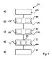
− X線源と、
− X線干渉計装置と、
− X線検出器と、
− 処理ユニットと、
を有する。

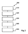
a)X線の検出に関するデータを供給するステップであって、X線検出器がX線源に対して該X線源と当該X線検出器との間の領域の少なくとも一部が被検体を収容する検査領域となるように配置されるように構成され、X線干渉計装置が前記X線源と前記検査領域との間又は前記X線検出器と前記検査領域との間に配置されるように構成されるステップと、
b)前記被検体の少なくとも一部を透過したX線放射に関する少なくとも1つのX線暗視野係数を決定するステップと、
c)前記被検体の少なくとも一部を透過したX線放射に関する少なくとも1つの透過係数を決定するステップと、
d)前記被検体の前記少なくとも一部に向かって放出されるべきX線放射の強度を、前記決定された少なくとも1つの暗視野係数及びX線放射の前記決定された少なくとも1つの透過係数の関数として自動的に制御するステップと、
を有する。
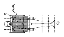
特定のスキャンアーム位置において、検出器の読み出しが実行されて、幾つかの測定値を提供し:
Claims (11)
- 被検体をX線撮像する装置であって、
X線源と、
X線干渉計装置と、
X線検出器と、
処理ユニットと、を有し、
前記X線検出器は、前記X線源に対して該X線源と当該X線検出器との間の領域の少なくとも一部が被検体を収容する検査領域となるように配置されると共に、前記処理ユニットに前記X線干渉計装置を少なくとも部分的に通過したX線放射の検出に関するデータを供給し、
前記X線干渉計装置は、前記X線源と前記検査領域との間又は前記X線検出器と前記検査領域との間に配置され、
前記処理ユニットは、前記被検体の少なくとも一部を透過したX線放射に関する少なくとも1つの透過係数であって、前記被検体の前記少なくとも一部を透過したX線放射の強度の割合である当該少なくとも1つの透過係数を決定すると共に、前記被検体の少なくとも一部を透過したX線放射に関する少なくとも1つの暗視野係数であって、干渉縞視認性が前記被検体の前記少なくとも一部により減少された割合である当該少なくとも1つの暗視野係数を決定し、
前記処理ユニットは、前記被検体の前記少なくとも一部に向かって放出されるべきX線放射の強度を、決定された前記少なくとも1つの透過係数及び決定された前記少なくとも1つの暗視野係数の関数として自動的に制御する、
装置。 - 前記処理ユニットが、前記被検体の前記少なくとも一部に向かって放出されるべきX線放射の前記強度を、前記決定された少なくとも1つの暗視野係数の単調減少関数として制御する、請求項1に記載の装置。
- 前記処理ユニットが、前記被検体の前記少なくとも一部に向かって放出されるべきX線放射の前記強度を、前記決定された少なくとも1つの透過係数の単調減少関数として制御する、請求項1又は請求項2に記載の装置。
- 前記X線干渉計装置は前記検査領域に対して前記X線検出器により検出されるX線放射が全て該X線干渉計装置を通過しなかったものとなるように配置可能であり、前記処理ユニットが前記少なくとも1つの暗視野係数を前記少なくとも1つの透過係数の関数として決定する、請求項1ないし3の何れか一項に記載の装置。
- 前記処理ユニットは前記被検体の部分内で関心領域を決定し、前記被検体の前記少なくとも一部が該関心領域である、請求項1ないし4の何れか一項に記載の装置。
- 被検体をX線撮像する方法であって、前記方法は、
a)X線放射の検出に関するデータを供給するステップであって、X線検出器がX線源に対して該X線源と当該X線検出器との間の領域の少なくとも一部が被検体を収容する検査領域となるように配置され、X線干渉計装置が前記X線源と前記検査領域との間又は前記X線検出器と前記検査領域との間に配置されるステップと、
b)前記被検体の少なくとも一部を透過したX線放射に関する少なくとも1つの暗視野係数であって、干渉縞視認性が前記被検体の前記少なくとも一部により減少された割合である当該少なくとも1つの暗視野係数を決定するステップと、
c)前記被検体の少なくとも一部を透過したX線放射に関する少なくとも1つの透過係数であって、前記被検体の前記少なくとも一部を透過したX線放射の強度の割合である当該少なくとも1つの透過係数を決定するステップと、
d)前記被検体の前記少なくとも一部に向かって放出されるべきX線放射の強度を、決定された前記少なくとも1つの暗視野係数及び決定された前記少なくとも1つの透過係数の関数として自動的に制御するステップと、
を有する、方法。 - ステップd)が、前記被検体の前記少なくとも一部に向かって放出されるべきX線放射の前記強度を前記決定された少なくとも1つの暗視野係数の単調減少関数として制御するステップ、及び/又は前記被検体の前記少なくとも一部に向かって放出されるべきX線放射の前記強度を前記決定された少なくとも1つの透過係数の単調減少関数として制御するステップを有する、請求項6に記載の方法。
- ステップd)が、前記被検体の前記少なくとも一部に向かって放出されるべきX線放射の前記強度を前記決定された少なくとも1つの透過係数の平方根の逆数の関数として制御するステップを有する、請求項7に記載の方法。
- ステップc)が、前記少なくとも1つの透過係数を決定すると共に、前記X線干渉計装置を前記検査領域に対して前記X線検出器により検出されるX線放射が全て該X線干渉計装置を通過しなかったものとなるように配置するステップを有し、ステップb)が前記少なくとも1つの暗視野係数を前記少なくとも1つの透過係数の関数として決定するステップを有する、請求項6ないし8の何れか一項に記載の方法。
- 請求項1ないし5の何れか一項に記載の装置を制御するコンピュータプログラムであって、プロセッサにより実行された場合に請求項6ないし9の何れか一項に記載の方法を実行する、コンピュータプログラム。
- 請求項10に記載のコンピュータプログラムを記憶した、コンピュータ読取可能な媒体。
Applications Claiming Priority (3)
| Application Number | Priority Date | Filing Date | Title |
|---|---|---|---|
| EP15197268 | 2015-12-01 | ||
| EP15197268.4 | 2015-12-01 | ||
| PCT/EP2016/078224 WO2017093055A1 (en) | 2015-12-01 | 2016-11-21 | Apparatus for x-ray imaging an object |
Publications (3)
| Publication Number | Publication Date |
|---|---|
| JP2019505251A JP2019505251A (ja) | 2019-02-28 |
| JP2019505251A5 JP2019505251A5 (ja) | 2019-12-26 |
| JP6816144B2 true JP6816144B2 (ja) | 2021-01-20 |
Family
ID=54770934
Family Applications (1)
| Application Number | Title | Priority Date | Filing Date |
|---|---|---|---|
| JP2018528002A Expired - Fee Related JP6816144B2 (ja) | 2015-12-01 | 2016-11-21 | 被検体をx線撮像する装置 |
Country Status (5)
| Country | Link |
|---|---|
| US (1) | US10779776B2 (ja) |
| EP (1) | EP3383273B1 (ja) |
| JP (1) | JP6816144B2 (ja) |
| CN (1) | CN108289649B (ja) |
| WO (1) | WO2017093055A1 (ja) |
Families Citing this family (9)
| Publication number | Priority date | Publication date | Assignee | Title |
|---|---|---|---|---|
| US10859517B2 (en) * | 2016-04-18 | 2020-12-08 | The Board Of Trustees Of The Leland Stanford Junior University | Single X-ray grating X-ray differential phase contrast imaging system |
| US10670744B2 (en) * | 2017-10-23 | 2020-06-02 | General Electric Company | Current measurement in an imaging system |
| JP6743983B2 (ja) * | 2017-10-31 | 2020-08-19 | 株式会社島津製作所 | X線位相差撮像システム |
| EP3603515A1 (en) * | 2018-08-01 | 2020-02-05 | Koninklijke Philips N.V. | Apparatus for generating x-ray imaging data |
| EP3701868A1 (en) * | 2019-02-28 | 2020-09-02 | Koninklijke Philips N.V. | System, method and computer program for acquiring phase imaging data of an object |
| EP3705044A1 (en) * | 2019-03-08 | 2020-09-09 | Koninklijke Philips N.V. | System for x-ray dark field; phase contrast and attenuation tomosynthesis image acquisition |
| EP3832690A1 (en) * | 2019-12-05 | 2021-06-09 | Koninklijke Philips N.V. | Estimation of full-field scattering for dax imaging |
| EP3925539A1 (en) * | 2020-06-19 | 2021-12-22 | Koninklijke Philips N.V. | X-ray imaging system |
| DE102023204333B3 (de) * | 2023-05-10 | 2024-05-16 | Siemens Healthineers Ag | Verfahren zum Betreiben eines Röntgenbildgebungssystems, Verfahren zur Generierung einer Datenbank, Regelungseinrichtung, Röntgenbildgebungssystem, Steuereinrichtung, Computerprogramm und elektronisch lesbarer Datenträger |
Family Cites Families (24)
| Publication number | Priority date | Publication date | Assignee | Title |
|---|---|---|---|---|
| US2015103A (en) | 1931-11-07 | 1935-09-24 | Celanese Corp | Process of treating fabrics and product thereof |
| CN101576515B (zh) * | 2007-11-23 | 2012-07-04 | 同方威视技术股份有限公司 | X射线光栅相衬成像系统及方法 |
| JP2011045655A (ja) * | 2009-08-28 | 2011-03-10 | Konica Minolta Medical & Graphic Inc | X線撮影装置 |
| JP5269041B2 (ja) | 2009-12-04 | 2013-08-21 | キヤノン株式会社 | X線撮像装置およびx線撮像方法 |
| US8989474B2 (en) | 2010-03-18 | 2015-03-24 | Konica Minolta Medical & Graphic, Inc. | X-ray image capturing system |
| CN102221565B (zh) * | 2010-04-19 | 2013-06-12 | 清华大学 | X射线源光栅步进成像系统与成像方法 |
| JP2014014379A (ja) | 2010-10-27 | 2014-01-30 | Fujifilm Corp | 放射線撮影システム及び放射線撮影方法 |
| JP5238787B2 (ja) * | 2010-10-27 | 2013-07-17 | 富士フイルム株式会社 | 放射線撮影装置及び放射線撮影システム |
| CN103188996B (zh) * | 2010-10-29 | 2015-06-24 | 富士胶片株式会社 | 放射线照相相衬成像设备 |
| JP2012120653A (ja) * | 2010-12-07 | 2012-06-28 | Fujifilm Corp | 放射線撮影装置、及び放射線撮影システム |
| JP5150713B2 (ja) * | 2010-12-08 | 2013-02-27 | 富士フイルム株式会社 | 放射線画像検出装置、放射線撮影装置、放射線撮影システム |
| CN103648388B (zh) | 2011-07-04 | 2017-05-03 | 皇家飞利浦有限公司 | 相位对比度成像设备 |
| WO2013171657A1 (en) * | 2012-05-14 | 2013-11-21 | Koninklijke Philips N.V. | Dark field computed tomography imaging |
| JP6250658B2 (ja) * | 2012-06-27 | 2017-12-20 | コーニンクレッカ フィリップス エヌ ヴェKoninklijke Philips N.V. | 暗視野イメージング |
| JP2014090967A (ja) * | 2012-11-06 | 2014-05-19 | Canon Inc | X線撮像装置 |
| WO2014077394A1 (ja) * | 2012-11-16 | 2014-05-22 | 株式会社 東芝 | X線コンピュータ断層撮影装置及び情報処理装置 |
| US10096098B2 (en) * | 2013-12-30 | 2018-10-09 | Carestream Health, Inc. | Phase retrieval from differential phase contrast imaging |
| EP3013233B1 (en) * | 2013-06-28 | 2017-11-15 | Koninklijke Philips N.V. | Correction in slit-scanning phase contrast imaging |
| DE102013214388B4 (de) * | 2013-07-23 | 2023-04-20 | Siemens Healthcare Gmbh | Medizinisches Instrument zur Verwendung mit einer Phasenkontrastbildgebung und Röntgenaufnahmesystem mit Phasenkontrastbildgebung |
| EP3066670B1 (en) * | 2013-11-05 | 2017-06-28 | Koninklijke Philips N.V. | X-ray imaging device with fast spatial modulation of photon flux |
| CN105705097B (zh) * | 2013-11-08 | 2019-04-12 | 皇家飞利浦有限公司 | 针对差分相位衬度ct的经验性射束硬化校正 |
| EP3082600B1 (en) * | 2013-12-17 | 2018-03-28 | Koninklijke Philips N.V. | Phase retrieval for scanning differential phase contrast systems |
| KR20170009909A (ko) * | 2014-05-15 | 2017-01-25 | 시그레이, 아이엔씨. | 주기적 구조의 측정과 특성화 및 분석을 위한 x-선 방법 |
| JP2016032573A (ja) * | 2014-07-31 | 2016-03-10 | キヤノン株式会社 | トールボット干渉計、トールボット干渉システム、及び縞走査法 |
-
2016
- 2016-11-21 JP JP2018528002A patent/JP6816144B2/ja not_active Expired - Fee Related
- 2016-11-21 CN CN201680069968.7A patent/CN108289649B/zh not_active Expired - Fee Related
- 2016-11-21 WO PCT/EP2016/078224 patent/WO2017093055A1/en not_active Ceased
- 2016-11-21 US US15/779,107 patent/US10779776B2/en not_active Expired - Fee Related
- 2016-11-21 EP EP16798182.8A patent/EP3383273B1/en active Active
Also Published As
| Publication number | Publication date |
|---|---|
| CN108289649B (zh) | 2022-04-19 |
| EP3383273A1 (en) | 2018-10-10 |
| JP2019505251A (ja) | 2019-02-28 |
| US10779776B2 (en) | 2020-09-22 |
| WO2017093055A1 (en) | 2017-06-08 |
| US20180344268A1 (en) | 2018-12-06 |
| CN108289649A (zh) | 2018-07-17 |
| EP3383273B1 (en) | 2021-05-12 |
Similar Documents
| Publication | Publication Date | Title |
|---|---|---|
| JP6816144B2 (ja) | 被検体をx線撮像する装置 | |
| US9795350B2 (en) | Material differentiation with phase contrast imaging | |
| JP5606455B2 (ja) | 逆投影のためのイメージング装置及びその作動方法 | |
| JP6670398B2 (ja) | 暗視野又は位相コントラストx線撮像における特徴抑制 | |
| JP6214819B1 (ja) | 微分位相コントラストx線撮像における暗視野信号の最適なエネルギ加重 | |
| JP6413950B2 (ja) | 放射線撮影システム及び画像処理装置 | |
| CN107567640B (zh) | 用于扫描暗场和相位对比成像的射束硬化校正 | |
| JP2016049455A (ja) | X線コンピュータ断層撮影装置及び画像再構成装置 | |
| CN103460251A (zh) | 利用对具有光栅布置的相衬成像的约束优化的图像积分的方法和系统 | |
| JP2012200567A (ja) | 放射線撮影システム及び放射線撮影方法 | |
| US10238352B2 (en) | Recording X-ray images without scattered radiation | |
| RU2677763C1 (ru) | Получение фазы для систем сканирования с дифференциальным фазовым контрастом | |
| US11234663B2 (en) | Apparatus for generating multi energy data from phase contrast imaging data | |
| JP6642676B2 (ja) | 放射線撮影システム及び画像処理装置 | |
| JP2022524071A (ja) | X線暗視野画像、位相コントラスト画像及び減衰トモシンセシス画像を取得するシステム | |
| CN110520049A (zh) | 用于暗场x射线成像的灵敏度优化患者定位系统 | |
| JP2014132913A (ja) | 放射線撮影システム及び放射線撮影方法 |
Legal Events
| Date | Code | Title | Description |
|---|---|---|---|
| A521 | Request for written amendment filed |
Free format text: JAPANESE INTERMEDIATE CODE: A523 Effective date: 20191118 |
|
| A621 | Written request for application examination |
Free format text: JAPANESE INTERMEDIATE CODE: A621 Effective date: 20191118 |
|
| A977 | Report on retrieval |
Free format text: JAPANESE INTERMEDIATE CODE: A971007 Effective date: 20201021 |
|
| TRDD | Decision of grant or rejection written | ||
| A01 | Written decision to grant a patent or to grant a registration (utility model) |
Free format text: JAPANESE INTERMEDIATE CODE: A01 Effective date: 20201124 |
|
| A61 | First payment of annual fees (during grant procedure) |
Free format text: JAPANESE INTERMEDIATE CODE: A61 Effective date: 20201223 |
|
| R150 | Certificate of patent or registration of utility model |
Ref document number: 6816144 Country of ref document: JP Free format text: JAPANESE INTERMEDIATE CODE: R150 |
|
| LAPS | Cancellation because of no payment of annual fees |