JP5124274B2 - Method and apparatus for grasping and evaluating tissue - Google Patents
Method and apparatus for grasping and evaluating tissue Download PDFInfo
- Publication number
- JP5124274B2 JP5124274B2 JP2007533770A JP2007533770A JP5124274B2 JP 5124274 B2 JP5124274 B2 JP 5124274B2 JP 2007533770 A JP2007533770 A JP 2007533770A JP 2007533770 A JP2007533770 A JP 2007533770A JP 5124274 B2 JP5124274 B2 JP 5124274B2
- Authority
- JP
- Japan
- Prior art keywords
- leaflet
- valve
- fixation device
- tissue
- proximal
- Prior art date
- Legal status (The legal status is an assumption and is not a legal conclusion. Google has not performed a legal analysis and makes no representation as to the accuracy of the status listed.)
- Active
Links
- 238000000034 method Methods 0.000 title abstract description 65
- 230000033001 locomotion Effects 0.000 claims description 70
- 230000006835 compression Effects 0.000 claims description 7
- 238000007906 compression Methods 0.000 claims description 7
- 230000000087 stabilizing effect Effects 0.000 abstract description 24
- 206010067171 Regurgitation Diseases 0.000 abstract description 12
- 210000003709 heart valve Anatomy 0.000 abstract description 7
- 208000005907 mitral valve insufficiency Diseases 0.000 abstract 1
- 210000001519 tissue Anatomy 0.000 description 165
- 239000012528 membrane Substances 0.000 description 104
- 239000000463 material Substances 0.000 description 46
- 210000004115 mitral valve Anatomy 0.000 description 44
- 239000008280 blood Substances 0.000 description 33
- 210000004369 blood Anatomy 0.000 description 33
- 239000000126 substance Substances 0.000 description 25
- 230000007246 mechanism Effects 0.000 description 21
- 230000001746 atrial effect Effects 0.000 description 18
- 230000006641 stabilisation Effects 0.000 description 16
- 238000011105 stabilization Methods 0.000 description 16
- 238000013459 approach Methods 0.000 description 15
- 230000006870 function Effects 0.000 description 15
- 210000005246 left atrium Anatomy 0.000 description 13
- 238000012986 modification Methods 0.000 description 13
- 230000004048 modification Effects 0.000 description 13
- 230000006872 improvement Effects 0.000 description 12
- 210000005240 left ventricle Anatomy 0.000 description 12
- 238000010992 reflux Methods 0.000 description 12
- 238000004873 anchoring Methods 0.000 description 11
- 229920000642 polymer Polymers 0.000 description 11
- 239000000523 sample Substances 0.000 description 10
- 238000002604 ultrasonography Methods 0.000 description 10
- 230000002829 reductive effect Effects 0.000 description 9
- 230000002441 reversible effect Effects 0.000 description 9
- 238000007794 visualization technique Methods 0.000 description 9
- 230000008878 coupling Effects 0.000 description 8
- 238000010168 coupling process Methods 0.000 description 8
- 238000005859 coupling reaction Methods 0.000 description 8
- 238000011156 evaluation Methods 0.000 description 8
- 230000001965 increasing effect Effects 0.000 description 8
- 230000008439 repair process Effects 0.000 description 8
- 230000002861 ventricular Effects 0.000 description 8
- 210000002837 heart atrium Anatomy 0.000 description 7
- 239000003381 stabilizer Substances 0.000 description 7
- 238000003780 insertion Methods 0.000 description 6
- 238000012800 visualization Methods 0.000 description 6
- 230000009471 action Effects 0.000 description 5
- 238000002594 fluoroscopy Methods 0.000 description 5
- 238000002347 injection Methods 0.000 description 5
- 239000007924 injection Substances 0.000 description 5
- 208000014674 injury Diseases 0.000 description 5
- 230000037431 insertion Effects 0.000 description 5
- 229910052751 metal Inorganic materials 0.000 description 5
- 239000002184 metal Substances 0.000 description 5
- 210000005245 right atrium Anatomy 0.000 description 5
- 230000008733 trauma Effects 0.000 description 5
- 239000011324 bead Substances 0.000 description 4
- 230000008859 change Effects 0.000 description 4
- 238000000576 coating method Methods 0.000 description 4
- 239000000835 fiber Substances 0.000 description 4
- 238000007667 floating Methods 0.000 description 4
- 238000009434 installation Methods 0.000 description 4
- 230000000007 visual effect Effects 0.000 description 4
- 229910000684 Cobalt-chrome Inorganic materials 0.000 description 3
- 210000001765 aortic valve Anatomy 0.000 description 3
- 230000006399 behavior Effects 0.000 description 3
- 239000011248 coating agent Substances 0.000 description 3
- 239000010952 cobalt-chrome Substances 0.000 description 3
- 239000002872 contrast media Substances 0.000 description 3
- 239000004744 fabric Substances 0.000 description 3
- 239000007943 implant Substances 0.000 description 3
- 238000005304 joining Methods 0.000 description 3
- 239000007788 liquid Substances 0.000 description 3
- 210000003540 papillary muscle Anatomy 0.000 description 3
- 210000001013 sinoatrial node Anatomy 0.000 description 3
- 239000007787 solid Substances 0.000 description 3
- 239000010935 stainless steel Substances 0.000 description 3
- 229910001220 stainless steel Inorganic materials 0.000 description 3
- 238000011144 upstream manufacturing Methods 0.000 description 3
- 230000005526 G1 to G0 transition Effects 0.000 description 2
- TZCXTZWJZNENPQ-UHFFFAOYSA-L barium sulfate Chemical compound [Ba+2].[O-]S([O-])(=O)=O TZCXTZWJZNENPQ-UHFFFAOYSA-L 0.000 description 2
- 230000008901 benefit Effects 0.000 description 2
- 230000002612 cardiopulmonary effect Effects 0.000 description 2
- 239000003795 chemical substances by application Substances 0.000 description 2
- 239000004020 conductor Substances 0.000 description 2
- 230000009977 dual effect Effects 0.000 description 2
- 230000004064 dysfunction Effects 0.000 description 2
- 238000002592 echocardiography Methods 0.000 description 2
- 229910000701 elgiloys (Co-Cr-Ni Alloy) Inorganic materials 0.000 description 2
- 230000002708 enhancing effect Effects 0.000 description 2
- 238000000605 extraction Methods 0.000 description 2
- 238000001802 infusion Methods 0.000 description 2
- 230000037231 joint health Effects 0.000 description 2
- 230000000670 limiting effect Effects 0.000 description 2
- 239000000696 magnetic material Substances 0.000 description 2
- 230000003287 optical effect Effects 0.000 description 2
- FDPIMTJIUBPUKL-UHFFFAOYSA-N pentan-3-one Chemical compound CCC(=O)CC FDPIMTJIUBPUKL-UHFFFAOYSA-N 0.000 description 2
- BASFCYQUMIYNBI-UHFFFAOYSA-N platinum Chemical compound [Pt] BASFCYQUMIYNBI-UHFFFAOYSA-N 0.000 description 2
- 229920001296 polysiloxane Polymers 0.000 description 2
- 229920002635 polyurethane Polymers 0.000 description 2
- 239000004814 polyurethane Substances 0.000 description 2
- 238000005086 pumping Methods 0.000 description 2
- 239000011435 rock Substances 0.000 description 2
- 238000000926 separation method Methods 0.000 description 2
- 230000003068 static effect Effects 0.000 description 2
- 230000004936 stimulating effect Effects 0.000 description 2
- 238000001356 surgical procedure Methods 0.000 description 2
- 230000001225 therapeutic effect Effects 0.000 description 2
- 230000008467 tissue growth Effects 0.000 description 2
- 229910000851 Alloy steel Inorganic materials 0.000 description 1
- 239000004677 Nylon Substances 0.000 description 1
- 239000004696 Poly ether ether ketone Substances 0.000 description 1
- 239000004642 Polyimide Substances 0.000 description 1
- RTAQQCXQSZGOHL-UHFFFAOYSA-N Titanium Chemical compound [Ti] RTAQQCXQSZGOHL-UHFFFAOYSA-N 0.000 description 1
- 238000010521 absorption reaction Methods 0.000 description 1
- 230000001154 acute effect Effects 0.000 description 1
- 230000002411 adverse Effects 0.000 description 1
- 239000000956 alloy Substances 0.000 description 1
- 210000003484 anatomy Anatomy 0.000 description 1
- 239000003242 anti bacterial agent Substances 0.000 description 1
- 229940088710 antibiotic agent Drugs 0.000 description 1
- 239000003146 anticoagulant agent Substances 0.000 description 1
- 229940127219 anticoagulant drug Drugs 0.000 description 1
- 210000000709 aorta Anatomy 0.000 description 1
- JUPQTSLXMOCDHR-UHFFFAOYSA-N benzene-1,4-diol;bis(4-fluorophenyl)methanone Chemical compound OC1=CC=C(O)C=C1.C1=CC(F)=CC=C1C(=O)C1=CC=C(F)C=C1 JUPQTSLXMOCDHR-UHFFFAOYSA-N 0.000 description 1
- 230000017531 blood circulation Effects 0.000 description 1
- 210000005242 cardiac chamber Anatomy 0.000 description 1
- 238000006243 chemical reaction Methods 0.000 description 1
- 230000004087 circulation Effects 0.000 description 1
- 210000000078 claw Anatomy 0.000 description 1
- 239000012141 concentrate Substances 0.000 description 1
- 238000012937 correction Methods 0.000 description 1
- 238000005520 cutting process Methods 0.000 description 1
- 230000006378 damage Effects 0.000 description 1
- 230000007547 defect Effects 0.000 description 1
- 230000007812 deficiency Effects 0.000 description 1
- 238000013461 design Methods 0.000 description 1
- 238000010790 dilution Methods 0.000 description 1
- 239000012895 dilution Substances 0.000 description 1
- 239000003814 drug Substances 0.000 description 1
- 230000000694 effects Effects 0.000 description 1
- 229920001971 elastomer Polymers 0.000 description 1
- 238000012282 endovascular technique Methods 0.000 description 1
- PCHJSUWPFVWCPO-UHFFFAOYSA-N gold Chemical compound [Au] PCHJSUWPFVWCPO-UHFFFAOYSA-N 0.000 description 1
- 239000010931 gold Substances 0.000 description 1
- 229910052737 gold Inorganic materials 0.000 description 1
- 239000007952 growth promoter Substances 0.000 description 1
- 230000035876 healing Effects 0.000 description 1
- 230000010247 heart contraction Effects 0.000 description 1
- 230000005986 heart dysfunction Effects 0.000 description 1
- 238000002513 implantation Methods 0.000 description 1
- 238000007373 indentation Methods 0.000 description 1
- 230000002452 interceptive effect Effects 0.000 description 1
- 238000002595 magnetic resonance imaging Methods 0.000 description 1
- 230000014759 maintenance of location Effects 0.000 description 1
- 230000013011 mating Effects 0.000 description 1
- 238000005259 measurement Methods 0.000 description 1
- 238000000968 medical method and process Methods 0.000 description 1
- 150000002739 metals Chemical class 0.000 description 1
- 229910001000 nickel titanium Inorganic materials 0.000 description 1
- HLXZNVUGXRDIFK-UHFFFAOYSA-N nickel titanium Chemical compound [Ti].[Ti].[Ti].[Ti].[Ti].[Ti].[Ti].[Ti].[Ti].[Ti].[Ti].[Ni].[Ni].[Ni].[Ni].[Ni].[Ni].[Ni].[Ni].[Ni].[Ni].[Ni].[Ni].[Ni].[Ni] HLXZNVUGXRDIFK-UHFFFAOYSA-N 0.000 description 1
- 229920001778 nylon Polymers 0.000 description 1
- RVTZCBVAJQQJTK-UHFFFAOYSA-N oxygen(2-);zirconium(4+) Chemical compound [O-2].[O-2].[Zr+4] RVTZCBVAJQQJTK-UHFFFAOYSA-N 0.000 description 1
- 230000035515 penetration Effects 0.000 description 1
- 229910052697 platinum Inorganic materials 0.000 description 1
- 229920000728 polyester Polymers 0.000 description 1
- 229920002530 polyetherether ketone Polymers 0.000 description 1
- 229920001721 polyimide Polymers 0.000 description 1
- 229920005594 polymer fiber Polymers 0.000 description 1
- 230000000750 progressive effect Effects 0.000 description 1
- 238000011084 recovery Methods 0.000 description 1
- 238000007634 remodeling Methods 0.000 description 1
- 239000011347 resin Substances 0.000 description 1
- 229920005989 resin Polymers 0.000 description 1
- 230000004044 response Effects 0.000 description 1
- 230000035939 shock Effects 0.000 description 1
- 238000004904 shortening Methods 0.000 description 1
- 238000009987 spinning Methods 0.000 description 1
- 238000005507 spraying Methods 0.000 description 1
- 238000004544 sputter deposition Methods 0.000 description 1
- 238000004381 surface treatment Methods 0.000 description 1
- 210000002435 tendon Anatomy 0.000 description 1
- 229940124597 therapeutic agent Drugs 0.000 description 1
- 239000010936 titanium Substances 0.000 description 1
- 229910052719 titanium Inorganic materials 0.000 description 1
- 238000013519 translation Methods 0.000 description 1
- 230000000472 traumatic effect Effects 0.000 description 1
- 210000005166 vasculature Anatomy 0.000 description 1
- 210000002073 venous valve Anatomy 0.000 description 1
- 238000004804 winding Methods 0.000 description 1
Images
Classifications
-
- A—HUMAN NECESSITIES
- A61—MEDICAL OR VETERINARY SCIENCE; HYGIENE
- A61B—DIAGNOSIS; SURGERY; IDENTIFICATION
- A61B17/00—Surgical instruments, devices or methods, e.g. tourniquets
- A61B17/04—Surgical instruments, devices or methods, e.g. tourniquets for suturing wounds; Holders or packages for needles or suture materials
- A61B17/0401—Suture anchors, buttons or pledgets, i.e. means for attaching sutures to bone, cartilage or soft tissue; Instruments for applying or removing suture anchors
-
- A—HUMAN NECESSITIES
- A61—MEDICAL OR VETERINARY SCIENCE; HYGIENE
- A61B—DIAGNOSIS; SURGERY; IDENTIFICATION
- A61B17/00—Surgical instruments, devices or methods, e.g. tourniquets
- A61B17/08—Wound clamps or clips, i.e. not or only partly penetrating the tissue ; Devices for bringing together the edges of a wound
-
- A—HUMAN NECESSITIES
- A61—MEDICAL OR VETERINARY SCIENCE; HYGIENE
- A61B—DIAGNOSIS; SURGERY; IDENTIFICATION
- A61B17/00—Surgical instruments, devices or methods, e.g. tourniquets
- A61B17/28—Surgical forceps
- A61B17/29—Forceps for use in minimally invasive surgery
-
- A—HUMAN NECESSITIES
- A61—MEDICAL OR VETERINARY SCIENCE; HYGIENE
- A61F—FILTERS IMPLANTABLE INTO BLOOD VESSELS; PROSTHESES; DEVICES PROVIDING PATENCY TO, OR PREVENTING COLLAPSING OF, TUBULAR STRUCTURES OF THE BODY, e.g. STENTS; ORTHOPAEDIC, NURSING OR CONTRACEPTIVE DEVICES; FOMENTATION; TREATMENT OR PROTECTION OF EYES OR EARS; BANDAGES, DRESSINGS OR ABSORBENT PADS; FIRST-AID KITS
- A61F2/00—Filters implantable into blood vessels; Prostheses, i.e. artificial substitutes or replacements for parts of the body; Appliances for connecting them with the body; Devices providing patency to, or preventing collapsing of, tubular structures of the body, e.g. stents
- A61F2/02—Prostheses implantable into the body
- A61F2/24—Heart valves ; Vascular valves, e.g. venous valves; Heart implants, e.g. passive devices for improving the function of the native valve or the heart muscle; Transmyocardial revascularisation [TMR] devices; Valves implantable in the body
- A61F2/2442—Annuloplasty rings or inserts for correcting the valve shape; Implants for improving the function of a native heart valve
- A61F2/246—Devices for obstructing a leak through a native valve in a closed condition
-
- A—HUMAN NECESSITIES
- A61—MEDICAL OR VETERINARY SCIENCE; HYGIENE
- A61B—DIAGNOSIS; SURGERY; IDENTIFICATION
- A61B17/00—Surgical instruments, devices or methods, e.g. tourniquets
- A61B17/34—Trocars; Puncturing needles
- A61B17/3478—Endoscopic needles, e.g. for infusion
-
- A—HUMAN NECESSITIES
- A61—MEDICAL OR VETERINARY SCIENCE; HYGIENE
- A61B—DIAGNOSIS; SURGERY; IDENTIFICATION
- A61B17/00—Surgical instruments, devices or methods, e.g. tourniquets
- A61B17/00234—Surgical instruments, devices or methods, e.g. tourniquets for minimally invasive surgery
- A61B2017/00238—Type of minimally invasive operation
- A61B2017/00243—Type of minimally invasive operation cardiac
-
- A—HUMAN NECESSITIES
- A61—MEDICAL OR VETERINARY SCIENCE; HYGIENE
- A61B—DIAGNOSIS; SURGERY; IDENTIFICATION
- A61B17/00—Surgical instruments, devices or methods, e.g. tourniquets
- A61B2017/00743—Type of operation; Specification of treatment sites
- A61B2017/00778—Operations on blood vessels
- A61B2017/00783—Valvuloplasty
-
- A—HUMAN NECESSITIES
- A61—MEDICAL OR VETERINARY SCIENCE; HYGIENE
- A61B—DIAGNOSIS; SURGERY; IDENTIFICATION
- A61B17/00—Surgical instruments, devices or methods, e.g. tourniquets
- A61B17/08—Wound clamps or clips, i.e. not or only partly penetrating the tissue ; Devices for bringing together the edges of a wound
- A61B2017/081—Tissue approximator
-
- A—HUMAN NECESSITIES
- A61—MEDICAL OR VETERINARY SCIENCE; HYGIENE
- A61B—DIAGNOSIS; SURGERY; IDENTIFICATION
- A61B17/00—Surgical instruments, devices or methods, e.g. tourniquets
- A61B17/22—Implements for squeezing-off ulcers or the like on the inside of inner organs of the body; Implements for scraping-out cavities of body organs, e.g. bones; Calculus removers; Calculus smashing apparatus; Apparatus for removing obstructions in blood vessels, not otherwise provided for
- A61B2017/22082—Implements for squeezing-off ulcers or the like on the inside of inner organs of the body; Implements for scraping-out cavities of body organs, e.g. bones; Calculus removers; Calculus smashing apparatus; Apparatus for removing obstructions in blood vessels, not otherwise provided for after introduction of a substance
- A61B2017/22088—Implements for squeezing-off ulcers or the like on the inside of inner organs of the body; Implements for scraping-out cavities of body organs, e.g. bones; Calculus removers; Calculus smashing apparatus; Apparatus for removing obstructions in blood vessels, not otherwise provided for after introduction of a substance ultrasound absorbing, drug activated by ultrasound
-
- A—HUMAN NECESSITIES
- A61—MEDICAL OR VETERINARY SCIENCE; HYGIENE
- A61B—DIAGNOSIS; SURGERY; IDENTIFICATION
- A61B17/00—Surgical instruments, devices or methods, e.g. tourniquets
- A61B17/22—Implements for squeezing-off ulcers or the like on the inside of inner organs of the body; Implements for scraping-out cavities of body organs, e.g. bones; Calculus removers; Calculus smashing apparatus; Apparatus for removing obstructions in blood vessels, not otherwise provided for
- A61B2017/22082—Implements for squeezing-off ulcers or the like on the inside of inner organs of the body; Implements for scraping-out cavities of body organs, e.g. bones; Calculus removers; Calculus smashing apparatus; Apparatus for removing obstructions in blood vessels, not otherwise provided for after introduction of a substance
- A61B2017/22089—Gas-bubbles
-
- A—HUMAN NECESSITIES
- A61—MEDICAL OR VETERINARY SCIENCE; HYGIENE
- A61B—DIAGNOSIS; SURGERY; IDENTIFICATION
- A61B17/00—Surgical instruments, devices or methods, e.g. tourniquets
- A61B17/22—Implements for squeezing-off ulcers or the like on the inside of inner organs of the body; Implements for scraping-out cavities of body organs, e.g. bones; Calculus removers; Calculus smashing apparatus; Apparatus for removing obstructions in blood vessels, not otherwise provided for
- A61B2017/22098—Decalcification of valves
-
- A—HUMAN NECESSITIES
- A61—MEDICAL OR VETERINARY SCIENCE; HYGIENE
- A61B—DIAGNOSIS; SURGERY; IDENTIFICATION
- A61B17/00—Surgical instruments, devices or methods, e.g. tourniquets
- A61B17/28—Surgical forceps
- A61B17/2812—Surgical forceps with a single pivotal connection
- A61B17/282—Jaws
- A61B2017/2825—Inserts of different material in jaws
-
- A—HUMAN NECESSITIES
- A61—MEDICAL OR VETERINARY SCIENCE; HYGIENE
- A61B—DIAGNOSIS; SURGERY; IDENTIFICATION
- A61B17/00—Surgical instruments, devices or methods, e.g. tourniquets
- A61B17/28—Surgical forceps
- A61B17/29—Forceps for use in minimally invasive surgery
- A61B2017/2926—Details of heads or jaws
- A61B2017/2931—Details of heads or jaws with releasable head
-
- A—HUMAN NECESSITIES
- A61—MEDICAL OR VETERINARY SCIENCE; HYGIENE
- A61B—DIAGNOSIS; SURGERY; IDENTIFICATION
- A61B17/00—Surgical instruments, devices or methods, e.g. tourniquets
- A61B17/30—Surgical pincettes without pivotal connections
- A61B2017/306—Surgical pincettes without pivotal connections holding by means of suction
-
- A—HUMAN NECESSITIES
- A61—MEDICAL OR VETERINARY SCIENCE; HYGIENE
- A61B—DIAGNOSIS; SURGERY; IDENTIFICATION
- A61B90/00—Instruments, implements or accessories specially adapted for surgery or diagnosis and not covered by any of the groups A61B1/00 - A61B50/00, e.g. for luxation treatment or for protecting wound edges
- A61B90/39—Markers, e.g. radio-opaque or breast lesions markers
- A61B2090/3904—Markers, e.g. radio-opaque or breast lesions markers specially adapted for marking specified tissue
- A61B2090/3908—Soft tissue, e.g. breast tissue
-
- A—HUMAN NECESSITIES
- A61—MEDICAL OR VETERINARY SCIENCE; HYGIENE
- A61B—DIAGNOSIS; SURGERY; IDENTIFICATION
- A61B90/00—Instruments, implements or accessories specially adapted for surgery or diagnosis and not covered by any of the groups A61B1/00 - A61B50/00, e.g. for luxation treatment or for protecting wound edges
- A61B90/39—Markers, e.g. radio-opaque or breast lesions markers
- A61B2090/3925—Markers, e.g. radio-opaque or breast lesions markers ultrasonic
-
- A—HUMAN NECESSITIES
- A61—MEDICAL OR VETERINARY SCIENCE; HYGIENE
- A61F—FILTERS IMPLANTABLE INTO BLOOD VESSELS; PROSTHESES; DEVICES PROVIDING PATENCY TO, OR PREVENTING COLLAPSING OF, TUBULAR STRUCTURES OF THE BODY, e.g. STENTS; ORTHOPAEDIC, NURSING OR CONTRACEPTIVE DEVICES; FOMENTATION; TREATMENT OR PROTECTION OF EYES OR EARS; BANDAGES, DRESSINGS OR ABSORBENT PADS; FIRST-AID KITS
- A61F2/00—Filters implantable into blood vessels; Prostheses, i.e. artificial substitutes or replacements for parts of the body; Appliances for connecting them with the body; Devices providing patency to, or preventing collapsing of, tubular structures of the body, e.g. stents
- A61F2/02—Prostheses implantable into the body
- A61F2/24—Heart valves ; Vascular valves, e.g. venous valves; Heart implants, e.g. passive devices for improving the function of the native valve or the heart muscle; Transmyocardial revascularisation [TMR] devices; Valves implantable in the body
- A61F2/2442—Annuloplasty rings or inserts for correcting the valve shape; Implants for improving the function of a native heart valve
- A61F2/2466—Delivery devices therefor
-
- A—HUMAN NECESSITIES
- A61—MEDICAL OR VETERINARY SCIENCE; HYGIENE
- A61F—FILTERS IMPLANTABLE INTO BLOOD VESSELS; PROSTHESES; DEVICES PROVIDING PATENCY TO, OR PREVENTING COLLAPSING OF, TUBULAR STRUCTURES OF THE BODY, e.g. STENTS; ORTHOPAEDIC, NURSING OR CONTRACEPTIVE DEVICES; FOMENTATION; TREATMENT OR PROTECTION OF EYES OR EARS; BANDAGES, DRESSINGS OR ABSORBENT PADS; FIRST-AID KITS
- A61F2/00—Filters implantable into blood vessels; Prostheses, i.e. artificial substitutes or replacements for parts of the body; Appliances for connecting them with the body; Devices providing patency to, or preventing collapsing of, tubular structures of the body, e.g. stents
- A61F2/02—Prostheses implantable into the body
- A61F2/24—Heart valves ; Vascular valves, e.g. venous valves; Heart implants, e.g. passive devices for improving the function of the native valve or the heart muscle; Transmyocardial revascularisation [TMR] devices; Valves implantable in the body
- A61F2/2478—Passive devices for improving the function of the heart muscle, i.e. devices for reshaping the external surface of the heart, e.g. bags, strips or bands
- A61F2/2487—Devices within the heart chamber, e.g. splints
-
- A—HUMAN NECESSITIES
- A61—MEDICAL OR VETERINARY SCIENCE; HYGIENE
- A61F—FILTERS IMPLANTABLE INTO BLOOD VESSELS; PROSTHESES; DEVICES PROVIDING PATENCY TO, OR PREVENTING COLLAPSING OF, TUBULAR STRUCTURES OF THE BODY, e.g. STENTS; ORTHOPAEDIC, NURSING OR CONTRACEPTIVE DEVICES; FOMENTATION; TREATMENT OR PROTECTION OF EYES OR EARS; BANDAGES, DRESSINGS OR ABSORBENT PADS; FIRST-AID KITS
- A61F2210/00—Particular material properties of prostheses classified in groups A61F2/00 - A61F2/26 or A61F2/82 or A61F9/00 or A61F11/00 or subgroups thereof
- A61F2210/0014—Particular material properties of prostheses classified in groups A61F2/00 - A61F2/26 or A61F2/82 or A61F9/00 or A61F11/00 or subgroups thereof using shape memory or superelastic materials, e.g. nitinol
-
- A—HUMAN NECESSITIES
- A61—MEDICAL OR VETERINARY SCIENCE; HYGIENE
- A61F—FILTERS IMPLANTABLE INTO BLOOD VESSELS; PROSTHESES; DEVICES PROVIDING PATENCY TO, OR PREVENTING COLLAPSING OF, TUBULAR STRUCTURES OF THE BODY, e.g. STENTS; ORTHOPAEDIC, NURSING OR CONTRACEPTIVE DEVICES; FOMENTATION; TREATMENT OR PROTECTION OF EYES OR EARS; BANDAGES, DRESSINGS OR ABSORBENT PADS; FIRST-AID KITS
- A61F2220/00—Fixations or connections for prostheses classified in groups A61F2/00 - A61F2/26 or A61F2/82 or A61F9/00 or A61F11/00 or subgroups thereof
- A61F2220/0008—Fixation appliances for connecting prostheses to the body
- A61F2220/0016—Fixation appliances for connecting prostheses to the body with sharp anchoring protrusions, e.g. barbs, pins, spikes
-
- A—HUMAN NECESSITIES
- A61—MEDICAL OR VETERINARY SCIENCE; HYGIENE
- A61F—FILTERS IMPLANTABLE INTO BLOOD VESSELS; PROSTHESES; DEVICES PROVIDING PATENCY TO, OR PREVENTING COLLAPSING OF, TUBULAR STRUCTURES OF THE BODY, e.g. STENTS; ORTHOPAEDIC, NURSING OR CONTRACEPTIVE DEVICES; FOMENTATION; TREATMENT OR PROTECTION OF EYES OR EARS; BANDAGES, DRESSINGS OR ABSORBENT PADS; FIRST-AID KITS
- A61F2230/00—Geometry of prostheses classified in groups A61F2/00 - A61F2/26 or A61F2/82 or A61F9/00 or A61F11/00 or subgroups thereof
- A61F2230/0002—Two-dimensional shapes, e.g. cross-sections
- A61F2230/0004—Rounded shapes, e.g. with rounded corners
- A61F2230/001—Figure-8-shaped, e.g. hourglass-shaped
Landscapes
- Health & Medical Sciences (AREA)
- Life Sciences & Earth Sciences (AREA)
- Surgery (AREA)
- Animal Behavior & Ethology (AREA)
- General Health & Medical Sciences (AREA)
- Engineering & Computer Science (AREA)
- Biomedical Technology (AREA)
- Heart & Thoracic Surgery (AREA)
- Veterinary Medicine (AREA)
- Public Health (AREA)
- Medical Informatics (AREA)
- Molecular Biology (AREA)
- Nuclear Medicine, Radiotherapy & Molecular Imaging (AREA)
- Cardiology (AREA)
- Ophthalmology & Optometry (AREA)
- Rheumatology (AREA)
- Oral & Maxillofacial Surgery (AREA)
- Transplantation (AREA)
- Vascular Medicine (AREA)
- Prostheses (AREA)
- Surgical Instruments (AREA)
Abstract
Description
本発明は一般に医療方法、装置およびシステムに関する。詳細には、本発明は組織への接近または弁の修復のような身体の組織の血管内、経皮的または最小に侵襲性の外科治療のための方法、装置およびシステムに関する。より詳細には、本発明は心臓の弁および静脈弁の修復に関する。 The present invention relates generally to medical methods, devices, and systems. In particular, the present invention relates to methods, devices and systems for intravascular, percutaneous or minimally invasive surgical treatment of body tissue such as tissue access or valve repair. More particularly, the present invention relates to heart valve and venous valve repair.
身体の外科修復は、しばしば、組織へ接近し、そして接近構成でかかる組織を留めることを伴う。弁を修復する際、組織への接近は、弁の弁膜を治療構成で接合することを含んでおり、この場合、治療構成は弁膜を留めるか或は固定することによりを維持されることができる。かかる接合は僧帽弁に最も一般的に起こる血液逆流を治療するために使用されることができる。 Body surgical repair often involves accessing the tissue and retaining such tissue in an access configuration. In repairing the valve, access to the tissue includes joining the valve leaflets in a therapeutic configuration, where the therapeutic configuration can be maintained by fastening or securing the valve leaflets. . Such a junction can be used to treat blood reflux that most commonly occurs in mitral valves.
僧帽弁の血液逆流は、心臓の左弁室から機能不全の僧帽弁を通って左心房の中へ入る逆行の流れが特徴である。心臓の収縮の正常なサイクル中、僧帽弁は酸素供給された血液が左心房の中へ流れるのを防ぐために逆止弁として作用する。このように、酸素供給された血液は大動脈弁を通って大動脈へ圧送される。弁の血液逆流は、心臓の圧送効率を著しく減少させて患者を厳しい漸進的な心臓の機能不全の恐れにさらしてしまう。 Mitral valve blood regurgitation is characterized by a retrograde flow from the left ventricle of the heart through the malfunctioning mitral valve into the left atrium. During the normal cycle of heart contraction, the mitral valve acts as a check valve to prevent oxygenated blood from flowing into the left atrium. Thus, oxygen-supplied blood is pumped through the aortic valve to the aorta. Valve blood regurgitation significantly reduces heart pumping efficiency and exposes the patient to severe progressive heart dysfunction.
僧帽弁の血液逆流は僧帽弁または左心室の壁部における多くの機械的な欠陥から生じてしまう。弁の弁膜、弁膜を乳頭筋に連結している弁の腱索、乳頭筋または左心室の壁部が損傷されるか、或は他の方法で機能しなくなることがある。一般に、弁に輪部が損傷されたり、膨張されたり、或は弱められたりして左心室の高い圧力に抗して適切に閉じる僧帽弁の能力を制限することがある。 Mitral blood regurgitation results from many mechanical defects in the mitral valve or left ventricular wall. The valve leaflets, the chords of the valves connecting the leaflets to the papillary muscles, the papillary muscles or the walls of the left ventricle may be damaged or otherwise fail. In general, the annulus can be damaged, inflated, or weakened by the valve, limiting the ability of the mitral valve to close properly against high left ventricular pressure.
僧帽弁の血液逆流のための最も一般的な治療は、弁輪形成と一般に称せられている弁膜および輪部の改造を含む弁の置換または修復に頼っている。対向している弁の弁膜の隣接部分の縫合に頼っている僧帽弁の修復のための最近の技術は「ボウタイ」または「切端(エッジツウエッジ)」技術と称されている。これらの技術すべてが非常に効果的であり得るが、これらの技術は、通常、患者の胸部を代表的には胸骨切開により切開し、患者に心肺バイパスを施す切開心臓外科手術に頼っている。胸部を切開して患者に心肺バイパスを施すことの必要は外傷性であり、関連された高い死亡率および罹患率を伴う。 The most common treatment for mitral blood regurgitation relies on valve replacement or repair, including remodeling of the valve and annulus, commonly referred to as annuloplasty. Recent techniques for repairing mitral valves that rely on stitching of adjacent portions of the valve leaflets of the opposite valve are referred to as “bow tie” or “edge-to-edge” techniques. Although all of these techniques can be very effective, these techniques typically rely on open heart surgery, in which the patient's chest is typically opened through a sternotomy and a cardiopulmonary bypass is performed on the patient. The need to open the chest and give the patient cardiopulmonary bypass is traumatic and is associated with high mortality and morbidity associated with it.
その結果、僧帽弁および他の心臓弁の修復を行なうための別の更なる方法、装置およびシステムが開発されてきた。かかる方法、装置およびシステムは、好ましくは、切開胸部接近を必要としなく、血管内的に、すなわち、心臓から遠い患者の血管系における個所から心臓まで前進される装置を使用して、或は最小に侵襲性の接近法により行われることが可能である。このような方法、装置およびシステムの例が、米国特許第6,629,534号、第6,752,813号および米国特許出願第10/441753号、第10/441531号、第11/130818号、第10/441508号、第11/441687号、第10/975555(これらのすべてはあらゆる目的で出典を明示することにより本願明細書の開示の一部とされる)に挙げられている。 As a result, other additional methods, devices, and systems have been developed for performing mitral and other heart valve repairs. Such methods, devices and systems preferably do not require incisional chest access and use a device that is advanced intravascularly, ie, from a location in the patient's vasculature far from the heart to the heart, or minimally. Can be done by invasive approaches. Examples of such methods, apparatus and systems are described in U.S. Patent Nos. 6,629,534, 6,752,813 and U.S. Patent Application Nos. 10 / 441,753, 10 / 441,531, 11/130818. 10/441508, 11/441687, 10/975555, all of which are incorporated herein by reference for all purposes.
しかしながら、或る場合には、弁の弁膜を望ましく固定する際に様々な課題が抱えられる。例えば、僧帽弁の血液逆流の場合、弁膜の一部が移動して他の弁膜または弁膜の一部と相ずれすることが一般に見られる。これは、弁膜を安定に且つ同期的に保持する構造体(腱索)の伸びまたは断絶に起因して起こってしまう。このような機能不全により、1つの弁膜または弁膜の一部が健康な接合部の高さの上方で揺動するか或は「動揺し」、それにより血液が右心房の中へ逆流することになってしまう。かかる動揺は、特に内視鏡接近法により弁膜を共に固定しようとする際の実施者に課題を呈する。弁膜は把持し難いことがあり、把持されても、弁膜は望ましく把持されないことがある。例えば、弁膜が、把持要素と全接触するのではなく、部分的に把持されるだけであることもある。これにより、弁膜のあまり望ましくない接合が生じ、いつしか弁膜が固定から外れてしまう。 However, in some cases, various challenges are encountered in desirably securing the valve leaflets. For example, in the case of mitral blood regurgitation, it is generally seen that a portion of the valve leaf moves and shifts out of phase with other leaflets or portions of the leaflets. This occurs due to the elongation or breakage of the structure (the chordae) that holds the leaflets stably and synchronously. Such dysfunction may cause one valve leaflet or part of the leaflet to rock or “sway” above the height of a healthy joint, thereby causing blood to flow back into the right atrium. turn into. Such shaking presents a problem for the practitioner particularly when trying to fix the valve leaf together by the endoscopic approach. The leaflets may be difficult to grip, and even if gripped, the leaflets may not be desirably gripped. For example, the valve membrane may be only partially gripped rather than in full contact with the gripping element. This results in a less desirable bond of the valve membrane, which will eventually dislodge.
従って、組織の把持の前および/または中に動揺および他の移動を阻止するために組織を安定化する装置、システムおよび方法が望まれる。更に、組織を把持するのを助け、組織のより望ましい接合を可能にし、把持の評価を行い、且つ実施者が特に固定前に組織の望ましい把持が生じたかどうかを判断することができるにした装置、システムおよび方法が望まれる。更に、固定の評価を可能にして実施者が組織の望ましい把持が生じたかどうかを判断することができるようにした装置、システムおよび方法が望まれる。これらの装置、システムおよび方法は弁膜以外および心臓弁以外の身体における組織の修復に有用である。これらの目的にうち少なくとも幾つかが後述の発明により満たされる。 Accordingly, devices, systems, and methods that stabilize tissue to prevent shaking and other movement before and / or during tissue grasping are desirable. In addition, the device that helps to grasp the tissue, allows for a more desirable joining of the tissue, makes an assessment of the grasp, and allows the practitioner to determine whether the desired grasp of the tissue has occurred, particularly prior to fixation. Systems and methods are desired. In addition, devices, systems and methods are desired that allow for the assessment of fixation so that the practitioner can determine whether the desired grasp of tissue has occurred. These devices, systems and methods are useful for the repair of tissues in the body other than the valve leaflets and other than the heart valves. At least some of these objectives are met by the invention described below.
本発明は心臓弁の血液逆流、より詳細には、僧帽弁の血液逆流の治療において組織、特に弁の弁膜を安定化し、把持し、評価し且つ固定するための様々な装置、システムおよび方法を提供する。これらの装置、システムおよび方法の多くは、組織が間に把持される少なくとも1つの近位要素および少なくとも1つの遠位要素を有する固定装置の好適な実施形態を利用するか、或はこの好適な実施形態と関連して利用される。しかしながら、本発明の装置、システムおよび方法が任意の適当な装置、特に任意の最小に侵襲性の装置を利用してもよいことはわかるであろう。弁の弁膜を治療する場合、代表的には、弁の中心の近くのような便の血液逆流を減じて標準的な外科ボウタイ修復をシミュレートする位置で接合線に沿って固定装置を位置決めして弁膜を把持する。しかしながら、後の段落で論述するように、種々の配置で1つより多い固定装置を設置してもよい。 The present invention relates to various devices, systems and methods for stabilizing, grasping, evaluating and securing tissue, particularly valve leaflets, in the treatment of heart valve blood reflux, and more particularly mitral valve blood reflux. I will provide a. Many of these devices, systems and methods utilize preferred embodiments of a fixation device having at least one proximal element and at least one distal element between which tissue is grasped, or this preferred Used in connection with the embodiment. However, it will be appreciated that the devices, systems and methods of the present invention may utilize any suitable device, particularly any minimally invasive device. When treating the valve leaflets, typically position the fixation device along the joint line at a location that reduces stool blood reflux, such as near the center of the valve, to simulate standard surgical bowtie repair. Grip the valve membrane. However, as discussed in later paragraphs, more than one fixation device may be installed in various arrangements.
組織の望ましい把持を助けるために、把持前に組織を安定化するために様々な装置および技術が提供される。かかる安定化は組織を効果的に且つ効率的に把持するのを助け、それにより多数の把持を必要とすることなしに所望量の組織が固定装置に取り入れられ可能性を増すことを目標とするものである。更に、例えば、近位および遠位要素間の把持された組織の位置を調整することにより把持を改良するために様々な装置および技術が提供される。組織または弁膜が把持されたら、捕獲量、組織の配向および固定装置が時間につれて把持を維持する可能性のような把持の質を評価することがしばしば望まれる。かくして、把持の質を評価するために様々な装置および技術が示される。更に、組織が固定装置により固定されたら、組織の固定の質を評価することができる。これは、血液逆流の改良のような治療されている医療状態の改良を評価することによって達成されることができる。改良が満足すべきものではない場合に固定装置が再位置決めされるように、固定装置を送出しカテーテルから分離する前に固定を評価することがしばしば望まれる。かくして、固定装置を分離する前に固定を評価するために様々な装置および技術が提供される。また、追加の装置、システムおよび方法が提供される。 Various devices and techniques are provided to stabilize the tissue prior to grasping to assist in the desired grasping of the tissue. Such stabilization aims to help grasp tissue effectively and efficiently, thereby increasing the likelihood that a desired amount of tissue will be incorporated into the fixation device without requiring multiple grasps. Is. In addition, various devices and techniques are provided to improve grasping, for example, by adjusting the position of the grasped tissue between the proximal and distal elements. Once the tissue or valvular is grasped, it is often desirable to assess the quality of the grasp, such as the amount captured, the orientation of the tissue and the likelihood that the fixation device will maintain the grasp over time. Thus, various devices and techniques are presented for assessing grip quality. Furthermore, once the tissue is fixed by a fixation device, the quality of tissue fixation can be assessed. This can be accomplished by evaluating an improvement in the medical condition being treated, such as an improvement in blood reflux. It is often desirable to evaluate the fixation before delivering and separating the fixation device from the catheter so that the fixation device is repositioned if the improvement is not satisfactory. Thus, various devices and techniques are provided for evaluating fixation before separating the fixation device. Additional devices, systems and methods are also provided.
本発明の1つの態様では、最小に侵襲性の装置による1つまたはそれ以上の組織の把持を評価するための方法が提供される。1つの実施形態では、この方法は、近位要素および遠位要素を有する最小に侵襲性の装置を、組織を有する身体に空洞の中へ前進させることと、組織を近位要素と遠位要素との間に把持することと、近位要素と遠位要素との間の目標領域における組織の存在を評価することとを有している。代表的には、組織は弁の弁膜よりなる。幾つかの実施形態では、存在の評価は蛍光透視法、超音波または心エコー検査法に基づいて目標領域を観察することよりなる。このような場合、この方法は、近位要素および/または遠位要素の少なくとも一部の可視性を高めることを更に備えている。変更例として、或は更に、この方法は組織の可視性を高めることを更に備えてもよい。 In one aspect of the invention, a method is provided for evaluating the grasping of one or more tissues by a minimally invasive device. In one embodiment, the method advances a minimally invasive device having a proximal element and a distal element into a body having tissue into a cavity and the tissue is moved to a proximal element and a distal element. And assessing the presence of tissue in the target area between the proximal and distal elements. Typically, the tissue consists of the valve leaflets. In some embodiments, the presence assessment consists of observing the target area based on fluoroscopy, ultrasound or echocardiography. In such cases, the method further comprises increasing the visibility of at least a portion of the proximal element and / or the distal element. As a modification, or in addition, the method may further comprise increasing the visibility of the tissue.
様々な実施形態において、装置は目標領域内の組織の存在を示すインジケータを有している。このような場合、方法はインジケータを観察することを更に備えてもよい。インジケータが目標領域内の組織の存在に基づいて形状および/または配向を変化させる場合、インジケータの観察は形状および/または配向の変化を観察することを含む。 In various embodiments, the device has an indicator that indicates the presence of tissue within the target area. In such cases, the method may further comprise observing the indicator. If the indicator changes shape and / or orientation based on the presence of tissue in the target area, observing the indicator includes observing changes in shape and / or orientation.
幾つかの実施形態では、装置は注入可能な高い可視性の物質を有している。このような場合、目標領域における組織の存在の評価は高い可視性の物質の流れパターンを観察することよりなってもよい。物質がポートを有する溜め部に収容されている場合、目標領域における組織の存在の評価は目標領域の近くでポートを通って流れている物質を観察することよりなってもよい。 In some embodiments, the device has an injectable high visibility material. In such a case, the assessment of the presence of tissue in the target area may consist of observing a highly visible flow pattern of the material. If the substance is contained in a reservoir having a port, the assessment of the presence of tissue in the target area may consist of observing the substance flowing through the port near the target area.
更なる実施形態では、方法は、更に、装置を通して注入可能な高い可視性の物質を導入することを備えている。このような場合、目標領域における組織の存在の評価は高い可視性の物質の流れパターンを観察することよりなってもよい。更に他の実施形態では、方法は、更に、プローブを目標領域の中へ前進させることを備えている。このような場合、目標領域における組織の存在の評価はプローブの前進深さを判定することよりなってもよい。幾つかの実施形態では、装置は目標領域内の組織の存在を示すセンサを有しており、この場合、方法は更にセンサからの信号を評価することを備えている。 In a further embodiment, the method further comprises introducing a highly visible substance that can be injected through the device. In such a case, the assessment of the presence of tissue in the target area may consist of observing a highly visible flow pattern of the material. In yet another embodiment, the method further comprises advancing the probe into the target area. In such a case, the assessment of the presence of tissue in the target area may consist of determining the advance depth of the probe. In some embodiments, the apparatus includes a sensor that indicates the presence of tissue within the target area, where the method further comprises evaluating a signal from the sensor.
本発明の他の態様では、近位および遠位要素間に把持された組織を調整するための方法が提供される。幾つかの実施形態では、この方法は、近位要素および遠位要素を有する最小に侵襲性の装置を、組織を有する身体の空洞の中へ前進させることと、組織を近位要素と遠位要素との間に把持することと、近位要素と遠位要素との間の組織を調整することとを備えている。調整は、吸引を組織に付与し、そして吸引力により組織を移動させることよりなってもよい。装置が二次把持具を有する場合、調整は組織を二次把持具で把持して移動させることよりなってもよい。装置が組織を近位要素と遠位要素との間で移動させる回転部品を有している場合、調整は回転部品を回転させることよりなってもよい。近位要素が遠位要素に対して移動可能である場合、調整は近位要素を遠位要素に対して移動させることよりなってもよい。 In another aspect of the invention, a method is provided for adjusting tissue grasped between proximal and distal elements. In some embodiments, the method includes advancing a minimally invasive device having a proximal element and a distal element into a body cavity having tissue, and moving the tissue distally and distally. Gripping between the elements and adjusting the tissue between the proximal and distal elements. The adjustment may consist of applying suction to the tissue and moving the tissue by suction. If the device has a secondary grasper, the adjustment may consist of grasping and moving the tissue with the secondary grasper. If the device has a rotating component that moves tissue between the proximal and distal elements, the adjustment may consist of rotating the rotating component. If the proximal element is movable relative to the distal element, the adjustment may consist of moving the proximal element relative to the distal element.
本発明の他の態様では、弁の弁膜を一時的に安定化するための方法が提供される。幾つかの実施形態では、この方法は、最小に侵襲性の装置を、弁膜を持つ弁を有する心臓の室の中へ前進させることと、弁の弁膜に移動を減じることにより弁の弁膜を一時的に安定化することとを備えている。室が左心房よりなる場合、弁は僧帽弁よりなり、装置はスタビライザーを有しており、一時的な安定化は弁膜の動揺を減じるように弁膜の心房側にスタビライザーを位置決めすることよりなってよい。幾つかの実施形態では、スタビライザーは拡張可能な部材、フラップまたは少なくとも1つのループよりなる。装置が少なくとも1つのループを有する場合、一時的な安定化は弁膜の移動を減じるように少なくとも1つのループを弁膜に位置決めすることよりなってよい。幾つかの実施形態では、一時的な安定化は更に、弁膜の移動を減じるように少なくとも1つのループを弁の中心に向けて弁膜に沿って移動させることを備えている。室が弁膜まで延びている腱索を有する心室よりなる場合、一時的な安定化は弁の弁膜の移動を減じるように装置で腱索を保持することよりなってもよい。装置が拡張可能な部材を有する場合、腱索の保持は拡張可能な部材を拡張して腱索に当てることよりなってもよい。幾つかの実施形態では、弁の弁膜の一時的な安定化はペース決め器具で心臓の自然鼓動を一時的に遅くすることよりなる。 In another aspect of the invention, a method is provided for temporarily stabilizing a valve leaflet. In some embodiments, the method temporarily advances the valve leaflet by advancing a minimally invasive device into a heart chamber having a valve with a valve leaflet and reducing movement into the valve leaflet. Stabilization. If the chamber consists of the left atrium, the valve consists of a mitral valve, the device has a stabilizer, and temporary stabilization consists of positioning the stabilizer on the atrial side of the leaflet so as to reduce valvular sway. It's okay. In some embodiments, the stabilizer comprises an expandable member, a flap or at least one loop. If the device has at least one loop, temporary stabilization may consist of positioning at least one loop in the leaflet to reduce leaflet movement. In some embodiments, the temporary stabilization further comprises moving the at least one loop along the leaflet toward the center of the valve to reduce leaflet movement. If the chamber consists of a ventricle with a chord that extends to the valve leaflets, temporary stabilization may consist of holding the chordae with the device to reduce movement of the valve leaflets. If the device has an expandable member, retention of the chords may consist of expanding the expandable member and hitting the chordae. In some embodiments, temporary stabilization of the valve leaflets comprises temporarily slowing the heart's natural beat with a pacing device.
本発明の更なる態様では、組織を間に把持するために構成された少なくとも1つの近位要素および少なくとも1つの遠位要素と、少なくとも1つの近位要素と少なくとも1つの遠位要素との間の目標領域における組織の存在を示すインジケータとを備えている最小に侵襲性の装置が提供される。幾つかの実施形態では、インジケータは高い可視性の物質よりなる。例えば、この高い可視性の物質は少なくとも1つの近位要素および/または少なくとも1つの遠位要素の上または中に配置されてもよい。更に、装置は高い可視性の物質が中に配置されている溜め部を備えてもよい。幾つかの実施形態では、この溜め部は、少なくとも1つずつの近位および遠位要素の間の目標領域における組織の存在により高い可視性の物質の少なくとも一部を放出するように構成されている。変更例として或は更に、溜め部は少なくとも1つずつの近位および遠位要素の間の目標領域における組織の存在により位置を移動させるように構成されてもよい。或は、装置は導管を備えてもよく、高い可視性の物質が目標領域に向けてこの導管を通して注入可能である。 In a further aspect of the invention, between at least one proximal element and at least one distal element configured for grasping tissue therebetween, between at least one proximal element and at least one distal element. A minimally invasive device is provided that includes an indicator that indicates the presence of tissue in the target area. In some embodiments, the indicator comprises a high visibility material. For example, the high visibility material may be disposed on or in at least one proximal element and / or at least one distal element. Furthermore, the device may comprise a reservoir in which a highly visible substance is placed. In some embodiments, the reservoir is configured to release at least a portion of the highly visible material due to the presence of tissue in the target region between the at least one proximal and distal elements. Yes. Alternatively or additionally, the reservoir may be configured to move position due to the presence of tissue in the target area between at least one proximal and distal element. Alternatively, the device may comprise a conduit through which highly visible material can be injected through the conduit towards the target area.
幾つかの実施形態では、インジケータは目標領域内の組織の存在に基づいて形状および/または配向を変化させるように構成されている。例えば、インジケータは目標領域内の組織の不在時に目標領域の中へ延びように、また目標領域内の組織の存在により目標領域内の形状または配向を変化させるように構成されてもよい。幾つかの場合には、インジケータは浮動ブロック、フラップ、溜め部、ループ、緩み線、プローブ、検出可能な要素またはそれらのいずれかの組合せよりなる。 In some embodiments, the indicator is configured to change shape and / or orientation based on the presence of tissue in the target region. For example, the indicator may be configured to extend into the target region in the absence of tissue in the target region and to change the shape or orientation in the target region due to the presence of tissue in the target region. In some cases, the indicator comprises a floating block, flap, reservoir, loop, slack line, probe, detectable element, or any combination thereof.
他の実施形態では、インジケータはセンサを備えている。センサの例としては、導体、歪計、無線センサ、光センサ、超音波センサ、磁気センサ、電気抵抗センサ、赤外線センサ、血管内超音波センサ、圧力センサまたはそれらのいずれかの組合せがある。選択自由として、インジケータは、組織が目標領域内に無いときに、少なくとも1つの遠位要素に接触して閉回路を形成するように構成されてもよい。 In other embodiments, the indicator comprises a sensor. Examples of sensors include conductors, strain gauges, wireless sensors, optical sensors, ultrasonic sensors, magnetic sensors, electrical resistance sensors, infrared sensors, intravascular ultrasonic sensors, pressure sensors, or any combination thereof. As an option, the indicator may be configured to contact the at least one distal element to form a closed circuit when the tissue is not within the target region.
本発明の他の態様では、組織を間に把持するために構成された少なくとも1つの近位要素および少なくとも1つの遠位要素と、少なくとも1つの近位要素と少なくとも1つの遠位要素との間の組織の一部を調整するように構成された調整要素とを備えている最小に侵襲性の装置が提供される。幾つかの実施形態では、調整要素は少なくとも1つの近位要素と少なくとも1つの遠位要素との間の組織の一部の位置を調整するために吸引を組織に付与するように構成された真空管路よりなる。他の実施形態では、調整要素は少なくとも1つの近位要素と少なくとも1つの遠位要素との間の組織の一部を調整するために組織を把持するように構成された二次把持具よりなる。更に他の実施形態では、調整要素は少なくとも1つの近位要素と少なくとも1つの遠位要素との間の組織の一部を調整するために組織を移動させるように構成された回転部品よりなる。また、更に他の実施形態では、調整要素は少なくとも1つの遠位要素に対して組織を移動させるように少なくとも1つの近位要素の位置を調整するように構成されている。 In another aspect of the invention, between at least one proximal element and at least one distal element configured to grasp tissue therebetween, and between at least one proximal element and at least one distal element. A minimally invasive device is provided that includes an adjustment element configured to adjust a portion of the tissue. In some embodiments, the adjustment element is a vacuum tube configured to apply suction to the tissue to adjust the position of the portion of tissue between the at least one proximal element and the at least one distal element. It consists of a road. In other embodiments, the adjustment element comprises a secondary grasper configured to grasp tissue to adjust a portion of tissue between the at least one proximal element and the at least one distal element. . In still other embodiments, the adjustment element comprises a rotating component configured to move tissue to adjust a portion of tissue between the at least one proximal element and the at least one distal element. In still other embodiments, the adjustment element is configured to adjust the position of the at least one proximal element to move tissue relative to the at least one distal element.
本発明の更なる態様では、組織を間に把持するために構成された少なくとも1つの近位要素および少なくとも1つの遠位要素と、少なくとも1つの近位要素と少なくとも1つの遠位要素との間に組織を把持する前に組織の移動を減じるように構成されたスタビライザーとを備えている最小に侵襲性の装置が提供される。組織が僧帽弁の弁膜よりなる場合、スタビライザーは、弁膜の動揺を減じるように弁膜の心房側に位置決めされるように構成された拡張可能な部材、フラップ、オーバーチューブまたは少なくとも1つのループよりなってもよい。例えば、スタビライザーは弁膜の移動を減じるように弁の中心に向かって移動可能である少なくとも1つのループよりなってもよい。組織が腱索が延びている弁膜よりなる場合、スタビライザーは弁膜の移動を減じるように拡張時に腱索を保持するように構成された拡張可能な部材よりなってもよい。 In a further aspect of the invention, between at least one proximal element and at least one distal element configured for grasping tissue therebetween, between at least one proximal element and at least one distal element. A minimally invasive device is provided that includes a stabilizer configured to reduce tissue movement prior to grasping the tissue. Where the tissue consists of a mitral valve leaflet, the stabilizer comprises an expandable member, flap, overtube or at least one loop configured to be positioned on the atrial side of the valve leaflet to reduce valvular sway. May be. For example, the stabilizer may consist of at least one loop that is movable toward the center of the valve to reduce movement of the valve membrane. If the tissue consists of a valvule from which the chordae extend, the stabilizer may consist of an expandable member configured to hold the chordae during expansion to reduce valvular movement.
本発明の他の態様では、組織を間に把持するために構成された少なくとも1つの近位要素および少なくとも1つの遠位要素を有する固定装置と、近位端部、遠位端部および中を通っている内腔を有していて、身体の少なくとも一部を通って組織まで内腔内前進可能に構成されているカテーテルと、固定装置に取外し可能に結合されているシャフトとを備えている身体内の組織の固定の質を評価するためのシステムが提供される。このシャフトはカテーテルの内腔を通るように構成されており、前記シャフトの少なくとも一部が、組織が少なくとも1つの近位要素と少なくとも1つの遠位要素との間に把持されている間、カテーテルに対する固定装置の移動を許容するように可撓性である。幾つかの実施形態では、シャフトは圧縮コイルよりなる。かくして、システムは、固定装置の設置中、圧縮コイルを剛性にするように圧縮コイルを通って延びるように構成された中心作動ワイヤを更に備えてもよい。選択自由として、システムは、固定装置の設置中、可撓性のシャフトを剛性にするように可撓性のシャフトの少なくとも一部を覆って伸張可能なシースを有してもよい。この場合、このような剛性化要素は、固定の望ましさを評価するために組織が把持される間、固定装置の移動を許容するために取出される。 In another aspect of the invention, a fixation device having at least one proximal element and at least one distal element configured for grasping tissue therebetween, a proximal end, a distal end, and a A catheter having a lumen therethrough and configured to be advanced intraluminally through at least a portion of the body to tissue and a shaft removably coupled to the fixation device A system is provided for assessing the quality of tissue fixation in the body. The shaft is configured to pass through the lumen of the catheter, and at least a portion of the shaft is catheterized while tissue is grasped between at least one proximal element and at least one distal element. Flexible to allow movement of the fixation device relative to. In some embodiments, the shaft consists of a compression coil. Thus, the system may further comprise a central actuation wire configured to extend through the compression coil to make the compression coil rigid during installation of the fixation device. As an option, the system may have a sheath that is extensible over at least a portion of the flexible shaft to render the flexible shaft rigid during installation of the fixation device. In this case, such a stiffening element is removed to allow movement of the fixation device while the tissue is grasped to assess the desirability of fixation.
本発明の他の態様では、一対の弁の弁膜を共に接合線に沿って固定する方法が提供される。この方法は、第1固定装置で一対の弁の弁膜を共に接合線に沿った第1位置に固定することと、第2固定装置で一対の弁の弁膜を共に接合線に沿った第2位置に固定することとを備えており、1および第2位置は異なっている。幾つかの実施形態では、第1および第2位置は互いに隣接している。或は、第1および第2位置は間隔を隔てられてもよく、例えば、ほぼ1cm離れている。第1および第2位置は、圧力勾配により一対の弁の弁膜を開放するときに、少しの例を挙げると、単一弁口、2重弁口または3重弁口の幾何形状を与えるように位置決めされてもよい。 In another aspect of the present invention, a method is provided for securing the leaflets of a pair of valves together along a joint line. In this method, the valve membranes of the pair of valves are both fixed at a first position along the joint line by the first fixing device, and the valve positions of the pair of valves are both fixed by the second fixing device at the second position along the joint line. And the first and second positions are different. In some embodiments, the first and second positions are adjacent to each other. Alternatively, the first and second positions may be spaced apart, for example, approximately 1 cm apart. The first and second positions are positioned to give a single, double or triple valve port geometry, to name a few examples when opening the valve membrane of a pair of valves due to a pressure gradient. May be.
幾つかの実施形態では、第1固定装置は第1の一対の把持要素および第2の一対の把持要素を有している。かくして、一対の弁の弁膜を共に第1位置に固定することは、一対の弁の弁膜のうちの一方の弁膜を第1の一対の把持要素の間に把持し、一対の弁の弁膜のうちの他方の弁膜を第2の一対の把持要素の間に把持することよりなってもよい。また、一対の弁の弁膜を共に第2位置に固定することは、一対の弁の弁膜のうちの一方の弁膜を第2の固定装置の第1の一対の把持要素の間に把持し、一対の弁の弁膜のうちの他方の弁膜を第2の固定装置の第2の一対の把持要素の間に把持することよりなってもよい。幾つかの実施形態では、方法は、更に、一対の弁の弁膜を共に第2位置に固定する工程の必要を判断するために、一対の弁の弁膜を共に第1位置に固定する工程の後に弁の弁膜の性能を評価することを備えている。 In some embodiments, the first fixation device has a first pair of gripping elements and a second pair of gripping elements. Thus, fixing the valve membranes of the pair of valves together in the first position means that one of the valve membranes of the pair of valves is held between the first pair of gripping elements, and the valve membranes of the pair of valves are The other valve membrane may be gripped between the second pair of gripping elements. Further, fixing the valve membranes of the pair of valves together in the second position means that one of the valve membranes of the pair of valves is gripped between the first pair of gripping elements of the second fixing device. The other valve membrane of the valve membrane of this valve may be grasped between the second pair of grasping elements of the second fixing device. In some embodiments, the method further includes after the step of locking the pair of valve membranes together in the first position to determine the necessity of locking the pair of valve membranes together in the second position. Evaluating the performance of the valve leaflets.
本発明の他の目的および利点は添付図面と共に後述の詳細な説明から明らかになるであろう。 Other objects and advantages of the present invention will become apparent from the following detailed description when taken in conjunction with the accompanying drawings.
本発明は、心臓弁の血液逆流、特に僧帽弁の血液逆流を治療するために、弁膜のような組織を安定化して把持し、これらの組織の把持を評価し、これらの組織に接近してそれらを固定し、そして組織の固定を評価するための装置、システムおよび方法を提供する。 The present invention stabilizes and grasps tissue such as the valve leaflets, evaluates grasping of these tissues, and approaches these tissues to treat heart valve blood regurgitation, particularly mitral blood regurgitation. Devices, systems and methods for fixing them and assessing tissue fixation are provided.
把持は、好ましくは、非外傷性であって、多くの利点をもたらす。非外傷性とは、弁膜の構造または機能のいずれの顕著な臨床損傷をも引き起こすことなしに、本発明の装置および方法が弁膜に付けられ、次いで取出され得ることを意味している。弁膜および弁は、本発明が適用される前と実質的に同じように機能し続ける。かくして、「非外傷性」の定義を満たしながら、本発明を使用して、弁膜の幾らかのわずかな侵入またはへこみが起こり得る。これにより、本発明の装置を、弁機能に悪影響することなしに、疾患弁に付け、望むなら、取出したり、再位置決めしたりすることができる。また、或る場合には、把持中、固定中、或は把持および固定の両方中、弁膜に突き刺さるか、或は他の方法で永久的に影響することが必要であるか或は望ましいこともあることは理解されるであろう。また、弁膜が把持されると、初めの把持の結果、組織のしっかりした固定が生じるようにするために、弁膜組織を更に取り入れることが望ましいこともある。更に、弁膜が把持されると、固定装置を解放する前に、十分な弁膜が取り入れられていると言う反応結果を使用者に与えること、および/またはその結果生じる設置の指示を使用者に与え、それにより、望むなら、設置の再位置決めまたは補正を許容することが望ましいことがある。 The grip is preferably atraumatic and provides many advantages. Atraumatic means that the devices and methods of the present invention can be applied to the leaflet and then removed without causing any significant clinical damage to the structure or function of the leaflet. The valve membrane and valve continue to function in substantially the same manner as before the present invention was applied. Thus, while satisfying the definition of “atraumatic”, using the present invention, some slight penetration or indentation of the leaflets can occur. This allows the device of the present invention to be attached to a diseased valve and removed or repositioned if desired without adversely affecting valve function. Also, in some cases, it may be necessary or desirable to pierce the valve membrane during gripping, fixation, or both gripping and fixation, or otherwise permanently affect it. It will be understood that there is. It may also be desirable to incorporate more valvular tissue so that when the leaflets are grasped, the initial grasp results in a firm fixation of the tissue. Further, when the leaflets are grasped, the user is given a reaction result that sufficient leaflets have been taken in and / or the resulting installation instructions prior to releasing the fixation device. Thereby, it may be desirable to allow repositioning or correction of the installation, if desired.
安定化工程、把持工程、接近工程、固定工程および評価工程の各々が別々の装置により達成されてもよいし、或は複数の工程が単一の装置により達成されてもよいことはわかるであろう。幾つかの実施形態では、これらの工程のすべてが単一の装置により達成されてもよい。更に、幾つかの実施形態では、複数の工程が異なる方向から組織に接近する別々の装置により行われる。例えば、僧帽弁を治療する場合、幾つかの装置は心房接近を使用してもよく、他の装置は心室接近を使用する。これらの結果を達成するために多くの実施形態が行われるが、基本的な特徴の一般的な要旨をここに示す。このような特徴は、本発明の範囲を限定しようとするものではなく、本願において後で示される個々の実施形態の説明のための基礎を示す目的で示される。 It will be appreciated that each of the stabilization, grasping, approaching, securing, and evaluation steps may be accomplished by separate devices, or multiple steps may be accomplished by a single device. Let's go. In some embodiments, all of these steps may be accomplished with a single device. Further, in some embodiments, multiple steps are performed by separate devices that approach the tissue from different directions. For example, when treating a mitral valve, some devices may use atrial access and other devices use ventricular access. Many embodiments are performed to achieve these results, but a general summary of the basic features is presented here. Such features are not intended to limit the scope of the invention, but are presented for the purpose of providing a basis for the description of individual embodiments presented later in this application.
I.固定装置の概観
本発明の装置、システムおよび方法のうちの多くは、本明細書および米国特許第6,629,534号および米国特許出願第10/441531号および第11/130818号(これらのすべてはあらゆる目的で出典を明示することにより本願明細書の開示の一部とされる)に記載の固定装置の好適な実施形態を利用しているか、或はそれと併せて利用されている。固定装置は、所望の治療部位の近くに位置決めされ、そして目標の組織を把持するために使用される介入用具により設けられる。血管内用途では、介入用具は代表的には介入カテーテルである。外科用途では、介入用具は代表的には介入器具である。好適な実施形態では、把持された組織の固定は、移植片として置き去りにされる介入用具の一部で把持を維持することにより達成される。本発明は、身体全体にわたる組織への接近および組織の固定のための様々な用途を有してもよいが、弁、特に僧帽弁のような心臓弁用に良く適している。
I. Overview of the Fixing Device Many of the devices, systems and methods of the present invention are described in the present specification and in US Pat. Nos. 6,629,534 and
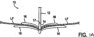
図1Aを参照すると、シャフト12のような送出し装置と、固定装置14とを有している介入用具10が、心房側から僧帽弁MVに接近し、そして弁膜LFを把持した状態で示されている。僧帽弁は、外科的に、或は血管内技術を使用することにより、そして心室を通る逆行接近によるか、或は前述のように心房を通る順行接近により接近されてもよい。例示の目的で、順行接近を説明する。
Referring to FIG. 1A, an
固定装置14は介入用具10のシャフト12にその遠位端部のところで解放可能に取付けられている。ここで本発明の装置を説明する場合、「近位」は、患者の身体の外側で使用者により操られるべき装置の端部に向かう方向を意味しており、「遠位」は、治療部位に使用者から離れる方向に位置決めされる装置の作用端部に向かう方向を意味している。僧帽弁に関しては、近位は弁の弁膜の心房側または上流側を指しており、遠位は弁の弁膜の心室側または下流側を指している。
The
固定装置14は、代表的には、半径方向外方に突出している近位要素16(すなわち、把持要素)および遠位要素18(すなわち、固定要素)を備えており、これらの要素16、18はそれらの間に弁膜を捕獲して保持するように、図示のように弁膜LFの両側に位置決め可能である。近位要素16は好ましくはコバルトクロム、ニチノールまたはステンレス鋼で構成されており、遠位要素18は好ましくは(エルギロイ(登録商標)のような)コバルトクロム合金またはステンレス鋼で構成されているが、任意の適当な材料を使用してもよい。固定装置14は結合機構17によりシャフト12に結合可能である。結合機構17によれば、固定装置14を取外したり、弁膜を共に接合位置に保持するために移植片として置き去りにしたりすることができる。
The
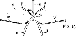
或る状況においては、近位要素16または遠位要素18またはそれらの両方が展開されて弁膜LFを捕獲した後、固定装置14を再位置決めするか或は取外すことが望まれることがある。このような再位置決めまたは取外しは、少しの例を挙げると、良好な弁機能、弁膜における装置14のより最適な位置決め、弁膜に対する良好な握りを達成したり、装置14が索状組織のような周囲組織からもつれをほぐしたり、装置14を異なる設計を有する装置と交換したり、或は固定手順を中断したりする試みで弁に再接近するためのような様々な理由で望まれることがある。固定装置14の再位置決めまたは取外しを容易にするために、遠位要素18は、索状組織、弁各または他の組織ともつれるか干渉したり、それらを損傷したりすることなしに、解放可能であり、そして任意に、装置14を弁から抜出すのに適した構成へ逆転可能である。図1Bは、遠位要素18が逆転位置へ矢印40の方向に移動可能である逆転を示している。同様に、近位要素16は、望むなら、上昇されてもよい。逆転位置では、装置14は所望の配向に再位置決めされ得、この配向では、遠位要素は、次いで、図1Aにおけるような弁膜に当たる把持位置へ戻され得る。変更例として、固定装置14は図1Cに示されるように、弁膜から(矢印42で示されるように)抜出され得る。このような逆転は弁膜に対する外傷を減じ、且つ周囲の組織との装置のいずれのもつれをも最小にする。装置14が弁の弁膜を通して抜出されたら、近位要素および遠位要素は身体からの取出しまたは僧帽弁への再挿通に適した閉鎖位置または構成へ移動され得る。
In some situations, it may be desirable to reposition or remove the
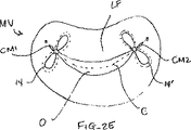
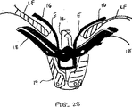
図2Aないし図2Cは弁膜LFに対する所望の配向における1つまたはそれ以上の固定相地14の例としての位置を示している。これらの図は心房側からの僧帽弁MVの短軸図であり、従って、近位要素16は実線で示されており、遠位要素18は鎖線で示されている。近位および遠位要素16、18は、代表的には、接合線Cに対して実質的に垂直であるように位置決めされる。装置14は固定のための任意の所望の位置まで接合線に沿って大まかに移動され得る。弁膜LFは、心臓拡張期中、図2Aないし図2Cに示されるように、弁膜LFが診断圧力勾配から生じる開口部Oにより取囲まれる要素16、18間に適所に留まるように、適所に保持される。有利には、弁膜LFは、それらの近位表面または上流表面が僧帽弁MVを通る血液の流れの方向と平行に、垂直配向で互いに面しているように接合される。上流表面は、互いに接触するように寄せ合わされてもよいし、わずかに離れて保持されてもよいが、好ましくは、それらが接合個所で互いに面する垂直配向に維持される。図2Aを参照して説明すると、固定装置を弁膜LFの中心の近くに設置することにより、標準的な外科ボウタイ修復の2重弁口幾何形状をシミュレートする。カラードプラーエコー法は弁の血液逆流が減少されたかどうかを示す。その結果得られた僧帽弁の流れパターンが満足すべきものであれば、弁膜を共にこの配向で固定すればよい。その結果得られたカラードプラー像が僧帽弁の血液逆流の不十分改良を示すなら、介入用具を再位置決めすればよい。これは、弁膜LFが適所に保持されている最適な結果が生じられるまで繰り返されるのがよい。弁膜が所望の構成で接合されたら、固定装置14をシャフト12から取外し、移植片として置き去りにして弁膜を供に接合位置に保持する。図2Bないし図2Eに示されるように、追加の固定要素14'を付け加えることが望まれることがある。図2Bでは、追加の固定要素14'は先に設置された固定要素14のそばに位置決めされて2重弁口幾何形状を保持する。図2Cでは、追加の固定要素14'は先に設置された固定要素14から最大1cmのような間隔を置いて位置決めされて3重弁口幾何形状を生じる。図2Dでは、固定要素14'は第1交連部CM1の近くで互いに隣接して位置決めされる。かかる配置は、一般に、弁開口部の一方の側にひだ形成により単一弁口幾何形状を生じる。同様に、図2Eに示されるように、1つの固定要素14が第1交連部CM1の近くに位置決めされ、追加の固定装置14'が第2交連部CM1の近くに位置決めされてもよい。かかる配置もまた、一般に、弁開口部の一方の側にひだ形成により単一弁口幾何形状を生じる。追加の固定装置14'は弁膜LFの適切な固定を確保するように、および/または弁膜LFを更に再位置決めするように望まれることがある。追加の固定装置14'は、手順中のいつでも追加されてもよいし、或いは後の時点で別の手順において追加されてもよい。弁膜または任意の他の組織を固定するために、2つ、3つ、4つ、5つまたはそれ以上の固定要素14を含めて、任意の数の固定要素14を位置決めしてもよいことはわかるであろう。
2A-2C show exemplary locations of one or more
図3は、固定装置14の実施形態を示している。この図には、固定装置14は介入用具10するようにシャフト12に結合されて示されている。固定装置14は、結合部材19と、複数の対向された遠位要素18とを有している。遠位要素18は細長いアーム53を備えており、各アームは、結合部材19に回転可能に連結された近位端部52と、自由端部54とを有している。自由端部54は周囲の組織構造との干渉および周囲の組織構造に対する外傷を最小にするために丸い形状を有している。好ましくは、各自由端部54は、一方がアーム53の長さ方向軸線66である2つの軸線のまわりに湾曲を構成する。かくして、係合面50は、組織と接触する表面領域を有し、且つ弁の弁膜を把持して保持するのを助けるためにカップ状または凹形の形状を有している。これにより、更に、アーム53を閉鎖位置でシャフト12のまわりに嵌めて装置の輪郭を最小にすることができる。好ましくは、アーム53はそれらの長さ方向軸線66のまわりに内方に少なくとも部分的にカップ状にされるか或いは湾曲されている。また、好ましくは、各自由端部54はアーム53の長さ方向軸線66に対して垂直な軸線67のまわりに湾曲を構成する。この湾曲は自由端部54の最も遠位の部分に沿った逆の湾曲である。同様に、自由端部54の長さ方向縁部は外方にフレアー状になってもよい。逆の湾曲およびフレアー状の両方により、係合された組織に対する外傷を最小にする。更に、アーム53は、握力を高め且つ植込み後の組織の内部成長を促進するために複数の開口部を有している。
FIG. 3 shows an embodiment of the fixing
弁の弁膜は遠位要素18と近位要素16との間に把持される。幾つかの実施形態では、近位要素16は、固定可能であり、弾性であり、且つ結合部材19から片持ち梁状になっている。近位要素16は、好ましくは、遠位要素に向けて弾性的に付勢されている。各近位要素16は、組織が存在しないとき、遠位要素18の凹部内に少なくとも部分的に凹まされるように形状決めされ且つ位置決めされる。固定装置14が開放位置にあるとき、近位要素16は、その各々がアーム53の近位端部52の近くで係合面50から分離され、そして図3に示されるように、近位要素の自由端部が係合面50に接触した状態で、自由端部54の近くで係合面50に向けて傾斜するように形状決めされる。近位要素16のこの形状は変化する厚さの弁の弁膜または他の組織に順応する。
The valve leaflet is grasped between the
近位要素16は組織に対する握力を増すために複数の開口部63および貝殻状の側演舞61を有してもよい。選択自由として、近位要素16は、弁膜を把持しおよび/または保持するのを助けるために、摩擦付属部、摩擦特徴または握力向上要素を有している。好適な実施形態では、摩擦付属部は係合面50に向けて延びているテーパ状の尖った先端部を有するバーブ60よりなる。突起部、巻き部、バンド、バーブ、溝、チャンネル、隆起部、表面凹凸、焼結部、高摩擦パッド、覆い部、被膜、またはそれらの組合せのような任意の適当な摩擦付属部を使用してもよいことはわかるであろう。選択自由として、磁石が近位要素および/または遠位要素に存在してもよい。合わせ面が、磁力により吸引を引き起こすために、反対磁荷の材料から形成されるか、或いはその材料を有している。例えば、近位要素および遠位要素は、各々、組織の早い治癒および内部成長を容易にするために組織が近位要素と遠位要素との間に保持されるように、反対荷の磁気材料を有してもよい。また、近位要素を遠位要素に向けて付勢することに加えて、或いはそれに代えて、近位要素16を遠位要素18に向けて引きつけるために磁力が使用されてもよい。これは、近位要素16の展開を助けことができる。他の例では、遠位要素18は、各々、遠位要素間に位置された組織が磁力によりそれらの間に保持されるように、反対荷の磁気材料を有している。
The
また、固定装置14は作動機構58を有している。この実施形態では、作動機構58は2つのリンク部材または脚部68よりなり、各脚部68はリベットジョイント76のところで遠位要素18のうちの一方と回転可能に接合されている第1端部70と、スタッド74と回転可能に接合されている第2端部72とを有している。脚部68は、好ましくは、エルギロイ(登録商標)、コバルトクロムまたはステンレス鋼のような剛性または半剛性の金属またはポリマーより構成されるが、任意の適当な材料を使用してもよい。図示の実施形態では、両脚部68は単一のリベット78によりスタッド74にピン止めされているが、各脚部68が別体のリベットまたはピンによりスタッド74に個々に取付けられてもよいことはわかるであろう。スタッド74はシャフト12を通って延びているアクチュエータロッド64(図示せず)と接合可能であり、このアクチュエータロッド64は、スタッド74、従って、脚部を移動させて遠位要素18を閉鎖位置と、開放位置と、逆転位置との間で回転させるために軸方向に伸張可能および引っ込み可能である。同様に、スタッド74の不動化により、脚部68を適所に保持し、従って遠位要素18を所望の位置に保持する。スタッド74は係止特徴により適所に係止されてもよい。
Further, the fixing
ここに開示される固定装置14の実施形態のいずれかにおいて、遠位要素18および近位要素16を弁の弁膜の開放または閉鎖に伴って移動させるか或いは撓ますことができるために、閉鎖位置において遠位要素18および/または近位要素16にいくらかの可動性または可撓性を与えることが望ましいことがある。これにより、衝撃吸収を行い、それにより弁膜に対する力を減少させ、且つ弁膜に対する引裂きまたは他の外傷の可能性を最小にする。かかる可動性または可撓性は、遠位要素18を構成するために適切な厚さの可撓性で弾性の金属またはポリマーを使用することにより与えられてもよい。また、(後述の)固定装置の係止機構は、係止時でも近位および遠位要素のいくらかの僅かな移動を許容するために可撓性材料で構成されてもよい。更に、遠位要素18は、これを閉鎖位置へ(内方に)付勢するが、弁膜により及ぼされる力に応じてアームを僅かに開放する機構により結合機構19または作動機構58に連結されることができる。例えば、これらの構成部品は、単一の個所でピン止めされるのではなく、アームに対する力に応じてピンの少しの量の並進を許容するスロットを通してピン止めされてもよい。ピン止めされた構成部品をスロットの一端に向けて付勢するためにばねが使用されている。
In any of the embodiments of the
図4A-4B、図5A-5B、図6A-6B、図7A-7Bは治療手順を行うために身体内における装置14の導入および設置中の種々の可能な位置における図3の固定装置14の実施形態を示している。図4Aはカテーテル86を通して送出された介入用具10の実施形態を示している。介入用具10がカテーテルの形態をとってもよく、同様に、カテーテル86がガイドカテーテルまたはシースの形態をとってもよいことはわかるであろう。しかしながら、この例では、語「介入用具10」および「カテーテル86」を使用する。介入用具10はシャフト12に結合された固定装置14を備えており、固定装置14は閉鎖位置で示されている。図4Bは図4Aの固定装置14の同様な実施形態をより大きい図で示している。閉鎖位置では、対向対の遠位要素18は係合面50が互いに面するように位置決めされる。各遠位要素18は、カップ状または凹形の形状を有する細長いアームを備えており、これらのアーム53が共にシャフト12を取囲み、選択自由として、シャフトの両側で互いに接触するようになっている。これにより、カテーテル86と、僧帽弁のような任意の解剖学的構造体とに容易に通過可能である固定装置14の低い輪郭をもたらす。また、図4Bは更に作動機構58を有している。この実施形態では、作動機構58は、各々が基部69に移動可能に結合されている2つの脚部68を備えている。基部69は、シャフト12を通って延びていて、固定装置14を操るために使用されるアクチュエータロッド64と接合されている。幾つかの実施形態では、アクチュエータロッド64は作動機構58、特に基部69に直接付いている。しかしながら、変更例として、アクチュエータロッド64は基部69に取付けられているスタッド74に付いてもよい。幾つかの実施形態では、スタッド74は、アクチュエータロッド64がねじ式の作用によりスタッド74に付くようにねじ付きである。しかしながら、ロッド64およびスタッド74は、固定装置14をシャフト12から取外すために解放可能である任意の機構により接合されてもよい。
4A-4B, 5A-5B, 6A-6B, and 7A-7B are views of the
図5A-5Bは開放位置における固定装置14を示している。開放位置では、遠位要素18は、係合面50が第1方向に向くように回転される。アクチュエータロッド64の作用により結合部材19に対してスタッド74を遠位方向に前進させることにより、遠位要素18に力を加え、これらの遠位要素18はこの方向における移動自由度によりジョイント76のまわりに回転し始める。遠位要素18のかかる回転および半径方向外方の移動は脚部68が直ちに僅かに外方に向くようにジョイント80のまわりの脚部68の回転を引き起こす。スタッド74を遠位要素18の所望の分離に相互関連する任意の所望距離まで前進されてもよい。開放位置では、係合面50はシャフト12に対して鋭角で配置され、好ましくは互いに対して90度と180度との間の角度にある。1つの実施形態において、開放位置では、アーム53の自由端部54は約10ないし30mm、通常、約12ないし18mm、好ましくは約14ないし16mmの両者間のスパンを有する。
5A-5B show the
近位要素16は、代表的には、アーム53に向けて外方に付勢される。近位要素16はシャフト16に向けて内方に移動され、そして縫合糸、ワイヤ、ニチノールワイヤ、ロッド、ケーブル、ポリマー線または他の適当な構造体の形態であることができる近位要素線90によりシャフト12に対して保持されてもよい。近位要素線90は、これらを様々な方法で通すことにより近位要素16と連結されてもよい。近位要素16が図5Aに示されるようにループ形状を有する場合、線90はループを通り、そして逆戻りしてもよい。近位要素16が図5Bに示されるように細長い中実の形状を有する場合、線90は要素16における開口部63のうちの1つまたはそれ以上を通ってもよい。更に、図5Bに示されるように、線ループ48が近位要素16に存在してもよく、近位要素線90はこの線ループ48を通り、そして逆戻りしてもよい。このような線ループ48は近位要素線90に対する摩擦を低減するために、或は近位要素16が中実であるか、或は近位要素線90が通る他のループまたは開口部を欠いている場合に、有用であることがある。単一の線90を逆戻りすることなしに近位要素16に取付けたり、望むときに単一の線90を近位要素16から直接取外したりする取り外し可能な手段により、近位要素線90が近位要素16に取付けられてもよい。このような取外し可能な手段の例としては、少しの例をあげると、フック、スネア、クリップまたは破断可能な継ぎ手がある。十分な張りを近位要素線90に付与することにより、取外し可能な手段は、例えば継ぎ手の破断により近位要素16から取外されてもよい。取外しのための他の機構を使用してもよい。同様に、同様な取外し可能な手段により係止線92が係止機構に対して取付けられたり、取外されたりされてもよい。
開放位置では、固定装置14は、接近されるべきか、或は治療されるべきである組織に係合することができる。この実施形態は左心房からの順行の接近を使用した僧帽弁の治療に適合されている。介入用具10を左心房から左心室へ僧帽弁を通して前進させる。遠位要素18を接合線に対して垂直であるように配向させ、次いで、係合面50が弁の弁膜の心室表面に接触するように位置決めし、それにより弁膜を把持する。近位要素16は、弁膜が近位要素と遠位要素との間に位置するように弁の弁膜の心房側に留まる。この実施形態では、近位要素16は遠位要素18に向けられるバーブ60のような摩擦付属部を有している。しかしながら、この時点で、近位要素16もバーブ60も弁膜に接触しない。
In the open position, the
介入用具10は、弁膜が所望の位置で適切に接触される或は把持されるように、固定装置14を再位置決めするように繰返し操られてもよい。再位置決めは固定装置が開放位置にある状態で達成される。或る場合には、装置14が開放位置にある間、血液逆流を抑制してもよい。血液逆流が申し分なく減少されないなら、装置を再位置決めし、所望の結果が達成されるまで、血液逆流を再び抑制してもよい。
The
固定装置14の再位置決めまたは取出しを助けるために、固定装置14を逆転することが望まれることもある。図6A-6Bは逆転位置にある固定装置14を示している。結合部材19に対してスタッド74を更に前進させることにより、各アーム53がシャフト12に対して鈍角を形成して状態で、係合面50が外方に向き、自由端部54が遠位方向を指すように、遠位要素18を更に回転させる。アーム53間の角度は好ましくは約270ないし360度の範囲にある。スタッド74を更に前進させることにより、遠位要素18をジョイント76のまわりに更に回転させる。遠位要素18のこの回転および半径方向外方の移動は、脚部68が互いに概ね平行なそれらの初めの位置に戻るように、ジョイント80のまわりの脚部68の回転を引き起こす。スタッド74を遠位要素18の所望の逆転に相互関連する任意の所望の距離まで前進させてもよい。好ましくは、完全に逆転された位置では、自由端部54間のスパンは約20mmに過ぎず、通常、約16mm未満、好ましくは約12ないし14mmである。この例では、近位要素16は張りを近位要素線90に及ぼすことによりシャフト12に当たって位置決めされたままである。かくして、再位置決めのための要素16、18間に比較的大きい空間が生じられ得る。また、逆転位置は、弁膜に対する外傷を最小にしながら、弁を通る固定装置14の抜出しを許容する。係合面50は、固定装置が近位方向に引っ込められるとき、組織を偏向させるための非外傷性表面をなす。なお、バーブ60は遠位方向に(近位要素16の自由端部から離れる方向に)僅かに傾斜されていて、固定装置を抜出すとき、バーブが組織に引っ掛かるか、或は組織を引裂く恐れを減じる。
It may be desirable to reverse the
固定装置14が弁の弁膜に当たる所望の位置に位置決めされたら、次いで、弁膜を近位要素16と遠位要素18との間に捕獲し得る。図7A-7Bはこのような位置にある固定装置14を示している。ここで、近位要素16を係合面50に向けて降下させて弁膜をそれらの間に保持するようにする。図7Bでは、近位要素16は弁膜の非外傷性把持を行うのに使用されてもよいバーブ60を有するように示されている。変更例として、弁膜に突き刺さって弁膜を適所に保持するのをより活発に助けるために、もっと大きくもっと鋭く尖ったバーブまたは他の侵入構造体を使用してもよい。この位置は図5A-5Bの開放位置と同様であるが、この場合、近位要素16は、弁膜組織をそれらの間に圧縮するために近位要素線90に対する張りを解放することによりアーム53に向けて降下されている。血液に逆流が十分に減少されていないなら、任意の時点で、近位要素16を上昇させ、遠位要素18を調整するか、或は逆転して固定装置14を再位置決めすればよい。
Once the anchoring
弁膜が所望の配置で近位要素16と遠位要素18との間に捕獲された後、遠位要素18を係止して弁膜をこの位置に保持するか、或は固定装置14を閉鎖位置まで或はそれに向けて戻してもよい。
After the leaflet is captured between the
本発明の固定装置が前述の機能および特徴のうちのいずれか或はすべてを有してもよいことはわかるであろう。例えば、固定装置14は逆転位置へ移動可能であってもよいし、なくてもよい。或は、固定相地14は近位要素16を有してもよいし、有していなくてもよい。かくして、固定装置14の前述の態様は単に好適な実施形態であり、本発明の範囲を限定しようとするものではない。
It will be appreciated that the fixation device of the present invention may have any or all of the functions and features described above. For example, the fixing
II.弁膜の安定化
弁膜を把持前に安定化するために、様々な装置および技術が提供される。かかる安定化は、弁膜を有効に且つ効率的に把持し、それにより多数の把持を必要とすることなしに所望量の弁膜が固定装置に取り入れられる可能性を増すことを目標とするものである。安定化装置および技術が、前述の固定装置との組合せで使用されてもよく、或は任意の適当な把持および/または固定装置とともに使用されてもよいことはわかるであろう。更に、このような安定化技術および装置のうちの多くは、任意の目的で弁の弁膜または他の組織を安定化するために使用されてもよい。
II. Stabilization of the leaflets Various devices and techniques are provided to stabilize the leaflets prior to gripping. Such stabilization is aimed at gripping the valve membrane effectively and efficiently, thereby increasing the likelihood that the desired amount of valve membrane will be incorporated into the fixation device without requiring multiple grips. . It will be appreciated that the stabilization devices and techniques may be used in combination with the aforementioned fixation devices, or may be used with any suitable gripping and / or fixation device. Furthermore, many of such stabilization techniques and devices may be used to stabilize the valve leaflets or other tissues for any purpose.
代表的には、僧帽弁の血液逆流の場合、弁膜LFの一部が他の弁膜または弁膜の諸部分と移送不一致に移動している。これは、弁膜を安定に且つ同調的に保持する構造体(腱索)の伸びまたは切断に起因して生じてしまう。このような機能不全の結果、1つの弁膜または弁膜の一部が健康な接合部のレベルの上方で揺動するか、或は「動揺し」、それにより血液が右心房の中へ逆流してしまう。図8A-8L、図9A-9B,図10A-Bは使用者が目標とされた弁膜をより確実に把持するために弁膜の上方移動および動揺を減じることにより弁の弁膜を安定化する装置の実施形態を示している。これらの実施形態では、図8Aに示されるように、カテーテル86を心臓Hの左心房LAの中へ前進させ、固定装置14をカテーテル86と、弁膜LFを有する僧帽弁MVとに通して前進させて固定装置14の一部を左心室LV内に位置決めする。弁の弁膜LFは、固定装置14が左弁室LV内で弁の下方にある間、左心房LAに向けて上方に動揺して示されている。この例では、固定装置14は、図3について前述した固定装置と類似しており、近位要素16および遠位要素18を有している。固定装置14は、近位要素16がシャフト12に保持されたままでありながら、遠位要素18を半径方向外方に延ばすために少なくとも部分的に開放されている。近位要素16を降下させて弁膜LFをそれらの間に把持するように、弁膜LFを遠位要素18と係合させることが望まれる。
Typically, in the case of mitral blood regurgitation, a portion of the leaflet LF moves inconsistent with other leaflets or portions of the leaflet. This occurs due to the stretching or cutting of the structure (the chordae) that holds the leaflets stably and synchronously. As a result of such dysfunction, one valve leaflet or part of the leaflet rocks or “sways” above the level of a healthy joint, causing blood to flow back into the right atrium. End up. 8A-8L, 9A-9B, and 10A-B show an apparatus for stabilizing a valve leaflet by reducing the upward movement and swaying of the leaflet in order for the user to more securely grasp the targeted leaflet. An embodiment is shown. In these embodiments, as shown in FIG. 8A, the
1つまたはそれ以上の安定化ループ100をカテーテル86から前進させ、弁膜LFの心房側に位置決めさせてもよい。図8Bは安定化ループ100の実施形態の横断面頂面図を示している。ループ100は円形形状を形成するようにカテーテル86から半径方向外方に延びて示されている。円形形状の直径はカテーテル86からのループ100の前進または引っ込みにより変化されてもよい。ループ100は金属、ポリマーまたは繊維のような任意の適当な材料で構成されてもよく、ワイヤ、リボン、リンクまたは織物のような任意の適当な形態を有してもよい。図8Cは図8Bに示される実施形態の側面図を示している。ループ100の円形形状はカテーテル86に対して実質的に垂直な平面にある。かくして、ループ100は図8Aに示されるように弁の輪部に沿って位置決めされてもよい。この位置では、弁膜LFはまだ上方に動揺することがある。図8Dを参照して説明すると、ループ100の直径は矢印102で示されるように減少されてもよい。これは、ループ100をカテーテル86の中へ部分的に引っ込めることにより達成されてもよい。直径の連続的な減少により、ループ100を輪部から弁の中心に向けて引入れる。ループ100が移動すると(先のループ100'が鎖線で示されている)、ループ100は弁膜LFの上方運動または動揺を制御法で制限し、そして近位要素16と遠位要素18との間に把持するために弁膜LFを位置決めする。
One or
弁膜を安定化するために1つより多いループ100が存在してもよく、これらのループが同心であってもよく、互いに隣接してもよく、別々の平面にあってもよく、或は任意の適当な配置にあってもよいことはわかるであろう。例えば、図8Eは第1ループ100aおよび第2ループ100bを有する実施形態を示している。これらのループ100a、100bは図8Aないし図8Dに示される実施形態と同様に機能するが、第2ループ100bはより小さく、そして第1ループ100a内に同心に位置されている。ループ100a、100bの直径は任意の適当な大きさを有してもよく、直径の関係が変化してもよい。図8Fは図8Eに示される実施形態の側面図を示している。図示のように、ループ100a、100bの円形形状はカテーテル86に対して実質的に垂直な平面にある。かくして、弁膜LFを安定化するためにループ100a、100bを弁膜LFに当てて位置決めしてもよい。次いで、ループ100a、100bをカテーテル86の中へ部分的に引っ込めることによりループ100a、100bの直径を同時に或は個々に減少させてもよい。直径の連続的な減少により、ループ100a、100bを弁の中心に向けて引入れる。また、ループ100a、100bが移動すると、これらのループ100a、100bは弁膜LFの上方運動または動揺を制御法で制限し、且つ近位および遠位要素16、18間の最適な把持のために弁膜LFを位置決めする。
There may be more than one
図8Gは第1ループ100aおよび第2ループ100bを有する他の実施形態を示している。しかしながら、この実施形態では、ループ100a、100bは非同心であり、各ループはカテーテル86の反対側から延びている。ループ100a、100bの直径は任意の適当な大きさを有してもよく、直径の関係が変化してもよい。図8Hは図8Gに示される実施形態の側面図を示している。図示のように、ループ100a、100bの円形形状はカテーテル86に対して実質的に垂直な平面にある。かくして、弁膜LFを安定化するためにループ100a、100bを弁膜LFに当てて位置決めしてもよい。図8Iは僧帽弁MVの頂面図を示しており、この場合、固定装置(図示せず)が弁膜LFに通されるように、カテーテル86が弁MVの上方に位置決めされている。ループ100a、100bは輪部に向かって半径方向外方に延ばされて示されている。次いで、ループ100a、100bの直径を矢印102でしめされるように減少させてもよい。これは、ループ100a、100bをカテーテル86の中へ部分的に引っ込めることにより達成されてもよい。直径の連続的な減少により、ループ100a、100bを輪部から弁の中心に向けて引入れる。また、ループ100a、100bが移動すると(先のループ100a'、100b'が鎖線で示されている)、これらのループ100a、100bは弁膜LFの上方運動または動揺を制御法で制限し、且つ近位および遠位要素16、18間の最適な把持のために弁膜LFを位置決めする。図示のように、各ループ100a、100bは個々の弁膜LFの移動を制限する。しかしながら、各ループ100a、100bが1つより多い弁膜LFに接触するようにカテーテル86が(例えば、90度の回転に)配向されてもよいことはわかるであろう。
FIG. 8G shows another embodiment having a first loop 100a and a
図8Jはカテーテル86と実質的に平行な平面にある単一のループ100を有する実施形態を示している。図8Kは図8Jに示される実施形態の側面図を示している。ループ100は任意の適当な形状および直径を有してもよい。かくして、ループ100は弁の弁膜LFを安定化するために弁膜LFに当たって位置決めされてもよい。図8Lは僧帽弁MVの頂面図を示しており、この図では、固定装置(図示せず)が弁膜LFに通されるように、カテーテル86が弁MVの上方に位置決めされている。ループ100は交連部Cに対して垂直に輪部に向かって半径方向外方に延びて示されている。かかる位置決めは弁膜LFの上方移動を制限する。また、この場合、ループ100の直径は矢印102で示されるように減少されてもよい。これは、ループ100をカテーテル86の中へ部分的に引っ込めることにより達成されてもよい。直径の連続的な減少により、ループ100を輪部から弁の中心に向けて引き入れる。ループ100が移動すると(先のループ100'が鎖線で示されている)、このループ100は弁膜LFの制限された上方移動または動揺を維持し、且つ近位および遠位要素16、18間における最適の把持のために弁膜LFを位置決めする。
FIG. 8J shows an embodiment having a
前述の実施形態のいずれにおいても、ループが両弁膜を安定化するために延ばされてもよいし、或いは動揺している1つの弁膜を安定化するために延ばされてもよいことはわかるであろう。これは、カテーテル86の配向、ループ100の形状、ループ100の延び量または任意の他の方法により達成されてもよい。図8Gないし図8Iに示される実施形態は単一弁膜の動揺に特に適合され、この場合、第1ループ100aのみが存在してもよい。更に、ループ100が弁膜の安定化を助けるためにローラまたはグリッパーのような表面処理部または付属部を有してもよいことはわかるであろう。
It will be appreciated that in any of the foregoing embodiments, the loop may be extended to stabilize both valve membranes, or may be extended to stabilize one agitating valve membrane. Will. This may be achieved by orientation of the
図9A-9Bは弁の弁膜LFの上方移動および動揺を減少させることにより弁の弁膜LFを安定化する他の実施形態を示している。図9Aに示されるように、カテーテル86を心臓Hの心房LAの中へ前進させ、固定装置14をカテーテル86と、弁膜LFを有する僧帽弁とに通して全身させて、固定装置14の少なくとも一部を左心室LV内に位置決めする。弁の弁膜LFは、装置14が左心室LV内で弁の下方にある間、左心房LAに向けて動揺しているものとして示されている。この例では、固定装置14は、図3について前述した固定装置と類似しており、近位要素16および遠位要素18を有している。固定装置14は、近位要素16がシャフト12に保持されたままでありながら、遠位要素18を半径方向外方に延ばすために少なくとも部分的に開放されている。近位要素16が降下されて弁膜LFをそれらの間に把持するように弁膜LFを遠位要素18と係合させることが望まれる。
9A-9B illustrate another embodiment that stabilizes the valve leaflet LF by reducing upward movement and swaying of the valve leaflet LF. As shown in FIG. 9A, the
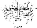
1つまたはそれ以上のフラップ104が、図示のようにカテーテル86から半径方向外方に延び、弁膜LFの心房側に位置決めされてもよい。これらのフラップ104は金属、ポリマーまたは繊維のような任意の適当な材料で構成されてもよく、中実物、メッシュまたは織物のような任意の適当な形態を有してもよい。更に、フラップ104は任意の適当な形状を有してもよく、1つまたはそれ以上の切欠き106を有してもよい。図9Bに示されるように、切欠き106は固定装置14の近位要素16をこれらの切欠きを通して延ばせるように寸法決めされ且つ位置決めされてもよい。これにより、フラップ104を弁膜LFの心房側に保持して弁膜LFの上方移動または動揺を制限し得る。これにより、近位および遠位要素16、18間における最適な把持のために弁膜LFを位置決めする。弁膜が把持されたら、フラップ104をカテーテル86と供に取出してもよいし、或は弁膜LFの保持を助けるために置き去りにしてもよい。
One or
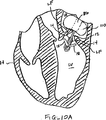
図10A-10Bは弁の弁膜LFの上方移動または動揺を減少させることにより弁膜LFを安定化する他の実施形態を示している。図10Aに示されるように、カテーテル86を心臓Hの左心房LAに中へ前進させ、そして固定装置14をカテーテル86と、弁膜Lfを有する僧帽弁MVとに通して前進させて、固定装置14の少なくとも一部を左心室LV内に位置決めする。弁の弁膜LFは、固定装置14が左心室LV内で弁の下方にある間、左心房LAに向かって上方に動揺しているものとして示されている。この例では、固定装置14は図3について前述した固定装置と類似しており、近位要素16および遠位要素18を有している。固定装置14は、近位要素16がシャフト12に保持されたままでありながら、遠位要素18を半径方向外方に延ばすために少なくとも部分的に開放されている。近位要素16を降下させて弁膜LFをそれらの間に把持するように、弁膜LFを遠位要素18と係合させることが望まれる。
FIGS. 10A-10B illustrate another embodiment of stabilizing the leaflet LF by reducing upward movement or swaying of the valve leaflet LF. As shown in FIG. 10A, the
1つまたはそれ以上の拡張可能な部材110が、図示のようにカテーテル86から半径方向外方に延び、弁膜LFの心房側に位置決めされてもよい。この拡張可能な部材110はシリコーンまたはポリウレタンのような任意の適当な材料で構成されてもよいし、バルーンのような任意の適当な形態を有してもよい。図10Bはこの実施形態の追加の図を示している。図示のように、拡張可能な部材110を左心房内で延ばし、弁膜LFの心房側に保持して弁膜LFの上方運動または動揺を制限し得る。これにより、近位および遠位要素16、18間における最適な把持のために弁膜LFを位置決めする。
One or more
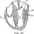
図11A-11Bは弁の弁膜LFの上方移動および動揺を減少させることにより弁の弁膜LFを安定化する他の実施形態を示している。この例では、固定装置14は図3について前述した固定装置に類似しており、近位要素16および遠位要素18を有している。また、固定装置14をカテーテルと、弁膜LFを有する僧帽弁とに通して前進させて固定装置14の遠位要素18を左弁室LV内に位置決めする。固定装置14は、近位要素16がシャフト12に保持されたままでありながら、遠位要素18を半径方向外方に延ばすために少なくとも部分的に開放されている。近位要素16を降下させて弁膜LFをそれらの間に把持するように、弁膜LFを遠位要素18と係合させることが望まれる。しかしながら、近位要素16を降下させる前に、スロット123を有するオーバーチューブ121をシャフト12の上を覆って前進させ、図11Bに示されるように、弁膜LFの心房側に位置決めする。オーバーチューブ121はポリイミド、ポリエチルエチルケトン(PEEK(登録商標))、(ぺバック(登録商標)のような)ナイロン樹脂またはポリウレタンのような任意の適当な材料で構成されてもよく、スロットは近位要素16をこれらのスロットに通せるように任意の適当な寸法を有してもよい。オーバーチューブ121による弁膜LFの保持により、弁各LFの上方運動または動揺を制限し、且つ近位要素16を降下させる前に弁膜が正確に位置決めされるのを確認することができる。これにより、近位および遠位要素16、18間における最適な把持のために弁膜LFを位置決めする。次いで、近位要素16を解放すればよく、この場合、近位要素16はスロット123を通過して弁膜を近位および遠位要素16、18間に保持する。次いで、オーバーチューブ121を引っ込めて取出せばよい。
11A-11B illustrate another embodiment that stabilizes the valve leaflet LF by reducing upward movement and swaying of the valve leaflet LF. In this example, the
図12A-12Bは弁膜に取付けられた腱に張りを付与することにより弁の弁膜を安定化する実施形態を示している。かかる安定化は、弁膜の上方移動および動揺を減少させるために、或は弁膜の移動を単に減少させるための望まれることがある。図12Aは弁膜LFを備えている僧帽弁MVを有する心臓Hを示している。腱索CHは弁膜LFのうちの一方から左心室LVまで延びて示されている。腱索が多数であり、両弁膜から左心室まで伸びているが、簡単化のために選択された腱索が示されている。図12Bに示されるように、バルーンのような拡張可能な部材122を有するカテーテル120を左心室LVまで前進させ、このカテーテル120を位置決めし、拡張可能な部材122を拡張して腱索CHに張りを付与する。図12Bは大動脈弁AVに通して前進されたカテーテル120を示しているが、カテーテル120は、僧帽弁MVを通ること、または隔膜Sを通ることを含めて、任意の適当な経路を経て腱索CHに接近してもよい。腱索CHに張りを付与することにより、取付けられた弁膜LFの位置を調整する。かくして、弁膜LFは、拡張可能な部材122の変化する拡張を含めて、カテーテル120および拡張可能な部材122を操ることによって操られて再位置決めされてもよい。特に、拡張可能な部材122で腱索CHに横方向に押し当てることにより、弁膜LFを押し下げて弁膜LFの上方移動および動揺を制限する。弁膜LFが望ましい位置に配置されたら、弁膜LFを図3について説明したような固定装置により固定すればよい。変更例として、腱索CHを張るのに把持具を用いてもよい。
12A-12B illustrate an embodiment in which the valve leaflets are stabilized by applying tension to the tendon attached to the leaflets. Such stabilization may be desired to reduce the upward movement and sway of the leaflet, or simply to reduce leaflet movement. FIG. 12A shows a heart H having a mitral valve MV with a leaflet LF. The chord CH is shown extending from one of the leaflets LF to the left ventricle LV. There are many chordae, extending from both leaflets to the left ventricle, but chords selected for simplicity are shown. As shown in FIG. 12B, a
前述の実施形態は弁の弁膜の機械的安定化に集中している。追加の実施形態は弁膜の運動を生理学的に遅くすることによる弁の弁膜の安定化に集中している。これは、心臓の自然鼓動を遅くすることにより達成され得る。図13に示される1つの実施形態では、右心房RAにおける洞房結節SAまで延びている電極134を持つペース決め用リード132を有するペースメーカー130またはパルス発生器が示されている。ペース決めは、ペースメーカー130が電気インパルスをペース決め用リード132に通して電極134へ送り、それにより洞房結節SAを刺激するときに達成される。これにより、右心房RAを刺激して血液を心臓Hに通して右心房RAへ圧送する。かくして、ペースメーカー130の使用により心臓のポンプ作用、従って弁の弁膜の移動を調整することができる。図13は僧帽弁MVの弁膜LFを通過された固定装置14を示している。弁膜LFの移動は、例えば、僧帽弁MVが、固定装置14で弁膜LFを把持するのを助けるためにより長い時間の間、収縮期(閉鎖された状態)に留まるようにペース決めされてもよい。同様に、図14に示されるように、左心房LVは、左束LBを刺激することによりペース決めカテーテル136で直接にペース決めされてもよい。これは、図示のようにペース決めカテーテル136を大動脈弁AVに通して左心房LVまで前進させることにより達成され得る。
The foregoing embodiments concentrate on the mechanical stabilization of the valve leaflets. Additional embodiments focus on stabilizing the valve leaflets by physiologically slowing the leaflet motion. This can be achieved by slowing the natural heartbeat. In one embodiment shown in FIG. 13, a
III.把持補助
弁膜を効果的および効率的に把持するのを補助するために、様々な装置および技術が提供される。これらの装置および技術のうちの多くを図3について説明した固定装置に対する補助として説明する。しかしながら、任意の適当な把持および/または固定装置について、多くの特徴を使用し得る。更に、このような技術および装置の多くは任意の目的で弁の弁膜または他の組織を把持するのに使用され得る。
III. Gripping Assistance Various devices and techniques are provided to assist in gripping the valve membrane effectively and efficiently. Many of these devices and techniques will be described as an aid to the fixation device described with respect to FIG. However, many features can be used for any suitable gripping and / or securing device. In addition, many such techniques and devices can be used to grasp the valve leaflets or other tissue for any purpose.
或る状況においては、1つまたはそれ以上の弁膜LFは所望の位置で近位要素16と遠位要素18との間に把持されない。例えば、所望量未満の弁膜LFが把持されてもよい。かかる減少された捕獲量は、例えば、血液逆流の治療に効果性を減じることがあり、および/または弁膜LFが固定装置から滑り出る恐れを増すことがある。弁膜LFの一部が把持されたら、弁膜LFの位置を調整すればよく、例えば、捕獲量を増すために、弁膜LFを固定装置14のシャフト12に「引入れる」か、或はシャフト12に向けて前進させればよい。このような調整を補助するための実施形態が図15ないし図17および図18A-18Bに示されている。
In some situations, one or more leaflets LF are not gripped between the
図15は図3に示された固定装置14と同様な固定装置14の実施形態を示している。図示のように、弁膜LFを近位要素16と遠位要素18との間に部分的に把持する。この実施形態では、真空管路140がシャフト12を通って延びていて、真空源142に連結されている。真空管路140は近位および遠位要素16、18間の空間146へ突入している遠位端部144を有している。真空源142の作動により、吸引を空間146に付与して弁膜LFをシャフトに向けて内方に引き寄せる。かくして、弁膜LFは、把持されると、吸引力により近位および遠位要素内に再位置決めされ得る。同じ真空管路140または追加の真空管路が他の近位および遠位要素間の弁膜に吸引を付与してもよいことはわかるであろう。更に、把持作用を助けるために、始めの把持中に吸引力を付与してもよいことはわかるであろう。
FIG. 15 shows an embodiment of a
同様に、図16の示されるように、図3に示される固定装置14と同様な固定装置14の他の実施形態が示されている。また、弁膜LFを近位要素16と遠位要素18との間に部分的に把持する。この実施形態では、顎部154を有する補助-把持具152が通るための補助-把持具チャンネル150がシャフト12を通って延びているが、非外傷性フック、クランプまたは爪のような任意の種類の把持機構が存在してもよい。顎部154は近位および遠位要素16、18間の空間146に突入している。保持-把持具152を前進させて顎部154で弁膜LFを把持し、そして引っ込めて弁膜LFをシャフト12に向けて内方に引くことができる。かくして、保持-把持具152の操りにより弁膜LFを再位置決めし得る。弁膜を他の近位および遠位要素間に再位置決めするために、同じまたは追加の保持-把持具152を使用してもよいことはわかるであろう。更に、把持作用を助けるために初めの把持中に保持-把持具152を使用してもよいことはわかるであろう。
Similarly, as shown in FIG. 16, another embodiment of a
図17は図3に示される固定装置14と同様な固定装置14の他の実施形態を示している。また、弁膜LFを近位要素16と遠位要素18との間に部分的に把持する。この実施形態では、コンベヤベルト160が各遠位要素18内に配置されて、ベルト160の表面が把持された弁膜LFに接触するようになっている。コンベヤベルト160は1つまたはそれ以上のローラ162の上に設けられている。ローラ162の回転により、コンベヤベルト160を移動させ、それにより接触された弁膜LFを移動させる。例えば、ローラ162の時計方向回転は、図示のように、弁膜LFをシャフト12に向けて内方に引くことができる。同様に、ローラ162の反時計方向回転は弁膜LFを外方に引くことができる。かくして、コンベヤベルト160の移動により弁膜LFを再位置決めすることができる。遠位要素18内に配置されたコンベヤベルト160が単独に或は一致して機能してもよいことはわかるであろう。更に、把持作用を助けるために初めの把持中にコンベヤベルト160を使用してもよいことはわかるであろう。
FIG. 17 shows another embodiment of a fixing
図18A-18Bは、図3に示される固定装置14と同様であって、近位要素16および遠位要素18を有する固定装置14の他の実施形態を示している。この実施形態では、近位要素16はシャフト12を跨っているブリッジ166により連結されている。図18Bを参照して説明すると、弁膜が近位および遠位要素16、18間に把持されると、力を加えて矢印168で示されるようにブリッジ166を固定装置14の基部69に向けて移動させる。近位要素16の湾曲に起因して、ブリッジ166のかかる移動により、近位要素16を(矢印170で示されるように)シャフト12に向けて内方に引き、それにより把持された弁膜をシャフト12に向けて内方に引く。同様に、力を加えてブリッジ166を基部69から離れる方向に移動させることにより、近位要素16を外方に移動させる。かくして、ブリッジ166の移動により弁膜を再位置決めすることができる。遠位要素18が閉鎖位置に向けて移動することにより、ブリッジ166が基部69に向かって移動してもよいことはわかるであろう。或は、遠位要素18が閉じると、近位要素14がシャフト12に向けて内方に引かれるように、近位要素16がカムまたは他の適当な要素に取付けられてもよい。かくして、近位要素16は、遠位要素18が静止している間に移動してもよく、或は近位要素16および遠位要素18の両方が互いに対して移動してもよい。更に、幾つかの実施形態では、近位要素16が静止している間、遠位要素18が移動してもよいことはわかるであろう。
18A-18B illustrate another embodiment of a
図19Aないし図19Cは、図3に示される固定装置14と同様であって、図示のように、予備把持具176が通るための通路がシャフト12を通して設けられている固定装置14の実施形態を示している。予備把持具176は、シャフト178と、その遠位端部の近くに配置された顎部180とを有しているが、非外傷性フック、クランプまたは爪のような任意の種類の把持機構が存在してもよい。図19Bを参照して説明すると、固定装置14を、これが心房内に存在するように、前述のような心房接近で僧帽弁に通して前進させる。予備把持具176をシャフト12に通して前進させ、そして弁膜LFの一方または両方の一部を把持するように操る。プルワイヤを含めて、任意の適当な機構により、予備把持具176を操向してもよいし、或は予備把持具176を所望の構成で予備形成してもよい。更に、予備把持具176をシャフト12内で回転させてもよい。弁膜を安定化するためにおよび/または弁膜を所望の配向へ移動させるために、予備把持具176は一方の弁膜を把持してもよいし、或いは両方の弁膜を例えば接合配向で把持してもよい。弁膜が申し分なく配向されたら、図19Cに示されるように、固定装置14を使用して弁膜LFを把持すればよい。次いで、予備把持具176を弁膜LFから解放し、シャフト12における通路を通しての抜出しにより取り出せばよい。変更例として、弁膜の固定を補強するために、予備把持具176を適所に残すことができる。
19A to 19C show an embodiment of the fixing
他の実施形態では、予備把持具176を例えば異なる接近により組織および弁膜LFまで別々に前進させる。図20は心房接近により前進された固定装置14と、心室接近により前進された予備把持具176とを示している。また、予備把持具176はシャフト178と、その遠位端部182の近くに配置された顎部180とを有しているが、非外傷性フック、クランプまたは爪のような任意の種類の把持機構が存在してもよい。予備把持具176を前進させ、そして弁膜LFの一方または両方の一部を把持するように操る。プルワイヤを含めて、任意の適当な機構により、予備把持具176を操向してもよいし、或は予備把持具176を所望の構成で予備形成してもよい。更に、予備把持具176を回転させてもよい。弁膜を安定化するためにおよび/または弁膜を所望の配向へ移動させるために、予備把持具176は一方の弁膜を把持してもよいし、或いは両方の弁膜を例えば接合配向で把持してもよい。弁膜が申し分なく配向されたら、固定装置14を使用して弁膜LFを把持すればよい。固定装置14を、これが心房内に存在するように、心房接近で僧帽弁に通して前進させる。これは、代表的には、固定装置14の少なくとも一部を予備把持具176により把持された弁膜の領域に隣接して弁膜に通すことにより達成される。次いで、予備把持具176を弁膜LFから解放し、抜出しにより取り出せばよい。変更例として、弁膜の固定を補強するために、予備把持具176を適所に残すことができる。他の実施形態では、固定装置14を心室接近により前進させ、予備把持具176を心房接近により前進させることはわかるであろう。
In other embodiments, the pre-gripper 176 is advanced separately to the tissue and leaflet LF, eg, with different approaches. FIG. 20 shows the
図21A-21Bはつなぎ体192により接合可能な2つの片面固定要素190を有する固定装置14の実施形態を示している。各片面固定要素190は少なくとも1つの近位要素16および遠位要素18で構成されている。幾つかの実施形態では、片面固定要素190は図3に示される固定装置14の2分の1に類似している。図21Aは一対の片面固定要素190を示している、各片面固定要素190は弁膜LFをその近位要素16と遠位要素18との間に把持する。これらの固定要素190は、送出しカテーテル191を通して弁膜LFに送出されてもよく、各要素190はカテーテル191を通っている細長い送出し装置193に連結されている。固定要素190はまたつなぎ体192により互いに連結されている。固定要素190が弁膜LFを申し分なく把持したら、固定要素190を送出し装置193から取外し、そして弁膜LFを置き去りにしてつなぎ体192を介して所望の配向に保持することができる。変更例として、固定要素190を互いに引き寄せ、それにより弁膜LFを接合するために、つなぎ体192を短縮したり、伸張したりしてもよい。図21Bに示されるような幾つかの実施形態では、つなぎ体192は、カテーテル191からの解放時に「自己短縮する」コイルまたはばねのような弾性要素よりなる。つなぎ体192を短縮したり、伸張したりする他の手段は、縫合糸留め具をつなぎ体192に付設することを含み、その好適な実施形態は米国特許出願第10/087004号(代理人ドケット第020489-000500US)に記載され、示されている。他の実施形態では、各片面固定要素190はカテーテル191を通って延びている個々のつなぎ体に取付けられている。その場合、個々のつなぎ体を互いに結んでもよく、結び目を固定要素190に向けて押して固定要素190を所望の距離のところで互いに縛ってもよい。
21A-21B illustrate an embodiment of a
かくして、固定要素190は、弁膜LFを接合位置に保持するために、互いに繋がれたり、取付けられたり、結合されたり、或いは接合されたりしてもよい。任意の数の片面固定要素190を使用してもよく、それらの幾つかまたはすべてが1つまたはそれ以上のつなぎ体192により接合可能であってもよいことはわかるであろう。更に、弁の治療を助けるために腱索のような弁の弁膜以外の組織を把持するのに、片面固定要素190のうちの少なくとも1つを使用してもよいことはわかるであろう。例えば、要素190は、弁膜を弁膜に、弁膜を乳頭筋に、弁膜を腱索に接合してもよい。更に、片面固定要素190を例えば心房接近および心室接近で弁の両側から展開し、そして弁を横切って接合してもよいことはわかるであろう。かくして、血液に逆流を治すために、1つの片面固定要素190を弁の前側で、1つの片面固定要素190を弁の後側で展開し、次いで、これらの要素190を互いに堅く縛ってもよい。
Thus, the
図22は図3に示された固定装置14と同様であって、近位要素16および遠位要素18を有している固定装置14の実施形態を示している。しかしながら、この実施形態では、遠位要素18は「自己係合性」である。図22に示されるように、遠位要素18が心房内に配置され、近位要素16が心房内に配置されるように、固定装置14を位置決めしてもよい。弁膜LFを遠位要素18と係合させ、次いで近位要素16を降下させて弁膜LFを両要素間に把持するのではなく、まず、近位要素16を降下させて弁各LFに係合させる。近位要素16の降下により、弁膜LFを安定化し、且つ弁膜LFの可能な上方運動または動揺を減じるのがよい。次いで、遠位要素18が自己係合するか、或いは閉鎖位置に向かって自動的に移動して弁膜LFに係合し、そして弁膜LFを近位および遠位要素16、18間に把持することができる。自己係合は、所定の位置までの近位要素16の降下を表す機構、また弁膜LFとの近位要素16の十分な係合を感知するセンサを含めて、様々な機構により作動されてもよい。遠位要素18の自動係合の無い図3の固定装置14について、遠位要素18の係合前に近位要素16を降下させる方法を利用してもよいことはわかるであろう。
FIG. 22 is similar to the
弁膜が把持されたら、様々な特徴が、把持された弁膜を固定装置内に保持するのを補助し得る。例えば、図23は、把持後、特に、閉鎖位置に向かう遠位要素18の移動中、吸引を行って弁膜の位置を維持する固定装置14の実施形態を示している。この実施形態では、吸引管路200が、遠位要素18の係合面50に配置された吸引ポート202まで延びている。吸引管路200は図15に示された実施形態と同様に固定装置を通って真空源まで延びている。遠位要素18が弁膜を係合面50と係合させたら、吸引ポート202を通して付与された吸引が、弁膜を係合面50に保持する助けとなる。かかる吸引は弁膜を保持するために任意の近位要素の降下の前、中、および/または後に付与されてもよい。前述のように、かかる吸引は、閉鎖位置に向かう遠位要素18の移動中、固定装置14内に弁膜を固着するのに特に役立つことができる。
Once the leaflets have been grasped, various features can assist in holding the grasped leaflets in the fixation device. For example, FIG. 23 shows an embodiment of a
他の例では、図24A-24Bは伸張された摩擦付属部を有する固定装置14の実施形態を示している。先に述べたように、近位要素16は、選択自由として、弁膜の把持および/または保持を助けるために、摩擦付属部、摩擦特徴、または握力向上要素を有している。そして、図5Bで説明し且つ示したように、摩擦付属部は係合面50に向かって延びているテーパ状の尖った先端部を有するバーブ60よりなってもよい。図24Aは遠位要素18の係合面50に向けられている伸張バーブ206を有する近位要素16を示している。同様に、図24Bは図24Aの近位要素16のバーブ206の詳細図を示している。図示のように、長さLが延長されている。かかる延長されたバーブ206は、ゴム、可撓性または剛性のポリマーまたは種々の金属を含めて、任意の適当な材料で構成されてもよい。好適な実施形態では、延長されたバーブ206は非外傷性であり、追加の長さLは摩擦力で弁膜を保持するために増大された表面積を与える。
In another example, FIGS. 24A-24B show an embodiment of a
図25A-25Bは、把持された弁膜を固定装置14内に保持するのを助けるために模様付き把持表面212を有する固定装置14の実施形態を示している。模様付き把持表面212の具体例が図25Aに示されている。表面212は角度をなして外方に延びている複数の突出部214を有している。突出部は、任意の適当な材料、好ましくは、シリコーン、ポリマーまたは繊維のような可撓性の材料で構成されてもよい。突出部214は実質的に一様な方向に傾斜されて反対方向に移動する物体に対する摩擦を与えるようになっている。模様付き把持表面212は近位要素14または遠位要素18の係合面50のような固定要素14の任意の適当な部分に付けられてもよい。図25Bは遠位要素18上の覆い体210に模様付き恥表面212を有する固定装置14を示している。覆い体210は組織の成長を促進するために存在してもよい。この実施形態では、覆い体は遠位要素18上に位置決めされた生物適合性布カバーよりなる。覆い体210は、選択自由として、組織成長促進剤、抗生物質、抗凝結剤、血液希薄剤および他の剤を含めて、種々の治療剤が含浸されるか、或は被覆されてもよい。変更例として、或は更に、覆い体210は、治療された組織が共に成長した後、分解するか、或は身体により吸収されるように、生物破壊性または生物吸収性の材料で構成されてもよい。
FIGS. 25A-25B illustrate an embodiment of a
このような覆い体10がここに記載の固定装置14のうちのいずれかの装置の遠位要素18および/または近位要素16を覆ってもよいことはわかるであろう。模様付き把持表面212は係合面50を覆う覆い体210に配置されて示されている。突出部214は固定装置14のシャフト12に向けて傾斜されている。従って、弁膜LFを、突出部214が最小の摩擦に出会うのと同じ方向にシャフト12に向けて引き寄せることができる。しかしながら、シャフト12から離れる方向に移動する弁膜LFは、突出部214が係合されて弁膜LFの移動を阻止するときに突出部214から著しい摩擦に出会う。かくして、模様付き把持表面212はシャフト12から離れる方向における弁膜の移動を阻止し、把持された弁膜を固定装置14内に保持するのを助ける。
It will be appreciated that such a
図26A-26Bは把持された弁膜を保持するのを助けるために模様付き把持表面212を有する固定装置14の他の実施形態を示している。この実施形態では、表面212は角度をなして外方に延びている複数の突出部214を有している。突出部は任意の適当な材料、好ましくは、弁膜の中におよび/またはそれを通って刺さることが可能な剛性の材料で構成されてもよい。従って、突出部は尖っていても、鋭くなっていてもよい。模様付き把持表面212は固定装置14の任意の適当な部分、好ましくは、遠位要素18の係合面に付けられてもよい。図26Aは遠位要素18により把持された弁膜を示しており、この場合、突出部214は弁膜LFを通って延びて弁膜LFを適所に保持するのを助ける。次いで、弁膜LFを近位および遠位要素16、18間に把持して、近位要素16を解放してもよい。幾つかの実施形態では、近位要素16は図26Bに示されるように、突出部214が弁膜LFを係合面50に「ステープル留めする」ように、突出部214に力を加えて突出部214を係合面50に向けて曲げる。変更例として、突出部214は、弁膜LFを係合面50にステープル留めするように同様に作用し得るバーブ付きまたはやじり形の先端部を有してもよい。
FIGS. 26A-26B illustrate another embodiment of a
IV.把持の評価
組織または弁膜が把持されたら、捕獲量、組織の配向、および固定装置が時間につれて把持を維持する可能性のような把持の質を評価することがしばしば望まれる。かくして、把持を評価するために、様々な装置および技術が提供される。評価装置および技術が、前述の固定装置との組合せで使用されてもよいし、或は任意の適当な把持および/または固定装置と共に使用されてもよいことはわかるであろう。更に、任意の目的で弁の弁膜または他の組織の把持を評価するために、かかる評価装置および技術を使用してもよい。
IV. Grasping Evaluation Once a tissue or leaflet has been grasped, it is often desirable to assess the quality of the grasp, such as the amount captured, the orientation of the tissue, and the likelihood that the fixation device will maintain the grasp over time. Thus, various devices and techniques are provided for evaluating grip. It will be appreciated that the evaluation device and technique may be used in combination with the aforementioned fixation devices, or may be used with any suitable gripping and / or fixation device. Further, such evaluation devices and techniques may be used to evaluate the grasping of the valve leaflets or other tissue for any purpose.
把持を質を判定する1つの方法は、蛍光透視法、超音波法、心エコー検査法または他の公知の視覚化技術により把持を視覚化することである。これらの技術を使用して、医師または実施者は、把持が望ましいかどうかを判定するために固定装置および把持された組織の像を観察することができる。固定装置は、周囲の組織、特に、弁の弁膜のような把持しようとする組織の諸部分の可視性を高めることによって周囲の組織から視覚的に識別され得る。かくして、図27A-27Bに示されるように、固定装置による把持の前および/または後に可視性を高める物質を弁膜LFに注入することができる。これらの物質の例としては、気泡を中に捕獲した液状の対比物質または生物吸収性ポリマービードがある。図27Aに示されるように、物質を注入するために、ニードル222を有する注入カテーテル220を弁膜LFまで前進させてもよい。模範的な注入カテーテル220が、米国特許第6,685,648号;第4,578,061号;第6,540,725号;第6,165,164号に記載されている。図27Bは(射影により示されるように)物質224が注入された弁膜LFと、注入カテーテル220のニードル222により注入されている他の弁膜とを示している。
One way to determine grip quality is to visualize the grip by fluoroscopy, ultrasound, echocardiography or other known visualization techniques. Using these techniques, the physician or practitioner can observe the fixation device and the image of the grasped tissue to determine if grasping is desired. The fixation device can be visually distinguished from the surrounding tissue by increasing the visibility of the surrounding tissue, particularly portions of the tissue to be grasped, such as the valve leaflets. Thus, as shown in FIGS. 27A-27B, a substance that enhances visibility can be injected into the leaflet LF before and / or after gripping by the fixation device. Examples of these materials are liquid contrast materials or bioabsorbable polymer beads with air bubbles trapped therein. As shown in FIG. 27A, an
変更例として、固定装置の諸部分が、固定装置を周囲の組織から識別するために高められた可視性を有してもよい。例えば、図28は、射影により示されるように、近位要素16および遠位要素18が高められた可視性を有している固定装置14を示している。かかる高められた可視性は、近位および遠位要素16、18をこれらの間に捕獲された弁の弁膜LFから識別するのを助けることができる。更に、実施者は、弁膜の縁部Eが近位および遠位要素16、18に対して位置決めされていることを、例えば、縁部Eが固定要素14のシャフト12にどのくらい近くにあるかを判定することができる。これは捕獲の大きさを示し得る。幾つかの実施形態では、固定装置の表面は、エコー輝度のような可視性を高めるために、例えばビード吹きつけにより粗面化されている。他の実施形態では、固定装置14の少なくとも諸部分が可視性の高められた覆い体を有している。このような覆い体はエコー輝度を与えるチタン糸、紡績ポリエステルまたは他の材料を有する布で構成されてもよい。変更例として、或は更に、覆い体は硫酸バリウムのようなエコー輝度を与える材料で型押し成形されてもよいし、或はその材料が含浸されてもよい。或は、覆い体の可視性は覆い体のかさばった外観により高められてもよい。
As a modification, portions of the fixation device may have increased visibility to identify the fixation device from surrounding tissue. For example, FIG. 28 shows the
幾つかの実施形態では、固定装置は超音波受信インジケータを有している。この超音波受信インジケータは、代表的には、目標領域の近くで近位または遠位要素に沿って配置されている。このインジケータは、目標領域内の組織の存在または不在を判定し、それにより把持の質を判定するために使用される。インジケータは、固定装置の残部および周囲の組織を可視化するのに使用される一般周波数と異なる特定の超音波周波数で共振するか或は振動するチップまたは他の装置よりなる。従って、可視化のために特定の超音波周波数が使用される場合、インジケータは心エコー像に明るい視覚アーチファクトを示す。これは、インジケータが自由に振動しているために、組織が目標領域内に十分に把持されていないことを示している。しかしながら、組織が目標領域内で近位および遠位要素間に圧縮されれば、組織がインジケータを圧縮してインジケータの振動を減じるか、或は減衰する。かくして、特定の超音波周波数で明るい視覚アーチファクトが見られないなら、組織が固定装置の目標領域内に十分に把持されているものと判定され得る。これにより、実施者は、特定の超音波周波数での呼掛け時に見られている像における動力学的変化を見ることによって把持を積極的に評価することができる。 In some embodiments, the fixation device has an ultrasound reception indicator. The ultrasound reception indicator is typically positioned along the proximal or distal element near the target area. This indicator is used to determine the presence or absence of tissue in the target area, thereby determining the quality of the grip. The indicator consists of a chip or other device that resonates or vibrates at a specific ultrasonic frequency that is different from the general frequency used to visualize the remainder of the fixation device and the surrounding tissue. Thus, when a specific ultrasound frequency is used for visualization, the indicator shows a bright visual artifact in the echocardiogram. This indicates that the tissue is not sufficiently grasped in the target area because the indicator is free to vibrate. However, if the tissue is compressed between the proximal and distal elements within the target region, the tissue compresses the indicator to reduce or dampen the indicator vibration. Thus, if no bright visual artifact is seen at a particular ultrasound frequency, it can be determined that the tissue is sufficiently grasped within the target area of the fixation device. This allows the practitioner to positively assess gripping by looking at the dynamic changes in the image being seen when interrogating at a particular ultrasonic frequency.
変更例として、インジケータは固定装置の残部および周囲の組織を可視化するのに使用される同じ一般周波数で共振するチップまたは他の装置よりなってもよい。可視化のために一般周波数が使用される場合、インジケータは心エコー像に明るい視覚アーチファクトを示す。これは、インジケータが自由に振動しているために、組織が目標領域内に十分に把持されていないことを示している。また、組織が目標領域内で近位および遠位要素間に圧縮されれば、組織がインジケータを圧縮してインジケータの振動を減じるか、或は減衰する。かくして、一般超音波周波数で明るい視覚アーチファクトが見られないなら、組織が固定装置の目標領域内に十分に把持されているものと判定され得る。これにより、実施者は、より静的な心エコー像を見ることによって把持を評価することができる。前述の超音波受信インジケータを両方とも実時間超音波像に対して使用してもよいが、一方が特定の超音波周波数での呼掛けにより像の動力学的変化を見ることに基づいた把持の評価を行い、他方が一般超音波周波数でのより静的な像を見ることに基づいた把持の評価を行うことはわかるであろう。 Alternatively, the indicator may consist of a chip or other device that resonates at the same general frequency used to visualize the rest of the fixation device and the surrounding tissue. When the general frequency is used for visualization, the indicator shows a bright visual artifact in the echocardiogram. This indicates that the tissue is not sufficiently grasped in the target area because the indicator is free to vibrate. Also, if the tissue is compressed between the proximal and distal elements within the target region, the tissue compresses the indicator to reduce or attenuate the indicator vibration. Thus, if no bright visual artifact is seen at the general ultrasound frequency, it can be determined that the tissue is sufficiently grasped within the target area of the fixation device. Thus, the practitioner can evaluate grasping by looking at a more static echocardiogram. Both of the aforementioned ultrasound reception indicators may be used for real-time ultrasound images, but one of the grips based on viewing the dynamic changes in the image by interrogation at a particular ultrasound frequency. It will be appreciated that the evaluation is performed and the other evaluates gripping based on viewing a more static image at the general ultrasonic frequency.
他の実施形態では、固定装置は磁気的インジケータを有している。この磁気的インジケータは、代表的には、目標領域の近くで近位または遠位要素に沿って配置されている。このインジケータは、目標領域内の組織の存在または不在を判定し、それにより把持の質を評価するために使用される。このインジケータは、磁場が付与されると、移動可能であるボールベアリングのような装置よりなる。このような磁場は、例えばカテーテルにより局部的に付与されてもよいし、或は例えば磁気共振像形成により全体的に付与されてもよい。インジケータの移動は蛍光投影法のような任意の適当な媒体により可視化されてもよい。かかる移動は、インジケータが自由に移動可能であるので、組織が目標領域内に十分に把持されていないことを示す。しかしながら、組織が目標領域内で近位および遠位要素間に圧縮されれば、組織がインジケータを圧縮してインジケータの移動を減じるか、或は減衰する。かくして、磁場が付与されるときに、移動が減じられるか或は見られないなら、組織が固定装置の目標領域内に十分に把持されているものと判断され得る。これにより、実施者は把持を積極的に評価することができる。 In other embodiments, the fixation device has a magnetic indicator. This magnetic indicator is typically located along the proximal or distal element near the target area. This indicator is used to determine the presence or absence of tissue in the target area and thereby assess the quality of the grasp. This indicator consists of a device such as a ball bearing that is movable when a magnetic field is applied. Such a magnetic field may be applied locally, for example by a catheter, or may be applied entirely, for example, by magnetic resonance imaging. The movement of the indicator may be visualized by any suitable medium such as fluorescence projection. Such movement indicates that the tissue is not sufficiently grasped within the target area because the indicator is freely movable. However, if the tissue is compressed between the proximal and distal elements within the target region, the tissue compresses the indicator to reduce or attenuate the movement of the indicator. Thus, if movement is reduced or not seen when a magnetic field is applied, it can be determined that the tissue is sufficiently grasped within the target area of the fixation device. Thereby, the practitioner can positively evaluate gripping.
他の実施形態では、固定装置内の把持された弁膜の位置は摩擦要素の可視性に基づいて判定されてもよい。このような摩擦要素は、代表的には、バーブのような観察可能な形状を有しており、そして高い可視性の材料で被覆されるか、或は構成されている。図29は摩擦要素として近位要素16に配置されたバーブ60を有している固定装置14を示している。この実施形態では、近位要素16はバーブ60を覆う視覚的に不透明または半不透明な覆い体230を有している。この覆い体230は、例えば、放射線不透過のために被覆された或はスパッタリングされた金製の繊維または白金ワイヤまたはポリマー繊維で構成されてもよい。弁膜LFが近位要素16と遠位要素18との間に把持されて捕獲されると、この弁膜LFは覆い体230を近位要素16に押しつけてバーブ60を覆い体230に通して延ばす。露出されたバーブ60視覚化技術により視覚的に観察可能である。可視のバーブの量および位置は把持された弁膜の位置を示す。例えば、弁膜LF'が近位要素16'と遠位要素18'との間に把持されて捕獲されると、(単一のバーブ60'のような)バーブ60の一部のみが露出される。かくして、可視のバーブ60'の低い量および外方の位置は、弁膜LFが十分に捕獲されていないことを示す。その場合、弁膜LFを解放し、そして再把持すればよい。
In other embodiments, the position of the grasped leaflets within the fixation device may be determined based on the visibility of the friction element. Such friction elements typically have an observable shape such as a barb and are coated or constructed of a high visibility material. FIG. 29 shows the
更に他の実施形態では、固定装置内の把持された弁膜の位置は近位要素16の可視形状に基づいて判定されてもよい。このような実施形態では、近位要素16は、図30に示されるように、ヒンジまたは可撓性領域240により分離されたセグメント部品で構成されてもよい。近位要素16は高い可視性の材料で被覆されるか或は構成されている。弁膜LFが近位要素16と遠位要素18との間に把持されて完全に捕獲されると、近位要素16は遠位要素18の外形に実質的にならう形状を有する。弁膜LF'が近位要素16'と遠位要素18'との間に把持されて部分的に捕獲されると、近位要素16'は部分的に捕獲された弁膜LF'の縁部E'の近くの可撓性領域240を撓ませることができる。近位要素16'もまた把持された弁膜LF'の様々な他の誤配向に起因して撓むことがある。セグメント状近位要素の形状の可視化は弁膜のどのくらいが捕獲されたかを示す不規則が生じる位置を示す。弁膜が望ましく捕獲されない場合、弁膜LFを解放し、そして再把持すればよい。
In still other embodiments, the position of the grasped leaflets within the fixation device may be determined based on the visible shape of the
更なる実施形態では、固定装置内の把持された弁膜の位置は遠位要素18と関連されたインジケータの可視性に基づいて定められてもよい。例えば、図31A-31Bはフラップ240を持つ遠位要素18を有している固定装置14の実施形態を示している。フラップ240は、遠位要素18の係合面50または一部に取付けられている取付け端部242と、近位要素16に向かって延びている自由端部244とを有している。フラップ240は係合面5と角度Θを形成している。フラップ240は、実施者が視覚化技術によりフラップ240およびその角度Θを観察し得るように高い可視性の材料で被覆されるか或は構成されている。好適な実施形態では、遠位要素18もまた高い可視性の材料で被覆されるか或は構成されている。弁膜のような組織を把持する前に、フラップ240は完全に可視であって、図31Aに示されるように、最大角度Θで位置決めされる。弁膜LFが近位および遠位要素16、18間に把持されると、弁膜LFはフラップ240を係合面50に向けて押す。弁膜LFが完全に捕獲されると、弁膜LFは、図31Bに示されるように、フラップ240が係合面50と平行または同一になるようにフラップ240を押してもよい。かくして、フラップ240の欠乏が、弁膜LFが申し分なく把持された指標であってもよい。変更例として、実施者は角度Θに基づいて把持または捕獲の程度を定めることができることでもよい。例えば、角度(Θ/2)を有するフラップ240が、弁膜LFが係合面50に沿って半分まで延びているだけであることを示してもよい。これが望ましくない場合、弁膜LFを解放し、そして再把持すればよい。フラップ240が、近位要素または任意の他の適当な要素における位置を含めて、任意の適当な形状、サイズまたは位置を有してもよいことはわかるであろう。更に、1つより多いフラップ240が存在してもよい。
In a further embodiment, the position of the grasped leaflet within the fixation device may be determined based on the visibility of the indicator associated with the
図32Aないし図32Cは固定装置内の把持された弁膜の位置が遠位要素18と関連されたインジケータの可視性に基づいて定められてもよい他の実施形態を示している。ここでは、インジケータは遠位要素18と関連された浮動ブロック248よりなる。浮動ブロック248は、力の付与時に遠位要素18を通るように遠位要素18と結合されている。ブロック248は、実施者が視覚化技術によりブロック248を観察し得るように、高い可視性の材料で被覆されるか或は構成されている。好適な実施形態では、遠位要素18もまた高い可視性の材料で被覆されるか或は構成されている。代表的には、ブロック248は、弁膜のような組織を把持する前に、図32Aに示されるように、ブロック248が近位要素14に向けて上昇されるように、ばね付勢のように付勢されている。弁膜LFが近位および遠位要素16、18間に把持されると、弁膜LFはブロック248を係合面50に押しつける。弁膜LFが図32Bに示されるように部分的に捕獲されると、ブロック248の一部が係合面50から上昇されて可視であってもよく、一部が反対側から延びて可視であってもよい。実施者はブロック248の回転個所に基づいて弁膜LFの位置を定めることができる。弁膜LFが図32Cに示されるように完全に捕獲されると、弁膜LFは、遠位要素18を完全に通過されて反対側から外方に延びるようにブロック248を移動させることができる。かくして、実施者は浮動ブロック248の位置に基づいて把持の望ましさを判定することができる。ブロック248が近位要素16または任意の他の適当な要素における位置を含めて、任意の適当な形状、サイズまたは位置を有してもよいことはわかるであろう。更に、1つより多いブロック248が存在してもよい。
FIGS. 32A-32C illustrate another embodiment in which the position of the grasped leaflets within the fixation device may be determined based on the visibility of the indicator associated with the
図33はインジケータが遠位要素18と関連されたブラダーまたは溜め部249よりなる実施形態を示している。溜め部249は、力の付与時に遠位要素18を通るように遠位要素18と結合されている。溜め部249は、実施者が視覚化技術により溜め部249を観察し得るように高い可視性の材料で満たされている。代表的には、溜め部249は、弁膜LFのような組織を把持する前に、図33の左側に示されるように、近位要素14に向けて上昇されるように位置決めされている。弁膜LFが図33の右側に示されるように近位および遠位要素16、18間に把持されると、弁膜LFは溜め部249を係合面50に押しつける。弁膜LFが部分的に捕獲されると、溜め部249の一部が係合面50から上昇されて可視であってもよく、一部が反対側から延びて可視であってもよい。実施者は溜め部249の位置に基づいて弁膜LFの位置を判定することができる。弁膜LFが完全に捕獲されると、弁膜LFは、遠位要素18に完全に通されて反対側から外方に延びるように溜め部249を移動させることができる。かくして、実施者は溜め部249の位置に基づいて把持の望ましさを判定することができる。
FIG. 33 shows an embodiment where the indicator comprises a bladder or
同様に、図34A-34Bに示されるように、溜め部249は、これが適切に変位されると(両弁膜が申し分なく把持されたことを示している)、溜め部249は図34Bに示されるように互いに合わさって独特なサイズまたは容積を形成することができる。これにより、弁膜LFが望ましく把持されていることを実施者に示すことができる。
図33および図34A-34Bの溜め部249が近位要素16または任意の他の適当な要素における位置を含めて任意の適当な形状、サイズまたは位置を有してもよいことはわかるであろう。更に、1つより多い溜め部249が存在してもよい。
Similarly, as shown in FIGS. 34A-34B,
It will be appreciated that the
図35は固定装置内の把持された弁膜の位置が遠位要素18と関連されたインジケータの可視性に基づいて判定され得る実施形態を示している。この場合、インジケータは遠位要素18と関連されたワイヤループのような1つまたはそれ以上のループ251よりなる。ループ251は、これらが力の付与時に遠位要素18を通るように遠位要素18と結合されている。これらのループ251は、実施者が視覚化技術によりループ251を観察し得るように高い可視性の材料で被覆されるか或は構成されている。代表的には、ループ251は、弁膜LFのような組織を把持する前に、図35の左側に示されるように、ループ251が近位要素14に向けて上昇されるように、ばね付勢のように付勢されている。弁膜LFが近位および遠位要素16、18間に把持されると、弁膜LFはループ251を係合面50に向けて押す。弁膜LFが図35の右側に示されるように完全に捕獲されると、弁膜LFは、遠位要素18を完全に通過されて反対側から外方に延びるようにループ251を移動させることができる。かくして、実施者はループ251の位置に基づいて把持の望ましさを判定することができる。ループ251が近位要素16または任意の他の適当な要素における位置を含めて、任意の適当な形状、サイズまたは位置を有してもよいことはわかるであろう。
FIG. 35 illustrates an embodiment in which the position of the grasped leaflet within the fixation device can be determined based on the visibility of an indicator associated with the
図36A-36Bは固定装置内の把持された弁膜の位置が遠位要素18と関連されたインジケータの可視性に基づいて判定され得る他の実施形態を示している。この場合、インジケータは、固定装置14の諸部分のまわりに延びているワイヤ、縫合糸、糸、フィラメント、ポリマーまたはストランドのような少なくとも1つの緩み線265よりなる。この実施形態では、図36Aに示されるように、緩み線265はカテーテル86における内腔を通り、そして固定装置14の基部69に向かってシャフト12に沿って延びている。次いで、緩み栓265は、遠位要素18'のうちの一方に自由端部54'のまわりに延び、そして反対側の遠位要素18''の自由端部54''まで横切り続けて遠位要素18'、18''間にインジケータセグメント265aを生じている。次いで、緩み栓265は、基部69に向かって延び、そしてカテーテル86における他の内腔(または同じ内腔)までシャフト12に沿って戻っている。緩み栓265は、実施者が視覚化技術により緩み線265を観察し得るように高い可視性の材料で被覆されるか或は構成されている。また、緩み線265は、力が例えば弁膜により付与されると、少なくともインジケータセグメント265aの移動を許容するのに十分な緩みを有している。図36Bは一対の弁膜LFが望ましく把持されている図36Aの固定装置を示している。この場合、近位要素16'、16''と遠位要素18'、18''との間の弁膜の望ましい位置決めにより、インジケータセグメント265aを異なる構成にし、この場合、インジケータセグメント265aを降下させる。かくして、実施者はインジケータセグメント265aの位置に基づいて把持の望ましさを判定することができる。インジケータセグメント265aおよび/または緩み線265が任意の構成を有してもよく、且つ1つまたはそれ以上の緩み線265が存在してもよいことはわかるであろう。
FIGS. 36A-36B illustrate another embodiment in which the position of the grasped leaflet within the fixation device can be determined based on the visibility of the indicator associated with the
他の実施形態では、固定装置14内の1つまたはそれ以上の弁膜LFの位置は、近位要素16の解放前に判定されるか或は確認されてもよい。例えば、図37Aは近位要素と同様に形状決めされてもよいが、サイズがより小さい小型把持具263を有する固定装置14の実施形態を示している。各小型把持具263は1組の近位および遠位要素16、18の間に配置されている。弁膜が遠位要素18の係合面50により係合されるように固定装置14を位置決する。次いで、各々が遠位要素18の係合面50に沿って短い距離、シャフト12から半径方向外方に延びている小型把持具263を解放する。小型把持具263を別個に或は同時に解放してもよいことはわかるであろう。小型把持具263が弁膜を把持したら、弁膜が固定装置14内に適切に位置決めされていると判定することができる。何故なら、かかる把持が、弁膜がシャフト12に対して所望の距離まで延びていることを示しているからである。弁膜の所望の把持が判定されたら、近位要素16を解放して弁膜を近位および遠位要素16、18間に把持することができる。小型把持具263は適所に留まってもよいし、或は取出されてもよい。
In other embodiments, the location of one or more leaflets LF within the
変更例として、小型把持具263および近位要素16の両方を同時に展開してもよい。次いで、小型把持具263が展開されたままでありながら、近位要素16を上昇させるか或は解放し、それにより弁膜がまだ小型把持具263により保持されているかどうかを確認することができる。小型把持具263が弁膜をまだ保持していれば、弁膜が固定装置14内に適切に位置決めされているものと判定することができる。何故なら、かかる把持が、弁膜がシャフト12に対して所望の距離まで延びていることを示しているからである。弁膜の所望の把持が判定されたら、近位要素16を再解放して弁膜を近位および遠位要素16、18間に把持することができる。小型把持具263は適所に留まってもよいし、或は取出されてもよい。
As a modification, both the
更に他の実施形態では、図37Bに示されるように、解放された小型把持具263が目標領域において弁膜に接触しないならば、小型把持具263は遠位要素18における窓265または空間を通って延びてもよい。かくして、図示のように、遠位要素18を超えて延びている小型把持具263の視覚化により、弁膜が望ましく把持されなかったことを示す。かかる視覚化は近位要素16の解放前または後に達成されてもよい。小型把持具263を近位要素16と同時に解放すると、かかる可視化により、近位要素16の更なる移動なしに把持に評価を達成することができる。
In still other embodiments, as shown in FIG. 37B, if the released
他の実施形態では、固定装置内の把持された弁膜の位置は、気泡を中に捕獲した液状の対比物質または生物吸収性ポリマービードのような視覚化技術で可視である放出された物質の可視性に基づいて判定されてもよい。図38Aに示される1つの実施形態では、物質258は遠位要素18内のブラダーまたは溜め部260に収容されている。弁膜LFが近位および遠位要素16、18間に把持されると、弁膜LFは溜め部260を押して物質258を、図38に示されるように、ポート262を通して放出する。ポート262は、弁膜LFが把持されている領域においてのみ物質258がポート262を通して追い出されるように、遠位要素18の長さに沿って配置されてもよい。従って、実施者は追い出された物質258の位置および/または量に基づいて把持または捕獲の程度を判定することができる。溜め部260は、近位要素16または任意の他の適当な要素における位置を含めて、任意の適当な形状、サイズまたは位置を有してもよいことはわかるであろう。更に、1つより多い溜め部260が存在してもよい。
In other embodiments, the position of the grasped leaflets within the fixation device is the visibility of the released material that is visible with a visualization technique such as a liquid contrast material or bioabsorbable polymer bead that has trapped bubbles therein. It may be determined based on sex. In one embodiment shown in FIG. 38A, the
図39A-39Bに示される他の実施形態では、固定装置内の把持された弁膜の位置もまた、気泡を中に捕獲した液状の対比物質または生物吸収性ポリマービードのような視覚化技術で可視である放出された物質の可視性に基づいて判定される。この場合、物質258は、図39Aに示されるように、固定装置14のシャフト12を通って延びている内腔270と、導管272とを通して放出される。導管272は遠位要素18の係合面50の目標領域に向けられている。目標領域は、これを覆う把持された弁膜LFが十分に把持されていると考えられるように、位置決めされている。弁膜LFが図39Aに示されるように目標領域を覆うと、放出された或は注入された物質258が弁膜LFに遮断される。かかる遮断は、物質258の注入を防いでもよいし、弁膜LFの中への物質258の注入を引き起こしてもより、或は注入された物質258を受ける方の弁膜LFの側における物質258の或る程度の可視性を許容してもよい。かくして、実施者は、物質258の欠乏または減少量または注入された物質258の位置(すなわち、弁膜内、または注入された物質258を受ける方の弁膜の側における位置)により申し分なく把持されているものと判断することができる。図39Bに示されるように、弁膜LFが目標領域を覆わない場合、放出された或は注入された物質258は弁膜LFに遮断されない。従って、物質258は近位および遠位要素16、18間の領域に注入され、循環へ自由にさまよう。かくして、実施者はさまよった物質258の可視性により申し分なく把持されていないと判断することができる。導管272が様々な形態、サイズおよび配向を有してもよく、また様々な目標領域に向けられてもよいことはわかるであろう。更に、1つより多い導管272が存在してもよい。物質を送出すために、或は任意の他の目的でニードル、管または他の器具を導管272を通して前進させてもよいこともわかるであろう。
また、吸引を引くために前述の内腔270および導管272を交互に使用してもよりことはわかるであろう。弁膜LFが図39Aに示されるように目標領域を覆うと、導管272を通して引かれた吸引により、弁膜LFを導管272に押し当てて血液が導管272に入るのを防ぐ。しかしながら、弁膜LFが目標領域を覆わないとき、血液は導管272を通して吸引される。従って、実施者は導管272を通して吸引された血液の存在に基づいて申し分なく把持されていると判断することができる。
In other embodiments shown in FIGS. 39A-39B, the position of the grasped valve membrane within the fixation device is also visible with visualization techniques such as liquid contrast material or bioabsorbable polymer beads that have trapped bubbles therein. Is determined based on the visibility of the released material. In this case, the
It will also be appreciated that the
同様に、固定装置14のシャフト12と、導管272とを通って延びている内腔270を有している図40A-40Bに示される実施形態を示す。この場合も、導管272は遠位要素18の係合面50の目標領域に向けられている。目標領域は、これを覆う把持された弁膜LFが十分に把持されていると考えられるように位置決めされている。この実施形態では、プローブ280が内腔270を通って前進可能である。また、プローブ280は、実施者がプローブ280の前進距離を判断することができるように挿入深さゲージ282と連結されている。弁膜LFが図40Aに示されるように目標領域を覆うと、プローブ280は弁膜LFに接触するまで前進されるだけであってもよい。かくして、実施者は挿入深さゲージ282により示される最小の前進距離により弁膜LFが申し分なく把持されているものと判断することができる。弁膜LFが図40Bに示されるように目標領域を覆わないとき、プローブ280は遠位要素18に向かって更に前進することができる。かくして、実施者は前進距離により弁膜LFが申し分なく把持されていないものと判断することができる。また、導管272が様々な形態、サイズおよび配向を有してもよく、且つ様々な目標領域に向けられてもよいことはわかるであろう。更に、1つより多い導管272が存在してもよい。
Similarly, the embodiment shown in FIGS. 40A-40B having a
同様に、図41Aないし図41Fに示されるように、検出可能な要素281が固定装置14のシャフト12から延びてもよい。図41A-41Bでは、検出可能な要素281は、各近位要素16の解放により、関連された検出可能な要素281を関連された遠位要素18の係合面50の目標領域に引き寄せるように、近位要素16と結合されている。各目標領域は、これを覆う把持された弁膜LFが十分に把持されているものと考えられるように位置決めされている。図41Aの左側に示されるように、弁膜LF'がその対応する目標領域を覆うと、検出可能な要素281'は弁膜LF'に接触する。図41Aの左側に示されるように、弁膜LF''がその対応する目標領域を覆わないとき、検出可能な要素281''は、弁膜が存在しなかった場合より更なる距離延長して目標領域に向かって前進することができる。検出可能な要素281'、281''は蛍光透視法、コンダクタンスまたはインピーダンス信号により検出可能である材料のような検出可能な材料または被膜で構成されている。従って、実施者は、検出可能な要素281'、281''の位置を検出することができ、その結果、図41Bに示されるように、弁膜が望ましく把持されているかどうかを判断することができる。次いで、検出可能な要素281'、281''を近位要素16から解放し、そして固定装置14の取外し時に取出す。検出可能な要素281がシャフト12から個々に伸張可能であってもよい(すなわち、近位要素16と結合されていなくてもよい)ことはわかるであろう。また、他の実施形態では、検出可能な要素281は、関連された遠位要素18の係合面50に接触すると、回路を形成してもよい。例えば、弁膜LF'がその対応する目標領域を覆うと、検出可能な要素281'は図41Aの左側に示されるような弁膜LF'に接触する。かくして、検出可能な要素281'は係合面50に接触しなく、回路は開放のままである。弁膜LF''が図41の右側にしめされるようなその対応する目標領域を覆わないとき、検出可能な要素281''は目標領域に向かって前進し、係合面に接触して回路を完成することができる。回路の統合性は抵抗計または電流計のような任意の適当な装置により検出され、それにより弁膜が望ましく把持されているかどうかを示すことができる。
Similarly, a
図41C-41Dでは、検出可能な要素281は、各々、シャフト12からその関連された遠位要素18の係合面50の目標領域に向かって前進可能である。図41Cの左側に示されるように、弁膜LF'がその対応する目標領域を覆うと、検出可能な要素281'は弁膜LF'に接触して第1形状を生じる。図41Cの右側に示されるように、弁膜LF''がその対応する目標領域を覆わないとき、検出可能な要素281''は目標領域に向かって前進して第1形状と異なる第2形状を生じることができる。検出可能な要素281'、 281''は蛍光透視法により或はインピーダンス信号により検出可能である材料のような検出可能な材料または被膜で構成されている。従って、実施者は検出可能な要素281の形状を検出することができる。両弁膜が望ましく把持されると、両検出可能な要素281'、 281''は、図41Dに示されるように、実質的に第1形状を形成する。
In FIGS. 41C-41D, each
図41E-41Fでは、単一の検出可能な要素281がシャフト12から遠位要素18の係合面50の目標領域に向かって前進可能である。図41Fの左側に示されるように、弁膜LF'がその対応する目標領域を覆うと、検出可能な要素281の一部283'が弁膜LF'に接触して第1形状を生じる。図41Fの右側に示されるように、弁膜LF''がその対応する目標領域を覆わないとき、検出可能な要素281の一部283''が目標領域に向かって前進して第1形状と異なる第2形状を生じる。検出可能な要素281は蛍光透視法により或はインピーダンス信号により検出可能である材料のような検出可能な材料または被膜で構成されている。従って、実施者は検出可能な要素281の部分283'、 283''の形状を検出することができる。両弁膜が望ましく把持されると、両検出可能な要素281の両部分283'、 283''は、図41Fに示されるように、実質的に第1形状を形成して対称形状を生じる。
In FIGS. 41E-41F, a single
幾つかの実施形態では、固定装置は把持された組織の位置を定めるために1つまたはそれ以上のセンサを有している。代表的には、センサはそこにおける或はその近くの組織の存在または不在を判定する。例えば、図42A-42Bは遠位要素18に或はその中に配置された少なくとも1つのセンサ290を有する固定装置を示している。この実施形態では、センサ290は、把持された弁膜LFが固定装置14に完全に挿入されているか、或は部分的にのみ挿入されているかを判断するためにシャフト12の近くに位置決めされている。図42Aに示されるように、センサ290は、弁膜LFがセンサ290の近くに検出されないとき(これは弁膜LFが完全に係合されていないことを示す)、第1信号292を発する。図42Bに示されるように、弁膜LFが完全に係合されると、センサ290はその近くに弁膜LFを検出し、第1信号292と異なる第2信号294を発する。センサ90は、少しの例を挙げると、導体、歪計、無線センサ、光センサ、超音波センサ、赤外線センサ、電気抵抗センサ、血管内超音波センサ(IVUS)または圧力センサのような任意の適当な形態を有してもよい。変更例として、センサ290は弁膜の挿入を示すために固定装置14における磁気エネルギーに応答する共振センサよりなってもよい。例えば、弁膜が十分に挿入されていないなら、センサ290が共振しない、或は作動されないような固定装置14に、磁気エネルギーを付与してもよい。任意の数のセンサ290が存在してもよく、そしてこれらのセンサが近位要素16を含めて任意の要素に或はその中に配置されてもよいことはわかるであろう。図43は近位および遠位要素16、18間で目標領域の中へ延びているセンサ290を有する固定装置14を示している。
In some embodiments, the fixation device has one or more sensors to determine the position of the grasped tissue. Typically, the sensor determines the presence or absence of tissue in or near it. For example, FIGS. 42A-42B illustrate a fixation device having at least one
図44A-44Bはシャフト12に位置決めされたセンサ290'、290''を有する固定装置14を示している。この実施形態では、センサ290'、290''はシャフト12の近くの遠位要素18の部分に向けて超音波信号を発する。図44Aでは、弁膜LF''がセンサ290''により検出されない。何故なら、弁膜LF''は対応する近位および遠位要素16、18間に把持されておらず、また弁膜LF''は発せられた信号の経路の中へ延びていないからである。図44Bは両センサ290'、290''が弁膜LF'、 LF''を感知するように両弁膜LF'、 LF''を望ましく把持した固定装置14を示している。
44A-44B show the
固定装置の移動を行うためにセンサを使用してもよいこともわかるであろう。例えば、歪計の形態のセンサが遠位要素の各々に配置されてもよい。遠位要素を弁膜と係合させることにより、歪計により測定可能な張力を遠位要素に付与する。従って、歪計が所定に量を測定すると、近位要素が自動的に降下されて弁膜をそれらの間に把持することができる。様々な他の移動を行うために、或はこのような移動が許容可能であることを実施者に単に示すために、歪計の測定値を使用してもよいことはわかるであろう。 It will also be appreciated that sensors may be used to effect movement of the fixation device. For example, a sensor in the form of a strain gauge may be placed on each of the distal elements. Engaging the distal element with the leaflet imparts tension to the distal element that can be measured by a strain gauge. Thus, when the strain gauge measures a predetermined amount, the proximal element is automatically lowered and the leaflets can be gripped between them. It will be appreciated that strain gauge measurements may be used to perform a variety of other movements or simply to indicate to the practitioner that such movements are acceptable.
V.固定の評価
組織の把持の量が評価されると、組織の固定の量を評価することがしばしば望まれる。これは治療されている医療状態の改良を評価することによって達成されることができる。弁膜固定の場合、血液逆流の改良が評価されてもよい。改良が満足すべきものではない場合に固定装置が再位置決めされるように、固定装置を送出しカテーテルから分離する前に固定を評価することがしばしば望まれる。かくして、固定装置を送出しカテーテルから分離する前に固定を評価するための様々な装置および技術が提供される。これらの評価装置および技術を前述の固定装置との組合せで使用してもよく、或は任意の適当な把持および/または固定装置と共に使用してもよいことはわかるであろう。更に、かかる評価装置および技術の多くは任意の目的で固定を評価するために使用されてもよい。
V. Evaluation of fixation Once the amount of tissue grip has been evaluated, it is often desirable to evaluate the amount of tissue fixation. This can be achieved by evaluating the improvement in the medical condition being treated. In the case of valvular fixation, improved blood reflux may be evaluated. It is often desirable to evaluate the fixation before delivering and separating the fixation device from the catheter so that the fixation device is repositioned if the improvement is not satisfactory. Thus, various devices and techniques are provided for assessing fixation before delivering the fixation device and separating it from the catheter. It will be appreciated that these evaluation devices and techniques may be used in combination with the aforementioned fixation devices, or may be used with any suitable gripping and / or fixation device. Furthermore, many of such evaluation devices and techniques may be used to evaluate fixation for any purpose.
図45A-45Bは僧帽弁MVの弁膜LFを把持するために位置決めされた固定装置14の結果的な設置および機能をシミュレートするための装置および方法の実施形態を示している。この実施形態では、固定装置14をカテーテル86により僧帽弁MVに送出す。固定装置14はカテーテル86に通されているシャフト12に移動可能に結合されている。また、固定装置14が弁MV内に位置決めされ、弁膜LFが近位および遠位要素16、18間に把持される間、支持を行うためにシャフト12を覆ってカテーテル86を通るシース300が設けられている。弁膜LFが申し分なく把持されたら、図45Bに示されるように、シース300を引っ込めればよい。シース300の引っ込めることにより、シャフト12からカテーテル86まで延びている可撓性リンク装置302を露出する。可撓性リンク装置302によれば、シャフト12からの分離後の固定装置14の挙動をまねて、固定装置14を自由に移動させることができる。次いで、血液逆流の改良を評価することができる。改良が不満足であると思われれば、可撓性リンク装置302を覆って固定装置14の再位置決めのための支持を行うためにシース300を前進させればよい。再位置決めすると、シース300を引っ込め、弁の機能を再び評価することができる。これは、所望程度の回数、繰り返されてもよい。改良が満足であると思われれば、固定装置14をシャフト12から分離すればよい。
45A-45B illustrate an embodiment of an apparatus and method for simulating the resulting placement and function of the
同様に、図46A-46Bもまた、僧帽弁MVの弁膜LFを把持するために位置決めされた固定装置14の結果的な設置および機能をシミュレートするための装置および方法の実施形態を示している。この実施形態では、固定装置14をカテーテル86により僧帽弁MVに送出す。固定装置14はカテーテル86に通されているシャフト12に移動可能に結合されている。ここでは、シャフト12は中心作動ワイヤ308により剛性に保持される圧縮コイルのような可撓性の構造体306で構成されている。ワイヤ308は、固定装置14が僧帽弁MV内に位置決めされ、弁膜LFが近位および遠位要素16、18間に把持される間、支持を行うように保持される。弁膜LFが申し分なく把持されたら、ワイヤ308の張りが解放されて可撓性構造体306を撓まし、それによりシャフト12からの分離後の固定装置14の挙動をまねて、固定装置14を自由に移動させることができる。次いで、血液逆流の改良を評価すればよい。改良が不満足であると思われるなら、張りをワイヤ308に再付与して固定装置14の再位置決めのための支持を行えばよい。再位置決めすると、張りを再び解放し、弁の機能を評価することができる。これは、所望程度の回数、繰り返されてもよい。改良が満足であると思われれば、固定装置14をシャフト12から分離すればよい。
Similarly, FIGS. 46A-46B also illustrate an apparatus and method embodiment for simulating the resulting placement and function of the
他の実施形態では、カテーテルに対する縫合糸線のようなつなぎ体を維持しながら、固定装置14をシャフトから分離することができる。これにより、固定装置がシャフトから分離されるが、再位置決めのために固定装置の回収の補助を行いながら、固定装置14を評価することができる。つなぎ体は特にこの目的で存在してもよく、或は固定装置14の位置決めに使用される他の要素、例えば、係止線92または近位要素線90がつなぎ体として使用されてもよい。変更例として、固定装置14を回収するために、スネアーがカテーテル86から伸張されてもよい。いずれの場合にも、満足な結果が達成されるまで、固定装置をつなぎ体と共に回収し、シャフト12と再結合し、そして再位置決めすることができる。
In other embodiments, the
前述の本発明を理解の明確化の目的で例示および例によりいくらか詳細に説明したが、種々の変更例、変形例および同等例を使用してもよく、前述の説明が添付の請求項により定められる本発明の範囲を限定するものと解されるべきでないことは明らかであろう。 Although the foregoing invention has been described in some detail by way of illustration and example for purposes of clarity of understanding, various modifications, changes and equivalents may be used, and the foregoing description is defined by the appended claims. It should be apparent that it should not be construed as limiting the scope of the invention as set forth.
Claims (1)
近位端と、遠位端と、貫通する内腔とを有し、身体の少なくとも一部を通して組織まで内腔内前進を行なうように構成されたカテーテルと、
前記固定装置に取外し可能に結合された圧縮コイルよりなるシャフトと、
前記圧縮コイルの中を通って延びて前記圧縮コイルを剛化するように構成された中心作動ワイヤと、を含み、
前記シャフトは、前記カテーテルの内腔を通過するように構成されており、前記シャフトの少なくとも一部は、組織が前記少なくとも1つの近位要素と前記少なくとも1つの遠位要素の間に把持されている間に、前記カテーテルに対して前記固定装置の移動が可能になるように可撓性である、ことを特徴とする身体内の組織を固定するためのシステム。A fixation device having at least one proximal element and at least one distal element configured to grasp tissue therebetween,
A catheter having a proximal end, a distal end, and a lumen therethrough, and configured to provide intraluminal advancement through at least a portion of the body to tissue;
A shaft comprising a compression coil removably coupled to the securing device ;
A central actuation wire configured to extend through the compression coil to stiffen the compression coil ;
The shaft is configured to pass through the lumen of the catheter, and at least a portion of the shaft includes tissue grasped between the at least one proximal element and the at least one distal element. during are a system for fixing the tissue of the body in which said flexible der Ru, so as to allow movement of the fixing device relative to said catheter.
Applications Claiming Priority (3)
| Application Number | Priority Date | Filing Date | Title |
|---|---|---|---|
| US61386704P | 2004-09-27 | 2004-09-27 | |
| US60/613,867 | 2004-09-27 | ||
| PCT/US2005/034902 WO2006037073A2 (en) | 2004-09-27 | 2005-09-27 | Methods and devices for tissue grasping and assessment |
Publications (3)
| Publication Number | Publication Date |
|---|---|
| JP2008514307A JP2008514307A (en) | 2008-05-08 |
| JP2008514307A5 JP2008514307A5 (en) | 2008-11-06 |
| JP5124274B2 true JP5124274B2 (en) | 2013-01-23 |
Family
ID=36119583
Family Applications (1)
| Application Number | Title | Priority Date | Filing Date |
|---|---|---|---|
| JP2007533770A Active JP5124274B2 (en) | 2004-09-27 | 2005-09-27 | Method and apparatus for grasping and evaluating tissue |
Country Status (6)
| Country | Link |
|---|---|
| US (2) | US7635329B2 (en) |
| EP (1) | EP1793745B2 (en) |
| JP (1) | JP5124274B2 (en) |
| AU (1) | AU2005289474B2 (en) |
| CA (2) | CA2581852C (en) |
| WO (1) | WO2006037073A2 (en) |
Families Citing this family (330)
| Publication number | Priority date | Publication date | Assignee | Title |
|---|---|---|---|---|
| FR2768324B1 (en) | 1997-09-12 | 1999-12-10 | Jacques Seguin | SURGICAL INSTRUMENT FOR PERCUTANEOUSLY FIXING TWO AREAS OF SOFT TISSUE, NORMALLY MUTUALLY REMOTE, TO ONE ANOTHER |
| US8216256B2 (en) | 1999-04-09 | 2012-07-10 | Evalve, Inc. | Detachment mechanism for implantable fixation devices |
| WO2000060995A2 (en) | 1999-04-09 | 2000-10-19 | Evalve, Inc. | Methods and apparatus for cardiac valve repair |
| US6752813B2 (en) | 1999-04-09 | 2004-06-22 | Evalve, Inc. | Methods and devices for capturing and fixing leaflets in valve repair |
| US7226467B2 (en) | 1999-04-09 | 2007-06-05 | Evalve, Inc. | Fixation device delivery catheter, systems and methods of use |
| US20040044350A1 (en) | 1999-04-09 | 2004-03-04 | Evalve, Inc. | Steerable access sheath and methods of use |
| US7811296B2 (en) | 1999-04-09 | 2010-10-12 | Evalve, Inc. | Fixation devices for variation in engagement of tissue |
| WO2003105670A2 (en) | 2002-01-10 | 2003-12-24 | Guided Delivery Systems, Inc. | Devices and methods for heart valve repair |
| US7048754B2 (en) | 2002-03-01 | 2006-05-23 | Evalve, Inc. | Suture fasteners and methods of use |
| US8641727B2 (en) | 2002-06-13 | 2014-02-04 | Guided Delivery Systems, Inc. | Devices and methods for heart valve repair |
| US7666193B2 (en) | 2002-06-13 | 2010-02-23 | Guided Delivery Sytems, Inc. | Delivery devices and methods for heart valve repair |
| US8287555B2 (en) | 2003-02-06 | 2012-10-16 | Guided Delivery Systems, Inc. | Devices and methods for heart valve repair |
| US7753858B2 (en) | 2002-06-13 | 2010-07-13 | Guided Delivery Systems, Inc. | Delivery devices and methods for heart valve repair |
| US20060122633A1 (en) | 2002-06-13 | 2006-06-08 | John To | Methods and devices for termination |
| US7753922B2 (en) | 2003-09-04 | 2010-07-13 | Guided Delivery Systems, Inc. | Devices and methods for cardiac annulus stabilization and treatment |
| US7758637B2 (en) | 2003-02-06 | 2010-07-20 | Guided Delivery Systems, Inc. | Delivery devices and methods for heart valve repair |
| US7753924B2 (en) | 2003-09-04 | 2010-07-13 | Guided Delivery Systems, Inc. | Delivery devices and methods for heart valve repair |
| US7883538B2 (en) | 2002-06-13 | 2011-02-08 | Guided Delivery Systems Inc. | Methods and devices for termination |
| US9949829B2 (en) | 2002-06-13 | 2018-04-24 | Ancora Heart, Inc. | Delivery devices and methods for heart valve repair |
| US8172856B2 (en) | 2002-08-02 | 2012-05-08 | Cedars-Sinai Medical Center | Methods and apparatus for atrioventricular valve repair |
| US10646229B2 (en) | 2003-05-19 | 2020-05-12 | Evalve, Inc. | Fixation devices, systems and methods for engaging tissue |
| US20050267520A1 (en) * | 2004-05-12 | 2005-12-01 | Modesitt D B | Access and closure device and method |
| JP4774048B2 (en) | 2004-05-14 | 2011-09-14 | エヴァルヴ インコーポレイテッド | Locking mechanism of fixing device engaged with tissue and tissue engaging method |
| US8052592B2 (en) | 2005-09-27 | 2011-11-08 | Evalve, Inc. | Methods and devices for tissue grasping and assessment |
| WO2006037073A2 (en) * | 2004-09-27 | 2006-04-06 | Evalve, Inc. | Methods and devices for tissue grasping and assessment |
| ATE513516T1 (en) | 2005-01-21 | 2011-07-15 | Mayo Foundation | THORASCOPIC HEART VALVE REPAIR DEVICE |
| WO2006097931A2 (en) | 2005-03-17 | 2006-09-21 | Valtech Cardio, Ltd. | Mitral valve treatment techniques |
| US8333777B2 (en) | 2005-04-22 | 2012-12-18 | Benvenue Medical, Inc. | Catheter-based tissue remodeling devices and methods |
| US8241325B2 (en) | 2005-05-12 | 2012-08-14 | Arstasis, Inc. | Access and closure device and method |
| US8951285B2 (en) | 2005-07-05 | 2015-02-10 | Mitralign, Inc. | Tissue anchor, anchoring system and methods of using the same |
| US8388680B2 (en) | 2006-10-18 | 2013-03-05 | Guided Delivery Systems, Inc. | Methods and devices for catheter advancement and delivery of substances therethrough |
| EP2088965B1 (en) | 2006-12-05 | 2012-11-28 | Valtech Cardio, Ltd. | Segmented ring placement |
| US11259924B2 (en) | 2006-12-05 | 2022-03-01 | Valtech Cardio Ltd. | Implantation of repair devices in the heart |
| US9883943B2 (en) | 2006-12-05 | 2018-02-06 | Valtech Cardio, Ltd. | Implantation of repair devices in the heart |
| US11660190B2 (en) | 2007-03-13 | 2023-05-30 | Edwards Lifesciences Corporation | Tissue anchors, systems and methods, and devices |
| EP2166953B8 (en) | 2007-06-15 | 2018-11-07 | Vivasure Medical Limited | A closure device |
| EP2219530B1 (en) | 2007-10-18 | 2013-06-19 | Neochord Inc. | A device for minimially invasive repair of a valve leaflet in a beating heart |
| US20100268254A1 (en) * | 2007-11-12 | 2010-10-21 | Joe Alan Golden | Clamp System and Method of Using the Same |
| WO2009072114A2 (en) * | 2007-12-02 | 2009-06-11 | Mor Research Applications Ltd. | Access to the left atrium and reduction of mitral valve leaflet mobility |
| WO2009100242A2 (en) | 2008-02-06 | 2009-08-13 | Guided Delivery Systems, Inc. | Multi-window guide tunnel |
| US8382829B1 (en) | 2008-03-10 | 2013-02-26 | Mitralign, Inc. | Method to reduce mitral regurgitation by cinching the commissure of the mitral valve |
| US7862581B2 (en) | 2008-05-01 | 2011-01-04 | Ethicon Endo-Surgery, Inc. | Tissue conveyor for use in gastric reduction surgery and associated method for use |
| EP2288402A4 (en) | 2008-05-07 | 2011-10-05 | Guided Delivery Systems Inc | Deflectable guide |
| CA2728078A1 (en) | 2008-06-16 | 2010-01-14 | Valtech Cardio, Ltd. | Annuloplasty devices and methods of delivery therefor |
| CN102159126A (en) | 2008-07-21 | 2011-08-17 | 阿尔斯塔西斯公司 | Devices, methods, and kits for forming tracts in tissue |
| US8652202B2 (en) | 2008-08-22 | 2014-02-18 | Edwards Lifesciences Corporation | Prosthetic heart valve and delivery apparatus |
| CN102245110A (en) | 2008-10-10 | 2011-11-16 | 导向传输系统股份有限公司 | Tether tensioning devices and related methods |
| AU2009302169B2 (en) | 2008-10-10 | 2016-01-14 | Ancora Heart, Inc. | Termination devices and related methods |
| US8926696B2 (en) | 2008-12-22 | 2015-01-06 | Valtech Cardio, Ltd. | Adjustable annuloplasty devices and adjustment mechanisms therefor |
| US8715342B2 (en) | 2009-05-07 | 2014-05-06 | Valtech Cardio, Ltd. | Annuloplasty ring with intra-ring anchoring |
| US8241351B2 (en) | 2008-12-22 | 2012-08-14 | Valtech Cardio, Ltd. | Adjustable partial annuloplasty ring and mechanism therefor |
| US8147542B2 (en) | 2008-12-22 | 2012-04-03 | Valtech Cardio, Ltd. | Adjustable repair chords and spool mechanism therefor |
| US8940044B2 (en) | 2011-06-23 | 2015-01-27 | Valtech Cardio, Ltd. | Closure element for use with an annuloplasty structure |
| US8545553B2 (en) | 2009-05-04 | 2013-10-01 | Valtech Cardio, Ltd. | Over-wire rotation tool |
| US8926697B2 (en) | 2011-06-23 | 2015-01-06 | Valtech Cardio, Ltd. | Closed band for percutaneous annuloplasty |
| US10517719B2 (en) | 2008-12-22 | 2019-12-31 | Valtech Cardio, Ltd. | Implantation of repair devices in the heart |
| US9011530B2 (en) | 2008-12-22 | 2015-04-21 | Valtech Cardio, Ltd. | Partially-adjustable annuloplasty structure |
| US8808368B2 (en) | 2008-12-22 | 2014-08-19 | Valtech Cardio, Ltd. | Implantation of repair chords in the heart |
| WO2010085456A1 (en) | 2009-01-20 | 2010-07-29 | Guided Delivery Systems Inc. | Anchor deployment devices and related methods |
| US8353956B2 (en) | 2009-02-17 | 2013-01-15 | Valtech Cardio, Ltd. | Actively-engageable movement-restriction mechanism for use with an annuloplasty structure |
| US9968452B2 (en) | 2009-05-04 | 2018-05-15 | Valtech Cardio, Ltd. | Annuloplasty ring delivery cathethers |
| US8523881B2 (en) | 2010-07-26 | 2013-09-03 | Valtech Cardio, Ltd. | Multiple anchor delivery tool |
| JP5814243B2 (en) * | 2009-09-25 | 2015-11-17 | ボストン サイエンティフィック サイムド,インコーポレイテッドBoston Scientific Scimed,Inc. | Device that brings tissue closer |
| US9011520B2 (en) | 2009-10-29 | 2015-04-21 | Valtech Cardio, Ltd. | Tissue anchor for annuloplasty device |
| US10098737B2 (en) | 2009-10-29 | 2018-10-16 | Valtech Cardio, Ltd. | Tissue anchor for annuloplasty device |
| US8277502B2 (en) | 2009-10-29 | 2012-10-02 | Valtech Cardio, Ltd. | Tissue anchor for annuloplasty device |
| US8690939B2 (en) | 2009-10-29 | 2014-04-08 | Valtech Cardio, Ltd. | Method for guide-wire based advancement of a rotation assembly |
| US9180007B2 (en) | 2009-10-29 | 2015-11-10 | Valtech Cardio, Ltd. | Apparatus and method for guide-wire based advancement of an adjustable implant |
| EP2506777B1 (en) | 2009-12-02 | 2020-11-25 | Valtech Cardio, Ltd. | Combination of spool assembly coupled to a helical anchor and delivery tool for implantation thereof |
| US8449599B2 (en) | 2009-12-04 | 2013-05-28 | Edwards Lifesciences Corporation | Prosthetic valve for replacing mitral valve |
| US8870950B2 (en) | 2009-12-08 | 2014-10-28 | Mitral Tech Ltd. | Rotation-based anchoring of an implant |
| WO2011080588A2 (en) | 2009-12-30 | 2011-07-07 | Vivasure Medical Limited | Closure system and uses thereof |
| US20110218622A1 (en) * | 2010-03-05 | 2011-09-08 | Micardia Corporation | Induction activation of adjustable annuloplasty rings and other implantable devices |
| US20110224785A1 (en) | 2010-03-10 | 2011-09-15 | Hacohen Gil | Prosthetic mitral valve with tissue anchors |
| US8579964B2 (en) | 2010-05-05 | 2013-11-12 | Neovasc Inc. | Transcatheter mitral valve prosthesis |
| US8790394B2 (en) | 2010-05-24 | 2014-07-29 | Valtech Cardio, Ltd. | Adjustable artificial chordeae tendineae with suture loops |
| CN102398800A (en) * | 2010-07-02 | 2012-04-04 | 株式会社东芝 | Motor control apparatus, image forming apparatus and motor control method |
| US9763657B2 (en) | 2010-07-21 | 2017-09-19 | Mitraltech Ltd. | Techniques for percutaneous mitral valve replacement and sealing |
| US11653910B2 (en) | 2010-07-21 | 2023-05-23 | Cardiovalve Ltd. | Helical anchor implantation |
| WO2012019052A2 (en) | 2010-08-04 | 2012-02-09 | Micardia Corporation | Percutaneous transcatheter repair of heart valves |
| US9861350B2 (en) | 2010-09-03 | 2018-01-09 | Ancora Heart, Inc. | Devices and methods for anchoring tissue |
| US10080659B1 (en) | 2010-12-29 | 2018-09-25 | Neochord, Inc. | Devices and methods for minimally invasive repair of heart valves |
| EP2670313B1 (en) | 2011-02-01 | 2016-01-20 | St. Jude Medical, Inc. | Apparatus for heart valve repair |
| US9554897B2 (en) | 2011-04-28 | 2017-01-31 | Neovasc Tiara Inc. | Methods and apparatus for engaging a valve prosthesis with tissue |
| US9308087B2 (en) | 2011-04-28 | 2016-04-12 | Neovasc Tiara Inc. | Sequentially deployed transcatheter mitral valve prosthesis |
| US9402721B2 (en) | 2011-06-01 | 2016-08-02 | Valcare, Inc. | Percutaneous transcatheter repair of heart valves via trans-apical access |
| CA2837206C (en) | 2011-06-01 | 2019-09-24 | John Zentgraf | Minimally invasive repair of heart valve leaflets |
| US10792152B2 (en) | 2011-06-23 | 2020-10-06 | Valtech Cardio, Ltd. | Closed band for percutaneous annuloplasty |
| US9918840B2 (en) | 2011-06-23 | 2018-03-20 | Valtech Cardio, Ltd. | Closed band for percutaneous annuloplasty |
| EP2723277B1 (en) * | 2011-06-27 | 2018-05-30 | University of Maryland, Baltimore | Transapical mitral valve repair device |
| US9364326B2 (en) | 2011-06-29 | 2016-06-14 | Mitralix Ltd. | Heart valve repair devices and methods |
| EP3417813B1 (en) | 2011-08-05 | 2020-05-13 | Cardiovalve Ltd | Percutaneous mitral valve replacement |
| US8852272B2 (en) | 2011-08-05 | 2014-10-07 | Mitraltech Ltd. | Techniques for percutaneous mitral valve replacement and sealing |
| WO2013021374A2 (en) * | 2011-08-05 | 2013-02-14 | Mitraltech Ltd. | Techniques for percutaneous mitral valve replacement and sealing |
| US9011468B2 (en) | 2011-09-13 | 2015-04-21 | Abbott Cardiovascular Systems Inc. | Independent gripper |
| US8945177B2 (en) * | 2011-09-13 | 2015-02-03 | Abbott Cardiovascular Systems Inc. | Gripper pusher mechanism for tissue apposition systems |
| US8858623B2 (en) | 2011-11-04 | 2014-10-14 | Valtech Cardio, Ltd. | Implant having multiple rotational assemblies |
| EP2775896B1 (en) | 2011-11-08 | 2020-01-01 | Valtech Cardio, Ltd. | Controlled steering functionality for implant-delivery tool |
| CN104023656B (en) * | 2011-12-05 | 2017-02-15 | Pi-R-方形有限公司 | Fracturing calcifications in heart valves |
| EP2790609B1 (en) | 2011-12-12 | 2015-09-09 | David Alon | Heart valve repair device |
| EP2806805B1 (en) | 2012-01-25 | 2016-05-25 | St. Jude Medical, Inc. | Apparatus for heart valve repair |
| EP2806804B1 (en) | 2012-01-25 | 2018-08-29 | St. Jude Medical, LLC | Apparatus for heart valve repair |
| US10058348B2 (en) | 2012-02-02 | 2018-08-28 | St. Jude Medical, Cardiology Division, Inc. | Apparatus and method for heart valve repair |
| US9662099B2 (en) * | 2012-02-29 | 2017-05-30 | Vivasure Medical Limited | Percutaneous perforation closure systems, devices, and methods |
| EP3542758B1 (en) | 2012-02-29 | 2022-12-14 | Valcare, Inc. | Percutaneous annuloplasty system with anterior-posterior adjustment |
| US9180008B2 (en) | 2012-02-29 | 2015-11-10 | Valcare, Inc. | Methods, devices, and systems for percutaneously anchoring annuloplasty rings |
| US20130317438A1 (en) | 2012-05-25 | 2013-11-28 | Arstasis, Inc. | Vascular access configuration |
| US20130317481A1 (en) | 2012-05-25 | 2013-11-28 | Arstasis, Inc. | Vascular access configuration |
| US9345573B2 (en) | 2012-05-30 | 2016-05-24 | Neovasc Tiara Inc. | Methods and apparatus for loading a prosthesis onto a delivery system |
| US9125653B2 (en) | 2012-08-02 | 2015-09-08 | St. Jude Medical, Cardiology Division, Inc. | Flexible nosecone for percutaneous device |
| US9254141B2 (en) | 2012-08-02 | 2016-02-09 | St. Jude Medical, Inc. | Apparatus and method for heart valve repair |
| US9662205B2 (en) * | 2012-08-02 | 2017-05-30 | St. Jude Medical, Cardiology Division, Inc. | Apparatus and method for heart valve repair |
| US10105219B2 (en) | 2012-08-02 | 2018-10-23 | St. Jude Medical, Cardiology Division, Inc. | Mitral valve leaflet clip |
| US20140067048A1 (en) | 2012-09-06 | 2014-03-06 | Edwards Lifesciences Corporation | Heart Valve Sealing Devices |
| WO2014052818A1 (en) | 2012-09-29 | 2014-04-03 | Mitralign, Inc. | Plication lock delivery system and method of use thereof |
| US9066710B2 (en) | 2012-10-19 | 2015-06-30 | St. Jude Medical, Cardiology Division, Inc. | Apparatus and method for heart valve repair |
| WO2014064694A2 (en) | 2012-10-23 | 2014-05-01 | Valtech Cardio, Ltd. | Controlled steering functionality for implant-delivery tool |
| WO2014064695A2 (en) | 2012-10-23 | 2014-05-01 | Valtech Cardio, Ltd. | Percutaneous tissue anchor techniques |
| US9730793B2 (en) | 2012-12-06 | 2017-08-15 | Valtech Cardio, Ltd. | Techniques for guide-wire based advancement of a tool |
| FR3001121B1 (en) * | 2013-01-18 | 2016-01-15 | Ladjali Mustapha Dr | CLIP FOR TREATING BODILY TISSUE AND TREATMENT NEEDED THEREFOR |
| US9681952B2 (en) | 2013-01-24 | 2017-06-20 | Mitraltech Ltd. | Anchoring of prosthetic valve supports |
| US9439763B2 (en) | 2013-02-04 | 2016-09-13 | Edwards Lifesciences Corporation | Prosthetic valve for replacing mitral valve |
| EP2961351B1 (en) | 2013-02-26 | 2018-11-28 | Mitralign, Inc. | Devices for percutaneous tricuspid valve repair |
| WO2014138482A1 (en) | 2013-03-07 | 2014-09-12 | Cedars-Sinai Medical Center | Method and apparatus for percutaneous delivery and deployment of a cardiovascular prosthesis |
| WO2014138284A1 (en) | 2013-03-07 | 2014-09-12 | Cedars-Sinai Medical Center | Catheter based apical approach heart prostheses delivery system |
| US9642706B2 (en) | 2013-03-11 | 2017-05-09 | St. Jude Medical, Llc | Apparatus and method for heart valve repair |
| US10449333B2 (en) | 2013-03-14 | 2019-10-22 | Valtech Cardio, Ltd. | Guidewire feeder |
| WO2014145399A1 (en) | 2013-03-15 | 2014-09-18 | Valcare, Inc. | Systems and methods for delivery of annuloplasty rings |
| US9724195B2 (en) | 2013-03-15 | 2017-08-08 | Mitralign, Inc. | Translation catheters and systems |
| US9572665B2 (en) | 2013-04-04 | 2017-02-21 | Neovasc Tiara Inc. | Methods and apparatus for delivering a prosthetic valve to a beating heart |
| US10813751B2 (en) | 2013-05-22 | 2020-10-27 | Valcare, Inc. | Transcatheter prosthetic valve for mitral or tricuspid valve replacement |
| WO2014190329A1 (en) | 2013-05-24 | 2014-11-27 | Valcare, Inc. | Heart and peripheral vascular valve replacement in conjunction with a support ring |
| FR3006582B1 (en) * | 2013-06-05 | 2015-07-17 | Mustapha Ladjali | DEVICE FOR TREATING A BODY TISSUE AND NECESSARY TREATMENT THEREFOR |
| EP3013262B1 (en) * | 2013-06-26 | 2020-04-22 | Strait Access Technologies Holdings (PTY) LTD | Orientation device for use in mitral valve repair |
| EP3797707A1 (en) | 2013-06-28 | 2021-03-31 | ValCare, Inc. | Systems for securing an article to a tissue |
| US10070857B2 (en) | 2013-08-31 | 2018-09-11 | Mitralign, Inc. | Devices and methods for locating and implanting tissue anchors at mitral valve commissure |
| US10299793B2 (en) | 2013-10-23 | 2019-05-28 | Valtech Cardio, Ltd. | Anchor magazine |
| US9610162B2 (en) | 2013-12-26 | 2017-04-04 | Valtech Cardio, Ltd. | Implantation of flexible implant |
| US9681864B1 (en) | 2014-01-03 | 2017-06-20 | Harpoon Medical, Inc. | Method and apparatus for transapical procedures on a mitral valve |
| US9572666B2 (en) * | 2014-03-17 | 2017-02-21 | Evalve, Inc. | Mitral valve fixation device removal devices and methods |
| US10390943B2 (en) | 2014-03-17 | 2019-08-27 | Evalve, Inc. | Double orifice device for transcatheter mitral valve replacement |
| US9700412B2 (en) | 2014-06-26 | 2017-07-11 | Mitralix Ltd. | Heart valve repair devices for placement in ventricle and delivery systems for implanting heart valve repair devices |
| CN104055605B (en) | 2014-07-07 | 2016-06-01 | 宁波健世生物科技有限公司 | A kind of for stoping the prosthese of valve reflux |
| US10524910B2 (en) | 2014-07-30 | 2020-01-07 | Mitraltech Ltd. 3 Ariel Sharon Avenue | Articulatable prosthetic valve |
| US10799359B2 (en) | 2014-09-10 | 2020-10-13 | Cedars-Sinai Medical Center | Method and apparatus for percutaneous delivery and deployment of a cardiac valve prosthesis |
| EP4331503A3 (en) | 2014-10-14 | 2024-06-05 | Edwards Lifesciences Innovation (Israel) Ltd. | Leaflet-restraining techniques |
| US9700445B2 (en) | 2014-11-04 | 2017-07-11 | Abbott Cardiovascular Systems, Inc. | One-way actuator knob |
| US10758265B2 (en) | 2014-11-14 | 2020-09-01 | Cedars-Sinai Medical Center | Cardiovascular access and device delivery system |
| JP6700278B2 (en) | 2014-12-04 | 2020-05-27 | エドワーズ ライフサイエンシーズ コーポレイションEdwards Lifesciences Corporation | Percutaneous clips for repairing heart valves |
| WO2016096930A1 (en) | 2014-12-15 | 2016-06-23 | Vivasure Medical Limited | Implantable sealable member with mesh layer |
| EP3232938B1 (en) | 2014-12-15 | 2024-05-01 | Vivasure Medical Limited | Closure apparatus with flexible sealable member and flexible support member |
| US10188392B2 (en) * | 2014-12-19 | 2019-01-29 | Abbott Cardiovascular Systems, Inc. | Grasping for tissue repair |
| US10702252B2 (en) | 2015-01-12 | 2020-07-07 | Epygon | Device and method for transcatheter heart valve repair under triangular resection technique |
| US9974651B2 (en) | 2015-02-05 | 2018-05-22 | Mitral Tech Ltd. | Prosthetic valve with axially-sliding frames |
| CA3162308A1 (en) | 2015-02-05 | 2016-08-11 | Cardiovalve Ltd. | Prosthetic valve with axially-sliding frames |
| US20160256269A1 (en) | 2015-03-05 | 2016-09-08 | Mitralign, Inc. | Devices for treating paravalvular leakage and methods use thereof |
| CA2978599C (en) | 2015-03-05 | 2022-09-06 | Ancora Heart, Inc. | Devices and methods of visualizing and determining depth of penetration in cardiac tissue |
| US10524912B2 (en) | 2015-04-02 | 2020-01-07 | Abbott Cardiovascular Systems, Inc. | Tissue fixation devices and methods |
| SG10202010021SA (en) | 2015-04-30 | 2020-11-27 | Valtech Cardio Ltd | Annuloplasty technologies |
| AU2016260305B2 (en) | 2015-05-12 | 2022-01-06 | Ancora Heart, Inc. | Device and method for releasing catheters from cardiac structures |
| CN107624058B (en) | 2015-05-14 | 2019-10-08 | 爱德华兹生命科学公司 | Heart valve sealing device and its delivery apparatus |
| US10376673B2 (en) | 2015-06-19 | 2019-08-13 | Evalve, Inc. | Catheter guiding system and methods |
| US10238494B2 (en) | 2015-06-29 | 2019-03-26 | Evalve, Inc. | Self-aligning radiopaque ring |
| US10667815B2 (en) | 2015-07-21 | 2020-06-02 | Evalve, Inc. | Tissue grasping devices and related methods |
| EP3324855B1 (en) | 2015-07-23 | 2024-03-20 | Cedars-Sinai Medical Center | Device for securing heart valve leaflets |
| US10413408B2 (en) | 2015-08-06 | 2019-09-17 | Evalve, Inc. | Delivery catheter systems, methods, and devices |
| US10765517B2 (en) | 2015-10-01 | 2020-09-08 | Neochord, Inc. | Ringless web for repair of heart valves |
| EP3753498B1 (en) | 2015-10-02 | 2023-12-06 | Harpoon Medical, Inc. | Distal anchor apparatus for mitral valve repair |
| US10238495B2 (en) | 2015-10-09 | 2019-03-26 | Evalve, Inc. | Delivery catheter handle and methods of use |
| JP7002451B2 (en) | 2015-12-15 | 2022-01-20 | ニオバスク ティアラ インコーポレイテッド | Transseptal delivery system |
| CN108697414B (en) | 2015-12-15 | 2022-02-01 | 维瓦舒尔医疗设备有限公司 | Arteriotomy closure device with slotted shoe to achieve advantageous pressure distribution |
| US10751182B2 (en) | 2015-12-30 | 2020-08-25 | Edwards Lifesciences Corporation | System and method for reshaping right heart |
| WO2017117370A2 (en) | 2015-12-30 | 2017-07-06 | Mitralign, Inc. | System and method for reducing tricuspid regurgitation |
| WO2017127939A1 (en) | 2016-01-29 | 2017-08-03 | Neovasc Tiara Inc. | Prosthetic valve for avoiding obstruction of outflow |
| US10531866B2 (en) | 2016-02-16 | 2020-01-14 | Cardiovalve Ltd. | Techniques for providing a replacement valve and transseptal communication |
| US10799677B2 (en) | 2016-03-21 | 2020-10-13 | Edwards Lifesciences Corporation | Multi-direction steerable handles for steering catheters |
| US10799676B2 (en) | 2016-03-21 | 2020-10-13 | Edwards Lifesciences Corporation | Multi-direction steerable handles for steering catheters |
| US10835714B2 (en) | 2016-03-21 | 2020-11-17 | Edwards Lifesciences Corporation | Multi-direction steerable handles for steering catheters |
| US11219746B2 (en) | 2016-03-21 | 2022-01-11 | Edwards Lifesciences Corporation | Multi-direction steerable handles for steering catheters |
| US10799675B2 (en) | 2016-03-21 | 2020-10-13 | Edwards Lifesciences Corporation | Cam controlled multi-direction steerable handles |
| US10624743B2 (en) | 2016-04-22 | 2020-04-21 | Edwards Lifesciences Corporation | Beating-heart mitral valve chordae replacement |
| ES1158811Y (en) * | 2016-05-12 | 2016-09-09 | Ansabere Surgical S L | CLAMP FOR MEDICAL USE |
| US10702274B2 (en) | 2016-05-26 | 2020-07-07 | Edwards Lifesciences Corporation | Method and system for closing left atrial appendage |
| EP3868306A1 (en) | 2016-06-20 | 2021-08-25 | Evalve, Inc. | Transapical removal device |
| US10736632B2 (en) | 2016-07-06 | 2020-08-11 | Evalve, Inc. | Methods and devices for valve clip excision |
| US10973638B2 (en) * | 2016-07-07 | 2021-04-13 | Edwards Lifesciences Corporation | Device and method for treating vascular insufficiency |
| GB201611910D0 (en) | 2016-07-08 | 2016-08-24 | Valtech Cardio Ltd | Adjustable annuloplasty device with alternating peaks and troughs |
| US11185413B2 (en) | 2016-07-13 | 2021-11-30 | Medfree, Inc. | Tissue grasping devices and related methods |
| CN106175986B (en) * | 2016-07-26 | 2017-12-01 | 复旦大学附属中山医院 | A kind of valve clamping machine |
| EP3490444B1 (en) * | 2016-07-28 | 2023-06-07 | Evalve, Inc. | Systems and methods for intra-procedural cardiac pressure monitoring |
| US11324495B2 (en) | 2016-07-29 | 2022-05-10 | Cephea Valve Technologies, Inc. | Systems and methods for delivering an intravascular device to the mitral annulus |
| US10661052B2 (en) | 2016-07-29 | 2020-05-26 | Cephea Valve Technologies, Inc. | Intravascular device delivery sheath |
| US10639151B2 (en) | 2016-07-29 | 2020-05-05 | Cephea Valve Technologies, Inc. | Threaded coil |
| US10646689B2 (en) | 2016-07-29 | 2020-05-12 | Cephea Valve Technologies, Inc. | Mechanical interlock for catheters |
| US10974027B2 (en) | 2016-07-29 | 2021-04-13 | Cephea Valve Technologies, Inc. | Combination steerable catheter and systems |
| GB201613219D0 (en) | 2016-08-01 | 2016-09-14 | Mitraltech Ltd | Minimally-invasive delivery systems |
| EP3848003A1 (en) | 2016-08-10 | 2021-07-14 | Cardiovalve Ltd. | Prosthetic valve with concentric frames |
| CN107753153B (en) | 2016-08-15 | 2022-05-31 | 沃卡尔有限公司 | Device and method for treating heart valve insufficiency |
| US11045315B2 (en) | 2016-08-29 | 2021-06-29 | Cephea Valve Technologies, Inc. | Methods of steering and delivery of intravascular devices |
| US10933216B2 (en) | 2016-08-29 | 2021-03-02 | Cephea Valve Technologies, Inc. | Multilumen catheter |
| US11109967B2 (en) | 2016-08-29 | 2021-09-07 | Cephea Valve Technologies, Inc. | Systems and methods for loading and deploying an intravascular device |
| US10874512B2 (en) | 2016-10-05 | 2020-12-29 | Cephea Valve Technologies, Inc. | System and methods for delivering and deploying an artificial heart valve within the mitral annulus |
| US11071564B2 (en) | 2016-10-05 | 2021-07-27 | Evalve, Inc. | Cardiac valve cutting device |
| US10653862B2 (en) | 2016-11-07 | 2020-05-19 | Edwards Lifesciences Corporation | Apparatus for the introduction and manipulation of multiple telescoping catheters |
| US10363138B2 (en) | 2016-11-09 | 2019-07-30 | Evalve, Inc. | Devices for adjusting the curvature of cardiac valve structures |
| US10398553B2 (en) | 2016-11-11 | 2019-09-03 | Evalve, Inc. | Opposing disk device for grasping cardiac valve tissue |
| US10398552B2 (en) | 2016-11-15 | 2019-09-03 | Abbott Cardiovascular Systems Inc. | Fixation devices, systems and methods for heart valve leaf repair |
| US10426616B2 (en) | 2016-11-17 | 2019-10-01 | Evalve, Inc. | Cardiac implant delivery system |
| AU2017362497B2 (en) | 2016-11-18 | 2022-07-28 | Ancora Heart, Inc. | Myocardial implant load sharing device and methods to promote LV function |
| CN113893064A (en) | 2016-11-21 | 2022-01-07 | 内奥瓦斯克迪亚拉公司 | Methods and systems for rapid retrieval of transcatheter heart valve delivery systems |
| US10779837B2 (en) | 2016-12-08 | 2020-09-22 | Evalve, Inc. | Adjustable arm device for grasping tissues |
| US10314586B2 (en) | 2016-12-13 | 2019-06-11 | Evalve, Inc. | Rotatable device and method for fixing tricuspid valve tissue |
| US10905554B2 (en) * | 2017-01-05 | 2021-02-02 | Edwards Lifesciences Corporation | Heart valve coaptation device |
| JP7280194B2 (en) * | 2017-01-25 | 2023-05-23 | セダーズ-シナイ メディカル センター | A device that secures the heart valve leaflets |
| CN108618871A (en) | 2017-03-17 | 2018-10-09 | 沃卡尔有限公司 | Bicuspid valve with multi-direction anchor portion or tricuspid valve repair system |
| US10213306B2 (en) | 2017-03-31 | 2019-02-26 | Neochord, Inc. | Minimally invasive heart valve repair in a beating heart |
| US10765515B2 (en) | 2017-04-06 | 2020-09-08 | University Of Maryland, Baltimore | Distal anchor apparatus and methods for mitral valve repair |
| US11224511B2 (en) | 2017-04-18 | 2022-01-18 | Edwards Lifesciences Corporation | Heart valve sealing devices and delivery devices therefor |
| US11045627B2 (en) | 2017-04-18 | 2021-06-29 | Edwards Lifesciences Corporation | Catheter system with linear actuation control mechanism |
| HRP20220104T1 (en) | 2017-04-18 | 2022-04-15 | Edwards Lifesciences Corporation | Heart valve sealing devices and delivery devices therefor |
| US10799312B2 (en) | 2017-04-28 | 2020-10-13 | Edwards Lifesciences Corporation | Medical device stabilizing apparatus and method of use |
| US10959846B2 (en) | 2017-05-10 | 2021-03-30 | Edwards Lifesciences Corporation | Mitral valve spacer device |
| US11065119B2 (en) | 2017-05-12 | 2021-07-20 | Evalve, Inc. | Long arm valve repair clip |
| WO2018236843A2 (en) | 2017-06-19 | 2018-12-27 | Harpoon Medical, Inc. | Method and apparatus for cardiac procedures |
| CN111050668A (en) | 2017-07-06 | 2020-04-21 | 拉古维尔·巴苏德 | Tissue grasping device and related methods |
| EP4413955A2 (en) | 2017-07-24 | 2024-08-14 | Emory University | Cardiac valve leaflet enhancer devices |
| US10888421B2 (en) | 2017-09-19 | 2021-01-12 | Cardiovalve Ltd. | Prosthetic heart valve with pouch |
| US12064347B2 (en) | 2017-08-03 | 2024-08-20 | Cardiovalve Ltd. | Prosthetic heart valve |
| US11793633B2 (en) | 2017-08-03 | 2023-10-24 | Cardiovalve Ltd. | Prosthetic heart valve |
| US11246704B2 (en) | 2017-08-03 | 2022-02-15 | Cardiovalve Ltd. | Prosthetic heart valve |
| US10856984B2 (en) | 2017-08-25 | 2020-12-08 | Neovasc Tiara Inc. | Sequentially deployed transcatheter mitral valve prosthesis |
| US11051940B2 (en) | 2017-09-07 | 2021-07-06 | Edwards Lifesciences Corporation | Prosthetic spacer device for heart valve |
| US11065117B2 (en) | 2017-09-08 | 2021-07-20 | Edwards Lifesciences Corporation | Axisymmetric adjustable device for treating mitral regurgitation |
| US11110251B2 (en) | 2017-09-19 | 2021-09-07 | Edwards Lifesciences Corporation | Multi-direction steerable handles for steering catheters |
| US11337803B2 (en) | 2017-09-19 | 2022-05-24 | Cardiovalve Ltd. | Prosthetic valve with inner and outer frames connected at a location of tissue anchor portion |
| JP6930035B2 (en) | 2017-09-19 | 2021-09-01 | ボストン サイエンティフィック サイムド,インコーポレイテッドBoston Scientific Scimed,Inc. | Percutaneous repair of mitral valve prolapse |
| US9895226B1 (en) | 2017-10-19 | 2018-02-20 | Mitral Tech Ltd. | Techniques for use with prosthetic valve leaflets |
| CR20200141A (en) | 2017-10-24 | 2020-08-27 | Univ Maryland | Method and apparatus for cardiac procedures |
| US10835221B2 (en) | 2017-11-02 | 2020-11-17 | Valtech Cardio, Ltd. | Implant-cinching devices and systems |
| US11135062B2 (en) | 2017-11-20 | 2021-10-05 | Valtech Cardio Ltd. | Cinching of dilated heart muscle |
| GB201720803D0 (en) | 2017-12-13 | 2018-01-24 | Mitraltech Ltd | Prosthetic Valve and delivery tool therefor |
| US10076415B1 (en) | 2018-01-09 | 2018-09-18 | Edwards Lifesciences Corporation | Native valve repair devices and procedures |
| US10973639B2 (en) | 2018-01-09 | 2021-04-13 | Edwards Lifesciences Corporation | Native valve repair devices and procedures |
| US10130475B1 (en) | 2018-01-09 | 2018-11-20 | Edwards Lifesciences Corporation | Native valve repair devices and procedures |
| DE102018100328B9 (en) * | 2018-01-09 | 2020-02-13 | Edwards Lifesciences Corporation | Repair devices and methods for body flaps |
| US10123873B1 (en) | 2018-01-09 | 2018-11-13 | Edwards Lifesciences Corporation | Native valve repair devices and procedures |
| US10507109B2 (en) | 2018-01-09 | 2019-12-17 | Edwards Lifesciences Corporation | Native valve repair devices and procedures |
| US10111751B1 (en) | 2018-01-09 | 2018-10-30 | Edwards Lifesciences Corporation | Native valve repair devices and procedures |
| US10231837B1 (en) | 2018-01-09 | 2019-03-19 | Edwards Lifesciences Corporation | Native valve repair devices and procedures |
| DE102018100350B9 (en) * | 2018-01-09 | 2021-01-07 | Edwards Lifesciences Corporation | REPAIR DEVICES AND PROCEDURES FOR BODY VALVES |
| US10238493B1 (en) | 2018-01-09 | 2019-03-26 | Edwards Lifesciences Corporation | Native valve repair devices and procedures |
| US10159570B1 (en) | 2018-01-09 | 2018-12-25 | Edwards Lifesciences Corporation | Native valve repair devices and procedures |
| US10136993B1 (en) | 2018-01-09 | 2018-11-27 | Edwards Lifesciences Corporation | Native valve repair devices and procedures |
| US10105222B1 (en) | 2018-01-09 | 2018-10-23 | Edwards Lifesciences Corporation | Native valve repair devices and procedures |
| US10245144B1 (en) | 2018-01-09 | 2019-04-02 | Edwards Lifesciences Corporation | Native valve repair devices and procedures |
| DE102018100352B9 (en) * | 2018-01-09 | 2021-01-07 | Edwards Lifesciences Corporation | Valve repair system |
| AU2019207613B2 (en) | 2018-01-09 | 2024-09-05 | Edwards Lifesciences Corporation | Native valve repair devices and procedures |
| GB201800399D0 (en) | 2018-01-10 | 2018-02-21 | Mitraltech Ltd | Temperature-control during crimping of an implant |
| EP3740135A4 (en) | 2018-01-16 | 2021-10-27 | Medfree, Inc. | Tissue grasping devices and related methods |
| CA3086884A1 (en) | 2018-01-24 | 2019-08-01 | Valtech Cardio, Ltd. | Contraction of an annuloplasty structure |
| WO2019145941A1 (en) | 2018-01-26 | 2019-08-01 | Valtech Cardio, Ltd. | Techniques for facilitating heart valve tethering and chord replacement |
| WO2019152598A2 (en) | 2018-02-02 | 2019-08-08 | Cedars-Sinai Medical Center | Delivery platforms, devices, and methods for tricuspid valve repair |
| US11285003B2 (en) | 2018-03-20 | 2022-03-29 | Medtronic Vascular, Inc. | Prolapse prevention device and methods of use thereof |
| US11026791B2 (en) | 2018-03-20 | 2021-06-08 | Medtronic Vascular, Inc. | Flexible canopy valve repair systems and methods of use |
| EP3768176B1 (en) | 2018-03-23 | 2024-03-20 | NeoChord, Inc. | Device for suture attachment for minimally invasive heart valve repair |
| US11389297B2 (en) | 2018-04-12 | 2022-07-19 | Edwards Lifesciences Corporation | Mitral valve spacer device |
| US11207181B2 (en) | 2018-04-18 | 2021-12-28 | Edwards Lifesciences Corporation | Heart valve sealing devices and delivery devices therefor |
| US11517435B2 (en) | 2018-05-04 | 2022-12-06 | Edwards Lifesciences Corporation | Ring-based prosthetic cardiac valve |
| US11173030B2 (en) | 2018-05-09 | 2021-11-16 | Neochord, Inc. | Suture length adjustment for minimally invasive heart valve repair |
| US11253360B2 (en) | 2018-05-09 | 2022-02-22 | Neochord, Inc. | Low profile tissue anchor for minimally invasive heart valve repair |
| WO2019218930A1 (en) * | 2018-05-17 | 2019-11-21 | 杭州德晋医疗科技有限公司 | Valve clamp and valve clamping system |
| CN110495972A (en) * | 2018-05-17 | 2019-11-26 | 杭州德晋医疗科技有限公司 | Valve clamping machine and valve clamping system |
| WO2019226803A1 (en) | 2018-05-22 | 2019-11-28 | Boston Scientific Scimed, Inc. | Percutaneous papillary muscle relocation |
| MX2020013973A (en) | 2018-07-12 | 2021-06-15 | Valtech Cardio Ltd | Annuloplasty systems and locking tools therefor. |
| CN113194854A (en) | 2018-09-07 | 2021-07-30 | 尼奥绰德有限公司 | Suture attachment device for minimally invasive heart valve repair |
| US10779946B2 (en) | 2018-09-17 | 2020-09-22 | Cardiovalve Ltd. | Leaflet-testing apparatus |
| US10945844B2 (en) | 2018-10-10 | 2021-03-16 | Edwards Lifesciences Corporation | Heart valve sealing devices and delivery devices therefor |
| US12102531B2 (en) | 2018-10-22 | 2024-10-01 | Evalve, Inc. | Tissue cutting systems, devices and methods |
| CN113271890B (en) | 2018-11-08 | 2024-08-30 | 内奥瓦斯克迪亚拉公司 | Ventricular deployment of transcatheter mitral valve prosthesis |
| US11724068B2 (en) | 2018-11-16 | 2023-08-15 | Cephea Valve Technologies, Inc. | Intravascular delivery system |
| EP3890658A4 (en) | 2018-12-03 | 2022-12-21 | Valcare, Inc. | Stabilizing and adjusting tool for controlling a minimally invasive mitral / tricuspid valve repair system |
| CN114173680A (en) * | 2019-01-08 | 2022-03-11 | 纽约哥伦比亚大学董事会 | Systems and methods for heart valve repair |
| BR122021018588A2 (en) * | 2019-02-14 | 2021-10-13 | Edwards Lifesciences Corporation | "CLOSURE FOR A HEART VALVE TREATMENT DEVICE" |
| AU2020233892A1 (en) | 2019-03-08 | 2021-11-04 | Neovasc Tiara Inc. | Retrievable prosthesis delivery system |
| WO2020206012A1 (en) | 2019-04-01 | 2020-10-08 | Neovasc Tiara Inc. | Controllably deployable prosthetic valve |
| WO2020210652A1 (en) | 2019-04-10 | 2020-10-15 | Neovasc Tiara Inc. | Prosthetic valve with natural blood flow |
| WO2020214818A1 (en) | 2019-04-16 | 2020-10-22 | Neochord, Inc. | Transverse helical cardiac anchor for minimally invasive heart valve repair |
| CN114025813B (en) | 2019-05-20 | 2024-05-14 | 内奥瓦斯克迪亚拉公司 | Introducer with hemostatic mechanism |
| US11534303B2 (en) | 2020-04-09 | 2022-12-27 | Evalve, Inc. | Devices and systems for accessing and repairing a heart valve |
| EP3941361A1 (en) | 2019-05-22 | 2022-01-26 | Evalve, Inc. | Devices and systems for accessing and repairing a heart valve |
| WO2020257643A1 (en) | 2019-06-20 | 2020-12-24 | Neovasc Tiara Inc. | Low profile prosthetic mitral valve |
| CN112206017A (en) * | 2019-07-12 | 2021-01-12 | 杭州德晋医疗科技有限公司 | Valve clamping device easy to clamp and valve clamping system |
| WO2021008461A1 (en) * | 2019-07-12 | 2021-01-21 | 杭州德晋医疗科技有限公司 | Easily operable valve clamping device and valve clamping system |
| US11660189B2 (en) | 2019-07-15 | 2023-05-30 | Evalve, Inc. | Wide clip with nondeformable wings |
| EP3998995A4 (en) | 2019-07-15 | 2023-08-23 | ValCare, Inc. | Transcatheter bio-prosthesis member and support structure |
| CN114727830A (en) | 2019-07-15 | 2022-07-08 | 复心公司 | Tether cutting device and method |
| JP7543391B2 (en) | 2019-07-15 | 2024-09-02 | エバルブ,インコーポレイティド | Method of Actuating Individual Proximal Elements |
| US11850151B2 (en) | 2019-07-15 | 2023-12-26 | Evalve, Inc. | Proximal element actuator fixation and release mechanisms |
| WO2021027588A1 (en) * | 2019-08-13 | 2021-02-18 | 杭州德晋医疗科技有限公司 | Adjustable valve clamping device and valve clamping system |
| CN112386368A (en) * | 2019-08-13 | 2021-02-23 | 杭州德晋医疗科技有限公司 | Adjustable valve clamping device and valve clamping system |
| US11497506B2 (en) * | 2019-08-28 | 2022-11-15 | Shanghai Huihe Healthcare Technology Co., Ltd. | Clamping instrument and clamping assembly |
| EP4033970A1 (en) | 2019-09-26 | 2022-08-03 | Evalve, Inc. | Systems for intra-procedural cardiac pressure monitoring |
| EP4041136A1 (en) | 2019-10-11 | 2022-08-17 | Evalve, Inc. | Repair clip for variable tissue thickness |
| AU2020375903A1 (en) | 2019-10-29 | 2021-12-23 | Edwards Lifesciences Innovation (Israel) Ltd. | Annuloplasty and tissue anchor technologies |
| WO2021092107A1 (en) | 2019-11-06 | 2021-05-14 | Evalve, Inc. | Stabilizer for a medical delivery system |
| US11622859B2 (en) | 2019-11-08 | 2023-04-11 | Evalve, Inc. | Medical device delivery system with locking system |
| WO2021097124A1 (en) | 2019-11-14 | 2021-05-20 | Evalve, Inc. | Catheter assembly with coaptation aid and methods for valve repair |
| WO2021097089A1 (en) * | 2019-11-14 | 2021-05-20 | Evalve, Inc. | Kit with coaptation aid and fixation system and methods for valve repair |
| US20220409373A1 (en) * | 2019-11-18 | 2022-12-29 | Medtentia International Ltd Oy | Catheter for manipulating anatomical structure and relative position of the catheter and the anatomical structure |
| US12109115B2 (en) | 2019-12-18 | 2024-10-08 | Evalve, Inc. | Wide clip with deformable width |
| EP4110231A4 (en) * | 2020-03-31 | 2023-08-09 | Shanghai Newmed Medical Co., Ltd. | Tissue clamping devices |
| US12048448B2 (en) | 2020-05-06 | 2024-07-30 | Evalve, Inc. | Leaflet grasping and cutting device |
| US11969347B2 (en) | 2020-05-13 | 2024-04-30 | Evalve, Inc. | Methods, systems, and devices for deploying an implant |
| US11395910B2 (en) | 2020-05-20 | 2022-07-26 | Rainbow Medical Ltd. | Passive pump |
| US12023247B2 (en) | 2020-05-20 | 2024-07-02 | Edwards Lifesciences Corporation | Reducing the diameter of a cardiac valve annulus with independent control over each of the anchors that are launched into the annulus |
| WO2022036209A1 (en) * | 2020-08-14 | 2022-02-17 | Edwards Lifesciences Corporation | Valve repair implant with leaflet tension indication |
| US11857417B2 (en) | 2020-08-16 | 2024-01-02 | Trilio Medical Ltd. | Leaflet support |
| JP7432796B2 (en) * | 2020-09-29 | 2024-02-16 | 上海捍宇医療科技股▲ふん▼有限公司 | Clamping device |
| WO2022081328A1 (en) | 2020-10-15 | 2022-04-21 | Evalve, Inc. | Biased distal assemblies with locking mechanism |
| WO2022198065A1 (en) * | 2021-03-19 | 2022-09-22 | Evalve, Inc. | Systems for tissue grasping and assessment |
| EP4082481B1 (en) | 2021-04-30 | 2024-04-17 | Evalve Inc. | Fixation device having a flexure portion |
| EP4122425A1 (en) | 2021-07-23 | 2023-01-25 | The Provost, Fellows, Foundation Scholars, and the other members of Board, of the College of the Holy & Undiv. Trinity of Queen Elizabeth near Dublin | A fixation device for implantation |
| US11357629B1 (en) | 2021-10-25 | 2022-06-14 | Rainbow Medical Ltd. | Diastolic heart failure treatment |
| US11484700B1 (en) | 2021-10-25 | 2022-11-01 | Yossi Gross | Mechanical treatment of heart failure |
| CN114910197B (en) * | 2022-05-07 | 2023-07-11 | 上海汇禾医疗科技股份有限公司 | Detection device and detection method for tissue workload |
| WO2024108146A1 (en) | 2022-11-18 | 2024-05-23 | Evalve, Inc. | Size adapter attachments |
| WO2024108072A1 (en) | 2022-11-18 | 2024-05-23 | Evalve, Inc. | Tunable fixation device |
| WO2024108118A1 (en) | 2022-11-18 | 2024-05-23 | Evalve, Inc. | Fixation device with symmetrical extension elements |
| EP4374826A1 (en) | 2022-11-23 | 2024-05-29 | The Provost, Fellows, Foundation Scholars, and The Other Members of Board, of The College of The Holy and Undivided Trinity of Queen Elizabeth | A fixation device for implantation |
| CN117752471A (en) * | 2024-01-30 | 2024-03-26 | 杭州德晋医疗科技有限公司 | Transcatheter atrioventricular valve pinching device and repair system |
Family Cites Families (475)
| Publication number | Priority date | Publication date | Assignee | Title |
|---|---|---|---|---|
| US2097018A (en) | 1936-07-17 | 1937-10-26 | Coleman R Chamberlin | Multiple purpose guide and retention clip |
| US2108206A (en) * | 1937-03-09 | 1938-02-15 | Lillian Pearl Mecker | Tenaculum |
| US3296668A (en) | 1965-03-03 | 1967-01-10 | Winthrop J Aiken | Clip for sheets and the like |
| US3378010A (en) * | 1965-07-28 | 1968-04-16 | Coldling | Surgical clip with means for releasing the clamping pressure |
| US3671979A (en) * | 1969-09-23 | 1972-06-27 | Univ Utah | Catheter mounted artificial heart valve for implanting in close proximity to a defective natural heart valve |
| US3779108A (en) | 1971-05-19 | 1973-12-18 | J Reiter | Clamp |
| US3874338A (en) * | 1972-10-09 | 1975-04-01 | Fritz Happel | Milking cup |
| US3874388A (en) | 1973-02-12 | 1975-04-01 | Ochsner Med Found Alton | Shunt defect closure system |
| GB1486351A (en) * | 1975-06-06 | 1977-09-21 | Rocket Of London Ltd | Surgical clip applicator |
| US4007743A (en) | 1975-10-20 | 1977-02-15 | American Hospital Supply Corporation | Opening mechanism for umbrella-like intravascular shunt defect closure device |
| US4112951A (en) | 1976-01-26 | 1978-09-12 | Research Corporation | Surgical clip |
| US4056854A (en) | 1976-09-28 | 1977-11-08 | The United States Of America As Represented By The Department Of Health, Education And Welfare | Aortic heart valve catheter |
| AU521676B2 (en) | 1977-02-23 | 1982-04-22 | Clark, Richard Edwin | Heart valve prosthesis |
| US4297749A (en) | 1977-04-25 | 1981-11-03 | Albany International Corp. | Heart valve prosthesis |
| US4235238A (en) | 1978-05-11 | 1980-11-25 | Olympus Optical Co., Ltd. | Apparatus for suturing coeliac tissues |
| NL7906691A (en) | 1979-09-07 | 1981-03-10 | Jansen Anton | MEDICAL DEVICE FOR COUPLING TWO Bowel Sections, Auxiliary Device For Using It And Method Of Laying A Gut Knot Using This Device. |
| US4578061A (en) * | 1980-10-28 | 1986-03-25 | Lemelson Jerome H | Injection catheter and method |
| US4498476A (en) | 1981-08-27 | 1985-02-12 | Ethicon, Inc. | Non-metallic, bio-compatible hemostatic clips with interlocking latch means |
| US4944295A (en) | 1981-10-21 | 1990-07-31 | Owen Gwathmay | Suturing assembly |
| US4809695A (en) * | 1981-10-21 | 1989-03-07 | Owen M. Gwathmey | Suturing assembly and method |
| US4425908A (en) | 1981-10-22 | 1984-01-17 | Beth Israel Hospital | Blood clot filter |
| US4700179A (en) * | 1982-04-12 | 1987-10-13 | Ici Americas Inc. | Crossed beam high frequency anti-theft system |
| US4487205A (en) | 1982-04-26 | 1984-12-11 | Ethicon, Inc. | Non-metallic, bio-compatible hemostatic clips |
| US4484579A (en) | 1982-07-19 | 1984-11-27 | University Of Pittsburgh | Commissurotomy catheter apparatus and method |
| US4510934A (en) | 1983-05-13 | 1985-04-16 | Batra Subhash K | Suture |
| DE3344934A1 (en) | 1983-12-13 | 1985-06-20 | Richard Wolf Gmbh, 7134 Knittlingen | ENDOSCOPE WITH DISTALLY DEFLECTABLE AUXILIARY INSTRUMENT |
| GB8424582D0 (en) | 1984-09-28 | 1984-11-07 | Univ Glasgow | Heart valve prosthesis |
| JPS6187434A (en) | 1984-10-04 | 1986-05-02 | Nec Corp | Portable radio equipment |
| US4691674A (en) * | 1984-10-13 | 1987-09-08 | Diesel Kiki Co., Ltd. | Multistage fuel injection system for internal combustion engines |
| DE3504292C1 (en) | 1985-02-08 | 1986-07-24 | Richard Wolf Gmbh, 7134 Knittlingen | Instrument for endoscopic interventions, especially for percutaneous gallstone removal or gallbladder surgery |
| US4710727A (en) * | 1985-08-30 | 1987-12-01 | Aardvark Audio Inc. | Nonlinear distortion synthesizer using over-threshold power-function feedback |
| CA1303298C (en) | 1986-08-06 | 1992-06-16 | Alain Carpentier | Flexible cardiac valvular support prosthesis |
| US4777951A (en) | 1986-09-19 | 1988-10-18 | Mansfield Scientific, Inc. | Procedure and catheter instrument for treating patients for aortic stenosis |
| US4850358A (en) | 1986-11-14 | 1989-07-25 | Millar Instruments, Inc. | Method and assembly for introducing multiple devices into a biological vessel |
| US5542949A (en) | 1987-05-14 | 1996-08-06 | Yoon; Inbae | Multifunctional clip applier instrument |
| US5478353A (en) | 1987-05-14 | 1995-12-26 | Yoon; Inbae | Suture tie device system and method for suturing anatomical tissue proximate an opening |
| US5059211A (en) | 1987-06-25 | 1991-10-22 | Duke University | Absorbable vascular stent |
| JPH088933B2 (en) | 1987-07-10 | 1996-01-31 | 日本ゼオン株式会社 | Catheter |
| US5090558A (en) * | 1987-12-15 | 1992-02-25 | Daifuku Co., Ltd. | Shelf apparatus utilizing rollers |
| US5019096A (en) | 1988-02-11 | 1991-05-28 | Trustees Of Columbia University In The City Of New York | Infection-resistant compositions, medical devices and surfaces and methods for preparing and using same |
| US5052407A (en) * | 1988-04-14 | 1991-10-01 | Mieczyslaw Mirowski | Cardiac defibrillation/cardioversion spiral patch electrode |
| AU608441B2 (en) * | 1988-06-15 | 1991-03-28 | Abraham Gien | Down the hole hammer equipment |
| WO1989009029A1 (en) | 1989-02-16 | 1989-10-05 | Taheri Syde A | Method and apparatus for removing venous valves |
| US5447966A (en) | 1988-07-19 | 1995-09-05 | United States Surgical Corporation | Treating bioabsorbable surgical articles by coating with glycerine, polalkyleneoxide block copolymer and gelatin |
| US4917089A (en) * | 1988-08-29 | 1990-04-17 | Sideris Eleftherios B | Buttoned device for the transvenous occlusion of intracardiac defects |
| US5108368A (en) * | 1990-01-04 | 1992-04-28 | Pilot Cardiovascular System, Inc. | Steerable medical device |
| US4994077A (en) * | 1989-04-21 | 1991-02-19 | Dobben Richard L | Artificial heart valve for implantation in a blood vessel |
| US5092872A (en) | 1989-07-28 | 1992-03-03 | Jacob Segalowitz | Valvulotome catheter |
| US5047041A (en) | 1989-08-22 | 1991-09-10 | Samuels Peter B | Surgical apparatus for the excision of vein valves in situ |
| GB8924806D0 (en) * | 1989-11-03 | 1989-12-20 | Neoligaments Ltd | Prosthectic ligament system |
| US5015249A (en) | 1989-12-26 | 1991-05-14 | Nakao Naomi L | Endoscopic stapling device and method |
| US5049153A (en) | 1989-12-26 | 1991-09-17 | Nakao Naomi L | Endoscopic stapling device and method |
| US6033378A (en) * | 1990-02-02 | 2000-03-07 | Ep Technologies, Inc. | Catheter steering mechanism |
| US5195968A (en) * | 1990-02-02 | 1993-03-23 | Ingemar Lundquist | Catheter steering mechanism |
| US5171259A (en) | 1990-04-02 | 1992-12-15 | Kanji Inoue | Device for nonoperatively occluding a defect |
| US5411552A (en) * | 1990-05-18 | 1995-05-02 | Andersen; Henning R. | Valve prothesis for implantation in the body and a catheter for implanting such valve prothesis |
| DK124690D0 (en) | 1990-05-18 | 1990-05-18 | Henning Rud Andersen | FAT PROTECTION FOR IMPLEMENTATION IN THE BODY FOR REPLACEMENT OF NATURAL FLEET AND CATS FOR USE IN IMPLEMENTING A SUCH FAT PROTECTION |
| US5001136A (en) | 1990-06-07 | 1991-03-19 | Pfizer Inc. | Leukotriene-synthesis-inhibiting 2-substitutedmethylamino-5-(hydroxy or alkoxy)pyridines |
| US5389102A (en) | 1990-09-13 | 1995-02-14 | United States Surgical Corporation | Apparatus and method for subcuticular stapling of body tissue |
| US5282845A (en) | 1990-10-01 | 1994-02-01 | Ventritex, Inc. | Multiple electrode deployable lead |
| US5042707A (en) | 1990-10-16 | 1991-08-27 | Taheri Syde A | Intravascular stapler, and method of operating same |
| US5125758A (en) | 1990-12-06 | 1992-06-30 | Dewan Thomas E | Piercing clamp |
| US5275578A (en) | 1991-01-11 | 1994-01-04 | Adams Andy W | Clip |
| US5163955A (en) | 1991-01-24 | 1992-11-17 | Autogenics | Rapid assembly, concentric mating stent, tissue heart valve with enhanced clamping and tissue alignment |
| US5171252A (en) | 1991-02-05 | 1992-12-15 | Friedland Thomas W | Surgical fastening clip formed of a shape memory alloy, a method of making such a clip and a method of using such a clip |
| US5329923A (en) | 1991-02-15 | 1994-07-19 | Lundquist Ingemar H | Torquable catheter |
| US5100684A (en) * | 1991-03-25 | 1992-03-31 | The Procter & Gamble Company | Process of using silica to decrease fat absorption |
| US5251611A (en) | 1991-05-07 | 1993-10-12 | Zehel Wendell E | Method and apparatus for conducting exploratory procedures |
| US5226429A (en) | 1991-06-20 | 1993-07-13 | Inamed Development Co. | Laparoscopic gastric band and method |
| US5304131A (en) | 1991-07-15 | 1994-04-19 | Paskar Larry D | Catheter |
| US5584803A (en) | 1991-07-16 | 1996-12-17 | Heartport, Inc. | System for cardiac procedures |
| US5769812A (en) * | 1991-07-16 | 1998-06-23 | Heartport, Inc. | System for cardiac procedures |
| US5571215A (en) | 1993-02-22 | 1996-11-05 | Heartport, Inc. | Devices and methods for intracardiac procedures |
| US5452733A (en) | 1993-02-22 | 1995-09-26 | Stanford Surgical Technologies, Inc. | Methods for performing thoracoscopic coronary artery bypass |
| US5370685A (en) | 1991-07-16 | 1994-12-06 | Stanford Surgical Technologies, Inc. | Endovascular aortic valve replacement |
| CA2078530A1 (en) | 1991-09-23 | 1993-03-24 | Jay Erlebacher | Percutaneous arterial puncture seal device and insertion tool therefore |
| US5226911A (en) | 1991-10-02 | 1993-07-13 | Target Therapeutics | Vasoocclusion coil with attached fibrous element(s) |
| DE4137218C1 (en) | 1991-11-13 | 1993-02-11 | Heidmueller, Harald, 5000 Koeln, De | |
| US5261916A (en) | 1991-12-12 | 1993-11-16 | Target Therapeutics | Detachable pusher-vasoocclusive coil assembly with interlocking ball and keyway coupling |
| US5234437A (en) | 1991-12-12 | 1993-08-10 | Target Therapeutics, Inc. | Detachable pusher-vasoocclusion coil assembly with threaded coupling |
| US5423882A (en) * | 1991-12-26 | 1995-06-13 | Cordis-Webster, Inc. | Catheter having electrode with annular recess and method of using same |
| AU3803193A (en) | 1991-12-30 | 1994-09-26 | Wellesley Research Associates, Inc. | Dental implant system and apparatus |
| US5489297A (en) | 1992-01-27 | 1996-02-06 | Duran; Carlos M. G. | Bioprosthetic heart valve with absorbable stent |
| US5417700A (en) * | 1992-03-30 | 1995-05-23 | Thomas D. Egan | Automatic suturing and ligating device |
| US5314424A (en) | 1992-04-06 | 1994-05-24 | United States Surgical Corporation | Surgical instrument locking mechanism |
| US5190554A (en) | 1992-04-08 | 1993-03-02 | Eastern Virginia Medical School | Appendix extractor |
| US5318525A (en) * | 1992-04-10 | 1994-06-07 | Medtronic Cardiorhythm | Steerable electrode catheter |
| US5254130A (en) | 1992-04-13 | 1993-10-19 | Raychem Corporation | Surgical device |
| US5368601A (en) | 1992-04-30 | 1994-11-29 | Lasersurge, Inc. | Trocar wound closure device |
| US5332402A (en) | 1992-05-12 | 1994-07-26 | Teitelbaum George P | Percutaneously-inserted cardiac valve |
| US5389098A (en) | 1992-05-19 | 1995-02-14 | Olympus Optical Co., Ltd. | Surgical device for stapling and/or fastening body tissues |
| US5658300A (en) | 1992-06-04 | 1997-08-19 | Olympus Optical Co., Ltd. | Tissue fixing surgical instrument, tissue-fixing device, and method of fixing tissues |
| US5325845A (en) | 1992-06-08 | 1994-07-05 | Adair Edwin Lloyd | Steerable sheath for use with selected removable optical catheter |
| US5306283A (en) | 1992-06-30 | 1994-04-26 | American Cyanamid Company | Two-part surgical ligation clip |
| US5368606A (en) | 1992-07-02 | 1994-11-29 | Marlow Surgical Technologies, Inc. | Endoscopic instrument system |
| US5538504A (en) | 1992-07-14 | 1996-07-23 | Scimed Life Systems, Inc. | Intra-extravascular drug delivery catheter and method |
| US5327891A (en) | 1992-07-30 | 1994-07-12 | Rammler David H | Catheter track and catheter for diagnosis and treatment |
| US5383886A (en) * | 1992-10-13 | 1995-01-24 | Kensey Nash Corporation | Methods and instruments for performing medical procedures percutaneously without a trocar |
| US5713910A (en) * | 1992-09-04 | 1998-02-03 | Laurus Medical Corporation | Needle guidance system for endoscopic suture device |
| US6048351A (en) | 1992-09-04 | 2000-04-11 | Scimed Life Systems, Inc. | Transvaginal suturing system |
| US5250071A (en) | 1992-09-22 | 1993-10-05 | Target Therapeutics, Inc. | Detachable embolic coil assembly using interlocking clasps and method of use |
| US5312415A (en) * | 1992-09-22 | 1994-05-17 | Target Therapeutics, Inc. | Assembly for placement of embolic coils using frictional placement |
| US5350397A (en) | 1992-11-13 | 1994-09-27 | Target Therapeutics, Inc. | Axially detachable embolic coil assembly |
| CA2106126A1 (en) | 1992-09-23 | 1994-03-24 | Ian M. Scott | Bipolar surgical instruments |
| US5500180A (en) | 1992-09-30 | 1996-03-19 | C. R. Bard, Inc. | Method of making a distensible dilatation balloon using a block copolymer |
| US5330442A (en) | 1992-10-09 | 1994-07-19 | United States Surgical Corporation | Suture retaining clip |
| US5718725A (en) * | 1992-12-03 | 1998-02-17 | Heartport, Inc. | Devices and methods for intracardiac procedures |
| US6283127B1 (en) | 1992-12-03 | 2001-09-04 | Wesley D. Sterman | Devices and methods for intracardiac procedures |
| US5462527A (en) | 1993-06-29 | 1995-10-31 | C.R. Bard, Inc. | Actuator for use with steerable catheter |
| US5417699A (en) | 1992-12-10 | 1995-05-23 | Perclose Incorporated | Device and method for the percutaneous suturing of a vascular puncture site |
| US6036699A (en) | 1992-12-10 | 2000-03-14 | Perclose, Inc. | Device and method for suturing tissue |
| US5702825A (en) | 1992-12-22 | 1997-12-30 | Essilor International (Compagnie Generale D'optique) | Low yellow index polymer compositions, polymerizable compositions and lenses using said compositions |
| US5403312A (en) | 1993-07-22 | 1995-04-04 | Ethicon, Inc. | Electrosurgical hemostatic device |
| US5368564A (en) * | 1992-12-23 | 1994-11-29 | Angeion Corporation | Steerable catheter |
| US5403326A (en) * | 1993-02-01 | 1995-04-04 | The Regents Of The University Of California | Method for performing a gastric wrap of the esophagus for use in the treatment of esophageal reflux |
| US5425705A (en) | 1993-02-22 | 1995-06-20 | Stanford Surgical Technologies, Inc. | Thoracoscopic devices and methods for arresting the heart |
| US5972030A (en) | 1993-02-22 | 1999-10-26 | Heartport, Inc. | Less-invasive devices and methods for treatment of cardiac valves |
| US5569274A (en) | 1993-02-22 | 1996-10-29 | Heartport, Inc. | Endoscopic vascular clamping system and method |
| US5797960A (en) | 1993-02-22 | 1998-08-25 | Stevens; John H. | Method and apparatus for thoracoscopic intracardiac procedures |
| JPH08502438A (en) | 1993-02-22 | 1996-03-19 | ヴァリーラブ・インコーポレーテッド | Laparoscopic distraction tension retractor device and method |
| US5980455A (en) | 1993-02-22 | 1999-11-09 | Heartport, Inc. | Method for manipulating a tissue structure within a thoracic cavity |
| US5364352A (en) * | 1993-03-12 | 1994-11-15 | Heart Rhythm Technologies, Inc. | Catheter for electrophysiological procedures |
| US5636634A (en) * | 1993-03-16 | 1997-06-10 | Ep Technologies, Inc. | Systems using guide sheaths for introducing, deploying, and stabilizing cardiac mapping and ablation probes |
| US5843167A (en) * | 1993-04-22 | 1998-12-01 | C. R. Bard, Inc. | Method and apparatus for recapture of hooked endoprosthesis |
| US5456400A (en) | 1993-04-22 | 1995-10-10 | United States Surgical Corporation | Apparatus and clip for fastening body tissue |
| DE4319829C1 (en) * | 1993-06-16 | 1994-08-25 | Lerch Karl Dieter | Set for treating vascular deformities |
| US5715817A (en) * | 1993-06-29 | 1998-02-10 | C.R. Bard, Inc. | Bidirectional steering catheter |
| US5527321A (en) | 1993-07-14 | 1996-06-18 | United States Surgical Corporation | Instrument for closing trocar puncture wounds |
| US5450860A (en) | 1993-08-31 | 1995-09-19 | W. L. Gore & Associates, Inc. | Device for tissue repair and method for employing same |
| US5423858A (en) * | 1993-09-30 | 1995-06-13 | United States Surgical Corporation | Septoplasty fasteners and device for applying same |
| US5472044A (en) | 1993-10-20 | 1995-12-05 | E. I. Du Pont De Nemours And Company | Method and apparatus for interacting a gas and liquid on a convoluted array of tubes |
| US5423857A (en) | 1993-11-02 | 1995-06-13 | Ethicon, Inc. | Three piece surgical staple |
| US6203531B1 (en) | 1993-11-03 | 2001-03-20 | Daig Corporation | Guiding introducers for use in the treatment of accessory pathways around the mitral valve using a retrograde approach |
| US5640955A (en) * | 1995-02-14 | 1997-06-24 | Daig Corporation | Guiding introducers for use in the treatment of accessory pathways around the mitral valve using a retrograde approach |
| US5527322A (en) | 1993-11-08 | 1996-06-18 | Perclose, Inc. | Device and method for suturing of internal puncture sites |
| US5437681A (en) | 1994-01-13 | 1995-08-01 | Suturtek Inc. | Suturing instrument with thread management |
| GB9400739D0 (en) | 1994-01-15 | 1994-03-16 | Femcare Ltd | Medical clip |
| US5741280A (en) * | 1994-01-18 | 1998-04-21 | Coral Medical | Knot tying method and apparatus |
| US5359994A (en) | 1994-01-24 | 1994-11-01 | Welch Allyn, Inc. | Proximal steering cable adjustment |
| US5501698A (en) * | 1994-02-14 | 1996-03-26 | Heartport, Inc. | Endoscopic microsurgical instruments and methods |
| US5431666A (en) | 1994-02-24 | 1995-07-11 | Lasersurge, Inc. | Surgical suture instrument |
| CA2141911C (en) | 1994-02-24 | 2002-04-23 | Jude S. Sauer | Surgical crimping device and method of use |
| US5476470A (en) | 1994-04-15 | 1995-12-19 | Fitzgibbons, Jr.; Robert J. | Trocar site suturing device |
| US5478309A (en) | 1994-05-27 | 1995-12-26 | William P. Sweezer, Jr. | Catheter system and method for providing cardiopulmonary bypass pump support during heart surgery |
| DE4447658C2 (en) | 1994-05-28 | 1996-12-19 | Karlsruhe Forschzent | Thread for constructing surgical seam |
| NZ272354A (en) | 1994-06-17 | 1997-10-24 | Trudell Medical Ltd | Catheter system; method and apparatus for delivering an aerosol form of medication to the lungs, details of method and of catheter apparatus |
| US5732872A (en) | 1994-06-17 | 1998-03-31 | Heartport, Inc. | Surgical stapling instrument |
| US5554185A (en) | 1994-07-18 | 1996-09-10 | Block; Peter C. | Inflatable prosthetic cardiovascular valve for percutaneous transluminal implantation of same |
| US5593435A (en) | 1994-07-29 | 1997-01-14 | Baxter International Inc. | Distensible annuloplasty ring for surgical remodelling of an atrioventricular valve and nonsurgical method for post-implantation distension thereof to accommodate patient growth |
| US5593424A (en) | 1994-08-10 | 1997-01-14 | Segmed, Inc. | Apparatus and method for reducing and stabilizing the circumference of a vascular structure |
| US5601576A (en) | 1994-08-10 | 1997-02-11 | Heartport Inc. | Surgical knot pusher and method of use |
| US5456684A (en) | 1994-09-08 | 1995-10-10 | Hutchinson Technology Incorporated | Multifunctional minimally invasive surgical instrument |
| US5599305A (en) | 1994-10-24 | 1997-02-04 | Cardiovascular Concepts, Inc. | Large-diameter introducer sheath having hemostasis valve and removable steering mechanism |
| US5814029A (en) | 1994-11-03 | 1998-09-29 | Daig Corporation | Guiding introducer system for use in ablation and mapping procedures in the left ventricle |
| US5487746A (en) | 1994-11-23 | 1996-01-30 | Yu; George W. | Surgical clip having a longitudinal opening through which clamped tissue protrudes |
| US5690671A (en) | 1994-12-13 | 1997-11-25 | Micro Interventional Systems, Inc. | Embolic elements and methods and apparatus for their delivery |
| US5620452A (en) | 1994-12-22 | 1997-04-15 | Yoon; Inbae | Surgical clip with ductile tissue penetrating members |
| US5643295A (en) | 1994-12-29 | 1997-07-01 | Yoon; Inbae | Methods and apparatus for suturing tissue |
| US5609598A (en) | 1994-12-30 | 1997-03-11 | Vnus Medical Technologies, Inc. | Method and apparatus for minimally invasive treatment of chronic venous insufficiency |
| US6540755B2 (en) * | 1995-02-14 | 2003-04-01 | Daig Corporation | Guiding introducers for use in the treatment of accessory pathways around the mitral valve using a retrograde approach |
| US5695504A (en) | 1995-02-24 | 1997-12-09 | Heartport, Inc. | Devices and methods for performing a vascular anastomosis |
| JPH08231444A (en) | 1995-02-28 | 1996-09-10 | Daikin Ind Ltd | Production of 1,1,3,3-pentafluoropropane |
| US5695505A (en) | 1995-03-09 | 1997-12-09 | Yoon; Inbae | Multifunctional spring clips and cartridges and applicators therefor |
| US5571085A (en) | 1995-03-24 | 1996-11-05 | Electro-Catheter Corporation | Steerable open lumen catheter |
| US5849005A (en) | 1995-06-07 | 1998-12-15 | Heartport, Inc. | Method and apparatus for minimizing the risk of air embolism when performing a procedure in a patient's thoracic cavity |
| AU708976B2 (en) | 1995-03-30 | 1999-08-19 | Edwards Lifesciences Ag | System and methods for performing endovascular procedures |
| US5639277A (en) | 1995-04-28 | 1997-06-17 | Target Therapeutics, Inc. | Embolic coils with offset helical and twisted helical shapes |
| US5540705A (en) | 1995-05-19 | 1996-07-30 | Suturtek, Inc. | Suturing instrument with thread management |
| US5562678A (en) | 1995-06-02 | 1996-10-08 | Cook Pacemaker Corporation | Needle's eye snare |
| US5846253A (en) | 1995-07-14 | 1998-12-08 | C. R. Bard, Inc. | Wound closure apparatus and method |
| WO1997007745A1 (en) | 1995-08-24 | 1997-03-06 | Nobles-Lai Engineering, Inc. | Method and apparatus for suturing |
| US6562052B2 (en) | 1995-08-24 | 2003-05-13 | Sutura, Inc. | Suturing device and method |
| US6117144A (en) | 1995-08-24 | 2000-09-12 | Sutura, Inc. | Suturing device and method for sealing an opening in a blood vessel or other biological structure |
| DE19534112A1 (en) | 1995-09-14 | 1997-03-20 | Wolf Gmbh Richard | Endoscopic instrument with steerable distal end |
| US5722421A (en) | 1995-09-15 | 1998-03-03 | Symbiosis Corporation | Clevis having deflection limiting stops for use in an endoscopic biopsy forceps instrument |
| US5797927A (en) | 1995-09-22 | 1998-08-25 | Yoon; Inbae | Combined tissue clamping and suturing instrument |
| US5810876A (en) | 1995-10-03 | 1998-09-22 | Akos Biomedical, Inc. | Flexible forceps device |
| US5634932A (en) * | 1995-10-10 | 1997-06-03 | Industrial & Scientific Designs, Ltd. | Cantilever aneurysm clip system |
| US6283951B1 (en) * | 1996-10-11 | 2001-09-04 | Transvascular, Inc. | Systems and methods for delivering drugs to selected locations within the body |
| JP3293118B2 (en) * | 1995-10-18 | 2002-06-17 | ニプロ株式会社 | Catheter assembly for endocardial suture surgery |
| WO1997016119A1 (en) | 1995-10-30 | 1997-05-09 | Children's Medical Center Corporation | Self-centering umbrella-type septal closure device |
| US5855271A (en) | 1995-11-03 | 1999-01-05 | T. K. F., Inc. | Noise and wear reducing apparatus for endless conveyors |
| US5823955A (en) | 1995-11-20 | 1998-10-20 | Medtronic Cardiorhythm | Atrioventricular valve tissue ablation catheter and method |
| JP2963037B2 (en) * | 1995-11-30 | 1999-10-12 | 三洋電機株式会社 | Disk recording and playback device |
| US5810724A (en) † | 1995-12-01 | 1998-09-22 | Nellcor Puritan Bennett Incorporated | Reusable sensor accessory containing a conformable spring activated rubber sleeved clip |
| US5662704A (en) | 1995-12-01 | 1997-09-02 | Medtronic, Inc. | Physiologic mitral valve bioprosthesis |
| US5725556A (en) | 1995-12-15 | 1998-03-10 | M & R Medical, Inc. | Suture locking apparatus |
| US5749828A (en) | 1995-12-22 | 1998-05-12 | Hewlett-Packard Company | Bending neck for use with invasive medical devices |
| US5810853A (en) | 1996-01-16 | 1998-09-22 | Yoon; Inbae | Knotting element for use in suturing anatomical tissue and methods therefor |
| US6015417A (en) | 1996-01-25 | 2000-01-18 | Reynolds, Jr.; Walker | Surgical fastener |
| US6182664B1 (en) | 1996-02-19 | 2001-02-06 | Edwards Lifesciences Corporation | Minimally invasive cardiac valve surgery procedure |
| JP3501893B2 (en) * | 1996-02-23 | 2004-03-02 | 株式会社 沖マイクロデザイン | Semiconductor storage device |
| US6162233A (en) | 1996-02-23 | 2000-12-19 | Cardiovascular Technologies, Llc | Wire fasteners for use in minimally invasive surgery and means and methods for handling those fasteners |
| US5891160A (en) | 1996-02-23 | 1999-04-06 | Cardiovascular Technologies, Llc | Fastener delivery and deployment mechanism and method for placing the fastener in minimally invasive surgery |
| US6402780B2 (en) | 1996-02-23 | 2002-06-11 | Cardiovascular Technologies, L.L.C. | Means and method of replacing a heart valve in a minimally invasive manner |
| US5879307A (en) * | 1996-03-15 | 1999-03-09 | Pulse Metric, Inc. | Non-invasive method and apparatus for diagnosing and monitoring aortic valve abnormalities, such a aortic regurgitation |
| US5853422A (en) | 1996-03-22 | 1998-12-29 | Scimed Life Systems, Inc. | Apparatus and method for closing a septal defect |
| US5769859A (en) | 1996-04-09 | 1998-06-23 | Dorsey; William R. | Umbilical scissors |
| US5738649A (en) | 1996-04-16 | 1998-04-14 | Cardeon Corporation | Peripheral entry biventricular catheter system for providing access to the heart for cardiopulmonary surgery or for prolonged circulatory support of the heart |
| US6110145A (en) | 1996-04-16 | 2000-08-29 | Cardeon Corporation | Catheter system for surgical access and circulatory support of the heart |
| US6149660A (en) | 1996-04-22 | 2000-11-21 | Vnus Medical Technologies, Inc. | Method and apparatus for delivery of an appliance in a vessel |
| US5662681A (en) | 1996-04-23 | 1997-09-02 | Kensey Nash Corporation | Self locking closure for sealing percutaneous punctures |
| US6949116B2 (en) * | 1996-05-08 | 2005-09-27 | Carag Ag | Device for plugging an opening such as in a wall of a hollow or tubular organ including biodegradable elements |
| EP0900051A1 (en) * | 1996-05-08 | 1999-03-10 | Salviac Limited | An occluder device |
| US5706824A (en) | 1996-05-20 | 1998-01-13 | Symbiosis Corporation | Endoscopic biopsy forceps instrument having a constant force spring biasing the jaws closed |
| JP2001503286A (en) | 1996-06-17 | 2001-03-13 | カーデオン コーポレイション | Externally valved catheter for controlled antegrade and retrograde fluid flow |
| US5827237A (en) | 1996-06-17 | 1998-10-27 | Cardeon Corporation | Dual lumen catheter with controlled antegrade and retrograde fluid flow |
| US5833671A (en) | 1996-06-17 | 1998-11-10 | Cardeon Corporation | Triple lumen catheter with controllable antegrade and retrograde fluid flow |
| US6059757A (en) | 1996-06-18 | 2000-05-09 | Cardeon | Single lumen catheter with controlled antegrade and retrograde flow |
| US6001796A (en) | 1996-07-03 | 1999-12-14 | Alliedsignal Inc. | Azeotrope-like compositions of 1,1,1,3,3-pentafluoropropane and hydrogen fluoride |
| US5820592A (en) | 1996-07-16 | 1998-10-13 | Hammerslag; Gary R. | Angiographic and/or guide catheter |
| US5782845A (en) | 1996-07-31 | 1998-07-21 | Shewchuk; Dwight | Trocar site suturing device |
| US5820631A (en) | 1996-08-01 | 1998-10-13 | Nr Medical, Inc. | Device and method for suturing tissue adjacent to a blood vessel |
| US6068628A (en) | 1996-08-20 | 2000-05-30 | Oratec Interventions, Inc. | Apparatus for treating chondromalacia |
| WO1998007375A1 (en) | 1996-08-22 | 1998-02-26 | The Trustees Of Columbia University | Endovascular flexible stapling device |
| US5713911A (en) | 1996-10-03 | 1998-02-03 | United States Surgical Corporation | Surgical clip |
| CA2273149A1 (en) | 1996-12-02 | 1998-06-11 | Angiotrax, Inc. | Apparatus and methods for percutaneously performing surgery |
| IL119911A (en) | 1996-12-25 | 2001-03-19 | Niti Alloys Tech Ltd | Surgical clip |
| US6406420B1 (en) * | 1997-01-02 | 2002-06-18 | Myocor, Inc. | Methods and devices for improving cardiac function in hearts |
| US6050936A (en) | 1997-01-02 | 2000-04-18 | Myocor, Inc. | Heart wall tension reduction apparatus |
| US6077214A (en) * | 1998-07-29 | 2000-06-20 | Myocor, Inc. | Stress reduction apparatus and method |
| US6074401A (en) | 1997-01-09 | 2000-06-13 | Coalescent Surgical, Inc. | Pinned retainer surgical fasteners, instruments and methods for minimally invasive vascular and endoscopic surgery |
| US5928224A (en) | 1997-01-24 | 1999-07-27 | Hearten Medical, Inc. | Device for the treatment of damaged heart valve leaflets and methods of using the device |
| US5916213A (en) | 1997-02-04 | 1999-06-29 | Medtronic, Inc. | Systems and methods for tissue mapping and ablation |
| US5972020A (en) | 1997-02-14 | 1999-10-26 | Cardiothoracic Systems, Inc. | Surgical instrument for cardiac valve repair on the beating heart |
| US5989284A (en) | 1997-02-18 | 1999-11-23 | Hearten Medical, Inc. | Method and device for soft tissue modification |
| JPH10234653A (en) * | 1997-02-28 | 1998-09-08 | Olympus Optical Co Ltd | Endoscope |
| US5885271A (en) * | 1997-03-14 | 1999-03-23 | Millennium Cardiac Strategies, Inc. | Device for regional immobilization of a compliant body |
| US5957949A (en) | 1997-05-01 | 1999-09-28 | World Medical Manufacturing Corp. | Percutaneous placement valve stent |
| US5876399A (en) | 1997-05-28 | 1999-03-02 | Irvine Biomedical, Inc. | Catheter system and methods thereof |
| US5810849A (en) | 1997-06-09 | 1998-09-22 | Cardiologics, L.L.C. | Device and method for suturing blood vessels and the like |
| US6269819B1 (en) | 1997-06-27 | 2001-08-07 | The Trustees Of Columbia University In The City Of New York | Method and apparatus for circulatory valve repair |
| US5944733A (en) | 1997-07-14 | 1999-08-31 | Target Therapeutics, Inc. | Controlled detachable vasoocclusive member using mechanical junction and friction-enhancing member |
| US5910148A (en) | 1997-08-06 | 1999-06-08 | Mitek Surgical Products, Inc. | Suture retrograder |
| US6060454A (en) | 1997-08-08 | 2000-05-09 | Duke University | Compositions, apparatus and methods for facilitating surgical procedures |
| US6174322B1 (en) * | 1997-08-08 | 2001-01-16 | Cardia, Inc. | Occlusion device for the closure of a physical anomaly such as a vascular aperture or an aperture in a septum |
| US6088889A (en) | 1997-09-03 | 2000-07-18 | Edward Elson | Clamp operable as a hemostasis valve |
| AU9225598A (en) * | 1997-09-04 | 1999-03-22 | Endocore, Inc. | Artificial chordae replacement |
| US6123699A (en) | 1997-09-05 | 2000-09-26 | Cordis Webster, Inc. | Omni-directional steerable catheter |
| US5954732A (en) | 1997-09-10 | 1999-09-21 | Hart; Charles C. | Suturing apparatus and method |
| FR2768324B1 (en) | 1997-09-12 | 1999-12-10 | Jacques Seguin | SURGICAL INSTRUMENT FOR PERCUTANEOUSLY FIXING TWO AREAS OF SOFT TISSUE, NORMALLY MUTUALLY REMOTE, TO ONE ANOTHER |
| US6019722A (en) * | 1997-09-17 | 2000-02-01 | Guidant Corporation | Device to permit offpump beating heart coronary bypass surgery |
| US6063106A (en) | 1997-09-19 | 2000-05-16 | Gibson; William Frits Stewart | Surgical spacer |
| JPH1189937A (en) | 1997-09-19 | 1999-04-06 | Atsuo Mori | Catheter for mitral regurgitation test |
| US5916147A (en) * | 1997-09-22 | 1999-06-29 | Boury; Harb N. | Selectively manipulable catheter |
| US6086600A (en) | 1997-11-03 | 2000-07-11 | Symbiosis Corporation | Flexible endoscopic surgical instrument for invagination and fundoplication |
| US6187003B1 (en) | 1997-11-12 | 2001-02-13 | Sherwood Services Ag | Bipolar electrosurgical instrument for sealing vessels |
| DE19753800C2 (en) | 1997-12-04 | 1999-12-30 | Mannesmann Vdo Ag | Method for producing an electrical resistance and a mechanical-electrical converter |
| US6332893B1 (en) | 1997-12-17 | 2001-12-25 | Myocor, Inc. | Valve to myocardium tension members device and method |
| DE69833882T2 (en) | 1998-01-30 | 2006-08-17 | St. Jude Medical ATG, Inc., Maple Grove | MEDICAL TRANSPLANTER CONNECTOR OR STOPPING AND PROCESS FOR THEIR MANUFACTURE |
| US6562037B2 (en) * | 1998-02-12 | 2003-05-13 | Boris E. Paton | Bonding of soft biological tissues by passing high frequency electric current therethrough |
| US6126658A (en) | 1998-02-19 | 2000-10-03 | Baker; James A. | Radiofrequency medical instrument and methods for vessel welding |
| US20020095175A1 (en) | 1998-02-24 | 2002-07-18 | Brock David L. | Flexible instrument |
| US6190408B1 (en) * | 1998-03-05 | 2001-02-20 | The University Of Cincinnati | Device and method for restructuring the heart chamber geometry |
| US6010516A (en) | 1998-03-20 | 2000-01-04 | Hulka; Jaroslav F. | Bipolar coaptation clamps |
| US6099553A (en) | 1998-05-21 | 2000-08-08 | Applied Medical Resources Corporation | Suture clinch |
| US6143024A (en) | 1998-06-04 | 2000-11-07 | Sulzer Carbomedics Inc. | Annuloplasty ring having flexible anterior portion |
| US6165164A (en) | 1999-03-29 | 2000-12-26 | Cordis Corporation | Catheter for injecting therapeutic and diagnostic agents |
| US6599311B1 (en) | 1998-06-05 | 2003-07-29 | Broncus Technologies, Inc. | Method and assembly for lung volume reduction |
| US6283962B1 (en) | 1998-06-08 | 2001-09-04 | Quantum Therapeutics Corp. | Device for valvular annulus treatment and methods thereof |
| US6250308B1 (en) | 1998-06-16 | 2001-06-26 | Cardiac Concepts, Inc. | Mitral valve annuloplasty ring and method of implanting |
| US6630001B2 (en) | 1998-06-24 | 2003-10-07 | International Heart Institute Of Montana Foundation | Compliant dehyrated tissue for implantation and process of making the same |
| US6066146A (en) | 1998-06-24 | 2000-05-23 | Carroll; Brendan J. | Laparascopic incision closure device |
| US6322559B1 (en) | 1998-07-06 | 2001-11-27 | Vnus Medical Technologies, Inc. | Electrode catheter having coil structure |
| JP4080161B2 (en) | 1998-07-08 | 2008-04-23 | アクシーア メディカル インコーポレイテッド | Tool and method for holding a suture and a ligature without a knot |
| US7569062B1 (en) | 1998-07-15 | 2009-08-04 | St. Jude Medical, Inc. | Mitral and tricuspid valve repair |
| AU5101699A (en) | 1998-07-15 | 2000-02-07 | Corazon Technologies, Inc. | Methods and devices for reducing the mineral content of vascular calcified lesions |
| US6165183A (en) | 1998-07-15 | 2000-12-26 | St. Jude Medical, Inc. | Mitral and tricuspid valve repair |
| US6547821B1 (en) | 1998-07-16 | 2003-04-15 | Cardiothoracic Systems, Inc. | Surgical procedures and devices for increasing cardiac output of the heart |
| US6260552B1 (en) | 1998-07-29 | 2001-07-17 | Myocor, Inc. | Transventricular implant tools and devices |
| US6102887A (en) * | 1998-08-11 | 2000-08-15 | Biocardia, Inc. | Catheter drug delivery system and method for use |
| US6159240A (en) | 1998-08-31 | 2000-12-12 | Medtronic, Inc. | Rigid annuloplasty device that becomes compliant after implantation |
| US6267781B1 (en) | 1998-08-31 | 2001-07-31 | Quantum Therapeutics Corp. | Medical device and methods for treating valvular annulus |
| AU5903599A (en) | 1998-09-01 | 2000-03-21 | Cardeon Corporation | System and methods for catheter procedures with circulatory support in high riskpatients |
| US6203553B1 (en) | 1999-09-08 | 2001-03-20 | United States Surgical | Stapling apparatus and method for heart valve replacement |
| US6355030B1 (en) * | 1998-09-25 | 2002-03-12 | Cardiothoracic Systems, Inc. | Instruments and methods employing thermal energy for the repair and replacement of cardiac valves |
| US6368326B1 (en) | 1998-09-28 | 2002-04-09 | Daos Limited | Internal cord fixation device |
| US6685627B2 (en) | 1998-10-09 | 2004-02-03 | Swaminathan Jayaraman | Modification of properties and geometry of heart tissue to influence heart function |
| US6319250B1 (en) | 1998-11-23 | 2001-11-20 | C.R. Bard, Inc | Tricuspid annular grasp catheter |
| US6210419B1 (en) | 1998-12-18 | 2001-04-03 | Aesculap Ag & Co. Kg | Surgical clip |
| US6558418B2 (en) | 1999-01-26 | 2003-05-06 | Edwards Lifesciences Corporation | Flexible heart valve |
| DK1154738T3 (en) | 1999-01-27 | 2010-07-26 | Medtronic Inc | Cardiac arrest devices |
| US6701929B2 (en) * | 1999-03-03 | 2004-03-09 | Hany Hussein | Device and method for treatment of congestive heart failure |
| US6136010A (en) | 1999-03-04 | 2000-10-24 | Perclose, Inc. | Articulating suturing device and method |
| US6582451B1 (en) † | 1999-03-16 | 2003-06-24 | The University Of Sydney | Device for use in surgery |
| US6267746B1 (en) | 1999-03-22 | 2001-07-31 | Biosense Webster, Inc. | Multi-directional steerable catheters and control handles |
| WO2000059382A1 (en) | 1999-04-01 | 2000-10-12 | Bjerken David B | Vacuum-assisted remote suture placement system |
| US7226467B2 (en) * | 1999-04-09 | 2007-06-05 | Evalve, Inc. | Fixation device delivery catheter, systems and methods of use |
| WO2000060995A2 (en) | 1999-04-09 | 2000-10-19 | Evalve, Inc. | Methods and apparatus for cardiac valve repair |
| US7811296B2 (en) * | 1999-04-09 | 2010-10-12 | Evalve, Inc. | Fixation devices for variation in engagement of tissue |
| US8216256B2 (en) | 1999-04-09 | 2012-07-10 | Evalve, Inc. | Detachment mechanism for implantable fixation devices |
| US10327743B2 (en) * | 1999-04-09 | 2019-06-25 | Evalve, Inc. | Device and methods for endoscopic annuloplasty |
| US6752813B2 (en) | 1999-04-09 | 2004-06-22 | Evalve, Inc. | Methods and devices for capturing and fixing leaflets in valve repair |
| US20040044350A1 (en) | 1999-04-09 | 2004-03-04 | Evalve, Inc. | Steerable access sheath and methods of use |
| US6860179B2 (en) | 1999-05-03 | 2005-03-01 | Irwin Industrial Tool Company | Clamp device |
| US6709382B1 (en) * | 1999-05-04 | 2004-03-23 | Simon Marcus Horner | Cardiac assist method and apparatus |
| US6206907B1 (en) | 1999-05-07 | 2001-03-27 | Cardia, Inc. | Occlusion device with stranded wire support arms |
| CA2373636A1 (en) | 1999-05-11 | 2000-11-16 | Craig Berky | Surgical clamp devices and methods especially useful in cardiac surgery |
| US6712836B1 (en) * | 1999-05-13 | 2004-03-30 | St. Jude Medical Atg, Inc. | Apparatus and methods for closing septal defects and occluding blood flow |
| US6716233B1 (en) * | 1999-06-02 | 2004-04-06 | Power Medical Interventions, Inc. | Electromechanical driver and remote surgical instrument attachment having computer assisted control capabilities |
| US6165204A (en) | 1999-06-11 | 2000-12-26 | Scion International, Inc. | Shaped suture clip, appliance and method therefor |
| JP4576521B2 (en) | 1999-06-25 | 2010-11-10 | ハンセン メディカル, インコーポレイテッド | Apparatus and method for treating tissue |
| US6626899B2 (en) * | 1999-06-25 | 2003-09-30 | Nidus Medical, Llc | Apparatus and methods for treating tissue |
| SE514718C2 (en) * | 1999-06-29 | 2001-04-09 | Jan Otto Solem | Apparatus for treating defective closure of the mitral valve apparatus |
| US6997951B2 (en) * | 1999-06-30 | 2006-02-14 | Edwards Lifesciences Ag | Method and device for treatment of mitral insufficiency |
| US7192442B2 (en) | 1999-06-30 | 2007-03-20 | Edwards Lifesciences Ag | Method and device for treatment of mitral insufficiency |
| DE19932565A1 (en) | 1999-07-13 | 2001-01-18 | Henkel Kgaa | Agent for dyeing keratin fibers |
| US20030109770A1 (en) | 1999-08-09 | 2003-06-12 | Sharkey Hugh R. | Device with a porous membrane for improving cardiac function |
| US6299637B1 (en) | 1999-08-20 | 2001-10-09 | Samuel M. Shaolian | Transluminally implantable venous valve |
| US6231561B1 (en) * | 1999-09-20 | 2001-05-15 | Appriva Medical, Inc. | Method and apparatus for closing a body lumen |
| US6485489B2 (en) | 1999-10-02 | 2002-11-26 | Quantum Cor, Inc. | Catheter system for repairing a mitral valve annulus |
| US20030069570A1 (en) * | 1999-10-02 | 2003-04-10 | Witzel Thomas H. | Methods for repairing mitral valve annulus percutaneously |
| US6306133B1 (en) | 1999-10-02 | 2001-10-23 | Quantum Cor Incorporated | Ablation catheter system and methods for repairing a valvular annulus |
| FR2799364B1 (en) | 1999-10-12 | 2001-11-23 | Jacques Seguin | MINIMALLY INVASIVE CANCELING DEVICE |
| WO2001026588A2 (en) | 1999-10-13 | 2001-04-19 | Yeung Jeffrey E | Methods and devices for treating urinary incontinence or obstruction |
| US6312447B1 (en) | 1999-10-13 | 2001-11-06 | The General Hospital Corporation | Devices and methods for percutaneous mitral valve repair |
| US6352708B1 (en) | 1999-10-14 | 2002-03-05 | The International Heart Institute Of Montana Foundation | Solution and method for treating autologous tissue for implant operation |
| US6491511B1 (en) | 1999-10-14 | 2002-12-10 | The International Heart Institute Of Montana Foundation | Mold to form stent-less replacement heart valves from biological membranes |
| US7004970B2 (en) | 1999-10-20 | 2006-02-28 | Anulex Technologies, Inc. | Methods and devices for spinal disc annulus reconstruction and repair |
| WO2001028455A1 (en) | 1999-10-21 | 2001-04-26 | Myocor, Inc. | Methods and devices for improving cardiac function in hearts |
| US6626930B1 (en) | 1999-10-21 | 2003-09-30 | Edwards Lifesciences Corporation | Minimally invasive mitral valve repair method and apparatus |
| EP1674040A3 (en) | 1999-10-21 | 2007-09-19 | Edwards Lifesciences Corporation | Minimally invasive mitral valve repair |
| US6533767B2 (en) | 2000-03-20 | 2003-03-18 | Corazon Technologies, Inc. | Methods for enhancing fluid flow through an obstructed vascular site, and systems and kits for use in practicing the same |
| US6926730B1 (en) | 2000-10-10 | 2005-08-09 | Medtronic, Inc. | Minimally invasive valve repair procedure and apparatus |
| US6458153B1 (en) | 1999-12-31 | 2002-10-01 | Abps Venture One, Ltd. | Endoluminal cardiac and venous valve prostheses and methods of manufacture and delivery thereof |
| US6641592B1 (en) | 1999-11-19 | 2003-11-04 | Lsi Solutions, Inc. | System for wound closure |
| EP1239795B1 (en) | 1999-12-23 | 2006-12-06 | Edwards Lifesciences Corporation | Enhanced visualization of medical implants |
| WO2001050985A1 (en) | 2000-01-14 | 2001-07-19 | Viacor Incorporated | Tissue annuloplasty band and apparatus and method for fashioning, sizing and implanting the same |
| US7507252B2 (en) | 2000-01-31 | 2009-03-24 | Edwards Lifesciences Ag | Adjustable transluminal annuloplasty system |
| US6402781B1 (en) | 2000-01-31 | 2002-06-11 | Mitralife | Percutaneous mitral annuloplasty and cardiac reinforcement |
| US6821297B2 (en) * | 2000-02-02 | 2004-11-23 | Robert V. Snyders | Artificial heart valve, implantation instrument and method therefor |
| US6797002B2 (en) * | 2000-02-02 | 2004-09-28 | Paul A. Spence | Heart valve repair apparatus and methods |
| WO2001056512A1 (en) | 2000-02-02 | 2001-08-09 | Snyders Robert V | Artificial heart valve |
| US6709458B2 (en) * | 2000-02-04 | 2004-03-23 | Gary Karlin Michelson | Expandable push-in arcuate interbody spinal fusion implant with tapered configuration during insertion |
| EP1263330B1 (en) | 2000-03-03 | 2014-06-18 | C.R. Bard, Inc. | Suture clip delivery device |
| TW465542U (en) | 2000-03-14 | 2001-11-21 | Tange Seiki Taichung Co Ltd | Improved structure for bearing alignment of concealed operation head joint of bicycle |
| CA2403533C (en) | 2000-03-20 | 2014-12-30 | Corazon Technologies, Inc. | Methods and systems for enhancing fluid flow through an obstructed vascular site |
| US6905498B2 (en) * | 2000-04-27 | 2005-06-14 | Atricure Inc. | Transmural ablation device with EKG sensor and pacing electrode |
| US6530897B2 (en) | 2000-04-28 | 2003-03-11 | Mahase Nardeo | Steerable medical catheter with bendable encapsulated metal spring tip fused to polymeric shaft |
| US7083628B2 (en) | 2002-09-03 | 2006-08-01 | Edwards Lifesciences Corporation | Single catheter mitral valve repair device and method for use |
| US6743239B1 (en) | 2000-05-25 | 2004-06-01 | St. Jude Medical, Inc. | Devices with a bendable tip for medical procedures |
| US6730016B1 (en) | 2000-06-12 | 2004-05-04 | Acorn Cardiovascular, Inc. | Cardiac disease treatment and device |
| US6902522B1 (en) | 2000-06-12 | 2005-06-07 | Acorn Cardiovascular, Inc. | Cardiac disease treatment and device |
| US6840246B2 (en) * | 2000-06-20 | 2005-01-11 | University Of Maryland, Baltimore | Apparatuses and methods for performing minimally invasive diagnostic and surgical procedures inside of a beating heart |
| US6358277B1 (en) | 2000-06-21 | 2002-03-19 | The International Heart Institute Of Montana Foundation | Atrio-ventricular valvular device |
| US6702826B2 (en) * | 2000-06-23 | 2004-03-09 | Viacor, Inc. | Automated annular plication for mitral valve repair |
| WO2002001999A2 (en) | 2000-06-30 | 2002-01-10 | Viacor, Incorporated | Method and apparatus for performing a procedure on a cardiac valve |
| US6419696B1 (en) | 2000-07-06 | 2002-07-16 | Paul A. Spence | Annuloplasty devices and related heart valve repair methods |
| SE0002878D0 (en) | 2000-08-11 | 2000-08-11 | Kimblad Ola | Device and method of treatment of atrioventricular regurgitation |
| US6572652B2 (en) | 2000-08-29 | 2003-06-03 | Venpro Corporation | Method and devices for decreasing elevated pulmonary venous pressure |
| WO2002019951A1 (en) | 2000-09-07 | 2002-03-14 | Viacor, Inc. | Fixation band for affixing a prosthetic heart valve to tissue |
| US20020035381A1 (en) | 2000-09-18 | 2002-03-21 | Cameron Health, Inc. | Subcutaneous electrode with improved contact shape for transthoracic conduction |
| WO2004030570A2 (en) | 2002-10-01 | 2004-04-15 | Ample Medical, Inc. | Devices for retaining native heart valve leaflet |
| WO2004030569A2 (en) | 2002-10-01 | 2004-04-15 | Ample Medical, Inc. | Devices, systems, and methods for reshaping a heart valve annulus |
| US8956407B2 (en) * | 2000-09-20 | 2015-02-17 | Mvrx, Inc. | Methods for reshaping a heart valve annulus using a tensioning implant |
| US20050228422A1 (en) | 2002-11-26 | 2005-10-13 | Ample Medical, Inc. | Devices, systems, and methods for reshaping a heart valve annulus, including the use of magnetic tools |
| US7033374B2 (en) | 2000-09-26 | 2006-04-25 | Microvention, Inc. | Microcoil vaso-occlusive device with multi-axis secondary configuration |
| US6723038B1 (en) | 2000-10-06 | 2004-04-20 | Myocor, Inc. | Methods and devices for improving mitral valve function |
| US6616684B1 (en) | 2000-10-06 | 2003-09-09 | Myocor, Inc. | Endovascular splinting devices and methods |
| US6918917B1 (en) * | 2000-10-10 | 2005-07-19 | Medtronic, Inc. | Minimally invasive annuloplasty procedure and apparatus |
| US6533796B1 (en) | 2000-10-11 | 2003-03-18 | Lsi Solutions, Inc. | Loader for surgical suturing instrument |
| US6508828B1 (en) | 2000-11-03 | 2003-01-21 | Radi Medical Systems Ab | Sealing device and wound closure device |
| US20020077687A1 (en) | 2000-12-14 | 2002-06-20 | Ahn Samuel S. | Catheter assembly for treating ischemic tissue |
| US7591826B2 (en) | 2000-12-28 | 2009-09-22 | Cardiac Dimensions, Inc. | Device implantable in the coronary sinus to provide mitral valve therapy |
| WO2002060352A1 (en) | 2001-01-30 | 2002-08-08 | Ev3 Santa Rosa, Inc. | Medical system and method for remodeling an extravascular tissue structure |
| US6810882B2 (en) | 2001-01-30 | 2004-11-02 | Ev3 Santa Rosa, Inc. | Transluminal mitral annuloplasty |
| AU2002240288B2 (en) | 2001-02-05 | 2006-05-18 | Viacor, Inc. | Method and apparatus for improving mitral valve function |
| WO2002062263A2 (en) | 2001-02-05 | 2002-08-15 | Viacor, Inc. | Apparatus and method for reducing mitral regurgitation |
| US20020107531A1 (en) | 2001-02-06 | 2002-08-08 | Schreck Stefan G. | Method and system for tissue repair using dual catheters |
| JP2001239212A (en) | 2001-02-06 | 2001-09-04 | Matsushita Electric Ind Co Ltd | Vibration generating motor |
| US7842050B2 (en) | 2001-02-26 | 2010-11-30 | Diduch David R | Suture passing devices |
| JP4295925B2 (en) | 2001-03-01 | 2009-07-15 | Hoya株式会社 | Bipolar high-frequency treatment instrument for endoscope |
| US6585761B2 (en) | 2001-03-01 | 2003-07-01 | Syde A. Taheri | Prosthetic vein valve and method |
| US20060069429A1 (en) | 2001-04-24 | 2006-03-30 | Spence Paul A | Tissue fastening systems and methods utilizing magnetic guidance |
| US6619291B2 (en) | 2001-04-24 | 2003-09-16 | Edwin J. Hlavka | Method and apparatus for catheter-based annuloplasty |
| WO2002092148A2 (en) | 2001-05-17 | 2002-11-21 | The Regents Of The University Of California | Retrieval catheter |
| US6858039B2 (en) | 2002-07-08 | 2005-02-22 | Edwards Lifesciences Corporation | Mitral valve annuloplasty ring having a posterior bow |
| DE10129525A1 (en) | 2001-06-21 | 2003-01-09 | Basf Ag | Multimodal polyamides, polyesters and polyester amides |
| JP2003031000A (en) | 2001-07-12 | 2003-01-31 | Oki Electric Ind Co Ltd | Semiconductor memory |
| US20030078654A1 (en) * | 2001-08-14 | 2003-04-24 | Taylor Daniel C. | Method and apparatus for improving mitral valve function |
| US6726716B2 (en) * | 2001-08-24 | 2004-04-27 | Edwards Lifesciences Corporation | Self-molding annuloplasty ring |
| WO2003020179A1 (en) | 2001-08-31 | 2003-03-13 | Mitral Interventions | Apparatus for valve repair |
| US20030050693A1 (en) * | 2001-09-10 | 2003-03-13 | Quijano Rodolfo C. | Minimally invasive delivery system for annuloplasty rings |
| FR2829922B1 (en) | 2001-09-21 | 2004-06-18 | Sofradim Production | COMPLETE AND UNIVERSAL IMPLANT FOR THE REPAIR OF HERNIA BY ANTERIOR |
| CN100333704C (en) | 2001-10-01 | 2007-08-29 | 安普尔医药公司 | Methods and devices for heart valve treatments |
| US7144363B2 (en) | 2001-10-16 | 2006-12-05 | Extensia Medical, Inc. | Systems for heart treatment |
| US7052487B2 (en) * | 2001-10-26 | 2006-05-30 | Cohn William E | Method and apparatus for reducing mitral regurgitation |
| US6949122B2 (en) | 2001-11-01 | 2005-09-27 | Cardiac Dimensions, Inc. | Focused compression mitral valve device and method |
| US6805710B2 (en) * | 2001-11-13 | 2004-10-19 | Edwards Lifesciences Corporation | Mitral valve annuloplasty ring for molding left ventricle geometry |
| US6575971B2 (en) * | 2001-11-15 | 2003-06-10 | Quantum Cor, Inc. | Cardiac valve leaflet stapler device and methods thereof |
| US20050177180A1 (en) * | 2001-11-28 | 2005-08-11 | Aptus Endosystems, Inc. | Devices, systems, and methods for supporting tissue and/or structures within a hollow body organ |
| AU2002228753A1 (en) | 2001-12-04 | 2003-06-17 | Edwards Lifesciences Corporation | Minimally-invasive annuloplasty repair segment delivery template system |
| US6976995B2 (en) | 2002-01-30 | 2005-12-20 | Cardiac Dimensions, Inc. | Fixed length anchor and pull mitral valve device and method |
| US6908478B2 (en) | 2001-12-05 | 2005-06-21 | Cardiac Dimensions, Inc. | Anchor and pull mitral valve device and method |
| US6978176B2 (en) | 2001-12-08 | 2005-12-20 | Lattouf Omar M | Treatment for patient with congestive heart failure |
| US6740107B2 (en) * | 2001-12-19 | 2004-05-25 | Trimedyne, Inc. | Device for treatment of atrioventricular valve regurgitation |
| US20030120341A1 (en) * | 2001-12-21 | 2003-06-26 | Hani Shennib | Devices and methods of repairing cardiac valves |
| US20030120340A1 (en) | 2001-12-26 | 2003-06-26 | Jan Liska | Mitral and tricuspid valve repair |
| US6764510B2 (en) | 2002-01-09 | 2004-07-20 | Myocor, Inc. | Devices and methods for heart valve treatment |
| US7150750B2 (en) | 2002-01-10 | 2006-12-19 | Boston Scientific Scimed, Inc. | Method and device for endoscopic suturing |
| WO2003105670A2 (en) | 2002-01-10 | 2003-12-24 | Guided Delivery Systems, Inc. | Devices and methods for heart valve repair |
| US7125420B2 (en) * | 2002-02-05 | 2006-10-24 | Viacor, Inc. | Method and apparatus for improving mitral valve function |
| EP2172165B1 (en) | 2002-02-27 | 2013-06-26 | DENTSPLY International Inc. | dental handpiece with improved grease retention |
| US7048754B2 (en) | 2002-03-01 | 2006-05-23 | Evalve, Inc. | Suture fasteners and methods of use |
| EP1489968B1 (en) | 2002-03-01 | 2009-02-18 | Siemens Healthcare Diagnostics Inc. | Assays for cancer patient monitoring based on levels of analyte components of the plasminogen activator system in body fluid samples |
| US7004958B2 (en) | 2002-03-06 | 2006-02-28 | Cardiac Dimensions, Inc. | Transvenous staples, assembly and method for mitral valve repair |
| US6797001B2 (en) | 2002-03-11 | 2004-09-28 | Cardiac Dimensions, Inc. | Device, assembly and method for mitral valve repair |
| US7094244B2 (en) | 2002-03-26 | 2006-08-22 | Edwards Lifesciences Corporation | Sequential heart valve leaflet repair device and method of use |
| US7335221B2 (en) | 2002-04-12 | 2008-02-26 | Ethicon, Inc. | Suture anchoring and tensioning device and method for using same |
| JP4431404B2 (en) † | 2002-04-25 | 2010-03-17 | タイコ ヘルスケア グループ エルピー | Surgical instruments including microelectromechanical systems (MEMS) |
| US7497822B1 (en) | 2003-04-10 | 2009-03-03 | Torax Medical, Inc. | Stomach reduction methods and apparatus |
| US7101395B2 (en) * | 2002-06-12 | 2006-09-05 | Mitral Interventions, Inc. | Method and apparatus for tissue connection |
| US8287555B2 (en) | 2003-02-06 | 2012-10-16 | Guided Delivery Systems, Inc. | Devices and methods for heart valve repair |
| US6773440B2 (en) | 2002-07-02 | 2004-08-10 | Satiety, Inc. | Method and device for use in tissue approximation and fixation |
| US8348963B2 (en) * | 2002-07-03 | 2013-01-08 | Hlt, Inc. | Leaflet reinforcement for regurgitant valves |
| EP1545371B1 (en) | 2002-08-01 | 2016-04-13 | Robert A. Levine | Cardiac devices and methods for minimally invasive repair of ischemic mitral regurgitation |
| US8172856B2 (en) | 2002-08-02 | 2012-05-08 | Cedars-Sinai Medical Center | Methods and apparatus for atrioventricular valve repair |
| EP2319427A2 (en) * | 2002-08-13 | 2011-05-11 | The General Hospital Corporation | Cardiac devices and methods for percutaneous repair of atrioventricular valves |
| CA2827984A1 (en) | 2002-08-28 | 2004-03-11 | Heart Leaflet Technologies, Inc. | Method and device for treating diseased valve |
| ATE464028T1 (en) | 2002-08-29 | 2010-04-15 | St Jude Medical Cardiology Div | IMPLANTABLE DEVICES FOR CONTROLLING THE INNER DIAMETER OF AN OPENING IN THE BODY |
| US7734316B2 (en) * | 2002-08-30 | 2010-06-08 | Motorola, Inc. | User-specified outputs in mobile wireless communication devices and methods therefor |
| US8454628B2 (en) † | 2002-09-20 | 2013-06-04 | Syntheon, Llc | Surgical fastener aligning instrument particularly for transoral treatment of gastroesophageal reflux disease |
| US20040133062A1 (en) | 2002-10-11 | 2004-07-08 | Suresh Pai | Minimally invasive cardiac force transfer structures |
| US7087064B1 (en) | 2002-10-15 | 2006-08-08 | Advanced Cardiovascular Systems, Inc. | Apparatuses and methods for heart valve repair |
| US8460371B2 (en) | 2002-10-21 | 2013-06-11 | Mitralign, Inc. | Method and apparatus for performing catheter-based annuloplasty using local plications |
| DE10249415B3 (en) | 2002-10-23 | 2004-03-25 | Siemens Ag | Motor vehicle has occupant medical support system with system controlling and/or interrogating data processor, arrangement for requesting person to carry out action and/or identification arrangement |
| CA2502967A1 (en) | 2002-10-24 | 2004-05-06 | Boston Scientific Limited | Venous valve apparatus and method |
| US20040097979A1 (en) * | 2002-11-14 | 2004-05-20 | Oleg Svanidze | Aortic valve implantation device |
| WO2004045378A2 (en) | 2002-11-15 | 2004-06-03 | The Government Of The United States Of America As Represented By The Secretary Of Health And Human Services | Method and device for catheter-based repair of cardiac valves |
| US6945978B1 (en) | 2002-11-15 | 2005-09-20 | Advanced Cardiovascular Systems, Inc. | Heart valve catheter |
| US7485143B2 (en) | 2002-11-15 | 2009-02-03 | Abbott Cardiovascular Systems Inc. | Apparatuses and methods for heart valve repair |
| US20040133240A1 (en) | 2003-01-07 | 2004-07-08 | Cardiac Dimensions, Inc. | Electrotherapy system, device, and method for treatment of cardiac valve dysfunction |
| US7381210B2 (en) | 2003-03-14 | 2008-06-03 | Edwards Lifesciences Corporation | Mitral valve repair system and method for use |
| EP1608297A2 (en) | 2003-03-18 | 2005-12-28 | St. Jude Medical, Inc. | Body tissue remodeling apparatus |
| US20040210240A1 (en) | 2003-04-21 | 2004-10-21 | Sean Saint | Method and repair device for treating mitral valve insufficiency |
| US20040220593A1 (en) | 2003-05-01 | 2004-11-04 | Secant Medical, Llc | Restraining clip for mitral valve repair |
| US20040220657A1 (en) | 2003-05-02 | 2004-11-04 | Cardiac Dimensions, Inc., A Washington Corporation | Tissue shaping device with conformable anchors |
| US10646229B2 (en) * | 2003-05-19 | 2020-05-12 | Evalve, Inc. | Fixation devices, systems and methods for engaging tissue |
| US20070255396A1 (en) | 2003-06-20 | 2007-11-01 | Medtronic Vascular, Inc. | Chrodae Tendinae Girdle |
| EP1648346A4 (en) | 2003-06-20 | 2006-10-18 | Medtronic Vascular Inc | Valve annulus reduction system |
| US8052751B2 (en) * | 2003-07-02 | 2011-11-08 | Flexcor, Inc. | Annuloplasty rings for repairing cardiac valves |
| US20050004665A1 (en) | 2003-07-02 | 2005-01-06 | Lishan Aklog | Annuloplasty rings and methods for repairing cardiac valves |
| WO2005018507A2 (en) | 2003-07-18 | 2005-03-03 | Ev3 Santa Rosa, Inc. | Remotely activated mitral annuloplasty system and methods |
| US7160322B2 (en) * | 2003-08-13 | 2007-01-09 | Shlomo Gabbay | Implantable cardiac prosthesis for mitigating prolapse of a heart valve |
| US20060167474A1 (en) | 2003-09-15 | 2006-07-27 | Medtronic Vascular, Inc. | Apparatus and method for elongation of a papillary muscle |
| WO2005027797A1 (en) | 2003-09-23 | 2005-03-31 | Ersin Erek | A mitral web apparatus for mitral valve insufficiencies |
| US7452363B2 (en) † | 2003-09-30 | 2008-11-18 | Ethicon Endo-Surgery, Inc. | Applier for fastener for single lumen access anastomosis |
| US20050159810A1 (en) | 2004-01-15 | 2005-07-21 | Farzan Filsoufi | Devices and methods for repairing cardiac valves |
| WO2005069850A2 (en) | 2004-01-15 | 2005-08-04 | Macoviak John A | Trestle heart valve replacement |
| ITTO20040135A1 (en) | 2004-03-03 | 2004-06-03 | Sorin Biomedica Cardio Spa | CARDIAC VALVE PROSTHESIS |
| US7641686B2 (en) | 2004-04-23 | 2010-01-05 | Direct Flow Medical, Inc. | Percutaneous heart valve with stentless support |
| JP4774048B2 (en) | 2004-05-14 | 2011-09-14 | エヴァルヴ インコーポレイテッド | Locking mechanism of fixing device engaged with tissue and tissue engaging method |
| US7601117B2 (en) | 2004-06-30 | 2009-10-13 | Ethicon, Inc. | Systems and methods for assisting cardiac valve coaptation |
| US7556632B2 (en) | 2004-07-09 | 2009-07-07 | Reza Zadno | Device and method for repairing tissue |
| US7402134B2 (en) | 2004-07-15 | 2008-07-22 | Micardia Corporation | Magnetic devices and methods for reshaping heart anatomy |
| EP1796597B1 (en) * | 2004-09-14 | 2013-01-09 | Edwards Lifesciences AG | Device for treatment of heart valve regurgitation |
| WO2006037073A2 (en) | 2004-09-27 | 2006-04-06 | Evalve, Inc. | Methods and devices for tissue grasping and assessment |
| US8052592B2 (en) * | 2005-09-27 | 2011-11-08 | Evalve, Inc. | Methods and devices for tissue grasping and assessment |
| WO2006041877A2 (en) * | 2004-10-05 | 2006-04-20 | Ample Medical, Inc. | Atrioventricular valve annulus repair systems and methods including retro-chordal anchors |
| US20060089711A1 (en) | 2004-10-27 | 2006-04-27 | Medtronic Vascular, Inc. | Multifilament anchor for reducing a compass of a lumen or structure in mammalian body |
| EP3967269A3 (en) | 2005-02-07 | 2022-07-13 | Evalve, Inc. | Systems and devices for cardiac valve repair |
| EP1865887A1 (en) | 2005-03-25 | 2007-12-19 | Ample Medical, Inc. | Device, systems, and methods for reshaping a heart valve annulus |
| US20060241746A1 (en) | 2005-04-21 | 2006-10-26 | Emanuel Shaoulian | Magnetic implants and methods for reshaping tissue |
| US7753934B2 (en) | 2005-04-22 | 2010-07-13 | Wilk Patent, Llc | Medical closure method and associated device |
| WO2007078772A1 (en) | 2005-12-15 | 2007-07-12 | The Cleveland Clinic Foundation | Apparatus and method for treating a regurgitant valve |
| EP2056750A2 (en) | 2006-08-14 | 2009-05-13 | BUCH, Wally S. | Methods and apparatus for mitral valve repair |
| US20080208329A1 (en) * | 2006-10-20 | 2008-08-28 | Gordon Bishop | Handle mechanism to adjust a medical device |
| US8459520B2 (en) * | 2007-01-10 | 2013-06-11 | Ethicon Endo-Surgery, Inc. | Surgical instrument with wireless communication between control unit and remote sensor |
| US7533790B1 (en) | 2007-03-08 | 2009-05-19 | Cardica, Inc. | Surgical stapler |
| EP2157916A2 (en) * | 2007-06-04 | 2010-03-03 | Mor Research Applications Ltd. | Cardiac valve leaflet augmentation |
| US7824377B2 (en) | 2007-12-21 | 2010-11-02 | St. Jude Medical, Atrial Fibrillation Division, Inc. | Tissue anchoring catheter systems and methods |
| US8945177B2 (en) * | 2011-09-13 | 2015-02-03 | Abbott Cardiovascular Systems Inc. | Gripper pusher mechanism for tissue apposition systems |
| US9572666B2 (en) * | 2014-03-17 | 2017-02-21 | Evalve, Inc. | Mitral valve fixation device removal devices and methods |
| US10478304B2 (en) * | 2016-07-20 | 2019-11-19 | Abbott Cardiovascular Systems Inc. | Independent system for tricuspid valve repair |
| US10493248B2 (en) * | 2016-11-09 | 2019-12-03 | Medtronic Vascular, Inc. | Chordae tendineae management devices for use with a valve prosthesis delivery system and methods of use thereof |
-
2005
- 2005-09-27 WO PCT/US2005/034902 patent/WO2006037073A2/en active Application Filing
- 2005-09-27 AU AU2005289474A patent/AU2005289474B2/en not_active Ceased
- 2005-09-27 US US11/237,213 patent/US7635329B2/en active Active
- 2005-09-27 CA CA2581852A patent/CA2581852C/en not_active Expired - Fee Related
- 2005-09-27 CA CA2748617A patent/CA2748617C/en not_active Expired - Fee Related
- 2005-09-27 JP JP2007533770A patent/JP5124274B2/en active Active
- 2005-09-27 EP EP05805707.6A patent/EP1793745B2/en active Active
-
2020
- 2020-06-25 US US16/912,361 patent/US11304715B2/en active Active
Also Published As
| Publication number | Publication date |
|---|---|
| CA2748617A1 (en) | 2006-04-06 |
| US20070197858A1 (en) | 2007-08-23 |
| EP1793745B1 (en) | 2019-04-03 |
| JP2008514307A (en) | 2008-05-08 |
| EP1793745B2 (en) | 2022-03-16 |
| US11304715B2 (en) | 2022-04-19 |
| AU2005289474A1 (en) | 2006-04-06 |
| WO2006037073A3 (en) | 2009-04-16 |
| EP1793745A4 (en) | 2012-09-05 |
| CA2581852C (en) | 2012-11-13 |
| EP1793745A2 (en) | 2007-06-13 |
| US20220240920A1 (en) | 2022-08-04 |
| CA2581852A1 (en) | 2006-04-06 |
| US20200323549A1 (en) | 2020-10-15 |
| CA2748617C (en) | 2014-09-23 |
| AU2005289474B2 (en) | 2010-12-09 |
| US7635329B2 (en) | 2009-12-22 |
| WO2006037073A2 (en) | 2006-04-06 |
Similar Documents
| Publication | Publication Date | Title |
|---|---|---|
| JP5124274B2 (en) | Method and apparatus for grasping and evaluating tissue | |
| US11484331B2 (en) | Methods and devices for tissue grasping and assessment | |
| JP7489957B2 (en) | Systems and methods for fixing implants - Patents.com | |
| US20220296248A1 (en) | Systems for tissue grasping and assessment | |
| US10568740B2 (en) | Device and method to plicate the tricuspid valve | |
| US9554906B2 (en) | Tissue restraining devices and methods of use | |
| US10292689B2 (en) | Body part repositioning apparatus and method | |
| US10327743B2 (en) | Device and methods for endoscopic annuloplasty | |
| CN111867486A (en) | Tissue grasping device and related methods | |
| JP2005534419A (en) | Device for atrioventricular valve repair | |
| JP2002540878A (en) | Methods and devices for heart valve repair | |
| EP1701671A2 (en) | Tissue fastening systems and methods utilizing magnetic guidance | |
| JP2004531337A (en) | Method and device for capturing and securing leaflets in valve treatment | |
| CA2606101A1 (en) | Device and methods for endoscopic annuloplasty | |
| US12121231B2 (en) | Methods and devices for tissue grasping and assessment | |
| US20240148505A1 (en) | Valve Repair Clip With Leaflet Capture Confirmation | |
| WO2024172835A1 (en) | Clip delivery catheter with helical multi-lumen extrusion for improved gripper actuation and methods of making and using same |
Legal Events
| Date | Code | Title | Description |
|---|---|---|---|
| A521 | Request for written amendment filed |
Free format text: JAPANESE INTERMEDIATE CODE: A523 Effective date: 20080919 |
|
| A621 | Written request for application examination |
Free format text: JAPANESE INTERMEDIATE CODE: A621 Effective date: 20080919 |
|
| A131 | Notification of reasons for refusal |
Free format text: JAPANESE INTERMEDIATE CODE: A131 Effective date: 20110307 |
|
| A521 | Request for written amendment filed |
Free format text: JAPANESE INTERMEDIATE CODE: A523 Effective date: 20110607 |
|
| A131 | Notification of reasons for refusal |
Free format text: JAPANESE INTERMEDIATE CODE: A131 Effective date: 20110627 |
|
| A601 | Written request for extension of time |
Free format text: JAPANESE INTERMEDIATE CODE: A601 Effective date: 20110927 |
|
| A602 | Written permission of extension of time |
Free format text: JAPANESE INTERMEDIATE CODE: A602 Effective date: 20111004 |
|
| A521 | Request for written amendment filed |
Free format text: JAPANESE INTERMEDIATE CODE: A523 Effective date: 20111027 |
|
| A02 | Decision of refusal |
Free format text: JAPANESE INTERMEDIATE CODE: A02 Effective date: 20111226 |
|
| A521 | Request for written amendment filed |
Free format text: JAPANESE INTERMEDIATE CODE: A523 Effective date: 20120525 |
|
| A01 | Written decision to grant a patent or to grant a registration (utility model) |
Free format text: JAPANESE INTERMEDIATE CODE: A01 |
|
| A61 | First payment of annual fees (during grant procedure) |
Free format text: JAPANESE INTERMEDIATE CODE: A61 Effective date: 20121029 |
|
| FPAY | Renewal fee payment (event date is renewal date of database) |
Free format text: PAYMENT UNTIL: 20151102 Year of fee payment: 3 |
|
| R150 | Certificate of patent or registration of utility model |
Free format text: JAPANESE INTERMEDIATE CODE: R150 Ref document number: 5124274 Country of ref document: JP Free format text: JAPANESE INTERMEDIATE CODE: R150 |
|
| R250 | Receipt of annual fees |
Free format text: JAPANESE INTERMEDIATE CODE: R250 |
|
| R250 | Receipt of annual fees |
Free format text: JAPANESE INTERMEDIATE CODE: R250 |
|
| R250 | Receipt of annual fees |
Free format text: JAPANESE INTERMEDIATE CODE: R250 |
|
| R250 | Receipt of annual fees |
Free format text: JAPANESE INTERMEDIATE CODE: R250 |
|
| R250 | Receipt of annual fees |
Free format text: JAPANESE INTERMEDIATE CODE: R250 |
|
| R250 | Receipt of annual fees |
Free format text: JAPANESE INTERMEDIATE CODE: R250 |
|
| R250 | Receipt of annual fees |
Free format text: JAPANESE INTERMEDIATE CODE: R250 |
|
| R250 | Receipt of annual fees |
Free format text: JAPANESE INTERMEDIATE CODE: R250 |
|
| R250 | Receipt of annual fees |
Free format text: JAPANESE INTERMEDIATE CODE: R250 |
