JP4468488B2 - Detection of nucleic acid sequence differences using combined ligase detection and polymerase chain reaction - Google Patents
Detection of nucleic acid sequence differences using combined ligase detection and polymerase chain reaction Download PDFInfo
- Publication number
- JP4468488B2 JP4468488B2 JP54287897A JP54287897A JP4468488B2 JP 4468488 B2 JP4468488 B2 JP 4468488B2 JP 54287897 A JP54287897 A JP 54287897A JP 54287897 A JP54287897 A JP 54287897A JP 4468488 B2 JP4468488 B2 JP 4468488B2
- Authority
- JP
- Japan
- Prior art keywords
- primer
- oligonucleotide
- pcr
- primary
- chain reaction
- Prior art date
- Legal status (The legal status is an assumption and is not a legal conclusion. Google has not performed a legal analysis and makes no representation as to the accuracy of the status listed.)
- Expired - Lifetime
Links
Images


Classifications
-
- C—CHEMISTRY; METALLURGY
- C12—BIOCHEMISTRY; BEER; SPIRITS; WINE; VINEGAR; MICROBIOLOGY; ENZYMOLOGY; MUTATION OR GENETIC ENGINEERING
- C12Q—MEASURING OR TESTING PROCESSES INVOLVING ENZYMES, NUCLEIC ACIDS OR MICROORGANISMS; COMPOSITIONS OR TEST PAPERS THEREFOR; PROCESSES OF PREPARING SUCH COMPOSITIONS; CONDITION-RESPONSIVE CONTROL IN MICROBIOLOGICAL OR ENZYMOLOGICAL PROCESSES
- C12Q1/00—Measuring or testing processes involving enzymes, nucleic acids or microorganisms; Compositions therefor; Processes of preparing such compositions
- C12Q1/68—Measuring or testing processes involving enzymes, nucleic acids or microorganisms; Compositions therefor; Processes of preparing such compositions involving nucleic acids
- C12Q1/6876—Nucleic acid products used in the analysis of nucleic acids, e.g. primers or probes
-
- C—CHEMISTRY; METALLURGY
- C12—BIOCHEMISTRY; BEER; SPIRITS; WINE; VINEGAR; MICROBIOLOGY; ENZYMOLOGY; MUTATION OR GENETIC ENGINEERING
- C12Q—MEASURING OR TESTING PROCESSES INVOLVING ENZYMES, NUCLEIC ACIDS OR MICROORGANISMS; COMPOSITIONS OR TEST PAPERS THEREFOR; PROCESSES OF PREPARING SUCH COMPOSITIONS; CONDITION-RESPONSIVE CONTROL IN MICROBIOLOGICAL OR ENZYMOLOGICAL PROCESSES
- C12Q1/00—Measuring or testing processes involving enzymes, nucleic acids or microorganisms; Compositions therefor; Processes of preparing such compositions
- C12Q1/68—Measuring or testing processes involving enzymes, nucleic acids or microorganisms; Compositions therefor; Processes of preparing such compositions involving nucleic acids
- C12Q1/6813—Hybridisation assays
- C12Q1/6827—Hybridisation assays for detection of mutation or polymorphism
- C12Q1/683—Hybridisation assays for detection of mutation or polymorphism involving restriction enzymes, e.g. restriction fragment length polymorphism [RFLP]
-
- C—CHEMISTRY; METALLURGY
- C12—BIOCHEMISTRY; BEER; SPIRITS; WINE; VINEGAR; MICROBIOLOGY; ENZYMOLOGY; MUTATION OR GENETIC ENGINEERING
- C12Q—MEASURING OR TESTING PROCESSES INVOLVING ENZYMES, NUCLEIC ACIDS OR MICROORGANISMS; COMPOSITIONS OR TEST PAPERS THEREFOR; PROCESSES OF PREPARING SUCH COMPOSITIONS; CONDITION-RESPONSIVE CONTROL IN MICROBIOLOGICAL OR ENZYMOLOGICAL PROCESSES
- C12Q1/00—Measuring or testing processes involving enzymes, nucleic acids or microorganisms; Compositions therefor; Processes of preparing such compositions
- C12Q1/68—Measuring or testing processes involving enzymes, nucleic acids or microorganisms; Compositions therefor; Processes of preparing such compositions involving nucleic acids
- C12Q1/6844—Nucleic acid amplification reactions
- C12Q1/6851—Quantitative amplification
-
- C—CHEMISTRY; METALLURGY
- C12—BIOCHEMISTRY; BEER; SPIRITS; WINE; VINEGAR; MICROBIOLOGY; ENZYMOLOGY; MUTATION OR GENETIC ENGINEERING
- C12Q—MEASURING OR TESTING PROCESSES INVOLVING ENZYMES, NUCLEIC ACIDS OR MICROORGANISMS; COMPOSITIONS OR TEST PAPERS THEREFOR; PROCESSES OF PREPARING SUCH COMPOSITIONS; CONDITION-RESPONSIVE CONTROL IN MICROBIOLOGICAL OR ENZYMOLOGICAL PROCESSES
- C12Q1/00—Measuring or testing processes involving enzymes, nucleic acids or microorganisms; Compositions therefor; Processes of preparing such compositions
- C12Q1/68—Measuring or testing processes involving enzymes, nucleic acids or microorganisms; Compositions therefor; Processes of preparing such compositions involving nucleic acids
- C12Q1/6844—Nucleic acid amplification reactions
- C12Q1/6853—Nucleic acid amplification reactions using modified primers or templates
-
- C—CHEMISTRY; METALLURGY
- C12—BIOCHEMISTRY; BEER; SPIRITS; WINE; VINEGAR; MICROBIOLOGY; ENZYMOLOGY; MUTATION OR GENETIC ENGINEERING
- C12Q—MEASURING OR TESTING PROCESSES INVOLVING ENZYMES, NUCLEIC ACIDS OR MICROORGANISMS; COMPOSITIONS OR TEST PAPERS THEREFOR; PROCESSES OF PREPARING SUCH COMPOSITIONS; CONDITION-RESPONSIVE CONTROL IN MICROBIOLOGICAL OR ENZYMOLOGICAL PROCESSES
- C12Q1/00—Measuring or testing processes involving enzymes, nucleic acids or microorganisms; Compositions therefor; Processes of preparing such compositions
- C12Q1/68—Measuring or testing processes involving enzymes, nucleic acids or microorganisms; Compositions therefor; Processes of preparing such compositions involving nucleic acids
- C12Q1/6844—Nucleic acid amplification reactions
- C12Q1/6858—Allele-specific amplification
-
- C—CHEMISTRY; METALLURGY
- C12—BIOCHEMISTRY; BEER; SPIRITS; WINE; VINEGAR; MICROBIOLOGY; ENZYMOLOGY; MUTATION OR GENETIC ENGINEERING
- C12Q—MEASURING OR TESTING PROCESSES INVOLVING ENZYMES, NUCLEIC ACIDS OR MICROORGANISMS; COMPOSITIONS OR TEST PAPERS THEREFOR; PROCESSES OF PREPARING SUCH COMPOSITIONS; CONDITION-RESPONSIVE CONTROL IN MICROBIOLOGICAL OR ENZYMOLOGICAL PROCESSES
- C12Q1/00—Measuring or testing processes involving enzymes, nucleic acids or microorganisms; Compositions therefor; Processes of preparing such compositions
- C12Q1/68—Measuring or testing processes involving enzymes, nucleic acids or microorganisms; Compositions therefor; Processes of preparing such compositions involving nucleic acids
- C12Q1/6844—Nucleic acid amplification reactions
- C12Q1/686—Polymerase chain reaction [PCR]
-
- C—CHEMISTRY; METALLURGY
- C12—BIOCHEMISTRY; BEER; SPIRITS; WINE; VINEGAR; MICROBIOLOGY; ENZYMOLOGY; MUTATION OR GENETIC ENGINEERING
- C12Q—MEASURING OR TESTING PROCESSES INVOLVING ENZYMES, NUCLEIC ACIDS OR MICROORGANISMS; COMPOSITIONS OR TEST PAPERS THEREFOR; PROCESSES OF PREPARING SUCH COMPOSITIONS; CONDITION-RESPONSIVE CONTROL IN MICROBIOLOGICAL OR ENZYMOLOGICAL PROCESSES
- C12Q1/00—Measuring or testing processes involving enzymes, nucleic acids or microorganisms; Compositions therefor; Processes of preparing such compositions
- C12Q1/68—Measuring or testing processes involving enzymes, nucleic acids or microorganisms; Compositions therefor; Processes of preparing such compositions involving nucleic acids
- C12Q1/6844—Nucleic acid amplification reactions
- C12Q1/6862—Ligase chain reaction [LCR]
-
- C—CHEMISTRY; METALLURGY
- C12—BIOCHEMISTRY; BEER; SPIRITS; WINE; VINEGAR; MICROBIOLOGY; ENZYMOLOGY; MUTATION OR GENETIC ENGINEERING
- C12Q—MEASURING OR TESTING PROCESSES INVOLVING ENZYMES, NUCLEIC ACIDS OR MICROORGANISMS; COMPOSITIONS OR TEST PAPERS THEREFOR; PROCESSES OF PREPARING SUCH COMPOSITIONS; CONDITION-RESPONSIVE CONTROL IN MICROBIOLOGICAL OR ENZYMOLOGICAL PROCESSES
- C12Q1/00—Measuring or testing processes involving enzymes, nucleic acids or microorganisms; Compositions therefor; Processes of preparing such compositions
- C12Q1/68—Measuring or testing processes involving enzymes, nucleic acids or microorganisms; Compositions therefor; Processes of preparing such compositions involving nucleic acids
- C12Q1/6869—Methods for sequencing
-
- C—CHEMISTRY; METALLURGY
- C12—BIOCHEMISTRY; BEER; SPIRITS; WINE; VINEGAR; MICROBIOLOGY; ENZYMOLOGY; MUTATION OR GENETIC ENGINEERING
- C12Q—MEASURING OR TESTING PROCESSES INVOLVING ENZYMES, NUCLEIC ACIDS OR MICROORGANISMS; COMPOSITIONS OR TEST PAPERS THEREFOR; PROCESSES OF PREPARING SUCH COMPOSITIONS; CONDITION-RESPONSIVE CONTROL IN MICROBIOLOGICAL OR ENZYMOLOGICAL PROCESSES
- C12Q1/00—Measuring or testing processes involving enzymes, nucleic acids or microorganisms; Compositions therefor; Processes of preparing such compositions
- C12Q1/68—Measuring or testing processes involving enzymes, nucleic acids or microorganisms; Compositions therefor; Processes of preparing such compositions involving nucleic acids
- C12Q1/6876—Nucleic acid products used in the analysis of nucleic acids, e.g. primers or probes
- C12Q1/6881—Nucleic acid products used in the analysis of nucleic acids, e.g. primers or probes for tissue or cell typing, e.g. human leukocyte antigen [HLA] probes
-
- C—CHEMISTRY; METALLURGY
- C12—BIOCHEMISTRY; BEER; SPIRITS; WINE; VINEGAR; MICROBIOLOGY; ENZYMOLOGY; MUTATION OR GENETIC ENGINEERING
- C12Q—MEASURING OR TESTING PROCESSES INVOLVING ENZYMES, NUCLEIC ACIDS OR MICROORGANISMS; COMPOSITIONS OR TEST PAPERS THEREFOR; PROCESSES OF PREPARING SUCH COMPOSITIONS; CONDITION-RESPONSIVE CONTROL IN MICROBIOLOGICAL OR ENZYMOLOGICAL PROCESSES
- C12Q2600/00—Oligonucleotides characterized by their use
- C12Q2600/156—Polymorphic or mutational markers
-
- C—CHEMISTRY; METALLURGY
- C12—BIOCHEMISTRY; BEER; SPIRITS; WINE; VINEGAR; MICROBIOLOGY; ENZYMOLOGY; MUTATION OR GENETIC ENGINEERING
- C12Q—MEASURING OR TESTING PROCESSES INVOLVING ENZYMES, NUCLEIC ACIDS OR MICROORGANISMS; COMPOSITIONS OR TEST PAPERS THEREFOR; PROCESSES OF PREPARING SUCH COMPOSITIONS; CONDITION-RESPONSIVE CONTROL IN MICROBIOLOGICAL OR ENZYMOLOGICAL PROCESSES
- C12Q2600/00—Oligonucleotides characterized by their use
- C12Q2600/16—Primer sets for multiplex assays
Abstract
Description
本出願は1996年5月29日付の米国仮出願60/018,532の利益を享受する。
本発明は、米国保健研究所認可番号GM41337−06による政府資金でもってなされた。米国政府が一定の権利を有し得る。
発明の分野
本発明は、組み合せたリガーゼ検出反応(LDR)およびポリメラーゼ連鎖反応(PCR)を用いて検出配列の相違を検出することに関する。本発明の1つの実施態様は、ポリメラーゼ連鎖反応に組み合せたリガーゼ検出反応の使用である。本発明の他の実施態様は、1次ポリメラーゼ連鎖反応と組み合わせた2次ポリメラーゼ連鎖反応と組み合せたリガーゼ検出反応である。本発明の第3の実施態様は、1次ポリメラーゼ連鎖反応と組み合わせた2次ポリメラーゼ連鎖反応である。
発明の背景
多重検出
高度に多形的な座の大規模多重解析が、父親検定および法医学(Reynolds et al.,Anal.Chem.,63:2-15(1991))、臓器移植のドナーとレシピエントの組み合わせ(Buyse et al.,Tissue Antigens,41:1-14(1993)and Gyllensten et al.,PCR Meth.Appl,1:91-98(1991))、遺伝疾患の診断、予後および出産前の相談(Chamberlain et al.,Nucleic Acids Res.,16:11141-11156(1988)and L.C.Tsui,Human Mutat.,1:197-203(1992))および発ガン変異(Hollstein et al.,Science,253:49-53(1991))などについて個体の実際的な同定に必要である。さらに、核酸解析による感染疾患診断の費用効率はパネル試験における多重規模で直接的に変動する。これらの適用の多くは、時に密接なスペース座の多重性において単一塩基の相違の識別力に依存している。
多数の配列領域を含むサンプル中に1以上の選択ポリヌクレオチド配列の存在を検出するのに、様々なDNAハイブリダイゼーション技法が利用される。フラグメント捕捉と標識による簡単な方法において、選択された配列含有のフラグメントが固定化プローブにハイブリダイゼーションされる。捕捉フラグメントは検出可能なレポーター部分を含有する第2プローブへのハイブリダイゼーションによって標識される。
広く用いられている他の方法はサザンブロット法である。この方法において、サンプル中のDNAフラグメントの混合物はゲル電気泳動により分別されて、ニトロセルローズ・フィルター上に固定される。フィルターをハイブリダイゼーション条件で1以上の標識プローブと反応さすことにより、プローブ配列含有のバンドが同定される。この方法は、与えられたプローブ配列含有の制限酵素DNA消化においてフラグメントを同定するのに、および制限フラグメント長・多形性(“RFLP”)を解析するのに、特に有用である。
ポリヌクレオチドサンプル中の、与えられた単数または複数の配列の存在を検出する他の方法に、ポリメラーゼ連鎖反応による配列の選択的増幅がある。
Mullis et alの米国特許第4,683,202号およびR.K.Saiki,et al.,Science 230:1350(1985)。この方法において、選択した配列の反対側の末端部分に相補的なプライマーがプライマー開始複製の連続ラウンドを熱サイクリングと共に推進するのに用いられる。増幅配列は種々の技術により容易に同定することができる。この方法は、ポリヌクレオチド含有サンプル中の低コピー配列の存在を検出するのに、例えば体液サンプル中の病原菌配列を検出するのに特に有用である。
さらに最近、既知の標的配列をプローブ連結方法によって同定する方法が報告されている。N.M.Whiteley et al.の米国特許第4,883,750号、D.Y.Wu et al.,Genomics 4:560(1989)、U.Landegren et al.,Science 241:1077(1988)およびE.Winn-Deen et al.,Clin.Chem.37:1522(1991)。オリゴヌクレオチド連結アッセイ(“OLA”)として知られる一つの方法において、所望の標的領域にまたがる2つのプローブまたはプローブエレメントが標的領域にハイブリダイズされる。プローブエレメントが隣接の標的塩基と塩基対になると、プローブエレメントの向かい合う末端は、連結反応により、例えばリガーゼ処理により結ばれる。連結プローブエレメントは検定されて、標的配列の存在が明らかにされる。
この方法の修飾として、連結プローブエレメントは相補的プローブエレメント対について鋳型として働く。対のプローブエレメントの存在における変性、ハイブリダイゼーションおよび連結反応の継続的サイクルでもって、標的配列は直線的に増幅され、非常に少量の標的配列が検出および/または増幅されるのを可能とする。この方法をリガーゼ検出反応と呼ぶ。プローブエレメントの2つの相補対が使用されるとき、そのプロセスはリガーゼ連鎖反応と呼ばれ、標的配列の指数的増幅を達成する。F.Barany,“Genetic Disease Detection and DNA Amplification Using Cloned Thermostable Ligase,”Proc.Natl.Acad.Sci.USA,88:189-93(1991)およびF.Barany,“The Ligase Chain Reaction(LCR)in a PCR World,”PCR Methods and Applications,1:5-16(1991)。
核酸配列相違の多重検出についての他の方式はGrossman et al.の米国特許第5,470,705号に開示されている。そこでは、検出可能標識と電荷/翻訳摩擦ドラッグの顕著な比率とを有する配列特異的プローブが標的にハイブリダイズされ、そして一緒に連結される。この技法は、嚢胞性線維症・透過膜レギュレーター遺伝子の大規模多重解析についてGrossman,et al.,“High-density Multiplex Detection of Nucleic Acid SequencesおよびOligonucleotide Ligation Assay and Sequence-coded Separation,”Nucl.Acids.Res.22(21):4527-34(1994)で用いられた。
Jou,et al.,“Deletion Detection in Dystrophin Gene by Multiplex Gap Ligase Chain Reaction and Immunochromatographic Strip Technology,”Human Mutation 5:86-93(1995)は、いわゆる“ギャップリガーゼ連鎖反応”の利用に関し、各エクソンについてのプローブ上の相違するハプテンに特異的な抗体を有する免疫クロマトグラフィー上で読まれる増幅産物でもって、多重エクソンの同時に選択された領域を増幅する。
標的ポリヌクレオチドにおける多数の配列の各々の存在・不存在を検出するのに有用な方法に関して、(例えば、遺伝子スクリーニング分野において)必要性が増大している。例えば400種もの変異が嚢胞性線維症に関連している。この疾患に対する遺伝子的前処置のためのスクリーニングにおいて、“嚢胞性線維症”の積極的な同定を行うために、対象者のゲノムDNAにおける可能性のある相違する遺伝子配列変異のすべてを検査することが最適である。1回のアッセイで可能性ある変異部位のすべての存在・不存在を検査するのが理想的である。しかし、上記した先行技術は、便利な自動的な単一アッセイとして多数の選択配列を検出するのに容易に用いることができない。
固相ハイブリダイゼーションアッセイにおいては、多数の液体処理工程を必要とし、単一ヌクレオチド・ミスマッチ識別に要するストリジェンシィを保持するために、いくつかのインキュベーションおよび洗浄温度を注意深くコントロールしなければならない。最適ハイブリダイゼーション条件がプローブ配列によって大きく変わるので、この方法の多重化は困難である。
等量の各PCR産物を得る多重PCRプロセスの開発は、困難であり、労力を要する。これは、反応におけるプライマーのアニール比率の変動と与えられたMg2+濃度での各配列のポリメラーゼ伸長比率の変動によるものである。典型的には、アニール温度に加え、プライマー、Mg2+および塩濃度は、反応におけるプライマーアニール比率とポリメラーゼ伸長比率のバランスを保つように調整される。残念ながら、各々新しいプライマーセットを反応に加えると、形成することのできる可能なアンプリコンとプライマー二量体の数が指数的に増加する。従って、各追加プライマーセットで、各々比較的等量の正確な産物を得る条件をつくりだすのがますます困難になり、時間を要することになる。
対立遺伝子特異的PCR産物は一般に同じ大きさを有し、アッセイ結果は、各反応チューブに関連するゲル・レーンでの産物バンドの存在・不存在によって計られる。Gibbs et al.,Nucleic Acids Res.,17:2437-2448(1989)。この方法は、異なるプライマーの組み合わせを有する多数の反応チューブに試験サンプルを分割することを要し、アッセイ費用が増大する。PCRも、1つの反応チューブ中で対立遺伝子プライマーを競合せしめるように相違する蛍光染料を付着せしめることにより、対立遺伝子を識別するが(F.F.Chehab et al.,Proc.Natl.Acad.Sci.USA,86:9178-9182(1989))、多重解析へのこの経路は、現在の機器および染料化学でもっては経済的にスペクトル的に分解できる染色が比較的少ないので、規模が限られている。粗大側鎖による修飾塩基の取り込みが、その電気泳動運動性によって対立遺伝子PCR産物を識別するのに用いられるが、この方法は、ポリメラーゼによるこれら修飾塩基の取り込みの成功性からして、およびこれらの基の唯一つによって大きさが相違する比較的大きいPCR産物を分解するための電気泳動性からして、限定されたものである。Livak et al.,Nucleic Acids Res.,20:4831-4837(1989)。各PCR産物は単一変異のみを探すのに用いられ、多重化を難しくする。
対立遺伝子特異的プローブの連結反応は一般的に、対立遺伝子シグナルを分解するのに、固相捕捉反応(U.Landegren et al.,Science,241:1077-1080(1988);Nickerson et al.,Proc.Natl.Acad.Sci.USA,87:8923-8927(1990))またはサイズ依存分離法(D.Y.Wu,et al.,Genomics,4:560-569(1989)and F.Barany,Proc.Natl.Acad.Sci.,88:189-193(1991))を用い、後者の方法は連結プローブのサイズ幅が狭いので多重規模では限定されている。さらに、多重フォーマットにおいて、リガーゼ検出反応だけでは少量の標的配列を検出および計測するのに充分な産物をつくることができない。ギャップリガーゼ連鎖反応は追加の工程、すなわちポリメラーゼ伸長を必要とする。より複雑な多重化のために電荷/翻訳摩擦ドラッグ(translational frictional drag)の明白な比率を有するプローブを用いることは、長い電気泳動時間かあるいは検出の他の形態の使用を必要とする。
ポリヌクレオチドサンプルにおける多重選択配列の存在・不存在を検出するための迅速な単一アッセイ方式が必要である。
ミクロサテライト解析
ミクロサテライトとして知られる縦列反復DNA配列は、ヒトゲノム中の遺伝子要素の非常に一般的で高度に多型性類を表す。小さい反復配列を含むこれらのミクロサテライトマーカーは、初期の遺伝子マッピングや連鎖解析に使用されている。Weber,J.L.et al.,Am.J.Hum.Genet.44:388-396(1989);Weissenbach,J.et al.,Nature(London)359:794-800(1992)。これらの反復体のPCR増幅によって、ヘテロ結合喪失を迅速に測定し、腫瘍抑制遺伝子マッピング方法を著しく簡素化することができる。Ruppert,J.M.,et al.,Cancer Res.53:5093-94(1993);van der Riet,et al.,Cancer Res.54:1156-58(1994);Nawroz,H.,et al.,Cancer Res.54:1152-55(1994);Cairns,P.,et al.,Cancer Res.54:1422-24(1994)。さらに最近、ハンチントン舞踏病、脆弱X症候群、筋緊張性ジストロフィー、脊髄小脳性運動失調I型、脊髄延髄性筋萎縮および遺伝性歯状赤色淡蒼球性萎縮(dentatorubral-pallidoluysian atrophy)を含む特定の遺伝性疾患における特異的変異を同定するために使用されている。The Huntington’s Disease Collaborative Research Group Cell 72:971-83(1993);Kremer,E.J.,et al.,Science 252:1711-14(1991);Imbert,G.,et al.,Nat.Genet.4:72-76(1993);Orr,H.T.,et al.,Nat.Genet.4:221-226(1993);Biancalana,V.,et al.,Hum.Mol.Genet.1:255-258(1992);Chung,M.-Y.,et al.,Nat.Genet.5:254-258(1993);Koide,R.,et al.,Nat.Genet.6:9-13(1994)。これらの遺伝性疾患は、感受性遺伝子中のトリヌクレオチド反復ユニットの伸長が原因のようである。さらに広範なミクロサテライト不安定性は、腫瘍組織の反復要素の伸長および欠失から証明され、最初、結腸直腸腫瘍についてPeinado,M.A.,et al.Proc.Natl.Acad.Sci.USA 89:10065-69(1992);Ionov,Y.,Nature(London)363:558-61(1993);Thibodeau,S.N.,et al.,Science 260:816-819(1993)また後にいくつかの他の腫瘍型について(Risinger,J.I.,Cancer Res.53:5100-03(1993);Han,H.-J.,et al.,Cancer Res.53:5087-89(1993);Peltomaki,P.,Cancer Res.53:5853-55(1993);Gonzalez-Zulueta,M,et al.,Cancer Res.53:5620-23(1993);Merlo,A.,et al.,Cancer Res.54:2098-2101(1994)報告された。遺伝性非ポリポーシス結腸直腸癌患者において、この遺伝子不安定性はミスマッチ修復遺伝子における遺伝性および体細胞性変異によるようである。Leach,F.,et al.,Cell 75:1215-1225(1993);Fishel,R.,et al.,Cell 75:1027-38(1993);Papadopoulos,N.,et al.,Science 263:1625-29(1994);Bronner,C.E.,et al.,Nature(London)368:258-61(1994)。
PCRは、癌検知において新しい多型性の出現とヘテロ接合性喪失の両方を同定する際のミクロサテライト解析に一般に使用される。L.Mao,et al.,“Microsatellite Alterations as Clonal Markers for the Detection of Human Cancer,”Proc.Natl.Acad.Sci.USA 91(21):9871-75(1994);L.Mao,et.al.,“Molecular Detection of Primary Bladder Cancer by Microsatellite Analysis,”Science 271:659-62(1996);D.Radford,et.al.,“Allelotyping of Ductal Carcinoma in situ of the Breast:Detection of Loci on 8p,13q,161,17p and 17q,”Cancer Res.55(15):3399-05(1995)。このような目的のためにPCRを使用するとき、各PCR反応は個別に行われ、配列ゲル上で分離される。
これらの参考文献はPCRが特定の癌の診断および予後に利用性があることを示しているにもかかわらず、このタイプの解析は高度な情報処理能力がなく、サイズ分割を要するので、欠点がある。さらに、PCR滑りに問題があり、研究者をトリ−、テトラ−および高ヌクレオチド反復ユニットにシフトさせ、癌検知をさらに困難にする。
ミクロサテライトマーカーはまた、結腸癌検知(L.Cawkwell,et.al.,“Frequency of Allele Loss of DCC,p53,RB1,WT1,NF1,NM23,and APC/MCC in Colorectal Cancer Assayed by Fluorescent Multiplex Polymerase Chain Reaction,”Br.J.Cancer 70(5):813-18(1994))およびゲノムマッピング(P.Reed,et.al.,“Chromosome-specific Microsatellite Sets for Fluorescent-Based,Semi-Automated Genome Mapping,”Nat.Genet.7(3):390-95(1994))にも使用されている。しかしながら、このような多重プロセスのカギは、それを単一反応チューブで行う能力である。従来の多重ミクロサテライトマーカー技法はプライマー濃度および増幅条件に注意深い対応を要する。PCR産物をセット中にためておくことができるが、混合物が各バンド中にほぼ等しい量のDNAを有することを保証するために、アガロースゲル上で試用することを要する。
ヒト同定
PCRはまたヒト同定、例えば父親検定、犯罪捜査および軍部の人員同定に使用されている。A.Syvanen et.al.,“Identification of Individuals by Analysis of Biallelic DNA Markers,Using PCR and Solid-Phase Mini-Sequencing”Am.J.Hum.Genet.52(1):46-59(1993)にヒト同定についてのミニ配列決定技法が記載されている。この技法は、単一PCRチューブで多くても4PCR反応が一度に行われ、個体マーカーのPCR増幅を要する。ミニ配列決定は個体の多型性を測定するのに行われる。
組み合せたプロセス
G.Deng,et.al.,“An Improved Method of Competitive PCR for Quantitation of Gene Copy Number,”Nucl.Acids.Res.21:4848-49(1993)は競合PCRプロセスを記述している。各遺伝子およびその等価標準について用いるプライマーの異なるセットを2PCR工程に利用する。
T.Msuih,et.al.,“Novel,Ligation-Dependent PCR Assay for Detection of Hepatitis C.Virus in Serum,”J.Clin.Microbio.34:501-07(1996)およびY.Park,et.al.,“Detection of HCV RNA Using Ligation-Dependent Polymerase CHain Reaction in Formalin-Fixed Paraffin-Embedded Liver Tissue”(提出)は、LDR/PCRプロセスのRNAとの併用について記述している。
発明の概要
本発明は、組み合せたLDRおよびPCRプロセスを用いて核酸配列の相違を検出することに関する。本発明は次の3実施態様の1つによって行われる。
(1)LDPと組み合せたPCR、(2)1次PCRと組み合せた2次PCRと組み合せたLDR、および(3)1次PCRと組み合せた2次PCR。これらの実施態様の各々がある種の性質を検出するのに特定の適用性を有する。しかし、各々において核酸配列の相違を多重的に検出するのに組み合せた反応を利用するものであり、そこでは各プロセスの初期段階由来のオリゴヌクレオチドがプロセスの後期段階由来のオリゴヌクレオチドで使用され得る配列を含有している。
I.1次PCR/2次PCR/LDRプロセス
本発明の一つの実施態様は、複数の標的ヌクレオチド配列における1以上の単一塩基の変更、挿入、欠失または転座により相違している複数の配列の2以上を同定するための方法に関する。この方法は、第1ポリメラーゼ相、第2ポリメラーゼ相およびリガーゼ検出相を含む。このプロセスは、複数の配列相違がある1以上の標的ヌクレオチド配列を含有するかも知れないサンプルを分析することを含む。
第1ポリメラーゼ連鎖反応相において、1以上の1次オリゴヌクレオチドプライマー群が準備される。各群は1以上の1次オリゴヌクレオチドプライマー・セットを含有し、各セットは、標的特異的部分と5’上流2次プライマー特異的部分を有する第1ヌクレオチドプライマー、および標的特異的部分と5’上流2次プライマー特異的部分を有する第2オリゴヌクレオチドプライマーを含む。同じ群の各セットの第1オリゴヌクレオチドプライマーは同じ5’上流2次プライマー特異的部分を有し、同じ群の各セットの第2オリゴヌクレオチドプライマーは同じ5’上流2次プライマー特異的部分を有している。特定のセットのオリゴヌクレオチドプライマーは、対応する標的ヌクレオチド配列の相補的鎖でのハイブリダイゼーションに適しており、ポリメラーゼ連鎖反応産物の形成をもたらす。しかし、1次オリゴヌクレオチドプライマーがサンプル中に他のヌクレオチド配列にハイブリダイズするときに、該ポリメラーゼ連鎖反応産物の形成に妨害するミスマッチがある。特定のセットにおけるポリメラーゼ連鎖反応産物は、同じ群での他のポリメラーゼ連鎖反応産物と識別され得る。1次オリゴヌクレオチドプライマー、サンプルおよびポリメラーゼは混合されて、1次ポリメラーゼ連鎖反応混合物を形成する。
1次ポリメラーゼ連鎖反応混合物は、実質的に上記したように、変性処理、ハイブリダイゼーション処理および伸長処理を含む2以上のポリメラーゼ連鎖反応サイクルにかけられる。ハイブリダイゼーションの際に、1次オリゴヌクレオチドプライマーの標的特異的部分が標的ヌクレオチド配列にハイブリダイズする。伸長処理によってハイブリダイズした1次オリゴヌクレオチドプライマーが伸長されて、1次オリゴヌクレオチドプライマーがハイブリダイズする標的ヌクレオチド配列に相補的な1次伸長産物を形成する。
1次オリゴヌクレオチドプライマーの上流2次プライマー特異的部分は標的DNAに存在しないが、その配列は1次ポリメラーゼ連鎖反応相の第2以後のサイクルで複写される。結果として、第2サイクル後に産生された1次伸長産物は、その5’末端に2次プライマー特異的部分、およびその3’末端にプライマー特異的部分の補体を有する。
次は、第2ポリメラーゼ連鎖反応相である。この相では1つまたは複数の2次オリゴヌクレオチドプライマーセットが準備される。各セットは、第1の1次オリゴヌクレオチドプライマーの5’上流部分と同じ配列を含有する第1の2次オリゴヌクレオチドプライマー、および第1の2次プライマーに相補的な第1の1次オリゴヌクレオチドと同じ1次オリゴヌクレオチドプライマーセットに由来する第2の1次オリゴヌクレオチドプライマーの5’上流部分と同じ配列を含有する第2の2次オリゴヌクレオチドプライマーを有する。2次オリゴヌクレオチドプライマーのセットは、特定の群において1次伸長産物のすべてを増幅するのに使用され得る。2次オリゴヌクレオチドプライマーを1次伸長産物およびポリメラーゼと混合して、2次ポリメラーゼ連鎖反応混合物とする。
2次ポリメラーゼ連鎖反応混合物は、実質的に上記したように、変性処理、ハイブリダイゼーション処理および伸長処理を含む2以上のポリメラーゼ連鎖反応サイクルにかけられる。ハイブリダイゼーションの際に、2次オリゴヌクレオチドプライマーの標的特異部分が1次伸長産物上に存在する相補的配列にハイブリダイズし、元の標的配列にハイブリダイズしない。伸長処理によってハイブリダイズ2次オリゴヌクレオチドプライマーが伸長されて、1次伸長産物に相補的な2次伸長産物を形成する。
本発明の態様における最後の相はリガーゼ検出反応相である。ここでは、複数のオリゴヌクレオチドプローブセットが準備され、各セットは、2次伸長産物特異的部分と検出可能レポーター標識を保持する第1オリゴヌクレオチドプローブ、および2次伸長産物特異的部分を保持する第2オリゴヌクレオチドプローブを有する。特定セットのオリゴヌクレオチドプローブは、相補的2次伸長産物特異的部分に互いに隣接してハイブリダイズしたときに一緒に連結反応するのに適している。しかし、オリゴヌクレオチドプローブがサンプル中の他のヌクレオチド配列にハイブリダイズするときに該連結反応を妨害するミスマッチがある。特定セットにおけるオリゴヌクレオチドプローブの連結反応産物は、プローブまたは他の連結反応産物のいずれかと識別され得る。複数のオリゴヌクレオチドプローブセットを2次伸長産物およびリガーゼと混合して、リガーゼ検出反応混合物とする。
リガーゼ検出反応混合物は、実質的に上記したように、変性処理およびハイブリダイゼーション処理を有する1以上のリガーゼ検出反応サイクルにかけられる。ハイブリダイゼーション処理において、オリゴヌクレオチドプローブセットは、隣接部位で塩基特異的に、もし存在すれば夫々の2次伸長産物にハイブリダイズする。結果として、隣接プローブは互いに連結して、一緒に結合している検出可能レポーター標識および2次伸長産物特異的部分を含有する連結産物配列を形成する。オリゴヌクレオチドプローブセットは、その夫々の相補的2次伸長産物以外のヌクレオチド配列にハイブリダイズし得るが、1以上のミスマッチの存在のために一緒に連結しないで、変性処理の際に個々に分離している。リガーゼ検出反応サイクルに続いて、連結反応産物配列のレポーター標識が検出されて、サンプル中の1以上の標的ヌクレオチド配列の存在が示される。
本発明の1次PCR/2次PCR/LDRプロセスは、単一ヌクレオチドおよび縦列反復多形性の多重的検出においてPCRのみの使用よりも顕著な利点をもたらす。
上記したように、PCRのみの使用では、多重検出を行うための操作条件において厳しい最適化をなすことを要する。さらに、多数の標的ヌクレオチド配列を検出するためには、オリゴヌクレオチドプライマーの量を増加しなければならない。しかし、これが起きると、標的独立反応(例えばプライマー二量体作用)の可能性が増加する。加うるに、変異を知らねばならず、偽の正反応が正常なタンプレットのポリメラーゼ伸長によって生じることがあり、重複しているプライマーの干渉による近接クラスター部位では多重検出はできず、小さい反復配列における単一塩基または小さい挿入や欠失は検出できず、そして正常DNAの高バックグランドにおける変異DNAの定量が難しい。結果として、単一の多重PCRプロセスにおいて検出される標的ヌクレオチド配列の数は限られてくる。
直接的配列決定は、配列を正しく得るには豊富な変異サンプルを必要とし、多くのエクソン含有の大きい遺伝子について多重反応を必要とし、産物の電気泳動分離を必要とし、時間がかかり、正常DNAのバックグランド5%以下での変異DNA検出には用いることができない。ミニ配列決定においては、変異を知らねばならず、重複しているプライマーの干渉による近接クラスター部位では多重検出はできず、小さい反復配列における単一塩基または小さい挿入や欠失は検出できず、そして4種の分離反応を必要とする。対立遺伝子特異的オリゴヌクレオチドハイブリダイゼーション(ASO)においては、変異を知らねばならず、ハイブリダイゼーションおよび洗浄条件を知らねばならず、交叉反応を防ぐのは難しく、重複するプライマーの干渉による近接クラスター部位では多重検出はできず、正常DNAのバックグランドの5%以下では変異DNAは検出できない。プライマー調節RELPは、正常DNAから変異体を識別するのに電気泳動分離を必要とし、制限部位に移行し得る部位に適用するには限界があり、変異の性質を調べるのに追加の解析を必要とし、変異DNAが正常のDNAの高バックグランドにあるときは用いるのが難しい。単鎖立体配座多形性解析(SSCP)は、変異配座体を正常配座体と区別するのに電気泳動分離を必要とし、可能性のある変異の30%を誤り、変異の性質を調べるために追加の解析を要し、サイレント多形性と変異を識別できない。ジデオキシヌクレオチドフィンガー・プリント(ddF)の場合、正常DNAの高いバックグランドにおいては変異を検出するのは困難であり、変異配座体を正常配座体と区別するのに電気泳動を必要とし、変異の性質を調べるために追加の解析を要し、サイレント多形性と変異を識別できない。変性勾配ゲル電気泳動(DGGE)は、変異配座体を正常配座体から電気泳動的に分離しなければならず、可能性のある変異の30%を誤り、変異の性質を調べるために追加の解析を要し、サイレント多形性と変異を識別できない。そして前に得た結果を再現するのに技術的に熟練を要する。RNaseミスマッチ開裂法は、変異の性質を調べるのに追加の解析を要し、RNase抵抗性ミスマッチを排除するために両鎖の解析を必要とし、正常DNAの高いバックグランドにおいては変異を検出するのに困難がある。化学的ミスマッチ分裂法は、正常DNAのバックグランドが5%以下では変異DNAを検出することができず、すべての変異を検出するのに両鎖の解析を必要とする。T4 Endo VIIミスマッチ分裂法では、変異の性質を調べるのに追加の解析を要し、変異とサイレント多形性を区別できず、エンドヌクレアーゼが結果の注意深い解釈に必要なコントロールDNAを分裂し、そして正常DNAの高いバックグランドでは変異の検出が困難である。
PCRの感受性とLDRの特異性を組み合せた本発明の1次PCR/2次PCR/LDRプロセスにおいては、上記のような問題がない。1次PCR相は2次プライマー特異的部分を有する1次伸長産物を産生する。この最初の相は、1次伸長産物の産生を最大にするのに効果的で、PCRのみのプロセスにおいて時々起きる有害反応を生じることのない条件で実施される。特に、本発明の1次PCR相は、15−20PCRサイクルで実施され、PCRのみのプロセスで用いられるであろうよりも少ないプライマーが使用される。本発明の1次PCR相は、いくつかの標的ヌクレオチド配列がよく増幅し、一方あるものは増幅しないので、非常に多様で、予想外に伸長産物を生み出す。しかし、2次PCR相においては、すべての1次伸長産物が同じ2次プライマー特異的部分を有するので、ほぼ等しく増幅される。サンプル中に元から存在している標的ヌクレオチド配列は、これらの配列が2次プライマー特異的部分を含有しないので、2次PCR相によっては増幅されない。結果として、本発明の1次PCR/2次PCR/LDRプロセスは、単一のチューブにおける数百のヌクレオチド配列相違の多重検出を、分析に供せられる各特定サンプルについての操作条件をその都度つくることなしに、達成することができる。変異配列の選択がPCRよりもむしろLDRで調節されるので、1次PCR/2次PCR/LDRプロセスは偽の正標的生成には感受性が低い。さらに、1次PCR/2次PCR/LDRプロセスは、近接クラスター変異の検出、小さい反復配列における単一塩基または小さい挿入や欠失の検出、正常DNAの高いバックグランドにおける1%以下の変異の定性的検出、およびアドレス可能アレイを用いる連結反応産物配列の検出を可能とする。唯一の主要な必要事項は、変異を知り、多数のオリゴヌクレオチドを合成することである。
単一ヌクレオチドおよび縦列反復多形性の検出ができることは、法医学的DNA同定および遺伝子疾患の診断に特に重要である。
II.LDR/PCRプロセス
本発明の第2態様は、複数の標的ヌクレオチド配列における1以上の単一塩基の変更、挿入、欠失および転座によって相違している1以上の複数配列を同定する方法に関する。この方法はリガーゼ検出反応相に続いてポリメラーゼ連鎖反応相を有する。この方法は、複数の配列相違を有する1以上の標的ヌクレオチド配列を含有する可能性のあるサンプルを準備することを含む。
リガーゼ検出反応相において1以上のオリゴヌクレオチドプローブが準備される。各セットは、標的特異的部分と5’上流プライマー特異的部分を保持する第1オリゴヌクレオチドプローブ、および標的特異的部分と3’下流プライマー特異的部分を保持する第2オリゴヌクレオチドプローブを有する。特定セットにおける標的ヌクレオチドプローブは、対応する標的ヌクレオチド配列に互いに隣接してハイブリダイズするときに一緒に連結反応するのに適している。しかし、サンプル中に存在する他のヌクレオチド配列とハイブリダイズするときに、この連結反応を妨害するミスマッチがある。サンプル、複数のオリゴヌクレオチドプローブセットおよびリガーゼを混合して、リガーゼ検出反応混合物とする。
リガーゼ検出反応混合物は1以上のリガーゼ検出反応サイクルにかけられる。これらのサイクルには変性処理とハイブリダイゼーション処理が含まれる。変性処理において、ハイブリダイズされたオリゴヌクレオチドは標的ヌクレオチド配列から分離される。ハイブリダイゼーション処理によって、オリゴヌクレオチドプローブセットは隣接部位で塩基特異的に、もしサンプル中に存在しておればその夫々の標的ヌクレオチド配列にハイブリダイズする。ハイブリダイズされると、オリゴヌクレオチドプローブセットは互いに連結して、連結反応産物配列を形成する。この産物は、5’上流プライマー特異的部分、一緒に結合した標的特異的部分および3’下流プライマー特異的部分を含む。各セットの連結反応産物配列は連結検出反応混合物中の他の核酸と識別可能である。オリゴヌクレオチドプローブセットはその夫々の標的ヌクレオチド配列以外のサンプル中のヌクレオチド配列とハイブリダイズするが、1以上のミスマッチの存在によって連結せず、そして、次の変性処理の際に個々に分離している。
ポリメラーゼ連鎖反応において、1または複数のオリゴヌクレオチドプローブが準備される。各セットは、連結反応産物配列の5’上流プライマー特異的部分に同じ配列を含有する上流プライマー、および連結反応産物配列の3’下流プライマー特異的部分に相補的な下流プライマーを有している。うち、1つのプライマーは検出可能レポーター標識を有する。リガーゼ検出反応混合物を1または複数のオリゴヌクレオチドプローブセットおよびポリメラーゼと混合すると、ポリメラーゼ連鎖反応混合物が形成する。
ポリメラーゼ連鎖反応混合物は、変性処理、ハイブリダイゼーション処理および伸長処理を含む1以上のポリメラーゼ連鎖反応サイクルにかけられる。変性処理においてハイブリダイズ核酸配列が分離される。ハイブリダイゼーション処理によって、プライマーが連結反応産物配列の相補的プライマー特異的部分とハイブリダイズする。伸長処理において、ハイブリダイズプライマーが伸長されて、プライマーがハイブリダイズしている配列に相補的な伸長産物を形成する。ポリメラーゼ連鎖反応相の第1サイクルにおいて、下流プライマーが連結反応産物配列の3’下流プライマー特異的部分にハイブリダイズし、伸長されて連結反応産物配列に相補的な伸長産物を形成する。次のサイクルにおいて、上流プライマーが連結反応産物に相補的な伸長産物の5’上流プライマー特異的部分にハイブリダイズし、下流プライマーが連結反応産物配列の3’下流部分にハイブリダイズする。
このプロセスのポリメラーゼ連鎖反応相に続いて、レポーター標識が検出され、伸長産物が識別されて、サンプル中の1以上の標的ヌクレオチド配列の存在が表される。
本発明のLDR/PCRプロセスの一つの実施態様は、細胞中の遺伝子コピーの数(すなわち遺伝子量)の測定においてLDRのみの使用に比べて改良された成果をもたらす。LDRのみを使用すると、複数の遺伝子を定量するの必要とする十分な標的コピーを産生するのが難しい。
本発明のLDR/PCRプロセスの他の実施態様において、LDR連結反応産物配列がサンプル中に誘導された遺伝子の比率に応じた比率で産生される。同じプライマー特異的部分をLDR相のためのオリゴヌクレオチドプローブに合体することにより、PCR相が連結反応産物配列をその比率の保持と同じ程度に増幅する。解析しようとするサンプル中に元から存在する標的配列は、この標的配列がPCRプライマー特異的部分を含有しないので、PCR相で増幅されない。さらに、PCR相のためのオリゴヌクレオチドプライマーのみがレポーター標識を有し、この標識のある伸長産物のみが検出される。
遺伝子量の変化の測定は多くの生物医学での利用において重要である。
X染色体上の遺伝子が男性と女性では相違しており、女性は2コピーに対し男性は1コピーである。女性はX染色体について欠失の保持者となることがある。もしX染色体の欠失領域が1以上の遺伝子を含むとすると、女性は他のX−染色体の対応する非欠失域にこれらの遺伝子1コピーのみを有する。この結果(X−連結遺伝子の1コピーを有すること)は、男性細胞における状況に類似しており、遺伝された障害を発揮することなく、常に寛容されている。しかし、もしその女性の息子が彼女の欠失X染色体を受け継ぐと、彼は欠失領域に遺伝子コピーを所有しないので、遺伝子不存在によるX−関連障害にかかる。このように、染色体欠失の検出は、本発明のLDR/PCRプロセスの適用例である。
受精卵が染色体の異常補体を有すると、先天性の染色体障害が起きる。最も多い先天性染色体障害はダウン症候群であって、各細胞に追加の染色体(47、XX+21または47、XY+21と表される)が存在するときに、これは生じる。本発明のLDR/PCRプロセスは先天性染色体障害を同定できるように設計され得る。
本発明のLDR/PCRプロセスは、モノヌクレオチドおよびジヌクレオチド反復配列における多形性を識別するのに有用でもある。クローン性の検出のためにユニーク多形性を含有すの少数の細胞を識別するのにも有用である。
LDR/PCRプロセスはゲルおよび非ゲル(すなわちDNAアレイ)の両方の技法で用いられる。多くの遺伝子の増幅および欠失の同時の多重分析、定量分析を可能にし、外部標準を必要としない。LDR/PCRプロセスにおける唯一の比較的小さい問題点は、染色体における大きい欠失の境界を測定するのが難しいことである。
一方、ミクロサテライト・マーカー分析は、欠失または増幅した小さい領域を検出するのに用いることはできず、増幅領域の同時検出に適合せず、情報マーカーの利用性に依存している。競合的PCR(すなわち分別的PCR)は、いくつもの欠失および増幅を同時に検出するために多重的に使用できないし、特に精密でない。サザン・ハイブリダイゼーションは、時間と労力がかかり、試験する各プローブについて多工程を要して多重化には適しておらず、そして大量のDNAを必要とする。蛍光インシトゥハイブリダイゼーション(FISH)は、特別の技術を要し、時間がかかり、調べようとする欠失または増幅領域を解析する大きいプローブを必要とする。このように本発明のLDR/PCRプロセスは先行プロセスに優れる顕著な進歩がある。
III.1次PCR/2次PCRプロセス
本発明の第三の実施態様は、1以上の標的ヌクレオチド配列における1以上の単一塩基の変更、挿入、欠失または転座により相違している複数の配列の2以上を同定するための方法に関する。この方法は、複数の配列相違がある1以上の標的ヌクレオチド配列を含有するかも知れないサンプルを2つの連続するポリメラーゼ連鎖反応相にかけることを含む。
第1ポリメラーゼ連鎖反応相において、1以上の1次オリゴヌクレオチドプライマー群が準備される。各群は2以上の1次オリゴヌクレオチドプライマー・セットを含有する。各セットは、標的特異的部分と5’上流第2プライマー特異的部分を保持する第1オリゴヌクレオチドプライマー、および標的特異的部分と5’上流第2プライマー特異的部分を保持する第2オリゴヌクレオチドプライマーを有する。同じ群の各セットの第1オリゴヌクレオチドプライマーは同じ5’上流第2プライマー特異的部分を有し、同じ群の各セットの第2オリゴヌクレオチドプライマーは同じ5’上流第2プライマー特異的部分を有している。特定のセットのオリゴヌクレオチドプライマーは、対応する標的ヌクレオチド配列の相補鎖でのハイブリダイゼーションに適しており、ポリメラーゼ連鎖反応産物の形成をもたらす。しかし、1次オリゴヌクレオチドプライマーがサンプル中に存在する他のヌクレオチド配列にハイブリダイズするときに、該ポリメラーゼ連鎖反応産物の形成を妨害するミスマッチがある。特定のセットにおけるポリメラーゼ連鎖反応産物は、同じ群または他の群を有する他のポリメラーゼ連鎖反応産物と識別され得る。1次オリゴヌクレオチドプライマーをサンプルおよびポリメラーゼと混合して、第1ポリメラーゼ連鎖反応混合物とする。
1次ポリメラーゼ連鎖反応混合物は、実質的に上記したように、変性処理、ハイブリダイゼーション処理および伸長処理を含む2以上のポリヌラーゼ連鎖反応サイクルにかけられる。ハイブリダイゼーションの際に、1次オリゴヌクレオチドプライマーの標的特異部分が標的ヌクレオチド配列にハイブリダイズする。伸長処理によってハイブリダイズ1次オリゴヌクレオチドプライマーが伸長されて、1次オリゴヌクレオチドプライマーがハイブリダイズする標的ヌクレオチド配列に相補的な1次伸長産物を形成する。
1次オリゴヌクレオチドプライマーの上流2次プライマー特異的部分は標的DNAに存在しないが、その配列は1次ポリメラーゼ連鎖反応相の第2以後のサイクルで複写される。結果として、第2サイクル後の1次伸長産物は、その5’末端に2次プライマー特異的部分、そしてその3’末端にプライマー特異的部分の補体を有する。
本発明のこの実施態様での第2ポリメラーゼ連鎖反応相において1つまたは複数の2次オリゴヌクレオチドプライマーセットが準備される。各セットは、検出可能レポーター標識を保持し、かつ第1の1次オリゴヌクレオチドプライマーの5’上流部分と同じ配列を含有する第1の2次プライマー、および第1の2次プライマーに相補的な第1の1次オリゴヌクレオチドと同じ1次オリゴヌクレオチドプライマーセットに由来する第2の1次オリゴヌクレオチドプライマーの5’上流部分と同じ配列を含有する第2の2次プライマーを有する。2次オリゴヌクレオチドプライマーのセットは、各群において1次伸長産物を増幅する。2次オリゴヌクレオチドプライマーを1次伸長産物およびポリメラーゼと混合して、2次ポリメラーゼ連鎖反応混合物とする。
2次ポリメラーゼ連鎖反応混合物は、実質的に上記したように、変性処理、ハイブリダイゼーション処理および伸長処理を含む1以上のポリメラーゼ連鎖反応サイクルにかけられる。ハイブリダイゼーションの際に、2次オリゴヌクレオチドプライマーが1次伸長産物にハイブリダイズする。伸長処理によって、ハイブリダイズした2次オリゴヌクレオチドプライマーが伸長されて、1次伸長産物に相補的な2次伸長産物を形成する。2次ポリメラーゼ連鎖反応混合物を2以上のポリメラーゼ連鎖反応サイクルに掛けた後に、標識2次伸長産物が検出される。これはサンプル中おける1以上の標的ヌクレオチド配列の存在を表す。
本発明の1次PCR/2次PCRプロセスは、サザンハイブリダイゼーション、競合的PCR、および異型接合の損失を起こすヌクレオチド欠失の検出におけるミクロサテライト・マーカー解析法を上回る顕著な利点を有する。サザンハイブリダイゼーションは、競合的PCRより精密であるが、非常に労力を要し、多量のDNAを必要とし、いかなる技法でも多重化されていない。現在の多重ミクロサテライト・マーカー法はプライマー濃度と増幅条件に非常な注意を要する。
本発明の1次PCR/2次PCRプロセスは、これら先行技術における困難性を解決する。1次PCR相において、1次オリゴヌクレオチドプライマーがジヌクレオチドまたは他の反復配列の両側に置かれ、2次プライマー特異的部分を含む。1次PCR相は、これらのプライマーの低濃度で実施されて、同時にいくつかの部座の増幅を可能とする。2次PCR相によって、選択される標的特異的2次プライマーと同じ速度で増幅が続けられて、隣接のセットからミクロサテライト・マーカーの1セットが並べられる。1次PCR/2次PCRプロセスは、単一PCRチューブで、単一ゲルレーン解析で多重検出を実施するのに用いられる。
本発明のこの実施態様は、ミクロサテライト・マーカー分析を実施して遺伝子中のヌクレオチド欠失を同定するのに有用である。このような多重検出は単一の反応チューブで実施され得る。しかし、1次PCR/2次PCRプロセスは、増幅と欠失を識別できないので、かかる識別をなそうとするときは、上記のLDR/PCRプロセスすなわち分別PCRプロセスを併用しなければならない。
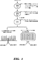
【図面の簡単な説明】
図1は、電気泳動またはアドレス可能アレイ上の捕捉による点変異などの生殖細胞系変異の検出のための1次PCR/2次PCR/LDRプロセスを表す流れ図である。なお、図1および他の図における用語“ジップコード”は、後に用いられるプライマーまたはプローブの特異的な配列を意味するが、標的配列または他のゲノム配列を意味しない。
図2は、電気泳動またはアドレス可能アレイ上の捕捉によるバイ対立遺伝子多形性の検出のための1次PCR/2次PCR/LDRプロセスを表す流れ図である。
図3は、電気泳動またはアドレス可能アレイ上の捕捉による癌関連変異の検出のための1次PCR/2次PCR/LDRプロセスを表す流れ図である。
図4は、バイ対立遺伝子多形性の検出のための1次PCR/2次PCR/LDRプロセスを表す模式図である。
図5は、与えられた部位ですべての可能性ある塩基を識別するLDRオリゴヌクレオチドプローブを用いて、対立遺伝子の相違の検出のための1次PCR/2次PCR/LDRプロセスを表す模式図である。
図6は、与えられた部位ですべての可能性ある塩基を識別するLDRオリゴヌクレオチドプローブを用いて、2つの近接する部位の可能性ある塩基の存在を検出のための1次PCR/2次PCR/LDRプロセスを表す模式図である。
図7は、隣接する対立遺伝子での癌関連変異の検出のための1次PCR/2次PCR/LDRプロセスを表す模式図である。
図8は、電気泳動検出を用いて制限エンドヌクレアーゼ消化の有無でのLDR/PCRプロセスを表す流れ図である。
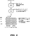
図9は、遺伝子特異的アドレスを用いるアドレス可能アレイでの検出のためのLDR/PCRプロセスを表す流れ図である。
図10は、遺伝子増幅および欠失の多重検出のためのLDR/PCRプロセスを表す模式図である。
図11は、LDR/PCRプロセスのための対立遺伝子特異的問題を表す模式図である。
図12は、図11に表されたLDR/PCRプロセスのための対立遺伝子特異的問題の解決を表す模式図である。
図13は、電気泳動またはアドレス可能アレイ上の捕捉による2対立遺伝子多形性の検出のための中間的エクソヌクレアーゼ消化相を有するLDR/PCRプロセスを表す流れ図である。
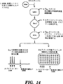
図14は、電気泳動またはアドレス可能アレイ上の捕捉による癌関連変異の検出のための中間的エクソヌクレアーゼ消化相を有するLDR/PCRプロセスを表す流れ図である。
図15は、対立遺伝子特異的変異および多形性の検出のための中間的エクソヌクレアーゼ消化相を有するLDR/PCRプロセスを表す模式図である。
図16は、モノヌクレオチド反復多形性検出のための中間的エクソヌクレアーゼ消化相を有するLDR/PCRプロセスを表す模式図である。
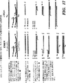
図17は、低存在量のモノヌクレオチド反復多形性検出のための中間的エクソヌクレアーゼ消化相を有するLDR/PCRプロセスを表す模式図である。
図18は、中間的配列伸長相、およびLDR相の後でPCR相の前でのウラシルN−グリコシラーゼ消化相を有し、および電気泳動またはアドレス可能アレイによる検出を有するLDR/PCRプロセスを用いての多形性検出を表す流れ図である。
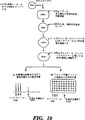
図19は、中間的配列伸長相、およびLDR相の後でPCR相の前でのウラシルN−グリコシラーゼ消化相を有し、および電気泳動またはアドレス可能アレイによる検出を有するLDR/PCRプロセスを用いての癌検出を表す流れ図である。
図20は、中間的配列伸長相、およびLDR相の後でPCR相の前でのウラシルN−グリコシラーゼ消化相を有するLDR/PCRプロセスを用いてのモノヌクレオチド反復体の検出を表す模式図である。
図21は、中間的配列伸長相、およびLDR相の後でPCR相の前でのウラシルN−グリコシラーゼ消化相を有するLDR/PCRプロセスを用いての低豊富であるモノヌクレオチド反復多形性の検出を表す模式図である。
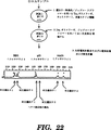
図22は、ミクロサテライト反復体の検出のための1次PCR/2次PCRプロセスを表す流れ図である。
図23は、ミクロサテライト反復体における挿入および欠失の多重検出のための1次PCR/2次PCRプロセスを表す模式図である。
図24は、LDR/PCRプロセスにおける遺伝子増幅および欠失の定量のためのLDRオリゴヌクレオチドプローブの設計を示す。
図25A−Dは、LDR/PCRプロセスについての電気泳動濃度を示す。
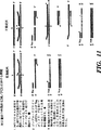
図26A−Cは、正常女性DNA、肺癌細胞系ZR−75−30 DNAおよび消化器癌細胞系SKGT−2に由来するErbB、G6PD、Int2、p53およびSOD遺伝子セグメントについてのLDR/PCRプロセスの電気泳動濃度を示す。ErbB遺伝子は癌細胞系で増幅することが知られている。104bpの標的特異的連結反応産物配列は、各連結反応プライマー500フェムトモル、ゲノムDNA50ng、Thermus thermophillus(“Tth”)リガーゼ124単位、10×緩衝液2μg(0.2M Tris、pH8.5および0.1M MgCl2)、10mM NAD2μgおよび200mM DTT 1μlを容量20μlで用いて、LDRの10サイクル(94℃で30秒、65℃で4分)において生成せしめる。連結反応産物は、10×ストフェル緩衝液(Perkin Elmer)5μl、各オリゴヌクレオチドプライマー25ピコモル、Taqポリメラーゼ・ストフェル・フラグメント25単位および各dNTP中の5mM溶液8μlを含有する溶液30μlを加えて、PCRの26サイクル(94℃で15秒、65℃で50秒)において比較的に増幅される。増幅後、産物をHaeIIIおよびHin P1Iで消化し、58bp(ErbB(すなわちHER−2/neu/erbBオンコジーン)、61bp(G6PD)、67bp(Int2(すなわちint−2オンコジーン))、70bp(p53)および76bp(SOD))のFAM標識産物を産生する。これらの産物を分離し、ジーンスキャン672ソフトウェア・パッケージ(Applied Biosystems,Inc.,Foster City,Calif.)を用いて373A DNAシクエンサー上で解析する。結果が電気泳動濃度図に表され、ピークの高さと面積がPCR産物の量を示している。図26Aは、正常女性DNAにおける5部位について遺伝子量測定を示す。G6PD、Int2、p53およびSODについてのピークの高さおよび面積は非常に類似している。FrbBについてのピークの高さおよび面積は正常ゲノムDNAにおいて明らかに小さい。図26Bは、既知のErbB増幅での細胞系、ZR−75−30において遺伝子量を検査したので、ErbBでのピークが高く、面積が大きいことを示している。図26Cは、消化器細胞系であるSKGT−2においてErbB遺伝子の劇的な増幅およびInt2の中程度の増幅があることを示している。G6PD遺伝子ピークは、大きいErbBピークにのみこまれたかも知れない。
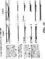
図27A−Cは、G6PD、Int2、p53およびSODについて他のLDRオリゴヌクレオチドプローブおよびPCRオリゴヌクレオチドプライマーの相対的ピークの高さにErbB増幅が影響するかどうかを測定するためのLDR/PCRプロセスについての電気泳動濃度図を示す。図27Aは、正常女性DNAにおける4部位についての遺伝子量測定を示す。全5LDRプライマーを用いた実験でのようにG6PD、Int2、p53およびSODについてのピークの高さおよび面積は類似している。図27Bは、ZR−75−30乳癌細胞系のG6PD、Int2、p53およびSODが正常女性DNAに比較して類似の相対ピークの高さであることを示している。p53についてのピークは低下し、この細胞系における細胞部分でのこの遺伝子の欠失を示唆している。図27Cは、消化器癌細胞系のSKGT−2において、G6PDおよびp53が同等のピークを有することを示している。全5LDRオリゴヌクレオチドプローブを用いた実験でのように、Int2のピークは比較的高い。このように、各産物のLDRおよびPCR増幅は、実験中において他の産物と独立的とみられる。
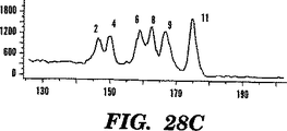
図28A−Cは、1次PCR/2次PCR/LDRプロセスのPCR相についての電気泳動濃度図を示す。1次PCRオリゴヌクレオチドプライマーを用いての12部位の多重PCR増幅は大略等量の産物をつくる。単一塩基多形性を有する80以上の遺伝子領域がヒトゲノム・データベースから同定された。そのうち12(表10および図29A−H参照)は、1次PCR相において次のように増幅された。2次PCRオリゴヌクレオチドプライマーの2セットの1つに相補的な遺伝子特異的3’末端および5’末端を有するように、長い1次PCRオリゴヌクレオチドプライマーを設計した。上流1次PCRオリゴヌクレオチドプライマーはFAM(すなわち、6−カルボキシフルオレセイン:配列決定および変異検定に用いられる蛍光染料)またはTET(すなわち、テトラクロリネート−6−カルボキシフルオレセイン:配列決定/変異検定に用いられる蛍光染料)のいずれかでの蛍光標識を持つように合成した。全24塩基の長い1次PCRオリゴヌクレオチドプライマーを低濃度(20μl中各プライマー2ピコモル)で15サイクルの1次PCR相に用いた。その後、2次PCRオリゴヌクレオチドプライマーの2セットを高濃度(各25ピコモル)で加え、2次PCR相をさらに25サイクル行った。産物を373DNAシークエンサー(Applied Biosystems)で分離した。パネルAは、FAM−およびTET−標識産物について合わせた電気泳動濃度図を示す。パネルBは、FAM標識産物のみを示す。パネルCは、TET標識産物のみを示す。このプロセスは、プライマー濃度またはPCR条件を注意深く調節する必要なしに、多重的産物について同様の量を産生する。
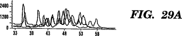
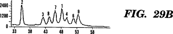
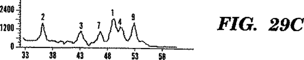
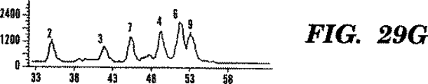
図29A−Hは、法医学的同定のための12バイ対立遺伝子の検出における1次PCR/2次PCR/LDRプロセスのLDR相についての電気泳動濃度図を表す。12多形性遺伝子についての1次および2次PCR相は、図28A−C記載のように行った。しかし、2次PCRオリゴヌクレオチドプライマーを蛍光標識しなかった。2次PCRプロセス伸長産物を36LDR・オリゴヌクレオチドプローブ(各部座につき1通常および2識別プライマー)含有のリガーゼ緩衝液中に希釈した。LDRオリゴヌクレオチドプローブセットを次の2方法で設計した。(i)対立遺伝子特異的オリゴヌクレオチドプローブは同じ長さであるが、FAMかTET標識のいずれかを含有する、(ii)対立遺伝子特異的オリゴヌクレオチドプローブ両者をHEX(すなわち、ヘキサクロリネート−6−カルボキシフルオレセイン:配列決定および変異検出に用いられる蛍光染料)で標識するが、長さに2塩基対の相違がある。LDR相の20サイクル後、連結反応産物配列を373DNAシークエンサー(Applied Biosystems)で10%ポリアクリルアミド・シークエンシング・ゲルにより溶解した。パネルAおよびEは、2個体の12座PCR/LDRを示す。パネルB、CおよびDは夫々、パネルAにおける個体についてのFAM、TETおよびHEXを示す。パネルAの個体は、座6(ALDOB(すなわちアルドラーゼB))および座8(IGF(すなわちインスリン生長因子))でのみ同型接合である。パネルEの個体は、座3(C6(すなわち、補体成分C6))、座5(NF1(すなわち、神経線維腫症))、座6(ALDOB)および座8(IGF)でのみ異型接合であった。このことから、1次PCR/2次PCR/LDRプロセスが多数の位置における同型接合および異型接合の両方の遺伝子型を同時に識別し得ることが分かる。
発明の詳細な記述
I.1次PCR/2次PCR/LDRプロセス
本発明の一つの実施態様は、複数の標的ヌクレオチド配列における1以上の単一塩基の変更、挿入、欠失または転座により相違している複数の配列の2以上を同定するための方法に関する。この方法は、第1ポリメラーゼ相、第2ポリメラーゼ相およびリガーゼ検出相を含む。このプロセスは、複数の配列相違がある1以上の標的ヌクレオチド配列を含有するかも知れないサンプルを分析することを含む。
第1ポリメラーゼ連鎖反応相において、1以上の1次オリゴヌクレオチドプライマー群が準備される。各群は1以上の1次オリゴヌクレオチドプライマー・セットを含有し、各セットは、標的特異的部分と5’上流2次プライマー特異的部分を有する第1ヌクレオチドプライマー、および標的特異的部分と5’上流2次プライマー特異的部分を有する第2オリゴヌクレオチドプライマーを含む。同じ群の各セットの第1オリゴヌクレオチドプライマーは同じ5’上流2次プライマー特異的部分を有し、同じ群の各セットの第2オリゴヌクレオチドプライマーは同じ5’上流2次プライマー特異的部分を有している。特定のセットのオリゴヌクレオチドプライマーは、対応する標的ヌクレオチド配列の相補的鎖でのハイブリダイゼーションに適しており、ポリメラーゼ連鎖反応産物の形成をもたらす。しかし、1次オリゴヌクレオチドプライマーがサンプル中に他のヌクレオチド配列にハイブリダイズするときに、該ポリメラーゼ連鎖反応産物の形成に妨害するミスマッチがある。特定のセットにおけるポリメラーゼ連鎖反応産物は、同じ群での他のポリメラーゼ連鎖反応産物と識別され得る。1次オリゴヌクレオチドプライマー、サンプルおよびポリメラーゼは混合されて、1次ポリメラーゼ連鎖反応混合物を形成する。
1次ポリメラーゼ連鎖反応混合物は、実質的に上記したように、変性処理、ハイブリダイゼーション処理および伸長処理を含む2以上のポリメラーゼ連鎖反応サイクルにかけられる。ハイブリダイゼーションの際に、1次オリゴヌクレオチドプライマーの標的特異的部分が標的ヌクレオチド配列にハイブリダイズする。伸長処理によってハイブリダイズした1次オリゴヌクレオチドプライマーが伸長されて、1次オリゴヌクレオチドプライマーがハイブリダイズする標的ヌクレオチド配列に相補的な1次伸長産物を形成する。
1次オリゴヌクレオチドプライマーの上流2次プライマー特異的部分は標的DNAに存在しないが、その配列は1次ポリメラーゼ連鎖反応相の第2以後のサイクルで複写される。結果として、第2サイクル後に産生された1次伸長産物は、その5’末端に2次プライマー特異的部分、およびその3’末端にプライマー特異的部分の補体を有する。
次は、第2ポリメラーゼ連鎖反応相である。この相では1つまたは複数の2次オリゴヌクレオチドプライマーセットが準備される。各セットは、第1の1次オリゴヌクレオチドプライマーの5’上流部分と同じ配列を含有する第1の2次オリゴヌクレオチドプライマー、および第1の2次プライマーに相補的な第1の1次オリゴヌクレオチドと同じ1次オリゴヌクレオチドプライマーセットに由来する第2の1次オリゴヌクレオチドプライマーの5’上流部分と同じ配列を含有する第2の2次オリゴヌクレオチドプライマーを有する。2次オリゴヌクレオチドプライマーのセットは、特定の群において1次伸長産物のすべてを増幅するのに使用され得る。2次オリゴヌクレオチドプライマーを1次伸長産物およびポリメラーゼと混合して、2次ポリメラーゼ連鎖反応混合物とする。
2次ポリメラーゼ連鎖反応混合物は、実質的に上記したように、変性処理、ハイブリダイゼーション処理および伸長処理を含む2以上のポリメラーゼ連鎖反応サイクルにかけられる。ハイブリダイゼーションの際に、2次オリゴヌクレオチドプライマーの標的特異部分が1次伸長産物上に存在する相補的配列にハイブリダイズし、元の標的配列にハイブリダイズしない。伸長処理によってハイブリダイズ2次オリゴヌクレオチドプライマーが伸長されて、1次伸長産物に相補的な2次伸長産物を形成する。
本発明の態様における最後の相はリガーゼ検出反応相である。ここでは、複数のオリゴヌクレオチドプローブセットが準備され、各セットは、2次伸長産物特異的部分と検出可能レポーター標識を保持する第1オリゴヌクレオチドプローブ、および2次伸長産物特異的部分を保持する第2オリゴヌクレオチドプローブを有する。特定セットのオリゴヌクレオチドプローブは、相補的2次伸長産物特異的部分に互いに隣接してハイブリダイズしたときに一緒に連結反応するのに適している。しかし、オリゴヌクレオチドプローブがサンプル中の他のヌクレオチド配列にハイブリダイズするときに該連結反応を妨害するミスマッチがある。特定セットにおけるオリゴヌクレオチドプローブの連結反応産物は、プローブまたは他の連結反応産物のいずれかと識別され得る。複数のオリゴヌクレオチドプローブセットを2次伸長産物およびリガーゼと混合して、リガーゼ検出反応混合物とする。
リガーゼ検出反応混合物は、実質的に上記したように、変性処理およびハイブリダイゼーション処理を有する1以上のリガーゼ検出反応サイクルにかけられる。ハイブリダイゼーション処理において、オリゴヌクレオチドプローブセットは、隣接部位で塩基特異的に、もし存在すれば夫々の2次伸長産物にハイブリダイズする。結果として、隣接プローブは互いに連結して、一緒に結合している検出可能レポーター標識および2次伸長産物特異的部分を含有する連結産物配列を形成する。オリゴヌクレオチドプローブセットは、その夫々の相補的2次伸長産物以外のヌクレオチド配列にハイブリダイズし得るが、1以上のミスマッチの存在のために一緒に連結しないで、変性処理の際に個々に分離している。リガーゼ検出反応サイクルに続いて、連結反応産物配列のレポーター標識が検出されて、サンプル中の1以上の標的ヌクレオチド配列の存在が示される。
図1、2および3は2種の検出手法のいずれかを用いた本発明の1次PCR/2次PCR/LDRプロセスを表す流れ図である。1つの方法として、毛管電気泳動またはゲル電気泳動と蛍光定量の使用がある。他方、捕捉オリゴヌクレオチドアドレスのアレイ上の捕捉と蛍光定量によって検出が実施される。図1は生殖細胞系変異(例えば、点変異)の検出に関し、図2はバイ対立遺伝子多形性の検出であり、図3は癌関連変異の検出を示す。
図1は生殖系点変異の検出を表す。第1工程において、DNAサンプル作成後、Taq(すなわちThermus aquaticus)ポリメラーゼを用いた1次PCR増幅に、ホットスタート条件で、標的特異的部分と2次プライマー特異的部分とを有するオリゴヌクレオチドプライマーと共に、多重エクソンをかける。1次PCR相の終了点で、Taqポリメラーゼは、100℃で10分間加熱するか、または凍結/解凍工程により不活性化される。1次PCR増幅相の産物は、第2工程でTaqポリメラーゼを用いホットスタート条件で2次オリゴヌクレオチドプライマーと共に、2次PCR増幅にかける。2次PCR相の終了点で、Taqポリメラーゼは、100℃で10分間加熱するか、または凍結/解凍工程により不活性化される。第3工程において、2次PCR相の産物は、対立遺伝子特異的部分と共通部分を有するLDRオリゴヌクレオチドプローブを含有する新鮮なLDR緩衝液に20倍に希釈する。第4工程は、LDR相であって、ホットスタート条件でTaqリガーゼを加えることにより始まる。LDRの際にオリゴヌクレオチドプローブはその隣接のオリゴヌクレオチドプローブに、接合部で完全な相補性を与える標的配列の存在においてのみ、連結する。
産物は2種の相違するホーマットにおいて検出される。第1ホーマット5aでは、蛍光標識LDRプローブが相違する長さのポリAまたはヘキサエチレンオキシド・テイルを含有する。このように各連結反応産物配列(正常DNA上でハイブリダイズされた2プローブの連結反応の結果である)は、いくつかの連結反応産物配列がピークの段差を与えるように、少し相違する長さと運動性を有する。生殖細胞系変異は電気泳動濃度図上に新しいピークをつくることがある。他方、生殖細胞系変異連結反応産物配列が正常DNA連結反応産物配列と同じ運動性を有して泳動するが、相違する蛍光レポーターで識別されるようにLDRプローブは設計され得る。新しいピークのサイズはほぼ元のサンプルに存在する変異の量である。すなわち、ホモ接合正常は0%、ヘテロ接合保持は50%、そしてホモ接合変異は100%である。第2ホーマット5bにおいて、各対立遺伝子特異的プローブは、例えばその5’末に24追加ヌクレオチド塩基を含有する。これらの配列はユニーク・アドレス可能配列であって、アドレス可能アレイ上でその相補的アドレス配列に特異的にハイブリダイズする。LDR反応において、各対立遺伝子特異的プローブは、対応する標的配列の存在において隣接の蛍光標識共通プローブに連結できる。野生型および変異の対立遺伝子に対応する連結反応産物配列は、アレイ上の隣接アドレスに捕捉される。未反応のプローブは洗浄して除く。黒点は野生型対立遺伝子についての100%シグナルを表す。白点は変異対立遺伝子についての0%シグナルを表す。灰色点は生殖細胞系変異の1つの位置、各対立遺伝子についての50%シグナルを表す。
図2は、バイ対立遺伝子多形性の検出を表す。第1工程において、Taqポリメラーゼを用いた1次PCR増幅に、ホットスタート条件で、標的特異的部分と2次プライマー特異的部分とを有するオリゴヌクレオチドプライマーと共に、多重エクソンをかける。1次PCR増幅相の産物は、第2工程でTaqポリメラーゼを用いホットスタート条件で2次オリゴヌクレオチドプライマーと共に、2次PCR増幅にかける。2次PCR相の終了点で、Taqポリメラーゼは、100℃で10分間加熱するか、または凍結/解凍工程により不活性化される。第3工程において2次PCR相の産物は対立遺伝子特異的部分と共通部分を有するLDRオリゴヌクレオチドプローブを含有する新鮮なLDR緩衝液に20倍に希釈する。第4工程は、LDR相であって、ホットスタート条件でTaqリガーゼを加えることにより始まる。LDRの際にオリゴヌクレオチドプローブはその隣接のオリゴヌクレオチドプローブに、接合部で完全な相補性を与える標的配列の存在においてのみ、連結する。第1ホーマット5aでは、蛍光標識LDRプローブが相違する長さのポリAまたはヘキサエチレンオキシド・テイルを含有する。各連結反応産物は、いくつかの連結反応産物配列がピークの段差を与えるように、少し相違する長さと運動性を有する。他方、多形性対立遺伝子の連結反応産物配列が同じ位置で移動するが、相違する蛍光レポーターで識別されるようにLDRプローブは設計され得る。ピークのサイズはほぼ各対立遺伝子の量である。第2ホーマット5bにおいて、各対立遺伝子特異的プローブは、例えばその5’末に24の追加ヌクレオチド塩基を有するユニーク・アドレス可能配列を含有する。これらの配列は、捕捉オリゴヌクレオチドアレイ上でその相補的アドレス配列に特異的にハイブリダイズする。LDR反応において、各対立遺伝子特異的プローブは、対応する標的配列の存在において隣接の蛍光標識共通プローブに連結できる。各対立遺伝子に対応する連結反応産物配列は、アレイ上の隣接アドレスに捕捉される。未反応のプローブは洗浄して除く。黒点は両染色体が与えられた対立遺伝子を有すことを表す。白点はいずれの染色体もこの対立遺伝子を有さないことを表す。灰色点は1つの染色体が与えられた対立遺伝子を有することを表す。
図3は、癌関連変異を表す。第1工程において、DNAサンプル作成後、Taqポリメラーゼを用いた1次PCR増幅に、ホットスタート条件で、標的特異的部分と2次プライマー特異的部分とを有するオリゴヌクレオチドプライマーと共に、多重エクソンをかける。1次PCR増幅相の産物は、第2工程でTaqポリメラーゼを用いホットスタート条件で2次オリゴヌクレオチドプライマーと共に、2次PCR増幅にかける。2次PCR相の終了点で、Taqポリメラーゼは、100℃で10分間加熱するか、または凍結/解凍工程により不活性化される。PCR産物の蛍光定量は第3工程において毛管またはゲル電気泳動により行うことができる。第4工程において、産物はマーカーDNAの1/100希釈(フラグメントの各々につき)でスパイクされる。このDNAは、癌サンプルにみられないが、適当なLDRプローブで容易に検出される変異を有することを除けば、野生型DNAに相同的である。第5工程において、2次PCR相産物中の混合DNA産物は、対立遺伝子特異的部分と共通部分を含有するLDRオリゴヌクレオチドプローブを含む新鮮なLDR緩衝液に20倍に希釈される。第6工程は、LDR相であって、ホットスタート条件でTaqリガーゼを加えることにより始まる。LDRの際に、接合部に完全な相補性を与える標的配列の存在においてのみ、オリゴヌクレオチドプローブがその隣接のオリゴヌクレオチドプローブに連結する。
産物は上記と同じホーマットで検出される。第7a工程のホーマットにおいて、毛管またはゲル電気泳動により産物が分離され、そして蛍光シグナルが定量される。変異ピークのマーカーピークに対する割合は、元のサンプル中に存在する癌変異の概量を100で除して算出される。第7b工程のホーマットにおいて、アドレス可能アレイ上の相補的配列についての特異的ハイブリダイゼーションによって産物が検出される。マーカー点に対する変異点中の蛍光シグナルの割合は、元のサンプル中に存在する癌変異の概量を100で除して算出される。
図4に示すように、興味ある2種のDNAフラグメントを本発明の1次PCR/2次PCR/LDRプロセスで処理する。最初に2重鎖DNA分子を変性して、鎖をほどく。これは80−105℃に加熱することによりなされる。3−標的特異的部分(影の部分)と5’2次プライマー特異的部分(黒い部分)を有する1次PCRオリゴヌクレオチドプライマーを低濃度で加えて、典型的に50−85℃で鎖にハイブリダイズせしめる。熱安定性ポリメラーゼ(例えば、Taq aquanticusポリメラーゼ)も加え、温度を50−85℃に調節して、プライマーがハイブリダイズする核酸の長さに沿ってプライマーを伸長する。ポリメラーゼ連鎖反応の伸長相の後に、得た2重鎖分子を80−105℃に加熱して、分子を変性し、鎖を分離する。これらのハイブリダイゼーション、伸長、変性工程は数回繰り返して、標的を適当なレベルにまで増幅する。
2次PCR相において、1次PCR相産物を2次PCRオリゴヌクレオチドプライマーと混合して、典型的には50−85℃で互いにハイブリダイズせしめる。2次オリゴヌクレオチドプライマーは1次オリゴヌクレオチドプライマーよりも常に高い濃度で使用する熱安定性ポリメラーゼも加え、温度を50−85℃に調節して、プライマーがハイブリダイズする核酸の長さに沿ってプライマーを伸長する。ポリメラーゼ連鎖反応の伸長相の後に、得た2重鎖分子を80−105℃に加熱して、分子を変性し、鎖を分離する。これらのハイブリダイゼーション、伸長、変性工程は数回繰り返して、標的を適当なレベルにまで増幅する。
2次PCR相が完了すると、図4に示すように、連結検出反応相が始まる。標的核酸変性後、もし存在すれば二重鎖DNA分子として、温度80−105℃、好ましくは94℃で、標的ヌクレオチド配列の1本鎖のための連結検出反応オリゴヌクレオチドプローブがリガーゼと共に加えられる(例えば、図4に示すように、Taq aquanticusポリガーゼのような熱安定性リガーゼ)。オリゴヌクレオチドプローブは標的核酸分子とハイブリダイズし得て、典型的には45−85℃、好ましくは65℃で連結する。連結部に完全な相補性があるとオリゴヌクレオチドは一緒に連結する。変わり得るヌクレオチドがTまたはAであると、標的ヌクレオチド配列におけるTの存在は、F1レポーター標識を有するオリゴヌクレオチドプローブを5’ポリAテイルAnを有する共通オリゴヌクレオチドプローブと連結せしめ、標的ヌクレオチド配列中のAの存在は、F2レポーター標識を有するオリゴヌクレオチドプローブをAnを有する共通のオリゴヌクレオチドプローブと連結せしめる。同様に、変わり得るヌクレオチドがAまたはGであると、標的ヌクレオチド配列におけるTの存在は、F3AAレポーター標識(すなわち、5’ポリAスペーサーを形成する2追加塩基にカップルしたF3レポーター標識)を有するオリゴヌクレオチドプローブを5’ポリAテイルAn+4を有する共通オリゴヌクレオチドプローブと連結せしめ、標的ヌクレオチド配列中のCの存在は、F3レポーター標識を有するオリゴヌクレオチドプローブをAn+4を有する共通のオリゴヌクレオチドプローブと連結せしめる。連結反応後に材料を変性にかけてハイブリダイズ鎖を分離する。ハイブリダイゼーション/連結反応および変性工程は1以上のサイクル(例えば1−50サイクル)で実施されて、標的シグナルが増幅される。両F3−標識オリゴヌクレオチドの等モル連結反応は個体がその座についてヘテロ接合型であることを示し、一方、F2標識オリゴヌクレオチドの連結反応は個体が他の座についてホモ接合型であることを示す。
図4において、定量が毛管またはゲル電気泳動で実施されるとき、ポリAnおよびポリAn+4テイルが用いられる。異なった長さのテイルによって、対応する相違の連結反応産物配列がゲルまたは毛管における異なる位置でバンドを形成する。異なる位置でのバンドの存在は、解析されるDNAにおける対応ヌクレオチド相違の同定を可能にする。連結反応産物配列が異なるレポーター標識の使用に基づいて識別できるけれども、異なるレポーター標識と異なる長さのテイルの組み合せは、より多数のヌクレオチド相違の識別を可能とする。これは多重検出プロセスに重要である。
図1−3について述べたように、ゲルまたは毛管電気泳動に代わって、アドレス可能アレイ上での検出を実施できる。かかるアドレス可能アレイの使用には、レポーター標識(すなわちテイルAnおよびAn+4)を含有しないLDRオリゴヌクレオチドプローブ上のポリAが相違するアドレス可能アレイ特異的オリゴヌクレオチド部分で置き換えられることを要する。上記でより完全に説明したように、固体支持物は捕捉オリゴヌクレオチドのアレイと共に準備され、そのいくつかは相違するアドレス可能アレイ特異的オリゴヌクレオチド部分に相補的である。相補的捕捉オリゴヌクレオチドプローブのこれらの部分のハイブリダイゼーションは対応するヌクレオチド相違の存在を示す。
図5は、本発明のよる1次PCR/2次PCR/LDRプロセスについての模式図であり、興味ある2DNA分子における可能性ある塩基が識別される。1次および2次PCRプロセスは、図4で述べたのと実質的に同じように実施される。左手のDNA分子と併せた蛍光レポーター標識F1、F2、F3およびF3の出現は、夫々DNA分子におけるA、G、CおよびT対立遺伝子の存在を表す。図5に示すように、等量のF1およびF3レポーター標識は問題の個体がAおよびC対立遺伝子についてヘテロ接合型であることを表す。図5における右手のDNA分子の分析について、同じレポーター標識が異なる対立遺伝子の存在を表すのに用いられる。しかし、識別塩基を有する各オリゴヌクレオチドプローブ上で、異なる5’ポリAテイルが存在する。より特異的に2単位ポリAテイル、4単位ポリAテイル、6単位ポリAテイルおよび8単位ポリAテイルが、夫々DNA分子におけるT、C、GおよびA対立遺伝子に対応する。図5に示すように、A6およびA4テイルを有する等量のF1レポーター標識は、問題の個体がGおよびC対立遺伝子についてヘテロ接合型であることを表す。
図6は、図5は、本発明のよる1次PCR/2次PCR/LDRプロセスについての模式図であり、興味ある2DNA分子における2近接部位で可能性ある塩基が識別される。1次および2次PCRプロセスは、図4で述べたのと実質的に同じように実施される。ここではLDRプローブは重複可能であり、接合部で完全な相補性が存在すると連結がなお可能である。これがLDRを他の方法と区別することであって、対立遺伝子特異的PCRなどでは、重複プライマーが互いに妨害し合う。図6において、識別オリゴヌクレオチドプローブは、そのプローブの3末に識別塩基を有するレポーター標識を含有する。ポリAテイルは共通オリゴヌクレオチドプローブの3’末上にある。左手のDNA分子において、レポーター標識F1およびF3を有する連結反応産物配列の等量の存在は、問題の個体が第1位置でAおよびC対立遺伝子についてヘテロ接合であることを示す。同様に、左手のDNA分子の第2位置において、レポーター標識F2、F3およびF4を有する連結反応産物配列の存在は、問題の個体がG、CおよびT対立遺伝子についてヘテロ接合であることを示す。
右手のDNA分子にもどって、A6およびA4テイルを持つレポーター標識F1を有する連結反応産物配列の等量の存在は、問題の個体が第1位置でGおよびC対立遺伝子についてヘテロ接合であることを示す。右手のDNA分子の第2位置において、A8およびA2テイルを持つレポーター標識F1を有する連結反応産物配列の等量の存在は、問題の個体がAおよびT対立遺伝子についてヘテロ接合であることを示す。
図7は、正常配列の過剰存在で低存在量の変異を検出するための本発明の1次PCR/2次PCR/LDRプロセスを表す模式図である。左手のDNA分子はK−ras遺伝子のコドン12、配列GGTでグリシン(“Gly”)をコードする。小さい比率の細胞がGATにおけるGからAへの変異を含有し、アスパラギン酸(“Asp”)をコードする。野生型(すなわち正常)配列についてのLDRプローブが反応から抜けている。正常LDRプローブ(Gである識別塩基と共)が挿入されると、このプローブは共通プローブに連結し、変異標的由来のいかなるシグナルにも打ち勝つ。代わりに、図7に示されるように、蛍光標識F1およびAn+2テイルを有する連結反応産物配列の存在は変異をコードするアスパルギン酸の存在を表している。右手のDNA分子において、K−ras遺伝子のコドン61配列CAGがあり、グルタミン(“Gln”)をコードする。小さい比率の細胞がGAGにおけるCからGへの変異を含有し、グルタミン酸(“Glu”)をコードする。さらに、LDRオリゴヌクレオチドプローブが野生型に見られるCおよびA塩基を含まず、変異シグナルに打ち勝つことはない。このDNA分子について、蛍光標識F2An+4テイルを有する連結反応産物配列の存在は変異をコードするグルタミン酸の存在を表す。
II.LDR/PCRプロセス
本発明の第2態様は、複数の標的ヌクレオチド配列における1以上の単一塩基の変更、挿入、欠失および転座によって相違している1以上の複数配列を同定する方法に関する。この方法はリガーゼ検出反応相に続いてポリメラーゼ連鎖反応相を有する。この方法は、複数の配列相違を有する1以上の標的ヌクレオチド配列を含有する可能性のあるサンプルを準備することを含む。
リガーゼ検出反応相において1以上のオリゴヌクレオチドプローブが準備される。各セットは、標的特異的部分と5’上流プライマー特異的部分を保持する第1オリゴヌクレオチドプローブ、および標的特異的部分と3’下流プライマー特異的部分を保持する第2オリゴヌクレオチドプローブを有する。特定セットにおける折後ヌクレオチドプローブは、対応する標的ヌクレオチド配列に互いに隣接してハイブリダイズするときに一緒に連結反応するのに適している。しかし、サンプル中に存在する他のヌクレオチド配列とハイブリダイズするときに、この連結反応を妨害するミスマッチがある。サンプル、複数のオリゴヌクレオチドプローブセットおよびリガーゼを混合して、リガーゼ検出反応混合物とする。
リガーゼ検出反応混合物は1以上のリガーゼ検出反応サイクルにかけられる。これらのサイクルには変性処理とハイブリダイゼーション処理が含まれる。変性処理において、ハイブリダイズされたオリゴヌクレオチドは標的ヌクレオチド配列から分離される。ハイブリダイゼーション処理によって、オリゴヌクレオチドプローブセットは隣接部位で塩基特異的に、もしサンプル中に存在しておればその夫々の標的ヌクレオチド配列にハイブリダイズする。ハイブリダイズされると、オリゴヌクレオチドプローブセットは互いに連結して、連結反応産物配列を形成する。この産物は、5’上流プライマー特異的部分、一緒に結合した標的特異的部分および3’下流プライマー特異的部分を含む。各セットの連結反応産物配列は連結検出反応混合物中のたの核酸と識別可能である。オリゴヌクレオチドプローブセットはその夫々の標的ヌクレオチド配列以外のサンプル中のヌクレオチド配列とハイブリダイズするが、1以上のミスマッチの存在によって連結せず、そして、次の変性処理の際に個々に分離している。
ポリメラーゼ連鎖反応において、1または複数のオリゴヌクレオチドプローブが準備される。各セットは、連結反応産物配列の5’上流プライマー特異的部分に同じ配列を含有する上流プライマー、および連結反応産物配列の3’下流プライマー特異的部分に相補的な下流プライマーを有している。うち、1つのプライマーは検出可能レポーター標識を有する。リガーゼ検出反応混合物を1または複数のオリゴヌクレオチドプローブセットおよびポリメラーゼと混合すると、ポリメラーゼ連鎖反応混合物が形成する。
ポリメラーゼ連鎖反応混合物は、変性処理、ハイブリダイゼーション処理および伸長処理を含む1以上のポリメラーゼ連鎖反応サイクルにかけられる。変性処理においてハイブリダイズ核酸配列が分離される。ハイブリダイゼーション処理によって、プライマーが連結反応産物配列の相補的プライマー特異的部分とハイブリダイズする。伸長処理において、ハイブリダイズプライマーが伸長されて、プライマーがハイブリダイズしている配列に相補的な伸長産物を形成する。ポリメラーゼ連鎖反応相の第1サイクルにおいて、下流プライマーが連結反応産物配列の3’下流プライマー特異的部分にハイブリダイズし、伸長されて連結反応産物配列に相補的な伸長産物を形成する。次のサイクルにおいて、上流プライマーが連結反応産物に相補的な伸長産物の5’上流プライマー特異的部分にハイブリダイズし、下流プライマーが連結反応産物配列の3’下流部分にハイブリダイズする。
このプロセスのポリメラーゼ連鎖反応相に続いて、レポーター標識が検出され、伸長産物が識別されて、サンプル中の1以上の標的ヌクレオチド配列の存在が表される。
図8は、制限エンドヌクレアーゼ消化の有無および毛管電気泳動検出を用いた本発明のLDR/PCRプロセスを表す流れ図である。第1工程において、DNAサンプルは、Taqリガーゼ、および標的特異的部分およびプライマー特異的部分を含有するオリゴヌクレオチドプローブと混合する。混合物はLDRプロセスにかけられて、連結標的特異的部分およびプライマー特異的部分を含有する連結反応産物配列を産生する。第2工程で、連結反応産物配列をポリメラーゼおよびプライマーと混合し、この混合物をPCRプロセスにかける。次の工程で連結反応産物配列が同じかまたは相違するサイズであるかの機能について測定する。連結反応産物配列が相違するサイズであると、第3a工程が選ばれてPCRからの伸長細胞が毛管電気泳動またはゲル電気泳動にかけられる。いずれも続いて蛍光定量がなされる。連結反応産物配列が同じサイズのときは第3b工程が用いられ、PCR相からの伸長産物は制限エンドヌクレアーゼ消化にかけられる。毛管電気泳動またはゲル電気泳動にかけられるユニーク・サイズの消化フラグメントが産生され、第4b工程による蛍光定量が行われる。第3a工程が選択されたときは、電気泳動の結果のカーブは、104、107および110に移行する3種の連結反応産物配列があり、ピーク域はそれぞれHer2遺伝子増幅、p53遺伝子のヘテロ接合の喪失およびコントロールSOD遺伝子を表す。第3bおよび4b工程による電気泳動曲線は、3種の連結反応産物配列制限フラグメントを58、70および76に有し、ピーク域はそれぞれHer2遺伝子増幅、p53遺伝子のヘテロ接合の喪失およびコントロールSOD遺伝子を表す。
図8の別法として、図9は本発明のLDR/PCRプロセスを表し、第3工程で伸長産物が捕捉オリゴヌクレオチドアドレスのアレイ上に捕捉される。捕捉オリゴヌクレオチドプローブは連結反応接合部を通じてヌクレオチド配列に相補的であり得る。捕捉オリゴヌクレオチドのアレイ上に捕捉された遺伝子コピー数は既知のコントロールと比較して蛍光定量で測定される。図8において、アレイのこのような分析は遺伝子特異的アドレスにハイブリダイズしている連結反応産物配列を表し、蛍光の強度がHer−2遺伝子増幅、p53遺伝子ヘテロ接合の喪失およびコントロールSOD遺伝子をそれぞれ示す。
図10は、遺伝子増幅および欠失の多重検出のためのLDR/PCRプロセスを表す模式図である。染色体17q由来のHer−2/neu、染色体17p由来のp53遺伝子および染色体21q由来のSOD遺伝子の比率が検出される。94℃でのDNAの変性に続いて、標的特異的部分とプライマー特異的部分とを有するオリゴヌクレオチドプローブの対が標的核酸上で互いに隣接してアニールし、互いに連結する(ミスマッチの不存在で)。このリガーゼ検出反応はTthリガーゼでもってハイブリダイゼーション/連結反応温度65℃で実施され、この温度はオリゴヌクレオチドプローブについてのTm値75℃より十分低い。次いで、連結反応産物配列は、Taqポリメラーゼとプライマー特異的部分に相補的な2つの共通プライマー(うち1つは蛍光標識されている)とを用いるPCRによって増幅される。サンプル中に最初に存在していた標的配列の比率は保持される。伸長産物は、サンプル中に存在の各標的配列についてのユニーク・サイズ蛍光標識フラグメントを放出するHaeIIIおよびHinp 1Iで消化される。消化産物を分離し、373A DNAシークエンサー(Applied Biosystems,Inc.,Foster City,Calif.)で分析する。ピークの高さおよび面積は最初の標的サンプルに存在する遺伝子の相対コピーに関する。
図11は、対立遺伝子特異的LDR/PCRプロセスにおいて生じ得る問題を表す模式図である。PCR/LDRプロセスは非常に強力であるが、本発明の多重対立遺伝子特異的LDR/PCRプロセスが好ましくなる状況が存在する。LDRオリゴヌクレオチドプローブの1以上のセットを用い、各セットは、(a)標的特異的部分と5’上流プライマー特異的部分とを有する第1オリゴヌクレオチドプライマー、および(b)標的特異的部分と3’下流プライマー特異的部分とを有する第2オリゴヌクレオチドプライマーを特徴とする。図11の第1工程に示すように、LDRオリゴヌクレオチドプローブは標的配列上で互いに隣接してアニールする。熱安定性リガーゼを用いるLDR反応(黒点)は、連結反応接合部に完全な相補性があれば、連結反応産物配列を形成する。第2工程において、連結反応産物配列はプライマーセットでPCR増幅される。この各セットは、(a)連結反応産物配列の5’上流プライマー特異的部分と同じ配列を含有する上流プライマー、および(b)この連結反応産物配列の3’下流プライマー特異的部分に相補的な下流プライマーを特徴とする。プライマーは第2工程に黒線として示される。1つのプライマーが蛍光標識されていると、様々な検出方法において検出され得る蛍光細胞を産生する。特異的であるべきLDR/PCRプロセスについて、PCR伸長産物は連結反応の不存在では形成されるべきでない。不都合にも、ポリメラーゼが第1LDRオリゴヌクレオチドプローブ(正常標的から離れて)を伸長して、標的配列の長さと5’末のプライマー特異的部分を含有する産物を形成する可能性がある。一方、ポリメラーゼは、下流プライマーを用いる下流LDRプローブのいくつかの相補的コピーをつくり得る。第2増幅サイクルにおいて、この下流LDRプローブ伸長産物は標的配列から離れた上流LDRプローブ伸長産物にアニールし、2つのプライマー特異的配列で両側から挟まれた標的領域を含有する配列を生み出す。この産物はLDR産物として増幅され、偽の正シグナルをもたらす。
図12は、中間エクソヌクレアーゼ消化工程を用いて対立遺伝子特異的LDR/PCR問題の解決を表す模式図である。対立遺伝子特異的LDR/PCRは、バックグランド連結反応独立(正しくない)標的増幅を顕著に低下して、達成することができる。そのために、連結反応産物配列の情報内容を除去しないで、連結反応独立PCR増幅に要する1以上の成分をなくすことが必要である。1つの解決法は、第2工程においてエクソヌクレアーゼを用い、第1工程からの未反応LDRオリゴヌクレオチドプローブを消化することである。連結していない末端、例えば下流オリゴヌクレオチドプローブの3’末をブロックすることにより、1つのプローブが実質的に消化に対し抵抗性となり、他は感受性となる。全長連結反応産物配列の存在のみが上流プライマーの消化を防止する。ブロック群には、骨格としてチオホスフェート類および/または2.0−メチルリボース糖類がある。エクソヌクレアーゼにはExoI(3’−5’)、ExoIII(3’−5’)およびExoIV(5’−3’と3’−5’の両方)があり、後者は両側のブロックを必要とする。両プローブをブロックする便利な方法は、1つの長い“パドロック(padlock)”プローブ(参照、M.Nilsson et.al.,“Padlock Probes:Circularizing Oligonucleotides for Localized DNA Detection,”Science 265:2085-88(1994)(出典明示により本明細書の一部とする))を用いることであるが、必ずしも必要としない。エクソヌクレアーゼを用いる利点は、例えばExoI(単鎖特異的)とExoIII(二重鎖特異的)との併用は、両標的および1つのプローブを破壊し得ることであり、連結反応産物配列は実質的に未消化で止まっていることである。PCRに先立つエクソヌクレアーゼ処理により、第3および4工程において、各セットの1つまたは両オリゴヌクレオチドプローブは実質的に減少し、残存のオリゴヌクレオチドプローブと元の標的DNA(これもエクソヌクレオチド処理により実質的に減少)とのハイブリダイゼーション、およびオリゴヌクレオチドプローブセットによるPCR増幅のための適当な基質である連結反応産物配列の形成が実質的に低下させられる。換言すれば連結反応独立標識伸長産物の形成は実質的に減少し、またはなくなる。
図13は、サイズまたはDNAアレイのいずれかによる検出でエクソヌクレアーゼ消化を用いる対立遺伝子特異的LDR/PCRプロセスを表す流れ図である。この流れ図は、多重対立遺伝子特異的LDR/PCRプロセスに要する3反応を示す。第1工程において、LDRオリゴヌクレオチドプローブ(下流プローブがその3’末でブロックされている)が、TaqDNAリガーゼを用いて、正しい対立遺伝子標的の存在で連結される。第2工程で、未反応の上流プローブはエクソヌクレアーゼで消化され、同時に標的も消化される。最後に第3工程で、プライマーセットが用いられて、連結反応産物配列のプライマー特異的部分にハイブリダイズすることにより連結反応産物配列を増幅する。第4a工程において、与えられた特定のセットのLDRオリゴヌクレオチドプローブがユニーク長産物を産生し、オリゴヌクレオチドプローブまたは他の連結反応産物と識別される。LDR反応の後に産物は産物はサイズまたは電気泳動運動性により分別される。PCRプライマー上の標識が検出され、サイズで産物が識別される。第4b工程において、特定セットのLDRオリゴヌクレオチドプローブが、PCRプライマー配列での相違によってオリゴヌクレオチドプローブまたは他の連結反応産物配列と識別される。複数のオリゴヌクレオチドプローブセットを使用することによりすべての産物が識別される。各セットは、(a)連結反応産物配列の5’上流プライマー特異的部分に同じ配列を含有する上流プライマーおよび(b)連結反応産物配列の3’下流プライマー特異的部分に相補的な下流プライマーを特徴とし、1つのプライマーは検出可能レポーター標識を有し、他のプライマーはその5’末に連結したアドレス可能ヌクレオチド配列(アドレス可能ヌクレオチド配列がPCR反応後単鎖を残すように)を含有する。後者は、ポリメラーゼが伸長し得ないPCRプライマー内の非天然塩基を用いて達成され、単鎖テイルを有するPCR産物を産生する。参照、Newton,et.al.,“The Production of PCR Products with 5’Single-stranded Tails Using Primers that Incorporate Novel PhosphoramiditeI ntermediates”Nucl.Acids Res.21(3):1155-62(1993)(出典明示により本明細書の一部とする)。相違する特定の部位に固定された相違捕捉オリゴヌクレオチドを有するDNAアレイを準備して(捕捉オリゴヌクレオチドはプライマー上のアドレス可能ヌクレオチド配列に相補的なヌクレオチド配列を有する)、PCR伸長産物がDNAアレイにハイブリダイズできる。最後に、特定部位でDNAアレイに固定されたアドレス可能アレイ特異的部分を用いて捕捉された伸長産物配列の標識が検出される。これはサンプル中の1以上の標的ヌクレオチド配列の存在を表す。
図14は、サイズを基とする、またはDNAアレイを基とする検出によるエクソヌクレアーゼ消化を用いる対立遺伝子特異的LDR/PCRプロセスを表す流れ図である。この流れ図は、LDR反応の開始点で(第2工程)マーカー配列を加えること(第1工程)により相違する標的量(特に低存在癌変異)をいかに定量するかを示す。生化学的反応(すなわちPCR(第4工程))に続いて、図13に示すように、マーカー産物に対する変異産物の相対量が毛管またはゲル電気泳動(第5a工程)またはアドレス可能アレイ上の捕捉(第5b工程)を用いて定量される。元のサンプル中の変異標的の量が決定される。
図15は、変異または多形性の検出のためのエクソヌクレアーゼ消化(第2工程)を有する対立遺伝子特異的LDR/PCRプロセスを表す模式図である。変異および多形性は図12に記載のように識別される。本例において、第1工程で、標的特異的部分の3’末に識別対立遺伝子特異的塩基を有する上流LDRオリゴヌクレオチドプローブは、相違する5’上流プライマー特異的部分を有する。このように、相違するプライマー(PCR増幅工程(すなわち第3工程における))が相違する蛍光群(FamおよびTet)で標識されて、産物の識別が可能となる(第4工程)。アレイに基づく検出法も用いられ、上流(対立遺伝子特異的)プローブが相違する5’上流プライマー特異的部分を有し、および相違するPCRプライマーがPCR反応後に単鎖を残す相違するアドレス可能ヌクレオチド配列を含有する。
図16は、モノヌクレオチドまたはジヌクレオチド反復多形性検出のためにエクソヌクレアーゼ消化を用いる(第2工程)対立遺伝子特異的LDR(第1工程)/PCR(第3工程)を表す模式図である。LDR/PCRの最も強力な使用の1つは、ヌクレオチド反復多形性の検出であって、対立遺伝子特異的PCRでは達成できず(3’ヌクレオチドが常に同じであるから)、またPCR産物サイズ変化の観察でも容易に達成できない(増幅中にTaqポリメラーゼが滑ることによる)ものである。図16において、LDRオリゴヌクレオチドプローブはA9とA10モノヌクレオチド反復配列を熱安定性DNAリガーゼの特異性によって識別する。LDR産物のみが正しい長さの標的配列上で形成し、そしてこの標的の存在が識別される(第4工程)。
図17は、低存在量のモノヌクレオチドまたはジヌクレオチド反復変異の検出のためにエクソヌクレアーゼ消化(第2工程)を用いる対立遺伝子特異的LDR/PCRプロセスを表す模式図である。モノヌクレオチド反復長変異は図12に記載のように識別される。図17において、LDRオリゴヌクレオチドプローブ(第1工程)はA8、A9(変異)およびA10(正常)モノヌクレオチド反復配列を熱安定性DNAリガーゼの特異性によって識別する。2つの上流LDRオリゴヌクレオチドプローブは、その標的特異的部分の3’末でモノヌクレオチド配列の長さが異なり、相違する5’上流プライマー特異的部分を有する。このように、相違するプライマー(PCR増幅工程(第3工程)における)は異なる蛍光群(FamおよびTet)で標識されて、産物の識別が可能となる(第4工程)。これには、サイズに代わり蛍光標識を基にしてモノヌクレオチド反復多形性が識別できるという顕著な利点がある。サイズによる識別はポリメラーゼ滑りのために偽の正反応をもたらしやすい。アレイに基づく検出法においては、上流(対立遺伝子特異的)プローブは相違する5’上流プライマー特異的部分を有し、相違するPCRプライマーはPCR反応後に単鎖を残す相違のアドレス可能ヌクレオチド配列を含有する。
図18は、サイズを基とする、またはDNAアレイを基とする検出においてウラシルN−グリコシラーゼ選択を用いる対立遺伝子特異的LDR/PCRプロセスを表す流れ図である。流れ図は、多重対立遺伝子特異的LDR/PCRに要する4反応を示す。LDRオリゴヌクレオチドプローブセット(うち、1または両方のプローブがデオキシチミジンの代わりにデオキシウラシルを含有する)は、第1工程においてTaqDNAリガーゼを用いて正しい対立遺伝子標的の存在下に連結される。連結反応産物配列の相補的コピーが第2工程においてシクエナーゼでつくられる。シクエナーゼは修飾T7ポリメラーゼであって有用な容易に不活性化されるポリメラーゼ(すなわちE.coliポリメラーゼなどの中温性ポリメラーゼ)を有する。連結反応産物配列および未反応プローブの両方が第3工程においてウラシルN−グリコシラーゼで破壊される。ウラシルN−グリコシラーゼを用いる利点は、PCRの持ち越し防止についてその証明された能力である。最後にPCRプライマーセットが利用されて、第4工程においてシクエノーゼ伸長産物が増幅される。第5a工程において、特定セットのLDRオリゴヌクレオチドプローブがユニーク長産物を産生し、プローブまたは他の連結反応産物と識別される。PCR反応後、産物はサイズまたは電気泳動運動性により分離される。PCRプライマー上の標識が検出され、産物はサイズにより識別される。第5b工程において、特定セットのLDRオリゴヌクレオチドプローブがユニーク長産物を産生し、プローブまたは他の連結反応産物と識別される。複数のオリゴヌクレオチドプローブセットを使用することにより、すべての産物が識別される。各セットは、(a)連結反応産物配列の5’上流プライマー特異的部分に同じ配列を含有する上流プライマーおよび(b)連結反応産物配列の3’下流プライマー特異的部分に相補的な下流プライマーを特徴とする。1つのプライマーは検出可能レポーター標識を有し、他のプライマーはその5’末に連結したアドレス可能ヌクレオチド配列(アドレス可能ヌクレオチド配列がPCR反応後単鎖を残すように)を含有する。相違する特定の部位に固定された相違捕捉オリゴヌクレオチドを有するDNAアレイを準備して(捕捉オリゴヌクレオチドはプライマー上のアドレス可能ヌクレオチド配列に相補的なヌクレオチド配列を有する)、PCR伸長産物がDNAアレイにハイブリダイズできる。最後に、特定部位でDNAアレイに固定されたアドレス可能アレイ特異的部分を用いて、捕捉された伸長産物配列の標識が検出される。これはサンプル中の1以上の標的ヌクレオチド配列の存在を表す。
図19は、サイズを基とする、またはDNAアレイを基とする検出によるウラシルN−グルコシラーゼ選択を用いる定量的対立遺伝子特異的LDR/PCRプロセスを表す流れ図である。この流れ図は、第2工程のLDR相開始点で第1工程におけるマーカー配列を加えることにより相違する標的(特に低存在量の癌変異)をいかに定量するかを示す。図18で記載したように、生化学反応(すなわちシクエナーゼ処理(第3工程)、ウラシルN−グリコシラーゼ選択(第4工程)およびPCR(第5工程)が進められ、マーカー産物に対する変異産物の相対量が毛管またはゲル電気泳動(第6a工程)あるいはアドレス可能アレイ上の捕捉(第6b工程)によって定量される。この情報から元のサンプル中の変異標的量が測定される。
図20は、モノヌクレオチドまたはジヌクレオチド反復多形性検出のためにウラシルN−グルコシラーゼ選択を用いる(第2工程)対立遺伝子特異的LDR(第1工程)/PCR(第3工程)を表す模式図である。LDR/PCRプロセスの最も強力な使用の1つは、ヌクレオチド反復多形性の検出であって、対立遺伝子特異的PCRでは達成できず(3’ヌクレオチドが常に同じであるから)、またPCR産物サイズ変化の観察でも容易に達成できない(増幅中にTaqポリメラーゼが滑ることによる)ものである。図20において、LDR(第1工程)オリゴヌクレオチドプローブはA9とA10モノヌクレオチド反復配列を熱安定性DNAリガーゼの特異性によって識別する。LDR産物のみが正しい長さの標的配列上で形成し、そしてこの標的の存在が第5工程で識別される。
図21は、低存在量のモノヌクレオチドまたはジヌクレオチド反復配列の検出のためのウラシルN−グリコシラーゼ選択を用いる対立遺伝子特異的LDR/PCRプロセスを表す流れ図である。モノヌクレオチド反復長変異は図18記載のように識別される。図21において、LDRオリゴヌクレオチドプローブは、A8、A9(変異)およびA10(正常)モノヌクレオチド反復配列間を熱安定性DNAリガーゼの特異性によって識別する(第1工程)。シクエナーゼ処理(第2工程)次いでウラシルN−グリコシラーゼ選択(第3工程)が実施される。2つの上流LDRオリゴヌクレオチドプローブはその標的特異的部分の3’末でモノヌクレオチド配列の長さが異なり、相違する5’上流プライマー特異的部分を有する。このように、相違するプライマー(PCR増幅(第4工程)における)は異なる蛍光群(FamおよびTet)により標識されて、産物の検出が可能となる。これには、サイズの代わりに蛍光標識に基づいてモノヌクレオチド反復多形性を識別し得るという顕著な利点がある。サイズに基づくと、ポリメラーゼ滑りのために偽の正反応が起きやすい。アレイに基づく検出法では、上流(対立遺伝子特異的)プローブは異なる5’上流プライマー特異的部分を有し、異なるPCRプライマーがPCR反応後に単鎖を残す相違アドレス可能アレイ特異的部分を含有する。
図11−17に関して記載されたLDR/エクソヌクレアーゼ/PCRプロセスおよび図18−21のLDR/シクナーゼ/ウラシルN−グリコシラーゼ/PCRプロセスのセットは、多重検出の利用をもたらし、そしてPCRが多くの相違する標的配列を増幅して、すべて単一の反応チューブにおいて多重単一塩基または配列変化を識別する。これはPCRの感受性とLDRの選択性を組み合すことにより達成される。変異配列の選択がPCRよりもむしろLDRで調節されるので、1次PCR/2次PCR/LDRプロセスは偽の正反応シグナル産生が起こりにくい。さらに、1次PCR/2次PCR/LDRプロセスは、密接クラスター変異の検出、単一塩基または小さい反復配列における小さい挿入および欠失の検出、正常DNAの高バックグランドにおける1%以下の変異の定量的検出、および連結反応産物配列の検出をアドレス可能アレイを用いて可能とする。小さい反復配列における小さい挿入および欠失の検出は、1次増幅がPCRであるとき、“どもり”を起こすことがある。現在の技術ではこの問題を適当に解決することができない。特に、モノヌクレオチド反復多形性を含有する標的配列が正常DNAよりも低い存在量で存在するときは、解決できない。実際、反復配列活性化を含むゲノム変異の解析はPCR“どもり”問題に深刻に悩んでいる。本発明のLDR/PCRプロセスを用いることにより、正常DNAの高バックグランドにおける1%を下まわる変異を検出することが可能である。このプロセスにおいて当面している唯一の比較的軽い問題は、変異を知らねばならないことおよび3種の相違する酵素/反応条件を用いなければならないことである。
III.1次PCR/2次PCRプロセス
本発明の第三の実施態様は、1以上の標的ヌクレオチド配列における1以上の単一塩基の変更、挿入、欠失または転座により相違している複数の配列の2以上を同定するための方法に関する。この方法は、複数の配列相違がある1以上の標的ヌクレオチド配列を含有するかも知れないサンプルを2つの連続するポリメラーゼ連鎖反応相にかけることを含む。
第1ポリメラーゼ連鎖反応相において、1以上の1次オリゴヌクレオチドプライマー群が準備される。各群は2以上の1次オリゴヌクレオチドプライマー・セットを含有する。各セットは、標的特異的部分と5’上流第2プライマー特異的部分を保持する第1オリゴヌクレオチドプライマー、および標的特異的部分と5’上流第2プライマー特異的部分を保持する第2オリゴヌクレオチドプライマーを有する。同じ群の各セットの第1オリゴヌクレオチドプライマーは同じ5’上流第2プライマー特異的部分を有し、同じ群の各セットの第2オリゴヌクレオチドプライマーは同じ5’上流第2プライマー特異的部分を有している。特定のセットのオリゴヌクレオチドプライマーは、対応する標的ヌクレオチド配列の相補鎖でのハイブリダイゼーションに適しており、ポリメラーゼ連鎖反応産物の形成をもたらす。しかし、1次オリゴヌクレオチドプライマーがサンプル中に存在する他のヌクレオチド配列にハイブリダイズするときに、該ポリメラーゼ連鎖反応産物の形成を妨害するミスマッチがある。特定のセットにおけるポリメラーゼ連鎖反応産物は、同じ群または他の群を有する他のポリメラーゼ連鎖反応産物と識別され得る。1次オリゴヌクレオチドプライマーをサンプルおよびポリメラーゼと混合して、第1ポリメラーゼ連鎖反応混合物とする。
1次ポリメラーゼ連鎖反応混合物は、実質的に上記したように、変性処理、ハイブリダイゼーション処理および伸長処理を含む2以上のポリメラーゼ連鎖反応サイクルにかけられる。ハイブリダイゼーションの際に、1次オリゴヌクレオチドプライマーの標的特異部分が標的ヌクレオチド配列にハイブリダイズする。伸長処理によってハイブリダイズ1次オリゴヌクレオチドプライマーが伸長されて、1次オリゴヌクレオチドプライマーがハイブリダイズする標的ヌクレオチド配列に相補的な1次伸長産物を形成する。
1次オリゴヌクレオチドプライマーの上流2次プライマー特異的部分は標的DNAに存在しないが、その配列は1次ポリメラーゼ連鎖反応相の第2以後のサイクルで複写される。結果として、第2サイクル後の1次伸長産物は、その5’末端に2次プライマー特異的部分、そしてその3’末端にプライマー特異的部分の補体を有する。
本発明のこの実施態様での第2ポリメラーゼ連鎖反応相において1つまたは複数の2次オリゴヌクレオチドプライマーセットが準備される。各セットは、検出可能レポーター標識を保持し、かつ第1の1次オリゴヌクレオチドプライマーの5’上流部分と同じ配列を含有する第1の2次プライマー、および第1の2次プライマーに相補的な第1の1次オリゴヌクレオチドと同じ1次オリゴヌクレオチドプライマーセットに由来する第2の1次オリゴヌクレオチドプライマーの5’上流部分と同じ配列を含有する第2の2次プライマーを有する。2次オリゴヌクレオチドプライマーのセットは、各群において1次伸長産物を増幅する。2次オリゴヌクレオチドプライマーを1次伸長産物およびポリメラーゼと混合して、2次ポリメラーゼ連鎖反応混合物とする。
2次ポリメラーゼ連鎖反応混合物は、実質的に上記したように、変性処理、ハイブリダイゼーション処理および伸長処理を含む1以上のポリメラーゼ連鎖反応サイクルにかけられる。ハイブリダイゼーションの際に、2次オリゴヌクレオチドプライマーが1次伸長産物にハイブリダイズする。伸長処理によって、ハイブリダイズした2次オリゴヌクレオチドプライマーが伸長されて、1次伸長産物に相補的な2次伸長産物を形成する。2次ポリメラーゼ連鎖反応混合物を2以上のポリメラーゼ連鎖反応サイクルに掛けた後に、標識2次伸長産物が検出される。これはサンプル中おける1以上の標的ヌクレオチド配列の存在を表す。
図22は、ミクロサテライト反復体の検出のための本発明による1次PCR/2次PCRプロセスを表す流れ図である。第1工程において(すなわち1次PCR相)、DNAサンプル作成後、Taqポリメラーゼをホットスタート条件で用いて、標的特異的部分と2次プライマー特異的部分とを有するオリゴヌクレオチドプライマーと共に多重エクソンを増幅する。第2工程は2次PCRでありTaqポリメラーゼを用いて、1次PCRプライマーの2次プライマー特異的部分と同じ配列を含有するオリゴヌクレオチドプライマーと共に1次PCR伸長産物を増幅する。2次PCR相で得た伸長産物を第3工程において毛管電気泳動またはゲル電気泳動にかけ、次いで蛍光定量を行う。図の電気泳動の結果は、RBIおよびNM23を含有する両対立遺伝子(すなわち染色体)およびp53についてのヘテロ接合の喪失(すなわち、1つの染色体上の対立遺伝子の喪失の存在を示す。)
図23は、ミクロサテライト反復体における挿入および欠失によるヘテロ接合性の喪失を検出するために、本発明による1次PCR/2次PCRプロセスを表す模式図である。第1工程の1次PCR相は、94℃でのサンプルDNAの変性から始められる。ユニークDNA周ミクロサテライト反復変異に相補的な3’末および2次PCR相で使用される2プライマーの1つに同じ配列を含有する5’末を有する長いPCRオリゴヌクレオチドプライマーは、65℃で標的DNAにアニールする。1次PCR相を10−15サイクル実施する。1次PCR相で使用された長いプライマーは、重複長の範囲を有する対立遺伝子を増幅しない限り、多重化され得る。これらの反応は腫瘍または対応正常DNA上で実施されて、情報的(すなわち、ヘテロ接合)部座が同定されねばならない。第2工程(すなわち2次PCR増幅)において、1次PCRプライマー(1つは蛍光標識されている)の5’末に相補的なプライマーが用いられて、ほぼ等しい効率で1次PCR伸長産物を増幅する。2次PCR伸長産物は分離され、ゲル電気泳動およびジーンスキャン672ソフトウェア−パッケージを用いた373A DNA(Applied Biosystems,Inc.)で解析される。情報部位でのヘテロ接合の喪失領域が同定される。図23の解析は、RBIおよびNM23を含有する両対立遺伝子(すなわち、染色体)およびp53についてのヘテロ接合の喪失(すなわち、1つの染色体上の対立遺伝子の喪失)を示す。
IV.一般的プロセス情報
リガーゼ検出反応は、Barany et al.によるWO90/17239、F.Barany et al.,“Cloning,Overexpression and Nucleotide Sequence of a Thermostable DNA Ligase-encoding Gene,”Gene,109:1-11(1991),およびF.Barany,“Genetic Disease Detection and DNA Amplification Using Cloned Thermostable Ligase,”Proc.Natl.Acad.Sci.USA,88:189-193(1991)(出典明示により本明細書の一部とする)に一般的に記載されている。本発明において、リガーゼ検出反応で2セットの相補的オリゴヌクレオチドが用いられる。これはリガーゼ鎖反応として知られるもので上記3引用に記載されている。他方、リガーゼ検出反応には、オリゴヌクレオチド連結反応検出法として知られる単一サイクル法もある。参照、Landegren,et al.,“A Ligase-Mediated Gene Detection Technique,”Science 241:1077-80(1988);Landegren,et al.,“DNA Diagnostics -- Molecular Techniques and Automation,”Science 242:229-37(1988);およびLandegren et al.による米国特許第4,988,617号(出典明示により本発明の一部とする)。
リガーゼ検出反応相において、変性処理は温度80−105℃でなされ、ハイブリダイゼーションは50−80℃で行われる。各サイクルは変性処理と熱ハイブリダイゼーションを含み、合計で約1−5分間の長さである。典型的には、連結検出反応は変性およびハイブリダイゼーションが2−50サイクル反復される。リガーゼ検出反応相の全時間は1−250分である。
オリゴヌクレオチドプローブセットまたはプライマーは、リボヌクレオチド、デオキシヌクレオチド、修飾リボヌクレオチド、修飾デオキシヌクレオチド、修飾ホスフェート−糖−骨格オリゴヌクレオチド、ヌクレオチドアナログおよびこれらの混合物であり得る。
一つの変法において、オリゴヌクレオチドプローブセットのオリゴヌクレオチド各々、ハイブリダイゼーションすなわち溶融温度(すなわち、Tm)66−70℃を有する。これらのオリゴヌクレオチドは長さが20−28ヌクレオチドである。
上記したように、オリゴヌクレオチドプローブセットまたはプライマーは検出に適したレポーター標識を有する。有用な標識には、発色団、蛍光分子、酵素、抗原、重金属、磁気プローブ、色素、燐光性基、放射活性物質、化学ルミネセント分子および電気化学的検出分子がある。
ポリメラーゼ連鎖反応法はH.Erlich,et.al.,“Recent Advances in the Polymerase Chain Reaction,”Science 252:1643-50(1991);M.Innis,et.al.,PCR Protocols:A Guide to Methods and Applications,Academic Press:New York(1990);およびR.Saiki,et.al.,“Primer-directed Enzymatic Amplification of DNA with a Thermostable DNA Polymerase,”Science 239:487-91(出典明示により本明細書の一部とする)に完全に記載されている。
本発明の特に重要な態様は、サンプル中の標的ヌクレオチド配列を定量できる能力である。これは、内部(すなわち、標準確立物質をサンプルと共に増幅および検出するとき)または外部(すなわち、標準確立物質を増幅せずに、サンプルと共に検出するとき)的であり得る基準を確立して、多くの方法で達成される。
一つの定量法に従い、レポーター標識により発生したシグナルを検出する。このシグナルは、分析するサンプルから製造された連結反応産物を捕捉して得られる。このシグナルの強度は、サンプル中の連結反応産物配列の捕捉で生じるシグナルを既知量の標的ヌクレオチド配列とを比較して目盛曲線から得られる。結果として、分析サンプル中の標的ヌクレオチド配列の量が測定できる。この技法は外部標準を用いる。
本発明に関する別の定量法は内部標準を用いるものである。この方法では、サンプルに1以上のマーカー標的ヌクレオチド配列の既知量を加える。さらに、複数のマーカー特異的オリゴヌクレオチドプローブセットを加えて、リガーゼ、前記のオリゴヌクレオチドプローブセットおよびサンプルと共に混合物とする。マーカー特異的オリゴヌクレオチドプローブセットは、(1)マーカー標的ヌクレオチド配列に相補的な標的特異的部分を有する第1オリゴヌクレオチドプローブ、および(2)マーカー標的ヌクレオチド配列に相補的な標的特異的部分を有する第2オリゴヌクレオチドプローブおよび検出可能レポーター標識を含有する。特定のマーカー特異的オリゴヌクレオチドセット中のオリゴヌクレオチドプローブは、対応するマーカー標的ヌクレオチド配列が互いに隣接してハイブリダイズするときに、連結反応するのに適している。しかし、サンプル中に存在する他のヌクレオチド配列または添加したマーカー配列がハイブリダイズするときに、このような連結反応を妨害するミスマッチがある。連結反応産物配列の存在は、レポーター標識の検出によって同定される。サンプル中の標的ヌクレオチド配列の量は、マーカー標的ヌクレオチド配列の既知量から生じた連結反応産物配列の量と他の連結反応産物配列の量を比較することにより測定される。
本発明の別の定量法は、複数の配列相違を有する2以上の複数の標的ヌクレオチド配列を含有するサンプルの分析である。標的ヌクレオチド配列に対応する連結反応産物配列は、前記の方法により検出および識別される。サンプル中の標的ヌクレオチド配列の相対量は、発生した捕捉連結反応産物配列の相対量と比較することにより定量される。これはサンプル中の標的ヌクレオチド配列の相対レベルの定量測定を提供する。
好ましい熱安定性リガーゼはThermus aquaticusから誘導される。この酵素は生体から単離される。M.Takahashi,et al.,“Thermophillic DNA Ligase,”J.Biol.Chem.259:10041-47(1984)(出典明示により本明細書の一部とする)。他方、これは組換え法でもつくられる。この単離方法およびThermus aquaticusリガーゼ(Thermus themopilusリガーゼも同様)の産生は、Barany,et al.によるWO90/17239およびF.Barany,et al.,“Cloning Overexpression and Nucleotide Sequence of a Thermostable DNA-Ligase Encoding Gene”Gene 109:1-11(1991)(出典明示により本明細書の一部とする)に開示されている。
これらの引用には、このリガーゼおよびコードDNAについての完全な配列情報が含まれている。他の適当なリガーゼにはE.coliリガーゼ、T4リガーゼおよびPycoccusリガーゼがある。
連結検出反応混合物は担体DNAやサケ精子DNAを含み得る。
連結検出反応でのハイブリダイゼーション段階は、好ましくは熱ハイブリダイゼーション処理であり、連結反応の接合部において識別されるヌクレオチドを基にしたヌクレオチド配列の間を識別する。標的ヌクレオチド配列間の相違は、例えば、単一核酸塩基の相違、核酸欠失、核酸挿入または再配置である。1以上の塩基を含むこのような配列相違もまた検出できる。好ましくは、オリゴヌクレオチドプローブセットは、実質的に同じハイブリダイゼーション条件下で標的ヌクレオチド配列にハイブリダイズするように、実質的に同じ長さである。結果として、本発明のプロセスは、感染性疾患、遺伝的疾患および癌の検出を可能とする。環境監視、法医学および食物科学においても有用である。
広範囲の感染性疾患が本発明プロセスで検出できる。典型的には、細菌、ウイルス、寄生虫、真菌感染作用因子によって起きる。種々の感染作用因子の薬剤に対する耐性も本発明を用いて測定できる。
本発明により検出できる細菌感染作用因子は、大腸菌、サルモネラ菌、赤痢菌、クレブシエラ、シュードモナス、リステリア・モノサイトゲネス、マイコバクテリウム・ツベルクローシス、マイコバクテリウム・アビウム−イントラセルラレ、エルシニア、フランシセラ、パスツレラ、ブルセラ、クロストリディア、ボルデテラ・ペルツシス、バクテリア状物、スタフィロコッカス・アウレウス、スタフィロコッカス・ニューモニア、B−溶血性連鎖球菌、コリネバクテリア・レジオネラ、ミコプラズマ、ウレアプラズマ、クラミジア、ナイセリア・ゴノレア、ナイセリア・メニンギティデス、ヘモフィラス・インフルエンザ、エンテロコッカス・ファエカリス、プロテウス・ブルガリス、プロテウス・ミラビリス、ヘリコバクター・ピロリ、トレポネマ・パラジウム、ボレリア・ブルグドルフェリ、ボレリア・レクレンティス、リケッチア病原菌、ノカルディアおよびアクチノマイセテスを含む。
本発明で検出できる真菌感染作用因子は、クリプトコッカス・ネオフォルマンス、ブラストミセス・デルマティティディス、ヒストプラスマ・カプスラタム、コシディオイデス・イミティス、パラコシシオイデス・ブラシリエンシス、カンジダ・アルビカンス、アスペルギルス・フミガウトゥス、フィマイセテス(リゾプス)、スポロスリックス・シェンキー、クロモマイコシスおよびマドゥロマイコシスを含む。
本発明により検出できるウイルス感染作用因子は、ヒト免疫不全ウイルス、ヒトTリンパ球好性ウイルス、肝炎ウイルス(例えば、B型肝炎ウイルスおよびC型肝炎ウイルス)、エプスタイン−バールウイルス、サイトメガロウイルス、ヒト乳頭腫ウイルス、オルソミクソウイルス、パラミクソウイルス、アデノウイルス、コロナウイルス、ラブドウイルス、ポリオウイルス、トガウイルス、ブニャウイルス、アレナウイルス、ルベラウイルスおよびレオウイルスを含む。
本発明により検出できる寄生虫作用因子は、プラスモジウム・ファルシパルム、プラスモジウム・マラリア、プラスモジウム・ビバックス、プラスモジウム・オバレ、オンコベルバ・ボルブルス、レイシュマニア、トリパノソーマ種、シストソーマ種、エンタモエバ・ヒストリティカ、クリプトスポリジウム、ギアルディア種、トリコモナス種、バラチジウム・コリ、ウシェレリア・バンクロフティ、トキソプラスマ種、エンテロビウス・バーミクラリス、アスカリス・ルンブリコイデス、トリシュリス・トリシウラ、ドラクンクルス・メディネシス、吸虫類、ジフィロボスリウム・ラトゥム、タエニア種、ニューモシスティス・カリニおよびネカター・アメリカニスを含む。
本発明はまた感染作用因子による医薬耐性の検出にも有用である。例えば、バンコマイシン耐性エンテロコッカス・ファエシウム、メチシリン耐性スタフィロコッカス・アウレウス、ペニシリン耐性ストレプトコッカス・ニューモニアエ、多剤耐性マイコバクテリウム・ツベルクローシスおよびAZT耐性ヒト免疫不全ウイルスを本発明によりまた同定できる。
遺伝的疾患もまた本発明の方法により検出できる。これは染色体および遺伝的異常のための出生前スクリーニングまたは遺伝的疾患のための出生後スクリーニングにより行い得る。検出可能な遺伝的疾患の例は:21ヒドロキシラーゼ欠損症、嚢胞線維症、フラジレX症候群、ターナー症候群、デュシェンヌ型筋ジストロフィー、ダウン症候群または他のトリソミー、心臓疾患、単一遺伝子疾患、HLAタイピング、フェニルケトン尿症、鎌状赤血球性貧血、テイ・サックス病、サラセミア、クラインフェルター症候群、ハンチントン病、自己免疫疾患、リピドーシス、肥満欠損、血友病、代謝の先天性異常および糖尿病を含む。
本発明の方法により検出できる癌は、一般的にオンコジーン、腫瘍抑制遺伝子またはDNA増幅、複製、組換えまたは修復に関与する遺伝子を含む、これらの例は:BRCA1遺伝子、p53遺伝子、APC遺伝子、Her2/Neu増幅、Bcr/Abl、K−ras遺伝子、ヒト乳頭腫ウイルスタイプ16および18を含む。下記の一般的なヒト癌における上記遺伝子の増幅、大きい欠失、点変異および小さい欠失/挿入を同定するために、本発明の様々な態様が用いられる。白血病、大腸癌、乳癌、肺癌、前立腺癌、脳腫瘍、中枢神経系腫瘍、膀胱癌、黒色腫、肝臓癌、骨肉腫および他の骨癌、精巣および卵巣癌、頭部・頚部腫瘍および脳新生物を含む。
環境監視の領域で、本発明は、天然および工学的生態系ならびに都市廃棄水浄化システムおよび貯水槽中または生物改善を行っている汚染領域のような微小生態系における病原性および常在性微生物の検出、同定および追跡に使用できる。新陳代謝生体異物であり得る遺伝子を含むプラスミドの検出もまた可能であり、集団機能的研究における特異的標的微生物の追跡、または環境的および工学的植物における遺伝的に修飾された微生物の検出、同定または追跡をする。
本発明は、また、軍務上の人的あるいは犯罪上の検査、父親検定および家族関係解析のためのヒト同定、HLA適合性タイプ分類、および汚染に関する血液、精液または移植臓器のスクリーニングを含む種々の領域または法医学領域で使用できる。
食物および飼料産業において、本発明は広範囲の適用を有する。例えば、ビール、ワイン、チーズ、ヨーグルト、パン等の製造のための酵母のような製造生物の同定および特徴付けのために使用できる。使用の他の領域は汚染に対する産物および処理(例えば、家畜、滅菌および肉処理)の品質コントロールおよび保証に関する。他の使用は、育種目的の植物、球根および種子の特徴付け、植物特異的病原体の同定および種々の家畜感染の検出および同定を含む。
望ましくは、オリゴヌクレオチドプローブは、連結反応の接合部における完全相補性のため、対応する標的ヌクレオチド配列と互いに隣接してハイブリダイズするとき、連結反応の接合部で互いに連結するのに適している。しかしながら、セット中のオリゴヌクレオチドプローブがサンプル内に存在する他のヌクレオチド配列とハイブリダイズするとき、連結を妨害する連結反応の接合部の塩基ミスマッチがある。最も好ましくは、ミスマッチは連結反応の接合部の3’塩基に隣接した塩基である。あるいは、ミスマッチは連結反応の接合部に隣接した塩基であり得る。
上述のように、検出および定量は、毛管またはゲル電気泳動あるいはアレイ捕捉オリゴヌクレオチドを有する固体支持物で実施され得る。
この目的のための毛管またはゲル電気泳動の使用は既知である。参照、例えば、Grossman,et al.,“High-density Multiplex Detection of Nucleic Acid Sequences:Oligonucleotide Ligation Assay and Sequence-coded separation,”Nucl.Acids Res.22(21):4527-34(1994)(出典明示により本明細書の一部とする)。
捕捉オリゴヌクレオチドアレイを有する固体支持物の使用は、米国特許出願第60/011,359(出典明示により本明細書の一部とする)に完全に開示されている。このようなアレイを使用するときには、上記のPCR相およびLDR相において用いられるオリゴヌクレオチドプライマーまたはプローブはアドレス可能アレイ特異的部分を有する。LDR相またはPCR相が完了した後、その産物についてのアドレス可能アレイ特異的部分は、単鎖を残し、捕捉相の際に捕捉オリゴヌクレオチドにハイブリダイズする。C.Newton,et al.,“The Production of PCR Products With 5’Single-Stranded Tails Using Primers That Incorporate Novel Phosphoramidite Intermediates,”Nucl.Acids Res.21(5):1155-62(1993)(出典明示により本明細書の一部とする)。
本方法の捕捉相において、混合物は固体支持物と温度45−90℃および60分間までの時間で接触する。ハイブリダイゼーションはカチオン、量排除またはカオトロピック剤を加えることにより促進される。アレイが何10から100アドレスよりなるとき、正しい連結反応産物配列が適切なアドレスにハイブリダイズする機会を持つことが重要である。これは、用いた高温でのオリゴヌクレオチドの熱運動、アレイ表面に接触する液体の機械的運動または電場でのアレイを通るオリゴヌクレオチドの運動により達成される。ハイブリダイゼーション後、アレイを低ストリジェンシー緩衝液および高ストリジェンシー緩衝液で順次洗う。
安定な状態でハイブリダイズする捕捉オリゴヌクレオチドおよびアドレス可能ヌクレオチド配列を選択することが重要である。それには、捕捉オリゴヌクレオチドがアドレス可能アレイ特異的部分にハイブリダイズする温度よりも低い温度で、オリゴヌクレオチドセットが標的ヌクレオチド配列にハイブリダイズするように、オリゴヌクレオチドセットと捕捉オリゴヌクレオチドが立体配座することが必要である。このようにオリゴヌクレオチドが設計されていないと、標的にハイブリダイズする同じオリゴヌクレオチドセットからの隣接未反応オリゴヌクレオチドの捕捉によって、偽の正シグナルが起きる。
捕捉オリゴヌクレオチドには、リボヌクレオチド、デオキシヌクレオチド、修飾リボヌクレオチド、修飾デオキシヌクレオチド、ペプチドヌクレオチドアナログ、修飾ペプチドヌクレオチドアナログ、修飾ホスフェート−糖−骨格オリゴヌクレオチド、ヌクレオチドアナログおよびこれらの混合物がある。
アレイを使用するとき、本方法の検出相は、LDRまたはPCR産物が生じているかを走査および同定し、かかる産物の存在を試験サンプル中の標的ヌクレオチド配列の存在・不存在と相関せしめることを含む。走査は、走査電子顕微鏡、共焦顕微鏡、電子結合装置、走査透過電子顕微鏡、赤外線顕微鏡、原子力顕微鏡、電気的コンダクタンスおよび蛍光および燐光造影により行うことができる。相関処理はコンピューターで行う。
実施例
LDR/PCRプロセス
実施例1―ゲノムDNAの調製
ゲノムDNAを男女2人の健常者ボランティアの血液から標準技術に従って調製した。簡単に説明すると、EDTAを含有する集血管で約12mlの血液を採取した。そのサンプル血を4容量の溶解緩衝液(10mM Tris pH8.0、10mM EDTA)に混合して赤血球を溶解した。10分間氷上でときどき攪拌した後、懸濁物を遠心分離し上澄み液をデカントした。白血球ペレットを20mlの溶解緩衝液に再懸濁させ、上記のプロセスを繰り返した。その後、各ペレットを15mlの消化緩衝液(50mM Tris pH8.0、5mM EDTA、100mM NaCl、1% SDS)に懸濁し、それに3mg(0.2mg/ml)のプロテイナーゼKを加えた。細胞を37℃で5時間消化した。消化産物を等量のフェノールで2度抽出し、それから等量の1:1フェノール:クロロホルム混合液で1度抽出し、最後に等量のクロロホルムで抽出した。抽出をするごとに混合液を遠心分離し、次の抽出に使用するために水相を除去した。最後の抽出の後に、水相を除去してから1/10量の3M酢酸ナトリウム、pH6.5を加えた。次いで2倍量の氷温100% EtOHをそれぞれの溶液に加えてゲノムDNAを沈澱させ、溶液からガラスピペットにスプールした。そのDNA沈澱物を0.75ml量の70% EtOH中で2度洗った。つまり、2度とも手短かに遠心分離して上澄み液を除去できるようにした。2度目に上澄み液を除去した後、残存するEtOHを蒸発させてDNAを0.5mlのTE(10mM Tri-HCl pH8.0、1mM EDT含有)溶液中に懸濁した。各DNA溶液の1/5希釈物もまたTE中で調製した。
第5DNA溶液の濃度を測定するために、それぞれの溶液の1μl、2μl、4μlアリコートを1%アガロース・ゲル上に対照としての既知の量のHindIII消化lambdaDNAと共に装填した。そのゲルを150ボルトで2時間電気泳動緩衝液中の臭化エチジウムで試運転した。ゲルを写真撮影した後に、DNAバンドの明暗度を比較して、1/5希釈物が約100ng/mlの濃度を示すのを判定した。さまざまな腫瘍細胞ラインから抽出したDNA溶液は、他の研究所の好意で入手したものである。それらの溶液の濃度を同様の手法でチェックし、100ng/mlのTE溶液を調製した。
ゲノムDNAをTagIで消化するために、25μlの100ng/μl溶液を5μlの10X中程度塩緩衝液(0.5M NaCl、0.1M MgCl Tris、pH8.0)、20μlの水性ME(すなわち、6mM ME(すなわち、メルカプトエタノール)を含む水)、および400UのTagI制限エンドヌクレアーゼと混合した。消化物を鉱油で覆い、65℃で1時間インキュベートした。1.2μlの500mM EDTAを加え、標本を85℃で10分間加熱して、反応を停止した。DNAの完全な消化を1%アガロース・ゲル上の電気泳動アリコートでチェックした。
実施例2−LDRプローブおよびPCRプライマーのためのオリゴヌクレオチド調製
すべてのオリゴヌクレオチドは394A DNAシンセサイザー(Applied Biosystems Division of Perkin−Elmer Corp,Forter City,Ca.)で合成した。6−FAM標識のオリゴヌクレオチドは、合成サイクルに製造業者の指示する修飾(Applied Biosystems Inc.,1994)を使用して、合成し、55℃で4時間経過して脱保護した。LDRオリゴヌクレオチドは、55℃で一晩脱保護した後、エタノール沈澱で精製した。PCR増幅に使用するオリゴヌクレオチドのプライマー特異的部分は10%Aアクリルアミド/7M尿素ゲル上のポリアクリルアミドゲル電気泳動によって精製した。オリゴヌクレオチドは電気泳動の後に照明スクリーン上で紫外線遮蔽法で視覚化し、ゲル(Applied Biosystems Inc.,1192)から削除した。次いでそれらオリゴヌクレオチドはTNE(すなわち、TrisナトリウムEDTA)緩衝液(100mM Tris/HCl、pH8.0、500mM NaClおよび5mM EDTA含有)の中で64℃で一晩溶離し、業者の指示に従ってSep Pakカートリッジ(Millipore Corp.,Milford,MA.)を使用して溶解液から回収した。
オリゴヌクレオチドを100μl TE(すなわち、.10mM Tri−HCl pH8.0、1mM EDTA含有)中で再懸濁した。それらの元からのLDRプローブ溶液の典型的な濃度は、下記の公式で定義されるように、およそ1μg/μlすなわち約74pm/μlである。
[濃度(μg/μl)X106/長さ(nt)X325]=pm/μl
LDRプローブの濃度を表1に示す。リガーゼ検出反応のオリゴヌクレオチドプローブに相補的なオリゴヌクレオチドの濃度は他と比較して高かった。上記の公式で算出すると、ZipALg1Fは3.75μg/μl、ZipBLg2Rは2.01μg/μl、すなわち、それぞれ524pm/μlおよび281pm/μlであった。

キナーゼの反応は次のように要約される。
37℃で30分間
ヒート・キル・キナーゼ、85℃で10分間
最終濃度=2pm/μl=2000fm/μl
LDRおよびPCRオリゴヌクレオチド溶液を実験に適した濃度に調節した。キナーゼLDRプローブ溶液を水で4倍に希釈して、500fm/μlの濃度を得た。200pmに等しい容量のプローブを十分な水と合わせて上流LDRプローブ溶液をつくり、最終濃度400μlとなるようにした。これによって上流LDRプローブの各溶液は500fm/μlとなった。キナーゼおよび非キナーゼLDRのアリコート(20μl)を次に行う実験に使用するために冷却した。9.5μlのZipALg1Fおよび17.8μlのZipBLg2Rと十分な水とを混合して、元の溶液からPCRプライマーの標準溶液(10pm/μl)をつくり、総容量500μlとした。これらの溶液をLDR/PCRプロセスにおいて使用するために冷却した。
非キナーゼプローブを次の方法で製造した。
LDRプローブ配列
![]()
実施例3−緩衝液および試薬
A.LDR緩衝液および試薬:次のLDR緩衝液および試薬を選択した。
10X STリガーゼ緩衝液(0.2M Tris pH8.5、0.1M MgCl2)[これはまたTris pH7.6でもって試験した。]
10X TTリガーゼ緩衝液(0.2M Tris pH7.6、0.5M KCL、0.1M MgCl2、5mM EDTA)
NAD(10mM)
DTT(200mM)
各LDRプライマー混合液の1/10の濃度を含むLDRプライマー溶液(1μlにつき各LDRプライマー50fm)
Tth DNAリガーゼ(625U/μl)
B.PCR緩衝液および試薬:次のPCR緩衝液および試薬を選択した。
10X Stoffel緩衝液(0,1M KCl、0.1M Tris-HCl pH8.3、Perkin Elemer)
dNTP溶液(総量100Mm、各dNTP 25mM、Perkin Elmer)。この溶液をdHOH中で5倍に希釈して、各dNTP 5mMの最終濃度にした。
ZipALg1F(10pm/μl)
ZipBLg2R(10pm/μl)
実施例4-LDRおよびPCRプロセス
各DNAについて、4つのLDR/PCRプロセスを実施して、22、24、26、30サイクルに増幅したPCR増幅反応管で試験し1つの反応が指数増殖期において停止するかどうかを確認した。各LDR反応(20μl)は熱循環であり、次いでLDRプローブのプライマー特異的部分に相補的な部分を有するプライマーを含むPCR混合物(30μl)を各標本に加えて指数増幅した。反応管のあいだの差異を最小にするために、LDRおよびPCR試薬の原混合液を作成した。
LDR試薬の原混合液は、すべての反応管に使用するに充分な量を作成した。1つの反応に対する混合比率および量は、次のとおりである。
各反応管に使用するPCR試薬混合液はつぎの比率で作成した。
実施例5−アガロースゲルによる検定
26および30サイクル反応標本10マイクロリッターアリコートを2%アガロースゲル上で検定した。臭化エチジウムによる染色で予想したサイズ(104bp)のバンドが現れた。
実施例6−産物の消化、希釈剤の調製、および遺伝子スキャナーにおける装填
遺伝子特異的LDR/PCR産物を分離するために、10μlアリコートの22、24、26サイクル反応物を各5UのHaeIIIおよびHinP1I制限酵素(共にNew England BioLabs製)、2μlの10X制限酵素緩衝液No2(New England BioLabs製)、および8μlのdHOH(すなわち、蒸留水)を含有する10μlの溶液を加えて消化した。消化物を37℃で1時間インキュベートし、次いで1μlの0.5M EDTA(pH8.0)を加えて消化を停止した。制限消化物は元のLDT/PCR産物の1/2希釈で得た。5μlの各制限消化物を20μlのTE緩衝液に加えて各サンプルの1/10希釈液調製した。ABI373A DNAシーケンサー(Applied Biosystem)にサンプルを装填する前に、2%のブルーデキストランおよび非イオン化ホルムアミドを含む50mM EDTA(pH8.0)と1;5の混合液を作成した。5μlのEDTAブルーデキストラン溶液に、5μlの消化LDT/PCR産物の希釈液および1μlのGENESCAN 1000ROXマーカー(Applied Biosystems)を加えた。これらの溶液を85℃まで10分間加熱し、素早く氷で冷却した後に5.5μlを変性ゲルに装填した。
サンプルを可視距離12cm、厚味0.4mmの10%ポリアクリルアミド/7M尿素ゲル上で、Applied Biosystems 373A DNAシーケンサーの中で分析した。ゲルマトリックスを1.2xTBE(106mM Tris-ホウ酸塩、2.4mM EDTA、pH8.3)および、0.6×TBE(53.4mM Tris-ホウ酸塩、1.2mM EDTA、pH8.3)を含む電気泳動チェンバー緩衝液とで処理した。ゲルを電極逆方向性(ゲルの頂上にあるサンプルウエルを有するチェンバーの陽極)でもって、サンプル装填に先立って1600ボルトで30分間試運転した。装填の後、遺伝子特異的LDR/PCR産物を1200ボルトで電気泳動し、ABI762 Data Collection Software、V1.1(Applied Biosystems)を使用して第一次のデータを得た。
ABI672 Gene Scan Analysis Software、V1.2.2(Applied Biosystems)を使用して、結果のデータを電気泳動濃度図で表示し、ピーク値とピーク域を計算した。
健常女性において、ErbB2ピーク値は低く、p53ピーク値は他の3つのピーク値よりわずかに低い。図25A-Dを参照されたい。いくつもの実験において、ErbB2ピーク値は常に低く、G6PD、Int.2、p53およびSODなどのピーク域はいくぶん異なるが、実験でサンプルを取り替えても、5つのピーク値すべてが同じ相対的なプロフィルを示す。男性のDNAを女性のと比較してみると、G6PDピーク値が他のピーク値のおよそ半分の範囲であり、男性の1個のX染色体と一致し、一方、その他のピーク値は基本的に同じであった。NM10乳癌細胞ラインについてのErB2ピークはわずかに上昇しており、これら細胞ラインにおける既知のErbB-2遺伝子の増幅を反映して、細胞ラインSKBR3におけるErB2ピークは健常女性のコントロール値より幾倍か大きい。加えて、NM10細胞ラインはp53のLOH(すなわち、ヘテロ接合性の喪失)を経たようであり、一方、SKBR3細胞ラインは、G6PDおよびp53のLOHを経たようである。SKBR3細胞ラインにおける細胞のいくつかは、p53遺伝子の両方の複製を喪失しているようである。ErbB2プライマーを欠いた状態でこれらの増幅の繰り返しを行い、これらの追加遺伝子欠失(下記参照)の存在を確認した。
これらの結果は、各ピーク値におけるピーク域を当該実験についての標準値の各ピーク(SODピーク域)に対する割合と比較することで定量化できる。ピーク域の生データおよび割合は下記に表示する。
さらにこれに加えて、細胞ラインNM10はわずかなErbB-2遺伝子の増幅を示したが、一方で、細胞ラインSKBR3はErbA-2遺伝子、G6PDおよびp53でのLOHの顕著な増幅を示した。追加的遺伝子増幅および欠失を確認するために、大増幅の原因となるプライマー対をLDR/PCR反応から除去される(下記参照)。
健常女性において、ErbB2ピーク値は残りの4つのピーク値より低い。いくつかの実験において、G6PD、Int-2、p53、SOD等のピーク値は幾分の差異はあるが、サンプルを変えても同じ相対的プロフィルを示す。図26A−Cを参照されたい。ErbB2ピーク値は常に低く、理由はわからないがG6PDおよびSODピーク値においてわずかな特性曲線の肩が観察される。両方の細胞ラインサンプルにおけるErbB-2のピーク値は、健常女性コントロール値より幾倍か大きく、それら2つの細胞ラインの既知のErbB-2遺伝子増幅を反映している。さらに、ZR-75-30ラインはp53のLOHを示すようであり、一方、SKGT-2細胞ラインはInt-2領域のわずかな増幅を持つようである。ErbB-2プライマーの不存在でLDR/PCRの実験を繰り返すことにより、これらの結果が高レベルのErbB-2増幅の産物ではないことが判明した。図27A−Cを参照されたい。LDR/PCRフォーマットを使用した多重対立遺伝子の増幅および欠失について示した。図26A−Cおよび27A−Cを参照されたい。
繰り返すが、これらの結果は、各ピーク値におけるピーク域をその実験について標準値の各ピーク(SODピーク域)に対する割合と比較することによって定量できる。ピーク域の生データおよび割合を次に示す。
ErbB2およびInt-2の増幅量をp53の欠失と同様に、正常DNAと癌細胞ラインとのピーク域割合を表8に示すように比較することで定量できる。
それに加えて、4つのセットのプライマー使用からの割合を5セットのプライマーと比較して、本技術の内在的一貫性を確かめることができる。
表8における下半分の数値は、ErbB-2増幅の程度を示す。数字は4プライマーおよび5プライマー増幅についてかなり一致している。(ただし、上記のとおりSKGT2-G6PDの場合は例外である。)ZR7530細胞ラインはp53に対する明確なLOHを示し、一方で、SKGT2細胞ラインはInt−2領域および存在する両p53遺伝子の増幅を示す。
一次PCR/二次PCR/LDRプロセス
実施例7−オリゴヌクレオチド合成法
オリゴヌクレオチドを標準ホスホラミジト化学によってExpedite DNAシンセサイザー(Perspective Biosystems,Framingham,MA)の上に集めた。6−FAM、TET、HEXによる5’−末標識のオリゴヌクレオチドを適切なダイ・ホスホルアミジド(Perkin Elmer-Applied Biosystems)を使用して合成し、製造業者のプロトコル(Applied Biosystems Division-Perkin Elmer Crop.,「ダイ・ホスホルアミジドを使用した蛍光標識オリゴヌクレオチドの合成と精製」“User Bulletin、第78号、Applied Biosystems Division,Foster City,Ca,(1994)(出典明示により本明細書の一部とする))に従って、オリゴヌクレオチド精製カートリッジ(Perkin Elmer-Applied Biosystems)で精製した。すべてのオリゴヌクレオチドを、mPAGE−3カラム(J&W Scientific,Folsom,CA)を使用してApplied Biosystems 270−HTの毛管電気泳動器で精製度をチェックした。精製度95%以上のオリゴヌクレオチドだけを実験に使用した。オリゴヌクレオチドを250ml TE(10mM Tris/HClおよび5mM EDTA、pH8.0)中に再懸濁した。典型的濃度は未精製貯蔵溶液で300〜500mMであり、OPC(すなわち、Oligonucleotide Purification Columns,Applied Biosystems市販)について100−200mMである。PCRおよびLDRについては、オリゴヌクレオチドを希釈して、10mM(10pmoles/ml)または5mM(5pmoles/ml)を使用溶液とした。
実施例8−LDRオリゴヌクレオチドのリン酸化
12LDR共通オリゴヌクレオチドを5’末で蛍光標識オリゴヌクレオチドとに連結反応できるようにリン酸化した。そのオリゴヌクレオチドを下記の表9に示す。
このリン酸化は、製造業者の指示に従うPhosphate-ON(Clontech Laboeatories,Palo Alto.Ca)での合成工程の間か、あるいはT4ポリヌクレオチドキナーゼ(Boehringer Manheim,Indianapolis,IN)を使用して合成後かのいずれかで達成した。後者の場合には共通オリゴマーを50μlのキナーゼ緩衝液(50mM Tris/HCl、10mM MgCl2、1mM ATP)で希釈し、最終濃度を1mM(50μl中500pmol)とした。10単位のT4キナーゼを加え、37℃で30分間培養した。T4キナーゼを95℃で10分間加熱して不活性化した。
キナーゼ反応を次のように実施した。
95℃で10分間
最終濃度=10mM
実施例9−多重PCRの増幅
12の遺伝子領域(2−13)を同時PCR増幅のために選んだ。これは下記の表10に示すHuman Genome Databaseから購入できる資料に基づいている。
表10の参考文献
2 M.Armstrong,et al.,”A Polymorphic Cfo I Site In Exon 6 of the Human Cytochrome P450 CYPD6 Gene Detected by the Polymerase Chain Reaction,”Human Genetics 91:616-17(1993),which is hereby incorporated by reference.
3 S.C.Bock,et al.,”Antithrombin III Utah:Proline-407 to Leucine Mutation in a Highly Conserved Region Near the Inhibitor Reactive Site,”Biochemistry 28:3628(1991),which is hereby incorporated by reference.
4 G.Dewald,et al.,“Polymorphism of Human Complement Component C6:An Amino Acid Substitution(glu/ala)Within the Second Thrombospondin Repeat Differentiates Between the Two Common Allotypes C6 A and C6 B,”Biochem,Biophys.Res.Commun.194:458-64(1993),which is hereby incorporated by reference.
5 P.A.Velden,et al.,”Amino Acid Dimorphism in IL1A is Detectable by PCR Amplification,”Hum.Mol.Genet.2:1753(1993),which is hereby incorporated by reference.
6 R.M.Cawthon,et al.,”Identification and Characterization of Transcripts From Theneurofibromatosis 1 Region:The Sequence and Genomic Structure of EV12 and Mapping of Other Transcripts,”Genomics 7:555-65(1990),which is hereby incorporated by reference.
7 C.C.Brooks,et al.,”Association of the Widespread A149P Hereditary Fructose Intolerance Mutation With Newly Identified Sequence Polymprphisms in the Aldolase B Gene,”Am.J.Human Genetics 52:835-40(1993),which is hereby incorporated by reference.
8 W,Poller,et al.,”Sequence Polymorphism in the Human Alpha-2-Macroglobulin(A2M)Gene,”Nucleic Acid Res.19:198(1991),which is hereby incorporated by reference.
9 T.Gloudemans,“An Ava IIRestriction Fragment Length Polymorphism in the Insulin-Like Growth Factor IIGene and the Occurrence of Smooth Muscle Tumors,“Cancer Res.53:5754-58(1993),which is hereby incorporated by reference.
10 C.M Diepstraten,et al.,”A CCA/CCG Neutral Dimorphism in the Codon for Pro 626 of the Human Protein S Gene Pa(PROS1),”Nucleic Acids Res.19:5091(1991),which is hereby incorporated by reference.
11 M.Reina,et al.,”SSCP Polymorphism in the Human Hepatic Triglyceride Lipase(LIPC)Gene,”Hum.Mol.Genet.1:453(1992),which is hereby incorporated by reference.
12 S.Matuura,et al.,“Investigation of the Polymorphic AvaII Site by a PCR-based Assay at the Human CD18 Gene Locus,”Human Genetics 93:721(1994),which is hereby incorporated by reference.
13 L.Warnich,et al.,”Detection of a Frequent Polymorphism in Exon 10 of the Low-Density Lipoprotein Receptor Gene,”Human Genetics 89:362(1992),which is hereby incorporated by reference.
それぞれの領域はよく特徴が出ており、2つの公知の対立遺伝子だけが単塩基変異として表れている。PCR増幅は全血から製造業者の掲示に従ってパージーンDNA単離キット(Gentra Systems,Inc.,Minneapolis,MI)で分離したゲノムDNAを使用して実施した。
25μlのPCR緩衝液(10mM Tris/HCl、pH8.3、10mM KCl、4mM MgCl2、0.4mM各々dNTP)、10−100ngのゲノム標識DNA、バイブリッドPCRプライマー対1〜12(各プライマー2pmol)、1.3単位のAmpliTaq DNAポリメラーゼStoffelフラグメント(Applied Biosystems)等を薄肉MicroAmp反応管(Applied Biosystems)に入れた。それぞれのハイブリッドプライマーは、遺伝子特異的3’領域(16〜29塩基)および5’領域(22塩基)から成り、1ないし2セットのユニバーサル(すなわち、プライマー特異的部分)のプライマー(図4参照。F1=Tet、F2=Fam、F3=Het)と対応する。これらのプライマーについては表11に示す。
高濃度の2対のユニバーサルプライマー(各プライマーが25pmol;表2参照)と、それと同容量のAmplitaq DNAポリメラーゼStoffelフラグメントの1.3単位をMicroamp反応管に加え、別の25サイクルに対してアニール温度を55℃に下げて熱循環を実行した。上流ユニバーサルプライマーALg1およびCLg3をそれぞれ6−FAMおよびTETで蛍光標識した。すべての熱循環をGeneAmp PCR System 9600熱循環器(Applied Biosystems)で達成した。
一次PCRプロセスを次の條件で実行した。
96℃15”
94℃15”、65℃1’x15
65℃ Hold(維持)
二次PCRプロセスを次の條件で実行した。
94℃15”、55℃ 1’×25
4℃ Hold(維持)
PCR産物をApllied Bisystems 373 DNAシーケンサーで分離した。3mlアリコートのPCRサンプルを蛍光標識Genescan−2500[TAMRA]サイズスタンダード(Apllied Biosystems)を含む3mlのホルムアミドと混合した。50ml Genescan−2500サイズスタンダード[TAMR]を450mlのホルムアミドに加えることによって、ホルムアミドのスタンダード溶液を調製した。サンプルを95℃で2分間加熱し、氷中で急速に冷却し、Genescanのバージョン1.2ソフトウェアでApplied Biosystems 373 DNAシーケンサー中の変性化8%ポリアクリルアミドゲルを通して電気泳動させた。蛍光標識産物のサイズをGenescan分析ソフトウェアによって、局所サザン法を使って自動的に計算した。電気泳動濃度図で12の産物がはっきりと判明した(図28)。各産物の均等な量は、アンプリコンのサイズの類似および2次プライマーに対し相補的な部分を有する1次プライマーの使用に起因する。この2次プライマーは同一の親和力で12のアンプリコンに注意深い反応條件の調整をしなくてもアニールする。産物のコンピューターで計算したサイズは135〜1756pの範囲で、正確にそれらの実際のサイズと一致した(表12参照)。
実施例10−多重リガーゼ検出反応
標識PCR産物がリガーゼ産物の配列の検出を妨げるのを避けるために、PCR産物をLDRの標識として使用する非標識PCRのユニバーサルプライマーを使って、PCR産物を増幅した。PCR内のポリメラーゼを解凍法によるか、あるいはEDTA/プロテイナーゼKを加えて、それぞれ5mMおよび100mg/mlの最終濃度にし、37℃で30分間、さらに95℃10分間加熱することによって不活性化した。
プロテイナーゼK消化を次の條件で行った:
95℃で10分間、プロテイナーゼK加熱停止
4μlのPCR産物を50mM Tris/HCl pH8.5、50mM KCl、10mM MgCl2、lmM NAD+、10mM DTT、LDRオリゴヌクレオチドセット1〜2(各ヌクレオチドが200fmol)、10単位のThermus aquaticus DNAリガーゼ(Barany,F.and Gelfand,D.,“Cloning,Overexpression,and Nucleotide Sequence of a Thermostable DNA Ligase-Encoding Gene,”Gene,109:1-11(1991)、(出典明示により本明細書の一部とする。)を含む20μlのLDR混合液で希釈した。各LDRオリゴヌクレオチドプローブセットは、2つの対立遺伝子特異的オリゴヌクレオチドおよび1つの共通ヌクレオチドを含む。LDRオリゴヌクレオチドプローブセット1,2,3,4,5,6,7,8,9における識別可能な対立遺伝子特異的オリゴヌクレオチドプローブの各一対は、サイズが等しく一方のオリゴヌクレオチドは6FAM標識であり、他のオリゴヌクレオチドはTET標識であった。LDRオリゴヌクレオチドプローブセット5,10,11,12に対して、対立遺伝子特異的オリゴヌクレオチドの各一対は、2つの塩基(大きい方のオリゴヌクレオチドは標的配列に相補的でない5’テイルを有する)で相異し、両方のオリゴヌクレオチドはHEX標識である。熱循環を実施して95℃2分間1サイクルで変性し、次いで95℃30秒間20サイクルで変性し、65℃4分間で連結反応した。
LDRプロセスを次のとおり実行した。
0.5μlの625u/ml +3.2μl 10X緩衝液 +27.5μl H2O
LDRサイクリングの條件は次のとおり:
94℃ 2’
94℃ 30”,65℃×20
4℃ Hold(維持)
3μlアリコートのLDRサンプルを3μlの標準サイズ(Applied Biosystems)蛍光標識Genescan−2500[TAMRA]と混合せしめた。サンプルを95℃で2分間加熱し、氷中で急速冷却し、Genescanの1.2バージョンソフトウエアでApplied Biosystems 373DNAシーケンサー中の変性化10%ポリアクリルアミドゲルを通して、電気泳動させた。蛍光標識産物のサイズをGenescan分析ソフトウエアによって、局所サザン法を使って自動的に計算した。2つの固体のLDRプロフィルを図29に示す。それぞれの蛍光着色を独立して分析すると(図29、パネルB−D,F−H)、各遺伝座ごとに存在する対立遺伝子の測定は非常に容易であった。各LDR産物(表12)に単純な”A”あるいは”B”コードをつけて、遺伝子型にコードを割り当てるのに使用した。次の表13を参照されたい。
第2の個体(図29E〜FおよびG〜H、表13の個体2)がヘテロ接合体であったのは、個体部位3,5,6,9だけであり、1,2,4,7,8,10,11,12の個体部位でホモ接合体であった。2人の試験者の間のこれらの位置における遺伝子型には合計8つの相異があった。3人の試験者を加えて型を判別した結果、5人のすべての試験者が12遺伝子座(表13)に基づいた明確な遺伝子型をもっていた。
本発明は、説明するために詳細に記載したが、説明のためのみに詳述したのであり、以下の請求の範囲で定義している本発明の精神および範囲からそれない限り、当業者は修飾を施すことができることを理解されたい。This application enjoys the benefit of US Provisional Application 60 / 018,532, dated May 29, 1996.
This invention was made with government funding under grant number GM41337-06, National Institutes of Health. The US government may have certain rights.
Field of Invention
The present invention relates to detecting differences in detection sequences using a combined ligase detection reaction (LDR) and polymerase chain reaction (PCR). One embodiment of the present invention is the use of a ligase detection reaction combined with a polymerase chain reaction. Another embodiment of the invention is a ligase detection reaction combined with a secondary polymerase chain reaction combined with a primary polymerase chain reaction. A third embodiment of the present invention is a secondary polymerase chain reaction combined with a primary polymerase chain reaction.
Background of the Invention
Multiple detection
Large-scale multiple analysis of highly polymorphic loci has been developed for paternity testing and forensics (Reynolds et al., Anal. Chem., 63: 2-15 (1991)), organ transplant donor and recipient combinations (Buyse et al., Tissue Antigens, 41: 1-14 (1993) and Gyllensten et al., PCR Meth. Appl, 1: 91-98 (1991)), diagnosis of genetic diseases, prognosis and prenatal consultation (Chamberlain et al., Nucleic Acids Res., 16: 11141-11156 (1988) and LCTsui, Human Mutat., 1: 197-203 (1992)) and carcinogenic mutations (Hollstein et al., Science, 253: 49-53) (1991)) is necessary for practical identification of individuals. Furthermore, the cost efficiency of infectious disease diagnosis by nucleic acid analysis varies directly at multiple scales in panel tests. Many of these applications rely on the discriminatory power of single base differences in sometimes multiplicity of space loci.
Various DNA hybridization techniques are utilized to detect the presence of one or more selected polynucleotide sequences in a sample containing multiple sequence regions. In a simple method by fragment capture and labeling, a selected sequence-containing fragment is hybridized to an immobilized probe. The capture fragment is labeled by hybridization to a second probe containing a detectable reporter moiety.
Another widely used method is the Southern blot method. In this method, a mixture of DNA fragments in a sample is fractionated by gel electrophoresis and immobilized on a nitrocellulose filter. By reacting the filter with one or more labeled probes under hybridization conditions, bands containing probe sequences are identified. This method is particularly useful for identifying fragments in restriction enzyme DNA digests containing a given probe sequence and for analyzing restriction fragment length polymorphism ("RFLP").
Another method for detecting the presence of a given sequence or sequences in a polynucleotide sample is selective amplification of the sequence by polymerase chain reaction.
Mullis et al U.S. Pat. No. 4,683,202 and R.K. Saiki, et al., Science 230: 1350 (1985). In this method, a primer complementary to the opposite end portion of the selected sequence is used to drive successive rounds of primer-initiated replication with thermal cycling. Amplified sequences can be easily identified by various techniques. This method is particularly useful for detecting the presence of low copy sequences in a polynucleotide-containing sample, for example detecting pathogen sequences in a body fluid sample.
More recently, a method for identifying a known target sequence by a probe ligation method has been reported. NMWhiteley et al., US Pat. No. 4,883,750, DYWu et al., Genomics 4: 560 (1989), U. Landegren et al., Science 241: 1077 (1988) and E. Winn-Deen et al., Clin. Chem. 37: 1522 (1991). In one method known as oligonucleotide ligation assay (“OLA”), two probes or probe elements that span the desired target region are hybridized to the target region. When the probe element is paired with an adjacent target base, the opposite ends of the probe element are ligated by a ligation reaction, for example, by ligase treatment. The ligated probe element is assayed to reveal the presence of the target sequence.
As a modification of this method, the linked probe elements serve as templates for complementary probe element pairs. With continuous cycles of denaturation, hybridization and ligation reactions in the presence of paired probe elements, the target sequence is linearly amplified, allowing very small amounts of target sequence to be detected and / or amplified. This method is called a ligase detection reaction. When two complementary pairs of probe elements are used, the process is called ligase chain reaction and achieves exponential amplification of the target sequence. F. Barany, “Genetic Disease Detection and DNA Amplification Using Cloned Thermostable Ligase,” Proc. Natl. Acad. Sci. USA, 88: 189-93 (1991) and F. Barany, “The Ligase Chain Reaction (LCR) in a PCR World, “PCR Methods and Applications, 1: 5-16 (1991).
Another scheme for multiplex detection of nucleic acid sequence differences is disclosed in Grossman et al. US Pat. No. 5,470,705. There, a sequence-specific probe having a detectable label and a significant ratio of charge / translational friction drag is hybridized to the target and linked together. This technique is described in Grossman, et al., “High-density Multiplex Detection of Nucleic Acid Sequences and Oligonucleotide Ligation Assay and Sequence-coded Separation,” Nucl. Acids, for large-scale multiplex analysis of cystic fibrosis and permeable membrane regulator genes. Res. 22 (21): 4527-34 (1994).
Jou, et al., “Deletion Detection in Dystrophin Gene by Multiplex Gap Ligase Chain Reaction and Immunochromatographic Strip Technology,” Human Mutation 5: 86-93 (1995), describes the use of so-called “gap ligase chain reaction” for each exon. Amplify simultaneously selected regions of multiple exons with amplification products read on immunochromatography with antibodies specific for different haptens on the probes.
There is an increasing need (eg, in the field of genetic screening) for methods useful for detecting the presence or absence of each of a number of sequences in a target polynucleotide. For example, as many as 400 mutations are associated with cystic fibrosis. In screening for genetic pretreatment for this disease, to examine all possible different genetic sequence variations in the subject's genomic DNA to positively identify "cystic fibrosis" Is the best. Ideally, it should be tested for the presence or absence of all possible mutation sites in a single assay. However, the prior art described above cannot be readily used to detect a large number of selected sequences as a convenient automated single assay.
Solid phase hybridization assays require multiple liquid processing steps and some incubation and wash temperatures must be carefully controlled to maintain the stringency required for single nucleotide mismatch discrimination. This method is difficult to multiplex because the optimal hybridization conditions vary greatly depending on the probe sequence.
Developing a multiplex PCR process to obtain equal amounts of each PCR product is difficult and labor intensive. This is due to the variation in primer annealing ratio in the reaction and the given Mg2+This is due to variations in the polymerase extension ratio of each sequence in concentration. Typically, in addition to annealing temperature, primer, Mg2+The salt concentration is adjusted to maintain a balance between the primer annealing ratio and the polymerase extension ratio in the reaction. Unfortunately, as each new primer set is added to the reaction, the number of possible amplicons and primer dimers that can be formed increases exponentially. Therefore, it will become increasingly difficult and time consuming to create conditions that will yield relatively equal amounts of accurate product with each additional primer set.
Allele-specific PCR products generally have the same size, and assay results are measured by the presence or absence of product bands in the gel lane associated with each reaction tube. Gibbs et al., Nucleic Acids Res., 17: 2437-2448 (1989). This method requires splitting the test sample into multiple reaction tubes with different primer combinations, increasing assay costs. PCR also identifies alleles by attaching different fluorescent dyes to compete for allele primers in one reaction tube (FFChehab et al., Proc. Natl. Acad. Sci. USA, 86: 9178-9182 (1989)), this route to multiplex analysis is limited in scale because of the relatively few dyes that can be economically spectrally resolved with current instrumentation and dye chemistry. The incorporation of modified bases by coarse side chains is used to distinguish allelic PCR products by their electrophoretic motility, but this method is based on the successful incorporation of these modified bases by polymerase and these This is limited in view of the electrophoretic properties for resolving relatively large PCR products that differ in size by only one of the groups. Livak et al., Nucleic Acids Res., 20: 4831-4837 (1989). Each PCR product is used to look for only a single mutation, making multiplexing difficult.
Allele-specific probe ligation reactions generally involve solid-phase capture reactions (U. Landegren et al., Science, 241: 1077-1080 (1988); Nickerson et al., Proc.Natl.Acad.Sci.USA, 87: 8923-8927 (1990)) or size-dependent separation method (DYWu, et al., Genomics, 4: 560-569 (1989) and F.Barany, Proc.Natl Acad. Sci., 88: 189-193 (1991)), the latter method is limited on multiple scales due to the narrow size of the ligation probe. Furthermore, in a multiplex format, a ligase detection reaction alone cannot produce a product sufficient to detect and measure small amounts of target sequences. Gap ligase chain reaction requires an additional step, namely polymerase extension. Using probes with a clear ratio of charge / translational frictional drag for more complex multiplexing requires long electrophoresis times or the use of other forms of detection.
A rapid single assay format is needed to detect the presence or absence of multiple selection sequences in a polynucleotide sample.
Microsatellite analysis
Tandem repeat DNA sequences, known as microsatellite, represent a very common and highly polymorphic class of genetic elements in the human genome. These microsatellite markers, including small repetitive sequences, are used for early gene mapping and linkage analysis. Weber, J. L. et al., Am. J. Hum. Genet. 44: 388-396 (1989); Weissenbach, J. et al., Nature (London) 359: 794-800 (1992). PCR amplification of these repeats can rapidly measure loss of heterozygosity and greatly simplify the tumor suppressor gene mapping method. Ruppert, JM, et al., Cancer Res. 53: 5093-94 (1993); van der Riet, et al., Cancer Res. 54: 1156-58 (1994); Nawroz, H., et al., Cancer Res. 54: 1152-55 (1994); Cairns, P., et al., Cancer Res. 54: 1422-24 (1994). More recently, certain include Huntington's disease, fragile X syndrome, myotonic dystrophy, spinocerebellar ataxia type I, spinal bulbar muscular atrophy and hereditary dentatorubral-pallidoluysian atrophy It has been used to identify specific mutations in hereditary diseases. The Huntington's Disease Collaborative Research Group Cell 72: 971-83 (1993); Kremer, EJ, et al., Science 252: 1711-14 (1991); Imbert, G., et al., Nat. Genet. 4:72 -76 (1993); Orr, HT, et al., Nat. Genet. 4: 221-226 (1993); Biancalana, V., et al., Hum. Mol. Genet. 1: 255-258 (1992) Chung, M.-Y., et al., Nat. Genet. 5: 254-258 (1993); Koide, R., et al., Nat. Genet. 6: 9-13 (1994). These genetic diseases appear to be due to the extension of trinucleotide repeat units in the susceptibility gene. More extensive microsatellite instability was evidenced from the elongation and deletion of repetitive elements in tumor tissue, initially Peinado, MA, et al. Proc. Natl. Acad. Sci. USA 89: 10065-69 for colorectal tumors. (1992); Ionov, Y., Nature (London) 363: 558-61 (1993); Thibodeau, SN, et al., Science 260: 816-819 (1993) and later on several other tumor types ( Risinger, JI, Cancer Res. 53: 5100-03 (1993); Han, H.-J., et al., Cancer Res. 53: 5087-89 (1993); Peltomaki, P., Cancer Res. 53: 5853-55 (1993); Gonzalez-Zulueta, M, et al., Cancer Res. 53: 5620-23 (1993); Merlo, A., et al., Cancer Res. 54: 2098-2101 (1994) In patients with hereditary nonpolyposis colorectal cancer, this gene instability appears to be due to heritable and somatic mutations in the mismatch repair gene.Leach, F., et al., Cell 75: 1215-1225 ( 1993); Fishel, R., et al., Cell 75: 1027-38 (1993); Papadopoulos, N., et al., Science 263: 1625-29 (1994); Bro nner, C.E., et al., Nature (London) 368: 258-61 (1994).
PCR is commonly used for microsatellite analysis in identifying both the emergence of new polymorphisms and loss of heterozygosity in cancer detection. L. Mao, et al., “Microsatellite Alterations as Clonal Markers for the Detection of Human Cancer,” Proc. Natl. Acad. Sci. USA 91 (21): 9871-75 (1994); L. Mao, et.al ., “Molecular Detection of Primary Bladder Cancer by Microsatellite Analysis,” Science 271: 659-62 (1996); D. Radford, et.al., “Allelotyping of Ductal Carcinoma in situ of the Breast: Detection of Loci on 8p, 13q, 161, 17p and 17q, “Cancer Res. 55 (15): 3399-05 (1995). When using PCR for such purposes, each PCR reaction is performed individually and separated on a sequencing gel.
Despite the fact that these references show that PCR has utility in the diagnosis and prognosis of certain cancers, this type of analysis lacks advanced information processing capabilities and requires size partitioning, so there are drawbacks. is there. Furthermore, there are problems with PCR slipping, shifting researchers to tri-, tetra- and high nucleotide repeat units, making cancer detection more difficult.
Microsatellite markers are also used in colon cancer detection (L. Cawkwell, et.al., “Frequency of Allele Loss of DCC, p53, RB1, WT1, NF1, NM23, and APC / MCC in Colorectal Cancer Assayed by Fluorescent Multiplex Polymerase Chain. Reaction, “Br. J. Cancer 70 (5): 813-18 (1994)) and genome mapping (P. Reed, et.al.,“ Chromosome-specific Microsatellite Sets for Fluorescent-Based, Semi-Automated Genome Mapping, "Nat. Genet. 7 (3): 390-95 (1994)). However, the key to such a multi-process is the ability to do it in a single reaction tube. Traditional multiplex microsatellite marker techniques require careful handling of primer concentration and amplification conditions. The PCR product can be stored in the set, but it needs to be tried on an agarose gel to ensure that the mixture has approximately equal amounts of DNA in each band.
Human identification
PCR is also used for human identification, such as paternity tests, criminal investigations and military personnel identification. A.Syvanen et.al., “Identification of Individuals by Analysis of Biallelic DNA Markers, Using PCR and Solid-Phase Mini-Sequencing”, Am.J.Hum.Genet.52 (1): 46-59 (1993) A mini-sequencing technique for identification has been described. This technique requires at most 4 PCR reactions at once in a single PCR tube and requires PCR amplification of individual markers. Minisequencing is performed to measure an individual's polymorphism.
Combined process
G. Deng, et.al., “An Improved Method of Competitive PCR for Quantitation of Gene Copy Number,” Nucl. Acids. Res. 21: 4848-49 (1993) describes a competitive PCR process. A different set of primers used for each gene and its equivalent standard is utilized in the 2PCR process.
T. Msuih, et.al., “Novel, Ligation-Dependent PCR Assay for Detection of Hepatitis C. Virus in Serum,” J. Clin. Microbio. 34: 501-07 (1996) and Y. Park, et.al. ., “Detection of HCV RNA Using Ligation-Dependent Polymerase CHain Reaction in Formalin-Fixed Paraffin-Embedded Liver Tissue” (submission), describes the combined use of RNA in LDR / PCR processes.
Summary of the Invention
The present invention relates to detecting nucleic acid sequence differences using a combined LDR and PCR process. The present invention is performed by one of the following three embodiments.
(1) PCR combined with LDP, (2) LDR combined with secondary PCR combined with primary PCR, and (3) Secondary PCR combined with primary PCR. Each of these embodiments has particular applicability in detecting certain properties. However, each utilizes a combined reaction to multiplexly detect nucleic acid sequence differences, where oligonucleotides from the early stages of each process can be used with oligonucleotides from later stages of the process. Contains the sequence.
I. Primary PCR / Secondary PCR / LDR process
One embodiment of the invention relates to a method for identifying two or more of a plurality of sequences that differ by one or more single base changes, insertions, deletions or translocations in a plurality of target nucleotide sequences. The method includes a first polymerase phase, a second polymerase phase and a ligase detection phase. This process involves analyzing a sample that may contain one or more target nucleotide sequences with multiple sequence differences.
In the first polymerase chain reaction phase, one or more primary oligonucleotide primer groups are prepared. Each group contains one or more primary oligonucleotide primer sets, each set comprising a first nucleotide primer having a target specific portion and a 5 ′ upstream secondary primer specific portion, and a target specific portion and 5 ′. A second oligonucleotide primer having an upstream secondary primer specific portion is included. Each set of first oligonucleotide primers in the same group has the same 5 ′ upstream secondary primer-specific portion, and each set of second oligonucleotide primers in the same group has the same 5 ′ upstream secondary primer-specific portion. is doing. A particular set of oligonucleotide primers is suitable for hybridization with the complementary strand of the corresponding target nucleotide sequence, resulting in the formation of a polymerase chain reaction product. However, when the primary oligonucleotide primer hybridizes to other nucleotide sequences in the sample, there are mismatches that interfere with the formation of the polymerase chain reaction product. Polymerase chain reaction products in a particular set can be distinguished from other polymerase chain reaction products in the same group. The primary oligonucleotide primer, sample and polymerase are mixed to form a primary polymerase chain reaction mixture.
The primary polymerase chain reaction mixture is subjected to two or more polymerase chain reaction cycles, including a denaturation treatment, a hybridization treatment, and an extension treatment, substantially as described above. Upon hybridization, the target specific portion of the primary oligonucleotide primer hybridizes to the target nucleotide sequence. The primary oligonucleotide primer hybridized by the extension treatment is extended to form a primary extension product complementary to the target nucleotide sequence to which the primary oligonucleotide primer hybridizes.
The upstream secondary primer specific portion of the primary oligonucleotide primer is not present in the target DNA, but its sequence is copied in the second and subsequent cycles of the primary polymerase chain reaction phase. As a result, the primary extension product produced after the second cycle has a secondary primer-specific portion at its 5 'end and a primer-specific portion complement at its 3' end.
Next is the second polymerase chain reaction phase. In this phase, one or more secondary oligonucleotide primer sets are prepared. Each set includes a first secondary oligonucleotide primer containing the same sequence as the 5 ′ upstream portion of the first primary oligonucleotide primer, and a first primary oligonucleotide complementary to the first secondary primer With a second secondary oligonucleotide primer containing the same sequence as the 5 ′ upstream portion of the second primary oligonucleotide primer derived from the same primary oligonucleotide primer set. A set of secondary oligonucleotide primers can be used to amplify all of the primary extension products in a particular group. A secondary oligonucleotide primer is mixed with the primary extension product and a polymerase to form a secondary polymerase chain reaction mixture.
The secondary polymerase chain reaction mixture is subjected to two or more polymerase chain reaction cycles, including a denaturation treatment, a hybridization treatment, and an extension treatment, substantially as described above. During hybridization, the target-specific portion of the secondary oligonucleotide primer hybridizes to the complementary sequence present on the primary extension product and does not hybridize to the original target sequence. By the extension treatment, the hybridized secondary oligonucleotide primer is extended to form a secondary extension product complementary to the primary extension product.
The final phase in the embodiment of the present invention is a ligase detection reaction phase. Here, a plurality of oligonucleotide probe sets are provided, each set comprising a first oligonucleotide probe carrying a secondary extension product specific portion and a detectable reporter label, and a first carrying a secondary extension product specific portion. Has two oligonucleotide probes. A particular set of oligonucleotide probes is suitable for ligation together when hybridized adjacent to each other to complementary secondary extension product specific portions. However, there are mismatches that interfere with the ligation reaction when the oligonucleotide probe hybridizes to other nucleotide sequences in the sample. Ligation products of oligonucleotide probes in a particular set can be distinguished from either probes or other ligation products. Multiple oligonucleotide probe sets are mixed with secondary extension products and ligase to form a ligase detection reaction mixture.
The ligase detection reaction mixture is subjected to one or more ligase detection reaction cycles having a denaturation treatment and a hybridization treatment, substantially as described above. In the hybridization process, the oligonucleotide probe set hybridizes to each secondary extension product, if present, in a base-specific manner at an adjacent site. As a result, adjacent probes are ligated together to form a ligated product sequence that contains a detectable reporter label and a secondary extension product specific portion bound together. Oligonucleotide probe sets can hybridize to nucleotide sequences other than their respective complementary secondary extension products, but do not ligate together due to the presence of one or more mismatches and are separated individually during denaturation. ing. Following the ligase detection reaction cycle, the reporter label of the ligation product sequence is detected to indicate the presence of one or more target nucleotide sequences in the sample.
The primary PCR / secondary PCR / LDR process of the present invention provides significant advantages over the use of PCR alone in the multiplex detection of single nucleotide and tandem repeat polymorphisms.
As described above, the use of only PCR requires strict optimization in the operating conditions for performing multiplex detection. Furthermore, in order to detect a large number of target nucleotide sequences, the amount of oligonucleotide primers must be increased. However, when this happens, the possibility of target independent reactions (eg primer dimer action) increases. In addition, mutations must be known, and false positive reactions can be caused by normal tampetlet polymerase extension, which does not allow multiplex detection at adjacent cluster sites due to overlapping primer interferences, in small repetitive sequences. Single bases or small insertions or deletions cannot be detected, and quantification of mutant DNA in a high background of normal DNA is difficult. As a result, the number of target nucleotide sequences detected in a single multiplex PCR process is limited.
Direct sequencing requires abundant mutant samples to obtain the correct sequence, requires multiple reactions for many large exon-containing genes, requires electrophoretic separation of products, is time consuming, and contains normal DNA It cannot be used for detecting mutant DNA with a background of 5% or less. In mini-sequencing, mutations must be known, multiplex detection is not possible at adjacent cluster sites due to overlapping primer interference, single bases or small insertions or deletions in small repetitive sequences cannot be detected, and Four separation reactions are required. In allele-specific oligonucleotide hybridization (ASO), mutations must be known, hybridization and washing conditions must be known, cross-reactions are difficult to prevent, and at adjacent cluster sites due to interference of overlapping primers. Multiplex detection is not possible, and mutant DNA cannot be detected at 5% or less of the background of normal DNA. Primer-regulated RELP requires electrophoretic separation to distinguish mutants from normal DNA, has limitations when applied to sites that can migrate to restriction sites, and requires additional analysis to investigate the nature of the mutation And difficult to use when the mutant DNA is in the high background of normal DNA. Single-strand conformational polymorphism analysis (SSCP) requires electrophoretic separation to distinguish mutant conformers from normal conformers, error 30% of possible mutations, and characterize the mutation Additional analysis is required to investigate, and silent polymorphisms and mutations cannot be distinguished. In the case of dideoxynucleotide finger print (ddF), it is difficult to detect mutations in a high background of normal DNA, and electrophoresis is required to distinguish mutant conformers from normal conformers. Additional analysis is required to investigate the nature of the protein, and silent polymorphisms cannot be distinguished from mutations. Denaturing gradient gel electrophoresis (DGGE) has to electrophoretically separate mutant conformers from normal conformers and adds 30% to error and investigate the nature of the mutation Analysis, and silent polymorphisms cannot be distinguished from mutations. And it takes technical skill to reproduce the previously obtained results. The RNase mismatch cleavage method requires additional analysis to examine the nature of the mutation, requires analysis of both strands to eliminate the RNase resistance mismatch, and detects mutations in a high background of normal DNA. There are difficulties. The chemical mismatch splitting method cannot detect mutant DNA when the background of normal DNA is 5% or less, and requires analysis of both strands to detect all mutations. The T4 Endo VII mismatch fission method requires additional analysis to determine the nature of the mutation, cannot distinguish between mutations and silent polymorphisms, endonuclease cleaves the control DNA necessary for careful interpretation of the results, and It is difficult to detect mutations in a high background of normal DNA.
The primary PCR / secondary PCR / LDR process of the present invention, which combines the sensitivity of PCR and the specificity of LDR, does not have the above problems. The primary PCR phase produces a primary extension product with a secondary primer specific portion. This first phase is performed under conditions that are effective in maximizing the production of the primary extension product and do not cause adverse reactions that sometimes occur in PCR-only processes. In particular, the primary PCR phase of the present invention is performed in 15-20 PCR cycles and uses fewer primers than would be used in a PCR-only process. The primary PCR phase of the present invention is very diverse and unexpectedly produces extension products because some target nucleotide sequences are well amplified while some are not. However, in the secondary PCR phase, all primary extension products are amplified approximately equally because they have the same secondary primer-specific portion. The target nucleotide sequences originally present in the sample are not amplified by the secondary PCR phase because these sequences do not contain secondary primer specific portions. As a result, the primary PCR / secondary PCR / LDR process of the present invention creates multiplex detection of hundreds of nucleotide sequence differences in a single tube each time operating conditions for each particular sample subjected to analysis. Can be achieved without The primary PCR / secondary PCR / LDR process is less sensitive to false positive target generation because the selection of the mutant sequence is regulated by LDR rather than PCR. In addition, the primary PCR / secondary PCR / LDR process detects proximity cluster mutations, detects single bases or small insertions or deletions in small repetitive sequences, and qualifies for less than 1% mutations in a high background of normal DNA. And detection of ligation product sequences using addressable arrays. The only major requirement is to know the mutation and synthesize a large number of oligonucleotides.
The ability to detect single nucleotide and tandem repeat polymorphisms is particularly important for forensic DNA identification and genetic disease diagnosis.
II. LDR / PCR process
A second aspect of the invention relates to a method of identifying one or more multiple sequences that differ by one or more single base changes, insertions, deletions and translocations in multiple target nucleotide sequences. This method has a polymerase chain reaction phase following a ligase detection reaction phase. The method includes providing a sample that may contain one or more target nucleotide sequences having multiple sequence differences.
One or more oligonucleotide probes are provided in the ligase detection reaction phase. Each set has a first oligonucleotide probe that retains a target-specific portion and a 5 'upstream primer-specific portion, and a second oligonucleotide probe that retains a target-specific portion and a 3' downstream primer-specific portion. Target nucleotide probes in a particular set are suitable for ligation together when hybridizing adjacent to corresponding target nucleotide sequences. However, there are mismatches that interfere with this ligation reaction when hybridizing to other nucleotide sequences present in the sample. A sample, a plurality of oligonucleotide probe sets and a ligase are mixed to form a ligase detection reaction mixture.
The ligase detection reaction mixture is subjected to one or more ligase detection reaction cycles. These cycles include denaturation treatment and hybridization treatment. In the denaturation process, the hybridized oligonucleotide is separated from the target nucleotide sequence. By the hybridization process, the oligonucleotide probe set hybridizes in a base-specific manner at adjacent sites, if present in the sample, to its respective target nucleotide sequence. Once hybridized, the oligonucleotide probe sets are ligated together to form a ligation product sequence. This product includes a 5 'upstream primer specific portion, a target specific portion bound together and a 3' downstream primer specific portion. Each set of ligation product sequences is distinguishable from other nucleic acids in the ligation detection reaction mixture. Oligonucleotide probe sets hybridize to nucleotide sequences in the sample other than their respective target nucleotide sequences, but do not ligate due to the presence of one or more mismatches and are separated individually during the next denaturation treatment .
In the polymerase chain reaction, one or more oligonucleotide probes are prepared. Each set has an upstream primer containing the same sequence in the 5 'upstream primer specific portion of the ligation product sequence and a downstream primer complementary to the 3' downstream primer specific portion of the ligation product sequence. Of these, one primer has a detectable reporter label. When the ligase detection reaction mixture is mixed with one or more oligonucleotide probe sets and a polymerase, a polymerase chain reaction mixture is formed.
The polymerase chain reaction mixture is subjected to one or more polymerase chain reaction cycles including a denaturation treatment, a hybridization treatment, and an extension treatment. In the denaturation treatment, the hybridized nucleic acid sequence is separated. By the hybridization treatment, the primer hybridizes with the complementary primer-specific portion of the ligation product sequence. In the extension treatment, the hybridizing primer is extended to form an extension product complementary to the sequence with which the primer is hybridized. In the first cycle of the polymerase chain reaction phase, the downstream primer hybridizes to the 3 'downstream primer specific portion of the ligation product sequence and is extended to form an extension product complementary to the ligation product sequence. In the next cycle, the upstream primer hybridizes to the 5 'upstream primer specific portion of the extension product complementary to the ligation product, and the downstream primer hybridizes to the 3' downstream portion of the ligation product sequence.
Following the polymerase chain reaction phase of this process, reporter labels are detected and extension products are identified to indicate the presence of one or more target nucleotide sequences in the sample.
One embodiment of the LDR / PCR process of the present invention provides improved results compared to the use of LDR alone in determining the number of gene copies in a cell (ie, gene amount). If only LDR is used, it is difficult to produce sufficient target copies that are required to quantify multiple genes.
In another embodiment of the LDR / PCR process of the present invention, LDR ligation product sequences are produced at a ratio that depends on the ratio of genes induced in the sample. By combining the same primer-specific portion with the oligonucleotide probe for the LDR phase, the PCR phase amplifies the ligation product sequence to the same extent as maintaining the ratio. The target sequence originally present in the sample to be analyzed is not amplified in the PCR phase because this target sequence does not contain a PCR primer specific part. Furthermore, only the oligonucleotide primers for the PCR phase have a reporter label and only the extension products with this label are detected.
The measurement of gene dosage changes is important in many biomedical applications.
Genes on the X chromosome are different between men and women, with 2 copies for women and 1 copy for men. Women can be carriers of deletions on the X chromosome. If the deleted region of the X chromosome contains more than one gene, the woman has only one copy of these genes in the corresponding non-deleted region of the other X-chromosome. This result (having one copy of the X-linked gene) is similar to the situation in male cells and is always tolerated without exerting a inherited disorder. However, if the woman's son inherits her deleted X chromosome, he does not own a gene copy in the deleted region and suffers from an X-related disorder due to the absence of the gene. Thus, detection of chromosomal deletions is an application of the LDR / PCR process of the present invention.
When a fertilized egg has an abnormal complement of a chromosome, congenital chromosomal damage occurs. The most common congenital chromosomal disorder is Down's syndrome, which occurs when there are additional chromosomes (represented as 47, XX + 21 or 47, XY + 21) in each cell. The LDR / PCR process of the present invention can be designed to identify congenital chromosomal disorders.
The LDR / PCR process of the present invention is also useful for identifying polymorphisms in mononucleotide and dinucleotide repeat sequences. It is also useful to identify a small number of cells that contain a unique polymorphism for clonal detection.
The LDR / PCR process is used in both gel and non-gel (ie DNA array) techniques. It allows simultaneous multiplex and quantitative analysis of many gene amplifications and deletions and does not require external standards. The only relatively minor problem in the LDR / PCR process is that it is difficult to measure the boundaries of large deletions in the chromosome.
On the other hand, microsatellite marker analysis cannot be used to detect small regions that have been deleted or amplified, is not compatible with simultaneous detection of amplified regions, and relies on the availability of information markers. Competitive PCR (ie fractional PCR) cannot be used multiplexly to detect multiple deletions and amplifications simultaneously, and is not particularly precise. Southern hybridization is time consuming, labor intensive, requires multiple steps for each probe tested, is not suitable for multiplexing, and requires large amounts of DNA. Fluorescence in situ hybridization (FISH) requires special techniques, is time consuming, and requires large probes to analyze the deletion or amplification region to be examined. Thus, the LDR / PCR process of the present invention is a significant advance over the prior process.
III. Primary PCR / secondary PCR process
A third embodiment of the invention is a method for identifying two or more of a plurality of sequences that differ by one or more single base changes, insertions, deletions or translocations in one or more target nucleotide sequences About. The method involves subjecting a sample that may contain one or more target nucleotide sequences with multiple sequence differences to two successive polymerase chain reaction phases.
In the first polymerase chain reaction phase, one or more primary oligonucleotide primer groups are prepared. Each group contains two or more primary oligonucleotide primer sets. Each set includes a first oligonucleotide primer holding a target specific portion and a 5 ′ upstream second primer specific portion, and a second oligonucleotide primer holding a target specific portion and a 5 ′ upstream second primer specific portion Have Each set of first oligonucleotide primers in the same group has the same 5 ′ upstream second primer specific portion, and each set of second oligonucleotide primers in the same group has the same 5 ′ upstream second primer specific portion. is doing. A particular set of oligonucleotide primers is suitable for hybridization with the complementary strand of the corresponding target nucleotide sequence, resulting in the formation of a polymerase chain reaction product. However, there is a mismatch that prevents the formation of the polymerase chain reaction product when the primary oligonucleotide primer hybridizes to other nucleotide sequences present in the sample. Polymerase chain reaction products in a particular set can be distinguished from other polymerase chain reaction products having the same group or other groups. The primary oligonucleotide primer is mixed with the sample and polymerase to form a first polymerase chain reaction mixture.
The primary polymerase chain reaction mixture is subjected to two or more polynulase chain reaction cycles, including a denaturation treatment, a hybridization treatment, and an extension treatment, substantially as described above. Upon hybridization, the target specific portion of the primary oligonucleotide primer hybridizes to the target nucleotide sequence. By the extension treatment, the hybridized primary oligonucleotide primer is extended to form a primary extension product complementary to the target nucleotide sequence to which the primary oligonucleotide primer hybridizes.
The upstream secondary primer specific portion of the primary oligonucleotide primer is not present in the target DNA, but its sequence is copied in the second and subsequent cycles of the primary polymerase chain reaction phase. As a result, the primary extension product after the second cycle has a secondary primer-specific portion at its 5 'end and a primer-specific portion complement at its 3' end.
One or more secondary oligonucleotide primer sets are provided in the second polymerase chain reaction phase in this embodiment of the invention. Each set carries a detectable reporter label and is complementary to a first secondary primer containing the same sequence as the 5 ′ upstream portion of the first primary oligonucleotide primer, and to the first secondary primer It has a second secondary primer that contains the same sequence as the 5 ′ upstream portion of the second primary oligonucleotide primer derived from the same primary oligonucleotide primer set as the first primary oligonucleotide. A set of secondary oligonucleotide primers amplifies the primary extension product in each group. A secondary oligonucleotide primer is mixed with the primary extension product and a polymerase to form a secondary polymerase chain reaction mixture.
The secondary polymerase chain reaction mixture is subjected to one or more polymerase chain reaction cycles, including a denaturation treatment, a hybridization treatment, and an extension treatment, substantially as described above. During hybridization, the secondary oligonucleotide primer hybridizes to the primary extension product. By the extension treatment, the hybridized secondary oligonucleotide primer is extended to form a secondary extension product complementary to the primary extension product. After subjecting the secondary polymerase chain reaction mixture to more than one polymerase chain reaction cycle, the labeled secondary extension product is detected. This represents the presence of one or more target nucleotide sequences in the sample.
The primary PCR / secondary PCR process of the present invention has significant advantages over microsatellite marker analysis methods in detecting Southern hybridization, competitive PCR, and nucleotide deletions that cause loss of heterozygosity. Southern hybridizations are more precise than competitive PCR, but are very labor intensive, require large amounts of DNA, and are not multiplexed by any technique. The current multi-microsatellite marker method requires great attention to primer concentration and amplification conditions.
The primary PCR / secondary PCR process of the present invention solves these difficulties in the prior art. In the primary PCR phase, a primary oligonucleotide primer is placed on either side of a dinucleotide or other repetitive sequence and contains a secondary primer specific portion. The primary PCR phase is performed at low concentrations of these primers, allowing simultaneous amplification of several loci. The secondary PCR phase continues amplification at the same rate as the selected target-specific secondary primer, aligning one set of microsatellite markers from the adjacent set. The primary PCR / secondary PCR process is used to perform multiplex detection with a single gel lane analysis in a single PCR tube.
This embodiment of the invention is useful for performing microsatellite marker analysis to identify nucleotide deletions in genes. Such multiplex detection can be performed in a single reaction tube. However, since the primary PCR / secondary PCR process cannot distinguish between amplification and deletion, the above-mentioned LDR / PCR process, that is, the fractional PCR process must be used in combination when trying to make such a distinction.
[Brief description of the drawings]
FIG. 1 is a flow diagram representing a primary PCR / secondary PCR / LDR process for detection of germline mutations such as point mutations by electrophoresis or capture on an addressable array. It should be noted that the term “zip code” in FIG. 1 and other figures means a specific sequence of a primer or probe used later, but does not mean a target sequence or other genomic sequence.
FIG. 2 is a flow diagram representing a primary PCR / secondary PCR / LDR process for detection of biallelic polymorphisms by electrophoresis or capture on an addressable array.
FIG. 3 is a flow diagram representing a primary PCR / secondary PCR / LDR process for detection of cancer-related mutations by electrophoresis or capture on an addressable array.
FIG. 4 is a schematic diagram representing the primary PCR / secondary PCR / LDR process for detection of biallelic polymorphisms.
FIG. 5 is a schematic diagram representing a primary PCR / secondary PCR / LDR process for detection of allelic differences using LDR oligonucleotide probes that identify all possible bases at a given site. is there.
FIG. 6 shows a primary PCR / secondary PCR for detection of the presence of potential bases at two adjacent sites using an LDR oligonucleotide probe that identifies all potential bases at a given site. It is a schematic diagram showing a / LDR process.
FIG. 7 is a schematic diagram depicting the primary PCR / secondary PCR / LDR process for detection of cancer-related mutations in adjacent alleles.
FIG. 8 is a flow diagram representing the LDR / PCR process with and without restriction endonuclease digestion using electrophoretic detection.
FIG. 9 is a flow diagram representing an LDR / PCR process for detection in an addressable array using gene specific addresses.
FIG. 10 is a schematic diagram illustrating an LDR / PCR process for multiplex detection of gene amplification and deletion.
FIG. 11 is a schematic diagram representing the allele-specific problem for the LDR / PCR process.
FIG. 12 is a schematic diagram illustrating the solution of the allele-specific problem for the LDR / PCR process depicted in FIG.
FIG. 13 is a flow diagram depicting an LDR / PCR process with an intermediate exonuclease digestion phase for detection of biallelic polymorphisms by electrophoresis or capture on an addressable array.
FIG. 14 is a flow diagram depicting an LDR / PCR process with an intermediate exonuclease digestion phase for detection of cancer-related mutations by electrophoresis or capture on an addressable array.
FIG. 15 is a schematic depicting an LDR / PCR process with an intermediate exonuclease digestion phase for detection of allele-specific mutations and polymorphisms.
FIG. 16 is a schematic diagram depicting an LDR / PCR process with an intermediate exonuclease digestion phase for mononucleotide repeat polymorphism detection.
FIG. 17 is a schematic diagram depicting an LDR / PCR process with an intermediate exonuclease digestion phase for low abundance mononucleotide repeat polymorphism detection.
FIG. 18 uses an LDR / PCR process with an intermediate sequence extension phase, and a uracil N-glycosylase digestion phase after the LDR phase and before the PCR phase, and with detection by electrophoresis or addressable array. It is a flowchart showing the polymorphism detection of.
FIG. 19 uses an LDR / PCR process with an intermediate sequence extension phase, and a uracil N-glycosylase digestion phase after the LDR phase and before the PCR phase, and with detection by electrophoresis or addressable array. It is a flowchart showing cancer detection.
FIG. 20 is a schematic diagram depicting the detection of mononucleotide repeats using an LDR / PCR process with an intermediate sequence extension phase and a uracil N-glycosylase digestion phase after the LDR phase and before the PCR phase. .
FIG. 21 shows detection of low-abundance mononucleotide repeat polymorphisms using an LDR / PCR process with an intermediate sequence extension phase and a uracil N-glycosylase digestion phase after the LDR phase and before the PCR phase. It is a schematic diagram showing.
FIG. 22 is a flow diagram representing a primary PCR / secondary PCR process for detection of microsatellite repeats.
FIG. 23 is a schematic diagram representing a primary PCR / secondary PCR process for multiplex detection of insertions and deletions in microsatellite repeats.
FIG. 24 shows the design of LDR oligonucleotide probes for quantification of gene amplification and deletion in the LDR / PCR process.
Figures 25A-D show the electrophoretic concentrations for the LDR / PCR process.
FIG. 26A-C shows the LDR / PCR process electricity for ErbB, G6PD, Int2, p53 and SOD gene segments from normal female DNA, lung cancer cell line ZR-75-30 DNA and digestive cancer cell line SKGT-2. The electrophoresis concentration is shown. The ErbB gene is known to be amplified in cancer cell lines. The 104 bp target-specific ligation product sequence consists of each ligation primer 500 femtomole,
Figures 27A-C show the LDR / PCR process for determining whether ErbB amplification affects the relative peak height of other LDR oligonucleotide probes and PCR oligonucleotide primers for G6PD, Int2, p53 and SOD. The electrophoretic concentration diagram of is shown. FIG. 27A shows gene dosage measurements for 4 sites in normal female DNA. The peak height and area for G6PD, Int2, p53 and SOD are similar as in the experiment with all 5 LDR primers. FIG. 27B shows that the ZR-75-30 breast cancer cell lines G6PD, Int2, p53 and SOD have similar relative peak heights compared to normal female DNA. The peak for p53 is reduced, suggesting a deletion of this gene in the cell part of this cell line. FIG. 27C shows that G6PD and p53 have comparable peaks in the digestive cancer cell line SKGT-2. As in experiments using all 5 LDR oligonucleotide probes, the peak of Int2 is relatively high. Thus, LDR and PCR amplification of each product appears to be independent of other products during the experiment.
28A-C show electrophoretic concentration diagrams for the PCR phase of the primary PCR / secondary PCR / LDR process. Twelve-site multiplex PCR amplification using primary PCR oligonucleotide primers produces approximately equal products. Over 80 gene regions with single nucleotide polymorphisms have been identified from the human genome database. Of these, 12 (see Table 10 and FIGS. 29A-H) were amplified in the primary PCR phase as follows. Long primary PCR oligonucleotide primers were designed to have gene-specific 3 'and 5' ends that are complementary to one of two sets of secondary PCR oligonucleotide primers. Upstream primary PCR oligonucleotide primers are used for FAM (ie 6-carboxyfluorescein: a fluorescent dye used for sequencing and mutation assays) or TET (ie tetrachlorinate-6-carboxyfluorescein: sequencing / mutation assays) It was synthesized to have a fluorescent label with one of the fluorescent dyes). All 24 base long primary PCR oligonucleotide primers were used at low concentration (2 pmol of each primer in 20 μl) for 15 cycles of primary PCR phase. Thereafter, two sets of secondary PCR oligonucleotide primers were added at high concentrations (25 pmol each) and the secondary PCR phase was performed for another 25 cycles. Products were separated with a 373 DNA sequencer (Applied Biosystems). Panel A shows a combined electrophoretic concentration diagram for FAM- and TET-labeled products. Panel B shows only the FAM labeled product. Panel C shows only the TET labeled product. This process produces similar amounts for multiple products without the need to carefully adjust primer concentrations or PCR conditions.
FIGS. 29A-H represent electrophoretic concentration diagrams for the LDR phase of the primary PCR / secondary PCR / LDR process in the detection of 12 bialleles for forensic identification. The primary and secondary PCR phases for the 12 polymorphic genes were performed as described in FIGS. 28A-C. However, the secondary PCR oligonucleotide primer was not fluorescently labeled. Secondary PCR process extension products were diluted in ligase buffer containing 36 LDR oligonucleotide probes (one normal and two discriminating primers for each locus). The LDR oligonucleotide probe set was designed by the following two methods. (I) Allele-specific oligonucleotide probes are the same length but contain either FAM or TET label, (ii) Both allele-specific oligonucleotide probes are HEX (ie, hexachlorinate-6 -Carboxyfluorescein: a fluorescent dye used for sequencing and mutation detection), but with a 2 base pair difference in length. After 20 cycles of the LDR phase, the ligation product sequence was lysed with a 373 DNA sequencer (Applied Biosystems) on a 10% polyacrylamide sequencing gel. Panels A and E show 2 individuals 12-locus PCR / LDR. Panels B, C and D show the FAM, TET and HEX for the individuals in panel A, respectively. The individual in panel A is homozygous only at locus 6 (ALDOB (ie, aldolase B)) and locus 8 (IGF (ie, insulin growth factor)). Individuals in panel E are heterozygous only at locus 3 (C6 (ie complement component C6)), locus 5 (NF1 (ie neurofibromatosis)), locus 6 (ALDOB) and locus 8 (IGF). there were. This indicates that the primary PCR / secondary PCR / LDR process can simultaneously identify both homozygous and heterozygous genotypes at multiple locations.
Detailed description of the invention
I. Primary PCR / Secondary PCR / LDR process
One embodiment of the invention relates to a method for identifying two or more of a plurality of sequences that differ by one or more single base changes, insertions, deletions or translocations in a plurality of target nucleotide sequences. The method includes a first polymerase phase, a second polymerase phase and a ligase detection phase. This process involves analyzing a sample that may contain one or more target nucleotide sequences with multiple sequence differences.
In the first polymerase chain reaction phase, one or more primary oligonucleotide primer groups are prepared. Each group contains one or more primary oligonucleotide primer sets, each set comprising a first nucleotide primer having a target specific portion and a 5 ′ upstream secondary primer specific portion, and a target specific portion and 5 ′. A second oligonucleotide primer having an upstream secondary primer specific portion is included. Each set of first oligonucleotide primers in the same group has the same 5 ′ upstream secondary primer-specific portion, and each set of second oligonucleotide primers in the same group has the same 5 ′ upstream secondary primer-specific portion. is doing. A particular set of oligonucleotide primers is suitable for hybridization with the complementary strand of the corresponding target nucleotide sequence, resulting in the formation of a polymerase chain reaction product. However, when the primary oligonucleotide primer hybridizes to other nucleotide sequences in the sample, there are mismatches that interfere with the formation of the polymerase chain reaction product. Polymerase chain reaction products in a particular set can be distinguished from other polymerase chain reaction products in the same group. The primary oligonucleotide primer, sample and polymerase are mixed to form a primary polymerase chain reaction mixture.
The primary polymerase chain reaction mixture is subjected to two or more polymerase chain reaction cycles, including a denaturation treatment, a hybridization treatment, and an extension treatment, substantially as described above. Upon hybridization, the target specific portion of the primary oligonucleotide primer hybridizes to the target nucleotide sequence. The primary oligonucleotide primer hybridized by the extension treatment is extended to form a primary extension product complementary to the target nucleotide sequence to which the primary oligonucleotide primer hybridizes.
The upstream secondary primer specific portion of the primary oligonucleotide primer is not present in the target DNA, but its sequence is copied in the second and subsequent cycles of the primary polymerase chain reaction phase. As a result, the primary extension product produced after the second cycle has a secondary primer-specific portion at its 5 'end and a primer-specific portion complement at its 3' end.
Next is the second polymerase chain reaction phase. In this phase, one or more secondary oligonucleotide primer sets are prepared. Each set includes a first secondary oligonucleotide primer containing the same sequence as the 5 ′ upstream portion of the first primary oligonucleotide primer, and a first primary oligonucleotide complementary to the first secondary primer With a second secondary oligonucleotide primer containing the same sequence as the 5 ′ upstream portion of the second primary oligonucleotide primer derived from the same primary oligonucleotide primer set. A set of secondary oligonucleotide primers can be used to amplify all of the primary extension products in a particular group. A secondary oligonucleotide primer is mixed with the primary extension product and a polymerase to form a secondary polymerase chain reaction mixture.
The secondary polymerase chain reaction mixture is subjected to two or more polymerase chain reaction cycles, including a denaturation treatment, a hybridization treatment, and an extension treatment, substantially as described above. During hybridization, the target-specific portion of the secondary oligonucleotide primer hybridizes to the complementary sequence present on the primary extension product and does not hybridize to the original target sequence. By the extension treatment, the hybridized secondary oligonucleotide primer is extended to form a secondary extension product complementary to the primary extension product.
The final phase in the embodiment of the present invention is a ligase detection reaction phase. Here, a plurality of oligonucleotide probe sets are provided, each set comprising a first oligonucleotide probe carrying a secondary extension product specific portion and a detectable reporter label, and a first carrying a secondary extension product specific portion. Has two oligonucleotide probes. A particular set of oligonucleotide probes is suitable for ligation together when hybridized adjacent to each other to complementary secondary extension product specific portions. However, there are mismatches that interfere with the ligation reaction when the oligonucleotide probe hybridizes to other nucleotide sequences in the sample. Ligation products of oligonucleotide probes in a particular set can be distinguished from either probes or other ligation products. Multiple oligonucleotide probe sets are mixed with secondary extension products and ligase to form a ligase detection reaction mixture.
The ligase detection reaction mixture is subjected to one or more ligase detection reaction cycles having a denaturation treatment and a hybridization treatment, substantially as described above. In the hybridization process, the oligonucleotide probe set hybridizes to each secondary extension product, if present, in a base-specific manner at an adjacent site. As a result, adjacent probes are ligated together to form a ligated product sequence that contains a detectable reporter label and a secondary extension product specific portion bound together. Oligonucleotide probe sets can hybridize to nucleotide sequences other than their respective complementary secondary extension products, but do not ligate together due to the presence of one or more mismatches and are separated individually during denaturation. ing. Following the ligase detection reaction cycle, the reporter label of the ligation product sequence is detected to indicate the presence of one or more target nucleotide sequences in the sample.
FIGS. 1, 2 and 3 are flow diagrams representing the primary PCR / secondary PCR / LDR process of the present invention using either of two detection techniques. One method is the use of capillary electrophoresis or gel electrophoresis and fluorimetry. On the other hand, detection is performed by capture on an array of capture oligonucleotide addresses and fluorimetry. FIG. 1 relates to detection of germline mutations (eg, point mutations), FIG. 2 shows detection of biallelic polymorphism, and FIG. 3 shows detection of cancer-related mutations.
FIG. 1 represents detection of germline point mutations. In the first step, after the DNA sample is prepared, primary PCR amplification using Taq (ie, Thermus aquaticus) polymerase is performed under hot start conditions with oligonucleotide primers having a target-specific portion and a secondary primer-specific portion. Apply multiple exons. At the end of the primary PCR phase, Taq polymerase is inactivated by heating at 100 ° C. for 10 minutes or by a freeze / thaw step. The product of the primary PCR amplification phase is subjected to secondary PCR amplification together with the secondary oligonucleotide primer under hot start conditions using Taq polymerase in the second step. At the end of the secondary PCR phase, Taq polymerase is inactivated by heating at 100 ° C. for 10 minutes or by a freeze / thaw step. In the third step, the product of the secondary PCR phase is diluted 20-fold into fresh LDR buffer containing an LDR oligonucleotide probe with an allele-specific part and a common part. The fourth step is the LDR phase and begins by adding Taq ligase under hot start conditions. During LDR, an oligonucleotide probe is ligated to its neighboring oligonucleotide probe only in the presence of a target sequence that gives complete complementarity at the junction.
The product is detected in two different formats. In the first format 5a, the fluorescently labeled LDR probe contains different lengths of poly A or hexaethylene oxide tail. Thus, each ligation product sequence (resulting from the ligation reaction of two probes hybridized on normal DNA) has a slightly different length so that several ligation product sequences give peak steps. Has mobility. Germline mutations can create new peaks on the electrophoretic concentration map. On the other hand, LDR probes can be designed so that germline mutation ligation product sequences migrate with the same motility as normal DNA ligation product sequences, but are distinguished by different fluorescent reporters. The new peak size is approximately the amount of mutation present in the original sample. That is, homozygous normal is 0%, heterozygous retention is 50%, and homozygous mutation is 100%. In the second format 5b, each allele-specific probe contains, for example, 24 additional nucleotide bases at its 5 'end. These sequences are unique addressable sequences that specifically hybridize to their complementary address sequences on the addressable array. In an LDR reaction, each allele-specific probe can be linked to an adjacent fluorescently labeled common probe in the presence of the corresponding target sequence. Ligation product sequences corresponding to wild type and mutant alleles are captured at adjacent addresses on the array. Unreacted probes are washed away. Black spots represent 100% signal for the wild type allele. White dots represent 0% signal for the mutant allele. Gray point represents one position of germline mutation, 50% signal for each allele.
FIG. 2 represents detection of biallelic polymorphism. In the first step, a multiplex exon is applied to a primary PCR amplification using Taq polymerase under hot start conditions with an oligonucleotide primer having a target specific portion and a secondary primer specific portion. The product of the primary PCR amplification phase is subjected to secondary PCR amplification together with the secondary oligonucleotide primer under hot start conditions using Taq polymerase in the second step. At the end of the secondary PCR phase, Taq polymerase is inactivated by heating at 100 ° C. for 10 minutes or by a freeze / thaw step. In the third step, the product of the secondary PCR phase is diluted 20-fold into fresh LDR buffer containing an LDR oligonucleotide probe with an allele-specific part and a common part. The fourth step is the LDR phase and begins by adding Taq ligase under hot start conditions. During LDR, an oligonucleotide probe is ligated to its neighboring oligonucleotide probe only in the presence of a target sequence that gives complete complementarity at the junction. In the first format 5a, the fluorescently labeled LDR probe contains different lengths of poly A or hexaethylene oxide tail. Each ligation product has slightly different lengths and motility so that several ligation product sequences give peak steps. On the other hand, LDR probes can be designed so that the polymorphic allele ligation product sequence migrates at the same position but is distinguished by different fluorescent reporters. The peak size is approximately the amount of each allele. In the second format 5b, each allele-specific probe contains, for example, a unique addressable sequence having 24 additional nucleotide bases at its 5 'end. These sequences specifically hybridize to their complementary address sequences on the capture oligonucleotide array. In an LDR reaction, each allele-specific probe can be linked to an adjacent fluorescently labeled common probe in the presence of the corresponding target sequence. Ligation product sequences corresponding to each allele are captured at adjacent addresses on the array. Unreacted probes are washed away. A black dot indicates that both chromosomes have a given allele. A white dot indicates that no chromosome has this allele. Gray dots represent that one chromosome has a given allele.
FIG. 3 represents cancer-related mutations. In the first step, after the DNA sample is prepared, multiple exons are applied to the primary PCR amplification using Taq polymerase together with the oligonucleotide primer having the target-specific portion and the secondary primer-specific portion under hot start conditions. The product of the primary PCR amplification phase is subjected to secondary PCR amplification together with the secondary oligonucleotide primer under hot start conditions using Taq polymerase in the second step. At the end of the secondary PCR phase, Taq polymerase is inactivated by heating at 100 ° C. for 10 minutes or by a freeze / thaw step. Fluorescence quantification of the PCR product can be performed by capillary or gel electrophoresis in the third step. In the fourth step, the product is spiked with 1/100 dilution of marker DNA (for each of the fragments). This DNA is not found in cancer samples but is homologous to wild-type DNA except that it has mutations that are easily detected with a suitable LDR probe. In the fifth step, the mixed DNA product in the secondary PCR phase product is diluted 20-fold into a fresh LDR buffer containing an LDR oligonucleotide probe containing an allele-specific portion and a common portion. The sixth step is the LDR phase and begins by adding Taq ligase under hot start conditions. During LDR, an oligonucleotide probe is linked to its adjacent oligonucleotide probe only in the presence of a target sequence that gives complete complementarity to the junction.
The product is detected with the same format as above. In the format of step 7a, the products are separated by capillary or gel electrophoresis and the fluorescent signal is quantified. The ratio of mutation peak to marker peak is calculated by dividing the approximate amount of cancer mutation present in the original sample by 100. In the format of step 7b, the product is detected by specific hybridization for complementary sequences on the addressable array. The ratio of the fluorescence signal in the mutation point to the marker point is calculated by dividing the approximate amount of cancer mutation present in the original sample by 100.
As shown in FIG. 4, two DNA fragments of interest are treated with the primary PCR / secondary PCR / LDR process of the present invention. First denature the double-stranded DNA molecule and unwind the strands. This is done by heating to 80-105 ° C. 3- Primary PCR oligonucleotide primer with target specific part (shaded part) and 5 'secondary primer specific part (black part) is added at low concentration, typically hybridizing to the strand at 50-85 ° C Let it soy. A thermostable polymerase (eg, Taq aquanticus polymerase) is also added and the temperature is adjusted to 50-85 ° C. to extend the primer along the length of the nucleic acid to which the primer hybridizes. After the elongation phase of the polymerase chain reaction, the resulting double-stranded molecule is heated to 80-105 ° C. to denature the molecule and separate the strands. These hybridization, extension, and denaturation steps are repeated several times to amplify the target to an appropriate level.
In the secondary PCR phase, the primary PCR phase products are mixed with secondary PCR oligonucleotide primers and hybridized to each other, typically at 50-85 ° C. The secondary oligonucleotide primer also adds a thermostable polymerase that is always used at a higher concentration than the primary oligonucleotide primer, adjusts the temperature to 50-85 ° C, and adjusts the primer along the length of the nucleic acid to which the primer hybridizes. Elongate. After the elongation phase of the polymerase chain reaction, the resulting double-stranded molecule is heated to 80-105 ° C. to denature the molecule and separate the strands. These hybridization, extension, and denaturation steps are repeated several times to amplify the target to an appropriate level.
When the secondary PCR phase is complete, the linked detection reaction phase begins, as shown in FIG. After denaturation of the target nucleic acid, a ligation detection oligonucleotide probe for one strand of the target nucleotide sequence is added along with the ligase as a double-stranded DNA molecule, if present, at a temperature of 80-105 ° C, preferably 94 ° C For example, as shown in FIG. 4, a thermostable ligase such as Taq aquanticus polygase). Oligonucleotide probes can hybridize to the target nucleic acid molecule and are typically ligated at 45-85 ° C, preferably 65 ° C. If there is complete complementarity at the junction, the oligonucleotides will be linked together. If the variable nucleotide is T or A, the presence of T in the target nucleotide sequence indicates that the oligonucleotide probe with the F1 reporter label is 5 'poly A tail AnThe presence of A in the target nucleotide sequence causes the oligonucleotide probe with the F2 reporter label to be AnLigated with a common oligonucleotide probe having Similarly, if the nucleotide that can be changed is A or G, the presence of T in the target nucleotide sequence indicates that the oligo with the F3AA reporter label (ie, the F3 reporter label coupled to two additional bases forming a 5 ′ poly A spacer). The nucleotide probe is attached to the 5 ′ poly A tail An + 4And the presence of C in the target nucleotide sequence makes the oligonucleotide probe with the F3 reporter label An + 4Ligated with a common oligonucleotide probe having After the ligation reaction, the material is denatured to separate hybridized strands. Hybridization / ligation reactions and denaturation steps are performed in one or more cycles (eg, 1-50 cycles) to amplify the target signal. The equimolar ligation reaction of both F3-labeled oligonucleotides indicates that the individual is heterozygous for that locus, while the ligation reaction of F2-labeled oligonucleotides indicates that the individual is homozygous for the other locus. .
In FIG. 4, when quantification is performed by capillary or gel electrophoresis, poly AnAnd poly An + 4A tail is used. With different length tails, correspondingly different ligation product sequences form bands at different positions in the gel or capillary. The presence of bands at different positions allows the identification of corresponding nucleotide differences in the DNA being analyzed. Although the ligation product sequence can be distinguished based on the use of different reporter labels, the combination of different reporter labels and different length tails allows for the identification of a greater number of nucleotide differences. This is important for the multiplex detection process.
As described with respect to FIGS. 1-3, detection on addressable arrays can be performed instead of gel or capillary electrophoresis. The use of such addressable arrays includes a reporter label (ie, tail AnAnd An + 4) Does not contain polyA on the LDR oligonucleotide probe that needs to be replaced with a different addressable array-specific oligonucleotide moiety. As explained more fully above, a solid support is provided with an array of capture oligonucleotides, some of which are complementary to different addressable array-specific oligonucleotide portions. Hybridization of these portions of the complementary capture oligonucleotide probe indicates the presence of corresponding nucleotide differences.
FIG. 5 is a schematic diagram for the primary PCR / secondary PCR / LDR process according to the present invention, in which possible bases in the two DNA molecules of interest are identified. The primary and secondary PCR processes are performed in substantially the same manner as described in FIG. The appearance of fluorescent reporter labels F1, F2, F3 and F3 in conjunction with the left hand DNA molecule indicates the presence of A, G, C and T alleles in the DNA molecule, respectively. As shown in FIG. 5, equal amounts of F1 and F3 reporter labels indicate that the individual in question is heterozygous for the A and C alleles. For the analysis of the right hand DNA molecule in FIG. 5, the same reporter label is used to indicate the presence of different alleles. However, there is a different 5 'poly A tail on each oligonucleotide probe with a discriminating base. More specifically, the 2 unit poly A tail, 4 unit poly A tail, 6 unit poly A tail and 8 unit poly A tail correspond to the T, C, G and A alleles in the DNA molecule, respectively. As shown in FIG.6And AFourAn equal amount of F1 reporter label with a tail indicates that the individual in question is heterozygous for the G and C alleles.
FIG. 6 is a schematic diagram for the primary PCR / secondary PCR / LDR process according to the present invention, where possible bases are identified at two adjacent sites in the two DNA molecules of interest. The primary and secondary PCR processes are performed in substantially the same manner as described in FIG. Here, the LDR probes can overlap and can still be linked if there is complete complementarity at the junction. This is to distinguish LDR from other methods, such as allele-specific PCR, where overlapping primers interfere with each other. In FIG. 6, the discriminating oligonucleotide probe contains a reporter label having a discriminating base at the 3 end of the probe. The poly A tail is on the 3 'end of the common oligonucleotide probe. In the left hand DNA molecule, the presence of equal amounts of ligation product sequences with reporter labels F1 and F3 indicates that the individual in question is heterozygous for the A and C alleles at the first position. Similarly, in the second position of the left hand DNA molecule, the presence of a ligation product sequence with reporter labels F2, F3 and F4 indicates that the individual in question is heterozygous for the G, C and T alleles.
Return to the right hand DNA molecule, A6And AFourThe presence of an equal amount of ligation product sequence with a reporter label F1 with a tail indicates that the individual in question is heterozygous for the G and C alleles at the first position. In the second position of the right hand DNA molecule, A8And A2The presence of an equal amount of ligation product sequence with a reporter tag F1 with a tail indicates that the individual in question is heterozygous for the A and T alleles.
FIG. 7 is a schematic diagram illustrating the primary PCR / secondary PCR / LDR process of the present invention for detecting low abundance mutations in the presence of normal sequences in excess. The left hand DNA molecule is the
II. LDR / PCR process
A second aspect of the invention relates to a method of identifying one or more multiple sequences that differ by one or more single base changes, insertions, deletions and translocations in multiple target nucleotide sequences. This method has a polymerase chain reaction phase following a ligase detection reaction phase. The method includes providing a sample that may contain one or more target nucleotide sequences having multiple sequence differences.
One or more oligonucleotide probes are provided in the ligase detection reaction phase. Each set has a first oligonucleotide probe that retains a target-specific portion and a 5 'upstream primer-specific portion, and a second oligonucleotide probe that retains a target-specific portion and a 3' downstream primer-specific portion. Folded nucleotide probes in a particular set are suitable for ligation together when hybridizing adjacent to the corresponding target nucleotide sequence. However, there are mismatches that interfere with this ligation reaction when hybridizing to other nucleotide sequences present in the sample. A sample, a plurality of oligonucleotide probe sets and a ligase are mixed to form a ligase detection reaction mixture.
The ligase detection reaction mixture is subjected to one or more ligase detection reaction cycles. These cycles include denaturation treatment and hybridization treatment. In the denaturation process, the hybridized oligonucleotide is separated from the target nucleotide sequence. By the hybridization process, the oligonucleotide probe set hybridizes in a base-specific manner at adjacent sites, if present in the sample, to its respective target nucleotide sequence. Once hybridized, the oligonucleotide probe sets are ligated together to form a ligation product sequence. This product includes a 5 'upstream primer specific portion, a target specific portion bound together and a 3' downstream primer specific portion. Each set of ligation product sequences is distinguishable from other nucleic acids in the ligation detection reaction mixture. Oligonucleotide probe sets hybridize to nucleotide sequences in the sample other than their respective target nucleotide sequences, but do not ligate due to the presence of one or more mismatches and are separated individually during the next denaturation treatment .
In the polymerase chain reaction, one or more oligonucleotide probes are prepared. Each set has an upstream primer containing the same sequence in the 5 'upstream primer specific portion of the ligation product sequence and a downstream primer complementary to the 3' downstream primer specific portion of the ligation product sequence. Of these, one primer has a detectable reporter label. When the ligase detection reaction mixture is mixed with one or more oligonucleotide probe sets and a polymerase, a polymerase chain reaction mixture is formed.
The polymerase chain reaction mixture is subjected to one or more polymerase chain reaction cycles including a denaturation treatment, a hybridization treatment, and an extension treatment. In the denaturation treatment, the hybridized nucleic acid sequence is separated. By the hybridization treatment, the primer hybridizes with the complementary primer-specific portion of the ligation product sequence. In the extension treatment, the hybridizing primer is extended to form an extension product complementary to the sequence with which the primer is hybridized. In the first cycle of the polymerase chain reaction phase, the downstream primer hybridizes to the 3 'downstream primer specific portion of the ligation product sequence and is extended to form an extension product complementary to the ligation product sequence. In the next cycle, the upstream primer hybridizes to the 5 'upstream primer specific portion of the extension product complementary to the ligation product, and the downstream primer hybridizes to the 3' downstream portion of the ligation product sequence.
Following the polymerase chain reaction phase of this process, reporter labels are detected and extension products are identified to indicate the presence of one or more target nucleotide sequences in the sample.
FIG. 8 is a flow chart representing the LDR / PCR process of the present invention using restriction endonuclease digestion and capillary electrophoresis detection. In the first step, a DNA sample is mixed with Taq ligase and an oligonucleotide probe containing a target specific part and a primer specific part. The mixture is subjected to an LDR process to produce a ligation product sequence that contains a ligation target-specific portion and a primer-specific portion. In the second step, the ligation product sequence is mixed with the polymerase and primers and this mixture is subjected to the PCR process. In the next step, the function of whether the ligation product sequence is the same or different size is measured. If the ligation product sequences are of different sizes, step 3a is chosen and the extended cells from the PCR are subjected to capillary electrophoresis or gel electrophoresis. In both cases, fluorescence quantification is subsequently performed. Step 3b is used when the ligation product sequence is the same size, and the extension product from the PCR phase is subjected to restriction endonuclease digestion. A digest fragment of unique size that is subjected to capillary electrophoresis or gel electrophoresis is produced and fluorimetrically determined according to step 4b. When step 3a is selected, the electrophoresis result curve has three types of ligation product sequences that migrate to 104, 107, and 110, and the peak areas are the heterozygous Her2 gene amplification and p53 gene, respectively. Represents lost and control SOD genes. The electrophoresis curves of steps 3b and 4b have 3 ligation product sequence restriction fragments at 58, 70 and 76, and the peak areas represent Her2 gene amplification, loss of heterozygosity of p53 gene and control SOD gene, respectively. To express.
As an alternative to FIG. 8, FIG. 9 represents the LDR / PCR process of the present invention, where extension products are captured on an array of capture oligonucleotide addresses in a third step. The capture oligonucleotide probe can be complementary to the nucleotide sequence through the ligation junction. The number of gene copies captured on the array of capture oligonucleotides is measured fluorometrically compared to known controls. In FIG. 8, such an analysis of the array represents the ligation product sequence hybridized to the gene specific address, and the intensity of fluorescence is Her-2 gene amplification, loss of p53 gene heterozygosity and control SOD gene, respectively. Show.
FIG. 10 is a schematic diagram illustrating an LDR / PCR process for multiplex detection of gene amplification and deletion. The ratio of Her-2 / neu derived from chromosome 17q, p53 gene derived from
FIG. 11 is a schematic diagram illustrating problems that may arise in the allele-specific LDR / PCR process. Although the PCR / LDR process is very powerful, there are situations in which the multiple allele-specific LDR / PCR process of the present invention is preferred. One or more sets of LDR oligonucleotide probes are used, each set comprising: (a) a first oligonucleotide primer having a target specific portion and a 5 ′ upstream primer specific portion; and (b) a target specific portion and 3 Characterized by a second oligonucleotide primer having a 'downstream primer specific portion. As shown in the first step of FIG. 11, the LDR oligonucleotide probes anneal adjacent to each other on the target sequence. The LDR reaction (black dots) using a thermostable ligase forms a ligation product sequence if the ligation junction is perfectly complementary. In the second step, the ligation product sequence is PCR amplified with a primer set. Each set consists of (a) an upstream primer containing the same sequence as the 5 ′ upstream primer specific portion of the ligation product sequence, and (b) complementary to the 3 ′ downstream primer specific portion of the ligation product sequence. Features a downstream primer. The primer is shown as a black line in the second step. When one primer is fluorescently labeled, it produces fluorescent cells that can be detected in various detection methods. For LDR / PCR processes that should be specific, PCR extension products should not be formed in the absence of a ligation reaction. Unfortunately, the polymerase may extend the first LDR oligonucleotide probe (away from the normal target) to form a product containing the length of the target sequence and a primer-specific portion at the 5 'end. On the other hand, the polymerase can make several complementary copies of the downstream LDR probe using the downstream primer. In the second amplification cycle, this downstream LDR probe extension product anneals to the upstream LDR probe extension product away from the target sequence, producing a sequence containing the target region flanked by two primer specific sequences. This product is amplified as an LDR product, resulting in a false positive signal.
FIG. 12 is a schematic diagram illustrating the resolution of the allele-specific LDR / PCR problem using an intermediate exonuclease digestion step. Allele specific LDR / PCR can be achieved with significantly reduced background ligation reaction independent (incorrect) target amplification. Therefore, it is necessary to eliminate one or more components required for ligation independent PCR amplification without removing the information content of the ligation product sequence. One solution is to use exonuclease in the second step to digest the unreacted LDR oligonucleotide probe from the first step. Blocking the unligated ends, eg, the 3 'end of the downstream oligonucleotide probe, makes one probe substantially resistant to digestion and the other sensitive. Only the presence of the full length ligation product sequence prevents digestion of the upstream primer. In the block group, there are thiophosphates and / or 2.0-methylribose sugars as a skeleton. Exonucleases include ExoI (3′-5 ′), ExoIII (3′-5 ′), and ExoIV (both 5′-3 ′ and 3′-5 ′), the latter requiring blocking on both sides . A convenient way to block both probes is to use one long “padlock” probe (see M. Nilsson et.al., “Padlock Probes: Circularizing Oligonucleotides for Localized DNA Detection,” Science 265: 2085-88). 1994) (which is a part of this specification by reference), but is not necessarily required. The advantage of using exonuclease is that, for example, the combination of ExoI (single strand specific) and ExoIII (double strand specific) can destroy both targets and one probe, and the ligation product sequence is substantially It is undigested and stopped. Due to the exonuclease treatment prior to PCR, in each of the third and fourth steps, one or both oligonucleotide probes in each set are substantially reduced, and the remaining oligonucleotide probes and the original target DNA (also substantially treated by exonucleotide treatment). The formation of ligation product sequences, which are suitable substrates for PCR amplification with oligonucleotide probe sets. In other words, the formation of the ligation independent label extension product is substantially reduced or eliminated.
FIG. 13 is a flow diagram representing an allele-specific LDR / PCR process using exonuclease digestion with detection by either size or DNA array. This flow diagram shows the three reactions required for the multiple allele specific LDR / PCR process. In the first step, an LDR oligonucleotide probe (downstream probe is blocked at its 3 'end) is ligated using Taq DNA ligase in the presence of the correct allelic target. In the second step, the unreacted upstream probe is digested with exonuclease and at the same time the target is digested. Finally, in the third step, a primer set is used to amplify the ligation product sequence by hybridizing to the primer specific portion of the ligation product sequence. In step 4a, a given set of LDR oligonucleotide probes produces a unique length product that is distinguished from oligonucleotide probes or other ligation products. After the LDR reaction, the product is fractionated by size or electrophoretic mobility. The label on the PCR primer is detected and the product is identified by size. In step 4b, a specific set of LDR oligonucleotide probes is distinguished from oligonucleotide probes or other ligation product sequences by differences in PCR primer sequences. All products are identified by using multiple oligonucleotide probe sets. Each set consists of (a) an upstream primer containing the same sequence in the 5 ′ upstream primer specific part of the ligation product sequence and (b) a downstream primer complementary to the 3 ′ downstream primer specific part of the ligation product sequence. Characteristically, one primer has a detectable reporter label and the other primer contains an addressable nucleotide sequence linked to its 5 'end (so that the addressable nucleotide sequence leaves a single strand after the PCR reaction). The latter is achieved using an unnatural base in the PCR primer that the polymerase cannot extend to produce a PCR product with a single-stranded tail. See, Newton, et.al., “The Production of PCR Products with 5'Single-stranded Tails Using Primers that Incorporate Novel PhosphoramiditeIntermediates” Nucl. Acids Res. 21 (3): 1155-62 (1993) (by reference) Part of this specification). Prepare a DNA array with the difference capture oligonucleotides immobilized at specific different sites (the capture oligonucleotide has a nucleotide sequence that is complementary to the addressable nucleotide sequence on the primer) so that the PCR extension product is transferred to the DNA array Can hybridize. Finally, the label of the extension product sequence captured using the addressable array specific portion immobilized on the DNA array at a specific site is detected. This represents the presence of one or more target nucleotide sequences in the sample.
FIG. 14 is a flow diagram representing an allele-specific LDR / PCR process using exonuclease digestion with size-based or DNA array-based detection. This flow chart shows how to quantify the target amount (especially low abundance cancer mutations) by adding a marker sequence (first step) at the start of the LDR reaction (second step). Following the biochemical reaction (ie PCR (step 4)), the relative amount of the mutant product relative to the marker product is captured on capillary or gel electrophoresis (step 5a) or addressable array as shown in FIG. Quantified using (Step 5b). The amount of mutant target in the original sample is determined.
FIG. 15 is a schematic diagram depicting an allele-specific LDR / PCR process with exonuclease digestion (second step) for mutation or polymorphism detection. Mutations and polymorphisms are identified as described in FIG. In this example, in the first step, an upstream LDR oligonucleotide probe having a discriminating allele-specific base at the 3 'end of the target-specific portion has a different 5' upstream primer-specific portion. In this way, different primers (PCR amplification step (ie, in the third step)) are labeled with different fluorescent groups (Fam and Tet), and the product can be identified (fourth step). An array-based detection method is also used, wherein the upstream (allele-specific) probe has a different 5 'upstream primer-specific portion, and the different PCR primers leave a single strand after the PCR reaction Containing.
FIG. 16 is a schematic diagram showing allele-specific LDR (first step) / PCR (third step) using exonuclease digestion to detect mononucleotide or dinucleotide repeat polymorphisms (second step). . One of the most powerful uses of LDR / PCR is the detection of nucleotide repeat polymorphisms, which cannot be achieved with allele-specific PCR (since the 3 ′ nucleotide is always the same), and the PCR product size change Cannot be easily achieved even by observation of (by sliding Taq polymerase during amplification). In FIG. 16, the LDR oligonucleotide probe is A9And ATenMononucleotide repeats are distinguished by the specificity of the thermostable DNA ligase. Only the LDR product forms on the correct length target sequence and the presence of this target is identified (step 4).
FIG. 17 is a schematic diagram depicting an allele-specific LDR / PCR process using exonuclease digestion (second step) for detection of low abundance mononucleotide or dinucleotide repeat mutations. Mononucleotide repeat length mutations are identified as described in FIG. In FIG. 17, the LDR oligonucleotide probe (first step) is A8, A9(Mutation) and ATen(Normal) mononucleotide repeats are distinguished by the specificity of the thermostable DNA ligase. The two upstream LDR oligonucleotide probes have different 5 'upstream primer specific portions that differ in the length of the mononucleotide sequence at the 3' end of their target specific portion. In this way, different primers (in the PCR amplification step (third step)) are labeled with different fluorescent groups (Fam and Tet) to enable product discrimination (fourth step). This has the significant advantage that mononucleotide repeat polymorphisms can be distinguished based on fluorescent labels instead of sizes. Identification by size is prone to false positive reactions due to polymerase slippage. In an array-based detection method, the upstream (allele-specific) probe has a different 5 'upstream primer-specific portion, and the different PCR primer contains a different addressable nucleotide sequence that leaves a single strand after the PCR reaction. To do.
FIG. 18 is a flow diagram representing an allele-specific LDR / PCR process using uracil N-glycosylase selection in size-based or DNA array-based detection. The flow diagram shows the four reactions required for multiple allele specific LDR / PCR. LDR oligonucleotide probe sets (of which one or both probes contain deoxyuracil instead of deoxythymidine) are ligated in the first step using Taq DNA ligase in the presence of the correct allelic target. A complementary copy of the ligation product sequence is made with sequenase in the second step. Sequenase is a modified T7 polymerase and has a useful easily inactivated polymerase (ie, a mesophilic polymerase such as E. coli polymerase). Both the ligation product sequence and the unreacted probe are destroyed with uracil N-glycosylase in the third step. The advantage of using uracil N-glycosylase is its proven ability to prevent carryover of PCR. Finally, the PCR primer set is used to amplify the sequenase extension product in the fourth step. In step 5a, a specific set of LDR oligonucleotide probes produces a unique length product that is distinguished from the probe or other ligation product. After the PCR reaction, the products are separated by size or electrophoretic mobility. The label on the PCR primer is detected and the product is identified by size. In step 5b, a specific set of LDR oligonucleotide probes produces a unique length product that is distinguished from the probe or other ligation product. By using multiple oligonucleotide probe sets, all products are identified. Each set consists of (a) an upstream primer containing the same sequence in the 5 ′ upstream primer specific part of the ligation product sequence and (b) a downstream primer complementary to the 3 ′ downstream primer specific part of the ligation product sequence. Features. One primer has a detectable reporter label and the other primer contains an addressable nucleotide sequence linked to its 5 'end (so that the addressable nucleotide sequence leaves a single strand after the PCR reaction). Prepare a DNA array with the difference capture oligonucleotides immobilized at specific different sites (the capture oligonucleotide has a nucleotide sequence that is complementary to the addressable nucleotide sequence on the primer) so that the PCR extension product is transferred to the DNA array Can hybridize. Finally, the label of the captured extension product sequence is detected using an addressable array specific portion immobilized to the DNA array at a specific site. This represents the presence of one or more target nucleotide sequences in the sample.
FIG. 19 is a flow diagram representing a quantitative allele-specific LDR / PCR process using uracil N-glucosylase selection with size-based or DNA array-based detection. This flow chart shows how different targets (especially low abundance cancer mutations) are quantified by adding the marker sequence in the first step at the LDR phase starting point in the second step. As described in FIG. 18, the biochemical reaction (ie, sequenase treatment (third step), uracil N-glycosylase selection (fourth step) and PCR (fifth step) are advanced, and the relative amount of the mutant product relative to the marker product Is quantified by capillary or gel electrophoresis (step 6a) or by capture on an addressable array (step 6b) From this information, the amount of mutant target in the original sample is determined.
FIG. 20 is a schematic diagram showing allele-specific LDR (first step) / PCR (third step) using uracil N-glucosylase selection for mononucleotide or dinucleotide repeat polymorphism detection (second step). It is. One of the most powerful uses of the LDR / PCR process is the detection of nucleotide repeat polymorphisms, which cannot be achieved with allele-specific PCR (since the 3 ′ nucleotide is always the same), and the PCR product size It is not easily achieved even by observing changes (due to slippage of Taq polymerase during amplification). In FIG. 20, the LDR (first step) oligonucleotide probe is A9And ATenMononucleotide repeats are distinguished by the specificity of the thermostable DNA ligase. Only the LDR product forms on the correct length target sequence and the presence of this target is identified in the fifth step.
FIG. 21 is a flow diagram representing an allele-specific LDR / PCR process using uracil N-glycosylase selection for detection of low abundance mononucleotide or dinucleotide repeats. Mononucleotide repeat length mutations are identified as described in FIG. In FIG. 21, the LDR oligonucleotide probe is A8, A9(Mutation) and ATenDistinguish between (normal) mononucleotide repeats by the specificity of thermostable DNA ligase (first step). Sequenase treatment (second step) is followed by uracil N-glycosylase selection (third step). The two upstream LDR oligonucleotide probes differ in the length of the mononucleotide sequence at the 3 'end of their target specific portion and have different 5' upstream primer specific portions. In this way, different primers (in PCR amplification (step 4)) are labeled with different fluorescent groups (Fam and Tet), allowing product detection. This has the significant advantage that mononucleotide repeat polymorphisms can be distinguished based on fluorescent labels instead of sizes. Based on size, false positive reactions are likely to occur due to polymerase slippage. In array-based detection methods, the upstream (allele-specific) probe has a different 5 'upstream primer-specific portion, and the different PCR primers contain a different addressable array-specific portion that leaves a single strand after the PCR reaction.
The set of LDR / exonuclease / PCR process described with respect to FIGS. 11-17 and the LDR / cyclase / uracil N-glycosylase / PCR process of FIGS. 18-21 provides for the use of multiplex detection, and PCR has many differences. The target sequence is amplified to identify multiple single bases or sequence changes, all in a single reaction tube. This is achieved by combining the sensitivity of PCR and the selectivity of LDR. Since the selection of mutant sequences is regulated by LDR rather than PCR, the primary PCR / secondary PCR / LDR process is less likely to produce false positive reaction signals. In addition, the primary PCR / secondary PCR / LDR process detects closely clustered mutations, detects small insertions and deletions in single bases or small repetitive sequences, and quantifies less than 1% mutations in a high background of normal DNA. Automatic detection and detection of the ligation product sequence is enabled using an addressable array. Detection of small insertions and deletions in small repetitive sequences can cause “stuttering” when the primary amplification is PCR. Current technology cannot adequately solve this problem. In particular, it cannot be resolved when the target sequence containing the mononucleotide repeat polymorphism is present in a lower abundance than normal DNA. In fact, analysis of genomic mutations, including repetitive sequence activation, is severely plagued by the PCR “stuttering” problem. By using the LDR / PCR process of the present invention, it is possible to detect mutations below 1% in a high background of normal DNA. The only relatively minor problem facing this process is that the mutation must be known and three different enzyme / reaction conditions must be used.
III. Primary PCR / secondary PCR process
A third embodiment of the invention is a method for identifying two or more of a plurality of sequences that differ by one or more single base changes, insertions, deletions or translocations in one or more target nucleotide sequences About. The method involves subjecting a sample that may contain one or more target nucleotide sequences with multiple sequence differences to two successive polymerase chain reaction phases.
In the first polymerase chain reaction phase, one or more primary oligonucleotide primer groups are prepared. Each group contains two or more primary oligonucleotide primer sets. Each set includes a first oligonucleotide primer holding a target specific portion and a 5 ′ upstream second primer specific portion, and a second oligonucleotide primer holding a target specific portion and a 5 ′ upstream second primer specific portion Have Each set of first oligonucleotide primers in the same group has the same 5 ′ upstream second primer specific portion, and each set of second oligonucleotide primers in the same group has the same 5 ′ upstream second primer specific portion. is doing. A particular set of oligonucleotide primers is suitable for hybridization with the complementary strand of the corresponding target nucleotide sequence, resulting in the formation of a polymerase chain reaction product. However, there is a mismatch that prevents the formation of the polymerase chain reaction product when the primary oligonucleotide primer hybridizes to other nucleotide sequences present in the sample. Polymerase chain reaction products in a particular set can be distinguished from other polymerase chain reaction products having the same group or other groups. The primary oligonucleotide primer is mixed with the sample and polymerase to form a first polymerase chain reaction mixture.
The primary polymerase chain reaction mixture is subjected to two or more polymerase chain reaction cycles, including a denaturation treatment, a hybridization treatment, and an extension treatment, substantially as described above. Upon hybridization, the target specific portion of the primary oligonucleotide primer hybridizes to the target nucleotide sequence. By the extension treatment, the hybridized primary oligonucleotide primer is extended to form a primary extension product complementary to the target nucleotide sequence to which the primary oligonucleotide primer hybridizes.
The upstream secondary primer specific portion of the primary oligonucleotide primer is not present in the target DNA, but its sequence is copied in the second and subsequent cycles of the primary polymerase chain reaction phase. As a result, the primary extension product after the second cycle has a secondary primer-specific portion at its 5 'end and a primer-specific portion complement at its 3' end.
One or more secondary oligonucleotide primer sets are provided in the second polymerase chain reaction phase in this embodiment of the invention. Each set carries a detectable reporter label and is complementary to a first secondary primer containing the same sequence as the 5 ′ upstream portion of the first primary oligonucleotide primer, and to the first secondary primer It has a second secondary primer that contains the same sequence as the 5 ′ upstream portion of the second primary oligonucleotide primer derived from the same primary oligonucleotide primer set as the first primary oligonucleotide. A set of secondary oligonucleotide primers amplifies the primary extension product in each group. A secondary oligonucleotide primer is mixed with the primary extension product and a polymerase to form a secondary polymerase chain reaction mixture.
The secondary polymerase chain reaction mixture is subjected to one or more polymerase chain reaction cycles, including a denaturation treatment, a hybridization treatment, and an extension treatment, substantially as described above. During hybridization, the secondary oligonucleotide primer hybridizes to the primary extension product. By the extension treatment, the hybridized secondary oligonucleotide primer is extended to form a secondary extension product complementary to the primary extension product. After subjecting the secondary polymerase chain reaction mixture to more than one polymerase chain reaction cycle, the labeled secondary extension product is detected. This represents the presence of one or more target nucleotide sequences in the sample.
FIG. 22 is a flow chart representing a primary PCR / secondary PCR process according to the present invention for the detection of microsatellite repeats. In the first step (ie, primary PCR phase), after DNA sample preparation, Taq polymerase is used in hot start conditions to amplify multiple exons with oligonucleotide primers having target-specific and secondary primer-specific portions. . The second step is a secondary PCR where Taq polymerase is used to amplify the primary PCR extension product with an oligonucleotide primer containing the same sequence as the secondary primer specific portion of the primary PCR primer. The extension product obtained in the secondary PCR phase is subjected to capillary electrophoresis or gel electrophoresis in the third step, followed by fluorescence quantification. The electrophoretic results in the figure show the loss of heterozygosity for both alleles (ie, chromosomes) and p53 containing RBI and NM23 (ie, the presence of allele loss on one chromosome).
FIG. 23 is a schematic diagram representing the primary PCR / secondary PCR process according to the present invention to detect loss of heterozygosity due to insertions and deletions in microsatellite repeats. The primary PCR phase of the first step begins with denaturation of the sample DNA at 94 ° C. A long PCR oligonucleotide primer with a 3 'end complementary to a unique DNA perimeter microsatellite repeat mutation and a 5' end containing the same sequence in one of the two primers used in the secondary PCR phase is targeted at 65 ° C. Anneal to DNA. The primary PCR phase is performed for 10-15 cycles. Long primers used in the primary PCR phase can be multiplexed as long as they do not amplify alleles with overlapping length ranges. These reactions must be performed on the tumor or corresponding normal DNA to identify informational (ie, heterozygous) loci. In the second step (ie, secondary PCR amplification), a primer complementary to the 5 ′ end of the primary PCR primer (one fluorescently labeled) is used to generate the primary PCR extension product with approximately equal efficiency. Amplify. Secondary PCR extension products are separated and analyzed on 373A DNA (Applied Biosystems, Inc.) using gel electrophoresis and GeneScan 672 software-package. Loss of heterozygosity at the information site is identified. The analysis in FIG. 23 shows both alleles (ie, chromosomes) containing RBI and NM23 and loss of heterozygosity for p53 (ie, loss of alleles on one chromosome).
IV. General process information
The ligase detection reaction is described by Barany et al. In WO90 / 17239, F. Barany et al., “Cloning, Overexpression and Nucleotide Sequence of a Thermostable DNA Ligase-encoding Gene,” Gene, 109: 1-11 (1991), and F. Barany, “Genetic Disease Detection and DNA Amplification Using Cloned Thermostable Ligase,” Proc. Natl. Acad. Sci. USA, 88: 189-193 (1991) (generally incorporated herein by reference) Has been described. In the present invention, two sets of complementary oligonucleotides are used in the ligase detection reaction. This is known as the ligase chain reaction and is described in 3 above. On the other hand, the ligase detection reaction includes a single cycle method known as an oligonucleotide ligation detection method. See, Landegren, et al., “A Ligase-Mediated Gene Detection Technique,” Science 241: 1077-80 (1988); Landegren, et al., “DNA Diagnostics-Molecular Techniques and Automation,” Science 242: 229- 37 (1988); and US Pat. No. 4,988,617 by Landegren et al., Which is incorporated herein by reference.
In the ligase detection reaction phase, denaturation is performed at a temperature of 80-105 ° C., and hybridization is performed at 50-80 ° C. Each cycle includes denaturation treatment and thermal hybridization and is a total length of about 1-5 minutes. Typically, the ligation detection reaction is repeated for 2-50 cycles of denaturation and hybridization. The total time for the ligase detection reaction phase is 1-250 minutes.
Oligonucleotide probe sets or primers can be ribonucleotides, deoxynucleotides, modified ribonucleotides, modified deoxynucleotides, modified phosphate-sugar-backbone oligonucleotides, nucleotide analogs and mixtures thereof.
In one variation, each oligonucleotide in the oligonucleotide probe set has a hybridization or melting temperature (ie, Tm) of 66-70 ° C. These oligonucleotides are 20-28 nucleotides in length.
As described above, the oligonucleotide probe set or primer has a reporter label suitable for detection. Useful labels include chromophores, fluorescent molecules, enzymes, antigens, heavy metals, magnetic probes, dyes, phosphorescent groups, radioactive substances, chemiluminescent molecules and electrochemical detection molecules.
The polymerase chain reaction method is described in H. Erlich, et.al., “Recent Advances in the Polymerase Chain Reaction,” Science 252: 1643-50 (1991); M. Innis, et.al., PCR Protocols: A Guide to Methods and Applications, Academic Press: New York (1990); and R. Saiki, et.al., “Primer-directed Enzymatic Amplification of DNA with a Thermostable DNA Polymerase,” Science 239: 487-91 (hereby incorporated by reference) As a part of).
A particularly important aspect of the present invention is the ability to quantify the target nucleotide sequence in the sample. This establishes criteria that can be internal (ie, when amplifying and detecting a standard established substance with a sample) or external (ie, detecting with a sample without amplifying the standard established substance), and many It is achieved by the method.
According to one quantitative method, the signal generated by the reporter label is detected. This signal is obtained by capturing the ligation product produced from the sample to be analyzed. The intensity of this signal is obtained from a scale curve by comparing the signal produced by capture of the ligation product sequence in the sample with a known amount of the target nucleotide sequence. As a result, the amount of target nucleotide sequence in the analytical sample can be measured. This technique uses an external standard.
Another quantification method for the present invention uses an internal standard. In this method, a known amount of one or more marker target nucleotide sequences is added to the sample. In addition, a plurality of marker specific oligonucleotide probe sets are added to form a mixture with the ligase, the oligonucleotide probe set and the sample. The marker-specific oligonucleotide probe set has (1) a first oligonucleotide probe having a target-specific portion complementary to the marker target nucleotide sequence, and (2) a target-specific portion complementary to the marker target nucleotide sequence. Contains a second oligonucleotide probe and a detectable reporter label. Oligonucleotide probes in a particular marker specific oligonucleotide set are suitable for ligation reactions when corresponding marker target nucleotide sequences hybridize adjacent to each other. However, there are mismatches that interfere with such ligation reactions when other nucleotide sequences present in the sample or added marker sequences hybridize. The presence of the ligation product sequence is identified by detection of the reporter label. The amount of target nucleotide sequence in the sample is determined by comparing the amount of ligation product sequence resulting from a known amount of marker target nucleotide sequence with the amount of other ligation product sequences.
Another quantification method of the present invention is the analysis of a sample containing two or more target nucleotide sequences having multiple sequence differences. The ligation product sequence corresponding to the target nucleotide sequence is detected and identified by the method described above. The relative amount of target nucleotide sequence in the sample is quantified by comparison with the relative amount of capture ligation product sequence generated. This provides a quantitative measure of the relative level of the target nucleotide sequence in the sample.
A preferred thermostable ligase is derived from Thermus aquaticus. This enzyme is isolated from the living body. M. Takahashi, et al., “Thermophillic DNA Ligase,” J. Biol. Chem. 259: 10041-47 (1984) (incorporated herein by reference). On the other hand, it can also be made by recombinant methods. This method of isolation and the production of Thermus aquaticus ligase (as well as Thermus themopilus ligase) is described by Barany, et al. In WO90 / 17239 and F. Barany, et al., “Cloning Overexpression and Nucleotide Sequence of a Thermostable DNA-Ligase Encoding. Gene ”Gene 109: 1-11 (1991), which is hereby incorporated by reference.
These citations contain complete sequence information about the ligase and coding DNA. Other suitable ligases include E. coli ligase, T4 ligase and Pycoccus ligase.
The ligation detection reaction mixture can include carrier DNA and salmon sperm DNA.
The hybridization step in the ligation detection reaction is preferably a thermal hybridization process that discriminates between nucleotide sequences based on the nucleotides identified at the junction of the ligation reaction. Differences between target nucleotide sequences are, for example, single nucleobase differences, nucleic acid deletions, nucleic acid insertions or rearrangements. Such sequence differences involving one or more bases can also be detected. Preferably, the oligonucleotide probe sets are substantially the same length so as to hybridize to the target nucleotide sequence under substantially the same hybridization conditions. As a result, the process of the present invention allows the detection of infectious diseases, genetic diseases and cancer. It is also useful in environmental monitoring, forensic medicine and food science.
A wide range of infectious diseases can be detected with the process of the present invention. Typically caused by bacterial, viral, parasite, fungal infectious agents. Resistance to drugs of various infectious agents can also be measured using the present invention.
Bacterial infectious agents that can be detected by the present invention include Escherichia coli, Salmonella, Shigella, Klebsiella, Pseudomonas, Listeria monocytogenes, Mycobacterium tuberculosis, Mycobacterium abium-intracellular, Yersinia, Francisella, Pasteurella, Brucella, Clostridia, Bordetella pertussis, Bacteria, Staphylococcus aureus, Staphylococcus pneumonia, B-hemolytic streptococci, Corynebacterium legionella, Mycoplasma, Ureaplasma, Chlamydia, Neisseria gonorrhoeae Neisseria Meningitides, Hemophilus influenza, Enterococcus faecalis, Proteus bulgaris, Proteus mirabilis, Helicobacter pylori Including Toreponema-palladium, Borrelia burgdorferi, Borrelia Rekurentisu, Rickettsial pathogens, Nocardia and Actinomycetes.
Fungal infectious agents that can be detected in the present invention include Cryptococcus neoformans, Blast myces dermatidis, Histoplasma capsuratam, Cosidioides imimitis, Paracocciosides brasiliensis, Candida albicans, Aspergillus fumigatus, Includes Fimicetes (Rhizopus), Sporothrix Schenky, Chromomycosis and Maduromycosis.
Viral infectious agents that can be detected by the present invention include human immunodeficiency virus, human T lymphocyte apophilic virus, hepatitis virus (eg, hepatitis B virus and hepatitis C virus), Epstein-Barr virus, cytomegalovirus, human Includes papilloma virus, orthomyxovirus, paramyxovirus, adenovirus, coronavirus, rhabdovirus, poliovirus, togavirus, bunyavirus, arenavirus, rubella virus and reovirus.
Parasitic agents that can be detected according to the present invention include Plasmodium falciparum, Plasmodium malaria, Plasmodium vivax, Plasmodium obales, Oncoberba volbulis, Leishmania, Trypanosoma spp., Cystosoma spp., Entamoeva historicala, Cryptosporidium Giardia, Trichomonas, Balachidium coli, Uscheria bancroft, Toxoplasma, Enterobius vermicularis, Ascaris lumbricoides, Trichris triculiura, Draculkulus mediensis, flukes, Diphylobostrium ratum, Taenia species, Pneumosis Including Tis Carini and Nekatar American Varnish.
The present invention is also useful for detecting drug resistance by infectious agents. For example, vancomycin resistant Enterococcus faecium, methicillin resistant Staphylococcus aureus, penicillin resistant Streptococcus pneumoniae, multidrug resistant Mycobacterium tuberculosis and AZT resistant human immunodeficiency virus can also be identified by the present invention.
Genetic diseases can also be detected by the method of the present invention. This can be done by prenatal screening for chromosomal and genetic abnormalities or postnatal screening for genetic disorders. Examples of detectable genetic diseases are: 21 hydroxylase deficiency, cystic fibrosis, Frazire X syndrome, Turner syndrome, Duchenne muscular dystrophy, Down syndrome or other trisomy, heart disease, single gene disease, HLA typing, phenyl Includes ketonuria, sickle cell anemia, Tay-Sachs disease, thalassemia, Kleinfelter syndrome, Huntington's disease, autoimmune disease, lipidosis, obesity deficiency, hemophilia, congenital metabolic disorders and diabetes.
Cancers that can be detected by the methods of the present invention generally include oncogenes, tumor suppressor genes or genes involved in DNA amplification, replication, recombination or repair, examples of these are: BRCA1 gene, p53 gene, APC gene, Her2 / Neu amplification, Bcr / Abl, K-ras gene, human papilloma virus types 16 and 18. Various aspects of the invention are used to identify amplification, large deletions, point mutations and small deletions / insertions of the above genes in the following common human cancers. Leukemia, colon cancer, breast cancer, lung cancer, prostate cancer, brain tumor, central nervous system tumor, bladder cancer, melanoma, liver cancer, osteosarcoma and other bone cancers, testicular and ovarian cancer, head and neck tumors and brain neoplasms including.
In the area of environmental monitoring, the present invention relates to pathogenic and resident microorganisms in natural and engineered ecosystems and in microecosystems such as municipal wastewater purification systems and reservoirs or contaminated areas undergoing bioremediation. Can be used for detection, identification and tracking. Detection of plasmids containing genes that can be metabolic xenobiotics is also possible, tracking specific target microorganisms in population functional studies, or detecting, identifying or identifying genetically modified microorganisms in environmental and engineering plants Keep track.
The present invention also includes a variety of methods including screening of blood, semen or transplanted organs for contamination, human identification for military human or criminal testing, paternity testing and family relationship analysis, HLA compatibility type classification, and contamination. Can be used in the field or forensic field.
In the food and feed industry, the present invention has a wide range of applications. For example, it can be used for the identification and characterization of production organisms such as yeast for the production of beer, wine, cheese, yogurt, bread etc. Other areas of use relate to quality control and assurance of products and processes (eg livestock, sterilization and meat processing) against contamination. Other uses include characterization of plants, bulbs and seeds for breeding purposes, identification of plant specific pathogens and detection and identification of various livestock infections.
Desirably, oligonucleotide probes are suitable for ligation to each other at the ligation junction when they hybridize adjacent to each other with the corresponding target nucleotide sequence due to full complementarity at the ligation junction. However, when the oligonucleotide probes in the set hybridize with other nucleotide sequences present in the sample, there is a base mismatch at the junction of the ligation reaction that prevents ligation. Most preferably, the mismatch is a base adjacent to the 3 'base at the junction of the ligation reaction. Alternatively, the mismatch can be a base adjacent to the junction of the ligation reaction.
As described above, detection and quantification can be performed on a solid support with capillary or gel electrophoresis or array capture oligonucleotides.
The use of capillary or gel electrophoresis for this purpose is known. See, eg, Grossman, et al., “High-density Multiplex Detection of Nucleic Acid Sequences: Oligonucleotide Ligation Assay and Sequence-coded separation,” Nucl. Acids Res. 22 (21): 4527-34 (1994) As a part of this specification).
The use of a solid support with a capture oligonucleotide array is fully disclosed in US Patent Application No. 60 / 011,359 (incorporated herein by reference). When using such arrays, the oligonucleotide primers or probes used in the PCR and LDR phases described above have an addressable array-specific portion. After the LDR phase or PCR phase is complete, the addressable array-specific portion for that product leaves a single strand and hybridizes to the capture oligonucleotide during the capture phase. C. Newton, et al., “The Production of PCR Products With 5 'Single-Stranded Tails Using Primers That Incorporate Novel Phosphoramidite Intermediates,” Nucl. Acids Res. 21 (5): 1155-62 (1993) (by reference) Part of this specification).
In the capture phase of the method, the mixture is contacted with the solid support at a temperature of 45-90 ° C. and for a time up to 60 minutes. Hybridization is facilitated by the addition of cations, quantity exclusion or chaotropic agents. When the array consists of 10 to 100 addresses, it is important to have the opportunity for the correct ligation product sequence to hybridize to the appropriate address. This is accomplished by the thermal motion of the oligonucleotide at the high temperatures used, the mechanical motion of the liquid in contact with the array surface, or the motion of the oligonucleotide through the array in an electric field. After hybridization, the array is washed sequentially with low and high stringency buffers.
It is important to select capture oligonucleotides and addressable nucleotide sequences that hybridize in a stable manner. To do this, the oligonucleotide set and the capture oligonucleotide are conformed such that the oligonucleotide set hybridizes to the target nucleotide sequence at a temperature lower than the temperature at which the capture oligonucleotide hybridizes to the addressable array specific portion. It is necessary. If the oligonucleotide is not designed in this manner, the capture of adjacent unreacted oligonucleotides from the same set of oligonucleotides that hybridize to the target results in a false positive signal.
Capture oligonucleotides include ribonucleotides, deoxynucleotides, modified ribonucleotides, modified deoxynucleotides, peptide nucleotide analogs, modified peptide nucleotide analogs, modified phosphate-sugar-backbone oligonucleotides, nucleotide analogs and mixtures thereof.
When using an array, the detection phase of the method involves scanning and identifying whether LDR or PCR products are occurring and correlating the presence of such products with the presence or absence of target nucleotide sequences in the test sample. . Scanning can be performed by scanning electron microscope, confocal microscope, electronic coupling device, scanning transmission electron microscope, infrared microscope, atomic force microscope, electrical conductance and fluorescence and phosphorescence imaging. The correlation process is performed by a computer.
Example
LDR / PCR process
Example 1-Preparation of genomic DNA
Genomic DNA was prepared from blood of two healthy male and female volunteers according to standard techniques. Briefly, approximately 12 ml of blood was collected with a blood collection tube containing EDTA. The sample blood was mixed with 4 volumes of lysis buffer (10 mM Tris pH 8.0, 10 mM EDTA) to lyse erythrocytes. After stirring occasionally on ice for 10 minutes, the suspension was centrifuged and the supernatant decanted. The leukocyte pellet was resuspended in 20 ml lysis buffer and the above process was repeated. Each pellet was then suspended in 15 ml of digestion buffer (50 mM Tris pH 8.0, 5 mM EDTA, 100 mM NaCl, 1% SDS), and 3 mg (0.2 mg / ml) proteinase K was added thereto. Cells were digested for 5 hours at 37 ° C. The digestion product was extracted twice with an equal amount of phenol, then extracted once with an equal amount of 1: 1 phenol: chloroform mixture, and finally with an equal amount of chloroform. The mixture was centrifuged at each extraction and the aqueous phase was removed for use in the next extraction. After the last extraction, the aqueous phase was removed and 1/10 volume of 3M sodium acetate, pH 6.5 was added. Then, twice the amount of ice temperature 100% EtOH was added to each solution to precipitate genomic DNA and spooled from the solution into a glass pipette. The DNA precipitate was washed twice in a 0.75 ml volume of 70% EtOH. In other words, the supernatant was removed by centrifuging both times briefly. After removing the supernatant a second time, the remaining EtOH was evaporated and the DNA was suspended in 0.5 ml of TE (10 mM Tri-HCl pH 8.0, containing 1 mM EDT). A 1/5 dilution of each DNA solution was also prepared in TE.
To determine the concentration of the fifth DNA solution, 1 μl, 2 μl and 4 μl aliquots of each solution were loaded onto a 1% agarose gel with a known amount of HindIII digested lambda DNA as a control. The gel was run with ethidium bromide in electrophoresis buffer at 150 volts for 2 hours. After the gel was photographed, the intensity of the DNA band was compared to determine that the 1/5 dilution exhibited a concentration of about 100 ng / ml. DNA solutions extracted from various tumor cell lines were obtained in favor of other laboratories. The concentration of these solutions was checked in the same manner, and 100 ng / ml TE solution was prepared.
To digest genomic DNA with Tag I, 25 μl of a 100 ng / μl solution was mixed with 5 μl of 10 × medium salt buffer (0.5 M NaCl, 0.1 M MgCl Tris, pH 8.0), 20 μl of aqueous ME (
Example 2-Oligonucleotide preparation for LDR probes and PCR primers
All oligonucleotides were synthesized on a 394A DNA synthesizer (Applied Biosystems Division of Perkin-Elmer Corp, Forter City, Ca.). The 6-FAM labeled oligonucleotides were synthesized using the manufacturer's directed modifications (Applied Biosystems Inc., 1994) during the synthesis cycle and deprotected after 4 hours at 55 ° C. LDR oligonucleotides were deprotected overnight at 55 ° C. and then purified by ethanol precipitation. The primer specific portion of the oligonucleotide used for PCR amplification was purified by polyacrylamide gel electrophoresis on a 10% A acrylamide / 7M urea gel. Oligonucleotides were visualized by ultraviolet screening on an illuminated screen after electrophoresis and deleted from the gel (Applied Biosystems Inc., 1192). The oligonucleotides are then eluted overnight in TNE (ie, Tris sodium EDTA) buffer (containing 100 mM Tris / HCl, pH 8.0, 500 mM NaCl and 5 mM EDTA) at 64 ° C., and a Sep Pak cartridge according to the manufacturer's instructions. (Millipore Corp., Milford, Mass.) Was used to recover from the lysate.
Oligonucleotides were resuspended in 100 μl TE (ie, containing 10 mM Tri-HCl pH 8.0, 1 mM EDTA). A typical concentration of their original LDR probe solution is approximately 1 μg / μl, or about 74 pm / μl, as defined by the formula below.
[Concentration (μg / μl) X106/ Length (nt) X325] = pm / μl
The concentration of LDR probe is shown in Table 1. The concentration of oligonucleotide complementary to the oligonucleotide probe in the ligase detection reaction was higher than the others. As calculated by the above formula, ZipALg1F was 3.75 μg / μl and ZipBLg2R was 2.01 μg / μl, ie, 524 pm / μl and 281 pm / μl, respectively.
The kinase reaction is summarized as follows.
30 minutes at 37 ° C
Heat kill kinase, 10 minutes at 85 ° C
Final concentration = 2pm / μl = 2000fm / μl
LDR and PCR oligonucleotide solutions were adjusted to concentrations suitable for the experiment. The kinase LDR probe solution was diluted 4 times with water to obtain a concentration of 500 fm / μl. An upstream LDR probe solution was made by combining a probe with a volume equal to 200 pm with sufficient water to a final concentration of 400 μl. This resulted in 500 fm / μl of each upstream LDR probe solution. Aliquots (20 μl) of kinase and non-kinase LDR were cooled for use in subsequent experiments. 9.5 μl of ZipALg1F and 17.8 μl of ZipBLg2R were mixed with sufficient water to make a standard PCR primer solution (10 pm / μl) from the original solution to a total volume of 500 μl. These solutions were cooled for use in the LDR / PCR process.
Non-kinase probes were produced in the following manner.
LDR probe sequence
![]()
Example 3-Buffers and reagents
A. LDR buffer and reagents: The following LDR buffers and reagents were selected.
10X ST ligase buffer (0.2 M Tris pH 8.5, 0.1 M MgCl2) [This was also tested with Tris pH 7.6. ]
10X TT ligase buffer (0.2M Tris pH7.6, 0.5M KCL, 0.1M MgCl2, 5mM EDTA)
NAD (10mM)
DTT (200mM)
LDR primer solution containing 1/10 the concentration of each LDR primer mixture (50 fm of each LDR primer per μl)
Tth DNA ligase (625U / μl)
B. PCR buffers and reagents: The following PCR buffers and reagents were selected.
10X Stoffel buffer (0,1M KCl, 0.1M Tris-HCl pH8.3, Perkin Elemer)
dNTP solution (total volume 100Mm, each dNTP 25mM, Perkin Elmer). This solution was diluted 5-fold in dHOH to a final concentration of 5 mM for each dNTP.
ZipALg1F (10 pm/μl)
ZipBLg2R (10 pm/μl)
Example 4-LDR and PCR processes
For each DNA, four LDR / PCR processes were performed and tested in PCR amplification reaction tubes amplified in 22, 24, 26, 30 cycles to see if one reaction stopped in the exponential growth phase. Each LDR reaction (20 μl) was thermal cycling, and then a PCR mixture (30 μl) containing a primer with a portion complementary to the primer specific portion of the LDR probe was added to each sample for exponential amplification. In order to minimize the differences between the reaction tubes, an original mixture of LDR and PCR reagents was made.
A sufficient amount of the LDR reagent stock mixture was made for use in all reaction tubes. The mixing ratio and amount for one reaction are as follows.
The PCR reagent mixture used in each reaction tube was prepared at the following ratio.
Example 5-Agarose gel assay
26 and 30
Example 6-Product digestion, diluent preparation, and loading in gene scanner
To separate gene-specific LDR / PCR products, 10 μl aliquots of 22, 24, and 26 cycle reactions were divided into 5 U each of HaeIII and HinP1I restriction enzymes (both from New England BioLabs), 2 μl of 10X restriction enzyme buffer No2 ( New England BioLabs) and 10 μl of a solution containing 8 μl of dHOH (ie, distilled water) were added for digestion. The digest was incubated at 37 ° C. for 1 hour, and then digestion was stopped by adding 1 μl of 0.5 M EDTA (pH 8.0). Restriction digests were obtained at 1/2 dilution of the original LDT / PCR product. 5 μl of each restriction digest was added to 20 μl of TE buffer to prepare 1/10 dilution of each sample. Before loading the sample on the ABI373A DNA sequencer (Applied Biosystem), a 1: 5 mixture was made with 50 mM EDTA (pH 8.0) containing 2% blue dextran and non-ionized formamide. To 5 μl of EDTA blue dextran solution, 5 μl of digested LDT / PCR product dilution and 1 μl of GENESCAN 1000ROX marker (Applied Biosystems) were added. These solutions were heated to 85 ° C. for 10 minutes and quickly cooled with ice before loading 5.5 μl onto the denaturing gel.
Samples were analyzed in an Applied Biosystems 373A DNA sequencer on a 10% polyacrylamide / 7M urea gel with a visible distance of 12 cm and a thickness of 0.4 mm. Electrophoretic chamber buffer containing gel matrix 1.2xTBE (106 mM Tris-borate, 2.4 mM EDTA, pH 8.3) and 0.6 × TBE (53.4 mM Tris-borate, 1.2 mM EDTA, pH 8.3) And processed. The gel was commissioned for 30 minutes at 1600 volts prior to sample loading with electrode reversal (anode of chamber with sample well on top of the gel). After loading, the gene specific LDR / PCR product was electrophoresed at 1200 volts and primary data was obtained using ABI762 Data Collection Software, V1.1 (Applied Biosystems).
Using ABI672 Gene Scan Analysis Software, V1.2.2 (Applied Biosystems), the resulting data was displayed as an electrophoretic concentration diagram and the peak value and peak area were calculated.
In healthy women, the ErbB2 peak value is low and the p53 peak value is slightly lower than the other three peak values. See Figures 25A-D. In several experiments, ErbB2 peak values are always low and peak areas such as G6PD, Int.2, p53 and SOD are somewhat different, but even if the samples are replaced in the experiment, all five peak values have the same relative profile. Show. Comparing male DNA with females, the G6PD peak value is approximately half the range of the other peak values, which is consistent with one male X chromosome, while the other peak values are basically It was the same. The ErB2 peak for the NM10 breast cancer cell line is slightly elevated, and the ErB2 peak in the cell line SKBR3 is several times larger than the control value in healthy women, reflecting amplification of the known ErbB-2 gene in these cell lines . In addition, the NM10 cell line appears to have undergone p53 LOH (ie loss of heterozygosity), whereas the SKBR3 cell line appears to have undergone G6PD and p53 LOH. Some of the cells in the SKBR3 cell line appear to have lost both copies of the p53 gene. These amplifications were repeated in the absence of the ErbB2 primer to confirm the presence of these additional gene deletions (see below).
These results can be quantified by comparing the peak area at each peak value with the ratio of the standard value for each experiment to each peak (SOD peak area). The raw data and percentage of peak areas are displayed below.
In addition to this, cell line NM10 showed a slight amplification of the ErbB-2 gene, while cell line SKBR3 showed a significant amplification of LOH in the ErbA-2 gene, G6PD and p53. To confirm additional gene amplification and deletion, the primer pair responsible for large amplification is removed from the LDR / PCR reaction (see below).
In healthy women, the ErbB2 peak value is lower than the remaining four peak values. In some experiments, the peak values of G6PD, Int-2, p53, SOD, etc. are somewhat different, but show the same relative profile with different samples. See Figures 26A-C. The ErbB2 peak value is always low, and a slight characteristic curve shoulder is observed in the G6PD and SOD peak values for unknown reasons. The peak value of ErbB-2 in both cell line samples is several times greater than the healthy female control value, reflecting the known ErbB-2 gene amplification in those two cell lines. Furthermore, the ZR-75-30 line appears to show p53 LOH, while the SKGT-2 cell line appears to have a slight amplification of the Int-2 region. Repeating LDR / PCR experiments in the absence of ErbB-2 primers revealed that these results were not the product of high levels of ErbB-2 amplification. See Figures 27A-C. Multiple allele amplification and deletion using the LDR / PCR format is shown. See Figures 26A-C and 27A-C.
Again, these results can be quantified by comparing the peak area at each peak value to the ratio of the standard value to each peak (SOD peak area) for that experiment. The raw data and percentage of peak area are shown below.
The amount of amplification of ErbB2 and Int-2 can be quantified by comparing the peak area ratios of normal DNA and cancer cell line as shown in Table 8 in the same manner as p53 deletion.
In addition, the proportion from the use of 4 sets of primers can be compared to 5 sets of primers to confirm the intrinsic consistency of the technology.
The lower half numbers in Table 8 indicate the degree of ErbB-2 amplification. The numbers are fairly consistent for 4 and 5 primer amplification. (Except for SKGT2-G6PD as described above.) ZR7530 cell line shows clear LOH to p53, while SKGT2 cell line shows amplification of both Int-2 region and existing p53 genes .
Primary PCR / secondary PCR / LDR process
Example 7-Oligonucleotide synthesis method
Oligonucleotides were collected on Expedite DNA synthesizer (Perspective Biosystems, Framingham, Mass.) By standard phosphoramidite chemistry. 6-FAM, TET, HEX 5'-end labeled oligonucleotides were synthesized using the appropriate dye phosphoramidide (Perkin Elmer-Applied Biosystems) and the manufacturer's protocol (Applied Biosystems Division-Perkin Elmer Crop., "Synthesis and purification of fluorescently labeled oligonucleotides using diphosphoramidides"User BulletinNo. 78, Applied Biosystems Division, Foster City, Ca, (1994) (incorporated herein by reference) and purified with an oligonucleotide purification cartridge (Perkin Elmer-Applied Biosystems). All oligonucleotides were checked for purity on an Applied Biosystems 270-HT capillary electrophoresis using an mPAGE-3 column (J & W Scientific, Folsom, Calif.). Only oligonucleotides with a purity of 95% or higher were used in the experiments. Oligonucleotides were resuspended in 250 ml TE (10 mM Tris / HCl and 5 mM EDTA, pH 8.0). A typical concentration is 300-500 mM with an unpurified stock solution and OPC (ie,OligonucleotidePurificationColumns, commercially available from Applied Biosystems). For PCR and LDR, the oligonucleotide was diluted and 10 mM (10 pmoles / ml) or 5 mM (5 pmoles / ml) was used as the working solution.
Example 8-Phosphorylation of LDR oligonucleotides
The 12LDR common oligonucleotide was phosphorylated at the 5 'end so that it could be ligated to a fluorescently labeled oligonucleotide. The oligonucleotides are shown in Table 9 below.
This phosphorylation is either during the synthesis process at Phosphate-ON (Clontech Laboeatories, Palo Alto. Ca) according to the manufacturer's instructions or after synthesis using T4 polynucleotide kinase (Boehringer Manheim, Indianapolis, IN). Achieved with either. In the latter case, the common oligomer is mixed with 50 μl of kinase buffer (50 mM Tris / HCl, 10 mM MgCl2, 1 mM ATP) to a final concentration of 1 mM (500 pmol in 50 μl). Ten units of T4 kinase was added and incubated at 37 ° C. for 30 minutes. T4 kinase was inactivated by heating at 95 ° C. for 10 minutes.
The kinase reaction was performed as follows.
10 minutes at 95 ° C
Final concentration = 10 mM
Example 9-Multiplex PCR amplification
Twelve gene regions (2-13) were selected for simultaneous PCR amplification. This is based on materials available from the Human Genome Database shown in Table 10 below.
References in Table 10
2 M. Armstrong, et al., “A Polymorphic Cfo I Site In
3 S.C.Bock, et al., “Antithrombin III Utah: Proline-407 to Leucine Mutation in a Highly Conserved Region Near the Inhibitor Reactive Site,”Biochemistry 28: 3628 (1991), which is hereby incorporated by reference.
4 G. Dewald, et al., “Polymorphism of Human Complement Component C6: An Amino Acid Substitution (glu / ala) Without the Second Thrombospondin Repeat Differentiates Between the Two Common Allotypes C6 A and C6 B,”Biochem, Biophys.Res.Commun.194: 458-64 (1993), which is hereby incorporated by reference.
5 P.A. Velden, et al., “Amino Acid Dimorphism in IL1A is Detectable by PCR Amplification,”Hum.Mol.Genet.2: 1753 (1993), which is hereby incorporated by reference.
6 R.M.Cawthon, et al., “Identification and Characterization of Transcripts From
7 C.C. Brooks, et al., “Association of the Widespread A149P Hereditary Fructose Intolerance Mutation With Newly Identified Sequence Polymprphisms in the Aldolase B Gene,”Am.J.Human Genetics 52: 835-40 (1993), which is hereby incorporated by reference.
8 W, Poller, et al., “Sequence Polymorphism in the Human Alpha-2-Macroglobulin (A2M) Gene,”Nucleic Acid Res.19: 198 (1991), which is hereby incorporated by reference.
9 T. Gloudemans, “An Ava II Restriction Fragment Length Polymorphism in the Insulin-Like Growth Factor IIGene and the Occurrence of Smooth Muscle Tumors,“Cancer res.53: 5754-58 (1993), which is hereby incorporated by reference.
10 C.M Diepstraten, et al., “A CCA / CCG Neutral Dimorphism in the Codon for Pro 626 of the Human Protein S Gene Pa (PROS1),”Nucleic Acids Res.19: 5091 (1991), which is hereby incorporated by reference.
11 M. Reina, et al., “SSCP Polymorphism in the Human Hepatic Triglyceride Lipase (LIPC) Gene,”Hum.Mol.Genet.1: 453 (1992), which is hereby incorporated by reference.
12 S. Matuura, et al., “Investigation of the Polymorphic AvaII Site by a PCR-based Assay at the Human CD18 Gene Locus,”Human genetics 93: 721 (1994), which is hereby incorporated by reference.
13 L. Warnich, et al., “Detection of a Frequent Polymorphism in
Each region is well characterized, and only two known alleles appear as single base mutations. PCR amplification was performed using genomic DNA isolated from whole blood with a purgene DNA isolation kit (Gentra Systems, Inc., Minneapolis, MI) according to the manufacturer's instructions.
25 μl PCR buffer (10 mM Tris / HCl, pH 8.3, 10 mM KCl, 4 mM MgCl2, 0.4 mM each dNTP), 10-100 ng of genome-labeled DNA, hybrid PCR primer pairs 1-12 (2 pmol of each primer), 1.3 units of AmpliTaq DNA polymerase Stoffel fragment (Applied Biosystems), etc. in a thin-walled MicroAmp reaction tube (Applied Biosystems) ). Each hybrid primer consists of a gene-specific 3 ′ region (16-29 bases) and a 5 ′ region (22 bases), and 1 to 2 sets of universal (ie, primer-specific portions) primers (see FIG. 4). F1 = Tet, F2 = Fam, F3 = Het). These primers are shown in Table 11.
Two pairs of universal primers in high concentration (each primer is 25 pmol; see Table 2) and 1.3 units of the same volume of Amplitaq DNA polymerase Stoffel fragment are added to the Microamp reaction tube and the annealing temperature is increased to 55 cycles for another 25 cycles. Thermal circulation was carried out with the temperature lowered to ° C. The upstream universal primers ALg1 and CLg3 were fluorescently labeled with 6-FAM and TET, respectively. All thermal cycling was achieved with a GeneAmp PCR System 9600 thermal circulator (Applied Biosystems).
The primary PCR process was performed with the following conditions:
96
94
65 ℃ Hold
A secondary PCR process was performed with the following conditions.
94
4 ℃ Hold
PCR products were separated on an Applied Bisystems 373 DNA sequencer. A 3 ml aliquot of the PCR sample was mixed with 3 ml of formamide containing fluorescently labeled Genescan-2500 [TAMRA] size standard (Apllied Biosystems). A standard solution of formamide was prepared by adding 50 ml Genescan-2500 size standard [TAMR] to 450 ml formamide. Samples were heated at 95 ° C. for 2 minutes, rapidly cooled in ice, and electrophoresed through a
Example 10-Multiple ligase detection reaction
To prevent the labeled PCR product from interfering with the detection of the sequence of the ligase product, the PCR product was amplified using universal primers for unlabeled PCR that used the PCR product as a label for the LDR. The polymerase in the PCR was inactivated by thawing or by adding EDTA / proteinase K to final concentrations of 5 mM and 100 mg / ml, respectively, and heating at 37 ° C. for 30 minutes and then 95 ° C. for 10 minutes.
Proteinase K digestion was performed with the following conditions:
Stop proteinase K heating at 95 ° C for 10 minutes
4 μl of PCR product was added to 50 mM Tris / HCl pH 8.5, 50 mM KCl, 10 mM MgCl2, LmM NAD +, 10 mM DTT, LDR oligonucleotide set 1-2 (each nucleotide is 200 fmol), 10 units of Thermus aquaticus DNA ligase (Barany, F. and Gelfand, D., “Cloning, Overexpression, and Nucleotide Sequence of a Thermostable DNA Ligase-Encoding Gene, ”Gene109: 1-11 (1991), (part of which is incorporated herein by reference) and diluted with 20 μl of LDR mixture. Each LDR oligonucleotide probe set includes two allele-specific oligonucleotides and one common nucleotide. Each pair of identifiable allele-specific oligonucleotide probes in LDR oligonucleotide probe sets 1, 2, 3, 4, 5, 6, 7, 8, 9 is equal in size and one oligonucleotide is 6FAM-labeled The other oligonucleotides were TET labeled. For LDR oligonucleotide probe sets 5, 10, 11, 12, each pair of allele-specific oligonucleotides is 2 bases (the larger oligonucleotide has a 5 'tail that is not complementary to the target sequence) Unlike, both oligonucleotides are HEX-labeled. Thermal cycling was performed to denature in 1 cycle at 95 ° C. for 2 minutes, then denatured in 20 cycles at 95 ° C. for 30 seconds, and ligated at 65 ° C. for 4 minutes.
The LDR process was performed as follows:
0.5μl 625u / ml + 3.2μl 10X buffer + 27.5μl H2O
The LDR cycling conditions are as follows:
94 ℃ 2 ’
94 ℃ 30 ”, 65 ℃ × 20
4 ℃ Hold
3 μl aliquots of LDR samples were mixed with 3 μl of standard size (Applied Biosystems) fluorescently labeled Genescan-2500 [TAMRA]. Samples were heated at 95 ° C. for 2 minutes, rapidly cooled in ice, and electrophoresed through a denaturing 10% polyacrylamide gel in an Applied Biosystems 373 DNA sequencer with Genescan 1.2 version software. The size of the fluorescently labeled product was automatically calculated by the Genescan analysis software using the local Southern method. Two solid LDR profiles are shown in FIG. When each fluorescent color was analyzed independently (Figure 29, Panels BD, F-H), it was very easy to determine the alleles present at each locus. Each LDR product (Table 12) was given a simple “A” or “B” code and used to assign a code to the genotype. See Table 13 below.
The second individual (FIGS. 29E-F and G-H, individual 2 in Table 13) was heterozygous only in
The present invention has been described in detail for purposes of illustration, but has been described in detail for purposes of illustration only, and those skilled in the art will make modifications without departing from the spirit and scope of the invention as defined in the following claims. It should be understood that can be applied.
Claims (4)
1以上の標的ヌクレオチド配列における1以上の単一塩基の変更、挿入、欠失または転座により相違している複数の配列の2以上を含有する可能性のあるサンプルを準備し;
1以上の1次オリゴヌクレオチドプライマー群を準備し、各群は1以上の1次オリゴヌクレオチドプライマーセットを含み、各セットは(a)標的特異的部分と5’上流2次プライマー特異的部分を保持する第1オリゴヌクレオチドプライマー、および(b)標的特異的部分と5’上流2次プライマー特異的部分を保持する第2オリゴヌクレオチドプライマーを特徴とし(同じ群の各セットの第1オリゴヌクレオチドプライマーが同じ5’上流2次プライマー特異的部分を含有し、同じ群の各セットの第2オリゴヌクレオチドプライマーが同じ5’上流2次プライマー特異的部分を含有し、特定セットのオリゴヌクレオチドプライマーは、対応する標的ヌクレオチド配列の相補的な鎖でハイブリダイズして、ポリメラーゼ連鎖反応産物を形成するのに適しており、しかし、サンプル中に存在する他のヌクレオチド配列にハイブリダイズするときは、ポリメラーゼ連鎖反応産物の形成を妨害するミスマッチを有し、特定セットのポリメラーゼ連鎖反応産物が同じ群または他の群における他のポリメラーゼ連鎖反応産物と識別され得る);
ポリメラーゼを準備し;
サンプル、1次オリゴヌクレオチドプライマーおよびポリメラーゼを混合して、1次ポリメラーゼ連鎖反応混合物を形成し;
1次ポリメラーゼ連鎖反応混合物を、変性処理、ハイブリダイゼーション処理および伸長処理を含む2以上のポリメラーゼ連鎖反応サイクルにかけ(変性処理において、ハイブリダイズされた核酸配列は分離され、ハイブリダイゼーション処理において、1次オリゴヌクレオチドプライマーの標的特異的部分は標的ヌクレオチド配列にハイブリダイズし、伸長処理において、ハイブリダイズされた1次オリゴヌクレオチドプライマーは伸長されて、1次オリゴヌクレオチドプライマーのハイブリダイズする1以上の標的ヌクレオチド配列における1以上の単一塩基の変更、挿入、欠失または転座により相違している複数の配列の2以上のそれぞれに相補的な1次伸長産物を形成する);
1または複数の2次オリゴヌクレオチドプライマーセットを準備し、各セットは、(a)検出可能レポーター標識を保持し、第1の1次オリゴヌクレオチドプライマーの5’上流部分と同じ配列を含有する第1の2次オリゴヌクレオチドプライマー、および(b)第1の2次プライマーに相補的な第1の1次オリゴヌクレオチドと同じ1次オリゴヌクレオチドプライマーセットに由来する第2の1次オリゴヌクレオチドプライマーの5’上流部分と同じ配列を含有する第2の2次オリゴヌクレオチドプライマーを有し(2次オリゴヌクレオチドプライマーのセットが与えられた特定の群における1次伸長産物を増幅する);
1次伸長産物、2次オリゴヌクレオチドプライマーおよびポリメラーゼを混合して、2次ポリメラーゼ連鎖反応混合物とし;
2次ポリメラーゼ連鎖反応混合物を、変性処理、ハイブリダイゼーション処理および伸長処理を含む2以上のポリメラーゼ連鎖反応サイクルにかけ(変性処理において、ハイブリダイズされた核酸配列が分離し、ハイブリダイゼーションにおいて、2次オリゴヌクレオチドプライマーが1次伸長産物にハイブリダイズし、伸長処理において、ハイブリダイズ2次オリゴヌクレオチドプライマーが伸長されて、1以上の標的ヌクレオチド配列における1以上の単一塩基の変更、挿入、欠失または転座により相違している複数の配列の2以上のそれぞれに相補的である1次伸長産物に相補的な2次伸長産物を形成する);および
標識2次伸長産物を検出し、それによってサンプル中の1以上の標的ヌクレオチド配列における1以上の単一塩基の変更、挿入、欠失または転座により相違している複数の配列の2以上の存在を表すことを含む方法。A method for identifying two or more of a plurality of sequences that differ by one or more single base changes, insertions, deletions or translocations in a plurality of target nucleotide sequences:
Providing a sample that may contain two or more of a plurality of sequences that differ by one or more single base changes, insertions, deletions or translocations in one or more target nucleotide sequences ;
Prepare one or more primary oligonucleotide primer groups, each group containing one or more primary oligonucleotide primer sets, each set (a) holding a target specific part and a 5 'upstream secondary primer specific part And (b) a second oligonucleotide primer that retains a target-specific portion and a 5 ′ upstream secondary primer-specific portion (the first oligonucleotide primer in each set of the same group is the same) Each set of second oligonucleotide primers in the same group contains the same 5 'upstream secondary primer specific portion, and the specific set of oligonucleotide primers has a corresponding target Hybridize with complementary strands of nucleotide sequences to form polymerase chain reaction products However, when hybridizing to other nucleotide sequences present in the sample, it has mismatches that interfere with the formation of polymerase chain reaction products, and a particular set of polymerase chain reaction products may be in the same group or other Can be distinguished from other polymerase chain reaction products in the group);
Preparing a polymerase;
Mixing the sample, primary oligonucleotide primer and polymerase to form a primary polymerase chain reaction mixture;
The primary polymerase chain reaction mixture is subjected to two or more polymerase chain reaction cycles including a denaturation treatment, a hybridization treatment, and an extension treatment (in the denaturation treatment, the hybridized nucleic acid sequences are separated, and in the hybridization treatment, the primary oligo chain is separated. The target-specific portion of the nucleotide primer hybridizes to the target nucleotide sequence, and in the extension process, the hybridized primary oligonucleotide primer is extended and in the one or more target nucleotide sequences to which the primary oligonucleotide primer hybridizes . A primary extension product complementary to each of two or more of the multiple sequences that differ by one or more single base changes, insertions, deletions or translocations );
One or more secondary oligonucleotide primer sets are provided, each set comprising: (a) a first carrying a detectable reporter label and containing the same sequence as the 5 ′ upstream portion of the first primary oligonucleotide primer. 5 'of a second primary oligonucleotide primer derived from the same primary oligonucleotide primer set as the first primary oligonucleotide complementary to the first secondary primer, and (b) the first secondary primer Having a second secondary oligonucleotide primer containing the same sequence as the upstream portion (amplifies the primary extension product in a particular group given a set of secondary oligonucleotide primers);
Mixing the primary extension product, secondary oligonucleotide primer and polymerase into a secondary polymerase chain reaction mixture;
The secondary polymerase chain reaction mixture is subjected to two or more polymerase chain reaction cycles including a denaturation treatment, a hybridization treatment and an extension treatment (in the denaturation treatment, the hybridized nucleic acid sequence is separated, and in the hybridization, the secondary oligonucleotide is separated). The primer hybridizes to the primary extension product, and in the extension process, the hybridized secondary oligonucleotide primer is extended to change, insert, delete or transpose one or more single bases in one or more target nucleotide sequences Forming a secondary extension product complementary to a primary extension product that is complementary to each of two or more of the plurality of sequences that differ by : and detecting a labeled secondary extension product ; 1 or more single salt in one or more target nucleotide sequences in the sample by A method comprising changing, inserting, to represent the presence of two or more of the plurality of sequences are different by deletion or translocation.
サイズまたは電気泳動により伸長産物を分離し;
サイズの異なる2次伸長産物を識別する;
ことを含む、請求項1の方法。2. The method of claim 1, wherein a specific set of polymerase chain reaction secondary oligonucleotide primers produces a secondary extension product of a unique length so that it can be distinguished from other nucleic acids in the secondary polymerase chain reaction mixture. And the method further comprises:
Separating extension products by size or electrophoresis ;
Identify secondary extension products of different sizes;
The method of claim 1 comprising:
相違する特定部位で固定化された相違捕捉オリゴヌクレオチドを有する固体支持物を準備し(捕捉オリゴヌクレオチドは2次伸長産物内のユニークなヌクレオチド配列に相補的であるヌクレオチド配列を有する);
1以上のポリメラーゼ連鎖反応サイクルにかけた後に、2次ポリメラーゼ連鎖反応混合物を、2次伸長産物が捕捉オリゴヌクレオチドに塩基特異的にハイブリダイズするのに効果的な条件で固体支持物に接触せしめ、それによって相補的捕捉オリゴヌクレオチドを有する部位で固体支持物に2次伸長産物を捕捉する(該検出が2次伸長産物内でユニークなヌクレオチド配列部分を用いて捕捉され、特定部位で固体支持物に固定された2次伸長産物の存在を表し、それによってサンプル中の1以上の標的ヌクレオチドの存在を表す);
ことを含む、請求項1の方法。The method of claim 1, wherein the secondary extension product contains a unique sequence so that it can be distinguished from other secondary extension products, the method further comprising:
Providing a solid support having different capture oligonucleotides immobilized at different specific sites (the capture oligonucleotide has a nucleotide sequence that is complementary to a unique nucleotide sequence in the secondary extension product);
After being subjected to one or more polymerase chain reaction cycles, the secondary polymerase chain reaction mixture is contacted with the solid support under conditions effective for the secondary extension product to base-specifically hybridize to the capture oligonucleotide; Captures a secondary extension product on a solid support at a site having a complementary capture oligonucleotide (the detection is captured using a unique nucleotide sequence portion within the secondary extension product and immobilized on the solid support at a specific site) The presence of one or more target nucleotides in the sample).
The method of claim 1 comprising:
相違する特定部位で固定化された相違捕捉オリゴヌクレオチドを有する固体支持物を準備し(捕捉オリゴヌクレオチドはアドレス可能アレイ特異的部分に相補的であるヌクレオチド配列を有する);
1以上のポリメラーゼ連鎖反応サイクルにかけた後に、該ポリメラーゼ連鎖反応混合物を、2次伸長産物が捕捉オリゴヌクレオチドに塩基特異的にハイブリダイズするのに効果的な条件で固体支持物に接触せしめ、それによって相補的捕捉オリゴヌクレオチドを有する部位で固体支持物にアドレス可能アレイ特異的部分を捕捉する(該検出は、2次伸長産物内でユニークなヌクレオチド配列部分を用いて捕捉され、特定部位で固体支持物に固定された2次伸長産物の存在を表し、それによってサンプル中の1以上の標的ヌクレオチドの存在を表す);
ことを含む、請求項1の方法。2. The method of claim 1, wherein the same group of primary oligonucleotide primer sets is either the same 5 ′ upstream primer specific portion on the first oligonucleotide primer or the same 5 ′ upstream primer specific portion on the second oligonucleotide primer. A secondary oligonucleotide primer set forms one or more oligonucleotide primer groups, each group comprising one or more oligonucleotide primer sets, one secondary oligonucleotide primer having a detectable reporter label And another secondary oligonucleotide primer linked to the 5 'end of the primer that leaves a single strand after the secondary polymerase chain reaction mixture has been subjected to one or more polymerase chain reaction cycles. Containing parts of the same group Total primary extension in a group in which the Rigonucleotide primer set has either the same first secondary oligonucleotide primer or the same second secondary oligonucleotide primer and one group of secondary oligonucleotide primers is given Used to amplify the product, the method further comprising:
Providing a solid support having a difference capture oligonucleotide immobilized at a different specific site (the capture oligonucleotide has a nucleotide sequence that is complementary to an addressable array specific portion);
After being subjected to one or more polymerase chain reaction cycles, the polymerase chain reaction mixture is contacted with the solid support under conditions effective for the secondary extension product to base-specifically hybridize to the capture oligonucleotide, thereby Capture an addressable array-specific portion on a solid support at a site with a complementary capture oligonucleotide (the detection is captured using a unique nucleotide sequence portion within the secondary extension product and the solid support at a specific site Represents the presence of a secondary extension product immobilized on the sample, thereby representing the presence of one or more target nucleotides in the sample);
The method of claim 1 comprising:
Applications Claiming Priority (3)
| Application Number | Priority Date | Filing Date | Title |
|---|---|---|---|
| US1853296P | 1996-05-29 | 1996-05-29 | |
| US60/018,532 | 1996-05-29 | ||
| PCT/US1997/009012 WO1997045559A1 (en) | 1996-05-29 | 1997-05-27 | Detection of nucleic acid sequence differences using coupled ligase detection and polymerase chain reactions |
Related Child Applications (1)
| Application Number | Title | Priority Date | Filing Date |
|---|---|---|---|
| JP2008000124A Division JP4781372B2 (en) | 1996-05-29 | 2008-01-04 | Detection of nucleic acid sequence differences using combined ligase detection and polymerase chain reaction |
Publications (3)
| Publication Number | Publication Date |
|---|---|
| JP2000511060A JP2000511060A (en) | 2000-08-29 |
| JP2000511060A5 JP2000511060A5 (en) | 2005-01-13 |
| JP4468488B2 true JP4468488B2 (en) | 2010-05-26 |
Family
ID=21788414
Family Applications (3)
| Application Number | Title | Priority Date | Filing Date |
|---|---|---|---|
| JP54287897A Expired - Lifetime JP4468488B2 (en) | 1996-05-29 | 1997-05-27 | Detection of nucleic acid sequence differences using combined ligase detection and polymerase chain reaction |
| JP2008000124A Expired - Lifetime JP4781372B2 (en) | 1996-05-29 | 2008-01-04 | Detection of nucleic acid sequence differences using combined ligase detection and polymerase chain reaction |
| JP2011096201A Expired - Lifetime JP5634936B2 (en) | 1996-05-29 | 2011-04-22 | Detection of nucleic acid sequence differences using combined ligase detection and polymerase chain reaction |
Family Applications After (2)
| Application Number | Title | Priority Date | Filing Date |
|---|---|---|---|
| JP2008000124A Expired - Lifetime JP4781372B2 (en) | 1996-05-29 | 2008-01-04 | Detection of nucleic acid sequence differences using combined ligase detection and polymerase chain reaction |
| JP2011096201A Expired - Lifetime JP5634936B2 (en) | 1996-05-29 | 2011-04-22 | Detection of nucleic acid sequence differences using combined ligase detection and polymerase chain reaction |
Country Status (6)
| Country | Link |
|---|---|
| US (18) | US6027889A (en) |
| EP (3) | EP0912761A4 (en) |
| JP (3) | JP4468488B2 (en) |
| AU (1) | AU730633B2 (en) |
| CA (1) | CA2255774C (en) |
| WO (1) | WO1997045559A1 (en) |
Families Citing this family (751)
| Publication number | Priority date | Publication date | Assignee | Title |
|---|---|---|---|---|
| US6652808B1 (en) * | 1991-11-07 | 2003-11-25 | Nanotronics, Inc. | Methods for the electronic assembly and fabrication of devices |
| EP1382386A3 (en) * | 1992-02-19 | 2004-12-01 | The Public Health Research Institute Of The City Of New York, Inc. | Novel oligonucleotide arrays and their use for sorting, isolating, sequencing, and manipulating nucleic acids |
| US5710000A (en) * | 1994-09-16 | 1998-01-20 | Affymetrix, Inc. | Capturing sequences adjacent to Type-IIs restriction sites for genomic library mapping |
| USRE43097E1 (en) | 1994-10-13 | 2012-01-10 | Illumina, Inc. | Massively parallel signature sequencing by ligation of encoded adaptors |
| US6852487B1 (en) * | 1996-02-09 | 2005-02-08 | Cornell Research Foundation, Inc. | Detection of nucleic acid sequence differences using the ligase detection reaction with addressable arrays |
| US20020150921A1 (en) * | 1996-02-09 | 2002-10-17 | Francis Barany | Detection of nucleic acid sequence differences using the ligase detection reaction with addressable arrays |
| US6881571B1 (en) * | 1998-03-11 | 2005-04-19 | Exonhit Therapeutics S.A. | Qualitative differential screening |
| US6458530B1 (en) | 1996-04-04 | 2002-10-01 | Affymetrix Inc. | Selecting tag nucleic acids |
| ATE366418T1 (en) | 1996-04-25 | 2007-07-15 | Bioarray Solutions Ltd | LIGHT-REGULATED, ELECTROKINETIC COMPOSITION OF PARTICLES ON SURFACES |
| EP0912761A4 (en) * | 1996-05-29 | 2004-06-09 | Cornell Res Foundation Inc | Detection of nucleic acid sequence differences using coupled ligase detection and polymerase chain reactions |
| US6312892B1 (en) * | 1996-07-19 | 2001-11-06 | Cornell Research Foundation, Inc. | High fidelity detection of nucleic acid differences by ligase detection reaction |
| WO1998011210A1 (en) * | 1996-09-13 | 1998-03-19 | Laboratory Of Molecular Biophotonics | Solid phase for target nucleic acid detection, process for production thereof, and method of target nucleic acid detection |
| GB9624165D0 (en) * | 1996-11-19 | 1997-01-08 | Amdex A S | Use of nucleic acids bound to carrier macromolecules |
| CA2276462C (en) | 1996-12-31 | 2007-06-12 | Genometrix Incorporated | Multiplexed molecular analysis system apparatus and method |
| US20030027126A1 (en) | 1997-03-14 | 2003-02-06 | Walt David R. | Methods for detecting target analytes and enzymatic reactions |
| US7622294B2 (en) * | 1997-03-14 | 2009-11-24 | Trustees Of Tufts College | Methods for detecting target analytes and enzymatic reactions |
| US6327410B1 (en) | 1997-03-14 | 2001-12-04 | The Trustees Of Tufts College | Target analyte sensors utilizing Microspheres |
| ES2563643T3 (en) | 1997-04-01 | 2016-03-15 | Illumina Cambridge Limited | Nucleic acid sequencing method |
| US6143496A (en) | 1997-04-17 | 2000-11-07 | Cytonix Corporation | Method of sampling, amplifying and quantifying segment of nucleic acid, polymerase chain reaction assembly having nanoliter-sized sample chambers, and method of filling assembly |
| CA2291180A1 (en) * | 1997-05-23 | 1998-11-26 | Lynx Therapeutics, Inc. | System and apparatus for sequential processing of analytes |
| US6607878B2 (en) | 1997-10-06 | 2003-08-19 | Stratagene | Collections of uniquely tagged molecules |
| US7115884B1 (en) | 1997-10-06 | 2006-10-03 | Trustees Of Tufts College | Self-encoding fiber optic sensor |
| US7348181B2 (en) | 1997-10-06 | 2008-03-25 | Trustees Of Tufts College | Self-encoding sensor with microspheres |
| GB9725197D0 (en) * | 1997-11-29 | 1998-01-28 | Secr Defence | Detection system |
| US20030039967A1 (en) * | 1997-12-19 | 2003-02-27 | Kris Richard M. | High throughput assay system using mass spectrometry |
| US20030096232A1 (en) * | 1997-12-19 | 2003-05-22 | Kris Richard M. | High throughput assay system |
| US20100105572A1 (en) * | 1997-12-19 | 2010-04-29 | Kris Richard M | High throughput assay system |
| US20020177144A1 (en) * | 1997-12-30 | 2002-11-28 | Jose Remacle | Detection and/or quantification method of a target molecule by a binding with a capture molecule fixed on the surface of a disc |
| US20050053962A1 (en) * | 1998-01-27 | 2005-03-10 | Gary Blackburn | Amplification of nucleic acids with electronic detection |
| US6759192B1 (en) * | 1998-06-05 | 2004-07-06 | Genset S.A. | Polymorphic markers of prostate carcinoma tumor antigen-1(PCTA-1) |
| WO1999067641A2 (en) * | 1998-06-24 | 1999-12-29 | Illumina, Inc. | Decoding of array sensors with microspheres |
| US20040203078A1 (en) * | 1998-07-22 | 2004-10-14 | National Institute Of Advanced Industrial Science And Technology | Labeled complex, process for producing same and process for utilizing same |
| US6703228B1 (en) | 1998-09-25 | 2004-03-09 | Massachusetts Institute Of Technology | Methods and products related to genotyping and DNA analysis |
| EP1001037A3 (en) * | 1998-09-28 | 2003-10-01 | Whitehead Institute For Biomedical Research | Pre-selection and isolation of single nucleotide polymorphisms |
| US6949370B1 (en) | 1998-10-30 | 2005-09-27 | Cornell Research Foundation, Inc. | High fidelity thermostable ligase and uses thereof |
| MXPA01005267A (en) * | 1998-11-27 | 2002-04-24 | Synaptics Uk Ltd | Position sensor. |
| AU775179B2 (en) | 1999-03-15 | 2004-07-22 | Applied Biosystems, Llc | Probe/mobility modifier complexes for multiplex nucleic acid detection |
| US6506594B1 (en) * | 1999-03-19 | 2003-01-14 | Cornell Res Foundation Inc | Detection of nucleic acid sequence differences using the ligase detection reaction with addressable arrays |
| US7014994B1 (en) | 1999-03-19 | 2006-03-21 | Cornell Research Foundation,Inc. | Coupled polymerase chain reaction-restriction-endonuclease digestion-ligase detection reaction process |
| WO2000056929A2 (en) * | 1999-03-19 | 2000-09-28 | Cornell Research Foundation, Inc. | Coupled polymerase chain reaction-restriction endonuclease digestion-ligase detection reaction process |
| US20060275782A1 (en) * | 1999-04-20 | 2006-12-07 | Illumina, Inc. | Detection of nucleic acid reactions on bead arrays |
| US20030207295A1 (en) * | 1999-04-20 | 2003-11-06 | Kevin Gunderson | Detection of nucleic acid reactions on bead arrays |
| US6355431B1 (en) | 1999-04-20 | 2002-03-12 | Illumina, Inc. | Detection of nucleic acid amplification reactions using bead arrays |
| US7056661B2 (en) * | 1999-05-19 | 2006-06-06 | Cornell Research Foundation, Inc. | Method for sequencing nucleic acid molecules |
| US20030096321A1 (en) * | 1999-05-19 | 2003-05-22 | Jose Remacle | Method for the identification and/or the quantification of a target compound obtained from a biological sample upon chips |
| WO2000075373A2 (en) | 1999-05-20 | 2000-12-14 | Illumina, Inc. | Combinatorial decoding of random nucleic acid arrays |
| US6544732B1 (en) | 1999-05-20 | 2003-04-08 | Illumina, Inc. | Encoding and decoding of array sensors utilizing nanocrystals |
| US8481268B2 (en) | 1999-05-21 | 2013-07-09 | Illumina, Inc. | Use of microfluidic systems in the detection of target analytes using microsphere arrays |
| US8080380B2 (en) * | 1999-05-21 | 2011-12-20 | Illumina, Inc. | Use of microfluidic systems in the detection of target analytes using microsphere arrays |
| JP3911909B2 (en) * | 1999-06-09 | 2007-05-09 | 株式会社日立製作所 | DNA sample preparation method and DNA sample preparation apparatus |
| US20020119448A1 (en) * | 1999-06-23 | 2002-08-29 | Joseph A. Sorge | Methods of enriching for and identifying polymorphisms |
| WO2001006012A1 (en) * | 1999-07-14 | 2001-01-25 | Packard Bioscience Company | Derivative nucleic acids and uses thereof |
| US6964847B1 (en) | 1999-07-14 | 2005-11-15 | Packard Biosciences Company | Derivative nucleic acids and uses thereof |
| US7482443B2 (en) * | 2000-03-09 | 2009-01-27 | Genetag Technology, Inc. | Systems and methods to quantify and amplify both signaling probes for cDNA chips and genes expression microarrays |
| US6692915B1 (en) | 1999-07-22 | 2004-02-17 | Girish N. Nallur | Sequencing a polynucleotide on a generic chip |
| ATE542916T1 (en) | 1999-08-18 | 2012-02-15 | Illumina Inc | METHODS FOR GENERATING OLIGONUCLEOTIDE SOLUTIONS |
| DE60038495T2 (en) * | 1999-09-10 | 2009-04-09 | Geron Corp., Menlo Park | Oligonucleotide N3'-P5 'thiophosphoramide: its synthesis and use |
| ES2447419T3 (en) | 1999-09-13 | 2014-03-12 | Nugen Technologies, Inc. | Compositions for linear isothermal amplification of polynucleotide sequences |
| US6692918B2 (en) | 1999-09-13 | 2004-02-17 | Nugen Technologies, Inc. | Methods and compositions for linear isothermal amplification of polynucleotide sequences |
| FR2798673B1 (en) * | 1999-09-16 | 2004-05-28 | Exonhit Therapeutics Sa | METHODS AND COMPOSITIONS FOR DETECTION OF PATHOLOGICAL EVENTS |
| WO2001025481A1 (en) * | 1999-10-04 | 2001-04-12 | Olympus Optical Co., Ltd. | Method of detecting nucleic acid |
| JP3668075B2 (en) * | 1999-10-12 | 2005-07-06 | 光夫 板倉 | Suspension system for determining genetic material sequence, method for determining genetic material sequence using the suspension system, and SNPs high-speed scoring method using the suspension system |
| EP1257664A4 (en) * | 2000-01-28 | 2006-04-05 | Althea Technologies Inc | Methods for analysis of gene expression |
| US7361488B2 (en) | 2000-02-07 | 2008-04-22 | Illumina, Inc. | Nucleic acid detection methods using universal priming |
| US6913884B2 (en) * | 2001-08-16 | 2005-07-05 | Illumina, Inc. | Compositions and methods for repetitive use of genomic DNA |
| US8076063B2 (en) * | 2000-02-07 | 2011-12-13 | Illumina, Inc. | Multiplexed methylation detection methods |
| US20050214825A1 (en) * | 2000-02-07 | 2005-09-29 | John Stuelpnagel | Multiplex sample analysis on universal arrays |
| US7611869B2 (en) * | 2000-02-07 | 2009-11-03 | Illumina, Inc. | Multiplexed methylation detection methods |
| US7955794B2 (en) | 2000-09-21 | 2011-06-07 | Illumina, Inc. | Multiplex nucleic acid reactions |
| DE60136166D1 (en) * | 2000-02-07 | 2008-11-27 | Illumina Inc | NUCLEIC ACID PROOF METHOD WITH UNIVERSAL PRIMING |
| AU2001238067B2 (en) | 2000-02-07 | 2007-01-25 | Illumina, Inc. | Nucleic acid detection methods using universal priming |
| US7582420B2 (en) | 2001-07-12 | 2009-09-01 | Illumina, Inc. | Multiplex nucleic acid reactions |
| JP4954415B2 (en) * | 2000-02-10 | 2012-06-13 | イルミナ インコーポレイテッド | Arrays of individual arrays as a substrate for simultaneous processing based on sample beads and methods for their manufacture |
| US6770441B2 (en) * | 2000-02-10 | 2004-08-03 | Illumina, Inc. | Array compositions and methods of making same |
| EP1130113A1 (en) * | 2000-02-15 | 2001-09-05 | Johannes Petrus Schouten | Multiplex ligation dependent amplification assay |
| AU2001238389B2 (en) | 2000-02-16 | 2006-09-21 | Illumina, Inc. | Parallel genotyping of multiple patient samples |
| DE10010281B4 (en) * | 2000-02-25 | 2005-03-10 | Epigenomics Ag | Ligase / polymerase method for the detection of cytosine methylation in DNA samples |
| US6368801B1 (en) | 2000-04-12 | 2002-04-09 | Molecular Staging, Inc. | Detection and amplification of RNA using target-mediated ligation of DNA by RNA ligase |
| JP5508654B2 (en) | 2000-04-14 | 2014-06-04 | コーネル・リサーチ・ファンデーション・インコーポレイテッド | Method for designing positionable arrays for detection of nucleic acid sequence differences using ligase detection reactions |
| AU5452901A (en) * | 2000-05-09 | 2001-11-20 | Diatech Pty Ltd | Methods for identifying polynucleotide repeat regions of defined length |
| EP1313880A2 (en) * | 2000-05-30 | 2003-05-28 | PE Corporation (NY) | Methods for detecting target nucleic acids using coupled ligation and amplification |
| US7087414B2 (en) | 2000-06-06 | 2006-08-08 | Applera Corporation | Methods and devices for multiplexing amplification reactions |
| US7892854B2 (en) * | 2000-06-21 | 2011-02-22 | Bioarray Solutions, Ltd. | Multianalyte molecular analysis using application-specific random particle arrays |
| US9709559B2 (en) | 2000-06-21 | 2017-07-18 | Bioarray Solutions, Ltd. | Multianalyte molecular analysis using application-specific random particle arrays |
| US7846733B2 (en) * | 2000-06-26 | 2010-12-07 | Nugen Technologies, Inc. | Methods and compositions for transcription-based nucleic acid amplification |
| AU2001271595A1 (en) | 2000-06-26 | 2002-01-08 | Nugen Technologies, Inc. | Methods and compositions for transcription-based nucleic acid amplification |
| DE50108396D1 (en) * | 2000-07-01 | 2006-01-19 | Clondiag Chip Tech Gmbh | PROCESS FOR THE QUALITATIVE AND / OR QUANTITATIVE DETECTION OF MOLECULAR INTERACTIONS ON PROBE ARRAYS |
| WO2002002823A2 (en) * | 2000-07-03 | 2002-01-10 | Applera Corporation | Polynucleotide sequence assay |
| JP4310599B2 (en) * | 2000-07-05 | 2009-08-12 | 東洋紡績株式会社 | Method for detecting nucleotide polymorphisms |
| US20040072239A1 (en) * | 2000-07-06 | 2004-04-15 | Patricia Renaud | Method for controlling the microbiological quality of an aqueous medium and kit therefor |
| US20050260574A1 (en) * | 2000-07-27 | 2005-11-24 | Gibbs Mark J | Combinatorial probes and uses therefor |
| US20030032020A1 (en) * | 2000-08-21 | 2003-02-13 | Sydney Brenner | Polymorphic DNA fragments and uses thereof |
| US20050272085A1 (en) * | 2000-09-06 | 2005-12-08 | Hodge Timothy A | Methods for forensic and congenic screening |
| EP1978110B1 (en) * | 2000-09-06 | 2010-05-26 | Transnetyx, Inc. | Computer-based method and system for screening genomic DNA |
| US20050239125A1 (en) * | 2000-09-06 | 2005-10-27 | Hodge Timothy A | Methods for genotype screening |
| US20030207289A1 (en) * | 2001-09-04 | 2003-11-06 | Hodge Timothy A. | Detection of genetic sequences using a bipartite probe |
| US20040185464A1 (en) * | 2000-09-15 | 2004-09-23 | Kris Richard M. | High throughput assay system |
| DE10048944A1 (en) * | 2000-10-03 | 2002-04-18 | Andreas Kage | Process for the selection of nucleic acids that bind highly affine to a target by two-dimensional separation |
| ATE380883T1 (en) * | 2000-10-24 | 2007-12-15 | Univ Leland Stanford Junior | DIRECT MULTIPLEX CHARACTERIZATION OF GENOMIC DNA |
| US20040018491A1 (en) * | 2000-10-26 | 2004-01-29 | Kevin Gunderson | Detection of nucleic acid reactions on bead arrays |
| ATE449339T1 (en) * | 2000-11-30 | 2009-12-15 | Boston Probes Inc | METHODS AND COMPOSITIONS FOR SORTING AND/OR DETECTING MICROORGANISMS |
| DE60142709D1 (en) | 2000-12-13 | 2010-09-09 | Nugen Technologies Inc | METHODS AND COMPOSITIONS FOR GENERATING A VARIETY OF COPIES OF NUCLEIC ACID SEQUENCES AND METHODS OF DETECTING THE SAME |
| US6312929B1 (en) * | 2000-12-22 | 2001-11-06 | Cepheid | Compositions and methods enabling a totally internally controlled amplification reaction |
| EP1390530A2 (en) * | 2001-01-05 | 2004-02-25 | Genomicfx, Inc. | Method for relative quantification of attached nucleic acids |
| FI115139B (en) * | 2001-01-10 | 2005-03-15 | Valtion Teknillinen | Method and test package for quantitative and / or comparative assessment of the variations of polynucleotide amounts in cell or tissue samples |
| EP1229128A1 (en) * | 2001-01-31 | 2002-08-07 | Boehringer Mannheim Gmbh | New method for genotype determination |
| WO2002064743A2 (en) * | 2001-02-12 | 2002-08-22 | Rosetta Inpharmatics, Inc. | Confirming the exon content of rna transcripts by pcr using primers complementary to each respective exon |
| WO2003012147A1 (en) * | 2001-02-20 | 2003-02-13 | Datascope Investment Corp. | Method for reusing standard blots and microarrays utilizing dna dendrimer technology |
| ZA200210369B (en) | 2001-03-09 | 2004-07-08 | Nugen Technologies Inc | Methods and compositions for amplification or RNA sequences. |
| JP2005508135A (en) | 2001-03-09 | 2005-03-31 | ニューゲン テクノロジーズ, インコーポレイテッド | Methods and compositions for amplification of RNA sequences |
| US20030077609A1 (en) * | 2001-03-25 | 2003-04-24 | Jakobsen Mogens Havsteen | Modified oligonucleotides and uses thereof |
| US7029919B2 (en) * | 2001-05-04 | 2006-04-18 | Agilent Technologies, Inc. | Electro-optical device and methods for hybridization and detection |
| WO2002090561A1 (en) * | 2001-05-07 | 2002-11-14 | Curagen Corporation | Methods and composition for nucleic acid amplification |
| US7838270B2 (en) * | 2001-05-22 | 2010-11-23 | The University Of Chicago | Target-dependent transcription using deletion mutants of N4 RNA polymerase |
| GB0112868D0 (en) * | 2001-05-25 | 2001-07-18 | Secr Defence | Detection system |
| DE10126630A1 (en) * | 2001-05-31 | 2003-01-09 | Peter Und Traudl Engelhorn Sti | Cell sorting method |
| EP1395805A4 (en) * | 2001-06-11 | 2005-03-09 | Illumina Inc | Multiplexed detection methods |
| US7262063B2 (en) | 2001-06-21 | 2007-08-28 | Bio Array Solutions, Ltd. | Directed assembly of functional heterostructures |
| NZ530343A (en) * | 2001-06-22 | 2007-01-26 | Marshfield Clinic | Methods and oligonucleotides for the detection of E. coli 0157:H7 |
| US20030170695A1 (en) * | 2001-06-29 | 2003-09-11 | Liang Shi | Enzymatic ligation-based identification of nucleotide sequences |
| US20030082584A1 (en) * | 2001-06-29 | 2003-05-01 | Liang Shi | Enzymatic ligation-based identification of transcript expression |
| US7473767B2 (en) * | 2001-07-03 | 2009-01-06 | The Institute For Systems Biology | Methods for detection and quantification of analytes in complex mixtures |
| EP2246438B1 (en) * | 2001-07-12 | 2019-11-27 | Illumina, Inc. | Multiplex nucleic acid reactions |
| AU2002365115A1 (en) * | 2001-07-20 | 2003-09-02 | North Carolina State University | Light addressable electrochemical detection of duplex structures |
| WO2003020983A1 (en) * | 2001-08-30 | 2003-03-13 | Virginia Commonwealth University | Allele specific pcr for genotyping |
| US20060014186A1 (en) * | 2001-09-04 | 2006-01-19 | Hodge Timothy A | Methods for genotype screening of a strain disposed on an adsorbent carrier |
| US7153656B2 (en) * | 2001-09-11 | 2006-12-26 | Los Alamos National Security, Llc | Nucleic acid sequence detection using multiplexed oligonucleotide PCR |
| JP2005504275A (en) * | 2001-09-18 | 2005-02-10 | ユー.エス. ジェノミクス, インコーポレイテッド | Differential tagging of polymers for high-resolution linear analysis |
| AU2002341891A1 (en) * | 2001-09-26 | 2003-04-07 | Epigenx Pharmaceuticals, Inc. | Assays for dna methylation changes |
| CN100494395C (en) | 2001-10-15 | 2009-06-03 | 生物芯片技术有限公司 | polymorphic locus multiplexed analysis by concurrent probing and enzyme-mediated detection |
| US20070264641A1 (en) * | 2001-10-15 | 2007-11-15 | Li Alice X | Multiplexed analysis of polymorphic loci by concurrent interrogation and enzyme-mediated detection |
| JP3829690B2 (en) * | 2001-10-30 | 2006-10-04 | 株式会社日立製作所 | Nucleic acid sequence inspection method |
| WO2003044216A2 (en) | 2001-11-19 | 2003-05-30 | Parallele Bioscience, Inc. | Multiplex oligonucleotide addition and target amplification |
| US20110151438A9 (en) | 2001-11-19 | 2011-06-23 | Affymetrix, Inc. | Methods of Analysis of Methylation |
| EP1319718A1 (en) * | 2001-12-14 | 2003-06-18 | Keygene N.V. | High throughput analysis and detection of multiple target sequences |
| CA2471198A1 (en) * | 2001-12-20 | 2003-07-03 | Merck & Co., Inc. | Identification of novel polymorphic sites in the human mglur8 gene and uses thereof |
| AU2002358353A1 (en) * | 2001-12-28 | 2003-07-30 | Keygene N.V. | Discrimination and detection of target nucleotide sequences using mass spectrometry |
| ATE431407T1 (en) * | 2002-01-11 | 2009-05-15 | Biospring Ges Fuer Biotechnolo | METHOD FOR PRODUCING DNA |
| DE10201463B4 (en) | 2002-01-16 | 2005-07-21 | Clondiag Chip Technologies Gmbh | Reaction vessel for performing array method |
| US20050208491A1 (en) * | 2002-02-08 | 2005-09-22 | Rudolf Zirwes | Specific multiplex analysis of nucleic acids |
| AU2003215240A1 (en) | 2002-02-14 | 2003-09-04 | Illumina, Inc. | Automated information processing in randomly ordered arrays |
| US7282355B2 (en) * | 2002-03-13 | 2007-10-16 | Syngenta Participations Ag | Nucleic acid detection method |
| US7081339B2 (en) * | 2002-04-12 | 2006-07-25 | Primera Biosystems, Inc. | Methods for variation detection |
| DE10220935B3 (en) * | 2002-05-10 | 2004-02-05 | Siemens Ag | Methods for the biochemical analysis of DNA and associated arrangement |
| JP2005527219A (en) * | 2002-05-24 | 2005-09-15 | シジェン インコーポレイテッド | Parallel double strands of deoxyribonucleic acid and methods of use thereof |
| EP1546380A4 (en) * | 2002-05-28 | 2007-02-14 | Us Genomics Inc | Methods and apparati using single polymer analysis |
| DK1512015T3 (en) * | 2002-06-12 | 2009-07-06 | Genencor Int | Methods for improving the binding properties of a molecule |
| FI20021325A0 (en) * | 2002-07-05 | 2002-07-05 | Valtion Teknillinen | Method and kit for determining the amount of individual polynucleotides |
| US7192700B2 (en) * | 2002-12-20 | 2007-03-20 | Orchid Cellmark Inc. | Methods and compositions for conducting primer extension and polymorphism detection reactions |
| WO2004027384A2 (en) * | 2002-09-17 | 2004-04-01 | Perkinelmer Life Sciences, Inc. | Real-time detection of nucleic acid reactions |
| CA2499077A1 (en) * | 2002-09-19 | 2004-04-01 | Applera Corporation | Methods and composition for detecting targets |
| AU2003275138A1 (en) * | 2002-09-19 | 2004-04-08 | Applied Biosystems, Llc. | Methods and compositions for detecting targets |
| US20040259105A1 (en) * | 2002-10-03 | 2004-12-23 | Jian-Bing Fan | Multiplex nucleic acid analysis using archived or fixed samples |
| GB0223563D0 (en) * | 2002-10-10 | 2002-11-20 | Secr Defence | Detection system |
| US20040235005A1 (en) * | 2002-10-23 | 2004-11-25 | Ernest Friedlander | Methods and composition for detecting targets |
| WO2004040020A1 (en) * | 2002-10-23 | 2004-05-13 | Applera Corporation | Methods and composition for detecting targets |
| WO2004047007A1 (en) | 2002-11-15 | 2004-06-03 | Bioarray Solutions, Ltd. | Analysis, secure access to, and transmission of array images |
| WO2004046344A2 (en) * | 2002-11-19 | 2004-06-03 | Applera Corporation | Polynucleotide sequence detection assays |
| CA2505687A1 (en) * | 2002-11-19 | 2004-06-03 | Applera Corporation | Polynucleotide sequence detection assays and analysis |
| GB2395557A (en) * | 2002-11-22 | 2004-05-26 | Dynal Biotech Ltd | Nucleic acid probes |
| WO2004051218A2 (en) | 2002-12-04 | 2004-06-17 | Applera Corporation | Multiplex amplification of polynucleotides |
| US20050059030A1 (en) * | 2002-12-12 | 2005-03-17 | Nanosphere, Inc. | Direct SNP detection with unamplified DNA |
| US8206904B2 (en) | 2002-12-18 | 2012-06-26 | Third Wave Technologies, Inc. | Detection of nucleic acids |
| US7851150B2 (en) | 2002-12-18 | 2010-12-14 | Third Wave Technologies, Inc. | Detection of small nucleic acids |
| EP2474630B1 (en) | 2002-12-20 | 2016-04-27 | Celera Corporation | Genetic polymorphisms associated with myocardial infarction, methods of detection and uses thereof |
| US20060094108A1 (en) * | 2002-12-20 | 2006-05-04 | Karl Yoder | Thermal cycler for microfluidic array assays |
| AU2003302264A1 (en) | 2002-12-20 | 2004-09-09 | Biotrove, Inc. | Assay apparatus and method using microfluidic arrays |
| US9487823B2 (en) * | 2002-12-20 | 2016-11-08 | Qiagen Gmbh | Nucleic acid amplification |
| US20040180369A1 (en) * | 2003-01-16 | 2004-09-16 | North Carolina State University | Photothermal detection of nucleic acid hybridization |
| WO2004065000A1 (en) | 2003-01-21 | 2004-08-05 | Illumina Inc. | Chemical reaction monitor |
| US8043834B2 (en) * | 2003-03-31 | 2011-10-25 | Qiagen Gmbh | Universal reagents for rolling circle amplification and methods of use |
| DE10315074A1 (en) | 2003-04-02 | 2004-10-14 | Clondiag Chip Technologies Gmbh | Device for the duplication and detection of nucleic acids |
| WO2004092418A2 (en) * | 2003-04-14 | 2004-10-28 | Nugen Technologies, Inc. | Global amplification using a randomly primed composite primer |
| WO2004094653A2 (en) * | 2003-04-22 | 2004-11-04 | Irm Llc | Differential tag length analysis of cell proliferation |
| US20040229269A1 (en) * | 2003-05-15 | 2004-11-18 | Ghazala Hashmi | Hybridization-mediated analysis of polymorphisms |
| DE10323685A1 (en) * | 2003-05-22 | 2004-12-09 | Rühe, Jürgen, Prof. Dr. | Process for the covalent immobilization of probe biomolecules on organic surfaces |
| ATE358733T1 (en) * | 2003-06-02 | 2007-04-15 | Check Points Holding B V | RAPID METHOD FOR DETECTING MICROORGANISMS IN FOOD SAMPLES |
| WO2005001129A2 (en) * | 2003-06-06 | 2005-01-06 | Applera Corporation | Mobility cassettes |
| DE602004028478D1 (en) | 2003-06-17 | 2010-09-16 | Human Genetic Signatures Pty | PROCESS FOR GENOMA PLIFICATION |
| WO2005001113A2 (en) * | 2003-06-27 | 2005-01-06 | Thomas Jefferson University | Methods for detecting nucleic acid variations |
| KR100683025B1 (en) * | 2003-06-30 | 2007-02-15 | 마츠시타 덴끼 산교 가부시키가이샤 | Method of modifying nucleotide chain |
| US20100291637A1 (en) * | 2003-06-30 | 2010-11-18 | Panasonic Corporation | Method for modifying nucleotide chain |
| CN1580283A (en) * | 2003-08-13 | 2005-02-16 | 清华大学 | Method for detecting nucleic acid molecule |
| GB0319949D0 (en) * | 2003-08-26 | 2003-09-24 | Univ Strathclyde | Nucleic acid sequence identification |
| WO2005021794A2 (en) * | 2003-09-02 | 2005-03-10 | Keygene N.V. | Ola-based methods for the detection of target nucleic acid sequences |
| WO2005024053A1 (en) * | 2003-09-04 | 2005-03-17 | Human Genetic Signatures Pty Ltd | Nucleic acid detection assay |
| WO2005026329A2 (en) * | 2003-09-12 | 2005-03-24 | Cornell Research Foundation, Inc. | Methods for identifying target nucleic acid molecules |
| US7927796B2 (en) * | 2003-09-18 | 2011-04-19 | Bioarray Solutions, Ltd. | Number coding for identification of subtypes of coded types of solid phase carriers |
| EP1664722B1 (en) | 2003-09-22 | 2011-11-02 | Bioarray Solutions Ltd | Surface immobilized polyelectrolyte with multiple functional groups capable of covalently bonding to biomolecules |
| EP1692298A4 (en) | 2003-10-28 | 2008-08-13 | Bioarray Solutions Ltd | Optimization of gene expression analysis using immobilized capture probes |
| US20050089916A1 (en) * | 2003-10-28 | 2005-04-28 | Xiongwu Xia | Allele assignment and probe selection in multiplexed assays of polymorphic targets |
| PT1694859E (en) | 2003-10-29 | 2015-04-13 | Bioarray Solutions Ltd | Multiplexed nucleic acid analysis by fragmentation of double-stranded dna |
| US20050191651A1 (en) * | 2003-10-30 | 2005-09-01 | North Carolina State University | Temperature-jump enhanced electrochemical detection of nucleic acid hybridization |
| WO2005042785A1 (en) * | 2003-10-30 | 2005-05-12 | North Carolina State University | Electrochemical detection of nucleic acid hybridization |
| CA2991249C (en) | 2003-11-26 | 2020-07-07 | Celera Corporation | Single nucleotide polymorphisms associated with cardiovascular disorders and statin response, methods of detection and uses thereof |
| US20050130213A1 (en) * | 2003-12-10 | 2005-06-16 | Tom Morrison | Selective ligation and amplification assay |
| US7582430B2 (en) * | 2004-01-20 | 2009-09-01 | United States Of America As Represented By The Secretary Of The Army | Immunoliposome-nucleic acid amplification (ILNAA) assay |
| DE102004003860A1 (en) | 2004-01-26 | 2005-08-18 | Clondiag Chip Technologies Gmbh | Method for genotyping and pathotyping of Pseudomonas aeruginosa |
| EP1561823A1 (en) * | 2004-02-04 | 2005-08-10 | Biotez Berlin-Buch GmbH | Method for the detection of single nucleotid polymorphisms (SNP) of genes of drug metabolism and test system for performing such a method |
| CA2555704A1 (en) * | 2004-02-10 | 2005-08-25 | Cornell Research Foundation, Inc. | Method for detection of promoter methylation status |
| EP1715954A1 (en) * | 2004-02-18 | 2006-11-02 | Applera Corporation | Multi-step bioassays on modular microfluidic application platforms |
| WO2005086679A2 (en) * | 2004-03-05 | 2005-09-22 | Medical College Of Ohio | Methods and compositions for assessing nucleic acids and alleles |
| WO2005092038A2 (en) * | 2004-03-22 | 2005-10-06 | The Johns Hopkins University | Methods for the detection of nucleic acid differences |
| WO2005098040A2 (en) * | 2004-03-24 | 2005-10-20 | Applera Corporation | Ligation and amplification reactions for determining target molecules |
| CA2568238C (en) * | 2004-04-12 | 2013-10-15 | Medical College Of Ohio | Methods and compositions for assaying analytes |
| US8168777B2 (en) | 2004-04-29 | 2012-05-01 | Human Genetic Signatures Pty. Ltd. | Bisulphite reagent treatment of nucleic acid |
| US20050282195A1 (en) * | 2004-04-30 | 2005-12-22 | Applera Corporation | Compositions, methods, and kits for (MIS)ligating oligonucleotides |
| US7364855B2 (en) * | 2004-04-30 | 2008-04-29 | Applera Corporation | Methods and kits for methylation detection |
| DE102005052713A1 (en) | 2005-11-04 | 2007-05-16 | Clondiag Chip Tech Gmbh | Apparatus and method for detecting molecular interactions |
| DE102004022263A1 (en) | 2004-05-06 | 2005-12-15 | Clondiag Chip Technologies Gmbh | Apparatus and method for detecting molecular interactions |
| EP2423327B1 (en) | 2004-05-07 | 2014-01-08 | Celera Corporation | Genetic polymorphism associated with liver fibrosis methods of detection and uses thereof |
| US7622281B2 (en) * | 2004-05-20 | 2009-11-24 | The Board Of Trustees Of The Leland Stanford Junior University | Methods and compositions for clonal amplification of nucleic acid |
| US7575863B2 (en) | 2004-05-28 | 2009-08-18 | Applied Biosystems, Llc | Methods, compositions, and kits comprising linker probes for quantifying polynucleotides |
| WO2005118871A1 (en) * | 2004-05-28 | 2005-12-15 | The Arizona Board Of Regents | Surface plasmon resonance sensor for detecting changes in polynucleotides mass |
| US7785843B2 (en) * | 2004-06-23 | 2010-08-31 | Sequenom, Inc. | Target-specific compomers and methods of use |
| JP2008502352A (en) * | 2004-06-30 | 2008-01-31 | アプレラ コーポレイション | Methods, reaction mixtures and kits for ligating polynucleotides |
| US20130055466A1 (en) | 2011-08-31 | 2013-02-28 | Monsanto Technology, Llc | Methods and Compositions for Watermelon Firmness |
| US7363170B2 (en) * | 2004-07-09 | 2008-04-22 | Bio Array Solutions Ltd. | Transfusion registry network providing real-time interaction between users and providers of genetically characterized blood products |
| US20060019304A1 (en) * | 2004-07-26 | 2006-01-26 | Paul Hardenbol | Simultaneous analysis of multiple genomes |
| DE102004037081A1 (en) * | 2004-07-30 | 2006-03-23 | Universität Bremen | Hybridization assay of nucleic acid, useful particularly for identifying nitrogen-fixing microorganisms, where the complex formed includes a lable in, or close to, the hybrid region |
| US20080096196A1 (en) * | 2004-07-30 | 2008-04-24 | Lei Zhang | Method for Analyzing Samples by Means of Hybridization |
| US7848889B2 (en) | 2004-08-02 | 2010-12-07 | Bioarray Solutions, Ltd. | Automated analysis of multiplexed probe-target interaction patterns: pattern matching and allele identification |
| EP1623996A1 (en) * | 2004-08-06 | 2006-02-08 | Deutsches Krebsforschungszentrum Stiftung des öffentlichen Rechts | Improved method of selecting a desired protein from a library |
| US20060040258A1 (en) * | 2004-08-23 | 2006-02-23 | Huiyan Guo | Water-soluble conjugates and methods of preparation |
| CN101056883B (en) | 2004-09-10 | 2012-10-10 | 人类遗传标记控股有限公司 | Amplification blocker for intercalating nucleic acid (ina) containing intercalating pseudo nucleic acid (IPN) |
| JP2008513028A (en) | 2004-09-21 | 2008-05-01 | アプレラ コーポレイション | Two-color real-time / endpoint quantification of microRNA (miRNA) |
| ES2301268B1 (en) * | 2004-10-25 | 2009-05-01 | Centro De Investigacion Biomolecular Aplicada Salamanca, S.L. | USE OF GEN SLUG, OR ITS REPLICATION, TRANSCRIPTION OR EXPRESSION PRODUCTS, IN THE IDENTIFICATION, DIAGNOSIS, PREVENTION OR TREATMENT OF CANCER DISSEMINATION AND / OR METASTASIS DEVELOPMENT. |
| NZ555620A (en) | 2004-12-03 | 2008-08-29 | Human Genetic Signatures Pty | Methods for simplifying microbial nucleic acids by chemical modification of cytosines |
| JPWO2006059769A1 (en) * | 2004-12-03 | 2008-06-05 | 愛知県 | Methods for diagnosis and prognosis of malignant lymphoma |
| US20060134650A1 (en) * | 2004-12-21 | 2006-06-22 | Illumina, Inc. | Methylation-sensitive restriction enzyme endonuclease method of whole genome methylation analysis |
| ATE457363T1 (en) * | 2004-12-29 | 2010-02-15 | Applied Biosystems Llc | METHODS, COMPOSITIONS AND KITS FOR FORMING COMPLEMENTARY POLYNUCLEOTIDES |
| US20060211024A1 (en) * | 2005-03-10 | 2006-09-21 | Gwc Technologies Incorporated | Methods for analysis of a nucleic acid sample |
| JP5590697B2 (en) | 2005-03-11 | 2014-09-17 | セレラ コーポレーション | Genetic polymorphism associated with coronary heart disease, detection method and use thereof |
| US20060205090A1 (en) * | 2005-03-14 | 2006-09-14 | Newton Michael W | Water-soluble conjugates for electrochemical detection |
| US20070026423A1 (en) * | 2005-03-16 | 2007-02-01 | Thomas Koehler | Method and test kit for the detection of target nucleic acids |
| US8309303B2 (en) * | 2005-04-01 | 2012-11-13 | Qiagen Gmbh | Reverse transcription and amplification of RNA with simultaneous degradation of DNA |
| US20060246463A1 (en) * | 2005-04-20 | 2006-11-02 | Vevea Dirk N | Methods and oligonucleotides for the detection of Salmonella SP., E coli 0157:H7, and Listeria monocytogenes |
| US20060240442A1 (en) * | 2005-04-20 | 2006-10-26 | Vevea Dirk N | Methods and oligonucleotides for the detection of Salmonella SP., E coli 0157:H7, and Listeria monocytogenes |
| US20070009925A1 (en) * | 2005-05-05 | 2007-01-11 | Applera Corporation | Genomic dna sequencing methods and kits |
| JP2008541705A (en) | 2005-05-26 | 2008-11-27 | ヒューマン ジェネティック シグネチャーズ ピーティーワイ リミテッド | Isothermal strand displacement amplification using primers containing non-standard bases |
| EP1896617B1 (en) * | 2005-05-31 | 2013-01-02 | Life Technologies Corporation | Multiplex amplification of short nucleic acids |
| US8486629B2 (en) | 2005-06-01 | 2013-07-16 | Bioarray Solutions, Ltd. | Creation of functionalized microparticle libraries by oligonucleotide ligation or elongation |
| US20060276220A1 (en) * | 2005-06-03 | 2006-12-07 | Judith Schure | Cell phone case with magnifier and method |
| US20070065844A1 (en) * | 2005-06-08 | 2007-03-22 | Massachusetts Institute Of Technology | Solution-based methods for RNA expression profiling |
| US20070087360A1 (en) * | 2005-06-20 | 2007-04-19 | Boyd Victoria L | Methods and compositions for detecting nucleotides |
| WO2007005482A1 (en) * | 2005-06-30 | 2007-01-11 | Ge Healthcare Bio-Sciences Corp. | Detection method for gene expression |
| US7977108B2 (en) * | 2005-07-25 | 2011-07-12 | Roche Molecular Systems, Inc. | Method for detecting a mutation in a repetitive nucleic acid sequence |
| US20070065847A1 (en) * | 2005-08-11 | 2007-03-22 | Affymetrix, Inc. | Degeneratively Labeled Probes |
| WO2007025281A2 (en) * | 2005-08-24 | 2007-03-01 | Applera Corporation | A method to quantify sirnas, mirnas and polymorphic mirnas |
| WO2007030759A2 (en) | 2005-09-07 | 2007-03-15 | Nugen Technologies, Inc. | Improved nucleic acid amplification procedure |
| EP1762627A1 (en) | 2005-09-09 | 2007-03-14 | Qiagen GmbH | Method for the activation of a nucleic acid for performing a polymerase reaction |
| EP1762628B1 (en) * | 2005-09-13 | 2008-03-12 | Eppendorf Array Technologies SA | Detection method of homologous sequences differing by one base on a microarray |
| WO2007030882A1 (en) | 2005-09-14 | 2007-03-22 | Human Genetic Signatures Pty Ltd | Assay for a health state |
| US7799530B2 (en) | 2005-09-23 | 2010-09-21 | Celera Corporation | Genetic polymorphisms associated with cardiovascular disorders and drug response, methods of detection and uses thereof |
| EP1957667A1 (en) * | 2005-11-15 | 2008-08-20 | Solexa Ltd. | Method of target enrichment |
| US7960105B2 (en) * | 2005-11-29 | 2011-06-14 | National Institutes Of Health | Method of DNA analysis using micro/nanochannel |
| ES2374788T3 (en) | 2005-12-23 | 2012-02-22 | Nanostring Technologies, Inc. | NANOINFORMERS AND METHODS FOR PRODUCTION AND USE. |
| CN101384732B (en) * | 2005-12-23 | 2015-11-25 | 珀金埃尔默Las公司 | The comparative genome hybridization of the multiplex particles of coding |
| US7932037B2 (en) * | 2007-12-05 | 2011-04-26 | Perkinelmer Health Sciences, Inc. | DNA assays using amplicon probes on encoded particles |
| US20090104613A1 (en) * | 2005-12-23 | 2009-04-23 | Perkinelmer Las, Inc. | Methods and compositions relating to multiplexed genomic gain and loss assays |
| US20100009373A1 (en) * | 2005-12-23 | 2010-01-14 | Perkinelmer Health Sciences, Inc. | Methods and compositions relating to multiplex genomic gain and loss assays |
| WO2007083766A1 (en) * | 2006-01-20 | 2007-07-26 | Olympus Corporation | Method for detecting nucleic acid sequence using intramolecular probe |
| WO2007095155A2 (en) | 2006-02-13 | 2007-08-23 | The Government Of The United States Of America As Represented By The Secretary Of The Department Of Health And Human Services, Centers For Disease Control And Prevention | Primers and probes for detection and discrimination of types and subtypes of influenza viruses |
| US20070196832A1 (en) * | 2006-02-22 | 2007-08-23 | Efcavitch J William | Methods for mutation detection |
| US20070196849A1 (en) * | 2006-02-22 | 2007-08-23 | Applera Corporation | Double-ligation Method for Haplotype and Large-scale Polymorphism Detection |
| JP5237126B2 (en) * | 2006-03-01 | 2013-07-17 | キージーン ナムローゼ フェンノートシャップ | Methods for detecting gene-related sequences based on high-throughput sequences using ligation assays |
| US8673567B2 (en) * | 2006-03-08 | 2014-03-18 | Atila Biosystems, Inc. | Method and kit for nucleic acid sequence detection |
| US10522240B2 (en) | 2006-05-03 | 2019-12-31 | Population Bio, Inc. | Evaluating genetic disorders |
| US7702468B2 (en) | 2006-05-03 | 2010-04-20 | Population Diagnostics, Inc. | Evaluating genetic disorders |
| DE102006020885A1 (en) * | 2006-05-05 | 2007-11-08 | Qiagen Gmbh | Inserting a tag sequence into a nucleic acid comprises using an anchor oligonucleotide comprising a hybridizing anchor sequence and a nonhybridizing tag-template sequence |
| US9074242B2 (en) | 2010-02-12 | 2015-07-07 | Raindance Technologies, Inc. | Digital analyte analysis |
| EP2047910B1 (en) | 2006-05-11 | 2012-01-11 | Raindance Technologies, Inc. | Microfluidic device and method |
| US7833716B2 (en) | 2006-06-06 | 2010-11-16 | Gen-Probe Incorporated | Tagged oligonucleotides and their use in nucleic acid amplification methods |
| US20090286694A1 (en) * | 2006-08-21 | 2009-11-19 | Gafur Zainiev | Nucleic acid array with releaseable nucleic acid probes |
| US20080044821A1 (en) * | 2006-08-21 | 2008-02-21 | Gafur Zainiev | Nucleic acid array having fixed nucleic acid anti-probes and complementary free nucleic acid probes |
| US20080044822A1 (en) * | 2006-08-21 | 2008-02-21 | Gafur Zainiev | Nucleic acid array with releaseable nucleic acid probes |
| US20100056388A1 (en) * | 2006-08-21 | 2010-03-04 | Cnvgenes, Inc. | Nucleic acid array having fixed nucleic acid anti-probes and complementary free nucleic acid probes |
| US20080050724A1 (en) * | 2006-08-24 | 2008-02-28 | Microfluidic Systems, Inc. | Method of detecting one or more limited copy targets |
| JP2008072950A (en) * | 2006-09-21 | 2008-04-03 | Sysmex Corp | Method for confirming conversion treatment and nucleic acid molecule used therefor |
| US8568979B2 (en) | 2006-10-10 | 2013-10-29 | Illumina, Inc. | Compositions and methods for representational selection of nucleic acids from complex mixtures using hybridization |
| US20080090238A1 (en) * | 2006-10-12 | 2008-04-17 | Dan-Hui Dorothy Yang | Increased sensitivity of proximity ligation assays |
| CA2915679C (en) | 2006-10-20 | 2017-12-12 | Celera Corporation | Genetic polymorphisms associated with venous thrombosis, methods of detection and uses thereof |
| US20090074637A1 (en) * | 2006-11-03 | 2009-03-19 | Murphy Michael C | Optimized Modular Microfluidic Devices |
| US7902345B2 (en) | 2006-12-05 | 2011-03-08 | Sequenom, Inc. | Detection and quantification of biomolecules using mass spectrometry |
| US8133701B2 (en) * | 2006-12-05 | 2012-03-13 | Sequenom, Inc. | Detection and quantification of biomolecules using mass spectrometry |
| JP2008148570A (en) * | 2006-12-14 | 2008-07-03 | Hitachi Ltd | Microorganism detection system |
| EP2121956B1 (en) | 2006-12-21 | 2016-08-17 | Gen-Probe Incorporated | Methods and compositions for nucleic acid amplification |
| JP5623747B2 (en) | 2006-12-27 | 2014-11-12 | エモリー ユニバーシティ | Compositions and methods for treating infections and tumors |
| US8930178B2 (en) | 2007-01-04 | 2015-01-06 | Children's Hospital Medical Center | Processing text with domain-specific spreading activation methods |
| US20080182235A1 (en) * | 2007-01-30 | 2008-07-31 | Celsis International Plc | Detection of Analytes in Samples Using Liposome-Amplified Luminescence and Magnetic Separation |
| WO2008097559A2 (en) | 2007-02-06 | 2008-08-14 | Brandeis University | Manipulation of fluids and reactions in microfluidic systems |
| MX2009008461A (en) | 2007-02-07 | 2009-08-17 | Decode Genetics Ehf | Genetic variants contributing to risk of prostate cancer. |
| WO2008118839A1 (en) * | 2007-03-23 | 2008-10-02 | Dana-Farber Cancer Institute, Inc. | Exon grouping analysis |
| WO2008118998A2 (en) * | 2007-03-27 | 2008-10-02 | Primera Biosystems Inc. | Method for multiplex detection and quantitation of nucleic acids |
| WO2008130623A1 (en) | 2007-04-19 | 2008-10-30 | Brandeis University | Manipulation of fluids, fluid components and reactions in microfluidic systems |
| US20080274458A1 (en) * | 2007-05-01 | 2008-11-06 | Latham Gary J | Nucleic acid quantitation methods |
| CN104593488B (en) | 2007-05-21 | 2018-06-12 | 健泰科生物技术公司 | For identifying and treating the method and composition of lupus |
| US20090036325A1 (en) * | 2007-05-25 | 2009-02-05 | Applera Corporation | Directed assembly of amplicons to enhance read pairing signature with massively parallel short read sequencers |
| MX2009012722A (en) | 2007-05-25 | 2009-12-11 | Decode Genetics Ehf | Genetic variants on chr 5pl2 and 10q26 as markers for use in breast cancer risk assessment, diagnosis, prognosis and treatment. |
| US7947446B2 (en) * | 2007-05-29 | 2011-05-24 | Ming-Sheng Lee | High throughput mutation screening methods and kits using a universalized approach—differential sequence fill-in (DSF)-enabled sequential adapter ligation and amplification |
| WO2008151004A1 (en) | 2007-05-31 | 2008-12-11 | Yale University | A genetic lesion associated with cancer |
| US20100216138A1 (en) * | 2007-06-01 | 2010-08-26 | Alexander Alan Morley | Method for dna breakpoint analysis |
| WO2008154085A1 (en) * | 2007-06-06 | 2008-12-18 | Bio-Rad Laboratories, Inc. | Signal amplification using circular hairpin probes |
| JP5165933B2 (en) * | 2007-06-12 | 2013-03-21 | 日本碍子株式会社 | Method and array for detecting specific partial sequence in target nucleic acid |
| US8008010B1 (en) * | 2007-06-27 | 2011-08-30 | Applied Biosystems, Llc | Chimeric oligonucleotides for ligation-enhanced nucleic acid detection, methods and compositions therefor |
| US9512470B2 (en) * | 2007-07-11 | 2016-12-06 | Pathofinder Holding B.V. | Method for the simultaneous detection of multiple nucleic acid sequences in a sample |
| WO2009032781A2 (en) | 2007-08-29 | 2009-03-12 | Sequenom, Inc. | Methods and compositions for universal size-specific polymerase chain reaction |
| WO2009036525A2 (en) * | 2007-09-21 | 2009-03-26 | Katholieke Universiteit Leuven | Tools and methods for genetic tests using next generation sequencing |
| MX2010003724A (en) * | 2007-10-04 | 2010-09-14 | Halcyon Molecular | Sequencing nucleic acid polymers with electron microscopy. |
| AU2008310530A1 (en) * | 2007-10-12 | 2009-04-16 | Decode Genetics Ehf | Sequence variants for inferring human pigmentation patterns |
| US7759112B2 (en) * | 2007-10-31 | 2010-07-20 | Akonni Biosystems, Inc. | Apparatus, system, and method for purifying nucleic acids |
| US20090111193A1 (en) | 2007-10-31 | 2009-04-30 | Cooney Christopher G | Sample preparation device |
| US10125388B2 (en) | 2007-10-31 | 2018-11-13 | Akonni Biosystems, Inc. | Integrated sample processing system |
| US9428746B2 (en) | 2007-10-31 | 2016-08-30 | Akonni Biosystems, Inc. | Method and kit for purifying nucleic acids |
| US8039212B2 (en) | 2007-11-05 | 2011-10-18 | Celera Corporation | Genetic polymorphisms associated with liver fibrosis, methods of detection and uses thereof |
| WO2009067743A1 (en) | 2007-11-27 | 2009-06-04 | Human Genetic Signatures Pty Ltd | Enzymes for amplification and copying bisulphite modified nucleic acids |
| US8093063B2 (en) * | 2007-11-29 | 2012-01-10 | Quest Diagnostics Investments Incorporated | Assay for detecting genetic abnormalities in genomic nucleic acids |
| WO2009073629A2 (en) * | 2007-11-29 | 2009-06-11 | Complete Genomics, Inc. | Efficient shotgun sequencing methods |
| US8697360B2 (en) | 2007-11-30 | 2014-04-15 | Decode Genetics Ehf. | Genetic variants on CHR 11Q and 6Q as markers for prostate and colorectal cancer predisposition |
| US20090181390A1 (en) * | 2008-01-11 | 2009-07-16 | Signosis, Inc. A California Corporation | High throughput detection of micrornas and use for disease diagnosis |
| EP2240606B1 (en) | 2008-01-14 | 2016-10-12 | Applied Biosystems, LLC | Compositions, methods, and kits for detecting ribonucleic acid |
| US20090203531A1 (en) * | 2008-02-12 | 2009-08-13 | Nurith Kurn | Method for Archiving and Clonal Expansion |
| US8828657B2 (en) * | 2008-02-14 | 2014-09-09 | Decode Genetics Ehf. | Susceptibility variants for lung cancer |
| US20090221620A1 (en) | 2008-02-20 | 2009-09-03 | Celera Corporation | Gentic polymorphisms associated with stroke, methods of detection and uses thereof |
| JP5651022B2 (en) * | 2008-03-15 | 2015-01-07 | ホロジック,インコーポレーテッド | Compositions and methods for analyzing nucleic acid molecules in amplification reactions |
| GB2470672B (en) | 2008-03-21 | 2012-09-12 | Nugen Technologies Inc | Methods of RNA amplification in the presence of DNA |
| JP5319148B2 (en) * | 2008-03-27 | 2013-10-16 | 日本碍子株式会社 | Method and array for detecting mutations in target nucleic acid |
| EP2274450A2 (en) | 2008-04-01 | 2011-01-19 | Decode Genetics EHF | Susceptibility variants for peripheral arterial disease and abdominal aortic aneurysm |
| JP5811483B2 (en) * | 2008-04-03 | 2015-11-11 | シービー バイオテクノロジーズ インコーポレイテッド | Amplicon Rescue Multiplex Polymerase Chain Reaction for Amplification of Multiple Targets |
| CA2758124A1 (en) * | 2008-04-19 | 2009-10-22 | New York University | Immunodominant mycobacterium tuberculosis peptides from cell wall proteins for early diagnosis and immunization |
| JP2009268665A (en) * | 2008-05-07 | 2009-11-19 | Canon Inc | Inhalation device |
| JP2011520451A (en) | 2008-05-14 | 2011-07-21 | ダームテック インターナショナル | Diagnosis of melanoma and Nikko Kuroko by nucleic acid analysis |
| CN101586150B (en) * | 2008-05-23 | 2016-09-28 | 陕西佰美基因股份有限公司 | Detection probe, general oligonucleotide chip and nucleic acid detection method and application thereof |
| CA2729856A1 (en) * | 2008-07-04 | 2010-01-07 | Decode Genetics Ehf. | Copy number variations predictive of risk of schizophrenia |
| JP2011527565A (en) | 2008-07-07 | 2011-11-04 | ディコーデ ジェネテクス イーエイチエフ | Genetic variation for breast cancer risk assessment |
| EP2733222A1 (en) | 2008-07-09 | 2014-05-21 | Celera Corporation | Genetic polymorphisms associated with cardiovascular diseases, methods of detection and uses thereof |
| EP2315629B1 (en) | 2008-07-18 | 2021-12-15 | Bio-Rad Laboratories, Inc. | Droplet libraries |
| US20100159506A1 (en) * | 2008-07-25 | 2010-06-24 | Cellscape Corporation | Methods and systems for genetic analysis of fetal nucleated red blood cells |
| CA2735250A1 (en) * | 2008-07-30 | 2010-02-04 | Nippon Steel Kankyo Engineering Co., Ltd. | Universal nucleic acid probe set and method for utilization thereof |
| EP2334820A1 (en) * | 2008-08-12 | 2011-06-22 | Decode Genetics EHF | Genetic variants useful for risk assessment of thyroid cancer |
| EP2329037B1 (en) * | 2008-08-15 | 2015-01-28 | Decode Genetics EHF | Genetic variants predictive of cancer risk |
| EP2326732A4 (en) * | 2008-08-26 | 2012-11-14 | Fluidigm Corp | Assay methods for increased throughput of samples and/or targets |
| CA2743104C (en) * | 2008-11-07 | 2019-11-26 | University Of Utah Research Foundation | Preferential amplification and detection of minor allele |
| HUE028582T2 (en) | 2008-11-28 | 2016-12-28 | Univ Emory | Methods for determining the efficacy of pd-1 antagonists |
| CN102301005A (en) | 2008-12-17 | 2011-12-28 | 生命技术公司 | Methods, compositions, and kits for detecting allelic variants |
| SG173081A1 (en) | 2009-01-30 | 2011-08-29 | Kantonsspital Aarau Ag | Gene dosage analysis |
| WO2010101696A1 (en) | 2009-02-06 | 2010-09-10 | Yale University | A snp marker of breast and ovarian cancer risk |
| EP2399131B1 (en) | 2009-02-20 | 2014-08-27 | The U.S.A. As Represented By The Secretary, Department Of Health And Human Services | Method for the diagnosis of age-associated vascular disorders |
| EP2411543B1 (en) | 2009-03-27 | 2017-04-19 | Life Technologies Corporation | Methods, compositions, and kits for detecting allelic variants |
| WO2010113088A1 (en) * | 2009-03-31 | 2010-10-07 | Centre Hospitalier Universitaire Vaudois | Methylation ligation-dependent macroarray (mlm) |
| EP2765205B1 (en) * | 2009-04-01 | 2016-05-18 | DxTerity Diagnostics Incorporated | Chemical ligation dependent probe amplification (CLPA) |
| JP5985390B2 (en) | 2009-04-02 | 2016-09-06 | フリューダイム・コーポレイション | Multi-primer amplification method for adding barcode to target nucleic acid |
| EP2414543B1 (en) | 2009-04-03 | 2016-10-12 | Decode Genetics EHF | Genetic markers for risk management of atrial fibrillation and stroke |
| EP2256215A1 (en) | 2009-04-30 | 2010-12-01 | Steffen Mergemeier | Assay system using a nuclease activity of a nucleic acid polymerase |
| WO2010129937A2 (en) * | 2009-05-08 | 2010-11-11 | Life Technologies Corporation | Methods for detecting genetic variations in dna samples |
| AU2010245598A1 (en) * | 2009-05-08 | 2011-11-17 | Decode Genetics Ehf | Genetic variants contributing to risk of prostate cancer |
| US20100299773A1 (en) * | 2009-05-20 | 2010-11-25 | Monsanto Technology Llc | Methods and compositions for selecting an improved plant |
| US8835117B2 (en) | 2009-05-29 | 2014-09-16 | The United States Of America As Represented By The Secretary Of The Department Of Health And Human Services, Centers For Disease Control And Prevention | Nucleic acids for detection and discrimination of genotypes of Chlamydophila psittaci |
| JP2012528569A (en) | 2009-05-29 | 2012-11-15 | ヴェンタナ メディカル システムズ, インク. | A method for scoring gene copy number of biological samples using in situ hybridization |
| WO2010138908A1 (en) | 2009-05-29 | 2010-12-02 | Ventana Medical Systems, Inc. | Igfir gene copy number as a prognostic marker in a non-small cell lung cancer |
| AU2010265889A1 (en) | 2009-06-25 | 2012-01-19 | Yale University | Single nucleotide polymorphisms in BRCA1 and cancer risk |
| CA2766391C (en) | 2009-07-01 | 2021-04-20 | Gen-Probe Incorporated | Methods and compositions for multiplex nucleic acid amplification |
| CA2767360A1 (en) | 2009-07-10 | 2011-01-13 | Decode Genetics Ehf. | Genetic markers associated with risk of diabetes mellitus |
| US9487839B2 (en) * | 2009-09-29 | 2016-11-08 | Case Western Reserve University | Method for detecting single nucleotide polymorphisms |
| WO2011044205A1 (en) | 2009-10-07 | 2011-04-14 | Genentech, Inc. | Methods for treating, diagnosing, and monitoring lupus |
| ES2672122T3 (en) * | 2009-10-29 | 2018-06-12 | Ngk Insulators, Ltd. | Method for the detection of a target nucleic acid |
| WO2011071923A2 (en) * | 2009-12-07 | 2011-06-16 | Illumina, Inc. | Multi-sample indexing for multiplex genotyping |
| US8835358B2 (en) | 2009-12-15 | 2014-09-16 | Cellular Research, Inc. | Digital counting of individual molecules by stochastic attachment of diverse labels |
| EP2526207A4 (en) * | 2010-01-22 | 2013-10-09 | Univ California | Compositions and methods for gene signature - based chemical screening |
| US9399797B2 (en) | 2010-02-12 | 2016-07-26 | Raindance Technologies, Inc. | Digital analyte analysis |
| US8774488B2 (en) | 2010-03-11 | 2014-07-08 | Cellscape Corporation | Method and device for identification of nucleated red blood cells from a maternal blood sample |
| US20190300945A1 (en) | 2010-04-05 | 2019-10-03 | Prognosys Biosciences, Inc. | Spatially Encoded Biological Assays |
| ES2555106T3 (en) | 2010-04-05 | 2015-12-29 | Prognosys Biosciences, Inc. | Spatially coded biological assays |
| US10787701B2 (en) | 2010-04-05 | 2020-09-29 | Prognosys Biosciences, Inc. | Spatially encoded biological assays |
| US9260762B2 (en) | 2010-04-16 | 2016-02-16 | The United States Of America As Represented By The Secretary Of The Department Of Health And Human Services, Centers For Disease Control And Prevention | Real time PCR assay for detection of bacterial respiratory pathogens |
| US9828637B2 (en) | 2010-04-18 | 2017-11-28 | Wake Forest University Health Sciences | Methods of predicting predisposition to or risk of kidney disease |
| US20110269735A1 (en) | 2010-04-19 | 2011-11-03 | Celera Corporation | Genetic polymorphisms associated with statin response and cardiovascular diseases, methods of detection and uses thereof |
| WO2012006056A2 (en) | 2010-06-29 | 2012-01-12 | Oregon Health & Science University | Ccr6 as a biomarker of alzheimer's disease |
| US8586301B2 (en) | 2010-06-30 | 2013-11-19 | Stratos Genomics, Inc. | Multiplexed identification of nucleic acid sequences |
| CN103392182B (en) | 2010-08-02 | 2017-07-04 | 众有生物有限公司 | System and method for finding pathogenic mutation in genetic disease |
| US20130040375A1 (en) | 2011-08-08 | 2013-02-14 | Tandem Diagnotics, Inc. | Assay systems for genetic analysis |
| US10533223B2 (en) | 2010-08-06 | 2020-01-14 | Ariosa Diagnostics, Inc. | Detection of target nucleic acids using hybridization |
| US20130261003A1 (en) | 2010-08-06 | 2013-10-03 | Ariosa Diagnostics, In. | Ligation-based detection of genetic variants |
| US10167508B2 (en) | 2010-08-06 | 2019-01-01 | Ariosa Diagnostics, Inc. | Detection of genetic abnormalities |
| US11203786B2 (en) | 2010-08-06 | 2021-12-21 | Ariosa Diagnostics, Inc. | Detection of target nucleic acids using hybridization |
| US20140342940A1 (en) | 2011-01-25 | 2014-11-20 | Ariosa Diagnostics, Inc. | Detection of Target Nucleic Acids using Hybridization |
| US8700338B2 (en) | 2011-01-25 | 2014-04-15 | Ariosa Diagnosis, Inc. | Risk calculation for evaluation of fetal aneuploidy |
| US11031095B2 (en) | 2010-08-06 | 2021-06-08 | Ariosa Diagnostics, Inc. | Assay systems for determination of fetal copy number variation |
| US20120034603A1 (en) | 2010-08-06 | 2012-02-09 | Tandem Diagnostics, Inc. | Ligation-based detection of genetic variants |
| PT2611925T (en) | 2010-08-30 | 2018-03-02 | Dow Agrosciences Llc | Sugarcane bacilliform viral (scbv) enhancer and its use in plant functional genomics |
| CA2809644C (en) | 2010-08-30 | 2020-06-30 | Agrigenetics, Inc. | Activation tagging platform for maize, and resultant tagged populations and plants |
| JP5871933B2 (en) | 2010-09-10 | 2016-03-01 | バイオ−ラッド ラボラトリーズ インコーポレーティッド | Detection of RNA interaction region in DNA |
| US9353406B2 (en) | 2010-10-22 | 2016-05-31 | Fluidigm Corporation | Universal probe assay methods |
| US20120108651A1 (en) | 2010-11-02 | 2012-05-03 | Leiden University Medical Center (LUMC) Acting on Behalf of Academic Hospital Leiden (AZL) | Genetic polymorphisms associated with venous thrombosis and statin response, methods of detection and uses thereof |
| EP2649197B1 (en) * | 2010-12-08 | 2015-01-07 | Imec | Heat-transfer resistance based analysis of bioparticles |
| US20130296175A1 (en) | 2011-01-13 | 2013-11-07 | Illumina Inc. | Genetic Variants as Markers for Use in Urinary Bladder Cancer Risk Assessment, Diagnosis, Prognosis and Treatment |
| DK2665833T3 (en) | 2011-01-17 | 2017-07-24 | Life Technologies Corp | WORKING PROCEDURE FOR DETECTING LIGANDS USING NUCLEIC ACIDS |
| US11270781B2 (en) | 2011-01-25 | 2022-03-08 | Ariosa Diagnostics, Inc. | Statistical analysis for non-invasive sex chromosome aneuploidy determination |
| US20120190020A1 (en) | 2011-01-25 | 2012-07-26 | Aria Diagnostics, Inc. | Detection of genetic abnormalities |
| KR20140006898A (en) | 2011-01-25 | 2014-01-16 | 알막 다이아그노스틱스 리미티드 | Colon cancer gene expression signatures and methods of use |
| US10131947B2 (en) | 2011-01-25 | 2018-11-20 | Ariosa Diagnostics, Inc. | Noninvasive detection of fetal aneuploidy in egg donor pregnancies |
| US8756020B2 (en) | 2011-01-25 | 2014-06-17 | Ariosa Diagnostics, Inc. | Enhanced risk probabilities using biomolecule estimations |
| US9994897B2 (en) | 2013-03-08 | 2018-06-12 | Ariosa Diagnostics, Inc. | Non-invasive fetal sex determination |
| WO2012112606A1 (en) | 2011-02-15 | 2012-08-23 | Bio-Rad Laboratories, Inc. | Detecting methylati0n in a subpopulation of genomic dna |
| WO2012112804A1 (en) | 2011-02-18 | 2012-08-23 | Raindance Technoligies, Inc. | Compositions and methods for molecular labeling |
| US20140024590A1 (en) | 2011-02-18 | 2014-01-23 | Yale University | KRAS-Variant And Endometriosis |
| US20120219950A1 (en) | 2011-02-28 | 2012-08-30 | Arnold Oliphant | Assay systems for detection of aneuploidy and sex determination |
| EP2689030A1 (en) | 2011-03-21 | 2014-01-29 | Yale University | The kras variant and tumor biology |
| US8759036B2 (en) | 2011-03-21 | 2014-06-24 | Affymetrix, Inc. | Methods for synthesizing pools of probes |
| SG10201507582VA (en) | 2011-04-01 | 2015-10-29 | Genentech Inc | Biomarkers for predicting sensitivity to cancer treatments |
| GB201106254D0 (en) | 2011-04-13 | 2011-05-25 | Frisen Jonas | Method and product |
| US10172305B2 (en) | 2011-04-29 | 2019-01-08 | Monsanto Technology Llc | Diagnostic molecular markers for seed lot purity traits in soybeans |
| US9708672B2 (en) | 2011-05-02 | 2017-07-18 | Nutech Ventures | Plants with useful traits and related methods |
| CN103649335B (en) | 2011-05-04 | 2015-11-25 | Htg分子诊断有限公司 | Quantitative nucleic acid enzyme protection measures the improvement of (QNPA) and order-checking (QNPS) |
| WO2012154876A1 (en) | 2011-05-09 | 2012-11-15 | Fluidigm Corporation | Probe based nucleic acid detection |
| CN106434871B (en) * | 2011-05-17 | 2020-08-18 | 德克斯特里蒂诊断公司 | Methods and compositions for detecting target nucleic acids |
| CN112592960A (en) | 2011-05-20 | 2021-04-02 | 富鲁达公司 | Nucleic acid encoding reactions |
| US9556470B2 (en) | 2011-06-02 | 2017-01-31 | Raindance Technologies, Inc. | Enzyme quantification |
| CN106518696B (en) | 2011-06-08 | 2019-06-14 | 生命技术公司 | The design and exploitation of novel detergent for PCR system |
| WO2012170907A2 (en) | 2011-06-08 | 2012-12-13 | Life Technologies Corporation | Polymerization of nucleic acids using proteins having low isoelectric points |
| CA2838091C (en) | 2011-07-05 | 2019-08-13 | The Government Of The United States Of America As Represented By The Secretary Of The Department Of Health And Human Services, Centers For Disease Control And Prevention | Hiv-1 genotyping assay for global surveillance of hiv-1 drug resistance |
| EP2980226A1 (en) | 2011-07-08 | 2016-02-03 | Keygene N.V. | Sequence based genotyping based on oligonucleotide ligation assays |
| WO2013010074A1 (en) | 2011-07-13 | 2013-01-17 | Primeradx, Inc. | Multimodal methods for simultaneous detection and quantification of multiple nucleic acids in a sample |
| US8658430B2 (en) | 2011-07-20 | 2014-02-25 | Raindance Technologies, Inc. | Manipulating droplet size |
| US9670540B2 (en) | 2011-07-21 | 2017-06-06 | Cornell University | Methods and devices for DNA sequencing and molecular diagnostics |
| US8712697B2 (en) | 2011-09-07 | 2014-04-29 | Ariosa Diagnostics, Inc. | Determination of copy number variations using binomial probability calculations |
| ES2613743T3 (en) | 2011-09-07 | 2017-05-25 | Human Genetic Signatures Pty Ltd | Molecular detection assay |
| CN108192952A (en) * | 2011-09-29 | 2018-06-22 | 露美内克丝公司 | Hydrolysis probes |
| US20140234360A1 (en) | 2011-09-30 | 2014-08-21 | The United States of America, as represented by the Secretary, Dept.of Health and Human Services | Influenza vaccine |
| US10221454B2 (en) | 2011-10-10 | 2019-03-05 | The Hospital For Sick Children | Methods and compositions for screening and treating developmental disorders |
| SG11201401628WA (en) | 2011-10-19 | 2014-05-29 | Nugen Technologies Inc | Compositions and methods for directional nucleic acid amplification and sequencing |
| EP2773779B1 (en) | 2011-11-04 | 2020-10-14 | Population Bio, Inc. | Methods and compositions for diagnosing, prognosing, and treating neurological conditions |
| US20140371094A1 (en) | 2011-11-10 | 2014-12-18 | F. Hoffman-La Roche Ag | Methods for treating, diagnosing and monitoring alzheimer's disease |
| EP2780466B1 (en) | 2011-11-15 | 2019-02-27 | Université Libre de Bruxelles | Streptococcus pneumoniae detection in blood |
| EP2785874B1 (en) | 2011-11-30 | 2018-09-26 | Children's Hospital Medical Center | Personalized pain management and anesthesia: preemptive risk identification and therapeutic decision support |
| CN104271761B (en) | 2011-11-30 | 2017-03-15 | 霍夫曼-拉罗奇有限公司 | Erbb3 mutation in cancer |
| US20150031015A1 (en) | 2011-12-14 | 2015-01-29 | De Staat der Nederlanden, Vert. Door De Minister Van VWS, Ministerie van Volksgezondheid, Welzijn | Identification of poliovirus strains |
| US10648030B2 (en) | 2012-01-13 | 2020-05-12 | Affymetrix, Inc. | Methods of determining the presence or absence of a plurality of target polynucleotides in a sample |
| CA2862552A1 (en) | 2012-01-26 | 2013-08-01 | Nugen Technologies, Inc. | Compositions and methods for targeted nucleic acid sequence enrichment and high efficiency library generation |
| DK2812452T3 (en) | 2012-02-09 | 2020-06-29 | Population Bio Inc | METHODS AND COMPOSITIONS FOR SCREENING AND TREATING DEVELOPMENT DISORDERS |
| EP3495817A1 (en) * | 2012-02-10 | 2019-06-12 | Raindance Technologies, Inc. | Molecular diagnostic screening assay |
| WO2013122826A1 (en) * | 2012-02-14 | 2013-08-22 | Gnubio, Inc. | Cascaded addition of target specific universal adapters to nucleic acids |
| ES2776673T3 (en) | 2012-02-27 | 2020-07-31 | Univ North Carolina Chapel Hill | Methods and uses for molecular tags |
| EP3321378B1 (en) | 2012-02-27 | 2021-11-10 | Becton, Dickinson and Company | Compositions for molecular counting |
| WO2013129457A1 (en) * | 2012-02-27 | 2013-09-06 | 東レ株式会社 | Nucleic acid detection method |
| ES2741099T3 (en) | 2012-02-28 | 2020-02-10 | Agilent Technologies Inc | Method of fixing a counting sequence for a nucleic acid sample |
| AU2013202941B2 (en) | 2012-02-29 | 2015-06-25 | Dow Agrosciences Llc | Sugarcane bacilliform viral (SCBV) enhancer and its use in plant functional genomics |
| US9045803B2 (en) | 2012-02-29 | 2015-06-02 | Abbott Molecular Inc. | Hepatitis B virus typing and resistance assay |
| WO2013148147A1 (en) | 2012-03-26 | 2013-10-03 | The U.S.A., As Represented By The Secretary Dept. Of Health And Human Services | Dna methylation analysis for the diagnosis, prognosis and treatment of adrenal neoplasms |
| US9803251B2 (en) | 2012-05-03 | 2017-10-31 | The Government Of The United States Of America As Represented By The Secretary Of The Department Of Health And Human Services | Methods of detecting influenza virus |
| KR101184566B1 (en) | 2012-05-11 | 2012-09-20 | 케이맥(주) | Method for integrated analysis of real-time pcr and dna chip |
| CN104471077B (en) | 2012-05-21 | 2017-05-24 | 富鲁达公司 | Single-particle analysis of particle populations |
| US10289800B2 (en) | 2012-05-21 | 2019-05-14 | Ariosa Diagnostics, Inc. | Processes for calculating phased fetal genomic sequences |
| CA2874407A1 (en) | 2012-05-24 | 2013-11-28 | Fundacio Institut D'investigacio Biomedica De Bellvitge (Idibell) | Method for the identification of the origin of a cancer of unknown primary origin by methylation analysis |
| US9394574B2 (en) | 2012-06-12 | 2016-07-19 | The United States Of America As Represented By The Secretary Of The Department Of Health And Human Services | Methods for detecting Legionella nucleic acids in a sample |
| ES2770638T3 (en) | 2012-06-14 | 2020-07-02 | Life Technologies Corp | New compositions, methods and kits for polymerase chain reaction (PCR) |
| JP6181751B2 (en) | 2012-06-18 | 2017-08-16 | ニューゲン テクノロジーズ, インコーポレイテッド | Compositions and methods for negative selection of unwanted nucleic acid sequences |
| WO2013192292A1 (en) | 2012-06-21 | 2013-12-27 | Justin Lamb | Massively-parallel multiplex locus-specific nucleic acid sequence analysis |
| US20150011396A1 (en) | 2012-07-09 | 2015-01-08 | Benjamin G. Schroeder | Methods for creating directional bisulfite-converted nucleic acid libraries for next generation sequencing |
| CN104583421A (en) | 2012-07-19 | 2015-04-29 | 阿瑞奥萨诊断公司 | Multiplexed sequential ligation-based detection of genetic variants |
| US10273541B2 (en) | 2012-08-14 | 2019-04-30 | 10X Genomics, Inc. | Methods and systems for processing polynucleotides |
| US9567631B2 (en) | 2012-12-14 | 2017-02-14 | 10X Genomics, Inc. | Methods and systems for processing polynucleotides |
| CN113528634A (en) | 2012-08-14 | 2021-10-22 | 10X基因组学有限公司 | Microcapsule compositions and methods |
| US11591637B2 (en) | 2012-08-14 | 2023-02-28 | 10X Genomics, Inc. | Compositions and methods for sample processing |
| US10752949B2 (en) | 2012-08-14 | 2020-08-25 | 10X Genomics, Inc. | Methods and systems for processing polynucleotides |
| US9951386B2 (en) | 2014-06-26 | 2018-04-24 | 10X Genomics, Inc. | Methods and systems for processing polynucleotides |
| US10400280B2 (en) | 2012-08-14 | 2019-09-03 | 10X Genomics, Inc. | Methods and systems for processing polynucleotides |
| US9701998B2 (en) | 2012-12-14 | 2017-07-11 | 10X Genomics, Inc. | Methods and systems for processing polynucleotides |
| US9388465B2 (en) | 2013-02-08 | 2016-07-12 | 10X Genomics, Inc. | Polynucleotide barcode generation |
| US10323279B2 (en) | 2012-08-14 | 2019-06-18 | 10X Genomics, Inc. | Methods and systems for processing polynucleotides |
| EP2893034B9 (en) * | 2012-09-10 | 2018-04-18 | Genesky Diagnostics (Suzhou) Inc. | Method for multiplex nucleic acid analysis |
| DK2895621T3 (en) | 2012-09-14 | 2020-11-30 | Population Bio Inc | METHODS AND COMPOSITION FOR DIAGNOSIS, FORECAST AND TREATMENT OF NEUROLOGICAL CONDITIONS |
| CA2922005A1 (en) | 2012-09-27 | 2014-04-03 | Population Diagnostics, Inc. | Methods and compositions for screening and treating developmental disorders |
| US20150322510A1 (en) | 2012-09-28 | 2015-11-12 | Cepheid | Two-primer pcr for microrna multiplex assay |
| DK2914741T3 (en) | 2012-11-02 | 2017-11-20 | Life Technologies Corp | New Compositions and Methods for Improving PCR Specificity |
| WO2014074942A1 (en) | 2012-11-08 | 2014-05-15 | Illumina, Inc. | Risk variants of alzheimer's disease |
| US10314253B2 (en) | 2012-12-04 | 2019-06-11 | Seminis Vegetable Seeds, Inc. | Methods and compositions for watermelon sex expression |
| WO2014089536A1 (en) * | 2012-12-07 | 2014-06-12 | Invitae Corporation | Multiplex nucleic acid detection methods |
| US10533221B2 (en) | 2012-12-14 | 2020-01-14 | 10X Genomics, Inc. | Methods and systems for processing polynucleotides |
| WO2014110528A1 (en) * | 2013-01-13 | 2014-07-17 | Unitaq Bio | Methods and compositions for pcr using blocked and universal primers |
| MX369175B (en) | 2013-02-25 | 2019-10-30 | Genentech Inc | Methods and compositions for detecting and treating drug resistant akt mutant. |
| US9982255B2 (en) * | 2013-03-11 | 2018-05-29 | Kailos Genetics, Inc. | Capture methodologies for circulating cell free DNA |
| EP3640347A3 (en) | 2013-03-12 | 2020-07-29 | Life Technologies Corporation | Universal reporter-based genotyping methods and materials |
| US10612088B2 (en) | 2013-03-14 | 2020-04-07 | The Broad Institute, Inc. | Massively multiplexed RNA sequencing |
| US20140272959A1 (en) * | 2013-03-14 | 2014-09-18 | President And Fellows Of Harvard College | Methods of Hybridizing Probes to Genomic DNA |
| US10294489B2 (en) | 2013-03-15 | 2019-05-21 | Board Of Trustees Of Southern Illinois University | Soybean resistant to cyst nematodes |
| AU2014233152A1 (en) | 2013-03-15 | 2015-09-17 | Theranos Ip Company, Llc | Nucleic acid amplification |
| US10450595B2 (en) | 2013-03-15 | 2019-10-22 | Theranos Ip Company, Llc | Nucleic acid amplification |
| GB2584364A (en) | 2013-03-15 | 2020-12-02 | Abvitro Llc | Single cell bar-coding for antibody discovery |
| AU2014233150A1 (en) | 2013-03-15 | 2015-09-17 | Theranos Ip Company, Llc | Nucleic acid amplification |
| EP2971130A4 (en) | 2013-03-15 | 2016-10-05 | Nugen Technologies Inc | Sequential sequencing |
| CA2906824C (en) | 2013-03-15 | 2023-10-03 | Theranos, Inc. | Nucleic acid amplification |
| EP2992112B1 (en) | 2013-04-22 | 2020-06-03 | Icahn School of Medicine at Mount Sinai | Mutations in pdgfrb and notch3 as causes of autosomal dominant infantile myofibromatosis |
| US10059999B2 (en) | 2013-06-10 | 2018-08-28 | Monsanto Technology Llc | Molecular markers associated with soybean tolerance to low iron growth conditions |
| CN105074011B (en) | 2013-06-13 | 2020-10-02 | 阿瑞奥萨诊断公司 | Statistical analysis for non-invasive chromosomal aneuploidy determination |
| WO2014210225A1 (en) | 2013-06-25 | 2014-12-31 | Prognosys Biosciences, Inc. | Methods and systems for determining spatial patterns of biological targets in a sample |
| JP6088710B2 (en) * | 2013-08-19 | 2017-03-01 | シンギュラー・バイオ・インコーポレイテッド | Single molecule detection assays and uses thereof |
| KR102536833B1 (en) | 2013-08-28 | 2023-05-26 | 벡톤 디킨슨 앤드 컴퍼니 | Massively parallel single cell analysis |
| US20150132743A1 (en) | 2013-09-04 | 2015-05-14 | Fluidigm Corporation | Proximity assays for detecting nucleic acids and proteins in a single cell |
| US10767188B2 (en) | 2013-09-25 | 2020-09-08 | Nutech Ventures | Methods and compositions for obtaining useful plant traits |
| US11901041B2 (en) | 2013-10-04 | 2024-02-13 | Bio-Rad Laboratories, Inc. | Digital analysis of nucleic acid modification |
| JP2017504307A (en) | 2013-10-07 | 2017-02-09 | セルラー リサーチ, インコーポレイテッド | Method and system for digitally counting features on an array |
| WO2015061475A2 (en) | 2013-10-22 | 2015-04-30 | THE GOVERNMENT OF THE USA as represented by THE SECRETARY OF THE DEPARTMENT OF HEATLH AND HUMAN SERV | Compositions and methods for detection and discrimination of influenza viruses |
| US9914964B2 (en) | 2013-10-25 | 2018-03-13 | Life Technologies Corporation | Compounds for use in PCR systems and applications thereof |
| EP2891722B1 (en) | 2013-11-12 | 2018-10-10 | Population Bio, Inc. | Methods and compositions for diagnosing, prognosing, and treating endometriosis |
| EP3068896B1 (en) | 2013-11-12 | 2018-08-08 | Life Technologies Corporation | Reagents and methods for sequencing |
| US9546399B2 (en) | 2013-11-13 | 2017-01-17 | Nugen Technologies, Inc. | Compositions and methods for identification of a duplicate sequencing read |
| CN105765080A (en) * | 2013-11-26 | 2016-07-13 | 伯乐实验室公司 | Methods for detecting nucleic acid proximity |
| NZ784204A (en) | 2013-11-27 | 2022-10-28 | Seminis Vegetable Seeds Inc | Disease resistance loci in onion |
| US9944977B2 (en) | 2013-12-12 | 2018-04-17 | Raindance Technologies, Inc. | Distinguishing rare variations in a nucleic acid sequence from a sample |
| US9824068B2 (en) | 2013-12-16 | 2017-11-21 | 10X Genomics, Inc. | Methods and apparatus for sorting data |
| EP2986742A4 (en) * | 2014-01-10 | 2016-12-07 | Bio Rad Laboratories Inc | Intercalating dyes for differential detection |
| US10260111B1 (en) | 2014-01-20 | 2019-04-16 | Brett Eric Etchebarne | Method of detecting sepsis-related microorganisms and detecting antibiotic-resistant sepsis-related microorganisms in a fluid sample |
| BR112016019007B1 (en) | 2014-02-21 | 2022-05-17 | Syngenta Participations Ag | Methods for producing a maize plant expressing vip3a or maize germplasm with increased male fertility, breeding program, method for improving seed production of a maize plant expressing vip3a, and seed production program |
| WO2015127201A1 (en) | 2014-02-21 | 2015-08-27 | The United States Of America, As Represented By The Secretary, Department Of Health & Human Services | Hiv-2 nucleic acids and methods of detection |
| AU2015218692A1 (en) | 2014-02-24 | 2016-09-15 | Children's Hospital Medical Center | Methods and compositions for personalized pain management |
| NZ630710A (en) | 2014-02-27 | 2016-03-31 | Seminis Vegetable Seeds Inc | Compositions and methods for peronospora resistance in spinach |
| WO2015131107A1 (en) | 2014-02-28 | 2015-09-03 | Nugen Technologies, Inc. | Reduced representation bisulfite sequencing with diversity adaptors |
| WO2015147370A1 (en) * | 2014-03-28 | 2015-10-01 | Seegene, Inc. | Detection of target nucleic acid sequences using different detection temperatures |
| US10844436B2 (en) | 2014-04-01 | 2020-11-24 | Cornell University | Use of double-stranded DNA in exosomes: a novel biomarker in cancer detection |
| EP3126523B1 (en) | 2014-04-01 | 2020-02-19 | Cornell University | Detection of dna methylation using combined nuclease ligation reactions |
| US20150284786A1 (en) * | 2014-04-04 | 2015-10-08 | Affymetrix, Inc. | Compositions and Methods for Molecular Inversion Probe Assays |
| MX2016013156A (en) | 2014-04-10 | 2017-02-14 | 10X Genomics Inc | Fluidic devices, systems, and methods for encapsulating and partitioning reagents, and applications of same. |
| US20170044631A1 (en) | 2014-04-14 | 2017-02-16 | The Unite States of America, as represented by the Secretary, Dept. of Health and Human Services | Methods for rapid detection and identification of viral nucleic acids |
| CA2949952C (en) | 2014-05-23 | 2021-03-23 | Fluidigm Corporation | Haploidome determination by digitized transposons |
| CN106460057A (en) * | 2014-06-02 | 2017-02-22 | 贝斯4创新公司 | Nucleotide polymorphism detection method |
| DK3152331T3 (en) | 2014-06-06 | 2019-11-04 | Univ Cornell | Method for Identifying and Counting Nucleic Acid Sequence, Expression, Copy or DNA Methylation Changes Using Combined Nuclease, Ligase, Polymerase and Sequencing Reactions |
| WO2015199976A1 (en) | 2014-06-24 | 2015-12-30 | The United States Of America, As Represented By The Secretary, Department Of Health & Human Services | Target activated microdissection |
| CN113249435A (en) | 2014-06-26 | 2021-08-13 | 10X基因组学有限公司 | Methods of analyzing nucleic acids from individual cells or cell populations |
| US10316369B2 (en) | 2014-06-27 | 2019-06-11 | Seminis Vegetable Seeds, Inc. | Methods and assays for male sterile watermelon |
| EP3175001B1 (en) | 2014-07-30 | 2021-02-10 | Mor Research Applications Ltd. | Prognostic methods and systems of treatment for acute lymphoblastic leukemia |
| GB201413718D0 (en) * | 2014-08-01 | 2014-09-17 | Olink Ab | Method for selecting a target nucleic acid sequence |
| US10422004B2 (en) | 2014-08-08 | 2019-09-24 | Children's Hospital Medical Center | Diagnostic method for distinguishing forms of esophageal eosinophilia |
| CN108064315B (en) | 2014-08-22 | 2022-01-18 | 西菲伊德公司 | Method for detecting influenza |
| CA2996445A1 (en) | 2014-09-05 | 2016-03-10 | Eli Hatchwell | Methods and compositions for inhibiting and treating neurological conditions |
| US11091810B2 (en) | 2015-01-27 | 2021-08-17 | BioSpyder Technologies, Inc. | Focal gene expression profiling of stained FFPE tissues with spatial correlation to morphology |
| US10683534B2 (en) | 2015-01-27 | 2020-06-16 | BioSpyder Technologies, Inc. | Ligation assays in liquid phase |
| US9856521B2 (en) | 2015-01-27 | 2018-01-02 | BioSpyder Technologies, Inc. | Ligation assays in liquid phase |
| EP3194593B1 (en) | 2014-09-15 | 2019-02-06 | AbVitro LLC | High-throughput nucleotide library sequencing |
| MX366113B (en) * | 2014-09-17 | 2019-06-26 | Theranos Ip Co Llc | Diagnostic methods and compositions. |
| EP4026914A1 (en) | 2014-10-08 | 2022-07-13 | Cornell University | Method for identification and relative quantification of nucleic acid methylation changes using combined nuclease, ligation, and polymerase reactions with carryover prevention |
| EP3005862A1 (en) | 2014-10-10 | 2016-04-13 | Seminis Vegetable Seeds, Inc. | Melon plants with improved disease tolerance |
| WO2016061111A1 (en) | 2014-10-13 | 2016-04-21 | Life Technologies Corporation | Methods, kits & compositions for determining gene copy numbers |
| WO2016069853A2 (en) | 2014-10-30 | 2016-05-06 | Cepheid | Methods of detecting ebola |
| US9975122B2 (en) | 2014-11-05 | 2018-05-22 | 10X Genomics, Inc. | Instrument systems for integrated sample processing |
| EP3216866B1 (en) * | 2014-11-06 | 2021-04-14 | Osaka City University | Clamping probe |
| CN114717264A (en) | 2014-11-14 | 2022-07-08 | 沃雅戈治疗公司 | Compositions and methods for treating Amyotrophic Lateral Sclerosis (ALS) |
| EP3227462B1 (en) | 2014-12-01 | 2020-04-22 | The Broad Institute, Inc. | Method for in situ determination of nucleic acid proximity |
| US10085951B2 (en) | 2014-12-11 | 2018-10-02 | Designs For Health, Inc. | Curcuminoid formulations and related methods of treatment |
| DK3234188T3 (en) | 2014-12-15 | 2020-01-20 | Cepheid | Exponential base nucleic acid amplification greater than 2 |
| CN107109398B (en) * | 2014-12-19 | 2020-08-18 | 荣研化学株式会社 | Oligonucleotide probe for detecting single nucleotide polymorphism and method for detecting single nucleotide polymorphism |
| KR101728023B1 (en) * | 2015-01-02 | 2017-04-18 | 주식회사 랩 지노믹스 | Detection of mutations in ATP7B gene using PCR-LDR |
| KR102321863B1 (en) | 2015-01-12 | 2021-11-08 | 10엑스 제노믹스, 인크. | Method and system for preparing nucleic acid sequencing library and library prepared using same |
| US9984201B2 (en) | 2015-01-18 | 2018-05-29 | Youhealth Biotech, Limited | Method and system for determining cancer status |
| US11639522B2 (en) * | 2015-01-30 | 2023-05-02 | President And Fellows Of Harvard College | Microscope-free imaging |
| JP6580331B2 (en) * | 2015-01-30 | 2019-09-25 | 倉敷紡績株式会社 | Method for preparing single-stranded DNA product |
| WO2016134078A1 (en) | 2015-02-19 | 2016-08-25 | Becton, Dickinson And Company | High-throughput single-cell analysis combining proteomic and genomic information |
| EP4286516A3 (en) | 2015-02-24 | 2024-03-06 | 10X Genomics, Inc. | Partition processing methods and systems |
| US20160246813A1 (en) * | 2015-02-25 | 2016-08-25 | International Business Machines Corporation | System and method for machine information life cycle |
| WO2016138490A1 (en) | 2015-02-27 | 2016-09-01 | Fluidigm Corporation | Single-cell nucleic acids for high-throughput studies |
| EP3262192B1 (en) | 2015-02-27 | 2020-09-16 | Becton, Dickinson and Company | Spatially addressable molecular barcoding |
| WO2016145409A1 (en) | 2015-03-11 | 2016-09-15 | The Broad Institute, Inc. | Genotype and phenotype coupling |
| US9434996B1 (en) | 2015-03-13 | 2016-09-06 | Tracy Ann Hayden | All mini-STR multiplex with increased C.E. through-put by STR prolongation template fusion |
| EP4180535A1 (en) | 2015-03-30 | 2023-05-17 | Becton, Dickinson and Company | Methods and compositions for combinatorial barcoding |
| US20160299129A1 (en) * | 2015-04-07 | 2016-10-13 | Xiaolei Qiu | Ultra Sensitive and Specific Multiplex Biosensor System Based on Multiple Cooperative Interactions |
| EP3901281B1 (en) | 2015-04-10 | 2022-11-23 | Spatial Transcriptomics AB | Spatially distinguished, multiplex nucleic acid analysis of biological specimens |
| WO2016172373A1 (en) * | 2015-04-23 | 2016-10-27 | Cellular Research, Inc. | Methods and compositions for whole transcriptome amplification |
| CN107532213B (en) * | 2015-04-23 | 2021-05-11 | 病理取景器控股有限责任公司 | Method for simultaneously detecting multiple nucleic acid sequences in a sample |
| CN114574612A (en) | 2015-04-28 | 2022-06-03 | 孟山都技术有限公司 | Methods and compositions for producing short shoot corn plants |
| CA2981819A1 (en) | 2015-04-30 | 2016-11-03 | Monsanto Technology Llc | Methods for producing canola plants with clubroot resistance and compositions thereof |
| US11124823B2 (en) | 2015-06-01 | 2021-09-21 | Becton, Dickinson And Company | Methods for RNA quantification |
| WO2016205233A2 (en) | 2015-06-15 | 2016-12-22 | Cepheid | Integrated purification and measurement of dna methylation and co-measurement of mutations and/or mrna expression levels in an automated reaction cartridge |
| EP3329010B1 (en) | 2015-07-29 | 2019-07-10 | Progenity, Inc. | Nucleic acids and methods for detecting chromosomal abnormalities |
| CR20180164A (en) | 2015-08-18 | 2018-09-04 | Monsanto Technology Llc | METHODS TO PRODUCE COTTON PLANTS WITH IMPROVED DROUGHT TOLERANCE AND COMPOSITIONS OF THESE |
| US11214800B2 (en) | 2015-08-18 | 2022-01-04 | The Broad Institute, Inc. | Methods and compositions for altering function and structure of chromatin loops and/or domains |
| US10448595B2 (en) | 2015-09-03 | 2019-10-22 | Seminis Vegetable Seeds, Inc. | Downy mildew resistant lettuce plants |
| US11118216B2 (en) | 2015-09-08 | 2021-09-14 | Affymetrix, Inc. | Nucleic acid analysis by joining barcoded polynucleotide probes |
| WO2017044744A2 (en) | 2015-09-10 | 2017-03-16 | Monsanto Technology Llc | Methods for producing corn plants with downy mildew resistance and compositions thereof |
| CN108026524A (en) | 2015-09-11 | 2018-05-11 | 赛卢拉研究公司 | Method and composition for nucleic acid library standardization |
| WO2018057051A1 (en) | 2016-09-24 | 2018-03-29 | Abvitro Llc | Affinity-oligonucleotide conjugates and uses thereof |
| WO2017053903A1 (en) | 2015-09-24 | 2017-03-30 | Abvitro Llc | Single amplicon activated exclusion pcr |
| EP3933047A1 (en) | 2015-09-24 | 2022-01-05 | AbVitro LLC | Affinity-oligonucleotide conjugates and uses thereof |
| US10928392B2 (en) | 2015-09-25 | 2021-02-23 | Abvitro Llc | High throughput process for T cell receptor target identification of natively-paired T cell receptor sequences |
| EP3371211A4 (en) | 2015-11-04 | 2019-08-21 | Icahn School of Medicine at Mount Sinai | Methods of treating tumors and cancer, and identifying candidate subjects for such treatment |
| CA3005101A1 (en) | 2015-11-16 | 2017-05-26 | Progenity, Inc. | Nucleic acids and methods for detecting methylation status |
| EP3377657B1 (en) * | 2015-11-19 | 2020-11-11 | 10X Genomics, Inc. | Transformable tagging methods |
| US11371094B2 (en) | 2015-11-19 | 2022-06-28 | 10X Genomics, Inc. | Systems and methods for nucleic acid processing using degenerate nucleotides |
| JP6703824B2 (en) * | 2015-11-30 | 2020-06-03 | シスメックス株式会社 | Cell selection method, cell detection method, cell selection device, and cell detection device |
| EP3882357B1 (en) | 2015-12-04 | 2022-08-10 | 10X Genomics, Inc. | Methods and compositions for nucleic acid analysis |
| EP3390658B1 (en) | 2015-12-16 | 2022-08-03 | Standard BioTools Inc. | High-level multiplex amplification |
| EP3389362A4 (en) | 2015-12-18 | 2019-08-07 | Monsanto Technology LLC | Methods for producing corn plants with northern leaf blight resistance and compositions thereof |
| AU2017207341A1 (en) | 2016-01-12 | 2018-08-02 | Interleukin Genetics, Inc. | Methods for predicting response to treatment |
| EP4059570A1 (en) | 2016-01-13 | 2022-09-21 | Children's Hospital Medical Center | Compositions and methods for treating allergic inflammatory conditions |
| CN108779491B (en) | 2016-02-11 | 2021-03-09 | 10X基因组学有限公司 | Systems, methods, and media for de novo assembly of whole genome sequence data |
| US11246868B2 (en) | 2016-04-26 | 2022-02-15 | Icahn School Of Medicine At Mount Sinai | Treatment of hippo pathway mutant tumors and methods of identifying subjects as candidates for treatment |
| US20200325234A1 (en) | 2016-04-27 | 2020-10-15 | Miradx | Immune-based treatment of kras-variant cancer patients |
| AU2017261189B2 (en) | 2016-05-02 | 2023-02-09 | Becton, Dickinson And Company | Accurate molecular barcoding |
| WO2017197338A1 (en) | 2016-05-13 | 2017-11-16 | 10X Genomics, Inc. | Microfluidic systems and methods of use |
| US10301677B2 (en) | 2016-05-25 | 2019-05-28 | Cellular Research, Inc. | Normalization of nucleic acid libraries |
| EP3465502B1 (en) | 2016-05-26 | 2024-04-10 | Becton, Dickinson and Company | Molecular label counting adjustment methods |
| US10640763B2 (en) | 2016-05-31 | 2020-05-05 | Cellular Research, Inc. | Molecular indexing of internal sequences |
| US10202641B2 (en) | 2016-05-31 | 2019-02-12 | Cellular Research, Inc. | Error correction in amplification of samples |
| EP3464642A4 (en) | 2016-05-31 | 2020-02-19 | The Regents of the University of California | Methods for evaluating, monitoring, and modulating aging process |
| WO2017218938A1 (en) | 2016-06-16 | 2017-12-21 | Life Technologies Corporation | Novel compositions, methods and kits for microorganism detection |
| US11248272B2 (en) | 2016-06-27 | 2022-02-15 | The United States Of America, As Represented By The Secretary, Department Of Health And Human Services | Methods and compositions for influenza a virus subtyping |
| US11396678B2 (en) | 2016-07-06 | 2022-07-26 | The Regent Of The University Of California | Breast and ovarian cancer methylation markers and uses thereof |
| CN107847515B (en) | 2016-07-06 | 2021-01-29 | 优美佳生物技术有限公司 | Solid tumor methylation marker and application thereof |
| US10093986B2 (en) | 2016-07-06 | 2018-10-09 | Youhealth Biotech, Limited | Leukemia methylation markers and uses thereof |
| US10822648B1 (en) | 2016-07-29 | 2020-11-03 | Labrador Diagnostics Llc | Hybrid multi-step nucleic acid amplification |
| US10837067B2 (en) | 2016-08-11 | 2020-11-17 | Monsanto Technology Llc | Methods and compositions for producing corn plants with resistance to late wilt |
| EP3504342A1 (en) | 2016-08-26 | 2019-07-03 | Life Technologies Corporation | Nucleic acid extraction and amplification controls and methods of use thereof |
| EP3516079B1 (en) | 2016-09-20 | 2022-06-22 | President and Fellows of Harvard College | Molecular product authentication systems |
| JP7091348B2 (en) | 2016-09-26 | 2022-06-27 | ベクトン・ディキンソン・アンド・カンパニー | Measurement of protein expression using reagents with barcoded oligonucleotide sequences |
| AU2017232187B2 (en) | 2016-09-30 | 2023-11-09 | Seminis Vegetable Seeds, Inc. | Xanthomonas resistant brassica oleracea plants |
| US11725232B2 (en) | 2016-10-31 | 2023-08-15 | The Hong Kong University Of Science And Technology | Compositions, methods and kits for detection of genetic variants for alzheimer's disease |
| WO2018106545A1 (en) | 2016-12-06 | 2018-06-14 | Oregon State University | Compositions and methods for enhanced production of enduracidin in a genetically engineered strain of streptomyces fungicidicus |
| US11260387B2 (en) | 2016-12-12 | 2022-03-01 | Cepheid | Integrated purification and measurement of DNA methylation and co-measurement of mutations and/or mRNA expression levels in an automated reaction cartridge |
| EP3551766B1 (en) | 2016-12-12 | 2022-09-14 | Cepheid | Integrated immuno-pcr and nucleic acid analysis in an automated reaction cartridge |
| WO2018118808A1 (en) | 2016-12-19 | 2018-06-28 | The Broad Institute, Inc. | Methods of treating autism spectrum disorders |
| US10815525B2 (en) | 2016-12-22 | 2020-10-27 | 10X Genomics, Inc. | Methods and systems for processing polynucleotides |
| US10550429B2 (en) | 2016-12-22 | 2020-02-04 | 10X Genomics, Inc. | Methods and systems for processing polynucleotides |
| US20190323074A1 (en) | 2017-01-05 | 2019-10-24 | Tervisetehnoloogiate Arenduskeskus As | Quantifying dna sequences |
| US10329620B2 (en) | 2017-01-12 | 2019-06-25 | Cardioforecast Ltd. | Methods and kits for treating cardiovascular disease |
| CN110573253B (en) | 2017-01-13 | 2021-11-02 | 赛卢拉研究公司 | Hydrophilic coating for fluid channels |
| EP3571309A4 (en) | 2017-01-20 | 2020-11-25 | Children's Hospital Medical Center | Methods and compositions relating to oprm1 dna methylation for personalized pain management |
| EP4310183A3 (en) | 2017-01-30 | 2024-02-21 | 10X Genomics, Inc. | Methods and systems for droplet-based single cell barcoding |
| CN110382708A (en) | 2017-02-01 | 2019-10-25 | 赛卢拉研究公司 | Selective amplification is carried out using blocking property oligonucleotides |
| US10240205B2 (en) | 2017-02-03 | 2019-03-26 | Population Bio, Inc. | Methods for assessing risk of developing a viral disease using a genetic test |
| US10995333B2 (en) | 2017-02-06 | 2021-05-04 | 10X Genomics, Inc. | Systems and methods for nucleic acid preparation |
| CN110603329B (en) | 2017-03-02 | 2023-12-05 | 优美佳肿瘤技术有限公司 | Methylation markers for diagnosing hepatocellular carcinoma and lung cancer |
| EP3378950A1 (en) * | 2017-03-21 | 2018-09-26 | Sequencing Multiplex SLK | Easy one-step amplification and labeling (eosal) |
| WO2018175399A1 (en) | 2017-03-24 | 2018-09-27 | Bio-Rad Laboratories, Inc. | Universal hairpin primers |
| CN110536969A (en) | 2017-04-24 | 2019-12-03 | 豪夫迈·罗氏有限公司 | ERBB2/HER2 mutation in cross-film or nearly film domain |
| US11708613B2 (en) | 2017-05-03 | 2023-07-25 | The United States of America, as Represened by the Secretary, Department of Health and Human Services | Rapid detection of Zika virus by reverse transcription loop-mediated isothermal amplification |
| AU2018261790A1 (en) | 2017-05-05 | 2019-11-28 | Voyager Therapeutics, Inc. | Compositions and methods of treating amyotrophic lateral sclerosis (ALS) |
| WO2018213803A1 (en) | 2017-05-19 | 2018-11-22 | Neon Therapeutics, Inc. | Immunogenic neoantigen identification |
| US20180340169A1 (en) | 2017-05-26 | 2018-11-29 | 10X Genomics, Inc. | Single cell analysis of transposase accessible chromatin |
| EP4230746A3 (en) | 2017-05-26 | 2023-11-01 | 10X Genomics, Inc. | Single cell analysis of transposase accessible chromatin |
| CN111148849A (en) | 2017-05-26 | 2020-05-12 | 阿布维托有限责任公司 | High throughput polynucleotide library sequencing and transcriptome analysis |
| CN110719959B (en) | 2017-06-05 | 2021-08-06 | 贝克顿迪金森公司 | Sample indexing for single cells |
| WO2018232028A1 (en) | 2017-06-14 | 2018-12-20 | The United States Of America, As Represented By The Secretary, Department Of Health And Human Services | Detection of blaimp antibacterial resistance genes |
| US20200292526A1 (en) | 2017-09-07 | 2020-09-17 | Juno Therapeutics, Inc. | Methods of identifying cellular attributes related to outcomes associated with cell therapy |
| WO2019055829A1 (en) | 2017-09-15 | 2019-03-21 | Nazarian Javad | Methods for detecting cancer biomarkers |
| ES2924185T3 (en) | 2017-09-25 | 2022-10-05 | Fred Hutchinson Cancer Center | High-efficiency in situ profiles targeting the entire genome |
| US10837047B2 (en) | 2017-10-04 | 2020-11-17 | 10X Genomics, Inc. | Compositions, methods, and systems for bead formation using improved polymers |
| US20200255828A1 (en) | 2017-10-04 | 2020-08-13 | The Broad Institute, Inc. | Methods and compositions for altering function and structure of chromatin loops and/or domains |
| US11434502B2 (en) | 2017-10-16 | 2022-09-06 | Voyager Therapeutics, Inc. | Treatment of amyotrophic lateral sclerosis (ALS) |
| US11099202B2 (en) | 2017-10-20 | 2021-08-24 | Tecan Genomics, Inc. | Reagent delivery system |
| WO2019084043A1 (en) | 2017-10-26 | 2019-05-02 | 10X Genomics, Inc. | Methods and systems for nuclecic acid preparation and chromatin analysis |
| EP4241882A3 (en) | 2017-10-27 | 2023-12-06 | 10X Genomics, Inc. | Methods for sample preparation and analysis |
| CN111527216A (en) | 2017-11-13 | 2020-08-11 | 生命技术公司 | Compositions, methods and kits for urinary tract microbial detection |
| SG11201913654QA (en) | 2017-11-15 | 2020-01-30 | 10X Genomics Inc | Functionalized gel beads |
| US10829815B2 (en) | 2017-11-17 | 2020-11-10 | 10X Genomics, Inc. | Methods and systems for associating physical and genetic properties of biological particles |
| WO2019108851A1 (en) | 2017-11-30 | 2019-06-06 | 10X Genomics, Inc. | Systems and methods for nucleic acid preparation and analysis |
| US11946095B2 (en) | 2017-12-19 | 2024-04-02 | Becton, Dickinson And Company | Particles associated with oligonucleotides |
| WO2019133727A1 (en) | 2017-12-29 | 2019-07-04 | The United States Of America, As Represented By The Secretary, Department Of Health And Human Services | Universal influenza virus probe set for enrichment of any influenza virus nucleic acid |
| EP3746570B1 (en) | 2018-02-02 | 2022-11-30 | The United States of America, as represented by the Secretary, Department of Health and Human Services | Random amplification methods for extremely low input nucleic acids |
| WO2019157529A1 (en) | 2018-02-12 | 2019-08-15 | 10X Genomics, Inc. | Methods characterizing multiple analytes from individual cells or cell populations |
| US11639928B2 (en) | 2018-02-22 | 2023-05-02 | 10X Genomics, Inc. | Methods and systems for characterizing analytes from individual cells or cell populations |
| US11859250B1 (en) | 2018-02-23 | 2024-01-02 | Children's Hospital Medical Center | Methods for treating eosinophilic esophagitis |
| AU2019247652A1 (en) | 2018-04-02 | 2020-10-15 | Enumera Molecular, Inc. | Methods, systems, and compositions for counting nucleic acid molecules |
| SG11202009889VA (en) | 2018-04-06 | 2020-11-27 | 10X Genomics Inc | Systems and methods for quality control in single cell processing |
| EP3781702A4 (en) * | 2018-04-20 | 2022-01-19 | Longhorn Vaccines and Diagnostics, LLC | Rapid methods for the detection of microbial resistance |
| WO2019213294A1 (en) | 2018-05-03 | 2019-11-07 | Becton, Dickinson And Company | High throughput multiomics sample analysis |
| WO2019213237A1 (en) | 2018-05-03 | 2019-11-07 | Becton, Dickinson And Company | Molecular barcoding on opposite transcript ends |
| US20210239700A1 (en) | 2018-05-04 | 2021-08-05 | Abbott Laboratories | Hbv diagnostic, prognostic, and therapeutic methods and products |
| CN108531599A (en) * | 2018-05-08 | 2018-09-14 | 杭州泰领生物技术有限公司 | A kind of kit and method of people HLA-B*5801 genotype |
| US11268102B2 (en) | 2018-05-16 | 2022-03-08 | University Of Florida Research Foundation, Incorporated | Compositions and methods for identifying and selecting brachytic locus in solanaceae |
| US11932899B2 (en) | 2018-06-07 | 2024-03-19 | 10X Genomics, Inc. | Methods and systems for characterizing nucleic acid molecules |
| CN112292460A (en) | 2018-06-12 | 2021-01-29 | 主基因有限公司 | Nucleic acid amplification method |
| US11703427B2 (en) | 2018-06-25 | 2023-07-18 | 10X Genomics, Inc. | Methods and systems for cell and bead processing |
| CA3107590A1 (en) | 2018-07-27 | 2020-01-30 | Aperta Biosciences, Llc | Spinosyn formulations for treatment of demodex-induced ocular and facial conditions |
| US20200032335A1 (en) | 2018-07-27 | 2020-01-30 | 10X Genomics, Inc. | Systems and methods for metabolome analysis |
| EP4177356A1 (en) | 2018-08-08 | 2023-05-10 | PML Screening, LLC | Methods for assessing risk of developing a viral disease using a genetic test |
| US11519033B2 (en) | 2018-08-28 | 2022-12-06 | 10X Genomics, Inc. | Method for transposase-mediated spatial tagging and analyzing genomic DNA in a biological sample |
| JP2022511398A (en) | 2018-10-01 | 2022-01-31 | ベクトン・ディキンソン・アンド・カンパニー | Determining the 5'transcription sequence |
| CA3118000A1 (en) | 2018-10-29 | 2020-05-07 | Cepheid | Exponential base-3 nucleic acid amplification with reduced amplification time using nested overlapping primers |
| JP2022506546A (en) | 2018-11-08 | 2022-01-17 | ベクトン・ディキンソン・アンド・カンパニー | Single-cell whole transcriptome analysis using random priming |
| WO2020123319A2 (en) | 2018-12-10 | 2020-06-18 | 10X Genomics, Inc. | Methods of using master / copy arrays for spatial detection |
| US11459607B1 (en) | 2018-12-10 | 2022-10-04 | 10X Genomics, Inc. | Systems and methods for processing-nucleic acid molecules from a single cell using sequential co-partitioning and composite barcodes |
| EP3894552A1 (en) | 2018-12-13 | 2021-10-20 | Becton, Dickinson and Company | Selective extension in single cell whole transcriptome analysis |
| US11649485B2 (en) | 2019-01-06 | 2023-05-16 | 10X Genomics, Inc. | Generating capture probes for spatial analysis |
| US11926867B2 (en) | 2019-01-06 | 2024-03-12 | 10X Genomics, Inc. | Generating capture probes for spatial analysis |
| US11845983B1 (en) | 2019-01-09 | 2023-12-19 | 10X Genomics, Inc. | Methods and systems for multiplexing of droplet based assays |
| WO2020150144A1 (en) | 2019-01-15 | 2020-07-23 | Seminis Vegetable Seeds, Inc. | Green bean plants with improved disease resistance |
| WO2020150356A1 (en) | 2019-01-16 | 2020-07-23 | Becton, Dickinson And Company | Polymerase chain reaction normalization through primer titration |
| EP4242322A3 (en) | 2019-01-23 | 2023-09-20 | Becton, Dickinson and Company | Oligonucleotides associated with antibodies |
| US11467153B2 (en) | 2019-02-12 | 2022-10-11 | 10X Genomics, Inc. | Methods for processing nucleic acid molecules |
| US11851683B1 (en) | 2019-02-12 | 2023-12-26 | 10X Genomics, Inc. | Methods and systems for selective analysis of cellular samples |
| EP3924505A1 (en) | 2019-02-12 | 2021-12-22 | 10X Genomics, Inc. | Methods for processing nucleic acid molecules |
| CA3127572A1 (en) | 2019-02-21 | 2020-08-27 | Keygene N.V. | Genotyping of polyploids |
| US11655499B1 (en) | 2019-02-25 | 2023-05-23 | 10X Genomics, Inc. | Detection of sequence elements in nucleic acid molecules |
| EP3938537A1 (en) | 2019-03-11 | 2022-01-19 | 10X Genomics, Inc. | Systems and methods for processing optically tagged beads |
| WO2020198229A1 (en) | 2019-03-26 | 2020-10-01 | Dermtech, Inc. | Novel gene classifiers and uses thereof in skin cancers |
| EP3947718A4 (en) | 2019-04-02 | 2022-12-21 | Enumera Molecular, Inc. | Methods, systems, and compositions for counting nucleic acid molecules |
| EP3980561A1 (en) | 2019-06-06 | 2022-04-13 | Sitokine Limited | Compositions and methods for treating lung, colorectal and breast cancer |
| US11939622B2 (en) | 2019-07-22 | 2024-03-26 | Becton, Dickinson And Company | Single cell chromatin immunoprecipitation sequencing assay |
| WO2021028469A1 (en) | 2019-08-12 | 2021-02-18 | Sitokine Limited | Compositions and methods for treating cytokine release syndrome and neurotoxicity |
| US11287422B2 (en) | 2019-09-23 | 2022-03-29 | Element Biosciences, Inc. | Multivalent binding composition for nucleic acid analysis |
| SG10202008262UA (en) | 2019-09-26 | 2021-04-29 | Seminis Vegetable Seeds Inc | Lettuce plants having resistance to nasonovia ribisnigri biotype nr:1 |
| US11844800B2 (en) | 2019-10-30 | 2023-12-19 | Massachusetts Institute Of Technology | Methods and compositions for predicting and preventing relapse of acute lymphoblastic leukemia |
| EP4055189A1 (en) | 2019-11-04 | 2022-09-14 | Mirnax Biosens, S.L. | Bivalent reverse primer |
| EP3901286A1 (en) | 2020-04-24 | 2021-10-27 | Mirnax Biosens, S.L. | Bivalent reverse primer |
| EP4025711A2 (en) | 2019-11-08 | 2022-07-13 | 10X Genomics, Inc. | Enhancing specificity of analyte binding |
| WO2021091611A1 (en) | 2019-11-08 | 2021-05-14 | 10X Genomics, Inc. | Spatially-tagged analyte capture agents for analyte multiplexing |
| CN114729350A (en) | 2019-11-08 | 2022-07-08 | 贝克顿迪金森公司 | Obtaining full-length V (D) J information for immunohistorian sequencing using random priming |
| SG11202106899SA (en) | 2019-12-23 | 2021-09-29 | 10X Genomics Inc | Methods for spatial analysis using rna-templated ligation |
| WO2021146207A1 (en) | 2020-01-13 | 2021-07-22 | Becton, Dickinson And Company | Methods and compositions for quantitation of proteins and rna |
| US11702693B2 (en) | 2020-01-21 | 2023-07-18 | 10X Genomics, Inc. | Methods for printing cells and generating arrays of barcoded cells |
| US11732299B2 (en) | 2020-01-21 | 2023-08-22 | 10X Genomics, Inc. | Spatial assays with perturbed cells |
| US11821035B1 (en) | 2020-01-29 | 2023-11-21 | 10X Genomics, Inc. | Compositions and methods of making gene expression libraries |
| US11898205B2 (en) | 2020-02-03 | 2024-02-13 | 10X Genomics, Inc. | Increasing capture efficiency of spatial assays |
| US11732300B2 (en) | 2020-02-05 | 2023-08-22 | 10X Genomics, Inc. | Increasing efficiency of spatial analysis in a biological sample |
| US11835462B2 (en) | 2020-02-11 | 2023-12-05 | 10X Genomics, Inc. | Methods and compositions for partitioning a biological sample |
| US11891654B2 (en) | 2020-02-24 | 2024-02-06 | 10X Genomics, Inc. | Methods of making gene expression libraries |
| US11926863B1 (en) | 2020-02-27 | 2024-03-12 | 10X Genomics, Inc. | Solid state single cell method for analyzing fixed biological cells |
| US11768175B1 (en) | 2020-03-04 | 2023-09-26 | 10X Genomics, Inc. | Electrophoretic methods for spatial analysis |
| WO2021205013A1 (en) | 2020-04-09 | 2021-10-14 | Sitokine Limited | Compositions and methods for treating covid-19 |
| WO2021216708A1 (en) | 2020-04-22 | 2021-10-28 | 10X Genomics, Inc. | Methods for spatial analysis using targeted rna depletion |
| US11851700B1 (en) | 2020-05-13 | 2023-12-26 | 10X Genomics, Inc. | Methods, kits, and compositions for processing extracellular molecules |
| EP4150118A1 (en) | 2020-05-14 | 2023-03-22 | Becton Dickinson and Company | Primers for immune repertoire profiling |
| WO2021237087A1 (en) | 2020-05-22 | 2021-11-25 | 10X Genomics, Inc. | Spatial analysis to detect sequence variants |
| EP4153775A1 (en) | 2020-05-22 | 2023-03-29 | 10X Genomics, Inc. | Simultaneous spatio-temporal measurement of gene expression and cellular activity |
| WO2021242834A1 (en) | 2020-05-26 | 2021-12-02 | 10X Genomics, Inc. | Method for resetting an array |
| EP4025692A2 (en) | 2020-06-02 | 2022-07-13 | 10X Genomics, Inc. | Nucleic acid library methods |
| WO2021247568A1 (en) | 2020-06-02 | 2021-12-09 | 10X Genomics, Inc. | Spatial trancriptomics for antigen-receptors |
| EP4162074B1 (en) | 2020-06-08 | 2024-04-24 | 10X Genomics, Inc. | Methods of determining a surgical margin and methods of use thereof |
| EP4165207A1 (en) | 2020-06-10 | 2023-04-19 | 10X Genomics, Inc. | Methods for determining a location of an analyte in a biological sample |
| CN116034166A (en) | 2020-06-25 | 2023-04-28 | 10X基因组学有限公司 | Spatial analysis of DNA methylation |
| CA3183566A1 (en) | 2020-06-26 | 2021-12-30 | Victor Chu | Methods of detecting sars-cov-2, influenza, and rsv |
| US11761038B1 (en) | 2020-07-06 | 2023-09-19 | 10X Genomics, Inc. | Methods for identifying a location of an RNA in a biological sample |
| US11932901B2 (en) | 2020-07-13 | 2024-03-19 | Becton, Dickinson And Company | Target enrichment using nucleic acid probes for scRNAseq |
| AU2021204717A1 (en) | 2020-07-15 | 2022-02-03 | Seminis Vegetable Seeds, Inc. | Green Bean Plants with Improved Disease Resistance |
| CA3186950A1 (en) | 2020-07-23 | 2022-01-27 | Scott Benson | Compositions, systems and methods for biological analysis involving energy transfer dye conjugates and analytes comprising the same |
| CA3186955A1 (en) | 2020-07-23 | 2022-01-27 | Scott Benson | Energy transfer dye conjugates for use in biological assays |
| WO2022056078A1 (en) | 2020-09-11 | 2022-03-17 | The United States Of America, As Represented By The Secretary, Department Of Health And Human Services | Rnase h-assisted detection assay for rna (radar) |
| AU2021345359A1 (en) | 2020-09-21 | 2023-05-11 | Progenity, Inc. | Compositions and methods for isolation of cell-free dna |
| US11926822B1 (en) | 2020-09-23 | 2024-03-12 | 10X Genomics, Inc. | Three-dimensional spatial analysis |
| WO2022076428A1 (en) | 2020-10-06 | 2022-04-14 | Cepheid | Methods of diagnosing tuberculosis and differentiating between active and latent tuberculosis |
| US11827935B1 (en) | 2020-11-19 | 2023-11-28 | 10X Genomics, Inc. | Methods for spatial analysis using rolling circle amplification and detection probes |
| EP4247967A1 (en) | 2020-11-20 | 2023-09-27 | Becton, Dickinson and Company | Profiling of highly expressed and lowly expressed proteins |
| AU2021409136A1 (en) | 2020-12-21 | 2023-06-29 | 10X Genomics, Inc. | Methods, compositions, and systems for capturing probes and/or barcodes |
| AU2022227563A1 (en) | 2021-02-23 | 2023-08-24 | 10X Genomics, Inc. | Probe-based analysis of nucleic acids and proteins |
| WO2022198068A1 (en) | 2021-03-18 | 2022-09-22 | 10X Genomics, Inc. | Multiplex capture of gene and protein expression from a biological sample |
| WO2023034489A1 (en) | 2021-09-01 | 2023-03-09 | 10X Genomics, Inc. | Methods, compositions, and kits for blocking a capture probe on a spatial array |
| US20230140360A1 (en) | 2021-10-22 | 2023-05-04 | Cepheid | Compositions and methods of diagnosing and treating tuberculosis |
| US20230193310A1 (en) | 2021-12-10 | 2023-06-22 | Seminis Vegetabe Seeds, Inc. | Lettuce plants having resistance to downy mildew |
| US20240035016A1 (en) | 2022-04-29 | 2024-02-01 | Cepheid | Nucleic acid extraction and isolation with heat labile silanes and chemically modified solid supports |
| WO2023225362A1 (en) | 2022-05-19 | 2023-11-23 | Cepheid | Mvp cartridge and methods of use and manufacture |
| US20230404003A1 (en) | 2022-06-21 | 2023-12-21 | Seminis Vegetable Seeds, Inc. | Novel qtls conferring resistance to cucumber mosaic virus |
| WO2024059692A1 (en) | 2022-09-15 | 2024-03-21 | Abbott Laboratories | Hbv diagnostic, prognostic, and therapeutic methods and products |
Family Cites Families (129)
| Publication number | Priority date | Publication date | Assignee | Title |
|---|---|---|---|---|
| US5258506A (en) * | 1984-10-16 | 1993-11-02 | Chiron Corporation | Photolabile reagents for incorporation into oligonucleotide chains |
| US4883750A (en) * | 1984-12-13 | 1989-11-28 | Applied Biosystems, Inc. | Detection of specific sequences in nucleic acids |
| US4683202A (en) * | 1985-03-28 | 1987-07-28 | Cetus Corporation | Process for amplifying nucleic acid sequences |
| US4925785A (en) | 1986-03-07 | 1990-05-15 | Biotechnica Diagnostics, Inc. | Nucleic acid hybridization assays |
| US5202231A (en) * | 1987-04-01 | 1993-04-13 | Drmanac Radoje T | Method of sequencing of genomes by hybridization of oligonucleotide probes |
| US5525464A (en) * | 1987-04-01 | 1996-06-11 | Hyseq, Inc. | Method of sequencing by hybridization of oligonucleotide probes |
| AU624601B2 (en) | 1988-01-21 | 1992-06-18 | Genentech Inc. | Amplification and detection of nucleic acid sequences |
| GB8810400D0 (en) * | 1988-05-03 | 1988-06-08 | Southern E | Analysing polynucleotide sequences |
| US4988617A (en) * | 1988-03-25 | 1991-01-29 | California Institute Of Technology | Method of detecting a nucleotide change in nucleic acids |
| US5700637A (en) * | 1988-05-03 | 1997-12-23 | Isis Innovation Limited | Apparatus and method for analyzing polynucleotide sequences and method of generating oligonucleotide arrays |
| US5185243A (en) | 1988-08-25 | 1993-02-09 | Syntex (U.S.A.) Inc. | Method for detection of specific nucleic acid sequences |
| AU629845B2 (en) | 1988-08-30 | 1992-10-15 | Abbott Laboratories | Detection and amplification of target nucleic acid sequences |
| ATE157404T1 (en) | 1989-03-17 | 1997-09-15 | Abbott Lab | METHOD AND DEVICE FOR HYBRIDIZING NUCLEIC ACID WITH IMPROVED REACTION KINETICS |
| WO1990011372A1 (en) | 1989-03-21 | 1990-10-04 | Collaborative Research, Inc. | Multiplex dna diagnostic test |
| US5035996A (en) * | 1989-06-01 | 1991-07-30 | Life Technologies, Inc. | Process for controlling contamination of nucleic acid amplification reactions |
| US5424186A (en) * | 1989-06-07 | 1995-06-13 | Affymax Technologies N.V. | Very large scale immobilized polymer synthesis |
| US5527681A (en) * | 1989-06-07 | 1996-06-18 | Affymax Technologies N.V. | Immobilized molecular synthesis of systematically substituted compounds |
| US6040138A (en) | 1995-09-15 | 2000-03-21 | Affymetrix, Inc. | Expression monitoring by hybridization to high density oligonucleotide arrays |
| US5143854A (en) * | 1989-06-07 | 1992-09-01 | Affymax Technologies N.V. | Large scale photolithographic solid phase synthesis of polypeptides and receptor binding screening thereof |
| US5744101A (en) * | 1989-06-07 | 1998-04-28 | Affymax Technologies N.V. | Photolabile nucleoside protecting groups |
| GB8920097D0 (en) * | 1989-09-06 | 1989-10-18 | Ici Plc | Amplification processes |
| US5104792A (en) * | 1989-12-21 | 1992-04-14 | The United States Of America As Represented By The Department Of Health And Human Services | Method for amplifying unknown nucleic acid sequences |
| US5516663A (en) | 1990-01-26 | 1996-05-14 | Abbott Laboratories | Ligase chain reaction with endonuclease IV correction and contamination control |
| JPH04222599A (en) | 1990-04-20 | 1992-08-12 | Syntex Usa Inc | Method for assaying double receptor polynucleotide |
| US5494810A (en) | 1990-05-03 | 1996-02-27 | Cornell Research Foundation, Inc. | Thermostable ligase-mediated DNA amplifications system for the detection of genetic disease |
| DE69133629D1 (en) | 1990-05-03 | 2010-04-15 | Cornell Res Foundation Inc | system for the determination of genetic diseases |
| DK0540693T3 (en) * | 1990-07-24 | 1999-09-13 | Hoffmann La Roche | Reduction of non-specific amplification during in vitro nucleic acid amplification using modified nucleic acid |
| US5484699A (en) | 1990-09-28 | 1996-01-16 | Abbott Laboratories | Nucleotide sequences useful as type specific probes, PCR primers and LCR probes for the amplification and detection of human papilloma virus, and related kits and methods |
| CA2046713A1 (en) * | 1990-10-16 | 1992-04-17 | Richard M. Martinelli | Amplification of midivariant dna templates |
| US5700636A (en) | 1990-10-19 | 1997-12-23 | Becton Dickinson And Company | Methods for selectively detecting microorganisms associated with vaginal infections in complex biological samples |
| CA2055755A1 (en) | 1990-11-22 | 1992-05-23 | Toshihiko Kishimoto | Method of immobilizing single-stranded dna on carrier at terminal |
| EP0834576B1 (en) * | 1990-12-06 | 2002-01-16 | Affymetrix, Inc. (a Delaware Corporation) | Detection of nucleic acid sequences |
| DE4039348A1 (en) | 1990-12-10 | 1992-06-11 | Henkel Kgaa | CARPET CLEANER |
| AU9178491A (en) | 1990-12-13 | 1992-07-08 | Board Of Regents, The University Of Texas System | In-situ hybridization probes for identification and banding of specific human chromosomes and regions |
| EP0566670A4 (en) * | 1990-12-17 | 1993-12-08 | Idexx Laboratories, Inc. | Nucleic acid sequence detection by triple helix formation |
| US5290925A (en) * | 1990-12-20 | 1994-03-01 | Abbott Laboratories | Methods, kits, and reactive supports for 3' labeling of oligonucleotides |
| RU1794088C (en) * | 1991-03-18 | 1993-02-07 | Институт Молекулярной Биологии Ан@ Ссср | Method of dna nucleotide sequence determination and a device for its realization |
| WO1992021079A1 (en) * | 1991-05-24 | 1992-11-26 | The President And Fellows Of Harvard College | Parallel sequential reactor |
| US5278298A (en) * | 1991-05-29 | 1994-01-11 | Merck & Co., Inc. | Eimeria brunetti 16s rDNA probes |
| GB9112251D0 (en) | 1991-06-07 | 1991-07-24 | Amersham Int Plc | Quantitative detection of nucleic acid amplification products |
| ES2091976T3 (en) * | 1991-06-20 | 1996-11-16 | Hoffmann La Roche | PERFECTED METHODS FOR THE AMPLIFICATION OF NUCLEIC ACID. |
| US5371241A (en) * | 1991-07-19 | 1994-12-06 | Pharmacia P-L Biochemicals Inc. | Fluorescein labelled phosphoramidites |
| WO1993004199A2 (en) | 1991-08-20 | 1993-03-04 | Scientific Generics Limited | Methods of detecting or quantitating nucleic acids and of producing labelled immobilised nucleic acids |
| ATE161964T1 (en) * | 1991-10-15 | 1998-01-15 | Multilyte Ltd | BINDING TEST USING A LABELED REAGENT |
| CA2122450C (en) | 1991-11-01 | 2004-07-13 | Charles Phillip Morris | Solid phase amplification process |
| US5594121A (en) * | 1991-11-07 | 1997-01-14 | Gilead Sciences, Inc. | Enhanced triple-helix and double-helix formation with oligomers containing modified purines |
| US5324633A (en) * | 1991-11-22 | 1994-06-28 | Affymax Technologies N.V. | Method and apparatus for measuring binding affinity |
| US5412087A (en) | 1992-04-24 | 1995-05-02 | Affymax Technologies N.V. | Spatially-addressable immobilization of oligonucleotides and other biological polymers on surfaces |
| EP1382386A3 (en) | 1992-02-19 | 2004-12-01 | The Public Health Research Institute Of The City Of New York, Inc. | Novel oligonucleotide arrays and their use for sorting, isolating, sequencing, and manipulating nucleic acids |
| AU3942993A (en) | 1992-03-31 | 1993-11-08 | Abbott Laboratories | Method of multiplex ligase chain reaction |
| US5470705A (en) * | 1992-04-03 | 1995-11-28 | Applied Biosystems, Inc. | Probe composition containing a binding domain and polymer chain and methods of use |
| ATE173767T1 (en) | 1992-04-03 | 1998-12-15 | Perkin Elmer Corp | SAMPLES COMPOSITION AND METHODS |
| AU4387193A (en) | 1992-05-29 | 1993-12-30 | Abbott Laboratories | Ligase chain reaction starting with rna sequences |
| US5981176A (en) * | 1992-06-17 | 1999-11-09 | City Of Hope | Method of detecting and discriminating between nucleic acid sequences |
| JPH06509946A (en) | 1992-06-17 | 1994-11-10 | シティ・オブ・ホープ | How to detect and identify nucleic acids |
| WO1994001446A2 (en) | 1992-07-09 | 1994-01-20 | Beckman Instruments, Inc. | Derivatized organic solid support for nucleic acid synthesis |
| GB9214873D0 (en) | 1992-07-13 | 1992-08-26 | Medical Res Council | Process for categorising nucleotide sequence populations |
| CA2140331C (en) | 1992-08-03 | 2000-01-18 | John J. Carrino | Detecting and amplifying target nucleic acids using exonucleolytic activity |
| US6180338B1 (en) | 1992-08-04 | 2001-01-30 | Beckman Coulter, Inc. | Method, reagent and kit for the detection and amplification of nucleic acid sequences |
| WO1994006906A1 (en) | 1992-09-18 | 1994-03-31 | Merck & Co., Inc. | DNA ENCODING MURINE PRECURSOR INTERLEUKIN 1β CONVERTING ENZYME |
| AU4848393A (en) | 1992-09-25 | 1994-04-26 | Abbott Laboratories | Ligase chain reaction method for detecting small mutations |
| AU5355594A (en) * | 1992-10-09 | 1994-05-09 | Oncor, Inc. | Methods for the detection of chromosome structural abnormalities by (in situ) hybridization to fixed tissue |
| US5795714A (en) * | 1992-11-06 | 1998-08-18 | Trustees Of Boston University | Method for replicating an array of nucleic acid probes |
| JP2575270B2 (en) * | 1992-11-10 | 1997-01-22 | 浜松ホトニクス株式会社 | Method for determining base sequence of nucleic acid, method for detecting single molecule, apparatus therefor and method for preparing sample |
| US5605798A (en) | 1993-01-07 | 1997-02-25 | Sequenom, Inc. | DNA diagnostic based on mass spectrometry |
| EP0688366B1 (en) | 1993-01-15 | 2002-05-22 | The Public Health Research Institute Of The City Of New York, Inc. | Sensitive nucleic acid sandwich hybridization assays and kits |
| JPH08505531A (en) | 1993-01-15 | 1996-06-18 | ザ パブリック ヘルス リサーチ インスティチュート オブ ザ シティー オブ ニューヨーク インク | RNA assay method using RNA binary probe and ribozyme ligase |
| WO1994016106A1 (en) | 1993-01-15 | 1994-07-21 | The Public Health Research Institute Of The City Of New York, Inc. | Diagnostic assays and kits for rna using rna binary probes and an rna-directed rna ligase |
| EP0686200A4 (en) * | 1993-01-27 | 1997-07-02 | Oncor Inc | Amplification of nucleic acid sequences |
| US5593840A (en) * | 1993-01-27 | 1997-01-14 | Oncor, Inc. | Amplification of nucleic acid sequences |
| WO1994017206A1 (en) * | 1993-01-27 | 1994-08-04 | Oncor, Inc. | Method for amplifying nucleic acid sequences |
| US5985548A (en) | 1993-02-04 | 1999-11-16 | E. I. Du Pont De Nemours And Company | Amplification of assay reporters by nucleic acid replication |
| DE69413574T2 (en) * | 1993-02-09 | 1999-05-12 | Agfa Gevaert Nv | A processing solution and method for producing a lithographic offset printing plate by the silver salt diffusion transfer process |
| US5593826A (en) | 1993-03-22 | 1997-01-14 | Perkin-Elmer Corporation, Applied Biosystems, Inc. | Enzymatic ligation of 3'amino-substituted oligonucleotides |
| US5422252A (en) * | 1993-06-04 | 1995-06-06 | Becton, Dickinson And Company | Simultaneous amplification of multiple targets |
| CA2122203C (en) * | 1993-05-11 | 2001-12-18 | Melinda S. Fraiser | Decontamination of nucleic acid amplification reactions |
| US6709813B1 (en) | 1993-05-14 | 2004-03-23 | Ortho-Clinical Diagnostics, Inc. | Diagnostic compositions, elements, methods and test kits for amplification and detection of human CMV DNA using primers having matched melting temperatures |
| US5652106A (en) | 1993-06-04 | 1997-07-29 | The United States Of America As Represented By The Secretary Of The Department Of Health And Human Services | Rapid amplification-based subtyping of mycobacterium tuberculosis |
| DK72493D0 (en) | 1993-06-18 | 1993-06-18 | Risoe Forskningscenter | SOLID SUPPORTS FOR USE IN PEPTIDE SYNTHESIS AND ASSAYS |
| US5858659A (en) * | 1995-11-29 | 1999-01-12 | Affymetrix, Inc. | Polymorphism detection |
| US5837832A (en) * | 1993-06-25 | 1998-11-17 | Affymetrix, Inc. | Arrays of nucleic acid probes on biological chips |
| US5731171A (en) * | 1993-07-23 | 1998-03-24 | Arch Development Corp. | Sequence independent amplification of DNA |
| US5601978A (en) | 1993-09-03 | 1997-02-11 | Abbott Laboratories | Oligonucleotides and methods for the detection of chlamydia trachomatis |
| US5415839A (en) * | 1993-10-21 | 1995-05-16 | Abbott Laboratories | Apparatus and method for amplifying and detecting target nucleic acids |
| US6156501A (en) * | 1993-10-26 | 2000-12-05 | Affymetrix, Inc. | Arrays of modified nucleic acid probes and methods of use |
| US5352582A (en) * | 1993-10-28 | 1994-10-04 | Hewlett-Packard Company | Holographic based bio-assay |
| US5429807A (en) * | 1993-10-28 | 1995-07-04 | Beckman Instruments, Inc. | Method and apparatus for creating biopolymer arrays on a solid support surface |
| US5415087A (en) * | 1994-01-21 | 1995-05-16 | Norfolk Southern Railway Co. | Mobile tie banding apparatus |
| US5631130A (en) | 1994-05-13 | 1997-05-20 | Abbott Laboratories | Materials and methods for the detection of Mycobacterium tuberculosis |
| CA2190090A1 (en) | 1994-05-13 | 1995-11-23 | Jon D. Kratochvil | Materials and methods for the detection of mycobacteria |
| US5508168A (en) | 1994-05-16 | 1996-04-16 | Hoffmann-La Roche Inc. | Methods and reagents for the detection of herpes simplex virus, treponema pallidum, and haemophilus ducreyi |
| US7378236B1 (en) | 1994-06-17 | 2008-05-27 | The Board Of Trustees Of The Leland Stanford Junior University | Method for analyzing gene expression patterns |
| US5942391A (en) * | 1994-06-22 | 1999-08-24 | Mount Sinai School Of Medicine | Nucleic acid amplification method: ramification-extension amplification method (RAM) |
| US5876924A (en) * | 1994-06-22 | 1999-03-02 | Mount Sinai School Of Medicine | Nucleic acid amplification method hybridization signal amplification method (HSAM) |
| WO1995035390A1 (en) * | 1994-06-22 | 1995-12-28 | Mount Sinai School Of Medicine Of The City University Of New York | Ligation-dependent amplification for the detection of infectious pathogens and abnormal genes |
| US5834181A (en) * | 1994-07-28 | 1998-11-10 | Genzyme Corporation | High throughput screening method for sequences or genetic alterations in nucleic acids |
| ATE226983T1 (en) * | 1994-08-19 | 2002-11-15 | Pe Corp Ny | COUPLED AMPLICATION AND LIGATION PROCEDURE |
| US5648213A (en) * | 1994-08-30 | 1997-07-15 | Beckman Instruments, Inc. | Compositions and methods for use in detection of analytes |
| US5695934A (en) * | 1994-10-13 | 1997-12-09 | Lynx Therapeutics, Inc. | Massively parallel sequencing of sorted polynucleotides |
| US5604097A (en) | 1994-10-13 | 1997-02-18 | Spectragen, Inc. | Methods for sorting polynucleotides using oligonucleotide tags |
| US5512441A (en) * | 1994-11-15 | 1996-04-30 | American Health Foundation | Quantative method for early detection of mutant alleles and diagnostic kits for carrying out the method |
| WO1996015271A1 (en) * | 1994-11-16 | 1996-05-23 | Abbott Laboratories | Multiplex ligations-dependent amplification |
| JP4066200B2 (en) | 1995-06-05 | 2008-03-26 | アクゾ・ノベル・エヌ・ベー | Device and method for detecting microorganisms |
| HUP9900910A2 (en) | 1995-06-07 | 1999-07-28 | Lynx Therapeutics, Inc. | Oligonucleotide tags for sorting and identification |
| US5728526A (en) | 1995-06-07 | 1998-03-17 | Oncor, Inc. | Method for analyzing a nucleotide sequence |
| US5667974A (en) * | 1995-06-07 | 1997-09-16 | Abbott Laboratories | Method for detecting nucleic acid sequences using competitive amplification |
| US5759779A (en) | 1995-08-29 | 1998-06-02 | Dehlinger; Peter J. | Polynucleotide-array assay and methods |
| US5723320A (en) * | 1995-08-29 | 1998-03-03 | Dehlinger; Peter J. | Position-addressable polynucleotide arrays |
| US5695937A (en) | 1995-09-12 | 1997-12-09 | The Johns Hopkins University School Of Medicine | Method for serial analysis of gene expression |
| US5856096A (en) | 1995-09-20 | 1999-01-05 | Ctrc Research Foundation | Rapid and sensitive assays for detecting and distinguishing between processive and non-processive telomerase activities |
| US5854033A (en) | 1995-11-21 | 1998-12-29 | Yale University | Rolling circle replication reporter systems |
| US6143495A (en) * | 1995-11-21 | 2000-11-07 | Yale University | Unimolecular segment amplification and sequencing |
| EP0880598A4 (en) | 1996-01-23 | 2005-02-23 | Affymetrix Inc | Nucleic acid analysis techniques |
| US6962263B2 (en) * | 1996-01-24 | 2005-11-08 | Sambrailo Packaging, Inc. | Produce packaging system having produce containers with double-arched ventilation channels |
| US6852487B1 (en) * | 1996-02-09 | 2005-02-08 | Cornell Research Foundation, Inc. | Detection of nucleic acid sequence differences using the ligase detection reaction with addressable arrays |
| US20020150921A1 (en) * | 1996-02-09 | 2002-10-17 | Francis Barany | Detection of nucleic acid sequence differences using the ligase detection reaction with addressable arrays |
| EP2368897B1 (en) | 1996-02-09 | 2016-10-19 | Cornell Research Foundation, Inc. | Detection of nucleic acid sequence differences using the ligase detection reaction with addressable arrays |
| US5868136A (en) * | 1996-02-20 | 1999-02-09 | Axelgaard Manufacturing Co. Ltd. | Medical electrode |
| EP0912761A4 (en) * | 1996-05-29 | 2004-06-09 | Cornell Res Foundation Inc | Detection of nucleic acid sequence differences using coupled ligase detection and polymerase chain reactions |
| US6312892B1 (en) | 1996-07-19 | 2001-11-06 | Cornell Research Foundation, Inc. | High fidelity detection of nucleic acid differences by ligase detection reaction |
| EP0991419A1 (en) | 1997-02-25 | 2000-04-12 | Eli Lilly And Company | Treatment of infertility with leptin receptor ligands |
| US6313048B1 (en) * | 1997-03-03 | 2001-11-06 | Micron Technology, Inc. | Dilute cleaning composition and method for using same |
| US5932711A (en) * | 1997-03-05 | 1999-08-03 | Mosaic Technologies, Inc. | Nucleic acid-containing polymerizable complex |
| US6899889B1 (en) * | 1998-11-06 | 2005-05-31 | Neomend, Inc. | Biocompatible material composition adaptable to diverse therapeutic indications |
| US6506594B1 (en) * | 1999-03-19 | 2003-01-14 | Cornell Res Foundation Inc | Detection of nucleic acid sequence differences using the ligase detection reaction with addressable arrays |
| US7014994B1 (en) * | 1999-03-19 | 2006-03-21 | Cornell Research Foundation,Inc. | Coupled polymerase chain reaction-restriction-endonuclease digestion-ligase detection reaction process |
| EP1165839A2 (en) | 1999-03-26 | 2002-01-02 | Whitehead Institute For Biomedical Research | Universal arrays |
| US20030207295A1 (en) | 1999-04-20 | 2003-11-06 | Kevin Gunderson | Detection of nucleic acid reactions on bead arrays |
| ATE380883T1 (en) | 2000-10-24 | 2007-12-15 | Univ Leland Stanford Junior | DIRECT MULTIPLEX CHARACTERIZATION OF GENOMIC DNA |
-
1997
- 1997-05-27 EP EP97927787A patent/EP0912761A4/en not_active Withdrawn
- 1997-05-27 WO PCT/US1997/009012 patent/WO1997045559A1/en not_active Application Discontinuation
- 1997-05-27 JP JP54287897A patent/JP4468488B2/en not_active Expired - Lifetime
- 1997-05-27 AU AU32160/97A patent/AU730633B2/en not_active Expired
- 1997-05-27 CA CA002255774A patent/CA2255774C/en not_active Expired - Lifetime
- 1997-05-27 EP EP10075461.3A patent/EP2369007B1/en not_active Expired - Lifetime
- 1997-05-27 EP EP06011667.0A patent/EP1736554B1/en not_active Expired - Lifetime
- 1997-05-28 US US08/864,473 patent/US6027889A/en not_active Expired - Lifetime
-
1999
- 1999-11-15 US US09/440,523 patent/US6268148B1/en not_active Expired - Lifetime
-
2001
- 2001-07-30 US US09/918,156 patent/US6797470B2/en not_active Expired - Lifetime
-
2004
- 2004-05-12 US US10/843,720 patent/US7166434B2/en not_active Expired - Fee Related
- 2004-05-24 US US10/852,289 patent/US7097980B2/en not_active Expired - Fee Related
-
2005
- 2005-09-16 US US11/229,366 patent/US7429453B2/en not_active Expired - Fee Related
-
2006
- 2006-10-31 US US11/590,384 patent/US7320865B2/en not_active Expired - Fee Related
- 2006-10-31 US US11/590,529 patent/US7364858B2/en not_active Expired - Fee Related
- 2006-10-31 US US11/590,383 patent/US7332285B2/en not_active Expired - Fee Related
- 2006-10-31 US US11/590,056 patent/US7312039B2/en not_active Expired - Fee Related
-
2007
- 2007-10-31 US US11/931,403 patent/US7556924B2/en not_active Expired - Fee Related
-
2008
- 2008-01-04 JP JP2008000124A patent/JP4781372B2/en not_active Expired - Lifetime
-
2009
- 2009-07-01 US US12/496,447 patent/US8283121B2/en not_active Expired - Fee Related
-
2011
- 2011-04-22 JP JP2011096201A patent/JP5634936B2/en not_active Expired - Lifetime
-
2012
- 2012-06-14 US US13/523,252 patent/US8597890B2/en not_active Expired - Fee Related
- 2012-07-06 US US13/543,365 patent/US8597891B2/en not_active Expired - Fee Related
- 2012-07-06 US US13/543,432 patent/US8642269B2/en not_active Expired - Fee Related
-
2013
- 2013-05-01 US US13/874,697 patent/US8802373B2/en not_active Expired - Fee Related
-
2014
- 2014-07-03 US US14/323,716 patent/US20140315757A1/en not_active Abandoned
- 2014-09-26 US US14/498,448 patent/US20150018249A1/en not_active Abandoned
Also Published As
Similar Documents
| Publication | Publication Date | Title |
|---|---|---|
| JP4468488B2 (en) | Detection of nucleic acid sequence differences using combined ligase detection and polymerase chain reaction | |
| US7244831B2 (en) | High fidelity detection of nucleic acid differences by ligase detection reaction |
Legal Events
| Date | Code | Title | Description |
|---|---|---|---|
| A521 | Request for written amendment filed |
Free format text: JAPANESE INTERMEDIATE CODE: A523 Effective date: 20040525 |
|
| A621 | Written request for application examination |
Free format text: JAPANESE INTERMEDIATE CODE: A621 Effective date: 20040525 |
|
| A131 | Notification of reasons for refusal |
Free format text: JAPANESE INTERMEDIATE CODE: A131 Effective date: 20070703 |
|
| A601 | Written request for extension of time |
Free format text: JAPANESE INTERMEDIATE CODE: A601 Effective date: 20071003 |
|
| A602 | Written permission of extension of time |
Free format text: JAPANESE INTERMEDIATE CODE: A602 Effective date: 20071112 |
|
| A521 | Request for written amendment filed |
Free format text: JAPANESE INTERMEDIATE CODE: A523 Effective date: 20080104 |
|
| A131 | Notification of reasons for refusal |
Free format text: JAPANESE INTERMEDIATE CODE: A131 Effective date: 20090310 |
|
| A601 | Written request for extension of time |
Free format text: JAPANESE INTERMEDIATE CODE: A601 Effective date: 20090610 |
|
| A602 | Written permission of extension of time |
Free format text: JAPANESE INTERMEDIATE CODE: A602 Effective date: 20090717 |
|
| A601 | Written request for extension of time |
Free format text: JAPANESE INTERMEDIATE CODE: A601 Effective date: 20090709 |
|
| A602 | Written permission of extension of time |
Free format text: JAPANESE INTERMEDIATE CODE: A602 Effective date: 20090817 |
|
| A601 | Written request for extension of time |
Free format text: JAPANESE INTERMEDIATE CODE: A601 Effective date: 20090810 |
|
| A602 | Written permission of extension of time |
Free format text: JAPANESE INTERMEDIATE CODE: A602 Effective date: 20090928 |
|
| A521 | Request for written amendment filed |
Free format text: JAPANESE INTERMEDIATE CODE: A523 Effective date: 20090910 |
|
| TRDD | Decision of grant or rejection written | ||
| A01 | Written decision to grant a patent or to grant a registration (utility model) |
Free format text: JAPANESE INTERMEDIATE CODE: A01 Effective date: 20100126 |
|
| A01 | Written decision to grant a patent or to grant a registration (utility model) |
Free format text: JAPANESE INTERMEDIATE CODE: A01 |
|
| A61 | First payment of annual fees (during grant procedure) |
Free format text: JAPANESE INTERMEDIATE CODE: A61 Effective date: 20100225 |
|
| R150 | Certificate of patent or registration of utility model |
Free format text: JAPANESE INTERMEDIATE CODE: R150 |
|
| FPAY | Renewal fee payment (event date is renewal date of database) |
Free format text: PAYMENT UNTIL: 20130305 Year of fee payment: 3 |
|
| FPAY | Renewal fee payment (event date is renewal date of database) |
Free format text: PAYMENT UNTIL: 20130305 Year of fee payment: 3 |
|
| S111 | Request for change of ownership or part of ownership |
Free format text: JAPANESE INTERMEDIATE CODE: R313117 |
|
| FPAY | Renewal fee payment (event date is renewal date of database) |
Free format text: PAYMENT UNTIL: 20130305 Year of fee payment: 3 |
|
| R350 | Written notification of registration of transfer |
Free format text: JAPANESE INTERMEDIATE CODE: R350 |
|
| FPAY | Renewal fee payment (event date is renewal date of database) |
Free format text: PAYMENT UNTIL: 20130305 Year of fee payment: 3 |
|
| FPAY | Renewal fee payment (event date is renewal date of database) |
Free format text: PAYMENT UNTIL: 20140305 Year of fee payment: 4 |
|
| R250 | Receipt of annual fees |
Free format text: JAPANESE INTERMEDIATE CODE: R250 |
|
| R250 | Receipt of annual fees |
Free format text: JAPANESE INTERMEDIATE CODE: R250 |
|
| R250 | Receipt of annual fees |
Free format text: JAPANESE INTERMEDIATE CODE: R250 |
|
| R250 | Receipt of annual fees |
Free format text: JAPANESE INTERMEDIATE CODE: R250 |
|
| EXPY | Cancellation because of completion of term |