JP4306865B2 - Novel bioactive peptides and uses thereof - Google Patents
Novel bioactive peptides and uses thereof Download PDFInfo
- Publication number
- JP4306865B2 JP4306865B2 JP08034099A JP8034099A JP4306865B2 JP 4306865 B2 JP4306865 B2 JP 4306865B2 JP 08034099 A JP08034099 A JP 08034099A JP 8034099 A JP8034099 A JP 8034099A JP 4306865 B2 JP4306865 B2 JP 4306865B2
- Authority
- JP
- Japan
- Prior art keywords
- peptide
- seq
- amino acid
- acid sequence
- dna
- Prior art date
- Legal status (The legal status is an assumption and is not a legal conclusion. Google has not performed a legal analysis and makes no representation as to the accuracy of the status listed.)
- Expired - Fee Related
Links
Images


Description
【0001】
【発明の属する技術分野】
本発明は、ガラニン・レセプターに対する活性化因子(リガンドペプチド等)に関する。
【0002】
【従来の技術】
ガラニン(galanin)は、ブタ小腸抽出液中に最初に見出された29アミノ酸残基からなる生理活性ペプチドであり〔フェブス・レター(FEBS Lett.),164, pp124-128 (1983)〕、ブタ以外にも、多くの哺乳類、鳥類、爬虫類、魚類にわたって見出されている。ガラニンのアミノ酸配列は、ヒト(FEBS Lett., 283, pp189-194 (1991))、ウシ(FEBS Lett., 234, pp400-406 (1988))、ラット(J. Biol. Chem., 262, pp 16755-16758 (1987))、ヒツジ(Peptides, 12, pp855-859 (1991))などで報告されており、N末端からの15アミノ酸残基は種間で保存されている。
ブタ・ガラニンには、123アミノ酸残基からなる前駆体蛋白質〔preprogalanin(1-123); Proc. Natl. Acad. Sci. USA,83, pp6287-6291 (1986)〕、また、ガラニンに比べてN端に9残基長い前駆体preprogalanin(24-61) amide、ガラニンのN端4残基を欠落したpreprogalanin(37-61) amideが知られている(Peptides, 13, pp1055-1060 (1992))。
ガラニンの生理作用としては、脳海馬でのアセチルコリン遊離阻害作用(Brain Research, 709, pp81-87 (1996))、脳視床下部での節食中枢促進作用(Obesity Research, 3, pp5735-5895 (1995))、脳下垂体での下垂体ホルモン放出促進作用(Neuroscience Letter, 75, pp49-54 (1987); Endocrinology, 134, pp529-536 (1994); Peptides, 7, pp51-53, (1986))、膵臓でのインスリン分泌阻害作用(Acta Physiol. Scand., 139, pp591-596 (1990))などが知られており、その生理作用はガラニンレセプターを介して発揮されるものと考えられている。ガラニン・レセプターには、3種類のサブタイプ(GALR1、GALR2、GALR3)が存在しており、GALR1についてはヒト、ラットおよびマウス(Proc. Natl. Acad. Sci. USA , 90, pp3845-3849 (1993); J. Mol. Neurosci., 6, pp33-41 (1995); FEBS Lett., 411, pp225-230 (1997))、GALR2 についてはラット(FEBS Lett., 405, pp285-290 (1997); Mol. Pharmacol., 52, pp337-343 (1997); J. Biol. Chem., 272, pp24612-24616 (1997))、GALR3についてはラット(J. Biol. Chem., 272, pp31949-31952 (1997))の各遺伝子がクローニングされている。また、これら3種類のガラニン・レセプターは、Gタンパク質共役型レセプターに特徴的な7個の疎水的領域(膜貫通ドメイン)を有しており、Gタンパク質を活性化することにより細胞内情報伝達系を刺激するものと考えられる。
ガラニンは、これら3種類のいずれのサブタイプのガラニン・レセプターとも結合することが確認されている。ガラニンの結合親和性はGALR1に対して最も強く、次いでGALR2、GALR3の順に強く、GALR3に対する親和性はGALR1に対する親和性に比べて約10倍弱い(J. Biol. Chem. 272, 31949-31952, 1997)。また、ガラニンは、GALR1発現細胞ではcAMPの産生阻害を引き起こすこと(Proc. Natl. Acad. Sci. USA 90, 3845-3849, 1993)、GALR2発現細胞ではcAMPの産生阻害を引き起こすこと(Mol. Pharmacol., 52, pp337-343 (1997))、イノシトール・りん酸代謝系の亢進や細胞内カルシウムイオン濃度の上昇を引き起こすこと(J. Biol. Chem. 272, 24612-24616, 1997)が報告されている。
【0003】
【発明が解決しようとする課題】
現在までのところ、ガラニン・レセプターに対する内因性アゴニストとしては、ガラニンしか見出されていない。また、ガラニン・レセプターにおけるアゴニスト依存的なGタンパク質(G蛋白共役型レセプター蛋白質)活性化反応、例えば35S標識グアノシン-5'-O-3-チオ三りん酸([35S]GTPgS)結合量増加反応(Methods in Enzymology, 237, pp3-13 (1994))あるいはGTPの水解促進反応(Methods in Enzymology 237, 13-26, 1994)などの方法を、ガラニン・レセプターのリガンド探索に使用することについては報告はない。
ガラニン・レセプターのサブタイプに対する選択性(特異性)がガラニンとは異なる新たな内因性アゴニストを見出すことが望まれている。
【0004】
【課題を解決するための手段】
本発明者らは、ガラニン・レセプターGALR2発現細胞およびガラニン・レセプターGALR1発現細胞を構築し、これらを用いることにより、ガラニン・レセプターのそれぞれのサブタイプに対するアゴニスト活性を測定する簡便なアッセイ法、即ち、[35S]GTPγS結合試験法を完成した。このアッセイ法により、GALR2アゴニストをスクリーニングした結果、 ガラニン・レセプターのそれぞれのサブタイプに対する活性化作用がガラニンとは異なる新規活性化因子を見出すことに成功した。さらに、本発明者らは、この知見に基づきこの新規活性化因子とガラニン・レセプターとの結合性を変化させる化合物のスクリーニングが可能であることを見出した。
すなわち、本発明は、
(1)配列番号:35で表わされるアミノ酸配列と同一もしくは実質的に同一のアミノ酸配列を含有し、かつ配列番号:1、配列番号:2または配列番号:3で表わされるアミノ酸配列と同一もしくは実質的に同一のアミノ酸配列を含有するレセプター蛋白質に結合する能力を有するペプチドもしくはその前駆体またはそのアミドもしくはそのエステルまたはその塩、
(2)配列番号:35で表わされるアミノ酸配列と同一もしくは実質的に同一のアミノ酸配列を含有し、かつ配列番号:1、配列番号:2または配列番号:3で表わされるアミノ酸配列と同一もしくは実質的に同一のアミノ酸配列を含有するレセプター蛋白質を活性化する能力を有する上記(1)記載のペプチドもしくはその前駆体またはそのアミドもしくはそのエステルまたはその塩、
(3)配列番号:36で表されるアミノ酸配列と同一もしくは実質的に同一のアミノ酸配列を含有することを特徴とするペプチドもしくはその前駆体またはそのアミドもしくはそのエステルまたはその塩、
(4)ペプチドが、配列番号:11、12、15、16または43で表されるアミノ酸配列と同一もしくは実質的に同一のアミノ酸配列を含有するペプチドである上記(1)または上記(3)記載のペプチドもしくはその前駆体またはそのアミドもしくはそのエステルまたはその塩、
(5)ペプチドが、配列番号:11、12、15、16または43で表されるアミノ酸配列と同一もしくは実質的に同一のアミノ酸配列を有し、分子量が5000〜10000であるペプチドである上記(1)または上記(3)記載のペプチドもしくはその前駆体またはそのアミドもしくはそのエステルまたはその塩、
(6)ペプチドが配列番号:31、33または34で表されるアミノ酸配列と同一もしくは実質的に同一のアミノ酸配列を有するペプチドである上記(1)または上記(3)記載のペプチドもしくはその前駆体またはそのアミドもしくはそのエステルまたはその塩、
(7)前駆体が配列番号:29で表されるアミノ酸配列と同一もしくは実質的に同一のアミノ酸配列を有するペプチドである上記(1)または上記(3)記載のペプチドの前駆体またはそのアミドもしくはそのエステルまたはその塩、
(8)実質的に同一のアミノ酸配列が配列番号:30、37または38で表されるアミノ酸配列である上記(6)記載のペプチドの前駆体またはそのアミドもしくはそのエステルまたはその塩、
(9)上記(1)または上記(3)記載のペプチドをコードする塩基配列を含有するDNAを含有するDNA、
(10)配列番号:31、33または34で表されるアミノ酸配列と同一もしくは実質的に同一のアミノ酸配列を有するペプチドをコードする上記(9)記載のDNA、
(11)配列番号:32、39または40で表される塩基配列を有する上記(10)記載のDNA、
(12)配列番号:29、30、37または38で表されるアミノ酸配列と同一もしくは実質的に同一のアミノ酸配列を有する上記(1)または上記(3)記載のペプチドの前駆体をコードする塩基配列を含有するDNAを含有するDNA、
(13)配列番号:27、28、41または42で表される塩基配列を含有するDNAを含有する上記(12)記載のDNA、
(14)上記(9)記載のDNAを含有する組換えベクター、
(15)上記(14)記載の組換えベクターで形質転換された形質転換体、
(16)上記(15)記載の形質転換体を培養し、上記(1)または上記(3)記載のペプチドを生成せしめることを特徴とする上記(1)または上記(3)記載のペプチドもしくはその前駆体またはそのアミドもしくはそのエステルまたはその塩の製造法、
(17)上記(1)または上記(3)記載のペプチドまたはその前駆体に対する抗体、
(18)上記(17)記載の抗体を含有してなる診断薬、
(19)上記(1)または上記(3)記載のペプチドもしくはその前駆体またはそのアミドもしくはそのエステルまたはその塩を含有してなる医薬、
(20)記憶機能改善剤、食欲調節剤、子宮機能調節剤、腎臓機能調節剤、前立腺機能調節剤、精巣機能調節剤または骨格筋機能調節剤である上記(19)記載の医薬、
(21)上記(1)または上記(3)記載のペプチドもしくはその前駆体またはそのアミドもしくはそのエステルまたはその塩を用いることを特徴とする、配列番号:1、2または3で表わされるアミノ酸配列と同一もしくは実質的に同一のアミノ酸配列を含有するレセプター蛋白質に対するアゴニストまたはアンタゴニストのスクリーニング方法、および
(22)上記(21)記載のスクリーニング方法によって得られる化合物またはその塩などに関する。
【0005】
【発明の実施の形態】
本明細書において、「実質的に同一」とは蛋白質の活性、例えばレセプターに対するアゴニスト活性、即ち、リガンドの有するレセプターを活性化させる活性、リガンドのレセプターへの結合活性などが、実質的に同じことを意味する。アミノ酸の置換、欠失、付加あるいは挿入は、しばしばペプチドの生理的な特性や化学的な特性に大きな変化をもたらさないが、こうした場合その置換、欠失、付加あるいは挿入を施されたペプチドは、そうした置換、欠失、付加あるいは挿入のされていないものと実質的に同一であるとされるであろう。該アミノ酸配列中のアミノ酸の実質的に同一な置換物としては、例えば、そのアミノ酸が属するクラスのうちの他のアミノ酸類から選ぶことができる。非極性(疎水性)アミノ酸としては、アラニン、ロイシン、イソロイシン、バリン、プロリン、フェニルアラニン、トリプトファン、メチオニンなどがあげられる。極性(中性)アミノ酸としてはグリシン、セリン、スレオニン、システイン、チロシン、アスパラギン、グルタミンなどがあげられる。陽電荷をもつ(塩基性)アミノ酸としてはアルギニン、リジン、ヒスチジンなどがあげられる。負電荷をもつ(酸性)アミノ酸としては、アスパラギン酸、グルタミン酸などがあげられる。
本発明のペプチドは、ガラニンレセプターに対して結合能を有するペプチドである。好ましくはガラニンレセプターに対する活性化作用を有し、公知のガラニン以外のリガンドペプチドである。なお、ガラニンレセプターについては後述のとおりである。
【0006】
本発明のペプチドとしては、例えば、配列番号:35で表わされるアミノ酸配列と同一もしくは実質的に同一のアミノ酸配列(例えば、配列番号:13など)を含有し、かつ配列番号:1、配列番号:2または配列番号:3で表されるアミノ酸配列と同一もしくは実質的に同一のアミノ酸配列を含有するレセプター蛋白質に対する結合する能力(好ましくはレセプター蛋白質を活性化する能力など)を有するペプチド、具体的には、
(I)配列番号:35で表わされるアミノ酸配列と同一もしくは実質的に同一のアミノ酸配列(例えば、配列番号:13など)を含有し、かつ配列番号:11、配列番号:12、配列番号:15、配列番号:16または配列番号:43で表されるアミノ酸配列と同一もしくは実質的に同一のアミノ酸配列を含有し、かつ配列番号:1、配列番号:2または配列番号:3で表されるアミノ酸配列と同一もしくは実質的に同一のアミノ酸配列を含有するレセプター蛋白質に対する結合する能力(好ましくはレセプター蛋白質を活性化する能力など)を有するペプチド、
(II)分子量が5000〜10000である上記(I)記載のペプチド、
(III)分子量が5000〜8000である上記(I)記載のペプチド、
(IV)配列番号:17で表されるアミノ酸配列と同一もしくは実質的に同一のアミノ酸配列を有し、かつ配列番号:1、配列番号:2または配列番号:3で表されるアミノ酸配列と同一もしくは実質的に同一のアミノ酸配列を含有するレセプター蛋白質に対する結合する能力(好ましくはレセプター蛋白質を活性化する能力など)を有し、さらに分子量が5000〜10000であるペプチド、
(V)配列番号:31、配列番号:33または配列番号:34で表されるアミノ酸配列と同一もしくは実質的に同一のアミノ酸配列を有し、かつ配列番号:1、配列番号:2または配列番号:3で表されるアミノ酸配列と同一もしくは実質的に同一のアミノ酸配列を含有するレセプター蛋白質に対する結合する能力(好ましくはレセプター蛋白質を活性化する能力など)を有するペプチドなどがあげられる。
さらに、本発明のペプチドとしては、例えば、
(VI)配列番号:36で表されるアミノ酸配列と同一もしくは実質的に同一のアミノ酸配列を含有するペプチド、
(VII)配列番号:11、配列番号:12、配列番号:15、配列番号:16または配列番号:43で表されるアミノ酸配列と同一もしくは実質的に同一のアミノ酸配列を含有する上記(VI)記載のペプチド、
(VIII)分子量が5000〜10000である上記(VII)記載のペプチド、
(IX)分子量が5000〜8000である上記(VII)記載のペプチド、
(X)配列番号:31、配列番号:33または配列番号:34で表されるアミノ酸配列と同一もしくは実質的に同一のアミノ酸配列を含有するペプチド、
(XI)配列番号:17で表されるアミノ酸配列と同一もしくは実質的に同一のアミノ酸配列を含有するペプチド、
(XII)配列番号:35で表わされるアミノ酸配列と同一もしくは実質的に同一のアミノ酸配列(例えば、配列番号:13など)を含有し、かつ配列番号:11、配列番号:12、配列番号:15、配列番号:16または配列番号:43で表されるアミノ酸配列と同一もしくは実質的に同一のアミノ酸配列を含有するペプチド、
(XIII)分子量が5000〜10000である上記(X)〜(XII)記載のペプチド、
【0007】
本発明のペプチド、その製造法および用途を以下にさらに詳細に説明する。
本発明の上記ペプチドの由来は特に限定されないが、例えば、ヒトや温血動物(例えば、モルモット、ラット、マウス、ブタ、ヒツジ、ウシ、サルなど)の組織(例えば、下垂体、膵臓、脳、腎臓、肝臓、生殖腺、甲状腺、胆のう、骨髄、副腎、皮膚、筋肉、肺、消化管、血管、心臓、精巣など)または細胞などに由来するペプチドであってもよく(例えば、マウス脳、ラット脳、ブタ脳、ウシ脳、ブタ視床下部、ウシ視床下部、ブタ肺、ウシ肺、ウシ胃、ヒト視床下部、ブタ精巣、ウシ精巣、ラット精巣、ヒト精巣またはヒト肺由来のものが好ましい)、また合成ペプチドであってもよい。
例えば、本発明のペプチドとしては、
▲1▼配列番号:17、配列番号:31、配列番号:33または配列番号:34(好ましくは、配列番号:31、配列番号:33または配列番号:34)で表わされるアミノ酸配列を含有するペプチドなどの他に、配列番号:17、配列番号:31、配列番号:33または配列番号:34(好ましくは、配列番号:31、配列番号:33または配列番号:34)で表わされるアミノ酸配列と約50〜99.9%(好ましくは70〜99.9%、より好ましくは80〜99.9%、さらに好ましくは90〜99.9%、最も好ましくは95〜99.9%)の相同性を有するアミノ酸配列を含有するペプチドと実質的に同質の活性を有するペプチド(ただし、配列番号:7,8,9もしくは10で表されるガラニンまたはその前駆体の配列を除く)などがあげられる。
【0008】
「実質的に同質の活性」としては、例えばガラニン・レセプターGALR1、GALR2またはGALR3に結合する活性、ガラニン・レセプターGALR1、GALR2またはGALR3を活性化する活性、またはそれに伴い引き起こされるアラキドン酸遊離、細胞内Ca2+遊離、細胞内cAMP生成阻害、イノシトールリン酸産生(阻害)、細胞膜電位変動、細胞内蛋白質の燐酸化、細胞内pHの変化などのシグナル伝達活性系を活性化させる能力において同質であることを示す。従って、これら活性の強弱やペプチドの分子量などの量的要素は異なっていてもよい。
本発明のペプチドとして具体的には、次のようなものがあげられる(ただし、配列番号:7,8,9もしくは10で表されるアミノ酸配列を有するガラニンまたはその前駆体を除く)。
(I)配列番号:17、配列番号:31、配列番号:33または配列番号:34(好ましくは、配列番号:31、配列番号:33または配列番号:34)で表わされるアミノ酸配列もしくはその部分配列と同一もしくは実質的に同一のアミノ酸配列で表わされるアミノ酸配列を含有するマウス脳、ラット脳、ブタ脳、ウシ脳、ブタ視床下部、ウシ視床下部、ブタ肺、ウシ肺、ウシ胃、ヒト視床下部、ブタ精巣、ウシ精巣、ラット精巣、ヒト精巣またはヒト肺由来のペプチド。
(II)配列番号:17、配列番号:31、配列番号:33または配列番号:34(好ましくは、配列番号:31、配列番号:33または配列番号:34)で表されるアミノ酸配列もしくはその部分配列と同一もしくは実質的に同一のアミノ酸配列を含有するペプチドもしくはその部分ペプチドに対して、1もしくは複数個のアミノ酸が置換、欠失、付加あるいは挿入されているアミノ酸配列を含有するペプチドは実質的に同一のアミノ酸配列を含有するペプチドとしてあげられる。例えば、(1)配列番号:17、配列番号:31、配列番号:33または配列番号:34(好ましくは、配列番号:31、配列番号:33または配列番号:34)で表されるアミノ酸配列もしくはその部分配列中の1個以上7個以下、好ましくは1個以上5個以下、より好ましくは1個以上3個以下のアミノ酸が欠失したアミノ酸配列、(2)配列番号:17、配列番号:31、配列番号:33または配列番号:34(好ましくは、配列番号:31、配列番号:33または配列番号:34)で表されるアミノ酸配列もしくはその部分配列に1個以上20個以下、好ましくは1個以上15個以下、より好ましくは1個以上10個以下のアミノ酸が付加した(または挿入された)アミノ酸配列、(3)配列番号:17、配列番号:31、配列番号:33または配列番号:34(好ましくは、配列番号:31、配列番号:33または配列番号:34)で表されるアミノ酸配列もしくはその部分配列中の1個以上7個以下、好ましくは1個以上5個以下、より好ましくは1個以上3個以下のアミノ酸が他のアミノ酸で置換されたアミノ酸配列を含有するペプチドなどがあげられる。
「実質的に同質のアミノ酸配列」としては、その配列と70〜99.9%、より好ましくは80〜99.9%、さらに好ましくは90〜99.9%、最も好ましくは95〜99.9%の相同性を有するアミノ酸配列があげられる。
【0009】
本発明のペプチドの分子量は、ゲルろ過クロマトグラフィー法等で測定する場合、約5000〜約10000ダルトン、好ましくは約5000〜約8000ダルトン、より好ましくは約5500〜約8000ダルトン、さらに好ましくは約6000〜約7000ダルトンである。
本明細書におけるペプチドは、ペプチド標記の慣例に従って左端がN末端(アミノ末端)、右端がC末端(カルボキシル末端)である。
本発明のペプチドは、C末端が通常カルボキシル基(−COOH)またはカルボキシレート(−COO-)であるが、C末端がアミド(−CONH2)またはエステル(−COOR)であってもよい。エステルのRとしては、例えばメチル、エチル、n−プロピル、イソプロピルもしくはn−ブチルなどのC1-6アルキル基、シクロペンチル、シクロヘキシルなどのC3-8シクロアルキル基、フェニル、α−ナフチルなどのC6-12アリール基、ベンジル、フェネチル、ベンズヒドリルなどのフェニル−C1-2アルキル、もしくはα−ナフチルメチルなどのα−ナフチル−C1-2アルキルなどのC7-14アラルキル基のほか、経口用エステルとして汎用されるピバロイルオキシメチル基などがあげられる。
本発明のペプチドがC末端以外にカルボキシル基またはカルボキシレートを有している場合、それらの基がアミド化またはエステル化されているものも本発明のペプチドに含まれる。この時のエステルとしては、例えば上記したC末端のエステルと同様なエステルなどが用いられる。
【0010】
本発明のペプチドの塩としては、生理学的に許容される塩基(例えばアルカリ金属など)や酸(有機酸、無機酸)との塩が用いられるが、とりわけ生理学的に許容される酸付加塩が好ましい。このような塩としては例えば無機酸(例えば、塩酸、リン酸、臭化水素酸、硫酸)との塩、あるいは有機酸(例えば、酢酸、ギ酸、プロピオン酸、フマル酸、マレイン酸、コハク酸、酒石酸、クエン酸、リンゴ酸、シュウ酸、安息香酸、メタンスルホン酸、ベンゼンスルホン酸)との塩などが用いられる。
本発明のペプチドは、ヒトや温血動物の組織または細胞からペプチドを精製する方法によって製造することもできるし、後述のペプチド合成法に準じて製造することもできる。
また、本発明のペプチドは、それをコードするDNAを含有する形質転換体を培養することによっても製造することができる。 該ペプチドをコードするDNAは、公知のクローニング方法〔例えば、Molecular Cloning(2nd ed.;J. Sambrook et al., Cold Spring Harbor Lab. Press, 1989)に記載の方法など〕に従って作成することができる。
該クローニング方法とは、(1)該ペプチドのアミノ酸配列に基づきデザインしたDNAプローブまたはDNAプライマーを用い、cDNAライブラリーからハイブリダイゼーション法により該ペプチドをコードするDNAを含有する形質転換体を得る方法、または(2)該ペプチドのアミノ酸配列に基づきデザインしたDNAプライマーを用い、PCR法により該ペプチドをコードするDNAを含有する形質転換体を得る方法があげられる。
本発明のペプチドをヒトや温血動物の組織または細胞から製造する場合、ヒトや温血動物の組織または細胞をホモジナイズした後、酸、またはアルコールなどで抽出を行い、該抽出液を、塩析、透析、ゲル濾過、逆相クロマトグラフィー、イオン交換クロマトグラフィー、アフィニティークロマトグラフィーなどのクロマトグラフィーを組み合わせることにより精製単離することができる。
【0011】
上記したように本発明のペプチドは、(1)自体公知のペプチドの合成法に従って、または(2)本発明のペプチドを含有するペプチドを適当なペプチダーゼで切断することによって製造することができる。
ペプチドの合成法としては、例えば固相合成法、液相合成法のいずれによっても良い。すなわち、本発明のペプチドを構成し得る部分ペプチドもしくはアミノ酸と残余部分とを縮合させ、生成物が保護基を有する場合は保護基を脱離することにより目的のペプチドを製造することができる。公知の縮合方法や保護基の脱離としてはたとえば、以下の▲1▼〜▲2▼に記載された方法があげられる。
▲1▼M. Bodanszky および M.A. Ondetti、ペプチド シンセシス (Peptide Synthesis), Interscience Publishers, New York (1966年)
▲2▼SchroederおよびLuebke、ザ ペプチド(The Peptide), Academic Press, New York (1965年)
また、反応後は通常の精製法、例えば、溶媒抽出、蒸留、カラムクロマトグラフィー、液体クロマトグラフィー、再結晶などを組み合わせて本発明のペプチドを精製単離することができる。上記方法で得られるペプチドが遊離体である場合は、公知の方法によって適当な塩に変換することができ、逆に塩で得られた場合は、公知の方法によって遊離体に変換することができる。
【0012】
本発明のペプチドのアミド体は、アミド形成に適した市販のペプチド合成用樹脂を用いることができる。そのような樹脂としては例えば、クロロメチル樹脂、ヒドロキシメチル樹脂、ベンズヒドリルアミン樹脂、アミノメチル樹脂、4−ベンジルオキシベンジルアルコール樹脂、4−メチルベンズヒドリルアミン樹脂、PAM樹脂、4−ヒドロキシメチルメチルフェニルアセトアミドメチル樹脂、ポリアクリルアミド樹脂、4−(2',4'-ジメトキシフェニル−ヒドロキシメチル)フェノキシ樹脂、4−(2',4'-ジメトキシフェニル−Fmocアミノエチル)フェノキシ樹脂などをあげることができる。このような樹脂を用い、α−アミノ基と側鎖官能基を適当に保護したアミノ酸を、目的とするペプチドの配列通りに、自体公知の各種縮合方法に従い、樹脂上で縮合させる。反応の最後に樹脂からペプチドを切り出すと同時に各種保護基を除去し、必要に応じて高希釈溶液中で分子内ジスルフィド結合形成反応を実施し、目的のペプチドを取得する。
上記した保護されたアミノ酸の縮合に関しては、ペプチド合成に使用できる各種活性化試薬を用いることができるが、特に、カルボジイミド類がよい。カルボジイミド類としてはDCC、N,N'-ジイソプロピルカルボジイミド、N-エチル-N'-(3-ジメチルアミノプロリル)カルボジイミドなどがあげられる。これらによる活性化にはラセミ化抑制添加剤(例えば、HOBt、HOOBtなど)とともに保護されたアミノ酸を直接樹脂に添加するかまたは、対称酸無水物またはHOBtエステルあるいはHOOBtエステルとしてあらかじめ保護されたアミノ酸の活性化を行ったのちに樹脂に添加することができる。保護されたアミノ酸の活性化や樹脂との縮合に用いられる溶媒としては、ペプチド縮合反応に使用しうることが知られている溶媒から適宜選択されうる。たとえばN,N−ジメチルホルムアミド、N,N−ジメチルアセトアミド、N−メチルピロリドンなどの酸アミド類、塩化メチレン、クロロホルムなどのハロゲン化炭化水素類、トリフルオロエタノールなどのアルコール類、ジメチルスルホキシドなどのスルホキシド類、ピリジンなどの三級アミン類、ジオキサン、テトラヒドロフランなどのエーテル類、アセトニトリル、プロピオニトリルなどのニトリル類、酢酸メチル、酢酸エチルなどのエステル類あるいはこれらの適宜の混合物などが用いられる。反応温度はペプチド結合形成反応に使用され得ることが知られている範囲から適宜選択され、通常約−20℃〜約50℃の範囲から適宜選択される。活性化されたアミノ酸誘導体は通常約1.5ないし約4倍過剰で用いられる。ニンヒドリン反応を用いたテストの結果、縮合が不十分な場合には保護基の脱離を行うことなく縮合反応を繰り返すことにより十分な縮合を行うことができる。反応を繰り返しても十分な縮合が得られないときには、無水酢酸またはアセチルイミダゾールを用いて未反応アミノ酸をアセチル化して、後の反応に影響を及ぼさないようにすることができる。
原料アミノ酸のアミノ基の保護基としては、たとえば、Z、Boc、ターシャリーペンチルオキシカルボニル、イソボルニルオキシカルボニル、4−メトキシベンジルオキシカルボニル、Cl-Z、Br-Z、アダマンチルオキシカルボニル、トリフルオロアセチル、フタロイル、ホルミル、2−ニトロフェニルスルフェニル、ジフェニルホスフィノチオイル、Fmocなどがあげられる。カルボキシル基の保護基としては、たとえばRとして上記したC1-6アルキル基、C3-8シクロアルキル基、C7-14アラルキル基の他、2−アダマンチル、4−ニトロベンジル、4−メトキシベンジル、4−クロロベンジル、フェナシル基およびベンジルオキシカルボニルヒドラジド、ターシャリーブトキシカルボニルヒドラジド、トリチルヒドラジドなどがあげられる。
セリンおよびスレオニンの水酸基は、たとえばエステル化またはエーテル化によって保護することができる。このエステル化に適する基としては例えばアセチル基などの低級(C1-6)アルカノイル基、ベンゾイル基などのアロイル基、ベンジルオキシカルボニル基、エトキシカルボニル基などの炭酸から誘導される基などがあげられる。また、エーテル化に適する基としては、たとえばベンジル基、テトラヒドロピラニル基、ターシャリーブチル基などである。
チロシンのフェノール性水酸基の保護基としては、たとえばBzl、Cl2-Bzl、2−ニトロベンジル、Br-Z、ターシャリーブチルなどがあげられる。
ヒスチジンのイミダゾールの保護基としては、Tos、4-メトキシ-2,3,6-トリメチルベンゼンスルホニル、DNP、ベンジルオキシメチル、Bum、Boc、Trt、Fmocなどがあげられる。
原料のカルボキシル基の活性化されたものとしては、たとえば対応する酸無水物、アジド、活性エステル[アルコール(たとえば、ペンタクロロフェノール、2,4,5-トリクロロフェノール、2,4-ジニトロフェノール、シアノメチルアルコール、パラニトロフェノール、HONB、N-ヒドロキシスクシミド、N-ヒドロキシフタルイミド、HOBt)とのエステル]などがあげられる。原料のアミノ基の活性化されたものとしては、たとえば対応するリン酸アミドがあげられる。
保護基の除去(脱離)方法としては、たとえばPd黒あるいはPd炭素などの触媒の存在下での水素気流中での接触還元や、また、無水フッ化水素、メタンスルホン酸、トリフルオロメタンスルホン酸、トリフルオロ酢酸あるいはこれらの混合液などによる酸処理や、ジイソプロピルエチルアミン、トリエチルアミン、ピペリジン、ピペラジンなどによる塩基処理、また液体アンモニア中ナトリウムによる還元などもあげられる。上記酸処理による脱離反応は一般に−20℃〜40℃の温度で行われるが、酸処理においてはアニソール、フェノール、チオアニソール、メタクレゾール、パラクレゾール、ジメチルスルフィド、1,4-ブタンジチオール、1,2-エタンジチオールのようなカチオン捕捉剤の添加が有効である。また、ヒスチジンのイミダゾール保護基として用いられる2,4-ジニトロフェニル基はチオフェノール処理により除去され、トリプトファンのインドール保護基として用いられるホルミル基は上記の1,2-エタンジチオール、1,4-ブタンジチオールなどの存在下の酸処理による脱保護以外に、希水酸化ナトリウム、希アンモニアなどによるアルカリ処理によっても除去される。
【0013】
原料の反応に関与すべきでない官能基の保護および保護基、ならびにその保護基の脱離、反応に関与する官能基の活性化などは公知の基あるいは公知の手段から適宜選択しうる。
本発明のペプチドのアミド体を得る別の方法としては、まず、カルボキシル末端アミノ酸のα−カルボキシル基をアミド化した後、アミノ基側にペプチド鎖を所望の鎖長まで延ばした後、該ペプチド鎖のN末端のα−アミノ基の保護基のみを除いたペプチドとC末端のカルボキシル基の保護基のみを除いたペプチド(またはアミノ酸)とを製造し、この両ペプチドを上記したような混合溶媒中で縮合させる。縮合反応の詳細については上記と同様である。縮合により得られた保護ペプチドを精製した後、上記方法によりすべての保護基を除去し、所望の粗ペプチドを得ることができる。この粗ペプチドは既知の各種精製手段を駆使して精製し、主要画分を凍結乾燥することで所望のペプチドのアミド体を得ることができる。
本発明のペプチドのエステル体を得るにはカルボキシ末端アミノ酸のα−カルボキシル基を所望のアルコール類と縮合しアミノ酸エステルとした後、ペプチドのアミド体と同様にして所望のペプチドのエステル体を得ることができる。
本発明のペプチドとしては、上記した配列番号:17、配列番号:31、配列番号:33または配列番号:34(好ましくは、配列番号:31、配列番号:33または配列番号:34)で表されるアミノ酸配列もしくはその部分配列と同一もしくは実質的に同一のアミノ酸配列を含有するペプチドと同様の作用(例、GALR1アゴニスト活性、GALR2アゴニスト活性またはGALR3アゴニスト活性など)を有しているものであれば、どのようなペプチドであってもよい。このようなペプチドとしてはたとえば、上記した配列番号:17、配列番号:31、配列番号:33または配列番号:34(好ましくは、配列番号:31、配列番号:33または配列番号:34)で表されるアミノ酸配列もしくはその部分配列、から1ないし5個以下のアミノ酸が欠失、付加(挿入)または置換したアミノ酸配列を有するペプチドをあげることができる。
本発明のペプチドの前駆体とは、本発明のリガンドペプチドをその部分配列としてコードするペプチド(ただし、配列番号:7,8,9もしくは10で表されるアミノ酸配列を有するガラニンまたはその前駆体を除く)であればいかなるものであってもよい。
【0014】
本発明のペプチドの前駆体として、具体的には、例えば、配列番号:29で表されるアミノ酸配列と同一もしくは実質的に同一のアミノ酸配列を含有するペプチド(またはポリペプチド)などがあげられる。
ここで、「実質的に同一のアミノ酸配列」としては、配列番号:29で表されるアミノ酸配列と70〜99.9%、より好ましくは80〜99.9%、さらに好ましくは90〜99.9%、最も好ましくは95〜99.9%の相同性を有するアミノ酸配列などがあげられる。
また、「実質的に同一のアミノ酸配列」としては、(i)配列番号:29で表されるアミノ酸配列中の1個以上7個以下、好ましくは1個以上5個以下、より好ましくは1個以上3個以下のアミノ酸が他のアミノ酸で置換されたアミノ酸配列、(ii) 配列番号:29で表されるアミノ酸配列中の1個以上7個以下、好ましくは1個以上5個以下、より好ましくは1個以上3個以下のアミノ酸が欠失したアミノ酸配列、(iii) 配列番号:29で表されるアミノ酸配列に1個以上7個以下、好ましくは1個以上5個以下、より好ましくは1個以上3個以下のアミノ酸が付加(または挿入)したアミノ酸配列などがあげられる。「置換、欠失、付加(または挿入)」の位置は本発明のリガンドペプチドをその部分配列としてコードする領域以外であれば、特に限定はされない。
配列番号:29で表されるアミノ酸配列と同一もしくは実質的に同一のアミノ酸配列を含有するペプチド(またはポリペプチド)の具体例としては、例えば、
(1)配列番号:30で表されるアミノ酸配列と同一もしくは実質的に同一のアミノ酸配列を含有するペプチド(またはポリペプチド)、
(2)配列番号:37で表されるアミノ酸配列と同一もしくは実質的に同一のアミノ酸配列を含有するペプチド(またはポリペプチド)、または
(3)配列番号:38で表されるアミノ酸配列と同一もしくは実質的に同一のアミノ酸配列を含有するペプチド(またはポリペプチド)などがあげられる。
本発明のペプチドは、さらに抗リガンドペプチド抗体の調製のための抗原として用いることができる。このような抗原としてのペプチドは上記した本発明のペプチドの他に、例えば、上記本発明のペプチドのN末端ペプチド、C末端ペプチド、中央部分のペプチドなどの部分ペプチドなどが用いられる。
部分ペプチドとしては、本発明ペプチドの個々のドメインを個別に含むペプチドも用い得るが、複数のドメインを同時に含む部分のペプチドでも良い。
また、上記の部分ペプチドは、抗リガンドペプチド抗体の調製のための抗原として用いることができればよいため、(i)上記本発明のペプチドのN末端ペプチド、C末端ペプチド、中央部分のペプチドなどの部分ペプチドに1ないし数個(好ましくは1ないし3個,より好ましくは1または2個)の他のアミノ酸残基が付加(または挿入)した部分ペプチド、(ii)上記本発明のペプチドのN末端ペプチド、C末端ペプチド、中央部分のペプチドなどの部分ペプチドから1ないし数個(好ましくは1ないし3個,より好ましくは1または2個)のアミノ酸残基が欠失した部分ペプチド、(iii)上記本発明のペプチドのN末端ペプチド、C末端ペプチド、中央部分のペプチドなどの部分ペプチドの構成アミノ酸の1ないし数個(好ましくは1ないし3個,より好ましくは1または2個)が他のアミノ酸残基で置換された部分ペプチドなども含まれる。
【0015】
上記の部分ペプチドとしてより具体的には、例えば、後述の実施例9などで用いられる(i) Ala Pro Ala His Arg Gly Arg Gly Gly Cys-NH2(配列番号:44のアミド体)で表されるアミノ酸配列からなるペプチドがあげられる他、
(ii) 配列番号:11で表されるアミノ酸配列からなるペプチド、
(iii) 配列番号:12で表されるアミノ酸配列からなるペプチド、
(iv) 配列番号:13で表されるアミノ酸配列からなるペプチド、
(v) 配列番号:15で表されるアミノ酸配列からなるペプチド、
(vi) 配列番号:16で表されるアミノ酸配列からなるペプチド、
(vii) 配列番号:17で表されるアミノ酸配列からなるペプチド、
(viii) 配列番号:35で表されるアミノ酸配列からなるペプチド、
(ix) 配列番号:36で表されるアミノ酸配列からなるペプチド、および
(x) 配列番号:43で表されるアミノ酸配列からなるペプチドなどがあげられる。
本明細書における部分ペプチドもC末端がアミド(-CONH2)またはエステル(-COOR)であってもよい。ここでエステル基の例としては上記したペプチドの場合と同様である。該部分ペプチドがC末端以外にカルボキシル基またはカルボキシレートを有している場合、それらの基がアミド化またはエステル化されているものも本発明の部分ペプチドに含まれる。この時のエステルとしては、例えば、上記したC末端のエステルなどが用いられる。
本発明のペプチドの部分ぺプチドはそれ自体で本発明のペプチドが有する活性(例えば、ガラニンレセプターに対する活性化作用など)を有していてもよい。本発明のペプチドまたはその部分ペプチドは、さらに、機能あるいは性質がよく知られているタンパク質との融合タンパク質であってもよい。
該融合タンパク質の具体例としては、例えば、後述の実施例11などで用いられる Ala Pro Ala His Arg Gly Arg Gly Gly Cys-NH2(配列番号:44のアミド体)で表されるアミノ酸配列を含有してなるペプチドとKeyhole Limpet Hemocyanin(KLH)との融合タンパク質などがあげられる。
【0016】
本発明のペプチドの部分ペプチドの塩としては、前述のペプチドの塩と同様のものが用いられる。
本発明のペプチドの部分ペプチドまたはそのアミド、エステルもしくはその塩は、上記したペプチドの場合と同様の合成法に従って、あるいは本発明のペプチドを適当なペプチダーゼで切断することによって製造することができる。
本発明のペプチドをコードするDNAとしては、(I)配列番号:35で表わされるアミノ酸配列と同一もしくは実質的に同一のアミノ酸配列(例えば、配列番号:13など)を含有し、かつ配列番号:1、配列番号:2または配列番号:3で表されるアミノ酸配列と同一もしくは実質的に同一のアミノ酸配列を含有するレセプター蛋白質に対する結合する能力(好ましくはレセプター蛋白質を活性化する能力など)を有するペプチド、または(II)配列番号:36で表わされるアミノ酸配列と同一もしくは実質的に同一のアミノ酸配列を含有するペプチドなどをコードする塩基配列を含有するDNAを含有するDNAであればいかなるものであってもよい。
さらには上記に具体的に記載された本発明のペプチドをコードする塩基配列を含有するDNAを含有するDNAであればいかなるものであっていてもよい。
また、ゲノムDNA、ゲノムDNAライブラリー、前記した組織・細胞由来のcDNA、前記した組織・細胞由来のcDNAライブラリー、合成DNAのいずれでもよい。ライブラリーに使用するベクターはバクテリオファージ、プラスミド、コスミド、ファージミドなどいずれであってもよい。また、前記した組織・細胞よりRNA画分を調製したものを用いて直接Reverse Transcriptase Polymerase Chain Reaction (以下、RT-PCR法と略称する)によって増幅することもできる。
より具体的には、
(1)配列番号:31、配列番号:33または配列番号:34で表されるアミノ酸配列と同一もしくは実質的に同一のアミノ酸配列を含有するペプチドをコードする塩基配列を含有するDNAを含有するDNA (2)ストリンジェントな条件下で(1)で規定された配列とハイブリダイズする哺乳動物由来のDNA、(3)遺伝コードの縮重のため(1)および(2)に定められている配列とハイブリッド形成しないが、同一アミノ酸配列をもつポリペプチドをコードするDNAなどが用いられる。ハイブリダイゼーションは、自体公知の方法あるいはそれに準じた方法に従って行うことができる。上記ストリンジェントな条件としては、例えば42℃、50%ホルムアミド、4×SSPE(1×SSPE=150mM NaCl, 10mM NaH2PO4・H2O, 1mM EDTA pH7.4)、5×デンハート溶液、0.1%SDSである。
配列番号:31で表されるアミノ酸配列と同一もしくは実質的に同一のアミノ酸配列を含有するペプチドをコードする塩基配列を含有するDNAを含有するDNAの具体例としては、配列番号:32で表される塩基配列を含有するDNAを含有するDNAなどがあげられ、配列番号:33で表されるアミノ酸配列と同一もしくは実質的に同一のアミノ酸配列を含有するペプチドをコードする塩基配列を含有するDNAを含有するDNAの具体例としては、配列番号:39で表される塩基配列を含有するDNAを含有するDNAなどがあげられ、配列番号:34で表されるアミノ酸配列と同一もしくは実質的に同一のアミノ酸配列を含有するペプチドをコードする塩基配列を含有するDNAを含有するDNAの具体例としては、配列番号:40で表される塩基配列を含有するDNAを含有するDNAなどがあげられる。
また、本発明のペプチドまたはその部分ペプチドをコードするDNAの中で例えば6個以上51個以下(好ましくは9個以上30個以下、さらに好ましくは12個以上30個以下)の部分塩基配列を含有するDNA断片はDNA検出プローブとしても好ましく用いられる。
本発明のペプチドの前駆体をコードするDNAとしては、上記の本発明のペプチドの前駆体、具体的には、配列番号:29で表わされるアミノ酸配列と同一もしくは実質的に同一のアミノ酸配列(例えば、配列番号:30、配列番号:37または配列番号:38で表されるアミノ酸配列など)を含有するペプチド(ポリペプチド)などをコードする塩基配列を含有するDNAを含有するDNAであればいかなるものであってもよい。
配列番号:29で表されるアミノ酸配列と同一もしくは実質的に同一のアミノ酸配列を含有するペプチド(ポリペプチド)をコードする塩基配列を含有するDNAを含有するDNAの具体例としては、配列番号:27で表される塩基配列を含有するDNAを含有するDNAなどがあげられ、配列番号:30で表されるアミノ酸配列と同一もしくは実質的に同一のアミノ酸配列を含有するペプチドをコードする塩基配列を含有するDNAを含有するDNAの具体例としては、配列番号:28で表される塩基配列を含有するDNAを含有するDNAなどがあげられ、配列番号:37で表されるアミノ酸配列と同一もしくは実質的に同一のアミノ酸配列を含有するペプチドをコードする塩基配列を含有するDNAを含有するDNAの具体例としては、配列番号:41で表される塩基配列を含有するDNAを含有するDNAなどがあげられ、配列番号:38で表されるアミノ酸配列と同一もしくは実質的に同一のアミノ酸配列を含有するペプチドをコードする塩基配列を含有するDNAを含有するDNAの具体例としては、配列番号:42で表される塩基配列を含有するDNAを含有するDNAなどがあげられる。
【0017】
本発明のペプチドなどをコードするDNAは、以下の遺伝子工学的手法によっても製造することができる。 該遺伝子工学的手法は、例えば Molecular Cloning(2nd ed.;J. Sambrook et al., Cold Spring Harbor Lab. Press, 1989)に記載の方法などがあげられる。また、市販のライブラリーやキットを使用する場合には、添付の使用説明書に記載の方法に従って行えばよい。
本発明のペプチドなどをコードするDNAは、そのペプチドのアミノ酸配列またはその一部に基づきDNA断片を合成し標識したプローブを、ゲノムDNAまたはcDNAのライブラリー等とハイブリダイゼーションにすることよって選別することができる。
本発明のペプチドなどを完全にコードするDNAのクローニングの手段としては、本発明のペプチドの部分塩基配列を有する合成DNAプライマーを用いて、自体公知のPCR法によってゲノムDNAまたはcDNAのライブラリー等から目的とするDNAを増幅するか、または適当なベクターに組み込んだDNAを例えば本発明のペプチドなどの一部あるいは全領域を有するDNA断片もしくは合成DNAを用いて標識したものとのハイブリダイゼーションによって選別することができる。
クローン化された本発明のペプチドなどをコードするDNAは目的によりそのまま、または所望により制限酵素で消化したり、リンカーを付加したりして使用することができる。該DNAはその5’末端側に翻訳開始コドンとしてのATGを有し、また3’末端側には翻訳終止コドンとしてのTAA、TGAまたはTAGを有していてもよい。これらの翻訳開始コドンや翻訳終止コドンは、適当な合成DNAアダプターを用いて付加することもできる。
本発明のペプチドなどの発現ベクターは、例えば、(イ)本発明のペプチドをコードするDNAから目的とするDNA断片を切り出し、(ロ)該DNA断片を適当な発現ベクター中のプロモーターの下流に連結することにより製造することができる。
ベクターとしては、大腸菌由来のプラスミド(例、pBR322,pBR325,pUC12,pUC13)、枯草菌由来のプラスミド(例、pUB110,pTP5,pC194)、酵母由来プラスミド(例、pSH19,pSH15)、λファージなどのバクテリオファージ、レトロウイルス,ワクシニアウイルス,バキュロウイルスなどの動物ウイルスなどが用いられる。
【0018】
本発明で用いられるプロモーターとしては、遺伝子の発現に用いる宿主に対応して適切なプロモーターであればいかなるものでもよい。
形質転換する際の宿主が動物細胞である場合には、SV40由来のプロモーター、レトロウイルスのプロモーター、メタロチオネインプロモーター、ヒートショックプロモーター、サイトメガロウイルスプロモーター、SRαプロモーターなどが利用できる。宿主がエシェリヒア属菌である場合は、trpプロモーター、T7プロモーター、lacプロモーター、recAプロモーター、λPLプロモーター、lppプロモーターなどが、宿主がバチルス属菌である場合は、SPO1プロモーター、SPO2プロモーター、penPプロモーターなど、宿主が酵母である場合は、PHO5プロモーター、PGKプロモーター、GAPプロモーター、ADH1プロモーター、GALプロモーターなどが好ましい。宿主が昆虫細胞である場合は、ポリヘドリンプロモーター、P10プロモーターなどが好ましい。
発現ベクターには、以上の他に、所望によりエンハンサー、スプライシングシグナル、ポリA付加シグナル、選択マーカー、SV40複製オリジン(以下、SV40oriと略称する場合がある)などを含有しているものを用いることができる。選択マーカーとしては、例えば、ジヒドロ葉酸還元酵素(以下、dhfrと略称する場合がある)遺伝子〔メソトレキセート(MTX)耐性〕、アンピシリン耐性遺伝子(以下、Amprと略称する場合がある)、ネオマイシン耐性遺伝子(以下、Neoと略称する場合がある、G418耐性)等があげられる。特に、CHO(dhfr-)細胞を用いてDHFR遺伝子を選択マーカーとして使用する場合、チミジンを含まない培地によっても選択できる。
また、必要に応じて、宿主に合ったシグナル配列を、ペプチドまたはその部分ペプチドのN末端側に付加する。宿主がエシェリヒア属菌である場合は、phoA・シグナル配列、OmpA・シグナル配列などが、宿主がバチルス属菌である場合は、α−アミラーゼ・シグナル配列、サブチリシン・シグナル配列などが、宿主が酵母である場合は、メイテイングファクターα(MFα)・シグナル配列、インベルターゼ・シグナル配列など、宿主が動物細胞である場合には、例えばインシュリン・シグナル配列、α−インターフェロン・シグナル配列、抗体分子・シグナル配列などがそれぞれ利用できる。
このようにして構築されたペプチドをコードするDNAを含有するベクターを用いて、形質転換体を製造することができる。
【0019】
宿主としては、たとえばエシェリヒア属菌、バチルス属菌、酵母、昆虫または昆虫細胞、動物細胞などが用いられる。
エシェリヒア属菌としては、エシェリヒア・コリ(Escherichia coli)K12・DH1〔プロシージングズ・オブ・ザ・ナショナル・アカデミー・オブ・サイエンシイズ・オブ・ザ・ユーエスエー(Proc. Natl. Acad. Sci. USA),60巻,160(1968)〕,JM103〔ヌクイレック・アシッズ・リサーチ,(Nucleic Acids Research),9巻,309(1981)〕,JA221〔ジャーナル・オブ・モレキュラー・バイオロジー(Journal of Molecular Biology)〕,120巻,517(1978)〕,HB101〔ジャーナル・オブ・モレキュラー・バイオロジー,41巻,459(1969)〕,C600〔ジェネティックス(Genetics),39巻,440(1954)〕などが用いられる。
バチルス属菌としては、たとえばバチルス・サチルス(Bacillus subtilis)MI114〔ジーン,24巻,255(1983)〕,207−21〔ジャーナル・オブ・バイオケミストリー(Journal of Biochemistry),95巻,87(1984)〕などが用いられる。
酵母としては、たとえばサッカロマイセス セレビシエ(Saccharomyces cerevisiae)AH22,AH22R-,NA87−11A,DKD−5D,20B−12などが用いられる。
昆虫としては、例えばカイコの幼虫などが用いられる〔前田ら、ネイチャー(Nature),315巻,592(1985)〕。
昆虫細胞としては、例えば、ウイルスがAcNPVの場合は、夜盗蛾の幼虫由来株化細胞(Spodoptera frugiperda cell;Sf細胞)、Trichoplusia niの中腸由来のMG1細胞、Trichoplusia niの卵由来の High FiveTM 細胞、Mamestra brassicae 由来の細胞または Estigmena acrea 由来の細胞などが用いられる。ウイルスがBmNPVの場合は、蚕由来株化細胞(Bombyx mori N;BmN細胞)などが用いられる。該Sf細胞としては、例えば、Sf9細胞(ATCC CRL1711)、Sf21細胞〔以上、Vaughn, J.L.ら、イン・ヴィトロ(in Vitro),13巻,213−217頁(1977年)〕などが用いられる。
動物細胞としては、たとえばサルCOS−7細胞,Vero細胞,チャイニーズハムスター細胞CHO,DHFR遺伝子欠損チャイニーズハムスター細胞CHO(CHO/dhfr- 細胞),マウスL細胞,マウス3T3細胞、マウスミエローマ細胞,ヒトHEK293細胞、ヒトFL細胞、293細胞、C127細胞、BALB3T3細胞、Sp−2/O細胞などが用いられる。
【0020】
エシェリヒア属菌を形質転換するには、たとえばプロシージングズ・オブ・ザ・ナショナル・アカデミー・オブ・サイエンジイズ・オブ・ザ・ユーエスエー(Proc. Natl. Acad. Sci. USA),69巻,2110(1972)やジーン(Gene),17巻,107(1982)などに記載の方法に従って行なわれる。
バチルス属菌を形質転換するには、たとえばモレキュラー・アンド・ジェネラル・ジェネティックス(Molecular & General Genetics),168巻,111(1979)などに記載の方法に従って行われる。
酵母を形質転換するには、たとえばプロシージングズ・オブ・ザ・ナショナル・アカデミー・オブ・サイエンシイズ・オブ・ザ・ユーエスエー(Proc. Natl. Acad. Sci. USA),75巻,1929(1978)に記載の方法に従って行なわれる。
昆虫細胞または昆虫を形質転換するには、たとえばバイオ/テクノロジー(Bio/Technology),6巻, 47−55頁(1988年)などに記載の方法に従って行なわれる。
動物細胞を形質転換するには、たとえばヴィロロジー(Virology),52巻,456(1973)に記載の方法に従って行なわれる。
発現ベクターの細胞への導入方法としては、例えば、リポフェクション法〔Felgner, P.L. et al. プロシージングズ・オブ・ザ・ナショナル・アカデミー・オブ・サイエンジイズ・オブ・ザ・ユーエスエー(Proceedings of the Natinal Academy of Sciences of the United States of America),84巻,7413頁(1987年)〕、リン酸カルシウム法〔Graham, F. L. and van der Eb, A. J.ヴィロロジー(Virology),52巻,456−467頁(1973年)〕、電気穿孔法〔Nuemann, E. et al. エンボ・ジャーナル(EMBO J.),1巻,841−845頁(1982年)〕等があげられる。
このようにして、本発明のペプチドなどをコードするDNAを含有する発現ベクターで形質転換された形質転換体が得られる。
なお、動物細胞を用いて、本発明のペプチド等を安定に発現させる方法としては、上記の動物細胞に導入された発現ベクターが染色体に組み込まれた細胞をクローン選択によって選択する方法がある。具体的には、上記の選択マーカーを指標にして形質転換体を選択する。さらに、このように選択マーカーを用いて得られた動物細胞に対して、繰り返しクローン選択を行なうことにより本発明のペプチド等の高発現能を有する安定な動物細胞株を得ることができる。また、dhfr遺伝子を選択マーカーとして用いた場合、MTX濃度を徐々に上げて培養し、耐性株を選択することにより、dhfr遺伝子とともに、本発明のペプチドなどをコードするDNAを細胞内で増幅させて、さらに高発現の動物細胞株を得ることもできる。
上記の形質転換体を本発明のペプチド等をコードするDNAが発現可能な条件下で培養し、本発明のペプチド等を生成、蓄積せしめることによって、本発明のペプチド等を製造することができる。
宿主がエシェリヒア属菌、バチルス属菌である形質転換体を培養する際、培養に使用される培地としては液体培地が適当であり、その中には該形質転換体の生育に必要な炭素源、窒素源、無機物その他が含有せしめられる。炭素源としては、たとえばグルコース、デキストリン、可溶性澱粉、ショ糖など、窒素源としては、たとえばアンモニウム塩類、硝酸塩類、コーンスチープ・リカー、ペプトン、カゼイン、肉エキス、大豆粕、バレイショ抽出液などの無機または有機物質、無機物としてはたとえば塩化カルシウム、リン酸二水素ナトリウム、塩化マグネシウムなどがあげられる。また、酵母、ビタミン類、生長促進因子などを添加してもよい。培地のpHは約5〜8が望ましい。
エシェリヒア属菌を培養する際の培地としては、例えばグルコース、カザミノ酸を含むM9培地〔ミラー(Miller),ジャーナル・オブ・エクスペリメンツ・イン・モレキュラー・ジェネティックス(Journal of Experiments in Molecular Genetics),431−433,Cold Spring Harbor Laboratory, New York 1972〕が好ましい。ここに必要によりプロモーターを効率よく働かせるために、たとえば3β−インドリルアクリル酸のような薬剤を加えることができる。
宿主がエシェリヒア属菌の場合、培養は通常約15〜43℃で約3〜24時間行い、必要により、通気や撹拌を加えることもできる。
宿主がバチルス属菌の場合、培養は通常約30〜40℃で約6〜24時間行ない、必要により通気や撹拌を加えることもできる。
宿主が酵母である形質転換体を培養する際、培地としては、たとえばバークホールダー(Burkholder)最小培地〔Bostian, K. L. ら、「プロシージングズ・オブ・ザ・ナショナル・アカデミー・オブ・サイエンシイズ・オブ・ザ・ユーエスエー(Proc. Natl. Acad. Sci. USA),77巻,4505(1980)〕や0.5%カザミノ酸を含有するSD培地〔Bitter, G. A. ら、「プロシージングズ・オブ・ザ・ナショナル・アカデミー・オブ・サイエンシイズ・オブ・ザ・ユーエスエー(Proc. Natl. Acad. Sci. USA),81巻,5330(1984)〕があげられる。培地のpHは約5〜8に調整するのが好ましい。培養は通常約20℃〜35℃で約24〜72時間行い、必要に応じて通気や撹拌を加える。宿主が昆虫細胞である形質転換体を培養する際、培地としては、Grace's Insect Medium(Grace, T.C.C.,ネイチャー(Nature),195,788(1962))に非働化した10%ウシ血清等の添加物を適宜加えたものなどが用いられる。培地のpHは約6.2〜6.4に調整するのが好ましい。培養は通常約27℃で約3〜5日間行い、必要に応じて通気や撹拌を加える。
宿主が動物細胞である形質転換体を培養する際、培地としては、たとえば約5〜20%の胎児牛血清を含むMEM培地〔サイエンス(Seience),122巻,501(1952)〕,DMEM培地〔ヴィロロジー(Virology),8巻,396(1959)〕,RPMI 1640培地〔ジャーナル・オブ・ザ・アメリカン・メディカル・アソシエーション(The Jounal of the American Medical Association)199巻,519(1967)〕,199培地〔プロシージング・オブ・ザ・ソサイエティ・フォー・ザ・バイオロジカル・メディスン(Proceeding of the Society for the Biological Medicine),73巻,1(1950)〕などが用いられる。pHは約6〜8であるのが好ましい。培養は通常約30℃〜40℃で約15〜60時間行い、必要に応じて通気や撹拌を加える。
特にCHO(dhfr-)細胞およびdhfr遺伝子を選択マーカーとして用いる場合には、チミジンをほとんど含まない透析ウシ胎児血清を含むDMEM培地を用いるのが好ましい。
上記培養物から本発明のペプチドなどを分離精製するには、例えば下記の方法により行なうことができる。
【0021】
本発明のペプチドなどを培養菌体あるいは細胞から抽出するに際しては、培養後、公知の方法で菌体あるいは細胞を集め、これを適当な緩衝液に懸濁し、超音波、リゾチームおよび/または凍結融解などによって菌体あるいは細胞を破壊したのち、遠心分離やろ過によりペプチドの粗抽出液を得る方法などが適宜用い得る。緩衝液の中に尿素や塩酸グアニジンなどのたんぱく変性剤や、トリトンX−100(登録商標。以下、TMと省略することがある。)などの界面活性剤が含まれていてもよい。
培養液中にペプチドが分泌される場合には、培養終了後、自体公知の方法で菌体あるいは細胞と上清とを分離し、上清を集める。
このようにして得られた培養上清、あるいは抽出液中に含まれる本発明のペプチドなどの精製は、自体公知の分離・精製法を適切に組み合わせて行なうことができる。これらの公知の分離、精製法としては、塩析や溶媒沈澱法などの溶解度を利用する方法、透析法、限外ろ過法、ゲルろ過法、およびSDS−ポリアクリルアミドゲル電気泳動法などの主として分子量の差を利用する方法、イオン交換クロマトグラフィーなどの荷電の差を利用する方法、アフィニティークロマトグラフィーなどの特異的親和性を利用する方法、逆相高速液体クロマトグラフィーなどの疎水性の差を利用する方法、等電点電気泳動法やクロマトフォーカシングなどの等電点の差を利用する方法などが用いられる。
かくして得られる本発明のペプチドなどが遊離体で得られた場合には、自体公知の方法あるいはそれに準じる方法によって塩に変換することができ、逆に塩で得られた場合には自体公知の方法あるいはそれに準じる方法により、遊離体または他の塩に変換することができる。
なお、形質転換体が産生する本発明のペプチドなどを、精製前または精製後に適当な蛋白修飾酵素を作用させることにより、任意に修飾を加えたり、ペプチドを部分的に除去することもできる。蛋白修飾酵素としては、例えば、トリプシン、キモトリプシン、アルギニルエンドペプチダーゼ、プロテインキナーゼ、グリコシダーゼなどが用いられる。
かくして生成する本発明のペプチドなどの存在は特異抗体を用いたエンザイムイムノアッセイなどにより測定することができる。
本発明のペプチドなどをコードするDNAまたは本発明のペプチドは、▲1▼ガラニン・レセプター蛋白質のリガンドの一部、あるいは全長の合成、▲2▼本発明のペプチドなどの有する生理作用の探索、▲3▼合成オリゴヌクレオチドプローブあるいはPCRのプライマーの作成、▲4▼G蛋白質共役型レセプター蛋白質のリガンドや前駆体蛋白質をコードするDNAの入手、▲5▼組換え型レセプター蛋白質の発現系を用いたレセプター結合アッセイ系の開発と医薬品候補化合物のスクリーニング、▲6▼抗体および抗血清の入手、▲7▼DNA、RNA、抗体または抗血清を用いた診断薬の開発、▲8▼記憶機能改善剤(向知能薬)、食欲調節剤、糖尿病治療薬、下垂体機能改善薬、子宮機能調節剤、腎臓機能調節剤、前立腺機能調節剤、精巣機能調節剤または骨格筋機能調節剤(好ましくは、記憶機能改善剤(向知能薬)、食欲調節剤、子宮機能調節剤、腎臓機能調節剤、前立腺機能調節剤、精巣機能調節剤または骨格筋機能調節剤)などの医薬の開発、▲9▼遺伝子治療等に用いることができる。
【0022】
特に、後述の組換え型G蛋白質共役型レセプター蛋白質の発現系を用いたレセプター結合アッセイ系によって、ヒトなどの温血動物に特異的なG蛋白質共役型レセプターアゴニストまたはアンタゴニストをスクリーニングすることができ、該アゴニストまたはアンタゴニストを各種疾病の予防・治療剤などとして使用することができる。
さらに、上記▲8▼に関し、本発明のペプチドまたはそれをコードするDNAは、海馬、視床下部、子宮、腎臓、前立腺、骨格筋、膵臓、精巣、脾臓、心臓、下垂体などで発現しているガラニン・レセプター(GALR)蛋白質がリガンドとして認識するものであるので、安全で低毒性な医薬として有用であり、例えば、記憶機能改善剤(向知能薬)、食欲調節剤、糖尿病治療薬、下垂体機能改善薬、食欲調節剤、子宮機能調節剤、腎臓機能調節剤、前立腺機能調節剤または骨格筋機能調節剤(好ましくは、記憶機能改善剤(向知能薬)、食欲調節剤、子宮機能調節剤、腎臓機能調節剤、前立腺機能調節剤または骨格筋機能調節剤)などとして用いることができる。
本発明のペプチドまたはそれをコードするDNAを上述の医薬として使用する場合、常套手段に従って実施することができる。例えば、必要に応じて糖衣や腸溶性被膜を施した錠剤、カプセル剤、エリキシル剤、マイクロカプセル剤などとして経口的に、あるいは水もしくはそれ以外の薬学的に許容し得る液との無菌性溶液、または懸濁液剤などの注射剤の形で非経口的に使用できる。例えば、該化合物またはその塩を生理学的に認められる担体、香味剤、賦形剤、ベヒクル、防腐剤、安定剤、結合剤などとともに一般に認められた製薬実施に要求される単位用量形態で混和することによって製造することができる。これら製剤における有効成分量は指示された範囲の適当な容量が得られるようにするものである。
本発明のDNAを用いる場合は、該DNAを単独またはレトロウイルスベクター、アデノウイルスベクター、アデノウイルスアソシエーテッドウイルスベクターなどの適当なベクターに挿入した後、常套手段に従って実施することができる。
錠剤、カプセル剤などに混和することができる添加剤としては、例えばゼラチン、コーンスターチ、トラガントガム、アラビアゴムのような結合剤、結晶性セルロースのような賦形剤、コーンスターチ、ゼラチン、アルギン酸などのような膨化剤、ステアリン酸マグネシウムのような潤滑剤、ショ糖、乳糖またはサッカリンのような甘味剤、ペパーミント、アカモノ油またはチェリーのような香味剤などが用いられる。調剤単位形態がカプセルである場合には、前記タイプの材料にさらに油脂のような液状担体を含有することができる。注射のための無菌組成物は注射用水のようなベヒクル中の活性物質、胡麻油、椰子油などのような天然産出植物油などを溶解または懸濁させるなどの通常の製剤実施にしたがって処方することができる。
注射用の水性液としては、例えば、生理食塩水、ブドウ糖やその他の補助薬を含む等張液(例えば、D−ソルビトール、D−マンニトール、塩化ナトリウムなど)などがあげられ、適当な溶解補助剤、たとえばアルコール(たとえばエタノール)、ポリアルコール(たとえばプロピレングリコール、ポリエチレングリコール)、非イオン性界面活性剤(たとえばポリソルベート80(TM)、HCO−50)などと併用してもよい。油性液としてはゴマ油、大豆油などがあげられ、溶解補助剤として安息香酸ベンジル、ベンジルアルコールなどと併用してもよい。
【0023】
また、緩衝剤(例えば、リン酸塩緩衝液、酢酸ナトリウム緩衝液)、無痛化剤(例えば、塩化ベンザルコニウム、塩酸プロカインなど)、安定剤(例えば、ヒト血清アルブミン、ポリエチレングリコールなど)、保存剤(例えば、ベンジルアルコール、フェノールなど)、酸化防止剤などと配合してもよい。調製された注射液は通常、適当なアンプルに充填される。
このようにして得られる製剤は安全で低毒性であるので、例えばヒトや哺乳動物(例えば、マウス、ラット、モルモット、ウサギ、ヒツジ、ブタ、ウシ、ネコ、イヌ、サル、など)に対して投与することができる。
本発明のペプチドまたはそれをコードするDNAの投与量は、症状などにより差異はあるが、該ペプチドを経口投与する場合、一般的に成人(体重60kgとして)においては、一日につき約0.1から100mg、好ましくは約1.0から50mg、より好ましくは約1.0から20mgである。該ペプチドを非経口的に投与する(例えば、静脈注射で投与する)場合は、その1回投与量は、投与対象、対象臓器、症状、投与方法などによっても異なるが、たとえば注射剤の形では成人(体重60kgとして)においては、一日につき約0.01から30mg程度、好ましくは約0.1から20mg程度、より好ましくは約0.1から10mg程度である。
他の動物の場合も、上記60kg当たりの投与量をその動物の体重に換算した量を投与することができる。
上記本発明において、ガラニン・レセプターとしては、ヒトや温血動物(例えば、哺乳温血動物(例、ウサギ、ヒツジ、ヤギ、ラット、マウス、モルモット、ウシ、ウマ、ブタ)、鳥類(例、ニワトリ、ハト、アヒル、ガチョウ、ウズラ)など)のあらゆる組織(例えば、下垂体、膵臓、脳、腎臓、肝臓、生殖腺、甲状腺、胆のう、骨髄、副腎、皮膚、筋肉、肺、消化管、血管、心臓など)または細胞などに由来するG蛋白質共役型レセプター蛋白質であって、GALR1としては配列番号:1、GALR2としては配列番号:2、GALR3としては配列番号:3で表わされるアミノ酸配列と同一もしくは実質的に同一のアミノ酸配列を含有するものであれば如何なるものであってもよい。即ち、該レセプター蛋白質としては、配列番号:1、2または3で表わされるアミノ酸配列を含有する蛋白質などの他に、配列番号:1、2または3で表わされるアミノ酸配列と約90〜99.9%の相同性を有するアミノ酸配列を含有し、配列番号:1、2または3で表わされるアミノ酸配列を含有する蛋白質と実質的に同質の活性を有する蛋白質などがあげられる。
【0024】
これらの蛋白質が示す活性としては、例えばリガンド結合活性、シグナル伝達活性などがあげられる。実質的に同質とは、それらの活性が性質的に同質であることを示す。従って、リガンド結合活性やシグナル伝達活性の強さなどの強弱、レセプター蛋白質の分子量などの量的要素は異なっていてもよい。
さらに、該レセプター蛋白質には、N末端のMetが保護基(例えば、ホルミル基、アセチル基などのC1-6アルカノイル基など)で保護されているもの、N末端のグルタミン残基がピログルタミン酸化したもの分子内のアミノ酸の側鎖が適当な保護基(例えば、ホルミル基、アセチル基などのC1-6アルカノイル基など)で保護されているもの、あるいは糖鎖が結合したいわゆる糖蛋白質などの複合蛋白質なども含まれる。
該レセプター蛋白質の塩としては、上記したペプチドの塩と同様のものがあげられる。
該レセプター蛋白質またはその塩またはその部分ペプチドは、ヒトや温血動物の組織または細胞から自体公知の蛋白質の精製方法によって製造することもできるし、前述のペプチドをコードするDNAを含有する形質転換体を培養する方法と同じ方法によっても製造することができる。また、前述のペプチド合成法に準じて製造することもできる。
該レセプター蛋白質の部分ペプチドとしては、例えば、G蛋白質共役型レセプター蛋白質分子のうち、細胞膜の外に露出している部位などが用いられる。すなわちG蛋白質共役型レセプター蛋白質の疎水性プロット解析において細胞外領域(親水性(Hydrophilic)部位)であると分析された部分を含むペプチドである。また、疎水性(Hydrophobic)部位を一部に含むペプチドも同様に用いることができる。個々のドメインを個別に含むペプチドも用い得るが、複数のドメインを同時に含む部分のペプチドでも良い。
該レセプター蛋白質の部分ペプチドの塩としては、上記したペプチドの塩と同様のものが用いられる。
【0025】
該ガラニン・レセプター蛋白質をコードするDNAとしては、配列番号:1、2または3のアミノ酸配列と同一もしくは実質的に同一のアミノ酸配列を含有するガラニン・レセプター蛋白質をコードする塩基配列を含有するものであればいかなるものであってもよい。また、それらは、ゲノムDNA、ゲノムDNAライブラリー、組織・細胞由来のcDNA、組織・細胞由来のcDNAライブラリー、合成DNAのいずれでもよい。ライブラリーに使用するベクターはバクテリオファージ、プラスミド、コスミド、ファージミドなどいずれであってもよい。また、組織・細胞よりRNA画分を調製したものを用いて直接自体公知のRT-PCR法によって増幅することもできる。
配列番号:1、2および3のアミノ酸配列を含有するガラニン・レセプターをコードするDNAとして具体的には、それぞれ配列番号:4、5および6で表わされる塩基配列を有するDNAなどが用いられる。
GALR2(ガラニン・レセプター・タイプ2)は海馬、視床下部、子宮、腎臓、前立腺、骨格筋などに多く分布しており、従って、GALR2活性化能を有するアゴニストである本発明のペプチド(あるいはGALR2に対するアンタゴニスト、及び本発明のペプチドに対する中和抗体)は記憶機能改善剤、食欲改善剤、または子宮、腎臓、前立腺もしくは骨格筋の機能改善剤として用いることができる。
GALR1(ガラニン・レセプター・タイプ1)は、視床下部、海馬、膵臓などに多く分布しており、従って、GALR1活性化能を有するアゴニストである本発明のペプチド(あるいはGALR1に対するアンタゴニスト、及び本発明のペプチドに対する中和抗体)は、抗肥満薬、向知能薬、インスリン分泌薬として用いることができる。
GALR3(ガラニン・レセプター・タイプ3)は、精巣、脾臓、心臓、視床下部、下垂体などに分布しており、従って、GALR3活性化能を有するアゴニストである本発明のペプチド(あるいはGALR3に対するアンタゴニスト、及び本発明のペプチドに対する中和抗体)は、精巣、脾臓、心臓の機能改善剤として用いることができる。
本発明は、(1)上記のガラニン・レセプターをコードするDNAを含有する形質転換体(細胞)を培養して得られる該ガラニン・レセプターを発現している細胞膜画分に、▲1▼ 被検物質を接触させる場合と、▲2▼ 被検物質を接触させない場合における、例えば 35S標識グアノシン-5'-O-3-チオ三りん酸の該細胞膜画分に対する結合量を測定し、比較することを特徴とするガラニンレセプター活性化物質(アゴニスト)またはその塩のスクリーニング方法、および(2)上記のガラニン・レセプターをコードするDNAを含有する形質転換体(細胞)を培養して得られる該ガラニン・レセプターを発現している細胞膜画分に、▲1▼ ガラニンまたは本発明ペプチドを接触させる場合と、▲2▼ ガラニンまたは本発明ペプチドを被検物質存在下で接触させる場合における、例えば 35S標識グアノシン-5'-O-3-チオ三りん酸の該細胞膜画分に対する結合量を測定し、比較することを特徴とするガラニンレセプター活性化阻害物質(アンタゴニスト)またはその塩のスクリーニング方法などを提供する。
【0026】
本発明のスクリーニング方法を以下に具体的に説明する。
上記ガラニン・レセプター蛋白質としては、GALR1、GALR2またはGALR3のいずれのガラニン・レセプター蛋白質またはその部分ペプチドを含有し、そのレセプター機能を有するものであれば何れのものであってもよいが、形質転換体(細胞)を用いて該ガラニン・レセプター蛋白質を大量発現している培養細胞より調製した細胞膜画分(その調製法は後述する)などが好ましい。
該ガラニン・レセプター蛋白質を含有する細胞としては、ガラニン・レセプター蛋白質を発現する宿主細胞をいうが、該宿主細胞としては、例えば、前述の酵母、昆虫細胞、動物細胞などがあげられるが、動物細胞が好ましい。
該膜画分としては、細胞を破砕した後、それ自体公知の方法で得られる細胞膜が多く含まれる画分のことをいう。細胞の破砕方法としては、Potter−Elvehjem型ホモジナイザーで細胞を押し潰す方法、ワーリングブレンダーやポリトロン(Kinematica社製)による破砕、超音波による破砕、フレンチプレスなどで加圧しながら細胞を細いノズルから噴出させることによる破砕などがあげられる。細胞膜の分画には、分画遠心分離法や密度勾配遠心分離法などの遠心力による分画法が主として用いられる。例えば、細胞破砕液を低速(500rpm〜3000rpm)で短時間(通常、約1分〜10分)遠心し、上清をさらに高速(15000rpm〜30000rpm)で通常30分〜2時間遠心し、得られる沈澱を膜画分とする。該膜画分は主として膜蛋白質と細胞膜を構成するりん脂質から成り、発現したガラニン・レセプターと同時に該細胞が本来発現しているG蛋白も含まれている。
該ガラニン・レセプターを含有する細胞や膜画分中のガラニンレセプターの量は、膜画分蛋白質 1 mg 当たり1-100 pmolであるのが好ましく、5-20 pmolであるのがより好適である。該ガラニン・レセプターの発現量が多いほど膜画分当たりのレセプターを活性化する活性(リガンド結合活性、比活性)が高くなり、高感度なスクリーニング系の構築が可能になるばかりでなく、同一ロットで大量の試料を測定できるようになる。
ガラニン・レセプターを活性化する化合物(アゴニスト)のスクリーニング方法として具体的には、まずガラニン・レセプターを含有する細胞の膜画分を、スクリーニングに適したバッファーに懸濁することにより該レセプター標品を調製する。該バッファーとしては、pH約4〜約10(望ましくはpH約6〜約8)の約1〜5mMのマグネシウムイオンを含むリン酸バッファーまたはトリス−塩酸バッファーなどのいずれでも良く、さらにグアノシン二りん酸(GDP)を約0.1nM〜100μM、好ましくは約0.1〜1μM添加するとよい。また、プロテアーゼによる該レセプターや被検物質の分解を抑える目的で、PMSF、ロイペプチン、E−64(ペプチド研究所製)、ペプスタチンなどのプロテアーゼ阻害剤を添加してもよい。 約0.01〜10mlの該レセプター溶液に、一定量(5000cpm〜50000cpm)の35S標識グアノシン-5'-O-3-チオ三りん酸、および被検物質を添加する。一方、被検化合物を加えず、35S標識グアノシン-5'-O-3-チオ三りん酸のみを添加した反応系(対象群)も用意する。反応は約0〜50℃、好ましくは約4℃〜37℃で約20分〜24時間、好ましくは約30分〜3時間行う。反応後、ガラス繊維濾紙等で濾過し、適量の同バッファーで洗浄した後、ガラス繊維濾紙に残存する35S標識グアノシン-5'-O-3-チオ三りん酸の放射活性を液体シンチレーションカウンターで計測する。被検化合物を加えない場合の放射活性に比べて被検化合物を加えた場合の放射活性の増加分の高い化合物であれば、それを該ガラニン・レセプターを活性化する能力のある候補化合物として選択することができる。
該ガラニン・レセプターの活性化を阻害する化合物(アンタゴニスト)のスクリーニングを行うには、上記アゴニストのスクリーニングの場合と同様に細胞膜画分を用意し、これに一定量(5000cpm〜50000cpm)の35S標識グアノシン-5'-O-3-チオ三りん酸、および10-4〜10-6 Mのガラニンあるいは本発明によるペプチド、および被検物質を添加する。 また、35S標識グアノシン-5'-O-3-チオ三りん酸とガラニンあるいは本発明によるペプチドを添加し被検化合物を加えない反応系(対象群)も用意する。上記と同様に反応を行い、被検化合物を加えない場合の放射活性に比べて被検化合物を加えた場合の放射活性の減少の大きい化合物であれば、それを該ガラニン・レセプターの活性化を阻害する能力のある候補化合物として選択することができる。
【0027】
該被検物質としては、例えばペプチド、タンパク、非ペプチド性化合物、合成化合物、発酵生産物、細胞抽出液、植物抽出液、動物組織抽出液などがあげられ、これら化合物は新規な化合物であってもよいし、公知の化合物であってもよい。
ガラニン・レセプター・アゴニストは、ガラニン・レセプターに対する本発明のペプチドが有する生理活性と同様の作用を有しているので、本発明のペプチドと同様に安全で低毒性な医薬として有用である。
逆に、ガラニン・レセプター・アンタゴニストは、ガラニン・レセプター蛋白質に対する本発明のペプチドが有する生理活性を抑制することができるので、該レセプター活性を抑制する安全で低毒性な医薬として有用である。
上記のスクリーニング方法を用いて得られる物質の塩としては、例えば、薬学的に許容可能な塩などが用いられる。例えば、無機塩基との塩、有機塩基との塩、無機酸との塩、有機酸との塩、塩基性または酸性アミノ酸との塩などがあげられる。
無機塩基との塩の好適な例としては、例えばナトリウム塩、カリウム塩などのアルカリ金属塩、カルシウム塩、マグネシウム塩などのアルカリ土類金属塩、ならびにアルミニウム塩、アンモニウム塩などがあげられる。
有機塩基との塩の好適な例としては、例えばトリメチルアミン、トリエチルアミン、ピリジン、ピコリン、2,6−ルチジン、エタノールアミン、ジエタノールアミン、トリエタノールアミン、シクロヘキシルアミン、ジシクロヘキシルアミン、N,N’−ジベンジルエチレンジアミンなどとの塩あげられる。
無機酸との塩の好適な例としては、例えば塩酸、臭化水素酸、硫酸、リン酸などとの塩があげられる。
有機酸との塩の好適な例としては、例えばギ酸、酢酸、プロピオン酸、フマル酸、シュウ酸、酒石酸、マレイン酸、クエン酸、コハク酸、リンゴ酸、メタンスルホン酸、ベンゼンスルホン酸、安息香酸などとの塩があげられる。
塩基性アミノ酸との塩の好適な例としては、例えばアルギニン、リジン、オルチニンなどとの塩があげられ、酸性アミノ酸との好適な例としては、例えばアスパラギン酸、グルタミン酸などとの塩があげられる。
本発明のスクリーニング方法を用いて得られる物質またはその塩を上述の医薬として使用する場合、上記の本発明のペプチドを医薬として実施する場合と同様にして実施することができる。
本発明のペプチドに対する抗体(例えば、ポリクローナル抗体、モノクローナル抗体)または抗血清は、本発明のペプチドを抗原として用い、自体公知の抗体または抗血清の製造法に従って製造することができる。
【0028】
例えば、ポリクローナル抗体は、後述の方法に従って製造することができる。
[ポリクローナル抗体の作製]
本発明のペプチドに対するポリクローナル抗体は、それ自体公知あるいはそれに準じる方法にしたがって製造することができる。例えば、免疫抗原(ペプチド等抗原)とキャリアー蛋白質との複合体をつくり、後述のモノクローナル抗体の製造法と同様に温血動物(例えば、哺乳温血動物(例、ウサギ、ヒツジ、ヤギ、ラット、マウス、モルモット、ウシ、ウマ、ブタ)、鳥類(例、ニワトリ、ハト、アヒル、ガチョウ、ウズラ)など)に免疫を行ない、該免疫動物から本発明のペプチドに対する抗体含有物を採取して、抗体の分離精製を行なうことにより製造できる。
哺乳動物を免疫するために用いられる免疫抗原とキャリアー蛋白質との複合体に関し、キャリアー蛋白質の種類およびキャリアーとハプテン(本発明のペプチドまたはその部分ペプチド)との混合比は、キャリアーに架橋させて免疫したハプテンに対して抗体が効率良くできれば、どの様なものをどの様な比率で架橋させてもよいが、例えば、ウシ血清アルブミン、ウシサイログロブリン、キーホール・リンペット・ヘモシアニン等のキャリアー・タンパク質を重量比でハプテン1に対し、約0.1〜20、好ましくは約1〜5の割合でカップリングさせる方法が用いられる。
また、ハプテンとキャリアーのカップリングには、種々の縮合剤を用いることができるが、グルタルアルデヒドやカルボジイミド、マレイミド活性エステル、チオール基、ジチオピリジル基を含有する活性エステル試薬等が用いられる。
縮合生成物は、上記温血動物に対して、抗体産生が可能な部位にそれ自体あるいは担体、希釈剤とともに投与される。投与に際して抗体産生能を高めるため、完全フロイントアジュバントや不完全フロイントアジュバントを投与してもよい。投与は、通常約2〜6週毎に1回ずつ、計約3〜10回程度行なわれる。
ポリクローナル抗体は、上記の方法で免疫された哺乳動物の血液、腹水など、好ましくは血液から採取される。
抗血清中の本発明のペプチドに対する抗体価の測定は、後述のハイブリドーマ培養上清の抗体価の測定と同様にして測定できる。抗体の分離精製は、後述のモノクローナル抗体の分離精製と同様の免疫グロブリンの分離精製法に従って行なうことができる。
【0029】
また、モノクローナル抗体は、後述の方法に従って製造することができる。
〔モノクローナル抗体の作製〕
(a)モノクロナール抗体産生細胞の作製
本発明のペプチドは、温血動物(例えば、哺乳温血動物(例、ウサギ、ヒツジ、ヤギ、ラット、マウス、モルモット、ウシ、ウマ、ブタ)、鳥類(例、ニワトリ、ハト、アヒル、ガチョウ、ウズラ)など)に対して投与により抗体産生が可能な部位にそれ自体あるいは担体、希釈剤とともに投与される。投与に際して抗体産生能を高めるため、完全フロイントアジュバントや不完全フロイントアジュバントを投与してもよい。投与は通常2〜6週毎に1回ずつ、計2〜10回程度行われる。
モノクローナル抗体産生細胞の作製に際しては、抗原を免疫された上記の温血動物、たとえばマウスなどから抗体価の認められた個体を選択し最終免疫の2〜5日後に脾臓またはリンパ節を採取し、それらに含まれる抗体産生細胞を骨髄腫細胞と融合させることにより、モノクローナル抗体産生ハイブリドーマを調製することができる。抗血清中の抗体価の測定は、例えば後記の標識剤で標識化されたペプチドまたはその部分ペプチドと抗血清とを反応させたのち、抗体に結合した標識剤の活性を測定することによりなされる。融合操作は既知の方法、たとえばケーラーとミルスタインの方法〔ネイチャー(Nature)、256、495 (1975)〕等に従い実施できる。融合促進剤としてはポリエチレングリコール(PEG)やセンダイウィルスなどがあげられるが、好ましくはPEGが用いられる。
骨髄腫細胞としてはたとえばNS−1、P3U1、SP2/0、AP−1などがあげられるが、P3U1が好ましく用いられる。用いられる抗体産生細胞(脾臓細胞)数と骨髄腫細胞数との好ましい比率は1:1〜20:1程度であり、PEG(好ましくはPEG1000〜PEG6000)が10〜80%程度の濃度で添加され、20〜40℃、好ましくは30〜37℃で1〜10分間インキュベートすることにより効率よく細胞融合を実施できる。
本発明のペプチドに対する抗体産生ハイブリドーマのスクリーニングには種々の方法が使用できるが、たとえば本発明のペプチド抗原を直接あるいは担体とともに吸着させた固相(例、マイクロプレート)にハイブリドーマ培養上清を添加し、次に放射性物質や酵素などで標識した抗免疫グロブリン抗体(細胞融合に用いられる細胞がマウスの場合、抗マウス免疫グロブリン抗体が用いられる)またはプロテインAを加え、固相に結合した本発明のペプチドに対するモノクローナル抗体を検出する方法、抗免疫グロブリン抗体またはプロテインAを吸着させた固相にハイブリドーマ培養上清を添加し、放射性物質や酵素などで標識した本発明のペプチドを加え、固相に結合した本発明のペプチドに対するモノクローナル抗体を検出する方法などがあげられる。
本発明のペプチドに対するモノクローナル抗体の選別は、自体公知あるいはそれに準じる方法に従って行なうことができる。通常HAT(ヒポキサンチン、アミノプテリン、チミジン)を添加した動物細胞用培地で行なわれる。選別および育種用培地としては、ハイブリドーマが生育できるものならばどのような培地を用いても良い。例えば、1〜20%、好ましくは10〜20%の牛胎児血清を含むRPMI 1640培地、1〜10%の牛胎児血清を含むGIT培地(和光純薬工業(株))あるいはハイブリドーマ培養用無血清培地(SFM−101、日水製薬(株))などを用いることができる。培養温度は、通常20〜40℃、好ましくは約37℃である。培養時間は、通常5日〜3週間、好ましくは1週間〜2週間である。培養は、通常5%炭酸ガス下で行なわれる。ハイブリドーマ培養上清の抗体価は、上記の抗血清中の本発明のペプチドに対する抗体価の測定と同様にして測定できる。
(b)モノクロナール抗体の精製
本発明のペプチドに対するモノクローナル抗体の分離精製は通常のポリクローナル抗体の分離精製と同様に免疫グロブリンの分離精製法〔例、塩析法、アルコール沈殿法、等電点沈殿法、電気泳動法、イオン交換体(例、DEAE)による吸脱着法、超遠心法、ゲルろ過法、抗原結合固相あるいはプロテインAあるいはプロテインGなどの活性吸着剤により抗体のみを採取し、結合を解離させて抗体を得る特異的精製法〕に従って行われる。
【0030】
上記の(a)および(b)の方法に従って製造させる本発明のペプチドに対する抗体は、それぞれ本発明のペプチドを特異的に認識することができるので、被検液中の本発明のペプチドの定量、特にサンドイッチ免疫測定法による定量などに使用することができる。すなわち、本発明は、例えば、
(i)本発明のペプチドに反応する抗体と、被検液および標識した本発明のペプチドとを競合的に反応させ、該抗体に結合した標識した本発明のペプチドの割合を測定することを特徴とする被検液中の本発明のペプチドの定量法、
(ii)被検液と担体上に不溶化した抗体および標識化された抗体とを同時あるいは連続的に反応させたのち、不溶化担体上の標識剤の活性を測定することを特徴とする被検液中の本発明のペプチドの定量法において、一方の抗体が、本発明のペプチドのN端部を認識する抗体で、他方の抗体が本発明のペプチドのN端部以外の部位(例えば、C端部)に反応する抗体であることを特徴とする被検液中の本発明のペプチドの定量法を提供する。
本発明のペプチドを認識するモノクローナル抗体を用いて本発明のペプチドの測定を行なえるほか、組織染色等による検出を行なうこともできる。これらの目的には、抗体分子そのものを用いてもよく、また、抗体分子のF(ab')2 、Fab'、あるいはFab画分を用いてもよい。本発明の抗体を用いる測定法は、 特に制限されるべきものではなく、被測定液中の抗原量(例えばリガンドペプチド量)に対応した抗体、抗原もしくは抗体−抗原複合体の量を化学的または物理的手段により検出し、これを既知量の抗原を含む標準液を用いて作製した標準曲線より算出する測定法であれば、いずれの測定法を用いてもよい。例えば、ネフロメトリー、競合法、イムノメトリック法およびサンドイッチ法が好適に用いられるが、感度、特異性の点で、後述するサンドイッチ法を用いるのが特に好ましい。
標識物質を用いる測定法に用いられる標識剤としては、放射性同位元素、酵素、蛍光物質、発光物質などがあげられる。放射性同位元素としては、例えば〔125I〕、〔131I〕、〔3H〕、〔14C〕などが、上記酵素としては、安定で比活性の大きなものが好ましく、例えばβ−ガラクトシダーゼ、β−グルコシダーゼ、アルカリフォスファターゼ、パーオキシダーゼ、リンゴ酸脱水素酵素等が、蛍光物質としては、フルオレスカミン、フルオレッセンイソチオシアネートなどが、発光物質としては、ルミノール、ルミノール誘導体、ルシフェリン、ルシゲニンなどがそれぞれあげられる。さらに、抗体あるいは抗原と標識剤との結合にビオチン−アビジン系を用いることもできる。
抗原あるいは抗体の不溶化に当っては、物理吸着を用いてもよく、また通常蛋白質あるいは酵素等を不溶化、固定化するのに用いられる化学結合を用いる方法でもよい。担体としては、アガロース、デキストラン、セルロースなどの不溶性多糖類、ポリスチレン、ポリアクリルアミド、シリコン等の合成樹脂、あるいはガラス等があげられる。
サンドイッチ法においては不溶化した本発明のペプチドに対する抗体に被検液を反応させ(1次反応)、さらに標識化された本発明のペプチドに対する抗体を反応させ(2次反応)たのち、不溶化担体上の標識剤の活性を測定することにより被検液中の本発明のペプチド量を定量することができる。1次反応と2次反応は逆の順序に行っても、また、同時に行なってもよいし時間をずらして行なってもよい。
標識化剤および不溶化の方法は前記のそれらに準じることができる。また、サンドイッチ法による免疫測定法において、固相用抗体あるいは標識用抗体に用いられる抗体は必ずしも1種類である必要はなく、測定感度を向上させる等の目的で2種類以上の抗体の混合物を用いてもよい。
【0031】
本発明のサンドイッチ法による本発明のペプチドの測定法においては、1次反応と2次反応に用いられる本発明のペプチドに対する抗体は本発明のペプチドの結合する部位が相異なる抗体が好ましく用いられる。即ち、1次反応および2次反応に用いられる抗体は、例えば、2次反応で用いられる抗体が、本発明のペプチドのC端部を認識する場合、1次反応で用いられる抗体は、好ましくはC末端部以外、例えばN末端部を認識する抗体が用いられる。
本発明のペプチドに対する抗体をサンドイッチ法以外の測定システム、例えば、競合法、イムノメトリック法あるいはネフロメトリーなどに用いることができる。競合法では、被検液中の抗原と標識抗原とを抗体に対して競合的に反応させたのち、未反応の標識抗原と(F)と抗体と結合した標識抗原(B)とを分離し(B/F分離)、B,Fいずれかの標識量を測定し、被検液中の抗原量を定量する。本反応法には、抗体として可溶性抗体を用い、B/F分離をポリエチレングリコール、前記抗体に対する第2抗体などを用いる液相法、および、第1抗体として固相化抗体を用いるか、あるいは、第1抗体は可溶性のものを用い第2抗体として固相化抗体を用いる固相化法とが用いられる。
イムノメトリック法では、被検液中の抗原と固相化抗原とを一定量の標識化抗体に対して競合反応させた後固相と液相を分離するか、あるいは、被検液中の抗原と過剰量の標識化抗体とを反応させ、次に固相化抗原を加え未反応の標識化抗体を固相に結合させたのち、固相と液相を分離する。次に、いずれかの相の標識量を測定し被検液中の抗原量を定量する。
また、ネフロメトリーでは、ゲル内あるいは溶液中で抗原抗体反応の結果生じた不溶性の沈降物の量を測定する。被検液中の抗原量が僅かであり、少量の沈降物しか得られない場合にもレーザーの散乱を利用するレーザーネフロメトリーなどが好適に用いられる。
これら個々の免疫学的測定法を本発明の測定方法に適用するにあたっては、特別の条件、操作等の設定は必要とされない。それぞれの方法における通常の条件、操作法に当業者の通常の技術的配慮を加えて本発明のペプチドまたはその部分ペプチドの測定系を構築すればよい。これらの一般的な技術手段の詳細については、総説、成書などを参照することができる〔例えば、入江 寛編「ラジオイムノアッセイ〕(講談社、昭和49年発行)、入江 寛編「続ラジオイムノアッセイ〕(講談社、昭和54年発行)、石川栄治ら編「酵素免疫測定法」(医学書院、昭和53年発行)、石川栄治ら編「酵素免疫測定法」(第2版)(医学書院、昭和57年発行)、石川栄治ら編「酵素免疫測定法」(第3版)(医学書院、昭和62年発行)、「Methods in ENZYMOLOGY」 Vol. 70(Immunochemical Techniques(Part A))、同書 Vol. 73(Immunochemical Techniques(Part B))、 同書 Vol. 74(Immunochemical Techniques(Part C))、同書 Vol. 84(Immunochemical Techniques(Part D:Selected Immunoassays))、同書 Vol. 92(Immunochemical Techniques(Part E:Monoclonal Antibodies and General Immunoassay Methods))、 同書 Vol. 121(Immunochemical Techniques(Part I:Hybridoma Technology and Monoclonal Antibodies))(以上、アカデミックプレス社発行)など参照〕。以上のように、本発明のペプチドに対する抗体を用いることによって、本発明のペプチドを感度良く定量することができる。
【0032】
さらには、本発明の抗体を用いて本発明のペプチド等の濃度を定量することによって、診断薬として用いることもできる。即ち、
(1)本発明のタンパク質等の濃度の変化が検出された場合、例えば、肥満症、痴呆、糖尿病、または下垂体腫瘍などの疾病である、または将来罹患する可能性が高いと、と診断することができる。
また、本発明の抗体は、体液や組織などの被検体中に存在する本発明のタンパク質等を検出するために使用することができる。また、本発明のタンパク質等を精製するために使用する抗体カラムの作製、精製時の各分画中の本発明のタンパク質等の検出、被検細胞内における本発明のタンパク質の挙動の分析などのために使用することができる。
本明細書および図面において、塩基やアミノ酸などを略号で表示する場合、IUPAC−IUB Commision on Biochemical Nomenclature による略号あるいは当該分野における慣用略号に基づくものであり、その例を下記する。またアミノ酸に関し光学異性体があり得る場合は、特に明示しなければL体を示すものとする。
DNA :デオキシリボ核酸
cDNA :相補的デオキシリボ核酸
A :アデニン
T :チミン
G :グアニン
C :シトシン
Y :チミンまたはシトシン
N :チミン、シトシン、アデニンまたはグアニン
R :アデニンまたはグアニン
M :シトシンまたはアデニン
W :チミンまたはアデニン
S :シトシンまたはグアニン
I :イノシン
H :アデニン、チミンまたはシトシン
D :グアニン、アデニンまたはチミン
B :グアニン、チミンまたはシトシン
RNA :リボ核酸
mRNA :メッセンジャーリボ核酸
dATP :デオキシアデノシン三リン酸
dTTP :デオキシチミジン三リン酸
dGTP :デオキシグアノシン三リン酸
dCTP :デオキシシチジン三リン酸
ATP :アデノシン三リン酸
EDTA :エチレンジアミン四酢酸
SDS :ドデシル硫酸ナトリウム
EIA :エンザイムイムノアッセイ
GlyまたはG :グリシン
AlaまたはA :アラニン
ValまたはV :バリン
LeuまたはL :ロイシン
IleまたはI :イソロイシン
SerまたはS :セリン
ThrまたはT :スレオニン
CysまたはC :システイン
MetまたはM :メチオニン
GluまたはE :グルタミン酸
AspまたはD :アスパラギン酸
LysまたはK :リジン
ArgまたはR :アルギニン
HisまたはH :ヒスチジン
PheまたはF :フェニルアラニン
TyrまたはY :チロシン
TrpまたはW :トリプトファン
ProまたはP :プロリン
AsnまたはN :アスパラギン
GlnまたはQ :グルタミン
pGlu :ピログルタミン酸
Me :メチル基
Et :エチル基
Bu :ブチル基
Ph :フェニル基
TC :チアゾリジン−4(R)−カルボキサミド基
また、本明細書中で繁用される置換基、保護基および試薬を下記の記号で表記する。
Tos:p−トルエンスルフォニル
HONB:N−ヒドロキシ−5−ノルボルネンー2,3−ジカルボキシイミド
Bzl:ベンジル
Cl2−Bzl:ジクロルベンジル
Z:ベンジルオキシカルボニル
Br−Z:2−ブロモベンジルオキシカルボニル
Cl−Z:2−クロルベンジルオキシカルボニル
Boc:t−ブチルオキシカルボニル
HOBt:1−ヒドロキシベンズトリアゾール
DCC:N、N'−ジシクロヘキシルカルボジイミド
TFA:トリフルオロ酢酸
Fmoc:N−9−フルオレニルメトキシカルボニル
DNP:ジニトロフェニル
Bum:ターシャリーブトキシメチル
Trt:トリチル
PAM:フェニルアセトアミドメチル
BHA:ベンツヒドリルアミン
Bom:ベンジルオキシメチル
OcHex:シクロヘキシルエステル
MeBzl:4−メチルベンジル
CHO:ホルミル
NMP:N−メチルピロリドン
【0033】
本願明細書の配列表の配列番号は、以下の配列を示す。
〔配列番号:1〕
ラット型のGALR1(ガラニン・レセプター・タイプ1)の全アミノ酸配列を示す。
〔配列番号:2〕
ラット型のGALR2(ガラニン・レセプター・タイプ2)の全アミノ酸配列を示す。
〔配列番号:3〕
ラット型のGALR3(ガラニン・レセプター・タイプ3)の全アミノ酸配列を示す。
〔配列番号:4〕
ラット型のGALR1(ガラニン・レセプター・タイプ1)のcDNAの塩基配列を示す。
〔配列番号:5〕
ラット型のGALR2(ガラニン・レセプター・タイプ2)のcDNAの塩基配列を示す。
〔配列番号:6〕
ラット型のGALR3(ガラニン・レセプター・タイプ3)のcDNAの塩基配列を示す。
〔配列番号:7〕
ブタ型のガラニンの全アミノ酸配列(29残基)を示す。
〔配列番号:8〕
ブタ型のガラニン前駆体 preprogalanin(1-123)のアミノ酸配列を示す。
〔配列番号:9〕
ブタ型のガラニン前駆体 preprogalanin(24−61)のアミノ酸配列を示す。
〔配列番号:10〕
ブタ型のガラニン前駆体 preprogalanin(37−61)のアミノ酸配列を示す。
〔配列番号:11〕
ブタ型の本発明ペプチドの部分アミノ酸配列(N末端から34残基)を示す。
〔配列番号:12〕
ブタ型の本発明ペプチドの部分アミノ酸配列(N末端から32残基)を示す。
〔配列番号:13〕
ブタ型の本発明ペプチドのキモトリプシン消化断片(CHY−1)の部分アミノ酸配列(N末端から9残基)を示す。
〔配列番号:14〕
ブタ型の本発明ペプチドのキモトリプシン消化断片(CHY−4)の部分アミノ酸配列(6残基)を示す。
〔配列番号:15〕
ブタ型の本発明ペプチドのキモトリプシン消化断片(CHY−3)の部分アミノ酸配列(27残基)を示す。
〔配列番号:16〕
ブタ型の本発明ペプチドのキモトリプシン消化断片(CHY−2)の部分アミノ酸配列(11残基)を示す。
〔配列番号:17〕
ブタ型の本発明ペプチドの部分アミノ酸配列(44残基)を示す。
〔配列番号:18〕
ラット型のGALR2をコードするcDNAの塩基配列のスクリーニングに使用した合成DNA(プライマー1)を示す。
〔配列番号:19〕
ラット型のGALR2をコードするcDNAの塩基配列のスクリーニングに使用した合成DNA(プライマー2)を示す。
〔配列番号:20〕
デジェネレートPCR(Degenerated PCR)法で使用した合成DNA(プライマー pGAL4-7F)を示す。
〔配列番号:21〕
デジェネレートPCR(Degenerated PCR)法で使用した合成DNA(プライマー pGAL9-3F)を示す。
〔配列番号:22〕
デジェネレートPCR(Degenerated PCR)法で使用した合成DNA(プライマー pGAL34-1R)を示す。
〔配列番号:23〕
pCR100-6中の nested PCR 産物の塩基配列を示す。
〔配列番号:24〕
pCR100-7中の nested PCR 産物の塩基配列を示す。
〔配列番号:25〕
ハイブリダイゼーションプローブ作製に使用した合成DNA(プライマー pGAL9-3F)を示す。
〔配列番号:26〕
ハイブリダイゼーションプローブ作製に使用した合成DNA(プライマー pGAL34-8R)を示す。
〔配列番号:27〕
実施例5で得られたpGR2PL6中のcDNAの塩基配列を示す。
〔配列番号:28〕
実施例5で得られた pGR2PL3中のcDNAの塩基配列を示す。
〔配列番号:29〕
pGR2PL6中のcDNAの塩基配列から推定される本発明ペプチドの前駆体の配列を示す。
〔配列番号:30〕
pGR2PL3中のcDNAの塩基配列から推定される本発明ペプチドの前駆体の配列を示す。
〔配列番号:31〕
pGR2PL6およびpGR2PL3中のcDNAの塩基配列から推定される本発明ペプチドの成熟蛋白質(ブタ型)の配列を示す。
〔配列番号:32〕
本発明ペプチドの成熟蛋白質をコードするcDNA配列(ブタ型)を示す。
〔配列番号:33〕
本発明ペプチドの成熟蛋白質(ラット型)の配列を示す。
〔配列番号:34〕
本発明ペプチドの成熟蛋白質(ヒト型)の配列を示す。
〔配列番号:35〕
本発明ペプチドの成熟蛋白質(ラット型・ヒト型共通)の部分アミノ酸配列(N末端から9残基)を示す。
〔配列番号:36〕
本発明ペプチドの成熟蛋白質(ラット型・ヒト型共通)の部分アミノ酸配列(N末端から21残基)を示す。
〔配列番号:37〕
本発明ペプチドの成熟蛋白質(ラット型)の前駆体タンパク質のアミノ酸配列を示す。
〔配列番号:38〕
本発明ペプチドの成熟蛋白質(ヒト型)の前駆体タンパク質のアミノ酸配列を示す。
〔配列番号:39〕
本発明ペプチドの成熟蛋白質(ラット型)をコードするcDNA配列を示す。
〔配列番号:40〕
本発明ペプチドの成熟蛋白質(ヒト型)をコードするcDNA配列を示す。
〔配列番号:41〕
本発明ペプチドの成熟蛋白質(ラット型)の前駆体タンパク質をコードするcDNA配列を示す。
〔配列番号:42〕
本発明ペプチドの成熟蛋白質(ヒト型)の前駆体タンパク質をコードするcDNA配列を示す。
〔配列番号:43〕
本発明ペプチドの成熟蛋白質(ブタ型)の部分アミノ酸配列(N末端から30残基)を示す。
〔配列番号:44〕
実施例9で用いられた免疫原としてのペプチドのアミノ酸配列を示す。
〔配列番号:45〕
実施例18で用いられたプラスミドpGR2PL6のEcoRI/BglII消化断片のDNA配列を示す。
〔配列番号:46〕
実施例18で用いられたプライマーpGAL1-1FのDNA配列を示す。
〔配列番号:47〕
実施例18で用いられたプライマーpGAL88-1RのDNA配列を示す。
〔配列番号:48〕
実施例19で用いられたプライマーF/R120のDNA配列を示す。
〔配列番号:49〕
実施例19で用いられたプライマーR/R120のDNA配列を示す。
〔配列番号:50〕
実施例19のPCRの結果得られたのcDNA配列を示す。
〔配列番号:51〕
実施例19で用いられたプライマー1F/H120のDNA配列を示す。
〔配列番号:52〕
実施例19で用いられたプライマー1R/H120のDNA配列を示す。
〔配列番号:53〕
実施例19で用いられたプライマー1F/H470のDNA配列を示す。
〔配列番号:54〕
実施例19で用いられたプライマー1R/H470のDNA配列を示す。
〔配列番号:55〕
実施例20で用いられたプライマー1のDNA配列を示す。
〔配列番号:56〕
実施例20で用いられたプライマー2のcDNA配列を示す。
〔配列番号:57〕
実施例20で用いられたプライマー3のDNA配列を示す。
〔配列番号:58〕
実施例20で用いられたプライマー4のDNA配列を示す。
〔配列番号:59〕
実施例21で用いられた合成DNA配列を示す。
〔配列番号:60〕
実施例21で用いられた合成DNA配列を示す。
〔配列番号:61〕
ラットガラニンのアミノ酸配列を示す(ラットガラニンは配列番号:61のC末端がアミド化されたものである)。
【0034】
後述の実施例5で得られた形質転換体 Escherichia coli TOP10/pGR2PL6 は、財団法人発酵研究所(IFO)に1998年8月21日から寄託番号 IFO 16201 として、また、日本国通商産業省工業技術院生命工学工業技術研究所(NIBH,日本国茨城県つくば市東1丁目1番3号)に1998年9月4日から寄託番号 FERM BP-6486 として寄託されている。
後述の実施例19で得られた形質転換体 Escherichia coli TOP10/pGR2HL14 は、財団法人発酵研究所(IFO)に1999年2月5日から寄託番号IFO16256として、また、日本国通商産業省工業技術院生命工学工業技術研究所(NIBH,日本国茨城県つくば市東1丁目1番3号)に1999年2月22日から寄託番号 FERM BP-6657 として寄託されている。
後述の実施例18で得られた形質転換体 Escherichia coli TOP10/pGR2RL4 は、財団法人発酵研究所(IFO)に1999年2月5日から寄託番号IFO16257として、また、日本国通商産業省工業技術院生命工学工業技術研究所(NIBH,日本国茨城県つくば市東1丁目1番3号)に1999年2月22日から寄託番号 FERM BP-6658 として寄託されている。
後述の実施例21で得られた形質転換体 Escherichia coli MM294(DE3)/pTFCGALは、財団法人発酵研究所(IFO)に1999年2月26日から寄託番号IFO16260として、また、日本国通商産業省工業技術院生命工学工業技術研究所(NIBH,日本国茨城県つくば市東1丁目1番3号)に1999年3月10日から寄託番号 FERM BP-6678として寄託されている。
後述の実施例21で用いられた形質転換体 Escherichia coli MM294(DE3)/pTB960−11は、財団法人発酵研究所(IFO)に1997年6月25日から寄託番号IFO16100として、また、日本国通商産業省工業技術院生命工学工業技術研究所(NIBH,日本国茨城県つくば市東1丁目1番3号)に1998年6月15日から寄託番号 FERM BP-6388 として寄託されている。
後述の実施例21で用いられた形質転換体 Escherichia coli MM294(DE3)/pTCIId23−MPIF1は、財団法人発酵研究所(IFO)に1998年10月27日から寄託番号IFO16212として、また、日本国通商産業省工業技術院生命工学工業技術研究所(NIBH,日本国茨城県つくば市東1丁目1番3号)に1998年11月23日から寄託番号 FERM BP-6582 として寄託されている。
後述の実施例13で用いられたGR2−1Nは、財団法人発酵研究所(IFO)に1999年3月11日から寄託番号IFO 50515として、また、日本国通商産業省工業技術院生命工学工業技術研究所(NIBH,日本国茨城県つくば市東1丁目1番3号)に1999年3月17日から寄託番号 FERM BP-6682として寄託されている。
【0035】
【実施例】
以下に、参考例および実施例を示し、本発明をより詳細に説明するが、これらは本発明の範囲を限定するものではない。
【0036】
実施例1 ガラニン・レセプター(GALR1、GALR2)活性化作用の検出
(1−1)ラット型ガラニン・レセプター(GALR1、GALR2)発現細胞の構築
ラットGALR2遺伝子を得るために、ラット視床下部cDNA(CLONTECH社)を2μl、10μMの
プライマー1(5'-GTCGACATGAATGGCTCCGGCAGCCAG-3'、配列番号:18)および
プライマー2(5'-ACTAGTTTAACAAGCCGGATCCAGGGTTCTAC-3'、配列番号:19)
を各1μl、10倍濃縮緩衝液(CLONTECH社製 Klen Taq Polymeraseに添付のもの)を5μl、10mMのデオキシヌクレオチドミクスチャー(Invitrogen社)を1μl、Klen Taq DNA polymerase(CLONTECH社)を1μl、注射用蒸留水(大塚製薬)を39μlを混合し、PCR反応溶液を調製した。PCR反応は、Gene Amp PCR System 9700(PERKIN ELMER社)を用いて、▲1▼(95℃、5分)を1サイクル、▲2▼(95℃、30秒; 76℃、15秒; 72℃、2分)を3サイクル、▲3▼(95℃、30秒; 72℃、15秒; 72℃、2分)を3サイクル、▲4▼(95℃、30秒; 68℃、15秒; 72℃、2分)を3サイクル、▲5▼(95℃、30秒; 64℃、15秒; 72℃、2分)を3サイクル、▲6▼(95℃、30秒; 60℃、15秒; 72℃、2分)を3サイクル、▲7▼(95℃、30秒; 56℃、15秒; 72℃、2分)を20サイクルと設定して反応を行なった。該PCR反応後の溶液10μlを、1%低融点アガロースゲル(Sea Plaque GTG:FTN社)上で電気泳動を行い、PCR反応産物をエチジウムブロマイド染色により確認した。該PCR反応産物として得られた約1.2kbp付近のDNAバンドを公知の方法により、アガロースゲルから回収した。即ち、アガロースゲル断片を0.5mlのサンプリングチューブに移し70℃に加温し融解後、40℃に平衡化した後、アガロースを分解させるためにベータアガラーゼ(ニッポンジーン社)を0.5μl加え60分間反応させた。該反応液から公知のエタノール沈殿法により目的のDNA断片を回収し、PCR-SCRIPT(STRATAGENE社)プラスミド・ベクターとライゲーション反応を行なった。該反応液をCompetent High(JM109:TOYOBO社)に加え、形質転換を行ない、100μg/mlのアンピシリンを含むLB寒天培地(和光純薬社)で一晩培養した。該培地上のコロニーをコロニ-PCR法にて目的DNA断片の挿入の有無を確認した。挿入DNA断片を有するコロニーを100μg/mlのアンピシリンを含むLB培地(和光純薬社)で一晩培養し、その培養液からPlasmid Mini Kit(QIAGEN社)を用いて挿入DNA断片を有するプラスミドを回収した。塩基配列解析のための反応は、Dye primer cycle sequencing ready reaction(ABI社)を用いて行い、蛍光式自動シークエンサーによる塩基配列解析により、該プラスミド中の挿入DNA断片が 1119 bp からなるGALR2をコードするDNAであることを確認した。
特開平7-304797号公報の実施例4に記載の方法により、動物細胞用発現ベクター pAKKO-1.11 を作製した。公知の方法により、該プラスミドからGALR2をコードするDNA断片を回収し、ベクター pAKKO-1.11に連結し、大腸菌DH5αに導入して形質転換体を得た。この形質転換体を培養してGALR2をコードするDNAを含むプラスミドを調製した。
該プラスミドをCellPhect Transfection kit(ファルマシア社)を用いて次の手順でCHO細胞へ形質導入し、目的のGALR2発現細胞を得た。まず、蒸留水240μlに溶解したプラスミドDNA 9.6 mgに対してBuffer A(CellPhect Transfection Kitに添付)240 μlを添加し、撹拌し、10分間静置後、Buffer B(CellPhect Transfection Kitに添付)480 μlを添加し、激しく撹拌し該DNAを含有するリポソームを形成させた。 4 x 10⁵個のCHO/dhfr- 細胞(ATCCより入手)を60 mmシャーレに播き、10%のウシ胎児血清(BIO WHITTAKER 社)を含む Ham's F-12培地(日水製薬株式会社)中で37℃、5%炭酸ガス中で2日間培養した後、該リポソーム480 μl をシャーレの該細胞上に滴下させた。これを、37℃、5%炭酸ガス中にて6時間培養した後、血清を含まない Ham's F-12培地で2回細胞を洗浄し、シャーレの該細胞上に15%グリセロール3mlを添加し2分間処理した。これを、再度、血清を含まないHam's F-12培地で2回洗浄した後、10%のウシ胎児血清を含む Ham's F-12培地中で37℃、5%炭酸ガス中で15時間培養した。該細胞をトリプシン処理により分散させてシャーレから回収し、1.25 x 10⁴個ずつ6-well plateに播き、透析済み10%ウシ胎児血清(JRH BIOSCIENCES 社)を含む Dulbecco's modified Eagle medium (DMEM) 培地(日水製薬株式会社)中にて37℃、5%炭酸ガス中にて培養を開始した。 プラスミドの導入された形質転換CHO細胞は該培地中で生育するが非導入細胞は次第に死滅していくので、培養開始1日目、および2日目に培地を交換して死滅細胞を除去した。培養開始8日後に生育してきた形質転換CHO細胞のコロニーを20個(20種のCHO細胞クローン)選んだ。 それぞれ選択された細胞(20種のCHO細胞クローン)を回収し、後述の実施例(1-2)に記載の方法で膜画分を調製した。膜画分に対する ブタ125I-galanin(New England Nuclear 社)の結合量を公知の方法(例えば、EP-0711830Aの実施例に記載の方法)により測定し、GALR2の発現量の高い細胞株を選別し、以降の実験に用いた。
GALR1をコードするcDNAは、上記GALR2をコードするcDNAの取得法と同様にして取得した。 プライマー3および4ラットを用い、脳 cDNA ライブラリー(CLONTECH社)から、PCR反応により 1486bp からなるラットGALR1 cDNAを取得し、pUC119( TAKARA SHUZO 株式会社)に挿入し、プラスミドpRGR2 と命名した。
このプラスミドを制限酵素 EcoRIおよびPst Iで二重消化して得られた cDNA 断片を、動物細胞用発現プラスミドベクター pcDNA I (Invitrogen社)を制限酵素 EcoRI および NsiI で二重消化した部分に組み込み、プラスミドpRGRPC と命名した。このプラスミドpRGRPC を制限酵素 Hind III および Xba Iで二重消化して得られた cDNA断片を動物細胞用発現プラスミドベクター pRc/CMV ( Invitrogen社 ) の Hind III および Xba I で二重消化したベクターに組み込み、ラットGALR1 cDNA 発現プラスミド pRGR 1を得た。
CellPhect Transfection Kit(Pharmacia社)を使用する公知のリン酸カルシウム法により、Ham's F12 培地(日水製薬株式会社)で培養した CHO-K1 細胞(ATCCより入手)にラットGALR1 cDNA発現プラスミド pRGR 1を導入し、最終的に 500μg/ml G-418 耐性の 24 クローンをステンレスシリンダーを用いて単離した。このようにして単離した 24 クローンの一部の細胞を 6 ウェルプレートにまき、飽和状態にまで培養した後、100pM ブタ・125I-ガラニン(New England Nuclear 社)に対する結合実験(例えば、EP-0711830Aの実施例に記載の方法)を行い、24 クローンにおけるラットガラニン受容体の発現量を調べた。その結果、No. 3 の細胞がブタ・125I-ガラニンとの結合活性が最も高く、GALR1の発現量が最も多いと考えられた。そこで、限界希釈法により No. 3 の細胞を 96ウェルマイクロプレート中 2 ウェルに 1 個の割合でまき、1 個の細胞から増殖させた。単一にした細胞から 12 個のクローン細胞を単離し、その一部の細胞を用いて再度同様の結合実験を行った。その結果、No.3-10 のクローン細胞が 1 25I-ブタガラニンとの結合活性が最も高く、GALR1の発現量が最も多いと考えられたので、以後、このクローンをGALR1発現細胞として使用した。
【0037】
(1−2)ガラニン・レセプター(GALR1、GALR2)発現細胞膜画分の調製
上記(1−1)で得たGALR2発現細胞を、10%ウシ胎児血清、グルタミン、ペニシリン、ストレプトマイシンを含むダルベッコ変法イーグル培地中でサブ・コンフルエント(80〜90%コンフルエント)になるまで培養した。培養後の細胞を、2.7mM エチレンジアミン-N,N,N',N'-四酢酸(EDTA)を含むリン酸緩衝液〔2.7mM EDTA / Phosphate-buffer saline(138mM NaCl, 2.7mM KCl, 8mM Na2HPO4, 1mM KH2PO4)〕を加え懸濁することにより培養器から遊離させ、遠心操作により該細胞を回収した。該細胞をプロテアーゼ・インヒビター・ミクスチュアー(終濃度はそれぞれ、0.5 mM フェニルメチルサルフォニルフルオライド、20 μg/mlロイペプチン、4 μg/ml E-64、10 μg/mlペプスタチン)を含む 10 mM炭酸水素ナトリウム、5 mM EDTA (pH 7.3)緩衝液中で、ポリトロン・ホモジェナイザーを用いてホモジェナイズした。ホモジェネートを、高速遠心機(CR26H、RR24A型ローター:日立株式会社)を用いて2500 rpm、 10分間遠心し、得られた上清を、超遠心機(SCP70H、RP42型ローター:日立株式会社)を用いて30,000 rpm、1時間遠心して沈殿を得た。この沈殿を再度プロテアーゼ・インヒビター・ミクスチュアー(0.5 mM フェニルメチルサルフォニルフルオライド、20 μg/ml ロイペプチン、4 μg/ml E-64、10 μg/mlペプスタチン)を含む10 mM炭酸水素ナトリウム、5 mM EDTA (pH 7.3)緩衝液に懸濁し、ガラニン・レセプター活性化作用の検出用の膜画分とした(-70℃に保存)。
【0038】
(1−3)[35S]GTPγS結合試験によるガラニン・レセプター活性化作用の検出
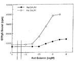
上記(1―2)で調製した膜画分を、濃度が40μg/ml(GALR1膜画分の場合)または32μg/ml(GALR2膜画分の場合)になるように、1 μM グアノシン-5’-二りん酸(GDP)、0.1% ウシ血清アルブミン(BSA)、5 mM MgCl2、 150 mM NaClを含む50 mMトリス緩衝液 (pH 7.4)に懸濁し、各0.2 mlごとポリプロピレン製小試験管(Falcon 2053)に分注した。分注した膜画分に50 nM [35S]GTPγS(New England Nuclear)を2 μl, およびアゴニストとしてブタ・ガラニン(100 μM、10 μM 、1 μM 、100 nM、10 nM、1 nM、100 pM、または10 pMの各濃度で)2μlを添加し、25℃で60分間反応した。該反応液に、1.5 mlの0.05% 3-[(3-コールアミドプロピル)ジメチルンモニオ]プロパンスルフォン酸(CHAPS)、0.1% BSA、 5 mM MgCl2、 1 mM EDTAを含む50 mMトリス緩衝液(pH 7.4)を加え、GF/Fガラス繊維ろ紙(ワットマン社)でろ過した。このろ紙を、1.5 mlの同緩衝液で洗浄し乾燥させた後、液体シンチレーション・カウンターにより放射活性を計測した。
その結果、ガラニン濃度依存的に[35S]GTPγS結合量の増加がみとめられ〔図1〕、本法によりガラニンレセプターのアゴニスト活性を測定できることが明らかになった。
ラット・ガラニンのEC50値(最大結合量の半分の結合量を与える濃度)は、GALR1の場合で約3 nM、またGALR2の場合で約10 nMであった。
【0039】
実施例2 GALR2(ガラニン・レセプター・タイプ2)を活性化する化合物のスクリーニング
(2―1)ブタ視床下部抽出液の調製
ブタ視床下部を東京芝浦臓器(株)で購入し、3時間以内に以下の操作を行い抽出液を調製した。
先ず、ブタ視床下部35個(約500 g)を包丁を用いて細かく切り刻み、沸騰した1250mlの蒸留水を含む3000 mlビーカーに入れ、10分間煮沸した。煮沸後の視床下部をビーカーごと水浴、次いで氷浴して、温度を約4℃まで下げた。該視床下部を煮沸に用いた蒸留水とともに、ポリトロン・ホモジェナイザーを用いて10分間ホモジェナイズした。得られたホモジェネートに終濃度が1 Mになるように酢酸90 mlを滴下し、1時間攪拌した。ホモジェネートを、高速遠心機(CR26H、RR10A型ローター:日立株式会社)を用いて、10,000 rpm、 30分間遠心し上清(1)を得た。一方、遠心後に得られた沈殿を、再度1 M酢酸2000 mlを加えてポリトロン・ホモジェナイザーを用いて10分間ホモジェナイズした。このホモジェネートを、攪拌翼を用い一晩(約16時間)攪拌した後、ポリトロン・ホモジェナイザーを用いて10分間ホモジェナイズし上清(2)を得た。上清(1)と上清(2)を混合し、その2倍容量のアセトンを加え、4℃にて1時間攪拌し、次いで高速遠心機(CR26H、RR10A型ローター:日立株式会社)を用いて10,000 rpm, 15分間遠心し上清を得た。得られた上清をロータリー・エバポレーターにかけ、アセトンを除去し、最終的に4000 mlまで濃縮した。この濃縮液を、超遠心機(SCP70H、日立RPZ35T型ローター:日立株式会社)を用いて、35,000 rpm、1時間遠心し、清澄な上清を得た。得られた上清を、1000 mlごとに500 mlのジエチルエーテルと混合し、分液ロート中にて激しく混和し、2相分離後、水相を得た。得られた水相を、ロータリーエバポレーターを用いて1000 mlまで濃縮し、最終的な抽出液を得た。
【0040】
(2―2)ブタ視床下部抽出液のオクタドデシル逆相クロマトグラフィーによる精製
オクタドデシル基を固定したシリカゲルODS-AM 120-S50(YMC社)をメタノールで膨潤後、直径5 cmのガラスカラムに容量が130 mlになるように充填し、1M酢酸で平衡化した。このカラムに、(2―1)で調製した抽出液(視床下500 g分)を流速400 ml/hで添着した。続いて、このカラムに、約500mlの1 M酢酸、次いで約500 mlの20% アセトニトリル/0.1 % トリフルオロ酢酸を、流速400ml/hで流し、ゲルを洗浄した。最後に、このカラムに、約500 mlの50% アセトニトリル/0.1 % トリフルオロ酢酸を流速400 ml/hで流し、目的とする粗ペプチド成分を溶出した。得られた溶出液を、エバポレーターを用いて濃縮した後、凍結乾燥機(12EL; VirTis社)にて凍結乾燥した。
【0041】
(2―3)ブタ視床下部抽出液のTSKgel ODS80TM逆相高速液体クロマトグラフィーによる精製
TSKgel ODS80TM逆相高速液体クロマトグラフィー用カラム(東ソー株式会社、22.5 mm x 30 cm)を、40℃にて、流速8 ml/minで A液(0.1% トリフルオロ酢酸/蒸留水)を流し、平衡化した。上記(2―2)で得られた凍結乾燥物を、1 M酢酸に40 mlに溶解し、各10 mlずつ4回のクロマトグラフィー操作を行った。即ち、凍結乾燥物の酢酸溶液10 mlを該カラムに添着した後、流速8 ml/minで、60分間かけてA液(0.1% トリフルオロ酢酸/蒸留水)容量80%/B液(0.1% トリフルオロ酢酸/100% アセトニトリル)容量20%から、A液(0.1% トリフルオロ酢酸/蒸留水)容量40%/B液(0.1% トリフルオロ酢酸/100% アセトニトリル)容量60% まで直線的グラジエントで上昇させた。
溶出液を、8 mlずつフラクションNo.をつけて分取し、各1 mlずつに分注し真空濃縮機(サーバント社)で濃縮乾燥させた。この乾燥物に、ジメチルスルフォキサイド0.03 mlを加えて溶解し、 GALR1 および GALR2活性化作用を測定するアッセイ用サンプルとした。
(2―4)[35S]GTPγS結合試験によるGALR2(ガラニン・レセプター・タイプ2)を活性化する化合物の単離と精製
上記(2―3)で得られたアッセイ用サンプルを、上記(1―3)に記載したGALR1 またはGALR2膜画分を用いる[35S]GTPγS結合試験によりガラニン・レセプター活性化作用を測定した。
GALR1発現細胞膜画分を用いた[35S]GTPγS結合試験では、フラクションNo.36-39 および No.42-43に[35S]GTPγS結合促進活性が認められ、一方、GALR2発現細胞膜画分を用いた[35S]GTPγS結合試験では、フラクションNo.36-39, No.42-43、及びNo.46-49に[35S]GTPγS結合促進活性が認められた〔図2〕。
さらに、アッセイ用サンプルをジメチルスルフォキサイドで100倍希釈し、この希釈サンプル3 mlをブタ・ガラニン・ラジオイムノアッセイキット(Peninsula社)により分析した結果、フラクションNo.36-38にガラニン免疫活性が検出された〔図3〕。
以上の結果より、フラクションNo.36-39のGALR1およびGALR2活性化作用を有する成分はブタ・ガラニンであると判断された。一方、フラクションNo.46-49のGALR2活性化作用を有する成分はガラニンとは異なる化合物であることが予想された。
【0042】
(2―5)GALR2(ガラニン・レセプター・タイプ2)を活性化する化合物の分子量測定
公知のゲルろ過高速液体クロマトグラフィー法により、上記(2―4)で得られたGALR2活性化成分(フラクションNo.46-49)の分子量を測定した。
フラクションNo.46-49のサンプル混合液の 0.02 mlを0.1% トリフルオロ酢酸/蒸留水0.08 mlに希釈し、その0.05 mlをG2000SWXL(東ソー株式会社)カラムに添着し、0.1% トリフルオロ酢酸/10%アセトニトリル を流速0.5 ml/minで流し、溶出液を各0.25 ml (0.5分ごとに)分取した。分取したフラクションを濃縮乾燥(SpeedVac Plus SC210A; Savant社)し、膜画分の懸濁液に直接溶解した後、50nM [35S]GTPγS(New England Nuclear社)を2 μL添加し、以下(1―3)に記載した方法により[35S]GTPγS結合量を測定した。
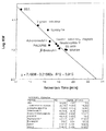
その結果、 GALR2活性化作用([35S]GTPγS結合促進活性)を有する成分は、フラクションNo.35(リテンション・タイム 17〜17.5分)に溶出された〔図4〕。同一条件で分析した公知のペプチド(Adrenomedulin, Gastric inhibitory peptide, PACAP38, Neuropeptide Y, β-endorphine, Galanin, SRIF28(Somatostatin 28), Bovine Serum Albumin, Trypsin inhibitor, Lysozyme)を用いて比較解析した結果〔図5〕、 フラクションNo.46-49のGALR2活性化作用を有する成分の分子量は約5000〜約7000と推測された。
一方、公知のブタ・ガラニン(ペプチド研究所)の分子量は 3157.4(リテンション・タイム 18.858分)である。従って、フラクションNo.46-49として得られたGALR2活性化作用を有する化合物は、公知のガラニンとは異なる物質であることが示唆された。
【0043】
実施例3 GALR2(ガラニン・レセプター・タイプ2)活性化作用を有する化合物の精製
(3―1)ブタ視床下部抽出液の調製
大量の視床下部抽出液を調製するために、上記(2―1)の方法を以下のとおり簡略化した。
ブタ視床下部凍結品(東京芝浦臓器株式会社)を50個(約1 kg)を包丁を用いて薄い切片にスライスし、沸騰した2500mlの蒸留水を含む5000 mlビーカーに加え、10分間煮沸した。煮沸した視床下部をビーカーごと水浴、及び氷浴して、温度を約4℃まで下げ、視床下部を煮沸に用いた蒸留水とともに、ポリトロン・ホモジェナイザーを用いて10分間ホモジェナイズした。得られたホモジェネートに、酢酸 150 ml、および 6N塩酸を 8 ml滴下し、それぞれの終濃度を1 Mおよび20 mMとし、次に攪拌翼を用いて一晩(約16時間)攪拌した。ホモジェネートを高速遠心機(CR26H、日立RR10A型ローター:日立株式会社)を用いて 8000 rpm, 30分間遠心し、得られた上清を、ガーゼでろ過し、脂質片を取り除いた。以上の操作を4回繰り返し行うことにより、約4kgの視床下部から抽出液を調製した。
【0044】
(3―2)ブタ視床下部抽出液の濃縮と粗分画
(3―2―1)オクタドデシル逆相クロマトグラフィーによる精製
オクタドデシル基を固定したシリカゲル(YMC社、ODS-AM 120-S50)をメタノールで膨潤後、直径5 cmのガラスカラムに容量が400 mlになるように充填し、1 M酢酸で平衡化した。
このカラムに(3―1)で調製した抽出液の半分量(視床下部2kg分)を流速400 ml/hで添着した後、流速400 ml/hで、約1000mlの1 M酢酸、次いで約1200 mlの20% アセトニトリル/0.1 % トリフルオロ酢酸を流し、ゲルを洗浄した。最後に、流速400 ml/hで、約2000 mlの60% アセトニトリル/0.1 % トリフルオロ酢酸をカラムに流し、目的とする粗ペプチド画分を溶出した。得られた溶出液を、エバポレーターを用いて濃縮した後、凍結乾燥機(12EL; VirTis社)にて凍結乾燥した。以上の操作を2回実施し、約4kg(200個)分の視床下部の抽出液の凍結乾燥粉末を調製した。
(3―2―2)SP-Sephadexイオン交換クロマトグラフィーによる精製
直径5 cmのガラスカラムに、10 mM塩酸中で膨潤させたSP-Sephadex C25(Pharmacia Biotech 社)を、容量が120 mlになるよう充填し、100 mM塩酸で洗浄した後、1 M酢酸で平衡化した。(3―2―1)により得られた視床下部抽出液の凍結乾燥粉末を、約800 mlの1 M酢酸に溶解し、平衡化したSP-Sephadex C25カラムに流速400 ml/hで添着した。約600 mlの1 M酢酸、約600 mlの1 Mピリジン、約600 mlの1 Mピリジン・酢酸(pH 5.0)を流速400 ml/hで順次カラムに流し、溶出液を40mlづつ分取し、これを各0.2 mlづつに分注し真空濃縮機(サーバント社)で濃縮乾燥させた。この乾燥物に、ジメチルスルフォキサイド0.04 mlを加えて溶解し、上記(1−3)の試験法により、 GALR2活性化作用を測定した。
その結果、目的とするGALR2活性化作用を有する成分は、1 Mピリジン・酢酸(pH 5.0)により溶出されていることが判明した。本結果より、GALR2活性化作用を有する成分(化合物)は強塩基性物質であると考えられた。
(3―2―3)Sephadex G50ゲルろ過クロマトグラフィーによる精製
上記(3―2―2)で得られた1 Mピリジン・酢酸(pH 5.0)溶出液分取品をエバポレーターを用いて100 mlになるまで濃縮し、1 M酢酸で平衡化したSephadex G50 (Pharmacia Biotech 社)カラム(直径6 cm、容量3000 ml)に流速400ml/hで添着した後、流速400 ml/hで1 M酢酸をカラムに流し、溶出液を33 mlづつフラクションNo.をつけて分取し、これを各0.25 mlづつに分注し真空濃縮機(Savant社)で濃縮乾燥させた。この乾燥物に、ジメチルスルフォキサイド0.03 mlを加えて溶解し、上記(1−3)の試験法により、 GALR2活性化作用を測定した。
その結果、目的とするGALR2活性化作用を有する成分は主として、分取フラクションNo.66-80に溶出されていることが判明した。また、フラクションNo.55-65および No.45-54にも弱い活性が検出された。フラクションNo.66-80、55-65 および 45-54をそれぞれ混合して凍結乾燥した。
(3―3)HPLCを用いるGALR2活性化作用を有する化合物の精製
(3―3―1)TSKgel ODS80TM逆相高速液体クロマトグラフィーによる精製
TSKgel ODS80TM逆相高速液体クロマトグラフィー用カラム(東ソー、22.5 mm x 30 cm)を、40℃にて、流速4 ml/minでA液(0.1% トリフルオロ酢酸/蒸留水)を流し、平衡化した。
上記(3―2―3)で得られたSephadex G50分取フラクションの各凍結乾燥物を1 M酢酸に溶解し、それぞれカラムに添着した後、流速4 ml/minで、120分間かけて A液(0.1% トリフルオロ酢酸/蒸留水)容量67%/B液(0.1% トリフルオロ酢酸/60% アセトニトリル)容量33%から、B液(0.1% トリフルオロ酢酸/60% アセトニトリル)容量100% まで直線的グラジエントで上昇させ溶出液を回収した。
溶出液を、8 mlずつフラクションNo.をつけて分取した。なお、フラクションNo.66-80の凍結乾燥物は2回に分けてクロマトグラフィーを実施し、またフラクションNo. 55-65、45-54はそれぞれ1回のクロマトグラフィーで分離した。
分取フラクション4 μlを直接用い、上記(1−3)の試験法により、 GALR2活性化作用を測定した。その結果、いずれのクロマトグラフィーにおいても主に3種の活性ピークが見られた。フラクションNo.35-38に見られた活性化作用を有する成分の溶出位置は実施例2に記載したガラニンの溶出位置にほぼ一致した。また、フラクションNo.47-50に見られた活性化成分は実施例2に記載したGALR2活性化作用を有する成分(GALR1を活性化せず、また前述のブタ・ガラニン・ラジオイムノアッセイ・キットで検出されない)の溶出位置にほぼ一致した。従って、フラクションNo.47-50を用いてGALR2活性化作用を有する成分の精製を実施することとし、これらフラクションを混合して凍結乾燥した。
(3―3―2)TSKgel CM-2SWイオン交換高速液体クロマトグラフィーによる精製
TSKgel CM-2SWイオン交換高速液体クロマトグラフィー用カラム(東ソー、 0.46 cm x 25 cm)を、20℃にて、流速1 ml/minで A液(10 mMぎ酸アンモニウム/40% アセトニトリル)を流速1 ml/minで流し、平衡化した。
(3―3―1)で得られた逆相高速液体クロマトグラフィー分取フラクションの凍結乾燥物をA液に溶解し、カラムに添着した後、流速1 ml/minで、60分間かけて A液(10 mMギ酸アンモニウム/40% アセトニトリル)容量100%から、B液(500 mMギ酸アンモニウム/40% アセトニトリル)容量100% まで直線的グラジエントで上昇させ溶出液を回収した。
溶出液を、0.5 mlづつフラクションNo.をつけて分取した。分取フラクション1μlを直接用い、上記(1−3)の試験法により、 GALR2活性化作用を測定した。その結果、主な活性ピークはフラクションNo.89-92に検出された。これらのフラクションをまとめて以降の精製に用いた。
【0045】
(3―3―3)ジフェニル逆相高速液体クロマトグラフィーによる精製
ジフェニル逆相高速液体クロマトグラフィー用カラム(Vydak 219TP54、 0.46 cm x 25 cm)を、40℃にて、流速1 ml/minでA液(0.1% トリフルオロ酢酸/蒸留水)を流し、平衡化した。
上記(3―3―2)で得られたイオン交換高速液体クロマトグラフィー分取フラクションを直接カラムに添着した後、流速 1ml/minで 1分間で A液(0.1% トリフルオロ酢酸/蒸留水)容量100%から B液(0.1% トリフルオロ酢酸/60% アセトニトリル)容量を50%まで急速に上昇させ、これを次に、流速1 ml/minで、60分間かけてB液濃度を100% まで直線的グラジエントで上昇させ溶出液を回収した。
溶出液を、1 mlずつフラクションNo.をつけて分取した。分取フラクション1 μlを直接用い、上記(1−3)の試験法により、 GALR2活性化作用を測定した。その結果、主な活性ピークはフラクションNo.24-26に検出された。これらのフラクションをまとめて以降の精製に用いた。
(3―3―4)Super Phenyl逆相高速液体クロマトグラフィーによる精製
TSKgel Super Phenyl逆相高速液体クロマトグラフィー用カラム(東ソー、 0.46 cm x 10 cm)を、40℃にて、流速1 ml/minで A液(0.1% トリフルオロ酢酸/蒸留水)を流し、平衡化した。
(3―3―3)で得られたジフェニル逆相高速液体クロマトグラフィー分取フラクションを直接カラムに添着した後、流速 1ml/minで 1分間で A液(0.1% トリフルオロ酢酸/蒸留水)容量100%から B液(0.1% トリフルオロ酢酸/60% アセトニトリル)容量45%まで急速に上昇させ、これを次に、流速1 ml/minで、80分間かけてB液濃度を55% まで直線的グラジエントで上昇させ溶出液を回収した。
溶出液を、1 mlずつフラクションNo.をつけて分取した。分取フラクション1 μlを直接用い、上記(1−3)の試験法により、 GALR2活性化作用を測定した。その結果、主な活性ピークはフラクションNo.44-45 および No.50-52に検出された。フラクションNo.44−45を混合して同条件にて再クロマトグラフィー操作を実施し、0.5 mlずつ溶出フラクションを分取した。一方、No.50、51および52はそれぞれ別個に同条件にて再クロマトグラフィー操作を実施し、0.5 mlずつ溶出フラクションを分取した。
各分取フラクション2 μlを直接使用し、上記(1−3)の試験法により、 GALR2活性化作用を測定した。その結果、 No.44−45の再クロマトグラフィー操作で得られたフラクションNo.93-95、および、フラクションNo.50の再クロマトグラフィー操作で得られたフラクションNo.105-107に活性ピークが検出された。フラクションNo.51の再クロマトグラフィー操作で得られたフラクションNo.105-107に主たる活性ピークと、フラクションNo.108-109に弱い活性ピークが検出された。さらに、フラクションNo.52の再クロマトグラフィー操作で得られたフラクションNo.106-110には主たる活性ピークがブロードに検出された。
【0046】
(3―3―5)Super ODS逆相高速液体クロマトグラフィーによる精製
(3―3―5―1)GALR2 活性化主成分の精製
(3―3―5―1―1)トリフルオロ酢酸存在下でのクロマトグラフィー
TSKgel Super ODS逆相高速液体クロマトグラフィー用カラム(東ソー株式会社、 0.46 cm x 10 cm)を、40℃にて、流速1 ml/minでA液(0.1% トリフルオロ酢酸/蒸留水)を流し、平衡化した。
(3―3―4)で得られたSuper Phenyl逆相高速液体クロマトグラフィー分取フラクションNo93-95を直接カラムに添着した後、流速 1ml/min、1分間で、A液(0.1% トリフルオロ酢酸/蒸留水)容量100%から B液(0.1% トリフルオロ酢酸/60% アセトニトリル)容量を50%まで直線的グラジエントで上昇させ、これを次に流速1 ml/minで、84分間かけてB液濃度を62.5% まで直線的グラジエントで上昇させ溶出液を回収した。
溶出液を、0.5 mlずつフラクションNo.をつけて分取した。分取フラクション2 μlを直接用い、上記(1−3)の試験法により、 GALR2活性化作用を測定した。その結果、活性ピークはフラクションNo.96-97に検出された。
得られたフラクションNo.96-97を混合して同条件にて再クロマトグラフィー操作を実施し、0.5 mlずつフラクションを分取した。210 nmの紫外(UV)吸収で検出されたピークは単一ではあったが、不純物の混入を示唆するピークの肩が認められた。各分取フラクション2 μlを直接用い、上記(1−3)の試験法により、 GALR2活性化作用を測定した。その結果、活性ピークはフラクションNo.98-100に検出され、これらは210 nmの紫外吸収で検出されたピーク肩部分を除く部分に一致した。該ピーク肩部分に含まれる不純物を除去するために、(3―3―5―1―2)に記載の方法によりさらに精製を行った。
(3―3―5―1―2)ヘプタフルオロ酪酸存在下でのクロマトグラフィー
TSKgel Super ODS逆相高速液体クロマトグラフィー用カラム(東ソー株式会社、 0.46 cm x 10 cm)を、40℃にて、流速1 ml/minで A液(0.1% ヘプタフルオロ酪酸/蒸留水)を流し、平衡化した。
(3―3―5―1―1)で得られたフラクションNo98-100を直接カラムに添着した後、 流速 1ml/min、1分間で A液(0.1% ヘプタフルオロ酪酸/蒸留水)容量100%から B液(0.1% ヘプタフルオロ酪酸/100% アセトニトリル)容量を35%まで急速に上昇させ、これを次に流速1 ml/minで、60分間かけてB液濃度を50% まで直線的グラジエントで上昇させ溶出液を回収した。
溶出液を、0.5 mlずつフラクションNo.をつけて分取した。分取フラクション2 μlを直接用い、上記(1−3)の試験法により、 GALR2活性化作用を測定した。その結果、活性ピークはフラクションNo.67-69に検出された。この活性ピークは、2本に分離された210nmの紫外吸収ピークのうち、早く溶出された方の紫外吸収ピークに完全に一致し、単一ペプチドにまで精製されたものと判断した。本活性化作用を有する成分をGALR2活性化主成分と呼ぶ。
(3―3―5―2)GALR2活性化副成分(1)の精製
TSKgel Super ODS逆相高速液体クロマトグラフィー用カラム(東ソー株式会社、 0.46 cm x 10 cm)を、40℃にて、流速1 ml/minで A液(0.1% ヘプタフルオロ酪酸/蒸留水)を流し、平衡化した。
(3―3―4)で得られたSuper Phenyl逆相高速液体クロマトグラフィー分取フラクションNo.105-107(フラクションNo.50由来)、フラクションNo.105-107(フラクションNo.51由来)、フラクションNo.106-107(フラクションNo.52由来)を混合し、直接カラムに添着した後、流速 1ml/min、1分間で、A液(0.1%トリフルオロ酢酸/蒸留水)容量100%から B液(0.1% トリフルオロ酢酸/60% アセトニトリル)容量を52.5%まで急速に上昇させ、これを次に流速1 ml/minで、84分間かけてB液濃度を65%まで直線的グラジエントで上昇させ溶出液を回収した。溶出液を、0.5 mlずつフラクションNo.をつけて分取した。
分取フラクション2 μlを直接用い、上記(1−3)の試験法により、 GALR2活性化作用を測定した。その結果、活性ピークはフラクションNo.75-76に検出された。得られたフラクションNo.75-76を混合し、同条件にて再度同じクロマトグラフィーを実施し、0.5 mlずつフラクションNo.をつけて分取した。溶出液は、210 nmの紫外吸収では単一なピークとして検出された。分取フラクション3 μlを直接用い、上記(1−3)の試験法により、 GALR2活性化作用を測定した。その結果、活性ピークはフラクションNo.76-78に検出された。この活性ピークは210 nmの紫外吸収ピークと一致し、活性成分は単一ペプチドにまで精製されたものと判断した。本活性化作用を有する成分をGALR2活性化副成分(1)と呼ぶ。
【0047】
(3―3―5―3)GALR2活性化副成分(2)の精製
TSKgel Super ODS逆相高速液体クロマトグラフィー用カラム(東ソー株式会社、 0.46 cm x 10 cm)を、40℃にて、流速1 ml/minで A液(0.1% ヘプタフルオロ酪酸/蒸留水)を流し、平衡化した。
(3―3―4)で得られたSuper Phenyl逆相高速液体クロマトグラフィー分取フラクションNo.108-109(フラクションNo.51由来)、フラクションNo.108-110(フラクションNo.52由来) を混和し、直接カラムに添着した後、流速 1 ml/min、1分間で、A液容量100%から B液(0.1% トリフルオロ酢酸/60% アセトニトリル)容量を52.5%まで急速に上昇させ、これを次に流速1 ml/minで、84分間かけてB液濃度を65%まで直線的グラジエントで上昇させ溶出液を回収した。溶出液を、0.5 mlずつフラクションNo.をつけて分取した。
分取フラクション2μlを直接用い、上記(1−3)の試験法により、 GALR2活性化作用を測定した。その結果、活性ピークはフラクションNo.79-80に検出された。
得られたフラクションNo.79-80を混合して同条件にて再度同じクロマトグラフィーを実施し、0.5 mlずつ分取したところ、210 nmの紫外吸収で単一なピークとして検出された。
分取フラクション3 μlを直接用い、上記(1−3)の試験法により、 GALR2活性化作用を測定した。その結果、活性ピークはフラクションNo.79-81に検出された。この活性ピークは210 nmの紫外吸収ピークと一致し、活性成分は単一ペプチドにまで精製されたものと判断した。本活性化作用を有する成分をGALR2活性化副成分(2)と呼ぶ。
【0048】
実施例4 アミノ酸配列分析
(4―1)GALR2活性化作用を有する主成分のN末端アミノ酸配列分析
上記(3―3―5―1―2)で得られたフラクションNo.67-69のGALR2活性化主成分を20μlの0.1% トリフルオロ酢酸/30% アセトニトリルに溶解し、ペプチドサポートディスク(ベックマン社)に滴下した後、窒素ガスにより該ディスクを乾燥させた。これをプロテインシーケンサー(ベックマン社、LF3400DT)にセットし、標準分析法(プロシージャ40)により自動エドマン分解反応を行ないアミノ酸配列解析を行った。各サイクルごとに同定されたPTHアミノ酸を〔表1〕に示した。
【表1】

APVHRGRGGWTLNSAGYLLGPVLHPPSXAEGGGK (GALR2活性化主成分のN末端アミノ酸配列、配列番号:11)
得られたアミノ酸配列中のN末端から9残基〜21残基目の13残基からなる配列は、公知のブタ・ガラニンのN末端アミノ酸配列に一致した。しかし、その前後のアミノ酸配列はブタ・ガラニンおよびその前駆体のいずれの配列とも一致しなかった。また、本アミノ酸配列と一致する公知のペプチドは見出されなかったので、得られたGALR2活性化主成分は新規ペプチドであると推測された。
(4―2) GALR2活性化作用を有する副成分(1)のN末端アミノ酸配列分析
上記(3―3―5―2)で得られたフラクションNo.76-78のGALR2活性化副成分は、(4―1)と同様にして分析した。各サイクルごとに同定されたアミノ酸を〔表2〕に示した。
【表2】
APVHRGRGGWTLNSAGYLLGPVLHPPSXAEGG (GALR2活性化副成分(1)のN末端配列、配列番号:12)
このアミノ酸配列は上記(4―1)で得られたGALR2活性化主成分のN末端アミノ酸配列32残基に完全に一致した。
【0049】
(4―3) GALR2活性化作用を有する副成分(2)のN末端アミノ酸配列分析
上記(3―3―5―3)で得られたフラクションNo.79-81のGALR2活性化副成分(2)を、20μlの0.1% トリフルオロ酢酸/30% アセトニトリルに溶解し、重炭酸アンモニウム(終濃度1%)、TLCK-キモトリプシン(終濃度10pmolシグマ社)および蒸留水を添加して 50μlとし、37℃で1時間反応させた。
Spheri-5 RP-18逆相高速液体クロマトグラフィー用カラム(ブラウンリー社、 2.1 mm x 30 mm)を、25℃にて、流速300μl/minで A液(0.1%トリフルオロ酢酸)を流し、平衡化した。
酵素反応液全量をカラムに添着した後、流速300μl/minで、30分間かけてA液(0.1%トリフルオロ酢酸)容量100%から B液(0.1%トリフルオロ酢酸/70% アセトニトリル)容量を70% まで上昇させ、210nmの紫外吸収を示す溶出ピーク4つ(CHY-1、CHY-2、CHY-3 および CHY-4)を溶出フラクションとして回収した。
分取した4つのフラクション(キモトリプシン消化断片)を、窒素気流下で50μl以下になるまで濃縮した後、(4―1)と同様にしてプロテインシーケンサーによりN末端アミノ酸配列分析を行なった。なお、CHY-3 については、ベックマン社 LF3400DTプロテインシーケンサーを、CHY-1、CHY-2、および CHY-4 については、アプライドバイオシステムズ社 477Aプロテインシーケンサーを使用した。
N末端アミノ酸配列解析により以下の配列が得られた。
CHY-1: APVHRGRGG(配列番号:13)
CHY-2: XAIDGLPYPQS (Xは未同定)(配列番号:16)
CHY-3: LLGPVLHPPSXAEGGGKTALGILDL (Xは未同定)(配列番号:15)
CHY-4: TLNSAG (配列番号:14)
上記(4―1)で得られたGALR2活性化主成分または上記(4―2)で得られたGALR2活性化副成分(1)のN末端アミノ酸配列結果を参考にして、GALR2活性化副成分(2)は次のN末端アミノ酸配列を有しているものと推測された。
APVHRGRGGWTLNSAGYLLGPVLHPPSXAEGGGKTALGILDL(配列番号:17)
【0050】
実施例5 ブタ型リガンドペプチド(1−60)をコードするcDNA
1)デジェネレートPCR(Degenerated PCR)法によるブタGALR2活性化因子遺伝子断片のクローニング
ブタ全脳からTRIZOL reagent(Gibco BRL社)を用い、添付されたマニュアルに記載された方法に従ってtotal RNAを抽出した。次いでオリゴdTセルロースカラム(mRNA Purification Kit, Pharmacia社)を用い、添付されたマニュアルに記載された方法に従って該total RNAからpoly(A)+ RNAを調製した。さらに、3' RACE System for rapid amplification of cDNA ends (Gibco BRL 社) を用い、添付されたマニュアルに記載された方法に従って、該Poly (A)+ RNAから first strand cDNA を合成した。また、ブタGALR2活性化成分の部分アミノ酸配列を基にデザインした次の degenerate primerを合成した。
pGAL4-7F: 5'-CAYMGNGGIMGNGGIGGSTGGAC-3' (配列番号:20)
pGAL9-3F: 5'-GGHTGGACNCTNAAYAGYGC-3' (配列番号:21)
pGAL34-1R: 5'-ATICCNAGIGCNGTYTTICCYTT-3' (配列番号:22)
前述の first strand cDNA を鋳型として用い、pGAL4-7FおよびpGAL34-1Rをプライマーとして用いて初段のPCR反応を実施した。該PCR反応における反応液は、Taq polymerase(宝酒造株式会社)を0.5 μl、それぞれ添付の10x PCR buffer(500 mM KCl-100 mM Tris・HCl, pH 8.3)を10 μl、25 mM MgCl2を6 μl、2.5 mM dNTP mixtureを8 μl、プライマーpGAL4-7FおよびプライマーpGAL34-1R(ともに10 μM)を各10 μl、および該鋳型cDNA(前述の first strand cDNA)を8 μl、蒸留水を47.5 μlを混合して作製した。該PCRとして、1)初期変性(94℃・30秒間)、2)32回のサイクル反応(94℃・20秒間-55℃・30秒間-72℃・30秒間)の後3)最終伸長反応(72℃・4分間)を行い初段PCR産物を得た。
得られた初段PCR産物を鋳型として第2段のPCR反応(nested PCR)を行った。該反応液は、Taq polymerase(宝酒造株式会社)を0.5 μl、添付の10x PCR buffer(500 mM KCl-100 mM Tris・HCl, pH 8.3)を10 μl、25 mM MgCl2を6 μl、2.5 mM dNTP mixtureを8 μl、プライマーpGAL9-3FおよびpGAL34-1R(ともに10 μM)を各10 μl、および該鋳型cDNA(初段PCR産物)を5 μl、蒸留水を50.5 μlを混合して作製した。該PCRとして、1)初期変性(94℃・30秒間)、2)32回のサイクル反応(94℃・20秒間-55℃・30秒間-72℃・30秒間)の後3)最終伸長反応(72℃・10分間)を行い nested PCR 産物を得た。
さらに nested PCR 産物を2.0%アガロースゲル電気泳動を行い、サイバーグリーン染色される約100 bpのバンドを含むゲル片を剃刀で切り出した。該ゲル片より Gene Clean DNA 抽出キット(BIO 101社)を用いてnested PCR 産物であるDNA断片を回収した。このDNA断片をTOPO TA Cloning Kit (Invitrogen社)を用いて、添付のマニュアルに記載された方法に従ってキットに添付されたプラスミドベクターpCRIIに連結した。得られたプラスミドをQIAwell 8 Ultra plasmid purification kit(QIAGEN社)を用いて精製した。該プラスミド中の nested PCR 産物の塩基配列決定のための反応は、Dye Terminator Cycle Sequencing Kit(Applied Biosystems、パーキンエルマー社)を用いて行い、蛍光式自動シークエンサー(DNA sequencer Prism 377:Applied Biosystems、パーキンエルマー社)を用いて nested PCR 産物の塩基配列を解読した。その結果、ブタGALR2活性化因子の部分ペプチドをコードする98 bpのDNA断片を有する数種類のプラスミドを得ることができた。PCR反応にdegenerate primersを用いたため、これらの数種類のプラスミドにおけるDNAはプライマーに対応する部分で塩基配列に差異が見られた。以下に代表的な2クローン、pCR100-6およびpCR100-7中の nested PCR 産物の塩基配列を示す。
pCR100-6:
GGCTGGACTT TAAATAGTGC TGGTTACCTC CTGGGTCCCG TACTCCATCC GCCCTCCAGG
GCTGAAGGAG GCGGGAAGGG CAAAACAGCC CTGGGCAT (配列番号:23)
pCR100-7:
GGTTGGACTT TGAACAGTGC TGGTTACCTC CTGGGTCCCG TACTCCATCC GCCCTCCAGG
GCTGAAGGAG GCGGGAAGGG CAAAACCGCC CTAGGCAT (配列番号:24)
2)ブタ型リガンドペプチド(1−60)をコードするcDNAのクローニング
ブタ全脳より得られた前述の poly (A)+ RNAより、ZAP-cDNA Gigapack III Gold cloning kit (STRATAGENE社)を用いて、ブタ全脳cDNAファージライブラリーを作製した。方法はキットに添付されたマニュアルに従った。得られたファージ(2,200,000 plaque forming units)を該マニュアルの方法で大腸菌XL1-Blue MRF'(STRATAGENE社)に感染させた後、NZY培地アガープレート120枚にまき、37℃で8時間培養した。得られた大腸菌プラークよりファージをHybond-N+ナイロンメンブレン(アマシャム社)に写し取った。このナイロンメンブレンを1.5 M塩化ナトリウムを含む0.5 N水酸化ナトリウム溶液、1.5 M 塩化ナトリウムを含む0.5 Mトリス緩衝溶液(pH 7.0)、および2x SSC液に順次浸した後、風乾させた。
得られたナイロンメンブレンをハイブリダイゼーション緩衝液(5x SSPE, 5x Denhardt's液、0.5% SDS, 0.1 mg/ml サケ精子DNA)中にて60℃に24時間保温し、次いで、ハイブリダイゼーションプローブ(5 x 10⁵ cpm/ml)を含むハイブリダイゼーション緩衝液(同上)中にて、60℃に24時間保温した。該ハイブリダイゼーションプローブは、前述のプラスミドpCR100-6とpCR100-7の1:1混合物を鋳型として、次に示す2個のプライマーpGAL9-3FおよびpGAL34-8Rにより増幅したDNA断片を用いた。
pGAL9-3F: 5'-GGHTGGACNCTNAAYAGYGC-3' (配列番号:25)
pGAL34-8R:5'-ATDCCBAGGGCDGTTTTGCCCTT-3' (配列番号:26)
即ち、該増幅反応〔反応液組成は、ExTaq(宝酒造株式会社)を0.5 ml、添付の10x Ex Taq buffer(20 mM MgCl2を含む。)を5 μl、2.5 mM dNTP mixtureを4 μl、[α-32P]dCTP (6000 Ci/mmol)を10 μl、プライマーpGAL9-3FおよびpGAL34-8R(それぞれ10 μM)を各0.5 μl、および鋳型cDNAを1 μl、蒸留水を29 μlを混合して作製〕として、96℃・30秒間の初期変性後、94℃・20秒間-55℃・30秒間-72℃・30秒間のサイクル反応を32回、および72℃・4分間の最終伸長反応を行い増幅DNA断片を得た。
該ハイブリダイゼーション反応後のナイロンメンブレンを、0.1% SDSを含む0.2x SSC緩衝液を用いて50℃にて30分間洗浄した後、X線フィルム(Kodak社、BioMax MS)を用いて増感スクリーンの存在下でオートラジオグラフィーに処した(曝露条件:-70℃、3日間)。該オートラジオグラフィーの結果から検出された陽性プラークを単離し、クロロフォルム0.1 mlを含むSM 緩衝液(100 mM NaCl, 8 mM MgSO4, 0.01% gelatin, 50 mM Tris・HCl, pH 7.5)5 ml中にファージを抽出した。
得られたファージを再度大腸菌XL1-Blue MRF'に感染させた後、NZY培地アガープレートにまき、37℃で8時間培養した。得られた大腸菌プラークよりファージをナイロンメンブレン(アマシャム社、Hybond-N+)に写し取り、前述と同様の方法によりハイブリダイゼーションを行った。オートラジオグラフィーにより、陽性の単一クローンを単離し、前述と同様の一連の操作をもう一度繰り返し実施し、完全に単一なファージクローンを取得した。
【0051】
次に、該ファージクローンからプラスミドを以下の方法で単離精製した。ファージ液(1 μl)を大腸菌XPORT(50 μl)、ヘルパーファージ(10 μl)、大腸菌XLOLR(5 μl)と混合し、NZY アガープレートにまき、37℃で8時間培養した。大腸菌XPORTプラーク中に生じた大腸菌XLOLRの小コロニーをつま楊枝でピックアップし、アンピシリンとテトラサイクリンを含むLB培地アガープレートにて37℃、一晩培養した。単一コロニーより大腸菌XLOLRをピックアップし、LB培地中にて液体培養した。培養終了後、大腸菌XLOLRを集菌し、プラスミド精製キット(キアゲン社、QIAwell 8 Ultra)を用いてプラスミドを精製した。
該プラスミド中のブタGALR2活性化因子をコードするcDNAの塩基配列決定のための反応は、Dye Terminator Cycle Sequencing Kit(Applied Biosystems、パーキンエルマー社)を用いて行い、蛍光式自動シークエンサー(DNA sequencer Prism 377:Applied Biosystems、パーキンエルマー社)を用いてブタGALR2活性化因子をコードするcDNAの塩基配列を解読した。その結果、ブタGALR2活性化因子の全長ペプチドをコードする974 bpのDNA断片を有するプラスミドpGR2PL6、および1007 bpのDNA断片を有するプラスミドpGR2PL3を得ることができた。両者のDNA配列によりコードされるGALR2活性化因子の前駆体の鎖長およびアミノ酸配列には大きな差異が見られたが、プロセシングにより産生されると予想される成熟活性化因子(mature peptide)部分の配列は同一であった。
得られた2個のプラスミドを公知の方法により大腸菌TOP10に導入し、形質転換体それぞれTOP10/pGR2PL6、TOP10/pGR2PL3を得た。
【0052】
実施例6 ブタ型リガンドペプチド(1−60):Ala-Pro-Val-His-Arg-Gly-Arg-Gly-Gly-Trp-Thr-Leu-Asn-Ser-Ala-Gly-Tyr-Leu-Leu-Gly-Pro-Val-Leu-His-Pro-Pro-Ser-Arg-Ala-Glu-Gly-Gly-Gly-Lys-Gly-Lys-Thr-Ala-Leu-Gly-Ile-Leu-Asp-Leu-Trp-Lys-Ala-Ile-Asp-Gly-Leu-Pro-Tyr-Pro-Gln-Ser-Gln-Leu-Ala-Ser(配列番号:31)の製造
市販Boc-Ser(Bzl)l-OCH2-PAM樹脂(0.73 m mole/g resin) 0.5 m mole 分をペプチド合成機ABI 430Aの反応曹に入れ、Boc-strategy (NMP-HOBt)ペプチド合成方法でBoc-Ala, Boc-Leu, Boc-Gln, Boc-Ser(Bzl), Boc-Pro, Boc-Tyr(Br-Z), Boc-Gly, Boc-Asp(OcHex), Boc-Ile, Boc-Lys(Cl-Z), Boc-Trp(CHO), Boc-Thr(Bzl), Boc-Glu(OcHex), Boc-Arg(Tos), Boc-His(Bom), Boc-Val, Boc-Asn, を所望のアミノ酸配列順に導入し目的の保護ペプチド樹脂を得た。 この樹脂0.105gをp-クレゾール1.1g、1,4-ブタンジチオール1.2 mlと共に無水弗化水素10 ml中、0℃ 60分撹袢した後、弗化水素を減圧留去し、残留物へジエチルエーテルを加え沈殿を濾過した。 この沈殿に50%酢酸水を加え抽出し、不溶部分を除き、抽出液を十分に濃縮後、50%酢酸水で充填したセファデックスョG-25カラム(2.0 x 80 cm)に付し、同溶媒で展開、主要画分を集めLiChroprepョ RP-18を充填した逆相クロマトカラム(2.6 x 60 cm)に付け0.1%TFA水 200mlで洗浄、0.1%TFA水 300mlと0.1%TFA含有50%アセトニトリル水 300mlを用いた線型勾配溶出を行い、主要画分を集め濃縮、凍結乾燥し白色粉末39mgを得た。 これを更にCM−セルロファインョを充填したイオン交換クロマトカラムに付け50mMから200mM酢酸アンモニウム濃度の線形勾配溶出を行い主要画分を集め凍結乾燥を繰り返し、白色粉末10mgを得た。
質量分析による(M+H)+ 6204.7 (計算値 6205.2)
HPLC溶出時間 21.8分
カラム条件
カラム Wakosil 5C18T 4.6 x 100mm
溶離液:A液−0.1% TFA水、B液−0.1%TFA含有アセトニトリルを用い A/B : 95/5〜45/55へ直線型濃度勾配溶出(25分)
流速:1.0 ml / 分
【0053】
実施例7 ラット型リガンドペプチド(1−60):Ala-Pro-Ala-His-Arg-Gly-Arg-Gly-Gly-Trp-Thr-Leu-Asn-Ser-Ala-Gly-Tyr-Leu-Leu-Gly-Pro-Val-Leu-His-Leu-Ser-Ser-Lys-Ala-Asn-Gln-Gly-Arg-Lys-Thr-Asp-Ser-Ala-Leu-Glu-Ile-Leu-Asp-Leu-Trp-Lys-Ala-Ile-Asp-Gly-Leu-Pro-Tyr-Ser-Arg-Ser-Pro-Arg-Met-Thr(配列番号:33)およびヒト型リガンドポリペプチド(1−60): Ala-Pro-Ala-His-Arg-Gly-Arg-Gly-Gly-Trp-Thr-Leu-Asn-Ser-Ala-Gly-Tyr-Leu-Leu-Gly-Pro-Val-Leu-His-Leu-Pro-Gln-Met-Gly-Asp-Gln-Asp-Gly-Lys-Arg-Glu-Thr-Ala-Leu-Glu-Ile-Leu-Asp-Leu-Trp-Lys-Ala-Ile-Asp-Gly-Leu-Pro-Tyr-Ser-His-Pro-Pro-Gln-Pro-Ser(配列番号:34)の製造
市販Boc-Thr(Bzl)l-OCH2-PAM樹脂を用い実施例1と同様に合成と精製を行い取得できる。
【0054】
実施例8 モノクロナル抗体作製に用いた抗原ペプチドAla-Pro-Ala-His-Arg-Gly-Arg-Gly-Gly-Cys-NH2(配列番号:44のアミド体)の製造
市販p-メチルMBH樹脂(0.77 m mole/g resin)を用い実施例1と同様にBoc-Cys(MeBzl), Boc-Gly, Boc-Arg(Tos), Boc-His(Bom), Boc-Ala, Boc-Proを所望のアミノ酸配列順に縮合、弗化水素処理、50%酢酸水で充填したセファデックスョG-25カラム(2.0 x 80 cm)で精製の後凍結乾燥し、白色粉末50mgを得た。
質量分析による(M+H)+ 980.5(計算値 980.5)
HPLC溶出時間 5.2分
カラム条件
カラム Wakosil 5C18T 4.6 x 100mm
溶離液:A液−0.1% TFA水、B液−0.1%TFA含有アセトニトリルを用い B液濃度0から50%へ直線型濃度勾配溶出(25分)
流速:1.0 ml / 分
【0055】
実施例9 Ala-Pro-Ala-His-Arg-Gly-Arg-Gly-Gly(配列番号:35)を含む免疫原の作製
上記実施例8で得られたAla-Pro-Ala-His-Arg-Gly-Arg-Gly-Gly-Cys-NH2(配列番号:44のアミド体)で表示のペプチドとKeyhole Limpet Hemocyanin(KLH)との複合体を作製し、免疫原とした。すなわち、KLH20mgを、0.1Mリン酸緩衝液(pH6.5)1.4mlに溶解させ、N−(γ−マレイミドブチリロキシ)サクシニミド(GMBS)2.2mg(8μmol)を含むDMF溶液100μlと混合し、室温で40分反応させた。反応後、セファデックスG−25カラムで分画したのち、マレイミド基の導入されたKLH15mgとAla-Pro-Ala-His-Arg-Gly-Arg-Gly-Gly-Cys-NH2(配列番号:44のアミド体)で表示のペプチド1.6 mgとを混合し、4℃で2日間反応させた。反応後、生理食塩水に対し、4℃で2日間透析した。
【0056】
実施例10 免疫
6〜8週令のBALB/C雌マウスに上記実施例9記載の免疫原Ala-Pro-Ala-His-Arg-Gly-Arg-Gly-Gly-Cys(-NH2)−KLH複合体を、約30μg/匹を完全フロイントアジュバントとともに皮下免疫した。以後3週間おきに同量の免疫原を不完全フロイントアジュバントとともに2回追加免疫した。
【0057】
実施例11 酵素標識化抗原の作製
西洋ワサビパーオキシダーゼ(HRP)標識化Ala-Pro-Ala-His-Arg-Gly-Arg-Gly-Gly-Cys-NH2(配列番号:44のアミド体)で表示のペプチドの作製
上記実施例8で得られたAla-Pro-Ala-His-Arg-Gly-Arg-Gly-Gly-Cys-NH2(配列番号:44のアミド体)で表示のペプチドとHRP(酵素免疫測定法用、ベーリンガーマンハイム社製)とを架橋し、酵素免疫測定法(EIA)の標識体とした。すなわち、HRP 6mg(150nmol)を0.95mlの0.1Mリン酸緩衝液、pH6.5に溶解させ、GMBS0.42mg(1.5μmol)を含むDMF溶液50μlと混合し、室温で30分間反応させたのち、セファデックスG−25カラムで分画した。このようにして作製した、マレイミド基の導入されたHRP4.2mg(105nmol)と実施例9で作製されたAla-Pro-Ala-His-Arg-Gly-Arg-Gly-Gly-Cys-NH2(配列番号:44のアミド体)で表示のペプチド 0.31mg(315nmol)とを混合し、4℃で1日反応させた。反応後ウルトロゲルAcA44(LKB−ファルマシア社製)カラムで分画し、HRP標識化Ala-Pro-Ala-His-Arg-Gly-Arg-Gly-Gly-Cys-NH2を得た。
【0058】
実施例12 Ala-Pro-Ala-His-Arg-Gly-Arg-Gly-Gly-Cys(-NH2)−KLH複合体を免疫したマウスの抗血清中の抗体価の測定
上記実施例9で得られたAla-Pro-Ala-His-Arg-Gly-Arg-Gly-Gly-Cys(-NH2)−KLH複合体を免疫中のマウス抗血清中の抗体価を以下の方法により測定した。抗マウスイムノグロブリン抗体結合マイクロプレートを作製するため、まず抗マウスイムノグロブリン抗体(IgG画分、カッペル社製)を100μg/ml含む0.1M炭酸緩衝液、pH9.6溶液を96ウェルマイクロプレートに100μlずつ分注し、4℃で24時間放置した。次に、プレートをリン酸緩衝生理食塩水(PBS、pH7.4)で洗浄したのち、ウェルの余剰の結合部位をふさぐため25%ブロックエース(雪印乳業社製)を含むPBSを300μlずつ分注し、4℃で少なくとも24時間処理した。
上記、抗マウスイムノグロブリン抗体結合マイクロプレートの各ウエルにバッファーC[1%BSA、0.4M NaCl、および2mM EDTAを含む0.02Mリン酸緩衝液、pH7.0] 50μl、バッファーCで希釈したAla-Pro-Ala-His-Arg-Gly-Arg-Gly-Gly-Cys(-NH2)−KLH複合体抗血清100μlを加え4℃で16時間反応させた。次に、該プレートをPBSで洗浄したのち、上記実施例11で作製したHRP標識化Ala-Pro-Ala-His-Arg-Gly-Arg-Gly-Gly-Cys-NH2(バッファーCで500倍希釈)100μlを加え室温で1日反応させた。次に、該プレートをPBSで洗浄したのち、固相上の酵素活性をTMBマイクロウェルパーオキシダーゼ基質システム(KIRKEGAARD&PERRY LAB, INC、 フナコシ薬品取り扱い)100μlを加え室温で10分間反応させることにより測定した。反応を1Mリン酸100μlを加え停止させたのち、450nmの吸収をプレートリーダー(BICHROMATIC、大日本製薬社製)で測定した。結果を〔図7〕に示す。免疫した8匹のマウス全てにAla-Pro-Ala-His-Arg-Gly-Arg-Gly-Gly-Cys(-NH2)−KLH複合体に対する抗体価の上昇が認められた。
【0059】
実施例13 ラット型リガンドぺプチド(1−60)(配列番号:33)のモノクローナル抗体の作製
比較的高い抗体価を示したマウスに対して200〜300μgの免疫原を生理食塩水0.25〜0.3mlに溶解させたものを静脈内に接種することにより最終免疫を行なった。 最終免疫3〜4日後のマウスから脾臓を摘出し、ステンレスメッシュで圧迫、ろ過し、イーグルズ・ミニマム・エッセンシャルメデイウム(MEM)に浮遊させ、脾臓細胞浮遊液を得た。細胞融合に用いる細胞として、BALB/Cマウス由来ミエローマ細胞P3−X63.Ag8.U1(P3U1)を用いた〔カレント トピックス イン マイクロバイオロジー アンド イムノロジー、81、1(1978)〕。細胞融合は、原法〔ネイチャー、256、495(1975)〕 に準じて行なった。すなわち、脾臓細胞およびP3U1をそれぞれ血清を含有しないMEMで3度洗浄し、脾臓細胞とP3U1数の比率を10:1になるよう混合して、800回転で15分間遠心を行なって細胞を沈澱させた。上清を充分に除去した後、沈殿を軽くほぐし、45%ポリエチレングリコール(PEG)6000(コッホライト社製)を0.3ml加え、37℃温水槽中で7分間静置して融合を行なった。融合後細胞に毎分2mlの割合でMEMを添加し、合計15mlのMEMを加えた後600回転15分間遠心して上清を除去した。この細胞沈殿物を10%牛胎児血清を含有するGITメデイウム(和光純薬)(GIT−10% FCS)にP3U1が1ml当り2×10⁵個になるように浮遊し、24穴マルチディッシュ(リンブロ社製)に1ウェル1mlずつ192ウェルに播種した。播種後、細胞を37℃で5%炭酸ガスインキュベーター中で培養した。24時間後HAT(ヒポキサンチン1×10⁻⁴M、アミノプテリン4×10⁻⁷M、チミジン1.6×10⁻³M)を含んだGIT−10% FCS培地(HAT培地)を1ウェル当り1mlずつ添加することにより、HAT選択培養を開始した。HAT選択培養は、培養開始3、6、9日後に旧液を1ml捨てたあと、1mlのHAT培地を添加することにより継続した。ハイブリドーマの増殖は、細胞融合後9〜14日で認められ、培養液が黄変したとき(約1×10⁶セル/ml)、上清を採取し、実施例5に記載の方法に従って抗体価を測定した。
Ala-Pro-Ala-His-Arg-Gly-Arg-Gly-Gly-Cys(-NH2)−KLH複合体を免疫したマウス由来のハイブリドーマのスクリーニングの典型例として、マウスNo.8(図7参照)を用いて得られた結果を〔図8〕に示した。
次に、これらの高い抗体価を示したハイブリドーマを限界希釈法によるクローニングに付した。クローニングに際しては、フィーダー細胞としてBALB/Cマウスの胸腺細胞をウェル当り5×105個になるように加えた。クローニング後、ハイブリドーマを、あらかじめミネラルオイル0.5mlを腹腔内投与されたマウス(BALB/C)に1〜3×106セル/匹を腹腔内投与したのち、6〜20日後に抗体含有腹水を採取した。
モノクローナル抗体は得られた腹水よりプロテイン−Aカラムにより精製した。即ち、腹水6〜20mlを等量の結合緩衝液(3.5M NaCl、0.05%NaN3を含む1.5Mグリシン、pH9.0)で希釈したのち、あらかじめ結合緩衝液で平衡化したリコンビナントプロテイン−A−アガロース(Repligen社製)カラムに供し、特異抗体を溶離緩衝液(0.05%NaN3を含む0.1Mクエン酸緩衝液、pH3.0)で溶出した。溶出液はPBSに対して4℃、2日間透析したのち、0.22μmのフィルター(ミリポア社製)により除菌濾過し4℃あるいは−80℃で保存した。モノクローナル抗体のクラス・サブクラスの決定に際しては、精製モノクローナル抗体結合固相を用いるエンザイム−リンクトイムノソーベントアッセイ(ELISA)法を行った。すなわち、抗体2μg/mlを含む0.1M炭酸緩衝液、pH9.6溶液を96ウェルマイクロプレートに100μlずつ分注し、4℃で24時間放置した。上記実施例12で述べた方法に従って、ウェルの余剰の結合部位をブロックエースでふさいだのち、アイソタイプタイピングキット(Mouse-TyperTM Sub-Isotyping Kit バイオラッド社製)を用いるELISAによって固相化抗体のクラス、サブクラスを調べた。
【0060】
実施例14 競合法酵素免疫測定法 (競合法−EIA)
Ala-Pro-Ala-His-Arg-Gly-Arg-Gly-Gly-Cys(-NH2)−KLH複合体を免疫原として作製したモノクローナル抗体の反応特異性を以下の方法により調べた。まず、各モノクローナル抗体溶液の抗体価を実施例12に記載の方法により調べ、競合法−EIAに用いる抗体濃度として、標識体の結合量が飽和結合量の約50%となる抗体濃度(約30〜50ng/ml)を決定した。次に、上記実施例12に記載の抗マウスイムノグロブリン抗体結合マイクロプレートに、決定された濃度にバッファーCで希釈された抗体溶液50μl、Ala-Pro-Ala-His-Arg-Gly-Arg-Gly-Gly-Cys-NH2(配列番号:44のアミド体)、ブタ型リガンドペプチド(1−60)(配列番号:31)およびラット型ガラニン(配列番号:61)の0.05% CHAPSを含むバッファーC溶液50μl、および上記実施例11に記載のHRP標識化Ala-Pro-Ala-His-Arg-Gly-Arg-Gly-Gly-Cys-NH2(バッファーCで250倍希釈)を50μl加え、4℃で16時間反応させた。反応後、PBSで洗浄したのち固相上の酵素活性を上記実施例12に記載の方法により測定した。 結果を表3に示す。
【表3】
【0061】
実施例15 ブタ型リガンドペプチド(1−34)(配列番号:11)の調製
5nmolのブタ型リガンドペプチド(1−60)は16%のアセトニトリルを含有する1% 重炭酸アンモニウム250μlの反応液中50pmolのリシルエンドペプチダーゼ(和光純薬社製)と37℃で3.5時間保温した。
Spheri-5 RP-18逆相高速液体クロマトグラフィー用カラム(ブラウンリー社製 、 2.1 mm x 30 mm)はあらかじめA液(0.1%トリフルオロ酢酸)を流速300μl/minで流し、25℃にて平衡化しておく。酵素反応液をカラムに打ち込んだ後、流速300μl/minを保ちながら、30分間かけてB液(0.1%トリフルオロ酢酸/70% アセトニトリル)濃度を70% まで上昇させる。溶出液は210nmの吸光度でモニターし、ピークは手動で分取した。
20.2分に溶出したピークフラクションは質量分析でM+H+3466.5を与え、アミノ酸分析からもブタ型リガンドペプチド(1−60)の断片リガンドペプチド(1−34)であることが確認された。
【0062】
実施例16 ブタ型リガンドペプチド(1−30)(配列番号:43)の調製
5nmolのブタ型リガンドペプチド(1−60)は8%のアセトニトリルを含有する1% 重炭酸アンモニウム250μlの反応液中50pmolのエンドプロテイナーゼGlu-C(ベーリンガーマンハイム社製)と37℃で6時間、次いで25℃で13時間保温した。
酵素反応液はブタ型リガンドペプチド(1−34)の場合と同様の条件で分離した。
19.8分に溶出したピークフラクションは質量分析でM+H+3167.2を与え、アミノ酸分析からもブタ型リガンドペプチド(1−60)の断片リガンドペプチド(1−30)であることを確認した。
【0063】
実施例17 [125I]ラットガラニンを用いたガラニン・レセプター結合実験
上記(1―2)で調製した膜画分を、濃度が13μg/ml(GALR1膜画分の場合)または15μg/ml(GALR2膜画分の場合)になるように、0.1% ウシ血清アルブミン(BSA)、5 mM MgCl2、0.5 mMフェニルメチルサルフォニルフルオライド、20 μg/ml ロイペプチン、10 μg/ml ペプスタチン、8 μg/ml E-64を含む50 mMトリス緩衝液 (pH 7.3)に懸濁し、各0.1 mlごとポリプロピレン製小試験管(Falcon 2053)に分注した。分注した膜画分に、5 nM [125I] ラットガラニン(New England Nuclear)を4 μl, および可変濃度(100 μMから100 pMのジメチルサルフォキシド溶液)の本発明ペプチド合成品(ブタ型リガンドペプチド(1−60)(配列番号:31)、本発明ペプチド合成品の限定分解物(ブタ型リガンドペプチド(1−30)(配列番号:43)、ブタ型リガンドペプチド(1−34)(配列番号:11)、あるいはラットガラニン(ペプチド研究所社製)(配列番号:61) 1μlを添加し、25℃で60分間反応した。該反応液に、1.5 mlの0.05% 3-[(3-コールアミドプロピル)ジメチルンモニオ]プロパンスルフォン酸(CHAPS)、0.1% BSA、 5 mM MgCl2、 1 mM EDTAを含む50 mMトリス緩衝液(pH 7.4)を加え、GF/Fガラス繊維ろ紙(ワットマン社製)でろ過した。このろ紙を、1.5 mlの同緩衝液で洗浄した後、ガンマー・カウンターにより放射活性を計測した。
100 μMラットガラニン 1 μlを添加した際に得られた結合量を非特異的結合量(NSB)、またジメチルサルフォキシド 1 μlを添加した際に得られた結合量を最大結合量(B0)とし、最大特異的結合量(B0-NSB)を50%に阻害するペプチドの濃度IC50を求めた(表4)。
【表4】
実施例5と同様の方法により、ラット視床下部からTRIZOL reagent (Gibco BRL社製)を用いてtotal RNAを調製した。該total RNAからオリゴdTセルロースカラム(mRNA Purification Kit, Pharmacia社)を用いてpoly (A)+ RNAを精製した。該poly (A)+ RNAからZAP-cDNA Gigapack III Gold cloning kit (Stratagene社製)を用いて、ラット視床下部cDNAファージライブラリーを作成した。得られたファージ(2,200,000 plaque forming units)を大腸菌XL1-Blue MRF'(Stratagene社製)に感染させた後、NZY培地アガープレート130枚にまき、37℃で8時間培養した。得られた大腸菌プラークよりファージをHybond N+ナイロンメンブレン(アマシャム社)に写し取った。このナイロンメンブレンを1.5 M 塩化ナトリウムを含む0.5 N水酸化ナトリウム溶液、1.5 M 塩化ナトリウムを含む0.5 Mトリス緩衝溶液(pH 7.0)、及び2x SSC液に順次浸した後、風乾させた。
得られたナイロンメンブレンをハイブリダイゼーション緩衝液(5x SSPE, 5x Denhardts液,0.1% SDS, 0.1 mg/ml サケ精子DNA)中にて60℃に24時間保温し、次いで、ハイブリダイゼーションプローブ(2.8 x 10⁶ cpm/ml)を含むハイブリダイゼーション緩衝液(同上)中にて、60℃に18時間保温した。ハイブリダイゼーションプローブはプラスミドpGR2PL6のEcoRI/BglII消化断片(配列番号:45)を鋳型として、下記のプライマーpGAL1-1F、pGAL88-1Rにより増幅されるDNA断片(356塩基対)を用いた。増幅反応の反応液組成はExTaq(宝酒造株式会社)を0.5μl、添付の10x PCR buffer(500 mM KCl-25 mM MgCl2-100 mM Tris・HCl, pH 8.3)を10 μl、2.5 mM dNTP mixtureを8 μl、[α-32P]dCTP (6000 Ci/mmol)を25 μl、プライマーpGAL9-3FおよびpGAL34-8R(それぞれ10 μM)を各1 μl、および鋳型cDNAを1 μl、蒸留水を54 μlを混合して作製し、反応条件は94℃・30秒の初期変性後、94℃・20秒-60℃・30秒-72℃・30秒のサイクル反応を32回、および72℃・4分の最終伸長反応とした。
pGAL1-1F : 5'- ATGGCTCTGACTGTCCCTCTGATCGTTCT -3'(配列番号:46)
pGAL88-1R: 5'- TGAAACCTCGTAGTTCCTGGTCGGATTCG -3'(配列番号:47)
ハイブリダイゼーションの終了したナイロンメンブレンは0.1% SDSを含む2x SSC緩衝液を用いて50℃にて洗浄した後、X線フィルム(Kodak社、BioMax MS)を用いて増感スクリーンの存在下でオートラジオグラフィーに処した(曝露条件:-70℃、3日間)。オートラジオグラフィーの結果から明らかになった陽性プラークを抜き取り、クロロフォルム0.1 mlを含むSM 緩衝液(100 mM NaCl, 8 mM MgSO4, 0.01% gelatin, 50 mM Tris・HCl, pH 7.5)5 ml中にファージを抽出した。得られたファージを再度大腸菌XL1-Blue MRF'に感染させNZY培地アガープレートにまき、37℃で8時間培養した。得られた大腸菌プラークよりファージをナイロンメンブレン(アマシャム社、Hybond N+)に写し取り、上記と同様の方法によりハイブリダイゼーションを行った。オートラジオグラフィーにより、陽性の単一クローンを選択し、これをキャピラリーで抜き取った。同様の一連の操作をもう一度繰り返し実施し、完全に単一なファージクローンを取得した。
次に、クローニングされたファージからプラスミドを以下の方法で単離した。ファージ液(1 μl)を大腸菌XPORT(50 μl)、ヘルパーファージ(10 μl)、大腸菌XLOLR(5 ml)と混合し、NZY アガープレートにまき、37℃で8時間培養した。大腸菌XPORTプラーク中に生じた大腸菌XLOLRの小コロニーをつま楊枝でピックアップし、アンピシリンとテトラサイクリンを含むLB培地アガープレートにて37℃、一晩培養した。単一コロニーより大腸菌XLOLRをピックアップし、LB培地中にて液体培養した。培養終了後、大腸菌XLOLRを集菌し、プラスミド精製キット(キアゲン社、QIAgen 8Well Ultra)を用いてプラスミドを精製した。
得られたプラスミドをDye terminator Cycle Sequencing Kit(Applied Biosystems、パーキンエルマー社)を用いたシークエンシング反応に処し、次いでシークエンシング反応物をDNA sequencer Prism 377(Applied Biosystems、パーキンエルマー社 )を用いて解析した。その結果、ラット型リガンド全長ペプチド(1−60)をコードする974 bpのDNA断片(配列番号:39)を有するpGR2RL4を得ることができた。該プラスミドによりトランスフォームさせた大腸菌TOP10をTOP10/pGR2RL4と命名した。
【0064】
実施例19 ヒト型リガンドぺプチド(1−60)のクローニング
ヒト脳poly(A)+ RNA(CLONTECH社)から、ZAP-cDNA Synthesis Kit (Stratagene社)を用いて、ヒト脳cDNAライブラリーを作成した。得られたcDNAを鋳型としてプライマー F/R120とR/R120を用いてPCRを実施した。
F/R120: 5'-AGGCTGGACCCTCAATAGTGCTGGTTAC-3' (配列番号:48)
R/R120: 5'-CCATCTATGGCCTTCCACAGGTCTAGGA-3' (配列番号:49)
PCR反応液組成はTaq(宝酒造株式会社)を0.5μl、添付の10x PCR buffer(500 mM KCl-25 mM MgCl2-100 mM Tris・HCl, pH 8.3)を5 μl、2.5 mM dNTP mixtureを4μl、25 mM MgCl2 を3 μl、プライマーF/R120およびR/R120(それぞれ100 μM)を各0.25 ml、および鋳型cDNAを1 μl、蒸留水を36 μlを混合して作製し、反応条件は94℃・30秒の初期変性後、94℃・30秒-60℃・30秒-72℃・60秒のサイクル反応を35回、および72℃・10分の最終伸長反応とした。得られたDNA断片をTOPO TA Cloning Kit (Invitrogen社)を用いて添付のマニュアルに記載された方法に従ってクローニングした。クローニングされたDNA配列を実施例5に記載された方法により解読し、配列番号:50の配列を得た。
AGGCTGGACC CTCAATAGTG CTGGTTACCT TCTGGGTCCC GTCCTCCACC TTCCCCAAAT 60
GGGTGACCAA GACGGAAAGA GGGAGACAGC CCTTGAGATC CTAGACCTGT GGAAGGCCAT 120
AGATGG 126
(配列番号:50)
得られた配列よりプライマー 1F/H120(配列番号:51)、1R/H120(配列番号:52)を作成し、以下に記した5'RACE及び3'RACE実験に用いた。
1F/H120: 5'-CAAATGGGTGACCAAGACGGAAAGAGGG-3' (配列番号:51)
1R/H120: 5'-GGTCTAGGATCTCAAGGGCTGTCTCCCT-3' (配列番号:52)
5'RACE及び3'RACEのPCR反応液はTaq(宝酒造株式会社)を0.5 μl、添付の10x PCR buffer(500 mM KCl-25 mM MgCl2-100 mM Tris・HCl, pH 8.3)を5 μl、2.5 mM dNTP mixtureを4 μl、25 mM MgCl2 を3 μl、10 μMプライマーF/R120(3'RACEの場合)あるいは10 μMプライマーR/R120(5'RACEの場合)を 1 ml、10 μMプライマーAP1(プライマーAP1はCLONTECH社のMarathon-Ready cDNA Kitに添付のもの)を1 μl、鋳型cDNA(CLONTECH社、Marathon-Ready cDNA Kit、Human Hypothalamus)を5 μl、及び蒸留水を31 μlを混合して作製した。反応条件は94℃・60秒の初期変性後、94℃・30秒-72℃・120秒のサイクル反応を5回、94℃・30秒-70℃・120秒のサイクル反応を5回、94℃・30秒-68℃・120秒のサイクル反応を25回および68℃・10分の最終伸長反応とした。
続いて、該PCR反応の反応液を鋳型としてnested PCRを実施した。反応液はTaq(宝酒造株式会社)を0.5 μl、添付の10x PCR buffer(500 mM KCl-25 mM MgCl2-100 mM Tris・HCl, pH 8.3)を5 μl、2.5 mM dNTP mixtureを4 μl、25 mM MgCl2 を3 μl、10 μMプライマー1F/H120(3'RACEの場合)あるいは10 μMプライマー1R/H120(5'RACEの場合)を 1 μl、10 μMプライマーAP2(プライマーAP2はCLONTECH社のMarathon-Ready cDNA Kitに添付のもの)を1 μl、鋳型DNA(該PCR反応液50倍希釈液)を5 μl、及び蒸留水を31 μlを混合して作製した。反応条件は94℃・60秒の初期変性後、94℃・30秒-72℃・120秒のサイクル反応を5回、94℃・30秒-70℃・120秒のサイクル反応を5回、94℃・30秒-68℃・120秒のサイクル反応を25回および68℃・10分の最終伸長反応とした。
得られたDNA断片をTOPO TA Cloning Kit (Invitrogen社)を用いて添付のマニュアルに記載された方法に従ってクローニングした。クローニングされたDNA配列を実施例5(ブタcDNA cloningの項)に記載された方法により解読し、5'端、3'端の配列情報を得た。この配列情報よりプライマー1F/H470、1R/H450を作成した。
1F/H470: 5'-GAGGAGCCAGAGAGAGCTGCGGAGAG-3' (配列番号:53)
1R/H470: 5'-GAGCTGGAGAAGAAGGATAGGAACAGGG-3' (配列番号:54)
該ヒト脳cDNAライブラリーを鋳型としてプライマー F/H450とR/H450を用いてPCRを実施した。PCR反応液はPfu turbo DNA polymerase(Stratagene社)を0.5 μl、添付の10x PCR buffer(500 mM KCl-25 mM MgCl2-100 mM Tris・HCl, pH 8.3)を5 μl、2.5 mM dNTP mixtureを4 μl、10 μMプライマーF/H470及びR/H470を各2.5 ml 、鋳型ヒト全脳cDNAを0.5 μl、及び蒸留水を35 μlを混合して作製した。反応条件は94℃・30秒の初期変性後、94℃・30秒-70℃・5分のサイクル反応を35回、および72℃・10分の最終伸長反応とした。得られたDNA断片をpPCR-Script Amp SK(+) vector (Stratagene社)を用いて添付のマニュアルに記載された方法に従ってクローニングした。クローニングされたDNA配列を実施例5(ブタcDNA cloningの項)に記載された方法により解読し、ヒト型リガンド全長ペプチド(1−60)をコードする473 bpのDNA断片(配列番号:40)を有するpGR2HL14を得ることができた。該プラスミドによりトランスフォームさせた大腸菌TOP10を、それぞれTOP10/pGR2HL14と命名した。
【0065】
実施例20 ブタ型リガンドペプチド(1−60)の構造遺伝子の調製(図10参照)
本発明の(1−60)ペプチドの構造遺伝子は、実施例5で得られたプラスミドpGR2PL6より、構造遺伝子の上流に隣接してNde I切断部位及びメチオニンを持つプライマー1:5'-AGCATATGGCTCCGGTCCACAGG-3'(配列番号:55 キコーテック社製)と、下流に隣接して終始コドン及びBamH I切断部位を持つプライマー2:5'-CTGGATCCTCAGGAGGCCAACTGAGAC-3'(配列番号:56 グライナー・ジャパン社製)を用いて、PCRで増幅した。この遺伝子をTOPO TA Cloning Kit(インヴィトロジェン社製)を用いてpCRII-TOPOベクターに連結し、 pCRII/GALを作成した。このプラスミドをTOP10 One Shotコンピテントセルに形質転換し、X-gal(5-Bromo-4-Chloro-3-Indolyl-b-D-Galactose)を塗布した50μg/mLのアンピシリンを含むLB寒天培地(1%ペプトン、0.5%酵母エキス、0.5%塩化ナトリウム、2%寒天)上に播き、37℃で1日培養し、テトラサイクリン耐性とβ−ガラクトシダーゼ活性を指標として形質転換体を選択した。この形質転換体を50μg/mLのアンピシリンを含むLB培地(1%ペプトン、0.5%酵母エキス、0.5%塩化ナトリウム)で一晩培養し、QIAprep8 Miniprep Kit(キアゲン社)を用いてプラスミドを回収した。またプライマー3: 5'-CAGCCCTCGGCATCCTGGACC-3'(配列番号:57 グライナー・ジャパン社製)およびプライマー4: 5'-GGTCCAGGATGCCGAGGGCTG-3' (配列番号:58 グライナー・ジャパン社製)を用いて、部位特異的変異導入(Quick Change, STRATAGENE社製)により本発明のブタ型リガンドペプチド(1−60)構造遺伝子中のBamH Iと隣接して存在するAva I認識部位を欠損させたpCRII/GALdesBamを作成した。プラスミドの本発明のブタ型リガンドペプチド(1−60)構造遺伝子部分の塩基配列は、アプライドバイオシステムズ社モデル377DNAシークエンサーを用いて確認を行った。 塩基配列を確認したpCRII/GALdesBam 2μgをNde Iで切断し、T4 DNAポリメラーゼ(DNA Blunting Kit、宝酒造社製)で末端を平滑化した。次にEcoR Vで切断後、3%アガロースゲル電気泳動を行い、約200bpのDNA断片をQIAquick Gel Extraction Kit(キアゲン社製)を用いて抽出し、25μLのTE緩衝液(10mM Tris-HCl(pH8.0)、1mM EDTA)に溶解した。
【0066】
実施例21 ブタ型リガンドペプチド(1−60)発現プラスミドの調製
a)融合蛋白質発現用ベクターの調製(図11参照)
1998年6月15日から通商産業省工業技術院生命工学工業技術研究所(NIBH)に FERM BP−6388として寄託されており、1997年6月25日から財団法人発酵研究所(IFO)にIFO 16100として寄託されている形質転換体 Escherichia coli MM294(DE3)/pTB960−11に保持されているプラスミドpTB960-11 1μgをXba IとAva Iで切断後1%アガロースゲル電気泳動を行い、約4.4kbpのDNA断片をQIAquick Gel Extraction Kit(キアゲン社)を用いて抽出し、50μLのTE緩衝液に溶解した。hFGFムテインCS23構造遺伝子の欠損部分と、その上流に隣接してNde I切断部位及び開始コドンが挿入されるようにデザインされた合成DNA( 5'-CTAGACATATGCCAGCATTGC-3'(配列番号:59)および5'-TCGGGCAATGCTGGCATATGT-3'(配列番号:60)共にグライナー・ジャパン社製)の各1μgを100μLのリン酸化反応液[50mM Tris-HCl(pH7.6)、10mM MgCl2、5mMジチオスレイトール、0.1mMスペルミジン、0.1mM EDTA、1mM ATP、10 unit T4ポリヌクレオチドキナーゼ(日本ジーン社製)]中で37℃・1時間反応させ、5'末端のリン酸化を行った。反応終了後、フェノール・クロロフォルム抽出、エタノール沈殿を行った。沈殿をTE緩衝液50μLに溶解し、この溶液を80℃で10分間保持した後、室温で徐冷しアニーリングした。Xba I-Ava I DNA断片とアニーリング液は、Takara DNA Ligation Kit Ver2(宝酒造)を用いてライゲーションが行われた。すなわち、Xba I-Ava I DNA断片1μLとアニーリング液1μLに蒸留水3μLとライゲーション I液5μLを加え、16℃・30分間反応させた。このライゲーション液10μLを用いて大腸菌JM109コンピテントセル(東洋紡製)を形質転換し、10μg/mLのテトラサイクリンを含むLB寒天培地上に播き、生じたテトラサイクリン耐性のコロニーを選択した。この形質転換体をLB培地で一晩培養し、QIAprep8 Miniprep Kit(キアゲン社)を用いてプラスミドを調製した。このプラスミドをNde Iで切断後、1%アガロースゲル電気泳動を行い、Nde Iの切断部位が新たに生じていることを確認し、プラスミドpTBNdeとした。
次に、プラスミドpTBNde 1μgをPst Iで切断し、T4 DNAポリメラーゼ(DNA Blunting Kit、宝酒造)で末端を平滑化した後、フェノール・クロロフォルム抽出、エタノール沈殿を行った。得られた沈殿をTE緩衝液10μLに溶解し、Nde Iで切断後、3%アガロースゲル電気泳動を行い、約470bpのDNA断片をQIAquick Gel Extraction Kit(キアゲン社製)を用いて抽出し、25μLのTE緩衝液に溶解した。1998年11月24日から通商産業省工業技術院生命工学工業技術研究所(NIBH)に寄託番号FERM BP−6582として、1998年10月27日から財団法人・発酵研究所(IFO)に寄託番号IFO 16212として寄託されている形質転換体エシェリヒア コリ(Escherichia coli)MM294(DE3)/pTCIId23−MPIF1に保持されているプラスミドpTCII 1μgをBsm IとPvu IIで切断後、1%アガロースゲル電気泳動を行い、約3.7kbpのDNA断片をQIAquick Gel Extraction Kitを用いて抽出し、25μLのTE緩衝液に溶解した。このDNA断片をDNA Blunting Kitを用いT4 DNAポリメラーゼで末端を平滑化後ライゲーションを行った。ライゲーション終了液をフェノール・クロロフォルム抽出した後、エタノール沈殿を行いTE緩衝液10μLに溶解した。この溶液をBamH Iで切断し、 T4 DNAポリメラーゼで末端を平滑化した後、更にNde Iで切断し、1%アガロースゲル電気泳動を行い、約3.7kbpのDNA断片をQIAquick Gel Extraction Kitを用いて抽出し、50μLのTE緩衝液に溶解した。上記約470bpのNde I-Blunt DNA断片4μLと約3.7kbpのNde I-Blunt DNA断片1μLにライゲーション I液5μLを加え、16℃・30分間反応させた。ライゲーション液10μLを用いて大腸菌JM109コンピテントセル(東洋紡社製)を形質転換し、10μg/mLのテトラサイクリンを含むLB寒天培地上に播き、生じたテトラサイクリン耐性のコロニーを選択した。この形質転換体をLB培地で一晩培養後、QIAprep8 Miniprep Kitを用いてプラスミドを調製し、融合蛋白質発現用ベクターpTFCとした。
b)ブタ型リガンドペプチド(1−60)発現株の構築(図12参照)
融合蛋白質発現用ベクター pTFC 1μgをBamH Iで切断し、T4 DNAポリメラーゼ(DNA Blunting Kit、宝酒造社製)で末端を平滑化した後、1%アガロースゲル電気泳動を行い、約4.2kbpのDNA断片をQIAquick Gel Extraction Kit(キアゲン社製)を用いて抽出し、50μLのTE緩衝液に溶解した。この断片と実施例20で調製した本発明のペプチドの構造遺伝子をTakara DNA Ligation Kit Ver2(宝酒造社製)を用いてライゲーションを行った。すなわちpTFCのBamH I切断平滑化溶液1μLと本発明のペプチドの構造遺伝子溶液4μLを混合し、更にライゲーション I液5μLを加え、16℃・30分間反応させた。ライゲーション液10μLを用いて大腸菌JM109コンピテントセル(東洋紡社製)を形質転換し、10μg/mLのテトラサイクリンを含むLB寒天培地上に播き、37℃で1日培養し、生じたテトラサイクリン耐性のコロニーを選択した。
この形質転換体をLB培地で一晩培養し、QIAprep8 Miniprep Kit(キアゲン社製)を用いてプラスミドを回収し、 本発明のペプチド発現プラスミドpTFCGALとした。このプラスミドの融合蛋白質の構造遺伝子部分の塩基配列は、アプライドバイオシステムズ社モデル377DNAシークエンサーを用いて確認を行った。この発現プラスミドpTFCGALを大腸菌MM294(DE3)に形質転換し、10μg/mLのテトラサイクリンを含むLB寒天培地上に播き、37℃で1日培養し、テトラサイクリン耐性の形質転換株を選択し、本発明のペプチド発現株MM294(DE3)/ pTFCGALを取得した。この形質転換大腸菌MM294(DE3)/pTFCGALは受託番号FERM BP-6678で1999年3月10日付で通産省工業技術院微生物工業研究所に寄託された。また1999年2月26日付受託番号IFO 16260として財団法人発酵研究所(IFO)に寄託された。
【0067】
実施例22 ブタ型リガンドペプチド(1−60)発現株の培養
実施例21で得た本発明のペプチド発現株MM294(DE3)/ pTFCGALを5mg/Lのテトラサイクリンを含むLB培地1Lで37℃・16時間振とう培養した。得られた培養液を20Lの主発酵用培地(1.68%リン酸一水素ナトリウム、0.3%リン酸二水素カリウム、0.1%塩化アンモニウム、0.05%塩化ナトリウム、0.05%硫酸マグネシウム、0.0005%塩酸チアミン、1.5%グルコース、1.0%カザミノ酸、1.0%イーストエキス)を仕込んだ50L容発酵槽に移植して、37℃、通気量20L/min、攪拌回転数200rpmで通気攪拌培養を行った。培養液の濁度が約1200クレット単位になった時点で、最終濃度23.8mg/Lとなるようにイソプロピル−β−D−チオガラクトピラノシド(IPTG)を添加した。IPTG添加後30分及び150分後に0.75%グルコースを添加し培養開始10時間後まで培養を行った。培養液を5000rpm・30minの遠心分離を行い、菌体約830gを得た。
【0068】
実施例23 ブタ型リガンドペプチド(1−60)の精製
実施例22で得た菌体200gに600mlの150mM 塩化ナトリウム、0.1mM p-アミジノフェニルメタンスルホニルフルオリド塩酸塩(APMSF)、0.1mM EDTAを含む50mMリン酸緩衝液(pH7.0)を加え懸濁し、超音波処理(BRANSON SONIFIER MODEL450)をした後、遠心分離(10000rpm・10分間)を行い、上清を廃棄し封入体を得た。この封入体を60mLの蒸留水に懸濁し、140mLのぎ酸を加え溶解後、融合蛋白質のCS23と本発明のブタ型リガンドペプチド(1−60)を連結しているメチオニンのC末端側のペプチド結合での切断反応を行うため、2gの臭化シアンを加え室温で24hr処理を行った。反応終了後、この溶液を蒸留水に一晩透析し、遠心分離(10000rpm・10分間)した。上清を50mM Tris-HCl(pH7.5)に再度一晩透析後、塩酸でpH6.0に調整し、遠心分離(10000rpm・10分間)い、遠心上清液約400mLを得た。この遠心上清液を50mM酢酸ナトリウム緩衝液(pH6.0)で平衡化したCM-TOYOPEARL 650M(3.0cmID×10cmL、東ソー社製)に10mL/minで吸着させ、平衡化に用いた緩衝液でよく洗浄した後、0-100%B(B = 50mM 酢酸ナトリウム緩衝液+1M 塩化ナトリウム、pH6.0)の段階勾配により溶出を行い、本発明のブタ型リガンドペプチド(1−60)を含む画分を得た。この画分の1/3量を0.1%トリフルオロ酢酸で平衡化したC4P-50(2.15cmID×30cmL、昭和電工社製)に5mL/minで吸着させた後、33-43%B(B = 80% アセトニトリル/ 0.1% トリフルオロ酢酸)の段階勾配により溶出を行った。同様の操作を残り2/3量についても行い、 本発明のブタ型リガンドペプチド(1−60)を含む画分を回収し、凍結乾燥を行い、本発明のブタ型リガンドペプチド(1−60)精製品67mgを得た。
a)SDS-ポリアクリルアミドゲル電気泳動を用いた分析
本発明のブタ型リガンドペプチド(1−60)精製品を2% SDS、10% グリセロール、5% 2-メルカプトエタノール、0.001% ブロムフェノールブルーを含む0.0625 Tris-HCl(pH6.8)[Laemmli, U. K. : Nature, 227, 680(1979)]で溶解し、3分間煮沸後、マルチゲル15/25(第一化学薬品社製)で電気泳動を行った。泳動後のゲルをラピッドCBB KANTO(関東化学)で染色したところ、単一バンドであった(図13参照)。
b)アミノ酸組成分析
本発明のブタ型リガンドペプチド(1−60)精製品を1% フェノールを含む6N塩酸で110℃、24及び48時間気相加水分解を行い、アミノ酸分析計(日立L-8500A Amino Acid Analyzer)を用いてアミノ酸組成を決定した。その結果、表5に示されるようにcDNA塩基配列から予想されるアミノ酸組成と一致した。
【表5】
N末端アミノ酸配列を気相プロテインシーケンサー(アプライドバイオシステム モデル477A)を用いて決定した。この結果、表6に示されるようにcDNA塩基配列から予想されるアミノ酸組成と一致した。
【表6】
本発明のブタ型リガンドペプチド(1−60)精製品を無水ヒドラジンで100℃、3.5時間気相加水分解を行い、アミノ酸分析計(日立L-8500A Amino Acid Analyzer)を用いてC末端を決定した。この結果、表7に示されるようにcDNA塩基配列から予想されるアミノ酸と一致した。
【表7】
実施例24 摂食試験
ペントバルビタール麻酔下のWistar雄性ラット(8W)の第三脳室(AP:-7.1、L:0.0、H:2.0 mm)にガイドカニューレを挿入した。術後、1週間回復させてから摂食実験を行った。明暗周期12時間(明:8時から20時)で飼育し、摂食実験は16時から開始した。ラットに無麻酔、無拘束下でマイクロインジェクションカニューレを取り付け、PBSに溶解させたブタ型ペプチド(1-60)(配列番号:31)またはPBSのみを2.5μl/minで4分間投与した。投与終了から1分後にマイクロインジェクションカニューレを取り外し、自由に標準飼料を摂食させた。投与終了から90分内間に摂食した餌を計量し、摂食量とした(図14)。
【発明の効果】
【0070】
本発明のペプチドもしくはその前駆体またはそのアミドもしくはそのエステルまたはその塩は、ガラニン・レセプターを活性化する能力を有する。従って、ガラニン・レセプターが多く分布している視床下部、下垂体、子宮、腎臓、前立腺または骨格筋における、副作用の少ない機能調節剤などの医薬の開発等に用いることができる。
本発明のペプチドもしくはその前駆体またはそのアミドもしくはそのエステルまたはその塩を用いることによって、ガラニン・レセプターとの結合性を変化させる化合物のスクリーニングが可能になる。
【0071】
【配列表】
【図面の簡単な説明】
【図1】 [35S]GTPγS結合試験によるガラニン・レセプター活性化作用の検出結果を示す。
【図2】 実施例2(2−3)で得られたサンプル・フラクションのGALR2発現細胞膜画分を用いた[35S]GTPγS結合試験結果を示す。
【図3】 実施例2(2−3)で得られたサンプル・フラクションのブタ・ガラニン・ラジオイムノアッセイキット(Peninsula社)による分析結果を示す。
【図4】 実施例2(2−4)で得られた GALR2活性化作用([35S]GTPγS結合促進活性)を有する成分のゲルろ過高速液体クロマトグラフィー法による分子量解析結果を示す。
【図5】 実施例2(2−4)における、公知のペプチドのゲルろ過高速液体クロマトグラフィー法による分子量解析結果を示す。
【図6】 本発明ペプチドとガラニン前駆体のアミノ酸配列における比較を示す。
【図7】 Ala-Pro-Ala-His-Arg-Gly-Arg-Gly-Gly-Cys(-NH2)−KLH複合体を免疫したマウスの抗体価をHRP標識化Ala-Pro-Ala-His-Arg-Gly-Arg-Gly-Gly-Cys-NH2を用いて調べた結果を示す。
【図8】 Ala-Pro-Ala-His-Arg-Gly-Arg-Gly-Gly-Cys(-NH2)−KLH複合体を免疫したマウスを用いた場合の細胞融合後のハイブリドーマのスクリーニングの典型例を示す。
【図9】 Ala-Pro-Ala-His-Arg-Gly-Arg-Gly-Gly-Cys(-NH2)−KLH複合体を免疫原として作製したモノクローナル抗体GR2−1NのAla-Pro-Ala-His-Arg-Gly-Arg-Gly-Gly-Cys-NH2(配列番号:44のアミド体)、ブタ型リガンドペプチド(1−60)(配列番号:31)およびラットガラニン(配列番号:61)に対する反応性をHRP標識化Ala-Pro-Ala-His-Arg-Gly-Arg-Gly-Gly-Cys-NH2を用いる競合法−EIAで調べた結果を示す。
【図10】 実施例20に記載のブタ型リガンドペプチドの構造遺伝子の調製法に関する図を示す。
【図11】 実施例21に記載の融合蛋白質発現用ベクターの構築図を示す。
【図12】 実施例21に記載のブタ型リガンドペプチド発現株の構築図を示す。
【図13】 実施例23に記載のSDS-ポリアクリルアミドゲル電気泳動を用いた分析結果(電気泳動図)を示す。
【図14】 実施例24で行われた摂食試験の結果を示す図を示す。[0001]
BACKGROUND OF THE INVENTION
The present invention relates to an activator for a galanin receptor (eg, a ligand peptide).
[0002]
[Prior art]
Galanin is a bioactive peptide consisting of 29 amino acid residues first found in porcine small intestine extract [FEBS Lett., 164, pp124-128 (1983)] Besides, it is found across many mammals, birds, reptiles and fish. The amino acid sequence of galanin is human (FEBS Lett., 283, pp 189-194 (1991)), bovine (FEBS Lett., 234, pp 400-406 (1988)), rat (J. Biol. Chem., 262, pp 16755-16758 (1987)) and sheep (Peptides, 12, pp855-859 (1991)), and 15 amino acid residues from the N-terminus are conserved among species.
Porcine galanin contains a precursor protein consisting of 123 amino acid residues (preprogalanin (1-123); Proc. Natl. Acad. Sci. USA, 83, pp6287-6291 (1986)), and N Preprogalanin (24-61) amide, a
Physiological actions of galanin include acetylcholine release inhibitory action in the brain hippocampus (Brain Research, 709, pp81-87 (1996)), and the action of promoting aphagy in the hypothalamus (Obesity Research, 3, pp5735-5895 (1995) ), Pituitary hormone release promoting action in the pituitary gland (Neuroscience Letter, 75, pp49-54 (1987); Endocrinology, 134, pp529-536 (1994); Peptides, 7, pp51-53, (1986)), Insulin secretion inhibitory action in the pancreas (Acta Physiol. Scand., 139, pp591-596 (1990)) is known, and its physiological action is considered to be exerted via a galanin receptor. There are three types of galanin receptors (GALR1, GALR2, and GALR3). For GALR1, human, rat and mouse (Proc. Natl. Acad. Sci. USA, 90, pp3845-3849 (1993) ); J. Mol. Neurosci., 6, pp33-41 (1995); FEBS Lett., 411, pp225-230 (1997)), and GALR2 for rats (FEBS Lett., 405, pp285-290 (1997); Mol. Pharmacol., 52, pp337-343 (1997); J. Biol. Chem., 272, pp24612-24616 (1997)), and GALR3 for rats (J. Biol. Chem., 272, pp31949-31952 (1997) )) Each gene has been cloned. In addition, these three types of galanin receptors have seven hydrophobic regions (transmembrane domains) characteristic of G protein-coupled receptors, and the intracellular signal transduction system is activated by activating G protein. It is thought that it stimulates.
Galanin has been identified to bind to any of these three subtypes of galanin receptors. The binding affinity of galanin is strongest for GALR1, followed by GALR2 and GALR3 in this order, and the affinity for GALR3 is about 10 times weaker than the affinity for GALR1 (J. Biol. Chem.272, 31949-31952, 1997). Galanin also causes inhibition of cAMP production in GALR1-expressing cells (Proc. Natl. Acad. Sci. USA).90, 3845-3849, 1993), cAMP production inhibition in GALR2-expressing cells (Mol. Pharmacol., 52, pp337-343 (1997)), increased inositol / phosphate metabolism system and intracellular calcium ion concentration Cause an increase (J. Biol. Chem.272, 24612-24616, 1997).
[0003]
[Problems to be solved by the invention]
To date, only galanin has been found as an endogenous agonist for the galanin receptor. Also, agonist-dependent G protein (G protein-coupled receptor protein) activation reaction in galanin receptor,35S-labeled guanosine-5'-O-3-thiotriphosphate ([35S] GTPgS) increased binding (Methods in Enzymology, 237, pp3-13 (1994)) or GTP hydrolysis promotion (Methods in Enzymology)237, 13-26, 1994) have not been reported on the use of galanin receptor ligands.
It would be desirable to find new endogenous agonists that differ in selectivity (specificity) for galanin receptor subtypes from galanin.
[0004]
[Means for Solving the Problems]
The present inventors constructed a galanin receptor GALR2-expressing cell and a galanin receptor GALR1-expressing cell, and using them, a simple assay method for measuring agonist activity for each subtype of galanin receptor, [35The S] GTPγS binding test method was completed. As a result of screening GALR2 agonists by this assay, we succeeded in finding a novel activator that has an activation effect on each subtype of galanin receptor different from that of galanin. Furthermore, the present inventors have found that it is possible to screen for a compound that changes the binding property of this novel activator and galanin receptor based on this finding.
That is, the present invention
(1) The same or substantially the same amino acid sequence as the amino acid sequence represented by SEQ ID NO: 1, SEQ ID NO: 2 or SEQ ID NO: 3 A peptide having the ability to bind to a receptor protein containing the same amino acid sequence or a precursor thereof, an amide or an ester thereof, or a salt thereof,
(2) It contains the same or substantially the same amino acid sequence as the amino acid sequence represented by SEQ ID NO: 35, and is the same or substantially the same as the amino acid sequence represented by SEQ ID NO: 1, SEQ ID NO: 2 or SEQ ID NO: 3. The peptide according to (1) above, which has the ability to activate a receptor protein containing the same amino acid sequence, its precursor, its amide, its ester, or its salt,
(3) a peptide or a precursor thereof, an amide or ester thereof or a salt thereof, characterized by containing an amino acid sequence identical or substantially identical to the amino acid sequence represented by SEQ ID NO: 36;
(4) The above (1) or the above (3), wherein the peptide is a peptide containing the same or substantially the same amino acid sequence as the amino acid sequence represented by SEQ ID NO: 11, 12, 15, 16 or 43 A peptide thereof, a precursor thereof, an amide thereof, an ester thereof, or a salt thereof,
(5) The above peptide, wherein the peptide has the same or substantially the same amino acid sequence as the amino acid sequence represented by SEQ ID NO: 11, 12, 15, 16, or 43 and has a molecular weight of 5000 to 10,000 ( 1) or the peptide according to (3) or a precursor thereof or an amide or ester thereof or a salt thereof,
(6) The peptide according to (1) or (3) above or a precursor thereof, wherein the peptide is a peptide having the same or substantially the same amino acid sequence as the amino acid sequence represented by SEQ ID NO: 31, 33 or 34 Or its amide or its ester or its salt,
(7) The precursor of the peptide according to (1) or (3) above, or an amide thereof, wherein the precursor is a peptide having the same or substantially the same amino acid sequence as the amino acid sequence represented by SEQ ID NO: 29 Its ester or its salt,
(8) The precursor of the peptide according to (6) above, wherein the substantially identical amino acid sequence is the amino acid sequence represented by SEQ ID NO: 30, 37 or 38, an amide or ester thereof or a salt thereof,
(9) DNA containing DNA containing a base sequence encoding the peptide according to (1) or (3) above,
(10) The DNA according to the above (9), which encodes a peptide having the same or substantially the same amino acid sequence as the amino acid sequence represented by SEQ ID NO: 31, 33 or 34,
(11) The DNA according to the above (10) having a base sequence represented by SEQ ID NO: 32, 39 or 40,
(12) A base encoding the precursor of the peptide according to (1) or (3) above having the same or substantially the same amino acid sequence as the amino acid sequence represented by SEQ ID NO: 29, 30, 37 or 38 DNA containing DNA containing sequence,
(13) the DNA according to (12) above, which comprises a DNA containing the base sequence represented by SEQ ID NO: 27, 28, 41 or 42;
(14) A recombinant vector containing the DNA according to (9) above,
(15) A transformant transformed with the recombinant vector according to (14) above,
(16) The transformant according to (15) above is cultured to produce the peptide according to (1) or (3) above, or the peptide according to (1) or (3) above, A process for producing a precursor or an amide or ester thereof or a salt thereof,
(17) An antibody against the peptide according to (1) or (3) above or a precursor thereof,
(18) a diagnostic agent comprising the antibody according to (17) above,
(19) A medicament comprising the peptide according to (1) or (3) above, a precursor thereof, an amide or ester thereof, or a salt thereof,
(20) The medicament according to the above (19), which is a memory function improving agent, appetite regulating agent, uterine function regulating agent, kidney function regulating agent, prostate function regulating agent, testis function regulating agent or skeletal muscle function regulating agent,
(21) The amino acid sequence represented by SEQ ID NO: 1, 2, or 3, wherein the peptide according to (1) or (3) above or a precursor thereof, an amide or ester thereof, or a salt thereof is used. A method for screening an agonist or antagonist for a receptor protein containing the same or substantially the same amino acid sequence, and
(22) It relates to a compound or a salt thereof obtained by the screening method described in (21) above.
[0005]
DETAILED DESCRIPTION OF THE INVENTION
In the present specification, “substantially the same” means that the activity of a protein, for example, the agonist activity for a receptor, that is, the activity of activating the receptor possessed by the ligand, the binding activity of the ligand to the receptor, etc. is substantially the same. Means. Amino acid substitutions, deletions, additions or insertions often do not result in significant changes in the physiological or chemical properties of the peptide. It will be substantially identical to those without such substitutions, deletions, additions or insertions. Substantially identical substitutions of amino acids in the amino acid sequence can be selected from, for example, other amino acids in the class to which the amino acid belongs. Nonpolar (hydrophobic) amino acids include alanine, leucine, isoleucine, valine, proline, phenylalanine, tryptophan, methionine and the like. Examples of polar (neutral) amino acids include glycine, serine, threonine, cysteine, tyrosine, asparagine, and glutamine. Examples of positively charged (basic) amino acids include arginine, lysine and histidine. Examples of negatively charged (acidic) amino acids include aspartic acid and glutamic acid.
The peptide of the present invention is a peptide having binding ability to a galanin receptor. Preferably, it is a known ligand peptide other than galanin, which has an activation action on a galanin receptor. The galanin receptor is as described later.
[0006]
The peptide of the present invention contains, for example, the same or substantially the same amino acid sequence (for example, SEQ ID NO: 13) as the amino acid sequence represented by SEQ ID NO: 35, and SEQ ID NO: 1, SEQ ID NO: 2 or a peptide having the ability to bind to a receptor protein containing the same or substantially the same amino acid sequence as the amino acid sequence represented by SEQ ID NO: 3 (preferably the ability to activate the receptor protein), specifically Is
(I) containing the same or substantially the same amino acid sequence (for example, SEQ ID NO: 13) as the amino acid sequence represented by SEQ ID NO: 35, and SEQ ID NO: 11, SEQ ID NO: 12, SEQ ID NO: 15 An amino acid sequence that is identical or substantially identical to the amino acid sequence represented by SEQ ID NO: 16 or SEQ ID NO: 43, and that is represented by SEQ ID NO: 1, SEQ ID NO: 2 or SEQ ID NO: 3 A peptide having the ability to bind to a receptor protein containing the same or substantially the same amino acid sequence as the sequence (preferably the ability to activate the receptor protein);
(II) the peptide according to the above (I), which has a molecular weight of 5,000 to 10,000,
(III) The peptide according to the above (I) having a molecular weight of 5000 to 8000,
(IV) It has the same or substantially the same amino acid sequence as the amino acid sequence represented by SEQ ID NO: 17, and is identical to the amino acid sequence represented by SEQ ID NO: 1, SEQ ID NO: 2 or SEQ ID NO: 3. Or a peptide having the ability to bind to a receptor protein containing substantially the same amino acid sequence (preferably the ability to activate the receptor protein), and having a molecular weight of 5000 to 10,000,
(V) having the same or substantially the same amino acid sequence as the amino acid sequence represented by SEQ ID NO: 31, SEQ ID NO: 33 or SEQ ID NO: 34, and SEQ ID NO: 1, SEQ ID NO: 2 or SEQ ID NO: : Peptides having the ability to bind to a receptor protein containing the same or substantially the same amino acid sequence represented by (3) (preferably the ability to activate the receptor protein).
Furthermore, as the peptide of the present invention, for example,
(VI) a peptide containing the same or substantially the same amino acid sequence as the amino acid sequence represented by SEQ ID NO: 36,
(VII) The above (VI) containing the same or substantially the same amino acid sequence as the amino acid sequence represented by SEQ ID NO: 11, SEQ ID NO: 12, SEQ ID NO: 15, SEQ ID NO: 16 or SEQ ID NO: 43 The described peptides,
(VIII) The peptide according to the above (VII) having a molecular weight of 5,000 to 10,000,
(IX) The peptide according to the above (VII) having a molecular weight of 5000 to 8000,
(X) a peptide containing the same or substantially the same amino acid sequence as the amino acid sequence represented by SEQ ID NO: 31, SEQ ID NO: 33 or SEQ ID NO: 34,
(XI) a peptide containing the same or substantially the same amino acid sequence as the amino acid sequence represented by SEQ ID NO: 17,
(XII) containing the same or substantially the same amino acid sequence as the amino acid sequence represented by SEQ ID NO: 35 (for example, SEQ ID NO: 13, etc.), and SEQ ID NO: 11, SEQ ID NO: 12, SEQ ID NO: 15 A peptide containing an amino acid sequence identical or substantially identical to the amino acid sequence represented by SEQ ID NO: 16 or SEQ ID NO: 43,
(XIII) the peptide according to the above (X) to (XII) having a molecular weight of 5,000 to 10,000,
[0007]
The peptides of the present invention, their production methods and uses are described in further detail below.
The origin of the peptide of the present invention is not particularly limited, but for example, tissues of humans and warm-blooded animals (eg, guinea pigs, rats, mice, pigs, sheep, cows, monkeys, etc.) It may be a peptide derived from the kidney, liver, gonad, thyroid, gallbladder, bone marrow, adrenal gland, skin, muscle, lung, gastrointestinal tract, blood vessel, heart, testis, etc.) (eg mouse brain, rat brain) Porcine brain, bovine brain, porcine hypothalamus, bovine hypothalamus, porcine lung, bovine lung, bovine stomach, human hypothalamus, porcine testis, bovine testis, rat testis, human testis or human lung are preferred), and It may be a synthetic peptide.
For example, as the peptide of the present invention,
(1) Peptide containing the amino acid sequence represented by SEQ ID NO: 17, SEQ ID NO: 31, SEQ ID NO: 33 or SEQ ID NO: 34 (preferably SEQ ID NO: 31, SEQ ID NO: 33 or SEQ ID NO: 34) In addition to the amino acid sequence represented by SEQ ID NO: 17, SEQ ID NO: 31, SEQ ID NO: 33 or SEQ ID NO: 34 (preferably SEQ ID NO: 31, SEQ ID NO: 33 or SEQ ID NO: 34) 50-99.9% homology (preferably 70-99.9%, more preferably 80-99.9%, even more preferably 90-99.9%, most preferably 95-99.9%). A peptide having substantially the same quality of activity as a peptide containing an amino acid sequence (excluding the sequence of galanin represented by SEQ ID NO: 7, 8, 9 or 10 or a precursor thereof) And the like.
[0008]
“Substantially the same activity” includes, for example, the activity of binding to the galanin receptor GALR1, GALR2 or GALR3, the activity of activating the galanin receptor GALR1, GALR2 or GALR3, or the arachidonic acid release caused by the activity, intracellular Ca2+Inhibition of intracellular cAMP production, inositol phosphate production (inhibition), cell membrane potential fluctuations, phosphorylation of intracellular proteins, changes in intracellular pH, etc. . Therefore, quantitative factors such as the intensity of the activity and the molecular weight of the peptide may be different.
Specific examples of the peptide of the present invention include the following (excluding galanin having an amino acid sequence represented by SEQ ID NO: 7, 8, 9 or 10 or a precursor thereof).
(I) An amino acid sequence represented by SEQ ID NO: 17, SEQ ID NO: 31, SEQ ID NO: 33 or SEQ ID NO: 34 (preferably SEQ ID NO: 31, SEQ ID NO: 33 or SEQ ID NO: 34) or a partial sequence thereof Mouse brain, rat brain, pig brain, bovine brain, porcine hypothalamus, bovine hypothalamus, porcine lung, bovine lung, bovine stomach, human hypothalamus containing the amino acid sequence represented by the same or substantially the same amino acid sequence as , Peptides derived from pig testis, bovine testis, rat testis, human testis or human lung.
(II) The amino acid sequence represented by SEQ ID NO: 17, SEQ ID NO: 31, SEQ ID NO: 33 or SEQ ID NO: 34 (preferably SEQ ID NO: 31, SEQ ID NO: 33 or SEQ ID NO: 34) or a portion thereof A peptide containing an amino acid sequence in which one or more amino acids are substituted, deleted, added, or inserted into a peptide containing the same or substantially the same amino acid sequence as the sequence or a partial peptide thereof is substantially And peptides containing the same amino acid sequence. For example, (1) the amino acid sequence represented by SEQ ID NO: 17, SEQ ID NO: 31, SEQ ID NO: 33 or SEQ ID NO: 34 (preferably SEQ ID NO: 31, SEQ ID NO: 33 or SEQ ID NO: 34) or 1 to 7 or less, preferably 1 to 5 or less, more preferably 1 to 3 amino acids in the partial sequence, (2) SEQ ID NO: 17, SEQ ID NO: 31, SEQ ID NO: 33 or SEQ ID NO: 34 (preferably, SEQ ID NO: 31, SEQ ID NO: 33 or SEQ ID NO: 34), preferably 1 to 20 amino acid sequences or partial sequences thereof, preferably An amino acid sequence to which 1 or more and 15 or less, more preferably 1 or more and 10 or less amino acids are added (or inserted), (3) SEQ ID NO: 17, SEQ ID NO: 31, SEQ ID NO: 33 or SEQ ID NO: 34 (preferably SEQ ID NO: 31, SEQ ID NO: 33 or SEQ ID NO: 34) 1 to 7 in the amino acid sequence or a partial sequence thereof, preferably 1 to 5 Peptides containing an amino acid sequence in which 1 or less, more preferably 1 or more and 3 or less amino acids are substituted with other amino acids are exemplified.
The “substantially the same amino acid sequence” is 70 to 99.9%, more preferably 80 to 99.9%, more preferably 90 to 99.9%, and most preferably 95 to 99.9 with the sequence. % Amino acid sequences with homology.
[0009]
The molecular weight of the peptide of the present invention is about 5000 to about 10,000 daltons, preferably about 5000 to about 8000 daltons, more preferably about 5500 to about 8000 daltons, more preferably about 6000, as measured by gel filtration chromatography or the like. ~ About 7000 daltons.
The peptide in the present specification has an N-terminus (amino terminus) at the left end and a C-terminus (carboxyl terminus) at the right end according to the convention of peptide designation.
In the peptide of the present invention, the C-terminus is usually a carboxyl group (—COOH) or a carboxylate (—COO).-) But the C-terminus is an amide (-CONH2) Or ester (—COOR). Examples of the R of the ester include C such as methyl, ethyl, n-propyl, isopropyl or n-butyl.1-6C such as alkyl group, cyclopentyl, cyclohexyl, etc.3-8C such as cycloalkyl group, phenyl, α-naphthyl, etc.6-12Phenyl-C such as aryl group, benzyl, phenethyl, benzhydryl1-2Alkyl or α-naphthyl-C such as α-naphthylmethyl1-2C such as alkyl7-14In addition to the aralkyl group, a pivaloyloxymethyl group, which is widely used as an oral ester, can be mentioned.
When the peptide of the present invention has a carboxyl group or a carboxylate other than the C-terminus, those in which these groups are amidated or esterified are also included in the peptide of the present invention. As the ester at this time, for example, the same ester as the above C-terminal ester is used.
[0010]
As the salt of the peptide of the present invention, a salt with a physiologically acceptable base (for example, alkali metal) or an acid (organic acid, inorganic acid) is used, and a physiologically acceptable acid addition salt is particularly preferable. preferable. Examples of such salts include salts with inorganic acids (eg, hydrochloric acid, phosphoric acid, hydrobromic acid, sulfuric acid), or organic acids (eg, acetic acid, formic acid, propionic acid, fumaric acid, maleic acid, succinic acid, Salts with tartaric acid, citric acid, malic acid, oxalic acid, benzoic acid, methanesulfonic acid, benzenesulfonic acid) and the like are used.
The peptide of the present invention can be produced by a method of purifying the peptide from human or warm-blooded animal tissue or cells, or can be produced according to the peptide synthesis method described below.
Moreover, the peptide of this invention can be manufactured also by culture | cultivating the transformant containing DNA which codes it. DNA encoding the peptide can be prepared according to a known cloning method (for example, the method described in Molecular Cloning (2nd ed .; J. Sambrook et al., Cold Spring Harbor Lab. Press, 1989)). .
The cloning method is (1) a method of obtaining a transformant containing DNA encoding the peptide from a cDNA library by hybridization using a DNA probe or DNA primer designed based on the amino acid sequence of the peptide, Or (2) A method of obtaining a transformant containing DNA encoding the peptide by PCR using a DNA primer designed based on the amino acid sequence of the peptide.
When the peptide of the present invention is produced from human or warm-blooded animal tissue or cells, the human or warm-blooded animal tissue or cells are homogenized, and then extracted with an acid or alcohol, and the extract is subjected to salting out. , Dialysis, gel filtration, reverse phase chromatography, ion exchange chromatography, affinity chromatography, etc. can be purified and isolated.
[0011]
As described above, the peptide of the present invention can be produced by (1) a peptide synthesis method known per se, or (2) cleaving a peptide containing the peptide of the present invention with an appropriate peptidase.
As a peptide synthesis method, for example, either a solid phase synthesis method or a liquid phase synthesis method may be used. That is, the partial peptide or amino acid that can constitute the peptide of the present invention is condensed with the remaining portion, and when the product has a protective group, the target peptide can be produced by removing the protective group. Examples of known condensation methods and protecting group removal include the methods described in (1) to (2) below.
(1) M. Bodanszky and M.A. Ondetti, Peptide Synthesis, Interscience Publishers, New York (1966)
(2) Schroeder and Luebke, The Peptide, Academic Press, New York (1965)
Further, after the reaction, the peptide of the present invention can be purified and isolated by combining ordinary purification methods such as solvent extraction, distillation, column chromatography, liquid chromatography, recrystallization and the like. When the peptide obtained by the above method is a free form, it can be converted into an appropriate salt by a known method. Conversely, when it is obtained as a salt, it can be converted into a free form by a known method. .
[0012]
As the amide form of the peptide of the present invention, a commercially available resin for peptide synthesis suitable for amide formation can be used. Examples of such resins include chloromethyl resin, hydroxymethyl resin, benzhydrylamine resin, aminomethyl resin, 4-benzyloxybenzyl alcohol resin, 4-methylbenzhydrylamine resin, PAM resin, 4-hydroxymethylmethylphenyl. Examples include acetamidomethyl resin, polyacrylamide resin, 4- (2 ′, 4′-dimethoxyphenyl-hydroxymethyl) phenoxy resin, 4- (2 ′, 4′-dimethoxyphenyl-Fmocaminoethyl) phenoxy resin, and the like. . Using such a resin, an amino acid having an α-amino group and a side chain functional group appropriately protected is condensed on the resin in accordance with the sequence of the target peptide according to various condensation methods known per se. At the end of the reaction, the peptide is cut out from the resin and at the same time, various protecting groups are removed, and if necessary, intramolecular disulfide bond forming reaction is performed in a highly diluted solution to obtain the target peptide.
For the above-mentioned condensation of protected amino acids, various activating reagents that can be used for peptide synthesis can be used, and carbodiimides are particularly preferable. Examples of carbodiimides include DCC, N, N′-diisopropylcarbodiimide, N-ethyl-N ′-(3-dimethylaminoprolyl) carbodiimide and the like. For activation by these, a protected amino acid together with a racemization inhibitor (eg, HOBt, HOOBt, etc.) is added directly to the resin, or a pre-protected amino acid such as a symmetric anhydride or HOBt ester or HOOBt ester is added. It can be added to the resin after activation. The solvent used for the activation of the protected amino acid and the condensation with the resin can be appropriately selected from solvents that are known to be usable for the peptide condensation reaction. For example, acid amides such as N, N-dimethylformamide, N, N-dimethylacetamide and N-methylpyrrolidone, halogenated hydrocarbons such as methylene chloride and chloroform, alcohols such as trifluoroethanol, and sulfoxides such as dimethyl sulfoxide. , Tertiary amines such as pyridine, ethers such as dioxane and tetrahydrofuran, nitriles such as acetonitrile and propionitrile, esters such as methyl acetate and ethyl acetate, or an appropriate mixture thereof. The reaction temperature is appropriately selected from a range that is known to be usable for peptide bond formation reaction, and is usually selected from the range of about -20 ° C to about 50 ° C. The activated amino acid derivative is usually used in an excess of about 1.5 to about 4 times. As a result of the test using the ninhydrin reaction, when the condensation is insufficient, sufficient condensation can be performed by repeating the condensation reaction without removing the protecting group. When sufficient condensation is not obtained even after repeating the reaction, the unreacted amino acid can be acetylated with acetic anhydride or acetylimidazole so as not to affect the subsequent reaction.
Examples of the protecting group for the amino group of the raw material amino acid include Z, Boc, tertiary pentyloxycarbonyl, isobornyloxycarbonyl, 4-methoxybenzyloxycarbonyl, Cl-Z, Br-Z, adamantyloxycarbonyl, trifluoro Acetyl, phthaloyl, formyl, 2-nitrophenylsulfenyl, diphenylphosphinothioyl, Fmoc and the like. Examples of the protecting group for the carboxyl group include C described above as R.1-6Alkyl group, C3-8A cycloalkyl group, C7-14In addition to the aralkyl group, 2-adamantyl, 4-nitrobenzyl, 4-methoxybenzyl, 4-chlorobenzyl, phenacyl group and benzyloxycarbonyl hydrazide, tertiary butoxycarbonyl hydrazide, trityl hydrazide and the like can be mentioned.
The hydroxyl groups of serine and threonine can be protected, for example, by esterification or etherification. Examples of groups suitable for this esterification include lower (C1-6) Aroyl groups such as alkanoyl group and benzoyl group, groups derived from carbonic acid such as benzyloxycarbonyl group and ethoxycarbonyl group. Examples of groups suitable for etherification include a benzyl group, a tetrahydropyranyl group, and a tertiary butyl group.
Examples of protecting groups for the phenolic hydroxyl group of tyrosine include Bzl and Cl.2-Bzl, 2-nitrobenzyl, Br-Z, tertiary butyl and the like.
Examples of the protecting group for imidazole of histidine include Tos, 4-methoxy-2,3,6-trimethylbenzenesulfonyl, DNP, benzyloxymethyl, Bum, Boc, Trt, Fmoc and the like.
Examples of the activated carboxyl group of the raw material include the corresponding acid anhydride, azide, active ester [alcohol (eg, pentachlorophenol, 2,4,5-trichlorophenol, 2,4-dinitrophenol, cyano Methyl alcohol, paranitrophenol, HONB, N-hydroxysuccinimide, N-hydroxyphthalimide, ester with HOBt)] and the like. Examples of the activated amino group of the raw material include the corresponding phosphoric acid amide.
Examples of methods for removing (eliminating) protecting groups include catalytic reduction in a hydrogen stream in the presence of a catalyst such as Pd black or Pd carbon, and anhydrous hydrogen fluoride, methanesulfonic acid, trifluoromethanesulfonic acid. And acid treatment with trifluoroacetic acid or a mixture thereof, base treatment with diisopropylethylamine, triethylamine, piperidine, piperazine, etc., reduction with sodium in liquid ammonia, and the like. The elimination reaction by the acid treatment is generally performed at a temperature of −20 ° C. to 40 ° C. In the acid treatment, anisole, phenol, thioanisole, metacresol, paracresol, dimethyl sulfide, 1,4-butanedithiol, 1 Therefore, the addition of a cation scavenger such as 2-ethanedithiol is effective. The 2,4-dinitrophenyl group used as the imidazole protecting group of histidine was removed by thiophenol treatment, and the formyl group used as the indole protecting group of tryptophan was the above 1,2-ethanedithiol and 1,4-butane. In addition to deprotection by acid treatment in the presence of dithiol or the like, it can also be removed by alkali treatment with dilute sodium hydroxide, dilute ammonia or the like.
[0013]
The protection of the functional group that should not be involved in the reaction of the raw material and the protective group, the elimination of the protective group, the activation of the functional group involved in the reaction, etc. can be appropriately selected from known groups or known means.
As another method for obtaining the amide form of the peptide of the present invention, first, the α-carboxyl group of the carboxyl terminal amino acid is amidated, and then the peptide chain is extended to the desired chain length on the amino group side. And a peptide (or amino acid) from which only the protecting group of the N-terminal α-amino group is removed and a peptide (or amino acid) from which only the protecting group of the C-terminal carboxyl group is removed are prepared in a mixed solvent as described above. To condense. The details of the condensation reaction are the same as described above. After purifying the protected peptide obtained by the condensation, all the protecting groups are removed by the above method to obtain the desired crude peptide. This crude peptide can be purified using various known purification means, and the main fraction can be lyophilized to obtain an amide of the desired peptide.
To obtain the ester of the peptide of the present invention, the α-carboxyl group of the carboxy terminal amino acid is condensed with a desired alcohol to form an amino acid ester, and then the desired peptide ester is obtained in the same manner as the peptide amide. Can do.
The peptide of the present invention is represented by SEQ ID NO: 17, SEQ ID NO: 31, SEQ ID NO: 33 or SEQ ID NO: 34 (preferably SEQ ID NO: 31, SEQ ID NO: 33 or SEQ ID NO: 34). As long as it has the same action (eg, GALR1 agonist activity, GALR2 agonist activity, GALR3 agonist activity, etc.) as a peptide containing the same or substantially the same amino acid sequence as the amino acid sequence or a partial sequence thereof. Any peptide may be used. Examples of such a peptide are represented by SEQ ID NO: 17, SEQ ID NO: 31, SEQ ID NO: 33 or SEQ ID NO: 34 (preferably SEQ ID NO: 31, SEQ ID NO: 33 or SEQ ID NO: 34). Examples thereof include peptides having an amino acid sequence in which 1 to 5 or less amino acids have been deleted, added (inserted) or substituted from the amino acid sequence or a partial sequence thereof.
The precursor of the peptide of the present invention is a peptide that encodes the ligand peptide of the present invention as a partial sequence (however, galanin having the amino acid sequence represented by SEQ ID NO: 7, 8, 9 or 10 or a precursor thereof) Except) it may be anything.
[0014]
Specific examples of the precursor of the peptide of the present invention include a peptide (or polypeptide) containing the same or substantially the same amino acid sequence as the amino acid sequence represented by SEQ ID NO: 29.
Here, as the “substantially identical amino acid sequence”, the amino acid sequence represented by SEQ ID NO: 29 is 70 to 99.9%, more preferably 80 to 99.9%, still more preferably 90 to 99.99. An amino acid sequence having a homology of 9%, most preferably 95-99.9%, and the like can be mentioned.
The “substantially identical amino acid sequence” is (i) 1 or more and 7 or less, preferably 1 or more and 5 or less, more preferably 1 in the amino acid sequence represented by SEQ ID NO: 29. An amino acid sequence wherein 3 or less amino acids are substituted with other amino acids, (ii) 1 or more and 7 or less, preferably 1 or more and 5 or less, more preferably in the amino acid sequence represented by SEQ ID NO: 29 Is an amino acid sequence in which 1 to 3 amino acids are deleted, (iii) 1 to 7 amino acids, preferably 1 to 5 amino acids, more preferably 1 in the amino acid sequence represented by SEQ ID NO: 29 Examples include amino acid sequences to which at least 3 amino acids are added (or inserted). The position of “substitution, deletion, addition (or insertion)” is not particularly limited as long as it is other than the region encoding the ligand peptide of the present invention as a partial sequence thereof.
As a specific example of a peptide (or polypeptide) containing the same or substantially the same amino acid sequence as the amino acid sequence represented by SEQ ID NO: 29, for example,
(1) a peptide (or polypeptide) containing the same or substantially the same amino acid sequence as the amino acid sequence represented by SEQ ID NO: 30
(2) a peptide (or polypeptide) containing an amino acid sequence identical or substantially identical to the amino acid sequence represented by SEQ ID NO: 37, or
(3) Peptides (or polypeptides) containing the same or substantially the same amino acid sequence as the amino acid sequence represented by SEQ ID NO: 38 are included.
The peptides of the present invention can further be used as antigens for the preparation of anti-ligand peptide antibodies. In addition to the peptide of the present invention described above, for example, a partial peptide such as an N-terminal peptide, a C-terminal peptide, or a central peptide of the peptide of the present invention is used as such an antigen peptide.
As the partial peptide, a peptide containing individual domains of the peptide of the present invention may be used, but a partial peptide containing a plurality of domains simultaneously may be used.
In addition, since the above partial peptide only needs to be used as an antigen for the preparation of an anti-ligand peptide antibody, (i) a portion such as the N-terminal peptide, C-terminal peptide, or central portion of the peptide of the present invention described above A partial peptide in which 1 to several (preferably 1 to 3, more preferably 1 or 2) other amino acid residues are added (or inserted) to the peptide, (ii) the N-terminal peptide of the peptide of the present invention A partial peptide in which 1 to several (preferably 1 to 3, more preferably 1 or 2) amino acid residues have been deleted from a partial peptide such as a C-terminal peptide and a central peptide, (iii) 1 to several amino acids (preferably 1 to 3) of the constituent peptides of the partial peptide such as N-terminal peptide, C-terminal peptide, central peptide of the peptide of the invention More preferably, partial peptides in which 1 or 2) are substituted with other amino acid residues are also included.
[0015]
More specifically, for example, (i) Ala Pro Ala His Arg Gly Arg Gly Gly Cys-NH used in Example 9 and the like to be described later2In addition to the peptide consisting of the amino acid sequence represented by (SEQ ID NO: 44 amide),
(ii) a peptide consisting of the amino acid sequence represented by SEQ ID NO: 11,
(iii) a peptide consisting of the amino acid sequence represented by SEQ ID NO: 12,
(iv) a peptide consisting of the amino acid sequence represented by SEQ ID NO: 13;
(v) a peptide consisting of the amino acid sequence represented by SEQ ID NO: 15;
(vi) a peptide consisting of the amino acid sequence represented by SEQ ID NO: 16;
(vii) a peptide comprising the amino acid sequence represented by SEQ ID NO: 17,
(viii) a peptide consisting of the amino acid sequence represented by SEQ ID NO: 35,
(ix) a peptide consisting of the amino acid sequence represented by SEQ ID NO: 36, and
(x) A peptide having the amino acid sequence represented by SEQ ID NO: 43, and the like.
In the partial peptides in this specification, the C-terminus is also an amide (-CONH2) Or ester (-COOR). Here, examples of the ester group are the same as those of the peptide described above. When the partial peptide has a carboxyl group or a carboxylate other than the C-terminus, those in which these groups are amidated or esterified are also included in the partial peptide of the present invention. As the ester at this time, for example, the above-mentioned C-terminal ester or the like is used.
The partial peptide of the peptide of the present invention may itself have the activity (for example, activation action on galanin receptor) of the peptide of the present invention. The peptide of the present invention or a partial peptide thereof may further be a fusion protein with a protein whose function or property is well known.
Specific examples of the fusion protein include, for example, Ala Pro Ala His Arg Gly Arg Gly Gly Cys-NH used in Example 11 described later.2And a fusion protein of a peptide comprising the amino acid sequence represented by (SEQ ID NO: 44 amide) and Keyhole Limpet Hemocyanin (KLH).
[0016]
As the salt of the partial peptide of the peptide of the present invention, the same salt as the aforementioned peptide salt is used.
A partial peptide of the peptide of the present invention or an amide, ester or salt thereof can be produced according to the same synthetic method as that for the peptide described above or by cleaving the peptide of the present invention with an appropriate peptidase.
The DNA encoding the peptide of the present invention contains (I) the same or substantially the same amino acid sequence (for example, SEQ ID NO: 13) as the amino acid sequence represented by SEQ ID NO: 35, and SEQ ID NO: 1. It has the ability to bind to a receptor protein containing an amino acid sequence identical or substantially identical to the amino acid sequence represented by SEQ ID NO: 2 or SEQ ID NO: 3 (preferably the ability to activate the receptor protein). Any DNA containing a peptide or (II) a DNA containing a base sequence encoding a peptide containing the same or substantially the same amino acid sequence as the amino acid sequence represented by SEQ ID NO: 36, etc. May be.
Furthermore, any DNA may be used as long as it contains a DNA containing a base sequence encoding the peptide of the present invention specifically described above.
Further, any of genomic DNA, genomic DNA library, cDNA derived from the tissue / cell described above, cDNA library derived from the tissue / cell described above, and synthetic DNA may be used. The vector used for the library may be any of bacteriophage, plasmid, cosmid, phagemid and the like. Moreover, it can also be amplified directly by Reverse Transcriptase Polymerase Chain Reaction (hereinafter abbreviated as RT-PCR method) using an RNA fraction prepared from the tissues and cells.
More specifically,
(1) DNA containing a DNA containing a base sequence encoding a peptide containing the same or substantially the same amino acid sequence as SEQ ID NO: 31, SEQ ID NO: 33 or SEQ ID NO: 34 (2) DNA derived from a mammal that hybridizes with the sequence specified in (1) under stringent conditions, (3) the sequence specified in (1) and (2) due to the degeneracy of the genetic code DNA encoding a polypeptide having the same amino acid sequence is used, although it does not hybridize. Hybridization can be performed according to a method known per se or a method analogous thereto. Examples of the stringent conditions include 42 ° C., 50% formamide, 4 × SSPE (1 × SSPE = 150 mM NaCl, 10 mM NaH).2POFour・ H2O, 1 mM EDTA pH 7.4), 5 × Denhardt's solution, 0.1% SDS.
A specific example of DNA containing a DNA containing a base sequence encoding a peptide containing the same or substantially the same amino acid sequence as the amino acid sequence represented by SEQ ID NO: 31 is represented by SEQ ID NO: 32 A DNA containing a base sequence encoding a peptide containing the same or substantially the same amino acid sequence as the amino acid sequence represented by SEQ ID NO: 33. Specific examples of the contained DNA include DNA containing a DNA containing the base sequence represented by SEQ ID NO: 39, and the like, and the same or substantially the same as the amino acid sequence represented by SEQ ID NO: 34 As a specific example of DNA containing a DNA containing a base sequence encoding a peptide containing an amino acid sequence, SEQ ID NO: 40 A DNA containing a DNA containing the base sequence represented like.
In addition, the DNA encoding the peptide of the present invention or a partial peptide thereof contains, for example, a partial base sequence of 6 to 51 (preferably 9 to 30 and more preferably 12 to 30). The DNA fragment to be used is also preferably used as a DNA detection probe.
Examples of the DNA encoding the precursor of the peptide of the present invention include the above-described precursor of the peptide of the present invention, specifically, the same or substantially the same amino acid sequence as the amino acid sequence represented by SEQ ID NO: 29 (for example, , SEQ ID NO: 30, SEQ ID NO: 37 or the amino acid sequence represented by SEQ ID NO: 38, etc.) Any DNA containing a DNA containing a base sequence encoding a peptide (polypeptide) etc. It may be.
As a specific example of DNA containing a DNA containing a base sequence encoding a peptide (polypeptide) containing the same or substantially the same amino acid sequence as the amino acid sequence represented by SEQ ID NO: 29, SEQ ID NO: A DNA containing a DNA containing the nucleotide sequence represented by SEQ ID NO: 27, and a nucleotide sequence encoding a peptide containing the same or substantially the same amino acid sequence as the amino acid sequence represented by SEQ ID NO: 30 Specific examples of the DNA containing the DNA include a DNA containing a DNA containing the base sequence represented by SEQ ID NO: 28, and the like or substantially the same as the amino acid sequence represented by SEQ ID NO: 37 Specific examples of DNA containing a DNA containing a base sequence encoding a peptide containing the same amino acid sequence DNA containing a DNA containing the base sequence represented by SEQ ID NO: 41, and the like, which encodes a peptide containing the same or substantially the same amino acid sequence as the amino acid sequence represented by SEQ ID NO: 38 Specific examples of the DNA containing the DNA containing the base sequence include DNA containing the DNA containing the base sequence represented by SEQ ID NO: 42.
[0017]
DNA encoding the peptide of the present invention can also be produced by the following genetic engineering technique. Examples of the genetic engineering technique include the method described in Molecular Cloning (2nd ed .; J. Sambrook et al., Cold Spring Harbor Lab. Press, 1989). In addition, when using a commercially available library or kit, it may be carried out according to the method described in the attached instruction manual.
DNA encoding the peptide of the present invention is selected by synthesizing a probe obtained by synthesizing a DNA fragment based on the amino acid sequence of the peptide or a part thereof and hybridizing it with a genomic DNA or cDNA library or the like. Can do.
As a means for cloning DNA that completely encodes the peptide of the present invention, a synthetic DNA primer having a partial base sequence of the peptide of the present invention is used, and a genomic DNA or cDNA library is prepared by a known PCR method. Amplify the target DNA, or select the DNA incorporated into an appropriate vector by hybridization with a DNA fragment having a partial or entire region of the peptide of the present invention or labeled with a synthetic DNA, for example. be able to.
The cloned DNA encoding the peptide of the present invention can be used as it is or after digestion with a restriction enzyme or addition of a linker as desired. The DNA may have ATG as a translation initiation codon on the 5 'end side, and may have TAA, TGA or TAG as a translation termination codon on the 3' end side. These translation initiation codon and translation termination codon can be added using an appropriate synthetic DNA adapter.
The expression vector such as the peptide of the present invention can be obtained by, for example, (a) cutting out the target DNA fragment from the DNA encoding the peptide of the present invention and (b) ligating the DNA fragment downstream of the promoter in an appropriate expression vector. Can be manufactured.
Examples of the vector include plasmids derived from E. coli (eg, pBR322, pBR325, pUC12, pUC13), plasmids derived from Bacillus subtilis (eg, pUB110, pTP5, pC194), plasmids derived from yeast (eg, pSH19, pSH15), λ phage, and the like. Animal viruses such as bacteriophage, retrovirus, vaccinia virus and baculovirus are used.
[0018]
The promoter used in the present invention may be any promoter as long as it is suitable for the host used for gene expression.
When the host for transformation is an animal cell, SV40-derived promoters, retrovirus promoters, metallothionein promoters, heat shock promoters, cytomegalovirus promoters, SRα promoters and the like can be used. When the host is Escherichia, trp promoter, T7 promoter, lac promoter, recA promoter, λPL promoter, lpp promoter, etc., and when the host is Bacillus, SPO1 promoter, SPO2 promoter, penP promoter, etc. When the host is yeast, PHO5 promoter, PGK promoter, GAP promoter, ADH1 promoter, GAL promoter, etc. are preferable. When the host is an insect cell, polyhedrin promoter, P10 promoter and the like are preferable.
In addition to the above, an expression vector containing an enhancer, a splicing signal, a poly A addition signal, a selection marker, an SV40 replication origin (hereinafter sometimes abbreviated as SV40ori) and the like is used as desired. it can. Examples of selection markers include dihydrofolate reductase (hereinafter sometimes abbreviated as dhfr) gene [methotrexate (MTX) resistance], ampicillin resistance gene (hereinafter Amp).rAnd neomycin resistance gene (hereinafter sometimes abbreviated as Neo, G418 resistance) and the like. In particular, CHO (dhfr-) When using the DHFR gene as a selection marker using cells, it can also be selected by a medium not containing thymidine.
If necessary, a signal sequence suitable for the host is added to the N-terminal side of the peptide or its partial peptide. When the host is Escherichia, the phoA signal sequence, OmpA, signal sequence, etc., and when the host is Bacillus, the α-amylase signal sequence, subtilisin signal sequence, etc. In some cases, mating factor α (MFα) signal sequence, invertase signal sequence, etc. When the host is an animal cell, for example, insulin signal sequence, α-interferon signal sequence, antibody molecule / signal sequence, etc. Can be used respectively.
A transformant can be produced using a vector containing a DNA encoding the peptide thus constructed.
[0019]
As the host, for example, Escherichia, Bacillus, yeast, insect or insect cells, animal cells, and the like are used.
Examples of the genus Escherichia include Escherichia coli K12 / DH1 [Proc. Natl. Acad. Sci. USA], 60, 160 (1968)], JM103 [Nucleic Acids Research, 9, 309 (1981)], JA221 [Journal of Molecular Biology], 120, 517 (1978)], HB101 [Journal of Molecular Biology, 41, 459 (1969)], C600 [Genetics, 39, 440 (1954)], and the like.
Examples of the genus Bacillus include Bacillus subtilis MI114 [Gen, 24, 255 (1983)], 207-21 [Journal of Biochemistry, 95, 87 (1984). ] Is used.
Examples of yeast include Saccharomyces cerevisiae AH22, AH22R.-NA87-11A, DKD-5D, 20B-12, etc. are used.
Examples of insects include silkworm larvae [Maeda et al., Nature, 315, 592 (1985)].
As insect cells, for example, when the virus is AcNPV, larvae-derived cell lines (Spodoptera frugiperda cells; Sf cells), MG1 cells derived from the midgut of Trichoplusia ni, High Five derived from eggs of Trichoplusia niTM Cells, cells derived from Mamestra brassicae or cells derived from Estigmena acrea are used. When the virus is BmNPV, sputum-derived cell lines (Bombyx mori N; BmN cells) and the like are used. Examples of the Sf cells include Sf9 cells (ATCC CRL 1711), Sf21 cells [Vaughn, J.L. et al., In Vitro, Vol. 13, pp. 213-217 (1977)].
Examples of animal cells include monkey COS-7 cells, Vero cells, Chinese hamster cells CHO, and DHFR gene-deficient Chinese hamster cells CHO (CHO / dhfr).- Cell), mouse L cell, mouse 3T3 cell, mouse myeloma cell, human HEK293 cell, human FL cell, 293 cell, C127 cell, BALB3T3 cell, Sp-2 / O cell and the like.
[0020]
In order to transform Escherichia, for example, Proc. Natl. Acad. Sci. USA, Vol. 69, 2110 (1972), for example, Proceedings of the National Academy of Sciences of the USA ) And Gene, Vol. 17, 107 (1982).
Transformation of Bacillus is carried out according to the method described in, for example, Molecular & General Genetics, 168, 111 (1979).
To transform yeast, see, for example, Proc. Natl. Acad. Sci. USA, 75, 1929 (1978). This is done according to the method described.
Insect cells or insects are transformed, for example, according to the method described in Bio / Technology, Vol. 6, pp. 47-55 (1988).
Animal cells are transformed according to the method described in, for example, Virology, Vol. 52, 456 (1973).
Examples of methods for introducing an expression vector into cells include the lipofection method [Felgner, PL et al. Proceedings of the National Academy of Sciences of the USA (Proceedings of the Natinal Academy of Sciences of the United States of America), 84, 7413 (1987)], calcium phosphate method [Graham, FL and van der Eb, AJ Virology, 52, 456-467 (1973)], Electroporation (Nuemann, E. et al. Embo Journal (EMBO J.), Vol. 1, pp. 841-845 (1982)).
Thus, a transformant transformed with an expression vector containing DNA encoding the peptide of the present invention can be obtained.
In addition, as a method of stably expressing the peptide of the present invention using animal cells, there is a method of selecting a cell in which the expression vector introduced into the animal cell is incorporated into a chromosome by clone selection. Specifically, a transformant is selected using the above selection marker as an index. Furthermore, a stable animal cell line having a high expression ability of the peptide of the present invention can be obtained by repeatedly selecting clones for the animal cells obtained using the selection marker. When the dhfr gene is used as a selection marker, the MTX concentration is gradually increased and cultured, and a resistant strain is selected to amplify the DNA encoding the peptide of the present invention together with the dhfr gene in the cell. Furthermore, animal cell lines with higher expression can also be obtained.
The peptide of the present invention can be produced by culturing the above transformant under conditions that allow expression of the DNA encoding the peptide of the present invention, and generating and accumulating the peptide of the present invention.
When cultivating a transformant whose host is an Escherichia bacterium or Bacillus genus, a liquid medium is suitable as a medium used for the culture, including a carbon source necessary for the growth of the transformant, Nitrogen sources, inorganic substances, etc. are contained. Examples of the carbon source include glucose, dextrin, soluble starch, and sucrose. Examples of the nitrogen source include ammonium salts, nitrates, corn steep liquor, peptone, casein, meat extract, soybean meal, potato extract, and the like. Examples of organic substances and inorganic substances include calcium chloride, sodium dihydrogen phosphate, and magnesium chloride. In addition, yeast, vitamins, growth promoting factors and the like may be added. The pH of the medium is preferably about 5-8.
As a medium for culturing Escherichia, for example, an M9 medium containing glucose and casamino acid [Miller, Journal of Experiments in Molecular Genetics, 431-433, Cold Spring Harbor Laboratory, New York 1972]. Here, in order to make the promoter work efficiently if necessary, a drug such as 3β-indolylacrylic acid can be added.
When the host is Escherichia, the culture is usually performed at about 15 to 43 ° C. for about 3 to 24 hours, and if necessary, aeration or agitation can be added.
When the host is Bacillus, the culture is usually performed at about 30 to 40 ° C. for about 6 to 24 hours, and if necessary, aeration or agitation can be added.
When culturing a transformant whose host is yeast, examples of the medium include a Burkholder minimal medium [Bostian, KL et al., “Procedures of the National Academy of Sciences of Sciences. USA, Proc. Natl. Acad. Sci. USA, 77, 4505 (1980)] and SD medium containing 0.5% casamino acid [Bitter, GA et al., “Procedures of the National Academy of Sciences of the USA (Proc. Natl. Acad. Sci. USA), 81, 5330 (1984)] The pH of the medium should be adjusted to about 5-8. Cultivation is usually carried out at about 20 ° C. to 35 ° C. for about 24 to 72 hours, and aeration and agitation are added as necessary.When cultivating a transformant whose host is an insect cell, as a medium Is a Grace's Insect Medium (Grace, TCC, Nature, 195, 788 (1962)), which is appropriately inactivated with additives such as 10% bovine serum, etc. The pH of the medium is about 6.2. The culture is preferably performed at about 27 ° C. for about 3 to 5 days, and aeration and agitation are added as necessary.
When culturing a transformant whose host is an animal cell, examples of the medium include a MEM medium containing about 5 to 20% fetal calf serum [Seience, Vol. 122, 501 (1952)], DMEM medium [ Virology, 8, 396 (1959)], RPMI 1640 medium [The Jounal of the American Medical Association 199, 519 (1967)], 199 medium [Journal of the American Medical Association 199, 519 (1967)] Proceeding of the Society for the Biological Medicine, Vol. 73, 1 (1950)]. The pH is preferably about 6-8. The culture is usually performed at about 30 ° C. to 40 ° C. for about 15 to 60 hours, and aeration and agitation are added as necessary.
Especially CHO (dhfr-) When cells and dhfr genes are used as selection markers, it is preferable to use a DMEM medium containing dialyzed fetal calf serum that contains almost no thymidine.
Separation and purification of the peptide of the present invention from the culture can be performed, for example, by the following method.
[0021]
When extracting the peptide of the present invention from cultured cells or cells, the cells or cells are collected by a known method after culturing, suspended in an appropriate buffer, and subjected to ultrasound, lysozyme and / or freeze-thaw. For example, a method of obtaining a crude peptide extract by centrifugation or filtration after disrupting cells or cells by, for example, can be used as appropriate. The buffer may contain a protein denaturing agent such as urea or guanidine hydrochloride, or a surfactant such as Triton X-100 (registered trademark, hereinafter may be abbreviated as TM).
When the peptide is secreted into the culture solution, after completion of the culture, the cells or cells are separated from the supernatant by a method known per se, and the supernatant is collected.
Purification of the culture supernatant thus obtained or the peptide of the present invention contained in the extract can be performed by appropriately combining per se known separation and purification methods. These known separation and purification methods include mainly molecular weights such as methods utilizing solubility such as salting out and solvent precipitation, dialysis, ultrafiltration, gel filtration, and SDS-polyacrylamide gel electrophoresis. Method using difference in charge, method using difference in charge such as ion exchange chromatography, method using specific affinity such as affinity chromatography, and difference in hydrophobicity such as reverse phase high performance liquid chromatography A method using a difference in isoelectric point such as a method, isoelectric focusing method or chromatofocusing is used.
When the peptide of the present invention thus obtained is obtained in a free form, it can be converted into a salt by a method known per se or a method analogous thereto, and conversely, when obtained as a salt, a method known per se Alternatively, it can be converted into a free form or other salt by a method similar thereto.
The peptide of the present invention produced by the transformant can be arbitrarily modified or partially removed by allowing an appropriate protein modifying enzyme to act before or after purification. Examples of protein modifying enzymes include trypsin, chymotrypsin, arginyl endopeptidase, protein kinase, glycosidase, and the like.
The presence of the peptide of the present invention thus produced can be measured by an enzyme immunoassay using a specific antibody.
The DNA encoding the peptide of the present invention or the peptide of the present invention comprises: (1) synthesis of a part or full length of a galanin receptor protein ligand, (2) search for physiological actions possessed by the peptide of the present invention, 3) Preparation of synthetic oligonucleotide probes or PCR primers, 4) Obtaining DNA encoding G protein-coupled receptor protein ligands and precursor proteins, 5) Receptors using recombinant receptor protein expression systems Development of binding assay system and screening of drug candidate compounds, (6) Obtaining antibodies and antisera, (7) Development of diagnostic agents using DNA, RNA, antibodies or antisera, (8) Memory function improvers Intelligent drugs), appetite regulator, diabetes treatment, pituitary function improver, uterine function regulator, kidney function regulator, prostate function regulator , Testicular function regulator or skeletal muscle function regulator (preferably memory function improver (nootropic), appetite regulator, uterine function regulator, kidney function regulator, prostate function regulator, testis function regulator or skeleton It can be used for the development of pharmaceuticals such as (muscle function regulators), and (9) gene therapy.
[0022]
In particular, G protein-coupled receptor agonists or antagonists specific to warm-blooded animals such as humans can be screened by a receptor binding assay system using a recombinant G protein-coupled receptor protein expression system described below, The agonist or antagonist can be used as a prophylactic / therapeutic agent for various diseases.
Furthermore, regarding the above (8), the peptide of the present invention or the DNA encoding the same is expressed in the hippocampus, hypothalamus, uterus, kidney, prostate, skeletal muscle, pancreas, testis, spleen, heart, pituitary gland and the like. Since galanin receptor (GALR) protein is recognized as a ligand, it is useful as a safe and low-toxic drug, such as a memory function improving agent (nootropic), appetite regulator, diabetes therapeutic agent, pituitary gland. Function improver, appetite regulator, uterine function regulator, kidney function regulator, prostate function regulator or skeletal muscle function regulator (preferably memory function improver (nootropic)), appetite regulator, uterine function regulator , Renal function regulator, prostate function regulator, or skeletal muscle function regulator).
When using the peptide of this invention or DNA which codes it as said pharmaceutical, it can implement according to a conventional means. For example, tablets, capsules, elixirs, microcapsules or the like with sugar coating or enteric coating as necessary, or aseptic solution with water or other pharmaceutically acceptable liquid, Alternatively, it can be used parenterally in the form of an injection such as a suspension. For example, the compound or salt thereof is admixed in a unit dosage form generally accepted in accepted pharmaceutical practice with physiologically acceptable carriers, flavoring agents, excipients, vehicles, preservatives, stabilizers, binders, and the like. Can be manufactured. The amount of active ingredient in these preparations is such that an appropriate volume within the indicated range can be obtained.
When the DNA of the present invention is used, it can be carried out according to conventional means after inserting the DNA alone or into an appropriate vector such as a retrovirus vector, adenovirus vector, adenovirus associated virus vector.
Additives that can be mixed into tablets, capsules and the like include binders such as gelatin, corn starch, gum tragacanth and gum arabic, excipients such as crystalline cellulose, corn starch, gelatin, alginic acid and the like A swelling agent, a lubricant such as magnesium stearate, a sweetening agent such as sucrose, lactose or saccharin, a flavoring agent such as peppermint, red oil, or cherry are used. When the dispensing unit form is a capsule, a liquid carrier such as fats and oils can be further contained in the above-mentioned type of material. Sterile compositions for injection can be formulated according to conventional pharmaceutical practice, such as dissolving or suspending active substances in vehicles such as water for injection, naturally occurring vegetable oils such as sesame oil, coconut oil and the like. .
Examples of aqueous solutions for injection include isotonic solutions (eg, D-sorbitol, D-mannitol, sodium chloride, etc.) containing physiological saline, glucose and other adjuvants, and appropriate solubilizing agents. For example, you may use together with alcohol (for example, ethanol), polyalcohol (for example, propylene glycol, polyethylene glycol), a nonionic surfactant (for example, polysorbate 80 (TM), HCO-50), etc. Examples of the oily liquid include sesame oil and soybean oil, which may be used in combination with benzyl benzoate, benzyl alcohol and the like as a solubilizing agent.
[0023]
Buffers (eg, phosphate buffer, sodium acetate buffer), soothing agents (eg, benzalkonium chloride, procaine, etc.), stabilizers (eg, human serum albumin, polyethylene glycol, etc.), storage You may mix | blend with an agent (for example, benzyl alcohol, phenol, etc.), antioxidant, etc. The prepared injection solution is usually filled in a suitable ampoule.
Since the preparation thus obtained is safe and has low toxicity, it is administered to, for example, humans and mammals (eg, mice, rats, guinea pigs, rabbits, sheep, pigs, cattle, cats, dogs, monkeys, etc.). can do.
The dose of the peptide of the present invention or the DNA encoding the peptide varies depending on symptoms and the like. However, when the peptide is administered orally, it is generally about 0.1 per day for an adult (with a body weight of 60 kg). To 100 mg, preferably about 1.0 to 50 mg, more preferably about 1.0 to 20 mg. When the peptide is administered parenterally (for example, administered intravenously), the single dose varies depending on the administration subject, target organ, symptom, administration method, etc. In adults (with a body weight of 60 kg), it is about 0.01 to 30 mg, preferably about 0.1 to 20 mg, more preferably about 0.1 to 10 mg per day.
In the case of other animals, an amount obtained by converting the dose per 60 kg to the body weight of the animal can be administered.
In the present invention, galanin receptors include humans and warm-blooded animals (eg, mammalian warm-blooded animals (eg, rabbits, sheep, goats, rats, mice, guinea pigs, cows, horses, pigs), birds (eg, chickens). , Pigeons, ducks, geese, quail, etc.) (eg pituitary, pancreas, brain, kidney, liver, gonad, thyroid, gallbladder, bone marrow, adrenal gland, skin, muscle, lung, gastrointestinal tract, blood vessels, heart Or a G protein-coupled receptor protein derived from a cell, etc., which is the same as or substantially the same as the amino acid sequence represented by SEQ ID NO: 1 as GALR1, SEQ ID NO: 2 as GALR2, and SEQ ID NO: 3 as GALR3. As long as they contain the same amino acid sequence, what kind of thing may be sufficient. That is, as the receptor protein, in addition to the protein containing the amino acid sequence represented by SEQ ID NO: 1, 2, or 3, the amino acid sequence represented by SEQ ID NO: 1, 2, or 3 and about 90-99.9. And a protein having substantially the same activity as the protein containing the amino acid sequence having% homology and containing the amino acid sequence represented by SEQ ID NO: 1, 2, or 3.
[0024]
Examples of the activity exhibited by these proteins include ligand binding activity and signal transduction activity. “Substantially homogeneous” means that their activities are homogeneous in nature. Accordingly, the quantitative factors such as the strength of the ligand binding activity and the signal transduction activity and the molecular weight of the receptor protein may be different.
Further, in the receptor protein, N-terminal Met is a protecting group (for example, C-form such as formyl group and acetyl group).1-6Protected with alkanoyl group, etc., N-terminal glutamine residue is pyroglutamine oxidized Side chain of amino acid in the molecule is a suitable protecting group (for example, formyl group, acetyl group, etc.)1-6Protected with an alkanoyl group or the like, or a complex protein such as a so-called glycoprotein to which a sugar chain is bound.
Examples of the salt of the receptor protein include the same salts as the peptide salts described above.
The receptor protein or a salt thereof or a partial peptide thereof can be produced from a human or warm-blooded animal tissue or cell by a known protein purification method, or a transformant containing a DNA encoding the aforementioned peptide Can also be produced by the same method as that used for culturing the yeast. Moreover, it can also manufacture according to the above-mentioned peptide synthesis method.
As the partial peptide of the receptor protein, for example, a part of the G protein-coupled receptor protein molecule that is exposed outside the cell membrane is used. That is, it is a peptide containing a portion analyzed to be an extracellular region (hydrophilic site) in a hydrophobicity plot analysis of a G protein-coupled receptor protein. In addition, a peptide partially including a hydrophobic site can be used as well. Peptides containing individual domains can also be used, but a peptide containing a portion containing a plurality of domains may be used.
As the salt of the partial peptide of the receptor protein, the same salt as the peptide salt described above can be used.
[0025]
The DNA encoding the galanin receptor protein contains a base sequence encoding a galanin receptor protein containing the same or substantially the same amino acid sequence as SEQ ID NO: 1, 2, or 3. Anything is acceptable. Further, they may be any of genomic DNA, genomic DNA library, tissue / cell-derived cDNA, tissue / cell-derived cDNA library, and synthetic DNA. The vector used for the library may be any of bacteriophage, plasmid, cosmid, phagemid and the like. Further, it can be directly amplified by a well-known RT-PCR method using a RNA fraction prepared from a tissue / cell.
Specific examples of DNA encoding a galanin receptor containing the amino acid sequences of SEQ ID NOs: 1, 2, and 3 include DNAs having base sequences represented by SEQ ID NOs: 4, 5 and 6, respectively.
GALR2 (Galanin receptor type 2) is widely distributed in the hippocampus, hypothalamus, uterus, kidney, prostate, skeletal muscle, and the like. Therefore, the peptide of the present invention which is an agonist having GALR2 activation ability (or against GALR2) Antagonists and neutralizing antibodies against the peptides of the present invention) can be used as memory function improvers, appetite improvers, or uterine, kidney, prostate or skeletal muscle function improvers.
GALR1 (Garanin Receptor Type 1) is widely distributed in the hypothalamus, hippocampus, pancreas, and the like. Therefore, the peptide of the present invention which is an agonist having GALR1 activation ability (or an antagonist to GALR1 and the present invention) Neutralizing antibodies against peptides) can be used as anti-obesity drugs, nootropics, insulin secreting drugs.
GALR3 (Galanin receptor type 3) is distributed in the testis, spleen, heart, hypothalamus, pituitary gland, etc. Therefore, the peptide of the present invention which is an agonist having GALR3 activation ability (or an antagonist to GALR3, And a neutralizing antibody against the peptide of the present invention can be used as a testicular, spleen, or heart function improving agent.
The present invention provides (1) a cell membrane fraction expressing the galanin receptor obtained by culturing a transformant (cell) containing the DNA encoding the galanin receptor; For example, in the case of contacting a substance and in the case of (2) not contacting a test substance, for example35A method for screening a galanin receptor activator (agonist) or a salt thereof, characterized by measuring and comparing the amount of S-labeled guanosine-5′-O-3-thiotriphosphate bound to the cell membrane fraction, and (2) To the cell membrane fraction expressing the galanin receptor obtained by culturing a transformant (cell) containing the DNA encoding the galanin receptor, (1) galanin or the peptide of the present invention is added. (2) When contacting galanin or the peptide of the present invention in the presence of a test substance, for example,35A method for screening a galanin receptor activation inhibitor (antagonist) or a salt thereof, characterized by measuring and comparing the amount of S-labeled guanosine-5′-O-3-thiotriphosphate bound to the cell membrane fraction, etc. I will provide a.
[0026]
The screening method of the present invention will be specifically described below.
As the above galanin receptor protein, any galanin receptor protein of GALR1, GALR2 or GALR3 or a partial peptide thereof may be used as long as it has the receptor function, but a transformant. Cell membrane fractions prepared from cultured cells that express the galanin receptor protein in large quantities using (cells) (the preparation method will be described later) are preferred.
The cell containing the galanin receptor protein refers to a host cell that expresses the galanin receptor protein. Examples of the host cell include the yeast, insect cells, and animal cells described above. Is preferred.
The membrane fraction refers to a fraction containing a large amount of cell membrane obtained by a method known per se after disrupting cells. Cell disruption methods include crushing cells with a Potter-Elvehjem type homogenizer, disrupting with a Waring blender or polytron (manufactured by Kinematica), disrupting with ultrasonic waves, or pressurizing with a French press, etc. Crushing. For fractionation of the cell membrane, a fractionation method using centrifugal force such as a fractional centrifugation method or a density gradient centrifugation method is mainly used. For example, the cell lysate is centrifuged at a low speed (500 rpm to 3000 rpm) for a short time (usually about 1 minute to 10 minutes), and the supernatant is further centrifuged at a high speed (15000 rpm to 30000 rpm) for usually 30 minutes to 2 hours. The precipitate is taken as the membrane fraction. The membrane fraction is mainly composed of a membrane protein and a phospholipid constituting the cell membrane, and also contains the G protein originally expressed in the cell simultaneously with the expressed galanin receptor.
The amount of the galanin receptor in the cell or membrane fraction containing the galanin receptor is preferably 1-100 pmol per 1 mg of membrane fraction protein, and more preferably 5-20 pmol. As the expression level of the galanin receptor increases, the activity to activate the receptor per membrane fraction (ligand binding activity, specific activity) increases, and not only the construction of a highly sensitive screening system is possible, but also the same lot. Can measure a large amount of samples.
Specifically, as a screening method for a compound (agonist) that activates the galanin receptor, first, the membrane fraction of cells containing the galanin receptor is suspended in a buffer suitable for screening, and the receptor preparation is then obtained. Prepare. The buffer may be either a phosphate buffer containing about 1 to 5 mM magnesium ions having a pH of about 4 to about 10 (preferably about
In order to screen for a compound (antagonist) that inhibits the activation of the galanin receptor, a cell membrane fraction is prepared in the same manner as in the case of the above agonist screening, and a certain amount (5000 cpm to 50000 cpm) of this is prepared.35S-labeled guanosine-5′-O-3-thiotriphosphate, and 10-Four-10-6 M galanin or a peptide according to the present invention and a test substance are added. Also,35A reaction system (target group) in which S-labeled guanosine-5′-O-3-thiotriphosphate and galanin or a peptide according to the present invention are added but no test compound is added is also prepared. If the compound reacts in the same manner as described above and the radioactivity decreases when the test compound is added compared to the radioactivity when the test compound is not added, it activates the galanin receptor. Can be selected as a candidate compound capable of inhibiting.
[0027]
Examples of the test substance include peptides, proteins, non-peptidic compounds, synthetic compounds, fermentation products, cell extracts, plant extracts, animal tissue extracts, and the like. These compounds are novel compounds. It may also be a known compound.
Since the galanin receptor agonist has the same action as the physiological activity of the peptide of the present invention on the galanin receptor, it is useful as a safe and low-toxic drug like the peptide of the present invention.
On the contrary, the galanin receptor antagonist can suppress the physiological activity of the peptide of the present invention against the galanin receptor protein, and thus is useful as a safe and low toxic pharmaceutical that suppresses the receptor activity.
As a salt of a substance obtained by using the above screening method, for example, a pharmaceutically acceptable salt is used. Examples include salts with inorganic bases, salts with organic bases, salts with inorganic acids, salts with organic acids, salts with basic or acidic amino acids, and the like.
Preferable examples of the salt with an inorganic base include alkali metal salts such as sodium salt and potassium salt, alkaline earth metal salts such as calcium salt and magnesium salt, aluminum salt and ammonium salt.
Preferable examples of the salt with an organic base include, for example, trimethylamine, triethylamine, pyridine, picoline, 2,6-lutidine, ethanolamine, diethanolamine, triethanolamine, cyclohexylamine, dicyclohexylamine, N, N′-dibenzylethylenediamine. And salt.
Preferable examples of the salt with inorganic acid include salts with hydrochloric acid, hydrobromic acid, sulfuric acid, phosphoric acid and the like.
Preferable examples of the salt with organic acid include formic acid, acetic acid, propionic acid, fumaric acid, oxalic acid, tartaric acid, maleic acid, citric acid, succinic acid, malic acid, methanesulfonic acid, benzenesulfonic acid, benzoic acid. And salt.
Preferable examples of the salt with basic amino acid include salts with arginine, lysine, ortinine and the like, and preferable examples with acidic amino acid include salts with aspartic acid, glutamic acid and the like.
When the substance obtained by using the screening method of the present invention or a salt thereof is used as the above-mentioned medicament, it can be carried out in the same manner as when the above-mentioned peptide of the present invention is implemented as a medicament.
An antibody (eg, polyclonal antibody, monoclonal antibody) or antiserum against the peptide of the present invention can be produced according to a method for producing an antibody or antiserum known per se, using the peptide of the present invention as an antigen.
[0028]
For example, a polyclonal antibody can be produced according to the method described below.
[Preparation of polyclonal antibody]
The polyclonal antibody against the peptide of the present invention can be produced according to a method known per se or a method analogous thereto. For example, a complex of an immunizing antigen (antigen such as a peptide) and a carrier protein is prepared, and a warm-blooded animal (eg, a mammalian warm-blooded animal (eg, rabbit, sheep, goat, rat, Mice, guinea pigs, cows, horses, pigs), birds (eg, chickens, pigeons, ducks, geese, quail) etc.), and the antibody-containing substance against the peptide of the present invention is collected from the immunized animals, It can manufacture by performing isolation | separation purification.
Regarding the complex of immune antigen and carrier protein used to immunize mammals, the type of carrier protein and the mixing ratio of carrier and hapten (the peptide of the present invention or a partial peptide thereof) are immunized by crosslinking to the carrier. Any antibody can be cross-linked at any ratio as long as the antibody can be efficiently produced against the hapten. For example, carrier proteins such as bovine serum albumin, bovine thyroglobulin, keyhole limpet hemocyanin, etc. A method of coupling at a weight ratio of about 0.1 to 20, preferably about 1 to 5, with respect to the
In addition, various condensing agents can be used for coupling the hapten and the carrier, and active ester reagents containing glutaraldehyde, carbodiimide, maleimide active ester, thiol group, and dithiopyridyl group are used.
The condensation product is administered to the above-mentioned warm-blooded animal per se, together with a carrier and a diluent, at a site where antibody production is possible. Complete Freund's adjuvant or incomplete Freund's adjuvant may be administered in order to enhance antibody production ability upon administration. Administration is usually performed once every about 2 to 6 weeks, about 3 to 10 times in total.
The polyclonal antibody is collected from blood, ascites, etc. of mammals immunized by the above method, preferably from blood.
The antibody titer against the peptide of the present invention in the antiserum can be measured in the same manner as the antibody titer of the hybridoma culture supernatant described later. Separation and purification of the antibody can be carried out according to the same immunoglobulin separation and purification method as that of the monoclonal antibody described later.
[0029]
Monoclonal antibodies can be produced according to the method described below.
[Production of monoclonal antibodies]
(A) Preparation of monoclonal antibody-producing cells
The peptides of the present invention can be used in warm-blooded animals (eg, mammalian warm-blooded animals (eg, rabbits, sheep, goats, rats, mice, guinea pigs, cows, horses, pigs), birds (eg, chickens, pigeons, ducks, geese, It is administered to a site where antibody production is possible by administration to quail) or the like) together with a carrier or a diluent. Complete Freund's adjuvant or incomplete Freund's adjuvant may be administered in order to enhance antibody production ability upon administration. The administration is usually performed once every 2 to 6 weeks, about 2 to 10 times in total.
When producing monoclonal antibody-producing cells, select an individual with an antibody titer from the above-mentioned warm-blooded animal immunized with an antigen, such as a mouse, and collect the spleen or
Examples of myeloma cells include NS-1, P3U1, SP2 / 0, AP-1, and the like. P3U1 is preferably used. The preferred ratio of the number of antibody-producing cells (spleen cells) to be used and the number of myeloma cells is about 1: 1 to 20: 1, and PEG (preferably PEG1000 to PEG6000) is added at a concentration of about 10 to 80%. Cell fusion can be efficiently carried out by incubating at 20 to 40 ° C., preferably 30 to 37 ° C. for 1 to 10 minutes.
Various methods can be used for screening antibody-producing hybridomas against the peptides of the present invention. For example, the hybridoma culture supernatant is added to a solid phase (eg, microplate) on which the peptide antigens of the present invention are adsorbed directly or with a carrier. Then, an anti-immunoglobulin antibody labeled with a radioactive substance or an enzyme (if the cell used for cell fusion is a mouse, an anti-mouse immunoglobulin antibody is used) or protein A is added and bound to a solid phase. A method for detecting a monoclonal antibody against a peptide, adding a hybridoma culture supernatant to a solid phase adsorbed with an anti-immunoglobulin antibody or protein A, adding the peptide of the present invention labeled with a radioactive substance or an enzyme, and binding to the solid phase A method for detecting a monoclonal antibody against the peptide of the present invention And the like.
Selection of a monoclonal antibody against the peptide of the present invention can be performed according to a method known per se or a method analogous thereto. Usually, it is performed in a medium for animal cells supplemented with HAT (hypoxanthine, aminopterin, thymidine). As a selection and breeding medium, any medium may be used as long as the hybridoma can grow. For example, RPMI 1640 medium containing 1-20%, preferably 10-20% fetal calf serum, GIT medium (Wako Pure Chemical Industries, Ltd.) containing 1-10% fetal calf serum, or serum-free for hybridoma culture A culture medium (SFM-101, Nissui Pharmaceutical Co., Ltd.) etc. can be used. The culture temperature is usually 20 to 40 ° C, preferably about 37 ° C. The culture time is usually 5 days to 3 weeks, preferably 1 to 2 weeks. Culture is usually performed under 5% carbon dioxide gas. The antibody titer of the hybridoma culture supernatant can be measured in the same manner as the antibody titer against the peptide of the present invention in the above antiserum.
(B) Purification of monoclonal antibody
Separation and purification of the monoclonal antibody against the peptide of the present invention can be carried out in the same manner as in the separation and purification of an ordinary polyclonal antibody [eg, salting out method, alcohol precipitation method, isoelectric point precipitation method, electrophoresis method, ion exchange method. Absorption / desorption method by body (eg, DEAE), ultracentrifugation method, gel filtration method, antigen-binding solid phase, or an antibody-active adsorbent such as protein A or protein G. Purification method].
[0030]
Since the antibody against the peptide of the present invention produced according to the above methods (a) and (b) can specifically recognize the peptide of the present invention, the quantification of the peptide of the present invention in the test solution, In particular, it can be used for quantitative determination by sandwich immunoassay. That is, the present invention provides, for example,
(I) An antibody that reacts with the peptide of the present invention is reacted competitively with a test solution and the labeled peptide of the present invention, and the ratio of the labeled peptide of the present invention bound to the antibody is measured. A method for quantifying the peptide of the present invention in a test solution,
(Ii) A test solution characterized by measuring the activity of the labeling agent on the insolubilized carrier after reacting the test solution with the antibody insolubilized on the carrier and the labeled antibody simultaneously or continuously. In the method for quantifying the peptide of the present invention, one antibody is an antibody that recognizes the N-terminal part of the peptide of the present invention, and the other antibody is a site other than the N-terminal part of the peptide of the present invention (for example, the C-terminal). A method for quantifying the peptide of the present invention in a test solution, wherein
In addition to the measurement of the peptide of the present invention using a monoclonal antibody that recognizes the peptide of the present invention, detection by tissue staining or the like can also be performed. For these purposes, the antibody molecule itself may be used, or the F (ab ′) of the antibody molecule.2 , Fab ′, or Fab fractions may be used. The measurement method using the antibody of the present invention is not particularly limited, and the amount of antibody, antigen or antibody-antigen complex corresponding to the amount of antigen (eg, amount of ligand peptide) in the solution to be measured is chemically or Any measurement method may be used as long as it is a measurement method that is detected by physical means and calculated from a standard curve prepared using a standard solution containing a known amount of antigen. For example, nephrometry, competition method, immunometric method and sandwich method are preferably used, but the sandwich method described later is particularly preferable in view of sensitivity and specificity.
Examples of the labeling agent used in the measurement method using the labeling substance include radioisotopes, enzymes, fluorescent substances, and luminescent substances. Examples of radioisotopes include [125I], [131I], [ThreeH], [14C] and the like are preferably stable and large specific activities, such as β-galactosidase, β-glucosidase, alkaline phosphatase, peroxidase, malate dehydrogenase, and the like. Examples of the luminescent substance include luminol, luminol derivatives, luciferin, and lucigenin. Furthermore, a biotin-avidin system can also be used for binding of an antibody or antigen and a labeling agent.
For insolubilization of an antigen or antibody, physical adsorption may be used, or a method using a chemical bond usually used to insolubilize or immobilize a protein or an enzyme may be used. Examples of the carrier include insoluble polysaccharides such as agarose, dextran, and cellulose, synthetic resins such as polystyrene, polyacrylamide, and silicon, or glass.
In the sandwich method, the test solution is reacted with the insolubilized antibody against the peptide of the present invention (primary reaction), and the labeled antibody with the antibody against the peptide of the present invention is reacted (secondary reaction). By measuring the activity of the labeling agent, the amount of the peptide of the present invention in the test solution can be quantified. The primary reaction and the secondary reaction may be performed in the reverse order, or may be performed simultaneously or at different times.
The labeling agent and the insolubilization method can be the same as those described above. In the immunoassay by the sandwich method, the antibody used for the solid phase antibody or the labeling antibody is not necessarily one type, and a mixture of two or more types of antibodies is used for the purpose of improving measurement sensitivity. May be.
[0031]
In the method for measuring the peptide of the present invention by the sandwich method of the present invention, an antibody to which the peptide of the present invention binds is preferably used as an antibody against the peptide of the present invention used in the primary reaction and the secondary reaction. That is, the antibody used in the primary reaction and the secondary reaction is preferably, for example, when the antibody used in the secondary reaction recognizes the C-terminal part of the peptide of the present invention, the antibody used in the primary reaction is preferably For example, an antibody that recognizes the N-terminal part other than the C-terminal part is used.
The antibody against the peptide of the present invention can be used in a measurement system other than the sandwich method, such as a competitive method, an immunometric method, or nephrometry. In the competition method, the antigen in the test solution and the labeled antigen are reacted competitively with the antibody, and then the unreacted labeled antigen (F) and the labeled antigen (B) bound to the antibody are separated. (B / F separation), the labeling amount of either B or F is measured, and the amount of antigen in the test solution is quantified. In this reaction method, a soluble antibody is used as an antibody, B / F separation is performed using polyethylene glycol, a liquid phase method using a second antibody against the antibody, and a solid-phased antibody is used as the first antibody, or As the first antibody, a soluble method is used, and a solid phase method using a solid phase antibody as the second antibody is used.
In the immunometric method, the antigen in the test solution and the immobilized antigen are subjected to a competitive reaction with a certain amount of labeled antibody, and then the solid phase and the liquid phase are separated, or the antigen in the test solution is separated. Are reacted with an excess amount of labeled antibody, and then a solid phased antigen is added to bind the unreacted labeled antibody to the solid phase, and then the solid phase and the liquid phase are separated. Next, the amount of label in any phase is measured to quantify the amount of antigen in the test solution.
In nephrometry, the amount of insoluble precipitate produced as a result of the antigen-antibody reaction in a gel or solution is measured. Laser nephrometry using laser scattering is preferably used even when the amount of antigen in the test solution is small and only a small amount of precipitate is obtained.
In applying these individual immunological measurement methods to the measurement method of the present invention, special conditions, operations, and the like are not required to be set. What is necessary is just to construct | assemble the measurement system of the peptide of this invention, or its partial peptide, adding the usual technical consideration of those skilled in the art to the usual conditions and operation method in each method. For details of these general technical means, review articles, books, etc. can be referred to (for example, Hiroshi Irie “Radioimmunoassay” (Kodansha, published in 1974), Hiroshi Irie “Continuing radioimmunoassay”. (Kodansha, published in 1979), "Enzyme Immunoassay" edited by Eiji Ishikawa et al. (Medical School, published in 1978), "Enzyme Immunoassay" edited by Eiji Ishikawa et al. (Second Edition) (Medical School, 1982) Issued), Eiji Ishikawa et al., “Enzyme Immunoassay” (3rd edition) (Medical School, published in 1987), “Methods in ENZYMOLOGY” Vol. 70 (Immunochemical Techniques (Part A)), ibid. Vol. 73 (Immunochemical Techniques (Part B)), ibid Vol. 74 (Immunochemical Techniques (Part C)), ibid Vol. 84 (Immunochemical Techniques (Part D: Selected Immunoassays)), ibid Vol. 92 (Immunochemical Techniques (Part E: Monoclonal) Antibodies and General Immunoassay Methods)), Vol. 121 (Immuno chemical Techniques (Part I: Hybridoma Technology and Monoclonal Antibodies)) (see above, published by Academic Press). As described above, the peptide of the present invention can be quantified with high sensitivity by using an antibody against the peptide of the present invention.
[0032]
Furthermore, it can also be used as a diagnostic agent by quantifying the concentration of the peptide of the present invention using the antibody of the present invention. That is,
(1) When a change in the concentration of the protein or the like of the present invention is detected, it is diagnosed that the disease is a disease such as obesity, dementia, diabetes, or pituitary tumor, or is likely to be affected in the future. be able to.
In addition, the antibody of the present invention can be used to detect the protein of the present invention present in a subject such as a body fluid or tissue. In addition, production of an antibody column used for purifying the protein of the present invention, detection of the protein of the present invention in each fraction during purification, analysis of the behavior of the protein of the present invention in a test cell, etc. Can be used for.
In the present specification and drawings, bases, amino acids, and the like are represented by abbreviations based on abbreviations by IUPAC-IUB Commision on Biochemical Nomenclature or conventional abbreviations in the field, examples of which are described below. In addition, when there is an optical isomer with respect to an amino acid, the L form is shown unless otherwise specified.
DNA: Deoxyribonucleic acid
cDNA: complementary deoxyribonucleic acid
A: Adenine
T: Thymine
G: Guanine
C: cytosine
Y: thymine or cytosine
N: thymine, cytosine, adenine or guanine
R: adenine or guanine
M: cytosine or adenine
W: thymine or adenine
S: cytosine or guanine
I: Inosine
H: adenine, thymine or cytosine
D: Guanine, adenine or thymine
B: Guanine, thymine or cytosine
RNA: Ribonucleic acid
mRNA: Messenger ribonucleic acid
dATP: Deoxyadenosine triphosphate
dTTP: Deoxythymidine triphosphate
dGTP: Deoxyguanosine triphosphate
dCTP: Deoxycytidine triphosphate
ATP: Adenosine triphosphate
EDTA: Ethylenediaminetetraacetic acid
SDS: sodium dodecyl sulfate
EIA: Enzyme immunoassay
Gly or G: Glycine
Ala or A: Alanine
Val or V: Valine
Leu or L: Leucine
Ile or I: isoleucine
Ser or S: Serine
Thr or T: Threonine
Cys or C: cysteine
Met or M: methionine
Glu or E: Glutamic acid
Asp or D: Aspartic acid
Lys or K: lysine
Arg or R: Arginine
His or H: histidine
Phe or F: phenylalanine
Tyr or Y: tyrosine
Trp or W: Tryptophan
Pro or P: proline
Asn or N: Asparagine
Gln or Q: glutamine
pGlu: pyroglutamic acid
Me: methyl group
Et: ethyl group
Bu: butyl group
Ph: phenyl group
TC: thiazolidine-4 (R) -carboxamide group
In addition, substituents, protecting groups and reagents frequently used in the present specification are represented by the following symbols.
Tos: p-toluenesulfonyl
HONB: N-hydroxy-5-norbornene-2,3-dicarboximide
Bzl: benzyl
Cl2-Bzl: Dichlorobenzyl
Z: benzyloxycarbonyl
Br-Z: 2-bromobenzyloxycarbonyl
Cl-Z: 2-chlorobenzyloxycarbonyl
Boc: t-butyloxycarbonyl
HOBt: 1-hydroxybenztriazole
DCC: N, N′-dicyclohexylcarbodiimide
TFA: trifluoroacetic acid
Fmoc: N-9-fluorenylmethoxycarbonyl
DNP: Dinitrophenyl
Bum: Tertiary butoxymethyl
Trt: Trityl
PAM: Phenylacetamidomethyl
BHA: Benzhydrylamine
Bom: benzyloxymethyl
OcHex: cyclohexyl ester
MeBzl: 4-methylbenzyl
CHO: Formyl
NMP: N-methylpyrrolidone
[0033]
The sequence numbers in the sequence listing in the present specification indicate the following sequences.
[SEQ ID NO: 1]
1 shows the entire amino acid sequence of rat-type GALR1 (Galanin receptor type 1).
[SEQ ID NO: 2]
The entire amino acid sequence of rat type GALR2 (Garanin receptor type 2) is shown.
[SEQ ID NO: 3]
The entire amino acid sequence of rat type GALR3 (Galanin receptor type 3) is shown.
[SEQ ID NO: 4]
This shows the nucleotide sequence of rat-type GALR1 (galanin receptor type 1) cDNA.
[SEQ ID NO: 5]
The nucleotide sequence of rat type GALR2 (Galanin receptor type 2) cDNA is shown.
[SEQ ID NO: 6]
This shows the base sequence of rat GALR3 (Galanin receptor type 3) cDNA.
[SEQ ID NO: 7]
The entire amino acid sequence (29 residues) of porcine galanin is shown.
[SEQ ID NO: 8]
1 shows the amino acid sequence of a porcine galanin precursor preprogalanin (1-123).
[SEQ ID NO: 9]
1 shows the amino acid sequence of a porcine galanin precursor preprogalanin (24-61).
[SEQ ID NO: 10]
1 shows the amino acid sequence of a porcine galanin precursor preprogalanin (37-61).
[SEQ ID NO: 11]
The partial amino acid sequence (34 residues from the N-terminus) of the porcine peptide of the present invention is shown.
[SEQ ID NO: 12]
The partial amino acid sequence (32 residues from the N-terminus) of the porcine peptide of the present invention is shown.
[SEQ ID NO: 13]
2 shows a partial amino acid sequence (9 residues from the N-terminus) of a chymotrypsin digested fragment (CHY-1) of a porcine peptide of the present invention.
[SEQ ID NO: 14]
The partial amino acid sequence (6 residues) of the chymotrypsin digested fragment (CHY-4) of the peptide of the present invention in the pig form is shown.
[SEQ ID NO: 15]
The partial amino acid sequence (27 residues) of the chymotrypsin digested fragment (CHY-3) of the porcine peptide of the present invention is shown.
[SEQ ID NO: 16]
2 shows a partial amino acid sequence (11 residues) of a chymotrypsin digested fragment (CHY-2) of a porcine peptide of the present invention.
[SEQ ID NO: 17]
2 shows a partial amino acid sequence (44 residues) of the peptide of the present invention in a pig type.
[SEQ ID NO: 18]
The synthetic DNA (primer 1) used for screening of the nucleotide sequence of cDNA encoding rat-type GALR2 is shown.
[SEQ ID NO: 19]
The synthetic DNA (primer 2) used for the screening of the nucleotide sequence of cDNA encoding rat-type GALR2 is shown.
[SEQ ID NO: 20]
The synthetic DNA (primer pGAL4-7F) used in the degenerated PCR method is shown.
[SEQ ID NO: 21]
The synthetic DNA (primer pGAL9-3F) used in the degenerated PCR method is shown.
[SEQ ID NO: 22]
The synthetic DNA (primer pGAL34-1R) used in the degenerated PCR method is shown.
[SEQ ID NO: 23]
This shows the base sequence of the nested PCR product in pCR100-6.
[SEQ ID NO: 24]
Shows the base sequence of the nested PCR product in pCR100-7.
[SEQ ID NO: 25]
The synthetic DNA (primer pGAL9-3F) used for the preparation of the hybridization probe is shown.
[SEQ ID NO: 26]
The synthetic DNA (primer pGAL34-8R) used for the preparation of the hybridization probe is shown.
[SEQ ID NO: 27]
The base sequence of cDNA in pGR2PL6 obtained in Example 5 is shown.
[SEQ ID NO: 28]
The base sequence of cDNA in pGR2PL3 obtained in Example 5 is shown.
[SEQ ID NO: 29]
The sequence of the precursor of the peptide of the present invention deduced from the base sequence of cDNA in pGR2PL6 is shown.
[SEQ ID NO: 30]
The sequence of the precursor of the peptide of the present invention deduced from the base sequence of cDNA in pGR2PL3 is shown.
[SEQ ID NO: 31]
The sequence of the mature protein (porcine type) of the peptide of the present invention deduced from the base sequence of cDNA in pGR2PL6 and pGR2PL3 is shown.
[SEQ ID NO: 32]
1 shows a cDNA sequence (porcine type) encoding a mature protein of the peptide of the present invention.
[SEQ ID NO: 33]
The sequence of the mature protein (rat type) of the peptide of the present invention is shown.
[SEQ ID NO: 34]
The sequence of the mature protein (human type) of the peptide of the present invention is shown.
[SEQ ID NO: 35]
The partial amino acid sequence (9 residues from the N-terminal) of the mature protein (common to rat and human types) of the peptide of the present invention is shown.
[SEQ ID NO: 36]
The partial amino acid sequence (21 residues from the N-terminal) of the mature protein (common to rat and human types) of the peptide of the present invention is shown.
[SEQ ID NO: 37]
The amino acid sequence of the precursor protein of the mature protein (rat type) of the peptide of the present invention is shown.
[SEQ ID NO: 38]
The amino acid sequence of the precursor protein of the mature protein (human type) of the peptide of the present invention is shown.
[SEQ ID NO: 39]
1 shows a cDNA sequence encoding a mature protein (rat type) of the peptide of the present invention.
[SEQ ID NO: 40]
1 shows a cDNA sequence encoding a mature protein (human type) of the peptide of the present invention.
[SEQ ID NO: 41]
1 shows a cDNA sequence encoding a precursor protein of a mature protein (rat type) of the peptide of the present invention.
[SEQ ID NO: 42]
1 shows a cDNA sequence encoding a precursor protein of a mature protein (human type) of the peptide of the present invention.
[SEQ ID NO: 43]
The partial amino acid sequence (30 residues from the N terminal) of the mature protein (porcine type) of the peptide of the present invention is shown.
[SEQ ID NO: 44]
The amino acid sequence of the peptide as an immunogen used in Example 9 is shown.
[SEQ ID NO: 45]
The DNA sequence of EcoRI / BglII digested fragment of plasmid pGR2PL6 used in Example 18 is shown.
[SEQ ID NO: 46]
The DNA sequence of primer pGAL1-1F used in Example 18 is shown.
[SEQ ID NO: 47]
The DNA sequence of primer pGAL88-1R used in Example 18 is shown.
[SEQ ID NO: 48]
The DNA sequence of primer F / R120 used in Example 19 is shown.
[SEQ ID NO: 49]
The DNA sequence of primer R / R120 used in Example 19 is shown.
[SEQ ID NO: 50]
The cDNA sequence obtained as a result of PCR of Example 19 is shown.
[SEQ ID NO: 51]
The DNA sequence of primer 1F / H120 used in Example 19 is shown.
[SEQ ID NO: 52]
The DNA sequence of primer 1R / H120 used in Example 19 is shown.
[SEQ ID NO: 53]
The DNA sequence of primer 1F / H470 used in Example 19 is shown.
[SEQ ID NO: 54]
The DNA sequence of primer 1R / H470 used in Example 19 is shown.
[SEQ ID NO: 55]
The DNA sequence of
[SEQ ID NO: 56]
2 shows the cDNA sequence of
[SEQ ID NO: 57]
The DNA sequence of
[SEQ ID NO: 58]
The DNA sequence of the primer 4 used in Example 20 is shown.
[SEQ ID NO: 59]
The synthetic DNA sequence used in Example 21 is shown.
[SEQ ID NO: 60]
The synthetic DNA sequence used in Example 21 is shown.
[SEQ ID NO: 61]
The amino acid sequence of rat galanin is shown (rat galanin is obtained by amidating the C-terminus of SEQ ID NO: 61).
[0034]
The transformant Escherichia coli TOP10 / pGR2PL6 obtained in Example 5 described below was deposited with the Fermentation Research Institute (IFO) as the deposit number IFO 16201 from August 21, 1998, and the industrial technology of the Japanese Ministry of International Trade and Industry. Since September 4, 1998, it has been deposited with the Institute of Biotechnology, National Institute of Biotechnology (NIBH, 1-3 1-3 Higashi 1-chome, Tsukuba, Ibaraki, Japan) as deposit number FERM BP-6486.
The transformant Escherichia coli TOP10 / pGR2HL14 obtained in Example 19, which will be described later, was deposited with the Foundation for Fermentation Research (IFO) as deposit number IFO16256 from February 5, 1999, and from the Ministry of International Trade and Industry, Ministry of Industry and Technology. Since February 22, 1999, it has been deposited with the Biotechnology Institute of Industrial Technology (NIBH, 1-3-1 Higashi 1-chome, Tsukuba, Ibaraki, Japan) as deposit number FERM BP-6657.
The transformant Escherichia coli TOP10 / pGR2RL4 obtained in Example 18, which will be described later, was deposited with the Fermentation Research Institute (IFO) as deposit number IFO16257 from February 5, 1999, and the Ministry of International Trade and Industry, Ministry of Industry and Technology. Since February 22, 1999, it has been deposited with the National Institute of Biotechnology (NIBH, 1-1-3 Higashi 1-chome, Tsukuba, Ibaraki, Japan) as deposit number FERM BP-6658.
The transformant Escherichia coli MM294 (DE3) / pTFCGAL obtained in Example 21, which will be described later, was deposited with the Institute for Fermentation (IFO) as deposit number IFO16260 from February 26, 1999, and the Ministry of International Trade and Industry, Japan. It has been deposited under the deposit number FERM BP-6678 since March 10, 1999 at the Institute of Biotechnology, National Institute of Advanced Industrial Science and Technology (NIBH, 1-3 1-3 Higashi, Tsukuba, Ibaraki, Japan).
The transformant Escherichia coli MM294 (DE3) / pTB960-11 used in Example 21, which will be described later, was deposited with the Fermentation Research Institute (IFO) as deposit number IFO16100 from June 25, 1997. It has been deposited as deposit number FERM BP-6388 since 15 June 1998 at the Institute of Biotechnology, National Institute of Advanced Industrial Science and Technology (NIBH, 1-3 1-3 Higashi, Tsukuba, Ibaraki, Japan).
The transformant Escherichia coli MM294 (DE3) / pTCIId23-MPIF1 used in Example 21 described below was deposited with the Fermentation Research Institute (IFO) as deposit number IFO16212 from October 27, 1998. It has been deposited as deposit number FERM BP-6582 since November 23, 1998 at the Institute of Biotechnology, National Institute of Industrial Science (NIBH, 1-3-1 Higashi 1-chome, Tsukuba, Ibaraki, Japan).
GR2-1N used in Example 13, which will be described later, has been deposited with the Fermentation Research Institute (IFO) as deposit number IFO 50515 from March 11, 1999, and is also a bioengineering technology of the Ministry of International Trade and Industry, Ministry of International Trade and Industry. It has been deposited with the research institute (NIBH, 1-3 1-3 Higashi 1-chome, Tsukuba City, Ibaraki, Japan) as deposit number FERM BP-6682 since March 17, 1999.
[0035]
【Example】
The following reference examples and examples illustrate the present invention in more detail, but these do not limit the scope of the present invention.
[0036]
Example 1 Detection of galanin receptor (GALR1, GALR2) activation action
(1-1) Construction of cells expressing rat galanin receptors (GALR1, GALR2)
To obtain the rat GALR2 gene, 2 μl of rat hypothalamic cDNA (CLONTECH), 10 μM
Primer 1 (5′-GTCGACATGAATGGCTCCGGCAGCCAG-3 ′, SEQ ID NO: 18) and
Primer 2 (5'-ACTAGTTTAACAAGCCGGATCCAGGGTTCTAC-3 ', SEQ ID NO: 19)
1 μl each, 10-fold concentrated buffer (attached to CLONTECH Klen Taq Polymerase) 5 μl, 10 mM deoxynucleotide mixture (Invitrogen) 1 μl, Klen Taq DNA polymerase (CLONTECH) 1 μl, distilled for
An animal cell expression vector pAKKO-1.11 was prepared by the method described in Example 4 of JP-A-7-304797. A DNA fragment encoding GALR2 was recovered from the plasmid by a known method, ligated into vector pAKKO-1.11, and introduced into E. coli DH5α to obtain a transformant. This transformant was cultured to prepare a plasmid containing DNA encoding GALR2.
The plasmid was transduced into CHO cells using CellPhect Transfection kit (Pharmacia) according to the following procedure to obtain the target GALR2-expressing cells. First, 240 μl of Buffer A (attached to CellPhect Transfection Kit) is added to 9.6 mg of plasmid DNA dissolved in 240 μl of distilled water, stirred, allowed to stand for 10 minutes, and then Buffer B (attached to CellPhect Transfection Kit) 480 μl And vigorously stirred to form liposomes containing the DNA. 4 x 10FiveCHO / dhfr- Cells (obtained from ATCC) are seeded in a 60 mm petri dish, and 2% in Ham's F-12 medium (Nissui Pharmaceutical Co., Ltd.) containing 10% fetal bovine serum (BIO WHITTAKER) at 37 ° C in 5% carbon dioxide. After culturing for a day, 480 μl of the liposomes were dropped on the cells of the petri dish. This was cultured at 37 ° C. in 5% carbon dioxide gas for 6 hours, then the cells were washed twice with serum-free Ham's F-12 medium, and 3 ml of 15% glycerol was added to the cells in the petri dish. Treated for minutes. This was again washed twice with Ham's F-12 medium without serum and then cultured in Ham's F-12 medium with 10% fetal calf serum at 37 ° C. in 5% carbon dioxide for 15 hours. The cells were dispersed by trypsinization and collected from the petri dish, and 1.25 x 10FourIndividually seeded on a 6-well plate and in dialyzed 10% fetal bovine serum (JRH BIOSCIENCES) in Dulbecco's modified Eagle medium (DMEM) medium (Nissui Pharmaceutical Co., Ltd.) at 37 ° C in 5% carbon dioxide The culture was started. The transformed CHO cells introduced with the plasmid grow in the medium, but the non-introduced cells gradually die. Therefore, the dead cells were removed by exchanging the medium on the first day and the second day of the culture. Twenty transformed CHO cell colonies (20 CHO cell clones) grown 8 days after the start of culture were selected. Each selected cell (20 CHO cell clones) was collected, and a membrane fraction was prepared by the method described in Example (1-2) described later. Pigs against membrane fraction125The amount of I-galanin (New England Nuclear) bound was measured by a known method (for example, the method described in the example of EP-0711830A), and a cell line with a high expression level of GALR2 was selected. Using.
The cDNA encoding GALR1 was obtained in the same manner as the method for obtaining the cDNA encoding GALR2. Using a
The cDNA fragment obtained by double digesting this plasmid with restriction enzymes EcoRI and Pst I is incorporated into the plasmid cell vector plasmid DNA (Invitrogen) for animal cells double digested with restriction enzymes EcoRI and NsiI. It was named pRGRPC. The cDNA fragment obtained by double digesting this plasmid pRGRPC with the restriction enzymes Hind III and Xba I is incorporated into the vector plasmid vector pRc / CMV (Invitrogen) double digested with Hind III and Xba I for animal cells. Rat GALR1 cDNA
Rat GALR1 cDNA
[0037]
(1-2) Preparation of cell membrane fraction expressing galanin receptor (GALR1, GALR2)
The GALR2-expressing cells obtained in (1-1) above were cultured until they became sub-confluent (80 to 90% confluent) in Dulbecco's modified Eagle medium containing 10% fetal bovine serum, glutamine, penicillin, and streptomycin. After culturing, the cells were added to a phosphate buffer containing 2.7 mM ethylenediamine-N, N, N ′, N′-tetraacetic acid (EDTA) [2.7 mM EDTA / Phosphate-buffer saline (138 mM NaCl, 2.7 mM KCl, 8 mM Na2HPOFour, 1mM KH2POFour)] Was added and suspended to release it from the incubator, and the cells were collected by centrifugation. 10 mM bicarbonate containing protease inhibitor mixture (final concentrations of 0.5 mM phenylmethylsulfonyl fluoride, 20 μg / ml leupeptin, 4 μg / ml E-64, 10 μg / ml pepstatin, respectively) Homogenization was performed using a Polytron homogenizer in sodium, 5 mM EDTA (pH 7.3) buffer. The homogenate was centrifuged at 2500 rpm for 10 minutes using a high-speed centrifuge (CR26H, RR24A type rotor: Hitachi, Ltd.), and the resulting supernatant was centrifuged using an ultracentrifuge (SCP70H, RP42 type rotor: Hitachi, Ltd.). The resulting solution was centrifuged at 30,000 rpm for 1 hour to obtain a precipitate. 10 mM sodium bicarbonate, 5 mM containing protease inhibitor mixture (0.5 mM phenylmethylsulfonyl fluoride, 20 μg / ml leupeptin, 4 μg / ml E-64, 10 μg / ml pepstatin) It was suspended in EDTA (pH 7.3) buffer, and used as a membrane fraction for detecting galanin receptor activation (stored at -70 ° C).
[0038]
(1-3) [35Detection of galanin receptor activation by S] GTPγS binding test
1 μM guanosine-5 ′ so that the concentration of membrane fraction prepared in (1-2) above is 40 μg / ml (GALR1 membrane fraction) or 32 μg / ml (GALR2 membrane fraction) -Diphosphate (GDP), 0.1% bovine serum albumin (BSA), 5 mM MgCl2The suspension was suspended in 50 mM Tris buffer (pH 7.4) containing 150 mM NaCl, and 0.2 ml each was dispensed into a small polypropylene test tube (Falcon 2053). 50 nM [352 μl of S] GTPγS (New England Nuclear) and 2 μl of porcine galanin as an agonist (at concentrations of 100 μM, 10 μM, 1 μM, 100 nM, 10 nM, 1 nM, 100 pM, or 10 pM) The mixture was added and reacted at 25 ° C. for 60 minutes. To the reaction solution, 1.5 ml of 0.05% 3-[(3-cholamidopropyl) dimethylonemonio] propanesulfonic acid (CHAPS), 0.1% BSA, 5 mM MgCl2Then, 50 mM Tris buffer (pH 7.4) containing 1 mM EDTA was added, and the mixture was filtered through GF / F glass fiber filter paper (Whatman). The filter paper was washed with 1.5 ml of the same buffer and dried, and then the radioactivity was measured with a liquid scintillation counter.
As a result, depending on the galanin concentration [35An increase in the amount of S] GTPγS binding was observed [FIG. 1], and it was revealed that the agonist activity of the galanin receptor can be measured by this method.
EC of rat galanin50The value (concentration giving half the maximum binding) was about 3 nM for GALR1 and about 10 nM for GALR2.
[0039]
Example 2 Screening of compounds that activate GALR2 (Garanin Receptor Type 2)
(2-1) Preparation of porcine hypothalamic extract
The porcine hypothalamus was purchased from Tokyo Shibaura Organ Co., Ltd., and the following operation was performed within 3 hours to prepare an extract.
First, 35 pig hypothalamus (about 500 g) were finely chopped using a knife, placed in a 3000 ml beaker containing 1250 ml of boiling distilled water, and boiled for 10 minutes. The hypothalamus after boiling was bathed with a beaker and then with an ice bath, and the temperature was lowered to about 4 ° C. The hypothalamus was homogenized with distilled water used for boiling for 10 minutes using a Polytron homogenizer. To the obtained homogenate, 90 ml of acetic acid was added dropwise so that the final concentration was 1 M, and the mixture was stirred for 1 hour. The homogenate was centrifuged at 10,000 rpm for 30 minutes using a high-speed centrifuge (CR26H, RR10A rotor: Hitachi, Ltd.) to obtain a supernatant (1). On the other hand, the precipitate obtained after centrifugation was again added with 2000 ml of 1 M acetic acid and homogenized for 10 minutes using a Polytron homogenizer. The homogenate was stirred overnight (about 16 hours) using a stirring blade, and then homogenized for 10 minutes using a Polytron homogenizer to obtain a supernatant (2). Mix the supernatant (1) and supernatant (2), add twice the volume of acetone, stir at 4 ° C for 1 hour, and then use a high-speed centrifuge (CR26H, RR10A rotor: Hitachi, Ltd.) And centrifuged at 10,000 rpm for 15 minutes to obtain a supernatant. The obtained supernatant was subjected to a rotary evaporator to remove acetone and finally concentrated to 4000 ml. This concentrated solution was centrifuged at 35,000 rpm for 1 hour using an ultracentrifuge (SCP70H, Hitachi RPZ35T type rotor: Hitachi, Ltd.) to obtain a clear supernatant. The obtained supernatant was mixed with 500 ml of diethyl ether every 1000 ml, and vigorously mixed in a separatory funnel to obtain an aqueous phase after separation of two phases. The obtained aqueous phase was concentrated to 1000 ml using a rotary evaporator to obtain a final extract.
[0040]
(2-2) Purification of porcine hypothalamic extract by octadodecyl reverse phase chromatography
Silica gel ODS-AM 120-S50 (YMC Co.) having an octadodecyl group fixed thereto was swollen with methanol, filled in a glass column having a diameter of 5 cm so as to have a volume of 130 ml, and equilibrated with 1M acetic acid. The extract (500 g under the thalamus) prepared in (2-1) was applied to this column at a flow rate of 400 ml / h. Subsequently, about 500 ml of 1 M acetic acid and then about 500 ml of 20% acetonitrile / 0.1% trifluoroacetic acid were passed through the column at a flow rate of 400 ml / h to wash the gel. Finally, about 500 ml of 50% acetonitrile / 0.1% trifluoroacetic acid was passed through the column at a flow rate of 400 ml / h to elute the desired crude peptide component. The obtained eluate was concentrated using an evaporator and then freeze-dried with a freeze-dryer (12EL; VirTis).
[0041]
(2-3) Purification of porcine hypothalamic extract by TSKgel ODS80TM reversed-phase high-performance liquid chromatography
Equilibrate TSKgel ODS80TM reverse-phase high-performance liquid chromatography column (Tosoh Corporation, 22.5 mm x 30 cm) at 40 ° C with a liquid A (0.1% trifluoroacetic acid / distilled water) at a flow rate of 8 ml / min. Turned into. The lyophilized product obtained in (2-2) above was dissolved in 1 M acetic acid in 40 ml, and 10 ml each was subjected to four chromatography operations. That is, after 10 ml of acetic acid solution of lyophilized product was attached to the column, the solution A (0.1% trifluoroacetic acid / distilled water) volume 80% / B solution (0.1%) over 60 minutes at a flow rate of 8 ml / min. In a linear gradient from 20% trifluoroacetic acid / 100% acetonitrile) volume to 40% solution A (0.1% trifluoroacetic acid / distilled water) volume / B solution (0.1% trifluoroacetic acid / 100% acetonitrile) volume Raised.
The eluate was fractionated with a fraction number of 8 ml each, dispensed into 1 ml each, and concentrated and dried with a vacuum concentrator (Servant). To this dried product, 0.03 ml of dimethyl sulfoxide was added and dissolved to prepare an assay sample for measuring GALR1 and GALR2 activation effects.
(2-4) [35Isolation and purification of compounds that activate GALR2 (Garanin Receptor Type 2) by S] GTPγS binding assay
Use the GALR1 or GALR2 membrane fraction described in (1-3) above for the assay sample obtained in (2-3) above.35The galanin receptor activating effect was measured by the S] GTPγS binding test.
Using GALR1-expressing cell membrane fraction [35In the S] GTPγS binding test, fractions No. 36-39 and No. 42-4335S] GTPγS binding-promoting activity was observed, while GALR2-expressing cell membrane fraction was used [35In the S] GTPγS binding test, fractions No. 36-39, No. 42-43, and No. 46-49 were assigned [35S] GTPγS binding promoting activity was observed [FIG. 2].
Furthermore, the assay sample was diluted 100-fold with dimethyl sulfoxide, and 3 ml of this diluted sample was analyzed with a porcine galanin radioimmunoassay kit (Peninsula). As a result, fraction No. 36-38 showed galanin immunoreactivity. Detected (FIG. 3).
From the above results, it was determined that the component having the GALR1 and GALR2 activating action of fraction No. 36-39 was porcine galanin. On the other hand, the component having the GALR2 activating action of fraction No. 46-49 was expected to be a compound different from galanin.
[0042]
(2-5) Molecular weight measurement of compounds that activate GALR2 (Garanin receptor type 2)
The molecular weight of the GALR2 activating component (fraction No. 46-49) obtained in (2-4) above was measured by a known gel filtration high performance liquid chromatography method.
Dilute 0.02 ml of the sample mixture of fraction No. 46-49 to 0.08 ml of 0.1% trifluoroacetic acid / distilled water, and add 0.05 ml to a G2000SWXL (Tosoh Corporation) column, and add 0.1% trifluoroacetic acid / 10 % Acetonitrile was flowed at a flow rate of 0.5 ml / min, and the eluate was fractionated 0.25 ml (every 0.5 minutes). The collected fraction was concentrated and dried (SpeedVac Plus SC210A; Savant) and directly dissolved in the suspension of the membrane fraction, and then 50 nM [352 μL of S] GTPγS (New England Nuclear) was added, and the method described below in (1-3) [35The amount of S] GTPγS binding was measured.
As a result, GALR2 activation action ([35The component having S] GTPγS binding promoting activity was eluted in fraction No. 35 (retention time 17-17.5 minutes) [FIG. 4]. Results of comparative analysis using known peptides (Adrenomedulin, Gastric inhibitory peptide, PACAP38, Neuropeptide Y, β-endorphine, Galanin, SRIF28 (Somatostatin 28), Bovine Serum Albumin, Trypsin inhibitor, Lysozyme) analyzed under the same conditions [Fig. 5], The molecular weight of the fraction having No. 46-49 GALR2 activating action was estimated to be about 5000 to about 7000.
On the other hand, the molecular weight of known porcine galanin (Peptide Institute) is 3157.4 (retention time 18.858 minutes). Therefore, it was suggested that the compound having GALR2 activating action obtained as fraction No. 46-49 is a substance different from known galanin.
[0043]
Example 3 Purification of a compound having GALR2 (Garanin receptor type 2) activating action
(3-1) Preparation of porcine hypothalamic extract
In order to prepare a large amount of hypothalamic extract, the above method (2-1) was simplified as follows.
50 pieces (about 1 kg) of frozen porcine hypothalamus (Tokyo Shibaura Organ Co., Ltd.) were sliced into thin sections using a knife, added to a boiled 5000 ml beaker containing 2500 ml of distilled water, and boiled for 10 minutes. The boiled hypothalamus was bathed in a beaker with water and ice bath, the temperature was lowered to about 4 ° C., and the hypothalamus was homogenized with distilled water used for boiling for 10 minutes using a Polytron homogenizer. To the obtained homogenate, 150 ml of acetic acid and 8 ml of 6N hydrochloric acid were added dropwise to make final concentrations of 1 M and 20 mM, respectively, and then stirred overnight (about 16 hours) using a stirring blade. The homogenate was centrifuged at 8000 rpm for 30 minutes using a high-speed centrifuge (CR26H, Hitachi RR10A rotor: Hitachi, Ltd.), and the resulting supernatant was filtered with gauze to remove lipid fragments. By repeating the above operation 4 times, an extract was prepared from about 4 kg of hypothalamus.
[0044]
(3-2) Concentration and coarse fractionation of porcine hypothalamic extract
(3-2-1) Purification by octadodecyl reverse phase chromatography
Silica gel having a fixed octadodecyl group (YMC, ODS-AM 120-S50) was swelled with methanol, filled in a glass column having a diameter of 5 cm so as to have a volume of 400 ml, and equilibrated with 1 M acetic acid.
Half of the extract prepared in (3-1) (for 2 kg of hypothalamus) was applied to this column at a flow rate of 400 ml / h, then about 1000 ml of 1 M acetic acid and then about 1200 at a flow rate of 400 ml / h. The gel was washed by flowing ml of 20% acetonitrile / 0.1% trifluoroacetic acid. Finally, about 2000 ml of 60% acetonitrile / 0.1% trifluoroacetic acid was passed through the column at a flow rate of 400 ml / h to elute the desired crude peptide fraction. The obtained eluate was concentrated using an evaporator and then freeze-dried with a freeze-dryer (12EL; VirTis). The above operation was performed twice to prepare about 4 kg (200 pieces) of lyophilized powder of the hypothalamic extract.
(3-2-2) Purification by SP-Sephadex ion exchange chromatography
SP-Sephadex C25 (Pharmacia Biotech) swollen in 10 mM hydrochloric acid was packed in a glass column with a diameter of 5 cm to a volume of 120 ml, washed with 100 mM hydrochloric acid, and then equilibrated with 1 M acetic acid. Turned into. The lyophilized powder of the hypothalamic extract obtained in (3-2-1) was dissolved in about 800 ml of 1 M acetic acid and attached to an equilibrated SP-Sephadex C25 column at a flow rate of 400 ml / h. About 600 ml of 1 M acetic acid, about 600 ml of 1 M pyridine, and about 600 ml of 1 M pyridine / acetic acid (pH 5.0) are sequentially passed through the column at a flow rate of 400 ml / h, and 40 ml of eluate is collected. This was dispensed in 0.2 ml portions and concentrated and dried with a vacuum concentrator (Servant). To this dried product, 0.04 ml of dimethyl sulfoxide was added and dissolved, and GALR2 activation action was measured by the test method of (1-3) above.
As a result, it was found that the target component having GALR2 activation action was eluted with 1 M pyridine / acetic acid (pH 5.0). From this result, it was considered that the component (compound) having GALR2 activation action is a strongly basic substance.
(3-2-3) Purification by Sephadex G50 gel filtration chromatography
The 1 M pyridine / acetic acid (pH 5.0) eluate fraction obtained in (3-2-2) above was concentrated to 100 ml using an evaporator, and Sephadex G50 (Pharmacia) equilibrated with 1 M acetic acid. Biotech) column (
As a result, it was found that the target component having GALR2 activation action was mainly eluted in preparative fraction No. 66-80. Weak activity was also detected in fractions Nos. 55-65 and 45-54. Fractions Nos. 66-80, 55-65 and 45-54 were mixed and lyophilized.
(3-3) Purification of a compound having GALR2 activation action using HPLC
(3-3-1) Purification by TSKgel ODS80TM reverse phase high performance liquid chromatography
TSKgel ODS80TM reverse-phase high-performance liquid chromatography column (Tosoh, 22.5 mm x 30 cm) was equilibrated by flowing solution A (0.1% trifluoroacetic acid / distilled water) at 40 ° C at a flow rate of 4 ml / min. .
Each lyophilized product of Sephadex G50 preparative fraction obtained in (3-3-3) above was dissolved in 1 M acetic acid, and each solution was applied to the column, and then liquid A over 120 minutes at a flow rate of 4 ml / min. (0.1% trifluoroacetic acid / distilled water) capacity 67% / B liquid (0.1% trifluoroacetic acid / 60% acetonitrile)
The eluate was fractionated with a fraction number of 8 ml each. The lyophilized product of fraction No. 66-80 was chromatographed in two portions, and fractions Nos. 55-65 and 45-54 were each separated by one chromatography.
Using 4 μl of the fractionated fraction directly, GALR2 activation action was measured by the test method of (1-3) above. As a result, three types of activity peaks were mainly observed in any chromatography. The elution position of the component having an activating action found in the fraction No. 35-38 almost coincided with the elution position of galanin described in Example 2. In addition, the activation component found in fraction No. 47-50 is a component having GALR2 activation action described in Example 2 (does not activate GALR1, and is detected by the aforementioned porcine galanin radioimmunoassay kit) It was almost the same as the elution position. Therefore, the fraction No. 47-50 was used to purify the component having GALR2 activation action, and these fractions were mixed and lyophilized.
(3-3-2) Purification by TSKgel CM-2SW ion exchange high performance liquid chromatography
TSKgel CM-2SW ion-exchange high-performance liquid chromatography column (Tosoh, 0.46 cm x 25 cm) at 20 ° C with a flow rate of 1 ml / min and liquid A (10 mM ammonium formate / 40% acetonitrile) Flowed at ml / min and equilibrated.
The lyophilized product of the reverse-phase high-performance liquid chromatography fraction obtained in (3-3-1) is dissolved in solution A and applied to the column, and then added to the column at a flow rate of 1 ml / min for 60 minutes. The eluate was recovered by increasing the linear gradient from 100% (10 mM ammonium formate / 40% acetonitrile) volume to 100% B solution (500 mM ammonium formate / 40% acetonitrile) volume.
The eluate was fractionated by adding a fraction No. every 0.5 ml. Using 1 μl of the fractionated fraction directly, GALR2 activation action was measured by the test method of (1-3) above. As a result, the main active peak was detected in fraction No. 89-92. These fractions were combined and used for further purification.
[0045]
(3-3-3) Purification by diphenyl reverse phase high performance liquid chromatography
Diphenyl reversed-phase high-performance liquid chromatography column (Vydak 219TP54, 0.46 cm x 25 cm) was equilibrated by flowing solution A (0.1% trifluoroacetic acid / distilled water) at 40 ° C at a flow rate of 1 ml / min. .
After the ion exchange high-performance liquid chromatography fraction obtained in (3-3-2) above was directly applied to the column, the volume of liquid A (0.1% trifluoroacetic acid / distilled water) in 1 minute at a flow rate of 1 ml / min From 100%, the volume of B solution (0.1% trifluoroacetic acid / 60% acetonitrile) is rapidly increased to 50%, and this is then linearized to 100% over 60 minutes at a flow rate of 1 ml / min. The eluate was recovered by raising with a gradient.
The eluate was fractionated by adding a fraction No. 1 ml each. Using 1 μl of the fractionated fraction directly, GALR2 activation action was measured by the test method of (1-3) above. As a result, the main active peak was detected in fractions No. 24-26. These fractions were combined and used for further purification.
(3-3-4) Purification by Super Phenyl reversed-phase high-performance liquid chromatography
TSKgel Super Phenyl reversed-phase high-performance liquid chromatography column (Tosoh, 0.46 cm x 10 cm) was equilibrated by flowing A solution (0.1% trifluoroacetic acid / distilled water) at 40 ° C at a flow rate of 1 ml / min. did.
After applying the fraction collected by diphenyl reverse phase high performance liquid chromatography fraction obtained in (3-3-3) directly to the column, the volume of liquid A (0.1% trifluoroacetic acid / distilled water) in 1 minute at a flow rate of 1 ml / min Rapid increase from 100% to B solution (0.1% trifluoroacetic acid / 60% acetonitrile) volume of 45%, which is then linearized to a B solution concentration of 55% over 80 minutes at a flow rate of 1 ml / min. The eluate was recovered by raising the gradient.
The eluate was fractionated by adding a fraction No. 1 ml each. Using 1 μl of the fractionated fraction directly, GALR2 activation action was measured by the test method of (1-3) above. As a result, main active peaks were detected in fractions No. 44-45 and No. 50-52. Fraction Nos. 44-45 were mixed and re-chromatography was performed under the same conditions, and the eluted fractions were fractionated by 0.5 ml each. On the other hand, No. 50, 51 and 52 were separately re-chromatographed under the same conditions, and 0.5 ml fractions were fractionated.
Using 2 μl of each preparative fraction directly, GALR2 activation action was measured by the test method of (1-3) above. As a result, active peaks were detected in fraction No. 93-95 obtained by the re-chromatography operation of No. 44-45 and fraction No. 105-107 obtained by the re-chromatography operation of fraction No. 50. It was done. A main active peak was detected in fraction No. 105-107 obtained by the re-chromatography operation of fraction No. 51, and a weak active peak was detected in fraction No. 108-109. Furthermore, the main active peak was broadly detected in fraction No. 106-110 obtained by the re-chromatography operation of fraction No. 52.
[0046]
(3-3-5) Purification by Super ODS reverse phase high performance liquid chromatography
(3-3-5-1) Purification of GALR2 activation main component
(3-3-3-1-1) Chromatography in the presence of trifluoroacetic acid
Flow column A (0.1% trifluoroacetic acid / distilled water) at a flow rate of 1 ml / min through a TSKgel Super ODS column for reverse phase high performance liquid chromatography (Tosoh Corporation, 0.46 cm x 10 cm) at a flow rate of 1 ml / min. Equilibrated.
Super Phenyl Reversed Phase High Performance Liquid Chromatography Preparative Fraction No93-95 obtained in (3-3-4) was directly applied to the column, and then liquid A (0.1% trifluoroacetic acid at a flow rate of 1 ml / min for 1 minute. / Distilled water) from 100% volume to B liquid (0.1% trifluoroacetic acid / 60% acetonitrile) volume up to 50% with a linear gradient, then B liquid over 84 minutes at a flow rate of 1 ml / min The eluate was collected by increasing the concentration to 62.5% with a linear gradient.
The eluate was fractionated with 0.5 ml fractions. Using 2 μl of the fractionated fraction directly, GALR2 activation action was measured by the test method of (1-3) above. As a result, an active peak was detected in fractions No. 96-97.
The obtained fraction No. 96-97 was mixed and re-chromatography was performed under the same conditions, and 0.5 ml fractions were fractionated. Although the peak detected by ultraviolet (UV) absorption at 210 nm was single, the shoulder of the peak suggesting contamination was observed. Using 2 μl of each fraction directly, GALR2 activation action was measured by the test method of (1-3) above. As a result, the active peak was detected in fraction No. 98-100, which coincided with the portion excluding the peak shoulder detected by ultraviolet absorption at 210 nm. In order to remove impurities contained in the shoulder portion of the peak, further purification was performed by the method described in (3-3-5-1-2).
(3-3-5-1-2) Chromatography in the presence of heptafluorobutyric acid
Flow column A (0.1% heptafluorobutyric acid / distilled water) at 40 ° C at a flow rate of 1 ml / min through a TSKgel Super ODS reversed-phase high-performance liquid chromatography column (Tosoh Corporation, 0.46 cm x 10 cm) Equilibrated.
Fraction No 98-100 obtained in (3-3-5-1-1) was directly attached to the column, then flow
The eluate was fractionated with 0.5 ml fractions. Using 2 μl of the fractionated fraction directly, GALR2 activation action was measured by the test method of (1-3) above. As a result, an active peak was detected in fractions Nos. 67-69. This activity peak completely coincided with the earlier eluted ultraviolet absorption peak of the 210 nm ultraviolet absorption peaks separated into two, and it was judged that it was purified to a single peptide. The component having this activation action is called a GALR2 activation main component.
(3-3-5-2) Purification of GALR2 activation accessory component (1)
Flow column A (0.1% heptafluorobutyric acid / distilled water) at 40 ° C at a flow rate of 1 ml / min through a TSKgel Super ODS reversed-phase high-performance liquid chromatography column (Tosoh Corporation, 0.46 cm x 10 cm) Equilibrated.
Super Phenyl Reversed Phase High Performance Liquid Chromatography Preparative Fraction No. 105-107 (Derived from Fraction No. 50), Fraction No. 105-107 (Derived from Fraction No. 51), Fraction Obtained in (3-3-4) After mixing No.106-107 (derived from fraction No.52) and adhering directly to the column, from a liquid A (0.1% trifluoroacetic acid / distilled water) volume of 100% in 1 minute at a flow rate of 1 ml / min. Increase the volume of solution B (0.1% trifluoroacetic acid / 60% acetonitrile) rapidly to 52.5%, then increase the concentration of solution B to 65% in a linear gradient over 84 minutes at a flow rate of 1 ml / min The eluate was collected. The eluate was fractionated with 0.5 ml fractions.
Using 2 μl of the fractionated fraction directly, GALR2 activation action was measured by the test method of (1-3) above. As a result, an active peak was detected in fractions Nos. 75-76. Fractions Nos. 75-76 thus obtained were mixed and subjected to the same chromatography again under the same conditions, and fractionated with 0.5 ml fractions. The eluate was detected as a single peak at 210 nm UV absorption. Using 3 μl of the fractionated fraction directly, GALR2 activation action was measured by the test method of (1-3) above. As a result, an active peak was detected in fractions Nos. 76-78. This activity peak coincided with the ultraviolet absorption peak at 210 nm, and the active component was judged to be purified to a single peptide. The component having this activation action is called GALR2 activation subcomponent (1).
[0047]
(3-3-3-3) Purification of GALR2 activation accessory component (2)
Flow column A (0.1% heptafluorobutyric acid / distilled water) at 40 ° C at a flow rate of 1 ml / min through a TSKgel Super ODS reversed-phase high-performance liquid chromatography column (Tosoh Corporation, 0.46 cm x 10 cm) Equilibrated.
Mix the preparative fraction No. 108-109 (derived from fraction No. 51) and fraction No. 108-110 (derived from fraction No. 52) obtained in (3-3-4). After being directly attached to the column, the liquid B volume (0.1% trifluoroacetic acid / 60% acetonitrile) was rapidly increased from 5% to 52.5% at a flow rate of 1 ml / min for 1 minute. Next, at a flow rate of 1 ml / min, the B solution concentration was increased to a linear gradient of 65% over 84 minutes, and the eluate was collected. The eluate was fractionated with 0.5 ml fractions.
Using 2 μl of the fractionated fraction directly, GALR2 activation action was measured by the test method of (1-3) above. As a result, an active peak was detected in fractions No. 79-80.
The obtained fraction No. 79-80 was mixed and subjected to the same chromatography again under the same conditions. When 0.5 ml was fractionated, it was detected as a single peak with ultraviolet absorption at 210 nm.
Using 3 μl of the fractionated fraction directly, GALR2 activation action was measured by the test method of (1-3) above. As a result, an active peak was detected in fractions No. 79-81. This activity peak coincided with the ultraviolet absorption peak at 210 nm, and the active component was judged to be purified to a single peptide. The component having this activation action is called GALR2 activation subcomponent (2).
[0048]
Example 4 Amino Acid Sequence Analysis
(4-1) N-terminal amino acid sequence analysis of the main component having GALR2 activation action
Peptide support disk (Beckman) was prepared by dissolving the GALR2-activated main component of fraction No. 67-69 obtained in (3-3-3-5-1-2) above in 20 μl of 0.1% trifluoroacetic acid / 30% acetonitrile. Then, the disc was dried with nitrogen gas. This was set in a protein sequencer (Beckman, LF3400DT) and subjected to automated Edman degradation by standard analysis (procedure 40) for amino acid sequence analysis. The PTH amino acids identified for each cycle are shown in [Table 1].
[Table 1]
APVHRGRGGWTLNSAGYLLGPVLHPPSXAEGGGK (N-terminal amino acid sequence of GALR2 activation main component, SEQ ID NO: 11)
The sequence consisting of 13 residues from 9 to 21 residues from the N-terminal in the obtained amino acid sequence was consistent with the known N-terminal amino acid sequence of porcine galanin. However, the amino acid sequences before and after that did not match any of the sequences of porcine galanin and its precursor. In addition, since no known peptide corresponding to this amino acid sequence was found, it was speculated that the obtained GALR2 activation main component was a novel peptide.
(4-2) N-terminal amino acid sequence analysis of subcomponent (1) having GALR2 activation action
The GALR2-activated subcomponent of fraction No. 76-78 obtained in (3-3-5-2) above was analyzed in the same manner as in (4-1). The amino acids identified for each cycle are shown in [Table 2].
[Table 2]
APVHRGRGGWTLNSAGYLLGPVLHPPSXAEGG (N-terminal sequence of GALR2 activation subcomponent (1), SEQ ID NO: 12)
This amino acid sequence completely matched the 32 residues of the N-terminal amino acid sequence of the GALR2 activation main component obtained in (4-1) above.
[0049]
(4-3) N-terminal amino acid sequence analysis of subcomponent (2) having GALR2 activation action
The GALR2 activation subcomponent (2) of fraction No. 79-81 obtained in (3-3-3-3) above was dissolved in 20 μl of 0.1% trifluoroacetic acid / 30% acetonitrile, and ammonium bicarbonate ( (
Spheri-5 RP-18 reversed-phase high-performance liquid chromatography column (Brownley, 2.1 mm x 30 mm) was allowed to flow at 25 ° C with a liquid A (0.1% trifluoroacetic acid) at a flow rate of 300 µl / min. Turned into.
After the entire amount of the enzyme reaction solution has been applied to the column, the liquid A (0.1% trifluoroacetic acid) volume from 100% to B liquid (0.1% trifluoroacetic acid / 70% acetonitrile) volume from 30% to 30 minutes at a flow rate of 300μl / min. %, And 4 elution peaks (CHY-1, CHY-2, CHY-3 and CHY-4) showing ultraviolet absorption at 210 nm were collected as elution fractions.
The four fractions (digested chymotrypsin fragments) were concentrated to 50 μl or less under a nitrogen stream, and N-terminal amino acid sequence analysis was performed using a protein sequencer in the same manner as in (4-1). For CHY-3, Beckman LF3400DT protein sequencer was used, and for CHY-1, CHY-2, and CHY-4, Applied Biosystems 477A protein sequencer was used.
The following sequence was obtained by N-terminal amino acid sequence analysis.
CHY-1: APVHRGRGG (SEQ ID NO: 13)
CHY-2: XAIDGLPYPQS (X is unidentified) (SEQ ID NO: 16)
CHY-3: LLGPVLHPPSXAEGGGKTALGILDL (X is unidentified) (SEQ ID NO: 15)
CHY-4: TLNSAG (SEQ ID NO: 14)
GALR2 activation subcomponent by referring to the N-terminal amino acid sequence result of GALR2 activation main component obtained in (4-1) above or GALR2 activation subcomponent (1) obtained in (4-2) above (2) was presumed to have the following N-terminal amino acid sequence.
APVHRGRGGWTLNSAGYLLGPVLHPPSXAEGGGKTALGILDL (SEQ ID NO: 17)
[0050]
Example 5 cDNA encoding porcine ligand peptide (1-60)
1) Cloning of porcine GALR2 activator gene fragment by degenerated PCR method
Total RNA was extracted from porcine whole brain using TRIZOL reagent (Gibco BRL) according to the method described in the attached manual. Then, using an oligo dT cellulose column (mRNA Purification Kit, Pharmacia), the poly (A) was extracted from the total RNA according to the method described in the attached manual.+RNA was prepared. Furthermore, using the 3 ′ RACE System for rapid amplification of cDNA ends (Gibco BRL), according to the method described in the attached manual, the Poly (A)+First strand cDNA was synthesized from RNA. In addition, the following degenerate primer designed based on the partial amino acid sequence of the porcine GALR2 activation component was synthesized.
pGAL4-7F: 5'-CAYMGNGGIMGNGGIGGSTGGAC-3 '(SEQ ID NO: 20)
pGAL9-3F: 5'-GGHTGGACNCTNAAYAGYGC-3 '(SEQ ID NO: 21)
pGAL34-1R: 5'-ATICCNAGIGCNGTYTTICCYTT-3 '(SEQ ID NO: 22)
First-stage PCR reaction was performed using the first strand cDNA as a template and pGAL4-7F and pGAL34-1R as primers. The reaction solution in the PCR reaction was 0.5 μl of Taq polymerase (Takara Shuzo Co., Ltd.), 10 μl of the attached 10x PCR buffer (500 mM KCl-100 mM Tris · HCl, pH 8.3), 25
A second-stage PCR reaction (nested PCR) was performed using the obtained first-stage PCR product as a template. The reaction solution was 0.5 μl Taq polymerase (Takara Shuzo Co., Ltd.), 10 μl of the attached 10x PCR buffer (500 mM KCl-100 mM Tris · HCl, pH 8.3), 25
Further, the nested PCR product was subjected to 2.0% agarose gel electrophoresis, and a gel piece containing a band of about 100 bp that was stained with cyber green was cut out with a razor. From the gel piece, a DNA fragment as a nested PCR product was recovered using a Gene Clean DNA extraction kit (BIO 101). This DNA fragment was ligated to the plasmid vector pCRII attached to the kit according to the method described in the attached manual using TOPO TA Cloning Kit (Invitrogen). The resulting plasmid was purified using a
pCR100-6:
GGCTGGACTT TAAATAGTGC TGGTTACCTC CTGGGTCCCG TACTCCATCC GCCCTCCAGG
GCTGAAGGAG GCGGGAAGGG CAAAACAGCC CTGGGCAT (SEQ ID NO: 23)
pCR100-7:
GGTTGGACTT TGAACAGTGC TGGTTACCTC CTGGGTCCCG TACTCCATCC GCCCTCCAGG
GCTGAAGGAG GCGGGAAGGG CAAAACCGCC CTAGGCAT (SEQ ID NO: 24)
2) Cloning of cDNA encoding porcine ligand peptide (1-60)
The aforementioned poly (A) obtained from porcine whole brain+ From the RNA, a porcine whole brain cDNA phage library was prepared using ZAP-cDNA Gigapack III Gold cloning kit (STRATAGENE). The method followed the manual attached to the kit. The obtained phage (2,200,000 plaque forming units) was infected with Escherichia coli XL1-Blue MRF ′ (STRATAGENE) by the method of the manual, then spread on 120 NZY medium agar plates and cultured at 37 ° C. for 8 hours. Phages were copied from the resulting E. coli plaques onto Hybond-N + nylon membrane (Amersham). The nylon membrane was sequentially immersed in a 0.5 N sodium hydroxide solution containing 1.5 M sodium chloride, a 0.5 M Tris buffer solution (pH 7.0) containing 1.5 M sodium chloride, and a 2 × SSC solution, and then air-dried.
The obtained nylon membrane was incubated at 60 ° C. for 24 hours in a hybridization buffer (5 × SSPE, 5 × Denhardt's solution, 0.5% SDS, 0.1 mg / ml salmon sperm DNA), and then a hybridization probe (5 × 10Five Incubation was carried out at 60 ° C. for 24 hours in a hybridization buffer (same as above) containing cpm / ml). The hybridization probe used was a DNA fragment amplified with the following two primers pGAL9-3F and pGAL34-8R using a 1: 1 mixture of the plasmids pCR100-6 and pCR100-7 as a template.
pGAL9-3F: 5'-GGHTGGACNCTNAAYAGYGC-3 '(SEQ ID NO: 25)
pGAL34-8R: 5'-ATDCCBAGGGCDGTTTTGCCCTT-3 '(SEQ ID NO: 26)
That is, the amplification reaction [the reaction solution composition is 0.5 ml of ExTaq (Takara Shuzo Co., Ltd.) and the attached 10x Ex Taq buffer (20 mM MgCl2including. ) 5 μl, 2.5 mM dNTP mixture 4 μl, [α-32P] dCTP (6000 Ci / mmol) 10 μl, primers pGAL9-3F and pGAL34-8R (each 10 μM) 0.5 μl each,
The nylon membrane after the hybridization reaction was washed with 0.2 × SSC buffer containing 0.1% SDS at 50 ° C. for 30 minutes, and then the intensifying screen was washed with X-ray film (Kodak, BioMax MS). Autoradiography was performed in the presence (exposure conditions: -70 ° C., 3 days). Positive plaques detected from the autoradiographic results were isolated and SM buffer (100 mM NaCl, 8 mM MgSO) containing 0.1 ml of chloroform was isolated.Four, 0.01% gelatin, 50 mM Tris · HCl, pH 7.5).
The obtained phage was again infected with Escherichia coli XL1-Blue MRF ′, then spread on an NZY medium agar plate and cultured at 37 ° C. for 8 hours. Phages were copied from the obtained E. coli plaques onto a nylon membrane (Amersham, Hybond-N +) and hybridized by the same method as described above. A positive single clone was isolated by autoradiography, and a series of operations similar to those described above were repeated once more to obtain a completely single phage clone.
[0051]
Next, a plasmid was isolated and purified from the phage clone by the following method. The phage solution (1 μl) was mixed with E. coli XPORT (50 μl), helper phage (10 μl) and E. coli XLOLR (5 μl), spread on NZY agar plates, and cultured at 37 ° C. for 8 hours. A small colony of E. coli XLOLR produced in the E. coli XPORT plaque was picked up with a toothpick and cultured overnight at 37 ° C. on an LB medium agar plate containing ampicillin and tetracycline. E. coli XLOLR was picked up from a single colony and cultured in liquid in LB medium. After completion of the culture, E. coli XLOLR was collected and the plasmid was purified using a plasmid purification kit (Qiagen,
The reaction for determining the nucleotide sequence of cDNA encoding the porcine GALR2 activator in the plasmid was performed using Dye Terminator Cycle Sequencing Kit (Applied Biosystems, PerkinElmer), and a fluorescent automatic sequencer (DNA sequencer Prism 377). : Applied Biosystems, Perkin Elmer Co.) and the base sequence of cDNA encoding porcine GALR2 activator was decoded. As a result, plasmid pGR2PL6 having a 974 bp DNA fragment encoding the full-length peptide of porcine GALR2 activator and plasmid pGR2PL3 having a 1007 bp DNA fragment could be obtained. Although there was a large difference in the chain length and amino acid sequence of the GALR2 activator precursor encoded by both DNA sequences, the mature activator (mature peptide) portion expected to be produced by processing The sequence was identical.
The obtained two plasmids were introduced into E. coli TOP10 by known methods to obtain TOP10 / pGR2PL6 and TOP10 / pGR2PL3, respectively.
[0052]
Example 6 Porcine ligand peptide (1-60): Ala-Pro-Val-His-Arg-Gly-Arg-Gly-Gly-Trp-Thr-Leu-Asn-Ser-Ala-Gly-Tyr-Leu-Leu -Gly-Pro-Val-Leu-His-Pro-Pro-Ser-Arg-Ala-Glu-Gly-Gly-Gly-Lys-Gly-Lys-Thr-Ala-Leu-Gly-Ile-Leu-Asp-Leu -Trp-Lys-Ala-Ile-Asp-Gly-Leu-Pro-Tyr-Pro-Gln-Ser-Gln-Leu-Ala-Ser (SEQ ID NO: 31)
Commercial Boc-Ser (Bzl) l-OCH2-PAM resin (0.73 m mole / g resin) 0.5 m mole is put into the reaction soda of the peptide synthesizer ABI 430A, and Boc-strata (NMP-HOBt) peptide synthesis method is used for Boc-Ala, Boc-Leu, Boc-Gln , Boc-Ser (Bzl), Boc-Pro, Boc-Tyr (Br-Z), Boc-Gly, Boc-Asp (OcHex), Boc-Ile, Boc-Lys (Cl-Z), Boc-Trp (CHO ), Boc-Thr (Bzl), Boc-Glu (OcHex), Boc-Arg (Tos), Boc-His (Bom), Boc-Val, Boc-Asn A resin was obtained. After stirring 0.105 g of this resin with 1.1 g of p-cresol and 1.2 ml of 1,4-butanedithiol in 10 ml of anhydrous hydrogen fluoride at 0 ° C. for 60 minutes, the hydrogen fluoride was distilled off under reduced pressure and diethyl residue was added to the residue. Ether was added and the precipitate was filtered. The precipitate is extracted by adding 50% aqueous acetic acid to remove insoluble parts. The extract is sufficiently concentrated and then applied to a Sephadexho G-25 column (2.0 x 80 cm) packed with 50% aqueous acetic acid. Development, collecting main fractions, attaching to a reverse phase chromatography column (2.6 x 60 cm) packed with LiChroprep® RP-18, washing with 200 ml of 0.1% TFA water, 300 ml of 0.1% TFA water and 300 ml of 50% acetonitrile containing 0.1% TFA Was used to collect the main fractions, and concentrated and lyophilized to obtain 39 mg of a white powder. This was further applied to an ion exchange chromatography column packed with CM-cellulofine, followed by linear gradient elution from 50 mM to 200 mM ammonium acetate concentration. The main fractions were collected and lyophilized repeatedly to obtain 10 mg of white powder.
By mass spectrometry (M + H)+ 6204.7 (calculated value 6205.2)
HPLC elution time 21.8 minutes
Column conditions
Column Wakosil 5C18T 4.6 x 100mm
Eluent: Solution A-0.1% TFA water, Solution B-0.1% TFA-containing acetonitrile A / B: Linear gradient elution from 95/5 to 45/55 (25 minutes)
Flow rate: 1.0 ml / min
[0053]
Example 7 Rat type ligand peptide (1-60): Ala-Pro-Ala-His-Arg-Gly-Arg-Gly-Gly-Trp-Thr-Leu-Asn-Ser-Ala-Gly-Tyr-Leu-Leu -Gly-Pro-Val-Leu-His-Leu-Ser-Ser-Lys-Ala-Asn-Gln-Gly-Arg-Lys-Thr-Asp-Ser-Ala-Leu-Glu-Ile-Leu-Asp-Leu -Trp-Lys-Ala-Ile-Asp-Gly-Leu-Pro-Tyr-Ser-Arg-Ser-Pro-Arg-Met-Thr (SEQ ID NO: 33) and human ligand polypeptide (1-60): Ala-Pro-Ala-His-Arg-Gly-Arg-Gly-Gly-Trp-Thr-Leu-Asn-Ser-Ala-Gly-Tyr-Leu-Leu-Gly-Pro-Val-Leu-His-Leu- Pro-Gln-Met-Gly-Asp-Gln-Asp-Gly-Lys-Arg-Glu-Thr-Ala-Leu-Glu-Ile-Leu-Asp-Leu-Trp-Lys-Ala-Ile-Asp-Gly- Production of Leu-Pro-Tyr-Ser-His-Pro-Pro-Gln-Pro-Ser (SEQ ID NO: 34)
Commercial Boc-Thr (Bzl) l-OCH2-PAM resin can be obtained by synthesis and purification in the same manner as in Example 1.
[0054]
Example 8 Antigen peptide Ala-Pro-Ala-His-Arg-Gly-Arg-Gly-Gly-Cys-NH used for monoclonal antibody production2Production of (amide form of SEQ ID NO: 44)
Boc-Cys (MeBzl), Boc-Gly, Boc-Arg (Tos), Boc-His (Bom), Boc-Ala in the same manner as in Example 1 using commercially available p-methyl MBH resin (0.77 mmol / g resin) , Boc-Pro were condensed in the order of the desired amino acid sequence, treated with hydrofluoride, purified on a Sephadexho G-25 column (2.0 x 80 cm) packed with 50% aqueous acetic acid and lyophilized to give 50 mg of white powder.
By mass spectrometry (M + H)+ 980.5 (calculated value 980.5)
HPLC elution time 5.2 minutes
Column conditions
Column Wakosil 5C18T 4.6 x 100mm
Eluent: Solution A-0.1% TFA water, Solution B-0.1% TFA-containing acetonitrile Linear solution gradient elution from
Flow rate: 1.0 ml / min
[0055]
Example 9 Production of Immunogen Containing Ala-Pro-Ala-His-Arg-Gly-Arg-Gly-Gly (SEQ ID NO: 35)
Ala-Pro-Ala-His-Arg-Gly-Arg-Gly-Gly-Cys-NH obtained in Example 8 above2A complex of the peptide represented by (SEQ ID NO: 44 amide) and Keyhole Limpet Hemocyanin (KLH) was prepared and used as an immunogen. That is, 20 mg of KLH was dissolved in 1.4 ml of 0.1 M phosphate buffer (pH 6.5) and mixed with 100 μl of DMF solution containing 2.2 mg (8 μmol) of N- (γ-maleimidobutyryloxy) succinimide (GMBS). And allowed to react at room temperature for 40 minutes. After the reaction, fractionation was performed with a Sephadex G-25 column, and then 15 mg of KLH into which a maleimide group was introduced and Ala-Pro-Ala-His-Arg-Gly-Arg-Gly-Gly-Cys-NH21.6 mg of the peptide represented by (SEQ ID NO: 44 amide) was mixed and reacted at 4 ° C. for 2 days. After the reaction, it was dialyzed against physiological saline at 4 ° C. for 2 days.
[0056]
Example 10 Immunization
6-8 week old BALB / C female mice were treated with the immunogen Ala-Pro-Ala-His-Arg-Gly-Arg-Gly-Gly-Cys (-NH2) -KLH conjugate was immunized subcutaneously at approximately 30 μg / animal with complete Freund's adjuvant. Thereafter, every 3 weeks, the same amount of immunogen was boosted twice with incomplete Freund's adjuvant.
[0057]
Example 11 Preparation of enzyme-labeled antigen
Horseradish peroxidase (HRP) labeled Ala-Pro-Ala-His-Arg-Gly-Arg-Gly-Gly-Cys-NH2Preparation of peptide represented by (amide of SEQ ID NO: 44)
Ala-Pro-Ala-His-Arg-Gly-Arg-Gly-Gly-Cys-NH obtained in Example 8 above2The peptide represented by (the amide of SEQ ID NO: 44) and HRP (for enzyme immunoassay, manufactured by Boehringer Mannheim) were cross-linked to obtain a label of enzyme immunoassay (EIA). Specifically, 6 mg (150 nmol) of HRP was dissolved in 0.95 ml of 0.1 M phosphate buffer, pH 6.5, mixed with 50 μl of DMF solution containing 0.42 mg (1.5 μmol) of GMBS, and reacted at room temperature for 30 minutes. Then, it fractionated with the Sephadex G-25 column. Thus prepared 4.2 mg (105 nmol) of HRP introduced with maleimide group and Ala-Pro-Ala-His-Arg-Gly-Arg-Gly-Gly-Cys-NH prepared in Example 92The peptide (0.31 mg (315 nmol)) represented by (SEQ ID NO: 44 amide) was mixed and reacted at 4 ° C. for 1 day. After the reaction, it was fractionated on a column of Ultrogel AcA44 (manufactured by LKB-Pharmacia) and labeled with HRP-labeled Ala-Pro-Ala-His-Arg-Gly-Arg-Gly-Gly-Cys-NH.2Got.
[0058]
Example 12 Ala-Pro-Ala-His-Arg-Gly-Arg-Gly-Gly-Cys (-NH2) Measurement of antibody titer in antiserum of mice immunized with -KLH complex
Ala-Pro-Ala-His-Arg-Gly-Arg-Gly-Gly-Cys (-NH) obtained in Example 9 above2The antibody titer in the mouse antiserum immunizing the) -KLH complex was measured by the following method. In order to prepare an anti-mouse immunoglobulin antibody-binding microplate, first, a 0.1 M carbonate buffer solution containing 100 μg / ml of an anti-mouse immunoglobulin antibody (IgG fraction, manufactured by Kappel), pH 9.6 solution in a 96-well microplate. 100 μl each was dispensed and left at 4 ° C. for 24 hours. Next, after washing the plate with phosphate buffered saline (PBS, pH 7.4), 300 μl of PBS containing 25% Block Ace (manufactured by Snow Brand Milk Products Co., Ltd.) was dispensed in order to block excess binding sites in the wells. And treated at 4 ° C. for at least 24 hours.
Each well of the anti-mouse immunoglobulin antibody-binding microplate was diluted with 50 μl of buffer C [0.02 M phosphate buffer, pH 7.0 containing 1% BSA, 0.4 M NaCl, and 2 mM EDTA], buffer C. Ala-Pro-Ala-His-Arg-Gly-Arg-Gly-Gly-Cys (-NH2) -KLH
[0059]
Example 13 Preparation of monoclonal antibody of rat type ligand peptide (1-60) (SEQ ID NO: 33)
Final immunization was carried out by intravenously inoculating mice showing a relatively high antibody titer with 200-300 μg of immunogen dissolved in physiological saline 0.25-0.3 ml. The spleen was extracted from the
Ala-Pro-Ala-His-Arg-Gly-Arg-Gly-Gly-Cys (-NH2) As a typical example of screening of hybridomas derived from mice immunized with -KLH complex, the results obtained using mouse No. 8 (see FIG. 7) are shown in FIG.
Next, these hybridomas showing high antibody titers were subjected to cloning by limiting dilution. In cloning, BALB / C mouse thymocytes were used as feeder cells at 5 × 10 5 per well.FiveIt was added to become pieces. After cloning, the hybridoma was added to a mouse (BALB / C) that had been intraperitoneally administered with 0.5 ml of mineral oil in an amount of 1 to 3 × 10 6.6After the cells / animal were administered intraperitoneally, antibody-containing ascites was collected 6 to 20 days later.
The monoclonal antibody was purified from the obtained ascites using a protein-A column. That is, 6-20 ml of ascites was added to an equal volume of binding buffer (3.5 M NaCl, 0.05% NaNThreeAnd diluted with 1.5 M glycine containing pH 9.0) and applied to a recombinant protein-A-agarose (Repligen) column previously equilibrated with a binding buffer, and the specific antibody was eluted with an elution buffer (0.05%). NaNThreeAnd 0.1 M citrate buffer solution, pH 3.0). The eluate was dialyzed against PBS at 4 ° C. for 2 days, sterilized by filtration through a 0.22 μm filter (Millipore), and stored at 4 ° C. or −80 ° C. In determining the class / subclass of the monoclonal antibody, an enzyme-linked immunosorbent assay (ELISA) method using a purified monoclonal antibody-bound solid phase was performed. Specifically, 100 μl of 0.1 M carbonate buffer solution and pH 9.6 solution containing 2 μg / ml of antibody was dispensed into a 96-well microplate and allowed to stand at 4 ° C. for 24 hours. According to the method described in Example 12 above, after blocking excess binding sites of wells with Block Ace, the class of the immobilized antibody was determined by ELISA using an isotype typing kit (Mouse-Typer ™ Sub-Isotyping Kit manufactured by Bio-Rad). Investigated subclasses.
[0060]
Example 14 Competitive Method Enzyme Immunoassay (Competitive Method-EIA)
Ala-Pro-Ala-His-Arg-Gly-Arg-Gly-Gly-Cys (-NH2) The specificity of the monoclonal antibody prepared using -KLH complex as an immunogen was examined by the following method. First, the antibody titer of each monoclonal antibody solution was examined by the method described in Example 12, and the antibody concentration (about 30) was used as the antibody concentration used in the competition method-EIA. ~ 50 ng / ml). Next, 50 μl of antibody solution diluted with buffer C to the determined concentration, Ala-Pro-Ala-His-Arg-Gly-Arg-Gly on the anti-mouse immunoglobulin antibody-binding microplate described in Example 12 above. -Gly-Cys-NH2(Amide form of SEQ ID NO: 44), porcine ligand peptide (1-60) (SEQ ID NO: 31) and rat-type galanin (SEQ ID NO: 61) 0.05% CHAPS
[Table 3]
[0061]
Example 15 Preparation of porcine ligand peptide (1-34) (SEQ ID NO: 11)
5 nmol of porcine ligand peptide (1-60) was incubated with 50 pmol of lysyl endopeptidase (manufactured by Wako Pure Chemical Industries) at 37 ° C. for 3.5 hours in a reaction solution of 250 μl of 1% ammonium bicarbonate containing 16% acetonitrile. did.
Spheri-5 RP-18 reversed-phase high-performance liquid chromatography column (Brownley, 2.1 mm x 30 mm) was pre-flowed with solution A (0.1% trifluoroacetic acid) at a flow rate of 300 μl / min and equilibrated at 25 ° C. Keep it. After the enzyme reaction solution is applied to the column, the concentration of solution B (0.1% trifluoroacetic acid / 70% acetonitrile) is increased to 70% over 30 minutes while maintaining a flow rate of 300 μl / min. The eluate was monitored by absorbance at 210 nm, and the peaks were collected manually.
The peak fraction eluted at 20.2 minutes was M + H by mass spectrometry.+3466.5 was obtained, and amino acid analysis also confirmed that it was a fragmented ligand peptide (1-34) of the porcine ligand peptide (1-60).
[0062]
Example 16 Preparation of porcine ligand peptide (1-30) (SEQ ID NO: 43)
5 nmol of porcine ligand peptide (1-60) was added with 50 pmol of endoproteinase Glu-C (Boehringer Mannheim) in 250 μl of 1% ammonium bicarbonate containing 8% acetonitrile for 6 hours at 37 ° C. Incubated at 25 ° C. for 13 hours.
The enzyme reaction solution was separated under the same conditions as in the case of the porcine ligand peptide (1-34).
The peak fraction eluted at 19.8 minutes was M + H by mass spectrometry.+3167.2 was given, and it was confirmed from the amino acid analysis that it was a fragmented ligand peptide (1-30) of the porcine ligand peptide (1-60).
[0063]
Example 17 [125I] Galanin receptor binding experiment using rat galanin
The membrane fraction prepared in (1-2) above was adjusted to 0.1% bovine serum albumin (in the case of GALR1 membrane fraction) or 15 μg / ml (in the case of GALR2 membrane fraction). BSA), 5 mM MgCl20.5 mM phenylmethylsulfonyl fluoride, 20 μg / ml leupeptin, 10 μg / ml pepstatin, 8 μg / mlE-64Was suspended in 50 mM Tris buffer solution (pH 7.3), and 0.1 ml each was dispensed into polypropylene small test tubes (Falcon 2053). Add 5 nM [125I] 4 g of rat galanin (New England Nuclear) and variable concentration (100 μM to 100 pM dimethyl sulfoxide solution) of the peptide synthesis product of the present invention (porcine ligand peptide (1-60) (SEQ ID NO: 31) ), Limited degradation product of the peptide synthesis product of the present invention (porcine ligand peptide (1-30) (SEQ ID NO: 43), porcine ligand peptide (1-34) (SEQ ID NO: 11), or rat galanin (peptide research) (SEQ ID NO: 61) 1 μl was added and reacted for 60 minutes at 25 ° C. To this reaction solution was added 1.5 ml of 0.05% 3-[(3-cholamidopropyl) dimethylonemonio] propanesulfonic acid. (CHAPS), 0.1% BSA, 5 mM MgCl2Then, 50 mM Tris buffer (pH 7.4) containing 1 mM EDTA was added, and the mixture was filtered with GF / F glass fiber filter paper (manufactured by Whatman). The filter paper was washed with 1.5 ml of the same buffer, and the radioactivity was measured with a gamma counter.
The amount of binding obtained when 1 μl of 100 μM rat galanin was added was determined as the non-specific amount of binding (NSB), and the amount of binding obtained when 1 μl of dimethyl sulfoxide was added was determined as the maximum amount of binding (B0) And the maximum specific binding amount (B0Concentration of peptide that inhibits -NSB) to 50%50(Table 4).
[Table 4]
In the same manner as in Example 5, total RNA was prepared from rat hypothalamus using TRIZOL reagent (Gibco BRL). From the total RNA, poly (A) using an oligo dT cellulose column (mRNA Purification Kit, Pharmacia)+ RNA was purified. Poly (A)+ A rat hypothalamic cDNA phage library was prepared from RNA using ZAP-cDNA Gigapack III Gold cloning kit (Stratagene). The obtained phage (2,200,000 plaque forming units) was infected with Escherichia coli XL1-Blue MRF ′ (Stratagene), then spread on 130 NZY medium agar plates and cultured at 37 ° C. for 8 hours. Hybond N+It was copied on a nylon membrane (Amersham). This nylon membrane was sequentially immersed in a 0.5 N sodium hydroxide solution containing 1.5 M sodium chloride, a 0.5 M Tris buffer solution (pH 7.0) containing 1.5 M sodium chloride, and a 2 × SSC solution, and then air-dried.
The obtained nylon membrane was incubated at 60 ° C for 24 hours in a hybridization buffer (5x SSPE, 5x Denhardts solution, 0.1% SDS, 0.1 mg / ml salmon sperm DNA), and then a hybridization probe (2.8 x 10⁶ Incubation was carried out at 60 ° C. for 18 hours in a hybridization buffer (same as above) containing cpm / ml). The hybridization probe used was a DNA fragment (356 base pairs) amplified with the following primers pGAL1-1F and pGAL88-1R using the EcoRI / BglII digested fragment (SEQ ID NO: 45) of plasmid pGR2PL6 as a template. The reaction composition of the amplification reaction was 0.5 μl of ExTaq (Takara Shuzo Co., Ltd.) and the attached 10x PCR buffer (500 mM KCl-25 mM MgCl2-100 mM Tris / HCl, pH 8.3) 10 μl, 2.5
pGAL1-1F: 5'- ATGGCTCTGACTGTCCCTCTGATCGTTCT -3 '(SEQ ID NO: 46)
pGAL88-1R: 5'- TGAAACCTCGTAGTTCCTGGTCGGATTCG-3 '(SEQ ID NO: 47)
After completion of hybridization, the nylon membrane was washed with 2 × SSC buffer containing 0.1% SDS at 50 ° C., and then autoradiated with an X-ray film (Kodak, BioMax MS) in the presence of an intensifying screen. (Exposure conditions: -70 ° C., 3 days). Positive plaques revealed from autoradiographic results were extracted and SM buffer (100 mM NaCl, 8 mM MgSO) containing 0.1 ml of chloroform was extracted.Four, 0.01% gelatin, 50 mM Tris · HCl, pH 7.5). The obtained phage was again infected with E. coli XL1-Blue MRF ′, seeded on a NZY medium agar plate, and cultured at 37 ° C. for 8 hours. Nylon membrane (Amersham, Hybond N) from the obtained Escherichia coli plaque+) And hybridization was performed in the same manner as described above. A positive single clone was selected by autoradiography and extracted with a capillary. A similar series of operations was repeated once more to obtain a completely single phage clone.
Next, the plasmid was isolated from the cloned phage by the following method. The phage solution (1 μl) was mixed with E. coli XPORT (50 μl), helper phage (10 μl) and E. coli XLOLR (5 ml), spread on NZY agar plates, and cultured at 37 ° C. for 8 hours. A small colony of E. coli XLOLR produced in the E. coli XPORT plaque was picked up with a toothpick and cultured overnight at 37 ° C. on an LB medium agar plate containing ampicillin and tetracycline. E. coli XLOLR was picked up from a single colony and cultured in liquid in LB medium. After completion of the culture, E. coli XLOLR was collected and the plasmid was purified using a plasmid purification kit (Qiagen,
The obtained plasmid was subjected to sequencing reaction using Dye terminator Cycle Sequencing Kit (Applied Biosystems, PerkinElmer), and then the sequencing reaction product was analyzed using DNA sequencer Prism 377 (Applied Biosystems, PerkinElmer). . As a result, pGR2RL4 having a 974 bp DNA fragment (SEQ ID NO: 39) encoding the rat ligand full-length peptide (1-60) could be obtained. E. coli TOP10 transformed with the plasmid was named TOP10 / pGR2RL4.
[0064]
Example 19 Cloning of human ligand peptide (1-60)
Human brain poly (A)+ A human brain cDNA library was prepared from RNA (CLONTECH) using ZAP-cDNA Synthesis Kit (Stratagene). PCR was performed using the obtained cDNA as a template and primers F / R120 and R / R120.
F / R120: 5'-AGGCTGGACCCTCAATAGTGCTGGTTAC-3 '(SEQ ID NO: 48)
R / R120: 5'-CCATCTATGGCCTTCCACAGGTCTAGGA-3 '(SEQ ID NO: 49)
PCR reaction solution composition is 0.5 μl of Taq (Takara Shuzo Co., Ltd.), 10x PCR buffer (500 mM KCl-25 mM MgCl)2-100 mM Tris / HCl, pH 8.3) 5 μl, 2.5 mM dNTP mixture 4 μl, 25
AGGCTGGACC CTCAATAGTG CTGGTTACCT
GGGTGACCAA GACGGAAAGA GGGAGACAGC CCTTGAGATC CTAGACCTGT GGAAGGCCAT 120
AGATGG 126
(SEQ ID NO: 50)
Primers 1F / H120 (SEQ ID NO: 51) and 1R / H120 (SEQ ID NO: 52) were prepared from the obtained sequences and used in the 5′RACE and 3′RACE experiments described below.
1F / H120: 5'-CAAATGGGTGACCAAGACGGAAAGAGGG-3 '(SEQ ID NO: 51)
1R / H120: 5'-GGTCTAGGATCTCAAGGGCTGTCTCCCT-3 '(SEQ ID NO: 52)
The PCR reaction solution for 5'RACE and 3'RACE is 0.5 μl of Taq (Takara Shuzo Co., Ltd.) and the attached 10x PCR buffer (500 mM KCl-25 mM MgCl2-100 mM Tris / HCl, pH 8.3) 5 μl, 2.5 mM dNTP mixture 4 μl, 25
Subsequently, nested PCR was performed using the PCR reaction solution as a template. The reaction solution is 0.5 μl of Taq (Takara Shuzo Co., Ltd.) and the attached 10x PCR buffer (500 mM KCl-25 mM MgCl2-100 mM Tris / HCl, pH 8.3) 5 μl, 2.5 mM dNTP mixture 4 μl, 25
The obtained DNA fragment was cloned using TOPO TA Cloning Kit (Invitrogen) according to the method described in the attached manual. The cloned DNA sequence was decoded by the method described in Example 5 (section of porcine cDNA cloning) to obtain sequence information on the 5 ′ end and 3 ′ end. Primers 1F / H470 and 1R / H450 were prepared from this sequence information.
1F / H470: 5'-GAGGAGCCAGAGAGAGCTGCGGAGAG-3 '(SEQ ID NO: 53)
1R / H470: 5'-GAGCTGGAGAAGAAGGATAGGAACAGGG-3 '(SEQ ID NO: 54)
PCR was performed using the human brain cDNA library as a template and primers F / H450 and R / H450. The PCR reaction solution is 0.5 μl of Pfu turbo DNA polymerase (Stratagene) and the attached 10x PCR buffer (500 mM KCl-25 mM MgCl2-100 mM Tris / HCl, pH 8.3) 5 μl, 2.5 mM dNTP mixture 4 μl, 10 μM primers F / H470 and R / H470 2.5 ml each, template human whole brain cDNA 0.5 μl, and distilled water Prepared by mixing 35 μl. The reaction conditions were 94 ° C and 30 seconds of initial denaturation, cycle reaction of 94 ° C and 30 seconds to 70 ° C for 5 minutes, 35 times, and 72 ° C and 10 minutes for the final extension reaction. The obtained DNA fragment was pPCR-Script Amp SK (+) The vector (Stratagene) was used for cloning according to the method described in the attached manual. The cloned DNA sequence was decoded by the method described in Example 5 (pig cDNA cloning), and a 473 bp DNA fragment (SEQ ID NO: 40) encoding the full-length human ligand (1-60) was obtained. It was possible to obtain pGR2HL14 having. E. coli TOP10 transformed with the plasmid was designated TOP10 / pGR2HL14, respectively.
[0065]
Example 20 Preparation of structural gene of porcine ligand peptide (1-60) (see FIG. 10)
The structural gene of the (1-60) peptide of the present invention is a primer 1: 5′-AGCATATGGCTCCGGTCCACAGG-3 adjacent to the upstream of the structural gene and having an Nde I cleavage site and methionine from the plasmid pGR2PL6 obtained in Example 5. '(SEQ ID NO: 55, manufactured by Kiko Tech Co., Ltd.) and primer 2: 5'-CTGGATCCTCAGGAGGCCAACTGAGAC-3' (SEQ ID NO: 56 manufactured by Greiner Japan) adjacent to the downstream and having a termination codon and BamH I cleavage site Amplified by PCR. This gene was ligated to pCRII-TOPO vector using TOPO TA Cloning Kit (manufactured by Invitrogen) to prepare pCRII / GAL. This plasmid was transformed into TOP10 One Shot competent cells and LB agar medium (1%) containing 50 μg / mL ampicillin coated with X-gal (5-Bromo-4-Chloro-3-Indolyl-bD-Galactose) Peptone, 0.5% yeast extract, 0.5% sodium chloride, 2% agar) and cultured at 37 ° C. for 1 day, and transformants were selected using tetracycline resistance and β-galactosidase activity as indicators. This transformant was cultured overnight in an LB medium (1% peptone, 0.5% yeast extract, 0.5% sodium chloride) containing 50 μg / mL ampicillin, and the plasmid was recovered using QIAprep8 Miniprep Kit (Qiagen). In addition, using primer 3: 5'-CAGCCCTCGGCATCCTGGACC-3 '(SEQ ID NO: 57 manufactured by Greiner Japan) and primer 4: 5'-GGTCCAGGATGCCGAGGGCTG-3' (SEQ ID NO: 58 manufactured by Greiner Japan), site-specific PCRII / GALdesBam in which the Ava I recognition site adjacent to BamH I in the structural gene of the porcine ligand peptide (1-60) of the present invention was deleted by genetic mutagenesis (Quick Change, manufactured by STRATAGENE) . The nucleotide sequence of the structural gene portion of the plasmid of the present invention for the porcine ligand peptide (1-60) of the plasmid was confirmed using an Applied Biosystems model 377 DNA sequencer. 2 μg of pCRII / GALdesBam whose base sequence was confirmed was cleaved with NdeI, and the ends were blunted with T4 DNA polymerase (DNA Blunting Kit, manufactured by Takara Shuzo). Next, after cutting with EcoR V, 3% agarose gel electrophoresis was performed, and a DNA fragment of about 200 bp was extracted using QIAquick Gel Extraction Kit (manufactured by Qiagen), and 25 μL of TE buffer (10 mM Tris-HCl (pH 8)). .0), 1 mM EDTA).
[0066]
Example 21 Preparation of Pig-type Ligand Peptide (1-60) Expression Plasmid
a) Preparation of fusion protein expression vector (see FIG. 11)
It has been deposited as FERM BP-6388 at the Institute of Biotechnology, National Institute of Advanced Industrial Science and Technology (NIBH) since June 15, 1998, and IFO at the Fermentation Research Institute (IFO) since June 25, 1997. 1 μg of the plasmid pTB960-11 held in the transformant Escherichia coli MM294 (DE3) / pTB960-11 deposited as 16100 was cleaved with Xba I and Ava I, and subjected to 1% agarose gel electrophoresis to obtain about 4.4 kbp. The DNA fragment was extracted using QIAquick Gel Extraction Kit (Qiagen) and dissolved in 50 μL of TE buffer. Synthetic DNA (5'-CTAGACATATGCCAGCATTGC-3 '(SEQ ID NO: 59) and 5) designed to have a deletion part of the hFGF mutein CS23 structural gene and an Nde I cleavage site and start codon adjacent to the upstream part thereof. 1 μg of each of '-TCGGGCAATGCTGGCATATGT-3' (SEQ ID NO: 60) manufactured by Greiner Japan, Inc.) was added to 100 μL of phosphorylation reaction solution [50 mM Tris-HCl (pH 7.6), 10 mM MgCl2, 5 mM dithiothreitol, 0.1 mM spermidine, 0.1 mM EDTA, 1 mM ATP, 10 unit T4 polynucleotide kinase (manufactured by Nippon Gene Co., Ltd.)] at 37 ° C. for 1 hour to phosphorylate the 5 ′ end. After completion of the reaction, phenol / chloroform extraction and ethanol precipitation were performed. The precipitate was dissolved in 50 μL of TE buffer, and this solution was kept at 80 ° C. for 10 minutes, and then slowly cooled at room temperature and annealed. The Xba I-Ava I DNA fragment and the annealing solution were ligated using Takara DNA Ligation Kit Ver2 (Takara Shuzo). That is, 3 μL of distilled water and 5 μL of Ligation I solution were added to 1 μL of Xba I-Ava I DNA fragment and 1 μL of annealing solution, and reacted at 16 ° C. for 30 minutes. Escherichia coli JM109 competent cells (manufactured by Toyobo Co., Ltd.) were transformed with 10 μL of this ligation solution and plated on an LB agar medium containing 10 μg / mL tetracycline, and the resulting tetracycline resistant colonies were selected. This transformant was cultured overnight in LB medium, and a plasmid was prepared using QIAprep8 Miniprep Kit (Qiagen). This plasmid was cleaved with NdeI, followed by 1% agarose gel electrophoresis to confirm that a new NdeI cleavage site was generated, and was designated as plasmid pTBNde.
Next, 1 μg of plasmid pTBNde was cleaved with Pst I, and the ends were blunted with T4 DNA polymerase (DNA Blunting Kit, Takara Shuzo), followed by phenol / chloroform extraction and ethanol precipitation. The resulting precipitate was dissolved in 10 μL of TE buffer, cleaved with Nde I, and subjected to 3% agarose gel electrophoresis. About 470 bp DNA fragment was extracted using QIAquick Gel Extraction Kit (Qiagen), 25 μL In TE buffer. The deposit number is FERM BP-6582 at the Institute of Biotechnology, National Institute of Advanced Industrial Science and Technology (NIBH) from November 24, 1998, and the deposit number at the Institute for Fermentation (IFO) from October 27, 1998. 1 μg of plasmid pTCII retained in transformant Escherichia coli MM294 (DE3) / pTCIId23-MPIF1 deposited as IFO 16212 was digested with Bsm I and Pvu II, and then subjected to 1% agarose gel electrophoresis. The DNA fragment of about 3.7 kbp was extracted using QIAquick Gel Extraction Kit and dissolved in 25 μL of TE buffer. The DNA fragment was blunted with T4 DNA polymerase using a DNA Blunting Kit and then ligated. The ligation finished solution was extracted with phenol / chloroform and then precipitated with ethanol and dissolved in 10 μL of TE buffer. This solution was digested with BamH I, blunted with T4 DNA polymerase, further digested with Nde I, and subjected to 1% agarose gel electrophoresis. An approximately 3.7 kbp DNA fragment was obtained using the QIAquick Gel Extraction Kit. Extracted and dissolved in 50 μL of TE buffer. 5 μL of Ligation I solution was added to 4 μL of the Nde I-Blunt DNA fragment of about 470 bp and 1 μL of the Nde I-Blunt DNA fragment of about 3.7 kbp, and reacted at 16 ° C. for 30 minutes. Escherichia coli JM109 competent cells (manufactured by Toyobo Co., Ltd.) were transformed with 10 μL of the ligation solution, seeded on an LB agar medium containing 10 μg / mL tetracycline, and the resulting tetracycline resistant colonies were selected. This transformant was cultured overnight in LB medium, and then a plasmid was prepared using QIAprep8 Miniprep Kit to obtain a fusion protein expression vector pTFC.
b) Construction of a porcine ligand peptide (1-60) expression strain (see FIG. 12)
1 μg of fusion protein expression vector pTFC was cleaved with BamH I, blunted with T4 DNA polymerase (DNA Blunting Kit, manufactured by Takara Shuzo), and then subjected to 1% agarose gel electrophoresis to obtain a DNA fragment of about 4.2 kbp. Extraction was performed using QIAquick Gel Extraction Kit (manufactured by Qiagen) and dissolved in 50 μL of TE buffer. The fragment and the structural gene of the peptide of the present invention prepared in Example 20 were ligated using Takara DNA Ligation Kit Ver2 (Takara Shuzo). That is, 1 μL of pTFC BamH I cleavage / smoothing solution and 4 μL of the structural gene solution of the peptide of the present invention were mixed, and further 5 μL of Ligation I solution was added and reacted at 16 ° C. for 30 minutes. E. coli JM109 competent cells (Toyobo Co., Ltd.) were transformed with 10 μL of the ligation solution, seeded on an LB agar medium containing 10 μg / mL tetracycline, cultured at 37 ° C. for 1 day, and the resulting tetracycline resistant colonies Selected.
This transformant was cultured overnight in LB medium, and the plasmid was recovered using QIAprep8 Miniprep Kit (Qiagen) to obtain the peptide expression plasmid pTFCGAL of the present invention. The nucleotide sequence of the structural gene portion of the fusion protein of this plasmid was confirmed using an Applied Biosystems model 377 DNA sequencer. This expression plasmid pTFCGAL was transformed into E. coli MM294 (DE3), seeded on an LB agar medium containing 10 μg / mL tetracycline, cultured at 37 ° C. for 1 day, and a tetracycline resistant transformant was selected. The peptide expression strain MM294 (DE3) / pTFCGAL was obtained. This transformed Escherichia coli MM294 (DE3) / pTFCGAL was deposited with the microbial industry laboratory of the Ministry of International Trade and Industry, Institute of Technology on March 10, 1999 under the accession number FERM BP-6678. It was deposited with the Fermentation Research Institute (IFO) under the accession number IFO 16260 dated February 26, 1999.
[0067]
Example 22 Culture of pig type ligand peptide (1-60) expression strain
The peptide expression strain MM294 (DE3) / pTFCGAL of the present invention obtained in Example 21 was cultured with shaking in 1 L of LB medium containing 5 mg / L tetracycline at 37 ° C. for 16 hours. The obtained culture broth was added to a 20 L main fermentation medium (1.68% sodium monohydrogen phosphate, 0.3% potassium dihydrogen phosphate, 0.1% ammonium chloride, 0.05% sodium chloride, 0.05% magnesium sulfate, 0.0005% thiamine hydrochloride, 1.5 The mixture was transplanted to a 50 L fermentor charged with% glucose, 1.0% casamino acid, 1.0% yeast extract), and aerated and agitated and cultured at 37 ° C., aerated volume of 20 L / min, and agitating rotation speed of 200 rpm. When the turbidity of the culture reached about 1200 kret units, isopropyl-β-D-thiogalactopyranoside (IPTG) was added to a final concentration of 23.8 mg / L. 30 minutes and 150 minutes after the addition of IPTG, 0.75% glucose was added, and the culture was continued until 10 hours after the start of the culture. The culture solution was centrifuged at 5000 rpm for 30 minutes to obtain about 830 g of bacterial cells.
[0068]
Example 23 Purification of porcine ligand peptide (1-60)
To 200 g of the cells obtained in Example 22, 600 ml of 150 mM sodium chloride, 0.1 mM p-amidinophenylmethanesulfonyl fluoride hydrochloride (APMSF), 50 mM phosphate buffer (pH 7.0) containing 0.1 mM EDTA was added. After turbidity and sonication (BRANSON SONIFIER MODEL450), centrifugation (10000 rpm, 10 minutes) was performed, and the supernatant was discarded to obtain inclusion bodies. This inclusion body is suspended in 60 mL of distilled water, 140 mL of formic acid is added and dissolved, and then the peptide on the C-terminal side of methionine in which CS23 of the fusion protein and the porcine ligand peptide (1-60) of the present invention are linked. In order to perform the cleavage reaction at the bond, 2 g of cyanogen bromide was added and the treatment was performed at room temperature for 24 hours. After completion of the reaction, the solution was dialyzed overnight against distilled water and centrifuged (10000 rpm for 10 minutes). The supernatant was dialyzed again against 50 mM Tris-HCl (pH 7.5) overnight, adjusted to pH 6.0 with hydrochloric acid, and centrifuged (10000 rpm for 10 minutes) to obtain about 400 mL of the centrifuged supernatant. This centrifugal supernatant was adsorbed to CM-TOYOPEARL 650M (3.0 cmID × 10 cmL, manufactured by Tosoh Corporation) equilibrated with 50 mM sodium acetate buffer (pH 6.0) at 10 mL / min, and the buffer used for equilibration was used. After washing well, elution was carried out with a step gradient of 0-100% B (B = 50 mM sodium acetate buffer + 1 M sodium chloride, pH 6.0), and the fraction containing the porcine ligand peptide (1-60) of the present invention was obtained. Got. After adsorbing 1/3 of this fraction to C4P-50 equilibrated with 0.1% trifluoroacetic acid (2.15 cm ID x 30 cm L, Showa Denko KK) at 5 mL / min, 33-43% B (B = Elution was performed with a step gradient of 80% acetonitrile / 0.1% trifluoroacetic acid. The same operation is performed for the remaining 2/3 amount, and the fraction containing the porcine ligand peptide (1-60) of the present invention is collected and lyophilized to obtain the porcine ligand peptide (1-60) of the present invention. 67 mg of purified product was obtained.
a) Analysis using SDS-polyacrylamide gel electrophoresis
0.0625 Tris-HCl (pH 6.8) containing 2% SDS, 10% glycerol, 5% 2-mercaptoethanol, 0.001% bromophenol blue [Laemmli, UK] : Nature, 227, 680 (1979)], boiled for 3 minutes, and then electrophoresed with
b) Amino acid composition analysis
The purified product of porcine ligand peptide (1-60) of the present invention was subjected to gas phase hydrolysis with 6N hydrochloric acid containing 1% phenol at 110 ° C. for 24 and 48 hours to obtain an amino acid analyzer (Hitachi L-8500A Amino Acid Analyzer). Was used to determine the amino acid composition. As a result, as shown in Table 5, it was consistent with the amino acid composition predicted from the cDNA base sequence.
[Table 5]
The N-terminal amino acid sequence was determined using a gas phase protein sequencer (Applied Biosystem model 477A). As a result, as shown in Table 6, it was consistent with the amino acid composition predicted from the cDNA base sequence.
[Table 6]
The purified product of porcine ligand peptide (1-60) of the present invention is subjected to gas phase hydrolysis with anhydrous hydrazine at 100 ° C. for 3.5 hours, and the C-terminal is determined using an amino acid analyzer (Hitachi L-8500A Amino Acid Analyzer). did. As a result, as shown in Table 7, the amino acid sequence agreed with the amino acid predicted from the cDNA base sequence.
[Table 7]
Example 24 feeding test
A guide cannula was inserted into the third ventricle (AP: -7.1, L: 0.0, H: 2.0 mm) of a male Wistar rat (8W) under pentobarbital anesthesia. After the operation, the patient was allowed to recover for 1 week before feeding. The animals were reared at a light / dark cycle of 12 hours (light: 8 to 20 hours), and feeding experiments started at 16:00. Rats were attached with a microinjection cannula without anesthesia and without restraint, and a porcine peptide (1-60) (SEQ ID NO: 31) dissolved in PBS or PBS alone was administered at 2.5 μl / min for 4 minutes. One minute after the end of the administration, the microinjection cannula was removed and the standard diet was freely fed. The food consumed within 90 minutes from the end of administration was weighed and used as the amount consumed (FIG. 14).
【The invention's effect】
[0070]
The peptide of the present invention or a precursor thereof or an amide or ester thereof or a salt thereof has an ability to activate a galanin receptor. Therefore, it can be used for the development of medicines such as function regulators with few side effects in the hypothalamus, pituitary gland, uterus, kidney, prostate or skeletal muscle in which a large amount of galanin receptors are distributed.
By using the peptide of the present invention or a precursor thereof or an amide or ester thereof or a salt thereof, it is possible to screen for a compound that changes the binding property to the galanin receptor.
[0071]
[Sequence Listing]
[Brief description of the drawings]
[Figure 1] [35The detection result of the galanin receptor activation effect | action by a S] GTPγS binding test is shown.
FIG. 2 shows the GALR2-expressing cell membrane fraction of the sample fraction obtained in Example 2 (2-3).35The S] GTPγS binding test results are shown.
FIG. 3 shows the analysis results of the sample fraction obtained in Example 2 (2-3) using a porcine galanin radioimmunoassay kit (Peninsula).
[Fig. 4] GALR2 activation action obtained in Example 2 (2-4) ([35The molecular weight analysis result by the gel filtration high performance liquid chromatography method of the component which has S] GTPγS binding promotion activity) is shown.
FIG. 5 shows the results of molecular weight analysis of a known peptide by gel filtration high performance liquid chromatography in Example 2 (2-4).
FIG. 6 shows a comparison in amino acid sequence between the peptide of the present invention and a galanin precursor.
FIG. 7: Ala-Pro-Ala-His-Arg-Gly-Arg-Gly-Gly-Cys (-NH2The antibody titer of the mouse immunized with KLH complex was labeled with HRP-labeled Ala-Pro-Ala-His-Arg-Gly-Arg-Gly-Gly-Cys-NH2The result investigated using is shown.
FIG. 8: Ala-Pro-Ala-His-Arg-Gly-Arg-Gly-Gly-Cys (-NH2A typical example of screening for hybridomas after cell fusion in the case of using a mouse immunized with) -KLH complex is shown.
FIG. 9: Ala-Pro-Ala-His-Arg-Gly-Arg-Gly-Gly-Cys (-NH2) -Ala-Pro-Ala-His-Arg-Gly-Arg-Gly-Gly-Cys-NH of the monoclonal antibody GR2-1N produced using the KLH complex as an immunogen2The reactivity to porcine ligand peptide (1-60) (SEQ ID NO: 31) and rat galanin (SEQ ID NO: 61) was determined to be HRP-labeled Ala-Pro-Ala-His-Arg. -Gly-Arg-Gly-Gly-Cys-NH2The result investigated by the competition method using EIA-EIA is shown.
FIG. 10 shows a diagram relating to a method for preparing a structural gene for porcine ligand peptide described in Example 20.
FIG. 11 shows a construction diagram of the fusion protein expression vector described in Example 21.
12 shows a construction diagram of a porcine ligand peptide expression strain described in Example 21. FIG.
13 shows the analysis results (electrophoresis diagram) using SDS-polyacrylamide gel electrophoresis described in Example 23. FIG.
14 shows a diagram showing the results of a feeding test conducted in Example 24. FIG.
Claims (20)
Priority Applications (1)
| Application Number | Priority Date | Filing Date | Title |
|---|---|---|---|
| JP08034099A JP4306865B2 (en) | 1998-03-25 | 1999-03-24 | Novel bioactive peptides and uses thereof |
Applications Claiming Priority (5)
| Application Number | Priority Date | Filing Date | Title |
|---|---|---|---|
| JP7813998 | 1998-03-25 | ||
| JP26697298 | 1998-09-21 | ||
| JP10-266972 | 1998-09-21 | ||
| JP10-78139 | 1998-09-21 | ||
| JP08034099A JP4306865B2 (en) | 1998-03-25 | 1999-03-24 | Novel bioactive peptides and uses thereof |
Publications (3)
| Publication Number | Publication Date |
|---|---|
| JP2000157273A JP2000157273A (en) | 2000-06-13 |
| JP2000157273A5 JP2000157273A5 (en) | 2006-05-18 |
| JP4306865B2 true JP4306865B2 (en) | 2009-08-05 |
Family
ID=27302624
Family Applications (1)
| Application Number | Title | Priority Date | Filing Date |
|---|---|---|---|
| JP08034099A Expired - Fee Related JP4306865B2 (en) | 1998-03-25 | 1999-03-24 | Novel bioactive peptides and uses thereof |
Country Status (1)
| Country | Link |
|---|---|
| JP (1) | JP4306865B2 (en) |
Families Citing this family (1)
| Publication number | Priority date | Publication date | Assignee | Title |
|---|---|---|---|---|
| CA2461471A1 (en) | 2001-09-26 | 2003-04-03 | Takeda Chemical Industries, Ltd. | Antibody and use thereof |
-
1999
- 1999-03-24 JP JP08034099A patent/JP4306865B2/en not_active Expired - Fee Related
Also Published As
| Publication number | Publication date |
|---|---|
| JP2000157273A (en) | 2000-06-13 |
Similar Documents
| Publication | Publication Date | Title |
|---|---|---|
| US7799533B2 (en) | Polypeptides, their production and use | |
| US7374910B2 (en) | DNA encoding galanin receptor activating peptide | |
| WO2002006483A1 (en) | Novel physiologically active peptide and use thereof | |
| WO2000071151A1 (en) | Liver function controlling agents | |
| JP4201712B2 (en) | GIP, a family of polypeptides with transcription factor activity that interact with Goodpasture antigen binding proteins | |
| JP4306865B2 (en) | Novel bioactive peptides and uses thereof | |
| EP1136503A1 (en) | Novel physiologically active substance, process for producing the same and utilization thereof | |
| EP1360964A1 (en) | Use of galanin-like peptide | |
| EP1634951B1 (en) | Novel protein | |
| JP4429410B2 (en) | Novel bioactive substance, its production method and use | |
| JP4306816B2 (en) | Novel bioactive substance, its production method and use | |
| WO2002066050A1 (en) | Casoase 3 inhibitors | |
| EP1275659A1 (en) | Novel physiologically active peptides and use thereof | |
| WO2001004298A1 (en) | Novel physiologically active substance, process for producing the same and use thereof | |
| JP3472854B2 (en) | New proteins and their uses | |
| JP4676608B2 (en) | Novel tahikinin-like polypeptide and uses thereof | |
| JP2002356439A (en) | Use of galanin-like peptide | |
| WO2001032705A1 (en) | Ghsr ligand polypeptides and dnas thereof | |
| JP2000228989A (en) | New protein and its use | |
| WO2000055197A1 (en) | Novel proteins and uses thereof | |
| JP2001128688A (en) | Polypeptide, its producing method and use thereof | |
| JP2001069996A (en) | New physiologically active substance, its production and use | |
| JP2001204478A (en) | New protein and its use | |
| JP2002000280A (en) | Physiologically active new peptide and its use | |
| JP2003174889A (en) | New rfrp-3 and its dna |
Legal Events
| Date | Code | Title | Description |
|---|---|---|---|
| A521 | Written amendment |
Free format text: JAPANESE INTERMEDIATE CODE: A523 Effective date: 20060323 |
|
| A621 | Written request for application examination |
Free format text: JAPANESE INTERMEDIATE CODE: A621 Effective date: 20060323 |
|
| A131 | Notification of reasons for refusal |
Free format text: JAPANESE INTERMEDIATE CODE: A131 Effective date: 20090120 |
|
| A521 | Written amendment |
Free format text: JAPANESE INTERMEDIATE CODE: A523 Effective date: 20090316 |
|
| RD03 | Notification of appointment of power of attorney |
Free format text: JAPANESE INTERMEDIATE CODE: A7423 Effective date: 20090316 |
|
| TRDD | Decision of grant or rejection written | ||
| A01 | Written decision to grant a patent or to grant a registration (utility model) |
Free format text: JAPANESE INTERMEDIATE CODE: A01 Effective date: 20090407 |
|
| A01 | Written decision to grant a patent or to grant a registration (utility model) |
Free format text: JAPANESE INTERMEDIATE CODE: A01 |
|
| A61 | First payment of annual fees (during grant procedure) |
Free format text: JAPANESE INTERMEDIATE CODE: A61 Effective date: 20090428 |
|
| R150 | Certificate of patent or registration of utility model |
Free format text: JAPANESE INTERMEDIATE CODE: R150 |
|
| FPAY | Renewal fee payment (event date is renewal date of database) |
Free format text: PAYMENT UNTIL: 20120515 Year of fee payment: 3 |
|
| FPAY | Renewal fee payment (event date is renewal date of database) |
Free format text: PAYMENT UNTIL: 20120515 Year of fee payment: 3 |
|
| FPAY | Renewal fee payment (event date is renewal date of database) |
Free format text: PAYMENT UNTIL: 20120515 Year of fee payment: 3 |
|
| FPAY | Renewal fee payment (event date is renewal date of database) |
Free format text: PAYMENT UNTIL: 20130515 Year of fee payment: 4 |
|
| FPAY | Renewal fee payment (event date is renewal date of database) |
Free format text: PAYMENT UNTIL: 20130515 Year of fee payment: 4 |
|
| LAPS | Cancellation because of no payment of annual fees |