JP4271663B2 - Nucleic acid adsorption to solid phase - Google Patents
Nucleic acid adsorption to solid phase Download PDFInfo
- Publication number
- JP4271663B2 JP4271663B2 JP2005042368A JP2005042368A JP4271663B2 JP 4271663 B2 JP4271663 B2 JP 4271663B2 JP 2005042368 A JP2005042368 A JP 2005042368A JP 2005042368 A JP2005042368 A JP 2005042368A JP 4271663 B2 JP4271663 B2 JP 4271663B2
- Authority
- JP
- Japan
- Prior art keywords
- mixture
- solid phase
- chaotropic agent
- nucleic acid
- concentration
- Prior art date
- Legal status (The legal status is an assumption and is not a legal conclusion. Google has not performed a legal analysis and makes no representation as to the accuracy of the status listed.)
- Expired - Lifetime
Links
Images


Classifications
-
- C—CHEMISTRY; METALLURGY
- C07—ORGANIC CHEMISTRY
- C07H—SUGARS; DERIVATIVES THEREOF; NUCLEOSIDES; NUCLEOTIDES; NUCLEIC ACIDS
- C07H21/00—Compounds containing two or more mononucleotide units having separate phosphate or polyphosphate groups linked by saccharide radicals of nucleoside groups, e.g. nucleic acids
- C07H21/02—Compounds containing two or more mononucleotide units having separate phosphate or polyphosphate groups linked by saccharide radicals of nucleoside groups, e.g. nucleic acids with ribosyl as saccharide radical
-
- F—MECHANICAL ENGINEERING; LIGHTING; HEATING; WEAPONS; BLASTING
- F16—ENGINEERING ELEMENTS AND UNITS; GENERAL MEASURES FOR PRODUCING AND MAINTAINING EFFECTIVE FUNCTIONING OF MACHINES OR INSTALLATIONS; THERMAL INSULATION IN GENERAL
- F16D—COUPLINGS FOR TRANSMITTING ROTATION; CLUTCHES; BRAKES
- F16D55/00—Brakes with substantially-radial braking surfaces pressed together in axial direction, e.g. disc brakes
- F16D55/02—Brakes with substantially-radial braking surfaces pressed together in axial direction, e.g. disc brakes with axially-movable discs or pads pressed against axially-located rotating members
-
- C—CHEMISTRY; METALLURGY
- C07—ORGANIC CHEMISTRY
- C07H—SUGARS; DERIVATIVES THEREOF; NUCLEOSIDES; NUCLEOTIDES; NUCLEIC ACIDS
- C07H21/00—Compounds containing two or more mononucleotide units having separate phosphate or polyphosphate groups linked by saccharide radicals of nucleoside groups, e.g. nucleic acids
- C07H21/04—Compounds containing two or more mononucleotide units having separate phosphate or polyphosphate groups linked by saccharide radicals of nucleoside groups, e.g. nucleic acids with deoxyribosyl as saccharide radical
-
- C—CHEMISTRY; METALLURGY
- C12—BIOCHEMISTRY; BEER; SPIRITS; WINE; VINEGAR; MICROBIOLOGY; ENZYMOLOGY; MUTATION OR GENETIC ENGINEERING
- C12N—MICROORGANISMS OR ENZYMES; COMPOSITIONS THEREOF; PROPAGATING, PRESERVING, OR MAINTAINING MICROORGANISMS; MUTATION OR GENETIC ENGINEERING; CULTURE MEDIA
- C12N15/00—Mutation or genetic engineering; DNA or RNA concerning genetic engineering, vectors, e.g. plasmids, or their isolation, preparation or purification; Use of hosts therefor
- C12N15/09—Recombinant DNA-technology
- C12N15/10—Processes for the isolation, preparation or purification of DNA or RNA
- C12N15/1003—Extracting or separating nucleic acids from biological samples, e.g. pure separation or isolation methods; Conditions, buffers or apparatuses therefor
- C12N15/1006—Extracting or separating nucleic acids from biological samples, e.g. pure separation or isolation methods; Conditions, buffers or apparatuses therefor by means of a solid support carrier, e.g. particles, polymers
-
- C—CHEMISTRY; METALLURGY
- C12—BIOCHEMISTRY; BEER; SPIRITS; WINE; VINEGAR; MICROBIOLOGY; ENZYMOLOGY; MUTATION OR GENETIC ENGINEERING
- C12Q—MEASURING OR TESTING PROCESSES INVOLVING ENZYMES, NUCLEIC ACIDS OR MICROORGANISMS; COMPOSITIONS OR TEST PAPERS THEREFOR; PROCESSES OF PREPARING SUCH COMPOSITIONS; CONDITION-RESPONSIVE CONTROL IN MICROBIOLOGICAL OR ENZYMOLOGICAL PROCESSES
- C12Q1/00—Measuring or testing processes involving enzymes, nucleic acids or microorganisms; Compositions therefor; Processes of preparing such compositions
- C12Q1/68—Measuring or testing processes involving enzymes, nucleic acids or microorganisms; Compositions therefor; Processes of preparing such compositions involving nucleic acids
- C12Q1/6813—Hybridisation assays
- C12Q1/6834—Enzymatic or biochemical coupling of nucleic acids to a solid phase
-
- F—MECHANICAL ENGINEERING; LIGHTING; HEATING; WEAPONS; BLASTING
- F16—ENGINEERING ELEMENTS AND UNITS; GENERAL MEASURES FOR PRODUCING AND MAINTAINING EFFECTIVE FUNCTIONING OF MACHINES OR INSTALLATIONS; THERMAL INSULATION IN GENERAL
- F16D—COUPLINGS FOR TRANSMITTING ROTATION; CLUTCHES; BRAKES
- F16D63/00—Brakes not otherwise provided for; Brakes combining more than one of the types of groups F16D49/00 - F16D61/00
- F16D63/006—Positive locking brakes
-
- F—MECHANICAL ENGINEERING; LIGHTING; HEATING; WEAPONS; BLASTING
- F16—ENGINEERING ELEMENTS AND UNITS; GENERAL MEASURES FOR PRODUCING AND MAINTAINING EFFECTIVE FUNCTIONING OF MACHINES OR INSTALLATIONS; THERMAL INSULATION IN GENERAL
- F16D—COUPLINGS FOR TRANSMITTING ROTATION; CLUTCHES; BRAKES
- F16D65/00—Parts or details
- F16D65/14—Actuating mechanisms for brakes; Means for initiating operation at a predetermined position
- F16D65/16—Actuating mechanisms for brakes; Means for initiating operation at a predetermined position arranged in or on the brake
-
- F—MECHANICAL ENGINEERING; LIGHTING; HEATING; WEAPONS; BLASTING
- F16—ENGINEERING ELEMENTS AND UNITS; GENERAL MEASURES FOR PRODUCING AND MAINTAINING EFFECTIVE FUNCTIONING OF MACHINES OR INSTALLATIONS; THERMAL INSULATION IN GENERAL
- F16D—COUPLINGS FOR TRANSMITTING ROTATION; CLUTCHES; BRAKES
- F16D2125/00—Components of actuators
- F16D2125/18—Mechanical mechanisms
- F16D2125/20—Mechanical mechanisms converting rotation to linear movement or vice versa
- F16D2125/22—Mechanical mechanisms converting rotation to linear movement or vice versa acting transversely to the axis of rotation
- F16D2125/28—Cams; Levers with cams
Landscapes
- Chemical & Material Sciences (AREA)
- Engineering & Computer Science (AREA)
- Health & Medical Sciences (AREA)
- Life Sciences & Earth Sciences (AREA)
- Organic Chemistry (AREA)
- Genetics & Genomics (AREA)
- General Engineering & Computer Science (AREA)
- Biochemistry (AREA)
- Molecular Biology (AREA)
- Biotechnology (AREA)
- Wood Science & Technology (AREA)
- Zoology (AREA)
- General Health & Medical Sciences (AREA)
- Biomedical Technology (AREA)
- Bioinformatics & Cheminformatics (AREA)
- Biophysics (AREA)
- Microbiology (AREA)
- Proteomics, Peptides & Aminoacids (AREA)
- Analytical Chemistry (AREA)
- Physics & Mathematics (AREA)
- Mechanical Engineering (AREA)
- Immunology (AREA)
- Crystallography & Structural Chemistry (AREA)
- Plant Pathology (AREA)
- Measuring Or Testing Involving Enzymes Or Micro-Organisms (AREA)
- Saccharide Compounds (AREA)
Abstract
Description
本発明は、固相に核酸を吸着させる、すなわち、非共有結合させる方法に関する。さらに、本発明は、生物学的サンプルから核酸を単離する方法に関する。 The present invention relates to a method for adsorbing a nucleic acid to a solid phase, that is, a method for non-covalently binding. Furthermore, the present invention relates to a method for isolating nucleic acids from a biological sample.
多くの生物学的物質、特に核酸は、それらの天然の環境からそれらを単離させる点で特別の難題をもたらす。一方では、それらの物質は、しばしば、非常に低い濃度で存在し、そして他方では、それらの物質は、しばしば、例えば細胞の溶解の後、多くの他の固形および溶解物質の存在下で見出される。これは、特に、特定の核酸の検出または核酸の特定の特性の検出を可能にする生物特異的アッセイにおいて、それらを単離または測定することを困難にする。このような生物特異的アッセイは、研究および開発における診断および生物分析学の分野で主要な役割を果たす。生物特異的アッセイの例は、ハイブリダイゼーションアッセイ、免疫アッセイおよびレセプター−リガンドアッセイである。ハイブリダイゼーションアッセイは、核酸被検体、例えばRNAおよびDNAの分子検出のために特異的塩基対合を使用する。従って、18から20ヌクレオチド長を有するオリゴヌクレオチドプローブは、例えば、ヒトゲノム中での選択された相補配列の特異的認識を可能にしうる。2つのオリゴヌクレオチドプライマーの選択結合を伴う別のアッセイは、特許文献1に記載のポリメラーゼ連鎖反応(PCR)である。この方法は、数サイクルで、デスオキシヌクレオチド三リン酸の存在下で熱安定性ポリメラーゼにより、検出可能なレベルまで特異的核酸領域の選択的増幅を可能にする。 Many biological materials, especially nucleic acids, present special challenges in terms of isolating them from their natural environment. On the one hand, these substances are often present at very low concentrations, and on the other hand, they are often found in the presence of many other solid and lysing substances, for example after cell lysis. . This makes them difficult to isolate or measure, particularly in biospecific assays that allow the detection of specific nucleic acids or the detection of specific properties of nucleic acids. Such biospecific assays play a major role in the fields of diagnostics and bioanalytical research and development. Examples of biospecific assays are hybridization assays, immunoassays and receptor-ligand assays. Hybridization assays use specific base pairing for molecular detection of nucleic acid analytes, such as RNA and DNA. Thus, oligonucleotide probes having a length of 18 to 20 nucleotides may allow specific recognition of selected complementary sequences in the human genome, for example. Another assay involving selective binding of two oligonucleotide primers is the polymerase chain reaction (PCR) described in US Pat. This method allows selective amplification of specific nucleic acid regions to detectable levels with a thermostable polymerase in the presence of desoxynucleotide triphosphates in several cycles.
上述のように、核酸が、上述のアッセイの内の1つで分析されるか、または他の処理に使用されうる前に、それらは、例えば、タンパク質性および非タンパク質性成分として、様々な成分の複合混合物を含む生物学的サンプルから単離または精製されなければならない。しばしば、第一段階として、目的の成分、すなわち核酸の富化を可能にする処理が使用される。頻繁に、これらは、細菌細胞、真菌細胞、ウイルス粒子、またはヒト血球または植物細胞のような、より複雑な生物の細胞に含まれる。目的の成分としての核酸は、「標的成分」とも呼ばれうる。 As described above, before nucleic acids can be analyzed in one of the above-described assays or used in other processes, they can be separated into various components, for example, as proteinaceous and non-proteinaceous components. Must be isolated or purified from a biological sample containing a complex mixture of Often, as a first step, a process that allows enrichment of the component of interest, ie, nucleic acid, is used. Often they are contained in cells of more complex organisms such as bacterial cells, fungal cells, viral particles, or human blood cells or plant cells. A nucleic acid as a component of interest can also be referred to as a “target component”.
上記細胞または粒子の内容物を放出するために、それらは、酵素で、または化合物で処理されて、このような生物の細胞壁および細胞膜を溶解、分解または変性させうる。この処理は、一般に、溶解と称される。このような溶解物質を含む得られた溶液は、溶解物と称される。溶解の間にしばしば遭遇する問題は、標的成分を分解する他の酵素(例えば、核酸を分解するデオキシリボヌクレアーゼまたはリボヌクレアーゼ)が、溶解の間に標的成分と接触するようになることである。これらの分解酵素も、細胞の外側に存在しうるか、または、溶解の前に異なる細胞区画中で空間的に分離されてきた可能性があり、そして今、標的成分と接触するようになる。この処理の間に放出される他の成分は、例えば、リポ多糖のファミリーに属する内毒素であり得、これらの内毒素は、細胞に対して毒性があり、ヒトまたは動物治療で使用されることが意図される生成物に対して問題を引き起こしうる。 In order to release the contents of the cells or particles, they can be treated with enzymes or compounds to lyse, degrade or denature the cell walls and cell membranes of such organisms. This process is commonly referred to as lysis. The resulting solution containing such dissolved material is referred to as a lysate. A problem often encountered during lysis is that other enzymes that degrade the target component (eg, deoxyribonucleases or ribonucleases that degrade nucleic acids) come into contact with the target component during lysis. These degrading enzymes may also be present outside the cell or may have been spatially separated in different cell compartments prior to lysis and now come into contact with the target component. Other components released during this treatment can be, for example, endotoxins belonging to the lipopolysaccharide family, which are toxic to cells and used in human or animal therapy. Can cause problems for the intended product.
溶解段階に続くサンプル調製の次の段階で、核酸はさらに富化される。核酸は、それらが、プローブベースのアッセイで使用される前に、複雑な溶解混合物から通常抽出される。核酸の抽出のためのいくつかの方法がある。配列依存性または生物特異的方法としては、例えば、アフィニティークロマトグラフィーまたは固定化プローブへのハイブリダイゼーションが挙げられる。配列依存性または物理化学的方法としては、例えば、フェノール−クロロホルムを用いた液体−液体抽出、純粋エタノールまたはイソプロパノールを用いた沈殿、ろ紙を用いた抽出、セチル−トリメチル−アンモニウム−ブロミドのようなミセル形成剤を用いた抽出、アクリジン誘導体のような固定化されたインターカレート染料への結合、シリカゲルまたは珪藻土のような基材への吸着、カオトロピック条件下での磁気的に誘導性のガラス粒子(MGP)または有機シラン粒子への吸着が挙げられる。他の理由のなかでも、核酸が修飾される必要がなく、天然の核酸でさえ結合されうるので、シリカ表面を有する材料のような基材への核酸の直接結合が好ましい。 In the next stage of sample preparation following the lysis stage, the nucleic acid is further enriched. Nucleic acids are usually extracted from complex lysis mixtures before they are used in probe-based assays. There are several methods for nucleic acid extraction. Sequence-dependent or biospecific methods include, for example, affinity chromatography or hybridization to an immobilized probe. Sequence-dependent or physicochemical methods include, for example, liquid-liquid extraction with phenol-chloroform, precipitation with pure ethanol or isopropanol, extraction with filter paper, micelles such as cetyl-trimethyl-ammonium-bromide Extraction with forming agents, binding to immobilized intercalating dyes such as acridine derivatives, adsorption to substrates such as silica gel or diatomaceous earth, magnetically induced glass particles under chaotropic conditions ( MGP) or adsorption to organosilane particles. Among other reasons, direct binding of nucleic acids to a substrate, such as a material having a silica surface, is preferred because the nucleic acids do not need to be modified and even natural nucleic acids can be bound.
抽出目的のために特に興味深いのは、他の表面は可能であるが、ガラス表面への核酸の吸着である。 Of particular interest for extraction purposes is the adsorption of nucleic acids to the glass surface, although other surfaces are possible.
細胞溶解および/またはミトコンドリア、色素体、核または他の核酸含有小器官のような細胞小器官の溶解などにより遊離される核酸は、無機質基材のような固相に結合し、核酸が結合した上記無機質基材を洗浄し、そして上記核酸を上記無機質基材から放出させることにより精製されうる。 Nucleic acids released by cell lysis and / or lysis of organelles such as mitochondria, plastids, nuclei or other nucleic acid-containing organelles are bound to a solid phase such as an inorganic substrate and the nucleic acids bound It can be purified by washing the inorganic substrate and releasing the nucleic acid from the inorganic substrate.
カオトロピック塩の存在下でのガラス粒子またはシリカ粒子への核酸の吸着は、当該分野で公知であり(非特許文献1)、核酸についてのクロマトグラフィー精製および分離処理についての基礎を提供する。さらに当該分野で公知であるのは、高濃度のカオトロピック塩(例えば、ヨウ化ナトリウム、過塩素酸ナトリウムおよびチオシアン酸グアニジンなど)を使用して、溶解物からRNAおよびDNAを単離および精製する方法である(非特許文献2;非特許文献3)。過塩素酸ナトリウムの存在下で、ガラス粉末上の細菌からのプラスミドDNAの精製は、非特許文献4に記載される。特許文献2では、酢酸を使用したファージ粒子の沈殿および過塩素酸塩を用いたファージ粒子の溶解によるガラス繊維フィルター上の一本鎖M13ファージDNAの単離が記載される。ガラス繊維フィルターに結合した核酸を洗浄し、次いで、メタノール含有Tris/EDTA緩衝液を用いて溶出させる。λファージからDNAを精製する類似の手順は、非特許文献5に記載される。その手順は、カオトロピック塩溶液中でのガラス表面への核酸の選択的結合、およびアガロース、タンパク質または細胞残渣のような混入物から核酸を分離することを伴う。混入物からガラス粒子を分離するために、粒子を遠心分離するか、または流動体を、ガラス繊維フィルターを通して流すかのいずれかでありうる。しかし、これは、その手順が、多量のサンプルを処理するために使用されるのを妨げる制限段階である。
Adsorption of nucleic acids to glass particles or silica particles in the presence of chaotropic salts is known in the art (Non-Patent Document 1) and provides the basis for chromatographic purification and separation processes for nucleic acids. Also known in the art is a method for isolating and purifying RNA and DNA from lysates using high concentrations of chaotropic salts such as sodium iodide, sodium perchlorate and guanidine thiocyanate. (Non-patent
塩およびエタノールを添加することによって沈殿後に核酸を固定するための磁性粒子の使用は、さらに有利であり、そして例えば、非特許文献6および特許文献3に記載される。この手順では、核酸は、磁性粒子と一緒に凝集される。磁場をかけ、そして洗浄工程を行うことによって、当初の溶媒から凝集物を分離する。1回の洗浄工程の後、核酸を、Tris緩衝液に溶解させる。しかし、この手順は、沈殿が、核酸に選択的でないという欠点を有する。むしろ、多様な固形および溶解物質を同様に凝集する。結果として、この手順は、存在する可能性のある有意な量の特異的酵素反応のあらゆるインヒビターを除去するために使用できない。多孔性、特にガラスマトリックス中に磁性粒子を含み、そしてストレプトアビジンを含む層で覆われた磁性多孔性ガラスも、市場で利用可能である。この製品は、それらがビオチンに共有結合するように複雑な調製段階で修飾される場合に、生物学的材料、例えばタンパク質または核酸を単離するために使用されうる。自動サンプル調製に非常に有効かつ適切な磁性化可能な特定の吸着剤が、提供される。フェリ磁性体および強磁性体、並びに超常磁性色素は、この目的のために使用される。最も好ましいMGPは、特許文献4に記載されるものである。
The use of magnetic particles to immobilize nucleic acids after precipitation by adding salt and ethanol is further advantageous and is described, for example, in Non-Patent
高濃度の塩の存在下で、無機質基材のような基材に同じものを吸着させることによる核酸の精製も、他の複合混合物に適用される。それらについての例は、分子生物学の当業者に公知であり、そして例えば、PCR、制限酵素消化、ライゲーション反応などのような核酸のインビトロ合成後の反応混合物が挙げられる。非特許文献1では、例えば、ヨウ化ナトリウムの存在下のアガロースゲルから、すりフリントガラスに核酸を結合する手順が提案される。高濃度の塩の存在下で、無機質基材のような基材に同じものを吸着させることによる核酸の精製のための別の適用は、その核酸と同時精製し得た発熱性混入物の除去である。
Purification of nucleic acids by adsorbing the same to a substrate such as an inorganic substrate in the presence of a high concentration of salt also applies to other complex mixtures. Examples for them are known to those skilled in the art of molecular biology and include, for example, reaction mixtures after in vitro synthesis of nucleic acids such as PCR, restriction enzyme digestion, ligation reactions and the like. Non-Patent
核酸がカオトロピック剤の存在下で無機質基材に結合する機構は、完全には明らかになっていない。核酸と溶媒との間の相互作用は、核酸が無機質支持体に吸着し、そして変性するように作用することが仮定される。高濃度のカオトロピック剤の存在下において、反応はほとんど定量的である。吸着核酸は、低イオン強度の無機質支持体緩衝液にアプライすることによって溶出されうる。 The mechanism by which nucleic acids bind to inorganic substrates in the presence of chaotropic agents is not completely clear. It is hypothesized that the interaction between the nucleic acid and the solvent acts to adsorb and denature the nucleic acid on the inorganic support. In the presence of high concentrations of chaotropic agents, the reaction is almost quantitative. Adsorbed nucleic acids can be eluted by applying to a low ionic strength mineral support buffer.
特許文献5には、所定の生物学的サンプルから約50塩基より長い核酸を単離する方法が開示されている。約2Mより高い濃度でカオトロピックイオンを含む水性溶解物を生成し、そして核酸を、溶解物からシリカ材料に吸着させる(「結合」とも呼ばれる)。シリカ材料およびカオトロピック塩を含むスラリーまたは樹脂を、生物学的材料に添加し、その結果、1段階溶解および結合手順を生じる。明細書中に開示される生物学的サンプルを溶解する方法は、アルカリ性水酸化物およびSDSを使用した細菌の溶解、2.8Mグアニジニウムの存在下でファージをインキュベートすることによるM13またはλファージの溶解、および2.8Mグアニジニウムの存在下で組織をインキュベートすることによる新鮮または凍結組織の溶解を包含する。さらに、N-ラウリル-ザルコシンを、溶解を助けるために使用する。
特許文献6および特許文献7は、液相中で、核酸を含む材料を、カオトロピック物質および核酸結合固相と混合することにより核酸を精製する方法を開示する。従って、その明細書中で開示される手順は、1段階溶解および結合手順をも示す。溶解および結合に続いて、結合した核酸を有する固相を、液相から分離する。洗浄工程に続いて、核酸を、固相から溶出させる。その明細書は、約10Mチオシアン酸グアニジニウム、約2%Triton-X100、約0.1M Tris塩、および約50μM EDTAを含む溶解緩衝液を開示する。別の溶解緩衝液は、さらに、40重量/体積%硫酸デキストランを含む。別の溶解緩衝液は、約10Mチオシアン酸グアニジニウムおよび約50μM EDTAを含有する。カオトロピック物質として、約3Mの濃度でヨウ化カリウムもしくはヨウ化ナトリウムを、または、1Mまたは8M尿素と組み合わせてヨウ化カリウムもしくはナトリウムを含む他の溶解緩衝液が明細書中に開示されている。生物学的サンプルを、溶解緩衝液とインキュベートし、ここで50体積部の生物学的サンプルを、900体積部の溶解緩衝液および40体積部の粗製シリカと混合することにより、生物学的サンプルの溶解は、達成される。粗製シリカを使用する代替として、シリカフィルター材料を使用する他の手順が記載される。
特許文献8は、クロマトグラフィー精製により核酸のクロマトグラフィー精製の方法を記載している。特に、第一の溶解段階および第二の結合段階を包含する2段階手順が、記載されている。第一の段階(溶解)では、生物学的サンプルを、カオトロピック剤と混合し、ここで、混合物中のカオトロピック剤の濃度は、約2Mおよび約4Mの間にある。都合により、混合物は、さらに、フェノール、クロロホルムまたはエーテルを含有する。都合により、混合物は、さらに、洗剤を含有する。プロテアーゼを添加し、そしてその混合物を、インキュベートする。第二段階(結合)では、アルコールを添加し、そして生じた混合物を、核酸結合固相と接触させる。
当該分野の現状の方法は、所定の欠点を示す。従って、核酸を含有する生物学的サンプルを調製し、そして調製物由来の核酸を固相に吸着させる代替方法を提供することが、本発明の目的であった。アルコールが可燃性物質であり、従ってそれの使用を制限することが望ましいので、吸着工程の間のアルコールの必要を克服することが、本発明の別の目的であった。 Current methods in the art exhibit certain drawbacks. Accordingly, it was an object of the present invention to provide an alternative method of preparing a biological sample containing nucleic acid and adsorbing the nucleic acid from the preparation to a solid phase. It was another object of the present invention to overcome the need for alcohol during the adsorption process because alcohol is a flammable material and therefore it is desirable to limit its use.
すなわち、本発明は、
〔1〕(a)カオトロピック剤を含有する水性溶解緩衝液を提供する工程;
(b)該溶解緩衝液を生物学的サンプルと混合し、ここで、混合物中のカオトロピック剤の濃度は1Mと4Mとの間であり、該混合物をインキュベートする工程;
(c)工程(b)の混合物に、さらなる量のカオトロピック剤を溶解するか、またはさらなるカオトロピック剤を含有する水溶液を添加し、それにより該混合物中のカオトロピック剤の濃度を0.5Mより多くだけ増加する工程;
(d)工程(c)の混合物を固相と接触させ、それにより該混合物由来の核酸を該固相に吸着する工程
を含む、生物学的サンプル由来の核酸を固相に吸着するための方法;
〔2〕カオトロピック剤が塩酸グアニジニウム、チオシアン酸グアニジニウム、イソチオシアン酸グアニジニウム、ヨウ化アルカリ、および過塩素酸アルカリからなる群より選択されることを特徴とする、上記〔1〕記載の方法;
〔3〕工程(a)の溶解緩衝液がタンパク質分解活性を有する酵素を含有することを特徴とする、上記〔1〕および〔2〕いずれか記載の方法;
〔4〕タンパク質分解活性を有する酵素がカスパーゼ、プロテイナーゼK、プロナーゼE、Bacillus sp由来のプロテアーゼ(エスペラーゼ)、およびサブチリシンからなる群より選択されることを特徴とする、上記〔3〕記載の方法;
〔5〕工程(a)の溶解緩衝液が洗剤を含有することを特徴とする、上記〔1〕〜〔4〕いずれか記載の方法;
〔6〕工程(a)の溶解緩衝液中の洗剤がドデシル硫酸ナトリウム、ドデシル硫酸リチウム、セチルトリメチルアンモニウムブロミド、デオキシコール酸、ラウロイルザルコシンナトリウム、Triton-X100、Tween 20、オクチルβ-D-グルコシド、Nonidet P40、CHAPSまたはSulphobetaine 14からなる群より選択されることを特徴とする、上記〔5〕記載の方法;
〔7〕固相がシリカゲル、ガラス繊維、石英繊維、およびゼオライトからなる群より選択される多孔性または非多孔性無機質基材を含有することを特徴とする、上記〔1〕〜〔6〕いずれか記載の方法;
〔8〕固相が磁性ガラス粒子を含有することを特徴とする、上記〔1〕〜〔7〕いずれか記載の方法;
〔9〕(a)カオトロピック剤を含有する水性溶解緩衝液を提供する工程;
(b)該溶解緩衝液を生物学的サンプルと混合し、ここで、混合物中のカオトロピック剤の濃度は1Mと4Mとの間であり、該混合物をインキュベートする工程;
(c)工程(b)の混合物に、さらなる量のカオトロピック剤を溶解するか、またはさらなるカオトロピック剤を含有する水溶液を添加し、それにより該混合物中のカオトロピック剤の濃度を0.5Mより多くだけ増加する工程;
(d)固相を提供し、工程(c)の混合物を固相と接触させ、それにより該混合物由来の核酸を該固相に吸着する工程;
(e)液相から該固相を分離する工程;
(f)工程(e)の固相を洗浄緩衝液で任意に洗浄する工程;
(g)該固相から核酸を溶出し、それにより核酸を単離する工程;
(h)工程(g)の核酸を溶出液から任意に沈殿させ、沈殿した核酸を単離する工程
を含む、生物学的サンプル由来の核酸を単離するための方法;
〔10〕カオトロピック剤が塩酸グアニジニウム、チオシアン酸グアニジニウム、イソチオシアン酸グアニジニウム、ヨウ化アルカリ、および過塩素酸アルカリからなる群より選択されることを特徴とする、上記〔9〕記載の方法;
〔11〕工程(a)の溶解緩衝液がタンパク質分解活性を有する酵素および洗剤を含有することを特徴とする、上記〔9〕および〔10〕いずれか記載の方法;
〔12〕タンパク質分解活性を有する酵素がカスパーゼ、プロテイナーゼK、プロナーゼE、Bacillus sp由来のプロテアーゼ(エスペラーゼ)、およびサブチリシンからなる群より選択され、
洗剤がドデシル硫酸ナトリウム、ドデシル硫酸リチウム、セチルトリメチルアンモニウムブロミド、デオキシコール酸、ラウロイルザルコシンナトリウム、Triton-X100、Tween 20、オクチルβ-D-グルコシド、Nonidet P40、CHAPSおよびSulphobetaine 14からなる群より選択されることを特徴とする、上記〔11〕記載の方法;
〔13〕工程(b)の混合物が、生物学的サンプル、約2Mの濃度のチオシアン酸グアニジニウム、約25mMの濃度のTris塩、約10体積%の濃度のTriton-X100、およびプロテイナーゼKを含有し、ここで、混合物中のプロテイナーゼKのタンパク質分解活性が約3U/mlであり、混合物のpHが約6であることを特徴とする、上記〔12〕記載の方法;
〔14〕工程(b)の混合物が、生物学的サンプル、約2.7Mの濃度の塩酸グアニジニウム、約5mMの濃度の尿素、約5mMの濃度のTris塩、約9体積%の濃度のTriton-X100、およびプロテイナーゼKを含有し、ここで、混合物中のプロテイナーゼKのタンパク質分解活性が約3U/mlであり、混合物のpHが4.4と6.5との間であることを特徴とする、上記〔12〕記載の方法;
〔15〕工程(b)の混合物が、生物学的サンプル、約2.4Mの濃度の塩酸グアニジニウム、約1.6mMの濃度の尿素、約85mMの濃度のTris塩、約88mMの濃度のEDTA、約8mMの濃度のNaCl、約9体積%の濃度のTriton-X100、およびプロテイナーゼKを含有し、ここで、混合物中のプロテイナーゼKのタンパク質分解活性が約3U/mlであり、混合物のpHが4.4と6.5との間であることを特徴とする、上記〔12〕記載の方法;
〔16〕工程(b)の混合物が約20℃と約75℃の間の温度で10分〜30分間インキュベートされ、ここで混合物がかき混ぜられることを特徴とする、上記〔13〕記載の方法;
〔17〕固相がシリカゲル、ガラス繊維、石英繊維、およびゼオライトからなる群より選択される多孔性または非多孔性無機質基材を含有することを特徴とする、上記〔9〕〜〔16〕いずれか記載の方法;
〔18〕固相が磁性ガラス粒子を含有することを特徴とする、上記〔9〕〜〔16〕いずれか記載の方法;ならびに
〔19〕生物学的サンプルが細菌細胞、真核生物細胞、ウイルスまたはその混合物を含有することを特徴とする、上記〔9〕〜〔18〕いずれか記載の方法
に関する。
That is, the present invention
[1] (a) providing an aqueous lysis buffer containing a chaotropic agent;
(B) mixing the lysis buffer with a biological sample, wherein the concentration of chaotropic agent in the mixture is between 1M and 4M and incubating the mixture;
(C) To the mixture of step (b), an additional amount of chaotropic agent is dissolved or an aqueous solution containing additional chaotropic agent is added, thereby increasing the concentration of chaotropic agent in the mixture by more than 0.5M The step of:
(D) a method for adsorbing a nucleic acid from a biological sample to a solid phase, comprising contacting the mixture of step (c) with the solid phase, thereby adsorbing the nucleic acid from the mixture to the solid phase. ;
[2] The method according to [1] above, wherein the chaotropic agent is selected from the group consisting of guanidinium hydrochloride, guanidinium thiocyanate, guanidinium isothiocyanate, alkali iodide, and alkali perchlorate;
[3] The method according to any one of [1] and [2] above, wherein the lysis buffer in step (a) contains an enzyme having proteolytic activity;
[4] The method according to [3] above, wherein the enzyme having proteolytic activity is selected from the group consisting of caspase, proteinase K, pronase E, Bacillus sp-derived protease (esperase), and subtilisin;
[5] The method according to any one of [1] to [4] above, wherein the lysis buffer of step (a) contains a detergent;
[6] The detergent in the lysis buffer of step (a) is sodium dodecyl sulfate, lithium dodecyl sulfate, cetyltrimethylammonium bromide, deoxycholic acid, sodium lauroylsarcosine, Triton-X100,
[7] Any of the above [1] to [6], wherein the solid phase contains a porous or non-porous inorganic substrate selected from the group consisting of silica gel, glass fiber, quartz fiber, and zeolite Or a method as described above;
[8] The method according to any one of [1] to [7] above, wherein the solid phase contains magnetic glass particles;
[9] (a) providing an aqueous lysis buffer containing a chaotropic agent;
(B) mixing the lysis buffer with a biological sample, wherein the concentration of chaotropic agent in the mixture is between 1M and 4M and incubating the mixture;
(C) To the mixture of step (b), an additional amount of chaotropic agent is dissolved or an aqueous solution containing additional chaotropic agent is added, thereby increasing the concentration of chaotropic agent in the mixture by more than 0.5M The step of:
(D) providing a solid phase, contacting the mixture of step (c) with the solid phase, thereby adsorbing nucleic acid from the mixture to the solid phase;
(E) separating the solid phase from the liquid phase;
(F) optionally washing the solid phase of step (e) with a wash buffer;
(G) eluting the nucleic acid from the solid phase, thereby isolating the nucleic acid;
(H) a method for isolating nucleic acid from a biological sample, comprising optionally precipitating the nucleic acid of step (g) from the eluate and isolating the precipitated nucleic acid;
[10] The method according to [9] above, wherein the chaotropic agent is selected from the group consisting of guanidinium hydrochloride, guanidinium thiocyanate, guanidinium isothiocyanate, alkali iodide, and alkali perchlorate;
[11] The method according to any one of [9] and [10] above, wherein the lysis buffer in step (a) contains an enzyme having a proteolytic activity and a detergent;
[12] The enzyme having proteolytic activity is selected from the group consisting of caspase, proteinase K, pronase E, Bacillus sp-derived protease (esperase), and subtilisin,
The detergent is selected from the group consisting of sodium dodecyl sulfate, lithium dodecyl sulfate, cetyltrimethylammonium bromide, deoxycholic acid, sodium lauroyl sarcosine, Triton-X100,
[13] The mixture of step (b) contains a biological sample, guanidinium thiocyanate at a concentration of about 2M, Tris salt at a concentration of about 25 mM, Triton-X100 at a concentration of about 10% by volume, and proteinase K. Wherein the proteinase K in the mixture has a proteolytic activity of about 3 U / ml, and the pH of the mixture is about 6;
[14] The mixture of step (b) is a biological sample, guanidinium hydrochloride at a concentration of about 2.7 M, urea at a concentration of about 5 mM, Tris salt at a concentration of about 5 mM, Triton-X100 at a concentration of about 9% by volume. And proteinase K, wherein the proteolytic activity of proteinase K in the mixture is about 3 U / ml and the pH of the mixture is between 4.4 and 6.5 [12] Described method;
[15] The mixture of step (b) is a biological sample, guanidinium hydrochloride at a concentration of about 2.4 M, urea at a concentration of about 1.6 mM, Tris salt at a concentration of about 85 mM, EDTA at a concentration of about 88 mM, about 8 mM. At a concentration of about 9% by volume, Triton-X100 at a concentration of about 9% by volume, and proteinase K, where the proteolytic activity of proteinase K in the mixture is about 3 U / ml and the pH of the mixture is 4.4 and 6.5. The method according to [12] above, wherein
[16] The method according to [13] above, wherein the mixture of step (b) is incubated at a temperature between about 20 ° C. and about 75 ° C. for 10 minutes to 30 minutes, wherein the mixture is stirred.
[17] Any of the above [9] to [16], wherein the solid phase contains a porous or non-porous inorganic substrate selected from the group consisting of silica gel, glass fiber, quartz fiber, and zeolite Or a method as described above;
[18] The method according to any one of [9] to [16] above, wherein the solid phase contains magnetic glass particles; and [19] the biological sample is a bacterial cell, eukaryotic cell, virus Or it is related with the method in any one of said [9]-[18] characterized by containing the mixture.
本発明によれば、核酸を含有する生物学的サンプルを調製し、そして調製物由来の核酸を固相に吸着させる代替方法を提供すること、および吸着工程の間のアルコールの必要を克服することができる。 In accordance with the present invention, providing an alternative method of preparing a biological sample containing nucleic acid and adsorbing the nucleic acid from the preparation to a solid phase, and overcoming the need for alcohol during the adsorption process Can do.
発明の詳細な説明
本明細書で、用語「核酸」は、少なくとも1種の核酸を示すことが理解される。さらに、用語「核酸」は、核酸の混合物も示しうる。用語「固相」および「基材」は、水溶液中で実質的に不溶性であり、そして高イオン強度の水溶液中の核酸が、その物質が添加されるときに吸着しうる物質を示す。従って、例は、シリカ、ガラス、石英、ゼオライトまたはそれらの混合物のような多孔性または非多孔性無機質粒子である。さらに、用語「基材」は、シリカ、ガラス、石英、またはゼオライトで被覆された磁気的に誘導性の粒子を包含する。さらに、「粉末」または「粉末状」材料の形態での基材は、液体有機化合物のような液相または水溶液中に分散させたときに、懸濁液を生じる、微細に分割された材料に該当することが分かる。用語「粉末」または「粉末状」材料は、粉末状材料が凝集されたが、しかし、水溶液のような液相と組合わせた場合に、なお懸濁液を生じる錠剤を含むことを意図する。さらに、用語「高イオン強度」および「高濃度」は、約1Mに等しいか、またはそれより大きい濃度で溶解した塩から生じる水溶液中でのイオン強度または濃度を意味することが理解される。好ましいのは、1から10Mまでの濃度でのカオトロピック塩である。
DETAILED DESCRIPTION OF THE INVENTION As used herein, the term “nucleic acid” is understood to denote at least one nucleic acid. Furthermore, the term “nucleic acid” can also indicate a mixture of nucleic acids. The terms “solid phase” and “substrate” refer to a material that is substantially insoluble in aqueous solution and to which nucleic acids in an aqueous solution of high ionic strength can adsorb when the material is added. Thus, examples are porous or non-porous inorganic particles such as silica, glass, quartz, zeolite or mixtures thereof. Further, the term “substrate” includes magnetically inductive particles coated with silica, glass, quartz, or zeolite. Furthermore, a substrate in the form of a “powder” or “powdered” material is a finely divided material that, when dispersed in a liquid phase or aqueous solution, such as a liquid organic compound, produces a suspension. It turns out that it corresponds. The term “powder” or “powdered” material is intended to include tablets in which the powdered material is agglomerated but still produces a suspension when combined with a liquid phase such as an aqueous solution. Further, the terms “high ionic strength” and “high concentration” are understood to mean ionic strength or concentration in an aqueous solution resulting from a salt dissolved at a concentration equal to or greater than about 1M. Preference is given to chaotropic salts at concentrations of 1 to 10M.
発明者らは、驚くべきことに、2工程溶解および結合手順を行うことは有利であり、ここで、第一工程では、生物学的サンプルを、カオトロピック剤を含む水性溶解緩衝液と混合し、そして混合物をインキュベートすることによって、溶解を達成し、第二工程では、混合物中のカオトロピック剤の濃度を増大させ、そして混合物を、核酸を結合する能力のある固相と接触させ、ここで液相中の核酸を、固相に吸着させることを知見した。 The inventors have surprisingly found it advantageous to perform a two-step lysis and binding procedure, where in the first step the biological sample is mixed with an aqueous lysis buffer containing a chaotropic agent, Lysis is then achieved by incubating the mixture, and in the second step the concentration of chaotropic agent in the mixture is increased and the mixture is contacted with a solid phase capable of binding nucleic acids, where the liquid phase It was found that the nucleic acid contained therein was adsorbed on the solid phase.
従って、本発明の第一の態様は、(a)カオトロピック剤を含有する水性溶解緩衝液を提供する工程;(b)溶解緩衝液を生物学的サンプルと混合し、ここで、混合物中のカオトロピック剤の濃度が1Mと4Mの間にあり、混合物をインキュベートする工程;(c)工程(b)の混合物に、追加量のカオトロピック剤を溶解させるか、または追加量のカオトロピック剤を含む水溶液を添加する工程であって、ここで、混合物中のカオトロピック剤の濃度を、0.5Mより多くまで増大させる、工程;(d)工程(c)の混合物を、固相と接触させ、それにより、混合物由来の核酸を固相に吸着させる工程を包含する、生物学的サンプル由来の核酸を固相に吸着させる方法である。 Accordingly, a first aspect of the invention comprises (a) providing an aqueous lysis buffer containing a chaotropic agent; (b) mixing the lysis buffer with a biological sample, wherein the chaotropic agent in the mixture The concentration of the agent is between 1M and 4M and incubating the mixture; (c) dissolving an additional amount of chaotropic agent in the mixture of step (b) or adding an aqueous solution containing an additional amount of chaotropic agent Wherein the concentration of the chaotropic agent in the mixture is increased to greater than 0.5M; (d) the mixture of step (c) is contacted with a solid phase, thereby resulting in the mixture A method of adsorbing a nucleic acid derived from a biological sample to a solid phase, which comprises the step of adsorbing the nucleic acid to a solid phase.
本発明の第二の態様は、(a)カオトロピック剤を含有する水性溶解緩衝液を提供する工程;(b)溶解緩衝液を、生物学的サンプルと混合させ、それにより、混合物中のカオトロピック剤の濃度が、1Mと4Mの間にあり、そしてその混合物をインキュベートする工程;(c)工程(b)の混合物に、追加量のカオトロピック剤を溶解させるか、または追加量のカオトロピック剤を含む水溶液を添加し、それにより、混合物中のカオトロピック剤の濃度を、0.5Mより多くまで増大させる工程;(d)固相を提供し、そして工程(c)の混合物を、固相と接触させ、それにより、混合物から得られる核酸を、固相に吸着させる工程;(e)液相から固相を分離し;(f)都合により、工程の固相を、洗浄緩衝液で洗浄する工程;(g)固相から核酸を溶出させ、それにより、核酸を単離する工程;(h)都合により、溶出液から工程(g)の核酸を沈殿させ、そして沈殿核酸を単離する工程を包含する、生物学的サンプルから核酸を単離する方法である。 A second aspect of the present invention provides: (a) providing an aqueous lysis buffer containing a chaotropic agent; (b) mixing the lysis buffer with a biological sample, thereby creating a chaotropic agent in the mixture. The concentration of is between 1M and 4M and incubating the mixture; (c) an aqueous solution containing an additional amount of chaotropic agent or an additional amount of chaotropic agent dissolved in the mixture of step (b) And thereby increasing the concentration of chaotropic agent in the mixture to greater than 0.5M; (d) providing a solid phase and contacting the mixture of step (c) with the solid phase; (E) separating the solid phase from the liquid phase; (f) optionally washing the solid phase of the step with a washing buffer; (g Elution of nucleic acids from the solid phase, Thereby isolating the nucleic acid; (h) optionally isolating the nucleic acid from the biological sample comprising precipitating the nucleic acid of step (g) from the eluate and isolating the precipitated nucleic acid. It is a method to do.
核酸を、それらの天然の環境から単離する多くの手順が、ガラス表面のような基材に対するそれらの結合作用の使用により、近年提案されている。例えば、高い塩条件下で、チオシアン酸グアニジンのようなカオトロピック剤を使用することが一般的である。高濃度のカオトロピック剤は、水の原料特性を変化させる(Cacace, M.G.ら, Quarterly Review of Biophysics (1997)30:241−277頁)。 Many procedures for isolating nucleic acids from their natural environment have been proposed in recent years through the use of their binding action on substrates such as glass surfaces. For example, it is common to use chaotropic agents such as guanidine thiocyanate under high salt conditions. High concentrations of chaotropic agents change the raw material properties of water (Cacace, MG, et al., Quarterly Review of Biophysics (1997) 30: 241-277).
水中での特定のイオンは、疎水性相互作用を増大させる傾向にある一方で、他のイオンは、疎水性相互作用を減少させる。どのイオンが、その効果がホフマイスター系列と呼ばれるものにより記載される傾向を示すのか。そのシリーズは、以下のとおりである: Certain ions in water tend to increase hydrophobic interactions, while other ions decrease hydrophobic interactions. Which ions tend to be described by what is called the Hofmeister series of effects. The series is as follows:
陽イオン:
NH4 +>Rb+>K+>Na+>Cs+>Li+>Mg2+>Ca2+>Ba2+>グアニジニウム
陰イオン:
PO4 3−>SO4 2−>HPO4 2−>酢酸イオン>クエン酸イオン>酒石酸イオン>Cl−>Br−>NO3 −>ClO3 −>ClO4 −>I−>SCN−
Cation:
NH 4 +> Rb +> K +> Na +> Cs +> Li +>
PO 4 3− > SO 4 2− > HPO 4 2− > acetate ion> citrate ion> tartrate ion> Cl − > Br − > NO 3 − > ClO 3 − > ClO 4 − > I − > SCN −
左にあるイオンは、「コスモトロピック」であり、そして疎水性相互作用の強度を増大させ、従って、高濃度でタンパク質を沈殿または「塩析」するとされる。右にあるイオンは、「カオトロピック」であり、そして疎水性相互作用を弱める傾向にある。ホフマイスター系列は、なぜグアニジニウムがタンパク質変性剤であるかを説明する。それは、タンパク質を変性させる疎水性相互作用を弱める。対照的に、(NH4)2SO4は、NH4 +とSO4 2−イオンに解離する。これらのイオンの両方は、コスモトロピックであり、そして各々の効果は、独立で、そして付加的である。これは、(NH4)2SO4を、タンパク質精製手順で広く使用される用途の広い沈殿剤にする。NaClは、そのシリーズの中ごろにあり、そしてそれは、コスモトリピックでも、カオトロピックでもない。 The ions on the left are “cosmotropic” and increase the strength of the hydrophobic interaction, thus precipitating or “salting out” the protein at high concentrations. The ions on the right are “chaotropic” and tend to weaken hydrophobic interactions. The Hofmeister family explains why guanidinium is a protein denaturant. It weakens hydrophobic interactions that denature proteins. In contrast, (NH 4 ) 2 SO 4 dissociates into NH 4 + and SO 4 2− ions. Both of these ions are kosmotropic and each effect is independent and additive. This makes (NH 4 ) 2 SO 4 a versatile precipitant that is widely used in protein purification procedures. NaCl is in the middle of the series, and it is neither cosmotropic nor chaotropic.
核酸を含有する生物学的サンプルを調製し、そして調製物由来の核酸を、固相に吸着させるために、有利に、2工程溶解および結合方法が使用されることが、発明者らにより知見された。第一工程(溶解)では、カオトロピック剤の濃度は、第二工程(結合)でより低い。溶解工程の間、それは、生物学的サンプルと溶解緩衝液の混合物中で、カオトロピック剤の好ましい濃度は、1Mと4Mの間にあるとされる。従って、本発明の第一の好ましい態様は、(a)カオトロピック剤を含有する水性溶解緩衝液を提供する工程;(b)溶解緩衝液を、生物学的サンプルと混合し、それにより、混合物中のカオトロピック剤の濃度は、1Mと4Mの間であり、そしてその混合物をインキュベートする工程;(c)工程(b)の混合物に、さらなる量のカオトロピック剤を溶解させるか、またはさらなるカオトロピック剤を含む水溶液を添加し、それにより、混合物中のカオトロピック剤の濃度を、0.5Mより多くだけ増大させる工程;(d)工程(c)の混合物を、固相と接触させ、それにより、混合物由来の核酸を固相に吸着させる工程を包含する、生物学的サンプル由来の核酸を固相に吸着させる方法である。 The inventors have found that a two-step lysis and binding method is advantageously used to prepare biological samples containing nucleic acids and to adsorb nucleic acids from the preparations to the solid phase. It was. In the first step (dissolution), the concentration of chaotropic agent is lower in the second step (binding). During the lysis step, it is assumed that the preferred concentration of chaotropic agent is between 1M and 4M in the mixture of biological sample and lysis buffer. Accordingly, a first preferred embodiment of the present invention comprises (a) providing an aqueous lysis buffer containing a chaotropic agent; (b) mixing the lysis buffer with a biological sample, and thereby in the mixture The concentration of the chaotropic agent is between 1M and 4M and incubating the mixture; (c) dissolving an additional amount of chaotropic agent in the mixture of step (b) or comprising additional chaotropic agent Adding an aqueous solution, thereby increasing the concentration of chaotropic agent in the mixture by more than 0.5M; (d) contacting the mixture of step (c) with a solid phase, thereby producing nucleic acids from the mixture A method of adsorbing nucleic acid derived from a biological sample to a solid phase, comprising the step of adsorbing to a solid phase.
カオトロピック剤は、カオトロピック塩であることが好ましい。カオトロピック剤は、塩酸グアニジニウム、チオシアン酸グアニジニウム、イソチオシアン酸グアニジニウム、ヨウ化アルカリ、および過塩素酸アルカリからなる群から選択されることがさらに好ましい。好ましくは、ヨウ化アルカリは、KIまたはNaIである。好ましくは、過塩素酸アルカリは、NaClO4またはKClO4である。これらの化合物の混合物、並びにこれらの化合物と尿素の混合物も可能である。 The chaotropic agent is preferably a chaotropic salt. More preferably, the chaotropic agent is selected from the group consisting of guanidinium hydrochloride, guanidinium thiocyanate, guanidinium isothiocyanate, alkali iodide, and alkali perchlorate. Preferably, the alkali iodide is KI or NaI. Preferably, the alkali perchlorate is NaClO 4 or KClO 4 . Mixtures of these compounds as well as mixtures of these compounds and urea are possible.
使用されるカオトロピック剤によって、工程(b)の混合物中でのそれの最適濃度は、変化しうる。例えば、塩酸グアニジニウムまたはチオシアン酸グアニジニウムを使用して匹敵するカオトロピック効果を得るために、1Mと4Mの間の所定範囲にある様々な濃度が選択されるべきである。この差の根底にある概念は、水中に溶解される場合にチオシアン酸グアニジニウム塩は解離して、2つのカオトロピックイオンを生じるのに対して、解離した塩酸グアニジニウムは、ただ1つのカオトロピックイオンを生じることである。従って、工程(b)の混合物で、カオトロピック剤の濃度は、1Mと2Mの間にあることが好ましい。工程(b)の混合物で、カオトロピック剤の濃度は、1.5Mと2Mの間にあることがさらに好ましい。工程(b)の混合物で、カオトロピック剤の濃度は、約2Mであることが、さらにいっそう好ましい。使用されるカオトロピック剤によって、工程(b)の混合物で、カオトロピック剤の濃度は、2Mと4Mの間にあることも好ましい。工程(b)の混合物で、カオトロピック剤の濃度は、2.5Mと3Mの間にあることがさらに好ましい。工程(b)の混合物で、カオトロピック剤の濃度は、約3Mであることがさらにいっそう好ましい。 Depending on the chaotropic agent used, its optimum concentration in the mixture of step (b) can vary. For example, in order to obtain comparable chaotropic effects using guanidinium hydrochloride or guanidinium thiocyanate, various concentrations within a predetermined range between 1M and 4M should be selected. The concept underlying this difference is that when dissolved in water, guanidinium thiocyanate dissociates to yield two chaotropic ions, whereas dissociated guanidinium hydrochloride yields only one chaotropic ion. It is. Accordingly, the concentration of chaotropic agent in the mixture of step (b) is preferably between 1M and 2M. More preferably, the concentration of chaotropic agent in the mixture of step (b) is between 1.5M and 2M. Even more preferably, the concentration of chaotropic agent in the mixture of step (b) is about 2M. Depending on the chaotropic agent used, it is also preferred that in the mixture of step (b) the concentration of chaotropic agent is between 2M and 4M. More preferably, the concentration of chaotropic agent in the mixture of step (b) is between 2.5M and 3M. Even more preferably, the concentration of chaotropic agent in the mixture of step (b) is about 3M.
この点で、数的定量値と組合わせた語句「約」は、この値が、変分(vartiation)に供され、それにより、値によって示されるか、または定義される所望の技術的効果は、変わらないままでありうることを意味することは、当業者の一般的理解である。一般に、「約」は、5%の変動を包含することが分かる。例えば、約100の値は、95と105の間の値を包含する。 In this respect, the phrase “about” in combination with a numerical quantitative value means that this value is subjected to variation, so that the desired technical effect indicated or defined by the value is It is a general understanding of those skilled in the art to mean that it can remain unchanged. In general, “about” is found to include a 5% variation. For example, a value of about 100 includes a value between 95 and 105.
グアニジニウムおよびチオシアン酸イオンは、ホフマイスター系列で、それぞれ、例えばカリウムまたはナトリウム陽イオン、またはヨウ化物または塩化物陰イオンより増強されたカオトロピック特性を示すと分類される。従って、工程(b)の混合物中で、カオトロピック剤の濃度は、2Mと4Mの間にあることも好ましい。 Guanidinium and thiocyanate ions are classified in the Hofmeister series as exhibiting enhanced chaotropic properties over, for example, potassium or sodium cations, or iodide or chloride anions, respectively. Accordingly, it is also preferred that the concentration of chaotropic agent in the mixture of step (b) is between 2M and 4M.
第二工程(結合)では、さらなるカオトロピック剤を、工程(b)の混合物に添加することにより、混合物中のカオトロピック剤の濃度は、増大される。工程(c)について示されるとおり、所望の増加を達成するいくつかの方法がある。工程(b)の混合物に、固形物としてカオトロピック剤を添加し、そしてカオトロピック剤を溶解させることが可能である。代わりに、カオトロピック剤の水溶液を調製し、そして工程(b)の混合物に添加する。好ましくは、水溶液は、緩衝液塩、洗剤または両方のような別の成分を含む。このような水溶液についての例は、実施例2、3および4に記載される結合緩衝液である。 In the second step (binding), the concentration of chaotropic agent in the mixture is increased by adding additional chaotropic agent to the mixture of step (b). As shown for step (c), there are several ways to achieve the desired increase. It is possible to add the chaotropic agent as a solid to the mixture of step (b) and dissolve the chaotropic agent. Instead, an aqueous solution of chaotropic agent is prepared and added to the mixture of step (b). Preferably, the aqueous solution contains another component such as a buffer salt, a detergent or both. Examples for such aqueous solutions are the binding buffers described in Examples 2, 3 and 4.
工程(c)の混合物中のカオトロピック剤の濃度は、0.5Mより多くだけ増大される。従って、工程(c)は、工程(b)の混合物に、さらなる量のカオトロピック剤を溶解させるか、またはさらなるカオトロピック剤を含む水溶液を添加することが、好ましく、ここでは、混合物中のカオトロピック剤の濃度を、1.5Mと10Mの間にある値に増大させる。混合物中での飽和が達成されるまで、さらなるカオトロピック剤を、工程(b)の混合物に、固形物として添加できることが、言及される。従って、工程(c)は、工程(b)の混合物中にさらなる量のカオトロピック剤を溶解させ、それにより、その混合物を、カオトロピック剤で飽和させることを包含することが、さらに好ましい。工程(c)で、混合物中のカオトロピック剤の濃度は、0.5Mと6Mの間だけ増大されることが、好ましい。工程(c)で、混合物中のカオトロピック剤の濃度は、0.5Mと4Mの間まで増大されることが、さらに好ましい。工程(c)で、混合物中のカオトロピック剤の濃度は、0.5Mと3Mの間まで増大されることがさらにいっそう好ましい。工程(c)で、混合物中のカオトロピック剤の濃度は、0.5Mと2Mの間まで増大されることが、さらにいっそう好ましい。工程(c)で、混合物中のカオトロピック剤の濃度は、0.5Mと1.5Mの間まで増大されることが、さらにいっそう好ましい。工程(c)で、混合物中のカオトロピック剤の濃度は、0.5Mと1Mの間まで増大されることが、さらにいっそう好ましい。 The concentration of chaotropic agent in the mixture of step (c) is increased by more than 0.5M. Therefore, step (c) preferably involves dissolving an additional amount of chaotropic agent in the mixture of step (b) or adding an aqueous solution containing additional chaotropic agent, wherein here the amount of chaotropic agent in the mixture is Increase the concentration to a value between 1.5M and 10M. It is mentioned that further chaotropic agents can be added to the mixture of step (b) as solids until saturation in the mixture is achieved. Thus, it is further preferred that step (c) comprises dissolving an additional amount of chaotropic agent in the mixture of step (b), thereby saturating the mixture with the chaotropic agent. In step (c) it is preferred that the concentration of chaotropic agent in the mixture is increased by between 0.5M and 6M. More preferably, in step (c), the concentration of chaotropic agent in the mixture is increased to between 0.5M and 4M. Even more preferably, in step (c), the concentration of chaotropic agent in the mixture is increased to between 0.5M and 3M. Even more preferably, in step (c), the concentration of chaotropic agent in the mixture is increased to between 0.5M and 2M. Even more preferably, in step (c), the concentration of chaotropic agent in the mixture is increased to between 0.5M and 1.5M. Even more preferably, in step (c), the concentration of chaotropic agent in the mixture is increased to between 0.5M and 1M.
詳細には、(少なくとも1種の)核酸(標的核酸としても称される)を、シリカ粒子、シリカ繊維、ガラスフィルターまたはガラス粒子のような基材と結合させる手順は、以下のとおりに記載されうる。本発明により、1.5Mと10Mの間、そして好ましくは2Mと6Mの間の濃度を示すカオトロピック塩の存在下で行われる。 Specifically, the procedure for binding (at least one) nucleic acid (also referred to as target nucleic acid) to a substrate such as silica particles, silica fibers, glass filters or glass particles is described as follows. sell. According to the invention, it is carried out in the presence of a chaotropic salt exhibiting a concentration between 1.5M and 10M, and preferably between 2M and 6M.
DNAまたはRNAは、これらの条件下で、すなわち、所定の濃度のカオトロピック剤の存在下で、ガラス表面を有する材料に結合する。基材、すなわち、核酸に親和性を示す材料と溶解物を接触させるために、溶解物を、基材と混合し、そして結合が生じるのに十分な期間インキュベートする。基材が、ガラス、シリカまたは石英繊維を包含するフィルターである場合、重力によるか、圧力をかけることによるか、または吸引を行うことによって、溶解物(すなわち、工程(c)の混合物)に、フィルターを通過させることができる。フィルターを通過させながら、液相中の核酸が、固相と接触するようになり、そしてそこに吸着される。専門家らは、通常、液相および固相のインキュベーションの持続期間に精通する。様々な時点で、表面上に固定された生物学的材料の量を測定することによって、インキュベーションを最適化させうる。10秒と30分の間のインキュベーション時間は、核酸に適切となりうる。 DNA or RNA binds to a material having a glass surface under these conditions, ie, in the presence of a predetermined concentration of chaotropic agent. In order to contact the lysate with a substrate, ie, a material that has an affinity for nucleic acids, the lysate is mixed with the substrate and incubated for a period of time sufficient for binding to occur. If the substrate is a filter comprising glass, silica or quartz fibers, the solution (ie, mixture of step (c)) by gravity, by applying pressure, or by applying suction It can be passed through a filter. While passing through the filter, the nucleic acid in the liquid phase comes into contact with and adsorbs to the solid phase. Experts are usually familiar with the duration of incubation of the liquid and solid phases. At various times, incubation can be optimized by measuring the amount of biological material immobilized on the surface. Incubation times between 10 seconds and 30 minutes may be appropriate for nucleic acids.
核酸を遊離させるために、生物学的サンプルを溶解させるときに、核酸を固相に結合させるか、または核酸を生物学的サンプルから単離する手順で、タンパク質分解活性を示す酵素を使用することが、さらに好ましい。用語「タンパク質分解活性を示す酵素」は、生物学的サンプル中で、核酸分解酵素または他の所望でないタンパク質を迅速に分解するプロテアーゼまたはプロテアーゼの混合物を包含すると理解される。本文脈において、用語「プロテアーゼ」は、あらゆるヒドロラーゼ、ペプチダーゼ、プロテイナーゼ、またはEC3.4〜3.11に包含されるとおりのタンパク質分解活性を有する酵素(すなわち、ペプチド結合に作用するヒドロラーゼ)およびその修飾(酵素の活性を保持した修飾)を意味することが意図される。タンパク質分解活性を有する酵素は、動物組織、植物組織、微生物から単離されうるか、または組換え手順によって得ることができる。 To liberate the nucleic acid, when the biological sample is lysed, use an enzyme that exhibits proteolytic activity in a procedure that binds the nucleic acid to a solid phase or isolates the nucleic acid from the biological sample. Is more preferable. The term “enzyme exhibiting proteolytic activity” is understood to include proteases or mixtures of proteases that rapidly degrade nucleolytic enzymes or other unwanted proteins in a biological sample. In this context, the term “protease” refers to any hydrolase, peptidase, proteinase, or enzyme having proteolytic activity as encompassed in EC 3.4 to 3.11 (ie, a hydrolase that acts on peptide bonds) and modifications thereof (enzymes). It is intended to mean a modification that retains the activity of Enzymes having proteolytic activity can be isolated from animal tissues, plant tissues, microorganisms or can be obtained by recombinant procedures.
従って、工程(a)の溶解緩衝液は、タンパク質分解活性を有する酵素を含有することが、好ましい。タンパク質分解活性を有する酵素は、EC3.4〜3.11に包含されるとおりのプロテアーゼから選択されることが、さらに好ましい。タンパク質分解活性を有する酵素は、アクロモペプチダーゼ、アミノペプチダーゼ、アンクロッド、アンギオテンシン変換酵素、ブロメライン、カルパイン、カルパインI、カルパインII、カルボキシペプチダーゼA、カルボキシペプチダーゼB、カルボキシペプチダーゼG、カルボキシペプチダーゼP、カルボキシペプチダーゼW、カルボキシペプチダーゼY、カスパーゼ、カスパーゼ1、カスパーゼ2、カスパーゼ3、カスパーゼ4、カスパーゼ5、カスパーゼ6、カスパーゼ7、カスパーゼ8、カスパーゼ9、カスパーゼ10、カスパーゼ13、カテプシンB、カテプシンC、カテプシンD、カテプシンG、カテプシンH、カテプシンL、キモパパイン、キマーゼ、アルファ−キモトリプシン、クロストリパイン、コラゲナーゼ、補体C1r、補体C1s、補体因子D、補体因子I、ククミシン、ジペプチジルペプチダーゼIV、白血球エラスターゼ、脾臓エラスターゼ、エンドプロテイナーゼArg-C、エンドプロテイナーゼAsp-N、エンドプロテイナーゼGlu-C、エンドプロテイナーゼLys-C、エンテロキナーゼ、Xa因子、フィシン、フリン、グランザイムA、グランザイムB、HIVプロテアーゼ、IGase、カリクレイン組織、ロイシンアミノペプチダーゼ、細胞質ゾルロイシンアミノペプチダーゼ、ミクロソーム性ロイシンアミノペプチダーゼ、マトリックスメタロプロテアーゼ、メチオニンアミノペプチダーゼ、ニュートラーゼ、パパイン、ペプシン、プラスミン、プロリダーゼ、プロナーゼE、前立腺特異的抗原、Streptomyces griseus由来の好アルカリ性プロテアーゼ、Aspergillus由来のプロテアーゼ、Aspergillus saitoi由来のプロテアーゼ、Aspergillus sojae由来のプロテアーゼ、B. licheniformis由来のアルカリ性プロテアーゼ、B. licheniformis由来のアルカラーゼ、Bacillus polymyxa由来のプロテアーゼ、Bacillus sp由来のプロテアーゼ、Bacillus sp由来のプロテアーゼ(エスペラーゼ)、Rhizopus sp.由来のプロテアーゼ、プロテアーゼS、プロテアソーム、Aspergillus oryzae由来のプロテイナーゼ、プロテイナーゼ3、プロテイナーゼA、プロテイナーゼK、プロテインC、ピログルタミン酸アミノペプチダーゼ、レニン、レンニン、ストレプトキナーゼ、サブチリシン、サーモリシン、トロンビン、組織プラスミノーゲンアクチベーター、トリプシン、トリプターゼ、およびウロキナーゼからなる群より選択されるのが、さらにいっそう好ましい。タンパク質分解活性を有する酵素は、カスパーゼ、プロテイナーゼK、プロナーゼE、Bacillus sp由来のプロテアーゼ(エスペラーゼ)、およびサブチリシンからなる群より選択されることが、さらにいっそう好ましい。EC3.4〜3.11に包含されるような少なくとも2種の異なるプロテアーゼの混合物も好ましい。プロテアーゼの選択に関して、専門家らは、通常、溶解緩衝液の最適化に精通している。実施例1に記載されるとおり、当業者は、様々の濃度で、カオトロピック剤を含有する緩衝液中の選択プロテアーゼのタンパク質分解活性について試験する。好ましくは、工程(b)による混合物のカオトロピック条件下でのプロテアーゼ活性が選択される。プロテアーゼを用いたインキュベーションの持続期間の最適化、並びにインキュベーション温度の最適化は、専門家によって行われうる。パラメーターとして、当業者は、生物学的サンプルから液相に遊離され、そして基材に結合される能力のある核酸の量を決定する。 Therefore, it is preferable that the lysis buffer in the step (a) contains an enzyme having proteolytic activity. More preferably, the enzyme having proteolytic activity is selected from proteases as included in EC 3.4 to 3.11. Enzymes with proteolytic activity are achromopeptidase, aminopeptidase, ancrod, angiotensin converting enzyme, bromelain, calpain, calpain I, calpain II, carboxypeptidase A, carboxypeptidase B, carboxypeptidase G, carboxypeptidase P, carboxypeptidase W , Carboxypeptidase Y, caspase, caspase 1, caspase 2, caspase 3, caspase 4, caspase 5, caspase 6, caspase 7, caspase 8, caspase 9, caspase 10, caspase 13, cathepsin B, cathepsin C, cathepsin D, cathepsin G, cathepsin H, cathepsin L, chymopapain, chymase, alpha-chymotrypsin, clostripain, collagenase, complement C1r, complement C1s, complement factor D, complement factor I, c Sewing machine, dipeptidyl peptidase IV, leukocyte elastase, spleen elastase, endoproteinase Arg-C, endoproteinase Asp-N, endoproteinase Glu-C, endoproteinase Lys-C, enterokinase, factor Xa, ficin, furin, granzyme A, Granzyme B, HIV protease, IGase, kallikrein tissue, leucine aminopeptidase, cytosolic leucine aminopeptidase, microsomal leucine aminopeptidase, matrix metalloprotease, methionine aminopeptidase, neutralase, papain, pepsin, plasmin, prolidase, pronase E, prostate Specific antigen, alkaliphilic protease from Streptomyces griseus, protease from Aspergillus, protease from Aspergillus saitoi, Aspergillus sojae Protease derived from, B. licheniformis-derived alkaline protease, B. licheniformis-derived alcalase, Bacillus polymyxa-derived protease, Bacillus sp-derived protease, Bacillus sp-derived protease (esperase), Rhizopus sp.-derived protease, protease S, Proteasome, proteinase from Aspergillus oryzae, proteinase 3, proteinase A, proteinase K, protein C, pyroglutamate aminopeptidase, renin, rennin, streptokinase, subtilisin, thermolysin, thrombin, tissue plasminogen activator, trypsin, tryptase, and Even more preferably, it is selected from the group consisting of urokinase. Even more preferably, the enzyme having proteolytic activity is selected from the group consisting of caspase, proteinase K, pronase E, Bacillus sp-derived protease (esperase), and subtilisin. Also preferred are mixtures of at least two different proteases as encompassed in EC 3.4 to 3.11. Regarding the choice of protease, experts are usually familiar with the optimization of lysis buffers. As described in Example 1, one of skill in the art tests for proteolytic activity of selected proteases in buffers containing chaotropic agents at various concentrations. Preferably, the protease activity under the chaotropic conditions of the mixture according to step (b) is selected. Optimization of the duration of incubation with the protease, as well as optimization of the incubation temperature, can be performed by a specialist. As a parameter, one of ordinary skill in the art determines the amount of nucleic acid capable of being released from the biological sample into the liquid phase and bound to the substrate.
工程(a)の溶解緩衝液は、プロテイナーゼKを含有することが、非常に好ましい。好ましくは、工程(b)の混合物中でのプロテイナーゼKの活性が、0.1U/mlと10U/mlの間にある。工程(b)の混合物中でのプロテイナーゼKの活性が、1U/mlと6U/mlの間にあることが、さらに好ましい。工程(b)の混合物中でのプロテイナーゼKの活性が、2U/mlと4U/mlの間にあることが、さらにいっそう好ましい。工程(b)の混合物中でのプロテイナーゼKの活性が、約3U/mlであることが、さらにいっそう好ましい。この点で、プロテイナーゼKの列挙された活性値は、実施例1に記載されるとおりに測定された活性値を表すことが分かる。 It is highly preferred that the lysis buffer of step (a) contains proteinase K. Preferably, the activity of proteinase K in the mixture of step (b) is between 0.1 U / ml and 10 U / ml. More preferably, the activity of proteinase K in the mixture of step (b) is between 1 U / ml and 6 U / ml. Even more preferably, the activity of proteinase K in the mixture of step (b) is between 2 U / ml and 4 U / ml. Even more preferably, the activity of proteinase K in the mixture of step (b) is about 3 U / ml. In this regard, it can be seen that the listed activity values of proteinase K represent activity values measured as described in Example 1.
核酸を遊離させるために生物学的サンプルを溶解するときに、または核酸を固相に結合させるときに、その手順に洗剤、すなわち、陰イオン、陽イオン、両極性イオン、または非イオン性洗剤を使用することが、さらに好ましい。このような洗剤は、当業者に周知である。一般に、「洗剤」は、表面活性剤であり、そして界面活性剤としても知られる。洗剤は、それが溶解される媒体の表面張力、および/または他の相との界面張力を低下させる能力があり、従って、液体/蒸気で、および/または他の界面で、確実に吸着される。従って、洗剤は、極性(水溶性)および非極性(疎水性)ドメインを有する両極性分子である。それらは、疎水性分子または水溶性を付与する分子ドメインに結合する能力がある。そのイオン特徴によって、洗剤は、イオン性洗剤、非イオン性洗剤、および双極性イオン洗剤として分類されうる。イオン性洗剤は、さらに、SDS(ドデシル硫酸ナトリウム)、LiDS(ドデシル硫酸リチウム)、またはセチルトリメチルアンモニウムブロミド(CTAB)のような陽イオン性洗剤、およびデオキシコール酸、またはナトリウムラウロイルザルコシンのような陰イオン性洗剤のいずれかに分類されうる。従って、これらは、通常、強いタンパク質変性剤である。このような非イオン性洗剤は、タンパク質変性が少ない。これは、CHAPSまたはスルホベタイン14のような双極性イオン洗剤にも当てはまる。双極性イオン、内部塩または二極性イオンとしても知られる双極性イオン化合物は、反対符号の式単位の電荷を有する中性化合物である。
When lysing a biological sample to liberate nucleic acids, or when binding nucleic acids to a solid phase, the procedure includes a detergent, i.e., an anionic, cationic, bipolar ion, or nonionic detergent. More preferably, it is used. Such detergents are well known to those skilled in the art. In general, a “detergent” is a surfactant and is also known as a surfactant. The detergent is capable of lowering the surface tension of the medium in which it is dissolved and / or the interfacial tension with other phases and is therefore reliably adsorbed with liquid / vapor and / or at other interfaces . Thus, detergents are bipolar molecules with polar (water soluble) and nonpolar (hydrophobic) domains. They are capable of binding to hydrophobic molecules or molecular domains that confer water solubility. Depending on their ionic characteristics, detergents can be classified as ionic detergents, nonionic detergents, and zwitterionic detergents. Ionic detergents are also cationic detergents such as SDS (sodium dodecyl sulfate), LiDS (lithium dodecyl sulfate), or cetyltrimethylammonium bromide (CTAB), and deoxycholic acid, or sodium lauroyl sarcosine It can be classified as any of the anionic detergents. Therefore, these are usually strong protein denaturing agents. Such nonionic detergents have less protein denaturation. This is also true for zwitterionic detergents such as CHAPS or
従って、工程(a)の溶解緩衝液は、洗剤を含有することが好ましい。工程(a)の溶解緩衝液は、陰イオン、陽イオン、双極性イオンまたは非イオン性洗剤を含有することが、さらに好ましい。工程(a)の溶解緩衝液中の洗剤は、ドデシル硫酸ナトリウム、ドデシル硫酸リチウム、セチルトリメチルアンモニウムブロミド、デオキシコール酸、ラウロイルザルコシンナトリウム、Triton-X100、ツイーン20、オクチルベータ−D−グルコシド、ノニデットP40、CHAPSまたはスルホベタイン14からなる群から選択される。しかし、他の洗剤が可能である。一般に、生物学的サンプルを溶解するためにカオトロピック剤、洗剤およびプロテアーゼの組合わせを使用した場合、当業者は、タンパク質分解活性が防止されることに基づいて、洗剤および工程(b)の混合物中でのその濃度を選択する。
Therefore, the lysis buffer in step (a) preferably contains a detergent. More preferably, the lysis buffer of step (a) contains an anion, a cation, a zwitterion or a nonionic detergent. The detergent in the lysis buffer of step (a) is sodium dodecyl sulfate, lithium dodecyl sulfate, cetyl trimethyl ammonium bromide, deoxycholic acid, sodium lauroyl sarcosine, Triton-X100,
好ましくは、固相は、シリカゲル、ガラス繊維、石英繊維、およびゼオライトからなる群から選択される多孔性または非多孔性無機質基材を包含する。さらに、好ましいのは、固相は、金属酸化物、および/または金属混成酸化物、アルミナ、チタニア、ジルコニア、およびガラスから主に構成される材料からなる群から選択される多孔性または非多孔性無機質基材を包含する。固相は、0.1μmから1,000μmまでの粒子サイズを示す無機質基材を包含することも好ましい。固相は、2から1,000nmまでの孔サイズを示す多孔性無機質支持体材料を包含することも、好ましい。さらに好ましくは、多孔性または非多孔性支持体材料、特にゼオライトは、目のあらい充填物の形態にある。さらにいっそう好ましくは、固相は、ガラス、石英またはセラミックフィルターシートの形態にあるフィルターシート、および/またはシリカゲルおよび/または無機質支持体の粒子または繊維を含む膜、および石英またはガラスウールの織物から構成される。固相は、磁性誘引性粒子を包含することも、好ましい。さらに好ましくは、磁性誘引性粒子を、シリカゲル、ガラス、石英およびゼオライトからなる群から選択される無機質基材で被覆する。さらにいっそう好ましくは、基材は、磁性ガラス粒子を包含する。 Preferably, the solid phase includes a porous or non-porous inorganic substrate selected from the group consisting of silica gel, glass fiber, quartz fiber, and zeolite. Further preferably, the solid phase is porous or non-porous selected from the group consisting of materials composed primarily of metal oxides and / or metal hybrid oxides, alumina, titania, zirconia, and glass. Includes inorganic substrates. It is also preferred that the solid phase includes an inorganic substrate exhibiting a particle size from 0.1 μm to 1,000 μm. It is also preferred that the solid phase includes a porous inorganic support material exhibiting a pore size from 2 to 1,000 nm. More preferably, the porous or non-porous support material, in particular the zeolite, is in the form of an open packing. Even more preferably, the solid phase consists of a filter sheet in the form of a glass, quartz or ceramic filter sheet, and / or a membrane comprising silica gel and / or mineral support particles or fibers, and a woven fabric of quartz or glass wool Is done. It is also preferred that the solid phase includes magnetically attractive particles. More preferably, the magnetically attractive particles are coated with an inorganic substrate selected from the group consisting of silica gel, glass, quartz and zeolite. Even more preferably, the substrate includes magnetic glass particles.
本発明の別の態様は、(a)カオトロピック剤を含有する水性溶解緩衝液を提供する工程;(b)その溶解緩衝液を生物学的サンプルと混合し、ここで、混合物中のカオトロピック剤の濃度が1Mと4Mの間にあり、そして混合物をインキュベートする工程;(c)工程(b)の混合物に、さらなる量のカオトロピック剤を溶解させるか、またはさらなるカオトロピック剤を含有する水溶液を添加し、それにより混合物中のカオトロピック剤の濃度を、0.5Mより多くまで増大させる工程;(d)固相を提供し、そして工程(c)の混合物を、固相と接触させ、それにより、混合物由来の核酸を固相に吸着させる工程;(e)液相から固相を分離する工程;(f)都合により、工程の固相を、洗浄緩衝液で洗浄する工程;(g)固相から核酸を溶出させ、それにより核酸を単離する工程;(h)都合により、溶出液から工程(g)の核酸を沈殿させ、そして沈殿核酸を単離する工程を包含する、生物学的サンプルから核酸を単離する方法である。 Another aspect of the present invention provides (a) providing an aqueous lysis buffer containing a chaotropic agent; (b) mixing the lysis buffer with a biological sample, wherein the chaotropic agent in the mixture is mixed. Incubating the mixture at a concentration between 1M and 4M; (c) dissolving an additional amount of chaotropic agent in the mixture of step (b) or adding an aqueous solution containing additional chaotropic agent; Thereby increasing the concentration of chaotropic agent in the mixture to more than 0.5M; (d) providing a solid phase and contacting the mixture of step (c) with the solid phase, thereby deriving from the mixture (E) separating the solid phase from the liquid phase; (f) optionally washing the solid phase of the process with a washing buffer; (g) removing the nucleic acid from the solid phase; Elute (H) a method for isolating nucleic acid from a biological sample, comprising (h) conveniently precipitating the nucleic acid of step (g) from the eluate and isolating the precipitated nucleic acid. is there.
固相は、シリカゲル、ガラス繊維、石英繊維、およびゼオライトからなる群から選択される多孔性または非多孔性無機質基材を包含することが、好ましい。固相は、磁性ガラス粒子を包含することも、好ましい。 The solid phase preferably includes a porous or non-porous inorganic substrate selected from the group consisting of silica gel, glass fiber, quartz fiber, and zeolite. It is also preferred that the solid phase includes magnetic glass particles.
カオトロピック剤がカオトロピック塩であることが、非常に好ましい。カオトロピック剤が、塩酸グアニジニウム、チオシアン酸グアニジニウム、イソチオシアン酸グアニジニウム、ヨウ化アルカリ、および過塩素酸アルカリからなる群から選択されることが、さらに好ましい。 It is highly preferred that the chaotropic agent is a chaotropic salt. More preferably, the chaotropic agent is selected from the group consisting of guanidinium hydrochloride, guanidinium thiocyanate, guanidinium isothiocyanate, alkali iodide, and alkali perchlorate.
工程(a)の溶解緩衝液は、タンパク質分解活性を有する酵素および洗剤を含有することも、好ましい。工程(a)の溶解緩衝液は、タンパク質分解活性を有する酵素を含有することが、非常に好ましい。タンパク質分解活性を有する酵素は、EC3.4〜3.11で包含されるとおりのプロテアーゼから選択されることが、さらに好ましい。タンパク質分解活性を有する酵素は、カスパーゼ、プロテイナーゼK、プロナーゼE、Bacillus sp由来のプロテアーゼ(エスペラーゼ)、およびサブチリシンからなる群から選択されることがさらにより好ましい。EC3.4〜3.11で包含されるような少なくとも2種の異なるプロテアーゼの混合物も好ましい。工程(a)の溶解緩衝液は、陰イオン、陽イオン、双極性イオンまたは非イオン性洗剤を含有することも、非常に好ましい。工程(a)の溶解緩衝液中の洗剤は、ドデシル硫酸ナトリウム、ドデシル硫酸リチウム、セチルトリメチルアンモニウムブロミド、デオキシコール酸、ラウロイルザルコシンナトリウム、Triton-X100、ツイーン20、オクチルベータ−D−グルコシド、ノニデットP40、CHAPSまたはスルホベタイン14からなる群から選択されることは、さらにいっそう好ましい。
It is also preferred that the lysis buffer of step (a) contains an enzyme having a proteolytic activity and a detergent. It is highly preferred that the lysis buffer of step (a) contains an enzyme having proteolytic activity. More preferably, the enzyme having proteolytic activity is selected from proteases as included in EC 3.4 to 3.11. Even more preferably, the enzyme having proteolytic activity is selected from the group consisting of caspase, proteinase K, pronase E, Bacillus sp-derived protease (esperase), and subtilisin. Also preferred are mixtures of at least two different proteases, as encompassed by EC 3.4 to 3.11. It is also highly preferred that the lysis buffer of step (a) contains an anionic, cationic, zwitterionic or non-ionic detergent. The detergent in the lysis buffer of step (a) is sodium dodecyl sulfate, lithium dodecyl sulfate, cetyl trimethyl ammonium bromide, deoxycholic acid, sodium lauroyl sarcosine, Triton-X100,
工程(b)の混合物のpH値は8.5と4の間にあることが好ましい。工程(b)の混合物のpH値は7.5と5の間にあることがさらに好ましい。工程(b)の混合物のpH値は7と6の間にあることがさらにいっそう好ましい。工程(b)の混合物のpH値は約6であることが、さらにいっそう好ましい。 The pH value of the mixture of step (b) is preferably between 8.5 and 4. More preferably, the pH value of the mixture of step (b) is between 7.5 and 5. Even more preferably, the pH value of the mixture of step (b) is between 7 and 6. Even more preferably, the pH value of the mixture of step (b) is about 6.
工程(b)の混合物は、生物学的サンプル、1.5Mと3Mの間の濃度でチオシアン酸グアニジニウム、20mMと40mMの間の濃度でTris塩、5体積%と20体積%の間の濃度でTriton-X100、およびプロテイナーゼKを含有し、ここで、混合物中のプロテイナーゼKのタンパク質分解活性は1U/mlと5U/mlの間にあり、そして、ここで、混合物のpHは、8.5と6.0の間にあることが、非常に好ましい。この点で、プロテイナーゼKの活性は、実施例1で記載されるとおりに測定されることが分かる。 Step (b) mixture is biological sample, guanidinium thiocyanate at a concentration between 1.5M and 3M, Tris salt at a concentration between 20mM and 40mM, Triton at a concentration between 5% and 20% by volume -X100, and proteinase K, where the proteolytic activity of proteinase K in the mixture is between 1 U / ml and 5 U / ml, and where the pH of the mixture is between 8.5 and 6.0 It is highly preferred that In this regard, it can be seen that the activity of proteinase K is measured as described in Example 1.
工程(b)の混合物は、生物学的サンプル、1.5Mと2.5Mの間の濃度でチオシアン酸グアニジニウム、20mMと30mMの間の濃度でTris塩、5体積%と15体積%の間の濃度でTriton-X100、およびプロテイナーゼKを含有し、ここで、混合物中のプロテイナーゼKのタンパク質分解活性は、2U/mlと4U/mlの間にあり、そして、ここで、混合物のpHは、8.5と6.0の間にあることが、非常に好ましい。 The mixture of step (b) is a biological sample, guanidinium thiocyanate at a concentration between 1.5M and 2.5M, Tris salt at a concentration between 20mM and 30mM, at a concentration between 5% and 15% by volume. Triton-X100, and proteinase K, where the proteolytic activity of proteinase K in the mixture is between 2 U / ml and 4 U / ml, and where the pH of the mixture is 8.5 and 6.0 It is highly preferred to be between.
工程(b)の混合物は、生物学的サンプル、約2Mの濃度でチオシアン酸グアニジニウム、約25mMの濃度でTris塩、約10体積%の濃度でTriton-X100、およびプロテイナーゼKを含有し、ここで、混合物中のプロテイナーゼKのタンパク質分解活性は、約3U/mlであり、そして、ここで、混合物のpHは、約6であることが、非常に好ましい。さらにいっそう好ましくは、6のpHである。それについての例は、実施例2の実験4で使用される溶解混合物(すなわち、工程(b)による混合物)である。
The mixture of step (b) contains a biological sample, guanidinium thiocyanate at a concentration of about 2M, Tris salt at a concentration of about 25 mM, Triton-X100 at a concentration of about 10% by volume, and proteinase K, where It is highly preferred that the proteolytic activity of proteinase K in the mixture is about 3 U / ml, and where the pH of the mixture is about 6. Even more preferred is a pH of 6. An example for that is the dissolution mixture used in
工程(b)の混合物は、生物学的サンプル、約2.7Mの濃度で塩酸グアニジニウム、約5mMの濃度で尿素、約5mMの濃度でTris塩、約9体積%の濃度でTriton-X100、およびプロテイナーゼKを含有し、ここで、混合物中のプロテイナーゼKのタンパク質分解活性は、約3U/mlであり、そして、ここで、混合物のpHは、約4.4と6.5の間にあることも、非常に好ましい。 The mixture of step (b) is a biological sample, guanidinium hydrochloride at a concentration of about 2.7 M, urea at a concentration of about 5 mM, Tris salt at a concentration of about 5 mM, Triton-X100 at a concentration of about 9% by volume, and proteinase It is also highly preferred that the proteolytic activity of proteinase K in the mixture is about 3 U / ml and wherein the pH of the mixture is between about 4.4 and 6.5 .
工程(b)の混合物は、生物学的サンプル、約2.4Mの濃度で塩酸グアニジニウム、約1.6mMの濃度で尿素、約85mMの濃度でTris塩、約88mMの濃度でEDTA、約8mMの濃度でNaCl、約9体積%の濃度でTriton-X100、およびプロテイナーゼKを含有し、ここで、混合物中のプロテイナーゼKのタンパク質分解活性は、約3U/mlであり、そして、ここで、混合物のpHは、約4.4と6.5の間にあることも、非常に好ましい。 The mixture of step (b) is a biological sample, guanidinium hydrochloride at a concentration of about 2.4 M, urea at a concentration of about 1.6 mM, Tris salt at a concentration of about 85 mM, EDTA at a concentration of about 88 mM, a concentration of about 8 mM. NaCl, containing Triton-X100 at a concentration of about 9% by volume, and proteinase K, where the proteolytic activity of proteinase K in the mixture is about 3 U / ml, and where the pH of the mixture is It is also highly preferred that it is between about 4.4 and 6.5.
プロテアーゼを含む工程(b)の混合物を、タンパク質分解活性を可能にする所定量の時間および周囲条件でインキュベートすることは、さらに好ましい。工程(b)の混合物を、10分から30分までの間、20℃と75℃の間の温度でインキュベートし、ここで、混合物をかき混ぜすることは、好ましい。好ましいかき混ぜ手順は、ローラー混合機または熱混合機である。用語「かき混ぜ(agitation)」および「かき混ぜすること(to agitate)」は、試験管を1秒に1回反転するように工程(b)の混合物を含有する試験管を動かすと解される。その用語は、工程(b)の混合物中で乱流を引き起こすことに関して等価の効果を示す動きをも包含する。かき混ぜの程度は、単離されるべき核酸のサイズにも影響されうる。多すぎるかき混ぜは、せん断力を導き、そして混合物中の核酸分子のサイズ限定を生じうる。従って、遅いかき混ぜが、専門家によって選択されうる。 It is further preferred to incubate the mixture of step (b) comprising protease for a predetermined amount of time and ambient conditions that allow proteolytic activity. It is preferred to incubate the mixture of step (b) for 10-30 minutes at a temperature between 20 ° C. and 75 ° C., where the mixture is agitated. A preferred agitation procedure is a roller mixer or a heat mixer. The terms “agitation” and “to agitate” are understood as moving the test tube containing the mixture of step (b) to invert the test tube once a second. The term also encompasses movements that show an equivalent effect with respect to causing turbulence in the mixture of step (b). The degree of agitation can also be affected by the size of the nucleic acid to be isolated. Too much agitation leads to shear forces and can result in size limitations of the nucleic acid molecules in the mixture. Thus, slow agitation can be selected by the expert.
工程(b)の混合物は、30分間、室温でインキュベートされ、ここで、その混合物がローラー混合機を用いてかき混ぜられることがさらに好ましい。工程(b)の混合物は、約10から20分間、50℃と75℃の間の温度でインキュベートされ、ここで、その混合物が熱混合機を用いてかき混ぜられることが、さらにいっそう好ましい。工程(b)の混合物を、10分から20分間、約56℃でインキュベートし、ここで、その混合物を、ローラー混合機または熱混合機を用いてかき混ぜることが、さらにいっそう好ましい。工程(b)の混合物を、15分間、56℃でインキュベートし、ここで、その混合物を、ローラー混合機または熱混合機を用いてかき混ぜることが、さらにいっそう好ましい。工程(b)の混合物を、10分から20分間、約72℃でインキュベートし、ここで、その混合物を、ローラー混合機または熱混合機を用いてかき混ぜることも、非常に好ましい。工程(b)の混合物を、10分間、72℃でインキュベートし、ここで、その混合物を、ローラー混合機または熱混合機を用いてかき混ぜることが、さらにいっそう好ましい。 It is further preferred that the mixture of step (b) is incubated for 30 minutes at room temperature, where the mixture is agitated using a roller mixer. Even more preferably, the mixture of step (b) is incubated at a temperature between 50 ° C. and 75 ° C. for about 10 to 20 minutes, wherein the mixture is agitated using a hot mixer. It is even more preferred that the mixture of step (b) is incubated at about 56 ° C. for 10 to 20 minutes, where the mixture is agitated using a roller mixer or a heat mixer. It is even more preferred that the mixture of step (b) is incubated for 15 minutes at 56 ° C., where the mixture is agitated using a roller mixer or a heat mixer. It is also highly preferred that the mixture of step (b) is incubated at about 72 ° C. for 10 to 20 minutes, where the mixture is agitated using a roller mixer or a heat mixer. Even more preferably, the mixture of step (b) is incubated for 10 minutes at 72 ° C., where the mixture is agitated using a roller mixer or a heat mixer.
工程(c)の混合物由来の核酸を固相に吸着させた後、結合核酸を、液相から分離する。これは、一般に、重力により、または磁性ガラス粒子に結合した核酸の都合のよい場合には、磁性粒子に結合した材料を、磁場を使用することによって分離することにより達成されうる。例えば、磁性粒子は、インキュベーションが行われた容器の壁に引き付けられうる。その後、磁性粒子に結合されなかったサンプル内容物を含有する液体を、除去できる。使用される除去手順は、インキュベーションが行われた容器の型による。適切な工程は、ピペット採取または吸引を介して液体を除去する工程を包含する。 After the nucleic acid derived from the mixture in step (c) is adsorbed to the solid phase, the bound nucleic acid is separated from the liquid phase. This can generally be accomplished by gravity or by separating the material bound to the magnetic particles by using a magnetic field, if convenient for the nucleic acids bound to the magnetic glass particles. For example, the magnetic particles can be attracted to the wall of the container in which incubation has occurred. Thereafter, the liquid containing the sample contents not bound to the magnetic particles can be removed. The removal procedure used depends on the type of container in which the incubation took place. Suitable steps include removing the liquid via pipetting or aspiration.
固相がフィルター(例えば石英またはガラス繊維を含有するフィルター)である場合には、工程(c)の混合物は、好ましくは、フィルターを通過させる。その後、固相からの液相の分離は、引力によるか、圧力の使用によるか、または吸引の使用によるかによって達成される。 If the solid phase is a filter (eg a filter containing quartz or glass fibers), the mixture of step (c) is preferably passed through the filter. Thereafter, separation of the liquid phase from the solid phase is accomplished by attraction, by using pressure, or by using suction.
その後、結合したDNAまたはRNAを有する固相を洗浄しうる。この工程は、生物学的サンプル中の所望でない材料の特性および量、並びに単離核酸の所望の純度に依存して任意である。洗浄工程が望まれる場合には、固相を、少なくとも1回、例えばイソプロピルアルコールまたはエタノールのようなアルコール1〜90体積%の混合液で洗浄することが好ましい。洗浄溶液の例は、実施例2(A)に記載される「インヒビター除去緩衝液」および「洗浄緩衝液」である。一般に、核酸が固相の表面から放出されないが、しかしできるだけ十分に、所望でない混入物を洗い流す洗浄溶液を使用する。この洗浄工程は、好ましくは、結合した核酸を有する材料を、洗浄溶液とインキュベートすることによって行われる。固相は、好ましくは、この工程の間に再懸濁される。固相がフィルターである場合、洗浄溶液を、フィルターに通すことが好ましい。混入した洗浄溶液は、好ましくは、核酸を結合するための上記工程とちょうど同様に除去される。2つの連続した洗浄工程を行い、ここで、第一の洗浄工程は、インヒビター除去緩衝液を使用して行われ、そして第二の洗浄工程は、洗浄緩衝液を使用して行われることがさらにいっそう好ましい。インヒビター除去緩衝液は、それがカオトロピック剤を含有する点で特徴づけられる。インヒビター除去緩衝液を用いた洗浄は、例えば制限酵素またはポリメラーゼによって行われる反応のような酵素的反応に干渉する可能性のある毒性物質またはインヒビターを除去する。洗浄緩衝液を用いた洗浄は、結合した核酸から残りのカオトロピック剤を除去する。最後の洗浄工程の後、材料を、真空で簡単に乾燥させることができるか、または流動体を、エバポレートさせうる。アセトンを用いた予備処理工程も、行われうる。 The solid phase with bound DNA or RNA can then be washed. This step is optional depending on the properties and amount of undesired material in the biological sample and the desired purity of the isolated nucleic acid. If a washing step is desired, the solid phase is preferably washed at least once with a mixture of 1 to 90% by volume of an alcohol such as isopropyl alcohol or ethanol. Examples of wash solutions are “inhibitor removal buffer” and “wash buffer” described in Example 2 (A). In general, a wash solution is used in which nucleic acids are not released from the surface of the solid phase, but wash out unwanted contaminants as well as possible. This washing step is preferably performed by incubating the material with bound nucleic acid with a washing solution. The solid phase is preferably resuspended during this step. When the solid phase is a filter, the washing solution is preferably passed through the filter. The contaminated wash solution is preferably removed just as in the above step for binding nucleic acids. Two consecutive washing steps are performed, wherein the first washing step is performed using an inhibitor removal buffer, and the second washing step is further performed using a washing buffer. Even more preferable. The inhibitor removal buffer is characterized in that it contains a chaotropic agent. Washing with an inhibitor removal buffer removes toxic substances or inhibitors that may interfere with enzymatic reactions such as those performed by restriction enzymes or polymerases. Washing with wash buffer removes the remaining chaotropic agent from the bound nucleic acid. After the last washing step, the material can be easily dried in a vacuum or the fluid can be evaporated. A pretreatment step with acetone can also be performed.
その後、その条件を反転しうる、例えば塩濃度を減少させて、材料に結合したDNAまたはRNAを溶出させる。溶出緩衝液は、ドイツ国特許第3724442号およびJakobi, R.ら, Anal. Biochem. 175(1988)196−201から公知である。低塩含有率を示す溶出緩衝液は、特に、0.2Mより少ない含有率を示す緩衝液である。好ましくは、溶出緩衝液は、緩衝目的のための物質Trisを含有する。溶出緩衝液は、Tris塩を含有する水溶液であり、ここで、Tris塩の濃度は50mMであることが、非常に好ましい。さらに好ましいのは、Tris塩を含有する水溶液であり、ここで、Tris塩の濃度は50mMであり、そしてpHは6.5と8.5の間にある。さらにいっそう好ましいのは、7.5のpHである。さらに好ましくは、溶出緩衝液は脱イオン水である。精製DNAまたはRNAを含有する溶液を、他の反応のために現在使用しうる。都合により、例えばエタノールまたはイソプロパノールを使用して溶液から核酸を沈殿させうる。沈殿物は、さらに洗浄工程に供することもできる。この種類の方法は当業者に周知であり、そしてSambrook, FritschおよびManiatis, Molecular Cloning, A Laboratory Manual, 第3版, CSHL Press, 2001に詳細に記載される。 The conditions can then be reversed, for example by reducing the salt concentration and eluting the DNA or RNA bound to the material. Elution buffers are known from German Patent No. 3724442 and Jakobi, R. et al., Anal. Biochem. 175 (1988) 196-201. An elution buffer exhibiting a low salt content is in particular a buffer exhibiting a content of less than 0.2M. Preferably, the elution buffer contains the substance Tris for buffering purposes. The elution buffer is an aqueous solution containing Tris salt, where the concentration of Tris salt is very preferably 50 mM. Further preferred is an aqueous solution containing a Tris salt, where the concentration of Tris salt is 50 mM and the pH is between 6.5 and 8.5. Even more preferred is a pH of 7.5. More preferably, the elution buffer is deionized water. Solutions containing purified DNA or RNA can now be used for other reactions. For convenience, the nucleic acid may be precipitated from the solution using, for example, ethanol or isopropanol. The precipitate can be further subjected to a washing step. This type of method is well known to those skilled in the art and is described in detail in Sambrook, Fritsch and Maniatis, Molecular Cloning, A Laboratory Manual, 3rd edition, CSHL Press, 2001.
標的核酸を検出および測定しうる。上述の精製方法、続いて測定または検出工程または精製方法、続いて増幅および測定または検出工程が、好ましい。標的核酸または目的の核酸は、非標的核酸のマトリックス中に含まれ得、そして特定の核酸の上記混合物中の主要成分でさえありうる。適切なDNA検出方法は当業者に公知であり、そしてSambrook, FritschおよびManiatis, Molecular Cloning, A Laboratory Manual, 第3版, CSHL Press, 2001;ならびにAusubelら, Current Protocols in Molecular Biology, J. Wiley and Sons, NY, 1987のような標準教科書に記載される。 The target nucleic acid can be detected and measured. The purification methods described above, followed by measurement or detection steps or purification methods, followed by amplification and measurement or detection steps are preferred. The target nucleic acid or nucleic acid of interest can be contained in a matrix of non-target nucleic acids and can even be a major component in the above mixture of specific nucleic acids. Suitable DNA detection methods are known to those skilled in the art, and Sambrook, Fritsch and Maniatis, Molecular Cloning, A Laboratory Manual, 3rd edition, CSHL Press, 2001; and Ausubel et al., Current Protocols in Molecular Biology, J. Wiley and Listed in standard textbooks such as Sons, NY, 1987.
DNA検出工程が例えば沈殿工程として行われる前に、さらなる精製工程もありうる。検出方法は、限定されないが、二本鎖DNAに挿入し、そして以降、それの蛍光を変えさせるエチジウムブロミドのような特定の染料の結合または挿入が挙げられ得る。精製DNAは、都合により制限消化の後に、電気泳動法によっても分離され、そして以後可視化されうる。特異的配列に対するオリゴヌクレオチドハイブリダイゼーションおよびその後のハイブリッドの検出を利用するブローブベースのアッセイもある。当業者に公知のさらなる工程の後に、DNAを配列決定することも可能である。他の方法は、特異的プローブが結合されるシリコンチップに対するDNA配列の多様性を使用し、そして相補的配列が結合するときにシグナルを生じる。 There may be further purification steps before the DNA detection step is carried out, for example as a precipitation step. Detection methods can include, but are not limited to, the binding or insertion of specific dyes such as ethidium bromide that insert into double stranded DNA and subsequently alter its fluorescence. The purified DNA can also be separated by electrophoresis after convenient restriction digestion and subsequently visualized. There are also probe-based assays that utilize oligonucleotide hybridization to specific sequences and subsequent detection of hybrids. It is also possible to sequence the DNA after further steps known to those skilled in the art. Other methods use the diversity of the DNA sequence relative to the silicon chip to which the specific probe is bound and produce a signal when the complementary sequence binds.
本発明は、非タンパク質性成分、および核酸を含み、ここで核酸がDNAまたはRNAまたは両方を包含するタンパク質性成分の混合物も包含する。 The invention also includes a mixture of non-proteinaceous components, and nucleic acid components, where the nucleic acid includes DNA or RNA or both.
本発明は、ウイルスまたは細菌細胞、並びに例えば白血球のようなヒトおよび動物細胞などの多細胞生物から単離された細胞、およびハプテン、抗原、抗体および核酸、血漿、脳液、痰、便、生検標本、骨髄、口腔洗浄液、血清、組織、尿またはそれらの混合物のような免疫学的に活性な低および高分子化合物を包含する、核酸が精製される生物学的サンプルも包含する。本発明は、ヒトまたは動物の体由来の流動体のような生物学的サンプルも包含する;好ましくは、生物学的サンプルは、全血、血漿、血清または尿である。全血サンプルは、好ましくは、EDTA血、ヘパリンまたはクエン酸血である。血漿は、好ましくは、EDTA、ヘパリンまたはクエン酸血漿である。本発明の態様では、生物学的サンプルは、細菌細胞、真核生物細胞、ウイルスまたはそれらの混合物を包含する。 The invention relates to cells isolated from viral or bacterial cells and multicellular organisms such as human and animal cells such as leukocytes, and haptens, antigens, antibodies and nucleic acids, plasma, brain fluid, sputum, stool, live Also included are biological samples from which nucleic acids are purified, including immunologically active low and high molecular weight compounds such as specimens, bone marrow, mouth washes, serum, tissues, urine or mixtures thereof. The invention also encompasses a biological sample such as a fluid from the human or animal body; preferably the biological sample is whole blood, plasma, serum or urine. The whole blood sample is preferably EDTA blood, heparin or citrate blood. The plasma is preferably EDTA, heparin or citrate plasma. In aspects of the invention, the biological sample includes bacterial cells, eukaryotic cells, viruses or mixtures thereof.
核酸およびタンパク質性材料の混合物は、デオキシリボ核酸(DNA)またはリボ核酸(RNA)またはその両方を含み、好ましくはDNAまたはRNAまたはその両方が、ウイルスまたは(少なくとも1種の)微生物由来であることも好ましい。ウイルスは、A型肝炎ウイルス(HAV)、B型肝炎ウイルス(HBV)、C型肝炎ウイルス(HCV)、ヒト免疫不全ウイルス(HIV)、ヒト乳頭腫ウイルス(HPV)またはパルボウイルスB19でありうる。 The mixture of nucleic acid and proteinaceous material comprises deoxyribonucleic acid (DNA) or ribonucleic acid (RNA) or both, preferably the DNA or RNA or both are derived from a virus or (at least one) microorganism. preferable. The virus can be hepatitis A virus (HAV), hepatitis B virus (HBV), hepatitis C virus (HCV), human immunodeficiency virus (HIV), human papilloma virus (HPV) or parvovirus B19.
標的核酸成分および他の核酸を、基本的に上に記載されるとおりに精製することも、好ましい。その後、標的核酸成分がさらに操作および検出される、すなわち、それは、標的配列を検出可能な量まで特異的に増幅するポリメラーゼ連鎖反応で増幅される。他の可能な増幅反応は、リガーゼ連鎖反応(LCR, Wu, D.Y.およびWallace,R.B., Genomics 4(1989)560−569、およびBarany, F., Proc. Natl. Acad. Sci. USA 88(1991)189−193);ポリメラーゼリガーゼ連鎖反応(Barany, F., PCR Methods and Applic. 1(1991)5−16);Gap-LCR(国際公開第90/01069号);修復連鎖反応(欧州特許第0439182号)、3SR(Kwoh, D.Y.ら, Proc. Natl. Acad. Sci. USA 86(1989)1173−1177;Guatelli, J.C.ら, Proc. Natl. Acad. Sci. USA 87(1990)1874−1878;国際公開第92/08800号)、およびNASBA(米国特許第5,130,238号)である。さらに、鎖置換増幅(SDA)、転写媒介性増幅(TMA)、およびQ-ベータ−増幅(例えば、Whelen, A.C.およびPersing, D.H., Annu. Rev. Microbiol. 50(1996)349−373;Abramson, R.D.およびMyers, T.W., Curr. Opin. Biotechnol. 4(1993)41−47)がある。 It is also preferred to purify the target nucleic acid component and other nucleic acids essentially as described above. The target nucleic acid component is then further manipulated and detected, i.e., it is amplified in a polymerase chain reaction that specifically amplifies the target sequence to a detectable amount. Other possible amplification reactions are the ligase chain reaction (LCR, Wu, DY and Wallace, RB, Genomics 4 (1989) 560-569, and Barany, F., Proc. Natl. Acad. Sci. USA 88 (1991). 189-193); polymerase ligase chain reaction (Barany, F., PCR Methods and Applic. 1 (1991) 5-16); Gap-LCR (WO 90/01069); repair chain reaction (European Patent No. 0439182) No.), 3SR (Kwoh, DY et al., Proc. Natl. Acad. Sci. USA 86 (1989) 1173-1177; Guatelli, JC et al., Proc. Natl. Acad. Sci. USA 87 (1990) 1874-1878; Publication 92/08800), and NASBA (US Pat. No. 5,130,238). In addition, strand displacement amplification (SDA), transcription-mediated amplification (TMA), and Q-beta-amplification (eg, Whelen, AC and Persing, DH, Annu. Rev. Microbiol. 50 (1996) 349-373; Abramson, RD and Myers, TW, Curr. Opin. Biotechnol. 4 (1993) 41-47).
特に好ましいのは、国際公開第92/02638号および対応の米国特許第5,210,015号;米国特許第5,804,375号;米国特許第5,487,972号に開示されるTaqMan(登録商標)検出方法である。この方法は、シグナルを発生するポリメラーゼのエキソヌクレアーゼ活性を利用する。詳細には、標的核酸成分の領域に相補的な配列を含有するオリゴヌクレオチド、および同じ標的核酸成分配列鎖の第二領域に相補的な配列を含有するが、しかし第一のオリゴヌクレオチドで定義された核酸配列を含まない標識オリゴヌクレオチドと、サンプルを接触させて、ハイブリダイゼーション条件の間に二重鎖の混合物を作製する工程であって、ここで、二重鎖は、第一のオリゴヌクレオチドの3'末端が標識オリゴヌクレオチドの5'末端に隣接するように、第一のオリゴヌクレオチドおよび標識オリゴヌクレオチドにアニールした標的核酸を含む、工程を包含する方法により、標的核酸成分を検出する。その後、ポリメラーゼの5'から3'へのヌクレアーゼ活性が、アニールされた標識オリゴヌクレオチドを切断し、そして標識フラグメントを放出することを可能にするのに十分な条件下で、5'から3'へのヌクレアーゼ活性を示すテンプレート依存性核酸ポリメラーゼで、この混合物を処理する。標識オリゴヌクレオチドの加水分解によって発生されるシグナルを検出および/または測定する。TaqMan(登録商標)技術は、形成されるべき反応複合体に結合し、そして検出可能にする固相についての必要を排除する。さらに一般的な見地から見ると、増幅および/または検出反応が、均一な溶液相である、標的核酸成分の精製、続いて検出工程についての手順が開示される。 Particularly preferred is the TaqMan® detection method disclosed in WO 92/02638 and the corresponding US Pat. No. 5,210,015; US Pat. No. 5,804,375; US Pat. No. 5,487,972. This method utilizes the exonuclease activity of a polymerase that generates a signal. Specifically, an oligonucleotide containing a sequence complementary to a region of the target nucleic acid component and a sequence complementary to a second region of the same target nucleic acid component sequence strand, but defined in the first oligonucleotide Contacting the sample with a labeled oligonucleotide that does not contain the nucleic acid sequence to create a mixture of duplexes during hybridization conditions, wherein the duplex is the first oligonucleotide The target nucleic acid component is detected by a method comprising a step comprising the first oligonucleotide and the target nucleic acid annealed to the labeled oligonucleotide such that the 3 ′ end is adjacent to the 5 ′ end of the labeled oligonucleotide. The 5 ′ to 3 ′ nuclease activity of the polymerase is then 5 ′ to 3 ′ under conditions sufficient to allow the annealed labeled oligonucleotide to be cleaved and the labeled fragment to be released. The mixture is treated with a template-dependent nucleic acid polymerase that exhibits the nuclease activity of: The signal generated by hydrolysis of the labeled oligonucleotide is detected and / or measured. TaqMan® technology eliminates the need for a solid phase that binds to and is detectable in the reaction complex to be formed. From a more general point of view, a procedure for purification of the target nucleic acid component followed by a detection step is disclosed in which the amplification and / or detection reaction is a homogeneous solution phase.
以下の実施例、文献および図面は、本発明の理解を助けるために提供され、それの真の範囲は、添付の特許請求の範囲で説明される。本発明の概念から逸脱することなく説明される手順で修飾が行われうることが理解される。 The following examples, literature and drawings are provided to aid the understanding of the present invention, the true scope of which is set forth in the appended claims. It will be understood that modifications may be made in the procedures described without departing from the inventive concept.
実施例1
(A)カオトロピック剤の存在下でのプロテイナーゼKのインキュベーション
10μlの20mg/mlのプロテイナーゼKの保存溶液(Roche Diagnostics GmbH、Mannheimm、カタログ番号745723;4.5ml水中に溶解された90mg)を、50mM Tris-HCl(pH6.0)、1%DTT、20%Triton-X100およびxMチオシアン酸グアニジニウム(x=0、1、2、3、4、5、6)を含有するカオトロピック緩衝液と混合した。混合した直後、下に記載されるアッセイ(0分活性値)を使用して、混合物の第一のアリコート(10μl)の中でプロテイナーゼK活性を測定した。混合した15分後、下に記載されるアッセイ(15分活性値)を使用して、混合物の第二のアリコート(10μl)の中でプロテイナーゼK活性を測定した。
Example 1
(A) Incubation of proteinase K in the presence of a chaotropic agent
10 μl of a 20 mg / ml proteinase K stock solution (Roche Diagnostics GmbH, Mannheimm, Catalog No. 745723; 90 mg dissolved in 4.5 ml water), 50 mM Tris-HCl (pH 6.0), 1% DTT, 20% Triton Mixed with chaotropic buffer containing -X100 and xM guanidinium thiocyanate (x = 0, 1, 2, 3, 4, 5, 6). Immediately after mixing, proteinase K activity was measured in the first aliquot (10 μl) of the mixture using the assay described below (0 min activity value). After 15 minutes of mixing, proteinase K activity was measured in a second aliquot (10 μl) of the mixture using the assay described below (15-minute activity value).
(B)プロテイナーゼK活性を測定するアッセイ
カオトロピック緩衝液(上(A)を参照)中10μlのプロテイナーゼKを、アッセイ緩衝液、すなわち、980μl 0.2Mトリエタノールアミン、0.05%(重量/体積)PEG6000、0.1M塩化カルシウム、および10μlの200mM基質溶液と混合した。基質は、Suc-Ala-Ala-Ala-p-ニトロアニリドであった。アッセイ緩衝液を、キュベットに提供した。混合直後に、キュベットを、光度計に入れた。測定(吸収)を25℃で、15分かけて行った。アッセイ緩衝液中のプロテイナーゼKの活性を、405nmでの吸収における変化によって示されるとおり速度論から計算した。
(B) Assay to measure
表1は、(A)で与えた様々の濃度でのチオシアン酸グアニジニウムの存在下でのプロテイナーゼKの0分値の活性を列挙する。表2は、(A)で与えた様々の濃度でのチオシアン酸グアニジニウムの存在下でのプロテイナーゼKの15分値の活性、並びに対照値(カオトロピック剤の存在なし)を列挙する。その値は、カオトロピック剤なしの対照緩衝液中でのプロテイナーゼKの0分活性値((A)を参照)に関連して表現され、0.5U/mlに対応し;この値を100%と設定した。表1および表2のデータを図1および図2においてグラフで示す。
Table 1 lists the
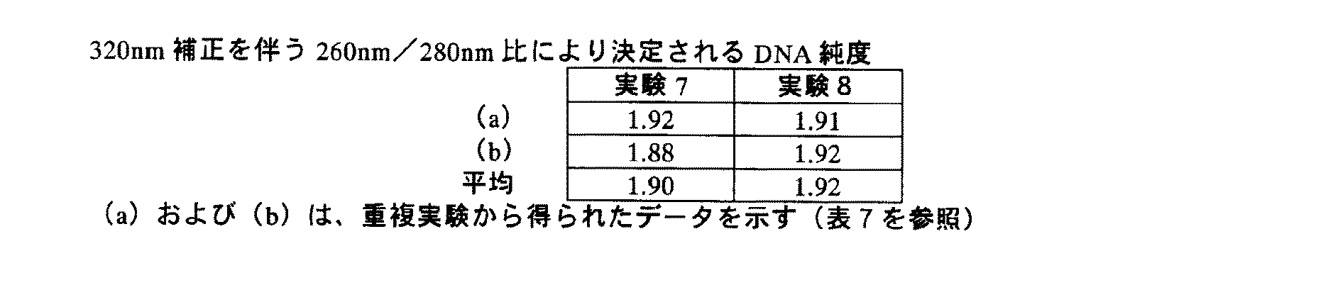
実施例2
(A)試薬
a)プロテイナーゼK保存溶液:組換え体プロテイナーゼK、PCR級、50U/ml
b)溶解緩衝液:4Mチオシアン酸グアニジニウム、50mM Tris-HCl、20%Triton-X100、pH6.0
c)結合緩衝液:4Mチオシアン酸グアニジニウム、50mM Tris-HCl、20%Triton-X100、pH6.0
d)インヒビター除去緩衝液:5M塩酸グアニジニウム、20mM Tris-HCl、38%エタノール、pH6.6
e)洗浄緩衝液:20mM NaCl、2mM Tris-HCl、80%エタノール、pH7.5
f)溶出緩衝液:50mM Tris-HCl、pH8.2
g)エタノール(100%)
Example 2
(A) Reagent
a) Proteinase K storage solution: recombinant proteinase K, PCR grade, 50 U / ml
b) Lysis buffer: 4M guanidinium thiocyanate, 50 mM Tris-HCl, 20% Triton-X100, pH 6.0
c) Binding buffer: 4M guanidinium thiocyanate, 50 mM Tris-HCl, 20% Triton-X100, pH 6.0
d) Inhibitor removal buffer: 5M guanidinium hydrochloride, 20 mM Tris-HCl, 38% ethanol, pH 6.6
e) Wash buffer: 20 mM NaCl, 2 mM Tris-HCl, 80% ethanol, pH 7.5
f) Elution buffer: 50 mM Tris-HCl, pH 8.2
g) Ethanol (100%)
さらなる必要性:シリカゲル膜を含有する市販のスピンカラム、例えば、Machery&Nagelにより配布されるNucleoSpin Blood L。 Further need: Commercial spin column containing silica gel membrane, eg NucleoSpin Blood L distributed by Machery & Nagel.
(B)実験1:プロテイナーゼK処理なしの1工程手順
1.15mlファルコンチューブに1,000μlのEDTA血液をピペット採取する
2.1,000μlの溶解緩衝液を添加し、ゆっくりボルテックスする
3.ローラー混合機で、室温で、15分間インキュベートし、かき混ぜる
4.スピンカラムを、新たなファルコンチューブに入れる
5.工程1.〜2.の混合物(約2,000μl)を、スピンカラムに移動する
6.1,900×gで、3分間、遠心分離する
7.1,000μlのインヒビター除去緩衝液を添加する
8.4,500×gで、2分間、遠心分離する
9.2,000μlの洗浄緩衝液を添加する
10.4,500×gで、10分間、遠心分離する
11.フロースルーを捨て、そして新たなファルコンチューブにフィルターカラムを入れる
12.300μlの予備加熱(70℃)した溶出緩衝液で溶出する
13.室温で、5分間、インキュベートする
14.4,500×gで、2分間、遠心分離する
15.260、280および320nmでの溶出液のOD測定
(B) Experiment 1: One-step procedure without proteinase K treatment
1. Pipette 1,000 μl EDTA blood into a 15 ml Falcon tube
2. Add 1,000 μl lysis buffer and vortex slowly
3. Incubate for 15 minutes at room temperature on a roller mixer and stir
Four. Place the spin column in a new falcon tube
Five.
6. Centrifuge at 1,900 xg for 3 minutes
7. Add 1,000 μl inhibitor removal buffer
8. Centrifuge at 4,500 xg for 2 minutes
9. Add 2,000 μl wash buffer
10. Centrifuge at 4,500 xg for 10 minutes
11. Discard the flow-through and place the filter column in a new falcon tube
12. Elute with 300 μl preheated (70 ° C) elution buffer
13. Incubate for 5 minutes at room temperature
14. Centrifuge at 4,500 xg for 2 minutes
15. OD measurement of eluate at 260, 280 and 320nm
(C)実験2:プロテイナーゼK処理を含む1工程手順
1.15mlファルコンチューブに125μlのプロテイナーゼKをピペット採取する
2.1,000μlのEDTA血液を添加し、ゆっくりボルテックスする
3.1,000μlの溶解緩衝液を添加し、ゆっくりボルテックスする
4.15分間、56℃で(例えば、熱混合機を使用することによって)インキュベートおよび振蘯し、かき混ぜる
5.スピンカラムを、新たなファルコンチューブに入れる
6.工程1.〜3.の混合物(約2,125μl)を、スピンカラムに移動する
7.1,900×gで、3分間、遠心分離する
8.1,000μlのインヒビター除去緩衝液を添加する
9.4,500×gで、2分間、遠心分離する
10.2,000μlの洗浄緩衝液を添加する
11.4,500×gで、10分間、遠心分離する
12.フロースルーを捨て、そして新たなファルコンチューブにフィルターカラムを入れる
13.300μlの予備加熱(70℃)した溶出緩衝液で溶出する
14.室温で、5分間、インキュベートする
15.4,500×gで、2分間、遠心分離する
16.260、280および320nmでの溶出液のOD測定
(C) Experiment 2: One-step procedure including proteinase K treatment
1. Pipette 125 μl proteinase K into a 15 ml falcon tube.
2. Add 1,000 μl EDTA blood and vortex slowly
3. Add 1,000 μl lysis buffer and vortex slowly
4. Incubate and shake for 15 minutes at 56 ° C (eg by using a heat mixer) and stir
Five. Place the spin column in a new falcon tube
6.
7. Centrifuge at 1,900 xg for 3 minutes
8. Add 1,000 μl inhibitor removal buffer
9. Centrifuge at 4,500 xg for 2 minutes
10. Add 2,000 μl wash buffer
11. Centrifuge at 4,500 xg for 10 minutes
12. Discard the flow-through and place the filter column in a new falcon tube
13. Elute with 300 μl preheated (70 ° C) elution buffer
14. Incubate for 5 minutes at room temperature
15. Centrifuge at 4,500 xg for 2 minutes
16. OD measurement of eluate at 260, 280 and 320nm
(D)実験3:エタノールを含む2工程手順
1.15mlファルコンチューブに125μlのプロテイナーゼKをピペット採取する
2.1,000μlのEDTA血液を添加し、ゆっくりボルテックスする
3.1,000μlの溶解緩衝液を添加し、ゆっくりボルテックスする
4.15分間、56℃で(例えば、熱混合機を使用することによって)インキュベートおよび振蘯し、かき混ぜる
5.1,000μlのエタノールを添加し、ゆっくりボルテックスする
6.スピンカラムを、新たなファルコンチューブに入れる
7.工程1.〜5.の混合物(約3,125μl)を、スピンカラムに移動する
8.1,900×gで、3分間、遠心分離する
9.1,000μlのインヒビター除去緩衝液を添加する
10.4,500×gで、2分間、遠心分離する
11.2,000μlの洗浄緩衝液を添加する
12.4,500×gで、10分間、遠心分離する
13.フロースルーを捨て、そして新たなファルコンチューブにフィルターカラムを入れる
14.300μlの予備加熱(70℃)した溶出緩衝液で溶出する
15.室温で、5分間、インキュベートする
16.4,500×gで、2分間、遠心分離する
17.260、280および320nmでの溶出液のOD測定
(D) Experiment 3: Two-step procedure with ethanol
1. Pipette 125 μl proteinase K into a 15 ml falcon tube.
2. Add 1,000 μl EDTA blood and vortex slowly
3. Add 1,000 μl lysis buffer and vortex slowly
4. Incubate and shake for 15 minutes at 56 ° C (eg by using a heat mixer) and stir
5. Add 1,000 μl of ethanol and vortex slowly
6. Place the spin column in a new falcon tube
7. Process 1.-5. Transfer the mixture (approximately 3,125 μl) to the spin column
8. Centrifuge at 1,900 xg for 3 minutes
9. Add 1,000 μl inhibitor removal buffer
10. Centrifuge at 4,500 xg for 2 minutes
11. Add 2,000 μl wash buffer
12. Centrifuge at 4,500 xg for 10 minutes
13. Discard the flow-through and place the filter column in a new falcon tube
14. Elute with 300 μl preheated (70 ° C) elution buffer
15. Incubate for 5 minutes at room temperature
16. Centrifuge at 4,500 xg for 2 minutes
17. OD measurement of eluate at 260, 280 and 320 nm
(E)実験4:結合緩衝液を含む2工程手順
1.15mlファルコンチューブに125μlのプロテイナーゼKをピペット採取する
2.1,000μlのEDTA血液を添加し、ゆっくりボルテックスする
3.1,000μlの溶解緩衝液を添加し、ゆっくりボルテックスする
4.15分間、56℃で(例えば、熱混合機を使用することによって)インキュベートおよび振蘯し、かき混ぜる
5.1,000μlの結合緩衝液を添加し、ゆっくりボルテックスする
6.スピンカラムを、新たなファルコンチューブに入れる
7.工程1.〜5.の混合物(約3,125μl)を、スピンカラムに移動する
8.1,900×gで、3分間、遠心分離する
9.1,000μlのインヒビター除去緩衝液を添加する
10.4,500×gで、2分間、遠心分離する
11.2,000μlの洗浄緩衝液を添加する
12.4,500×gで、10分間、遠心分離する
13.フロースルーを捨て、そして新たなファルコンチューブにフィルターカラムを入れる
14.300μlの予備加熱(70℃)した溶出緩衝液で溶出する
15.室温で、5分間、インキュベートする
16.4,500×gで、2分間、遠心分離する
17.260、280および320nmでの溶出液のOD測定
(E) Experiment 4: Two-step procedure with binding buffer
1. Pipette 125 μl proteinase K into a 15 ml falcon tube.
2. Add 1,000 μl EDTA blood and vortex slowly
3. Add 1,000 μl lysis buffer and vortex slowly
4. Incubate and shake for 15 minutes at 56 ° C (eg by using a heat mixer) and stir
5. Add 1,000 μl binding buffer and vortex slowly
6. Place the spin column in a new falcon tube
7. Process 1.-5. Transfer the mixture (approximately 3,125 μl) to the spin column
8. Centrifuge at 1,900 xg for 3 minutes
9. Add 1,000 μl inhibitor removal buffer
10. Centrifuge at 4,500 xg for 2 minutes
11. Add 2,000 μl wash buffer
12. Centrifuge at 4,500 xg for 10 minutes
13. Discard the flow-through and place the filter column in a new falcon tube
14. Elute with 300 μl preheated (70 ° C) elution buffer
15. Incubate for 5 minutes at room temperature
16. Centrifuge at 4,500 xg for 2 minutes
17. OD measurement of eluate at 260, 280 and 320 nm
DNAの純度に関して、1.8±0.1の260nm/280nm比(320補正を含めた)は、許容しうると見なされる。そのデータは、本発明による方法、すなわち実験4の方法は、実験3の方法と比較して等しい純度(高純度のDNAでない場合)を生じることを示す。
For DNA purity, a 260 nm / 280 nm ratio of 1.8 ± 0.1 (including 320 correction) is considered acceptable. The data show that the method according to the invention, ie the method of
実施例3
(A)試薬
a)溶解緩衝液/結合緩衝液:カオトロピック(7M[溶解緩衝液]/4M[結合緩衝液]チオシアン酸グアニジニウム、50mM Tris-HCl、20%Triton-X100、pH6.0)
b)インヒビター除去緩衝液(5MグアニジニウムHCl、20mM Tris-HCl、38%エタノール、pH6.6)
c)洗浄緩衝液(20mM NaCl、2mM Tris-HCl、80%エタノール、pH7.5)
d)溶出緩衝液(50mM Tris、pH8.1)
e)エタノール(無水)
Example 3
(A) Reagent
a) Lysis buffer / binding buffer: chaotropic (7M [lysis buffer] / 4M [binding buffer] guanidinium thiocyanate, 50 mM Tris-HCl, 20% Triton-X100, pH 6.0)
b) Inhibitor removal buffer (5M guanidinium HCl, 20 mM Tris-HCl, 38% ethanol, pH 6.6)
c) Wash buffer (20 mM NaCl, 2 mM Tris-HCl, 80% ethanol, pH 7.5)
d) Elution buffer (50 mM Tris, pH 8.1)
e) Ethanol (anhydrous)
さらなる必要性:ガラス繊維フィルターカラム(シリカゲル膜を有する、例えばNucleoSpin Blood L、Machery−Nagelから市販) Further need: glass fiber filter column (with silica gel membrane, eg commercially available from NucleoSpin Blood L, Machery-Nagel)
(B)実験5:1工程手順:溶解緩衝液:7M、結合緩衝液なし
1,000μlのEDTA血液を、15mlファルコンチューブにピペット採取し、1,000μlの溶解緩衝液(7M)を添加し、そしてチューブをボルテックスした。混合物を、15分間、56℃で、熱混合機でインキュベートした。ガラス繊維フィルターカラムを有する新たなファルコンチューブを提供し、そして全サンプル調製物(約2,000μl;カオトロープ濃度3.5M)を、フィルターカラムに移動させた。1,900×gで、3分間の遠心分離の後、1,000μlのインヒビター除去緩衝液を、カラムに移動させ、続いて4,200×gで、2分間、別の遠心分離工程を行った。その後、2,000μlの洗浄緩衝液を、カラムに移動させ、続いて4,200×gで、10分間、遠心分離した。フロースルーを捨て、そしてフィルターカラムを、新たなファルコンチューブに入れた。結合した核酸を、500μlの予備加熱(70℃)した溶出緩衝液を使用して溶出させた。溶出緩衝液を、カラムに移動させ、そして室温で5分間インキュベートした。4,200×gで、2分間、遠心分離の後、230、260、280および320nmでの光学密度測定により、フロースルーを分析した。
(B) Experiment 5: 1 step procedure: Lysis buffer: 7M, no binding buffer
1,000 μl EDTA blood was pipetted into a 15 ml Falcon tube, 1,000 μl lysis buffer (7M) was added, and the tube vortexed. The mixture was incubated for 15 minutes at 56 ° C. in a thermal mixer. A new falcon tube with a glass fiber filter column was provided and the entire sample preparation (approximately 2,000 μl; chaotrope concentration 3.5 M) was transferred to the filter column. After centrifugation at 1,900 × g for 3 minutes, 1,000 μl of inhibitor removal buffer was transferred to the column followed by another centrifugation step at 4,200 × g for 2 minutes. Thereafter, 2,000 μl of wash buffer was transferred to the column followed by centrifugation at 4,200 × g for 10 minutes. The flow-through was discarded and the filter column was placed in a new falcon tube. Bound nucleic acids were eluted using 500 μl preheated (70 ° C.) elution buffer. The elution buffer was transferred to the column and incubated for 5 minutes at room temperature. Flow-through was analyzed by optical density measurements at 230, 260, 280 and 320 nm after centrifugation at 4,200 × g for 2 minutes.
(C)実験6、2工程手順:溶解緩衝液:7M、結合緩衝液:4M
1,000μlのEDTA血液を、15mlファルコンチューブにピペット採取し、1,000μlの溶解緩衝液(7M)を添加し、そしてチューブをボルテックスした。混合物を、15分間、56℃で、熱混合機でインキュベートした。500μlの結合緩衝液(4M)を添加し、ボルテックスにより混合した。ガラス繊維フィルターカラムを有する新たなファルコンチューブを提供し、そして全サンプル調製物(約2,500μl;カオトロープ濃度4.5M)を、フィルターカラムに移動させた。1,900×gで、3分間の遠心分離の後、1,000μlのインヒビター除去緩衝液を、カラムに移動させ、続いて4,200×gで、2分間、別の遠心分離工程を行った。その後、2,000μlの洗浄緩衝液を、カラムに移動させ、続いて4,200×gで、10分間、遠心分離した。フロースルーを捨て、そしてフィルターカラムを、新たなファルコンチューブに入れた。結合した核酸を、500μlの予備加熱(70℃)した溶出緩衝液を使用して溶出させた。溶出緩衝液を、カラムに移動させ、そして室温で5分間インキュベートした。4,200×gで、2分間、遠心分離の後、230、260、280および320nmでの光学密度測定により、フロースルーを分析した。
(C)
1,000 μl EDTA blood was pipetted into a 15 ml Falcon tube, 1,000 μl lysis buffer (7M) was added, and the tube vortexed. The mixture was incubated for 15 minutes at 56 ° C. in a thermal mixer. 500 μl of binding buffer (4M) was added and mixed by vortexing. A new falcon tube with a glass fiber filter column was provided and the entire sample preparation (approximately 2,500 μl; chaotrope concentration 4.5 M) was transferred to the filter column. After centrifugation at 1,900 × g for 3 minutes, 1,000 μl of inhibitor removal buffer was transferred to the column followed by another centrifugation step at 4,200 × g for 2 minutes. Thereafter, 2,000 μl of wash buffer was transferred to the column followed by centrifugation at 4,200 × g for 10 minutes. The flow-through was discarded and the filter column was placed in a new falcon tube. Bound nucleic acids were eluted using 500 μl preheated (70 ° C.) elution buffer. The elution buffer was transferred to the column and incubated for 5 minutes at room temperature. Flow-through was analyzed by optical density measurements at 230, 260, 280 and 320 nm after centrifugation at 4,200 × g for 2 minutes.
実施例4
(A)試薬
a)溶解緩衝液/結合緩衝液:カオトロピック(7M[溶解緩衝液]/5M[溶解緩衝液]/4M[結合緩衝液]チオシアン酸グアニジニウム、50mM Tris-HCl、20%Triton-X100、pH6.0)
b)インヒビター除去緩衝液(5MグアニジニウムHCl、20mM Tris-HCl、38%エタノール、pH6.6)
c)洗浄緩衝液(20mM NaCl、2mM Tris-HCl、80%エタノール、pH7.5)
d)溶出緩衝液(50mM Tris、pH8.1)
e)エタノール(無水)
Example 4
(A) Reagent
a) Lysis buffer / binding buffer: chaotropic (7M [lysis buffer] / 5M [lysis buffer] / 4M [binding buffer] guanidinium thiocyanate, 50 mM Tris-HCl, 20% Triton-X100, pH 6.0 )
b) Inhibitor removal buffer (5M guanidinium HCl, 20 mM Tris-HCl, 38% ethanol, pH 6.6)
c) Wash buffer (20 mM NaCl, 2 mM Tris-HCl, 80% ethanol, pH 7.5)
d) Elution buffer (50 mM Tris, pH 8.1)
e) Ethanol (anhydrous)
さらなる必要性:ガラス繊維フィルターカラム(シリカゲル膜を有する、例えばNucleoSpin Blood L、Machery-Nagelから市販) Further need: glass fiber filter column (with silica gel membrane, eg NucleoSpin Blood L, commercially available from Machery-Nagel)
(B)実験7:1工程手順:溶解緩衝液:7M、結合緩衝液なし
1,000μlのEDTA血液を、15mlファルコンチューブにピペット採取し、1,000μlの溶解緩衝液(7M)を添加し、そしてチューブをボルテックスした。混合物を、15分間、56℃で、熱混合機でインキュベートした。ガラス繊維フィルターカラムを有する新たなファルコンチューブを提供し、そして全サンプル調製物(約2,000μl;カオトロープ濃度3.5M)を、フィルターカラムに移動させた。1,900×gで、3分間の遠心分離の後、1,000μlのインヒビター除去緩衝液を、カラムに移動させ、続いて4,200×gで、2分間、別の遠心分離工程を行った。その後、2,000μlの洗浄緩衝液を、カラムに移動させ、続いて4,200×gで、10分間、遠心分離した。フロースルーを捨て、そしてフィルターカラムを、新たなファルコンチューブに入れた。結合した核酸を、500μlの予備加熱(70℃)した溶出緩衝液を使用して溶出させた。溶出緩衝液を、カラムに移動させ、そして室温で5分間インキュベートした。4,200×gで、2分間、遠心分離の後、230、260、280および320nmでの光学密度測定により、フロースルーを分析した。
(B) Experiment 7: 1 step procedure: Lysis buffer: 7M, no binding buffer
1,000 μl EDTA blood was pipetted into a 15 ml Falcon tube, 1,000 μl lysis buffer (7M) was added, and the tube vortexed. The mixture was incubated for 15 minutes at 56 ° C. in a thermal mixer. A new falcon tube with a glass fiber filter column was provided and the entire sample preparation (approximately 2,000 μl; chaotrope concentration 3.5 M) was transferred to the filter column. After centrifugation at 1,900 × g for 3 minutes, 1,000 μl of inhibitor removal buffer was transferred to the column followed by another centrifugation step at 4,200 × g for 2 minutes. Thereafter, 2,000 μl of wash buffer was transferred to the column followed by centrifugation at 4,200 × g for 10 minutes. The flow-through was discarded and the filter column was placed in a new falcon tube. Bound nucleic acids were eluted using 500 μl preheated (70 ° C.) elution buffer. The elution buffer was transferred to the column and incubated for 5 minutes at room temperature. Flow-through was analyzed by optical density measurements at 230, 260, 280 and 320 nm after centrifugation at 4,200 × g for 2 minutes.
(C)実験8、2工程手順:溶解緩衝液:5M、結合緩衝液:4M
1,000μlのEDTA血液を、15mlファルコンチューブにピペット採取し、1,000μlの溶解緩衝液(5M)を添加し、そしてチューブをボルテックスした。混合物を、15分間、56℃で、熱混合機でインキュベートした。500μlの結合緩衝液(4M)を添加し、ボルテックスにより混合した。ガラス繊維フィルターカラムを有する新たなファルコンチューブを提供し、そして全サンプル調製物(約2,500μl;カオトロープ濃度3.5M)を、フィルターカラムに移動させた。1,900×gで、3分間の遠心分離の後、1,000μlのインヒビター除去緩衝液を、カラムに移動させ、続いて4,200×gで、2分間、別の遠心分離工程を行った。その後、2,000μlの洗浄緩衝液を、カラムに移動させ、続いて4,200×gで、10分間、遠心分離した。フロースルーを捨て、そしてフィルターカラムを、新たなファルコンチューブに入れた。結合した核酸を、500μlの予備加熱(70℃)した溶出緩衝液を使用して溶出させた。溶出緩衝液を、カラムに移動させ、そして室温で5分間インキュベートした。4,200×gで、2分間、遠心分離の後、230、260、280および320nmでの光学密度測定により、フロースルーを分析した。
(C) Experiment 8, two-step procedure: lysis buffer: 5M, binding buffer: 4M
1,000 μl of EDTA blood was pipetted into a 15 ml falcon tube, 1,000 μl of lysis buffer (5M) was added, and the tube was vortexed. The mixture was incubated for 15 minutes at 56 ° C. in a thermal mixer. 500 μl of binding buffer (4M) was added and mixed by vortexing. A new falcon tube with a glass fiber filter column was provided and the entire sample preparation (about 2,500 μl; chaotrope concentration 3.5 M) was transferred to the filter column. After centrifugation at 1,900 × g for 3 minutes, 1,000 μl of inhibitor removal buffer was transferred to the column followed by another centrifugation step at 4,200 × g for 2 minutes. Thereafter, 2,000 μl of wash buffer was transferred to the column followed by centrifugation at 4,200 × g for 10 minutes. The flow-through was discarded and the filter column was placed in a new falcon tube. Bound nucleic acids were eluted using 500 μl preheated (70 ° C.) elution buffer. The elution buffer was transferred to the column and incubated for 5 minutes at room temperature. Flow-through was analyzed by optical density measurements at 230, 260, 280 and 320 nm after centrifugation at 4,200 × g for 2 minutes.
参考文献一覧
Abramson, R.D., およびMyers, T.W., Curr. Opin. Biotechnol. 4 (1993) 41-47
Alderton, R.P.,ら, Anal. Biochem. 201 (1992) 166-169
Ausubelら, Current Protocols in Molecular Biology, J. Wiley and Sons, NY, 1987
Barany, F., PCR Methods and Applic. 1 (1991) 5-16
Barany, F., Proc. Natl. Acad. Sci. USA 88 (1991) 189-193
Boom, R.,ら, J. Clin. Microbiol. 28 (1990) 495-503
Cacace, M.G.,ら. Quarterly Review of Biophysics (1997) 30:241-277
DE 37 24 442
EP 0 439 182
EP 0 389 063
EP 0 658 164
EP 0 819 696
Guatelli, J.C.,ら, Proc. Natl. Acad. Sci. USA 87 (1990) 1874-1878
Jakobi, R.,ら, Anal. Biochem. 175 (1988) 196-201
Kwoh, D.Y., et al., Proc. Natl. Acad. Sci. USA 86 (1989) 1173-1177
Marko, M.A., et al., Anal. Biochem. 121 (1982) 382-387
Sambrook, Fritsch & Maniatis, Molecular Cloning, A Laboratory Manual, 3rd edition, CSHL Press, 2001
US 4,683,195
US 5,130,238
US 5,210,015
US 5,487,972
US 5,804,375
US 5,808,041
Vogelstein, B.,およびGillespie, D., Proc. Natl. Acad. Sci. USA 76 (1979) 615-619
Whelen, A.C.,およびPersing, D.H., Annu. Rev. Microbiol. 50 (1996) 349-373
WO 01/37291
WO 90/01069
WO 91/00212
WO 92/02638
WO 92/08800
Wu, D.Y.,およびWallace, R.B., Genomics 4 (1989) 560-569
Yamada, O.,ら, J. Virol. Methods 27 (1990) 203-209
List of references
Abramson, RD, and Myers, TW, Curr. Opin. Biotechnol. 4 (1993) 41-47
Alderton, RP, et al., Anal. Biochem. 201 (1992) 166-169
Ausubel et al., Current Protocols in Molecular Biology, J. Wiley and Sons, NY, 1987
Barany, F., PCR Methods and Applic. 1 (1991) 5-16
Barany, F., Proc. Natl. Acad. Sci. USA 88 (1991) 189-193
Boom, R., et al., J. Clin. Microbiol. 28 (1990) 495-503
Cacace, MG, et al. Quarterly Review of Biophysics (1997) 30: 241-277
DE 37 24 442
Guatelli, JC, et al., Proc. Natl. Acad. Sci. USA 87 (1990) 1874-1878
Jakobi, R., et al., Anal. Biochem. 175 (1988) 196-201
Kwoh, DY, et al., Proc. Natl. Acad. Sci. USA 86 (1989) 1173-1177
Marko, MA, et al., Anal. Biochem. 121 (1982) 382-387
Sambrook, Fritsch & Maniatis, Molecular Cloning, A Laboratory Manual, 3rd edition, CSHL Press, 2001
US 4,683,195
US 5,130,238
US 5,210,015
US 5,487,972
US 5,804,375
US 5,808,041
Vogelstein, B., and Gillespie, D., Proc. Natl. Acad. Sci. USA 76 (1979) 615-619
Whelen, AC, and Persing, DH, Annu. Rev. Microbiol. 50 (1996) 349-373
WO 01/37291
WO 90/01069
WO 91/00212
WO 92/02638
WO 92/08800
Wu, DY, and Wallace, RB, Genomics 4 (1989) 560-569
Yamada, O., et al., J. Virol. Methods 27 (1990) 203-209
Claims (14)
(b)該溶解緩衝液を生物学的サンプルと混合し、ここで、混合物中のカオトロピック剤の濃度は1Mと4Mとの間であり、該混合物をインキュベートする工程;
(c)工程(b)の混合物に、さらなる量のカオトロピック剤を溶解するか、またはさらなるカオトロピック剤を含有する水溶液を添加し、それにより該混合物中のカオトロピック剤の濃度を0.5Mを超える多い量だけ増加させる工程;
(d)工程(c)の混合物を固相と接触させ、それにより該混合物由来の核酸を該固相に吸着する工程
を含む、生物学的サンプル由来の核酸を固相に吸着するための方法。 (A) providing an aqueous lysis buffer containing a chaotropic agent;
(B) mixing the lysis buffer with a biological sample, wherein the concentration of chaotropic agent in the mixture is between 1M and 4M and incubating the mixture;
(C) An additional amount of chaotropic agent is dissolved in the mixture of step (b) or an aqueous solution containing additional chaotropic agent is added, thereby increasing the concentration of chaotropic agent in the mixture to more than 0.5M the step of Ru is increased by;
(D) a method for adsorbing a nucleic acid from a biological sample to a solid phase, comprising contacting the mixture of step (c) with the solid phase, thereby adsorbing the nucleic acid from the mixture to the solid phase. .
(b)該溶解緩衝液を生物学的サンプルと混合し、ここで、混合物中のカオトロピック剤の濃度は1Mと4Mとの間であり、該混合物をインキュベートする工程;
(c)工程(b)の混合物に、さらなる量のカオトロピック剤を溶解するか、またはさらなるカオトロピック剤を含有する水溶液を添加し、それにより該混合物中のカオトロピック剤の濃度を0.5Mを超える多い量だけ増加させる工程;
(d)固相を提供し、工程(c)の混合物を固相と接触させ、それにより該混合物由来の核酸を該固相に吸着する工程;
(e)液相から該固相を分離する工程;
(f)工程(e)の固相を洗浄緩衝液で任意に洗浄する工程;
(g)該固相から核酸を溶出し、それにより核酸を単離する工程;
(h)工程(g)の核酸を溶出液から任意に沈殿させ、沈殿した核酸を単離する工程
を含む、生物学的サンプル由来の核酸を単離するための方法。 (A) providing an aqueous lysis buffer containing a chaotropic agent;
(B) mixing the lysis buffer with a biological sample, wherein the concentration of chaotropic agent in the mixture is between 1M and 4M and incubating the mixture;
(C) An additional amount of chaotropic agent is dissolved in the mixture of step (b) or an aqueous solution containing additional chaotropic agent is added, thereby increasing the concentration of chaotropic agent in the mixture to more than 0.5M the step of Ru is increased by;
(D) providing a solid phase, contacting the mixture of step (c) with the solid phase, thereby adsorbing nucleic acid from the mixture to the solid phase;
(E) separating the solid phase from the liquid phase;
(F) optionally washing the solid phase of step (e) with a wash buffer;
(G) eluting the nucleic acid from the solid phase, thereby isolating the nucleic acid;
(H) A method for isolating nucleic acid derived from a biological sample, comprising the step of optionally precipitating the nucleic acid of step (g) from the eluate and isolating the precipitated nucleic acid.
洗剤がドデシル硫酸ナトリウム、ドデシル硫酸リチウム、セチルトリメチルアンモニウムブロミド、デオキシコール酸、ラウロイルザルコシンナトリウム、Triton(登録商標)-X100、Tween(登録商標)20、オクチルβ-D-グルコシド、Nonidet(登録商標)P40、CHAPSおよびSulphobetaine 14からなる群より選択されることを特徴とする、請求項10記載の方法。 The enzyme having proteolytic activity is selected from the group consisting of caspase, proteinase K, pronase E, Bacillus sp-derived protease (esperase), and subtilisin;
Detergents are sodium dodecyl sulfate, lithium dodecyl sulfate, cetyltrimethylammonium bromide, deoxycholic acid, sodium lauroyl sarcosine, Triton (registered trademark) -X100, Tween (registered trademark) 20, octyl β-D-glucoside, Nonidet (registered trademark) ) P40, CHAPS and Sulphobetaine 14, characterized in that it is selected from the group consisting of the method of claim 10, wherein.
14. A method according to any one of claims 8 to 13, characterized in that the biological sample contains bacterial cells, eukaryotic cells, viruses or mixtures thereof.
Applications Claiming Priority (1)
| Application Number | Priority Date | Filing Date | Title |
|---|---|---|---|
| EP04003898 | 2004-02-20 |
Publications (2)
| Publication Number | Publication Date |
|---|---|
| JP2005230012A JP2005230012A (en) | 2005-09-02 |
| JP4271663B2 true JP4271663B2 (en) | 2009-06-03 |
Family
ID=34878164
Family Applications (1)
| Application Number | Title | Priority Date | Filing Date |
|---|---|---|---|
| JP2005042368A Expired - Lifetime JP4271663B2 (en) | 2004-02-20 | 2005-02-18 | Nucleic acid adsorption to solid phase |
Country Status (15)
| Country | Link |
|---|---|
| US (1) | US7491495B2 (en) |
| EP (1) | EP1566437B1 (en) |
| JP (1) | JP4271663B2 (en) |
| KR (1) | KR100692941B1 (en) |
| CN (1) | CN1310938C (en) |
| AT (1) | ATE364694T1 (en) |
| AU (1) | AU2005200670B2 (en) |
| BR (1) | BRPI0500474B1 (en) |
| CA (1) | CA2497493C (en) |
| DE (1) | DE602005001331T2 (en) |
| DK (1) | DK1566437T3 (en) |
| ES (1) | ES2289611T3 (en) |
| MX (1) | MXPA05001815A (en) |
| PL (1) | PL1566437T3 (en) |
| SG (1) | SG114738A1 (en) |
Families Citing this family (74)
| Publication number | Priority date | Publication date | Assignee | Title |
|---|---|---|---|---|
| US7078224B1 (en) * | 1999-05-14 | 2006-07-18 | Promega Corporation | Cell concentration and lysate clearance using paramagnetic particles |
| US20070015165A1 (en) * | 2005-07-13 | 2007-01-18 | Sigma-Aldrich Co. | Method for the isolation of RNA from biological sources |
| EP1963526A4 (en) * | 2005-12-09 | 2009-11-18 | Promega Corp | Nucleic acid purification with a binding matrix |
| WO2007103485A2 (en) * | 2006-03-08 | 2007-09-13 | Promega Corporation | Small rna purification |
| EP2027293B1 (en) * | 2006-05-24 | 2014-11-19 | Gen-Probe Incorporated | Method of lysing mycobacterium and amplifying its nucleic acid |
| DE102006032610C5 (en) * | 2006-07-11 | 2016-09-15 | Aj Innuscreen Gmbh | Method for the parallel isolation of viral nucleic acids |
| US10131935B2 (en) * | 2006-07-11 | 2018-11-20 | Aj Innuscreen Gmbh | Method for parallel isolation of viral nucleic acids |
| AU2007281535A1 (en) * | 2006-08-01 | 2008-02-07 | Applied Biosystems, Llc. | Detection of analytes and nucleic acids |
| KR100805449B1 (en) * | 2006-08-18 | 2008-02-20 | 배정훈 | Method for Purifying DNA from Agarose Gel Using Zeolite Powder as Molecular Sieve |
| CA2629589C (en) * | 2007-04-20 | 2016-03-29 | F.Hoffmann-La Roche Ag | Isolation and purification of nucleic acid molecules with a solid phase |
| CA2629586C (en) * | 2007-04-20 | 2016-05-24 | F.Hoffmann-La Roche Ag | Adsorption of nucleic acids to solid phases under low-salt conditions |
| CN101323852B (en) * | 2007-06-11 | 2013-01-23 | 北京东胜创新生物科技有限公司 | Reagent box and method for genome extraction on automatic nucleic acid extraction workstation |
| US20090035748A1 (en) * | 2007-08-01 | 2009-02-05 | Lizzi Tiffiny Marie | Bromelain as a clinical sample pre-treatment, lysis agent and nuclease inhibitor |
| US20090048438A1 (en) * | 2007-08-13 | 2009-02-19 | Taigen Bioscience Corporation. | Method for washing a column and method for extracting membrane-bound target molecules |
| US9347055B2 (en) * | 2007-11-05 | 2016-05-24 | Eiken Kagaku Kabushiki Kaisha | Method and kit for preparation of sample for use in nucleic acid amplification |
| DE102008026058A1 (en) * | 2008-05-30 | 2009-12-03 | Qiagen Gmbh | Lysis, binding and / or washing reagent useful for isolation and / or purification of nucleic acids |
| ES2539856T3 (en) * | 2008-08-01 | 2015-07-06 | F. Hoffmann-La Roche Ag | Enhanced lysis and reverse transcription for mRNA quantification |
| DE102008047790A1 (en) * | 2008-09-17 | 2010-04-15 | Qiagen Gmbh | Method for normalizing the content of biomolecules in a sample |
| BRPI0919895B1 (en) | 2008-10-31 | 2019-08-13 | Bio Merieux Inc | method for identifying an unknown microorganism from a test sample |
| CN102317789B (en) | 2008-10-31 | 2014-08-27 | 生物梅里埃公司 | Methods for separation, characterization, and/or identification of microorganisms using mass spectrometry |
| US8647835B2 (en) | 2008-10-31 | 2014-02-11 | BIO MéRIEUX, INC. | Methods for separation, characterization and/or identification of microorganisms using spectroscopy |
| MX2012001236A (en) * | 2009-08-04 | 2012-03-16 | Du Pont | Process and device for collecting nucleic acids of microorganisms from a particulate sample. |
| US8039613B2 (en) | 2009-08-28 | 2011-10-18 | Promega Corporation | Methods of purifying a nucleic acid and formulation and kit for use in performing such methods |
| US8222397B2 (en) | 2009-08-28 | 2012-07-17 | Promega Corporation | Methods of optimal purification of nucleic acids and kit for use in performing such methods |
| TWI407994B (en) * | 2009-10-22 | 2013-09-11 | Ind Tech Res Inst | Method, agent, and kit for isolating nucleic acids |
| CN102094002A (en) * | 2009-12-11 | 2011-06-15 | 上海裕隆临床检验中心有限公司 | Extraction reagent and extraction method of hepatitis B virus DNA |
| CN101869827A (en) * | 2010-04-30 | 2010-10-27 | 北京九州泰康生物科技有限责任公司 | Method for preparing novel affinity medium and application thereof |
| EP2388312A1 (en) | 2010-05-17 | 2011-11-23 | Curetis AG | Universally applicable lysis buffer and processing methods for the lysis of bodily samples |
| ES2398646T3 (en) | 2010-07-24 | 2013-03-20 | F. Hoffmann-La Roche Ag | Stabilization of interleukin-6 in serum solutions |
| DE112011102524T5 (en) | 2010-07-29 | 2013-07-11 | Roche Diagnostics Gmbh | Preparation of generic samples |
| US9725754B2 (en) | 2010-07-29 | 2017-08-08 | Sean F. Boyle | Generic sample preparation |
| US8916344B2 (en) | 2010-11-15 | 2014-12-23 | Exact Sciences Corporation | Methylation assay |
| US8361720B2 (en) | 2010-11-15 | 2013-01-29 | Exact Sciences Corporation | Real time cleavage assay |
| GB201020095D0 (en) | 2010-11-26 | 2011-01-12 | Invitrogen Dynal As | Nucleic acid preparation method |
| US9051563B2 (en) * | 2011-01-14 | 2015-06-09 | Zymo Research Corporation | Nucleic acid purification |
| WO2012125710A1 (en) | 2011-03-15 | 2012-09-20 | E. I. Du Pont De Nemours And Company | Method for improved dna release from binding substrates and/or decreasing pcr inhibition in pathogen detection |
| US9029529B2 (en) | 2011-03-15 | 2015-05-12 | E.I. Du Pont De Nemours And Company | Methods for improved DNA release from binding substrates and/or decreasing PCR inhibition in pathogen detection |
| KR101193765B1 (en) * | 2011-05-16 | 2012-10-24 | (주)나노헬릭스 | Rapid nucleic acid purification |
| CN102533725A (en) * | 2011-09-09 | 2012-07-04 | 上海佑科生物技术有限公司 | Integrated buffer solution and method for separating nucleic acid by using same |
| CN103173432B (en) * | 2011-12-22 | 2020-08-04 | 通用电气公司 | Method and device for isolating nucleic acid |
| CN103667253B (en) * | 2012-09-18 | 2016-03-30 | 通用电气公司 | The method of isolating nucleic acid and device |
| US20140322706A1 (en) | 2012-10-24 | 2014-10-30 | Jon Faiz Kayyem | Integrated multipelx target analysis |
| AU2013334189B2 (en) | 2012-10-24 | 2018-08-02 | Genmark Diagnostics, Inc. | Integrated multiplex target analysis |
| CA2902916C (en) | 2013-03-14 | 2018-08-28 | Mayo Foundation For Medical Education And Research | Detecting neoplasm |
| JP6351702B2 (en) | 2013-03-15 | 2018-07-04 | ジェンマーク ダイアグノスティクス, インコーポレイテッド | System, method and apparatus for operating a deformable fluid container |
| US9498778B2 (en) | 2014-11-11 | 2016-11-22 | Genmark Diagnostics, Inc. | Instrument for processing cartridge for performing assays in a closed sample preparation and reaction system |
| USD881409S1 (en) | 2013-10-24 | 2020-04-14 | Genmark Diagnostics, Inc. | Biochip cartridge |
| US10184154B2 (en) | 2014-09-26 | 2019-01-22 | Mayo Foundation For Medical Education And Research | Detecting cholangiocarcinoma |
| JP2017539080A (en) | 2014-10-23 | 2017-12-28 | コーニング インコーポレイテッド | Polymer encapsulated magnetic nanoparticles |
| US10005080B2 (en) | 2014-11-11 | 2018-06-26 | Genmark Diagnostics, Inc. | Instrument and cartridge for performing assays in a closed sample preparation and reaction system employing electrowetting fluid manipulation |
| US9598722B2 (en) | 2014-11-11 | 2017-03-21 | Genmark Diagnostics, Inc. | Cartridge for performing assays in a closed sample preparation and reaction system |
| US10435755B2 (en) | 2015-03-27 | 2019-10-08 | Exact Sciences Development Company, Llc | Detecting esophageal disorders |
| CN114574585A (en) | 2015-08-31 | 2022-06-03 | 梅约医药教育及研究基金会 | Detection of gastric tumors |
| CN108350485A (en) | 2015-10-30 | 2018-07-31 | 精密科学发展有限责任公司 | Multiplex Amplified Detection Assay and Isolation and Detection of Plasma DNA |
| US20180135040A1 (en) | 2016-02-16 | 2018-05-17 | Life Magnetics, Inc. | Methods for separating nucleic acids with graphene coated magnetic beads |
| AU2017261283B2 (en) | 2016-05-04 | 2023-02-09 | Children's Hospital & Research Center At Oakland | Rapid extraction of nucleic acids from clinical samples for downstream applications |
| CN109563546B (en) * | 2016-05-05 | 2022-09-09 | 精密科学发展有限责任公司 | Detecting lung tumors by analyzing methylated DNA |
| CN108070584A (en) * | 2016-11-16 | 2018-05-25 | 江苏然科生物技术有限公司 | The silica gel adsorption column extracts kit and method of a kind of blood plasma or free serum DNA and RNA |
| KR102892245B1 (en) | 2017-01-27 | 2025-11-27 | 이그젝트 싸이언스 디블롭먼트 컴패니, 엘엘씨 | Detection of colon neoplasia by analysis of methylated dna |
| US10648025B2 (en) | 2017-12-13 | 2020-05-12 | Exact Sciences Development Company, Llc | Multiplex amplification detection assay II |
| CN108342382B (en) * | 2018-01-30 | 2020-12-15 | 广东海大畜牧兽医研究院有限公司 | A kind of nucleic acid rapid extraction method and its kit |
| CN108753767A (en) * | 2018-08-02 | 2018-11-06 | 广州奕昕生物科技有限公司 | A kind of Viral nucleic acid extraction reagent box and extracting method |
| WO2020037262A1 (en) * | 2018-08-17 | 2020-02-20 | Cepheid | Nucleic acid isolation and related methods |
| AU2019320825B2 (en) | 2018-08-17 | 2025-09-25 | Cepheid | Methods and compositions for nucleic acid isolation |
| CN113874522B (en) * | 2019-05-29 | 2025-07-22 | 藤仓化成株式会社 | Solid phase adhering composition, solid phase carrier using the same, and method for producing and using the solid phase carrier |
| CN110468125A (en) * | 2019-09-17 | 2019-11-19 | 广州欣源生物科技有限公司 | A kind of kit and its extracting method for viral nucleic acid extraction |
| GB202001034D0 (en) * | 2020-01-24 | 2020-03-11 | Ge Healthcare Uk Ltd | Method and kit for DNA isolation |
| DE102021204952A1 (en) * | 2021-05-17 | 2022-11-17 | Robert Bosch Gesellschaft mit beschränkter Haftung | Method for purifying nucleic acids, in particular in a microfluidic device |
| CN114672481A (en) * | 2022-04-21 | 2022-06-28 | 叶晓君 | Novel high-efficiency nucleic acid extraction method of plant fiber adsorption matrix |
| KR20250040066A (en) * | 2022-07-22 | 2025-03-21 | 바이오파이어 다이어그노스틱스, 엘엘씨 | Rapid cell lysis and nucleic acid recovery |
| CN119895050A (en) * | 2022-07-22 | 2025-04-25 | 拜奥法尔诊断有限责任公司 | Cell lysis and nucleic acid recovery |
| CN115960885B (en) * | 2022-10-09 | 2023-12-12 | 南京诺唯赞生物科技股份有限公司 | Method and composition for extracting nucleic acid from heparin sodium sample |
| CN117286229B (en) * | 2023-11-23 | 2024-05-03 | 中山大学中山眼科中心 | High-throughput long-reading long-sequencing method for three-dimensional genome structure of MHC (major histocompatibility complex) region |
| CN117535283B (en) * | 2024-01-08 | 2024-03-29 | 广州凯普医药科技有限公司 | Kit for extracting blood genome DNA by magnetic bead method and application thereof |
Family Cites Families (24)
| Publication number | Priority date | Publication date | Assignee | Title |
|---|---|---|---|---|
| US5075430A (en) * | 1988-12-12 | 1991-12-24 | Bio-Rad Laboratories, Inc. | Process for the purification of DNA on diatomaceous earth |
| NL8900725A (en) * | 1989-03-23 | 1990-10-16 | Az Univ Amsterdam | METHOD AND COMBINATION OF AGENTS FOR INSULATING NUCLEIC ACID. |
| US5234809A (en) * | 1989-03-23 | 1993-08-10 | Akzo N.V. | Process for isolating nucleic acid |
| US5124444A (en) * | 1989-07-24 | 1992-06-23 | Microprobe Corporation | Lactam-containing compositions and methods useful for the extraction of nucleic acids |
| US5130423A (en) * | 1990-07-13 | 1992-07-14 | Microprobe Corporation | Non-corrosive compositions and methods useful for the extraction of nucleic acids |
| US5155018A (en) * | 1991-07-10 | 1992-10-13 | Hahnemann University | Process and kit for isolating and purifying RNA from biological sources |
| AU7375794A (en) * | 1993-07-28 | 1995-02-28 | Akzo Nobel N.V. | Process for isolating nucleic acid from gram positive microorganisms |
| US5637687A (en) * | 1993-08-31 | 1997-06-10 | Wiggins; James C. | Methods and compositions for isolating nucleic acids |
| US6180778B1 (en) * | 1994-02-11 | 2001-01-30 | Qiagen Gmbh | Process for the separation of double-stranded/single-stranded nucleic acid structures |
| US5783686A (en) * | 1995-09-15 | 1998-07-21 | Beckman Instruments, Inc. | Method for purifying nucleic acids from heterogenous mixtures |
| ATE284891T1 (en) * | 1996-02-14 | 2005-01-15 | Biomerieux Bv | ISOLATION OF SINGLE STRANDED NUCLEIC ACIDS |
| US5981235A (en) * | 1996-07-29 | 1999-11-09 | Promega Corporation | Methods for isolating nucleic acids using alkaline protease |
| EP1003908B1 (en) * | 1997-04-16 | 2006-12-06 | Applera Corporation | Nucleic acid archiving |
| WO1998059076A1 (en) * | 1997-06-25 | 1998-12-30 | Promega Corporation | Method of isolating rna |
| US6111096A (en) * | 1997-10-31 | 2000-08-29 | Bbi Bioseq, Inc. | Nucleic acid isolation and purification |
| US6120985A (en) * | 1997-10-31 | 2000-09-19 | Bbi Bioseq, Inc. | Pressure-enhanced extraction and purification |
| US6914137B2 (en) * | 1997-12-06 | 2005-07-05 | Dna Research Innovations Limited | Isolation of nucleic acids |
| WO1999058664A1 (en) * | 1998-05-14 | 1999-11-18 | Whitehead Institute For Biomedical Research | Solid phase technique for selectively isolating nucleic acids |
| EP0969090A1 (en) * | 1998-05-27 | 2000-01-05 | QIAGEN GmbH | Rapid and simple process for isolation of circular nucleic acids |
| DE10006662A1 (en) * | 2000-02-15 | 2001-08-23 | Antigen Produktions Gmbh | Sample vessel for stabilizing and isolating nucleic acid, contains a lytic solution that stabilizes nucleic acid and a solid phase that binds it, especially for sampling whole blood |
| AU6380001A (en) * | 2000-03-24 | 2001-10-03 | Qiagen Gmbh | Porous ferro- or ferrimagnetic glass particles for isolating molecules |
| AU758744B2 (en) * | 2000-10-31 | 2003-03-27 | F. Hoffmann-La Roche Ag | Methods for the analysis of non-proteinaceous components using a protease from a bacillus strain |
| US7001724B1 (en) | 2000-11-28 | 2006-02-21 | Applera Corporation | Compositions, methods, and kits for isolating nucleic acids using surfactants and proteases |
| CN100395257C (en) * | 2003-05-08 | 2008-06-18 | 慈溪市中鼎生物技术有限公司 | Acid potassium ion aqua and DNA extracting method and kit therewith |
-
2005
- 2005-02-15 AU AU2005200670A patent/AU2005200670B2/en not_active Expired
- 2005-02-15 MX MXPA05001815A patent/MXPA05001815A/en active IP Right Grant
- 2005-02-16 ES ES05003254T patent/ES2289611T3/en not_active Expired - Lifetime
- 2005-02-16 DE DE602005001331T patent/DE602005001331T2/en not_active Expired - Lifetime
- 2005-02-16 AT AT05003254T patent/ATE364694T1/en active
- 2005-02-16 PL PL05003254T patent/PL1566437T3/en unknown
- 2005-02-16 DK DK05003254T patent/DK1566437T3/en active
- 2005-02-16 EP EP05003254A patent/EP1566437B1/en not_active Expired - Lifetime
- 2005-02-17 CA CA002497493A patent/CA2497493C/en not_active Expired - Lifetime
- 2005-02-17 BR BRPI0500474-8A patent/BRPI0500474B1/en not_active IP Right Cessation
- 2005-02-18 JP JP2005042368A patent/JP4271663B2/en not_active Expired - Lifetime
- 2005-02-18 CN CNB2005100542311A patent/CN1310938C/en not_active Expired - Lifetime
- 2005-02-18 US US11/061,948 patent/US7491495B2/en active Active
- 2005-02-18 SG SG200500954A patent/SG114738A1/en unknown
- 2005-02-18 KR KR1020050013430A patent/KR100692941B1/en not_active Expired - Lifetime
Also Published As
| Publication number | Publication date |
|---|---|
| CA2497493C (en) | 2009-06-16 |
| CN1680421A (en) | 2005-10-12 |
| US20050214926A1 (en) | 2005-09-29 |
| CA2497493A1 (en) | 2005-08-20 |
| EP1566437B1 (en) | 2007-06-13 |
| HK1084122A1 (en) | 2006-07-21 |
| KR20060042961A (en) | 2006-05-15 |
| BRPI0500474A (en) | 2006-04-04 |
| DE602005001331D1 (en) | 2007-07-26 |
| MXPA05001815A (en) | 2005-08-24 |
| PL1566437T3 (en) | 2007-11-30 |
| CN1310938C (en) | 2007-04-18 |
| JP2005230012A (en) | 2005-09-02 |
| EP1566437A1 (en) | 2005-08-24 |
| AU2005200670B2 (en) | 2007-05-03 |
| ATE364694T1 (en) | 2007-07-15 |
| DE602005001331T2 (en) | 2008-02-21 |
| DK1566437T3 (en) | 2007-10-15 |
| US7491495B2 (en) | 2009-02-17 |
| AU2005200670A1 (en) | 2005-09-08 |
| BRPI0500474B1 (en) | 2015-01-27 |
| SG114738A1 (en) | 2005-09-28 |
| KR100692941B1 (en) | 2007-03-12 |
| ES2289611T3 (en) | 2008-02-01 |
Similar Documents
| Publication | Publication Date | Title |
|---|---|---|
| JP4271663B2 (en) | Nucleic acid adsorption to solid phase | |
| JP4291247B2 (en) | Methods for isolating nucleic acids | |
| JP5325448B2 (en) | Isolation and purification of nucleic acid molecules using solid phase | |
| JP5354894B2 (en) | Nucleic acid isolation using polidocanols and derivatives | |
| US20110266172A1 (en) | Use of tde for isolation of nucleic acids | |
| ES2305023T3 (en) | METHODS FOR THE ANALYSIS OF NON-PROTEIN COMPONENTS USING A PROTEASE OF A BACILO CEPA. | |
| US7803935B2 (en) | Nucleic acid adsorption under low-salt conditions | |
| JP3621673B2 (en) | Method for analysis of non-protein components using protease from Bacillus strain | |
| WO2005093065A1 (en) | Improved method of isolating nucleic acids | |
| EP1524317B1 (en) | Methods for isolating nucleic acids | |
| US10131935B2 (en) | Method for parallel isolation of viral nucleic acids | |
| US20070148651A1 (en) | Method and kit for the isolation of rna | |
| JPH11262387A (en) | Nucleic acid-binding magnetic carrier and isolation of nucleic acid using the carrier | |
| EP1201752A1 (en) | Methods for the analysis of non-proteinaceous components using a protease from a Bacillus strain | |
| HK1084122B (en) | Adsorption of nucleic acids to a solid phase |
Legal Events
| Date | Code | Title | Description |
|---|---|---|---|
| A131 | Notification of reasons for refusal |
Free format text: JAPANESE INTERMEDIATE CODE: A131 Effective date: 20080414 |
|
| A601 | Written request for extension of time |
Free format text: JAPANESE INTERMEDIATE CODE: A601 Effective date: 20080711 |
|
| A602 | Written permission of extension of time |
Free format text: JAPANESE INTERMEDIATE CODE: A602 Effective date: 20080716 |
|
| A601 | Written request for extension of time |
Free format text: JAPANESE INTERMEDIATE CODE: A601 Effective date: 20080811 |
|
| A602 | Written permission of extension of time |
Free format text: JAPANESE INTERMEDIATE CODE: A602 Effective date: 20080814 |
|
| A521 | Request for written amendment filed |
Free format text: JAPANESE INTERMEDIATE CODE: A523 Effective date: 20080912 |
|
| A131 | Notification of reasons for refusal |
Free format text: JAPANESE INTERMEDIATE CODE: A131 Effective date: 20081031 |
|
| A521 | Request for written amendment filed |
Free format text: JAPANESE INTERMEDIATE CODE: A523 Effective date: 20090129 |
|
| TRDD | Decision of grant or rejection written | ||
| A01 | Written decision to grant a patent or to grant a registration (utility model) |
Free format text: JAPANESE INTERMEDIATE CODE: A01 Effective date: 20090219 |
|
| A01 | Written decision to grant a patent or to grant a registration (utility model) |
Free format text: JAPANESE INTERMEDIATE CODE: A01 |
|
| A61 | First payment of annual fees (during grant procedure) |
Free format text: JAPANESE INTERMEDIATE CODE: A61 Effective date: 20090225 |
|
| FPAY | Renewal fee payment (event date is renewal date of database) |
Free format text: PAYMENT UNTIL: 20120306 Year of fee payment: 3 |
|
| R150 | Certificate of patent or registration of utility model |
Ref document number: 4271663 Country of ref document: JP Free format text: JAPANESE INTERMEDIATE CODE: R150 Free format text: JAPANESE INTERMEDIATE CODE: R150 |
|
| FPAY | Renewal fee payment (event date is renewal date of database) |
Free format text: PAYMENT UNTIL: 20120306 Year of fee payment: 3 |
|
| FPAY | Renewal fee payment (event date is renewal date of database) |
Free format text: PAYMENT UNTIL: 20130306 Year of fee payment: 4 |
|
| R250 | Receipt of annual fees |
Free format text: JAPANESE INTERMEDIATE CODE: R250 |
|
| FPAY | Renewal fee payment (event date is renewal date of database) |
Free format text: PAYMENT UNTIL: 20130306 Year of fee payment: 4 |
|
| FPAY | Renewal fee payment (event date is renewal date of database) |
Free format text: PAYMENT UNTIL: 20140306 Year of fee payment: 5 |
|
| R250 | Receipt of annual fees |
Free format text: JAPANESE INTERMEDIATE CODE: R250 |
|
| R250 | Receipt of annual fees |
Free format text: JAPANESE INTERMEDIATE CODE: R250 |
|
| R250 | Receipt of annual fees |
Free format text: JAPANESE INTERMEDIATE CODE: R250 |
|
| R250 | Receipt of annual fees |
Free format text: JAPANESE INTERMEDIATE CODE: R250 |
|
| R250 | Receipt of annual fees |
Free format text: JAPANESE INTERMEDIATE CODE: R250 |
|
| R250 | Receipt of annual fees |
Free format text: JAPANESE INTERMEDIATE CODE: R250 |
|
| R250 | Receipt of annual fees |
Free format text: JAPANESE INTERMEDIATE CODE: R250 |
|
| R250 | Receipt of annual fees |
Free format text: JAPANESE INTERMEDIATE CODE: R250 |
|
| R250 | Receipt of annual fees |
Free format text: JAPANESE INTERMEDIATE CODE: R250 |
|
| R250 | Receipt of annual fees |
Free format text: JAPANESE INTERMEDIATE CODE: R250 |
|
| R250 | Receipt of annual fees |
Free format text: JAPANESE INTERMEDIATE CODE: R250 |
|
| R250 | Receipt of annual fees |
Free format text: JAPANESE INTERMEDIATE CODE: R250 |
|
| EXPY | Cancellation because of completion of term |