EP1201752A1 - Methods for the analysis of non-proteinaceous components using a protease from a Bacillus strain - Google Patents
Methods for the analysis of non-proteinaceous components using a protease from a Bacillus strain Download PDFInfo
- Publication number
- EP1201752A1 EP1201752A1 EP00123728A EP00123728A EP1201752A1 EP 1201752 A1 EP1201752 A1 EP 1201752A1 EP 00123728 A EP00123728 A EP 00123728A EP 00123728 A EP00123728 A EP 00123728A EP 1201752 A1 EP1201752 A1 EP 1201752A1
- Authority
- EP
- European Patent Office
- Prior art keywords
- protease
- acid sequence
- amino acid
- proteinaceous
- nucleic acids
- Prior art date
- Legal status (The legal status is an assumption and is not a legal conclusion. Google has not performed a legal analysis and makes no representation as to the accuracy of the status listed.)
- Withdrawn
Links
Images


Classifications
-
- C—CHEMISTRY; METALLURGY
- C12—BIOCHEMISTRY; BEER; SPIRITS; WINE; VINEGAR; MICROBIOLOGY; ENZYMOLOGY; MUTATION OR GENETIC ENGINEERING
- C12Q—MEASURING OR TESTING PROCESSES INVOLVING ENZYMES, NUCLEIC ACIDS OR MICROORGANISMS; COMPOSITIONS OR TEST PAPERS THEREFOR; PROCESSES OF PREPARING SUCH COMPOSITIONS; CONDITION-RESPONSIVE CONTROL IN MICROBIOLOGICAL OR ENZYMOLOGICAL PROCESSES
- C12Q1/00—Measuring or testing processes involving enzymes, nucleic acids or microorganisms; Compositions therefor; Processes of preparing such compositions
- C12Q1/34—Measuring or testing processes involving enzymes, nucleic acids or microorganisms; Compositions therefor; Processes of preparing such compositions involving hydrolase
- C12Q1/37—Measuring or testing processes involving enzymes, nucleic acids or microorganisms; Compositions therefor; Processes of preparing such compositions involving hydrolase involving peptidase or proteinase
-
- C—CHEMISTRY; METALLURGY
- C12—BIOCHEMISTRY; BEER; SPIRITS; WINE; VINEGAR; MICROBIOLOGY; ENZYMOLOGY; MUTATION OR GENETIC ENGINEERING
- C12N—MICROORGANISMS OR ENZYMES; COMPOSITIONS THEREOF; PROPAGATING, PRESERVING, OR MAINTAINING MICROORGANISMS; MUTATION OR GENETIC ENGINEERING; CULTURE MEDIA
- C12N9/00—Enzymes; Proenzymes; Compositions thereof; Processes for preparing, activating, inhibiting, separating or purifying enzymes
- C12N9/14—Hydrolases (3)
- C12N9/48—Hydrolases (3) acting on peptide bonds (3.4)
- C12N9/50—Proteinases, e.g. Endopeptidases (3.4.21-3.4.25)
- C12N9/52—Proteinases, e.g. Endopeptidases (3.4.21-3.4.25) derived from bacteria or Archaea
- C12N9/54—Proteinases, e.g. Endopeptidases (3.4.21-3.4.25) derived from bacteria or Archaea bacteria being Bacillus
-
- C—CHEMISTRY; METALLURGY
- C12—BIOCHEMISTRY; BEER; SPIRITS; WINE; VINEGAR; MICROBIOLOGY; ENZYMOLOGY; MUTATION OR GENETIC ENGINEERING
- C12Q—MEASURING OR TESTING PROCESSES INVOLVING ENZYMES, NUCLEIC ACIDS OR MICROORGANISMS; COMPOSITIONS OR TEST PAPERS THEREFOR; PROCESSES OF PREPARING SUCH COMPOSITIONS; CONDITION-RESPONSIVE CONTROL IN MICROBIOLOGICAL OR ENZYMOLOGICAL PROCESSES
- C12Q1/00—Measuring or testing processes involving enzymes, nucleic acids or microorganisms; Compositions therefor; Processes of preparing such compositions
- C12Q1/68—Measuring or testing processes involving enzymes, nucleic acids or microorganisms; Compositions therefor; Processes of preparing such compositions involving nucleic acids
- C12Q1/6806—Preparing nucleic acids for analysis, e.g. for polymerase chain reaction [PCR] assay
-
- C—CHEMISTRY; METALLURGY
- C12—BIOCHEMISTRY; BEER; SPIRITS; WINE; VINEGAR; MICROBIOLOGY; ENZYMOLOGY; MUTATION OR GENETIC ENGINEERING
- C12Y—ENZYMES
- C12Y304/00—Hydrolases acting on peptide bonds, i.e. peptidases (3.4)
- C12Y304/21—Serine endopeptidases (3.4.21)
- C12Y304/21062—Subtilisin (3.4.21.62)
-
- G—PHYSICS
- G01—MEASURING; TESTING
- G01N—INVESTIGATING OR ANALYSING MATERIALS BY DETERMINING THEIR CHEMICAL OR PHYSICAL PROPERTIES
- G01N2333/00—Assays involving biological materials from specific organisms or of a specific nature
- G01N2333/195—Assays involving biological materials from specific organisms or of a specific nature from bacteria
- G01N2333/32—Assays involving biological materials from specific organisms or of a specific nature from bacteria from Bacillus (G)
Definitions
- This invention relates to a method for the analysis of at least one target non-proteinaceous component of a mixture of non-proteinaceous and proteinaceous components derived from a biological sample using a protease from a Bacillus strain.
- the invention further relates to a method for the analysis of at least one target nucleic acid component of a mixture of non-proteinaceous components, which comprise nucleic acids, and proteinaceous components whereby the mixture is derived from a biological sample comprising the steps of incubating the mixture with at least one protease from a Bacillus strain, optionally amplifying the at least one target nucleic acid component, and determining or detecting the at least one target nucleic acid component.
- biospecific assays which allow the detection of specific analytes, e.g. nucleic acids, or specific analyte properties and play a major role in the field of diagnostics and bioanalytics in research and development.
- biospecific assays are hybridisation assays, immuno assays and receptor-ligand assays.
- Hybridisation assays use the specific base-pairing for the molecular detection of nucleic acid analytes e.g. RNA and DNA.
- oligonucleotide probes with sequence of a length of 18 to 20 nucleotides may enable the specific recognition of a selected complementary sequence e.g. in the human genome.
- Another assay which entails the selective binding of two oligonucleotide primers is the polymerase chain reaction (PCR) described in US 4,683,195. This method allows the selective amplification of a specific nucleic acid region to detectable levels by a thermostable polymerase in the presence of desoxynucleotide triphosphates in several cycles.
- the biological substances may be analysed in one of the above-mentioned assays or used for other processes, it has to be isolated or purified from biological samples containing complex mixtures of different components as e.g. proteinaceous and non-proteinaceous components.
- processes are used which allow the enrichment of the component of interest, e.g. the non-proteinaceous material such as nucleic acids.
- these are contained in a bacterial cell, a fungal cell, a viral particle, or the cell of a more complex organism, such as a human blood cell or a plant cell.
- the component of interest can also be called target component.
- lysate A problem often encountered during the lysis is that other enzymes degrading the non-proteinaceous component of interest, e.g. desoxyribonucleases or ribonucleases degrading nucleic acids, come into contact with the component of interest during lysis. These degrading enzymes may also be present outside the cells or may have been spatially separated in different cellular associations before the lysis. Other components released during this process may be e.g. endotoxins belonging to the family of lipopolysaccharides which are toxic to cells and can cause problems for products intended to be used in human or animal therapy.
- endotoxins belonging to the family of lipopolysaccharides which are toxic to cells and can cause problems for products intended to be used in human or animal therapy.
- chaotropic agents as e.g. guanidinium thiocyanate or anionic, cationic, zwitterionic or non-ionic detergents when nucleic acids are intended to be set free. It is also an advantage to use proteases which rapidly degrade these enzymes or unwanted proteins. However, this may produce another problem as the said substances or enzymes can interfere with reagents or components in subsequent steps.
- Enzymes which can be advantageously used in such lysis or sample preparation processes mentioned-above are enzymes which cleave the amide linkages in protein substrates and which are classified as proteases, or (interchangeably) peptidases (See Walsh, 1979, Enzymatic Reaction Mechanisms. W. H. Freeman and Company, San Francisco, Chapter 3).
- Proteases which have been used in the prior art are e.g. alkaline proteases (WO98/04730) or acid proteases (US 5,386,024).
- the protease which is widely used in the prior art for sample preparation for the isolation of nucleic acids is proteinase K from Tritirachium album (see e.g.
- subtilisin is a serine protease produced by Gram-positive bacteria or fungi.
- alkaline protease reflects the high pH optimum of the serine proteases, from pH 9.0 to 11.0 (for review, see Priest, 1977, Bacteriological Rev. 41: 711-753).
- subtilisins A wide variety of subtilisins have been identified (see e.g. Kurihara et al., 1972, J. Biol. Chem. 247: 5629-5631; Stahl and Ferrari, 1984, J. Bacteriol. 158: 411-418; Vasantha et al., 1984, J. Bacteriol. 159: 811-819, Jacobs et al., 1985, Nucl. Acids Res. 13: 8913-8926; Nedkov et al., 1985, Biol. Chem. Hoppe-Seyler 366: 421-430; Svendsen et al., 1986, FEBS Lett 196: 228-232;Meloun et al., 1985, FEBS.
- subtilisin 147 and 309 the amino acid and DNA sequences of two further serine proteases are of particular interest.
- These proteases were derived from two Bacillus lentus variants, 147 and 309, which have been deposited with NCIB and designated the accession Nos. NCIB 10147 and NCIB 10309, respectively (see WO89/06279 and US 3,723,250).
- the proteases produced by these strains are designated subtilisin 147 and subtilisin 309, respectively, and the genes encoding these proteins are referred to as the subtilisin 147 and 309 genes.
- the disclosure of these sequences can be found in WO89/06279.
- the equivalents thereto are EP396608 and US 5,741,694.
- Subtilisins have found much utility in industry, particularly detergent formulations used for the washing of clothes.
- the component of interest is further enriched. If the non-proteinaceous components of interest are e.g. nucleic acids, they are normally extracted from the complex lysis mixtures before they are used in a probe-based assay.
- nucleic acids particularly interesting for extraction purposes is the adsorption of nucleic acids to a glass surface although other surfaces are possible. Many procedures for isolating nucleic acids from their natural environment have been proposed in recent years by the use of their binding behavior to glass surfaces.
- protease K is proteinase K from Tritirachium album.
- proteinase K is disadvantageous in methods using magnetic glass particles for the nucleic acid isolation from EDTA, heparin or citrate blood plasma, as the particles will often stick to one another. This is very disadvantageous for automated processes used for the analysis of a very large number of samples.
- the present invention provides a new method for the analysis of target non-proteinaceous components, in particular nucleic acids, using a protease which is relatively cheap, has constant quality and can be used in a variety of processes.
- a protease which is relatively cheap, has constant quality and can be used in a variety of processes.
- This method should be particularly suitable in automated processes.
- the protease would be very active and rapid in the presence of chaotropic agents frequently used in the processes for the purification of nucleic acids but would have a low residual activity after proteolysis when the mixture is transferred into the amplification reaction.
- the findings of the present invention which is related to a method for the analysis of at least one target non-proteinaceous component of a mixture of non-proteinaceous and proteinaceous components derived from a biological sample comprising the step of incubating the mixture with at least one protease having an amino acid sequence which is at least 80 % identical to the amino acid sequence of the protease subtilisin 147 from Bacillus lentus.
- the protease is very active in the presence of chaotropic agents, has low residual activity after proteolysis and is equally active for the digestion of citrate or EDTA blood plasma. This could not be foreseen from the prior art.
- this invention relates to a method for the analysis of at least one target non-proteinaceous component of a mixture of non-proteinaceous and proteinaceous components derived from a biological sample using a protease from a Bacillus strain.
- the invention further relates to a method for the analysis of at least one target nucleic acid component of a mixture of non-proteinaceous components, which comprise nucleic acids, and proteinaceous components whereby the mixture is derived from a biological sample comprising the steps of incubating the mixture with at least one protease having an amino acid sequence which is at least 80 % identical to the amino acid sequence of the protease subtilisin 147 from Bacillus lentus, optionally amplifying the at least one target nucleic acid component, and determining or detecting the at least one target nucleic acid component.
- the nucleic acids and the at least one target nucleic acid component are bound to a material with an affinity thereto, optionally washed and optionally released from the material with an affinity thereto, whereby the material with an affinity to nucleic acids and the at least one target nucleic acid component comprises a material with a silica surface, in particular magnetic glass particles.
- the invention is further related to the use of a protease according to the invention in diagnostics, research and bioanalytics e.g.
- the invention is also related to a kit comprising the protease according to the invention and the use of a kit according to the invention in diagnostics and/ or for the purification of nucleic acids. The invention will be described in more detail below.
- It is one embodiment of this invention to provide a method for the analysis of at least one target non-proteinaceous component of a mixture of non-proteinaceous and proteinaceous components derived from a biological sample comprising the step of incubating the mixture with at least one protease having an amino acid sequence which is at least 80 % identical to the amino acid sequence of the protease subtilisin 147 from Bacillus lentus.
- the term "derived" means that a biological sample is manipulated or treated in order to create a mixture of non-proteinaceous and proteinaceous components which are originally contained in the biological sample. From this mixture it should be possible to analyse, isolate, enrich or purify specific non-proteinaceous components.
- Manipulation or treatment steps include chemical or physical manipulation steps which are known to the expert in the field. More specifically, this can be done by lysing the biological sample.
- Biological samples are samples which are taken from a plant or an animal (including a human being) and are solid or liquid. Specific examples are described in more detail below.
- the method has further steps after the incubation as binding the at least one target non-proteinaceous component to a material with an affinity thereto, optionally washing and optionally releasing the at least one target non-proteinaceous component from the material with an affinity thereto.
- the at least one target non-proteinaceous component may be determined or detected by standard analytical methods known to the person skilled in the art and described e.g. in Sambrook et al. (1989), Molecular Cloning, Cold Spring Harbor University Press, New York, NY, USA or in "Bioanalytik", Lottspeich and Zorbas (eds.), 1 st edition 1998, Spektrum Akademischer Verlag, Heidelberg, Berlin, Germany.
- the method according to the invention is preferably used in research, bioanalytics in particular in diagnostics or in diagnostic investigations in medicine, i.e. in methods that are used to determine the cause of an illness or disorder in humans or in animals.
- the protease according to the invention degrades the proteinaceous components, i.e. the components containing peptide bonds which shall be hydrolyzed if it is of interest to enrich, isolate or purify the at least one target non-proteinaceous component of the biological sample.
- the protease according to the present invention may be added in solid form e.g. as a tablet or a powder or in a dissolved form in a buffered or unbuffered solution in a similar manner as described for proteinase K.
- the term "esperase” shall mean the protease according to the invention, i.e. the protease subtilisin 147 derived from the Bacillus lentus variant 147, which was deposited with NCIB under accession No. NCIB 10147.
- the amino acid sequence SEQ ID NO 1 is the full length amino acid sequence of the protease subtilisin 147 (or esperase) including a signal sequence which is removed after secretion by the action of proteases.
- a signal sequence is a sequence that directs secretion of an expressed protein from the host cell and is proteolytically removed after secretion.
- the SEQ ID NO 2 is the sequence of esperase without signal sequence.
- the term "esperase” shall also comprise those proteolytical derivatives of SEQ ID NO 1 which might be generated by incomplete or inexact processing of the signal sequence and which still have proteolytic activity.
- the amino acid sequence of the protein may be encoded by the subtilisin 147 gene, i.e. the nucleotide sequence SEQ ID NO 3, by parts thereof or a degenerated version thereof. Degenerated sequences are degenerated within the meaning of the genetic code in that an unlimited number of nucleotides are replaced by other nucleotides without resulting in a change of the amino acid sequence originally encoded.
- proteinaceous material is meant to describe material that contains at least one peptide bond, therefore “proteinaceous material” is preferably a composition of matter containing at least one protein with natural amino acids. Most of these peptide bonds may be hydrolyzed by the protease according to the present invention depending on the chemical nature of the neighboring chemical groups (or amino acids) and the accessibility of the peptide bond, i.e. the proteinaceous material is a substrate to the protease according to the invention. In consequence, the term “non-proteinaceous material” is meant to describe material that does not contain a peptide bond and is not substrate to the protease according to the present invention.
- protease subtilisin 147 from Bacillus lentus is available to the expert in the field e.g. from Roche Molecular Biochemicals, Mannheim, Germany, or from Novo Nordisk, Denmark. Another possibility to obtain this protease is to isolate the gene from the deposited microorganism or to synthesize the gene coding for that protease according to standard methodology see e.g. Sambrook et al. (1989), Molecular Cloning, Cold Spring Harbor University Press, New York, NY, USA.
- the amino acid sequence of the pro-protein comprising a signal sequence (SEQ ID NO 1), the amino acid sequence of the secreted protease (SEQ ID NO 2) and the DNA sequence (see SEQ ID NO 3) of this protein are known from WO89/06279, EP 396 608 and WO98/20115.
- the major form of the secreted protein is encoded by the nucleotides 280 to 1083 of SEQ ID NO 3, i.e. the signal peptide is encoded by the nucleotides 1 to 279 of SEQ ID NO 3.
- the isolation of the microorganism is described in US 3,723,250. The isolated strain is deposited under NCIB 10147.
- Custom gene synthesis can be performed by example by Operon Technologies, Alameda, CA, USA, recently acquired by Qiagen, Germany. Using standard methodology the person skilled in the art can construct an expression vector, express the gene product and isolate the protein essentially as described in WO89/06279 or WO98/20115 which shall be incorporated herein by reference.
- the expert in the field can also construct and express a gene coding for a protease with an amino acid sequence with 80 % identity to the amino acid sequence of subtilisin 147 by substituting various amino acids.
- a gene coding for a protease with an amino acid sequence with 80 % identity to the amino acid sequence of subtilisin 147 by substituting various amino acids.
- he uses standard methodology as described in Sambrook et al. (1989), Molecular Cloning, Cold Spring Harbor University Press, New York, NY, USA or methodology as described in WO89/ 06279 or WO98/20115.
- the tests for the proteolytical activity are described in these two international applications or in this invention.
- a method according to the invention in which a protease is used with an amino acid sequence which is identical (100 % identical) to the amino acid sequence of the protease subtilisin 147 from Bacillus lentus.
- a method according to the invention is disclosed characterized in that the amino acid sequence of protease is the amino acid sequence SEQ ID NO 1, a proteolytical derivative thereof having protease activity or the amino acid sequence SEQ ID NO 2.
- a method according to the invention characterized in that the amino acid sequence of the protease according to the invention is encoded by the nucleic acid sequence SEQ ID NO 3, a part thereof coding for an active protease according to the invention or a degenerated version of the nucleic acid sequence SEQ ID NO 3.
- the invention contemplates derivatives of the DNA sequence SEQ ID NO 3 which have been altered by substitutions, deletions and additions that provide for functionally equivalent molecules.
- other DNA sequences which encode substantially the same amino acid sequence as depicted in SEQ ID NO 1 or 2 can be used in the practice of this invention.
- amino acid sequences can be used which have amino acid substitutions at positions where amino acids of the same group, e.g. polar or hydrophobic have been exchanged for one another.
- the biological sample is intended to comprise viruses or bacterial cells, as well as isolated cells from multicellular organisms as e.g. human and animal cells such as leucocytes, and immunologically active low and high molecular chemical compounds such as haptens, antigens, antibodies and nucleic acids, blood plasma, cerebral fluid, sputum, stool, biopsy specimens, bone marrow, oral rinses, blood serum, tissues, urine or mixtures thereof.
- the biological sample is a fluid from the human or animal body, preferably the biological sample is blood, blood plasma, blood serum or urine.
- the blood plasma is preferably EDTA, heparin or citrate blood plasma.
- the biological sample comprises bacterial cells, eukaryotic cells, viruses or mixtures thereof.
- the biological sample can also be of a type used for environmental analysis, food analysis or molecular biology research, e.g. from bacterial cultures, phage lysates.
- the sample can be used without pretreatment in the method according to the invention.
- the sample should be lysed using an appropriate method, releasing the biological substances contained in the sample thereby creating a mixture of proteinaceous and non-proteinaceous components derived from the biological sample.
- Procedures for lysing samples are known by the expert and can be chemical, enzymatic or physical in nature. A combination of these procedures is applicable as well.
- lysis can be performed using ultrasound, high pressure, by shear forces, using alkali, detergents or chaotropic saline solutions, or by means of proteases or lipases.
- lysis procedure to obtain nucleic acids, special reference is made to Sambrook et al.: Molecular Cloning, A Laboratory Manual, 2nd Addition, Cold Spring Harbour Laboratory Press, Cold Spring Harbour, NY and Ausubel et al.: Current Protocols in Molecular Biology 1987, J. Wiley and Sons, NY..
- the biological sample comprises at least one glycosylated protein which is partially or fully degraded by the protease according to the invention. Therefore, the invention also contemplates the use of the protease according to the invention for the partial or full degradation of glycosylated proteins, i.e. proteins with covalently attached carbohydrate moieties.
- the method according to the invention can also have further steps after the incubation as binding the at least one target non-proteinaceous component to a material with an affinity thereto, optionally washing and optionally releasing the at least one target non-proteinaceous component from the material with an affinity thereto.
- the at least one target non-proteinaceous component may be determined or detected by standard analytical methods known to the person skilled in the art and described e.g. in Sambrook et al. (1989), Molecular Cloning, Cold Spring Harbor University Press, New York, NY, USA or in "Bioanalytik", Lottspeich and Zorbas (eds.), 1 st edition 1998, Spektrum Akademischer Verlag, Heidelberg, Berlin, Germany.
- the mixture of non-proteinaceous and proteinaceous components is brought in contact with the material with an affinity to the at least one target non-proteinaceous component under conditions in which the at least one target non-proteinaceous component binds to the surface of the material.
- the conditions for this depend on the type of the at least one target non-proteinaceous component involved, but are basically known to the expert in the field. They also depend on the method by which the at least one target non-proteinaceous component is bound to the surface. For example, if modified nucleic acids are the target non-proteinaceous components, the binding can take place via the groups of nucleic acids that represent the modification, e.g., biotin via binding with streptavidin-coated surfaces.
- nucleic acids are the target non-proteinaceous components
- a direct binding of the nucleic acids to a material with a silica surface is preferred because among other reasons the nucleic acids do not have to be modified and even native nucleic acids can be bound.
- These processes are described in detail by various documents. In Proc. Natl. Acad. USA 76, 615-691 (1979), for instance, a procedure for binding nucleic acids from agarose gels in the presence of sodium iodide to ground flint glass is proposed. The purification of plasmid DNA from bacteria on glass dust in the presence of sodium perchlorate is described in Anal. Biochem. 121, 382-387 (1982).
- the particles may be either centrifuged or fluids are drawn through glass fiber filters. This is a limiting step, however, that prevents the procedure from being used to process large quantities of samples.
- the use of magnetic particles to immobilize nucleic acids after precipitation by adding salt and ethanol is more advantageous and described e.g. in Anal. Biochem. 201, 166-169 (1992) and PCT GB 91/00212.
- the nucleic acids are agglutinated along with the magnetic particles.
- the agglutinate is separated from the original solvent by applying a magnetic field and performing a wash step. After one wash step, the nucleic acids are dissolved in a Tris buffer.
- Magnetic, porous glass is also available on the market that contains magnetic particles in a porous, particular glass matrix and is covered with a layer containing streptavidin.
- This product can be used to isolate biological materials, e.g., proteins or nucleic acids, if they are modified in a complex preparation step so that they bind covalently to biotin.
- Magnetizable particular adsorbents proved to be very efficient and suitable for automatic sample preparation. Ferrimagnetic and ferromagnetic as well as superparamagnetic pigments are used for this purpose.
- the procedure for binding the at least one target nucleic acid to glass particles can be described as follows. It is preferably performed in the presence of chaotropic salts with a concentration of between 1 and 8 mol/l, and preferably between 2 and 6 mol/l.
- Chaotropic salts can be sodium iodide, sodium perchlorate, guanidinium thiocyanate, guanidinium isothiocyanate or guanidinium hydrochloride. Other substances are also possible.
- the purification effect results from the behavior of DNA or RNA to bind to material with a glass surface under these conditions i.e. in the presence of certain concentration of a chaotropic agent, higher concentrations of organic solvents or under acidic conditions.
- the sample is mixed with the material and incubated for a period of time sufficient for the binding to occur.
- the duration of the incubation step from procedures for performing treatment with non-magnetic particles.
- This step can be optimized by determining the quantity of immobilized biological material on the surface at different points in time. Incubation times of between 10 seconds and 30 minutes can be appropriate for nucleic acids.
- the bound at least one target non-proteinaceous component is separated from the liquid. This may be achieved in general by gravity or in the convenient case of nucleic acids bound to magnetic glass particles by separating the material bound to the magnetic particles by applying a magnetic field. For instance, the magnetic particles can be pulled to the wall of the vessel in which incubation was performed. The liquid containing the sample contents that were not bound to the magnetic particles can then be removed.
- the removal procedure used depends on the type of vessel in which incubation was performed. Suitable steps include removing the liquid via pipetting or aspiration.
- the material with the bound DNA or RNA may then be washed at least once, preferably with a mixture of 70 volume parts ethanol with 30 volume parts water ("70 % Ethanol").
- a wash solution is used that does not cause the at least one target non-proteinaceous component to be released from the material surface but that washes away the undesired contaminants as thoroughly as possible.
- This wash step preferably takes place by incubating the material with the bound at least one target non-proteinaceous component with the wash solution.
- the material is preferably resuspended during this step.
- the contaminated wash solution is preferably removed just as in the step described above for binding the biological material.
- the material can be dried briefly in a vacuum, or the fluid can be allowed to evaporate.
- a pretreatment step using acetone may also be performed.
- the conditions may be reversed, e.g. the concentration of the chaotropic agent or organic solvent is decreased to elute the DNA or RNA bound to the material.
- the process of separating the magnetic glass particles from the rest of the sample is done by pelleting the immobilized biological material, e.g. by gravity force or by the use of a magnet in the case of magnetic glass particles and removal of the supernatant. Then the magnetic glass particles with the immobilized biological material are resuspended in a solution with no or only a low amount of chaotropic agent and/ or organic solvent.
- the suspension can be diluted with a solution with no or only a low amount of chaotropic agent and/ or organic solvent. Buffers of this nature are known from DE 3724442 and Analytical Biochemistry 175, 196-201 (1988).
- the elution buffers with a low salt content are in particular buffers with a content of less than 0.2 mol/l.
- the elution buffer contains the substance Tris for buffering purposes.
- the elution buffer is demineralized water. The solution containing purified DNA or RNA can now be used for other reactions.
- liquids are used which are suitable for processes in molecular biology, in particular desoxyribonucleic acid (DNA) or ribonucleic acid (RNA) purification processes which make use of the binding of these substances to glass particles under certain conditions.
- Preferred liquids comprise alcohols and/ or ketones or any mixtures thereof with water.
- other alcohols can also be used if they are suitable for molecular biology purposes as e.g. glycerol.
- the alcohols isopropanol, ethanol or mixtures thereof with water preferably a mixture of 80 volume parts of isopropanol with 20 volume parts of water.
- the liquid comprises ketones as e.g. acetone.
- the magnetic glass particles used in the present invention may be provided in different formulations. It is possible to provide them in the form of a tablet, as a powder or preferably as a suspension. In a preferred embodiment of the invention these suspensions contain between 5 to 60 mg/ ml magnetic glass particles (MGPs). In another embodiment of the invention the silica-containing material is suspended in aqueous buffered solutions which may optionally contain a chaotropic agent in a concentration of between 2 and 8 mol/l, and preferably between 4 and 6 mol/l.
- aqueous buffered solutions which may optionally contain a chaotropic agent in a concentration of between 2 and 8 mol/l, and preferably between 4 and 6 mol/l.
- Chaotropic salts are sodium iodide, sodium perchlorate, guanidinium thiocyanate, guanidinium isothiocyanate or guanidinium hydrochloride. Other compounds known to the expert in the field are also possible.
- a chaotropic agent according to the present invention is any chemical substance which disturbs the ordered structure of liquid water and has the effect that DNA or RNA binds to the magnetic glass particles if this agent is present in the DNA or RNA containing solution. It is obvious for the artisan to produce suitable aqueous buffered solutions. Buffer systems which suitable for molecular biology purposes may be found e.g. in Sambrook et al. (1989), Molecular Cloning, Cold Spring Harbor University Press, New York, NY, USA.
- Preferred buffer substances are Trishydroxymethylamine (TRIS), phosphate, N-(2-Hydroxyethyl)piperazine-N'-(2-ethanesulfonic acid) (HEPES), salts thereof or other suitable substances. Additionally, substances may be present which modify the ionic strength of the solution as e.g. NaCl, KCl or CaCl 2 or which are metal cation complexing agents as e.g. ethylene-diamine-tetra-acetic acid (EDTA) or the salts thereof. Other biological substances known to the expert in the field may also be present.
- the method according to the present invention is suitable for the purification of nucleic acids, i.e.
- RNA or DNA from complex mixtures with other biological substances containing them.
- mixtures of different nucleic acids may be purified, even mixtures containing a nucleic acid of interest in low abundance.
- mixtures of specific nucleic acids are purified, in which the target nucleic acid(s) may be a minor component in terms of concentration (or may be present in low abundance).
- Native biological material is understood to be material, the structure of which was not irreversibly changed compared with the naturally-occurring biological materials. This does not mean that other components of the sample can not be modified, however.
- Modified biological materials include materials that do not occur in nature, e.g., nucleic acids that are modified by attaching to them groups that are reactive, detectable or capable of immobilization. An example of this are biotinylated nucleic acids.
- the non-proteinaceous components isolated using the method according to the invention can now be used further as necessary. For instance, they can be used as a substrate for various enzymatic reactions.
- nucleic acids When nucleic acids are involved, they can be used for sequencing, radioactive or non-radioactive labelling, amplification of one or more of the sequences they contain, transcription, hybridization with labelled probe nucleic acids, translation or ligation. Therefore, in a more preferred embodiment of the invention the method comprises the step of releasing the bound at least one target non-proteinaceous component from the material with an affinity thereto. If desired, the at least one target non-proteinaceous component purified in this manner can be separated from the material as described above.
- the method comprises the step of detecting at least one target non-proteinaceous component.
- a preferred embodiment of the invention are therefore the above-described purification method followed by a determination or detection step or purification methods followed by an amplification and determination or detection step.
- the target nucleic acid or nucleic acids of interest may be contained in a matrix of non-target nucleic acids, and may even be a minor component in said mixture of specific nucleic acids.
- Suitable DNA detection methods are known to the expert in the field and are described in standard textbooks as Sambrook et al.: Molecular Cloning, A Laboratory Manual, 2nd Addition, Cold Spring Harbour Laboratory Press, Cold Spring Harbour, NY and Ausubel et al.: Current Protocols in Molecular Biology 1987, J. Wiley and Sons, NY.
- the detection methods may include but are not limited to the binding or intercalating of specific dyes as ethidiumbromide which intercalates into the double-stranded DNA and changes its fluorescence thereafter.
- the purified DNA may also be separated by electrophoretic methods optionally after a restriction digest and visualized thereafter.
- the mixture of non-proteinaceous and proteinaceous components comprises nucleic acids whereby the nucleic acids comprise DNA or RNA or both.
- a preferred embodiment of the invention is related to a method for the analysis of at least one target nucleic acid component of a mixture non-proteinaceous components, which comprise nucleic acids, and proteinaceous material derived from a biological sample comprising the steps of incubating the mixture with at least one protease having an amino acid sequence which is at least 80 % identical to the amino acid sequence of the protease subtilisin 147 from Bacillus lentus, optionally amplifying the at least one target nucleic acid component, and determining or detecting the at least one target nucleic acid component.
- the amino acid sequence of the protease is identical to the amino acid sequence of the protease subtilisin 147 from Bacillus lentus.
- the amino acid sequence of protease is the amino acid sequence SEQ ID NO 1, a proteolytical derivative thereof having protease activity or the amino acid sequence SEQ ID NO 2.
- the amino acid sequence of the protease according to the invention is encoded by the nucleic acid sequence SEQ ID NO 3, a part thereof or a degenerated version of the nucleic acid sequence SEQ ID NO 3.
- the biological sample is intended to comprise viruses or bacterial cells, as well as isolated cells from multicellular organisms as e.g.
- the biological sample is a fluid from the human or animal body, preferably the biological sample is blood, blood plasma, blood serum or urine.
- the blood plasma is preferably EDTA, heparin or citrate blood plasma.
- the biological sample comprises bacterial cells, eukaryotic cells, viruses or mixtures thereof.
- the mixture of nucleic acids and proteinaceous material comprises desoxyribonucleic acid (DNA) or ribonucleic acid (RNA) or both, preferably the DNA or RNA or both is derived from at least one virus or at least one microorganism.
- the virus can be hepatitis A virus (HAV), hepatitis B virus (HBV), hepatitis C virus (HCV), the human immunodeficiency virus (HIV), the human papilloma virus (HPV) or parvovirus B19.
- At least one target nucleic acid component and the other nucleic acids are purified essentially as described above. Then the at least one target nucleic acid component is further manipulated and detected, i.e. it is amplified with the polymerase chain reaction which specifically amplifies target sequences to detectable amounts.
- Other possible amplification reactions are the ligase Chain Reaction (LCR, Wu and Wallace, 1989, Genomics 4:560-569 and Barany, 1991, Proc. Natl. Acad. Sci. USA 88:189-193); Polymerase Ligase Chain Reaction (Barany, 1991, PCR Methods and Applic. 1:5-16); Gap-LCR (PCT Patent Publication No.
- a particularly preferred detection method is the TaqMan® method disclosed in WO92/02638 and the corresponding US patents US 5,210,015, US 5,804,375, US 5,487,972. This method exploits the exonuclease activity of a polymerase to generate a signal.
- the at least one target nucleic acid component is detected by a process comprising contacting the sample with an oligonucleotide containing a sequence complementary to a region of the target nucleic acid component and a labeled oligonucleotide containing a sequence complementary to a second region of the same target nucleic acid component sequence strand, but not including the nucleic acid sequence defined by the first oligonucleotide, to create a mixture of duplexes during hybridization conditions, wherein the duplexes comprise the target nucleic acid annealed to the first oligonucleotide and to the labeled oligonucleotide such that the 3'-end of the first oligonucleotide is adjacent to the 5'-end of the labeled oligonucleotide.
- this mixture is treated with a template-dependent nucleic acid polymerase having a 5' to 3' nuclease activity under conditions sufficient to permit the 5' to 3' nuclease activity of the polymerase to cleave the annealed, labeled oligonucleotide and release labeled fragments.
- the signal generated by the hydrolysis of the labeled oligonucleotide is detected and/ or measured.
- TaqMan® technology eliminates the need for a solid phase bound reaction complex to be formed and made detectable.
- a procedure for the purification of at least one target nucleic acid component followed by a detection step is disclosed wherein the amplification and/ or detection reaction is a homogeneous solution-phase.
- the nucleic acids including the at least target nucleic acid component are bound to a material with an affinity thereto, optionally washed and optionally released from the material with an affinity thereto essentially as described above.
- the material with an affinity to nucleic acids and the at least one target nucleic acid component comprises a material with a silica surface, preferably the material with a silica surface is a glass, most preferably the material with an affinity to nucleic acids is a composition comprising magnetic glass particles.
- the steps are performed essentially as already describe above. In summary, magnetic glass particles are added to the lysis mixture comprising the nucleic acids including the at least one target nucleic acid component.
- a wash step is preferably performed with a fluid that does not cause the nucleic acids and the at least one target nucleic acid component to be released from the glass surface.
- An elution buffer having reagent conditions under which the nucleic acids and the at least one target nucleic acid component are not bound to the glass surface and are eluted is added to remove the nucleic acids including the at least one target nucleic acid component from the glass surface.
- These conditions are low salt conditions in particular.
- the fluid can now be separated from the particles and processed further. This separation step is preferably performed via application of a magnetic field so that the particles are separated from the eluate.
- the method according to the invention is used for diagnostic analysis or bioanalytics.
- the protease according to the invention is used in research, bioanalytics or diagnostics.
- the protease according to the invention is used for the analysis of at least one target non-proteinaceous component of a mixture of non-proteinaceous and proteinaceous components derived from a biological sample, for the enrichment of at least one target non-proteinaceous component of a mixture of non-proteinaceous and proteinaceous components derived from a biological sample or for the purification or isolation of at least one target non-proteinaceous component of a mixture of non-proteinaceous and proteinaceous components derived from a biological sample.
- the at least one target non-proteinaceous component is a nucleic acid, preferably from a virus or a microorganism, or the mixture of non-proteinaceous and proteinaceous components comprises nucleic acids.
- Preferred viruses are hepatitis B virus, hepatitis C virus or the human immunodeficiency virus or the other viruses described above.
- the invention further contemplates a kit of parts characterized in that it contains at least one protease having an amino acid sequence, which is at least 80 % identical to the amino acid sequence of the protease subtilisin 147 from Bacillus lentus.
- the amino acid sequence of the protease is identical to the amino acid sequence of the protease subtilisin 147 from Bacillus lentus.
- the amino acid sequence of protease is the amino acid sequence SEQ ID NO 1, a proteolytical derivative thereof having protease activity or the amino acid sequence SEQ ID NO 2, preferably the amino acid sequence of the protease according to the invention is encoded by the nucleic acid sequence SEQ ID NO 3, a part thereof coding for an active protease or a degenerated version of the nucleic acid sequence SEQ ID NO 3.
- kits known in the art further comprise plastics ware which can be used during the sample preparation procedure as e.g. microtitre plates in the 96 or 384 well format or just ordinary reaction tubes manufactured e.g. by Eppendorf, Hamburg, Germany and all other reagents for carrying out the method according to the invention.
- the kit can additionally contain a material with an affinity to nucleic acids and the at least one target nucleic acid component, preferably the material with an affinity to nucleic acids and the at least one target nucleic acid component comprises a material with a silica surface.
- the material with a silica surface is a glass.
- the material with an affinity to nucleic acids is a composition comprising magnetic glass particles.
- the kit can further comprise a lysis buffer containing e.g. chaotropic agents, detergents or alcohols or mixtures thereof which allows the lysis of cells.
- the kit may further comprise a washing solution which is suitable for the washing step of the magnetic glass particles when DNA or RNA is bound thereto.
- This washing solution may contain ethanol and/ or chaotropic agents in a buffered solution or solutions with an acidic pH without ethanol and/ or chaotropic agents as described above. Often the washing solution or other solutions are provided as stock solutions which have to be diluted before use.
- the kit may further comprise an eluent or elution buffer, i.e. a solution or a buffer (e.g. 10 mM Tris, 1 mM EDTA, pH 8.0) or pure water to elute the DNA or RNA bound to the magnetic glass particles. Further, additional reagents or buffered solutions may be present which can be used for the purification process of a nucleic acid, i.e. DNA or RNA.
- a preferred embodiment of the present invention is to use the method or the kit of the present invention in automatable methods as e.g. described in WO 99/16781.
- Automatable method means that the steps of the method are suitable to be carried out with an apparatus or machine capable of operating with little or no external control or influence by a human being.
- Automatized method means that the steps of the automatable method are carried out with an apparatus or machine capable of operating with little or no external control or influence by a human being. Only the preparation steps for the method may have to be done by hand, e.g. the storage containers have to filled up and put into place, the choice of the samples has to be done by a human being and further steps known to the expert in the field, e.g. the operation of the controlling computer.
- the apparatus or machine may e.g.
- a machine or apparatus is a robot controlled by a computer which carries out a program in which the single steps and commands are specified.
- Preferred automatized methods are those which are carried out in a high-throughput format which means that the methods and the used machine or apparatus are optimized for a high-throughput of samples in a short time.
- the methods or the kits according to the present invention are used in semi-automatized process which means that some reaction steps may have to be done manually.
- a suspension containing MGPs according to the present invention is taken from a storage container and partial volumes are added to different reaction vessels.
- Reaction vessels may be reaction tubes made from plastics eventually in mictrotitreplate format contain 96 or 384 or more wells where a reaction can be carried out. However, these vessels may be made from other material e.g. from steel.
- the kit according to the invention is used for the purification of nucleic acids in research, bioanalytics or diagnostics.
- the kit according to the invention or the method according to the invention is use in a high-throughput format, i.e. in an automatized method which allows the analysis of a high number of different samples in a very short time.
- mutant amino acid sequence refers to a polypeptide having an amino acid sequence which varies from a native sequence or is encoded by a nucleotide sequence intentionally made variant from a native sequence.
- mutant protein means a protein comprising a mutant amino acid sequence and includes polypeptides which differ from the amino acid sequence of native esperase due to amino acid deletions, substitutions, or both.
- Native sequence refers to an amino acid or nucleic acid sequence which is identical to a wild-type or native form of a gene or protein.
- proteases have been tested for their suitability in the sample preparation process: Alcalase (Novo Nordisk) Proteinase K (60 mg/ml) Roche Diagnostics, Cat. No.1 964 372 Subtilisin A (19 mg/ml) (Novo Nordisk) Esperase (24 mg/ml) (Novo Nordisk) Chirazym (31 mg/ml) Novozyme 539 (Novo Nordisk) Novo 47002 (Novo Nordisk) Novocor PL (Novo Nordisk) Pronase (Roche Diagnostics, Cat. No. 165 921)
- Lysis-and binding buffer has been prepared from:
- Washing buffer had the following composition:
- Elution buffer has been RNase-free destilled water.
- Magnetic glass particles as described in EP 00110165.8 have been suspended in isopropanol at a concentration of 6 mg/ml.
- the said magnetic glass particles can also been taken from the MagNA Pure LC DNA Isolation Kit I (Roche, Mannheim, Germany).
- protease solution 80 ⁇ l protease solution is mixed with 420 ⁇ l sample material (e.g. plasma with a specific virus concentration) and mixed. 500 ⁇ l lysis- and binding buffer are added and the solution is mixed for 10 minutes at room temperature.
- sample material e.g. plasma with a specific virus concentration
- the magnetic glass particles are separated from the solution by a magnet and washed five times with 750 ⁇ l washing buffer per wash cycle.
- the magnetic glass particles are separated by a magnet from the suspension and the washing buffer is sucked off from the magnetic glass particles and 100 ⁇ l elution buffer are added. The suspension is mixed and incubated for 15 minutes at 80°C. After the elution step the magnetic glass particles are separated again by a magnet and the supernatant containing the viral nucleic acid is harvested.
- RNA e.g. viral RNA (HCV, HIV) or viral DNA (HBV)
- HCV Perkin-Elmer Thermocycler 9600 with the following thermocycler programms: HCV: UNG step 1x 10 min 37°C RT step 1x 30 min 60°C 1x 1 min 95°C PCR 2x 10 sec 95°C 20 sec 60°C 33x 15 sec 90°C 20 sec 60°C 1x 7 min 72°C HIV : UNG step 1x 10 min 37°C RT step 1x 30 min 60°C PCR 4x 10 sec 95°C 10 sec 55°C 10 sec 72°C 31x 10 sec 90°C 10 sec 60°C 10 sec 72°C HBV: UNG step 1x 10 min 37°C PCR 35x 30 sec 92°C 30 sec 55°C 40 sec 72°C
- ECL electrochemiluminescence
- Ruthenium-tris(bipyridyl)-labeled oligonucleotides were hybridized specifically to the biotinylated denatured amplicons. Subsequent, this hybrid was bound to the surface of streptavidin-coated magnetic beads. After the beads were captured on an electrode by using a permanent magnet, the ECL reaction of the ruthenium label was triggered by voltage application.
- ECL detection process see Hoyle et al. (Clin. Chem. 42 (1996) 42: 1576-1578).
- HCV KY80 SEQ ID NO: 4 KY78 SEQ ID NO: 5
- HBV Primer 1 SEQ ID NO: 10
- Primer 2 SEQ ID NO: 11
- Probe SEQ ID NO: 12 Result Virus Proteinase K (ECL counts x 10 -3 ) Pronase (ECL counts x 10 -3 ) Subtilisin A (ECL counts x 10 -3 ) Esperase (ECL counts x 10 -3 ) Chirazym (ECL counts x 10 -3 ) HIV 278 62 62 210 214 HCV 184 22 49 179 249 HBV 371 30 241 300 446
- Protein digestion and lysis were carried out as described. Each 100 ⁇ l of the lysated solution were injected onto an high pressure liquid chromatography instrument (HPLC) (Dionex, Gynkothek) and separated on an reversed phase column (C4, Vydac, 4.6 mm x 150 mm) in a linear gradient of 0 - 80 % acetonitrile in 0.1 % trifluoroacetic acid (TFA). Peaks were detected at a wavelength of 220 nm and 280 nm.
- HPLC high pressure liquid chromatography instrument
- C4 reversed phase column
- TFA trifluoroacetic acid
- the pH optimum of esperase is compared to the pH optimum of proteinase K as described below.
- the pH optimum is more in the neutral pH region as compared to proteinase K (see Figure 2).
- Sample 10 mg protein are dissolved in 1 ml dest. water. Before the determination, the sample is diluted with dest. water so that the increase in the extinction in the test is between 0.02 and 0.05 E.
- Substrate Suc-Ala-Ala-Pro-Phe-p-nitroanilide (200 mM dissolved in Dimethyl sulfoxide (DMSO)).
- Relative Activity For each sample, the highest measured activity is regarded as the value of 100 % and the activities at other pH-values are evaluated by determining the percental relation to this value.
- the enzymatic activity of esperase is compared to the enzymatic activity of proteinase K in the presence of chaotropic agents as described below.
- Esperase has a lower residual activity than proteinase K (see Figure 3 and Figure 4). This lower residual activity is advantageous as the protein digestion by esperase is very quick in the presence of chaotropic agent ( ⁇ 1 min) and as esperase has a low residual activity. This is of advantage as less active esperase is transferred into the amplification reaction where it may disturb the amplification reaction.
Abstract
This invention relates to a method for the analysis of at least one target non-proteinaceous
component of a mixture of non-proteinaceous and proteinaceous
components derived from a biological sample using a protease from a Bacillus strain.
The invention further relates to a method for the analysis of at least one target nucleic
acid component of a mixture of non-proteinaceous components, which comprise
nucleic acids, and proteinaceous components whereby the mixture is derived from a
biological sample comprising the steps of incubating the mixture with at least one
protease from a Bacillus strain, optionally amplifying the at least one target nucleic acid
component, and determining or detecting the at least one target nucleic acid
component.
Description
- This invention relates to a method for the analysis of at least one target non-proteinaceous component of a mixture of non-proteinaceous and proteinaceous components derived from a biological sample using a protease from a Bacillus strain. The invention further relates to a method for the analysis of at least one target nucleic acid component of a mixture of non-proteinaceous components, which comprise nucleic acids, and proteinaceous components whereby the mixture is derived from a biological sample comprising the steps of incubating the mixture with at least one protease from a Bacillus strain, optionally amplifying the at least one target nucleic acid component, and determining or detecting the at least one target nucleic acid component.
- Many biological substances, especially nucleic acids, present special challenges in terms of isolating them from their natural environment. On the one hand, they are often present in very small concentrations and, on the other hand, they are often found in the presence of many other solid and dissolved substances e.g. after lysis of cells. This makes them difficult to isolate or to measure, in particular in biospecific assays which allow the detection of specific analytes, e.g. nucleic acids, or specific analyte properties and play a major role in the field of diagnostics and bioanalytics in research and development. Examples for biospecific assays are hybridisation assays, immuno assays and receptor-ligand assays. Hybridisation assays use the specific base-pairing for the molecular detection of nucleic acid analytes e.g. RNA and DNA. Hence, oligonucleotide probes with sequence of a length of 18 to 20 nucleotides may enable the specific recognition of a selected complementary sequence e.g. in the human genome. Another assay which entails the selective binding of two oligonucleotide primers is the polymerase chain reaction (PCR) described in US 4,683,195. This method allows the selective amplification of a specific nucleic acid region to detectable levels by a thermostable polymerase in the presence of desoxynucleotide triphosphates in several cycles.
- As described above, before the biological substances may be analysed in one of the above-mentioned assays or used for other processes, it has to be isolated or purified from biological samples containing complex mixtures of different components as e.g. proteinaceous and non-proteinaceous components. Often, for the first steps, processes are used which allow the enrichment of the component of interest, e.g. the non-proteinaceous material such as nucleic acids. Frequently, these are contained in a bacterial cell, a fungal cell, a viral particle, or the cell of a more complex organism, such as a human blood cell or a plant cell. The component of interest can also be called target component.
- To release the contents of said cells or particles, they may be treated with enzymes or with chemicals to dissolve, degrade or denature the cellular walls of such organisms. This process is commonly referred to as lysis. The resulting solution containing such lysed material is referred to as lysate. A problem often encountered during the lysis is that other enzymes degrading the non-proteinaceous component of interest, e.g. desoxyribonucleases or ribonucleases degrading nucleic acids, come into contact with the component of interest during lysis. These degrading enzymes may also be present outside the cells or may have been spatially separated in different cellular compartiments before the lysis. Other components released during this process may be e.g. endotoxins belonging to the family of lipopolysaccharides which are toxic to cells and can cause problems for products intended to be used in human or animal therapy.
- There are a variety of means to tackle this problem mentioned-above. It is common to use chaotropic agents as e.g. guanidinium thiocyanate or anionic, cationic, zwitterionic or non-ionic detergents when nucleic acids are intended to be set free. It is also an advantage to use proteases which rapidly degrade these enzymes or unwanted proteins. However, this may produce another problem as the said substances or enzymes can interfere with reagents or components in subsequent steps.
- Enzymes which can be advantageously used in such lysis or sample preparation processes mentioned-above are enzymes which cleave the amide linkages in protein substrates and which are classified as proteases, or (interchangeably) peptidases (See Walsh, 1979, Enzymatic Reaction Mechanisms. W. H. Freeman and Company, San Francisco, Chapter 3). Proteases which have been used in the prior art are e.g. alkaline proteases (WO98/04730) or acid proteases (US 5,386,024). The protease which is widely used in the prior art for sample preparation for the isolation of nucleic acids is proteinase K from Tritirachium album (see e.g. Sambrook et al., 1989) which is active around neutral pH and belongs to a family of proteases known to the person skilled in the art as subtilisins. A subtilisin is a serine protease produced by Gram-positive bacteria or fungi.
- Bacteria of the Bacillus species secrete two extracellular species of protease, a neutral or metalloprotease, and an alkaline protease which is functionally a serine endopeptidase, referred to as subtilisin. A serine protease is an enzyme which catalyzes the hydrolysis of peptide bonds, in which there is an essential serine residue at the active site (White, Handler, and Smith, 1973 "Principles of Biochemistry," Fifth Edition, McGraw-Hill Book Company, N.Y., pp. 271-272). The serine proteases have molecular weights in the 25,000 to 30,000 Da (Dalton) range. They hydrolyze simple terminal esters and are similar in activity to eukaryotic chymotrypsin, also a serine protease. The alternative term, alkaline protease, reflects the high pH optimum of the serine proteases, from pH 9.0 to 11.0 (for review, see Priest, 1977, Bacteriological Rev. 41: 711-753).
- A wide variety of subtilisins have been identified (see e.g. Kurihara et al., 1972, J. Biol. Chem. 247: 5629-5631; Stahl and Ferrari, 1984, J. Bacteriol. 158: 411-418; Vasantha et al., 1984, J. Bacteriol. 159: 811-819, Jacobs et al., 1985, Nucl. Acids Res. 13: 8913-8926; Nedkov et al., 1985, Biol. Chem. Hoppe-Seyler 366: 421-430; Svendsen et al., 1986, FEBS Lett 196: 228-232;Meloun et al., 1985, FEBS. Lett. 183: 195-200) including proteinase K from Tritirachium album (Jany and Mayer, 1985, Biol. Chem. Hoppe-Seyler 366: 584-492). Subtilisins are well characterized by their primary as well as by their tertiary structure (see e.g. Kraut, 1977, Ann. Rev. Biochem. 46: 331-358; Kurihara et al., 1972, J. Biol. Chem. 247: 5629-5631; Stahl and Ferrari, 1984, J. Bacteriol. 158: 411-418; Vasantha et al., 1984, J. Bacteriol. 159: 811-819; Jacobs et al., 1985, Nucl. Acids Res. 13: 8913-8926; Nedkov et al., 1985, Biol. Chem. Hoppe-Seyler 366: 421-430; Svendsen et al., 1986, FEBS Lett. 196: 228-232; Meloun et al., 1985, FEBS Lett. 183: 195-200; Jany and Mayer, 1985, Biol. Chem. Hoppe-Seyler 366: 485-492).
- In connection with this invention the amino acid and DNA sequences of two further serine proteases are of particular interest. These proteases were derived from two Bacillus lentus variants, 147 and 309, which have been deposited with NCIB and designated the accession Nos. NCIB 10147 and NCIB 10309, respectively (see WO89/06279 and US 3,723,250). For convenience the proteases produced by these strains are designated subtilisin 147 and subtilisin 309, respectively, and the genes encoding these proteins are referred to as the subtilisin 147 and 309 genes. The disclosure of these sequences can be found in WO89/06279. The equivalents thereto are EP396608 and US 5,741,694. Subtilisins have found much utility in industry, particularly detergent formulations used for the washing of clothes.
- In the next steps of the sample preparation which follow on the lysis step, the component of interest is further enriched. If the non-proteinaceous components of interest are e.g. nucleic acids, they are normally extracted from the complex lysis mixtures before they are used in a probe-based assay.
- There are several methods for the extraction of nucleic acids:
- sequence-dependent or biospecific methods as e.g.:
- affinity chromatography
- hybridisation to immobilised probes
- sequence-independent or physico-chemical methods as e.g.:
- liquid-liquid extraction with e.g. phenol-chloroform
- precipitation with e.g. pure ethanol
- extraction with filter paper
- extraction with micelle-forming agents as cetyl-trimethyl-ammonium-bromide
- binding to immobilised, intercalating dyes, e.g. acridine derivatives
- adsorption to silica gel or diatomic earths
- adsorption to magnetic glass particles (MGP) or organo silane particles under chaotropic conditions
- Particularly interesting for extraction purposes is the adsorption of nucleic acids to a glass surface although other surfaces are possible. Many procedures for isolating nucleic acids from their natural environment have been proposed in recent years by the use of their binding behavior to glass surfaces.
- As mentioned above, the protease which is widely used in the prior art for sample preparation for the isolation of nucleic acids is proteinase K from Tritirachium album. However, this protease has the disadvantage that the production is relatively expensive. Further, proteinase K is disadvantageous in methods using magnetic glass particles for the nucleic acid isolation from EDTA, heparin or citrate blood plasma, as the particles will often stick to one another. This is very disadvantageous for automated processes used for the analysis of a very large number of samples.
- Therefore, it was an object of the present invention to provide a new method for the analysis of target non-proteinaceous components, in particular nucleic acids, using a protease which is relatively cheap, has constant quality and can be used in a variety of processes. Preferably it should possible to use it for the analyis of at least one target nucleic acid component from a variety of different matrices e.g. EDTA, citrate, or heparin blood plasma or blood serum. This method should be particularly suitable in automated processes. Ideally the protease would be very active and rapid in the presence of chaotropic agents frequently used in the processes for the purification of nucleic acids but would have a low residual activity after proteolysis when the mixture is transferred into the amplification reaction.
- This problem was solved by the findings of the present invention which is related to a method for the analysis of at least one target non-proteinaceous component of a mixture of non-proteinaceous and proteinaceous components derived from a biological sample comprising the step of incubating the mixture with at least one protease having an amino acid sequence which is at least 80 % identical to the amino acid sequence of the protease subtilisin 147 from Bacillus lentus. As can be seen from the example, the protease is very active in the presence of chaotropic agents, has low residual activity after proteolysis and is equally active for the digestion of citrate or EDTA blood plasma. This could not be foreseen from the prior art.
- In summary, this invention relates to a method for the analysis of at least one target non-proteinaceous component of a mixture of non-proteinaceous and proteinaceous components derived from a biological sample using a protease from a Bacillus strain. The invention further relates to a method for the analysis of at least one target nucleic acid component of a mixture of non-proteinaceous components, which comprise nucleic acids, and proteinaceous components whereby the mixture is derived from a biological sample comprising the steps of incubating the mixture with at least one protease having an amino acid sequence which is at least 80 % identical to the amino acid sequence of the protease subtilisin 147 from Bacillus lentus, optionally amplifying the at least one target nucleic acid component, and determining or detecting the at least one target nucleic acid component. Optionally, the nucleic acids and the at least one target nucleic acid component are bound to a material with an affinity thereto, optionally washed and optionally released from the material with an affinity thereto, whereby the material with an affinity to nucleic acids and the at least one target nucleic acid component comprises a material with a silica surface, in particular magnetic glass particles. The invention is further related to the use of a protease according to the invention in diagnostics, research and bioanalytics e.g. for the purification of nucleic acids, for the analysis of at least one target non-proteinaceous component of a mixture of non-proteinaceous and proteinaceous components derived from a biological sample, for the enrichment of at least one target non-proteinaceous component of a mixture of non-proteinaceous and proteinaceous components derived from a biological sample or for the purification or isolation of at least one target non-proteinaceous component of a mixture of non-proteinaceous and proteinaceous components derived from a biological sample. The invention is also related to a kit comprising the protease according to the invention and the use of a kit according to the invention in diagnostics and/ or for the purification of nucleic acids. The invention will be described in more detail below.
- It is one embodiment of this invention to provide a method for the analysis of at least one target non-proteinaceous component of a mixture of non-proteinaceous and proteinaceous components derived from a biological sample comprising the step of incubating the mixture with at least one protease having an amino acid sequence which is at least 80 % identical to the amino acid sequence of the protease subtilisin 147 from Bacillus lentus. The term "derived" means that a biological sample is manipulated or treated in order to create a mixture of non-proteinaceous and proteinaceous components which are originally contained in the biological sample. From this mixture it should be possible to analyse, isolate, enrich or purify specific non-proteinaceous components. Manipulation or treatment steps include chemical or physical manipulation steps which are known to the expert in the field. More specifically, this can be done by lysing the biological sample. Biological samples are samples which are taken from a plant or an animal (including a human being) and are solid or liquid. Specific examples are described in more detail below.
- In a further embodiment of the invention, the method has further steps after the incubation as binding the at least one target non-proteinaceous component to a material with an affinity thereto, optionally washing and optionally releasing the at least one target non-proteinaceous component from the material with an affinity thereto.
- Afterwards, the at least one target non-proteinaceous component may be determined or detected by standard analytical methods known to the person skilled in the art and described e.g. in Sambrook et al. (1989), Molecular Cloning, Cold Spring Harbor University Press, New York, NY, USA or in "Bioanalytik", Lottspeich and Zorbas (eds.), 1st edition 1998, Spektrum Akademischer Verlag, Heidelberg, Berlin, Germany. The method according to the invention is preferably used in research, bioanalytics in particular in diagnostics or in diagnostic investigations in medicine, i.e. in methods that are used to determine the cause of an illness or disorder in humans or in animals.
- The protease according to the invention degrades the proteinaceous components, i.e. the components containing peptide bonds which shall be hydrolyzed if it is of interest to enrich, isolate or purify the at least one target non-proteinaceous component of the biological sample. The protease according to the present invention may be added in solid form e.g. as a tablet or a powder or in a dissolved form in a buffered or unbuffered solution in a similar manner as described for proteinase K.
- For the purpose of this invention, the term "esperase" shall mean the protease according to the invention, i.e. the protease subtilisin 147 derived from the Bacillus lentus variant 147, which was deposited with NCIB under accession No. NCIB 10147. The amino acid sequence
SEQ ID NO 1 is the full length amino acid sequence of the protease subtilisin 147 (or esperase) including a signal sequence which is removed after secretion by the action of proteases. A signal sequence is a sequence that directs secretion of an expressed protein from the host cell and is proteolytically removed after secretion. TheSEQ ID NO 2 is the sequence of esperase without signal sequence. The term "esperase" shall also comprise those proteolytical derivatives ofSEQ ID NO 1 which might be generated by incomplete or inexact processing of the signal sequence and which still have proteolytic activity. The amino acid sequence of the protein may be encoded by the subtilisin 147 gene, i.e. the nucleotide sequenceSEQ ID NO 3, by parts thereof or a degenerated version thereof. Degenerated sequences are degenerated within the meaning of the genetic code in that an unlimited number of nucleotides are replaced by other nucleotides without resulting in a change of the amino acid sequence originally encoded. According to the present invention the term "proteinaceous material" is meant to describe material that contains at least one peptide bond, therefore "proteinaceous material" is preferably a composition of matter containing at least one protein with natural amino acids. Most of these peptide bonds may be hydrolyzed by the protease according to the present invention depending on the chemical nature of the neighboring chemical groups (or amino acids) and the accessibility of the peptide bond, i.e. the proteinaceous material is a substrate to the protease according to the invention. In consequence, the term "non-proteinaceous material" is meant to describe material that does not contain a peptide bond and is not substrate to the protease according to the present invention. - The protease subtilisin 147 from Bacillus lentus is available to the expert in the field e.g. from Roche Molecular Biochemicals, Mannheim, Germany, or from Novo Nordisk, Denmark. Another possibility to obtain this protease is to isolate the gene from the deposited microorganism or to synthesize the gene coding for that protease according to standard methodology see e.g. Sambrook et al. (1989), Molecular Cloning, Cold Spring Harbor University Press, New York, NY, USA. The amino acid sequence of the pro-protein comprising a signal sequence (SEQ ID NO 1), the amino acid sequence of the secreted protease (SEQ ID NO 2) and the DNA sequence (see SEQ ID NO 3) of this protein are known from WO89/06279, EP 396 608 and WO98/20115. The major form of the secreted protein is encoded by the nucleotides 280 to 1083 of
SEQ ID NO 3, i.e. the signal peptide is encoded by thenucleotides 1 to 279 ofSEQ ID NO 3. The isolation of the microorganism is described in US 3,723,250. The isolated strain is deposited under NCIB 10147. Custom gene synthesis can be performed by example by Operon Technologies, Alameda, CA, USA, recently acquired by Qiagen, Germany. Using standard methodology the person skilled in the art can construct an expression vector, express the gene product and isolate the protein essentially as described in WO89/06279 or WO98/20115 which shall be incorporated herein by reference. - With this information in hand, the expert in the field can also construct and express a gene coding for a protease with an amino acid sequence with 80 % identity to the amino acid sequence of subtilisin 147 by substituting various amino acids. Therefor, he uses standard methodology as described in Sambrook et al. (1989), Molecular Cloning, Cold Spring Harbor University Press, New York, NY, USA or methodology as described in WO89/ 06279 or WO98/20115. The tests for the proteolytical activity are described in these two international applications or in this invention.
- In further embodiments, a method according to the invention is disclosed in which a protease is used with an amino acid sequence which is identical (100 % identical) to the amino acid sequence of the protease subtilisin 147 from Bacillus lentus. In a further embodiment, a method according to the invention is disclosed characterized in that the amino acid sequence of protease is the amino acid sequence
SEQ ID NO 1, a proteolytical derivative thereof having protease activity or the amino acid sequenceSEQ ID NO 2. In still another embodiment of the invention, a method according to the invention is disclosed characterized in that the amino acid sequence of the protease according to the invention is encoded by the nucleic acid sequenceSEQ ID NO 3, a part thereof coding for an active protease according to the invention or a degenerated version of the nucleic acid sequenceSEQ ID NO 3. The invention contemplates derivatives of the DNA sequenceSEQ ID NO 3 which have been altered by substitutions, deletions and additions that provide for functionally equivalent molecules. For example, due to the degeneracy of nucleotide coding sequences, other DNA sequences which encode substantially the same amino acid sequence as depicted inSEQ ID NO - In an embodiment of the invention the biological sample is intended to comprise viruses or bacterial cells, as well as isolated cells from multicellular organisms as e.g. human and animal cells such as leucocytes, and immunologically active low and high molecular chemical compounds such as haptens, antigens, antibodies and nucleic acids, blood plasma, cerebral fluid, sputum, stool, biopsy specimens, bone marrow, oral rinses, blood serum, tissues, urine or mixtures thereof. In a preferred embodiment of the invention the biological sample is a fluid from the human or animal body, preferably the biological sample is blood, blood plasma, blood serum or urine. The blood plasma is preferably EDTA, heparin or citrate blood plasma. In an embodiment of the invention the biological sample comprises bacterial cells, eukaryotic cells, viruses or mixtures thereof. The biological sample can also be of a type used for environmental analysis, food analysis or molecular biology research, e.g. from bacterial cultures, phage lysates. In certain cases the sample can be used without pretreatment in the method according to the invention. In many cases, however, the sample should be lysed using an appropriate method, releasing the biological substances contained in the sample thereby creating a mixture of proteinaceous and non-proteinaceous components derived from the biological sample. Procedures for lysing samples are known by the expert and can be chemical, enzymatic or physical in nature. A combination of these procedures is applicable as well. For instance, lysis can be performed using ultrasound, high pressure, by shear forces, using alkali, detergents or chaotropic saline solutions, or by means of proteases or lipases. With regard for the lysis procedure to obtain nucleic acids, special reference is made to Sambrook et al.: Molecular Cloning, A Laboratory Manual, 2nd Addition, Cold Spring Harbour Laboratory Press, Cold Spring Harbour, NY and Ausubel et al.: Current Protocols in Molecular Biology 1987, J. Wiley and Sons, NY..
- In still another embodiment of the invention the biological sample comprises at least one glycosylated protein which is partially or fully degraded by the protease according to the invention. Therefore, the invention also contemplates the use of the protease according to the invention for the partial or full degradation of glycosylated proteins, i.e. proteins with covalently attached carbohydrate moieties.
- The method according to the invention can also have further steps after the incubation as binding the at least one target non-proteinaceous component to a material with an affinity thereto, optionally washing and optionally releasing the at least one target non-proteinaceous component from the material with an affinity thereto. Afterwards, the at least one target non-proteinaceous component may be determined or detected by standard analytical methods known to the person skilled in the art and described e.g. in Sambrook et al. (1989), Molecular Cloning, Cold Spring Harbor University Press, New York, NY, USA or in "Bioanalytik", Lottspeich and Zorbas (eds.), 1st edition 1998, Spektrum Akademischer Verlag, Heidelberg, Berlin, Germany.
- In order to bind the at least one target non-proteinaceous component to a material with an affinity thereto, the mixture of non-proteinaceous and proteinaceous components is brought in contact with the material with an affinity to the at least one target non-proteinaceous component under conditions in which the at least one target non-proteinaceous component binds to the surface of the material. The conditions for this depend on the type of the at least one target non-proteinaceous component involved, but are basically known to the expert in the field. They also depend on the method by which the at least one target non-proteinaceous component is bound to the surface. For example, if modified nucleic acids are the target non-proteinaceous components, the binding can take place via the groups of nucleic acids that represent the modification, e.g., biotin via binding with streptavidin-coated surfaces.
- If unmodified nucleic acids are the target non-proteinaceous components, a direct binding of the nucleic acids to a material with a silica surface is preferred because among other reasons the nucleic acids do not have to be modified and even native nucleic acids can be bound. These processes are described in detail by various documents. In Proc. Natl. Acad. USA 76, 615-691 (1979), for instance, a procedure for binding nucleic acids from agarose gels in the presence of sodium iodide to ground flint glass is proposed. The purification of plasmid DNA from bacteria on glass dust in the presence of sodium perchlorate is described in Anal. Biochem. 121, 382-387 (1982). In DE-A 37 34 442, the isolation of single-stranded M13 phage DNA on glass fiber filters by precipitating phage particles using acetic acid and lysis of the phage particles with perchlorate is described. The nucleic acids bound to the glass fiber filters are washed and then eluted with a methanol-containing Tris/EDTA buffer. A similar procedure for purifying DNA from lambda phages is described in Anal. Biochem. 175, 196-201 (1988). The procedure entails the selective binding of nucleic acids to glass surfaces in chaotropic salt solutions and separating the nucleic acids from contaminants such as agarose, proteins or cell residue. To separate the glass particles from the contaminants, the particles may be either centrifuged or fluids are drawn through glass fiber filters. This is a limiting step, however, that prevents the procedure from being used to process large quantities of samples. The use of magnetic particles to immobilize nucleic acids after precipitation by adding salt and ethanol is more advantageous and described e.g. in Anal. Biochem. 201, 166-169 (1992) and PCT GB 91/00212. In this procedure, the nucleic acids are agglutinated along with the magnetic particles. The agglutinate is separated from the original solvent by applying a magnetic field and performing a wash step. After one wash step, the nucleic acids are dissolved in a Tris buffer. This procedure has a disadvantage, however, in that the precipitation is not selective for nucleic acids. Rather, a variety of solid and dissolved substances are agglutinated as well. As a result, this procedure can not be used to remove significant quantities of any inhibitors of specific enzymatic reactions that may be present. Magnetic, porous glass is also available on the market that contains magnetic particles in a porous, particular glass matrix and is covered with a layer containing streptavidin. This product can be used to isolate biological materials, e.g., proteins or nucleic acids, if they are modified in a complex preparation step so that they bind covalently to biotin. Magnetizable particular adsorbents proved to be very efficient and suitable for automatic sample preparation. Ferrimagnetic and ferromagnetic as well as superparamagnetic pigments are used for this purpose.
- In detail, the procedure for binding the at least one target nucleic acid to glass particles can be described as follows. It is preferably performed in the presence of chaotropic salts with a concentration of between 1 and 8 mol/l, and preferably between 2 and 6 mol/l.
- Chaotropic salts can be sodium iodide, sodium perchlorate, guanidinium thiocyanate, guanidinium isothiocyanate or guanidinium hydrochloride. Other substances are also possible. The purification effect results from the behavior of DNA or RNA to bind to material with a glass surface under these conditions i.e. in the presence of certain concentration of a chaotropic agent, higher concentrations of organic solvents or under acidic conditions. To bring the sample in contact with the material with an affinity to the at least one target non-proteinaceous component, the sample is mixed with the material and incubated for a period of time sufficient for the binding to occur. Experts are usually familiar with the duration of the incubation step from procedures for performing treatment with non-magnetic particles. This step can be optimized by determining the quantity of immobilized biological material on the surface at different points in time. Incubation times of between 10 seconds and 30 minutes can be appropriate for nucleic acids. After incubation, the bound at least one target non-proteinaceous component is separated from the liquid. This may be achieved in general by gravity or in the convenient case of nucleic acids bound to magnetic glass particles by separating the material bound to the magnetic particles by applying a magnetic field. For instance, the magnetic particles can be pulled to the wall of the vessel in which incubation was performed. The liquid containing the sample contents that were not bound to the magnetic particles can then be removed. The removal procedure used depends on the type of vessel in which incubation was performed. Suitable steps include removing the liquid via pipetting or aspiration. The material with the bound DNA or RNA may then be washed at least once, preferably with a mixture of 70 volume parts ethanol with 30 volume parts water ("70 % Ethanol"). A wash solution is used that does not cause the at least one target non-proteinaceous component to be released from the material surface but that washes away the undesired contaminants as thoroughly as possible. This wash step preferably takes place by incubating the material with the bound at least one target non-proteinaceous component with the wash solution. The material is preferably resuspended during this step. The contaminated wash solution is preferably removed just as in the step described above for binding the biological material. After the last wash step, the material can be dried briefly in a vacuum, or the fluid can be allowed to evaporate. A pretreatment step using acetone may also be performed. Afterwards, the conditions may be reversed, e.g. the concentration of the chaotropic agent or organic solvent is decreased to elute the DNA or RNA bound to the material. Preferably, the process of separating the magnetic glass particles from the rest of the sample is done by pelleting the immobilized biological material, e.g. by gravity force or by the use of a magnet in the case of magnetic glass particles and removal of the supernatant. Then the magnetic glass particles with the immobilized biological material are resuspended in a solution with no or only a low amount of chaotropic agent and/ or organic solvent. Alternatively, the suspension can be diluted with a solution with no or only a low amount of chaotropic agent and/ or organic solvent. Buffers of this nature are known from DE 3724442 and Analytical Biochemistry 175, 196-201 (1988). The elution buffers with a low salt content are in particular buffers with a content of less than 0.2 mol/l. In an especially preferred embodiment, the elution buffer contains the substance Tris for buffering purposes. In another special embodiment, the elution buffer is demineralized water. The solution containing purified DNA or RNA can now be used for other reactions.
- For washing and binding steps, preferably liquids are used which are suitable for processes in molecular biology, in particular desoxyribonucleic acid (DNA) or ribonucleic acid (RNA) purification processes which make use of the binding of these substances to glass particles under certain conditions. Preferred liquids comprise alcohols and/ or ketones or any mixtures thereof with water. Alcohols shall include according to the invention preferably primary, secondary or tertiary alcohols of the general formula R-OH where the R stands for the general formula -(-CH2)n-CH3 with n >= 0. However, other alcohols can also be used if they are suitable for molecular biology purposes as e.g. glycerol. Particularly suitable are the alcohols isopropanol, ethanol or mixtures thereof with water, preferably a mixture of 80 volume parts of isopropanol with 20 volume parts of water. In another embodiment of the invention the liquid comprises ketones as e.g. acetone.
- The magnetic glass particles used in the present invention may be provided in different formulations. It is possible to provide them in the form of a tablet, as a powder or preferably as a suspension. In a preferred embodiment of the invention these suspensions contain between 5 to 60 mg/ ml magnetic glass particles (MGPs). In another embodiment of the invention the silica-containing material is suspended in aqueous buffered solutions which may optionally contain a chaotropic agent in a concentration of between 2 and 8 mol/l, and preferably between 4 and 6 mol/l.
- Chaotropic salts are sodium iodide, sodium perchlorate, guanidinium thiocyanate, guanidinium isothiocyanate or guanidinium hydrochloride. Other compounds known to the expert in the field are also possible. A chaotropic agent according to the present invention is any chemical substance which disturbs the ordered structure of liquid water and has the effect that DNA or RNA binds to the magnetic glass particles if this agent is present in the DNA or RNA containing solution. It is obvious for the artisan to produce suitable aqueous buffered solutions. Buffer systems which suitable for molecular biology purposes may be found e.g. in Sambrook et al. (1989), Molecular Cloning, Cold Spring Harbor University Press, New York, NY, USA. Preferred buffer substances are Trishydroxymethylamine (TRIS), phosphate, N-(2-Hydroxyethyl)piperazine-N'-(2-ethanesulfonic acid) (HEPES), salts thereof or other suitable substances. Additionally, substances may be present which modify the ionic strength of the solution as e.g. NaCl, KCl or CaCl2 or which are metal cation complexing agents as e.g. ethylene-diamine-tetra-acetic acid (EDTA) or the salts thereof. Other biological substances known to the expert in the field may also be present. The method according to the present invention is suitable for the purification of nucleic acids, i.e. RNA or DNA, from complex mixtures with other biological substances containing them. Thereby also mixtures of different nucleic acids may be purified, even mixtures containing a nucleic acid of interest in low abundance. In one embodiment of the invention mixtures of specific nucleic acids are purified, in which the target nucleic acid(s) may be a minor component in terms of concentration (or may be present in low abundance).
- The procedure described can be used to isolate native or modified biological material. Native biological material is understood to be material, the structure of which was not irreversibly changed compared with the naturally-occurring biological materials. This does not mean that other components of the sample can not be modified, however. Modified biological materials include materials that do not occur in nature, e.g., nucleic acids that are modified by attaching to them groups that are reactive, detectable or capable of immobilization. An example of this are biotinylated nucleic acids.
- After the steps described above, the non-proteinaceous components isolated using the method according to the invention can now be used further as necessary. For instance, they can be used as a substrate for various enzymatic reactions. When nucleic acids are involved, they can be used for sequencing, radioactive or non-radioactive labelling, amplification of one or more of the sequences they contain, transcription, hybridization with labelled probe nucleic acids, translation or ligation. Therefore, in a more preferred embodiment of the invention the method comprises the step of releasing the bound at least one target non-proteinaceous component from the material with an affinity thereto. If desired, the at least one target non-proteinaceous component purified in this manner can be separated from the material as described above.
- In a preferred embodiment of the invention the method comprises the step of detecting at least one target non-proteinaceous component. A preferred embodiment of the invention are therefore the above-described purification method followed by a determination or detection step or purification methods followed by an amplification and determination or detection step. In the case of nucleic acids, the target nucleic acid or nucleic acids of interest may be contained in a matrix of non-target nucleic acids, and may even be a minor component in said mixture of specific nucleic acids. Suitable DNA detection methods are known to the expert in the field and are described in standard textbooks as Sambrook et al.: Molecular Cloning, A Laboratory Manual, 2nd Addition, Cold Spring Harbour Laboratory Press, Cold Spring Harbour, NY and Ausubel et al.: Current Protocols in Molecular Biology 1987, J. Wiley and Sons, NY. There may be also further purification steps before the DNA detection step is carried out as e.g. a precipitation step. The detection methods may include but are not limited to the binding or intercalating of specific dyes as ethidiumbromide which intercalates into the double-stranded DNA and changes its fluorescence thereafter. The purified DNA may also be separated by electrophoretic methods optionally after a restriction digest and visualized thereafter. There are also probe-based assays which exploit the oligonucleotide hybridisation to specific sequences and subsequent detection of the hybrid. It is also possible to sequence the DNA after further steps known to the expert in the field. Other methods apply a diversity of DNA sequences to a silicon chip to which specific probes are bound and yield a signal when a complementary sequences bind. In a preferred embodiment of the invention the mixture of non-proteinaceous and proteinaceous components comprises nucleic acids whereby the nucleic acids comprise DNA or RNA or both.
- A preferred embodiment of the invention is related to a method for the analysis of at least one target nucleic acid component of a mixture non-proteinaceous components, which comprise nucleic acids, and proteinaceous material derived from a biological sample comprising the steps of incubating the mixture with at least one protease having an amino acid sequence which is at least 80 % identical to the amino acid sequence of the protease subtilisin 147 from Bacillus lentus, optionally amplifying the at least one target nucleic acid component, and determining or detecting the at least one target nucleic acid component. In an embodiment of the invention the amino acid sequence of the protease is identical to the amino acid sequence of the protease subtilisin 147 from Bacillus lentus. In a preferred embodiment of the invention the amino acid sequence of protease is the amino acid sequence
SEQ ID NO 1, a proteolytical derivative thereof having protease activity or the amino acid sequenceSEQ ID NO 2. In yet another preferred embodiment of the invention the amino acid sequence of the protease according to the invention is encoded by the nucleic acid sequenceSEQ ID NO 3, a part thereof or a degenerated version of the nucleic acid sequenceSEQ ID NO 3. In still another embodiment of the invention the biological sample is intended to comprise viruses or bacterial cells, as well as isolated cells from multicellular organisms as e.g. human and animal cells such as leucocytes, and immunologically active low and high molecular chemical compounds such as haptens, antigens, antibodies and nucleic acids, blood plasma, cerebral fluid, sputum, stool, biopsy specimens, bone marrow, oral rinses, blood serum, tissues, urine or mixtures thereof. In a preferred embodiment of the invention the biological sample is a fluid from the human or animal body, preferably the biological sample is blood, blood plasma, blood serum or urine. The blood plasma is preferably EDTA, heparin or citrate blood plasma. In an embodiment of the invention the biological sample comprises bacterial cells, eukaryotic cells, viruses or mixtures thereof. - In a preferred embodiment of the invention the mixture of nucleic acids and proteinaceous material comprises desoxyribonucleic acid (DNA) or ribonucleic acid (RNA) or both, preferably the DNA or RNA or both is derived from at least one virus or at least one microorganism. The virus can be hepatitis A virus (HAV), hepatitis B virus (HBV), hepatitis C virus (HCV), the human immunodeficiency virus (HIV), the human papilloma virus (HPV) or parvovirus B19.
- In a preferred embodiment of the invention at least one target nucleic acid component and the other nucleic acids are purified essentially as described above. Then the at least one target nucleic acid component is further manipulated and detected, i.e. it is amplified with the polymerase chain reaction which specifically amplifies target sequences to detectable amounts. Other possible amplification reactions are the ligase Chain Reaction (LCR, Wu and Wallace, 1989, Genomics 4:560-569 and Barany, 1991, Proc. Natl. Acad. Sci. USA 88:189-193); Polymerase Ligase Chain Reaction (Barany, 1991, PCR Methods and Applic. 1:5-16); Gap-LCR (PCT Patent Publication No. WO 90/01069); Repair Chain Reaction (European Patent Publication No. 439,182 A2), 3SR (Kwoh et al., 1989, Proc. Natl. Acad. Sci. USA 86:1173-1177; Guatelli et al., 1990, Proc. Natl. Acad. Sci. USA 87:1874-1878; PCT Patent Publication No. WO 92/0880A), and NASBA (U.S. Pat. No. 5,130,238). Further, there are strand displacement amplification (SDA), transciption mediated amplification (TMA), and Qβ-amplification (for a review see e.g. Whelen and Persing (1996). Annu. Rev. Microbiol. 50, 349-373; Abramson and Myers, 1993, Current Opinion in Biotechnology 4:41-47).
- A particularly preferred detection method is the TaqMan® method disclosed in WO92/02638 and the corresponding US patents US 5,210,015, US 5,804,375, US 5,487,972. This method exploits the exonuclease activity of a polymerase to generate a signal. In detail, the at least one target nucleic acid component is detected by a process comprising contacting the sample with an oligonucleotide containing a sequence complementary to a region of the target nucleic acid component and a labeled oligonucleotide containing a sequence complementary to a second region of the same target nucleic acid component sequence strand, but not including the nucleic acid sequence defined by the first oligonucleotide, to create a mixture of duplexes during hybridization conditions, wherein the duplexes comprise the target nucleic acid annealed to the first oligonucleotide and to the labeled oligonucleotide such that the 3'-end of the first oligonucleotide is adjacent to the 5'-end of the labeled oligonucleotide. Then this mixture is treated with a template-dependent nucleic acid polymerase having a 5' to 3' nuclease activity under conditions sufficient to permit the 5' to 3' nuclease activity of the polymerase to cleave the annealed, labeled oligonucleotide and release labeled fragments. The signal generated by the hydrolysis of the labeled oligonucleotide is detected and/ or measured. TaqMan® technology eliminates the need for a solid phase bound reaction complex to be formed and made detectable. In more general terms, a procedure for the purification of at least one target nucleic acid component followed by a detection step is disclosed wherein the amplification and/ or detection reaction is a homogeneous solution-phase.
- In another preferred embodiment of the invention the nucleic acids including the at least target nucleic acid component are bound to a material with an affinity thereto, optionally washed and optionally released from the material with an affinity thereto essentially as described above. The material with an affinity to nucleic acids and the at least one target nucleic acid component comprises a material with a silica surface, preferably the material with a silica surface is a glass, most preferably the material with an affinity to nucleic acids is a composition comprising magnetic glass particles. The steps are performed essentially as already describe above. In summary, magnetic glass particles are added to the lysis mixture comprising the nucleic acids including the at least one target nucleic acid component. After a suitable period of time for adsorption to take place ― which can be optimized by mechanical agitation ― the particles are separated from the surrounding fluid that contains additional components that are not to be detected. This is performed preferably by applying a magnetic field by placing a magnet against the vessel wall and removing the remaining liquid from the tube. To remove further contaminants that may still be present, a wash step is preferably performed with a fluid that does not cause the nucleic acids and the at least one target nucleic acid component to be released from the glass surface. An elution buffer having reagent conditions under which the nucleic acids and the at least one target nucleic acid component are not bound to the glass surface and are eluted is added to remove the nucleic acids including the at least one target nucleic acid component from the glass surface. These conditions are low salt conditions in particular. Depending on the intended further use of the nucleic acids and the at least one target nucleic acid component, the fluid can now be separated from the particles and processed further. This separation step is preferably performed via application of a magnetic field so that the particles are separated from the eluate.
- Preferably the method according to the invention is used for diagnostic analysis or bioanalytics.
- In a preferred embodiment of the invention the protease according to the invention is used in research, bioanalytics or diagnostics. In further preferred embodiments the protease according to the invention is used for the analysis of at least one target non-proteinaceous component of a mixture of non-proteinaceous and proteinaceous components derived from a biological sample, for the enrichment of at least one target non-proteinaceous component of a mixture of non-proteinaceous and proteinaceous components derived from a biological sample or for the purification or isolation of at least one target non-proteinaceous component of a mixture of non-proteinaceous and proteinaceous components derived from a biological sample. Preferably the at least one target non-proteinaceous component is a nucleic acid, preferably from a virus or a microorganism, or the mixture of non-proteinaceous and proteinaceous components comprises nucleic acids. Preferred viruses are hepatitis B virus, hepatitis C virus or the human immunodeficiency virus or the other viruses described above.
- The invention further contemplates a kit of parts characterized in that it contains at least one protease having an amino acid sequence, which is at least 80 % identical to the amino acid sequence of the protease subtilisin 147 from Bacillus lentus. In another embodiment of the invention the amino acid sequence of the protease is identical to the amino acid sequence of the protease subtilisin 147 from Bacillus lentus. In a preferred embodiment of the invention the amino acid sequence of protease is the amino acid sequence
SEQ ID NO 1, a proteolytical derivative thereof having protease activity or the amino acid sequenceSEQ ID NO 2, preferably the amino acid sequence of the protease according to the invention is encoded by the nucleic acid sequenceSEQ ID NO 3, a part thereof coding for an active protease or a degenerated version of the nucleic acid sequenceSEQ ID NO 3. Such kits known in the art further comprise plastics ware which can be used during the sample preparation procedure as e.g. microtitre plates in the 96 or 384 well format or just ordinary reaction tubes manufactured e.g. by Eppendorf, Hamburg, Germany and all other reagents for carrying out the method according to the invention. Therefore, the kit can additionally contain a material with an affinity to nucleic acids and the at least one target nucleic acid component, preferably the material with an affinity to nucleic acids and the at least one target nucleic acid component comprises a material with a silica surface. Preferably, the material with a silica surface is a glass. Most preferably, the material with an affinity to nucleic acids is a composition comprising magnetic glass particles. The kit can further comprise a lysis buffer containing e.g. chaotropic agents, detergents or alcohols or mixtures thereof which allows the lysis of cells. These components of the kit according to the invention may be provided separately in tubes or storage containers. Depending on the nature of the components, these may be even provided in a single tube or storage container. The kit may further comprise a washing solution which is suitable for the washing step of the magnetic glass particles when DNA or RNA is bound thereto. This washing solution may contain ethanol and/ or chaotropic agents in a buffered solution or solutions with an acidic pH without ethanol and/ or chaotropic agents as described above. Often the washing solution or other solutions are provided as stock solutions which have to be diluted before use. The kit may further comprise an eluent or elution buffer, i.e. a solution or a buffer (e.g. 10 mM Tris, 1 mM EDTA, pH 8.0) or pure water to elute the DNA or RNA bound to the magnetic glass particles. Further, additional reagents or buffered solutions may be present which can be used for the purification process of a nucleic acid, i.e. DNA or RNA. - A preferred embodiment of the present invention is to use the method or the kit of the present invention in automatable methods as e.g. described in WO 99/16781. Automatable method means that the steps of the method are suitable to be carried out with an apparatus or machine capable of operating with little or no external control or influence by a human being. Automatized method means that the steps of the automatable method are carried out with an apparatus or machine capable of operating with little or no external control or influence by a human being. Only the preparation steps for the method may have to be done by hand, e.g. the storage containers have to filled up and put into place, the choice of the samples has to be done by a human being and further steps known to the expert in the field, e.g. the operation of the controlling computer. The apparatus or machine may e.g. add automatically liquids, mix the samples or carry out incubation steps at specific temperatures. Typically, such a machine or apparatus is a robot controlled by a computer which carries out a program in which the single steps and commands are specified. Preferred automatized methods are those which are carried out in a high-throughput format which means that the methods and the used machine or apparatus are optimized for a high-throughput of samples in a short time. In another embodiment of the invention the methods or the kits according to the present invention are used in semi-automatized process which means that some reaction steps may have to be done manually. In a preferred embodiment of the invention, a suspension containing MGPs according to the present invention is taken from a storage container and partial volumes are added to different reaction vessels. Reaction vessels may be reaction tubes made from plastics eventually in mictrotitreplate format contain 96 or 384 or more wells where a reaction can be carried out. However, these vessels may be made from other material e.g. from steel.
- In preferred embodiments of the invention the kit according to the invention is used for the purification of nucleic acids in research, bioanalytics or diagnostics. In preferred embodiments according to the invention the kit according to the invention or the method according to the invention is use in a high-throughput format, i.e. in an automatized method which allows the analysis of a high number of different samples in a very short time.
- The person skilled in the art knows from the teachings and the example of the present invention how to identify other proteases performing in an equivalent manner as the protease according to the invention, i.e. the protease esperase. Thereby, it is also possible to identify variant or mutant proteins of esperase performing in an equivalent manner to esperase. "Mutant amino acid sequence," "mutant protein" or "mutant polypeptide" refers to a polypeptide having an amino acid sequence which varies from a native sequence or is encoded by a nucleotide sequence intentionally made variant from a native sequence. "Mutant protein," "variant protein" or "mutein" means a protein comprising a mutant amino acid sequence and includes polypeptides which differ from the amino acid sequence of native esperase due to amino acid deletions, substitutions, or both. "Native sequence" refers to an amino acid or nucleic acid sequence which is identical to a wild-type or native form of a gene or protein.
- To find these variant or mutant proteins, he will prepare solutions identical to the reagents and buffers described in Example 1.1 whereby esperase is used as a standard for the determination of the protease activity. Primarily, the expert in the field will analyze the protease of interest as described in the Chromatographic Analysis of Plasma Protein Digestion Protocol (see Example 1.4). The protease in question will further be analyzed by its properties in sample preparation with subsequent PCR amplification and detection of the amplified product (see Example 1.2 and 1.3). Of further interest for comparison with the disclosed enzyme esperase is the investigation of the storage stability (see Example 1.5). Taking the results of these investigations into account, the expert in the field can decide whether a protease of interest performs in an equivalent manner as the protease esperase disclosed by the present invention.
- The following proteases have been tested for their suitability in the sample preparation process:
Alcalase (Novo Nordisk) Proteinase K (60 mg/ml) Roche Diagnostics, Cat. No.1 964 372 Subtilisin A (19 mg/ml) (Novo Nordisk) Esperase (24 mg/ml) (Novo Nordisk) Chirazym (31 mg/ml) Novozyme 539 (Novo Nordisk) Novo 47002 (Novo Nordisk) Novocor PL (Novo Nordisk) Pronase (Roche Diagnostics, Cat. No. 165 921) - Lysis-and binding buffer has been prepared from:
- 5 M Guanidiumthiocyanate
- 15% Polydocanol
- 1% Dithiothreitol (DTT)
- 15 mM Bis-TRIS, pH 6.0
-
- Washing buffer had the following composition:
- 50 % Ethanol
- 50 mM NaCl
- 10 mM Bis-TRIS, pH 6.0
-
- Elution buffer has been RNase-free destilled water.
- Magnetic glass particles as described in EP 00110165.8 have been suspended in isopropanol at a concentration of 6 mg/ml. The said magnetic glass particles can also been taken from the MagNA Pure LC DNA Isolation Kit I (Roche, Mannheim, Germany).
- Buffer:
- 50 mM Tris/ HCl pH 8.2
- 10 mM calcium chloride
-
- Substrate solution:
- 200 mM Suc-Ala-Ala-Pro-Phe-p-nitroanilide in dimethylsulfoxide (DMSO)
-
- 80 µl protease solution is mixed with 420 µl sample material (e.g. plasma with a specific virus concentration) and mixed. 500 µl lysis- and binding buffer are added and the solution is mixed for 10 minutes at room temperature.
- 500 µl of the suspension of magnetic glass particles in isopropanol are added and the solution is mixed for 20 minutes at room temperature.
- After the binding step the magnetic glass particles are separated from the solution by a magnet and washed five times with 750 µl washing buffer per wash cycle.
- After the last wash cycle the magnetic glass particles are separated by a magnet from the suspension and the washing buffer is sucked off from the magnetic glass particles and 100 µl elution buffer are added. The suspension is mixed and incubated for 15 minutes at 80°C. After the elution step the magnetic glass particles are separated again by a magnet and the supernatant containing the viral nucleic acid is harvested.
- With the exception of the primers all reagents were purchased from Roche Molecular Biochemicals.
Master Mix HCV: Reagent conc./PCR Bicine Buffer (pH 8.3) 1 x MnOAc 2.5 mM dNTP Mix with dUTP dUTP 0.6 mM dATP/dCTP/dGTP 0.2 mM each Primer KY 80 (F) 300 nMol Primer KY 78-bio (R) 300 nMol Tth-Polymerase 10 U Uracil-N-glycosylase (UNG) 2 U Master Mix HIV: Reagent conc./PCR Bicine Buffer (pH 8.3) 1 x MnOAc 1.25 mM dNTP Mix with dUTP dUTP 0.6 mM dATP/dCTP/dGTP 0.2 mM each Primer SK 462-bio (F) 200 nMol Primer SK 431-bio (R) 200 nMol Tth-Polymerase 15 U UNG 2 U Master Mix HBV: Reagent conc./PCR DNA-Master Mix 1x MgCl2 3.0 mM Primer 1 (F) 200 nMol Primer 2-bio (R) 200 nMol UNG 2U - 20 µl of the eluate from the sample preparation process which contains the target nucleic acid, e.g. viral RNA (HCV, HIV) or viral DNA (HBV) are mixed which 100 µl master mix. Amplification is performed on a Perkin-Elmer Thermocycler 9600 with the following thermocycler programms:
HCV: UNG step 1x 10 min 37°C RT step 1x 30 min 60°C 1x 1 min 95°C PCR 2x 10 sec 95° C 20 sec 60°C 33x 15 sec 90° C 20 sec 60°C1x 7 min 72°C HIV: UNG step 1x 10 min 37°C RT step 1x 30 min 60°CPCR 4x 10 sec 95°C 10 sec 55°C10 sec 72°C 31x 10 sec 90°C 10 sec 60°C10 sec 72°C HBV: UNG step 1x 10 min 37°C PCR 35x 30 sec 92°C 30 sec 55°C 40 sec 72°C - For the detection of the amplified material, a very sensitive nonisotopic approach based on electrochemiluminescence (ECL) was used. Ruthenium-tris(bipyridyl)-labeled oligonucleotides (capture probes) were hybridized specifically to the biotinylated denatured amplicons. Subsequent, this hybrid was bound to the surface of streptavidin-coated magnetic beads. After the beads were captured on an electrode by using a permanent magnet, the ECL reaction of the ruthenium label was triggered by voltage application. For details of the ECL detection process, see Hoyle et al. (Clin. Chem. 42 (1996) 42: 1576-1578). The totally automated ECL detection was performed on an instrumental platform (preprototype of Elecsys 1010; Boehringer Mannheim GmbH).
HCV: KY80 SEQ ID NO: 4 KY78 SEQ ID NO: 5 Probe SEQ ID NO: 6 HIV: SK 462 SEQ ID NO: 7 SK 431 SEQ ID NO: 8 Probe SEQ ID NO: 9 HBV: Primer 1SEQ ID NO: 10 Primer 2SEQ ID NO: 11 Probe SEQ ID NO: 12 Result Virus Proteinase K (ECL counts x 10-3) Pronase (ECL counts x 10-3) Subtilisin A (ECL counts x 10-3) Esperase (ECL counts x 10-3) Chirazym (ECL counts x 10-3) HIV 278 62 62 210 214 HCV 184 22 49 179 249 HBV 371 30 241 300 446 - Only the use of esperase and chirazym for the degradation of plasma proteins in the sample preparation process results in an ECL signal comparable to the signal generated by the use of proteinase K in the sample preparation process.
- Protein digestion and lysis were carried out as described. Each 100 µl of the lysated solution were injected onto an high pressure liquid chromatography instrument (HPLC) (Dionex, Gynkothek) and separated on an reversed phase column (C4, Vydac, 4.6 mm x 150 mm) in a linear gradient of 0 - 80 % acetonitrile in 0.1 % trifluoroacetic acid (TFA). Peaks were detected at a wavelength of 220 nm and 280 nm.
Protease Plasma Protein Digestion With unstressed Protease with Protease stressed by thermal treatment Esperase ++ ++ Proteinase K ++ ++ Pronase ++ - Subtilisin A ++/+ + Alcalase + not tested Novozyme 539 + not tested Novo 47002 - not tested Novocor PL - not tested - In Figure 1, the comparison of the digestion of EDTA plasma versus citrate plasma with Esperase ( see Figure 1a) and proteinase K (see Figure 1b) is shown.
- The pH optimum of esperase is compared to the pH optimum of proteinase K as described below. The pH optimum is more in the neutral pH region as compared to proteinase K (see Figure 2).
- Sample: 10 mg protein are dissolved in 1 ml dest. water. Before the determination, the sample is diluted with dest. water so that the increase in the extinction in the test is between 0.02 and 0.05 E.
- Sample buffer:
- pH-range: 5.5 bis 7.5: 50 mM Bis-Tris + 10 mM CaCl2 are adjusted with 2 N HCl or 2 N NaOH to the respective pH.
- pH-range 7.5 bis 9.5: 50 mM Tris-Base + 10 mM CaCl2 are adjusted with 2 N HCl or 2 N NaOH to the respective pH.
- Substrate: Suc-Ala-Ala-Pro-Phe-p-nitroanilide (200 mM dissolved in Dimethyl sulfoxide (DMSO)).
- Measurement:
- Pipetting scheme:
- 2.00 ml sample buffer
- 0.02 ml substrate
- 0.05 ml sample
- Temperature for measurement: 25°C
- Wavelength for measurement: 405 nm
- Evaluation: The linear increase in extinction (de/min) is determined between 2 and 6 min.
- Layer thickness : 1 cm
- Relative Activity: For each sample, the highest measured activity is regarded as the value of 100 % and the activities at other pH-values are evaluated by determining the percental relation to this value.
- The enzymatic activity of esperase is compared to the enzymatic activity of proteinase K in the presence of chaotropic agents as described below. Esperase has a lower residual activity than proteinase K (see Figure 3 and Figure 4). This lower residual activity is advantageous as the protein digestion by esperase is very quick in the presence of chaotropic agent (≤ 1 min) and as esperase has a low residual activity. This is of advantage as less active esperase is transferred into the amplification reaction where it may disturb the amplification reaction.
- Protease solution: 20 mg/ml Protease
- Sample:
- 500 µl chaotropic agent
- 50 µl protease solution.
- The activity of the protease is determined in various solutions. Then, the sample is incubated for 15 min at 25°C and the residual activity determined in various agents.
- Determination of the activity:
- Test buffer: 50 mM Tris.HCl pH = 8.2; 10 mM CaCl2
- Substrate: 200 mM Suc-Ala-Ala-Prp-Phe-p-nitroanilide in DMSO
- Measuring temperature: 25°C
- Measuring wavelength: 405 nm
- Evaluation: see evaluation of the pH Optimum.

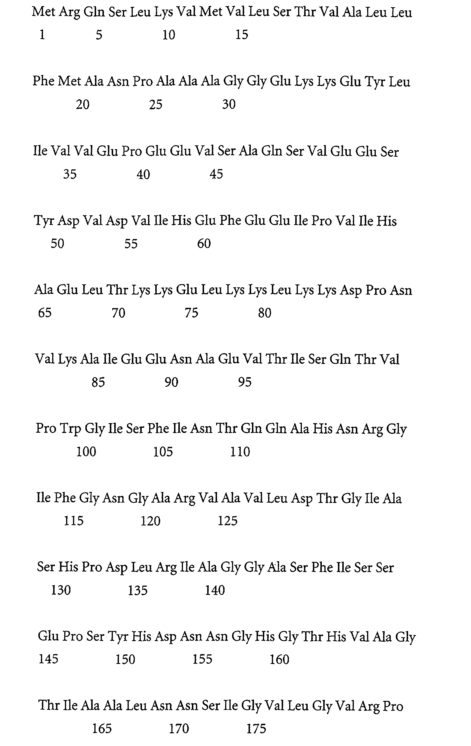


Claims (48)
- A method for the analysis of at least one target non-proteinaceous component of a mixture of non-proteinaceous and proteinaceous components derived from a biological sample
comprising the step of incubating the mixture with at least one protease having an amino acid sequence which is at least 80 % identical to the amino acid sequence of the protease subtilisin 147 from Bacillus lentus. - A method according to claim 1
characterized in that
the amino acid sequence of the protease is identical to the amino acid sequence of the protease subtilisin 147 from Bacillus lentus. - A method according to any of the claims 1 to 2
characterized in that
the amino acid sequence of protease is the amino acid sequence SEQ ID NO 1, a proteolytical derivative thereof having protease activity or the amino acid sequence SEQ ID NO 2. - A method according to any of the claims 1 to 3
characterized in that
the amino acid sequence of the protease according to any of the claims 1 to 3 is encoded by the nucleic acid sequence SEQ ID NO 3, a part thereof or a degenerated version of the nucleic acid sequence SEQ ID NO 3. - A method according to any of the claims 1 to 4
characterized in that
the biological sample is a fluid from the human or animal body. - A method according to claim 5
characterized in that
the biological sample is blood, blood plasma, blood serum or urine. - A method according to claim 6
characterized in that
the blood plasma is EDTA, heparin or citrate blood plasma. - A method according to any of the claims 1 to 7
characterized in that
the biological sample comprises bacterial cells, eukaryotic cells, viruses or mixtures thereof. - A method according to any of the claims 1 to 8,
characterized in that
after the incubation step the at least one target non-proteinaceous component is bound to a material with an affinity thereto, optionally washed and optionally released from the material with an affinity thereto. - A method according to any of the claims 1 to 9
characterized in that
the mixture of non-proteinaceous and proteinaceous components comprises nucleic acids. - A method according to any of the claims 1 to 9
characterized in that
the nucleic acids comprise DNA or RNA or both. - A method for the analysis of at least one target nucleic acid component of a mixture of non-proteinaceous components, which comprise nucleic acids, and proteinaceous components whereby the mixture is derived from a biological sample
comprising the steps ofa) incubating the mixture with at least one protease having an amino acid sequence which is at least 80 % identical to the amino acid sequence of the protease subtilisin 147 from Bacillus lentus,b) optionally amplifying the at least one target nucleic acid component, andc) determining or detecting the at least one target nucleic acid component. - A method according to claim 12
characterized in that
the amino acid sequence of the protease is identical to the amino acid sequence of the protease subtilisin 147 from Bacillus lentus. - A method according to any of the claims 12 to 13
characterized in that
the amino acid sequence of protease is the amino acid sequence SEQ ID NO 1, a proteolytical derivative thereof having protease activity or the amino acid sequence SEQ ID NO 2. - A method according to any of the claims 12 to 14
characterized in that
the amino acid sequence of the protease according to any of the claims 12 to 14 is encoded by the nucleic acid sequence SEQ ID NO 3, a part thereof or a degenerated version of the nucleic acid sequence SEQ ID NO 3. - A method according to any of the claims 12 to 15
characterized in that
the biological sample is a fluid from the human or animal body. - A method according to claim 16
characterized in that
the biological sample is blood, blood plasma, blood serum or urine. - A method according to claim 17
characterized in that
the blood plasma is EDTA, heparin or citrate blood plasma. - A method according to any of the claims 12 to 18
characterized in that
the biological sample comprises bacterial cells, eukaryotic cells, viruses or mixtures thereof. - A method according to any of the claims 12 to 19
characterized in that
the nucleic acids comprise DNA or RNA or both. - A method according to claim 20
characterized in that
the DNA or RNA or both is derived from at least one virus or at least one microorganism. - A method according to claim 21
characterized in that
the virus is hepatitis B virus, hepatitis C virus or the human immunodeficiency virus. - A method according to any of the claims 12 to 22
characterized in that
the at least one target nucleic acid component is amplified with the polymerase chain reaction. - A method according to any of the claims 12 to 23
characterized in that
after step b) the nucleic acids and the at least one target nucleic acid component are bound to a material with an affinity thereto, optionally washed and optionally released from the material with an affinity thereto. - A method according to claim 24
characterized in that
the material with an affinity to nucleic acids and the at least one target nucleic acid component comprises a material with a silica surface. - A method according to claim 25
characterized in that
the material with a silica surface is a glass. - A method according to any of the claims 25 to 26
characterized in that
the material with an affinity to nucleic acids and the at least one target nucleic acid component is a composition comprising magnetic glass particles. - A method according to any of the claims 1 to 27 for the diagnostic analysis.
- Use of a protease as specified in any of the claims 12 to 15 in diagnostics.
- Use of a protease as specified in any of the claims 12 to 15 for the analysis of at least one target non-proteinaceous component of a mixture of non-proteinaceous and proteinaceous components derived from a biological sample
- Use of a protease as specified in any of the claims 12 to 15 for the enrichment of at least one target non-proteinaceous component of a mixture of non-proteinaceous and proteinaceous components derived from a biological sample.
- Use of a protease as specified in any of the claims 12 to 15 for the purification or isolation of at least one target non-proteinaceous component of a mixture of non-proteinaceous and proteinaceous components derived from a biological sample.
- Use according to any of the claims 30 to 32
characterized in that
the at least one target non-proteinaceous component is a nucleic acid. - Use according to any of the claims 30 to 32
characterized in that
the mixture of non-proteinaceous and proteinaceous components comprises nucleic acids. - Use according to claim 33
characterized in that
the nucleic acid is from a virus or a microorganism. - Use according to claim 35
characterized in that
the virus is hepatitis B virus, hepatitis C virus or the human immunodeficiency virus. - A kit of parts
characterized in that
it contains at least one protease having an amino acid sequence, which is at least 80 % identical to the amino acid sequence of the protease subtilisin 147 from Bacillus lentus. - A kit according to claim 37
characterized in that
the amino acid sequence of the protease is identical to the amino acid sequence of the protease subtilisin 147 from Bacillus lentus. - A kit according to any of the claims 37 to 38
characterized in that
the amino acid sequence of protease is the amino acid sequence SEQ ID NO 1, a proteolytical derivative thereof having protease activity or the amino acid sequence SEQ ID NO 2. - A kit according to any of the claims 37 to 39
characterized in that
the amino acid sequence of the protease according to any of the claims 37 to 39 is encoded by the nucleic acid sequence SEQ ID NO 3, a part thereof or a degenerated version of the nucleic acid sequence SEQ ID NO 3. - A kit according to any of the claims 37 to 40
characterized in that
it additionally contains a material with an affinity to nucleic acids. - Kit according to claim 41
characterized in that
the material with an affinity to nucleic acids comprises a material with a silica surface. - Kit according to claim 42
characterized in that
the material with a silica surface is a glass. - Kit according to any of the claims 37 to 43
characterized in that
the material with an affinity to nucleic acids is a composition comprising magnetic glass particles. - Kit according to any of the claims 37 to 44
characterized in that
the kit comprises a lysis buffer, a washing buffer and an elution buffer. - Use of a kit according to any of the claims 37 to 45 for the purification of nucleic acids.
- Use of a kit according to any of the claims 37 to 45 in diagnostics.
- Use of a kit according to any of the claims 37 to 45 or a method according to any of the claims 1 to 28 in a high-throughput format.
Priority Applications (10)
| Application Number | Priority Date | Filing Date | Title |
|---|---|---|---|
| EP00123728A EP1201752A1 (en) | 2000-10-31 | 2000-10-31 | Methods for the analysis of non-proteinaceous components using a protease from a Bacillus strain |
| AU83619/01A AU758744B2 (en) | 2000-10-31 | 2001-10-25 | Methods for the analysis of non-proteinaceous components using a protease from a bacillus strain |
| DE60133708T DE60133708T2 (en) | 2000-10-31 | 2001-10-26 | A method of analyzing non-protein components using a protease from a Bacillus strain |
| CA002360012A CA2360012C (en) | 2000-10-31 | 2001-10-26 | Methods for the analysis of non-proteinaceous components using a protease from a bacillus strain |
| AT01125322T ATE393219T1 (en) | 2000-10-31 | 2001-10-26 | METHOD FOR ANALYZING NON-PROTEIN COMPONENTS USING A PROTEASE FROM A BACILLUS STRAIN |
| ES01125322T ES2305023T3 (en) | 2000-10-31 | 2001-10-26 | METHODS FOR THE ANALYSIS OF NON-PROTEIN COMPONENTS USING A PROTEASE OF A BACILO CEPA. |
| EP01125322A EP1201753B1 (en) | 2000-10-31 | 2001-10-26 | Methods for the analysis of non-proteinaceous components using a protease from a bacillus strain |
| US10/007,389 US6727067B2 (en) | 2000-10-31 | 2001-10-29 | Methods for the analysis of non-proteinaceous components using a protease from a Bacillus strain |
| JP2001334894A JP3621673B2 (en) | 2000-10-31 | 2001-10-31 | Method for analysis of non-protein components using protease from Bacillus strain |
| US10/655,508 US20040063155A1 (en) | 2000-10-31 | 2003-09-03 | Methods for the analysis of non-proteinaceous components using a protease from a Bacillus strain |
Applications Claiming Priority (1)
| Application Number | Priority Date | Filing Date | Title |
|---|---|---|---|
| EP00123728A EP1201752A1 (en) | 2000-10-31 | 2000-10-31 | Methods for the analysis of non-proteinaceous components using a protease from a Bacillus strain |
Publications (1)
| Publication Number | Publication Date |
|---|---|
| EP1201752A1 true EP1201752A1 (en) | 2002-05-02 |
Family
ID=8170259
Family Applications (1)
| Application Number | Title | Priority Date | Filing Date |
|---|---|---|---|
| EP00123728A Withdrawn EP1201752A1 (en) | 2000-10-31 | 2000-10-31 | Methods for the analysis of non-proteinaceous components using a protease from a Bacillus strain |
Country Status (1)
| Country | Link |
|---|---|
| EP (1) | EP1201752A1 (en) |
Cited By (1)
| Publication number | Priority date | Publication date | Assignee | Title |
|---|---|---|---|---|
| WO2022079236A1 (en) * | 2020-10-16 | 2022-04-21 | Turbobeads Gmbh | Methods and kits for nucleic acid purification |
Citations (4)
| Publication number | Priority date | Publication date | Assignee | Title |
|---|---|---|---|---|
| WO1989006279A1 (en) * | 1988-01-07 | 1989-07-13 | Novo-Nordisk A/S | Mutated subtilisin genes |
| US4895799A (en) * | 1986-03-14 | 1990-01-23 | Boehringer Mannheim Gmbh | Process and reagent for the determination of total bilirubin |
| DE4431432A1 (en) * | 1994-09-03 | 1996-03-28 | Juergen Weidner | Isolation of DNA from biological prepn(s). and amplification of highly degraded DNA |
| WO1998020115A1 (en) * | 1996-11-04 | 1998-05-14 | Novo Nordisk A/S | Subtilase variants and compositions |
-
2000
- 2000-10-31 EP EP00123728A patent/EP1201752A1/en not_active Withdrawn
Patent Citations (4)
| Publication number | Priority date | Publication date | Assignee | Title |
|---|---|---|---|---|
| US4895799A (en) * | 1986-03-14 | 1990-01-23 | Boehringer Mannheim Gmbh | Process and reagent for the determination of total bilirubin |
| WO1989006279A1 (en) * | 1988-01-07 | 1989-07-13 | Novo-Nordisk A/S | Mutated subtilisin genes |
| DE4431432A1 (en) * | 1994-09-03 | 1996-03-28 | Juergen Weidner | Isolation of DNA from biological prepn(s). and amplification of highly degraded DNA |
| WO1998020115A1 (en) * | 1996-11-04 | 1998-05-14 | Novo Nordisk A/S | Subtilase variants and compositions |
Cited By (1)
| Publication number | Priority date | Publication date | Assignee | Title |
|---|---|---|---|---|
| WO2022079236A1 (en) * | 2020-10-16 | 2022-04-21 | Turbobeads Gmbh | Methods and kits for nucleic acid purification |
Similar Documents
| Publication | Publication Date | Title |
|---|---|---|
| AU2005200670B2 (en) | Adsorption of nucleic acids to a solid phase | |
| CA2482097C (en) | Methods for isolating nucleic acids | |
| JP5354894B2 (en) | Nucleic acid isolation using polidocanols and derivatives | |
| EP1466018B2 (en) | Use of silica material in an amplification reaction | |
| EP1201753B1 (en) | Methods for the analysis of non-proteinaceous components using a protease from a bacillus strain | |
| EP1932913A1 (en) | Nucleic acid isolation using polidocanol and derivatives | |
| US6727067B2 (en) | Methods for the analysis of non-proteinaceous components using a protease from a Bacillus strain | |
| EP1201752A1 (en) | Methods for the analysis of non-proteinaceous components using a protease from a Bacillus strain | |
| EP1524317A1 (en) | Methods for isolating nucleic acids | |
| KR101910172B1 (en) | Method for Detection of Food Poisoning Bacteria By Using Gene Amplification and Kit for Use in The Same Method | |
| JP2003116550A (en) | Method for isolating nucleic acid |
Legal Events
| Date | Code | Title | Description |
|---|---|---|---|
| PUAI | Public reference made under article 153(3) epc to a published international application that has entered the european phase |
Free format text: ORIGINAL CODE: 0009012 |
|
| AK | Designated contracting states |
Kind code of ref document: A1 Designated state(s): DE Kind code of ref document: A1 Designated state(s): AT BE CH CY DE DK ES FI FR GB GR IE IT LI LU MC NL PT SE |
|
| AX | Request for extension of the european patent |
Free format text: AL;LT;LV;MK;RO;SI |
|
| AKX | Designation fees paid |
Free format text: DE |
|
| STAA | Information on the status of an ep patent application or granted ep patent |
Free format text: STATUS: THE APPLICATION IS DEEMED TO BE WITHDRAWN |
|
| 18D | Application deemed to be withdrawn |
Effective date: 20021105 |