JP3607023B2 - X-ray quantitative analysis apparatus and method - Google Patents
X-ray quantitative analysis apparatus and method Download PDFInfo
- Publication number
- JP3607023B2 JP3607023B2 JP31877596A JP31877596A JP3607023B2 JP 3607023 B2 JP3607023 B2 JP 3607023B2 JP 31877596 A JP31877596 A JP 31877596A JP 31877596 A JP31877596 A JP 31877596A JP 3607023 B2 JP3607023 B2 JP 3607023B2
- Authority
- JP
- Japan
- Prior art keywords
- group
- ray
- quantitative
- sample
- density
- Prior art date
- Legal status (The legal status is an assumption and is not a legal conclusion. Google has not performed a legal analysis and makes no representation as to the accuracy of the status listed.)
- Expired - Fee Related
Links
Images
Classifications
-
- G—PHYSICS
- G01—MEASURING; TESTING
- G01N—INVESTIGATING OR ANALYSING MATERIALS BY DETERMINING THEIR CHEMICAL OR PHYSICAL PROPERTIES
- G01N23/00—Investigating or analysing materials by the use of wave or particle radiation, e.g. X-rays or neutrons, not covered by groups G01N3/00 – G01N17/00, G01N21/00 or G01N22/00
- G01N23/22—Investigating or analysing materials by the use of wave or particle radiation, e.g. X-rays or neutrons, not covered by groups G01N3/00 – G01N17/00, G01N21/00 or G01N22/00 by measuring secondary emission from the material
- G01N23/225—Investigating or analysing materials by the use of wave or particle radiation, e.g. X-rays or neutrons, not covered by groups G01N3/00 – G01N17/00, G01N21/00 or G01N22/00 by measuring secondary emission from the material using electron or ion
-
- G—PHYSICS
- G01—MEASURING; TESTING
- G01N—INVESTIGATING OR ANALYSING MATERIALS BY DETERMINING THEIR CHEMICAL OR PHYSICAL PROPERTIES
- G01N2223/00—Investigating materials by wave or particle radiation
- G01N2223/40—Imaging
- G01N2223/402—Imaging mapping distribution of elements
Landscapes
- Physics & Mathematics (AREA)
- Health & Medical Sciences (AREA)
- Life Sciences & Earth Sciences (AREA)
- Chemical & Material Sciences (AREA)
- Analytical Chemistry (AREA)
- Biochemistry (AREA)
- General Health & Medical Sciences (AREA)
- General Physics & Mathematics (AREA)
- Immunology (AREA)
- Pathology (AREA)
- Analysing Materials By The Use Of Radiation (AREA)
Description
【0001】
【発明の属する技術分野】
この発明は、例えば物質に電子線またはX線を照射したときに発生する特性X線を分析するX線マイクロアナリシスの手法などを用いるX線定量分析装置および方法に関する。
【0002】
【従来の技術】
EDX(エネルギー分散型X線分析装置)やWDX(波長分散型X線分析装置)を用いた定量分析においては、試料中に含まれる元素を定量する場合、一般的に、ZAF(Z:原子番号補正、A:吸収補正、F:蛍光励起補正)演算を行うが、これは前記元素が試料中に均一に分布していることが前提となっており、元素が不均一に存在しているところの所謂不均一試料に対しては、検量線法を用いて元素を定量していた。
【0003】
これに対して、特開昭62−70740号公報に示されるように、試料面に電子線を走査し、試料上の各点のX線強度から成分ごとに特性X線のヒストグラムを作成し、試料面上の部分相の元素組成を算出し、この元素組成に、試料における各部分相の割合を乗じて加重平均することが考えられている。
【0004】
しかしながら、上記公報に記載の定量分析方法によれば、試料中に分布している元素の密度が似通っている場合はまだしも、前記密度に大きな差があるような場合には、精度がよくないといった欠点がある。
【0005】
【発明が解決しようとする課題】
この発明は、上述の事柄に留意してなされたもので、試料中に分布している元素の密度の如何にかかわらず、多元素を同時に精度よく、しかも短時間で定量分析することができるX線定量分析装置および方法を提供することを目的としている。
【0006】
【課題を解決するための手段】
上記目的を達成するため、この発明のX線定量分析装置は、試料に対して電子線またはX線を走査しながら照射したときに生ずる特性X線を検出するX線検出器と、このX線検出器の出力を処理するマルチチャネルアナライザと、このマルチチャネルアナライザからの出力に基づいて元素の画素位置に対応した濃度値を格納するメモリを備えるとともに、このメモリ内容に基づいて検出元素およびバックグラウンドをマッピングして相分析を行って組成別にグループ分けするとともに、前記マッピングした各点を定量演算して各グループにおける元素の定量値を求め、さらに、各グループの面積比率を求め、次いで、検出元素の密度と前記各グループにおける元素の定量値とに基づいて前記面積比率を各グループの重量比率に変換できるように構成している。
【0007】
そして、この発明のX線定量分析方法は、試料に対して電子線またはX線を走査しながら照射し、そのとき発生する特性X線の強度を元素ごとにメモリ内に記録し、これに基づいて検出元素およびバックグラウンドをマッピングして相分析を行って組成別にグループ分けするとともに、前記マッピングした各点を定量演算して各グループにおける元素の定量値を求め、さらに、各グループの面積比率を求め、次いで、検出元素の密度と前記各グループにおける元素の定量値とに基づいて前記面積比率を各グループの重量比率に変換するようにしている。
【0008】
この発明においては、検出元素の密度を考慮に入れて定量演算を行っているので、多元素における密度が似通っている場合は勿論のこと、元素間の密度が大きく異なるような場合においても、元素を精度よく定量演算することができる。
【0009】
【発明の実施の形態】
以下、この発明の好ましい実施例を、図を参照しながら説明する。
【0010】
図1〜図3は、この発明の一実施例を示し、まず、図1は、この発明のX線定量分析装置の一例を概略的に示すものである。すなわち、図1は、EDXを用いたディジタルマッピング装置を示し、この図1において、Iは走査型電子顕微鏡で、1は鉄製の試料室(図示してない)の内部上方に設けられる電子銃、2はこの電子銃1から試料室の内部下方に配置された試料3に向けて発せられる電子線4を二次元方向(互いに直交するx方向とy方向)に走査する電子線走査コイル、5はこの電子線走査コイル4を制御する電子線走査電源、6はディジタル・アナログ変換器(DAC)である。なお、試料3は、例えば試料ステージ(図示してない)に載置され、x,y方向に移動できるように構成されている。
【0011】
7は対物レンズである。8は電子線4を試料3に照射したときに試料3から発生する二次電子および試料3において反射される電子9を検出する二次電子および反射電子検出器、10は増幅器、11はアナログ・ディジタル変換器(ADC)である。
【0012】
12は電子線4が試料3に照射されたとき試料3から放出される特性X線13を検出するX線検出器で、例えばSi検出器からなる。14はパルスプロセッサ、15はマルチチャネル波高分析器(MCA)、16はスペクトルメモリである。
【0013】
17は各種の演算・制御を行うCPU、18はX線定量分析を行うための制御プログラムを内蔵したメインメモリ、19は画像メモリである。そして、20はデータバスで、このデータバス20に前記DAC6、ADC11、スペクトルメモリ16、CPU17、メインメモリ、画像メモリ19などが接続されている。
【0014】
上記構成のディジタルマッピング装置においては、走査型電子顕微鏡Iの電子線4の照射位置をDAC6によって制御し、10〜数100ミリ秒間走査を停止する。その間にX線検出器12に到達した一つ一つの特性X線13は、その波高値がエネルギーに比例した電気パルスに変換され、X線検出器12から出力される。この出力は、パルスプロセッサ14を経てMCA15に入力され、個々のパルス波高値がディジタル化されて、波高値に対応するデータメモリのアドレス(チャネル)にパルスの個数が記録され、特性X線13のエネルギースペクトルが形成され、これがスペクトルメモリ16に記憶される。スペクトルメモリ16に記憶されたエネルギースペクトルから着目している元素に対応する特性X線13の個数を積算し、X線像の輝度として画像メモリ19に送られる。
【0015】
このように、電子線4の試料3に対する照射位置をx方向、y方向に順次変えていくことにより、画像メモリ19に元素の分布を表すディジタル画像が得られる。
【0016】
なお、上記ディジタルマッピング装置においては、走査型電子線顕微鏡Iの二次電子や反射電子検出器8の出力を増幅器10およびADC11によって取り込むことにより、二次電子や反射電子の像をも得ることができる。
【0017】
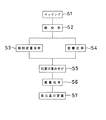
次に、上記構成のX線定量分析装置を用いて、X線定量分析を行う手順について、図2および図3をも参照しながら詳細に説明する。
【0018】
電子銃1からの電子線4を試料3に照射する。この場合、例えば、走査電源5からの制御信号によって、電子線4の試料3に対する照射位置をx,y方向に移動させることによって、電子線4を試料3に走査しながら照射する。
【0019】
前記電子線4の照射によって、試料3から特性X線13が放出され、この特性X線13は、X線検出器7によって検出される。そして、MCAにおいて、個々の元素ごとの特性X線13の数が計数される。電子線4または試料3の位置の走査の座標(x,y)と元素の種類に対応したアドレスを有する画像メモリ19に特性X線13の計数値を格納し、マッピング(ステップS1)を行う。
【0020】
前記マッピングは、面分析とも呼ばれ、検出元素とバックグラウンドとを同時にマッピングすることにより行われる。このマッピングの際、DBC(Digital Beam Control)面分析を行う。
【0021】
そして、前記マッピングの結果に基づいて相分析(ステップS2)を行い、組成別にグループ分けする。この相分析としては、この出願人に係る特許出願(特願平1−302725号(特開平3−163740号)や特願平6−248845号に記載されているような手法を採用することができる。
【0022】
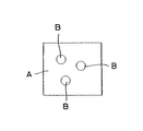
前記相分析により、試料に含まれる元素ごとの分布が分かる。図3は、前記相分析によって得られたマッピング像の一例を概略的に示すもので、この図において、符号Aは元素a1 ,a2 が分布している領域(グループ)を、符号Bは元素a1 ,b1 が分布している領域をそれぞれ示している。
【0023】
そして、前記マッピングした各点を定量演算し、グループA,Bにおける元素a1 ,a2 ,b1 の定量値を求める(ステップS3)とともに、グループA,Bの面積比率を求める(ステップS4)。すなわち、図3におけるグループA,Bの全体(A+B)に対する面積の割合(面積比率)As ,Bs を求める。この場合の面積比率As ,Bs は、
As =Aの面積/(Aの面積+Bの面積)
Bs =Bの面積/(Aの面積+Bの面積)
で表される。
【0024】
そして、検出された元素の密度(ステップS5)と、ステップS3において求められたグループA,Bにおける元素a1 ,a2 ,b1 の定量値とを用いて、前記面積比率As ,Bs を重量比率Am ,Bm に変換する(ステップS6)。
【0025】
すなわち、この例においては、各グループA,Bの平均密度Ad ,Bd は、
Ad ={(元素a1 の定量値)×(元素a1 の密度)+(元素a2 の定量値) ×(元素a2 の密度)}×As
Bd ={(元素a1 の定量値)×(元素a1 の密度)+(元素b1 の定量値) ×(元素b1 の密度)}×Bs
と表される。
【0026】
したがって、各グループA,Bの重量比率Am ,Bm は、
Am =Ad/(Ad+Bd)
Bm =Bd/(Ad+Bd)
として求められる。
【0027】
そして、前記グループA,Bの重量比率Am ,Bm とグループA,Bにおける元素a1 ,a2 ,b1 の定量値とを用いて、試料全体に対する各元素の濃度を求める(ステップS7)。すなわち、
元素a1 の濃度=(元素a1 の定量値)×Am +(元素a1 の定量値)×Bm 元素a2 の濃度=(元素a2 の定量値)×Am
元素b1 の濃度=(元素b1 の定量値)×Bm
として求められる。
【0028】
上述の説明から理解されるように、この発明の定量分析方法は、前記公報に開示されているような、各部分相の割合を乗じて加重平均するといった演算とは異なり、各グループA,Bの定量値および元素の密度を用いて、各グループA,Bの面積比率を重量比率に変換し、グループA,Bを重量によって重み付けするようにしているので、試料中に分布している元素の密度の如何にかかわらず、つまり、不均一試料であっても、それに含まれる多元素を同時に精度よく定量分析することができる。
【0029】
試料としてのAl中に含まれるSiを各種の演算方法で求めたところ、下記表1が得られた。
【0030】
【表1】
【0031】
この表1からは、面積比率による定量値と、この発明方法の密度を考慮した定量値との間にはそれほど差は見られず、いずれも化学分析値(真値)に近い値を示していることが分かるが、これは、AlとSiの密度がかなり似通った値(Al:2.70g/cm3 、Si:2.33g/cm3 )であるためと思われる。
【0032】
また、試料としての鋳鉄(Fe)中に含まれる炭素(C)を各種の演算方法で求めたところ、下記表2が得られた。
【0033】
【表2】
【0034】
この表2からは、FeとCというように、元素の密度にかなり差があるもの(Fe:7.87g/cm3 、C:2.27g/cm3 )の場合には、面積比率による定量値と、この発明方法の密度を考慮した定量値との間には大きな差が生じていることが分かる。すなわち、従来の面積比率による定量分析方法では、元素の密度にかなり差があるような場合には、ほとんど実用に供試得ないことが分かる。これに対して、この発明の定量分析方法は、元素の密度にかなり差があるような場合にも精度よく所定の分析を行うことができる。
【0035】
上述の実施例においては、X線定量分析をX線定量分析装置に設けられたメインメモリ18に内蔵した制御プログラム(図2参照)にしたがって行うようにしていたが、この制御プログラムを、フレキシブルディスク、CD−ROM、メモリカードといった他の媒体に内蔵させてあってもよい。
【0036】
さらに、前記図2に示す制御プログラムのうち、ステップS1〜ステップS4までのプログラムは、前記メインメモリ18に内蔵させ、ステップS5〜ステップS7までのプログラム、すなわち、検出された複数のグループに含まれる複数元素の密度と、これらの元素の定量値とを用いて、各グループの面積比率を重量比率に変換する手順と、この求められた各グループの重量比率と前記元素の定量値とを用いて、試料全体に対する各元素の濃度を求める手順を実行させるためのプログラムについてのみ、前記フレキシブルディスクなどの媒体に収容してあってもよい。このようにすれば、この発明のX線定量分析方法を、各種のX線定量分析装置によって実施することができる。
【0037】
この発明のX線定量分析装置および方法は、二元素のみならず、それ以上の多元素を同時に定量分析する場合にも適用できることはいうまでもない。
【0038】
そして、この発明の定量分析装置および方法は、WDXを用いても同様に定量分析を行うことができる。
【0039】
また、試料3に電子線4を照射するのに代えて、X線を照射するようにしてもよい。
【0040】
【発明の効果】
この発明においては、検出元素の密度を考慮に入れて定量演算を行っているので、多元素における密度が似通っている場合は勿論のこと、元素間の密度が大きく異なるような場合においても、元素を精度よく定量演算することができる。また、短時間で所望の結果を得ることができる。
【図面の簡単な説明】
【図1】この発明の定量分析装置の一例を概略的に示す図である。
【図2】この発明の定量分析方法の一例を示すフローチャートである。
【図3】相分析によって得られる図を概略的に示す図である。
【符号の説明】
3…試料、4…電子線、12…X線検出器、13…特性X線、15…マルチチャネルアナライザ、16,19…メモリ。[0001]
BACKGROUND OF THE INVENTION
The present invention relates to an X-ray quantitative analysis apparatus and method using, for example, an X-ray microanalysis technique for analyzing characteristic X-rays generated when a substance is irradiated with an electron beam or X-ray.
[0002]
[Prior art]
In quantitative analysis using EDX (energy dispersive X-ray analyzer) and WDX (wavelength dispersive X-ray analyzer), when quantifying an element contained in a sample, generally, ZAF (Z: atomic number) Correction, A: Absorption correction, F: Fluorescence excitation correction) are performed. This is based on the premise that the elements are uniformly distributed in the sample, and the elements are present non-uniformly. In the so-called heterogeneous sample, the element was quantified using a calibration curve method.
[0003]
On the other hand, as shown in Japanese Patent Laid-Open No. 62-70740, an electron beam is scanned on the sample surface, and a characteristic X-ray histogram is created for each component from the X-ray intensity of each point on the sample. It is considered that the elemental composition of the partial phase on the sample surface is calculated, and this elemental composition is multiplied by the ratio of each partial phase in the sample to perform a weighted average.
[0004]
However, according to the quantitative analysis method described in the above publication, if the density of elements distributed in the sample is similar, the accuracy is not good if there is a large difference in the density. There are drawbacks.
[0005]
[Problems to be solved by the invention]
The present invention has been made in consideration of the above-mentioned matters, and X can simultaneously and accurately perform multi-element quantitative analysis in a short time regardless of the density of elements distributed in a sample. An object of the present invention is to provide a linear quantitative analysis apparatus and method.
[0006]
[Means for Solving the Problems]
In order to achieve the above object, an X-ray quantitative analysis apparatus of the present invention includes an X-ray detector that detects characteristic X-rays generated when a sample is irradiated with an electron beam or X-ray while scanning, and the X-ray detector. A multi-channel analyzer that processes the output of the detector, and a memory that stores a concentration value corresponding to the pixel position of the element based on the output from the multi-channel analyzer, and the detected element and background based on the memory contents The phase is analyzed and grouped by composition, and each mapped point is quantitatively calculated to obtain the quantitative value of the element in each group, and further, the area ratio of each group is obtained, and then the detected element density and the like which can convert the area ratio based on a quantitative value of the elements in each group to the weight ratio of each group of It is configured.
[0007]
The X-ray quantitative analysis method of the present invention irradiates a sample while scanning with an electron beam or X-ray, records the intensity of characteristic X-rays generated at that time in a memory for each element, and based on this. The detected elements and background are mapped to perform phase analysis and grouped by composition, and the mapped points are quantitatively calculated to determine the quantitative values of the elements in each group, and the area ratio of each group is calculated. Then, the area ratio is converted into the weight ratio of each group based on the density of the detected element and the quantitative value of the element in each group.
[0008]
In the present invention, since the quantitative calculation is performed in consideration of the density of the detection element, not only when the density of multiple elements is similar, but also when the density between elements is greatly different, Can be quantitatively calculated accurately.
[0009]
DETAILED DESCRIPTION OF THE INVENTION
Hereinafter, preferred embodiments of the present invention will be described with reference to the drawings.
[0010]
1 to 3 show an embodiment of the present invention. First, FIG. 1 schematically shows an example of an X-ray quantitative analysis apparatus of the present invention. That is, FIG. 1 shows a digital mapping apparatus using EDX. In FIG. 1, I is a scanning electron microscope, 1 is an electron gun provided inside an iron sample chamber (not shown),
[0011]
[0012]
[0013]
[0014]
In the digital mapping apparatus having the above configuration, the irradiation position of the electron beam 4 of the scanning electron microscope I is controlled by the
[0015]
Thus, by sequentially changing the irradiation position of the electron beam 4 on the
[0016]
In the above digital mapping apparatus, the secondary electrons of the scanning electron microscope I and the output of the
[0017]
Next, a procedure for performing X-ray quantitative analysis using the X-ray quantitative analyzer having the above-described configuration will be described in detail with reference to FIGS. 2 and 3 as well.
[0018]
The
[0019]
The
[0020]
The mapping is also referred to as surface analysis, and is performed by mapping the detection element and the background simultaneously. At the time of this mapping, a DBC (Digital Beam Control) surface analysis is performed.
[0021]
Then, phase analysis (step S2) is performed based on the mapping result, and grouped by composition. For this phase analysis, it is possible to adopt a technique as described in the patent application relating to this applicant (Japanese Patent Application No. 1-330225 (Japanese Patent Laid-open No. Hei 3-163740)) or Japanese Patent Application No. 6-248845. it can.
[0022]
The phase analysis reveals the distribution of each element contained in the sample. FIG. 3 schematically shows an example of a mapping image obtained by the phase analysis. In this figure, symbol A denotes a region (group) in which elements a 1 and a 2 are distributed, and symbol B denotes The regions where the elements a 1 and b 1 are distributed are shown.
[0023]
Then, the mapped points are quantitatively calculated to determine the quantitative values of the elements a 1 , a 2 , and b 1 in the groups A and B (step S3) and the area ratio of the groups A and B (step S4). . That is, area ratios (area ratios) A s and B s with respect to the entire group A and B (A + B) in FIG. The area ratios A s and B s in this case are
A s = A area / (A area + B area)
B s = B area / (A area + B area)
It is represented by
[0024]
Then, using the detected element density (step S5) and the quantitative values of the elements a 1 , a 2 , and b 1 in the groups A and B obtained in step S3, the area ratios A s and B s are used. Are converted into weight ratios A m and B m (step S6).
[0025]
That is, in this example, the average densities A d and B d of the groups A and B are
A d = {(the density of the elements a 2) (elemental quantitative value of a 1) × (quantitative value of an element a 2) (element density of a 1) + ×} × A s
B d = {(the density of the elements a 1) (element quantitative value of a 1) × + (quantitative value of elements b 1) × (element b 1 density)} × B s
It is expressed.
[0026]
Therefore, the weight ratios A m and B m of the groups A and B are
A m = A d / (A d + B d )
B m = B d / (A d + B d )
As required.
[0027]
Then, using the weight ratios A m and B m of the groups A and B and the quantitative values of the elements a 1 , a 2 and b 1 in the groups A and B, the concentration of each element with respect to the entire sample is obtained (step S7). ). That is,
The concentration of the element a 1 = (quantitative value of an element a 2) (element quantitative value of a 1) × A m + (quantitative value of the elements a 1) × B m elements a 2 concentration = × A m
Concentration of element b 1 = (quantitative value of element b 1 ) × B m
As required.
[0028]
As understood from the above description, the quantitative analysis method of the present invention is different from the calculation such as the weighted average by multiplying the ratio of each partial phase as disclosed in the above-mentioned publication. Since the area ratio of each group A and B is converted into a weight ratio by using the quantitative value of the element and the element density, and the groups A and B are weighted by the weight, the elements distributed in the sample Regardless of the density, that is, even if it is a heterogeneous sample, the multi-elements contained therein can be quantitatively analyzed with high accuracy at the same time.
[0029]
When Si contained in Al as a sample was obtained by various calculation methods, the following Table 1 was obtained.
[0030]
[Table 1]
[0031]
From Table 1, there is not much difference between the quantitative value based on the area ratio and the quantitative value considering the density of the method of the present invention, and both show values close to the chemical analysis value (true value). As can be seen, this is probably because the densities of Al and Si are quite similar (Al: 2.70 g / cm 3 , Si: 2.33 g / cm 3 ).
[0032]
Moreover, when carbon (C) contained in cast iron (Fe) as a sample was obtained by various calculation methods, the following Table 2 was obtained.
[0033]
[Table 2]
[0034]
According to Table 2, in the case where the element density is significantly different (Fe: 7.87 g / cm 3 , C: 2.27 g / cm 3 ), such as Fe and C, quantification by the area ratio is performed. It can be seen that there is a large difference between the value and the quantitative value considering the density of the method of the present invention. That is, it can be seen that the conventional quantitative analysis method based on the area ratio can hardly be practically used when there is a considerable difference in element density. On the other hand, the quantitative analysis method of the present invention can perform a predetermined analysis with high accuracy even when there is a considerable difference in element density.
[0035]
In the above-described embodiment, the X-ray quantitative analysis is performed according to the control program (see FIG. 2) incorporated in the
[0036]
Further, among the control programs shown in FIG. 2, the programs from step S1 to step S4 are built in the
[0037]
It goes without saying that the X-ray quantitative analysis apparatus and method of the present invention can be applied not only to two elements but also to quantitative analysis of more than one multi-element simultaneously.
[0038]
And the quantitative analysis apparatus and method of this invention can perform quantitative analysis similarly even if it uses WDX.
[0039]
Further, instead of irradiating the
[0040]
【The invention's effect】
In the present invention, since the quantitative calculation is performed in consideration of the density of the detection element, not only when the density of multiple elements is similar, but also when the density between elements is greatly different, Can be quantitatively calculated accurately. Moreover, a desired result can be obtained in a short time.
[Brief description of the drawings]
FIG. 1 is a diagram schematically showing an example of a quantitative analysis apparatus according to the present invention.
FIG. 2 is a flowchart showing an example of a quantitative analysis method of the present invention.
FIG. 3 schematically shows a diagram obtained by phase analysis.
[Explanation of symbols]
3 ... sample, 4 ... electron beam, 12 ... X-ray detector, 13 ... characteristic X-ray, 15 ... multi-channel analyzer, 16, 19 ... memory.
Claims (3)
Priority Applications (2)
| Application Number | Priority Date | Filing Date | Title |
|---|---|---|---|
| JP31877596A JP3607023B2 (en) | 1996-05-10 | 1996-11-13 | X-ray quantitative analysis apparatus and method |
| US08/853,549 US5866903A (en) | 1996-05-10 | 1997-05-09 | Equipment and process for quantitative x-ray analysis and medium with quantitative x-ray analysis program recorded |
Applications Claiming Priority (3)
| Application Number | Priority Date | Filing Date | Title |
|---|---|---|---|
| JP8-141068 | 1996-05-10 | ||
| JP14106896 | 1996-05-10 | ||
| JP31877596A JP3607023B2 (en) | 1996-05-10 | 1996-11-13 | X-ray quantitative analysis apparatus and method |
Publications (2)
| Publication Number | Publication Date |
|---|---|
| JPH1026593A JPH1026593A (en) | 1998-01-27 |
| JP3607023B2 true JP3607023B2 (en) | 2005-01-05 |
Family
ID=26473391
Family Applications (1)
| Application Number | Title | Priority Date | Filing Date |
|---|---|---|---|
| JP31877596A Expired - Fee Related JP3607023B2 (en) | 1996-05-10 | 1996-11-13 | X-ray quantitative analysis apparatus and method |
Country Status (2)
| Country | Link |
|---|---|
| US (1) | US5866903A (en) |
| JP (1) | JP3607023B2 (en) |
Families Citing this family (29)
| Publication number | Priority date | Publication date | Assignee | Title |
|---|---|---|---|---|
| JP3500264B2 (en) * | 1997-01-29 | 2004-02-23 | 株式会社日立製作所 | Sample analyzer |
| US6368672B1 (en) * | 1999-09-28 | 2002-04-09 | General Electric Company | Method for forming a thermal barrier coating system of a turbine engine component |
| US6787773B1 (en) | 2000-06-07 | 2004-09-07 | Kla-Tencor Corporation | Film thickness measurement using electron-beam induced x-ray microanalysis |
| US6584413B1 (en) | 2001-06-01 | 2003-06-24 | Sandia Corporation | Apparatus and system for multivariate spectral analysis |
| US6675106B1 (en) | 2001-06-01 | 2004-01-06 | Sandia Corporation | Method of multivariate spectral analysis |
| US6801596B2 (en) | 2001-10-01 | 2004-10-05 | Kla-Tencor Technologies Corporation | Methods and apparatus for void characterization |
| US6664541B2 (en) | 2001-10-01 | 2003-12-16 | Kla Tencor Technologies Corporation | Methods and apparatus for defect localization |
| US6810105B2 (en) * | 2002-01-25 | 2004-10-26 | Kla-Tencor Technologies Corporation | Methods and apparatus for dishing and erosion characterization |
| US7202475B1 (en) * | 2003-03-06 | 2007-04-10 | Kla-Tencor Technologies Corporation | Rapid defect composition mapping using multiple X-ray emission perspective detection scheme |
| US7490009B2 (en) * | 2004-08-03 | 2009-02-10 | Fei Company | Method and system for spectroscopic data analysis |
| US7269523B2 (en) * | 2004-10-19 | 2007-09-11 | Jordanov Valentin T | Method and apparatus for radiation spectroscopy using seeded localized averaging (“SLA”) |
| JP4499125B2 (en) * | 2007-03-05 | 2010-07-07 | 日本電子株式会社 | Quantitative analysis method in sample analyzer |
| CN103776855B (en) * | 2008-02-06 | 2017-05-31 | Fei 公司 | The method and system of modal data analysis |
| CZ303228B6 (en) * | 2011-03-23 | 2012-06-06 | Tescan A.S. | Method of analyzing material by a focused electron beam by making use of characteristic X-ray radiation and knocked-on electrons and apparatus for making the same |
| US8664595B2 (en) | 2012-06-28 | 2014-03-04 | Fei Company | Cluster analysis of unknowns in SEM-EDS dataset |
| US9188555B2 (en) | 2012-07-30 | 2015-11-17 | Fei Company | Automated EDS standards calibration |
| US9778215B2 (en) | 2012-10-26 | 2017-10-03 | Fei Company | Automated mineral classification |
| US9091635B2 (en) | 2012-10-26 | 2015-07-28 | Fei Company | Mineral identification using mineral definitions having compositional ranges |
| US9048067B2 (en) | 2012-10-26 | 2015-06-02 | Fei Company | Mineral identification using sequential decomposition into elements from mineral definitions |
| US8937282B2 (en) | 2012-10-26 | 2015-01-20 | Fei Company | Mineral identification using mineral definitions including variability |
| TWI582708B (en) * | 2012-11-22 | 2017-05-11 | 緯創資通股份有限公司 | Facial expression control system, facial expression control method, and computer system thereof |
| US9194829B2 (en) | 2012-12-28 | 2015-11-24 | Fei Company | Process for performing automated mineralogy |
| US9714908B2 (en) | 2013-11-06 | 2017-07-25 | Fei Company | Sub-pixel analysis and display of fine grained mineral samples |
| JP6242291B2 (en) * | 2014-05-21 | 2017-12-06 | 日本電子株式会社 | Phase analyzer, phase analysis method, and surface analyzer |
| JP6588362B2 (en) * | 2016-03-08 | 2019-10-09 | 日本電子株式会社 | Phase analyzer, phase analysis method, and surface analyzer |
| JP6769402B2 (en) * | 2017-06-30 | 2020-10-14 | 株式会社島津製作所 | Electron microanalyzer and data processing program |
| EP3614414A1 (en) * | 2018-08-20 | 2020-02-26 | FEI Company | Method of examining a sample using a charged particle microscope |
| US11373839B1 (en) | 2021-02-03 | 2022-06-28 | Fei Company | Method and system for component analysis of spectral data |
| JP7442487B2 (en) * | 2021-11-02 | 2024-03-04 | 日本電子株式会社 | Phase analyzer, sample analyzer, and analysis method |
Family Cites Families (1)
| Publication number | Priority date | Publication date | Assignee | Title |
|---|---|---|---|---|
| JP3461208B2 (en) * | 1994-09-16 | 2003-10-27 | 株式会社堀場製作所 | Identification method and distribution measurement method for substances contained in samples |
-
1996
- 1996-11-13 JP JP31877596A patent/JP3607023B2/en not_active Expired - Fee Related
-
1997
- 1997-05-09 US US08/853,549 patent/US5866903A/en not_active Expired - Lifetime
Also Published As
| Publication number | Publication date |
|---|---|
| US5866903A (en) | 1999-02-02 |
| JPH1026593A (en) | 1998-01-27 |
Similar Documents
| Publication | Publication Date | Title |
|---|---|---|
| JP3607023B2 (en) | X-ray quantitative analysis apparatus and method | |
| US7579591B2 (en) | Method and apparatus for analyzing sample | |
| US20030080292A1 (en) | System and method for depth profiling | |
| CN101101269B (en) | Energy dispersion type radiation detecting system and method of measuring content of object element | |
| WO2003038417A2 (en) | System and method for depth profiling and characterization of thin films | |
| EP0452825B1 (en) | Method and apparatus for background correction in analysis of a specimen surface | |
| JP2000249668A (en) | Quantitative analyzer for heterogeneous composite tissue samples | |
| JP2000235009A (en) | Particle analysis device using electron probe microanalyzer | |
| JP2002062270A (en) | Surface analysis data display method in surface analyzer using electron beam | |
| JP2009002658A (en) | Observation method for thin film samples | |
| JP3950626B2 (en) | Quantitative analysis method in sample analyzer | |
| JP3956282B2 (en) | Surface analyzer | |
| JP2004108854A (en) | Analysis method | |
| EP4123299B1 (en) | Analyzer apparatus and method of image processing | |
| JP2000214111A (en) | Electronic probe micro analyzer | |
| JP2004294233A (en) | X-ray spectroscopic microscopic analysis method and photoelectric conversion type x-ray microscope | |
| JP2502050B2 (en) | Electron beam micro analyzer | |
| JP2964591B2 (en) | Automatic focus detector for electron beam micro analyzer | |
| JPH0682399A (en) | Electron beam analyzing method and apparatus | |
| JP2671117B2 (en) | Element mapping device | |
| JP3790629B2 (en) | Scanning charged particle beam apparatus and method of operating scanning charged particle beam apparatus | |
| JPH1167138A (en) | Small area observation device | |
| JPH11237230A (en) | Sample measurement method and device for electron microscope | |
| JPH0752163B2 (en) | Simplified quantitative analysis method using wavelength dispersive X-ray spectrometer | |
| JP2000002674A (en) | Electronic probe micro analyzer |
Legal Events
| Date | Code | Title | Description |
|---|---|---|---|
| TRDD | Decision of grant or rejection written | ||
| A01 | Written decision to grant a patent or to grant a registration (utility model) |
Free format text: JAPANESE INTERMEDIATE CODE: A01 Effective date: 20041005 |
|
| A61 | First payment of annual fees (during grant procedure) |
Free format text: JAPANESE INTERMEDIATE CODE: A61 Effective date: 20041006 |
|
| R150 | Certificate of patent or registration of utility model |
Free format text: JAPANESE INTERMEDIATE CODE: R150 |
|
| FPAY | Renewal fee payment (event date is renewal date of database) |
Free format text: PAYMENT UNTIL: 20101015 Year of fee payment: 6 |
|
| FPAY | Renewal fee payment (event date is renewal date of database) |
Free format text: PAYMENT UNTIL: 20101015 Year of fee payment: 6 |
|
| FPAY | Renewal fee payment (event date is renewal date of database) |
Free format text: PAYMENT UNTIL: 20111015 Year of fee payment: 7 |
|
| FPAY | Renewal fee payment (event date is renewal date of database) |
Free format text: PAYMENT UNTIL: 20111015 Year of fee payment: 7 |
|
| FPAY | Renewal fee payment (event date is renewal date of database) |
Free format text: PAYMENT UNTIL: 20121015 Year of fee payment: 8 |
|
| FPAY | Renewal fee payment (event date is renewal date of database) |
Free format text: PAYMENT UNTIL: 20121015 Year of fee payment: 8 |
|
| FPAY | Renewal fee payment (event date is renewal date of database) |
Free format text: PAYMENT UNTIL: 20121015 Year of fee payment: 8 |
|
| FPAY | Renewal fee payment (event date is renewal date of database) |
Free format text: PAYMENT UNTIL: 20121015 Year of fee payment: 8 |
|
| FPAY | Renewal fee payment (event date is renewal date of database) |
Free format text: PAYMENT UNTIL: 20131015 Year of fee payment: 9 |
|
| R250 | Receipt of annual fees |
Free format text: JAPANESE INTERMEDIATE CODE: R250 |
|
| R250 | Receipt of annual fees |
Free format text: JAPANESE INTERMEDIATE CODE: R250 |
|
| R250 | Receipt of annual fees |
Free format text: JAPANESE INTERMEDIATE CODE: R250 |
|
| LAPS | Cancellation because of no payment of annual fees |


