JP2012533354A - Electrospun silk material system for wound healing - Google Patents
Electrospun silk material system for wound healing Download PDFInfo
- Publication number
- JP2012533354A JP2012533354A JP2012520744A JP2012520744A JP2012533354A JP 2012533354 A JP2012533354 A JP 2012533354A JP 2012520744 A JP2012520744 A JP 2012520744A JP 2012520744 A JP2012520744 A JP 2012520744A JP 2012533354 A JP2012533354 A JP 2012533354A
- Authority
- JP
- Japan
- Prior art keywords
- silk
- mat
- cells
- peo
- cell
- Prior art date
- Legal status (The legal status is an assumption and is not a legal conclusion. Google has not performed a legal analysis and makes no representation as to the accuracy of the status listed.)
- Pending
Links
Images


Classifications
-
- A—HUMAN NECESSITIES
- A61—MEDICAL OR VETERINARY SCIENCE; HYGIENE
- A61L—METHODS OR APPARATUS FOR STERILISING MATERIALS OR OBJECTS IN GENERAL; DISINFECTION, STERILISATION OR DEODORISATION OF AIR; CHEMICAL ASPECTS OF BANDAGES, DRESSINGS, ABSORBENT PADS OR SURGICAL ARTICLES; MATERIALS FOR BANDAGES, DRESSINGS, ABSORBENT PADS OR SURGICAL ARTICLES
- A61L15/00—Chemical aspects of, or use of materials for, bandages, dressings or absorbent pads
- A61L15/16—Bandages, dressings or absorbent pads for physiological fluids such as urine or blood, e.g. sanitary towels, tampons
- A61L15/40—Bandages, dressings or absorbent pads for physiological fluids such as urine or blood, e.g. sanitary towels, tampons containing ingredients of undetermined constitution or reaction products thereof, e.g. plant or animal extracts
-
- A—HUMAN NECESSITIES
- A61—MEDICAL OR VETERINARY SCIENCE; HYGIENE
- A61L—METHODS OR APPARATUS FOR STERILISING MATERIALS OR OBJECTS IN GENERAL; DISINFECTION, STERILISATION OR DEODORISATION OF AIR; CHEMICAL ASPECTS OF BANDAGES, DRESSINGS, ABSORBENT PADS OR SURGICAL ARTICLES; MATERIALS FOR BANDAGES, DRESSINGS, ABSORBENT PADS OR SURGICAL ARTICLES
- A61L15/00—Chemical aspects of, or use of materials for, bandages, dressings or absorbent pads
- A61L15/16—Bandages, dressings or absorbent pads for physiological fluids such as urine or blood, e.g. sanitary towels, tampons
- A61L15/22—Bandages, dressings or absorbent pads for physiological fluids such as urine or blood, e.g. sanitary towels, tampons containing macromolecular materials
- A61L15/225—Mixtures of macromolecular compounds
-
- A—HUMAN NECESSITIES
- A61—MEDICAL OR VETERINARY SCIENCE; HYGIENE
- A61L—METHODS OR APPARATUS FOR STERILISING MATERIALS OR OBJECTS IN GENERAL; DISINFECTION, STERILISATION OR DEODORISATION OF AIR; CHEMICAL ASPECTS OF BANDAGES, DRESSINGS, ABSORBENT PADS OR SURGICAL ARTICLES; MATERIALS FOR BANDAGES, DRESSINGS, ABSORBENT PADS OR SURGICAL ARTICLES
- A61L15/00—Chemical aspects of, or use of materials for, bandages, dressings or absorbent pads
- A61L15/16—Bandages, dressings or absorbent pads for physiological fluids such as urine or blood, e.g. sanitary towels, tampons
- A61L15/42—Use of materials characterised by their function or physical properties
- A61L15/44—Medicaments
-
- A—HUMAN NECESSITIES
- A61—MEDICAL OR VETERINARY SCIENCE; HYGIENE
- A61P—SPECIFIC THERAPEUTIC ACTIVITY OF CHEMICAL COMPOUNDS OR MEDICINAL PREPARATIONS
- A61P17/00—Drugs for dermatological disorders
- A61P17/02—Drugs for dermatological disorders for treating wounds, ulcers, burns, scars, keloids, or the like
-
- A—HUMAN NECESSITIES
- A61—MEDICAL OR VETERINARY SCIENCE; HYGIENE
- A61P—SPECIFIC THERAPEUTIC ACTIVITY OF CHEMICAL COMPOUNDS OR MEDICINAL PREPARATIONS
- A61P31/00—Antiinfectives, i.e. antibiotics, antiseptics, chemotherapeutics
-
- A—HUMAN NECESSITIES
- A61—MEDICAL OR VETERINARY SCIENCE; HYGIENE
- A61P—SPECIFIC THERAPEUTIC ACTIVITY OF CHEMICAL COMPOUNDS OR MEDICINAL PREPARATIONS
- A61P37/00—Drugs for immunological or allergic disorders
- A61P37/02—Immunomodulators
-
- D—TEXTILES; PAPER
- D01—NATURAL OR MAN-MADE THREADS OR FIBRES; SPINNING
- D01D—MECHANICAL METHODS OR APPARATUS IN THE MANUFACTURE OF ARTIFICIAL FILAMENTS, THREADS, FIBRES, BRISTLES OR RIBBONS
- D01D5/00—Formation of filaments, threads, or the like
- D01D5/0007—Electro-spinning
- D01D5/0015—Electro-spinning characterised by the initial state of the material
- D01D5/003—Electro-spinning characterised by the initial state of the material the material being a polymer solution or dispersion
-
- D—TEXTILES; PAPER
- D01—NATURAL OR MAN-MADE THREADS OR FIBRES; SPINNING
- D01F—CHEMICAL FEATURES IN THE MANUFACTURE OF ARTIFICIAL FILAMENTS, THREADS, FIBRES, BRISTLES OR RIBBONS; APPARATUS SPECIALLY ADAPTED FOR THE MANUFACTURE OF CARBON FILAMENTS
- D01F4/00—Monocomponent artificial filaments or the like of proteins; Manufacture thereof
- D01F4/02—Monocomponent artificial filaments or the like of proteins; Manufacture thereof from fibroin
-
- D—TEXTILES; PAPER
- D04—BRAIDING; LACE-MAKING; KNITTING; TRIMMINGS; NON-WOVEN FABRICS
- D04H—MAKING TEXTILE FABRICS, e.g. FROM FIBRES OR FILAMENTARY MATERIAL; FABRICS MADE BY SUCH PROCESSES OR APPARATUS, e.g. FELTS, NON-WOVEN FABRICS; COTTON-WOOL; WADDING ; NON-WOVEN FABRICS FROM STAPLE FIBRES, FILAMENTS OR YARNS, BONDED WITH AT LEAST ONE WEB-LIKE MATERIAL DURING THEIR CONSOLIDATION
- D04H1/00—Non-woven fabrics formed wholly or mainly of staple fibres or like relatively short fibres
- D04H1/04—Non-woven fabrics formed wholly or mainly of staple fibres or like relatively short fibres from fleeces or layers composed of fibres having existing or potential cohesive properties, e.g. natural fibres, prestretched or fibrillated artificial fibres
- D04H1/08—Non-woven fabrics formed wholly or mainly of staple fibres or like relatively short fibres from fleeces or layers composed of fibres having existing or potential cohesive properties, e.g. natural fibres, prestretched or fibrillated artificial fibres and hardened by felting; Felts or felted products
- D04H1/09—Silk
-
- D—TEXTILES; PAPER
- D04—BRAIDING; LACE-MAKING; KNITTING; TRIMMINGS; NON-WOVEN FABRICS
- D04H—MAKING TEXTILE FABRICS, e.g. FROM FIBRES OR FILAMENTARY MATERIAL; FABRICS MADE BY SUCH PROCESSES OR APPARATUS, e.g. FELTS, NON-WOVEN FABRICS; COTTON-WOOL; WADDING ; NON-WOVEN FABRICS FROM STAPLE FIBRES, FILAMENTS OR YARNS, BONDED WITH AT LEAST ONE WEB-LIKE MATERIAL DURING THEIR CONSOLIDATION
- D04H1/00—Non-woven fabrics formed wholly or mainly of staple fibres or like relatively short fibres
- D04H1/40—Non-woven fabrics formed wholly or mainly of staple fibres or like relatively short fibres from fleeces or layers composed of fibres without existing or potential cohesive properties
- D04H1/42—Non-woven fabrics formed wholly or mainly of staple fibres or like relatively short fibres from fleeces or layers composed of fibres without existing or potential cohesive properties characterised by the use of certain kinds of fibres insofar as this use has no preponderant influence on the consolidation of the fleece
- D04H1/4266—Natural fibres not provided for in group D04H1/425
-
- D—TEXTILES; PAPER
- D04—BRAIDING; LACE-MAKING; KNITTING; TRIMMINGS; NON-WOVEN FABRICS
- D04H—MAKING TEXTILE FABRICS, e.g. FROM FIBRES OR FILAMENTARY MATERIAL; FABRICS MADE BY SUCH PROCESSES OR APPARATUS, e.g. FELTS, NON-WOVEN FABRICS; COTTON-WOOL; WADDING ; NON-WOVEN FABRICS FROM STAPLE FIBRES, FILAMENTS OR YARNS, BONDED WITH AT LEAST ONE WEB-LIKE MATERIAL DURING THEIR CONSOLIDATION
- D04H1/00—Non-woven fabrics formed wholly or mainly of staple fibres or like relatively short fibres
- D04H1/70—Non-woven fabrics formed wholly or mainly of staple fibres or like relatively short fibres characterised by the method of forming fleeces or layers, e.g. reorientation of fibres
- D04H1/72—Non-woven fabrics formed wholly or mainly of staple fibres or like relatively short fibres characterised by the method of forming fleeces or layers, e.g. reorientation of fibres the fibres being randomly arranged
- D04H1/728—Non-woven fabrics formed wholly or mainly of staple fibres or like relatively short fibres characterised by the method of forming fleeces or layers, e.g. reorientation of fibres the fibres being randomly arranged by electro-spinning
-
- A—HUMAN NECESSITIES
- A61—MEDICAL OR VETERINARY SCIENCE; HYGIENE
- A61L—METHODS OR APPARATUS FOR STERILISING MATERIALS OR OBJECTS IN GENERAL; DISINFECTION, STERILISATION OR DEODORISATION OF AIR; CHEMICAL ASPECTS OF BANDAGES, DRESSINGS, ABSORBENT PADS OR SURGICAL ARTICLES; MATERIALS FOR BANDAGES, DRESSINGS, ABSORBENT PADS OR SURGICAL ARTICLES
- A61L2300/00—Biologically active materials used in bandages, wound dressings, absorbent pads or medical devices
- A61L2300/40—Biologically active materials used in bandages, wound dressings, absorbent pads or medical devices characterised by a specific therapeutic activity or mode of action
- A61L2300/404—Biocides, antimicrobial agents, antiseptic agents
- A61L2300/406—Antibiotics
-
- A—HUMAN NECESSITIES
- A61—MEDICAL OR VETERINARY SCIENCE; HYGIENE
- A61L—METHODS OR APPARATUS FOR STERILISING MATERIALS OR OBJECTS IN GENERAL; DISINFECTION, STERILISATION OR DEODORISATION OF AIR; CHEMICAL ASPECTS OF BANDAGES, DRESSINGS, ABSORBENT PADS OR SURGICAL ARTICLES; MATERIALS FOR BANDAGES, DRESSINGS, ABSORBENT PADS OR SURGICAL ARTICLES
- A61L2300/00—Biologically active materials used in bandages, wound dressings, absorbent pads or medical devices
- A61L2300/60—Biologically active materials used in bandages, wound dressings, absorbent pads or medical devices characterised by a special physical form
- A61L2300/62—Encapsulated active agents, e.g. emulsified droplets
-
- A—HUMAN NECESSITIES
- A61—MEDICAL OR VETERINARY SCIENCE; HYGIENE
- A61L—METHODS OR APPARATUS FOR STERILISING MATERIALS OR OBJECTS IN GENERAL; DISINFECTION, STERILISATION OR DEODORISATION OF AIR; CHEMICAL ASPECTS OF BANDAGES, DRESSINGS, ABSORBENT PADS OR SURGICAL ARTICLES; MATERIALS FOR BANDAGES, DRESSINGS, ABSORBENT PADS OR SURGICAL ARTICLES
- A61L2300/00—Biologically active materials used in bandages, wound dressings, absorbent pads or medical devices
- A61L2300/60—Biologically active materials used in bandages, wound dressings, absorbent pads or medical devices characterised by a special physical form
- A61L2300/64—Animal cells
Landscapes
- Health & Medical Sciences (AREA)
- Engineering & Computer Science (AREA)
- Chemical & Material Sciences (AREA)
- Life Sciences & Earth Sciences (AREA)
- Textile Engineering (AREA)
- Public Health (AREA)
- Animal Behavior & Ethology (AREA)
- General Health & Medical Sciences (AREA)
- Veterinary Medicine (AREA)
- Chemical Kinetics & Catalysis (AREA)
- Materials Engineering (AREA)
- Hematology (AREA)
- Epidemiology (AREA)
- General Chemical & Material Sciences (AREA)
- Pharmacology & Pharmacy (AREA)
- Organic Chemistry (AREA)
- Nuclear Medicine, Radiotherapy & Molecular Imaging (AREA)
- Medicinal Chemistry (AREA)
- Zoology (AREA)
- Dispersion Chemistry (AREA)
- Mechanical Engineering (AREA)
- Botany (AREA)
- Bioinformatics & Cheminformatics (AREA)
- Immunology (AREA)
- Communicable Diseases (AREA)
- Dermatology (AREA)
- Oncology (AREA)
- Materials For Medical Uses (AREA)
- Medicinal Preparation (AREA)
- Medicines That Contain Protein Lipid Enzymes And Other Medicines (AREA)
- Medicines Containing Material From Animals Or Micro-Organisms (AREA)
- Peptides Or Proteins (AREA)
- Treatments For Attaching Organic Compounds To Fibrous Goods (AREA)
Abstract
本発明は、絹フィブロイン/ポリエチレンオキシドブレンド材料を調製するプロセス、および、創傷治癒などの生物医学的用途に適したその得られる材料に関する。特に、制御された蒸発、拘束乾燥技法、および/またはアルコール処理、および/またはPEO抽出で処理された、絹:PEOブレンド比が2:1〜4:1の電界紡糸した絹フィブロイン/PEOマットは、創傷被覆材に対する有用性をもつ生体材料系に関する、適した物理的なおよび生体機能特性、例えば繊維構造、トポグラフィー、吸収、水蒸気透過速度、酸素透過性、および生分解性などを実証する。

Description
関連出願の相互参照
本出願は、2009年7月14日出願の米国特許仮出願第61/225,335号の優先権の恩典を主張するものであり、その内容はその全文が参照により本明細書に組み入れられる。
This application claims the benefit of priority of US Provisional Patent Application No. 61 / 225,335 filed July 14, 2009, the contents of which are hereby incorporated by reference in their entirety. Be incorporated.
政府支援
本発明は、米国国立衛生研究所(National Institutes of Health)(組織工学資源センター(Tissue Engineering Resource Center))により与えられた助成金番号P41 EB002520の下での財政的支援によってなされたものである。米国政府は本発明において一定の権利を有する。
GOVERNMENT SUPPORT This invention was made with financial support under grant number P41 EB002520 awarded by the National Institutes of Health (Tissue Engineering Resource Center). is there. The US government has certain rights in this invention.
発明の分野
本発明は、創傷治癒などの生物医学的用途に適した、絹/ポリエチレンオキシドブレンド材料を調製するためのプロセス、およびその得られる材料に関する。
The present invention relates to a process for preparing a silk / polyethylene oxide blend material suitable for biomedical applications such as wound healing and the resulting material.
発明の背景
創傷治癒、または創傷修復は、真皮および上皮組織を再生する身体の自然のプロセスである。創傷治癒のプロセスは複雑で脆弱である。これらの中でも、全層熱傷の治療は、医学において最も取り組みがいのある仕事の一つであり続けている。大きな割合の体表面積(BSA)にわたって全層の損傷を受けている患者は、多くの場合焼痂から合併症を起こし、それは全身の細菌感染、血液量減少、低体温、低灌流、および、横紋筋融解および溶血に起因するヘモグロビン尿症を引き起こす可能性がある。現在、全層熱傷は、一般に自家皮膚移植により最小限の瘢痕形成で治癒する。しかし、自家皮膚移植には制限がある:20%を上回るBSAに全層熱傷を負っている患者は、死体由来の一時的に引き伸ばしたメッシュ状同種移植片か、またはブタ異種移植片およびコラーゲンでコーティングされた半透性合成膜などの人工の真皮再生鋳型に制限される。患者に免疫学的に適合しないこととともに、これらの代用品は、広く不規則なコラーゲンバンドの急性分布によって治癒を誘発し、不均等な格子様の表面および過度の過形成性肥大性瘢痕をもたらす。
BACKGROUND OF THE INVENTION Wound healing, or wound repair, is the body's natural process of regenerating dermis and epithelial tissue. The process of wound healing is complex and fragile. Of these, full-thickness burn treatment continues to be one of the most challenging tasks in medicine. Patients suffering full thickness injury over a large proportion of body surface area (BSA) often complications from cauterization, which can include systemic bacterial infection, decreased blood volume, hypothermia, hypoperfusion, and laterality. May cause hemoglobinuria due to rhabdomyolysis and hemolysis. Currently, full-thickness burns are generally healed with minimal scar formation by autologous skin transplantation. However, autologous skin transplantation has limitations: Patients with full-thickness burns with more than 20% BSA are either temporarily stretched mesh allografts from cadaver or porcine xenografts and collagen. Limited to artificial dermal regeneration templates such as coated semipermeable synthetic membranes. Along with being not immunologically compatible with patients, these substitutes induce healing by an acute distribution of widely irregular collagen bands, resulting in uneven grid-like surfaces and excessive hyperplastic hypertrophic scarring .
様々な合成および天然ポリマーを用いて創傷被覆材料(wound dressing materials)を開発することができ、それは例えばポリ(グリコール酸)(PGA)、ポリ(L-乳酸)(PLA)などの加水分解に不安定な合成脂肪族ポリエステルまたはキトサンなどの天然由来ポリマーである。しかし、これらのポリマーは、特定の創傷環境にさらされた場合に、副反応または性能の低下を被る可能性がある。例えば、PGAまたはPLAポリマーの加水分解副生成物の酸度は、全層創傷治癒カスケードを阻害する可能性がある;酸性の創傷環境に浸漬した場合、キトサンはアミン基のプロトン化に起因して可溶性となり、それは機械的完全性の早期喪失をもたらし得る。 Various synthetic and natural polymers can be used to develop wound dressing materials, which are resistant to hydrolysis such as poly (glycolic acid) (PGA), poly (L-lactic acid) (PLA), etc. Naturally derived polymers such as stable synthetic aliphatic polyesters or chitosan. However, these polymers can suffer from side reactions or reduced performance when exposed to certain wound environments. For example, acidity of hydrolysis by-products of PGA or PLA polymers can inhibit the full thickness wound healing cascade; when immersed in an acidic wound environment, chitosan is soluble due to protonation of amine groups Which can lead to an early loss of mechanical integrity.
それ故に、改良された生分解性、生体適合性を有し、天然の皮膚の創傷治癒特性を保有するだけでなく、改良された物理的および機械的特性、ならびに効果的な創傷被覆材に適した満足できる柔軟性も有する、新規な種類の生体材料が必要とされている。 Therefore, it has improved biodegradability, biocompatibility and not only possesses natural skin wound healing properties, but also improved physical and mechanical properties and suitable for effective wound dressing There is a need for new types of biomaterials that also have satisfactory flexibility.
本発明は、絹ブレンドマットの製造のためのプロセスを提供する。本プロセスは、ポリエチレンオキシド(PEO)と絹フィブロイン水溶液をブレンドする段階;ブレンドした溶液を電界紡糸し、それにより絹タンパク質/PEOブレンドマットを形成する段階;および電界紡糸した絹マットを拘束乾燥する段階を含む。結晶化皿の技法(a crystallization dish technique)を拘束乾燥段階に用いてよい。本プロセスは、乾燥段階の前または後にアルコールおよび/または水溶液中で電子紡糸絹マットを処理する段階をさらに含むことができる。アルコールは、メタノール、エタノール、イソプロピルアルコール(2-プロパノール)またはn-ブタノールであってよい。本プロセスは、PEOを絹マットから抽出する段階をさらに含むことができる。PEOは、水中に浸出することにより絹マットから抽出することができる。その上、本プロセスは、少なくとも一つの活性物質、例えば治療剤または生物材料などを絹マットに埋め込む段階をさらに含むことができる。 The present invention provides a process for the production of silk blend mats. The process includes blending polyethylene oxide (PEO) and silk fibroin aqueous solution; electrospinning the blended solution thereby forming a silk protein / PEO blend mat; and constrained drying of the electrospun silk mat. including. A crystallization dish technique may be used for the constrained drying stage. The process can further include treating the electrospun silk mat in alcohol and / or an aqueous solution before or after the drying step. The alcohol may be methanol, ethanol, isopropyl alcohol (2-propanol) or n-butanol. The process can further include extracting PEO from the silk mat. PEO can be extracted from silk mats by leaching into water. Moreover, the process can further include embedding at least one active agent, such as a therapeutic agent or biological material, in the silk mat.
本発明はまた、ポリエチレンオキシド(PEO)と絹フィブロイン水溶液をブレンドする段階;ブレンドした溶液を電界紡糸し、それにより絹タンパク質/PEOブレンドマットを形成する段階;および電界紡糸した絹マットを拘束乾燥する段階、を含むプロセスにより調製した絹材料も提供する。 The present invention also includes blending polyethylene oxide (PEO) and silk fibroin aqueous solution; electrospinning the blended solution, thereby forming a silk protein / PEO blend mat; and constrained drying the electrospun silk mat. A silk material prepared by a process comprising the steps is also provided.
本発明の一部の態様は、ポリエチレンオキシド(PEO)と、少なくとも一つの活性物質を含む絹フィブロイン水溶液をブレンドする段階;ブレンドした溶液を電界紡糸し、それにより活性物質を封入する絹タンパク質/PEOブレンドマットを形成する段階;および電界紡糸した絹マットを拘束乾燥する段階を含むプロセスにより調製した、創傷治癒を促進するために創傷を手当てするための、少なくとも一つの活性物質を埋め込むかまたは封入する絹材料に関する。あるいは、活性物質は、PEOとブレンドした後に絹フィブロインに添加されるか、または電界紡糸した絹材料に添加されてよい、例えば、電界紡糸した絹/PEOマットを活性物質でコーティングしてよい。 Some embodiments of the invention include blending polyethylene oxide (PEO) and an aqueous silk fibroin solution comprising at least one active agent; electrospinning the blended solution, thereby encapsulating the active agent, silk protein / PEO Embedding or encapsulating at least one active agent for treating a wound to promote wound healing, prepared by a process comprising forming a blend mat; and constrained drying the electrospun silk mat It relates to silk materials. Alternatively, the active agent may be added to the silk fibroin after blending with PEO or added to the electrospun silk material, eg, an electrospun silk / PEO mat may be coated with the active agent.
本発明はまた、少なくとも絹フィブロインタンパク質を含む電界紡糸絹マットに関し、ここで、絹マット中の絹フィブロインタンパク質の含量は、約50重量%〜約90重量%の範囲に及び、絹マットの厚さは約20〜80μmである。 The present invention also relates to an electrospun silk mat comprising at least silk fibroin protein, wherein the silk fibroin protein content in the silk mat ranges from about 50 wt% to about 90 wt%, and the thickness of the silk mat Is about 20-80 μm.
本発明はまた、絹フィブロインタンパク質およびポリエチレンオキシド(PEO)を含む電界紡糸絹マットに関する。電界紡糸絹マットは、2:1〜4:1の絹フィブロインタンパク質/PEOブレンド比を有するか、または絹の割合は約75%w/w〜90%(w/w)であり;かつ、絹マットの厚さは約20〜約80μmである。 The present invention also relates to an electrospun silk mat comprising silk fibroin protein and polyethylene oxide (PEO). The electrospun silk mat has a silk fibroin protein / PEO blend ratio of 2: 1 to 4: 1 or the silk proportion is about 75% w / w to 90% (w / w); and silk The thickness of the mat is about 20 to about 80 μm.
一態様では、電子紡糸絹マットは、約20〜30μmほどの細さである。 In one embodiment, the electrospun silk mat is as thin as about 20-30 μm.
一態様では、電子紡糸絹マットは、平均して約0.1〜約1μmの細孔のど径(pore throat size)表面積をもつ相互接続した細孔を有する。 In one aspect, the electrospun silk mat has interconnected pores with an average pore throat size surface area of about 0.1 to about 1 μm.
本発明のプロセスにより調製される電界紡糸絹マットは、生物医学的用途、特に創傷被覆材に適した、良好な構造特性、形態特性、生体機能特性および生体適合特性を提示する。例えば、得られる本発明の絹マットは、14日未満で約86%を超える重量を分解する;本発明の絹マットの平衡含水率は、約82%より大きく;絹マットの酸素透過速度は、約15460cm3/m2/日より大きく;かつ、絹マットの水蒸気透過速度は、約1934g/m2/日より大きい。 The electrospun silk mat prepared by the process of the present invention presents good structural, morphological, biofunctional and biocompatible properties suitable for biomedical applications, especially wound dressings. For example, the resulting silk mat of the present invention degrades more than about 86% in less than 14 days; the equilibrium moisture content of the silk mat of the present invention is greater than about 82%; Greater than about 15460 cm 3 / m 2 / day; and the water vapor transmission rate of the silk mat is greater than about 1934 g / m 2 / day.
本発明の一部の態様は、絹フィブロインタンパク質および任意で少なくとも一つの活性物質を含む、少なくとも一つの拘束乾燥した電界紡糸絹マットに創傷を接触させることを含む、創傷治癒を促進する方法にも関する。電子紡糸絹マットの絹フィブロイン含量は、約50重量%〜約90重量%の範囲に及び;かつ、絹マットの厚さは、約20〜約80μmである。 Some embodiments of the present invention also provide a method for promoting wound healing comprising contacting a wound with at least one constrained and dried electrospun silk mat comprising silk fibroin protein and optionally at least one active agent. Related. The silk fibroin content of the electrospun silk mat ranges from about 50% to about 90% by weight; and the thickness of the silk mat is about 20 to about 80 μm.
本発明の一部の態様は、絹フィブロインタンパク質、PEO、および任意で少なくとも一つの活性物質を含む、少なくとも一つの拘束乾燥した電界紡糸絹マットに創傷を接触させることを含む、創傷治癒を促進する方法にも関する。電子紡糸絹マットは、約2:1〜約4:1の絹フィブロイン/PEOブレンド比を有し(または電界紡糸絹マット中の絹フィブロインの割合は、約75%w/w〜90%w/wであるか、または電界紡糸絹マット中のPEOの割合は、約10%w/w〜約25%w/wである);かつ、絹マットの厚さは、約20〜約80μmである。 Some embodiments of the invention promote wound healing comprising contacting the wound with at least one constrained and dried electrospun silk mat comprising silk fibroin protein, PEO, and optionally at least one active agent. Also related to the method. The electrospun silk mat has a silk fibroin / PEO blend ratio of about 2: 1 to about 4: 1 (or the ratio of silk fibroin in the electrospun silk mat is about 75% w / w to 90% w / or the proportion of PEO in the electrospun silk mat is about 10% w / w to about 25% w / w); and the thickness of the silk mat is about 20 to about 80 μm .
発明の詳細な説明
本発明が、本明細書に記載される特定の方法論、プロトコール、および試薬などに制限されないこと、およびそのようなものが変動しうることは当然理解される。本明細書において使用される用語法は、特定の態様を説明する目的だけのものであって、特許請求の範囲によってのみ定義される本発明の範囲を制限するものではない。
DETAILED DESCRIPTION OF THE INVENTION It will be appreciated that the invention is not limited to the particular methodologies, protocols, reagents, and the like described herein, and that such can vary. The terminology used herein is for the purpose of describing particular embodiments only and is not intended to limit the scope of the invention, which is defined only by the claims.
本明細書および特許請求の範囲において、単数形には、文脈上明らかに示されている場合を除き複数への言及が含まれ、逆の場合も同様である。運用例以外では、または別に指定されていない限り、本明細書において使用される構成成分の量または反応条件を表す全ての数値は、全ての場合において用語「約」によって変更されると理解されるべきである。 In this specification and in the claims, the singular includes the plural, unless the context clearly indicates otherwise, and vice versa. Unless otherwise specified, or unless otherwise specified, all numerical values representing component amounts or reaction conditions used herein are understood to be altered in all cases by the term “about”. Should.
特定される全ての特許およびその他の刊行物は、例えば、本発明に関連して使用されうるかかる刊行物に記載される方法論を説明および開示する目的で、参照により本明細書に明示的に組み入れられる。これらの刊行物は、単にその開示が本出願の出願日よりも前であるために提供される。この件について、先行発明という理由でまたは任意のその他の理由で、発明者らがかかる開示に先行する権利のないことを承認すると解釈されるべきではない。これらの文書の内容に関する日付または表現に関する全ての記述は、出願者らに利用可能な情報に基づくものであり、これらの文書の日付または内容の正確さに関していかなる承認も成すものでない。 All patents and other publications identified are expressly incorporated herein by reference for the purpose of explaining and disclosing the methodology described in such publications that may be used in connection with the present invention, for example. It is done. These publications are provided solely for their disclosure prior to the filing date of the present application. This should not be construed as an admission that the inventors have no prior rights to such disclosure, either for reasons of prior invention or for any other reason. All statements regarding dates or expressions relating to the contents of these documents are based on information available to applicants and do not provide any approval as to the accuracy of the dates or contents of these documents.
別に定義されない限り、本明細書において使用される全ての技術用語および科学用語は、本発明が関係する当業者に一般に理解される意味と同じ意味を有する。あらゆる公知の方法、装置、および材料を本発明の実践または実験に用いてよいが、この件について、方法、装置、および材料は、本明細書に記載されている。 Unless defined otherwise, all technical and scientific terms used herein have the same meaning as commonly understood by one of ordinary skill in the art to which this invention pertains. Although any known methods, devices, and materials may be used in the practice or experimentation of the present invention, in this regard, methods, devices, and materials are described herein.
本発明は、創傷治癒などの生物医学的用途に適した、絹/ポリエチレンオキシドブレンド材料を調製するプロセス、およびその得られる材料に関する。特に、絹フィブロイン/PEOブレンド比が2:1〜4:1であり、制御蒸発および拘束乾燥技法によって乾燥させた、電界紡糸した絹フィブロイン/PEOマットは、創傷被覆材に有用性をもつ生体材料系に関する、適した物理的特性および生体機能特性、例えば繊維構造、トポグラフィー、多孔性、吸収性、水蒸気透過速度、酸素透過、および生分解性などを実証した。 The present invention relates to a process for preparing a silk / polyethylene oxide blend material suitable for biomedical applications such as wound healing and the resulting material. In particular, electrospun silk fibroin / PEO mats with a silk fibroin / PEO blend ratio of 2: 1-4: 1 and dried by controlled evaporation and constrained drying techniques are useful biomaterials for wound dressings Suitable physical and biofunctional properties for the system have been demonstrated, such as fiber structure, topography, porosity, absorbency, water vapor transmission rate, oxygen transmission, and biodegradability.
全層熱傷の治療は、医学において最も取り組みがいのある仕事の一つであり続けている。毎年、米国において、3000人が死亡しており、100万人を超える患者が、I度の上皮損傷からIII度の全層皮膚創傷の範囲に及ぶ、熱、放射線、化学物質および電気源から受けた熱傷を治療している。Beers et al.,MERCK MANUAL DIAGNOSIS&THER.(Merck&Co.,Inc.,Boston,MA,2006)。大きな割合のBSAにわたって全層損傷を受けた患者は、多くの場合焼痂から合併症を起こし、それは全身の細菌感染、血液量減少、低体温、低灌流、ならびに、横紋筋融解および溶血に起因するヘモグロビン尿症を誘発する。Ratner et al.,BIOMATS.Sci.INTRO.MATS.MED.(Acad.Press,NY,2004);Beers et al.,2006;Malafaya et al.,59 Adv.Drug Deliv.Rev.207-33(2007)。即時の処置を行わないと、全層熱傷は、血液量減少性ショック、免疫抑制、および、細菌性敗血症を引き起こし得、全身性炎症反応症状群(SIRS)、臓器不全、および死をもたらし得る。 Treatment of full thickness burns continues to be one of the most challenging tasks in medicine. Each year in the United States, 3000 people die and more than 1 million patients receive heat, radiation, chemicals and electrical sources ranging from grade I epithelial damage to grade III full-thickness skin wounds. Treating burns. Beers et al., MERCK MANUAL DIAGNOSIS & THER (Merck & Co., Inc., Boston, MA, 2006). Patients who have full-thickness injury over a large proportion of BSA often have complications from cauterization, which can result in systemic bacterial infection, blood loss, hypothermia, hypoperfusion, and rhabdomyolysis and hemolysis. Induces the resulting hemoglobinuria. Ratner et al., BIOMATS.Sci.INTRO.MATS.MED. (Acad.Press, NY, 2004); Beers et al., 2006; Malafaya et al., 59 Adv. Drug Deliv. Rev. 207-33 (2007 ). Without immediate treatment, full-thickness burns can cause blood loss shock, immunosuppression, and bacterial sepsis, which can lead to systemic inflammatory response symptoms (SIRS), organ failure, and death.
現在、全層熱傷は、一般に自家皮膚移植により最小限の瘢痕形成で治癒する。皮膚は、優れた曲げ強度をもつ耐久性のある生体材料複合材料であり、有害な細菌に対する物理的障壁を提供し、バランスのとれた静的血流および血栓性の創傷治癒カスケードを促進する重要な血液-表面の界面をもたらす。皮膚の優れた吸着、ガスおよび水蒸気の透過性により、浮腫および脱水症を抑制すると同時にタンパク質性浸出液のドレナージが可能となり、従って体温調節、細胞浸潤および軟組織再生を促進する。Kim et al.,341 Int.J.Pharm.35-43(2007);Lee et al.,11 J.Mater.Sci.-Mater.Med.817-23(2000)。永久的分層自家皮膚移植片を直ちに適用すると、72時間後に新血管新生を開始し、多くの場合、関節拘縮、虚血、瘢痕または全身毒性の合併症なく完全な真皮再構成をもたらす。Ratner et al.,2004;Beers et al.,2006。 Currently, full-thickness burns are generally healed with minimal scar formation by autologous skin transplantation. The skin is a durable biomaterial composite with excellent flexural strength, providing a physical barrier to harmful bacteria and promoting balanced static blood flow and a thrombotic wound healing cascade Provides a good blood-surface interface. The excellent skin adsorption, gas and water vapor permeability allows for edema and dehydration while at the same time allowing drainage of the proteinaceous exudate, thus facilitating thermoregulation, cell infiltration and soft tissue regeneration. Kim et al., 341 Int. J. Pharm. 35-43 (2007); Lee et al., 11 J. Mater. Sci.-Mater. Med. 817-23 (2000). Immediate application of a permanent bilayer autologous skin graft initiates neovascularization after 72 hours, often resulting in complete dermal reconstruction without complications of joint contracture, ischemia, scarring or systemic toxicity. Ratner et al., 2004; Beers et al., 2006.
しかし、自家皮膚移植には制限がある。20%BSAを上回る全層熱傷を負っている患者は、一般に死体由来の一時的に引き伸ばしたメッシュ状同種移植片か、またはブタ異種移植片およびコラーゲンでコーティングされた半透性合成膜などの人工の真皮再生鋳型で処理される。患者に免疫学的に適合しないこととともに、これらの代用品は、多くの場合、広く不規則なコラーゲンバンドの急性分布によって治癒を誘発し、不均等な格子様の表面および過度の過形成性肥大性瘢痕をもたらす。Queen et al.,8 Biomats.367-71(1987);Ratner et al.,2004;Beers et al.,2006。そのために、天然の皮膚の創傷治癒特性を保有するだけでなく、十分に生分解性である、効果的な創傷被覆材を開発することが必要である。 However, there are limitations to autologous skin transplantation. Patients with full-thickness burns in excess of 20% BSA are generally treated with artificially stretched mesh allografts from cadaver or semi-permeable synthetic membranes coated with porcine xenografts and collagen. Of dermal regeneration mold. Along with not being immunologically compatible with patients, these substitutes often induce healing by an acute distribution of widely irregular collagen bands, uneven grid-like surfaces and excessive hyperplastic hypertrophy Causes sexual scarring. Queen et al., 8 Biomats. 367-71 (1987); Ratner et al., 2004; Beers et al., 2006. To that end, it is necessary to develop effective wound dressings that not only possess the natural skin wound healing properties, but are also fully biodegradable.
良好な生分解性、生体適合性および機械的特性を有するさまざまな合成および天然ポリマーを用いて創傷被覆材料を開発することができる。加水分解に不安定な合成脂肪族ポリエステル、例えばポリ(グリコール酸)(PGA)、ポリ(L-乳酸)(PLA)、およびポリ(乳酸-コ-グリコール酸)(PLGA)は、手術による移植、骨セメント、吸収性縫合、およびミクロスフェア制御放出系を含む、多くの医学的適用に用いられる。Quynh et al.,43 Eur.Polym.J.1779-85(2007);Wu&Wu 91 Polym.Degrad.Stab.2198-204(2006)。多孔性の電界紡糸PLAおよびPGA繊維は現在創傷被覆材料として研究されているが、これらのポリマーの加水分解副生成物の酸度は、全層創傷治癒カスケードを抑制しうる。Quynh et al.,2007;Wu&Wu,2006。グルコサミンおよびN-アセチルグルコサミンから構成され、ヘパリンに似た二糖の天然由来ポリマーキトサンは、血栓性カスケードを促進し、多形核(PMN)および単核細胞の創傷部位への細胞移動を刺激することにより創傷治癒を加速させることが見出された。Kim et al.,341 Int.J.Pharm.35-43(2007);Malafaya et al.,2007。しかし、酸性の創傷環境に浸漬すると、キトサンはアミン基のプロトン化に起因して可溶性となり、それは機械的完全性の早期喪失をもたらし得る。それ故に、柔軟性をもつフィルム、ゲルまたはスポンジを形成するためのその他の合成ポリマーの共重合は、全層創傷被覆材に必要である。Kim et al.,2007;Malafaya et al.,2007。 A variety of synthetic and natural polymers with good biodegradability, biocompatibility and mechanical properties can be used to develop wound dressing materials. Synthetic aliphatic polyesters that are unstable to hydrolysis, such as poly (glycolic acid) (PGA), poly (L-lactic acid) (PLA), and poly (lactic acid-co-glycolic acid) (PLGA) Used in many medical applications, including bone cement, resorbable sutures, and microsphere controlled release systems. Quynh et al., 43 Eur. Polym. J. 1779-85 (2007); Wu & Wu 91 Polym. Degrad. Stab. 2198-204 (2006). Although porous electrospun PLA and PGA fibers are currently being investigated as wound dressing materials, the acidity of the hydrolysis by-products of these polymers can inhibit the full thickness wound healing cascade. Quynh et al., 2007; Wu & Wu, 2006. A naturally occurring polymer chitosan composed of glucosamine and N-acetylglucosamine, resembling heparin, promotes thrombotic cascade and stimulates cell migration of polymorphonuclear (PMN) and mononuclear cells to wound sites Has been found to accelerate wound healing. Kim et al., 341 Int. J. Pharm. 35-43 (2007); Malafaya et al., 2007. However, when immersed in an acidic wound environment, chitosan becomes soluble due to protonation of amine groups, which can lead to an early loss of mechanical integrity. Therefore, copolymerization of other synthetic polymers to form flexible films, gels or sponges is necessary for full thickness wound dressings. Kim et al., 2007; Malafaya et al., 2007.
本発明は、フィルム、繊維、ゲル、および多孔性のスポンジを含む、広い範囲の材料形態にわたって明確な生物学的特性を有する天然のフィブロイン絹を提供する。Vepari&Kaplan 32 Prog.Polym.Sci.991-1007(2007)。カイコおよびクモにより製造された絹フィブロインは、グリシンおよびアラニンから主に成る生体高分子に基づくタンパク質である。Vepari&Kaplan,2007;Zhou et al.12 Nucleic Acids Res.2413-19(2000);Tanaka et al.,29 Insect Biochem.MoI.Biol.269-76(1999)。構造的に、絹フィブロイン生体高分子はアミノ酸の繰り返し配列を含有し、それは結晶化する重鎖、および低結晶性の軽鎖を形成する。フィブロインの両親媒性領域の相互作用は、その他の二次構造とともに有意な含量の結晶性β-シート(およそ55%)を生じて、この特有の生体高分子の機械的および生体機能特性を作り出す。Vepari&Kaplan,2007;Wang et al.,39 Macromol.1102-07(2006);Wang et al.,37 Macromol.6856-64(2004);Jin et al.,15 Adv.Funct.Mater.1241-47(2005);Hu et al.,39 Macromol.6161-70(2006)。しっかりと詰め込まれた結晶性β-シートは水を排除するが、構築したタンパク質中の低結晶性ドメインは水素結合によって組織化されたままであり、含水量の変化に対応し得る。Wong et al.,82 Appl.Phys.A-Mater.293-303(2006)。広範な細胞および組織研究が、とりわけ骨、軟骨、靭帯および血管工学を含む絹タンパク質生体材料を用いて行われ、このタンパク質系の生体適合性かつ有効な組織再生の特色を実証した。Vepari&Kaplan 2007。創傷被覆材に関して、絹フィルムは、従来のブタに基づく創傷被覆材よりも速く、低い炎症応答で、ラットにおいて全層皮膚創傷を治癒することが示された。Sugihara et al.,225 Exp.Biol.Med.58-64(2000)。
The present invention provides natural fibroin silk with distinct biological properties over a wide range of material forms, including films, fibers, gels, and porous sponges. Vepari &
本発明の態様は、絹フィブロイン/ポリエチレンオキシド(PEO)のブレンドの二流体電界紡糸技法を利用して調製した創傷被覆材に対する潜在的有用性をもつ絹マトリックスを提供する。Wang et al.,39 Macromol.1102-07(2006)。電界紡糸法は、豊富な種類の機能性材料からナノファイバーの膜を作製するための単純で多用途かつ有用な技法である。Doshi&Reneker 35 J.Electro.151-60(1995);Reneker&Chun 7 Nanotech.216-23(1996);Fridrikh et al.,90 Phys.Rev.Let.144502-06(2003)。かなりの数の天然および合成材料が創傷被覆材を形成するために電界紡糸されているが、生体適合性、機械的特性、および総合的な機能性性能に関して課題が残っている。本発明では、絹フィブロインを標的プラットフォームに継続的に紡糸することにより、絹/PEO比濃度および用いた紡糸ドープの容積に相対的な厚さをもつ、層状の繊維シートで構築される大型の融合性(confluent)絹マットが製造される。絹マットはメタノールに浸漬され、β-シート結晶化に関連する物理的架橋を引き起こし、水安定化材料の形成を誘導する。異なる絹/PEOブレンドの特有の繊維多孔性および表面粗さを活用することで(Jin et al.,3 Biomacromol.1233-39(2002);Wang et al.,37 Macromol.6856-64(2004))、さまざまな絹/PEOブレンド比をもつ絹材料系が調製され、創傷治癒の必要性に照らして物理的なおよび生体機能特性について評価された。
Embodiments of the present invention provide silk matrices with potential utility for wound dressings prepared utilizing a two-fluid electrospinning technique of a silk fibroin / polyethylene oxide (PEO) blend. Wang et al., 39 Macromol. 1102-07 (2006). Electrospinning is a simple, versatile and useful technique for making nanofiber membranes from a wide variety of functional materials. Doshi & Reneker 35 J. Electro. 151-60 (1995); Reneker &
従って、本発明は、絹ブレンドマットの製造のためのプロセスを提供する。本プロセスは、ポリエチレンオキシド(PEO)と絹フィブロイン水溶液をブレンドする段階;ブレンドした溶液を電界紡糸し、それにより絹タンパク質/PEOブレンドマットを形成する段階;および電界紡糸絹マットを拘束乾燥する段階を含む。所望の口径をもつ結晶化皿またはポリスチレン容器を拘束乾燥段階に用いることができる。 The present invention thus provides a process for the production of silk blend mats. The process comprises the steps of blending polyethylene oxide (PEO) and silk fibroin aqueous solution; electrospinning the blended solution thereby forming a silk protein / PEO blend mat; and constrained drying of the electrospun silk mat. Including. A crystallization dish or polystyrene container with the desired diameter can be used for the constrained drying stage.
電界紡糸法は、当技術分野において公知のあらゆる手段により行うことができる(例えば、米国特許第6,110,590号参照)。例えば、内径1.0〜2.0mmの先端をもつ鋼毛管を、調節可能な電気絶縁スタンドの上に取り付ける。毛管は、一般に高い電位で維持され、平行なプレート配置で取り付けられる。毛管は、絹/生体適合性ポリマー溶液を満たしたシリンジに接続することができる。一定の体積流量は、通常、滴が漏れることなく管の先端に溶液を保持するように設定したシリンジポンプを用いて維持される。表1に示されるように、電位(10〜12kV)、溶液流速(0.014〜0.032mL/分)、および毛管の先端と収集スクリーンの間の作動距離(20〜22.5cm)を、安定した噴出が得られるように調節する。乾燥または湿潤繊維は、毛管の先端と収集スクリーンの間の距離を変えることにより収集される。 Electrospinning can be performed by any means known in the art (see, eg, US Pat. No. 6,110,590). For example, a steel capillary with a tip with an inner diameter of 1.0-2.0 mm is mounted on an adjustable electrical insulating stand. The capillaries are generally maintained at a high potential and are mounted in a parallel plate arrangement. The capillary can be connected to a syringe filled with a silk / biocompatible polymer solution. A constant volume flow rate is usually maintained using a syringe pump set to hold the solution at the tip of the tube without leaking drops. As shown in Table 1, the stable squirting potential (10-12 kV), solution flow rate (0.014-0.032 mL / min), and working distance (20-22.5 cm) between the capillary tip and the collection screen Adjust as you get. Dry or wet fibers are collected by changing the distance between the capillary tip and the collection screen.
(表1)各絹/PEOブレンドについての電界紡糸パラメータ。8重量%絹、5%PEO、および6%PEO溶液の粘度は、それぞれ約24、約4464、および約7520センチポイズ(mPa・s)であった。各ブレンドに対して4.5mL、8mL、および10.4mLの3種類のバッチ溶液を作成してそれぞれ直径10、13、および16.5cmの絹マットを作出した。
絹繊維を収集するのに適した収集プレートまたは収集スクリーンは、金網またはポリマー網であってよい。あるいは、収集スクリーンはアルミニウム箔(直径10〜16.5cm)である。アルミニウム箔をTeflon流体でコーティングして絹繊維をより簡単に剥離するようにすることができる。繊維溶液が電場を移動するときにそれを収集するその他の手段を当業者は容易に選択することができる。下により詳細に記載されるように、毛管の先端とアルミニウム箔の対極との間の電位差は、約10〜12kVに徐々に増加する可能性があるが、当業者であれば電位を調節して適した噴出流を実現することができるはずである。 A collection plate or collection screen suitable for collecting silk fibers may be a wire mesh or a polymer mesh. Alternatively, the collection screen is aluminum foil (diameter 10-16.5 cm). Aluminum foil can be coated with Teflon fluid to make it easier to peel silk fibers. One skilled in the art can readily select other means of collecting the fiber solution as it moves through the electric field. As described in more detail below, the potential difference between the tip of the capillary and the counter electrode of the aluminum foil can gradually increase to about 10-12 kV, but one skilled in the art can adjust the potential. A suitable jet flow should be able to be realized.
次に、電界紡糸マットを拘束乾燥する。本発明のプロセスは、乾燥段階の前または後に電界紡糸絹マットをアルコール/水溶液中で処理してβ-シート形成および結晶化を誘導する段階をさらに含むことができる。アルコールは、メタノール、エタノール、イソプロピルアルコール(2-プロパノール)またはn-ブタノールであってよい。さらに、PEOを絹マットから抽出することができる。絹マットからのPEOの抽出は、電界紡糸した絹ブレンドマットを水(例えば、dH2O)中で一定期間、例えば1〜3日にわたって浸出させることにより行うことができる。 Next, the electrospun mat is restrained and dried. The process of the present invention can further comprise treating the electrospun silk mat in an alcohol / water solution before or after the drying step to induce β-sheet formation and crystallization. The alcohol may be methanol, ethanol, isopropyl alcohol (2-propanol) or n-butanol. In addition, PEO can be extracted from the silk mat. Extraction of PEO from the silk mat can be performed by leaching the electrospun silk blend mat in water (eg, dH 2 O) for a period of time, eg, 1-3 days.
本明細書において、用語「フィブロイン」には、カイコフィブロインおよび昆虫もしくはクモ絹タンパク質が含まれる。Lucas et al.,13 Adv.Protein Chem.107-242(1958)。例えば、フィブロインは、溶解したカイコ絹またはクモ絹を含有する溶液から得られる。カイコ絹タンパク質は、例えば、カイコ(Bombyx mori)から得られ、クモ絹は、アメリカジョロウグモ(Nephil clavipes)から得られる。しかし、使用することのできる絹は、数多くあり、それには、クモ絹(例えば、アメリカジョロウグモから得たもの)、トランスジェニック絹、遺伝子操作された絹、例えば細菌、酵母、哺乳類細胞、トランスジェニック動物、またはトランスジェニック植物由来の絹(例えば、国際公開公報第97/08315号;米国特許第5,245,012号参照)、ならびにそれらの変異体が含まれる。 As used herein, the term “fibroin” includes silkworm fibroin and insect or spider silk protein. Lucas et al., 13 Adv. Protein Chem. 107-242 (1958). For example, fibroin is obtained from a solution containing dissolved silkworm silk or spider silk. Silkworm silk protein is obtained from, for example, Bombyx mori, and spider silk is obtained from Nephil clavipes. However, there are many silks that can be used, including spider silk (eg, from American spider spider), transgenic silk, genetically engineered silk, such as bacteria, yeast, mammalian cells, transgenic animals Or silk derived from transgenic plants (see, eg, WO 97/08315; see US Pat. No. 5,245,012), as well as variants thereof.
絹フィブロイン水溶液は、当技術分野において公知の技法を用いてカイコ繭から調製することができる。絹フィブロイン溶液を調製するために適したプロセスは、例えば、米国特許出願第11/247,358号;国際公開公報第2005/012606号;および国際公開公報第2008/127401号に開示されている。一態様では、カイコ(B.mori)繭を約30分間水溶液中で沸騰させる。この水溶液は、0.02M炭酸ナトリウムであってよい。繭を水ですすいでこれらのリシンタンパク質を抽出し、抽出した絹を塩類水溶液に溶解する。この目的に有用な塩としては、臭化リチウム、チオシアン酸リチウム、硝酸カルシウムまたは絹を可溶化する能力のあるその他の化学物質が挙げられるが、それに限定されるわけではない。例えば、抽出した絹を、60℃の約9〜12M LiBr溶液に4時間溶解し、20%(w/v)溶液を得てもよい。塩は結果的に透析を用いて除去される。このプロセスの間に形成されうる少量の絹凝集体、通常繭に存在する環境汚染物質由来のものを除去するために、この溶液を遠心してよい。絹フィブロイン水溶液の終濃度は、およそ8%(w/v)であってよい。より高い濃度の絹フィブロイン溶液を得るために、より低い濃度の絹フィブロイン溶液を吸湿性ポリマー、例えば、PEG、ポリエチレンオキシド、アミロースまたはセリシンに対して透析してよい。例えば、8%絹フィブロイン溶液は、10%(w/v)PEG(10,000g/mol)溶液に対して透析することができる。透析は、10〜30%の間の絹水溶液終濃度をもたらすのに十分な時間なされる。大抵の場合、2〜12時間の透析が十分である。 A silk fibroin aqueous solution can be prepared from silkworm cocoons using techniques known in the art. Suitable processes for preparing silk fibroin solutions are disclosed, for example, in US patent application Ser. No. 11 / 247,358; WO 2005/012606; and WO 2008/127401. In one embodiment, B.mori cocoons are boiled in an aqueous solution for about 30 minutes. This aqueous solution may be 0.02M sodium carbonate. The cocoon is rinsed with water to extract these lysine proteins, and the extracted silk is dissolved in an aqueous salt solution. Salts useful for this purpose include, but are not limited to, lithium bromide, lithium thiocyanate, calcium nitrate or other chemicals capable of solubilizing silk. For example, the extracted silk may be dissolved in an approximately 9-12 M LiBr solution at 60 ° C. for 4 hours to obtain a 20% (w / v) solution. The salt is eventually removed using dialysis. This solution may be centrifuged to remove small amounts of silk aggregates that may form during this process, usually from environmental pollutants present in the cocoon. The final concentration of the silk fibroin aqueous solution may be approximately 8% (w / v). In order to obtain a higher concentration silk fibroin solution, the lower concentration silk fibroin solution may be dialyzed against hygroscopic polymers such as PEG, polyethylene oxide, amylose or sericin. For example, an 8% silk fibroin solution can be dialyzed against a 10% (w / v) PEG (10,000 g / mol) solution. Dialysis is done for a time sufficient to produce a final silk solution concentration between 10-30%. In most cases, dialysis for 2-12 hours is sufficient.
絹フィブロイン溶液は、本明細書に記載されるように、1以上の生体適合性ポリマー、例えば、ポリエチレンオキシド、ポリエチレングリコール、コラーゲン、フィブロネクチン、ケラチン、ポリアスパラギン酸、ポリライシン、アルギン酸塩、キトサン、キチン、ヒアルロン酸、および同類のものなど;または1以上の活性物質、例えば細胞、酵素、タンパク質、核酸、抗体および同類のもの、と混合することができる。例えば、国際公開公報第2004/062697号および国際公開公報第2005/012606号を参照されたい。また、絹フィブロインは、絹タンパク質の物理的特性および官能性を変えるために、例えばジアゾニウムまたはカルボジイミドカップリング反応、アビジン-ビオチン(biodin)相互作用、または遺伝子組み換えおよび同類のものによって、溶液中の活性物質によって化学的に修飾することができる。例えば、PCT/US09/64673号;米国特許出願第61/227,254号;同第61/224,618号;同第12/192,588号を参照されたい。 The silk fibroin solution, as described herein, includes one or more biocompatible polymers such as polyethylene oxide, polyethylene glycol, collagen, fibronectin, keratin, polyaspartic acid, polylysine, alginate, chitosan, chitin, Hyaluronic acid, and the like; or can be mixed with one or more active substances, such as cells, enzymes, proteins, nucleic acids, antibodies and the like. For example, see International Publication No. 2004/062697 and International Publication No. 2005/012606. Silk fibroin is also active in solution to alter the physical properties and functionality of silk proteins, for example by diazonium or carbodiimide coupling reactions, avidin-biodin interactions, or genetic recombination and the like. It can be chemically modified by the substance. See, for example, PCT / US09 / 64673; U.S. Patent Application Nos. 61 / 227,254; 61 / 224,618; 12 / 192,588.
広範囲の水溶液中の絹フィブロインおよびPEO濃度が、絹材料を電界紡糸するためのブレンド溶液を調製するために適している。例えば、溶液中の絹フィブロインの濃度は、ブレンディングの前に約30重量%未満であってよく;溶液中のPEOの濃度は、PEO溶液の溶解度および粘度に応じて、ブレンディングの前に約1%〜約15重量%の範囲に及んでよい。例えば、約5重量%〜15重量%の絹フィブロイン濃度を有する水溶液および約3重量%〜10重量%のPEO濃度を有するPEO溶液を、ブレンディングに使用してよい。一態様では、8重量%の絹フィブロイン溶液および5重量%のPEO溶液がブレンディングに使用される。別の態様では、8重量%の絹フィブロイン溶液および6重量%のPEO溶液がブレンディングに使用される。絹フィブロイン溶液およびPEO溶液の初期濃度、ならびに絹フィブロインタンパク質とPEOの間の初期ブレンディング比は両方ともに、電界紡糸の間に安定した流体の噴出を生じるために望ましい粘弾特性および表面張力特性に依存しうる。Jin et al.,3 Biomacromol.s 1233-39(2002)。絹フィブロイン溶液およびPEO溶液の初期濃度ならびに絹フィブロインタンパク質とPEOの間の初期ブレンディング比は、最終の絹ブレンドマット中の絹フィブロインおよび/またはPEOの所望の重量百分率にも依存しうる。 Silk fibroin and PEO concentrations in a wide range of aqueous solutions are suitable for preparing blend solutions for electrospinning silk materials. For example, the concentration of silk fibroin in the solution may be less than about 30% by weight before blending; the concentration of PEO in the solution is about 1% before blending, depending on the solubility and viscosity of the PEO solution. It may range from ~ 15% by weight. For example, an aqueous solution having a silk fibroin concentration of about 5 wt% to 15 wt% and a PEO solution having a PEO concentration of about 3 wt% to 10 wt% may be used for blending. In one embodiment, 8 wt% silk fibroin solution and 5 wt% PEO solution are used for blending. In another embodiment, 8 wt% silk fibroin solution and 6 wt% PEO solution are used for blending. The initial concentrations of silk fibroin solution and PEO solution, and the initial blending ratio between silk fibroin protein and PEO, both depend on the desired viscoelastic and surface tension properties to produce a stable fluid ejection during electrospinning Yes. Jin et al., 3 Biomacromol. S 1233-39 (2002). The initial concentration of silk fibroin solution and PEO solution and the initial blending ratio between silk fibroin protein and PEO can also depend on the desired weight percentage of silk fibroin and / or PEO in the final silk blend mat.
一態様では、本発明の絹生体材料は、少なくとも一つの治療剤を含有してよい。これらの材料を形成するため、マトリックスを形成するよりも前に絹フィブロインまたは絹フィブロイン/PEO溶液を治療剤と混合するか、それを形成した後に材料に添加する。本発明の生体材料と併せて使用することのできる豊富な種類のさまざまな治療剤は膨大である。 In one aspect, the silk biomaterial of the present invention may contain at least one therapeutic agent. To form these materials, silk fibroin or silk fibroin / PEO solution is mixed with the therapeutic agent prior to forming the matrix or added to the material after it is formed. There are a vast variety of different therapeutic agents that can be used in conjunction with the biomaterials of the present invention.
通例、本発明の薬学的組成物によって投与されうる治療剤としては:抗生物質および抗ウイルス剤などの抗感染薬;化学療法剤(例えば、抗癌剤);拒絶反応抑制剤;鎮痛薬および鎮痛薬合剤;抗炎症剤;ステロイドなどのホルモン類;細胞接着メディエーター、例えば細胞接着に影響を及ぼすことが公知の「RGD」インテグリン結合配列のペプチド含有種、生物活性リガンド、および特定の種類の細胞または組織内殖を強化または排除する物質、例えば骨形態形成タンパク質(例えば、BMP1〜7)、骨形成様タンパク質(例えば、GFD-5、GFD-7、およびGFD-8)、上皮成長因子(EGF)、線維芽細胞成長因子(例えば、FGF1〜9)、血小板由来成長因子(PDGF)、インスリン様成長因子(IGF-IおよびIGF-II)、トランスフォーミング成長因子(例えば、TGF-βI〜III)、TGF-、YIGSRペプチド、グリコサミノグリカン(GAG)、ヒアルロン酸(HA)、インテグリン、セレクチンおよびカドヘリンなど;血管内皮成長因子(VEGF);ならびにその他の天然由来もしくは遺伝子操作されたタンパク質、多糖、糖タンパク質、またはリポタンパク質が挙げられるが、それに限定されるわけではない。成長因子は当技術分野において公知であり、例えば、Rosen&Thies,CELLULAR&MOL.BASIS BONE FORMATION&REPAIR(R.G.Landes Co.,2004)を参照されたい。 Typically, therapeutic agents that can be administered by the pharmaceutical composition of the present invention include: anti-infectives such as antibiotics and antiviral agents; chemotherapeutic agents (eg, anticancer agents); rejection inhibitors; analgesics and analgesic combinations Agents; anti-inflammatory agents; hormones such as steroids; cell adhesion mediators such as peptide-containing species of "RGD" integrin binding sequences known to affect cell adhesion, bioactive ligands, and certain types of cells or tissues Substances that enhance or eliminate ingrowth, such as bone morphogenetic proteins (eg BMP1-7), bone morphogenetic proteins (eg GFD-5, GFD-7, and GFD-8), epidermal growth factor (EGF), Fibroblast growth factor (eg, FGF1-9), platelet derived growth factor (PDGF), insulin-like growth factor (IGF-I and IGF-II), transforming growth factor (eg, TGF-βI-III), TGF -, YIGSR peptides, glycosaminoglycans (GAG), hyaluronic acid (HA), integrins, selectins and cadherins; vascular endothelial growth factor (VEGF); and other naturally derived or genetically engineered proteins, polysaccharides, glycoproteins, or Examples include, but are not limited to lipoproteins. Growth factors are known in the art, see for example Rosen & Thies, CELLULAR & MOL. BASIS BONE FORMATION & REPAIR (R. G. Landes Co., 2004).
活性物質は、絹材料に埋め込まれることの可能なあらゆる材料を表し得る。例えば、活性物質は、治療剤、または生物材料、例えば、細胞(幹細胞を含む)、タンパク質、ペプチド、核酸(例えば、DNA、RNA、siRNA)、核酸類似体、ヌクレオチド、オリゴヌクレオチド、ペプチド核酸(PNA)、アプタマー、抗体またはその断片もしくは部分(例えば、パラトープまたは相補性決定領域)、抗原またはエピトープ、ホルモン、ホルモンアンタゴニスト、成長因子または組換え成長因子およびその断片および変異体、細胞接着メディエーター(RGDなど)、サイトカイン、酵素、小分子、薬物、色素、アミノ酸、ビタミン、抗酸化剤、抗生物質または抗菌性化合物、抗炎症剤、抗真菌薬、ウイルス、抗ウイルス薬、毒素、プロドラッグ、化学療法剤、またはそれらの組合せなどであってよい。例えば、PCT/US09/44117号;米国特許出願第61/224,618号を参照されたい)。また、活性物質は、上記の物質のいずれかの組合せであってもよい。封入された生成物は多数の生物医学的目的に使用することができるので、治療剤または生物材料、あるいはそれらの組合せを封入することが望ましい。 An active substance may represent any material that can be embedded in a silk material. For example, the active agent can be a therapeutic agent or a biological material such as a cell (including stem cells), protein, peptide, nucleic acid (eg, DNA, RNA, siRNA), nucleic acid analog, nucleotide, oligonucleotide, peptide nucleic acid (PNA) ), Aptamers, antibodies or fragments or portions thereof (eg, paratopes or complementarity determining regions), antigens or epitopes, hormones, hormone antagonists, growth factors or recombinant growth factors and fragments and variants thereof, cell adhesion mediators (RGD, etc.) ), Cytokines, enzymes, small molecules, drugs, dyes, amino acids, vitamins, antioxidants, antibiotics or antibacterial compounds, anti-inflammatory agents, antifungal agents, viruses, antiviral agents, toxins, prodrugs, chemotherapeutic agents Or a combination thereof. See, for example, PCT / US09 / 44117; U.S. Patent Application No. 61 / 224,618). The active substance may be any combination of the above substances. Since the encapsulated product can be used for a number of biomedical purposes, it is desirable to encapsulate a therapeutic agent or biomaterial, or a combination thereof.
一部の態様では、活性物質はまた、真菌、植物、動物、細菌、またはウイルス(バクテリオファージを含む)などの生物であってもよい。さらに、活性物質には、神経伝達物質、ホルモン、細胞間シグナル伝達物質、薬学活性物質、毒剤、農薬、化学的毒素、生物学的毒素、微生物、ならびに動物細胞、例えばニューロン、肝細胞、および免疫系細胞が含まれてよい。また、活性物質には、治療用化合物、例えば薬理学的材料、ビタミン、鎮静薬、催眠薬、プロスタグランジンおよび放射性医薬品などが含まれてよい。 In some aspects, the active agent may also be an organism such as a fungus, plant, animal, bacterium, or virus (including bacteriophage). In addition, active substances include neurotransmitters, hormones, intercellular signaling substances, pharmaceutically active substances, toxic agents, pesticides, chemical toxins, biological toxins, microorganisms, and animal cells such as neurons, hepatocytes, and Immune system cells may be included. Active substances may also include therapeutic compounds such as pharmacological materials, vitamins, sedatives, hypnotics, prostaglandins and radiopharmaceuticals.
本明細書での使用に適した例示的な細胞としては、前駆細胞または幹細胞、平滑筋細胞、骨格筋細胞、心筋細胞、上皮細胞、内皮細胞、尿路上皮細胞、線維芽細胞、筋芽細胞、口腔細胞(oscular cell)、軟骨細胞、軟骨芽細胞、骨芽細胞、破骨細胞、ケラチン生成細胞、腎尿細管細胞、腎基底膜細胞、外皮細胞、骨髄細胞、肝細胞、胆管細胞、膵島細胞、甲状腺細胞、上皮小体細胞、副腎細胞、視床下部細胞、下垂体細胞、卵巣細胞、精巣細胞、唾液腺細胞、脂肪細胞、および前駆細胞を挙げることができるが、それに限定されるわけではない。また、活性物質は、上記に列挙した細胞のいずれかの組合せであってもよい。国際公開公報第2008/106485号;PCT/US2009/059547号;国際公開公報第2007/103442号も参照されたい。 Exemplary cells suitable for use herein include progenitor cells or stem cells, smooth muscle cells, skeletal muscle cells, cardiomyocytes, epithelial cells, endothelial cells, urothelial cells, fibroblasts, myoblasts , Oscular cells, chondrocytes, chondroblasts, osteoblasts, osteoclasts, keratinocytes, renal tubular cells, renal basement membrane cells, epithelial cells, bone marrow cells, hepatocytes, bile duct cells, islets Cells, thyroid cells, parathyroid cells, adrenal cells, hypothalamic cells, pituitary cells, ovarian cells, testicular cells, salivary gland cells, adipocytes, and progenitor cells, but are not limited to these. . The active substance may be any combination of the cells listed above. See also International Publication No. 2008/106485; PCT / US2009 / 059547; International Publication No. 2007/103442.
絹フィブロインに組み込むことのできる例示的な抗体としては、アブシキマブ、アダリムマブ、アレムツズマブ、バシリキシマブ、ベバシズマブ、セツキシマブ、セルトリズマブ・ペゴル、ダクリズマブ、エクリズマブ、エファリズマブ、ゲムツズマブ、イブリツモマブ・ティウキセタン、インフリキシマブ、ムロモナブ-CD3、ナタリズマブ、オファツムマブ、オマリズマブ、パリビズマブ、パニツムマブ、ラニビズマブ、リツキシマブ、トシツモマブ、トラスツズマブ、アルツモマブペンテテート(altumomab pentetate)、アルシツモマブ、アトリズマブ、ベクツモマブ、ベリムマブ、ベシレソマブ、ビシロマブ、カナキヌマブ、カプロマブペンデチド、カツマキソマブ、デノスマブ、エドレコロマブ、エフングマブ(efungumab)、エルツマキソマブ(ertumaxomab)、エタラシズマブ、ファノレソマブ(fanolesomab)、フォントリズマブ、ゲムツズマブオゾガマイシン、ゴリムマブ、イゴボマブ(igovomab)、イミシロマブ(imciromab)、ラベツズマブ、メポリズマブ、モタビズマブ、ニモツズマブ(nimotuzumab)、ノフェツモマブメルペンタン(nofetumomab merpentan)、オレゴボマブ、ペムツモマブ(pemtumomab)、ペルツズマブ、ロベリズマブ(rovelizumab)、ルプリズマブ(ruplizumab)、スレソマブ(sulesomab)、タカツズマブテトラキセタン、テフィバズマブ、トシリズマブ、ウステキヌマブ、ビジリズマブ、ボツムマブ(votumumab)、ザルツムバブ、およびザノリムマブが挙げられるが、それに限定されるわけではない。また、活性物質は、上記に列挙した抗体のいずれかの組合せであってもよい。 Exemplary antibodies that can be incorporated into silk fibroin include abciximab, adalimumab, alemtuzumab, basiliximab, bevacizumab, cetuximab, certolizumab pegol, daclizumab, eculizumab, efalizumab, gemtuzumab, ibritumomab, ibritumomab Ofatumumab, Omalizumab, Palivizumab, Panitumumab, Ranibizumab, Rituximab, Tositumomab, Trastuzumab, Altumomab pentetate, Alcitumomab, Atrizumab, Bectomumab, Belizumab , Edrecolomab, Efungumab, Ertzumaxoma (Ertumaxomab), etaracizumab, fanolesomab (fanolesomab), font lizumab, gemtuzumab ozogamicin, golimumab, igobomab (igovomab), imimiromab (imciromab), ravetuzumab, mepolizumab, motavizumab, motabizumab Melpentan (nofetumomab merpentan), oregovobumab, pemtumomab (pemtumomab), pertuzumab, robelizumab (rovelizumab), ruplizumab, sulesomab (sulesomab), tutuzumab tetraxetane, tefizumab, tofizumab, tofizumab , Salzumbab, and zanolimumab, but are not limited to such. The active substance may be any combination of the antibodies listed above.
例示的な抗生剤としては、アクチノマイシン;アミノグリコシド(例えば、ネオマイシン、ゲンタマイシン、トブラマイシン);β-ラクタマーゼ阻害剤(例えば、クラブラン酸、スルバクタム);グリコペプチド(例えば、バンコマイシン、テイコプラニン、ポリミキシン);アンサマイシン;バシトラシン;カルバセフェム;カルバペネム;セファロスポリン(例えば、セファゾリン、セファクロル、セフジトレン、セフトビプロール、セフロキシム、セフォタキシム、セフェピム、セファドロキシル、セフォキシチン、セフプロジル、セフジニル);グラミシジン;イソニアジド;リネゾリド;マクロライド(例えば、エリスロマイシン、クラリスロマイシン、アジスロマイシン);ムピロシン;ペニシリン(例えば、アモキシシリン、アンピシリン、クロキサシリン、ジクロキサシリン、フルクロキサシリン、オキサシリン、ピペラシリン);オキソリン酸;ポリペプチド(例えば、バシトラシン、ポリミキシンB);キノロン(例えば、シプロフロキサシン、ナリジクス酸、エノキサシン、ガチフロキサシン、レバキン、オフロキサシン、など);スルホンアミド(例えば、スルファサラジン、トリメトプリム、トリメトプリム-スルファメトキサゾール(コ-トリモキサゾール)、スルファジアジン);テトラサイクリン(例えば、ドキシシリン(doxycyline)、ミノサイクリン、テトラサイクリンなど);アズトレオナムなどのモノバクタム;クロラムフェニコール;リンコマイシン;クリンダマイシン;エタンブトール;ムピロシン;メトロニダゾール;ペフロキサシン;ピラジンアミド;チアンフェニコール;リファンピシン;チアンフェニコール;ダプソン;クロファジミン;キヌプリスチン;メトロニダゾール;リネゾリド;イソニアジド;ピラシル(piracil);ノボビオシン;トリメトプリム;ホスホマイシン;フシジン酸;またはその他の局所用抗生物質が挙げられが、それに限定されるわけではない。任意で、抗生剤は、抗菌ペプチド、例えばデフェンシン、マゲイニンおよびナイシンなど;または溶菌性バクテリオファージであってもよい。また、抗生剤は、上記に列挙した抗生剤のいずれかの組合せであってもよい。PCT/US2010/026190も参照されたい。 Exemplary antibiotics include: actinomycin; aminoglycoside (eg, neomycin, gentamicin, tobramycin); β-lactamase inhibitor (eg, clavulanic acid, sulbactam); glycopeptide (eg, vancomycin, teicoplanin, polymyxin); Carbacephem; Carbapenem; Cephalosporins (e.g. cefazolin, cefaclor, cefditoren, ceftobiprole, cefuroxime, cefotaxime, cefepime, cefadroxyl, cefoxitin, cefprozil, cefdinir); gramicidin; isonialidide; Erythromycin, clarithromycin, azithromycin); mupirocin; penicillin (eg, amoxicillin, ampicillin, black Sacillin, dicloxacillin, flucloxacillin, oxacillin, piperacillin); oxophosphate; polypeptide (eg, bacitracin, polymyxin B); quinolone (eg, ciprofloxacin, nalidixic acid, enoxacin, gatifloxacin, rebaquin, ofloxacin, Sulfonamide (eg, sulfasalazine, trimethoprim, trimethoprim-sulfamethoxazole (co-trimoxazole), sulfadiazine); tetracycline (eg, doxycyline, minocycline, tetracycline, etc.); monobactam, such as aztreonam; chloram Phenicol; lincomycin; clindamycin; ethambutol; mupirocin; metronidazole; pefloxacin; pyrazinamide; thianphenicol; rifa Pisine; thianphenicol; dapsone; clofazimine; quinupristine; metronidazole; linezolid; isoniazid; piracil; novobiocin; trimethoprim; fosfomycin; fusidic acid; or other topical antibiotics, including but not limited to Absent. Optionally, the antibiotic may be an antimicrobial peptide such as defensin, magainin and nisin; or a lytic bacteriophage. Further, the antibiotic may be any combination of the antibiotics listed above. See also PCT / US2010 / 026190.
本明細書での使用に適した例示的な酵素としては、ペルオキシダーゼ、リパーゼ、アミロース、有機リン酸デヒドロゲナーゼ、リガーゼ、制限エンドヌクレアーゼ、リボヌクレアーゼ、DNAポリメラーゼ、グルコースオキシダーゼ、ラッカーゼ、および同類のものが挙げられるが、それに限定されるわけではない。また、成分間の相互作用を用いて、例えば、アビジンとビオチンとの間の特定の相互作用によって絹フィブロインを官能化してもよい。また、活性物質は、上記に列挙した酵素のいずれかの組合せであってもよい。米国特許出願第61/226,801号を参照されたい。 Exemplary enzymes suitable for use herein include peroxidase, lipase, amylose, organophosphate dehydrogenase, ligase, restriction endonuclease, ribonuclease, DNA polymerase, glucose oxidase, laccase, and the like. However, it is not limited to that. Alternatively, the interaction between components may be used to functionalize silk fibroin, for example, by a specific interaction between avidin and biotin. The active substance may be any combination of the enzymes listed above. See U.S. Patent Application No. 61 / 226,801.
治療剤または生物材料を絹フィブロインの中に導入する場合、当技術分野において公知のその他の材料を該作用物質に添加してもよい。例として、該作用物質の成長(生物材料に関して)を促進する、それが絹マットから放出された後に該作用物質の機能性を促進する、またはそれが絹に埋め込まれている期間の間、該作用物質が残存するかまたはその有効性を保持する能力を増加させる材料を添加することが望ましいであろう。細胞増殖を促進するとして公知の材料としては、細胞増殖培地、例えば、ダルベッコ改変イーグル培地(DMEM)、ウシ胎児血清(FBS)、非必須アミノ酸および抗生物質、ならびに成長因子および形態形成因子、例えば線維芽細胞成長因子(FGF)、トランスフォーミング成長因子(TGF)、血管内皮成長因子(VEGF)、上皮成長因子(EGF)、インスリン様成長因子(IGF-I)、骨形態形成成長因子(BMP)、神経成長因子、および関連タンパク質などを使用してよい。成長因子は、当技術分野において公知である。例えば、Rosen&Thies,CELLULAR&MOLECULAR BASIS BONE FORMATION&REPAIR(R.G.Landes Co.,Austin,TX,1995)を参照されたい。絹マットによる送達のためのさらなる選択肢としては、DNA、siRNA、アンチセンス、プラスミド、リポソームおよび遺伝物質の送達のための関連系;細胞のシグナル伝達カスケードを活性化するペプチドおよびタンパク質;ミネラル化または細胞由来の関連事象を促進するペプチドおよびタンパク質;絹マット-組織界面を改善する接着ペプチドおよびタンパク質;抗菌ペプチド;ならびにタンパク質および関連化合物が挙げられる。 When introducing therapeutic agents or biological materials into silk fibroin, other materials known in the art may be added to the agent. Examples include promoting the growth of the agent (with respect to biological material), promoting the functionality of the agent after it is released from the silk mat, or during the period it is embedded in silk It may be desirable to add materials that increase the ability of the agent to remain or retain its effectiveness. Materials known to promote cell growth include cell growth media such as Dulbecco's Modified Eagle Medium (DMEM), fetal bovine serum (FBS), non-essential amino acids and antibiotics, and growth and morphogenic factors such as fiber Blast growth factor (FGF), transforming growth factor (TGF), vascular endothelial growth factor (VEGF), epidermal growth factor (EGF), insulin-like growth factor (IGF-I), bone morphogenetic growth factor (BMP), Nerve growth factor, and related proteins may be used. Growth factors are known in the art. See, for example, Rosen & Thies, CELLULAR & MOLECULAR BASIS BONE FORMATION & REPAIR (R.G. Landes Co., Austin, TX, 1995). Additional options for delivery via silk mats include DNA, siRNA, antisense, plasmids, liposomes and related systems for delivery of genetic material; peptides and proteins that activate cellular signaling cascades; mineralization or cells Peptides and proteins that promote related events of origin; adhesion peptides and proteins that improve the silk mat-tissue interface; antimicrobial peptides; and proteins and related compounds.
また、さらなる生体適合性材料を、絹フィブロインマットにブレンドすることもでき、それは例えばポリエチレングリコール(PCT/US09/64673号参照)、コラーゲン、フィブロネクチン、ケラチン、ポリアスパラギン酸、ポリリジン、アルギン酸塩、キトサン、キチン、ヒアルロン酸、ペクチン、ポリカプロラクトン、ポリ乳酸、ポリグリコール酸、ポリヒドロキシアルカノエート、デキストラン、ポリ無水物、グリセロール(PCT/US2009/060135号参照)、およびその他の生体適合性ポリマーなどである。国際公開公報第2004/0000915号を参照されたい。あるいは、絹をヒドロキシアパタイト粒子と混合してもよい。PCT/US08/82487号を参照されたい。本明細書において述べたように、絹フィブロインは組換え起源であってよく、それは繊維状タンパク質ドメインおよびミネラル化ドメインを含む融合ポリペプチドの封入などの絹のさらなる修飾をもたらし、有機-無機複合材料を形成するために使用される。これらの有機-無機複合材料は、使用する繊維状タンパク質融合ドメインのサイズに応じて、ナノスケールからマクロスケールまで構築することができる。国際公開公報第2006/076711号を参照されたい。また、米国特許出願第12/192,588号も参照されたい。 Additional biocompatible materials can also be blended into silk fibroin mats, such as polyethylene glycol (see PCT / US09 / 64673), collagen, fibronectin, keratin, polyaspartic acid, polylysine, alginate, chitosan, Chitin, hyaluronic acid, pectin, polycaprolactone, polylactic acid, polyglycolic acid, polyhydroxyalkanoate, dextran, polyanhydride, glycerol (see PCT / US2009 / 060135), and other biocompatible polymers. See International Publication No. 2004/0000915. Alternatively, silk may be mixed with hydroxyapatite particles. See PCT / US08 / 82487. As mentioned herein, silk fibroin may be of recombinant origin, which results in further modification of the silk, such as encapsulation of a fusion polypeptide comprising a fibrous protein domain and a mineralized domain, and an organic-inorganic composite material Used to form These organic-inorganic composite materials can be constructed from nanoscale to macroscale depending on the size of the fibrous protein fusion domain used. See International Publication No. 2006/076711. See also US patent application Ser. No. 12 / 192,588.
活性物質または生物材料を埋め込んだ絹フィブロインは、細胞および/または活性物質の長期保存および安定化に適しうる。細胞および/または活性物質は、絹マットに組み込まれた場合に、室温にて(すなわち22℃〜25℃)および体温(37℃)で少なくとも30日間安定している(すなわち少なくとも50%の残効性を維持する)ことができる。それ故に、温度感受性活性物質、例えば一部の抗生物質などは、冷凍することなく絹マット中に保存することができる。重要なことには、温度感受性生物活性物質は、絹マット中で身体に(例えば、注射により)送達されて、これまでに想像されたよりも長い期間活性を維持することができる。例えば、PCT/US2010/026190号を参照されたい。 Silk fibroin embedded with active substances or biological materials may be suitable for long-term storage and stabilization of cells and / or active substances. Cells and / or actives are stable for at least 30 days at room temperature (ie 22 ° C. to 25 ° C.) and body temperature (37 ° C.) when incorporated into a silk mat (ie at least 50% aftereffect) Maintain the sex). Therefore, temperature sensitive actives such as some antibiotics can be stored in the silk mat without freezing. Importantly, the temperature sensitive bioactive agent can be delivered to the body (eg, by injection) in a silk mat to maintain activity for a longer period of time than previously imagined. For example, see PCT / US2010 / 026190.
活性物質(例えば、治療剤)または生物材料を埋め込んだ絹フィブロインは、生物送達装置に適している。絹フィブロインを生物送達装置として使用するための技法は、例えば、米国特許出願第10/541,182号;同第11/628,930号;同第11/664,234号;同第11/407,373号;PCT/US07/020789号;PCT/US08/55072号;PCT/US09/44117号に見出すことができる。本発明の一部の態様は、医療用移植、組織修復における潜在的有用性のため、かつ医療用具のコーティングのための、治療剤または生物材料を埋め込んだ絹フィブロインの薬物送達システムとしての有用性に関する。 Silk fibroin embedded with an active agent (eg, therapeutic agent) or biological material is suitable for a biological delivery device. Techniques for using silk fibroin as a biological delivery device include, for example, U.S. Patent Application Nos. 10 / 541,182; 11 / 628,930; 11 / 664,234; 11 / 407,373; PCT / US07 / 020789; PCT / US08 / 55072; PCT / US09 / 44117. Some aspects of the invention are useful as drug delivery systems for silk fibroin embedded with therapeutic agents or biological materials for potential utility in medical transplantation, tissue repair, and for coating medical devices. About.
絹マット構造は、生物送達媒体が制御放出を有することを可能にする。制御放出は、投薬量を経時的に制御放出速度で投与することを可能にする。場合によっては、治療剤または生物材料の送達は、治療を必要とする部位に連続して、例えば、数週にわたって行われる。経時的な、例えば、数日もしくは数週にわたるかまたはそれよりも長い制御放出は、好ましい治療を得るための治療剤または生物材料の連続送達を可能にする。制御送達媒体は、治療剤または生物材料を体液および組織中で、例えば、プロテアーゼによるインビボ分解から保護するので有利である。例えば、PCT/US09/44117号を参照されたい。 The silk mat structure allows the biological delivery vehicle to have controlled release. Controlled release allows the dosage to be administered at a controlled release rate over time. In some cases, delivery of the therapeutic agent or biological material is performed continuously to the site in need of treatment, for example, over several weeks. Controlled release over time, eg, days or weeks or longer, allows for continuous delivery of therapeutic agents or biological materials to obtain a preferred treatment. Controlled delivery vehicles are advantageous because they protect the therapeutic agent or biological material from bodily fluids and tissues, for example from in vivo degradation by proteases. For example, see PCT / US09 / 44117.
生物活性物質の絹マットからの制御放出は、経時的に、例えば、約12時間または24時間以上の間;1ヶ月または2ヶ月または5ヶ月以上起こるように設計することができる。放出の期間は、例えば、約12時間〜24時間、または約12時間〜1週間の期間にわたって起こるように選択することができる。別の態様では、放出は、例えば約1ヶ月以上2ヶ月以内で起こりうる。制御放出期間は処置条件に基づいて選択されてよい。例えば、一貫した放出および高い局所投薬量が望ましい場合には特定の放出プロフィールがより効果的でありうる。 Controlled release of bioactive substances from silk mats can be designed to occur over time, for example, between about 12 hours or 24 hours or more; 1 month or 2 months or 5 months or more. The duration of release can be selected to occur over a period of about 12 hours to 24 hours, or about 12 hours to 1 week, for example. In another embodiment, release can occur within, for example, about 1 month to 2 months. The controlled release period may be selected based on treatment conditions. For example, certain release profiles may be more effective when consistent release and high local dosages are desired.
あるいは、治療剤を絹材料の上に薬学的に許容される担体で覆うこともあり得る。マトリックスを溶解しないどんな薬学的担体を使用してもよい。治療剤は液体、微粉化された固体、または任意のその他の適切な物理的形態として存在してよい。一般に、だが任意で、マトリックスには、1以上の添加剤、例えば希釈剤、担体、賦形剤、安定剤または同類のものなどが含まれる。 Alternatively, the therapeutic agent can be coated on the silk material with a pharmaceutically acceptable carrier. Any pharmaceutical carrier that does not dissolve the matrix may be used. The therapeutic agent may be present as a liquid, a finely divided solid, or any other suitable physical form. In general, but optionally, the matrix includes one or more additives such as diluents, carriers, excipients, stabilizers, or the like.
治療剤の量は、用いられる特定の薬物および治療される医学的状態によって決まる。例えば、薬物の量は、材料の約0.001%〜約70%、または約0.001%〜約50%、または約0.001%〜約20重量%に相当しうる。体液と接触すると、薬物は放出される。 The amount of therapeutic agent depends on the particular drug used and the medical condition being treated. For example, the amount of drug may correspond to about 0.001% to about 70%, or about 0.001% to about 50%, or about 0.001% to about 20% by weight of the material. Upon contact with bodily fluids, the drug is released.
組織工学足場に適した絹材料は、作製後にさらに修飾することができる。例えば、所望の細胞集団に対する受容体または化学誘引物質(chemoattractors)の役割を果たす生物活性物質で足場をコーティングすることができる。コーティングは、吸収または化学結合によって適用することができる。 Silk materials suitable for tissue engineering scaffolds can be further modified after fabrication. For example, the scaffold can be coated with a bioactive agent that acts as a receptor or chemoattractors for a desired cell population. The coating can be applied by absorption or chemical bonding.
本発明の一部の態様は、ポリエチレンオキシド(PEO)と、少なくとも一つの活性物質を含む絹フィブロイン水溶液をブレンドすること;ブレンドした溶液を電界紡糸し、それにより活性物質を封入する絹タンパク質/PEOブレンドマットを形成すること;および電界紡糸した絹マットを拘束乾燥することにより、創傷治癒を促進するための創傷被覆材としての、少なくとも一つの活性物質を埋め込むかまたは封入する絹材料に関する。あるいは、活性物質は、PEOとブレンドした後に絹フィブロインに添加されるか、または電界紡糸した絹材料に添加されてよい、例えば、電界紡糸した絹/PEOマットは、活性物質でコーティングされてよい。 Some embodiments of the invention include blending polyethylene oxide (PEO) and an aqueous silk fibroin solution containing at least one active agent; electrospinning the blended solution, thereby encapsulating the active agent, silk protein / PEO It relates to a silk material embedding or encapsulating at least one active substance as a wound dressing to promote wound healing by forming a blend mat; and constrained drying the electrospun silk mat. Alternatively, the active agent may be added to the silk fibroin after blending with PEO or added to the electrospun silk material, for example, the electrospun silk / PEO mat may be coated with the active agent.
本発明の絹材料は、生理活性分子を局所送達する能力があり、次世代の生体材料となりうる。例えば、ナノスケールの絹繊維でできた、EGFを含有する電界紡糸絹マットは、創傷治癒プロセスの促進のために使用されている。絹マットの中に組み込まれたEGFは、時間依存性にゆっくり放出されることができ得る(例えば、170時間で25%のEGF放出)。本発明の絹材料は、ヒト皮膚と同じ構造を示し、インビボで見出される同じ分子および細胞機構を用いて治癒する能力のある、三次元の負傷したヒト皮膚等価モデルにおいて特徴付けることができる。この生体機能性絹マットを負傷したヒト皮膚等価モデルの上に包帯材として載せると、絹マットは、上皮舌状部(epidermal tongue)による創傷閉鎖の時間を90%まで減少させることにより治癒を助ける。Schneider et al.,Acta Biomater.,(2009)。 The silk material of the present invention has the ability to locally deliver bioactive molecules, and can be a next-generation biomaterial. For example, electrospun silk mats containing EGF made of nanoscale silk fibers have been used to accelerate the wound healing process. EGF incorporated into the silk mat can be released slowly in a time-dependent manner (eg, 25% EGF release in 170 hours). The silk material of the present invention can be characterized in a three-dimensional injured human skin equivalent model that exhibits the same structure as human skin and is capable of healing using the same molecular and cellular mechanisms found in vivo. When this biofunctional silk mat is placed as a dressing on an injured human skin equivalent model, the silk mat helps healing by reducing the time of wound closure by epidermal tongue to 90% . Schneider et al., Acta Biomater., (2009).
本発明の一部の態様は、絹フィブロインタンパク質およびPEOを含む電界紡糸絹マットに関する。一態様では、電界紡糸絹マットの絹フィブロインタンパク質/PEOブレンド比は、約2:1〜約4:1である。絹/PEO重量比および次式:
絹マットのw/wでの絹の割合は、約75%w/w〜90%w/wの範囲に及びうる。電界紡糸絹マットの厚さは約20μm〜約80μmの範囲である。 The proportion of silk in w / w of the silk mat can range from about 75% w / w to 90% w / w. The thickness of the electrospun silk mat ranges from about 20 μm to about 80 μm.
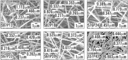
PEO濃度、または絹/PEOブレンド比は、絹繊維の表面積および電界紡糸プロセスの間のバルク形態に直接影響を及ぼす。Jin et al.,3 Biomacromol.1233-39(2002);Wang et al.,37 Macromol.6856-64(2004)。PEO濃度が増加するにつれて、繊維中で生じたフィブロインミセルおよび小球構造のサイズは低下する。その上、ひとたび素早く動く帯電した流体の噴出の中に入ると、これらの小球構造は整列し、100,000倍まで伸張する。Wang et al.,2006;Kowalewski et al.,53 Bulletin Polish Acad.ScL,Tech.Sci.385-94(2005);Reneker&Yarin,49 Polymer 2387-425(2008)。本発明は、絹/PEOブレンド比が、繊維厚さ、密度、配向、相分散、多孔性およびマット厚さを含む、得られる絹マットの特性に重要な役割を果たすことを実証する。その結果として、増加したPEO濃度で形成された繊維は、幾何学的形状、表面積、ならびに、4:1から1:1までの絹/PEOブレンドマットに観察された漸進的な視覚およびテクスチャーの変化に相関するバルク体積が低下した。 PEO concentration, or silk / PEO blend ratio, directly affects silk fiber surface area and bulk morphology during the electrospinning process. Jin et al., 3 Biomacromol. 1233-39 (2002); Wang et al., 37 Macromol. 6856-64 (2004). As the PEO concentration increases, the size of the fibroin micelles and globules formed in the fiber decreases. Moreover, once inside a jet of charged fluid that moves quickly, these globular structures align and stretch up to 100,000 times. Wang et al., 2006; Kowalewski et al., 53 Bulletin Polish Acad. ScL, Tech. Sci. 385-94 (2005); Reneker & Yarin, 49 Polymer 2387-425 (2008). The present invention demonstrates that the silk / PEO blend ratio plays an important role in the properties of the resulting silk mat, including fiber thickness, density, orientation, phase dispersion, porosity and mat thickness. As a result, the fibers formed with increased PEO concentrations are geometric, surface area, and the gradual visual and texture changes observed in silk / PEO blend mats from 4: 1 to 1: 1. The bulk volume correlated with
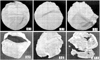
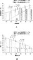
一態様では、4:1、3:1、2:1、3:2、7:6、および1:1の絹/PEOブレンド比で調製した6つの絹/PEOブレンド材料系を、融合性の直径16.5cmおよび10cmのマットに電界紡糸した。各々のサンプルの物理的特性は、水飽和状態と乾燥状態の両方で評価した。水に浸すと、6つのマトリックスは、不透明に半透明な灰白色の外観を提示する、均一な立体構造を有し、絹のようなテクスチャーをもち柔軟であったが、図1Aに示されるように、取扱期間が延びると絹濃度に対する(respective of)引張強さの悪化を提示した。絹のようなテクスチャーとは、水分子が非晶質ポリマーマトリックスの全体にわたって継続的に可塑化するフィブロインの動的吸湿性を説明するために言及される。アミノ、ヒドロキシル、またはカルボキシル酸末端基との水素結合を形成することによるか、または、親水性ドメインの至る所に自由に分散することにより;この流動性の環境は、これらの飽和材料系の軟質の絹のようなテクスチャーをもたらす運動エネルギーの最小化に起因して、継続的に移行している。Hu et al.,39 Macromol.6161-70(2006);Agarwal et al.,63 J.Appl.Polym.Sci.401-10(1997);van der Heijden et al.,378 Thermochim.Acta 27-34(2001);Wong et al.,2006。周囲温度で24時間の乾燥期間の後、物理的な特徴は、6つの材料系で漸進的に変化した。図2および3に示されるように、絹濃度の低下(86.5%、82.8%、76%、70.6%、65.1%および61.5%)に関連して、マットは、付着曲げ強度(cohesive flex strength)をもつ雪のように白い柔軟なウエハーのようなテクスチャーから、半透明褐色で超薄の柔軟性の低いフィルムのような材料に変わった。 In one aspect, six silk / PEO blend material systems prepared at 4: 1, 3: 1, 2: 1, 3: 2, 7: 6, and 1: 1 silk / PEO blend ratios are made of fusogenic Electrospun into 16.5 cm and 10 cm diameter mats. The physical properties of each sample were evaluated in both water saturated and dry conditions. When soaked in water, the six matrices had a uniform three-dimensional structure presenting an opaque translucent off-white appearance, had a silky texture, and were flexible, as shown in Figure 1A As the handling period was extended, it showed a deterioration in tensile strength with respect to silk concentration. Silky texture is mentioned to describe the dynamic hygroscopicity of fibroin where water molecules plasticize continuously throughout the amorphous polymer matrix. By forming hydrogen bonds with amino, hydroxyl, or carboxylic acid end groups, or by freely dispersing throughout the hydrophilic domain; this fluid environment is the softness of these saturated material systems Due to the minimization of kinetic energy resulting in a silky texture of the continual transition. Hu et al., 39 Macromol.6161-70 (2006); Agarwal et al., 63 J.Appl.Polym.Sci.401-10 (1997); van der Heijden et al., 378 Thermochim.Acta 27-34 (2001); Wong et al., 2006. After a drying period of 24 hours at ambient temperature, the physical characteristics gradually changed in the six material systems. As shown in FIGS. 2 and 3, in relation to the decrease in silk concentration (86.5%, 82.8%, 76%, 70.6%, 65.1% and 61.5%), the mat has cohesive flex strength. It turned from a snowy white flexible wafer-like texture to a translucent brown, ultra-thin, low-flexible film-like material.
本発明において、乾燥法は、絹/PEOブレンドマットの物理的および機械的特性、例えば電界紡糸絹/PEOブレンドマットの厚さなどにも影響を及ぼす。例えば、ポリスチレンペトリ皿を使用する風乾法を用いることができる。あるいは、拘束乾燥の方法を用いてよい。例えば、電界紡糸絹マットを乾燥させるために結晶化皿技法を用いてよい。 In the present invention, the drying method also affects the physical and mechanical properties of the silk / PEO blend mat, such as the thickness of the electrospun silk / PEO blend mat. For example, an air drying method using a polystyrene petri dish can be used. Alternatively, a constrained drying method may be used. For example, a crystallization dish technique may be used to dry the electrospun silk mat.
本発明の電界紡糸絹マットの厚さは、約20μm〜約80μmである。拘束乾燥法を用いる場合、電界紡糸絹マットの厚さは、平均約20μm〜30μmでありうる。 The electrospun silk mat of the present invention has a thickness of about 20 μm to about 80 μm. When using the constrained drying method, the thickness of the electrospun silk mat can average from about 20 μm to 30 μm.
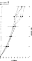
例えば、絹/PEOブレンド比が4:1、3:1、2:1、3:2、7:6、および1:1の電界紡糸絹マットの直径3.5cmのサンプルを、直径10cmのマットから打ち抜き、ポリスチレンペトリ皿法を用いて風乾させた。得られる絹マットを図2Aに示す。飽和したいくつかの直径3.5cmのサンプルは、正味の力(net force)の表面-表面疎水性平衡を実現し、層状の絹シートの分離および変位で親水性挙動を提示するために、取り扱いが困難で、多くの場合半分に折り重なる。水乾燥段階の全体にわたって、極性水分子がこの不織の多孔性生体材料の大きい表面積から蒸発するにつれて、界面での親水性の疎水性ドメインへの転移により表面エネルギーは最小となった。この動的表面構造の再編成は、重鎖の再整列およびβ-シートの結晶化を明示する。Vepari&Kaplan,2007;Jin et al.,200);Hu et al.,39 Macromol.6161-70(2006)。ねじれた襞のあるβ-シート形成の特徴を示して、マトリックスはいずれも完全に平らな配向で乾燥せず、4:1および3:1マトリックスだけが元の円形の形状を維持していた。その上、フィブロインの重鎖の結晶性ドメイン間に交互に配置された非晶質ドメインの末端に位置するプロリン残基がある。Zhou et al.,2000。プロリンは、脱水によって非常に収縮し、従って繊維の収縮する能力を増加させることが示された。Liu et al.,9 Biomacromol.116-21(2008)。図4に示されるように、絹濃度の低下とともに、これらの要素は乾燥したサンプルにおける飽和状態から乾燥状態へのそれらの表面積の51.0±0.0%〜87.5±9.9%の間の減少の一因となる(n=3)。図5に示されるように、サンプルの各々の乾燥したセットの厚さ測定値は、それぞれ、81.7±7.5から77.5±10.5に、66.7±5.1に、53.3±8.1に、46.7±5.1に、および30.0±6.4μmに漸進的に下降した(n=6)。 For example, a 3.5 cm diameter sample of an electrospun silk mat with a silk / PEO blend ratio of 4: 1, 3: 1, 2: 1, 3: 2, 7: 6, and 1: 1 was taken from a 10 cm diameter mat. Punched and air dried using polystyrene petri dish method. The resulting silk mat is shown in FIG. 2A. Some saturated 3.5 cm diameter samples can be handled to achieve a net-force surface-surface hydrophobic equilibrium and to exhibit hydrophilic behavior in the separation and displacement of layered silk sheets. Difficult, often folded in half. Throughout the water drying phase, as polar water molecules evaporated from the large surface area of this nonwoven porous biomaterial, the surface energy was minimized by the transition to hydrophilic hydrophobic domains at the interface. This dynamic surface structure rearrangement demonstrates heavy chain realignment and β-sheet crystallization. Vepari & Kaplan, 2007; Jin et al., 200); Hu et al., 39 Macromol. 6161-70 (2006). Neither matrix was dried in a perfectly flat orientation, and only the 4: 1 and 3: 1 matrices maintained their original circular shape, indicating the characteristics of twisted wrinkled β-sheet formation. In addition, there are proline residues located at the ends of the amorphous domains that are interleaved between the crystalline domains of the fibroin heavy chain. Zhou et al., 2000. Proline has been shown to shrink significantly upon dehydration, thus increasing the ability of the fiber to shrink. Liu et al., 9 Biomacromol. 116-21 (2008). As shown in FIG. 4, with decreasing silk concentration, these factors contribute to a decrease in their surface area from 51.0 ± 0.0% to 87.5 ± 9.9% from saturated to dry in the dried sample. (N = 3). As shown in FIG. 5, the thickness measurements of each dry set of samples were 81.7 ± 7.5 to 77.5 ± 10.5, 66.7 ± 5.1, 53.3 ± 8.1, 46.7 ± 5.1, and 30.0, respectively. Gradually descended to ± 6.4 μm (n = 6).
別の態様では、絹/PEOブレンド比が4:1、3:1、2:1、3:2、7:6、および1:1の直径12.5cmの電界紡糸絹マットを、結晶化皿技法を用いて乾燥させた。得られる絹マットを図3Aに示す。水に浸すと、これらのより大型のサンプルは展開がより容易であった。親水性の力に関して、層状のシート分離は、シートの変位なく各々のサンプルの内部領域でのみ観察された。これは、これらのサンプルがより大型のマットから打ち抜かれておらず、従ってサンプルの端部の架橋された結晶化領域を保持しているためでありうる。乾燥段階の間、飽和したサンプルが縁からサンプルの中心に向かって均一に乾燥するとき、マットの各々のセットは皿の口の全体にわたって漸進的に収縮する。4:1および3:1サンプルは、結晶化皿の縁に付着して完全に乾燥し、完全に平坦で柔軟な白色の膜のような材料をもたらした。3:2、7:6、および1:1サンプルが乾燥するとき、結晶化する延伸力が繊維の降伏点伸びを越えて材料に応力を加え、皿の縁での材料の剪断による構造的破壊をもたらし、サンプルの内部領域に伝わった。この乾燥法では、絹濃度の低下は、材料の構造的完全性および曲げ強度に影響を及ぼした。2:1サンプルは皿の縁から切り取られたが、材料変形の痕跡はほとんどなく、特性は4:1および3:1サンプルに類似していた。図4に示されるように、4:1および3:1マトリックスは、元の表面積の98%を保持したが、2:1マットは、11.8%±2.7%を失った。3:2、7:6および1:1サンプルは、それぞれ、68.8%±9.1%、65.9%±4.3%および63.9%±6.5%収縮した(n=3)。図5に示されるように、マットの各々の乾燥したセットの平均厚さは、それぞれ、31.2±1.8、28.7±1.2、24.3±2.3、25.3±2.3、20.0±1.4および26.0±0.9μmであった。 In another embodiment, electrospun silk mats with a silk / PEO blend ratio of 4: 1, 3: 1, 2: 1, 3: 2, 7: 6, and 1: 1 with a diameter of 12.5 cm are prepared using a crystallization dish technique. And dried. The resulting silk mat is shown in FIG. 3A. These larger samples were easier to deploy when immersed in water. With regard to hydrophilic forces, laminar sheet separation was observed only in the inner region of each sample without sheet displacement. This may be because these samples have not been stamped out of a larger mat and thus retain a cross-linked crystallized region at the end of the sample. During the drying phase, each set of mats progressively shrinks across the mouth of the dish as the saturated sample dries evenly from the edge toward the center of the sample. The 4: 1 and 3: 1 samples adhered to the edge of the crystallization dish and dried completely, resulting in a completely flat and flexible white film-like material. When 2: 2, 7: 6, and 1: 1 samples dries, the stretching force that crystallizes stresses the material beyond the yield point elongation of the fiber, resulting in structural failure by shearing the material at the edge of the dish Brought to the inner area of the sample. In this drying method, the decrease in silk concentration affected the structural integrity and bending strength of the material. The 2: 1 sample was cut from the edge of the dish, but there was little evidence of material deformation and the properties were similar to the 4: 1 and 3: 1 samples. As shown in FIG. 4, the 4: 1 and 3: 1 matrices retained 98% of the original surface area, while the 2: 1 mat lost 11.8% ± 2.7%. The 3: 2, 7: 6 and 1: 1 samples contracted by 68.8% ± 9.1%, 65.9% ± 4.3% and 63.9% ± 6.5%, respectively (n = 3). As shown in FIG. 5, the average thickness of each dry set of mats was 31.2 ± 1.8, 28.7 ± 1.2, 24.3 ± 2.3, 25.3 ± 2.3, 20.0 ± 1.4 and 26.0 ± 0.9 μm, respectively. .
本発明において、電界紡糸絹/PEOブレンドマットの結果として得られる繊維は、マット構造全体にわたって実質的に均一な直径分布を有する。図6AのSEM画像は、電界紡糸プロセスの直後、かつメタノール飽和およびPEO浸出処理の前の全ての6つの絹/PEO材料群の画像である。全体に、これらの電界紡糸した絹/PEO繊維は、直径が200nm〜500nmの範囲に及ぶ。Huang et al,2001に報告されるように、繊維ビーズ形成は、PEO濃度の低下とともに次第に顕著となった。Wang et al.,2006;Zhou et al.,2000;Huang et al.,12 J.Biomat.Sci.Polym.Ed.979-93(2001)。具体的には、4:1、3:1、および2:1(フィブロイン:PEO)サンプルは、各々、繊維内のランダムな位置にちょうど幅1μmを上回るものから700nm間での範囲に及ぶビーズセグメントを有した。ビーズは、3:2および7:6サンプルセットで最小化し、明確に定義された微細な円形状の繊維が構造の全体にわたって秩序化された外観を与えた。1:1サンプルセットは特有の外観を有し、繊維は不規則で非円形の形状であって、不均一で高密度のマット構造に移行した。この移行が、装置の接地ステージに集まった場合の、液-液/液-固相分散による繊維の収束に起因しうることは妥当である。1:1の個々の繊維は、300〜500nmの範囲に及び、一方、混合繊維は700〜900nmの間で測定された。 In the present invention, the resulting fibers of the electrospun silk / PEO blend mat have a substantially uniform diameter distribution throughout the mat structure. The SEM image in FIG. 6A is an image of all six silk / PEO material groups immediately after the electrospinning process and before the methanol saturation and PEO leaching processes. Overall, these electrospun silk / PEO fibers range in diameter from 200 nm to 500 nm. As reported in Huang et al, 2001, fiber bead formation became progressively more pronounced with decreasing PEO concentration. Wang et al., 2006; Zhou et al., 2000; Huang et al., 12 J. Biomat. Sci. Polym. Ed. 979-93 (2001). Specifically, 4: 1, 3: 1, and 2: 1 (fibroin: PEO) samples are each bead segments ranging from just over 1 μm wide to 700 nm at random locations within the fiber Had. The beads were minimized in the 3: 2 and 7: 6 sample sets, with well-defined fine circular fibers giving an ordered appearance throughout the structure. The 1: 1 sample set had a unique appearance and the fibers were irregular and non-circular in shape, transitioning to a non-uniform and dense mat structure. It is reasonable that this transition can be attributed to fiber convergence due to liquid-liquid / liquid-solid dispersion when collected at the ground stage of the apparatus. 1: 1 individual fibers ranged from 300 to 500 nm, while mixed fibers were measured between 700 and 900 nm.
「拘束乾燥技法」または「拘束乾燥する」とは、本明細書において、絹材料が延伸力を受けている間に乾燥するように、絹材料を拘束している間に乾燥させるプロセスをさす。例えば、拘束力は、結晶化皿の口の上に付着しながら絹材料が乾燥するときに起こる合成収縮力に起因しうる。記載されるように、これらの飽和した絹材料は、最初に結晶化皿の口の上に掛けられてそれに付着する。水分子が蒸発するにつれて、タンパク質の表面基質の疎水性ドメインおよびバルク領域の全体が、不織のキャスト(non-woven cast)の組織間隙(interstitial space)および材料のバルク領域の中から自由体積の喪失を開始する。自由体積の喪失は、材料を収縮させ、結晶化皿の縁に向かって放射状に延伸させる。結晶化皿の縁に付着しているので、材料は自由体積の継続的な喪失によって束縛されることとなり、繊維は半径応力の方向に整列し、伸張されるようになる。絹体積に応じて、材料繊維が降伏点伸びを越えて収縮すれば、材料の剪断が材料/結晶化皿縁表面界面で起こる。拘束乾燥法に反して、ペトリ皿の中の風乾サンプルは、乾燥してねじれた不規則な立体構造となるまで継続的に収縮する。 “Constrained drying technique” or “constrained drying” as used herein refers to the process of drying a silk material while constrained so that the silk material dries while subjected to stretching forces. For example, the restraining force may be due to a synthetic shrinkage force that occurs when the silk material dries while depositing on the mouth of the crystallization dish. As described, these saturated silk materials are first hung on and attached to the mouth of the crystallization dish. As the water molecules evaporate, the entire hydrophobic domain and bulk region of the protein surface matrix becomes free volume out of the interstitial space of the non-woven cast and the bulk region of the material. Start losing. The loss of free volume causes the material to shrink and stretch radially toward the edge of the crystallization dish. Because it is attached to the edge of the crystallization dish, the material will be constrained by the continuous loss of free volume, and the fibers will align and stretch in the direction of radial stress. Depending on the silk volume, material shearing occurs at the material / crystallization dish edge surface interface if the material fibers shrink beyond the yield point elongation. Contrary to the constrained drying method, the air-dried sample in the Petri dish is continuously shrunk until it becomes a dried and twisted irregular three-dimensional structure.
一態様では、拘束乾燥法は、制御された蒸発によって行われる。この方法は、電界紡糸絹/PEOブレンドマットを水浴から取り出し、マットを、皿の3分の1まで水を入れた結晶化皿の上に掛け、マットを含有する皿を20%〜50%の相対湿度のデシケーターに入れて一晩乾燥させることを含む。結晶化皿乾燥法を利用する漸進的なポリマー鎖および繊維の立体構造を説明する概念図を図9に示す。上部は、分散した未整列の二次構造の、親水性環境から、疎水性結合により駆動される整列したタンパク質凝集体への移行である。繊維が引き伸ばされ始めるとき、水なましされた(water-annealed)β-シートが鎖間形成対鎖内形成によって集合する。図の下部の繊維形成は、絹濃度および繊維整列および伸張の間の逆相関を反映する。 In one aspect, the constrained drying method is performed by controlled evaporation. This method removes the electrospun silk / PEO blend mat from the water bath, hangs the mat on a crystallization dish containing up to one third of the dish, and the dish containing the mat is 20% -50% Including overnight drying in a relative humidity desiccator. FIG. 9 shows a conceptual diagram illustrating the gradual polymer chain and the three-dimensional structure of the fiber using the crystallization dish drying method. The top is the transition of dispersed unaligned secondary structure from the hydrophilic environment to aligned protein aggregates driven by hydrophobic bonds. As the fibers begin to be stretched, water-annealed β-sheets assemble through interchain formation versus intrachain formation. The fiber formation at the bottom of the figure reflects the inverse correlation between silk concentration and fiber alignment and stretching.
一態様では、図7に示されるように、絹/PEOブレンドマットのSEM画像は、拘束乾燥法、例えば結晶化皿法などを用いて調製された。調製したマットの絹/PEOブレンド比は、それぞれ4:1、3:1および2:1である。表面のトポグラフィーは、各々のモデルの全体にわたる繊維の高密度でランダムな分布を反映する。評価は、この乾燥技法によって起こる繊維収縮、伸張、および再整列の痕跡の増加を示す。図7Bを参照すると、4:1マットの繊維は、目立った繊維収縮または整列がなく緩んでねじれた外観を有する。対照的に、3:1および2:1マットの繊維は伸張して整列し付着して、ウェブのような微小なテクスチャー(web-like micro texture)を形成するようになる。図7Cの繊維形成に焦点を合わせると、3:1マットの伸張繊維は、ピンと張ったウェブ構造を形成し、末端が液化性の外観になっている整列した繊維間の相分散の痕跡を示している。2:1フィブロイン:PEOサンプルのウェブ構造は、ロープのような配置を形成する、明確に定義された、伸張され整列された繊維の絡み合った網状構造からなる。 In one embodiment, as shown in FIG. 7, SEM images of silk / PEO blend mats were prepared using a constrained drying method, such as a crystallization dish method. The silk / PEO blend ratios of the prepared mats are 4: 1, 3: 1 and 2: 1, respectively. The surface topography reflects the dense and random distribution of fibers throughout each model. The evaluation shows an increased trace of fiber shrinkage, stretching, and realignment that occurs with this drying technique. Referring to FIG. 7B, the 4: 1 matte fibers have a loose and twisted appearance without noticeable fiber shrinkage or alignment. In contrast, the 3: 1 and 2: 1 matte fibers stretch, align and adhere to form a web-like micro texture. Focusing on the fiber formation of FIG. 7C, the 3: 1 matte stretch fibers form a taut web structure and show traces of phase dispersion between aligned fibers with a liquefied appearance at the ends. ing. The 2: 1 fibroin: PEO sample web structure consists of an intertwined network of well-defined, stretched and aligned fibers that form a rope-like arrangement.
拘束乾燥技法により明らかにされる繊維の整列および伸張は、絹フィブロインの水なまし特性によるものでありうる。Jin et al.,15 Adv.Funct.Mater.1241-47(2005);Agarwal et al.,63 J.Appl.Polym.Sci.401-10(1997);Lawrence et al.,43 J.Mat.Sci.6967-85(2008);Wong et al.,2006。ポリマーバルク領域内部で可塑剤の役割を果たすことにより、水分子は、低結合エネルギーのポリマー鎖間で分子間の動きを伝え、ポリマーの流動性および再整列を促進する。水分子が蒸発するときに、ポリマー鎖は、延伸されて、結晶化皿の縁の周囲から発生する半径応力の方向を向く。延伸されたポリマー鎖の整列とともに、非晶質軽鎖の末端で折り畳まれている、両側性の鎖間層構造の段階的拡大および結晶化した鎖内のねじれた立体構造の減少を促進するプロリンは減少する。Zhou et al.,2000;Liu et al.,9 Biomacromol.116-21(2008)。支配的な鎖間疎水性相互作用は、バルク領域から自由体積を圧縮し、結晶性二次構造整列および非晶質絹I状態の結晶性絹II状態への移行に影響を及ぼす。Jin et al.,15 Adv.Funct.Mater.1241-47(2005);Agarwal et al.,1997;Lawrence et al.,43 J.Mat.Sci.6967-85(2008);Wong et al.,82 Appl.Phys.A-Mater.293-203(2006)。水分子の継続的な排出によって、バルク体積は低下し、繊維が結晶化皿の口の上で伸張し始めるまで収縮する。図9に示されるように、これらの構成要素の組合せが、繊維軸に沿って、高い強度材料特性をもつ伸張され整列された繊維によって、マットを形成する。降伏点伸びを越えて、重および軽(H,L)鎖の両方の非晶質二次構造の内部および二次構造に沿って剪断変形が起こる。 The fiber alignment and stretching revealed by the constrained drying technique may be due to the water-annealing properties of silk fibroin. Jin et al., 15 Adv. Funct. Mater. 1241-47 (2005); Agarwal et al., 63 J. Appl. Polym. Sci. 401-10 (1997); Lawrence et al., 43 J. Mat. Sci. 6967-85 (2008); Wong et al., 2006. By acting as a plasticizer within the polymer bulk region, water molecules convey intermolecular movement between polymer chains of low binding energy, facilitating polymer fluidity and realignment. As the water molecules evaporate, the polymer chains are stretched to point in the direction of radial stress generated from around the edge of the crystallization dish. Proline promotes stepwise expansion of bilateral interchain structure and reduction of twisted conformation within the crystallized chain, folded at the end of the amorphous light chain, along with alignment of stretched polymer chains Decrease. Zhou et al., 2000; Liu et al., 9 Biomacromol. 116-21 (2008). The dominant interchain hydrophobic interaction compresses the free volume from the bulk region and affects the crystalline secondary structure alignment and the transition from the amorphous silk I state to the crystalline silk II state. Jin et al., 15 Adv. Funct. Mater. 1241-47 (2005); Agarwal et al., 1997; Lawrence et al., 43 J. Mat. Sci. 6967-85 (2008); Wong et al., 82 Appl. Phys. A-Mater. 293-203 (2006). With continuous discharge of water molecules, the bulk volume decreases and shrinks until the fiber begins to stretch over the mouth of the crystallization dish. As shown in FIG. 9, the combination of these components forms a mat with stretched and aligned fibers with high strength material properties along the fiber axis. Beyond the yield elongation, shear deformation occurs inside and along the secondary structure of both heavy and light (H, L) chain amorphous secondary structures.
整列した図7Cの3:1繊維と図7Dの2:1繊維の間の巨視的相分散の外観も、水の可塑化特性に起因しうる。水およびカイコ絹フィブロインフィルム系の示差走査熱量測定により、脱水した絹フィブロインのガラス転移温度(Tg)が、20〜23重量%の吸水量で178℃から40℃より下まで低下したことが明らかとなった。Agarwal et al.,1997。結晶性立体構造は生体材料の全体にわたる希釈剤の吸収に影響を及ぼすが、各々の絹マット群の平衡含水率(EWC)は、80重量%よりも大きく(表4)、相当な親水性相互作用を示し;Tgおよび妥当な相分散の低下を予見する。Hu et al.,39 Macromol.6161-70(2006)。その上、絹ポリマー鎖の全体にわたって交互に配置された疎水性ドメインおよび親水性ドメインは、材料が表面-液体から表面-ガス、表面-表面界面へ移行するときに、動的移動性表面基質をもたらす。Ratner et al.,BIOMATS.Sci.:INTRO.MATS.MED.(Acad.Press,NY,2004;Allcock,INTRO.MATS.CHEM.(Wiley&Sons,Hoboken,NJ,2008)。具体的には、希釈剤が蒸発するときに、親水性のドメインは、表面で疎水性セグメントに置き換わる。上記の相分散を考慮に入れると、この現象が界面の繊維間に起こる場合、非晶質二次構造が散在するようになり、混合繊維となる。また、繊維間の線形の二次構造が整列するようになって熱力学的に安定した結晶性β-ストランド(stands)を形成しうることも考えられる。Lawrence et al.,43 J.Mat.Sci.6967-85(2008);Fink,3 Folding&Design R9-R23(1998)。 The appearance of the macroscopic phase dispersion between the aligned 3: 1 fiber of FIG. 7C and the 2: 1 fiber of FIG. 7D can also be attributed to the plasticizing properties of water. Differential scanning calorimetry of water and silkworm silk fibroin film systems revealed that the glass transition temperature (Tg) of dehydrated silk fibroin decreased from 178 ° C to below 40 ° C at 20-23 wt% water absorption. became. Agarwal et al., 1997. Although the crystalline conformation affects the absorption of diluent throughout the biomaterial, the equilibrium moisture content (EWC) of each silk mat group is greater than 80 wt% (Table 4) Show effects; predict decrease in Tg and reasonable phase dispersion. Hu et al., 39 Macromol. 6161-70 (2006). In addition, the hydrophobic and hydrophilic domains, which are interleaved throughout the silk polymer chain, create a dynamic mobile surface matrix as the material transitions from surface-liquid to surface-gas, surface-surface interface. Bring. Ratner et al., BIOMATS.Sci.:INTRO.MATS.MED. (Acad.Press, NY, 2004; Allcock, INTRO.MATS. CHEM. (Wiley & Sons, Hoboken, NJ, 2008). Specifically, diluents. When evaporating, hydrophilic domains replace hydrophobic segments at the surface, taking into account the above phase dispersion, interspersed amorphous secondary structures if this phenomenon occurs between interfacial fibers It is also conceivable that the linear secondary structure between the fibers can be aligned to form thermodynamically stable crystalline β-strands (Lawrence). et al., 43 J. Mat. Sci. 6967-85 (2008); Fink, 3 Folding & Design R9-R23 (1998).
本発明の絹マットは、平均して約0.1〜約1μmの細孔のど径表面積をもつ相互接続した細孔を有しうる。図7Eの断面図より、数種類の異なる絹/PEOブレンド比をもつ絹マットのさまざまな特色が明らかとなる。これらの画像は、絹濃度の低下とともに増加した繊維密度を示す;そして、マトリックスにわたる繊維凝集も、異なる絹/PEOブレンド比をもつ絹マットに関して異なっている。4:1ブレンドの繊維は、多数の大きな空間の隙間をもつ水平シートに凝集した。3:1および2:1ブレンドの繊維は、繊維束の増加を実証し、絹容積の低下に対する繊維の収縮および空間の間隙の漸進的低下を反映した。これらの所見は、絹濃度に対するマット厚さの低下に一致する。図7Bの3枚の画像は、この生体材料の全体にわたって相互に連絡する多孔性を表す。細孔は、各々のマトリックスに対して、それぞれ平均して294、201、から103nm2ののど径表面積を有した。図9に示される下降する細孔径分布は、断面図に見られる繊維密度の増加に一致し、ビーズ領域の数ならびに4:1、3:1、および2:1マトリックスの繊維直径の低下に関して繊維の集合体に関連する。 The silk mat of the present invention can have interconnected pores with an average throat surface area of about 0.1 to about 1 μm pores. From the cross-sectional view of FIG. 7E, various features of silk mats with several different silk / PEO blend ratios become apparent. These images show increased fiber density with decreasing silk concentration; and fiber aggregation across the matrix is also different for silk mats with different silk / PEO blend ratios. The 4: 1 blend of fibers aggregated into a horizontal sheet with many large space gaps. The 3: 1 and 2: 1 blend fibers demonstrated an increase in fiber bundles, reflecting the shrinkage of the fibers and a gradual decrease in the gap in the space as the silk volume decreased. These findings are consistent with a decrease in mat thickness with respect to silk concentration. The three images in FIG. 7B represent the porosity communicating with each other throughout the biomaterial. The pores had an average throat surface area of 294, 201 to 103 nm 2 for each matrix, respectively. The descending pore size distribution shown in FIG. 9 is consistent with the increase in fiber density seen in the cross-sectional view, and the fiber with respect to the number of bead regions and the decrease in fiber diameter of the 4: 1, 3: 1, and 2: 1 matrix Related to a collection of
材料の表面粗さは、細胞の配向、接着、増殖、および遊走を制御する、正味の力の生体力学的平衡を促進する、応力/剪断(sheer)のない平面による細胞接触誘導に影響を及ぼす。拘束乾燥処理を受けた本発明の絹/PEOブレンドマットは、風乾法を受けた絹マットよりも表面粗さが小さい。図10のAFM画像は、ポリスチレン皿風乾法で乾燥させた後のさまざまな絹/PEOブレンド比の本発明の絹マットの、三次元の形態学的イメージおよびサンプルの二乗平均平方根の粗さ値を示す。局地的なドメイン粗さ分析は、画像のXおよびY平面の全体にわたる粗さの変動により特徴付けられる。全体として、これらの絹材料系は、粗さ値が500nmから1.4μmまでの明確な表面不規則性を示すクラス3の表面トポグラフィー(class three surface topographies)を実証した(n=3)。4:1および1:1サンプルは、それぞれ1.17±0.00および0.78±0.01μmを測定する、ナノサイズの不規則性をもつ相対的に均一な粗さを有した。3:1、2:1、および3:2サンプルの粗さの標準偏差は0.1〜0.17μmの間で、それぞれ0.65±0.10、0.88±0.17、および0.76±0.16μmの範囲に及んだ。7:6マットが、局地的な粗さが平均1.01±0.43μmの最も大きい偏差を有した。AFM粗さ評価も結晶化皿技法で拘束乾燥した絹/PEOブレンドマットに行った。4:1、3:1、および2:1サンプルは、16×16μmの面積に関して、それぞれ0.66、0.36および0.25μmの粗さ値を有する。サンプルの面積サイズは低下し、サンプルサイズは制限されるが(n=1)、引き伸ばし乾燥したマットの粗さは、風乾マットよりも少なくとも44%平坦である。拘束乾燥したサンプルは、絹濃度に対して粗さが直線的に低下するように見えるが、風乾サンプルについては顕著な傾向はない。この所見は、風乾サンプルのねじれた不規則さと比較して、拘束乾燥したサンプルの繊維の伸張特性に一致する。 Material surface roughness affects cell contact induction by stress / sheerless planes, which promotes the biomechanical equilibrium of net forces that control cell orientation, adhesion, proliferation, and migration . The silk / PEO blend mat of the present invention that has undergone the constrained drying treatment has a lower surface roughness than the silk mat that has undergone the air drying method. The AFM image in Figure 10 shows three-dimensional morphological images and sample root-mean-square roughness values of the silk mats of the present invention with various silk / PEO blend ratios after drying by polystyrene dish air drying. Show. Local domain roughness analysis is characterized by variations in roughness across the X and Y planes of the image. Overall, these silk material systems demonstrated class three surface topographies (n = 3) exhibiting a distinct surface irregularity with roughness values from 500 nm to 1.4 μm. The 4: 1 and 1: 1 samples had relatively uniform roughness with nano-sized irregularities measuring 1.17 ± 0.00 and 0.78 ± 0.01 μm, respectively. The standard deviations of the roughness of the 3: 1, 2: 1, and 3: 2 samples ranged from 0.1 to 0.17 μm, ranging from 0.65 ± 0.10, 0.88 ± 0.17, and 0.76 ± 0.16 μm, respectively. The 7: 6 mat had the largest deviation with local roughness averaging 1.01 ± 0.43 μm. AFM roughness evaluation was also performed on silk / PEO blend mats constrained and dried by the crystallization dish technique. The 4: 1, 3: 1, and 2: 1 samples have roughness values of 0.66, 0.36, and 0.25 μm, respectively, for an area of 16 × 16 μm. Although the area size of the sample is reduced and the sample size is limited (n = 1), the roughness of the stretch dried mat is at least 44% flatter than the air dried mat. Restrained dry samples appear to have a linear decrease in roughness with respect to silk concentration, but there is no significant trend for air-dried samples. This finding is consistent with the fiber stretch properties of the constrained dried sample compared to the twisted irregularities of the air dried sample.
本発明のプロセスにより調製した電界紡糸絹/PEOブレンドマットは、生体材料用途、例えば創傷被覆材などに適した、良好な構造特性、形態特性、生体機能特性および生体適合特性を示す。例えば、得られる本発明の絹マットは、14日未満で約86%(重量)超を分解する;本発明の絹マットの平衡含水率は約82%を上回る;絹マットの酸素透過速度は、約15460cm3/m2/日よりも大きい;かつ、絹マットの水蒸気透過速度は、約1934g/m2/日よりも大きい。 The electrospun silk / PEO blend mat prepared by the process of the present invention exhibits good structural, morphological, biofunctional and biocompatible properties suitable for biomaterial applications such as wound dressings. For example, the resulting silk mat of the present invention degrades greater than about 86% (by weight) in less than 14 days; the equilibrium moisture content of the silk mat of the present invention is greater than about 82%; Greater than about 15460 cm 3 / m 2 / day; and the water vapor transmission rate of the silk mat is greater than about 1934 g / m 2 / day.
本発明の態様は、時間放出生物療法によって上皮化を促進するための、酵素生分解を備える絹材料を提供する。さまざまなブレンド比をもつ電界紡糸絹/PEOブレンドマットの酵素生分解を、14日以上にわたって評価した。図12Aに示されるように、インビトロ生分解性は、全ての時点で全ての材料群について線形の分解を示し、それぞれ、各々の群に対して1日後に22.6%±3.4%の分解、および14日後に74.0%±8.8%までの材料損失をもたらした。データは、6日までは、全てのブレンドについての分解率は48.2±4.6%で比較的近かったという推論を許容する。10日後、4:1(78%)から1:1(51%)の範囲に及ぶ絹/PEOブレンド比をもつさまざまな材料群について27%の重量減少差が記録された。14日後、酵素分解は85.6±3.8%(4:1)から62.5±5.2%(1:1)に至る範囲に及んだ。形態学的に、目視検査によって、全ての材料系が、最初の6日で表面浸食により主に分解された。10日の時点の後、4:1および3:1サンプルが、マットのほころび、断片化、および微粒子破片への崩壊をもたらす、繊維の切断分解の増加を実証した。4:1および3:1サンプルの生分解挙動は、これらのブレンドの増加した繊維サイズ、マット多孔性および繊維密度特性の低下により、マットの内部繊維構造に酵素が接近することに起因し得る。 Aspects of the invention provide silk materials with enzyme biodegradation to promote epithelialization by time release biotherapy. Enzymatic biodegradation of electrospun silk / PEO blend mats with various blend ratios was evaluated over 14 days. As shown in FIG. 12A, in vitro biodegradability showed linear degradation for all material groups at all time points, with 22.6% ± 3.4% degradation after 1 day for each group, and 14 It resulted in material loss up to 74.0% ± 8.8% after day. The data allows the inference that by 6 days, the degradation rate for all blends was relatively close at 48.2 ± 4.6%. After 10 days, a 27% weight loss difference was recorded for various material groups with silk / PEO blend ratios ranging from 4: 1 (78%) to 1: 1 (51%). After 14 days, enzymatic degradation ranged from 85.6 ± 3.8% (4: 1) to 62.5 ± 5.2% (1: 1). Morphologically, by visual inspection, all material systems were mainly degraded by surface erosion in the first 6 days. After the 10 day time point, the 4: 1 and 3: 1 samples demonstrated an increase in fiber breakage that resulted in mat breakage, fragmentation, and disintegration into particulate debris. The biodegradation behavior of the 4: 1 and 3: 1 samples can be attributed to enzyme access to the internal fiber structure of the mat due to the increased fiber size, mat porosity and fiber density properties of these blends.
線形回帰分析は、Minitab(登録商標)15.2.30を用いて各々の絹/PEOブレンド比について全てのサンプルで行った。図12Bの散布図を参照することにより、各々の材料群に関して実行した対数変換は、3日の分解時点の直前の全ての材料群について明確な転移点を明らかにした。次に、各々の材料群について全ての時点、開始から3日の時点および3日〜14日の時点で回帰分析を行った。図12Cに示されるように、全ての材料群につての分解の傾きは3日後に実質的に変化した。開始から3日の時点まで、酵素分解速度は、平均-11.68±0.91傾き単位であった;3日以後14日まで、分解の速度は-3.41±1.12まで横ばいとなった。初期分解段階の間、非晶質領域は、内部結晶領域と比較して加速した速度で分解する。この仮説は、酵素分解の開始時および3日後にこれらの絹材料の赤外吸光度スペクトルにフーリエセルフデコンボリューション(FSD)を用いることにより検証することができる。Hu et al.,2006。 Linear regression analysis was performed on all samples for each silk / PEO blend ratio using Minitab® 15.2.30. By referring to the scatter plot of FIG. 12B, the logarithmic transformation performed for each material group revealed clear transition points for all material groups just prior to the 3 day decomposition time point. Next, regression analysis was performed for each material group at all time points, at the 3rd day from the start, and at the 3rd to 14th day. As shown in FIG. 12C, the slope of degradation for all material groups changed substantially after 3 days. From the start to 3 days, the rate of enzymatic degradation averaged -11.68 ± 0.91 slope units; from 3 days to 14 days, the rate of degradation leveled off to -3.41 ± 1.12. During the initial decomposition phase, the amorphous region decomposes at an accelerated rate compared to the internal crystalline region. This hypothesis can be verified by using Fourier Self Deconvolution (FSD) on the infrared absorbance spectra of these silk materials at the start of enzymatic degradation and after 3 days. Hu et al., 2006.
正常なヒト皮膚は約21日で再生する。本発明は、時間放出生物療法での上皮化を促進するために、例えば、プロテアーゼが絹材料を断片に分解する時間を比較することにより、マトリックスの設計を所望の分解速度に一致させることを可能にする。結果は、14日後に、4:1絹/PEOブレンドマットの86%が溶解し、それは、リゾチームに曝露した場合に14日後に82%分解したキトサン/ポロキサマー包帯材、およびPBS中で14日後に20%分解するPLGA/PLLA(90/10)と比べても遜色がない。また、酵素レベルは変動する可能性があるため、インビボでの分解速度にも対処し、絹生体材料が、材料形式、位置、および関連する変数によって、インビボで数週間から数年のうちに分解し得ることが示された。Wang et al.,2008。 Normal human skin regenerates in about 21 days. The present invention makes it possible to match the design of the matrix to the desired degradation rate, for example by comparing the time for the protease to break down the silk material into fragments to promote epithelialization in time-release biotherapy To. The results show that after 14 days, 86% of the 4: 1 silk / PEO blend mat dissolves, it is 82% degraded after 14 days when exposed to lysozyme, and after 14 days in PBS Compared to PLGA / PLLA (90/10) which decomposes by 20%, it is not inferior. Enzyme levels can also vary, thus addressing in vivo degradation rates and silk biomaterials can degrade in vivo in weeks to years depending on material format, location, and related variables It was shown that. Wang et al., 2008.
本発明のプロセスにより製造された絹材料は、多様な医学的用途、例えば、血管創傷修復装置、止血包帯材、全層熱傷被覆材、パッチおよびグルー、縫合糸、薬物送達を含む、創傷閉鎖系などにおいて、かつ、組織工学用途、例えば、足場材料、靭帯補綴装置などにおいて、かつ、ヒト身体への長期もしくは生分解性の移植のための製品において、使用することができる。例示的な組織工学足場は、電界紡糸繊維の不織網状構造である。 Silk materials produced by the process of the present invention can be used in a variety of medical applications, including wound closure systems, including vascular wound repair devices, hemostatic dressings, full thickness burn dressings, patches and glues, sutures, drug delivery. And in tissue engineering applications such as scaffold materials, ligament prosthetic devices, and in products for long-term or biodegradable implantation into the human body. An exemplary tissue engineering scaffold is a non-woven network of electrospun fibers.
その上、これらの生体材料は、限定されるものではないが、脊椎板、頭蓋組織、硬膜、神経組織、肝臓、膵臓、腎臓、膀胱、脾臓、心筋、骨格筋、腱、靭帯、および乳房組織を含む、これらの特有の足場から利益を受けうる、器官修復置換または再生計画に使用することができる。 In addition, these biomaterials include, but are not limited to, spinal disc, skull tissue, dura mater, nerve tissue, liver, pancreas, kidney, bladder, spleen, heart muscle, skeletal muscle, tendon, ligament, and breast It can be used in organ repair replacement or regeneration plans that can benefit from these unique scaffolds, including tissues.
一態様では、本発明は、材料を包帯に加工し、創傷部位の浮腫およびO2/CO2ガス透過を管理する能力、ならびに同時刻に合わせた(time synchronized)抗生物質、免疫学的、および組織再生生物治療を投与する能力を含む全層熱傷被覆材において有用な特性をもつ絹材料を提供する。本発明において、拘束乾燥技法を用いて、絹濃度が繊維厚さ、密度、配向、相分散、多孔性およびマット厚さを含む特性に大きな役割を果たしたことが見出された。例えば、全層創傷被覆材において、4:1〜2:1の絹/PEOブレンド比をもつ電界紡糸絹マットが使用され、それは最小限の表面積減少で柔軟な膜様材料を提示する有用な物理的特性を保有し、グラム陰性桿菌およびグラム陽性球菌敗血症を惹起する病原性微生物に対して不透過性の障壁をもたらす0.3μm2よりも小さい細孔のど表面積サイズを示す。 In one aspect, the present invention provides the ability to process a material into a bandage to manage wound site edema and O 2 / CO 2 gas permeation, as well as time synchronized antibiotics, immunological, and Silk materials having properties useful in full-thickness burn dressings including the ability to administer tissue regenerative biotherapy are provided. In the present invention, using constrained drying techniques, it was found that silk concentration played a major role in properties including fiber thickness, density, orientation, phase dispersion, porosity and mat thickness. For example, in full-thickness wound dressings, electrospun silk mats with a silk / PEO blend ratio of 4: 1 to 2: 1 are used, which is useful physics to present a flexible membrane-like material with minimal surface area reduction It exhibits a pore throat surface area size of less than 0.3 μm 2 that possesses mechanical properties and provides an impervious barrier to pathogenic microorganisms that cause Gram-negative bacilli and Gram-positive cocci sepsis.
材料の吸収および平衡含水率(EWC)特性は、細菌の栄養床を提供し得る創傷浸出液の蓄積を制御する際に役割を果たす。一態様では、4:1〜1:1の範囲の絹/PEOブレンド比をもつ絹材料についての全体的な吸収性およびEWC性能は、各群内で比較的接近し、それぞれ400%〜700%および82%〜86%の範囲に及んだ。絹材料群での繊維密度の違いを考慮すると、異なるブレンド比をもつ各々の材料群はそれでも類似する膨潤特性を提示した。これらのモデルと表2中のその他の創傷被覆材候補を比較すると、本発明の絹マットはスポンジのような天然キトサンに基づく包帯材と同様によく機能した。キトサン/ポロキサマー包帯材候補は良好な吸収性およびEWC特性を有したが、本発明の全天然のFDA認可絹材料系は、これらのその他の系と比較して生体適合性および顕著な機械的堅牢性を提供する。 The absorption and equilibrium water content (EWC) properties of the material play a role in controlling the accumulation of wound exudates that can provide a nutrient bed for bacteria. In one aspect, the overall absorbency and EWC performance for silk materials with silk / PEO blend ratios ranging from 4: 1 to 1: 1 are relatively close within each group, 400% to 700% respectively. And ranged from 82% to 86%. Considering the difference in fiber density among the silk material groups, each material group with different blend ratios still exhibited similar swelling properties. Comparing these models with the other wound dressing candidates in Table 2, the silk mat of the present invention performed as well as a dressing based on natural chitosan such as a sponge. Although the chitosan / poloxamer dressing candidates have good absorbency and EWC properties, the all-natural FDA approved silk material system of the present invention is biocompatible and significantly mechanically robust compared to these other systems. Provide sex.
(表2)キトサン誘導体に基づく創傷被覆材と本発明の絹材料系との間の平均吸水率および平衡含水率の比較
体温恒常性を維持するために、正常な皮膚は、1日あたり204g/m2の速度で体液を透過させる。Lamke et al.,3 Burns 159-65(1977)。また、全層芽創の蒸発水損失は、1日当たり5,138g/m2であり、過剰な脱水を防ぐと同時に適切な水分レベルを許容するために理想的な全層創傷被覆材は、1日当たり2,000〜2,500g/m2の水蒸気透過速度(WVTR)有するべきであることも報告されている。Queen et al.,1987。一態様では、本発明において飽和および乾燥した絹材料は、37℃および50%RHで1,977±35および1,469±81g/m2/日のWVTRを有し、それは、表3に詳細に示されるように、相対温度および湿度で1,180〜2,830g/m2/日の範囲に及ぶキトサン包帯材と遜色がない。本発明の電界紡糸絹マットの厚さ(30〜80μm)を、スポンジのような二層および不斉キトサン包帯材(60〜800μm)に対して考慮すると、多層の絹包帯材を目的に合わせて作り、上記の所望のWVTR基準を実現することが妥当である。その上、図7に示されるように、1日当たり25,000から7,800cm3/m2までの酸素透過速度は、SEM写真において示される各々の材料群の繊維サイズ、多孔性および繊維密度に起因し得る。この生体材料の疎水性の性質は、これらのマットを、創傷浸出液のドレナージ、浮腫および脱水のバランスをとる際に最適なガスおよび水蒸気の透過性能に理想的な厚さに作る能力を促進する。 In order to maintain body temperature homeostasis, normal skin permeates body fluids at a rate of 204 g / m 2 per day. Lamke et al., 3 Burns 159-65 (1977). Also, evaporative water loss for full thickness buds is 5,138 g / m 2 per day, and ideal full thickness wound dressings to prevent excessive dehydration and allow adequate moisture levels are it has also been reported that should have 2,000~2,500g / m 2 of water vapor transmission rate (WVTR). Queen et al., 1987. In one aspect, the silk material saturated and dried in the present invention has a WVTR of 1,977 ± 35 and 1,469 ± 81 g / m 2 / day at 37 ° C. and 50% RH, as detailed in Table 3. Incomparable with chitosan dressings ranging from 1,180 to 2,830 g / m 2 / day at relative temperature and humidity. Considering the thickness (30-80 μm) of the electrospun silk mat of the present invention for a double layer like sponge and an asymmetric chitosan dressing (60-800 μm), a multilayer silk dressing is tailored to the purpose. It is reasonable to make and achieve the desired WVTR criteria described above. Moreover, as shown in FIG. 7, the oxygen permeation rate from 25,000 to 7,800 cm 3 / m 2 per day can be attributed to the fiber size, porosity and fiber density of each material group shown in the SEM picture . The hydrophobic nature of this biomaterial promotes the ability to make these mats to an ideal thickness for optimal gas and water vapor transmission performance in balancing wound exudate drainage, edema and dehydration.
(表3)キトサン誘導体に基づく創傷被覆材と本発明の絹材料系との間の平均水蒸気および酸素透過速度の比較
絹マトリックスの全体にわたって相互連絡する細孔の網状構造は、水の不織構造組織間隙への吸収に有用な材料系であることがわかる。改質した電界紡糸絹繊維(絹/PEOブレンド比は4:1)は、68%までの多孔性を有する。Wang et al.,37 Macromol.6856-64(2004)。増加した表面積は、重鎖および軽鎖の両方の親水性領域内部に存在するヒドロキシル基、カルボキシル基、およびアミノ基に結合している極性水分子によりエネルギーが最小化されるときに、生体高分子のバルク領域への水の吸収を促進する。膨潤は、混和性の希釈剤分子が、ポリマー鎖間を流動して自由体積を生成するときに起こる。絹/PEOブレンディング比が4:1、3:1、2:1、3:2、7:6、および1:1の風乾した2.8cmの電界紡糸絹マットを、直径10cmのマットから打ち抜いた。表4を参照すると、吸収は、461%〜613%の範囲に及び、全ての絹材料群の平均は551%±54%であった。その上、各々の絹モデルについての平均乾燥重量は、22.5mg(絹/PEOブレンド比は4:1)から13.6mg(絹/PEOブレンド比は1:1)まで直線的に低下したが、平衡含水率は、全ての材料系について84%±1%で比較的一定のままであった。データは、吸水が各々の絹濃度の繊維直径、密度、多孔性および二次構造集合特性とは無関係であることを示唆する。 It can be seen that the network of pores interconnected throughout the silk matrix is a useful material system for absorption of water into the non-woven tissue gap. Modified electrospun silk fibers (silk / PEO blend ratio 4: 1) have a porosity of up to 68%. Wang et al., 37 Macromol. 6856-64 (2004). Increased surface area is a biopolymer when energy is minimized by polar water molecules attached to the hydroxyl, carboxyl, and amino groups present within the hydrophilic regions of both heavy and light chains. Promotes water absorption into the bulk region. Swelling occurs when miscible diluent molecules flow between polymer chains to create a free volume. Air-dried 2.8 cm electrospun silk mats with silk / PEO blending ratios of 4: 1, 3: 1, 2: 1, 3: 2, 7: 6, and 1: 1 were punched from a 10 cm diameter mat. Referring to Table 4, the absorption ranged from 461% to 63% and the average for all silk material groups was 551% ± 54%. In addition, the average dry weight for each silk model decreased linearly from 22.5 mg (silk / PEO blend ratio 4: 1) to 13.6 mg (silk / PEO blend ratio 1: 1), but at equilibrium The moisture content remained relatively constant at 84% ± 1% for all material systems. The data suggests that water absorption is independent of the fiber diameter, density, porosity and secondary structure assembly properties at each silk concentration.
(表4)脱イオン水に24時間浸漬した電界紡糸絹/PEOブレンドマットについての平均吸水および平衡含水率測定値(±値=SD、n=6)
酸素/二酸化炭素ガス交換を促進する包帯材は、創傷の酸度を低下させ、嫌気性細菌感染を抑制し、従って創傷治癒を促進する環境を形成する。Mi et al.,22 Biomats.165-73(2001);Mi et al.,59 J.Biomed.Mat.Res.438-49(2002)。表5に提示されるように、水和条件下(37℃および80%RH)で評価したサンプルの平均酸素透過速度(OTR)は、4:1サンプルについて25,000cm3/m2/日から、1:1サンプルについて7,800cm3/m2/日に至る平均OTRを提示した。これらのOTRの低下は、それぞれの絹材料群の減少する繊維サイズ、細孔のど径、および増加した繊維密度に起因し得る。既に述べたように、4:1、3:1、および2:1モデルは、緩く分布した繊維密度をもつ多孔性足場を形成するビーズ領域を含むミクロン以上の(micron plus)繊維直径を有した。3:2および7:6繊維は、200nm〜500nmの間に及ぶより小さい直径を有し、繊維密度の増加およびマット多孔性の低下を示した。その上、相の分散した1:1繊維は、多孔性が低下して結晶質-非晶質ガラス障壁を生成したフィブロインのシートを形成した。対照的に、37℃および50% RHで試験した各々の材料群についての全ての飽和および風乾サンプルは、1回の15分間隔の試験の終了よりも前に100,000cm3/m2/日の分析器閾値を上回った。予測されるように、これらの結果は、乾燥またはほぼ乾燥状態ではこれらの多孔質材料は、飽和不織織物の組織間隙およびバルク領域に存在する水分子を通じて酸素が拡散される場合よりも大きい酸素透過速度を有することを反映する。図11の線形曲線分析は、4:1から2:1までの絹ブレンドマトリックスの9,600cm3/m2/日のOTR低下を開示する。 A dressing that promotes oxygen / carbon dioxide gas exchange reduces the acidity of the wound, reduces the anaerobic bacterial infection, and thus creates an environment that promotes wound healing. Mi et al., 22 Biomats. 165-73 (2001); Mi et al., 59 J. Biomed. Mat. Res. 438-49 (2002). As presented in Table 5, the average oxygen transmission rate (OTR) for samples evaluated under hydration conditions (37 ° C and 80% RH) is from 25,000 cm 3 / m 2 / day for a 4: 1 sample, An average OTR up to 7,800 cm 3 / m 2 / day was presented for the 1: 1 sample. These reductions in OTR can be attributed to the decreasing fiber size, pore throat diameter, and increased fiber density of each silk material group. As already mentioned, the 4: 1, 3: 1 and 2: 1 models had micron plus fiber diameters including bead regions that form porous scaffolds with loosely distributed fiber density. . 3: 2 and 7: 6 fibers had smaller diameters ranging between 200 nm and 500 nm, indicating increased fiber density and decreased mat porosity. In addition, the phase dispersed 1: 1 fibers formed a sheet of fibroin with reduced porosity that produced a crystalline-amorphous glass barrier. In contrast, all saturated and air-dried samples for each material group tested at 37 ° C. and 50% RH were 100,000 cm 3 / m 2 / day prior to the end of a single 15-minute interval test. The analyzer threshold was exceeded. As expected, these results show that in a dry or near dry state, these porous materials have greater oxygen than when oxygen diffuses through water molecules present in the interstitial and bulk regions of saturated nonwoven fabrics. Reflects having a transmission rate. The linear curve analysis of FIG. 11 discloses a 9,600 cm 3 / m 2 / day OTR reduction for silk blend matrices from 4: 1 to 2: 1.
(表5)飽和した電界紡糸絹/PEOブレンドマットの平均酸素透過速度を、Illinois Instruments 8001 Oxygen Permeation Analyzerを用いて37℃にて80% RHで測定した。酸素透過性(PO2)および酸素透過係数(P'O2)値は、ASTM 3985-05に基づいて計算した。(±値=SD、n=3)
全層創傷被覆材の水蒸気透過性は、創傷部位での体液蒸発の制御において重要な役割を果たす。過剰な水蒸気透過特性を提示する創傷被覆材は、血液量減少、低体温、および高血圧症を引き起こし得る。Peppas,HYDROGEL MED.&PHARM.II&III(CRC Press,Boca Raton,FL,1987);Beers et al.,2006。水蒸気透過速度(WVTR)は、24時間にわたって、引き伸ばして乾燥した25cm2の4:1、3:1、および2:1絹/PEOマットについて計算した。3:2、7:6、および1:1マットについてのWVTRを確かめる努力は、乾燥段階中の材料変形のために不成功に終わった。拘束乾燥した材料は、水和状態と乾燥状態の両方で評価した。表6に示されるように、飽和および乾燥した4:1、3:1、および2:1材料群についてのWVTRは、平均して、飽和に関して1,977±35g/m2/日、乾燥に関して1469±81g/m2/日であった。飽和したサンプルが乾燥したサンプルよりも優れていたが、これは液体-ガス-膜-ガス界面と比べて直接の液体-膜-ガス界面に起因する可能性が最も高い。水和したマットの吸湿性は、熱力学的反応および蒸発の加速を促進する生体材料表面-ガス界面に水分子を都合よく存在させることを可能にした。図11に示されるように、WVTRは、3つ全ての絹濃度で比較的同じであり、162g/m2/日および82g/m2/日のごく僅かな下降する変化は、繊維サイズ、マット密度、多孔性および二次構造特性に起因するものであった。 The water vapor permeability of full thickness wound dressings plays an important role in the control of fluid evaporation at the wound site. Wound dressings that exhibit excessive water vapor transmission characteristics can cause blood loss, hypothermia, and hypertension. Peppas, HYDROGEL MED. & PHARM. II & III (CRC Press, Boca Raton, FL, 1987); Beers et al., 2006. The water vapor transmission rate (WVTR) was calculated for 25 cm 2 of 4: 1, 3: 1 and 2: 1 silk / PEO mats stretched and dried over 24 hours. Efforts to confirm WVTR for 3: 2, 7: 6, and 1: 1 mats were unsuccessful due to material deformation during the drying phase. Restrained material was evaluated in both hydrated and dry state. As shown in Table 6, the WVTR for the saturated, dry 4: 1, 3: 1, and 2: 1 material groups averages 1,977 ± 35 g / m 2 / day for saturation and 1469 ± for dryness. It was 81 g / m 2 / day. The saturated sample was superior to the dried sample, but this is most likely due to the direct liquid-membrane-gas interface compared to the liquid-gas-membrane-gas interface. The hygroscopic nature of the hydrated mat allowed water molecules to be conveniently present at the biomaterial surface-gas interface that promotes thermodynamic reactions and accelerated evaporation. As shown in FIG. 11, WVTR is relatively the same in all three silk density, negligible drops changes in 162 g / m 2 / day and 82 g / m 2 / day, fiber size, mat This was due to density, porosity and secondary structure characteristics.
(表6)飽和および乾燥した4:1、3:1および2:1拘束乾燥絹/PEOマトリックスについての平均水透過速度を、24時間後に、22.8±0.6℃および50%±2%RHで、ウォーターカップ法(water cup method)に続いてASTM D 1653に従ってPerm Cupを用いて測定した。(±値=SD、n=3)
本発明の一部の態様はまた、絹フィブロインタンパク質、ポリエチレンオキシド(PEO)、および少なくとも一つの活性物質を含む、少なくとも一つの電界紡糸絹マットと創傷を接触させることを含む、創傷治癒を促進する方法にも関する。電子紡糸絹マットの絹フィブロインタンパク質/PEOブレンド比は、約2:1〜約4:1であり(または絹の割合が約75%w/w〜90%w/wであり);絹マットの厚さは、約20〜約80μmである。 Some embodiments of the present invention also promote wound healing, including contacting the wound with at least one electrospun silk mat comprising silk fibroin protein, polyethylene oxide (PEO), and at least one active agent. Also related to the method. The silk fibroin protein / PEO blend ratio of the electrospun silk mat is about 2: 1 to about 4: 1 (or the ratio of silk is about 75% w / w to 90% w / w); The thickness is about 20 to about 80 μm.
本発明は、効果的な創傷被覆材などの生物医学的用途に適した、電界紡糸した絹/PEO材料を提供する。電界紡糸した絹/PEOマトリックスの物理的特性および生体機能特性を評価して、創傷被覆材に関連する構造的形態学的および生体適合性の特徴を査定した。例えば吸収、水蒸気透過性、酸素透過性、および生分解性生体機能特性などの特性は、創傷被覆材用途に有用である。絹/ポリエチレンオキシド(PEO)含量の変化を用いて、形態および構造に関して異なるマトリックスを作成した。二流体絹/PEO電界紡糸技法を適用して、大型の融合性絹マットを製造し、表面のテクスチャーおよびバルク特性を数量化した(繊維構造、トポグラフィー、吸収、水WVTR、酸素透過性、および生分解性を含む)。水和した状態では、全ての材料群が、創傷治癒に適した吸収性およびVTRを提示した。酸素透過速度(OTR)は、創傷部位に適した酸素/二酸化炭素ガス交換の特色を示唆した。インビトロ酵素生分解は、1日で初期重量の23%±3%の分解および14日で74±9%までの分解を確認した。かかる創傷マット系の保存および分布に関連する材料特性に対応するために、複数の乾燥法を検討した。制御蒸発および拘束乾燥技法を用いると、絹濃度が、繊維伸張、整列、密度、多孔性および相分散に影響を及ぼす各々のマトリックスの特性の決定要因であった。絹/PEOブレンド比が2:1〜4:1の、制御蒸発および拘束乾燥技法で処理した電界紡糸絹/PEOマットは、創傷被覆材に対する潜在的有用性をもつ生体材料系に関する特に適した特性を実証した。これらの絹材料系は、抗生物質送達、マクロファージ応答、線維芽細胞/ケラチン生成細胞およびサイトカインの影響、および関連する生物学的問題に有用でありうる。 The present invention provides electrospun silk / PEO materials suitable for biomedical applications such as effective wound dressings. The physical and biofunctional properties of electrospun silk / PEO matrix were evaluated to assess the structural morphological and biocompatibility characteristics associated with wound dressings. Properties such as absorption, water vapor permeability, oxygen permeability, and biodegradable biofunctional properties are useful for wound dressing applications. Changes in silk / polyethylene oxide (PEO) content were used to create different matrices with respect to morphology and structure. A two-fluid silk / PEO electrospinning technique was applied to produce large fusible silk mats and quantified surface texture and bulk properties (fiber structure, topography, absorption, water WVTR, oxygen permeability, and Including biodegradability). In the hydrated state, all groups of materials presented absorbency and VTR suitable for wound healing. Oxygen transmission rate (OTR) suggested a feature of oxygen / carbon dioxide gas exchange suitable for wound sites. In vitro enzyme biodegradation confirmed 23% ± 3% degradation of initial weight in 1 day and up to 74 ± 9% degradation in 14 days. In order to address material properties related to the storage and distribution of such wound mat systems, several drying methods were investigated. Using controlled evaporation and constrained drying techniques, silk concentration was a determinant of the properties of each matrix affecting fiber stretch, alignment, density, porosity, and phase dispersion. Electrospun silk / PEO mats treated with controlled evaporation and constrained drying techniques with a silk / PEO blend ratio of 2: 1 to 4: 1 are particularly suitable properties for biomaterial systems with potential utility for wound dressings Proved. These silk material systems may be useful for antibiotic delivery, macrophage response, fibroblast / keratinocyte and cytokine effects, and related biological problems.
本発明の特定の態様を、限定されない例において説明する。 Certain aspects of the invention are described in non-limiting examples.
本発明は、以下の番号の付いた項目のいずれか一つに定義される通りでありうる。
1.以下の段階を含む、絹マットを製造するための方法:
ポリエチレンオキシド(PEO)と絹フィブロイン水溶液をブレンドする段階;
前記ブレンドした溶液を電界紡糸する段階であって、それにより絹タンパク質/PEOブレンドマットを形成する、段階;および
前記電界紡糸した絹マットを拘束乾燥する段階。
2.電界紡糸した絹マットをアルコールで処理する段階をさらに含む、項目1記載の方法。
3.PEOを絹マットから抽出する段階をさらに含む、項目1または2記載の方法。
4.少なくとも一つの活性物質を絹マットに埋め込む段階をさらに含む、項目1〜3のいずれか一項記載の方法。
5.活性物質が、細胞、タンパク質、ペプチド、核酸、核酸類似体、ヌクレオチドまたはオリゴヌクレオチド、ペプチド核酸、アプタマー、抗体またはその断片もしくは部分、抗原またはエピトープ、ホルモン、ホルモンアンタゴニスト、成長因子または組換え成長因子およびその断片および変異体、細胞接着メディエーター、サイトカイン、酵素、抗生物質または抗菌性化合物、ウイルス、毒素、プロドラッグ、化学療法剤、小分子、薬物、ならびにそれらの組合せからなる群より選択される治療剤または生物材料である、項目4記載の方法。
6.活性物質が、前駆細胞または幹細胞、平滑筋細胞、骨格筋細胞、心筋細胞、上皮細胞、内皮細胞、尿路上皮細胞、線維芽細胞、筋芽細胞、口腔細胞、軟骨細胞、軟骨芽細胞、骨芽細胞、破骨細胞、ケラチン生成細胞、腎尿細管細胞、腎基底膜細胞、外皮細胞、骨髄細胞、肝細胞、胆管細胞、膵島細胞、甲状腺細胞、上皮小体細胞、副腎細胞、視床下部細胞、下垂体細胞、卵巣細胞、精巣細胞、唾液腺細胞、脂肪細胞、前駆細胞、およびそれらの組合せからなる群より選択される細胞である、項目5記載の方法。
7.活性物質が、細胞増殖培地をさらに含む、項目6記載の方法。
8.活性物質が、抗生物質である、項目6記載の方法。
9.ポリエチレンオキシド(PEO)と絹フィブロイン水溶液をブレンドする段階;
ブレンドした溶液を電界紡糸する段階であって、それにより絹タンパク質/PEOブレンドマットを形成する、段階;および
電界紡糸した絹マットを拘束乾燥する段階
を含む方法から調製された絹材料。
10.ポリエチレンオキシド(PEO)を、少なくとも一つの活性物質を含む絹フィブロイン水溶液とブレンドする段階;
ブレンドした溶液を電界紡糸する段階であって、それにより該活性物質を封入する絹タンパク質/PEOブレンドマットを形成する、段階;および
該活性物質を封入する電界紡糸した絹マットを拘束乾燥する段階
を含む方法から調製された、創傷に巻いて創傷治癒を促進するための少なくとも一つの活性物質を封入する絹材料。
11.活性物質が、細胞、タンパク質、ペプチド、核酸、核酸類似体、ヌクレオチドまたはオリゴヌクレオチド、ペプチド核酸、アプタマー、抗体またはその断片もしくは部分、抗原またはエピトープ、ホルモン、ホルモンアンタゴニスト、成長因子または組換え成長因子およびその断片および変異体、細胞接着メディエーター、サイトカイン、酵素、抗生物質または抗菌性化合物、ウイルス、毒素、プロドラッグ、化学療法剤、小分子、薬物、ならびにそれらの組合せからなる群より選択される治療剤または生物材料である、項目10記載の絹材料。
12.活性物質が、前駆細胞または幹細胞、平滑筋細胞、骨格筋細胞、心筋細胞、上皮細胞、内皮細胞、尿路上皮細胞、線維芽細胞、筋芽細胞、口腔細胞、軟骨細胞、軟骨芽細胞、骨芽細胞、破骨細胞、ケラチン生成細胞、腎尿細管細胞、腎基底膜細胞、外皮細胞、骨髄細胞、肝細胞、胆管細胞、膵島細胞、甲状腺細胞、上皮小体細胞、副腎細胞、視床下部細胞、下垂体細胞、卵巣細胞、精巣細胞、唾液腺細胞、脂肪細胞、前駆細胞、およびそれらの組合せからなる群より選択される細胞である、項目11記載の絹材料。
13.活性物質が、細胞増殖培地をさらに含む、項目12記載の絹材料。
14.活性物質が、抗生物質である、項目12記載の絹材料。
15.電界紡糸絹マットが、アルコールでさらに処理される、項目9〜14のいずれか一項記載の絹材料。
16.PEOが、電界紡糸絹マットから抽出される、項目9〜15のいずれか一項記載の絹材料。
17.電界紡糸絹マットの厚さが約20μm〜約80μmである、約50重量%〜約100重量%の範囲に及ぶ絹フィブロインタンパク質を含む電界紡糸した絹材料。
18.電界紡糸絹マット中の絹フィブロインタンパク質の含量が、約75重量%〜約90重量%の範囲に及ぶ、項目17記載の電界紡糸絹材料。
19.電界紡糸絹マット中のPEOの含量が、約0重量%〜約50重量%の範囲に及ぶ、電界紡糸絹マット中にポリエチレンオキシド(PEO)のブレンドをさらに含む、項目17または18記載の電界紡糸絹材料。
20.電界紡糸絹マット中のPEOの含量が、約10重量%〜約25重量%の範囲に及ぶ、項目19記載の電界紡糸絹材料。
21.少なくとも一つの活性物質をさらに含む、項目17〜20のいずれか一項記載の絹材料。
22.活性物質が、細胞、タンパク質、ペプチド、核酸、核酸類似体、ヌクレオチドまたはオリゴヌクレオチド、ペプチド核酸、アプタマー、抗体またはその断片もしくは部分、抗原またはエピトープ、ホルモン、ホルモンアンタゴニスト、成長因子または組換え成長因子およびその断片および変異体、細胞接着メディエーター、サイトカイン、酵素、抗生物質または抗菌性化合物、ウイルス、毒素、プロドラッグ、化学療法剤、小分子、薬物、およびそれらの組合せからなる群より選択される治療剤または生物材料である、項目21記載の絹材料。
23.絹マットの厚さが約20〜30μmである、項目17〜22のいずれか一項記載の絹材料。
24.絹マットが、平均して約0.1〜約0.3μmの細孔のど径表面積をもつ、相互接続した細孔を有する、項目17〜23のいずれか一項記載の絹材料。
25.得られる絹マットの吸水量が約460%よりも大きい、項目9〜24のいずれか一項記載の絹材料。
26.得られる絹マットの平衡含水率が約82%よりも大きい、項目9〜25のいずれか一項記載の絹材料。
27.得られる絹マットの酸素透過速度が約15460cm3/m2/日よりも大きい、項目9〜26のいずれか一項記載の絹材料。
28.得られる絹マットの水蒸気透過速度が約1934g/m2/日よりも大きい、項目9〜27のいずれか一項記載の絹材料。
29.絹フィブロインタンパク質、および任意で少なくとも一つの活性物質を含む、少なくとも一つの電界紡糸絹マットに創傷を接触させる段階を含む、創傷治癒を促進する方法であって;
前記絹フィブロインタンパク質が約50重量%〜約90重量%の範囲に及び、
前記絹マットの厚さが約20μm〜約80μmであり;
前記絹マットが、約460%よりも大きい吸水量か、または約82%よりも大きい平衡含水率を有し;かつ
得られる絹マットの酸素透過速度が約15460cm3/m2/日よりも大きい、前記方法。
30.絹フィブロインタンパク質が、約75重量%〜約90重量%の範囲に及ぶ、項目29記載の方法。
31.絹フィブロインタンパク質、ポリエチレンオキシド(PEO)、および任意で少なくとも一つの活性物質を含む、少なくとも一つの電界紡糸絹マットに創傷を接触させる段階を含む、創傷治癒を促進する方法であって;
絹/PEOブレンド比が、約4:1〜約2:1であり;
前記絹マットの厚さが約20μm〜約80μmであり;
前記絹マットが、約460%よりも大きい吸水量か、または約82%よりも大きい平衡含水率を有し;かつ
得られる絹マットの酸素透過速度が約15460cm3/m2/日よりも大きい、前記方法。
32.絹マットの水蒸気透過速度が、約1934g/m2/日よりも大きい、項目29〜31のいずれか一項記載の方法。
33.活性物質が、細胞、タンパク質、ペプチド、核酸、核酸類似体、ヌクレオチドまたはオリゴヌクレオチド、ペプチド核酸、アプタマー、抗体またはその断片もしくは部分、抗原またはエピトープ、ホルモン、ホルモンアンタゴニスト、成長因子または組換え成長因子およびその断片および変異体、細胞接着メディエーター、サイトカイン、酵素、抗生物質または抗菌性化合物、ウイルス、毒素、プロドラッグ、化学療法剤、小分子、薬物、およびそれらの組合せからなる群より選択される治療剤または生物材料である、項目29〜32のいずれか一項記載の方法。
The present invention may be as defined in any one of the following numbered items:
1. A method for producing a silk mat comprising the following steps:
Blending polyethylene oxide (PEO) and silk fibroin aqueous solution;
Electrospinning the blended solution, thereby forming a silk protein / PEO blend mat; and constrained drying the electrospun silk mat.
2. The method of
3. The method according to
4. The method according to any one of
5. The active substance is a cell, protein, peptide, nucleic acid, nucleic acid analog, nucleotide or oligonucleotide, peptide nucleic acid, aptamer, antibody or fragment or portion thereof, antigen or epitope, hormone, hormone antagonist, growth factor or recombinant growth Selected from the group consisting of factors and fragments and variants thereof, cell adhesion mediators, cytokines, enzymes, antibiotics or antibacterial compounds, viruses, toxins, prodrugs, chemotherapeutic agents, small molecules, drugs, and combinations thereof Item 5. The method according to
6. Active substance is progenitor cell or stem cell, smooth muscle cell, skeletal muscle cell, cardiomyocyte, epithelial cell, endothelial cell, urothelial cell, fibroblast, myoblast, oral cell, chondrocyte, chondroblast , Osteoblasts, osteoclasts, keratinocytes, renal tubular cells, renal basement membrane cells, epithelial cells, bone marrow cells, hepatocytes, bile duct cells, islet cells, thyroid cells, parathyroid cells, adrenal cells,
7. The method according to
8. The method according to
9. blending polyethylene oxide (PEO) and silk fibroin aqueous solution;
A silk material prepared from a method comprising electrospinning the blended solution thereby forming a silk protein / PEO blend mat; and constrained drying of the electrospun silk mat.
10. blending polyethylene oxide (PEO) with an aqueous silk fibroin solution containing at least one active substance;
Electrospinning the blended solution, thereby forming a silk protein / PEO blend mat that encapsulates the active agent; and constrained drying the electrospun silk mat that encapsulates the active agent. A silk material encapsulating at least one active substance for wrapping around a wound and promoting wound healing, prepared from a method comprising.
11. The active substance is a cell, protein, peptide, nucleic acid, nucleic acid analog, nucleotide or oligonucleotide, peptide nucleic acid, aptamer, antibody or fragment or portion thereof, antigen or epitope, hormone, hormone antagonist, growth factor or recombinant growth Selected from the group consisting of factors and fragments and variants thereof, cell adhesion mediators, cytokines, enzymes, antibiotics or antibacterial compounds, viruses, toxins, prodrugs, chemotherapeutic agents, small molecules, drugs, and combinations thereof Item 11. The silk material according to
12. Active substance is precursor cell or stem cell, smooth muscle cell, skeletal muscle cell, cardiomyocyte, epithelial cell, endothelial cell, urothelial cell, fibroblast, myoblast, oral cell, chondrocyte, chondroblast , Osteoblasts, osteoclasts, keratinocytes, renal tubular cells, renal basement membrane cells, epithelial cells, bone marrow cells, hepatocytes, bile duct cells, islet cells, thyroid cells, parathyroid cells, adrenal cells,
13. The silk material according to
14. The silk material according to
15. The silk material according to any one of
16. The silk material according to any one of
17. An electrospun silk material comprising silk fibroin protein ranging from about 50 wt% to about 100 wt%, wherein the electrospun silk mat has a thickness of about 20 μm to about 80 μm.
18. The electrospun silk material of item 17, wherein the content of silk fibroin protein in the electrospun silk mat ranges from about 75% to about 90% by weight.
19. The item 17 or 18, further comprising a blend of polyethylene oxide (PEO) in the electrospun silk mat, wherein the content of PEO in the electrospun silk mat ranges from about 0 wt% to about 50 wt%. Electrospun silk material.
20. The electrospun silk material of item 19, wherein the content of PEO in the electrospun silk mat ranges from about 10% to about 25% by weight.
21. The silk material according to any one of items 17 to 20, further comprising at least one active substance.
22. The active substance is a cell, protein, peptide, nucleic acid, nucleic acid analog, nucleotide or oligonucleotide, peptide nucleic acid, aptamer, antibody or fragment or portion thereof, antigen or epitope, hormone, hormone antagonist, growth factor or recombinant growth Selected from the group consisting of factors and fragments and variants thereof, cell adhesion mediators, cytokines, enzymes, antibiotics or antibacterial compounds, viruses, toxins, prodrugs, chemotherapeutic agents, small molecules, drugs, and combinations thereof Item 22. The silk material according to
23. The silk material according to any one of items 17 to 22, wherein the thickness of the silk mat is about 20 to 30 μm.
24. The silk material of any one of items 17-23, wherein the silk mat has interconnected pores having an average pore throat surface area of about 0.1 to about 0.3 μm.
25. The silk material according to any one of
26. The silk material according to any one of
27. The silk material according to any one of
28. The silk material according to any one of
29. A method of promoting wound healing comprising contacting a wound with at least one electrospun silk mat comprising silk fibroin protein and optionally at least one active agent;
The silk fibroin protein ranges from about 50% to about 90% by weight;
The silk mat has a thickness of about 20 μm to about 80 μm;
The silk mat has a water absorption greater than about 460% or an equilibrium moisture content greater than about 82%; and the oxygen transmission rate of the resulting silk mat is greater than about 15460 cm 3 / m 2 / day , Said method.
30. The method of item 29, wherein the silk fibroin protein ranges from about 75% to about 90% by weight.
31. A method of promoting wound healing comprising the step of contacting a wound with at least one electrospun silk mat comprising silk fibroin protein, polyethylene oxide (PEO), and optionally at least one active agent;
The silk / PEO blend ratio is from about 4: 1 to about 2: 1;
The silk mat has a thickness of about 20 μm to about 80 μm;
The silk mat has a water absorption greater than about 460% or an equilibrium moisture content greater than about 82%; and the oxygen transmission rate of the resulting silk mat is greater than about 15460 cm 3 / m 2 / day , Said method.
32. The method of any one of items 29-31, wherein the water vapor transmission rate of the silk mat is greater than about 1934 g / m 2 / day.
33. The active substance is a cell, protein, peptide, nucleic acid, nucleic acid analog, nucleotide or oligonucleotide, peptide nucleic acid, aptamer, antibody or fragment or portion thereof, antigen or epitope, hormone, hormone antagonist, growth factor or recombinant growth Selected from the group consisting of factors and fragments and variants thereof, cell adhesion mediators, cytokines, enzymes, antibiotics or antibacterial compounds, viruses, toxins, prodrugs, chemotherapeutic agents, small molecules, drugs, and combinations thereof 33. A method according to any one of items 29 to 32, which is a therapeutic agent or a biological material.
実施例1
材料
カイコ絹の繭(Tajima Shoji Co.,Yokohama,Japan)を調製して8重量%の絹溶液を生成した。Wang et al.,2006.4:1、3:1、2:1、3:2、7:6、および1:1w/w絹:PEO(920,000g/mol)比溶液を用いて6つの絹材料を調製した。電界紡糸の間に安定した流体の噴出を生じるための粘弾特性および表面張力特性に必要な最低限の7.2%絹/PEOポリマー濃度を維持するために、4:1、3:1、および2:1ブレンドは、5%のPEOを含有し、一方3:2、7:6、および1:1ブレンドは6%のPEOを含有した。Jin et al.,3 Biomacromol.s 1233-39(2002)。
Example 1
Materials Silkworm silk cocoons (Tajima Shoji Co., Yokohama, Japan) were prepared to produce an 8 wt% silk solution. Wang et al., 2006.4: 1, 3: 1, 2: 1, 3: 2, 7: 6, and 1: 1 w / w silk: PEO (920,000g / mol) ratios to 6 silk materials Prepared. 4: 1, 3: 1, and 2 to maintain the minimum 7.2% silk / PEO polymer concentration required for viscoelastic and surface tension properties to produce a stable fluid ejection during electrospinning The 1: 1 blend contained 5% PEO, while the 3: 2, 7: 6, and 1: 1 blends contained 6% PEO. Jin et al., 3 Biomacromol. S 1233-39 (2002).
この研究のために構築した電界紡糸装置は、高電圧電源(Gamma High Voltage Research ES-30P,Ormond Beach,FL)、10〜60mlシリンジポンプ(Braintree Scientific 8000,Braintree,MA)、電位ステージおよび接地ステージ、1.5mmポリエチレン管および16ゲージ5.08cm鋼毛管を用いて、既に公開された手順(Wang et al.,2002)に従った。例えば、国際公開公報第2004/0000915号;国際公開公報第2004/062697号を参照されたい。
The electrospinning device constructed for this study consists of a high voltage power supply (Gamma High Voltage Research ES-30P, Ormond Beach, FL), a 10-60 ml syringe pump (
絹/PEO溶液を、ポリエチレン管を通じてシリンジポンプから、電位プレートに挿入した12kV DC荷電鋼毛管(steal capillary tube)にポンプで送った。同文献;Reneker&Yarin,49 Polymer 2387-25(2008);Jin et al.,2002。電界紡糸繊維は、電位プレートよりもおよそ17cm低く設置され、毛管の先端の垂直落下線を越えておよそ2cm〜3cmに位置する、接地ステージで収集した。これらの重量比に基づくと、方程式は次の通りである。
各々のモデルについての絹の割合w/wは、それぞれ、86.5%、82.8%、76%、70.6%、65.1%および61.5%に等しかった。溶液粘度は、#5スピンドルを69°Fで用いるBrookfield HATD粘度計(Brookfield Engineering Laboratories,Inc.,Stoughton,MA)によって、それぞれ、128、152、240、424、768、および1120mPa-Sに等しいと決定された。直径16.5cmおよび10cmのマットを、6つの材料群の各々について室温にて(RT)60%よりも低い相対湿度で電界紡糸して、電界紡糸プロセスによるマット厚さの違いを評価した。 The silk percentage w / w for each model was equal to 86.5%, 82.8%, 76%, 70.6%, 65.1% and 61.5%, respectively. Solution viscosity is equal to 128, 152, 240, 424, 768, and 1120 mPa-S by Brookfield HATD viscometer (Brookfield Engineering Laboratories, Inc., Stoughton, Mass.) Using a # 5 spindle at 69 ° F., respectively. It has been determined. Mats 16.5 cm in diameter and 10 cm in diameter were electrospun for each of the six material groups at room temperature (RT) below 60% relative humidity to evaluate the difference in mat thickness due to the electrospinning process.
乾燥法
乾燥技法を用いて、絹電界紡糸材料マットの物理的特性を評価した。風乾法では、直径3.5cm、2.8cmおよび2.2cmのサンプルを、水に浸漬した直径10cmのマットから打ち抜いた。秤量紙(VWR,West Chester,PA)の間でプレスした後、サンプルを、ほぼ乾燥するまでポリスチレンペトリ皿の側壁に垂直に置いた。その後、粘着を防ぐためにサンプルを定期的に再配置し、RTにて24時間乾燥させた。
Drying Method Drying techniques were used to evaluate the physical properties of the silk electrospun material mat. In the air drying method, samples having a diameter of 3.5 cm, 2.8 cm, and 2.2 cm were punched from a mat having a diameter of 10 cm soaked in water. After pressing between weighing paper (VWR, West Chester, PA), the sample was placed vertically on the side wall of the polystyrene petri dish until almost dry. Thereafter, the samples were periodically repositioned to prevent sticking and dried at RT for 24 hours.
拘束乾燥法では、大型の直径16.5cmのサンプルを水浴から取り出し、3分の1まで脱イオン水を入れた125×65mm結晶化皿の口の上に掛け、RTにて20%〜50%の間のRHのデシケーターの中に入れ、一晩乾燥させる。絹/PEOマットは、口の円周全体に沿った縁にマットが接触し、軽く付着するように掛けた。乾燥段階の間、飽和したサンプルが縁からサンプルの中心に向かって均一に乾燥するとき、マットの各々のセットは皿の口の全体にわたって漸進的に収縮する。4:1および3:1サンプルは、結晶化皿の縁に付着して完全に乾燥し、引き伸ばされた、完全に平坦で柔軟な白色の膜のような材料をもたらした。サンプル重量(Mettler Toledo AB54-S/FAC,Columbus,OH)および厚さ(Ono Sokki EG-225F Digital Indicator,Addison,IL;等半径点AA821;力25g)の測定値を、各々の絹系について記録した。 In the constrained drying method, a large sample with a diameter of 16.5 cm is removed from the water bath and hung on the mouth of a 125 x 65 mm crystallization dish containing deionized water up to one third, and 20% to 50% at RT. Place in a RH desiccator between and let dry overnight. The silk / PEO mat was hung so that the mat touched the edges along the entire circumference of the mouth and lightly adhered. During the drying phase, each set of mats progressively shrinks across the mouth of the dish as the saturated sample dries evenly from the edge toward the center of the sample. The 4: 1 and 3: 1 samples resulted in a completely flat and flexible white film-like material that adhered to the edge of the crystallization dish and was completely dried and stretched. Sample weight (Mettler Toledo AB54-S / FAC, Columbus, OH) and thickness (Ono Sokki EG-225F Digital Indicator, Addison, IL; isoradius AA821; force 25 g) recorded for each silk system did.
実施例2 材料の特徴付け
繊維厚さおよび表面トポグラフィーは、JEOL JSM 740-1F FE-SEM(Tokyo,Japan)を1.5倍、6.5倍および12倍の倍率で用いて特徴付けた(加速電圧:1kV、作動距離:13.6mm)。断面画像は、2.5倍、5倍、10倍、および50倍の倍率を用いて撮影した(加速電圧:5kV、作動距離:6mm)。断面のサンプルを、2×5mmの断片に切断し、液体窒素中で急速冷凍し、ピンセットを用いて半分に割った。サンプルを、断面を上にしてカーボンテープの上に取り付けた。全てのサンプルを、Denten Vacuum Desk IV(Moorestown,NJ)を次の設定で用いて100Å Auでコーティングした:真空:80〜90mtorr、スパッタリング設定点:20〜30%、堆積時間:2分。サンプルの表面形態、粗さ、および三次元の特徴を、PSIA XE-150 AFM(Santa Clara,CA)によって、Ultrasharp NSC16/AIBSプローブを非接触モードで用いて得た(共振周波数:170kHz、バネ定数:45N/m)。XEIデータ分析ソフトウェア(Park Solutions,Santa Clara,CA)を、表面粗さの特徴付けに用いた。5つの繊維層の最小深さ(約1μm)まで伸びる細孔によって定義される材料多孔性を、50×50μmの面積に分布バケツアルゴリズム(distribution bucket algorithm)を適用して統計的に評価した。細孔のど径および細孔表面積を、細孔径直径が0.15、0.30、0.45、0.60、0.75、0.90、1.05および1.25μmの範囲に及ぶ、円形領域に関して幾何学的に推定した。
Example 2 Material Characterization Fiber thickness and surface topography were characterized using JEOL JSM 740-1F FE-SEM (Tokyo, Japan) at magnifications of 1.5, 6.5 and 12 (acceleration voltage: 1kV, working distance: 13.6mm). Cross-sectional images were taken using 2.5x, 5x, 10x, and 50x magnifications (acceleration voltage: 5kV, working distance: 6mm). Cross-section samples were cut into 2 x 5 mm pieces, snap frozen in liquid nitrogen and split in half using tweezers. The sample was mounted on carbon tape with the cross section facing up. All samples were coated with 100 Å Au using a Denten Vacuum Desk IV (Moorestown, NJ) with the following settings: vacuum: 80-90 mtorr, sputtering set point: 20-30%, deposition time: 2 minutes. Sample surface morphology, roughness, and three-dimensional features were obtained by PSIA XE-150 AFM (Santa Clara, Calif.) Using the Ultrasharp NSC16 / AIBS probe in non-contact mode (resonance frequency: 170 kHz, spring constant). : 45N / m). XEI data analysis software (Park Solutions, Santa Clara, CA) was used to characterize the surface roughness. Material porosity, defined by pores extending to a minimum depth of 5 fiber layers (about 1 μm), was statistically evaluated by applying a distribution bucket algorithm over an area of 50 × 50 μm. Pore throat diameter and pore surface area were estimated geometrically for a circular region with pore diameter diameters ranging from 0.15, 0.30, 0.45, 0.60, 0.75, 0.90, 1.05 and 1.25 μm.
実施例3 水および酸素透過性
吸収
6つの直径2.8cmの試験サンプルを、ポリスチレン皿法で乾燥させ、無菌の6ウェルの組織培養処理したポリスチレンプレートに入れ、脱イオン水に24時間浸漬して膨潤平衡に達した。次に、サンプルを取り出し、Kimwipe(登録商標)組織の上に最小限の安定したペンダントドロップがサンプルの端部に維持されるまで優しく塗りつけた。次に、飽和したサンプルを秤量し、吸水および平衡含水率(EWC)を次の方程式により算出した。
Six 2.8 cm diameter test samples were dried by polystyrene dish method, placed in sterile 6 well tissue culture treated polystyrene plates and immersed in deionized water for 24 hours to reach swelling equilibrium. The sample was then removed and gently smeared onto the Kimwipe® tissue until a minimal stable pendant drop was maintained at the end of the sample. Next, the saturated sample was weighed, and water absorption and equilibrium water content (EWC) were calculated by the following equation.
酸素透過速度
酸素透過速度(OTR)を、Illinois 8001 Oxygen Permeation Analyzer(Illinois Instruments,Johnsburg,Illinois;ASTM 3985-05)を用いて測定した。円形の5cm2脱イオン水飽和サンプルを、15分の試験間隔の間中、37℃および80% RHの水和した環境ならびに37℃および50% RHのより乾燥した条件下で試験した。試験は、3つの連続した1%以内の酸素透過速度を記録して上首尾に終わった。酸素透過性は、ASTM 3985-05に従って1日当たりのcm3/m2で記録した。Apiezon Type T Grease(Manchester,UK)を用いてサンプルを5cm2マスクの間に密封した。単位厚さ当たりの酸素ガス透過速度および酸素透過速度の結果を表5に示す。
Oxygen transmission rate Oxygen transmission rate (OTR) was measured using an Illinois 8001 Oxygen Permeation Analyzer (Illinois Instruments, Johnsburg, Illinois; ASTM 3985-05). Circular 5 cm 2 deionized water saturated samples were tested during a 15 minute test interval under 37 ° C. and 80% RH hydrated environment and 37 ° C. and 50% RH dry conditions. The test was successfully completed, recording oxygen transmission rates within 3 consecutive 1%. Oxygen permeability was recorded in cm 3 / m 2 per day according to ASTM 3985-05. Samples were sealed between 5 cm 2 masks using Apiezon Type T Grease (Manchester, UK). Table 5 shows the results of oxygen gas permeation rate and oxygen permeation rate per unit thickness.
水蒸気透過速度(WVTR)
Perm Cup(Gardner Co.,Pompano Beach,FL)を用いてASTM D 1653ウォーターカップ法Bに従ってWVTRを測定した。飽和および乾燥した25cm2直径サンプルを、上端の6mmまで満たされたカップの開口部に密閉し、73±1°F(22.8±0.6℃)および50±2%RHに維持した、温度と湿度を制御された環境に24時間置いた。装入されたカップ構成を、開始時および24時間後に0.1mgの精度で秤量した。温度および相対湿度は6時間ごとに確認し、水蒸気透過を1日当たりのg/m2でカップの重量減少により計算した。
Water vapor transmission rate (WVTR)
WVTR was measured according to ASTM D 1653 water cup method B using Perm Cup (Gardner Co., Pompano Beach, FL). Saturated and dried 25cm 2 diameter sample sealed in cup opening filled up to 6mm at the top, maintained at 73 ± 1 ° F (22.8 ± 0.6 ° C) and 50 ± 2% RH, temperature and humidity Placed in a controlled environment for 24 hours. The charged cup configuration was weighed with an accuracy of 0.1 mg at the start and after 24 hours. Temperature and relative humidity were checked every 6 hours and water vapor transmission was calculated by weight loss of the cup in g / m 2 per day.
生分解
生分解性に用いたプロテアーゼは、タンパク質構造の複数の位置で絹フィブロインを区別せずに切断することが示された。Horan et al.26 Biomats.3385-93(2005);Li et al.,24 Biomats.357-65(2003)。6つの材料群からの三層の円形の3.5cmサンプルを、25±5mgの重さになるように手入れし(manicured)、3回20分の70%エタノール浴で滅菌し、PBSですすぎ、その後にpH7.4のPBS中1mg/mLプロテアーゼXIV(EC 3.4.24.31,5.6 U mg-1,Streptomyces griseus,Sigma,St.Louis,MO)の溶液6mL中37℃でインキュベートした。Jin et al.,15 Adv.Funct.Mater.1241-47(2005);Horan et al.,2005。対照サンプルを、酵素を含まないPBSに浸漬した。酵素および対照溶液は毎日補充した。サンプルを脱イオン水中で1時間すすいでから1、3、6、10、および14日後に生分解性を測定した。小型のへら、および4mLシリンジにに取り付けられた25ゲージ毛管を用いて、サンプルを培養ウェルプレートから指定された事前に秤量した秤量皿に移した。サンプルを無菌フード下RTにて24時間乾燥させた後、秤量して経時的重量減少率を決定した。線形回帰分析は、Minitab(登録商標)15.1.30(Minitab Inc.,State College,PA)を用いて行った。
Biodegradation The protease used for biodegradability has been shown to cleave silk fibroin without distinction at multiple positions in the protein structure. Horan et al. 26 Biomats. 3385-93 (2005); Li et al., 24 Biomats. 357-65 (2003). Three layers of a circular 3.5 cm sample from six material groups are manicured to a weight of 25 ± 5 mg, sterilized three times in a 70% ethanol bath for 20 minutes, rinsed with PBS, and then Were incubated at 37 ° C in 6 mL of a solution of 1 mg / mL protease XIV (EC 3.4.24.31,5.6 U mg -1, Streptomyces griseus, Sigma, St. Louis, MO) in PBS pH 7.4. Jin et al., 15 Adv. Funct. Mater. 1241-47 (2005); Horan et al., 2005. Control samples were immersed in PBS without enzyme. Enzyme and control solutions were replenished daily. Samples were rinsed in deionized water for 1 hour, and biodegradability was measured after 1, 3, 6, 10, and 14 days. Using a small spatula and a 25 gauge capillary attached to a 4 mL syringe, the sample was transferred from the culture well plate to the designated pre-weighed weighing pan. The sample was dried in a sterile hood at RT for 24 hours and then weighed to determine the rate of weight loss over time. Linear regression analysis was performed using Minitab® 15.1.30 (Minitab Inc., State College, PA).
実施例4
材料
カイコ絹の繭(Tajima Shoji Co.,Yokohama,Japan)を調製して8重量%の絹溶液を生成した。Wang et al.,2006。4:1、3:1、2:1、3:2、7:6、および1:1w/w絹:PEO(900,000g/mol)比溶液を用いて6つの絹材料を調製した。電界紡糸の間に安定した流体の噴出を生じるための粘弾特性および表面張力特性に必要な最低限の7.2%絹/PEOポリマー濃度を維持するために、4:1、3:1、および2:1ブレンドは、5%のPEOを含有し、一方3:2、7:6、および1:1ブレンドは6%のPEOを含有した。
Example 4
Materials Silkworm silk cocoons (Tajima Shoji Co., Yokohama, Japan) were prepared to produce an 8 wt% silk solution. Wang et al., 2006. Six silks with 4: 1, 3: 1, 2: 1, 3: 2, 7: 6, and 1: 1 w / w silk: PEO (900,000 g / mol) ratio solutions The material was prepared. 4: 1, 3: 1, and 2 to maintain the minimum 7.2% silk / PEO polymer concentration required for viscoelastic and surface tension properties to produce a stable fluid ejection during electrospinning The 1: 1 blend contained 5% PEO, while the 3: 2, 7: 6, and 1: 1 blends contained 6% PEO.
溶液粘度を、#5スピンドルを69°Fで用いるBrookfield HATD粘度計(Brookfield Engineering Laboratories,Inc.,Stoughton,MA)によって、それぞれ、128、152、240、424、768、および1120mPa・S-1に等しいと決定した。 Solution viscosity was increased to 128, 152, 240, 424, 768, and 1120 mPa · S −1 by Brookfield HATD viscometer (Brookfield Engineering Laboratories, Inc., Stoughton, Mass.) Using a # 5 spindle at 69 ° F., respectively. Determined to be equal.
この研究のために構築した電界紡糸装置は、高電圧電源(Gamma High Voltage Research ES-30P,Ormond Beach,Fl.)、60mlシリンジポンプ(Braintree Scientific 8000,Braintree,MA)、電位ステージおよび接地ステージ、1.5mmポリエチレン管および16ゲージ5.08cm鋼毛管を用いて、既に公開された手順に従った。
The electrospinning device constructed for this study includes a high voltage power supply (Gamma High Voltage Research ES-30P, Ormond Beach, Fl.), A 60 ml syringe pump (
絹/PEO溶液を、ポリエチレン管を通じてシリンジポンプから、電位プレートに挿入した12kV DC荷電鋼毛管にポンプで送った。電界紡糸繊維を、電位プレートよりもおよそ21cm低く設置され、毛管の先端の垂直落下線を越えておよそ2.5cmに位置する、アルミニウム箔で覆った接地ステージで収集した。 The silk / PEO solution was pumped from a syringe pump through a polyethylene tube to a 12 kV DC charged steel capillary inserted into a potential plate. Electrospun fibers were collected on a grounded stage covered with aluminum foil, placed approximately 21 cm below the potential plate and located approximately 2.5 cm beyond the vertical drop line at the tip of the capillary.
上に挙げたた各々のブレンド比の絹/PEOの10および4.5mLバッチ溶液を用いて、それぞれ16.5および10cm絹マットを作成した。溶液は、室温にて(RT、20〜22℃)60%よりも低い相対湿度(RH)で電界紡糸した。絹/PEOマットを、90%MeOH溶液に20分間浸漬してβ-シート形成および結晶化を誘導した。72時間の3回の1L dH2O浴での浸出でPEOを抽出した。 16.5 and 10 cm silk mats were made using 10 and 4.5 mL batch solutions of silk / PEO with each blend ratio listed above. The solution was electrospun at room temperature (RT, 20-22 ° C.) with a relative humidity (RH) lower than 60%. The silk / PEO mat was immersed in 90% MeOH solution for 20 minutes to induce β-sheet formation and crystallization. PEO was extracted by leaching in three 1 L dH 2 O baths for 72 hours.
重量比方程式
乾燥法
非拘束および拘束乾燥技法を用いて、S87〜S57絹材料群の物理的特性を評価した。
Drying Method The physical properties of the S87-S57 silk material group were evaluated using unconstrained and constrained drying techniques.
非拘束乾燥法では、飽和直径3.5cmのサンプルを10cmの電界紡糸キャストから打ち抜き、秤量紙(VWR,West Chester,PA)の間で手でプレスした後、ほぼ乾燥するまで表面対空気界面を最大化するためにポリスチレンペトリ皿の側壁に垂直に置いた。サンプルは、粘着を防ぐためにサンプルを定期的に再配置し、RTにて24時間乾燥させた。 In the unconstrained drying method, a sample with a saturation diameter of 3.5 cm is punched out of a 10 cm electrospun cast, pressed manually between weighing papers (VWR, West Chester, PA), and then the surface-to-air interface is maximized until almost dry. It was placed vertically on the side wall of a polystyrene petri dish. Samples were repositioned regularly to prevent sticking and dried at RT for 24 hours.
拘束乾燥技法を適用し、絹材料を、結晶化皿の口の上に掛けて付着している間に延伸力を受けながら乾燥させた。水浴から取り出し、直径16.5cmのS87〜S57サンプルを、3分の1をdH2Oで満たした125×65mm2結晶化皿の上に掛け、周囲条件下で一晩乾燥させた。サンプル重量(Mettler Toledo AB54-S/FAC,Columbus,OH)および厚さ(Ono Sokki EG225F Digital Indicator,Addison,IL;等半径点AA821;力25g)の測定値を、各々の絹系について記録した。 A constrained drying technique was applied, and the silk material was dried while subjected to stretching forces while attached over the mouth of the crystallization dish. The S87-S57 sample, 16.5 cm in diameter, was removed from the water bath, hung on a 125 × 65 mm 2 crystallization dish filled with one third of dH 2 O and dried overnight under ambient conditions. Measurements of sample weight (Mettler Toledo AB54-S / FAC, Columbus, OH) and thickness (Ono Sokki EG225F Digital Indicator, Addison, IL; isoradius AA821; force 25 g) were recorded for each silk system.
材料の特徴付け
繊維形態、表面トポグラフィー、および断面の特性を、電界放射型走査電子顕微鏡(FE-SEM,JEOL JSM 740-1F,Tokyo,Japan)により1.5〜50倍の倍率で特徴付けた。繊維形態を、S87P13〜S57P43および拘束乾燥したS87〜S76サンプルセットについて評価した。表面トポグラフィーおよび断面特性を、拘束乾燥したS87〜S76サンプルについて査定した。断面のサンプルを、2×5mm2の断片に切断し、液体窒素中で急速冷凍し、ピンセットを用いて半分に割った。サンプルを、断面を上にしてカーボンテープの上に取り付けた。全てのサンプルを、Denton Vacuum Desk IV(Moorestown,NJ)を次の設定で用いて100Å Auでコーティングした:真空:80〜90mtorr、スパッタリング設定点:20〜30%、堆積時間:2分。
Material characterization Fiber morphology, surface topography, and cross-sectional properties were characterized by a field emission scanning electron microscope (FE-SEM, JEOL JSM 740-1F, Tokyo, Japan) at a magnification of 1.5 to 50 times. Fiber morphology was evaluated for S87P13-S57P43 and restrained dried S87-S76 sample sets. Surface topography and cross-sectional properties were assessed for restrained and dried S87-S76 samples. Cross-sectional samples were cut into 2 × 5 mm 2 pieces, snap frozen in liquid nitrogen and split in half using tweezers. The sample was mounted on carbon tape with the cross section facing up. All samples were coated with 100 Å Au using a Denton Vacuum Desk IV (Moorestown, NJ) with the following settings: vacuum: 80-90 mtorr, sputtering set point: 20-30%, deposition time: 2 minutes.
S87〜S57サンプルの表面形態は、PSIA XE-150原子間力顕微鏡(AFM Santa Clara,CA)によって、Ultrasharp NSC16/AIBSプローブを非接触モードで用いて測定した(共振周波数:170kHz、バネ定数:45N・m-1)。表面粗さおよび三次元(3D)の特色を、XEI定量分析(Park Solutions Inc.,Santa Clara,CA)によりレンダリングした(rendered)。材料の多孔性は、5つの繊維層の最小深さ(約1μm)まで伸びる細孔により定義した。細孔のど径頻度分布は、50×50μm2の面積にわたって測定した。 The surface morphology of the S87-S57 samples was measured by PSIA XE-150 atomic force microscope (AFM Santa Clara, CA) using the Ultrasharp NSC16 / AIBS probe in non-contact mode (resonance frequency: 170 kHz, spring constant: 45 N). M -1 ). Surface roughness and three-dimensional (3D) features were rendered by XEI quantitative analysis (Park Solutions Inc., Santa Clara, Calif.). The porosity of the material was defined by pores extending to a minimum depth of 5 fiber layers (about 1 μm). The pore throat frequency distribution was measured over an area of 50 × 50 μm 2 .
吸収
非拘束乾燥した、直径2.9cmのS87〜S57サンプルを事前に秤量し、次にdH2Oに24時間浸漬して膨潤平衡に達した。次に、サンプルを、Kimwipe(登録商標)組織の上に最小限の水がサンプル表面に観察されるまで優しく塗りつけた。次に、飽和したサンプルを秤量し、吸水および平衡含水率(EWC)を次の方程式により算出した。
酸素透過速度
酸素透過速度(OTR)を、Illinois 8001 Oxygen Permeation Analyzer(Illinois Instruments,Johnsburg,Illinois;ASTM 3985-05)を用いて測定した。OTRを、37℃および80% RHで15分間隔で試験した、円形の5cm2の水和したS87〜S57サンプルについて測定した。非拘束乾燥S87〜S57マットのOTRは、37℃および50%RHで測定した。酸素透過性は、ASTM 3985-05に従ってcm3・m-2・d-1で記録し、試験は3つの連続した1%以内のOTRを記録して上首尾に終わった。Apiezon Type T Grease(Manchester,UK)を用いてサンプルを5cm2マスクの間に密封した。
Oxygen transmission rate Oxygen transmission rate (OTR) was measured using an Illinois 8001 Oxygen Permeation Analyzer (Illinois Instruments, Johnsburg, Illinois; ASTM 3985-05). OTR was measured on circular 5 cm 2 hydrated S87-S57 samples tested at 37 ° C. and 80% RH at 15 minute intervals. The OTR of the unconstrained dry S87-S57 mat was measured at 37 ° C. and 50% RH. Oxygen permeability was recorded in cm 3 · m −2 · d −1 according to ASTM 3985-05, and the test was successfully completed with 3 consecutive OTRs recorded within 1%. Samples were sealed between 5 cm 2 masks using Apiezon Type T Grease (Manchester, UK).
水蒸気透過速度
水蒸気透過速度(WVTR)は、Perm Cup(Gardner Company,Pompano Beach Florida)を用いてASTM D 1653ウォーターカップ法Bに従って測定した。水和および拘束乾燥した円形の25cm2 S87〜S76サンプルで、上端の4mmまでdH2Oを満たしたPerm Cupの口を覆って密閉した。事前に秤量した集合体を、73±1°F(22.8±0.6℃)および50±2% RHに維持された環境に置き、24時間後に0.1mgの精度で再び秤量した。温度および相対湿度は6時間ごとに確認し、水蒸気透過を集合体の重量減少によりg・m-2・d-1で計算した。
Water vapor transmission rate The water vapor transmission rate (WVTR) was measured according to ASTM D 1653 water cup method B using a Perm Cup (Gardner Company, Pompano Beach Florida). Hydrated and restrained dry round 25 cm 2 S87-S76 samples were sealed over the mouth of Perm Cup filled with dH 2 O up to 4 mm at the top. The pre-weighed mass was placed in an environment maintained at 73 ± 1 ° F. (22.8 ± 0.6 ° C.) and 50 ± 2% RH and weighed again with an accuracy of 0.1 mg after 24 hours. Temperature and relative humidity were checked every 6 hours, and water vapor transmission was calculated as g · m −2 · d −1 due to weight loss of the aggregate.
生分解
生分解性に用いたプロテアーゼは、タンパク質構造の複数の位置で絹フィブロインを区別せずに切断することが示された。三層の円形3.5cm非拘束乾燥S87〜S57サンプルを、25±5mgの重さになるように切断し、3回20分の70%エタノールへの浸漬によって滅菌し、リン酸緩衝生理食塩水(PBS,pH=7.4,Invitrogen 291,Carlsbad,CA.)ですすぎ、その後に1mg/mLプロテアーゼXIV(EC 3.4.24.31,5.6 U mg-1,Streptomyces griseus,Sigma,MO)の6mL PBS溶液中37℃でインキュベートした。対照サンプルを、酵素を含まないPBSに浸漬した。酵素および対照溶液は、毎日補充した。サンプルをdH2O中で1時間すすいでから1、3、6、10、および14日後に生分解性を測定した。先細りした端の平らなマイクロスパチュラ、および4mLシリンジにに取り付けられた25ゲージ毛管を用いて、サンプルを培養ウェルプレートから指定された事前に秤量した秤量皿に移した。サンプルを無菌フード下RTにて24時間乾燥させた後、秤量して経時的重量減少率を決定した。線形回帰分析は、Minitab(登録商標)15.1.30(Minitab Inc.,State College,Pennsylvania)を用いて行った。
Biodegradation The protease used for biodegradability has been shown to cleave silk fibroin without distinction at multiple positions in the protein structure. Three-layer circular 3.5 cm unconstrained dry S87-S57 samples were cut to a weight of 25 ± 5 mg and sterilized by three 20
初期評価
絹/PEO電界紡糸プロセスを用いて、6つのS87P13〜S57P43材料系を全て直径16.5cmおよび10cmのマットに電界紡糸した。各々のS87〜S57サンプルの物理的特性を、水和状態および乾燥状態で評価した。dH2Oに浸漬されると、6つのS87〜S57材料群は全て、不透明に半透明の外観をもつ均一な立体構造を有し、絹のようなテクスチャーをもち柔軟であったが、取扱期間が延びると絹濃度の低下に対して材料の剪断の増加を提示した(図1B)。絹のようなテクスチャーとは、水分子が非晶質バルクマトリックスの全体にわたって吸収し可塑化するフィブロインの動的吸湿性を説明するために言及される。アミノ、ヒドロキシル、またはカルボキシル酸末端基との水素結合を形成することによるか、または、親水性ドメインの至る所に自由に分散することにより、高移動性水分子は、これらの飽和材料系の軟質の絹のようなテクスチャーを生成する運動エネルギーの最小化によって継続的に移行している。
Initial Evaluation All six S87P13-S57P43 material systems were electrospun into 16.5 cm and 10 cm diameter mats using a silk / PEO electrospinning process. The physical properties of each S87-S57 sample were evaluated in hydrated and dry conditions. When immersed in dH 2 O, all six S87-S57 material groups had a uniform three-dimensional structure with an opaque and translucent appearance, had a silky texture and were flexible, As it stretched, it showed an increase in material shear with respect to a decrease in silk concentration (FIG. 1B). Silky texture is mentioned to describe the dynamic hygroscopicity of fibroin where water molecules absorb and plasticize throughout the amorphous bulk matrix. By forming hydrogen bonds with amino, hydroxyl, or carboxylic acid end groups, or by freely dispersing throughout the hydrophilic domain, highly mobile water molecules are soft in these saturated material systems. The transition is continually by minimizing kinetic energy to produce silky textures.
乾燥法1
水和した3.5cm S87〜S57サンプルは、正味の力の表面-表面疎水性平衡を実現し、層状の絹シートの分離および変位をもつ親水性の傾向を示すために、半分に折り重なることがある。しかし、24時間の乾燥時間の後、物理的な特徴はS87〜S57材料系で漸進的に変化した。絹濃度の低下に関連して、マットは、雪のように白い柔軟なウエハーのような構造から、制限された付着曲げ強度をもつ半透明褐色で超薄のフィルムのような材料に変わった(図2B)。
Hydrated 3.5cm S87-S57 samples may fold in half to achieve a net force surface-surface hydrophobic equilibrium and show a hydrophilic tendency with layered silk sheet separation and displacement . However, after 24 hours drying time, the physical characteristics gradually changed in the S87-S57 material system. In connection with the decrease in silk concentration, the mat has changed from a snow-like white flexible wafer-like structure to a translucent brown ultra-thin film-like material with limited bond bending strength ( Figure 2B).
タンパク質ポリマーのねじれた襞のあるβ-シート形成の特徴を示して、マトリックスは完全に平坦な配向で乾燥せず、S87およびS82群だけが元の円形の形状を保持した。その上、重鎖結晶領域間に交互に配置された非晶質ドメインの末端に位置するプロリンがある。プロリンは、脱水すると収縮し、繊維の縮小する能力を増加させることが示されている。これらの要素は、51.0±0.0%〜87.5±9.9%の間のそれらの表面積を失う、非拘束乾燥サンプルのねじれた、不規則な立体構造の一因となる(図13)。 The matrix did not dry in a perfectly flat orientation, showing the characteristics of twisted wrinkled β-sheet formation of the protein polymer, and only the S87 and S82 groups retained their original circular shape. In addition, there are prolines located at the ends of amorphous domains that are interleaved between heavy chain crystal regions. Proline has been shown to shrink upon dehydration and increase the ability of the fiber to shrink. These factors contribute to the twisted, irregular conformation of unconstrained dry samples that lose their surface area between 51.0 ± 0.0% and 87.5 ± 9.9% (FIG. 13).
非拘束乾燥S87〜S57材料群についての厚さ測定値は、それぞれ、81.7±7.5μmから77.5±10.5μm、66.7±5.1μm、53.3±8.1μm、46.7±5.1μm、および30.0±6.4μmに直線的に下降した(図13)。PEO濃度は、電界紡糸プロセスの間の繊維の表面積およびバルク形態に直接影響を及ぼしうる。Wang et al.,2006。PEO濃度が増加するにつれて、絹繊維を形成するフィブロインミセルおよび小球構造のサイズは低下する。その上、素早く動く(whipping)帯電した流体噴出の内部の縦応力が、これらの小球構造を整列させ、10000倍まで伸張させる。Wang et al.,2006;Kowalewski et al.,2005;Reneker&Yarin,2008。その結果として、増加したPEO濃度で形成された絹繊維はバルク体積が低下し、それは非拘束乾燥S87〜S57材料群に観察される漸進的物理的な変化と相互に関連する。 Thickness measurements for the unconstrained dry S87-S57 material group are linear from 81.7 ± 7.5 μm to 77.5 ± 10.5 μm, 66.7 ± 5.1 μm, 53.3 ± 8.1 μm, 46.7 ± 5.1 μm, and 30.0 ± 6.4 μm, respectively. (Figure 13). The PEO concentration can directly affect the fiber surface area and bulk morphology during the electrospinning process. Wang et al., 2006. As the PEO concentration increases, the size of the fibroin micelles and globule structures that form silk fibers decrease. In addition, the longitudinal stress inside the charged fluid jets that whipping aligns these spheroid structures and stretches them up to 10,000 times. Wang et al., 2006; Kowalewski et al., 2005; Reneker & Yarin, 2008. As a result, silk fibers formed with increased PEO concentrations have a reduced bulk volume, which correlates with the gradual physical changes observed in the unconstrained dry S87-S57 material group.
乾燥法2
水和した12.5cm S87〜S57サンプルは展開がより容易であった。層状のシート分離は、横変位なしに各々のマットの内部領域においてのみ観察された。恐らくこれは、これらのサンプルが、より大型のサンプルから打ち抜かれたものでなく、従ってサンプルのパラメータに結晶化領域を保持しているという事実に起因しうる。図3Bの拘束乾燥したマットを参照すると、S87およびS82サンプルは、結晶化皿に付着して乾燥し、元の表面積の98%を保持し、平坦で柔軟な白色の膜をもたらす。S67、S61、およびS57群は同様には起こらなかった。これらのサンプルが乾燥するときに、延伸力が繊維の降伏点伸びを越えて材料に応力を加え、構造的破壊および60%の表面積減少がもたらされた。材料の剪断は、一般に皿の縁で開始し、絹マットの内部領域に伝わった。S76サンプルは皿の縁から剪断されたが、たった12%しか表面積減少はなく、物理的特性はS87およびS82マトリックスに類似していた(図13)。
Hydrated 12.5 cm S87-S57 samples were easier to develop. Laminar sheet separation was observed only in the inner area of each mat without lateral displacement. Perhaps this may be due to the fact that these samples are not stamped out of larger samples and thus retain a crystallization region in the sample parameters. Referring to the constrained dry mat of FIG. 3B, the S87 and S82 samples adhere to the crystallization dish and dry, retaining 98% of the original surface area, resulting in a flat, flexible white film. S67, S61, and S57 groups did not occur as well. As these samples dried, the drawing force stressed the material beyond the yield point elongation of the fiber, resulting in structural failure and a 60% surface area reduction. Material shear generally began at the edge of the dish and propagated to the interior area of the silk mat. The S76 sample was sheared from the edge of the dish, but with only a 12% surface area reduction, and the physical properties were similar to the S87 and S82 matrices (Figure 13).
拘束乾燥期間の間、表面基質で、かつタンパク質バルク領域の全体にわたって蒸発する水分子は、メチル基の収束および疎水性鎖相互作用を開始した。このカスケードは、自由体積喪失およびフィブロイン収縮を実現した。結晶化皿の縁に付着しているので、材料は拘束された状態になり、繊維を半径応力の方向に延伸および伸張させる。材料が各々のサンプル群で均質であり、12.5cmの拘束乾燥したS76およびS67サンプルの平均厚さがごく僅かであった(図13)ことを考えると、各々の材料群の剪断点は各々の繊維のバルク体積に依存したと思われた。 During the constrained drying period, water molecules evaporating on the surface substrate and throughout the protein bulk region initiated methyl group convergence and hydrophobic chain interactions. This cascade realized free volume loss and fibroin contraction. As it adheres to the edge of the crystallization dish, the material becomes constrained, causing the fibers to stretch and stretch in the direction of radial stress. Considering that the material was homogeneous in each sample group and the average thickness of the 12.5 cm constrained dry S76 and S67 samples was negligible (Figure 13), the shear point of each material group was It seemed to depend on the bulk volume of the fiber.
材料の特徴付け
未処理のS87P13〜S57P43マットのFE-SEM顕微鏡写真を、図6Bに示す。全体的に、これらの電界紡糸絹/PEO繊維は、直径が200nm〜500nmの範囲に及び、構造全体にわたって均一に分布し、繊維ビーズ形成は、PEO濃度の低下とともに次第に顕著であった。Wang et al.,2006;Zhou et al.,2000;Huang et al.,2001。S87P13、S82P18、およびS76P24繊維は、直径が1μmをやや上回るものから700nmまでの範囲に及ぶ、ランダムに分散したビーズセグメントを有した。ビーズは、S67P33およびS61P39画像で最小化され、明確な直径200nm〜500nmの繊維の均一な分布を開示する。S57P43サンプルは、不均一で高密度のマット構造に移行する、不規則なおよび非円形の形状の繊維から明らかな、特有の形態を有した。高密度のマットの外観は、装置の接地ステージに集まるときに固化していない繊維の相分散に起因しうる。S57P43繊維の直径は、300〜500nmの範囲に及んだが、一方、混合繊維は700nm〜900nmの間で測定された。
Material Characterization An FE-SEM micrograph of an untreated S87P13-S57P43 mat is shown in FIG. 6B. Overall, these electrospun silk / PEO fibers ranged in diameter from 200 nm to 500 nm and were evenly distributed throughout the structure, with fiber bead formation becoming increasingly pronounced with decreasing PEO concentration. Wang et al., 2006; Zhou et al., 2000; Huang et al., 2001. S87P13, S82P18, and S76P24 fibers had randomly dispersed bead segments ranging in diameter from slightly above 1 μm to 700 nm. The beads are minimized in the S67P33 and S61P39 images and disclose a uniform distribution of fibers with a clear diameter of 200 nm to 500 nm. The S57P43 sample had a unique morphology evident from irregular and non-circular shaped fibers that migrated to a non-uniform and dense mat structure. The appearance of the dense mat can be attributed to the phase dispersion of fibers that have not solidified when gathering on the ground stage of the device. The diameter of S57P43 fibers ranged from 300 to 500 nm, while mixed fibers were measured between 700 nm and 900 nm.
図14のFE-SEM画像は、拘束乾燥したS87〜S76材料群を示す。図14Aの表面トポグラフィーは、各々のモデルの全体にわたる繊維の高密度でランダムな分布を反映するが、より精密な評価は、拘束乾燥技法と一緒に起こる繊維の伸張および再整列の痕跡の増加を明らかにする。図14BのS87繊維は、限られた繊維伸張または整列を伴う緩んでねじれた外観を有する。対照的に、S82およびS76繊維は、伸張され整列されて、ウェブのような微小なテクスチャーを形成するようになる。図14CのS82マットは、ピンと張ったウェブ構造を形成し、末端が透明な共連続形態になっている隣接する繊維間の相分散の痕跡を示している。S76サンプルのウェブ構造は、ロープのような配置を形成する、明確な、伸張され整列された繊維の絡み合った網状構造により定義される。 The FE-SEM image in FIG. 14 shows the S87 to S76 material group that has been constrained and dried. The surface topography of FIG. 14A reflects the dense and random distribution of fibers throughout each model, but a more accurate assessment shows the increased traces of fiber stretching and realignment that occur with constrained drying techniques. To clarify. The S87 fiber of FIG. 14B has a loose and twisted appearance with limited fiber stretching or alignment. In contrast, S82 and S76 fibers become stretched and aligned to form a web-like microtexture. The S82 mat of FIG. 14C shows a trace of phase dispersion between adjacent fibers forming a taut web structure and in a co-continuous form with transparent ends. The web structure of the S76 sample is defined by an intertwined network of distinct stretched and aligned fibers that form a rope-like arrangement.
拘束乾燥技法により明示される繊維の整列および伸張は、絹フィブロインブロック共重合体設計で示される両親媒性特性に起因し得る。親水性領域の内部で可塑剤として働くので、水分子は、二次構造の移動および再整列を促進する、低凝集エネルギーの非晶質鎖間で分子間の動きを伝える。述べたように、蒸発は、疎水性鎖相互作用および自由体積喪失を作動させ、繊維を半径応力の方向に収縮および延伸させる。二次構造が伸張されるにつれて、両側性の鎖間層構造の整列を促進し、結晶化した鎖内のねじれた立体構造を制限する、非晶質軽鎖の末端のプロリンの折り畳みは制限されるようになる。支配的な鎖間疎水性相互作用も、非晶質絹Iから結晶性絹IIへの結晶性二次構造の移行に影響を及ぼす。これらの作用の組合せにより、機械的安定性および繊維軸に沿った曲げ強度を備えるマットが形成される(図8B)。降伏点伸びを越えると、重鎖と軽鎖の両方の非晶質二次構造の中で、かつそれに沿って、剪断変形が起こる。 The fiber alignment and stretching demonstrated by the constrained drying technique can be attributed to the amphiphilic properties exhibited by the silk fibroin block copolymer design. Because it acts as a plasticizer inside the hydrophilic region, the water molecules convey intermolecular movement between the low cohesive energy amorphous chains that promote secondary structure migration and realignment. As stated, evaporation triggers hydrophobic chain interactions and free volume loss, causing the fibers to contract and stretch in the direction of radial stress. As the secondary structure is stretched, proline folding at the end of the amorphous light chain is restricted, promoting alignment of the bilateral interchain structure and limiting the twisted conformation within the crystallized chain. Become so. The dominant interchain hydrophobic interaction also affects the transition of the crystalline secondary structure from amorphous silk I to crystalline silk II. The combination of these actions forms a mat with mechanical stability and bending strength along the fiber axis (FIG. 8B). Beyond the yield point elongation, shear deformation occurs in and along the amorphous secondary structure of both heavy and light chains.
図14C中の整列したS82繊維と図14D中のS76繊維との間の肉眼的な相分散の外観は、水の可塑化特性に起因し得る。水和したカイコ絹フィルムの示差走査熱量測定は、20〜23重量%の吸水で脱水した絹フィブロインのガラス転移温度(Tg)が178℃から40℃まで低下したことを明らかにした。結晶性立体構造はバルク領域の全体にわたって吸収に影響を及ぼすが、各々の水和した群のEWCは、80重量%よりも大きかった(表7)、これは相当な親水性相互作用を示し、Tgの低下および妥当な相分散を予測する。その上、表面/液体、表面/気体、表面/表面の熱力学的転移の間に、親水性分子および疎水性分子が逆転するときに界面エネルギーが最小となる場合に、移動性材料の表面基質が提示された。延伸乾燥期間の間の相分散と親水性/疎水性表面交換の両方を考慮に入れると、非晶質二次構造は、混合繊維立体構造を作り出す界面の繊維の間に散在するようになる。また、繊維間の線形二次構造が、整列して、熱力学的に安定した結晶性βストランドを形成することも考えられる。 The appearance of macroscopic phase dispersion between the aligned S82 fibers in FIG. 14C and the S76 fibers in FIG. 14D can be attributed to the plasticizing properties of water. Differential scanning calorimetry of hydrated silkworm silk films revealed that the glass transition temperature (Tg) of silk fibroin dehydrated with 20-23 wt% water absorption decreased from 178 ° C to 40 ° C. Although the crystalline conformation affects absorption throughout the bulk region, the EWC of each hydrated group was greater than 80% by weight (Table 7), indicating a significant hydrophilic interaction, Predict Tg reduction and reasonable phase dispersion. In addition, the surface matrix of the mobile material when the interfacial energy is minimized when the hydrophilic and hydrophobic molecules are reversed during the surface / liquid, surface / gas, surface / surface thermodynamic transition Was presented. Taking into account both phase dispersion and hydrophilic / hydrophobic surface exchange during the stretch drying period, the amorphous secondary structure becomes interspersed between the interfacial fibers that create the mixed fiber conformation. It is also conceivable that the linear secondary structures between the fibers are aligned to form a thermodynamically stable crystalline β strand.
(表7)dH2Oに24時間浸漬した直径2.9cmの非拘束乾燥S87〜S57サンプルについての平均吸収およびEWC測定値(±SD、n=6)
図14Eの拘束乾燥したS87〜S76材料群の断面図は、低下する絹濃度に対して増加する繊維密度および凝集を開示する。S87繊維は、多数の大きな空間の隙間を有する水平のシートに凝集した。S82およびS76マットは、繊維束の増加および空間の隙間の漸進的減少を実証した。これらの所見は、絹体積に対するマット厚さの減少に一致する。図14BのS87〜S76顕微鏡写真は、これらの立体構造の全体にわたって相互に連絡する多孔性を示す。細孔のど径表面積は、それぞれ平均294nm2、201nm2、および103nm2である。下降する細孔径分布は、断面図に提示される繊維密度の増加に一致し、また、各々のS87、S82、およびS76材料群のビーディング(beading)および繊維直径に関する繊維の集合にも関連する(図9)。 The cross-sectional view of the constrained dried S87-S76 material family of FIG. 14E discloses increasing fiber density and agglomeration for decreasing silk concentration. S87 fibers aggregated into horizontal sheets with a large number of large space gaps. S82 and S76 mats demonstrated an increase in fiber bundles and a gradual decrease in space gaps. These findings are consistent with a decrease in mat thickness versus silk volume. The S87-S76 photomicrograph in FIG. 14B shows the porosity communicating with each other throughout these conformations. The pore throat surface area averages 294 nm 2 , 201 nm 2 , and 103 nm 2 , respectively. The decreasing pore size distribution is consistent with the increase in fiber density presented in the cross-sectional view and is also related to the beading and fiber collection for each S87, S82, and S76 material group (Figure 9).
材料の表面粗さは、細胞の配向、接着、増殖、および遊走を制御する、正味の力の生体力学的平衡を促進する、応力/剪断のない平面による細胞接触誘導に影響を及ぼす。図15のS87三次元AFM画像に代表されるように、6つのS87〜S57非拘束乾燥サンプルは全て、二乗平均平方根粗さ値が500nmから1.4μmまでの範囲に及ぶ、明確な表面不規則性を提示するクラス3の表面トポグラフィーを実証した(n=3)。S87、S82、S76、S67、およびS57群は、それぞれ1.17±0.00μm、0.65±0.10μm、0.88±0.17μm、0.76±0.16μm、および0.78±0.01μmを測定する、ナノサイズの不規則性をもつ相対的に均一な粗さを有した。S61マットが、局地的な粗さが平均1.01±0.43μmの最も大きい偏差を有した。拘束乾燥したS87、S82、およびS76サンプルについての16×16μm2の面積に関するAFM粗さ値は、それぞれ0.66μm、0.36μm、および0.25μmとなった。低下した面積サイズおよび制限されたサンプルサイズを有しているが(n=1)、拘束乾燥した材料の粗さ値は、非拘束乾燥サンプルよりも少なくとも44%平坦である。拘束乾燥したサンプルは、絹濃度に対して粗さが直線的に低下するが、非拘束乾燥サンプルについては顕著な傾向はない。この所見は、非拘束乾燥サンプルのねじれた不規則さと比較して、拘束乾燥の繊維の伸張特性に一致する。
The surface roughness of the material affects cell contact induction by a stress / shearless plane that promotes the biomechanical equilibrium of net forces that control cell orientation, adhesion, proliferation, and migration. All six S87-S57 unconstrained dry samples, as represented by the S87 3D AFM image in Figure 15, all have clear surface irregularities with root mean square roughness values ranging from 500nm to 1.4μm.
吸収
創傷被覆材の吸収およびEWC特性は、細菌の栄養床を作り出す創傷浸出液の蓄積を制御する際に寄与する。表7を参照すると、吸収は、461%〜613%の範囲に及び、全ての絹材料群は平均して551±54%であった。非拘束乾燥したS87〜S57サンプルの平均乾燥重量は、22.5mgから13.6mgまで直線的に低下したが、EWCは84±1%で比較的一定のままであった。データは、吸水が各々の絹濃度の繊維直径、密度、および二次構造集合特性とは無関係であることを示唆する。同等な吸収およびEWC率は、主にこれらのマトリックスの全体にわたって相互に連絡する細孔の網状構造、および電界紡糸した絹繊維の多孔性に起因すると思われる。Wang et al.,2004。水分子がこれらの不織構造の格子間領域の中に、かつ、生体高分子のバルク領域への中に可塑化するときに、比例する量の膨潤が全ての材料群で起こる。
Absorption The absorption and EWC properties of wound dressings contribute in controlling the accumulation of wound exudate that creates a bacterial nutrient bed. Referring to Table 7, the absorption ranged from 461% to 63%, with an average of 551 ± 54% for all silk material groups. The average dry weight of the unconstrained dried S87-S57 samples decreased linearly from 22.5 mg to 13.6 mg, but the EWC remained relatively constant at 84 ± 1%. The data suggests that water absorption is independent of fiber diameter, density, and secondary structure assembly properties at each silk concentration. Equivalent absorption and EWC rates are likely due primarily to the network of pores interconnected throughout these matrices and the porosity of the electrospun silk fibers. Wang et al., 2004. As water molecules plasticize into these non-woven interstitial regions and into the bulk regions of biopolymers, a proportional amount of swelling occurs in all material groups.
酸素透過速度
酸素/二酸化炭素ガス交換を促進する包帯材は、創傷の酸度を低下させ、嫌気性細菌感染を抑制し、従って創傷治癒を促進する環境を作り出すと思われる。水和条件下(80% RH)で評価したS87〜S57サンプルは、それぞれ、25000cm-3・m-2・d-1から7800cm-3・m-2・d-1までの平均OTRを提示した(表8)。絹濃度の低下に対して(Respective to)、OTRの線形の減少は、各々の絹材料群の減少するマット厚さ、繊維サイズ、細孔のど径、および繊維密度の増加に起因した。S87、S82、およびS76材料群についてのOTR線形回帰分析は、96%の予測二乗相関係数分散および4800cm cm-3・m-2・d-1の低下するOTR率を有した。対照的に、乾燥条件(50%RH)下で試験した水和したS87〜S57サンプルは、1回の試験間隔の終了よりも前に100000cm-3・m-2・d-1の分析器閾値を上回った。これらの結果は、乾燥またはほぼ乾燥状態ではこれらの多孔質材料は、水和した不織織物の組織間隙およびバルク領域に存在する水分子を通じて酸素が拡散される場合よりも大きい酸素透過速度を有することを反映する。
Oxygen transmission rate A dressing that promotes oxygen / carbon dioxide gas exchange appears to create an environment that reduces wound acidity, suppresses anaerobic bacterial infection, and thus promotes wound healing. S87 to S57 samples evaluated under hydration conditions (80% RH) each exhibited an average OTR from 25000 cm -3 · m -2 · d -1 to 7800 cm -3 · m -2 · d -1 (Table 8). Respective to the decrease in silk concentration, the linear decrease in OTR was attributed to the decreasing mat thickness, fiber size, pore throat diameter, and fiber density of each silk material group. OTR linear regression analysis for the S87, S82, and S76 material groups had a predicted square correlation coefficient variance of 96% and a decreasing OTR rate of 4800 cm cm −3 · m −2 · d −1 . In contrast, hydrated S87-S57 samples tested under dry conditions (50% RH) have an analyzer threshold of 100,000 cm -3 · m -2 · d -1 before the end of one test interval Exceeded. These results show that in the dry or near dry state, these porous materials have a higher oxygen transmission rate than when oxygen diffuses through water molecules present in the interstices and bulk regions of hydrated nonwoven fabrics. Reflect that.
(表8)8001 OPA(Illinois Instruments,ASTM 3985-05)およびPerm Cup(Gardner Company,ASTM D1653)で測定したS87〜S57材料群についての平均O2ガス透過速度(GTR)、WVTRおよび厚さ(±SD、n=3)
水蒸気透過速度
全層創傷被覆材の水蒸気透過性は、体液の蒸発を制御し、創傷部位の感染を抑制する際に役割を果たす。S67、S61、およびS57マトリックスについてのWVTRを確かめる努力は、乾燥段階中の材料変形のために不成功に終わった。水和し拘束乾燥したS87、S82、およびS76材料群についてのWVTRは、それぞれ、平均1977±35g・m-2・d-1および1469±81g・m-2・d-1であった(表8)。液体/ガス/膜/ガス界面と比べて直接の液体/膜/ガス界面の界面エネルギー最小化の増加に起因して、飽和したサンプルが乾燥したサンプルよりも優れていた。これは、蒸発の加速を促進する生体材料表面/ガス界面に水分子が都合よく存在することを可能にする、これらの材料系の親水性の特性に起因しうる。水和し乾燥したS87〜S76材料群についてのWVTR回帰分析は、97%および99%の二乗相関係数分散での線形当てはめ、および81g・m-2・d-1および41g・m-2・d-1の下降するWVTRを予測した。これらのごく僅かなWVTRの差は、材料の厚さ、繊維サイズ、繊維密度、および多孔性に起因しうる。
Water vapor transmission rate The water vapor permeability of the full-thickness wound dressing plays a role in controlling the evaporation of bodily fluids and suppressing infection at the wound site. Efforts to confirm WVTR for S67, S61, and S57 matrices have been unsuccessful due to material deformation during the drying stage. The WVTRs for the hydrated and restrained S87, S82, and S76 material groups averaged 1977 ± 35 g · m −2 · d −1 and 1469 ± 81 g · m −2 · d −1 , respectively (Table 8). The saturated sample was superior to the dried sample due to increased interfacial energy minimization at the direct liquid / film / gas interface compared to the liquid / gas / film / gas interface. This can be attributed to the hydrophilic nature of these material systems that allow water molecules to conveniently exist at the biomaterial surface / gas interface that facilitates accelerated evaporation. WVTR regression analysis for the hydrated and dried S87-S76 material group is linear fit with 97% and 99% squared correlation coefficient variance, and 81 g · m −2 · d −1 and 41 g · m −2 · We predicted a descent WVTR of d- 1 . These negligible WVTR differences can be attributed to material thickness, fiber size, fiber density, and porosity.
生分解性
時間放出生物療法での上皮化を促進するための、これらの絹材料の酵素生分解を評価した。インビトロでの生分解性研究は、全てのS87〜S57材料群について1日後に平均22.6±3.4%の分解および14日後に74.0±8.8%までの材料損失の、線形の分解傾向を明らかにした(図16A)。データは、6日までは、全てのブレンドについての分解率は48.2±4.6%で比較的近かったことを示唆する。対照的に、10日後に、S87(78%)とS57(51%)サンプルの間で27%の重量減少差が記録された。14日後、酵素分解は、S87〜S57サンプルに関して、それぞれ85.6±3.8から62.5±5.2%に至る範囲に及んだ。目視検査によって、全ての材料系が最初の6日で表面浸食により形態学的に分解された。10日後、S87およびS82マトリックスは、材料のほころび、断片化、および微粒子破片への崩壊をもたらす、繊維の切断の増加を実証した。S87およびS82サンプルの形態的分解は、これらのマトリックスの増加した繊維サイズ、マット多孔性、および繊維密度特性の低下により、内部マット構造に酵素が接近することに起因した。
Biodegradability Enzymatic biodegradation of these silk materials was evaluated to promote epithelialization with time-release biotherapy. In vitro biodegradability studies revealed a linear degradation trend with an average of 22.6 ± 3.4% degradation after 1 day and material loss up to 74.0 ± 8.8% after 14 days for all S87-S57 material groups ( Figure 16A). The data suggests that by 6 days, the degradation rate for all blends was relatively close at 48.2 ± 4.6%. In contrast, after 10 days, a 27% weight loss difference was recorded between the S87 (78%) and S57 (51%) samples. After 14 days, enzymatic degradation ranged from 85.6 ± 3.8 to 62.5 ± 5.2% for S87-S57 samples, respectively. By visual inspection, all material systems were morphologically degraded by surface erosion in the first 6 days. After 10 days, the S87 and S82 matrices demonstrated increased fiber cutting resulting in material breakage, fragmentation, and disintegration into particulate debris. Morphological degradation of the S87 and S82 samples was attributed to enzyme access to the internal mat structure due to the increased fiber size, mat porosity, and fiber density properties of these matrices.
図16Bの散布図を参照すると、各々の材料群に関して実行した対数変換は、3日の分解時点の直前に全ての材料群につての明確な転移点を明らかにした。次に、回帰分析を各々の材料群について全ての時点、0日から3日まで、および3日〜14日の間の時点に関して行った。図16Cに示されるように、全ての材料群についての分解の傾きは、3日後に実質的に変わった。0日から3日まで、酵素分解速度は平均-11.7±0.9であった。3日後から14日まで、分解の速度は-3.4±1.1で安定した。初期の加速した分解速度は、潜在性の結晶領域と比較して表面基質の非晶質領域の分解に起因しうる。
Referring to the scatter plot of FIG. 16B, the logarithmic transformation performed for each material group revealed a clear transition point for all material groups just prior to the 3 day decomposition time point. Next, regression analysis was performed for all time points for each material group, from
全層熱傷被覆材のための材料は、病原性微生物に対する不透過性の障壁をもたらし、創傷部位の浮腫および脱水症を管理し、および同時刻に合わせた抗生物質、免疫学的、および組織再生生物治療を支援する能力を含む、多くの有用な特性を提示することができる。さまざまな電界紡糸した絹/PEOが設計され、これらのPEO抽出絹材料系の立体構造特性および生体機能特性が、全層創傷被覆材としての有用性について研究された。拘束乾燥技法を用いることにより、絹濃度が、材料の厚さ、繊維密度、繊維配向、相分散、および多孔性を含む材料構造特性において役割を果たすことが見出された。この乾燥技法によって、S87、S82、およびS76の絹割合の材料群が、最小限の表面積減少で平坦な柔軟な膜のような立体構造に変化した、それは持続可能な有効期間をもつ流通可能な創傷被覆材に理想的である。 Materials for full-thickness burn dressings provide an impervious barrier to pathogenic microorganisms, manage wound site edema and dehydration, and synchronize antibiotic, immunological, and tissue regeneration Many useful properties can be presented, including the ability to support biotherapy. Various electrospun silk / PEOs were designed and the three-dimensional and biofunctional properties of these PEO-extracted silk material systems were studied for their usefulness as full-thickness wound dressings. By using constrained drying techniques, it was found that silk concentration plays a role in material structural properties including material thickness, fiber density, fiber orientation, phase dispersion, and porosity. This drying technique has transformed the S87, S82, and S76 silk proportions into a flat, flexible membrane-like structure with minimal surface area reduction, which can be distributed with a sustainable shelf life. Ideal for wound dressings.
安定した恒常状態を維持するために、正常な皮膚は体液を204g・m-2・d-1の速度で透過させる。全層肉芽創の蒸発水損失は5138g・m-2・d-1である。2000〜2500g・m-2・d-1の水透過速度を有する全層創傷被覆材が、適切な水分レベルを許容すると同時に過剰な脱水を防ぐことが決定された。表9の吸収、EWCおよび水蒸気透過特性を参照すると、拘束乾燥したS87〜S76材料は、提案されたスポンジのような天然キトサン創傷被覆材に対して比較的機能した。キトサン/ポロキサマー包帯材候補は、強い吸収およびEWC特性を提示したが、これらの試験は37℃のPBS(pH=7.4)で実施された。その上、S87〜S76材料系と、不斉および二層キトサン材料との間の酸素透過性の相違は、試験条件に起因するものであった。キトサン誘導体のOTRは、0%RHの乾燥条件下で試験されたが、dH2O飽和絹材料は、エキソバサーティングな(exovasating)創傷環境を模倣するために80%RHの水和環境下で評価された。 In order to maintain a stable and steady state, normal skin permeates body fluids at a rate of 204 g · m −2 · d −1 . The evaporating water loss of the full-thick granulation wound is 5138 g · m −2 · d −1 . It was determined that a full thickness wound dressing with a water permeation rate of 2000-2500 g · m −2 · d −1 would allow adequate moisture levels while preventing excessive dehydration. Referring to the absorption, EWC and water vapor transmission properties of Table 9, the constrained and dried S87-S76 material performed relatively well against the proposed natural chitosan wound dressing such as sponge. Chitosan / poloxamer dressing candidates exhibited strong absorption and EWC properties, but these tests were performed in PBS at 37 ° C. (pH = 7.4). Moreover, the difference in oxygen permeability between the S87-S76 material system and the asymmetric and bilayer chitosan materials was due to the test conditions. The OTR of chitosan derivatives was tested under 0% RH dry conditions, but dH 2 O saturated silk material is under 80% RH hydrated environment to mimic an exovasating wound environment It was evaluated.
(表9)キトサン誘導体に基づく創傷被覆材とS87〜S76材料系との間の平均吸収、EWC、WVTR、O2 GTR、および厚さの比較
b)吸収およびEWCはPBS(pH=7.4)で測定した。
Table 9 Comparison of mean absorption, EWC, WVTR, O 2 GTR, and thickness between wound dressings based on chitosan derivatives and S87-S76 material system
b) Absorption and EWC were measured with PBS (pH = 7.4).
正常なヒト皮膚は約21日で再生する。時間放出生物療法を送達する多層創傷被覆材を用いることによって全層創傷の上皮化を促進するために、これらの絹材料の酵素分解時間を評価した。結果は、14日後、S87〜S76マトリックスは80%分解したことを明らかにし、これは同じ時間の間に82%分解したリゾチームに曝露したキトサン/ポロキサマー包帯材と遜色がなかった。対照的に、PLGA/PLLA(90/10)コ-ブロックポリマー系はPBS中14日後に20%の分解率しかなかった。酵素レベルは著しく変動するので、インビボでの分解率も考慮されるべきである。絹生体材料は、材料形式、位置、および関連する変数によって、インビボで数週から数年で分解し得ることが示された。 Normal human skin regenerates in about 21 days. The enzymatic degradation times of these silk materials were evaluated to promote full-thickness wound epithelialization by using multilayer wound dressings that deliver time-release biotherapy. The results revealed that after 14 days, the S87-S76 matrix was 80% degraded, which was not inferior to the chitosan / poloxamer dressing exposed to 82% degraded lysozyme during the same time. In contrast, the PLGA / PLLA (90/10) co-block polymer system had only a 20% degradation rate after 14 days in PBS. Since enzyme levels vary significantly, the in vivo degradation rate should also be considered. It has been shown that silk biomaterials can degrade in weeks to years in vivo, depending on the material format, location, and related variables.
Claims (33)
ポリエチレンオキシド(PEO)と絹フィブロイン水溶液をブレンドする段階;
前記ブレンドした溶液を電界紡糸する段階であって、それにより絹タンパク質/PEOブレンドマットを形成する、段階;および
前記電界紡糸した絹マットを拘束乾燥する段階。 A method for producing a silk mat comprising the following steps:
Blending polyethylene oxide (PEO) and silk fibroin aqueous solution;
Electrospinning the blended solution, thereby forming a silk protein / PEO blend mat; and constrained drying the electrospun silk mat.
ブレンドした溶液を電界紡糸する段階であって、それにより絹タンパク質/PEOブレンドマットを形成する、段階;および
電界紡糸した絹マットを拘束乾燥する段階
を含む方法から調製された絹材料。 Blending polyethylene oxide (PEO) and silk fibroin aqueous solution;
A silk material prepared from a method comprising electrospinning the blended solution thereby forming a silk protein / PEO blend mat; and constrained drying of the electrospun silk mat.
ブレンドした溶液を電界紡糸する段階であって、それにより該活性物質を封入する絹タンパク質/PEOブレンドマットを形成する、段階;および
該活性物質を封入する電界紡糸した絹マットを拘束乾燥する段階
を含む方法から調製された、創傷に巻いて創傷治癒を促進するための少なくとも一つの活性物質を封入する絹材料。 Blending polyethylene oxide (PEO) with an aqueous silk fibroin solution containing at least one active substance;
Electrospinning the blended solution, thereby forming a silk protein / PEO blend mat that encapsulates the active agent; and constrained drying the electrospun silk mat that encapsulates the active agent. A silk material encapsulating at least one active substance for wrapping around a wound and promoting wound healing, prepared from a method comprising.
前記絹フィブロインタンパク質が約50重量%〜約90重量%の範囲に及び、
前記絹マットの厚さが約20μm〜約80μmであり;
前記絹マットが、約460%よりも大きい吸水量か、または約82%よりも大きい平衡含水率を有し;かつ
得られる絹マットの酸素透過速度が約15460cm3/m2/日よりも大きい、前記方法。 A method of promoting wound healing comprising the step of contacting a wound with at least one electrospun silk mat comprising silk fibroin protein, and optionally at least one active agent;
The silk fibroin protein ranges from about 50% to about 90% by weight;
The silk mat has a thickness of about 20 μm to about 80 μm;
The silk mat has a water absorption greater than about 460% or an equilibrium moisture content greater than about 82%; and the oxygen transmission rate of the resulting silk mat is greater than about 15460 cm 3 / m 2 / day , Said method.
絹/PEOブレンド比が、約4:1〜約2:1であり;
前記絹マットの厚さが約20μm〜約80μmであり;
前記絹マットが、約460%よりも大きい吸水量か、または約82%よりも大きい平衡含水率を有し;かつ
得られる絹マットの酸素透過速度が約15460cm3/m2/日よりも大きい、前記方法。 A method of promoting wound healing comprising the step of contacting a wound with at least one electrospun silk mat comprising silk fibroin protein, polyethylene oxide (PEO), and optionally at least one active agent;
The silk / PEO blend ratio is from about 4: 1 to about 2: 1;
The silk mat has a thickness of about 20 μm to about 80 μm;
The silk mat has a water absorption greater than about 460% or an equilibrium moisture content greater than about 82%; and the oxygen transmission rate of the resulting silk mat is greater than about 15460 cm 3 / m 2 / day , Said method.
Applications Claiming Priority (3)
| Application Number | Priority Date | Filing Date | Title |
|---|---|---|---|
| US22533509P | 2009-07-14 | 2009-07-14 | |
| US61/225,335 | 2009-07-14 | ||
| PCT/US2010/041953 WO2011008842A2 (en) | 2009-07-14 | 2010-07-14 | Electrospun silk material systems for wound healing |
Publications (2)
| Publication Number | Publication Date |
|---|---|
| JP2012533354A true JP2012533354A (en) | 2012-12-27 |
| JP2012533354A5 JP2012533354A5 (en) | 2013-08-29 |
Family
ID=43450163
Family Applications (1)
| Application Number | Title | Priority Date | Filing Date |
|---|---|---|---|
| JP2012520744A Pending JP2012533354A (en) | 2009-07-14 | 2010-07-14 | Electrospun silk material system for wound healing |
Country Status (7)
| Country | Link |
|---|---|
| US (1) | US8728498B2 (en) |
| EP (1) | EP2453931A4 (en) |
| JP (1) | JP2012533354A (en) |
| CA (1) | CA2812635A1 (en) |
| IL (1) | IL217464A0 (en) |
| IN (1) | IN2012DN00445A (en) |
| WO (1) | WO2011008842A2 (en) |
Cited By (7)
| Publication number | Priority date | Publication date | Assignee | Title |
|---|---|---|---|---|
| JP2017061756A (en) * | 2015-09-24 | 2017-03-30 | 日立化成株式会社 | Method for producing nanofiber and electrospinning dope |
| JP2017521205A (en) * | 2014-06-13 | 2017-08-03 | リパブリック オブ コリア(マネージメント ルーラル デベロップメント アドミニストレーション) | Artificial biological membrane using scissors and method for producing the same |
| JP2017141533A (en) * | 2016-02-12 | 2017-08-17 | 株式会社豊和堂シルクテック | Fibroin molded article having nanoporous structure and manufacturing method therefor |
| JP2017141532A (en) * | 2016-02-12 | 2017-08-17 | 株式会社豊和堂シルクテック | Fibroin nanofiber and manufacturing method therefor |
| JP2017532178A (en) * | 2014-10-17 | 2017-11-02 | レオナルド ソチエタ レスポンサビリタ リミタータ | Wound dressing tool |
| JP2018536451A (en) * | 2015-10-21 | 2018-12-13 | リパブリック オブ コリア(マネージメント ルーラル デベロップメント アドミニストレーション) | Vessel patch using silk matrix and method for producing the same |
| WO2019194230A1 (en) * | 2018-04-03 | 2019-10-10 | Spiber株式会社 | High-density non-woven cloth, and method for manufacturing high-density non-woven cloth |
Families Citing this family (53)
| Publication number | Priority date | Publication date | Assignee | Title |
|---|---|---|---|---|
| CN102097458B (en) | 2004-06-04 | 2013-10-30 | 伊利诺伊大学评议会 | Methods and devices for fabricating and assembling printable semiconductor elements |
| US8886334B2 (en) | 2008-10-07 | 2014-11-11 | Mc10, Inc. | Systems, methods, and devices using stretchable or flexible electronics for medical applications |
| US8389862B2 (en) | 2008-10-07 | 2013-03-05 | Mc10, Inc. | Extremely stretchable electronics |
| EP2349440B1 (en) | 2008-10-07 | 2019-08-21 | Mc10, Inc. | Catheter balloon having stretchable integrated circuitry and sensor array |
| KR20110094277A (en) | 2008-10-09 | 2011-08-23 | 트러스티즈 오브 터프츠 칼리지 | Modified silk films containing glycerol |
| WO2011038401A2 (en) | 2009-09-28 | 2011-03-31 | Trustees Of Tufts College | Drawn silk egel fibers and methods of making same |
| US9723122B2 (en) | 2009-10-01 | 2017-08-01 | Mc10, Inc. | Protective cases with integrated electronics |
| US10441185B2 (en) | 2009-12-16 | 2019-10-15 | The Board Of Trustees Of The University Of Illinois | Flexible and stretchable electronic systems for epidermal electronics |
| US9936574B2 (en) | 2009-12-16 | 2018-04-03 | The Board Of Trustees Of The University Of Illinois | Waterproof stretchable optoelectronics |
| US10918298B2 (en) | 2009-12-16 | 2021-02-16 | The Board Of Trustees Of The University Of Illinois | High-speed, high-resolution electrophysiology in-vivo using conformal electronics |
| CN102892356B (en) | 2010-03-17 | 2016-01-13 | 伊利诺伊大学评议会 | Based on the implantable bio-medical instrument of biological absorbable substrate |
| WO2012031282A2 (en) | 2010-09-03 | 2012-03-08 | Tufts University/Trustees Of Tufts College | Plasmonic nanoparticle-doped silk materials |
| GB2485384A (en) * | 2010-11-12 | 2012-05-16 | Ngee Ann Polytechnic | Porous fibre encapsulating biological material |
| WO2012158709A1 (en) | 2011-05-16 | 2012-11-22 | The Board Of Trustees Of The University Of Illinois | Thermally managed led arrays assembled by printing |
| JP2014523633A (en) | 2011-05-27 | 2014-09-11 | エムシー10 インコーポレイテッド | Electronic, optical and / or mechanical devices and systems and methods of manufacturing these devices and systems |
| WO2012167096A2 (en) | 2011-06-03 | 2012-12-06 | The Board Of Trustees Of The University Of Illinois | Conformable actively multiplexed high-density surface electrode array for brain interfacing |
| US20150230918A1 (en) * | 2011-08-16 | 2015-08-20 | The University Of Kansas | Biomaterial based on aligned fibers, arranged in a gradient interface, with mechanical reinforcement for tracheal regeneration and repair |
| EP2786644B1 (en) | 2011-12-01 | 2019-04-10 | The Board of Trustees of the University of Illionis | Transient devices designed to undergo programmable transformations |
| WO2013130156A2 (en) | 2011-12-05 | 2013-09-06 | Tufts University | Signal enhancement by silk photonic crystals |
| US10912862B2 (en) * | 2012-02-06 | 2021-02-09 | Children's Medical Center Corporation | Multi-layer biomaterial for tissue regeneration and wound healing |
| WO2013121186A1 (en) * | 2012-02-15 | 2013-08-22 | Archimed Llp | Wound screen |
| NL1039495C2 (en) * | 2012-03-27 | 2013-09-30 | Jalila Essaidi | METHOD FOR MANUFACTURING A SKIN SUBSTITUTE BASED ON SPIDER SIDE AND WEAR STRUCTURE MANUFACTURED USING THIS METHOD AND WEAR STRUCTURE WITH GROWING LIVING SKIN. |
| JP2015521303A (en) | 2012-03-30 | 2015-07-27 | ザ ボード オブ トラスティーズ オブ ザ ユニヴァーシ | An electronic device that can be attached to the surface and can be attached to an accessory |
| WO2013152265A1 (en) | 2012-04-06 | 2013-10-10 | Trustees Of Tufts College | Methods of producing and using silk microfibers |
| US10653786B2 (en) | 2012-04-25 | 2020-05-19 | Trustees Of Tufts College | Silk microspheres and methods for surface lubrication |
| US9171794B2 (en) | 2012-10-09 | 2015-10-27 | Mc10, Inc. | Embedding thin chips in polymer |
| CN103083224A (en) * | 2013-02-06 | 2013-05-08 | 东华大学 | Method for preparing dexamethasone slow-release medicine carrier |
| EP2970411B1 (en) | 2013-03-15 | 2018-05-09 | Trustees Of Tufts College | Low molecular weight silk compositions and stabilizing silk compositions |
| US11376329B2 (en) | 2013-03-15 | 2022-07-05 | Trustees Of Tufts College | Low molecular weight silk compositions and stabilizing silk compositions |
| DE102013111322A1 (en) | 2013-05-03 | 2014-11-06 | Ars Arthro Bioteknoloji A.S. | Process for the preparation of hydrocolloid sponges |
| GB2514592A (en) * | 2013-05-30 | 2014-12-03 | Medtrade Products Ltd | Degradable haemostat composition |
| SG11201704010UA (en) | 2014-08-20 | 2017-06-29 | Silk Tech Ltd | Fibroin-derived protein composition |
| CN113564927A (en) | 2014-12-02 | 2021-10-29 | 丝绸医疗公司 | Silk performance garments and products and methods of making same |
| JP2018524677A (en) | 2015-06-01 | 2018-08-30 | ザ ボード オブ トラスティーズ オブ ザ ユニヴァーシティー オブ イリノイ | Small electronic system with wireless power and short-range wireless communication function |
| KR20180034342A (en) | 2015-06-01 | 2018-04-04 | 더 보드 오브 트러스티즈 오브 더 유니버시티 오브 일리노이 | Alternative UV detection method |
| CA2992462A1 (en) | 2015-07-14 | 2017-01-19 | Silk Therapeutics, Inc. | Silk performance apparel and products and methods of preparing the same |
| US10925543B2 (en) | 2015-11-11 | 2021-02-23 | The Board Of Trustees Of The University Of Illinois | Bioresorbable silicon electronics for transient implants |
| KR101871781B1 (en) * | 2016-01-18 | 2018-07-31 | 한림대학교 산학협력단 | Portable Urea Sensor Module Using Urease-immobilized Insoluble Porous Support |
| SG11201808804TA (en) | 2016-04-08 | 2018-11-29 | Univ Cornell | A method to enhance wound healing using silk-derived protein |
| US11298443B2 (en) | 2016-07-01 | 2022-04-12 | Trustees Of Tufts College | Innervated artificial skin |
| CN109862851A (en) | 2016-08-12 | 2019-06-07 | 蚕丝科技有限公司 | For treating the silk derived protein of inflammation |
| US10517989B1 (en) | 2016-11-23 | 2019-12-31 | Jonathan F. Arnold | Enhanced medical dressing apparatus, system, and method |
| KR102493851B1 (en) * | 2017-09-08 | 2023-01-31 | 제우스 컴퍼니 인크. | Polymeric tubes with controlled orientation |
| CN111712514A (en) | 2017-09-27 | 2020-09-25 | 自然进化公司 | Silk coated fabrics and products and methods of making same |
| GB201903388D0 (en) * | 2019-03-12 | 2019-04-24 | Univ Manchester | Tissue repair scaffold and device |
| CN110031610B (en) * | 2019-03-20 | 2024-08-27 | 广东工业大学 | Test device for silkworm cocoon bidirectional moisture permeability |
| CN110448719B (en) * | 2019-06-28 | 2020-05-22 | 浙江大学 | Silk fibroin-polypeptide electrospun membrane for promoting blood coagulation and preparation method thereof |
| NL2026464B1 (en) * | 2020-09-14 | 2022-05-16 | Umc Utrecht Holding Bv | A composition for electrowriting of viable cells |
| EP3981443A1 (en) | 2020-10-08 | 2022-04-13 | Johann Wolfgang Goethe-Universität Frankfurt | Improved wound dressings, methods for making these, and uses thereof |
| CN112301542A (en) * | 2020-10-20 | 2021-02-02 | 福建农林大学 | Core-shell type composite nanofiber membrane and preparation method and application thereof |
| CN113604958A (en) * | 2021-08-11 | 2021-11-05 | 浙江理工大学 | Preparation method and application of anti-infection composite layer medical dressing |
| CN114870095A (en) * | 2022-05-06 | 2022-08-09 | 南通大学 | Method for constructing tissue engineering graft with surface having anisotropic nano topological structure |
| CN115198446A (en) * | 2022-08-22 | 2022-10-18 | 青岛农业大学 | Electrostatic spinning fiber preservative film, and preparation method and application thereof |
Citations (6)
| Publication number | Priority date | Publication date | Assignee | Title |
|---|---|---|---|---|
| US5252277A (en) * | 1992-10-23 | 1993-10-12 | E. I. Du Pont De Nemours And Company | Process for spinning polypeptide fibers from solutions of lithium thiocyanate and liquefied phenol |
| JPH1170160A (en) * | 1997-06-18 | 1999-03-16 | Norin Suisansyo Sanshi Konchu Nogyo Gijutsu Kenkyusho | Occlusive dressing consisting essentially of silk fibroin and silk sericin and its production |
| JP2005510268A (en) * | 2001-10-25 | 2005-04-21 | ユニヴァーシティー オブ コネティカット | Bioactive material, method for producing bioactive material and method of use thereof |
| JP2006504450A (en) * | 2002-06-24 | 2006-02-09 | タフツ ユニバーシティー | Silk biomaterial and method of using the same |
| WO2006033196A1 (en) * | 2004-09-22 | 2006-03-30 | Shinano Kenshi Kabushiki Kaisha | Wound dressing |
| JP2009524507A (en) * | 2006-01-27 | 2009-07-02 | ザ リージェンツ オブ ザ ユニバーシティ オブ カリフォルニア | Biomimetic scaffold |
Family Cites Families (60)
| Publication number | Priority date | Publication date | Assignee | Title |
|---|---|---|---|---|
| BE380083A (en) | 1929-12-23 | |||
| US3424164A (en) | 1966-05-20 | 1969-01-28 | Ethicon Inc | Silk suture |
| JPS5838449B2 (en) | 1979-04-17 | 1983-08-23 | カネボウ株式会社 | Manufacturing method of finely powdered silk fiproin |
| JPS60142259A (en) | 1983-12-29 | 1985-07-27 | Kanebo Ltd | Immobilized antibody |
| JPS60259677A (en) | 1984-05-31 | 1985-12-21 | 水島 繁三郎 | Raw yarn, fabric and knitted fabric comprising animal protein adsorbed regeneration fiber and its production |
| CA1340581C (en) | 1986-11-20 | 1999-06-08 | Joseph P. Vacanti | Chimeric neomorphogenesis of organs by controlled cellular implantation using artificial matrices |
| JPS63190604A (en) | 1987-02-03 | 1988-08-08 | Agency Of Ind Science & Technol | Novel water-alcohol separating membrane |
| US5606019A (en) | 1987-10-29 | 1997-02-25 | Protien Polymer Technologies, Inc. | Synthetic protein as implantables |
| JPH0694518B2 (en) | 1987-11-02 | 1994-11-24 | 工業技術院長 | Method for producing silk fibroin porous body |
| GB8800078D0 (en) | 1988-01-05 | 1988-02-10 | Ciba Geigy Ag | Novel antibodies |
| US5290494A (en) | 1990-03-05 | 1994-03-01 | Board Of Regents, The University Of Texas System | Process of making a resorbable implantation device |
| US5989894A (en) | 1990-04-20 | 1999-11-23 | University Of Wyoming | Isolated DNA coding for spider silk protein, a replicable vector and a transformed cell containing the DNA |
| JPH06346314A (en) | 1993-06-02 | 1994-12-20 | Toyobo Co Ltd | Regenerated silk fibroin yarn and its production |
| JPH08295697A (en) | 1995-04-26 | 1996-11-12 | Kanebo Ltd | Production of aqueous solution of silk fibroin at high concentration |
| JPH1036676A (en) | 1996-07-26 | 1998-02-10 | Kanebo Ltd | Method for concentrating aqueous protein solution |
| CA2301064A1 (en) | 1997-07-01 | 1999-01-14 | Brown University Research Foundation | Implantable prosthetic devices coated with bioactive molecules |
| US6123819A (en) | 1997-11-12 | 2000-09-26 | Protiveris, Inc. | Nanoelectrode arrays |
| US5994099A (en) | 1997-12-31 | 1999-11-30 | The University Of Wyoming | Extremely elastic spider silk protein and DNA coding therefor |
| US6110590A (en) | 1998-04-15 | 2000-08-29 | The University Of Akron | Synthetically spun silk nanofibers and a process for making the same |
| US20030007991A1 (en) | 1998-09-25 | 2003-01-09 | Masters David B. | Devices including protein matrix materials and methods of making and using thereof |
| US7662409B2 (en) | 1998-09-25 | 2010-02-16 | Gel-Del Technologies, Inc. | Protein matrix materials, devices and methods of making and using thereof |
| US6592623B1 (en) | 1999-08-31 | 2003-07-15 | Virginia Commonwealth University Intellectual Property Foundation | Engineered muscle |
| JP3151665B2 (en) | 1999-03-23 | 2001-04-03 | 農林水産省蚕糸・昆虫農業技術研究所長 | Biopolymer / polyallylamine complex and method for producing the same |
| AU1412201A (en) | 1999-11-15 | 2001-05-30 | Akira Hachimori | Molecularly composite polymeric material of fibroin/cellulose and process for producing the same |
| WO2001056626A1 (en) | 2000-02-03 | 2001-08-09 | Nexia Biotechnologies, Inc. | Surgical sutures containing spider silk |
| IT1316885B1 (en) | 2000-10-02 | 2003-05-13 | Consorzio Per Gli Studi Uni | PROCEDURE FOR THE PREPARATION OF A NON-WOVEN FABRIC IN SILK FIBER. |
| JP2002128691A (en) | 2000-10-24 | 2002-05-09 | National Institute Of Agrobiological Sciences | Sericin-containing raw material, method for producing the same and method for using the same |
| CA2405850A1 (en) | 2001-03-14 | 2002-10-10 | Tetsuo Asakura | Method for producing fiber and film of silk and silk-like material |
| ITVR20010098A1 (en) | 2001-09-11 | 2003-03-11 | Consorzio Per Gli Studi Uni | PROCEDURE FOR OBTAINING SILK FIBROIN HYDROGELS. |
| TWI272386B (en) | 2001-10-02 | 2007-02-01 | Univ Northwestern | Protein and peptide nanoarrays |
| GB0126118D0 (en) | 2001-10-31 | 2002-01-02 | Vollrath Friedrich W L | Precursor feedstock for forming filaments |
| US6902932B2 (en) | 2001-11-16 | 2005-06-07 | Tissue Regeneration, Inc. | Helically organized silk fibroin fiber bundles for matrices in tissue engineering |
| JP3803962B2 (en) | 2001-12-28 | 2006-08-02 | 独立行政法人科学技術振興機構 | Silk protein film and production method thereof |
| US7057023B2 (en) | 2002-01-11 | 2006-06-06 | Nexia Biotechnologies Inc. | Methods and apparatus for spinning spider silk protein |
| JP3772207B2 (en) | 2002-06-19 | 2006-05-10 | 独立行政法人農業生物資源研究所 | Biodegradable biopolymer material, production method thereof, and functional material comprising the polymer material |
| CN1732014B (en) | 2002-11-01 | 2013-05-22 | 塔夫茨大学理事会 | Templated native silk smectic gels |
| WO2004062697A2 (en) | 2003-01-07 | 2004-07-29 | Tufts University | Silk fibroin materials and use thereof |
| EP1613796B1 (en) | 2003-04-10 | 2017-03-22 | Tufts University | Concentrated aqueous silk fibroin solution and use thereof |
| WO2005000483A1 (en) | 2003-06-06 | 2005-01-06 | Tufts University | Method for forming inorganic coatings |
| WO2005123114A2 (en) | 2004-06-11 | 2005-12-29 | Trustees Of Tufts College | Silk-based drug delivery system |
| WO2006076711A2 (en) | 2005-01-14 | 2006-07-20 | Trustees Of Tufts College | Fibrous protein fusions and use thereof in the formation of advanced organic/inorganic composite materials |
| US9290579B2 (en) | 2005-04-20 | 2016-03-22 | Trustees Of Tufts College | Covalently immobilized protein gradients in three-dimensional porous scaffolds |
| CA2645934C (en) | 2005-08-02 | 2014-04-29 | Trustees Of Tufts College | Methods for stepwise deposition of silk fibroin coatings |
| WO2008118133A2 (en) | 2006-09-26 | 2008-10-02 | Trustees Of Tufts College | Silk microspheres for encapsulation and controlled release |
| WO2008127403A2 (en) | 2006-11-03 | 2008-10-23 | Trustees Of Tufts College | Biopolymer optofluidic device and method of manufacturing the same |
| US8529835B2 (en) | 2006-11-03 | 2013-09-10 | Tufts University | Biopolymer sensor and method of manufacturing the same |
| WO2008127405A2 (en) | 2006-11-03 | 2008-10-23 | Trustees Of Tufts College | Microfluidic device with a cylindrical microchannel and a method for fabricating same |
| JP2010509645A (en) | 2006-11-03 | 2010-03-25 | トラスティーズ オブ タフツ カレッジ | Biopolymer optical device having nano pattern formed thereon and method for producing the same |
| WO2008118211A2 (en) | 2006-11-03 | 2008-10-02 | Trustees Of Tufts College | Biopolymer photonic crystals and method of manufacturing the same |
| US9102916B2 (en) | 2007-02-27 | 2015-08-11 | Trustees Of Tufts College | Tissue-engineered silk organs |
| KR101839659B1 (en) | 2007-05-29 | 2018-03-16 | 트러스티즈 오브 터프츠 칼리지 | Method for Silk Fibroin Gelation Using Sonication |
| WO2009023615A1 (en) | 2007-08-10 | 2009-02-19 | Trustees Of Tufts College | Tubular silk compositions and methods of use thereof |
| US9599891B2 (en) | 2007-11-05 | 2017-03-21 | Trustees Of Tufts College | Fabrication of silk fibroin photonic structures by nanocontact imprinting |
| JP2011511668A (en) | 2008-02-07 | 2011-04-14 | トラスティーズ オブ タフツ カレッジ | Three-dimensional silk hydroxyapatite composition |
| US8206774B2 (en) | 2008-03-13 | 2012-06-26 | Trustees Of Tufts College | Diazonium salt modification of silk polymer |
| US9068282B2 (en) | 2008-04-08 | 2015-06-30 | Trustees Of Tufts College | System and method for making biomaterial structures |
| US9040073B2 (en) | 2008-05-15 | 2015-05-26 | Trustees Of Tufts College | Silk polymer-based adenosine release: therapeutic potential for epilepsy |
| EP2307054A4 (en) | 2008-06-18 | 2013-02-06 | Tufts College | Edible holographic silk products |
| US8501172B2 (en) | 2008-09-26 | 2013-08-06 | Trustees Of Tufts College | pH-induced silk gels and uses thereof |
| EP2451953A4 (en) | 2009-07-10 | 2013-07-03 | Tufts College | Bioengineered silk protein-based nucleic acid delivery systems |
-
2010
- 2010-07-14 IN IN445DEN2012 patent/IN2012DN00445A/en unknown
- 2010-07-14 EP EP10800470.6A patent/EP2453931A4/en not_active Withdrawn
- 2010-07-14 WO PCT/US2010/041953 patent/WO2011008842A2/en active Application Filing
- 2010-07-14 JP JP2012520744A patent/JP2012533354A/en active Pending
- 2010-07-14 US US13/382,967 patent/US8728498B2/en active Active
- 2010-07-14 CA CA2812635A patent/CA2812635A1/en not_active Abandoned
-
2012
- 2012-01-10 IL IL217464A patent/IL217464A0/en unknown
Patent Citations (6)
| Publication number | Priority date | Publication date | Assignee | Title |
|---|---|---|---|---|
| US5252277A (en) * | 1992-10-23 | 1993-10-12 | E. I. Du Pont De Nemours And Company | Process for spinning polypeptide fibers from solutions of lithium thiocyanate and liquefied phenol |
| JPH1170160A (en) * | 1997-06-18 | 1999-03-16 | Norin Suisansyo Sanshi Konchu Nogyo Gijutsu Kenkyusho | Occlusive dressing consisting essentially of silk fibroin and silk sericin and its production |
| JP2005510268A (en) * | 2001-10-25 | 2005-04-21 | ユニヴァーシティー オブ コネティカット | Bioactive material, method for producing bioactive material and method of use thereof |
| JP2006504450A (en) * | 2002-06-24 | 2006-02-09 | タフツ ユニバーシティー | Silk biomaterial and method of using the same |
| WO2006033196A1 (en) * | 2004-09-22 | 2006-03-30 | Shinano Kenshi Kabushiki Kaisha | Wound dressing |
| JP2009524507A (en) * | 2006-01-27 | 2009-07-02 | ザ リージェンツ オブ ザ ユニバーシティ オブ カリフォルニア | Biomimetic scaffold |
Non-Patent Citations (2)
| Title |
|---|
| BIOMATERIALS, vol. 30, JPN6014029521, 23 February 2009 (2009-02-23), pages 3058 - 3067, ISSN: 0002854635 * |
| JOURNAL OF MATERIALS SCIENCE, vol. 43, no. 21, JPN6014029522, November 2008 (2008-11-01), pages 6967 - 6985, ISSN: 0002854636 * |
Cited By (7)
| Publication number | Priority date | Publication date | Assignee | Title |
|---|---|---|---|---|
| JP2017521205A (en) * | 2014-06-13 | 2017-08-03 | リパブリック オブ コリア(マネージメント ルーラル デベロップメント アドミニストレーション) | Artificial biological membrane using scissors and method for producing the same |
| JP2017532178A (en) * | 2014-10-17 | 2017-11-02 | レオナルド ソチエタ レスポンサビリタ リミタータ | Wound dressing tool |
| JP2017061756A (en) * | 2015-09-24 | 2017-03-30 | 日立化成株式会社 | Method for producing nanofiber and electrospinning dope |
| JP2018536451A (en) * | 2015-10-21 | 2018-12-13 | リパブリック オブ コリア(マネージメント ルーラル デベロップメント アドミニストレーション) | Vessel patch using silk matrix and method for producing the same |
| JP2017141533A (en) * | 2016-02-12 | 2017-08-17 | 株式会社豊和堂シルクテック | Fibroin molded article having nanoporous structure and manufacturing method therefor |
| JP2017141532A (en) * | 2016-02-12 | 2017-08-17 | 株式会社豊和堂シルクテック | Fibroin nanofiber and manufacturing method therefor |
| WO2019194230A1 (en) * | 2018-04-03 | 2019-10-10 | Spiber株式会社 | High-density non-woven cloth, and method for manufacturing high-density non-woven cloth |
Also Published As
| Publication number | Publication date |
|---|---|
| IN2012DN00445A (en) | 2015-05-15 |
| WO2011008842A2 (en) | 2011-01-20 |
| IL217464A0 (en) | 2012-02-29 |
| WO2011008842A3 (en) | 2011-05-19 |
| US8728498B2 (en) | 2014-05-20 |
| US20120171256A1 (en) | 2012-07-05 |
| EP2453931A2 (en) | 2012-05-23 |
| CA2812635A1 (en) | 2011-01-20 |
| EP2453931A4 (en) | 2014-04-30 |
Similar Documents
| Publication | Publication Date | Title |
|---|---|---|
| JP2012533354A (en) | Electrospun silk material system for wound healing | |
| Yao et al. | Novel bilayer wound dressing based on electrospun gelatin/keratin nanofibrous mats for skin wound repair | |
| Chouhan et al. | Role of non-mulberry silk fibroin in deposition and regulation of extracellular matrix towards accelerated wound healing | |
| Wharram et al. | Electrospun silk material systems for wound healing | |
| JP6424213B2 (en) | Wound dressing and its application | |
| Wang et al. | Functional electrospun fibers for the treatment of human skin wounds | |
| Zhao et al. | Fabrication of functional PLGA-based electrospun scaffolds and their applications in biomedical engineering | |
| Kanokpanont et al. | An innovative bi-layered wound dressing made of silk and gelatin for accelerated wound healing | |
| Franco et al. | Preparation and characterization of electrospun PCL/PLGA membranes and chitosan/gelatin hydrogels for skin bioengineering applications | |
| Kuppan et al. | Development of poly (3-hydroxybutyrate-co-3-hydroxyvalerate) fibers for skin tissue engineering: effects of topography, mechanical, and chemical stimuli | |
| Supaphol et al. | Electrospinning of biocompatible polymers and their potentials in biomedical applications | |
| Ashammakhi et al. | Advancing tissue engineering by using electrospun nanofibers | |
| CamposDoris et al. | Surface entrapment of fibronectin on electrospun PLGA scaffolds for periodontal tissue engineering | |
| Nejaddehbashi et al. | Application of polycaprolactone, chitosan, and collagen composite as a nanofibrous mat loaded with silver sulfadiazine and growth factors for wound dressing | |
| Cao et al. | Biomimetic SIS-based biocomposites with improved biodegradability, antibacterial activity and angiogenesis for abdominal wall repair | |
| Datta et al. | Oleoyl-chitosan-based nanofiber mats impregnated with amniotic membrane derived stem cells for accelerated full-thickness excisional wound healing | |
| Liu et al. | Dual-factor loaded functional silk fibroin scaffolds for peripheral nerve regeneration with the aid of neovascularization | |
| Bacakova et al. | Protein nanocoatings on synthetic polymeric nanofibrous membranes designed as carriers for skin cells | |
| Dwivedi et al. | Nanofibre based smart pharmaceutical scaffolds for wound repair and regenerations | |
| US20160325022A1 (en) | Composite synthetic nanofibrous scaffolds and articles thereof | |
| EP4188464A1 (en) | Fibrous composite material | |
| Tamilarasi et al. | Electrospun scaffold-based antibiotic therapeutics for chronic wound recovery | |
| Yu et al. | Features of a simvastatin‑loaded multi‑layered co‑electrospun barrier membrane for guided bone regeneration | |
| Subramanian et al. | Skin tissue regeneration | |
| Naghibzadeh | Nanofibers for Skin Regeneration. |
Legal Events
| Date | Code | Title | Description |
|---|---|---|---|
| A521 | Request for written amendment filed |
Free format text: JAPANESE INTERMEDIATE CODE: A523 Effective date: 20130712 |
|
| A621 | Written request for application examination |
Free format text: JAPANESE INTERMEDIATE CODE: A621 Effective date: 20130712 |
|
| A131 | Notification of reasons for refusal |
Free format text: JAPANESE INTERMEDIATE CODE: A131 Effective date: 20140714 |
|
| A601 | Written request for extension of time |
Free format text: JAPANESE INTERMEDIATE CODE: A601 Effective date: 20141010 |
|
| A02 | Decision of refusal |
Free format text: JAPANESE INTERMEDIATE CODE: A02 Effective date: 20150325 |