JP2010057896A - 網膜組織を組織発生光でイメージングするためのシステム及び方法 - Google Patents
網膜組織を組織発生光でイメージングするためのシステム及び方法 Download PDFInfo
- Publication number
- JP2010057896A JP2010057896A JP2009148479A JP2009148479A JP2010057896A JP 2010057896 A JP2010057896 A JP 2010057896A JP 2009148479 A JP2009148479 A JP 2009148479A JP 2009148479 A JP2009148479 A JP 2009148479A JP 2010057896 A JP2010057896 A JP 2010057896A
- Authority
- JP
- Japan
- Prior art keywords
- tissue
- input
- imaging
- return
- wavelength
- Prior art date
- Legal status (The legal status is an assumption and is not a legal conclusion. Google has not performed a legal analysis and makes no representation as to the accuracy of the status listed.)
- Granted
Links
- 238000003384 imaging method Methods 0.000 title claims abstract description 24
- 230000002207 retinal effect Effects 0.000 title claims abstract description 14
- 238000000034 method Methods 0.000 title abstract description 11
- 230000004075 alteration Effects 0.000 claims abstract description 42
- 210000001525 retina Anatomy 0.000 claims abstract description 30
- 230000003287 optical effect Effects 0.000 claims abstract description 22
- 210000001519 tissue Anatomy 0.000 description 34
- 210000003583 retinal pigment epithelium Anatomy 0.000 description 15
- 230000000007 visual effect Effects 0.000 description 8
- 210000000695 crystalline len Anatomy 0.000 description 5
- 210000004087 cornea Anatomy 0.000 description 3
- 230000003044 adaptive effect Effects 0.000 description 2
- 210000003786 sclera Anatomy 0.000 description 2
- 241000437273 Auricularia cornea Species 0.000 description 1
- 108010043121 Green Fluorescent Proteins Proteins 0.000 description 1
- 201000009310 astigmatism Diseases 0.000 description 1
- 210000004027 cell Anatomy 0.000 description 1
- 238000010276 construction Methods 0.000 description 1
- 238000010586 diagram Methods 0.000 description 1
- 238000005259 measurement Methods 0.000 description 1
- 210000004126 nerve fiber Anatomy 0.000 description 1
- 210000003733 optic disk Anatomy 0.000 description 1
- 108091008695 photoreceptors Proteins 0.000 description 1
- 108090000623 proteins and genes Proteins 0.000 description 1
- 102000004169 proteins and genes Human genes 0.000 description 1
- 210000001747 pupil Anatomy 0.000 description 1
- 210000003994 retinal ganglion cell Anatomy 0.000 description 1
- 230000004936 stimulating effect Effects 0.000 description 1
Images


Classifications
-
- A—HUMAN NECESSITIES
- A61—MEDICAL OR VETERINARY SCIENCE; HYGIENE
- A61B—DIAGNOSIS; SURGERY; IDENTIFICATION
- A61B3/00—Apparatus for testing the eyes; Instruments for examining the eyes
- A61B3/10—Objective types, i.e. instruments for examining the eyes independent of the patients' perceptions or reactions
- A61B3/12—Objective types, i.e. instruments for examining the eyes independent of the patients' perceptions or reactions for looking at the eye fundus, e.g. ophthalmoscopes
- A61B3/1225—Objective types, i.e. instruments for examining the eyes independent of the patients' perceptions or reactions for looking at the eye fundus, e.g. ophthalmoscopes using coherent radiation
-
- A—HUMAN NECESSITIES
- A61—MEDICAL OR VETERINARY SCIENCE; HYGIENE
- A61B—DIAGNOSIS; SURGERY; IDENTIFICATION
- A61B5/00—Measuring for diagnostic purposes; Identification of persons
- A61B5/0059—Measuring for diagnostic purposes; Identification of persons using light, e.g. diagnosis by transillumination, diascopy, fluorescence
Landscapes
- Life Sciences & Earth Sciences (AREA)
- Health & Medical Sciences (AREA)
- General Health & Medical Sciences (AREA)
- Molecular Biology (AREA)
- Veterinary Medicine (AREA)
- Engineering & Computer Science (AREA)
- Biomedical Technology (AREA)
- Heart & Thoracic Surgery (AREA)
- Medical Informatics (AREA)
- Biophysics (AREA)
- Surgery (AREA)
- Animal Behavior & Ethology (AREA)
- Physics & Mathematics (AREA)
- Public Health (AREA)
- Ophthalmology & Optometry (AREA)
- Pathology (AREA)
- Eye Examination Apparatus (AREA)
Abstract
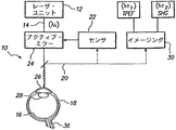
【解決手段】このシステム及び方法は、組織を刺激するために、超短パルス及び入力波長(λι)を有する入力光ビームを発生させる。イメージングされている特定のタイプの組織に応じて、網膜組織は入力ビームに、異なる波長(λτ1及びλτ2)の第1の成分及び第2の成分を有する光の戻りビームを発生させることにより応答する。次いで、イメージング・ユニットが戻り光を受け取って、戻り波長(λτ1対λτ2)に応じて組織をイメージングする。更に、網膜から戻る光を評価して眼によって導入された光学収差及び位相収差を測定し、収差を除去することにより入力ビームを補償する補償器、例えばアクティブ・ミラーをプログラムするために、センサ・ユニットが使用される。
【選択図】図1
Description
Claims (3)
- 眼の網膜組織をイメージングするためのシステムであって、
波長λιを有する入力ビームを発生させ、前記入力ビームを眼の網膜内のスポットに向けて、網膜組織からの戻りビームを発生させるためのレーザ・ユニットにして、前記戻りビームが、波長λτ1をもつ第1の成分、及び波長λτ2をもつ第2の成分を有し、λι≠λτ1≠λτ2である、レーザ・ユニットと、
網膜から戻された光を評価して、前記眼の構成要素によって前記戻りビームに導入された光学収差及び位相収差を特定するためのセンサ・ユニットと、
前記入力ビームを光学収差をそこから除去することによって補償するように、前記入力ビームを変更するための、前記センサ・ユニットに応答する補償器と、
前記戻りビームを受け取って、前記網膜組織を、前記戻りビーム内の選択された成分(λτ1対λτ2)を使用することによりイメージングするためのイメージング・ユニットと
を備えるシステム。 - 眼の組織をイメージングするためのシステムであって、
入力ビームをイメージングすべき標的組織上に向けるためのレーザ手段にして、前記入力ビームが、波長「λι」を有する複数のフェムト秒パルスを含み、前記入力ビームが、第1の標的組織に応答して、波長「λτ1」をもつ第1の成分を有する前記標的組織からの戻りビームを発生させ、第2の標的組織に応答して、波長「λτ2」をもつ第2の成分を有する前記標的組織からの前記戻りビームを発生させ、更にλι≠λτ1≠λτ2である、レーザ手段と、
前記標的組織のイメージングで使用する前記戻りビームの成分を選択する(λτ1対λτ2)ためのイメージング・ユニットと
を備えるシステム。 - 網膜から戻された光を評価して、前記眼の構成要素によって前記戻りビームに導入された光学収差及び位相収差を特定するためのセンサ・ユニットと、
前記入力ビームを前記センサによって特定された光学収差及び位相収差をそこから除去することによって補償するように、前記入力ビームを変更するための補償器と
を更に備える、請求項2に記載のシステム。
Applications Claiming Priority (2)
| Application Number | Priority Date | Filing Date | Title |
|---|---|---|---|
| US12/205,309 US7703923B2 (en) | 2008-09-05 | 2008-09-05 | System and method for imaging retinal tissue with tissue generated light |
| US12/205,309 | 2008-09-05 |
Publications (2)
| Publication Number | Publication Date |
|---|---|
| JP2010057896A true JP2010057896A (ja) | 2010-03-18 |
| JP5473427B2 JP5473427B2 (ja) | 2014-04-16 |
Family
ID=41396298
Family Applications (1)
| Application Number | Title | Priority Date | Filing Date |
|---|---|---|---|
| JP2009148479A Active JP5473427B2 (ja) | 2008-09-05 | 2009-06-23 | 眼の組織をイメージングするためのシステム |
Country Status (3)
| Country | Link |
|---|---|
| US (1) | US7703923B2 (ja) |
| EP (1) | EP2160971B1 (ja) |
| JP (1) | JP5473427B2 (ja) |
Cited By (3)
| Publication number | Priority date | Publication date | Assignee | Title |
|---|---|---|---|---|
| JP2012519553A (ja) * | 2009-03-04 | 2012-08-30 | アーレン サイエンティフィック インコーポレイテッド | 角膜の特性を測定して眼用レンズを得るためのシステム |
| EP2497411A2 (en) | 2011-03-10 | 2012-09-12 | Sony Corporation | Fundus imaging apparatus and fundus imaging method |
| JP2016500530A (ja) * | 2012-10-12 | 2016-01-14 | ソルラブス、インコーポレイテッド | コンパクトで低分散および低収差の補償光学走査システム |
Families Citing this family (7)
| Publication number | Priority date | Publication date | Assignee | Title |
|---|---|---|---|---|
| US8646916B2 (en) * | 2009-03-04 | 2014-02-11 | Perfect Ip, Llc | System for characterizing a cornea and obtaining an opthalmic lens |
| US8292952B2 (en) * | 2009-03-04 | 2012-10-23 | Aaren Scientific Inc. | System for forming and modifying lenses and lenses formed thereby |
| CA2859832C (en) | 2011-12-20 | 2015-07-14 | Research Foundation Of The City University Of New York | Method for screening a drug in retinal tissue |
| JP6146952B2 (ja) * | 2012-01-27 | 2017-06-14 | キヤノン株式会社 | 画像処理装置、画像処理方法及びプログラム。 |
| US9629750B2 (en) | 2012-04-18 | 2017-04-25 | Technolas Perfect Vision Gmbh | Surgical laser unit with variable modes of operation |
| CN106257915B (zh) * | 2015-06-17 | 2021-02-26 | 松下知识产权经营株式会社 | 摄像装置 |
| AU2017305979A1 (en) * | 2016-08-01 | 2019-02-07 | Cognoptix, Inc. | System and method for detecting tau protein in ocular tissue |
Citations (4)
| Publication number | Priority date | Publication date | Assignee | Title |
|---|---|---|---|---|
| JP2004159955A (ja) * | 2002-11-14 | 2004-06-10 | Canon Inc | 蛍光眼底観察装置 |
| US20050159662A1 (en) * | 2004-01-21 | 2005-07-21 | Yoshikazu Imanishi | Methods for assessing a physiological state of a mammalian retina |
| JP2007330585A (ja) * | 2006-06-16 | 2007-12-27 | Topcon Corp | 眼科撮影装置 |
| WO2008077532A1 (de) * | 2006-12-21 | 2008-07-03 | Carl Zeiss Meditec Ag | Anordnung für ophthalmologische geräte zur verbesserung von fundusbildern |
Family Cites Families (8)
| Publication number | Priority date | Publication date | Assignee | Title |
|---|---|---|---|---|
| US4665913A (en) | 1983-11-17 | 1987-05-19 | Lri L.P. | Method for ophthalmological surgery |
| US4754328A (en) | 1984-01-03 | 1988-06-28 | Medical Dynamics, Inc. | Laser endoscope |
| JPS62266032A (ja) | 1986-05-12 | 1987-11-18 | 興和株式会社 | 眼底検査装置 |
| US4881808A (en) | 1988-02-10 | 1989-11-21 | Intelligent Surgical Lasers | Imaging system for surgical lasers |
| JPH09234184A (ja) | 1996-02-29 | 1997-09-09 | Nikon Corp | 眼科装置 |
| US6494878B1 (en) | 2000-05-12 | 2002-12-17 | Ceramoptec Industries, Inc. | System and method for accurate optical treatment of an eye's fundus |
| JP4563828B2 (ja) * | 2005-01-27 | 2010-10-13 | 株式会社トプコン | 眼底検査装置 |
| US20080009922A1 (en) * | 2006-05-25 | 2008-01-10 | Josef Bille | Photodynamic therapy for treating age-related macular degeneration |
-
2008
- 2008-09-05 US US12/205,309 patent/US7703923B2/en active Active
-
2009
- 2009-06-23 JP JP2009148479A patent/JP5473427B2/ja active Active
- 2009-07-22 EP EP09166147.0A patent/EP2160971B1/en active Active
Patent Citations (4)
| Publication number | Priority date | Publication date | Assignee | Title |
|---|---|---|---|---|
| JP2004159955A (ja) * | 2002-11-14 | 2004-06-10 | Canon Inc | 蛍光眼底観察装置 |
| US20050159662A1 (en) * | 2004-01-21 | 2005-07-21 | Yoshikazu Imanishi | Methods for assessing a physiological state of a mammalian retina |
| JP2007330585A (ja) * | 2006-06-16 | 2007-12-27 | Topcon Corp | 眼科撮影装置 |
| WO2008077532A1 (de) * | 2006-12-21 | 2008-07-03 | Carl Zeiss Meditec Ag | Anordnung für ophthalmologische geräte zur verbesserung von fundusbildern |
Cited By (5)
| Publication number | Priority date | Publication date | Assignee | Title |
|---|---|---|---|---|
| JP2012519553A (ja) * | 2009-03-04 | 2012-08-30 | アーレン サイエンティフィック インコーポレイテッド | 角膜の特性を測定して眼用レンズを得るためのシステム |
| EP2497411A2 (en) | 2011-03-10 | 2012-09-12 | Sony Corporation | Fundus imaging apparatus and fundus imaging method |
| JP2012187252A (ja) * | 2011-03-10 | 2012-10-04 | Sony Corp | 眼底イメージング装置および眼底イメージング方法 |
| US9066682B2 (en) | 2011-03-10 | 2015-06-30 | Sony Corporation | Fundus imaging apparatus and fundus imaging method |
| JP2016500530A (ja) * | 2012-10-12 | 2016-01-14 | ソルラブス、インコーポレイテッド | コンパクトで低分散および低収差の補償光学走査システム |
Also Published As
| Publication number | Publication date |
|---|---|
| EP2160971B1 (en) | 2016-10-12 |
| US7703923B2 (en) | 2010-04-27 |
| JP5473427B2 (ja) | 2014-04-16 |
| US20100060853A1 (en) | 2010-03-11 |
| EP2160971A1 (en) | 2010-03-10 |
Similar Documents
| Publication | Publication Date | Title |
|---|---|---|
| JP5473427B2 (ja) | 眼の組織をイメージングするためのシステム | |
| US8152302B2 (en) | System for characterizing a cornea and obtaining an ophthalmic lens | |
| JP5170783B2 (ja) | 眼の画像化 | |
| AU747840B2 (en) | Apparatus and method for measuring vision defects of a human eye | |
| CN102014732B (zh) | 用于观察、检查、诊断和/或治疗眼睛的眼科设备 | |
| JP4157839B2 (ja) | 生体眼の網膜領域撮像方法及びそのシステム | |
| CN102076290B (zh) | 用于眼科激光手术尤其是屈光激光手术的设备 | |
| US8646916B2 (en) | System for characterizing a cornea and obtaining an opthalmic lens | |
| JP2005501587A5 (ja) | ||
| JP2008149164A (ja) | 波面分析を用いた、光学系の客観的な測定および矯正のための装置および方法 | |
| US7510283B2 (en) | High resolution imaging for diagnostic evaluation of the fundus of the human eye | |
| JP5567847B2 (ja) | 補償光学装置、補償光学方法、撮像装置 | |
| JP2017184788A (ja) | 走査型レーザー検眼鏡 | |
| EP1232722B1 (en) | Aberration-free imaging of the fundus of the human eye | |
| JP2001000395A (ja) | 適応型光学フィードバック制御により人間の眼の屈折特性を事前補償するための方法および装置 | |
| WO2016009603A1 (en) | Optical imaging apparatus and method for controlling the same | |
| JP6602368B2 (ja) | 眼球又は視覚領域の光拡散を測定する方法とシステムとコンピュータ・プログラム | |
| HK1180927B (en) | System for characterizing a cornea and obtaining an ophthalmic lens |
Legal Events
| Date | Code | Title | Description |
|---|---|---|---|
| A621 | Written request for application examination |
Free format text: JAPANESE INTERMEDIATE CODE: A621 Effective date: 20120206 |
|
| A977 | Report on retrieval |
Free format text: JAPANESE INTERMEDIATE CODE: A971007 Effective date: 20130411 |
|
| A131 | Notification of reasons for refusal |
Free format text: JAPANESE INTERMEDIATE CODE: A131 Effective date: 20130412 |
|
| A601 | Written request for extension of time |
Free format text: JAPANESE INTERMEDIATE CODE: A601 Effective date: 20130712 |
|
| A602 | Written permission of extension of time |
Free format text: JAPANESE INTERMEDIATE CODE: A602 Effective date: 20130718 |
|
| A521 | Request for written amendment filed |
Free format text: JAPANESE INTERMEDIATE CODE: A523 Effective date: 20130729 |
|
| A131 | Notification of reasons for refusal |
Free format text: JAPANESE INTERMEDIATE CODE: A131 Effective date: 20130906 |
|
| A521 | Request for written amendment filed |
Free format text: JAPANESE INTERMEDIATE CODE: A523 Effective date: 20131205 |
|
| TRDD | Decision of grant or rejection written | ||
| A01 | Written decision to grant a patent or to grant a registration (utility model) |
Free format text: JAPANESE INTERMEDIATE CODE: A01 Effective date: 20140117 |
|
| A61 | First payment of annual fees (during grant procedure) |
Free format text: JAPANESE INTERMEDIATE CODE: A61 Effective date: 20140204 |
|
| R150 | Certificate of patent or registration of utility model |
Ref document number: 5473427 Country of ref document: JP Free format text: JAPANESE INTERMEDIATE CODE: R150 Free format text: JAPANESE INTERMEDIATE CODE: R150 |
|
| R250 | Receipt of annual fees |
Free format text: JAPANESE INTERMEDIATE CODE: R250 |
|
| R250 | Receipt of annual fees |
Free format text: JAPANESE INTERMEDIATE CODE: R250 |
|
| R250 | Receipt of annual fees |
Free format text: JAPANESE INTERMEDIATE CODE: R250 |
|
| R250 | Receipt of annual fees |
Free format text: JAPANESE INTERMEDIATE CODE: R250 |
|
| R250 | Receipt of annual fees |
Free format text: JAPANESE INTERMEDIATE CODE: R250 |
|
| R250 | Receipt of annual fees |
Free format text: JAPANESE INTERMEDIATE CODE: R250 |
|
| R250 | Receipt of annual fees |
Free format text: JAPANESE INTERMEDIATE CODE: R250 |
|
| R250 | Receipt of annual fees |
Free format text: JAPANESE INTERMEDIATE CODE: R250 |
|
| R250 | Receipt of annual fees |
Free format text: JAPANESE INTERMEDIATE CODE: R250 |