JP2008535494A - Cancer-related gene (PRLR) - Google Patents
Cancer-related gene (PRLR) Download PDFInfo
- Publication number
- JP2008535494A JP2008535494A JP2008505603A JP2008505603A JP2008535494A JP 2008535494 A JP2008535494 A JP 2008535494A JP 2008505603 A JP2008505603 A JP 2008505603A JP 2008505603 A JP2008505603 A JP 2008505603A JP 2008535494 A JP2008535494 A JP 2008535494A
- Authority
- JP
- Japan
- Prior art keywords
- cancer
- antibody
- prlr
- expression
- gene
- Prior art date
- Legal status (The legal status is an assumption and is not a legal conclusion. Google has not performed a legal analysis and makes no representation as to the accuracy of the status listed.)
- Pending
Links
Images


Classifications
-
- C—CHEMISTRY; METALLURGY
- C12—BIOCHEMISTRY; BEER; SPIRITS; WINE; VINEGAR; MICROBIOLOGY; ENZYMOLOGY; MUTATION OR GENETIC ENGINEERING
- C12Q—MEASURING OR TESTING PROCESSES INVOLVING ENZYMES, NUCLEIC ACIDS OR MICROORGANISMS; COMPOSITIONS OR TEST PAPERS THEREFOR; PROCESSES OF PREPARING SUCH COMPOSITIONS; CONDITION-RESPONSIVE CONTROL IN MICROBIOLOGICAL OR ENZYMOLOGICAL PROCESSES
- C12Q1/00—Measuring or testing processes involving enzymes, nucleic acids or microorganisms; Compositions therefor; Processes of preparing such compositions
- C12Q1/68—Measuring or testing processes involving enzymes, nucleic acids or microorganisms; Compositions therefor; Processes of preparing such compositions involving nucleic acids
- C12Q1/6876—Nucleic acid products used in the analysis of nucleic acids, e.g. primers or probes
- C12Q1/6883—Nucleic acid products used in the analysis of nucleic acids, e.g. primers or probes for diseases caused by alterations of genetic material
- C12Q1/6886—Nucleic acid products used in the analysis of nucleic acids, e.g. primers or probes for diseases caused by alterations of genetic material for cancer
-
- A—HUMAN NECESSITIES
- A61—MEDICAL OR VETERINARY SCIENCE; HYGIENE
- A61P—SPECIFIC THERAPEUTIC ACTIVITY OF CHEMICAL COMPOUNDS OR MEDICINAL PREPARATIONS
- A61P35/00—Antineoplastic agents
-
- C—CHEMISTRY; METALLURGY
- C12—BIOCHEMISTRY; BEER; SPIRITS; WINE; VINEGAR; MICROBIOLOGY; ENZYMOLOGY; MUTATION OR GENETIC ENGINEERING
- C12Q—MEASURING OR TESTING PROCESSES INVOLVING ENZYMES, NUCLEIC ACIDS OR MICROORGANISMS; COMPOSITIONS OR TEST PAPERS THEREFOR; PROCESSES OF PREPARING SUCH COMPOSITIONS; CONDITION-RESPONSIVE CONTROL IN MICROBIOLOGICAL OR ENZYMOLOGICAL PROCESSES
- C12Q2600/00—Oligonucleotides characterized by their use
- C12Q2600/106—Pharmacogenomics, i.e. genetic variability in individual responses to drugs and drug metabolism
-
- C—CHEMISTRY; METALLURGY
- C12—BIOCHEMISTRY; BEER; SPIRITS; WINE; VINEGAR; MICROBIOLOGY; ENZYMOLOGY; MUTATION OR GENETIC ENGINEERING
- C12Q—MEASURING OR TESTING PROCESSES INVOLVING ENZYMES, NUCLEIC ACIDS OR MICROORGANISMS; COMPOSITIONS OR TEST PAPERS THEREFOR; PROCESSES OF PREPARING SUCH COMPOSITIONS; CONDITION-RESPONSIVE CONTROL IN MICROBIOLOGICAL OR ENZYMOLOGICAL PROCESSES
- C12Q2600/00—Oligonucleotides characterized by their use
- C12Q2600/136—Screening for pharmacological compounds
-
- C—CHEMISTRY; METALLURGY
- C12—BIOCHEMISTRY; BEER; SPIRITS; WINE; VINEGAR; MICROBIOLOGY; ENZYMOLOGY; MUTATION OR GENETIC ENGINEERING
- C12Q—MEASURING OR TESTING PROCESSES INVOLVING ENZYMES, NUCLEIC ACIDS OR MICROORGANISMS; COMPOSITIONS OR TEST PAPERS THEREFOR; PROCESSES OF PREPARING SUCH COMPOSITIONS; CONDITION-RESPONSIVE CONTROL IN MICROBIOLOGICAL OR ENZYMOLOGICAL PROCESSES
- C12Q2600/00—Oligonucleotides characterized by their use
- C12Q2600/158—Expression markers
Abstract
本発明は、癌関連遺伝子の分野にある。特に、本発明は、PRLR遺伝子またはタンパク質の発現の存在または不存在に基づいて癌または癌発現の可能性を検出する方法に関する。本発明は、PRLR遺伝子発現およびPRLRタンパク質活性をアップレギュレートまたはダウンレギュレートするための方法および分子も提供する。1つの実施形態において、本発明は、PRLR遺伝子の配列または発現レベルを決定する工程を含む、生体サンプル内で癌性細胞を検出する方法を提供する。この方法では、生物学的サンプルは、乳房、前立腺、肺または皮膚組織を含む。The present invention is in the field of cancer-related genes. In particular, the present invention relates to a method of detecting cancer or the likelihood of cancer expression based on the presence or absence of PRLR gene or protein expression. The present invention also provides methods and molecules for upregulating or downregulating PRLR gene expression and PRLR protein activity. In one embodiment, the present invention provides a method of detecting cancerous cells in a biological sample comprising determining the sequence or expression level of a PRLR gene. In this method, the biological sample includes breast, prostate, lung or skin tissue.
Description
本願は、2005年4月7日に出願された米国仮特許出願60/669,861の利益を主張する。 This application claims the benefit of US Provisional Patent Application 60 / 669,861 filed Apr. 7, 2005.
(技術分野)
本発明は、癌関連遺伝子の分野にある。特に、本発明は、PRLR遺伝子またはタンパク質の発現の存在または不存在に基づいて癌または癌発現の可能性を検出する方法に関する。本発明は、PRLR遺伝子をアップレギュレートまたはダウンレギュレートするための方法および分子も提供する。加えて本発明は、癌処置のための方法および分子はもちろんのこと、癌処置に有用な分子をスクリーニングする方法も提供する。
(Technical field)
The present invention is in the field of cancer-related genes. In particular, the present invention relates to a method of detecting cancer or the likelihood of cancer expression based on the presence or absence of PRLR gene or protein expression. The present invention also provides methods and molecules for upregulating or downregulating the PRLR gene. In addition, the present invention provides methods for screening for molecules useful for cancer treatment as well as methods and molecules for cancer treatment.
(発明の背景)
腫瘍遺伝子は、癌を引き起こし得る遺伝子である。発癌は、腫瘍遺伝子を含有するウィルスによる細胞の感染、宿主ゲノムにおける原腫瘍遺伝子(腫瘍遺伝子になる可能性を有する正常な遺伝子)の活性化、ならびに原腫瘍遺伝子および腫瘍抑制遺伝子の変異を含む、多岐に亘る機構によって起こり得る。発癌は基本的に、体細胞の進化(すなわち増殖制御の進行性損失を伴う、変異および変異体の自然淘汰)によって推進される。このような体細胞変異の標的として作用する遺伝子は、その突然変異表現型がそれぞれ優性または劣性であるかどうかによって、原腫瘍遺伝子または腫瘍抑制遺伝子のどちらかとして分類される。
(Background of the Invention)
Oncogenes are genes that can cause cancer. Carcinogenesis includes infection of cells by viruses containing oncogenes, activation of proto-oncogenes (normal genes that have the potential to become oncogenes) in the host genome, and mutations of proto-oncogenes and tumor suppressor genes, It can happen by a variety of mechanisms. Carcinogenesis is basically driven by somatic cell evolution (ie, mutations and natural selection of mutants with a progressive loss of growth control). Genes that act as targets for such somatic mutations are classified as either proto-oncogenes or tumor suppressor genes, depending on whether the mutant phenotype is dominant or recessive, respectively.
ヒトはもちろんのこと、動物の癌にも関与することが公知である多数のウィルスがある。ここで特に興味深いのは、それ自体は腫瘍遺伝子を含有しないウィルスである;それらは低速形質転換レトロウィルスである。そのようなウィルスは、宿主ゲノム内に組み込まれて、隣接する原腫瘍遺伝子に各種の方法で影響を及ぼすことによって腫瘍を誘発する。プロウィルス挿入変異は、レトロウィルスのライフサイクルの通常の結果である。感染した細胞では、レトロウィルスゲノムのDNAコピー(プロウィルスと呼ばれる)が宿主ゲノム内に組み込まれる。新たに組み込まれたプロウィルスは、2つの機構のうちの1つによって組み込み部位またはその付近で遺伝子発現にシスで影響し得る。I型挿入変異は、プロウィルスの長い末端反復配列(long terminal repeat、LTR)内で制御配列(エンハンサおよび/またはプロモータ)の結果として、近位の遺伝子の転写をアップレギュレートする。遺伝子のイントロンまたはエキソン内に位置するII型挿入変異は、プロウィルスの長い末端反復配列(LTR)内で制御配列(エンハンサおよび/またはプロモータ)の結果として、前記遺伝子の転写をアップレギュレートできる。加えてII型挿入変異は、開いた読み取り枠内への直接の組み込みまたはコード配列によって両端に隣接されたイントロン内への組み込みのどちらかのために、コード領域の切断を引き起こすことができ、このことは切断された、または不安定な転写/タンパク質生成物をもたらすことができる。挿入部位またはその付近の配列の解析は、多数の新しい原腫瘍遺伝子の同定につながっている。 There are a number of viruses known to be involved in cancer of animals as well as humans. Of particular interest here are viruses that themselves do not contain oncogenes; they are slow-transforming retroviruses. Such viruses integrate into the host genome and induce tumors by affecting adjacent proto-oncogenes in various ways. Proviral insertion mutations are a normal result of the retroviral life cycle. In infected cells, a DNA copy of the retroviral genome (called a provirus) is integrated into the host genome. Newly integrated proviruses can affect gene expression in cis at or near the site of integration by one of two mechanisms. Type I insertion mutations up-regulate transcription of proximal genes as a result of regulatory sequences (enhancers and / or promoters) within the proviral long terminal repeat (LTR). Type II insertion mutations located within introns or exons of a gene can up-regulate transcription of the gene as a result of regulatory sequences (enhancers and / or promoters) within the proviral long terminal repeat (LTR). In addition, type II insertion mutations can cause cleavage of the coding region, either directly into an open reading frame or into an intron flanked by coding sequences. This can result in a truncated or unstable transcription / protein product. Analysis of the sequence at or near the insertion site has led to the identification of a number of new proto-oncogenes.
リンパ腫および白血病に関して、AKVマウス白血病ウィルス(MLV)またはSL3−3MLVなどのレトロウィルスは、感受性の高い新生マウスに接種されるとき、または生殖細胞系列に保持されるとき、腫瘍の強力な誘発物質である。挿入部位を解析することによって、多数の配列がリンパ腫および白血病の誘発に関連するとして同定されている;非特許文献1;非特許文献2;非特許文献3;非特許文献4;非特許文献5;および非特許文献6を参照;そのすべてが参照により本明細書に明示的に組み入れられている。癌、とりわけ乳癌、前立腺癌および上皮起源の癌に関して、哺乳類レトロウィルス、マウス乳腺腫瘍ウィルス(MMTV)は、感受性の高い新生マウスに接種されるとき、または生殖細胞系列に保持されるとき、腫瘍の強力な誘発物質である。J.HilgersおよびM.Sluyserによって著されたMammary Tumours in the Mouse;Elsevier/North−Holland Biomedical Press;New York,N.Y。
With regard to lymphoma and leukemia, retroviruses such as AKV murine leukemia virus (MLV) or SL3-3MLV are potent inducers of tumors when inoculated into susceptible newborn mice or when retained in the germline. is there. By analyzing the insertion site, a number of sequences have been identified as being associated with induction of lymphoma and leukemia; Non-patent
特定の生細胞における遺伝子発現のパターンは、その現在の状態の特徴である。細胞の状態またはタイプのほぼすべての相違は、1つ以上の遺伝子のRNAレベルの相違に反映されている。特徴付けられていない遺伝子の発現パターンの比較は、その機能への手がかりを与え得る。数百または数千の遺伝子の発現のハイスループット解析は、(a)複雑な遺伝性疾患の同定、(b)組織と疾患状態との間の経時的な差次的遺伝子発現の解析、および(c)薬物の発見および毒物学研究に役立ち得る。ある遺伝子の発現レベルの上昇または下降は、癌の生物学に関連している。たとえば腫瘍遺伝子は腫瘍形成の正の調節因子であるのに対して、腫瘍抑制遺伝子は腫瘍形成の負の調節因子である(非特許文献7)。 The pattern of gene expression in a particular living cell is characteristic of its current state. Almost all differences in cell status or type are reflected in differences in RNA levels of one or more genes. Comparison of expression patterns of uncharacterized genes can provide clues to their function. High-throughput analysis of the expression of hundreds or thousands of genes includes (a) identification of complex genetic diseases, (b) analysis of differential gene expression over time between tissues and disease states, and ( c) Can be useful for drug discovery and toxicology studies. An increase or decrease in the expression level of a gene is associated with cancer biology. For example, tumor genes are positive regulators of tumorigenesis, whereas tumor suppressor genes are negative regulators of tumorigenesis (Non-patent Document 7).
免疫療法、すなわち治療目的での抗体の使用は、近年、癌を処置するために使用されている。受動免疫療法は、癌処置でのモノクローナル抗体の使用を含む。たとえば非特許文献8を参照。これらの抗体の独自の治療的生物活性は、腫瘍細胞成長または生存の直接抑制、および体の免疫系の自然の殺細胞活性を補充する能力を含む。これらの因子は、単独で、あるいは放射線または化学療法剤と併せて投与される。リンパ腫および乳癌の処置のためにそれぞれ承認されたリツキサン(登録商標)およびヘルセプチン(登録商標)は、そのような治療薬の2つの例である。あるいは抗体を使用して、抗体が毒剤に結合して、腫瘍に特異的に結合することによってその因子を腫瘍に向かわせる抗体コンジュゲートを作製する。マイロターグ(登録商標)は、白血病の処置に使用される承認済み抗体コンジュゲートの例である。しかしながらこれらの抗体は、原因ではなく腫瘍自体を標的としている。 Immunotherapy, ie the use of antibodies for therapeutic purposes, has recently been used to treat cancer. Passive immunotherapy involves the use of monoclonal antibodies in cancer treatment. For example, see Non-Patent Document 8. The unique therapeutic biological activity of these antibodies includes the direct suppression of tumor cell growth or survival, and the ability to supplement the body's immune system's natural cytocidal activity. These factors are administered alone or in conjunction with radiation or chemotherapeutic agents. Rituxan® and Herceptin®, approved for the treatment of lymphoma and breast cancer, respectively, are two examples of such therapeutic agents. Alternatively, an antibody is used to create an antibody conjugate in which the antibody binds to a toxic agent and directs the agent to the tumor by specifically binding to the tumor. Mylotag® is an example of an approved antibody conjugate used for the treatment of leukemia. However, these antibodies target the tumor itself, not the cause.
抗癌療法のためのより優れた手法は、癌を発生させ得る原腫瘍遺伝子を標的とすることであろう。これを行うために、原腫瘍遺伝子を最初に同定する必要がある。いったんこれらの遺伝子が同定されれば、次にそれらを監視して癌の開始を検出することができ、次いで癌を処置するために標的化することができる。 A better approach for anti-cancer therapy would be to target proto-oncogenes that can develop cancer. To do this, it is necessary to first identify the proto-oncogene. Once these genes are identified, they can then be monitored to detect the onset of cancer and then targeted to treat the cancer.
プロラクチンレセプタ(PRLR)は、サイトカインスーパーファミリーのメンバーに対するレセプタ、たとえばIL2、IL3、IL4、IL6、IL7、エリスロポエチン、およびGM−CSFのレセプタと類似している、単一の膜貫通クラス1サイトカインレセプタである。PRLRは、細胞成長、分化、発生、泌乳および生殖を含む、複数の生物機能に関与する。それは固有のキナーゼ活性を持たない;リガンド結合は、レセプタ二量体化、Jak2のクロスホスホリル化および下流シグナル伝達をもたらす。ヒトプロラクチンレセプタcDNAは元来、肝癌および乳癌ライブラリーから単離された(非特許文献9)。ヌクレオチド配列は、ラット肝臓PRLレセプタよりもはるかに長い細胞質ドメインを持つ、598アミノ酸の成熟タンパク質を予測した。プロラクチンレセプタ遺伝子は、5;13−p12にマップされている成長ホルモン遺伝子と同じ染色体領域に属する(非特許文献10)。成長ホルモンもプロラクチンレセプタに結合して、レセプタを活性化する。
Prolactin receptor (PRLR) is a single
ヒトPRLR遺伝子のゲノム構成が決定されている(非特許文献11)。PRLR遺伝子の5−プライム未翻訳領域は、2つの代わりの第1エキソン:ラットおよびマウスE13のヒト対応物であるE13、ならびにE1Nと呼ばれる新規なヒト型の代わりの第1エキソンを含有する。5−プライム未翻訳領域は、翻訳開始コドンを含有する共通の非コードエキソン2およびエキソン3の一部も含有する。E13およびE1Nエキソンは、相互の800塩基対以内である。これらの2つのエキソンは、ヒト乳房組織、乳癌細胞、生殖腺、および肝臓で発現される。全体として、E13を含有する転写は、大半の組織で一般的である。PRLR遺伝子産物は、エキソン3〜10によってコードされ、そのうちエキソン10は細胞内ドメインの大半をコードする。E13およびE1Nエキソンは、代わりのプロモータPIIIおよびPNそれぞれから転写される。PIIIプロモータは、げっ歯類プロモータのそれらと同一であるSpIおよびC/EBP要素を含有し、ラットおよびマウスの領域−480/−106と81%類似している。PNプロモータは、ETSファミリータンパク質の推定結合部位および核内レセプタのハーフ部位を含有する。
The genomic organization of the human PRLR gene has been determined (Non-patent Document 11). The 5-prime untranslated region of the PRLR gene contains two alternative first exons: E13, the human counterpart of rat and mouse E13, and an alternative first exon of a novel human form called E1N. The 5-prime untranslated region also contains a portion of the
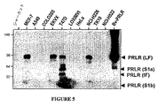
PRLRは、その細胞質ドメインの長さが異なる、多数の各種のアイソフォームとして存在する。4つのPRLR mRNAアイソフォーム(L、I、S1a、およびS1b)が、ヒト皮下腹部脂肪組織および胸部脂肪組織で示されている(非特許文献12)。加えてそれらは免疫ブロット解析を使用して、ヒト皮下腹部脂肪組織および胸部脂肪組織でのL−PRLRおよびI−PRLRタンパク質発現を検出した。PRLは、対照と比較してヒト脂肪組織でのリポタンパク質リパーゼ活性を低下させた。Lingらは、これらの結果がヒト脂肪組織でのLPL活性を低下させることでの、機能性PRLRを介したPRLの直接効果を示したことと、これらの結果はLPLも泌乳中にこの方式で制御され得ることを暗示したこととを示唆している。ラットでのこれらのPRLRアイソフォームの機能が解明されている(非特許文献13)。公知の長型(591アミノ酸)と同様に、細胞質ドメインの198アミノ酸が欠失したNb2型は、乳腺刺激性シグナルを伝達できる。これに対して、細胞質ドメインの291アミノ酸を欠失した短型は不活性である。短型の機能が、長型および短型の両方の同時形質移入後に調査された。これらの結果は、短型が不活性へテロダイマーの生成を通じてドミナントネガティブインヒビタとして作用して、Janusキナーゼ2活性化の抑制を引き起こすことを示す。Perrot−Applanatらは、PRLRのヘテロダイマー化がPRL転写を陽性または陰性活性化できることを示唆している。
PRLR exists as a number of different isoforms that differ in the length of their cytoplasmic domains. Four PRLR mRNA isoforms (L, I, S1a, and S1b) have been shown in human subcutaneous abdominal adipose tissue and thoracic adipose tissue (12). In addition, they used immunoblot analysis to detect L-PRLR and I-PRLR protein expression in human subcutaneous abdominal and breast adipose tissues. PRL reduced lipoprotein lipase activity in human adipose tissue compared to controls. Ling et al. Showed that these results showed a direct effect of PRL via functional PRLR in reducing LPL activity in human adipose tissue, and these results indicate that LPL is also in this manner during lactation. It suggests that it can be controlled. The function of these PRLR isoforms in rats has been elucidated (Non-patent Document 13). Similar to the known long form (591 amino acids), the Nb2 form lacking 198 amino acids in the cytoplasmic domain can transmit mammary stimulatory signals. In contrast, the short form lacking 291 amino acids of the cytoplasmic domain is inactive. The function of the short form was investigated after co-transfection of both the long and short forms. These results indicate that the short form acts as a dominant negative inhibitor through the generation of inactive heterodimers, causing suppression of
非特許文献14は、免疫組織化学によって41の乳癌におけるPRLRの存在を調査して、その発現を疾患の病理的グレーディングに相関させることを試みた。PRLR免疫反応スコアは、患者の腫瘍サイズ、病理組織学的グレーディング、年齢、または家族暦と相関しなかった。PRLR陽性および陰性乳癌細胞のどちらでも、PRLR遺伝子のコード配列の直接配列決定は、いずれの体細胞または遺伝性遺伝子異常も検出しなかった。著者らは、PRLR変異がヒト乳癌で一般的であるとは考えられないことを結論付けて、PRLRの構成的活性化が乳癌発生の主な原因として除外できることを示唆した。 Non-Patent Document 14 investigated the presence of PRLR in 41 breast cancers by immunohistochemistry and attempted to correlate its expression with pathological grading of the disease. The PRLR immune response score did not correlate with patient tumor size, histopathological grading, age, or family calendar. In both PRLR positive and negative breast cancer cells, direct sequencing of the coding sequence of the PRLR gene did not detect any somatic or inherited gene abnormality. The authors concluded that PRLR mutations are not considered common in human breast cancer, suggesting that constitutive activation of PRLR can be ruled out as a major cause of breast cancer development.
PRLメストランスジェニックマウスは、悪性乳腺腺癌を発症する(非特許文献15)。PRLRモノクローナル抗体は、マウスでの乳腺腫瘍の発生率を減少させた(非特許文献16)。加えてPRLアンタゴニスト(S179D突然変異体PRL)は、インビトロでのヒト前立腺癌細胞系DU−145およびインビボでのDU−145誘発腫瘍の増殖を抑制した(非特許文献17)。
(発明の要旨)
原腫瘍遺伝子は、「プロウィルス・タギング」として公知のプロセスを使用してヒトにおいて同定されており、マウスモデルを用いて原腫瘍遺伝子を単離するために、挿入変異機構によって作用する低速形質転換レトロウィルスを使用する。一部のモデルにおいて、非感染動物は低い癌率を有し、感染動物は高い癌率を有する。関与するレトロウィルスの多くが形質導入宿主原腫瘍遺伝子または病原体トランス作用ウィルス遺伝子を持たないことが公知であり、それゆえ癌発生率はしたがって宿主原腫瘍遺伝子へのプロウィルス組み込み効果の直接結果でなければならない。プロウィルス組み込みはランダムであるため、まれな組み込み体が選択的成長利益を与える宿主原腫瘍遺伝子を「活性化」して、これらのまれな事象が腫瘍内にてクローン化学量論で新たなプロウィルスを生じる。化学作用、放射線、または自然発生的誤りによって引き起こされた変異とは対照的に、原腫瘍遺伝子挿入変異は、公知の配列の好都合なサイズの遺伝子マーカー(プロウィルス)が変異部位に存在するという事実のために、容易に位置付けることができる。クローン的に組み込まれたプロウィルスに隣接する宿主配列は、各種の方法を使用してクローニングできる。これらの配列をいったん手にすれば、タグ付けされた原腫瘍遺伝子を続いて同定できる。2つ以上の独立した腫瘍の同じ座位にあるプロウィルスの存在は、原腫瘍遺伝子がプロウィルス組み込み部位、またはその非常に近くに存在することの一応の証拠である(Kimら、Journal of Virology,2003,77:2056−2062;Mikkers,H and Berns,A,Advances in Cancer Research,2003,88:53−99;Keokoら、Nucleic Acids Research,2004,32:D523−D527)。これはゲノムが大きすぎて、ランダム組み込みが観察可能なクラスター形成を生成できないためである。検出されるいずれのクラスター形成も、生物学的選択の明白な証拠である(すなわち腫瘍表現型)。その上、プロウィルス組み込み体のパターン(向きを含む)は、各クラスターでの標的遺伝子の位置決定を比較的簡単にする有力な位置情報を提供する。挿入変異機構によって癌を引き起こすことが公知である3つの哺乳類レトロウィルスは、FeLV(猫白血病/リンパ腫)、MLV(マウスおよびラット白血病/リンパ腫)、およびMMTV(マウス乳癌)である。マウスモデルで原腫瘍遺伝子がいったん同定されると、ヒトオルソグは原腫瘍遺伝子として注釈でき、さらなる研究が実施される。
(Summary of the Invention)
Proto-oncogenes have been identified in humans using a process known as "provirus tagging" and slow transformations that act by an insertional mutation mechanism to isolate proto-oncogenes using a mouse model Use retroviruses. In some models, uninfected animals have a low cancer rate and infected animals have a high cancer rate. It is known that many of the retroviruses involved do not have a transduced host proto-oncogene or a pathogen trans-acting virus gene, so the incidence of cancer should therefore be a direct result of the effect of proviral integration into the host proto-oncogene. I must. Because proviral integration is random, rare integrants “activate” host proto-oncogenes that provide selective growth benefits, and these rare events can be clonal stoichiometrically generated in tumors. Produce a virus. In contrast to mutations caused by chemistry, radiation, or spontaneous errors, proto-oncogene insertion mutations are the fact that a convenient sized genetic marker (provirus) of a known sequence is present at the mutation site. Can be positioned easily. Host sequences flanking a clonally integrated provirus can be cloned using a variety of methods. Once these sequences are in hand, tagged proto-oncogenes can be subsequently identified. The presence of a provirus at the same locus in two or more independent tumors is a tentative proof that the original oncogene is at or very close to the proviral integration site (Kim et al., Journal of Virology, 2003, 77: 2056-2062; Mikkers, H and Berns, A, Advances in Cancer Research, 2003, 88: 53-99; Keoko et al., Nucleic Acids Research, 2004, 32: D523-D527). This is because the genome is too large to produce cluster formations that can be observed for random integration. Any cluster formation detected is clear evidence of biological selection (ie tumor phenotype). In addition, the pattern of proviral integrants (including orientation) provides powerful positional information that makes it relatively easy to locate target genes in each cluster. Three mammalian retroviruses known to cause cancer by an insertional mutation mechanism are FeLV (cat leukemia / lymphoma), MLV (mouse and rat leukemia / lymphoma), and MMTV (mouse breast cancer). Once the proto-oncogene has been identified in the mouse model, the human orthog can be annotated as a proto-oncogene and further studies will be performed.
それゆえ癌を生じる宿主生物のゲノム内に配列が挿入される腫瘍遺伝子レトロウィルスの使用は、癌に関与する宿主遺伝子の同定を可能にする。これらの配列は次に、診断、予後、モジュレータのスクリーニング(アゴニストおよびアンタゴニストの両方を含む)、抗体産生(免疫療法および撮像のために)などを含む、多くの各種方法で使用できる。しかしながら当業者によって認識されるように、リンパ腫または白血病などの1種類の癌で同定される腫瘍遺伝子は、他の種類の癌にも関与している強い可能性を有する。 Therefore, the use of oncogene retroviruses in which sequences are inserted into the genome of the host organism that causes cancer allows the identification of host genes involved in cancer. These sequences can then be used in many different ways, including diagnosis, prognosis, modulator screening (including both agonists and antagonists), antibody production (for immunotherapy and imaging), and the like. However, as will be appreciated by those skilled in the art, oncogenes identified in one type of cancer, such as lymphoma or leukemia, have a strong potential to be involved in other types of cancer.
したがって本発明は、癌関連遺伝子であるPRLR遺伝子の配列および発現レベルを決定する工程を含む、生物学的サンプル中の癌性細胞を検出するための方法を提供する。 Accordingly, the present invention provides a method for detecting cancerous cells in a biological sample, comprising the step of determining the sequence and expression level of a PRLR gene that is a cancer-related gene.
好ましくは、該方法は、PRLR遺伝子の1つ以上(すなわち1、2、3、4、5、6、7、8、9、10またはそれ以上)の発現産物の発現レベルを測定する工程を含み、ここで対照レベルと異なる発現レベルが疾患を示す。
Preferably, the method comprises the step of measuring the expression level of one or more expression products (
発現産物は好ましくはタンパク質であるが、代わりのmRNA発現産物も検出され得る。タンパク質を使用する場合、タンパク質は好ましくは、そのタンパク質に好ましくは特異的に結合する抗体によって検出される。用語「特異的に結合する」は、抗体が他の関連ポリペプチドに対するその親和性よりも、その標的ポリペプチドに対して実質的に高い親和性を有することを意味する。本明細書で使用するように、用語「抗体」は、問題の抗原決定基に結合できる無傷分子はもちろんのこと、その断片、たとえばFab、F(ab’)2およびFvも指す。抗体のさらなる例は、それらが所望の生物活性(たとえばPRLRの細胞外ドメインへの結合)を示す限り、完全集合(fully assembled)抗体、モノクローナル抗体、ポリクローナル抗体、多重特異性抗体(たとえば二重特異性抗体)、一本鎖抗体、ダイアボディ、および上記を含む組換えペプチドを含む。「実質的に高い親和性」によって、我々は、他の関連ポリペプチドに対する親和性と比較して、本発明の標的ポリペプチドに対する親和性の測定可能な上昇があることを意味する。親和性は、他の公知のホモログまたはオルソグの親和性と比較して、標的ポリペプチドに対して少なくとも1.5倍、2倍、5倍、10倍、100倍、103倍、104倍、105倍、106倍またはそれ以上である。あるいは抗体が公知のホモログおよびオルソグと交差反応することが有用であり得る。
The expression product is preferably a protein, but alternative mRNA expression products can also be detected. When using a protein, the protein is preferably detected by an antibody that preferably specifically binds to the protein. The term “specifically binds” means that an antibody has a substantially higher affinity for its target polypeptide than its affinity for other related polypeptides. As used herein, the term “antibody” refers to intact molecules capable of binding to the antigenic determinant in question, as well as fragments thereof, such as Fab, F (ab ′) 2 and Fv. Further examples of antibodies are fully assembled antibodies, monoclonal antibodies, polyclonal antibodies, multispecific antibodies (eg bispecific) as long as they exhibit the desired biological activity (eg binding to the extracellular domain of PRLR). Sex antibodies), single chain antibodies, diabodies, and recombinant peptides comprising the above. By “substantially high affinity” we mean that there is a measurable increase in affinity for a target polypeptide of the invention compared to affinity for other related polypeptides. Affinity, as compared to the affinity of the other known homologues or Orusogu, at least 1.5 times the target polypeptide, 2-fold, 5-fold, 10-fold, 100-fold, 10 3 fold, 10 4
好ましくは、抗体は高い親和性で、好ましくは10−4M、10−5M、10−6Mまたはそれ未満、好ましくは10−7M、10−8Mまたはそれ未満、最も好ましくは10−9Mまたは10−10Mあるいはそれ未満で結合し;サブナノモルの親和性(0.9、0.8、0.7、0.6、0.5、0.4、0.3、0.2、0.1nMまたはなおそれ未満)が好ましい。 Preferably, the antibody has high affinity, preferably 10 −4 M, 10 −5 M, 10 −6 M or less, preferably 10 −7 M, 10 −8 M or less, most preferably 10 −. Bind at 9 M or 10 −10 M or less; subnanomolar affinity (0.9, 0.8, 0.7, 0.6, 0.5, 0.4, 0.3, 0.2 , 0.1 nM or less).
mRNA発現産物が使用されるとき、それは好ましくは、mRNAとプローブとの間のハイブリッド複合体の形成を可能にするストリンジェントな条件下で組織サンプルにプローブを接触させることと;複合体の形成を検出することとによって検出される。 When an mRNA expression product is used, it preferably contacts the probe with a tissue sample under stringent conditions that allow formation of a hybrid complex between the mRNA and the probe; It is detected by detecting.
PRLR遺伝子自体は、PRLR遺伝子をコードする核酸発現産物とプローブとの間のハイブリッド複合体の形成を可能にするストリンジェントな条件下で生物学的サンプルに核酸プローブを接触させることと;プローブと生物学的サンプルからの核酸との間の複合体の形成を検出することとによって検出できる。そのような場合、複合体の形成の不存在は好ましくは、PRLR遺伝子の配列における変異を示す。 The PRLR gene itself comprises contacting the nucleic acid probe with a biological sample under stringent conditions that allow formation of a hybrid complex between the nucleic acid expression product encoding the PRLR gene and the probe; By detecting the formation of a complex with the nucleic acid from the biological sample. In such cases, the absence of complex formation preferably indicates a mutation in the sequence of the PRLR gene.
好ましい方法は、形成される複合体の量を、対照組織を使用したときに形成される複合体の量と比較する工程を含み、ここで対照とサンプルとの間で形成される複合体の量の差は、癌の存在を示す。好ましくは、正常組織と比較した試験組織によって形成される複合体の量の差は、増加または減少する。さらに好ましくは、形成される複合体の量の2倍の増加または減少は、疾患を示す。なおさらに好ましくは、形成される複合体の量の3倍、4倍、5倍、10倍、20倍、50倍、またなお100倍の増加または減少が、疾患を示す。 A preferred method comprises comparing the amount of complex formed with the amount of complex formed when using a control tissue, wherein the amount of complex formed between the control and the sample. The difference in indicates the presence of cancer. Preferably, the difference in the amount of complex formed by the test tissue compared to normal tissue is increased or decreased. More preferably, a 2-fold increase or decrease in the amount of complex formed indicates a disease. Even more preferably, an increase or decrease of 3-fold, 4-fold, 5-fold, 10-fold, 20-fold, 50-fold or even 100-fold of the amount of complex formed indicates a disease.
本発明の方法で使用する生物学的サンプルは好ましくは、組織サンプルである。いずれの組織サンプルも使用できる。しかしながら、好ましくは、組織は乳房組織、結腸組織、腎臓組織、肝臓組織、肺組織、リンパ様組織、卵巣組織、膵臓組織、前立腺組織、子宮組織、頸部組織または皮膚組織から選択される。 The biological sample used in the method of the present invention is preferably a tissue sample. Any tissue sample can be used. Preferably, however, the tissue is selected from breast tissue, colon tissue, kidney tissue, liver tissue, lung tissue, lymphoid tissue, ovarian tissue, pancreatic tissue, prostate tissue, uterine tissue, cervical tissue or skin tissue.
本発明は、第1時点での生物学的サンプル中の上で言及した1つ以上(すなわち1、2、3、4、5、6、7、8、9、10またはそれ以上)の発現産物の発現を、第2時点での同じ発現産物の発現と比較する工程を含む、患者の癌の進行を評価する方法も提供し、ここで第1時点に対する第2時点における発現の増加または減少、あるいは発現の増加または減少の速度の上昇または下降は、癌の進行を示す。
The present invention includes one or more (
本発明は、PRLR遺伝子のポリペプチド発現産物に結合する抗体と;前記抗体と前記ポリペプチドとの間の結合反応の検出のために有用な試薬とを含む、癌を診断するのに有用なキットも提供する。好ましくは、抗体は、PRLR遺伝子のポリペプチド生成物に特異的に結合する。 The present invention relates to a kit useful for diagnosing cancer, comprising an antibody that binds to a polypeptide expression product of a PRLR gene; and a reagent useful for detecting a binding reaction between the antibody and the polypeptide. Also provide. Preferably, the antibody specifically binds to the polypeptide product of the PRLR gene.
さらに本発明は、ストリンジェントな条件下でPRLR遺伝子にハイブリダイズする核酸プローブと;PRLR遺伝子を増幅するのに有用なプライマーと;場合により疾患の診断を容易にするための、プローブおよびプライマーを使用するための説明書と;を含む、癌を診断するのに有用なキットを提供する。 Furthermore, the present invention uses nucleic acid probes that hybridize to the PRLR gene under stringent conditions; primers useful for amplifying the PRLR gene; optionally using probes and primers to facilitate diagnosis of disease And a kit useful for diagnosing cancer, comprising:
本発明は、癌の処置に使用するための、PRLR遺伝子の1つ以上(すなわち1、2、3、4、5、6、7、8、9、10またはそれ以上)の発現産物の発現を調節するための使用に適した抗体、核酸、またはタンパク質も提供する。
The present invention provides the expression of one or more (
したがって、本発明は、PRLR遺伝子の1つ以上(すなわち1、2、3、4、5、6、7、8、9、10またはそれ以上)の発現産物のレベルを調節する工程を含む、患者の癌を処置する方法を提供する。そのような方法は好ましくは、患者に前記発現産物のレベルを調節する抗体、核酸、またはポリペプチドを治療的有効量で投与する工程を含む。
Accordingly, the present invention includes the step of modulating the level of the expression product of one or more of PRLR genes (
本発明はしたがって、癌の処置または診断のための、医薬の製造での、PRLR遺伝子の発現産物のレベルを調節する抗体、核酸、またはポリペプチドの使用も提供する。そのような発現レベルは好ましくは、遺伝子、mRNAまたはコードタンパク質に対する作用によって調節される。発現は好ましくは、アップレギュレートまたはダウンレギュレートされる。たとえば制御の変化は、2倍、3倍、5倍、10倍、20倍、50倍、またはなお100倍あるいはそれ以上であり得る。 The invention thus also provides the use of an antibody, nucleic acid, or polypeptide that modulates the level of the expression product of the PRLR gene in the manufacture of a medicament for the treatment or diagnosis of cancer. Such expression levels are preferably regulated by action on the gene, mRNA or encoding protein. Expression is preferably up-regulated or down-regulated. For example, the change in control can be 2x, 3x, 5x, 10x, 20x, 50x, or even 100x or more.
本発明による使用に適した抗体は、癌性細胞にてまたは内部で発現されるものと同様に、PRLRポリペプチドに対して特異性であり得る。たとえば癌性細胞表面で発現されるような癌関連タンパク質のグリコシル化パターンは、非癌性細胞で発現されるものと同じこれらのタンパク質でのグリコシルのパターンとは異なり得る。好ましくは、そのような筋書きでは、本発明による抗体は、癌性細胞のみで発現されるようなPRLRタンパク質に対して特異性である。このことは治療用抗体にとって特定の価値を有する。抗標的抗体はもちろん、標的のありとあらゆる変異体、欠失、付加および/または置換変異体に結合し得る。 Antibodies suitable for use with the present invention can be specific for PRLR polypeptides, as well as those expressed in or internally in cancerous cells. For example, the glycosylation pattern of cancer-associated proteins, such as that expressed on the surface of cancerous cells, may differ from the glycosylation pattern on these same proteins that are expressed on non-cancerous cells. Preferably, in such a scenario, the antibody according to the invention is specific for a PRLR protein that is expressed only in cancerous cells. This has particular value for therapeutic antibodies. Anti-target antibodies can of course bind to any and all variants, deletions, additions and / or substitution variants of the target.
本発明による治療的使用に適した抗体は好ましくは、抗体依存性細胞傷害(ADCC)を誘発するのに有効であり得る。ADCCは、Fcレセプタを発現する非特異性細胞傷害性細胞が標的細胞の境界抗体を認識して、続いて標的細胞の溶解を引き起こす、細胞媒介反応を指す(Raghavanら、1996 Annu Rev Cell Dev Biol 12:181−220;Ghetieら、2000,Annu Rev Immunol 18:739−766;Ravetchら、2001,Annu Rev Immunol 19:275−290)。本発明による治療的使用に適した抗体は好ましくは、抗体依存性細胞媒介食作用(ADCP)を誘発するのに有効であり得る。ADCPは、Fcレセプタを発現する非特異性細胞傷害性細胞が標的細胞の境界抗体を認識して、続いて食作用を引き起こす、細胞媒介反応である。これらのプロセスは、IgG抗体のFc部分に対するレセプタをその表面に所有するナチュラルキラー(NK)細胞によって媒介される。IgGが癌細胞を含む「外来」膜結合細胞のエピトープに対して作製されるとき、抗体のFab部分は癌性細胞と反応する。NK細胞は次に、抗体のFc部分に結合する。 Antibodies suitable for therapeutic use according to the present invention may preferably be effective in inducing antibody-dependent cellular cytotoxicity (ADCC). ADCC refers to a cell-mediated reaction in which non-specific cytotoxic cells expressing Fc receptors recognize target cell border antibodies and subsequently cause target cell lysis (Raghavan et al., 1996 Annu Rev Cell Dev Biol). 12: 181-220; Ghetie et al., 2000, Annu Rev Immunol 18: 739-766; Ravetch et al., 2001, Annu Rev Immunol 19: 275-290). Antibodies suitable for therapeutic use according to the present invention may preferably be effective in inducing antibody-dependent cell-mediated phagocytosis (ADCP). ADCP is a cell-mediated reaction in which non-specific cytotoxic cells expressing Fc receptors recognize target cell border antibodies and subsequently cause phagocytosis. These processes are mediated by natural killer (NK) cells that possess receptors on their surface for the Fc portion of IgG antibodies. When IgG is made against epitopes of “foreign” membrane-bound cells, including cancer cells, the Fab portion of the antibody reacts with cancerous cells. NK cells then bind to the Fc portion of the antibody.
好ましくは、本発明による治療的使用に適した抗体はADCCを誘発するのに有効であり、標的に結合してADCC活性を有することにより癌性細胞の生存を調節する。抗体はADCC活性を向上させるために組換えることができる(たとえばUS20050054832A1,Xencor Inc.およびそこで引用された文献を参照)。 Preferably, antibodies suitable for therapeutic use according to the present invention are effective in inducing ADCC and modulate cancer cell survival by binding to a target and having ADCC activity. The antibody can be recombined to improve ADCC activity (see, eg, US20050054832A1, Xencor Inc. and references cited therein).
そのような方法で使用される核酸の種類は好ましくは、アンチセンス構築物、リボザイムまたはRNAi、特にsiRNAである。 The type of nucleic acid used in such a method is preferably an antisense construct, ribozyme or RNAi, in particular siRNA.
癌は、腫瘍成長の抑制または腫瘍体積の減少によって、あるいは癌細胞の侵襲性を低下させることによって処置できる。いくつかの実施形態において、上述の処置方法は外科手術、ホルモン除去療法、放射線療法または化学療法の1つ以上と併せて使用される。たとえば患者がすでに化学療法を受けている場合、上に挙げたような発現産物のレベルを調節する本発明の化合物を投与することもできる。化学療法、ホルモンおよび/または放射線療法剤および本発明による化合物は同時に、別個にまたは連続して投与できる。 Cancer can be treated by suppressing tumor growth or reducing tumor volume, or by reducing the invasiveness of cancer cells. In some embodiments, the treatment methods described above are used in conjunction with one or more of surgery, hormone removal therapy, radiation therapy or chemotherapy. For example, if the patient has already undergone chemotherapy, a compound of the invention that modulates the level of the expression product as listed above can also be administered. Chemotherapy, hormones and / or radiation therapy agents and the compounds according to the invention can be administered simultaneously, separately or sequentially.
好ましくは、上述の方法の1つに従って検出または処置されている癌は、乳癌、結腸癌、腎臓癌、肝臓癌、肺癌、リンパ性癌、卵巣癌、膵臓癌、前立腺癌、子宮癌、子宮頸部癌または皮膚癌から選択される。 Preferably, the cancer being detected or treated according to one of the methods described above is breast cancer, colon cancer, kidney cancer, liver cancer, lung cancer, lymphoid cancer, ovarian cancer, pancreatic cancer, prostate cancer, uterine cancer, cervical Selected from cervical cancer or skin cancer.
本発明は、患者からの生物学的サンプル中のPRLR遺伝子発現産物の発現レベルを測定する工程を含む、その患者をPRLR調節性抗体による処置に対して感受性であると確認するための方法も提供する。 The present invention also provides a method for confirming that a patient is susceptible to treatment with a PRLR-modulating antibody, comprising measuring the expression level of a PRLR gene expression product in a biological sample from the patient. To do.
さらに本発明は、第1時点のPRLR遺伝子発現産物の発現レベルが第2時点の同じ発現産物の発現レベルと比較され、第1時点に対する第2時点での発現の増加または減少がPRLR遺伝子の関与する癌の進行を示す、患者をPRLR調節性抗体による処置に対して感受性であると確認するための方法を提供する。 Furthermore, the present invention relates to the comparison of the expression level of the PRLR gene expression product at the first time point with the expression level of the same expression product at the second time point, and the increase or decrease of the expression at the second time point relative to the first time point is related to the PRLR gene A method is provided for confirming that a patient is susceptible to treatment with a PRLR-modulating antibody that exhibits progression of the cancer to be treated.
本発明は、癌性細胞の増殖を調節する候補因子を同定するためのアッセイであって:
a)候補因子の存在下で本発明の上述の実施形態のいずれかで挙げたようなPRLR遺伝子の1つ以上(すなわち1、2、3、4、5、6、7、8、9、10またはそれ以上)の発現レベルを検出する工程と;
b)候補因子の不存在下での発現レベルをその発現レベルと比較する工程であって、発現の差が、候補因子がPRLR遺伝子の発現産物の発現レベルを調節することを示す比較工程と;
を含むアッセイをさらに提供する。
The present invention is an assay for identifying candidate factors that modulate the growth of cancerous cells:
a) one or more of the PRLR genes (
b) comparing the expression level in the absence of the candidate factor with its expression level, wherein the difference in expression indicates that the candidate factor regulates the expression level of the expression product of the PRLR gene;
Further provided is an assay comprising:
本発明は、また、PRLR遺伝子の発現レベルを変更する因子を同定するための方法であって:
a)本発明の上述の実施形態のいずれかに示したようなPRLR遺伝子を発現する細胞に候補因子を接触させる工程と;
b)候補因子の細胞に対する効果を判定する工程であって、発現レベルの変化が、候補因子が発現を調節できることを示す判定工程と;
を含む方法も提供する。
The present invention is also a method for identifying a factor that alters the expression level of a PRLR gene:
a) contacting a candidate factor with a cell expressing a PRLR gene as shown in any of the above embodiments of the invention;
b) determining the effect of the candidate factor on the cell, wherein the change in expression level indicates that the candidate factor can regulate expression;
A method is also provided.
好ましくは、因子はポリヌクレオチド、ポリペプチド、抗体または低有機分子である。 Preferably, the factor is a polynucleotide, polypeptide, antibody or small organic molecule.
本発明は、乳癌に関連のある、PRLRの配列または発現レベルを決定する工程を含む、生物学的サンプル中で乳癌を検出する方法も提供する。 The present invention also provides a method of detecting breast cancer in a biological sample comprising determining the sequence or expression level of PRLR associated with breast cancer.
本発明は、肺癌に関連のある、PRLRの配列または発現レベルを決定する工程を含む、生物学的サンプル中で肺癌を検出する方法も提供する。 The present invention also provides a method of detecting lung cancer in a biological sample comprising determining the sequence or expression level of PRLR associated with lung cancer.
本発明は、前立腺癌に関連のある、PRLRの配列または発現レベルを決定する工程を含む、生物学的サンプル中で前立腺癌を検出する方法も提供する。 The present invention also provides a method of detecting prostate cancer in a biological sample comprising determining the sequence or expression level of PRLR associated with prostate cancer.
本発明は、皮膚癌に関連のある、PRLRの配列または発現レベルを決定する工程を含む、生物学的サンプル中で皮膚癌を検出する方法も提供する。 The present invention also provides a method of detecting skin cancer in a biological sample comprising determining the sequence or expression level of PRLR associated with skin cancer.
本発明の一実施形態において、配列番号:2の残基25〜234のアミノ酸配列を有するPRLRの細胞外ドメインに特異的に結合する単離抗体が提供される。別の実施形態において、抗体はPRLRアイソフォームのS1ドメインまたはS2ドメインに特異的に結合する。別の実施形態において、抗体は乳癌、肺癌、前立腺癌または皮膚癌細胞の増殖または生存を抑制する。なお別の実施形態において、抗体はモノクローナル抗体、ヒト化抗体、またはヒト抗体である。なお別の実施形態において、抗体はPRLRの細胞外ドメインに対して、10−8または10−9Mまたはそれ未満の結合親和性を保持する。 In one embodiment of the invention, an isolated antibody is provided that specifically binds to the extracellular domain of PRLR having the amino acid sequence of residues 25-234 of SEQ ID NO: 2. In another embodiment, the antibody specifically binds to the SLR domain or S2 domain of a PRLR isoform. In another embodiment, the antibody inhibits the growth or survival of breast cancer, lung cancer, prostate cancer or skin cancer cells. In yet another embodiment, the antibody is a monoclonal antibody, a humanized antibody, or a human antibody. In yet another embodiment, the antibody retains a binding affinity of 10 −8 or 10 −9 M or less for the extracellular domain of PRLR.
本発明の一実施形態において、上述の抗体と、製薬的に適切な担体、賦形剤または希釈剤とを含む製薬組成物が提供される。別の実施形態において、製薬組成物は第2の治療剤をさらに含む。なお別の実施形態において、第2の治療剤は癌化学療法剤である。 In one embodiment of the invention, a pharmaceutical composition is provided comprising the above-described antibody and a pharmaceutically suitable carrier, excipient or diluent. In another embodiment, the pharmaceutical composition further comprises a second therapeutic agent. In yet another embodiment, the second therapeutic agent is a cancer chemotherapeutic agent.
別の実施形態において、本発明は、上述の抗体を治療的有効量で投与する工程を含む、乳癌、肺癌、前立腺癌、または皮膚癌に罹患している対象を処置する方法を提供する。別の実施形態において、対象は乳癌、肺癌、前立腺癌、または皮膚癌に罹患している。 In another embodiment, the present invention provides a method of treating a subject suffering from breast cancer, lung cancer, prostate cancer, or skin cancer comprising administering a therapeutically effective amount of the antibody described above. In another embodiment, the subject has breast cancer, lung cancer, prostate cancer, or skin cancer.
本発明のなお別の実施形態において、乳癌、肺癌、前立腺癌、または皮膚癌より成る群より選択される癌の処置のための医薬の製造における、上述の抗体の使用が提供される。別の実施形態において、癌は乳癌、肺癌、前立腺癌、または皮膚癌である。 In yet another embodiment of the invention, there is provided the use of an antibody as described above in the manufacture of a medicament for the treatment of a cancer selected from the group consisting of breast cancer, lung cancer, prostate cancer, or skin cancer. In another embodiment, the cancer is breast cancer, lung cancer, prostate cancer, or skin cancer.
本発明の一実施形態において、本発明は、上述の抗体を治療的有効量で投与する工程を含む、乳癌、肺癌、前立腺癌、または皮膚癌に罹患している対象を処置する方法を提供し、ここで抗体のPRLRへの結合が、PRLRへのプロラクチン結合を抑制する、および/またはMAPKホスホリル化の誘発を抑制する、および/またはStat5ホスホリル化の誘発を抑制する、および/またはc−Src/Fynおよび/またはPI−3キナーゼおよび/またはAktおよび/またはJAK2および/またはRasシグナル伝達の活性化を抑制する。そのような抑制はたとえば、少なくとも2倍、5倍、10倍、20倍、または100倍であり得る。 In one embodiment of the invention, the invention provides a method of treating a subject suffering from breast cancer, lung cancer, prostate cancer, or skin cancer comprising administering a therapeutically effective amount of the antibody described above. Wherein binding of the antibody to PRLR suppresses prolactin binding to PRLR and / or suppresses induction of MAPK phosphorylation and / or suppresses induction of Stat5 phosphorylation and / or c-Src Suppresses activation of / Fyn and / or PI-3 kinase and / or Akt and / or JAK2 and / or Ras signaling. Such suppression can be, for example, at least 2-fold, 5-fold, 10-fold, 20-fold, or 100-fold.
なお別の実施形態において、a)乳房細胞、肺細胞、前立腺細胞、または皮膚細胞と候補抗体とを接触させる工程と;b)細胞の増殖または生存を検出する工程と;c)細胞増殖または生存の減少が検出される場合に、候補抗体を癌の処置に有用な抗体であるとして同定する工程と;を含む、癌の処置に有用なPRLRタンパク質の細胞外ドメインに対する抗体をスクリーニングする方法が提供される。 In yet another embodiment, a) contacting breast cells, lung cells, prostate cells, or skin cells with a candidate antibody; b) detecting cell proliferation or survival; c) cell proliferation or survival. Providing a method for screening antibodies against the extracellular domain of a PRLR protein useful for the treatment of cancer, comprising: Is done.
(発明の詳細な説明)
本発明は、PRLRが癌の発生に関与することを確認する。したがってPRLR遺伝子は、「癌関連遺伝子」と呼ばれる。それゆえこの遺伝子によってコードされたPRLRポリペプチドは、「癌関連ポリペプチド」または「癌関連タンパク質」と呼ばれる。これらの癌関連ポリペプチドをコードする核酸配列は、「癌関連ポリヌクレオチド」と呼ばれる。癌関連遺伝子をコードおよび/または発現する細胞は、「癌関連細胞」と呼ばれる。癌関連遺伝子をコードする細胞は、「癌関連遺伝子型」を有すると言われる。癌関連タンパク質を発現する細胞は、「癌関連表現型」を有すると言われる。「癌関連遺伝子配列」は、PRLRポリペプチドおよびポリヌクレオチド配列の両方を指す。「癌関連核酸」は、癌関連遺伝子(PRLR)を含むDNAはもちろんのこと、その遺伝子に由来するmRNAおよびcDNAも含む。
(Detailed description of the invention)
The present invention confirms that PRLR is involved in the development of cancer. Therefore, the PRLR gene is called “cancer-related gene”. The PRLR polypeptide encoded by this gene is therefore referred to as a “cancer-associated polypeptide” or “cancer-associated protein”. Nucleic acid sequences that encode these cancer-related polypeptides are referred to as “cancer-related polynucleotides”. Cells that encode and / or express cancer-related genes are referred to as “cancer-related cells”. A cell encoding a cancer-related gene is said to have a “cancer-related genotype”. A cell that expresses a cancer-associated protein is said to have a “cancer-associated phenotype”. “Cancer-associated gene sequence” refers to both PRLR polypeptide and polynucleotide sequences. “Cancer-related nucleic acid” includes not only DNA containing a cancer-related gene (PRLR) but also mRNA and cDNA derived from the gene.
本文脈での「関連」とは、ヌクレオチドまたはタンパク質配列が正常組織と比較して、癌において差次的に発現、活性化、不活性化または改変されることを意味する。以下で概説するように、癌関連PRLR配列は、癌においてアップレギュレート(すなわちより高いレベルで発現)される配列はもちろんのこと、ダウンレギュレート(すなわちより低いレベルで発現)される配列も含む。癌関連PRLR配列は、改変された配列(すなわち切断配列または点変異を含む、置換、欠失または挿入を持つ配列)も含み、同じ発現プロフィールまたは改変されたプロフィールのどちらかを示す。一般に、癌関連PRLR配列は、ヒト由来である;しかしながら当業者によって認識されるように、他の生物からの癌関連配列は疾患および薬物評価の動物モデルにおいて有用であり得る;それゆえ他の癌関連PRLR配列は、哺乳類を含む、げっ歯類(ラット、マウス、ハムスター、モルモットなど)、霊長類、および家畜(ヒツジ、ヤギ、ブタ、ウシ、ウマなどを含む)を含む脊椎動物から同定できる。ある例において、原核癌関連配列は有用であり得る。他の生物からの癌関連配列は、以下に概説する技法を使用して得ることができる。 “Relevant” in this context means that a nucleotide or protein sequence is differentially expressed, activated, inactivated or modified in cancer compared to normal tissue. As outlined below, cancer-related PRLR sequences include sequences that are down-regulated (ie expressed at a lower level) as well as sequences that are up-regulated (ie expressed at a higher level) in cancer. . Cancer-associated PRLR sequences also include modified sequences (ie, sequences with substitutions, deletions or insertions, including truncation sequences or point mutations) and exhibit either the same expression profile or a modified profile. In general, cancer-related PRLR sequences are derived from humans; however, as will be appreciated by those skilled in the art, cancer-related sequences from other organisms may be useful in animal models of disease and drug evaluation; Related PRLR sequences can be identified from vertebrates, including mammals, rodents (rats, mice, hamsters, guinea pigs, etc.), primates, and livestock (including sheep, goats, pigs, cows, horses, etc.). In certain instances, prokaryotic cancer associated sequences may be useful. Cancer associated sequences from other organisms can be obtained using the techniques outlined below.
癌関連PRLR配列は組換え核酸を含む。用語「組換え核酸」は本明細書において、自然界で通常は見出されない形の、一般にポリメラーゼおよびエンドヌクレアーゼによる核酸の操作による、インビトロで当初は形成された核酸を意味する。それゆえ組換え核酸は、直鎖形の、または通常は連結されないDNA分子を連結させることによりインビトロで形成されたベクター内クローニングされる単離核酸でもあり、どちらも本発明の目的で組換え体と見なされる。いったん組換え核酸が作製され、宿主細胞または生物に再導入されると、インビトロでの操作ではなく宿主細胞のインビボでの細胞機構を使用して複製することが理解されるであろう;しかしながらそのような核酸はいったん組換えにより作製されると、続いてインビボで複製されても、本発明の目的ではなお組換え体と見なされるか、または単離される。本明細書で使用するとき、「ポリヌクレオチド」または「核酸」は、リボヌクレオチドまたはデオキシリボヌクレオチドのどちらかの、いずれかの長さのヌクレオチドの重合形である。この用語は分子の一次構造のみを指す。それゆえこの用語は二本鎖および一本鎖DNAおよびRNAを含む。それはまたは公知の種類の修飾、たとえば当分野で公知である標識、メチル化、「キャップ」、天然発生型ヌクレオチドの1個以上の類似物質による置換、インターヌクレオチド修飾、たとえば非荷電性結合によるもの(たとえばホスホロチオエート、ホスホロジチオエートなど)、ペンダント部分、たとえばタンパク質を含有するもの(たとえばヌクレアーゼ、毒素、抗体、シグナルペプチド、ポリ−L−リジンなど)、インタカレータによるもの(たとえばアクリジン、ソラレンなど)、キレート剤を含有するもの(たとえば金属、放射性金属など)、アルキル化剤を含有するもの、修飾結合によるもの(たとえばアルファアノマー核酸など)はもちろんのこと、ポリヌクレオチドの非修飾形も含む。 Cancer associated PRLR sequences include recombinant nucleic acids. The term “recombinant nucleic acid” as used herein refers to a nucleic acid originally formed in vitro in a form not normally found in nature, generally by manipulation of the nucleic acid with polymerases and endonucleases. Thus, a recombinant nucleic acid is also an isolated nucleic acid that is cloned in a vector formed in vitro by ligating linear or non-ligated DNA molecules, both of which are recombinant for the purposes of the present invention. Is considered. It will be appreciated that once a recombinant nucleic acid has been created and reintroduced into a host cell or organism, it replicates using the in vivo cellular machinery of the host cell rather than in vitro manipulation; Such a nucleic acid, once produced recombinantly, is still considered recombinant or isolated for purposes of the present invention, even if subsequently replicated in vivo. As used herein, a “polynucleotide” or “nucleic acid” is a polymeric form of nucleotides of either length, either ribonucleotides or deoxyribonucleotides. This term refers only to the primary structure of the molecule. The term therefore includes double- and single-stranded DNA and RNA. It may also be a known type of modification, such as labeling, methylation, “cap”, substitution of one or more analogues of naturally occurring nucleotides, internucleotide modifications, such as uncharged bonds ( For example, phosphorothioates, phosphorodithioates, etc.), pendant moieties, such as those containing proteins (eg, nucleases, toxins, antibodies, signal peptides, poly-L-lysine, etc.), by intercalators (eg, acridine, psoralen, etc.), It includes chelating agents (such as metals, radioactive metals), alkylating agents, modified bonds (such as alpha anomeric nucleic acids) as well as unmodified forms of polynucleotides.
本明細書で使用するとき、指定された配列「に由来する」ポリヌクレオチドは、指定されたヌクレオチド配列の領域に対応する、ほぼ少なくとも約6ヌクレオチド、好ましくは少なくとも約8ヌクレオチド、さらに好ましくは少なくとも約10〜12ヌクレオチド、そしてなおさらに好ましくは少なくとも約15〜20ヌクレオチドより成るポリヌクレオチド配列を指す。「対応する」とは、指定された配列に対して相同性または相補性であることを意味する。好ましくはポリヌクレオチドが由来する領域の配列は、癌関連遺伝子に特有である配列に対して相同性または相補性である。 As used herein, a polynucleotide “derived from” a designated sequence is approximately at least about 6 nucleotides, preferably at least about 8 nucleotides, and more preferably at least about at least, corresponding to a region of the designated nucleotide sequence. Refers to a polynucleotide sequence consisting of 10-12 nucleotides, and even more preferably at least about 15-20 nucleotides. “Corresponding” means homologous or complementary to a specified sequence. Preferably, the sequence of the region from which the polynucleotide is derived is homologous or complementary to a sequence that is unique to a cancer-associated gene.
「組換えタンパク質」は、組換え技法を使用して、すなわち上述したように組換え核酸の発現を通じて作製したタンパク質である。組換えタンパク質は、少なくとも1つ以上の特徴によって天然発生型タンパク質から区別される。たとえばタンパク質は、通常、その野生種宿主内で結合しているタンパク質または化合物の一部またはすべてから単離または精製することができ、それゆえ実質的に純粋であり得る。たとえば単離タンパク質は、所与のサンプル中の全タンパク質の重量の好ましくは少なくとも約0.5%、さらに好ましくは少なくとも約5%を構成する、通常はその天然状態で結合する物質の少なくとも一部を伴わない。実質的に純粋なタンパク質は、全タンパク質の重量の約50〜75%を構成し、約80%が好ましく、約90%が特に好ましい。定義は、各種の生物または宿主細胞内での1つの生物からのPRLRなどの癌関連タンパク質の生成を含む。あるいはタンパク質は、タンパク質が上昇した濃度レベルで作製されるように、誘導プロモータまたは高発現プロモータの使用によって通常見られるよりも著しく高い濃度で作製できる。あるいはタンパク質は、以下で議論するようにエピトープタグの付加またはアミノ酸置換、挿入および欠失においてなど、天然には通常見いだされない形で存在し得る。 A “recombinant protein” is a protein made using recombinant techniques, ie through expression of a recombinant nucleic acid as described above. A recombinant protein is distinguished from naturally occurring protein by at least one or more characteristics. For example, a protein can usually be isolated or purified from some or all of the proteins or compounds that are bound in its wild-type host, and therefore can be substantially pure. For example, an isolated protein preferably comprises at least a portion of the material that normally binds in its native state, preferably comprising at least about 0.5%, more preferably at least about 5% by weight of the total protein in a given sample. Not accompanied. Substantially pure protein constitutes about 50-75% of the total protein weight, preferably about 80%, particularly preferably about 90%. The definition includes the production of cancer-related proteins such as PRLR from one organism in various organisms or host cells. Alternatively, the protein can be made at a significantly higher concentration than would normally be seen by the use of an inducible or high expression promoter, such that the protein is made at an elevated concentration level. Alternatively, the protein may be present in a form not normally found in nature, such as in epitope tag additions or amino acid substitutions, insertions and deletions as discussed below.
本明細書で使用するとき、用語「タグ」、「配列タグ」または「プライマータグ配列」は、そのようなタグを中に保持するポリヌクレオチドのバッチを同定する役割を果たす特異性核酸配列を持つオリゴヌクレオチドを指す。同じ生物源によるポリヌクレオチドは、特異性配列タグによって共有結合的にタグ付けされるので、続いての分析でポリヌクレオチドはその供給源に従って同定できる。配列タグも核酸増幅反応のプライマーとして役割を果たす。 As used herein, the term “tag”, “sequence tag” or “primer tag sequence” has a specific nucleic acid sequence that serves to identify a batch of polynucleotides in which such tag is retained. Refers to an oligonucleotide. Since polynucleotides from the same biological source are covalently tagged with a specific sequence tag, subsequent analysis can identify the polynucleotide according to its source. Sequence tags also serve as primers for nucleic acid amplification reactions.
「マイクロアレイ」は、好ましくは不連続の領域の直鎖または二次元アレイであり、それぞれ固体支持体表面上に形成された定義範囲を有する。マイクロアレイ上の不連続領域の密度は、単一の固相支持体の表面上で検出される標的ポリヌクレオチドの総数によって決定され、好ましくは少なくとも50/cm2、さらに好ましくは少なくとも100/cm2、なおさらに好ましくは少なくとも約500/cm2、およびなおさらに好ましくは少なくとも約1,000/cm2である。本明細書で使用するとき、DNAマイクロアレイは、標的ポリヌクレオチドを増幅またはクローニングするために使用されるチップまたは他の表面上に配置されたオリゴヌクレオチドプライマーのアレイである。アレイ内のプライマーの各特定の基の位置が既知であるため、標的ポリヌクレオチドの同一性は、マイクロアレイ内の特定の位置へのその結合に基づいて決定できる。 A “microarray” is preferably a linear or two-dimensional array of discrete regions, each having a defined area formed on the surface of a solid support. The density of discrete regions on the microarray is determined by the total number of target polynucleotides detected on the surface of a single solid support, preferably at least 50 / cm 2 , more preferably at least 100 / cm 2 , Even more preferably at least about 500 / cm 2 and even more preferably at least about 1,000 / cm 2 . As used herein, a DNA microarray is an array of oligonucleotide primers placed on a chip or other surface used to amplify or clone target polynucleotides. Since the position of each particular group of primers within the array is known, the identity of the target polynucleotide can be determined based on its binding to a particular position within the microarray.
「リンカー」は、制限部位を含有する合成オリゴデオキシリボヌクレオチドである。リンカーは、ベクター分子内への断片の続いてのクローニングに使用できる制限部位を作製するために、DNA配列の末端へ平滑末端ライゲーションできる。 A “linker” is a synthetic oligodeoxyribonucleotide containing a restriction site. The linker can be blunt-ended ligated to the end of the DNA sequence to create a restriction site that can be used for subsequent cloning of the fragment into the vector molecule.
用語「標識」は、アッセイサンプル中での標的ポリヌクレオチドの存在を示す検出可能なシグナルを生成することができる組成物を指す。適切な標識としては、放射性同位体、ヌクレオチド発色団、酵素、基質、蛍光分子、化学発光部分、磁性粒子、生物発光部分などが挙げられる。そのようなものとして、標識は、分光学的、光化学的、生物化学的、免疫化学的、電気的、光学的、化学的、またはいずれかの他の適切な手段によって検出可能ないずれかの組成物である。用語「標識」は、検出可能な物理的特性を有するいずれかの化学基または部分、あるいは基質の検出可能な生成物への変換を触媒する酵素などの、化学基または部分に検出可能な物理的特性を示させることができるいずれかの化合物を指すために使用される。用語「標識」は、特定の物理的特性の発現を抑制する化合物も含む。標識は、結合対の一方のメンバーである化合物であってもよく、その他方のメンバーは検出可能な物理的特性を保持する。 The term “label” refers to a composition capable of producing a detectable signal indicative of the presence of a target polynucleotide in an assay sample. Suitable labels include radioisotopes, nucleotide chromophores, enzymes, substrates, fluorescent molecules, chemiluminescent moieties, magnetic particles, bioluminescent moieties, and the like. As such, the label can be any composition detectable by spectroscopic, photochemical, biochemical, immunochemical, electrical, optical, chemical, or any other suitable means. It is a thing. The term "label" refers to any chemical group or moiety having a detectable physical property, or a physical group detectable to a chemical group or moiety, such as an enzyme that catalyzes the conversion of a substrate to a detectable product. Used to refer to any compound capable of exhibiting properties. The term “label” also includes compounds that suppress the expression of certain physical properties. The label may be a compound that is one member of a binding pair, while the other member retains a detectable physical property.
用語「支持体」は、ビーズ、粒子、ディップスティック、ファイバ、フィルタ、膜などの従来の支持体、およびガラススライドなどのシランまたはシリケート支持体を指す。 The term “support” refers to conventional supports such as beads, particles, dipsticks, fibers, filters, membranes, and silane or silicate supports such as glass slides.
用語「増幅する」は、広範な意味で、たとえば追加の標的分子、あるいは標的様分子または標的分子に対して相補性である分子を含み得る増幅生成物を作製することを意味するために使用され、それらの分子はサンプル中の標的分子の存在によって作製される。標的が核酸である状況において、増幅生成物はDNAまたはRNAポリメラーゼまたは逆転写酵素を用いて酵素的に作製できる。 The term “amplify” is used in a broad sense to mean creating an amplification product that may include, for example, an additional target molecule, or a target-like molecule or a molecule that is complementary to a target molecule. These molecules are created by the presence of the target molecule in the sample. In the situation where the target is a nucleic acid, the amplification product can be made enzymatically using DNA or RNA polymerase or reverse transcriptase.
本明細書で使用するとき、「生物学的サンプル」は、限定されないが、たとえば血液、血漿、血清、髄液、リンパ液、皮膚、気道、腸管および尿生殖路、涙液、唾液、乳汁、細胞(限定されないが血液細胞を含む)、腫瘍、臓器、ならびにインビトロでの細胞培養構成要素のサンプルも含む、個体から単離された組織または液体のサンプルを指す。 As used herein, a “biological sample” includes, but is not limited to, for example, blood, plasma, serum, spinal fluid, lymph, skin, airways, intestinal and urogenital tract, tears, saliva, milk, cells Refers to a sample of tissue or fluid isolated from an individual, including but not limited to samples of tumors, organs, and cell culture components in vitro.
用語「生物源」は、本明細書で使用するとき、標的ポリヌクレオチドが由来する源を指す。源は、限定されないが細胞、組織または液体を含む、上述のような「サンプル」のいずれの形でもよい。「異なる生物源」は、同じ個体の異なる細胞/組織/臓器、または同じ種の異なる個体からの細胞/組織/臓器、あるいは異なる種からの細胞/組織/臓器を指すことができる。 The term “biological source” as used herein refers to the source from which the target polynucleotide is derived. The source may be in any form of “sample” as described above, including but not limited to cells, tissues or fluids. “Different biological sources” can refer to different cells / tissues / organs of the same individual, or cells / tissues / organs from different individuals of the same species, or cells / tissues / organs from different species.
PRLR遺伝子
「PRLR」によって、我々は、NCBIパブリックデータベースで遺伝子座位ID5618によって呼ばれ、アセッション番号NM_000949で呼ばれるmRNAを有し、アセッション番号NP_000940で呼ばれるポリペプチドをコードする、遺伝子「プロラクチンレセプタ」を意味する。PRLRはレセプタタンパク質である。この遺伝子は、16症例においてMLVまたはMMTVプロウィルスのどちらかのタイプIおよびII組込みを受けた。この遺伝子は、試験した癌組織タイプの4つで過剰発現され、サンプリングした皮膚腫瘍の45%で、サンプリングした肺腫瘍の34%で、そしてサンプリングした乳房腫瘍の40%で過剰発現されることが見出された。この遺伝子は、サンプリングした前立腺腫瘍でも過剰発現されることが見出された(t検定)。このことは、この遺伝子が皮膚癌、肺癌、乳癌および前立腺癌に相関しており、したがってこれらの疾患の診断および治療の標的であることを意味している。
With the PRLR gene “PRLR” we have the gene “Prolactin Receptor”, which has an mRNA called locus number NM_000949, which is called by the locus ID 5618 in the NCBI public database and encodes a polypeptide called accession number NP_000940. means. PRLR is a receptor protein. This gene received type I and II integration of either MLV or MMTV provirus in 16 cases. This gene is overexpressed in 4 of the cancer tissue types tested and is overexpressed in 45% of sampled skin tumors, 34% of sampled lung tumors, and 40% of sampled breast tumors. It was found. This gene was found to be overexpressed in sampled prostate tumors (t-test). This means that this gene is correlated with skin cancer, lung cancer, breast cancer and prostate cancer and is therefore a target for diagnosis and treatment of these diseases.
PRLRのヌクレオチド配列は配列番号:1に列挙され、アミノ酸配列は配列番号2に列挙される。細胞外ドメインは配列番号:2のアミノ酸25〜234から成り、これを2つの主要なドメインS1(アミノ酸25〜122)およびS2(アミノ酸123〜234)に分割できる。PRLRの多数の異なるアイソフォーム:長(L)、中間(I)、ΔS1、不活性溶解形(PRLBP)、および不活性短形SIaおよびSIbが同定されている。各アイソフォーム内に含有されるエキソンおよびヌクレオチド領域を図8に示す。例示的な実施形態において、本発明はS1ドメインに、またはS2ドメインに結合する抗体を考慮する。S2ドメインに結合するそのような抗体は、すべての活性アイソフォームを標的とし得る。本発明は、1つのアイソフォームに特異的に結合して、別のアイソフォーム(たとえば中間で、SIaまたはSIbではない)に結合しない、あるいは活性アイソフォーム(長、中間およびΔS1)に特異的に結合するが、不活性アイソフォーム(SIaおよびSIb)には結合しない抗体も考慮する。例示的な実施形態において、本発明の抗体は、ある癌で支配的に発現される長アイソフォームに特異的に結合され得るが、他のアイソフォーム(たとえばヌクレオチド1293〜1866によってコードされたアミノ酸)に結合しない、あるいは領域:EWEIHFAGQQTEFKILSLHPに結合できる。 The nucleotide sequence of PRLR is listed in SEQ ID NO: 1 and the amino acid sequence is listed in SEQ ID NO: 2. The extracellular domain consists of amino acids 25-234 of SEQ ID NO: 2, which can be divided into two major domains S1 (amino acids 25-122) and S2 (amino acids 123-234). A number of different isoforms of PRLR have been identified: long (L), intermediate (I), ΔS1, inactive soluble form (PRLBP), and inactive short forms SIa and SIb. The exons and nucleotide regions contained within each isoform are shown in FIG. In an exemplary embodiment, the present invention contemplates antibodies that bind to the S1 domain or to the S2 domain. Such antibodies that bind to the S2 domain can target all active isoforms. The present invention specifically binds to one isoform and does not bind to another isoform (eg, intermediate, not SIa or SIb) or specifically to active isoforms (long, intermediate and ΔS1). Also considered are antibodies that bind but do not bind to the inactive isoforms (SIa and SIb). In an exemplary embodiment, an antibody of the invention can be specifically bound to a long isoform that is predominantly expressed in one cancer, but other isoforms (eg, amino acids encoded by nucleotides 1293-1866). Or can bind to the region: EWEIHFAGQQTEFKILSLHP.
したがってPRLRの1つの発現の存在または不存在は、癌を引き起こすのに十分であり得る。あるいはPRLR遺伝子の1つの発現の増加または減少は、癌を引き起こすのに十分であり得る。さらなる代案において、癌は、PRLR遺伝子が閾値レベルに到達するか、それを超えたときに誘発され得る。閾値レベルは、発現の「正常な」制御レベルにおける発現と比較したときに、PRLR遺伝子の発現の増加または減少パーセンテージとして表すことができる。 Thus, the presence or absence of one expression of PRLR may be sufficient to cause cancer. Alternatively, an increase or decrease in the expression of one of the PRLR genes may be sufficient to cause cancer. In a further alternative, cancer can be induced when the PRLR gene reaches or exceeds a threshold level. The threshold level can be expressed as a percentage increase or decrease in expression of the PRLR gene when compared to expression at a “normal” control level of expression.
本発明は、PRLR遺伝子の相同体、断片、および機能同等物の使用も可能にする。相同性は、上で参照した全PRLR遺伝子配列に基づいていてよく、一般に相同性プログラムまたはハイブリダイゼーション条件を使用して、下で概説するように決定される。PRLR遺伝子のホモログは好ましくは、約75%を超える(すなわち80、85、90、92、94、95、96、97、98、99%以上)、PRLR遺伝子との相同性を有する。そのようなホモログは、スプライス変異体、欠失、付加および/または置換突然変異体を含み、一般に機能類似性を有し得る。 The present invention also allows the use of homologues, fragments and functional equivalents of the PRLR gene. Homology may be based on the entire PRLR gene sequence referenced above and is generally determined as outlined below using a homology program or hybridization conditions. A homologue of the PRLR gene preferably has greater than about 75% (ie, 80, 85, 90, 92, 94, 95, 96, 97, 98, 99% or more) homology with the PRLR gene. Such homologues include splice variants, deletions, additions and / or substitution mutants and may generally have functional similarity.
この文脈での相同性は、配列類似性または同一性を意味して、同一性が好ましい。相同性目的での好ましい比較は、配列決定エラーを含有する配列を正しい配列と比較することである。この相同性は、限定されないが、Smith & Waterman,Adv.Appl.Math.2:482(1981)の局所相同性アルゴリズム、Needleman & Wunsch,J.MoL Biol.48:443(1970)の相同性アラインメントアルゴリズムにより、Pearson & Lipman,PNAS USA 85:2444(1988)の類似性方法の検索により、これらのアルゴリズムのコンピュータによる実行により(GAP,BESTFIT,FASTA,and TFASTA in the Wisconsin Genetics Software Package,Genetics Computer Group,575 Science Drive,Madison,WI)、好ましくはデフォルト設定を使用するDevereuxら、Nucl.Acid Res.12:387−395(1984)によって記載されたBest Fit配列プログラムを含む当分野で公知の技法を使用して、または検査によって決定されるであろう。 Homology in this context means sequence similarity or identity and identity is preferred. A preferred comparison for homology purposes is to compare the sequence containing the sequencing error with the correct sequence. This homology is, but not limited to, Smith & Waterman, Adv. Appl. Math. 2: 482 (1981), Local homology algorithm, Needleman & Wunsch, J. et al. MoL Biol. 48: 443 (1970) homology alignment algorithm, Pearson & Lipman, PNAS USA 85: 2444 (1988) by searching for similarity methods, and by computer implementation of these algorithms (GAP, BESTFIT, FASTA, and TFSTA) in the Wisconsin Genetics Software Package, Genetics Computer Group, 575 Science Drive, Madison, WI), preferably using Devereux et al., Nucl. Acid Res. 12: 387-395 (1984) and will be determined using techniques known in the art, including the Best Fit sequence program, or by examination.
有用なアルゴリズムの一例はPILEUPである。PILEUPは、累進的ペアワイズアラインメントを使用して関連する配列の群からマルチプル配列アラインメントを生成する。アラインメントを生成するのに使用されるクラスタリング関係を示すツリーをプロットすることもできる。PILEUPは、Feng & Doolittle,J.Mol.Evol.35:351−360(1987)の累進的アラインメント方法の簡略化を使用する;その方法はHiggins & Sharp CABIOS 5:151−153(1989)によって記載された方法に似ている。有用なPILEUPパラメータは、3.00のデフォルトギャップ重み付け、0.10のデフォルトギャップ長重み付け、および重み付けエンドキャップを含む。 An example of a useful algorithm is PILEUP. PILEUP generates a multiple sequence alignment from a group of related sequences using a progressive pair-wise alignment. It is also possible to plot a tree showing the clustering relationships used to generate the alignment. PILEUP is available from Feng & Doolittle, J.A. Mol. Evol. 35: 351-360 (1987) is used to simplify the progressive alignment method; the method is similar to the method described by Higgins & Sharp CABIOS 5: 151-153 (1989). Useful PILEUP parameters include a default gap weight of 3.00, a default gap length weight of 0.10, and a weighted end cap.
有用なアルゴリズムの別の例は、Altschulら、J.Mol.Biol.215,403−410,(1990)およびKarlinら、PNAS USA 90:5873−5787(1993)に記載されているBLAST(Basic Local Alignment Search Tool)アルゴリズムである。特に有用なBLASTプログラムは、Altschulら、Methods in Enzymology,266:460−480(1996);http://blast.wustl.edu/]から入手したWU−BLAST−2プログラムである。WU−BLAST−2は、大半がデフォルト値に設定されている複数の検索パラメータを使用する。調節可能なパラメータは、次の値:オーバーラップスパン=1、オーバーラップフラクション=0.125、ワード閾値(T)=11に設定される。HSP SパラメータおよびHSP S2パラメータは動的な値であり、特定の配列の組成および目的の配列が検索される特定のデータベースの組成に依存して、プログラム自体によって確立される;しかしながらこの値は、感度を向上させるために調節できる。アミノ酸配列同一性の値は、整列された領域の「より長い」配列の残基の総数によって除算された、一致する同一残基の数によって決定される。「より長い」配列は、整列された領域において最も多くの実際の残基を有する配列である(アラインメントスコアを最大にするためにWU−Blast−2によって導入されるギャップは無視される)。 Another example of a useful algorithm is Altschul et al. Mol. Biol. 215, 403-410, (1990) and Karlin et al., PNAS USA 90: 5873-5787 (1993), BLAST (Basic Local Alignment Search Tool) algorithm. A particularly useful BLAST program is described in Altschul et al., Methods in Enzymology, 266: 460-480 (1996); http: // blast. Wustl. This is the WU-BLAST-2 program obtained from edu /]. WU-BLAST-2 uses a plurality of search parameters, most of which are set to default values. The adjustable parameters are set to the following values: overlap span = 1, overlap fraction = 0.125, word threshold (T) = 11. The HSP S and HSP S2 parameters are dynamic values and are established by the program itself, depending on the composition of the particular sequence and the composition of the particular database in which the sequence of interest is searched; Adjustable to improve sensitivity. The value of amino acid sequence identity is determined by the number of matching identical residues divided by the total number of residues of the “longer” sequence in the aligned region. The “longer” sequence is the sequence with the most actual residues in the aligned region (gap introduced by WU-Blast-2 to maximize the alignment score is ignored).
アラインメントは、整列される配列へのギャップの導入を含み得る。加えてPRLR遺伝子のヌクレオチドよりも多いまたは少ないヌクレオチドを含有する配列では、相同性のパーセンテージが、ヌクレオシドの総数に関連して相同性ヌクレオシドの数に基づいて決定されることが理解される。それゆえ本明細書で同定された配列よりも短い配列の相同性は、より短い配列内のヌクレオシドの数を使用して決定されるであろう。 Alignment can include the introduction of gaps into the aligned sequences. In addition, for sequences that contain more or fewer nucleotides than those of the PRLR gene, it is understood that the percentage of homology is determined based on the number of homologous nucleosides relative to the total number of nucleosides. Therefore, sequence homology that is shorter than the sequences identified herein will be determined using the number of nucleosides within the shorter sequence.
本発明の別の実施形態において、中程度から高度のストリンジェントな条件下において本明細書で与えるポリヌクレオチド配列、またはその断片、またはその相補性配列にハイブリダイズできるという条件で、ポリヌクレオチド組成物が提供される。ハイブリダイゼーション技法は、分子生物学の分野で公知である。例示のために、本発明のポリヌクレオチドの他のポリヌクレオチドとのハイブリダイゼーションを試験するための適切な中程度の厳密条件は、5×SSC(「生理食塩水クエン酸ナトリウム」;9mM NaCl、0.9mMクエン酸ナトリウム)、0.5% SDS、1.0mM EDTA(pH8.0)溶液中での予備洗浄;50〜60℃、5×SSCにて一晩のハイブリダイゼーション;続いて、0.1% SDSを含む2×SSC、0.5×SSCおよび0.2×SSCをそれぞれ用いた、65℃で20分間の2回の洗浄を含む。当業者は、ハイブリダイゼーションの厳密性が、たとえばハイブリダイゼーション溶液の塩含有量および/またはハイブリダイゼーションが実施される温度を変化させることによってただちに操作できることを理解するであろう。たとえば別の実施形態において、適切な高厳密ハイブリダイゼーション条件は、ハイブリダイゼーション温度がたとえば60〜65℃または65〜70℃まで上昇されることを除いて、上記の条件を含む。厳密条件は、ホルムアミドなどの不安定化剤の添加によって達成することもできる。 In another embodiment of the present invention, a polynucleotide composition provided that it can hybridize to the polynucleotide sequence provided herein, or a fragment thereof, or a complementary sequence thereof, under moderate to high stringency conditions. Is provided. Hybridization techniques are known in the field of molecular biology. For purposes of illustration, suitable moderate stringency conditions for testing hybridization of a polynucleotide of the invention to other polynucleotides are 5 × SSC (“Saline Sodium Citrate”; 9 mM NaCl, 0 .9 mM sodium citrate), 0.5% SDS, 1.0 mM EDTA (pH 8.0) in pre-wash; 50-60 ° C., 5 × SSC overnight hybridization; Includes 2 washes for 20 minutes at 65 ° C. with 2 × SSC, 0.5 × SSC and 0.2 × SSC, respectively, containing 1% SDS. One skilled in the art will appreciate that the stringency of hybridization can be readily manipulated, for example, by changing the salt content of the hybridization solution and / or the temperature at which the hybridization is performed. For example, in another embodiment, suitable high stringency hybridization conditions include those described above, except that the hybridization temperature is increased to, for example, 60-65 ° C or 65-70 ° C. Stringent conditions can also be achieved with the addition of destabilizing agents such as formamide.
それゆえ高厳密性下で、本出願および配列リストを通して同定される核酸、またはその相補体にハイブリダイズする核酸は、PRLRまたは癌関連配列とみなされる。高厳密条件が当分野で公知である;たとえばManiatisら、Molecular Cloning:A Laboratory Manual,2nd Edition,1989,およびShort Protocols in Molecular Biology,ed.Ausubel,らを参照、そのどちらも参照により本明細書に組み入れられている。厳密条件は配列依存性であり、異なる状況において異なるであろう。より長い配列はより高温にて特異的にハイブリダイズする。核酸のハイブリダイゼーションへの広範な指針は、Tijssen,Techniques in Biochemistry and Molecular Biology−Hybridization with Nucleic Acid Probes,“Overview of principles of hybridization and the strategy of nucleic acid assays”(1993)に見出される。一般に、厳密条件は、定義されたイオン強度pHにおいて、特定の配列の熱融解点(Tm)より約5〜10℃低くなるように選択される。Tmは、(定義されたイオン強度、pHおよび核酸濃度下で)その標的に相補性であるプローブの50%が、平衡状態の標的配列にハイブリダイズする温度である(標的配列は過剰に存在するので、Tmにて、平衡状態では50%のプローブが占有される)。厳密条件は、塩濃度が、pH7.0〜8.3にて約1.0Mナトリウムイオン未満、典型的には約0.01〜1.0Mナトリウムイオン濃度(または他の塩)であり、温度が、短プローブ(たとえば10〜50ヌクレオチド)では少なくとも約30℃、長プローブ(たとえば50ヌクレオチド超)では少なくとも約60℃である条件である。別の実施形態において、より低い厳密ハイブリダイゼーション条件が使用される;たとえば中程度〜低い厳密条件が当分野で公知であるように使用できる;たとえばManiatis and Ausubel,同上、およびTijssen,同上を参照。 Therefore, under high stringency, nucleic acids that are identified throughout the application and sequence listing, or that hybridize to their complements, are considered PRLR or cancer-related sequences. High stringency conditions are known in the art; see, for example, Maniatis et al., Molecular Cloning: A Laboratory Manual, 2nd Edition, 1989, and Short Protocols in Molecular Biology, ed. See Ausubel, et al., Both of which are incorporated herein by reference. The exact condition is sequence dependent and will be different in different circumstances. Longer sequences hybridize specifically at higher temperatures. A broad guide to nucleic acid hybridization is Tijssen, Techniques in Biochemistry and Molecular Biology-Hybridization with Nucleic Acid Physiology, “Overview of Principles. Generally, stringent conditions are selected to be about 5-10 ° C. lower than the thermal melting point (T m ) for the specific sequence at a defined ionic strength pH. T m is the temperature at which 50% of a probe that is complementary to its target (under defined ionic strength, pH and nucleic acid concentration) hybridizes to the target sequence in equilibrium (the target sequence is present in excess). So, at Tm , 50% of the probe is occupied in equilibrium). The stringent conditions are a salt concentration of less than about 1.0 M sodium ion at pH 7.0-8.3, typically about 0.01-1.0 M sodium ion concentration (or other salt), and temperature. However, conditions are at least about 30 ° C. for short probes (eg, 10-50 nucleotides) and at least about 60 ° C. for long probes (eg, greater than 50 nucleotides). In another embodiment, lower stringent hybridization conditions are used; for example, moderate to lower stringent conditions can be used as is known in the art; see, eg, Maniatis and Ausubel, supra, and Tijssen, supra.
PRLR遺伝子発現の検出
PRLR遺伝子は同定されており、コード配列およびアミノ酸配列本は明細書で参照される。直鎖核酸セグメントとしてたとえばプラスミドまたは他のベクターに含有されたその天然源から単離されれば、あるいはそこから切除されれば、組換えPRLR核酸は、他のPRLR核酸、たとえば追加のコード領域を同定および単離するためのプローブとしてさらに使用できる。それは「修飾」または変異体PRLR核酸およびタンパク質を作製するために「前駆体」核酸として使用することもできる。PRLRヌクレオチド配列は、PRLR遺伝子に特異性のプローブを設計するために使用できる。
Detection of PRLR gene expression The PRLR gene has been identified and the coding sequence and amino acid sequence are referred to herein. If isolated from or excised from its natural source contained in, for example, a plasmid or other vector as a linear nucleic acid segment, a recombinant PRLR nucleic acid will contain other PRLR nucleic acids, eg, additional coding regions. It can further be used as a probe for identification and isolation. It can also be used as a “precursor” nucleic acid to create “modified” or mutant PRLR nucleic acids and proteins. The PRLR nucleotide sequence can be used to design probes specific for the PRLR gene.
PRLR核酸は複数の方法で使用してよい。PRLR核酸にハイブリダイズできる核酸プローブを作製して、スクリーニングおよび診断方法で使用される、あるいは遺伝子治療および/またはアンチセンス用途のためにバイオチップに結合させることができる。あるいはPRLRタンパク質のコード領域を含むPRLR核酸を、再度、スクリーニング目的、または患者への投与のどちらかのためのPRLRタンパク質の発現のために発現ベクター内に配置することができる。 PRLR nucleic acids may be used in a number of ways. Nucleic acid probes that can hybridize to PRLR nucleic acids can be generated and used in screening and diagnostic methods or coupled to biochips for gene therapy and / or antisense applications. Alternatively, a PRLR nucleic acid comprising a PRLR protein coding region can be placed in an expression vector again for expression of the PRLR protein, either for screening purposes or for administration to a patient.
遺伝子発現を定量するための1つのそのようなシステムは、動力学的ポリメラーゼ連鎖反応(PCR)である。動力学的PCRは、特異的核酸配列の同時増幅および定量を可能にする。特異性は、標的部位をまとめた一本鎖核酸配列に優先的に接着するように設計された、合成オリゴヌクレオチドプライマーに由来する。このオリゴヌクレオチドプライマー対は、標的配列の各鎖に特異的な非共有結合複合体を形成する。これらの複合体が、対向する向きでの二本鎖DNAのインビトロ転写を促進する。反応混合物の温度サイクリングは、プライマー結合、転写、および核酸から個別の鎖への再融解という連続的なサイクルを生成する。結果は、標的dsDNA産物の指数関数的な増加である。この生成物は、挿入染料または配列特異性プローブのどちらかの使用によってリアルタイムで定量できる。SYBR(登録商標)GreenIは、dsDNAに優先的に結合して、蛍光シグナルの同時増加を発生させる挿入染料の例である。TaqMan(登録商標)技術で使用されるような配列特異性プローブは、オリゴヌクレオチドの対向する末端に共有結合する蛍光色素および消光分子から成る。プローブは、2つのプライマー間の標的DNA配列に選択的に結合するように設計されている。DNA鎖がPCR反応中に合成されるとき、蛍光色素はポリメラーゼのエキソヌクレアーゼ活性によってプローブから開裂されて、シグナルの脱消光を引き起こす。プローブシグナル伝達方法は、挿入染料方法よりも特異的であり得るが、それぞれの場合でシグナル強度は生成されたdsDNA産物に比例する。各種類の定量方法はマルチウェル液相アレイにおいて使用でき、各ウェルは、興味のある核酸配列に特異的なプライマーおよび/またはプローブを示す。組織または細胞系のメッセンジャーRNA調製物と共に使用した場合、プローブ/プライマー反応のアレイは、興味のある多数の遺伝子産物の発現を同時に定量できる。Germer,S.,ら、Genome Res.10:258−266(2000);Heid,C.A.,ら、Genome Res.6,986−994(1996)を参照。 One such system for quantifying gene expression is kinetic polymerase chain reaction (PCR). Kinetic PCR allows the simultaneous amplification and quantification of specific nucleic acid sequences. Specificity is derived from synthetic oligonucleotide primers designed to preferentially adhere to a single stranded nucleic acid sequence grouping target sites. This oligonucleotide primer pair forms a non-covalent complex specific for each strand of the target sequence. These complexes facilitate in vitro transcription of double-stranded DNA in opposing orientations. Temperature cycling of the reaction mixture produces a continuous cycle of primer binding, transcription, and remelting of nucleic acids into individual strands. The result is an exponential increase in the target dsDNA product. This product can be quantified in real time by the use of either intercalating dyes or sequence specific probes. SYBR® GreenI is an example of an intercalating dye that preferentially binds to dsDNA and generates a simultaneous increase in fluorescence signal. Sequence specific probes, such as those used in TaqMan® technology, consist of a fluorescent dye and a quenching molecule that are covalently attached to opposite ends of the oligonucleotide. The probe is designed to selectively bind to the target DNA sequence between the two primers. When the DNA strand is synthesized during the PCR reaction, the fluorescent dye is cleaved from the probe by the exonuclease activity of the polymerase, causing signal dequenching. The probe signaling method can be more specific than the intercalating dye method, but in each case the signal intensity is proportional to the dsDNA product produced. Each type of quantification method can be used in a multi-well liquid phase array, each well showing primers and / or probes specific for the nucleic acid sequence of interest. When used with tissue or cell line messenger RNA preparations, the probe / primer reaction array can simultaneously quantitate the expression of multiple gene products of interest. Germer, S .; , Et al., Genome Res. 10: 258-266 (2000); Heid, C.I. A. , Et al., Genome Res. 6,986-994 (1996).
DNAマイクロアレイ技術における最近の開発は、単一の固相支持体上での複数のPRLR核酸分子の大規模アッセイを実施することを可能にする。米国特許第5,837,832号(Cheeら)および関連出願には、サンプル中の特異的な核酸配列のハイブリダイゼーションおよび検出のための、オリゴヌクレオチドプローブアレイの固定化について記載されている。興味のある組織から単離された興味のある標的ポリヌクレオチドは、DNAチップにハイブリダイズされ、そして別個のプローブ位置でのその標的ポリヌクレオチドの優先度およびハイブリダイゼーションの程度に基づいて、特異的配列が検出される。アレイの1つの重要な用途は、差次的遺伝子発現の分析にあり、ここで異なる細胞、しばしば興味のある細胞および対照細胞における遺伝子発現プロフィールが比較されて、各細胞間での遺伝子発現のいずれかの相違が同定される。このような情報は、特定の細胞または組織タイプで発現される遺伝子タイプの同定、およびその発現プロフィールに基づく癌状態の診断のために有用である。 Recent developments in DNA microarray technology make it possible to perform large-scale assays of multiple PRLR nucleic acid molecules on a single solid support. US Pat. No. 5,837,832 (Chee et al.) And related applications describe the immobilization of oligonucleotide probe arrays for hybridization and detection of specific nucleic acid sequences in a sample. A target polynucleotide of interest isolated from a tissue of interest is hybridized to a DNA chip and a specific sequence based on the priority of the target polynucleotide and the degree of hybridization at a distinct probe location. Is detected. One important use of the array is in the analysis of differential gene expression, where gene expression profiles in different cells, often cells of interest, and control cells are compared to determine which gene expression between each cell. Differences are identified. Such information is useful for the identification of gene types expressed in specific cell or tissue types and for the diagnosis of cancer conditions based on their expression profiles.
典型的には、興味のあるサンプル由来のRNAに逆転写を受けさせて、標識cDNAを得る。米国特許第6,410,229等(Lockhartら)を参照。次いで該cDNAは、チップまたは他の表面上に公知の順序で整列された公知配列のオリゴヌクレオチドまたはcDNAにハイブリダイズされる。該標識cDNAがハイブリダイズするオリゴヌクレオチドの位置は、該cDNAに関する配列情報を提供するのに対して、標識ハイブリダイズRNAまたは標識ハイブリダイズcDNAの量は、興味のあるRNAまたはcDNAの相対的提示の推定を提供する。Schena,ら、Science 270:467−470(1995)を参照。たとえばヒト癌の遺伝子発現パターンを分析するためのDNAマイクロアレイの使用については、DeRisi,ら(Nature Genetics 14:457−460(1996)が記載している。 Typically, RNA from a sample of interest is reverse transcribed to obtain labeled cDNA. See U.S. Patent No. 6,410,229 (Lockhart et al.). The cDNA is then hybridized to a known sequence of oligonucleotides or cDNA aligned in a known order on a chip or other surface. The position of the oligonucleotide to which the labeled cDNA hybridizes provides sequence information about the cDNA, whereas the amount of labeled hybridized RNA or labeled hybridized cDNA is a measure of the relative presentation of the RNA or cDNA of interest. Provide an estimate. See Schena, et al., Science 270: 467-470 (1995). For example, DeRisi, et al. (Nature Genetics 14: 457-460 (1996) describe the use of DNA microarrays to analyze gene expression patterns in human cancer.
PRLR核酸に対応する核酸プローブを作製できる。典型的には、これらのプローブは開示されたPRLR遺伝子に基づいて合成される。バイオチップに付着する核酸プローブは、PRLR核酸、すなわち標的配列(たとえばサンドイッチアッセイにおける、サンプルの標的配列または他のプローブ配列のどちらか)に対して実質的に相補的であるように設計されるので、標的配列と本発明のプローブとの特異的ハイブリダイゼーションが起こる。下で概説するように、この相補性は、標的配列と本発明の一本鎖核酸との間のハイブリダイゼーションを妨害するいずれかの数の塩基対ミスマッチが存在し得るという点で、完全である必要はない。ヌクレオチドレベルでの遺伝子の全体の相同性は、おそらく約40%以上、おそらく約60%以上、そして、なおさらにおそらく約80%以上であることと;加えて、約8〜12ヌクレオチド以上の対応する連続配列が存在することが予想される。しかしながら変異の数があまりにも多いので、最低の厳密ハイブリダイゼーション条件下でさえ、ハイブリダイゼーションが起こらず、該配列は相補性標的配列ではない。それゆえ「実質的に相補性の」は本明細書では、プローブが、本明細書で概説するように通常の反応条件、特に高ストリンジェントな条件下でハイブリダイズするために標的配列に対して十分に相補性であることを意味する。配列が本発明によるPRLR遺伝子にとって独自かどうかは、当業者に公知の技法によって判定できる。たとえば配列は、データバンク、たとえばGeneBankの配列と比較され得、非感染宿主または他の生物に存在するかどうかが判定され得る。配列は、癌を誘発することが公知であるウィルス因子を含む、他のウィルス因子の公知の配列と比較することもできる。 Nucleic acid probes corresponding to PRLR nucleic acids can be made. Typically, these probes are synthesized based on the disclosed PRLR gene. The nucleic acid probe attached to the biochip is designed to be substantially complementary to the PRLR nucleic acid, ie the target sequence (eg, either the target sequence of the sample or other probe sequence in a sandwich assay). Specific hybridization of the target sequence with the probe of the present invention occurs. As outlined below, this complementarity is perfect in that there can be any number of base pair mismatches that interfere with hybridization between the target sequence and the single stranded nucleic acid of the invention. There is no need. The overall homology of the gene at the nucleotide level is probably more than about 40%, probably more than about 60%, and even more probably more than about 80%; in addition, corresponding to about 8-12 nucleotides or more A continuous sequence is expected to exist. However, the number of mutations is so great that no hybridization occurs even under the strictest hybridization conditions and the sequence is not a complementary target sequence. Thus, “substantially complementary” is used herein to refer to a target sequence in order for the probe to hybridize under normal reaction conditions, particularly highly stringent conditions as outlined herein. It means that they are sufficiently complementary. Whether a sequence is unique to a PRLR gene according to the present invention can be determined by techniques known to those skilled in the art. For example, the sequence can be compared to a data bank, such as that of GeneBank, to determine if it is present in an uninfected host or other organism. The sequence can also be compared to known sequences of other viral factors, including viral factors that are known to induce cancer.
核酸プローブは一般に一本鎖であるが、部分的に一本鎖であり、部分的に二本鎖であり得る。プローブの鎖状態は、標的配列の構造、組成、および特性によって決定される。一般にオリゴヌクレオチドプローブは、約6、8、10、12、15、20、30〜約100塩基長の範囲であり、約10〜約80塩基が好ましく、約30〜約50塩基対が特に好ましい。すなわち一般に遺伝子全体は、プローブとしてめったに使用されない。ある実施形態において、数百塩基までのより長い核酸を使用できる。プローブは、当業者によって公知の条件下で相補性テンプレート配列にハイブリダイズするのに十分特異性である。プローブ配列とそれらがハイブリダイゼーションの間にハイブリダイズするその相補性テンプレート(標的)配列との間のミスマッチの数は、FASTA(デフォルト設定)によって決定されるように、一般に15%を超えず、典型的には10%を超えず、好ましくは5%を超えない。 Nucleic acid probes are generally single stranded, but may be partially single stranded and partially double stranded. The chain state of the probe is determined by the structure, composition, and properties of the target sequence. In general, oligonucleotide probes range in length from about 6, 8, 10, 12, 15, 20, 30 to about 100 bases, with about 10 to about 80 bases being preferred, and about 30 to about 50 base pairs being particularly preferred. That is, generally the entire gene is rarely used as a probe. In certain embodiments, longer nucleic acids up to several hundred bases can be used. The probe is sufficiently specific to hybridize to a complementary template sequence under conditions known by those skilled in the art. The number of mismatches between the probe sequences and their complementary template (target) sequences to which they hybridize during hybridization generally does not exceed 15%, as determined by FASTA (default setting) Specifically, it does not exceed 10%, and preferably does not exceed 5%.
オリゴヌクレオチドプローブとしては、核酸で通常見出される天然発生型へテロシクリル塩基(ウラシル、シトシン、チミン、アデニンおよびグアニン)はもちろんのこと、修飾塩基および塩基類似物質が挙げられ得る。プローブの標的配列へのハイブリダイゼーションと適合するいずれの修飾塩基または塩基類似物質も、本発明の常法で有用である。プローブの糖またはグリコシド部分は、デオキシリボース、リボースおよび/またはこれらの糖の修飾形、たとえば2’−O−アルキルリボースを含み得る。実施形態において、糖部分は2’−デオキシリボースである;しかしながらプローブが標的配列にハイブリダイズする能力と適合性であるいずれの糖部分も使用できる。 Oligonucleotide probes can include modified bases and base analogs as well as naturally occurring heterocyclyl bases commonly found in nucleic acids (uracil, cytosine, thymine, adenine and guanine). Any modified base or base analog that is compatible with the hybridization of the probe to the target sequence is useful in the routine methods of the invention. The sugar or glycoside portion of the probe can include deoxyribose, ribose and / or modified forms of these sugars, such as 2'-O-alkyl ribose. In embodiments, the sugar moiety is 2'-deoxyribose; however, any sugar moiety that is compatible with the ability of the probe to hybridize to the target sequence can be used.
プローブのヌクレオシド単位は、当分野で公知であるように、ホスホジエステル主鎖によって連結され得る。さらなる実施形態において、ヌクレオチド間結合は、限定されないが、ホスホロチオエート、メチルホスホネート、サルファメート(たとえば米国特許第5,470,967号)およびポリアミド(すなわちペプチド核酸)を含む、プローブの特異的ハイブリダイゼーションと適合性である、当業者に既知であるいずれの結合も含み得る。ペプチド核酸は、Nielsenら(1991)Science 254:1497−1500,米国特許第5,714,331号、およびNielsen(1999)Curr.Opin.Biotechnol.10:71−75に記載されている。 The nucleoside units of the probe can be linked by a phosphodiester backbone, as is known in the art. In further embodiments, internucleotide linkages include specific hybridization of probes, including but not limited to phosphorothioates, methylphosphonates, sulfamate (eg, US Pat. No. 5,470,967) and polyamides (ie, peptide nucleic acids). Any bond known to those of skill in the art that is compatible can be included. Peptide nucleic acids are described in Nielsen et al. (1991) Science 254: 1497-1500, US Pat. No. 5,714,331, and Nielsen (1999) Curr. Opin. Biotechnol. 10: 71-75.
プローブは、キメラ分子、すなわち1種類を超える塩基または糖サブユニットを含み得る、および/または、結合が同じプライマー内で1を超える種類であり得る。プローブは、当分野で公知であるように、その標的配列へのハイブリダイゼーションを促進する部分、たとえばインタカレータおよび/または副溝バインダを含み得る。塩基、糖およびヌクレオシド間主鎖の変化は、プローブ上のいずれかのペンダント基の存在と同様に、配列特異的な方式で、プローブがその標的配列に結合する能力と適合するであろう。多数の構造上の修飾は、既知のものおよび開発中のものの両方とも、これらの結合内で可能である。好都合には、本発明によるプローブは、シグナル増幅を可能にするような構造上の特徴を有することができ、このような構造上の特徴はたとえばUrdeaら(Nucleic Acids Symp.Ser.,24:197−200(1991))または欧州特許EP−0225,807に記載されたような分岐DNAプローブである。その上、プローブを形成する様々な複素環塩基、糖、ヌクレオシドおよびヌクレオチドを調製するための合成方法、および特異的な既定の配列のオリゴヌクレオチドの調製は十分に開発されており、当分野で公知である。オリゴヌクレオチド合成の方法は、米国特許第5,419,966号の教示を含む。 A probe can comprise a chimeric molecule, ie more than one type of base or sugar subunit, and / or more than one type of binding within the same primer. The probe can include moieties that facilitate hybridization to its target sequence, such as intercalators and / or minor groove binders, as is known in the art. Changes in the base, sugar and internucleoside backbone will be compatible with the ability of the probe to bind to its target sequence in a sequence specific manner, similar to the presence of any pendant groups on the probe. Many structural modifications, both known and in development, are possible within these bonds. Conveniently, the probe according to the present invention may have structural features that allow signal amplification, such as Urdea et al. (Nucleic Acids Symp. Ser., 24: 197). -200 (1991)) or branched DNA probes as described in European patent EP-0225,807. Moreover, synthetic methods for preparing the various heterocyclic bases, sugars, nucleosides and nucleotides that form probes, and the preparation of oligonucleotides of specific predefined sequences are well developed and known in the art It is. Oligonucleotide synthesis methods include the teachings of US Pat. No. 5,419,966.
多数のプローブが特定の標的核酸に対して設計され、標的核酸における多型および/または二次構造、データの重複性などを説明し得る。ある実施形態において、配列当たり1つを超えるプローブが使用される場合、オーバーラッププローブまたは単一の標的PRLR遺伝子の異なる区間に対するプローブのどちらかが使用される。すなわち特定の標的の重複性を構築するために、2、3、4つ以上のプローブが使用され、3つが好ましい。プローブは、オーバーラップ(すなわち、共通したいくつかの配列を有する)し得るか、またはPRLR遺伝子の別個の配列に対して特異性であり得る。本発明に従って複数の標的ポリヌクレオチドが検出されるとき、特定の標的ポリヌクレオチドに対応する各プローブまたはプローブ群は、マイクロアレイの別個の範囲に位置する。 A number of probes can be designed for a particular target nucleic acid to account for polymorphisms and / or secondary structures in the target nucleic acid, data redundancy, and the like. In certain embodiments, if more than one probe per sequence is used, either overlapping probes or probes for different sections of a single target PRLR gene are used. That is, two, three, four or more probes are used and three are preferred in order to establish the redundancy of a particular target. The probes can overlap (ie have several sequences in common) or can be specific for distinct sequences of the PRLR gene. When multiple target polynucleotides are detected according to the present invention, each probe or group of probes corresponding to a particular target polynucleotide is located in a separate area of the microarray.
プローブは、溶液中、たとえばウェル中またはマイクロアレイの表面上にあり得るか、または固体支持体に付着され得る。使用され得る固体支持体材料の例としては、プラスチック、セラミック、金属、樹脂、ゲルおよび膜が挙げられる。有用な種類の固体支持体としては、プレート、ビーズ、磁気材料、マイクロビーズ、ハイブリダイゼーションチップ、膜、結晶、セラミックおよび自己集合単層が挙げられる。実施形態は、二次元または三次元マトリクス、たとえば複数のプローブ結合部位を備えたゲルまたはハイブリダイゼーションチップを含む(Pevznerら、J.Biomol.Struc.& Dyn.9:399−410,1991;Maskos and Southern,Nuc.Acids Res.20:1679−84,1992)。ハイブリダイゼーションチップは、次に標的核酸によってハイブリダイズされる非常に大型のプローブアレイを構築するために使用され得る。チップのハイブリダイゼーションパターンの解析は、標的ヌクレオチド配列の同定を補助し得る。パターンは手動またはコンピュータで解析され得るが、ハイブリダイゼーションによる位置配列決定が、コンピュータ解析および自動化に役立つことは明らかである。配列再構築のために開発されているアルゴリズムおよびソフトウェアは、本明細書に記載される方法に利用できる(R.Drmanacら、J.Biomol.Struc.& Dyn.5:1085−1102,1991;P.A.Pevzner,J.Biomol.Struc.& Dyn.7:63−73,1989)。 The probe can be in solution, such as in a well or on the surface of a microarray, or can be attached to a solid support. Examples of solid support materials that can be used include plastics, ceramics, metals, resins, gels and membranes. Useful types of solid supports include plates, beads, magnetic materials, microbeads, hybridization chips, membranes, crystals, ceramics and self-assembled monolayers. Embodiments include 2D or 3D matrices, such as gels or hybridization chips with multiple probe binding sites (Pevzner et al., J. Biomol. Struc. & Dyn. 9: 399-410, 1991; Maskos and Southern, Nuc. Acids Res. 20: 1679-84, 1992). The hybridization chip can be used to construct a very large probe array that is then hybridized with the target nucleic acid. Analysis of the hybridization pattern of the chip can help identify the target nucleotide sequence. Although the pattern can be analyzed manually or by computer, it is clear that position sequencing by hybridization is useful for computer analysis and automation. Algorithms and software developed for sequence reconstruction can be used in the methods described herein (R. Drmanac et al., J. Biomol. Struc. & Dyn. 5: 1085-1102, 1991; P A. Pevzner, J. Biomol. Struc. & Dyn. 7: 63-73, 1989).
当業者によって認識されるように、核酸は多岐に亘る方法で固体支持体に付着または固定化され得る。「固定化される」とは本明細書では、核酸プローブと固体支持体との間の結合または接合が、下で概説するような結合、洗浄、解析、および除去の条件下で十分に安定であることを意味する。結合は、共有結合性または非共有結合性であり得る。「非共有結合」および文法的等価語は本明細書では、静電的、親水性、および疎水性の相互作用のいずれか1つ以上を意味する。非共有結合としては、ストレプトアビジンなどの分子の支持体への共有結合的付着およびビオチン化プローブのストレプトアビジンへの非共有的結合が挙げられる。「非共有結合」および文法的等価語は本明細書では、2つの部分、すなわち固体支持体およびプローブが、シグマ結合、パイ結合および配位結合を含む少なくとも1つの結合に付着される。共有結合は、プローブと固体支持体との間に直接形成され得るか、あるいは架橋剤によって、あるいは固体支持体またはプローブのいずれかまたは両方の分子への特異的反応性基の包含によって形成され得る。固定化は、共有結合性および非共有結合性相互作用の組合せを含むこともできる。 As will be appreciated by those skilled in the art, nucleic acids can be attached or immobilized to a solid support in a variety of ways. As used herein, “immobilized” means that the binding or conjugation between the nucleic acid probe and the solid support is sufficiently stable under the conditions of binding, washing, analysis, and removal as outlined below. It means that there is. The linkage can be covalent or non-covalent. “Noncovalent” and grammatical equivalents as used herein means any one or more of electrostatic, hydrophilic, and hydrophobic interactions. Non-covalent binding includes covalent attachment of a molecule such as streptavidin to a support and non-covalent binding of a biotinylated probe to streptavidin. “Non-covalent bonds” and grammatical equivalents are used herein where two parts, a solid support and a probe, are attached to at least one bond including a sigma bond, a pi bond and a coordination bond. A covalent bond can be formed directly between the probe and the solid support, or can be formed by a cross-linking agent, or by inclusion of specific reactive groups on either or both molecules of the solid support or probe. . Immobilization can also include a combination of covalent and non-covalent interactions.
核酸プローブは、共有結合、たとえばカップリング剤を用いたコンジュゲーションによって、あるいは共有結合または非共有結合、たとえば静電気的相互作用、水素結合または抗体−抗原カップリング、あるいはこれらの組合せによって固体支持体に付着され得る。典型的なカップリング剤としては、ビオチン/アビジン、ビオチン/ストレプトアビジン、黄色ブドウ球菌タンパク質A/IgG抗体Fc断片、およびストレプトアビジン/タンパク質Aキメラ(T.Sano and C.R.Cantor,Bio/Technology 9:1378−81(1991))、あるいはこれらの薬剤の誘導体または組合せが挙げられる。核酸は、光開裂性結合、静電気的結合、ジスルフィド結合、ペプチド結合、ジエステル結合、またはこれらの種類の結合の組合せによって固体支持体に付着され得る。該アレイは、選択的に解放可能な結合、たとえば4,4’−ジメトキリトリチルまたはその誘導体によっても固体支持体に付着され得る。有用であることが見出された誘導体としては、3または4[ビス−(4−メトキシフェニル)]−メチル−安息香酸、N−スクシンイミジル−3または4[ビス−(4−メトキシフェニル)]−メチル−安息香酸、N−スクシンイミジル−3または4[ビス−(4−メトキシフェニル)]−ヒドロキシメチル−安息香酸、N−スクシンイミジル−3または4[ビス−(4−メトキシフェニル)]−クロロメトキシ−安息香酸、およびこれらの酸の塩が挙げられる。 Nucleic acid probes can be attached to a solid support by covalent bonds, eg, conjugation with a coupling agent, or by covalent or non-covalent bonds, eg, electrostatic interactions, hydrogen bonds or antibody-antigen coupling, or combinations thereof. Can be attached. Typical coupling agents include biotin / avidin, biotin / streptavidin, Staphylococcus aureus protein A / IgG antibody Fc fragment, and streptavidin / protein A chimera (T. Sano and CR Cancer, Bio / Technology). 9: 1378-81 (1991)), or derivatives or combinations of these agents. The nucleic acid can be attached to the solid support by a photocleavable bond, an electrostatic bond, a disulfide bond, a peptide bond, a diester bond, or a combination of these types of bonds. The array can also be attached to the solid support by a selectively releasable bond, such as 4,4'-dimethoxytrityl or a derivative thereof. Derivatives found to be useful include 3 or 4 [bis- (4-methoxyphenyl)]-methyl-benzoic acid, N-succinimidyl-3 or 4 [bis- (4-methoxyphenyl)]- Methyl-benzoic acid, N-succinimidyl-3 or 4 [bis- (4-methoxyphenyl)]-hydroxymethyl-benzoic acid, N-succinimidyl-3 or 4 [bis- (4-methoxyphenyl)]-chloromethoxy- Examples include benzoic acid and salts of these acids.
プローブは当業者によって認識されるように、バイオチップに多岐に亘る方法で付着され得る。本明細書中に記載されるように、核酸は、最初に合成され、その後にバイオチップに付着され得る、あるいは直接バイオチップ上で合成され得るかのどちらかである。 The probe can be attached to the biochip in a variety of ways, as will be appreciated by those skilled in the art. As described herein, nucleic acids can be synthesized first and then attached to the biochip or synthesized directly on the biochip.
バイオチップは、適切な固体基材を含む。「基材」または「固体支持体」または他の文法的等価語は本明細書では、核酸プローブの付着または結合のために適切な別個のそれぞれの部位を含有するように修飾することが可能であり、少なくとも1つの検出方法に従ういずれかの材料を意味する。本発明の固相支持体は、ヌクレオチドハイブリダイゼーションおよび合成を支持するのに適したいずれの固体材料および構造でもよい。好ましくは、固相支持体は、プライマーを固定化でき、逆転写酵素反応が実施され得る、少なくとも1つの実質的に剛性の表面を含む。ポリヌクレオチドマイクロアレイ要素が安定して結合される基材は、プラスチック、セラミック、金属、アクリルアミド、セルロース、ニトロセルロース、ガラス、ポリスチレン、ポリエチレンビニルアセテート、ポリプロピレン、ポリメタクリレート、ポリエチレン、ポリエチレンオキシド、ポリケイ酸塩、ポリカーボネート、Teflon(登録商標)、フルオロカーボン、ナイロン、シリコンゴム、ポリ無水物、ポリグリコール酸、ポリ乳酸、ポリオルトエステル、ポリプロピルフマレート、コラーゲン、グリコサミノグリカンおよびポリアミノ酸を含む各種の材料から製造され得る。基材は、形が二次元または三次元、たとえばゲル、膜、薄膜、ガラス、プレート、円筒、ビーズ、磁気ビーズ、光ファイバ、製織繊維などであり得る。アレイの1つの形は三次元アレイである。三次元アレイの1つの種類は、タグ付きビーズの集合である。各タグ付きビーズは、それに付着した異なるプライマーを有する。タグは、シグナル伝達手段、たとえば呈色(Luminex,Illumina)および電磁場(Pharmaseq)によって検出可能であり、タグ付きビーズ上のシグナルは(たとえば光ファイバを用いて)さらに遠隔でも検出され得る。固体支持体のサイズは、DNAマイクロアレイ技術に有用な標準マイクロアレイのいずれでもよく、サイズは本発明の反応を実施するために使用される特定の機械に適合するように調整され得る。一般に基材は、光学的検出を可能にして、感知されるほどには蛍光を発さない。 The biochip includes a suitable solid substrate. “Substrate” or “solid support” or other grammatical equivalents herein can be modified to contain separate respective sites suitable for attachment or binding of nucleic acid probes. Yes, meaning any material that conforms to at least one detection method. The solid support of the present invention can be any solid material and structure suitable for supporting nucleotide hybridization and synthesis. Preferably, the solid support includes at least one substantially rigid surface on which the primer can be immobilized and a reverse transcriptase reaction can be performed. The substrates to which the polynucleotide microarray elements are stably bonded are plastic, ceramic, metal, acrylamide, cellulose, nitrocellulose, glass, polystyrene, polyethylene vinyl acetate, polypropylene, polymethacrylate, polyethylene, polyethylene oxide, polysilicate, From various materials including polycarbonate, Teflon®, fluorocarbon, nylon, silicone rubber, polyanhydrides, polyglycolic acid, polylactic acid, polyorthoesters, polypropyl fumarate, collagen, glycosaminoglycan and polyamino acids Can be manufactured. The substrate can be two-dimensional or three-dimensional, eg gel, membrane, thin film, glass, plate, cylinder, bead, magnetic bead, optical fiber, woven fiber, and the like. One form of array is a three-dimensional array. One type of three-dimensional array is a collection of tagged beads. Each tagged bead has a different primer attached to it. The tag can be detected by signal transmission means such as coloration (Luminex, Illumina) and electromagnetic field (Pharmacaseq), and the signal on the tagged bead can also be detected remotely (eg, using optical fiber). The size of the solid support can be any standard microarray useful for DNA microarray technology, and the size can be adjusted to suit the particular machine used to carry out the reaction of the invention. In general, the substrate allows optical detection and does not fluoresce appreciably.
バイオチップおよびプローブの表面は、続いての2つの結合のために化学官能基によって誘導体化され得る。それゆえ、たとえばバイオチップは、限定されないがアミノ基、カルボキシ基、オキソ基およびチオール基を含む化学官能基によって誘導体化され、アミノ基が特に好ましい。これらの官能基を使用して、プローブはプローブ上の官能基を使用して付着され得る。たとえばアミノ基を含有する核酸は、アミノ基を含む表面にたとえば当分野で公知であるようなリンカーを使用して付着され得る;たとえばホモまたはヘテロ二官能性リンカーが公知である(参照により本明細書に組み入れられる、(1994)Pierce Chemical Company catalog,technical section on cross−linkers,155−200ページを参照)。加えて、ある場合では、追加のリンカー、たとえばアルキル基(置換およびヘテロアルキル基を含む)が使用され得る。 The surface of the biochip and probe can be derivatized with chemical functional groups for subsequent two attachments. Thus, for example, biochips are derivatized with chemical functional groups including but not limited to amino groups, carboxy groups, oxo groups and thiol groups, with amino groups being particularly preferred. Using these functional groups, the probe can be attached using functional groups on the probe. For example, a nucleic acid containing an amino group can be attached to a surface containing an amino group using, for example, a linker as is known in the art; for example, homo- or heterobifunctional linkers are known (hereby incorporated by reference (See (1994) Pierce Chemical Company catalog, technical section on cross-linkers, pages 155-200)). In addition, in some cases, additional linkers such as alkyl groups (including substituted and heteroalkyl groups) can be used.
オリゴヌクレオチドは当分野で公知であるように合成され得、次に固体支持体の表面に付着され得る。当業者によって認識されるように、5’末端または3’末端のどちらかが固体支持体に結合され得るか、あるいはこの結合は内在性ヌクレオチドを介してよい。追加の実施形態において、固体支持体への固定化は非常に強力であり得るが、なお非共有結合性であり得る。たとえば、ストレプトアビジンで共有結合的にコートされた表面に結合し、付着を生じるビオチン化オリゴヌクレオチドを作製し得る。 Oligonucleotides can be synthesized as is known in the art and then attached to the surface of a solid support. As will be appreciated by those skilled in the art, either the 5 'end or the 3' end can be attached to the solid support, or this attachment may be via an endogenous nucleotide. In additional embodiments, immobilization to a solid support can be very strong but still non-covalent. For example, a biotinylated oligonucleotide can be made that binds to a surface covalently coated with streptavidin and results in attachment.
アレイは、いずれかの好都合な方法、たとえばポリヌクレオチドマイクロアレイ要素を実施し、次にそれらを表面に安定に会合させることによって作製され得る。あるいはオリゴヌクレオチドは、当分野で公知であるように表面上で合成され得る。多数の異なるアレイ構成およびその製造方法が当業者に公知であり、WO95/25116およびWO95/35505(フォトリングラフィー技法)、米国特許第5,445,934号(フォトリングラフィーによるインサイチュー合成)、米国特許第5,384,261号(機械指示流路によるインサイチュー合成);および米国特許第5,700,637号(スポッティング、印刷または結合による合成)に開示されており、その開示は参照によりその全体が本明細書に組み入れられている。DNAをビーズにカップリングする別の方法は、ビーズに付着されたリガンド結合分子を連結させるために、DNAの末端に付着された特異性リガンドを使用する。考えられるリガンド結合パートナーとしては、ビオチン−アビジン/ストレプトアビジン、または各種の抗体/抗原対、たとえばジゴキシゲニン−抗ジゴキシゲニン抗体が挙げられる(Smithら“Direct Mechanical Measurements of the Elasticity of Single DNA Molecules by Using Magnetic Beads,”Science 258:1122−1126(1992))。DNAの支持体への共有結合性化学付着は、ホスホアミデート結合を介してDNAの5’−ホスフェートをコーティングされた微粒子に連結するために標準カップリング剤を使用することによって実施できる。オリゴヌクレオチドの固体基材への固定化の方法は十分に確立されている。Peaseら、Proc.Natl.Acad.Sci.USA 91(11):5022−5026(1994)を参照。オリゴヌクレオチドを固体基材に付着させる1つの方法は、Guoら、Nucleic Acids Res.22:5456−5465(1994)によって記述されている。固定化は、インサイチューDNA合成(Maskos and Southern,Nucleic Acids Research,20:1679−1684(1992)、またはロボットアレイ作製技術と組合された化学合成オリゴヌクレオチドの共有結合性付着(Guoら、同上)のどちらかによって実施され得る。 Arrays can be made by performing any convenient method, eg, polynucleotide microarray elements, and then stably associating them with a surface. Alternatively, oligonucleotides can be synthesized on the surface as is known in the art. A number of different array configurations and methods for their production are known to those skilled in the art, including WO95 / 25116 and WO95 / 35505 (photolinography techniques), US Pat. No. 5,445,934 (in situ synthesis by photolinography), US Pat. No. 5,384,261 (in-situ synthesis with machine-directed flow path); and US Pat. No. 5,700,637 (synthesis by spotting, printing or bonding), the disclosure of which is incorporated by reference The entirety of which is incorporated herein. Another method of coupling DNA to beads uses a specific ligand attached to the end of the DNA to link the ligand binding molecule attached to the bead. Possible ligand binding partners include biotin-avidin / streptavidin, or various antibody / antigen pairs, such as digoxigenin-anti-digoxigenin antibody (Smith et al. , "Science 258: 1122-1126 (1992)). Covalent chemical attachment of the DNA to the support can be performed by using a standard coupling agent to link the 5'-phosphate of DNA to the coated microparticles via a phosphoamidate bond. Methods for immobilizing oligonucleotides on solid substrates are well established. Pease et al., Proc. Natl. Acad. Sci. USA 91 (11): 5022-5026 (1994). One method of attaching oligonucleotides to a solid substrate is described by Guo et al., Nucleic Acids Res. 22: 5456-5465 (1994). Immobilization can be accomplished by in situ DNA synthesis (Maskos and Southern, Nucleic Acids Research, 20: 1679-1684 (1992), or covalent attachment of chemically synthesized oligonucleotides combined with robotic array fabrication techniques (Guo et al., Supra). Can be implemented by either.
発現産物
用語「発現産物」は、本明細書で使用するとき、たとえばmRNAを含む核酸ならびにPRLR遺伝子の転写および/または翻訳によって産生されたポリペプチド産物の両方を指す。
Expression Product The term “expression product” as used herein refers to both nucleic acids, including, for example, mRNA, and polypeptide products produced by transcription and / or translation of the PRLR gene.
ポリペプチドは、成熟タンパク質の形であり得るか、あるいは活性成熟ポリペプチドを産生するためにプレ−、プロ−またはプレプロ−部分の開裂によって活性化され得るプレ−、プロ−またはプレプロ−タンパク質であり得る。このようなポリペプチドにおいて、プレ−、プロ−またはプレプロ−配列は、リーダーまたは分泌配列であり得るか、あるいは成熟ポリペプチド配列の精製のために利用される配列であり得る。このようなポリペプチドは、「癌関連ポリペプチド」または「PRLRポリペプチド」と呼ばれる。 The polypeptide can be in the form of a mature protein or can be a pre-, pro- or prepro-protein that can be activated by cleavage of the pre-, pro- or prepro-moiety to produce an active mature polypeptide. obtain. In such polypeptides, the pre-, pro- or prepro-sequence can be a leader or secretory sequence or can be a sequence utilized for purification of the mature polypeptide sequence. Such polypeptides are referred to as “cancer-associated polypeptides” or “PRLR polypeptides”.
用語「癌関連ポリペプチド」または「PRLRポリペプチド」は、断片、ホモログ、融合物および突然変異体も含む。相同性ポリペプチドは、ギャップオープンペナルティ=12、ギャップ拡大ペナルティ=2、BLOSUMマトリクス=62のアフィンギャップ検索を用いるSmith−Waterman相同性検索アルゴリズムで決定されるように、上で言及されたPRLRポリペプチドとの少なくとも80%以上(すなわち85、90、91、92、93、94、95、96、97、98、99%以上)の配列同一性を有する。Smith−Waterman相同性検索アルゴリズムは、Smith and Waterman,Adv.Appl.Math.(1981)2:482−489に教示されている。変異体ポリペプチドは天然的または非天然的にグリコシル化されることができ、すなわち該ポリペプチドは、対応する天然発生型タンパク質で見出されるグリコシル化パターンとは異なるグリコシル化パターンを有する。 The term “cancer-associated polypeptide” or “PRLR polypeptide” also includes fragments, homologues, fusions and mutants. The homologous polypeptide is the PRLR polypeptide referred to above, as determined by the Smith-Waterman homology search algorithm using an affine gap search of gap open penalty = 12, gap extension penalty = 2, BLOSUM matrix = 62. And at least 80% or more (ie 85, 90, 91, 92, 93, 94, 95, 96, 97, 98, 99% or more). The Smith-Waterman homology search algorithm is described in Smith and Waterman, Adv. Appl. Math. (1981) 2: 482-489. Variant polypeptides can be naturally or non-naturally glycosylated, ie the polypeptide has a glycosylation pattern that is different from the glycosylation pattern found in the corresponding naturally occurring protein.
突然変異体としては、アミノ酸置換、付加または欠失が挙げられる。アミノ酸置換は、保存的アミノ酸置換、あるいはたとえばグリコシル化部位、ホスホリル化部位、またはアセチル化部位を改変して非必須アミノ酸を除去するための、または機能には不要である1つ以上のシステイン残基の置換または欠失による誤った折畳みを最小限にするための置換であり得る。保存的アミノ酸置換は、置換されたアミノ酸の全体の電荷、疎水性/親水性および/または立体容積を保存する置換である。これらの産物の変異体は、タンパク質の特定の領域(たとえば機能ドメイン、および/またはポリペプチドがタンパク質ファミリーのメンバーである場合、コンセンサス配列に関連する領域)の強化された生物学的活性を維持または所持するように設計され得る。このような変異体は次に検出または処置の方法で使用され得る。変異体の産生のためアミノ酸改変の選択は、アミノ酸の到達性(内側対外側)(たとえばGoら、Int.J.Peptide Protein Res.(1980)75:211を参照)、変異体ポリペプチドの熱安定性(たとえばQuerolら、Prot.Eng.(1996)9:265を参照)、所望のグリコシル化部位(たとえばOlsen and Thomsen,J Gen.Microbiol.(1991)137:579を参照)、所望のジスルフィドブリッジ(たとえばClarkeら、Biochemistry(1993)32:4322;およびWakarchukら、Protein Eng.(1994)7:1379を参照)、所望の金属結合部位(たとえばTomaら、Biochemistry(1991)30:91,およびHaezerbrouckら、Protein Eng.(1993)6:643を参照)、プロリンループ内の所望の置換(たとえばMasulら、Appl.Env.Microbiol.(1994)60:3579を参照)に基づき得る。システイン欠失ムテインは、米国特許第4,959,314号に開示されているように産生され得る。 Mutants include amino acid substitutions, additions or deletions. Amino acid substitutions are conservative amino acid substitutions, or one or more cysteine residues that are not required for function or to modify, for example, glycosylation, phosphorylation, or acetylation sites to remove non-essential amino acids Substitutions to minimize false folding due to substitutions or deletions. A conservative amino acid substitution is a substitution that preserves the overall charge, hydrophobicity / hydrophilicity and / or steric volume of the substituted amino acid. These product variants maintain enhanced biological activity of specific regions of the protein (eg, functional domains and / or regions associated with consensus sequences if the polypeptide is a member of a protein family) or Can be designed to possess. Such mutants can then be used in detection or treatment methods. The choice of amino acid modification for the production of the mutant is determined by amino acid accessibility (inside vs. outside) (see, eg, Go et al., Int. J. Peptide Protein Res. (1980) 75: 211), the heat of the mutant polypeptide. Stability (see, eg, Querol et al., Prot. Eng. (1996) 9: 265), desired glycosylation site (see, eg, Olsen and Thomsen, J Gen. Microbiol. (1991) 137: 579), desired disulfide Bridges (see, eg, Clarke et al., Biochemistry (1993) 32: 4322; and Wakarchuk et al., Protein Eng. (1994) 7: 1379), desired metal binding sites (eg, Toma et al., Biochemistry (see, eg, 991) 30:91, and Haezerbrook et al., Protein Eng. (1993) 6: 643), desired substitutions within the proline loop (see, eg, Masul et al., Appl. Env. Microbiol. (1994) 60: 3579). Based on Cysteine deficient muteins can be produced as disclosed in US Pat. No. 4,959,314.
変異体は、本明細書で開示したポリペプチドの断片、特に生物活性断片および/または機能ドメインに対応する断片も含む。興味のある断片は、典型的には、長さが少なくとも約8アミノ酸(aa)、10aa、15aa、20aa、25aa、30aa、35aa、40aa、少なくとも約45aaまで、通常、長さが少なくとも約50aa、少なくとも約75aa、少なくとも約100aa、少なくとも約125aa、少なくとも約150aa、少なくとも約200aa、少なくとも約300aa、少なくとも約400aaであり、そして長さが500aa以上であり得るが、通常、長さが約1000aaを超えず、該断片は、本明細書中で提供されるポリヌクレオチド配列のいずれか1つの配列を有するポリヌクレオチドによってコードされるポリペプチドまたはそのホモログと同一のアミノ酸のストレッチを有するであろう。本明細書に記載するタンパク質変異体は、本発明の範囲内のポリヌクレオチドによってコードされる。遺伝子コードを使用して、対応する変異体を構築するための適切なコドンを選択できる。 Variants also include fragments of the polypeptides disclosed herein, particularly fragments corresponding to biologically active fragments and / or functional domains. Fragments of interest are typically at least about 8 amino acids (aa), 10aa, 15aa, 20aa, 25aa, 30aa, 35aa, 40aa, up to at least about 45aa, usually at least about 50aa, At least about 75 aa, at least about 100 aa, at least about 125 aa, at least about 150 aa, at least about 200 aa, at least about 300 aa, at least about 400 aa, and can have a length of 500 aa or more, but usually more than about 1000 aa Rather, the fragment will have the same stretch of amino acids as a polypeptide encoded by a polynucleotide having the sequence of any one of the polynucleotide sequences provided herein, or a homologue thereof. The protein variants described herein are encoded by polynucleotides within the scope of the present invention. The genetic code can be used to select the appropriate codon for constructing the corresponding variant.
PRLR遺伝子によってコードされたポリペプチドの発現の改変レベルは、遺伝子およびその産物が癌において役割を果たすことを示し得る。好ましくは、形成される複合体の量の2倍の増加または減少は、疾患を示す。なおさらに好ましくは、形成される複合体の量の3倍、4倍、5倍、10倍、20倍、50倍、またなお100倍の増加または減少が、疾患を示す。 The altered level of expression of the polypeptide encoded by the PRLR gene may indicate that the gene and its product play a role in cancer. Preferably, a 2-fold increase or decrease in the amount of complex formed indicates a disease. Even more preferably, an increase or decrease of 3-fold, 4-fold, 5-fold, 10-fold, 20-fold, 50-fold or even 100-fold of the amount of complex formed indicates a disease.
PRLRポリペプチドは、野生種アミノ酸配列より短くてよく、または長くてよく、同等のコードmRNAは野生種mRNAと比較して同様に修飾され得る。それゆえPRLRポリペプチドの定義には、本明細書の野生種配列の部分または断片が含まれる。加えて、上で概説したように、当分野で公知の技法を使用すると、PRLR遺伝子を使用して追加のコード領域、それゆえ追加のタンパク質配列を入手できる。 The PRLR polypeptide may be shorter or longer than the wild type amino acid sequence, and the equivalent coding mRNA may be similarly modified as compared to the wild type mRNA. Therefore, the definition of PRLR polypeptide includes portions or fragments of the wild type sequence herein. In addition, as outlined above, using techniques known in the art, the PRLR gene can be used to obtain additional coding regions and hence additional protein sequences.
実施形態において、PRLRポリペプチドは野生種配列と比較して、誘導体または変異体PRLRポリペプチドである。すなわち、下でさらに十分に概説するように、誘導体PRLRポリペプチドは、少なくとも1つのアミノ酸置換、欠失または挿入を含有し、アミノ酸置換が特に好ましい。アミノ酸置換、挿入または欠失は、PRLRポリペプチド内のいずれかの残基にて起こり得る。 In embodiments, the PRLR polypeptide is a derivative or variant PRLR polypeptide as compared to the wild type sequence. That is, as more fully outlined below, derivative PRLR polypeptides contain at least one amino acid substitution, deletion or insertion, with amino acid substitutions being particularly preferred. Amino acid substitutions, insertions or deletions can occur at any residue within the PRLR polypeptide.
PRLRポリペプチドのアミノ酸配列変異体も含まれる。これらの変異体は、3つのクラス:置換、挿入または欠失変異体の1つ以上に分類される。これらの変異体は普通、変異体をコードするDNAを産生するために、カセット式またはPCR変異誘発あるいは当分野で公知の他の技法を使用して、PRLRタンパク質をコードするDNAのヌクレオチドの部位特異性変異誘発によって調製され、そしてその後、上で概説したような組換え細胞培養でDNAを発現させる。しかしながら、約100〜150残基までを有する変異体PRLRポリペプチド断片は、確立された技法を使用してインビトロでの合成によって調製できる。アミノ酸配列変異体は、変異体の予め定められた性質、PRLRポリペプチドアミノ酸配列の天然発生型対立または種間変異からそれらを区別する特徴によって特徴付けられる。変異体は、典型的には、天然発生型類似物質と同じ定性的生物活性を示すが、下で十分に概説されるように、改良された特徴を有する変異体も選択され得る。 Also included are amino acid sequence variants of the PRLR polypeptide. These variants fall into one or more of three classes: substitutional, insertional or deletional variants. These variants are usually site-specific for nucleotides in the DNA encoding the PRLR protein using cassette-type or PCR mutagenesis or other techniques known in the art to produce DNA encoding the variant. The DNA is prepared by sex mutagenesis and then expressed in recombinant cell culture as outlined above. However, variant PRLR polypeptide fragments having up to about 100-150 residues can be prepared by in vitro synthesis using established techniques. Amino acid sequence variants are characterized by the predetermined nature of the variant, a feature that distinguishes them from naturally occurring allelic or interspecies variation of the PRLR polypeptide amino acid sequence. Variants typically exhibit the same qualitative biological activity as naturally occurring analogs, but variants with improved characteristics can also be selected, as outlined more fully below.
アミノ酸配列変異を導入するための部位または領域が予め定められているのに対して、突然変異自体は予め定められる必要がない。たとえば所与の部位での突然変異の性能を最適化するために、標的コドンまたは領域においてランダム突然変異誘発を実施して、発現されたPRLRポリペプチド変異体を所望の活性の最適な組合せに関してスクリーニングできる。公知の配列を有するDNAの予め定めた部位に置換突然変異を作製する技法、たとえばM13プライマー突然変異誘発およびLAR突然変異誘発は公知である。突然変異のスクリーニングは、PRLRタンパク質活性のアッセイを使用して実施する。 Whereas a site or region for introducing an amino acid sequence variation is predetermined, the mutation per se need not be predetermined. For example, to optimize the performance of a mutation at a given site, random mutagenesis is performed at the target codon or region to screen the expressed PRLR polypeptide variants for the optimal combination of desired activities. it can. Techniques for making substitution mutations at predetermined sites in DNA having a known sequence, such as M13 primer mutagenesis and LAR mutagenesis are known. Mutation screening is performed using an assay for PRLR protein activity.
アミノ酸置換は、典型的には1個の残基であるが、もちろん複数の残基であってよい;挿入は通常、約1〜20アミノ酸に対してであるが、かなりより大きい挿入が許容され得る。欠失は約1〜約20残基の範囲であるが、ある場合では欠失ははるかに大きくなり得る。 Amino acid substitutions are typically single residues, but of course may be multiple residues; insertions are usually for about 1-20 amino acids, but much larger insertions are allowed obtain. Deletions range from about 1 to about 20 residues, but in some cases the deletion can be much larger.
置換、欠失、挿入またはそのいずれかの組合せは、最終誘導体に達するために使用され得る。一般にこれらの変化は、分子の改変を最小限にするために数個のアミノ酸に対して実施される。しかしながら、より大規模な変更がある状況では許容され得る。PRLRポリペプチドの特徴の小規模な改変が所望されるとき、置換は一般に次の表に従って実施される: Substitutions, deletions, insertions or any combination thereof can be used to arrive at the final derivative. In general, these changes are made on several amino acids to minimize molecular modifications. However, it can be tolerated in situations where there are more extensive changes. When minor modifications of PRLR polypeptide characteristics are desired, substitutions are generally performed according to the following table:

変異体は、典型的には、天然発生型類似物質と同じ定性的生物活性を示し、同じ免疫応答を誘発するが、変異体は改良された特徴も有し得る。 Variants typically exhibit the same qualitative biological activity as naturally occurring analogs and elicit the same immune response, but variants may also have improved characteristics.
PRLRポリペプチドは、検出および処置の方法においてそれ自体発現されて、使用され得る。それらはこのような方法でのそれらの使用を補助するためにさらに修飾され得る。 The PRLR polypeptide can itself be expressed and used in detection and treatment methods. They can be further modified to aid their use in such a manner.
PRLRポリペプチドの共有結合性修飾はたとえばスクリーニングに利用され得る。共有結合性修飾の1つの種類は、PRLRポリペプチドの標的化アミノ酸残基を、PRLRポリペプチドの選択された側鎖あるいはNまたはC末端残基と反応することができる有機誘導体化剤と反応させる工程を含む。二官能性薬剤を用いる誘導体化は、たとえば、下でさらに十分に記載するように、抗PRLR抗体を精製するための、またはスクリーニングアッセイのための方法のための、PRLRポリペプチドを水−不溶性支持体マトリクスまたは表面へ架橋するために有用である。一般に使用される架橋剤としては、たとえば1,1−ビス(ジアゾアセチル)−2−フェニルエタン、グルタルアルデヒド、N−ヒドロキシスクシンイミドエステル、たとえば4−アジドサリチル酸とのエステル、3,3’−ジチオビス(スクシンイミジルプロピオネート)などのジスクシンイミジルエステルを含むホモ二官能性イミドエステル、ビス−N−マレイミド−1,8−オクタンなどの二官能性マレイミドおよびメチル−3−[(p−アジドフェニル)ジチオ]プロピオイミデートなどの薬剤が挙げられる。 Covalent modifications of PRLR polypeptides can be utilized for screening, for example. One type of covalent modification reacts a targeted amino acid residue of a PRLR polypeptide with an organic derivatizing agent that can react with a selected side chain or N- or C-terminal residue of the PRLR polypeptide. Process. Derivatization with a bifunctional agent can be used to support a PRLR polypeptide in a water-insoluble support, for example, as described more fully below, for purifying anti-PRLR antibodies, or for methods for screening assays. Useful for crosslinking to body matrix or surface. Commonly used crosslinking agents include, for example, 1,1-bis (diazoacetyl) -2-phenylethane, glutaraldehyde, N-hydroxysuccinimide esters such as esters with 4-azidosalicylic acid, 3,3′-dithiobis ( Homobifunctional imidoesters including disuccinimidyl esters such as succinimidylpropionate), bifunctional maleimides such as bis-N-maleimide-1,8-octane and methyl-3-[(p-azide) And drugs such as phenyl) dithio] propioimidate.
他の修飾としては、グルタミニル残基およびアスパラギニル残基の、それぞれ対応するグルタミル残基およびアスパルチル残基への脱アミド化、プロリンおよびリジンのヒドロキシル化、セリル、トレオニルまたはチロシル残基のヒドロキシル基のホスホリル化、リジン、アルギニン、およびヒスチジン側鎖のa−アミノ基のメチル化(T.E.Creighton、Proteins:Structure and Molecular Properties,W.H.Freeman & Co.,San Francisco,pp.79−86(1983))、N末端アミンのアセチル化、およびいずれかのC末端カルボキシル基のアミド化が挙げられる。 Other modifications include deamidation of glutaminyl and asparaginyl residues to the corresponding glutamyl and aspartyl residues, hydroxylation of proline and lysine, phosphoryl at the hydroxyl group of seryl, threonyl or tyrosyl residues, respectively. , Lysine, arginine, and methylation of the a-amino group of the histidine side chain (TE Creighton, Proteins: Structure and Molecular Properties, WH Freeman & Co., San Francisco, pp. 79-86). 1983)), acetylation of N-terminal amines, and amidation of any C-terminal carboxyl group.
本発明の範囲に含まれるPRLRポリペプチドの共有結合性修飾の別のタイプは、ポリペプチドの未変性グリコシル化を変更することを含む。「未変性グリコシル化パターンを変更すること」は、本明細書の目的では、未変性配列PRLRポリペプチドに見出される1つ以上の炭水化物部分を欠失させること、および/または未変性配列PRLRポリペプチド中に存在しない1つ以上のグリコシル化部位を付加することを意図している。 Another type of covalent modification of a PRLR polypeptide that is within the scope of the invention involves altering the native glycosylation of the polypeptide. “Modifying the native glycosylation pattern” is, for purposes herein, deleting one or more carbohydrate moieties found in a native sequence PRLR polypeptide and / or native sequence PRLR polypeptide. It is intended to add one or more glycosylation sites that are not present therein.
グリコシル化部位のPRLRポリペプチドへの付加は、そのアミノ酸配列を変更することによって実施され得る。変更はたとえば、(O結合グリコシル化部位について)未変性配列PRLRポリペプチドへの1つ以上のセリンまたはトレオニン残基の付加、あるいは置換によって実施され得る。PRLRアミノ酸配列は、場合により、特に、所望のアミノ酸へ翻訳するコドンが産生されるようにPRLRポリペプチドをコードするDNA予備選択した塩基にて変異させることによって、DNAレベルでの変化を通じて変更させることができる。 Addition of a glycosylation site to a PRLR polypeptide can be performed by altering its amino acid sequence. Alterations can be made, for example, by the addition or substitution of one or more serine or threonine residues to the native sequence PRLR polypeptide (for O-linked glycosylation sites). The PRLR amino acid sequence is optionally altered through changes at the DNA level, particularly by mutating at the DNA preselected base encoding the PRLR polypeptide so that a codon that translates into the desired amino acid is produced. Can do.
PRLRポリペプチドの炭化水素部分の数を増加させる別の手段は、グリコシドのポリペプチドへの化学的または酵素的カップリングによる。このような方法は、当分野で、たとえば1987年9月11に公開されたWO87/05330に、そしてAplin and Wriston,LA Crit.Rev.Biochem.,pp.259−306(1981)に記載されている。 Another means of increasing the number of hydrocarbon moieties in the PRLR polypeptide is by chemical or enzymatic coupling of glycosides to the polypeptide. Such methods are described in the art, for example in WO 87/05330 published September 11, 1987, and in Aplin and Wriston, LA Crit. Rev. Biochem. , Pp. 259-306 (1981).
PRLRポリペプチドに存在する炭水化物部分の除去は、化学的または酵素的に、あるいはグリコシル化の標的として作用するアミノ酸残基をコードするコドンの突然変異的置換によって達成され得る。化学的脱グリコシル化技法は当分野で公知であり、たとえば、Hakimuddin,ら、Arch.Biochem.Biophys.,259:52(1987)によって、そしてEdgeら、Anal.Biochem.,118:131(1981)によって記載されている。ポリペプチドの炭水化物部分の酵素的開裂は、Thotakuraら、Meth.Enzymol.,138:350(1987)によって記載されているように、各種のエンドグリコシダーゼおよびエキソグリコシダーゼの使用によって達成され得る。 Removal of the carbohydrate moiety present in the PRLR polypeptide can be accomplished chemically or enzymatically or by mutational substitution of codons encoding amino acid residues that act as targets for glycosylation. Chemical deglycosylation techniques are known in the art and are described, for example, in Hakimudin, et al., Arch. Biochem. Biophys. 259: 52 (1987) and by Edge et al., Anal. Biochem. 118: 131 (1981). Enzymatic cleavage of the carbohydrate portion of the polypeptide is described by Thotkura et al., Meth. Enzymol. 138: 350 (1987), which can be achieved through the use of various endoglycosidases and exoglycosidases.
PRLRの共有結合性修飾の別のタイプは、米国特許第4,640,835号;第4,496,689号;第4,301,144号;第4,670,417号;第4,791,192号または第4,179,337号に記載されている方法で、PRLRポリペプチドを各種の非タンパク質性ポリマー、たとえばポリエチレングリコール、ポリプロピレングリコール、またはポリオキシアルキレンの1つに連結することを含む。 Another type of covalent modification of PRLR is described in US Pat. Nos. 4,640,835; 4,496,689; 4,301,144; 4,670,417; 4,791. , 192 or 4,179,337, comprising linking a PRLR polypeptide to one of a variety of non-proteinaceous polymers such as polyethylene glycol, polypropylene glycol, or polyoxyalkylene. .
PRLRポリペプチドは、別の異種ポリペプチドまたはアミノ酸配列に融合されたPRLRポリペプチドを含むキメラ分子を形成する方法で修飾することもできる。一実施形態において、このようなキメラ分子は、PRLRポリペプチドと、抗タグ抗体が選択的に結合し得るエピトープを提供するタグポリペプチドとの融合を含む。エピトープタグは一般に、PRLRポリペプチドのアミノ−またはカルボキシル−末端に配置されるが、ある例では内部融合も許容され得る。PRLRポリペプチドのこのようなエピトープタグ付き形の存在は、タグポリペプチドに対する抗体を使用して検出できる。また、エピトープタグの供給は、PRLRポリペプチドが抗タグ抗体またはエピトープタグに結合する他の種類の親和性マトリクスを使用する親和性精製によって容易に精製されるようにする。代わりの実施形態において、キメラ分子は、PRLRポリペプチドと免疫グロブリンまたは免疫グロブリンの特定の領域との融合を含み得る。キメラ分子の2価形では、このような融合はIgG分子のFc領域に対してであり得る。 A PRLR polypeptide can also be modified in a way that forms a chimeric molecule comprising a PRLR polypeptide fused to another heterologous polypeptide or amino acid sequence. In one embodiment, such a chimeric molecule comprises a fusion of a PRLR polypeptide and a tag polypeptide that provides an epitope to which an anti-tag antibody can selectively bind. The epitope tag is generally placed at the amino- or carboxyl-terminus of the PRLR polypeptide, but in some instances internal fusion may be acceptable. The presence of such epitope-tagged forms of PRLR polypeptide can be detected using an antibody against the tag polypeptide. Also, the supply of the epitope tag allows the PRLR polypeptide to be easily purified by affinity purification using an anti-tag antibody or other type of affinity matrix that binds to the epitope tag. In an alternative embodiment, the chimeric molecule can comprise a fusion of a PRLR polypeptide and an immunoglobulin or a specific region of an immunoglobulin. In the bivalent form of the chimeric molecule, such fusion can be to the Fc region of the IgG molecule.
各種のタグポリペプチドおよびそれらの各抗体は当分野で公知である。例としては、ポリ−ヒスチジン(poly−his)またはポリ−ヒスチジン−グリシン(poly−his−gly)タグ;flu HAタグポリペプチドおよびその抗体12CA5(Fieldら、Mol.Cell.Biol.,8:2159−2165(1988));c−mycタグおよびこれに対する8F9、3C7、6E10、G4、B7および9E10抗体(Evanら、Molecular and Cellular Biology,5:3610−3616(1985));ならびに単純ヘルペスウイルス糖タンパク質D(gD)タグおよびその抗体(Paborskyら、Protein Engineering,3(6):547−553(1990))が挙げられる。他のタグポリペプチドとしては、Flagペプチド(Hoppら、Bio Technology,6:1204−1210(1988));KT3エピトープペプチド(Martinら,Science,255:192−194(1992));チューブリンエピトープペプチド(Skinnerら、J.Biol.Chem.,266:15163−15166(1991));およびT7遺伝子10タンパク質ペプチドタグ(Lutz−Freyermuthら、Proc.Natl.Acad.Sci.USA,87:6393−6397(1990))が挙げられる。
Various tag polypeptides and their respective antibodies are known in the art. Examples include poly-histidine (poly-his) or poly-histidine-glycine (poly-his-gly) tag; flu HA tag polypeptide and its antibody 12CA5 (Field et al., Mol. Cell. Biol., 8: 2159). -2165 (1988)); c-myc tag and 8F9, 3C7, 6E10, G4, B7 and 9E10 antibodies thereto (Evan et al., Molecular and Cellular Biology, 5: 3610-3616 (1985)); and herpes simplex virus sugars A protein D (gD) tag and its antibodies (Paborsky et al., Protein Engineering, 3 (6): 547-553 (1990)). Other tag polypeptides include Flag peptide (Hopp et al., Bio Technology, 6: 1204-1210 (1988)); KT3 epitope peptide (Martin et al., Science, 255: 192-194 (1992)); tubulin epitope peptide (Skinner et al., J. Biol. Chem., 266: 15163-15166 (1991)); and the
あるいは、PRLRタンパク質ファミリーの他のPRLRタンパク質、および他の生物からのPRLRタンパク質も下で概説するようにクローニングおよび発現され得る。それゆえプローブまたは縮重ポリメラーゼ連鎖反応(PCR)プライマー配列を使用して、ヒトまたは他の生物から他の関連するPRLRタンパク質を見出すことができる。当業者によって認識されるように、特に有用なプローブおよび/またはPCRプライマー配列は、PRLR核酸配列の独自の範囲を含む。当分野で一般に既知であるように、好ましいPCRプライマーは約15〜約35ヌクレオチド長であり、約20〜約30が好ましく、必要に応じてイノシンを含有し得る。PCR反応の条件は、当分野で公知である。 Alternatively, other PRLR proteins of the PRLR protein family, and PRLR proteins from other organisms can be cloned and expressed as outlined below. Thus, probes or degenerate polymerase chain reaction (PCR) primer sequences can be used to find other related PRLR proteins from humans or other organisms. As will be appreciated by those skilled in the art, particularly useful probe and / or PCR primer sequences include unique ranges of PRLR nucleic acid sequences. As is generally known in the art, preferred PCR primers are from about 15 to about 35 nucleotides in length, preferably from about 20 to about 30, and can optionally contain inosine. The conditions for the PCR reaction are known in the art.
加えて本明細書で概説するように、PRLR遺伝子によってコードされるものより長いPRLRタンパク質を、たとえば追加配列の解明、エピトープまたは精製タグの添加、他の融合配列の添加などによって作製できる。 In addition, as outlined herein, PRLR proteins longer than those encoded by the PRLR gene can be made, for example, by elucidating additional sequences, adding epitopes or purification tags, adding other fusion sequences, and the like.
PRLR核酸によってコードされるようなPRLRタンパク質も同定され得る。それゆえ、上で挙げたPRLR遺伝子、またはそれらの補体にハイブリダイズするPRLRタンパク質は、本明細書で概説するようにコードされる。 A PRLR protein as encoded by a PRLR nucleic acid can also be identified. Therefore, PRLR proteins that hybridize to the PRLR genes listed above, or their complements, are encoded as outlined herein.
PRLRポリペプチドの発現
PRLRタンパク質をコードするPRLR遺伝子に由来する核酸は、上述のように次にスクリーニングアッセイで使用され得るPRLRタンパク質を発現する各種の発現ベクターを作製するために使用され得る。発現ベクターは、自己複製染色体外ベクターまたは宿主ゲノムに組み込まれるベクターのどちらかであり得る。一般に、これらの発現ベクターは、PRLRタンパク質をコードする核酸に作動可能に連結された転写および翻訳制御核酸を含む。用語「制御配列」は、特定の宿主生物において、作動可能に連結されたコード配列の発現に不可欠なDNA配列を指す。原核生物に適切な制御配列としては、たとえばプロモータ、場合によりオペレータ配列、およびリボソーム結合部位が挙げられる。真核生物細胞は、プロモータ、ポリアデニル化シグナル、およびエンハンサを利用することが公知である。
Expression of PRLR Polypeptides Nucleic acids derived from PRLR genes encoding PRLR proteins can be used to generate various expression vectors that express PRLR proteins that can then be used in screening assays as described above. Expression vectors can be either self-replicating extrachromosomal vectors or vectors that integrate into the host genome. In general, these expression vectors include transcriptional and translational control nucleic acids operably linked to a nucleic acid encoding a PRLR protein. The term “control sequence” refers to a DNA sequence that is essential for the expression of an operably linked coding sequence in a particular host organism. The control sequences that are suitable for prokaryotes, for example, include a promoter, optionally an operator sequence, and a ribosome binding site. Eukaryotic cells are known to utilize promoters, polyadenylation signals, and enhancers.
核酸は、別の核酸配列と機能的関係で配置されるときに、「作動可能に連結される」。たとえば、プレ配列または分泌リーダーのDNAは、ポリペプチドの分泌に関与するプレタンパク質として発現される場合、ポリペプチドのDNAに作動可能に連結される;プロモータまたはエンハンサは、コード配列の転写に影響する場合、この配列に作動可能に連結される;あるいはリボソーム結合部位は、翻訳を容易にするように配置された場合、コード配列に作動可能に連結される。一般に、「作動可能に連結される」は、連結されるDNA配列が隣接すること、および分泌リーダーの場合は、隣接してリーディングフレームにあることを意味する。しかしながら、エンハンサは、隣接する必要はない。連結は、好都合な制限部位での結合によって達成される。このような部位が存在しない場合、合成オリゴヌクレオチドアダプタまたはリンカーが、従来の常法に従って使用される。転写調節核酸および翻訳調節核酸は一般に、PRLRタンパク質を発現させるために使用される宿主細胞にとって適切である;たとえば、Bacillus由来の転写調節核酸配列および翻訳調節核酸配列は好ましくは、Bacillus中でPRLRタンパク質を発現させるために使用される。多くの種類の適切な発現ベクター、および適切な調節配列が、種々の宿主細胞について、当分野で公知である。 A nucleic acid is “operably linked” when it is placed into a functional relationship with another nucleic acid sequence. For example, when a presequence or secretory leader DNA is expressed as a preprotein involved in polypeptide secretion, it is operably linked to the polypeptide DNA; a promoter or enhancer affects the transcription of the coding sequence. The ribosome binding site is operably linked to the coding sequence when positioned to facilitate translation. In general, “operably linked” means that the DNA sequences to be linked are adjacent, and in the case of a secretory leader, are adjacent in a reading frame. However, enhancers do not have to be contiguous. Ligation is achieved by conjugation at convenient restriction sites. If such sites do not exist, synthetic oligonucleotide adapters or linkers are used according to conventional routine methods. Transcriptional and translational regulatory nucleic acids are generally suitable for the host cell used to express the PRLR protein; for example, transcriptional and translational regulatory nucleic acid sequences from Bacillus are preferably PRLR protein in Bacillus. Is used to express Many types of suitable expression vectors, and suitable regulatory sequences are known in the art for a variety of host cells.
一般に、転写調節配列および翻訳調節配列としては、限定されないが、プロモータ配列、リボソーム結合部位、転写開始配列および転写停止配列、翻訳開始配列および翻訳停止配列、ならびにエンハンサ配列またはアクチベータ配列が挙げられる。実施形態において、制御配列はプロモータならびに転写開始および停止配列を含む。 In general, transcriptional and translational regulatory sequences include, but are not limited to, promoter sequences, ribosome binding sites, transcription initiation and transcription termination sequences, translation initiation and translation termination sequences, and enhancer or activator sequences. In embodiments, the control sequences include a promoter and transcription start and stop sequences.
プロモータ配列は、構成的プロモータまたは誘導プロモータのどちらかをコードする。プロモータは天然発生型プロモータまたはハイブリッドプロモータのどちらかであり得る。1つを超えるプロモータの要素を組合せるハイブリッドプロモータも当分野で公知であり、本発明で有用である。 A promoter sequence encodes either a constitutive or inducible promoter. The promoter can be either a naturally occurring promoter or a hybrid promoter. Hybrid promoters that combine elements of more than one promoter are also known in the art and are useful in the present invention.
加えて、発現ベクターは追加の要素を含み得る。たとえば、発現ベクターは2つの複製系を有することができ、このためそれを2種類の生物、たとえば発現のための哺乳類細胞または昆虫細胞およびクローニングおよび増幅のための原核生物宿主において維持されるようにする。さらに、発現ベクターを組み込むために、発現ベクターは、宿主細胞ゲノムに相同性である少なくとも1つの配列と、好ましくは発現構築物に隣接する2つの相同配列とを含有する。組み込みベクターは、ベクター内への封入のために適切な相同配列を選択することによって、宿主細胞内の特定の座位に向けることができる。組み込みベクターの構築物は当分野で公知である。 In addition, the expression vector may contain additional elements. For example, an expression vector can have two replication systems so that it is maintained in two organisms, eg, mammalian or insect cells for expression and a prokaryotic host for cloning and amplification. To do. Furthermore, to incorporate an expression vector, the expression vector contains at least one sequence that is homologous to the host cell genome and preferably two homologous sequences that flank the expression construct. The integrating vector can be directed to a specific locus in the host cell by selecting appropriate homologous sequences for inclusion in the vector. Integration vector constructs are known in the art.
加えて、実施形態において、発現ベクターは、形質転換された宿主細胞の選択を可能にするために、選択可能なマーカー遺伝子を含有する。抗生物質抵抗性遺伝子を含む選択遺伝子は当分野で公知であり、使用する宿主細胞の種類によって変わるであろう。 In addition, in embodiments, the expression vector contains a selectable marker gene to allow selection of transformed host cells. Selection genes including antibiotic resistance genes are known in the art and will vary with the type of host cell used.
PRLRタンパク質は、PRLRタンパク質の発現を誘起または発生させるのに適切な条件下で、PRLRタンパク質をコードする核酸を含有する発現ベクターによって形質転換された宿主細胞を培養することによって産生され得る。PRLRタンパク質発現に適切な条件は、発現ベクターおよび宿主細胞の選択によって変化し、日常的な実験を通じて当業者によって容易に確認されるであろう。たとえば、発現ベクター内での構成的プロモータの使用は、宿主細胞の成長および増殖の最適化を必要とするのに対して、誘導プロモータの使用は誘導に適切な成長条件を必要とする。加えて、ある実施形態において、収集のタイミングが重要である。たとえば、昆虫細胞発現で使用されるバキュロウィルス系は溶解性のウィルスであり、それゆえ収集時間選択は産物収率にとって重要であり得る。 A PRLR protein can be produced by culturing host cells transformed with an expression vector containing a nucleic acid encoding the PRLR protein under conditions suitable to induce or generate expression of the PRLR protein. Suitable conditions for PRLR protein expression will vary with the choice of the expression vector and the host cell and will be readily ascertained by one skilled in the art through routine experimentation. For example, the use of a constitutive promoter in an expression vector requires optimization of host cell growth and proliferation, whereas the use of an inducible promoter requires appropriate growth conditions for induction. In addition, in certain embodiments, the timing of collection is important. For example, the baculovirus system used in insect cell expression is a lytic virus and therefore collection time selection may be important for product yield.
適切な宿主細胞としては、酵母、細菌、古細菌、真菌、ならびに昆虫細胞、植物細胞および哺乳類を含む動物細胞が挙げられる。特に興味があるのは、キイロショウジョウバエ(Drosophila melanogaster)細胞、サッカロマイセス・セレヴィシエ(Saccharomyces cerevisiae)および他の酵母、大腸菌(E.coli)、枯草菌(Bacillus subtillis)、Sf9細胞、C129細胞、293細胞、アカパンカビ(Neurospora)、BHK、CHO、COS、HeLa細胞、THP1細胞株(マクロファージ細胞株)ならびに細胞およびヒト細胞株である。 Suitable host cells include yeast, bacteria, archaea, fungi, and animal cells including insect cells, plant cells and mammals. Of particular interest are Drosophila melanogaster cells, Saccharomyces cerevisiae and other yeasts, E. coli, Bacillus subtilis 93, C29 cells, S29 cells, S29 cells, Neurospora, BHK, CHO, COS, HeLa cells, THP1 cell line (macrophage cell line) and cell and human cell lines.
好ましくは、PRLRタンパク質は哺乳類細胞において発現される。哺乳類発現系も当分野で公知であり、レトロウィルス系を含む。好ましい発現ベクター系は、どちらも参照により明示的に本明細書に組み入れられているWO97/27212(PCT/US97/01019)およびWO97/27213(PCT/US97/01048)に一般に記載されているようなレトロウィルスベクター系である。哺乳類プロモータとして特に有用なのは、ウィルス遺伝子が高度に発現されることが多く、広範な宿主の範囲を有するため、哺乳類ウィルス遺伝子からのプロモータである。例としては、SV40初期プロモータ、マウス乳腺腫瘍ウィルスLTRプロモータ、アデノウィルス主要後期プロモータ、単純ヘルペスウィルスプロモータ、およびCMVプロモータが挙げられる。典型的には、哺乳類細胞によって認識される転写終結配列およびポリアデニル化配列は、翻訳停止コドンの3’側に位置する制御領域であり、それゆえプロモータ要素と共にコード配列に隣接する。転写ターミネータおよびポリアデニル化シグナルの例としては、SV40に由来するものが挙げられる。 Preferably, PRLR protein is expressed in mammalian cells. Mammalian expression systems are also known in the art and include retroviral systems. Preferred expression vector systems are as generally described in WO 97/27212 (PCT / US97 / 01019) and WO 97/27213 (PCT / US97 / 01048), both of which are expressly incorporated herein by reference. Retrovirus vector system. Particularly useful as mammalian promoters are promoters from mammalian viral genes because viral genes are often highly expressed and have a broad host range. Examples include the SV40 early promoter, mouse mammary tumor virus LTR promoter, adenovirus major late promoter, herpes simplex virus promoter, and CMV promoter. Typically, transcription termination and polyadenylation sequences recognized by mammalian cells are control regions located 3 'to the translation stop codon and therefore flank the coding sequence with the promoter element. Examples of transcription terminators and polyadenylation signals include those derived from SV40.
哺乳類宿主はもちろんのこと、他の宿主に外来核酸を導入する方法は、当分野で公知であり、そして使用される宿主細胞によって変化するであろう。技法としては、デキストラン媒介トランスフェクション、カルシウムホスフェート沈殿、ポリブレン媒介トランスフェクション、プロトプラスト融合、電気穿孔、ウィルス感染、リポソーム中へのポリヌクレオチドのカプセル化、および核内へのDNAの直接微量注入が挙げられる。 Methods of introducing foreign nucleic acids into other hosts as well as mammalian hosts are known in the art and will vary with the host cell used. Techniques include dextran-mediated transfection, calcium phosphate precipitation, polybrene-mediated transfection, protoplast fusion, electroporation, viral infection, encapsulation of polynucleotides in liposomes, and direct microinjection of DNA into the nucleus. .
代わりの実施形態において、PRLRタンパク質は細菌系で発現される。細菌発現系は当分野で公知である。バクテリオファージからのプロモータも当分野で使用され得、公知である。加えて、合成プロモータおよびハイブリッドプロモータも有用である;たとえば、tacプロモータはtrpおよびlacプロモータ配列のハイブリッドである。さらに、細菌プロモータは、細菌RNAポリメラーゼに結合して転写を開始する能力を有する非細菌起源の天然発生型プロモータを含み得る。機能プロモータ配列に加えて、効率的なリボソーム結合部位が望ましい。発現ベクターは、細菌におけるPRLRタンパク質の分泌を与えるシグナルペプチド配列も含み得る。タンパク質は、成長培地(グラム陽性細菌)内、または細胞の内膜と外膜との間に位置する細胞膜周辺腔(グラム陰性細菌)内へのどちらかへ分泌される。細菌発現ベクターは、形質転換された細菌株の選択を可能にする選択型マーカー遺伝子も含み得る。適切な選択遺伝子は、アンピシリン、クロラムフェニコール、エリスロマイシン、カナマイシン、ネオマイシンおよびテトラサイクリンなどの薬物に対して細菌を耐性にする遺伝子を含む。選択可能なマーカーは、ヒスチジン、トリプトファンおよびロイシン生合成経路におけるものなどの生合成遺伝子も含む。これらの構成要素は発現ベクター中へ構築される。細菌の発現ベクターは当分野で公知であり、特に枯草菌、大腸菌、クレモリス菌(Streptococcus cremoris)、およびStreptococcus lividansのベクターを含む。細菌発現ベクターは、当分野で公知の技法、たとえばカルシウムクロライド処理、電気穿孔およびその他などを使用して、細菌宿主細胞内へ形質転換される。 In an alternative embodiment, the PRLR protein is expressed in a bacterial system. Bacterial expression systems are known in the art. Promoters from bacteriophages can also be used in the art and are known. In addition, synthetic and hybrid promoters are also useful; for example, the tac promoter is a hybrid of trp and lac promoter sequences. In addition, bacterial promoters can include naturally occurring promoters of non-bacterial origin that have the ability to bind bacterial RNA polymerase and initiate transcription. In addition to a functional promoter sequence, an efficient ribosome binding site is desirable. The expression vector may also include a signal peptide sequence that provides for secretion of the PRLR protein in bacteria. The protein is secreted either into the growth medium (Gram positive bacteria) or into the periplasmic space (Gram negative bacteria) located between the inner and outer membranes of the cell. Bacterial expression vectors can also include a selectable marker gene that allows selection of transformed bacterial strains. Suitable selection genes include genes that make bacteria resistant to drugs such as ampicillin, chloramphenicol, erythromycin, kanamycin, neomycin and tetracycline. Selectable markers also include biosynthetic genes such as those in the histidine, tryptophan and leucine biosynthetic pathways. These components are assembled into expression vectors. Bacterial expression vectors are known in the art and include, in particular, vectors of Bacillus subtilis, E. coli, Streptococcus cremoris, and Streptococcus lividans. Bacterial expression vectors are transformed into bacterial host cells using techniques known in the art, such as calcium chloride treatment, electroporation and others.
PRLRタンパク質は、昆虫細胞内で産生され得る。昆虫内の形質転換のための発現ベクター、そして特にバキュロウィルスベース発現ベクターは当分野で公知である。 PRLR protein can be produced in insect cells. Expression vectors for transformation in insects, and particularly baculovirus-based expression vectors, are known in the art.
さらなる実施形態において、PRLRタンパク質は酵母細胞内で産生され得る。酵母発現系は当分野で公知であり、サッカロマイセス・セレヴィシエ、カンジダ菌(Candida albicans)およびC.maltosa、Hansenula polymorpha、Kluyveromyces fragilisおよびK.lactis、Pichia guillerimondiiおよびP.pastoris、シゾサッカロミセス・ポンベ(Schizosaccharomyces pombe)ならびにYarrowia lipolyticaのための発現ベクターを含む。 In further embodiments, PRLR protein can be produced in yeast cells. Yeast expression systems are known in the art and include Saccharomyces cerevisiae, Candida albicans, and C. cerevisiae. maltosa, Hansenula polymorpha, Kluyveromyces fragilis and K. et al. lactis, Pichia guillerimonidi and P. including expression vectors for pastoris, Schizosaccharomyces pombe, and Yarrowia lipolytica.
PRLRタンパク質は、当分野で公知の技法を使用して、融合タンパク質としても産生され得る。それゆえ、たとえば、モノクローナル抗体の産生のためである。所望のエピトープが小さい場合、PRLRタンパク質は、免疫原を形成するために担体タンパク質に融合され得る。あるいは、PRLRタンパク質は、発現を増強するために、または他の理由のために融合タンパク質として作製され得る。たとえば、PRLRタンパク質がPRLRペプチドであるとき、ペプチドをコードする核酸は発現目的で他の核酸に連結され得る。 The PRLR protein can also be produced as a fusion protein using techniques known in the art. Thus, for example, for the production of monoclonal antibodies. If the desired epitope is small, the PRLR protein can be fused to a carrier protein to form an immunogen. Alternatively, PRLR protein can be made as a fusion protein to enhance expression or for other reasons. For example, when the PRLR protein is a PRLR peptide, the nucleic acid encoding the peptide can be linked to other nucleic acids for expression purposes.
癌
本発明の方法によって検出される癌は、限定されないが、口腔および咽頭(唇、舌、唾液腺、口腔底、歯肉および他の口、鼻咽頭、扁桃、口腔咽頭部、下咽頭、他の口腔/他の咽頭)の癌;消化系(食道;胃;小腸;結腸および直腸;肛門、肛門管、および肛門直腸;肝臓;肝臓内胆汁管;胆嚢;他の胆管;膵臓;腹膜後腔;腹膜、網、および腸間膜、他の消化器)の癌;呼吸系(鼻腔、中耳、および洞;喉頭;肺および気管支;胸膜;気管;縦隔;ならびに他の呼吸系)の癌;中皮腫;骨および関節;および軟組織(心臓を含む)の癌;皮膚癌(黒色腫および他の非上皮性皮膚癌を含む);カポジ肉腫および乳癌;女性生殖系(子宮頸;子宮体;子宮、nos;卵巣;膣;外陰;および他の女性生殖器)の癌;男性生殖系(前立腺;精巣;陰茎;および他の男性生殖器)の癌;泌尿系(膀胱;腎臓および腎盤;尿管;および他の泌尿器)の癌;眼および眼窩の癌;脳および神経系(脳;および他の神経系)の癌;内分泌系(甲状腺、および胸腺を含む他の内分泌腺)の癌;リンパ腫(ホジキン病リンパ腫および非ホジキン性リンパ腫)、多発性骨髄腫、ならびに白血病(リンパ性白血病;骨髄性白血病;単球性白血病;および他の白血病)を含む。
Cancers detected by the methods of the present invention include, but are not limited to, the oral cavity and pharynx (lips, tongue, salivary gland, oral floor, gingiva and other mouth, nasopharynx, tonsils, oral pharynx, hypopharynx, other oral cavity / Other pharyngeal cancer; digestive system (esophagus; stomach; small intestine; colon and rectum; anus, anal canal and anorectal; liver; intrahepatic bile duct; gallbladder; other bile duct; pancreas; retroperitoneal cavity; peritoneum Cancer of the respiratory tract (nasal cavity, middle ear and sinus; larynx; lung and bronchi; pleura; trachea; mediastinum; and other respiratory systems); Skin and bone cancer (including the heart); skin cancer (including melanoma and other non-epithelial skin cancer); Kaposi sarcoma and breast cancer; female reproductive system (cervical; uterine body; uterus) , Nos; ovary; vagina; vulva; and other female genitalia cancer; male reproductive system (prostate; testis) Cancer of the penis; and other male genitals; cancer of the urinary system (bladder; kidney and kidney disc; ureter; and other urinary organs); cancer of the eyes and orbit; brain and nervous system (brain; and other nerves); Cancer of the endocrine system (thyroid and other endocrine glands including the thymus); lymphoma (Hodgkin's disease lymphoma and non-Hodgkin's lymphoma), multiple myeloma, and leukemia (lymphoid leukemia; myeloid leukemia; Monocytic leukemia; and other leukemias).
組織学的タイプによって分類される他の癌としては、限定されないが、新生物(悪性);癌腫(NOS);癌腫(未分化、NOS);巨細胞癌腫および紡錘体細胞癌;小細胞癌(NOS);乳頭状癌(NOS);扁平上皮癌(NOS);リンパ上皮癌腫;基底細胞癌(NOS);毛質癌腫;移行上皮癌(NOS);乳頭状移行上皮癌;腺癌(NOS);ガストリノーマ(悪性);胆管癌;肝細胞癌(NOS);肝細胞癌と胆管癌との複合癌;索状腺癌;腺様嚢胞癌;腺腫様ポリープにおける腺癌;腺癌(大腸家族性ポリポシス);固形癌腫(NOS);カルチノイド腫瘍(悪性);気管支槽腺癌;乳頭状腺癌(NOS);色素嫌性癌腫;好酸性癌腫;好酸性腺癌;好塩基性癌腫;明細胞腺癌(NOS);顆粒細胞癌腫;濾胞状腺癌(NOS);乳頭状腺癌および濾胞状腺癌;非被包硬化癌腫;副腎皮質癌腫;類内膜癌;皮膚付属器癌腫;アポクリン腺癌;脂腺腺癌;耳垢腺癌;粘膜表皮癌腫;嚢胞腺癌(NOS);乳頭状嚢胞腺癌(NOS);乳頭状漿液嚢胞腺癌;粘液性嚢胞腺癌(NOS);粘液性腺癌;印環細胞癌;浸潤腺管癌;髄様癌(NOS);小葉癌;炎症性癌;パジェット疾患(乳房);腺房細胞癌腫;腺扁平上皮癌;上皮扁平化生を伴う腺癌;胸腺腫(悪性);卵巣間質腫瘍(悪性);莢膜腫(悪性);顆粒膜細胞腫(悪性);男性ホルモン産生細胞腫(悪性);セルトーリ細胞癌腫;ライディッヒ細胞腫(悪性);脂質細胞腫瘍(悪性);パラガングリオーマ(悪性);乳房外パラガングリオーマ(悪性);褐色細胞腫;グロムス血管肉腫;悪性黒色腫(NOS);メラニン欠乏症黒色腫;表在拡大型黒色腫;巨大色素性母斑における悪性黒色腫;類上皮細胞黒色腫;青色母斑(悪性);肉腫(NOS);線維肉腫(NOS);線維性組織球腫(悪性);粘液肉腫;脂肪肉腫(NOS);平滑筋肉腫(NOS);横紋筋肉腫(NOS);胎児性横紋筋肉腫;槽横紋筋肉腫;間質肉腫(NOS);複合腫瘍(悪性、NOS);ミュラー複合腫瘍;腎臓芽細胞腫;肝臓芽細胞腫;癌肉腫(NOS);間葉腫(悪性);ブレンナー腫(悪性);葉状腫瘍(悪性);滑膜肉腫(NOS);中皮腫(悪性);未分化胚細胞腫;胎生期癌(NOS);奇形腫(悪性、NOS);卵巣甲状腺腫(悪性);絨毛癌;中腎腫(悪性);血管肉腫;血管内皮腫(悪性);カポジ肉腫;血管周囲細胞腫(悪性);リンパ管肉腫;骨肉腫(NOS);皮質近接部骨肉腫;軟骨肉腫(NOS);軟骨芽細胞腫(悪性);間葉軟骨肉腫;骨巨細胞腫;ユーイング肉腫;歯原性腫瘍(悪性);エナメル上皮歯原性肉腫;エナメル上皮腫(悪性);エナメル上皮線維肉腫;松果体腫(悪性);脊索腫;神経膠腫(悪性);上衣腫(NOS);星状細胞腫(NOS);原型質性星状細胞腫;線維性星状細胞腫;星状芽細胞腫;膠芽細胞腫(NOS);希突起膠腫(NOS);希突起膠細胞腫;未分化神経外胚葉性腫瘍;小脳肉腫(NOS);神経節芽細胞腫;神経芽細胞腫(NOS);網膜芽腫(NOS);嗅神経原性腫瘍;髄膜腫(悪性);神経線維肉腫;神経鞘腫(悪性);顆粒細胞腫瘍(悪性);悪性リンパ腫(NOS);ホジキン病(NOS);ホジキン性病側肉芽種(NOS);悪性リンパ腫(小リンパ球性);悪性リンパ腫(大細胞、散在性);悪性リンパ腫(濾胞状、NOS);菌状息肉腫;他の特定の非ホジキン性リンパ腫;悪性組織球増殖;多発性骨髄腫;肥満細胞肉腫;免疫増殖性小腸疾患;白血病(NOS);リンパ性白血病(NOS);形質細胞性白血病;赤白血病;リンパ肉腫細胞白血病;骨髄性白血病(NOS);好塩基球性白血病;好酸球性白血病;単球性白血病(NOS);肥満細胞性白血病;巨核芽球性白血病;骨髄性肉腫;ならびにヘアリーセル白血病が挙げられる。 Other cancers classified by histological type include, but are not limited to, neoplastic (malignant); carcinoma (NOS); carcinoma (undifferentiated, NOS); giant cell carcinoma and spindle cell carcinoma; small cell carcinoma ( Papillary carcinoma (NOS); squamous cell carcinoma (NOS); lymphoid epithelial carcinoma; basal cell carcinoma (NOS); hairy carcinoma; transitional cell carcinoma (NOS); papillary transitional cell carcinoma (NOS) Gastrinoma (malignant); cholangiocarcinoma; hepatocellular carcinoma (NOS); combined cancer of hepatocellular carcinoma and cholangiocarcinoma; choroidal carcinoma; adenoid cystic carcinoma; adenocarcinoma in adenomatous polyp; Polyposis); solid carcinoma (NOS); carcinoid tumor (malignant); bronchial adenocarcinoma; papillary adenocarcinoma (NOS); chromophore carcinoma; eosinophilic carcinoma; eosinophilic carcinoma; basophil carcinoma; Cancer (NOS); Granular cell carcinoma; Follicular adenocarcinoma (NOS) Papillary and follicular adenocarcinoma; unencapsulated sclerotic carcinoma; adrenocortical carcinoma; endometrioid carcinoma; skin adnexal carcinoma; apocrine adenocarcinoma; sebaceous gland carcinoma; (NOS); papillary cystadenocarcinoma (NOS); papillary serous cystadenocarcinoma; mucinous cystadenocarcinoma (NOS); mucinous adenocarcinoma; signet ring cell carcinoma; invasive ductal carcinoma; Lobular carcinoma; inflammatory cancer; Paget disease (breast); acinar cell carcinoma; adenosquamous carcinoma; adenocarcinomas with squamous epithelial cells; thymoma (malignant); ovarian stromal tumor (malignant); Malignant); granulosa cell tumor (malignant); male hormone-producing cell tumor (malignant); sertoli cell carcinoma; Leydig cell carcinoma (malignant); lipid cell tumor (malignant); paraganglioma (malignant); extramammary paraganglioma (malignant) ); Pheochromocytoma; glomus angiosarcoma; malignant melanoma (NOS) Melanin deficiency melanoma; superficial enlarged melanoma; malignant melanoma in giant pigmented nevus; epithelioid cell melanoma; blue nevus (malignant); sarcoma (NOS); fibrosarcoma (NOS); Malignant sarcoma; liposarcoma (NOS); leiomyosarcoma (NOS); rhabdomyosarcoma (NOS); fetal rhabdomyosarcoma; bath rhabdomyosarcoma; interstitial sarcoma (NOS); Tumor (malignant, NOS); Mueller composite tumor; renal blastoma; hepatoblastoma; carcinosarcoma (NOS); mesenchyme (malignant); Brenner tumor (malignant); phyllodes tumor (malignant); synovial sarcoma ( Mesothelioma (malignant); embryonal carcinoma (NOS); teratocarcinoma (malignant, NOS); ovarian goiter (malignant); choriocarcinoma; Angioendothelioma (malignant); Kaposi's sarcoma; perivascular cell tumor (malignant); lymphangiosarcoma; bone Sarcoma (NOS); cortical proximal osteosarcoma; chondrosarcoma (NOS); chondroblastoma (malignant); mesenchymal chondrosarcoma; bone giant cell tumor; Ewing sarcoma; odontogenic tumor (malignant); Enamel epithelioma (malignant); Enamel epithelial fibrosarcoma; Pineal tumor (malignant); Chordoma; Glioma (malignant); Ependymoma (NOS); Astrocytoma (NOS); Astrocytoma; fibrous astrocytoma; astroblastoma; glioblastoma (NOS); oligodendroma (NOS); oligodendroma; anaplastic neuroectodermal tumor; cerebellar sarcoma (NOS); ganglioblastoma; neuroblastoma (NOS); retinoblastoma (NOS); olfactory neurogenic tumor; meningioma (malignant); neurofibrosarcoma; schwannoma (malignant); granule Cell tumor (malignant); Malignant lymphoma (NOS); Hodgkin's disease (NOS); Hodgkin's disease side granulation (NO) Malignant lymphoma (small lymphocytic); malignant lymphoma (large cell, scattered); malignant lymphoma (follicular, NOS); mycosis fungoides; other specific non-Hodgkin's lymphoma; malignant histiocytosis; Mast cell sarcoma; immunoproliferative small bowel disease; leukemia (NOS); lymphoid leukemia (NOS); plasma cell leukemia; erythroleukemia; lymphosarcoma cell leukemia; myeloid leukemia (NOS); Leukemia; eosinophilic leukemia; monocytic leukemia (NOS); mast cell leukemia; megakaryoblastic leukemia; myeloid sarcoma;
好ましくは、本発明の方法によって検出および処置される癌は、リンパ腫、白血病、乳癌、結腸癌、腎臓癌、肝臓癌、肺癌、卵巣癌、膵臓癌、前立腺癌、子宮癌、子宮頸部癌または皮膚癌から選択される。 Preferably, the cancer detected and treated by the method of the present invention is lymphoma, leukemia, breast cancer, colon cancer, kidney cancer, liver cancer, lung cancer, ovarian cancer, pancreatic cancer, prostate cancer, uterine cancer, cervical cancer or Selected from skin cancer.
抗体
本発明は、PRLR遺伝子によって発現されたPRLRポリペプチドに結合する、好ましくはこのようなポリペプチドに特異的に結合する抗体を使用する。用語「特異的に結合する」は、抗体が他の関連ポリペプチドに対するその親和性よりも、その標的PRLRポリペプチドに対して実質的に高い親和性を有することを意味する。本明細書で使用するとき、用語「抗体」は、問題の抗原決定基に結合できる無傷分子はもちろんのこと、その断片、たとえばFab、F(ab’)2およびFvも指す。抗体のさらなる例としては、完全集合(fully assembled)抗体、モノクローナル抗体、ポリクローナル抗体、多重特異性抗体(たとえば二重特異性抗体)、一本鎖抗体、キメラ抗体、ヒト化抗体、human engineered antibody、ヒト抗体、ダイアボディ、トリアボディ、テトラボディ、ミニボディ、直鎖抗体、キレート組換え抗体、イントラボディ、ナノボディ、小型モジュール免疫薬(small modular immunopharmaceutical(SMIP))、抗原結合ドメイン免疫グロブリン融合タンパク質、ラクダ化抗体、VHH含有抗体、または抗体の1つ以上のCDR配列を含むこれらの抗体のいずれか1つのムテイン、およびそれらが所望の生物活性(たとえばPRLRの細胞外ドメインへの結合)を示す限り、上記を含む組換えペプチドを含む。「実質的に高い親和性」によって、我々は、他の関連ポリペプチドに対する親和性と比較して、本発明の標的PRLRポリペプチドに対する親和性の測定可能な上昇があることを意味する。好ましくは、親和性は、他の公知のホモログまたはオルソグの親和性と比較して、標的PRLRポリペプチドに対して少なくとも1.5倍、2倍、5倍、10倍、100倍、103倍、104倍、105倍、106倍またはそれ以上である。あるいは抗体が公知のホモログおよびオルソグと交差反応することが有用であり得る。
Antibodies The present invention uses antibodies that bind to, preferably specifically bind to, a PRLR polypeptide expressed by a PRLR gene. The term “specifically binds” means that an antibody has a substantially higher affinity for its target PRLR polypeptide than its affinity for other related polypeptides. As used herein, the term “antibody” refers to intact molecules capable of binding to the antigenic determinant in question, as well as fragments thereof, such as Fab, F (ab ′) 2 and Fv. Further examples of antibodies include fully assembled antibodies, monoclonal antibodies, polyclonal antibodies, multispecific antibodies (eg, bispecific antibodies), single chain antibodies, chimeric antibodies, humanized antibodies, human engineered antibodies, Human antibodies, diabodies, triabodies, tetrabodies, minibodies, linear antibodies, chelating recombinant antibodies, intrabodies, nanobodies, small modular immunopharmaceuticals (SMIPs), antigen binding domain immunoglobulin fusion proteins, camel antibodies, V HH containing antibody, or one of the muteins of these antibodies comprising one or more CDR sequences of the antibodies, and their desired biological activity (e.g. the PRLR So long as they exhibit the binding) to extracellular domain, a recombinant peptide comprising the. By “substantially high affinity” we mean that there is a measurable increase in affinity for a target PRLR polypeptide of the invention compared to the affinity for other related polypeptides. Preferably, the affinity is compared to the affinity of the other known homologues or Orusogu, at least 1.5 times the target PRLR polypeptide, 2-fold, 5-fold, 10-fold, 100-fold, 10 3
好ましくは、抗体は高い親和性で、好ましくは10−4M、10−5M、10−6Mまたはそれ未満、好ましくは10−7M、10−8Mまたはそれ未満、最も好ましくは10−9Mまたは10−10Mあるいはそれ未満の解離定数でPRLRに結合し;サブナノモルの親和性(0.9、0.8、0.7、0.6、0.5、0.4、0.3、0.2、0.1nMまたはなおそれ未満)が好ましい。PRLRポリペプチドがたとえば免疫療法のための抗体を産生するのに使用されるとき、PRLRポリペプチドは少なくとも1つのエピトープまたは決定基を全長タンパク質と共有する。「エピトープ」または「決定基」とは本明細書では、MHCの文脈で抗体またはT細胞レセプタを産生および/または結合するであろうタンパク質の部分を意味する。それゆえ、大半の例において、より小さいPRLRポリペプチドに対して作製された抗体は、全長タンパク質に結合できるであろう。実施形態において、エピトープは独自である;すなわち、独自のエピトープに対して産生された抗体は、交差反応性をほとんどまたは全く示さない。 Preferably, the antibody has high affinity, preferably 10 −4 M, 10 −5 M, 10 −6 M or less, preferably 10 −7 M, 10 −8 M or less, most preferably 10 −. Bind to PRLR with a dissociation constant of 9 M or 10 −10 M or less; subnanomolar affinity (0.9, 0.8, 0.7, 0.6, 0.5, 0.4,. 3, 0.2, 0.1 nM or less). When a PRLR polypeptide is used, for example, to produce antibodies for immunotherapy, the PRLR polypeptide shares at least one epitope or determinant with the full-length protein. By “epitope” or “determinant” is meant herein the portion of a protein that will produce and / or bind to an antibody or T cell receptor in the context of MHC. Thus, in most instances, antibodies raised against smaller PRLR polypeptides will be able to bind to the full-length protein. In embodiments, the epitope is unique; that is, antibodies raised against the unique epitope show little or no cross-reactivity.
PRLR遺伝子によってコードされるいずれかのポリペプチド配列を分析して、ポリペプチドのある好ましい領域を決定し得る。高い抗原性の領域は、DNASTAR分析のデータから、免疫応答の開始のプロセスで抗原認識が生じ得る環境で、ポリペプチドの表面上に露出されやすいポリペプチドの領域を表す値を選択することによって決定される。たとえば、PRLR遺伝子配列によってコードされるポリペプチドのアミノ酸配列は、DNASTARコンピュータアルゴリズムのデフォルトのパラメータを用いて分析され得る(DNASTAR,Inc.,Madison,Wis.;dnastar.comでワールドワイドウェブサイトを参照)。 Any polypeptide sequence encoded by the PRLR gene can be analyzed to determine certain preferred regions of the polypeptide. Highly antigenic regions are determined from DNASTAR analysis data by selecting values that represent regions of the polypeptide that are likely to be exposed on the surface of the polypeptide in an environment where antigen recognition can occur during the process of initiating an immune response. Is done. For example, the amino acid sequence of a polypeptide encoded by the PRLR gene sequence can be analyzed using the default parameters of the DNASTAR computer algorithm (see the worldwide website at DNASTAR, Inc., Madison, Wis .; dnastar.com) ).
DNASTARコンピュータアルゴリズムを使用して日常的に得ることができるポリヌクレオチドレオチドの特徴としては、限定されないが:Garnier−Robsonのアルファ領域、ベータ領域、ターン領域およびコイル領域(Garnierら、J.Mol.Biol.,120:97(1978));Chou−Fasmanのアルファ領域、ベータ領域およびターン領域(Adv.Enzymol.,47:45−148(1978));Kyte−Doolittleの親水性領域および疎水性領域(J.Mol.Biol.,157:105−132(1982));Eisenbergのアルファ領域およびベータ領域;Karplus−Schulz柔軟性領域;Eminiの表面形成領域(J.Virol.,55(3):836−839(1985));ならびに高い抗原指数のJameson−Wolf領域(CABIOS,4(1):181−186(1988))が挙げられる。Kyte−Doolittleの親水性領域および疎水性領域、Eminiの表面形成領域ならびにJameson−Wolfの高い抗原指数の領域(すなわちJameson−Wolfプログラムのデフォルトパラメータを用いて同定されるように、1.5以上の抗原指数を有する4つ以上の連続するアミノ酸を含有する)は、抗原性に対する高度の可能性を示すポリペプチド領域を決定するために日常的に用いられ得る。タンパク質に対する抗体を調製する1つの手法は、タンパク質のすべてまたは一部のアミノ酸配列の選択および調製、その配列の化学的合成、ならびに適切な動物、典型的には、ウサギ、ハムスターまたはマウスへのその注射である。オリゴペプチドは、親水性領域に存在するオリゴペプチドに基づくPRLRタンパク質に対する抗体産生の候補として選択でき、それゆえ成熟なタンパク質中で露出されやすい。追加のオリゴペプチドは、たとえば参照により本明細書に組み入れられている、Antigenicity Index,Welling,G.W.ら、FEBS Lett.188:215−218(1985)を使用して決定され得る。 The characteristics of polynucleotide rheotides that can be routinely obtained using the DNASTAR computer algorithm include, but are not limited to: Garnier-Robson's alpha, beta, turn and coil regions (Garnier et al., J. Mol. Biol , 120: 97 (1978)); Chou-Fasman alpha, beta and turn regions (Adv. Enzymol., 47: 45-148 (1978)); Kyte-Doolittle hydrophilic and hydrophobic regions ( J. Mol. Biol., 157: 105-132 (1982)); the alpha and beta regions of Eisenberg; the Karplus-Schulz flexible region; the surface forming region of Emini (J. Virol., 55). 3): 836-839 (1985)); and Jameson-Wolf regions of high antigenic index (CABIOS, 4 (1): 181-186 (1988)) and the like. Kyte-Doolittle hydrophilic and hydrophobic regions, Emini surface-forming regions, and Jameson-Wolf high antigen index regions (ie 1.5 or more as identified using the default parameters of the Jameson-Wolf program) Containing four or more consecutive amino acids with an antigenic index) can be routinely used to determine polypeptide regions that exhibit a high degree of potential for antigenicity. One approach to preparing antibodies against a protein is to select and prepare the amino acid sequence of all or part of the protein, to chemically synthesize that sequence, and to a suitable animal, typically a rabbit, hamster or mouse. It is an injection. Oligopeptides can be selected as candidates for antibody production against PRLR proteins based on oligopeptides present in the hydrophilic region and are therefore susceptible to exposure in mature proteins. Additional oligopeptides can be found in, for example, Antigenicity Index, Welling, G., incorporated herein by reference. W. Et al., FEBS Lett. 188: 215-218 (1985).
用語「モノクローナル抗体」は、本明細書で使用するとき、実質的に同種の抗体の集合から得た抗体を指し、すなわち集合を含む個々の抗体が、微量で存在し得ると考えられる天然発生型突然変異体を除いて同一である。モノクローナル抗体は高度に特異性であり、単一の抗原部位に対して作られる。さらに、異なる決定基(エピトープ)に対して作られた、異なる抗体を典型的に含む、従来の(ポリクローナル)抗体調製とは対照的に、各モノクローナル抗体は抗原上の単一の決定基に対して作られる。その特異性に加えて、モノクローナル抗体は、それらが異なる特異性および特徴を備えた他の免疫グロブリンによって汚染されていない均質培養物によって合成されるという点で好都合である。 The term “monoclonal antibody” as used herein refers to an antibody obtained from a collection of substantially homogeneous antibodies, ie, a naturally occurring form in which individual antibodies comprising the collection may be present in trace amounts. Identical except for mutants. Monoclonal antibodies are highly specific and are raised against a single antigenic site. Furthermore, in contrast to conventional (polyclonal) antibody preparations that typically include different antibodies made to different determinants (epitopes), each monoclonal antibody is directed against a single determinant on the antigen. Made. In addition to its specificity, monoclonal antibodies are advantageous in that they are synthesized by homogeneous cultures that are not contaminated by other immunoglobulins with different specificities and characteristics.
修飾語句「モノクローナル」は、抗体の特徴が抗体の実質的に同種の集合から得られた通りであり、特定の方法による抗体の産生を必要とするとして解釈されるべきでない。たとえば、本発明に従って使用されるモノクローナル抗体は、Kohlerら、Nature,256:495[19751によって最初に記載されたように作製され得るか、または組換えDNA法によって作製され得る(たとえば米国特許第4,816,567号)。「モノクローナル抗体」は、たとえばClacksonら、Nature,352:624628[1991]およびMarksら、J.Mol.Biol.,222:581−597(1991)に記載されている技法を使用してファージ抗体ライブラリーから単離することもできる。 The modifier “monoclonal” is not to be construed as requiring the production of the antibody by a particular method, as the antibody characteristics are obtained from a substantially homogeneous collection of antibodies. For example, monoclonal antibodies used in accordance with the present invention can be made as described initially by Kohler et al., Nature, 256: 495 [19751 or can be made by recombinant DNA methods (eg, US Pat. , 816, 567). “Monoclonal antibodies” are described in, for example, Clackson et al., Nature, 352: 624628 [1991] and Marks et al. Mol. Biol. 222: 581-597 (1991) can also be used to isolate from a phage antibody library.
その重鎖の定常ドメインのアミノ酸配列に応じて、免疫グロブリンを異なるクラスに割り当てることができる。5つの主要なクラスIgA、IgD、IgE、IgGおよびIgMがあり、これらのいくつかはサブクラスまたはアイソタイプ、たとえばIgG1、IgG2、IgG3、IgG4、IgA1およびIgA2にさらに分割できる。免疫グロブリンの異なるクラスに対応する重鎖定常ドメインは、それぞれアルファ、デルタ、イプシロン、ガンマおよびミューと呼ばれる。免疫グロブリンの異なるクラスのサブユニット構造および三次元配置は周知である。異なるアイソタイプは異なるエフェクタ機能を有する;たとえば、IgG1およびIgG3アイソタイプはADCC活性を有する。 Depending on the amino acid sequence of the constant domain of their heavy chains, immunoglobulins can be assigned to different classes. There are five major classes IgA, IgD, IgE, IgG and IgM, some of which can be further divided into subclasses or isotypes, eg IgG1, IgG2, IgG3, IgG4, IgA1 and IgA2. The heavy chain constant domains that correspond to the different classes of immunoglobulins are called alpha, delta, epsilon, gamma, and mu, respectively. The subunit structures and three-dimensional configurations of different classes of immunoglobulins are well known. Different isotypes have different effector functions; for example, IgG1 and IgG3 isotypes have ADCC activity.
「抗体フラグメント」は、無傷の全長抗体の一部、好ましくは無傷の抗体の抗原結合または可変領域を含む。抗体フラグメントの例としては、Fab、Fab’、F(ab’)2、およびFv断片;ダイアボディ、直鎖抗体(Zapataら、Protein Eng.,8(10):1057−1062(1995));一本鎖抗体分子;および抗体断片から形成された多重特異性抗体が挙げられる。抗体のパパイン消化は、それぞれ単一の抗原結合部位を備えた「Fab」断片と、その名前がただちに結晶化するその能力を反映している残りの「Fc」断片と呼ばれる、2つの同一の抗原結合断片を産生する。ペプシン処理は、抗体のVHおよびVLドメインを含む2つの「一本鎖Fv」または「sFv」抗体断片を有するF(ab’)2断片を産生し、これらのドメインは単一のポリペプチド鎖内に存在する。好ましくは、Fvポリペプチドは、Fvに抗原結合のための所望の構造を形成させることができるVHドメインとVLドメインとの間のポリペプチドリンカーをさらに含む。sFvの総説については、Pluckthun in The Pharmacology of Monoclonal Antibodies,vol.113,Rosenburg and Moore eds.,Springer−Verlag,New York,pp.269−315(1994)を参照。 “Antibody fragments” comprise a portion of an intact full length antibody, preferably the antigen binding or variable region of the intact antibody. Examples of antibody fragments include Fab, Fab ′, F (ab ′) 2, and Fv fragments; Diabodies, linear antibodies (Zapata et al., Protein Eng., 8 (10): 1057-1062 (1995)); Single-chain antibody molecules; and multispecific antibodies formed from antibody fragments. Papain digestion of an antibody consists of two identical antigens, called the “Fab” fragment, each with a single antigen-binding site, and the remaining “Fc” fragment reflecting its ability to crystallize immediately. A binding fragment is produced. Pepsin treatment yields an F (ab ′) 2 fragment with two “single chain Fv” or “sFv” antibody fragments containing the VH and VL domains of the antibody, and these domains are within a single polypeptide chain. Exists. Preferably, the Fv polypeptide further comprises a polypeptide linker between the VH domain and the VL domain that allows the Fv to form the desired structure for antigen binding. For a review of sFv, see Plugthun in The Pharmacology of Monoclonal Antibodies, vol. 113, Rosenburg and Moore eds. Springer-Verlag, New York, pp. 269-315 (1994).
用語「超可変」領域は、抗原結合の原因となる抗体のアミノ酸残基を指す。超可変領域は、相補性決定領域すなわちCDRからのアミノ酸残基[すなわちKabatら、Sequences of Proteins of Immunological Interest,5th Ed.Public Health Service,National Institutes of Health,Bethesda,Md.(1991)によって記載されている、軽鎖可変ドメインの24−34(L1)、50−56(L2)および89−97(L3)ならびに重鎖可変ドメインの31−35(H1)、50−65(H2)および95−102(H3)]および/または超可変ループからのこのような残基(すなわち[Chothiaら、J.Mol.Biol.196:901−917(1987)]によって記載されている、軽鎖可変ドメインの残基26−32(Ll)、50−52(L2)および91−96(L3)ならびに重鎖可変ドメインの26−32(H1)、53−55(H2)および96−101(H3))を含む。 The term “hypervariable” region refers to the amino acid residues of an antibody which are responsible for antigen binding. The hypervariable region is an amino acid residue from the complementarity determining region or CDR [ie Kabat et al., Sequences of Proteins of Immunological Interest, 5th Ed. Public Health Service, National Institutes of Health, Bethesda, Md. (1991), light chain variable domains 24-34 (L1), 50-56 (L2) and 89-97 (L3) and heavy chain variable domains 31-35 (H1), 50-65. (H2) and 95-102 (H3)] and / or such residues from hypervariable loops (ie [Chothia et al., J. Mol. Biol. 196: 901-917 (1987)]. , Residues 26-32 (Ll), 50-52 (L2) and 91-96 (L3) of the light chain variable domain and 26-32 (H1), 53-55 (H2) and 96- of the heavy chain variable domain 101 (H3)).
「フレームワーク」すなわちFR残基は、超可変領域残基以外のこのような可変ドメイン残基である。 “Framework” or FR residues are such variable domain residues other than the hypervariable region residues.
ポリクローナル抗体
ポリクローナル抗体を調製する方法は当業者に公知である。ポリクローナル抗体は、たとえば免疫剤、および所望ならばアジュバントの1回以上の注射によって哺乳類において産生できる。典型的には、免疫剤および/またはアジュバントは、複数回の皮下または腹腔内注射によって哺乳類に注射されるであろう。免疫剤は、図の核酸またはその断片またはその融合タンパク質によってコードされるタンパク質を含み得る。免疫化される哺乳類において免疫原であることは、公知のタンパク質に免疫剤をコンジュゲートすることに有用であり得る。そのような免疫原性タンパク質の例としては、限定されないが、キーホールリンペットヘモシアニン、血清アルブミン、ウシサイログロブリン、およびダイズトリプシンインヒビタが挙げられる。利用され得るアジュバントの例としては、フロイント完全アジュバントおよびMPL−TDMアジュバント(モノホスホリルリピッドA、合成トレハロースジコリノミコレート)が挙げられる。免疫プロトコルは、過度の実験をすることなく、当業者によって選択され得る。
Polyclonal antibodies Methods for preparing polyclonal antibodies are known to those skilled in the art. Polyclonal antibodies can be produced in mammals by, for example, one or more injections of an immunizing agent and, if desired, an adjuvant. Typically, the immunizing agent and / or adjuvant will be injected into the mammal by multiple subcutaneous or intraperitoneal injections. The immunizing agent can include a protein encoded by the illustrated nucleic acid or fragment thereof or a fusion protein thereof. Being an immunogen in the mammal to be immunized can be useful for conjugating immunizing agents to known proteins. Examples of such immunogenic proteins include, but are not limited to, keyhole limpet hemocyanin, serum albumin, bovine thyroglobulin, and soybean trypsin inhibitor. Examples of adjuvants that can be utilized include Freund's complete adjuvant and MPL-TDM adjuvant (monophosphoryl lipid A, synthetic trehalose dicorynomycolate). The immunization protocol can be selected by one skilled in the art without undue experimentation.
モノクローナル抗体
好ましくは、抗体はモノクローナル抗体である。モノクローナル抗体は、Kohler and Milstein、Nature,256:495(1975)によって記載されるようなハイブリドーマ法を使用して調製され得る。ハイブリドーマ法では、マウス、ハムスター、または他の適切な宿主動物は、典型的には、免疫剤に特異的に結合する抗体を産生するか、または産生し得るリンパ球を誘発するために免疫剤によって免疫化される。あるいは、リンパ球はインビトロで免疫化され得る。免疫剤は、典型的には、PRLRポリペプチド、あるいはその断片またはその融合タンパク質を含むであろう。一般に、ヒト起源の細胞が所望の場合には末梢血リンパ球(PBL)が使用され、あるいは非ヒト哺乳類源が所望の場合には脾臓細胞またはリンパ節細胞が使用される。リンパ球は次にハイブリドーマ細胞を形成するために、適切な融合剤、たとえポリエチレングリコールを使用して不死化細胞株と融合される(Goding,Monoclonal Antibodies:Principles and Practice,Academic Press,(1986)pp.59−103)。不死化細胞株は通常、形質転換された哺乳類細胞、特にげっ歯類、ウシおよびヒト起源の骨髄腫細胞である。通常、ラットまたはマウス骨髄腫細胞株が利用される。ハイブリドーマ細胞は、未融合の不死化細胞の成長または生存を抑制する1つ以上の物質を好ましくは含有する適切な培養培地中で培養され得る。たとえば、親細胞で酵素ヒポキサンチングアニンホスホリルトランスフェラーゼ(HGPRTまたはHPRT)が欠乏している場合、ハイブリドーマの培養培地は、典型的には、ヒポキサンチン、アミノプテリン、およびチミジン(「HAT培地」)を含み、この物質は、HGPRT欠失細胞の成長を予防する。モノクローナル抗体技術は、研究、診断および治療の実行に使用される。モノクローナル抗体は、インビトロでの診断のためのラジオイムノアッセイ、酵素結合免疫吸着測定法、免疫細胞病理学およびフローサイトメトリーで、およびヒト疾患の診断および免疫治療のためにインビボで使用される。Waldmann,T.A.(1991)Science 252:1657−1662。特に、モノクローナル抗体は、正常組織を避けながら悪性病巣を標的とすることが所望である癌の診断および治療に幅広く利用されている。たとえばFrankel,らへの米国特許第4,753,894号;Ringらへの米国特許第4,938,948号;およびBjornらへの米国特許第4,956,453号を参照。
Monoclonal antibody Preferably, the antibody is a monoclonal antibody. Monoclonal antibodies can be prepared using the hybridoma method as described by Kohler and Milstein, Nature, 256: 495 (1975). In hybridoma methods, a mouse, hamster, or other suitable host animal typically produces antibodies that specifically bind to the immunizing agent or is immunized by the immunizing agent to induce lymphocytes that can be produced. Immunized. Alternatively, lymphocytes can be immunized in vitro. The immunizing agent will typically include a PRLR polypeptide, or a fragment or fusion protein thereof. In general, peripheral blood lymphocytes (PBL) are used when cells of human origin are desired, or spleen cells or lymph node cells are used when non-human mammalian sources are desired. The lymphocytes are then fused with an immortal cell line using a suitable fusion agent, even polyethylene glycol, to form hybridoma cells (Goding, Monoclonal Antibodies: Principles and Practice, Academic Press, (1986) pp. 59-103). Immortal cell lines are usually transformed mammalian cells, particularly myeloma cells of rodent, bovine and human origin. Usually, rat or mouse myeloma cell lines are utilized. The hybridoma cells can be cultured in a suitable culture medium that preferably contains one or more substances that inhibit the growth or survival of unfused, immortalized cells. For example, if the parent cell is deficient in the enzyme hypoxanthine guanine phosphoryltransferase (HGPRT or HPRT), the culture medium of the hybridoma typically contains hypoxanthine, aminopterin, and thymidine (“HAT medium”). This substance prevents the growth of HGPRT-deficient cells. Monoclonal antibody technology is used to conduct research, diagnosis and therapy. Monoclonal antibodies are used in vivo in radioimmunoassays for in vitro diagnostics, enzyme-linked immunosorbent assays, immunocytopathology and flow cytometry, and for diagnosis and immunotherapy of human diseases. Waldmann, T .; A. (1991) Science 252: 1657-1662. In particular, monoclonal antibodies are widely used in the diagnosis and treatment of cancers where it is desirable to target malignant lesions while avoiding normal tissues. See, for example, US Pat. No. 4,753,894 to Frankel, et al .; US Pat. No. 4,938,948 to Ring et al .; and US Pat. No. 4,956,453 to Bjorn et al.
抗体断片
抗体は二重特異性断片であり得る。この場合の用語「抗体」としては、本明細書で使用するとき、当分野で公知であるような、Fab、Fab’、F(ab’)2、およびFv断片;ダイアボディ;直鎖抗体;一本鎖抗体分子;多重特異性抗体、たとえば二重特異性などの抗体;ミニボディ;キレート組換え抗体;トリボディ;バイボディ;イントラボディ;ナノボディ;小型モジュール免疫薬(SMIP)、結合ドメイン免疫グロブリン融合タンパク質;ラクダ化抗体、VHH含有抗体、キメラ抗体など、および抗体全体の修飾によって産生された抗体断片または組換えDNA技術を使用してデノボ合成された抗体断片のどちらかから形成された他のポリペプチドが挙げられる。
Antibody Fragments Antibodies can be bispecific fragments. The term “antibody” in this case, as used herein, is Fab, Fab ′, F (ab ′) 2, and Fv fragments, as known in the art; diabody; linear antibody; Single-chain antibody molecules; multispecific antibodies, antibodies such as bispecifics; minibodies; chelating recombinant antibodies; tribodies; bibodies; intrabodies; nanobodies; small module immunopharmaceuticals (SMIP), binding domain immunoglobulin fusions Proteins; other antibodies formed from camelized antibodies, VHH-containing antibodies, chimeric antibodies, etc. and either antibody fragments produced by modification of the whole antibody or antibody fragments synthesized de novo using recombinant DNA technology Peptides are mentioned.
用語「ダイアボディ」は、2個の抗原結合部位を備えた小型抗体断片を指し、その断片は同じポリペプチド鎖(VHVL)内に軽鎖可変ドメイン(VL)に結合された重鎖可変ドメイン(VH)を含む。同じ鎖上の2個のドメイン間の対形成を可能にするには短すぎるリンカーを使用することによって、ドメインは強制的に別の鎖の相補性ドメインと対形成させられて、2個の抗原結合部位を生成する。ダイアボディは、たとえば欧州特許第404,097号;WO93/11161;および30、Hollingerら、Proc.Natl.Acad.Sci.USA,90:6444−6448(1993)により十分に記載されている。 The term “diabody” refers to a small antibody fragment with two antigen binding sites, which fragment is a heavy chain variable domain (VL) linked to a light chain variable domain (VL) within the same polypeptide chain (VHVL). VH). By using a linker that is too short to allow pairing between two domains on the same chain, the domain is forced to pair with the complementary domain of another chain, resulting in two antigens. Create a binding site. Diabodies are described, for example, in European Patent No. 404,097; WO 93/11161; and 30, Hollinger et al., Proc. Natl. Acad. Sci. USA, 90: 6444-6448 (1993).
抗体のパパイン消化は、それぞれ単一の抗原結合部位を備えたVL、VH、CLおよびCHドメインより成る1価断片である「Fab」断片と、その名前がただちに結晶化するその能力を反映している残りの「Fc」断片と呼ばれる、2つの同一の抗原結合断片を産生する。ペプシン処理は、抗体のVHおよびVLドメインを含む2個の「一本鎖Fv」または「sFv」抗体断片を有する、ヒンジ領域にてジスルフィドブリッジによって連結された2個のFab断片を含む2価断片であるF(ab’)2断片を産生し、これらのドメインは単一のポリペプチド鎖内に存在する。好ましくは、Fvポリペプチドは、Fvに抗体結合のための所望の構造を形成させて、一本鎖抗体(scFv)を生じさせる、VHドメインとVLドメインとの間のポリペプチドリンカーをさらに含み、VLおよびVH領域は対形成して、それらを単一のタンパク質鎖として生成させる合成リンカーによって1価分子を形成する(Birdら、Science 242:423−426,1988,およびHustonら、Proc.Natl.Acad.Sci.USA 85:5879−5883,1988)。sFvの総説については、Pluckthun in The Pharmacology of Monoclonal Antibodies,vol.113,Rosenburg and Moore eds.,Springer−Verlag,New York,pp.269−315(1994)を参照。Fd断片はVHドメインおよびCH1ドメインより成る。 Antibody papain digestion reflects the “Fab” fragment, which is a monovalent fragment of VL, VH, CL and CH domains, each with a single antigen binding site, and its ability to crystallize immediately. Produces two identical antigen-binding fragments, called the remaining “Fc” fragments. Pepsin treatment is a bivalent fragment containing two Fab fragments joined by a disulfide bridge at the hinge region, with two “single chain Fv” or “sFv” antibody fragments containing the VH and VL domains of the antibody. F (ab ′) 2 fragments are produced, and these domains are present in a single polypeptide chain. Preferably, the Fv polypeptide further comprises a polypeptide linker between the VH domain and the VL domain that causes the Fv to form the desired structure for antibody binding, resulting in a single chain antibody (scFv). And VH regions pair to form monovalent molecules by a synthetic linker that generates them as a single protein chain (Bird et al., Science 242: 423-426, 1988, and Huston et al., Proc. Natl. Acad Sci. USA 85: 5879-5883, 1988). For a review of sFv, see Plugthun in The Pharmacology of Monoclonal Antibodies, vol. 113, Rosenburg and Moore eds. Springer-Verlag, New York, pp. 269-315 (1994). The Fd fragment consists of a VH domain and a CH1 domain.
追加の抗体断片は、VHドメインを構成するドメイン抗体(dAb)断片(Wardら、Nature 341:544−546,1989)を含む。 Additional antibody fragments include domain antibody (dAb) fragments (Ward et al., Nature 341: 544-546, 1989) that make up the VH domain.
「直鎖抗体」は、抗原結合領域の対を形成するタンデムFdセグメント(VH−CH1−VH−CH1)の対を含む。直鎖抗体は、二重特異性または単一特異性であり得る(Zapataら、Protein Eng.8:1057−62(1995))。 A “linear antibody” includes a pair of tandem Fd segments (VH-CH1-VH-CH1) that form a pair of antigen binding regions. Linear antibodies can be bispecific or monospecific (Zapata et al., Protein Eng. 8: 1057-62 (1995)).
ペプチドリンカー(ヒンジなし)を介して、またはIgGヒンジを介してCH3に融合されたscFvより成る「ミニボディ」は、Olafsen,ら、Protein Eng Des Sel 2004 Apr;17(4):315−23に記載されている。 A “minibody” consisting of scFv fused to CH3 via a peptide linker (no hinge) or via an IgG hinge is described in Olafsen, et al., Protein Eng Des Sel 2004 Apr; 17 (4): 315-23 Are listed.
軽鎖を欠失した機能重鎖抗体は、テンジクザメ(Greenbergら、Nature 374:168−73,1995)、ウォビゴンサメ(Nuttallら、Mol Immunol.38:313−26,2001)およびラクダ、ヒトコブラクダ、アルパカおよびラマなどのラクダ科(Hamers−Castermanら、Nature 363:446−8,1993;Nguyenら、J.Mol.Biol.275:413,1998)で自然に発生する。抗原結合部位は、これらの動物で単一のドメインのVHHドメインに還元される。これらの抗体は、重鎖可変領域のみを使用して抗原結合領域を形成し、すなわちこれらの機能抗体は、構造H2L2のみを有する重鎖のホモダイマーである(「重鎖抗体」または「HCAb」と呼ばれる)。ラクダ化VHHは、ヒンジ、CH2およびCH3ドメインを含有し、CH1ドメインを欠失したIgG2およびIgG3定常領域と組換えることが報じられている(Hamers−Castermanら、同上)。たとえば、リマIgG1は、VHがヒンジ、CH1、CH2およびCH3ドメインを含有する定常領域と組換える従来の(H2L2)抗体アイソタイプであるのに対して、リマIgG2およびIgGは、CH1ドメインを欠失して、軽鎖を含有しない重鎖のみのアイソタイプである。古典的なVHのみの断片は、溶解形で産生することが困難であるが、フレームワーク残基をさらにVHH様に改変したときに溶解度および特異性結合の改良を得ることができる。(たとえばReichman,ら、J Immunol Methods 1999,231:25−38)。ラクダ科VHHドメインは、高い親和性を備えた抗原に結合することと(Desmyterら、J.Biol.Chem.276:26285−90,2001)、溶液中で高い安定性を持つこと(Ewertら、Biochemistry 41:3628−36,2002)が見出されている。ラクダ化重鎖を有する抗体を産生する方法は、たとえば、米国特許公開第20050136049号および第20050037421号に記載されている。 Functional heavy chain antibodies lacking the light chain include shark sharks (Greenberg et al., Nature 374: 168-73, 1995), wobgon sharks (Nuttall et al., Mol Immunol. 38: 313-26, 2001) and camels, dromedaries, alpaca and It occurs naturally in camelids such as llamas (Hamers-Casterman et al., Nature 363: 446-8, 1993; Nguyen et al., J. Mol. Biol. 275: 413, 1998). The antigen binding site is reduced to a single domain VHH domain in these animals. These antibodies use only the heavy chain variable region to form the antigen binding region, ie, these functional antibodies are heavy chain homodimers having only the structure H2L2 (referred to as “heavy chain antibody” or “HCAb”). be called). Camelized VHH has been reported to recombine with IgG2 and IgG3 constant regions containing the hinge, CH2 and CH3 domains and lacking the CH1 domain (Hamers-Casterman et al., Ibid). For example, Lima IgG1 is a conventional (H2L2) antibody isotype in which VH recombines with a constant region containing the hinge, CH1, CH2, and CH3 domains, whereas Lima IgG2 and IgG lack the CH1 domain. Thus, it is a heavy chain-only isotype that does not contain a light chain. Classical VH-only fragments are difficult to produce in a lysed form, but improved solubility and specific binding can be obtained when framework residues are further modified to be VHH-like. (For example, Reichman, et al., J Immunol Methods 1999, 231: 25-38). Camelid VHH domains bind to antigens with high affinity (Desmyter et al., J. Biol. Chem. 276: 26285-90, 2001) and have high stability in solution (Ewert et al., Biochemistry 41: 3628-36, 2002) has been found. Methods for producing antibodies having camelized heavy chains are described, for example, in US Patent Publication Nos. 20050136049 and 20050037421.
重鎖抗体の可変領域はわずか15kDaの分子量を持つ最小の完全機能抗原結合断片であるため、この実体はナノボディと呼ばれる(Cortez−Retamozoら、Cancer Research 64:2853−57,2004)。ナノボディライブラリーは、Conrathら(Antimicrob Agents Chemother 45:2807−12,2001)に記載されている免疫化ヒトコブラクダから、またはSchoonjansら(J Immunol.165:7050−57,2000)およびWillemsら(J Chromatogr B Analyt Technol Biomed Life Sci.786:161−76,2003)に記載されている、二重特異性Fab−scFv(「バイボディ」)および三重特異性Fab−(scFv)(2)(「トリボディ」)の産生に記載されているような組換え方法を使用して、産生され得る。バイボディまたはトリボディでは、scFv分子はVL−CL(L)鎖およびVH−CH1(Fd)鎖の一方または両方に融合されて、たとえば2個のscFvがFabのC−termに融合されたトリボディを産生するのに対して、バイボディでは1個のscFvがFabのC−termに融合される。 Since the variable region of a heavy chain antibody is the smallest fully functional antigen-binding fragment with a molecular weight of only 15 kDa, this entity is called a nanobody (Cortez-Retamozo et al., Cancer Research 64: 2853-57, 2004). Nanobody libraries can be obtained from immunized dromedaries described in Conrath et al. (Antimicrob Agents Chemother 45: 2807-12, 2001), or Schoonjans et al. (J Immunol. 165: 7050-57, 2000) and Willems et al. (J. Bispecific Fab-scFv (“Bibody”) and Trispecific Fab- (scFv) (2) (“Tribodies”), as described in Chromatgr B Analyte Technol Biomed Life Sci. 786: 161-76, 2003). Can be produced using recombinant methods such as those described in production). In a bibody or tribody, the scFv molecule is fused to one or both of the VL-CL (L) chain and the VH-CH1 (Fd) chain, for example, to produce a tribody in which two scFvs are fused to the Fab C-term In contrast, one scFv is fused to the Fab C-term in the bibody.
イントラボディは、細胞内発現を証明する一本鎖抗体であり、細胞内タンパク質機能を操作し得る(Biocca,ら、EMBO J.9:101−108,1990;Colbyら、Proc.Natl Acad Sci USA.101:17616−21,2004)。細胞内領域で抗体構築物を保持する細胞シグナル配列を含むイントラボディは、Mhashilkarら(EMBO J 14:1542−51,1995)およびWheelerら(FASEB J.17:1733−5.2003)に記載されているように産生され得る。トランスボディは、タンパク質伝達ドメイン(PTD)が一本鎖可変断片(scFv)抗体と融合される細胞透過性抗体である、Hengら(Med Hypotheses.64:1105−8,2005)。 Intrabodies are single chain antibodies that demonstrate intracellular expression and can manipulate intracellular protein function (Biocca, et al., EMBO J. 9: 101-108, 1990; Colby et al., Proc. Natl Acad Sci USA). 101: 17616-21, 2004). Intrabodies containing cellular signal sequences that retain antibody constructs in the intracellular region are described in Mashikalkar et al. (EMBO J 14: 1542-51, 1995) and Wheeler et al. Can be produced. The transbody is a cell-permeable antibody in which the protein transduction domain (PTD) is fused with a single chain variable fragment (scFv) antibody, Heng et al. (Med Hypotheses. 64: 1105-8, 2005).
標的タンパク質に対して特異性であるSMIPまたは結合ドメイン免疫グロブリン融合タンパク質が、さらに考慮される。これらの構築物は、抗体エフェクタ機能を実施するのに必要な免疫グロブリンドメインに融合した抗体結合ドメインを含む一本鎖ポリペプチドである。たとえばWO03/041600、米国特許公開第20030133939号および米国特許公開第20030118592号を参照。 Further contemplated are SMIP or binding domain immunoglobulin fusion proteins that are specific for the target protein. These constructs are single chain polypeptides comprising an antibody binding domain fused to an immunoglobulin domain necessary to perform antibody effector functions. See, for example, WO 03/041600, US Patent Publication No. 20030133939 and US Patent Publication No. 20030118592.
多重特異性抗体
ある実施形態において、少なくとも2つの異なるエピトープに対する結合特異性を有する多重特異性(たとえば二重特異性、三重特異性など)モノクローナル抗体を産生することが望ましい。例示的な二重特異性抗体は、PRLRの2つの異なるエピトープに結合し得る。あるいは抗PRLRアームを、T細胞レセプタ分子などの白血球上のトリガー分子(たとえばCD2またはCD3)、またはIgGのFcレセプタ(FcγR)、たとえばFcγRI(CD64)、FcγRII(CD32)およびFcγRIII(CD16)に結合するアームと結合させて、細胞防御機構をPRLR発現細胞に集中させることができる。
Multispecific Antibodies In certain embodiments, it is desirable to produce multispecific (eg, bispecific, trispecific, etc.) monoclonal antibodies that have binding specificities for at least two different epitopes. Exemplary bispecific antibodies can bind to two different epitopes of PRLR. Alternatively, an anti-PRLR arm binds to a trigger molecule on leukocytes such as a T cell receptor molecule (eg CD2 or CD3), or an Fc receptor for IgG (FcγR), eg FcγRI (CD64), FcγRII (CD32) and FcγRIII (CD16) By combining with the arm that performs, the cellular defense mechanism can be concentrated on the PRLR-expressing cells.
別の例において、結合特異性の1つはPRLRポリペプチド、PRLRまたはその断片に対してであり、もう一方は他の抗原、たとえば細胞表面タンパク質またはレセプタまたはレセプタサブユニットに対してであり、好ましくは腫瘍特異性または組織特異性であるものである。例としては、皮膚癌特異性抗原、肺癌特異性抗原、乳癌特異性抗原、または前立腺癌特異性抗原が挙げられる。 In another example, one of the binding specificities is for a PRLR polypeptide, PRLR or a fragment thereof, and the other is for another antigen, such as a cell surface protein or receptor or receptor subunit, Are tumor specific or tissue specific. Examples include skin cancer specific antigen, lung cancer specific antigen, breast cancer specific antigen, or prostate cancer specific antigen.
二重特異性抗原は、PRLRを発現する細胞に細胞傷害剤を局在化させるためにも使用され得る。このような抗体は、PRLR結合アームと、細胞傷害剤(たとえばサポニン、抗インターフェロン60、ビンカアルカロイド、リシンA鎖、メトトレキサートまたは放射性同位体ハプテン)を結合するアームとを所有する。二重特異性抗原は、全長抗体または抗体断片として調製され得る。 Bispecific antigens can also be used to localize cytotoxic agents to cells that express PRLR. Such antibodies possess a PRLR binding arm and an arm that binds a cytotoxic agent (eg, saponin, anti-interferon 60, vinca alkaloid, ricin A chain, methotrexate or radioisotope hapten). Bispecific antigens can be prepared as full length antibodies or antibody fragments.
二重特異性抗体を作製する別の手法により、抗体分子の対の間の界面は、組換え細胞培養物から回収されるヘテロダイマーのパーセンテージを最大にするように設計され得る。好ましい界面は、抗体定常ドメインのCH3ドメインの少なくとも一部を含む。この方法では、第1の抗体分子からの1個以上の小型アミノ酸側鎖がより大型の側鎖(たとえばチロシンまたはトリプトファン)によって交換される。大型側鎖に対する同じまたは同様のサイズの代償性「空洞」が、大型アミノ酸側鎖をより小型の側鎖(たとえばアラニンまたはトレオニン)と交換することによって、第2の抗体分子の界面上に作製される。これはホモダイマーなどの他の望ましくない最終産物よりもヘテロダイマーの収率を向上させるための機構を提供する。1996年9月6日に公開されたWO96/27011を参照。 With another approach to making bispecific antibodies, the interface between pairs of antibody molecules can be designed to maximize the percentage of heterodimers recovered from the recombinant cell culture. A preferred interface comprises at least a portion of the CH3 domain of an antibody constant domain. In this method, one or more small amino acid side chains from the first antibody molecule are replaced by larger side chains (eg tyrosine or tryptophan). A compensatory “cavity” of the same or similar size for the large side chain is created on the interface of the second antibody molecule by replacing the large amino acid side chain with a smaller side chain (eg, alanine or threonine). The This provides a mechanism for improving the yield of heterodimers over other undesirable end products such as homodimers. See WO96 / 27011 published September 6, 1996.
二重特異性抗体としては、架橋または「ヘテロコンジュゲート」抗体を含む。たとえば、ヘテロコンジュゲートにおける抗体の一方をアビジンに、他方をビオチンにコンジュゲートさせることができる。ヘテロコンジュゲート抗体は、いずれかの好都合な架橋法を使用して作製され得る。適切な架橋剤は当分野で公知であり、多くの架橋技法と共に米国特許第4,676,980号に開示されている。 Bispecific antibodies include cross-linked or “heteroconjugate” antibodies. For example, one of the antibodies in the heteroconjugate can be conjugated to avidin and the other to biotin. Heteroconjugate antibodies can be made using any convenient cross-linking method. Suitable crosslinking agents are known in the art and are disclosed in US Pat. No. 4,676,980, along with a number of crosslinking techniques.
抗体断片から二重特異性抗体を産生するための技法も文献に記載されている。たとえば、二重特異性抗体は、化学結合を使用して調製され得る。Brennanら、Science 229:81(1985)は、無傷の抗体をタンパク質分解により開裂させてF(ab’)断片を産生する。これらの断片は、近接するジチオールを安定化させて、分子間ジスルフィド形成を予防するために、ジチオール錯化剤の亜ヒ酸ナトリウムの存在下で還元される。産生されたFab’断片は次に、チオニトロベンゾエート(TNB)誘導体に変換される。Fab−TNB誘導体の1つは次に、メルカプトエチルアミンによる還元によってFab’−チオールに再変換され、等モル量の他のFab’−TNB誘導体と混合して二重特異性抗体が形成される。産生された二重特異性抗体は、酵素の選択的固定化のための薬剤として使用され得る。なおさらなる実施形態において、大腸菌から直接回収されたFab’−SH断片は、二重特異性抗体を形成するためにインビトロで化学カップリングされ得る(Shalabyら、J.Exp.Med.175:217−225(1992))。 Techniques for producing bispecific antibodies from antibody fragments have also been described in the literature. For example, bispecific antibodies can be prepared using chemical linkage. Brennan et al., Science 229: 81 (1985) cleave intact antibodies by proteolysis to produce F (ab ') fragments. These fragments are reduced in the presence of the dithiol complexing agent sodium arsenite to stabilize vicinal dithiols and prevent intermolecular disulfide formation. The produced Fab 'fragment is then converted to a thionitrobenzoate (TNB) derivative. One of the Fab-TNB derivatives is then reconverted to Fab'-thiol by reduction with mercaptoethylamine and mixed with an equimolar amount of other Fab'-TNB derivatives to form bispecific antibodies. The bispecific antibody produced can be used as an agent for the selective immobilization of enzymes. In still further embodiments, Fab′-SH fragments directly recovered from E. coli can be chemically coupled in vitro to form bispecific antibodies (Shalaby et al., J. Exp. Med. 175: 217- 225 (1992)).
Shalabyら、J.Exp.Med.175:217−225(1992)は、完全ヒト化二重特異性抗体F(ab’)2分子の産生について記載している。各Fab’断片は、大腸菌から独立して分泌され、二重特異性抗体を形成するためにインビトロでの定方向化学カップリングに供される。このように形成された二重特異性抗体は、HER2レセプタを過剰発現する細胞および正常ヒトT細胞に結合するのはもちろんのこと、ヒト乳房腫瘍標的に対してヒト細胞傷害性リンパ球の溶解活性を誘発させることも可能であった。 Shalaby et al. Exp. Med. 175: 217-225 (1992) describes the production of two molecules of fully humanized bispecific antibody F (ab '). Each Fab 'fragment is secreted independently from E. coli and subjected to in vitro directed chemical coupling to form bispecific antibodies. The bispecific antibody thus formed binds to HER2 receptor overexpressing cells and normal human T cells, as well as lytic activity of human cytotoxic lymphocytes against human breast tumor targets It was possible to induce.
組換え細胞培養物から直接、二重特異性抗体断片を作製および単離する各種の技法も記載されている。たとえば、二重特異性抗体は、ロイシンジッパーを使用して産生されている。(Kostelnyら、J.Immunol.148(5):1547−1553(1992))。FosおよびJunタンパク質によるロイシンジッパーペプチドは、2つの異なる抗体のFab’部分に遺伝子融合によって連結された。抗体ホモダイマーはヒンジ領域にて還元されてモノマーを形成し、次に再酸化されて抗体へテロダイマーを形成する。この方法も抗体ホモダイマーの産生に利用され得る。Hollingerら、Proc.Natl.Acad.Sci.USA 90:6444−6448(1993)によって記載された「ダイアボディ」技術は、二重特異性抗体断片を作製するための代わりの機構を提供している。 Various techniques for making and isolating bispecific antibody fragments directly from recombinant cell culture have also been described. For example, bispecific antibodies have been produced using leucine zippers. (Koselny et al., J. Immunol. 148 (5): 1547-1553 (1992)). The leucine zipper peptide from Fos and Jun proteins was linked by gene fusion to the Fab 'portion of two different antibodies. Antibody homodimers are reduced at the hinge region to form monomers and then reoxidized to form antibody heterodimers. This method can also be utilized for the production of antibody homodimers. Hollinger et al., Proc. Natl. Acad. Sci. The “diabody” technology described by USA 90: 6444-6448 (1993) provides an alternative mechanism for making bispecific antibody fragments.
断片は、同じ鎖上の2個のドメイン間の対形成を可能にするには短すぎるリンカーによって軽鎖可変領域(VL)に結合された重鎖可変領域(VH)を含む。したがって、1つの断片のVHドメインおよびVLドメインは、別の断片の相補性VHドメインおよびVLドメインと強制的に対形成させられて、それにより2個の抗原結合部位を形成する。短鎖Fv(sFv)ダイマーの使用により二重特異性抗体断片を作製する別の方法も報告されている。Gruberら、J.Immunol.152:5368(1994)を参照。 The fragment includes a heavy chain variable region (VH) joined to a light chain variable region (VL) by a linker that is too short to allow pairing between two domains on the same chain. Thus, the VH and VL domains of one fragment are forced to pair with the complementary VH and VL domains of another fragment, thereby forming two antigen binding sites. Another method for generating bispecific antibody fragments by the use of short chain Fv (sFv) dimers has also been reported. Gruber et al. Immunol. 152: 5368 (1994).
あるいは、二重特異性抗体は、Zapataら、Protein Eng.8(10):1057−1062(1995)に記載されているように産生された「直鎖抗体」であり得る。簡潔には、これらの抗体は、抗原結合領域の対を形成するタンデムFdセグメント(VH−CH1−VH−CH1)の対を含む。直鎖抗体は二重特異性または単一特異性であり得る。 Alternatively, bispecific antibodies can be obtained from Zapata et al., Protein Eng. 8 (10): 1057-1062 (1995), and may be a “linear antibody” produced. Briefly, these antibodies comprise a pair of tandem Fd segments (VH-CH1-VH-CH1) that form a pair of antigen binding regions. Linear antibodies can be bispecific or monospecific.
2を超える価数を持つ抗体も考慮される。たとえば、三重特異性抗体が調製され得る(Tuttら、J.Immunol.147:60(1991))。「キレート組換え抗体」は、標的抗原の隣接および非重複エピトープを認識する二重特異性抗体であり、両方のエピトープを同時に結合するのに十分な柔軟性である(Neriら、J Mol Biol.246:367−73,1995)。 Antibodies with a valence greater than 2 are also contemplated. For example, trispecific antibodies can be prepared (Tutt et al., J. Immunol. 147: 60 (1991)). A “chelated recombinant antibody” is a bispecific antibody that recognizes adjacent and non-overlapping epitopes of a target antigen and is flexible enough to bind both epitopes simultaneously (Neri et al., J Mol Biol. 246: 367-73, 1995).
別の実施形態において、PRLRポリペプチドに対する抗体は、下に記載するように、PRLRポリペプチドの生物機能を低下または消滅させることができる。すなわち、抗PRLRポリペプチド抗体(ポリクローナル、または好ましくはモノクローナルのどちらか)の、PRLRポリペプチド(またはPRLRポリペプチドを含有する細胞)への添加は、PRLRポリペプチド活性を低下または消滅させ得る。一般に、活性の少なくとも25%の低下が好ましく、少なくとも約50%が特に好ましく、約95〜100%の低下がとりわけ好ましい。 In another embodiment, the antibody against the PRLR polypeptide can reduce or extinguish the biological function of the PRLR polypeptide, as described below. That is, the addition of an anti-PRLR polypeptide antibody (either polyclonal or preferably monoclonal) to a PRLR polypeptide (or a cell containing a PRLR polypeptide) can reduce or eliminate PRLR polypeptide activity. In general, a reduction of at least 25% in activity is preferred, at least about 50% is particularly preferred, and a reduction of about 95-100% is particularly preferred.
抗体の組換え産生
抗体の組換え産生では、それをコードする抗体が単離されて、さらなるクローニング(DNAの増幅)のために、または発現のために複製可能なベクター内に挿入される。モノクローナル抗体をコードするDNAはただちに単離され、従来の手順を使用して(たとえば、抗体の重鎖および軽鎖をコードする遺伝子に特異的に結合できるオリゴヌクレオチドプローブを使用して)配列決定される。多くのベクターが利用可能である。ベクター構成要素は、一般に、限定されないが、次の1つ以上:シグナル配列、複製源、1つ以上の選択的マーカー遺伝子、エンハンサ要素、プロモータ、転写終結配列、当分野で公知であるものの例を含む。
Recombinant production of antibodies In recombinant production of antibodies, the antibody encoding it is isolated and inserted into a replicable vector for further cloning (amplification of DNA) or for expression. The DNA encoding the monoclonal antibody is immediately isolated and sequenced using conventional procedures (eg, using oligonucleotide probes that can specifically bind to the genes encoding the heavy and light chains of the antibody). The Many vectors are available. Vector components generally include, but are not limited to, one or more of the following: signal sequences, replication sources, one or more selective marker genes, enhancer elements, promoters, transcription termination sequences, examples of what are known in the art Including.
当業者に公知である組換えポリペプチドの機能発現のために各種の異種系が利用可能である。そのような系としては、細菌(Strosberg,ら、Trends in Pharmacological Sciences(1992)13:95−98)、酵母(Pausch,Trends in Biotechnology(1997)15:487−494)、複数の種類の昆虫細胞(Vanden Broeck,Int.Rev.Cytology(1996)164:189−268)、両生類細胞(Jayawickremeら、Current Opinion in Biotechnology(1997)8:629−634)および複数の哺乳類細胞株(CHO,HEK293,COSなど;Gerhardt,ら、Eur.J.Pharmacology(1997)334:1−23を参照)が挙げられる。これらの例は、線虫類から得た細胞株(PCT出願WO98/37177)を含む、他の考えられる細胞発現系の使用を除外しない。 Various heterologous systems are available for functional expression of recombinant polypeptides known to those skilled in the art. Such systems include bacteria (Strosberg, et al., Trends in Pharmaceutical Sciences (1992) 13: 95-98), yeast (Pausch, Trends in Biotechnology (1997) 15: 487-494), multiple types of insect cells. (Vanden Broeck, Int. Rev. Cytology (1996) 164: 189-268), amphibian cells (Jaywickreme et al., Current Opinion in Biotechnology (1997) 8: 629-634) and multiple mammalian cell lines (CHO, HEK293, S And so on; see Gerhardt, et al., Eur. J. Pharmacology (1997) 334: 1-23). That. These examples do not exclude the use of other possible cell expression systems, including cell lines obtained from nematodes (PCT application WO 98/37177).
キメラ、ヒト化、Human Engineered(商標)およびヒト抗体
ヒトでの反復インビボ投与に対するげっ歯類抗体は単独で、またはコンジュゲートとしてのどちらかで、げっ歯類抗体に対するレシピエントにおける免疫応答;いわゆるHAMA反応(ヒト抗マウス抗体、Human Anti Mouse Antibody)を引き起こすであろう。HAMA応答は、反復投薬が要求される場合に、薬剤の有効性を制限し得る。抗体の免疫原性は、ポリエチレングリコールなどの親水性ポリマーによる抗体の化学修飾によって、または抗体結合構造をよりヒト様にするための遺伝子操作の方法を使用することによって低下させることができる。
Chimeric, humanized, Human Engineered ™ and human antibodies Rodent antibodies to repeated in vivo administration in humans, either alone or as conjugates, immune responses in recipients to rodent antibodies; so-called HAMA A reaction (human anti-mouse antibody, Human Anti Mouse Antibody) will be triggered. The HAMA response can limit the effectiveness of the drug when repeated dosing is required. The immunogenicity of an antibody can be reduced by chemical modification of the antibody with a hydrophilic polymer such as polyethylene glycol, or by using genetic engineering methods to make the antibody binding structure more human-like.
実施形態において、PRLRポリペプチドに対する抗体はヒト化またはキメラ抗体である。「ヒト化」抗体は、非ヒト種からの免疫グロブリンに実質的に由来する抗原結合部位を有する分子を指し、分子の残りの免疫グロブリン構造はヒト免疫グロブリンの構造および/または配列に基づいている。抗原結合部位は、定常ドメイン上に融合された完全可変ドメイン、または可変ドメイン内の適切なフレームワーク領域上にグラフトされた相補性決定領域(CDR)のみのどちらかを含み得る。抗原結合部位は、野生種であり得るか、または1つ以上のアミノ酸置換によって修飾され得る、たとえばヒト免疫グロブリンにより精密に似るように修飾され得る。あるいは、ヒト化抗体は、親非ヒト抗体の抗原結合特性を保持または実質的に保持するが、ヒトへの投与時に親抗体と比較して減弱した免疫原性を示すキメラ抗体より由来し得る。句「キメラ抗体」は、本明細書で使用するとき、典型的に異なる種を起源とする2つの異なる抗体に由来する配列を含有する抗体を指す(たとえば米国特許第4,816,567号を参照)。典型的には、これらのキメラ抗体では、軽鎖および重鎖の両方の可変領域は、哺乳類の1つの種に由来する抗体の可変領域を模倣するのに対して、定常部分は別の哺乳類に由来する抗体の配列と相同である。より典型的には、キメラ抗体はヒトおよびマウス抗体断片、一般にヒト定常およびマウス可変領域を含む。げっ歯類モノクローナル抗体の可変Igドメインがヒト定常Igドメインに融合されるキメラモノクローナル抗体は、当分野で公知の標準手順を使用して産生できる(Morrison,S.L.,ら(1984)Chimeric Human Antibody Molecules;Mouse Antigen Binding Domains with Human Constant Region Domains,Proc.Natl.Acad.Sci.USA 81,6841−6855;およびBoulianne,G.L.,ら、Nature 312,643−646(1984)を参照)。あるキメラモノクローナル抗体はヒトにおいてあまり免疫原性でないことが判明しているが、マウス可変Igドメインは著しいヒト抗マウス反応をなお引き起こし得る。 In embodiments, the antibody against the PRLR polypeptide is a humanized or chimeric antibody. “Humanized” antibody refers to a molecule having an antigen binding site substantially derived from an immunoglobulin from a non-human species, the remaining immunoglobulin structure of the molecule being based on the structure and / or sequence of a human immunoglobulin. . An antigen binding site can include either a fully variable domain fused onto a constant domain or only a complementarity determining region (CDR) grafted onto an appropriate framework region within the variable domain. The antigen binding site can be wild type or can be modified by one or more amino acid substitutions, eg, modified to more closely resemble human immunoglobulins. Alternatively, a humanized antibody can be derived from a chimeric antibody that retains or substantially retains the antigen-binding properties of the parent non-human antibody, but exhibits reduced immunogenicity compared to the parent antibody when administered to a human. The phrase “chimeric antibody” as used herein refers to an antibody that contains sequences derived from two different antibodies, typically originating from different species (see, eg, US Pat. No. 4,816,567). reference). Typically, in these chimeric antibodies, both the light and heavy chain variable regions mimic the variable region of an antibody from one species of mammal, whereas the constant portion is in another mammal. Homologous to the sequence of the antibody from which it was derived. More typically, chimeric antibodies comprise human and mouse antibody fragments, generally human constant and mouse variable regions. Chimeric monoclonal antibodies in which the variable Ig domain of a rodent monoclonal antibody is fused to a human constant Ig domain can be produced using standard procedures known in the art (Morrison, SL, et al. (1984) Chimeric Human (See Antibodies Molecules; Mouse Antigen Binding Domains with Human Constant Region Domains, Proc. Natl. Acad. Sci. USA 81, 6841-6855; . Although some chimeric monoclonal antibodies have been found to be less immunogenic in humans, mouse variable Ig domains can still cause significant human anti-mouse responses.
ヒト化抗体は、ヒト抗体(アクセプタ抗体)の相補性決定領域(CDR)を、所望の特異性、親和性および能力を有するマウス、ラットまたはウサギなどの非ヒト抗体(ドナー抗体)のそれらで置換することによって作製される。ある例では、ヒト「アクセプタ」抗体のFvフレームワーク残基は、「ドナー」抗体からの対応する非ヒト残基によって交換される。ヒト化抗体は、レシピエント抗体にも、インポートされたCDRにも、あるいはフレームワーク配列にも見出されない残基も含み得る。一般に、ヒト化抗体は、実質的に少なくとも1つの、そして典型的には2つの可変ドメインを含み、そこではCDR領域のすべてまたは実質的にすべてが非ヒト免疫グロブリンのそれらに相当し、あるいはフレームワーク残基(FR)領域のすべてまたは実質的にすべてがヒト免疫グロブリンコンセンサス配列のそれらである。ヒト化抗体は最適には、免疫グロブリン定常領域(Fc)の少なくとも一部、典型的にはヒト免疫グロブリンのそれも含むであろう(Jonesら、Nature,321:522−525(1986);Riechmannら、Nature,332:323−329(1988);およびPresta,Curr.Op.Struct.Biol,2:593−596(1992))。このようなキメラにとっての1つの明らかな利点は、たとえば可変領域が、たとえば、ヒト細胞調製物に由来する定常領域と組合せて非ヒト宿主生物からただちに入手できるハイブリドーマまたはB細胞を使用して、現在既知の源から好都合に得られるということである。可変領域は調製が容易であるという利点を有し、特異性はその源から影響を受けないが、定常領域はヒトであり、抗体を注射するときに、非ヒト源からの定常領域よりもヒト対象から免疫反応をより誘発しにくい。しかしながら、定義はこの特定の例に限定されない。 Humanized antibodies replace the complementarity determining regions (CDRs) of a human antibody (acceptor antibody) with those of a non-human antibody (donor antibody) such as a mouse, rat or rabbit having the desired specificity, affinity and ability. It is produced by doing. In one example, the Fv framework residues of a human “acceptor” antibody are replaced by corresponding non-human residues from a “donor” antibody. Humanized antibodies may also comprise residues that are found neither in the recipient antibody nor in the imported CDR or framework sequences. In general, a humanized antibody comprises substantially at least one and typically two variable domains, wherein all or substantially all of the CDR regions correspond to those of non-human immunoglobulin, or are in frame All or substantially all of the work residue (FR) regions are those of the human immunoglobulin consensus sequence. A humanized antibody optimally will also comprise at least a portion of an immunoglobulin constant region (Fc), typically that of a human immunoglobulin (Jones et al., Nature, 321: 522-525 (1986); Riechmann. Et al., Nature, 332: 323-329 (1988); and Presta, Curr. Op. Struct. Biol, 2: 593-596 (1992)). One obvious advantage for such chimeras is that, for example, using a hybridoma or B cell where the variable region is readily available from a non-human host organism in combination with, for example, a constant region derived from a human cell preparation, It is conveniently obtained from a known source. The variable region has the advantage of being easy to prepare and specificity is unaffected by its source, but the constant region is human and is more human than the constant region from non-human sources when the antibody is injected. The subject is less likely to elicit an immune response. However, the definition is not limited to this particular example.
ヒト化抗体は親マウスモノクローナル抗体よりもはるかに免疫原性が低いため、それらはアナフィラキシーのはるかに低いリスクでヒトの処置に使用され得る。それゆえ、これらの抗体は、ヒトへのインビボ投与を含む治療用途、たとえば腫瘍性疾患の処置のための放射線増感剤としての使用またはたとえば癌療法の副作用を減少させる方法での使用において好ましい。非ヒト抗体をヒト化する方法は当分野で公知である。一般に、ヒト化抗体は、非ヒトである源からのそれに導入される1つ以上のアミノ酸残基を有する。これらの非ヒトアミノ酸残基は、インポート残基と呼ばれることが多く、典型的には、インポート可変ドメインから得られる。ヒト化は、Winter and co−workers(Jonesら、Nature 321:522−525(1986);Riechmannら、Nature 332:323−327(1988);Verhoeyenら、Science 239:1534−1536(1988))の方法に従って、げっ歯類CDRまたはヒト抗体の対応する配列のCDR配列を置換することによって本質的に実施され得る。したがって、このような非ヒト化抗体はキメラ抗体であり(米国特許第4,816,567号)、ここで実質的により少ない無傷のヒト可変ドメインが、非ヒト種からの対応する配列によって置換されている。実際に、ヒト化抗体は典型的には、あるCDR残基およびおそらくあるFR残基がげっ歯類抗体の類似部位からの残基によって置換されるヒト抗体である。 Since humanized antibodies are much less immunogenic than the parental mouse monoclonal antibodies, they can be used to treat humans at a much lower risk of anaphylaxis. Therefore, these antibodies are preferred for therapeutic uses, including in vivo administration to humans, such as use as radiosensitizers for the treatment of neoplastic diseases or in methods that reduce the side effects of cancer therapy, for example. Methods for humanizing non-human antibodies are known in the art. Generally, a humanized antibody has one or more amino acid residues introduced into it from a source that is non-human. These non-human amino acid residues are often referred to as import residues and are typically derived from import variable domains. Humanization is described by Winter and co-workers (Jones et al., Nature 321: 522-525 (1986); Riechmann et al., Nature 332: 323-327 (1988); Verhoeyen et al., Science 239: 1534-1536 (1988)). According to the method, it can be performed essentially by replacing the CDR sequence of the corresponding sequence of the rodent CDR or human antibody. Accordingly, such non-humanized antibodies are chimeric antibodies (US Pat. No. 4,816,567) wherein substantially less intact human variable domains are replaced by corresponding sequences from non-human species. ing. In practice, humanized antibodies are typically human antibodies in which some CDR residues and possibly some FR residues are substituted by residues from analogous sites in rodent antibodies.
しかしながら、CDRグラフトの欠点は、フレームワーク領域のアミノ酸が抗原結合に寄与できないため、そしてCDRループのアミノ酸が2つの可変Igドメインの結合に影響を及ぼし得るため、元のマウス抗体よりも実質的に低い親和性を有するヒト化抗体を生じ得ることである。ヒト化モノクローナル抗体の親和性を維持するために、CDRグラフト技法は、元のマウス抗体のフレームワーク領域に最も厳密に似ているヒトフレームワーク領域を選択することによって、そして抗原結合部位のコンピュータモデル化によって補助されたフレームワークまたはCDR内の単一アミノ酸の部位依存性突然変異誘によって改善され得る(たとえばCo,M.S.,ら(1994),J.Immunol.152,2968−2976)。 However, the disadvantages of CDR grafting are substantially greater than the original murine antibody because framework region amino acids cannot contribute to antigen binding and because CDR loop amino acids can affect the binding of two variable Ig domains. It is possible to produce humanized antibodies with low affinity. In order to maintain the affinity of the humanized monoclonal antibody, the CDR grafting technique involves selecting a human framework region that most closely resembles the framework region of the original mouse antibody and a computer model of the antigen binding site. Can be improved by site-dependent mutagenesis of single amino acids within the framework or CDRs assisted by crystallization (eg, Co, MS, et al. (1994), J. Immunol. 152, 2968-2976).
抗体をヒト化する1つの方法は、非ヒト重鎖および軽鎖配列をヒト重鎖および軽鎖配列に整列させることと、そのようなアラインメントに基づいて非ヒトフレームワークをヒトフレームワークによって選択および交換する工程と、ヒト化配列の配座を予測するために分子モデル化する工程と、親抗体の配座と比較する工程とを含む。このプロセスには、ヒト化配列モデルの予測配座が親非ヒト抗体の非ヒトCDRの配座と厳密に近づくまで、CDRの構造を妨害するCDR領域内の残基の反復復帰突然変異が続く。 One method of humanizing an antibody is to align non-human heavy and light chain sequences with human heavy and light chain sequences and to select a non-human framework with a human framework based on such alignment. Swapping, molecular modeling to predict the conformation of the humanized sequence, and comparing to the conformation of the parent antibody. This process is followed by repeated back mutations of residues in the CDR regions that interfere with the structure of the CDR until the predicted conformation of the humanized sequence model closely approximates the conformation of the non-human CDR of the parent non-human antibody. .
非ヒト免疫グロブリンに由来する抗原結合部位を含む多数の「ヒト化」抗体分子が、ヒト定常ドメインに融合されたげっ歯類V領域およびその関連CDRを有するキメラ抗体(Winterら(1991)Nature 349:293−299;Lobuglioら(1989)Proc.Nat.Acad.ScL USA 86:4220−4224;Shawら(1987)J Immunol.138:4534−4538;and Brownら(1987)Cancer Res.47:3577−3583)、適切なヒト抗体定常ドメインとの融合前にヒト支持FR内にグラフトされたげっ歯類CDR(Riechmannら(1988)Nature 332:323−327;Verhoeyenら(1988)Science 239:1534−1536;およびJonesら(1986)Nature 321:522−525)、および組換えベニヤ化げっ歯類FRによって支持されたげっ歯類CDR(欧州特許公開第519,596号、1992年12月23日公開)を含めて記載されている。 A number of “humanized” antibody molecules containing antigen-binding sites derived from non-human immunoglobulin have been produced by chimeric antibodies (Winter et al. (1991) Nature 349) with rodent V regions fused to human constant domains and their associated CDRs. Lobuglio et al. (1989) Proc. Nat. Acad. ScL USA 86: 4220-4224; Shaw et al. (1987) J Immunol. 138: 4534-4538; and Brown et al. (1987) Cancer Res. 47: 3577. -3583), rodent CDRs grafted into human supporting FRs prior to fusion with appropriate human antibody constant domains (Riechmann et al. (1988) Nature 332: 323-327; Verhoeyen et al. (1988) Scien e 239: 1534-1536; and Jones et al. (1986) Nature 321: 522-525), and rodent CDRs supported by recombinant veneered rodent FRs (European Patent Publication No. 519,596, 1992). (December 23)).
ヒト化抗体は、たとえば:(1)非ヒト相補性決定領域(CDR)をヒトフレームワークおよび定常領域上にグラフトすること(当分野で「ヒト化」と呼ばれるプロセス)、または代わりに(2)非ヒト可変ドメイン全体を移植するが、表面残基の交換によりそれらをヒト様表面で「被覆」すること(当分野で「ベニヤ化」と呼ばれるプロセス)を含む各種の方法によって実現され得る。本発明において、ヒト化抗体は「ヒト化」および「ベニヤ化」抗体の両方を含むであろう。同様に、ヒト抗体は、ヒト免疫グロブリン座位を、内在性免疫グロブリン遺伝子が部分的または完全に不活性化されているトランスジェニック動物、たとえばマウスに導入することによって作製できる。攻撃時に、遺伝子再構成、構築、および抗体レパートリーを含めて、あらゆる点でヒトに見られるものと厳密に類似したヒト抗体産生が観察される。この手法はたとえば米国特許第5,545,807号;第5,545,806号;第5,569,825号;第5,625,126号;第5,633,425号;第5,661,016号に、そして次の科学文献に記載されている:Marksら、Bio/Technology 10,779−783(1992);Lonbergら、Nature 368 856−859(1994);Morrison,Nature 368,812−13(1994);Fishwildら、Nature Biotechnology 14,845−51(1996);Neuberger,Nature Biotechnology 14,826(1996);Lonberg and Huszar,Intern.Rev.Immunol.13 65−93(1995);Jonesら、Nature 321:522−525(1986);Morrisonら、Proc.Natl.Acad.Sci,USA.,5i:6851−6855(1984);Morrison and Oi,Adv.Immunol,44:65−92(1988);Verhoeyerら、Science 239:1534−1536(1988);Padlan,Molec.Immun.25:489−498(1991);Padlan,Molec.Immunol.31(3):169−217(1994);およびKettleborough,CA.ら、Protein Eng.4(7):773−83(1991);そのそれぞれが参照により本明細書に組み入れられている。
Humanized antibodies, for example: (1) graft non-human complementarity determining regions (CDRs) onto human frameworks and constant regions (a process called “humanization” in the art), or alternatively (2) Transplanting entire non-human variable domains, but can be accomplished by a variety of methods including "coating" them with a human-like surface by exchanging surface residues (a process referred to in the art as "veneerization"). In the present invention, humanized antibodies will include both “humanized” and “veneered” antibodies. Similarly, human antibodies can be made by introducing human immunoglobulin loci into transgenic animals, such as mice, in which the endogenous immunoglobulin genes have been partially or completely inactivated. Upon challenge, human antibody production is observed that closely resembles that seen in humans in all respects, including gene rearrangement, construction, and antibody repertoire. This approach is described, for example, in US Pat. Nos. 5,545,807; 5,545,806; 5,569,825; 5,625,126; 5,633,425; 016, and in the following scientific literature: Marks et al., Bio /
句「相補性決定領域」は、未変性免疫グロブリン結合部位の天然Fv領域の結合親和性および特異性を共に定義するアミノ酸配列を指す。たとえばChothiaら、J.Mol.Biol.196:901−917(1987);Kabatら、U.S.Dept.of Health and Human Services NIH Publication No.91−3242(1991)を参照。句「定常領域」は、エフェクタ機能を与える抗体分子の部分を指す。本発明において、マウス定常領域はヒト定常領域によって置換される。対象ヒト化抗体の定常領域は、ヒト化抗体に由来する。重鎖定常領域は、5つのアイソタイプ:アルファ、デルタ、イプシロン、ガンマまたはミューのいずれかから選択され得る。抗体をヒト化する1つの方法は、非ヒト重鎖および軽鎖配列をヒト重鎖および軽鎖配列に整列させることと、そのようなアラインメントに基づいて非ヒトフレームワークをヒトフレームワークによって選択および交換する工程と、ヒト化配列の配座を予測するために分子モデル化する工程と、親抗体の配座と比較する工程とを含む。このプロセスには、ヒト化配列モデルの予測配座が親非ヒト抗体の非ヒトCDRの配座と厳密に近づくまで、CDRの構造を妨害するCDR領域内の残基の反復復帰突然変異が続く。そのようなヒト化抗体は、たとえばAshwellレセプタを介した摂取およびクリアランスを促進するためにさらに誘導体化され得る。たとえば、参照により本明細書に組み入れられる米国特許第5,530,101号および第5,585,089号を参照。 The phrase “complementarity determining region” refers to an amino acid sequence that defines both the binding affinity and specificity of the native Fv region of a native immunoglobulin binding site. For example, Chothia et al. Mol. Biol. 196: 901-917 (1987); Kabat et al. S. Dept. of Health and Human Services NIH Publication No. 91-3242 (1991). The phrase “constant region” refers to the portion of the antibody molecule that confers effector functions. In the present invention, the mouse constant region is replaced by a human constant region. The constant region of the subject humanized antibody is derived from a humanized antibody. The heavy chain constant region can be selected from any of the five isotypes: alpha, delta, epsilon, gamma or mu. One method of humanizing an antibody is to align non-human heavy and light chain sequences with human heavy and light chain sequences and to select a non-human framework with a human framework based on such alignment. Swapping, molecular modeling to predict the conformation of the humanized sequence, and comparing to the conformation of the parent antibody. This process is followed by repeated back mutations of residues in the CDR regions that interfere with the structure of the CDR until the predicted conformation of the humanized sequence model closely approximates the conformation of the non-human CDR of the parent non-human antibody. . Such humanized antibodies can be further derivatized, for example, to facilitate uptake and clearance via the Ashwell receptor. See, for example, US Pat. Nos. 5,530,101 and 5,585,089, incorporated herein by reference.
Human Engineering(商標)
抗体可変ドメインのHuman Engineering(商標)は、Studnicka[たとえばStudnickaら、米国特許第5,766,886号;Studnickaら、Protein Engineering 7:805−814(1994)を参照]によって、抗体分子の結合特異性を維持しながら免疫原性を低下させる方法として記載されている。該方法によって、各可変領域アミノ酸は、置換のリスクを割り当てられている。アミノ酸置換は、3つのリスクカテゴリの1つによって区別される:(1)低リスク変化は、抗原結合を妨害する最も低い可能性で免疫原性を低下させる最大の可能性を有する変化である;(2)中程度のリスク変化は、免疫原性をさらに低下させるが、抗原結合またはタンパク質折り畳みに影響を及ぼすより高い可能性を有する変化である;(3)高リスク残基は、結合にとって、または抗体構造を維持するために重要であり、抗体結合またはタンパク質折り畳みが影響される最高のリスクを持つものである。プロリンの三次元構造の役割のために、プロリンにおける修飾は一般に、位置が典型的には低リスク位置であっても、少なくとも中程度のリスク変化であると見なされる。
Human Engineering (TM)
The antibody variable domain Human Engineering ™ is a binding specificity of antibody molecules by Studnika [see, eg, Studnikka et al., US Pat. No. 5,766,886; Studnikka et al., Protein Engineering 7: 805-814 (1994)]. It is described as a method of reducing immunogenicity while maintaining sex. By this method, each variable region amino acid has been assigned a risk of substitution. Amino acid substitutions are distinguished by one of three risk categories: (1) A low risk change is the change with the greatest potential to reduce immunogenicity with the lowest likelihood of interfering with antigen binding; (2) A moderate risk change is a change that further reduces immunogenicity but has a higher likelihood of affecting antigen binding or protein folding; (3) high-risk residues are Or it is important to maintain antibody structure and has the highest risk that antibody binding or protein folding will be affected. Because of the three-dimensional structure of proline, modifications in proline are generally considered to be at least moderate risk changes, even if the position is typically a low risk position.
げっ歯類抗体の軽鎖および重鎖の可変領域は、抗原結合またはタンパク質折り畳みのどちらかに悪影響を及ぼしにくいが、ヒト環境での免疫原性を低下させやすいことが判定された位置にてヒトアミノ酸を置換するための、次のようなHuman Engineered(商標)である。「低リスク」位置にあり、本方法による修飾の候補であるアミノ酸残基は、げっ歯類可変領域のアミノ酸配列をヒト可変領域配列と共に整列させることによって同定される。個々のVHまたはVL配列あるいはヒトコンセンサスVHまたはVL配列あるいは個々のまたはコンセンサスのヒト生殖系列配列を含む、いずれのヒト可変領域も使用され得る。低リスク位置のいずれの数の、または低リスク位置すべてのアミノ酸残基が変更され得る。たとえば、整列されたマウスおよびヒトアミノ酸残基が異なる各低リスク位置では、げっ歯類残基をヒト残基と交換するアミノ酸修飾が導入される。あるいは、低リスク位置すべての、および中程度のリスク位置のいずれかの数のアミノ酸残基が変更され得る。理想的には、最も少ない免疫原性を達成するために、低および中程度のリスク位置のすべてでげっ歯類からヒト配列へ変更される。 The light and heavy chain variable regions of rodent antibodies are unlikely to adversely affect either antigen binding or protein folding, but humans at positions that have been determined to be less immunogenic in the human environment. The Human Engineered ™ for replacing amino acids is as follows: Amino acid residues that are in “low risk” positions and that are candidates for modification by the present method are identified by aligning the amino acid sequence of the rodent variable region with the human variable region sequence. Any human variable region can be used, including individual VH or VL sequences or human consensus VH or VL sequences or individual or consensus human germline sequences. Any number of low risk positions, or all amino acid residues of low risk positions may be altered. For example, at each low risk position where the aligned mouse and human amino acid residues are different, an amino acid modification is introduced that replaces the rodent residue with a human residue. Alternatively, any number of amino acid residues at all low risk positions and at any moderate risk position can be altered. Ideally, rodents are changed to human sequences at all low and moderate risk positions to achieve the least immunogenicity.
修飾重鎖および/または軽鎖可変領域を含有する合成遺伝子が構築されて、ヒトγ重鎖および/またはカッパ軽鎖定常領域に連結される。いずれのヒト重鎖および軽鎖定常領域も、IgA(いずれのサブクラスの、たとえばIgA1またはIgA2)、IgD、IgE、IgG(いずれのサブクラスの、たとえばIgG1、IgG2、IgG3、またはIgG4)、またはIgMを含むHuman Engineered(商標)抗体可変領域と組合せて使用され得る。ヒト重鎖および軽鎖遺伝子は宿主細胞、たとえば哺乳類細胞に導入され、生じた組換え免疫グロブリン産物が取得およびキャラクタリゼーションされる。ヒト抗体は、ファージ提示ライブラリーを含む当分野で公知の各種技法を使用しても産生され得る[Hoogenboom and Winter,J.Mol.Biol.,227:381(1991);Marksら、J.Mol.Biol,222:581(1991)]。Coleら、およびBoernerらの技法もヒトモノクローナル抗体の調製に利用できる[Coleら、Monoclonal Antibodies and Cancer Therapy,Alan R.Liss,p.77(1985)およびBoernerら、J.Immunol.,147(1):86 95(1991)]。 Synthetic genes containing modified heavy and / or light chain variable regions are constructed and linked to human γ heavy chain and / or kappa light chain constant regions. Any human heavy and light chain constant region may contain IgA (any subclass, eg IgA1 or IgA2), IgD, IgE, IgG (any subclass, eg IgG1, IgG2, IgG3, or IgG4), or IgM. It can be used in combination with a Human Engineered ™ antibody variable region comprising. Human heavy and light chain genes are introduced into host cells, eg, mammalian cells, and the resulting recombinant immunoglobulin product is obtained and characterized. Human antibodies can also be produced using various techniques known in the art, including phage display libraries [Hoogenboom and Winter, J. et al. Mol. Biol. , 227: 381 (1991); Marks et al., J. Biol. Mol. Biol, 222: 581 (1991)]. The techniques of Cole et al. And Boerner et al. Can also be used for the preparation of human monoclonal antibodies [Cole et al., Monoclonal Antibodies and Cancer Therapy, Alan R. et al. Liss, p. 77 (1985) and Boerner et al. Immunol. , 147 (1): 8695 (1991)].
PRLRポリペプチドに対するヒト抗体はまた、ヒト免疫グロブリン座位を含有するように操作されたトランスジェニック動物を使用して産生され得る。たとえばWO98/24893は、ヒトIg座位を有するトランスジェニック動物を開示し、そこでは動物は内在性重鎖および軽鎖座位の不活性化のために機能性内在性免疫グロブリンを産生しない。WO91/10741はまた、免疫原に対する免疫応答を開始できるトランスジェニック非霊長類哺乳類宿主を開示し、ここで抗体は霊長類定常および/または可変領域を有し、ここで内在性免疫グロブリンコード座位が置換または不活性化される。WO96/30498は、定常または可変領域の全部または一部を交換して修飾抗体分子を形成するなどのために、哺乳類の免疫グロブリン座位を修飾するCre/Lox系の使用を開示する。WO94/02602は、不活性化された内在性Ig座位および機能性ヒトIg座位を有する非ヒト哺乳類宿主を開示する。米国特許第5,939,598号は、マウスが内在性重鎖を欠失し、1つ以上の異種定常領域を含む外来免疫グロブリン座位を発現する、トランスジェニックマウスを作製する方法を開示する。 Human antibodies against PRLR polypeptides can also be produced using transgenic animals that have been engineered to contain human immunoglobulin loci. For example, WO 98/24893 discloses a transgenic animal having a human Ig locus, where the animal does not produce functional endogenous immunoglobulins due to inactivation of the endogenous heavy and light chain loci. WO 91/10741 also discloses a transgenic non-primate mammalian host capable of initiating an immune response against an immunogen, wherein the antibody has primate constant and / or variable regions, wherein the endogenous immunoglobulin coding locus is Substituted or inactivated. WO 96/30498 discloses the use of the Cre / Lox system to modify mammalian immunoglobulin loci, such as by exchanging all or part of the constant or variable region to form modified antibody molecules. WO 94/02602 discloses a non-human mammalian host having an inactivated endogenous Ig locus and a functional human Ig locus. US Pat. No. 5,939,598 discloses a method of making a transgenic mouse in which the mouse lacks an endogenous heavy chain and expresses a foreign immunoglobulin locus that includes one or more heterologous constant regions.
上述のようなトランスジェニック動物を使用すると、選択した抗原分子に対する免疫反応を生成することができ、抗体産生細胞を動物から除去でき、ヒトモノクローナル抗体を分泌するハイブリドーマを産生するために使用できる。免疫化プロトコル、アジュバントなどは当分野で公知であり、たとえばWO96/33735に記載されているようにトランスジェニックマウスの免疫化に使用される。モノクローナル抗体は、対応するタンパク質の生物活性または生理学的効果を抑制または中和する能力を試験するために使用され得る。Jakobovitsら、Proc.Natl.Acad.Sci.USA,90:2551[0192](1993);Jakobovitsら、Nature,362:255−258(1993);Bruggermannら、Year in Immuno.,7:33(1993);ならびに米国特許第5,591,669号、米国特許第5,589,369号、米国特許第5,545,807号;ならびに米国特許公開第20020199213号、WO96/34096および米国特許公開第20030194404号;ならびに米国特許公開第20030031667号も参照。米国特許公開第20030092125号は、動物の免疫応答を所望のエピトープにバイアスさせるための方法を記載している。ヒト抗体は、インビトロでの活性化B細胞によっても産生され得る(米国特許第5,567,610号および米国特許第5,229,275号を参照)。 Using a transgenic animal as described above can generate an immune response against a selected antigen molecule, remove antibody-producing cells from the animal, and can be used to produce hybridomas that secrete human monoclonal antibodies. Immunization protocols, adjuvants, etc. are known in the art and are used for immunization of transgenic mice as described, for example, in WO 96/33735. Monoclonal antibodies can be used to test the ability to inhibit or neutralize the biological activity or physiological effect of the corresponding protein. Jakobovits et al., Proc. Natl. Acad. Sci. USA, 90: 2551 [0192] (1993); Jakobovits et al., Nature, 362: 255-258 (1993); Bruggermann et al., Year in Immuno. 7:33 (1993); and U.S. Patent No. 5,591,669, U.S. Patent No. 5,589,369, U.S. Patent No. 5,545,807; and U.S. Patent Publication No. 20020199213, WO96 / 334096. See also U.S. Patent Publication No. 20030194404; and U.S. Patent Publication No. 2003013667. US Patent Publication No. 20030092125 describes a method for biasing an animal's immune response to a desired epitope. Human antibodies can also be produced by activated B cells in vitro (see US Pat. No. 5,567,610 and US Pat. No. 5,229,275).
モノクローナル抗体を作製するのに有用なトランスジェニック動物としては、ヒト抗体の重鎖および軽鎖をコードする再編成されていないヒト抗体遺伝子からの遺伝子配列を含有する、米国特許第5,770,429号およびFishwild,ら(Nat.Biotechnol.14:845−851,1996)に記載されているMedarex HuMAb−MOUSE(登録商標)が挙げられる。HuMAb−MOUSE(登録商標)の免疫化は、標的タンパク質に対するモノクローナル抗体の産生を可能にする。 Transgenic animals useful for making monoclonal antibodies include US Pat. No. 5,770,429, which contains gene sequences from unrearranged human antibody genes encoding the heavy and light chains of human antibodies. No. and Fishwild, et al. (Nat. Biotechnol. 14: 845-851, 1996), and Medarex HuMAb-MOUSE®. HuMAb-MOUSE® immunization allows the production of monoclonal antibodies against the target protein.
またIshidaら(Cloning Stem Cells.4:91−102,2002)は、ヒトDNAのメガベース単位のセグメントを含み、ヒト免疫グロブリン(hIg)座位全体を包含する、TransChromo Mouse(TCMOUSE(商標))について記載している。TCMOUSEは、IgGの全サブクラス(IgG1〜G4)を含む、hIgの完全に多様なレパートリーを有する。各種ヒト抗原によるTCマウスの免疫化は、ヒト抗体を含む抗体反応を産生する。 Ishida et al. (Cloning Stem Cells. 4: 91-102, 2002) also describe TransChromous Mouse (TCMOUSE ™), which contains a segment of the megabase unit of human DNA and encompasses the entire human immunoglobulin (hIg) locus. is doing. TCMOUSE has a completely diverse repertoire of hIg, including all IgG subclasses (IgG1-G4). Immunization of TC mice with various human antigens produces antibody responses including human antibodies.
組換えヒト抗体遺伝子のレパートリーを作製する技術の開発、およびコードされた抗体断片の繊維状バクテリオファージ表面への提示は、ヒト抗体を直接作製する手段を提供してきた。ファージ技術によって産生された抗体は、抗原結合断片−通例、FvまたはFab断片として細菌内で産生され、それゆえエフェクタ機能が欠如している。エフェクタ機能は、2つの方法のうちの1つによって導入され得る。断片は、哺乳類細胞内での発現のために完全抗体中、またはエフェクタ機能を誘発できる第2結合部位を持つ二重特異性抗体断片中のどちらかへ操作することができる。 Development of techniques to generate a repertoire of recombinant human antibody genes, and presentation of encoded antibody fragments on the surface of filamentous bacteriophages has provided a means to directly generate human antibodies. Antibodies produced by phage technology are produced in bacteria as antigen-binding fragments-usually Fv or Fab fragments and therefore lack effector functions. Effector functions can be introduced by one of two methods. Fragments can be engineered into complete antibodies for expression in mammalian cells or into bispecific antibody fragments with a second binding site capable of eliciting effector function.
典型的には、抗体のFd断片(VH−CH1)および軽鎖(VL−CL)は、PCRによって個別にクローニングされ、組合せファージ提示ライブラリー内でランダムに組換えられ、これが次に特定の抗原への結合のために選択され得る。Fab断片はファージ表面に発現される、すなわちそれらをコードする遺伝子に物理的に連結される。それゆえ、抗原結合によるFabの選択は、次に増幅され得るFabコード配列のための同時選択する。パニングと呼ばれる手順である、複数回の抗原結合および再増幅によって、抗原に対して特異性のFabが濃縮され、最終的に単離される。 Typically, antibody Fd fragments (VH-CH1) and light chains (VL-CL) are individually cloned by PCR and recombined randomly in a combinatorial phage display library, which is then selected for a specific antigen. Can be selected for binding to. Fab fragments are expressed on the phage surface, i.e. physically linked to the gene that encodes them. Therefore, Fab selection by antigen binding is a co-selection for Fab coding sequences that can then be amplified. The antigen-specific Fab is enriched and finally isolated by multiple rounds of antigen binding and reamplification, a procedure called panning.
1994年に、「誘導選択」と呼ばれる抗体のヒト化のための手法が記載された。誘導選択は、マウスモノクローナル抗体のヒト化のためのファージ提示技法の能力を利用する(Jespers,L.S.,ら、Bio/Technology 12,899−903(1994)を参照)。このために、マウスモノクローナル抗体のFd断片をヒト軽鎖ライブラリーと組合せて提示させて、得られたハイブリッドFabライブラリーを次に抗原を用いて選択することができる。それによりマウスFd断片は、選択を誘導するためのテンプレートを与える。続いて、選択されたヒト軽鎖がヒトFd断片ライブラリーと組み合わされる。得られたライブラリーの選択は、ヒトFab全体を生じる。
In 1994, a technique for humanization of antibodies called “induction selection” was described. Induced selection takes advantage of the ability of phage display techniques for humanization of mouse monoclonal antibodies (see Jespers, LS, et al., Bio /
ファージ提示ライブラリーからヒト抗体を得るための各種の手順が記載されている(たとえばHoogenboomら、J.Mol.Biol.,227:381(1991);Marksら、J.Mol.Biol,222:581−597(1991);米国特許第5,565,332号および第5,573,905号;Clackson,T., and Wells,J.A.,TIBTECH 12,173−184(1994)を参照)。特に、ファージ提示ライブラリーから得られた抗体のインビトロでの選択および進化は、強力なツールとなっている(Burton,D.R.,and Barbas III,C.F.,Adv.Immunol.57,191−280(1994);およびWinter,G.,ら、Annu.Rev.Immunol.12,433−455(1994);米国特許公開第20020004215号およびWO92/01047;2003年10月9日に公開された米国特許公開第20030190317号および米国特許第6,054,287号;米国特許第5,877,293号を参照。
Various procedures have been described for obtaining human antibodies from phage display libraries (eg Hoogenboom et al., J. Mol. Biol., 227: 381 (1991); Marks et al., J. Mol. Biol, 222: 581). -597 (1991); U.S. Patent Nos. 5,565,332 and 5,573,905; see Clackson, T., and Wells, JA,
Watkins,“Screening of Phage−Expressed Antibody Libraries by Capture Lift,”Methods in Molecular Biology,Antibody Phage Display:Methods and Protocols 178:187−193、および2003年3月6日に公開された米国特許公開第200120030044772号は、キャプチャーリフトによってファージ発現抗体ライブラリーまたは他の結合分子をスクリーニングする方法、固体支持体への候補結合分子の固定化を含む方法を記載している。 Published in Watkins, “Screening of Phage-Expressed Antibody Library by Captule Lift,“ Methods in Molecular Biology, Anti-Ph. Describe a method for screening phage-expressed antibody libraries or other binding molecules by capture lift, including immobilization of candidate binding molecules to a solid support.
抗体産物は、活性について、そして本発明の処置方法での適合性について、本明細書の「スクリーニング方法」という題の節に記載したアッセイを使用して、または当分野で公知のいずれかの適切なアッセイを使用してスクリーニングされ得る。 Antibody products may be assayed for activity and compatibility with the treatment methods of the invention using the assays described in the section entitled “Screening Methods” herein, or any suitable known in the art. Can be screened using a simple assay.
本発明では、上に挙げたPRLRポリペプチドおよびその変異体を使用して上述のようなトランスジェニック動物を免疫化し得る。モノクローナル抗体は当分野で公知の方法を使用して作製され、抗体の特異性は単離PRLRポリペプチドを使用して試験される。ヒトまたは霊長類PRLRまたはそのエピトープの調製方法としては、限定されないが、化学合成、組換えDNA技法または生物学的サンプルからの単離が挙げられる。ペプチドの化学合成はたとえば、古典的なMerrifeldの固相ペプチド合成方法(参照により組み入れられる、Merrifeld,J.Am.Chem.Soc.85:2149,1963)、または高速自動多重ペプチド合成システムでのFMOCストラテジー(E.I.du Pont de Nemours Company,Wilmington,DE)(参照により組み入れられる、Caprino and Han,J.Org.Chem.37:3404,1972)によって実施され得る。 In the present invention, the above listed PRLR polypeptides and variants thereof can be used to immunize transgenic animals as described above. Monoclonal antibodies are produced using methods known in the art, and the specificity of the antibodies is tested using isolated PRLR polypeptides. Methods for preparing human or primate PRLR or epitopes thereof include, but are not limited to, chemical synthesis, recombinant DNA techniques or isolation from biological samples. Chemical synthesis of peptides is, for example, the classic Merrifeld solid phase peptide synthesis method (Merifeld, J. Am. Chem. Soc. 85: 2149, 1963, incorporated by reference) or FMOC in a fast automated multiple peptide synthesis system. Strategy (EI du Pont de Nemours Company, Wilmington, DE) (Caprio and Han, J. Org. Chem. 37: 3404, 1972, incorporated by reference).
ポリクローナル抗体は、抗原を注射すること、それに続く適切な間隔での追加免疫によってウサギまたは他の動物を免疫化することによって調製され得る。代わりの動物としては、マウス、ラット、ニワトリ、モルモット、ヒツジ、ウマ、サル、ラクダおよびサメが挙げられる。動物は放血させて、通常はELISAによって、またはPRLRタンパク質の作用を遮断する能力に基づくバイオアッセイによって精製PRLRタンパク質に対して血清アッセイする。トリ種、たとえばニワトリ、シチメンチョウなどを使用するとき、抗体は卵黄から単離され得る。モノクローナル抗体は、Milstein and Kohlerの方法の後に、免疫化したマウスからの脾細胞を骨髄腫またはリンパ腫細胞などの連続複製腫瘍細胞と融合させることによって調製され得る。(参照により組み入れられる、Milstein and Kohler,Nature 256:495−497,1975;Gulfre and Milstein,Methods in Enzymology:Immunochemical Techniques 73:1−46,Langone and Banatis eds.,Academic Press,1981)。そのように形成されたハイブリドーマは次に、限界希釈法によってクローニングされ、上清を抗体産生のためのELISA、RIAまたはバイオアッセイによってアッセイする。 Polyclonal antibodies can be prepared by immunizing rabbits or other animals by injecting antigen followed by boosts at appropriate intervals. Alternative animals include mice, rats, chickens, guinea pigs, sheep, horses, monkeys, camels and sharks. Animals are exsanguinated and serum assayed for purified PRLR protein, usually by ELISA or by a bioassay based on the ability to block the action of PRLR protein. When using avian species such as chickens, turkeys and the like, antibodies can be isolated from egg yolk. Monoclonal antibodies can be prepared after the Milstein and Kohler method by fusing spleen cells from immunized mice with continuously replicating tumor cells such as myeloma or lymphoma cells. (Milstein and Kohler, Nature 256: 495-497, 1975; incorporated by reference) Gulf and Milstein, Methods in Enzymology: Immunochemical Technologies 73: 1-46, Langonee A. Bed. The so-formed hybridoma is then cloned by limiting dilution and the supernatant is assayed by ELISA, RIA or bioassay for antibody production.
標的タンパク質を認識して特異的に結合する抗体の独自の能力は、タンパク質の過剰発現を処置するための手法を提供する。それゆえ、本発明の別の態様は、PRLRタンパク質に対する特異性抗体による患者の処置によって、PRLRポリペプチドの過剰発現を伴う疾患を予防または処置する方法を提供する。 The unique ability of an antibody to recognize and specifically bind to a target protein provides a means for treating protein overexpression. Therefore, another aspect of the present invention provides a method for preventing or treating a disease associated with overexpression of a PRLR polypeptide by treatment of a patient with a specific antibody to PRLR protein.
PRLRタンパク質に対する、ポリクローナルまたはモノクローナルのどちらかの特異性抗体は、上述のような当分野で公知のいずれかの適切な方法によって産生され得る。たとえば、マウスまたはヒトモノクローナル抗体は、ハイブリドーマ技術によって産生され得るか、あるいはPRLRタンパク質、またはその免疫活性断片、または抗イディオタイプ抗体、またはその断片を動物に投与して、PRLRタンパク質を認識および結合できる抗体の産生を誘発し得る。そのような抗体は、限定されないが、IgG、IgA、IgM、IgD、およびIgEを含むいずれのクラスの抗体からでも、またはトリ種の場合にはIgYおよび抗体のいずれのサブクラスからでもよい。 Either polyclonal or monoclonal specific antibodies to the PRLR protein can be produced by any suitable method known in the art as described above. For example, mouse or human monoclonal antibodies can be produced by hybridoma technology, or PRLR protein, or an immunoactive fragment thereof, or anti-idiotype antibody, or fragment thereof can be administered to an animal to recognize and bind to PRLR protein. It can induce the production of antibodies. Such antibodies may be from any class of antibodies, including but not limited to IgG, IgA, IgM, IgD, and IgE, or in the case of avian species, from any subclass of IgY and antibodies.
アミノ酸配列変異体
突然変異誘発のために好ましい位置である抗体のある残基または領域の同定のための有用な方法は、Cunningham and Wells Science,244:1081−1085(1989)によって記載されているように「アラニンスキャニング突然変異誘発」と呼ばれる。ここで、残基または標的残基の群が同定され(たとえばarg、asp、his、lys、およびgluなどの帯電残基)、中性または負に帯電したアミノ酸(最も好ましくはアラニンまたはポリアラニン)によって交換されて、アミノ酸と抗体との相互作用に影響を及ぼす。置換に対して機能感受性を示すアミノ酸位置は次に、置換の部位に、または置換の部位のためにさらなるまたは他の変異体を導入することによって改良される。それゆえ、アミノ酸配列変異体を導入するための部位が予め定められているのに対して、突然変異体自体の性質は予め定められる必要がない。たとえば、所与の部位における突然変異体の性能を解析するために、アラニンスキャニングまたはランダム突然変異誘発が標的コドンまたは領域にて実施され、発現された抗体変異体を所望の活性についてスクリーニングする。
Amino acid sequence variants Useful methods for the identification of certain residues or regions of antibodies that are preferred positions for mutagenesis are as described by Cunningham and Wells Science, 244: 1081-1085 (1989). Called “alanine scanning mutagenesis”. Here, residues or groups of target residues are identified (eg, charged residues such as arg, asp, his, lys, and glu) and neutral or negatively charged amino acids (most preferably alanine or polyalanine) To affect the interaction between the amino acid and the antibody. Amino acid positions that are functionally sensitive to substitution are then improved by introducing further or other variants at or for the site of substitution. Therefore, while the site for introducing an amino acid sequence variant is predetermined, the nature of the mutant itself need not be predetermined. For example, to analyze the performance of a mutant at a given site, alanine scanning or random mutagenesis is performed at the target codon or region and the expressed antibody variants are screened for the desired activity.
アミノ酸配列挿入は、1個の残基から100個以上の残基を含有するポリペプチドまでの範囲で長さが変化するアミノ−および/またはカルボキシル−末端融合はもちろんのこと、1個または複数のアミノ酸残基の配列内挿入も含む。末端挿入の例としては、N末端メチオニル残基を持つ抗体、あるいはエピトープタグまたはサルベージレセプタエピトープに融合された抗体(抗体断片を含む)が挙げられる。抗体分子の他の挿入変異体としては、たとえばN末端またはC末端にて抗体の血清半減期を延長するポリペプチドへの融合が挙げられる。 Amino acid sequence insertions include one or more amino- and / or carboxyl-terminal fusions, of course varying in length, ranging from one residue to a polypeptide containing 100 or more residues. Also includes intrasequence insertions of amino acid residues. Examples of terminal insertions include antibodies with an N-terminal methionyl residue, or antibodies (including antibody fragments) fused to an epitope tag or salvage receptor epitope. Other insertional variants of the antibody molecule include, for example, fusion to a polypeptide that increases the serum half-life of the antibody at the N-terminus or C-terminus.
用語「エピトープタグ付き」は、エピトープタグに融合された抗体を指す。エピトープタグポリペプチドは、それに対する抗体が作製され得るが、それが抗体の活性を妨害しないようになお十分短いエピトープを供給するために十分な残基を有する。エピトープタグは好ましくは十分に独自であるため、それに対する抗体は他のエピトープと実質的に交差反応しない。適切なタグポリペプチドは一般に、少なくとも6アミノ酸残基を有し、通常は約8〜50アミノ酸残基を有する(好ましくは約9〜30残基)。例としては、flu HAタグポリペプチドおよびその抗体12CA5[Fieldら、Mol.Cell.Biol.8:2159−2165(1988)];c−mycタグならびにそれに対する8F9、3C7、6E10、G4、B7および9E10抗体[Evanら、Mol.Cell.Biol.5(12):3610−3616(1985)];ならびにHerpes Simplex virus glycoprotein D(gD)タグおよびその抗体[Paborskyら、Protein Engineering 3(6):547−553(1990)]が挙げられる。他の例示的なタグは、ニッケルキレート化を使用してそのように標識された化合物の単離を可能にするポリ−ヒスチジン配列、一般に約6ヒスチジン残基である。当分野で公知であり日常的に使用される他の標識およびタグ、たとえばFLAG(登録商標)タグ(Eastman Kodak,Rochester,NY)は、本発明に含まれる。 The term “epitope tagged” refers to an antibody fused to an epitope tag. An epitope tag polypeptide has sufficient residues to provide an epitope against which an antibody can be made, but still short enough so that it does not interfere with the activity of the antibody. The epitope tag is preferably sufficiently unique so that antibodies against it do not substantially cross-react with other epitopes. Suitable tag polypeptides generally have at least 6 amino acid residues, usually about 8-50 amino acid residues (preferably about 9-30 residues). Examples include the flu HA tag polypeptide and its antibody 12CA5 [Field et al., Mol. Cell. Biol. 8: 2159-2165 (1988)]; c-myc tag and 8F9, 3C7, 6E10, G4, B7 and 9E10 antibodies thereto [Evan et al., Mol. Cell. Biol. 5 (12): 3610-3616 (1985)]; and Herpes Simplex virus glycoprotein D (gD) tag and its antibodies [Paborsky et al., Protein Engineering 3 (6): 547-553 (1990)]. Another exemplary tag is a poly-histidine sequence, generally about 6 histidine residues, that allows the isolation of compounds so labeled using nickel chelation. Other labels and tags known in the art and used routinely, such as the FLAG® tag (Eastman Kodak, Rochester, NY) are included in the present invention.
別の種類の変異体は、アミノ酸置換変異体である。これらの変異体は、除去された抗体分子内の少なくとも1個のアミノ酸残基と、その場所に挿入された別の残基とを有する。超可変またはCDR領域あるいはフレームワーク領域のいずれかの中での置換突然変異誘発が考慮される。保存的置換を表2に示す。最も保存的な置換は、「好ましい置換」の見出しの下に見出される。このような置換が生物活性に何の変化も生じない場合は、次に、表2で「例示的置換」と呼ぶ、またはアミノ酸クラスに関して以下でさらに記載するような、さらに実質的な変化を導入することができ、産物がスクリーニングされる。 Another type of variant is an amino acid substitution variant. These variants have at least one amino acid residue in the removed antibody molecule and another residue inserted in its place. Substitution mutagenesis within either the hypervariable or CDR regions or framework regions is contemplated. Conservative substitutions are shown in Table 2. The most conservative substitutions are found under the heading “Preferred substitutions”. If such substitutions do not cause any change in biological activity, then introduce more substantial changes, referred to in Table 2 as “exemplary substitutions” or as described further below with respect to amino acid classes. And the product is screened.
(1)疎水性:ノルロイシン、met、ala、val、leu、ile;
(2)中性親水性:cys、ser、thr;
(3)酸性:asp、glu;
(4)塩基性:asn、gin、his、lys、arg;
(5)鎖配向に影響する残基:gly、pro;および
(6)芳香族:trp、tyr、phe。
(1) Hydrophobicity: norleucine, met, ala, val, leu, ile;
(2) Neutral hydrophilicity: cys, ser, thr;
(3) Acidity: asp, glu;
(4) Basic: asn, gin, his, lys, arg;
(5) Residues that affect chain orientation: gly, pro; and (6) Aromatics: trp, tyr, phe.
保存的置換は、アミノ酸をそのクラスの別のメンバーと交換することを含む。非保存的置換は、これらのクラスの1つのメンバーを別のクラスのメンバーで交換することを含む。 A conservative substitution involves exchanging an amino acid for another member of that class. Non-conservative substitutions involve exchanging one member of these classes with a member of another class.
モノクローナル、ヒト、ヒト化、または変異体抗体の適正な配座を維持することに関与していないいずれのシステイン残基も、分子の酸化安定性を改善し、異常な架橋を予防するために、一般にセリンによって置換され得る。反対に、(複数の)システイン結合は、その安定性を改善するために抗体に添加され得る(特に、抗体が、Fv断片などの抗体断片である場合)。 Any cysteine residue that is not involved in maintaining the proper conformation of the monoclonal, human, humanized, or variant antibody improves the oxidative stability of the molecule and prevents abnormal cross-linking. Generally it can be replaced by serine. Conversely, cysteine bond (s) can be added to an antibody to improve its stability (particularly where the antibody is an antibody fragment such as an Fv fragment).
親和性成熟は、親抗体のCDR内に置換を有する抗体変異体を調製およびスクリーニングすることと、親抗体に対する結合親和性などの改善された生物学的特性を有する変異体を選択することとを含む。このような置換変異体を産生するための好都合な方法は、ファージ提示を使用する親和性成熟である。簡潔には、いくつかの超可変領域部位(たとえば6〜7部位)が変異されて、各部位にすべての考えられるアミノ置換を産生する。このように産生された抗体変異体は、繊維状ファージ粒子から1価方式で、各粒子内にパッケージされたM13の遺伝子III産物への融合として提示される。ファージ提示変異体は次に、その生物活性(たとえば結合親和性)についてスクリーニングされる。 Affinity maturation involves preparing and screening antibody variants having substitutions within the CDRs of the parent antibody and selecting variants with improved biological properties such as binding affinity for the parent antibody. Including. A convenient method for producing such substitutional variants is affinity maturation using phage display. Briefly, several hypervariable region sites (eg, 6-7 sites) are mutated to produce all possible amino substitutions at each site. The antibody variants thus produced are presented as fusions from filamentous phage particles to the gene III product of M13 packaged within each particle in a monovalent fashion. The phage-displayed mutant is then screened for its biological activity (eg, binding affinity).
アラニンスキャニング突然変異誘発は、抗原結合に著しく寄与する超可変領域残基を同定するために実施され得る。あるいは、または加えて、抗体と抗原との間の接触点を同定するために抗原−抗体複合体の結晶構造を分析することが有益であり得る。このような接触残基および隣接残基は、本明細書で詳説する技法による置換の候補である。このような変異体は、いったん産生されたら、変異体のパネルに本明細書に記載するようなスクリーニングに供されて、1つ以上の関連アッセイにおいて優れた特性を備えた抗体がさらなる開発のために選択され得る。 Alanine scanning mutagenesis can be performed to identify hypervariable region residues that contribute significantly to antigen binding. Alternatively or additionally, it may be beneficial to analyze the crystal structure of the antigen-antibody complex to identify contact points between the antibody and the antigen. Such contact residues and neighboring residues are candidates for substitution according to the techniques detailed herein. Once produced, such mutants are subjected to screening as described herein in a panel of mutants to develop antibodies with superior properties in one or more related assays for further development. Can be selected.
親抗体に対する修飾グリコシル化パターンを有する抗体変異体も、たとえば抗体に見出される1個以上の炭化水素部分を除去して、および/または抗体に存在しない1個以上のグリコシル化部位を付加して産生され得る。 Antibody variants having a modified glycosylation pattern relative to the parent antibody are also produced, for example, by removing one or more hydrocarbon moieties found in the antibody and / or adding one or more glycosylation sites that are not present in the antibody Can be done.
抗体のグリコシル化は、典型的には、N結合またはO結合のどちらかである。N結合は、アスパラギン残基の側鎖への炭水化物部分の付加を指す。トリペプチド配列のアスパラギン−X−セリンおよびアスパラギン−X−トレオニン(Xはプロリンを除くいずれかのアミノ酸である)は、炭化水素部分のアスパラギン側鎖への酵素付着のための認識配列である。ポリペプチド内でのこれらのトリペプチド配列のどちらかの存在は、潜在的なグリコシル化部位を形成する。それゆえ、N結合グリコシル化部位は、アミノ酸配列がこれらのトリペプチド配列の1つ以上を含有するようにアミノ酸配列を改変することによって抗体に付加され得る。O結合グリコシル化は、糖であるN−アセチルガラクトサミン、ガラクトース、またはキシロースの1つの、ヒドロキシアミノ酸、最も一般的にはセリンまたはトレオニンへの付着を指すが、5−ヒドロキシプロリンまたは5−ヒドロキシリシンも使用され得る。O結合グリコシル化部位は、1個以上のセリンまたはトレオニン残基を元の抗体の配列に挿入または置換することによって、抗体に付加され得る。 Antibody glycosylation is typically either N-linked or O-linked. N-linked refers to the addition of a carbohydrate moiety to the side chain of an asparagine residue. The tripeptide sequences asparagine-X-serine and asparagine-X-threonine (X is any amino acid except proline) are recognition sequences for enzyme attachment to the asparagine side chain of the hydrocarbon moiety. The presence of either of these tripeptide sequences within the polypeptide forms a potential glycosylation site. Thus, N-linked glycosylation sites can be added to antibodies by altering the amino acid sequence such that the amino acid sequence contains one or more of these tripeptide sequences. O-linked glycosylation refers to the attachment of one of the sugars N-acetylgalactosamine, galactose, or xylose to a hydroxy amino acid, most commonly serine or threonine, but also 5-hydroxyproline or 5-hydroxylysine. Can be used. O-linked glycosylation sites can be added to an antibody by inserting or substituting one or more serine or threonine residues into the original antibody sequence.
改変されたエフェクタ機能
抗体の他の修飾が考慮される。たとえば、癌を処置する抗体の有効性を強化するために、たとえばエフェクタ機能、たとえば半減期、CDCまたはADCC活性に関する本発明の抗体を修飾することが所望であり得る。たとえばシステイン残基はFc領域に導入され得、それによりこの領域での鎖間ジスルフィド結合形成を可能にする。このように産生されたホモダイマー抗体が、内部移行能力を改善させた、および/または補体媒介殺細胞および抗体依存性細胞傷害(ADCC)を向上させた可能性がある。Caronら、J.Exp Med.176:1191−1195(1992)およびShopes,B.J.Immunol.148:2918−2922(1992)を参照。強化された抗腫瘍活性を備えたホモダイマー抗体も、Wolffら、Cancer Research 53:2560−2565(1993)に記載されているようにヘテロ二官能性架橋剤を使用して調製され得る。あるいは、デュアルFc領域を有する抗体は操作されて、それにより強化された補体溶解およびADCC機能を有し得る。Stevensonら、Anti−Cancer Drug Design 3:219−230(1989)を参照。加えて、CDR内の配列が抗体をMHCクラスIIに結合させることができ、望ましくないヘルパーT細胞応答を誘発させることが示されている。保存的置換は、抗体に結合活性を維持させるが、望ましくないT細胞応答を誘発するその能力をなお失わせることができる。マウス可変領域がヒトガンマ1、ガンマ2、ガンマ3、およびガンマ4定常領域と接合されるキメラ抗体について記載した、参照によりその全体が本明細書に組み入れられているSteplewskiら、Proc Natl Acad Sci USA.1988;85(13):4852−6も参照。特異性Fcガンマレセプタ(R)のみへの結合を改善し、同時にFcガンマRの1つの種類への結合を改善して、そして別の種類への結合を低減させた、IgG1のFc領域内の複数の位置が見出されたことについて記載した、参照によりその全体が本明細書に組み入れられているPrestaら、Biochem Soc Trans.2002;30(4):487−90も参照。FcガンマRIIIaへの結合が改善された選択されたIgG1変異体を次にインビトロでの抗体依存性細胞傷害(ADCC)アッセイで試験すると、末梢血単核細胞またはナチュラルキラー細胞のどちらかが使用されたときにADCCの向上を示した。
Altered effector functions Other modifications of the antibody are contemplated. For example, it may be desirable to modify an antibody of the invention with respect to, for example, effector function, such as half-life, CDC or ADCC activity, to enhance the effectiveness of the antibody to treat cancer. For example, cysteine residues can be introduced into the Fc region, thereby allowing interchain disulfide bond formation in this region. The homodimeric antibody thus produced may have improved internalization ability and / or improved complement-mediated cell killing and antibody-dependent cellular cytotoxicity (ADCC). Caron et al. Exp Med. 176: 1191-1195 (1992) and Shopes, B .; J. et al. Immunol. 148: 2918-2922 (1992). Homodimeric antibodies with enhanced anti-tumor activity can also be prepared using heterobifunctional crosslinkers as described in Wolff et al., Cancer Research 53: 2560-2565 (1993). Alternatively, an antibody having a dual Fc region can be engineered to thereby have enhanced complement lysis and ADCC function. See Stevenson et al., Anti-Cancer Drug Design 3: 219-230 (1989). In addition, sequences within CDRs have been shown to allow antibodies to bind to MHC class II and elicit unwanted helper T cell responses. Conservative substitutions can cause the antibody to maintain binding activity but still lose its ability to elicit unwanted T cell responses. Stepleski et al., Proc Natl Acad Sci USA., Which is described herein for chimeric antibodies in which the mouse variable region is conjugated to
本発明のある実施形態において、たとえば腫瘍浸透を向上させるために、無傷の抗体よりもむしろ抗体断片を使用することが所望であり得る。この場合、その血清半減期を延長するために抗体断片を修飾することが所望であり得、PEGまたはポリサッカライドポリマーを含む他の水溶性ポリマーなどの分子を抗体断片に添加してその半減期を延長する。これはたとえば抗体断片内へのサルベージレセプタ結合エピトープの包含によっても達成され得る(たとえば、抗体断片内の適切な領域の突然変異によって、または次にたとえばDNAまたはペプチド合成によって抗体断片に端部または中間のどちらかで融合されるペプチドタグ内にエピトープを包含させることによって)(たとえばWO96/32478を参照)。 In certain embodiments of the invention, it may be desirable to use antibody fragments rather than intact antibodies, eg, to improve tumor penetration. In this case, it may be desirable to modify the antibody fragment to increase its serum half-life, and molecules such as PEG or other water soluble polymers including polysaccharide polymers can be added to the antibody fragment to increase its half-life. Extend. This can also be achieved, for example, by inclusion of a salvage receptor binding epitope within the antibody fragment (eg, by mutating the appropriate region within the antibody fragment, or then end or intermediate to the antibody fragment, eg, by DNA or peptide synthesis). By including the epitope within a peptide tag fused at either) (see eg WO 96/32478).
加えて抗体は、たとえばその循環半減期を延長するために、ポリマーへの共有結合性コンジュゲーションによって化学修飾され得る。各抗体分子は、1個以上(すなわち1、2、3、4、5個以上)のポリマー分子に付着され得る。ポリマー分子は、好ましくは、リンカー分子によって抗体に付着される。ポリマーは、一般に、合成または天然発生型ポリマー、たとえば場合により置換された直鎖または分岐鎖ポリアルケン、ポリアルキレンまたはポリオキシアルキレンポリマーあるいは分岐または非分岐ポリサッカライド、たとえばホモまたはヘテロサッカライドであり得る。好ましいポリマーは、ポリオキシエチレンポリオールおよびポリエチレングリコール(PEG)である。PEGは室温にて水溶性であり、一般式:R(O−−CH2−−CH2)nO−−Rを有し、式中、Rは水素または保護基、たとえばアルキルまたはアルカノール基であり得る。好ましくは、保護基は1〜8個の炭素を有し、さらに好ましくはメチルである。記号nは正の整数、好ましくは1〜1,000、さらに好ましくは2〜500である。PEGは、好ましい1000〜40,000の、さらに好ましくは2000〜20,000の、最も好ましくは3,000〜12,000の平均分子量を有する。好ましくは、PEGは少なくとも1個のヒドロキシ基を有し、さらに好ましくは、それは末端ヒドロキシ基である。それは、好ましくは活性化されてインヒビタの遊離アミノ基と反応するこのヒドロキシ基である。しかしながら、反応性基の種類および量は、共有結合的にコンジュゲートされたPEG/本発明の抗体を得るために変更され得る。好ましいポリマー、およびそれらをペプチドに付着させる方法は、参照によりその全体が本明細書にすべて組み入れられている米国特許第4,766,106号;第4,179,337号;第4,495,285号;および第4,609,546号に示されている。
In addition, an antibody can be chemically modified, for example, by covalent conjugation to a polymer to increase its circulating half-life. Each antibody molecule can be attached to one or more (
本明細書で使用するとき、用語「サルベージレセプタ結合エピトープ」は、IgG分子のインビボでの血清半減期を延長する原因であるIgG分子(たとえばIgG1、IgG2、IgG3、またはIgG4)のFc領域のエピトープを指す。 As used herein, the term “salvage receptor binding epitope” refers to the epitope of the Fc region of an IgG molecule (eg, IgG1, IgG2, IgG3, or IgG4) that is responsible for extending the in vivo serum half-life of the IgG molecule. Point to.
サルベージレセプタ結合エピトープは、好ましくは、Fcドメインの1または2個のループからのいずれかの1個以上のアミノ酸残基が抗体断片の類似位置に移動される領域を構成する。なおさらに好ましくは、Fcドメインの1または2個のループからの3個以上の残基が移動される。まださらに好ましくは、エピトープが(たとえばIgGの)Fc領域のCH2ドメインから採取され、抗体のCH1、CH3、またはVH領域、あるいは1より多いそのような領域に移動される。あるいは、エピトープがFc領域のCH2ドメインから採取され、抗体断片のCL領域またはVH領域、あるいは両方に移動される。Fc変異体およびサルベージレセプタとのその相互作用について記載している国際出願WO97/34631およびWO96/32478も参照。 The salvage receptor binding epitope preferably constitutes a region in which any one or more amino acid residues from one or two loops of the Fc domain are moved to similar positions in the antibody fragment. Even more preferably, three or more residues from one or two loops of the Fc domain are moved. Still more preferably, the epitope is taken from the CH2 domain of the Fc region (eg of IgG) and transferred to the CH1, CH3, or VH region of the antibody, or more than one such region. Alternatively, the epitope is taken from the CH2 domain of the Fc region and transferred to the CL region or VH region, or both, of the antibody fragment. See also International applications WO 97/34631 and WO 96/32478 which describe Fc variants and their interaction with salvage receptors.
それゆえ、本発明の抗体は、ジスルフィド結合に含まれるシステインが修飾または除去される、および/またはmetがN末端に付加される、および/またはN末端20アミノ酸の1つ以上が除去される、および/またはC1q結合部位などの補体と相互作用する領域が改変または除去される、および/またはADCC部位が改変または除去される変異体を含む、Fcサルベージレセプタと相互作用する能力を保持する、ヒトFc部分、ヒトコンセンサスFc部分、またはその変異体を含み得る[たとえばMolec.Immunol.29(5):633−9(1992)を参照]。
Thus, an antibody of the invention has a cysteine contained in a disulfide bond modified or removed, and / or a met is added to the N-terminus, and / or one or more of the N-
以前の研究は、FcRのためのヒトおよびマウスIgGの結合部位を、主に、IgG残基233−239より成る下部ヒンジ領域にマッピングした。他の研究は、追加の広範なセグメント、たとえばヒトFcレセプタIのGly316−Lys338、ヒトFcレセプタIIIのLys274−Arg301およびTyr407−Arg416を提案し、下部ヒンジ外側のいくつかの特異性領域、たとえばマウスFcレセプタIIと相互作用するマウスIgG2bのAsn297およびGlu318を見出した。ヒトFcレセプタIIIAを持つヒトIgG1 Fc断片の3.2Å結晶構造の報告は、FcレセプタIIIAへの結合に関与するようなIgG1残基Leu234−Ser239、Asp265−Glu269、Asn297−Thr299、およびAla327−Ile332を記載している。結晶構造に基づいて、下部ヒンジ(Leu234−Gly237)に加えて、IgG CH2ドメインループFG(残基326−330)およびBC(残基265−271)の残基がFcレセプタHAへの結合に役割を果たし得ることが示唆された。参照によりその全体が本明細書に組み入れられている、Shieldsら、L Biol.Chem.,276(9):6591−6604(2001)を参照。Fcレセプタ結合部位内の残基の突然変異は、改変されたエフェクタ機能、たとえば改変されたADCCまたはCDC活性、または改変された半減期をもたらし得る。上述したように、潜在的な突然変異は、アラニンによる置換、保存的置換、非保存的置換または異なるIgGサブクラスからの同じ位置の対応するアミノ酸残基による交換(たとえばIgG1残基をその位置の対応するIgG2残基と交換すること)を含む、1個以上の残基の挿入、欠失または置換を含む。 Previous studies have mapped the binding site of human and mouse IgG for FcR primarily to the lower hinge region consisting of IgG residues 233-239. Other studies have proposed additional broad segments such as Gly316-Lys338 of human Fc receptor I, Lys274-Arg301 and Tyr407-Arg416 of human Fc receptor III, and several specific regions outside the lower hinge, such as mouse Mouse IgG2b Asn297 and Glu318 were found to interact with Fc receptor II. A report of the 3.2 Å crystal structure of a human IgG1 Fc fragment with human Fc receptor IIIA is reported as IgG1 residues Leu234-Ser239, Asp265-Glu269, Asn297-Thr299, and Ala327-Ile332 as involved in binding to Fc receptor IIIA. Is described. Based on the crystal structure, in addition to the lower hinge (Leu234-Gly237), residues of IgG CH2 domain loop FG (residues 326-330) and BC (residues 265-271) play a role in binding to Fc receptor HA It was suggested that could be fulfilled. Shields et al., L Biol., Which is incorporated herein by reference in its entirety. Chem. 276 (9): 6591-6604 (2001). Mutation of residues within the Fc receptor binding site can result in altered effector function, eg, altered ADCC or CDC activity, or altered half-life. As noted above, a potential mutation can be a substitution by alanine, a conservative substitution, a non-conservative substitution or an exchange by a corresponding amino acid residue at the same position from a different IgG subclass (eg, an IgG1 residue corresponding to that position). Including the insertion, deletion or substitution of one or more residues.
Shieldsらは、すべてのヒトFcレセプタへの結合に関与するIgG1残基が、ヒンジに近接したCH2ドメイン内に位置し、次のような2つの種類に分類されることを報告した:1)すべてのFcRと直接相互作用し得る位置としては、Leu234−Pro238、Ala327、およびPro329(およびおそらくAsp265)が挙げられる;2)炭水化物の性質または位置に影響する位置としては、Asp265およびAsn297が挙げられる。FcレセプタIIへの結合に影響した追加のIgG1残基は次の通りである:(最大効果)Arg255、Thr256、Glu258、Ser267、Asp270、Glu272、Asp280、Arg292、Ser298、および(より低い効果)His268、Asn276、His285、Asn286、Lys290、Gln295、Arg301、Thr307、Leu309、Asn315、Lys322、Lys326、Pro331、Ser337、Ala339、Ala378、およびLys414。A327Q、A327S、P329A、D265AおよびD270Aは結合を低下させた。すべてのFcRについて上で同定した残基に加えて、FcレセプタIIIAへの結合を40%以上低下させた追加のIgG1残基は次の通りである:Ser239、Ser267(GIyのみ)、His268、Glu293、Gln295、Tyr296、Arg301、Val303、Lys338、およびAsp376。FcRIIIAへの結合を改善した変異体としては、T256A、K290A、S298A、E333A、K334A、およびA339Tが挙げられる。Lys414はFcRIIAおよびFcRIIBへの結合で40%の低下を示し、Arg416はFcRIIAおよびFcRIIIAへの結合で30%の低下を示し、Gln419はFcRIIAへの結合で30%の、そしてFcRIIBへの結合で40%の低下を示し、そしてLys360はFcIIIAへの結合で23%の改善を示した。Prestaら、Biochem.Soc.Trans.(2001)30,487−490も参照。 Shields et al. Reported that IgG1 residues involved in binding to all human Fc receptors are located in the CH2 domain adjacent to the hinge and are classified into two types: 1) all Positions that can directly interact with FcRs include Leu234-Pro238, Ala327, and Pro329 (and possibly Asp265); 2) Positions that affect carbohydrate properties or positions include Asp265 and Asn297. Additional IgG1 residues that affected binding to Fc receptor II are: (maximum effect) Arg255, Thr256, Glu258, Ser267, Asp270, Glu272, Asp280, Arg292, Ser298, and (lower effect) His268 , Asn276, His285, Asn286, Lys290, Gln295, Arg301, Thr307, Leu309, Asn315, Lys322, Lys326, Pro331, Ser337, Ala339, Ala378, and Lys414. A327Q, A327S, P329A, D265A and D270A reduced binding. In addition to the residues identified above for all FcRs, additional IgG1 residues that reduced binding to Fc receptor IIIA by more than 40% are: Ser239, Ser267 (GIy only), His268, Glu293 , Gln295, Tyr296, Arg301, Val303, Lys338, and Asp376. Variants with improved binding to FcRIIIA include T256A, K290A, S298A, E333A, K334A, and A339T. Lys414 shows a 40% reduction in binding to FcRIIA and FcRIIB, Arg416 shows a 30% reduction in binding to FcRIIA and FcRIIIIA, Gln419 is 30% in binding to FcRIIA, and 40 in binding to FcRIIB % And Lys360 showed a 23% improvement in binding to FcIIIA. Presta et al., Biochem. Soc. Trans. See also (2001) 30, 487-490.
たとえば参照によりその全体が本明細書に組み入れられている米国特許第6,194,551号は、ヒトIgG Fc領域内のアミノ酸位置329、331または322(カバットナンバリングを使用)に突然変異を含有する改変されたエフェクタ機能を備えた変異体と、その一部は低下したC1q結合またはCDC活性を示すこととを記載している。別の例として、参照によりその全体が本明細書に組み入れられている米国特許第6,737,056号は、ヒトIgG Fc領域内のアミノ酸位置238、239、248、249、252、254、255、256、258、265、267、268、269、270、272、276、278、280、283、285、286、289、290、292、294、295、296、298、301、303、305、307、309、312、315、320、322、324、326、327、329、330、331、333、334、335、337、338、340、360、373、376、378、382、388、389、398、414、416、419、430、434、435、437、438または439(カバットナンバリングを使用)に突然変異を含有する改変されたエフェクタまたはFcガンマレセプタ結合を備えた変異体と、その一部は低下したADCCまたはCDC活性と関連するレセプタ結合プロフィールを示すこととを記載している。これらのうち、アミノ酸位置238、265、269、270、327または329における突然変異がFcRIへの結合を低下させることが記載され、アミノ酸位置238、265、269、270、292、294、295、298、303、324、327、329、333、335、338、373、376、414、416、419、435、438または439における突然変異がFcRIIへの結合を低下させることが記載され、アミノ酸位置238、239、248、249、252、254、265、268、269、270、272、278、289、293、294、295、296、301、303、322、327、329、338、340、373、376、382、388、389、416、434、435または437における突然変異がFcRIIIへの結合を低下させることが記載されている。
For example, US Pat. No. 6,194,551, incorporated herein by reference in its entirety, contains mutations at amino acid positions 329, 331, or 322 (using Kabat numbering) within the human IgG Fc region. Mutants with altered effector functions, some of which exhibit reduced C1q binding or CDC activity. As another example, US Pat. No. 6,737,056, incorporated herein by reference in its entirety, describes amino acid positions 238, 239, 248, 249, 252, 254, 255 within the human IgG Fc region. 256, 258, 265, 267, 268, 269, 270, 272, 276, 278, 280, 283, 285, 286, 289, 290, 292, 294, 295, 296, 298, 301, 303, 305, 307 , 309, 312, 315, 320, 322, 324, 326, 327, 329, 330, 331, 333, 334, 335, 337, 338, 340, 360, 373, 376, 378, 382, 388, 389, 398 414, 416, 419, 430, 434, 435, 437, 438 or 439 Mutants with mutations containing Fc gamma receptor binding, some of which exhibit receptor binding profiles associated with reduced ADCC or CDC activity. is doing. Of these, mutations at amino acid positions 238, 265, 269, 270, 327 or 329 are described to reduce binding to FcRI, with amino acid positions 238, 265, 269, 270, 292, 294, 295, 298 , 303, 324, 327, 329, 333, 335, 338, 373, 376, 414, 416, 419, 435, 438 or 439, are described that reduce binding to FcRII at
参照によりその全体が本明細書に組み入れられている米国特許第5,624,821号は、マウス抗体のC1q結合活性は、重鎖のアミノ酸残基318、320または322を突然変異させることによって改変可能であることと、残基297(Asn)を交換することが溶解活性の除去を引き起こすこととを報告している。 US Pat. No. 5,624,821, incorporated herein by reference in its entirety, modifies the C1q binding activity of murine antibodies by mutating amino acid residues 318, 320 or 322 of the heavy chain. It is reported that this is possible and that exchanging residue 297 (Asn) causes removal of lytic activity.
参照によりその全体が本明細書に組み入れられている米国特許公開第20040132101号は、アミノ酸位置240、244、245、247、262、263、266、299、313、325、328、または332(カバットナンバリングを使用)または位置234、235、239、240、241、243、244、245、247、262、263、264、265、266、267、269、296、297、298、299、313、325、327、328、329、330、または332(カバットナンバリングを使用)における突然変異を備えた変異体と、そのうち位置234、235、239、240、241、243、244、245、247、262、263、264、265、266、267、269、296、297、298、299、313、325、327、328、329、330、または332における突然変異がADCC活性を低下させる、またはFcガンマレセプタへの結合を低下させ得ることとを記載している。 US Patent Publication No. 20040132101, which is incorporated herein by reference in its entirety, can be found at amino acid positions 240, 244, 245, 247, 262, 263, 266, 299, 313, 325, 328, or 332 (Kabat Numbering) or positions 234, 235, 239, 240, 241, 243, 244, 245, 247, 262, 263, 264, 265, 266, 267, 269, 296, 297, 298, 299, 313, 325, Variants with mutations at 327, 328, 329, 330, or 332 (using Kabat numbering), of which positions 234, 235, 239, 240, 241, 243, 244, 245, 247, 262, 263 264, 265, 266, 267, 269, Describes that mutations at 96, 297, 298, 299, 313, 325, 327, 328, 329, 330, or 332 can reduce ADCC activity or reduce binding to Fc gamma receptors. .
参照によりその全体が本明細書に組み入れられているChappelら、Proc Natl Acad Sci USA.1991;88(20):9036−40は、IgG1の細胞親和活性がその重鎖CH2ドメインの固有の特性であることを報告している。IgG1のアミノ酸残基234−237のいずれかにおける単一点突然変異は、その活性を著しく低下させるか、または無効にする。IgG1残基234−237のすべて(LLGG)のIgG2およびIgG4への置換は、完全な結合活性を回復するために必要であった。ELLGGP配列全体(残基233−238)を含有するIgG2抗体は、野生種IgG1よりも活性であることが観察された。 Chappel et al., Proc Natl Acad Sci USA., Which is incorporated herein by reference in its entirety. 1991; 88 (20): 9036-40 report that the cytophilic activity of IgG1 is an intrinsic property of its heavy chain CH2 domain. A single point mutation in any of the amino acid residues 234-237 of IgG1 significantly reduces or abolishes its activity. Replacement of all IgG1 residues 234-237 (LLGG) with IgG2 and IgG4 was necessary to restore full binding activity. An IgG2 antibody containing the entire ELLGGP sequence (residues 233-238) was observed to be more active than wild type IgG1.
参照によりその全体が本明細書に組み入れられているIsaacsら、J Immunol.1998;161(8):3862−9は、FcガンマR結合にとって重要なモチーフ内の突然変異(グルタメート233からプロリン、ロイシン/フェニルアラニン234からバリン、およびロイシン235からアラニン)が標的細胞の欠乏を完全に予防したことを報告している。グルタメート318からアラニンへの突然変異は、マウスIgG2bのエフェクタ機能を除去して、ヒトIgG4の効力も低下させた。 Isaacs et al., J Immunol., Which is incorporated herein by reference in its entirety. 1998; 161 (8): 3862-9, mutations within motifs important for Fc gamma R binding (glutamate 233 to proline, leucine / phenylalanine 234 to valine, and leucine 235 to alanine) completely eliminate target cell depletion I have reported on prevention. Mutation of glutamate 318 to alanine removed the effector function of mouse IgG2b and also reduced the efficacy of human IgG4.
参照によりその全体が本明細書に組み入れられているArmourら、MoI Immunol.2003;40(9):585−93は、活性化レセプタFcガンマRIIaと野生種IgG1よりも少なくとも10分の1の効率で反応するが、抑制性レセプタFcガンマRIIbへのその結合は4分の1に低下するのみである、IgG1変異体を同定した。突然変異はアミノ酸233−236の領域および/またはアミノ酸位置327、330および331にて行われた。参照によりその全体が本明細書に組み入れられているWO99/58572も参照。 Armour et al., MoI Immunol., Which is incorporated herein by reference in its entirety. 2003; 40 (9): 585-93 reacts with the activated receptor Fc gamma RIIa at least 1/10 the efficiency of wild type IgG1, but its binding to the inhibitory receptor Fc gamma RIIb is 4 minutes. An IgG1 variant was identified that only dropped to 1. Mutations were made in the region of amino acids 233-236 and / or at amino acid positions 327, 330 and 331. See also WO 99/58572, which is hereby incorporated by reference in its entirety.
参照によりその全体が本明細書に組み入れられているXuら、J Biol Chem.1994;269(5):3469−74は、IgGl Pro331からSerへの突然変異はC1q結合を顕著に減少させ、溶解活性を本質的に排除したことを報告している。対照的に、IgG4内のSer331のProによる置換は、IgG4 Pro331変異体に部分溶解活性(40%)を与えた。 Xu et al., J Biol Chem., Which is incorporated herein by reference in its entirety. 1994; 269 (5): 3469-74 report that the IgG1 Pro331 to Ser mutation significantly reduced C1q binding and essentially eliminated lytic activity. In contrast, replacement of Ser331 with IgG in IgG4 gave partial lytic activity (40%) to the IgG4 Pro331 mutant.
参照によりその全体が本明細書に組み入れられているSchuurmanら、Mol Immunol.2001;38(1):1−8は、重鎖内結合形成Cys226に関与するヒンジシステインの1つのセリンへの突然変異、さらに安定な重鎖内連鎖を生じたことを報告している。IgGヒンジ配列Cys−Pro−Ser−CysのIgG1ヒンジ配列Cys−Pro−Pro−Cysへの突然変異も、重鎖間の共有結合性相互作用を顕著に安定化させる。 Schuurman et al., Mol Immunol. 2001; 38 (1): 1-8 report that the mutation of the hinge cysteine to one serine involved in intra-heavy chain bond formation Cys226, resulting in a more stable intra-heavy chain linkage. Mutation of the IgG hinge sequence Cys-Pro-Ser-Cys to the IgG1 hinge sequence Cys-Pro-Pro-Cys also significantly stabilizes the covalent interaction between heavy chains.
参照によりその全体が本明細書に組み入れられているAngalら、Mol Immunol.1993;30(1):105−8は、IgG4におけるアミノ酸位置241のセリンのプロリン(IgG1およびIgG2においてその位置に見出される)への突然変異は、相同性抗体の産生はもちろんのこと、元のキメラIgG4と比較して血清半減期の延長および組織分布の改善ももたらす。 Angal et al., Mol Immunol., Which is incorporated herein by reference in its entirety. 1993; 30 (1): 105-8 is a mutation of serine to proline (found at that position in IgG1 and IgG2) at amino acid position 241 in IgG4, as well as the production of homologous antibodies. It also results in increased serum half-life and improved tissue distribution compared to chimeric IgG4.
本発明は、改善されたADCC活性を示すフコシル化の不存在または低下を伴う抗体分子を含めて、改変されたエフェクタ活性を生じる、改変された炭水化物構造を持つ抗体分子の産生も考慮する。このことを達成するために各種の方法が当分野で公知である。たとえば、ADCCエフェクタ活性は、抗体分子を、CH2ドメインのAsn−297におけるN結合グリコシル化の炭水化物構造に依存することが示されているFcγRIIIレセプタに結合することによって媒介される。非フコシル化抗体は、向上した親和性でこのレセプタを結合して、未変性のフコシル化抗体よりも効率的にFcγRIII媒介エフェクタ機能を誘発する。たとえば、アルファ−1,6−フコシルトランスフェラーゼ酵素がノックアウトされたCHO細胞における非フコシル化抗体の組換え産生は、ADCC活性が100倍上昇した抗体を生じる[Yamane−Ohnukiら、Biotechnol Bioeng.2004 Sep 5;87(5):614−22]。同様の効果が、たとえばsiRNAまたはアンチセンスRNA処理によってフコシル化経路におけるこのまたは他の酵素の活性を低下させること、酵素をノックアウトするために細胞系を操作すること、または選択的グリコシル化インヒビタと共に培養することによって達成され得る[Rothmanら、Mol Immunol.1989 Dec;26(12):1113−23]。ある宿主細胞株、たとえばLec13またはラットハイブリドーマYB2/0細胞系は、より低いフコシル化レベルの抗体を自然に産生する。Shieldsら、J Biol Chem.2002 JuI 26;277(30):26733−40;Shinkawaら、J Biol Chem.2003 Jan 31;278(5):3466−73。たとえばGnTIII酵素を過剰発現する細胞内の組換え産生抗体による二等分された炭水化物レベルの上昇も、ADCC活性を上昇させると判定されている。Umanaら、Nat Biotechnol.1999 Feb;17(2):176−80。2つのフコース残基の一方のみの不存在がADCC活性を上昇させるのに十分であり得ることが予測されている。Ferraraら、J Biol Chem.2005 Dec 5;[Epub ahead of print]。
The invention also contemplates the production of antibody molecules with altered carbohydrate structures that produce altered effector activity, including antibody molecules with absence or reduction of fucosylation that exhibit improved ADCC activity. Various methods are known in the art to accomplish this. For example, ADCC effector activity is mediated by binding an antibody molecule to an FcγRIII receptor that has been shown to depend on the carbohydrate structure of N-linked glycosylation in Asn-297 of the CH2 domain. Non-fucosylated antibodies bind this receptor with improved affinity and induce FcγRIII-mediated effector functions more efficiently than native fucosylated antibodies. For example, recombinant production of nonfucosylated antibodies in CHO cells in which the alpha-1,6-fucosyltransferase enzyme has been knocked out results in an antibody with a 100-fold increase in ADCC activity [Yamane-Ohnuki et al., Biotechnol Bioeng. 2004
他の共有結合性修飾
抗体の共有結合性修飾も本発明の範囲内に含まれる。それらは化学合成によって、あるいは妥当な場合は抗体の酵素または化学開裂によって行うことができる。抗体の他の種類の共有結合性修飾は、抗体の標的アミノ酸残基に、選択された側鎖あるいはNまたはC末端残基と反応することができる有機誘導体化剤を反応させることによって分子中に挿入される。
Other Covalent Modifications Covalent modifications of antibodies are also included within the scope of the present invention. They can be carried out by chemical synthesis or, where appropriate, by enzymatic or chemical cleavage of the antibody. Another type of covalent modification of the antibody is introduced into the molecule by reacting the target amino acid residue of the antibody with an organic derivatizing agent capable of reacting with a selected side chain or N- or C-terminal residue. Inserted.
システイニル残基は最も一般的には、α−ハロアセテート(および対応するアミン)、たとえばクロロ酢酸またはクロロアセトアミドと反応して、カルボキシメチルまたはカルボキシアミドメチル誘導体を与える。システイニル残基は、ブロモトリフルオロアセトン、アルファ−ブロモ−□−(5−イミドゾイル)プロピオン酸、クロロアセチルホスフェート、N−アルキルマレイミド、3−ニトロ−2−ピリジルジスルフィド、メチル2−ピリジルジスルフィド、p−クロロメルクリベンゾエート、2−クロロメルクリ−4−ニトロフェノール、またはクロロ−7−ニトロベンゾ−2−オキサ−1,3−ジアゾールとの反応によっても誘導体化される。 Cysteinyl residues are most commonly reacted with α-haloacetates (and corresponding amines) such as chloroacetic acid or chloroacetamide to give carboxymethyl or carboxyamidomethyl derivatives. Cysteinyl residues include bromotrifluoroacetone, alpha-bromo- □-(5-imidozoyl) propionic acid, chloroacetyl phosphate, N-alkylmaleimide, 3-nitro-2-pyridyl disulfide, methyl 2-pyridyl disulfide, p- It is also derivatized by reaction with chloromercuribenzoate, 2-chloromercuri-4-nitrophenol, or chloro-7-nitrobenzo-2-oxa-1,3-diazole.
ヒスチジル残基はpH5.5〜7.0におけるジエチルピロカーボネートとの反応によって誘導体化されるのは、この薬剤がヒスチジル側鎖に対して比較的特異性であるためである。パラ−ブロモフェニルブロミドも有用である;反応は好ましくはpH6.0にて0.1Mナトリウムカコジレート中で実施される。 The histidyl residue is derivatized by reaction with diethylpyrocarbonate at pH 5.5-7.0 because the drug is relatively specific for the histidyl side chain. Para-bromophenyl bromide is also useful; the reaction is preferably carried out in 0.1 M sodium cacodylate at pH 6.0.
リシニルおよびアミノ末端残基を、コハク酸または他のカルボン酸無水物と反応させる。これらの薬剤による誘導体化は、リシニル残基の電荷を反転させる効果を有する。アルファ−アミノ含有残基を誘導体化するための他の適切な試薬としては、メチルピコリンイミデートなどのイミドエステル、ピリドキサールホスフェート、ピリドキサールクロロボロヒドリド、トリニトロベンゼンスルホン酸、O−メチルイソ尿素、2,4−ペンタンジオンおよびグリオキシレートとのトランスアミナーゼ触媒反応が挙げられる。 The ricinyl and amino terminal residues are reacted with succinic acid or other carboxylic anhydrides. Derivatization with these agents has the effect of reversing the charge of the ricinyl residue. Other suitable reagents for derivatizing alpha-amino containing residues include imide esters such as methylpicolinimidate, pyridoxal phosphate, pyridoxal chloroborohydride, trinitrobenzene sulfonic acid, O-methylisourea, 2,4 -Transaminase catalyzed reaction with pentanedione and glyoxylate.
アルギニル残基は、1つまたは複数の従来の試薬、その中でもフェニルグリオキサール、2,3−ブタンジオン、1,2−シクロヘキサンジオン、およびニンヒドリンによって修飾される。アルギニン残基の誘導体化は、グアニジン官能基の高いpKaのために反応がアルカリ性条件で実施されることを必要とする。さらにこれらの試薬は、リシンの基はもちろんのこと、アルギニンイプシロンアミノ基とも反応し得る。 Arginyl residues are modified by one or more conventional reagents, among them phenylglyoxal, 2,3-butanedione, 1,2-cyclohexanedione, and ninhydrin. Derivatization of arginine residues requires that the reaction be carried out in alkaline conditions due to the high pKa of the guanidine functionality. Furthermore, these reagents can react with arginine epsilon amino groups as well as lysine groups.
チロシル残基の特異的修飾は、芳香族ジアゾニウム化合物またはテトラニトロメタンとの反応によるチロシル残基へのスペクトル標識の導入に特に興味を持って行われ得る。最も一般には、O−アセチルチロシル種および3ニトロ誘導体をそれぞれ生成するために、N−アセチルイミジゾールおよびテトラニトロメタンが使用される。チロシル残基は、ラジオイムノアッセイで使用するための標識タンパク質を調製するために125Iおよび131Iを使用してヨウ素化される。 Specific modification of tyrosyl residues can be made with particular interest in introducing spectral labels into tyrosyl residues by reaction with aromatic diazonium compounds or tetranitromethane. Most commonly, N-acetylimidizole and tetranitromethane are used to produce O-acetyl tyrosyl species and 3nitro derivatives, respectively. Tyrosyl residues are iodinated using 125I and 131I to prepare labeled proteins for use in radioimmunoassay.
カルボキシル側基(アスパルチルまたはグルタミル)は、1−シクロヘキシル−3−(2−モルホリニル−4−エチル)カルボジイミドまたは1−エチル−3−(4−アゾニア−4,4−ジメチルペンチル)カルボジイミドなどのカルボジイミド(R−N.dbd.C.dbd.N−R’)(式中、RおよびR’は異なるアルキル基である)との反応によって選択的に修飾される。さらに、アスパルチルおよびグルタミル残基は、アンモニウムイオンとの反応によってアスパラギニルおよびグルタミニル残基に変換される。 The carboxyl side group (aspartyl or glutamyl) is a carbodiimide such as 1-cyclohexyl-3- (2-morpholinyl-4-ethyl) carbodiimide or 1-ethyl-3- (4-azonia-4,4-dimethylpentyl) carbodiimide ( R—N.dbd.C.dbd.N—R ′), wherein R and R ′ are different alkyl groups, are selectively modified. Furthermore, aspartyl and glutamyl residues are converted to asparaginyl and glutaminyl residues by reaction with ammonium ions.
グルタミニルおよびアスパラギニル残基は、それぞれ対応するグルタミルおよびアスパルチル残基に頻繁に脱アミド化される。これらの残基は、中性または塩基性条件下で脱アミド化される。これらの残基の脱アミド化形は、本発明の範囲に含まれる。 Glutaminyl and asparaginyl residues are frequently deamidated to the corresponding glutamyl and aspartyl residues, respectively. These residues are deamidated under neutral or basic conditions. Deamidated forms of these residues are included within the scope of the present invention.
他の修飾としては、プロリンおよびリジンのヒドロキシル化、セリルまたはトレオニル残基のヒドロキシル基のホスホリル化、リジン、アルギニン、およびヒスチジン側鎖のアルファ−アミノ基のメチル化(T.E.Creighton、Proteins:Structure and Molecular Properties,W.H.Freeman & Co.,San Francisco,pp.79−86(1983))、N末端アミンのアセチル化、およびいずれかのC末端カルボキシル基のアミド化が挙げられる。 Other modifications include hydroxylation of proline and lysine, phosphorylation of the hydroxyl group of seryl or threonyl residues, methylation of alpha-amino groups of lysine, arginine, and histidine side chains (TE Creighton, Proteins: Structure and Molecular Properties, WH Freeman & Co., San Francisco, pp. 79-86 (1983)), acetylation of N-terminal amines, and amidation of any C-terminal carboxyl group.
別の種類の共有結合性修飾は、グリコシドを抗体に化学的または酵素的にカップリングさせることを含む。これらの手順は、それらが、NまたはO結合グリコシル化のためにグリコシル化機能を有する宿主細胞内での抗体の産生を必要としないという点で好都合である。使用するカップリング方式に応じて、糖は(a)アルギニンおよびヒスチジン、(b)遊離カルボキシル基、(c)遊離スルフヒドリル基、たとえばシステインの遊離スルフヒドリル基、(d)遊離ヒドロキシル基、たとえばセリン、トレオニン、またはヒドロキシプロリンの遊離ヒドロキシル基、(e)芳香族残基、たとえばフェニルアラニン、チロシン、またはトリプトファンの芳香族残基、あるいは(f)グルタミンのアミド基に付着され得る。これらの方法は、当分野で、たとえば1987年9月11に公開されたWO87/05330に、そしてAplin and Wriston,CRC Crit.Rev.Biochem.,pp.259−306(1981)に記載されている。 Another type of covalent modification involves chemically or enzymatically coupling a glycoside to an antibody. These procedures are advantageous in that they do not require production of antibodies in a host cell that has glycosylation functions for N- or O-linked glycosylation. Depending on the coupling method used, the sugar can be (a) arginine and histidine, (b) free carboxyl group, (c) free sulfhydryl group, eg free sulfhydryl group of cysteine, (d) free hydroxyl group, eg serine, threonine Or (e) an aromatic residue such as phenylalanine, tyrosine, or tryptophan, or (f) an amide group of glutamine. These methods are described in the art, for example in WO 87/05330 published September 11, 1987, and in Aplin and Wriston, CRC Crit. Rev. Biochem. , Pp. 259-306 (1981).
抗体上に存在するいずれかの炭水化物部分の除去は、化学的または酵素的に達成され得る。化学的脱グリコシル化は、化合物トリフルオロメタンスルホン酸、または同等の化合物への抗体の暴露を必要とする。この処理は、結合する糖(N−アセチルグルコサミンまたはN−アセチルガラクトサミン)を除いて大半またはすべての糖の開裂を生じるが、抗体を無傷のままにする。化学的脱グリコシル化は、Hakimuddin,ら、Arch.Biochem.Biophys.259:52(1987)によって、そしてEdgeら、Anal.Biochem.,118:131(1981)によって記載されている。抗体の炭水化物部分の酵素的開裂は、Thotakuraら、Meth.Enzymol.,138:350(1987)によって記載されているように、各種のエンドグリコシダーゼおよびエキソグリコシダーゼの使用によって達成され得る。 Removal of any carbohydrate moieties present on the antibody can be accomplished chemically or enzymatically. Chemical deglycosylation requires exposure of the antibody to the compound trifluoromethanesulfonic acid, or an equivalent compound. This treatment results in the cleavage of most or all sugars except the linking sugar (N-acetylglucosamine or N-acetylgalactosamine), but leaves the antibody intact. Chemical deglycosylation is described by Hakimuddin, et al., Arch. Biochem. Biophys. 259: 52 (1987) and by Edge et al., Anal. Biochem. 118: 131 (1981). Enzymatic cleavage of the carbohydrate portion of the antibody is described by Thotkura et al., Meth. Enzymol. 138: 350 (1987), which can be achieved through the use of various endoglycosidases and exoglycosidases.
抗体の別の種類の共有結合性修飾は、抗体を各種の非タンパク質性ポリマーの1つ、たとえばポリエチレングリコール、ポリプロピレングリコール、ポリオキシエチル化ポリオール、ポリオキシエチル化ソルビトール、ポリオキシエチル化グルコース、ポリオキシエチル化グリセロール、ポリオキシアルキレン、またはデキストランなどのポリサッカライドポリマーに連結することを含む。このような方法は当分野で公知であり、たとえば米国特許第4,640,835号;第4,496,689号;第4,301,144号;第4,670,417号;第4,791,192号、第4,179,337号、第4,766,106号、第4,179,337号、第4,495,285号、第4,609,546号または欧州特許第315456号を参照。 Another type of covalent modification of the antibody is to modify the antibody to one of a variety of non-proteinaceous polymers such as polyethylene glycol, polypropylene glycol, polyoxyethylated polyol, polyoxyethylated sorbitol, polyoxyethylated glucose, polyoxyethylene. Linking to a polysaccharide polymer such as oxyethylated glycerol, polyoxyalkylene, or dextran. Such methods are known in the art, for example, U.S. Pat. Nos. 4,640,835; 4,496,689; 4,301,144; 4,670,417; 791,192, 4,179,337, 4,766,106, 4,179,337, 4,495,285, 4,609,546 or European Patent 315456 See
遺伝子治療
適切な細胞への治療用抗体の送達は、物理的DNA伝達法(たとえばリポソームまたは化学処置)の使用、またはウィルスベクター(たとえばアデノウィルス、アデノ関連ウィルス、またはレトロウィルス)の使用を含む、当分野で公知のいずれかの適切な手法の使用によるエキソビボ、インサイチュー、またはインビボでの遺伝子治療によって実施され得る。たとえば、インビボでの治療では、所望の抗体をコードする核酸は単独で、またはベクター、リポソーム、または沈殿物と併せてのどちらかで対象に直接注射され得るか、ある実施形態において、抗体化合物の発現が所望である部位に注射され得る。エキソビボ処置では、対象の細胞が取り出され、核酸がこれらの細胞内に導入されて、修飾された細胞が直接、またはたとえば患者に移植される多孔性膜内にカプセル化されてのどちらかで対象に戻される。たとえば米国特許第4,892,538号および第5,283,187号を参照。核酸を生細胞内に導入するために利用できる各種の技法がある。技法は、核酸が目的とする宿主の細胞のインビトロ、またはインビボのどちらで培養された細胞に伝達されるかによって変わる。核酸の試験管内の哺乳類細胞への伝達に適した技法としては、リポソーム、電気穿孔、微量注入、細胞融合、DEAE−デキストラン、およびカルシウムホスフェート沈殿の使用が挙げられる。核酸のエキソビボ送達のために一般に使用されるベクターはレトロウィルスである。
Gene therapy Delivery of therapeutic antibodies to appropriate cells involves the use of physical DNA transfer methods (eg, liposomes or chemical treatment) or viral vectors (eg, adenovirus, adeno-associated virus, or retrovirus). It can be performed by ex vivo, in situ, or in vivo gene therapy by using any suitable technique known in the art. For example, for in vivo therapy, the nucleic acid encoding the desired antibody can be directly injected into the subject either alone or in combination with a vector, liposome, or precipitate, or in certain embodiments, an antibody compound. It can be injected at the site where expression is desired. In ex vivo treatment, cells of interest are removed, nucleic acids are introduced into these cells, and the modified cells are either encapsulated directly or encapsulated in, for example, a porous membrane that is implanted in a patient. Returned to See, for example, U.S. Pat. Nos. 4,892,538 and 5,283,187. There are a variety of techniques available for introducing nucleic acids into living cells. The technique will vary depending on whether the nucleic acid is transferred to cells cultured in vitro or in vivo in the intended host cell. Suitable techniques for transferring nucleic acids into mammalian cells in vitro include the use of liposomes, electroporation, microinjection, cell fusion, DEAE-dextran, and calcium phosphate precipitation. A commonly used vector for ex vivo delivery of nucleic acids is a retrovirus.
他のインビボ核酸伝達技法としては、ウィルスベクター(たとえばアデノウィルス、Herps simplex Iウィルス、またはアデノ関連ウィルス)および脂質ベースシステムを使用したトランスフェクションが挙げられる。核酸およびトランスフェクション剤は微粒子と場合により結合される。例示的なトランスフェクション剤としては、カルシウムホスフェートおよびカルシウムクロライドの共沈殿物、DEAE−デキストラン媒介トランスフェクション、GIBCO−BRLによってリポフェクチンとして商品化された4級アンモニウム両親媒性物質DOTMA((ジオレオイルオキシプロピル)トリメチルアンモニウムブロミド(Felgnerら(1987)Proc.Natl.Acad.Sci.USA 84,7413−7417;Maloneら(1989)Proc.Natl Acad.Sci.USA 86 6077−6081);ペンダントトリメチルアンモニウムヘッドを備えた親油性グルタメートジエステル(Itoら(1990)Biochem.Biophys.Acta 1023,124−132);代謝性親脂質、たとえばカチオン脂質ジオクタデシルアミドグリシルスペルミン(DOGS,Transfectam,Promega)およびジパルミトイルホスファチジルエタノールアミルスペルミン(DPPES)(J.P.Behr(1986)Tetrahedron Lett.27,5861−5864;J.P.Behrら(1989)Proc.Natl.Acad.Sci.USA 86,6982−6986);代謝性4級アンモニウム塩(DOTB、N−(1−[2,3−ジオレイルオキシ]プロピル)−N,N,N−トリメチルアンモニウムメチルサルフェート(DOTAP)(Boehringer Mannheim)、ポリエチレンイミン(PEI)、ジオレイルエステル、ChoTB,ChoSC,DOSC)(Leventisら(1990)Biochim.Inter.22,235−241);3ベータ[N−(N’,N’−ジメチルアミノエタン)−カルバモイル]コレステロール(DC−Choi)、1:1混合物のジオレイルホスファチジルエタノールアミン(DOPE)/3ベータ[N−(N’,N’−ジメチルアミノエタン)−カルバモイル]コレステロールDC−Chol(Gaoら(1991)Biochim.Biophys.Acta 1065,8−14)、スペルミン、スペルミジン、リポポリアミン(Behrら、Bioconjugate Chem,1994,5:382−389)、親油性ポリリシン(LPLL)(Zhouら(1991)Biochim.Biophys.Acta 939,8−18)、過剰なホスファチジルコリン/コレステロールを含む[[(1,1,3,3−テトラメチルブチル)クレソキシ]エトキシ]エチル)ジメチルベンジルアンモニウムヒドロキシド(DEBDAヒドロキシド)(Ballasら(1988)Biochim.Biophys.Acta 939,8−18)、セチルトリメチルアンモニウムブロミド(CTAB)/DOPE混合物(Pinnaduwageら(1989)Biochim.Biophys.Acta 985,33−37)、ホスファチジルエタノールアミンとの混合物中のDOPE、CTAB、DEBDA、ジドデシルアンモニウムブロミド(DDAB)、およびステアリルアミンとのグルタミン酸の親油性ジエステル(TMAG)(Roseら(1991)Biotechnique 10,520−525)、DDAB/DOPE(TransfectACE、GIBCO BRL)、およびオリゴガラクトース含有脂質が挙げられる。伝達の効率を上昇させる例示的なトランスフェクション強化剤としては、たとえばDEAE−デキストラン、ポリブレン、リソソーム破壊ペプチド(Ohmori N Iら、Biochem Biophys Res Commun Jun.27,1997;235(3):726−9)、コンドロイタンベースプロテオグリカン、硫酸化プロテオグリカン、ポリエチレンイミン、ポリリシン(Pollard Hら、J Biol Chem,1998 273(13):7507−11)、インテグリン結合ペプチドCYGGRGDTP、直鎖デキストランノナサッカライド、グリセロール、オリゴヌクレオチドの3’−末端インターヌクレオチド連鎖に結合されたコレステリル基(Letsinger,R.L.1989 Proc Natl Acad Sci USA 86:(17):6553−6)、リソホスファチド、リソホスファチジルコリン、リソホスファチジルエタノールアミン、および1−オレイル−リソホスファチジルコリンが挙げられる。 Other in vivo nucleic acid transfer techniques include transfection using viral vectors (eg, adenovirus, Herps simplex I virus, or adeno-associated virus) and lipid-based systems. Nucleic acids and transfection agents are optionally combined with the microparticles. Exemplary transfection agents include calcium phosphate and calcium chloride coprecipitates, DEAE-dextran mediated transfection, quaternary ammonium amphiphile DOTMA ((dioleoyloxy) commercialized as lipofectin by GIBCO-BRL. Propyl) trimethylammonium bromide (Felgner et al. (1987) Proc. Natl. Acad. Sci. USA 84, 7413-7417; Malone et al. (1989) Proc. Natl Acad. Sci. USA 86 6077-6081); Lipophilic glutamate diester (Ito et al. (1990) Biochem. Biophys. Acta 1023, 124-132 ); Metabolic parent lipids such as the cationic lipids dioctadecylamidoglycylspermine (DOGS, Transfectam, Promega) and dipalmitoylphosphatidylethanolamylspermine (DPPES) (JP Behr (1986) Tetrahedron Lett. 27, 5861-5864). JP Behr et al. (1989) Proc. Natl. Acad. Sci. USA 86,6982-6986); Metabolic quaternary ammonium salts (DOTB, N- (1- [2,3-dioleoyloxy] propyl) ) -N, N, N-trimethylammonium methyl sulfate (DOTAP) (Boehringer Mannheim), polyethyleneimine (PEI), dioleyl ester, ChoTB, ChoSC, D SC) (Leventis et al. (1990) Biochim. Inter. 22, 235-241); 3 beta [N- (N ′, N′-dimethylaminoethane) -carbamoyl] cholesterol (DC-Choi), 1: 1 mixture. Dioleylphosphatidylethanolamine (DOPE) / 3beta [N- (N ′, N′-dimethylaminoethane) -carbamoyl] cholesterol DC-Chol (Gao et al. (1991) Biochim. Biophys. Acta 1065, 8-14), Spermine, spermidine, lipopolyamine (Behr et al., Bioconjugate Chem, 1994, 5: 382-389), lipophilic polylysine (LPLL) (Zhou et al. (1991) Biochim. Biophys. Acta 939,8-18), [[(1,1,3,3-tetramethylbutyl) cresoxy] ethoxy] ethyl) dimethylbenzylammonium hydroxide (DEBDA hydroxide) (Ballas et al. 1988) Biochim.Biophys.Acta 939,8-18), cetyltrimethylammonium bromide (CTAB) / DOPE mixture (Pinduwage et al. (1989) Biochim. Biophys. Acta 985, 33-37) in a mixture with phosphatidylethanolamine. DOPE, CTAB, DEBDA, didodecyl ammonium bromide (DDAB), and lipophilic diester of glutamic acid with stearylamine (TMAG) (Ros) Luo (1991) Biotechnique 10,520-525), DDAB / DOPE (TransfectACE, GIBCO BRL), and oligo galactose-containing lipids. Exemplary transfection enhancing agents that increase the efficiency of transmission include, for example, DEAE-dextran, polybrene, lysosome disrupting peptides (Ohmori NI et al., Biochem Biophys Res Commun Jun. 27, 1997; 235 (3): 726-9 ), Chondroitane-based proteoglycan, sulfated proteoglycan, polyethyleneimine, polylysine (Pollard H et al., J Biol Chem, 1998 273 (13): 7507-11), integrin-binding peptide CYGGRGDTP, linear dextran nonasaccharide, glycerol, oligonucleotide A cholesteryl group (Letsinger, RL 1989 Proc Na) linked to the 3′-terminal internucleotide chain of l Acad Sci USA 86: (17): 6553-6), Risohosufachido, lysophosphatidylcholine, lysophosphatidylethanolamine, and 1-oleyl - lysophosphatidylcholine and the like.
ある状況において、核酸含有ベクターを標的細胞に向ける薬剤と共に核酸を送達することが所望であり得る。このような「ターゲッティング」分子としては、標的細胞の細胞表面膜タンパク質に対して特異性の抗体、または標的細胞のレセプタに対するリガンドを含む。リポソームを利用する場合、エンドサイトーシスに関連する細胞表面膜タンパク質に結合するタンパク質がターゲッティングに、および/または摂取を促進するために使用され得る。このようなタンパク質の例としては、特定の細胞タイプに対してトロピックであるカプシドタンパク質およびその断片、循環中に内部移行を受けるタンパク質に対する抗体、および細胞内局在化を標的として、細胞内半減期を延長するタンパク質が挙げられる。他の実施形態において、レセプタ媒介エンドサイトーシスが使用され得る。このような方法は、たとえば、Wuら、1987またはWagnerら、1990に記載されている。現在公知である遺伝子マーキングおよび遺伝子治療プロトコルの総説については、Anderson 1992を参照。WO93/25673およびそこで引用されている参考文献も参照。遺伝子治療技術のさらなる総説については、Friedmann,Science,244:1275−1281(1989);Anderson,Nature,supplement to vol.392,no 6679,pp.25−30(1998);Verma,Scientific American:68−84(1990);およびMiller,Nature,357:455460(1992)を参照。 In certain situations, it may be desirable to deliver the nucleic acid with an agent that directs the nucleic acid-containing vector to the target cell. Such “targeting” molecules include antibodies specific for cell surface membrane proteins of the target cell, or ligands for the receptor of the target cell. When utilizing liposomes, proteins that bind to cell surface membrane proteins associated with endocytosis can be used for targeting and / or to facilitate uptake. Examples of such proteins include capsid proteins and fragments thereof that are tropic for specific cell types, antibodies to proteins that undergo internalization during circulation, and intracellular half-life targeting intracellular localization A protein that prolongs. In other embodiments, receptor-mediated endocytosis can be used. Such methods are described, for example, in Wu et al., 1987 or Wagner et al., 1990. See Anderson 1992 for a review of currently known gene marking and gene therapy protocols. See also WO93 / 25673 and references cited therein. For a further review of gene therapy techniques, see Friedmann, Science, 244: 1275-1281 (1989); Anderson, Nature, supplement to vol. 392, no 6679, pp. 25-30 (1998); Verma, Scientific American: 68-84 (1990); and Miller, Nature, 357: 455460 (1992).
スクリーニング方法
有効な治療学は、著しい毒性のない有効な薬剤を確認することに依存する。抗体は、当分野で公知の方法によって結合親和性についてスクリーニングされ得る。たとえば、ゲルシフトアッセイ、ウェスタンブロット、放射性標識競合アッセイ、クロマトグラフィーによる同時分画、同時沈殿、架橋、ELISAなどを使用でき、それらはたとえば参照によりその全体が本明細書に組み入れられているCurrent Protocols in Molecular Biology(1999)John Wiley & Sons,NYに記載されている。
Screening Methods Effective therapeutics relies on identifying effective drugs that are not significantly toxic. Antibodies can be screened for binding affinity by methods known in the art. For example, gel shift assays, Western blots, radiolabeled competition assays, chromatographic co-fractionation, co-precipitation, cross-linking, ELISA, etc. can be used, for example, Current Protocols in, which is incorporated herein by reference in its entirety. Molecular Biology (1999) John Wiley & Sons, NY.
PRLRの所望のエピトープ(たとえばPRLRの細胞外ドメイン)に結合する抗体を最初にスクリーニングするために、Antibodies,A Laboratory Manual,Cold Spring Harbor Laboratory, Ed Harlow and David Lane(1988)に記載されているアッセイのような、日常的なクロスブロッキングアッセイが実施され得る。未知の抗体が、本発明のPRLR特異性抗体へのPRLRの結合を抑制するその能力によって特徴付けられる、日常的な競合結合アッセイも使用され得る。エピトープマッピングは、Champeら、J.Biol.Chem.270:1388−1394(1995)に記載されている。 To initially screen for antibodies that bind to the desired epitope of PRLR (eg, the extracellular domain of PRLR), an assay is described in Antibodies, A Laboratory Manual, Cold Spring Harbor Laboratory, Ed Harlow and David Lane (1988). Routine cross-blocking assays such as can be performed. Routine competitive binding assays may also be used where the unknown antibody is characterized by its ability to inhibit the binding of PRLR to the PRLR-specific antibodies of the invention. Epitope mapping is described in Champe et al. Biol. Chem. 270: 1388-1394 (1995).
インビトロでの結合アッセイの1つの変形において、本発明は(a)固定化PRLRに候補抗体を接触させる工程と、(b)候補抗体のPRLRへの結合を検出する工程とを含む方法を提供する。代わりの実施形態において、候補抗体は固定化され、PRLRの結合が検出される。固定化は、支持体、ビーズ、またはクロマトグラフィー樹脂への共有結合はもちろんのこと、非共有結合性の高親和性相互作用、たとえば抗体結合、または固定化化合物がビオチン部分を含むストレプトアビジン/ビオチン結合の使用を含む、当分野で公知の方法のいずれを使用しても達成される。結合の検出は、当分野で公知であり、日常的に実施される他の技法と同様に、(i)固定化されていない化合物の放射性標識を使用して、(ii)非固定化化合物の蛍光標識を使用して、(iii)非固定化化合物に対して免疫特異性である抗体を使用して、(iv)非固定化化合物が付着される蛍光支持体を励起する非固定化化合物の標識を使用して達成され得る。 In one variation of an in vitro binding assay, the invention provides a method comprising: (a) contacting an immobilized PRLR with a candidate antibody; and (b) detecting binding of the candidate antibody to PRLR. . In an alternative embodiment, the candidate antibody is immobilized and PRLR binding is detected. Immobilization can be covalently bound to a support, bead, or chromatography resin, as well as non-covalent, high-affinity interactions such as antibody binding, or streptavidin / biotin where the immobilized compound contains a biotin moiety This is accomplished using any method known in the art, including the use of binding. Detection of binding is known in the art and, like other routinely practiced techniques, uses (i) radiolabeling of non-immobilized compounds, and (ii) of non-immobilized compounds. Using a fluorescent label, (iii) using an antibody that is immunospecific for the non-immobilized compound, and (iv) of a non-immobilized compound that excites the fluorescent support to which the non-immobilized compound is attached. It can be achieved using a label.
PRLRの活性または発現を調節(すなわち上昇、下降、または遮断)する抗体は、推定上のモジュレータを、PRLRを発現する細胞によってインキュベートすることと、PRLRの活性または発現に対する推定上のモジュレータの効果を決定することによって同定され得る。PRLRポリペプチドまたはポリヌクレオチドの活性を調節する抗体の選択性は、PRLRポリペプチドまたはポリヌクレオチドに対するその効果を、他の関連化合物に対するその効果と比較することによって評価され得る。選択的モジュレータとしてはたとえば、PRLRポリペプチドに、またはPRLRポリペプチドをコードする核酸に特異的に結合する抗体および他のタンパク質、ペプチド、または有機分子が挙げられ得る。PRLR活性のモジュレータは、PRLRポリペプチドの正常または異常な活性が関与する疾患または生理的状態の処置に治療上有用であろう。 Antibodies that modulate (i.e., increase, decrease, or block) PRLR activity or expression may cause the putative modulator to be incubated with cells that express PRLR and the effect of the putative modulator on PRLR activity or expression. Can be identified by determining. The selectivity of an antibody that modulates the activity of a PRLR polypeptide or polynucleotide can be assessed by comparing its effect on the PRLR polypeptide or polynucleotide with its effect on other related compounds. Selective modulators can include, for example, antibodies and other proteins, peptides, or organic molecules that specifically bind to PRLR polypeptides or to nucleic acids encoding PRLR polypeptides. Modulators of PRLR activity will be therapeutically useful for the treatment of diseases or physiological conditions involving normal or abnormal activity of PRLR polypeptides.
癌の予防または処置に潜在的に有用な化合物は、各種のアッセイを使用してスクリーニングされ得る。たとえば、候補アンタゴニストは、はじめに、STAT5および/MAPKホスホリル化の誘発におおいてPRLRダイマー化を予防する、および/またはPRLRを中和するその能力を判定するために、培養細胞系にて特徴付けられ得る。 Compounds potentially useful for the prevention or treatment of cancer can be screened using various assays. For example, a candidate antagonist is first characterized in a cultured cell line to determine its ability to prevent PRLR dimerization and / or neutralize PRLR in inducing STAT5 and / MAPK phosphorylation. obtain.
特定のPRLR抗体、またはPRLR抗体の組合せの抗腫瘍活性は、適切な動物モデルを使用してインビボで評価され得る(Loukopoulosら、Pancreas,29(3):193−203(2004))。加えて、特定のPRLR抗体の抗腫瘍活性は、たとえばSTAT5および/MAPKホスホリル化あるいはPRLRシグナル伝達の他のインジケータをアッセイすることによって評価され得る。加えて、本明細書に記載するような増殖アッセイ、軟寒天アッセイ、および/または細胞傷害性アッセイを含む細胞アッセイは、特定のPRLR抗体を評価するために使用され得る。 The anti-tumor activity of a particular PRLR antibody, or combination of PRLR antibodies, can be assessed in vivo using an appropriate animal model (Lukopoulos et al., Pancreas, 29 (3): 193-203 (2004)). In addition, the anti-tumor activity of a particular PRLR antibody can be assessed, for example, by assaying STAT5 and / or MAPK phosphorylation or other indicators of PRLR signaling. In addition, cellular assays, including proliferation assays, soft agar assays, and / or cytotoxicity assays as described herein can be used to evaluate specific PRLR antibodies.
本発明は、PRLRポリペプチドと相互作用する、またはPRLRポリペプチドの生物活性を抑制する(すなわち酵素活性、結合活性などを抑制する)ハイスループットスクリーニング(HTS)アッセイも含む。HTSアッセイは、効率的な方式での多数の化合物のスクリーニングを可能にする。PRLRポリペプチドとその結合パターンとの間の相互作用を調査するために、細胞ベースHTSシステムが考慮される。HTSアッセイは、所望の特性を改善するための修飾が設計され得る所望の特性を有する「ヒット」または「リード化合物」を同定するように設計される。「ヒット」または「リード化合物」の化学修飾は、「ヒット」とPRLRポリペプチドとの間の同定可能な構造/活性関係に基づくことが多い。 The invention also includes high-throughput screening (HTS) assays that interact with PRLR polypeptides or that inhibit the biological activity of PRLR polypeptides (ie, inhibit enzyme activity, binding activity, etc.). The HTS assay allows the screening of large numbers of compounds in an efficient manner. To investigate the interaction between the PRLR polypeptide and its binding pattern, a cell-based HTS system is considered. The HTS assay is designed to identify “hits” or “lead compounds” that have the desired properties for which modifications can be designed to improve the desired properties. Chemical modification of a “hit” or “lead compound” is often based on an identifiable structure / activity relationship between the “hit” and the PRLR polypeptide.
本発明の別の態様は、PRLRに抗体を接触させる工程と、抗体がPRLRの活性を修飾するかどうかを判定する工程とを含む、PRLRの活性を調節する(すなわち低下させる)抗体を同定する方法に関する。試験抗体の存在下での活性を、試験抗体の不存在下での活性と比較する。試験抗体を含有するサンプルの活性が、試験抗体のないサンプルの活性より低い場合、抗体は抑制された活性を有するであろう。 Another aspect of the invention identifies an antibody that modulates (ie, reduces) the activity of PRLR, comprising contacting the antibody with PRLR and determining whether the antibody modifies the activity of PRLR. Regarding the method. The activity in the presence of the test antibody is compared to the activity in the absence of the test antibody. If the activity of the sample containing the test antibody is lower than the activity of the sample without the test antibody, the antibody will have suppressed activity.
抗体コンジュゲート
抗PRLR抗体は、その「裸の」または「非コンジュケート」形で投与され得るか、あるいは他の治療または診断剤にコンジュゲートされ得るか、あるいは間接的にそのような他の治療または診断剤を含む担体ポリマーにコンジュゲートされ得る。
Antibody Conjugates Anti-PRLR antibodies can be administered in their “naked” or “unconjugated” form, or can be conjugated to other treatments or diagnostic agents, or indirectly such other treatments. Or it can be conjugated to a carrier polymer containing a diagnostic agent.
抗体は、放射性同位体、親和性標識(たとえばビオチン、アビジンなど)、酵素標識(たとえばホースラディッシュペルオキシダーゼ、アルカリホスファターゼなど)、蛍光または発光または生物発光標識(たとえばFITCまたはローダミンなど)、常磁性原子などの使用によって検出できるように標識され得る。このような標識を達成する手順は当分野で公知である;たとえば(Sternberger,L.A.ら、J.Histochem.Cytochem.18:315(1970);Bayer,E.A.ら、Meth.Enzym.62:308(1979);Engval,E.ら、Immunol.109:129(1972);Goding,J.W.J.Immunol.Meth.13:215(1976))を参照。 Antibodies include radioisotopes, affinity labels (eg, biotin, avidin, etc.), enzyme labels (eg, horseradish peroxidase, alkaline phosphatase, etc.), fluorescent or luminescent or bioluminescent labels (eg, FITC or rhodamine, etc.), paramagnetic atoms, etc. Can be labeled so that they can be detected. Procedures to achieve such labeling are known in the art; for example (Sternberger, LA et al., J. Histochem. Cytochem. 18: 315 (1970); Bayer, EA et al., Meth. Enzym. 62: 308 (1979); Engval, E. et al., Immunol. 109: 129 (1972); Goding, JWJ. Immunol. Meth. 13: 215 (1976)).
抗体部分のコンジュゲーションは米国特許第6,306,393号に記載されている;一般技法はShihら、Int.J.Cancer 41:832−839(1988);Shihら、Int.J.Cancer 46:1101−1106(1990);およびShihら、米国特許第5,057,313号にも記載されている。この一般的な方法は、酸化炭水化物部分を有する抗体成分に、少なくとも1つの遊離アミン官能基を有し、複数の薬物、毒素、キレート剤、ホウ素アデンド(addend)、または他の治療剤が装入される担体ポリマーと反応させることを含む。この反応は初期のシッフ塩基(イミン)結合を生じさせ、これを2級アミンへの還元によって安定化させて最終コンジュゲートを生成させることができる。 Conjugation of the antibody moiety is described in US Pat. No. 6,306,393; general techniques are described in Shih et al., Int. J. et al. Cancer 41: 832-839 (1988); Shih et al., Int. J. et al. Cancer 46: 1101-1106 (1990); and Shih et al., US Pat. No. 5,057,313. This general method involves loading an antibody component having an oxidized carbohydrate moiety with at least one free amine functional group and loading multiple drugs, toxins, chelators, boron addends, or other therapeutic agents. Reacting with a supported carrier polymer. This reaction produces an initial Schiff base (imine) bond that can be stabilized by reduction to a secondary amine to produce the final conjugate.
担体ポリマーはたとえば、少なくとも50アミノ酸残基のアミノデキストランまたはポリペプチドであり得る。薬物または他の薬剤の担体ポリマーにコンジュゲートするための各種の技法は当分野で公知である。ポリペプチド担体はアミノデキストランの代わりに使用され得るが、ポリペプチド担体は鎖内に少なくとも50アミノ酸残基、好ましくは100〜5000アミノ残基を有するべきである。アミノ酸の少なくとも一部は、リシン残基あるいはグルタメートまたはアスパルテート残基であるべきである。リシン残基のペンダントアミンならびにグルタミンおよびアスパルテートのペンダントカルボキシレートは、薬物、毒素、免疫調節剤、キレート剤、ホウ素アデンドまたは他の治療剤を付着するのに好都合である。適切なポリペプチド担体の例としては、ポリリシン、ポリグルタミン酸、ポリアスパラギン酸、そのコポリマー、ならびに得られた装入担体およびコンジュゲートに所望の溶解度特性を付与するための、これらのアミノ酸およびその他、たとえばセリンの混合ポリマーが挙げられる。 The carrier polymer can be, for example, an aminodextran or polypeptide of at least 50 amino acid residues. Various techniques for conjugating to a carrier polymer of a drug or other agent are known in the art. A polypeptide carrier can be used in place of aminodextran, but the polypeptide carrier should have at least 50 amino acid residues in the chain, preferably 100-5000 amino residues. At least some of the amino acids should be lysine residues or glutamate or aspartate residues. Pendant amines of lysine residues and pendant carboxylates of glutamine and aspartate are convenient for attaching drugs, toxins, immunomodulators, chelators, boron addends or other therapeutic agents. Examples of suitable polypeptide carriers include polylysine, polyglutamic acid, polyaspartic acid, copolymers thereof, and these amino acids and others to impart desired solubility characteristics to the resulting charge carriers and conjugates, such as A mixed polymer of serine is exemplified.
あるいは、コンジュゲート抗体は、抗体成分に治療剤を直接結合させることによって作製され得る。一般的な手順は、治療剤が酸化された抗体成分に直接付着していることを除いて、コンジュゲーションの間接方法と似ている。たとえば、抗体の炭水化物部分は、半減期を延長するためにポリエチレングリコールに付着され得る。 Alternatively, conjugated antibodies can be made by directly attaching a therapeutic agent to an antibody component. The general procedure is similar to the indirect method of conjugation except that the therapeutic agent is attached directly to the oxidized antibody component. For example, the carbohydrate portion of the antibody can be attached to polyethylene glycol to increase half-life.
あるいは、治療剤は、ジスルフィド結合形成を介して、またはヘテロ二官能性架橋剤、たとえばN−スクシニル3−(2−ピリジルジチオ)プロピオネート(SPDP)を使用して、還元された抗体成分のヒンジ領域に付着され得る。Yuら、Int.J.Cancer56:244(1994)。このようなコンジュゲートの一般技法は、当分野で公知である。たとえばWong,Chemistry Of Protein Conjugation and Cross−Linking(CRC Press 1991);Upeslacisら“Modification of Antibodies by Chemical Methods,”in Monoclonal Antibodies:Principles and Applications,Birchら(eds.),pages 187−230(Wiley−Liss,Inc.1995);Price,“Production and Characterization of Synthetic Peptide−Derived Antibodies,” in Monoclonal Antibodies:Production,Enineering and Clinical Application,Ritterら(eds.),pages 60−84(Cambridge University Press 1995)を参照。各種の二官能性タンパク質カップリング剤、たとえばN−スクシンイミジル−3−(2−ピリジルジチオール)プロピオネート(SPDP)、イミノチオラン(IT)、イミドエステルの二官能性誘導体(たとえばジメチルアジピミデートHCL)、活性エステル(たとえばジスクシンイミジルスベレート)、アルデヒド(たとえばグルタルアルデヒド)、ビスアジド化合物(たとえばビス(p−アジドベンゾイル)ヘキサンジアミン)、ビスジアゾニウム誘導体(たとえばビス−(p−ジアゾニウムベンゾイル)−エチレンジアミン)、ジイソシアネート(たとえばトリエン2,6−ジイソシアネート)、およびビス活性フッ素化合物(たとえば1,5−ジフルオロ−2,4−ジニトロベンゼン)が当分野で公知である。
Alternatively, the therapeutic agent is a hinge region of the reduced antibody component via disulfide bond formation or using a heterobifunctional cross-linking agent such as N-succinyl 3- (2-pyridyldithio) propionate (SPDP). Can be attached to. Yu et al., Int. J. et al. Cancer 56: 244 (1994). General techniques for such conjugates are known in the art. For example Wong, Chemistry Of Protein Conjugation and Cross-Linking (CRC Press 1991); Upeslacis et al "Modification of Antibodies by Chemical Methods," in Monoclonal Antibodies: (. Eds) Principles and Applications, Birch et al., Pages 187-230 (Wiley- Lisse, Inc. 1995); Price, “Production and Characteristic of Synthetic Peptide-Derived Antibodies,” in Monoclonal Antibodies: See Production, Engineering and Clinical Application, Ritter et al. (Eds.), Pages 60-84 (Cambridge University Press 1995). Various bifunctional protein coupling agents such as N-succinimidyl-3- (2-pyridyldithiol) propionate (SPDP), iminothiolane (IT), bifunctional derivatives of imide esters (eg dimethyl adipimidate HCL), Active esters (eg disuccinimidyl suberate), aldehydes (eg glutaraldehyde), bisazide compounds (eg bis (p-azidobenzoyl) hexanediamine), bisdiazonium derivatives (eg bis- (p-diazoniumbenzoyl) -ethylenediamine) , Diisocyanates (eg,
最後に、1つ以上の抗PRLR抗体および別のポリペプチドより成る融合タンパク質が構築され得る。抗体融合タンパク質を作製する方法は、当分野で公知である。たとえば米国特許第6,306,393号を参照。インターロイキン−2部分を含む抗体融合タンパク質は、Boletiら、Ann.Oncol.6:945(1995)、Nicoletら、Cancer Gene Ther.2:161(1995)、Beckerら、Proc.Nat’l Acad.Sci.USA 93:7826(1996)、Hankら、Clin.Cancer Res.2:1951(1996)、およびHuら、Cancer Res.56:4998(1996)によって記載されている。 Finally, a fusion protein consisting of one or more anti-PRLR antibodies and another polypeptide can be constructed. Methods for making antibody fusion proteins are known in the art. See, for example, US Pat. No. 6,306,393. Antibody fusion proteins comprising an interleukin-2 moiety are described in Boleti et al., Ann. Oncol. 6: 945 (1995), Nicolet et al., Cancer Gene Ther. 2: 161 (1995), Becker et al., Proc. Nat'l Acad. Sci. USA 93: 7826 (1996), Hank et al., Clin. Cancer Res. 2: 1951 (1996), and Hu et al., Cancer Res. 56: 4998 (1996).
併用療法
動物モデルで有効である1を超えるPRLR抗体が同定されたら、癌に対してなお改善された有効性を提供するために2つ以上のこのようなPRLR抗体を共に混合することがさらに好都合であり得る。1つ以上のPRLR抗体を含む組成物は、癌に罹患している、または癌に罹患する素因のあるヒトまたは哺乳類に投与され得る。PRLR抗体はまた、別の治療剤、たとえば細胞傷害剤、または癌化学治療剤と共に投与され得る。2つの治療剤の同時投与は、薬剤がその治療効果を及ぼす期間に重複がある限り、薬剤が同時に、または同じ経路で投与されることを必要としない。投与が別の日または週であるような、同時または連続投与が考慮される。
Combination Therapy Once more than one PRLR antibody has been identified that is effective in an animal model, it is further advantageous to mix two or more such PRLR antibodies together to provide still improved efficacy against cancer. It can be. A composition comprising one or more PRLR antibodies can be administered to a human or mammal suffering from or predisposed to suffering from cancer. The PRLR antibody can also be administered with another therapeutic agent, such as a cytotoxic agent, or a cancer chemotherapeutic agent. Simultaneous administration of two therapeutic agents does not require that the agents be administered simultaneously or by the same route, so long as there is an overlap in the period of time that the agents exert their therapeutic effect. Simultaneous or sequential administration is contemplated, where administration is on separate days or weeks.
本発明の方法は、単一の抗PRLR抗体の投与はもちろんのこと、異なる抗体の組合せ、すなわち「カクテル」も考慮する。そのような抗体カクテルは、異なるエフェクタ機構を利用する、または細胞傷害性抗体を免疫エフェクタ機能性に依存する抗体と直接結合させる抗体をそれらが含有する限り、ある利点を有し得る。組合されたこのような抗体は、相乗治療効果を示すことができる。 The methods of the present invention contemplate not only administration of a single anti-PRLR antibody, but also combinations of different antibodies, or “cocktails”. Such antibody cocktails may have certain advantages as long as they contain antibodies that utilize different effector mechanisms or that directly bind cytotoxic antibodies to antibodies that depend on immune effector functionality. Such antibodies combined can show a synergistic therapeutic effect.
細胞傷害剤は、細胞の機能を抑制または予防および/あるいは細胞の破壊を引き起こす物質を指す。該用語は、放射性同位体(たとえばI131、I125、Y90およびRe186)、化学療法剤、および毒素、たとえば細菌、真菌、植物または動物起源の酵素活性毒素あるいは合成毒素、またはその断片を含むものである。非細胞傷害剤は、細胞の機能を抑制または予防しない、および/あるいは細胞の破壊を引き起こさない物質を指す。非細胞傷害剤は、活性化されて細胞傷害性となり得る薬剤を含み得る。非細胞傷害剤は、ビーズ、リポソーム、マトリクスまたは粒子を含み得る(たとえば、参照により本明細書に組み入れられている、米国特許公開第2003/0028071号および第2003/0032995号を参照)。このような薬剤は、本発明による抗体にコンジュゲート、カップリング、連結または結合され得る。 A cytotoxic agent refers to a substance that inhibits or prevents the function of cells and / or causes destruction of cells. The term is intended to include radioisotopes (eg, I131, I125, Y90 and Re186), chemotherapeutic agents, and toxins, such as enzymatically active toxins or synthetic toxins of bacterial, fungal, plant or animal origin, or fragments thereof. A non-cytotoxic agent refers to a substance that does not inhibit or prevent the function of cells and / or does not cause destruction of cells. Non-cytotoxic agents can include agents that can be activated to become cytotoxic. Non-cytotoxic agents can include beads, liposomes, matrices or particles (see, eg, US Patent Publication Nos. 2003/0028071 and 2003/0032995, which are incorporated herein by reference). Such agents can be conjugated, coupled, linked or conjugated to the antibodies according to the invention.
癌化学療法剤としては、非限定的に、アルキル化剤、たとえばカルボプラチンおよびシスプラチン;ナイトロジェンマスタードアルキル化剤;ニトロソ尿素アルキル化剤、たとえばカルムスチン(BCNU);抗代謝産物、たとえばメトトレキサート;ホリニン酸;プリン類似物質抗代謝産物、メルカプトプリン;ピリミジン類似物質抗代謝産物、たとえばフルオロウラシル(5−FU)およびゲムシタビン(Gemzar(登録商標));ホルモン抗新生物薬、たとえばゴセレリン、ロイプロリド、およびタモキシフェン;天然抗新生物薬、たとえばアルデスロイキン、インターロイキン−2、ドセタキセル、エトポシド(VP−16)、インターフェロンアルファ、パクリタキセル(タキソール(登録商標))、およびトレチオニン(ATRA);抗生天然抗新生物薬、たとえばブレオマイシン、ダクチノマイシン、ダウノルビシン、ドキソルビシン、ダウノマイシンおよびマイトマイシンCを含むマイトマイシン;およびビンカアルカロイド天然抗新生物薬、たとえばビンブラスチン、ビンクリスチン、ビンデシン;ヒドロキシ尿素;アセグラトン、アドリアマイシン、イフォスファミド、エノシタビン、エピチオスタノール、アクラルビシン、アンシタビン、ニムスチン、プロカルバジンヒドロクロライド、カルボコン、カルボプラチン、カルモフール、クロモマイシンA3、抗腫瘍ポリサッカライド、抗腫瘍血小板因子、シクロホスファミド(サイトキシン(登録商標))、スキゾフィラン、シタラビン(シトシンアラビノシド)、ダカルバジン、チオイノシン、チオテパ、テガフール、ドラスタチン、ドラスタチン類似物質、たとえばオーリスタチン、CPT−11(イリノテカン)、ミトザントロン、ビノレルビン、テニポシド、アミノプテリン、カルミノマイシン、エスペラマイシン(たとえば米国特許第4,675,187号を参照)、ネオカルジノスタチン、OK−432、ブレオマイシン、フルツロン、ブロクスウリジン、ブスルファン、ホンバン、ペプロマイシン、ベスタチン(ウベニメクス(登録商標))、インターフェロン−β、メピチオスタン、ミトブロニトール、メルファラン、ラミニンペプチド、レンチナン、カワラタケ抽出物、テガフール/ウラシル、エストラムスチン(エストロゲン/メクロレタミン)が挙げられる。さらに、癌患者への療法として使用される追加の薬剤としては、EPO、G−CSF、ガンシクロビル;抗生剤、ロイプロリド;メペリジン;ジドブジン(AZT);突然変異体および類似物質を含む、インターロイキン1〜18;インターフェロンおよびサイトカイン、たとえばインターフェロンα、β、およびγホルモン、たとえば黄体形成ホルモン放出ホルモン(LHRH)および類似物質ならびにゴナドトロピン放出ホルモン(GnRH);成長因子、たとえば形質転換増殖因子−β(TGF−β)、線維芽細胞成長因子(FGF)、神経増殖因子(NGF)、成長ホルモン放出因子(GHRF)、上皮増殖因子(EGF)、線維芽細胞増殖因子相同因子(FGFHF)、肝細胞増殖因子(HGF)、およびインスリン増殖因子(IGF);腫瘍壊死因子−αおよびβ(TNF−αおよびβ);浸潤抑制因子−2(IIF−2);骨形成タンパク質1〜7(BMP1〜7);ソマトスタチン;サイモシン−α−1;γ−グロブリン;スーパーオキシドジスムターゼ(SOD);補体因子;抗血管新生因子;抗原性物質;ならびにプロドラッグが挙げられる。 Cancer chemotherapeutic agents include, but are not limited to, alkylating agents such as carboplatin and cisplatin; nitrogen mustard alkylating agents; nitrosourea alkylating agents such as carmustine (BCNU); antimetabolites such as methotrexate; folinic acid; Purine analog antimetabolites, mercaptopurines; pyrimidine analog antimetabolites such as fluorouracil (5-FU) and gemcitabine (Gemzar®); hormone antineoplastic drugs such as goserelin, leuprolide, and tamoxifen; New biologics such as aldesleukin, interleukin-2, docetaxel, etoposide (VP-16), interferon alpha, paclitaxel (Taxol®), and trethionine (A RA); antibiotic natural antineoplastic drugs such as bleomycin, dactinomycin, daunorubicin, doxorubicin, daunomycin and mitomycin C; and vinca alkaloid natural antineoplastic drugs such as vinblastine, vincristine, vindesine; hydroxyurea; acegraton, Adriamycin, Ifosfamide, Enocitabine, Epithiostanol, Aclarubicin, Ancitabine, Nimustine, Procarbazine hydrochloride, Carbocon, Carboplatin, Carmofur, Chromomycin A3, Antitumor polysaccharide, Antitumor platelet factor, Cyclophosphamide (registered trademark) )), Schizophyllan, cytarabine (cytosine arabinoside), dacarbazine, thioinosine, thiote , Tegafur, dolastatin, dolastatin analogs such as auristatin, CPT-11 (irinotecan), mitozantrone, vinorelbine, teniposide, aminopterin, carminomycin, esperamycin (see, eg, US Pat. No. 4,675,187), Neocarzinostatin, OK-432, Bleomycin, Fluturon, Broxuridine, Busulfan, Hongban, Pepromycin, Bestatin (Ubenimex (registered trademark)), Interferon-β, Mepithiostan, Mitobronitol, Melphalan, Laminin peptide, Lentinan, Kawaratake extract Products, tegafur / uracil, and estramustine (estrogens / mechloretamine). In addition, additional drugs used as therapy for cancer patients include EPO, G-CSF, ganciclovir; antibiotics, leuprolide; meperidine; zidovudine (AZT); 18; interferons and cytokines such as interferon alpha, beta and gamma hormones such as luteinizing hormone releasing hormone (LHRH) and analogues and gonadotropin releasing hormone (GnRH); growth factors such as transforming growth factor-beta (TGF-beta) ), Fibroblast growth factor (FGF), nerve growth factor (NGF), growth hormone releasing factor (GHRF), epidermal growth factor (EGF), fibroblast growth factor homologous factor (FGFHF), hepatocyte growth factor (HGF) ), And insulin growth factor (IGF) ); Tumor necrosis factor-α and β (TNF-α and β); invasion inhibitory factor-2 (IIF-2); bone morphogenetic protein 1-7 (BMP1-7); somatostatin; thymosin-α-1; γ- Globulin; superoxide dismutase (SOD); complement factor; anti-angiogenic factor; antigenic substance;
プロドラッグは、親薬物と比較して腫瘍細胞に対してより細胞傷害性が低いか、または細胞傷害性でなく、酵素活性化され得るか、あるいは活性形またはさらに活性な親形に変換され得る、製薬的活性物質の前駆物質または誘導体を指す。たとえば、Wilman,“Prodrugs in Cancer Chemotherapy”Biochemical Society Transactions,14,pp.375−382,615th Meeting Belfast(1986)およびStellaら“Prodrugs:A Chemical Approach to Targeted Drug Delivery,”Directed Drug Delivery,Borchardtら(ed.),pp.247−267,Humana Press(1985)を参照。プロドラッグとしては、限定されないが、さらに活性の細胞傷害性遊離薬に変換され得る、ホスフェート含有プロドラッグ、チオホスフェート含有プロドラッグ、サルフェート含有プロドラッグ、ペプチド含有プロドラッグ、D−アミノ酸含有プロドラッグ、D−アミノ酸修飾プロドラッグ、□−ラクタム含有プロドラッグ、場合により置換されたフェノキシアセトアミド含有プロドラッグまたは場合により置換されたフェニルアセトアミド含有プロドラッグ、5−フルオロシトシンおよび他のフルオロウリジンプロドラッグが挙げられる。本明細書で使用するためのプロドラッグ形に誘導体化され得る細胞傷害薬の例としては、限定されないが、上述した化学療法剤が挙げられる。 Prodrugs are less cytotoxic to tumor cells compared to the parent drug or are not cytotoxic and can be enzymatically activated or converted to an active form or a more active parent form Refers to a precursor or derivative of a pharmaceutically active substance. See, for example, Wilman, “Prodrugs in Cancer Chemotherapy” Biochemical Society Transactions, 14, pp. 375-382, 615th Meeting Belfast (1986) and Stella et al. “Prodrugs: A Chemical Approach to Targeted Drug Delivery,” Directed Drug Delivery, Borchard. 247-267, Humana Press (1985). Prodrugs include, but are not limited to, phosphate-containing prodrugs, thiophosphate-containing prodrugs, sulfate-containing prodrugs, peptide-containing prodrugs, D-amino acid-containing prodrugs that can be further converted into active cytotoxic release drugs, D-amino acid modified prodrugs, □ -lactam containing prodrugs, optionally substituted phenoxyacetamide containing prodrugs or optionally substituted phenylacetamide containing prodrugs, 5-fluorocytosine and other fluorouridine prodrugs . Examples of cytotoxic agents that can be derivatized into prodrug forms for use herein include, but are not limited to, the chemotherapeutic agents described above.
投与および調製
本発明の方法の常法で使用される抗PRLR抗体は、所望の送達方法に適した担体を含む製薬組成物に調合され得る。適切な担体としては、抗PRLR抗体と結合させたときに、抗体の抗腫瘍機能を保持し、対象の免疫系と非反応性であるいずれの物質でも挙げられる。例としては、限定されないが、滅菌リン酸緩衝生理食塩溶液、静菌水などの、多数の標準製薬担体のいずれもが挙げられる。各種の水性担体、たとえば水、緩衝水、0.4%生理食塩水、0.3%グリシンなどが使用でき、安定性を向上させるための、軽度の化学修飾などに付された他のタンパク質、たとえばアルブミン、リポフェクチン、グロブリンなどを含み得る。
Administration and Preparation The anti-PRLR antibodies used in the conventional manner of the methods of the invention can be formulated into pharmaceutical compositions comprising a carrier suitable for the desired delivery method. Suitable carriers include any substance that retains the anti-tumor function of the antibody and is non-reactive with the subject's immune system when conjugated to the anti-PRLR antibody. Examples include, but are not limited to, any of a number of standard pharmaceutical carriers such as sterile phosphate buffered saline, bacteriostatic water, and the like. Various aqueous carriers such as water, buffered water, 0.4% physiological saline, 0.3% glycine, etc. can be used, and other proteins subjected to mild chemical modification to improve stability, For example, albumin, lipofectin, globulin and the like can be included.
抗体の治療用調合物は、所望の純度を有する抗体を任意の生理学的に許容される担体、賦形剤または安定剤(Remington’s Pharmaceutical Sciences 16th edition,Osol,A.Ed.(1980))と混合することにより、凍結乾燥調合物または水溶液の形で貯蔵のために調製される。許容される担体、賦形剤または安定剤は利用される投薬量および濃度においてレシピエントに対して非毒性であり、リン酸塩、クエン酸塩、および他の有機酸などの緩衝液;アスコルビン酸およびメチオニンを含む抗酸化剤;保存料(たとえばオクタデシルジメチルベンジルアンモニウムクロライド;ヘキサメトニウムクロライド;ベンズアルコニウムクロライド、ベンズエトニウムクロライド;フェノール、ブチルまたはベンジルアルコール;アルキルパラベン、たとえばメチルまたはプロピルパラベン;カテコール;レゾルシノール;シクロヘキサノール;3−ペンタノール;およびm−クレゾール);低分子量(約10残基未満)ポリペプチド;タンパク質、たとえば血清アルブミン、ゼラチン、または免疫グロブリン;親水性ポリマー、たとえばポリビニルピロリドン;アミノ酸、たとえばグリシン、グルタミン、アスパラギン、ヒスチジン、アルギニン、またはリシン;モノサッカライド、ジサッカライド、およびグルコース、マンノース、またはデキストリンを含む他の炭水化物;キレート剤、たとえばEDTA;糖、たとえばスクロース、マンニトール、トレハロースまたはソルビトール;塩形成対イオン、たとえばナトリウム;金属錯体(たとえばZn−タンパク質錯体);および/または非イオン性界面活性剤、たとえばTWEEN(商標)、PLURONICS(商標)またはポリエチレングリコール(PEG)が挙げられる。 Therapeutic formulations of antibodies can be prepared by adding antibodies of the desired purity to any physiologically acceptable carrier, excipient or stabilizer (Remington's Pharmaceutical Sciences 16th edition, Osol, A. Ed. (1980)). Prepared for storage in the form of a lyophilized formulation or an aqueous solution. Acceptable carriers, excipients or stabilizers are non-toxic to recipients at the dosages and concentrations utilized, and buffers such as phosphate, citrate, and other organic acids; ascorbic acid And preservatives (eg octadecyldimethylbenzylammonium chloride; hexamethonium chloride; benzalkonium chloride, benzethonium chloride; phenol, butyl or benzyl alcohol; alkyl parabens such as methyl or propyl paraben; catechol Resorcinol; cyclohexanol; 3-pentanol; and m-cresol); low molecular weight (less than about 10 residues) polypeptide; protein such as serum albumin, gelatin, or immunoglobulin; hydrophilic polymer Amino acids such as glycine, glutamine, asparagine, histidine, arginine, or lysine; monosaccharides, disaccharides, and other carbohydrates including glucose, mannose, or dextrin; chelating agents such as EDTA; sugars such as sucrose , Mannitol, trehalose or sorbitol; salt-forming counterions such as sodium; metal complexes (eg Zn-protein complexes); and / or nonionic surfactants such as TWEEN ™, PLURONICS ™ or polyethylene glycol (PEG ).
本明細書の調合物は、処置される特定の適応症のために必要に応じて1を超える活性化合物、好ましくは相互に悪影響を及ぼさない補足的活性を備えた化合物も含有し得る。たとえば、免疫抑制剤をさらに提供することが所望であり得る。そのような分子は、意図した目的に有効である量と組合されて適切に存在する。 The formulations herein may also contain more than one active compound as necessary for the particular indication being treated, preferably compounds with complementary activities that do not adversely affect each other. For example, it may be desirable to further provide an immunosuppressive agent. Such molecules are suitably present in combination with amounts that are effective for the purpose intended.
活性成分は、コロイド薬物送達システム(たとえばリポソーム、アルブミン、ミクロスフェア、マイクロエマルジョン、ナノ粒子およびナノカプセル)における、たとえばコアセルベーション技法によって、または界面重合によって調製されたマイクロカプセル、たとえばそれぞれヒドロキシメチルセルロースまたはゼラチンマイクロカプセルおよびポリ(メチルメタクリレート)マイクロカプセル中に、あるいはマクロエマルジョン中に捕捉され得る。そのような技法は、Remington’s Pharmaceutical Sciences 16th edition,Osol,A.Ed.(1980)に開示されている。 The active ingredients are microcapsules prepared in colloidal drug delivery systems (eg liposomes, albumin, microspheres, microemulsions, nanoparticles and nanocapsules), eg by coacervation techniques or by interfacial polymerization, eg hydroxymethylcellulose or It can be encapsulated in gelatin microcapsules and poly (methyl methacrylate) microcapsules or in macroemulsions. Such techniques are described in Remington's Pharmaceutical Sciences 16th edition, Osol, A. et al. Ed. (1980).
インビボ投与に使用される調合物は滅菌されている必要がある。これは滅菌濾過膜によってただちに達成される。 Formulations used for in vivo administration should be sterile. This is accomplished immediately by a sterile filtration membrane.
非経口、皮下、腹腔内、肺内、および鼻腔内を含むいずれかの適切な手段によって、所望ならば局所処置には病巣内投与によって投与される。非経口注入としては、静脈内、動脈内、腹腔内、筋肉内、皮内または皮下投与が挙げられる。加えて、抗体は、特に抗体の漸減用量でのパルス注入によって適切に投与される。好ましくは、投薬は注入によって、最も好ましくは、一部は投与が短期または慢性的であるかによって、静脈内または皮下注入によって与えられる。たとえば所望の部位に近接して配置されたカテーテルを通じた局所、特に経皮、経粘膜、直腸、経口または局所投与を含む、他の投与方法が考慮される。 It is administered by any suitable means including parenteral, subcutaneous, intraperitoneal, intrapulmonary, and intranasal, and if desired for topical treatment by intralesional administration. Parenteral injection includes intravenous, intraarterial, intraperitoneal, intramuscular, intradermal or subcutaneous administration. In addition, the antibody is suitably administered by pulse infusion, particularly with decreasing doses of antibody. Preferably, dosing is given by infusion, most preferably partly by intravenous or subcutaneous infusion, depending on whether administration is short term or chronic. Other methods of administration are contemplated, including, for example, topical, particularly transdermal, transmucosal, rectal, oral or topical administration through a catheter placed close to the desired site.
本発明の組成物は、たとえば顆粒剤、粉剤、錠剤、カプセル剤、シロップ剤、坐剤、注射剤、乳剤、エリキシル剤、懸濁剤または液剤の形であり得る。本組成物は、たとえば経口投与による、経鼻投与による、直腸投与、皮下注射、静脈内注射、筋肉内注射、または腹腔内注射による、各種の投与経路のために調合され得る。下の投薬形は一例として与えられ、本発明を制限するとして解釈されるべきではない。 The compositions of the invention can be in the form of granules, powders, tablets, capsules, syrups, suppositories, injections, emulsions, elixirs, suspensions or solutions, for example. The composition may be formulated for various routes of administration, for example by oral administration, nasal administration, rectal administration, subcutaneous injection, intravenous injection, intramuscular injection, or intraperitoneal injection. The following dosage forms are given by way of example and should not be construed as limiting the invention.
注射用投薬形としては、一般に、適切な分散剤または湿潤剤および懸濁化剤を使用して調製され得る水性懸濁剤または油性懸濁剤が挙げられる。注射形は溶液相であっても、懸濁剤の形でもよく、溶媒または希釈剤を用いて調製される。許容される溶媒またはビヒクルとしては、滅菌水、リンゲル液、または等張生理的食塩水溶液が挙げられる。あるいは、溶媒または懸濁化剤として滅菌油が利用され得る。好ましくは、天然または合成油、脂肪酸、モノ、ジまたはトリグリセリドを含む油または脂肪酸は非揮発性である。 Injectable dosage forms generally include aqueous or oily suspensions which can be prepared using suitable dispersing or wetting agents and suspending agents. Injectable forms may be in solution phase or in the form of a suspension, which is prepared using a solvent or diluent. Acceptable solvents or vehicles include sterile water, Ringer's solution, or isotonic saline solution. Alternatively, sterile oils can be utilized as a solvent or suspending agent. Preferably, natural or synthetic oils, oils or fatty acids including fatty acids, mono-, di- or triglycerides are non-volatile.
注射の場合、製薬調合物および/または薬剤は、上述のような適切な溶液を用いた再構成に適した粉剤であり得る。これらの例としては、限定されないが、凍結乾燥、回転乾燥またはスプレー乾燥粉剤、アモルファス粉剤、顆粒剤、沈殿物、または微粒子が挙げられる。注射の場合、調合物は場合により、安定剤、pH調節剤、界面活性剤、生物学的利用能調節剤およびこれらの組合せを含有し得る。 In the case of injection, the pharmaceutical formulation and / or drug may be a powder suitable for reconstitution with an appropriate solution as described above. Examples of these include, but are not limited to, lyophilized, rotary dried or spray dried powders, amorphous powders, granules, precipitates, or microparticles. For injection, the formulation can optionally contain stabilizers, pH modifiers, surfactants, bioavailability modifiers and combinations thereof.
持続放出調製物が調製され得る。持続放出調製物の適切な例としては、抗体を含有する固体疎水性ポリマーの半透性マトリクスが挙げられ、該マトリクスは成形品、たとえばフィルムまたはマイクロカプセルの形である。持続放出マトリクスの例としては、ポリエステル、ハイドロゲル(たとえばポリ(2−ヒドロキシエチル−メタクリレート)、またはポリ(ビニルアルコール))、ポリラクチド(米国特許第3,773,919号)、L−グルタミン酸とy エチル−L−グルタメートとのコポリマー、非分解性エチレン−ビニルアセテート、分解性乳酸−グリコール酸コポリマー、たとえばLupron Depot(商標)(乳酸−グリコール酸コポリマーおよびロイプロリドアセテートから成る注射用ミクロスフェア)、およびポリ−D−(+)−3−ヒドロキシ酪酸が挙げられる。エチレン−ビニルアセテートおよび乳酸−グリコール酸などのポリマーは100日に亘る分子の放出を可能にするが、あるハイドロゲルはタンパク質をより短い期間に亘って放出する。カプセル化抗体が体内に長期に亘って残存するとき、それらは37℃の水分への曝露の結果として変性または凝集して、生物活性の損失および免疫原性の考えられる変化を生じ得る。関与する機構に応じた安定化のための合理的なストラテジーが考案され得る。たとえば、凝集機構がチオ−ジスルフィド交換による分子内S−S結合形成であることが見出される場合、安定化は、スルフヒドリル残基を修飾することと、酸性溶液から凍結乾燥させることと、水分含量を制御することと、適切な添加剤を使用することと、特有のポリマーマトリクス組成物を開発することとによって達成され得る。 Sustained release preparations can be prepared. Suitable examples of sustained-release preparations include semi-permeable matrices of solid hydrophobic polymers containing antibodies, which are in the form of molded articles, for example films or microcapsules. Examples of sustained release matrices include polyesters, hydrogels (eg, poly (2-hydroxyethyl-methacrylate) or poly (vinyl alcohol)), polylactides (US Pat. No. 3,773,919), L-glutamic acid and y Copolymers with ethyl-L-glutamate, non-degradable ethylene-vinyl acetate, degradable lactic acid-glycolic acid copolymers, such as Lupron Depot ™ (injectable microspheres consisting of lactic acid-glycolic acid copolymer and leuprolide acetate), And poly-D-(+)-3-hydroxybutyric acid. While polymers such as ethylene-vinyl acetate and lactic acid-glycolic acid allow release of molecules over 100 days, some hydrogels release proteins over a shorter period of time. When encapsulated antibodies remain in the body for extended periods, they can denature or aggregate as a result of exposure to moisture at 37 ° C., resulting in loss of biological activity and possible changes in immunogenicity. Rational strategies for stabilization depending on the mechanism involved can be devised. For example, if the aggregation mechanism is found to be intramolecular S—S bond formation by thio-disulfide exchange, stabilization can include modifying sulfhydryl residues, lyophilizing from acidic solution, and reducing moisture content. It can be achieved by controlling, using appropriate additives, and developing unique polymer matrix compositions.
本発明の調合物は、本明細書に記載するように短時間作用性、高速放出性、長時間作用性、または持続放出性となるように設計され得る。それゆえ、製薬調合物は、制御放出のために、または短時間放出のためにも調合され得る。 The formulations of the present invention can be designed to be short acting, fast release, long acting, or sustained release as described herein. Thus, pharmaceutical formulations can be formulated for controlled release or for short-term release.
本組成物は、たとえばミセルまたはリポソームあるいは他のカプセル化形を含むこともあり、あるいは長期の貯蔵および/または送達効果を提供するために長時間放出形として投与され得る。したがって、製薬調合物および薬剤は、ペレットまたはシリンダー内に圧入して、デポー注射として、またはステントなどのインプラントとして筋肉内または皮下に移植され得る。このようなインプラントは、シリコーンおよび生分解性ポリマーなどの公知の不活性材料を利用し得る。 The composition may include, for example, micelles or liposomes or other encapsulated forms, or may be administered as a long release form to provide a long term storage and / or delivery effect. Thus, pharmaceutical formulations and drugs can be pressed into pellets or cylinders and implanted intramuscularly or subcutaneously as a depot injection or as an implant such as a stent. Such implants can utilize known inert materials such as silicones and biodegradable polymers.
上述したこれらの代表的な投薬形以外に、製薬的に許容される賦形剤および担体は一般に当業者に公知であり、それゆえ本発明に含まれる。このような賦形剤および担体はたとえば、参照により本明細書に組み入れられている“Remingtons Pharmaceutical Sciences”Mack Pub.Co.,New Jersey(1991)に記載されている。 Besides these exemplary dosage forms described above, pharmaceutically acceptable excipients and carriers are generally known to those skilled in the art and are therefore included in the present invention. Such excipients and carriers are described, for example, in “Remingtons Pharmaceutical Sciences” Mack Pub., Which is incorporated herein by reference. Co. , New Jersey (1991).
疾患、対象の年齢、体重、全身健康状態、性別、および食餌、投薬間隔、投与経路、排泄率、および薬物の組合せに応じて、特定の投薬量が調整され得る。有効量を含有する上の投薬形のいずれも日常的な実験の十分な限度内にあり、したがって本発明の十分な範囲内にある。 Depending on the disease, the subject's age, weight, general health, gender, and diet, dosing interval, route of administration, excretion rate, and drug combination, a particular dosage may be adjusted. Any of the above dosage forms containing an effective amount are well within the limits of routine experimentation and are thus within the full scope of the present invention.
治療薬として有用なPRLR抗体は、他の天然発生型免疫グロブリンまたは他の生体分子を実質的に含まずに調製されることが多いであろう。好ましいPRLR抗体は、その必要がある対象に投与されたときに最小毒性も示すであろう。 PRLR antibodies useful as therapeutic agents will often be prepared substantially free of other naturally occurring immunoglobulins or other biomolecules. Preferred PRLR antibodies will also exhibit minimal toxicity when administered to a subject in need thereof.
本発明の組成物は、従来の公知の滅菌技法によって滅菌され得る。得られた液剤は使用のために包装されるか、または無菌条件下で濾過されていてよく、凍結乾燥され、凍結乾燥調製物は投与前に滅菌溶液と混合される。組成物は、生理学的条件に近づけるために必要に応じて製薬的に許容される補助物質、たとえばpH調節剤および緩衝剤、張性調節剤など、たとえば酢酸ナトリウム、乳酸ナトリウム、塩化ナトリウム、塩化カリウム、塩化カルシウムおよび安定剤(たとえば120%マルトースなど)を含有し得る。 The compositions of the present invention can be sterilized by conventional, known sterilization techniques. The resulting solution may be packaged for use or filtered under aseptic conditions, lyophilized and the lyophilized preparation mixed with a sterile solution prior to administration. The composition may contain pharmaceutically acceptable auxiliary substances such as pH adjusting and buffering agents, tonicity adjusting agents such as sodium acetate, sodium lactate, sodium chloride, potassium chloride as necessary to approximate physiological conditions. , Calcium chloride and stabilizers such as 120% maltose.
本発明のPRLR抗体はリポソームによっても投与でき、リポソームは、薬物(たとえば本明細書で開示した抗体、そして場合により化学治療剤)の送達に有用である脂質および/またはリン脂質および/または界面活性剤の各種のタイプから成る小型ベシクルである。リポソームはエマルジョン、泡、ミセル、不溶性単層、リン脂質分散物、ラメラ層などを含み、ビヒクルとしてPRLR抗体を特定の組織に標的化するのはもちろんのこと、組成物の半減期を延長するためにも作用し得る。参照により本明細書に組み入れられている米国特許第4,837,028号および第5,019,369号に記載されているような、リポソームを調製するための各種の方法が利用され得る。 The PRLR antibodies of the invention can also be administered by liposomes, which are lipids and / or phospholipids and / or surfactants that are useful for delivery of drugs (eg, antibodies disclosed herein, and optionally chemotherapeutic agents). It is a small vesicle composed of various types of agents. Liposomes include emulsions, foams, micelles, insoluble monolayers, phospholipid dispersions, lamellar layers, etc., to target PRLR antibodies to specific tissues as a vehicle, as well as to extend the half-life of the composition. Can also work. Various methods for preparing liposomes may be utilized, such as those described in US Pat. Nos. 4,837,028 and 5,019,369, incorporated herein by reference.
抗体を含有するリポソームは、Epsteinら、Proc.Natl.Acad.Sci.USA 82:3688(1985);Hwangら、Proc.Natl Acad.Sci.USA 77:4030(1980);ならびに米国特許第4,485,045号および第4,544,545号に記載されているような当分野で公知の方法によって調製される。長い循環期間を持つリポソームは米国特許第5,013,556号に開示されている。 Liposomes containing antibodies are described in Epstein et al., Proc. Natl. Acad. Sci. USA 82: 3688 (1985); Hwang et al., Proc. Natl Acad. Sci. USA 77: 4030 (1980); and US Pat. Nos. 4,485,045 and 4,544,545, and are prepared by methods known in the art. Liposomes with a long circulation period are disclosed in US Pat. No. 5,013,556.
特に有用なリポソームは、ホスファチジルコリン、コレステロールおよびPEG誘導体化ホスホチジルエタノールアミン(PEG−PE)を含む脂質組成物を用いて逆相蒸発法によって生成され得る。リポソームは、所望の直径を持つリポソームを生じるために定義された孔径のフィルタを通じて押し出される。本発明の抗体の断片は、Martinら、J.Biol.Chem.257:286−288(1982)に記載されているように、ジスルフィド交換反応によってリポソームにコンジュゲートされ得る。化学療法剤(たとえばドキソルビシン)は場合によりリポソーム中に含有される[たとえばGabizonら、J.National Cancer Inst.81(19):1484(1989)を参照]。 Particularly useful liposomes can be generated by the reverse phase evaporation method with a lipid composition comprising phosphatidylcholine, cholesterol and PEG-derivatized phosphotidylethanolamine (PEG-PE). Liposomes are extruded through a defined pore size filter to produce liposomes with the desired diameter. Fragments of the antibodies of the present invention are described in Martin et al. Biol. Chem. 257: 286-288 (1982) can be conjugated to liposomes by a disulfide exchange reaction. Chemotherapeutic agents (eg, doxorubicin) are optionally contained in liposomes [see, eg, Gabizon et al. National Cancer Inst. 81 (19): 1484 (1989)].
これらの組成物中のPRLR抗体の濃度は非常に幅広く、すなわち約10%未満、通常は少なくとも約25%から75重量%または90重量%までも変化可能であり、選択された特定の投与方式に従って流体体積、粘度などによって主に選択されるであろう。経口、局所および非経口投薬組成物を調製するための実際の方法は、当業者に公知または明白となり、たとえば参照により本明細書に組み入れられているRemington’s Pharmaceutical Science,19th ed.,Mack Publishing Co.,Easton,PA(1995)に詳細に記載されている。 The concentration of PRLR antibody in these compositions can vary widely, i.e., less than about 10%, usually varying from at least about 25% to 75% or 90% by weight, depending on the particular mode of administration chosen. It will be selected primarily by fluid volume, viscosity, etc. Actual methods for preparing oral, topical and parenteral dosage compositions will be known or apparent to those skilled in the art and are described, for example, in Remington's Pharmaceutical Sciences, 19th ed. , Mack Publishing Co. , Easton, PA (1995).
本発明の組成物は、癌にすでに罹患している、または癌の素因がある哺乳類に、癌の発達を予防または少なくとも部分的に停止するのに十分な量で投与される。これを達成するのに十分な量は、「治療的有効用量」として定義される。PRLR抗体の有効量は変化し、疾患の重症度ならびに処置される患者の体重および全身状態によって変化するが、一般に約1.0μg/kg〜約100mg/kg体重、または約10μg/kg〜約30mg/kgの範囲であり、適用ごとに約0.1mg/kg〜約10mg/kgまたは約1mg/kg〜約10mg/kgが最も普通に使用される。たとえば、1回以上の別個の投薬によるか、または連続注入によるかにかかわらず、たとえば、抗体約10μg/kg〜約5mg/kgまたは約30μg/kg〜約1mg/kgが患者への投与のための初期候補投薬量である。投与は、疾患への応答および患者の治療耐性によって必要に応じて、毎日、1日おき、毎週またはそれ以下の頻度である。より長期間、たとえば4、5、6、7、8、10または12週以上に亘る維持投薬量は、疾患症状の所望の抑制が起こるまで必要であり、投薬量は必要に応じて調節され得る。この治療の進捗は従来の技法およびアッセイによって容易に監視される。 The compositions of the invention are administered to a mammal already suffering from or predisposed to cancer in an amount sufficient to prevent or at least partially stop the development of cancer. An amount adequate to accomplish this is defined as a “therapeutically effective dose”. Effective amounts of PRLR antibodies vary and vary with the severity of the disease and the weight and general condition of the patient being treated, but are generally about 1.0 μg / kg to about 100 mg / kg body weight, or about 10 μg / kg to about 30 mg. /0.1 kg / kg, with about 0.1 mg / kg to about 10 mg / kg or about 1 mg / kg to about 10 mg / kg being most commonly used per application. For example, from about 10 μg / kg to about 5 mg / kg or from about 30 μg / kg to about 1 mg / kg of antibody for administration to a patient, whether by one or more separate doses or by continuous infusion The initial candidate dosage. Administration is at a frequency of daily, every other day, weekly or less, as needed depending on the response to the disease and the patient's resistance to treatment. Maintenance dosages over longer periods of time, eg, 4, 5, 6, 7, 8, 10 or 12 weeks or longer, are necessary until the desired suppression of disease symptoms occurs, and dosages can be adjusted as needed . The progress of this therapy is easily monitored by conventional techniques and assays.
組成物の1回または複数回の投与は、治療する医師によって選択される用量レベルおよびパターンによって実施され得る。疾患の予防および処置では、抗体の適切な投薬量は、上で定義したように処置される疾患の種類、疾患の重症度および経過、抗体が予防または治療目的のどちらで投与されるか、以前の治療、患者の臨床暦および抗体への反応、および担当医の裁量によって変わるであろう。抗体は、1回に、または一連の処置に亘って患者に適切に投与される。 Single or multiple administrations of the compositions can be carried out with the dose level and pattern being selected by the treating physician. For disease prevention and treatment, the appropriate dosage of antibody is determined by the type of disease being treated, the severity and course of the disease, whether the antibody is administered for prophylactic or therapeutic purposes, as defined above. Treatment, patient's clinical calendar and antibody response, and at the discretion of the attending physician. The antibody is suitably administered to the patient at one time or over a series of treatments.
抗体組成物は、良質の医療に一致する様式で調合、投薬、および投与されるであろう。この文脈で考慮する因子としては、処置される特定の障害、処置される特定の哺乳類、個々の患者の臨床状態、障害の原因、薬剤の送達部位、投与方法、投与計画、および開業医に公知の他の因子が挙げられる。投与される抗体の治療的有効量は、このような考慮事項によって影響され、PRLR媒介疾患、状態または障害を予防、寛解、または処置するために、詳細には癌細胞を処置するために、最も詳細には腫瘍細胞転移を処置するために必要な最小量である。このような量は好ましくは、宿主に対して毒性である、または宿主を感染に著しくさらに感受性にする量より少ない。 The antibody composition will be formulated, dosed, and administered in a manner consistent with good medical practice. Factors to consider in this context include the specific disorder being treated, the particular mammal being treated, the clinical status of the individual patient, the cause of the disorder, the site of delivery of the drug, the method of administration, the dosage regimen, and known to the practitioner Other factors are mentioned. The therapeutically effective amount of antibody administered is influenced by such considerations, most notably to prevent, ameliorate or treat PRLR-mediated diseases, conditions or disorders, particularly to treat cancer cells. Specifically, it is the minimum amount necessary to treat tumor cell metastasis. Such amount is preferably less than the amount that is toxic to the host or renders the host significantly more susceptible to infection.
抗体は必ずではないが、場合により、問題の障害を予防または処置するために現在使用される1つ以上の薬剤を用いて調合される。たとえば癌では、抗体は化学療法剤と併せて、または上述のようにADEPTで投与され得る。このような他の薬剤の有効量は、調合物に存在する抗体の量、疾患の種類、状態または障害または処置、および上で議論した他の因子によって変わる。これらは一般に、同じ投薬量および投薬経路で、またはこれまでに利用した投薬量の約1〜99%で使用される。 The antibody is optionally, but not necessarily, formulated with one or more agents currently used to prevent or treat the disorder in question. For example, in cancer, the antibody can be administered in combination with a chemotherapeutic agent or as described above with ADEPT. The effective amount of such other agents depends on the amount of antibody present in the formulation, the type of disease, condition or disorder or treatment, and other factors discussed above. They are generally used at the same dosage and route, or at about 1-99% of the dosage utilized so far.
本発明の別の実施形態において、癌の処置のためを含めた、上述の疾患、障害または状態の処置に有用な材料を含有する製造品が提供される。製造品は容器およびラベルを含む。適切な容器としてはたとえば、ボトル、バイアル、注射器、および試験管が挙げられる。容器は、ガラスまたはプラスチックなどの各種の材料から形成され得る。容器は、状態を処置するために有効である組成物を保持し、滅菌アクセスポートを有し得る(たとえば容器は、皮下注射針によって穿刺できるストッパを有する静脈内液剤バッグまたはバイアルであり得る)。組成物中の活性剤は、本発明の抗体である。容器に添付または結び付けられたラベルは、組成物が選択の状態を処置するために使用されることを示す。製造品はさらに、製薬的に許容される緩衝液、たとえばリン酸緩衝生理食塩水、リンゲル液およびデキストロース液から成る第2の容器を含み得る。それは、他の緩衝液、希釈剤、フィルタ、針、注射器、および使用説明を含むパッケージ挿入物を含む、商業上またはユーザーの視点から望ましい他の材料をさらに含み得る。 In another embodiment of the invention, an article of manufacture containing materials useful for the treatment of the aforementioned diseases, disorders or conditions, including for the treatment of cancer, is provided. Articles of manufacture include containers and labels. Suitable containers include, for example, bottles, vials, syringes, and test tubes. The container may be formed from a variety of materials such as glass or plastic. The container holds a composition that is effective for treating the condition and may have a sterile access port (eg, the container may be an intravenous solution bag or vial with a stopper that can be punctured by a hypodermic needle). The active agent in the composition is the antibody of the present invention. A label attached or associated with the container indicates that the composition is used to treat the condition of choice. The article of manufacture may further comprise a second container consisting of a pharmaceutically acceptable buffer, such as phosphate buffered saline, Ringer's solution and dextrose solution. It may further include other materials desirable from a commercial or user standpoint, including other buffers, diluents, filters, needles, syringes, and package inserts including instructions for use.
標識化
一実施形態において、本発明のPRLR核酸、タンパク質および抗体が標識される。「標識される」とは本明細書では、化合物が、化合物の検出を可能にするために結合された少なくとも1つの元素、同位体または化学化合物を有することを意味する。一般に、標識は3つのクラスに分類される:a)放射性同位体または重同位体であり得る、同位体標識;b)抗体または抗原であり得る、免疫標識;およびc)有色染料または蛍光染料。標識は、PRLR核酸、タンパク質および抗体のいずれかの位置に包含され得る。たとえば、標識は、検出可能なシグナルを直接または間接的にどちらかで産生させ得るべきである。検出可能な部分は、3H、14C、32P、35S、または125Iなどの放射性同位体、フルオレセインイソチオシネート、ローダミンまたはルシフェリンなどの蛍光または化学発光化合物、あるいはたとえばアルカリホスファターゼ、ベータガラクトシダーゼまたはホースラディッシュペルオキシダーゼなどの酵素であり得る。Hunterら、Nature,144:945(1962);Davidら、Biochemistry,13:1014(1974);Painら、J.Immunol.Meth.,40:219(1981);およびNygren,J.Histochem.and Cytochem.,30:407(1982)によって記載された方法を含む、抗体を標識にコンジュゲートするための当分野で公知のいずれの方法も利用され得る。
Labeling In one embodiment, the PRLR nucleic acids, proteins and antibodies of the invention are labeled. “Labeled” as used herein means that the compound has at least one element, isotope or chemical compound attached to allow detection of the compound. In general, labels are divided into three classes: a) isotope labels, which can be radioisotopes or heavy isotopes; b) immunolabels, which can be antibodies or antigens; and c) colored or fluorescent dyes. The label can be included at any position of the PRLR nucleic acid, protein and antibody. For example, the label should be able to produce a detectable signal either directly or indirectly. The detectable moiety can be a radioisotope such as 3 H, 14 C, 32 P, 35 S, or 125 I, a fluorescent or chemiluminescent compound such as fluorescein isothiocyanate, rhodamine or luciferin, or, for example, alkaline phosphatase, beta galactosidase Or it can be an enzyme such as horseradish peroxidase. Hunter et al., Nature, 144: 945 (1962); David et al., Biochemistry, 13: 1014 (1974); Pain et al., J. Biol. Immunol. Meth. , 40: 219 (1981); and Nygren, J. et al. Histochem. and Cytochem. 30: 407 (1982), any method known in the art for conjugating an antibody to a label can be utilized.
癌表現型の検出
いったん発現され、必要ならば精製されると、PRLRタンパク質および核酸は多数の用途で使用される。1つの態様において、遺伝子の発現レベルは癌表現型における各種の細胞状態について決定される;すなわち正常組織および癌組織における(および一部の場合では、下で概説するように、予後に関連するリンパ腫の各種の重症度の変化に関して)遺伝子の発現レベルは、発現プロフィールを提供するために評価される。発生の特定の細胞状態または時点の発現プロフィールは本質的に、状態の「フィンガープリント」である;2つの状態がいずれかの特定の遺伝子を同様に発現させたとき、多数の遺伝子の評価は、細胞の状態に独自である遺伝子発現プロフィールの生成を可能にする。異なる状態での細胞の発現プロフィールを比較することによって、これらの状態のそれぞれにおいて(遺伝子のアップレギュレーションおよびダウンレギュレーションの両方を含む)どちらの遺伝子が重要であるかに関する情報が得られる。次に、診断が実施または確認され得る:特定の患者からの組織は、正常または癌組織の遺伝子発現プロフィールを有する。
Detection of cancer phenotypes Once expressed and purified if necessary, PRLR proteins and nucleic acids are used in a number of applications. In one embodiment, gene expression levels are determined for various cellular conditions in the cancer phenotype; ie, lymphoma associated with prognosis in normal and cancerous tissues (and in some cases, as outlined below) The expression level of the gene (for various severity changes) is evaluated to provide an expression profile. The expression profile of a particular cellular state or time point of development is essentially a “fingerprint” of the state; when two states similarly express any particular gene, the evaluation of multiple genes is Allows generation of gene expression profiles that are unique to the state of the cell. Comparing the expression profiles of cells in different states gives information on which genes are important in each of these states (including both gene up-regulation and down-regulation). A diagnosis can then be performed or confirmed: Tissue from a particular patient has a gene expression profile of normal or cancerous tissue.
「差次的発現」または等価語は、本明細書で使用するとき、細胞内および細胞間の遺伝子の時間的および/または細胞的な発現パターンの定性的相違はもちろんのこと、定量的相違の両方を指す。それゆえ、差次的に発現された遺伝子は、たとえば正常組織対癌組織における活性化および不活性化を含めて、その発現を定性的に変化させ得る。すなわち遺伝子は、別の状態に対する特定の状態において作動または遮断され得る。当業者に明白であるように、2つ以上の状態のいずれの比較を行うこともできる。このような定性的に制御された遺伝子は、状態または細胞タイプ内での発現パターンであって、1つのこのような状態または細胞タイプにおいて標準技法によって検出可能であるが、両方においては検出できない、状態または細胞タイプ内での発現パターンを示すであろう。あるいは、判定は、発現が上昇または下降されるという点で定量的である;すなわち、遺伝子の発現は、アップレギュレートされて転写量の増加を生じさせ、あるいはダウンレギュレートされて転写量の減少を生じさせるかのどちらかである。発現の相違の程度は、下に概説するように、たとえば参照により本明細書に明示的に組み入れられているAffymetrix GeneChip(登録商標)発現アレイ、Lockhart,Nature Biotechnology,14:1675−1680(1996)の使用によってなどの標準キャラクタリゼーション技法で定量するのに十分な大きさでさえあればよい。他の技法としては、限定されないが、定量的逆転写酵素PCR、ノザン解析およびRNase保護が挙げられる。上で概説したように、好ましくは、発現(すなわちアップレギュレーションまたはダウンレギュレーション)の変化は、少なくとも2倍、3倍、5倍、10倍、20倍、50倍、またはなお100倍あるいはそれ以上である。 “Differential expression” or equivalent terms, as used herein, refers to quantitative differences as well as qualitative differences in the temporal and / or cellular expression patterns of genes within and between cells. Refers to both. Thus, differentially expressed genes can qualitatively change their expression, including, for example, activation and inactivation in normal versus cancerous tissues. That is, a gene can be activated or blocked in a particular state relative to another state. As will be apparent to those skilled in the art, any comparison of two or more states can be made. Such a qualitatively controlled gene is an expression pattern within a state or cell type that is detectable by standard techniques in one such state or cell type, but not in both, It will show the expression pattern within the condition or cell type. Alternatively, the determination is quantitative in that expression is increased or decreased; that is, gene expression is up-regulated resulting in increased transcription, or down-regulated, decreased transcription. Either. The degree of difference in expression is determined by, for example, the Affymetrix GeneChip® expression array, Lockhart, Nature Biotechnology, 14: 1675-1680 (1996), which is expressly incorporated herein by reference, as outlined below. Need only be large enough to be quantified by standard characterization techniques such as Other techniques include, but are not limited to, quantitative reverse transcriptase PCR, Northern analysis and RNase protection. As outlined above, preferably the change in expression (ie up-regulation or down-regulation) is at least 2-fold, 3-fold, 5-fold, 10-fold, 20-fold, 50-fold, or even 100-fold or more. is there.
当業者によって認識されるように、これは遺伝子転写、またはタンパク質レベルのどちらかでの評価によって行われ得る;すなわち、遺伝子発現の量は、遺伝子転写のDNAまたはRNA等価物に対して核酸プローブを使用して監視でき、遺伝子発現レベルの定量、ありは最終遺伝産物自体(タンパク質)は、たとえばPRLRタンパク質に対する抗体または標準イムノアッセイ(ELISAなど)あるいは質量分析アッセイ、2Dゲル電気泳動アッセイなどの他の技法の使用によって監視され得る。それゆえ、PRLR遺伝子に対応するタンパク質、すなわち特定の癌表現型において重要であると同定されたタンパク質は、その癌に特異的である診断試験において評価され得る。 As will be appreciated by those skilled in the art, this can be done by assessment at either the gene transcription or protein level; Other techniques such as antibodies to the PRLR protein or standard immunoassays (such as ELISA) or mass spectrometry assays, 2D gel electrophoresis assays, etc. Can be monitored by the use of. Therefore, a protein corresponding to the PRLR gene, ie a protein identified as important in a particular cancer phenotype, can be evaluated in a diagnostic test that is specific for that cancer.
実施形態において、遺伝子発現の監視が実施され、多数の遺伝子が同時に監視される。しかしながら、遺伝子プロフィールを作製するために、複数の遺伝子発現の監視が同様に実施され得る。あるいはこれらのアッセイは個別ベースで実施され得る。 In an embodiment, gene expression monitoring is performed and multiple genes are monitored simultaneously. However, multiple gene expression monitoring can be performed as well to generate gene profiles. Alternatively, these assays can be performed on an individual basis.
一実施形態において、PRLR核酸プローブは、特定の細胞内のPRLR配列の検出および定量のために本明細書で概説するようにバイオチップに付着され得る。アッセイは当分野で公知であるように実施される。当業者によって認識されるように、いずれの数の異なるPRLR配列もプローブとして使用でき、単一の配列アッセイは複数の場合に使用され、本明細書に記載した複数の配列は他の実施形態で使用される。加えて、固相アッセイも記載されているが、いずれの数の溶液ベースのアッセイも同様に実施され得る。 In one embodiment, PRLR nucleic acid probes can be attached to a biochip as outlined herein for the detection and quantification of PRLR sequences in specific cells. The assay is performed as is known in the art. As will be appreciated by those skilled in the art, any number of different PRLR sequences can be used as probes, a single sequence assay is used in multiple cases, and multiple sequences described herein may be used in other embodiments. used. In addition, although solid phase assays have been described, any number of solution based assays can be performed as well.
実施形態において、固体および液体ベースアッセイの両方を使用して、正常組織と比較してアップレギュレートまたはダウンレギュレートされるPRLR配列を検出できる。PRLR配列が改変されているが、同じ発現プロフィールまたは改変プロフィールを示す例では、タンパク質は本明細書で概説するように検出されるであろう。 In embodiments, both solid and liquid based assays can be used to detect PRLR sequences that are up-regulated or down-regulated compared to normal tissue. In examples where the PRLR sequence has been modified but exhibits the same expression profile or modification profile, the protein will be detected as outlined herein.
実施形態において、PRLRタンパク質をコードする核酸が検出される。PRLRタンパク質をコードするDNAまたはRNAが検出され得るが、PRLRタンパク質をコードするmRNAが検出される方法が特に興味深い。サンプル中のmRNAの存在は、PRLR遺伝子が転写されてmRNAを形成したことの表示であり、タンパク質が発現されることを示唆している。mRNAを検出するプローブは、mRNAを持つ塩基対に対して and 相補性であり、限定されないが、オリゴヌクレオチド、cDNAまたはRNAを含む、いずれのヌクレオチド/デオキシヌクレオチドであってもよい。プローブは、本明細書で定義するように、検出可能な標識も含有すべきである。1つの方法において、mRNAは、検査される核酸をナイロン膜などの固体支持体に固定化して、プローブをサンプルによってハイブリダイズした後に検出される。非特異性結合プローブを除去するための洗浄後に、標識が検出される。別の方法において、mRNAの検出はインサイチューで実施される。この方法では、透過処理された細胞または組織サンプルに検出可能な標識核酸プローブを、プローブが標的mRNAとハイブリダイズするのに十分な時間に亘って接触させる。非特異性結合プローブを除去するための洗浄後に、標識が検出される。たとえば、PRLRタンパク質をコードするmRNAに相補性であるジゴキシゲニン標識リボプローブ(RNAプローブ)は、ジゴキシゲニンに抗ジゴキシゲニン二次抗体を結合させることによって検出され、ニトロブルーテトラゾリウムおよび5−ブロモ−4−クロロ−3−インドリルホスフェートによって展開される。 In embodiments, a nucleic acid encoding a PRLR protein is detected. Although DNA or RNA encoding a PRLR protein can be detected, methods in which mRNA encoding a PRLR protein is detected are of particular interest. The presence of mRNA in the sample is an indication that the PRLR gene has been transcribed to form mRNA, suggesting that the protein is expressed. Probes that detect mRNA are and complementary to base pairs with mRNA, and may be any nucleotide / deoxynucleotide, including but not limited to oligonucleotides, cDNA or RNA. The probe should also contain a detectable label, as defined herein. In one method, mRNA is detected after the nucleic acid to be tested is immobilized on a solid support such as a nylon membrane and the probe is hybridized with the sample. After washing to remove nonspecific binding probes, the label is detected. In another method, mRNA detection is performed in situ. In this method, a permeabilized cell or tissue sample is contacted with a detectable labeled nucleic acid probe for a time sufficient for the probe to hybridize to the target mRNA. After washing to remove nonspecific binding probes, the label is detected. For example, a digoxigenin-labeled riboprobe (RNA probe) that is complementary to the mRNA encoding the PRLR protein is detected by binding an anti-digoxigenin secondary antibody to digoxigenin, and nitroblue tetrazolium and 5-bromo-4-chloro- Developed with 3-indolyl phosphate.
本明細書に記載したタンパク質の3つのクラス(分泌、膜貫通または細胞内タンパク質)のいずれも、診断アッセイに使用され得る。PRLR配列を含有する、PRLRタンパク質、抗体、核酸、修飾タンパク質および細胞が診断アッセイに使用される。これは個々の遺伝子または対応するポリペプチドレベルで、あるいはアッセイのセットとして実施され得る。 Any of the three classes of proteins described herein (secreted, transmembrane or intracellular proteins) can be used in diagnostic assays. PRLR proteins, antibodies, nucleic acids, modified proteins and cells that contain PRLR sequences are used in diagnostic assays. This can be performed at the individual gene or corresponding polypeptide level, or as a set of assays.
本明細書で記載および定義するように、PRLRタンパク質は癌のマーカーとしての用途を見出している。推定癌組織または患者におけるこれらのタンパク質の検出は、癌の種類の判定または診断を可能にする。当業者に公知の多くの方法が癌の検出に用途を見出している。 As described and defined herein, PRLR proteins find use as cancer markers. Detection of these proteins in putative cancer tissues or patients allows for the determination or diagnosis of cancer types. Many methods known to those skilled in the art find use in detecting cancer.
抗体はPRLRタンパク質を検出するのに使用され得る。1つの方法は、タンパク質をサンプルまたは患者からゲル電気泳動によって分離する(典型的には、変性および還元タンパク質ゲル、しかし等電点電気泳動ゲルなどを含む、他のいずれの種類のゲルであってもよい)。タンパク質の分離後、PRLRタンパク質は、PRLRタンパク質に対して産生された抗体を用いた免疫ブロッティングによって検出される。免疫ブロッティングの方法は当業者に公知である。このような方法で使用する抗体は、上述のように標識され得る。 The antibody can be used to detect PRLR protein. One method is to separate proteins from a sample or patient by gel electrophoresis (typically any other type of gel, including denaturing and reducing protein gels, but isoelectric focusing gels, etc.) Also good). After separation of the protein, PRLR protein is detected by immunoblotting using antibodies raised against the PRLR protein. Methods of immunoblotting are known to those skilled in the art. The antibody used in such a method can be labeled as described above.
別の方法において、PRLRタンパク質に対する抗体は、インサイチュー撮像技法に用途を見出している。この方法では、細胞に(複数の)PRLRタンパク質に対する1つから多くの抗体を接触させる。非特異的抗体結合を除去するための洗浄後、1つまたは複数の抗体の存在が検出される。一実施形態において、抗体は検出可能な標識を含有する二次抗体を用いてインキュベートすることによって検出される。別の方法では、PRLRタンパク質に対する一次抗体は、検出可能な標識を含有する。別の方法では、複数の一次抗体のそれぞれ1つが別個の検出可能な標識を含有する。この方法は、複数のPRLRタンパク質の同時スクリーニングに特に用途を見出している。当業者によって認識されるように、多数の他の組織学的撮像技法が本発明で有用である。 In another method, antibodies against the PRLR protein find use in in situ imaging techniques. In this method, a cell is contacted with one to many antibodies against the PRLR protein (s). After washing to remove non-specific antibody binding, the presence of one or more antibodies is detected. In one embodiment, the antibody is detected by incubating with a secondary antibody that contains a detectable label. In another method, the primary antibody against the PRLR protein contains a detectable label. In another method, each one of the plurality of primary antibodies contains a separate detectable label. This method finds particular use in the simultaneous screening of multiple PRLR proteins. As will be appreciated by those skilled in the art, numerous other histological imaging techniques are useful in the present invention.
標識は、異なる波長の放出を検出および識別する能力を有する蛍光光度計で検出され得る。加えて、方法では蛍光活性化細胞選別器(FACS)が使用され得る。 The label can be detected with a fluorimeter that has the ability to detect and distinguish emission at different wavelengths. In addition, the method can use a fluorescence activated cell sorter (FACS).
抗体は血液サンプルから癌を診断するのに使用され得る。上述したように、あるPRLRタンパク質は分泌/循環分子である。したがって血液サンプルは、分泌PRLRタンパク質の存在について調査または試験されるサンプルとして有用である。抗体は、当業者によって認識されるように、ELISA、免疫ブロッティング(ウェスタンブロッティング)、免疫沈降、BIACORE技術などを含む、上述のイムノアッセイ技法のいずれかによってPRLRタンパク質を検出するために使用され得る。 The antibody can be used to diagnose cancer from a blood sample. As mentioned above, certain PRLR proteins are secreted / circulating molecules. Thus, a blood sample is useful as a sample to be investigated or tested for the presence of secreted PRLR protein. The antibodies can be used to detect PRLR protein by any of the immunoassay techniques described above, including ELISA, immunoblotting (Western blotting), immunoprecipitation, BIACORE technology, etc., as will be appreciated by those skilled in the art.
組織アレイへの標識PRLR核酸プローブのインサイチューハイブリダイゼーションが実施され得る。たとえば、癌組織および/または正常組織を含む組織サンプルのアレイが作製される。インサイチューハイブリダイゼーションは次に、当分野で公知であるように実施される。 In situ hybridization of labeled PRLR nucleic acid probes to the tissue array can be performed. For example, an array of tissue samples containing cancerous tissue and / or normal tissue is created. In situ hybridization is then performed as is known in the art.
個体と標準との間で発現フィンガープリントを比較するときに、当業者が予後と同様に診断を実施できることが理解される。診断を示す遺伝子が予後を示す遺伝子と異なり得ることがさらに理解される。 When comparing expression fingerprints between an individual and a standard, it is understood that one skilled in the art can make a diagnosis as well as prognosis. It is further understood that genes that indicate diagnosis can differ from genes that indicate prognosis.
上記のように、PRLR配列を含有する、PRLRタンパク質、抗体、核酸、修飾タンパク質および細胞が予後アッセイに使用される。上のように、長期予後の点から、癌、特にリンパ腫、重症度に関連する遺伝子発現プロフィールが生成され得る。再度、これは好ましい遺伝子の使用によって、タンパク質または遺伝子レベルのどちらかで実施され得る。上述のように、PRLRプローブは、組織または患者におけるPRLR配列の検出および定量のためにバイオチップに付着される。アッセイは診断のために上述したように進行する。 As described above, PRLR proteins, antibodies, nucleic acids, modified proteins and cells containing PRLR sequences are used in prognostic assays. As above, gene expression profiles related to cancer, particularly lymphoma, severity can be generated in terms of long-term prognosis. Again, this can be done at either the protein or gene level, depending on the use of the preferred gene. As described above, PRLR probes are attached to biochips for the detection and quantification of PRLR sequences in tissues or patients. The assay proceeds as described above for diagnosis.
PRLR遺伝子および発現産物に標的化された薬物のスクリーニング
本明細書に記載するcPRLR遺伝子配列のいずれも、薬物スクリーニングアッセイに使用され得る。PRLR遺伝子配列を含有するPRLRタンパク質、抗体、核酸、修飾タンパク質および細胞は、薬物スクリーニングアッセイで、または「遺伝子発現プロフィール」またはポリペプチドの発現プロフィールに対する薬物候補の効果を評価することによって使用される。1つの方法では、発現プロフィールは好ましくは、ハイスループットスクリーニング技法と併せて使用されて、候補因子による処置後の発現プロフィール遺伝子の監視を可能にする、Zlokarnik,ら、Science 279,84−8(1998),Heid,ら、Genome Res.,6:986−994(1996)。
Screening for drugs targeted to the PRLR gene and expression product Any of the cPRLR gene sequences described herein can be used in drug screening assays. PRLR proteins, antibodies, nucleic acids, modified proteins and cells containing PRLR gene sequences are used in drug screening assays or by assessing the effect of drug candidates on “gene expression profiles” or polypeptide expression profiles. In one method, the expression profile is preferably used in conjunction with high-throughput screening techniques to allow monitoring of expression profile genes after treatment with candidate factors, Zlokarnik, et al., Science 279, 84-8 (1998). ), Heid, et al., Genome Res. 6: 986-994 (1996).
別の方法では、未変性または修飾PRLRタンパク質を含有するPRLRタンパク質、抗体、核酸、修飾タンパク質および細胞がスクリーニングアッセイで使用される。すなわち、本発明は、癌表現型を調節する組成物をスクリーニングするための新規の方法を提供する。上述のように、これは遺伝子発現のモジュレータを、またはタンパク質活性のモジュレータをスクリーニングすることによって実施され得る。同様に、これは個々の遺伝子またはタンパク質レベルで、または「遺伝子発現プロフィール」に対する薬物候補の効果を評価することによって実施され得る。実施形態において、発現プロフィールは好ましくは、ハイスループットスクリーニング技法と併せて使用されて、候補因子による処置後の発現プロフィール遺伝子の監視を可能にする、Zlokarnik、同上を参照。 In another method, PRLR proteins, antibodies, nucleic acids, modified proteins and cells containing native or modified PRLR proteins are used in screening assays. That is, the present invention provides a novel method for screening for compositions that modulate the cancer phenotype. As described above, this can be done by screening for modulators of gene expression or modulators of protein activity. Similarly, this can be done at the individual gene or protein level or by assessing the effect of drug candidates on a “gene expression profile”. In embodiments, the expression profile is preferably used in conjunction with high throughput screening techniques to allow monitoring of expression profile genes after treatment with candidate agents, see Zlokarnik, ibid.
PRLR遺伝子発現に対する薬剤の効果を評価する各種のアッセイが実行され得る。実施形態において、アッセイは個々の遺伝子またはタンパク質レベルで実施され得る。すなわち、癌において異常に制御されたような特定の遺伝子を同定すると、候補生物活性剤は遺伝子の制御を調節するためにスクリーニングされ得る。「調節」はそれゆえ、遺伝子発現または活性の上昇および下降の両方を含む。調節の好ましい量は、正常組織対腫瘍組織における遺伝子発現の元の変化であって、少なくとも10%、好ましくは50%、さらに好ましくは100〜300%、およびある実施形態では300〜1000%以上の変化に依存する。それゆえ、遺伝子が腫瘍において正常組織と比較して4倍の上昇を示す場合、約4倍の下降が望ましい;腫瘍において正常組織と比較して10倍の下降が発現の10倍の上昇を与えるのは、候補因子が望ましいからなどである。あるいはPRLR配列が改変されているが、同じ発現プロフィールまたは改変プロフィールを示す場合、タンパク質は本明細書で概説するように検出されるであろう。 Various assays can be performed to assess the effect of a drug on PRLR gene expression. In embodiments, assays can be performed at the individual gene or protein level. That is, once a particular gene is identified that is abnormally regulated in cancer, candidate bioactive agents can be screened to regulate gene regulation. “Modulation” therefore includes both an increase and a decrease in gene expression or activity. A preferred amount of modulation is the original change in gene expression in normal tissue versus tumor tissue, which is at least 10%, preferably 50%, more preferably 100-300%, and in some embodiments 300-1000% or more. Depends on change. Therefore, if the gene shows a 4-fold increase in the tumor compared to normal tissue, a decrease of about 4-fold is desirable; a 10-fold decrease in tumor gives a 10-fold increase in expression compared to normal tissue This is because candidate factors are desirable. Alternatively, if the PRLR sequence is modified but exhibits the same expression profile or modification profile, the protein will be detected as outlined herein.
当業者によって認識されるように、これは遺伝子またはタンパク質レベルのどちらかでの評価によって実施され得る;すなわち、遺伝子発現の量は核酸プローブおよび遺伝子発現レベルの定量を使用して監視され得るか、あるいは遺伝産物自体のレベルがたとえばPRLRタンパク質に対する抗体の使用および標準イムノアッセイによって監視され得る。あるいは、タンパク質を用いた結合および生物活性アッセイが下で概説するように実施され得る。 As will be appreciated by those skilled in the art, this can be done by assessment at either the gene or protein level; that is, the amount of gene expression can be monitored using nucleic acid probes and quantification of gene expression levels; Alternatively, the level of the genetic product itself can be monitored, for example, by use of antibodies to PRLR protein and standard immunoassays. Alternatively, protein binding and bioactivity assays can be performed as outlined below.
実施形態において、多数の遺伝子が同時に監視される、すなわち発現プロフィールが作製されるが、複数のタンパク質発現監視も同様に実施され得る。 In embodiments, multiple genes are monitored simultaneously, ie, an expression profile is created, although multiple protein expression monitoring can be performed as well.
本実施形態において、PRLR核酸プローブは、特定の細胞内のPRLR配列の検出および定量のために本明細書で概説するようにバイオチップに付着される。アッセイは下でさらに記載する。 In this embodiment, PRLR nucleic acid probes are attached to a biochip as outlined herein for the detection and quantification of PRLR sequences in specific cells. The assay is described further below.
一般に、好ましい方法では、分析の前に候補生物活性剤が細胞に添加される。さらに、特定の種類の癌を調節し、PRLRタンパク質を調節し、PRLRタンパク質に結合し、またはPRLRタンパク質と抗体との間の結合を妨害する、候補生物活性剤を同定するためのスクリーンが供給される。 In general, in preferred methods, the candidate bioactive agent is added to the cells prior to analysis. In addition, a screen is provided to identify candidate bioactive agents that modulate certain types of cancer, modulate the PRLR protein, bind to the PRLR protein, or interfere with the binding between the PRLR protein and the antibody. The
用語「候補生物活性剤」または「薬物候補」または文法的等価語は本明細書で使用されるように、癌表現型を直接または間接的のどちらかで改変できる、PRLRタンパク質に結合および/またはPRLRタンパク質の生物活性、または核酸配列およびタンパク質配列の両方を含むPRLR配列の発現を調節できる生物活性剤に対して試験される、いずれかの分子、たとえばタンパク質、オリゴペプチド、小型有機または無機分子、ポリサッカライド、ポリヌクレオチドなどを説明する。一実施形態において、候補因子はたとえば正常組織フィンガープリントに対するPRLR表現型を抑制する。同様に、候補因子は好ましくは、重篤なPRLR癌関連表現型を抑制する。一般に、各種の濃度に対する差次的応答を得るために、複数のアッセイ混合物は、異なる薬剤濃度で並行に処理される。典型的には、これらの濃度の1つは負の対照として、すなわちゼロ濃度または検出レベル未満として作用する。 The term “candidate bioactive agent” or “drug candidate” or grammatical equivalent, as used herein, binds to and / or binds to a PRLR protein that can modify the cancer phenotype either directly or indirectly. Any molecule, such as a protein, oligopeptide, small organic or inorganic molecule, to be tested against the biological activity of a PRLR protein, or a bioactive agent capable of modulating the expression of a PRLR sequence comprising both nucleic acid and protein sequences, A polysaccharide, a polynucleotide, etc. are demonstrated. In one embodiment, the candidate factor suppresses the PRLR phenotype, eg, for normal tissue fingerprints. Similarly, the candidate factor preferably suppresses a severe PRLR cancer associated phenotype. In general, multiple assay mixtures are processed in parallel with different drug concentrations to obtain differential responses to various concentrations. Typically, one of these concentrations acts as a negative control, i.e., zero concentration or less than the detection level.
好ましくは、候補因子はPRLRタンパク質の効果を中和するであろう。「中和する」とは、細胞に対して実質的に効果を有さず、それゆえ癌の重症度を低下させるか、または癌の発生を防止するために、タンパク質の活性が抑制されること、または対抗されることのどちらかを意味する。 Preferably, the candidate factor will neutralize the effect of the PRLR protein. “Neutralize” means that the activity of a protein is suppressed in order to reduce the severity of cancer or prevent the development of cancer, thus having no substantial effect on the cells. , Or to be countered.
候補因子は多くの化学クラスを含むが、典型的にはそれらは有機または無機分子、好ましくは100ダルトンを超え、約2500ダルトン未満の分子量を有する小型有機化合物である。好ましい小型分子は、2000Da未満、または1500Da未満、または1000Da未満、または500Da未満である。候補因子は、タンパク質との構造的相互作用、特に水素結合に必要な官能基を含み、典型的には、少なくともアミン、カルボニル、ヒドロキシルまたはカルボキシル基、好ましくは官能化学基の少なくとも2個を含む。候補因子は、上の官能基の1個以上によって置換された環式炭素または複素環式構造および/あるいは芳香族またはポリ芳香族構造を含むことが多い。候補因子は、ペプチド、サッカライド、脂肪酸、ステロイド、プリン、ピロリジン、その誘導体、構造類似物質または組合せを含む生体分子にも見出される。特に好ましいのはペプチドである。 Candidate factors include many chemical classes, but typically they are organic or inorganic molecules, preferably small organic compounds having a molecular weight of greater than 100 and less than about 2500 daltons. Preferred small molecules are less than 2000 Da, or less than 1500 Da, or less than 1000 Da, or less than 500 Da. Candidate factors include functional groups necessary for structural interactions with proteins, particularly hydrogen bonds, and typically include at least two amine, carbonyl, hydroxyl or carboxyl groups, preferably functional chemical groups. Candidate factors often include cyclic carbon or heterocyclic structures and / or aromatic or polyaromatic structures substituted with one or more of the above functional groups. Candidate factors are also found in biomolecules including peptides, saccharides, fatty acids, steroids, purines, pyrrolidines, derivatives thereof, structural analogs or combinations. Particularly preferred are peptides.
候補因子は、合成または天然化合物のライブラリーを含む多岐に亘る源より得られる。たとえば、ランダム化オリゴヌクレオチドの発現を含む、多岐に亘る有機化合物および生体分子のランダムおよび定方向合成には多くの手段が利用できる。あるいは、細菌、真菌、植物および動物抽出物の形の天然化合物のライブラリーは、入手可能であるか、または直ちに産生される。加えて、天然または合成産生ライブラリーおよび化合物は、従来の化学的、物理的、および生化学的手段によってただちに修飾される。公知の薬理活性物質は構造類似物質を産生するために、定方向またはランダム化学修飾、たとえばアシル化、アルキル化、エステル化、またはアミド化に付すことができる。 Candidate factors are obtained from a wide variety of sources including libraries of synthetic or natural compounds. For example, many means are available for random and directed synthesis of a wide variety of organic compounds and biomolecules, including expression of randomized oligonucleotides. Alternatively, libraries of natural compounds in the form of bacterial, fungal, plant and animal extracts are available or are produced immediately. In addition, natural or synthetic production libraries and compounds are readily modified by conventional chemical, physical, and biochemical means. Known pharmacologically active substances can be subjected to directed or random chemical modifications such as acylation, alkylation, esterification, or amidation to produce structurally similar substances.
一実施形態において、候補生物活性剤はタンパク質である。「タンパク質」とは本明細書では、少なくとも2個の共有結合的に付着されたアミノ酸を意味し、タンパク質、ポリペプチド、オリゴペプチドおよびペプチドを含む。タンパク質は、天然発生型アミノ酸およびペプチド結合、または合成ペプチド模倣構造より構成され得る。それゆえ、「アミノ酸」、または「ペプチド残基」は本明細書で使用するとき、天然発生型および合成アミノ酸の両方を意味する。たとえば、ホモフェニルアラニン、シトルリンおよびノルロイシンは本明細書の目的ではアミノ酸と見なされる。「アミノ酸」は、プロリンおよびヒドロキシプロリンなどのイミノ酸残基も含む。側鎖は、(R)または(S)配置のどちらかであり得る。1つの実施形態では、アミノ酸は(S)またはL−配置である。非天然発生型側鎖が使用される場合、非アミノ酸置換基がたとえばインビボでの分解を防止または遅延するために使用され得る。 In one embodiment, the candidate bioactive agent is a protein. “Protein” as used herein means at least two covalently attached amino acids and includes proteins, polypeptides, oligopeptides and peptides. Proteins can be composed of naturally occurring amino acids and peptide bonds, or synthetic peptidomimetic structures. Thus, “amino acid”, or “peptide residue” as used herein means both naturally occurring and synthetic amino acids. For example, homophenylalanine, citrulline and norleucine are considered amino acids for purposes herein. “Amino acid” also includes imino acid residues such as proline and hydroxyproline. The side chain can be either in the (R) or (S) configuration. In one embodiment, the amino acid is in the (S) or L-configuration. When non-naturally occurring side chains are used, non-amino acid substituents can be used, for example, to prevent or delay degradation in vivo.
実施形態において、候補生物活性剤は、天然発生型タンパク質または天然発生型タンパク質の断片である。それゆえ、たとえば、タンパク質を含有する細胞抽出物、あるいはタンパク質細胞抽出物のランダムまたは定方向消化物が使用され得る。このようにして、本発明の方法でスクリーングするための原核および真核タンパク質のライブラリーが作製され得る。本実施形態で特に好ましいのは、細菌、真菌、ウィルス、および哺乳類のライブラリーであり、後者が好ましく、ヒトタンパク質が特に好ましい。 In embodiments, the candidate bioactive agent is a naturally occurring protein or a fragment of a naturally occurring protein. Thus, for example, cell extracts containing proteins or random or directed digests of protein cell extracts can be used. In this way, libraries of prokaryotic and eukaryotic proteins can be generated for screening with the methods of the invention. Particularly preferred in this embodiment are bacterial, fungal, viral, and mammalian libraries, the latter being preferred, and human proteins being particularly preferred.
別の実施形態において、候補生物活性剤は、約5〜約30アミノ酸のペプチドであり、約5〜約20アミノ酸が好ましく、約7〜約15が特に好ましい。ペプチドは、上で概説したような天然発生型タンパク質の消化物、または「バイアスされた」ランダムペプチドであり得る。「ランダム化」または文法的等価語は本明細書で、各核酸およびペプチドがそれぞれ本質的にランダムなヌクレオチドおよびアミノ酸より成ることを意味する。一般にこれらのランダムペプチド(または後述するように核酸)は化学的に合成されるので、それらはヌクレオチドまたはアミノ酸をいずれかの位置に組み入れることができる。合成プロセスは、ランダム化タンパク質または核酸を産生させて、配列の長さに亘って考えられる組合せのすべてまたは大半の形成を可能にして、それゆえランダム化候補生物活性タンパク質薬剤のライブラリーを形成させるために設計され得る。 In another embodiment, the candidate bioactive agent is a peptide of about 5 to about 30 amino acids, preferably about 5 to about 20 amino acids, and particularly preferably about 7 to about 15. The peptides can be digests of naturally occurring proteins as outlined above, or “biased” random peptides. “Randomized” or grammatical equivalents means herein that each nucleic acid and peptide consists of essentially random nucleotides and amino acids, respectively. In general, these random peptides (or nucleic acids as described below) are chemically synthesized so that they can incorporate nucleotides or amino acids at any position. The synthesis process produces a randomized protein or nucleic acid, allowing the formation of all or most of the possible combinations over the length of the sequence, thus forming a library of randomized candidate bioactive protein drugs Can be designed for.
一実施形態において、ライブラリーは完全にランダム化され、いずれの位置にも配列優先性または定常部はない。実施形態において、ライブラリーはバイアスされる。すなわち、配列内のある位置は一定のままであるか、制限数の可能性から選択される。たとえば、一実施形態において、核酸結合ドメインの作製、システインの作製に向けて、SH−3ドメインではプロリンを、ホスホリル化部位などではセリン、トレオニン、チロシンまたはヒスチジンを、あるいはプリンなどに架橋するために、ヌクレオチドまたはアミノ酸残基は、たとえば疎水性アミノ酸、親水性残基、立体的にバイアスされた(小型または大型のどちらか)残基の定義されたクラス内でランダム化される。 In one embodiment, the library is fully randomized and there is no sequence preference or constant at any position. In embodiments, the library is biased. That is, some positions in the array remain constant or are selected from a limited number of possibilities. For example, in one embodiment, to create a nucleic acid binding domain, cysteine, to cross-link proline in the SH-3 domain, serine, threonine, tyrosine or histidine at the phosphorylation site, etc., or purine, etc. Nucleotides or amino acid residues are randomized within a defined class of, for example, hydrophobic amino acids, hydrophilic residues, sterically biased (either small or large) residues.
一実施形態において、候補生物活性剤は核酸である。タンパク質について一般に上述したように、核酸候補生物活性剤は天然発生型核酸、ランダム核酸、または「バイアスされた」ランダム核酸であり得る。別の実施形態において、候補生物活性剤は有機化学部分であり、多岐に亘るそれは文献で入手できる。 In one embodiment, the candidate bioactive agent is a nucleic acid. As generally described above for proteins, nucleic acid candidate bioactive agents can be naturally occurring nucleic acids, random nucleic acids, or “biased” random nucleic acids. In another embodiment, the candidate bioactive agent is an organic chemical moiety, and a wide variety is available in the literature.
PRLR遺伝子の発現プロフィールの改変を試験するアッセイでは、候補因子が添加され、細胞がある期間に亘ってインキュベートされた後に、分析される標的配列を含有する核酸サンプルが調製される。標的配列は既知の技法(たとえば上述のようにRNAから標識cDNAに変換される)を使用して調製され、適切なマイクロアレイに添加される。たとえば、ヌクレオシドに共有結合的に付着された標識を用いたインビトロでの逆転写が実施される。一般に核酸は、本明細書で定義するような標識によって、特にビオチン−FITCまたはPE、Cy3およびCy5によって標識される。 In assays that test for alterations in the expression profile of the PRLR gene, candidate factors are added and the cells are incubated for a period of time before preparing a nucleic acid sample containing the target sequence to be analyzed. The target sequence is prepared using known techniques (eg, converted from RNA to labeled cDNA as described above) and added to the appropriate microarray. For example, in vitro reverse transcription is performed using a label covalently attached to a nucleoside. In general, nucleic acids are labeled with a label as defined herein, in particular with biotin-FITC or PE, Cy3 and Cy5.
当業者によって認識されるように、これらのアッセイは直接ハイブリダイゼーションアッセイであり得るか、またはそのすべてが参照により本明細書に明示的に組み入れられている米国特許第5,681,702号、第5,597,909号、第5,545,730号、第5,594,117号、第5,591,584号、第5,571,670号、第5,580,731号、第5,571,670号、第5,591,584号、第5,624,802号、第5,635,352号、第5,594,118号、第5,359,100号、第5,124,246号および第5,681,697号に一般に概説されているように、複数のプローブの使用を含む「サンドイッチアッセイ」を含み得る。本実施形態において、一般に、標的核酸は上で概説されたように調製され、次に複数の核酸プローブを含むバイオチップに、ハイブリダイゼーション複合体の形成を可能にする条件下で添加される。 As will be appreciated by those skilled in the art, these assays can be direct hybridization assays, or US Pat. No. 5,681,702, all of which are expressly incorporated herein by reference. 5,597,909, 5,545,730, 5,594,117, 5,591,584, 5,571,670, 5,580,731, 571,670, 5,591,584, 5,624,802, 5,635,352, 5,594,118, 5,359,100, 5,124, As generally outlined in 246 and 5,681,697, a “sandwich assay” involving the use of multiple probes may be included. In this embodiment, generally the target nucleic acid is prepared as outlined above and then added to a biochip containing a plurality of nucleic acid probes under conditions that allow the formation of a hybridization complex.
本発明では、上で概説したように高い、中程度の、および低いストリンジェントな条件下を含む、各種のハイブリダイゼーション条件が使用され得る。アッセイは一般に、標的の存在下のみで標識プローブハイブリダイゼーション複合体の形成を可能にするストリンジェントな条件下で処理される。厳密性は、限定されないが、温度、ホルムアミド濃度、塩濃度、カオトロピック塩濃度、pH、有機溶媒濃度などを含む熱力学変数である工程パラメータを改変することによって制御され得る。これらのパラメータも、一般に米国特許第5,681,697号に記載されているように、非特異性結合を制御するために使用され得る。それゆえ非特異性結合を減少させるために、より高い厳密条件である工程を実施することが所望であり得る。 In the present invention, a variety of hybridization conditions may be used, including high, moderate and low stringent conditions as outlined above. Assays are generally processed under stringent conditions that allow the formation of labeled probe hybridization complexes only in the presence of the target. The stringency can be controlled by modifying process parameters that are thermodynamic variables including but not limited to temperature, formamide concentration, salt concentration, chaotropic salt concentration, pH, organic solvent concentration, and the like. These parameters can also be used to control non-specific binding, as generally described in US Pat. No. 5,681,697. Therefore, it may be desirable to perform steps that are more stringent to reduce non-specific binding.
本明細書で概説する反応は、当業者によって認識されるように、各種の方法で達成され得る。反応の成分は、同時に、またはいずれかの順序で連続して添加され、下で概説する形態で示される。加えて反応は、アッセイにおける各種の他の試薬を含み得る。これらとしては、最適なハイブリダイゼーションおよび検出を促進して、および/または非特異性またはバックグラウンドの相互作用を低下させるために使用され得る、塩、緩衝剤、中性タンパク質、たとえばアルブミン、洗浄剤などの試薬が挙げられる。またそうでなければアッセイの効率を改善する試薬、たとえばプロテアーゼインヒビタ、ヌクレアーゼインヒビタ、抗菌剤が、サンプル調製方法および標的の純度に応じて使用され得る。加えて、固相または溶液ベースの(すなわち動力学的PCR)どちらかのアッセイが使用され得る。 The reactions outlined herein can be accomplished in a variety of ways, as will be appreciated by those skilled in the art. The components of the reaction are added simultaneously or sequentially in any order and are shown in the form outlined below. In addition, the reaction may include various other reagents in the assay. These include salts, buffers, neutral proteins such as albumin, detergents that can be used to promote optimal hybridization and detection and / or reduce non-specific or background interactions And the like. Reagents that otherwise improve the efficiency of the assay, such as protease inhibitors, nuclease inhibitors, antibacterial agents, may be used depending on the sample preparation method and target purity. In addition, either solid phase or solution based (ie kinetic PCR) assays can be used.
いったんアッセイが行われると、データが分析されて、発現レベルと個々の遺伝子の状態間の発現レベルの変化が決定されて、遺伝子発現プロフィールが形成される。 Once the assay is performed, the data is analyzed to determine changes in expression levels between expression levels and individual gene states to form a gene expression profile.
実施形態において、診断および予後用途に関して、いずれかの1つの状態において重要な差次的に発現された遺伝子または変異された遺伝子が同定されると、PRLR遺伝子の発現の改変について個別に試験するためにスクリーンが実施され得る。すなわち、単一の遺伝子の発現の制御の調節のためのスクリーニングが実施され得る。それゆえ、たとえば、2つの状態の間でその存在または不存在が独自である標的遺伝子の場合、スクリーニングは標的遺伝子発現のモジュレータについて実施される。 In embodiments, for diagnostic and prognostic applications, once a differentially expressed or mutated gene that is important in any one condition is identified, to individually test for altered expression of the PRLR gene A screen can be implemented. That is, screening for the regulation of the control of single gene expression can be performed. Thus, for example, in the case of target genes whose presence or absence is unique between two states, screening is performed on modulators of target gene expression.
加えて、候補因子に応答して誘発される新規な遺伝子についてスクリーンが実施され得る。正常組織からの遺伝子の発現を模倣するために、正常な発現パターンにつながるPRLR発現パターンを抑制する、またはPRLR遺伝子発現プロフィールを調節するその能力に基づいて候補因子を同定した後に、該薬剤に応答して特異的に調節される遺伝子を同定するために上述のスクリーンが実施され得る。正常組織と薬剤処置組織との間の発現プロフィールの比較は、正常組織または癌組織では発現されないが、薬剤処置組織で発現される遺伝子を明らかにする。これらの薬剤特異性配列は、PRLR遺伝子またはタンパク質について本明細書に記載する方法のいずれかによって同定および使用され得る。特にこれらの配列およびそれらがコードするタンパク質は、薬剤処置細胞をマーキングおよび同定するのに用途を見出している。加えて、抗体は薬剤誘発タンパク質に対して産生され、処置された組織サンプルに新規な治療剤を標的化するために使用され得る。 In addition, screens can be performed for new genes that are induced in response to candidate factors. Responding to the drug after identifying candidate factors based on their ability to suppress the PRLR expression pattern leading to the normal expression pattern or modulate the PRLR gene expression profile to mimic the expression of the gene from normal tissue Thus, the screen described above can be performed to identify genes that are specifically regulated. Comparison of expression profiles between normal and drug-treated tissues reveals genes that are not expressed in normal or cancerous tissues but are expressed in drug-treated tissues. These drug-specific sequences can be identified and used by any of the methods described herein for PRLR genes or proteins. In particular, these sequences and the proteins they encode find use in marking and identifying drug-treated cells. In addition, antibodies can be produced against drug-induced proteins and used to target new therapeutic agents to treated tissue samples.
それゆえ、一実施形態において、候補因子は、関連するPRLR発現プロフィールを有する癌関連細胞の集合に投与される。「投与」または「接触」とは本明細書では、摂取および細胞内作用によって、または細胞表面での作用によってのいずれにしても、薬剤を細胞に作用させるような方法で候補因子が細胞に添加されることを意味する。ある実施形態において、タンパク質候補因子(すなわちペプチド)をコードする核酸は、レトロウィルス構築物などのウィルス性構築物内に挿入されて、ペプチド薬剤の発現が実現されるように細胞内に添加される;参照により本明細書に明示的に組み入れられている、PCT US97/01019を参照。 Thus, in one embodiment, the candidate agent is administered to a population of cancer-related cells that have an associated PRLR expression profile. “Administration” or “contact” as used herein refers to the addition of a candidate factor to a cell in a manner that causes the agent to act on the cell, either by uptake and intracellular action or by action at the cell surface. Means that In certain embodiments, a nucleic acid encoding a protein candidate factor (ie, a peptide) is inserted into a viral construct, such as a retroviral construct, and added into the cell such that expression of the peptide drug is achieved; See PCT US97 / 01019, which is expressly incorporated herein by reference.
いったん候補因子が細胞に投与されると、細胞は所望ならば洗浄されて、好ましい生理学的条件下である期間に亘ってインキュベートされる。細胞は次に収集されて、本明細書で概説するように新たな遺伝子発現プロフィールが産生される。 Once the candidate agent is administered to the cells, the cells are washed if desired and incubated for a period of time under favorable physiological conditions. The cells are then collected and a new gene expression profile is produced as outlined herein.
それゆえ、たとえば、癌組織は、PRLR関連表現型を低下または抑制させる薬剤についてスクリーニングされ得る。発現プロフィールの少なくとも1つの遺伝子の変化は、薬剤がPRLR活性に効果を有することを示す。PRLR関連表現型に関するそのようなサインを定義することによって、表現型を改変する新たな薬物に対するスクリーンが考案され得る。この手法において、薬物標的は公知である必要はなく、元の発現スクリーニングプラットフォームに示される必要はなく、標的タンパク質の転写のレベルを変更する必要もない。 Thus, for example, cancerous tissue can be screened for agents that reduce or suppress a PRLR-related phenotype. A change in at least one gene in the expression profile indicates that the drug has an effect on PRLR activity. By defining such signatures for PRLR-related phenotypes, screens for new drugs that alter the phenotype can be devised. In this approach, the drug target need not be known, need not be presented to the original expression screening platform, and the level of transcription of the target protein need not be altered.
実施形態において、上で概説したように、スクリーンは個々の遺伝子および遺伝産物(タンパク質)に対して実施され得る。すなわち、特定の状態において重要であるような特定の差次的に発現された遺伝子が同定されると、遺伝子または遺伝産物自体のどちらかの発現のモジュレータのスクリーニングが実施され得る。PRLRタンパク質は断片であるか、あるいは上に挙げたPRLR遺伝子によってコードされる断片に対する全長タンパク質であり得る。別の実施形態において、配列は、上でさらに記載したような配列変異体である。 In embodiments, as outlined above, the screen can be performed on individual genes and genetic products (proteins). That is, once a particular differentially expressed gene is identified that is important in a particular state, a modulator screen for expression of either the gene or the gene product itself can be screened. The PRLR protein can be a fragment or a full-length protein for the fragments encoded by the PRLR gene listed above. In another embodiment, the sequence is a sequence variant as further described above.
好ましくは、PRLRタンパク質は長さが約14〜24アミノ酸の断片である。さらに好ましくは、断片は溶解性断片である。好ましくは、断片は非膜貫通領域を含む。実施形態において、断片は溶解性を補助するためにN末端Cysを有する。一実施形態において、断片のC末端は遊離酸として保持され、N末端はたとえばシステインへのカップリングを補助するための遊離アミンである。 Preferably, the PRLR protein is a fragment of about 14-24 amino acids in length. More preferably, the fragment is a soluble fragment. Preferably the fragment comprises a non-transmembrane region. In embodiments, the fragment has an N-terminal Cys to aid solubility. In one embodiment, the C-terminus of the fragment is retained as a free acid and the N-terminus is a free amine to assist in coupling to, for example, cysteine.
一実施形態において、PRLRタンパク質は本明細書で議論するような免疫原にコンジュゲートされる。一実施形態において、PRLRタンパク質はBSAにコンジュゲートされる。 In one embodiment, the PRLR protein is conjugated to an immunogen as discussed herein. In one embodiment, the PRLR protein is conjugated to BSA.
実施形態において、PRLR遺伝子の発現産物の生物機能を改変するためにスクリーニングが実施される。再度、特定の状態における遺伝子の重要性を確認したら、遺伝産物を結合する、および/または遺伝産物の生物活性を調節する薬剤のスクリーニングが、下でさらに十分に概説するように実施され得る。 In embodiments, screening is performed to alter the biological function of the PRLR gene expression product. Again, once the importance of the gene in a particular state has been confirmed, screening for agents that bind the genetic product and / or modulate the biological activity of the genetic product can be performed as more fully outlined below.
実施形態において、スクリーンは、PRLRタンパク質に結合し得る候補因子を最初に発見するように設計され、次にこれらの薬剤は、PRLRタンパク質活性および癌表現型を調節する候補因子の能力を評価するアッセイで使用され得る。それゆえ当業者によって認識されるように、実施され得る多数の異なるアッセイ;結合アッセイおよび活性アッセイがある。 In embodiments, the screen is designed to first find candidate factors that can bind to the PRLR protein, and then these agents are assays that evaluate the candidate agent's ability to modulate PRLR protein activity and cancer phenotype. Can be used in Thus, as will be appreciated by those skilled in the art, there are a number of different assays that can be performed; binding assays and activity assays.
実施形態において、結合アッセイが実施される。一般に、精製または単離遺伝産物が使用される;すなわち1つ以上のPRLR核酸の遺伝産物が作製される。一般にこれは当業界で公知であるように実施される。たとえば、タンパク質遺伝産物に対する抗体が産生され、存在するタンパク質の量を決定するために標準イムノアッセイが実施される。あるいは、PRLRタンパク質を含む細胞がアッセイで使用され得る。 In an embodiment, a binding assay is performed. In general, purified or isolated genetic products are used; that is, genetic products of one or more PRLR nucleic acids are generated. Generally this is done as is known in the art. For example, antibodies against protein genetic products are produced and standard immunoassays are performed to determine the amount of protein present. Alternatively, cells containing PRLR protein can be used in the assay.
それゆえ、実施形態において、方法は、PRLRタンパク質および候補生物活性剤を結合させる工程と、PRLRタンパク質への候補因子の結合を判定する工程とを含む。ある実施形態は、ヒトまたはマウスPRLRタンパク質を利用するが、他の哺乳類タンパク質も、たとえばヒト疾患の動物モデルの開発のために使用され得る。ある実施形態において、本明細書で概説されるように、変異体または誘導体PRLRタンパク質が使用され得る。 Thus, in an embodiment, the method includes binding the PRLR protein and the candidate bioactive agent and determining binding of the candidate agent to the PRLR protein. Some embodiments utilize human or mouse PRLR proteins, although other mammalian proteins can be used, for example, for the development of animal models of human disease. In certain embodiments, mutant or derivative PRLR proteins may be used as outlined herein.
一般に、本明細書の方法の一実施形態において、PRLRタンパク質または候補因子は、単離されたサンプルの収容エリア(たとえばマイクロタイタープレート、アレイなど)を有する不溶性支持体に非拡散的に結合される。不溶性支持体は、組成物が結合され得るいずれの組成から成ってもよく、溶解性物質からただちに分離され、そうでなければスクリーニング方法全体と適合性であり得る。このような支持体の表面は固体または多孔性であり、いずれの好都合な形状であってもよい。適切な不溶性支持体の例としては、マイクロタイタープレート、アレイ、膜およびビーズを含む。これらは、典型的には、ガラス、プラスチック(たとえばポリスチレン)、ポリサッカライド、ナイロンまたはニトロセルロース、テフロン(登録商標)などから成る。マイクロタイタープレートおよびアレイは、少量の試薬およびサンプルを使用して、多数のアッセイが同時に実施可能であるため、特に好都合である。 In general, in one embodiment of the methods herein, the PRLR protein or candidate agent is non-diffusively bound to an insoluble support having an isolated sample containment area (eg, microtiter plate, array, etc.). . The insoluble support may consist of any composition to which the composition can be bound, is immediately separated from the soluble material, or is otherwise compatible with the overall screening method. The surface of such a support is solid or porous and can have any convenient shape. Examples of suitable insoluble supports include microtiter plates, arrays, membranes and beads. These typically consist of glass, plastic (eg polystyrene), polysaccharides, nylon or nitrocellulose, Teflon®, and the like. Microtiter plates and arrays are particularly advantageous because a large number of assays can be performed simultaneously using small amounts of reagents and samples.
組成物の結合の特定の方式は、それが試薬および本発明の方法全体と適合性であり、組成物の活性を維持して、非拡散性である限り、重要ではない。結合の好ましい方法としては、(タンパク質が支持体に結合されるときに、リガンド結合部位または活性化配列のどちらかを立体的に遮断しない)抗体の使用、「粘着性」またはイオン性支持体への直接結合、化学架橋、タンパク質または薬剤の表面上での合成が挙げられる。タンパク質または薬剤の結合の後に、過剰な未結合物質が洗浄により除去される。サンプル収容エリアは次に、ウシ血清アルブミン(BSA)、カゼインあるいは他の無害なタンパク質または他の部分を用いたインキュベーションによって遮断され得る。 The particular mode of binding of the composition is not critical as long as it is compatible with the reagents and the overall method of the invention, maintains the activity of the composition and is non-diffusible. Preferred methods of binding include the use of antibodies (which do not sterically block either the ligand binding site or the activation sequence when the protein is bound to the support), “sticky” or ionic supports Direct bonding, chemical crosslinking, synthesis on the surface of proteins or drugs. Following protein or drug binding, excess unbound material is removed by washing. The sample receiving area can then be blocked by incubation with bovine serum albumin (BSA), casein or other innocuous protein or other moiety.
実施形態において、PRLRタンパク質は支持体に結合され、候補生物活性剤がアッセイに添加される。あるいは候補因子が支持体に結合され、PRLRタンパク質が添加される。新規な結合剤としては、特異性抗体、化学ライブラリーのスクリーンで同定された非天然結合剤またはペプチド類似物質が挙げられる。特に興味深いのは、ヒト細胞に対して低い毒性を有する薬剤に対するスクリーニングアッセイである。この目的で、標識インビトロタンパク質−タンパク質結合アッセイ、電気泳動移動度シフトアッセイ、タンパク質結合に関するイムノアッセイ、機能アッセイ(ホスホリル化アッセイなど)などを含む、多岐に亘るアッセイが使用され得る。 In embodiments, the PRLR protein is bound to a support and a candidate bioactive agent is added to the assay. Alternatively, the candidate factor is bound to the support and PRLR protein is added. Novel binding agents include specific antibodies, non-natural binding agents identified on a chemical library screen, or peptide analogs. Of particular interest are screening assays for agents that have low toxicity to human cells. A wide variety of assays can be used for this purpose, including labeled in vitro protein-protein binding assays, electrophoretic mobility shift assays, immunoassays for protein binding, functional assays (such as phosphorylation assays), and the like.
PRLRタンパク質への候補生物活性剤の結合の判定は、多数の方法で実施され得る。一実施形態において、候補生物活性剤が標識され、結合は直接判定される。たとえば、これは、PRLRタンパク質の全部または一部を固体担体に付着させる工程と、標識候補因子(たとえば蛍光標識)を添加する工程と、過剰な試薬を洗い流す工程と、標識が固体担体に存在するかどうかを判定する工程とによって実施され得る。当分野で公知であるような各種の遮断および洗浄工程が利用され得る。 Determining the binding of a candidate bioactive agent to a PRLR protein can be performed in a number of ways. In one embodiment, the candidate bioactive agent is labeled and binding is determined directly. For example, this involves attaching all or part of the PRLR protein to a solid support, adding a labeling candidate factor (eg, a fluorescent label), washing away excess reagent, and the label is present on the solid support. Determining whether or not. Various blocking and washing steps can be utilized as are known in the art.
ある実施形態において、成分の1つのみが標識される。たとえば、タンパク質(またはタンパク質候補因子)は、125Iを使用してチロシン位置で、またはフルオロフォアを用いて標識され得る。あるいは1を超える成分に、たとえばタンパク質には125Iを、候補因子にフルオロフォアを使用して、異なる標識によって標識され得る。 In certain embodiments, only one of the components is labeled. For example, a protein (or protein candidate agent) can be labeled using tyrosine positions using 125 I or with a fluorophore. Alternatively, more than one component can be labeled with a different label using, for example, 125 I for a protein and a fluorophore for a candidate agent.
実施形態において、候補生物活性剤の結合は、競合結合アッセイの使用によって判定される。本実施形態において、競合物は標的分子(すなわちPRLRタンパク質)に結合することが公知である結合部分、たとえば抗体、ペプチド、結合パートナー、リガンドなどである。ある状況下では、生物活性剤と結合部分との間のような競合結合があり、結合部分が生物活性剤に置き換わる。 In embodiments, the binding of the candidate bioactive agent is determined by use of a competitive binding assay. In this embodiment, the competitor is a binding moiety that is known to bind to the target molecule (ie, PRLR protein), such as an antibody, peptide, binding partner, ligand, and the like. Under certain circumstances, there is a competitive bond such as between the bioactive agent and the binding moiety, which replaces the bioactive agent.
一実施形態において、候補生物活性剤は標識されている。候補生物活性剤、または競合物、あるいは両方のいずれかは、最初にタンパク質に、存在する場合には結合を可能にするのに十分な時間に亘って添加される。インキュベーションは、最適な活性を促進するいずれかの温度、典型的には、4〜40℃で実施され得る。インキュベーション期間は最適活性のために選択されるが、迅速なハイスループットスクリーニングを促進するためにも最適化され得る。典型的には、0.1〜1時間が十分であろう。過剰な試薬は一般に除去されるか、洗い流される。次に第2の成分が添加され、結合を示すために標識成分の存在または不存在が追跡される。 In one embodiment, the candidate bioactive agent is labeled. Either the candidate bioactive agent, or the competitor, or both are first added to the protein for a time sufficient to allow binding if present. Incubations may be performed at any temperature that facilitates optimal activity, typically between 4 and 40 ° C. Incubation periods are selected for optimal activity, but can also be optimized to facilitate rapid high-throughput screening. Typically between 0.1 and 1 hour will be sufficient. Excess reagent is generally removed or washed away. A second component is then added and the presence or absence of the label component is followed to indicate binding.
実施形態において、競合物が最初に添加され、候補生物活性剤が続く。競合物の交換は、候補生物活性剤がPRLRタンパク質へ結合しており、それゆえPRLRタンパク質の活性への結合すること、そして潜在的にPRLRタンパク質の活性を調節することが可能であることの表示である。本実施形態では、どちらかの成分が標識され得る。それゆえ、たとえば、競合物が標識される場合、洗浄溶液中の標識の存在は薬剤による交換を示す。あるいは、候補生物活性剤が標識される場合、支持体での標識の存在が交換を示す。 In embodiments, the competitor is added first, followed by the candidate bioactive agent. The exchange of competitors indicates that the candidate bioactive agent is bound to the PRLR protein and therefore can bind to the activity of the PRLR protein and potentially modulate the activity of the PRLR protein. It is. In this embodiment, either component can be labeled. Thus, for example, when the competitor is labeled, the presence of the label in the wash solution indicates exchange by the drug. Alternatively, when the candidate bioactive agent is labeled, the presence of the label on the support indicates exchange.
代わりの実施形態において、候補生物活性剤が最初に添加され、インキュベーションおよび洗浄を伴い、競合物が続く。競合物による結合の不存在は、生物活性剤がPRLRタンパク質により高い親和性で結合していることを示すことができる。それゆえ、候補生物活性剤が標識される場合、支持体上の標識の存在は、競合物結合の欠如と相まって、候補因子がPRLRタンパク質に結合できることを示すことができる。 In an alternative embodiment, the candidate bioactive agent is added first, followed by incubation and washing, followed by competitors. The absence of binding by the competitor can indicate that the bioactive agent is bound with higher affinity to the PRLR protein. Thus, when the candidate bioactive agent is labeled, the presence of the label on the support can be coupled with the lack of competitor binding to indicate that the candidate agent can bind to the PRLR protein.
実施形態において、該方法は、PRLRタンパク質の活性を調節できる生物活性剤を同定するための差次的スクリーニングを含む。本実施形態において、該方法はPRLRタンパク質および第1サンプル中の競合物を結合させる工程を含む。第2サンプルは、候補生物活性剤、PRLRタンパク質および競合物を含み得る。競合物の結合は両方のサンプルについて判定され、2つのサンプル間の結合の変化、または相違は、PRLRタンパク質に結合可能であり、その活性を潜在的に調節できる薬剤の存在を示す。すなわち、競合物の結合が第1サンプルと比較して第2サンプルで異なる場合、薬剤はPRLRタンパク質に結合可能である。 In embodiments, the method includes a differential screen to identify bioactive agents that can modulate the activity of the PRLR protein. In this embodiment, the method includes binding the PRLR protein and a competitor in the first sample. The second sample can include a candidate bioactive agent, PRLR protein and competitor. The binding of the competitor is determined for both samples, and a change or difference in binding between the two samples indicates the presence of an agent that can bind to PRLR protein and potentially modulate its activity. That is, if the binding of the competitor is different in the second sample compared to the first sample, the agent can bind to the PRLR protein.
あるいは、実施形態は、未変性PRLRタンパク質に結合するが、修飾PRLRタンパク質には結合できない薬物候補を同定するために差次的スクリーニングを利用する。PRLRタンパク質の構造はモデル化され、その部位と相互作用する薬剤を合成するための合理的な薬物設計で使用され得る。PRLR生物活性に影響する薬物候補は、タンパク質の活性を上昇または下降させる能力について薬物をスクリーニングすることによっても同定される。 Alternatively, embodiments utilize differential screening to identify drug candidates that bind to native PRLR protein but cannot bind to modified PRLR protein. The structure of the PRLR protein can be modeled and used in rational drug design to synthesize drugs that interact with that site. Drug candidates that affect PRLR biological activity are also identified by screening drugs for the ability to increase or decrease the activity of the protein.
アッセイでは正の対照および負の対照が使用され得る。統計的に有意な結果を得るために、好ましくはすべての対照および試験サンプルは少なくとも3通り実施される。すべてのサンプルのインキュベーションは、タンパク質への薬剤の結合に十分な時間に亘る。インキュベーションの後、すべてのサンプルは非特異性結合物質を含まないように洗浄され、結合された、一般に標識された薬剤の量が決定される。たとえば、放射性標識が利用される場合、結合された化合物の量を決定するためにサンプルはシンチレーションカウンタでカウントすることができる。 Positive and negative controls can be used in the assay. To obtain statistically significant results, preferably all controls and test samples are performed at least in triplicate. Incubation of all samples is for a time sufficient for binding of the drug to the protein. After incubation, all samples are washed free of non-specific binding material and the amount of generally labeled drug bound is determined. For example, if a radioactive label is utilized, the sample can be counted in a scintillation counter to determine the amount of bound compound.
各種の他の試薬がスクリーニングアッセイに含まれ得る。これらとしては、最適なタンパク質−タンパク質結合および検出を促進して、および/または非特異性またはバックグラウンドの相互作用を低下させるために使用され得る、塩、中性タンパク質、たとえばアルブミン、洗浄剤などの試薬が挙げられる。またそうでなければアッセイの効率を改善する試薬、たとえばプロテアーゼインヒビタ、ヌクレアーゼインヒビタ、抗菌剤などが使用され得る。成分の混合物は、必要な結合を与えるいずれの順序でも添加され得る。 A variety of other reagents can be included in the screening assay. These include salts, neutral proteins such as albumin, detergents, etc. that can be used to facilitate optimal protein-protein binding and detection and / or reduce non-specific or background interactions These reagents may be mentioned. Alternatively, reagents that improve the efficiency of the assay, such as protease inhibitors, nuclease inhibitors, antibacterial agents and the like may be used. The mixture of components can be added in any order that provides the requisite binding.
PRLRタンパク質の活性を調節する薬剤のスクリーニングも実施され得る。実施形態において、PRLRタンパク質の活性を調節できる生物活性剤をスクリーニングする方法は、上述のように候補生物活性剤をPRLRタンパク質に添加する工程と、PRLRタンパク質の生物活性の改変を判定する工程とを含む。「PRLRタンパク質の活性を調節すること」は、活性の上昇、活性の下降、または存在する活性のタイプまたは種類の変化を含む。それゆえ、本実施形態において、候補因子はPRLRタンパク質に結合する(このことは必要ではないが)と共に、本明細書で定義するようなその生物または生化学活性を改変するべきである。該方法は、一般に上で概説したようなインビトロでのスクリーニング方法と、PRLRタンパク質の存在、分布、活性または量の改変のための細胞のインビボでのスクリーニングとの両方を含む。 Screening for agents that modulate the activity of the PRLR protein may also be performed. In an embodiment, the method of screening for a bioactive agent capable of modulating the activity of the PRLR protein comprises the steps of adding a candidate bioactive agent to the PRLR protein as described above and determining a modification of the biological activity of the PRLR protein. Including. “Modulating the activity of a PRLR protein” includes an increase in activity, a decrease in activity, or a change in the type or kind of activity present. Therefore, in this embodiment, the candidate agent should bind to the PRLR protein (although this is not necessary) and modify its biological or biochemical activity as defined herein. The methods generally include both in vitro screening methods as outlined above and in vivo screening of cells for alteration of the presence, distribution, activity or amount of PRLR protein.
それゆえ、本実施形態において、該方法は、PRLRサンプルと候補生物活性剤とを結合させる工程と、PRLR活性に対する効果を評価する工程とを含む。「PRLR活性」または文法的等価語とは本明細書では、限定されないが、好ましくはリンパ組織における細胞分裂、細胞増殖、腫瘍成長および細胞の形質転換を含む腫瘍形成におけるその役割を含む、PRLRタンパク質の生物活性の1つを意味する。一実施形態において、PRLR活性は、上で確認したようなPRLR遺伝子から由来する核酸によってコードされたタンパク質の、またはタンパク質による活性を含む。PRLR活性のインヒビタは、いずれか1つ以上のPRLR活性の抑制である。 Thus, in this embodiment, the method comprises the steps of combining the PRLR sample with the candidate bioactive agent and evaluating the effect on PRLR activity. “PRLR activity” or grammatical equivalents herein include, but are not limited to, PRLR proteins, including their role in tumorigenesis, preferably including cell division, cell proliferation, tumor growth and cell transformation in lymphoid tissues One of the biological activities. In one embodiment, PRLR activity includes activity of or by a protein encoded by a nucleic acid derived from a PRLR gene as identified above. Inhibitors of PRLR activity are suppression of any one or more PRLR activities.
一実施形態において、PRLRタンパク質の活性は上昇する;別の実施形態において、PRLRタンパク質の活性は下降する。それゆえ、ある実施形態ではアンタゴニストである生物活性剤が好ましく、他の実施形態ではアゴニストである生物活性剤が好ましい。 In one embodiment, the activity of the PRLR protein is increased; in another embodiment, the activity of the PRLR protein is decreased. Therefore, bioactive agents that are antagonists are preferred in some embodiments, and bioactive agents that are agonists are preferred in other embodiments.
一実施形態において、本発明は、PRLRタンパク質の活性を調節できる生物活性剤をスクリーニングする方法を提供する。該方法は、上で定義したような候補生物活性剤を、PRLRタンパク質を含む細胞に添加する工程を含む。好ましい細胞タイプは、ほぼいずれの細胞も含む。細胞は、PRLRタンパク質をコードする組換え核酸を含有する。一実施形態において、候補因子のライブラリーは複数の細胞に対して試験される。 In one embodiment, the present invention provides a method of screening for bioactive agents that can modulate the activity of PRLR protein. The method comprises the step of adding a candidate bioactive agent as defined above to a cell comprising PRLR protein. Preferred cell types include almost any cell. The cell contains a recombinant nucleic acid encoding a PRLR protein. In one embodiment, the library of candidate factors is tested against a plurality of cells.
一態様において、アッセイは、生理学的シグナル、たとえばホルモン、抗体、ペプチド、抗原、サイトカイン、増殖因子、活動電位、化学療法剤、放射線、発癌、または他の細胞(すなわち細胞−細胞接触)を含む薬理作用物質の存在または不存在あるいは曝露の前または後で評価される。他の例において、判定は細胞周期プロセスの異なるステージにて判定される。 In one embodiment, the assay is a pharmacology that includes physiological signals, such as hormones, antibodies, peptides, antigens, cytokines, growth factors, action potentials, chemotherapeutic agents, radiation, carcinogenesis, or other cells (ie cell-cell contacts). Assessed before or after agent presence or absence or exposure. In other examples, the determination is made at a different stage of the cell cycle process.
このようにして、生物活性剤は同定される。薬理活性を備えた化合物は、PRLRタンパク質の活性を上昇または妨害できる。 In this way, bioactive agents are identified. Compounds with pharmacological activity can increase or interfere with the activity of the PRLR protein.
癌の診断および処置
癌細胞分裂を抑制する方法が発明により提供される。別の実施形態において、腫瘍成長を抑制する方法が提供される。さらなる実施形態において、癌を持つ細胞または個体を処置する方法が提供される。
Cancer Diagnosis and Treatment Methods of inhibiting cancer cell division are provided by the invention. In another embodiment, a method for inhibiting tumor growth is provided. In a further embodiment, a method of treating a cell or individual with cancer is provided.
方法は、癌インヒビタの投与を含み得る。特定の実施形態において、癌インヒビタは、アンチセンス分子、製薬組成物、治療剤または小型分子、あるいはモノクローナル、ポリクローナル、キメラまたはヒト化抗体である。特定の実施形態において、治療剤は抗体、好ましくはモノクローナル抗体と結合される。 The method can include administration of a cancer inhibitor. In certain embodiments, the cancer inhibitor is an antisense molecule, a pharmaceutical composition, a therapeutic agent or small molecule, or a monoclonal, polyclonal, chimeric or humanized antibody. In certain embodiments, the therapeutic agent is conjugated to an antibody, preferably a monoclonal antibody.
個体での癌細胞の検出または診断の方法も提供される。特定の実施形態において、診断/検出剤は、本発明によるPRLRタンパク質に優先的に結合する小型分子である。一実施形態において、診断/検出剤は、抗体、好ましくはモノクローナル抗体で、好ましくは検出可能な薬剤に連結されている。 Also provided are methods of detecting or diagnosing cancer cells in an individual. In certain embodiments, the diagnostic / detection agent is a small molecule that preferentially binds to a PRLR protein according to the present invention. In one embodiment, the diagnostic / detection agent is an antibody, preferably a monoclonal antibody, preferably linked to a detectable agent.
本発明の他の実施形態において、動物モデルおよびトランスジェニック動物が提供され、それらは癌、特にリンパ腫および癌種の動物モデルを発生させるのに用途を見出している。 In other embodiments of the present invention, animal models and transgenic animals are provided that find use in generating animal models of cancer, particularly lymphomas and cancer species.
(a)アンチセンス分子
使用される癌インヒビタは、アンチセンス分子であり得る。アンチセンス分子は本明細書で使用するように、癌分子の標的mRNA(センス)またはDNA(アンチセンス)配列に結合できる一本鎖核酸配列(RNAまたはDNAのどちらか)を含むアンチセンスまたはセンスオリゴヌクレオチドを含む。本発明によるアンチセンスまたはセンスオリゴヌクレオチドは、一般に少なくとも14ヌクレオチドの、好ましくは約14〜30ヌクレオチドの断片を含む。所与のタンパク質をコードするcDNA配列に基づいてアンチセンスまたはセンスオリゴヌクレオチドを誘導する能力は、たとえばStein and Cohen,Cancer Res.48:2659,(1988)およびvan der Krolら、BioTechniques 6:958,(1988)に記載されている。
(A) Antisense molecule The cancer inhibitor used may be an antisense molecule. An antisense molecule, as used herein, includes an antisense or sense comprising a single stranded nucleic acid sequence (either RNA or DNA) that can bind to a target mRNA (sense) or DNA (antisense) sequence of a cancer molecule. Includes oligonucleotides. Antisense or sense oligonucleotides according to the present invention generally comprise a fragment of at least 14 nucleotides, preferably about 14-30 nucleotides. The ability to derive an antisense or sense oligonucleotide based on a cDNA sequence encoding a given protein is described, for example, in Stein and Cohen, Cancer Res. 48: 2659, (1988) and van der Krol et al., BioTechniques 6: 958, (1988).
アンチセンス分子は、修飾または非修飾RNA、DNA、あるいは混合ポリマーオリゴヌクレオチドであり得る。これらの分子機能はマッチング配列に特異的に結合することによって、立体遮断またはRNase H酵素の活性化のどちらかにより、ペプチド合成の抑制を引き起こす(Wu−Pong,Nov 1994,BioPharm,20−33)。アンチセンス分子は、RNA処理または核の細胞質内への輸送を妨害することによっても、タンパク質合成を改変し得る(Mukhopadhyay & Roth,1996,Crit.Rev.in Oncogenesis 7,151−190)。加えて、一本鎖DNAのRNAへの結合は、ヘテロ二本鎖のヌクレアーゼ媒介分解を引き起こし得る(Wu−Pong,同上)。RNase Hの基質として作用することがこのようにずっと示されてきた主鎖修飾DNA化学的性質は、ホスホロチオエート、ホスホロジチオエート、ボロントリフルオリダート、および2’−アラビノおよび2’−フルオロアラビノ含有オリゴヌクレオチドである。
Antisense molecules can be modified or unmodified RNA, DNA, or mixed polymer oligonucleotides. These molecular functions bind to the matching sequence specifically and cause inhibition of peptide synthesis, either by steric blockade or by activation of the RNase H enzyme (Wu-Pong, Nov 1994, BioPharm, 20-33). . Antisense molecules can also modify protein synthesis by interfering with RNA processing or nuclear transport into the cytoplasm (Mukhopadhyay & Roth, 1996, Crit. Rev. in
アンチセンス分子は、WO91/04753に記載されているようにリガンド結合分子とのコンジュゲートの形成により、標的ヌクレオチド配列を含有する細胞内に導入され得る。適切なリガンド結合分子としては、限定されないが、細胞表面レセプタ、増殖因子、他のサイトカイン、または細胞表面レセプタに結合する他のリガンドを含む。好ましくは、リガンド結合分子のコンジュゲーションは、リガンド結合分子がその対応する分子またはレセプタに結合する能力、あるいはセンスまたはアンチセンスオリゴヌクレオチドまたはそのコンジュゲート版の細胞内への進入を遮断する能力を実質的に妨害しない。あるいは、センスまたはアンチセンスオリゴヌクレオチドは、WO90/10448に記載されているようなオリゴヌクレオチド−脂質複合体の形成により、標的核酸配列を含有する細胞内に導入され得る。処置の方法に加えて、アンチセンス分子またはノックアウトおよびノックインモデルの使用も上述のようなスクリーニングアッセイで使用され得ることが理解される。 Antisense molecules can be introduced into cells containing the target nucleotide sequence by formation of a conjugate with a ligand binding molecule as described in WO 91/04753. Suitable ligand binding molecules include, but are not limited to, cell surface receptors, growth factors, other cytokines, or other ligands that bind to cell surface receptors. Preferably, the conjugation of the ligand binding molecule has substantially the ability of the ligand binding molecule to bind to its corresponding molecule or receptor, or to block the entry of sense or antisense oligonucleotides or conjugated versions thereof into the cell. Does not interfere. Alternatively, sense or antisense oligonucleotides can be introduced into cells containing the target nucleic acid sequence by formation of oligonucleotide-lipid complexes as described in WO 90/10448. In addition to methods of treatment, it is understood that the use of antisense molecules or knockout and knockin models can also be used in screening assays as described above.
(b)RNA干渉
RNA干渉は、短干渉RNA(siRNA)によって媒介される動物における、配列特異性転写後遺伝子サイレンシングのプロセスを指す(Fireら、Nature,391,806(1998))。植物での対応するプロセスは、転写後遺伝子サイレンシングまたはRNAサイレンシングと呼ばれ、真菌類ではクエリング(quelling)とも呼ばれる。細胞でのdsRNAの存在は、まだ十分に特徴付けられていない機構を通じてRNAi応答を誘発する。この機構は、リボヌクレアーゼLによるmRNAの非特異性開裂を生じるタンパク質キナーゼPKRおよび2’,5’−オリゴアデニレートシンターゼのdsRNA介在活性化から生じるインターフェロン応答とは異なるように思われる(Sharp,P.A.,RNA interference−2001,Genes & Development 15:485−490(2001)に概説)
小型干渉RNA(siRNA)は、RNA干渉(RNAi)として公知のプロセスを通じて培養された哺乳類細胞における遺伝子の発現を抑制するように設計された、強力な配列特異性試薬である。Elbashir,S.M.ら、Nature 411:494−498(2001);Caplen,NJ.ら、Proc.Natl.Acad.Sci.USA 98:9742−9747(2001);Harborth,J.ら、J. Cell Sci.114:4557−4565(2001)。用語「短干渉RNA」または「siRNA」は、RNA干渉「RNAi」が可能である二本鎖核酸分子を指す(Kreutzerら、WO00/44895;Zernicka−Goetzら、WO01/36646;Fire,WO99/32619;Mello and Fire,WO01/29058を参照)。本明細書で使用するとき、siRNA分子はRNA分子に限定されるが、化学修飾ヌクレオチドおよび非ヌクレオチドもさらに含む。siRNA遺伝子ターゲッティング実験は、細胞内への過渡的なsiRNA転移によって実施されている(リポソーム介在トランスフェクション、電気穿孔、または微量注入などの方法によって達成)。
(B) RNA interference RNA interference refers to the process of sequence-specific post-transcriptional gene silencing in animals mediated by short interfering RNA (siRNA) (Fire et al., Nature, 391, 806 (1998)). The corresponding process in plants is called post-transcriptional gene silencing or RNA silencing and in fungi it is also called quelling. The presence of dsRNA in cells triggers an RNAi response through a mechanism that has not yet been well characterized. This mechanism appears to be different from the interferon response resulting from dsRNA-mediated activation of protein kinases PKR and 2 ′, 5′-oligoadenylate synthase, which results in nonspecific cleavage of mRNA by ribonuclease L (Sharp, P A., RNA interface-2001, Genes & Development 15: 485-490 (2001)).
Small interfering RNA (siRNA) is a powerful sequence-specific reagent designed to suppress gene expression in mammalian cells cultured through a process known as RNA interference (RNAi). Elbashir, S .; M.M. Et al., Nature 411: 494-498 (2001); Caplen, NJ. Et al., Proc. Natl. Acad. Sci. USA 98: 9742-9747 (2001); Harborth, J. et al. Et al. Cell Sci. 114: 4557-4565 (2001). The term “short interfering RNA” or “siRNA” refers to a double-stranded nucleic acid molecule capable of RNA interference “RNAi” (Kreutzer et al., WO 00/44895; Zernika-Goetz et al., WO 01/36646; Fire, WO 99/32619). Mello and Fire, WO 01/29058). As used herein, siRNA molecules are limited to RNA molecules, but further include chemically modified nucleotides and non-nucleotides. siRNA gene targeting experiments have been performed by transient siRNA transfer into cells (achieved by methods such as liposome-mediated transfection, electroporation, or microinjection).
siRNAの分子は21〜23ヌクレオチドRNAであり、特徴的な2〜3ヌクレオチド3’突出端は、RNAiを通常開始する長二本鎖RNA(dsRNA)のRNase III処理産物に似ている。細胞内に導入されるとき、それらはエンドヌクレアーゼ複合体(RNA誘発サイレンシング複合体)のまだ同定されていないタンパク質と結集して、これは次に標的mRNAの開裂を誘導する。標的mRNAの分解の結果として、対応するタンパク質産物の抑制を特徴とする特異性表現型を持つ細胞が得られる。これまでのアンチセンス分子と比較したsiRNAの小さいサイズは、哺乳類細胞に存在するdsRNA誘発型インターフェロン系の活性化を防止する。これは、体細胞内の30塩基対を超えるdsRNAによって通常産生される非特異性表現型を回避する。 The siRNA molecule is 21-23 nucleotide RNA, and the characteristic 2-3 nucleotide 3 'overhang resembles the RNase III treatment product of long double stranded RNA (dsRNA) that normally initiates RNAi. When introduced into a cell, they assemble with an unidentified protein of the endonuclease complex (RNA-induced silencing complex), which in turn induces cleavage of the target mRNA. As a result of the degradation of the target mRNA, cells with a specific phenotype characterized by suppression of the corresponding protein product are obtained. The small size of siRNA compared to previous antisense molecules prevents activation of the dsRNA-induced interferon system present in mammalian cells. This avoids the non-specific phenotype normally produced by dsRNA exceeding 30 base pairs in somatic cells.
小型RNA分子の細胞内転写は、siRNAテンプレートを、通常は小型核RNA(snRNA)U6またはヒトRNase P RNA H1をコードするRNAポリメラーゼIII(Pol III)転写単位内にクローニングすることによって達成される。siRNAを発現させるために2つの手法が開発されている:第1に、siRNA二本鎖を構成するセンスおよびアンチセンス鎖は個々のプロモータによって転写される(Lee,N.S.ら、Nat.Biotechnol.20,500−505(2002);Miyagishi,M.& Taira,K.Nat.Biotechnol.20,497−500(2002).)、第2に、siRNAは、細胞内処理の後にsiRNAを生じさせる折り畳みステムループ構造として発現される(Paul,C.P.ら、Nat.Biotechnol.20:505−508(2002))。導入されたDNAテンプレートからのsiRNAの内在性発現は、外来siRNA送達のある制限、特に表現型の過渡的消失を克服すると考えられる。U6およびH1 RNAプロモータは、Pol IIIプロモータのタイプIIIクラスのメンバーである。(Paule,M.R.& White,RJ.Nucleic Acids Res.28,1283−1298(2000))。 Intracellular transcription of small RNA molecules is accomplished by cloning siRNA templates into RNA polymerase III (Pol III) transcription units, usually encoding small nuclear RNA (snRNA) U6 or human RNase P RNA H1. Two approaches have been developed to express siRNAs: First, the sense and antisense strands that make up the siRNA duplex are transcribed by individual promoters (Lee, NS et al., Nat. Biotechnol. 20, 500-505 (2002); Miyagishi, M. & Taira, K. Nat. Biotechnol. 20, 497-500 (2002). Expressed as a folded stem loop structure (Paul, CP, et al., Nat. Biotechnol. 20: 505-508 (2002)). Endogenous expression of siRNA from the introduced DNA template is believed to overcome certain limitations of exogenous siRNA delivery, particularly the transient loss of phenotype. The U6 and H1 RNA promoters are members of the type III class of the Pol III promoter. (Paule, MR & White, RJ. Nucleic Acids Res. 28, 1283-1298 (2000)).
センスおよびアンチセンスsiRNAの同時発現は、標的遺伝子のサイレンシングを媒介するのに対して、センスまたはアンチセンスsiRNAの発現は単独では標的遺伝子発現に大幅な影響を及ぼさない。合成siRNAではなくプラスミドDNAのトランスフェクションは、RNase汚染の危険および化学合成siRNAまたはsiRNA転写キットのコストを考慮すると、有利であると思われ得る。siRNAの安定な発現は、新たな遺伝子治療用途、たとえば持続性ウィルス感染の処置を可能にする。siRNAと高い特異性を考慮して、該手法は、残存する野生種対立遺伝子を改変せずに、点突然変異との疾患由来転写、たとえばRASまたはTP53発癌遺伝子転写のターゲッティングも可能にする。最後に、各種のゲノムのハイスループット配列解析によって、DNAベース方法は、とりわけ小型アレイベース表現型スクリーンと組合せたときに、自動化ゲノム規模の機能消失表現型解析の費用効率的な代替手段でもあり得る。(Ziauddin,J.&Sabatini,D.M.Nature 411:107−110(2001))。 Co-expression of sense and antisense siRNA mediates silencing of the target gene, whereas expression of sense or antisense siRNA alone does not significantly affect target gene expression. Transfection of plasmid DNA but not synthetic siRNA may be advantageous in view of the risk of RNase contamination and the cost of chemically synthesized siRNA or siRNA transcription kits. Stable expression of siRNA allows for new gene therapy applications such as treatment of persistent viral infections. In view of the high specificity with siRNA, the approach also allows the targeting of disease-derived transcription with point mutations, such as RAS or TP53 oncogene transcription, without altering the remaining wild-type allele. Finally, with high-throughput sequence analysis of various genomes, DNA-based methods can be a cost-effective alternative to automated genome-scale loss-of-function phenotype analysis, especially when combined with a small array-based phenotype screen . (Ziauddin, J. & Sabatini, DM Nature 411: 107-110 (2001)).
細胞での長dsRNAの存在は、ダイサーと呼ばれるリボヌクレアーゼIII酵素の活性を刺激する。ダイサーは、dsRNAの、短干渉RNA(siRNA)として公知であるdsRNAの短片への処理に関与する(Bersteinら、2001,Nature,409:363(2001))。ダイサー活性に由来する短干渉RNAは、典型的には、長さが約21〜23ヌクレオチドであり、約19塩基対二本鎖を含む。ダイサーは、翻訳制御に関与している保存構造の前駆体RNAからの21および22ヌクレオチド小型一過性RNA(RNA)の切除にも関与している(Hutvagnerら、Science,293,834(2001))。RNAi応答は、siRNAと相同である配列を有する一本鎖RNAの開裂を媒介する、RNA誘発サイレンシング複合体(RISC)と一般に呼ばれる、siRNAを含有するエンドヌクレアーゼ複合体も特徴とする。標的RNAの開裂は、siRNA二本鎖のガイド配列に相補性である領域の中間で起こる(Elbashirら、Genes Dev.,15,188(2001))。 The presence of long dsRNA in the cell stimulates the activity of a ribonuclease III enzyme called Dicer. Dicer is involved in the processing of dsRNA into short pieces of dsRNA known as short interfering RNA (siRNA) (Berstein et al., 2001, Nature, 409: 363 (2001)). Short interfering RNAs derived from Dicer activity are typically about 21-23 nucleotides in length and contain about 19 base pair duplexes. Dicer is also involved in the excision of 21 and 22 nucleotide small transient RNAs (RNA) from conserved precursor RNAs involved in translational control (Hutvagner et al., Science, 293, 834 (2001)). ). The RNAi response is also characterized by an endonuclease complex containing siRNA, commonly referred to as RNA-induced silencing complex (RISC), that mediates the cleavage of single-stranded RNA having a sequence that is homologous to siRNA. Cleavage of the target RNA occurs in the middle of the region that is complementary to the siRNA duplex guide sequence (Elbashir et al., Genes Dev., 15, 188 (2001)).
本発明は、PRLR配列に特異的にハイブリダイズできる配列を含む単離核酸分子を含む発現系を提供する。実施形態において、核酸分子はPRLRタンパク質の発現を抑制することができる。短RNAのベクターに向けた発現による、細胞内でのPRLR遺伝子発現の発現を抑制する方法であり、短RNAはそれ自体折り畳まれていてよく、PRLR mRNA配列同一性を有し、細胞内のPRLR遺伝子の、転写後遺伝子サイレンシング、またはRNA干渉(RNAi)を誘発できる二本鎖RNAを作製できる。別の方法において、PRLR遺伝子の転写後遺伝子サイレンシング、またはRNAiを誘発するために、PRLR mRNA配列同一性を有する短二本鎖RNAは細胞内に送達される。各種の実施形態において、核酸分子は少なくとも7mer、少なくとも10mer、または少なくとも20merである。さらなる実施形態において、配列は独自である。 The present invention provides an expression system comprising an isolated nucleic acid molecule comprising a sequence that can specifically hybridize to a PRLR sequence. In embodiments, the nucleic acid molecule can suppress the expression of PRLR protein. A method for suppressing the expression of PRLR gene expression in a cell by expression of a short RNA vector, wherein the short RNA may itself be folded, has PRLR mRNA sequence identity, and has an intracellular PRLR Double-stranded RNA can be generated that can induce post-transcriptional gene silencing or RNA interference (RNAi) of the gene. In another method, short double-stranded RNA having PRLR mRNA sequence identity is delivered into a cell to induce post-transcriptional gene silencing of the PRLR gene, or RNAi. In various embodiments, the nucleic acid molecule is at least 7 mer, at least 10 mer, or at least 20 mer. In further embodiments, the sequence is unique.
(c)製薬組成物
本発明に含まれる製薬組成物は、活性剤として、本明細書で開示する本発明のポリペプチド、ポリヌクレオチド、アンチセンスオリゴヌクレオチド、または抗体を治療的有効量で含む。「有効量」とは、臨床結果を含む有利なまたは所望の結果に十分な量である。有効量は、1回以上の投与で投与され得る。この発明の目的では、アデノウィルスベクターの有効量は、疾患状態の進行を軽減、寛解、安定化、逆行、低速化または遅延するのに十分な量である。
(C) Pharmaceutical Composition The pharmaceutical composition included in the present invention contains, as an active agent, a therapeutically effective amount of the polypeptide, polynucleotide, antisense oligonucleotide, or antibody of the present invention disclosed herein. An “effective amount” is an amount sufficient for an advantageous or desired result, including clinical results. An effective amount can be administered in one or more administrations. For purposes of this invention, an effective amount of an adenoviral vector is an amount sufficient to reduce, ameliorate, stabilize, retrograde, slow down or delay the progression of the disease state.
組成物は、癌はもちろんのこと、原発性癌の転移も処置するために使用され得る。加えて、製薬組成物は、腫瘍を放射線または従来の化学療法に感作させるために、従来の癌処置方法と併せて使用され得る。「処置」、「処置すること」、「処置する」などの用語は本明細書で使用するとき、所望の薬理学的および/または生理学的効果を一般に指すために使用される。効果は、疾患またはその症状を完全または部分的に予防するという点で予防的であり得るか、および/または、疾患および/または疾患に起因する悪影響に対する部分的または完全な安定化または治癒の点で治療的であり得る。「処置」は本明細書で使用するとき、哺乳類、特にヒトにおける疾患のいずれの処置も対象とし、(a)疾患または症状が、疾患または症状の素因があり得るが、まだそれを有するとして診断されていない対象で発生するのを防止すること;(b)疾患症状を抑制すること、すなわちその発生を停止させること;または(c)疾患症状を軽減すること、すなわち疾患または症状の退行を引き起こすこと;を含む。 The composition may be used to treat primary cancer metastases as well as cancer. In addition, the pharmaceutical composition can be used in conjunction with conventional cancer treatment methods to sensitize tumors to radiation or conventional chemotherapy. Terms such as “treatment”, “treating”, “treating” and the like are used herein generally to refer to a desired pharmacological and / or physiological effect. The effect may be prophylactic in terms of completely or partially preventing the disease or its symptoms and / or a point of partial or complete stabilization or healing against the disease and / or adverse effects resulting from the disease. Can be therapeutic. “Treatment” as used herein is directed to any treatment of a disease in a mammal, particularly a human, and (a) the disease or symptom may be predisposed to the disease or symptom but still diagnosed as having it Preventing occurrence in an untreated subject; (b) suppressing disease symptoms, ie stopping its occurrence; or (c) reducing disease symptoms, ie causing regression of the disease or symptoms Including.
製薬組成物が差次的に発現されたポリヌクレオチドによってコードされた遺伝子産物に特異的に結合する抗体を含む場合、抗体は、処置部位への送達のための薬物に連結され得るか、または癌細胞、たとえば前立腺癌細胞を含む部位の撮像を容易にするために検出可能な標識に連結され得る。抗体を薬物および検出可能な標識に連結する方法は、検出可能な標識を使用して撮像する方法と同様に当分野で公知である。 If the pharmaceutical composition comprises an antibody that specifically binds to a gene product encoded by a differentially expressed polynucleotide, the antibody can be linked to a drug for delivery to the treatment site or cancer. It can be linked to a detectable label to facilitate imaging of sites containing cells, such as prostate cancer cells. Methods for linking antibodies to drugs and detectable labels are known in the art, as are methods for imaging using detectable labels.
本発明の目的で用語「患者」は、ヒトおよび他の動物、特に哺乳類、ならびに生物を指す。それゆえ該方法は、ヒト療法および獣医用途に利用できる。一実施形態において、患者は哺乳類であり、好ましくは、患者はヒトである。1つの標的患者集合は、癌、特に本明細書で挙げる特定の癌タイプの処置を現在受けているすべての患者を含む。これらの患者集合のサブセットは、過去6ヶ月に以前に処置されたこのタイプの癌の再発を経験した患者および過去6ヶ月に疾患の進行がある患者を含む。 For the purposes of the present invention, the term “patient” refers to humans and other animals, particularly mammals, and organisms. The method can therefore be used for human therapy and veterinary applications. In one embodiment, the patient is a mammal, preferably the patient is a human. One target patient population includes all patients currently undergoing treatment for cancer, particularly the specific cancer types listed herein. A subset of these patient populations includes patients who have experienced a recurrence of this type of cancer previously treated in the past 6 months and those who have disease progression in the past 6 months.
用語「治療的有効量」用語は、本明細書で使用するとき、所望の疾患または症状を処置、寛解、または予防するための、あるいは検出可能な治療または予防効果を示すための治療剤の量を指す。効果は、たとえば化学マーカーまたは抗原レベルによって検出され得る。治療効果は、身体症状の減退、たとえば体温の低下を含み得る。対象の正確な有効量は、対象のサイズおよび健康状態、状態の性質および程度、投与のために選択された治療薬または治療薬の組合せによって変わるであろう。所与の状況のための有効量は、日常的な実験によって決定され、臨床医の判断の範囲内である。本発明の目的では、有効用量は一般に、それが投与される個体におおて本発明の組成物の約0.01mg/kg〜約5mg/kg、または約0.01mg/kg〜約50mg/kg、または約0.05mg/kg〜約10mg/kgであろう。 The term “therapeutically effective amount” as used herein is an amount of a therapeutic agent for treating, ameliorating or preventing a desired disease or condition, or for exhibiting a detectable therapeutic or prophylactic effect. Point to. The effect can be detected by, for example, chemical markers or antigen levels. The therapeutic effect may include a decrease in physical symptoms, such as a decrease in body temperature. The exact effective amount of a subject will vary depending on the size and health of the subject, the nature and extent of the condition, and the therapeutic agent or combination of therapeutic agents selected for administration. The effective amount for a given situation is determined by routine experimentation and is within the judgment of the clinician. For purposes of the present invention, an effective dose is generally about 0.01 mg / kg to about 5 mg / kg, or about 0.01 mg / kg to about 50 mg / kg of the composition of the present invention in the individual to whom it is administered. Or about 0.05 mg / kg to about 10 mg / kg.
製薬組成物は、製薬的に許容される担体も含有し得る。用語「製薬的に許容される担体」は、治療剤、たとえば抗体またはポリペプチド、遺伝子、および他の治療剤の投与のための担体を指す。該用語は、組成物を摂取する個体に有害である抗体の産生をそれ自体は誘発せず、過度の毒性なしに投与され得るいずれの製薬担体も指す。適切な担体は、大型で低速代謝される巨大分子、たとえばタンパク質、ポリサッカライド、ポリ乳酸、ポリグリコール酸、ポリマーアミノ酸、アミノ酸コポリマー、および不活性ウィルス粒子であり得る。そのような担体は当業者に公知である。治療用組成物中の製薬的に許容される担体は、水、生理食塩水、グリセロールおよびエタノールなどの液体を含み得る。補助物質、たとえば湿潤剤または乳化剤、pH緩衝物質などもそのようなビヒクル中に存在し得る。典型的には、治療用組成物は、注射剤として、液体溶液または懸濁剤のどちらかとして調製される;注射前に液体ビヒクルへの溶解、または懸濁に適した固体形も調製され得る。リポソームは、製薬的に許容される担体の定義の範囲内に含まれる。製薬的に許容される塩、たとえばヒドロクロライド、ヒドロブロミド、ホスフェート、サルフェートなどの無機塩;アセテート、プロピオネート、マロネート、ベンゾエートなどの有機酸の塩も、製薬組成物中に存在し得る。製薬的に許容される賦形剤の完全な議論は、Remington:The Science and Practice of Pharmacy(1995)Alfonso Gennaro,Lippincott,Williams,& Wilkinsで入手できる。 The pharmaceutical composition may also contain a pharmaceutically acceptable carrier. The term “pharmaceutically acceptable carrier” refers to a carrier for administration of a therapeutic agent, such as an antibody or polypeptide, a gene, and other therapeutic agents. The term refers to any pharmaceutical carrier that does not itself induce the production of antibodies that are harmful to the individual taking the composition and that can be administered without undue toxicity. Suitable carriers can be large, slowly metabolized macromolecules such as proteins, polysaccharides, polylactic acid, polyglycolic acid, polymeric amino acids, amino acid copolymers, and inactive virus particles. Such carriers are known to those skilled in the art. Pharmaceutically acceptable carriers in therapeutic compositions can include liquids such as water, saline, glycerol and ethanol. Auxiliary substances such as wetting or emulsifying agents, pH buffering substances and the like may also be present in such vehicles. Typically, therapeutic compositions are prepared as injections, either as liquid solutions or suspensions; solid forms suitable for dissolution or suspension in liquid vehicles prior to injection can also be prepared. . Liposomes are included within the definition of a pharmaceutically acceptable carrier. Pharmaceutically acceptable salts such as inorganic salts such as hydrochloride, hydrobromide, phosphate, sulfate; organic acid salts such as acetate, propionate, malonate, benzoate may also be present in the pharmaceutical composition. A complete discussion of pharmaceutically acceptable excipients is available at Remington: The Science and Practice of Pharmacy (1995) Alfonso Gennaro, Lippincott, Williams, & Wilkins.
製薬組成物は、各種の形で、たとえば顆粒剤、錠剤、丸剤、坐剤、カプセル剤、懸濁剤、軟膏、ローション剤などで調製され得る。経口または局所使用に適した製薬グレードの有機または無機担体および/または希釈剤は、治療活性化合物を含有する組成物を作製するために使用され得る。当分野で公知の希釈剤としては、水性媒体、植物および動物油脂が挙げられる。安定剤、湿潤剤および乳化剤、浸透圧を変化させるための塩および十分なpH値を確保するための緩衝剤、ならびに皮膚浸透促進剤は、助剤として使用され得る。 The pharmaceutical composition can be prepared in various forms, such as granules, tablets, pills, suppositories, capsules, suspensions, ointments, lotions and the like. Pharmaceutical grade organic or inorganic carriers and / or diluents suitable for oral or topical use can be used to make compositions containing therapeutically active compounds. Diluents known in the art include aqueous media, vegetable and animal fats. Stabilizers, wetting and emulsifying agents, salts for changing osmotic pressure and buffers to ensure a sufficient pH value, and skin penetration enhancers can be used as auxiliaries.
本発明の製薬組成物は、患者への投与に適した形のPRLRタンパク質を含む。一実施形態において、製薬組成物は、酸および塩基添加塩の両方を含むことを意図する、製薬的に許容される塩として存在するような水溶形である。「製薬的に許容される酸添加塩」は、塩酸、臭化水素酸、硫酸、硝酸、リン酸などの無機酸、および酢酸、プロピオン酸、グリコール酸、ピルビン酸、シュウ酸、マレイン酸、マロン酸、コハク酸、フマル酸、酒石酸、クエン酸、安息香酸、桂皮酸、マンデル酸、メタンスルホン酸、エタンスルホン酸、p−トルエンスルホン酸、サリチル酸などの有機酸によって形成される、遊離塩基の生物的有効性を保持して、生物的にも、そうでなくても望ましくなくはない塩を指す。「製薬的に許容される塩基添加塩」としては、ナトリウム、カリウム、リチウム、アンモニウム、カルシウム、マグネシウム、鉄、亜鉛、銅、マンガン、アルミニウム塩などの無機塩基に由来するものが挙げられる。特に好ましいのは、アンモニウム、カリウム、ナトリウム、カルシウム、およびマグネシウム塩である。製薬的に許容される有機非毒性塩基に由来する塩としては、1級、2級、および3級アミン、天然発生型置換アミンを含む置換アミン、環式アミンおよび塩基性イオン交換樹脂、たとえばイソプロピルアミン、トリメチルアミン、ジエチルアミン、トリエチルアミン、トリプロピルアミン、およびエタノールアミンが挙げられる。 The pharmaceutical composition of the present invention comprises a PRLR protein in a form suitable for administration to a patient. In one embodiment, the pharmaceutical composition is in an aqueous form as it exists as a pharmaceutically acceptable salt, which is intended to include both acid and base addition salts. “Pharmaceutically acceptable acid addition salts” include inorganic acids such as hydrochloric acid, hydrobromic acid, sulfuric acid, nitric acid, phosphoric acid, and acetic acid, propionic acid, glycolic acid, pyruvic acid, oxalic acid, maleic acid, malon Free base organisms formed by organic acids such as acids, succinic acid, fumaric acid, tartaric acid, citric acid, benzoic acid, cinnamic acid, mandelic acid, methanesulfonic acid, ethanesulfonic acid, p-toluenesulfonic acid, salicylic acid Refers to salts that retain their biological effectiveness and are not biologically or otherwise undesirable. “Pharmaceutically acceptable base addition salts” include those derived from inorganic bases such as sodium, potassium, lithium, ammonium, calcium, magnesium, iron, zinc, copper, manganese, aluminum salts and the like. Particularly preferred are the ammonium, potassium, sodium, calcium, and magnesium salts. Salts derived from pharmaceutically acceptable organic non-toxic bases include primary, secondary, and tertiary amines, substituted amines including naturally occurring substituted amines, cyclic amines and basic ion exchange resins such as isopropyl Amine, trimethylamine, diethylamine, triethylamine, tripropylamine, and ethanolamine.
製薬組成物は、次の:担体タンパク質、たとえば血清アルブミン;緩衝剤;充填剤、たとえばマクロ結晶性セルロース、ラクトース、トウモロコシおよび他のデンプン;結合剤;甘味料および他の調味料;着色剤;およびポリエチレングリコールの1つ以上も含み得る。添加剤は当分野で公知であり、各種の調合物で使用される。 The pharmaceutical composition comprises: carrier protein, such as serum albumin; buffer; filler; such as macrocrystalline cellulose, lactose, corn and other starches; binders; sweeteners and other seasonings; One or more of polyethylene glycols may also be included. Additives are known in the art and are used in various formulations.
所望の薬理活性を有する化合物は、上述したように宿主に生理学的に許容される担体中で投与され得る。製薬組成物は、限定されないが、静脈内、筋肉内、動脈内、髄内、髄腔内、脳室内、経皮(transdermal)または経皮(transcutaneous)用途(たとえばWO98/20734を参照)、皮下、腹腔内、鼻腔内、腸内、局所、舌下、腟内または直腸手段を含む各種の経路で投与され得る。導入の方式に応じて、化合物は各種の方法で調合され得る。治療用活性化合物の濃度は、約0.1〜100%wgt/volで変化し得る。いったん調合されると、発明により考慮される組成物は(1)対象に直接投与され得る(たとえばポリヌクレオチド、ポリペプチド、小型分子アゴニストまたはアンタゴニストなどとして);または(2)エキソビボで対象に由来する細胞へ送達される(エキソビボでの遺伝子治療と同様に)。組成物の直接送達は一般に、たとえば皮下、腹腔内、静脈内または筋肉内、腫瘍内などによる、あるいは組織の組織内空間への非経口注射によって達成されるであろう。投与の他の方式としては、経口および肺投与、坐剤、および経皮用途、針、ならびに遺伝子銃(powderject.comのワールドワイドウェブサイトを参照)またはハイポスプレーが挙げられる。投薬処置は、1回用量計画および複数回用量計画であり得る。 The compound having the desired pharmacological activity can be administered in a carrier that is physiologically acceptable to the host, as described above. Pharmaceutical compositions include, but are not limited to, intravenous, intramuscular, intraarterial, intramedullary, intrathecal, intraventricular, transdermal, or transcutaneous use (see, eg, WO 98/20734), subcutaneous Can be administered by various routes including intraperitoneal, intranasal, enteral, topical, sublingual, intravaginal or rectal means. Depending on the mode of introduction, the compounds can be formulated in various ways. The concentration of therapeutically active compound can vary from about 0.1-100% wgt / vol. Once formulated, the composition contemplated by the invention can be (1) administered directly to the subject (eg, as a polynucleotide, polypeptide, small molecule agonist or antagonist, etc.); or (2) derived from the subject ex vivo. Delivered to cells (similar to ex vivo gene therapy). Direct delivery of the composition will generally be achieved, for example, subcutaneously, intraperitoneally, intravenously or intramuscularly, intratumorally, etc., or by parenteral injection into the tissue space of a tissue. Other modes of administration include oral and pulmonary administration, suppositories, and transdermal applications, needles, and gene guns (see the worldwide website at powder.com) or hypospray. Dosage treatment can be a single dose schedule and a multiple dose schedule.
対象内への形質転換細胞のエキソビボ送達および再移植の方法は、当分野で公知であり、たとえばWO93/14778に記載されている。エキソビボ用途で有用な細胞の例としては、たとえば幹細胞、特に造血、リンパ細胞、マクロファージ、樹状細胞、または腫瘍細胞が挙げられる。一般に、エキソビボおよびインビトロ用途の両方への核酸の送達は、すべて当分野で公知の、たとえばデキストラン媒介トランスフェクション、カルシウムホスフェート沈殿、ポリブレン媒介トランスフェクション、プロトブラスト融合、電気穿孔、リポソームへのポリヌクレオチドのカプセル化、および核へのDNAの微量注入によって達成され得る。 Methods for ex vivo delivery and reimplantation of transformed cells into a subject are known in the art and are described, for example, in WO 93/14778. Examples of cells useful for ex vivo applications include, for example, stem cells, particularly hematopoietic, lymphocytes, macrophages, dendritic cells, or tumor cells. In general, delivery of nucleic acids to both ex vivo and in vitro applications are all known in the art, e.g., dextran mediated transfection, calcium phosphate precipitation, polybrene mediated transfection, protoblast fusion, electroporation, polynucleotides to liposomes. It can be achieved by encapsulation and microinjection of DNA into the nucleus.
本明細書に記載したPRLRポリヌクレオチドに対応する遺伝子の差次的発現が増殖性障害、たとえば新形成、異形成、および過形成に関連することがいったん見出されると、該障害は、提供されたポリヌクレオチド、対応するペプチドまたは他の対応する分子(たとえばアンチセンス、リボザイムなど)をベースとする治療剤の投与による処置に影響を受けやすくなり得る。他の実施形態において、該障害は、たとえば正常細胞と比較して癌性細胞において発現増加を有する遺伝子のコードされた遺伝子産物の機能のインヒビタ(アンタゴニスト)として、または癌性細胞において発現が減少する遺伝子産物のアゴニストとして(たとえば腫瘍抑制剤として作用する遺伝子産物の活性を促進するために)作用する小型分子薬物の投与による処置に影響を受けやすくなり得る。 Once the differential expression of genes corresponding to the PRLR polynucleotides described herein was found to be associated with proliferative disorders such as neoplasia, dysplasia, and hyperplasia, the disorders were provided It may be susceptible to treatment by administration of therapeutic agents based on polynucleotides, corresponding peptides, or other corresponding molecules (eg, antisense, ribozymes, etc.). In other embodiments, the disorder is, for example, as an inhibitor (antagonist) of the function of the encoded gene product of a gene having increased expression in cancerous cells compared to normal cells, or decreased expression in cancerous cells. It can be susceptible to treatment by administration of small molecule drugs that act as agonists of gene products (eg, to promote the activity of gene products that act as tumor suppressors).
本発明の製薬組成物の投与の用量および手段は、治療用組成物の特定の品質、患者の状態、年齢、および体重、疾患の進行、および他の関連因子に基づいて決定される。たとえば、ポリヌクレオチド治療用組成物剤の投与としては、注射を含む局所(local)または全身投与、経口投与、パーティクルガンまたはカテーテル処置投与、および局所(topical)投与が挙げられる。好ましくは、治療用ポリヌクレオチド組成物は、本明細書で開示するポリヌクレオチドの少なくとも12、22、25、30、または35隣接ntのポリヌクレオチドに操作可能に連結されたプロモータを含む発現構築物を含有する。治療用組成物を体内の特定の部位に直接投与するために、各種の方法が使用され得る。たとえば、小規模な転移病巣が発見され、治療的組成物は腫瘍体内の複数の異なる位置に数回注入される。あるいは腫瘍に供給する動脈が特定されて、組成物を腫瘍に直接送達するために、治療用組成物がそのような動脈に注入される。壊死中心を有する腫瘍は吸引されて、組成物は腫瘍の今や空の中心へ直接注入される。アンチセンス組成物は、たとえば組成物の局所施用によって、腫瘍の表面に直接投与される。X線撮像は、上の送達方法のいくつかで補助するために使用される。 The dosage and means of administration of the pharmaceutical composition of the invention will be determined based on the particular quality of the therapeutic composition, the patient's condition, age and weight, disease progression, and other relevant factors. For example, administration of a polynucleotide therapeutic composition includes local or systemic administration, including injection, oral administration, particle gun or catheterized administration, and topical administration. Preferably, the therapeutic polynucleotide composition contains an expression construct comprising a promoter operably linked to at least 12, 22, 25, 30, or 35 adjacent nt polynucleotides of a polynucleotide disclosed herein. To do. Various methods can be used to administer the therapeutic composition directly to a specific site in the body. For example, small metastatic lesions are discovered and the therapeutic composition is injected several times at several different locations within the tumor body. Alternatively, the arterials supplying the tumor are identified and the therapeutic composition is injected into such arteries to deliver the composition directly to the tumor. A tumor with a necrotic center is aspirated and the composition is injected directly into the now empty center of the tumor. The antisense composition is administered directly to the surface of the tumor, for example, by topical application of the composition. X-ray imaging is used to assist with some of the above delivery methods.
アンチセンスポリヌクレオチド、サブゲノムポリヌクレオチド、または特定の組織に対する抗体を含有する治療用組成物の標的送達も使用され得る。レセプタ媒介DNA送達技法はたとえば、Findeisら、Trends Biotechnol.(1993)11:202;Chiouら、Gene Therapeutics:Methods And Applications Of Direct Gene Transfer (J.A.Wolff,ed.)(1994);Wuら、J.Biol.Chem.(1988)263:621;Wuら、J.Biol.Chem.(1994)269:542;Zenkeら、Proc.Natl.Acad.Sci.(USA)(1990)87:3655;Wuら、J.Biol.Chem.(1991)266:338に記載されている。ポリヌクレオチドを含有する治療用組成物は、遺伝子治療プロトコルの局所投与ではDNA約100ng〜約200mgの範囲で投与される。DNA約500ng〜約50mg、約1μg〜約2mg、約5μg〜約500μg、および約20μg〜約100μgの濃度範囲が遺伝子治療プロトコルの間に使用され得る。作用の方法(たとえばコードされた遺伝子産物のレベルを上昇させる、または抑制するための)ならびに形質転換および発現の有効性などの因子は、アンチセンスサブゲノムポリヌクレオチドの最終有効性に必要な投薬量に影響を及ぼし得る考慮事項である。より広い範囲の組織に対してより高い発現が所望である場合、より大量のアンチセンスサブゲノムポリヌクレオチドまたは投与の連続プロトコルで再投与された同じ量、またはたとえば腫瘍部位の別の隣接または近接組織部への複数回の投与が、明確な治療結果をもたらすために必要であり得る。あらゆる場合において、臨床試験での日常的な実験が最適な治療効果のための特定の範囲を決定するであろう。 Targeted delivery of therapeutic compositions containing antisense polynucleotides, subgenomic polynucleotides, or antibodies to specific tissues can also be used. Receptor-mediated DNA delivery techniques are described in, for example, Findeis et al., Trends Biotechnol. (1993) 11: 202; Chiou et al., Gene Therapeutics: Methods And Applications Of Direct Gene Transfer (JA Wolf, ed.) (1994); Wu et al., J. Biol. Biol. Chem. (1988) 263: 621; Wu et al., J. MoI. Biol. Chem. (1994) 269: 542; Zenke et al., Proc. Natl. Acad. Sci. (USA) (1990) 87: 3655; Wu et al., J. MoI. Biol. Chem. (1991) 266: 338. The therapeutic composition containing the polynucleotide is administered in the range of about 100 ng to about 200 mg of DNA for local administration of a gene therapy protocol. A concentration range of about 500 ng to about 50 mg of DNA, about 1 μg to about 2 mg, about 5 μg to about 500 μg, and about 20 μg to about 100 μg can be used during a gene therapy protocol. Factors such as the method of action (eg, to increase or suppress the level of the encoded gene product) and the efficacy of transformation and expression are the dosages required for the final efficacy of the antisense subgenomic polynucleotide. Is a consideration that can affect If higher expression is desired for a wider range of tissues, the same amount re-administered with a larger amount of antisense subgenomic polynucleotide or continuous protocol of administration, or another adjacent or adjacent tissue at the tumor site, for example. Multiple administrations to the department may be necessary to provide a clear treatment outcome. In all cases, routine experimentation in clinical trials will determine the specific range for optimal therapeutic effect.
本発明の治療用ポリヌクレオチドおよびポリペプチドは、遺伝子送達ビヒクルを使用して送達され得る。遺伝子送達ビヒクルは、ウィルスまたは非ウィルス起源でありうる(一般にJolly,Cancer Gene Therapy(1994)1:51;Kimura,Human Gene Therapy(1994)5:845;Connelly,Human Gene Therapy(1995)1:185;and Kaplitt,Nature Genetics(1994)6:148を参照)このようなコード配列の発現は、内在性哺乳類または異種プロモータを使用して誘発され得る。コード配列の発現は構成的であるか、または制御され得る。 The therapeutic polynucleotides and polypeptides of the present invention can be delivered using gene delivery vehicles. Gene delivery vehicles can be of viral or non-viral origin (generally Jolly, Cancer Gene Therapy (1994) 1:51; Kimura, Human Gene Therapy (1994) 5: 845; Connelly, Human Gene Therapy (1995) 18: And and Kaplitt, Nature Genetics (1994) 6: 148) Expression of such coding sequences can be induced using endogenous mammalian or heterologous promoters. Expression of the coding sequence can be constitutive or controlled.
所望のポリヌクレオチドの送達および所望の細胞における発現のためのウィルスベースベクターは、当分野で公知である。例示的なウィルスベースビヒクルとしては、限定されないが、組換えレトロウィルス(たとえばWO90/07936;WO94/03622;WO93/25698;WO93/25234;米国特許第5,219,740号;WO93/11230;WO93/10218;米国特許第4,777,127号;英国特許第2,200,651号;欧州特許第0345242号;およびWO91/02805を参照)、アルファウィルスベースベクター(たとえばSindbisウィルスベクター、Semliki forestウィルス(ATCC VR−67;ATCC VR−1247)、Ross Riverウィルス(ATCC VR−373;ATCC VR−1246)およびベネズエラ馬脳炎ウィルス(ATCC VR−923;ATCC VR−1250;ATCC VR1249;ATCC VR−532))、ならびにアデノ関連ウィルス(AAV)ベクター(たとえばWO94/12649、WO93/03769;WO93/19191;WO94/28938;WO95/11984およびWO95/00655を参照)が挙げられる。Curiel,Hum.Gene Ther.(1992)3:147に記載されているような、死んだアデノウィルスに連結されたDNAの投与も利用され得る。 Viral-based vectors for delivery of the desired polynucleotide and expression in the desired cell are known in the art. Exemplary viral-based vehicles include, but are not limited to, recombinant retroviruses (eg, WO 90/07936; WO 94/03622; WO 93/25698; WO 93/25234; US Pat. No. 5,219,740; WO 93/11230; WO 93 U.S. Patent No. 4,777,127; British Patent No. 2,200,651; European Patent No. 0345242; and WO91 / 02805), alphavirus-based vectors (e.g., Sindbis virus vectors, Semliki forest virus) (ATCC VR-67; ATCC VR-1247), Ross River virus (ATCC VR-373; ATCC VR-1246) and Venezuelan equine encephalitis virus (ATCC VR-92). ATCC VR-1250; ATCC VR1249; ATCC VR-532)), and adeno-associated virus (AAV) vectors (eg WO94 / 12649, WO93 / 03769; WO93 / 19191; WO94 / 28938; WO94 / 11984 and WO95 / 00655). Reference). Curiel, Hum. Gene Ther. (1992) 3: 147, administration of DNA linked to dead adenovirus can also be utilized.
限定されないが、死んだアデノウィルスのみに結合された、または結合されていないポリカチオン性縮合DNA(たとえばCuriel,Hum.Gene Ther.(1992)3:147を参照);リガンド結合DNA(たとえばWu,J.Biol.Chem.(1989)264:16985を参照);真核細胞送達ビヒクル細胞(たとえば米国特許第5,814,482号;WO95/07994;WO96/17072;WO95/30763;およびWO97/42338を参照)ならびに核電荷中和または細胞膜との融合を含む、非ウィルス性送達ビヒクルおよび方法も利用され得る。裸のDNAも利用されうる。例示的な裸のDNA導入方法は、WO90/11092および米国特許第5,580,859号に記載されている。遺伝子送達ビヒクルとして作用し得るリポソームは、米国特許第5,422,120号;WO95/13796;WO94/23697;WO91/14445;および欧州特許第0524968号に記載されている。追加の手法は、Philip,Mol.Cell Biol.(1994)14:2411に、そしてWoffendin,Proc.Natl.Acad.Sd.(1994)97:1581に記載されている。 Without limitation, polycationic condensed DNA that is bound or not bound only to dead adenovirus (see, eg, Curiel, Hum. Gene Ther. (1992) 3: 147); ligand-binding DNA (eg, Wu, J Biol.Chem. (1989) 264: 16985); eukaryotic delivery vehicle cells (see, eg, US Pat. No. 5,814,482; WO95 / 07994; WO96 / 17072; WO95 / 30763; and WO97 / 42338). Non-viral delivery vehicles and methods can also be utilized, including nuclear charge neutralization or fusion with cell membranes. Naked DNA can also be used. Exemplary naked DNA introduction methods are described in WO 90/11092 and US Pat. No. 5,580,859. Liposomes that can act as gene delivery vehicles are described in US Pat. No. 5,422,120; WO95 / 13796; WO94 / 23697; WO91 / 14445; and European Patent No. 0524968. Additional techniques are described in Philip, Mol. Cell Biol. (1994) 14: 2411 and Woffendin, Proc. Natl. Acad. Sd. (1994) 97: 1581.
使用に適切なさらなる非ウィルス性送達は、Woffendinら、Proc.Natl.Acad.Sci USA(1994)91(24):11581に記載されている手法などの機械的送達システムを含む。その上、コード配列およびその発現の産物は、光重合ハイドロゲル材料の沈着または電離放射線の使用によって送達され得る(たとえば米国特許第5,206,152号およびWO92/11033を参照)。コード配列の送達に使用され得る遺伝子送達のための他の従来の方法としては、たとえば手持ち式遺伝子転移パーティクルガンの使用(たとえば米国特許第5,149,655号を参照);転移遺伝子を活性化するための電離放射線の使用(たとえば米国特許第5,206,152号およびWO92/11033)の使用が挙げられる。 Additional non-viral delivery suitable for use is described by Woffendin et al., Proc. Natl. Acad. Sci USA (1994) 91 (24): 11581, including a mechanical delivery system. Moreover, the coding sequence and the product of its expression can be delivered by deposition of photopolymerized hydrogel material or use of ionizing radiation (see, eg, US Pat. No. 5,206,152 and WO 92/11033). Other conventional methods for gene delivery that can be used to deliver coding sequences include, for example, the use of hand-held gene transfer particle guns (see, eg, US Pat. No. 5,149,655); Use of ionizing radiation to do so (eg, US Pat. No. 5,206,152 and WO 92/11033).
一実施形態において、PRLRタンパク質およびモジュレータは治療剤として投与され、上で概説したように調合され得る。同様に、PRLR遺伝子(PRLRコード配列の全長配列、部分配列、または制御配列の両方を含む)は、当分野で公知であるように遺伝子治療で投与され得る。PRLR遺伝子は、当業者に認識されるように、遺伝子治療(すなわちゲノムへの組み入れのための)、またはアンチセンス組成物のどちらかとしてのアンチセンス用途を含み得る。 In one embodiment, PRLR protein and modulator are administered as therapeutic agents and can be formulated as outlined above. Similarly, PRLR genes (including both full-length, partial, or regulatory sequences of PRLR coding sequences) can be administered in gene therapy as is known in the art. The PRLR gene can include antisense use, either as gene therapy (ie for integration into the genome) or as an antisense composition, as will be appreciated by those skilled in the art.
それゆえ一実施形態において、細胞または生物におけるPRLR遺伝子活性を調節する方法が提供される。一実施形態において、該方法は、細胞に内在性PRLRタンパク質の生物活性を低下または消滅させる抗PRLR抗体を投与する工程を含む。あるいは該方法は、細胞または生物にPRLRタンパク質をコードする組換え核酸を投与する工程を含む。当業者によって認識されるように、これは多くの方法で達成され得る。一実施形態において、たとえばPRLR配列が癌においてダウンレギュレートされるとき、PRLR発現産物の活性は、細胞内のPRLR発現の量を増加させることによって、たとえば公知の遺伝子治療技法を使用して内在性PRLR遺伝子を過剰発現させることによって、またはPRLR配列をコードする遺伝子を投与することによって上昇される。一実施形態において、遺伝子治療技法は、たとえば参照によりその全体が本明細書に組み入れられているPCT/US93/03868に記載されているように、向上した相同組換え(EHR)を使用する外来性遺伝子の組み入れを含む。あるいは、たとえば、PRLR配列が癌においてアップレギュレートされるとき、PRLR遺伝子の活性はたとえばPRLRアンチセンス核酸の投与によって低下される。 Thus, in one embodiment, a method for modulating PRLR gene activity in a cell or organism is provided. In one embodiment, the method comprises administering to the cell an anti-PRLR antibody that reduces or extinguishes the biological activity of the endogenous PRLR protein. Alternatively, the method comprises administering to the cell or organism a recombinant nucleic acid encoding a PRLR protein. As will be appreciated by those skilled in the art, this can be accomplished in a number of ways. In one embodiment, when the PRLR sequence is down-regulated in cancer, for example, the activity of the PRLR expression product is endogenous by increasing the amount of PRLR expression in the cell, eg, using known gene therapy techniques. Elevated by overexpressing the PRLR gene or by administering a gene encoding a PRLR sequence. In one embodiment, gene therapy techniques are exogenous using enhanced homologous recombination (EHR), eg, as described in PCT / US93 / 03868, which is incorporated herein by reference in its entirety. Includes gene incorporation. Alternatively, for example, when the PRLR sequence is upregulated in cancer, the activity of the PRLR gene is reduced, for example, by administration of a PRLR antisense nucleic acid.
(d)ワクチン
一実施形態において、PRLR遺伝子は単独でまたは他の遺伝子と組合されてDNAワクチンとして投与される。裸のDNAワクチンが当分野で一般に公知である。Brower,Nature Biotechnology,16:1304−1305(1998)。
(D) Vaccine In one embodiment, the PRLR gene is administered as a DNA vaccine alone or in combination with other genes. Naked DNA vaccines are generally known in the art. Brower, Nature Biotechnology, 16: 1304-1305 (1998).
一実施形態において、本発明のPRLR遺伝子はDNAワクチンで使用される。DNAワクチンとしての遺伝子の使用方法は当業者に公知であり、癌患者における発現のためのプロモータの制御の下にPRLR遺伝子またはPRLR遺伝子の一部を配置する工程を含む。DNAワクチンに使用されるPRLR遺伝子は、全長PRLRタンパク質をコードし得る、より好ましくはPRLRに由来するペプチドを含むPRLRタンパク質の一部をコードする。一実施形態において、患者はPRLR遺伝子に由来する複数のヌクレオチド配列を含むDNAワクチンによって免疫化される。同様に、PRLR遺伝子に由来する複数のヌクレオチド配列によって患者を免疫化することが可能である。理論に縛られることなく、PRLRタンパク質を発現する細胞を認識および破壊または消滅する、DNAワクチン、細胞傷害性T細胞、ヘルパーT細胞および抗体によってコードされるポリペプチドの発現が誘発される。 In one embodiment, the PRLR gene of the present invention is used in a DNA vaccine. Methods for using genes as DNA vaccines are known to those skilled in the art and include placing a PRLR gene or a portion of a PRLR gene under the control of a promoter for expression in cancer patients. The PRLR gene used in the DNA vaccine encodes a portion of a PRLR protein that can encode a full-length PRLR protein, more preferably a peptide derived from PRLR. In one embodiment, the patient is immunized with a DNA vaccine comprising a plurality of nucleotide sequences derived from the PRLR gene. Similarly, it is possible to immunize a patient with multiple nucleotide sequences derived from the PRLR gene. Without being bound by theory, expression of polypeptides encoded by DNA vaccines, cytotoxic T cells, helper T cells and antibodies that induce and destroy or extinguish cells expressing PRLR protein is induced.
一実施形態において、DNAワクチンは、DNAワクチンと共にアジュバント分子をコードする遺伝子を含む。このようなアジュバント分子としては、DNAワクチンによってコードされたPRLRポリペプチドに対する免疫原性応答を増強するサイトカインを含む。さらなるまたは代わりのアジュバントは当業者に公知であり、本発明で用途を見出している。 In one embodiment, the DNA vaccine includes a gene encoding an adjuvant molecule with the DNA vaccine. Such adjuvant molecules include cytokines that enhance the immunogenic response to the PRLR polypeptide encoded by the DNA vaccine. Additional or alternative adjuvants are known to those skilled in the art and find use in the present invention.
(e)抗体
上述のPRLR抗体は、多数の利用法に用途を見出している。たとえば、PRLR抗体は、標準親和性クロマトグラフィーカラムに結合されて、PRLRタンパク質を精製するために使用され得る。抗体はまた、それらがPRLRタンパク質に特異的に結合されるので、上で概説したようにブロッキングポリペプチドとして治療的に使用され得る。
(E) Antibodies The PRLR antibodies described above find use in numerous applications. For example, PRLR antibodies can be bound to a standard affinity chromatography column and used to purify PRLR protein. Antibodies can also be used therapeutically as blocking polypeptides as outlined above because they are specifically bound to PRLR proteins.
本発明はさらに、生物学的サンプル中のポリペプチドの存在を検出するための、および/またはそのレベルを測定するための方法を提供し、PRLRポリペプチドは、コードされたポリペプチドに対して特異性の抗体を使用して、癌細胞内で差次的に発現されるPRLRポリヌクレオチドによってコードされる。該方法は一般に:a)サンプルに、癌細胞内で差次的に発現されるPRLRポリヌクレオチドによってコードされるポリペプチドに対して特異性の抗体を接触させる工程と;b)抗体とサンプルの分子との間の結合を検出する工程と;を含む。 The present invention further provides a method for detecting the presence of and / or measuring the level of a polypeptide in a biological sample, wherein the PRLR polypeptide is specific for the encoded polypeptide. Using a sex antibody, it is encoded by a PRLR polynucleotide that is differentially expressed in cancer cells. The methods generally include: a) contacting a sample with an antibody specific for a polypeptide encoded by a PRLR polynucleotide that is differentially expressed in cancer cells; b) the antibody and sample molecules. Detecting the binding between and.
コードされたPRLRポリペプチドに対して特異性の抗体の特異的結合の検出は、適切な対照と比較したときに、コードされたポリペプチドがサンプル中に存在することの表示である。適切な対照は、コードされたPRLRポリペプチドを含有しないことが公知である、またはポリペプチドのレベル上昇を含有しないことが公知であるサンプル、たとえば正常組織、そしてコードされたポリペプチドに対して特異性でない抗体、たとえば抗イディオタイプ抗体と接触させたサンプルを含む。特異的な抗体−抗原相互作用を検出する各種の方法が当分野で公知であり、限定されないが、標準免疫組織学的方法、免疫沈降、酵素イムノアッセイ、およびラジオイムノアッセイを含む方法で使用され得る。一般に、特異性抗体は直接または間接的のどちらかで、検出されるように標識されるであろう。直接標識としては、放射性同位体;その産物が検出可能である酵素(たとえばルシフェラーゼ、β−ガラクトシダーゼなど);蛍光標識(たとえばフルオレセインイソチオシアネート、ローダミン、フィコエリトリンなど);EDTAなどの金属キレート基を通じて抗体に付着された、蛍光放出金属、たとえば152Eu、または他のランタニド系列;化学発光化合物、たとえばルミノール、イソルミノール、アクリジニウム塩など;生物発光化合物、たとえばルシフェリン、エクオリン(緑色蛍光タンパク質)などが挙げられる。抗体は、不溶性支持体、たとえばポリスチレンプレートまたはビーズに付着(結合)され得る。間接標識としては、コードされたポリペプチド(「一次特異性抗体」)に対して特異性の抗体に対して特異性の二次抗体であって、上述のように標識される二次抗体;特異性結合対、たとえばビオチン−アビジンのメンバーなどが挙げられる。生物学的サンプルは、細胞、細胞粒子、または溶解性タンパク質を固定化できる固体支持体または担体、たとえばニトロセルロースに接触させられて、固定化され得る。支持体は次に適切な緩衝剤で洗浄され、検出可能に標識された一次特異性抗体との接触が続く。検出方法は当分野で公知であり、検出可能な標識によって発生するシグナルに適するように選択されるであろう。検出は一般に、適切な対照との、そして適切な標準との比較で達成される。 Detection of specific binding of an antibody specific for the encoded PRLR polypeptide is an indication that the encoded polypeptide is present in the sample when compared to the appropriate control. Appropriate controls are specific for samples that are known not to contain the encoded PRLR polypeptide or that are known not to contain elevated levels of the polypeptide, such as normal tissue, and the encoded polypeptide. Includes a sample contacted with a non-sex antibody, such as an anti-idiotype antibody. Various methods of detecting specific antibody-antigen interactions are known in the art and can be used in methods including, but not limited to, standard immunohistological methods, immunoprecipitations, enzyme immunoassays, and radioimmunoassays. In general, a specific antibody will be labeled to be detected, either directly or indirectly. Direct labels include: radioisotopes; enzymes whose products are detectable (eg, luciferase, β-galactosidase, etc.); fluorescent labels (eg, fluorescein isothiocyanate, rhodamine, phycoerythrin, etc.); Fluorescent emitting metals attached, such as 152 Eu, or other lanthanide series; chemiluminescent compounds such as luminol, isoluminol, acridinium salts, etc .; bioluminescent compounds such as luciferin, aequorin (green fluorescent protein) and the like. The antibody can be attached (bound) to an insoluble support, such as a polystyrene plate or bead. Indirect labeling is a secondary antibody specific for an antibody specific for the encoded polypeptide ("primary specificity antibody") and labeled as described above; specific Sex binding pairs, such as biotin-avidin members. The biological sample can be immobilized by contacting it with a solid support or carrier capable of immobilizing cells, cell particles, or soluble proteins, such as nitrocellulose. The support is then washed with a suitable buffer followed by contact with a detectably labeled primary specific antibody. Detection methods are known in the art and will be selected to suit the signal generated by the detectable label. Detection is generally achieved by comparison with an appropriate control and with an appropriate standard.
ある実施形態において、該方法は、たとえば癌細胞が存在する部位を発見または同定するためのインビボでの使用に適している。これらの実施形態において、PRLRポリペプチドに対して特異性の検出されるように標識された部分、たとえば抗体が個体に(たとえば注射によって)投与され、標識細胞は、限定されないが、磁気共鳴撮影、コンピュータ断層撮影などを含む標準撮影技法を使用して発見される。この方式では、癌細胞は差次的に標識される。 In certain embodiments, the method is suitable for use in vivo, for example to discover or identify sites where cancer cells are present. In these embodiments, a moiety labeled to detect specificity for a PRLR polypeptide, eg, an antibody, is administered to an individual (eg, by injection) and the labeled cells include, but are not limited to, magnetic resonance imaging, Discovered using standard imaging techniques including computed tomography. In this manner, cancer cells are differentially labeled.
(f)癌の検出および診断の他の方法
理論に縛られることなく、PRLR遺伝子は癌において重要であると思われる。したがって突然変異体または変異体PRLR遺伝子に基づく障害が判定され得る。一実施形態において、本発明は、細胞内の少なくとも1つの細胞内のPRLR遺伝子の配列の全部または一部を判定する工程を含む、PRLR遺伝子の変異体を含有する細胞を同定する方法を提供する。当業者によって認識されるように、これはいずれの数の配列決定技法を使用して実施され得る。実施形態において、本発明は、個体の少なくとも1つのPRLR遺伝子の配列の全部または一部を決定する工程を含む、個体のPRLR遺伝子型を同定する方法を含む。これは一般に個体の少なくとも1つの組織で実施され、多数の組織または同じ組織の異なるサンプルの評価を含み得る。該方法は、配列決定されたPRLR遺伝子の配列を既知のPRLR遺伝子、すなわち野生種遺伝子に対して比較する工程を含み得る。当業者によって認識されるように、PRLR遺伝子の配列における改変は、疾患の存在、または疾患を発生させる性向、または予後評価のどちらかの表示であり得る。
(F) Other methods of cancer detection and diagnosis Without being bound by theory, the PRLR gene appears to be important in cancer. Thus, disorders based on mutant or mutant PRLR genes can be determined. In one embodiment, the present invention provides a method for identifying a cell containing a PRLR gene variant comprising determining all or part of the sequence of the PRLR gene in at least one intracellular cell. . As will be appreciated by those skilled in the art, this can be performed using any number of sequencing techniques. In an embodiment, the present invention includes a method of identifying an individual's PRLR genotype, comprising determining all or part of the sequence of at least one PRLR gene of the individual. This is generally performed on at least one tissue of the individual and may involve evaluation of multiple tissues or different samples of the same tissue. The method may comprise comparing the sequence of the sequenced PRLR gene against a known PRLR gene, ie a wild type gene. As will be appreciated by those skilled in the art, alterations in the sequence of the PRLR gene can be an indication of either the presence of the disease, or the propensity to develop the disease, or prognostic assessment.
PRLR遺伝子の全部または一部の配列は次に、何らかの相違が存在するかどうか判定するために既知のPRLR遺伝子の配列と比較され得る。これは、いずれの数の既知の相同性プログラム、たとえばBestfitなどを使用して実施され得る。一実施形態において、患者のPRLR遺伝子と既知のPRLR遺伝子との間の配列の相違の存在は、本明細書で概説されるように、疾患状態または疾患状態への性向を示す。 The sequence of all or part of the PRLR gene can then be compared to the sequence of a known PRLR gene to determine if any differences exist. This can be performed using any number of known homology programs, such as Bestfit. In one embodiment, the presence of a sequence difference between a patient's PRLR gene and a known PRLR gene indicates a disease state or propensity to a disease state, as outlined herein.
一実施形態において、PRLR遺伝子は、ゲノム内のPRLR遺伝子の複製の数を決定するためにプローブとして使用される。たとえば、ある癌は、染色体欠失または挿入を示し、遺伝子の複製数の改変を生じる。 In one embodiment, the PRLR gene is used as a probe to determine the number of replicas of the PRLR gene in the genome. For example, some cancers exhibit chromosomal deletions or insertions, resulting in alterations in the number of copies of the gene.
別の実施形態において、PRLR遺伝子はPRLR遺伝子の染色体位置を決定するプローブとして使用される。染色体位置などの情報は、特に転座などの染色体異常がPRLR遺伝子座位で同定されたときに、診断または予後を提供するために用途を見出している。 In another embodiment, the PRLR gene is used as a probe to determine the chromosomal location of the PRLR gene. Information such as chromosomal location finds use to provide diagnosis or prognosis, particularly when chromosomal abnormalities such as translocations are identified at the PRLR locus.
本発明は、癌細胞を検出するために、対象における癌の診断および癌の重症度(たとえば腫瘍異型度、全身腫瘍組織量など)を促進するために、対象の予後の判定を促進するために、(たとえば化学療法計画の間または後に全身腫瘍組織量を評価することによって、たとえば治療効果の尺度を提供することによって)治療に対する対象の応答性を評価するために、本明細書に記載したポリヌクレオチドを使用する方法を提供する。検出は、癌細胞において差次的に発現されたポリヌクレオチドの検出、および/または癌細胞において差次的に発現されたポリヌクレオチドによってコードされたポリヌクレオチドの検出に基づき得る。本発明の検出方法は、インビトロまたはインビボで、単離細胞に対して、または組織全体または体液中で、たとえば血液、血漿、血清、尿中などで実施され得る。 The present invention is directed to promoting prognostic determination of a subject in order to detect cancer cells and to promote cancer diagnosis and cancer severity (eg, tumor grade, whole body tumor mass, etc.) in a subject. To assess a subject's responsiveness to treatment (eg, by assessing systemic tumor tissue volume during or after a chemotherapy regimen, eg, by providing a measure of therapeutic effect). Methods of using nucleotides are provided. Detection may be based on detection of a polynucleotide that is differentially expressed in the cancer cell and / or detection of a polynucleotide encoded by a polynucleotide that is differentially expressed in the cancer cell. The detection methods of the invention can be performed in vitro or in vivo, on isolated cells, or in whole tissues or body fluids, such as in blood, plasma, serum, urine, and the like.
ある実施形態において、癌細胞において差次的に発現される転写物の細胞において発現を検出することによって、癌細胞を検出する方法が提供される。各種の公知の方法のいずれも、限定されないが、前立腺癌細胞において差次的に発現されるポリヌクレオチドにハイブリダイズするポリヌクレオチドとのハイブリダイゼーションによる転写物の検出;特異性オリゴヌクレオチドプライマーを使用するポリメラーゼ連鎖反応による転写物の検出;を含む検出、前立腺癌細胞において差次的に発現される遺伝子にハイブリダイズするポリヌクレオチドをプローブとして使用する細胞のインサイチューハイブリダイゼーションに使用され得る。該方法は、癌細胞において差次的に発現される遺伝子のmRNAレベルを検出および/または測定するために使用され得る。ある実施形態において、該方法は:a)サンプルに、ハイブリダイゼーションを可能にする条件下で本明細書に記載した差次的に発現された遺伝子に対応するポリヌクレオチドを接触させる工程と;b)存在する場合には、ハイブリダイゼーションを検出する工程と;を含む。 In certain embodiments, a method of detecting a cancer cell is provided by detecting expression in a cell of a transcript that is differentially expressed in the cancer cell. Any of a variety of known methods include, but are not limited to, detection of transcripts by hybridization with a polynucleotide that hybridizes to a polynucleotide that is differentially expressed in prostate cancer cells; using specific oligonucleotide primers Detection of transcripts by polymerase chain reaction can be used for detection, in situ hybridization of cells using as a probe a polynucleotide that hybridizes to a gene that is differentially expressed in prostate cancer cells. The method can be used to detect and / or measure mRNA levels of genes that are differentially expressed in cancer cells. In certain embodiments, the method comprises: a) contacting a sample with a polynucleotide corresponding to a differentially expressed gene described herein under conditions that allow hybridization; and b) If present, detecting hybridization.
差次的ハイブリダイゼーションの検出は、適切な対照と比較したときに、癌細胞において差次的に発現されるポリヌクレオチドのサンプルでの存在の表示である。適切な対照としては、たとえば、癌細胞において差次的に発現されるポリヌクレオチドを含有しないことが公知であるサンプルと、癌細胞において差次的に発現されるポリヌクレオチドとしての、同じ「意味」の標識ポリヌクレオチドの使用が挙げられる。ハイブリダイゼーションを可能にする条件は当分野で公知であり、上でさらに詳細に記載されている。検出はまた、限定されないが、インサイチューハイブリダイゼーション、PCR(ポリメラーゼ連鎖反応)、RT−PCR(逆転写−PCR)、TMA、bDNA、ならびにNasbauおよび「ノザン」またはRNAブロッティング、あるいはこのような技法の組合せを含む、いずれかの公知の方法によって、適切に標識されたポリヌクレオチドを使用して達成され得る。ポリヌクレオチドの各種の標識および標識方法は当分野で公知であり、本発明のアッセイ方法で使用され得る。ハイブリダイゼーションの特異性は、適切な対照との比較によって決定され得る。 Detection of differential hybridization is an indication of the presence in the sample of polynucleotides that are differentially expressed in cancer cells when compared to appropriate controls. Suitable controls include, for example, the same “meaning” as a sample known not to contain a polynucleotide that is differentially expressed in cancer cells and a polynucleotide that is differentially expressed in cancer cells. And the use of labeled polynucleotides. Conditions that allow hybridization are known in the art and are described in more detail above. Detection also includes, but is not limited to, in situ hybridization, PCR (polymerase chain reaction), RT-PCR (reverse transcription-PCR), TMA, bDNA, and Nasbau and “Northern” or RNA blotting, or a combination of such techniques Can be achieved using appropriately labeled polynucleotides by any known method, including: Various labels and labeling methods for polynucleotides are known in the art and can be used in the assay methods of the invention. The specificity of hybridization can be determined by comparison with appropriate controls.
本明細書で提供するポリヌクレオチドの少なくとも10nt、少なくとも12ntまたは少なくとも15隣接ヌクレオチドを一般に含むポリヌクレオチドは、各種の目的に、たとえば前立腺癌で差次的に発現されるポリヌクレオチドの転写レベルの検出および/または測定のためのプローブに使用される。当業者によってただちに認識されるように、プローブは検出可能に標識されて、試験サンプル(たとえばmRNA)から得た固定化ポリヌクレオチドを含むアレイと接触させることができる。あるいは、プローブはアレイに固定化されて、試験サンプルを検出可能に標識することができる。本発明の方法のこれらまたは他の変形形態は当業者の十分に範囲内であり、本発明の範囲内である。 A polynucleotide that generally comprises at least 10 nt, at least 12 nt, or at least 15 contiguous nucleotides of a polynucleotide provided herein can be used for various purposes, such as detecting the transcription level of a polynucleotide that is differentially expressed in prostate cancer and Used for probes for measurement. As will be readily recognized by those skilled in the art, the probe can be detectably labeled and contacted with an array comprising immobilized polynucleotides obtained from a test sample (eg, mRNA). Alternatively, the probe can be immobilized on the array to detectably label the test sample. These or other variations of the method of the present invention are well within the purview of those skilled in the art and are within the scope of the present invention.
ヌクレオチドプローブは、提供されたポリヌクレオチドに対応する遺伝子の発現を検出するために使用される。ノザンブロットでは、mRNAは電気泳動によって分離され、プローブに接触させられる。プローブは、特定のサイズのmRNA種にハイブリダイズされるときに検出される。ハイブリダイゼーションの量は、たとえば特定の条件下での発現の相対量を決定するために定量化され得る。プローブは、発現を検出するために細胞へのインサイチューハイブリダイゼーションに使用される。プローブは、ハイブリダイズ配列の診断検出のためにインビボで使用され得る。プローブは、典型的には、放射性同位体によって標識される。検出可能な標識の他のタイプ、たとえば発色団、フルオロフォア、および酵素が使用され得る。ヌクレオチドハイブリダイゼーションアッセイの他の例は、WO92/02526および米国特許第5,124,246号に記載されている。 The nucleotide probe is used to detect the expression of a gene corresponding to the provided polynucleotide. In Northern blots, mRNA is separated by electrophoresis and contacted with a probe. A probe is detected when hybridized to a specific size mRNA species. The amount of hybridization can be quantified, for example, to determine the relative amount of expression under certain conditions. The probe is used for in situ hybridization to cells to detect expression. Probes can be used in vivo for diagnostic detection of hybridizing sequences. The probe is typically labeled with a radioisotope. Other types of detectable labels can be used, such as chromophores, fluorophores, and enzymes. Other examples of nucleotide hybridization assays are described in WO 92/02526 and US Pat. No. 5,124,246.
PCRは、少量の標的核酸を検出する別の手段である(たとえばMullisら、Meth.Enzymol.(1987)155:335;米国特許第4,683,195号;および米国特許第4,683,202号を参照)。標的核酸とハイブリダイズする2つのプライマーオリゴヌクレオチドは、反応を刺激するために使用される。プライマーは、本明細書で開示されたPRLRポリヌクレオチド内の配列、またはPRLRポリヌクレオチドに対して3’または5’の配列より構成され得る。あるいはプライマーがこれらのポリヌクレオチドに対して3’または5’である場合、それらはそれらまたは他の補体にハイブリダイズする必要はない。標的の熱安定性ポリメラーゼによる増幅の後、増幅された標的核酸は、当分野で公知の方法、たとえばサザンブロットによって検出され得る。mRNAまたはcDNAは、Sambrookら“Molecular Cloning:A Laboratory Manual”(New York,Cold Spring Harbor Laboratory,1989)に記載された伝統的なブロッティング技法(たとえばサザンブロット、ノザンブロットなど)によっても検出され得る(たとえばPCR増幅なしで)。一般に、mRNAまたはポリメラーゼ酵素を使用してmRNAから産生されたcDNAは、ゲル電気泳動を使用して精製および分離され、固体支持体、たとえばニトロセルロースに転移され得る。固体支持体は、標識プローブに曝露され、いずれの非ハイブリダイズプローブも除去するために洗浄されて、標識プローブを含有する二本鎖が検出される。 PCR is another means of detecting small amounts of target nucleic acid (eg, Mullis et al., Meth. Enzymol. (1987) 155: 335; US Pat. No. 4,683,195; and US Pat. No. 4,683,202). Issue). Two primer oligonucleotides that hybridize with the target nucleic acid are used to stimulate the reaction. A primer can be composed of a sequence within a PRLR polynucleotide disclosed herein, or a 3 'or 5' sequence relative to a PRLR polynucleotide. Alternatively, if the primers are 3 'or 5' to these polynucleotides, they need not hybridize to them or other complements. Following amplification of the target with a thermostable polymerase, the amplified target nucleic acid can be detected by methods known in the art, such as Southern blots. mRNA or cDNA can also be detected by traditional blotting techniques (eg, Southern blots, Northern blots, etc.) described in Sambrook et al. “Molecular Cloning: A Laboratory Manual” (New York, Cold Spring Harbor Laboratory, 1989). Without PCR amplification). In general, mRNA or cDNA produced from mRNA using a polymerase enzyme can be purified and separated using gel electrophoresis and transferred to a solid support, such as nitrocellulose. The solid support is exposed to a labeled probe and washed to remove any unhybridized probe, and the duplex containing the labeled probe is detected.
PCR増幅を使用する方法は、単一の細胞によるDNAに対して実施され得るが、少なくとも約105個の細胞を使用することが好都合である。ポリメラーゼ連鎖反応の使用はSaikiら(1985)Science 239:487に記載され、現在の技法の概説はSambrook,ら、Molecular Cloning:A Laboratory Manual,CSH Press 1989,pp.14.2−14.33に見出され得る。検出可能な標識は、増幅反応に含まれ得る。適切な検出可能な標識としては、蛍光色素(たとえばフルオロセインイソチオシアネート(FITC)、ローダミン、テキサスレッド、フィコエリトリン、アロフィコシアニン、6−カルボキシフルオロセイン(6−FAM)、2’,7’−ジメトキシ−4’,5’−ジクロロ−6−カルボキシフルオレセイン、6−カルボキシ−X−ローダミン(ROX)、6−カルボキシ−2’,4’,7’,4,7−ヘキサクロロフルオレセイン(HEX)、5−カルボキシフルオレセイン(5−FAM)またはN,N,N’,N’−テトラメチル−6−カルボキシローダミン(TAMRA))、放射性標識(たとえば32P、35S、3Hなど)などが挙げられる。標識は2段階の系であり、ポリヌクレオチドは、高親和性結合パートナー、たとえばアビジン、特異性抗体などを有するビオチン、ハプテンなどにコンジュゲートされ、結合パートナーは、検出可能な標識にコンジュゲートされる。標識は、プライマーの一方または両方にコンジュゲートされ得る。あるいは増幅で使用されるヌクレオチドのプールは、標識を増幅産物に組込むために標識される。 Although methods using PCR amplification can be performed on DNA from a single cell, it is advantageous to use at least about 10 5 cells. The use of the polymerase chain reaction is described in Saiki et al. (1985) Science 239: 487 and a review of current techniques is provided by Sambrook, et al., Molecular Cloning: A Laboratory Manual, CSH Press 1989, pp. 14.2-14.33 can be found. A detectable label can be included in the amplification reaction. Suitable detectable labels include fluorescent dyes (eg, fluorescein isothiocyanate (FITC), rhodamine, Texas red, phycoerythrin, allophycocyanin, 6-carboxyfluorescein (6-FAM), 2 ′, 7′-dimethoxy- 4 ′, 5′-dichloro-6-carboxyfluorescein, 6-carboxy-X-rhodamine (ROX), 6-carboxy-2 ′, 4 ′, 7 ′, 4,7-hexachlorofluorescein (HEX), 5-carboxy Fluorescein (5-FAM) or N, N, N ′, N′-tetramethyl-6-carboxyrhodamine (TAMRA)), radioactive label (for example, 32 P, 35 S, 3 H, etc.) and the like. The label is a two-step system, where the polynucleotide is conjugated to a high affinity binding partner such as avidin, biotin with a specific antibody, hapten, etc., and the binding partner is conjugated to a detectable label. . The label can be conjugated to one or both of the primers. Alternatively, the pool of nucleotides used in the amplification is labeled to incorporate the label into the amplification product.
検出方法で使用される試薬は、キットの一部として提供され得る。それゆえ本発明は、生物学的サンプル中の癌細胞において差次的に発現されるポリヌクレオチドの、および/またはそれによってコードされるポリペプチドの存在および/またはレベルを(たとえば興味のある差次的に発現された遺伝子によってコードされるmRNAの検出によって)検出するためのキットをさらに提供する。これらのキットを使用する手順は、臨床研究室、実験研究室、開業医、または私人によって実施され得る。癌細胞において差次的に発現されるポリヌクレオチドにコードされたポリペプチドを検出するための本発明のキットは、ポリペプチドまたはその断片を結合する抗体であり得るポリペプチドに特異的に結合する部分を含み得る。前立腺癌細胞において差次的に発現され得るポリヌクレオチドを検出するために使用される本発明のキットは、このようなポリヌクレオチドに特異的にハイブリダイズする部分を含み得る。該キットは、限定されないが、緩衝剤、展開試薬、標識、反応表面、検出手段、対照サンプル、標準、説明書、および説明情報を含む、手順で有用である追加の構成要素を場合により提供し得る。 The reagents used in the detection method can be provided as part of the kit. Therefore, the present invention determines the presence and / or level of a polynucleotide that is differentially expressed in and / or the polypeptide encoded thereby in a cancer cell in a biological sample (eg, a differential of interest). Further provided is a kit for detection (by detection of mRNA encoded by a genetically expressed gene). Procedures using these kits can be performed by clinical laboratories, experimental laboratories, practitioners, or private individuals. A kit of the invention for detecting a polypeptide encoded by a polynucleotide that is differentially expressed in cancer cells comprises a moiety that specifically binds to a polypeptide, which may be an antibody that binds the polypeptide or fragment thereof. Can be included. The kits of the invention used to detect polynucleotides that can be differentially expressed in prostate cancer cells can include moieties that specifically hybridize to such polynucleotides. The kit optionally provides additional components that are useful in the procedure, including but not limited to buffers, developing reagents, labels, reaction surfaces, detection means, control samples, standards, instructions, and instructional information. obtain.
本発明はさらに、哺乳類(たとえばヒト)における腫瘍性または前癌状態を検出/診断する方法に関する。「診断」としては本明細書で使用するとき、一般に、疾患または障害に対する対象の感受性の判定、対象が疾患または障害に現在影響されているかどうかに関する判定、疾患または障害に影響された対象の予後(たとえば前転移性または転移性癌状態の同定、癌の段階、または治療に対する癌の応答性)、およびセラメトリクス(therametrics)(たとえば対象の状態を監視して、治療の効果または有効性に関する情報を提供する)が挙げられる。 The invention further relates to a method of detecting / diagnosing a neoplastic or precancerous condition in a mammal (eg, a human). “Diagnosis” as used herein generally refers to determining a subject's susceptibility to a disease or disorder, determining whether a subject is currently affected by a disease or disorder, and prognosis of a subject affected by the disease or disorder. (E.g., identification of pre-metastatic or metastatic cancer conditions, stage of cancer, or responsiveness of cancer to treatment), and thermometrics (e.g., monitoring the subject's condition, information regarding the effectiveness or effectiveness of the treatment) Provide).
「有効量」とは、臨床結果を含む有利なまたは所望の結果に十分な量である。有効量は、1回以上の投与で投与され得る。 An “effective amount” is an amount sufficient for an advantageous or desired result, including clinical results. An effective amount can be administered in one or more administrations.
「細胞サンプル」は、個体から得られた各種のサンプルタイプを含み、診断または監視アッセイに使用され得る。定義は、生物起源の血液および他の液体サンプル、固体組織サンプル、たとえば生検標本または組織培養物あるいはそれに由来する細胞およびその子孫を含む。定義は、その調達後に何らかの方法で、たとえば試薬による処置、可溶化、またはある成分、たとえばタンパク質またはポリヌクレオチドなどの濃縮などによって操作されたサンプルも含む。用語「細胞サンプル」は、臨床サンプルを含み、培養中のサンプル、細胞上清、細胞溶解液、血清、血漿、生体液、および組織サンプルも含む。 A “cell sample” includes various sample types obtained from an individual and can be used in a diagnostic or monitoring assay. The definition includes biological blood and other fluid samples, solid tissue samples such as biopsy specimens or tissue culture or cells derived therefrom and their progeny. The definition also includes samples that have been manipulated in some way after their procurement, such as by treatment with reagents, solubilization, or concentration of certain components such as proteins or polynucleotides. The term “cell sample” includes clinical samples, including samples in culture, cell supernatants, cell lysates, serum, plasma, biological fluids, and tissue samples.
本明細書で使用するとき、「新生細胞」、「新形成」、「腫瘍」、「腫瘍細胞」、「癌」および「癌細胞」(互換的に使用される)は、比較的自立的な成長を示す細胞を指すので、それらは細胞増殖の著しい制御の喪失を特徴とする異常増殖表現型を示す(すなわち、無秩序な細胞分裂)。新生細胞は悪性または良性であり得る。 As used herein, “neoplastic cells”, “neoplasia”, “tumor”, “tumor cell”, “cancer” and “cancer cell” (used interchangeably) are relatively autonomous. As they refer to cells that exhibit growth, they exhibit an abnormal growth phenotype characterized by a loss of significant control of cell proliferation (ie, unregulated cell division). Neoplastic cells can be malignant or benign.
用語「個体」、「対象」、「宿主」、および「患者」は、本明細書では互換的に使用され、診断、処置、または治療が所望であるいずれかの哺乳類対象、特にヒトを指す。他の対象は、ウシ、イヌ、ネコ、モルモット、ウサギ、ラット、マウス、ウマなどを含み得る。これらの方法に従って検出/診断され得る状態の例としては、癌が挙げられる。適切な発現パターンを示す遺伝子に対応するポリヌクレオチドは、対象の癌を検出するのに使用され得る。癌のマーカーの概説については、Hanahanら、Cell 100:57−70(2000)を参照。 The terms “individual”, “subject”, “host”, and “patient” are used interchangeably herein and refer to any mammalian subject, particularly a human, for whom diagnosis, treatment, or therapy is desired. Other subjects may include cattle, dogs, cats, guinea pigs, rabbits, rats, mice, horses, and the like. Examples of conditions that can be detected / diagnosed according to these methods include cancer. Polynucleotides corresponding to genes that exhibit an appropriate expression pattern can be used to detect a subject's cancer. For a review of cancer markers, see Hanahan et al., Cell 100: 57-70 (2000).
ある検出/診断方法は:(a)哺乳類(たとえばヒト)から生物学的サンプルを採取する工程と、(b)PRLRタンパク質のサンプルの存在を検出する工程と、(c)存在する産物の量を対照サンプル中のそれと比較する工程とを含む。これらの方法に従って、PRLR遺伝子産物の上昇したレベルのサンプル中での存在は、対照が腫瘍性または前癌状態を有することを示す。 One detection / diagnostic method is: (a) taking a biological sample from a mammal (eg, a human), (b) detecting the presence of a sample of PRLR protein, and (c) determining the amount of product present. Comparing to that in the control sample. According to these methods, the presence of elevated levels of PRLR gene product in the sample indicates that the control has a neoplastic or precancerous condition.
本方法で使用するのに適した生物学的サンプルとしては、生体液、たとえば血清、血漿、胸水、尿および脳脊髄液(CSF)が挙げられ、組織サンプル(たとえば乳腺腫瘍または前立腺組織スライス)も、生検より得られたサンプルを含めて本発明の方法で使用され得る。たとえば組織生検から得られた細胞培養物または細胞抽出物も使用され得る。 Biological samples suitable for use in the present methods include biological fluids such as serum, plasma, pleural effusion, urine and cerebrospinal fluid (CSF), as well as tissue samples such as breast tumors or prostate tissue slices. Including the sample obtained from the biopsy, it can be used in the method of the present invention. For example, cell cultures or cell extracts obtained from tissue biopsies can also be used.
化合物は好ましくは、結合タンパク質、たとえば抗体、ポリクローナルまたはモノクローナル、あるいはその抗原結合断片であり、検出可能なマーカー(たとえばフルオロフォア、発色団または同位体など)によって標識され得る。適切な場合、化合物を固体支持体、たとえばビーズ、プレート、フィルタ、樹脂などに付着させることができる。複合体の形成の判定は、複合体に第1の化合物(または複合体)に特異的に結合するさらなる化合物(たとえば抗体)を接触させることによって実施され得る。第1の化合物と同様に、さらなる化合物は固体支持体に付着させ得るか、および/または検出可能なマーカーによって標識され得る。 The compound is preferably a binding protein, such as an antibody, polyclonal or monoclonal, or antigen-binding fragment thereof, which can be labeled with a detectable marker (such as a fluorophore, chromophore or isotope). Where appropriate, the compounds can be attached to a solid support such as beads, plates, filters, resins, and the like. Determination of complex formation can be performed by contacting the complex with an additional compound (eg, an antibody) that specifically binds to the first compound (or complex). Similar to the first compound, the additional compound can be attached to a solid support and / or can be labeled with a detectable marker.
本発明によるPRLRタンパク質の上昇レベルの同定は、アジュバント治療に恩恵を受けやすい対象(患者)の同定を可能にする。たとえば、一次治療後対象(たとえば手術を受けた患者)からの生物学的サンプルは、正常な集合の試験によって決定され、残存する腫瘍組織を示す、PRLRタンパク質の存在、タンパク質の上昇レベルの存在についてスクリーニングされ得る。同様に、外科的に除去された腫瘍部位からの組織は(たとえば免疫蛍光法によって)検査することができ、(周囲組織と比較した)産物の上昇レベルの存在は、腫瘍の不完全な除去を示す。このような対象を同定する能力は、特定の対象の必要性に治療を適合させることを可能にする。非手術治療、たとえば化学療法または放射線療法を受ける対象を監視することもでき、このような対象からのサンプル中のPRLRタンパク質の上昇レベルの存在は、継続処置の必要性を示す。疾患のステージング(たとえば処置計画を最適化する目的での)も、たとえばPRLRタンパク質に対して特異性の抗体を用いた、たとえば生検によって実施され得る。 Identification of elevated levels of PRLR protein according to the present invention allows the identification of subjects (patients) that are likely to benefit from adjuvant treatment. For example, a biological sample from a post-primary treatment subject (eg, a patient who has undergone surgery) is determined by normal population testing and is indicative of the presence of PRLR protein, the presence of elevated levels of protein, which indicates residual tumor tissue. Can be screened. Similarly, tissue from a tumor site that has been surgically removed can be examined (eg, by immunofluorescence), and the presence of elevated levels of product (compared to the surrounding tissue) can result in incomplete removal of the tumor. Show. The ability to identify such subjects allows the treatment to be tailored to the needs of the particular subject. Subjects undergoing non-surgical treatment, such as chemotherapy or radiation therapy, can also be monitored and the presence of elevated levels of PRLR protein in samples from such subjects indicates the need for continued treatment. Disease staging (eg, for the purpose of optimizing treatment regimes) can also be performed, eg, by biopsy, using antibodies specific for the PRLR protein.
(g)動物モデルおよびトランスジェニック
遺伝子は、癌の動物モデルの産生にも用途を見出している。当業者によって認識されるように、同定されたPRLR遺伝子が癌組織において抑制または減少されるときに、アンチセンスRNAがPRLR遺伝子に向けられた遺伝子治療技術も、遺伝子の発現を減少または抑制するであろう。そのように産生された動物は、生物活性薬候補のスクリーニングにおいて用途が見出される、PRLR関連癌の動物モデルとして機能する。同様に、たとえば適切な遺伝子ターゲッティングベクターとの相同組換えの結果としての、遺伝子ノックアウト技術は、PRLRタンパク質の不存在を引き起こすであろう。所望のときに、PRLRタンパク質の組織特異性発現またはノックアウトは必要であり得る。
(G) Animal models and transgenic genes have also found use in the production of animal models of cancer. As will be appreciated by those skilled in the art, gene therapy techniques in which antisense RNA is directed to the PRLR gene also reduce or suppress gene expression when the identified PRLR gene is suppressed or reduced in cancer tissue. I will. The animals so produced serve as animal models for PRLR-related cancers that find use in the screening of bioactive drug candidates. Similarly, gene knockout techniques, for example as a result of homologous recombination with an appropriate gene targeting vector, will cause the absence of PRLR protein. When desired, tissue-specific expression or knockout of PRLR protein may be necessary.
PRLRタンパク質を癌、特に乳癌、前立腺癌、肺癌および皮膚癌で過剰発現させることも可能である。そのようなものとして、PRLRタンパク質を過剰発現するトランスジェニック動物が産生され得る。所望の発現レベルに応じて、各種の強度のプロモータを利用してトランス遺伝子を発現させることができる。組込みトランス遺伝子の複製の数を決定でき、トランス遺伝子の発現レベルの決定のために比較することができる。このような方法によって産生された動物は、PRLR関連癌の動物モデルとして用途を見出し、加えて癌を処置するための生物活性分子のスクリーニングにも有用である。 PRLR protein can also be overexpressed in cancer, particularly breast cancer, prostate cancer, lung cancer and skin cancer. As such, transgenic animals that overexpress the PRLR protein can be produced. Depending on the desired expression level, the transgene can be expressed using promoters of various strengths. The number of replicas of the integrated transgene can be determined and compared to determine the expression level of the transgene. Animals produced by such methods find use as animal models of PRLR-related cancers, and are also useful for screening bioactive molecules for treating cancer.
以下の実施例は、当業者に本発明を作製および使用する方法の完全な開示および説明を提供するために記載され、発明者らがその発明と見なすものの範囲を制限するものでも、下の実験が実施されたすべておよび唯一の実験であることを示すものでもない。使用した数字(たとえば量、温度など)に関する精度を確保するための努力はなされたが、多少の実験誤差および偏差が説明されるべきである。別途指摘しない限り、部は重量部であり、分子量は重量平均分子量であり、温度は摂氏であり、圧力は大気圧またはその付近である。 The following examples are set forth to provide those skilled in the art with a complete disclosure and description of how to make and use the present invention, and are intended to limit the scope of what the inventors regard as their invention. It does not indicate that is the only and only experiment conducted. Efforts have been made to ensure accuracy with respect to numbers used (eg amounts, temperature, etc.) but some experimental errors and deviations should be accounted for. Unless indicated otherwise, parts are parts by weight, molecular weight is weight average molecular weight, temperature is in degrees Centigrade, and pressure is at or near atmospheric.
(実施例1)
マウスでの腫瘍誘発後の挿入部位分析
腫瘍は、マウス乳腺腫瘍ウィルス(MMTV)またはマウス白血病ウィルス(MLV)のどちらかを使用して、マウスにて誘発した。MMTVは乳癌を引き起こし、MLVは各種の異なる造血器悪性腫瘍(主にTまたはB細胞リンパ腫)を引き起こす。
Example 1
Insertion site analysis after tumor induction in mice Tumors were induced in mice using either mouse mammary tumor virus (MMTV) or mouse leukemia virus (MLV). MMTV causes breast cancer and MLV causes a variety of different hematopoietic malignancies (mainly T or B cell lymphoma).
3つの感染経路:(1)新生仔への精製ウィルス調製物の注射、(2)授乳中の乳汁中ウィルスによる感染、および(3)生殖細胞系(Akvr1および/またはMtv2)を介した病原性プロウィルスの遺伝的伝達を使用した。各罹患マウスに存在する悪性腫瘍のタイプを、ホルマリン固定、パラフィン包埋生検サンプルのH&E染色薄切片の組織学的分析によって決定した。各腫瘍のすべてのクローン組込みプロウィルスに隣接する宿主DNA配列は、2つのウィルス特異性プライマーおよび制限酵素消化された腫瘍DNAに連結された40bp二本鎖DNAアンカーに対して特異性の2つのプライマーを用いて、ネストされたアンカーPCRにより回収した。宿主/ウィルス結合部位断片を表す増幅されたバンドをクローニングおよび配列決定した。次に宿主配列(「タグ」と呼ばれる)を使用して、マウスゲノム配列をBLAST分析した。 Three routes of infection: (1) injection of purified virus preparations into newborns, (2) infection with lactating milk virus, and (3) pathogenicity via germline (Akvr1 and / or Mtv2) Proviral genetic transmission was used. The type of malignancy present in each affected mouse was determined by histological analysis of H & E stained thin sections of formalin-fixed, paraffin-embedded biopsy samples. The host DNA sequences flanking all cloned integrated proviruses of each tumor are two virus specific primers and two primers specific for a 40 bp double stranded DNA anchor linked to restriction enzyme digested tumor DNA. Were collected by nested anchor PCR. The amplified band representing the host / virus binding site fragment was cloned and sequenced. The mouse sequence was then BLAST analyzed using host sequences (called “tags”).
抽出したマウスゲノムタグ配列を次に、www.ensembl.orgからダウンロードしたドラフトマウスゲノムアセンブリ(NCBI m33リリース)にマップした。45bp以上のタグ配列を、Timelogicの高速blastアルゴリズムであるterablastを次のパラメータ設定:−t=10 −X=1e−10 −v=20 −b=20 −Rで使用して、ゲノムにマップした。短いタグ配列(<45bp)は、NCBI blastallアルゴリズムを次のパラメータ設定:−e 1000 −F F −W 9 −v 20 −b 20で使用して、ゲノムにマップした。次に合せたblastの結果を、タグ配列長の少なくとも30%での最低95%の同一性を通例必要とする、各タグ配列のベストマッチについてフィルタリングした。uniq染色体位置を持つタグは、遺伝子コールプロセスに渡された。
The extracted mouse genome tag sequence is then transferred to www. ensembl. mapped to the draft mouse genome assembly (NCBI m33 release) downloaded from org. A tag sequence of 45 bp or more was mapped to the genome using Timelogic's fast blast algorithm terablast with the following parameter settings: -t = 10 -X = 1e-10 -v = 20 -b = 20 -R . The short tag sequence (<45 bp) was mapped to the genome using the NCBI blastall algorithm with the following parameter settings: -e 1000 -F F -W 9 -v 20 -
それぞれ別個のタグに、3つのパラメータ:(1)マウス染色体配置、(2)組込みが発生した塩基対座標および(3)プロウィルスの配向を記録した。この情報を使用して、すべての分析された腫瘍からのすべての利用可能なタグをマウスゲノムにマップした。プロウィルス挿入突然変異の原腫瘍遺伝子標的を同定するために、組込み体の各クラスターにおけるプロウィルス組込みパターンを、トランスクリプトーム内の既知の遺伝子すべての位置に対して分析した。2つ以上の独立した腫瘍内の同じ座位におけるプロウィルスの存在は、原腫瘍遺伝子がプロウィルス組込み部位またはそのごく近くに存在するという一応の証拠である。これはゲノムが大きすぎて、ランダム組込みが観察可能なクラスター形成を生成できないためである。検出されたいずれのクラスター形成も、腫瘍形成の間の生物学的選択についての明瞭な証拠を提供する。プロウィルス挿入突然変異の原腫瘍遺伝子標的のヒトオルソグを同定するために、マウスおよびヒトゲノムの合成領域の比較分析を実施した。 Three parameters were recorded on each separate tag: (1) mouse chromosome configuration, (2) base pair coordinates where integration occurred, and (3) provirus orientation. This information was used to map all available tags from all analyzed tumors to the mouse genome. To identify proto-oncogene targets for proviral insertion mutations, the proviral integration pattern in each cluster of integrants was analyzed against the location of all known genes within the transcriptome. The presence of a provirus at the same locus in two or more independent tumors is a tentative proof that the proto-oncogene is at or very close to the provirus integration site. This is because the genome is too large to produce a cluster formation that can be observed for random integration. Any clustering detected provides clear evidence for biological selection during tumor formation. A comparative analysis of the synthetic regions of the mouse and human genomes was performed to identify the pro-insertion mutation proto-oncogene target human orthog.
Ensemblマウス遺伝子モデルならびにUCSC refseqおよび既知遺伝子セットを使用して、マウストランスクリプトームを表した。上記のように、タグ染色体位置および隣接する遺伝子に対するプロウィルス挿入配向に基づいて、各タグをその最も近接する遺伝子に割り当てた。遺伝子に結合されたプロウィルス挿入は、2つの種類、タイプI挿入およびタイプII挿入にグループ化した。挿入がイントロンまたはエキソンのどちらかの遺伝子座位内である場合、タイプII挿入と呼ばれる。そうでない場合、挿入がこれらの追加基準:1)それが遺伝子座位の外側であったが、遺伝子の開始または終了位置から100キロベース以内であったこと、2)アップストリーム挿入では、プロウィルス配向が遺伝子の配向と反対であったこと、そして3)ダウンストリーム挿入では、プロウィルス配向が遺伝子と同じであったことを満たすという条件で、挿入はタイプI挿入と呼んだ。このプロセスで発見された遺伝子または転写物には、NCBI Locus Link注釈からの座位IDを割り当てる。少なくとも2つのウィルス挿入を持つuniqマウス座位IDは、現在のOncogenome(商標)を構成する。 The Ensembl mouse gene model and UCSC refseq and known gene sets were used to represent the mouse transcriptome. As described above, each tag was assigned to its closest gene based on the tag chromosome location and the proviral insertion orientation relative to the adjacent gene. Proviral insertions linked to genes were grouped into two types, type I insertions and type II insertions. If the insertion is within either an intron or exon locus, it is called a type II insertion. Otherwise, the insertion was on these additional criteria: 1) that it was outside the locus but was within 100 kilobases from the start or end of the gene, 2) for upstream insertion, the provirus orientation The insertion was called a type I insertion, provided that it was opposite to the orientation of the gene, and 3) the downstream insertion met that the provirus orientation was the same as the gene. Genes or transcripts found in this process are assigned a locus ID from the NCBI Locus Link annotation. A uniq mouse locus ID with at least two virus insertions constitutes the current Oncogenome ™.
ヒトオルソグをOncogenome(商標)内のマウス遺伝子に割り当てるために、ヒトオルソグ注釈およびNCBIのhomologene注釈に対するMGIのマウスを使用した。オルソグ注釈の矛盾または欠落があったとき、マウスおよびヒトゲノムの合成領域の比較分析を、UCSCまたはEnsemblゲノムブラウザを使用して実施した。オルソグヒト遺伝子にはNCBI Locus Linkからの座位IDが割り当てられ、これらのヒト遺伝子は本明細書に記載するような癌治療薬の潜在的な標的としてさらに評価した。 MGI mice for human orthog annotation and NCBI homologene annotation were used to assign human orthog to mouse genes in Oncogenome ™. When there were discrepancies or omissions in orthog annotation, a comparative analysis of the synthetic regions of the mouse and human genomes was performed using the UCSC or Ensembl genome browser. The ortholog human genes have been assigned locus IDs from NCBI Locus Link and these human genes were further evaluated as potential targets for cancer therapeutics as described herein.
宿主/ウィルス結合部位断片のPCR増幅の例を図1に示す。レーン1は、正常な対照DNAからの増幅産物を含有し、レーン2は腫瘍DNAからの増幅産物を含有する。バンドは、DNAサンプル内に存在する5’宿主/ウィルス結合部位断片から生じる。レーン1は、すべてのプロウィルスからのenv/3’LTR結合部位からのバンド(上)およびこの特定のマウス系統に存在する病原性内在性Mtv2プロウィルスからの宿主/5’LTRからのバンドを有する。この内在性プロウィルスは、その配列が腫瘍内に新たにクローンによって組込まれたプロウィルスと同一であるために検出される。この腫瘍内にあることが既知である4つすべての新たにクローンによって組込まれたプロウィルスが、ただちに検出される。
An example of PCR amplification of a host / virus binding site fragment is shown in FIG.
(実施例2)
定量RT−PCRの分析:比較CT方法
RT−PCR分析は、4つの主要な工程:1)一次正常および腫瘍組織からのRNA精製;2)リアルタイム定量PCRのための精製組織RNAからの第1鎖cDNAの産生;3)384ウェル反応に合せたABI PRISM 7900HT Sequence Detection Systemを使用する、遺伝子発現のためのRT−PCRの設定;4)癌において差次的に発現された(アップレギュレートされた)遺伝子を同定するための、統計的方法によるRT−PCRデータの分析に分けた。
これらの工程を下でさらに詳細に述べる。
(Example 2)
Quantitative RT-PCR analysis: Comparative CT method RT-PCR analysis consists of four main steps: 1) RNA purification from primary normal and tumor tissue; 2) First strand from purified tissue RNA for real-time quantitative PCR Production of cDNA; 3) RT-PCR setup for gene expression using ABI PRISM 7900HT Sequence Detection System tailored to 384 well reaction; 4) Differentially expressed (upregulated) in cancer ) We divided the analysis of RT-PCR data by statistical methods to identify genes.
These steps are described in more detail below.
A)一次正常および腫瘍組織からのRNA精製
これはQiagen RNeasy mini Kit CAT#74106を使用して実施した。組織チャックは典型的には、溶離緩衝液150μlを使用した場合に約200ng/μlの最終濃度を生じるRNA約30μgを生じた。
A) RNA purification from primary normal and tumor tissue. This was performed using Qiagen RNeasy mini Kit CAT # 74106. Tissue chucks typically yielded about 30 μg of RNA that yielded a final concentration of about 200 ng / μl when using 150 μl of elution buffer.
Qiagenのプロトコルを使用してRNAを抽出した後、Molecular ProbesからのRibgreen定量試薬を使用して、製造プロトコルに従ってRNAの収率および濃度を決定した。 After extracting RNA using Qiagen's protocol, RNA yield and concentration were determined according to the manufacturing protocol using Ribgreen quantitative reagents from Molecular Probes.
抽出RNAの完全性は、28Sおよび18Sバンドが等しい強度を持つかどうか判定するために、EtBr染色アガロースゲルで評価した。加えて、サンプルバンドは透明で可視であるべきである。バンドが見えないか、またはゲル内でスミア状にならない場合、サンプルは廃棄した。 The integrity of the extracted RNA was evaluated on an EtBr-stained agarose gel to determine if the 28S and 18S bands have equal intensity. In addition, the sample band should be transparent and visible. If no band was visible or did not smear in the gel, the sample was discarded.
抽出されたRNAの完全性はまた、Agilent2100を製造者のプロトコルに従って使用して評価した。Agilent Bioanalyzer/“Lab−On−A−Chip”は、RNAサンプルの電気泳動図を生成するマイクロフルイディクスシステムである。18Sおよび28Sバンドの比およびベースラインの平滑性を観察することによって、RNA分解のレベルの決定を行った。1未満の28S:18S比を有するサンプルは廃棄した。 The integrity of the extracted RNA was also assessed using Agilent 2100 according to the manufacturer's protocol. Agilent Bioanalyzer / “Lab-On-A-Chip” is a microfluidic system that generates electropherograms of RNA samples. The level of RNA degradation was determined by observing the ratio of 18S and 28S bands and the smoothness of the baseline. Samples with a 28S: 18S ratio of less than 1 were discarded.
RNAサンプルをRT−PCRによっても検査して、抽出中のゲノムDNA汚染のレベルを決定した。一般に、RNAサンプルを逆転写酵素の存在および不存在下で、検証したTaqmanプライマーおよび興味のある遺伝子のプローブを使用して直接アッセイした。ウェルごとに5ulの体積で384ウェル形式にて4通りの反応ごとにRNA12.5ngを使用した。(RNA2ul+RTまたはRTマスターミックス3ul)。次の熱サイクルパラメータを使用した(2工程PCR): RNA samples were also examined by RT-PCR to determine the level of genomic DNA contamination during extraction. In general, RNA samples were assayed directly in the presence and absence of reverse transcriptase using validated Taqman primers and probes of the gene of interest. 12.5 ng of RNA was used for every 4 reactions in a 384 well format with a volume of 5 ul per well. (RNA2ul + RT or RT master mix 3ul). The following thermal cycling parameters were used (2 step PCR):
a.合格のためには、Ct差は7Ct以上でなければならない。少ないものはいずれも「不合格」であり、再精製すべきである。
b.合格するためには、平均サンプルCtは平均(全サンプル)から2STDEV(全サンプル)以内でなければならない。サンプル群の異常値を見出すために条件付フォーマッティングを使用する。*RNAパネルには異常値を含めないこと。
c.RT増幅または(Ct)は>34サイクルでなければならず、そうでなければ「不合格」である。
d.ヒトゲノムDNAは23〜27.6Ctでなければならない。
a. In order to pass, the Ct difference must be greater than 7 Ct. Anything that is small is "failed" and should be repurified.
b. To pass, the average sample Ct must be within 2 STDEV (all samples) from the average (all samples). Use conditional formatting to find outliers in a sample group. * Do not include abnormal values in the RNA panel.
c. RT amplification or (Ct) must be> 34 cycles, otherwise it is “failed”.
d. Human genomic DNA should be 23-27.6 Ct.
サンプルがすべてのQC工程(ゲル操作、AgilentおよびゲノムDNAのRT−PCR)に合格した場合のみに、RNAをパネル内に集めた。cDNA合成のためにRNAを配置した。一般に、各腫瘍タイプには最低10個の正常および20個の腫瘍が必要であった(すなわち組織タイプが扁平上皮癌および腺癌を有し得る場合、各腫瘍タイプの20個のサンプルを使用する必要がある(同じ10個の正常が各腫瘍タイプに使用されるであろう))。一般に、パネルごとにRNA11μgが必要であった。少なくとも2μgの誤差が許容されるべきである;すなわちデータベース内のサンプルは13μgを有する必要があるか、またはそれらはcDNAアレイ中に除去されるであろう。サンプル番号は昇順に配置し、ウェルA1から開始して、96ウェルフォーマットのカラムを終わりまで操作した。4個の対照サンプル:hFB、hrRNA、hgDNAおよびWater(この順序で)は、パネルの最後に配置されるであろう。追加のNTC対照(水)をウェルA2に入れた。対照の全ロット番号を記録した。RNAサンプルは、ヌクレアーゼを含まない水中100ng/μlで正規化した。RNA11μgを使用して、全体積は110μlであった。注記:必要とされるRNAの濃度は、使用した特定のcDNA合成キットに応じて変化し得る。100mg/μl未満であるRNAサンプルを、純粋で装入した。正規化が完了した後、簡易剥離フォイルを用いたヒートシーラーを175℃にて2秒間使用して、ブロックを密封した。ブロックを目視検査して、フォイルが完全に密封されたことを確認した。次に手動シーラーをフォイル上で動かした。ブロックは、cDNA合成に備えて−80℃の冷凍庫に貯蔵した。 RNA was collected in the panel only if the sample passed all QC steps (gel manipulation, Agilent and genomic DNA RT-PCR). RNA was placed for cDNA synthesis. In general, each tumor type required a minimum of 10 normal and 20 tumors (ie, if the tissue type can have squamous cell carcinoma and adenocarcinoma, 20 samples of each tumor type are used. (The same 10 normals will be used for each tumor type)). In general, 11 μg RNA was required per panel. An error of at least 2 μg should be tolerated; ie the samples in the database should have 13 μg or they will be removed in the cDNA array. Sample numbers were arranged in ascending order, starting with well A1 and operating the 96 well format column to the end. Four control samples: hFB, hrRNA, hgDNA and Water (in this order) will be placed at the end of the panel. An additional NTC control (water) was placed in well A2. All control lot numbers were recorded. RNA samples were normalized with 100 ng / μl in nuclease free water. Using 11 μg of RNA, the total volume was 110 μl. Note: The concentration of RNA required can vary depending on the particular cDNA synthesis kit used. RNA samples that were less than 100 mg / μl were charged pure. After normalization was completed, the block was sealed using a heat sealer with a simple peel foil at 175 ° C. for 2 seconds. Visual inspection of the block confirmed that the foil was completely sealed. The manual sealer was then moved over the foil. Blocks were stored in a −80 ° C. freezer for cDNA synthesis.
B)リアルタイム定量PCRのための精製組織RNAからの第1鎖cDNAの産生:
次の反応混合物を事前に準備した:
B) Production of first strand cDNA from purified tissue RNA for real-time quantitative PCR:
The following reaction mixture was prepared in advance:
C)384ウェル反応に合せたABI PRISM 7900HT配列検出システムを使用する遺伝子発現のためのRT−PCRの準備:
カクテルの作製
1.本プロトコルは、96サンプルを含むパネル用のカクテルを作製するために設計した;これは全パネルで470rxnsである。
2.FRT(フォワードおよびリバースプライマーならびに標的プローブ)ミックスを−20℃から取り出し、4℃の冷蔵庫で解凍した。
3.作製される最初の10のFRTを取り出し、冷たい金属製ラックに、または氷上のラックに置いた。
4.新しい1.5mlカクテル管のキャップに標的番号を、側面に合成日(FRT管に見出される、合成日がない場合には、当日の日付をラベル貼りする)、および化学者のイニシャルをラベル貼りして、各FRTについて1個の管を作製した。
5.FRT管およびカクテル管をラックに、それらが順序通りに追跡しやすくするように配置した。
6.ピペット注入するときに、p200を速度6で使用した。液体の表面で吸引を実施して、管の内側の上部付近で分散した。各吸引/分散工程後にチップを交換した。
C) RT-PCR preparation for gene expression using the ABI PRISM 7900HT sequence detection system tailored to the 384 well reaction:
Preparation of cocktail This protocol was designed to make a cocktail for a panel containing 96 samples; this is 470 rxns for all panels.
2. The FRT (forward and reverse primer and target probe) mix was removed from −20 ° C. and thawed in a 4 ° C. refrigerator.
3. The first 10 FRTs made were removed and placed in a cold metal rack or rack on ice.
4). Label the target number on the new 1.5 ml cocktail tube cap, the synthesis date on the side (label the date of the day if there is no synthesis date found in the FRT tube), and the chemist's initials. One tube was prepared for each FRT.
5. FRT tubes and cocktail tubes were placed on the rack so that they were easy to follow in order.
6). P200 was used at speed 6 when pipetting. Suction was performed on the surface of the liquid and dispersed near the top inside the tube. The tip was changed after each aspiration / dispersion step.
6.1 すべてのカクテル管を開いて、Ambion水94μl(毎日新しいものを注入)を添加して、次に管を閉じた。 6.1 All cocktail tubes were opened and 94 μl of Ambion water (injected fresh every day) was added, then the tubes were closed.
6.2 FRTを15回パルスボルテックスして、次に10秒間遠心分離した。対応するカクテル管に1つずつFRT141μlを添加した。 6.2 FRT was pulse vortexed 15 times and then centrifuged for 10 seconds. 141 μl of FRT was added one by one to the corresponding cocktail tube.
6.3 最初の10個で実施したときに、FRTをただちに−20℃へ戻した(体積が10μl未満ならば、そのときはそれらを廃棄した)。 6.3 FRT was immediately returned to −20 ° C. when run on the first 10 (if the volume was less than 10 μl, then they were discarded).
6.4 操作準備ができるまで、カクテルを4℃に貯蔵した。(1日以上待機させる場合には−20℃)
6.5 カクテルを操作する準備ができたときに(工程2.7を参照)、マスターミックスをカクテルに添加した。
7.すべてのカクテルが作製されるまで、次の10のカクテルなどについて、工程1.3〜1.6.5を反復した。
6.4 The cocktail was stored at 4 ° C. until ready for operation. (-20 ° C when waiting for more than 1 day)
6.5 When the cocktail was ready to be manipulated (see step 2.7), the master mix was added to the cocktail.
7). Steps 1.3 to 1.6.5 were repeated for the next 10 cocktails, etc., until all cocktails were made.
PRLR遺伝子のQPCRで使用したプライマーおよびプローブを表3に与える。 The primers and probes used in the PRLR gene QPCR are given in Table 3.
正常サンプルおよび腫瘍サンプルの両方での標的遺伝子の発現レベルは、ABI PRISM 7900HT配列決定システム(Applied Biosystems,California)を使用した定量RT−PCRを使用して決定した。方法は、標的テンプレートの初期複写数の、参照(ノーマライザ)ハウスキーパー遺伝子のそれと比較した定量に基づいている(Pre−Developed TaqMan(登録商標)Assay Reagents Gene Expression Quantification Protocol,Applied Biosystems,2001)。各PCRサイクルを用いたDNA産物の蓄積は、単位複製配列効率および初期テンプレート濃度に関連している。したがって標的およびノーマライザの両方の増幅効率は同様でなければならない。閾値サイクル(CT)は、開始テンプレート複製数およびDNA増幅効率に依存しており、PCR産物増殖が指数関数的である間のPCRサイクルである。各アッセイは4通り実施した;したがって所与のサンプル中の標的遺伝子に対して、4個のCT値を得た。同様に、ハウスキーパー遺伝子の群の発現レベルも同じ方式で測定した。4つの4通りでの異常値が検出され、残りの3つの3通りでの標準偏差が、元の4つの4通りの標準偏差と比較して30%以下である場合は除去する。残りのCT値の平均(CtまたはCnと呼ばれる)を計算して、次の計算で使用した。
データ正規化。正規化では、分析に利用できるハウスキーパーのセット(5〜8遺伝子)に基づく「ユニバーサルノーマライザ」を開発した。簡潔には、ハウスキーパー遺伝子は、組織サンプルの全パネルでの発現レベルのその変動に従って重み付けした。同じ組織タイプのn個のサンプルでは、k番目のハウスキーパー遺伝子の重みを次の式によって計算した:
Data normalization. For normalization, we developed a “universal normalizer” based on a set of housekeepers (5-8 genes) that can be used for analysis. Briefly, housekeeper genes were weighted according to their variation in expression levels across all panels of tissue samples. For n samples of the same tissue type, the weight of the kth housekeeper gene was calculated by the following formula:
ROC式では、t0は正常集合における許容された偽陽性率であり、我々の研究では0.1に設定した。したがってCn(t0)は正常サンプルでのCnの10パーセンタイルであり、ROC(0.1)は、正常サンプルの10パーセンタイルより低いCtを備えた腫瘍サンプルのパーセンテージである。t統計値は、正常サンプルと比較した腫瘍サンプルでのより高い平均発現レベルを示す遺伝子を同定するのに対して、ROC統計値は、腫瘍のサブセットのみにおいて高い発現レベルを示す遺伝子を同定するのにさらに適している。ROC統計値を使用する理論的根拠は、Pepe,ら(2003)Biometrics 59,133−142で詳細に議論されている。帰無仮説の下でのtの分布は、正規分布仮定を回避するために順列によって経験的に概算し、そこで我々は正常または腫瘍標識をサンプルにランダムに割り当てて、次にt統計値(tp)を上のように2000回計算する。p値は次に、2000で割られた実際のサンプルからのtよりも小さいtpの数として計算する。ROCの可変性に接近するために、サンプルは2000回ブートストラップされ、毎回、ブートストラップROC(ROCb)は上のように計算する。2000のROCbの97.5%が、我々が正規母集団に設定した許容される偽陽性率である0.1を超える場合、実際のサンプルからのROCは次に統計的に有意であると見なされる。有意性を判定するための閾値は、ROCについては>20%発生率に、T検定P値には<0.05に設定した。 In the ROC equation, t 0 is the allowed false positive rate in the normal set and was set to 0.1 in our study. Thus, C n (t 0 ) is the 10 th percentile of C n in normal samples, and ROC (0.1) is the percentage of tumor samples with Ct lower than the 10 th percentile of normal samples. The t statistic identifies genes that show higher average expression levels in tumor samples compared to normal samples, whereas the ROC statistic identifies genes that show high expression levels in only a subset of tumors. More suitable for. The rationale for using ROC statistics is discussed in detail in Pepe, et al. (2003) Biometrics 59, 133-142. The distribution of t under the null hypothesis is estimated empirically by permutation to avoid the normal distribution assumption, where we randomly assign normal or tumor markers to the samples, and then t statistics (t p ) is calculated 2000 times as above. p value is then calculated as the number of small t p than t from real samples divided by 2000. In order to approach the variability of ROC, the sample is bootstrapped 2000 times and each time the bootstrap ROC (ROC b ) is calculated as above. If 97.5% of the 2000 ROC b exceeds the acceptable false positive rate we set for the normal population, 0.1, then the ROC from the actual sample is then statistically significant Considered. The threshold for determining significance was set to> 20% incidence for ROC and <0.05 for T test P value.
上の方法の利用により、我々は正常およびサンプルセット間に3つの仮定上の分布をモデル化できた(図2)。 By using the above method, we were able to model three hypothetical distributions between normal and sample sets (Figure 2).
シナリオIでは、2つのサンプル集合(対照および疾患)間に、本質的に完全な分離があった。ROCおよびT検定のどちらも、このシナリオに高い有意性で評価している。シナリオIIでは、サンプルは重複分布を示し、疾患サンプルのサブセットのみが対照(正常)集合と異なっている。ROC法のみがこのシナリオを有意であると評価するであろう。シナリオIIIでは、疾患サンプル集合が対照集合と完全に重複している。シナリオIおよびIIとは対照的に、T検定法のみがこのシナリオを有意であると評価するであろう。要するに、両方の統計的方法の組合せによって、サンプル集合内の標的遺伝子の発現パターンを正確に特徴付けることができる。 In scenario I, there was essentially complete separation between the two sample sets (control and disease). Both ROC and T-test value this scenario with high significance. In scenario II, the samples show an overlapping distribution, with only a subset of disease samples differing from the control (normal) set. Only the ROC method will evaluate this scenario as significant. In scenario III, the disease sample set completely overlaps the control set. In contrast to scenarios I and II, only the T test will evaluate this scenario as significant. In short, the combination of both statistical methods can accurately characterize the expression pattern of a target gene within a sample set.
これらの検定の結果を表4に示す。 The results of these tests are shown in Table 4.
ヒト癌細胞および組織におけるPRLR配列の検出
前立腺および乳癌組織ならびに他のヒト癌組織、ヒト結腸、非癌性前立腺を含む正常ヒト組織からの、そして他のヒト細胞系からのDNAを、Delli Boviら(1986,Cancer Res.46:6333−6338)の手順に従って抽出した。DNAは0.05M Tris HCl緩衝液、pH7.8および0.1mM EDTAを含有する溶液に再懸濁させて、回収したDNAの量はHoechst33258染料を使用した微量蛍光定量法によって決定する。Cesarone,C.ら、Anal Biochem 100:188−197(1979)。
Detection of PRLR sequences in human cancer cells and tissues DNA from normal human tissues, including prostate and breast cancer tissues and other human cancer tissues, human colon, non-cancerous prostate, and from other human cell lines was obtained from Delli Bovi et al. (1986, Cancer Res. 46: 6333-6338). DNA is resuspended in a solution containing 0.05 M Tris HCl buffer, pH 7.8 and 0.1 mM EDTA, and the amount of recovered DNA is determined by microfluorimetry using Hoechst 33258 dye. Cesarone, C.I. Et al., Anal Biochem 100: 188-197 (1979).
ポリメラーゼ連鎖反応(PCR)は、緩衝液、Mg2+、およびヌクレオチド濃度に関して製造者(Perkin Elmer Cetus)によって勧告された条件に従って、Taqポリメラーゼを使用して実施する。熱サイクルはDNAサイクラーで、94℃にて3分間の変性、それに続く94℃にて1.5分間、50℃にて2分間、そして72℃にて3分間の35回または50回のどちらかのサイクルによって実施する。PCRがPRLR遺伝子の選択した領域を増幅する能力を、クローニングされたPRLRポリペプチドを正のテンプレートとして使用して試験する。最適なMg2+、プライマー濃度および異なるサイクリング温度の要件は、これらのテンプレートによって決定する。製造者によって勧告されるマスターミックスを使用する。マスターミックス成分の考えられる汚染を検出するために、テンプレートなしの反応を日常的に試験する。 Polymerase chain reaction (PCR) is performed using Taq polymerase according to the conditions recommended by the manufacturer (Perkin Elmer Cetus) for buffer, Mg 2+ , and nucleotide concentrations. Thermal cycle is a DNA cycler, denaturing at 94 ° C for 3 minutes, followed by 94 ° C for 1.5 minutes, 50 ° C for 2 minutes, and 72 ° C for 3 minutes, either 35 times or 50 times The cycle is carried out. The ability of PCR to amplify selected regions of the PRLR gene is tested using the cloned PRLR polypeptide as a positive template. Optimal Mg 2+ , primer concentration and different cycling temperature requirements are determined by these templates. Use the master mix recommended by the manufacturer. Templateless reactions are routinely tested to detect possible contamination of the master mix components.
サザンブロッティングおよびハイブリダイゼーションは、Southern,E.M.,(J.MoL Biol.98:503−517,1975)によって記載されているように、ランダムプライマー手順(Feinberg,A.P.,ら、1983,Anal.Biochem.132:6−13)によって標識されたクローニング配列を使用して実施する。プレハイブリダイゼーションおよびハイブリダイゼーションは、42℃にて18時間インキュベートされた6xSSPE、5%のDenhardt’s、0.5%のSDS、50%ホルムアルデヒド、100μg/ml変性サケ精巣DNAを含有する溶液中で、室温および37℃における2xSSCおよび0.5% SDSによる洗浄の後に、最後に68℃で30分間0.5%のSDSを含む0.1xSSC中で実施する(Sambrookら、1989、in“Molecular Cloning:A Laboratory Manual”、Cold Spring Harbor Lab.Press)。パラフィン包埋組織切片では、250bp配列を検出するように設計されたプライマーを使用して、WrightおよびManos(1990,in“PCR Protocols”,Innisら、eds.,Academic Press,pp.153−158)によって記載された条件に従う。 Southern blotting and hybridization are described in Southern, E .; M.M. (J. MoL Biol. 98: 503-517, 1975) labeled by random primer procedure (Feinberg, AP, et al., 1983, Anal. Biochem. 132: 6-13). Performed using the cloned cloning sequence. Prehybridization and hybridization were performed in a solution containing 6 × SSPE, 5% Denhardt's, 0.5% SDS, 50% formaldehyde, 100 μg / ml denatured salmon testis DNA incubated for 18 hours at 42 ° C. After washing with 2 × SSC and 0.5% SDS at room temperature and 37 ° C., the final run is performed in 0.1 × SSC containing 0.5% SDS for 30 minutes at 68 ° C. (Sambrook et al., 1989, in “Molecular Cloning. : A Laboratory Manual ", Cold Spring Harbor Lab. Press). For paraffin-embedded tissue sections, Wright and Manos (1990, in “PCR Protocols”, Innis et al., Eds., Academic Press, pp. 153-158), using primers designed to detect the 250 bp sequence. Follow the conditions described by.
(実施例4)
宿主細胞でのクローン化ポリヌクレオチドの発現
PRLR遺伝子のタンパク質産物を試験するために、PRLR DNAからの制限断片を発現ベクターpMT2へクローニングし(Sambrook,ら、Molecular Cloning:A Laboratory Manual,Cold Spring Harbor Laboratory Press pp16.17−16.22(1989))、10%のFCSが添加されたDMEM中で培養されたCOS中にトランスフェクトする。トランスフェクションはホスフェートカルシウム技法(Sambrook,ら(1989)pp.16.32−16.40,同上)を利用して実施し、細胞溶解液はトランスフェクションの48時間後にトランスフェクトされたCOS細胞およびトランスフェクトされないCOS細胞の両方から調製する。溶解液は抗ペプチド抗体を使用する免疫ブロッティングによる分析に付す。
Example 4
Expression of Cloned Polynucleotides in Host Cells To test the protein product of the PRLR gene, a restriction fragment from PRLR DNA was cloned into the expression vector pMT2 (Sambrook, et al., Molecular Cloning: A Laboratory Manual, Cold Spring Harbor Laboratory). Press pp 16.17-16.22 (1989)) Transfect in COS cultured in DMEM supplemented with 10% FCS. Transfection was performed utilizing the phosphate calcium technique (Sambrook, et al. (1989) pp. 16.32-16.40, ibid.) And cell lysates were used to transfect COS cells and transfectants transfected 48 hours after transfection. Prepare from both uninfected COS cells. The lysate is subjected to analysis by immunoblotting using an anti-peptide antibody.
免疫ブロッティング実験では、細胞溶解液の調製および電気泳動は標準手順に従って実施する。タンパク質濃度はBioRadタンパク質アッセイ溶液を使用して決定する。ニトロセルロースへのセミドライ電気泳動転移の後、膜はNaCl500mM、Tris20mM、pH7.5、5%粉乳を含む0.05%のTween−20(TTBS)中で遮断する。TTBSでの洗浄および二次抗体(Amersham)によるインキュベーションの後、高感度化学発光(enhanced chemiluminescence(ECL))を、検出を促進するために製造者によって記載された通りに実施する。
In immunoblotting experiments, cell lysate preparation and electrophoresis are performed according to standard procedures. Protein concentration is determined using BioRad protein assay solution. After semi-dry electrophoretic transfer to nitrocellulose, the membrane is blocked in 0.05% Tween-20 (TTBS) containing NaCl 500 mM,
(実施例5)
ポリペプチドに対する抗体の産生
PRLR遺伝子に独自のポリペプチドは、細菌または他(たとえば酵母、バキュロウィルス)の発現系から合成または単離し、ウサギ血清アルブミン(RSA)にm−マレイミド安息香酸N−ヒドロキシスクシンイミドエステル(MSB)(Pierce,Rockford,Ill.)を用いてコンジュゲートする。これらのペプチドを用いた免疫化プロトコルは、標準方法に従って実施する。最初に、免疫化の前にウサギの予備採血を実施する。第1の免疫化は、フロイント完全アジュバントおよびコンジュゲートペプチド500μgまたは精製ペプチド100μgを含む。前の注射の4週間後に実施した、続いてのすべての免疫化は、フロイント不完全アジュバントを同量のタンパク質と共に含む。採血は免疫化後に7〜10日間実施する。
(Example 5)
Production of antibodies to polypeptides Polypeptides unique to the PRLR gene can be synthesized or isolated from bacterial or other (eg, yeast, baculovirus) expression systems and m-maleimidobenzoic acid N-hydroxysuccinimide into rabbit serum albumin (RSA). Conjugate with ester (MSB) (Pierce, Rockford, Ill.). Immunization protocols using these peptides are performed according to standard methods. First, a rabbit pre-bleed is performed prior to immunization. The first immunization includes complete Freund's adjuvant and 500 μg of conjugated peptide or 100 μg of purified peptide. All subsequent immunizations performed 4 weeks after the previous injection contain Freund's incomplete adjuvant with the same amount of protein. Blood collection is performed for 7-10 days after immunization.
抗体の親和性精製のために、対応するPRLRポリペプチドを、MBSを用いてRSAにコンジュゲートし、CNBr活性化セファロース(Pharmacia,Uppsala,Sweden)に連結する。抗血清をTris−HCl10mM、pH7.5で10倍に希釈して、親和性マトリクスを用いて一晩インキュベートする。洗浄後、結合された抗体は、グリシン100mM、pH2.5を用いて樹脂から溶離させる。
For antibody affinity purification, the corresponding PRLR polypeptide is conjugated to RSA using MBS and ligated to CNBr activated Sepharose (Pharmacia, Uppsala, Sweden). Antiserum is diluted 10-fold with Tris-
(実施例6)
PRLRポリペプチドに対するモノクローナル抗体の産生
非変性アジュバント(Ribi,R730,Corixa,Hamilton MT)をリン酸緩衝生理食塩水中で4mlまで再水和させる。この再水和アジュバント100μlを次にハンクス液400μlで希釈して、これを次に免疫化に使用するために細胞ペレットと共に静かに混合する。コンジュゲートペプチド約500μgまたは精製ペプチド100μgおよびフロイント完全をBalb/cマウスの足蹠に週1回注射する。毎週の注射の6週間後、血液1滴を各免疫化動物の尾から採取して、FACS分析を使用してPRLRポリペプチドに対する抗体の力価を試験する。力価が少なくとも1:2000に達すると、マウスをCO2チャンバ内で殺処分して、頸椎脱臼を続ける。ハイブリドーマ調製のためにリンパ節を収集する。高い力価のマウスからのリンパ球は、35%ポリエチレングリコール4000を用いて、マウス骨髄腫株X63−Ag8.653と融合させる。融合の10日後に、ハイブリドーマ上清を蛍光活性化細胞選別器(FACS)によりCAP特異性モノクローナル抗体の存在についてスクリーニングする。各ハイブリドーマからの条件培地をPC3、Colo−205、LnCap、またはPanc−1細胞の組合せ分割量を用いて30分間インキュベートする。インキュベーション後に、細胞サンプルを洗浄し、0.1ml希釈液で再懸濁させて、ヤギ抗マウスIgGのFITCコンジュゲートF(ab’)2断片1μg/mlによって4℃にて30分間インキュベートする。細胞を洗浄して、FACS希釈液0.5mlに再懸濁させて、FACScan細胞分析器(Becton Dickinson;San Jose,CA)を使用して分析する。ハイブリドーマクローンは、FACSによって評価されたようにPRLRポリペプチドを発現する1つ以上の細胞系の表面へのその結合に基づいて、さらなる膨張、クローニング、およびキャラクタリゼーションのために選択する。Ag−CA.xと呼ばれる抗原およびAg−CA.x1と呼ばれるその抗原上のエピトープを結合するmAbPRLRと呼ばれるモノクローナル抗体を作製するハイブリドーマを選択する。
(Example 6)
Production of monoclonal antibodies against PRLR polypeptide Non-denaturing adjuvant (Ribi, R730, Corixa, Hamilton MT) is rehydrated to 4 ml in phosphate buffered saline. 100 μl of this rehydration adjuvant is then diluted with 400 μl Hank's solution, which is then gently mixed with the cell pellet for use in immunization. Approximately 500 μg of conjugated peptide or 100 μg of purified peptide and Freund's complete are injected weekly into the footpads of Balb / c mice. Six weeks after weekly injections, a drop of blood is drawn from the tail of each immunized animal and tested for antibody titer against PRLR polypeptide using FACS analysis. Titer of at least 1: 2000 is reached, the mice were sacrificed in a CO 2 chamber, continue cervical dislocation. Collect lymph nodes for hybridoma preparation. Lymphocytes from high titer mice are fused with mouse myeloma line X63-Ag8.653 using 35% polyethylene glycol 4000. Ten days after the fusion, the hybridoma supernatant is screened for the presence of a CAP-specific monoclonal antibody by a fluorescence activated cell sorter (FACS). Conditioned media from each hybridoma is incubated for 30 minutes with a combined aliquot of PC3, Colo-205, LnCap, or Panc-1 cells. Following incubation, cell samples are washed, resuspended in a 0.1 ml dilution and incubated with 1 μg / ml of FITC conjugate F (ab ′) 2 fragment of goat anti-mouse IgG for 30 minutes at 4 ° C. Cells are washed, resuspended in 0.5 ml FACS diluent and analyzed using a FACScan cell analyzer (Becton Dickinson; San Jose, CA). Hybridoma clones are selected for further expansion, cloning, and characterization based on their binding to the surface of one or more cell lines that express the PRLR polypeptide as assessed by FACS. Ag-CA. an antigen called x and Ag-CA. A hybridoma is selected that produces a monoclonal antibody called mAbPRLR that binds an epitope on that antigen called x1.
(実施例7)
PRLR関連抗原を検出するためのELISAアッセイ
組換え産生PRLR抗原に特異的に結合する抗体について血液サンプルを試験するために、次の手順を利用する。組換えPRLR関連タンパク質を精製した後、組換えタンパク質をPBSで5μg/mlの濃度まで希釈する(500ng/100μl)。希釈した抗原溶液100マイクロリットルを96ウェルImmulon1プレート(Dynatech Laboratories,Chantilly,Va.)の各プレートに添加して、次にプレートを室温にて1時間または4℃にて一晩インキュベートして、PBS中の0.05%のTween20によって3回洗浄する。抗体の非特異性結合を減少させるための遮断は、各ウェルにPBS/Tween20によるウシ血清アルブミン1%溶液200μlの添加および1時間のインキュベーションによって達成する。遮断溶液の吸引後、遮断溶液中で1/16〜1/2048の範囲で希釈された一次抗体溶液(抗凝固処理全血、血漿、または血清)100μlを添加して、室温にて1時間または4℃にて一晩インキュベートする。次にウェルを3回洗浄して、PBS/Tween20によって1/500または1/1000で希釈したホースラディッシュペルオキシダーゼにコンジュゲートされたヤギ抗ヒトIgG抗体(Organon Teknika,Durham,N.C.)100μl、o−フェニレンジアミンジヒドロクロライド(OPD,Sigma)溶液100μlを各ウェルに添加して、5〜15分間インキュベートする。OPD溶液は、OPD錠剤5mgを1%メタノールH2O溶液50mlに溶解させて、使用直前に30%H2O2(50ml)を添加することによって調製する。反応は、4MのH2SO4(25μl)を添加することによって停止する。吸収はマイクロプレートリーダー(Bio−Rad)で490nmにて読み取る。
(Example 7)
ELISA assay to detect PRLR-associated antigens The following procedure is utilized to test blood samples for antibodies that specifically bind to recombinantly produced PRLR antigens. After purification of the recombinant PRLR-related protein, the recombinant protein is diluted with PBS to a concentration of 5 μg / ml (500 ng / 100 μl). 100 microliters of diluted antigen solution is added to each plate of a 96-well Immulon1 plate (Dynatech Laboratories, Chantilly, Va.), And then the plate is incubated for 1 hour at room temperature or overnight at 4 °
(実施例8)
癌細胞表面上のPRLRの同定およびキャラクタリゼーション
癌細胞調製物の充填細胞体積約25ulの細胞ペレットを、細胞を水で0.5mlまで最初に希釈して、次に3回凍結および解凍させることによって溶解させる。溶液を14,000rpmにて遠心分離する。細胞膜断片を含有する、得られたペレットをSDSサンプル緩衝液(Invitrogen,Carlsbad,CA)50mlで再懸濁させる。サンプルを80℃で5分間加熱して、次に14,000rpmで2分間遠心分離して、いずれの不溶性物質も除去する。
(Example 8)
Identification and characterization of PRLR on the surface of cancer cells By filling a cell pellet with a cell volume of approximately 25 ul of the cancer cell preparation, the cells are first diluted to 0.5 ml with water and then frozen and thawed three times. Dissolve. Centrifuge the solution at 14,000 rpm. The resulting pellet containing cell membrane fragments is resuspended in 50 ml SDS sample buffer (Invitrogen, Carlsbad, CA). Samples are heated at 80 ° C. for 5 minutes and then centrifuged at 14,000 rpm for 2 minutes to remove any insoluble material.
サンプルは、4〜20%ポリアクリルアミド勾配ゲルをTris−Glycine SDS(Invitrogen;Carlsbad CA)で製造者の指示に従って使用して、ウェスタンブロットによって分析する。膜サンプル10マイクロリットルをポリアクリルアミドゲルの1個のレーンに施用する。別個の10μLサンプルは、80℃にて2分間の加熱を伴うジチオスレイトール(100mM)2μLの添加によって最初に還元し、次に別のレーンに装入する。予備染色分子量マーカーSeeBlue Plus2(Invitrogen;Carlsbad CA)を使用して、ゲル上で分子量を評価する。ゲルタンパク質は、グリシン14.4g/l、Tris塩基3g/l、10%メタノール、および0.05%SDSの転移緩衝液を使用してニトロセルロース膜に転移する。膜を遮断して、CAP特異性モノクローナル抗体でプローブして(0.5ug/mlの濃度で)、Invitrogen WesternBreeze Chromogenic Kit−AntiMouseを製造者の指示に従って使用して展開させる。腫瘍細胞膜サンプルの還元サンプルにおいて、対応するPRLRタンパク質の予測分子量の約10%以内の分子量で移動する顕著なバンドが観察される。 Samples are analyzed by Western blot using a 4-20% polyacrylamide gradient gel with Tris-Glycine SDS (Invitrogen; Carlsbad CA) according to the manufacturer's instructions. A 10 microliter membrane sample is applied to one lane of a polyacrylamide gel. A separate 10 μL sample is first reduced by the addition of 2 μL dithiothreitol (100 mM) with heating at 80 ° C. for 2 minutes and then loaded into another lane. Prestained molecular weight marker SeeBlue Plus2 (Invitrogen; Carlsbad CA) is used to assess molecular weight on the gel. The gel protein is transferred to the nitrocellulose membrane using a transfer buffer of glycine 14.4 g / l, Tris base 3 g / l, 10% methanol, and 0.05% SDS. The membrane is blocked and probed with a CAP-specific monoclonal antibody (at a concentration of 0.5 ug / ml) and developed using the Invitrogen Western Breeze Chromogenic Kit-AntiMouse according to the manufacturer's instructions. In the reduced sample of the tumor cell membrane sample, a prominent band is observed that migrates with a molecular weight within about 10% of the predicted molecular weight of the corresponding PRLR protein.
(実施例9)
ワクチンの調製
本発明は、悪性細胞によって産生または悪性細胞に関連する抗原として作用する本発明のPRLRポリペプチドを使用して、患者にてPRLRポリペプチドを発現する細胞に対する免疫応答を刺激する方法にも関する。発明の本態様は、癌細胞またはPRLRポリヌクレオチドおよびポリペプチドを発現する細胞に対するヒトの免疫応答を刺激する方法を提供する。該方法は、ヒトに:(a)ヒトPRLRタンパク質のアミノ酸配列または(b)ヒトPRLRタンパク質のアミノ酸配列を含むポリペプチドのムテインまたは変異体を含む、ポリペプチドの免疫原性量を投与する工程を含む。
Example 9
Vaccine Preparation The present invention relates to a method of stimulating an immune response against cells expressing a PRLR polypeptide in a patient using the PRLR polypeptide of the present invention that acts as an antigen produced by or associated with a malignant cell. Also related. This aspect of the invention provides a method of stimulating a human immune response against cancer cells or cells expressing PRLR polynucleotides and polypeptides. The method comprises administering to a human an immunogenic amount of a polypeptide comprising: (a) an amino acid sequence of a human PRLR protein or (b) a mutein or variant of a polypeptide comprising the amino acid sequence of a human PRLR protein. Including.
(実施例10)
治療薬を試験する手段としての、ポリペプチドを発現するトランスジェニック動物の産生
PRLR核酸は、前立腺腫瘍関連遺伝子の機能または制御の研究のために、あるいは前立腺癌を含む疾患の動物モデルを作製するために、遺伝的に修飾された非ヒト動物、またはその部位特異性遺伝子修飾を細胞系において産生するために使用する。用語「トランスジェニック」は、遺伝子が配列において改変されて修飾タンパク質を産生する、またはレセプタ遺伝子に作動可能に結合された外来性PRLR LTRプロモータを有する宿主細胞内に、安定に伝達される外来性PRLRを有する、遺伝的に修飾された動物を含むものである。トランスジェニック動物は、ゲノム内にランダムに組込まれた核酸構築物を通じて作製することができる。安定な組込みのためのベクターとしては、プラスミド、レトロウィルスおよび他の動物ウィルス、YACなどが挙げられる。トランスジェニック哺乳類、たとえばメウシ、ブタ、ヤギ、ウマなど、および特にげっ歯類、たとえばラット、マウスなどが興味深い。
(Example 10)
Production of transgenic animals expressing polypeptides as a means to test therapeutic agents. PRLR nucleic acids are used to study the function or control of prostate tumor-related genes or to create animal models of diseases including prostate cancer. In addition, genetically modified non-human animals, or their site-specific genetic modifications, are used to produce in cell lines. The term “transgenic” refers to an exogenous PRLR that is stably transmitted into a host cell in which the gene is altered in sequence to produce a modified protein or has an exogenous PRLR LTR promoter operably linked to a receptor gene. Including genetically modified animals having Transgenic animals can be generated through nucleic acid constructs that are randomly integrated into the genome. Vectors for stable integration include plasmids, retroviruses and other animal viruses, YACs and the like. Of interest are transgenic mammals such as cows, pigs, goats, horses, and particularly rodents such as rats and mice.
修飾された細胞または動物は、PRLR遺伝子の機能および制御の研究に有用である。たとえば、腫瘍形成における各種の遺伝子の役割を決定するために、一連の小規模な欠失および/または置換をPRLR遺伝子内に作製することができる。興味深い特異性構築物としては、限定されないが、PRLR遺伝子発現、ドミナントネガティブPRLR遺伝子突然変異の発現、およびPRLR遺伝子の過剰発現を遮断するアンチセンス構築物が挙げられる。通常発現されない細胞または組織における、あるいは異常な発生時期におけるPRLR遺伝子またはその変異体の発現を提供する。加えて、そうでなければ通常産生されない細胞内のPRLR遺伝子に由来するタンパク質の発現を供給することによって、細胞挙動の変化を誘発することができる。 The modified cells or animals are useful for studying the function and regulation of the PRLR gene. For example, a series of small deletions and / or substitutions can be made in the PRLR gene to determine the role of various genes in tumorigenesis. Interesting specificity constructs include, but are not limited to, PRLR gene expression, dominant negative PRLR gene mutation expression, and antisense constructs that block overexpression of the PRLR gene. Expression of PRLR gene or a variant thereof is provided in cells or tissues that are not normally expressed, or at abnormal times of development. In addition, alterations in cellular behavior can be induced by supplying expression of proteins derived from PRLR genes in cells that would otherwise not be normally produced.
ランダム組込みのためのDNA構築物は、組換えを媒介するための相同性領域を含む。好都合には、ポジティブまたはネガティブ選択のマーカーを含む。哺乳類細胞をトランスフェクトするための各種の技法については、Keownら、Methods in Enzymology185:527−537(1990)を参照。 DNA constructs for random integration include regions of homology for mediating recombination. Conveniently, a positive or negative selection marker is included. For various techniques for transfecting mammalian cells, see Keown et al., Methods in Enzymology 185: 527-537 (1990).
胚幹(ES)細胞では、ES細胞系を利用し、または胚細胞は宿主、たとえばマウス、ラット、モルモットなどから新たに得られる。そのような細胞は、適切な線維芽細胞フィーダー層上で増殖し、または適切な増殖因子、たとえば白血病抑制因子(LIF)の存在下で増殖する。ES細胞を形質転換するとき、それらはトランスジェニック動物を産生するために使用することができる。形質転換後、細胞を適切な培地内のフィーダー層にプレーティングする。構築物を含有する細胞は、選択的培地を利用することによって検出することができる。コロニーを成長させるのに十分な時間の後に、それらを採取し、構築物の組込みの発生について分析する。陽性であるこれらのコロニーは次に、胚操作および線維芽細胞注入に使用することができる。胚胎盤は、4〜6週齢過排卵メスから得る。ES細胞はトリプシン処理し、修飾された細胞は胚胎盤の胞胚腔に注入する。注入後、胚胎盤は偽妊娠メスの各子宮角に戻す。メスは次に出産させられて、得られたキメラ動物は構築物を持つ細胞についてスクリーニングする。胚胎盤およびES細胞の異なる表現型を提供することにより、キメラ子孫をただちに検出することができる。 For embryonic stem (ES) cells, ES cell lines are utilized, or embryonic cells are freshly obtained from a host, such as a mouse, rat, guinea pig, and the like. Such cells grow on a suitable fibroblast feeder layer or grow in the presence of a suitable growth factor such as leukemia inhibitory factor (LIF). When transforming ES cells, they can be used to produce transgenic animals. After transformation, cells are plated on a feeder layer in an appropriate medium. Cells containing the construct can be detected by utilizing selective media. After sufficient time to allow colonies to grow, they are picked and analyzed for the occurrence of construct integration. Those colonies that are positive can then be used for embryo manipulation and fibroblast injection. Embryo placenta is obtained from 4-6 week old superovulated females. ES cells are trypsinized and the modified cells are injected into the blastocoel of the embryo placenta. After injection, the embryo placenta is returned to each uterine horn of the pseudopregnant female. The female is then born and the resulting chimeric animal is screened for cells with the construct. By providing different phenotypes of embryonic placenta and ES cells, chimeric progeny can be detected immediately.
キメラ動物を修飾遺伝子の存在についてスクリーニングし、修飾を有するオスおよびメスをつがわせてホモ接合子孫を産生する。遺伝子改変が発生のある時点で致死性を引き起こす場合、組織または器官を同種または類遺伝子性グラフトまたはトランスプラントとして、あるいはインビトロ培養物内で維持する。トランスジェニック動物は、いずれの非人間哺乳類、たとえば実験動物、家畜などであり得る。トランスジェニック動物は、たとえば候補薬物の前立腺癌に対する効果を判定するために、潜在的な治療薬または処置計画を試験するなどのために、機能研究、薬物スクリーニングなどに使用する。 Chimeric animals are screened for the presence of the modified gene and males and females with modifications are paired to produce homozygous offspring. If the genetic modification causes lethality at some point in development, the tissue or organ is maintained as an allogeneic or syngeneic graft or transplant, or in in vitro culture. The transgenic animal can be any non-human mammal, such as a laboratory animal, livestock, and the like. Transgenic animals are used for functional studies, drug screening, etc., for example, to test potential therapeutic agents or treatment plans, to determine the effect of candidate drugs on prostate cancer.
(実施例11)
CA特異性抗体を使用する診断撮像
本発明は、最初の発現で癌患者を正確にステージングするための、または癌の転移拡散の早期検出のための、PRLRポリペプチドに対する抗体の使用を含む。PRLRポリペプチドに対して特異性のモノクローナル抗体を使用する放射免疫シンチグラフィーは、追加の癌特異性診断試験を提供し得る。本発明のモノクローナル抗体は、癌腫の組織学的診断に使用する。
(Example 11)
Diagnostic imaging using CA-specific antibodies The present invention includes the use of antibodies to PRLR polypeptides for accurately staging cancer patients with initial expression or for early detection of cancer metastatic spread. Radioimmunoscintigraphy using monoclonal antibodies specific for the PRLR polypeptide may provide additional cancer specificity diagnostic tests. The monoclonal antibody of the present invention is used for histological diagnosis of carcinoma.
ヌードマウス内の癌細胞の皮下ヒト異種移植片は、本発明のテクネチウム−99m(99mTc)標識モノクローナル抗体が、Marks,ら、Brit.J.Urol.75:225(1995)によって精上皮腫細胞について記載されているように、外部ガンマシントグラフィーによって異種移植癌を正しく造影できるかどうかを試験するために使用する。PRLRポリペプチドに対して特異性の各モノクローナル抗体は、ハイブリドーマ腫瘍を持つBALB/cマウスの腹水から、タンパク質A−セファロースでの親和性クロマトグラフィーによって精製する。アビジン特異性モノクローナル抗体(Skea,ら、J.Immunol.151:3557(1993))などの対照モノクローナル抗体を含む精製された抗体は、還元後に、Mather,ら、J.Nucl.Med.31:692(1990)およびZhangら、Nucl.Med.Biol.19:607(1992)の方法を使用して99mTcで標識する。ヒト癌細胞を持つヌードマウスには、99mTc標識抗体200〜500μCiを腹腔内注射する。注射の24時間後、動物から約8cmにセットされた6mmピンホールコリメータを装備したSiemens ZLC3700ガンマカメラを使用して、マウスの画像を得る。撮像後にモノクローナル抗体の体内分布を判定するために、正常な器官および腫瘍を取り出し、秤量して、組織および注入液のサンプルの放射能を測定する。加えて、抗腫瘍化合物にコンジュゲートされたPRLR特異性抗体は、癌特異性化学療法に使用する。 Subcutaneous human xenografts of cancer cells in nude mice were prepared using the technetium- 99m ( 99m Tc) -labeled monoclonal antibody of the present invention by Marks, et al., Brit. J. et al. Urol. 75: 225 (1995), as described for seminoma cells, and used to test whether xenograft cancer can be correctly imaged by external gunmachigraphy. Each monoclonal antibody specific for PRLR polypeptide is purified from the ascites of BALB / c mice bearing hybridoma tumors by affinity chromatography on protein A-Sepharose. Purified antibodies, including control monoclonal antibodies, such as avidin-specific monoclonal antibodies (Skea, et al., J. Immunol. 151: 3557 (1993)), after reduction, Mather, et al. Nucl. Med. 31: 692 (1990) and Zhang et al., Nucl. Med. Biol. Label with 99m Tc using the method of 19: 607 (1992). Nude mice with human cancer cells are injected intraperitoneally with 99m Tc-labeled antibody 200-500 μCi. Images of mice are obtained 24 hours after injection using a Siemens ZLC 3700 gamma camera equipped with a 6 mm pinhole collimator set approximately 8 cm from the animal. To determine the biodistribution of monoclonal antibodies after imaging, normal organs and tumors are removed, weighed, and the radioactivity of the tissue and infusate samples is measured. In addition, PRLR-specific antibodies conjugated to anti-tumor compounds are used for cancer-specific chemotherapy.
(実施例12)
免疫組織化学的方法
癌患者からの凍結サンプルを最適切断温度(OCT)コンパウンドに包埋して、ドライアイスを用いてイソペンタンで高速凍結させる。凍結切片は、Leica3050CMミクロトームを用いて5μmの厚さに切断し、vectaboundコートしたスライドで融解マウントする。切片を−20℃のエタノールで固定して、室温にて一晩空気乾燥させる。固定した切片を使用するまで−80℃で貯蔵する。免疫組織化学では、組織切片を回収して、最初に遮断緩衝液(PBS、5%正常ヤギ血清、0.1%のTween20)で30分間室温にてインキュベートし、次に遮断緩衝液で希釈したPRLR特異性モノクローナル抗体および対照モノクローナル抗体(1μg/ml)で120分間インキュベートする。次に切片を遮断緩衝液で3回洗浄する。結合したモノクローナル抗体は、0.1M酢酸ナトリウム緩衝液pH5.05および0.0003%過酸化水素(Sigma cat.No.H1009)中のヤギ抗マウスIgG+IgM(H+L)F(ab’)2ペルオキシダーゼコンジュゲートおよびペルオキシダーゼ基質ジアミノベンジジン(1mg/ml、Sigma Catalog No.D 5637)で検出する。染色したスライドをヘマトキシリンで対比染色して、Nikon顕微鏡下で検査する。
(Example 12)
Immunohistochemical methods Frozen samples from cancer patients are embedded in an optimal cutting temperature (OCT) compound and snap frozen with isopentane using dry ice. Cryosections are cut to 5 μm thickness using a Leica 3050CM microtome and thaw mounted with vectorbound coated slides. Sections are fixed with -20 ° C ethanol and allowed to air dry overnight at room temperature. Store fixed sections at −80 ° C. until use. For immunohistochemistry, tissue sections were collected and first incubated with blocking buffer (PBS, 5% normal goat serum, 0.1% Tween 20) for 30 minutes at room temperature and then diluted with blocking buffer. Incubate with PRLR-specific monoclonal antibody and control monoclonal antibody (1 μg / ml) for 120 minutes. The sections are then washed 3 times with blocking buffer. The bound monoclonal antibody is a goat anti-mouse IgG + IgM (H + L) F (ab ′) 2 peroxidase conjugate in 0.1M sodium acetate buffer pH 5.05 and 0.0003% hydrogen peroxide (Sigma cat. No. H1009). And the peroxidase substrate diaminobenzidine (1 mg / ml, Sigma Catalog No. D 5637). Stained slides are counterstained with hematoxylin and examined under a Nikon microscope.
PRLRタンパク質(抗体)に対するモノクローナル抗体を、異なる種類の組織からの各種の細胞系との反応を試験するために使用する。異なる樹立細胞系からの細胞を、プロテアーゼを使用せずに成長表面から除去し、OCTコンパウンドに充填および包埋する。細胞を凍結および切断し、次に標準IHCプロトコルを使用して染色する。CellArray(商標)技術はWO01/43869に記載されている。外科切除によって得た正常組織(ヒト)を凍結およびマウントする。凍結切片は、Leica3050CMミクロトームを用いて5μmの厚さに切断し、vectaboundコートしたスライドで融解マウントする。切片を−20℃のエタノールで固定して、室温にて一晩空気乾燥させる。PolyMICA(商標)検出キットは、正常組織へのPRLR特異性モノクローナル抗体の結合を判定するために使用する。一次モノクローナル抗体を1μg/mlの最終濃度で使用する。 Monoclonal antibodies against the PRLR protein (antibody) are used to test the reaction with various cell lines from different types of tissues. Cells from different established cell lines are removed from the growth surface without the use of proteases and filled and embedded in OCT compounds. Cells are frozen and cut and then stained using standard IHC protocols. CellArray ™ technology is described in WO 01/43869. Normal tissue (human) obtained by surgical resection is frozen and mounted. Cryosections are cut to 5 μm thickness using a Leica 3050CM microtome and thaw mounted with vectorbound coated slides. Sections are fixed with -20 ° C ethanol and allowed to air dry overnight at room temperature. The PolyMICA ™ detection kit is used to determine the binding of PRLR-specific monoclonal antibodies to normal tissues. Primary monoclonal antibody is used at a final concentration of 1 μg / ml.
免疫組織化学分析からの結果を表5に示す。 The results from the immunohistochemical analysis are shown in Table 5.
PRLRは形質転換性および腫瘍原性である
I.Rat−1安定細胞系産生
細胞をトリプシン処理して、PBSで1回洗浄し、細胞培地[DMEM(グルタミン含有)+10%FBS(ウシ胎仔血清)]で再懸濁させて、ウェル当たり2x10⁶細胞を総体積2mlsの6ウェル培養プレートに播種した。プラスミドDNA(2ug)を無血清培地100μlと混合して、Superfectトランスフェクション試薬(Qiagen)10μlの添加を続け、10秒間ボルテックスして、室温にて10分間インキュベートした。複合体が形成されたら、細胞をPBSで2回洗浄した。次にDMEM600ulおよび10%FBSを複合体に添加し、混合して、細胞含有ウェルに移し、37℃にて4時間インキュベートした。次にDMEM2mls+10%FBSを添加し、37℃、5%CO2にて48時間のインキュベーションを続けた。
PRLR is transforming and tumorigenic. Rat-1 Stable Cell Line Production Cells are trypsinized, washed once with PBS, resuspended in cell culture medium [DMEM (with glutamine) + 10% FBS (fetal bovine serum)], 2 × 10 6 cells per well Were seeded in 6-well culture plates with a total volume of 2 mls. Plasmid DNA (2 ug) was mixed with 100 μl of serum-free medium, followed by addition of 10 μl of Superfect transfection reagent (Qiagen), vortexed for 10 seconds and incubated at room temperature for 10 minutes. Once complex was formed, the cells were washed twice with PBS. DMEM 600 ul and 10% FBS were then added to the complex, mixed, transferred to cell-containing wells and incubated at 37 ° C. for 4 hours. Then DMEM 2 mls + 10% FBS was added and incubation was continued for 48 hours at 37 ° C., 5% CO 2 .
次に細胞をトリプシン処理して、DMEM1ml+10%FBS+G418(800ug/ml)によって再懸濁させて、10cm皿で異なる密度(1:10、1:20から1:100まで)にて播種した。G418耐性コロニーが形成されるまで皿を37℃、5%CO2にてインキュベートして、その後、個々のクローンを採取して、DMEM1ml+10%FBS+G418(800ug/ml)を含有する24ウェル皿に移した。 Cells were then trypsinized and resuspended with 1 ml DMEM + 10% FBS + G418 (800 ug / ml) and seeded at different densities (1:10, 1:20 to 1: 100) in 10 cm dishes. The dishes were incubated at 37 ° C., 5% CO 2 until G418 resistant colonies formed, after which individual clones were picked and transferred to 24-well dishes containing DMEM 1 ml + 10% FBS + G418 (800 ug / ml). .
クローン化細胞をさらに膨張させて、プラスミド発現遺伝子産物の存在について一般にウェスタンブロット分析によってスクリーニングした。 Cloned cells were further expanded and screened for the presence of plasmid-expressed gene products, generally by Western blot analysis.
II.軟寒天形質転換アッセイ
0.7%および1%の低融点アガロース(DNAグレード、J.T.Baker)溶液を滅菌水で調製し、沸騰するまで加熱して、水浴で40℃まで冷却した。2X DMEM溶液は、10X粉末DMEM(Invitrogen)を水と混合して、混合して、体積1リットル当たり3.7gのNaHCO3の添加を続け、20%までのFBSの添加を続けて調製した。培地0.75ml(40℃まで事前加温)に1%アガロース溶液0.75mlを混合して、ウェル当たり最終の0.5%アガロース溶液1.5mlを6ウェル皿に添加した。Rat1安定細胞をトリプシン処理して、PBSで2回洗浄し、1xDMEM+10%FBS当たり50000細胞まで希釈した。この細胞懸濁物0.1mlを2xDMEM+20%FBS1mlおよび0.7%アガロース溶液1mlと静かに混合して、最終の懸濁物1.5mlを、固化0.5%寒天を含有する6ウェル皿に添加した。これらの寒天プレートを37℃、5%CO2の加湿インキュベータ内に10〜14日間置いて、細胞に新しい1xDMEM+10%FBSを3〜4日おきに再供給した。
II. Soft Agar Transformation Assay 0.7% and 1% low melting point agarose (DNA grade, JT Baker) solutions were prepared in sterile water, heated to boiling and cooled to 40 ° C. in a water bath. A 2X DMEM solution was prepared by mixing 10X powdered DMEM (Invitrogen) with water and mixing, followed by the addition of 3.7 g NaHCO 3 per liter, followed by the addition of FBS up to 20%. 0.75 ml of medium (prewarmed to 40 ° C.) was mixed with 0.75 ml of 1% agarose solution and 1.5 ml of the final 0.5% agarose solution per well was added to the 6-well dish. Rat1 stable cells were trypsinized, washed twice with PBS and diluted to 50000 cells per 1 × DMEM + 10% FBS. Gently mix 0.1 ml of this cell suspension with 1 ml of 2 × DMEM + 20% FBS and 1 ml of 0.7% agarose solution, and add 1.5 ml of the final suspension to a 6-well dish containing solidified 0.5% agar. Added. These agar plates were placed in a humidified incubator at 37 ° C., 5% CO 2 for 10-14 days, and the cells were re-fed with fresh 1 × DMEM + 10% FBS every 3-4 days.
Rat−1およびRat−1/PRLR軟寒天アッセイの結果を図3に示す。これらの結果からわかるように、Rat−1細胞中でのPRLR発現は細胞の増殖を増加させる。 The results of the Rat-1 and Rat-1 / PRLR soft agar assays are shown in FIG. As can be seen from these results, PRLR expression in Rat-1 cells increases cell proliferation.
III.マウス腫瘍原性アッセイ
Rat−1安定細胞系を2個のT150フラスコ内で70〜80%コンフルエンシーまで増殖させた。細胞をトリプシン処理して、PBS中で2回洗浄し、PBSを用いて107、106、および105細胞/mlまで再懸濁させた。細胞懸濁物はマウスに注射するまで氷上で保管した。3〜5週齢のメスNOD.CB17−Prkdc<scid>/JマウスをJAX WestのM−3施設(UC Davis)より入手して、JAX Westサクラメント施設にてアイソレータユニット内でケージごとに4匹ずつ収容した。25ゲージ針を使用して、マウスの胸部領域に細胞懸濁物0.1mlを皮下注射した(マウスにつき2部位)。腫瘍が形成したら、カリパスを使用して腫瘍成長を週2回測定した。腫瘍は吻側尾側と内側外側との2方向で測定した。測定値は幅×長さとして記録し、腫瘍体積は変換式(長さ×幅2)/2を使用して計算した。
III. Mouse tumorigenicity assay Rat-1 stable cell lines were grown to 70-80% confluency in two T150 flasks. Cells were trypsinized, washed twice in PBS, and resuspended to 107, 106, and 105 cells / ml with PBS. The cell suspension was stored on ice until injected into mice. 3-5 week old female NOD. CB17-Prkdc <scid> / J mice were obtained from JAX West's M-3 facility (UC Davis) and housed 4 per cage in an isolator unit at the JAX West Sacramento facility. Using a 25 gauge needle, 0.1 ml of the cell suspension was injected subcutaneously (2 sites per mouse) into the chest area of the mouse. Once tumors formed, tumor growth was measured twice a week using a caliper. Tumors were measured in two directions: rostral caudal and medial-lateral. Measurements were recorded as width x length and tumor volume was calculated using the conversion formula (length x width 2) / 2.
Rat−1細胞系の腫瘍原性アッセイの結果を表4に示す。PRLR遺伝子のトランスフェクション(ここではコード1−1と呼ぶ)が、細胞の腫瘍原性の顕著な上昇を引き起こすことは明らかである。 The results of the Rat-1 cell line tumorigenicity assay are shown in Table 4. It is clear that transfection of the PRLR gene (referred to herein as code 1-1) causes a marked increase in the tumorigenicity of the cells.
(実施例14)
PRLRの長形を主に発現するPRLR(+)細胞
ヒト癌細胞系を10cm皿に1×10⁶細胞/皿の密度で一晩播種した。2日後、細胞をPBSで1回すすぎ、50mMのTris−HCl、pH7.5、150mMのNaCl、1mMのEDTA、1%のNP−40、1mMのNa3OV4、50mMのNaF、0.25%デオキシコレートおよびプロテアーゼインヒビタから成る緩衝液中に溶解させた。管を冷却マイクロフュージで14,000rpmにて遠沈させて、Bradford試薬を使用して溶解液を定量した。全細胞溶解液20ugを10%のSDS−PAGEによって分離して、タンパク質はPRLRモノクローナル抗体(Zymed、クローン1A2BI)およびECLを用いて検出した。
(Example 14)
PRLR (+) cells that mainly express the long form of PRLR Human cancer cell lines were seeded overnight in 10 cm dishes at a density of 1 × 10 6 cells / dish. Two days later, the cells were rinsed once with PBS, 50 mM Tris-HCl, pH 7.5, 150 mM NaCl, 1 mM EDTA, 1% NP-40, 1 mM Na 3 OV 4 , 50 mM NaF, 0.25. Dissolved in a buffer consisting of% deoxycholate and protease inhibitor. The tube was spun down at 14,000 rpm with a cooled microfuge and the lysate was quantified using Bradford reagent. 20 ug of whole cell lysate was separated by 10% SDS-PAGE and protein was detected using PRLR monoclonal antibody (Zymed, clone 1A2BI) and ECL.
図5は、大半のヒト癌細胞系がPRLRの長型(LF)を発現することを示す。長形に加えて、中間形(IF)ならびに短アイソフォームSIaおよびSIbを含む、さらに3つのアイソフォームがT47Dヒト細胞系(図5)に見出された。 FIG. 5 shows that most human cancer cell lines express the long form of PRLR (LF). In addition to the long form, three additional isoforms were found in the T47D human cell line (Figure 5), including the intermediate form (IF) and short isoforms SIa and SIb.
(実施例15)
増殖および細胞成長のsiRNA
I.トランスフェクション
PRLRのsiRNAは、PRLR遺伝子配列に対して相補性で、遺伝子発現を低下させるように設計された(表6)。siRNAトランスフェクションは、トランスフェクション試薬販売者(Invitrogen)の勧告に従って実施した。細胞をトランスフェクトするために使用した最終siRNA濃度は、別途記載しない限り100nMであった。一般に、トランスフェクションの日に細胞を30〜50%コンフルエンシーまで増殖させた(たとえば48ウェルプレートでウェル当たり5000〜20000細胞)。
(Example 15)
Proliferation and cell growth siRNA
I. Transfection PRLR siRNA was designed to be complementary to the PRLR gene sequence and reduce gene expression (Table 6). siRNA transfection was performed according to the recommendations of the transfection reagent vendor (Invitrogen). The final siRNA concentration used to transfect cells was 100 nM unless otherwise stated. In general, cells were grown to 30-50% confluency on the day of transfection (eg, 5000-20000 cells per well in a 48 well plate).
II.siRNAトランスフェクト細胞のQPCR分析のためのRNA抽出
QPCR分析を実施するために、RNAesy96キット(Qiagen)を使用してトランスフェクト細胞からRNAを抽出して、製造者の勧告に従って実施した。一般に48ウェルプレートのウェル1個からの細胞を収集して溶解させ、RNAは96ウェルRNAesyプレートのウェル1個にて収集した。
III.siトランスフェクト細胞の細胞増殖アッセイ
増殖アッセイは一般のアッセイ、たとえばCell Titer Glo(Promega)またはWST−I(Roche Applied Science)などを使用して実施し、製造者の勧告に従って実施した。一般にアッセイは3通り実施した。増殖の抑制パーセントは、スクランブルされたsiRNA対照オリゴによってトランスフェクトされた細胞に対して計算した。
II. RNA extraction for QPCR analysis of siRNA transfected cells To perform QPCR analysis, RNA was extracted from transfected cells using the RNAesy 96 kit (Qiagen) and performed according to the manufacturer's recommendations. In general, cells from one well of a 48 well plate were collected and lysed, and RNA was collected in one well of a 96 well RNAesy plate.
III. Cell Proliferation Assay of si Transfected Cells Proliferation assays were performed using common assays such as Cell Titer Glo (Promega) or WST-I (Roche Applied Science) and were performed according to manufacturer's recommendations. In general, the assay was performed in triplicate. The percent inhibition of proliferation was calculated relative to cells transfected with the scrambled siRNA control oligo.
図6は、MCF−7ヒト癌細胞系増殖に対するPRLR特異性siRNAの効果を示す。MCF−7細胞系を、PRLR特異性の、正の対照(Eg5)または負の対照(Eg5s)siRNAのどちらかによってトランスフェクトした。細胞増殖はCellTiter Gloアッセイによって3回測定した。結果は、PRLRノックダウンがMCF−7細胞系の増殖を特異的に損なうことを証明した。図6は、スクランブルされたsiRNA対照と比較してsiRNA cc477−1Siおよびcc477−12Si処置細胞における細胞増殖の低下を示す。図6に示すように、2つのPRLR特異性siRNAは、対照に対してPRLRタンパク質をノックダウンする。cc477−1Siおよび12Siオリゴヌクレオチドによる、MCF−7におけるPRLRノックダウンは、軟寒天におけるMCF−7の能力の劇的な低下も引き起こした。 FIG. 6 shows the effect of PRLR-specific siRNA on MCF-7 human cancer cell line growth. The MCF-7 cell line was transfected with either a PRLR-specific, positive control (Eg5) or negative control (Eg5s) siRNA. Cell proliferation was measured in triplicate by CellTiter Glo assay. The results demonstrated that PRLR knockdown specifically impairs the growth of the MCF-7 cell line. FIG. 6 shows the reduction in cell proliferation in siRNA cc477-1Si and cc477-12Si treated cells compared to scrambled siRNA controls. As shown in FIG. 6, two PRLR-specific siRNAs knock down the PRLR protein relative to the control. PRLR knockdown in MCF-7 by cc477-1Si and 12Si oligonucleotides also caused a dramatic decrease in MCF-7's ability in soft agar.
(実施例16)
PRLによるSTATSおよびMAPKのホスホリル化は、T47D細胞において用量/時間依存性である。
(Example 16)
STATS and MAPK phosphorylation by PRL is dose / time dependent in T47D cells.
細胞を6ウェルプレートでフェノールレッド不含RPMI/10%FBS中に3×105細胞/mlの密度で一晩播種した。翌日、培地を無血清RPMIと30分間に亘って交換した。時間依存試験では、細胞を50ng/mlヒト組換えプロラクチンhPRLで各種の時間(0〜50分)に亘って刺激した。対照的に、用量依存試験では、細胞をhPRLの上昇濃度(0〜100ng/ml)によって30分間に亘って刺激した。次に細胞をPBSで1回すすぎ、50mMのTris−HCl、pH7.5、150mMのNaCl、1mMのEDTA、1%のNP−40、1mMのNa3OV4、50mMのNaF、0.25%デオキシコレートおよびプロテアーゼインヒビタから成る緩衝液中に溶解させた。管を冷却マイクロフュージで14,000rpmにて遠沈させて、Bradford試薬を使用して溶解液を定量した。全細胞溶解液(Phospho−STAT5およびPhospho−MAPKでは20ug、ならびにSTAT5およびMAPKタンパク質では10ug)を10%SDS−PAGEによって分離して、タンパク質はSTAT5A/B(Y694/Y699,Upstate)またはMAPK(Thr202/Tyr204,Cell Signaling Technology)およびECLについてホスホ特異性抗体を使用して検出した。等しいタンパク質挿入は、全STAT5(BD−Transduction Lab)またはMAPK(Cell Signaling Technology)に対して特異性の抗体による染色によって決定した。 Cells were seeded overnight in 6-well plates in phenol red-free RPMI / 10% FBS at a density of 3 × 10 5 cells / ml. The next day, the medium was replaced with serum-free RPMI for 30 minutes. For time-dependent tests, cells were stimulated with 50 ng / ml human recombinant prolactin hPRL for various times (0-50 minutes). In contrast, in dose-dependent studies, cells were stimulated for 30 minutes with increasing concentrations of hPRL (0-100 ng / ml). The cells are then rinsed once with PBS, 50 mM Tris-HCl, pH 7.5, 150 mM NaCl, 1 mM EDTA, 1% NP-40, 1 mM Na 3 OV 4 , 50 mM NaF, 0.25%. Dissolved in a buffer consisting of deoxycholate and protease inhibitors. The tube was spun down at 14,000 rpm with a cooled microfuge and the lysate was quantified using Bradford reagent. Whole cell lysates (20 ug for Phospho-STAT5 and Phospho-MAPK, and 10 ug for STAT5 and MAPK proteins) were separated by 10% SDS-PAGE and the protein was either STAT5A / B (Y694 / Y699, Upstate) or MAPK (Thr202 / Tyr204, Cell Signaling Technology) and ECL were detected using phosphospecific antibodies. Equal protein insertions were determined by staining with antibodies specific for total STAT5 (BD-Transduction Lab) or MAPK (Cell Signaling Technology).
図7は、hPRLが時間依存的方式でSTAT5およびMAPKホスホリル化を刺激することを示す。両方のタンパク質の最大ホスホリル化は、hPRLによる刺激後30分で検出される。また、hPRLは用量依存方式でSTA5およびMARKホスホリル化を誘発した(図7)。 FIG. 7 shows that hPRL stimulates STAT5 and MAPK phosphorylation in a time-dependent manner. Maximum phosphorylation of both proteins is detected 30 minutes after stimulation with hPRL. HPRL also induced STA5 and MARK phosphorylation in a dose-dependent manner (FIG. 7).
(実施例17)
インビボでのPRLR抗体の試験
T47DまたはMCF−7乳癌細胞系をマトリゲルと共に(1:1)、4〜6週齢の(徐放E2ペレットを事前に移植した)メスnu/nuマウスへ皮下的に(T47D)、または乳腺脂肪体(MCF−7)に注射する。腫瘍体積および動物の体重を毎週監視する。腫瘍の大きさが約100〜150mm3に達したときに、動物は処置群へと無作為化されて、それに応じてビヒクル対照、PRLR抗体を1回以上の用量で、場合により化学療法剤と共に、または化学療法剤なしで投薬されるであろう。試験の終わりに、最終腫瘍体積測定値を取り、適切な統計試験;終点;腫瘍抑制(退行)を使用してデータを分析する。
(Example 17)
In vivo PRLR antibody testing T47D or MCF-7 breast cancer cell lines with Matrigel (1: 1) subcutaneously into 4-6 week old female nu / nu mice (pre-implanted with sustained release E2 pellet) (T47D), or mammary fat pad (MCF-7). Tumor volume and animal weight are monitored weekly. When the tumor size reaches approximately 100-150 mm 3 , the animals are randomized into treatment groups and accordingly vehicle control, PRLR antibody at one or more doses, optionally with chemotherapeutic agents Or will be dosed without chemotherapeutic agents. At the end of the study, a final tumor volume measurement is taken and data is analyzed using the appropriate statistical test; endpoint; tumor suppression (regression).
あるいは細胞移植の前に動物を抗体によって事前処置して、腫瘍成長の相対的抑制をビヒクル対照と比較して測定する。 Alternatively, animals are pre-treated with antibodies prior to cell transplantation and the relative suppression of tumor growth is measured relative to vehicle controls.
同様にPRLR抗体を肺癌モデルまたは他の癌モデルで試験することができる。適切なモデルはたとえばPukacら、Br.J.Cancer 92:1430−1441(2005)で報告されている。 Similarly, PRLR antibodies can be tested in lung cancer models or other cancer models. Suitable models are described, for example, in Pukac et al., Br. J. et al. Cancer 92: 1430-1441 (2005).
本明細書で引用されたすべての公報、特許および特許出願は、あたかも各個々の公報または特許出願が詳細および個別に参照により組み入れられるように指示されているかのように、参照により本明細書に組み入れられている。 All publications, patents, and patent applications cited herein are hereby incorporated by reference as if each individual publication or patent application were instructed to be incorporated in detail and individually by reference. Is incorporated.
本発明が例としてのみ記載され、本発明の範囲および精神を維持しながら修正形態が行われ得ることが理解されるであろう。 It will be understood that the invention has been described by way of example only and modifications may be made whilst maintaining the scope and spirit of the invention.
Claims (63)
b)複合体の形成を検出する工程と;
を含む、請求項5に記載の方法。 a) contacting the biological sample with the probe under stringent conditions allowing formation of a hybrid complex between the mRNA and the probe;
b) detecting the formation of the complex;
The method of claim 5 comprising:
b)プローブと生物学的サンプルからの核酸との間の複合体の形成を検出する工程と;
を含む、請求項1に記載の方法。 a) contacting the biological sample with the nucleic acid probe under stringent conditions allowing the formation of a hybrid complex between the nucleic acid expression product encoding the PRLR gene and the probe;
b) detecting the formation of a complex between the probe and nucleic acid from the biological sample;
The method of claim 1 comprising:
b)候補因子の不存在下での発現レベルをその発現レベルと比較する工程であって、発現の差が、候補因子がPRLR遺伝子の発現産物の発現レベルを調節することを示す工程と;
を含む、癌性細胞の増殖を調節する候補因子を同定するためのアッセイ。 a) detecting the expression level of the PRLR gene in the presence of the candidate factor;
b) comparing the expression level in the absence of the candidate factor to its expression level, wherein the difference in expression indicates that the candidate factor regulates the expression level of the expression product of the PRLR gene;
An assay for identifying candidate factors that modulate the growth of cancerous cells.
b)細胞に対する候補因子の効果を判定する工程であって、発現レベルの変化が、候補因子が発現を調節できることを示す工程と;
を含む、PRLR遺伝子の発現レベルを変更する因子を同定するための方法。 a) contacting a cell expressing the PRLR gene with a candidate factor;
b) determining the effect of the candidate factor on the cell, wherein the change in expression level indicates that the candidate factor can regulate expression;
A method for identifying a factor that alters the expression level of a PRLR gene.
b)該細胞の増殖または生存を検出する工程と;
c)細胞増殖または生存の減少が検出される場合に、該候補抗体を癌の処置に有用な抗体として同定する工程と;
を含む、癌の処置に有用なPRLRタンパク質の細胞外ドメインに対する抗体をスクリーニングする方法。 a) contacting breast cells, lung cells, prostate cells, or skin cells with a candidate antibody;
b) detecting the proliferation or survival of the cells;
c) identifying the candidate antibody as an antibody useful for the treatment of cancer if a decrease in cell proliferation or survival is detected;
A method for screening an antibody against an extracellular domain of a PRLR protein useful for treating cancer.
Applications Claiming Priority (2)
| Application Number | Priority Date | Filing Date | Title |
|---|---|---|---|
| US66986105P | 2005-04-07 | 2005-04-07 | |
| PCT/US2006/013160 WO2006110585A2 (en) | 2005-04-07 | 2006-04-07 | Cancer-related genes (prlr) |
Publications (2)
| Publication Number | Publication Date |
|---|---|
| JP2008535494A true JP2008535494A (en) | 2008-09-04 |
| JP2008535494A5 JP2008535494A5 (en) | 2009-04-02 |
Family
ID=36809257
Family Applications (2)
| Application Number | Title | Priority Date | Filing Date |
|---|---|---|---|
| JP2008505600A Pending JP2008535853A (en) | 2005-04-07 | 2006-04-07 | Cancer-related genes |
| JP2008505603A Pending JP2008535494A (en) | 2005-04-07 | 2006-04-07 | Cancer-related gene (PRLR) |
Family Applications Before (1)
| Application Number | Title | Priority Date | Filing Date |
|---|---|---|---|
| JP2008505600A Pending JP2008535853A (en) | 2005-04-07 | 2006-04-07 | Cancer-related genes |
Country Status (6)
| Country | Link |
|---|---|
| US (1) | US20090220495A1 (en) |
| EP (3) | EP1871911A2 (en) |
| JP (2) | JP2008535853A (en) |
| AU (1) | AU2006235258A1 (en) |
| CA (1) | CA2604844A1 (en) |
| WO (2) | WO2006110585A2 (en) |
Families Citing this family (66)
| Publication number | Priority date | Publication date | Assignee | Title |
|---|---|---|---|---|
| US9200061B2 (en) | 2004-02-06 | 2015-12-01 | Morpho Sys AG | Generation and profiling of fully human HuCAL gold®-derived therapeutic antibodies specific for human CD3i |
| US8263746B2 (en) | 2004-02-06 | 2012-09-11 | Morphosys Ag | Anti-CD38 human antibodies and uses thereof |
| JP5362359B2 (en) | 2005-10-12 | 2013-12-11 | モルフォシス アーゲー | Generation and profiling of fully human HuCALGOLD-derived therapeutic antibodies specific for human CD38 |
| EP1974058B1 (en) | 2006-01-11 | 2014-06-11 | Genomic Health, Inc. | Gene expression markers for colorectal cancer prognosis |
| JP2009536834A (en) * | 2006-05-12 | 2009-10-22 | ジェネンテック・インコーポレーテッド | Methods and compositions for diagnosis and treatment of cancer |
| US20100021424A1 (en) * | 2006-06-02 | 2010-01-28 | Vincent Brichard | Method For Identifying Whether A Patient Will Be Responder or Not to Immunotherapy |
| US7919092B2 (en) | 2006-06-13 | 2011-04-05 | Oncomed Pharmaceuticals, Inc. | Antibodies to notch receptors |
| AR062435A1 (en) * | 2006-08-18 | 2008-11-05 | Xoma Technology Ltd | PRLR SPECIFIC ANTIBODY (PROLACTIN RECEPTOR) AND ITS USES |
| JP5618544B2 (en) | 2007-01-24 | 2014-11-05 | オンコメッドファーマシューティカルズ インコーポレイテッド | Compositions and methods for diagnosis and treatment of cancer |
| JP2010525301A (en) * | 2007-03-23 | 2010-07-22 | ザ・トランスレーショナル・ジェノミクス・リサーチ・インスティチュート | Methods for diagnosis, classification and treatment of endometrial and precancer |
| PL2140269T3 (en) | 2007-03-27 | 2014-07-31 | Immunovia Ab | Protein signature/markers for the detection of adenocarcinoma |
| US20100240035A1 (en) * | 2007-06-01 | 2010-09-23 | The Regents Of The University Of California | Multigene prognostic assay for lung cancer |
| EP2173379B2 (en) | 2007-07-02 | 2023-07-05 | Oncomed Pharmaceuticals, Inc. | Compositions and methods for treating and diagnosing cancer |
| JP5580205B2 (en) * | 2007-11-19 | 2014-08-27 | セレラ コーポレーション | Lung cancer markers and their use |
| CA2718854A1 (en) * | 2008-03-20 | 2009-09-24 | Transmolecular, Inc. | Inhibition of angiogenesis |
| BRPI0912683A2 (en) | 2008-05-15 | 2016-01-26 | Transmolecular Inc | treatment of metastatic tumors |
| WO2010002862A2 (en) | 2008-07-01 | 2010-01-07 | Aveo Pharmaceuticals, Inc. | Fibroblast growth factor receptor 3 (fgfr3) binding proteins |
| US9132189B2 (en) | 2008-07-08 | 2015-09-15 | Oncomed Pharmaceuticals, Inc. | Notch1 binding agents and methods of use thereof |
| WO2010005567A2 (en) | 2008-07-08 | 2010-01-14 | Oncomed Pharmaceuticals, Inc. | Notch1 receptor binding agents and methods of use thereof |
| WO2010065613A2 (en) | 2008-12-03 | 2010-06-10 | The Johns Hopkins University | Annexina2 as immunological target |
| JP2012525159A (en) | 2009-05-01 | 2012-10-22 | ジェノミック ヘルス, インコーポレイテッド | Gene expression profile algorithms and trials in colorectal cancer recurrence and possible response to chemotherapy |
| WO2011088215A2 (en) | 2010-01-13 | 2011-07-21 | Oncomed Pharmaceuticals, Inc. | Notch1 binding agents and methods of use thereof |
| PL2531206T3 (en) | 2010-02-04 | 2017-12-29 | Morphotek, Inc. | Chlorotoxin polypeptides and conjugates and uses thereof |
| KR101972173B1 (en) | 2010-05-11 | 2019-04-24 | 프레드 헛친슨 켄서 리서치 센터 | Chlorotoxin variants, conjugates, and methods for their use |
| EP3578205A1 (en) | 2010-08-06 | 2019-12-11 | ModernaTX, Inc. | A pharmaceutical formulation comprising engineered nucleic acids and medical use thereof |
| CA2813466A1 (en) | 2010-10-01 | 2012-04-05 | Moderna Therapeutics, Inc. | Modified nucleosides, nucleotides, and nucleic acids that disrupt major groove binding partner interactions |
| GB201103726D0 (en) | 2011-03-04 | 2011-04-20 | Immunovia Ab | Method, array and use thereof |
| CA2831613A1 (en) | 2011-03-31 | 2012-10-04 | Moderna Therapeutics, Inc. | Delivery and formulation of engineered nucleic acids |
| WO2012136519A1 (en) | 2011-04-06 | 2012-10-11 | Bayer Pharma Aktiengesellschaft | Use of neutralizing prolactin receptor antibodies for the treatment and prevention of antiestrogen-resistant breast cancer |
| EP2530089A1 (en) | 2011-06-03 | 2012-12-05 | Bayer Pharma Aktiengesellschaft | Neutralising prolactin receptor antibody Mat3 and its therapeutical use |
| AU2012284254B2 (en) | 2011-07-15 | 2015-07-09 | Oncomed Pharmaceuticals, Inc. | RSPO binding agents and uses thereof |
| US9464124B2 (en) | 2011-09-12 | 2016-10-11 | Moderna Therapeutics, Inc. | Engineered nucleic acids and methods of use thereof |
| WO2013044164A1 (en) * | 2011-09-21 | 2013-03-28 | The Regents Of The University Of California | Methods to treat and/or prevent relapse in notch1-driven malignancies |
| CN110511939A (en) | 2011-10-03 | 2019-11-29 | 现代泰克斯公司 | Nucleosides, nucleotide and nucleic acid of modification and application thereof |
| EP2791160B1 (en) | 2011-12-16 | 2022-03-02 | ModernaTX, Inc. | Modified mrna compositions |
| KR102148303B1 (en) * | 2012-02-11 | 2020-08-26 | 제넨테크, 인크. | R-spondin translocations and methods using the same |
| US9572897B2 (en) | 2012-04-02 | 2017-02-21 | Modernatx, Inc. | Modified polynucleotides for the production of cytoplasmic and cytoskeletal proteins |
| US9878056B2 (en) | 2012-04-02 | 2018-01-30 | Modernatx, Inc. | Modified polynucleotides for the production of cosmetic proteins and peptides |
| US9283287B2 (en) | 2012-04-02 | 2016-03-15 | Moderna Therapeutics, Inc. | Modified polynucleotides for the production of nuclear proteins |
| EP2833923A4 (en) | 2012-04-02 | 2016-02-24 | Moderna Therapeutics Inc | Modified polynucleotides for the production of proteins |
| WO2013166153A2 (en) * | 2012-05-01 | 2013-11-07 | Brentnall Teri | Methods and compositions for cancer diagnosis |
| KR102082098B1 (en) * | 2012-05-11 | 2020-02-26 | 국립연구개발법인 고쿠리츠간켄큐센터 | Method for predicting prognosis of renal cell carcinoma |
| PE20150360A1 (en) | 2012-07-13 | 2015-03-20 | Oncomed Pharm Inc | HUMAN R-SPONDINE PROTEIN BINDING AGENTS (RSP03) AND USES OF THEM |
| PT2922554T (en) | 2012-11-26 | 2022-06-28 | Modernatx Inc | Terminally modified rna |
| US20140179560A1 (en) | 2012-12-10 | 2014-06-26 | Fred Hutchinson Cancer Research Center | Drug discovery methods and platforms |
| TWI582239B (en) * | 2013-03-11 | 2017-05-11 | 諾華公司 | Markers associated with wnt inhibitors |
| US8980864B2 (en) | 2013-03-15 | 2015-03-17 | Moderna Therapeutics, Inc. | Compositions and methods of altering cholesterol levels |
| JP2015008686A (en) * | 2013-06-28 | 2015-01-19 | 三菱レイヨン株式会社 | Method for evaluating state of liver cell or cell derived from progenitor cell thereof, and probe or probe set and micro array used for the same |
| US9567641B2 (en) | 2013-07-03 | 2017-02-14 | Samsung Electronics Co., Ltd. | Combination therapy for the treatment of cancer using an anti-C-met antibody |
| WO2015035415A1 (en) | 2013-09-09 | 2015-03-12 | Nantomics, Llc | Method of detecting suceptibility to breast cancer through mutations in the fgfr3 and tp53 genes |
| US11559580B1 (en) | 2013-09-17 | 2023-01-24 | Blaze Bioscience, Inc. | Tissue-homing peptide conjugates and methods of use thereof |
| WO2015048744A2 (en) | 2013-09-30 | 2015-04-02 | Moderna Therapeutics, Inc. | Polynucleotides encoding immune modulating polypeptides |
| SG11201602503TA (en) | 2013-10-03 | 2016-04-28 | Moderna Therapeutics Inc | Polynucleotides encoding low density lipoprotein receptor |
| GB201319878D0 (en) * | 2013-11-11 | 2013-12-25 | Immunovia Ab | Method, Array and use thereof |
| EP4101461A1 (en) | 2014-01-09 | 2022-12-14 | Hadasit Medical Research Services and Development Ltd. | Improved cell compositions and methods for cancer therapy |
| US9994912B2 (en) | 2014-07-03 | 2018-06-12 | Abbott Molecular Inc. | Materials and methods for assessing progression of prostate cancer |
| WO2016044295A1 (en) | 2014-09-16 | 2016-03-24 | Oncomed Pharmaceuticals, Inc. | Treatment of fibrotic diseases |
| WO2019112489A1 (en) * | 2017-12-05 | 2019-06-13 | Limited Liability Company "Gero" | Methods and agents for treatment of aging and aging related conditions and diseases |
| US11834487B2 (en) | 2018-02-12 | 2023-12-05 | Hadasit Medical Research Services & Development Ltd. | Modulation of SLAMF6 splice variants for cancer therapy |
| KR101890392B1 (en) * | 2018-05-28 | 2018-08-21 | 의료법인 성광의료재단 | A composition, and marker for diagnosing ovarian cancer comprising WNT3A |
| KR20200115883A (en) * | 2019-03-28 | 2020-10-08 | 황인후 | Composition for prevention, improvement or treatment of pancreatic cancer comprising tetraspanin-2 inhibitor |
| KR102169901B1 (en) * | 2019-05-17 | 2020-10-26 | 연세대학교 산학협력단 | Methods for providing information about responses to cancer immunotherapy using dna methylation and kits using the same |
| WO2020235943A1 (en) * | 2019-05-21 | 2020-11-26 | (주)아큐레시스바이오 | Composition for cancer diagnosis |
| IL267614A (en) * | 2019-06-24 | 2019-09-26 | Lotem Michal | Nucleic acid agents modulating slamf6 isoforms |
| GB202010970D0 (en) | 2020-07-16 | 2020-09-02 | Immunovia Ab | Methods, arrays and uses thereof |
| CN114487418A (en) * | 2022-02-17 | 2022-05-13 | 温州医科大学附属第二医院(温州医科大学附属育英儿童医院) | Application of TGFb RIII protein as gastric cancer serum biomarker |
Citations (3)
| Publication number | Priority date | Publication date | Assignee | Title |
|---|---|---|---|---|
| WO2003081250A1 (en) * | 2002-03-20 | 2003-10-02 | Sagres Discovery, Inc. | Novel compositions and methods in cancer associated with altered expression of prlr |
| JP2005073548A (en) * | 2003-08-29 | 2005-03-24 | National Cancer Center-Japan | UTILIZATION OF HNF-1beta FOR INSPECTING AND TREATING OVARIAN CLEAR CELL ADENOMATOUS CARCINOMA AND FOR SCREENING REMEDY |
| WO2005028675A2 (en) * | 2003-09-24 | 2005-03-31 | Oncotherapy Science, Inc. | Methods for detecting, diagnosing and treating hepatocellular carcinomas (hcc) |
Family Cites Families (128)
| Publication number | Priority date | Publication date | Assignee | Title |
|---|---|---|---|---|
| US4179337A (en) | 1973-07-20 | 1979-12-18 | Davis Frank F | Non-immunogenic polypeptides |
| US4275149A (en) | 1978-11-24 | 1981-06-23 | Syva Company | Macromolecular environment control in specific receptor assays |
| JPS6023084B2 (en) | 1979-07-11 | 1985-06-05 | 味の素株式会社 | blood substitute |
| WO1981001145A1 (en) | 1979-10-18 | 1981-04-30 | Univ Illinois | Hydrolytic enzyme-activatible pro-drugs |
| US4640835A (en) | 1981-10-30 | 1987-02-03 | Nippon Chemiphar Company, Ltd. | Plasminogen activator derivatives |
| US4816567A (en) | 1983-04-08 | 1989-03-28 | Genentech, Inc. | Recombinant immunoglobin preparations |
| US4675187A (en) | 1983-05-16 | 1987-06-23 | Bristol-Myers Company | BBM-1675, a new antibiotic complex |
| WO1995013796A1 (en) | 1993-11-16 | 1995-05-26 | Depotech Corporation | Vesicles with controlled release of actives |
| US4496689A (en) | 1983-12-27 | 1985-01-29 | Miles Laboratories, Inc. | Covalently attached complex of alpha-1-proteinase inhibitor with a water soluble polymer |
| US4753894A (en) | 1984-02-08 | 1988-06-28 | Cetus Corporation | Monoclonal anti-human breast cancer antibodies |
| US5430136A (en) | 1984-10-16 | 1995-07-04 | Chiron Corporation | Oligonucleotides having selectably cleavable and/or abasic sites |
| US4959314A (en) | 1984-11-09 | 1990-09-25 | Cetus Corporation | Cysteine-depleted muteins of biologically active proteins |
| US4683195A (en) | 1986-01-30 | 1987-07-28 | Cetus Corporation | Process for amplifying, detecting, and/or-cloning nucleic acid sequences |
| US4683202A (en) | 1985-03-28 | 1987-07-28 | Cetus Corporation | Process for amplifying nucleic acid sequences |
| US4737456A (en) | 1985-05-09 | 1988-04-12 | Syntex (U.S.A.) Inc. | Reducing interference in ligand-receptor binding assays |
| DE3675588D1 (en) | 1985-06-19 | 1990-12-20 | Ajinomoto Kk | HAEMOGLOBIN TIED TO A POLY (ALKENYLENE OXIDE). |
| US4777127A (en) | 1985-09-30 | 1988-10-11 | Labsystems Oy | Human retrovirus-related products and methods of diagnosing and treating conditions associated with said retrovirus |
| US4938948A (en) | 1985-10-07 | 1990-07-03 | Cetus Corporation | Method for imaging breast tumors using labeled monoclonal anti-human breast cancer antibodies |
| US4956453A (en) | 1985-12-06 | 1990-09-11 | Cetus Corporation | Anti-human ovarian cancer immunotoxins and methods of use thereof |
| US4868105A (en) | 1985-12-11 | 1989-09-19 | Chiron Corporation | Solution phase nucleic acid sandwich assay |
| AU597574B2 (en) | 1986-03-07 | 1990-06-07 | Massachusetts Institute Of Technology | Method for enhancing glycoprotein stability |
| US4791192A (en) | 1986-06-26 | 1988-12-13 | Takeda Chemical Industries, Ltd. | Chemically modified protein with polyethyleneglycol |
| GB8702816D0 (en) | 1987-02-07 | 1987-03-11 | Al Sumidaie A M K | Obtaining retrovirus-containing fraction |
| US5219740A (en) | 1987-02-13 | 1993-06-15 | Fred Hutchinson Cancer Research Center | Retroviral gene transfer into diploid fibroblasts for gene therapy |
| GB8705477D0 (en) | 1987-03-09 | 1987-04-15 | Carlton Med Prod | Drug delivery systems |
| US4975278A (en) | 1988-02-26 | 1990-12-04 | Bristol-Myers Company | Antibody-enzyme conjugates in combination with prodrugs for the delivery of cytotoxic agents to tumor cells |
| US5124246A (en) | 1987-10-15 | 1992-06-23 | Chiron Corporation | Nucleic acid multimers and amplified nucleic acid hybridization assays using same |
| US5359100A (en) | 1987-10-15 | 1994-10-25 | Chiron Corporation | Bifunctional blocked phosphoramidites useful in making nucleic acid mutimers |
| US5206152A (en) | 1988-04-08 | 1993-04-27 | Arch Development Corporation | Cloning and expression of early growth regulatory protein genes |
| US5700637A (en) | 1988-05-03 | 1997-12-23 | Isis Innovation Limited | Apparatus and method for analyzing polynucleotide sequences and method of generating oligonucleotide arrays |
| US5422120A (en) | 1988-05-30 | 1995-06-06 | Depotech Corporation | Heterovesicular liposomes |
| AP129A (en) | 1988-06-03 | 1991-04-17 | Smithkline Biologicals S A | Expression of retrovirus gag protein eukaryotic cells |
| GB8823869D0 (en) | 1988-10-12 | 1988-11-16 | Medical Res Council | Production of antibodies |
| US5530101A (en) | 1988-12-28 | 1996-06-25 | Protein Design Labs, Inc. | Humanized immunoglobulins |
| EP0832980B1 (en) | 1989-01-23 | 2002-06-19 | Chiron Corporation | Recombinant therapies for infection and hyperproliferative disorders |
| WO1990010448A2 (en) | 1989-03-07 | 1990-09-20 | Genentech, Inc. | Covalent conjugates of lipid and oligonucleotide |
| US5703055A (en) | 1989-03-21 | 1997-12-30 | Wisconsin Alumni Research Foundation | Generation of antibodies through lipid mediated DNA delivery |
| ATE277193T1 (en) | 1989-03-21 | 2004-10-15 | Vical Inc | EXPRESSION OF EXOGENEOUS POLYNUCLEOTIDE SEQUENCES IN VERTEBRATES |
| US6040138A (en) | 1995-09-15 | 2000-03-21 | Affymetrix, Inc. | Expression monitoring by hybridization to high density oligonucleotide arrays |
| US5143854A (en) | 1989-06-07 | 1992-09-01 | Affymax Technologies N.V. | Large scale photolithographic solid phase synthesis of polypeptides and receptor binding screening thereof |
| AU648261B2 (en) | 1989-08-18 | 1994-04-21 | Novartis Vaccines And Diagnostics, Inc. | Recombinant retroviruses delivering vector constructs to target cells |
| US5585362A (en) | 1989-08-22 | 1996-12-17 | The Regents Of The University Of Michigan | Adenovirus vectors for gene therapy |
| WO1991004753A1 (en) | 1989-10-02 | 1991-04-18 | Cetus Corporation | Conjugates of antisense oligonucleotides and therapeutic uses thereof |
| DE69133566T2 (en) | 1990-01-12 | 2007-12-06 | Amgen Fremont Inc. | Formation of xenogenic antibodies |
| ZA911974B (en) | 1990-03-21 | 1994-08-22 | Res Dev Foundation | Heterovesicular liposomes |
| US5470967A (en) | 1990-04-10 | 1995-11-28 | The Dupont Merck Pharmaceutical Company | Oligonucleotide analogs with sulfamate linkages |
| US5149655A (en) | 1990-06-21 | 1992-09-22 | Agracetus, Inc. | Apparatus for genetic transformation |
| WO1992002526A1 (en) | 1990-07-27 | 1992-02-20 | Chiron Corporation | Large comb-type branched polynucleotides |
| US5661016A (en) | 1990-08-29 | 1997-08-26 | Genpharm International Inc. | Transgenic non-human animals capable of producing heterologous antibodies of various isotypes |
| US5545806A (en) | 1990-08-29 | 1996-08-13 | Genpharm International, Inc. | Ransgenic non-human animals for producing heterologous antibodies |
| DK0814159T3 (en) | 1990-08-29 | 2005-10-24 | Genpharm Int | Transgenic, non-human animals capable of forming heterologous antibodies |
| US5625126A (en) | 1990-08-29 | 1997-04-29 | Genpharm International, Inc. | Transgenic non-human animals for producing heterologous antibodies |
| US5633425A (en) | 1990-08-29 | 1997-05-27 | Genpharm International, Inc. | Transgenic non-human animals capable of producing heterologous antibodies |
| AU657111B2 (en) | 1990-12-20 | 1995-03-02 | Dana-Farber Cancer Institute | Control of gene expression by ionizing radiation |
| EP0519596B1 (en) | 1991-05-17 | 2005-02-23 | Merck & Co. Inc. | A method for reducing the immunogenicity of antibody variable domains |
| US5714331A (en) | 1991-05-24 | 1998-02-03 | Buchardt, Deceased; Ole | Peptide nucleic acids having enhanced binding affinity, sequence specificity and solubility |
| US5419966A (en) | 1991-06-10 | 1995-05-30 | Microprobe Corporation | Solid support for synthesis of 3'-tailed oligonucleotides |
| US5264586A (en) | 1991-07-17 | 1993-11-23 | The Scripps Research Institute | Analogs of calicheamicin gamma1I, method of making and using the same |
| ES2197145T3 (en) | 1991-08-20 | 2004-01-01 | The Government Of The Usa As Represented By The Secretary Of The Deptm. Of Health And Human Services | TRANSFER OF GENES MEDIATED BY ADENOVIRUS TO GASTROINTESTINAL. |
| WO1993010218A1 (en) | 1991-11-14 | 1993-05-27 | The United States Government As Represented By The Secretary Of The Department Of Health And Human Services | Vectors including foreign genes and negative selective markers |
| US5384261A (en) | 1991-11-22 | 1995-01-24 | Affymax Technologies N.V. | Very large scale immobilized polymer synthesis using mechanically directed flow paths |
| GB9125623D0 (en) | 1991-12-02 | 1992-01-29 | Dynal As | Cell modification |
| CA2103887C (en) | 1991-12-13 | 2005-08-30 | Gary M. Studnicka | Methods and materials for preparation of modified antibody variable domains and therapeutic uses thereof |
| CA2128616A1 (en) | 1992-01-23 | 1993-08-05 | Gary H. Rhodes | Ex vivo gene transfer |
| FR2688514A1 (en) | 1992-03-16 | 1993-09-17 | Centre Nat Rech Scient | Defective recombinant adenoviruses expressing cytokines and antitumour drugs containing them |
| ZA932522B (en) | 1992-04-10 | 1993-12-20 | Res Dev Foundation | Immunotoxins directed against c-erbB-2(HER/neu) related surface antigens |
| WO1993022443A1 (en) | 1992-04-24 | 1993-11-11 | Sri International | In vivo homologous sequence targeting in eukaryotic cells |
| WO1993025234A1 (en) | 1992-06-08 | 1993-12-23 | The Regents Of The University Of California | Methods and compositions for targeting specific tissue |
| EP0644946A4 (en) | 1992-06-10 | 1997-03-12 | Us Health | Vector particles resistant to inactivation by human serum. |
| ES2301158T3 (en) | 1992-07-24 | 2008-06-16 | Amgen Fremont Inc. | XENOGENIC ANTIBODY PRODUCTION. |
| GB2269175A (en) | 1992-07-31 | 1994-02-02 | Imperial College | Retroviral vectors |
| PT752248E (en) | 1992-11-13 | 2001-01-31 | Idec Pharma Corp | THERAPEUTIC APPLICATION OF QUIMERIC ANTIBODIES AND RADIOACTIVELY MARKING OF ANTIGENES OF RESTRICTED DIFFERENTIATION OF HUMAN LYMPHOCYTE B FOR THE TREATMENT OF B-CELL LYMPHOMA |
| CA2592997A1 (en) | 1992-12-03 | 1994-06-09 | Genzyme Corporation | Pseudo-adenovirus vectors |
| US5759573A (en) | 1993-04-22 | 1998-06-02 | Depotech Corporation | Cyclodextrin liposomes encapsulating pharmacologic compounds and methods for their use |
| CA2166118C (en) | 1993-06-24 | 2007-04-17 | Frank L. Graham | Adenovirus vectors for gene therapy |
| US5837832A (en) | 1993-06-25 | 1998-11-17 | Affymetrix, Inc. | Arrays of nucleic acid probes on biological chips |
| EP0814154B1 (en) | 1993-09-15 | 2009-07-29 | Novartis Vaccines and Diagnostics, Inc. | Recombinant alphavirus vectors |
| US6015686A (en) | 1993-09-15 | 2000-01-18 | Chiron Viagene, Inc. | Eukaryotic layered vector initiation systems |
| DE69435223D1 (en) | 1993-10-25 | 2009-09-03 | Canji Inc | Recombinant adenovirus vector and method of use |
| US5681697A (en) | 1993-12-08 | 1997-10-28 | Chiron Corporation | Solution phase nucleic acid sandwich assays having reduced background noise and kits therefor |
| US6015880A (en) | 1994-03-16 | 2000-01-18 | California Institute Of Technology | Method and substrate for performing multiple sequential reactions on a matrix |
| EP0772689B1 (en) | 1994-05-09 | 2007-12-19 | Oxford Biomedica (UK) Limited | Retroviral vectors having a reduced recombination rate |
| US5773001A (en) | 1994-06-03 | 1998-06-30 | American Cyanamid Company | Conjugates of methyltrithio antitumor agents and intermediates for their synthesis |
| US5807522A (en) | 1994-06-17 | 1998-09-15 | The Board Of Trustees Of The Leland Stanford Junior University | Methods for fabricating microarrays of biological samples |
| US5580731A (en) | 1994-08-25 | 1996-12-03 | Chiron Corporation | N-4 modified pyrimidine deoxynucleotides and oligonucleotide probes synthesized therewith |
| US5597909A (en) | 1994-08-25 | 1997-01-28 | Chiron Corporation | Polynucleotide reagents containing modified deoxyribose moieties, and associated methods of synthesis and use |
| US5681702A (en) | 1994-08-30 | 1997-10-28 | Chiron Corporation | Reduction of nonspecific hybridization by using novel base-pairing schemes |
| WO1996017072A2 (en) | 1994-11-30 | 1996-06-06 | Chiron Viagene, Inc. | Recombinant alphavirus vectors |
| US6130364A (en) | 1995-03-29 | 2000-10-10 | Abgenix, Inc. | Production of antibodies using Cre-mediated site-specific recombination |
| EP0822830B1 (en) | 1995-04-27 | 2008-04-02 | Amgen Fremont Inc. | Human anti-IL-8 antibodies, derived from immunized xenomice |
| US5712374A (en) | 1995-06-07 | 1998-01-27 | American Cyanamid Company | Method for the preparation of substantiallly monomeric calicheamicin derivative/carrier conjugates |
| US5714586A (en) | 1995-06-07 | 1998-02-03 | American Cyanamid Company | Methods for the preparation of monomeric calicheamicin derivative/carrier conjugates |
| EP1295952A3 (en) | 1996-01-23 | 2003-07-09 | The Board of Trustees of The Leland Stanford Junior University | Methods for screening for transdominant effector peptides and RNA molecules |
| ATE424463T1 (en) | 1996-05-06 | 2009-03-15 | Oxford Biomedica Ltd | RECOMBINATION-INCAPABLE RETROVIRAL VECTORS |
| US5980898A (en) | 1996-11-14 | 1999-11-09 | The United States Of America As Represented By The U.S. Army Medical Research & Material Command | Adjuvant for transcutaneous immunization |
| CA2722378C (en) | 1996-12-03 | 2015-02-03 | Amgen Fremont Inc. | Human antibodies that bind tnf.alpha. |
| US6506559B1 (en) | 1997-12-23 | 2003-01-14 | Carnegie Institute Of Washington | Genetic inhibition by double-stranded RNA |
| EP2261229A3 (en) | 1998-04-20 | 2011-03-23 | GlycArt Biotechnology AG | Glycosylation engineering of antibodies for improving antibody-dependent cellular cytotoxicity |
| DE19956568A1 (en) | 1999-01-30 | 2000-08-17 | Roland Kreutzer | Method and medicament for inhibiting the expression of a given gene |
| EP2270148A3 (en) | 1999-04-09 | 2011-06-08 | Kyowa Hakko Kirin Co., Ltd. | Method for controlling the activity of immunologically functional molecule |
| AU1086501A (en) | 1999-10-15 | 2001-04-30 | Carnegie Institution Of Washington | Rna interference pathway genes as tools for targeted genetic interference |
| US6475220B1 (en) | 1999-10-15 | 2002-11-05 | Whiteside Biomechanics, Inc. | Spinal cable system |
| GB9927444D0 (en) | 1999-11-19 | 2000-01-19 | Cancer Res Campaign Tech | Inhibiting gene expression |
| US6406840B1 (en) | 1999-12-17 | 2002-06-18 | Biomosaic Systems, Inc. | Cell arrays and the uses thereof |
| DE10019058A1 (en) * | 2000-04-06 | 2001-12-20 | Epigenomics Ag | Designing primers and probes for analyzing diseases associated with cytosine methylation state e.g. arthritis, cancer, aging, arteriosclerosis comprising fragments of chemically modified genes associated with cell cycle |
| AU2002229099A1 (en) * | 2000-12-18 | 2002-07-01 | Genaissance Pharmaceuticals, Inc. | Haplotypes of the prlr gene |
| US20030133939A1 (en) | 2001-01-17 | 2003-07-17 | Genecraft, Inc. | Binding domain-immunoglobulin fusion proteins |
| US7754208B2 (en) | 2001-01-17 | 2010-07-13 | Trubion Pharmaceuticals, Inc. | Binding domain-immunoglobulin fusion proteins |
| WO2003008583A2 (en) * | 2001-03-02 | 2003-01-30 | Sagres Discovery | Novel compositions and methods for cancer |
| US6884869B2 (en) | 2001-04-30 | 2005-04-26 | Seattle Genetics, Inc. | Pentapeptide compounds and uses related thereto |
| US7713526B2 (en) * | 2001-05-01 | 2010-05-11 | The Regents Of The University Of California | Wnt and frizzled receptors as targets for immunotherapy in head and neck squamous cell carcinomas |
| US6441163B1 (en) | 2001-05-31 | 2002-08-27 | Immunogen, Inc. | Methods for preparation of cytotoxic conjugates of maytansinoids and cell binding agents |
| US7705120B2 (en) * | 2001-06-21 | 2010-04-27 | Millennium Pharmaceuticals, Inc. | Compositions, kits, and methods for identification, assessment, prevention, and therapy of breast cancer |
| US7074175B2 (en) | 2001-07-25 | 2006-07-11 | Erik Schroeder Handy | Thermotherapy via targeted delivery of nanoscale magnetic particles |
| US6997863B2 (en) | 2001-07-25 | 2006-02-14 | Triton Biosystems, Inc. | Thermotherapy via targeted delivery of nanoscale magnetic particles |
| WO2003026577A2 (en) | 2001-09-24 | 2003-04-03 | Seattle Genetics, Inc. | P-amidobenzylethers in drug delivery agents |
| AU2003201741A1 (en) * | 2002-01-09 | 2003-07-24 | Yusuke Nakamura | Cancer profiles |
| US7317091B2 (en) | 2002-03-01 | 2008-01-08 | Xencor, Inc. | Optimized Fc variants |
| JP4594085B2 (en) * | 2002-06-06 | 2010-12-08 | オンコセラピー・サイエンス株式会社 | Genes and polypeptides related to human colon cancer |
| WO2004016317A1 (en) * | 2002-08-14 | 2004-02-26 | Erasmus University Medical Center Rotterdam | Use of murine genomic regions identified to be involved in tumor development for the development of anti-cancer drugs and diagnosis of cancer |
| BRPI0410748B8 (en) | 2003-05-20 | 2021-05-25 | Immunogen Inc | maytansinoid compounds, their pharmaceutical compositions, methods of esterification of maytansinoids, as well as methods for their production, and maytansinoid-cell binding agent conjugate |
| US7640114B2 (en) * | 2003-05-21 | 2009-12-29 | The Wistar Institute Of Anatomy & Biology | Method of diagnosis of cancer based on gene expression profiles in cells |
| CA2527321A1 (en) * | 2003-05-30 | 2004-12-23 | Genomic Health, Inc. | Gene expression markers for response to egfr inhibitor drugs |
| US20060019256A1 (en) * | 2003-06-09 | 2006-01-26 | The Regents Of The University Of Michigan | Compositions and methods for treating and diagnosing cancer |
| JP2006526998A (en) * | 2003-06-12 | 2006-11-30 | コリア リサーチ インスティテュート オブ バイオサイエンス アンド バイオテクノロジー | Gastric cancer and metastatic gastric cancer diagnostic kit |
| PL1639011T3 (en) | 2003-06-30 | 2009-05-29 | Domantis Ltd | Pegylated single domain antibodies (dAb) |
| US20050249723A1 (en) | 2003-12-22 | 2005-11-10 | Xencor, Inc. | Fc polypeptides with novel Fc ligand binding sites |
| CA2580803C (en) * | 2004-09-21 | 2016-06-14 | Rhode Island Hospital, A Lifespan-Partner | Wnt proteins and detection and treatment of cancer |
-
2006
- 2006-04-07 EP EP06749567A patent/EP1871911A2/en not_active Ceased
- 2006-04-07 AU AU2006235258A patent/AU2006235258A1/en not_active Abandoned
- 2006-04-07 WO PCT/US2006/013160 patent/WO2006110585A2/en active Application Filing
- 2006-04-07 EP EP06749562A patent/EP1910557A2/en not_active Withdrawn
- 2006-04-07 EP EP09001181A patent/EP2083088A3/en not_active Withdrawn
- 2006-04-07 JP JP2008505600A patent/JP2008535853A/en active Pending
- 2006-04-07 CA CA002604844A patent/CA2604844A1/en not_active Abandoned
- 2006-04-07 WO PCT/US2006/013153 patent/WO2006110581A2/en active Application Filing
- 2006-04-07 JP JP2008505603A patent/JP2008535494A/en active Pending
- 2006-04-07 US US11/910,837 patent/US20090220495A1/en not_active Abandoned
Patent Citations (3)
| Publication number | Priority date | Publication date | Assignee | Title |
|---|---|---|---|---|
| WO2003081250A1 (en) * | 2002-03-20 | 2003-10-02 | Sagres Discovery, Inc. | Novel compositions and methods in cancer associated with altered expression of prlr |
| JP2005073548A (en) * | 2003-08-29 | 2005-03-24 | National Cancer Center-Japan | UTILIZATION OF HNF-1beta FOR INSPECTING AND TREATING OVARIAN CLEAR CELL ADENOMATOUS CARCINOMA AND FOR SCREENING REMEDY |
| WO2005028675A2 (en) * | 2003-09-24 | 2005-03-31 | Oncotherapy Science, Inc. | Methods for detecting, diagnosing and treating hepatocellular carcinomas (hcc) |
Also Published As
| Publication number | Publication date |
|---|---|
| WO2006110585A2 (en) | 2006-10-19 |
| EP1871911A2 (en) | 2008-01-02 |
| EP2083088A3 (en) | 2009-10-14 |
| WO2006110581A2 (en) | 2006-10-19 |
| AU2006235258A1 (en) | 2006-10-19 |
| US20090220495A1 (en) | 2009-09-03 |
| EP2083088A2 (en) | 2009-07-29 |
| CA2604844A1 (en) | 2006-10-19 |
| WO2006110585A3 (en) | 2006-12-21 |
| WO2006110581A3 (en) | 2007-07-12 |
| EP1910557A2 (en) | 2008-04-16 |
| JP2008535853A (en) | 2008-09-04 |
Similar Documents
| Publication | Publication Date | Title |
|---|---|---|
| JP2008535494A (en) | Cancer-related gene (PRLR) | |
| US20070237770A1 (en) | Novel compositions and methods in cancer | |
| US7919246B2 (en) | SEMA4D in cancer diagnosis, detection and treatment | |
| US20090214536A1 (en) | CACNA1E in Cancer Diagnosis, Detection and Treatment | |
| US20070274990A1 (en) | Novel Therapeutic Targets in Cancer | |
| US20080039413A1 (en) | Novel compositions and methods in cancer | |
| US20070098728A1 (en) | Novel compositions and methods in cancer | |
| JP4942219B2 (en) | DDR2 in cancer diagnosis, detection and treatment | |
| WO2005107396A2 (en) | Novel compositions and methods in cancer | |
| US20090317391A1 (en) | Cancer Related Genes (PTPE) | |
| US20090297507A1 (en) | ADAM10 in Cancer Diagnosis, Detection and Treatment | |
| US20060024677A1 (en) | Novel therapeutic targets in cancer |
Legal Events
| Date | Code | Title | Description |
|---|---|---|---|
| A711 | Notification of change in applicant |
Free format text: JAPANESE INTERMEDIATE CODE: A711 Effective date: 20080912 |
|
| A521 | Written amendment |
Free format text: JAPANESE INTERMEDIATE CODE: A821 Effective date: 20080912 |
|
| A521 | Written amendment |
Free format text: JAPANESE INTERMEDIATE CODE: A523 Effective date: 20090210 |
|
| A621 | Written request for application examination |
Free format text: JAPANESE INTERMEDIATE CODE: A621 Effective date: 20090210 |
|
| A521 | Written amendment |
Free format text: JAPANESE INTERMEDIATE CODE: A523 Effective date: 20090311 |
|
| A131 | Notification of reasons for refusal |
Free format text: JAPANESE INTERMEDIATE CODE: A131 Effective date: 20110921 |
|
| A02 | Decision of refusal |
Free format text: JAPANESE INTERMEDIATE CODE: A02 Effective date: 20120229 |