EP4564288A2 - Systeme und verfahren zur automatisierten und interaktiven analyse von knochenscan-bildern zum nachweis von metastasen - Google Patents
Systeme und verfahren zur automatisierten und interaktiven analyse von knochenscan-bildern zum nachweis von metastasen Download PDFInfo
- Publication number
- EP4564288A2 EP4564288A2 EP25166382.9A EP25166382A EP4564288A2 EP 4564288 A2 EP4564288 A2 EP 4564288A2 EP 25166382 A EP25166382 A EP 25166382A EP 4564288 A2 EP4564288 A2 EP 4564288A2
- Authority
- EP
- European Patent Office
- Prior art keywords
- hotspot
- hotspots
- images
- processor
- bone scan
- Prior art date
- Legal status (The legal status is an assumption and is not a legal conclusion. Google has not performed a legal analysis and makes no representation as to the accuracy of the status listed.)
- Pending
Links
Images


Classifications
-
- A—HUMAN NECESSITIES
- A61—MEDICAL OR VETERINARY SCIENCE; HYGIENE
- A61B—DIAGNOSIS; SURGERY; IDENTIFICATION
- A61B6/00—Apparatus or devices for radiation diagnosis; Apparatus or devices for radiation diagnosis combined with radiation therapy equipment
- A61B6/46—Arrangements for interfacing with the operator or the patient
- A61B6/467—Arrangements for interfacing with the operator or the patient characterised by special input means
- A61B6/469—Arrangements for interfacing with the operator or the patient characterised by special input means for selecting a region of interest [ROI]
-
- A—HUMAN NECESSITIES
- A61—MEDICAL OR VETERINARY SCIENCE; HYGIENE
- A61B—DIAGNOSIS; SURGERY; IDENTIFICATION
- A61B6/00—Apparatus or devices for radiation diagnosis; Apparatus or devices for radiation diagnosis combined with radiation therapy equipment
- A61B6/02—Arrangements for diagnosis sequentially in different planes; Stereoscopic radiation diagnosis
- A61B6/03—Computed tomography [CT]
- A61B6/037—Emission tomography
-
- A—HUMAN NECESSITIES
- A61—MEDICAL OR VETERINARY SCIENCE; HYGIENE
- A61B—DIAGNOSIS; SURGERY; IDENTIFICATION
- A61B6/00—Apparatus or devices for radiation diagnosis; Apparatus or devices for radiation diagnosis combined with radiation therapy equipment
- A61B6/46—Arrangements for interfacing with the operator or the patient
- A61B6/461—Displaying means of special interest
- A61B6/465—Displaying means of special interest adapted to display user selection data, e.g. graphical user interface, icons or menus
-
- A—HUMAN NECESSITIES
- A61—MEDICAL OR VETERINARY SCIENCE; HYGIENE
- A61B—DIAGNOSIS; SURGERY; IDENTIFICATION
- A61B6/00—Apparatus or devices for radiation diagnosis; Apparatus or devices for radiation diagnosis combined with radiation therapy equipment
- A61B6/50—Apparatus or devices for radiation diagnosis; Apparatus or devices for radiation diagnosis combined with radiation therapy equipment specially adapted for specific body parts; specially adapted for specific clinical applications
- A61B6/505—Apparatus or devices for radiation diagnosis; Apparatus or devices for radiation diagnosis combined with radiation therapy equipment specially adapted for specific body parts; specially adapted for specific clinical applications for diagnosis of bone
-
- A—HUMAN NECESSITIES
- A61—MEDICAL OR VETERINARY SCIENCE; HYGIENE
- A61B—DIAGNOSIS; SURGERY; IDENTIFICATION
- A61B6/00—Apparatus or devices for radiation diagnosis; Apparatus or devices for radiation diagnosis combined with radiation therapy equipment
- A61B6/52—Devices using data or image processing specially adapted for radiation diagnosis
- A61B6/5211—Devices using data or image processing specially adapted for radiation diagnosis involving processing of medical diagnostic data
- A61B6/5217—Devices using data or image processing specially adapted for radiation diagnosis involving processing of medical diagnostic data extracting a diagnostic or physiological parameter from medical diagnostic data
-
- A—HUMAN NECESSITIES
- A61—MEDICAL OR VETERINARY SCIENCE; HYGIENE
- A61B—DIAGNOSIS; SURGERY; IDENTIFICATION
- A61B6/00—Apparatus or devices for radiation diagnosis; Apparatus or devices for radiation diagnosis combined with radiation therapy equipment
- A61B6/52—Devices using data or image processing specially adapted for radiation diagnosis
- A61B6/5258—Devices using data or image processing specially adapted for radiation diagnosis involving detection or reduction of artifacts or noise
-
- A—HUMAN NECESSITIES
- A61—MEDICAL OR VETERINARY SCIENCE; HYGIENE
- A61B—DIAGNOSIS; SURGERY; IDENTIFICATION
- A61B6/00—Apparatus or devices for radiation diagnosis; Apparatus or devices for radiation diagnosis combined with radiation therapy equipment
- A61B6/56—Details of data transmission or power supply, e.g. use of slip rings
- A61B6/563—Details of data transmission or power supply, e.g. use of slip rings involving image data transmission via a network
-
- A—HUMAN NECESSITIES
- A61—MEDICAL OR VETERINARY SCIENCE; HYGIENE
- A61K—PREPARATIONS FOR MEDICAL, DENTAL OR TOILETRY PURPOSES
- A61K51/00—Preparations containing radioactive substances for use in therapy or testing in vivo
- A61K51/02—Preparations containing radioactive substances for use in therapy or testing in vivo characterised by the carrier, i.e. characterised by the agent or material covalently linked or complexing the radioactive nucleus
- A61K51/04—Organic compounds
- A61K51/0489—Phosphates or phosphonates, e.g. bone-seeking phosphonates
-
- G—PHYSICS
- G06—COMPUTING OR CALCULATING; COUNTING
- G06T—IMAGE DATA PROCESSING OR GENERATION, IN GENERAL
- G06T7/00—Image analysis
- G06T7/0002—Inspection of images, e.g. flaw detection
- G06T7/0012—Biomedical image inspection
-
- G—PHYSICS
- G06—COMPUTING OR CALCULATING; COUNTING
- G06T—IMAGE DATA PROCESSING OR GENERATION, IN GENERAL
- G06T7/00—Image analysis
- G06T7/0002—Inspection of images, e.g. flaw detection
- G06T7/0012—Biomedical image inspection
- G06T7/0014—Biomedical image inspection using an image reference approach
-
- G—PHYSICS
- G06—COMPUTING OR CALCULATING; COUNTING
- G06T—IMAGE DATA PROCESSING OR GENERATION, IN GENERAL
- G06T7/00—Image analysis
- G06T7/10—Segmentation; Edge detection
- G06T7/11—Region-based segmentation
-
- G—PHYSICS
- G06—COMPUTING OR CALCULATING; COUNTING
- G06T—IMAGE DATA PROCESSING OR GENERATION, IN GENERAL
- G06T7/00—Image analysis
- G06T7/70—Determining position or orientation of objects or cameras
- G06T7/73—Determining position or orientation of objects or cameras using feature-based methods
-
- G—PHYSICS
- G16—INFORMATION AND COMMUNICATION TECHNOLOGY [ICT] SPECIALLY ADAPTED FOR SPECIFIC APPLICATION FIELDS
- G16H—HEALTHCARE INFORMATICS, i.e. INFORMATION AND COMMUNICATION TECHNOLOGY [ICT] SPECIALLY ADAPTED FOR THE HANDLING OR PROCESSING OF MEDICAL OR HEALTHCARE DATA
- G16H15/00—ICT specially adapted for medical reports, e.g. generation or transmission thereof
-
- G—PHYSICS
- G16—INFORMATION AND COMMUNICATION TECHNOLOGY [ICT] SPECIALLY ADAPTED FOR SPECIFIC APPLICATION FIELDS
- G16H—HEALTHCARE INFORMATICS, i.e. INFORMATION AND COMMUNICATION TECHNOLOGY [ICT] SPECIALLY ADAPTED FOR THE HANDLING OR PROCESSING OF MEDICAL OR HEALTHCARE DATA
- G16H30/00—ICT specially adapted for the handling or processing of medical images
- G16H30/20—ICT specially adapted for the handling or processing of medical images for handling medical images, e.g. DICOM, HL7 or PACS
-
- G—PHYSICS
- G16—INFORMATION AND COMMUNICATION TECHNOLOGY [ICT] SPECIALLY ADAPTED FOR SPECIFIC APPLICATION FIELDS
- G16H—HEALTHCARE INFORMATICS, i.e. INFORMATION AND COMMUNICATION TECHNOLOGY [ICT] SPECIALLY ADAPTED FOR THE HANDLING OR PROCESSING OF MEDICAL OR HEALTHCARE DATA
- G16H30/00—ICT specially adapted for the handling or processing of medical images
- G16H30/40—ICT specially adapted for the handling or processing of medical images for processing medical images, e.g. editing
-
- G—PHYSICS
- G16—INFORMATION AND COMMUNICATION TECHNOLOGY [ICT] SPECIALLY ADAPTED FOR SPECIFIC APPLICATION FIELDS
- G16H—HEALTHCARE INFORMATICS, i.e. INFORMATION AND COMMUNICATION TECHNOLOGY [ICT] SPECIALLY ADAPTED FOR THE HANDLING OR PROCESSING OF MEDICAL OR HEALTHCARE DATA
- G16H50/00—ICT specially adapted for medical diagnosis, medical simulation or medical data mining; ICT specially adapted for detecting, monitoring or modelling epidemics or pandemics
- G16H50/20—ICT specially adapted for medical diagnosis, medical simulation or medical data mining; ICT specially adapted for detecting, monitoring or modelling epidemics or pandemics for computer-aided diagnosis, e.g. based on medical expert systems
-
- G—PHYSICS
- G16—INFORMATION AND COMMUNICATION TECHNOLOGY [ICT] SPECIALLY ADAPTED FOR SPECIFIC APPLICATION FIELDS
- G16H—HEALTHCARE INFORMATICS, i.e. INFORMATION AND COMMUNICATION TECHNOLOGY [ICT] SPECIALLY ADAPTED FOR THE HANDLING OR PROCESSING OF MEDICAL OR HEALTHCARE DATA
- G16H50/00—ICT specially adapted for medical diagnosis, medical simulation or medical data mining; ICT specially adapted for detecting, monitoring or modelling epidemics or pandemics
- G16H50/30—ICT specially adapted for medical diagnosis, medical simulation or medical data mining; ICT specially adapted for detecting, monitoring or modelling epidemics or pandemics for calculating health indices; for individual health risk assessment
-
- G—PHYSICS
- G16—INFORMATION AND COMMUNICATION TECHNOLOGY [ICT] SPECIALLY ADAPTED FOR SPECIFIC APPLICATION FIELDS
- G16H—HEALTHCARE INFORMATICS, i.e. INFORMATION AND COMMUNICATION TECHNOLOGY [ICT] SPECIALLY ADAPTED FOR THE HANDLING OR PROCESSING OF MEDICAL OR HEALTHCARE DATA
- G16H50/00—ICT specially adapted for medical diagnosis, medical simulation or medical data mining; ICT specially adapted for detecting, monitoring or modelling epidemics or pandemics
- G16H50/70—ICT specially adapted for medical diagnosis, medical simulation or medical data mining; ICT specially adapted for detecting, monitoring or modelling epidemics or pandemics for mining of medical data, e.g. analysing previous cases of other patients
-
- G—PHYSICS
- G16—INFORMATION AND COMMUNICATION TECHNOLOGY [ICT] SPECIALLY ADAPTED FOR SPECIFIC APPLICATION FIELDS
- G16H—HEALTHCARE INFORMATICS, i.e. INFORMATION AND COMMUNICATION TECHNOLOGY [ICT] SPECIALLY ADAPTED FOR THE HANDLING OR PROCESSING OF MEDICAL OR HEALTHCARE DATA
- G16H70/00—ICT specially adapted for the handling or processing of medical references
- G16H70/60—ICT specially adapted for the handling or processing of medical references relating to pathologies
-
- G—PHYSICS
- G06—COMPUTING OR CALCULATING; COUNTING
- G06T—IMAGE DATA PROCESSING OR GENERATION, IN GENERAL
- G06T2200/00—Indexing scheme for image data processing or generation, in general
- G06T2200/24—Indexing scheme for image data processing or generation, in general involving graphical user interfaces [GUIs]
-
- G—PHYSICS
- G06—COMPUTING OR CALCULATING; COUNTING
- G06T—IMAGE DATA PROCESSING OR GENERATION, IN GENERAL
- G06T2207/00—Indexing scheme for image analysis or image enhancement
- G06T2207/10—Image acquisition modality
- G06T2207/10072—Tomographic images
- G06T2207/10081—Computed x-ray tomography [CT]
-
- G—PHYSICS
- G06—COMPUTING OR CALCULATING; COUNTING
- G06T—IMAGE DATA PROCESSING OR GENERATION, IN GENERAL
- G06T2207/00—Indexing scheme for image analysis or image enhancement
- G06T2207/10—Image acquisition modality
- G06T2207/10072—Tomographic images
- G06T2207/10104—Positron emission tomography [PET]
-
- G—PHYSICS
- G06—COMPUTING OR CALCULATING; COUNTING
- G06T—IMAGE DATA PROCESSING OR GENERATION, IN GENERAL
- G06T2207/00—Indexing scheme for image analysis or image enhancement
- G06T2207/10—Image acquisition modality
- G06T2207/10072—Tomographic images
- G06T2207/10108—Single photon emission computed tomography [SPECT]
-
- G—PHYSICS
- G06—COMPUTING OR CALCULATING; COUNTING
- G06T—IMAGE DATA PROCESSING OR GENERATION, IN GENERAL
- G06T2207/00—Indexing scheme for image analysis or image enhancement
- G06T2207/10—Image acquisition modality
- G06T2207/10116—X-ray image
- G06T2207/10128—Scintigraphy
-
- G—PHYSICS
- G06—COMPUTING OR CALCULATING; COUNTING
- G06T—IMAGE DATA PROCESSING OR GENERATION, IN GENERAL
- G06T2207/00—Indexing scheme for image analysis or image enhancement
- G06T2207/20—Special algorithmic details
- G06T2207/20004—Adaptive image processing
- G06T2207/20012—Locally adaptive
-
- G—PHYSICS
- G06—COMPUTING OR CALCULATING; COUNTING
- G06T—IMAGE DATA PROCESSING OR GENERATION, IN GENERAL
- G06T2207/00—Indexing scheme for image analysis or image enhancement
- G06T2207/20—Special algorithmic details
- G06T2207/20112—Image segmentation details
- G06T2207/20128—Atlas-based segmentation
-
- G—PHYSICS
- G06—COMPUTING OR CALCULATING; COUNTING
- G06T—IMAGE DATA PROCESSING OR GENERATION, IN GENERAL
- G06T2207/00—Indexing scheme for image analysis or image enhancement
- G06T2207/30—Subject of image; Context of image processing
- G06T2207/30004—Biomedical image processing
- G06T2207/30008—Bone
-
- G—PHYSICS
- G06—COMPUTING OR CALCULATING; COUNTING
- G06T—IMAGE DATA PROCESSING OR GENERATION, IN GENERAL
- G06T2207/00—Indexing scheme for image analysis or image enhancement
- G06T2207/30—Subject of image; Context of image processing
- G06T2207/30004—Biomedical image processing
- G06T2207/30096—Tumor; Lesion
Definitions
- This invention relates generally to systems and methods for creation, analysis, and/or presentation of medical image data. More particularly, in certain embodiments, the invention relates to systems and methods for improved computer-aided display and analysis of nuclear medicine images
- Radiopharmaceuticals are administered to patients and accumulate in various regions in the body in manner that depends on, and is therefore indicative of, biophysical and/or biochemical properties of tissue therein, such as those influenced by presence and/or state of disease, such as cancer.
- certain radiopharmaceuticals following administration to a patient, accumulate in regions of abnormal osteogenesis associated with malignant bone lesions, which are indicative of metastases.
- Other radiopharmaceuticals may bind to specific receptors, enzymes, and proteins in the body that are altered during evolution of disease. After administration to a patient, these molecules circulate in the blood until they find their intended target. The bound radiopharmaceutical remains at the site of disease, while the rest of the agent clears from the body.
- Nuclear medicine imaging techniques capture images by detecting radiation emitted from the radioactive portion of the radiopharmaceutical.
- the accumulated radiopharmaceutical serves as a beacon so that an image may be obtained depicting the disease location and concentration using commonly available nuclear medicine modalities.
- nuclear medicine imaging modalities include bone scan imaging (also referred to as scintigraphy), single-photon emission computerized tomography (SPECT), and positron emission tomography (PET). Bone scan, SPECT, and PET imaging systems are found in most hospitals throughout the world. Choice of a particular imaging modality depends on and/or dictates the particular radiopharmaceutical used.
- technetium 99m ( 99m Tc) labeled compounds are compatible with bone scan imaging and SPECT imaging, while PET imaging often uses fluorinated compounds labeled with 18F.
- the compound 99m Tc methylenediphosphonate ( 99m Tc MDP) is a popular radiopharmaceutical used for bone scan imaging in order to detect metastatic cancer.
- Radiolabeled prostate-specific membrane antigen (PSMA) targeting compounds such as 99m Tc labeled 1404 and PyL TM (also referred to as [18F]DCFPyL) can be used with SPECT and PET imaging, respectively, and offer the potential for highly specific prostate cancer detection.
- PSMA prostate-specific membrane antigen
- nuclear medicine imaging is a valuable technique for providing physicians with information that can be used to determine the presence and the extent of disease in a patient.
- the physician can use this information to provide a recommended course of treatment to the patient and to track the progression of disease.
- an oncologist may use nuclear medicine images from a study of a patient as input in her assessment of whether the patient has a particular disease, e.g., prostate cancer, what stage of the disease is evident, what the recommended course of treatment (if any) would be, whether surgical intervention is indicated, and likely prognosis.
- the oncologist may use a radiologist report in this assessment.
- a radiologist report is a technical evaluation of the nuclear medicine images prepared by a radiologist for a physician who requested the imaging study and includes, for example, the type of study performed, the clinical history, a comparison between images, the technique used to perform the study, the radiologist's observations and findings, as well as overall impressions and recommendations the radiologist may have based on the imaging study results.
- a signed radiologist report is sent to the physician ordering the study for the physician's review, followed by a discussion between the physician and patient about the results and recommendations for treatment.
- the process involves having a radiologist perform an imaging study on the patient, analyzing the images obtained, creating a radiologist report, forwarding the report to the requesting physician, having the physician formulate an assessment and treatment recommendation, and having the physician communicate the results, recommendations, and risks to the patient.
- the process may also involve repeating the imaging study due to inconclusive results, or ordering further tests based on initial results. If an imaging study shows that the patient has a particular disease or condition (e.g., cancer), the physician discusses various treatment options, including surgery, as well as risks of doing nothing or adopting a watchful waiting or active surveillance approach, rather than having surgery.
- a particular disease or condition e.g., cancer
- systems and methods that provide for improved computer aided display and analysis of nuclear medicine images.
- the systems and methods described herein provide improvements to several image processing steps used for automated analysis of bone scan images for assessing cancer status of a patient.
- improved approaches for image segmentation, hotspot detection, automated classification of hotspots as representing metastases, and computation of risk indices such as bone scan index (BSI) values are provided.
- BBI bone scan index
- the systems and methods described herein can be used for accurate and reliable image-based lesion detection and quantification for assessment of various metastatic bone cancers (e.g., any cancer having metastasized to the bone). These include metastases associated with prostate cancer, breast cancer, lung cancer, and various other metastatic cancers.
- Bone scan images are widely used for diagnosing and evaluating metastatic cancer. Patients are injected with radiopharmaceutical that emits nuclear radiation, which can be detected to image the spatial distribution of the radiopharmaceutical within the patient. Radiopharmaceuticals can be chosen to selectively accumulate in types of tissue associated with cancerous lesions, such as regions of abnormal osteogenesis.
- Radiopharmaceutical may accumulate in non-cancerous anatomical regions as well, such as in a patient's bladder, and physicians and technicians must carefully distinguish hotspots representing lesions from these regions, as well as from noise and artifacts. This work is time-consuming, error prone, and subject to significant inter-operator variability.
- Computer automated lesion detection and analysis offers a route to addressing these challenges and can dramatically increase accuracy and repeatability of lesion detection and cancer diagnostics.
- Tools for automated lesion detection and analysis rely on a complex combination of imaging processing and artificial intelligence steps. For example, image segmentation to identify skeletal regions may be used to focus analysis to bone regions. Filtering and thresholding steps can be used to automatically detect hotspots, and machine learning approaches, such as artificial neural networks (ANNs), may be used to quantitatively assess the likelihood that a detected hotspot represents a metastasis, based on features such as size, shape, and intensity of hotspots.
- ANNs artificial neural networks
- a set of detected hotspots representing metastases is used to compute an overall risk index for the patient, representing an overall likelihood of the patient having and/or developing metastases or having a particular cancer state.
- One such risk index is bone scan index (BSI), which provides an estimated mass fraction of the patient's skeleton occupied by metastases.
- the accuracy of any one step can have a significant impact on downstream steps and the overall lesion detection and analysis process.
- the systems and methods described herein provide several specific improvements to various steps in the automated lesion detection and analysis workflow, thereby increasing accuracy of results over a wider range of patient types and cancer stages.
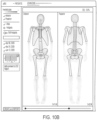
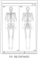
- the improved image analysis techniques described herein include an improved skeletal segmentation approach in which entire (e.g., more than three-quarters length) humerus and/or femur region(s) are identified in bone scan images.
- Previous approaches only identified a limited fraction of femur and humerus bones.
- segmenting a larger portion of these bones allows lesions located further out in the extremities of the arms and legs to be identified, whereas previously such lesions would have escaped detection.
- the approaches described herein utilize a region dependent thresholding technique that enhances detection sensitivity in femur and humerus bone regions to overcome this issue.
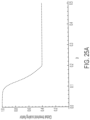
- the present disclosure also provides a global thresholding technique that improves hotspot detection accuracy, particularly at high disease burdens (e.g., when a patient has many lesions).
- This approach detects a preliminary set of potential hotspots, and then adjusts thresholds used for hotspot detection based on a scaling factor computed from this preliminary set.
- the improvement in hotspot detection provides advantages for downstream calculations, improving linearity of computed BSI values for patients with high levels of metastases.
- the systems and methods described herein improve the accuracy with which automated decisions about whether a hotspot represents a metastasis are made.
- the approaches described herein leverage clinical experience indicating that hotspots selection as potential metastases depends not only on the image features of the hotspot itself, but also information from the entire image. Accordingly, the approaches described herein may also use global features, for example a total number of hotspots, as input in automated decision making steps (e.g., as input to ANNs) for lesion identification.
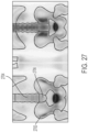
- the approaches described herein also offer improvements to approaches for calculating risk index values based on skeletal involvement, by employing correction factors that account for potential errors in the accuracy with which hotspots can be automatically localized to a particular skeletal region. This is particularly important for hotspots located in or near the sacrum region, which is a complex three dimensional structure that may be difficult to identify in two-dimensional bone scan images. This approach improves accuracy of BSI calculations, and limits sensitivity to errors in hotspot localization.
- the systems and methods described herein include several improved image analysis techniques for lesion identification and quantification. These approaches improve accuracy and robustness with which bone scan images can be analyzed. As described herein, they can be used as part of a cloud-based system that facilitates review and reporting of patient data, and allow for improved disease detection, treatment, and monitoring.
- the invention is directed to a method for lesion marking and quantitative analysis (e.g., user assisted / reviewed automated or semi-automated lesion marking and quantitative analysis) of nuclear medicine images (e.g., a bone scan image set) of a human subject, the method comprising: (a) accessing (e.g., and/or receiving), by a processor of a computing device, a bone scan image set (e.g., a set of one, two, or more images) for the human subject, said bone scan image set obtained following administration of an agent (e.g.
- a radiopharmaceutical to the human subject (e.g., the bone scan image set comprising an anterior bone scan image and a posterior bone scan image)(e.g., wherein each image of the bone scan image set comprises a plurality of pixels, each pixel having a value corresponding to an intensity); (b) automatically segmenting, by the processor, each image in the bone scan image set to identify one or more skeletal regions of interest, each corresponding to a particular anatomical region of a skeleton of the human subject (e.g., a particular bone and/or set of one or more bones, such as a cervical spine, a clavicle, a costae, a lumber spine, a pelvis, a sacrum, a scapula, a skull, a thoracic spine, a sternum, a femur, a humerus), thereby obtaining an annotated set of images, wherein the one or more skeletal regions of interest comprise at least one of (i) and (i
- step (b) comprises: comparing each member of the bone scan image set with a corresponding atlas image of an atlas image set, each atlas image comprising one or more identifications of the one or more skeletal regions of interest (e.g., graphical identifications superimposed on the atlas image), said skeletal regions of interest including the femur region and/or the humerus region; and for each image of the bone scan image set, registering the corresponding atlas image with the image of the bone scan image set, such that the identifications of the one or more skeletal regions of interest of the atlas image are applied to (e.g., are superimposed on) the image of the bone scan image set.
- each atlas image comprises an identification of (i) the femur region comprising at least a portion of a knee region of the human subject and/or (ii) the humerus region comprising at least a portion of an elbow region of the human subject, and wherein, for each image of the bone scan image set, the registering of the corresponding atlas image to the bone scan image comprises using the identified knee region and/or the identified elbow region in the image as (a) landmark(s) [e.g., registering the corresponding atlas image to the bone scan image by identifying a knee region in the bone scan image and matching it to the identified knee region in the corresponding atlas image, then adjusting the atlas image (e.g., calculating a coordinate transform)].
- the registering of the corresponding atlas image to the bone scan image comprises using the identified knee region and/or the identified elbow region in the image as (a) landmark(s) [e.g., registering the corresponding atlas image to the bone scan image by identifying
- a location of at least one detected hotspot of the initial hotspot set corresponds to a physical location in or on a femur more than three quarters of a distance along the femur from an end of the femur oriented toward a hip of the human subject to an end of the femur oriented toward a knee of the human subject.
- a location of at least one detected hotspot of the initial hotspot set corresponds to a physical location in or on a humerus more than three quarters of a distance along the humerus from an end of the humerus oriented toward a shoulder of the human subject to an end of the humerus oriented toward an elbow of the human subject.
- step (c) comprises (e.g., iteratively): identifying, by the processor, healthy tissue regions in the images of the bone scan image set determined not to include any hotspots (e.g., localized regions of relatively high intensity); calculating, by the processor, a normalization factor such that a product of the normalization factor and an average intensity of the identified healthy tissue regions is a pre-defined intensity level; and normalizing the images of the bone scan image set by the normalization factor.
- any hotspots e.g., localized regions of relatively high intensity
- a normalization factor such that a product of the normalization factor and an average intensity of the identified healthy tissue regions is a pre-defined intensity level
- the method further comprises: (g) calculating, by the processor, one or more risk index values for the human subject based at least in part on a computed fraction (e.g., an area fraction) of the skeleton of the human subject occupied by the initial set of hotspots [e.g., wherein the computed fraction is a ratio of a total area of the initial set of hotspots, divided by a total area of all identified skeletal regions].
- a computed fraction e.g., an area fraction
- the computed fraction is a ratio of a total area of the initial set of hotspots, divided by a total area of all identified skeletal regions.
- the method comprises: (h) selecting, by the processor, a first subset (e.g., up to all) of the initial set of hotspots based at least in part on the metastasis likelihood values [e.g., determining whether or not to include a particular hotspot of the initial set of hotspots in the subset based on the metastasis likelihood value calculated for that particular hotspot exceeding a threshold value)]; and (i) causing, by the processor, rendering of a graphical representation of the first subset [e.g., a visual indication (e.g., points, boundaries) of hotspots overlaid on one or more members of the bone scan image set and/or annotated set of images; e.g., a table listing identified hotspots along with additional information (e.g., location; e.g., likelihood value) for each hotspot] for display within a graphical user interface (GUI) (e.g., a cloud-based GUI).
- GUI graphical user
- the method further comprises: (j) calculating, by the processor, one or more risk index values for the human subject based at least in part on a computed fraction (e.g., an area fraction) of the skeleton of the human subject occupied by the first subset of hotspots [e.g., wherein the computed fraction is a total area of the initial set of hotspots divided by a total area of all identified skeletal regions].
- a computed fraction e.g., an area fraction
- the computed fraction is a total area of the initial set of hotspots divided by a total area of all identified skeletal regions.
- the method comprises: (k) receiving, by the processor, via the GUI, a user selection of a second subset of the initial set of hotspots; and (l) calculating, by the processor, one or more risk index values for the human subject based at least in part on a computed fraction (e.g., an area fraction) of the skeleton of the human subject occupied by the second subset of hotspots [e.g., wherein the computed fraction is a total area of the second subset of hotspots, divided by a total area of all identified skeletal regions].
- a computed fraction e.g., an area fraction
- At least one of the risk index values is indicative of a risk of the human subject having and/or developing metastatic cancer (e.g., metastatic prostate cancer, metastatic breast cancer, metastatic lung cancer, and other metastatic bone cancers).
- metastatic cancer e.g., metastatic prostate cancer, metastatic breast cancer, metastatic lung cancer, and other metastatic bone cancers.
- the metastatic cancer is metastatic prostate cancer.
- At least one of the risk index values is indicative of the human subject having a particular state of metastatic cancer (e.g., metastatic prostate cancer, metastatic breast cancer, metastatic lung cancer, and other metastatic bone cancers).
- metastatic cancer e.g., metastatic prostate cancer, metastatic breast cancer, metastatic lung cancer, and other metastatic bone cancers.
- the processor is a processor of a cloud-based system.
- the GUI is part of a general Picture Archiving and Communications System (PACS) (e.g., as well as a clinical application for oncology including lesion marking and quantitative analysis).
- PACS Picture Archiving and Communications System
- the agent e.g., radiopharmaceutical
- the agent comprises technetium 99m methylenediphosphonate ( 99m Tc-NMP).
- the invention is directed to a method for lesion marking and quantitative analysis (e.g., user assisted / reviewed automated or semi-automated lesion marking and quantitative analysis) of nuclear medicine images (e.g., a bone scan image set) of a human subject, the method comprising: (a) accessing (e.g., and/or receiving), by a processor of a computing device, a bone scan image set (e.g., a set of one, two, or more images) for the human subject, said bone scan image set obtained following administration of an agent (e.g., a radiopharmaceutical) to the human subject (e.g., the bone scan image set comprising an anterior bone scan image and a posterior bone scan image) (e.g., wherein each image of the bone scan image set comprises a plurality of pixels, each pixel having a value corresponding to an intensity); (b) automatically segmenting, by the processor, each image in the bone scan image set to identify one or more skeletal regions of interest, each skeletal region of interest
- the global threshold scaling factor is a function of a measure of disease burden for the human subject [e.g., an area fraction of the skeleton of the subject occupied by metastases (e.g., hotspots); e.g., a risk index value], and wherein the adjusting the plurality of preliminary threshold values performed at step (c) comprises decreasing the adjusted threshold values (e.g., with respect to the preliminary threshold values) as disease burden increases (e.g., as measured by the global threshold scaling factor) so as to compensate for an underestimation of hotspot area that occurs with increasing disease burden (e.g., such that a total number and/or size of hotspots increases with the decreased adjusted threshold values).
- a measure of disease burden for the human subject e.g., an area fraction of the skeleton of the subject occupied by metastases (e.g., hotspots); e.g., a risk index value
- the adjusting the plurality of preliminary threshold values performed at step (c) comprises decreasing the adjusted threshold
- the global threshold scaling factor is a function (e.g., a non-linear function) of a fraction (e.g., an area fraction) of the identified skeletal regions occupied by the set of potential hotspot set (e.g., wherein the global threshold scaling factor is a function of a total area of all hotspots in the preliminary set, divided by a total area of all identified skeletal regions).
- the global threshold scaling factor is based on (e.g., computed as a function of) a risk index value calculated using the set of potential hotspots.
- step (c) comprises (e.g., iteratively): identifying, by the processor, healthy tissue regions in the images of the bone scan image set determined not to include any hotspots (e.g., localized regions of relatively high intensity); calculating, by the processor, a normalization factor such that a product of the normalization factor and an average intensity of the identified healthy tissue regions is a pre-defined intensity level; and normalizing, by the processor, the images of the bone scan image set by the normalization factor.
- any hotspots e.g., localized regions of relatively high intensity
- a normalization factor such that a product of the normalization factor and an average intensity of the identified healthy tissue regions is a pre-defined intensity level
- the method further comprises: (g) calculating, by the processor, one or more risk index values for the human subject based at least in part on a computed fraction (e.g., an area fraction) of the skeleton of the human subject occupied by the initial set of hotspots [e.g., wherein the computed fraction is a ratio of a total area of the initial set of hotspots, divided by a total area of all identified skeletal regions].
- a computed fraction e.g., an area fraction
- the computed fraction is a ratio of a total area of the initial set of hotspots, divided by a total area of all identified skeletal regions.
- the method comprises: (h) selecting, by the processor, a first subset (e.g., up to all) of the initial set of hotspots based at least in part on the metastasis likelihood values [e.g., determining whether or not to include a particular hotspot of the initial set of hotspots in the subset based on the metastasis likelihood value calculated for that particular hotspot exceeding a threshold value)]; and (i) causing, by the processor, rendering of a graphical representation of the first subset [e.g., a visual indication (e.g., points, boundaries) of hotspots overlaid on one or more members of the bone scan image set and/or annotated set of images; e.g., a table listing identified hotspots along with additional information (e.g., location; e.g., likelihood value) for each hotspot] for display within a graphical user interface (GUI) (e.g., a cloud-based GUI).
- GUI graphical user
- the method further comprises: (j) calculating, by the processor, one or more risk index values for the human subject based at least in part on a computed fraction (e.g., an area fraction) of the skeleton of the human subject occupied by the first subset of hotspots [e.g., wherein the computed fraction is a total area of the initial set of hotspots divided by a total area of all identified skeletal regions].
- a computed fraction e.g., an area fraction
- the computed fraction is a total area of the initial set of hotspots divided by a total area of all identified skeletal regions.
- the method comprises: (k) receiving, by the processor, via the GUI, a user selection of a second subset of the initial set of hotspots; and (l) calculating, by the processor, one or more risk index values for the human subject based at least in part on a computed fraction (e.g., an area fraction) of the skeleton of the human subject occupied by the second subset of hotspots [e.g., wherein the computed fraction is a total area of the second subset of hotspots, divided by a total area of all identified skeletal regions].
- a computed fraction e.g., an area fraction
- At least one of the risk index values is indicative of a risk of the human subject having and/or developing metastatic cancer (e.g., metastatic prostate cancer, metastatic breast cancer, metastatic lung cancer, and other metastatic bone cancers).
- metastatic cancer e.g., metastatic prostate cancer, metastatic breast cancer, metastatic lung cancer, and other metastatic bone cancers.
- the metastatic cancer is metastatic prostate cancer.
- At least one of the risk index values is indicative of the human subject having a particular state of metastatic cancer (e.g., metastatic prostate cancer, metastatic breast cancer, metastatic lung cancer, and other metastatic bone cancers).
- metastatic cancer e.g., metastatic prostate cancer, metastatic breast cancer, metastatic lung cancer, and other metastatic bone cancers.
- the processor is a processor of a cloud-based system.
- the GUI is part of a general Picture Archiving and Communications System (PACS) (e.g., as well as a clinical application for oncology including lesion marking and quantitative analysis).
- PACS Picture Archiving and Communications System
- the agent e.g., radiopharmaceutical
- the agent comprises technetium 99m methylenediphosphonate ( 99m Tc-NMP).
- the invention is directed to a method for lesion marking and quantitative analysis (e.g., user assisted / reviewed automated or semi-automated lesion marking and quantitative analysis) of nuclear medicine images (e.g., a bone scan image set) of a human subject, the method comprising: (a) accessing (e.g., and/or receiving), by a processor of a computing device, a bone scan image set (e.g., a set of one, two, or more images) for the human subject, said bone scan image set obtained following administration of an agent (e.g., a radiopharmaceutical) to the human subject (e.g., the bone scan image set comprising an anterior bone scan image and a posterior bone scan image)(e.g., wherein each image of the bone scan image set comprises a plurality of pixels, each pixel having a value corresponding to an intensity); (b) automatically segmenting, by the processor, each image in the bone scan image set to identify one or more skeletal regions of interest, each skeletal region of interest
- the one or more global hotspot features comprises a total number of hotspots in the initial hotspot set.
- step (f) comprises adjusting criteria for selection of hotspots for inclusion in the first subset based on the total number of hotspots in the initial hotspot set [e.g., by relaxing criteria as the total number of hotspots in the initial hotspot set increases (e.g., by reducing a metastasis likelihood threshold to which each hotspots metastasis likelihood value is compared; e.g., by scaling metastasis likelihood values based on the total number of hotspots in the initial hotspot set)].
- step (f) comprises using a machine learning module to select the first subset (e.g., an ANN module)[e.g., wherein the machine learning module receives, for each hotspot, at least the metastasis likelihood value calculated for the hotspot and the one or more global hotspot features and outputs (i) an adjusted metastasis likelihood value that takes into account the global hotspot features (e.g., a value on a scale that can be compared to a threshold for selection of the hotspot in the first subset) and/or (ii) a binary (e.g., 0 or 1; e.g., Boolean True or False) value representing whether the hotspot should or should not be included in the first subset].
- a machine learning module to select the first subset (e.g., an ANN module)[e.g., wherein the machine learning module receives, for each hotspot, at least the metastasis likelihood value calculated for the hotspot and the one or more
- step (c) comprises (e.g., iteratively): identifying, by the processor, healthy tissue regions in the images of the bone scan image set determined not to include any hotspots (e.g., localized regions of relatively high intensity); calculating, by the processor, a normalization factor such that a product of the normalization factor and an average intensity of the identified healthy tissue regions is a pre-defined intensity level; and normalizing, by the processor, the images of the bone scan image set by the normalization factor.
- any hotspots e.g., localized regions of relatively high intensity
- a normalization factor such that a product of the normalization factor and an average intensity of the identified healthy tissue regions is a pre-defined intensity level
- the method further comprises: (g) calculating, by the processor, one or more risk index values for the human subject based at least in part on a computed fraction (e.g., an area fraction) of the skeleton of the human subject occupied by the initial set of hotspots [e.g., wherein the computed fraction is a ratio of a total area of the initial set of hotspots, divided by a total area of all identified skeletal regions].
- a computed fraction e.g., an area fraction
- the computed fraction is a ratio of a total area of the initial set of hotspots, divided by a total area of all identified skeletal regions.
- the method comprises: (h) selecting, by the processor, a first subset (e.g., up to all) of the initial set of hotspots based at least in part on the metastasis likelihood values [e.g., determining whether or not to include a particular hotspot of the initial set of hotspots in the subset based on the metastasis likelihood value calculated for that particular hotspot exceeding a threshold value)]; and (i) causing, by the processor, rendering of a graphical representation of the first subset [e.g., a visual indication (e.g., points, boundaries) of hotspots overlaid on one or more members of the bone scan image set and/or annotated set of images; e.g., a table listing identified hotspots along with additional information (e.g., location; e.g., likelihood value) for each hotspot] for display within a graphical user interface (GUI) (e.g., a cloud-based GUI).
- GUI graphical user
- the method further comprises: (j) calculating, by the processor, one or more risk index values for the human subject based at least in part on a computed fraction (e.g., an area fraction) of the skeleton of the human subject occupied by the first subset of hotspots [e.g., wherein the computed fraction is a total area of the initial set of hotspots divided by a total area of all identified skeletal regions].
- a computed fraction e.g., an area fraction
- the computed fraction is a total area of the initial set of hotspots divided by a total area of all identified skeletal regions.
- the method comprises: (k) receiving, by the processor, via the GUI, a user selection of a second subset of the initial set of hotspots; and (l) calculating, by the processor, one or more risk index values for the human subject based at least in part on a computed fraction (e.g., an area fraction) of the skeleton of the human subject occupied by the second subset of hotspots [e.g., wherein the computed fraction is a total area of the second subset of hotspots, divided by a total area of all identified skeletal regions].
- a computed fraction e.g., an area fraction
- At least one of the risk index values is indicative of a risk of the human subject having and/or developing metastatic cancer (e.g., metastatic prostate cancer, metastatic breast cancer, metastatic lung cancer, and other metastatic bone cancers).
- metastatic cancer e.g., metastatic prostate cancer, metastatic breast cancer, metastatic lung cancer, and other metastatic bone cancers.
- the metastatic cancer is metastatic prostate cancer.
- At least one of the risk index values is indicative of the human subject having a particular state of metastatic cancer (e.g., metastatic prostate cancer, metastatic breast cancer, metastatic lung cancer, and other metastatic bone cancers).
- metastatic cancer e.g., metastatic prostate cancer, metastatic breast cancer, metastatic lung cancer, and other metastatic bone cancers.
- the processor is a processor of a cloud-based system.
- the GUI is part of a general Picture Archiving and Communications System (PACS) (e.g., as well as a clinical application for oncology including lesion marking and quantitative analysis).
- PACS Picture Archiving and Communications System
- the agent e.g., radiopharmaceutical
- the agent comprises technetium 99m methylenediphosphonate ( 99m Tc-NMP).
- the invention is directed to a method for lesion marking and quantitative analysis (e.g., user assisted / reviewed automated or semi-automated lesion marking and quantitative analysis) of nuclear medicine images (e.g., a bone scan image set) of a human subject, the method comprising: (a) accessing (e.g., and/or receiving), by a processor of a computing device, a bone scan image set (e.g., a set of one, two, or more images) for the human subject (e.g., the bone scan image set comprising an anterior bone scan image and a posterior bone scan image)(e.g., wherein each image of the bone scan image set comprises a plurality of pixels, each pixel having a value corresponding to an intensity); (b) automatically segmenting, by the processor, each image in the bone scan image set to identify one or more skeletal regions of interest, each skeletal region of interest corresponding to a particular anatomical region of a skeleton of the human subject (e.g., a particular bone
- the computed skeletal involvement factor estimates a proportion of total skeletal mass occupied by a physical volume associated with the particular hotspot.
- the computing the skeletal involvement factor comprises: calculating, by the processor, a ratio of an area of the particular hotspot to an area of the corresponding skeletal region of interest, thereby computing an area fraction for the particular hotspot; and scaling (e.g., multiplying) the area fraction by a density coefficient associated with the skeletal region of interest to which the particular hotspot is assigned [e.g., that accounts for weight and/or density of bond in the corresponding skeletal region of interest (e.g., wherein the density coefficient is a weight fraction of the corresponding skeletal region of interest with respect to a total skeleton (e.g., of an average human)], thereby computing the skeletal involvement factor for the particular hotspot.
- a density coefficient associated with the skeletal region of interest to which the particular hotspot is assigned e.g., that accounts for weight and/or density of bond in the corresponding skeletal region of interest (e.g., wherein the density coefficient is a weight fraction of the corresponding skeletal region of interest with respect to a
- At least a portion of the hotspots of the first subset are assigned to a skeletal region of interest that is a member selected from the group consisting of a pelvis region (e.g., corresponding to a pelvis of the human subject), a lumbar region (e.g., corresponding to a lumbar column of the human subject), and a sacrum region (e.g., corresponding to a sacrum of the human subject).
- a pelvis region e.g., corresponding to a pelvis of the human subject
- a lumbar region e.g., corresponding to a lumbar column of the human subject
- a sacrum region e.g., corresponding to a sacrum of the human subject
- the one or more region-dependent correction factors comprise a sacrum region correction factor associated with a sacrum region and used to adjust skeletal involvement factors of hotspots identified (e.g., by the processor) as being located therein, and wherein the sacrum region correction factor has a value less than one (e.g., less than 0.5).
- the one or more region dependent correction factors comprise one or more correction factor pairs, each correction factor pair associated with a specific skeletal region of interest and comprising a first member and a second member (of the pair), wherein: the first member of the pair is an anterior image correction factor and is used to adjust skeletal involvement factors computed for hotspots having been detected in an annotated anterior bone scan image of the annotated image set, and the second member of the pair is a posterior image correction factor and is used to adjust skeletal involvement factors computed for hotspots having been detected in an annotated posterior bone scan image of the annotated image set.
- step (c) comprises (e.g., iteratively): identifying, by the processor, healthy tissue regions in the images of the bone scan image set determined not to include any hotspots (e.g., localized regions of relatively high intensity); calculating, by the processor, a normalization factor such that a product of the normalization factor and an average intensity of the identified healthy tissue regions is a pre-defined intensity level; and normalizing, by the processor, the images of the bone scan image set by the normalization factor.
- any hotspots e.g., localized regions of relatively high intensity
- a normalization factor such that a product of the normalization factor and an average intensity of the identified healthy tissue regions is a pre-defined intensity level
- the method further comprises: (g) calculating, by the processor, one or more risk index values for the human subject based at least in part on a computed fraction (e.g., an area fraction) of the skeleton of the human subject occupied by the initial set of hotspots [e.g., wherein the computed fraction is a ratio of a total area of the initial set of hotspots, divided by a total area of all identified skeletal regions].
- a computed fraction e.g., an area fraction
- the computed fraction is a ratio of a total area of the initial set of hotspots, divided by a total area of all identified skeletal regions.
- the method comprises: (h) selecting, by the processor, a first subset (e.g., up to all) of the initial set of hotspots based at least in part on the metastasis likelihood values [e.g., determining whether or not to include a particular hotspot of the initial set of hotspots in the subset based on the metastasis likelihood value calculated for that particular hotspot exceeding a threshold value)]; and (i) causing, by the processor, rendering of a graphical representation of the first subset [e.g., a visual indication (e.g., points, boundaries) of hotspots overlaid on one or more members of the bone scan image set and/or annotated set of images; e.g., a table listing identified hotspots along with additional information (e.g., location; e.g., likelihood value) for each hotspot] for display within a graphical user interface (GUI) (e.g., a cloud-based GUI).
- GUI graphical user
- the method further comprises: (j) calculating, by the processor, one or more risk index values for the human subject based at least in part on a computed fraction (e.g., an area fraction) of the skeleton of the human subject occupied by the first subset of hotspots [e.g., wherein the computed fraction is a total area of the initial set of hotspots divided by a total area of all identified skeletal regions].
- a computed fraction e.g., an area fraction
- the computed fraction is a total area of the initial set of hotspots divided by a total area of all identified skeletal regions.
- the method comprises: (k) receiving, by the processor, via the GUI, a user selection of a second subset of the initial set of hotspots; and (l) calculating, by the processor, one or more risk index values for the human subject based at least in part on a computed fraction (e.g., an area fraction) of the skeleton of the human subject occupied by the second subset of hotspots [e.g., wherein the computed fraction is a total area of the second subset of hotspots, divided by a total area of all identified skeletal regions].
- a computed fraction e.g., an area fraction
- At least one of the risk index values is indicative of a risk of the human subject having and/or developing metastatic cancer (e.g., metastatic prostate cancer, metastatic breast cancer, metastatic lung cancer, and other metastatic bone cancers).
- metastatic cancer e.g., metastatic prostate cancer, metastatic breast cancer, metastatic lung cancer, and other metastatic bone cancers.
- the metastatic cancer is metastatic prostate cancer.
- At least one of the risk index values is indicative of the human subject having a particular state of metastatic cancer (e.g., metastatic prostate cancer, metastatic breast cancer, metastatic lung cancer, and other metastatic bone cancers).
- metastatic cancer e.g., metastatic prostate cancer, metastatic breast cancer, metastatic lung cancer, and other metastatic bone cancers.
- the processor is a processor of a cloud-based system.
- the GUI is part of a general Picture Archiving and Communications System (PACS) (e.g., as well as a clinical application for oncology including lesion marking and quantitative analysis).
- PACS Picture Archiving and Communications System
- the agent e.g., radiopharmaceutical
- the agent comprises technetium 99m methylenediphosphonate ( 99m Tc-NMP).
- the invention is directed to a system for lesion marking and quantitative analysis (e.g., user assisted / reviewed automated or semi-automated lesion marking and quantitative analysis) of nuclear medicine images (e.g., a bone scan image set) of a human subject, the system comprising: a processor; and a memory having instructions thereon, wherein the instructions, when executed by the processor, cause the processor to: (a) access (e.g., and/or receive) a bone scan image set (e.g., a set of one, two, or more images) for the human subject, said bone scan image set obtained following administration of an agent (e.g.
- a bone scan image set e.g., a set of one, two, or more images
- a radiopharmaceutical to the human subject (e.g., the bone scan image set comprising an anterior bone scan image and a posterior bone scan image)(e.g., wherein each image of the bone scan image set comprises a plurality of pixels, each pixel having a value corresponding to an intensity); (b) automatically segment each image in the bone scan image set to identify one or more skeletal regions of interest, each corresponding to a particular anatomical region of a skeleton of the human subject (e.g., a particular bone and/or set of one or more bones, such as a cervical spine, a clavicle, a costae, a lumber spine, a pelvis, a sacrum, a scapula, a skull, a thoracic spine, a sternum, a femur, a humerus), thereby obtaining an annotated set of images, wherein the one or more skeletal regions of interest comprise at least one of (i) and (ii): (i)
- the instructions cause the processor to: compare each member of the bone scan image set with a corresponding atlas image of an atlas image set, each atlas image comprising one or more identifications of the one or more skeletal regions of interest (e.g., graphical identifications superimposed on the atlas image), said skeletal regions of interest including the femur region and/or the humerus region; and for each image of the bone scan image set, register the corresponding atlas image with the image of the bone scan image set, such that the identifications of the one or more skeletal regions of interest of the atlas image are applied to (e.g., are superimposed on) the image of the bone scan image set.
- each atlas image comprises an identification of (i) the femur region comprising at least a portion of a knee region of the human subject and/or (ii) the humerus region comprising at least a portion of an elbow region of the human subject, and wherein, for each image of the bone scan image set, the instructions cause the processor to register the corresponding atlas image to the bone scan image using the identified knee region and/or the identified elbow region in the image as (a) landmark(s) [e.g., registering the corresponding atlas image to the bone scan image by identifying a knee region in the bone scan image and matching it to the identified knee region in the corresponding atlas image, then adjusting the atlas image (e.g., calculating a coordinate transform)].
- the instructions cause the processor to register the corresponding atlas image to the bone scan image using the identified knee region and/or the identified elbow region in the image as (a) landmark(s) [e.g., registering the corresponding atlas image to the bone scan image
- a location of at least one detected hotspot of the initial hotspot set corresponds to a physical location in or on a femur more than three quarters of a distance along the femur from an end of the femur oriented toward a hip of the human subject to an end of the femur oriented toward a knee of the human subject.
- a location of at least one detected hotspot of the initial hotspot set corresponds to a physical location in or on a humerus more than three quarters of a distance along the humerus from an end of the humerus oriented toward a shoulder of the human subject to an end of the humerus oriented toward an elbow of the human subject.
- the instructions cause the processor to (e.g., iteratively): identify healthy tissue regions in the images of the bone scan image set determined not to include any hotspots (e.g., localized regions of relatively high intensity); calculate a normalization factor such that a product of the normalization factor and an average intensity of the identified healthy tissue regions is a pre-defined intensity level; and normalize the images of the bone scan image set by the normalization factor.
- identify healthy tissue regions in the images of the bone scan image set determined not to include any hotspots e.g., localized regions of relatively high intensity
- calculate a normalization factor such that a product of the normalization factor and an average intensity of the identified healthy tissue regions is a pre-defined intensity level
- the instructions further cause the processor to: (g) calculate one or more risk index values for the human subject based at least in part on a computed fraction (e.g., an area fraction) of the skeleton of the human subject occupied by the initial set of hotspots [e.g., wherein the computed fraction is a ratio of a total area of the initial set of hotspots, divided by a total area of all identified skeletal regions].
- a computed fraction e.g., an area fraction
- the computed fraction is a ratio of a total area of the initial set of hotspots, divided by a total area of all identified skeletal regions.
- the instructions cause the processor to: (h) select a first subset (e.g., up to all) of the initial set of hotspots based at least in part on the metastasis likelihood values [e.g., determining whether or not to include a particular hotspot of the initial set of hotspots in the subset based on the metastasis likelihood value calculated for that particular hotspot exceeding a threshold value)]; and (i) cause rendering of a graphical representation of the first subset [e.g., a visual indication (e.g., points, boundaries) of hotspots overlaid on one or more members of the bone scan image set and/or annotated set of images; e.g., a table listing identified hotspots along with additional information (e.g., location; e.g., likelihood value) for each hotspot] for display within a graphical user interface (GUI) (e.g., a cloud-based GUI).
- GUI graphical user interface
- the instructions cause the processor to: (j) calculate one or more risk index values for the human subject based at least in part on a computed fraction (e.g., an area fraction) of the skeleton of the human subject occupied by the first subset of hotspots [e.g., wherein the computed fraction is a total area of the initial set of hotspots divided by a total area of all identified skeletal regions].
- a computed fraction e.g., an area fraction
- the instructions cause the processor to: (k) receive, via the GUI, a user selection of a second subset of the initial set of hotspots; and (l) calculate one or more risk index values for the human subject based at least in part on a computed fraction (e.g., an area fraction) of the skeleton of the human subject occupied by the second subset of hotspots [e.g., wherein the computed fraction is a total area of the second subset of hotspots, divided by a total area of all identified skeletal regions].
- a computed fraction e.g., an area fraction
- At least one of the risk index values is indicative of a risk of the human subject having and/or developing metastatic cancer (e.g., metastatic prostate cancer, metastatic breast cancer, metastatic lung cancer, and other metastatic bone cancers).
- metastatic cancer e.g., metastatic prostate cancer, metastatic breast cancer, metastatic lung cancer, and other metastatic bone cancers.
- the metastatic cancer is metastatic prostate cancer.
- At least one of the risk index values is indicative of the human subject having a particular state of metastatic cancer (e.g., metastatic prostate cancer, metastatic breast cancer, metastatic lung cancer, and other metastatic bone cancers).
- metastatic cancer e.g., metastatic prostate cancer, metastatic breast cancer, metastatic lung cancer, and other metastatic bone cancers.
- the system is a cloud based system.
- the processor is a processor of a cloud-based system.
- the GUI is part of a general Picture Archiving and Communications System (PACS) (e.g., as well as a clinical application for oncology including lesion marking and quantitative analysis).
- PACS Picture Archiving and Communications System
- the agent e.g., radiopharmaceutical
- the agent comprises technetium 99m methylenediphosphonate ( 99m Tc-NMP).
- the invention is directed to a system for lesion marking and quantitative analysis (e.g., user assisted / reviewed automated or semi-automated lesion marking and quantitative analysis) of nuclear medicine images (e.g., a bone scan image set) of a human subject, the system comprising: a processor; and a memory having instructions thereon, wherein the instructions, when executed by the processor, cause the processor to: (a) access (e.g., and/or receive), by a processor of a computing device, a bone scan image set (e.g., a set of one, two, or more images) for the human subject, said bone scan image set obtained following administration of an agent (e.g., a radiopharmaceutical) to the human subject (e.g., the bone scan image set comprising an anterior bone scan image and a posterior bone scan image)(e.g., wherein each image of the bone scan image set comprises a plurality of pixels, each pixel having a value corresponding to an intensity); (b) automatically segment each image in
- the instructions cause the processor to: compute the global threshold scaling factor a function of a measure of disease burden for the human subject [e.g., an area fraction of the skeleton of the subject occupied by metastases (e.g., hotspots); e.g., a risk index value]; and, at step (c) adjust the plurality of preliminary threshold values by decreasing the adjusted threshold values (e.g., with respect to the preliminary threshold values) as disease burden increases (e.g., as measured by the global threshold scaling factor) so as to compensate for an underestimation of hotspot area that occurs with increasing disease burden (e.g., such that a total number and/or size of hotspots increases with the decreased adjusted threshold values).
- a measure of disease burden for the human subject e.g., an area fraction of the skeleton of the subject occupied by metastases (e.g., hotspots); e.g., a risk index value
- the instructions cause the processor to: compute the global threshold scaling factor a function of
- the instructions cause the processor to compute global threshold scaling factor as a function (e.g., a non-linear function) of a fraction (e.g., an area fraction) of the identified skeletal regions occupied by the set of potential hotspot set (e.g., wherein the global threshold scaling factor is a function of a total area of all hotspots in the preliminary set, divided by a total area of all identified skeletal regions).
- a function e.g., a non-linear function
- a fraction e.g., an area fraction of the identified skeletal regions occupied by the set of potential hotspot set
- the instructions cause the processor to compute the global threshold scaling factor based on (e.g., as a function of) a risk index value calculated using the set of potential hotspots.
- the instructions cause the processor to (e.g., iteratively): identify healthy tissue regions in the images of the bone scan image set determined not to include any hotspots (e.g., localized regions of relatively high intensity); calculate a normalization factor such that a product of the normalization factor and an average intensity of the identified healthy tissue regions is a pre-defined intensity level; and normalize the images of the bone scan image set by the normalization factor.
- identify healthy tissue regions in the images of the bone scan image set determined not to include any hotspots e.g., localized regions of relatively high intensity
- calculate a normalization factor such that a product of the normalization factor and an average intensity of the identified healthy tissue regions is a pre-defined intensity level
- the instructions further cause the processor to: (g) calculate one or more risk index values for the human subject based at least in part on a computed fraction (e.g., an area fraction) of the skeleton of the human subject occupied by the initial set of hotspots [e.g., wherein the computed fraction is a ratio of a total area of the initial set of hotspots, divided by a total area of all identified skeletal regions].
- a computed fraction e.g., an area fraction
- the computed fraction is a ratio of a total area of the initial set of hotspots, divided by a total area of all identified skeletal regions.
- the instructions cause the processor to: (h) select a first subset (e.g., up to all) of the initial set of hotspots based at least in part on the metastasis likelihood values [e.g., determining whether or not to include a particular hotspot of the initial set of hotspots in the subset based on the metastasis likelihood value calculated for that particular hotspot exceeding a threshold value)]; and (i) cause rendering of a graphical representation of the first subset [e.g., a visual indication (e.g., points, boundaries) of hotspots overlaid on one or more members of the bone scan image set and/or annotated set of images; e.g., a table listing identified hotspots along with additional information (e.g., location; e.g., likelihood value) for each hotspot] for display within a graphical user interface (GUI) (e.g., a cloud-based GUI).
- GUI graphical user interface
- the instructions cause the processor to: (j) calculate one or more risk index values for the human subject based at least in part on a computed fraction (e.g., an area fraction) of the skeleton of the human subject occupied by the first subset of hotspots [e.g., wherein the computed fraction is a total area of the initial set of hotspots divided by a total area of all identified skeletal regions].
- a computed fraction e.g., an area fraction
- the instructions cause the processor to: (k) receive, via the GUI, a user selection of a second subset of the initial set of hotspots; and (l) calculate one or more risk index values for the human subject based at least in part on a computed fraction (e.g., an area fraction) of the skeleton of the human subject occupied by the second subset of hotspots [e.g., wherein the computed fraction is a total area of the second subset of hotspots, divided by a total area of all identified skeletal regions].
- a computed fraction e.g., an area fraction
- At least one of the risk index values is indicative of a risk of the human subject having and/or developing metastatic cancer (e.g., metastatic prostate cancer, metastatic breast cancer, metastatic lung cancer, and other metastatic bone cancers).
- metastatic cancer e.g., metastatic prostate cancer, metastatic breast cancer, metastatic lung cancer, and other metastatic bone cancers.
- the metastatic cancer is metastatic prostate cancer.
- At least one of the risk index values is indicative of the human subject having a particular state of metastatic cancer (e.g., metastatic prostate cancer, metastatic breast cancer, metastatic lung cancer, and other metastatic bone cancers).
- metastatic cancer e.g., metastatic prostate cancer, metastatic breast cancer, metastatic lung cancer, and other metastatic bone cancers.
- the system is a cloud based system.
- the processor is a processor of a cloud-based system.
- the GUI is part of a general Picture Archiving and Communications System (PACS) (e.g., as well as a clinical application for oncology including lesion marking and quantitative analysis).
- PACS Picture Archiving and Communications System
- the agent e.g., radiopharmaceutical
- the agent comprises technetium 99m methylenediphosphonate ( 99m Tc-NMP).
- the invention is directed to a system for lesion marking and quantitative analysis (e.g., user assisted / reviewed automated or semi-automated lesion marking and quantitative analysis) of nuclear medicine images (e.g., a bone scan image set) of a human subject, the system comprising: a processor; and a memory having instructions thereon, wherein the instructions, when executed by the processor, cause the processor to: (a) access (e.g., and/or receive) a bone scan image set (e.g., a set of one, two, or more images) for the human subject, said bone scan image set obtained following administration of an agent (e.g., a radiopharmaceutical) to the human subject (e.g., the bone scan image set comprising an anterior bone scan image and a posterior bone scan image)(e.g., wherein each image of the bone scan image set comprises a plurality of pixels, each pixel having a value corresponding to an intensity); (b) automatically segment each image in the bone scan image set to identify one or more agents (e
- the one or more global hotspot features comprises a total number of hotspots in the initial hotspot set.
- the instructions cause the processor to adjust criteria for selection of hotspots for inclusion in the first subset based on the total number of hotspots in the initial hotspot set [e.g., by relaxing criteria as the total number of hotspots in the initial hotspot set increases (e.g., by reducing a metastasis likelihood threshold to which each hotspots metastasis likelihood value is compared; e.g., by scaling metastasis likelihood values based on the total number of hotspots in the initial hotspot set)].
- criteria for selection of hotspots for inclusion in the first subset based on the total number of hotspots in the initial hotspot set [e.g., by relaxing criteria as the total number of hotspots in the initial hotspot set increases (e.g., by reducing a metastasis likelihood threshold to which each hotspots metastasis likelihood value is compared; e.g., by scaling metastasis likelihood values based on the total number of hotspots
- the instructions cause the processor to use a machine learning module to select the first subset (e.g., an ANN module)[e.g., wherein the machine learning module receives, for each hotspot, at least the metastasis likelihood value calculated for the hotspot and the one or more global hotspot features and outputs (i) an adjusted metastasis likelihood value that takes into account the global hotspot features (e.g., a value on a scale that can be compared to a threshold for selection of the hotspot in the first subset) and/or (ii) a binary (e.g., 0 or 1; e.g., Boolean True or False) value representing whether the hotspot should or should not be included in the first subset].
- a machine learning module to select the first subset (e.g., an ANN module)[e.g., wherein the machine learning module receives, for each hotspot, at least the metastasis likelihood value calculated for the hotspot and the one or
- the instructions cause the processor to (e.g., iteratively): identify healthy tissue regions in the images of the bone scan image set determined not to include any hotspots (e.g., localized regions of relatively high intensity); calculate a normalization factor such that a product of the normalization factor and an average intensity of the identified healthy tissue regions is a pre-defined intensity level; and normalize the images of the bone scan image set by the normalization factor.
- identify healthy tissue regions in the images of the bone scan image set determined not to include any hotspots e.g., localized regions of relatively high intensity
- calculate a normalization factor such that a product of the normalization factor and an average intensity of the identified healthy tissue regions is a pre-defined intensity level
- the instructions further cause the processor to: (g) calculate one or more risk index values for the human subject based at least in part on a computed fraction (e.g., an area fraction) of the skeleton of the human subject occupied by the initial set of hotspots [e.g., wherein the computed fraction is a ratio of a total area of the initial set of hotspots, divided by a total area of all identified skeletal regions].
- a computed fraction e.g., an area fraction
- the computed fraction is a ratio of a total area of the initial set of hotspots, divided by a total area of all identified skeletal regions.
- the instructions cause the processor to: (h) select a first subset (e.g., up to all) of the initial set of hotspots based at least in part on the metastasis likelihood values [e.g., determining whether or not to include a particular hotspot of the initial set of hotspots in the subset based on the metastasis likelihood value calculated for that particular hotspot exceeding a threshold value)]; and (i) cause rendering of a graphical representation of the first subset [e.g., a visual indication (e.g., points, boundaries) of hotspots overlaid on one or more members of the bone scan image set and/or annotated set of images; e.g., a table listing identified hotspots along with additional information (e.g., location; e.g., likelihood value) for each hotspot] for display within a graphical user interface (GUI) (e.g., a cloud-based GUI).
- GUI graphical user interface
- the instructions cause the processor to: (j) calculate one or more risk index values for the human subject based at least in part on a computed fraction (e.g., an area fraction) of the skeleton of the human subject occupied by the first subset of hotspots [e.g., wherein the computed fraction is a total area of the initial set of hotspots divided by a total area of all identified skeletal regions].
- a computed fraction e.g., an area fraction
- the instructions cause the processor to: (k) receive, via the GUI, a user selection of a second subset of the initial set of hotspots; and (l) calculate one or more risk index values for the human subject based at least in part on a computed fraction (e.g., an area fraction) of the skeleton of the human subject occupied by the second subset of hotspots [e.g., wherein the computed fraction is a total area of the second subset of hotspots, divided by a total area of all identified skeletal regions].
- a computed fraction e.g., an area fraction
- At least one of the risk index values is indicative of a risk of the human subject having and/or developing metastatic cancer (e.g., metastatic prostate cancer, metastatic breast cancer, metastatic lung cancer, and other metastatic bone cancers).
- metastatic cancer e.g., metastatic prostate cancer, metastatic breast cancer, metastatic lung cancer, and other metastatic bone cancers.
- the metastatic cancer is metastatic prostate cancer.
- At least one of the risk index values is indicative of the human subject having a particular state of metastatic cancer (e.g., metastatic prostate cancer, metastatic breast cancer, metastatic lung cancer, and other metastatic bone cancers).
- metastatic cancer e.g., metastatic prostate cancer, metastatic breast cancer, metastatic lung cancer, and other metastatic bone cancers.
- the system is a cloud based system.
- the processor is a processor of a cloud-based system.
- the GUI is part of a general Picture Archiving and Communications System (PACS) (e.g., as well as a clinical application for oncology including lesion marking and quantitative analysis).
- PACS Picture Archiving and Communications System
- the agent e.g., radiopharmaceutical
- the agent comprises technetium 99m methylenediphosphonate ( 99m Tc-NMP).
- the invention is directed to a system for lesion marking and quantitative analysis (e.g., user assisted / reviewed automated or semi-automated lesion marking and quantitative analysis) of nuclear medicine images (e.g., a bone scan image set) of a human subject, the system comprising: a processor; and a memory having instructions thereon, wherein the instructions, when executed by the processor, cause the processor to: (a) access (e.g., and/or receive) a bone scan image set (e.g., a set of one, two, or more images) for the human subject (e.g., the bone scan image set comprising an anterior bone scan image and a posterior bone scan image)(e.g., wherein each image of the bone scan image set comprises a plurality of pixels, each pixel having a value corresponding to an intensity); (b) automatically segment each image in the bone scan image set to identify one or more skeletal regions of interest, each skeletal region of interest corresponding to a particular anatomical region of a skeleton of the
- the computed skeletal involvement factor estimates a proportion of total skeletal mass occupied by a physical volume associated with the particular hotspot.
- the instructions cause the processor to compute the skeletal involvement factor by: calculating a ratio of an area of the particular hotspot to an area of the corresponding skeletal region of interest, thereby computing an area fraction for the particular hotspot; and scaling (e.g., multiplying) the area fraction by a density coefficient associated with the skeletal region of interest to which the particular hotspot is assigned [e.g., that accounts for weight and/or density of bond in the corresponding skeletal region of interest (e.g., wherein the density coefficient is a weight fraction of the corresponding skeletal region of interest with respect to a total skeleton (e.g., of an average human)], thereby computing the skeletal involvement factor for the particular hotspot.
- a density coefficient associated with the skeletal region of interest to which the particular hotspot is assigned e.g., that accounts for weight and/or density of bond in the corresponding skeletal region of interest (e.g., wherein the density coefficient is a weight fraction of the corresponding skeletal region of interest with respect to a
- At least a portion of the hotspots of the first subset are assigned to a skeletal region of interest that is a member selected from the group consisting of a pelvis region (e.g., corresponding to a pelvis of the human subject), a lumbar region (e.g., corresponding to a lumbar column of the human subject), and a sacrum region (e.g., corresponding to a sacrum of the human subject).
- a pelvis region e.g., corresponding to a pelvis of the human subject
- a lumbar region e.g., corresponding to a lumbar column of the human subject
- a sacrum region e.g., corresponding to a sacrum of the human subject
- the one or more region-dependent correction factors comprise a sacrum region correction factor associated with a sacrum region and used to adjust skeletal involvement factors of hotspots identified (e.g., by the processor) as being located therein, and wherein the sacrum region correction factor has a value less than one (e.g., less than 0.5).
- the one or more region dependent correction factors comprise one or more correction factor pairs, each correction factor pair associated with a specific skeletal region of interest and comprising a first member and a second member (of the pair), wherein: the first member of the pair is an anterior image correction factor and is used to adjust skeletal involvement factors computed for hotspots having been detected in an annotated anterior bone scan image of the annotated image set, and the second member of the pair is a posterior image correction factor and is used to adjust skeletal involvement factors computed for hotspots having been detected in an annotated posterior bone scan image of the annotated image set.
- the instructions cause the processor to (e.g., iteratively): identify healthy tissue regions in the images of the bone scan image set determined not to include any hotspots (e.g., localized regions of relatively high intensity); calculate a normalization factor such that a product of the normalization factor and an average intensity of the identified healthy tissue regions is a pre-defined intensity level; and normalize the images of the bone scan image set by the normalization factor.
- identify healthy tissue regions in the images of the bone scan image set determined not to include any hotspots e.g., localized regions of relatively high intensity
- calculate a normalization factor such that a product of the normalization factor and an average intensity of the identified healthy tissue regions is a pre-defined intensity level
- the instructions further cause the processor to: (g) calculate one or more risk index values for the human subject based at least in part on a computed fraction (e.g., an area fraction) of the skeleton of the human subject occupied by the initial set of hotspots [e.g., wherein the computed fraction is a ratio of a total area of the initial set of hotspots, divided by a total area of all identified skeletal regions].
- a computed fraction e.g., an area fraction
- the computed fraction is a ratio of a total area of the initial set of hotspots, divided by a total area of all identified skeletal regions.
- the instructions cause the processor to: (h) select a first subset (e.g., up to all) of the initial set of hotspots based at least in part on the metastasis likelihood values [e.g., determining whether or not to include a particular hotspot of the initial set of hotspots in the subset based on the metastasis likelihood value calculated for that particular hotspot exceeding a threshold value)]; and (i) cause rendering of a graphical representation of the first subset [e.g., a visual indication (e.g., points, boundaries) of hotspots overlaid on one or more members of the bone scan image set and/or annotated set of images; e.g., a table listing identified hotspots along with additional information (e.g., location; e.g., likelihood value) for each hotspot] for display within a graphical user interface (GUI) (e.g., a cloud-based GUI).
- GUI graphical user interface
- the instructions cause the processor to: (j) calculate one or more risk index values for the human subject based at least in part on a computed fraction (e.g., an area fraction) of the skeleton of the human subject occupied by the first subset of hotspots [e.g., wherein the computed fraction is a total area of the initial set of hotspots divided by a total area of all identified skeletal regions].
- a computed fraction e.g., an area fraction
- the instructions cause the processor to: (k) receive, via the GUI, a user selection of a second subset of the initial set of hotspots; and (l) calculate one or more risk index values for the human subject based at least in part on a computed fraction (e.g., an area fraction) of the skeleton of the human subject occupied by the second subset of hotspots [e.g., wherein the computed fraction is a total area of the second subset of hotspots, divided by a total area of all identified skeletal regions].
- a computed fraction e.g., an area fraction
- At least one of the risk index values is indicative of a risk of the human subject having and/or developing metastatic cancer (e.g., metastatic prostate cancer, metastatic breast cancer, metastatic lung cancer, and other metastatic bone cancers).
- metastatic cancer e.g., metastatic prostate cancer, metastatic breast cancer, metastatic lung cancer, and other metastatic bone cancers.
- the metastatic cancer is metastatic prostate cancer.
- At least one of the risk index values is indicative of the human subject having a particular state of metastatic cancer (e.g., metastatic prostate cancer, metastatic breast cancer, metastatic lung cancer, and other metastatic bone cancers).
- metastatic cancer e.g., metastatic prostate cancer, metastatic breast cancer, metastatic lung cancer, and other metastatic bone cancers.
- the system is a cloud based system.
- the processor is a processor of a cloud-based system.
- the GUI is part of a general Picture Archiving and Communications System (PACS) (e.g., as well as a clinical application for oncology including lesion marking and quantitative analysis).
- PACS Picture Archiving and Communications System
- the agent e.g., radiopharmaceutical
- the agent comprises technetium 99m methylenediphosphonate ( 99m Tc-NMP).
- the invention is directed to a computer aided image analysis device [e.g., a computer-aided detection (CADe) device; e.g., a computer-aided diagnostic (CADx) device] comprising the system of any one of the aspects and embodiments described herein (for example in paragraphs [0083] - [00145]).
- CADe computer-aided detection
- CADx computer-aided diagnostic
- the device is programmed to be used by trained healthcare professionals and/or researchers [e.g., for acceptance, transfer, storage, image display, manipulation, quantification, and reporting of digital medicine images acquired using nuclear medicine imaging; e.g., wherein the device provides general Picture Archiving and Communications System (PACS) tools and/or a clinical application for oncology, including lesion marking and quantitative analysis].
- PACS Picture Archiving and Communications System
- the device is programmed to be used for analysis of bone scan images for evaluation and/or detection of metastatic cancer (e.g., metastatic prostate cancer, metastatic breast cancer, metastatic lung cancer, and other metastatic bone cancers).
- metastatic cancer e.g., metastatic prostate cancer, metastatic breast cancer, metastatic lung cancer, and other metastatic bone cancers.
- the device is programmed to be used for analysis of bone scan images for evaluation and/or detection of prostate cancer.
- the device comprises a label specifying that the device is intended to be used by trained healthcare professionals and/or researchers [e.g., for acceptance, transfer, storage, image display, manipulation, quantification, and reporting of digital medicine images acquired using nuclear medicine imaging; e.g., wherein the device provides general Picture Archiving and Communications System (PACS) tools and/or a clinical application for oncology, including lesion marking and quantitative analysis].
- PACS Picture Archiving and Communications System
- the label further specifies that the device is intended to be used for analysis of bone scan images for evaluation and/or detection of metastatic cancer (e.g., metastatic prostate cancer, metastatic breast cancer, metastatic lung cancer, and other metastatic bone cancers).
- metastatic cancer e.g., metastatic prostate cancer, metastatic breast cancer, metastatic lung cancer, and other metastatic bone cancers.
- the label further specifies that the device is intended to be used for analysis of bone scan images for evaluation and/or detection of prostate cancer.
- Embodiments described with respect to one aspect of the invention may be, applied to another aspect of the invention (e.g., features of embodiments described with respect to one independent claim, e.g., a method claim, are contemplated to be applicable to other embodiments of other independent claims, e.g., a system claim, and vice versa).
- Headers are provided for the convenience of the reader - the presence and/or placement of a header is not intended to limit the scope of the subject matter described herein.
- the term “approximately” or “about” refers to a range of values that fall within 25%, 20%, 19%, 18%, 17%, 16%, 15%, 14%, 13%, 12%, 11%, 10%, 9%, 8%, 7%, 6%, 5%, 4%, 3%, 2%, 1%, or less in either direction (greater than or less than) of the stated reference value unless otherwise stated or otherwise evident from the context (except where such number would exceed 100% of a possible value).
- the systems and methods described herein are directed to improved computer aided display and analysis of nuclear medicine images.
- the systems and methods described herein provide improvements to several image processing steps used for automated analysis of bone scan images for assessing cancer status of a patient.
- improved approaches for image segmentation, hotspot detection, automated classification of hotspots as representing metastases, and computation of risk indices such as bone scan index (BSI) values are provided.
- Automated BSI calculation technology is described in detail in U.S. Patent Application No. 15/282,422, filed September 30, 2016 , in U.S. Patent No. 8,855,387, issued October 7, 2014 (of which U.S. Patent Application No. 15/282,422 is a reissue), and PCT Application No.
- PCT/US17/58418 filed October 26, 2017 , the content of each of which is hereby incorporated by reference in its entirety.
- PCT Application PCT/US2017/058418 filed October 26, 2017 , also incorporated herein in its entirety, describes a cloud-based platform that can serve as a platform for providing image analysis and BSI calculation tools in accordance with the approaches described herein.
- bone scan images are acquired following administration of an agent, such as a radiopharmaceutical, to a human subject.
- the administered agent accumulates in cancerous bone lesions as a result of physical properties of the underlying tissue (e.g., increased vasculature, abnormal osteogenesis) or due to recognition (by the agent) of particular biomolecules that are selectively expressed or over-expressed in tumors, such as prostate-specific membrane antigen (PSMA).
- PSMA prostate-specific membrane antigen
- the agent comprises radionuclide(s) that emit nuclear radiation, which can be detected and thereby used to image the spatial distribution of agent within the subject.
- bone scan images are acquired as two-dimensional scans using a gamma camera.
- two images - an anterior and a posterior image - are acquired to form a bone scan image set.
- Physical regions where the agent has accumulated at high concentrations appear as regions of elevated intensity, i.e., bright spots, in bone scan images.
- the agent may accumulate in cancerous bone lesions, e.g., as described above, as well as other regions, such as the bladder of the subject.
- bone scan images are segmented to identify regions corresponding to bones of the subject's skeleton, forming an annotated set of images. Regions of elevated intensity with respect to their surroundings are identified within the skeletal regions and compared with thresholds to detect an initial set of hotspots.
- a hotspot size e.g., area
- hotspot shape e.g., as described by various metrics, such as radius, eccentricity
- measure of hotspot intensity e.g., peak intensity, average intensity, integrated intensity, etc.
- the metastasis likelihood values are computed using artificial neural networks (ANNs) that receive as input, for each hotspot, a set of hotspot features and output a metastasis likelihood value.
- ANNs artificial neural networks
- the metastasis likelihood values can be used to automatically filter the initial set of hotspots, to determine a subset to be used for calculation of risk indices that indicate a risk of the subject having and/or developing metastatic cancer. By filtering hotspots in this manner, only those determined to have a high likelihood of representing a metastasis are included in the risk index calculations.
- graphical representations of hotpots and/or their likelihood values are rendered for display to a user, for example as overlaid markings on annotated images and/or tables of information, allowing the user to select a subset of the hotspots for use in calculating risk indices. This allows the user to augment the automated selection of hotspots for calculation of risk indices with their input.
- the approaches described herein include several improvements to the aforementioned image processing steps, providing for improved accuracy of lesion detection and risk index calculations.
- the present disclosure includes and improved segmentation approach whereby an entire (e.g., more than three-quarters length) humerus and/or an entire (e.g., more than three-quarters length) femur region is/are identified.
- an entire (e.g., more than three-quarters length) humerus and/or an entire (e.g., more than three-quarters length) femur region is/are identified.
- Previously only a limited faction of the femur and humerus bones were identified. Segmenting a larger (e.g., entire) portion of these bones allows lesions located further out in the extremities of a subject's arms and legs to be identified.
- the approach described herein also utilizes region dependent thresholds in the hotspot detection step.
- the region-dependent threshold values vary for different skeletal regions, and have lower values in the femur and humerus regions, so as to increase detection sensitivity therein.
- the systems and methods described herein may use a global threshold scaling technique to detect hotspots.
- the initial set of hotspots is detected by first identifying a set of potential hotspots, using a plurality of preliminary region-dependent thresholds.
- the potential set of hotspots is used to compute a global threshold scaling factor, based on an area fraction of the subject's skeleton occupied by the set of potential hotspots.
- the preliminary thresholds are then adjusted using the global threshold scaling factor, and the adjusted thresholds used to detect the initial set of hotspots.
- the present disclosure also includes improvements for selection of hotspots and calculation of risk indices.
- the approaches described herein may use a hotspot pre-selection technique that filters hotspots based not only on their computed metastasis likelihood values, but also global hotspot features that measure properties of the overall set of initial hotspots, such as a total number of hotspots in the set.
- global hotspot features include other measures of total hotspot number, such as an average number of hotspots per region, measures of overall hotspot intensity, such as peak or average hotspot intensity, and measures of overall hotspot size, such as a total area of hotspots, an average hotspot size, etc.
- BSI bone scan index
- BSI is a risk index value that provides an estimate of the fraction of a subject's total skeletal mass occupied by cancerous lesions.
- Calculating BSI involves calculating, for each particular hotspot, a skeletal involvement factor based on a ratio of the area of the particular hotspot to that of the skeletal region in which it is located.
- Scaled versions e.g., to convert area ratios to relative mass
- Bone scan images are two dimensional images, but the underlying skeleton of the subject is a three dimensional structure. Accordingly, a hotspot may be incorrectly identified as located in one region, when in fact its represent a lesion located in a different bone. This is a particular challenge for the sacrum and neighboring pelvic and lumbar regions.
- the present disclosure includes a modified risk index calculation approach that uses region-dependent correction factors to scale skeletal involvement factors in a manner that accounts for potential errors in localizing hotspots. This approach improves accuracy of BSI calculations, and limits sensitivity to hotspot localization.
- the systems and methods described herein include several improved image analysis techniques for lesion identification and quantification. These approaches improve accuracy and robustness with which bone scan images can be analyzed. As described herein, they can be used as part of a cloud-based system that facilitates review and reporting of patient data, and allows for improved disease detection, treatment, and monitoring.
- Nuclear medicine images are obtained using a nuclear imaging modality such as bone scan imaging, Positron Emission Tomography (PET) imaging, and Single-Photon Emission Tomography (SPECT) imaging.
- a nuclear imaging modality such as bone scan imaging, Positron Emission Tomography (PET) imaging, and Single-Photon Emission Tomography (SPECT) imaging.
- PET Positron Emission Tomography
- SPECT Single-Photon Emission Tomography
- an "image" - for example, a 3-D image of mammal - includes any visual representation, such as a photo, a video frame, streaming video, as well as any electronic, digital or mathematical analogue of a photo, video frame, or streaming video.
- "3-D" or "three-dimensional” with reference to an "image” means conveying information about three dimensions.
- a 3-D image may be rendered as a dataset in three dimensions and/or may be displayed as a set of two-dimensional representations, or as a three-dimensional representation.
- nuclear medicine images use imaging agents comprising radiopharmaceuticals.
- Nuclear medicine images are obtained following administration of a radiopharmaceutical to a patient (e.g., a human subject), and provide information regarding the distribution of the radiopharmaceutical within the patient.
- Radiopharmaceuticals are compounds that comprise a radionuclide.
- administering means introducing a substance (e.g., an imaging agent) into a subject.
- a substance e.g., an imaging agent
- any route of administration may be utilized including, for example, parenteral (e.g., intravenous), oral, topical, subcutaneous, peritoneal, intraarterial, inhalation, vaginal, rectal, nasal, introduction into the cerebrospinal fluid, or instillation into body compartments.
- radionuclide refers to a moiety comprising a radioactive isotope of at least one element.
- exemplary suitable radionuclides include but are not limited to those described herein.
- a radionuclide is one used in positron emission tomography (PET).
- PET positron emission tomography
- SPECT single-photon emission computed tomography
- a non-limiting list of radionuclides includes 99m Tc, 111 In, 64 Cu, 67 Ga, 68 Ga, 186 Re, 188 Re , 153 Sm, 177 Lu, 67 Cu, 123 I, 124 I, 125 I, 126 I, 131 I , 11 C, 13 N, 15 O, 18 F, 153 Sm, 166 Ho, 177 Lu, 149 Pm, 90 Y, 213 Bi, 103 Pd, 109 Pd, 159 Gd, 140 La, 198 Au, 199 Au, 169 Yb, 175 Yb, 165 Dy, 166 Dy, 105 Rh, 111 Ag, 89 Zr, 225 Ac, 82 Rb, 75 Br, 76 Br, 77 Br, 80 Br, 80m Br, 82 Br, 83 Br, 211 At and 192 Ir.
- radiopharmaceutical refers to a compound comprising a radionuclide.
- radiopharmaceuticals are used for diagnostic and/or therapeutic purposes.
- radiopharmaceuticals include small molecules that are labeled with one or more radionuclide(s), antibodies that are labeled with one or more radionuclide(s), and antigen-binding portions of antibodies that are labeled with one or more radionuclide(s).
- Nuclear medicine images detect radiation emitted from the radionuclides of radiopharmaceuticals to form an image.
- the distribution of a particular radiopharmaceutical within a patient may be determined by biological mechanisms such as blood flow or perfusion, as well as by specific enzymatic or receptor binding interactions.
- Different radiopharmaceuticals may be designed to take advantage of different biological mechanisms and/or particular specific enzymatic or receptor binding interactions and thus, when administered to a patient, selectively concentrate within particular types of tissue and/or regions within the patient.
- intensity variations within a nuclear medicine image can be used to map the distribution of radiopharmaceutical within the patient. This mapped distribution of radiopharmaceutical within the patient can be used to, for example, infer the presence of cancerous tissue within various regions of the patient's body.
- technetium 99m methylenediphosphonate ( 99m Tc MDP) selectively accumulates within the skeletal region of the patient, in particular at sites with abnormal osteogenesis associated with malignant bone lesions.
- the selective concentration of radiopharmaceutical at these sites produces identifiable hotspots - localized regions of high intensity in nuclear medicine images. Accordingly, presence of malignant bone lesions associated with metastatic prostate cancer can be inferred by identifying such hotspots within a whole-body scan of the patient.
- risk indices that correlate with patient overall survival and other prognostic metrics indicative of disease state, progression, treatment efficacy, and the like can be computed based on automated analysis of intensity variations in whole-body scans obtained following administration of 99m Tc MDP to a patient.
- other radiopharmaceuticals can also be used in a similar fashion to 99m Tc MDP.
- the particular radiopharmaceutical used depends on the particular nuclear medicine imaging modality used.
- 18F sodium fluoride (NaF) also accumulates in bone lesions, similar to 99m Tc MDP, but can be used with PET imaging.
- PET imaging may also utilize a radioactive form of the vitamin choline, which is readily absorbed by prostate cancer cells.
- radiopharmaceuticals that selectively bind to particular proteins or receptors of interest - particularly those whose expression is increased in cancerous tissue may be used.
- proteins or receptors of interest include, but are not limited to tumor antigens, such as CEA, which is expressed in colorectal carcinomas, Her2/neu, which is expressed in multiple cancers, BRCA 1 and BRCA 2, expressed in breast and ovarian cancers; and TRP-1 and -2, expressed in melanoma.
- PSMA prostate-specific membrane antigen
- PSMA binding agents e.g., compounds that a high affinity to PSMA
- radionuclide(s) can be used to obtain nuclear medicine images of a patient from which the presence and/or state of prostate cancer within a variety of regions (e.g., including, but not limited to skeletal regions) of the patient can be assessed.
- nuclear medicine images obtained using PSMA binding agents are used to identify the presence of cancerous tissue within the prostate, when the disease is in a localized state.
- nuclear medicine images obtained using radiopharmaceuticals comprising PSMA binding agents are used to identify the presence of cancerous tissue within a variety of regions that include not only the prostate, but also other organs and tissue regions such as lungs, lymph nodes, and bones, as is relevant when the disease is metastatic.
- radionuclide labelled PSMA binding agents upon administration to a patient, selectively accumulate within cancerous tissue, based on their affinity to PSMA.
- the selective concentration of radionuclide labelled PSMA binding agents at particular sites within the patient produces detectable hotspots in nuclear medicine images.
- PSMA binding agents concentrate within a variety of cancerous tissues and regions of the body expressing PSMA, localized cancer within a prostate of the patient and/or metastatic cancer in various regions of the patient's body can be detected, and evaluated.
- Risk indices that correlate with patient overall survival and other prognostic metrics indicative of disease state, progression, treatment efficacy, and the like can be computed based on automated analysis of intensity variations in nuclear medicine images obtained following administration of a PSMA binding agent radiopharmaceutical to a patient.
- radionuclide labelled PSMA binding agents may be used as radiopharmaceutical imaging agents for nuclear medicine imaging to detect and evaluate prostate cancer.
- the particular radionuclide labelled PSMA binding agent that is used depends on factors such as the particular imaging modality (e.g., PET; e.g., SPECT) and the particular regions (e.g., organs) of the patient to be imaged.
- certain radionuclide labelled PSMA binding agents are suited for PET imaging, while others are suited for SPECT imaging.
- certain radionuclide labelled PSMA binding agents facilitate imaging a prostate of the patient, and are used primarily when the disease is localized, while others facilitate imaging organs and regions throughout the patient's body, and are useful for evaluating metastatic prostate cancer.
- PSMA binding agents and radionuclide labelled versions thereof are described in U.S. Patent Nos. 8,778,305 , 8,211,401 , and 8,962,799 , each of which are incorporated herein by reference in their entireties.
- PSMA binding agents and radionuclide labelled versions thereof are also described in PCT Application PCT/US2017/058418, filed October 26, 2017 , incorporated herein by reference in its entirety.
- computer aided image analysis devices as described herein are intended to be used by trained healthcare professionals and researchers for acceptance, transfer, storage, image display, manipulation, quantification, and reporting of digital medical images acquired using nuclear medicine (NM) imaging.
- NM nuclear medicine
- such devices provide general Picture Archiving and Communications System (PACS) tools as well as clinical applications for oncology, including lesion marking and quantitative analysis
- Bone scintigraphy (also referred to as bone scan imaging) is an imaging modality widely used to assess skeletal disease burden.
- a current standard of assessing disease progression based on bone scan images is based on the semi-quantitative modified Prostate Cancer Working Group 2 and the Prostate Cancer Working Group 3 (PCWG) criteria.
- the definition of this criteria relies on the appearance of new lesions as interpreted by a trained reader: (i) either two new lesions followed by two additional lesions on a first and second follow-up scans compared to a pretreatment scan (known at the 2+2 rule), or (ii) two new confirmed lesions relative to the first follow-up scan thereafter.
- this semi-quantitative assessment approach of counting lesion number has a number of limitations. In particular, the assessment is subject to manual variability, it is confined to assessing disease progression, and is unable to assess accurate burden of confluent/diffused diseases as is relevant, e.g., to metastatic prostate cancer.
- the automated Bone Scan Index (BSI), developed by EXINI Diagnostics AB, Lund Sweden, is a fully quantitative assessment of a patient's skeletal disease on a bone scan, as the fraction of the total skeleton weight.
- the BSI has undergone rigorous pre-analytical and analytical validation as an objective measure of the quantitative change in disease burden bone scans.
- BSI assessment was demonstrated to risk stratify metastatic prostate cancer patients.
- the systems and methods described herein are directed to improved computer aided approaches for analyzing bone scan images and computing BSI in automated and semi-automated, user guided, fashions. Also described herein are GUI tools that facilitate review of bone scan images and automated analyses used to determine BSI values by a user.
- systems and methods described herein can be implemented as a cloud based platform for automated and semi-automated image analysis for detection and evaluation of cancer status in a patient.
- An example device described herein is the automated BSI device ( aBSI ), which is as a cloud-based software platform with a web interface where users can upload bone scan image data in the form of particular image files, such as DICOM files.
- the software complies with the Digital Imaging and Communications in Medicine (DICOM 3) standard.
- devices in accordance with the systems and methods described herein are programmed for an intended user, typically a health-care professional who uses the software to view patient images and analyze results.
- the user operates the service in a web browser, (such as Google's Chrome browser) on a computer running an operating system such as Microsoft Windows or OSX.
- the software can be configured to occupy a single application window.
- the service is web-based and accessed via a specific URL. Keyboard and mouse controls may be used for operating the software.
- Multiple scans can be uploaded for each patient and the system provides a separate image-based automated analysis for each.
- the automated analysis is reviewed by the physician, who may be guided through a quality control and reporting workflow. If the quality control of the automated assessment is approved, a report can be created and signed.
- the service can be configured for HIPAA and 21 CFR part 11 compliance.
- access to software tools in accordance with the systems and methods described herein is restricted and protected by security measures.
- access to a cloud-based implementation of the systems and methods described herein, aBSI is protected by multi-factor authentication in the form of a username, password and verification code sent as a text message to the phone number associated with the account
- software enforced requirements include one or more of the following:
- user enforced requirements include one or more of the following:
- software enforced requirements include one or more of the following:
- user enforced requirements include one or more of the following:
- whole-body bone scintigraphy images are acquired in compliance with relevant guidelines, such as "EANM Bone scintigraphy: procedure guidelines for tumor imaging” and "ACR-SPR Practice Parameter for the Performance of Skeletal Scintigraphy (Bone Scan)”.
- FIG. 1 is a block flow diagram showing a quality control and reporting workflow 100 for generating a BSI report, according to an illustrative embodiment.
- the systems and methods described herein include a GUI for guiding a user (e.g., healthcare professional) through review and analysis of patient image data to compute an automated BSI value and generate a report.
- the workflow 100 allows the user to select 102 and upload 104 image files of patient data, which are then analyzed by the software and reviewed by the user.
- the user may be presented with a first GUI window, such as the window 200 shown in FIG. 2 , that allows the user to select a particular patient for review/analysis from a list.
- a first GUI window such as the window 200 shown in FIG. 2
- a new and/or updated window 300 shown in FIG. 3 , showing patient information is displayed.
- the user may then access a review page 400 of the guided analysis software.
- the review page 400 provides the user (e.g., healthcare professional, such as a physician) with a GUI that allows them examine image data and automated identification of hotspots performed by software backend, in order to calculate the automated BSI index for the patient.
- Automated BSI calculation technology is described further detail herein, and previous versions (without the improvements of the present disclosure) are described in detail in U.S. Patent Application No. 15/282,422, filed September 30, 2016 , and in U.S. Patent No. 8,855,387, issued October 7, 2014 (of which U.S. Patent Application No. 15/282,422 is a reissue), and PCT Application No. PCT/US17/58418, filed October 26, 2017 , the content of each of which is hereby incorporated by reference in its entirety.
- the review page allows the user review 106 of hotspots, representing cancerous lesions, that have been automatically identified in images by the software.
- the user may use the review page GUI to edit 108 the set of regions identified as hotspots, and must confirm that image quality, skeletal segmentation (as depicted by the outlining in the screenshot), and that the set of identified hotspots have been reviewed and accepted 110 in order to proceed with report generation.
- a report such as the report 500 shown in FIG. 5 , including a final BSI computed value, may be generated 114.
- the systems and methods described herein may be used in a process 600 for automated detection and pre-selection of hotspots, receipt of a user verification of the set of preselected hotspots, and for calculation of a risk index value, such as, in particular, a bone scan index (BSI) value.
- a risk index value such as, in particular, a bone scan index (BSI) value.
- BBI bone scan index
- the software tools described herein use an algorithm for image registration to bring patient images into a reference coordinate frame. This is done by non-rigidly adjusting an atlas image to each patient image. By fitting the atlas image to the patient, the patient image can be segmented into a skeleton region and background region. The skeleton region can be further divided into smaller skeletal regions of interest, also referred to herein as localizations.
- a normalization step 604 the whole-body bone scan images are normalized to provide a user with a standardized intensity range when viewing images at different contrast and brightness levels. Hotspots are detected in a hotspot detection step 606.
- hotspots are detected using a thresholding rule.
- hotspot pre-selection 608 is performed.
- pre-selection of hotspots is based on image analysis and machine learning techniques that aim to pre-select significant hotspots for inclusion in a set of pre-selected hotspots, which may be reviewed by the user in a hotspot verification step 610. This pre-selection selection is based on a range of features, such as size, location, orientation, shape and texture of the hotspots.
- pre-selection is a user-convenience tool, aiming to reduce the number of manual clicks that a user must perform (e.g., to select hotspots to use for computing risk indices, such as a BSI value).
- the pre-selection step may be followed by a mandatory hotspot verification step where the user must review and approve the preselection of hotspots or if needed manually include and/or exclude hotspots to be able to create a report.
- Bone Scan Index a particular risk index referred to as Bone Scan Index (BSI) is computed using the set of verified hotspots.
- skeletal segmentation is performed using a segmentation by registration approach in which an atlas image is registered with a bone scan image to be segmented.
- the device automatically contours the skeleton into distinct skeletal regions by elastically fitting a manually contoured annotated template image set to each analyzed image set.
- This annotated template image set is known as the skeletal atlas image set.
- This atlas image set is structured just like any patient image - it looks like a regular bone scan and comprises one anterior and one posterior image.
- the atlas images provide a fixed reference when analyzing a scan.
- the atlas is manually annotated with the regions of interest (skeletal regions) that can be transferred to a new scan to accurately calculate BSI.
- An example atlas image set 700 with 31 manually drawn skeletal regions is shown in FIG. 7 .
- the atlas image set 700 may comprise an anterior image 702 and a posterior image 704.
- the atlas images are elastically deformed to resemble the bone scan image.
- the same transformation is then applied to the atlas contours, thereby producing a delineation/segmentation of each skeletal region of interest of the patient bone scan image. Additional detail regarding construction of a suitable atlas is described at the end of this section.
- Deforming the skeletal atlas to fit a patient scan Deforming the skeletal atlas to fit a patient scan.
- deformation of the skeletal atlas to fit a patient's bone scan image follows an iterative approach.
- the segmentation algorithm proceeds in iterations, where in each iteration, a vector is estimated for every pixel describing how the pixel should be displaced to its corresponding position in the target image. With individual displacements for every pixel, chances are that the displacement vectors will cross or share target position, which will result in holes and/or tears in the deformed image.
- the vector field is smoothed using a filtering approach.
- Displacements are estimated by applying complex-valued filters to the atlas and target images.
- the complex filter responses can be expressed in terms of an amplitude and phase for each pixel.
- phase differences i.e. phase differences between pixels that are within a short distance of each other
- phase differences are proportional to the size of the displacement necessary to bring them into alignment.
- this process is repeated several times for varying filter angles. Knowing the angles of each filter and the resulting magnitude of the displacement makes it possible to infer the direction in which the largest displacement can be observed. While this approach works for small displacements, it must also be possible to apply it in cases where the atlas and target images are further apart. To achieve this, a subsampling approach is taken where the algorithm is first applied to a subsampled (down-sized) version of the image. This approach treats large displacements as local differences.
- the algorithm proceeds to increasingly more detailed (less subsampled) images to add more detail and variability to the resulting displacement field.
- the algorithm is run on a fixed pyramid of subsampled images, for a fixed number of iterations at each pyramid level, and with a predetermined level of smoothing.
- the example aBSI device described herein relies on a single atlas image.
- the contouring algorithm is driven by structural information in the atlas and target images and seeks to deform the atlas image such that distances between similar structures in the two images are minimized. Structure is defined by edges and ridges (lines) in the images. Accordingly, global intensity differences and texture patterns are disregarded by the algorithm. As a result, a suitable atlas image exhibits two important properties:
- Atlas images based on a database of real, normal (e.g., no metastases or other visible medical conditions) bone scan images are used.
- a contouring algorithm can be used to bring all images of the database in alignment with each other.
- an average transformation is computed from all resulting transformations.
- all images are transformed to this anatomical average, representing the average anatomy in the database.
- intensities are also normalized, creating a typical bone scan image suitable as an anatomical reference.
- the schematic shown in FIG. 8 illustrates this idea conceptually - how average anatomy and intensities (center image, 802) can be inferred from multiple bone scan images 804a, 804b, 804c, 804d of a database.
- the average anatomy used for the atlas image set converges to a stable estimate quickly as the number of scans included is increased.
- a relatively small number e.g., 30
- a single atlas can be applied to any bone scan image for skeletal segmentation.
- a challenge when reading scintigraphy images such as bone scans is that intensity levels between scans may differ due to a variety of parameters such as injected dose, time from injection to scanning, scan time, body type, camera hardware and configuration etc.
- input bone scans are normalized such that the average intensity of healthy bone tissue is scaled to a predetermined reference level.
- skeletal segmentation has been carried out and the pixels belonging to the skeleton are known.
- regions of high intensity must be identified and excluded.
- Hotspots are detected using a simple thresholding approach wherein the image is filtered using a difference-of-Gaussians band-pass filter which emphasizes small regions of high intensity relative to their respective surroundings. This filtered image is then thresholded at a region based constant level.
- threshold levels used in the cloud-based aBSI example embodiment are 650 for cervical spine, clavicle, costae, lumbar spine, pelvis, sacrum, scapula, skull, thoracic spine, sternum and 500 for femur and humerus.
- the output of the hotspot detection is a set of ROIs (regions of interest) representing the hotspots in the image and a normalization factor which is used to set the initial maximum and minimum thresholds for image windowing.
- Hotspots may be classified as either included or excluded for pre-selection using a learning (data driven) approach based on artificial neural networks (ANNs).
- the ANNs may be tuned/trained on a training database with patients ranging from normal bone scans to bone scans with numerous and extensive hotspots.
- Each hotspot in the training database is characterized using a set of features (measurements) relating to their size, location, orientation, shape and texture. These features are fed into the ANN, first during a training phase where the parameters of the ANN are set to maximize classification performance in a cross-validation study, and then in the actual software to classify a hotspot as included or excluded.
- the pre-selection training analyzes hotspots and their immediate neighborhood in the images, making the classifier robust to large-scale differences in training materials. Therefore, the classifier is applicable to a wide range of input data. However, small difference in performance between cohorts can be expected.
- separate networks can be constructed for different bone localizations. The set of input features for each of these ANNs then differ somewhat. For instance, the symmetry features only apply to localizations which have a natural symmetric counterpart.
- hotspots in training sets are typically manually labeled as included or excluded by a medical expert trained in reading bone scans.
- the target labels can be verified by a second medical expert.
- the US and European procedural guidelines for bone scintigraphy are harmonized and the equipment used to obtain bone scans are the same in US and Europe.
- the criteria used to interpret bone scans for clinical trials for example the Prostate Working Group 2 Criteria, are global. This is based on the common knowledge in the nuclear medicine community that variation in bone scan appearance due to cancer disease is much more pronounced than minor variation that can be measured in for example normal bone density between different races.
- the parameters of the ANNs are optimized such that the resulting classifier mimics the selection of a medical expert.
- a cross validation approach can be used.
- pre-selection saves time for the reader, all hotspots are reviewed and approved by the reader before a report is created.
- ANN's as described herein can be implemented via one or more machine learning modules.
- machine learning module refers to a computer implemented process (e.g., function) that implements one or more specific machine learning algorithms in order to determine, for a given input (such as an image (e.g., a 2D image; e.g., a 3D image), dataset, and the like) one or more output values.
- a machine learning module may receive as input a 3D image of a subject (e.g., a CT image; e.g., an MRI), and for each voxel of the image, determine a value that represents a likelihood that the voxel lies within a region of the 3D image that corresponds to a representation of a particular organ or tissue of the subject.
- a machine learning module may be combined and implemented as a single module and/or a single software application.
- two or more machine learning modules may also be implemented separately, e.g., as separate software applications.
- a machine learning module may be software and/or hardware.
- a machine learning module may be implemented entirely as software, or certain functions of a CNN module may be carried out via specialized hardware (e.g., via an application specific integrated circuit (ASIC)).
- ASIC application specific integrated circuit
- the systems and methods described herein include a graphical user interface for review of patient data and images.
- the GUI may allow a user to review a list of patients and select patients for whom to review and analyze images.
- FIG. 9A and FIG. 9B show example GUI windows providing a listing of patients from which specific patients can be selected. Once a particular patient has been selected, a user may view bone scan images for that patient, and review automated analysis (e.g., detection and pre-selection of hotspots) using a GUI such as the GUIs shown in FIG. 10A and FIG. 10B .
- Bone scan images may be colorized using various colormaps.
- FIG. 10C and FIG. 10D show example colormaps that may be used.
- GUI systems in accordance with the approaches described herein facilitate viewing of images. For example, automatic adjustment of images to a screen size may be provided.
- FIG. 11A shows a GUI embodiment without this functionality, wherein a user clicks on zoom icons to vary the size of the images.
- FIG. 11B shows a screenshot of a GUI in which images are automatically sized to fill the screen vertically.
- FIG. 11C and FIG. 11D show two approaches for providing zoom functionality for viewing images in detail. In the embodiment shown in FIG. 11C magnification around a mouse pointer occurs when a scroll wheel button is clicked and held. In the embodiment shown in FIG. 11D , zoom an pan functionality using a mouse scroll wheel and click and drag operations are provided.
- FIGs. 12A - 12F illustrate intensity windowing approaches for display of images.
- FIGs. 12B and 12D show a GUI embodiment that implements a custom intensity windowing slider that facilitates a user adjustment of intensity window thresholds.
- FIG. 13A and FIG. 13B show screenshots of example GUI windows in which local intensity is displayed.
- local intensity is displayed when a user hovers a mouse pointer over an image.
- a local intensity value is displayed at a location of the mouse pointer.
- the intensity is displayed at the bottom left corner of the image, instead of next to the mouse pointer, where it then hides part of the image (e.g., accordingly providing for improved safety).
- FIGs. 14A - 14D show example GUI embodiments for displaying multiple bone scan images.
- a tabular approach us used to allow a user to toggle through different bone scan image sets for different studies, or to toggle through anterior and posterior images.
- checkboxes are used to toggle visibility of various images, allowing multiple images from different studies to be displayed alongside each other.
- embodiments of GUI tools for image analysis described herein may provide information useful for quality control.
- total image intensities may be displayed.
- FIG. 15A is a screenshot of a GUI showing display of total image intensities and total skeletal intensities, according to an illustrative embodiment.
- FIG. 15B only total image intensities (and not total skeletal intensities) are displayed to provide a simpler user interface (e.g., to avoid confusion).
- the GUI tools for reviewing bone scan images may show graphical indications of detected (e.g., and pre-selected) hotspots overlaid on bone scan images, for example as shown in FIG. 16A and FIG. 16B .
- a table of hotspots as shown in FIG. 17A , is also displayed.
- the GUI tools described herein allow a user to select or deselect a pre-selected hotspot for inclusion in a final set of selected hotspots used to compute a BSI value.
- the GUI may display the resultant BSI value 1602 above an image, allowing a user to observe its changes (compare 1704a with 1704b) with selection and/or deselection of various hotspots (1702a and 1702b), as shown in FIGs. 17B and 17C .
- Various graphical control approaches may be used to allow a user to select pre-selected hotspots to include or exclude.
- FIG. 18A shows a pop-up control that appears following a user right-click operation.
- FIG. 18B shows a toggle switch 1802 ("Edit hotspots") that can be turned on and off. Once the toggle switch is turned on, the user clicks on various hotspots to select or deselect them.
- the GUI tools may include safety / quality control features, such as the pop-up window shown in FIG. 18C that prompts a user to verify quality control requirements before a report is generated.
- BSI values are computed for various studies, they are displayed for user review.
- computed BSI values may be displayed in a table, as shown in FIG. 19A .
- computed BSI values for different studies are displayed above the displayed bone scan images for their corresponding studies, as shown in FIG. 19B .
- Charts showing time evolution of computed BSI values may also be displayed, as shown in FIGs. 20A and 20B . Additional information pertinent to computed BSI values may also be displayed.
- FIG. 21 shows a screenshot of a GUI providing a table listing number of hotspots in particular anatomical regions used for computation of BSI values in different studies, according to an illustrative embodiment.
- the systems and methods described herein provide for automated report generation.
- FIG. 22A and FIG. 22B show example auto-generated reports.
- the systems and methods described herein include improvements to one or more of the image processing steps shown in FIG. 6 .
- a skeletal atlas image set may be used for image segmentation.
- the skeletal atlas image set includes a pair of template bone scan images (anterior and posterior) representing a typical normal bone scan, and a manual outlining of 31 skeletal regions. These regions are warped to fit a current patient image to be analyzed.
- a limited atlas with skeletal identifications that only cover three-quarters of a femur and a humerus is used.
- an improved, full-length atlas that includes skeletal region identifications covering the entire femur and humerus is used.
- FIG. 23 shows a set of bone scan images with atlas-based skeletal segmentation overlaid, comparing use of a limited atlas 2300a (left) with a full-length atlas 2300b (right).
- the limited atlas 2300a only includes a three-quarters or less of the humerus 2302a and femur 2304a, whereas the full length atlas includes well over three quarts of the length of the humerus 2302b and femur 2304b.
- Use of the full-length atlas can increase stability of skeleton segmentation.
- the increased atlas allows knees and elbows to be utilized as references during registration. Such reference points, with clear contrasts, are beneficial for image analysis and provide improved stability.
- the increased atlas also allows for detection of hotspots further out in the limbs.
- an initial set of candidate hotspots is found using thresholding of image intensities.
- a global and fixed threshold is used, such that a same value is used across all skeletal regions of interest and all images.
- Another, improved, approach sets regional thresholds that vary across different skeletal regions of interest. For example, this approach allowed a reduced threshold to be used for femur and humerus regions (e.g., decreased from 650 to 500) to increase detection sensitivity. Femur and humerus regions show less uptake in bone scan images than other skeletal regions. Accordingly, lower thresholds can be used for these regions to achieve a similar level of sensitivity as for the rest of the body. Setting individual thresholding values for different skeletal region allows this functionality and leads to increased detection of lower intensity hotspots in these skeletal regions.
- FIG. 24 shows an example process 2400 for lesion marking and analysis using the above-described improved segmentation and region-dependent threshold approaches.
- a bone scan image set for a human subject is accessed 2410.
- Each member image of the bone scan image set is automatically segmented 2420 to identify skeletal regions of interest, including a femur region corresponding to greater than three quarters of the length of the femur of the subject 2422 and/or a humerus region corresponding to greater than three-quarters of the length of the humerus of the subject 2424.
- the identified skeletal regions of interest are then analyzed to automatically detect an initial set of hotspots 2430.
- this step of automatically detecting hotspots may comprise applying thresholding operations that use region dependent thresholds - i.e., whose values are not uniform, and differ between one skeletal region and another, to each skeletal region of interest.
- region dependent thresholds i.e., whose values are not uniform, and differ between one skeletal region and another, to each skeletal region of interest.
- lower valued thresholds for the femur and/or humerus regions 2432 and 2434, respectively
- Hotspots may be filtered (pre-selected) based on the calculated likelihood values, for inclusion in a first subset, to either be presented to the user and/or used for calculation of risk indices such as BSI values.
- a user may review the detected hotspots - e.g., the initial hotspot set, or the first subset comprising the filtered hotspots, via the graphical display, and confirm or reject hotspots for inclusion in a second, final subset. This final subset may then be used for calculating risk index values, thereby incorporating user expertise into the decision making process.
- a global dynamic threshold adjustment is used. This method checks the resulting BSI value and fine tunes a global threshold scaling factor to accommodate for high burden disease.
- the global scaling factor approach is a data-driven approach accounts for accounts for errors wherein BSI values can be underestimated at higher levels of disease - i.e., high levels of metastasis.
- errors were discovered using a simulation system that allows bone-scan images to be simulated for a patient with any (e.g., selected) degree of disease.
- the simulation system produces realistic bone-scan images, accounting for specific camera and examination parameters as well. Realistic bone-scan images can therefore be produced for known specific input parameters, such that ground truth in terms of lesion volume, skeletal volumes, and, therefore, BSI value are known.
- this approach allows for BSI values computed via the image analysis approaches described herein to be compared with and checked against known ground truth BSI values determined from the image simulation input parameters.
- Running a large number of simulations with varying degree of disease burden demonstrated that a previous system, which did not use the global threshold scaling approach described herein, was underestimating BSI values for higher disease burden in a non-linear fashion.
- the form of the non-linear function used for the global threshold scaling factor in Equation 1 is based on the pattern of errors observed in the simulation study, so as to correct for the observed non-linear under estimation of computed BSI values.
- FIG. 25A is a graph of the scaling factor as a function of b. As shown in the graph, multiplying the preliminary thresholds by the global threshold scaling factor decreases the thresholds as disease burden - measured by the hotspot area fraction, b - increases. The decreased (adjusted) thresholds result in larger regions identified as hotspots, thereby increasing computed BSI values, which measure the overall fraction of a patient's skeleton occupied by hotspots. In this manner, using the global threshold scaling factor to adjust thresholds used for hotspot detection corrects for the observed underestimation in computed BSI values at high levels of disease.
- FIG. 25B shows an example process 2500 for using a global threshold scaling approach for detecting hotspots.
- a bone scan image set for a human subject is accessed.
- the bone scan image set is automatically segmented 2520 to produce a set of annotated images comprising identifications of skeletal regions of interest.
- segmenting may comprise identification of a full length femur and/or humerus, as described with respect to FIG. 24 above.
- an initial set of hotspots is automatically detected.
- a set of potential hotspots is first detected, using preliminary threshold values 2532.
- the set of potential hotspots is then used to compute a global threshold scaling factor 2534, for example as described herein, which is then used to adjust the preliminary threshold values 2536. These adjusted threshold values are then used to automatically detect hotspots for inclusion in the initial hotspot set 2538. Similar to process 2400, once the initial set of hotspots is detected a set of hotspot features is extracted for each hotspot 2540 and used to calculate, for each hotspot, a metastasis likelihood value 2550. The detected hotspots can be rendered for graphical display to a user 2560.
- hotspots are classified to determine if they should be pre-selected or not.
- hotspot classification is carried out via a two-step process.
- a first step classifies a particular hotspot using local features of the particular hotspot, but not the rest of the image, for example if the patient had no other or many other hotspots.
- a second step is included to incorporate global information about hotspots in the rest of the image.
- Hotspot selection depends on the rest of the image.
- the probability of a hotspot being selected is higher if there are many other hotspots and lower if it is the only hotspot. Therefore, using only a single step process can result in underestimation of the BSI value in patients with many hotspots.
- Using the two-step process can improve performance in patients with many hotspots and high BSI.
- Selection of hotspots using global hotspot features may be performed using a machine learning module.
- a second machine learning module may receive the calculated likelihood values along with the global hotspot features to determine whether a hotspot should be included in the subset of pre-selected hotspots.
- FIG. 26 shows an example process 2600 for hotspot pre-selection using global hotspot features.
- Process 2600 begins with accessing a bone scan image set 2610, automatically segmenting images (members) of the bone scan image set to identify skeletal regions 2620 and automatically detect an initial hotspot set 2630. Segmentation step 2620 and hotspot detection step 2630 may utilize the improved segmentation approach of process 2400 and/or the global threshold scaling approach of process 2500.
- Hotspot features are extracted for each hotspot 2640 and used to calculate, for each hotspot, a metastasis likelihood value 2650.
- the metastasis likelihood values are used, along with global hotspot features, to pre-select a first subset of the initial hotspots 2662. This first subset thereby filters hotspots, allowing a smaller, targeted set of hotspots that have been automatically determined to be likely cancerous lesions to be displayed to the user 2664.
- correction factors for the sacrum, pelvis and lumbar spine regions are adjusted so that hotspots of equal area correspond to a more uniform measurement of BSI involvement.
- the sacral region differs significantly from the neighboring pelvic and lumbar regions.
- FIG. 27 shows these regions - the sacral region 2706, pelvic region 2702, and lumbar region 2704 - as outlined in the skeletal atlas.
- the fraction of the total skeleton for each selected hotspot is calculated, and the BSI value is calculated as the sum of all such fractions.
- the fraction is calculated as follows.
- ICRP International Commission on Radiological Protection
- this basic method works well in most skeletal regions, but not well in the sacrum region with surrounding pelvic and lumbar areas.
- the sacrum is a complex 3-dimensional structure and with a 2-dimensional bone scan it is difficult to separate hotspots in different regions and to localize a hotspot to the correct region.
- hotspots e.g., to pelvis, lumbar column or sacrum
- hotspots of similar size can have large differences in terms of their contribution to the computed BSI fraction.
- the coefficient c in the formula above is adjusted for sacrum such that the difference from the pelvic towards the lumbar region is more gradual.
- the correction factor is adjusted such that the ratio icrp ⁇ c area region describes this gradual change.
- FIG. 28 shows an example process 2800 for utilizing the correction factors described herein to adjust skeletal involvement factors and compute risk index values therefrom.
- Process 2800 includes steps of accessing a bone scan image 2810, automatically segmenting the bone scan image 2820 to identify skeletal regions, automatically detecting an initial set of hotspots 2830, and extracting hotspot features 2840 and calculating metastasis likelihood values for each hotspot 2850. Segmentation step 2820 and hotspot detection step 2830 may utilize the improved segmentation approach of process 2400 and/or the global threshold scaling approach of process 2500.
- a first subset of hotspots of the initial set is automatically selected 2860, based at least on the metastasis likelihood values computed at step 2850.
- Global hotspot features such as those described with reference to process 2600 may also be used, and user input, for example received via interaction with a GUI, may also be used to select the first subset. Hotspots of the first subset are then used to calculate risk index values for the subject 2870.
- the risk index calculation may include computing skeletal involvement factors 2872, adjusting the skeletal involvement factors using region dependent correction factors 2874, and summing the adjusted skeletal involvement factors 2876.
- This example demonstrates linearity , precision, and reproducibility of computed BSI values.
- the automated BSI the dependent variable
- FIG. 25 shows a scatter plot with a linear fit line and the associated parameters for the linear regression in the range from 0.10 to 13.0 BSI are presented Table 1 below. Table 1.
- Table 3 shows simulation results for BSI values computed for 5 disease burdens and different cameras. The different collimator setting of the camera had minimal effect on the reproducibility of the BSI values. The standard deviation for each disease burden was ⁇ 10%. Table 3. BSI values calculated for simulations using different cameras. Simulation with Different Camera Siemens Symbia Vs. Philips BrightView Vs.GE Infinia; Simulation of 0.5 BSI Simulation of 1.0 BSI Simulation of 3.0 BSI Simulation of 5.0 BSI Simulation of 10.0 BSI Standard Deviation 0.08 0.05 0.08 0.09 0.08
- FIG. 26 shows a Bland-Altman plot to evaluate the reproducibility of the automated BSI reads from 50 simulated phantoms.
- the mean and median BSI difference was 0.0 (solid horizontal line), and the standard deviation was 0.20, with coefficient of repeatability (2xSD) at 0.40 and at -0.40 (horizontal dotted lines).
- Descriptive statistics are presented in Table 4 below. Paired t-test demonstrated a p value of 0.15, suggesting that there is no statistically significant difference between the two aBSI values obtained on the repeat scans. Table 4. Descriptive statistics showing reproducibility of automated BSI calculations from 50 simulated phantoms.
- FIG. 27 shows a Bland-Altman plot to evaluate the reproducibility of the automated BSI reads from repeat bone scans of 35 metastatic patients.
- the mean and median BSI difference was 0.0 (solid horizontal line), and the standard deviation was 0.18, with coefficient of repeatability (2xSD) at 0.36 and at -0.36 (horizontal dotted lines).
- Descriptive statistics are presented in Table 5 below. A paired t-test demonstrated a p value of 0.09, suggesting that there is no statistically significant difference between the two aBSI values obtained on the repeat scans. Table 5. Descriptive statistics showing reproducibility of automated BSI calculations from repeat bone scans of 35 metastatic patients.
- FIG. 28A and FIG. 28B compare Bland-Altman plots of the BSI calculations for the an automated BSI device (software aBSI version 3.4), which utilizes the improvements of the present disclosure, and a predicate device (software EXINI version 1.7) implementing an earlier version of software that does not utilize the improvements of the present disclosure.
- Each plot shows difference between a known BSI value of a simulated phantom with the computed value for the phantom, using one of the two BSI software versions.
- FIG. 28A shows the differences between the known phantom values and the aBSI 3.4 computed values
- FIG. 28B shows the differences between the known phantom values and values computed using EXINI 1.7.
- the mean BSI difference (between the phantom and computed BSI values) was 0.14 for aBSI 3.4 and -0.89 for EXINI 1.7, indicated via the horizontal solid lines in FIG. 28A and FIG. 28B.
- Descriptive statistics are presented in Tables 6A and 6B below. Table 6A.
- FIG. 33A shows an example cloud platform architecture
- FIG. 33B shows an example microservice communication design chart.
- FIG. 34 shows an illustrative network environment 3400 for use in the methods and systems described herein.
- the cloud computing environment 3400 may include one or more resource providers 3402a, 3402b, 3402c (collectively, 3402).
- Each resource provider 3402 may include computing resources.
- computing resources may include any hardware and/or software used to process data.
- computing resources may include hardware and/or software capable of executing algorithms, computer programs, and/or computer applications.
- exemplary computing resources may include application servers and/or databases with storage and retrieval capabilities.
- Each resource provider 3402 may be connected to any other resource provider 3402 in the cloud computing environment 3400.
- the resource providers 3402 may be connected over a computer network 3408.
- Each resource provider 3402 may be connected to one or more computing device 3404a, 3404b, 3404c (collectively, 3404), over the computer network 3408.
- the cloud computing environment 3400 may include a resource manager 3406.
- the resource manager 3406 may be connected to the resource providers 3402 and the computing devices 3404 over the computer network 3408.
- the resource manager 3406 may facilitate the provision of computing resources by one or more resource providers 3402 to one or more computing devices 3404.
- the resource manager 3406 may receive a request for a computing resource from a particular computing device 3404.
- the resource manager 3406 may identify one or more resource providers 3402 capable of providing the computing resource requested by the computing device 3404.
- the resource manager 3406 may select a resource provider 3402 to provide the computing resource.
- the resource manager 3406 may facilitate a connection between the resource provider 3402 and a particular computing device 3404.
- the resource manager 3406 may establish a connection between a particular resource provider 3402 and a particular computing device 3404. In some implementations, the resource manager 3406 may redirect a particular computing device 3404 to a particular resource provider 3402 with the requested computing resource.
- FIG. 35 shows an example of a computing device 3500 and a mobile computing device 3550 that can be used in the methods and systems described in this disclosure.
- the computing device 3500 is intended to represent various forms of digital computers, such as laptops, desktops, workstations, personal digital assistants, servers, blade servers, mainframes, and other appropriate computers.
- the mobile computing device 3550 is intended to represent various forms of mobile devices, such as personal digital assistants, cellular telephones, smart-phones, and other similar computing devices.
- the components shown here, their connections and relationships, and their functions, are meant to be examples only, and are not meant to be limiting.
- the computing device 3500 includes a processor 3502, a memory 3504, a storage device 3506, a high-speed interface 3508 connecting to the memory 3504 and multiple high-speed expansion ports 3510, and a low-speed interface 3512 connecting to a low-speed expansion port 3514 and the storage device 3506.
- Each of the processor 3502, the memory 3504, the storage device 3506, the high-speed interface 3508, the high-speed expansion ports 3510, and the low-speed interface 3512 are interconnected using various busses, and may be mounted on a common motherboard or in other manners as appropriate.
- the processor 3502 can process instructions for execution within the computing device 3500, including instructions stored in the memory 3504 or on the storage device 3506 to display graphical information for a GUI on an external input/output device, such as a display 3516 coupled to the high-speed interface 3508.
- an external input/output device such as a display 3516 coupled to the high-speed interface 3508.
- multiple processors and/or multiple buses may be used, as appropriate, along with multiple memories and types of memory.
- multiple computing devices may be connected, with each device providing portions of the necessary operations (e.g., as a server bank, a group of blade servers, or a multi-processor system).
- the memory 3504 stores information within the computing device 3500.
- the memory 3504 is a volatile memory unit or units.
- the memory 3504 is a non-volatile memory unit or units.
- the memory 3504 may also be another form of computer-readable medium, such as a magnetic or optical disk.
- the storage device 3506 is capable of providing mass storage for the computing device 3500.
- the storage device 3506 may be or contain a computer-readable medium, such as a floppy disk device, a hard disk device, an optical disk device, or a tape device, a flash memory or other similar solid state memory device, or an array of devices, including devices in a storage area network or other configurations.
- Instructions can be stored in an information carrier.
- the instructions when executed by one or more processing devices (for example, processor 3502), perform one or more methods, such as those described above.
- the instructions can also be stored by one or more storage devices such as computer- or machine-readable mediums (for example, the memory 3504, the storage device 3506, or memory on the processor 3502).
- the high-speed interface 3508 manages bandwidth-intensive operations for the computing device 3500, while the low-speed interface 3512 manages lower bandwidth-intensive operations. Such allocation of functions is an example only.
- the high-speed interface 3508 is coupled to the memory 3504, the display 3516 (e.g., through a graphics processor or accelerator), and to the high-speed expansion ports 3510, which may accept various expansion cards (not shown).
- the low-speed interface 3512 is coupled to the storage device 3506 and the low-speed expansion port 3514.
- the low-speed expansion port 3514 which may include various communication ports (e.g., USB, Bluetooth ® , Ethernet, wireless Ethernet) may be coupled to one or more input/output devices, such as a keyboard, a pointing device, a scanner, or a networking device such as a switch or router, e.g., through a network adapter.
- input/output devices such as a keyboard, a pointing device, a scanner, or a networking device such as a switch or router, e.g., through a network adapter.
- the computing device 3500 may be implemented in a number of different forms, as shown in the figure. For example, it may be implemented as a standard server 3520, or multiple times in a group of such servers. In addition, it may be implemented in a personal computer such as a laptop computer 3522. It may also be implemented as part of a rack server system 3524. Alternatively, components from the computing device 3500 may be combined with other components in a mobile device (not shown), such as a mobile computing device 3550. Each of such devices may contain one or more of the computing device 3500 and the mobile computing device 3550, and an entire system may be made up of multiple computing devices communicating with each other.
- the mobile computing device 3550 includes a processor 3552, a memory 3564, an input/output device such as a display 3554, a communication interface 3566, and a transceiver 3568, among other components.
- the mobile computing device 3550 may also be provided with a storage device, such as a micro-drive or other device, to provide additional storage.
- a storage device such as a micro-drive or other device, to provide additional storage.
- Each of the processor 3552, the memory 3564, the display 3554, the communication interface 3566, and the transceiver 3568, are interconnected using various buses, and several of the components may be mounted on a common motherboard or in other manners as appropriate.
- the processor 3552 can execute instructions within the mobile computing device 3550, including instructions stored in the memory 3564.
- the processor 3552 may be implemented as a chipset of chips that include separate and multiple analog and digital processors.
- the processor 3552 may provide, for example, for coordination of the other components of the mobile computing device 3550, such as control of user interfaces, applications run by the mobile computing device 3550, and wireless communication by the mobile computing device 3550.
- the processor 3552 may communicate with a user through a control interface 3558 and a display interface 3556 coupled to the display 3554.
- the display 3554 may be, for example, a TFT (Thin-Film-Transistor Liquid Crystal Display) display or an OLED (Organic Light Emitting Diode) display, or other appropriate display technology.
- the display interface 3556 may comprise appropriate circuitry for driving the display 3554 to present graphical and other information to a user.
- the control interface 3558 may receive commands from a user and convert them for submission to the processor 3552.
- an external interface 3562 may provide communication with the processor 3552, so as to enable near area communication of the mobile computing device 3550 with other devices.
- the external interface 3562 may provide, for example, for wired communication in some implementations, or for wireless communication in other implementations, and multiple interfaces may also be used.
- the memory 3564 stores information within the mobile computing device 3550.
- the memory 3564 can be implemented as one or more of a computer-readable medium or media, a volatile memory unit or units, or a non-volatile memory unit or units.
- An expansion memory 3574 may also be provided and connected to the mobile computing device 3550 through an expansion interface 3572, which may include, for example, a SIMM (Single In Line Memory Module) card interface.
- SIMM Single In Line Memory Module
- the expansion memory 3574 may provide extra storage space for the mobile computing device 3550, or may also store applications or other information for the mobile computing device 3550.
- the expansion memory 3574 may include instructions to carry out or supplement the processes described above, and may include secure information also.
- the expansion memory 3574 may be provided as a security module for the mobile computing device 3550, and may be programmed with instructions that permit secure use of the mobile computing device 3550.
- secure applications may be provided via the SIMM cards, along with additional information, such as placing identifying information on the SIMM card in a non-hackable manner.
- the memory may include, for example, flash memory and/or NVRAM memory (non-volatile random access memory), as discussed below.
- instructions are stored in an information carrier and, when executed by one or more processing devices (for example, processor 3552), perform one or more methods, such as those described above.
- the instructions can also be stored by one or more storage devices, such as one or more computer- or machine-readable mediums (for example, the memory 3564, the expansion memory 3574, or memory on the processor 3552).
- the instructions can be received in a propagated signal, for example, over the transceiver 3568 or the external interface 3562.
- the mobile computing device 3550 may communicate wirelessly through the communication interface 3566, which may include digital signal processing circuitry where necessary.
- the communication interface 3566 may provide for communications under various modes or protocols, such as GSM voice calls (Global System for Mobile communications), SMS (Short Message Service), EMS (Enhanced Messaging Service), or MMS messaging (Multimedia Messaging Service), CDMA (code division multiple access), TDMA (time division multiple access), PDC (Personal Digital Cellular), WCDMA (Wideband Code Division Multiple Access), CDMA2000, or GPRS (General Packet Radio Service), among others.
- GSM voice calls Global System for Mobile communications
- SMS Short Message Service
- EMS Enhanced Messaging Service
- MMS messaging Multimedia Messaging Service
- CDMA code division multiple access
- TDMA time division multiple access
- PDC Personal Digital Cellular
- WCDMA Wideband Code Division Multiple Access
- CDMA2000 Code Division Multiple Access
- GPRS General Packet Radio Service
- short-range communication may occur, such as using a Bluetooth ® , Wi-Fi TM , or other such transceiver (not shown).
- a GPS (Global Positioning System) receiver module 3570 may provide additional navigation- and location-related wireless data to the mobile computing device 3550, which may be used as appropriate by applications running on the mobile computing device 3550.
- the mobile computing device 3550 may also communicate audibly using an audio codec 3560, which may receive spoken information from a user and convert it to usable digital information.
- the audio codec 3560 may likewise generate audible sound for a user, such as through a speaker, e.g., in a handset of the mobile computing device 3550.
- Such sound may include sound from voice telephone calls, may include recorded sound (e.g., voice messages, music files, etc.) and may also include sound generated by applications operating on the mobile computing device 3550.
- the mobile computing device 3550 may be implemented in a number of different forms, as shown in the figure. For example, it may be implemented as a cellular telephone 3580. It may also be implemented as part of a smart-phone 3582, personal digital assistant, or other similar mobile device.
- implementations of the systems and techniques described here can be realized in digital electronic circuitry, integrated circuitry, specially designed ASICs (application specific integrated circuits), computer hardware, firmware, software, and/or combinations thereof.
- ASICs application specific integrated circuits
- These various implementations can include implementation in one or more computer programs that are executable and/or interpretable on a programmable system including at least one programmable processor, which may be special or general purpose, coupled to receive data and instructions from, and to transmit data and instructions to, a storage system, at least one input device, and at least one output device.
- machine-readable medium and computer-readable medium refer to any computer program product, apparatus and/or device (e.g., magnetic discs, optical disks, memory, Programmable Logic Devices (PLDs)) used to provide machine instructions and/or data to a programmable processor, including a machine-readable medium that receives machine instructions as a machine-readable signal.
- machine-readable signal refers to any signal used to provide machine instructions and/or data to a programmable processor.
- the systems and techniques described here can be implemented on a computer having a display device (e.g., a CRT (cathode ray tube) or LCD (liquid crystal display) monitor) for displaying information to the user and a keyboard and a pointing device (e.g., a mouse or a trackball) by which the user can provide input to the computer.
- a display device e.g., a CRT (cathode ray tube) or LCD (liquid crystal display) monitor
- a keyboard and a pointing device e.g., a mouse or a trackball
- Other kinds of devices can be used to provide for interaction with a user as well; for example, feedback provided to the user can be any form of sensory feedback (e.g., visual feedback, auditory feedback, or tactile feedback); and input from the user can be received in any form, including acoustic, speech, or tactile input.
- the systems and techniques described here can be implemented in a computing system that includes a back end component (e.g., as a data server), or that includes a middleware component (e.g., an application server), or that includes a front end component (e.g., a client computer having a graphical user interface or a Web browser through which a user can interact with an implementation of the systems and techniques described here), or any combination of such back end, middleware, or front end components.
- the components of the system can be interconnected by any form or medium of digital data communication (e.g., a communication network). Examples of communication networks include a local area network (LAN), a wide area network (WAN), and the Internet.
- LAN local area network
- WAN wide area network
- the Internet the global information network
- the computing system can include clients and servers.
- a client and server are generally remote from each other and typically interact through a communication network.
- the relationship of client and server arises by virtue of computer programs running on the respective computers and having a client-server relationship to each other.
- the modules and/or services described herein can be separated, combined or incorporated into single or combined modules and/or services.
- the modules and/or services depicted in the figures are not intended to limit the systems described herein to the software architectures shown therein
- Embodiments of the invention may include the features of the following enumerated paragraphs ("paras").
Landscapes
- Engineering & Computer Science (AREA)
- Health & Medical Sciences (AREA)
- Medical Informatics (AREA)
- Life Sciences & Earth Sciences (AREA)
- Public Health (AREA)
- General Health & Medical Sciences (AREA)
- Physics & Mathematics (AREA)
- Biomedical Technology (AREA)
- Radiology & Medical Imaging (AREA)
- Nuclear Medicine, Radiotherapy & Molecular Imaging (AREA)
- Pathology (AREA)
- Biophysics (AREA)
- Optics & Photonics (AREA)
- Epidemiology (AREA)
- Molecular Biology (AREA)
- Veterinary Medicine (AREA)
- Animal Behavior & Ethology (AREA)
- Primary Health Care (AREA)
- High Energy & Nuclear Physics (AREA)
- Surgery (AREA)
- Heart & Thoracic Surgery (AREA)
- Computer Vision & Pattern Recognition (AREA)
- Theoretical Computer Science (AREA)
- General Physics & Mathematics (AREA)
- Data Mining & Analysis (AREA)
- Databases & Information Systems (AREA)
- Human Computer Interaction (AREA)
- Chemical & Material Sciences (AREA)
- Quality & Reliability (AREA)
- Orthopedic Medicine & Surgery (AREA)
- Physiology (AREA)
- Dentistry (AREA)
- Computer Networks & Wireless Communication (AREA)
- Oral & Maxillofacial Surgery (AREA)
- Biochemistry (AREA)
- Proteomics, Peptides & Aminoacids (AREA)
- Medicinal Chemistry (AREA)
- Pharmacology & Pharmacy (AREA)
- Image Analysis (AREA)
- Apparatus For Radiation Diagnosis (AREA)
Applications Claiming Priority (3)
| Application Number | Priority Date | Filing Date | Title |
|---|---|---|---|
| US201962837955P | 2019-04-24 | 2019-04-24 | |
| PCT/US2020/029435 WO2020219620A1 (en) | 2019-04-24 | 2020-04-23 | Systems and methods for automated and interactive analysis of bone scan images for detection of metastases |
| EP20725041.6A EP3959684B1 (de) | 2019-04-24 | 2020-04-23 | Systeme und verfahren zur automatisierten und interaktiven analyse von knochenscan-bildern zum nachweis von metastasen |
Related Parent Applications (2)
| Application Number | Title | Priority Date | Filing Date |
|---|---|---|---|
| EP20725041.6A Division EP3959684B1 (de) | 2019-04-24 | 2020-04-23 | Systeme und verfahren zur automatisierten und interaktiven analyse von knochenscan-bildern zum nachweis von metastasen |
| EP20725041.6A Division-Into EP3959684B1 (de) | 2019-04-24 | 2020-04-23 | Systeme und verfahren zur automatisierten und interaktiven analyse von knochenscan-bildern zum nachweis von metastasen |
Publications (2)
| Publication Number | Publication Date |
|---|---|
| EP4564288A2 true EP4564288A2 (de) | 2025-06-04 |
| EP4564288A3 EP4564288A3 (de) | 2025-09-17 |
Family
ID=70617271
Family Applications (2)
| Application Number | Title | Priority Date | Filing Date |
|---|---|---|---|
| EP20725041.6A Active EP3959684B1 (de) | 2019-04-24 | 2020-04-23 | Systeme und verfahren zur automatisierten und interaktiven analyse von knochenscan-bildern zum nachweis von metastasen |
| EP25166382.9A Pending EP4564288A3 (de) | 2019-04-24 | 2020-04-23 | Systeme und verfahren zur automatisierten und interaktiven analyse von knochenscan-bildern zum nachweis von metastasen |
Family Applications Before (1)
| Application Number | Title | Priority Date | Filing Date |
|---|---|---|---|
| EP20725041.6A Active EP3959684B1 (de) | 2019-04-24 | 2020-04-23 | Systeme und verfahren zur automatisierten und interaktiven analyse von knochenscan-bildern zum nachweis von metastasen |
Country Status (10)
| Country | Link |
|---|---|
| US (3) | US11534125B2 (de) |
| EP (2) | EP3959684B1 (de) |
| JP (2) | JP7561140B2 (de) |
| CN (1) | CN113710159B (de) |
| AU (1) | AU2020261370B2 (de) |
| BR (1) | BR112021021011A2 (de) |
| CA (1) | CA3136127A1 (de) |
| ES (1) | ES3034085T3 (de) |
| TW (1) | TWI872062B (de) |
| WO (1) | WO2020219620A1 (de) |
Families Citing this family (28)
| Publication number | Priority date | Publication date | Assignee | Title |
|---|---|---|---|---|
| ES3041039T3 (en) | 2016-10-27 | 2025-11-06 | Progenics Pharm Inc | Network for medical image analysis, decision support system, and related graphical user interface (gui) applications |
| US10973486B2 (en) | 2018-01-08 | 2021-04-13 | Progenics Pharmaceuticals, Inc. | Systems and methods for rapid neural network-based image segmentation and radiopharmaceutical uptake determination |
| AU2020206584B2 (en) | 2019-01-07 | 2024-11-07 | Exini Diagnostics Ab | Systems and methods for platform agnostic whole body image segmentation |
| JP7539921B2 (ja) | 2019-04-24 | 2024-08-26 | プロジェニクス ファーマシューティカルズ, インコーポレイテッド | 核医学画像における強度ウィンドウ処理の双方向調節のためのシステムおよび方法 |
| TWI872062B (zh) | 2019-04-24 | 2025-02-11 | 美商普吉尼製藥公司 | 用於偵測轉移之骨掃描影像之自動及互動式分析系統、裝置及方法 |
| US11544407B1 (en) | 2019-09-27 | 2023-01-03 | Progenics Pharmaceuticals, Inc. | Systems and methods for secure cloud-based medical image upload and processing |
| US11900597B2 (en) | 2019-09-27 | 2024-02-13 | Progenics Pharmaceuticals, Inc. | Systems and methods for artificial intelligence-based image analysis for cancer assessment |
| US12417533B2 (en) | 2019-09-27 | 2025-09-16 | Progenics Pharmaceuticals, Inc. | Systems and methods for artificial intelligence-based image analysis for cancer assessment |
| US11564621B2 (en) | 2019-09-27 | 2023-01-31 | Progenies Pharmacenticals, Inc. | Systems and methods for artificial intelligence-based image analysis for cancer assessment |
| US11594001B2 (en) | 2020-01-20 | 2023-02-28 | Rapiscan Systems, Inc. | Methods and systems for generating three-dimensional images that enable improved visualization and interaction with objects in the three-dimensional images |
| US11386988B2 (en) | 2020-04-23 | 2022-07-12 | Exini Diagnostics Ab | Systems and methods for deep-learning-based segmentation of composite images |
| US11321844B2 (en) | 2020-04-23 | 2022-05-03 | Exini Diagnostics Ab | Systems and methods for deep-learning-based segmentation of composite images |
| US11721428B2 (en) | 2020-07-06 | 2023-08-08 | Exini Diagnostics Ab | Systems and methods for artificial intelligence-based image analysis for detection and characterization of lesions |
| KR102283673B1 (ko) * | 2020-11-30 | 2021-08-03 | 주식회사 코어라인소프트 | 병변 추적 검사에 기반하여 진단 보조 정보의 임계치를 조정하는 의료 영상 판독 지원 장치 및 방법 |
| EP4392986A1 (de) * | 2021-08-24 | 2024-07-03 | PAIGE.AI, Inc. | Systeme und verfahren zur verarbeitung elektronischer bilder in forensischer pathologie |
| EP4398803A4 (de) | 2021-09-07 | 2025-09-10 | Rapiscan Systems Inc | Verfahren und systeme zur genauen visuellen schichttrennung in den anzeigen von abtastsystemen |
| US12169934B2 (en) * | 2021-09-16 | 2024-12-17 | University of Pittsburgh—of the Commonwealth System of Higher Education | Real-time, artificial intelligence-enabled analysis device and method for use in nuclear medicine imaging |
| TWI896909B (zh) * | 2021-10-07 | 2025-09-11 | 采風智匯股份有限公司 | 提高醫學影像品質管控準確性的方法 |
| CN114496177B (zh) * | 2022-01-24 | 2022-09-16 | 佳木斯大学 | 一种基于大数据的感染科临床感染源的检测方法及系统 |
| CN114577122B (zh) * | 2022-02-28 | 2023-08-18 | 长三角先进材料研究院 | 一种基于图像处理的自冲铆接剖面几何参数自动测量方法 |
| US12385854B2 (en) | 2022-07-26 | 2025-08-12 | Rapiscan Holdings, Inc. | Methods and systems for performing on-the-fly automatic calibration adjustments of X-ray inspection systems |
| KR102686815B1 (ko) * | 2022-12-06 | 2024-07-19 | 사회복지법인 삼성생명공익재단 | 흉부 엑스레이 영상을 이용한 상완골의 골종양 시각화 방법 및 분석장치 |
| TWI843443B (zh) * | 2023-02-18 | 2024-05-21 | 林喆 | 人體骨掃描影像處理系統及其方法 |
| EP4690131A1 (de) * | 2023-04-07 | 2026-02-11 | Progenics Pharmaceuticals, Inc. | Systeme und verfahren zur erleichterung der läsionsprüfung und -analyse |
| WO2025106727A1 (en) * | 2023-11-14 | 2025-05-22 | The Regents Of The University Of California | Method of grouping pixels in medical images for improved diagnosis |
| KR20250114611A (ko) * | 2024-01-22 | 2025-07-29 | 가톨릭대학교 산학협력단 | 무릎의 이상 여부에 대한 정보를 제공하는 방법, 장치 및 프로그램을 기록한 저장 매체 |
| CN118196588B (zh) * | 2024-03-18 | 2025-04-08 | 西北民族大学 | 一种骨扫描图像标注方法、装置、介质及产品 |
| US12086990B1 (en) * | 2024-04-23 | 2024-09-10 | MultiFunctional Imaging LLC | Systems and methods for simultaneous imaging of multiple positron emission tomography (PET) tracers |
Citations (4)
| Publication number | Priority date | Publication date | Assignee | Title |
|---|---|---|---|---|
| US8211401B2 (en) | 2008-12-05 | 2012-07-03 | Molecular Insight Pharmaceuticals, Inc. | Technetium- and rhenium-bis(heteroaryl) complexes and methods of use thereof for inhibiting PSMA |
| US8778305B2 (en) | 2008-08-01 | 2014-07-15 | The Johns Hopkins University | PSMA-binding agents and uses thereof |
| US8855387B2 (en) | 2007-12-28 | 2014-10-07 | Exini Diagnostics Ab | System for detecting bone cancer metastases |
| US8962799B2 (en) | 2008-01-09 | 2015-02-24 | Molecular Insight Pharmaceuticals, Inc. | Technetium—and rhenium-bis(heteroaryl) complexes and methods of use thereof |
Family Cites Families (144)
| Publication number | Priority date | Publication date | Assignee | Title |
|---|---|---|---|---|
| US5165410A (en) * | 1987-05-15 | 1992-11-24 | Medical & Scientific Enterprises, Inc. | Position indicating system for a multidiagnostic scanner |
| CN1111823C (zh) | 1997-05-27 | 2003-06-18 | 力捷电脑股份有限公司 | 使用分段加权校正的影像处理装置及其处理方法 |
| US6058322A (en) | 1997-07-25 | 2000-05-02 | Arch Development Corporation | Methods for improving the accuracy in differential diagnosis on radiologic examinations |
| US6944330B2 (en) | 2000-09-07 | 2005-09-13 | Siemens Corporate Research, Inc. | Interactive computer-aided diagnosis method and system for assisting diagnosis of lung nodules in digital volumetric medical images |
| WO2002061457A2 (en) | 2001-01-30 | 2002-08-08 | Decharms R Christopher | Methods for physiological monitoring, training, exercise and regulation |
| US7295691B2 (en) | 2002-05-15 | 2007-11-13 | Ge Medical Systems Global Technology Company, Llc | Computer aided diagnosis of an image set |
| US7450747B2 (en) | 2002-07-12 | 2008-11-11 | Ge Medical Systems Global Technology Company, Llc | System and method for efficiently customizing an imaging system |
| SE524500C2 (sv) | 2002-09-16 | 2004-08-17 | Weaidu In Europ Ab | Förfarande och anordning för bestämning av en tredimensionell kontur av ett organ i en patients kropp |
| EP1508872A1 (de) | 2003-08-22 | 2005-02-23 | Semeion | Algorithmus zum Erkennen von Verhältnissen zwischen Daten einer Datenbank und auf diesem Algorithmus basierende Bildmustererkennungsmethode |
| US7935055B2 (en) | 2003-09-19 | 2011-05-03 | Siemens Medical Solutions Usa, Inc. | System and method of measuring disease severity of a patient before, during and after treatment |
| DE10356272B4 (de) | 2003-11-28 | 2006-02-23 | Siemens Ag | Verfahren zur Navigation in 3-dimensionalen Bilddaten |
| WO2006051531A2 (en) | 2004-11-09 | 2006-05-18 | Spectrum Dynamics Llc | Radioimaging |
| US20090311182A1 (en) * | 2004-03-31 | 2009-12-17 | Dong Wang | Macromolecular Delivery Systems for Non-Invasive Imaging, Evaluation and Treatment of Arthritis and Other Inflammatory Diseases |
| CN101076724B (zh) | 2004-04-14 | 2012-11-14 | 美国医软科技公司 | 肝病诊断系统,方法和图形用户界面 |
| DE102004027710A1 (de) | 2004-06-07 | 2006-01-26 | Siemens Ag | Verfahren zur automatischen Detektion einer Struktur bei bildgebenden medizinischen Verfahren, Computertomografiegerät, Arbeitsstation und Comupterprogrammprodukt |
| US7397475B2 (en) | 2004-09-02 | 2008-07-08 | Siemens Medical Solutions Usa, Inc. | Interactive atlas extracted from volume data |
| US20140193336A1 (en) * | 2005-07-19 | 2014-07-10 | Biosensors International Group, Ltd. | Imaging protocols |
| US7668342B2 (en) | 2005-09-09 | 2010-02-23 | Carl Zeiss Meditec, Inc. | Method of bioimage data processing for revealing more meaningful anatomic features of diseased tissues |
| US7876938B2 (en) | 2005-10-06 | 2011-01-25 | Siemens Medical Solutions Usa, Inc. | System and method for whole body landmark detection, segmentation and change quantification in digital images |
| US7920730B2 (en) | 2005-10-07 | 2011-04-05 | Siemens Medical Solutions Usa, Inc. | Automatic bone detection in MRI images |
| DE102005048853A1 (de) | 2005-10-12 | 2007-04-26 | Siemens Ag | Bildgebende medizinische Modalität |
| WO2007062135A2 (en) | 2005-11-23 | 2007-05-31 | Junji Shiraishi | Computer-aided method for detection of interval changes in successive whole-body bone scans and related computer program product and system |
| DE102005059209B4 (de) | 2005-12-12 | 2010-11-25 | Siemens Ag | Verfahren und Vorrichtung zur Visualisierung einer Folge von tomographischen Bilddatensätzen |
| EP2005388B1 (de) | 2006-03-24 | 2010-08-11 | Exini Diagnostics Ab | Automatische interpretation von medizinischen 3d-bildern des hirns und verfahren zum produzieren von zwischenergebnissen |
| JP5127276B2 (ja) | 2006-05-26 | 2013-01-23 | 株式会社東芝 | 画像処理装置および磁気共鳴イメージング装置 |
| US20080027315A1 (en) | 2006-07-31 | 2008-01-31 | Icad, Inc. | Processing and presentation of electronic subtraction for tagged colonic fluid and rectal tube in computed colonography |
| RU2450832C2 (ru) | 2006-10-25 | 2012-05-20 | Конинклейке Филипс Электроникс Н.В. | Контрастные вещества для детекции рака предстательной железы |
| EP2618102A2 (de) | 2006-11-21 | 2013-07-24 | Mantisvision Ltd. | Geometrische 3D-Modellierung und 3D-Videoinhaltserzeugung |
| US8545809B2 (en) | 2007-01-11 | 2013-10-01 | Immunomedics, Inc. | Methods and compositions for improved 18F labeling of proteins, peptides and other molecules |
| GB0803064D0 (en) | 2008-02-20 | 2008-03-26 | Siemens Medical Solutions | System for defining volumes of interest with reference to anatomical features |
| US20090309874A1 (en) | 2008-06-11 | 2009-12-17 | Siemens Medical Solutions Usa, Inc. | Method for Display of Pre-Rendered Computer Aided Diagnosis Results |
| JP2010029481A (ja) | 2008-07-29 | 2010-02-12 | Univ Of Tsukuba | 腫瘍の経過観察レポート自動作成診断支援システム |
| US20100032575A1 (en) | 2008-08-08 | 2010-02-11 | Andrei Iagaru | Methods and systems for pet/ct scanning for evaluation of malignancy |
| US8705887B2 (en) | 2008-08-22 | 2014-04-22 | Weyerhaeuser Nr Company | Method and apparatus for filling in or replacing image pixel data |
| GB2463141B (en) * | 2008-09-05 | 2010-12-08 | Siemens Medical Solutions | Methods and apparatus for identifying regions of interest in a medical image |
| US9785858B2 (en) | 2008-09-26 | 2017-10-10 | Siemens Healthcare Gmbh | Method and system for hierarchical parsing and semantic navigation of full body computed tomography data |
| US20100158332A1 (en) | 2008-12-22 | 2010-06-24 | Dan Rico | Method and system of automated detection of lesions in medical images |
| JP5744834B2 (ja) | 2009-03-26 | 2015-07-08 | コーニンクレッカ フィリップス エヌ ヴェ | 医学的ガイドラインで支持されるpet/ct治療モニタシステム |
| US8073220B2 (en) | 2009-04-20 | 2011-12-06 | Siemens Aktiengesellschaft | Methods and systems for fully automatic segmentation of medical images |
| US8588486B2 (en) | 2009-06-18 | 2013-11-19 | General Electric Company | Apparatus and method for isolating a region in an image |
| US20110007954A1 (en) | 2009-07-07 | 2011-01-13 | Siemens Corporation | Method and System for Database-Guided Lesion Detection and Assessment |
| US8467856B2 (en) | 2009-07-17 | 2013-06-18 | Koninklijke Philips Electronics N.V. | Anatomy modeling for tumor region of interest definition |
| JP5613235B2 (ja) | 2009-07-20 | 2014-10-22 | コーニンクレッカ フィリップス エヌ ヴェ | 関心腫瘍領域の画成のための生体構造モデリング |
| JP2011067594A (ja) | 2009-08-25 | 2011-04-07 | Fujifilm Corp | 肝機能造影像を用いた医用画像診断装置および方法、並びにプログラム |
| CN101639937B (zh) | 2009-09-03 | 2011-12-14 | 复旦大学 | 一种基于人工神经网络的超分辨率方法 |
| US20110063288A1 (en) | 2009-09-11 | 2011-03-17 | Siemens Medical Solutions Usa, Inc. | Transfer function for volume rendering |
| WO2011077303A1 (en) | 2009-12-23 | 2011-06-30 | Koninklijke Philips Electronics N.V. | Methods and apparatuses for prostate cancer detection, staging, and therapy response assessment |
| AU2011207453A1 (en) | 2010-01-22 | 2012-08-02 | The Research Foundation Of The State University Of New York | System and method for prostate visualization and cancer detection |
| EP2533816A1 (de) * | 2010-02-08 | 2012-12-19 | Piramal Imaging SA | F18-tyrosin-derivate zur bildgebung von knochenmetastasen |
| US9401047B2 (en) | 2010-04-15 | 2016-07-26 | Siemens Medical Solutions, Usa, Inc. | Enhanced visualization of medical image data |
| CN102096804A (zh) * | 2010-12-08 | 2011-06-15 | 上海交通大学 | 骨扫描中肿瘤骨转移的图像识别方法 |
| CN103607954B (zh) * | 2011-06-10 | 2016-08-24 | 皇家飞利浦有限公司 | 用于混合式扫描器上ac和定位的剂量优化方案 |
| US9123155B2 (en) | 2011-08-09 | 2015-09-01 | Covidien Lp | Apparatus and method for using augmented reality vision system in surgical procedures |
| CN103930030B (zh) * | 2011-10-18 | 2017-06-16 | 迷笛公司 | 具有自动化病变检测和骨疾病负荷变化的量化评定的计算机辅助骨扫描评定 |
| US9002081B2 (en) * | 2011-10-18 | 2015-04-07 | Matthew Sherman Brown | Computer-aided bone scan assessment with automated lesion detection and quantitative assessment of bone disease burden changes |
| US9053534B2 (en) | 2011-11-23 | 2015-06-09 | The Regents Of The University Of Michigan | Voxel-based approach for disease detection and evolution |
| US9547061B2 (en) | 2012-01-27 | 2017-01-17 | Koninklijke Philips N.V. | Tumor segmentation and tissue classification in 3D multi-contrast |
| US20130211231A1 (en) | 2012-02-14 | 2013-08-15 | Manivannan Sundarapandian | Method and system for visualization of treatment volumes |
| US8682049B2 (en) | 2012-02-14 | 2014-03-25 | Terarecon, Inc. | Cloud-based medical image processing system with access control |
| HK1205788A1 (en) | 2012-02-24 | 2015-12-24 | Cornell University | Elevated psma identifies lethal prostate cancers |
| EP3647822A3 (de) * | 2012-05-08 | 2020-08-12 | Spectrum Dynamics Medical Limited | Nuklearmedizinische tomografiesysteme, detektoren und verfahren |
| JP6170284B2 (ja) | 2012-06-22 | 2017-07-26 | 富士フイルムRiファーマ株式会社 | 画像処理プログラム、記録媒体、画像処理装置、及び画像処理方法 |
| JP6013042B2 (ja) | 2012-06-27 | 2016-10-25 | 富士フイルムRiファーマ株式会社 | 画像処理プログラム、記録媒体、画像処理装置、及び画像処理方法 |
| WO2014113786A1 (en) | 2013-01-18 | 2014-07-24 | H. Lee Moffitt Cancer Center And Research Institute, Inc. | Quantitative predictors of tumor severity |
| EP2816525A1 (de) | 2013-06-18 | 2014-12-24 | Thomson Licensing | Verfahren und Vorrichtung zur Erzeugung eines hochauflösenden Bilds aus einem Einzelbild |
| US9721340B2 (en) | 2013-08-13 | 2017-08-01 | H. Lee Moffitt Cancer Center And Research Institute, Inc. | Systems, methods and devices for analyzing quantitative information obtained from radiological images |
| US9324140B2 (en) | 2013-08-29 | 2016-04-26 | General Electric Company | Methods and systems for evaluating bone lesions |
| CA2927103A1 (en) | 2013-10-18 | 2015-04-23 | Molecular Insight Pharmaceuticals, Inc. | Methods of using spect/ct analysis for staging cancer |
| US20150287188A1 (en) | 2014-04-02 | 2015-10-08 | Algotec Systems Ltd. | Organ-specific image display |
| US20150331995A1 (en) | 2014-05-14 | 2015-11-19 | Tiecheng Zhao | Evolving contextual clinical data engine for medical data processing |
| WO2015176011A1 (en) | 2014-05-15 | 2015-11-19 | The Johns Hopkins University | Method, system and computer-readable media for treatment plan risk analysis |
| GB2528249B (en) | 2014-07-11 | 2019-03-06 | Siemens Medical Solutions Usa Inc | Automatic background region selection for lesion delineation in medical images |
| US10112974B2 (en) | 2014-08-24 | 2018-10-30 | Max-Planck-Gesellschaft Zur Foerderung Der Wissenschaften E.V. | Method for the production of 18F-labeled active esters and their application exemplified by the preparation of a PSMA-specific PET-tracer |
| US10061003B2 (en) | 2014-09-01 | 2018-08-28 | bioProtonics, L.L.C. | Selective sampling for assessing structural spatial frequencies with specific contrast mechanisms |
| CN104463840A (zh) * | 2014-09-29 | 2015-03-25 | 北京理工大学 | 基于pet/ct影像的发热待查计算机辅助诊断方法 |
| WO2016086289A1 (en) | 2014-12-01 | 2016-06-09 | Quikflo Technologies Inc. | Decision support tool for stroke patients |
| WO2016087592A1 (en) | 2014-12-03 | 2016-06-09 | Ventana Medical Systems, Inc. | Systems and methods for early-stage cancer prognosis |
| EP3043318B1 (de) | 2015-01-08 | 2019-03-13 | Imbio | Analyse medizinischer bilder und erzeugung eines berichts |
| JP6545591B2 (ja) | 2015-09-28 | 2019-07-17 | 富士フイルム富山化学株式会社 | 診断支援装置、方法及びコンピュータプログラム |
| BR112018005899A2 (pt) | 2015-09-30 | 2018-10-16 | Deutsches Krebsforschungszentrum | composto e composição farmacêutica |
| US10058393B2 (en) * | 2015-10-21 | 2018-08-28 | P Tech, Llc | Systems and methods for navigation and visualization |
| WO2017091833A1 (en) | 2015-11-29 | 2017-06-01 | Arterys Inc. | Automated cardiac volume segmentation |
| EP3389497B1 (de) | 2015-12-17 | 2020-12-09 | Koninklijke Philips N.V. | Verfahren zur optimierung eines verfahrens zur verarbeitung medizinischer bilder |
| US9801601B2 (en) | 2015-12-29 | 2017-10-31 | Laboratoires Bodycad Inc. | Method and system for performing multi-bone segmentation in imaging data |
| CN106127819B (zh) | 2016-06-30 | 2019-10-08 | 上海联影医疗科技有限公司 | 医学图像中提取血管中心线的方法及其装置 |
| US10210634B2 (en) * | 2016-07-20 | 2019-02-19 | Shanghai United Imaging Healthcare Co., Ltd. | System and method for segmenting medical image |
| US10810740B2 (en) | 2016-07-20 | 2020-10-20 | Tel Hashomer Medical Research Infrastructure And Services Ltd. | System and method for automated characterization of solid tumors using medical imaging |
| CN106558045B (zh) | 2016-10-20 | 2019-07-19 | 上海联影医疗科技有限公司 | 一种肺组织分割方法、装置,医学图像处理系统 |
| ES3041039T3 (en) | 2016-10-27 | 2025-11-06 | Progenics Pharm Inc | Network for medical image analysis, decision support system, and related graphical user interface (gui) applications |
| US11132529B2 (en) | 2016-11-16 | 2021-09-28 | Ventana Medical Systems, Inc. | Convolutional neural networks for locating objects of interest in images of biological samples |
| JP6849966B2 (ja) | 2016-11-21 | 2021-03-31 | 東芝エネルギーシステムズ株式会社 | 医用画像処理装置、医用画像処理方法、医用画像処理プログラム、動体追跡装置および放射線治療システム |
| WO2018140596A2 (en) | 2017-01-27 | 2018-08-02 | Arterys Inc. | Automated segmentation utilizing fully convolutional networks |
| US10492723B2 (en) | 2017-02-27 | 2019-12-03 | Case Western Reserve University | Predicting immunotherapy response in non-small cell lung cancer patients with quantitative vessel tortuosity |
| EP4438016A3 (de) | 2017-03-31 | 2024-12-04 | Hipsley, AnnMarie | Systeme für augenlaserchirurgie und therapeutische behandlungen |
| KR101754291B1 (ko) | 2017-04-04 | 2017-07-06 | 이현섭 | 개인 맞춤형 뇌질병 진단 및 상태 판정을 위한 의료 영상 처리 시스템 및 방법 |
| GB201705876D0 (en) | 2017-04-11 | 2017-05-24 | Kheiron Medical Tech Ltd | Recist |
| US11664114B2 (en) | 2017-05-25 | 2023-05-30 | Enlitic, Inc. | Medical scan assisted review system |
| EP3629898A4 (de) | 2017-05-30 | 2021-01-20 | Arterys Inc. | Automatisierte läsionserkennung, segmentierung und längsidentifizierung |
| CA3067824C (en) | 2017-06-26 | 2025-05-27 | The Research Foundation For The State University Of New York | COMPUTER-ACCESSIBLE SYSTEM, METHOD, AND MEDIA FOR VIRTUAL PANCREATOGRAPHY |
| US10552978B2 (en) * | 2017-06-27 | 2020-02-04 | International Business Machines Corporation | Dynamic image and image marker tracking |
| JP2017198697A (ja) | 2017-06-30 | 2017-11-02 | 富士フイルムRiファーマ株式会社 | 画像処理プログラム、記録媒体、画像処理装置、及び画像処理方法 |
| WO2019023900A1 (zh) | 2017-07-31 | 2019-02-07 | 深圳联影医疗科技有限公司 | 在体数据中提取感兴趣区域的方法及系统 |
| EP3437559B1 (de) | 2017-08-03 | 2020-06-24 | Siemens Healthcare GmbH | Ermitteln eines funktionsparameters betreffend eine lokale gewebefunktion für mehrere gewebebereiche |
| EP3470006B1 (de) | 2017-10-10 | 2020-06-10 | Holo Surgical Inc. | Automatische segmentierung von dreidimensionalen knochenstrukturbildern |
| US10223610B1 (en) | 2017-10-15 | 2019-03-05 | International Business Machines Corporation | System and method for detection and classification of findings in images |
| WO2019103912A2 (en) | 2017-11-22 | 2019-05-31 | Arterys Inc. | Content based image retrieval for lesion analysis |
| EP3738097B1 (de) | 2018-01-08 | 2025-03-05 | Progenics Pharmaceuticals, Inc. | System und verfahren zur schnellen bildsegmentierung auf der grundlage eines neuronalen netzes |
| US20200342600A1 (en) | 2018-01-08 | 2020-10-29 | Progenics Pharmaceuticals, Inc. | Systems and methods for rapid neural network-based image segmentation and radiopharmaceutical uptake determination |
| US10973486B2 (en) | 2018-01-08 | 2021-04-13 | Progenics Pharmaceuticals, Inc. | Systems and methods for rapid neural network-based image segmentation and radiopharmaceutical uptake determination |
| EP3514756A1 (de) * | 2018-01-18 | 2019-07-24 | Koninklijke Philips N.V. | Medizinisches analyseverfahren zur vorhersage von metastasen in einer testgewebeprobe |
| DK3765097T3 (da) | 2018-03-16 | 2022-05-23 | Univ Koeln | 2-alkoxy-6-[18f]fluornicotinoylsubstituerede lys-c(o)-glu-derivater som effektive prober til billeddannelse af psma-udtrykkende væv |
| US10140544B1 (en) | 2018-04-02 | 2018-11-27 | 12 Sigma Technologies | Enhanced convolutional neural network for image segmentation |
| WO2019245009A1 (ja) | 2018-06-22 | 2019-12-26 | 株式会社Aiメディカルサービス | 消化器官の内視鏡画像による疾患の診断支援方法、診断支援システム、診断支援プログラム及びこの診断支援プログラムを記憶したコンピュータ読み取り可能な記録媒体 |
| US11026649B2 (en) | 2018-06-25 | 2021-06-08 | Siemens Medical Solutions Usa, Inc. | Method and system for determining tumor burden in medical images |
| US10902588B2 (en) | 2018-08-13 | 2021-01-26 | International Business Machines Corporation | Anatomical segmentation identifying modes and viewpoints with deep learning across modalities |
| JP7018856B2 (ja) | 2018-09-14 | 2022-02-14 | 富士フイルム株式会社 | 医用画像処理装置、方法およびプログラム |
| US11145059B2 (en) | 2018-11-21 | 2021-10-12 | Enlitic, Inc. | Medical scan viewing system with enhanced training and methods for use therewith |
| US11282198B2 (en) | 2018-11-21 | 2022-03-22 | Enlitic, Inc. | Heat map generating system and methods for use therewith |
| US10943681B2 (en) | 2018-11-21 | 2021-03-09 | Enlitic, Inc. | Global multi-label generating system |
| US11457871B2 (en) | 2018-11-21 | 2022-10-04 | Enlitic, Inc. | Medical scan artifact detection system and methods for use therewith |
| US20200170604A1 (en) * | 2018-12-04 | 2020-06-04 | Howmedica Osteonics Corp. | CT Based Probabilistic Cancerous Bone Region Detection |
| US11514571B2 (en) | 2018-12-17 | 2022-11-29 | Siemens Healthcare Gmbh | Hierarchical analysis of medical images for identifying and assessing lymph nodes |
| AU2020206584B2 (en) | 2019-01-07 | 2024-11-07 | Exini Diagnostics Ab | Systems and methods for platform agnostic whole body image segmentation |
| EP3890611B1 (de) | 2019-01-07 | 2024-11-27 | Siemens Medical Solutions USA, Inc. | Bestimmung der metabolischen rate aus einer statischen pet-abtastung |
| WO2020190821A1 (en) | 2019-03-15 | 2020-09-24 | Genentech, Inc. | Deep convolutional neural networks for tumor segmentation with positron emission tomography |
| EP3664034B1 (de) | 2019-03-26 | 2021-09-01 | Siemens Healthcare GmbH | Verfahren und datenverarbeitungssystem zur bereitstellung von lymphknoteninformationen |
| JP7539921B2 (ja) | 2019-04-24 | 2024-08-26 | プロジェニクス ファーマシューティカルズ, インコーポレイテッド | 核医学画像における強度ウィンドウ処理の双方向調節のためのシステムおよび方法 |
| TWI872062B (zh) | 2019-04-24 | 2025-02-11 | 美商普吉尼製藥公司 | 用於偵測轉移之骨掃描影像之自動及互動式分析系統、裝置及方法 |
| US12417533B2 (en) | 2019-09-27 | 2025-09-16 | Progenics Pharmaceuticals, Inc. | Systems and methods for artificial intelligence-based image analysis for cancer assessment |
| US11900597B2 (en) | 2019-09-27 | 2024-02-13 | Progenics Pharmaceuticals, Inc. | Systems and methods for artificial intelligence-based image analysis for cancer assessment |
| US11564621B2 (en) | 2019-09-27 | 2023-01-31 | Progenies Pharmacenticals, Inc. | Systems and methods for artificial intelligence-based image analysis for cancer assessment |
| US11386988B2 (en) | 2020-04-23 | 2022-07-12 | Exini Diagnostics Ab | Systems and methods for deep-learning-based segmentation of composite images |
| US11321844B2 (en) | 2020-04-23 | 2022-05-03 | Exini Diagnostics Ab | Systems and methods for deep-learning-based segmentation of composite images |
| WO2022008374A1 (en) | 2020-07-06 | 2022-01-13 | Exini Diagnostics Ab | Systems and methods for artificial intelligence-based image analysis for detection and characterization of lesions |
| US11721428B2 (en) | 2020-07-06 | 2023-08-08 | Exini Diagnostics Ab | Systems and methods for artificial intelligence-based image analysis for detection and characterization of lesions |
| US20230351586A1 (en) | 2020-07-06 | 2023-11-02 | Exini Diagnostics Ab | Systems and methods for artificial intelligence-based image analysis for detection and characterization of lesions |
| JPWO2022215530A1 (de) | 2021-04-07 | 2022-10-13 | ||
| AU2022361659A1 (en) | 2021-10-08 | 2024-03-21 | Exini Diagnostics Ab | Systems and methods for automated identification and classification of lesions in local lymph and distant metastases |
| CN114219787B (zh) | 2021-12-17 | 2024-08-06 | 三峡大学 | 一种初诊多发性骨髓瘤预后分期系统 |
| AU2023282844A1 (en) | 2022-06-08 | 2024-11-28 | Exini Diagnostics Ab | Systems and methods for assessing disease burden and progression |
| US20240285248A1 (en) | 2023-02-13 | 2024-08-29 | Progenics Pharmaceuticals, Inc. | Systems and methods for predicting biochemical progression free survival in prostate cancer patients |
| EP4690131A1 (de) | 2023-04-07 | 2026-02-11 | Progenics Pharmaceuticals, Inc. | Systeme und verfahren zur erleichterung der läsionsprüfung und -analyse |
| US20250104225A1 (en) | 2023-09-25 | 2025-03-27 | Progenics Pharmaceuticals, Inc. | Systems and methods for ai-assisted analysis of primary tumor images for prediction of metastases |
| WO2025072177A1 (en) | 2023-09-25 | 2025-04-03 | Progenics Pharmaceuticals, Inc. | Systems and methods for automated cancer staging and risk prediction |
-
2020
- 2020-04-23 TW TW109113698A patent/TWI872062B/zh active
- 2020-04-23 WO PCT/US2020/029435 patent/WO2020219620A1/en not_active Ceased
- 2020-04-23 AU AU2020261370A patent/AU2020261370B2/en active Active
- 2020-04-23 EP EP20725041.6A patent/EP3959684B1/de active Active
- 2020-04-23 US US16/856,120 patent/US11534125B2/en active Active
- 2020-04-23 ES ES20725041T patent/ES3034085T3/es active Active
- 2020-04-23 EP EP25166382.9A patent/EP4564288A3/de active Pending
- 2020-04-23 CA CA3136127A patent/CA3136127A1/en active Pending
- 2020-04-23 JP JP2021562965A patent/JP7561140B2/ja active Active
- 2020-04-23 CN CN202080030140.7A patent/CN113710159B/zh active Active
- 2020-04-23 BR BR112021021011A patent/BR112021021011A2/pt unknown
-
2022
- 2022-11-18 US US17/989,863 patent/US11937962B2/en active Active
-
2024
- 2024-02-07 US US18/435,386 patent/US12414748B2/en active Active
- 2024-09-20 JP JP2024163842A patent/JP2025000721A/ja active Pending
Patent Citations (4)
| Publication number | Priority date | Publication date | Assignee | Title |
|---|---|---|---|---|
| US8855387B2 (en) | 2007-12-28 | 2014-10-07 | Exini Diagnostics Ab | System for detecting bone cancer metastases |
| US8962799B2 (en) | 2008-01-09 | 2015-02-24 | Molecular Insight Pharmaceuticals, Inc. | Technetium—and rhenium-bis(heteroaryl) complexes and methods of use thereof |
| US8778305B2 (en) | 2008-08-01 | 2014-07-15 | The Johns Hopkins University | PSMA-binding agents and uses thereof |
| US8211401B2 (en) | 2008-12-05 | 2012-07-03 | Molecular Insight Pharmaceuticals, Inc. | Technetium- and rhenium-bis(heteroaryl) complexes and methods of use thereof for inhibiting PSMA |
Also Published As
| Publication number | Publication date |
|---|---|
| US20200337658A1 (en) | 2020-10-29 |
| EP3959684A1 (de) | 2022-03-02 |
| TWI872062B (zh) | 2025-02-11 |
| EP4564288A3 (de) | 2025-09-17 |
| US11937962B2 (en) | 2024-03-26 |
| ES3034085T3 (en) | 2025-08-12 |
| JP7561140B2 (ja) | 2024-10-03 |
| AU2020261370A1 (en) | 2021-10-14 |
| JP2022530039A (ja) | 2022-06-27 |
| WO2020219620A1 (en) | 2020-10-29 |
| TW202105407A (zh) | 2021-02-01 |
| JP2025000721A (ja) | 2025-01-07 |
| US20230148980A1 (en) | 2023-05-18 |
| BR112021021011A2 (pt) | 2021-12-14 |
| CN113710159B (zh) | 2026-01-13 |
| US20240285246A1 (en) | 2024-08-29 |
| US12414748B2 (en) | 2025-09-16 |
| EP3959684C0 (de) | 2025-06-04 |
| AU2020261370B2 (en) | 2025-04-10 |
| CN113710159A (zh) | 2021-11-26 |
| US11534125B2 (en) | 2022-12-27 |
| EP3959684B1 (de) | 2025-06-04 |
| CA3136127A1 (en) | 2020-10-29 |
Similar Documents
| Publication | Publication Date | Title |
|---|---|---|
| US12414748B2 (en) | Systems and methods for automated and interactive analysis of bone scan images for detection of metastases | |
| TWI851646B (zh) | 用於平台中立性全身影像分段之系統及方法 | |
| TWI835768B (zh) | 用於基於神經網路之快速影像分段及放射性藥品之攝取的測定之系統及方法 | |
| US20230351586A1 (en) | Systems and methods for artificial intelligence-based image analysis for detection and characterization of lesions | |
| HK40128176A (en) | Systems and methods for automated and interactive analysis of bone scan images for detection of metastases | |
| HK40060009A (en) | Systems and methods for platform agnostic whole body image segmentation | |
| HK40060009B (en) | Systems and methods for platform agnostic whole body image segmentation | |
| HK40036494B (en) | System and method for rapid neural network-based image segmentation | |
| HK40036494A (en) | System and method for rapid neural network-based image segmentation |
Legal Events
| Date | Code | Title | Description |
|---|---|---|---|
| PUAI | Public reference made under article 153(3) epc to a published international application that has entered the european phase |
Free format text: ORIGINAL CODE: 0009012 |
|
| STAA | Information on the status of an ep patent application or granted ep patent |
Free format text: STATUS: THE APPLICATION HAS BEEN PUBLISHED |
|
| AC | Divisional application: reference to earlier application |
Ref document number: 3959684 Country of ref document: EP Kind code of ref document: P |
|
| AK | Designated contracting states |
Kind code of ref document: A2 Designated state(s): AL AT BE BG CH CY CZ DE DK EE ES FI FR GB GR HR HU IE IS IT LI LT LU LV MC MK MT NL NO PL PT RO RS SE SI SK SM TR |
|
| PUAL | Search report despatched |
Free format text: ORIGINAL CODE: 0009013 |
|
| AK | Designated contracting states |
Kind code of ref document: A3 Designated state(s): AL AT BE BG CH CY CZ DE DK EE ES FI FR GB GR HR HU IE IS IT LI LT LU LV MC MK MT NL NO PL PT RO RS SE SI SK SM TR |
|
| RIC1 | Information provided on ipc code assigned before grant |
Ipc: G06T 7/11 20170101AFI20250812BHEP Ipc: G06T 7/00 20170101ALI20250812BHEP |
|
| REG | Reference to a national code |
Ref country code: HK Ref legal event code: DE Ref document number: 40128176 Country of ref document: HK |