EP4538384A1 - Detection method and detection reagent for undifferentiated pluripotent stem cells - Google Patents
Detection method and detection reagent for undifferentiated pluripotent stem cells Download PDFInfo
- Publication number
- EP4538384A1 EP4538384A1 EP23819912.9A EP23819912A EP4538384A1 EP 4538384 A1 EP4538384 A1 EP 4538384A1 EP 23819912 A EP23819912 A EP 23819912A EP 4538384 A1 EP4538384 A1 EP 4538384A1
- Authority
- EP
- European Patent Office
- Prior art keywords
- seq
- cell
- pluripotent stem
- stem cell
- mir302chg
- Prior art date
- Legal status (The legal status is an assumption and is not a legal conclusion. Google has not performed a legal analysis and makes no representation as to the accuracy of the status listed.)
- Pending
Links
Images









Classifications
-
- C—CHEMISTRY; METALLURGY
- C12—BIOCHEMISTRY; BEER; SPIRITS; WINE; VINEGAR; MICROBIOLOGY; ENZYMOLOGY; MUTATION OR GENETIC ENGINEERING
- C12Q—MEASURING OR TESTING PROCESSES INVOLVING ENZYMES, NUCLEIC ACIDS OR MICROORGANISMS; COMPOSITIONS OR TEST PAPERS THEREFOR; PROCESSES OF PREPARING SUCH COMPOSITIONS; CONDITION-RESPONSIVE CONTROL IN MICROBIOLOGICAL OR ENZYMOLOGICAL PROCESSES
- C12Q1/00—Measuring or testing processes involving enzymes, nucleic acids or microorganisms; Compositions therefor; Processes of preparing such compositions
- C12Q1/68—Measuring or testing processes involving enzymes, nucleic acids or microorganisms; Compositions therefor; Processes of preparing such compositions involving nucleic acids
- C12Q1/6876—Nucleic acid products used in the analysis of nucleic acids, e.g. primers or probes
- C12Q1/6881—Nucleic acid products used in the analysis of nucleic acids, e.g. primers or probes for tissue or cell typing, e.g. human leukocyte antigen [HLA] probes
-
- C—CHEMISTRY; METALLURGY
- C12—BIOCHEMISTRY; BEER; SPIRITS; WINE; VINEGAR; MICROBIOLOGY; ENZYMOLOGY; MUTATION OR GENETIC ENGINEERING
- C12Q—MEASURING OR TESTING PROCESSES INVOLVING ENZYMES, NUCLEIC ACIDS OR MICROORGANISMS; COMPOSITIONS OR TEST PAPERS THEREFOR; PROCESSES OF PREPARING SUCH COMPOSITIONS; CONDITION-RESPONSIVE CONTROL IN MICROBIOLOGICAL OR ENZYMOLOGICAL PROCESSES
- C12Q2600/00—Oligonucleotides characterized by their use
- C12Q2600/158—Expression markers
Definitions
- the present invention relates to a method for detecting an undifferentiated pluripotent stem cell(s) and a reagent for detecting the same.
- renal progenitor cells obtained by differentiation induction from human pluripotent stem cells such as human iPS cells (iPSCs) and human ES cells for the treatment of renal diseases such as renal failure is under investigation.
- iPSCs human iPS cells
- ES cells human ES cells
- Non Patent Literature 1 and Non Patent Literature 2 Methods for detecting residual iPS cells have been reported, such as detection by an iPS cell culture amplification method (Non Patent Literature 1 and Non Patent Literature 2) and detection of LIN28A by PCR (e.g., Non Patent Literature 3).
- culture conditions used for the iPS cell culture amplification method are problematic in that nephron progenitor cells are also highly efficiently cultured and amplified, making culture amplification of iPS cells alone impossible, and LIN28A is expressed to some extent in nephron progenitor cells, as well as in pancreatic and liver organoids into which iPS cells have differentiated.
- these methods are difficult to be employed to detect iPS cells. Therefore, a new method for detecting iPS cells, which can be used in nephron progenitor cells, pancreatic progenitor cells, hepatic progenitor cells, or the like, has been required.
- An object of the present invention is to provide a method for detecting an undifferentiated pluripotent stem cell(s), such as a residual iPS cell(s) that are present in a cell population containing a nephron progenitor cell(s), etc., which has(ve) been obtained by differentiation induction from an iPS cell(s), and a reagent therefor.
- an undifferentiated pluripotent stem cell(s) such as a residual iPS cell(s) that are present in a cell population containing a nephron progenitor cell(s), etc.
- the present inventors conducted extensive research to solve the above problems. As a result, the present inventors discovered that measuring the level of RNA expressed from a MIR302CHG gene can detect a few undifferentiated pluripotent stem cells remaining in a cell population and the like, such as those obtained by differentiation induction from a pluripotent stem cell(s), leading to the completion of the present invention.
- the present invention provides the following [1] to [15].
- an undifferentiated pluripotent stem cell(s) remaining in a cell population containing a nephron progenitor cell(s) obtained by differentiation induction from a pluripotent stem cell(s) can be detected. This enables the evaluation of the safety of products for regenerative medicine and others (for cell therapy, transplant organ reconstruction, etc.) containing a human iPS cell-derived nephron progenitor cell(s).
- the present invention can also be applied to products for regenerative medicine and others using another differentiated cell(s) other than a human iPS cell-derived nephron progenitor cell(s), such as a pancreatic progenitor cell(s), a hepatic progenitor cell(s), an ureteroblast(s), an interstitial progenitor cell(s) (e.g., a renal interstitial progenitor cell(s)), a vascular endothelial cell(s), a chondrocyte(s), a bile duct progenitor cell(s), an erythropoietin-producing cell(s), a neuron(s), a cardiomyocyte(s), and a pneumocyte(s).
- a pancreatic progenitor cell(s) such as a pancreatic progenitor cell(s), a hepatic progenitor cell(s), an ureteroblast(s), an interstitial
- pluripotent stem cells include, but are not particularly limited to, an embryonic stem (ES) cell(s), an induced pluripotent stem (iPS) cell(s), an embryonic stem (nt-ES) cell(s) derived from cloned embryos obtained by nuclear transfer, a sperm stem cell(s) ("GS cell(s)”), an embryonic germ cell(s) ("EG cell(s)”), and a pluripotent cell(s) (Muse cell(s)) derived from cultured fibroblasts or bone marrow stem cell(s).
- GS cell(s) sperm stem cell(s)
- EG cell(s) embryonic germ cell
- a pluripotent cell(s) derived from cultured fibroblasts or bone marrow stem cell(s).
- a preferred pluripotent stem cell(s) is/are an iPS cell(s) and an ES cell(s).
- a pluripotent stem cell(s) is/are preferably derived from a mammal(s) including a primate(s) and a rodent(s), more preferably from a primate(s), and even more preferably from a human(s).
- an iPS cell(s) can be produced by, for example, introducing reprogramming factors into any type of a somatic cell(s).
- reprogramming factors include a gene(s) or a gene product(s) such as Oct3/4, Sox2, Sox1, Sox3, Sox15, Sox17, Klf4, Klf2, c-Myc, N-Myc, L-Myc, Nanog, Lin28, Fbx15, ERas, ECAT15-2, Tcl1, beta-catenin, Lin28b, Sall1, Sall4, Esrrb, Nr5a2, Tbx3Glis1, or miR302/367 cluster.
- reprogramming factor(s) may be used alone or in combination.
- Examples of the combination of reprogramming factors include WO2007/069666 , WO2008/118820 , WO2009/007852 , WO2009/032194 , WO2009/058413 , WO2009/057831 , WO2009/075119 , WO2009/079007 , WO2009/091659 , WO2009/101084 , WO2009/101407 , WO2009/102983 , WO2009/114949 , WO2009/117439 , WO2009/126250 , WO2009/126251 , WO2009/126655 , WO2009/157593 , WO2010/009015 , WO2010/033906 , WO2010/033920 , WO2010/042800 , WO2010/050626 , WO2010/056831 , WO2010/068955 , WO2010/098419 , WO
- somatic cells include, but are not limited to, a fetal (offspring) somatic cell(s), a neonatal (offspring) somatic cell(s), and a mature healthy or diseased somatic cell(s), and also include all of primary cultured cells, passaged cells, and cell lines.
- somatic cells include (1) a tissue stem cell(s) (a somatic stem cell(s)) such as a neural stem cell(s), a hematopoietic stem cell(s), a mesenchymal stem cell(s), and a dental pulp stem cell(s), (2) a tissue progenitor cell(s), (3) a differentiated cell(s) such as a blood cell(s) (a peripheral blood cell(s), a cord blood cell(s), etc.), a lymphocyte(s), an epithelial cell(s), an endothelial cell(s), a muscle cell(s), a fibroblast(s) (such as a skin cell(s)), a hair cell(s), a hepatocyte(s), a gastric mucosal cell(s), an intestinal cell(s), a splenocyte(s), a pancreatic cell(s) (a pancreatic exocrine cell(s), etc.), a brain cell(s), a pneumocyte(s)
- an undifferentiated pluripotent stem cell(s) refers to a pluripotent stem cell(s) that has(ve) the characteristic(s) of a pluripotent stem cell(s), such as pluripotency and proliferative properties, and that has(ve) not been induced to differentiate or have not undergone differentiation.
- a pluripotent stem cell(s) that has(ve) not undergone differentiation may be, for example, a cell(s) that has(ve) been induced to differentiate into any type of cells but has(ve) not lost their pluripotency or pluripotency and proliferative potential.
- the an undifferentiated pluripotent stem cell(s) may be a pluripotent stem cell(s) that remain(s) in a cell population that has not been induced to differentiate or has been induced to differentiate into any type of cells from a pluripotent stem cell(s).
- the undifferentiated pluripotent stem cell(s) may be specifically an ES cell(s), an iPS cell(s), a nt-ES cell(s), a GS cell(s), an EG cell(s), a Muse cell(s), etc., and specifically an ES cell(s), an iPS cell(s), a nt-ES cell(s), a GS cell(s), an EG cell(s), a Muse cell(s), etc., remaining in a cell population obtained by differentiation induction from a pluripotent stem cell(s) into any type of cells.
- Such a method for inducing differentiation from a pluripotent stem cell(s) into a nephron progenitor cell(s) include methods described in Tsujimoto, H. et al. (2020), Cell Rep. 31, 107476 , WO2020/213734 , Morizane, R. et al. (2015) Nat. Biotechnol. 33, 1193-1200 , and Li, Z. et al. (2016) Cell Stem Cell 19, 516-529, 2016 .
- a specific example of a method for inducing differentiation from a pluripotent stem cell(s) into a vascular endothelial cell(s) and a chondrocyte(s) is a method described in Tsujimoto H, et al. Cell Rep. 2020 Apr 7;31(1):107476. doi: 10. 1016/j.celrep.2020.03.040 .
- a method for inducing differentiation from a pluripotent stem cell(s) into an interstitial progenitor cell(s) include methods described in WO/2023/017848 , and Takasato M, et al., Nature. 2015;526: 564-568. doi:10.1038/nature15695 .
- a specific example of a method for inducing differentiation from a pluripotent stem cell(s) into a bile duct progenitor cell(s) is a method described in Matsui S, et al., Stem Cell Res. 2019 Mar; 35: 101400. doi: 10.1016/j. scr. 2019. 101400 .
- a specific example of a method for inducing differentiation from a pluripotent stem cell(s) into an erythropoietin-producing cell(s) is a method described in Katagiri N, et al. Sci Rep. 2021 Feb 16;11(1):3936. doi: 10.1038/s41598-021-83431-6 .
- a specific example of a method for inducing differentiation from a pluripotent stem cell(s) into a neuron(s) is a method described in Chen J, et al. PLoS One. 2013; 8: e75682. doi: 10.1371/journal.pone.0075682 .
- a specific example of a method for inducing differentiation from a pluripotent stem cell(s) into a pneumocyte(s) is a method described in Jacob A, et al., Cell Stem Cell. 2017; 21: 472-488.e10. doi: 10.1016/j.stem.2017.08.014 or Kerschner LJ, et al., J Cell Mol Med. 2020; 24: 9853-9870. doi: 10.1111/jcmm.15568 .
- MIR302CHG is a known gene not coding for proteins, which contains a cluster encoding miRNA, miR-302/367, at its locus.
- RNA to be measured in the method of the present invention is RNA having a nucleotide sequence that contains the sequence of SEQ ID NO: 2, or a sequence substantially identical to the sequence of SEQ ID NO: 2.
- a base may be modified in the cell(s), such as 5'-methylated cytosine.
- RNA having a sequence that contains such a modified base in the sequence of SEQ ID NO: 1, SEQ ID NO: 2 or SEQ ID NO: 3 or a sequence that is substantially identical to the sequence of SEQ ID NO: 1, SEQ ID NO: 2 or SEQ ID NO: 3 is also subjected to the measurement of the present invention.
- the complementary RNA or complementary DNA of the RNA having the sequence of SEQ ID NO: 1, SEQ ID NO: 2 or SEQ ID NO: 3 or a sequence substantially identical to the sequence of SEQ ID NO: 1, SEQ ID NO: 2 or SEQ ID NO: 3 may also be measured.
- substantially identical sequence refers to a nucleotide sequence having an identity of 80% or more, preferably 90% or more, more preferably 95% or more, even more preferably 97% or more, and particularly preferably 99% or more with respect to the entire nucleotide sequence exemplified above, or a nucleotide sequence in which one or several nucleotides at one or several positions have been substituted, deleted, modified, or inserted.
- the above "one or several” specifically means preferably 1 to 20, more preferably 1 to 10, further preferably 1 to 5, and particularly preferably 1 to 3.
- measuring the level of RNA or RNA level means measuring the amount of RNA, and even if the measured amount is below the detection limit, this is included in “measuring the level of RNA”.
- the method for measuring the level of RNA is not particularly limited as long as it is a method that can measure the amount of RNA contained in a cell population, and any known method can be used. Examples of such methods include a measurement method that utilizes probe hybridization, a measurement method that utilizes nucleic acid amplification, a measurement method that utilizes mass spectrometry, and a measurement method that utilizes sequencing.
- Examples of the measurement method using nucleic acid amplification include a method for determining amplifiability by quantitative RT-PCR, which combines Reverse Transcription (RT) reaction and PCR, a method for determining amplifiability by 2-step (RT) ddPCR, which combines Reverse Transcription (RT) reaction and 2-step ddPCR, and a one-step ddPCR that directly determines the amplifiability of RNA.
- Examples of the method for measuring the level of RNA of the present invention preferably include a method for determining amplifiability by quantitative RT-PCR or a method for determining amplifiability by 2-step (RT) ddPCR.
- the measurement of the level of RNA is preferably performed by extracting RNA from a cell population, synthesizing DNA from the extracted RNA via reverse transcription reaction, and thus determining amplifiability by quantitative PCR or by 2-step ddPCR.
- the method for extracting RNA from a cell population is not particularly limited as long as it is a method that can extract RNA expressed from MIR302CHG, and can be appropriately selected from known methods. Specific examples thereof include the AGPC method.
- the extracted RNA may be total RNA, or a portion of RNA that contains RNA expressed from MIR302CHG.
- RNA extracted from a cell population is preferably subjected to a treatment to remove DNA.
- a treatment for removing DNA can be appropriately selected from known treatment methods, and an example thereof is a treatment method that involves adding a DNA degrading enzyme (DNase).
- DNase DNA degrading enzyme
- the step of synthesizing DNA from extracted RNA by reverse transcription reaction is typically carried out using reverse transcriptase.
- the method for synthesizing DNA by reverse transcription reaction can be appropriately selected from known methods, but DNA is preferably synthesized using a primer targeting a polyA sequence or a random sequence, the extracted RNA as a template and reverse transcriptase, and is more preferably synthesized using a primer targeting a polyA sequence, the extracted RNA as a template, and reverse transcriptase.
- the reverse transcription reaction may be carried out simultaneously with quantitative PCR or 2-step ddPCR described below.
- the method for determining amplifiability by quantitative PCR can be appropriately selected from known methods, such as the SYBR (trademark) Green method and the TaqMan (trademark) method. Performing the aforementioned step of synthesizing DNA from RNA by reverse transcription reaction and quantitative PCR successively or simultaneously is also referred to as quantitative RT-PCR.
- RNA expressed from MIR302CHG it is preferable to determine at least the amplifiability of RNA expressed from MIR302CHG by quantitative PCR.
- oligonucleotide primers for amplifying RNA expressed from MIR302CHG, or a portion of its DNA complementary sequence or RNA complementary sequence in quantitative PCR include combinations of SEQ ID NO: 4 and SEQ ID NO: 5, SEQ ID NO: 6 and SEQ ID NO: 7, SEQ ID NO: 8 and SEQ ID NO: 9, SEQ ID NO: 10 and SEQ ID NO: 1 1, SEQ ID NO: 12 and SEQ ID NO: 13, SEQ ID NO: 14 and SEQ ID NO: 15, SEQ ID NO: 16 and SEQ ID NO: 17, SEQ ID NO: 18 and SEQ ID NO: 19, SEQ ID NO: 20 and SEQ ID NO: 21, SEQ ID NO: 22 and SEQ ID NO: 23, SEQ ID NO: 24 and SEQ ID NO: 25, SEQ ID NO: 26 and SEQ ID NO: 27, SEQ ID NO: 28 and SEQ
- RNA can be determined by quantitative PCR using preferably an oligonucleotide primer set(s) having one or more pairs of sequences selected from the group consisting of SEQ ID NO: 4 and SEQ ID NO: 5, SEQ ID NO: 6 and SEQ ID NO: 7, SEQ ID NO: 8 and SEQ ID NO: 9, SEQ ID NO: 10 and SEQ ID NO: 11, SEQ ID NO: 12 and SEQ ID NO: 13, SEQ ID NO: 14 and SEQ ID NO: 15, SEQ ID NO: 16 and SEQ ID NO: 17, SEQ ID NO: 18 and SEQ ID NO: 19, SEQ ID NO: 20 and SEQ ID NO: 21, SEQ ID NO: 22 and SEQ ID NO: 23, SEQ ID NO: 24 and SEQ ID NO: 25, SEQ ID NO: 26 and SEQ ID NO: 27, SEQ ID NO: 28 and SEQ ID NO: 29, SEQ ID NO: 30 and SEQ ID NO: 31, and SEQ ID NO: 32 and SEQ ID NO: 33.
- the method for determining amplifiability by 2-step ddPCR is performed by ddPCR using a combination of oligonucleotide primers and an oligonucleotide probe. It is preferable to determine the amplifiability of RNA expressed from MIR302CHG by 2-step ddPCR.
- oligonucleotide primers and an oligonucleotide probe for amplifying RNA expressed from MIR302CHG or a portion of its complementary DNA sequence or complementary RNA sequence in 2-step ddPCR are an oligonucleotide primer set having the sequences of SEQ ID NO: 34 and SEQ ID NO: 35, and an oligonucleotide probe having the sequence of SEQ ID NO: 36.
- RNA is determined by 2-step ddPCR using preferably DNA containing the nucleotide sequence of SEQ ID NO: 36 as an oligonucleotide probe and DNA having the sequences of SEQ ID NO: 34 and SEQ ID NO: 35 as an oligonucleotide primer set.
- the level of RNA can be measured based on amplification by quantitative PCR or 2-step ddPCR, for example.
- the level of RNA expressed from MIR302CHG may be evaluated by the absolute value (copy number) and may be determined that an undifferentiated pluripotent stem cell(s) is/are present when more than a certain level thereof is detected, e.g., 1 copy or more or 10 copies or more are detected.
- RNA expressed from MIR302CHG may be normalized by the expression level of a housekeeping gene (e.g., TATA-box binding protein (TBP) gene), and then evaluated with its value (relative amount). When the value is at a certain level or higher, the presence of an undifferentiated pluripotent stem cell(s) may be determined.
- a housekeeping gene e.g., TATA-box binding protein (TBP) gene
- the level of RNA measured may be expressed as a value relative to the positive control.
- the measured level of RNA expressed from MIR302CHG in a cell population consisting of a pluripotent stem cell(s) is set to 1, and the measured RNA level may be expressed as a value relative thereto.
- the measured level of RNA expressed from MIR302CHG in a cell population consisting of a fixed number of a pluripotent stem cell(s) (first cell population) is set to 1
- the measured level of RNA expressed from MIR302CHG in a cell population that may contain an undifferentiated pluripotent cell(s) (second cell population) is expressed as a relative value, and then the presence or absence of an undifferentiated pluripotent stem cell(s) can be determined based on the value.
- an undifferentiated pluripotent stem cell(s) is/are present when the relative value is measured to be equal to or greater than 0.000001, 0.00001, 0.0001, or 0.001.
- a cell population to be used in the method of the present invention may be purified, enriched or fractionated to any fraction prior to the step of measuring the level of RNA.
- a cell population that may contain an undifferentiated pluripotent stem cell(s) may be subjected to purification, enrichment, culture amplification, etc., using an antigen(s) specific to an undifferentiated pluripotent stem cell(s) as an indicator, or an mRNA switch targeting a nucleotide specific to an undifferentiated pluripotent stem cell(s), or culture conditions suitable for proliferation and adhesion of an undifferentiated pluripotent stem cell(s), etc., so as to enrich and/or fractionate into fractions that may contain an undifferentiated pluripotent stem cell(s).
- Examples of such an undifferentiated pluripotent stem cell-specific antigen(s) include(s) a protein(s), a glycopeptide(s), a glycoprotein(s), a glycolipid(s), and a glycan(s) specific to an undifferentiated pluripotent stem cell(s).
- a cell surface antigen(s) specific to an undifferentiated pluripotent stem cell(s) is/are preferred, and a specific example thereof is TRA-1-60.
- Examples of mRNA switches targeting an undifferentiated pluripotent stem cell-specific nucleotide(s) include miR-302a response switch ( Fujita Y et al. Science Advance 2022 Jan 7;8(1):eabj 1793 ) and the like.
- Examples of conditions for undifferentiated pluripotent stem cell-specific proliferation and adhesion include Essential-8/LN521 culture amplification method ( Tano et al., PLoS One. 2014 Oct 27;9(10)e110496 ).
- cell sorting is performed using a cell surface antigen(s) of a nephron progenitor cell(s), a pancreatic progenitor cell(s), a hepatic progenitor cell(s), anureteroblast(s), an interstitial progenitor cell(s) (e.g., a renal interstitial progenitor cell(s)), a vascular endothelial cell(s), a chondrocyte(s), a bile duct progenitor cell(s), an erythropoietin-producing cell(s), a neuron(s), a cardiomyocyte(s), a pneumocyte(s), etc.
- any known method of cell sorting can be selected, and an example thereof is a method using magnetic bead-based enrichment.
- the magnetic bead-based enrichment can be performed by MACS, for example.
- Cell populations determined to be free of undifferentiated pluripotent stem cells by the method of the present invention may be further evaluated for tumorigenesis in vivo.
- the reagent kit of the present invention contains (i) oligonucleotide primers for measuring RNA expressed from MIR302CHG or (ii) a combination of the oligonucleotide primers and an oligonucleotide probe for measuring RNA expressed from MIR302CHG.
- the oligonucleotide primers in (i) above are preferably an oligonucleotide primer set(s) having one or more pairs of sequences selected from the group consisting of, for example, SEQ ID NO: 4 and SEQ ID NO: 5, SEQ ID NO: 6 and SEQ ID NO: 7, SEQ ID NO: 8 and SEQ ID NO: 9, SEQ ID NO: 10 and SEQ ID NO: 11, SEQ ID NO: 12 and SEQ ID NO: 13, SEQ ID NO: 14 and SEQ ID NO: 15, SEQ ID NO: 16 and SEQ ID NO: 17, SEQ ID NO: 18 and SEQ ID NO: 19, SEQ ID NO: 20 and SEQ ID NO: 21, SEQ ID NO: 22 and SEQ ID NO: 23, SEQ ID NO: 24 and SEQ ID NO: 25, SEQ ID NO: 26 and SEQ ID NO: 27, SEQ ID NO: 28 and SEQ ID NO: 29, SEQ ID NO: 30 and SEQ ID NO: 31, and SEQ ID NO: 32 and SEQ ID NO: 33.
- the combination of oligonucleotide primers and an oligonucleotide probe in (ii) above may be, for example, a combination of an oligonucleotide primer set having the sequences of SEQ ID NO: 34 and SEQ ID NO: 35 and an oligonucleotide probe having the sequence of SEQ ID NO: 36.
- a combination can be used to determine the amplifiability of RNA by 2-step ddPCR.
- the reagent kit of the present invention may contain a reagent for 2-step ddPCR or One step ddPCR.
- Human iPS cells (human iPSCs) were used as pluripotent stem cells. Human iPSCs were maintained with feeder-free cultures using Stem Fit AK02N medium (Ajinomoto) or Stem Fit AK03N medium (Ajinomoto) on iMatrix-coated cell culture plates. The human iPSCs were passaged every 4 or 5 days using 0.5 mM EDTA/PBS (Thermo Fisher Scientific) and routinely tested for mycoplasma contamination.
- StemM EDTA/PBS Thermo Fisher Scientific
- NPCs Nephron progenitor cells
- Renal interstitial progenitor cells were obtained by differentiation induction from human iPSCs by a method described in WO/2023/017848 , specifically by the following technique.
- RNA-Seq data that have already been reported and published in a repository were analyzed using the following procedures.
- transcript quantification was performed using Salmon with Homo sapiens GRCh38 cDNA gene annotation (Ensembl).
- the output from Salmon was processed using the R/Bioconductor package tximport to obtain gene expression values.
- DEG analysis was performed using iDEP (version 0.92).
- Enrichment analysis for the k-means-clustered top 1,000 DEGs was performed using iDEP through the biological process of Gene Ontology (GO).
- Quantitative PCR was performed using QuantStudio 3 real-time PCR system (Thermo Fisher Scientific) and TB Green Premix Ex Taq II (Tli RNaseH Plus) (Takara Bio). Denaturation was performed at 95°C for 30 seconds, followed by 45 cycles of 95°C for 5 seconds and 60°C for 34 seconds.
- quantification was performed according to the comparative Ct method, and the values were normalized to those of TBP, the housekeeping gene.
- cDNA obtained from total RNA was added to perform ddPCR using ddPCR Supermix for Probes (No dUTP) (Bio-Rad), 900 nM forward and reverse primers, and 250 nM probes labeled with FAM or HEX dye and ZEN-Iowa Black fluorescence quencher.
- droplets were generated in an 8-well cartridge by a QX200 Droplet Generator (Bio-Rad) or an Automated Droplet Generator (Bio-Rad) according to the manufacturer's instructions. This water-in-oil emulsions were transferred to 96-well plates, and PCR was performed using a C1000 Touch thermal cycler (Bio-Rad).
- Reaction conditions are as follows: 10 minutes of enzyme activation at 95°C and 40 cycles of a thermal profile comprising 30 seconds of denaturation at 94°C and 60 seconds of annealing/extension at 57.1°C. After PCR amplification, the products were denatured at 98°C for 10 minutes and then cooled at 4°C until fluorescence intensity was measured. The fluorescence intensity of each droplet obtained from the samples was measured using a QX200 Droplet Reader (Bio Rad). Positive droplets containing amplification products were distinguished from negative droplets and counted by applying a fluorescence amplitude threshold using QuantaSoft software. The threshold was determined manually based on the droplet distribution and was set at 4,000 for TBP and 7,000 for MIR302CHG. QuantaSoft software provides the concentration results in copies of target per microliter (copies/ ⁇ L). The ratio of the copy number of target genes per copy number of TBP was calculated.
- Cell suspensions containing human iPSCs and NPCs at a ratio of 1: 10 6 were prepared, and then incubated with 1 ⁇ L of anti-TRA-1-60 microbeads, human (Miltenyi Biotec) per 10 6 cells for 15 min at 4°C. Tapping was performed every five minutes. Next, the suspensions were then filtered using a 100- ⁇ m pore cell strainer (Falcon) and applied to an LS column with a MidiMACS separator (Miltenyi Biotec) attached thereto. The column was then washed twice using MACS Rinsing solution supplemented with 0.5% BSA.
- Falcon 100- ⁇ m pore cell strainer
- MidiMACS separator MidiMACS separator
- the labeled cells were applied and washed by gently pressing a syringe so that the drops fell at a rate of about 1 drop per second.
- the positive fraction was flushed out and collected by gently pressing the syringe at a rate of 2 to 3 drops per second. After centrifugation at 200 g for 5 minutes, cells obtained from the positive and negative fractions were respectively lysed using an RLT buffer (QIAGEN) supplemented with ⁇ -mercaptoethanol and stored at -80°C until analysis
- RNA-seq analysis was performed including non-coding RNA and thus 26 extremely highly expressed genes were identified (log2FC ⁇ - 10 & FDR ⁇ 10 -5 ) ( Fig. 1 ).
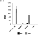
- MIR302CHG which is related to lncRNA, was found to exhibit the value of each transcript (transcripts per million: TPM value) per million total transcripts, which was higher than that of LIN28A, which is used for conventional detection of residual iPS cells, and CUZD1, which is found to be differentially highly expressed in human iPSCs (data omitted) also in the analysis of deposited RNA-seq data ( Fig. 2 ).
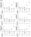
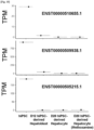
- MIR302CHG was found to exhibit highly differential high expression thereof in iPSCs compared to not only that in NPCs after differentiation, but also that in cells on day 4 (D4C) of the NPC differentiation protocols ( Fig. 3 ). Furthermore, all three splicing variants of MIR302CHG, ENST00000510655.1 (variant 655), ENST00000509938.1 (variant 938), and ENST00000505215.1 (variant 215), exhibited highly differential high expression in the above iPSCs. ( Fig. 4 ).
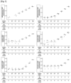
- MIR302CHG Since lncRNA expressed from MIR302CHG has a poly(A) 3' tail, reverse transcription (RT) reaction using oligo dT can be performed. Therefore, MIR302CHG, CUZD1, and pluripotency marker POU5F1 were analyzed by quantitative RT-PCR analysis using the mixtures of cDNAs derived from human iPSCs and NPCs or cells on day 4 of differentiation (D4C), which had been prepared by mixing them at 100% to 10 -5 % in increments of 10 -1 .
- D4C day 4 of differentiation
- the TBP-normalized MIR302CHG expression pattern was better at detecting iPSCs than the CUZD1 and POU5F1 expression patterns when the proportion of iPSCs was low. It was suggested that through analysis of the expression of MIR302CHG by quantitative RT-PCR, iPSC contamination of approximately 10 -3 % (1 cell in 10 5 cells) in NPC and 10 -1 (1 cell in 10 3 cells) in D4C can be detected.
- iPSCs exhibited significantly higher comparative Ct (dCt) values compared to NPCs.
- iPSCs accounting for 10 -4 % of the total cell count were mixed with a cell population of nephron progenitor cells (NPCs) to yield 10 -4 % iPSCs.
- NPCs nephron progenitor cells
- 10 -4 % iPSCs were subjected to magnetic bead-based cell enrichment using TRA-1-60 surface antigen as an indicator, thereby collecting TRA-1-60 positive fractions (PF) and negative fractions (NF).
- MIR302CHG, CUZD1, and POU5F1 were analyzed for amplifiability by quantitative RT-PCR in each compartment of NPC, iPSC, 10 -4 % iPSC, TRA-1-60 PF, and TRA-1-60 NF.
- MIR302CHG primers of the combination of SEQ ID NOS: 6 and 7 (corresponding to #2 in Table 1 above) were used.
- Fig. 7 depicts the results.
- MIR302CHG was subjected to analysis by quantitative RT-PCR using the mixtures of cDNAs derived from human iPSCs and NPCs, which had been prepared by mixing them at 100% to 10 -5 % in increments of 10 -1 . Note that primers of the combination of SEQ ID NOS: 34 and 35 targeting variant 938 expressed from MIR302CHG and the probe of SEQ ID NO: 36 were used. Fig. 8 depicts the results.
- RNA-seq datasets The expression of MIR302CHG in human iPSC-derived pancreatic progenitor cells and liver organoids was analyzed based on publicly available RNA-seq datasets.
- Fig. 9 depicts the TPM value of each transcript variant of MIR302CHG as analyzed using each publicly available RNA-seq dataset of human iPSCs or human iPSC-derived pancreatic progenitor cells reported in Kimura et al., (2020), Cell Chem. Biol. 27, 1561-1572 .
- Fig. 10 depicts the same as analyzed using each publicly available RNA-seq dataset of human iPSCs or human iPSC-derived hepatocytes reported in Kotaka, M. et al., 2017, Sci. Rep. 7, 1-13 .
- RNA-seq datasets The expression of MIR302CHG in human iPSC-derived neural progenitor cells and neurons was analyzed based on publicly available RNA-seq datasets.
- Fig. 11 depicts the TPM value of each transcript variant of MIR302CHG as analyzed using the published RNA-seq dataset of human iPSCs or human iPSC-derived neural progenitor cells and neurons reported in Chen et al., 2013, PLoS One. 2013;8: e75682. doi: 10. 1371/journal.pone.0075682 .
- MIR302CHG The high expression of MIR302CHG was observed in iPSCs, but the expression almost disappeared due to differentiation. Therefore, undifferentiated cells can be detected using MIR302CHG even during differentiation into the neuronal lineage.
- RNA-seq datasets The expression of MIR302CHG in human iPSC-derived cardiomyocytes was analyzed based on publicly available RNA-seq datasets.
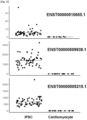
- Fig. 12 depicts the TPM value of each transcript variant of MIR302CHG as analyzed using the publicly available RNA-seq datasets of human iPSCs or human iPSC-derived cardiomyocytes reported in Banovich et al., 2018., Genome Res. 2018; 28: 122-131. doi:10.1101/gr.224436.117 .
- MIR302CHG The high expression of MIR302CHG was observed in iPSCs, but the expression almost disappeared due to differentiation. Therefore, undifferentiated cells can be detected using MIR302CHG even during differentiation into the myocardial lineage.
- RNA-seq datasets The expression of MIR302CHG in human ES cell-derived lung progenitor cells or lung organoids was analyzed based on publicly available RNA-seq datasets.
- Fig. 13 depicts the TPM value of each transcript variant of MIR302CHG as analyzed using the publicly available RNA-seq datasets of human ESCs, human ESC-derived lung progenitor cells, or lung organoids reported in Jacob A et al., Cell Stem Cell. 2017; 21: 472-488.e10. doi:10.1016/j.stem.2017.08.014 .
- MIR302CHG The high expression of MIR302CHG was observed in iPSCs, but the expression almost disappeared due to differentiation. Therefore, undifferentiated cells can be detected using MIR302CHG even during differentiation into pneumocytes.
- MIR302CHG in human iPSCs, IVF-ESCs, or nt-ESC-derived vascular endothelial cells
- RNA-seq datasets The expression of MIR302CHG in human iPSCs, IVF-ESCs, or nt-ESCs and vascular endothelial cells derived therefrom was analyzed based on publicly available RNA-seq datasets.
- Fig. 14 depicts the TPM value of each transcript variant of MIR302CHG as analyzed using the publicly available RNA-seq datasets of human iPSCs, IVF-ESCs, nt-ESCs or human iPSCs, IVF-ESCs, and nt-ESC-derived vascular endothelial cells reported in Zhao et. al., Proc Natl Acad Sci U.S.A. 2017;114: E11111-E11120 .
- MIR302CHG The high expression of MIR302CHG was observed in iPSCs, but the expression almost disappeared due to differentiation. Therefore, undifferentiated cells can be detected using MIR302CHG even during differentiation into vascular endothelial cells.
- MIR302CHG Expression of MIR302CHG in human ESC, anterior primitive streak, definitive endoderm, anterior foregut, posterior foregut, and midgut/hindgut
- MIR302CHG The expression of MIR302CHG in human ESCs, and anterior primitive streak, definitive endoderm, anterior foregut, posterior foregut and midgut/hindgut induced from the human ESCs was analyzed. As a result, as depicted in Fig. 15 , it was found that MIR302CHG was expressed in ESCs, anterior primitive streak, and definitive endoderm, but the expression levels decreased in anterior foregut, posterior foregut, and midgut/hindgut.
- MIR302CHG by measuring the amount of MIR302CHG, it is possible to detect not only undifferentiated pluripotent stem cells but also cells in the early stages of differentiation such as definitive endoderm and anterior primitive streak, and to distinguish such cells from cells in the later stages of differentiation, such as cells that arise at the foregut, the midgut, and the hindgut stages or later.
- renal interstitial progenitor cells IPCs
- ND ureteroblasts
- the measured values were normalized with the housekeeping gene TBP (MIR302CHG/TBP).
- Fig. 16 depicts the results.
- the expression level of MIR302CHG was so low such that it was not detected. Even in ND, the expression level of MIR302CHG was as extremely low as approximately 1/10,000 compared to that in iPSCs. In other words, the expression of MIR302CHG, the high-level expression of which was observed in iPSCs, was hardly observed even after differentiation into renal interstitial progenitor cells or ureteroblasts. Therefore, undifferentiated cells can be detected by MIR302CHG even during differentiation into renal interstitial progenitor cells and differentiation into ureteroblasts.
- undifferentiated pluripotent stem cells remaining in a cell population containing nephron progenitor cells obtained by differentiation induction from pluripotent stem cells can be detected. This makes it possible to assess the safety of products for regenerative medicine and others (for cell therapy, transplant organ reconstruction, etc.) containing nephron progenitor cells derived from human iPS cells.
- the present invention is also applicable to products for regenerative medicine and others produced using other differentiated cells other than human iPS cell-derived nephron progenitor cells, such as pancreatic progenitor cells, hepatic progenitor cells, ureteroblasts, interstitial progenitor cells (e.g., renal interstitial progenitor cells), vascular endothelial cells, chondrocytes, bile duct progenitor cells, erythropoietin-producing cells, neurons, cardiomyocytes, and pneumocytes.
- differentiated cells other than human iPS cell-derived nephron progenitor cells
- pancreatic progenitor cells such as pancreatic progenitor cells, hepatic progenitor cells, ureteroblasts, interstitial progenitor cells (e.g., renal interstitial progenitor cells), vascular endothelial cells, chondrocytes, bile duct pro
Landscapes
- Life Sciences & Earth Sciences (AREA)
- Chemical & Material Sciences (AREA)
- Proteomics, Peptides & Aminoacids (AREA)
- Health & Medical Sciences (AREA)
- Organic Chemistry (AREA)
- Zoology (AREA)
- Analytical Chemistry (AREA)
- Wood Science & Technology (AREA)
- Immunology (AREA)
- Engineering & Computer Science (AREA)
- Biotechnology (AREA)
- Microbiology (AREA)
- Molecular Biology (AREA)
- Biophysics (AREA)
- Physics & Mathematics (AREA)
- Cell Biology (AREA)
- Biochemistry (AREA)
- Bioinformatics & Cheminformatics (AREA)
- General Engineering & Computer Science (AREA)
- General Health & Medical Sciences (AREA)
- Genetics & Genomics (AREA)
- Measuring Or Testing Involving Enzymes Or Micro-Organisms (AREA)
Applications Claiming Priority (2)
| Application Number | Priority Date | Filing Date | Title |
|---|---|---|---|
| JP2022094396 | 2022-06-10 | ||
| PCT/JP2023/021545 WO2023238932A1 (ja) | 2022-06-10 | 2023-06-09 | 未分化多能性幹細胞の検出方法および検出試薬 |
Publications (1)
| Publication Number | Publication Date |
|---|---|
| EP4538384A1 true EP4538384A1 (en) | 2025-04-16 |
Family
ID=89118426
Family Applications (1)
| Application Number | Title | Priority Date | Filing Date |
|---|---|---|---|
| EP23819912.9A Pending EP4538384A1 (en) | 2022-06-10 | 2023-06-09 | Detection method and detection reagent for undifferentiated pluripotent stem cells |
Country Status (5)
| Country | Link |
|---|---|
| US (1) | US20250376727A1 (https=) |
| EP (1) | EP4538384A1 (https=) |
| JP (1) | JPWO2023238932A1 (https=) |
| CN (1) | CN119365606A (https=) |
| WO (1) | WO2023238932A1 (https=) |
Families Citing this family (1)
| Publication number | Priority date | Publication date | Assignee | Title |
|---|---|---|---|---|
| CN119979719B (zh) * | 2025-02-26 | 2026-01-13 | 中国食品药品检定研究院 | 一种基于基因的多能干细胞残留通用型检测方法 |
Family Cites Families (36)
| Publication number | Priority date | Publication date | Assignee | Title |
|---|---|---|---|---|
| US9453219B2 (en) | 2003-05-15 | 2016-09-27 | Mello Biotech Taiwan Co., Ltd. | Cosmetic designs and products using intronic RNA |
| US8048999B2 (en) | 2005-12-13 | 2011-11-01 | Kyoto University | Nuclear reprogramming factor |
| JP5813321B2 (ja) | 2007-03-23 | 2015-11-17 | ウィスコンシン アラムニ リサーチ ファンデーション | 体細胞の再プログラム化 |
| JP2008307007A (ja) | 2007-06-15 | 2008-12-25 | Bayer Schering Pharma Ag | 出生後のヒト組織由来未分化幹細胞から誘導したヒト多能性幹細胞 |
| ES2799897T3 (es) | 2007-08-31 | 2020-12-22 | Whitehead Inst Biomedical Res | Estimulación de la vía wnt en la reprogramación de células somáticas |
| KR101564044B1 (ko) | 2007-10-31 | 2015-10-28 | 고쿠리츠 다이가쿠 호진 교토 다이가쿠 | 핵초기화 방법 |
| JP5558097B2 (ja) | 2007-12-10 | 2014-07-23 | 国立大学法人京都大学 | 効率的な核初期化方法 |
| WO2009079007A1 (en) | 2007-12-17 | 2009-06-25 | Gliamed, Inc. | Stem-like cells and method for reprogramming adult mammalian somatic cells |
| WO2009091659A2 (en) | 2008-01-16 | 2009-07-23 | Shi-Lung Lin | Generation of tumor-free embryonic stem-like pluripotent cells using inducible recombinant rna agents |
| US20100330677A1 (en) | 2008-02-11 | 2010-12-30 | Cambridge Enterprise Limited | Improved Reprogramming of Mammalian Cells, and Cells Obtained |
| EP2090649A1 (en) | 2008-02-13 | 2009-08-19 | Fondazione Telethon | Method for reprogramming differentiated cells |
| WO2009102983A2 (en) | 2008-02-15 | 2009-08-20 | President And Fellows Of Harvard College | Efficient induction of pluripotent stem cells using small molecule compounds |
| MX2010010165A (es) | 2008-03-17 | 2010-11-25 | Scripps Research Inst | Procedimientos quimicos y geneticos combinados para generacion de celulas madre pluripotentes inducidas. |
| WO2009114949A1 (en) | 2008-03-20 | 2009-09-24 | UNIVERSITé LAVAL | Methods for deprogramming somatic cells and uses thereof |
| KR20110019727A (ko) | 2008-04-07 | 2011-02-28 | 뉴포텐셜, 인크. | Rna 간섭을 통한 다능 유전자의 유도에 의한 세포 재프로그래밍 |
| CA2695590C (en) | 2008-06-27 | 2018-01-09 | Kyoto University | Method of efficiently establishing induced pluripotent stem cells |
| US9453204B2 (en) | 2008-07-14 | 2016-09-27 | Oklahoma Medical Research Foundation | Production of pluripotent cells through inhibition of bright/ARID3a function |
| WO2010147612A1 (en) | 2009-06-18 | 2010-12-23 | Lixte Biotechnology, Inc. | Methods of modulating cell regulation by inhibiting p53 |
| US20120021519A1 (en) | 2008-09-19 | 2012-01-26 | Presidents And Fellows Of Harvard College | Efficient induction of pluripotent stem cells using small molecule compounds |
| WO2010033920A2 (en) | 2008-09-19 | 2010-03-25 | Whitehead Institute For Biomedical Research | Compositions and methods for enhancing cell reprogramming |
| WO2010042800A1 (en) | 2008-10-10 | 2010-04-15 | Nevada Cancer Institute | Methods of reprogramming somatic cells and methods of use for such cells |
| JP2012507258A (ja) | 2008-10-30 | 2012-03-29 | 国立大学法人京都大学 | 人工多能性幹細胞の作製方法 |
| US20110059526A1 (en) | 2008-11-12 | 2011-03-10 | Nupotential, Inc. | Reprogramming a cell by inducing a pluripotent gene through use of an hdac modulator |
| EP2376626A4 (en) | 2008-12-13 | 2012-10-17 | Dna Microarray | MICRO-ENVIRONMENTAL NICHE ASSAY FOR SCREENING OF INDUCED PLURIPOTENT STEM CELLS (CIPS) |
| WO2010098419A1 (en) | 2009-02-27 | 2010-09-02 | Kyoto University | Novel nuclear reprogramming substance |
| WO2010102267A2 (en) | 2009-03-06 | 2010-09-10 | Ipierian, Inc. | Tgf-beta pathway inhibitors for enhancement of cellular reprogramming of human cells |
| WO2010111409A2 (en) | 2009-03-25 | 2010-09-30 | The Salk Institute For Biological Studies | Pluripotent stem cells |
| US20120076762A1 (en) | 2009-03-25 | 2012-03-29 | The Salk Institute For Biological Studies | Induced pluripotent stem cell generation using two factors and p53 inactivation |
| WO2010115050A2 (en) | 2009-04-01 | 2010-10-07 | The Regents Of The University Of California | Embryonic stem cell specific micrornas promote induced pluripotency |
| WO2010124290A2 (en) | 2009-04-24 | 2010-10-28 | Whitehead Institute For Biomedical Research | Compositions and methods for deriving or culturing pluripotent cells |
| WO2010147395A2 (en) | 2009-06-16 | 2010-12-23 | Korea Research Institute Of Bioscience And Biotechnology | Medium composition comprising neuropeptide y for the generation, maintenance, prologned undifferentiated growth of pluripotent stem cells and method of culturing pluripotent stem cell using the same |
| WO2011133288A1 (en) * | 2010-04-19 | 2011-10-27 | The Trustees Of The University Of Pennsylvania | Microrna induction of pluripotential stem cells and uses thereof |
| JP2013158325A (ja) | 2012-02-08 | 2013-08-19 | Hokkaido Univ | 細胞診断方法 |
| US10711249B2 (en) | 2014-12-26 | 2020-07-14 | Kyoto University | Method for inducing hepatocytes |
| WO2020213734A1 (ja) | 2019-04-19 | 2020-10-22 | 国立大学法人京都大学 | ネフロン前駆細胞の製造方法 |
| EP4386083A4 (en) | 2021-08-11 | 2025-11-12 | Univ Kyoto | METHOD FOR PRODUCING RENAL INTERSTITIAL PROGENITOR CELLS, ERYTHROPOIETIN-PRODUCING CELLS AND METHOD FOR PRODUCING RENIIN-PRODUCING CELLS |
-
2023
- 2023-06-09 CN CN202380045888.8A patent/CN119365606A/zh active Pending
- 2023-06-09 WO PCT/JP2023/021545 patent/WO2023238932A1/ja not_active Ceased
- 2023-06-09 JP JP2024527032A patent/JPWO2023238932A1/ja active Pending
- 2023-06-09 EP EP23819912.9A patent/EP4538384A1/en active Pending
- 2023-06-09 US US18/873,491 patent/US20250376727A1/en active Pending
Also Published As
| Publication number | Publication date |
|---|---|
| JPWO2023238932A1 (https=) | 2023-12-14 |
| WO2023238932A1 (ja) | 2023-12-14 |
| US20250376727A1 (en) | 2025-12-11 |
| CN119365606A (zh) | 2025-01-24 |
Similar Documents
| Publication | Publication Date | Title |
|---|---|---|
| Freeman et al. | Single-cell RNA-Seq of bone marrow-derived mesenchymal stem cells reveals unique profiles of lineage priming | |
| KR102784455B1 (ko) | 재프로그래밍 벡터 | |
| US8852941B2 (en) | Method for producing induced pluripotent stem cells | |
| HK1258309A1 (zh) | 用於确定候选药物的功效特徵的方法 | |
| CA2417356A1 (en) | Directed differentiation of embryonic cells | |
| WO2013188679A1 (en) | Methods of preparing pluripotent stem cells | |
| EP3121276B1 (en) | Cardiomyocyte sorting method | |
| US20220401493A1 (en) | Isolation of fusion-competent myoblasts and therapeutic applications thereof related to muscular dystrophy | |
| EP2524033B1 (en) | Method for screening induced pluripotent stem cells | |
| JP7648190B2 (ja) | 成熟心筋細胞の製造法 | |
| EP3170893B1 (en) | Method for extracting differentiated cells | |
| US20230340595A1 (en) | Use of pluripotent markers to detect contaminating residual undifferentiated pluripotent stem cells | |
| US20200216813A1 (en) | Cell reprogramming to cardiomyocytes | |
| EP4538384A1 (en) | Detection method and detection reagent for undifferentiated pluripotent stem cells | |
| WO2018037091A1 (en) | Methods for the identification and isolation of hematopoietic stem and progenitor cells | |
| Harkness et al. | Molecular characterisation of stromal populations derived from human embryonic stem cells: Similarities to immortalised bone marrow derived stromal stem cells | |
| WO2023118050A1 (en) | Use of novel markers to detect pluripotent stem cells | |
| KR101272901B1 (ko) | 체세포를 신경줄기세포로의 직접 리프로그래밍 방법 및 그 조성물 | |
| Rebs | Analysis of splice-defect associated cardiac diseases using a patient-specific iPSC-cardiomyocyte system | |
| CN113646424A (zh) | 具有分化成特定细胞的能力的多能干细胞的制造方法及其应用 | |
| WO2012029316A1 (en) | Method for screening differentiation-responsive induced pluripotent stem cell | |
| Franzen | DNA methylation changes upon replicative senescence of mesenchymal stromal cells | |
| EP3748012A1 (en) | Method for evaluating state of undifferentiated cell and utilization thereof | |
| CN115505573A (zh) | 一种迁移活力增强的基因工程间充质干细胞及在干细胞治疗、干细胞美容中的应用 | |
| Wesseling et al. | Reinhard Roessler, Sebastien A. Smallwood, 2, 7 Jesse V. Veenvliet, 3, 7 Petros Pechlivanoglou, 4, 9 Su-Ping Peng, Koushik Chakrabarty, 6 Marian JA Groot-Koerkamp, 5 R. Jeroen Pasterkamp, 6 |
Legal Events
| Date | Code | Title | Description |
|---|---|---|---|
| STAA | Information on the status of an ep patent application or granted ep patent |
Free format text: STATUS: THE INTERNATIONAL PUBLICATION HAS BEEN MADE |
|
| PUAI | Public reference made under article 153(3) epc to a published international application that has entered the european phase |
Free format text: ORIGINAL CODE: 0009012 |
|
| STAA | Information on the status of an ep patent application or granted ep patent |
Free format text: STATUS: REQUEST FOR EXAMINATION WAS MADE |
|
| 17P | Request for examination filed |
Effective date: 20250109 |
|
| AK | Designated contracting states |
Kind code of ref document: A1 Designated state(s): AL AT BE BG CH CY CZ DE DK EE ES FI FR GB GR HR HU IE IS IT LI LT LU LV MC ME MK MT NL NO PL PT RO RS SE SI SK SM TR |
|
| DAV | Request for validation of the european patent (deleted) | ||
| DAX | Request for extension of the european patent (deleted) |