EP4534972A2 - Synthetische kontrollen für immunhistochemie - Google Patents
Synthetische kontrollen für immunhistochemie Download PDFInfo
- Publication number
- EP4534972A2 EP4534972A2 EP25156644.4A EP25156644A EP4534972A2 EP 4534972 A2 EP4534972 A2 EP 4534972A2 EP 25156644 A EP25156644 A EP 25156644A EP 4534972 A2 EP4534972 A2 EP 4534972A2
- Authority
- EP
- European Patent Office
- Prior art keywords
- carrier protein
- antigen
- gel
- solid
- target
- Prior art date
- Legal status (The legal status is an assumption and is not a legal conclusion. Google has not performed a legal analysis and makes no representation as to the accuracy of the status listed.)
- Pending
Links
Images













Classifications
-
- G—PHYSICS
- G01—MEASURING; TESTING
- G01N—INVESTIGATING OR ANALYSING MATERIALS BY DETERMINING THEIR CHEMICAL OR PHYSICAL PROPERTIES
- G01N33/00—Investigating or analysing materials by specific methods not covered by groups G01N1/00 - G01N31/00
- G01N33/48—Biological material, e.g. blood, urine; Haemocytometers
- G01N33/50—Chemical analysis of biological material, e.g. blood, urine; Testing involving biospecific ligand binding methods; Immunological testing
- G01N33/53—Immunoassay; Biospecific binding assay; Materials therefor
- G01N33/543—Immunoassay; Biospecific binding assay; Materials therefor with an insoluble carrier for immobilising immunochemicals
-
- G—PHYSICS
- G01—MEASURING; TESTING
- G01N—INVESTIGATING OR ANALYSING MATERIALS BY DETERMINING THEIR CHEMICAL OR PHYSICAL PROPERTIES
- G01N1/00—Sampling; Preparing specimens for investigation
- G01N1/28—Preparing specimens for investigation including physical details of (bio-)chemical methods covered elsewhere, e.g. G01N33/50, C12Q
-
- G—PHYSICS
- G01—MEASURING; TESTING
- G01N—INVESTIGATING OR ANALYSING MATERIALS BY DETERMINING THEIR CHEMICAL OR PHYSICAL PROPERTIES
- G01N1/00—Sampling; Preparing specimens for investigation
- G01N1/28—Preparing specimens for investigation including physical details of (bio-)chemical methods covered elsewhere, e.g. G01N33/50, C12Q
- G01N1/30—Staining; Impregnating ; Fixation; Dehydration; Multistep processes for preparing samples of tissue, cell or nucleic acid material and the like for analysis
-
- G—PHYSICS
- G01—MEASURING; TESTING
- G01N—INVESTIGATING OR ANALYSING MATERIALS BY DETERMINING THEIR CHEMICAL OR PHYSICAL PROPERTIES
- G01N1/00—Sampling; Preparing specimens for investigation
- G01N1/28—Preparing specimens for investigation including physical details of (bio-)chemical methods covered elsewhere, e.g. G01N33/50, C12Q
- G01N1/36—Embedding or analogous mounting of samples
-
- G—PHYSICS
- G01—MEASURING; TESTING
- G01N—INVESTIGATING OR ANALYSING MATERIALS BY DETERMINING THEIR CHEMICAL OR PHYSICAL PROPERTIES
- G01N1/00—Sampling; Preparing specimens for investigation
- G01N1/28—Preparing specimens for investigation including physical details of (bio-)chemical methods covered elsewhere, e.g. G01N33/50, C12Q
- G01N2001/2893—Preparing calibration standards
-
- G—PHYSICS
- G01—MEASURING; TESTING
- G01N—INVESTIGATING OR ANALYSING MATERIALS BY DETERMINING THEIR CHEMICAL OR PHYSICAL PROPERTIES
- G01N2333/00—Assays involving biological materials from specific organisms or of a specific nature
- G01N2333/435—Assays involving biological materials from specific organisms or of a specific nature from animals; from humans
- G01N2333/76—Assays involving albumins other than in routine use for blocking surfaces or for anchoring haptens during immunisation
-
- G—PHYSICS
- G01—MEASURING; TESTING
- G01N—INVESTIGATING OR ANALYSING MATERIALS BY DETERMINING THEIR CHEMICAL OR PHYSICAL PROPERTIES
- G01N2333/00—Assays involving biological materials from specific organisms or of a specific nature
- G01N2333/435—Assays involving biological materials from specific organisms or of a specific nature from animals; from humans
- G01N2333/76—Assays involving albumins other than in routine use for blocking surfaces or for anchoring haptens during immunisation
- G01N2333/765—Serum albumin, e.g. HSA
-
- G—PHYSICS
- G01—MEASURING; TESTING
- G01N—INVESTIGATING OR ANALYSING MATERIALS BY DETERMINING THEIR CHEMICAL OR PHYSICAL PROPERTIES
- G01N2496/00—Reference solutions for assays of biological material
- G01N2496/25—Reference solutions for assays of biological material containing added polymers to stabilise biological material against degradation or maintain viscosity or density, e.g. gelatin, polyacrylamides or polyvinyl alcohol
Definitions
- the present disclosure relates to synthetic controls useful, e.g., for immunohistochemistry (IHC), as well as methods of manufacture and uses related thereto.
- the synthetic controls include a solid gel comprising a purified antigen (e.g., at a known quantity) cross-linked to a carrier protein such as a serum albumin protein, an egg white protein or a mixture of egg white proteins, or gelatin.
- IHC represents an important tool for both research and clinical applications. Among the many techniques used to characterize protein expression, IHC is one of the few that provides information on expression level as well as localization, e.g., at the cellular and/or tissue levels. IHC is therefore a critical tool used in research to characterize a protein of interest. IHC has also become an important diagnostic tool in the clinic, e.g., to categorize patients for a variety of personalized medicine applications. As one example, the HercepTest TM (Dako Denmark A/S) semi-quantitative HER2 IHC assay has been approved by the FDA for use in assessing HER2 protein status for almost 20 years. This test allows clinicians to identify patients whose tumors overexpress HER2.
- HER2 is overexpressed in many types of cancer, 25-30% of breast cancers have been shown to overexpress HER2, and this marker is correlated with shortened disease-free and overall survival.
- Therapies that target HER2 in these patients e.g., anti-HER2 antibody treatment
- provide significantly improved overall survival, response rate, duration of response, and time to disease progression See, e.g., Slamon, D.J. et al. (2001) N Engl J Med 344:783-792 .
- Tissue samples and cell lines have both been used as IHC controls; however, both have significant limitations. For many targets, appropriate tissues are not available. For others, expression in tissues may be variable, uncharacterized, or too weak to detect. Cell lines are easier to obtain than tissue samples (although growing certain cell lines on an industrial scale can be difficult), but like expression in tissue samples, expression in cell lines can be variable, uncharacterized, or too weak to detect.
- Sompuram, S.R. et al. (2002) Clin Chem 48:410-420 describe spotting peptides directly to glass slides, or coupling peptides to glass beads (see also Sompuram et al. (2015) J. Histochem Cytochem 63:681-690 ).
- implementation of this approach led to technical challenges. For example, since it was difficult for technicians to see the spots on which the peptides were applied to the glass (they are invisible prior to staining), many slides were stained with insufficient amounts of reagent to cover all of the controls, thereby leading to artifacts in staining ( see Bogen, S.A. et al. (2009) Appl Immunohistochem Mol Morphol 17:239-246 ).
- the peptides dispersed in agarose were not homogeneous.
- a solid antigen/carrier protein gel can be processed like tissue samples or other biological samples according to standard IHC or electron microscopy (EM) processing methods (including fixation, sectioning, antigen retrieval, and so forth). Since the gels contain a known amount of an antigen of interest, they can be used, e.g., to create a series of gels having known concentrations of antigen to standardize staining with a particular antibody, or to screen for antibodies that specifically recognize an antigen of interest and are suitable for IHC/ EM analyses. These methods are thought to provide a general platform that allows for control staining of any antigen of interest, simulating multiple levels of expression using known concentrations of antigen.
- Certain aspects of the present disclosure relate to methods of generating a solid antigen/carrier protein gel (e.g., for IHC or EM analysis) comprising: (a) mixing a purified antigen with a liquid solution comprising a carrier protein selected from the group consisting of an albumin protein (e.g., a serum albumin protein), an egg white protein or a mixture of egg white proteins, and gelatin to produce an antigen/carrier protein liquid solution; and (b) heating the antigen/carrier protein liquid solution to form the solid antigen/carrier protein gel.
- a carrier protein selected from the group consisting of an albumin protein (e.g., a serum albumin protein), an egg white protein or a mixture of egg white proteins, and gelatin to produce an antigen/carrier protein liquid solution
- an albumin protein e.g., a serum albumin protein
- an egg white protein or a mixture of egg white proteins e.g., a mixture of egg white proteins
- the methods comprise: (a) mixing a purified antigen with a liquid solution comprising a carrier protein selected from the group consisting of an albumin protein (e.g., a serum albumin protein), an egg white protein or a mixture of egg white proteins, gelatin, and poly-lysine to produce an antigen/carrier protein liquid solution; and (b) heating the antigen/carrier protein liquid solution to form the solid antigen/carrier protein gel.
- the methods further comprise, after (b): dehydrating the solid antigen/carrier protein gel and embedding the dehydrated solid antigen/carrier protein gel in a paraffin block.
- the methods further comprise: prior to (b), including a fixative in the antigen/carrier protein liquid solution; after (b), dehydrating the solid antigen/carrier protein gel; embedding the dehydrated solid antigen/carrier protein gel in a paraffin block; sectioning the paraffin block with the embedded antigen/carrier protein gel into one or more sections having a thickness of between about 30nm and about 50 ⁇ m; subjecting the one or more sections of the embedded solid antigen/ carrier protein gel to antigen retrieval; and after antigen retrieval, blocking the one or more sections of the embedded solid antigen/carrier protein gel.
- Certain aspects of the present disclosure relate to methods for immunohistochemical (IHC) staining of an antigen, comprising: providing a solid antigen/carrier protein gel produced by the method of any one of the above embodiments, wherein the solid antigen/carrier protein gel contains the antigen; providing a sample; contacting the solid antigen/carrier protein gel and the sample with a primary antibody that specifically binds the antigen; after contacting the solid antigen/carrier protein gel and the sample with the primary antibody, contacting the solid antigen/carrier protein gel and the sample with a secondary antibody that specifically binds the primary antibody, wherein a detectable moiety is conjugated to the secondary antibody; detecting a signal of the detectable moiety from the solid antigen/carrier protein gel; and detecting a signal of the detectable moiety from the sample, wherein detection of a signal from the sample as compared to the signal detected from the solid antigen/carrier protein gel indicates the presence of the antigen in the sample.
- IHC immunohistochemical
- Certain aspects of the present disclosure relate to methods for control immunohistochemical (IHC) staining of an antigen, comprising: providing a first and a second solid antigen/carrier protein gel, wherein each of the first and the second solid antigen/carrier protein gels is produced by the method of any one of the above embodiments, wherein the first solid antigen/carrier protein gel contains the antigen at a first concentration, and wherein the second solid antigen/carrier protein gel contains the antigen at a second concentration higher than the first concentration; contacting the first and the second solid antigen/carrier protein gels with a primary antibody that specifically binds the antigen; after contacting the first and the second solid antigen/carrier protein gels with the primary antibody, contacting the first and the second solid antigen/carrier protein gels with a secondary antibody that specifically binds the primary antibody, wherein a detectable moiety is conjugated to the secondary antibody; detecting a first signal of the detectable moiety from the first solid antigen/carrier protein gel; and detecting
- the detectable moiety comprises an enzyme, and wherein detecting the signal of the detectable moiety comprises exposing the detectable moiety to a chromogenic, fluorescent or chemiluminescent substrate of the enzyme and detecting a signal from the substrate upon reaction with the enzyme.
- the detectable moiety comprises a fluorophore, metal particle, metal ion, radioisotope, nucleic acid, electrochemiluminescent reporter, or a "quantum dot" (a solid state, semiconductor or carbon-based fluorescent nanoparticle).
- the first concentration is 0nM, and wherein detection of a lack of a first signal indicates control IHC staining of the antigen.
- the methods further comprise: providing a sample; contacting the sample with the primary antibody; after contacting the sample with the primary antibody, contacting the sample with the secondary antibody, wherein a detectable moiety is conjugated to the secondary antibody; detecting a third signal of the detectable moiety from the sample; and comparing the third signal with the first and the second signals, wherein an amount of the third signal relative to the amounts of the first and the second signals indicates an abundance of the antigen in the sample, relative to the amounts of the antigen in the first and the second solid antigen/carrier protein gels.
- Certain aspects of the present disclosure relate to methods for control immunohistochemical (IHC) staining with a secondary antibody, comprising: providing a first and a second solid antigen/carrier protein gel, wherein each of the first and the second solid antigen/carrier protein gels is produced by the method of any one of the above embodiments, wherein the first solid antigen/carrier protein gel comprises a first antibody having a first isotype, and wherein the second solid antigen/carrier protein gel comprises a second antibody having a second isotype different from the first isotype; contacting the first and the second solid antigen/carrier protein gels with a secondary antibody that specifically binds the first isotype, wherein a detectable moiety is conjugated to the secondary antibody; detecting a signal of the detectable moiety from the first solid antigen/carrier protein gel; and detecting a lack of the signal of the detectable moiety from the second solid antigen/carrier protein gel, wherein detection of the signal associated with the first solid antigen/carrier protein gel
- the detectable moiety comprises an enzyme, and wherein detecting the signal of the detectable moiety comprises exposing the detectable moiety to a chromogenic, fluorescent or chemiluminescent substrate of the enzyme and detecting a signal from the substrate upon reaction with the enzyme.
- the detectable moiety comprises a fluorophore, metal particle, metal ion, radioisotope, nucleic acid, electrochemiluminescent reporter, or a "quantum dot" (a solid state, semiconductor or carbon-based fluorescent nanoparticle).
- solid antigen/carrier protein gels comprising a purified antigen cross-linked to a carrier protein selected from the group consisting of an albumin protein (e.g., a serum albumin protein), an egg white protein or a mixture of egg white proteins, and gelatin.
- a carrier protein selected from the group consisting of an albumin protein (e.g., a serum albumin protein), an egg white protein or a mixture of egg white proteins, and gelatin.
- the present disclosure relates to solid antigen/carrier protein gels (e.g., for immunohistochemical (IHC) staining) comprising a purified antigen cross-linked to a carrier protein selected from the group consisting of an albumin protein (e.g ., a serum albumin protein), an egg white protein or a mixture of egg white proteins, gelatin, and poly-lysine.
- a carrier protein selected from the group consisting of an albumin protein (e.g ., a serum albumin protein), an egg white protein or a mixture of egg white proteins, gelatin, and poly-lysine.
- the solid gel has a thickness of between about 30nm and about 50 ⁇ m. In some embodiments, the solid gel has a thickness of between about 2 ⁇ m and about 30 ⁇ m. In some embodiments, the solid gel has a thickness of between about 30nm and about 100nm.
- the antigen is a polypeptide antigen.
- the antigen comprises an N-terminal tyrosine, a C-terminal cysteine, or both.
- the N-terminal tyrosine and/or C-terminal cysteine is cross-linked to the carrier protein.
- the antigen comprises a non-polypeptide antigen.
- the carrier protein is an albumin protein, such as bovine, goat, horse, or human serum albumin.
- the carrier protein is an egg white protein or a mixture of egg white proteins.
- the solid gel comprises the carrier protein at a concentration of greater than or equal to 2%.
- the solid gel comprises the carrier protein at a concentration of less than or equal to about 25%. In some embodiments, the carrier protein is gelatin. In some embodiments, the solid gel comprises the carrier protein at a concentration of greater than or equal to 0.5%. In some embodiments, the solid gel contains the antigen at a concentration of at least about 25nM. In some embodiments, the solid gel comprises poly-lysine at a concentration of about 14 mg/mL.
- tissue microarrays comprising at least a first and a second solid antigen/carrier protein gel, wherein the first and second solid antigen/carrier protein gels both comprise a purified antigen cross-linked to a carrier protein selected from the group consisting of an albumin protein, an egg white protein or mixture of egg white proteins, and gelatin.
- Certain aspects of the present disclosure relate to methods for immunohistochemical (IHC) staining of an antigen, comprising: providing a solid antigen/carrier protein gel according to any one of the above embodiments, wherein the solid antigen/carrier protein gel contains the antigen; providing a sample; contacting the solid antigen/carrier protein gel and the sample with a primary antibody that specifically binds the antigen; after contacting the solid antigen/carrier protein gel and the sample with the primary antibody, contacting the solid antigen/carrier protein gel and the sample with a secondary antibody that specifically binds the primary antibody, wherein a detectable moiety is conjugated to the secondary antibody; detecting a signal of the detectable moiety from the solid antigen/carrier protein gel; and detecting a signal of the detectable moiety from the sample, wherein detection of a signal from the sample as compared to the signal detected from the solid antigen/carrier protein gel indicates the presence of the antigen in the sample.
- IHC immunohistochemical
- the methods further comprise: providing a sample; contacting the sample with the primary antibody; after contacting the sample with the primary antibody, contacting the sample with the secondary antibody; detecting a third signal of the detectable moiety from the sample; and comparing the third signal with the first and the second signals, wherein an amount of the third signal relative to the amounts of the first and the second signals indicates an abundance of the antigen in the sample, relative to the amounts of the antigen in the first and the second solid antigen/carrier protein gels.
- Certain aspects of the present disclosure relate to methods for control immunohistochemical (IHC) staining with a secondary antibody, comprising: providing a first and a second solid antigen/carrier protein gel according to any one of the above embodiments, wherein the first solid antigen/carrier protein gel comprises a first antibody having a first isotype, and wherein the second solid antigen/carrier protein gel comprises a second antibody having a second isotype different from the first isotype; contacting the first and the second solid antigen/carrier protein gels with a secondary antibody that specifically binds the first isotype, wherein a detectable moiety is conjugated to the secondary antibody; detecting a signal of the detectable moiety from the first solid antigen/carrier protein gel; and detecting a lack of the signal of the detectable moiety from the second solid antigen/carrier protein gel, wherein detection of the signal associated with the first solid antigen/carrier protein gel and the lack of the signal associated with the second solid antigen/carrier protein gel indicates control staining
- kits comprise: a first solid gel comprising a purified antigen cross-linked to a carrier protein selected from the group consisting of an albumin protein (e.g., a serum albumin protein), an egg white protein or a mixture of egg white proteins, gelatin, and poly-lysine, wherein the purified antigen is present in the first solid gel at a first concentration; and a second solid gel comprising the purified antigen cross-linked to the carrier protein, wherein the purified antigen is present in the second solid gel at a second concentration different from the first concentration.
- the first and the second solid gels are each sectioned into a thickness of between about 30nm and about 50 ⁇ m.
- antigen herein is used in the broadest sense and encompasses various forms of both polypeptide and non-polypeptide antigens, including, without limitation, small peptide antigens, full-length protein antigens, carbohydrate antigens, lipid antigens, and nucleic acid antigens.
- a “purified” antigen refers to an antigen has been increased in purity, such that it exists in a form that is more pure than it exists in its natural environment and/or when initially produced and/or synthesized and/or amplified under laboratory conditions. Purity is a relative term and does not necessarily mean absolute purity. In some embodiments, the antigen is purified to at least 90%, at least 95%, or at least 99% purity.
- Certain aspects of the present disclosure relate to solid antigen/carrier protein gels, as well as methods for generating a solid antigen/carrier protein gel.
- the present disclosure provides a solid antigen/carrier protein gel comprising a purified antigen and a carrier protein.
- the carrier protein is selected from an albumin protein (e.g ., a serum albumin protein), an egg white protein or a mixture of egg white proteins, gelatin, and poly-lysine.
- the purified antigen is cross-linked to the carrier protein. As described below, these gels may find use, e.g., as a control for IHC staining or EM imaging analyses.
- the carrier protein is an albumin protein.
- the carrier protein is a serum albumin protein.
- the serum albumin protein is a mammalian serum albumin protein. Examples of serum albumin proteins include, without limitation, mouse, rat, Guinea pig, rabbit, porcine, bovine, goat, sheep, horse, and human serum albumin.
- the carrier protein is an egg white protein. In some embodiments, the carrier protein is a mixture of two or more egg white proteins. In some embodiments, the carrier protein is gelatin. In some embodiments, the carrier protein is poly-lysine.
- the solid antigen/carrier protein gel comprises the carrier protein at a concentration of greater than or equal to 0.5%, 1%, 2%, 5%, 7%, 10%, 15%, or 20% (w/v). In some embodiments, the solid antigen/carrier protein gel comprises the carrier protein at a concentration of less than or equal to 25%, 20%, 15%, 10%, 7%, 5%, 2%, or 1% (w/ v).
- the solid antigen/carrier protein gel can comprise the carrier protein at any concentration of a range of concentrations having an upper limit of 25%, 20%, 15%, 10%, 7%, 5%, 2%, or 1% (w/v) and an independently selected lower limit of 0.5%, 1%, 2%, 5%, 7%, 10%, 15%, or 20% (w/v), wherein the lower limit is less than the upper limit.
- the carrier protein comprises an albumin protein (e.g ., a serum albumin protein), and the solid antigen/carrier protein gel comprises the carrier protein at greater than 2% (w/v), e.g., between 2% and 25% (w/v).
- the solid antigen/carrier protein gel contains the antigen at a concentration of at least about 25nM. In another embodiment, the solid antigen/carrier protein gel contains the antigen at a concentration of about 25nM. In yet another embodiment, the solid antigen/carrier protein gel contains the antigen at a concentration of about 15, 20, 25, 30, 35, 40, 50, 60, 75, 80, 90, or 100nM, or more. In another embodiment, the solid antigen/carrier protein gel contains the antigen at a concentration of at least about: 10nM, 15nM, 20nM, or 25nM to about 100nM. As described herein, a solid antigen/carrier protein gel of the present disclosure can comprise the antigen at a wide range of concentrations, depending, e.g., upon the sensitivity of the detection method, desired application of the gel, and so forth.
- cysteine-reactive reagents include, without limitation, sulfhydryl-reactive crosslinker reactive groups such as haloacetyls, maleimides (e.g., sulfo-SMCC and its analogs), aziridines, acryloyls, arylating agents, vinylsulfones, pyridyl disulfides, TNB-thiols, and disulfide reducing agents.
- sulfhydryl-reactive crosslinker reactive groups such as haloacetyls, maleimides (e.g., sulfo-SMCC and its analogs), aziridines, acryloyls, arylating agents, vinylsulfones, pyridyl disulfides, TNB-thiols, and disulfide reducing agents.
- the solid antigen/carrier protein gel has been fixed with a fixative, e.g., as described below.
- the fixative is a cross-linking fixative (e.g., an aldehyde-based fixative).
- the fixative is not a cross-linking fixative (e.g., a precipitating fixative, such as Carnoy's).
- a solid gel containing a carrier protein and an antigen may be prepared by denaturing and precipitating the carrier protein and/or the antigen by heating, or by adding a precipitating fixative to the mixture, causing the antigen to be immobilized in the protein gel.
- cross-linking the antigen to the carrier protein is thought to help to retain the antigen in the gel and in the resulting sections, e.g., during the embedding, sectioning, and/or staining processes.
- a solid antigen/carrier protein gel used for IHC staining may have a thickness, e.g., of between about 2 ⁇ m and about 30 ⁇ m, whereas a solid antigen/carrier protein gel used for EM may have a thickness, e.g., of between about 30nm and about 50 ⁇ m or between about 30nm and about 100nm.
- the solid antigen/carrier protein gel has been subjected to antigen retrieval. Exemplary methods for antigen retrieval are described in greater detail below.
- a method for generating a solid antigen/carrier protein gel comprises mixing a purified antigen with a liquid solution comprising a carrier protein to produce an antigen/carrier protein liquid solution and heating the antigen/carrier protein liquid solution to form the solid antigen/carrier protein gel.
- the carrier protein is selected from an albumin protein (e.g., a serum albumin protein), an egg white protein or a mixture of egg white proteins, and gelatin.
- the antigen/carrier protein liquid solution comprises the carrier protein at a concentration of greater than or equal to 0.5%, 1%, 2%, 5%, 7%, 10%, 15%, or 20% (w/v). In some embodiments, the antigen/carrier protein liquid solution comprises the carrier protein at a concentration of less than or equal to 25%, 20%, 15%, 10%, 7%, 5%, 2%, or 1% (w/v).
- the antigen/carrier protein liquid solution can comprise the carrier protein at any concentration of a range of concentrations having an upper limit of 25%, 20%, 15%, 10%, 7%, 5%, 2%, or 1% (w/v) and an independently selected lower limit of 0.5%, 1%, 2%, 5%, 7%, 10%, 15%, or 20% (w/v), wherein the lower limit is less than the upper limit.
- the carrier protein comprises an albumin protein (e.g., a serum albumin protein), and the antigen/carrier protein liquid solution comprises the carrier protein at greater than 2% (w/v), e.g., between 2% and 25% (w/v).
- the carrier protein comprises an egg white protein or a mixture of egg white proteins
- the antigen/carrier protein liquid solution comprises the carrier protein at greater than 2% (w/v), e.g., between 2% and 25% (w/ v).
- the carrier protein comprises gelatin, and the antigen/carrier protein liquid solution comprises the carrier protein at greater than 0.5% (w/v), e.g., between 0.5% and 25% (w/v) or about 10% (w/v).
- the methods of the present disclosure include ( e.g., prior to heating the antigen/carrier protein liquid solution) cross-linking the antigen with the carrier protein using a cysteine-reactive reagent.
- cysteine-reactive reagents include, without limitation, sulfhydryl-reactive crosslinker reactive groups such as haloacetyls, maleimides (e.g., sulfo-SMCC and its analogs), aziridines, acryloyls, arylating agents, vinylsulfones, pyridyl disulfides, TNB-thiols, and disulfide reducing agents.
- the methods of the present disclosure include fixing the antigen with the carrier protein.
- a fixative is included in the antigen/ carrier protein liquid solution.
- the fixative is a cross-linking fixative.
- the fixative is a non-cross-linking fixative.
- fixatives include, without limitation, formaldehyde, formalin, paraformaldehyde (PFA), glutaraldehyde, Davidson's fixative, Bouin's fixative, Karnovski's fixative (e.g., 1 ⁇ 2 strength Karnovski's fixative), Zenker's solution, Helly's solution, Carnoy's solution, Zinc formalin, neutral-buffered formalin (NBF), Periodate-Lysine-paraformaldehyde (PLP), Zamboni's fixative, dimethyl suberimidate (DMS), acetone, alcohols (e.g., methanol or ethanol), and zinc salts (e.g., zinc acetate, zinc chloride, zinc trifluoroactetate, etc.).
- the fixative comprises formaldehyde (e.g., at a concentration of at least about 1% or at least about 2%).
- the antigen is cross-linked with the carrier protein using selective chemistry, including without limitation azide-alkyne addition.
- selective chemistry is used to cross-link a non-protein antigen to the carrier protein, e.g., a carbohydrate or lipid antigen.
- the methods of the present disclosure include dehydrating the solid antigen/carrier protein gel.
- Compounds suitable for dehydrating are known in the art and can include, e.g., alcohols such as ethanol or methanol.
- the methods of the present disclosure include embedding the solid antigen/carrier protein gel.
- the solid antigen/carrier protein gel is embedded after dehydration.
- the solid antigen/carrier protein gel is dehydrated by exposure to a sequence of increasing or graded alcohols (e.g., a sequence of increasing ethanol concentrations), followed by exchange of alcohols with xylene, and exchange of xylene with paraffin.
- paraffin e.g., embedding in a paraffin block
- epoxy acrylic, or plastic resin
- EPON TM resins methacrylate, LR White resin, Araldite ® , Spurr plastic, LOWICRYL ® , and the like
- synthetic wax blends of paraffin wax and plastic polymer or co-polymer alloys
- polyethylene glycol and GACH embedding medium (Glutaraldehyde-Carbohydrazide).
- the carrier protein comprises poly-lysine, e.g., at a concentration of about 14 mg/mL.
- the methods of the present disclosure include incubating the solid antigen/carrier protein gel in an embedding medium (e.g., a liquid embedding medium).
- an embedding medium e.g., a liquid embedding medium.
- a variety of embedding media are known in the art and can include, without limitation, Optimum Cutting Temperature (OCT) Compound (e.g., Tissue-Tek ® O.C.T. Compound or Tissue-plus ® O.C.T Compound), PELCO ® Cryo-Embedding Compound, PolarStat TM or PolarStat Plus TM Embedding Medium, and Tissue Freezing Medium or TFM TM .
- OCT Optimum Cutting Temperature
- PELCO ® Cryo-Embedding Compound e.g., PolarStat TM or PolarStat Plus TM Embedding Medium
- TFM TM Tissue Freezing Medium
- these embedding media can contain, e.g., water-soluble glycols and resins; the exemplary embedding media OCT Compound includes 5-15% polyvinyl alcohol and 1-10% polyethylene glycol.
- the methods of the present disclosure include freezing the solid antigen/ carrier protein gel (e.g., after incubating the solid antigen/carrier protein gel in an embedding medium).
- the methods of the present disclosure include freezing a solid antigen/carrier protein gel that has not been fixed ( e.g., that does not include a fixative), similar to the preparation of frozen, unfixed fresh tissue samples.
- the one or more solid antigen/ carrier protein gel sections have a thickness of less than or equal to about 50, 48, 46, 44, 42, 40, 38, 36, 34, 32, 30, 28, 26, 24, 22, 20, 18, 16, 14, 12, 10, 8, 6, 4, 2, 1, 0.9, 0.8, 0.7, 0.6, 0.5, 0.4, 0.3, 0.2, or 0.1 ⁇ m.
- the one or more solid antigen/carrier protein gel sections can have a thickness having an upper limit of 50, 48, 46, 44, 42, 40, 38, 36, 34, 32, 30, 28, 26, 24, 22, 20, 18, 16, 14, 12, 10, 8, 6, 4, 2, 1, 0.9, 0.8, 0.7, 0.6, 0.5, 0.4, 0.3, 0.2, or 0.1 ⁇ m and an independently selected lower limit of 0.03, 0.05, 0.1, 0.2, 0.3, 0.4, 0.5, 0.6, 0.7, 0.8, 0.9, 1, 2, 4, 6, 8, 10, 12, 14, 16, 18, 20, 22, 24, 26, 28, 30, 32, 34, 36, 38, 40, 42, 44, 46, or 48 ⁇ m, wherein the lower limit is less than the upper limit.
- subjecting the solid antigen/carrier protein gel to antigen retrieval comprises treatment with one or more proteolytic enzymes (e.g., trypsin, proteinase K, pepsin, ficin, or pronase) or antigen retrieval reagents (e.g ., hydrochloric acid, formic acid, sodium dodecyl sulfate (SDS), citrate buffer, EDTA, citrate-EDTA, Tris, Tris-EDTA, Tris-HCl, Tris-buffered saline, or citraconic anhydride).
- proteolytic enzymes e.g., trypsin, proteinase K, pepsin, ficin, or pronase
- antigen retrieval reagents e.g ., hydrochloric acid, formic acid, sodium dodecyl sulfate (SDS), citrate buffer, EDTA, citrate-EDTA, Tris, Tris-EDTA, Tri
- the methods of the present disclosure include blocking a solid antigen/carrier protein gel.
- blocking e.g., prior to IHC staining
- a variety of suitable blocking reagents are known in the art and can include, without limitation, serum or a protein solution (e.g., comprising a serum albumin protein, gelatin, non-fat dry milk, or the like).
- the methods of the present disclosure include fixing a solid antigen/carrier protein gel (e.g., by including a fixative in the antigen/carrier protein liquid solution), dehydrating the solid antigen/carrier protein gel, embedding the dehydrated solid antigen/carrier protein gel, sectioning the paraffin block with the embedded solid antigen/carrier protein gel into one or more sections, subjecting the one or more sections to antigen retrieval, and blocking the one or more sections after antigen retrieval.
- the methods of the present disclosure include incubating the solid antigen/carrier protein gel in a liquid embedding medium, freezing the solid antigen/carrier protein gel in the embedding medium, sectioning the frozen antigen/carrier protein gel into one or more sections, and blocking the one or more sections.
- the TMA is a TMA section on a histology slide.
- solid antigen/carrier protein gels of the present disclosure are particularly advantageous for use in TMAs, e.g., for providing a range of epitope concentrations and/or types. This range can also be provided adjacent to a tissue section, thereby allowing a convenient reference for quantitative analyses.
- Solid antigen/carrier protein gels may find use in a variety of applications, including but not limited to controls for IHC or EM analysis.
- a method for control immunohistochemical (IHC) staining of an antigen comprises providing multiple solid antigen/carrier protein gels of the present disclosure (e.g., two or more, three or more, four or more, five or more, etc.) representing different concentrations of an antigen of interest.
- the methods can include providing two solid antigen/carrier protein gels made with different concentrations of a purified antigen.
- the methods include contacting the solid antigen/carrier protein gels with a primary antibody that specifically binds the antigen and is coupled to a detectable moiety.
- the methods include contacting the solid antigen/ carrier protein gels with a primary antibody that specifically binds the antigen, then contacting the solid antigen/carrier protein gels with a secondary antibody that specifically binds the primary antigen and is coupled to a detectable moiety.
- the methods further include detecting signal(s) of the detectable moiety from one or more of the multiple solid antigen/carrier protein gels. Amount of signal detected from the multiple solid antigen/carrier protein gels can then be detected and, optionally, compared against amount or concentration of antigen present in each of the multiple solid antigen/carrier protein gels to provide control IHC staining of the antigen at various levels.
- one of the antigen/carrier protein gels contains no antigen (e.g., 0nM), and lack of a detectable signal from IHC staining of this gel indicates negative control or background IHC staining of the sample.
- the methods further include contacting a sample with the primary antibody that specifically binds the antigen and is coupled to a detectable moiety, or with the primary antibody that specifically binds the antigen and the secondary antibody that specifically binds the primary antigen and is coupled to a detectable moiety, and detecting a signal of the detectable moiety from the sample.
- the amount of signal detected from the sample can then be compared against the amount of signal (s) detected from the solid antigen/carrier protein gel(s), e.g., to compare the amount of antigen present in the sample with known concentrations or amounts of antigen present in the solid antigen/carrier protein gel(s).
- a method for control immunohistochemical (IHC) staining with a secondary antibody comprises providing multiple solid antigen/carrier protein gels of the present disclosure (e.g., two or more, three or more, four or more, five or more, etc.) representing different antibody isotypes.
- the methods can include providing two solid antigen/ carrier protein gels made with antibodies representing different antibody isotypes.
- the methods include contacting the solid antigen/carrier protein gels with a secondary antibody that specifically binds one of the antibody isotypes and is conjugated to a detectable moiety.
- signal can be detected from the multiple solid antigen/carrier protein gels.
- a method for immunohistochemical (IHC) staining of an antigen comprises providing one or more solid antigen/carrier protein gels containing the antigen and a sample; contacting the solid antigen/carrier protein gel and the sample with a primary antibody that specifically binds the antigen and is coupled to a detectable moiety, or contacting the solid antigen/carrier protein gel and the sample with a primary antibody that specifically binds the antigen and contacting the solid antigen/carrier protein gel and the sample with a secondary antibody that specifically binds the primary antibody and is coupled to a detectable moiety; detecting a signal of the detectable moiety from the solid antigen/carrier protein gel; and detecting a signal of the detectable moiety from the sample.
- the one or more solid antigen/ carrier protein gels can be used as a positive control for IHC staining of the sample.
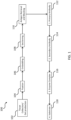
- FIG. 1 An exemplary flow diagram for process 100 for IHC staining of an antigen, such as an antigen in a sample and in a solid antigen/carrier protein gel of the present disclosure (used as a control for staining of the sample for the antigen) is shown in FIG. 1 .
- tissue is collected to provide a sample for analysis, and/or a solid antigen/carrier protein gel of the present disclosure is produced ( e.g., as described in section II above).
- the solid antigen/carrier protein gel contains an antigen (e.g., a purified antigen) of interest, which may or may not also be present in the sample.
- the sample and/or solid antigen/carrier protein gel is fixed ( e.g., as described in section II above).
- the solid antigen/carrier protein gel is incubated with a fixative.
- the antigen/carrier protein liquid solution used to make the solid gel comprises a fixative.
- the sample and/or solid antigen/carrier protein gel are embedded ( e.g., as described in section II above).
- the sample and/or solid antigen/carrier protein gel can be frozen in an embedding medium.
- sample and/or solid antigen/carrier protein gel are sectioned ( e.g., as described in section II above).
- sample and/or solid antigen/carrier protein gel are subjected to antigen retrieval and blocking ( e.g., as described in section II above).
- presence of an antigen in a solid antigen/carrier protein gel of the present disclosure is detected by direct detection, e.g., by incubation of the gel with an antibody or other antigen-binding moiety that specifically binds the antigen and is coupled to a detectable moiety.
- presence of an antigen in a solid antigen/carrier protein gel of the present disclosure is detected by indirect detection, e.g., by incubation of the gel with a primary antibody or other antigen-binding moiety that specifically binds the antigen and a secondary antibody or other antigen-binding moiety that specifically binds the primary antibody or other antigen-binding moiety, where the secondary antibody or other antigen-binding moiety is coupled to a detectable moiety.
- detectable moieties are contemplated for use with a solid antigen/carrier protein gel of the present disclosure.
- the detectable moiety comprises an enzyme, e.g., horseradish peroxidase (HRP) or alkaline phosphatase (AP). Signal is then detected by exposing the detectable moiety to a chromogenic substrate of the enzyme and detecting a signal from the chromogenic substrate upon reaction with the enzyme (e.g., as described above for blocks 116 and 118).
- HRP horseradish peroxidase
- AP alkaline phosphatase
- chromogenic substrates examples include, without limitation, 3,3'-diaminobenzidine (DAB), which is converted to a brown product by HRP; and 3-amino-9-ethylcarbazole (AEC), which is converted to a red product by AP.
- DAB 3,3'-diaminobenzidine
- AEC 3-amino-9-ethylcarbazole
- a peroxidase can chemically activate a tyramide moiety conjugated to one of several reporters (for example a fluorescent molecule); the activated tyramide conjugate can then covalently couple to nearby molecules in the sample, creating a detectable signal in the location of the peroxidase. Indirect detection can also be used with chromogenic staining.
- a biotinylated secondary antibody can be incubated with an avidin- or streptavidin-labeled enzyme complex, a polymer such as a dextran can be coupled with one or more secondary antibodies and one or more enzyme complexes, or an enzyme complex can be polymerized directly onto a secondary antibody.
- the detectable moiety comprises a fluorophore, such as Fluorescein (FITC), Rhodamine or its derivatives, TRITC, Cyanine (Cy3), Phycoerythrin (R-PE), and CF TM , and so forth.
- a fluorophore can be conjugated to the primary or secondary antibody, or conjugated to a tyramide moiety that can covalently couple to the sample after being activated by a peroxidase.
- the detectable moiety comprises a metal particle, such as gold.
- a gold particle can be conjugated to the primary or secondary antibody and imaged, e.g., by immuno-EM.
- the detectable moiety comprises a radioisotope, such as 35 S, 125 I , or 131 I and imaged, e.g., by radioimmunodetection.
- a radioisotope can be conjugated to the primary or secondary antibody.
- a primary or secondary antibody can be coupled to a peroxidase such as horseradish peroxidase which is then detected by incubation with a chemiluminescent substrate.
- a primary or secondary antibody can be coupled to an electrochemiluminescent reporter (for example, ruthenium; see Valenti et al, J Am Chem Soc. 2017 Nov 8 ).
- kits or articles of manufacture comprising solid antigen/carrier protein gels.
- a kit of the present disclosure comprises a first solid antigen/carrier protein gel of the present disclosure comprising a purified antigen and a second solid antigen/carrier protein gel of the present disclosure comprising the purified antigen at a different concentration from that of the first solid antigen/carrier protein gel.
- first and/or second solid antigen/carrier protein gel(s) are sectioned, e.g., as described in section II above. In some embodiments, the first and/or second solid antigen/carrier protein gel(s) are affixed to a solid substrate, e.g., as described in section II above.
- kits may further comprise instructions for using the kits, e.g., according to any of the methods described in section III above.
- Example 1 Validating BSA gels as a platform approach to IHC controls
- FIG. 1 shows exemplary workflow 100 for IHC staining in tissue samples. Importantly, IHC staining demonstrates not only the expression level of a target, but also its subcellular and tissue distributions.
- Example 2 describes a new platform approach for generating IHC controls that can be adapted to a wide range of targets.
- This approach was validated using human BCL2, a target for which 3+ positive control tissues can be identified (for example, human chronic lymphocytic leukemia tumor samples often score as 3+ in this assay), but are difficult to procure, or are limited by ethical considerations, for routine use as staining controls.
- the generality of the approach was also demonstrated using mouse and rat IgG and a human protein of interest.
- Approximately 0-0.5mg of antigen containing approximately 0 to 10 -4 M of the target epitope was mixed with 0.5mL 25% BSA in PBS in a 1.5mL microcentrifuge tube. 0.5mL 37% formaldehyde was added to the tube. The tube was heated for 10 minutes at 85°C, then left overnight at room temperature to solidify if containing a fixative (e.g., formalin). The resulting gel was removed from the tube and put into 10% neutral buffered formalin (NBF).
- NBF neutral buffered formalin
- the gel was dehydrated through graded alcohols to xylene, infiltrated with warm paraffin wax, embedded in a paraffin block, sectioned on a microtome, and stained using the primary mouse monoclonal anti-BCL2 antibody 124 and the horseradish peroxidase (HRP)/ 3,3'-diaminobenzidine (DAB) chromogenic enzyme/substrate system according to standard IHC methods.
- HRP horseradish peroxidase
- DAB 3,3'-diaminobenzidine
- the IF detection assay was developed on the Ventana Discovery Ultra automated staining instrument. Four micron sections containing the target BSA/peptide samples were cut, deparaffinized and pretreated with CC1 cell conditioning solution. The primary mouse monoclonal anti-BCL2 antibody 124 was incubated for 16 minutes at 37°C and detected with the OmniMAP anti-mouse HRP Detection System for 8 minutes, followed by the Ventana Discovery Red 610 detection system for 16 minutes.
- the BSA gel samples were generated in microcentrifuge tubes as described above, embedded in paraffin blocks, then used to create a tissue microarray of 600 micron-diameter cylinders cut from the paraffin blocks and re-embedded into the array using standard tissue microarray techniques.
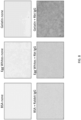
- FIG. 2A BSA/peptide gels were prepared as described supra. Mixing the antigen (e.g., a peptide) with BSA in a liquid solution before producing a gel allowed the peptide to be mixed uniformly.
- the resulting gel ( FIG. 2A ) can be constructed in any desired shape (in this example, a gel was produced in the shape of a 1.5mL microcentrifuge tube).
- the gel can also be sectioned ( FIGS. 2B & 2C ) and subjected to typical IHC processing steps such as antigen retrieval, blocking, primary/secondary antibody incubation, and detection ( e.g., using an enzyme/substrate approach).
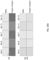
- FIG. 2D shows a sliced gel portion prepared for dehydration and paraffin processing
- FIG. 2D shows a sliced gel portion prepared for dehydration and paraffin processing
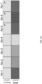
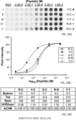
- BCL2 peptide/BSA gels were also subjected to quantitative immunofluorescence staining. Like the results obtained using chromogenic staining, IF analyses showed a graded increase in staining intensity as a function of peptide concentration ( FIG. 3D ). Analyses of optical emittance ( FIG. 3H ) and signal intensity ( FIG. 3J ) again demonstrated consistent, graded increases in staining. Negative control staining with a naive control primary antibody ( FIGS. 3F , 3I , & 3K ) or autofluorescence of the gels ( FIGS. 3E & 3G ) showed no increase in intensity, as expected.
- the approach is ideally suited for identifying antibodies suitable for IHC analysis of a target of interest, a process in which hundreds to thousands of candidate antibodies are typically screened in order to identify a small handful ( e.g ., 2-5) of leads for further characterization.
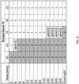
- BSA gel formation For evaluation of BSA gel formation, 1mL of a BSA solution in PBS was heated at a range of temperatures from 25-85°C. No formaldehyde was included in the BSA/PBS solution. The solution was heated continuously and observed under heated conditions at various time points for liquid/solid phase. Solutions were tested at the following BSA concentrations: 25%, 20%, 15%, 10%, 5%, and 2%.
- formaldehyde concentration 500 ⁇ L formaldehyde solution was mixed with 500 ⁇ L 25% BSA solution in PBS and incubated for 10 minutes at 85°C.
- Formaldehyde solutions were tested at the following concentrations (concentration refers to the original concentration prior to diluting with BSA solution): 1%, 2%, 4.5%, 9%, and 18%.
- concentration 1%, 2%, 4.5%, 9%, and 18%.
- stock formaldehyde solution at 37% was mixed with distilled water to make a final volume of 500 microliters containing 1%, 2%, 4.5%, 9%, or 18% formaldehyde; these solutions were then mixed (each in separate microfuge tubes) with an equal volume (500 microliters) of 25% BSA solution and heated as described above.
- casein, lact-albumin, soybean flour, and non-fat dry milk were tested for their ability to form gels. Each was tested at a final concentration of 12.5% in PBS after mixing with formaldehyde (final concentration: 18.5%), as described above. Under these conditions, only soybean flour was observed to form a gel, but the resulting solid was not homogeneous. These data demonstrate the unique properties of serum albumin in promoting gel formation.
- Additional substrate proteins were tested for their ability to form gels suitable for IHC staining, e.g., by forming a homogeneous gel that allows for IHC staining and adheres to a glass IHC slide.
- egg white protein or a mixture of egg white proteins and gelatin formed gels that exhibit specific IHC staining (in this case, after the gels containing a rabbit IgG antibody sample were stained with an anti-rabbit secondary antibody) with low non-specific staining (as seen after staining a gel lacking rabbit IgG with an anti-rabbit secondary antibody).
- gels generated from lact-albumin, liver protein powder, or soy flour failed to generate a homogenous gel ( FIG.
- 0.5 mL of solution containing the desired antigen in 25% (w/v) BSA / phosphate-buffered saline (PBS) was mixed in a 1.5 ml microfuge tube with an equal volume of 37% formaldehyde (Electron Microscopy Sciences; Hatfield, PA), heated for 10 minutes at 85C to coagulate the solution, then fixed overnight at room temperature.
- the final reagent concentrations were 12.5% (1.8 mM) BSA and 18.5% (6.2 M) formaldehyde.
- Peptide antigens had final concentrations of 2.5 x 10 -8 M to 2.5 x 10 -4 M in the fixed gels.
- Gels containing na ⁇ ve mouse, rat and rabbit IgG had a final concentration of 0.1 mg/ml (6.7 x 10 -7 M) IgG.
- the human [His] 6 -MBP-KSR2 fusion protein had a final gel concentration of 0.5 mg/ml (6.3 ⁇ 10 -6 M).
- FIGS. 11A-11D A tissue microarray (TMA) can be constructed containing the desired antigens in a range of antigen concentrations.
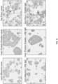
- FIGS. 12A-12E show a TMA composed of duplicate cores containing either no added peptide, serial dilutions of a negative control peptide from the human MCL1 protein, or dilutions of peptide encoding amino acids 41-54 of the BCL2 protein.
- a second BCL2 peptide TMA was constructed using new donor paraffin blocks formulated to have the same target BCL2 peptide concentrations as those used to build the TMA in FIGS. 12A-12E .
- Replicate sections of this second TMA were stained by two operators, on six separate days in a 6 month interval using the same anti-BCL2 clone 124 chromogenic IHC protocol used in FIGS. 12A-12E .
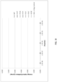
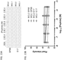
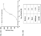
- Quantitative digital image analysis of the stained sections showed the signal intensity for each core was highly reproducible ( FIGS. 14A-14C ; see also FIGS. 3B & 3C ).
- the concentration of peptide that is available to bind antibody is reduced from the formulated value by three parameters: the efficiency of crosslinking the peptide to the protein matrix, the biochemical integrity of the peptide and accessibility of the cross-linked peptide to antibody.
- Formaldehyde was intended to cross-link BSA sidechains to the target peptides by reaction with the N-terminal tyrosine and C-terminal cysteine included in the peptide sequence.
- glutamine has been reported to react with formaldehyde ( Metz, B. et al. (2004) J. Biol. Chem. 279:6235-6243 ).
- N-terminal tyrosine was replaced with serine, expected to be minimally reactive with formalin, or with arginine, reported to be 50% more reactive than tyrosine ( Metz, B. et al. (2004) J. Biol. Chem. 279:6235-6243 ).
- Other variants included tyrosine or arginine at both the N- and C-termini.
- TMA cores containing serial dilutions of each peptide were prepared and stained as described above.
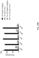
- FIG. 17A A peptide containing MYC amino acids 9-24 reacted strongly with the antibody, whereas other peptides reacted only weakly (aa 17-32) or not at all (aa 35-50 and aa 53-68) ( FIG. 17A ).
- a TMA was constructed containing duplicate cores, in the range of concentrations described earlier, of both the MYC aa 9-24 peptide and the BCL2 peptide.
- Chromogenic detection using anti-BCL2 clone 124 and anti-MYC clone Y69 showed a range of signal intensity, with no cross-reactivity to the non-target peptide ( FIGS. 17B & 17C ).
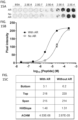
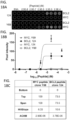
- Quantification of the resulting data ( FIG. 17D ) showed the MYC protocol had a 4-fold lower ACHM than does the BCL2 protocol, with similar dynamic range and HillSlope parameters ( FIG. 17E ).
- Dual immunofluorescent detection on the same TMA used in FIGS. 17A-17E was done using serial incubation with both anti-BCL2 and anti-MYC primary antibodies appropriate detection reagents ( FIGS. 18A-18C ).
- Qualitative results FIGS. 18A & 18B ) showed the expected specificity with no cross-reactivity between either antibody and the non-target peptide. Isotype controls used in place of antigen-specific primary antibodies resulted in no signal. Quantification of the resulting fluorescent data shows increased HillSlope parameters and increased replicate variability under the conditions tested, relative to the chromogenic protocol.
- Example 1 As described in Example 1 above ( see FIGS. 5A-5D ), another protein was incorporated into a BSA gel and used to evaluate antibodies generated from hybridoma clones.
- a peptide including the C-terminal 301 amino acids (i.e., amino acids 650-950) of human KSR2 was used.
- the results described above demonstrate that the synthetic IHC controls approach provides a useful method of identifying antibodies suitable for IHC analysis of a target of interest, a process in which hundreds to thousands of candidate antibodies are typically screened in order to identify a small handful ( e.g ., 2-5) of leads for further characterization.
- Candidate antibodies were further tested for reactivity against the antigen without prior antigen retrieval. For each antibody, antigen retrieval was required for detectable reactivity ( FIGS. 20A & 20B ). This contrasts with the results with BCL2 peptides described in Example 4, for which antigen retrieval improved signal strength approximately 6-fold, but was not required for detectable reactivity ( FIGS. 15A-15C ).
- Donor blocks containing BCL2 and MYC peptides in BSA gels were fixed with either formalin or zinc-based fixatives with heating to 85C ( FIGS. 22A-22C ).
- the signal was approximately 10-fold stronger with formaldehyde fixation than with zinc fixation during heating. The loss of signal strength correlated with heating in the presence of zinc.
- Example 10 Imaging mass cytometry analysis
- Sections of the BCL2 peptide TMA were stained with 146 Nd-labeled anti-BCL2 antibody EPR 17509, then analyzed by ultraviolet laser ablation and quantitative mass spectrometry.
- the TMA sample quantified was typically an area 150 microns square by 4 microns thick, containing 9 ⁇ 10 4 cubic microns. The results showed a graded signal that increases as the BCL2 peptide concentration in the target increases ( FIGS. 24A-24C ).
- the EPR17509 antibody signal in cores containing no peptide or in cores containing the negative control MCL1 peptide was less than 3% of the maximum signal.
- Examples 1-10 demonstrate that antigen-containing gels can be created using materials and methods available in any histology laboratory, and can be embedded and sectioned to produce uniformly stained samples. These examples demonstrate that the method is compatible with a variety of fixatives and with detection using chromogenic, immunofluorescent and mass spectrometry-based methods. The choice of possible antigens is limited only by the availability of the target protein or knowledge of the linear epitope sequence. The ability to create synthetic controls of known composition offers the opportunity to more precisely characterize and control routinely used immunohistochemistry protocols, independent of complicating factors inherent in heterogeneous tissue samples and subjective human interpretations.
Landscapes
- Health & Medical Sciences (AREA)
- Life Sciences & Earth Sciences (AREA)
- Immunology (AREA)
- Chemical & Material Sciences (AREA)
- Engineering & Computer Science (AREA)
- Molecular Biology (AREA)
- General Physics & Mathematics (AREA)
- Biochemistry (AREA)
- Pathology (AREA)
- Analytical Chemistry (AREA)
- Biomedical Technology (AREA)
- Physics & Mathematics (AREA)
- General Health & Medical Sciences (AREA)
- Hematology (AREA)
- Urology & Nephrology (AREA)
- Cell Biology (AREA)
- Microbiology (AREA)
- Biotechnology (AREA)
- Food Science & Technology (AREA)
- Medicinal Chemistry (AREA)
- Investigating Or Analysing Biological Materials (AREA)
- Peptides Or Proteins (AREA)
- Confectionery (AREA)
- Compositions Of Macromolecular Compounds (AREA)
- Solid-Sorbent Or Filter-Aiding Compositions (AREA)
Applications Claiming Priority (5)
| Application Number | Priority Date | Filing Date | Title |
|---|---|---|---|
| US201762595434P | 2017-12-06 | 2017-12-06 | |
| US201862730422P | 2018-09-12 | 2018-09-12 | |
| EP18822568.4A EP3721230B1 (de) | 2017-12-06 | 2018-12-05 | Synthetische kontrollen für immunhistochemie |
| EP22157356.1A EP4027144B1 (de) | 2017-12-06 | 2018-12-05 | Synthetische kontrollen für immunhistochemie |
| PCT/US2018/064012 WO2019113171A1 (en) | 2017-12-06 | 2018-12-05 | Synthetic controls for immunohistochemistry |
Related Parent Applications (2)
| Application Number | Title | Priority Date | Filing Date |
|---|---|---|---|
| EP22157356.1A Division EP4027144B1 (de) | 2017-12-06 | 2018-12-05 | Synthetische kontrollen für immunhistochemie |
| EP18822568.4A Division EP3721230B1 (de) | 2017-12-06 | 2018-12-05 | Synthetische kontrollen für immunhistochemie |
Publications (2)
| Publication Number | Publication Date |
|---|---|
| EP4534972A2 true EP4534972A2 (de) | 2025-04-09 |
| EP4534972A3 EP4534972A3 (de) | 2025-07-30 |
Family
ID=64746658
Family Applications (3)
| Application Number | Title | Priority Date | Filing Date |
|---|---|---|---|
| EP22157356.1A Active EP4027144B1 (de) | 2017-12-06 | 2018-12-05 | Synthetische kontrollen für immunhistochemie |
| EP25156644.4A Pending EP4534972A3 (de) | 2017-12-06 | 2018-12-05 | Synthetische kontrollen für immunhistochemie |
| EP18822568.4A Active EP3721230B1 (de) | 2017-12-06 | 2018-12-05 | Synthetische kontrollen für immunhistochemie |
Family Applications Before (1)
| Application Number | Title | Priority Date | Filing Date |
|---|---|---|---|
| EP22157356.1A Active EP4027144B1 (de) | 2017-12-06 | 2018-12-05 | Synthetische kontrollen für immunhistochemie |
Family Applications After (1)
| Application Number | Title | Priority Date | Filing Date |
|---|---|---|---|
| EP18822568.4A Active EP3721230B1 (de) | 2017-12-06 | 2018-12-05 | Synthetische kontrollen für immunhistochemie |
Country Status (16)
| Country | Link |
|---|---|
| US (2) | US12019069B2 (de) |
| EP (3) | EP4027144B1 (de) |
| JP (3) | JP7341140B2 (de) |
| KR (1) | KR20200096939A (de) |
| CN (2) | CN112219119B (de) |
| AU (1) | AU2018378025A1 (de) |
| BR (1) | BR112020011325A2 (de) |
| CA (1) | CA3083599A1 (de) |
| ES (1) | ES2920502T3 (de) |
| HR (1) | HRP20220770T1 (de) |
| IL (2) | IL310910A (de) |
| MX (1) | MX2020005836A (de) |
| PL (1) | PL3721230T3 (de) |
| SI (1) | SI3721230T1 (de) |
| TW (1) | TWI820061B (de) |
| WO (1) | WO2019113171A1 (de) |
Families Citing this family (7)
| Publication number | Priority date | Publication date | Assignee | Title |
|---|---|---|---|---|
| GB202006259D0 (en) | 2020-04-28 | 2020-06-10 | Leeds Teaching Hospitals Nhs Trust | Stain Assessment |
| CN113607534B (zh) * | 2021-08-20 | 2022-10-18 | 河南赛诺特生物技术有限公司 | 一种染色方法、试剂盒及应用 |
| CN113740526B (zh) * | 2021-08-20 | 2024-02-20 | 广西康柏莱科技有限公司 | 一种改性封闭剂及其制备方法 |
| CN114557953A (zh) * | 2022-02-22 | 2022-05-31 | 烟台毓璜顶医院 | 一种可自发荧光的血清白蛋白微针的制备方法 |
| CN114755407B (zh) * | 2022-04-02 | 2024-10-22 | 北京大学深圳医院 | 一种人骨组织多标记免疫组化样本的制备与检测方法 |
| JP2025532064A (ja) | 2022-09-22 | 2025-09-29 | アジレント・テクノロジーズ・インク | 免疫組織化学(ihc)プロトコールにおける使用のためおよびがんを診断するための抗ヒトicos抗体 |
| JP2025074654A (ja) * | 2023-10-30 | 2025-05-14 | 株式会社日立ハイテク | 病理検査向けコントロール物質、コントロールスライドガラス及びコントロール物質の製造方法 |
Family Cites Families (11)
| Publication number | Priority date | Publication date | Assignee | Title |
|---|---|---|---|---|
| GB8813628D0 (en) * | 1988-06-09 | 1988-07-13 | Shandon Southern Prod | Immunostaining |
| EP0352026A3 (de) * | 1988-07-19 | 1991-02-06 | Ruby Pauline Bonderman | Lamelle zur Kontrolle oder Kalibrierung mit mehreren Feldern mit verschiedenen Analysestoffen oder Konzentrationen an Analysestoff und Verfahren und Gerät zu deren Herstellung |
| EP0445277B1 (de) * | 1989-09-26 | 1996-07-31 | City Of Hope | Quantitative immunocytochemische prüfung |
| CA2467563A1 (en) * | 2001-11-20 | 2003-05-30 | Genentech, Inc. | Cell and tissue arrays and microarrays and methods of use |
| EP1494662B1 (de) * | 2002-04-11 | 2012-11-28 | Children's Medical Center Corporation | Tnp-470-hpma-methacrylsäure kopolymer-konjugate und ihre verwendung |
| CN1727896B (zh) * | 2005-07-22 | 2016-01-27 | 南京川博生物技术有限公司 | 磷酸化及对应非磷酸化蛋白质多克隆抗体在制备疾病诊断试剂中的应用及其制备方法 |
| US8420335B2 (en) * | 2006-03-14 | 2013-04-16 | Kobenhavns Universitet | Inhibition of GASC1 |
| WO2014144383A1 (en) * | 2013-03-15 | 2014-09-18 | Arizona Board Of Regents, A Body Corporate Of The State Of Arizona, Acting For And On Behalf Of Arizona State University | Non-covalent patterned chemical features and use thereof in maldi-based quality control |
| JP5993967B2 (ja) | 2014-02-20 | 2016-09-21 | 国立大学法人秋田大学 | 細胞内生体分子の検出に用いる標準試料及び細胞内生体分子の検出方法 |
| JP5972412B2 (ja) | 2015-02-05 | 2016-08-17 | 国立大学法人秋田大学 | 培養細胞を用いた標準試料及びその製造方法 |
| CN108593380B (zh) | 2018-04-25 | 2021-05-18 | 复旦大学附属中山医院 | 一种组织芯片量产制作方法 |
-
2018
- 2018-12-05 EP EP22157356.1A patent/EP4027144B1/de active Active
- 2018-12-05 BR BR112020011325-5A patent/BR112020011325A2/pt not_active IP Right Cessation
- 2018-12-05 PL PL18822568.4T patent/PL3721230T3/pl unknown
- 2018-12-05 WO PCT/US2018/064012 patent/WO2019113171A1/en not_active Ceased
- 2018-12-05 IL IL310910A patent/IL310910A/en unknown
- 2018-12-05 HR HRP20220770TT patent/HRP20220770T1/hr unknown
- 2018-12-05 MX MX2020005836A patent/MX2020005836A/es unknown
- 2018-12-05 KR KR1020207019032A patent/KR20200096939A/ko not_active Ceased
- 2018-12-05 CA CA3083599A patent/CA3083599A1/en active Pending
- 2018-12-05 CN CN201880085378.2A patent/CN112219119B/zh active Active
- 2018-12-05 ES ES18822568T patent/ES2920502T3/es active Active
- 2018-12-05 SI SI201830681T patent/SI3721230T1/sl unknown
- 2018-12-05 JP JP2020530652A patent/JP7341140B2/ja active Active
- 2018-12-05 CN CN202410855406.1A patent/CN118837544A/zh active Pending
- 2018-12-05 EP EP25156644.4A patent/EP4534972A3/de active Pending
- 2018-12-05 TW TW107143576A patent/TWI820061B/zh not_active IP Right Cessation
- 2018-12-05 AU AU2018378025A patent/AU2018378025A1/en not_active Abandoned
- 2018-12-05 EP EP18822568.4A patent/EP3721230B1/de active Active
-
2020
- 2020-05-25 IL IL274927A patent/IL274927A/en unknown
- 2020-05-29 US US16/888,252 patent/US12019069B2/en active Active
-
2023
- 2023-08-29 JP JP2023138663A patent/JP2023169184A/ja not_active Withdrawn
-
2024
- 2024-05-20 US US18/669,218 patent/US20240418709A1/en active Pending
-
2025
- 2025-09-16 JP JP2025153000A patent/JP2026012670A/ja active Pending
Non-Patent Citations (24)
| Title |
|---|
| "UniProt", Database accession no. P10415 |
| "Uniprot", Database accession no. Q6VAB6 |
| ADAM, P. ET AL., HUMAN PATHOLOGY, vol. 44, 2013, pages 1817 - 1826, Retrieved from the Internet <URL:www.abcam.com/bcl2-alpha-antibody-sp66-n-terminal-ab93884.html> |
| ANGELO ET AL., NAT MED., vol. 20, no. 4, April 2014 (2014-04-01), pages 436 - 42 |
| BOGEN, S.A. ET AL., APPL IMMUNOHISTOCHEM MOL MORPHOL, vol. 17, 2009, pages 239 - 246 |
| BRANDTZAEG, P., IMMUNOLOGY, vol. 22, no. 1, 1972, pages 177 - 183 |
| COLE, J., SCIENTIFIC REPORTS, vol. 7, 2017, pages 44707 |
| FOWLER ET AL., LAB INVEST., vol. 87, no. 8, 2007, pages 836 - 46 |
| FOWLER, C.B. ET AL., LAB INVEST, vol. 87, 2007, pages 836 - 846 |
| GIESEN ET AL., NAT METHODS., vol. 11, no. 4, April 2014 (2014-04-01), pages 417 - 22 |
| METZ, B. ET AL., J. BIOL. CHEM., vol. 279, 2004, pages 6235 - 6243 |
| MILLARWILLIAMS, HISTOCHEM J., vol. 14, no. 4, 1982, pages 609 - 620 |
| MOHD OMAR ET AL., ACTA HISTOCHEM., vol. 112, no. 6, 2010, pages 519 - 28 |
| SCHIPPERTILDERS, J HISTOCHEM CYTOCHEM., vol. 31, no. 1, 1983, pages 12 - 18 |
| SHI ET AL., J HISTOCHEM CYTOCHEM., vol. 53, no. 9, 2005, pages 1167 - 1170 |
| SHI, S.R. ET AL., J. HISTOCHEM. CYTOCHEM., vol. 59, 2011, pages 13 - 32 |
| SLAMON, D.J. ET AL., N ENGL J MED, vol. 344, 2001, pages 783 - 792 |
| SOMPURAM ET AL., J. HISTOCHEM CYTOCHEM, vol. 63, 2015, pages 681 - 690 |
| SOMPURAM, S.R. ET AL., CLIN CHEM, vol. 48, 2002, pages 410 - 420 |
| TOEWS, J. ET AL., ANAL. CHIM. ACTA, vol. 618, 2008, pages 168 - 183 |
| VALENTI ET AL., J AM CHEM SOC., 8 November 2017 (2017-11-08) |
| VALNES ET AL., J HISTOCHEM CYTOCHEM., vol. 33, no. 8, 1984, pages 755 - 61 |
| VALNESBRANDTZAEG, HISTOCHEMISTRY, vol. 81, no. 4, 1985, pages 313 - 9 |
| WU ET AL., NAT BIOTECHNOL., vol. 21, no. 1, January 2003 (2003-01-01), pages 41 - 6 |
Also Published As
| Publication number | Publication date |
|---|---|
| EP4027144B1 (de) | 2025-02-12 |
| WO2019113171A1 (en) | 2019-06-13 |
| JP2026012670A (ja) | 2026-01-27 |
| CA3083599A1 (en) | 2019-06-13 |
| PL3721230T3 (pl) | 2022-07-18 |
| IL274927A (en) | 2020-07-30 |
| JP2023169184A (ja) | 2023-11-29 |
| EP3721230A1 (de) | 2020-10-14 |
| EP4534972A3 (de) | 2025-07-30 |
| HRP20220770T1 (hr) | 2022-09-16 |
| US12019069B2 (en) | 2024-06-25 |
| TWI820061B (zh) | 2023-11-01 |
| CN112219119B (zh) | 2024-07-12 |
| US20200292536A1 (en) | 2020-09-17 |
| JP2021505878A (ja) | 2021-02-18 |
| CN118837544A (zh) | 2024-10-25 |
| CN112219119A (zh) | 2021-01-12 |
| TW201925239A (zh) | 2019-07-01 |
| ES2920502T3 (es) | 2022-08-04 |
| EP4027144A1 (de) | 2022-07-13 |
| IL310910A (en) | 2024-04-01 |
| EP3721230B1 (de) | 2022-04-06 |
| JP7341140B2 (ja) | 2023-09-08 |
| KR20200096939A (ko) | 2020-08-14 |
| AU2018378025A1 (en) | 2020-06-18 |
| SI3721230T1 (sl) | 2022-07-29 |
| US20240418709A1 (en) | 2024-12-19 |
| BR112020011325A2 (pt) | 2020-11-17 |
| MX2020005836A (es) | 2020-08-20 |
Similar Documents
| Publication | Publication Date | Title |
|---|---|---|
| US20240418709A1 (en) | Synthetic controls for immunohistochemistry | |
| JP5048757B2 (ja) | 診断指標または予測指標としての細胞内局在プロフィールの使用 | |
| US6281004B1 (en) | Quality control for cytochemical assays | |
| US20180100856A1 (en) | Calibration Standards For Digital Histocytometry | |
| US20190112356A1 (en) | High-affinity immunopolymers | |
| CN101598731A (zh) | 一种用于肿瘤病理诊断用途的免疫组织化学诊断试剂盒 | |
| JP2026502043A (ja) | カオトロープ剤補助による深部免疫染色 | |
| JP6277190B2 (ja) | 改善された細胞の同定のための方法 | |
| Huang et al. | Quantitative fluorescence imaging analysis for cancer biomarker discovery: application to β-catenin in archived prostate specimens | |
| HK40112153A (zh) | 用於免疫组织化学的合成对照 | |
| HK40043325A (en) | Synthetic controls for immunohistochemistry | |
| HK40043325B (zh) | 用於免疫组织化学的合成对照 | |
| Lee et al. | Development of a multiplex bead-based method for the microquantitation of δ-catenin | |
| Bogen et al. | Recent trends and advances in immunodiagnostics of solid tumors | |
| US20250044284A1 (en) | Materials and methods for enhanced detection and screening of molecules of therapeutic potential | |
| US20140113385A1 (en) | Compositions and Methods for Identifying Single Antigens or Other Molecules in Cell Preparations | |
| Coons | Introduction to Immunohistochemistry | |
| Han et al. | High-throughput single-vesicle imaging platform for direct extracellular vesicle profiling of human plasma |
Legal Events
| Date | Code | Title | Description |
|---|---|---|---|
| PUAI | Public reference made under article 153(3) epc to a published international application that has entered the european phase |
Free format text: ORIGINAL CODE: 0009012 |
|
| STAA | Information on the status of an ep patent application or granted ep patent |
Free format text: STATUS: THE APPLICATION HAS BEEN PUBLISHED |
|
| AC | Divisional application: reference to earlier application |
Ref document number: 3721230 Country of ref document: EP Kind code of ref document: P Ref document number: 4027144 Country of ref document: EP Kind code of ref document: P |
|
| AK | Designated contracting states |
Kind code of ref document: A2 Designated state(s): AL AT BE BG CH CY CZ DE DK EE ES FI FR GB GR HR HU IE IS IT LI LT LU LV MC MK MT NL NO PL PT RO RS SE SI SK SM TR |
|
| REG | Reference to a national code |
Ref country code: DE Ref legal event code: R079 Free format text: PREVIOUS MAIN CLASS: G01N0001360000 Ipc: G01N0033543000 |
|
| PUAL | Search report despatched |
Free format text: ORIGINAL CODE: 0009013 |
|
| AK | Designated contracting states |
Kind code of ref document: A3 Designated state(s): AL AT BE BG CH CY CZ DE DK EE ES FI FR GB GR HR HU IE IS IT LI LT LU LV MC MK MT NL NO PL PT RO RS SE SI SK SM TR |
|
| RIC1 | Information provided on ipc code assigned before grant |
Ipc: G01N 33/543 20060101AFI20250625BHEP Ipc: G01N 1/36 20060101ALI20250625BHEP |
|
| REG | Reference to a national code |
Ref country code: HK Ref legal event code: DE Ref document number: 40124797 Country of ref document: HK |
|
| STAA | Information on the status of an ep patent application or granted ep patent |
Free format text: STATUS: REQUEST FOR EXAMINATION WAS MADE |
|
| 17P | Request for examination filed |
Effective date: 20260130 |
|
| STAA | Information on the status of an ep patent application or granted ep patent |
Free format text: STATUS: EXAMINATION IS IN PROGRESS |