EP4527465A2 - Anticorps anti-glypicane 3 - Google Patents
Anticorps anti-glypicane 3 Download PDFInfo
- Publication number
- EP4527465A2 EP4527465A2 EP25152478.1A EP25152478A EP4527465A2 EP 4527465 A2 EP4527465 A2 EP 4527465A2 EP 25152478 A EP25152478 A EP 25152478A EP 4527465 A2 EP4527465 A2 EP 4527465A2
- Authority
- EP
- European Patent Office
- Prior art keywords
- seq
- 52h5d3b8
- amino acid
- antibody
- acid sequence
- Prior art date
- Legal status (The legal status is an assumption and is not a legal conclusion. Google has not performed a legal analysis and makes no representation as to the accuracy of the status listed.)
- Pending
Links
Images


Classifications
-
- A—HUMAN NECESSITIES
- A61—MEDICAL OR VETERINARY SCIENCE; HYGIENE
- A61K—PREPARATIONS FOR MEDICAL, DENTAL OR TOILETRY PURPOSES
- A61K40/00—Cellular immunotherapy
- A61K40/30—Cellular immunotherapy characterised by the recombinant expression of specific molecules in the cells of the immune system
- A61K40/31—Chimeric antigen receptors [CAR]
-
- A—HUMAN NECESSITIES
- A61—MEDICAL OR VETERINARY SCIENCE; HYGIENE
- A61K—PREPARATIONS FOR MEDICAL, DENTAL OR TOILETRY PURPOSES
- A61K47/00—Medicinal preparations characterised by the non-active ingredients used, e.g. carriers or inert additives; Targeting or modifying agents chemically bound to the active ingredient
- A61K47/50—Medicinal preparations characterised by the non-active ingredients used, e.g. carriers or inert additives; Targeting or modifying agents chemically bound to the active ingredient the non-active ingredient being chemically bound to the active ingredient, e.g. polymer-drug conjugates
- A61K47/51—Medicinal preparations characterised by the non-active ingredients used, e.g. carriers or inert additives; Targeting or modifying agents chemically bound to the active ingredient the non-active ingredient being chemically bound to the active ingredient, e.g. polymer-drug conjugates the non-active ingredient being a modifying agent
- A61K47/68—Medicinal preparations characterised by the non-active ingredients used, e.g. carriers or inert additives; Targeting or modifying agents chemically bound to the active ingredient the non-active ingredient being chemically bound to the active ingredient, e.g. polymer-drug conjugates the non-active ingredient being a modifying agent the modifying agent being an antibody, an immunoglobulin or a fragment thereof, e.g. an Fc-fragment
- A61K47/6835—Medicinal preparations characterised by the non-active ingredients used, e.g. carriers or inert additives; Targeting or modifying agents chemically bound to the active ingredient the non-active ingredient being chemically bound to the active ingredient, e.g. polymer-drug conjugates the non-active ingredient being a modifying agent the modifying agent being an antibody, an immunoglobulin or a fragment thereof, e.g. an Fc-fragment the modifying agent being an antibody or an immunoglobulin bearing at least one antigen-binding site
- A61K47/6851—Medicinal preparations characterised by the non-active ingredients used, e.g. carriers or inert additives; Targeting or modifying agents chemically bound to the active ingredient the non-active ingredient being chemically bound to the active ingredient, e.g. polymer-drug conjugates the non-active ingredient being a modifying agent the modifying agent being an antibody, an immunoglobulin or a fragment thereof, e.g. an Fc-fragment the modifying agent being an antibody or an immunoglobulin bearing at least one antigen-binding site the antibody targeting a determinant of a tumour cell
- A61K47/6859—Medicinal preparations characterised by the non-active ingredients used, e.g. carriers or inert additives; Targeting or modifying agents chemically bound to the active ingredient the non-active ingredient being chemically bound to the active ingredient, e.g. polymer-drug conjugates the non-active ingredient being a modifying agent the modifying agent being an antibody, an immunoglobulin or a fragment thereof, e.g. an Fc-fragment the modifying agent being an antibody or an immunoglobulin bearing at least one antigen-binding site the antibody targeting a determinant of a tumour cell the tumour determinant being from liver or pancreas cancer cell
-
- A—HUMAN NECESSITIES
- A61—MEDICAL OR VETERINARY SCIENCE; HYGIENE
- A61P—SPECIFIC THERAPEUTIC ACTIVITY OF CHEMICAL COMPOUNDS OR MEDICINAL PREPARATIONS
- A61P35/00—Antineoplastic agents
-
- C—CHEMISTRY; METALLURGY
- C07—ORGANIC CHEMISTRY
- C07K—PEPTIDES
- C07K16/00—Immunoglobulins [IGs], e.g. monoclonal or polyclonal antibodies
- C07K16/18—Immunoglobulins [IGs], e.g. monoclonal or polyclonal antibodies against material from animals or humans
- C07K16/28—Immunoglobulins [IGs], e.g. monoclonal or polyclonal antibodies against material from animals or humans against receptors, cell surface antigens or cell surface determinants
- C07K16/30—Immunoglobulins [IGs], e.g. monoclonal or polyclonal antibodies against material from animals or humans against receptors, cell surface antigens or cell surface determinants from tumour cells
- C07K16/303—Liver or Pancreas
-
- A—HUMAN NECESSITIES
- A61—MEDICAL OR VETERINARY SCIENCE; HYGIENE
- A61K—PREPARATIONS FOR MEDICAL, DENTAL OR TOILETRY PURPOSES
- A61K38/00—Medicinal preparations containing peptides
-
- C—CHEMISTRY; METALLURGY
- C07—ORGANIC CHEMISTRY
- C07K—PEPTIDES
- C07K2317/00—Immunoglobulins specific features
- C07K2317/20—Immunoglobulins specific features characterized by taxonomic origin
- C07K2317/24—Immunoglobulins specific features characterized by taxonomic origin containing regions, domains or residues from different species, e.g. chimeric, humanized or veneered
-
- C—CHEMISTRY; METALLURGY
- C07—ORGANIC CHEMISTRY
- C07K—PEPTIDES
- C07K2317/00—Immunoglobulins specific features
- C07K2317/30—Immunoglobulins specific features characterized by aspects of specificity or valency
- C07K2317/33—Crossreactivity, e.g. for species or epitope, or lack of said crossreactivity
-
- C—CHEMISTRY; METALLURGY
- C07—ORGANIC CHEMISTRY
- C07K—PEPTIDES
- C07K2317/00—Immunoglobulins specific features
- C07K2317/50—Immunoglobulins specific features characterized by immunoglobulin fragments
- C07K2317/56—Immunoglobulins specific features characterized by immunoglobulin fragments variable (Fv) region, i.e. VH and/or VL
- C07K2317/565—Complementarity determining region [CDR]
-
- C—CHEMISTRY; METALLURGY
- C07—ORGANIC CHEMISTRY
- C07K—PEPTIDES
- C07K2317/00—Immunoglobulins specific features
- C07K2317/70—Immunoglobulins specific features characterized by effect upon binding to a cell or to an antigen
- C07K2317/73—Inducing cell death, e.g. apoptosis, necrosis or inhibition of cell proliferation
- C07K2317/732—Antibody-dependent cellular cytotoxicity [ADCC]
-
- C—CHEMISTRY; METALLURGY
- C07—ORGANIC CHEMISTRY
- C07K—PEPTIDES
- C07K2317/00—Immunoglobulins specific features
- C07K2317/70—Immunoglobulins specific features characterized by effect upon binding to a cell or to an antigen
- C07K2317/75—Agonist effect on antigen
-
- C—CHEMISTRY; METALLURGY
- C07—ORGANIC CHEMISTRY
- C07K—PEPTIDES
- C07K2317/00—Immunoglobulins specific features
- C07K2317/70—Immunoglobulins specific features characterized by effect upon binding to a cell or to an antigen
- C07K2317/77—Internalization into the cell
-
- C—CHEMISTRY; METALLURGY
- C07—ORGANIC CHEMISTRY
- C07K—PEPTIDES
- C07K2317/00—Immunoglobulins specific features
- C07K2317/90—Immunoglobulins specific features characterized by (pharmaco)kinetic aspects or by stability of the immunoglobulin
- C07K2317/92—Affinity (KD), association rate (Ka), dissociation rate (Kd) or EC50 value
Definitions
- Glypican 3 is a member of the glypican-related integral membrane heparan sulfate proteoglycans (GRIPS) family that are present on cell surfaces.
- the protein core of GPC3 consists of two subunits, where the N-terminal subunit has a size of ⁇ 40 kDa and the C-terminal subunit is ⁇ 30 kDa.
- Six glypicans (GPC1-6) have been identified in mammals.
- GPC3 plays an important role in modulating the cell proliferation, differentiation, adhesion and migration.
- GPC3 interacts with both Wnt and frizzled (FZD) to form a complex and triggers downstream signaling.
- the core protein of GPC3 may serve as a co-receptor or a receiver for Wnt.
- Glypican 3 immunostaining can be for differentiating hepatocellular carcinoma (HCC) and dysplastic changes in cirrhotic livers.
- HCC hepatocellular carcinoma
- GPC3 protein expression is found in HCC, not in normal liver and cholangiocarcinoma. GPC3 is also expressed to a lesser degree in melanoma, ovarian clear-cell carcinomas, yolk sac tumors, neuroblastoma, hepatoblastoma, Wilms' tumor cells, and other tumors.
- GPC3 is a promising therapeutic target for treating cancers such as liver cancer.
- Several therapeutic anti-GPC3 antibodies have been developed, including GC33 and YP7. Some of these antibodies inhibit Wnt signaling in liver cancer cells.
- chimeric antigen receptor (CAR) T cell immunotherapies are being developed at various stages for treating cancer. In mice with xenograft or orthoptic liver tumors, CAR-T cells can eliminate GPC3-positive cancer cells, by inducing perforin- and granzyme-mediated cell death and reducing Wnt signaling in tumor cells.
- the present disclosure provides antibodies and antigen-binding fragments specific to the human GPC3 protein. Experimental testing shows that these newly identified antibodies outperformed the benchmark GC33 antibody.
- the VH CDR1 comprises an amino acid sequence selected from the group consisting of SEQ ID NO:13, 25, 59 and 63;
- the VH CDR2 comprises an amino acid sequence selected from the group consisting of SEQ ID NO: 14, 19, 23, 26, 29, 30, 60 and 65;
- the VH CDR3 comprises an amino acid sequence selected from the group consisting of SEQ ID NO:15, 20, 24 and 33;
- the VL CDR1 comprises an amino acid sequence selected from the group consisting of SEQ ID NO:16, 21, 27 and 31;
- the VL CDR2 comprises an amino acid sequence selected from the group consisting of SEQ ID NO:17, 61, 64 and 66;
- the VL CDR3 comprises an amino acid sequence selected from the group consisting of SEQ ID NO:18, 22, 28, 32, 34 and 62.
- the VH CDR1, VH CDR2, VH CDR3, VL CDR1, VL CDR2, and VL CDR3, respectively comprise the amino acid sequences of:
- the VH CDR1 comprises an amino acid sequence selected from the group consisting of SEQ ID NO:13, 59, and 63;
- the VH CDR2 comprises an amino acid sequence selected from the group consisting of SEQ ID NO: 19, 60, and 65;
- the VH CDR3 comprises the amino acid sequence of SEQ ID NO:15;
- the VL CDR1 comprises the amino acid sequence of SEQ ID NO:21;
- the VL CDR2 comprises an amino acid sequence selected from the group consisting of SEQ ID NO:17, 61, 64 and 66;
- the VL CDR3 comprises an amino acid sequence selected from the group consisting of SEQ ID NO:18 and 62.
- the VH CDR1, VH CDR2, VH CDR3, VL CDR1, VL CDR2, and VL CDR3, respectively comprise the amino acid sequences of SEQ ID NO:13, 19, 15, 21, 17 and 18.
- the VH comprises an amino acid sequence selected from the group consisting of SEQ ID NO:35-39
- the VL comprises an amino acid sequence selected from the group consisting of SEQ ID NO:40-44.
- the VH comprises the amino acid sequence of SEQ ID NO:36 and the VL comprises the amino acid sequence of SEQ ID NO:41. In some embodiments, the VH comprises the amino acid sequence of SEQ ID NO:37 and the VL comprises the amino acid sequence of SEQ ID NO:43.
- the VH comprises the amino acid sequence of SEQ ID NO:36 and the VL comprises the amino acid sequence of SEQ ID NO:42. In some embodiments, the VH comprises the amino acid sequence of SEQ ID NO:36 and the VL comprises the amino acid sequence of SEQ ID NO:43. In some embodiments, the VH comprises the amino acid sequence of SEQ ID NO:36 and the VL comprises the amino acid sequence of SEQ ID NO:44. In some embodiments, the VH comprises the amino acid sequence of SEQ ID NO:37 and the VL comprises the amino acid sequence of SEQ ID NO:41. In some embodiments, the VH comprises the amino acid sequence of SEQ ID NO:37 and the VL comprises the amino acid sequence of SEQ ID NO:42.
- the VH comprises the amino acid sequence of SEQ ID NO:37 and the VL comprises the amino acid sequence of SEQ ID NO:44. In some embodiments, the VH comprises the amino acid sequence of SEQ ID NO:38 and the VL comprises the amino acid sequence of SEQ ID NO:41. In some embodiments, the VH comprises the amino acid sequence of SEQ ID NO:38 and the VL comprises the amino acid sequence of SEQ ID NO:42. In some embodiments, the VH comprises the amino acid sequence of SEQ ID NO:38 and the VL comprises the amino acid sequence of SEQ ID NO:43. In some embodiments, the VH comprises the amino acid sequence of SEQ ID NO:38 and the VL comprises the amino acid sequence of SEQ ID NO:44.
- the VH comprises the amino acid sequence of SEQ ID NO:39 and the VL comprises the amino acid sequence of SEQ ID NO:41. In some embodiments, the VH comprises the amino acid sequence of SEQ ID NO:39 and the VL comprises the amino acid sequence of SEQ ID NO:42. In some embodiments, the VH comprises the amino acid sequence of SEQ ID NO:39 and the VL comprises the amino acid sequence of SEQ ID NO:43. In some embodiments, the VH comprises the amino acid sequence of SEQ ID NO:39 and the VL comprises the amino acid sequence of SEQ ID NO:44.
- the VH CDR1, VH CDR2, VH CDR3, VL CDR1, VL CDR2, and VL CDR3, respectively, comprise the amino acid sequences of SEQ ID NO:59, 60, 15, 21, 61, and 62.
- the VH comprises the amino acid sequence of SEQ ID NO:53 and the VL comprises the amino acid sequence of SEQ ID NO:54.
- the VH CDR1, VH CDR2, VH CDR3, VL CDR1, VL CDR2, and VL CDR3, respectively, comprise the amino acid sequences of SEQ ID NO:63, 19, 15, 21, 64, and 62.
- the VH comprises the amino acid sequence of SEQ ID NO:55 and the VL comprises the amino acid sequence of SEQ ID NO:56.
- the VH CDR1, VH CDR2, VH CDR3, VL CDR1, VL CDR2, and VL CDR3, respectively comprise the amino acid sequences of SEQ ID NO:13, 65, 15, 21, 66, and 62.
- the VH comprises the amino acid sequence of SEQ ID NO:57 and the VL comprises the amino acid sequence of SEQ ID NO:58.
- the VH CDR1, VH CDR2, VH CDR3, VL CDR1, VL CDR2, and VL CDR3, respectively comprise the amino acid sequences of SEQ ID NO: 13, 19, 20, 21, 17 and 22.
- the VH comprises an amino acid sequence selected from the group consisting of SEQ ID NO:3 and 45-50
- the VL comprises an amino acid sequence selected from the group consisting of SEQ ID NO:4, 51 and 52.
- the VH comprises the amino acid sequence of SEQ ID NO:48 and the VL comprises the amino acid sequence of SEQ ID NO:51.
- the VH comprises the amino acid sequence of SEQ ID NO:48 and the VL comprises the amino acid sequence of SEQ ID NO:52.
- the VH comprises the amino acid sequence of SEQ ID NO:45 and the VL comprises the amino acid sequence of SEQ ID NO:51. In some embodiments, the VH comprises the amino acid sequence of SEQ ID NO:45 and the VL comprises the amino acid sequence of SEQ ID NO:52. In some embodiments, the VH comprises the amino acid sequence of SEQ ID NO:46 and the VL comprises the amino acid sequence of SEQ ID NO:51. In some embodiments, the VH comprises the amino acid sequence of SEQ ID NO:46 and the VL comprises the amino acid sequence of SEQ ID NO:52. In some embodiments, the VH comprises the amino acid sequence of SEQ ID NO:47 and the VL comprises the amino acid sequence of SEQ ID NO:51.
- the VH comprises the amino acid sequence of SEQ ID NO:47 and the VL comprises the amino acid sequence of SEQ ID NO:52. In some embodiments, the VH comprises the amino acid sequence of SEQ ID NO:49 and the VL comprises the amino acid sequence of SEQ ID NO:51. In some embodiments, the VH comprises the amino acid sequence of SEQ ID NO:49 and the VL comprises the amino acid sequence of SEQ ID NO:52. In some embodiments, the VH comprises the amino acid sequence of SEQ ID NO:50 and the VL comprises the amino acid sequence of SEQ ID NO:51. In some embodiments, the VH comprises the amino acid sequence of SEQ ID NO:50 and the VL comprises the amino acid sequence of SEQ ID NO:52.
- the VH CDR1, VH CDR2, VH CDR3, VL CDR1, VL CDR2, and VL CDR3, respectively, comprise the amino acid sequences of SEQ ID NO:13-18.
- the VH comprises the amino acid sequence of SEQ ID NO:1
- the VL comprises the amino acid sequence of SEQ ID NO:2.
- the VH CDR1, VH CDR2, VH CDR3, VL CDR1, VL CDR2, and VL CDR3, respectively comprise the amino acid sequences of SEQ ID NO:13, 23, 24, 21, 17 and 18.
- the VH comprises the amino acid sequence of SEQ ID NO:5
- the VL comprises the amino acid sequence of SEQ ID NO:6.
- the VH CDR1, VH CDR2, VH CDR3, VL CDR1, VL CDR2, and VL CDR3, respectively comprise the amino acid sequences of SEQ ID NO:25, 26, 20, 27, 17 and 28.
- the VH comprises the amino acid sequence of SEQ ID NO:7
- the VL comprises the amino acid sequence of SEQ ID NO:8.
- the VH CDR1, VH CDR2, VH CDR3, VL CDR1, VL CDR2, and VL CDR3, respectively comprise the amino acid sequences of SEQ ID NO:25, 29, 20, 27, 17 and 28.
- the VH comprises the amino acid sequence of SEQ ID NO:9
- the VL comprises the amino acid sequence of SEQ ID NO:10.
- the VH CDR1 comprises the amino acid sequence of SEQ ID NO:13; the VH CDR2 comprises the amino acid sequence of SEQ ID NO:19; the VH CDR3 comprises an amino acid sequence selected from the group consisting of SEQ ID NO:15, 20, 24, and 33; the VL CDR1 comprises the amino acid sequence of SEQ ID NO:21; the VL CDR2 comprises the amino acid sequence of SEQ ID NO:17; and the VL CDR3 comprises an amino acid sequence selected from the group consisting SEQ ID NO:18, 22, 28, 32 and 34.
- the antibody or fragment thereof is humanized.
- an antibody-drug conjugate characterized in that the antibody-drug conjugate comprises: (a) an antibody or fragment thereof of the present disclosure; and (b) a conjugation moiety conjugated to the antibody or fragment, wherein the conjugation moiety is selected from: a detectable marker, a drug, a toxin, a cytokine, a radionuclide, an enzyme, or a combination thereof.
- a multispecific antibody comprising the antibody of the present disclosure and one or more second antibody or antigen-binding fragment having binding specificity to a target antigen that is not GPC3.
- a chimeric antigen receptor comprising the antibody or fragment thereof of the present disclosure, a transmembrane domain, a costimulatory domain, and a CD3 ⁇ intracellular domain.
- a polynucleotide encoding the antibody or fragment thereof of the present disclosure, a cell comprising the polynucleotide, and a composition comprising the antibody or fragment thereof and a pharmaceutically acceptable carrier. Also provided are methods and uses for treating cancer with the antibody or fragment thereof.
- a or “an” entity refers to one or more of that entity; for example, “an antibody,” is understood to represent one or more antibodies.
- the terms “a” (or “an”), “one or more,” and “at least one” can be used interchangeably herein.
- an “antibody” or “antigen-binding polypeptide” refers to a polypeptide or a polypeptide complex that specifically recognizes and binds to an antigen.
- An antibody can be a whole antibody and any antigen binding fragment or a single chain thereof.
- antibody includes any protein or peptide containing molecule that comprises at least a portion of an immunoglobulin molecule having biological activity of binding to the antigen.
- CDR complementarity determining region
- antibody fragment or "antigen-binding fragment”, as used herein, is a portion of an antibody such as F(ab')2, F(ab)2, Fab', Fab, Fv, scFv and the like. Regardless of structure, an antibody fragment binds with the same antigen that is recognized by the intact antibody.
- antibody fragment includes aptamers, spiegelmers, and diabodies.
- antibody fragment also includes any synthetic or genetically engineered protein that acts like an antibody by binding to a specific antigen to form a complex.
- antibody encompasses various broad classes of polypeptides that can be distinguished biochemically. Those skilled in the art will appreciate that heavy chains are classified as gamma, mu, alpha, delta, or epsilon ( ⁇ , ⁇ , ⁇ , ⁇ , ⁇ ) with some subclasses among them (e.g., ⁇ 1- ⁇ 4). It is the nature of this chain that determines the "class" of the antibody as IgG, IgM, IgA IgG, or IgE, respectively.
- immunoglobulin subclasses e.g., IgG1, IgG2, IgG3, IgG4, IgG5, etc. are well characterized and are known to confer functional specialization. Modified versions of each of these classes and isotypes are readily discernable to the skilled artisan in view of the instant disclosure and, accordingly, are within the scope of the instant disclosure. All immunoglobulin classes are clearly within the scope of the present disclosure, the following discussion will generally be directed to the IgG class of immunoglobulin molecules.
- IgG a standard immunoglobulin molecule comprises two identical light chain polypeptides of molecular weight approximately 23,000 Daltons, and two identical heavy chain polypeptides of molecular weight 53,000-70,000. The four chains are typically joined by disulfide bonds in a "Y" configuration wherein the light chains bracket the heavy chains starting at the mouth of the "Y" and continuing through the variable region.
- Antibodies, antigen-binding polypeptides, variants, or derivatives thereof of the disclosure include, but are not limited to, polyclonal, monoclonal, multispecific, human, humanized, primatized, or chimeric antibodies, single chain antibodies, epitope-binding fragments, e.g., Fab, Fab' and F(ab')2, Fd, Fvs, single-chain Fvs (scFv), single-chain antibodies, disulfide-linked Fvs (sdFv), fragments comprising either a VK or VH domain, fragments produced by a Fab expression library, and anti-idiotypic (anti-Id) antibodies (including, e.g., anti-Id antibodies to the antibodies disclosed herein).
- anti-Id antigen-binding polypeptides, variants, or derivatives thereof of the disclosure
- Immunoglobulin or antibody molecules of the disclosure can be of any type (e.g., IgG, IgE, IgM, IgD, IgA, and IgY), class (e.g., IgGl, IgG2, IgG3, IgG4, IgAl and IgA2) or subclass of immunoglobulin molecule.
- chimeric antibody will be held to mean any antibody wherein the immunoreactive region or site is obtained or derived from a first species and the constant region (which may be intact, partial or modified in accordance with the instant disclosure) is obtained from a second species.
- the target binding region or site will be from a non-human source (e.g. mouse or primate) and the constant region is human.
- Antibodies disclosed herein can be from any animal origin including birds and mammals.
- the antibodies are human, murine, donkey, rabbit, goat, guinea pig, camel, llama, horse, or chicken antibodies.
- the variable region may be condricthoid in origin (e.g., from sharks).
- the term "recombinant" as it pertains to polypeptides or polynucleotides intends a form of the polypeptide or polynucleotide that does not exist naturally, a non-limiting example of which can be created by combining polynucleotides that would not normally occur together.
- Hybridoma technology can be performed under conditions of different "stringency".
- a low stringency hybridization reaction is carried out at about 40°C in about 10 x SSC or a solution of equivalent ionic strength/temperature.
- a moderate stringency hybridization is typically performed at about 50°C in about 6 x SSC, and a high stringency hybridization reaction is generally performed at about 60°C in about 1 x SSC.
- Hybridization reactions can also be performed under "physiological conditions" which is well known to one of skill in the art.
- a nonlimiting example of a physiological condition is the temperature, ionic strength, pH and concentration of Mg2+ normally found in a cell.
- the instant inventors were able to generate potent murine anti-GPC3 antibodies 18C1D9, 52H5D3B8, 163E7D12, 172B4E9, 152E9F7 and 171F9C5 (Table 1).
- the 52H5D3B8 antibody exhibited higher cell-based binding efficiency than GC33, a leading anti-GPC3 antibody under clinical development.
- experimental data show that these new antibodies have higher activity in inducing cellular internationalization. Such a property makes these new antibodies particularly suitable for use in, e.g., antibody-drug conjugates (ADC).
- ADC antibody-drug conjugates
- 52H5D3B8 antibody was further optimized.
- the humanized antibodies were subjected to further affinity maturation in an effort to further enhance binding efficiency. As shown in FIG. 11 and Table 16, encouragingly, all of the affinity maturated antibodies exhibited enhanced binding efficiency as compared to the parental antibody (52H5D3B8-6#-VH2+VL3) and benchmark antibody GC33. Also surprisingly, when targeting cells with moderate GPC3 expression levels, the humanized antibodies exhibited considerably higher ADCC signaling potency over GC33 ( FIG. 12 and Table 17).
- the parental antibody 52H5D3B8 was also subjected to humanization, and two of the humanized ones, Hu 52H5D3B8-7 and Hu 52H5D3B8-8, were tested for their binding activity and internalization activity, with GC33 and Y035 as references. As shown in FIG. 17-18 and Tables 24-25, these humanized antibodies outperformed both references.
- CDR sequences CDR Sequence SEQ ID NO: CDRH1 X 1 YE X 2 H (X 1 is D/R/G, X 2 is I or M) 71 DYEMH 13 DYEIH 25 RYEMH 59 GYEMH 63 CDRH2 AI X 1 P X 2 X 3 X 4 X 5 TAY X 6 X 7 X 8 FKG (X 1 is D/G/H, X 2 is A/E/G, X 3 is S/T, X 4 is D/G, X 5 is D/G/N/S, X 6 is N/S/T, X 7 is Q/S, X 8 is R/K/L) 72 AIDPETGGTAYNQKFKG 14 AIHPGSGGTAYNQKFKG 19 AIGPETGSTAYNQRFKG
- the present disclosure provides an anti-glypican 3 antibody that includes a VH (heavy chain variable region) and a VL (light chain variable region).
- the VH and VL regions include VH CDR1, VH CDR2, VH CDR3, VL CDR1, VL CDR2, and VL CDR3, such as those illustrated in Tables A, 1A-1F, 5A-5O, 8A and 8B.
- the VH CDR1 includes the amino acid sequence of SEQ ID NO:71
- the VH CDR2 includes the amino acid sequence of SEQ ID NO:72
- the VH CDR3 includes the amino acid sequence of SEQ ID NO:73
- the VL CDR1 includes the amino acid sequence of SEQ ID NO:74
- the VL CDR2 includes the amino acid sequence of SEQ ID NO:75
- the VL CDR3 includes the amino acid sequence of SEQ ID NO:76.
- SEQ ID NO:71 is X 1 YE X 2 H, where X 1 can be D, R or G, X 2 can be I or M.
- SEQ ID NO:72 is AI X 1 P X 2 X 3 X 4 X 5 TAY X 6 X 7 X 8 FKG, where X 1 can be D, G, or H, X 2 can be A, E or G, X 3 can be S or T, X 4 can be D or G, X 5 can be D, G, N or S, X 6 can be N, S or T, X 7 can be Q or S, and Xs can be R, K or L.
- SEQ ID NO:73 is X 1 YS X 2 AY, where X 1 can be F or Y, and X 2 can be F or Y
- SEQ ID NO:74 is RS X 1 QS X 2 VH X 3 NG X 4 TYL X 5 , where X 1 can be R or S, X 2 can be L or P, X 3 can be S, R or T, X 4 can be H or N, and X 5 can be H or Q.
- SEQ ID NO:75 is KVSNRX 1 X 2 , where X 1 can be F or Y, and X 2 can be S, A or P.
- SEQ ID NO:76 is X 1 X 2 X 3 X 4 HVPYT, where X 1 can be F, S or V, X 2 can be Q or E, X 3 can be S or T, and X 4 can be I or T.
- the VH CDR1 includes an amino acid sequence selected from the group consisting of SEQ ID NO: 13 and 25; the VH CDR2 comprises an amino acid sequence selected from the group consisting of SEQ ID NO: 14, 19, 23, 26, 29 and 30; the VH CDR3 comprises an amino acid sequence selected from the group consisting of SEQ ID NO: 15, 20, 24 and 33; the VL CDR1 comprises an amino acid sequence selected from the group consisting of SEQ ID NO: 16, 21, 27 and 31; the VL CDR2 comprises the amino acid sequence of SEQ ID NO: 17; and the VL CDR3 comprises an amino acid sequence selected from the group consisting of SEQ ID NO: 18, 22, 28, 32 and 34.
- the VH CDR1 comprises an amino acid sequence selected from the group consisting of SEQ ID NO: 13, 25, 59 and 63;
- the VH CDR2 comprises an amino acid sequence selected from the group consisting of SEQ ID NO: 14, 19, 23, 26, 29, 30, 60 and 65;
- the VH CDR3 comprises an amino acid sequence selected from the group consisting of SEQ ID NO: 15, 20, 24 and 33;
- the VL CDR1 comprises an amino acid sequence selected from the group consisting of SEQ ID NO: 16, 21, 27 and 31;
- the VL CDR2 comprises an amino acid sequence selected from the group consisting of SEQ ID NO: 17, 61, 64 and 66;
- the VL CDR3 comprises an amino acid sequence selected from the group consisting of SEQ ID NO: 18, 22, 28, 32, 34 and 62.
- anti-GPC3 antibodies and antigen binding fragments that compete with any of the antibodies disclosed herein in binding to human GPC3. Also provided, in some embodiments, are anti-GPC3 antibodies and antigen binding fragments that bind to the same epitope as any of the antibodies disclosed herein. Also provided, in some embodiments, are anti-GPC3 antibodies and antigen binding fragments that included the VH and VL CDR1, CDR2, and CDR3 of the antibodies disclosed herein.
- an antibody or antigen-binding fragment that includes the CDRs of antibody 52H5D3B8-6# or its humanized/affinity maturated versions.
- the VH CDR1 comprises an amino acid sequence selected from the group consisting of SEQ ID NO: 13, 59, and 63;
- the VH CDR2 comprises an amino acid sequence selected from the group consisting of SEQ ID NO: 19, 60, and 65;
- the VH CDR3 comprises the amino acid sequence of SEQ ID NO:15;
- the VL CDR1 comprises the amino acid sequence of SEQ ID NO:21;
- the VL CDR2 comprises an amino acid sequence selected from the group consisting of SEQ ID NO:17, 61, 64 and 66;
- the VL CDR3 comprises an amino acid sequence selected from the group consisting of SEQ ID NO:18 and 62.
- the VH CDR1, VH CDR2, VH CDR3, VL CDR1, VL CDR2, and VL CDR3, respectively comprise the amino acid sequences of SEQ ID NO: 13, 19, 15, 21, 17 and 18.
- the VH comprises an amino acid sequence selected from the group consisting of SEQ ID NO:35-39
- the VL comprises an amino acid sequence selected from the group consisting of SEQ ID NO:40-44.
- Example humanized antibodies or antigen-binding fragment derived from 52H5D3B8-6# include those illustrated in Table 8A. Examples include those that have a VH of SEQ ID NO:36 and a VL of SEQ ID NO:41, a VH of SEQ ID NO:36 and a VL of SEQ ID NO:42, a VH of SEQ ID NO:36 and a VL of SEQ ID N0:43, a VH of SEQ ID NO:36 and a VL of SEQ ID NO:44, VH of SEQ ID NO:37 and a VL of SEQ ID NO:41, a VH of SEQ ID NO:37 and a VL of SEQ ID NO:42, a VH of SEQ ID NO:37 and a VL of SEQ ID NO:43, a VH of SEQ ID NO:37 and a VL of SEQ ID NO:44, VH of SEQ ID NO:38 and a VL of SEQ ID NO:41, a VH of
- the VH comprises the amino acid sequence of SEQ ID NO:36 and the VL comprises the amino acid sequence of SEQ ID NO:41. In some embodiments, the VH comprises the amino acid sequence of SEQ ID NO:37 and the VL comprises the amino acid sequence of SEQ ID NO:43.
- the VH comprises the amino acid sequence of SEQ ID NO:37 and the VL comprises the amino acid sequence of SEQ ID NO:44. In some embodiments, the VH comprises the amino acid sequence of SEQ ID NO:38 and the VL comprises the amino acid sequence of SEQ ID NO:41. In some embodiments, the VH comprises the amino acid sequence of SEQ ID NO:38 and the VL comprises the amino acid sequence of SEQ ID NO:42. In some embodiments, the VH comprises the amino acid sequence of SEQ ID NO:38 and the VL comprises the amino acid sequence of SEQ ID NO:43. In some embodiments, the VH comprises the amino acid sequence of SEQ ID NO:38 and the VL comprises the amino acid sequence of SEQ ID NO:44.
- the VH comprises the amino acid sequence of SEQ ID NO:39 and the VL comprises the amino acid sequence of SEQ ID NO:41. In some embodiments, the VH comprises the amino acid sequence of SEQ ID NO:39 and the VL comprises the amino acid sequence of SEQ ID NO:42. In some embodiments, the VH comprises the amino acid sequence of SEQ ID NO:39 and the VL comprises the amino acid sequence of SEQ ID NO:43. In some embodiments, the VH comprises the amino acid sequence of SEQ ID NO:39 and the VL comprises the amino acid sequence of SEQ ID NO:44.
- VH CDR1, VH CDR2, VH CDR3, VL CDR1, VL CDR2, and VL CDR3, respectively comprise the amino acid sequences of SEQ ID NO:59, 60, 15, 21, 61, and 62.
- the VH comprises the amino acid sequence of SEQ ID NO:53 and the VL comprises the amino acid sequence of SEQ ID NO:54.
- the VH CDR1, VH CDR2, VH CDR3, VL CDR1, VL CDR2, and VL CDR3, respectively, comprise the amino acid sequences of SEQ ID NO:63, 19, 15, 21, 64, and 62.
- the VH comprises the amino acid sequence of SEQ ID NO:55 and the VL comprises the amino acid sequence of SEQ ID NO:56.
- the VH CDR1, VH CDR2, VH CDR3, VL CDR1, VL CDR2, and VL CDR3, respectively, comprise the amino acid sequences of SEQ ID NO: 13, 65, 15, 21, 66, and 62.
- the VH comprises the amino acid sequence of SEQ ID NO:57 and the VL comprises the amino acid sequence of SEQ ID NO:58.
- an antibody or antigen-binding fragment that includes the CDRs of antibody 52H5D3B8.
- the VH CDR1, VH CDR2, VH CDR3, VL CDR1, VL CDR2, and VL CDR3, respectively comprise the amino acid sequences of SEQ ID NO: 13, 19, 20, 21, 17 and 22.
- the VH comprises an amino acid sequence selected from the group consisting of SEQ ID NO:3 and 45-50
- the VL comprises an amino acid sequence selected from the group consisting of SEQ ID NO:4, 51 and 52.
- the VH comprises the amino acid sequence of SEQ ID NO:48 and the VL comprises the amino acid sequence of SEQ ID NO:51. In some embodiments, the VH comprises the amino acid sequence of SEQ ID NO:48 and the VL comprises the amino acid sequence of SEQ ID NO:52.
- an antibody or antigen-binding fragment that is derived from antibody 52H5D3B8, such as those having alternative VH CDR3 and VL CDR3 as illustrated in Tables 5A-5O.
- the VH CDR1 comprises the amino acid sequence of SEQ ID NO:13; the VH CDR2 comprises the amino acid sequence of SEQ ID NO:19; the VH CDR3 comprises an amino acid sequence selected from the group consisting of SEQ ID NO:15, 20, 24, and 33; the VL CDR1 comprises the amino acid sequence of SEQ ID NO:21; the VL CDR2 comprises the amino acid sequence of SEQ ID NO:17; and the VL CDR3 comprises an amino acid sequence selected from the group consisting SEQ ID NO:18, 22, 28, 32 and 34.
- the VH comprises the amino acid sequence of SEQ ID NO:35 and the VL comprises the amino acid sequence of SEQ ID NO:4. In some embodiments, the VH comprises the amino acid sequence of SEQ ID NO:67 and the VL comprises the amino acid sequence of SEQ ID NO:4. In some embodiments, the VH comprises the amino acid sequence of SEQ ID NO:3 and the VL comprises the amino acid sequence of SEQ ID NO:40. In some embodiments, the VH comprises the amino acid sequence of SEQ ID NO:3 and the VL comprises the amino acid sequence of SEQ ID NO:68. In some embodiments, the VH comprises the amino acid sequence of SEQ ID NO:69 and the VL comprises the amino acid sequence of SEQ ID NO:4.
- the VH comprises the amino acid sequence of SEQ ID NO:35 and the VL comprises the amino acid sequence of SEQ ID NO:68. In some embodiments, the VH comprises the amino acid sequence of SEQ ID NO:67 and the VL comprises the amino acid sequence of SEQ ID NO:40. In some embodiments, the VH comprises the amino acid sequence of SEQ ID NO:67 and the VL comprises the amino acid sequence of SEQ ID NO:68. In some embodiments, the VH comprises the amino acid sequence of SEQ ID NO:3 and the VL comprises the amino acid sequence of SEQ ID NO:70. In some embodiments, the VH comprises the amino acid sequence of SEQ ID NO:67 and the VL comprises the amino acid sequence of SEQ ID NO:70.
- the VH comprises the amino acid sequence of SEQ ID NO:35 and the VL comprises the amino acid sequence of SEQ ID NO:70. In some embodiments, the VH comprises the amino acid sequence of SEQ ID NO:69 and the VL comprises the amino acid sequence of SEQ ID NO:68. In some embodiments, the VH comprises the amino acid sequence of SEQ ID NO:69 and the VL comprises the amino acid sequence of SEQ ID NO:40. In some embodiments, the VH comprises the amino acid sequence of SEQ ID NO:69 and the VL comprises the amino acid sequence of SEQ ID NO:70.
- an antibody or antigen-binding fragment that includes the CDRs of antibody 18C1D9.
- the VH CDR1, VH CDR2, VH CDR3, VL CDR1, VL CDR2, and VL CDR3, respectively comprise the amino acid sequences of SEQ ID NO: 13-18.
- the VH comprises the amino acid sequence of SEQ ID NO: 1
- the VL comprises the amino acid sequence of SEQ ID NO:2.
- an antibody or antigen-binding fragment that includes the CDRs of antibody 163E7D12.
- the VH CDR1, VH CDR2, VH CDR3, VL CDR1, VL CDR2, and VL CDR3, respectively comprise the amino acid sequences of SEQ ID NO: 13, 23, 24, 21, 17 and 18.
- the VH comprises the amino acid sequence of SEQ ID NO:5
- the VL comprises the amino acid sequence of SEQ ID NO:6.
- an antibody or antigen-binding fragment that includes the CDRs of antibody 172B4E9.
- the VH CDR1, VH CDR2, VH CDR3, VL CDR1, VL CDR2, and VL CDR3, respectively comprise the amino acid sequences of SEQ ID NO:25, 26, 20, 27, 17 and 28.
- the VH comprises the amino acid sequence of SEQ ID NO:7
- the VL comprises the amino acid sequence of SEQ ID NO:8.
- an antibody or antigen-binding fragment that includes the CDRs of antibody 152E9F7.
- the VH CDR1, VH CDR2, VH CDR3, VL CDR1, VL CDR2, and VL CDR3, respectively comprise the amino acid sequences of SEQ ID NO:25, 29, 20, 27, 17 and 28.
- the VH comprises the amino acid sequence of SEQ ID NO:9
- the VL comprises the amino acid sequence of SEQ ID NO:10.
- an antibody or antigen-binding fragment that includes the CDRs of antibody 171F9C5.
- the VH CDR1, VH CDR2, VH CDR3, VL CDR1, VL CDR2, and VL CDR3, respectively comprise the amino acid sequences of SEQ ID NO:25, 30, 15, 31, 17 and 32.
- the VH comprises the amino acid sequence of SEQ ID NO:11
- the VL comprises the amino acid sequence of SEQ ID NO:12.
- the antibodies or fragments may be conjugated to therapeutic agents, prodrugs, peptides, proteins, enzymes, viruses, lipids, biological response modifiers, pharmaceutical agents, or PEG.
- the antibodies or fragments of the disclosure are covalently attached to a drug moiety.
- the drug moiety may be, or be modified to include, a group reactive with a conjugation point on the antibody.
- a drug moiety can be attached by alkylation (e.g., at the epsilon-amino group lysines or the N-terminus of antibodies), reductive amination of oxidized carbohydrate, transesterification between hydroxyl and carboxyl groups, amidation at amino groups or carboxyl groups, and conjugation to thiols.
- the protein when chemical activation of the protein results in formation of free thiol groups, the protein may be conjugated with a sulfhydryl reactive agent.
- the agent is one which is substantially specific for free thiol groups.
- agents include, for example, malemide, haloacetamides (e.g., iodo, bromo or chloro), haloesters (e.g., iodo, bromo or chloro), halomethyl ketones (e.g., iodo, bromo or chloro), benzylic halides (e.g., iodide, bromide or chloride), vinyl sulfone and pyridylthio.
- haloacetamides e.g., iodo, bromo or chloro
- haloesters e.g., iodo, bromo or chloro
- halomethyl ketones e.g., i
- the drug can be linked to the antibody or fragment by a linker.
- Suitable linkers include, for example, cleavable and non-cleavable linkers.
- a cleavable linker is typically susceptible to cleavage under intracellular conditions.
- Suitable cleavable linkers include, for example, a peptide linker cleavable by an intracellular protease, such as lysosomal protease or an endosomal protease.
- a linker can include a group for linkage to the antibody.
- linker can include an amino, hydroxyl, carboxyl or sulfhydryl reactive groups (e.g., malemide, haloacetamides (e.g., iodo, bromo or chloro), haloesters (e.g., iodo, bromo or chloro), halomethyl ketones (e.g., iodo, bromo or chloro), benzylic halides (e.g., iodide, bromide or chloride), vinyl sulfone and pyridylthio).
- amino, hydroxyl, carboxyl or sulfhydryl reactive groups e.g., malemide, haloacetamides (e.g., iodo, bromo or chloro), haloesters (e.g., iodo, bromo or chloro), halomethyl keto
- DM1 is a derivative of the tubulin inhibitor maytansine while MMAD, MMAE, and MMAF are auristatin derivatives.
- the drug moiety is selected from the group consisting of mc-MMAF and mc-Val-Cit-PABA-MMAE.
- the drug moiety is a maytansinoid or an auristatin.
- the antibodies or fragments may be conjugated or fused to a therapeutic agent, which may include detectable labels such as radioactive labels, an immunomodulator, a hormone, an enzyme, an oligonucleotide, a photoactive therapeutic or diagnostic agent, a cytotoxic agent, which may be a drug or a toxin, an ultrasound enhancing agent, a non-radioactive label, a combination thereof and other such agents known in the art.
- a therapeutic agent which may include detectable labels such as radioactive labels, an immunomodulator, a hormone, an enzyme, an oligonucleotide, a photoactive therapeutic or diagnostic agent, a cytotoxic agent, which may be a drug or a toxin, an ultrasound enhancing agent, a non-radioactive label, a combination thereof and other such agents known in the art.
- the antibodies can also be detectably labeled using fluorescence emitting metals such as 152 Eu, or others of the lanthanide series. These metals can be attached to the antibody using such metal chelating groups as diethylenetriaminepentacetic acid (DTPA) or ethylenediaminetetraacetic acid (EDTA).
- DTPA diethylenetriaminepentacetic acid
- EDTA ethylenediaminetetraacetic acid
- the immune cell is selected from the group consisting of a T cell, a B cell, a monocyte, a macrophage, a neutrophil, a dendritic cell, a phagocyte, a natural killer cell, an eosinophil, a basophil, and a mast cell.
- the second specificity is to CD3, CD47, PD1, PD-L1, LAG3, TIM3, CTLA4, VISTA, CSFR1, A2AR, CD73, CD39, CD40, CEA, HER2, CMET, 4-1BB, OX40, SIRPA CD16, CD28, ICOS, CTLA4, BTLA, TIGIT, HVEM, CD27, VEGFR, or VEGF.
- a chimeric antigen receptor that includes the antibody or fragment thereof of the present disclosure as a targeting unit.
- the CAR includes an antibody or fragment thereof of the present disclosure, a transmembrane domain, a costimulatory domain, and a CD3 ⁇ intracellular domain.
- a transmembrane domain can be designed to be fused to the extracellular domain which includes the antibody or fragment, optionally through a hinge domain. It can similarly be fused to an intracellular domain, such as a costimulatory domain.
- the transmembrane domain can include the natural transmembrane region of a costimulatory domain (e.g., the TM region of a CD28T or 4- IBB employed as a costimulatory domain) or the natural transmembrane domain of a hinge region (e.g., the TM region of a CD8 alpha or CD28T employed as a hinge domain).
- the transmembrane domain is fused to the cytoplasmic domain through a short linker.
- the short peptide or polypeptide linker preferably between 2 and 10 amino acids in length can form the linkage between the transmembrane domain and a proximal cytoplasmic signaling domain of the chimeric receptor.
- a glycine-serine doublet (GS), glycine-serine-glycine triplet (GSG), or alanine- alanine-alanine triplet (AAA) provides a suitable linker.
- the cytoplasmic portion of the CAR also includes a signaling/activation domain.
- the signaling/activation domain is the CD3 ⁇ domain, or is an amino acid sequence having at least about 80%, 85%, 90%, 95%, 98% or 99% sequence identity to the CD3 ⁇ domain.
- the present disclosure is further directed to antibody-based therapies which involve administering the antibodies, fragments, or antibody-drug conjugates of the disclosure to a patient such as an animal, a mammal, and a human for treating one or more of the disorders or conditions described herein.
- Therapeutic compounds of the disclosure include, but are not limited to, antibodies of the disclosure (including variants and derivatives thereof as described herein) and nucleic acids or polynucleotides encoding antibodies of the disclosure (including variants and derivatives thereof as described herein).
- the antibodies of the disclosure can also be used to treat or inhibit cancer.
- GPC3 can be overexpressed in tumor cells, in particular liver, gastric, pancreatic, esophageal, ovarian, and lung tumors. Inhibition of GPC3 has been shown to be useful for treating the tumors.
- the method in one embodiment, entails administering to the patient an effective amount of an antibody, fragment, or antibody-drug conjugate of the present disclosure.
- at least one of the cancer cells e.g., stromal cells
- the cancer cells in the patient over-express GPC3.
- Cellular therapies such as chimeric antigen receptor (CAR) T-cell therapies, are also provided in the present disclosure.
- a suitable cell can be used, that is transduced with a vector that encodes, or put in contact with, an CAR that includes an anti-GPC3 antibody of the present disclosure (or alternatively engineered to express an anti-GPC3 antibody of the present disclosure).
- the cell can then be introduced to a cancer patient in need of a treatment.
- the cancer patient may have a cancer of any of the types as disclosed herein.
- the cell e.g., T cell
- the cell was isolated from the cancer patient him- or her-self. In some embodiments, the cell was provided by a donor or from a cell bank. When the cell is isolated from the cancer patient, undesired immune reactions can be minimized.
- Non-limiting examples of cancers include bladder cancer, breast cancer, colorectal cancer, endometrial cancer, esophageal cancer, head and neck cancer, kidney cancer, leukemia, liver cancer, lung cancer, lymphoma, melanoma, pancreatic cancer, prostate cancer, and thyroid cancer.
- the cancer is one or more of gastric, pancreatic, esophageal, ovarian, and lung cancers.
- Additional diseases or conditions associated with increased cell survival include, but are not limited to, progression, and/or metastases of malignancies and related disorders such as leukemia (including acute leukemias (e.g., acute lymphocytic leukemia, acute myelocytic leukemia (including myeloblastic, promyelocytic, myelomonocytic, monocytic, and erythroleukemia)) and chronic leukemias (e.g., chronic myelocytic (granulocytic) leukemia and chronic lymphocytic leukemia)), polycythemia vera, lymphomas (e.g., Hodgkin's disease and non-Hodgkin's disease), multiple myeloma, Waldenstrom's macroglobulinemia, heavy chain disease, and solid tumors including, but not limited to, sarcomas and carcinomas
- leukemia including acute leukemias (e.g., acute lymphocytic leukemia, acute
- a specific dosage and treatment regimen for any particular patient will depend upon a variety of factors, including the particular antibodies, variant or derivative thereof used, the patient's age, body weight, general health, sex, and diet, and the time of administration, rate of excretion, drug combination, and the severity of the particular disease being treated. Judgment of such factors by medical caregivers is within the ordinary skill in the art.
- the amount will also depend on the individual patient to be treated, the route of administration, the type of formulation, the characteristics of the compound used, the severity of the disease, and the desired effect. The amount used can be determined by pharmacological and pharmacokinetic principles well known in the art.
- Methods of administration of the antibody, fragment, or antibody-drug conjugate include but are not limited to intradermal, intramuscular, intraperitoneal, intravenous, subcutaneous, intranasal, epidural, and oral routes.
- the antigen-binding polypeptides or compositions may be administered by any convenient route, for example by infusion or bolus injection, by absorption through epithelial or mucocutaneous linings (e.g., oral mucosa, rectal and intestinal mucosa, etc.) and may be administered together with other biologically active agents.
- compositions containing the antigen-binding polypeptides of the disclosure may be administered orally, rectally, parenterally, intracistemally, intravaginally, intraperitoneally, topically (as by powders, ointments, drops or transdermal patch), bucally, or as an oral or nasal spray.
- parenteral refers to modes of administration which include intravenous, intramuscular, intraperitoneal, intrasternal, subcutaneous and intra-articular injection and infusion.
- Administration can be systemic or local.
- Pulmonary administration can also be employed, e.g., by use of an inhaler or nebulizer, and formulation with an aerosolizing agent.
- the antigen-binding polypeptides or compositions of the disclosure may be administered locally to the area in need of treatment; this may be achieved by, for example, and not by way of limitation, local infusion during surgery, topical application, e.g., in conjunction, with a wound dressing after surgery, by injection, by means of a catheter, by means of a suppository, or by means of an implant, said implant being of a porous, non-porous, or gelatinous material, including membranes, such as sialastic membranes, or fibers.
- care must be taken to use materials to which the protein does not absorb.
- the amount of the antibodies, fragments, or antibody-drug conjugates of the disclosure which will be effective in the treatment, inhibition and prevention of an inflammatory, immune or malignant disease, disorder or condition can be determined by standard clinical techniques.
- in vitro assays may optionally be employed to help identify optimal dosage ranges.
- the precise dose to be employed in the formulation will also depend on the route of administration, and the seriousness of the disease, disorder or condition, and should be decided according to the judgment of the practitioner and each patient's circumstances. Effective doses may be extrapolated from dose-response curves derived from in vitro or animal model test systems.
- the dosage administered to a patient of the antibodies, fragments, or antibody-drug conjugates of the present disclosure is typically 0.001 mg/kg to 100 mg/kg of the patient's body weight, between 0.01 mg/kg and 20 mg/kg of the patient's body weight, or 0.5 mg/kg to 10 mg/kg of the patient's body weight.
- human antibodies have a longer half-life within the human body than antibodies from other species due to the immune response to the foreign polypeptides. Thus, lower dosages of human antibodies and less frequent administration is often possible.
- the dosage and frequency of administration of antibodies of the disclosure may be reduced by enhancing uptake and tissue penetration (e.g., into the brain) of the antibodies by modifications such as, for example, lipidation.
- compositions of the disclosure are administered in combination with cytokines.
- Cytokines that may be administered with the compositions of the disclosure include, but are not limited to, IL-2, IL-3, IL-4, IL-5, IL-6, IL-7, IL-10, IL-12, IL-13, IL-15, anti-CD40, CD40L, and TNF- ⁇ .
- compositions of the disclosure are administered in combination with other therapeutic or prophylactic regimens, such as, for example, radiation therapy.
- compositions comprise an effective amount of an antibody, fragment, or antibody-drug conjugate, and an acceptable carrier.
- the composition further includes a second anticancer agent (e.g., an immune checkpoint inhibitor).
- the term "pharmaceutically acceptable” means approved by a regulatory agency of the Federal or a state government or listed in the U.S. Pharmacopeia or other generally recognized pharmacopeia for use in animals, and more particularly in humans.
- a “pharmaceutically acceptable carrier” will generally be a non-toxic solid, semisolid or liquid filler, diluent, encapsulating material or formulation auxiliary of any type.
- carrier refers to a diluent, adjuvant, excipient, or vehicle with which the therapeutic is administered.
- Such pharmaceutical carriers can be sterile liquids, such as water and oils, including those of petroleum, animal, vegetable or synthetic origin, such as peanut oil, soybean oil, mineral oil, sesame oil and the like. Water is a preferred carrier when the pharmaceutical composition is administered intravenously. Saline solutions and aqueous dextrose and glycerol solutions can also be employed as liquid carriers, particularly for injectable solutions.
- Suitable pharmaceutical excipients include starch, glucose, lactose, sucrose, gelatin, malt, rice, flour, chalk, silica gel, sodium stearate, glycerol monostearate, talc, sodium chloride, dried skim milk, glycerol, propylene, glycol, water, ethanol and the like.
- the composition if desired, can also contain minor amounts of wetting or emulsifying agents, or pH buffering agents such as acetates, citrates or phosphates.
- Antibacterial agents such as benzyl alcohol or methyl parabens; antioxidants such as ascorbic acid or sodium bisulfite; chelating agents such as ethylenediaminetetraacetic acid; and agents for the adjustment of tonicity such as sodium chloride or dextrose are also envisioned.
- These compositions can take the form of solutions, suspensions, emulsion, tablets, pills, capsules, powders, sustained-release formulations and the like.
- the composition can be formulated as a suppository, with traditional binders and carriers such as triglycerides.
- Oral formulation can include standard carriers such as pharmaceutical grades of mannitol, lactose, starch, magnesium stearate, sodium saccharine, cellulose, magnesium carbonate, etc.
- compositions will contain a therapeutically effective amount of the antigen-binding polypeptide, preferably in purified form, together with a suitable amount of carrier so as to provide the form for proper administration to the patient.
- suitable amount of carrier so as to provide the form for proper administration to the patient.
- the formulation should suit the mode of administration.
- the parental preparation can be enclosed in ampoules, disposable syringes or multiple dose vials made of glass or plastic.
- the composition is formulated in accordance with routine procedures as a pharmaceutical composition adapted for intravenous administration to human beings.
- compositions for intravenous administration are solutions in sterile isotonic aqueous buffer.
- the composition may also include a solubilizing agent and a local anesthetic such as lignocaine to ease pain at the site of the injection.
- the ingredients are supplied either separately or mixed together in unit dosage form, for example, as a dry lyophilized powder or water free concentrate in a hermetically sealed container such as an ampoule or sachette indicating the quantity of active agent.
- composition is to be administered by infusion, it can be dispensed with an infusion bottle containing sterile pharmaceutical grade water or saline.
- an ampoule of sterile water for injection or saline can be provided so that the ingredients may be mixed prior to administration.
- the compounds of the disclosure can be formulated as neutral or salt forms.
- Pharmaceutically acceptable salts include those formed with anions such as those derived from hydrochloric, phosphoric, acetic, oxalic, tartaric acids, etc., and those formed with cations such as those derived from sodium, potassium, ammonium, calcium, ferric hydroxides, isopropylamine, triethylamine, 2-ethylamino ethanol, histidine, procaine, etc.
- This example describes the generation of anti-human-GPC3 mouse monoclonal antibodies using the hybridoma technology.
- Antigen human GPC3-His protein.
- mice To generate mouse monoclonal antibodies targeting human GPC3, Balb/c mice and C57BL/6 mice were first immunized with the GPC3-His protein. The immunized mice were subsequently boosted with the GPC3-His protein. To select mice producing antibodies that bound to GPC3 protein, the serum of immunized mice was subjected to the antibody titer evaluation by ELISA. Briefly, microtiter plates were coated with human GPC3 protein at 0.5 ⁇ g/ml in ELISA coating buffer, 100 ⁇ l/well at 4°C overnight, then blocked with 150 ⁇ l/well of 1% BSA. Dilutions of serum from immunized mice were added to each well and incubated for 1-2 hours at 37°C.
- the plates were washed with PBS/Tween and then incubate with anti-mouse IgG antibody conjugated with Horse Radish Peroxidase (HRP) for 1 hour at 37°C. After washing, the plates were developed with TMB substrate and analyzed by spectrophotometer at OD 450nm. After a few rounds of immunization, immune responses were also tested by serum FACS against GPC3-CHOK1 cell line with CHOK1 parental cell line served as negative control. The resulting mice were used for fusions. The hybridoma supernatants were screened by ELISA.
- HRP Horse Radish Peroxidase
- Fusion was performed by electro fusion. Fused cells were plated into 50 96-well plates for each fusion.
- Subcloning and screening positive primary clones from each fusion were subcloned by limiting dilution to ensure that the subclones were derived from a single parental cell. Subcloning were screened in the same approach as primary clones and culture supernatant of positive clones underwent additional confirmative screening by affinity ranking.
- Hybridoma clones 18C1D9, 52H5D3B8, 163E7D12, 172B4E9, 152E9F7 and 171F9C5 were selected for further analysis.
- the amino acid sequences of the variable regions of 18C1D9, 52H5D3B8, 163E7D12, 172B4E9, 152E9F7 and 171F9C5 are listed in Table 1 below. Table 1.
- CDR sequences of 152E9F7 152E9F7 Sequence SEQ ID NO: CDRH1 DYEIH 25 CDRH2 AIDPETGDTAYTQKFKG 29 CDRH3 FYSYAY 20 CDRL1 RSSQSLVHTNGHTYLQ 27 CDRL2 KVSNRFS 17 CDRL3 SQSTHVPYT 28 Table IF.
- variable region of mouse antibodies 18C1D9, 52H5D3B8, 163E7D12, 172B4E9, 152E9F7 and 171F9C5 were then fused to human IgG1 to generate chimeric mAbs.
- GPC3-CHOK1 cells were washed by FACS buffer and divided to each well with 3-fold serially diluted chimeric mAbs or GC33 mAb starting at 5 ⁇ g/mL at 4°C for 40 mins. After washing by FACS buffer, Alexa Fluor ® 647 AffiniPure Goat Anti-Human IgG (H+L) was added to each well and incubated at 4°C for 30 mins. Samples were washed twice with FACS buffer. The mean florescence intensity (MFI) of Alexa Fluor ® 647 was evaluated. The EC50 of each mAbs on GPC3-CHOK1 cells is listed in Table 3A below.
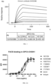
- FACS FACS was used to evaluate the binding activity of 52H5D3B8 chimeric mAbs with comparison to a known GPC3 antibody GC33 (Chugai Pharmaceutical) on human GPC3 over-expressed CHOK1 cells, HEPG2 cells, Hep3B cells and Huh-7 cells.
- the binding activity to GPC3-CHOK1 cells, HEPG2 cells, Hep3B cells and Huh-7 cells EC50 mAbs GPC3-CHOK1 cells HEPG2 cells Hep3B cells Huh-7 cells 52H5D3B8 0.0922 ⁇ g/mL 0.1111 ⁇ g/mL 0.1915 ⁇ g/mL 0.0986 ⁇ g/mL GC33 0.1656 ⁇ g/mL 0.1808 ⁇ g/mL 0.3411 ug/mL 0.1816 ug/mL
- ELISA testing was carried out to evaluate the binding of chimeric antibodies to human, mouse and cynomolgus GPC3, respectively.
- microtiter plates were coated with human, mouse and cynomolgus GPC3 proteins at 1 ⁇ g/ml in PBS, 100 ⁇ l/well at 4°C overnight, then blocked with 150 ⁇ l/well of 1% BSA. Three-fold dilutions of chimeric antibodies starting from 9 ⁇ g/ml were added to each well and incubated for 1 hour at 37 °C. The plates were washed with PBS/Tween and then incubate with mouse-anti-human IgG Fc antibody conjugated with Horse Radish Peroxidase (HRP) for 30 mins at 37 °C. After washing, the plates were developed with TMB substrate and analyzed by spectrophotometer at OD 450nm.
- HRP Horse Radish Peroxidase
- This example describes the optimization of 52H5D3B8 chimeric mAb by mutation of CDR3s to enhance the binding activity of 52H5D3B8 chimeric mAb on human GPC3.
- FACS FACS was used to evaluate the binding activity of 52H5D3B8, 52H5D3B8-1#, 52H5D3B8-2#, 52H5D3B8-3#, 52H5D3B8-4#, 52H5D3B8-5#, 52H5D3B8-6#, 52H5D3B8-7#, 52H5D3B8-8#, 52H5D3B8-9#, 52H5D3B8-10#, 52H5D3B8-11#, 52H5D3B8-12#, 52H5D3B8-13#, 52H5D3B8-14# and 52H5D3B8-15# chimeric mAbs on human GPC3 over-expressed CHOK1 cells.
- GPC3-CHOK1 cells were firstly incubated with Fc blocking reagent at 4°C for 15 mins. Cells were washed by FACS buffer and divided to each well with 3-fold serially diluted 52H5D3B8, 52H5D3B8-1#, 52H5D3B8-2#, 52H5D3B8-3#, 52H5D3B8-4#, 52H5D3B8-5#, 52H5D3B8-6#, 52H5D3B8-7#, 52H5D3B8-8#, 52H5D3B8-9#, 52H5D3B8-10#, 52H5D3B8-11#, 52H5D3B8-12#, 52H5D3B8-13#, 52H5D3B8-14# and 52H5D3B8-15# chimeric mAbs starting at 30 ⁇ g/mL at 4°C for 60 mins.
- the optimized antibodies exhibited diverse binding affinity to the recombinant GPC3 protein ranging from 10 -8 to 10 -10 .
- 52H5D3B8-6# mAbs showed the highest affinity to GPC3 protein.
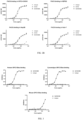
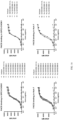
- the humanized antibodies were analyzed for their binding to human GPC3 over-expressed CHOK1 cells, HEPG2 cells, Hep3B cells and Huh-7 cells by FACS. Briefly, GPC3-CHOK1 cells, HEPG2 cells, Hep3B cells and Huh-7 cells were firstly incubated with Fc blocking reagent at 4°C for 15 mins. Cells were washed by FACS buffer and divided to each well with 4-fold serially diluted humanized antibodies starting at 10 ⁇ g/mL at 4°C for 60 mins. Cells were washed by FACS buffer and fixed with 2% PFA at RT for 15 mins.
- the 16 humanized antibodies were separated into two plates. As shown in FIG. 7-8 .
- the EC50 of each humanized antibody on GPC3-CHOK1 cells, HEPG2 cells, Hep3B cells and Huh-7 cells are listed in Tables 11-12 below.
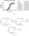
- FACS was used to evaluate the binding activity of 52H5D3B8-6#-VH1+VL1 and 52H5D3B8-6#-VH2+VL3 with comparison to GC33 (Chugai Pharmaceutical) and Y035 (Carsgen Therapeutics, U.S. Patent Application US20190046659 ) on human GPC3 over-expressed CHOK1 cells and HEPG2 cells.
- GC33 Chougai Pharmaceutical
- Y035 Carsgen Therapeutics, U.S. Patent Application US20190046659
- the EC50 of each antibody on GPC3-CHOK1 cells and HEPG2 cells are listed in Table 14 below.
- both 52H5D3B8-6#-VH1+VL1 and 52H5D3B8-6#-VH2+VL3 bound to GPC3-CHOK1 cells and HEPG2 cells with comparable affinity, which are higher than benchmark mAbs.
- Table 14 shows that both 52H5D3B8-6#-VH1+VL1 and 52H5D3B8-6#-VH2+VL3 bound to GPC3-CHOK1 cells and HEPG2 cells with comparable affinity, which are higher than benchmark mAbs. Table 14.
- affinity maturation was performed. Briefly, 4 phage libraries containing single-point or two-point saturation mutation in CDR regions were constructed. Three candidates with unique mutations in CDRs were obtained by 2 rounds of screening with solid or liquid panning. CDRs of these candidates were engrafted into 52H5D3B8-6#-VH2+VL3 frameworks to generate antibodies for further binding and functional validation.
- variable regions of frameworks engrafted affinity maturated candidates are listed in Table 15 below.
- Table 15 Sequences of the variable regions of 52H5D3B8-18#-VH2+VL3, 52H5D3B8-19#-VH2+VL3, 52H5D3B8-20#-VH2+VL3 Name Sequence SEQ ID NO: 52H5D3B8-18#-VH2+VL3 VH 53 52H5D3B8-18#-VH2+VL3 VL 54 52H5D3B8-19#-VH2+VL3 VH 55 52H5D3B8-19#-VH2+VL3 VL 56 52H5D3B8-20#-VH2+VL3 VH 57 52H5D3B8-20#-VH2+VL3 VL 58 Table 15A.
- the affinity maturated antibodies were analyzed for their binding to human GPC3 over-expressed CHOK1 cells by FACS as previously described.
- the EC50 of each antibody on GPC3-CHOK1 cells are listed in Table 16 below.
- ADCC antibody-dependent cell cytotoxicity
- Jukrat-hCD16a-NFAT cell line allowed us to evaluate GPC3 binding mediated ADCC signaling by examining downstream NFAT signaling.
- This reporter cell line was cocultured with several GPC3 positive tumor cell lines including HEPG2 or Huh-7 hepatocellular carcinoma cells with high or moderate GPC3 expression level, respectively.
- the Luciferase-based chemiluminescence could be detected by ONE-Glo TM Luciferase assay system (Promega, Cat.No E6130) and Envision multilabel plate readers (PerkinElemer).
- both 52H5D3B8-6#-VH1+VL1 and 52H5D3B8-6#-VH2+VL3 showed efficient GPC3 binding mediated ADCC signaling to HEPG2 cells and Huh-7 cells, which are higher than GC33.
- both 52H5D3B8-19#-VH2+VL3 and 52H5D3B8-20#-VH2+VL3 showed better EC50 of ADCC signaling to HEPG2 cells than parental 52H5D3B8-6#-VH2+VL3, while 52H5D3B8-18#-VH2+VL3 showed better maximum ADCC signaling potency than other antibodies.
- Huh-7 cells which showed moderate GPC3 expression level, all humanized antibodies exhibited enhanced maximum ADCC signaling potency over GC33.
- a human primary NK cell-mediated cytotoxicity assay was set-up. Briefly, fresh human primary NK cells (CD3-CD56+) were isolated from buffy coat of healthy donors by negative selection with magnetic beads (Miltenyi, Cat. No. 130-092-657). The purity of the isolated NK cells was monitored by FACS analysis and typically more than 90%. The isolated NK cells were then rested in the complete culture media for 24 hours, which were used as effector cells. HEPG2 or Huh-7 human hepatocellular carcinoma cell line were used as target cells.
- LDH lactate dehydrogenase
- Cytotoxicity (%) (effector - target cell mix - effector cell control - low control) / (high control - low control) x 100.
- Cytotoxicity (%) (effector - target cell mix - effector cell control - low control) / (high control - low control) x 100.
- Four parameter nonlinear regression curve fit was performed with GraphPad Prism 9.
- both 52H5D3B8-6#-VH1+VL1 and 52H5D3B8-6#-VH2+VL3 showed efficient GPC3 antibody-mediated NK cytotoxicity to HEPG2 cells and Huh-7 cells, which are higher than GC33.
- both 52H5D3B8-19#-VH2+VL3 and 52H5D3B8-20#-VH2+VL3 showed comparable GPC3 antibody-mediated NK cytotoxicity to HEPG2 cells with parental 52H5D3B8-6#-VH2+VL3.
- both 52H5D3B8-19#-VH2+VL3 and 52H5D3B8-20#-VH2+VL3 showed better EC50 than parental 52H5D3B8-6#-VH2+VL3.
- No 328608 was diluted in the staining buffer with 1 ⁇ L per well and incubated with the cells for 40 mins at 4°C in the dark. The samples were then washed and fixed with 2% PFA. Cells were analyzed on the flow cytometer MACSQuant ® Analyze 16 (Miltenyi Biotech). Data were analyzed with Flowjo 10.0 software. Four parameter nonlinear regression curve fit was performed with GraphPad Prism 9.
- the protein trafficking inhibitor Brefeldin A (BFA) was added into the co-culture systems to prevent cytokine secretion to the supernatant during NK cell activation.
- Antibodies were serially diluted at a 5-fold starting from 50 nM and added into corresponding wells.
- the 52H5D3B8 variable region genes were employed to create a humanized mAb.
- the amino acid sequences of the VH and VL or VK of 52H5D3B8 were compared against the available database of human Ig gene sequences to find the overall best-matching human germline Ig gene sequences.
- IGKV2-29*02 and IGHV1-2*02 were used as backbone of heavy chain and light chain separately.
- a 3D model was then generated to determine the amino acids in the original mouse FR region sequences that are essential for antibody binding and conformation. Based on the 52H5D3B8 CDR grafting antibody sequence, 5 additional humanized heavy chains and 1 additional light chain were created, In the case of the heavy chain, I, K, A, L, T in the framework were involved in back-mutations. In the case of the light chain, F in the framework were involved in back-mutations.
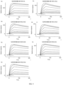
- the chimeric antibody's affinity was measured by Biacore 8K. Two concentrations with most close affinity to the chimeric antibody's affinity were chosen for the 12 humanized antibodies' affinity detection. Human GPC3-his tag protein was injected over captured antibody for 3 mins at a flow rate of 30 ⁇ l/min. The antigen was allowed to dissociate for 720s. As shown in Table 21, all 12 humanized antibodies showed similar affinity to the chimeric antibody. Table 21.
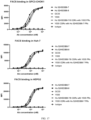
- the humanized antibodies were analyzed for their binding to human GPC3 over-expressed CHOK1 cells and Huh-7 cells by FACS. Briefly, GPC3-CHOK1 cells and Huh-7 cells were firstly incubated with Fc blocking reagent at 4°C for 15 mins. Cells were washed by FACS buffer and divided to each well with 4-fold serially diluted humanized antibodies starting at 10 ⁇ g/mL at 4°C for 60 mins. Cells were washed by FACS buffer and fixed with 2% PFA at RT for 15 mins.
- the binding activity to GPC3-CHOK1 and Huh-7 cells EC50 mAbs GPC3-CHOK1 cells Huh-7 cells 52H5D3B8-VH+VL 0.0479 ⁇ g/mL 0.0563 ⁇ g/mL Hu 52H5D3B8-10 0.0389 ⁇ g/mL 0.0323 ⁇ g/mL Hu 52H5D3B8-11 0.0400 ⁇ g/mL 0.0288 ⁇ g/mL Hu 52H5D3B8-12 0.0384 ⁇ g/mL 0.0294 ⁇ g/mL
- both Hu 52H5D3B8-7 and Hu 52H5D3B8-8 bound to GPC3-CHOK1 cells, Huh-7 cells and HEPG2 cells with comparable affinity, which are higher than benchmark antibodies GC33 and Y035. More importantly, both Hu 52H5D3B8-7/8 CDRs with Y035 FRs and Y035 CDRs with Hu 52H5D3B8-7 FRs showed higher affinity than Y035, which demonstrated that the CDRs and frameworks of Hu 52H5D3B8-7 contributed to a superior binding ability to Y035. Table 24.
- PHAb thiol Dyes are pH sensor dyes that have very low fluorescence at pH > 7, and a dramatic increase in fluorescence will occur when as it is internalized into the endosome or lysosome where the pH is around 6.3 or 4.7, respectively.
- a pHAb thiol Dyes labeled ⁇ -hIgG secondary antibody (10 ⁇ g/mL) was incubated with antibodies (20 ⁇ g/mL) for 30min. After incubation, the mixture Abs were serially diluted with 2-fold. Then the diluted mixture was added into 96-well assay plates pre-seeded with 2 ⁇ 10 4 HEPG2 cells in each well. After 48 hours incubation, fluorescence signal was captured on Multimode Plate Reader (Envision ® 2105).
- both Hu 52H5D3B8-7 and Hu 52H5D3B8-8 showed higher internalization efficacy than other mAbs.
- Hu 52H5D3B8-7/8 CDRs with Y035 FRs showed higher internalization efficacy than Y035 (higher Top and comparable EC50), which indicated that the CDRs of Hu-52H5D3B8-7 contribute to an enhanced target mediated internalization when compared with Y035.
- the EC50 and Top of each mAbs is listed in Table 25 below. Table 25.
Landscapes
- Health & Medical Sciences (AREA)
- Chemical & Material Sciences (AREA)
- Life Sciences & Earth Sciences (AREA)
- General Health & Medical Sciences (AREA)
- Organic Chemistry (AREA)
- Immunology (AREA)
- Medicinal Chemistry (AREA)
- Animal Behavior & Ethology (AREA)
- Veterinary Medicine (AREA)
- Public Health (AREA)
- Pharmacology & Pharmacy (AREA)
- Cell Biology (AREA)
- Gastroenterology & Hepatology (AREA)
- Epidemiology (AREA)
- Chemical Kinetics & Catalysis (AREA)
- General Chemical & Material Sciences (AREA)
- Nuclear Medicine, Radiotherapy & Molecular Imaging (AREA)
- Proteomics, Peptides & Aminoacids (AREA)
- Molecular Biology (AREA)
- Genetics & Genomics (AREA)
- Biophysics (AREA)
- Biochemistry (AREA)
- Bioinformatics & Cheminformatics (AREA)
- Engineering & Computer Science (AREA)
- Oncology (AREA)
- Peptides Or Proteins (AREA)
- Medicinal Preparation (AREA)
- Medicines Containing Antibodies Or Antigens For Use As Internal Diagnostic Agents (AREA)
- Micro-Organisms Or Cultivation Processes Thereof (AREA)
- Medicines That Contain Protein Lipid Enzymes And Other Medicines (AREA)
- Preparation Of Compounds By Using Micro-Organisms (AREA)
Applications Claiming Priority (3)
| Application Number | Priority Date | Filing Date | Title |
|---|---|---|---|
| CN2021124182 | 2021-10-15 | ||
| EP22880441.5A EP4416187A1 (fr) | 2021-10-15 | 2022-10-17 | Anticorps anti-glypicane 3 |
| PCT/CN2022/125725 WO2023061505A1 (fr) | 2021-10-15 | 2022-10-17 | Anticorps anti-glypicane 3 |
Related Parent Applications (1)
| Application Number | Title | Priority Date | Filing Date |
|---|---|---|---|
| EP22880441.5A Division EP4416187A1 (fr) | 2021-10-15 | 2022-10-17 | Anticorps anti-glypicane 3 |
Publications (2)
| Publication Number | Publication Date |
|---|---|
| EP4527465A2 true EP4527465A2 (fr) | 2025-03-26 |
| EP4527465A3 EP4527465A3 (fr) | 2025-07-09 |
Family
ID=85988149
Family Applications (2)
| Application Number | Title | Priority Date | Filing Date |
|---|---|---|---|
| EP22880441.5A Pending EP4416187A1 (fr) | 2021-10-15 | 2022-10-17 | Anticorps anti-glypicane 3 |
| EP25152478.1A Pending EP4527465A3 (fr) | 2021-10-15 | 2022-10-17 | Anticorps anti-glypicane 3 |
Family Applications Before (1)
| Application Number | Title | Priority Date | Filing Date |
|---|---|---|---|
| EP22880441.5A Pending EP4416187A1 (fr) | 2021-10-15 | 2022-10-17 | Anticorps anti-glypicane 3 |
Country Status (5)
| Country | Link |
|---|---|
| US (2) | US20240409663A1 (fr) |
| EP (2) | EP4416187A1 (fr) |
| JP (2) | JP2024537323A (fr) |
| CN (3) | CN119080937A (fr) |
| WO (1) | WO2023061505A1 (fr) |
Families Citing this family (4)
| Publication number | Priority date | Publication date | Assignee | Title |
|---|---|---|---|---|
| WO2024067764A1 (fr) * | 2022-09-30 | 2024-04-04 | 信立泰(成都)生物技术有限公司 | Anticorps monoclonal/anticorps bispécifique anti-gpc3 ou fragment de liaison à l'antigène de celui-ci et son utilisation |
| WO2025045242A1 (fr) * | 2023-09-01 | 2025-03-06 | 信立泰(成都)生物技术有限公司 | Anticorps bispécifique ciblant gpc3 ou fragment de liaison à l'antigène et son utilisation |
| WO2025051159A1 (fr) * | 2023-09-05 | 2025-03-13 | 乐普生物科技股份有限公司 | Conjugué anticorps-médicament ciblant gpc3 et son utilisation |
| WO2025159562A1 (fr) * | 2024-01-24 | 2025-07-31 | 국립암센터 | Nouvel anticorps se liant de manière spécifique à gpc3 et son utilisation |
Citations (4)
| Publication number | Priority date | Publication date | Assignee | Title |
|---|---|---|---|---|
| US6150584A (en) | 1990-01-12 | 2000-11-21 | Abgenix, Inc. | Human antibodies derived from immunized xenomice |
| US6420140B1 (en) | 1996-10-11 | 2002-07-16 | Abgenix, Inc. | Production of multimeric protein by cell fusion method |
| US6458592B1 (en) | 1995-03-29 | 2002-10-01 | Abgenix, Inc. | Production of antibodies using cre-mediated site-specific recombination |
| US20190046659A1 (en) | 2015-08-03 | 2019-02-14 | Carsgen Therapeutics Ltd | Antibody against glypican-3 and application thereof |
Family Cites Families (13)
| Publication number | Priority date | Publication date | Assignee | Title |
|---|---|---|---|---|
| KR100877176B1 (ko) * | 2001-06-22 | 2009-01-07 | 츄가이 세이야꾸 가부시키가이샤 | 항글리피칸 3항체를 포함하는 세포증식 억제제 |
| WO2004022597A1 (fr) * | 2002-09-04 | 2004-03-18 | Chugai Seiyaku Kabushiki Kaisha | Anticorps d'un peptide n-terminal du gpc3 solubilise dans le sang |
| EP1674111B1 (fr) * | 2004-07-09 | 2010-11-03 | Chugai Seiyaku Kabushiki Kaisha | Anticorps anti-glypican 3 |
| RS54624B1 (sr) * | 2007-07-17 | 2016-08-31 | E. R. Squibb & Sons, L.L.C. | Monoklonska antitela protiv glipikana-3 |
| CN101633693A (zh) * | 2009-08-24 | 2010-01-27 | 中国人民解放军第二军医大学 | 一种抗gpc3的单克隆抗体 |
| US9926377B2 (en) * | 2014-05-22 | 2018-03-27 | Genentech, Inc. | Anti-GPC3 antibodies and immunoconjugates |
| MX2016015162A (es) * | 2014-05-22 | 2017-03-03 | Genentech Inc | Anticuerpos anti - gpc3 e inmunoconjugados. |
| CN105037540A (zh) * | 2015-05-13 | 2015-11-11 | 北京比洋生物技术有限公司 | 抗磷脂酰肌醇蛋白聚糖3全人源抗体 |
| MX2018010546A (es) * | 2016-03-15 | 2019-02-20 | Chugai Pharmaceutical Co Ltd | Metodos de tratamiento de cancer que usan antagonistas de union al eje de muerte programada 1 (pd-1) y anticuerpos anti glipicano 3 (gpc3). |
| AU2018258049B2 (en) * | 2017-04-26 | 2025-03-27 | Eureka Therapeutics, Inc. | Constructs specifically recognizing glypican 3 and uses thereof |
| CN109988240B (zh) * | 2017-12-29 | 2022-06-28 | 安源医药科技(上海)有限公司 | 抗gpc-3抗体及其用途 |
| CN109021108B (zh) * | 2018-08-27 | 2019-06-25 | 南京医科大学 | 抗gpc3全人源化抗体、其嵌合抗原受体细胞及应用 |
| EP4007604A4 (fr) * | 2019-08-01 | 2023-07-19 | R.P. Scherer Technologies, LLC | Anticorps spécifique gpc3 et leurs procédés d'utilisation |
-
2022
- 2022-10-17 US US18/700,228 patent/US20240409663A1/en active Pending
- 2022-10-17 CN CN202411431728.XA patent/CN119080937A/zh active Pending
- 2022-10-17 CN CN202280083725.4A patent/CN118488970A/zh active Pending
- 2022-10-17 CN CN202411431725.6A patent/CN118994403B/zh active Active
- 2022-10-17 EP EP22880441.5A patent/EP4416187A1/fr active Pending
- 2022-10-17 JP JP2024522070A patent/JP2024537323A/ja active Pending
- 2022-10-17 EP EP25152478.1A patent/EP4527465A3/fr active Pending
- 2022-10-17 WO PCT/CN2022/125725 patent/WO2023061505A1/fr not_active Ceased
-
2025
- 2025-01-16 US US19/026,082 patent/US20250154280A1/en active Pending
- 2025-01-17 JP JP2025006663A patent/JP2025061426A/ja active Pending
Patent Citations (4)
| Publication number | Priority date | Publication date | Assignee | Title |
|---|---|---|---|---|
| US6150584A (en) | 1990-01-12 | 2000-11-21 | Abgenix, Inc. | Human antibodies derived from immunized xenomice |
| US6458592B1 (en) | 1995-03-29 | 2002-10-01 | Abgenix, Inc. | Production of antibodies using cre-mediated site-specific recombination |
| US6420140B1 (en) | 1996-10-11 | 2002-07-16 | Abgenix, Inc. | Production of multimeric protein by cell fusion method |
| US20190046659A1 (en) | 2015-08-03 | 2019-02-14 | Carsgen Therapeutics Ltd | Antibody against glypican-3 and application thereof |
Non-Patent Citations (5)
| Title |
|---|
| "Monoclonal Antibodies For Cancer Detection And Therapy", 1985, ACADEMIC PRESS, article "Analysis, Results, And Future Prospective Of The Therapeutic Use Of Radiolabeled Antibody In Cancer Therapy", pages: 303 - 16 |
| ARNON ET AL.: "Monoclonal Antibodies And Cancer Therapy", 1985, ALAN R. LISS INC, article "Monoclonal Antibodies For Immunotargeting Of Drugs In Cancer Therapy", pages: 243 - 56 |
| HELLSTROM ET AL.: "Controlled Drug Delivery", 1987, MARCEL DEKKER, INC., article "Antibodies For Drug Delivery", pages: 623 - 53 |
| THORPE ET AL.: "Monoclonal Antibodies '84: Biological And Clinical Applications", 1985, article "Antibody Carriers Of Cytotoxic Agents In Cancer Therapy: A Review", pages: 475 - 506 |
| THORPE ET AL.: "The Preparation And Cytotoxic Properties Of Antibody-Toxin Conjugates", IMMUNOL. REV., vol. 52, 1982, pages 119 - 58, XP001179872, DOI: 10.1111/j.1600-065X.1982.tb00392.x |
Also Published As
| Publication number | Publication date |
|---|---|
| US20250154280A1 (en) | 2025-05-15 |
| JP2025061426A (ja) | 2025-04-10 |
| CN118994403A (zh) | 2024-11-22 |
| EP4416187A1 (fr) | 2024-08-21 |
| EP4527465A3 (fr) | 2025-07-09 |
| US20240409663A1 (en) | 2024-12-12 |
| WO2023061505A1 (fr) | 2023-04-20 |
| CN118994403B (zh) | 2025-04-01 |
| CN119080937A (zh) | 2024-12-06 |
| JP2024537323A (ja) | 2024-10-10 |
| CN118488970A (zh) | 2024-08-13 |
Similar Documents
| Publication | Publication Date | Title |
|---|---|---|
| US11873342B2 (en) | Anti-CCR8 monoclonal antibodies and uses thereof | |
| JP7222024B2 (ja) | 上皮増殖因子受容体変異体iiiおよびcd3の単一および二重特異性抗体およびそれらの使用 | |
| KR102224556B1 (ko) | Ig 및 itim 도메인을 갖는 t 세포 면역 수용체 (tigit)에 대한 항체 및 이것의 사용 | |
| EP4527465A2 (fr) | Anticorps anti-glypicane 3 | |
| JP2020055870A (ja) | 抗ntb−a抗体ならびに関連する組成物および方法 | |
| CA3160159A1 (fr) | Conjugues anticorps-medicament ciblant claudine 18,2 | |
| US20250011427A1 (en) | Anti-cd3 antibodies with cross-reactivity to human and cynomolgus proteins | |
| US20250313632A1 (en) | Anti-b7-h3 antibodies and uses thereof | |
| US20250186601A1 (en) | Antibodies targeting liv-1 and uses thereof | |
| US20250154277A1 (en) | Anti-5t4 antibodies and uses thereof | |
| TW202330599A (zh) | 抗ccr8單株抗體及其用途 | |
| US12180276B2 (en) | Antibodies and antibody-drug conjugates targeting claudin 6 | |
| EP4662241A1 (fr) | Anticorps ciblant liv-1 et leurs utilisations |
Legal Events
| Date | Code | Title | Description |
|---|---|---|---|
| PUAI | Public reference made under article 153(3) epc to a published international application that has entered the european phase |
Free format text: ORIGINAL CODE: 0009012 |
|
| STAA | Information on the status of an ep patent application or granted ep patent |
Free format text: STATUS: THE APPLICATION HAS BEEN PUBLISHED |
|
| AC | Divisional application: reference to earlier application |
Ref document number: 4416187 Country of ref document: EP Kind code of ref document: P |
|
| AK | Designated contracting states |
Kind code of ref document: A2 Designated state(s): AL AT BE BG CH CY CZ DE DK EE ES FI FR GB GR HR HU IE IS IT LI LT LU LV MC ME MK MT NL NO PL PT RO RS SE SI SK SM TR |
|
| PUAL | Search report despatched |
Free format text: ORIGINAL CODE: 0009013 |
|
| AK | Designated contracting states |
Kind code of ref document: A3 Designated state(s): AL AT BE BG CH CY CZ DE DK EE ES FI FR GB GR HR HU IE IS IT LI LT LU LV MC ME MK MT NL NO PL PT RO RS SE SI SK SM TR |
|
| RIC1 | Information provided on ipc code assigned before grant |
Ipc: C07K 16/30 20060101ALI20250605BHEP Ipc: A61K 38/00 20060101ALI20250605BHEP Ipc: A61P 35/00 20060101AFI20250605BHEP |
|
| RAP1 | Party data changed (applicant data changed or rights of an application transferred) |
Owner name: LEPU BIOPHARMA CO., LTD. |