EP4473979A1 - Mit einem nahinfrarot-fluoreszenzfarbstoff geladene polymermikrokugeln zur markierung von läsionen und formulierung daraus - Google Patents
Mit einem nahinfrarot-fluoreszenzfarbstoff geladene polymermikrokugeln zur markierung von läsionen und formulierung daraus Download PDFInfo
- Publication number
- EP4473979A1 EP4473979A1 EP23749928.0A EP23749928A EP4473979A1 EP 4473979 A1 EP4473979 A1 EP 4473979A1 EP 23749928 A EP23749928 A EP 23749928A EP 4473979 A1 EP4473979 A1 EP 4473979A1
- Authority
- EP
- European Patent Office
- Prior art keywords
- microspheres
- hsa
- solution
- icg
- prepared
- Prior art date
- Legal status (The legal status is an assumption and is not a legal conclusion. Google has not performed a legal analysis and makes no representation as to the accuracy of the status listed.)
- Pending
Links
Images












Classifications
-
- A—HUMAN NECESSITIES
- A61—MEDICAL OR VETERINARY SCIENCE; HYGIENE
- A61K—PREPARATIONS FOR MEDICAL, DENTAL OR TOILETRY PURPOSES
- A61K49/00—Preparations for testing in vivo
- A61K49/001—Preparation for luminescence or biological staining
- A61K49/0013—Luminescence
- A61K49/0017—Fluorescence in vivo
- A61K49/0019—Fluorescence in vivo characterised by the fluorescent group, e.g. oligomeric, polymeric or dendritic molecules
- A61K49/0021—Fluorescence in vivo characterised by the fluorescent group, e.g. oligomeric, polymeric or dendritic molecules the fluorescent group being a small organic molecule
- A61K49/0032—Methine dyes, e.g. cyanine dyes
- A61K49/0034—Indocyanine green, i.e. ICG, cardiogreen
-
- A—HUMAN NECESSITIES
- A61—MEDICAL OR VETERINARY SCIENCE; HYGIENE
- A61K—PREPARATIONS FOR MEDICAL, DENTAL OR TOILETRY PURPOSES
- A61K49/00—Preparations for testing in vivo
- A61K49/001—Preparation for luminescence or biological staining
- A61K49/0013—Luminescence
- A61K49/0017—Fluorescence in vivo
- A61K49/0019—Fluorescence in vivo characterised by the fluorescent group, e.g. oligomeric, polymeric or dendritic molecules
- A61K49/0021—Fluorescence in vivo characterised by the fluorescent group, e.g. oligomeric, polymeric or dendritic molecules the fluorescent group being a small organic molecule
- A61K49/0032—Methine dyes, e.g. cyanine dyes
-
- A—HUMAN NECESSITIES
- A61—MEDICAL OR VETERINARY SCIENCE; HYGIENE
- A61K—PREPARATIONS FOR MEDICAL, DENTAL OR TOILETRY PURPOSES
- A61K49/00—Preparations for testing in vivo
- A61K49/001—Preparation for luminescence or biological staining
- A61K49/0013—Luminescence
- A61K49/0017—Fluorescence in vivo
- A61K49/005—Fluorescence in vivo characterised by the carrier molecule carrying the fluorescent agent
- A61K49/0054—Macromolecular compounds, i.e. oligomers, polymers, dendrimers
-
- A—HUMAN NECESSITIES
- A61—MEDICAL OR VETERINARY SCIENCE; HYGIENE
- A61K—PREPARATIONS FOR MEDICAL, DENTAL OR TOILETRY PURPOSES
- A61K49/00—Preparations for testing in vivo
- A61K49/001—Preparation for luminescence or biological staining
- A61K49/0013—Luminescence
- A61K49/0017—Fluorescence in vivo
- A61K49/005—Fluorescence in vivo characterised by the carrier molecule carrying the fluorescent agent
- A61K49/0056—Peptides, proteins, polyamino acids
-
- A—HUMAN NECESSITIES
- A61—MEDICAL OR VETERINARY SCIENCE; HYGIENE
- A61K—PREPARATIONS FOR MEDICAL, DENTAL OR TOILETRY PURPOSES
- A61K49/00—Preparations for testing in vivo
- A61K49/001—Preparation for luminescence or biological staining
- A61K49/006—Biological staining of tissues in vivo, e.g. methylene blue or toluidine blue O administered in the buccal area to detect epithelial cancer cells, dyes used for delineating tissues during surgery
-
- A—HUMAN NECESSITIES
- A61—MEDICAL OR VETERINARY SCIENCE; HYGIENE
- A61K—PREPARATIONS FOR MEDICAL, DENTAL OR TOILETRY PURPOSES
- A61K49/00—Preparations for testing in vivo
- A61K49/001—Preparation for luminescence or biological staining
- A61K49/0063—Preparation for luminescence or biological staining characterised by a special physical or galenical form, e.g. emulsions, microspheres
- A61K49/0069—Preparation for luminescence or biological staining characterised by a special physical or galenical form, e.g. emulsions, microspheres the agent being in a particular physical galenical form
- A61K49/0089—Particulate, powder, adsorbate, bead, sphere
-
- A—HUMAN NECESSITIES
- A61—MEDICAL OR VETERINARY SCIENCE; HYGIENE
- A61K—PREPARATIONS FOR MEDICAL, DENTAL OR TOILETRY PURPOSES
- A61K49/00—Preparations for testing in vivo
- A61K49/001—Preparation for luminescence or biological staining
- A61K49/0063—Preparation for luminescence or biological staining characterised by a special physical or galenical form, e.g. emulsions, microspheres
- A61K49/0069—Preparation for luminescence or biological staining characterised by a special physical or galenical form, e.g. emulsions, microspheres the agent being in a particular physical galenical form
- A61K49/0089—Particulate, powder, adsorbate, bead, sphere
- A61K49/0091—Microparticle, microcapsule, microbubble, microsphere, microbead, i.e. having a size or diameter higher or equal to 1 micrometer
-
- B—PERFORMING OPERATIONS; TRANSPORTING
- B01—PHYSICAL OR CHEMICAL PROCESSES OR APPARATUS IN GENERAL
- B01J—CHEMICAL OR PHYSICAL PROCESSES, e.g. CATALYSIS OR COLLOID CHEMISTRY; THEIR RELEVANT APPARATUS
- B01J13/00—Colloid chemistry, e.g. the production of colloidal materials or their solutions, not otherwise provided for; Making microcapsules or microballoons
- B01J13/02—Making microcapsules or microballoons
- B01J13/04—Making microcapsules or microballoons by physical processes, e.g. drying, spraying
- B01J13/046—Making microcapsules or microballoons by physical processes, e.g. drying, spraying combined with gelification or coagulation
-
- B—PERFORMING OPERATIONS; TRANSPORTING
- B01—PHYSICAL OR CHEMICAL PROCESSES OR APPARATUS IN GENERAL
- B01J—CHEMICAL OR PHYSICAL PROCESSES, e.g. CATALYSIS OR COLLOID CHEMISTRY; THEIR RELEVANT APPARATUS
- B01J13/00—Colloid chemistry, e.g. the production of colloidal materials or their solutions, not otherwise provided for; Making microcapsules or microballoons
- B01J13/02—Making microcapsules or microballoons
- B01J13/06—Making microcapsules or microballoons by phase separation
- B01J13/10—Complex coacervation, i.e. interaction of oppositely charged particles
-
- C—CHEMISTRY; METALLURGY
- C09—DYES; PAINTS; POLISHES; NATURAL RESINS; ADHESIVES; COMPOSITIONS NOT OTHERWISE PROVIDED FOR; APPLICATIONS OF MATERIALS NOT OTHERWISE PROVIDED FOR
- C09B—ORGANIC DYES OR CLOSELY-RELATED COMPOUNDS FOR PRODUCING DYES, e.g. PIGMENTS; MORDANTS; LAKES
- C09B67/00—Influencing the physical, e.g. the dyeing or printing properties of dyestuffs without chemical reactions, e.g. by treating with solvents grinding or grinding assistants, coating of pigments or dyes; Process features in the making of dyestuff preparations; Dyestuff preparations of a special physical nature, e.g. tablets, films
- C09B67/0097—Dye preparations of special physical nature; Tablets, films, extrusion, microcapsules, sheets, pads, bags with dyes
Definitions
- the present invention relates to near-infrared fluorescent dye-loaded microspheres and a formulation thereof, and specifically, to near-infrared fluorescent dye-loaded microspheres and a formulation thereof that, by injecting the microspheres into lesion sites such as cancer, can help enable precision surgery by accurately identifying the location of lesion sites from fluorescence imaging during surgery.
- fluorescence imaging technology Since fluorescence imaging technology has the great advantage of being able to acquire real-time images with high sensitivity compared to magnetic resonance imaging, nuclear medicine imaging and ultrasound imaging, it is evaluated as the most powerful tool for identifying and removing the location of lesions such as cancer in real time during surgery. Moreover, as systems capable of implementing near-infrared fluorescence imaging functions in endoscopic and laparoscopic equipment have recently become increasingly commercialized, procedures using fluorescence imaging are expected to be widely used in patient procedures in the future.
- Laparoscopic surgery is one of the "minimally invasive surgical" methods that reduces injury or impact on the patient's body and is a method of performing surgery using surgical tools (forceps, electric scalpels, hemostatic suturing device, and the like) while viewing the abdomen by creating a small hole of 0.5 to 1.5 cm in size instead of making an incision in the abdomen or chest and inserting an endoscope (laparoscope) equipped with a special camera.
- surgical tools forceps, electric scalpels, hemostatic suturing device, and the like
- laparoscopic surgery has been developed over the past several decades due to its advantages, such as less postoperative pain compared to laparotomy, faster recovery due to a smaller incision area, and shorter hospitalization period.
- a method is being used in which a drug such as Indian ink is injected into the lesion identified through an endoscope through an endoscopic injection needle to mark the lesion sites, and then surgery is performed while visually identifying the sites marked with Indian ink through a laparoscope during surgery.
- this marking method has the disadvantage that complications occur due to ink incorrectly injected into the muscle layer, and the sites to be removed by surgery widen as the ink or dye spreads from the injected sites to surrounding sites.
- a contrast agent for "marking" or "marker” is needed to indicate the appropriate location of the disease lesion.
- Fluorescence imaging technology using near-infrared fluorescent dyes has the advantage of not only being able to detect marked sites located deep in tissues from fluorescence imaging, but also being able to sensitively detect fluorescence signals even when using a small amount of fluorescent dye. Since fluorescence imaging technology has the great advantage of being able to acquire real-time images with high sensitivity compared to magnetic resonance imaging, nuclear medicine imaging and ultrasound imaging, it is evaluated as the most powerful tool for identifying and removing the location of lesions such as cancer in real time during surgery. As a fluorescent imaging contrast agent that has been attempted previously, indocyanine green, which is a near-infrared fluorescent dye approved for clinical use by the U.S.
- FDA Food and Drug Administration
- Indocyanine green has reduced fluorescence brightness due to concentration-dependent aggregation phenomenon and is prone to decomposition due to chemical instability in aqueous solutions.
- fluorescence signals are generated in a wider range than the injected sites or that the fluorescence signals weaken within a short period of time after injection. For this reason, there is a need to develop a method that increases the efficiency of marking lesions and maintains high fluorescence signals of near-infrared fluorescent dye for a long time without fluorescence blurring in the marked sites.
- PLGA poly(D,L-lactide-co-glycolide)
- indocyanine green tends to aggregate with each other in aqueous solution and is unstable, if indocyanine green is used alone and loaded into microbubbles, there is a problem that the intensity of fluoresce signal is greatly weakened by the aggregation phenomenon of indocyanine green molecules, and the fluorescence intensity is further reduced by the decomposition of indocyanine green over time.
- microbubbles if a hole is created in the shell layer during the preparation process or by in vivo hydrolysis, as the loaded indocyanine green is quickly released to the outside, a decrease in fluorescence signals and a blurring phenomenon of fluorescence signals to surrounding tissues may occur.
- the present invention has been devised to solve the problems of the prior art as described above, and the present inventors have developed polymer microspheres loaded with a near-infrared fluorescent dye complex consisting of a near-infrared fluorescent dye and human serum albumin, or a near-infrared fluorescent dye and cyclodextrin and completed the present invention. Since near-infrared fluorescent dye-loaded microspheres allow the fluorescent dye loaded within the microspheres to remain within the microspheres for a long period of time while generating a strong fluorescence signal, it is possible to accurately and over a long period of time determine the location of the lesion into which the microspheres were injected through fluorescence imaging.
- the present invention provides near-infrared fluorescent dye-loaded polymer microspheres for marking lesions, wherein the near-infrared fluorescent dye forms a complex with human serum albumin or cyclodextrin.
- the marking of the lesions allows accurate surgery and shortened surgery time by marking the location of the lesions to be removed by surgery in advance before surgery and identifying the marked location in real time through fluorescence imaging during surgery.
- the near-infrared fluorescent dye is any one selected from, but is not limited to, indocyanine green, IRDye 800CW carboxylate (IRDye 800CW), Alexa Fluor 647, Alexa Fluor 660, Alexa Fluor 680, Alexa Fluor 700, Alexa Fluor 750, Alexa Fluor 780, Flamma 749, Flamma 774, Flamma 800, FSD Fluor TM 647, FSD Fluor TM 680, FSD Fluor TM 750, FSD Fluor TM 800, sulfo-cyanine 5 carboxylic acid (Cy5), sulfo-cyanine 5.5 carboxylic acid (Cy5.5), sulfo-cyanine 7 carboxylic acid (Cy7) or sulfo-cyanine 7.5 carboxylic acid (Cy7.5).
- the near-infrared fluorescent dye may be a dye that generates a fluorescence signal at a wavelength of 650 nm or more.
- the indocyanine green is a fluorescent imaging contrast agent approved for clinical use by the U.S. Food and Drug Administration (FDA) and has been used in vascular imaging, and the like, but has several limitations in clinical application.
- Indocyanine green has reduced fluorescence brightness due to concentration-dependent aggregation and is prone to decomposition due to chemical instability in aqueous solutions.
- fluorescence signals are generated in a wider range than the injected sites or that the fluorescence signals weaken within a short period of time after injection. For this reason, there is a need to develop a method that increases the efficiency of marking lesions and maintains high fluorescence signals of near-infrared fluorescent dye for a long time without fluorescence blurring in the marked sites.
- indocyanine green tends to aggregate with each other in aqueous solution and is unstable, if indocyanine green is used alone, there is a problem that the intensity of fluoresce signal is greatly weakened by the aggregation phenomenon of indocyanine green molecules, and the fluorescence intensity is further reduced by the decomposition of indocyanine green over time.
- the microspheres loaded with a complex of a near-infrared fluorescent dye and human serum albumin, or a near-infrared fluorescent dye and cyclodextrin according to the present invention allow the fluorescent dye loaded within the microspheres to remain within the microspheres for a long period of time while generating a strong fluorescence signal, it is possible to accurately and over a long period of time determine the location of the target sites through fluorescence imaging.
- a water-in-oil-in-water (W1/O/W2) emulsion method may be used as a method for loading the complex into the polymer microspheres.
- a near-infrared fluorescent dye with low water solubility is loaded alone into the microspheres, there is a disadvantage that as aggregation of the near-infrared fluorescent dyes occurs within the microspheres, the intensity of the fluorescence signal generated from the prepared microspheres becomes very weak, and the chemical/physical stability of the near-infrared fluorescent dye is also lowered.
- the aggregation phenomenon that occurs when the fluorescent dye is loaded alone may be prevented, the fluorescence signal generated from the microspheres is also much stronger, and the chemical/physical stability of the fluorescent dye is also increased.
- the polymer is a biodegradable polymer and may be, but is not limited to, poly(lactide-co-glycolide) (PLGA), poly(DL-lactide-co-glycolide) (PDLGA), poly(glycolic acid) (PGA), poly(lactide) (PLA), poly(hydroxybutyrate), polycaprolactone (PCL), polydioxanone (PDO), poly(amino acid), polyanhydride, polyorthoester or polyphosphazene.
- PLGA poly(lactide-co-glycolide)
- PDL-lactide-co-glycolide) PDL-lactide-co-glycolide)
- PGA poly(glycolic acid)
- PLA poly(lactide)
- PCL poly(hydroxybutyrate)
- PCL polycaprolactone
- PDO polydioxanone
- poly(amino acid) polyanhydride
- the polymer may form a block copolymer with poly(ethylene oxide) (PEG).
- PEG poly(ethylene oxide)
- the polymer may be one that forms a block copolymer with poly(D,L-lactide)-block-poly(ethylene glycol) (PDLLA-PEG) .
- the polymer may be poly(methyl methacrylate) (PMMA) or polycarbonate (PC), which are non-biodegradable polymers.
- the microspheres may further comprise alginic acid or hyaluronic acid along with a complex of a near-infrared fluorescent dye and human serum albumin, or a near-infrared fluorescent dye and cyclodextrin.
- a complex of a near-infrared fluorescent dye and human serum albumin or a near-infrared fluorescent dye and cyclodextrin.
- a near-infrared fluorescent dye, a near-infrared fluorescent dye and human serum albumin, or a near-infrared fluorescent dye and cyclodextrin, present in W1 phase are mixed with hydrogel using polymers such as alginic acid or hyaluronic acid, the rate at which the fluorescent dye is released out of the microspheres may be further slowed.
- the microspheres comprise one or more cavities (isolated spaces) within the microspheres, the complex of the near-infrared fluorescent dye and human serum albumin, or the near-infrared fluorescent dye and cyclodextrin may be loaded into the cavities.
- the microspheres may further comprise a surfactant.
- the surfactant may be any one selected from polyvinyl alcohol, polyethylene glycol, Labrafil, Labrasol, medium chain triglyceride, lecitin, N-methyl pyrrolidone, polyvinyl pyrrolidone, hydropropyl methylcellulose, poloxamer (e.g., poloxamer 188 (P-188), poloxamer 407 (P-407)) or Tween (e.g., Tween-80, Tween-20).
- poloxamer e.g., poloxamer 188 (P-188), poloxamer 407 (P-407)
- Tween e.g., Tween-80, Tween-20.
- microspheres of the present invention may be dispersed in an aqueous solution in which the surfactant or hydrophilic polymer is dissolved for injection into the marked sites.
- the hydrophilic polymer include carboxymethylcellulose, gelatin, collagen, alginic acid and hyaluronic acid.
- microspheres are prepared in the form of pellets together with a hydrogel polymer and injected using a syringe into the sites to be marked, it is possible to prevent migration of the microspheres from the injected sites and further inhibit the release of fluorescent dye into surrounding tissues.
- the hydrogel polymer may be a temperature-sensitive polymer such as poloxamer, or may be alginic acid, hyaluronic acid or derivatives thereof that cause an ionic crosslinking reaction.
- the microspheres may be for coating on the surface of a metal or non-metallic material.
- the coating may be made with adhesives or rubber.
- a lesion marker may be prepared using a metal and/or non-metal material by coating the microspheres with adhesives or rubber on the surface of the metal and/or non-metal material.
- the adhesives may be, but is not limited to, for example, urea-based, melamine-based, phenol-based, unsaturated polyester-based, epoxy-based, resorcinol-based, vinyl acetate-based, polyvinyl alcohol-based, vinyl chloride-based, polyvinyl acetal-based, acrylic-based, saturated polyester-based, polyamide-based or polyethylene-based.
- the rubber may be, but is not limited to, for example, butadiene rubber-based, nitrile rubber-based, butyl rubber-based, silicone rubber-based, chloroprene rubber-based, latex-free rubber (NFR), neoprene, natural latex or synthetic latex.
- the present invention provides a method for preparing polymer microspheres for marking lesions, comprising the steps of: forming a complex of a near-infrared fluorescent dye and human serum albumin, or a near-infrared fluorescent dye and cyclodextrin; and loading the complex into polymer microspheres.
- a water-in-oil-in-water (W1/O/W2) emulsion method may be used as a method for loading the complex into the polymer microspheres.
- the method may further comprise mixing the near-infrared fluorescent dye with a hydrogel polymer to load it into the polymer microspheres.
- the microspheres may be prepared by mixing a polymer and a surfactant in oil phase for the purpose of further increasing the fluorescence signal of the near-infrared fluorescent dye loaded in the microspheres.
- the present invention provides a composition for marking lesions, comprising the polymer microspheres.
- the fluorescent dye, polymer, microspheres and surfactant are as described above.
- the microspheres may be mixed with methylcellulose, alginic acid, hyaluronic acid, polyvinyl alcohol, collagen, gelatin, Tween, poloxamer or polyvinyl pyrrolidone.
- the microspheres may be in the form dispersed in an aqueous solution in which methylcellulose, alginic acid, hyaluronic acid, polyvinyl alcohol, collagen, gelatin, Tween, poloxamer or polyvinylpyrrolidone is dissolved.
- the composition for marking the lesions may be prepared so that it is injectable through a syringe.
- the composition for marking the lesions may be in the form of a liquid or solid pellet, and specifically may be in the form of a solid pellet.
- the present invention provides a method for marking lesions, comprising the step of injecting the composition into an individual.
- the individual is not particularly limited, but includes, for example, a human, a monkey, a cow, a horse, a sheep, a pig, a chicken, a turkey, a quail, a cat, a dog, a mouse, a rat, a rabbit or a guinea pig, and may be specifically a mammal, more specifically a human.
- the present invention provides use of the composition for marking lesions.
- the near-infrared fluorescent dye-loaded microspheres according to the present invention enable the location of lesion sites to be accurately determined in real time through fluorescence imaging when injected into lesion sites such as cancer, can dramatically increase the accuracy of surgery for lesion excision, and can significantly shorten the time required for surgery.
- the intensity of fluorescence generated from the microspheres is stronger than that when microspheres are prepared using a near-infrared fluorescent dye alone, the stability of the fluorescent dye in the microspheres is improved, and the marked sites can be identified through fluorescent imaging for a long period of time.
- Example 1 Comparison of fluorescence signals of indocyanine green solution and indocyanine green/human serum albumin complex
- indocyanine green Diianogreen Injection
- indocyanine green/human serum albumin (HSA, SK Plasma) complex for comparison of fluorescence signals with the indocyanine green solution, 0.5 mg of indocyanine green powder was dissolved in a mixed solution of 0.214 mL of human serum albumin solution (20% concentration) and 0.786 mL of distilled water, and then diluted with distilled water to prepare an indocyanine green/human serum albumin complex (ICG-HSA) at a concentration of 30 ⁇ M.
- ICG-HSA indocyanine green/human serum albumin
- Example 2 Preparation and analysis of PLGA microspheres loaded with ICG or ICG-HSA
- ICG-PLGA (without P-188), which is PLGA microspheres loaded with indocyanine green alone as a near-infrared fluorescent dye, was prepared by the water-in-oil-in-water (W1/O/W2) emulsion method.
- indocyanine green (ICG, Selleck chem) was added to 1.5 mL of distilled water and then dissolved to prepare W1 solution.
- 3.6 g of poly(vinyl alcohol) (PVA, Sigma-Aldrich) was added to 180 mL of distilled water, and then dissolved while stirring at 600 rpm under a condition of 85°C and cooled to room temperature to prepare W2 solution.
- 1.08 g of poly(D,L-lactide-co-glycolid (PLGA, MW 192,000-240,000 Da, Evonik) was added to 15 mL of methylene chloride (dichloromethane, DCM, Sigma-Aldrich) and then dissolved to prepare an oil solution.
- the prepared W1 solution was added to the oil solution and then sonicated in a water bath-type ultrasonic device for 40 seconds to prepare a first emulsion (water-in-oil). While stirring the prepared W2 solution at 1000 rpm using a mechanical stirrer, the prepared first emulsion was added thereto to prepare a second emulsion (water-in-oil-in-water). The prepared emulsion was stirred at 1000 rpm for 10 minutes, and then stirred at 280 rpm for 21 hours using a magnetic stirrer under a temperature condition of 40°C to remove the methylene chloride solvent.
- the particles of indocyanine green-loaded PLGA polymer microspheres were obtained by centrifugation at 14,000 rpm for 5 minutes, and the process of decanting the supernatant, adding distilled water, redispersing and centrifuging was repeated three times to wash the microspheres.
- the particles of the washed microspheres were rapidly cooled using liquid nitrogen and then freeze-dried to obtain a powder of microspheres.
- indocyanine green/human serum albumin complex-loaded PLGA microspheres ICG-HSA-PLGA (without P-188)
- 0.75 mg of indocyanine green Diianogreen Injection, Jeil Pharmaceutical Co., Ltd.
- HSA human serum albumin
- W1 solution 0.6 g of poly(vinyl alcohol) was added to 180 mL of distilled water, and then dissolved while stirring at 600 rpm under a condition of 85°C and cooled to room temperature to prepare W2 solution.
- PLGA PLGA was added to 15 mL of methylene chloride and then dissolved to prepare an oil solution.
- the prepared W1 solution was added to the oil solution and then sonicated in a water bath-type ultrasonic device for 40 seconds to prepare a first emulsion (water-in-oil).
- a first emulsion water-in-oil
- the first W/O emulsion was added thereto to prepare a second emulsion (water-in-oil-in-water).
- the prepared emulsion was stirred at 1000 rpm for 10 minutes, and then stirred at 280 rpm for 21 hours using a magnetic stirrer under a temperature condition of 40°C to remove the methylene chloride solvent.
- the particles of indocyanine green-loaded PLGA polymer microspheres were obtained by centrifugation at 14,000 rpm for 5 minutes, and the process of decanting the supernatant, adding distilled water, redispersing and centrifuging was repeated three times to wash the microspheres.
- the particles of the washed microspheres were rapidly cooled using liquid nitrogen and then freeze-dried to obtain a powder of microspheres.
- microspheres were prepared in which a surfactant was added to PLGA.
- ICG-HSA-PLGA ICG-HSA-PLGA (P-188 0.1%)
- 0.1% (oil phase) poloxamer 188 (P-188, BASF) 0.75 mg
- indocyanine green Diianogreen Injection, Jeil Pharmaceutical Co., Ltd.
- the prepared first emulsion was added thereto to prepare a second emulsion (water-in-oil-in-water).
- the prepared emulsion was stirred at 1000 rpm for 10 minutes, and then stirred at 280 rpm for 21 hours using a magnetic stirrer under a temperature condition of 40°C to remove the methylene chloride solvent.
- the particles of indocyanine green-loaded PLGA polymer microspheres were obtained by centrifugation at 14,000 rpm for 5 minutes, and the process of decanting the supernatant, adding distilled water, redispersing and centrifuging was repeated three times to wash the microspheres.
- the particles of the washed microspheres were rapidly cooled using liquid nitrogen and then freeze-dried to obtain a powder of microspheres.
- the concentration of indocyanine green in the microsphere-dissolved solution was quantified using an ultraviolet/visible light absorption spectrometer.
- 0.5 mg of indocyanine green was dissolved in a mixed solution of 0.214 mL of human serum albumin (20% concentration, 42.89 mg) and 0.786 mL of distilled water to prepare a high concentration indocyanine green/human serum albumin complex solution.
- the high concentration complex solution was diluted with distilled water to prepare standard solutions with 13 different concentrations between 0.065 and 6.54 ⁇ M.
- the powder of the microspheres was attached to a carbon tape attached to the sample holder of a scanning electron microscope, and Pt/Pd was coated on the surface through sputtering for 60 seconds, and then the scanning electron microscope images were analyzed at an acceleration voltage of 5 kV.
- the particle size was calculated as the average value by randomly measuring the size of 100 particles from the acquired images.
- indocyanine green/human serum albumin complex To compare the fluorescence signals of the prepared microspheres and the indocyanine green/human serum albumin complex, the same concentration of indocyanxhine green complex solution and dispersion of microparticles were prepared. 0.5 mg of indocyanine green powder was dissolved in a mixed solution of 0.214 mL of human serum albumin solution (20% concentration) and 0.786 mL of distilled water, and then diluted with distilled water to prepare 0.3 mL of indocyanine green/human serum albumin complex (ICG-HSA) at a concentration of 30 ⁇ M.
- ICG-HSA indocyanine green/human serum albumin complex
- a dispersion was prepared by adding indocyanine green to 0.3 mL of distilled water so that the concentration was 30 ⁇ M.
- indocyanine green in microspheres As a result of quantifying indocyanine green in microspheres, it was confirmed that the loading rate of indocyanine green in ICG-PLGA (without P-188) microspheres was 3.99% and 43.6 ng of indocyanine green was loaded in 1 mg of microspheres. In the case of ICG-HSA-PLGA (without P-188) microspheres, it was confirmed that the loading rate was 8.39% and the content of indocyanine green in 1 mg of microspheres was 93.6 ng.
- ICG-HSA-PLGA P-188 0.1%) prepared under conditions containing 0.1% poloxamer 188 based on the first emulsion when synthesizing ICG-HSA-PLGA, it was confirmed that the loading rate of indocyanine green compared to ICG-HSA-PLGA (without P-188) was 5.26% and the content of indocyanine green in 1 mg of microspheres was reduced to 59.7 ng.
- Example 3 Preparation and fluorescence intensity analysis of ICG-HSA-PLGA microspheres depending on concentration of poloxamer 188
- microspheres were prepared by increasing the concentration of poloxamer 188, and the effect on the fluorescence intensity of the microspheres was observed.
- ICG-HSA-loaded PLGA microspheres ICG-HSA-PLGA (P-188 0.5%)
- 0.5% (oil phase) poloxamer 188 P-188, BASF
- 0.75 mg of indocyanine green was dissolved in a mixed solution of 0.322 mL of human serum albumin (20% concentration, 64.335 mg) and 1.178 mL of distilled water to prepare W1 solution.
- the prepared first emulsion was added thereto to prepare a second emulsion (water-in-oil-in-water).
- the prepared emulsion was stirred at 1000 rpm for 10 minutes, and then stirred at 280 rpm for 21 hours using a magnetic stirrer under a temperature condition of 40°C to remove the methylene chloride solvent.
- the particles of indocyanine green-loaded PLGA polymer microspheres were obtained by centrifugation at 14,000 rpm for 5 minutes, and the process of decanting the supernatant, adding distilled water, redispersing and centrifuging was repeated three times to wash the microspheres.
- the particles of the washed microspheres were rapidly cooled using liquid nitrogen and then freeze-dried to obtain a powder of microspheres.
- ICG-HSA-loaded PLGA microspheres (ICG-HSA-PLGA (P-188 1.0%)) containing 1.0% (oil phase) poloxamer 188 based on the first emulsion
- 0.75 mg of indocyanine green was dissolved in a mixed solution of 0.322 mL of human serum albumin (20% concentration, 64.335 mg) and 1.178 mL of distilled water to prepare W1 solution.
- 3.6 g of poly(vinyl alcohol) was added to 180 mL of distilled water, and then dissolved while stirring at 600 rpm under a condition of 85°C and cooled to room temperature to prepare W2 solution.
- PLGA and 165 mg of poloxamer 188 were added to 15 mL of methylene chloride and then dissolved to prepare an oil solution.
- the prepared W1 solution was added to the oil solution and then sonicated in a water bath-type ultrasonic device for 40 seconds to prepare a first emulsion (water-in-oil). While stirring the prepared W2 solution at 1000 rpm using a mechanical stirrer, the prepared first emulsion was added thereto to prepare a second emulsion (water-in-oil-in-water).
- the prepared emulsion was stirred at 1000 rpm for 10 minutes, and then stirred at 280 rpm for 21 hours using a magnetic stirrer under a temperature condition of 40°C to remove the methylene chloride solvent.
- the particles of indocyanine green-loaded PLGA polymer microspheres were obtained by centrifugation at 14,000 rpm for 5 minutes, and the process of decanting the supernatant, adding distilled water, redispersing and centrifuging was repeated three times to wash the microspheres.
- the particles of the washed microspheres were rapidly cooled using liquid nitrogen and then freeze-dried to obtain a powder of microspheres.
- the concentration of indocyanine green in the microsphere-dissolved solution was quantified using an ultraviolet/visible light absorption spectrometer.
- 0.5 mg of indocyanine green was dissolved in a mixed solution of 0.214 mL of human serum albumin (20% concentration, 42.89 mg) and 0.786 mL of distilled water to prepare a high concentration indocyanine green/human serum albumin complex solution.
- the high concentration complex solution was diluted with distilled water to prepare standard solutions with 13 different concentrations between 0.065 and 6.54 ⁇ M.
- the absorbance of a solution in which 10 mg of PLGA microspheres was dissolved in 1 mL of dimethyl sulfoxide solvent was analyzed using an ultraviolet/visible light absorption spectrometer, and the concentration was measured by substituting the calibration curve.
- the content and loading efficiency of indocyanine green in the microspheres were evaluated by substituting the measured concentration of the complex into the following equation: To analyze the particle shape and size of the synthesized microspheres, scanning electron microscope analysis was performed. For cross-sectional analysis of the prepared microspheres, the powder of the microspheres was pulverized with a razor blade.
- the powder of the microspheres for measuring the surface and cross section of microspheres was attached to a carbon tape attached to the sample holder of a scanning electron microscope, and Pt/Pd was coated on the surface through sputtering for 60 seconds, and then the scanning electron microscope images were analyzed at an acceleration voltage of 5 kV.
- the particle size was calculated as the average value by randomly measuring the size of 100 particles from the acquired images.
- indocyanine green/human serum albumin complex To compare the fluorescence signals of the prepared microspheres and the indocyanine green/human serum albumin complex, the same concentration of indocyanxhine green complex solution and dispersion of microparticles were prepared. 0.5 mg of indocyanine green powder was dissolved in a mixed solution of 0.214 mL of human serum albumin solution (20% concentration) and 0.786 mL of distilled water, and then diluted with distilled water to prepare 0.3 mL of indocyanine green/human serum albumin complex (ICG-HSA) at a concentration of 30 ⁇ M.
- ICG-HSA indocyanine green/human serum albumin complex
- a dispersion was prepared by adding indocyanine green to 0.3 mL of distilled water so that the concentration was 30 ⁇ M.
- IVIS Lumina XR fluorescence imaging equipment
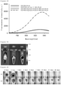
- ICG-HSA was injected, it was confirmed that the fluorescence blurring was severe at the administered site, and the fluorescence signal was barely detected 1 day after administration ((a) in Figure 8 ).
- ROI values for fluorescence signals at marked sites analyzed from the fluorescence images are shown in Figure 9 .
- the fluorescence signal decreased rapidly after injection, the fluorescence decreased to a level of 6.4% after 1 day and the fluorescence almost disappeared after 2 days.
- Example 4 Preparation and analysis of microspheres using polymers other than PLGA
- PDLLA-PEG poly(D,L-lactide)-block-poly(ethylene glycol)
- ICG-HSA-Alginate-PLGA and PDLLA-PEG P-188 0.1%) microspheres.
- ICG indocyanine green
- HSA human serum albumin
- ALG sodium alginate
- W1 solution 0.6 mL
- poly(vinyl alcohol) was added to 180 mL of distilled water, and then dissolved while stirring at 600 rpm under a condition of 85°C and cooled to room temperature to prepare W2 solution.
- the prepared first emulsion was added thereto to prepare a second emulsion (water-in-oil-in-water).
- the prepared emulsion was stirred at 1000 rpm for 10 minutes, and then stirred at 280 rpm for 21 hours using a magnetic stirrer under a temperature condition of 40°C to remove the methylene chloride solvent.
- the particles of indocyanine green-loaded PLGA polymer microspheres were obtained by centrifugation at 14,000 rpm for 5 minutes, and the process of decanting the supernatant, adding distilled water, redispersing and centrifuging was repeated three times to wash the microspheres.
- the particles of the washed microspheres were freeze-dried to finally obtain a powder of ICG-HSA-Alginate-PLGA, PDLLA-PEG (P-188 0.1%) microspheres.
- the microspheres were dissolved and the concentration of indocyanine green in the solution was quantified using an ultraviolet/visible light absorption spectrometer.
- indocyanine green/human serum albumin complex To compare the fluorescence signals of the prepared microspheres and the indocyanine green/human serum albumin complex, the same concentration of indocyanxhine green complex solution and dispersion of microparticles were prepared. 0.5 mg of indocyanine green powder was dissolved in a mixed solution of 0.214 mL of human serum albumin solution (20% concentration) and 0.786 mL of distilled water, and then diluted with distilled water to prepare 0.3 mL of indocyanine green/human serum albumin complex (ICG-HSA) at a concentration of 30 ⁇ M.
- ICG-HSA indocyanine green/human serum albumin complex
- a dispersion of microspheres was prepared by adding indocyanine green to 0.3 mL of distilled water so that the concentration was 30 ⁇ M.
- IVIS Lumina XR fluorescence imaging equipment
- ICG-HSA-Alginate-PLGA, PDLLA-PEG (P-188 0.1%) microspheres were spherical particles with an average particle size of 74.3 ⁇ 18.2 um, and had an appropriate size to avoid the phagocytosis of macrophages ((a) in Figure 10 ) .
- ICG-HSA it was confirmed that a fluorescence signal was visible for up to 5 hours, but no fluorescence signal was visible after 1 day ((a) in Figure 13 ).
- ICG-HSA-PMMA P-188 1.0%) microspheres were prepared using poly(methyl methacrylate) (PMMA; MW 120,000 Da, Sigma-Aldrich) polymer.
- PMMA poly(methyl methacrylate)
- HSA human serum albumin
- W1 solution 0.75 mg of indocyanine green (ICG) was dissolved in a mixed solution of 0.322 mL of human serum albumin (HSA) (20% concentration, 64.335 mg) and 1.178 mL of distilled water to prepare W1 solution.
- HSA human serum albumin
- poly(vinyl alcohol) was added to 180 mL of distilled water, and then dissolved while stirring at 600 rpm under a condition of 85°C and cooled to room temperature to prepare W2 solution.
- the prepared emulsion was stirred at 1000 rpm for 10 minutes, and then stirred at 280 rpm for 21 hours using a magnetic stirrer under a temperature condition of 40°C to remove the methylene chloride solvent. Afterwards, the particles of indocyanine green-loaded PMMA polymer microspheres were obtained by centrifugation at 14,000 rpm for 5 minutes, and the process of decanting the supernatant, adding distilled water, redispersing and centrifuging was repeated three times to wash the PMMA microspheres. The particles of the washed microspheres were freeze-dried to obtain a powder of microspheres.
- the microspheres were dissolved and the concentration of indocyanine green in the solution was quantified using an ultraviolet/visible light absorption spectrometer.
- indocyanine green/human serum albumin complex To compare the fluorescence signals of the prepared microspheres and the indocyanine green/human serum albumin complex, the same concentration of indocyanxhine green complex solution and dispersion of microparticles were prepared. 0.5 mg of indocyanine green powder was dissolved in a mixed solution of 0.214 mL of human serum albumin solution (20% concentration) and 0.786 mL of distilled water, and then diluted with distilled water to prepare 0.3 mL of indocyanine green/human serum albumin complex (ICG-HSA) at a concentration of 30 ⁇ M.
- ICG-HSA indocyanine green/human serum albumin complex
- a dispersion of microspheres was prepared by adding indocyanine green to 0.3 mL of distilled water so that the concentration was 30 ⁇ M.
- IVIS Lumina XR fluorescence imaging equipment
- the loading rate of indocyanine green in ICG-HSA-PMMA (P-188 1.0%) microspheres was 44.0%, and the content of indocyanine green per 1 mg of microspheres was high at 370.3 ng.
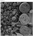
- ICG-HSA-PMMA P-188 1.0%) microspheres were spherical particles with an average particle size of 49.0 ⁇ 18.7 ⁇ m, and had an appropriate size to avoid the phagocytosis of macrophages ((a) in Figure 15 ).
- ICG-HSA-PMMA P-188 1.0%) microspheres were spherical particles with an average particle size of 49.0 ⁇ 18.7 ⁇ m, and had an appropriate size to avoid the phagocytosis of macrophages ((a) in Figure 15 ).
- a scanning electron image of the particle cross-section ((b) in Figure 15 )
- ICG-HSA (a) in Figure 18 )
- Example 5 Preparation and analysis of ICG-loaded PLGA microspheres using alginate hydrogel
- the prepared W1 solution was added to the oil solution and then sonicated in a water bath type ultrasonic device for 40 seconds, and 0.3 mL of calcium chloride (CaCl 2 , Sigma-Aldrich) at a concentration of 4.304 mg/mL was further added to induce ionic cross-linking of alginic acid and sonicated for 40 seconds to prepare a first emulsion (water-in-oil) in which alginic acid was gelled. While stirring the W2 solution at 1000 rpm using a mechanical stirrer, the prepared first emulsion was added thereto to prepare a second emulsion (water-in-oil-in-water).
- CaCl 2 calcium chloride
- the prepared emulsion was stirred at 1000 rpm for 10 minutes, and then stirred at 280 rpm for 21 hours using a magnetic stirrer under a temperature condition of 40°C to remove the methylene chloride solvent.
- ICG-Alginate-PLGA without P-188 particles, which are indocyanine green-loaded PLGA polymer microspheres, were obtained by centrifugation at 14,000 rpm for 5 minutes, and the process of decanting the supernatant, adding distilled water, redispersing and centrifuging was repeated three times to wash the microspheres. The particles of the washed microspheres were freeze-dried to obtain a powder of microspheres.
- ICG-Alginate-PLGA P-188 0.1%) microspheres, which are ICG-Alginate-PLGA microspheres further mixed with 0.1% (oil phase) of poloxamer 188 (P-188) based on the first emulsion, were prepared.
- W1 solution was prepared by mixing a solution of 0.75 mg of indocyanine green (ICG) dissolved in 0.6 mL of distilled water and a solution of 6 mg of sodium alginate dissolved in 0.6 mL of distilled water.
- 3.6 g of poly(vinyl alcohol) was added to 180 mL of distilled water, and then dissolved while stirring at 600 rpm under a condition of 85°C and cooled to room temperature to prepare W2 solution.
- the prepared first emulsion was added thereto to prepare a second emulsion (water-in-oil-in-water).
- the prepared emulsion was stirred at 1000 rpm for 10 minutes, and then stirred at 280 rpm for 21 hours using a magnetic stirrer under a temperature condition of 40°C to remove the methylene chloride solvent.
- the particles of indocyanine green-loaded PLGA polymer microspheres were obtained by centrifugation at 14,000 rpm for 5 minutes, and the process of decanting the supernatant, adding distilled water, redispersing and centrifuging was repeated three times to wash the microspheres.
- the particles of the washed microspheres were freeze-dried to obtain a powder of microspheres.
- the concentration of indocyanine green in the microsphere-dissolved solution was quantified using an ultraviolet/visible light absorption spectrometer.
- the absorbance of a solution in which 10 mg of PLGA microspheres was dissolved in 1 mL of dimethyl sulfoxide (DMSO) solvent was analyzed using an ultraviolet/visible light absorption spectrometer, and the concentration of indocyanine green was measured by comparison with the standard calibration curve.
- DMSO dimethyl sulfoxide
- the powder of the microspheres was attached to a carbon tape attached to the sample holder of a scanning electron microscope, and Pt/Pd was coated on the surface through sputtering for 60 seconds, and then the scanning electron microscope images were analyzed at an acceleration voltage of 5 kV.
- the particle size was calculated as the average value by randomly measuring the size of 100 particles from the acquired images.
- indocyanine green/human serum albumin complex To compare the fluorescence signals of the prepared microspheres and the indocyanine green/human serum albumin complex, the same concentration of indocyanxhine green complex solution and dispersion of microparticles were prepared. 0.5 mg of indocyanine green powder was dissolved in a mixed solution of 0.214 mL of human serum albumin solution (20% concentration) and 0.786 mL of distilled water, and then diluted with distilled water to prepare 0.3 mL of indocyanine green/human serum albumin complex (ICG-HSA) at a concentration of 30 ⁇ M.
- ICG-HSA indocyanine green/human serum albumin complex
- a dispersion of microspheres was prepared by adding indocyanine green to 0.3 mL of distilled water so that the concentration was 30 ⁇ M.
- the loading rate of indocyanine green in ICG-Algiante-PLGA (without P-188) microspheres is 5.79% and the content of indocyanine green in 1 mg of microspheres is 69.0 ng.
- ICG-Algiante-PLGA P-188 0.1%) microspheres to which 0.1% poloxamer 188 was added based on the first emulsion, the loading rate of indocyanine green was increased to 9.21%, and the content of indocyanine green in 1 mg of microspheres was also increased to 84.3 ng.
- Example 6 Preparation and analysis of PLGA microspheres in which ICG-HSA is mixed in alginate hydrogel
- Polymer microspheres were prepared under conditions where the ICG-HSA complex was mixed with alginate polymer and the alginate polymer was ionically cross-linked with calcium, and the fluorescence signal was analyzed.
- ICG-HSA-Alginate-PLGA P-188 0.1%) microspheres, which are ICG-HSA-loaded PLGA microspheres in which alginic acid is mixed with ICG-HSA and poloxamer 188 (P-188) is added to the oil phase
- a W1 solution was prepared by preparing a solution of 75 mg of indocyanine green (ICG) mixed with 0.322 mL of human serum albumin (HSA) (20% concentration, 64.335 mg) and 0.278 mL of distilled water and mixing it with a solution of 6 mg of sodium alginate (ALG, Kimica) dissolved in 0.6 mL of distilled water.
- ICG indocyanine green
- HSA human serum albumin
- the prepared W1 solution was added to the oil solution and then sonicated in a water bath type ultrasonic device for 40 seconds, and 0.3 mL of calcium chloride (CaCl 2 , Sigma-Aldrich) at a concentration of 4.304 mg/mL was further added and sonicated for 40 seconds to prepare a first emulsion (water-in-oil) in which alginic acid was gelled. While stirring the W2 solution at 1000 rpm using a mechanical stirrer, the first emulsion was added thereto to prepare a second emulsion (water-in-oil-in-water).
- CaCl 2 calcium chloride
- the prepared emulsion was stirred at 1000 rpm for 10 minutes, and then stirred at 280 rpm for 21 hours using a magnetic stirrer under a temperature condition of 40°C to remove the methylene chloride solvent.
- the particles of indocyanine green-loaded PLGA polymer microspheres were obtained by centrifugation at 14,000 rpm for 5 minutes, and the process of decanting the supernatant, adding distilled water, redispersing and centrifuging was repeated three times to wash the microspheres.
- the particles of the washed microspheres were freeze-dried to obtain a powder of microspheres.
- ICG-HSA-Alginate-PLGA P-188 0.5%) microspheres, which are ICG-HSA-loaded PLGA microspheres in which alginic acid is mixed with ICG-HSA and poloxamer 188 (P-188) is added to the oil phase
- a W1 solution was prepared by preparing a solution of 75 mg of indocyanine green (ICG) mixed with 0.322 mL of human serum albumin (HSA) (20% concentration, 64.335 mg) and 0.278 mL of distilled water and mixing it with a solution of 6 mg of sodium alginate (ALG, Kimica) dissolved in 0.6 mL of distilled water.
- ICG indocyanine green
- HSA human serum albumin
- the prepared W1 solution was added to the oil solution and then sonicated in a water bath type ultrasonic device for 40 seconds, and 0.3 mL of calcium chloride (CaCl 2 , Sigma-Aldrich) at a concentration of 4.304 mg/mL was added and further sonicated for 40 seconds to prepare a first emulsion (water-in-oil) in which alginic acid was gelled. While stirring the W2 solution at 1000 rpm using a mechanical stirrer, the first emulsion was added thereto to prepare a second emulsion (water-in-oil-in-water).
- CaCl 2 calcium chloride
- the prepared emulsion was stirred at 1000 rpm for 10 minutes, and then stirred at 280 rpm for 21 hours using a magnetic stirrer under a temperature condition of 40°C to remove the methylene chloride solvent.
- the particles of indocyanine green-loaded PLGA polymer microspheres were obtained by centrifugation at 14,000 rpm for 5 minutes, and the process of decanting the supernatant, adding distilled water, redispersing and centrifuging was repeated three times to wash the microspheres.
- the particles of the washed microspheres were freeze-dried to obtain a powder of microspheres.
- the particle size was calculated as the average value by randomly measuring the size of 100 particles from the acquired images.
- IVIS Lumina XR fluorescence imaging equipment
- the loading rates of indocyanine green in ICG-HSA-Alginate-PLGA (P-188 0.1%) and ICG-HSA-Alginate-PLGA (P-188 0.5%) microspheres were 10.5% and 7.62%, respectively, and the content of indocyanine green in 1 mg of microspheres was analyzed to be 108.7 ng and 61.6 ng.
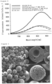
- ICG-HSA-Alginate-PLGA P-188 0.1%) and ICG-HSA-Alginate-PLGA (P-188 0.5%) microspheres were spherical particles with average particle sizes of 77.8 ⁇ 26.5 ⁇ m and 67.9 ⁇ 24.1 ⁇ m ( FIG. 22 ), and had an appropriate size to avoid the phagocytosis of macrophages.
- the emission of fluorescent dye may be suppressed better than a microbubble-type structure.
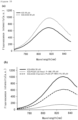
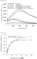
- ICG-HSA-Alginate-PLGA (P-188 0.1%) showed a similar fluorescence signal to ICG-HSA and showed a fluorescence signal 6.4 times higher than that of ICG-PLGA (without P-188), and ICG-HSA-Alginate-PLGA (P-188 0.5%) microspheres showed a fluorescence signal 2.5 times higher than that of ICG-HSA and showed a fluorescence signal 17 times higher than that of ICG-PLGA (without P-188) .
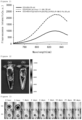
- ICG-HSA ((a) in Figure 25 )
- a dispersion of ICG-HSA-Alginate-PLGA (P-188 0.5%) microspheres ((b) in Figure 25 ) was administered, a high fluorescence signal was maintained for up to 30 days.
- ICG-HSA-Alginate-PLGA P-188 microspheres were very useful as a fluorescent marker.
- Example 7 Preparation and analysis of microspheres using Tween as a surfactant
- W1 solution was prepared by mixing a solution of 0.75 mg of indocyanine green (ICG) mixed with 0.322 mL of human serum albumin (HSA) (20% concentration, 64.335 mg) and 0.278 mL of distilled water and a solution of 6 mg of sodium alginate dissolved in 0.6 mL of distilled water.
- HSA human serum albumin
- poly(vinyl alcohol) was added to 180 mL of distilled water, and then dissolved while stirring at 600 rpm under a condition of 85°C and cooled to room temperature to prepare W2 solution.
- the first emulsion was added thereto to prepare a second emulsion (water-in-oil-in-water).
- the prepared emulsion was stirred at 1000 rpm for 10 minutes, and then stirred at 280 rpm for 21 hours using a magnetic stirrer under a temperature condition of 40°C to remove the methylene chloride solvent.
- the particles of indocyanine green-loaded PLGA polymer microspheres were obtained by centrifugation at 14,000 rpm for 5 minutes, and the process of decanting the supernatant, adding distilled water, redispersing and centrifuging was repeated three times to wash the microspheres. The washed microspheres were freeze-dried to obtain a powder of microspheres.
- the concentration of indocyanine green in the microsphere-dissolved solution was quantified using an ultraviolet/visible light absorption spectrometer.
- the content and loading efficiency of indocyanine green in the microspheres were evaluated using the measured concentration of the complex.
- the powder of the microspheres for analyzing the surface and cross section of microspheres was attached to a carbon tape attached to the sample holder of a scanning electron microscope, and Pt/Pd was coated on the surface through sputtering for 60 seconds, and then the scanning electron microscope images were analyzed at an acceleration voltage of 5 kV.
- the particle size was calculated as the average value by randomly measuring the size of 100 particles from the acquired images.
- IVIS Lumina XR fluorescence imaging equipment
- ICG-HSA-Algiante-PLGA Tween 0.5%) microspheres had a spherical particle shape ((a) in Figure 27 ), and the average particle size was 66.1 ⁇ 26.0 ⁇ m, which was an appropriate size for evading the phagocytosis of macrophages.
- the cross-sectional scanning electron microscopy image of the particle ((b) in Figure 27 ) it was confirmed that since the inside of the microspheres was divided into several isolated small spheres, it had a structure in which the release of the loaded fluorescent dye was suppressed in the same way as the results of the previous examples.
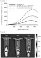
- the dispersion of ICG-HSA-Algiante-PLGA (Tween 0.5%) microspheres showed a fluorescence signal that was 2 times higher than that of ICG-HSA, and 13 times higher than that of ICG-PLGA (without P-188) ( Figure 28 ).
- microspheres As a result of graphing the ROI values analyzed from the fluorescence images ( Figure 31 ), in the case of the dispersion of ICG-HSA-Alginate-PLGA (Tween 0.5%) microspheres, it was confirmed that 70% of fluorescence remained after 1 day, and 52% of fluorescence still remained even after 30 days. Based on these results, it can be seen that even if microspheres are prepared by replacing poloxamer 188 as a surfactant with another surfactant such as Tween, microspheres may be prepared that may fluorescently mark a lesion site for a long period of time while generating a high fluorescent signal.
- Example 8 Preparation and analysis of indocyanine green/cyclodextrin complex-loaded microspheres
- Indocyanine green was formed into a complex with cyclodextrin (mCD), microspheres loaded therewith were prepared, and the intensity of fluorescence and utility as a fluorescent marker were analyzed.
- mCD indocyanine green/cyclodextrin complex-loaded PLGA microspheres
- ICG-mCD-Alginate-PLGA P-188 0.1%)
- mCD indocyanine green
- mCD methylcyclodextrin
- the prepared W1 solution was added to the oil solution and then sonicated in a water bath type ultrasonic device for 40 seconds, and 0.072 mL of calcium chloride (CaCl 2 , Sigma-Aldrich) at a concentration of 10.76 mg/mL was added and further sonicated for 40 seconds to prepare a first emulsion (water-in-oil) in which alginic acid was gelled. While stirring the W2 solution at 1000 rpm using a mechanical stirrer, the first emulsion was added thereto to prepare a second emulsion (water-in-oil-in-water).
- CaCl 2 calcium chloride
- the prepared emulsion was stirred at 1000 rpm for 10 minutes, and then stirred at 280 rpm for 21 hours using a magnetic stirrer under a temperature condition of 40°C to remove the methylene chloride solvent.
- the particles of indocyanine green-loaded PLGA polymer microspheres were obtained by centrifugation at 14,000 rpm for 5 minutes, and the process of decanting the supernatant, adding distilled water, redispersing and centrifuging was repeated three times to wash the microspheres.
- the particles of the washed microspheres were freeze-dried to obtain a powder of microspheres.
- the particle size was calculated as the average value by randomly measuring the size of 100 particles from the acquired images.
- indocyanine green/human serum albumin complex To compare the fluorescence signals of the prepared microspheres and the indocyanine green/human serum albumin complex, the same concentration of indocyanxhine green complex solution and dispersion of microparticles were prepared. 0.5 mg of indocyanine green powder was dissolved in a mixed solution of 0.214 mL of human serum albumin solution (20% concentration) and 0.786 mL of distilled water, and then diluted with distilled water to prepare 0.3 mL of indocyanine green/human serum albumin complex (ICG-HSA) at a concentration of 30 ⁇ M.
- ICG-HSA indocyanine green/human serum albumin complex
- a dispersion of microspheres was prepared by adding indocyanine green to 0.3 mL of distilled water so that the concentration was 30 ⁇ M.
- IVIS Lumina XR fluorescence imaging equipment
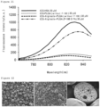
- the loading rates of indocyanine green in indocyanine green/cyclodextrin-loaded ICG-mCD-Alginate-PLGA (P-188 0.1%) microspheres were 11.4%, and the content of indocyanine green in 1 mg of microspheres was confirmed to be 76.3 ng.
- ICG-mCD-Alginate-PLGA P-188 0.1%) microspheres were also spherical particles ((a) in Figure 32 ), with an average size of 65.0 ⁇ 26.0 ⁇ m, which was an appropriate size for evading the phagocytosis of macrophages.
- the emission of fluorescent dye may be suppressed better than a microbubble-type structure.
- sulfo-cyanine 7 carboxylic acid Cy7
- sulfo-cyanine 7.5 carboxylic acid Cy7.5
- Flamma 774 carboxylic acid Flamma774
- Alexa fluor 750 carboxylic acid Alexa 750
- IRDye 800CW carboxylate IRDye 800CW
- Cy7 sulfo-cyanine7 carboxylic acid
- Cy7 sulfo-cyanine7 carboxylic acid
- HSA SK Plasma
- Cy7-HSA Cy7-human serum albumin complexes
- Cy7-PMMA (without P-188)
- Cy7-PMMA (without P-188)
- 0.81 mg of Cy7 was dissolved in 1.691 mL of distilled water to prepare W1 solution.
- 3.6 g of poly (vinyl alcohol) was added to 180 mL of distilled water, and then dissolved while stirring at 600 rpm under a condition of 85°C and cooled to room temperature to prepare W2 solution.
- 1.217 g of PMMA was added to 16.909 mL of methylene chloride and then dissolved to prepare an oil solution.
- the prepared W1 solution was added to the oil solution, and then sonicated in a probe-type ultrasonic device for 3 minutes (repeated 1-minute sonication + 30-second rest) to prepare a first emulsion (water-in-oil). While stirring the prepared W2 solution at 1000 rpm using a mechanical stirrer, the prepared first emulsion solution was added thereto at a rate of 10 mL/min using a syringe pump to prepare a second emulsion (water-in-oil-in-water).
- the prepared emulsion was stirred at 1000 rpm for 10 minutes, and then stirred at 280 rpm for 21 hours using a magnetic stirrer under a temperature condition of 40°C to remove the methylene chloride solvent. Afterwards, the particles of Cy7-loaded PMMA polymer microspheres were obtained by centrifugation at 14,000 rpm for 5 minutes, and the process of decanting the supernatant, adding distilled water, redispersing and centrifuging was repeated three times to wash the PMMA microspheres. The particles of the washed microspheres were freeze-dried to obtain a powder of microspheres.
- Cy7-HSA-PMMA (without P-188)
- Cy7-HSA-PMMA 0.81 mg of Cy7 was dissolved in 0.363 mL of human serum albumin (HSA) (20% concentration, 72.553 mg) and 1.328 mL of distilled water to prepare W1 solution.
- HSA human serum albumin
- 3.6 g of poly(vinyl alcohol) was added to 180 mL of distilled water, and then dissolved while stirring at 600 rpm under a condition of 85°C and cooled to room temperature to prepare W2 solution.
- 1.217 g of PMMA was added to 16.909 mL of methylene chloride and then dissolved to prepare an oil solution.
- the prepared W1 solution was added to the oil solution, and then sonicated in a probe-type ultrasonic device for 3 minutes (repeated 1-minute sonication + 30-second rest) to prepare a first emulsion (water-in-oil). While stirring the prepared W2 solution at 1000 rpm using a mechanical stirrer, the prepared first emulsion solution was added thereto at a rate of 10 mL/min using a syringe pump to prepare a second emulsion (water-in-oil-in-water).
- the prepared emulsion was stirred at 1000 rpm for 10 minutes, and then stirred at 280 rpm for 21 hours using a magnetic stirrer under a temperature condition of 40°C to remove the methylene chloride solvent.
- the particles of Cy7-HSA-loaded PMMA polymer microspheres were obtained by centrifugation at 14,000 rpm for 5 minutes, and the process of decanting the supernatant, adding distilled water, redispersing and centrifuging was repeated three times to wash the PMMA microspheres.
- the particles of the washed microspheres were freeze-dried to obtain a powder of microspheres.
- 1.217 g of PMMA and 186 mg of poloxamer 188 (P-188) were added to 16.909 mL of methylene chloride and then dissolved to prepare an oil solution.
- the prepared W1 solution was added to the oil solution, and then sonicated in a probe-type ultrasonic device for 3 minutes (repeated 1-minute sonication + 30-second rest) to prepare a first emulsion (water-in-oil).
- the prepared first emulsion solution was added thereto at a rate of 10 mL/min using a syringe pump to prepare a second emulsion (water-in-oil-in-water).
- the prepared emulsion was stirred at 1000 rpm for 10 minutes, and then stirred at 280 rpm for 21 hours using a magnetic stirrer under a temperature condition of 40°C to remove the methylene chloride solvent.
- the particles of Cy7-HSA-loaded PMMA polymer microspheres were obtained by centrifugation at 14,000 rpm for 5 minutes, and the process of decanting the supernatant, adding distilled water, redispersing and centrifuging was repeated three times to wash the PMMA microspheres.
- the particles of the washed microspheres were freeze-dried to obtain a powder of microspheres.
- the microspheres were dissolved and the concentration of Cy7 in the solution was quantified using an ultraviolet/visible light absorption spectrometer.
- Cy7-HSA Cy7-human serum albumin complex
- each of the microspheres was a spherical particle with an average particle size of 56.7 ⁇ 21.3 ⁇ m, 53.7 ⁇ 16.1 ⁇ m and 59.0 ⁇ 20.0 ⁇ m, and had an appropriate size to avoid the phagocytosis of macrophages ((a), (c) and (d) in Figure 38 ).
- Cy7.5 sulfo-cyanine7.5 carboxylic acid
- sulfo-cyanine7.5 carboxylic acid Cy7.5, Lumiprobe
- concentrations of 100, 70, 50, 40, 30, 20, 10, 5, 4, 3, 2 and 1 ⁇ M 0.70 mg of sulfo-cyanine7.5 carboxylic acid (Cy7.5, Lumiprobe) as a near-infrared fluorescent dye was dissolved in 1 mL of distilled water, and then diluted with distilled water to prepare Cy7.5 solutions at concentrations of 100, 70, 50, 40, 30, 20, 10, 5, 4, 3, 2 and 1 ⁇ M.
- Cy7.5-human serum albumin (HSA, SK Plasma) complex for comparison of fluorescence signals with Cy7.5 solution, 0.70 mg of Cy7.5 powder was dissolved in a mixed solution of 0.214 mL of human serum albumin solution (20% concentration) and 0.786 mL of distilled water, and then diluted with distilled water to prepare Cy7.5-human serum albumin complexes (Cy7.5-HSA) at concentrations of 100, 70, 50, 40, 30, 20, 10, 5, 4, 3, 2 and 1 ⁇ M.
- Cy7.5-PMMA (without P-188)
- Cy7.5-PMMA (without P-188)
- 1.18 mg of Cy7.5 was dissolved in 1.691 mL of distilled water to prepare W1 solution.
- 3.6 g of poly (vinyl alcohol) was added to 180 mL of distilled water, and then dissolved while stirring at 600 rpm under a condition of 85°C and cooled to room temperature to prepare W2 solution.
- 1.217 g of PMMA was added to 16.909 mL of methylene chloride and then dissolved to prepare an oil solution.
- the prepared W1 solution was added to the oil solution, and then sonicated in a probe-type ultrasonic device for 3 minutes (repeated 1-minute sonication + 30-second rest) to prepare a first emulsion (water-in-oil). While stirring the prepared W2 solution at 1000 rpm using a mechanical stirrer, the prepared first emulsion solution was added thereto at a rate of 10 mL/min using a syringe pump to prepare a second emulsion (water-in-oil-in-water).
- the prepared emulsion was stirred at 1000 rpm for 10 minutes, and then stirred at 280 rpm for 21 hours using a magnetic stirrer under a temperature condition of 40°C to remove the methylene chloride solvent. Afterwards, the particles of Cy7.5-loaded PMMA polymer microspheres were obtained by centrifugation at 14,000 rpm for 5 minutes, and the process of decanting the supernatant, adding distilled water, redispersing and centrifuging was repeated three times to wash the PMMA microspheres. The particles of the washed microspheres were freeze-dried to obtain a powder of microspheres.
- Cy7.5-HSA-PMMA (without P-188), which is PMMA microspheres loaded with Cy7.5-HSA complex as a near-infrared fluorescent dye, was prepared.
- 1.18 mg of Cy7.5 was dissolved in 0.363 mL of human serum albumin (HSA) (20% concentration, 72.553 mg) and 1.328 mL of distilled water to prepare W1 solution.
- 3.6 g of poly(vinyl alcohol) was added to 180 mL of distilled water, and then dissolved while stirring at 600 rpm under a condition of 85°C and cooled to room temperature to prepare W2 solution.
- 1.217 g of PMMA was added to 16.909 mL of methylene chloride and then dissolved to prepare an oil solution.
- the prepared W1 solution was added to the oil solution, and then sonicated in a probe-type ultrasonic device for 3 minutes (repeated 1-minute sonication + 30-second rest) to prepare a first emulsion (water-in-oil). While stirring the prepared W2 solution at 1000 rpm using a mechanical stirrer, the prepared first emulsion solution was added thereto at a rate of 10 mL/min using a syringe pump to prepare a second emulsion (water-in-oil-in-water).
- the prepared emulsion was stirred at 1000 rpm for 10 minutes, and then stirred at 280 rpm for 21 hours using a magnetic stirrer under a temperature condition of 40°C to remove the methylene chloride solvent.
- the particles of Cy7.5-HSA-loaded PMMA polymer microspheres were obtained by centrifugation at 14,000 rpm for 5 minutes, and the process of decanting the supernatant, adding distilled water, redispersing and centrifuging was repeated three times to wash the PMMA microspheres.
- the particles of the washed microspheres were freeze-dried to obtain a powder of microspheres.
- Cy7.5-HSA-PMMA (P-188 1.0%), which is PMMA microspheres comprising poloxamer 188 at a concentration of 0.1% in the first emulsion and loaded with Cy7.5-HSA complex as a near-infrared fluorescent dye, was prepared.
- 1.18 mg of Cy7.5 was dissolved in 0.363 mL of human serum albumin (HSA) (20% concentration, 72.553 mg) and 1.328 mL of distilled water to prepare W1 solution.
- W1 solution 1.328 mL of distilled water
- poly(vinyl alcohol) was added to 180 mL of distilled water, and then dissolved while stirring at 600 rpm under a condition of 85°C and cooled to room temperature to prepare W2 solution.
- 1.217 g of PMMA and 186 mg of poloxamer 188 (P-188) were added to 16.909 mL of methylene chloride and then dissolved to prepare an oil solution.
- the prepared W1 solution was added to the oil solution, and then sonicated in a probe-type ultrasonic device for 3 minutes (repeated 1-minute sonication + 30-second rest) to prepare a first emulsion (water-in-oil).
- the prepared first emulsion solution was added thereto at a rate of 10 mL/min using a syringe pump to prepare a second emulsion (water-in-oil-in-water).
- the prepared emulsion was stirred at 1000 rpm for 10 minutes, and then stirred at 280 rpm for 21 hours using a magnetic stirrer under a temperature condition of 40°C to remove the methylene chloride solvent.
- the particles of Cy7.5-HSA-loaded PMMA polymer microspheres were obtained by centrifugation at 14,000 rpm for 5 minutes, and the process of decanting the supernatant, adding distilled water, redispersing and centrifuging was repeated three times to wash the PMMA microspheres.
- the particles of the washed microspheres were freeze-dried to obtain a powder of microspheres.
- the microspheres were dissolved and the concentration of Cy7.5 in the solution was quantified using an ultraviolet/visible light absorption spectrometer.
- Cy7.5 complex solution a concentration of Cy7.5 complex solution and dispersion of microparticles was prepared. 0.70 mg of Cy7.5 powder was dissolved in a mixed solution of 0.214 mL of human serum albumin solution (20% concentration) and 0.786 mL of distilled water, and then diluted with distilled water to prepare 0.3 mL of Cy7.5-human serum albumin complex (Cy7-HSA) at a concentration of 30 ⁇ M. Using the information on the content of Cy7.5 in PMMA microspheres evaluated by the quantification, a dispersion of microspheres was prepared by adding Cy7.5 to 0.3 mL of distilled water so that the concentration was 30 ⁇ M.
- each of the microspheres was a spherical particle with an average particle size of 57.9 ⁇ 18.4 um, 58.0 ⁇ 16.0 ⁇ m and 65.9 ⁇ 24.4 ⁇ m, and had an appropriate size to avoid the phagocytosis of macrophages ((a), (c) and (e) in Figure 41 ).
- Cy7.5-PC (without P-188), which is polycarbonate (PC) microspheres loaded with Cy7.5 as a near-infrared fluorescent dye, was prepared.
- W1 solution was prepared by dissolving 1.18 mg of Cy7.5 in 1.691 mL of distilled water.
- 3.6 g of poly(vinyl alcohol) was added to 180 mL of distilled water, and then dissolved while stirring at 600 rpm under a condition of 85°C and cooled to room temperature to prepare W2 solution.
- 1.217 g of PC was added to 16.909 mL of methylene chloride and then dissolved to prepare an oil solution.
- the prepared W1 solution was added to the oil solution, and then sonicated in a probe-type ultrasonic device for 3 minutes (repeated 1-minute sonication + 30-second rest) to prepare a first emulsion (water-in-oil). While stirring the prepared W2 solution at 1000 rpm using a mechanical stirrer, the prepared first emulsion solution was added thereto at a rate of 10 mL/min using a syringe pump to prepare a second emulsion (water-in-oil-in-water).
- the prepared emulsion was stirred at 1000 rpm for 10 minutes, and then stirred at 280 rpm for 21 hours using a magnetic stirrer under a temperature condition of 40°C to remove the methylene chloride solvent. Afterwards, the particles of Cy7.5-loaded PC polymer microspheres were obtained by centrifugation at 14,000 rpm for 5 minutes, and the process of decanting the supernatant, adding distilled water, redispersing and centrifuging was repeated three times to wash the PC microspheres. The particles of the washed microspheres were freeze-dried to obtain a powder of microspheres.
- Cy7.5-HSA-PC (without P-188), which is PC microspheres loaded with Cy7.5-HSA complex, was prepared. 1.18 mg of Cy7.5 was dissolved in 0.363 mL of human serum albumin (HSA) (20% concentration, 72.553 mg) and 1.328 mL of distilled water to prepare W1 solution. 3.6 g of poly(vinyl alcohol) was added to 180 mL of distilled water, and then dissolved while stirring at 600 rpm under a condition of 85°C and cooled to room temperature to prepare W2 solution. 1.217 g of PC was added to 16.909 mL of methylene chloride and then dissolved to prepare an oil solution.
- HSA human serum albumin
- the prepared W1 solution was added to the oil solution, and then sonicated in a probe-type ultrasonic device for 3 minutes (repeated 1-minute sonication + 30-second rest) to prepare a first emulsion (water-in-oil). While stirring the prepared W2 solution at 1000 rpm using a mechanical stirrer, the prepared first emulsion solution was added thereto at a rate of 10 mL/min using a syringe pump to prepare a second emulsion (water-in-oil-in-water).
- the prepared emulsion was stirred at 1000 rpm for 10 minutes, and then stirred at 280 rpm for 21 hours using a magnetic stirrer under a temperature condition of 40°C to remove the methylene chloride solvent.
- the particles of Cy7.5-HSA-loaded PC polymer microspheres were obtained by centrifugation at 14,000 rpm for 5 minutes, and the process of decanting the supernatant, adding distilled water, redispersing and centrifuging was repeated three times to wash the PC microspheres.
- the particles of the washed microspheres were freeze-dried to obtain a powder of microspheres.
- Cy7.5 complex solution a concentration of Cy7.5 complex solution and dispersion of microparticles was prepared. 0.70 mg of Cy7.5 powder was dissolved in a mixed solution of 0.214 mL of human serum albumin solution (20% concentration) and 0.786 mL of distilled water, and then diluted with distilled water to prepare 0.3 mL of Cy7.5-human serum albumin complex (Cy7-HSA) at a concentration of 30 ⁇ M. Using the information on the content of Cy7.5 in PC microspheres evaluated by the quantification, a dispersion of microspheres was prepared by adding Cy7.5 to 0.3 mL of distilled water so that the concentration was 30 ⁇ M.
- Cy7-HSA Cy7.5-human serum albumin complex
- Flamma774 carboxylic acid (Flamma774, Bioacts) as a near-infrared fluorescent dye was dissolved in 1 mL of distilled water, and then diluted with distilled water to prepare Flamma774 solutions at concentrations of 100, 70, 50, 40, 30, 20, 10, 5, 4, 3, 2 and 1 ⁇ M.
- Flamma774-human serum albumin (HSA, SK Plasma) complex for comparison of fluorescence signals with Flamma774 solution, 0.60 mg of Flamma774 powder was dissolved in a mixed solution of 0.214 mL of human serum albumin solution (20% concentration) and 0.786 mL of distilled water, and then diluted with distilled water to prepare Flamma774-human serum albumin complexes (Flamma774-HSA) at concentrations of 100, 70, 50, 40, 30, 20, 10, 5, 4, 3, 2 and 1 ⁇ M.
- Flamma774-HSA Flamma774-human serum albumin complexes
- Flamma774-PMMA (without P-188), which is PMMA microspheres loaded with Flamma774 as a near-infrared fluorescent dye, was prepared.
- W1 solution was prepared by dissolving 1.01 mg of Flamma774 in 1.691 mL of distilled water.
- 3.6 g of poly(vinyl alcohol) was added to 180 mL of distilled water, and then dissolved while stirring at 600 rpm under a condition of 85°C and cooled to room temperature to prepare W2 solution.
- 1.217 g of PMMA was added to 16.909 mL of methylene chloride and then dissolved to prepare an oil solution.
- the prepared W1 solution was added to the oil solution, and then sonicated in a probe-type ultrasonic device for 3 minutes (repeated 1-minute sonication + 30-second rest) to prepare a first emulsion (water-in-oil). While stirring the prepared W2 solution at 1000 rpm using a mechanical stirrer, the prepared first emulsion solution was added thereto at a rate of 10 mL/min using a syringe pump to prepare a second emulsion (water-in-oil-in-water).
- the prepared emulsion was stirred at 1000 rpm for 10 minutes, and then stirred at 280 rpm for 21 hours using a magnetic stirrer under a temperature condition of 40°C to remove the methylene chloride solvent.
- the particles of Flamma774-loaded PMMA polymer microspheres were obtained by centrifugation at 14,000 rpm for 5 minutes, and the process of decanting the supernatant, adding distilled water, redispersing and centrifuging was repeated three times to wash the PMMA microspheres.
- the particles of the washed microspheres were freeze-dried to obtain a powder of microspheres.
- Flamma774-HSA-PMMA (without P-188), which is PMMA microspheres loaded with Flamma774-HSA complex, was prepared.
- 1.01 mg of Flamma774 was dissolved in 0.363 mL of human serum albumin (HSA) (20% concentration, 72.553 mg) and 1.328 mL of distilled water to prepare W1 solution.
- 3.6 g of poly(vinyl alcohol) was added to 180 mL of distilled water, and then dissolved while stirring at 600 rpm under a condition of 85°C and cooled to room temperature to prepare W2 solution.
- 1.217 g of PMMA was added to 16.909 mL of methylene chloride and then dissolved to prepare an oil solution.
- the prepared W1 solution was added to the oil solution, and then sonicated in a probe-type ultrasonic device for 3 minutes (repeated 1-minute sonication + 30-second rest) to prepare a first emulsion (water-in-oil). While stirring the prepared W2 solution at 1000 rpm using a mechanical stirrer, the prepared first emulsion solution was added thereto at a rate of 10 mL/min using a syringe pump to prepare a second emulsion (water-in-oil-in-water).
- the prepared emulsion was stirred at 1000 rpm for 10 minutes, and then stirred at 280 rpm for 21 hours using a magnetic stirrer under a temperature condition of 40°C to remove the methylene chloride solvent.
- the particles of Flamma 77 4-HSA-loaded PMMA polymer microspheres were obtained by centrifugation at 14,000 rpm for 5 minutes, and the process of decanting the supernatant, adding distilled water, redispersing and centrifuging was repeated three times to wash the PMMA microspheres.
- the particles of the washed microspheres were freeze-dried to obtain a powder of microspheres.
- Flamma774-HSA-PMMA (P-188 1.0%), which is PMMA microspheres comprising poloxamer 188 at a concentration of 0.1% in the first emulsion and loaded with Flamma774-HSA complex as a near-infrared fluorescent dye, was prepared.
- 1.01 mg of Flamma774 was dissolved in 0.363 mL of human serum albumin (HSA) (20% concentration, 72.553 mg) and 1.328 mL of distilled water to prepare W1 solution.
- W1 solution 1.328 mL of distilled water
- poly(vinyl alcohol) was added to 180 mL of distilled water, and then dissolved while stirring at 600 rpm under a condition of 85°C and cooled to room temperature to prepare W2 solution.
- 1.217 g of PMMA and 186 mg of poloxamer 188 (P-188) were added to 16.909 mL of methylene chloride and then dissolved to prepare an oil solution.
- the prepared W1 solution was added to the oil solution, and then sonicated in a probe-type ultrasonic device for 3 minutes (repeated 1-minute sonication + 30-second rest) to prepare a first emulsion (water-in-oil).
- the prepared first emulsion solution was added thereto at a rate of 10 mL/min using a syringe pump to prepare a second emulsion (water-in-oil-in-water).
- the prepared emulsion was stirred at 1000 rpm for 10 minutes, and then stirred at 280 rpm for 21 hours using a magnetic stirrer under a temperature condition of 40°C to remove the methylene chloride solvent.
- the particles of Flamma 77 4-HSA-loaded PMMA polymer microspheres were obtained by centrifugation at 14,000 rpm for 5 minutes, and the process of decanting the supernatant, adding distilled water, redispersing and centrifuging was repeated three times to wash the PMMA microspheres.
- the particles of the washed microspheres were freeze-dried to obtain a powder of microspheres.
- the microspheres were dissolved and the concentration of Flamma774 in the solution was quantified using an ultraviolet/visible light absorption spectrometer.
- Flamma774-human serum albumin complex To compare the fluorescence signals of the prepared microspheres and the Flmma774-human serum albumin complex, the same concentration of Flamma774 complex solution and dispersion of microparticles were prepared. 0.60 mg of Flamma774 powder was dissolved in a mixed solution of 0.214 mL of human serum albumin solution (20% concentration) and 0.786 mL of distilled water, and then diluted with distilled water to prepare 0.3 mL of Flamma774-human serum albumin complex (Flmma774-HSA) at a concentration of 30 ⁇ M.
- Flamma774-HSA Flamma774-human serum albumin complex
- a dispersion of microspheres was prepared by adding Flamma774 to 0.3 mL of distilled water so that the concentration was 30 ⁇ M.
- Flamma774 in Flamma774-HSA-PMMA without P-188) prepared by loading Flamma774-HSA and Flamma774-HSA-PMMA (1.0% P-188) microspheres prepared by adding 1.0% poloxamer 188 based on the first emulsion were 23.3% and 21.6%, and the contents of Flamma774 in 1 mg of microspheres were 183.0 ng and 148.6 ng, and thus, they were loaded with a higher content of Flamma774 than Flamma774-PMMA (without P-188) microspheres loaded with Flamma774 alone.
- each of the microspheres was a spherical particle with an average particle size of 56.8 ⁇ 19.2 ⁇ m, 62.5 ⁇ 19.3 ⁇ m and 55.8 ⁇ 19.3 ⁇ m, and had an appropriate size to avoid the phagocytosis of macrophages ((a), (c) and (e) in Figure 46 ).
- Alexa fluor TM 750 carboxylic acid, tris(triethylammonium) salt Alexa750, Thermo Fisher Scientific
- Alexa750-human serum albumin (HSA, SK Plasma) complex for comparison of fluorescence signals with Alexa750 solution, 0.77 mg of Alexa750 powder was dissolved in a mixed solution of 0.214 mL of human serum albumin solution (20% concentration) and 0.786 mL of distilled water, and then diluted with distilled water to prepare Alexa750-human serum albumin complexes (Alexa750-HSA) at concentrations of 100, 70, 50, 40, 30, 20, 10, 5, 4, 3, 2 and 1 ⁇ M.
- Alexa750-PMMA (without P-188), which is PMMA microspheres loaded with Alexa750 as a near-infrared fluorescent dye, was prepared.
- W1 solution was prepared by dissolving 1.31 mg of Alexa750 in 1.691 mL of distilled water.
- 3.6 g of poly(vinyl alcohol) was added to 180 mL of distilled water, and then dissolved while stirring at 600 rpm under a condition of 85°C and cooled to room temperature to prepare W2 solution.
- 1.217 g of PMMA was added to 16.909 mL of methylene chloride and then dissolved to prepare an oil solution.
- the prepared W1 solution was added to the oil solution, and then sonicated in a probe-type ultrasonic device for 3 minutes (repeated 1-minute sonication + 30-second rest) to prepare a first emulsion (water-in-oil). While stirring the prepared W2 solution at 1000 rpm using a mechanical stirrer, the prepared first emulsion solution was added thereto at a rate of 10 mL/min using a syringe pump to prepare a second emulsion (water-in-oil-in-water).
- the prepared emulsion was stirred at 1000 rpm for 10 minutes, and then stirred at 280 rpm for 21 hours using a magnetic stirrer under a temperature condition of 40°C to remove the methylene chloride solvent.
- the particles of Alexa750-loaded PMMA polymer microspheres were obtained by centrifugation at 14,000 rpm for 5 minutes, and the process of decanting the supernatant, adding distilled water, redispersing and centrifuging was repeated three times to wash the PMMA microspheres.
- the particles of the washed microspheres were freeze-dried to obtain a powder of microspheres.
- Alexa750-HSA-PMMA (without P-188), which is PMMA microspheres loaded with Alexa750-HSA complex as a near-infrared fluorescent dye, was prepared.
- 1.31 mg of Alexa750 was dissolved in 0.363 mL of human serum albumin (HSA) (20% concentration, 72.553 mg) and 1.328 mL of distilled water to prepare W1 solution.
- 3.6 g of poly(vinyl alcohol) was added to 180 mL of distilled water, and then dissolved while stirring at 600 rpm under a condition of 85°C and cooled to room temperature to prepare W2 solution.
- 1.217 g of PMMA was added to 16.909 mL of methylene chloride and then dissolved to prepare an oil solution.
- Alexa750-human serum albumin complex To compare the fluorescence signals of the prepared microspheres and the Alexa750-human serum albumin complex, the same concentration of Alexa750 complex solution and dispersion of microparticles were prepared. 0.77 mg of Alexa750 powder was dissolved in a mixed solution of 0.214 mL of human serum albumin solution (20% concentration) and 0.786 mL of distilled water, and then diluted with distilled water to prepare 0.3 mL of Alexa750-human serum albumin complex (Alexa750-HSA) at a concentration of 30 ⁇ M. Using the information on the content of Alexa750 in PMMA microspheres evaluated by the quantification, a dispersion of microspheres was prepared by adding Alexa750 to 0.3 mL of distilled water so that the concentration was 30 ⁇ M.
- IRDye ® 800CW carboxylate (IRDye 800CW, LI-COR, Inc.) as a near-infrared fluorescent dye was dissolved in 1 mL of distilled water, and then diluted with distilled water to prepare Alexa750 solutions at concentrations of 100, 70, 50, 40, 30, 20, 10, 5, 4, 3, 2 and 1 ⁇ M.
- IRDye 800CW-human serum albumin (HSA, SK Plasma) complex for comparison of fluorescence signals with IRDye 800CW solution, 0.70 mg of IRDye 800CW powder was dissolved in a mixed solution of 0.214 mL of human serum albumin solution (20% concentration) and 0.786 mL of distilled water, and then diluted with distilled water to prepare IRDye 800CW-human serum albumin complexes (IRDye 800CW-HSA) at concentrations of 100, 70, 50, 40, 30, 20, 10, 5, 4, 3, 2 and 1 ⁇ M.
- IRDye 800CW-HSA IRDye 800CW-human serum albumin complexes
- the prepared emulsion was stirred at 1000 rpm for 10 minutes, and then stirred at 280 rpm for 21 hours using a magnetic stirrer under a temperature condition of 40°C to remove the methylene chloride solvent.
- the particles of IRDye 800CW-loaded PMMA polymer microspheres were obtained by centrifugation at 14,000 rpm for 5 minutes, and the process of decanting the supernatant, adding distilled water, redispersing and centrifuging was repeated three times to wash the PMMA microspheres.
- the particles of the washed microspheres were freeze-dried to obtain a powder of microspheres.
- IRDye 800CW-HSA-PMMA (without P-188), which is PMMA microspheres loaded with IRDye 800CW-HSA complex as a near-infrared fluorescent dye, was prepared.
- 1.19 mg of IRDye 800CW was dissolved in 0.363 mL of human serum albumin (HSA) (20% concentration, 72.553 mg) and 1.328 mL of distilled water to prepare W1 solution.
- 3.6 g of poly(vinyl alcohol) was added to 180 mL of distilled water, and then dissolved while stirring at 600 rpm under a condition of 85°C and cooled to room temperature to prepare W2 solution.
- 1.217 g of PMMA was added to 16.909 mL of methylene chloride and then dissolved to prepare an oil solution.
- the prepared W1 solution was added to the oil solution, and then sonicated in a probe-type ultrasonic device for 3 minutes (repeated 1-minute sonication + 30-second rest) to prepare a first emulsion (water-in-oil).
- the prepared first emulsion solution was added thereto at a rate of 10 mL/min using a syringe pump to prepare a second emulsion (water-in-oil-in-water).
- the prepared emulsion was stirred at 1000 rpm for 10 minutes, and then stirred at 280 rpm for 21 hours using a magnetic stirrer under a temperature condition of 40°C to remove the methylene chloride solvent.
- the particles of IRDye 800CW-HSA-loaded PMMA polymer microspheres were obtained by centrifugation at 14,000 rpm for 5 minutes, and the process of decanting the supernatant, adding distilled water, redispersing and centrifuging was repeated three times to wash the PMMA microspheres.
- the particles of the washed microspheres were freeze-dried to obtain a powder of microspheres.
- IRDye 800CW-HSA-PMMA (P-188 1.0%), which is PMMA microspheres comprising poloxamer 188 at a concentration of 0.1% in the first emulsion and loaded with IRDye 800CW-HSA complex as a near-infrared fluorescent dye, was prepared.
- 1.19 mg of IRDye 800CW was dissolved in 0.363 mL of human serum albumin (HSA) (20% concentration, 72.553 mg) and 1.328 mL of distilled water to prepare W1 solution.
- 3.6 g of poly(vinyl alcohol) was added to 180 mL of distilled water, and then dissolved while stirring at 600 rpm under a condition of 85°C and cooled to room temperature to prepare W2 solution.
- 1.217 g of PMMA and 186 mg of poloxamer 188 (P-188) were added to 16.909 mL of methylene chloride and then dissolved to prepare an oil solution.
- the prepared W1 solution was added to the oil solution, and then sonicated in a probe-type ultrasonic device for 3 minutes (repeated 1-minute sonication + 30-second rest) to prepare a first emulsion (water-in-oil).
- the prepared first emulsion solution was added thereto at a rate of 10 mL/min using a syringe pump to prepare a second emulsion (water-in-oil-in-water).
- the prepared emulsion was stirred at 1000 rpm for 10 minutes, and then stirred at 280 rpm for 21 hours using a magnetic stirrer under a temperature condition of 40°C to remove the methylene chloride solvent.
- the particles of IRDye 800CW-HSA-loaded PMMA polymer microspheres were obtained by centrifugation at 14,000 rpm for 5 minutes, and the process of decanting the supernatant, adding distilled water, redispersing and centrifuging was repeated three times to wash the PMMA microspheres.
- the particles of the washed microspheres were freeze-dried to obtain a powder of microspheres.
- the microspheres were dissolved and the concentration of IRDye 800CW in the solution was quantified using an ultraviolet/visible light absorption spectrometer.
- IRDye 800CW-human serum albumin complex To compare the fluorescence signals of the prepared microspheres and the IRDye 800CW-human serum albumin complex, the same concentration of Alexa750 complex solution and dispersion of microparticles were prepared. 0.70 mg of IRDye 800CW powder was dissolved in a mixed solution of 0.214 mL of human serum albumin solution (20% concentration) and 0.786 mL of distilled water, and then diluted with distilled water to prepare 0.3 mL of IRDye 800CW-human serum albumin complex (IRDye 800CW-HSA) at a concentration of 30 ⁇ M.
- IRDye 800CW-HSA IRDye 800CW-human serum albumin complex
- a dispersion of microspheres was prepared by adding IRDye 800CW to 0.3 mL of distilled water so that the concentration was 30 ⁇ M.
- each of the microspheres was a spherical particle with an average particle size of 55.8 ⁇ 19.7 um, 66.6 ⁇ 13.8 ⁇ m and 59.8 ⁇ 17.3 ⁇ m, and had an appropriate size to avoid the phagocytosis of macrophages ((a), (c) and (e) in Figure 52 ).
- Example 14 Preparation of a dispersion composition of PLGA microspheres and preparation and analysis of a marker in the form of a pellet containing microspheres
- Microspheres prepared using PLGA polymer as a representative example of a biodegradable polymer were dispersed in various other surfactants or biocompatible polymer materials, and an aqueous dispersion composition solution for injection was prepared.
- the microspheres used in this example are ICG-HSA-PLGA (P-188 0.5%).
- markers in the form of dried pellets were prepared.
- the microspheres dispersed in 1% sodium hyaluronate and 5% aqueous gelatin solution were transferred to a cylindrical frame, and then rapidly cooled using liquid nitrogen and freeze-dried to prepare a fluorescence marker in the form of a solid pellet.
- a fluorescence marker in the form of a solid pellet prepared using microspheres dispersed in a 1% sodium hyaluronate and 5% gelatin solution may be prepared.
- Example 15 Preparation of a dispersion composition of PMMA microspheres and preparation and analysis of a marker in the form of a pellet containing microspheres
- Microspheres prepared using PMMA polymer as a representative example of a non-biodegradable polymer were dispersed in various other surfactants or biocompatible polymer materials, and an aqueous dispersion composition solution for injection was prepared.
- the microspheres used in this example are ICG-HSA-PMMA (P-188 1.0%) microspheres.
- microspheres For dispersion of microspheres, a solution of 0.5% sodium methylcellulose (Sigma-Aldrich)/0.1% Tween 80 (Sigma-Aldrich) dissolved in physiological saline for injection, 1% sodium alginate, 1% sodium hyaluronate, 2% soidum methylcellulose, 2% poly(vinyl alcohol), 0.1% collagen, 5% gelatin, 10% Tween 80, 40% poloxamer 188, 5% polyvinyl pyrrolidone solution were prepared, and then the microspheres were dispersed in each solution so that the concentration of indocyanine green in the ICG-HSA-PMMA (P-188 1.0%) microspheres was 30 ⁇ M.
Landscapes
- Health & Medical Sciences (AREA)
- Life Sciences & Earth Sciences (AREA)
- Engineering & Computer Science (AREA)
- Biomedical Technology (AREA)
- Public Health (AREA)
- Veterinary Medicine (AREA)
- Epidemiology (AREA)
- Animal Behavior & Ethology (AREA)
- General Health & Medical Sciences (AREA)
- Chemical & Material Sciences (AREA)
- Organic Chemistry (AREA)
- Chemical Kinetics & Catalysis (AREA)
- Dispersion Chemistry (AREA)
- Proteomics, Peptides & Aminoacids (AREA)
- Optics & Photonics (AREA)
- Oncology (AREA)
- Nuclear Medicine, Radiotherapy & Molecular Imaging (AREA)
- Biodiversity & Conservation Biology (AREA)
- Physics & Mathematics (AREA)
- Medicines Containing Antibodies Or Antigens For Use As Internal Diagnostic Agents (AREA)
- Bioinformatics & Cheminformatics (AREA)
- Medicinal Chemistry (AREA)
- Pharmacology & Pharmacy (AREA)
- Molecular Biology (AREA)
Applications Claiming Priority (3)
| Application Number | Priority Date | Filing Date | Title |
|---|---|---|---|
| KR20220014733 | 2022-02-04 | ||
| KR1020220084369A KR102552104B1 (ko) | 2022-02-04 | 2022-07-08 | 병변 표지를 위한 근적외선 형광염료 탑재 고분자 미립구 및 이의 제형 |
| PCT/KR2023/001265 WO2023149695A1 (ko) | 2022-02-04 | 2023-01-27 | 병변 표지를 위한 근적외선 형광염료 탑재 고분자 미립구 및 이의 제형 |
Publications (1)
| Publication Number | Publication Date |
|---|---|
| EP4473979A1 true EP4473979A1 (de) | 2024-12-11 |
Family
ID=87155190
Family Applications (1)
| Application Number | Title | Priority Date | Filing Date |
|---|---|---|---|
| EP23749928.0A Pending EP4473979A1 (de) | 2022-02-04 | 2023-01-27 | Mit einem nahinfrarot-fluoreszenzfarbstoff geladene polymermikrokugeln zur markierung von läsionen und formulierung daraus |
Country Status (8)
| Country | Link |
|---|---|
| US (1) | US20240390526A1 (de) |
| EP (1) | EP4473979A1 (de) |
| JP (1) | JP7842230B2 (de) |
| KR (1) | KR102552104B1 (de) |
| CN (1) | CN118647411A (de) |
| AU (1) | AU2023216619A1 (de) |
| CA (1) | CA3243661A1 (de) |
| WO (1) | WO2023149695A1 (de) |
Family Cites Families (6)
| Publication number | Priority date | Publication date | Assignee | Title |
|---|---|---|---|---|
| KR20100042768A (ko) | 2008-10-17 | 2010-04-27 | 동아제약주식회사 | 생분해성 고분자 미립구 및 그 제조방법 |
| KR101228147B1 (ko) * | 2010-08-13 | 2013-01-31 | 국립암센터 | 방사성 동위원소가 표지되고 근적외선 영역의 형광색소 및 가시광선 영역의 색소가 결합된 알부민을 포함하는 감시림프절 표지자 |
| US9610367B2 (en) * | 2010-08-24 | 2017-04-04 | Canon Kabushiki Kaisha | Particle and contrast agent having the particle |
| KR101610465B1 (ko) * | 2014-04-11 | 2016-04-07 | 국립암센터 | 다목적용 의료 영상 표지자 및 이의 제조방법 |
| KR20200027895A (ko) * | 2018-09-05 | 2020-03-13 | 고려대학교 산학협력단 | 형광조영제를 유효성분으로 포함하는 폐 종양 병변 표지를 위한 흡입 투여용 약학적 조성물 |
| EP3878476B1 (de) * | 2018-11-06 | 2025-10-01 | National Cancer Center | Injizierbare hydrogelsysteme auf der basis von alginsäure |
-
2022
- 2022-07-08 KR KR1020220084369A patent/KR102552104B1/ko active Active
-
2023
- 2023-01-27 EP EP23749928.0A patent/EP4473979A1/de active Pending
- 2023-01-27 AU AU2023216619A patent/AU2023216619A1/en active Pending
- 2023-01-27 CA CA3243661A patent/CA3243661A1/en active Pending
- 2023-01-27 WO PCT/KR2023/001265 patent/WO2023149695A1/ko not_active Ceased
- 2023-01-27 CN CN202380019860.7A patent/CN118647411A/zh active Pending
- 2023-01-27 JP JP2024546230A patent/JP7842230B2/ja active Active
-
2024
- 2024-08-02 US US18/793,301 patent/US20240390526A1/en active Pending
Also Published As
| Publication number | Publication date |
|---|---|
| AU2023216619A1 (en) | 2024-08-22 |
| CA3243661A1 (en) | 2025-01-29 |
| CN118647411A (zh) | 2024-09-13 |
| JP2025506421A (ja) | 2025-03-11 |
| US20240390526A1 (en) | 2024-11-28 |
| WO2023149695A1 (ko) | 2023-08-10 |
| KR102552104B1 (ko) | 2023-07-07 |
| JP7842230B2 (ja) | 2026-04-07 |
Similar Documents
| Publication | Publication Date | Title |
|---|---|---|
| Cruz et al. | Effect of PLGA NP size on efficiency to target traumatic brain injury | |
| KR102315855B1 (ko) | 알긴산 기반의 주입형 수화젤 시스템 | |
| US9782430B2 (en) | Resorbable oxidized cellulose embolization solution | |
| EP3085396B1 (de) | Resorbierbare oxydierte celluloseembolisierungslösung | |
| Das et al. | Intracranial nanomedicine-gel with deep brain-penetration for glioblastoma therapy | |
| US20190350965A1 (en) | Thixotropic oxidized cellulose solutions and medical applications thereof | |
| EP3242690B1 (de) | Chirurgische nahinfrarot-fluoreszenzfarbstoffmarker | |
| EP2777820B1 (de) | Sprühpflaster unter verwendung von löslicher oxidierter zellulose mit miniaturisiertem elektrospraysystem und verfahren | |
| EP3281646A1 (de) | Thixotrope oxidierte celluloselösungen und medizinische anwendungen davon | |
| US20240390526A1 (en) | Near-infrared fluorescent dye-loaded polymer microspheres for labelling lesions, and formulation thereof | |
| EP2708227B1 (de) | Mehrfach gekapselte Formulierungen mit oxidierter Cellulose für In-situ-Reaktionen | |
| CN104288793B (zh) | 纳米超声/荧光双模态造影剂、其制备方法与应用 | |
| EP2895151B1 (de) | Mehrfach verkapselte formulierungen mit oxidierter cellulose | |
| US20230285600A1 (en) | Tumor targeted diagnostic imaging agent for diagnostic biopsy, or intraoperative tumor identification or margin assessment using near-infrared fluorescence (nirf) imaging | |
| KR20250142557A (ko) | 온도 민감성 고분자 하이드로겔을 포함하는 형광 병변 표지용 조성물 | |
| Hu et al. | Noninvasive Monitoring of In Vivo Degradation Kinetics of PLGA Implants Based on NIR-II Fluorescence Imaging | |
| US20250090693A1 (en) | Biocompatible composition for labeling target lesion, and preparation method therefor | |
| EP4536295A1 (de) | Injizierbarer nanoverbundstoff auf hydrogelbasis für die chirurgische bildgebung | |
| KR20230109549A (ko) | 생체 친화적 목적 병변 표지용 조성물 및 이의 제조 방법 | |
| Rodriguez et al. | Stabilized micellar formulation of indocyanine green for near-infrared imaging |
Legal Events
| Date | Code | Title | Description |
|---|---|---|---|
| STAA | Information on the status of an ep patent application or granted ep patent |
Free format text: STATUS: THE INTERNATIONAL PUBLICATION HAS BEEN MADE |
|
| PUAI | Public reference made under article 153(3) epc to a published international application that has entered the european phase |
Free format text: ORIGINAL CODE: 0009012 |
|
| STAA | Information on the status of an ep patent application or granted ep patent |
Free format text: STATUS: REQUEST FOR EXAMINATION WAS MADE |
|
| 17P | Request for examination filed |
Effective date: 20240806 |
|
| AK | Designated contracting states |
Kind code of ref document: A1 Designated state(s): AL AT BE BG CH CY CZ DE DK EE ES FI FR GB GR HR HU IE IS IT LI LT LU LV MC ME MK MT NL NO PL PT RO RS SE SI SK SM TR |
|
| DAV | Request for validation of the european patent (deleted) | ||
| DAX | Request for extension of the european patent (deleted) |