EP4450616A2 - Neue monoklonale antikörper gegen programmierten tod 1 (pd-1) - Google Patents
Neue monoklonale antikörper gegen programmierten tod 1 (pd-1) Download PDFInfo
- Publication number
- EP4450616A2 EP4450616A2 EP24184760.7A EP24184760A EP4450616A2 EP 4450616 A2 EP4450616 A2 EP 4450616A2 EP 24184760 A EP24184760 A EP 24184760A EP 4450616 A2 EP4450616 A2 EP 4450616A2
- Authority
- EP
- European Patent Office
- Prior art keywords
- seq
- variable region
- chain variable
- antibody
- sequence
- Prior art date
- Legal status (The legal status is an assumption and is not a legal conclusion. Google has not performed a legal analysis and makes no representation as to the accuracy of the status listed.)
- Pending
Links
Images







Classifications
-
- A—HUMAN NECESSITIES
- A61—MEDICAL OR VETERINARY SCIENCE; HYGIENE
- A61K—PREPARATIONS FOR MEDICAL, DENTAL OR TOILETRY PURPOSES
- A61K39/00—Medicinal preparations containing antigens or antibodies
- A61K39/395—Antibodies; Immunoglobulins; Immune serum, e.g. antilymphocytic serum
- A61K39/39533—Antibodies; Immunoglobulins; Immune serum, e.g. antilymphocytic serum against materials from animals
- A61K39/3955—Antibodies; Immunoglobulins; Immune serum, e.g. antilymphocytic serum against materials from animals against proteinaceous materials, e.g. enzymes, hormones, lymphokines
-
- A—HUMAN NECESSITIES
- A61—MEDICAL OR VETERINARY SCIENCE; HYGIENE
- A61K—PREPARATIONS FOR MEDICAL, DENTAL OR TOILETRY PURPOSES
- A61K39/00—Medicinal preparations containing antigens or antibodies
- A61K39/395—Antibodies; Immunoglobulins; Immune serum, e.g. antilymphocytic serum
- A61K39/39533—Antibodies; Immunoglobulins; Immune serum, e.g. antilymphocytic serum against materials from animals
- A61K39/39558—Antibodies; Immunoglobulins; Immune serum, e.g. antilymphocytic serum against materials from animals against tumor tissues, cells, antigens
-
- A—HUMAN NECESSITIES
- A61—MEDICAL OR VETERINARY SCIENCE; HYGIENE
- A61P—SPECIFIC THERAPEUTIC ACTIVITY OF CHEMICAL COMPOUNDS OR MEDICINAL PREPARATIONS
- A61P35/00—Antineoplastic agents
-
- C—CHEMISTRY; METALLURGY
- C07—ORGANIC CHEMISTRY
- C07K—PEPTIDES
- C07K16/00—Immunoglobulins [IG], e.g. monoclonal or polyclonal antibodies
- C07K16/18—Immunoglobulins [IG], e.g. monoclonal or polyclonal antibodies against material from animals or humans
- C07K16/28—Immunoglobulins [IG], e.g. monoclonal or polyclonal antibodies against material from animals or humans against receptors, cell surface antigens or cell surface determinants
- C07K16/2803—Immunoglobulins [IG], e.g. monoclonal or polyclonal antibodies against material from animals or humans against receptors, cell surface antigens or cell surface determinants against the immunoglobulin superfamily
-
- C—CHEMISTRY; METALLURGY
- C07—ORGANIC CHEMISTRY
- C07K—PEPTIDES
- C07K16/00—Immunoglobulins [IG], e.g. monoclonal or polyclonal antibodies
- C07K16/18—Immunoglobulins [IG], e.g. monoclonal or polyclonal antibodies against material from animals or humans
- C07K16/28—Immunoglobulins [IG], e.g. monoclonal or polyclonal antibodies against material from animals or humans against receptors, cell surface antigens or cell surface determinants
- C07K16/2803—Immunoglobulins [IG], e.g. monoclonal or polyclonal antibodies against material from animals or humans against receptors, cell surface antigens or cell surface determinants against the immunoglobulin superfamily
- C07K16/2806—Immunoglobulins [IG], e.g. monoclonal or polyclonal antibodies against material from animals or humans against receptors, cell surface antigens or cell surface determinants against the immunoglobulin superfamily against CD2
-
- C—CHEMISTRY; METALLURGY
- C07—ORGANIC CHEMISTRY
- C07K—PEPTIDES
- C07K16/00—Immunoglobulins [IG], e.g. monoclonal or polyclonal antibodies
- C07K16/18—Immunoglobulins [IG], e.g. monoclonal or polyclonal antibodies against material from animals or humans
- C07K16/28—Immunoglobulins [IG], e.g. monoclonal or polyclonal antibodies against material from animals or humans against receptors, cell surface antigens or cell surface determinants
- C07K16/2803—Immunoglobulins [IG], e.g. monoclonal or polyclonal antibodies against material from animals or humans against receptors, cell surface antigens or cell surface determinants against the immunoglobulin superfamily
- C07K16/2809—Immunoglobulins [IG], e.g. monoclonal or polyclonal antibodies against material from animals or humans against receptors, cell surface antigens or cell surface determinants against the immunoglobulin superfamily against the T-cell receptor (TcR)-CD3 complex
-
- C—CHEMISTRY; METALLURGY
- C07—ORGANIC CHEMISTRY
- C07K—PEPTIDES
- C07K16/00—Immunoglobulins [IG], e.g. monoclonal or polyclonal antibodies
- C07K16/18—Immunoglobulins [IG], e.g. monoclonal or polyclonal antibodies against material from animals or humans
- C07K16/28—Immunoglobulins [IG], e.g. monoclonal or polyclonal antibodies against material from animals or humans against receptors, cell surface antigens or cell surface determinants
- C07K16/2803—Immunoglobulins [IG], e.g. monoclonal or polyclonal antibodies against material from animals or humans against receptors, cell surface antigens or cell surface determinants against the immunoglobulin superfamily
- C07K16/2818—Immunoglobulins [IG], e.g. monoclonal or polyclonal antibodies against material from animals or humans against receptors, cell surface antigens or cell surface determinants against the immunoglobulin superfamily against CD28 or CD152
-
- C—CHEMISTRY; METALLURGY
- C12—BIOCHEMISTRY; BEER; SPIRITS; WINE; VINEGAR; MICROBIOLOGY; ENZYMOLOGY; MUTATION OR GENETIC ENGINEERING
- C12N—MICROORGANISMS OR ENZYMES; COMPOSITIONS THEREOF; PROPAGATING, PRESERVING, OR MAINTAINING MICROORGANISMS; MUTATION OR GENETIC ENGINEERING; CULTURE MEDIA
- C12N15/00—Mutation or genetic engineering; DNA or RNA concerning genetic engineering, vectors, e.g. plasmids, or their isolation, preparation or purification; Use of hosts therefor
- C12N15/09—Recombinant DNA-technology
- C12N15/63—Introduction of foreign genetic material using vectors; Vectors; Use of hosts therefor; Regulation of expression
-
- C—CHEMISTRY; METALLURGY
- C12—BIOCHEMISTRY; BEER; SPIRITS; WINE; VINEGAR; MICROBIOLOGY; ENZYMOLOGY; MUTATION OR GENETIC ENGINEERING
- C12N—MICROORGANISMS OR ENZYMES; COMPOSITIONS THEREOF; PROPAGATING, PRESERVING, OR MAINTAINING MICROORGANISMS; MUTATION OR GENETIC ENGINEERING; CULTURE MEDIA
- C12N5/00—Undifferentiated human, animal or plant cells, e.g. cell lines; Tissues; Cultivation or maintenance thereof; Culture media therefor
- C12N5/10—Cells modified by introduction of foreign genetic material
-
- A—HUMAN NECESSITIES
- A61—MEDICAL OR VETERINARY SCIENCE; HYGIENE
- A61K—PREPARATIONS FOR MEDICAL, DENTAL OR TOILETRY PURPOSES
- A61K39/00—Medicinal preparations containing antigens or antibodies
- A61K2039/505—Medicinal preparations containing antigens or antibodies comprising antibodies
-
- C—CHEMISTRY; METALLURGY
- C07—ORGANIC CHEMISTRY
- C07K—PEPTIDES
- C07K2317/00—Immunoglobulins specific features
- C07K2317/20—Immunoglobulins specific features characterized by taxonomic origin
- C07K2317/24—Immunoglobulins specific features characterized by taxonomic origin containing regions, domains or residues from different species, e.g. chimeric, humanized or veneered
-
- C—CHEMISTRY; METALLURGY
- C07—ORGANIC CHEMISTRY
- C07K—PEPTIDES
- C07K2317/00—Immunoglobulins specific features
- C07K2317/30—Immunoglobulins specific features characterized by aspects of specificity or valency
- C07K2317/33—Crossreactivity, e.g. for species or epitope, or lack of said crossreactivity
-
- C—CHEMISTRY; METALLURGY
- C07—ORGANIC CHEMISTRY
- C07K—PEPTIDES
- C07K2317/00—Immunoglobulins specific features
- C07K2317/30—Immunoglobulins specific features characterized by aspects of specificity or valency
- C07K2317/34—Identification of a linear epitope shorter than 20 amino acid residues or of a conformational epitope defined by amino acid residues
-
- C—CHEMISTRY; METALLURGY
- C07—ORGANIC CHEMISTRY
- C07K—PEPTIDES
- C07K2317/00—Immunoglobulins specific features
- C07K2317/50—Immunoglobulins specific features characterized by immunoglobulin fragments
- C07K2317/56—Immunoglobulins specific features characterized by immunoglobulin fragments variable (Fv) region, i.e. VH and/or VL
-
- C—CHEMISTRY; METALLURGY
- C07—ORGANIC CHEMISTRY
- C07K—PEPTIDES
- C07K2317/00—Immunoglobulins specific features
- C07K2317/70—Immunoglobulins specific features characterized by effect upon binding to a cell or to an antigen
- C07K2317/74—Inducing cell proliferation
-
- C—CHEMISTRY; METALLURGY
- C07—ORGANIC CHEMISTRY
- C07K—PEPTIDES
- C07K2317/00—Immunoglobulins specific features
- C07K2317/70—Immunoglobulins specific features characterized by effect upon binding to a cell or to an antigen
- C07K2317/76—Antagonist effect on antigen, e.g. neutralization or inhibition of binding
-
- C—CHEMISTRY; METALLURGY
- C07—ORGANIC CHEMISTRY
- C07K—PEPTIDES
- C07K2317/00—Immunoglobulins specific features
- C07K2317/90—Immunoglobulins specific features characterized by (pharmaco)kinetic aspects or by stability of the immunoglobulin
- C07K2317/92—Affinity (KD), association rate (Ka), dissociation rate (Kd) or EC50 value
Definitions
- PD-1 has two known ligands, PD-L1 (B7-H1, CD274) and PD-L2 (B7-DC, CD273), which are cell surface expressed members of the B7 family ( Freeman et al (2000) J Exp Med 192: 1027-34 ; Latchman et al (2001) Nat Immunol 2:261-8 ; Carter et al (2002) Eur J Immunol 32:634-43 ) . Both PD-L1 and PD-L2 are B7 homologs that bind to PD-1, but do not bind to other CD28 family members.
- PD-1 deficient animals develop various autoimmune phenotypes, including autoimmune cardiomyopathy and a lupus-like syndrome with arthritis and nephritis ( Nishimura et al. (1999) Immunity 11: 141-51 ; Nishimura et al. (2001) Science 291:319-22 ). Additionally, PD-1 has been found to play a role in autoimmune encephalomyelitis, systemic lupus erythematosus, graft-versus-host disease (GVHD), type I diabetes, and rheumatoid arthritis ( Salama et al.
- GVHD graft-versus-host disease
- Immune suppression can be reversed by inhibiting the local interaction of PD-1 with PD-L1, and the effect is additive when the interaction of PD-1 with PD-L2 is blocked as well.
- Pembrolizumab is a humanized monoclonal IgG4 antibody that acts against PD-1, which grabbed the FDA's new breakthrough designation after impressive IB data came through for skin cancer.
- Roche's experimental MPDL3280A (Atezolizumab) demonstrated an ability to shrink tumors in 29 of 140 (21%) advanced cancer patients with various tumor sizes.
- the present invention provides an antibody or antigen binding fragment thereof that binds to an epitope of PD-1 comprising amino acids at positions 128, 129, 130, 131 and 132 and at least one of amino acids at positions 35, 64, 82, 83 of SEQ ID NO: 24.
- the present invention also provides an antibody or antigen binding fragment thereof that binds to an epitope of human and murine PD-1, wherein the epitope comprises amino acids at positions 128, 129, 130, 131 and 132 of SEQ ID NO : 24.
- the present invention provides an antibody or an antigen binding fragment thereof, comprising an amino acid sequence that is at least 70%, 80%, 90% or 95% homologous to a sequence selected from a group consisting of SEQ ID NOs: 1, 2, 3, 4, 5, 6, 7, 8 and 9, wherein the antibody specifically binds to PD-1.
- the present invention provides an antibody or an antigen binding fragment thereof, comprising an amino acid sequence selected from a group consisting of SEQ ID NOs: 1, 2, 3, 4, 5, 6, 7, 8 and 9, wherein the antibody specifically binds to PD-1.
- the present invention provides an antibody, or an antigen-binding fragment thereof, comprising:
- the present invention provides an antibody or an antigen binding fragment thereof, comprising:
- the antibody comprises:
- the invention provides an antibody or an antigen binding fragment thereof, comprising a complementarity-determining region (CDR) having an amino acid sequence selected from the group consisting of SEQ ID NOs: 10-23, wherein the antibody specifically binds to PD-1.
- CDR complementarity-determining region
- the light chain variable region CDR3 sequence of the aforesaid antibody comprises an amino acid sequence selected from a group consisting of SEQ ID NOs: 20, 21, 22 and 23, and conservative modifications thereof.
- the heavy chain variable region CDR2 sequence of the aforesaid antibody comprises an amino acid sequence selected from a group consisting of amino acid sequences of SEQ ID NO: 11, and conservative modifications thereof.
- the heavy chain variable region CDR1 sequence of the aforesaid antibody comprises an amino acid sequence selected from a group consisting of amino acid sequences of SEQ ID NO: 10, and conservative modifications thereof.
- the antibody of this invention wherein the light chain variable region CDR1 sequence of the aforesaid antibody comprises an amino acid sequence selected from a group consisting of amino acid sequences of SEQ ID NO: 14, 15, 16, 17 and 18, and conservative modifications thereof.
- the invention provides an antibody, or an antigen-binding fragment thereof, wherein the antibody specifically binds to PD-1 and comprises: a heavy chain variable region that comprises CDR1, CDR2, and CDR3 sequences; and a light chain variable region that comprises CDR1, CDR2, and CDR3 sequences, wherein:
- a preferred antibody comprises:
- Another preferred antibody comprises:
- Another preferred antibody comprises:
- Another preferred antibody comprises:
- Another preferred antibody comprises:
- Another preferred antibody comprises:
- the antibodies of the invention can be chimeric or humanized or human antibody.
- the antibodies of the invention can exhibit at least one of the following properties:
- the invention provides a nucleic acid molecule encoding the antibody, or antigen binding fragment thereof.
- the invention also provides a host cell comprising one or more cloning or expression vectors.
- the invention provides hybridoma prepared from the mouse of this invention, wherein the hybridoma produces said antibody.
- the invention provides pharmaceutical composition
- pharmaceutical composition comprising the antibody, or the antigen binding fragment of said antibody in the invention, and one or more of a pharmaceutically acceptable excipient, diluent or carrier.
- the invention provides a pharmaceutical composition
- a pharmaceutical composition comprising the said immunoconjugate and a pharmaceutically acceptable excipient, diluent or carrier.
- the invention also provides a method for preparing an anti-PD-1 antibody or an antigen-binding fragment thereof comprising:
- the invention also provides a method of modulating an immune response in a subject comprising administering to the subject the antibody, or antigen binding fragment of any one of said antibodies in this invention.
- the invention also provides a method of inhibiting growth of tumor cells in a subject, comprising administering to the subject a therapeutically effective amount of the said antibody, or the said antigen-binding fragment to inhibit growth of the tumor cells.
- the invention provides the method, wherein the tumor cells are of a cancer selected from a group consisting of melanoma, renal cancer, prostate cancer, breast cancer, colon cancer, lung cancer, bone cancer, pancreatic cancer, skin cancer, cancer of the head or neck, cutaneous or intraocular malignant melanoma, uterine cancer, ovarian cancer, and rectal cancer.
- a cancer selected from a group consisting of melanoma, renal cancer, prostate cancer, breast cancer, colon cancer, lung cancer, bone cancer, pancreatic cancer, skin cancer, cancer of the head or neck, cutaneous or intraocular malignant melanoma, uterine cancer, ovarian cancer, and rectal cancer.
- the invention provides the method, wherein the antibody is a chimeric antibody or humanized antibody.
- the inventors have generated a humanized antibody against PD-1 utilizing the proprietary hybridoma technology.
- the antibodies reported in this invention have high binding affinity, specifically binding to both human and mouse PD-1 protein without cross-family reactions; and potent modulating immune responses, including enhancing T cell proliferation and increasing cytokine IFN- ⁇ and interleukin-2 production.
- New anti-PD-1 antibodies binding to mouse PD-1 are derived from immuned rats, which overcomes the disadvantage that is anti-PD-1 antibodies can not be used in pre-clinical mouse model; and the humanized level is close to 100% after sequence humanization, greatly reducing the adverse effects of drugs used in the human body.
- antibody as referred to herein includes whole antibodies and any antigen- binding fragment (i.e., "antigen-binding portion") or single chains thereof.
- An “antibody” refers to a protein comprising at least two heavy (H) chains and two light (L) chains inter-connected by disulfide bonds, or an antigen-binding portion thereof.
- Each heavy chain is comprised of a heavy chain variable region (abbreviated herein as VH) and a heavy chain constant region.
- the heavy chain constant region is comprised of three domains, CH1, CH2 and CH3.
- Each light chain is comprised of a light chain variable region (abbreviated herein as VL) and a light chain constant region.
- the light chain constant region is comprised of one domain, CL.
- VH and VL regions can be further subdivided into regions of hypervariability, termed complementarity determining regions (CDR), interspersed with regions that are more conserved, termed framework regions (FR).
- CDR complementarity determining regions
- FR framework regions
- Each VH and VL is composed of three CDRs and four FRs, arranged from amino-terminus to carboxy-terminus in the following order: FR1, CDR1, FR2, CDR2, FR3, CDR3, FR4.
- the variable regions of the heavy and light chains contain a binding domain that interacts with an antigen.
- antibody refers to an immunoglobulin or a fragment or a derivative thereof, and encompasses any polypeptide comprising an antigen-binding site, regardless whether it is produced in vitro or in vivo.
- the term includes, but is not limited to, polyclonal, monoclonal, monospecific, polyspecific, non-specific, humanized, single-chain, chimeric, synthetic, recombinant, hybrid, mutated, and grafted antibodies.
- antibody also includes antibody fragments such as Fab, F(ab')2, Fv, scFv, Fd, dAb, and other antibody fragments that retain antigen-binding function, i.e., the ability to bind PD-1 specifically. Typically, such fragments would comprise an antigen-binding fragment.
- An antigen-binding fragment typically comprises an antibody light chain variable region (VL) and an antibody heavy chain variable region (VH), however, it does not necessarily have to comprise both.
- VL antibody light chain variable region
- VH antibody heavy chain variable region
- a so-called Fd antibody fragment consists only of a VH domain, but still retains some antigen-binding function of the intact antibody.
- a continuous or linear epitope consists of two or more discrete amino acid residues, which are present in a single linear segment of a polypeptide chain.
- binding refers to the antibodies have specific binding for a particular epitope of PD-1, which may be defined by a linear amino acid sequence, or by a tertiary, i.e., three-dimensional, conformation on part of the PD-1 polypeptide. Binding means that the antibodies affinity for the portion of PD-1 is substantially greater than their affinity for other related polypeptides.
- substantially greater affinity means that there is a measurable increase in the affinity for the portion of PD-1 as compared with the affinity for other related polypeptides.
- the affinity is at least 1.5-fold, 2-fold, 5-fold 10-fold, 100-fold, 10 3 -fold, 10 4 -fold, 10 5 -fold, 10 6 -fold or greater for the particular portion of PD-1 than for other proteins.
- the binding affinity is determined by enzyme-linked immunoabsorbent assay (ELISA), or by fluorescence-activated cell sorting (FACS) analysis or surface Plasmon resonance (SPR). More preferably, the binding specificity is obtained by fluorescence-activated cell sorting (FACS) analysis.
- the term “subject” includes any human or nonhuman animal.
- nonhuman animal includes all vertebrates, e.g., mammals and non-mammals, such as nonhuman primates, sheep, dogs, cats, horses, cows, chickens, amphibians, reptiles, etc. Except when noted, the terms “patient” or “subject” are used interchangeably.
- treatment and “therapeutic method” refer to both therapeutic treatment and prophylactic/preventative measures. Those in need of treatment may include individuals already having a particular medical disorder as well as those who may ultimately acquire the disorder.
- PD-1 and PD-L1 transfecting cell lines were transfected with pcDNA3.3 expression vector containing full-length PD-1 or PD-L1 using Lipofectamine 2000 Transfection kit according to manufacturer's protocol. 48-72 hours post transfection; the transfected cells were cultured in medium containing Blasticidin or G418 to select the cells that had PD-1 or PD-L1 genes stably incorporated into their genomic DNAs. Meanwhile the cells were checked for interested genes PD-1 and PD-L1 expression. Once the expression verified, single clones of interested were picked by limited dilution and scaled up to large volumes. The established monoclonal cell lines were then maintained in medium containing lower dose of antibiotics Blasticidin or G418.
- mice Female SD rats, at 6-8 weeks of age, were immunized with 10 ⁇ g/animal of human PD-1 ECD protein and 10 ⁇ g/animal of mouse PD-1 ECD protein in TiterMax by footpad injection for prime, and were boosted twice a week with human PD-1 ECD protein or mouse PD-1 ECD protein in Aluminium alternately.
- the serum antibody titers were measured by ELISA or FACS every two weeks.
- mice were given a final boost with both human and mouse PD-1 ECD protein in the equal volume of D-PBS (Dulbecco's Phosphate Buffered Saline) without adjuvant.
- D-PBS Dulbecco's Phosphate Buffered Saline
- the cell fusion was performed as follows: preparing myeloma cells SP2/0, myeloma cells were thawed the week before the fusion, and were split at 1:2 each day until the day before the fusion to keep in logarithmic growth. B lymphocytes isolated from lymph node of immunized SD rats were combined with myeloma cells (at 1: 1 ratio). The cells were treated with Trypsin and the reaction was stopped by FBS.
- ELISA assay was used as first screen method to test the binding of hybridoma supernatants to human or mouse PD-1 protein. Briefly, plates (Nunc) were coated with human or mouse PD-1 ECD at 1 ⁇ g/ml overnight at 4 °C. After blocking and washing, the hybridoma supernatants were loaded to the coated plates and incubated at room temperature for 1 h. The plates were then washed and subsequently incubated with secondary antibody goat anti rat IgG Fc HRP (Bethyl) for 1 h. After washing, TMB substrate was added and the reaction was stopped by 2M HCl. The absorbance at 450 nm was read using a microplate reader (Molecular Device).
- FACS analysis was performed using PD-1 transfected cell lines as second screening.
- CHO-S cells expressing human PD-1 or 293F cells expressing mouse PD-1 were transferred into 96-well U-bottom plates (Corning) at a density of 1 ⁇ 10 5 cells/well. The hybridoma supernatants were then added and incubated with the cells for 1 h at 4 °C. After washing with 1 ⁇ PBS/1%BSA, the secondary antibody goat anti rat FITC (Jackson ImmunoResearch Lab) was applied and incubated with cells at 4 °C in the dark for 1 h.
- the cells were then washed and resuspended in 1 xPBS/1%BSA or fixed with 4% paraformldehyde, and analyzed by flow cytometery (BD) and FlowJo software. Antibody binding to parental CHO-S or 293F cell line was used as negative control, respectively.
- CHO-S cells expressing human PD-1 or 293F cells expressing mouse PD-1 were transferred into 96-well U-bottom plates (BD) at a density of 1 ⁇ 10 5 cells/well. Hybridoma supernatants were added and incubated with the cells at 4 °C for 1 h. After washing, mouse Fc fusion-human PD-L1 protein or mouse Fc fusion-mouse PD-L1 protein was added and incubated at 4 °C for 1 h.
- the secondary antibody goat anti mouse IgG Fc FITC antibody (no cross-reactivity to rat IgG Fc, Jackson ImmunoResearch Lab) was incubated with cells at 4 °C in the dark for 1 h. The cells were then washed and resuspended in 1 ⁇ PBS/1%BSA or fixed with 4% paraformldehyde, and analyzed by flow cytometery (BD) and FlowJo software.
- Figure. 1 shows graphs of 16 hybridoma antibodies binding to cell surface human and mouse PD-1.
- Figure.1A shows binding to human PD-1.
- Figure.1B shows binding to mouse PD-1.
- the positive hybridoma cell lines were used for subcloning. Briefly, for each hybridoma cell line, cells were counted and diluted to give 5 cells, 1 cell or 0.5 cell per 200 ⁇ L cloning medium. The cell suspension was plated 200 ⁇ L/well into 96-well plates, one plate at 5 cells/well, one plate at 1 cell/well and four plates at 0.5 cell/well. Plates were cultured at 37 °C, 5% CO 2 , till they were ready to be screened by binding ELISA or FACS as described above. The ESN of selected single clones were collected, and the antibodies were purified for further characterization.
- RNAs were isolated from monoclonal hybridoma cells with Trizol reagent.
- the VH and VL of PD-1 chimeric antibodies were amplified as follows: RNA is first reverse transcribed into cDNA using a reverse transcriptase as described here,
- 10xRT Buffer 2.0 ⁇ L 25 ⁇ dNTP Mix (100 mM) 0.8 ⁇ L 10xRT Random Primers/oligodT/specific primer 2.0 ⁇ L MultiScribe TM Reverse Transcriptase 1.0 ⁇ L RNase Inhibitor 1.0 ⁇ L RNA 2 ⁇ g Nuclease-free H 2 O to 20.0 ⁇ L
- Step1 Step2 Step3 Step4 Temperature (°C) 25 37 85 4 Time 10 min 120 min 5 ⁇
- the resulting PCR product (10 ⁇ L) was ligated with pMD18-T vector. Top10 competent cells were transformed with 10 ⁇ L of the ligation product. Positive clones were checked by PCR using M13-48 and M13-47 primers followed by sequencing.
- the rat anti-PD-1 antibody from hybridomas were selected and humanized according to the high affinity and specificity of anti-PD-1 antibody binding to PD-1, improving the homology with human antibody sequence.
- the said humanized usage is called as CDR-grafting technique.
- the variable region gene of antibody such as FR regions and CDR regions were divided by KABAT system and IMGT system.
- the gene of murine region FR1-3 was replaced by humanized variable region FR1-3
- region FR4 of the murine gene was replaced by humanized FR4 region derived from JH and JK genes which had the most similar structures.
- the heavy chain variable region and light chain variable region were synthesized and cloned into the expression vector, and then expressing the humanized antibody.
- W3052_r16.88.9 and W3052_r16.81.3 was selected for humanization.
- Table.2 shows the analysis of humanization scores.
- the clones W3052-16.88-z9-IgG4 (42720) was selected for affinity maturation considering all these factors such as better affinity and humanization scores (Table.4).
- Table.4 Lead antibody FR1 FR2 FR3 FR4 Human ization score W3052_r16.
- Each amino acid of three complementary-determining regions (VH CDR3, VK CDR1, and VK CDR3) of parental clone was individually mutated to other 20 amino acids using a hybridization mutagenesis method.
- DNA primers containing a NNS codon encoding twenty amino acids were used to introduce mutation to each targeted CDR position.
- the individual degenerate primers were used in hybridization mutagenesis reactions. Briefly, each degenerate primer was phosphorylated, and then used in a 10: 1 ratio with uridinylated ssDNA. The mixture was heated to 85 °C for 5 minutes then cooled down to 55 °C over 1 hour.
- the primary screen consisted of a single point ELISA (SPE) assay which was carried out using periplasmic extract (PE) of bacteria grown in 96-well plates (deep well). Briefly, this capture ELISA involved coating individual wells of a 96-well Maxisorp Immunoplate with anti-c-myc antibody in coating buffer (200 mM Na 2 CO 3 /NaHCO 3 )at pH 9.2 overnight at 4 °C. The next day, the plate was blocked with Casein for 1 h at room temperature. scFv PE was then added to the plate and incubated at room temperature for 1 hr. After washing, biotinylated antigen protein was added to the well and the mixture was incubated for 1h at room temperature.
- SPE single point ELISA
- PE periplasmic extract
- the scFv protein concentration was determined by comparing the ELISA signals with signals generated by the reference scFv. The binding assay was repeated once more for all positive variants under normalized scFv concentration in order to determine the relative binding affinity of the mutant scFv and the parental antibody.
- VH and VL determined to be beneficial for binding to antigen were further combined to gain additional binding synergy.
- the combinatorial mutants were expressed as scFv and screened using the capture ELISA. Clones exhibiting an OD signal at 450 nm greater than the parental clone were sequenced and further confirmed by binding ELISA as described above.
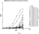
- Table.5 showed the result from second round mutagenesis library screen.
- the clones 1H6, 2E5, 2G4 and 2C2 were selected for further analysis.
- Table 5 Name VHCDR3 VKCDR1 VKCDR3 Bmax(Hu man) Kd (Human) Bmax (Mouse) Kd (Mouse) Bmax (Cynomol gus) Kd (Cynomol gus) 2E5 I LDSDGGTYLYW MQLTHWPYTFGQ 3.279 0.0675 0.4696 0.0443 1.72 0.1088 2G4 I LDSDGSTYLYW MQLTHWPYTFGQ 3.371 0.0708 0.4793 0.0426 1.718 0.1057 1G10 L LDSDGATYLYW MQLTHWPYTFGQ 2.600 0.0711 0.2997 0.0718 1.082 0.1224 2C2 I LDSDGATYLYW MQLTHWPYTFGQ 3.175 0.082 0.416
- the vector containing affinity matured humanized antibody were transfected into 293F cells for antibody production and expression.
- Antibodies in the supernatant of 293F cells were purified using Protein A affinity chromatography.
- Figure 3 showed the results of cross-species test by FACS.
- Figure 3A showed binding to human PD-1 transfected CHO-S cells.
- the antibodies can bind specifically to the human PD-1 with EC50 of 2.20-2.78 nM.
- Figure.3B showed binding to mouse PD-1 transfected 293F cells.
- the antibodies can bind specifically to the mouse PD-1 with EC50 of 11.8-15.1 nM.
- Figure.3C showed binding to activated cynomolgus PBMC in a dose dependent way.
- the isotype was human IgG4 kappa. The same below.
- ELISA For ELISA, plates (Nunc) were coated with human, cynomolgus or mouse PD-1 (Sino Biological) at 1 ⁇ g/ml overnight at 4 °C. After blocking and washing, antibodies were serially diluted in blocking buffer and added to the plates and incubated at room temperature for 1 h. The plates were then washed and subsequently incubated with secondary antibody goat anti human IgG HRP (Bethyl) for 1 h. After washing, TMB substrate was added and the reaction was stopped by 2 M HCl. The absorbance at 450 nm was read using a microplate reader (Molecular Device).
- Figure 4 showed the result of cross-species test by ELISA.
- Figure 4A showed binding to human PD-1.
- Figure 4B showed binding to mouse PD-1.
- Figure 4C showed binding to cynomolgus PD-1.
- Constructed cell lines that respectively express human PD-1, CD28, CTLA-4 or ICOS were transferred in to 96-well U-bottom plates (BD) at a density of 2 ⁇ 10 5 cells/well. Testing antibodies were diluted in wash buffer (1 ⁇ PBS/1%BSA) and incubated with cells at 4 °C for 1 h. After washing, the secondary antibody goat anti-human IgG Fc FITC (Jackson ImmunoResearch Lab) was added and incubated at 4 °C in the dark for 1 h. The cells were then washed once and resuspended in 1 ⁇ PBS/1%BSA, and analyzed by flow cytometery (BD) and FlowJo software.
- wash buffer (1 ⁇ PBS/1%BSA
- IgG Fc FITC Jackson ImmunoResearch Lab
- Figure 5 showed the result of cross-family test.
- the anti-PD-1 antibodies can bind specifically to human PD-1, but not to CD28 and CTLA-4.
- Figure 6A showed the result of anti-PD-1 antibodies blocking human PD-L1 binding to PD-1 transfected CHO-S cells.
- Figure 6B shows the result of anti-PD-1 antibodies blocking mouse PD-L1 binding to PD-1 transfected 293F cells.
- Figure 7 showed that the anti-PD-1 antibodies could block human PD-L2 binding to PD-1 in a dose-dependent manner.
- Antibodies were characterized for affinity and binding kinetics to PD-1 by SPR assay using ProteOn XPR36 (Bio-Rad). Protein A protein (Sigma) was immobilized to a GLM sensor chip (Bio-Rad) through amine coupling. Purified antibodies were flowed over the sensor chip and captured by the Protein A. The chip was rotated 90° and washed with running buffer (1 ⁇ PBS/0.01% Tween20, Bio-Rad) until the baseline was stable. Seven concentrations of human PD-1 and running buffer were flowed through the sensor chip at a flow rate of 30 ⁇ L/min for an association phase of 180 s, followed by 300s dissociation.
- mice PD-1 and running buffer were flowed through the sensor chip at a flow rate of 30 ⁇ L/min for an association phase of 180 s, followed by 300 s dissociation.
- the chip was regenerated with pH 1.5 H 3 PO 4 after each run.
- the association and dissociation curve was fit by 1:1 Langmuir binding model using ProteOn software.
- Table.6A-6B showed the results of full kinetic binding affinity to human and mouse PD-1 by SPR.
- WBP305BMK1 was synthesized according to the clone of 5C4 from BMS patent US9084776B2 .
- Keytruda was the anti-PD-1 drug from Merck. The same below.
- the results showed that the affinity ability to human PD-1 by SPR assay was from 1.43E-8 to 5.64E-9 mol/L. Comparing WBP305BMK1 with Keytruda, the K D value of antibody 2E5, 2G4 or 2C2 was much smaller, illustrating that 2E5, 2G4 or 2C2 had better binding ability to human PD-1.
- CHO-S cells expressing human PD-1 or 293F cells expressing mouse PD-1 were transferred in to 96-well U-bottom plates (BD) at a density of 1x10 5 cells/well. Testing antibodies were 1:2 serially diluted in wash buffer (1 ⁇ PBS/1%BSA) and incubated with cells at 4 °C for 1 h. The secondary antibody goat anti-human IgG Fc FITC (3.0 moles FITC per mole IgG, (Jackson Immunoresearch Lab) was added and incubated at 4 °C in the dark for 1 h. The cells were then washed once and resuspended in 1XPBS/1%BSA, and analyzed by flow cytometery (BD). Fluorescence intensity was converted to bound molecules/cell based on the quantitative beads (Quantum TM MESF Kits, Bangs Laboratories, Inc.). KD was calculated using Graphpad Prism5.
- Table.7A-7B show the results of binding affinity of anti-PD-1 antibodies to cell surface human and mouse PD-1 molecules tested by flow cytometry. The results showed that the affinity ability to human PD-1 by FACS assay was from 3.80E-10 to 2.15E-10 mol/L. In addition, the affinity ability to mouse PD-1 was from 5.39E-8 to 1.74E-8 mol/L.
- the binding epitope of anti-PD-1 antibodies was compared with benchmark antibody A and B by FACS.
- CHO-S cells expressing human PD-1 on the cell surface were incubated with mixture of biotinylated benchmark antibody A or B ( ⁇ g/ml) and testing antibodies (serially diluted in wash buffer) at 4 °C for 1 h.
- the cells were washed and the second antibody Streptavidin-PE were added and incubated for 30 min at 4 °C.
- the cells were then washed once and resuspended in 1 xPBS/1%BSA, and analyzed by flow cytometery (BD).
- BD flow cytometery
- Figure 8A-8B showed the results of epitope binning assay suggesting that the anti-PD-1 antibodies are in the same or close epitope bin as benchmark antibodies.
- Figure.8A showed binning against WBP305BMK1 ( US9084776 ).
- Figure.8B showed binning against Keytruda ( US8168757 ).
- Dpn I endonuclease was used to digest the parental template after mutant strand synthesis reaction.
- linear DNA expression cassette which composed of a CMV promoter, an extracellular domain (ECD) of PD-1, a His-tag and a herpes simplex virus thymidine kinase (TK) polyadenylation was amplified and transiently expressed in HEK293F cells (Life Technologies, Gaithersburg, MD).
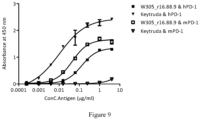
- Monoclonal antibodies W3052_r16.88.9 and Keytruda were coated in plates for ELISA binding assay. After interacting with the supernatant that contains quantified PD-1 mutant or human/mouse PD-1_ECD.His protein (Sino Biological, China), HRP conjugated anti-His antibody was added as detection antibody. Absorbance was normalized according to the average of control mutants. After setting an additional cutoff to the binding fold change ( ⁇ 0.55), the final determined epitope residues were identified.
- the epitope mapping results show that the epitope of antibody W3052_r16.88.9 was composed of hot spots distributed across multiple locations, and have direct overlap with the hPD-L1 binding site ( Figure 10A, 10B ).
- W3052_r16.88.9 blocked HPD-L1 by means of competing with hPD-L1 in reacting to their common binding site.
- W3052_r16.88.9 had no interactions with the flexible C'D loop (or the corresponding C" strand on mPD-1), where human and murine PD-1 show big structural deviations ( Figure 11 ). Its binding site is mostly located on the FG loop ( Lin et al. (2008) PNAS 105: 3011-3016 ).
- Human DCs, CD4 + T, CD8 + T and total T cells isolation Human PBMCs were freshly isolated from healthy donors using Ficoll-Paque PLUS (GE) gradient centrifugation. Monocytes were isolated using Human Monocyte Enrichment Kit (StemCell) according to the manufacturer's instructions. Cells were cultured in medium containing rhGM-CSF and rhIL-4 for 5 to 7 days to generate dendritic cells. 18 to 24 hours before MLR, 1 ⁇ g/mL LPS was added to the culture to induce the maturation of the DCs. Human CD4 + T cells were isolated using Human CD4 + T Cell Enrichment Kit (StemCell) according to the manufacturer's protocol.
- Mouse CD4 + T cells were obtained from the spleen of Balb / c mouse using Mouse CD4 + T Cell Isolation Kit (StemCell) according to the manufacturer's protocol.
- Mouse DCs were induced from bone marrow cells of C57BL/6 mouse in medium containing rmGM-CSF and rmIL-4 for 5 to 7 days. 18 to 24 hours before MLR, 1 ⁇ g/mL LPS was added to the culture to induce the maturation of the DCs.
- DC dendritic cell
- Cytokine detection Human IFN- ⁇ and IL-2 were measured by enzyme-linked immunosorbent assay (ELISA) using matched antibody pairs. The plates were pre-coated with capture antibody specific for human IFN- ⁇ (cat# Pierce-M700A) or IL-2 (cat# R&D-MAB602), respectively. The biotin-conjugated anti-IFN-y antibody (cat# Pierce-M701B) or anti-IL-2 antibody (cat# R&D-BAF202) was used as detecting antibody.
- ELISA enzyme-linked immunosorbent assay
- Figure 12A showed anti-PD-1 antibodies increased IL-2 secretion in a dose-dependent manner.
- Figure 12B shows anti-PD-1 antibodies increase IFN- ⁇ secretion in a dose-dependent manner.
- Proliferation assay 3H-thymidine (cat# PerkinElmer- NET027001MC) was diluted 1:20 in 0.9% NaCl solution, and added to the cell culture plates at 0.5 uCi/well. The plates were cultured in 5% CO 2 at 37°C for 16 to 18 hours, before the incorporation of 3H-thymidine into the proliferating cells was determined.
- Figure 12C shows anti-PD-1 antibodies increase CD4 + T cells proliferation in a dose-dependent manner.
- Figure 13A-13C showed the results of mouse allo-MLR demonstrating that the anti-PD-1 antibodies can enhance the function of mouse CD4 + T cell.
- Figure 13A showed anti-PD-1 antibodies increased IL-2 secretion in a dose-dependent manner.
- Figure. 13B showed anti-PD-1 antibodies increased IFN- ⁇ secretion in a dose-dependent manner.
- Figure 13C showed anti-PD-1 antibodies increased CD4 + T cells proliferation in a dose-dependent manner.
- CD4 + T cells and DCs were from a same donor.
- CD4 + T cells were purified from PBMC and cultured in the presence of CMV pp65 peptide and low dose of IL-2 (20 U/mL), at the meanwhile, DCs were generated by culturing monocytes from the same donor's PBMC in GM-CSF and IL-4.
- the CMV pp65 peptide treated CD4 + T cells were co-cultured with DCs pulsed with CMV pp65 peptide in the absence or presence of human anti-PD-1 antibodies or benchmark antibodies (as control).
- 100 ⁇ L of supernatants were taken from each of cultures for IFN- ⁇ measurement by ELISA as described above.
- the proliferation of CMV pp65-specific T cells was assessed by 3H-thymidine incorporation as described above.
- Figure 14A-14B showed the results of human auto-MLR demonstrating the anti-PD-1 antibodies can enhance the function of human CD4 + T cell.
- Figure 14A showed anti-PD-1 antibodies increase IFN- ⁇ secretion in a dose-dependent manner.
- Figure 14B showed anti-PD-1 antibodies increase CD4 + T cells proliferation in a dose-dependent manner.
- Tregs a subpopulation of T cells
- Increased numbers of CD4 + CD25 + Tregs were found in patients with multiple cancers and associated with a poorer prognosis.
- To determine whether the anti-PD-1 antibodies affect the immune suppressive role of Tregs we compared the T cell function in the presence of Tregs with or without anti-PD-1 antibody treatment.
- CD4 + CD25 + and CD4 + CD25 - T cells were separated using specific anti-CD25 microbeads (StemCell) per manufacture's instruction.
- Figure 15 demonstrates that the anti-PD-1 antibodies can reverse the suppressive function of Tregs.
- Figure 15A showed anti-PD-1 antibodies can restore the IFN- ⁇ secretion.
- Figure 15B showed anti-PD-1 antibodies can restore the T-cell proliferation.
- PD-1 is expressed on variety of cell types. In order to minimize potential toxicity to healthy PD-1 positive cells, the anti-PD-1 antibodies were evaluated for their ability to mediate antibody-dependent cellular cytotoxicity (ADCC) and complement-dependent cytotoxicity (CDC).
- ADCC antibody-dependent cellular cytotoxicity
- CDC complement-dependent cytotoxicity
- Human activated CD4 + T cells and various concentrations of PD-1 antibodies were pre-incubated in 96-well plate for 30 minutes, and then PBMCs were added at the effector/target ratio of 50:1.
- the plate was kept at 37 °C in a 5% CO 2 incubator for 6 hours.
- Target cell lysis was determined by LDH-based cytotoxicity detection kit (cat# Roche-11644793001). The absorbance at 492 nm was read using a microplate reader (Molecular Device). Herceptin-induced SK-Br-3 cell lysis was used as positive control.
- Figure 16 showed the result of ADCC test demonstrating the anti-PD-1 antibodies did not mediate ADCC activity on activated CD4 + T cells.
- Human activated CD4 + T cells and various concentrations of PD-1 antibodies were mixed in 96-well plate.
- Human complement (Quidel-A112) was added at the dilution ratio of 1:50.
- the plate was kept at 37 °C in a 5% CO 2 incubator for 2 hours.
- Target cell lysis was determined by CellTiter-Glo.
- Rituxan ® -induced Raji cell lysis was used as positive control.
- the luminescence was read using a microplate reader (Molecular Device).
- Figure 17 showed the result of CDC test demonstrating the anti-PD-1 antibodies did not mediate CDC activity on activated CD4 + T cells.
- Example 5 Treatment of in vivo tumor model using human monoclonal antibodies against PD-1
- Murine melanoma cell CloudmanS91 cell (ATCC-CCL-53.1) was cultured in vitro as monolayer, and the culture condition was F-12K medium plus 2.5% FBS and 15% horse serum, 100 U/mL penicillin, and 100 ⁇ g/mL streptomycin, incubate at 37 °C and 5% CO 2 .
- the cells were digested using trypsin-EDTA and passaged twice a week routinely. Cells were harvested, counted, and then inoculated when approximately 80%-90% confluent and the number is as required.
- CloudmanS91 cells were inoculated subcutaneously in the right backside of each animal. When the mean of tumor volume had reached approximately 64 mm 3 , the administration started in groups. Grouping and dosing regimens were shown in Table 10.
- Tumor growth was inhibited, delayed or cured.
- Tumor diameters were measured with a caliper three times a week.
- TGI (%) [(1-(average tumor volume at the end of administration in the treatment group - average tumor volume at the start of administration in the treatment group)) / (average tumor volume at the end of treatment in the solvent control group - average tumor volume at the start of treatment in the solvent control group)] ⁇ 100%.
- T/C (%) T RTV / C RTV ⁇ 100% (T RTV : treatment group RTV; C RTV : negative control group RTV).
- T-C days reflected tumor growth delay index
- T represented average days passed when the tumor had reached a predetermined volume in the treatment group (eg. 300 mm 3 )
- C represented the average days when tumors in the control group had reached the same volume.
- Animal survival time was defined as the time from the administration to animal deaths or the time when tumor volume had reached 2000 mm 3 .
- the median survival time (days) was calculated in each group.
- Increased life span (ILS) was calculated by comparison of the median survival times between the treated group and model control group and represented as a percentage over the lifetime of the model control group.
- Tumor volume in CloudmanS91 subcutaneously syngeneic xenograft female DBA/2 mouse model after 2E5 treatment was as shown in Table 11.
- Table 11 Tumor volume at different times in each group Days Tumor volume (mm 3 ) a Vehicle 2E5 2E5 2E5 1 mg/kg 3 mg/kg 10 mg/kg 0 b 66 ⁇ 9 65 ⁇ 8 64 ⁇ 8 63 ⁇ 8 2 142 ⁇ 23 129 ⁇ 10 110 ⁇ 10 94 ⁇ 8 4 251 ⁇ 39 231 ⁇ 34 162 ⁇ 9 143 ⁇ 17 6 345 ⁇ 65 339 ⁇ 61 200 ⁇ 13 197 ⁇ 38 9 599 ⁇ 66 597 ⁇ 100 281 ⁇ 38 291 ⁇ 83 11 1,026 ⁇ 173 943 ⁇ 307 335 ⁇ 66 475 ⁇ 190 13 1,626 ⁇ 262 1,089 ⁇ 365 361 ⁇ 81 614 ⁇ 273
- Tumor growth curve was shown in Figure 20 .
- the median survival time of tumor-bearing mice in solvent control group was 16 days.
Landscapes
- Health & Medical Sciences (AREA)
- Chemical & Material Sciences (AREA)
- Life Sciences & Earth Sciences (AREA)
- Organic Chemistry (AREA)
- Immunology (AREA)
- Genetics & Genomics (AREA)
- General Health & Medical Sciences (AREA)
- Medicinal Chemistry (AREA)
- Engineering & Computer Science (AREA)
- Biochemistry (AREA)
- Molecular Biology (AREA)
- Biophysics (AREA)
- Bioinformatics & Cheminformatics (AREA)
- Biomedical Technology (AREA)
- Proteomics, Peptides & Aminoacids (AREA)
- Veterinary Medicine (AREA)
- Public Health (AREA)
- Zoology (AREA)
- Animal Behavior & Ethology (AREA)
- Wood Science & Technology (AREA)
- Biotechnology (AREA)
- Pharmacology & Pharmacy (AREA)
- General Engineering & Computer Science (AREA)
- Microbiology (AREA)
- General Chemical & Material Sciences (AREA)
- Nuclear Medicine, Radiotherapy & Molecular Imaging (AREA)
- Chemical Kinetics & Catalysis (AREA)
- Plant Pathology (AREA)
- Physics & Mathematics (AREA)
- Mycology (AREA)
- Epidemiology (AREA)
- Cell Biology (AREA)
- Oncology (AREA)
- Endocrinology (AREA)
- Peptides Or Proteins (AREA)
- Medicines Containing Antibodies Or Antigens For Use As Internal Diagnostic Agents (AREA)
- Preparation Of Compounds By Using Micro-Organisms (AREA)
- Micro-Organisms Or Cultivation Processes Thereof (AREA)
Priority Applications (1)
| Application Number | Priority Date | Filing Date | Title |
|---|---|---|---|
| EP24184760.7A EP4450616A3 (de) | 2016-09-21 | 2016-09-21 | Neue monoklonale antikörper gegen programmierten tod 1 (pd-1) |
Applications Claiming Priority (3)
| Application Number | Priority Date | Filing Date | Title |
|---|---|---|---|
| PCT/CN2016/099576 WO2018053709A1 (en) | 2016-09-21 | 2016-09-21 | The novel monoclonal antibodies to programmed death 1 (pd-1) |
| EP16916449.8A EP3515938B8 (de) | 2016-09-21 | 2016-09-21 | Monoklonale antikörper gegen den programmierten tod 1 (pd-1) |
| EP24184760.7A EP4450616A3 (de) | 2016-09-21 | 2016-09-21 | Neue monoklonale antikörper gegen programmierten tod 1 (pd-1) |
Related Parent Applications (2)
| Application Number | Title | Priority Date | Filing Date |
|---|---|---|---|
| EP16916449.8A Division-Into EP3515938B8 (de) | 2016-09-21 | 2016-09-21 | Monoklonale antikörper gegen den programmierten tod 1 (pd-1) |
| EP16916449.8A Division EP3515938B8 (de) | 2016-09-21 | 2016-09-21 | Monoklonale antikörper gegen den programmierten tod 1 (pd-1) |
Publications (2)
| Publication Number | Publication Date |
|---|---|
| EP4450616A2 true EP4450616A2 (de) | 2024-10-23 |
| EP4450616A3 EP4450616A3 (de) | 2025-01-01 |
Family
ID=61690082
Family Applications (2)
| Application Number | Title | Priority Date | Filing Date |
|---|---|---|---|
| EP16916449.8A Active EP3515938B8 (de) | 2016-09-21 | 2016-09-21 | Monoklonale antikörper gegen den programmierten tod 1 (pd-1) |
| EP24184760.7A Pending EP4450616A3 (de) | 2016-09-21 | 2016-09-21 | Neue monoklonale antikörper gegen programmierten tod 1 (pd-1) |
Family Applications Before (1)
| Application Number | Title | Priority Date | Filing Date |
|---|---|---|---|
| EP16916449.8A Active EP3515938B8 (de) | 2016-09-21 | 2016-09-21 | Monoklonale antikörper gegen den programmierten tod 1 (pd-1) |
Country Status (22)
| Country | Link |
|---|---|
| US (3) | US11414487B2 (de) |
| EP (2) | EP3515938B8 (de) |
| JP (2) | JP7101169B2 (de) |
| KR (2) | KR102527160B1 (de) |
| AU (1) | AU2016423559C1 (de) |
| CA (1) | CA3037407C (de) |
| DK (1) | DK3515938T3 (de) |
| ES (1) | ES3017957T3 (de) |
| FI (1) | FI3515938T3 (de) |
| HR (1) | HRP20250400T1 (de) |
| HU (1) | HUE071253T2 (de) |
| IL (1) | IL265525A (de) |
| LT (1) | LT3515938T (de) |
| MX (1) | MX2019003058A (de) |
| PL (1) | PL3515938T3 (de) |
| PT (1) | PT3515938T (de) |
| RS (1) | RS66679B1 (de) |
| RU (1) | RU2757316C2 (de) |
| SA (1) | SA519401370B1 (de) |
| SI (1) | SI3515938T1 (de) |
| SM (1) | SMT202500148T1 (de) |
| WO (1) | WO2018053709A1 (de) |
Families Citing this family (23)
| Publication number | Priority date | Publication date | Assignee | Title |
|---|---|---|---|---|
| MX392711B (es) | 2015-08-11 | 2025-03-24 | Wuxi Biologics Ireland Ltd | Anticuerpos anti-pd-1 novedosos. |
| KR20250099459A (ko) | 2015-11-03 | 2025-07-01 | 얀센 바이오테크 인코포레이티드 | Pd-1과 특이적으로 결합하는 항체 및 그의 용도 |
| JP7101169B2 (ja) * | 2016-09-21 | 2022-07-14 | シーストーン ファーマシューティカルズ | プログラム死1(pd-1)に対する新規モノクローナル抗体 |
| RS62589B1 (sr) | 2016-11-02 | 2021-12-31 | Jounce Therapeutics Inc | Antitela na pd-1 i njihova upotreba |
| TWI901940B (zh) | 2017-08-03 | 2025-10-21 | 美商安進公司 | 介白素-21突變蛋白及治療方法 |
| JP2019122373A (ja) | 2018-01-12 | 2019-07-25 | アムジエン・インコーポレーテツド | 抗pd−1抗体及び治療方法 |
| US12398209B2 (en) | 2018-01-22 | 2025-08-26 | Janssen Biotech, Inc. | Methods of treating cancers with antagonistic anti-PD-1 antibodies |
| AU2019255515B2 (en) * | 2018-04-15 | 2023-04-06 | Immvira Co., Limited | Antibodies binding PD-1 and uses thereof |
| US20210095032A1 (en) * | 2018-04-15 | 2021-04-01 | Salubris (Chengdu) Biotech Co., Ltd. | Antibodies binding pd-1 and uses thereof |
| CN109160949B (zh) * | 2018-07-23 | 2021-05-14 | 中国医学科学院血液病医院(血液学研究所) | 一种鼠抗人pd-1单克隆抗体及应用 |
| WO2020216348A1 (en) * | 2019-04-26 | 2020-10-29 | Wuxi Biologics (Shanghai) Co., Ltd. | Bispecific antibodies against pd-1 and lag-3 |
| WO2020239558A1 (en) | 2019-05-24 | 2020-12-03 | Pfizer Inc. | Combination therapies using cdk inhibitors |
| WO2021057836A1 (en) * | 2019-09-25 | 2021-04-01 | Wuxi Biologics (Shanghai) Co. Ltd. | Novel anti-pd-l1 antibodies |
| JP2023500385A (ja) * | 2019-11-11 | 2023-01-05 | シーストーン・ファーマシューティカルズ・(スージョウ)・カンパニー・リミテッド | 医薬組合せ物およびその使用 |
| WO2021110122A1 (zh) * | 2019-12-05 | 2021-06-10 | 基石药业(苏州)有限公司 | Cdk4/6抑制剂的组合疗法 |
| MA59827B1 (fr) | 2020-05-26 | 2024-10-31 | Boehringer Ingelheim International Gmbh | Anticorps anti-pd-1 |
| US20230331848A1 (en) * | 2020-08-31 | 2023-10-19 | Biosion Inc. | Pd-1 binding antibodies and uses thereof |
| CN114316022A (zh) * | 2020-09-30 | 2022-04-12 | 正大天晴康方(上海)生物医药科技有限公司 | 结合pd-1抗体的肽及其应用 |
| WO2022118197A1 (en) | 2020-12-02 | 2022-06-09 | Pfizer Inc. | Time to resolution of axitinib-related adverse events |
| JP2024508658A (ja) * | 2021-02-04 | 2024-02-28 | ジェヌーヴ インク. | 抗pd-1抗体及びその用途 |
| WO2023057882A1 (en) | 2021-10-05 | 2023-04-13 | Pfizer Inc. | Combinations of azalactam compounds with a pd-1 axis binding antagonist for the treatment of cancer |
| WO2023079428A1 (en) | 2021-11-03 | 2023-05-11 | Pfizer Inc. | Combination therapies using tlr7/8 agonist |
| CN116036266A (zh) * | 2022-12-28 | 2023-05-02 | 广州誉衡生物科技有限公司 | 抗-pd-1抗体及其在制备治疗结肠癌患者的药物中的用途 |
Citations (2)
| Publication number | Priority date | Publication date | Assignee | Title |
|---|---|---|---|---|
| US8168757B2 (en) | 2008-03-12 | 2012-05-01 | Merck Sharp & Dohme Corp. | PD-1 binding proteins |
| US9084776B2 (en) | 2005-05-09 | 2015-07-21 | E.R. Squibb & Sons, L.L.C. | Methods for treating cancer using anti-PD-1 antibodies |
Family Cites Families (13)
| Publication number | Priority date | Publication date | Assignee | Title |
|---|---|---|---|---|
| WO2006021955A2 (en) | 2004-08-23 | 2006-03-02 | Mor Research Applications Ltd. | Use of bat monoclonal antibody for immunotherapy |
| US7662965B2 (en) * | 2006-01-26 | 2010-02-16 | Cornerstone Therapeutics, Inc. | Anabaseine derivatives, pharmaceutical compositions and method of use thereof |
| JP6285414B2 (ja) * | 2012-03-30 | 2018-02-28 | モルフォテック, インコーポレイテッド | Tem−1診断用抗体 |
| CA2935423C (en) * | 2014-01-24 | 2023-11-07 | Dana-Farber Cancer Institute, Inc. | Antibody molecules to pd-1 and uses thereof |
| CN105330740B (zh) * | 2014-07-30 | 2018-08-17 | 珠海市丽珠单抗生物技术有限公司 | 抗pd-1抗体及其应用 |
| WO2016077397A2 (en) | 2014-11-11 | 2016-05-19 | Sutro Biopharma, Inc. | Anti-pd-1 antibodies, compositions comprising anti-pd-1 antibodies and methods of using anti-pd-1 antibodies |
| TWI595006B (zh) | 2014-12-09 | 2017-08-11 | 禮納特神經系統科學公司 | 抗pd-1抗體類和使用彼等之方法 |
| US20180244779A1 (en) | 2015-02-06 | 2018-08-30 | Kadmon Corporation Llc | Immunomodulatory agents |
| MX392711B (es) | 2015-08-11 | 2025-03-24 | Wuxi Biologics Ireland Ltd | Anticuerpos anti-pd-1 novedosos. |
| AU2016333517B2 (en) | 2015-10-02 | 2023-09-07 | Les Laboratoires Servier | Anti-PD-1 antibodies and compositions |
| JP7101169B2 (ja) * | 2016-09-21 | 2022-07-14 | シーストーン ファーマシューティカルズ | プログラム死1(pd-1)に対する新規モノクローナル抗体 |
| CN109575139A (zh) * | 2017-09-29 | 2019-04-05 | 上海药明生物技术有限公司 | 针对表皮生长因子受体和程序性死亡受体的双特异抗体 |
| JP2023500385A (ja) * | 2019-11-11 | 2023-01-05 | シーストーン・ファーマシューティカルズ・(スージョウ)・カンパニー・リミテッド | 医薬組合せ物およびその使用 |
-
2016
- 2016-09-21 JP JP2019515471A patent/JP7101169B2/ja active Active
- 2016-09-21 EP EP16916449.8A patent/EP3515938B8/de active Active
- 2016-09-21 HR HRP20250400TT patent/HRP20250400T1/hr unknown
- 2016-09-21 CA CA3037407A patent/CA3037407C/en active Active
- 2016-09-21 KR KR1020217020495A patent/KR102527160B1/ko active Active
- 2016-09-21 RS RS20250329A patent/RS66679B1/sr unknown
- 2016-09-21 RU RU2019111277A patent/RU2757316C2/ru active
- 2016-09-21 FI FIEP16916449.8T patent/FI3515938T3/fi active
- 2016-09-21 PT PT169164498T patent/PT3515938T/pt unknown
- 2016-09-21 LT LTEPPCT/CN2016/099576T patent/LT3515938T/lt unknown
- 2016-09-21 US US16/333,993 patent/US11414487B2/en active Active
- 2016-09-21 SI SI201631893T patent/SI3515938T1/sl unknown
- 2016-09-21 PL PL16916449.8T patent/PL3515938T3/pl unknown
- 2016-09-21 HU HUE16916449A patent/HUE071253T2/hu unknown
- 2016-09-21 MX MX2019003058A patent/MX2019003058A/es unknown
- 2016-09-21 KR KR1020197010353A patent/KR102273634B1/ko active Active
- 2016-09-21 DK DK16916449.8T patent/DK3515938T3/da active
- 2016-09-21 AU AU2016423559A patent/AU2016423559C1/en active Active
- 2016-09-21 SM SM20250148T patent/SMT202500148T1/it unknown
- 2016-09-21 ES ES16916449T patent/ES3017957T3/es active Active
- 2016-09-21 WO PCT/CN2016/099576 patent/WO2018053709A1/en not_active Ceased
- 2016-09-21 EP EP24184760.7A patent/EP4450616A3/de active Pending
-
2019
- 2019-03-20 SA SA519401370A patent/SA519401370B1/ar unknown
- 2019-03-20 IL IL265525A patent/IL265525A/en unknown
-
2021
- 2021-04-21 JP JP2021071948A patent/JP2021120378A/ja active Pending
-
2022
- 2022-06-29 US US17/853,606 patent/US20230014722A1/en not_active Abandoned
-
2023
- 2023-08-31 US US18/459,236 patent/US20240101676A1/en active Pending
Patent Citations (2)
| Publication number | Priority date | Publication date | Assignee | Title |
|---|---|---|---|---|
| US9084776B2 (en) | 2005-05-09 | 2015-07-21 | E.R. Squibb & Sons, L.L.C. | Methods for treating cancer using anti-PD-1 antibodies |
| US8168757B2 (en) | 2008-03-12 | 2012-05-01 | Merck Sharp & Dohme Corp. | PD-1 binding proteins |
Non-Patent Citations (20)
| Title |
|---|
| AGATA ET AL., BIT IMMUNOL, vol. 8, 1996, pages 765 - 72 |
| BENNETT ET AL., J IMMUNOL, vol. 170, 2003, pages 711 - 8 |
| BLANK, CANCER IMMUNOL. IMMUNOTHER., vol. 54, 2005, pages 307 - 314 |
| CARTER ET AL., EUR J IMMUNOL, vol. 32, 2002, pages 634 - 43 |
| DONG ET AL., J. MOL. MED., vol. 81, 2003, pages 281 - 7 |
| DONG ET AL., NAT. MED, vol. 8, 2002, pages 787 - 9 |
| FREEMAN ET AL., J EXP MED, vol. 192, 2000, pages 1027 - 34 |
| ISHIDA ET AL., EMBO J, vol. 11, 1992, pages 3887 - 95 |
| KONISHI ET AL., CLIN. CANCER RES., vol. 10, 2004, pages 5094 - 100 |
| LATCHMAN ET AL., NAT IMMUNOL, vol. 2, 2001, pages 261 - 8 |
| LIN ET AL., PNAS, vol. 105, 2008, pages 3011 - 3016 |
| NIELSEN ET AL., LUPUS, vol. 11, 2004, pages 510 |
| NISHIMURA ET AL., IMMUNITY, vol. 11, 1999, pages 141 - 51 |
| NISHIMURA, SCIENCE, vol. 291, 2001, pages 319 - 22 |
| OKAZAKI ET AL., CURR OPIN IMMUNOL, vol. 14, 2002, pages 391779 - 82 |
| OKAZAKI ET AL., CURR. OPIN. IMMUNOL., vol. 14, 2002, pages 391779 - 82 |
| OKAZAKI ET AL., PNAS, vol. 98, 2001, pages 13866 - 71 |
| OKAZAKI, TAKU ET AL., INTERNATIONAL IMMUNOLOGY, vol. 19, 2007, pages 813 - 824 |
| PROKUNINAALARCON-RIQUELME, HUM MOL GENET, vol. 13, 2004, pages R143 |
| SALAMA ET AL., J EXP MED, vol. 198, 2003, pages 71 - 78 |
Also Published As
Similar Documents
| Publication | Publication Date | Title |
|---|---|---|
| US20230014722A1 (en) | Novel Monoclonal Antibodies to Programmed DEATH (PD-1) | |
| TWI723220B (zh) | 一種新的pd-1單株抗體 | |
| WO2022136649A1 (en) | Non-blocking human ccr8 binders | |
| EP4378954A1 (de) | Bispezifischer anti-pvrig/anti-tigit-antikörper und anwendung | |
| EP4458417A2 (de) | Neue anti-pd-1-antikörper | |
| EP4267621A1 (de) | Kreuzreaktive menschliche ccr8-bindemittel aus der maus | |
| EP4101867A1 (de) | Anti-cd3 und anti-cd123 bispezifischer antikörper und verwendung davon | |
| EP4292611A1 (de) | Anti-cd112r-antikörper und verwendung davon | |
| CN113727731B (zh) | 靶向pd-1和lag-3的双特异性抗体 | |
| EP3498839A1 (de) | Anti-pd-1-antikörper | |
| AU2020243076A1 (en) | Antibodies having specificity for BTN2 and uses thereof | |
| TWI833227B (zh) | 靶向pd-l1和cd73的特異性結合蛋白及其應用 | |
| TWI835166B (zh) | 靶向pd-1和/或ox40的特異性結合蛋白及其應用 | |
| WO2022228545A1 (en) | Antibodies and variants thereof against human 4-1bb | |
| HK1246307B (en) | The novel monoclonal antibodies to pd-1 | |
| HK1246307A1 (zh) | 一种新的pd-1单克隆抗体 | |
| BR112019005316B1 (pt) | Anticorpos monoclonais para morte programada 1(pd-1), seu método de preparado e seus usos, bem como composições farmacêuticas e imunoconjugado | |
| HK40103081A (zh) | 鼠交叉反应性人ccr8结合剂 |
Legal Events
| Date | Code | Title | Description |
|---|---|---|---|
| PUAI | Public reference made under article 153(3) epc to a published international application that has entered the european phase |
Free format text: ORIGINAL CODE: 0009012 |
|
| STAA | Information on the status of an ep patent application or granted ep patent |
Free format text: STATUS: THE APPLICATION HAS BEEN PUBLISHED |
|
| AC | Divisional application: reference to earlier application |
Ref document number: 3515938 Country of ref document: EP Kind code of ref document: P |
|
| AK | Designated contracting states |
Kind code of ref document: A2 Designated state(s): AL AT BE BG CH CY CZ DE DK EE ES FI FR GB GR HR HU IE IS IT LI LT LU LV MC MK MT NL NO PL PT RO RS SE SI SK SM TR |
|
| REG | Reference to a national code |
Ref country code: DE Ref legal event code: R079 Free format text: PREVIOUS MAIN CLASS: C12N0005200000 Ipc: C07K0016280000 |
|
| PUAL | Search report despatched |
Free format text: ORIGINAL CODE: 0009013 |
|
| AK | Designated contracting states |
Kind code of ref document: A3 Designated state(s): AL AT BE BG CH CY CZ DE DK EE ES FI FR GB GR HR HU IE IS IT LI LT LU LV MC MK MT NL NO PL PT RO RS SE SI SK SM TR |
|
| RIC1 | Information provided on ipc code assigned before grant |
Ipc: C12N 5/20 20060101ALI20241125BHEP Ipc: C12N 5/10 20060101ALI20241125BHEP Ipc: C12N 15/63 20060101ALI20241125BHEP Ipc: C12N 15/13 20060101ALI20241125BHEP Ipc: A61P 35/00 20060101ALI20241125BHEP Ipc: A61K 39/395 20060101ALI20241125BHEP Ipc: C07K 16/28 20060101AFI20241125BHEP |
|
| STAA | Information on the status of an ep patent application or granted ep patent |
Free format text: STATUS: REQUEST FOR EXAMINATION WAS MADE |
|
| 17P | Request for examination filed |
Effective date: 20250701 |