EP4417693A1 - Extracellular vesicle secretion booster for boosting extracellular vesicle secretion, and use thereof - Google Patents
Extracellular vesicle secretion booster for boosting extracellular vesicle secretion, and use thereof Download PDFInfo
- Publication number
- EP4417693A1 EP4417693A1 EP22880979.4A EP22880979A EP4417693A1 EP 4417693 A1 EP4417693 A1 EP 4417693A1 EP 22880979 A EP22880979 A EP 22880979A EP 4417693 A1 EP4417693 A1 EP 4417693A1
- Authority
- EP
- European Patent Office
- Prior art keywords
- secretion
- extracellular vesicle
- booster
- cell
- cells
- Prior art date
- Legal status (The legal status is an assumption and is not a legal conclusion. Google has not performed a legal analysis and makes no representation as to the accuracy of the status listed.)
- Pending
Links
Images




Classifications
-
- C—CHEMISTRY; METALLURGY
- C12—BIOCHEMISTRY; BEER; SPIRITS; WINE; VINEGAR; MICROBIOLOGY; ENZYMOLOGY; MUTATION OR GENETIC ENGINEERING
- C12N—MICROORGANISMS OR ENZYMES; COMPOSITIONS THEREOF; PROPAGATING, PRESERVING, OR MAINTAINING MICROORGANISMS; MUTATION OR GENETIC ENGINEERING; CULTURE MEDIA
- C12N5/00—Undifferentiated human, animal or plant cells, e.g. cell lines; Tissues; Cultivation or maintenance thereof; Culture media therefor
- C12N5/06—Animal cells or tissues; Human cells or tissues
-
- A—HUMAN NECESSITIES
- A61—MEDICAL OR VETERINARY SCIENCE; HYGIENE
- A61P—SPECIFIC THERAPEUTIC ACTIVITY OF CHEMICAL COMPOUNDS OR MEDICINAL PREPARATIONS
- A61P43/00—Drugs for specific purposes, not provided for in groups A61P1/00-A61P41/00
-
- C—CHEMISTRY; METALLURGY
- C12—BIOCHEMISTRY; BEER; SPIRITS; WINE; VINEGAR; MICROBIOLOGY; ENZYMOLOGY; MUTATION OR GENETIC ENGINEERING
- C12N—MICROORGANISMS OR ENZYMES; COMPOSITIONS THEREOF; PROPAGATING, PRESERVING, OR MAINTAINING MICROORGANISMS; MUTATION OR GENETIC ENGINEERING; CULTURE MEDIA
- C12N15/00—Mutation or genetic engineering; DNA or RNA concerning genetic engineering, vectors, e.g. plasmids, or their isolation, preparation or purification; Use of hosts therefor
- C12N15/09—Recombinant DNA-technology
- C12N15/11—DNA or RNA fragments; Modified forms thereof; Non-coding nucleic acids having a biological activity
- C12N15/113—Non-coding nucleic acids modulating the expression of genes, e.g. antisense oligonucleotides; Antisense DNA or RNA; Triplex- forming oligonucleotides; Catalytic nucleic acids, e.g. ribozymes; Nucleic acids used in co-suppression or gene silencing
- C12N15/1138—Non-coding nucleic acids modulating the expression of genes, e.g. antisense oligonucleotides; Antisense DNA or RNA; Triplex- forming oligonucleotides; Catalytic nucleic acids, e.g. ribozymes; Nucleic acids used in co-suppression or gene silencing against receptors or cell surface proteins
-
- C—CHEMISTRY; METALLURGY
- C12—BIOCHEMISTRY; BEER; SPIRITS; WINE; VINEGAR; MICROBIOLOGY; ENZYMOLOGY; MUTATION OR GENETIC ENGINEERING
- C12N—MICROORGANISMS OR ENZYMES; COMPOSITIONS THEREOF; PROPAGATING, PRESERVING, OR MAINTAINING MICROORGANISMS; MUTATION OR GENETIC ENGINEERING; CULTURE MEDIA
- C12N5/00—Undifferentiated human, animal or plant cells, e.g. cell lines; Tissues; Cultivation or maintenance thereof; Culture media therefor
- C12N5/10—Cells modified by introduction of foreign genetic material
-
- C—CHEMISTRY; METALLURGY
- C12—BIOCHEMISTRY; BEER; SPIRITS; WINE; VINEGAR; MICROBIOLOGY; ENZYMOLOGY; MUTATION OR GENETIC ENGINEERING
- C12N—MICROORGANISMS OR ENZYMES; COMPOSITIONS THEREOF; PROPAGATING, PRESERVING, OR MAINTAINING MICROORGANISMS; MUTATION OR GENETIC ENGINEERING; CULTURE MEDIA
- C12N2310/00—Structure or type of the nucleic acid
- C12N2310/10—Type of nucleic acid
- C12N2310/14—Type of nucleic acid interfering nucleic acids [NA]
-
- C—CHEMISTRY; METALLURGY
- C12—BIOCHEMISTRY; BEER; SPIRITS; WINE; VINEGAR; MICROBIOLOGY; ENZYMOLOGY; MUTATION OR GENETIC ENGINEERING
- C12N—MICROORGANISMS OR ENZYMES; COMPOSITIONS THEREOF; PROPAGATING, PRESERVING, OR MAINTAINING MICROORGANISMS; MUTATION OR GENETIC ENGINEERING; CULTURE MEDIA
- C12N2310/00—Structure or type of the nucleic acid
- C12N2310/10—Type of nucleic acid
- C12N2310/14—Type of nucleic acid interfering nucleic acids [NA]
- C12N2310/141—MicroRNAs, miRNAs
Definitions
- the present invention relates to an extracellular vesicle secretion booster for boosting extracellular vesicle secretion from cells and the use thereof.
- extracellular vesicles such as exosomes secreted from cells have been reported, and the use thereof is attracting attention.
- extracellular vesicles contain, for example, nucleic acids, such as microRNA (miRNA) and proteins.
- miRNA microRNA
- the extracellular vesicles mediate the transmission of their inclusions from a cell that has secreted these extracellular vesicles to a recipient cell, and thus are considered to function as a cell-to-cell communication tool.
- the same function has also attracted attention to the use thereof as carriers in drug delivery systems (DDSs).
- DDSs drug delivery systems
- the present invention provides an extracellular vesicle secretion booster containing an ABCA1 reducing agent.
- the present invention also provides a secretion boosting method including boosting extracellular vesicle secretion by causing a target having a cell to coexist with the extracellular vesicle secretion booster of the present invention.
- the present invention also provides a method for producing an extracellular vesicle, including: boosting extracellular vesicle secretion by causing a cell to coexist with the extracellular vesicle secretion booster of the present invention; and collecting an extracellular vesicle secreted from the cell.
- extracellular vesicle secretion booster of the present invention extracellular vesicle secretion from cells can be boosted.
- the preparation of extracellular vesicles as a material and the secretion of endogenous extracellular vesicles in a living organism by causing extracellular vesicle secretion from cells can be efficiently performed. Therefore, the present invention can be said to be a very useful technology, for example, in medical and other various fields.
- extracellular vesicles are hereinafter also referred to as EVs.
- An extracellular vesicle secretion booster of the present invention is also hereinafter referred to as an EV secretion booster.
- the extracellular vesicle secretion booster of the present invention is characterized by, as described above, containing an ABCA1 reducing agent.
- the ABCA1 reducing agent is an expression reducing substance or a function reducing substance for(against) an ABCA1 protein.
- the expression reducing substance is at least one selected from the group consisting of substances that reduce transcription from a gene encoding an ABCA1 protein, substances that degrade a transcription product resulting from transcription, and substances that reduce translation of the transcription product into a protein.
- the expression reducing substance is at least one nucleic acid substance selected from the group consisting of miRNAs, siRNAs, antisenses, and ribozymes, and precursors thereof.
- the expression reducing substance is an expression vector for expression of the nucleic acid substance.
- the expression reducing substance is miR-3202 or a precursor thereof.
- the precursor is at least one selected from the group consisting of a nucleic acid encoding the miRNAs, a primary miRNA transcription product, a precursor miRNA, and an expression vector of the miRNAs.
- the function reducing substance is a function inhibitory substance or a function neutralizing substance for(against) an ABCA1 protein.
- the function neutralizing substance is an antibody or antigen-binding fragment against an ABCA1 protein.
- the function reducing substance is an expression vector for expression of the function neutralizing substance.
- the EV secretion booster of the present invention is, for example, for boosting extracellular vesicle secretion from a cell, and the cell is a mesenchymal stem cell.
- the secretion boosting method of the present invention is characterized by, as described above, including: boosting extracellular vesicle secretion by causing a target having a cell to coexist with the extracellular vesicle secretion booster of the present invention.
- the extracellular vesicle secretion booster is added to the target in vivo, ex vivo, or in vitro.
- the cell is transfected with the extracellular vesicle secretion booster.
- the cell is a mesenchymal stem cell.
- the cell is a mesenchymal stem cell derived from bone marrow or adipose tissue.
- the cell is a cell collected from a living organism or a cell of a cell line.
- the cell is caused to coexist with the extracellular vesicle secretion booster in a medium.
- the target is tissue, an organ, or a living organism.
- the method for producing an extracellular vesicle of the present invention is characterized by, as described above, including: boosting extracellular vesicle secretion by causing a cell to coexist with the extracellular vesicle secretion booster of the present invention; and collecting an extracellular vesicle secreted from the cell.
- the cell is transfected with the extracellular vesicle secretion booster.
- the cell is a mesenchymal stem cell.
- the cell is a mesenchymal stem cell derived from bone marrow or adipose tissue.
- the cell is a cell collected from a living organism or a cell of a cell line.
- the cell is caused to coexist with the extracellular vesicle secretion booster in a medium.
- the EV production method of the present invention is, for example, for producing the extracellular vesicle as a carrier in a drug delivery system.
- the EV production method of the present invention is, for example, for producing the extracellular vesicle as a therapeutic agent.
- the EV secretion booster (promotor) of the present invention is characterized by, as described above, containing an ABCA1 reducing agent.
- the EV secretion booster of the present invention is characterized by containing the ABCA1 reducing agent, and other configurations and conditions are not particularly limited.
- the EV secretion booster of the present invention reference can be made to the following descriptions regarding the EV secretion boosting (promoting) method and the like of the present invention.
- the present invention is characterized by being based on the finding that the ABCA1 is involved in EV secretion and that the EV secretion from cells can be reduced by reducing the ABCA1.
- the type of substance used for reducing the ABCA1 and the method for reducing the same are not limited by any means.
- ABCA1 stands for ATP-binding cassette transporter 1 (member 1 of human transporter sub-family ABCA). ABCA1 protein and ABCA1 gene encoding the same are registered under Gene ID: 19 in the Genetic Testing Registry (GTR) database.
- GTR Genetic Testing Registry
- the "ABCA1 reducing agent” may be any agent that can reduce the ABCA1, and it may reduce, for example, the expression of an ABCA1 protein or the function of the ABCA1 protein.
- the ABCA1 reducing agent is, for example, an expression reducing substance for an ABCA1 protein
- the ABCA1 reducing agent is, for example, a function reducing substance for an ABCA1 protein.
- “reduce” may mean "inhibit”.
- the EV secretion booster of the present invention may contain, as the active ingredient, for example, only the above-described ABCA1 reducing agent or may further contain other ingredients for boosting the EV secretion in addition to the ABCA1 reducing agent.
- the EV secretion booster of the present invention may contain, for example, any one of or both the expression reducing substance and the function reducing substance, as the ABCA1 reducing agent.
- the type of ABCA1 reducing agent is not particularly limited, and it may be, for example, a low molecular weight compound such as a nucleic acid substance or an organic compound, a protein such as an antibody, a peptide such as an antigen-binding fragment, or the like.
- the expression reducing substance is not particularly limited, and may be, for example, a substance that reduces either transcription or translation during expression of an ABCA1 protein (hereinafter also referred to as a "target protein") from the gene (hereinafter also referred to as an "ABCA1 gene” or a "target gene”) encoding the ABCA1 protein.
- a target protein an ABCA1 protein
- ABCA1 gene a gene that reduces either transcription or translation during expression of an ABCA1 protein
- ABCA1 gene a gene that reduces either transcription or translation during expression of an ABCA1 protein
- ABCA1 gene a gene that reduces either transcription or translation during expression of an ABCA1 protein
- Examples of the reduction of transcription include inhibition of transcription from DNA to an mRNA precursor, inhibition of RNA processing to form a mature mRNA from an mRNA precursor, and degradation of an mRNA precursor or a mature mRNA.
- Examples of the reduction of translation include inhibition of translation from a mature mRNA and inhibition of modification of a
- the expression reducing substance is, for example, a nucleic acid substance (hereinafter also referred to as a "nucleic acid-type reducing substance”), and may be embodied as a nucleic acid substance that reduces the expression as it is (first embodiment) or may be embodied in the form of a precursor that converts into a state capable of reducing the expression when it is in vivo, in vitro, or ex vivo (second embodiment).
- a nucleic acid substance hereinafter also referred to as a "nucleic acid-type reducing substance”
- the expression reducing substance of the first embodiment is, for example, an antigene substance, an antisense (antisense oligonucleotide), an RNA interference (RNAi) substance, or a ribozyme.
- the RNAi substance is, for example, siRNA or miRNA.
- the antigene substance inhibits, for example, mRNA transcription.
- the antisense and miRNA inhibit, for example, translation from mRNA.
- the siRNA and ribozyme for example, degrade mRNA.
- the antisense and miRNA can be designed, for example, so as to bind to the 3' UTR region of mRNA transcribed from the target gene
- the siRNA and ribozyme can be designed, for example, so as to fully complementarily bind to a partial region of mRNA transcribed from the target gene.
- miRNA-3202 Specific examples include miR-3202.
- the miRNA is, for example, registered under the following accession number in the NCBI DB (https://www.ncbi.nlm.nih.gov/nuccore).
- miR-3202 No. NR_036175
- the miRNAs in the active ingredient may be used, for example, alone or in a combination of two or more.
- the miRNAs are, for example, single-stranded RNA molecules.
- the expression reducing substance of the first embodiment can be, for example, obtained by a screening method to be described below or can be designed from the sequence of the target gene.
- the expression reducing substance of the first embodiment may be, for example, either a single strand or a double strand.
- the structural units of the expression reducing substance are not particularly limited. Examples of the structural units include a deoxyribonucleotide backbone and a ribonucleotide backbone, each including a sugar, a base such as purine or pyrimidine, and phosphoric acid. Other examples of the structural units include non-nucleotide backbones including a base such as pyrrolidine or piperidine. These backbones may be either modified or unmodified.
- the structural units may be, for example, natural structural units or unnatural, i.e., artificial structural units.
- the expression reducing substance may be, for example, composed of the same structural units or two or more types of structural units.
- the expression reducing substance of the second embodiment is, as described above, the precursor, and specific examples thereof include precursors that express an expression reducing substance of the first embodiment.
- the expression reducing substance of the first embodiment can be expressed and allowed to function in, for example, an in vivo, in vitro, or ex vivo environment.
- the precursor may be, for example, in a form that includes the expression reducing substance of the first embodiment and a linker.
- the precursor may be, for example, in a form in which both strands of siRNA are linked together via the linker.
- the precursor in such a form can generate (express) a double-stranded siRNA when, for example, the linker is removed from the precursor upon cleavage of the precursor in an in vivo, in vitro, or ex vivo environment.
- a specific example of the precursor is shRNA that generates siRNAs when, for example, it is cleaved.
- the type of miRNA precursor is not particularly limited, and it may be, for example, any substance that generates the miRNAs in cells.
- the precursor include a nucleic acid encoding the miRNAs, a primary miRNA transcription product (pri-miRNA), and a precursor miRNA (pre-miRNA).
- the precursor may be, for example, an expression vector with the coding sequence of the expression reducing substance of the first embodiment inserted therein.
- the coding sequence of the precursor e.g., the above-mentioned shRNA, etc.
- Such an expression vector can cause the expression of the expression reducing substance of the first embodiment in, for example, an in vivo, in vitro, or ex vivo environment.
- the type of expression vector is not particularly limited, and it may be, for example, a plasmid vector or a viral vector. Examples of the viral vector include adenovirus vectors and Sendai virus vectors.
- the expression vector include an expression vector of the miRNAs.
- the coding sequence in the expression vector may be, for example, a gene that generates pri-miRNA through transcription.
- the expression vector may be, for example, an expression vector with the coding sequence of miRNA inserted in the cloning site of miRNA in the vector.
- the coding sequence of the miRNA is, for example, a coding sequence of a single-stranded pri-miRNA or pre-miRNA having a stem-loop structure.
- the function reducing substance may be, for example, a function inhibitory substance that inhibits the function of the ABCA1 protein (also referred to as an "activity inhibitory substance”) or a function neutralizing substance that neutralizes the function of the protein (also referred to as an "activity neutralizing substance").
- the function inhibitory substance is not particularly limited, and may be a low molecular weight compound or the like. Specific examples thereof include cyclosporin, probucol, valspodar, and their analogs.
- the function neutralizing substance may be, for example, an antibody or antigen-binding fragment (antigen-binding peptide) against the ABCA1 protein (such an antibody and antigen-binding fragment are also collectively referred to as "antibody-type reducing substances" hereinafter). Since the antibody-type reducing substance can reduce the function of the ABCA1 protein by, for example, binding to the ABCA1 protein, it can also be said to be a neutralizing antibody or a neutralizing antigen-binding fragment.
- the antibody-type reducing substance can also be obtained by, for example, a screening method to be described below.
- the antibody may be, for example, either a monoclonal antibody or a polyclonal antibody.
- the isotype thereof is not particularly limited, and may be, for example, IgG, IgM, or IgA.
- the antibody is administered to a human, it preferably is, for example, a fully human antibody, a humanized antibody, or a chimeric antibody.
- the antigen-binding fragment need only be capable of, for example, recognizing a target site of the target protein and binding thereto, and examples of the antigen-binding fragment include fragments that include a complementarity-determining region (CDR) of the antibody. Specific examples of the antigen-binding fragment include fragments such as Fab, Fab', and F(ab').
- the function reducing substance may be, for example, of the first embodiment in which the function reducing substance reduces the function of the ABCA1 protein as is, or may be of the second embodiment in which the function reducing substance is a precursor that converts into a state capable of reducing the function of the ABCA1 protein in an in vivo, in vitro, or ex vivo environment.
- the function reducing substance of the first embodiment is, for example, the above-described antibody-type reducing substance.
- the precursor of the second embodiment may be, for example, an expression vector with the coding sequence of a protein (e.g., an antibody) or a peptide (e.g., an antigen-binding fragment) that reduces the function of the ABCA1 protein inserted therein.
- the type of expression vector is not particularly limited, and it may be, for example, a plasmid vector or a viral vector, as with the expression vector described above.
- the function reducing substance may be, for example, a substance that allows the ABCA1 protein to have its function and to maintain the function but reduces the conditions under which the ABCA1 protein can exhibit the function.
- the function reducing substance may be a reducing substance that reduces a substance (also referred to as a "substrate") that the ABCA1 protein needs in order to exhibit its function or a reducing substance that alters the substrate.
- the reduction of the substrate may be achieved by, for example, reducing the generation of the substrate or degrading the substrate.
- the EV secretion booster of the present invention may contain, for example, only the above-described active ingredient or may further contain other additive ingredients.
- the EV secretion booster of the present invention may contain, as the active ingredient, for example, only the above-described ABCA1 reducing agent or may further contain other active ingredients involved in the boosting of EV secretion in addition to the ABCA1 reducing agent.
- the additive ingredients are not particularly limited, and examples thereof include ingredients to be described below, which are preferably pharmacologically acceptable.
- the additive ingredients can be set as appropriate according to, for example, the method for administering the EV secretion booster, the administration target, and the dosage form.
- One example of the additive ingredients is a vehicle.
- the vehicle include liquid media such as aqueous solvents, alcohol solvents, polyalcohol solvents, lipid solvents, and mixed solvents thereof (e.g., emulsifying solvents), lactose, and starch.
- aqueous solvents include water, physiological saline, and isotonic solutions containing sodium chloride and the like.
- the lipid solvents include soybean oil.
- Other examples of the additive ingredients include: binding agents such as starch glue; disintegrants such as starch and carbonate; and lubricants such as talc and wax.
- the additive ingredients may also include, for example, a DDS agent for delivering the active ingredient to a target site.
- the additive ingredients may be, for example, a transfection agent for transfecting cells with the ABCA1 reducing agent in the present invention.
- the additive ingredients may be, for example, a DDS agent for delivering the ABCA1 reducing agent in the present invention to target cells of a target.
- the additive ingredients are, for example, preferably pharmacologically acceptable ingredients.
- the additive ingredients can be set as appropriate according to, for example, the method for supplying the EV secretion booster of the present invention to a target, the type of the target, and the dosage form of the EV secretion booster.
- EV secretion from cells can be boosted.
- cells to be subjected to boosting of EV secretion are not particularly limited, and may be various cells derived from a human or a non-human animal.
- the cells may be, for example, normal cells or cells that are abnormal for an item of interest.
- extracellular vesicles are, for example, exosomes, microvesicles (submicroscopic vesicles), and apoptotic bodies secreted through endocytosis pathways.
- the extracellular vesicles in the present invention for example, are exosomes.
- exosomes can be detected using a marker molecule such as Alix, Tsg101, CD81, CD63, CD9, or flotillin.
- the EVs contain an information cargo such as a protein or an RNA, and, when secreted from a cell, they have the function of mediating cell-to-cell information transmission through translocation to another cell. Based on such a function, it has been reported that EVs secreted from cells show therapeutic effects on diseases at the cellular level.
- the use of secreted EVs for drug delivery, for example, as carriers in drug delivery systems has also attracted attention.
- the EV secretion booster of the present invention for example, EV secretion from cells can be boosted by supplying the booster to target cells in vivo, ex vivo, or in vitro, depending on the purpose.
- the use of the EV secretion booster of the present invention will be described in more detail, for example, in the EV secretion boosting method of the present invention and the EV production method of the present invention below, and reference can be made thereto.
- the EV secretion boosting method of the present invention is characterized by, as described above, including: boosting extracellular vesicle secretion by causing a target having cells to coexist with the EV secretion booster of the present invention.
- the EV secretion boosting method of the present invention can boost the extracellular vesicle secretion from cells of the target, and thus it can also be said to be an "EV production method".
- the target is not particularly limited, and, in the EV secretion boosting method of the present invention, for example, the EV secretion booster of the present invention may be supplied to a target having cells to be subjected to boosting of EV secretion.
- the type of cells is not particularly limited, and they may be, as a specific example, mesenchymal stem cells or epithelial cells.
- the source of the mesenchymal stem cells is not particularly limited, and it may be, for example, bone marrow, umbilical cord, dental pulp, or adipose tissue.
- the cells may be, for example, normal cells or abnormal cells.
- the method for supplying the EV secretion booster of the present invention is not particularly limited, and the EV secretion booster may be, for example, supplied in vitro, ex vivo, or in vivo.
- the target to which the EV secretion booster of the present invention is to be supplied in vitro is, for example, cells, and the cells may be, for example, cells collected (isolated) from a living organism or cells of cell lines, or a culture thereof.
- the target to which the EV secretion booster of the present invention is to be supplied ex vivo is, for example, tissue composed of the cells as structural units, an organ composed of the tissue as structural units, or the like, and the tissue or the organ is isolated, for example, from a living organism.
- the target to which the EV secretion booster of the present invention is to be supplied in vivo is, for example, a living organism.
- the source and type of cells, tissue, or organ are not particularly limited, and they may be, for example, fat, large intestine, lung, skin, breast, breast duct, mammary gland, pancreas, or bone marrow.
- the cells, the tissue, and the organ may be, for example, derived from a human or a non-human animal.
- the living organism may be, for example, a human or a non-human animal.
- the non-human animal include mammals such as mice, rats, rabbits, horses, sheep, cows, and camels.
- a preferable ABCA1 reducing agent is, for example, a reducing substance that relatively specifically acts on an ABCA1 protein or ABCA1 gene derived from this particular non-human animal.
- a preferable ABCA1 reducing agent is, for example, a reducing substance that relatively specifically acts on an ABCA1 protein or ABCA1 gene derived from a human.
- the type of the cells, the tissue, and the organ is not particularly limited, and any target cells to be subjected to boosting of EV secretion can be selected.
- the cells, the tissue, and the organ may be, for example, those mentioned as examples above.
- the EV secretion boosting method of the present invention may, for example, further include collecting EVs secreted from the cells.
- the collecting of EVs can also be said to be, for example, isolating of EVs secreted from the cells.
- EVs have recently been reported for their potential use in the treatment of various diseases.
- mesenchymal stem cells their therapeutic effects on immunological diseases, ischemic diseases, inflammatory diseases, neurological diseases, diabetes mellitus, and the like have been reported.
- the EVs derived from mesenchymal stem cells can be an active ingredient of therapeutic agents for these diseases, it is possible to obtain efficiently EVs that can be used as an active ingredient by boosting the EV secretion from mesenchymal stem cells using the EV secretion booster of the present invention.
- the source of the mesenchymal stem cells is not particularly limited, and it may be, for example, those mentioned as examples above.
- preferable mesenchymal stem cells are, for example, normal cells.
- EVs that can be an active ingredient of therapeutic agents
- Other possible sources of EVs that can be an active ingredient of therapeutic agents may be, for example, normal epithelial cells ( Kadota et al., JEV, 2021 ) or the like as a specific example, but the present invention is not limited thereto.
- the EVs are, for example, known to contain a substance as described above and to mediate cell-to-cell information transmission. Therefore, the EVs can be used, for example, as a carrier in a drug delivery system.
- the EVs that can be used as the carrier can be efficiently prepared by boosting the EV secretion from target cells using the EV secretion booster of the present invention.
- the target cells to be subjected to boosting of secretion of the EVs that can be used as the carrier are not particularly limited, and may be, for example, mesenchymal stem cells or the like, and preferable cells as such cells are, for example, normal cells.
- the EV secretion can be boosted, and the EVs can be collected.
- EVs derived from the abnormal cells can be a subject for an analysis, and the present invention can be used to efficiently obtain samples for the analysis.
- the target and the EV secretion booster are co-present.
- Specific examples are given below, but the invention is not limited to these examples.
- the cells When supplying the EV secretion booster of the present invention in vitro, for example, it is preferable to cause the cells to coexist with the EV secretion booster in a medium, and to incubate the cells in the medium in the presence of the EV secretion booster.
- This allows, for example, EVs to be secreted from the cells into the medium, and thus the secreted EVs can be easily collected from the medium.
- the incubation of the cells in the medium may be, for example, a two-dimensional culture of the cells or a three-dimensional culture of the cells using a scaffold or the like.
- the cells to be caused to coexist with the EV secretion booster of the present invention may be, for example, uncultured cells or cultured cells, preferably the latter. Specifically, for example, it is preferable to pre-culture the cells and then perform the main culture of the cells in the presence of the EV secretion booster in the medium to boost secretion of EVs from the cells.
- the cells are preferably transfected with the ABCA1 reducing agent in the present invention.
- the EV secretion booster of the present invention is used, for example, further in the presence of a transfection agent (also referred to as a "gene transfer agent").
- a transfection agent also referred to as a "gene transfer agent”
- the type of the transfection agent is not particularly limited, and, for example, commercially available reagents such as LIPOFECTAMINE ® can be used.
- the conditions for the addition of the transfection agent are not particularly limited, and they can be set as appropriate according to, for example, the type of cells, the number of cells, the type of medium, the amount of medium, and the like.
- the incubation conditions for the cells are not particularly limited, and they can be set as appropriate according to, for example, the type of cells, the type of medium, and the like. As a specific example, if the cells are mesenchymal stem cells, the incubation conditions are set to, for example, MesenPRO RS medium, culture time of 24 to 96 hours, and culture temperature at 36 to 37°C. Similar conditions can be employed, for example, for other cells as well.
- the amount of the EV secretion booster of the present invention added to the cells is not particularly limited.
- the EV secretion booster is the expression reducing substance (e.g., RNAi substances such as miRNA, siRNA, etc.)
- it is added to the medium, for example, to a final concentration of 25 to 100 nmol/L.
- the EV secretion booster is the function reducing substance (e.g., a function inhibitory substance such as the above-mentioned low molecular weight compound, etc.), it is added to the medium, for example, to a final concentration of 10 to 100 nmol/L.
- the function reducing substance e.g., a function inhibitory substance such as the above-mentioned low molecular weight compound, etc.
- the EV secretion boosting method of this embodiment may further include, for example, collecting EVs secreted from the cells.
- the collecting of EVs can also be said to be, for example, isolating of EVs secreted from the cells.
- the method for collecting EVs is not particularly limited.
- the cells may be removed from the medium and a liquid fraction such as supernatant may be collected, and then EVs may be collected from the liquid fraction.
- filtration such as ultrafiltration, centrifugation such as ultracentrifugation, and the like can be used to collect EVs from the supernatant.
- the EV secretion booster of the present invention reference can be made to the above descriptions regarding the supply to the cells in vitro, even in the case in which the supply is performed ex vivo and the target is the tissue or the organ.
- the incubation conditions are not particularly limited either, and they can be set as appropriate according to, for example, the type of tissue or organ, the size thereof, and the like.
- the amount of the EV secretion booster of the present invention that is added for example, reference can be made to the above-mentioned example regarding the amount of booster added to the cells.
- the EV secretion boosting method of this embodiment may further include, for example, collecting EVs secreted from cells of the tissue or the organ.
- the collecting of EVs can also be said to be, for example, isolating of EVs secreted from the cells.
- the method for collecting EVs is not particularly limited.
- the tissue or the organ may be removed from the medium and a liquid fraction such as supernatant may be collected, and then EVs may be collected from the liquid fraction.
- filtration such as ultrafiltration, centrifugation such as ultracentrifugation, and the like can be used to collect EVs from the supernatant.
- the target may be, for example, a living organism having the cells.
- the living organism may be, for example, a human or a non-human animal.
- the non-human animal include mammals such as mice, rats, rabbits, horses, sheep, cows, and camels.
- EVs have been reported to have therapeutic effects on various diseases.
- therapeutic effects can be obtained by administering the EV secretion booster of the present invention to a living organism to boost EV secretion from cells.
- the method for administering the EV secretion booster of the present invention is not particularly limited, and it may be parenteral administration, oral administration, intravenous administration, or the like.
- the administration conditions are not particularly limited, and they can be determined as appropriate according to, for example, the type of living organism and the type of cells, tissue, or organ as the administration target, and the like.
- the amount of the EV secretion booster of the present invention added for example, reference can be made to the above-mentioned example regarding the amount of booster added to the cells.
- an administration site may be, for example, target tissue or a target organ, i.e., tissue or an organ that includes cells to be subjected to boosting of EV secretion (such tissue or an organ is also referred to as a "target site"), or a site from which the ABCA1 reducing agent in the present invention as the active ingredient of the EV secretion booster can be delivered to the target site.
- target tissue or a target organ i.e., tissue or an organ that includes cells to be subjected to boosting of EV secretion (such tissue or an organ is also referred to as a "target site")
- a site from which the ABCA1 reducing agent in the present invention as the active ingredient of the EV secretion booster can be delivered to the target site.
- the administration site may be the lung as the target site, or a site from which the ABCA1 reducing agent in the present invention can be delivered to the lung.
- the parenteral administration method include injection to an affected area, intravenous injection, subcutaneous injection, intradermal injection, intravenous infusion, and transdermal administration.
- the form of the EV secretion booster of the present invention is not particularly limited, and it can be set as appropriate according to the administration method and the like, as described above. As to the form of the EV secretion booster, reference can be made to the above descriptions therein.
- the dosage form of the EV secretion booster of the present invention is not particularly limited, and it can be determined as appropriate according to the administration method.
- the EV secretion booster may be in the form of liquid, cream, or gel, and can be prepared by mixing the ABCA1 reducing agent in the present invention with a medium.
- the aqueous solvent is, for example, physiological saline or an isotonic solution
- the lipid solvent is, for example, soybean oil
- the emulsifying solvent is, for example, a mixture thereof.
- Such a parenteral administration agent may further contain, for example, alcohol, polyalcohol, and/or a surfactant.
- the EV secretion booster of the present invention may contain, for example, a DDS agent for effectively delivering the ABCA1 reducing agent in the present invention from a site other than a target site, to the target site.
- a DDS agent for effectively delivering the ABCA1 reducing agent in the present invention from a site other than a target site, to the target site.
- the EV secretion booster of the present invention may contain, for example, a DDS agent that specifically recognizes the particular cells.
- the dosage form of the EV secretion booster of the present invention is not particularly limited, and examples thereof include tablets, pills, granules, powder medicines, capsules, and syrups.
- the EV secretion booster of the present invention may contain, for example, a diluent, a vehicle, and/or a carrier.
- the EV secretion booster of the present invention may also contain, for example, a DDS agent for effectively delivering the ABCA1 reducing agent in the present invention to the target site.
- the EV secretion booster of the present invention may contain, for example, a DDS agent that specifically recognizes the particular cells.
- the administration conditions of the EV secretion booster of the present invention can be determined as appropriate according to, for example, the age, the body weight, the type of tissue or organ as the administration target, and the sex.
- EVs are known to have therapeutic effects on various diseases.
- secretion of EVs having such effects can be boosted by administering the EV secretion booster of the present invention to a living organism.
- the EV secretion booster of the present invention itself can also be said to be, for example, a pharmaceutical composition that indirectly treats the above-mentioned diseases.
- the EVs are known to contain, for example, an information cargo in a cell, and to mediate cell-to-cell information transmission through translocation.
- the EV secretion booster of the present invention can also be said to be, for example, a cell-to-cell information transmission booster
- the EV secretion boosting method of the present invention can also be said to be, for example, a cell-to-cell information transmission boosting method.
- the EV production method of the present invention is characterized by, as described above, including: boosting extracellular vesicle secretion by causing cells to coexist with the EV secretion booster of the present invention; and collecting an extracellular vesicle secreted from the cells.
- boosting extracellular vesicle secretion by causing cells to coexist with the EV secretion booster of the present invention and collecting an extracellular vesicle secreted from the cells.
- the boosting is similar to that performed in in vitro or ex vivo in the EV secretion boosting method of the present invention.
- the cells are caused to coexist with the EV secretion booster in a medium. This allows, as described above, EVs to be secreted from the cells into the medium.
- the EVs can be easily collected from the medium.
- the method for collecting EVs is not particularly limited, and conventionally known methods can be used. Examples of the collecting method include filtration, centrifugal methods such as ultracentrifugation, and precipitation methods. Also, for example, a device in which an antibody against the marker molecule of the EVs mentioned above is immobilized can be used to collect the EVs by causing the EVs to bind to and separate from the antibody.
- the present invention is directed to an ABCA1 reducing agent for use in boosting extracellular vesicle secretion from cells. Also, the present invention is directed to an ABCA1 reducing agent for use in producing a secretion booster of extracellular vesicles from cells. As to the ABCA1 reducing agent, reference can be made to the above descriptions regarding the EV secretion booster of the present invention.
- a human colorectal carcinoma cell line HCT116 (ATCC CCL-247) was used as colorectal cancer cells, and an adenocarcinomic human alveolar basal epithelial cell line A549 (ATCC CCL-185) was used as lung cancer cells.
- human prostate cancer cells PC3M-Luc-C6 PerkinElmer CVCL_D577), a human melanoma cell line A375 (ATCC CRL-1619), human fetal kidney cells HEK293 (CRL-1573), a human pancreatic adenocarcinoma cell line PANC-1 (ATCC CRL-1469), human breast cancer cells MM231 (ATCC HTB-26), human normal prostate epithelial cells PNT-2 (KAC Co., Ltd. (formerly known as DS Pharma), EC95012613), adult human female-derived pre-neoplastic breast epithelial cells MCF10A (ATCC CRL-10317) were used.
- BM-MSCs Human bone marrow-derived mesenchymal stem cells BM-MSCs (ATCC PCS-500-012 TM ) and human subcutaneous adipose tissue-derived mesenchymal stem cells AT-MSCs (ATCC PCS-500-011) were used as mesenchymal stem cells.
- DMEM serum-supplemented
- advanced DMEM serum-free
- RPMI1640 serum-supplemented
- advanced RPMI1640 serum-free
- MesenPRO RS was used as a medium for BM-MSCs and AT-MSCs.
- the culture conditions were set to 37°C, 5% carbon dioxide, and 95% relative humidity (RH).
- MEGM Karl Catalog No. CC-3150, LONZA, serum-free
- secreted exosomes were collected and used as secreted EV samples.
- a secreted EV sample for the ExoScreen method, cells were cultured and supernatant was collected as follows. First, cells were seeded in a 96-well plate using a serum-supplemented medium (the medium according to the type of cells) at 5 ⁇ 10 3 cells/medium 0.1 mL/well (Day 0). After incubation for 24 hours (Day 1), 10 fmol of the miRNA was added, and the cells were transfected with the miRNA using a transfection agent (product name Dhermafect1, manufactured by Dhermacon, the same shall apply hereinafter) according to the instructions for use.
- a transfection agent product name Dhermafect1, manufactured by Dhermacon, the same shall apply hereinafter
- the culture supernatant was removed and replaced with a serum-free new medium (the medium according to the type of cells). Then, after incubation for 48 hours (Day 4), the supernatant was collected from the wells. Then, 10 pL of the collected supernatant was used as a secreted EV sample and the following ExoScreen method was performed.
- cells were cultured and supernatant was collected and purified as follows. First, cells were seeded in a 6-well dish at 1 ⁇ 10 5 cells/medium 4 mL/well (Day 0). In the 6-well dish, three wells were for miR-3202, and the remaining three wells were for NC1. After incubation for 24 hours (Day 1), the miRNA was added, and the cells were transfected with the miRNA using the transfection agent according to the instructions for use. After further incubation for 24 hours (Day 2), the culture supernatant was removed and replaced with a serum-free new medium (the medium according to the type of cells).
- a serum-free new medium the medium according to the type of cells.
- the supernatant was collected from the wells, and the supernatants from the three wells with the same miRNA were mixed to obtain approximately 12 mL of mixed supernatant. After collecting the supernatant, the number of cells in each well was counted. Next, the collected supernatant mixture was centrifuged at 2000 ⁇ g for 10 minutes to remove cell debris, and then filtered through a 0.22 pm filter (Millipore) to collect filtrate. Then, EVs (exosomes) were purified through ultracentrifugation using 11 mL of the filtrate.
- the filtrate was centrifuged at 110,000 ⁇ g for 70 minutes to obtain pellets in which secreted EVs were concentrated.
- the pellets were washed with 11 mL of PBS, ultracentrifuged at 110,000 ⁇ g for 70 minutes, and collected again.
- the resultant was used as the secreted EV sample and subjected to the following NTA method.
- EV detection was performed using an exosome marker CD9 or C63 according to the ExoScreen method. EV detection was performed using an AlphaLISA reagent (Perkin Elmer) composed of AlphaScreen streptavidin-coated donor beads (6760002), AlphaLISA unconjugated acceptor beads (6062011), and an AlphaLISA universal buffer (AL001F), a 96-well half-area white plate (6005560, Perkin Elmer), and a detection device EnSpire Alpha 2300 Mutilabel plate reader (Perkin Elmer).
- AlphaLISA reagent Perkin Elmer
- AZA AlphaLISA universal buffer
- 10 ⁇ L of secreted EV sample for the ExoScreen method 10 pL of a 5 nmol/L biotinylated antibody solution prepared using the above-mentioned buffer, and 10 pL of 50 pg/mL AlphaLISA acceptor bead-conjugated antibody were added to each well of the plate.
- a biotinylated anti-human CD9 antibody and an anti-human CD9 antibody conjugated with AlphaLISA acceptor beads were used.
- a biotinylated anti-human CD63 antibody and an anti-human CD63 antibody conjugated with AlphaLISA acceptor beads were used.
- the plate was incubated at 37°C for 1 hour, and then, 25 pL of 80 pg/mL AlphaScreen streptavidin-coated donor beads were added thereto. The plate was incubated at 37°C for another 30 minutes in the dark. Next, luminescence in the wells of the plate was measured using the detection device with the excitation wavelength set to 680 nm and the emission detection wavelength set to 615 nm.
- the antibodies used were both commercially available products, namely, a mouse monoclonal anti-human CD9 (Clone 12A12) and a CD63 antibody (Clone 8A12) (both available from Cosmo Bio).
- NTA Nanoparticle Tracking Analysis
- the secreted EV sample for the NTA method was suspended in PBS. Further, a diluted system was prepared using PBS and subjected to NanoSight particle tracking analysis (LM10, software ver. 2.03). In the particle tracking, at least five 60-second videos were taken for each sample with the camera level set to 14. Analysis settings were optimized and kept constant between the samples. An EV concentration was calculated as particles/cells in the culture solution, whereby a net EV secretion rate was obtained. The results of EV measurement by the ExoScreen method correlated with the results of EV measurement by the NTA method. This confirmed that the ExoScreen method is capable of measuring the amount of secreted EVs.
- LM10 NanoSight particle tracking analysis
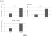
- HCT116 andA549 were transfected with miR-3202 using the method described above, secreted EV samples were prepared from the collected culture supernatant, and the amount of secreted EVs was measured according to the ExoScreen and NTA methods described above.
- the amount of secreted EVs in the negative control (NC) was measured in the same way, except that negative control miRNA (the NC1) was used.
- FIGS. 1 and 2 show the results.
- FIG. 1 shows the results of the amount of secreted EVs of the transfected HCT116
- FIG. 2 shows the results of the amount of secreted EVs of the transfected A549.
- the upper left graphs show the results of CD9/CD9 double-positive EVs according to ExoScreen
- the upper right graphs show the results of CD63/CD63 double-positive EVs according to ExoScreen
- the lower graphs show the results according to the NTA method. As shown in FIGS.
- FIG. 3 shows the results.
- FIG. 3 shows the results of CD63-positive EVs according to ExoScreen, expressed in terms of a relative value of signal intensity with respect to a measured value of the NC.
- miR-3202 can boost EV secretion from cells regardless of the type of cells.
- EV sample (miR-3202) collected from cells cultured in the presence of miR-3202 was compared with an EV sample (C) collected from cells cultured in the absence of miR-3202.
- the function of the EV sample collected from cells cultured in the presence of miR-3202 was compared with that of the EV sample collected from cells cultured in the absence of miR-3202.
- the EV sample (miR-3202) was collected from BM-MSCs cultured in the presence of miR-3202
- the EV sample (C) was collected from BM-MSCs cultured in the absence of miR-3202.
- HEL293 was cultured in the absence of the EV sample, in the presence of the EV sample (miR-3202), and in the presence of the EV sample (C), and their cell proliferative abilities were compared by counting the number of cells.
- the secreted EV samples obtained in (1) above the size of EVs contained in the samples was confirmed.
- the secreted EV samples were submitted to a nanoparticle analysis system (product name NanoSight, Malvern) to measure the particle size distribution of the particles (EVs) contained in the secreted EV samples.
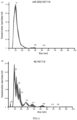
- FIGS. 4 and 5 show the results of the particle size distribution.
- FIG. 4 shows the results of the secreted EV samples secreted from HCT116, where the upper graph shows the results of the EV sample secreted from cells transfected with miR-3202, and the lower graph shows the results of the EV sample secreted from the NC (negative control).
- FIG. 5 shows the results of the secreted EV samples secreted from A549, where the upper graph shows the results of the EV sample secreted from cells transfected with miR-3202, and the lower graph shows the results of the EV sample secreted from the NC (negative control).
- FIGS. 4 shows the results of the secreted EV samples secreted from HCT116, where the upper graph shows the results of the EV sample secreted from cells transfected with miR-3202, and the lower graph shows the results of the EV sample secreted from the NC (negative control).
- the vertical axes indicate particle concentration (particles/mL) and the horizontal axes indicate particle size (nm), where the thin lines indicate the average concentration of three measurements with respect to the particle size, and the thick lines indicate each concentration (concentration range) obtained in three measurements with respect to the particle size.
- the particle size peak (97 nm) of the secreted EV sample of the cells transfected with miR-3202 was comparable to the particle size peak (94 nm) of the secreted EV sample of the NC.
- the particle size peak (111 nm) of the secreted EV sample of the cells transfected with miR-3202 was comparable to the particle size peak (102 nm) of the secreted EV sample of the NC. It was confirmed from these results that the EVs released from the cells transfected with miR-3202 are similar to those of the control.
- apoptotic bodies may be released. It was confirmed whether or not the EVs secreted from cells are not apoptotic bodies.
- Caspase activity is a known marker for apoptosis. Therefore, the caspase activity of cell samples obtained by culturing various types of cells transfected with the miRNAs in (1) and (2) above was measured. The cell viability of the cell samples was also measured by MTS assay. The caspase activity of the cell samples was then normalized by the respective cell viability rates, and the normalized caspase activity of the cells transfected with the miR-3202 was divided by the normalized caspase activity of the cells transfected with the NC1 to obtain a relative value of caspase activity (miR-3202/NC1).
- FIG. 6 shows the results.
- FIG. 6 shows the caspase activity of the cells, where the vertical axis indicates a relative value of caspase activity (miR-3202INC1). If the relative value of caspase activity is about 1 or less than 1, it can be concluded that caspase activity is not increased by the transfection with miRNA. As shown in FIG. 6 , the relative value of the caspase activity was about 1 or less than 1 in all cells, and the activity of caspase was not significantly increased in the cells transfected with miR-3202 compared with that of cells transfected with the control NC1.
- the EV secretion booster of the present invention can, for example, maintain cell survival and boost EV secretion, rather than promoting cell death by apoptosis.
- the EVs in the present invention can also be said to be EVs derived from viable cells.
- miR-3202 boosts EV secretion from mesenchymal stem cells.
- Example A1 it was confirmed that miRNAs in the present invention can boost EV secretion from cells with wide versatility. Therefore, it was further confirmed whether or not EV secretion from mesenchymal stem cells derived from bone marrow is boosted.
- BM-MSCs were transfected with miR-3202, secreted EV samples were prepared from the collected culture supernatant, and the amount of secreted EVs was measured according to the NTA method, in the same way as that of Example A1, except that BM-MSCs were used as cells and MesenPRO RS (serum-free) was used as a medium.
- the amount of secreted EVs in the negative control (NC) was measured in the same way, except that the negative control miRNA (NC1) was used.
- NC1 negative control miRNA
- the secreted EV samples were submitted to the nanoparticle analysis system (product name NanoSight, Malvern) to measure the particles (EVs) contained in the secreted EV samples.
- FIG. 7 shows the results.
- the left graph shows the results of the amount of secreted EVs of the transfected MSC, as measured using the nanoparticle analysis system
- the right graph shows the results of the amount of secreted EVs of the transfected MSC cells, as measured according to the NTA method
- the amount of secreted EVs measured according to the NTA method is normalized by the number of viable cells. Therefore, assuming that apoptosis has been caused by transfection with miR-3202, the amount of secreted EVs measured using the nanoparticle analysis system, which is not normalized by the number of viable cells, and the amount of secreted EVs measured according to the NTA method would yield different results. However, as shown in the graphs in FIG. 7 , the amount of secreted EVs measured using the nanoparticle analysis system and the amount of secreted EVs measured according to the NTA method yielded similar results. In addition, as shown in the table in FIG.
- FIG. 8 shows the results of the particle size distribution.
- FIG. 8 shows the results of the secreted EV samples secreted from the BM-MSCs, where the upper graph shows the results of the EV sample secreted from cells transfected with miR-3202, and the lower graph shows the results of the EV sample secreted from the NC (negative control).
- the vertical axes indicate particle concentration (particles/mL) and the horizontal axes indicate particle size (nm), where the thin lines indicate the average concentration of three measurements with respect to the particle size, and the thick lines indicate each concentration (concentration range) obtained in three measurements with respect to the particle size.
- the vertical axes indicate particle concentration (particles/mL)
- the horizontal axes indicate particle size (nm)
- the thin lines indicate the average concentration of three measurements with respect to the particle size
- the thick lines indicate each concentration (concentration range) obtained in three measurements with respect to the particle size.
- the particle size peak (106 nm) of the secreted EV sample of the cells transfected with miR-3202 was comparable to the particle size peak (112 nm) of the secreted EV sample of the NC. It was confirmed from these results that the EVs released from the BM-MSCs transfected with miR-3202 are similar to those of the control.
- A549, HCT116, and Panc6 were transfected with miR-3202 in the same way as that of Example A2 above and cultured, and then the expression level of ABCA1 mRNAin the obtained cultured cells was measured.
- the ABCA1 mRNA was measured through RT-PCR.
- As a negative control cells were cultured and expression levels were measured in the same way, except that the miR-3202 was not added. The relative values of the expression level of cells transfected with miR-3202 were then determined assuming that the expression level in the negative control was a relative value of 1.
- the relative value of the expression level in A549 was 0.61
- the relative value of the expression level in HCT116 was 0.63
- the relative value of the expression level in Panel was 0.63, that is, it was confirmed that the relative value was less than 1 in all cells, and the expression of the ABCA1 gene was reduced compared with that of the NC. It was confirmed from these results that the target gene of miR-3202 is the ABCA1 gene.
- EV secretion can be boosted by miR-3202
- siRNAs were used as siRNAs against the ABCA1 gene.
- A549 and HCT116 were transfected with the siRNA and cultured, and then the expression level of ABCA1 mRNA in the obtained cultured cells was measured.
- the cells were cultured as described in [Materials and Methods] above, and the transfection with the siRNA was performed in the same way as the transfer of miRNAin [Materials and Methods] above.
- the ABCA1 mRNA was measured through RT-PCR.
- As a negative control cells were cultured and expression levels were measured in the same way, except that the siRNA was not added.
- the relative values of the expression level of cells transfected with siRNA were then determined assuming that the expression level in the negative control was a relative value of 1.
- FIG. 9 shows the results.
- FIG. 9 shows graphs of the relative values of the expression level of ABCA1 mRNAin cells transfected with siRNA against the ABCA1 gene, where the upper graph shows the results of A549 cells, and the lower graph shows the results of HCT116 cells.
- the expression level of ABCA1 mRNAin A549 and HCT116 significantly decreased compared with that of the negative control regardless of which of siABCA1_1 and siABCA1_2 was used. It was confirmed from these results that the ABCA1 gene was knocked out by siRNA for ABCA1.
- A549 and HCT116 were transfected with the siRNA and cultured in the same way as that in (1) above. After confirming the number of cells after culture, secreted EV samples were prepared from the collected culture supernatant and the amount of secreted EVs was measured according to the NTA method as described in [Materials and Methods] above. The negative control (NC) was similar to that in (1) above, and the amount of secreted EVs therein was measured.
- the relative value of secreted EVs in the cells transfected with siRNA was then determined assuming that the amount of secreted EVs in the NC was a relative value of 1, and the relative value of the number of cells in the cells transfected with siRNA was calculated as cell viability assuming that the number of cells in the NC was a relative value of 1.
- FIG. 10 shows the results.
- FIG. 10 shows results of cells transfected with siRNA against the ABCA1 gene, where the upper graphs are graphs of the cell viability, the lower graphs are graphs of the relative values of the amount of secreted EVs, the left graphs show the results of A549 cells, and the right graphs show the results of HCT116 cells.
- the upper graphs in FIG. 10 in both A549 cells and HCT116 cells, no decrease in the viability due to transfection with siRNA was observed compared with that of the NC.
- Cyclosporin, probucol, and valspodar were used as inhibitors.
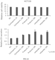
- A549 and HCT116 were cultured as described in (Transfer of miRNA and Collection of Secreted EVs) in [Materials and Methods] above. After incubation for 24 hours (Day 1), the number of cells was counted and the amount of secreted EVs was measured in the same way, except that the inhibitors were added instead of the miRNA. Each of the inhibitors was added at 10 nmol/L or 100 nmol/L per well. As a negative control, the cells were cultured, the number of cells was counted, and the amount of secreted EVs was measured in the same way, except that the inhibitors were not added.
- the relative value of secreted EVs in the cells to which the inhibitors were added was then determined assuming that the amount of secreted EVs in the NC was a relative value of 1, and the relative value of the number of cells in the cells to which the inhibitors were added was calculated as cell viability assuming that the number of cells in the NC was a relative value of 1.
- FIGS. 11 and 12 show the results.
- FIG. 11 shows the results of A549 cells to which the inhibitors were added, where the upper graph is a graph of the cell viability, and the lower graph is a graph of the relative values of the amount of secreted EVs.
- FIG. 12 shows the results of HCT116 cells to which the inhibitors were added, where the upper graph is a graph of the cell viability, and the lower graph is a graph of the relative values of the amount of secreted EVs.
- the upper graphs in FIGS. 11 and 12 in both A549 cells and HCT116 cells, no significant decrease in the viability due to addition of the inhibitors was observed compared with that of the NC.
- extracellular vesicle secretion from cells can be boosted.
- the preparation of extracellular vesicles as a material and the secretion of endogenous extracellular vesicles in a living organism by causing extracellular vesicle secretion from cells can be efficiently performed. Therefore, it can be said that the present invention, for example, is very useful in the medical field.
Landscapes
- Health & Medical Sciences (AREA)
- Engineering & Computer Science (AREA)
- Life Sciences & Earth Sciences (AREA)
- Genetics & Genomics (AREA)
- Biomedical Technology (AREA)
- Bioinformatics & Cheminformatics (AREA)
- Organic Chemistry (AREA)
- Chemical & Material Sciences (AREA)
- Zoology (AREA)
- Biotechnology (AREA)
- Wood Science & Technology (AREA)
- General Engineering & Computer Science (AREA)
- General Health & Medical Sciences (AREA)
- Biochemistry (AREA)
- Microbiology (AREA)
- Molecular Biology (AREA)
- Cell Biology (AREA)
- Plant Pathology (AREA)
- Biophysics (AREA)
- Physics & Mathematics (AREA)
- Pharmacology & Pharmacy (AREA)
- Veterinary Medicine (AREA)
- Medicinal Chemistry (AREA)
- Nuclear Medicine, Radiotherapy & Molecular Imaging (AREA)
- Chemical Kinetics & Catalysis (AREA)
- Animal Behavior & Ethology (AREA)
- Public Health (AREA)
- General Chemical & Material Sciences (AREA)
- Developmental Biology & Embryology (AREA)
- Medicines That Contain Protein Lipid Enzymes And Other Medicines (AREA)
- Medicines Containing Material From Animals Or Micro-Organisms (AREA)
- Rheumatology (AREA)
- Pharmaceuticals Containing Other Organic And Inorganic Compounds (AREA)
- Hematology (AREA)
- Immunology (AREA)
Applications Claiming Priority (2)
| Application Number | Priority Date | Filing Date | Title |
|---|---|---|---|
| JP2021166634 | 2021-10-11 | ||
| PCT/JP2022/037768 WO2023063284A1 (ja) | 2021-10-11 | 2022-10-11 | 細胞外小胞の分泌を促進する細胞外小胞分泌促進剤、およびその用途 |
Publications (1)
| Publication Number | Publication Date |
|---|---|
| EP4417693A1 true EP4417693A1 (en) | 2024-08-21 |
Family
ID=85987765
Family Applications (1)
| Application Number | Title | Priority Date | Filing Date |
|---|---|---|---|
| EP22880979.4A Pending EP4417693A1 (en) | 2021-10-11 | 2022-10-11 | Extracellular vesicle secretion booster for boosting extracellular vesicle secretion, and use thereof |
Country Status (4)
| Country | Link |
|---|---|
| US (1) | US20250270564A1 (https=) |
| EP (1) | EP4417693A1 (https=) |
| JP (1) | JP7825631B2 (https=) |
| WO (1) | WO2023063284A1 (https=) |
Family Cites Families (4)
| Publication number | Priority date | Publication date | Assignee | Title |
|---|---|---|---|---|
| WO2013118606A1 (ja) * | 2012-02-08 | 2013-08-15 | 独立行政法人産業技術総合研究所 | マラリアの予防または治療薬及びそのスクリーニング方法 |
| WO2020204161A1 (ja) * | 2019-04-04 | 2020-10-08 | 日産化学株式会社 | 細胞外小胞の分泌を促進するための組成物 |
| WO2021095842A1 (ja) * | 2019-11-15 | 2021-05-20 | 国立大学法人 東京大学 | バーコード化された細胞外小胞のライブラリー |
| JP2021166634A (ja) | 2020-04-10 | 2021-10-21 | 株式会社アイシン | 車両用シート装置 |
-
2022
- 2022-10-11 EP EP22880979.4A patent/EP4417693A1/en active Pending
- 2022-10-11 US US18/700,552 patent/US20250270564A1/en active Pending
- 2022-10-11 JP JP2023554511A patent/JP7825631B2/ja active Active
- 2022-10-11 WO PCT/JP2022/037768 patent/WO2023063284A1/ja not_active Ceased
Also Published As
| Publication number | Publication date |
|---|---|
| WO2023063284A1 (ja) | 2023-04-20 |
| JP7825631B2 (ja) | 2026-03-06 |
| JPWO2023063284A1 (https=) | 2023-04-20 |
| US20250270564A1 (en) | 2025-08-28 |
Similar Documents
| Publication | Publication Date | Title |
|---|---|---|
| Zhu et al. | Roles of microvesicles in tumor progression and clinical applications | |
| Li et al. | Osteoclast-derived exosomal miR-214-3p inhibits osteoblastic bone formation | |
| US20200392219A1 (en) | Extracellular vesicles comprising membrane-tethered tgf-beta, compositions and methods of use thereof | |
| KR20160035087A (ko) | 줄기 세포 마이크로입자 및 miRNA | |
| WO2013131000A1 (en) | Emt-inducing transcription factors cooperate with sox9 | |
| Soroczynska et al. | Immunosuppressive extracellular vesicles as a linking factor in the development of tumor and endometriotic lesions in the gynecologic tract | |
| CN111166867B (zh) | Pd-1泛素化激动剂的功能与用途 | |
| TW201821125A (zh) | 皮膚纖維化症治療劑 | |
| Horta et al. | Looking out for cancer stem cells’ properties: the value-driving role of CD44 for personalized medicines | |
| Mei et al. | “All-in-One” exosome engineering strategy for effective therapy of familial hypercholesterolemia | |
| CN118001416A (zh) | 一种包载双氢青蒿素的pd-1修饰的细胞外囊泡及其制备方法 | |
| CN117563001A (zh) | 半乳糖凝集素-1作为免疫检查点在制备肿瘤免疫治疗药物中的应用以及该药物 | |
| EP2402033B1 (en) | Cell adhesion inhibitor and use thereof | |
| EP4417693A1 (en) | Extracellular vesicle secretion booster for boosting extracellular vesicle secretion, and use thereof | |
| Wu et al. | Extracellular vesicles derived from “serum and glucose” deprived HUCMSCs promoted skin wound healing through enhanced angiogenesis | |
| US20150018225A1 (en) | Cell adhesion inhibitor, cell proliferation inhibitor, and method and kit for examining cancer | |
| Calascibetta et al. | GMP-compliant, serum-free cultures preserve therapeutic potential of extracellular vesicles from human mesenchymal stromal cells | |
| US12565654B2 (en) | Extracellular vesicle secretion reducing agent for reducing extracellular vesicle secretion, and use of the same | |
| CA3172262C (en) | Extracellular vesicle secretion reducing agent for reducing extracellular vesicle secretion, and use of the same | |
| US20230390335A1 (en) | Synthetic antigens as chimeric antigen receptor (car) ligands and uses thereof | |
| KR102276237B1 (ko) | 조직 재생 촉진제 | |
| EP3981473A1 (en) | Therapeutic agent for cancer | |
| JP2021138623A (ja) | 前立腺癌細胞からの細胞外小胞の分泌を抑制する細胞外小胞分泌抑制剤、前立腺癌治療薬、およびその用途 | |
| WO2020196926A1 (ja) | 細胞競合の制御剤 | |
| Chen et al. | Hierarchical Targeting of TREM2+ Myeloid Cells via Acid‐Triggered OMVs Reprogram Immunosuppression and Suppress Osteolysis in Bone‐Metastatic TNBC |
Legal Events
| Date | Code | Title | Description |
|---|---|---|---|
| STAA | Information on the status of an ep patent application or granted ep patent |
Free format text: STATUS: THE INTERNATIONAL PUBLICATION HAS BEEN MADE |
|
| PUAI | Public reference made under article 153(3) epc to a published international application that has entered the european phase |
Free format text: ORIGINAL CODE: 0009012 |
|
| STAA | Information on the status of an ep patent application or granted ep patent |
Free format text: STATUS: REQUEST FOR EXAMINATION WAS MADE |
|
| 17P | Request for examination filed |
Effective date: 20240508 |
|
| AK | Designated contracting states |
Kind code of ref document: A1 Designated state(s): AL AT BE BG CH CY CZ DE DK EE ES FI FR GB GR HR HU IE IS IT LI LT LU LV MC ME MK MT NL NO PL PT RO RS SE SI SK SM TR |
|
| DAV | Request for validation of the european patent (deleted) | ||
| DAX | Request for extension of the european patent (deleted) |