EP4413951A2 - Thrombektomiesystem - Google Patents
Thrombektomiesystem Download PDFInfo
- Publication number
- EP4413951A2 EP4413951A2 EP24179172.2A EP24179172A EP4413951A2 EP 4413951 A2 EP4413951 A2 EP 4413951A2 EP 24179172 A EP24179172 A EP 24179172A EP 4413951 A2 EP4413951 A2 EP 4413951A2
- Authority
- EP
- European Patent Office
- Prior art keywords
- clot
- thrombus
- aspiration
- catheter
- section
- Prior art date
- Legal status (The legal status is an assumption and is not a legal conclusion. Google has not performed a legal analysis and makes no representation as to the accuracy of the status listed.)
- Pending
Links
Images


Classifications
-
- A—HUMAN NECESSITIES
- A61—MEDICAL OR VETERINARY SCIENCE; HYGIENE
- A61B—DIAGNOSIS; SURGERY; IDENTIFICATION
- A61B17/00—Surgical instruments, devices or methods
- A61B17/22—Implements for squeezing-off ulcers or the like on inner organs of the body; Implements for scraping-out cavities of body organs, e.g. bones; for invasive removal or destruction of calculus using mechanical vibrations; for removing obstructions in blood vessels, not otherwise provided for
- A61B17/221—Gripping devices in the form of loops or baskets for gripping calculi or similar types of obstructions
-
- A—HUMAN NECESSITIES
- A61—MEDICAL OR VETERINARY SCIENCE; HYGIENE
- A61F—FILTERS IMPLANTABLE INTO BLOOD VESSELS; PROSTHESES; DEVICES PROVIDING PATENCY TO, OR PREVENTING COLLAPSING OF, TUBULAR STRUCTURES OF THE BODY, e.g. STENTS; ORTHOPAEDIC, NURSING OR CONTRACEPTIVE DEVICES; FOMENTATION; TREATMENT OR PROTECTION OF EYES OR EARS; BANDAGES, DRESSINGS OR ABSORBENT PADS; FIRST-AID KITS
- A61F2/00—Filters implantable into blood vessels; Prostheses, i.e. artificial substitutes or replacements for parts of the body; Appliances for connecting them with the body; Devices providing patency to, or preventing collapsing of, tubular structures of the body, e.g. stents
- A61F2/01—Filters implantable into blood vessels
- A61F2/013—Distal protection devices, i.e. devices placed distally in combination with another endovascular procedure, e.g. angioplasty or stenting
-
- A—HUMAN NECESSITIES
- A61—MEDICAL OR VETERINARY SCIENCE; HYGIENE
- A61B—DIAGNOSIS; SURGERY; IDENTIFICATION
- A61B17/00—Surgical instruments, devices or methods
- A61B17/22—Implements for squeezing-off ulcers or the like on inner organs of the body; Implements for scraping-out cavities of body organs, e.g. bones; for invasive removal or destruction of calculus using mechanical vibrations; for removing obstructions in blood vessels, not otherwise provided for
- A61B17/22031—Gripping instruments, e.g. forceps, for removing or smashing calculi
- A61B2017/22034—Gripping instruments, e.g. forceps, for removing or smashing calculi for gripping the obstruction or the tissue part from inside
-
- A—HUMAN NECESSITIES
- A61—MEDICAL OR VETERINARY SCIENCE; HYGIENE
- A61B—DIAGNOSIS; SURGERY; IDENTIFICATION
- A61B17/00—Surgical instruments, devices or methods
- A61B17/22—Implements for squeezing-off ulcers or the like on inner organs of the body; Implements for scraping-out cavities of body organs, e.g. bones; for invasive removal or destruction of calculus using mechanical vibrations; for removing obstructions in blood vessels, not otherwise provided for
- A61B2017/22079—Implements for squeezing-off ulcers or the like on inner organs of the body; Implements for scraping-out cavities of body organs, e.g. bones; for invasive removal or destruction of calculus using mechanical vibrations; for removing obstructions in blood vessels, not otherwise provided for with suction of debris
-
- A—HUMAN NECESSITIES
- A61—MEDICAL OR VETERINARY SCIENCE; HYGIENE
- A61B—DIAGNOSIS; SURGERY; IDENTIFICATION
- A61B17/00—Surgical instruments, devices or methods
- A61B17/22—Implements for squeezing-off ulcers or the like on inner organs of the body; Implements for scraping-out cavities of body organs, e.g. bones; for invasive removal or destruction of calculus using mechanical vibrations; for removing obstructions in blood vessels, not otherwise provided for
- A61B2017/22094—Implements for squeezing-off ulcers or the like on inner organs of the body; Implements for scraping-out cavities of body organs, e.g. bones; for invasive removal or destruction of calculus using mechanical vibrations; for removing obstructions in blood vessels, not otherwise provided for for crossing total occlusions, i.e. piercing
-
- A—HUMAN NECESSITIES
- A61—MEDICAL OR VETERINARY SCIENCE; HYGIENE
- A61B—DIAGNOSIS; SURGERY; IDENTIFICATION
- A61B17/00—Surgical instruments, devices or methods
- A61B17/22—Implements for squeezing-off ulcers or the like on inner organs of the body; Implements for scraping-out cavities of body organs, e.g. bones; for invasive removal or destruction of calculus using mechanical vibrations; for removing obstructions in blood vessels, not otherwise provided for
- A61B17/221—Gripping devices in the form of loops or baskets for gripping calculi or similar types of obstructions
- A61B2017/2212—Gripping devices in the form of loops or baskets for gripping calculi or similar types of obstructions having a closed distal end, e.g. a loop
-
- A—HUMAN NECESSITIES
- A61—MEDICAL OR VETERINARY SCIENCE; HYGIENE
- A61B—DIAGNOSIS; SURGERY; IDENTIFICATION
- A61B17/00—Surgical instruments, devices or methods
- A61B17/22—Implements for squeezing-off ulcers or the like on inner organs of the body; Implements for scraping-out cavities of body organs, e.g. bones; for invasive removal or destruction of calculus using mechanical vibrations; for removing obstructions in blood vessels, not otherwise provided for
- A61B17/221—Gripping devices in the form of loops or baskets for gripping calculi or similar types of obstructions
- A61B2017/2215—Gripping devices in the form of loops or baskets for gripping calculi or similar types of obstructions having an open distal end
-
- A—HUMAN NECESSITIES
- A61—MEDICAL OR VETERINARY SCIENCE; HYGIENE
- A61F—FILTERS IMPLANTABLE INTO BLOOD VESSELS; PROSTHESES; DEVICES PROVIDING PATENCY TO, OR PREVENTING COLLAPSING OF, TUBULAR STRUCTURES OF THE BODY, e.g. STENTS; ORTHOPAEDIC, NURSING OR CONTRACEPTIVE DEVICES; FOMENTATION; TREATMENT OR PROTECTION OF EYES OR EARS; BANDAGES, DRESSINGS OR ABSORBENT PADS; FIRST-AID KITS
- A61F2/00—Filters implantable into blood vessels; Prostheses, i.e. artificial substitutes or replacements for parts of the body; Appliances for connecting them with the body; Devices providing patency to, or preventing collapsing of, tubular structures of the body, e.g. stents
- A61F2/01—Filters implantable into blood vessels
- A61F2002/016—Filters implantable into blood vessels made from wire-like elements
-
- A—HUMAN NECESSITIES
- A61—MEDICAL OR VETERINARY SCIENCE; HYGIENE
- A61F—FILTERS IMPLANTABLE INTO BLOOD VESSELS; PROSTHESES; DEVICES PROVIDING PATENCY TO, OR PREVENTING COLLAPSING OF, TUBULAR STRUCTURES OF THE BODY, e.g. STENTS; ORTHOPAEDIC, NURSING OR CONTRACEPTIVE DEVICES; FOMENTATION; TREATMENT OR PROTECTION OF EYES OR EARS; BANDAGES, DRESSINGS OR ABSORBENT PADS; FIRST-AID KITS
- A61F2/00—Filters implantable into blood vessels; Prostheses, i.e. artificial substitutes or replacements for parts of the body; Appliances for connecting them with the body; Devices providing patency to, or preventing collapsing of, tubular structures of the body, e.g. stents
- A61F2/95—Instruments specially adapted for placement or removal of stents or stent-grafts
- A61F2002/9528—Instruments specially adapted for placement or removal of stents or stent-grafts for retrieval of stents
-
- A—HUMAN NECESSITIES
- A61—MEDICAL OR VETERINARY SCIENCE; HYGIENE
- A61M—DEVICES FOR INTRODUCING MEDIA INTO, OR ONTO, THE BODY; DEVICES FOR TRANSDUCING BODY MEDIA OR FOR TAKING MEDIA FROM THE BODY; DEVICES FOR PRODUCING OR ENDING SLEEP OR STUPOR
- A61M25/00—Catheters; Hollow probes
- A61M25/0021—Catheters; Hollow probes characterised by the form of the tubing
- A61M2025/0042—Microcatheters, cannula or the like having outside diameters around 1 mm or less
Definitions
- the present invention is directed, in general, to the field of medical devices.
- the invention relates to a thrombectomy system, which allows the removal of thrombi at the vascular level.
- the disclosure also relates to methods of extracting a thrombus from a thrombus site in a blood vessel of a patient.
- the thrombectomy system includes a combination of an aspiration catheter and a clot-capture element.
- Acute ischemic stroke is a major cause of morbidity and mortality, with an annual incidence of 118 cases/100000 population and a mortality of 29 cases per 100000 population/year. These numbers position ischemic stroke as one of the main causes of death in developed countries together with cardiovascular diseases and cancer. In order to prevent or reduce complications related to this disease and to improve the prognosis of patients with ischemic stroke, it is necessary a clinical diagnosis to establish a proper reperfusion strategy in the shortest period of time. Until 2015 the treatment of choice for stroke was the recombinant tissue plasminogen activator (rtPA) administered intravenously 4.5 hours after symptom onset. However, this drug presents a narrow therapeutic window and not always gets recanalization.
- rtPA tissue plasminogen activator
- intra-arterial recanalization therapy as mechanical thrombectomy is performed by means of various devices (Merci ® , Penumbra ® , etc.).
- the objective is to remove thrombus through aspiration, disruption or capture/extraction, viewed as a therapeutic option for patients who are not candidates for rtPA or in whom rtPA has failed.
- stent retrievers appear to give this technique a more widespread use (Solitaire TM, Trevo ® and Revive).
- Endovascular treatment of stroke has been performed since the 1990's. Its growth in the number of treated patients has been slow but constant.
- the main obstacle to its more widespread use is the necessity of a coordinated medical system at different levels to make it possible for patients to get to a medical center capable of administering these highly complex treatments within 6-8 hours of symptom onset.
- a large-gauge catheter was designed to be advanced to the thrombus.
- the catheter was connected to a continuous aspiration pump to aspirate the thrombus (the Penumbra System ® ).
- This system has evolved over the years, seeking to attain a catheter with an increasingly large diameter, able to navigate close to the thrombus.
- stent retrievers In 2009, the use of stent retrievers started. Their use consists of crossing the thrombus with a microcatheter. Thereafter, the endoprosthesis is advanced through the microcatheter. Once the distal end of the microcatheter has reached the distal part of the thrombus, the endoprosthesis (stent retriever) is unsheathed, self-expanding through the thrombus and capturing it. It is recommended to wait a few minutes with the endoprosthesis expanded to enable proper engagement of the thrombus. The expanded stent is then withdrawn to drag the thrombus toward the catheter and out of the blood vessel.
- This last step can be done while aspirating through the catheter to try to reverse the blood flow in the vessel and to increase the likelihood of recovering the thrombus.
- a guide balloon catheter is often used. This catheter only advances to the extracranial carotid (distant from thrombi located in the intracranial arteries).
- Stent retrievers have entirely displaced the first-generation devices described above due to their high efficacy and speed.
- Several prospective randomized trials have recently demonstrated the marked superiority of stent retriever assisted mechanical thrombectomy with standard intravenous tissue plasminogen activator (IV tPA) thrombolysis over medical therapy (IV tPA) alone for revascularization of acute ischemic stroke in patients presenting with proximal large vessel occlusion.
- US-A1-2018132876 discloses a system for removing a thrombus from a blood vessel including a stent retriever, a catheter configured to receive the stent retriever in a collapsed configuration, wherein the stent retriever is movable relative to the catheter, a sheath having a tubular body and defining a distal opening and a proximal opening, and a wire coupled to the stent retriever for positioning the stent retriever.
- the wire extends through the proximal opening and the distal opening of the sheath.
- the stent retriever is moveable relative to the sheath, and the distal opening of the sheath is sized to allow the stent retriever to be withdrawn into the sheath without substantially compressing the stent retriever.
- WO-A1-2015006782 discloses a device and a method for intravascular treatment of an embolism.
- the device comprises a clot treatment device that includes a support member configured to extend through a delivery catheter and a plurality of clot engagement members positioned about the circumference of a distal portion of the support member.
- the clot engagement members can be configured to penetrate clot material along an arcuate path and mechanically macerate clot and release embolic particles when resheathed into the delivery catheter.
- US-A1-2017119408 discloses a clot removal device comprising an expandable treatment member having a distal tip and a proximal end, a delivery wire having a distal end coupled to the proximal end of the expandable treatment member, and a flow restrictor carried along the delivery wire at a location that is separate and proximal from the expandable treatment member.
- the flow restrictor has a body with a distal section and a proximal section, the distal section being covered and the proximal section being uncovered.
- the expandable treatment member is moveable relative to the flow restrictor, and can be retracted into the distal section.
- a problem to be solved by the present invention is to improve the efficacy of the currently used clot-capture devices, and particularly, stent retrievers. This is particularly interesting for the capture of hard clots such as fibrin rich clots.
- the present invention according to a first aspect provides a thrombectomy system.
- the proposed thrombectomy system comprises: a delivery catheter configured to be advanced through vasculature of a patient to a thrombus site within a blood vessel; an aspiration catheter adapted to apply suction to an expandable aspiration funnel extending from a distal end of the aspiration catheter, the aspiration funnel being configured to be movably disposed within the delivery catheter in a retracted position in a compressed state (in the delivery configuration) and at least partially outside the delivery catheter in an extended and expanded position (also referred in this description as "in a deployment configuration"), the aspiration funnel comprising a non-permeable covering, a diameter of a distal end of the aspiration funnel being greater in the extended and expanded position than in the retracted position, the aspiration funnel being configured to adapt its shape and length to an inner wall of the blood vessel such that the aspiration funnel reduces blood flow through the blood vessel and lengthens as it narrows
- the clot-capture element is movably disposed within the microcatheter in a retracted position.
- the microcatheter is movably disposed within the aspiration catheter.
- the different elements of the thrombectomy system can be moved together or separately.
- the interconnection between the elements is through hemostatic valves.
- a suitable thrombectomy device for use for the purposes of the disclosure is described in the patent application WO2016113047A1 .
- the delivery catheter, the aspiration funnel, the microcatheter and the clot-capture element are oriented on the same axis, are coaxially configured and movable to each other independently.
- the aspiration funnel is self-expandable.
- the thrombectomy system/apparatus of the invention can be used in the neurovasculature or in the peripheral vasculature and is particularly suited to navigate to the desired location and provide a concrete sealing where it is most needed, thus avoiding development of secondary thrombi for instance.
- Its design allows the introduction of a retrieval device that actuates as a clot mobilizer to remove a thrombus by dragging it into the funnel mouth.
- the approach of the invention consists in guiding the thrombectomy device to a position close to the thrombus and to retrieve it by means of aspiration combined with a mechanical action.
- the aspiration funnel is a self-expandable covered stent operated with the use of a catheter (e.g.
- the catheter is intended to maintain the vacuum from its proximal end, where the vacuum is generated by an interventionist (e.g., with a syringe), to the vicinity of the thrombus at the distal end of the catheter.
- an interventionist e.g., with a syringe
- This covered aspiration funnel can be in retracted or extended configurations, the diameter of the aspiration funnel being bigger in the extended configuration than in the retracted configuration. Additionally, the aspiration funnel is designed not to cause damage to the intracranial or peripheral artery. Its design is intended to adjust to the diameter of the artery and, as a result, to restrict the blood flow which is one of the most important characteristics of the described system for the prevention of distal embolism. Distal embolism is a typical clinical complication when a stent retriever crosses a clot or during the extraction process.
- the expansion behavior of the aspiration funnel is due to the Nitinol material from which it is formed, thanks to its shape memory properties and super elasticity.
- Shape memory refers to the ability to undergo deformation and then recover its original shape by heating the material above its "transformation temperature”.
- Nitinol presents the right characteristics to position to different diameters and geometries of the vessel.
- the aspiration funnel a self-expandable stent (formed, e.g., from Nitinol) sealed with a film of polymeric material, upon deployment expands and mimics the blood vessel dimensions.
- the large mouth of the funnel together with the clot-capture element is able to aspirate the entire thrombus without fragmenting it and also allocates the clot perfectly during the removal procedure.
- the loss of the clot due to the long distance from the occlusion site to the exit and also due to the big size of the clot (difficult to catch by the clot-capture element) is also prevented.
- the system is able to restrict blood flow in the vessel and, as result, increases the aspiration power of the system and reduces further clinical side complications, mainly distal embolism.
- Another key feature of the system is that, as the flow is not stopped at carotid level, but directly at the thrombus site, only a particular arterial branch is affected, not the entire hemisphere, thus increasing the safety of the overall procedure.
- the thrombectomy system of the invention offers a clear advantage respect other marketed or ready-to be marketed devices.
- the clot-capture element in the present description is understood as a device able to interact with the clot in order to capture it and retrieve it from the blood stream.
- This definition includes the following categories without being limiting for the present invention:
- the clot-capture element is a stent retriever device. More particularly the stent retriever device has closed cells and a continuous scaffold like Solitaire TM revascularization device or Trevo Stentriever TM .
- the aspiration funnel comprises a segment defining a distal end and a proximal end and is formed by a mesh of at least two sets, equal or different, of helicoidal filaments (or wires) turning respectively in opposite directions and being intertwined.
- the mesh comprises a first tubular section, particularly of a uniform diameter, and a second tubular section, adjacent to the first section, having a diameter smaller than that of the first tubular section.
- the mesh of the first section has helicoidal filaments with a braiding angle ( ⁇ ) adapted to provide outward radial forces, i.e. pressure, higher than in the second section, such that the first section becomes better appositioned, or overlapped, against the inner wall of the blood vessel.
- the first section may comprise closed loops at the distal end configured to act as a spring, such that the radial forces in the first and second end portions of the first section are higher than in an intermediate portion of the first section.
- the straight shape of the first section of the aspiration funnel creates a space which will accommodate the thrombus once it has been aspirated.
- the first section is adaptable to the vessel geometry and its outer surface overlaps the inner wall of the blood vessel.
- the second section comprises two sub-sections, a first sub-section and a second sub-section.
- the first sub-section has a cone-shape (or funnel-shape) and comprises a braiding angle ( ⁇ ) that change at its proximal and distal ends to provide radial strength to maintain the conical shape and to reduce the proximal blood flow during the removal of the thrombus.
- the second sub-section comprises a tubular uniform diameter configured to provide a connection to the aspiration catheter.
- the aspiration funnel can be produced in different sizes.
- the first section is longer than the second section.
- the first section comprises a length ranging between 4 and 40 millimeters and an outer diameter ranging between 3.5 and 6 millimeters
- the second sub-section comprises a length ranging between 1 and 10 millimeters and an outer diameter ranging between 1 and 2 millimeters.
- the braiding angle ( ⁇ ) of the first sub-section is comprised between 15 and 45 degrees with regard to a longitudinal axis of the aspiration funnel. This angle favors having more radial force thereby stopping the flow, but at the same time that there is a seal of the blood vessel it also has to allow the aspiration funnel to be compressed.
- the non-permeable covering of the aspiration funnel comprises a polymer, for example silicone or polyurethane.
- the helicoidal filaments of the mesh can be made of a metal, a metal alloy or a composite including, among others, Nitinol or Nitinol/Platinum, or also Niti#1-DFTR R (Drawn Filled Tube), with a percentage of Platinum from 10% to 40%; in particular with 20% Platinum (Niti#1-DFTR R -20%Pt).
- the helicoidal filaments are adapted to become more longitudinally aligned as the aspiration funnel lengthens and narrows.
- the helicoidal filaments in an embodiment, comprise a number ranging between 12 and 48 filaments, and particularly between 18 and 24 filaments.
- the filaments have a cross section comprised in a range between 40 and 60 ⁇ m, and particularly 50 ⁇ m, and the braiding angle ( ⁇ ) of the filaments with regard to the longitudinal axis of the aspiration funnel is comprised between 50 and 65 degrees for the first section, and between 15 and 50 for the second sub-section.
- the aspiration funnel may also include or have attached thereto one or more sensors to provide information thereof.
- a lighting sensor or sensors may provide information of whether the aspiration funnel is in the retracted position within the delivery catheter or in the extended and expanded position.
- the sensor(s) can alternatively, or additionally, provide information on whether the aspiration funnel is well extended and expanded, on whether the thrombus is in or out, about the composition of the thrombus, or about the position of the funnel in relation to the blood vessel.
- the sensor(s) may include a piezoelectric sensor providing information about the radial forces in each of the different sections or subsections of the aspiration funnel.
- the sensor(s) may provide information to distinguish between blot obstruction and intracranial atherosclerotic disease.
- the aspiration funnel can also comprise at least one radiopaque marker at its distal end and/or other strategic point(s) of the mesh which allow a physician to know the precise location of the aspiration funnel while using fluoroscopy.
- Another aspect of the disclosure relates to a method of extracting a thrombus from a thrombus site in a blood vessel of a patient, the method comprising:
- the steps of the proposed method can be performed in any order.
- the step of applying suction can be performed either before or after the step of moving the aspiration funnel and the thrombus proximally within the vasculature.
- the method may also comprise advancing a microcatheter within the aspiration catheter, the clot-capture element being disposed within the microcatheter. Moreover, the method may also comprise moving the microcatheter and clot-capture device with respect to each other to place the clot-capture device outside of the microcatheter; and expanding the clot-capture device.
- the expanding of the clot-capture device comprises allowing the clot capture device to self-expand.
- advancing a microcatheter comprises advancing a distal end of the microcatheter through the thrombus.
- the method may also comprise moving the clot-capture device proximally at least partially into the aspiration funnel.
- expanding the aspiration funnel comprises allowing the aspiration funnel to self-expand.
- the aspiration funnel comprises a mesh of at least two sets of intertwined helicoidal filaments turning respectively in opposite directions, the method further comprising moving the two sets of helicoidal filaments to a more longitudinally aligned position as the aspiration funnel lengthens and narrows.
- Another aspect of the disclosure relates to a method of extracting a thrombus from a thrombus site in a blood vessel of a patient, the method comprising:
- the clot-capture element By advancing the clot-capture element distally through vasculature toward the thrombus site, the clot-capture element is used as an anchor element, thus pushability/navigability of the delivery catheter is enabled.
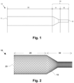
- Figs. 1 and 2 show particular embodiments of the aspiration funnel 1 included in the proposed thrombectomy system/apparatus (or ANCD) for extraction of thrombus from a blood vessel.
- the aspiration funnel 1 includes a segment 10 which is self-expandable and defines a distal end 11 and a proximal end 12 and can adapt its shape to a surrounding blood vessel from a retracted position in a compressed state, for example inside a carrier such a delivery catheter 3, to an extended/expanded position, once coming out of the delivery catheter 3, to be appositioned against the inner wall of a blood vessel to receive and retain a thrombus THR.
- the segment 10 comprises a mesh 13 having two sets of helicoidal filaments turning respectively in opposite directions and being intertwined.
- the mesh 13 in an embodiment can follow a diamond-type structure or a regular structure.
- the density of the mesh 13 defines the elasticity of the segment 10.
- the mesh angle (or braiding angle ( ⁇ )) with regard to a longitudinal direction can be variable.
- the helicoidal filaments can be made of a metal (including metal alloys), polymers, a composite including Nitinol or Nitinol/Platinum, or also DFT R (Drawn Filled Tube), among other materials having suitable mechanical properties.
- the mesh 13 defines two distinct tubular sections, a first section 20 and a second section 30.
- the second section 30 comprises two sub sections, a first sub section 31 and a second sub section 32.
- the end portion of the first section 20 at the distal end 11 comprises closed loops 23 facilitating the expansion of the segment 10 once it comes out of the cited delivery catheter 3.
- these closed loops 23 act as a spring or fixing point by limiting the movement between the helicoidal filaments and thus increasing the outward radial force.
- the closed loops 23 also provide a smooth distal end to reduce possible vessel damage and improve navigability of the aspiration funnel 1 within the blood vessel.
- the rest of the first section 20 creates the space which will accommodate the thrombus THR once it has been aspirated.
- the first section 20 is adaptable to the vessel geometry and, because of its configuration (e.g., diameter and braiding angle ⁇ ), provides outward radial forces higher than in the second section 30 so that the segment 10 is better appositioned against the inner wall of the vessel.
- the radial forces in the end portions of the first section 20 are particularly higher than in an intermediate portion thereof, e.g., because of the spring action of closed loops 23.
- the radial forces in the first section 20 could be uniformly distributed along all its generatrix.
- the first sub-section 31 (or portion of the second section 30 adjacent to the first section 20) is cone-shaped or funnel-shaped. Because of its shape, this sub-section 31 has features enabling it to withstand the blood pressure without collapsing.
- the braiding angle ( ⁇ ) change at the proximal and distal ends of sub-section 31 provide radial strength to maintain the conical shape.
- the braiding angle ( ⁇ ) change at the distal end of sub-section 31 also works with the closed loops 23 to maintain first section 20 in an open position and create the space for the thrombus THR.
- the covering over sub-section 31 stops the blood flow during the capture and removal of the thrombus THR and protects the captured thrombus THR during the withdrawal of the segment 10 to the delivery catheter 3.
- This sub-section 31 is also the transition from the larger diameter of section 20 to the smaller diameter sub-section 32 for connection to an aspiration catheter 2 (see Fig. 5 ), or alternatively to a hypotube.
- the second sub-section 32 (or portion of the second section 30 adjacent to proximal end 12) has a tubular uniform diameter and provides the connection to the aspiration catheter 2.
- the aspiration catheter 2 is a PTFE-lined braided catheter covered by an outer jacket.
- the aspiration catheter's braid and liner extend distally from the outer jacket.
- a layer of polymer material may be placed around the protruding braid and liner, and a mandrel may be placed within the braid and liner. Thereafter, the second sub-section 32 of segment 10 may be placed over this polymer section, and another layer of polymer may be placed over the mesh of subsection 32.
- This outer layer of polymer material is then melted so that polymer flows through the cells of the mesh 13, the mandrel is removed, and a smooth surface is left over the entire aspiration catheter 2.
- This attachment approach adds structure and stiffness to the attachment section of the aspiration catheter 2, so it should be as short as possible without compromising the integrity of the attachment of segment 10 to the aspiration catheter 2.
- segment 10 to the aspiration catheter 2
- the mesh 13 of the sub-section 32 is welded to a Nitinol ring. This ring is welded directly to the hypotube.
- a Stainless-steel ring can be glued to the mesh 13 of the sub-section 32. Then, the Stainless-steel ring is welded to the hypotube.
- Another option is to directly mesh the segment 10 over a perforated ring so that the filaments pass through the holes.
- segment 10 When the segment 10 is compressed inside the delivery catheter 3, segment 10 elongates to move the helicoidal filaments toward a longitudinal alignment so as to reduce the spring effect and to facilitate the movement of segment 10 within the delivery catheter 3 by reducing friction effects and by increasing pushability.
- the pushability of the segment 10 inside the delivery catheter 3 is related to the navigability of the segment 10 within the arteries.
- the mesh angle or braiding angle ( ⁇ ) allows the mesh 13 to be adapted to a curve of the blood vessel, avoiding the kinking and creating a free space inside the mesh for unobstructed suction.
- Table 1 indicates the main specifications of the aspiration funnel 1.
- Table 2 indicates the measuring method used for calculating such parameters.
- Main specifications of the aspiration funnel Example Range Big Ref. Small Ref. Shape parameters OD sec 20 [mm] 6 3.5--6 5.2 Approx.
- Table 1 shows the parameters for particular embodiments.
- the parameters of the aspiration funnel 1 are such indicated in Table 1 for a big blood vessel ("Big Ref.") of e.g. 4.5 mm diameter, such as the final part of the carotid or the carotid siphon.
- the parameters of the aspiration funnel 1 are such indicated in Table 1 for a small blood vessel (“Small Ref.") of e.g. 2.5 mm diameter, such as the Internal Carotid Artery (ICA) or the Middle Cerebral Artery (MCA).
- ICA Internal Carotid Artery
- MCA Middle Cerebral Artery
- Wire number Alternative 1 Counting the number of distal loops and multiplying by 2
- Alternative 2 Counting the number of reels used for meshing ⁇ sec 20 [°]
- Alternative 1 Measuring the number of wire crossings in a given length measured in the axial direction.
- Alternative 2 If the mandrel is manufactured with grooves so that during the meshing the wires are inserted inside, and the manufacturing is improved, it is simply measured that the mandrel is manufactured with the appropriate parameters. ⁇ sec 32 [°] Same as before.
- the aspiration funnel 1 may be in two configurations: in a retracted form (or compressed state) inside the delivery catheter 3 while approaching the thrombus site, and in an extended and expanded (deployed) form when there is no interaction with the delivery catheter 3 or the blood vessel.
- the parameters specified herein relate to the aspiration funnel 1 in its natural (relaxed) form; i.e. extended and expanded (deployed) position.
- the segment 10 may include radiopaque markers made of platinum, tungsten, barium derivatives, gold, iridium, among others, at its distal end 11 and/or other strategic points within the mesh 13 which allow a physician to know the precise location of the aspiration funnel 1 while using fluoroscopy.
- the radiopaque material can be deposited on the helicoidal filaments once manufactured (if the aspiration funnel 1 has a coating, the material may also be dispersed on the surface of the coating).
- Alternative possibilities to confer radiopacity to the segment 10 are using helicoidal filaments of different material and opacity grade (e.g. Nitinol and Platinum). In a particular embodiment, Nitinol wires with a Platinum core are used.
- the delivery catheter 3 may also include radiopaque markers.
- the segment 10 may have a coating, for example covering the first section 20 only or covering the whole segment 10.
- the coating goes from the closed loops 23 to sub-section 32.
- the coating is applied about attachment of segment 10 to the aspiration catheter 2 by dipping segment 10 into a liquid polymer, therefore allowing the polymer to solidify.
- a mandrel may be disposed inside the mesh 13 of segment 10 when it is dipped into the polymeric coating material.
- the coating material may be sprayed onto the mesh 13.
- the coating may be applied before attaching segment 10 to the aspiration catheter 2. In such embodiments, the coating does not reach the proximal end 12 of sub-section 32, but there is an uncoated space between the helicoidal filaments, leaving them free to allow assembly with the aspiration catheter 2.
- the coating prevents damage to the arteries, avoiding direct contact with the helicoidal filaments. Moreover, the coating provides a watertight compartment so that the thrombus THR can be sucked in and protected during removal.
- the mesh 13 is attached to the delivery catheter 3 and then the coating is applied.
- An interior or exterior glaze can be also applied to the coating to improve its properties.
- a hydrophilic or hydrophobic coating to the exterior surface of the segment 10
- the exterior surface can be more easily displaced into the carrier and through the blood vessel by reducing the coefficient of friction.
- an adhesion effect that retains the thrombus THR once it is inside can be achieved.
- the coating is made of an elastic material.
- the aspiration funnel 1 coating is silicone.
- polyurethanes or other types of plastic materials can be used.
- a blend of polyurethane and silicone may also be employed.
- the coating can be treated by the addition of a material as explained or can have constitutively such features by the structure of the mesh itself.
- the coating can include holes to avoid collapse of the segment 10. Such holes may be formed after the coating has been applied by perforating the coating.
- segment 10 depend on the dimensions of the blood vessel in which it will be used to capture a thrombus THR.
- the dimensions of the sub-sections of segment 10 and the braid angles of the mesh help segment 10 provide a reduced radially outward force when compressed into the delivery catheter 3 and sufficient outward force when expanded to avoid collapse from the blood pressure.
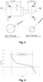
- Fig. 4 illustrates a possible work curve of one embodiment of the segment 10.
- Y-axis defines the device pressure (mmHg) whereas X-axis defines the diameter of the arteries (mm).
- the horizontal dotted line marks the blood pressure limit.
- the diameter range of the arteries in which the aspiration funnel 1 of this invention may be used is 2 to 5 mm.
- the segment 10 is designed so that it can expand without being blocked by the artery working in a standard range of 2 to 5 mm and so that it can cope with a blood pressure greater than 200 mmHg. As shown by Fig. 4 , this particular embodiment is not designed to be compressed to a diameter less than 2 mm. Compression of the segment 10 within the delivery catheter 3 may result in radially outward forces high enough to inhibit advancement of the aspiration funnel 1 within the carrier.
- Some embodiments of the invention may be automated for used in traditional (hospital) and non-traditional (nursing home, assisted care facility) environments which may allow for greater deployment and usage of the ANCD and hasten the removal of thrombus THR, thus significantly improving patient outcomes, as flow may be restored (e.g., to critical areas of the brain) within much shorter times.
- One such automated device is illustrated in WO2016/113047 .
- segment 10 and the aspiration catheter 2 to which it is attached are advanced through the delivery catheter 3 to a thrombus site within a blood vessel of the patient.
- the segment 10 is in a delivery configuration in which the first and second sets of helicoidal filaments form a first distally facing angle with respect to each other.
- segment 10 emerges from the delivery catheter 3, it begins to self-expand to a deployment configuration.
- the spring action of the closed loops of the helicoidal filaments helps the first section 20 expand into apposition with the blood vessel proximate to the thrombus site.
- the first and second sets of helicoidal filaments form a second distally facing angle less than the first angle (i.e., the filaments are less longitudinally aligned in the deployment configuration than they were in the delivery configuration).
- Sub-section 31 also self-expands to a conical or funnel shape. The distal end of sub-section 31 helps support the proximal end of section 20 in its deployment configuration.
- the coating on the outside of sub-section 31 and section 20 reduce blood flow to the thrombus site.
- the optional holes through the coating permit a small amount of blood to pass through the aspiration funnel 1 to avoid collapse of sub-section 31 caused by the blood pressure and also by the difference of pressure between the blood pressure (externally) and the vacuum applied (internally).
- suction may be applied through the catheter 2 to the interior spaces of sub-section 31 and section 20 to aspirate the thrombus THR into section 20.
- Aspiration funnel 1 capturing the thrombus THR may then be removed from the patient. In the capture configuration (i.e.
- the first and second sets of filaments form a third distally-facing angle less than the first distally-faced angle (i.e., the filaments become more longitudinally aligned) as the aspiration funnel 1 assumes a longer and smaller diameter shape.
- FIG. 5 therein it is illustrated a scheme of an expanded configuration of the proposed ANCD, which in this particular embodiment includes an aspiration funnel 1, an aspiration catheter 2 connected to the aspiration funnel 1, a delivery catheter 3; a clot-capture element 4 and a microcatheter 5.
- Detail A shows a scheme of an expandable-tip aspiration catheter 7 comprising the aspiration funnel 1 and the aspiration catheter 2.
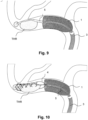
- Figs. 6-13 show the steps of a method of extracting a thrombus THR from a thrombus site in a blood vessel of a patient using the ANCD of the invention.
- the delivery catheter 3 containing the expandable-tip aspiration catheter 7 is advanced over a guide wire 6 and the microcatheter 5 to the internal carotid ( Fig. 6 ).
- the delivery catheter 3 Once the delivery catheter 3 reaches its position it is withdrawn to deploy the mouth of the expandable-tip aspiration catheter 7 ( Fig. 7-8 ).
- the aspiration funnel 1 self-expands to the diameter of the vessel and arterial flow is stopped (i.e. blocked or partially reduced) ( Fig. 8 ).
- the microcatheter 5 is advanced into the thrombus THR ( Fig. 9 ). Then the microcatheter 5 is withdrawn to deploy the clot-capture element 4, capturing the clot ( Fig. 10 ). The clot-capture element 4 drags the clot to the aspiration funnel 1 mouth while suction is applied to the aspiration catheter 2 by means of a syringe to aspirate the clot ( Fig. 11 ). Finally, the clot is engaged in the aspiration funnel 1 ( Fig. 12 ) and the system is removed ( Fig. 13 ).
- the proposed ANCD is comprised of the ANA device, the clot-capture element 4 and the microcatheter 5.
- the ANA device i.e. the catheter device comprised of the delivery catheter 3 and the expandable-tip aspiration catheter 7 included in the ANCD.
- the expandable-tip aspiration catheter 7 was built using a DFT (Nitinol/platinum) braided stent covered with silicone as defined below.
- the performances were evaluated in an in vitro 3D simulation model, a cerebrovascular model of the intracranial circulation that simulates the carotid and cerebral physiological blood flow, pressure and vessel anatomy including an occlusive ex vivo clot analog.
- this study aimed to assess the efficacy of the ANA device in combination with the clot-capture element 4, such as a stent retriever (SR), in terms of the rate of revascularization and rate of clot embolization.
- SR stent retriever
- the ANA devices used in the study were the following (Table 4): Funnel Catheter Funnel Model Funnel Reference Delivery Catheter Group 10/ Sample 8 5,2*9 mm ZA00583-03 Coiled delivery (new)/group 10/sample 8 lot 945034 sample 10 5,2*9 mm ZA00599-10 Coiled delivery (new)/group 10/sample 8 Sample 2-IVT efficacy 5,2*9 mm ZA00600-02 Sample 3-IVT ANA Compatibility Sample 5 - IVT efficacy 5,2*9 mm ZA00600-05 Sample 3-IVT ANA Compatibility Sample 5 - IVT efficacy 5,2*9 mm ZA00601-06 Sample 3 - IVT efficacy Sample 3-IVT. Efficacy IVT SAB 5,2*9 mm ZA00615-03 Sample 4 - IVT compatibility Group 10/ Sample 8 5,2*9 mm ZA00599-02 Sample 4 - IVT compatibility
- the marketed devices are shown below (Table 5): Device type Name Company Thrombectomy devices Stent Retrievers Solitaire 4-6x20 mm Medtronic Neurovascular guide catheters Guide catheter Neuronmax 088 Penumbra Inc Microcatheter Rebar Covidien Distal Access Catheters (DAC) Navien Covidien Balloon Guide Catheter (BGC) Cello Covidien
- the model system of cerebrovasculature is composed of a human vascular replica and a physiologically relevant mock circulation flow loop, as described below.
- a three-dimensional in vitro model of the intracranial circulation was used as vascular replica.
- the model was connected to a peristaltic pump. Saline solution heated to 37°C was circulated through the model using a peristaltic pump. The rate of flow into the full neurovascular model was set at 370-450 mL/min, values based in physiological flow rates. The pressure was also regulated to 180 mmHg, which is in the upper range of clinically representative blood pressure. Flow and pressure sensors were located in the entrance of the circuit, after the peristaltic pump output, while a second pressure sensor placed after the vascular replica calculates the differential pressure. A thermometer measures the fluid temperature in the mid zone. Intravascular devices were maneuvered under fluoroscopic guidance and angiographic images of the vessel were obtained with contrast media to identify the proper location of the target vessel.
- MCA middle cerebral artery
- Porcine blood clots were fabricated in the VHIR. Soft red and fibrin rich clots were made as per Mokin et al 2016 and Duffy et al 2017, respectively:
- the clot (5 x 5 x 7 mm) was injected into the flow loop to form a MCA occlusion. Prior to initiating thrombectomy, complete occlusion with TICI 0 was required.
- Neuron Max 088 guide catheter (Penumbra) was placed in the cervical ICA and delivered the guidewire which will be then softly advanced through the target vessel.
- EDT Distal Territory
- ENT Emboli New Territory
- EDT score of 0 and ENT score of 0 is indicative of no embolic events.
- EDT score of 1 and ENT score of 1 is indicative of an embolic event.
- Table 6 shows the experiments that were carried out for each group and each condition for different assessments. The maximum number of thrombectomy attempts (passes) was limited to 3. Table 6.
- Experimental design EFFICACY OF MARKET DEVICES ENDPOINTS revascularization 1 st pass revascularization 3 rd pass Distal embolization (EDT) New territory embolization (EDT) DEVICES SAMPLE SIZE VASCULAR MODEL CLOT type location Solitaire + BGC 10 Jacobs Soft red MCA-M1 10 Jacobs Fibrin rich MCA-M1 5 UMASS moderate Soft red MCA-M1 5 UMASS moderate Fibrin rich MCA-M1 5 UMASS severe Soft red MCA-M1 5 UMASS severe Fibrin rich MCA-M1 Solitaire + DAC 5 Jacobs Soft red MCA-M1 5 Jacobs Fibrin rich MCA-M1 5 UMASS moderate Soft red MCA-M1 5 UMASS moderate Fibrin rich MCA-M1 5 UMASS severe Soft
- Revascularization and embolization values were expressed as percentage; the mean per group was calculated.
- Performance scores were qualitatively analyzed. Mean and SD per group were also calculated.
- ANA in combination with stent-retriever showed significantly better recanalization rates in a smaller number of passes as compared to other commonly used device combinations such as BGC or DAC in combination with stent retriever especially with fibrin rich clots.
- EXAMPLE 2 In vivo assay: Chronic Evaluation of Performance and Safety of the ANA in combination with a clot-capture element (for example a Stent Retriever (SR)) in a Swine Clot Model.
- a clot-capture element for example a Stent Retriever (SR)
- EVT Endovascular treatment
- LVO vessel occlusion
- Highest degree of recanalization in the shortest time with the minimum number of attempts has been demonstrated to correlate with improved clinical outcomes.
- failure to reach complete recanalization has been reported in about 20% of treated patients.
- different devices and combinations are under development to increase the first pass complete recanalization rate.
- the development of such devices includes preclinical testing in phantom models simulating the cerebrovascular human anatomy, and animal models in which device related vessel injury can be assessed. Each simulation model has its own characteristics and therefore it is recommended that any new device or combination will prove its efficacy and safety in different conditions before final evaluation in a first in human study.
- the aim of this study was to evaluate the pre-clinical efficacy and safety of the ANCD, in conjunction with adjunct devices, in a swine model 3 and 30 days following 3 passes, and specifically confirm that the use of the self-expanding funnel 1 is unrelated to higher vascular injury in comparison with commonly used devices.
- the study design was as follows:
- the ANA device in this case includes the delivery catheter 3 and the expandable-tip aspiration catheter 7.
- the expandable-tip aspiration catheter 7 is comprised of highly flexible polymers onto a braided metallic structure. It is intended to restrict locally the blood flow during the intervention. It includes the self-expanding funnel 1 that, when unsheathed, can expand to the diameter of the blood vessel, adapting to its shape, thereby restricting the blood flow.
- the expandable-tip aspiration catheter 7 can provide an effective aspiration that serves as a complementary mechanism when combined with retrieval devices.
- the aspiration funnel 1 is designed to have enough flexibility to adapt to the neurovascular tortuosity.
- the aspiration funnel 1 is comprised of a radiopaque braid and a polymeric film.
- the delivery catheter 3 is the outermost catheter of the ANA device, which navigates until reaching the target vessel. It has a hydrophilic coating to reduce friction during use and a radiopaque marker on the distal end for angiographic visualization.
- the materials used allow enhanced flexibility in the tip and sufficient stiffness and pushability of the proximal portion.
- swine model was chosen as the experimental species for this study because the size and anatomy of the vascular system is clinically relevant for the purpose of testing catheter-based medical devices for the treatment of vascular disease. Also, swine is an established animal model for vascular studies and generally accepted as a scientific standard.
- an 8F Mach 1 TM guide catheter (CGC: Boston Scientific, Marlborough, MA) was advanced through the sheath over a guide wire into the descending aorta and to the target arteries.
- Angiographic images of the vessels were obtained with contrast media to identify a suitable location for the treatment site.
- Angiograms were performed throughout the procedure: baseline, after each pass, and prior to necropsy.
- the parameters assessed by angiography were the following: vessel anatomy, target site, device monitoring, vessel status-injury, vasospasm, and blood flow (mTICI scale).
- Cervical and lingual arteries were targeted. These arteries cover the diameter range between 2.2 and 5 mm for ANA and SR, and 2.7 to 5 mm for the BGC, which represents the size of the target vessels in the cerebrovasculature (internal carotid artery (ICA), middle cerebral artery (MCA)).
- ICA internal carotid artery
- MCA middle cerebral artery
- ANA+SR and BGC+SR devices were distributed among target vessels to ensure that assessment was made in all vascular beds at each time point. Randomization of animals was not required for this study as each animal had both ANA+SR and BGC+SR devices evaluations.
- Firm (high fibrin) and soft clots, previously generated with autologous blood (24-48h) were administered in target treatment vessels: cervical and lingual arteries. Vessels and clot consistency were randomly selected to ensure even distribution of test and control devices.
- Firm clots were prepared using whole blood samples (50 mL) collected into standard tubes, centrifuged and extracting the serum layer plus 10% of the lower red blood cell layer including the buffy layer. This extracted solution was mixed and incubated for two hours. Swine blood (up to 30 mL), was incubated at room temperature for two hours for generating the soft clots. In both cases the solid component was stored at 4°C in contrast filled containers until the time of the procedure.
- Clots were cut to a size appropriate for the target vessel prior to administration. Clots were introduced to the target region through the 8F guide catheter via a customized luer to minimize shear/fragmentation. A follow up angiography was done to confirm vessel occlusion (TICI 0). Clots were allowed to stabilize in the vessel prior to treatment for 5-10 minutes before thrombectomy.
- TICI flow mTICI scale
- vasospasm was assessed following clot administration and after each thrombectomy attempt.
- Intravascular devices were maneuvered under fluoroscopic guidance and angiographic images of the vessels were obtained to identify the proper location of the device.
- a microcatheter 5 (Rebar 18, Medtronic Neurovascular) was advanced over a 0.014" micro guidewire (Synchro; Stryker) to the proximal aspect of the occluding clot.
- the BGC was inflated to arrest flow before thrombectomy was performed with the SR, as per IFU and usual practice; aspiration was applied through the BGC while the SR was pulled out, with the microcatheter 5 in place.
- the delivery catheter 3 was advanced close to the proximal aspect of the clot and the aspiration funnel 1 deployed proximal to the clot creating local flow arrest.
- the microcatheter 5 was then advanced through the clot and the SR deployed as in usual practice.
- microcatheter 5 was completely withdrawn to increase aspiration force through the expandable-tip aspiration catheter 7.
- the SR was then slowly pulled until its proximal end was inside the aspiration funnel 1, aspiration was initiated, and the ANA + SR were progressively conjunctively pulled out.
- Treated vessels were dissected and relevant tissues/organs were collected, fixed in 10% NBF (Neutral Buffered Formalin) and paraffin embedded and stained with H&E (hematoxylin and eosin) and Verhoeff's for histomorphologic assessment.
- NBF Neutral Buffered Formalin
- H&E hematoxylin and eosin
- Verhoeff's histomorphologic assessment.
- Each treated vessel was trimmed to yield at least six cross-sections (2 proximal, 2 mid and 2 distal) within the putative area of treatment.
- the proximal and mid sections were within the deployment site of the test or control device, the distal section was taken within the clot/stent retriever (Solitaire) region.
- the treated vessel sections were taken from the breadloafed tongue sections and may include surrounding parenchyma. Additionally, untreated distal sections of the vessel were obtained within approximately 5 mm of the distal end of the putative treated area.
- Histomorphological scoring was used to determine histomorphological scoring of parameters that reflected the degree and extent of the host response/repair process to the treatment in target vessels. Histomorphometric markers included: vascular injury, vascular mural compression lesion, inflammation, endothelization, luminal fibrin/thrombus deposition, neointima formation, and adventitial fibrosis. Histologic sections of vessels were also examined for other microscopic changes including hemorrhage, necrosis, and type and relative amounts of inflammatory cell infiltrates. Sections of representative downstream tissues were evaluated for any adverse effects associated with treatment, including thrombosis, necrosis, inflammation and presence of embolic material.
- Scoring values were calculated for every section and level and reported as an overall mean of each vessel, ranking from 0 (no injury) to 3 (highest possible degree of injury) in all markers except for endothelization than ranked from 0 (absence of endothelial covering) to 4 (complete endothelial covering).
- the pathologist was blinded to the treatment matrix at the time of the pathologist read.
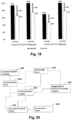
- recanalization rates were ANA+SR: 69% and BGC+SR: 46%. With additional passes, recanalization rates increased in both treatment groups: ANA+SR: 100% vs BGC+SR: 77%. The mean number of passes to achieve complete recanalization tended to be lower with ANA+SR (1.4) as compared to BGC+SR (1.9).
- the ANA device performed similarly to the FlowGate control with respect to compatibility between device components and ancillary devices, pushability of the catheter through the anatomy, radiopacity of the catheter, and device integrity after use.
- the ANA was slightly better in regard to navigating/tracking through the vessel and flexibility, compared to the FlowGate BGC.
- the angiography showed that three dissections occurred during the interventions: one in the ANA+SR group and two in the BGC+SR group, none of them were related to the ANA or Solitaire devices as they were immediately observed after catheterization of the target arteries either with the guiding catheter or the BGC.
- the second dissection in BGC+SR group was mild and not associated with further complications.
- occlusions a total of seven were observed after 3 or 30 days. Two detected after 30 days (one for ANA+SR and one for BGC+SR), were subsequent to severe dissections. Three (one for ANA+SR after 3 days, and two for BGC+SR after 3 and 30 days, respectively), were considered inherent procedural complications. The last two occlusions observed at 3 days angiography (two for BGC+SR) were related to the failure to retrieve the clot after at the end of the procedure (up to three passes). No vessel perforation was observed in either group. Vasospasm was a common observation to varying degrees in both groups. This is a common observation in the swine model as pigs are prone to vasospasm.
- vessel injury was absent to minimal, and comparable for both ANA+SR and BGC+SR groups at 3 days and 30 days, as not statistically differences were found.
- Other findings of inflammation, thrombosis, embolization and necrosis in downstream tissues to cervical artery (brachiocephalicus muscle) and lingual artery (tongue) were also absent to minimal in both groups and time points, with most scores 0 and below 1.
- target arteries were selected to have 2.2-5 mm. These diameters are smaller than arterial segments where guide catheters and BGC are usually placed. This may be the reason of the few arterial dissections and occlusions that were observed secondary to guiding catheter/BGC manipulations previous to ANA/SR deployment.
- the design of the aspiration funnel 1 and the entire ANA device is atraumatic to the vasculature, mainly due to the balanced radial force of the aspiration funnel 1, sufficient to adapt to the vessel and allow aspiration, but not excessively high to damage the vessel wall, together with the smooth silicone covering of the aspiration funnel 1. Additionally, the catheters surface and tip are smooth with lubricious coating to facilitate the navigation and avoid the vessel trauma.
- the ANA device in combination with the stent retriever achieved high rates of complete recanalization in a low number of passes; thus, the proposed ANCD presents an improved efficacy profile than the current commercial product.
- the observed efficacy rates are in line with the recanalization rates achieved in Example 1 ( in vitro model).
- the fact that the BGC+SR combination showed similar recanalization results in both models and in real patients may indicate that the results of human clinical studies with ANCD will be in line with the present results obtained in preclinical models.
- the ANA device and consequently the ANCD, is designed to induce local flow arrest in combination with a complete clot ingestion into the aspiration funnel 1 that prevents fragmentation and distal embolization.
- EXAMPLE 3 Human Clinical Trial: Prospective, Single-Arm, Multi-center Study to Assess the Safety and Performance of the ANA device, in combination with a Clot-Capture Element (for example a Stent Retriever (SR)) in Patients with Acute Ischemic Stroke.
- a Clot-Capture Element for example a Stent Retriever (SR)
- the first patient of the following clinical trial was enrolled last 21 th September and it is currently ongoing.
- the ANA device is a distal access catheter designed to assist in neurovascular procedures by facilitating the insertion and guiding of other devices (i.e. retrieval devices and intravascular catheters) and restricting blood flow at the target position.
- the ANA device is a sterile, single-use, disposable intravascular device comprised of two coaxial catheters (the delivery catheter 3 and the expandable-tip aspiration catheter 7) consisting of sections of variable stiffness.
- the expandable-tip aspiration catheter 7 comprises a radiopaque nitinol braid (self-expanding funnel 1), covered by a continuous silicone coating that, when deployed, provides local and temporary flow restriction.
- the delivery catheter 3 has a hydrophilic coating to reduce friction during use and a radiopaque marker on the distal end. Both catheters 1, 7 have Luer lock hubs on their proximal end.
- the proposed study has been designed to collect prospective clinical evidence to compare the ANA device to similar devices used for guiding and supporting stent retrievers during neurothrombectomy procedures.
- the protocol has been designed to replicate the patient population enrolled in prior studies of similar devices.
- the primary endpoint is the ability of the ANA device to facilitate stent retriever deployment and neurothrombectomy in the anterior circulation, with successful reperfusion defined as achieving a modified Thrombolysis in Cerebral Infarction (mTICI) score of ⁇ 2b in the target vessel with ⁇ 3 passes of the ANA device without the use of rescue therapy.
- mTICI modified Thrombolysis in Cerebral Infarction
- the objective of the study is to assess safety and performance of the ANA catheter system to be used as a tool and to facilitate the Solitaire stent retriever placement and to provide temporary restriction of blood flow in stroke patients undergoing neurothrombectomy for an acute large vessel occlusion (LVO), presenting (to the neuroimaging laboratory) within 8 h of symptom onset (last time the subject was seen well).
- LVO large vessel occlusion
- the performance has been assessed as the ability of the ANA device to facilitate stent retriever deployment and to perform neurothrombectomy in the anterior circulation, with successful reperfusion defined as achieving a modified Thrombolysis in Cerebral Infarction (mTICI) score of ⁇ 2b in the target vessel with ⁇ 3 passes of the ANA device without the use of rescue therapy.
- mTICI modified Thrombolysis in Cerebral Infarction
- the safety has been assessed as the occurrence of all serious adverse device effects up to 90 days post-procedure, including symptomatic IntraCerebral Hemorrhage (sICH), at 24 h (-8/+12 h) post-procedure.
- sICH IntraCerebral Hemorrhage
- the population has been based on patients with Acute Ischemic Stroke (AIS), whose stroke is attributable to an occlusion of a large artery in the neurovasculature, such as the internal carotid artery, M1 or M2 segments of the middle cerebral artery, and who are either ineligible for IV alteplase (tissue-type Plasminogen Activator [t-PA]) or have received IV t-PA therapy without sufficient recanalization, but are within the timeframe of 8 h from symptom onset (last seen well) to groin puncture in the catheterization lab.
- Hundred and twenty-five (125) consecutive subjects indicated for treatment with an ANA device in combination with the Solitaire stent retriever has been set to be enrolled. Inclusion and exclusion criteria for the selection of the patients are described in detail in the clinical trial protocol.
- Table 10 shows the schedule of assessments recorded during the baseline, during the procedure, and the assessments which should be completed at 24-h post-procedure, at Day 5 (+/- 12 h) or discharge (whichever comes first, depending on whatever time point is earliest) and at the 90-day follow-up office visit.
- Table 10 Schedule of assessments Event Screening / Baseline Procedure 24 h Post-procedure Day 5 +/- 12 h / Discharge (whichever is earliest) Day 90 +/- 14 days Eligibility criteria (inclusion/exclusion) X Informed consent X Demographic/medical history X Physical exam (blood pressure, heart rate) X Baseline laboratory assessments X Pregnancy test (as applicable) X 12-lead electrocardiogram (ECG) X Modified Ranking Scale (mRS) X X X X X National Institute of Health Stroke Scale (NIHSS) X X X X X X Neuro imaging (MRI/CT) X X Angiogram X Mechanical thrombectomy X Adverse events X X X X X X X X X X X X X X X X X
- Primary analysis set for statistical reporting is the ITT population, with no replacement of missing data planned in the statistical analysis to provide unbiased results.
- two sensitivity analyses are conducted in regard to the missing values for the primary performance endpoint. The first one imputes failures instead of missing value, in a conservative approach. The second one uses the repartition of success/failure reported on non-missing value to impute the missing values with the same repartition. In both sensitivity analyses, the same statistical testing is presented.
- primary endpoints are reported on the mITT population.
- Categorical variables are summarized using classical frequency statistics: number of non-missing observations and percentages by categories. Percentages are calculated on the number of non-missing observations. The number of missing observations is also specified.
- CIs bilateral asymptotic or exact confidence intervals
- AE data is summarized using descriptive statistics: total number of events and number of subjects with at least one of the respective categories AEs, ADEs, SAEs, SADEs and device deficiencies. The severity and the causal relationship are presented.
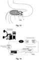
- FIG. 20 this figure depicts another example of the proposed thrombectomy system (or ANCD) 600 which allows for its automated maneuvering through a vascular system.
- an automated proximal device 601 provides a guidance system to deploy the ANCD 600.
- an imaging device 602 can detect the radiopaque markers included in the segment 10, and also in the delivery catheter 3, and a communications channel 603 can be used to provide means to transport the image to a control module 604.
- the control module 604 is programmed or configured to allow for guidance of the deployment of the ANCD 600 and storage of data on a data storage device 605.
- the control module 604 may be a programmable logic controller, a computer, or the like.
- control module 604 is guided by a computer assisted controller 606.
- the communications channel 603 can be Ethernet, WiFi, Bluetooth, or the like.
- the control module 604 is programmed to guide a physician or technician operating the ANCD 600 which allows for the ANCD 600 to be used in non-hospital settings such as nursing homes or assisted care living facilities.
- the time required to perform the thrombectomy is greatly reduced significantly improving patient outcomes.
- the control may also be via a controller such as those in use in other current medical devices.
- the system may be controlled manually.
Landscapes
- Health & Medical Sciences (AREA)
- Life Sciences & Earth Sciences (AREA)
- Surgery (AREA)
- Heart & Thoracic Surgery (AREA)
- Public Health (AREA)
- Vascular Medicine (AREA)
- Engineering & Computer Science (AREA)
- Biomedical Technology (AREA)
- Veterinary Medicine (AREA)
- General Health & Medical Sciences (AREA)
- Animal Behavior & Ethology (AREA)
- Molecular Biology (AREA)
- Medical Informatics (AREA)
- Nuclear Medicine, Radiotherapy & Molecular Imaging (AREA)
- Orthopedic Medicine & Surgery (AREA)
- Cardiology (AREA)
- Oral & Maxillofacial Surgery (AREA)
- Transplantation (AREA)
- Surgical Instruments (AREA)
Applications Claiming Priority (3)
| Application Number | Priority Date | Filing Date | Title |
|---|---|---|---|
| EP18382800 | 2018-11-13 | ||
| EP19805581.6A EP3880090B1 (de) | 2018-11-13 | 2019-11-12 | Thrombektomiesystem |
| PCT/EP2019/080993 WO2020099386A1 (en) | 2018-11-13 | 2019-11-12 | A thrombectomy system and methods of extracting a thrombus from a thrombus site in a blood vessel of a patient |
Related Parent Applications (1)
| Application Number | Title | Priority Date | Filing Date |
|---|---|---|---|
| EP19805581.6A Division EP3880090B1 (de) | 2018-11-13 | 2019-11-12 | Thrombektomiesystem |
Publications (2)
| Publication Number | Publication Date |
|---|---|
| EP4413951A2 true EP4413951A2 (de) | 2024-08-14 |
| EP4413951A3 EP4413951A3 (de) | 2024-11-06 |
Family
ID=64426851
Family Applications (2)
| Application Number | Title | Priority Date | Filing Date |
|---|---|---|---|
| EP24179172.2A Pending EP4413951A3 (de) | 2018-11-13 | 2019-11-12 | Thrombektomiesystem |
| EP19805581.6A Active EP3880090B1 (de) | 2018-11-13 | 2019-11-12 | Thrombektomiesystem |
Family Applications After (1)
| Application Number | Title | Priority Date | Filing Date |
|---|---|---|---|
| EP19805581.6A Active EP3880090B1 (de) | 2018-11-13 | 2019-11-12 | Thrombektomiesystem |
Country Status (10)
| Country | Link |
|---|---|
| US (1) | US20220000500A1 (de) |
| EP (2) | EP4413951A3 (de) |
| JP (3) | JP7385794B2 (de) |
| KR (1) | KR102818094B1 (de) |
| CN (1) | CN113423348B (de) |
| AU (1) | AU2019380578B2 (de) |
| CA (1) | CA3119221A1 (de) |
| ES (1) | ES2989076T3 (de) |
| IL (1) | IL283132B2 (de) |
| WO (1) | WO2020099386A1 (de) |
Families Citing this family (21)
| Publication number | Priority date | Publication date | Assignee | Title |
|---|---|---|---|---|
| US11771446B2 (en) | 2020-10-19 | 2023-10-03 | Anaconda Biomed, S.L. | Thrombectomy system and method of use |
| EP3639768A1 (de) | 2018-10-16 | 2020-04-22 | Anaconda Biomed, S.L. | Vorrichtung zur extraktion von thromben aus einem blutgefäss und einer thrombectomievorrichtung |
| JP2021517850A (ja) | 2018-03-12 | 2021-07-29 | エクストラクト メディカル,インコーポレイティド | 患者から物質を除去するための装置及び方法 |
| JP7312262B2 (ja) | 2018-09-28 | 2023-07-20 | フロー メディカル コーポレイション | カテーテル装置 |
| CN113795204A (zh) | 2019-01-11 | 2021-12-14 | 阿纳康达生物医学有限公司 | 用于将医疗器械装载到导管中的装载装置和方法 |
| US20230346416A1 (en) * | 2020-03-04 | 2023-11-02 | Shifamed Holdings, Llc | Thrombus removal systems and associated methods |
| US12440230B2 (en) | 2020-05-27 | 2025-10-14 | Penumbra, Inc. | Devices and methods for removing material from a patient |
| CN116916833A (zh) * | 2020-12-09 | 2023-10-20 | 赛瑞特锐弗有限公司 | 取回系统和方法 |
| AU2022231082A1 (en) | 2021-03-01 | 2023-09-21 | Endovascular Engineering, Inc. | Aspiration devices for treatment of thrombosis including expandable distal ends and systems and methods thereof |
| US11679194B2 (en) | 2021-04-27 | 2023-06-20 | Contego Medical, Inc. | Thrombus aspiration system and methods for controlling blood loss |
| EP4340752A1 (de) * | 2021-05-18 | 2024-03-27 | Koninklijke Philips N.V. | Systeme und verfahren zur gerinnselentfernung in der mechanischen thrombektomie |
| CN113576600A (zh) * | 2021-07-19 | 2021-11-02 | 启晨(上海)医疗器械有限公司 | 一种血栓回收装置 |
| CN114081579A (zh) * | 2021-11-29 | 2022-02-25 | 上海市第十人民医院 | 一种脑卒中机械取栓装置及其应用 |
| CN119343096A (zh) | 2022-03-28 | 2025-01-21 | 万能医药公司 | 带有可扩展远侧尖端的抽吸导管 |
| WO2024039214A1 (ko) * | 2022-08-18 | 2024-02-22 | 대구가톨릭대학교산학협력단 | 혈전제거시스템 |
| KR102535089B1 (ko) * | 2022-08-18 | 2023-05-26 | 대구가톨릭대학교산학협력단 | 혈관 내 혈류 재개통과 미세흐름회로의 생성이 가능한 스텐트 리트리버 형태의 혈전제거장치 |
| US12053192B2 (en) | 2022-09-01 | 2024-08-06 | Endovascular Engineering, Inc. | Systems, devices, and methods for aspiration, including expandable structures and rotatable shafts |
| WO2024054648A1 (en) * | 2022-09-08 | 2024-03-14 | Sonovascular, Inc. | Systems and methods for an ultrasound catheter |
| CN115317077B (zh) * | 2022-10-13 | 2023-02-17 | 成都百瑞恒通医疗科技有限公司 | 一种取栓装置 |
| CN115919405A (zh) * | 2022-11-02 | 2023-04-07 | 上海玮琅医疗科技有限公司 | 一种血管异物抽取辅助装置及抽取装置 |
| CN116570344B (zh) * | 2023-05-26 | 2025-08-22 | 苏州恒瑞宏远医疗科技有限公司 | 一种取栓装置系统 |
Citations (4)
| Publication number | Priority date | Publication date | Assignee | Title |
|---|---|---|---|---|
| WO2015006782A1 (en) | 2013-07-12 | 2015-01-15 | Inceptus Medical, Llc | Methods and apparatus for treating pulmonary embolism |
| WO2016113047A1 (en) | 2015-01-13 | 2016-07-21 | Anaconda Biomed, S.L. | Thrombectomy device and system for extraction of vascular thrombi from a blood vessel |
| US20170119408A1 (en) | 2015-10-31 | 2017-05-04 | Neurovasc Technologies, Inc. | Embolus Removal Device with Blood Flow Restriction and Related Methods |
| US20180132876A1 (en) | 2016-11-16 | 2018-05-17 | Osama O. Zaidat | System and device for engulfing thrombi |
Family Cites Families (18)
| Publication number | Priority date | Publication date | Assignee | Title |
|---|---|---|---|---|
| US6746469B2 (en) * | 2001-04-30 | 2004-06-08 | Advanced Cardiovascular Systems, Inc. | Balloon actuated apparatus having multiple embolic filters, and method of use |
| DE60328694D1 (de) * | 2002-10-02 | 2009-09-17 | Boston Scient Ltd | Expandierbare rückholvorrichtung |
| JP3660931B2 (ja) * | 2003-09-22 | 2005-06-15 | 新 石丸 | 血栓塞栓捕獲装置 |
| US7963988B2 (en) * | 2005-06-23 | 2011-06-21 | Boston Scientific Scimed, Inc. | ePTFE lamination—resizing ePTFE tubing |
| US8066757B2 (en) * | 2007-10-17 | 2011-11-29 | Mindframe, Inc. | Blood flow restoration and thrombus management methods |
| EP3311875B1 (de) * | 2007-12-20 | 2024-11-20 | AngioDynamics, Inc. | Systeme zur entfernung von unerwünschtem material in einem kreislaufsystem |
| US8858497B2 (en) * | 2010-09-07 | 2014-10-14 | Angio Dynamics, Inc. | Device and method for removing material from a hollow anatomical structure |
| CN107126244B (zh) * | 2011-05-23 | 2020-07-31 | 柯惠有限合伙公司 | 取出系统及其使用方法 |
| US9597171B2 (en) * | 2012-09-11 | 2017-03-21 | Covidien Lp | Retrieval catheter with expandable tip |
| KR101317434B1 (ko) * | 2013-05-31 | 2013-10-10 | (주) 더아이엔지메디칼 | 혈관 이물질 적출용 카테터 |
| JP6352642B2 (ja) * | 2013-12-03 | 2018-07-04 | 川澄化学工業株式会社 | 血管内異物除去用カテーテル |
| US10441301B2 (en) * | 2014-06-13 | 2019-10-15 | Neuravi Limited | Devices and methods for removal of acute blockages from blood vessels |
| US10792056B2 (en) * | 2014-06-13 | 2020-10-06 | Neuravi Limited | Devices and methods for removal of acute blockages from blood vessels |
| US9987028B2 (en) * | 2015-02-12 | 2018-06-05 | Cook Medical Technologies Llc | Partially covered braided funnel aspiration catheter |
| CN105662647B (zh) * | 2016-02-19 | 2017-11-17 | 杭州启明医疗器械有限公司 | 血栓过滤器 |
| EP3509507A1 (de) * | 2016-09-12 | 2019-07-17 | Stryker Corporation | Selbstrollende thrombektomievorrichtungen und verfahren |
| WO2018147449A1 (ja) * | 2017-02-10 | 2018-08-16 | 川澄化学工業株式会社 | 異物除去デバイス、異物除去用カテーテル及び異物回収システム |
| CN207898518U (zh) * | 2017-06-20 | 2018-09-25 | 吉林一方科技有限公司 | 一种血栓清除器 |
-
2019
- 2019-11-12 AU AU2019380578A patent/AU2019380578B2/en active Active
- 2019-11-12 CN CN201980088158.XA patent/CN113423348B/zh active Active
- 2019-11-12 KR KR1020217017933A patent/KR102818094B1/ko active Active
- 2019-11-12 WO PCT/EP2019/080993 patent/WO2020099386A1/en not_active Ceased
- 2019-11-12 EP EP24179172.2A patent/EP4413951A3/de active Pending
- 2019-11-12 EP EP19805581.6A patent/EP3880090B1/de active Active
- 2019-11-12 US US17/291,696 patent/US20220000500A1/en active Pending
- 2019-11-12 IL IL283132A patent/IL283132B2/en unknown
- 2019-11-12 ES ES19805581T patent/ES2989076T3/es active Active
- 2019-11-12 JP JP2021525077A patent/JP7385794B2/ja active Active
- 2019-11-12 CA CA3119221A patent/CA3119221A1/en active Pending
-
2023
- 2023-08-22 JP JP2023134347A patent/JP2023153269A/ja active Pending
-
2025
- 2025-07-25 JP JP2025125234A patent/JP2025146928A/ja active Pending
Patent Citations (4)
| Publication number | Priority date | Publication date | Assignee | Title |
|---|---|---|---|---|
| WO2015006782A1 (en) | 2013-07-12 | 2015-01-15 | Inceptus Medical, Llc | Methods and apparatus for treating pulmonary embolism |
| WO2016113047A1 (en) | 2015-01-13 | 2016-07-21 | Anaconda Biomed, S.L. | Thrombectomy device and system for extraction of vascular thrombi from a blood vessel |
| US20170119408A1 (en) | 2015-10-31 | 2017-05-04 | Neurovasc Technologies, Inc. | Embolus Removal Device with Blood Flow Restriction and Related Methods |
| US20180132876A1 (en) | 2016-11-16 | 2018-05-17 | Osama O. Zaidat | System and device for engulfing thrombi |
Non-Patent Citations (3)
| Title |
|---|
| DUFFY SFARRELL MMCARDLE KTHORNTON JVALE DRAINSFORD EMORRIS LLIEBESKIND DSMACCARTHY EGILVARRY M: "Novel methodology to replicate clot analogs with diverse composition in acute ischemic stroke", J NEUROINTERV SURG, vol. 9, no. 5, May 2017 (2017-05-01), pages 486 - 491 |
| FENNELL VS ET AL.: "What to do about fibrin rich 'tough clots'? Comparing the Solitaire stent retriever with a novel geometric clot extractor in an in vitro stroke model", J NEUROLNTERVENT SURG, vol. 0, 2018, pages 1 - 4 |
| MOKIN MSETLUR NAGESH SVLONITA CNMOCCO JSIDDIQUI AH: "Stent retriever thrombectomy with the Cover accessory device versus proximal protection with a balloon guide catheter: in vitro stroke model comparison", J NEUROINTERV SURG, vol. 8, no. 4, April 2016 (2016-04-01), pages 413 - 7 |
Also Published As
| Publication number | Publication date |
|---|---|
| EP4413951A3 (de) | 2024-11-06 |
| US20220000500A1 (en) | 2022-01-06 |
| CA3119221A1 (en) | 2020-05-22 |
| JP2023153269A (ja) | 2023-10-17 |
| CN113423348A (zh) | 2021-09-21 |
| IL283132A (en) | 2021-06-30 |
| AU2019380578A1 (en) | 2021-06-03 |
| JP2025146928A (ja) | 2025-10-03 |
| JP2022515010A (ja) | 2022-02-17 |
| EP3880090C0 (de) | 2024-07-10 |
| EP3880090A1 (de) | 2021-09-22 |
| EP3880090B1 (de) | 2024-07-10 |
| WO2020099386A1 (en) | 2020-05-22 |
| KR102818094B1 (ko) | 2025-06-13 |
| ES2989076T3 (es) | 2024-11-25 |
| IL283132B2 (en) | 2024-11-01 |
| JP7385794B2 (ja) | 2023-11-24 |
| IL283132B1 (en) | 2024-07-01 |
| KR20210091770A (ko) | 2021-07-22 |
| BR112021009338A2 (pt) | 2021-08-17 |
| CN113423348B (zh) | 2025-11-25 |
| AU2019380578B2 (en) | 2024-07-25 |
Similar Documents
| Publication | Publication Date | Title |
|---|---|---|
| EP3880090B1 (de) | Thrombektomiesystem | |
| US12364492B2 (en) | Thrombectomy system and method of use | |
| US11986195B2 (en) | Device and a thrombectomy apparatus for extraction of thrombus from a blood vessel | |
| US20250032134A1 (en) | Thrombectomy system and method of use | |
| US20220273322A1 (en) | Catheter Systems Enabling Improved Aspiration from Cerebral Arteries | |
| CA2979816C (en) | Ischemic stroke thrombus aspiration system, processes, and products thereby | |
| CN116437865A (zh) | 用于从血管中提取闭塞物的凝块活动器装置 | |
| US20240374272A1 (en) | Device and a thrombectomy apparatus for extraction of thrombus from a blood vessel | |
| BR112021009338B1 (pt) | Sistema para trombectomia e sistema para manobrar de maneira automatizada um sistema de trombectomia | |
| US20240065713A1 (en) | Systems and methods of removing challenging clots and restoring perfusion to a vessel | |
| Kaiser et al. | Procedural Complications | |
| US20230248379A1 (en) | Systems and methods to restore perfusion to a vessel | |
| US20240407794A1 (en) | Devices and methods for accessing a neurovascular site | |
| EP4115822A1 (de) | Systeme zum wiederherstellen der durchblutung eines gefässes | |
| BR112021007203B1 (pt) | Dispositivo para extração de trombo de um vaso sanguíneo, aparelho para trombectomia e sistema para manobra automatizada do aparelho de trombectomia | |
| KR20230015853A (ko) | 병원내 사망률을 감소시키는 시스템 및 방법 | |
| SZIKORA et al. | The Evolving Endovascular Therapy Toolkit: Real-World and Clinical Experiences with Pipeline Flex and Vantage with Shield Technology and Solitaire X 3 mm | |
| Jones et al. | Incidence and Prevention of Strokes in TAVI | |
| Kinlay | 18 Intervention for Lower Extremity |
Legal Events
| Date | Code | Title | Description |
|---|---|---|---|
| PUAI | Public reference made under article 153(3) epc to a published international application that has entered the european phase |
Free format text: ORIGINAL CODE: 0009012 |
|
| STAA | Information on the status of an ep patent application or granted ep patent |
Free format text: STATUS: THE APPLICATION HAS BEEN PUBLISHED |
|
| AC | Divisional application: reference to earlier application |
Ref document number: 3880090 Country of ref document: EP Kind code of ref document: P |
|
| AK | Designated contracting states |
Kind code of ref document: A2 Designated state(s): AL AT BE BG CH CY CZ DE DK EE ES FI FR GB GR HR HU IE IS IT LI LT LU LV MC MK MT NL NO PL PT RO RS SE SI SK SM TR |
|
| PUAL | Search report despatched |
Free format text: ORIGINAL CODE: 0009013 |
|
| REG | Reference to a national code |
Ref country code: DE Ref legal event code: R079 Free format text: PREVIOUS MAIN CLASS: A61F0002010000 Ipc: A61B0017221000 |
|
| AK | Designated contracting states |
Kind code of ref document: A3 Designated state(s): AL AT BE BG CH CY CZ DE DK EE ES FI FR GB GR HR HU IE IS IT LI LT LU LV MC MK MT NL NO PL PT RO RS SE SI SK SM TR |
|
| RIC1 | Information provided on ipc code assigned before grant |
Ipc: A61F 2/01 20060101ALI20241003BHEP Ipc: A61B 17/221 20060101AFI20241003BHEP |
|
| STAA | Information on the status of an ep patent application or granted ep patent |
Free format text: STATUS: REQUEST FOR EXAMINATION WAS MADE |
|
| 17P | Request for examination filed |
Effective date: 20250505 |