EP4293124A2 - Amplification d'adn en phase solide et en phase soluble combinée - Google Patents
Amplification d'adn en phase solide et en phase soluble combinée Download PDFInfo
- Publication number
- EP4293124A2 EP4293124A2 EP23185680.8A EP23185680A EP4293124A2 EP 4293124 A2 EP4293124 A2 EP 4293124A2 EP 23185680 A EP23185680 A EP 23185680A EP 4293124 A2 EP4293124 A2 EP 4293124A2
- Authority
- EP
- European Patent Office
- Prior art keywords
- amplification
- immobilised
- primers
- nucleic acid
- primer
- Prior art date
- Legal status (The legal status is an assumption and is not a legal conclusion. Google has not performed a legal analysis and makes no representation as to the accuracy of the status listed.)
- Pending
Links
- 239000007790 solid phase Substances 0.000 title claims abstract description 23
- 239000012071 phase Substances 0.000 title abstract description 40
- 230000004544 DNA amplification Effects 0.000 title description 5
- 230000003321 amplification Effects 0.000 claims abstract description 174
- 238000003199 nucleic acid amplification method Methods 0.000 claims abstract description 174
- 150000007523 nucleic acids Chemical group 0.000 claims abstract description 57
- 238000000034 method Methods 0.000 claims abstract description 38
- 238000006243 chemical reaction Methods 0.000 claims description 56
- 108020004707 nucleic acids Proteins 0.000 claims description 45
- 102000039446 nucleic acids Human genes 0.000 claims description 45
- 238000001514 detection method Methods 0.000 claims description 33
- 239000002773 nucleotide Substances 0.000 claims description 21
- 125000003729 nucleotide group Chemical group 0.000 claims description 21
- 239000000523 sample Substances 0.000 claims description 18
- 239000007788 liquid Substances 0.000 claims description 17
- 239000003153 chemical reaction reagent Substances 0.000 claims description 9
- 239000007787 solid Substances 0.000 claims description 9
- 238000004458 analytical method Methods 0.000 claims description 4
- 230000003287 optical effect Effects 0.000 claims description 4
- 238000012634 optical imaging Methods 0.000 claims description 3
- 238000005259 measurement Methods 0.000 claims description 2
- 238000012163 sequencing technique Methods 0.000 abstract description 8
- 239000000243 solution Substances 0.000 description 51
- 239000000047 product Substances 0.000 description 47
- 108091093088 Amplicon Proteins 0.000 description 35
- 108020004414 DNA Proteins 0.000 description 16
- 108090000790 Enzymes Proteins 0.000 description 11
- 102000004190 Enzymes Human genes 0.000 description 11
- 108091034117 Oligonucleotide Proteins 0.000 description 11
- 102000054765 polymorphisms of proteins Human genes 0.000 description 9
- 239000000499 gel Substances 0.000 description 8
- 230000035772 mutation Effects 0.000 description 8
- 238000003556 assay Methods 0.000 description 7
- 239000000872 buffer Substances 0.000 description 7
- 230000000295 complement effect Effects 0.000 description 7
- 241000607715 Serratia marcescens Species 0.000 description 6
- 238000012790 confirmation Methods 0.000 description 6
- 238000009396 hybridization Methods 0.000 description 6
- 239000000463 material Substances 0.000 description 6
- 239000000203 mixture Substances 0.000 description 6
- 244000052769 pathogen Species 0.000 description 6
- 230000001717 pathogenic effect Effects 0.000 description 6
- 108090000623 proteins and genes Proteins 0.000 description 6
- 238000011897 real-time detection Methods 0.000 description 6
- 241000235347 Schizosaccharomyces pombe Species 0.000 description 5
- 230000003139 buffering effect Effects 0.000 description 5
- 238000001502 gel electrophoresis Methods 0.000 description 5
- 206010040047 Sepsis Diseases 0.000 description 4
- FAPWRFPIFSIZLT-UHFFFAOYSA-M Sodium chloride Chemical compound [Na+].[Cl-] FAPWRFPIFSIZLT-UHFFFAOYSA-M 0.000 description 4
- 230000008569 process Effects 0.000 description 4
- 238000010530 solution phase reaction Methods 0.000 description 4
- PEDCQBHIVMGVHV-UHFFFAOYSA-N Glycerine Chemical compound OCC(O)CO PEDCQBHIVMGVHV-UHFFFAOYSA-N 0.000 description 3
- 108091028043 Nucleic acid sequence Proteins 0.000 description 3
- 229920001213 Polysorbate 20 Polymers 0.000 description 3
- HEMHJVSKTPXQMS-UHFFFAOYSA-M Sodium hydroxide Chemical compound [OH-].[Na+] HEMHJVSKTPXQMS-UHFFFAOYSA-M 0.000 description 3
- 238000013459 approach Methods 0.000 description 3
- 230000003115 biocidal effect Effects 0.000 description 3
- 230000002051 biphasic effect Effects 0.000 description 3
- 239000000839 emulsion Substances 0.000 description 3
- 239000012530 fluid Substances 0.000 description 3
- 238000010348 incorporation Methods 0.000 description 3
- 239000000256 polyoxyethylene sorbitan monolaurate Substances 0.000 description 3
- 235000010486 polyoxyethylene sorbitan monolaurate Nutrition 0.000 description 3
- 238000003746 solid phase reaction Methods 0.000 description 3
- XLYOFNOQVPJJNP-UHFFFAOYSA-N water Substances O XLYOFNOQVPJJNP-UHFFFAOYSA-N 0.000 description 3
- TWRXJAOTZQYOKJ-UHFFFAOYSA-L Magnesium chloride Chemical compound [Mg+2].[Cl-].[Cl-] TWRXJAOTZQYOKJ-UHFFFAOYSA-L 0.000 description 2
- 239000012807 PCR reagent Substances 0.000 description 2
- WCUXLLCKKVVCTQ-UHFFFAOYSA-M Potassium chloride Chemical compound [Cl-].[K+] WCUXLLCKKVVCTQ-UHFFFAOYSA-M 0.000 description 2
- RTAQQCXQSZGOHL-UHFFFAOYSA-N Titanium Chemical compound [Ti] RTAQQCXQSZGOHL-UHFFFAOYSA-N 0.000 description 2
- 239000007795 chemical reaction product Substances 0.000 description 2
- 238000004132 cross linking Methods 0.000 description 2
- 238000013211 curve analysis Methods 0.000 description 2
- 238000001962 electrophoresis Methods 0.000 description 2
- 238000000799 fluorescence microscopy Methods 0.000 description 2
- 239000012634 fragment Substances 0.000 description 2
- 238000003384 imaging method Methods 0.000 description 2
- 239000012160 loading buffer Substances 0.000 description 2
- 239000003550 marker Substances 0.000 description 2
- 238000002493 microarray Methods 0.000 description 2
- 239000013641 positive control Substances 0.000 description 2
- 238000012545 processing Methods 0.000 description 2
- 238000000926 separation method Methods 0.000 description 2
- 239000011780 sodium chloride Substances 0.000 description 2
- 230000001225 therapeutic effect Effects 0.000 description 2
- 229910052719 titanium Inorganic materials 0.000 description 2
- 239000010936 titanium Substances 0.000 description 2
- 239000011534 wash buffer Substances 0.000 description 2
- IJGRMHOSHXDMSA-UHFFFAOYSA-N Atomic nitrogen Chemical compound N#N IJGRMHOSHXDMSA-UHFFFAOYSA-N 0.000 description 1
- 208000035143 Bacterial infection Diseases 0.000 description 1
- KWIUHFFTVRNATP-UHFFFAOYSA-N Betaine Natural products C[N+](C)(C)CC([O-])=O KWIUHFFTVRNATP-UHFFFAOYSA-N 0.000 description 1
- 208000035049 Blood-Borne Infections Diseases 0.000 description 1
- 108091003079 Bovine Serum Albumin Proteins 0.000 description 1
- 108010014303 DNA-directed DNA polymerase Proteins 0.000 description 1
- 102000016928 DNA-directed DNA polymerase Human genes 0.000 description 1
- 241000588724 Escherichia coli Species 0.000 description 1
- 241000495778 Escherichia faecalis Species 0.000 description 1
- KWIUHFFTVRNATP-UHFFFAOYSA-O N,N,N-trimethylglycinium Chemical compound C[N+](C)(C)CC(O)=O KWIUHFFTVRNATP-UHFFFAOYSA-O 0.000 description 1
- 238000012408 PCR amplification Methods 0.000 description 1
- 206010040070 Septic Shock Diseases 0.000 description 1
- 101000794822 Serratia marcescens Anthranilate synthase component 1 Proteins 0.000 description 1
- 101000847781 Serratia marcescens Anthranilate synthase component 2 Proteins 0.000 description 1
- XUIMIQQOPSSXEZ-UHFFFAOYSA-N Silicon Chemical compound [Si] XUIMIQQOPSSXEZ-UHFFFAOYSA-N 0.000 description 1
- 108010006785 Taq Polymerase Proteins 0.000 description 1
- 239000007983 Tris buffer Substances 0.000 description 1
- 239000013504 Triton X-100 Substances 0.000 description 1
- 229920004890 Triton X-100 Polymers 0.000 description 1
- 239000011543 agarose gel Substances 0.000 description 1
- 238000000137 annealing Methods 0.000 description 1
- 238000003491 array Methods 0.000 description 1
- 238000007846 asymmetric PCR Methods 0.000 description 1
- 230000001580 bacterial effect Effects 0.000 description 1
- 208000022362 bacterial infectious disease Diseases 0.000 description 1
- 239000011324 bead Substances 0.000 description 1
- 230000008901 benefit Effects 0.000 description 1
- 229960003237 betaine Drugs 0.000 description 1
- 239000012620 biological material Substances 0.000 description 1
- 239000012472 biological sample Substances 0.000 description 1
- 230000015572 biosynthetic process Effects 0.000 description 1
- 239000008280 blood Substances 0.000 description 1
- 210000004369 blood Anatomy 0.000 description 1
- 238000009640 blood culture Methods 0.000 description 1
- 229940098773 bovine serum albumin Drugs 0.000 description 1
- 210000004027 cell Anatomy 0.000 description 1
- 238000004113 cell culture Methods 0.000 description 1
- 230000008859 change Effects 0.000 description 1
- 210000000349 chromosome Anatomy 0.000 description 1
- 239000002299 complementary DNA Substances 0.000 description 1
- 238000012258 culturing Methods 0.000 description 1
- SUYVUBYJARFZHO-RRKCRQDMSA-N dATP Chemical compound C1=NC=2C(N)=NC=NC=2N1[C@H]1C[C@H](O)[C@@H](COP(O)(=O)OP(O)(=O)OP(O)(O)=O)O1 SUYVUBYJARFZHO-RRKCRQDMSA-N 0.000 description 1
- RGWHQCVHVJXOKC-SHYZEUOFSA-N dCTP Chemical compound O=C1N=C(N)C=CN1[C@@H]1O[C@H](CO[P@](O)(=O)O[P@](O)(=O)OP(O)(O)=O)[C@@H](O)C1 RGWHQCVHVJXOKC-SHYZEUOFSA-N 0.000 description 1
- HAAZLUGHYHWQIW-KVQBGUIXSA-N dGTP Chemical compound C1=NC=2C(=O)NC(N)=NC=2N1[C@H]1C[C@H](O)[C@@H](COP(O)(=O)OP(O)(=O)OP(O)(O)=O)O1 HAAZLUGHYHWQIW-KVQBGUIXSA-N 0.000 description 1
- NHVNXKFIZYSCEB-XLPZGREQSA-N dTTP Chemical compound O=C1NC(=O)C(C)=CN1[C@@H]1O[C@H](COP(O)(=O)OP(O)(=O)OP(O)(O)=O)[C@@H](O)C1 NHVNXKFIZYSCEB-XLPZGREQSA-N 0.000 description 1
- 238000007405 data analysis Methods 0.000 description 1
- 239000005547 deoxyribonucleotide Substances 0.000 description 1
- 125000002637 deoxyribonucleotide group Chemical group 0.000 description 1
- 238000013461 design Methods 0.000 description 1
- 239000000539 dimer Substances 0.000 description 1
- 229910001873 dinitrogen Inorganic materials 0.000 description 1
- 201000010099 disease Diseases 0.000 description 1
- 231100000676 disease causative agent Toxicity 0.000 description 1
- 208000037265 diseases, disorders, signs and symptoms Diseases 0.000 description 1
- 239000000975 dye Substances 0.000 description 1
- 230000000694 effects Effects 0.000 description 1
- 230000005684 electric field Effects 0.000 description 1
- 238000003366 endpoint assay Methods 0.000 description 1
- ZMMJGEGLRURXTF-UHFFFAOYSA-N ethidium bromide Chemical compound [Br-].C12=CC(N)=CC=C2C2=CC=C(N)C=C2[N+](CC)=C1C1=CC=CC=C1 ZMMJGEGLRURXTF-UHFFFAOYSA-N 0.000 description 1
- 229960005542 ethidium bromide Drugs 0.000 description 1
- 238000002474 experimental method Methods 0.000 description 1
- 230000005669 field effect Effects 0.000 description 1
- 239000007850 fluorescent dye Substances 0.000 description 1
- 238000012632 fluorescent imaging Methods 0.000 description 1
- 238000011534 incubation Methods 0.000 description 1
- 238000007403 mPCR Methods 0.000 description 1
- UEGPKNKPLBYCNK-UHFFFAOYSA-L magnesium acetate Chemical compound [Mg+2].CC([O-])=O.CC([O-])=O UEGPKNKPLBYCNK-UHFFFAOYSA-L 0.000 description 1
- 239000011654 magnesium acetate Substances 0.000 description 1
- 235000011285 magnesium acetate Nutrition 0.000 description 1
- 229940069446 magnesium acetate Drugs 0.000 description 1
- 229910001629 magnesium chloride Inorganic materials 0.000 description 1
- 239000013642 negative control Substances 0.000 description 1
- 239000003921 oil Substances 0.000 description 1
- 108091033319 polynucleotide Proteins 0.000 description 1
- 239000002157 polynucleotide Substances 0.000 description 1
- 102000040430 polynucleotide Human genes 0.000 description 1
- 239000001103 potassium chloride Substances 0.000 description 1
- 235000011164 potassium chloride Nutrition 0.000 description 1
- 230000002250 progressing effect Effects 0.000 description 1
- 238000000746 purification Methods 0.000 description 1
- 238000004451 qualitative analysis Methods 0.000 description 1
- 238000011160 research Methods 0.000 description 1
- 230000035945 sensitivity Effects 0.000 description 1
- 230000036303 septic shock Effects 0.000 description 1
- 238000002864 sequence alignment Methods 0.000 description 1
- 229910052710 silicon Inorganic materials 0.000 description 1
- 239000010703 silicon Substances 0.000 description 1
- 239000001509 sodium citrate Substances 0.000 description 1
- 241000894007 species Species 0.000 description 1
- 239000000126 substance Substances 0.000 description 1
- 238000003239 susceptibility assay Methods 0.000 description 1
- PBCFLUZVCVVTBY-UHFFFAOYSA-N tantalum pentoxide Inorganic materials O=[Ta](=O)O[Ta](=O)=O PBCFLUZVCVVTBY-UHFFFAOYSA-N 0.000 description 1
- 238000012360 testing method Methods 0.000 description 1
- 239000001226 triphosphate Substances 0.000 description 1
- 235000011178 triphosphate Nutrition 0.000 description 1
- UNXRWKVEANCORM-UHFFFAOYSA-N triphosphoric acid Chemical compound OP(O)(=O)OP(O)(=O)OP(O)(O)=O UNXRWKVEANCORM-UHFFFAOYSA-N 0.000 description 1
- LENZDBCJOHFCAS-UHFFFAOYSA-N tris Chemical compound OCC(N)(CO)CO LENZDBCJOHFCAS-UHFFFAOYSA-N 0.000 description 1
- 238000011144 upstream manufacturing Methods 0.000 description 1
- 230000003612 virological effect Effects 0.000 description 1
Images


Classifications
-
- C—CHEMISTRY; METALLURGY
- C12—BIOCHEMISTRY; BEER; SPIRITS; WINE; VINEGAR; MICROBIOLOGY; ENZYMOLOGY; MUTATION OR GENETIC ENGINEERING
- C12Q—MEASURING OR TESTING PROCESSES INVOLVING ENZYMES, NUCLEIC ACIDS OR MICROORGANISMS; COMPOSITIONS OR TEST PAPERS THEREFOR; PROCESSES OF PREPARING SUCH COMPOSITIONS; CONDITION-RESPONSIVE CONTROL IN MICROBIOLOGICAL OR ENZYMOLOGICAL PROCESSES
- C12Q1/00—Measuring or testing processes involving enzymes, nucleic acids or microorganisms; Compositions therefor; Processes of preparing such compositions
- C12Q1/68—Measuring or testing processes involving enzymes, nucleic acids or microorganisms; Compositions therefor; Processes of preparing such compositions involving nucleic acids
- C12Q1/6813—Hybridisation assays
- C12Q1/6816—Hybridisation assays characterised by the detection means
- C12Q1/6825—Nucleic acid detection involving sensors
-
- G—PHYSICS
- G01—MEASURING; TESTING
- G01N—INVESTIGATING OR ANALYSING MATERIALS BY DETERMINING THEIR CHEMICAL OR PHYSICAL PROPERTIES
- G01N21/00—Investigating or analysing materials by the use of optical means, i.e. using sub-millimetre waves, infrared, visible or ultraviolet light
- G01N21/62—Systems in which the material investigated is excited whereby it emits light or causes a change in wavelength of the incident light
- G01N21/63—Systems in which the material investigated is excited whereby it emits light or causes a change in wavelength of the incident light optically excited
- G01N21/64—Fluorescence; Phosphorescence
- G01N21/6428—Measuring fluorescence of fluorescent products of reactions or of fluorochrome labelled reactive substances, e.g. measuring quenching effects, using measuring "optrodes"
-
- G—PHYSICS
- G01—MEASURING; TESTING
- G01N—INVESTIGATING OR ANALYSING MATERIALS BY DETERMINING THEIR CHEMICAL OR PHYSICAL PROPERTIES
- G01N27/00—Investigating or analysing materials by the use of electric, electrochemical, or magnetic means
- G01N27/26—Investigating or analysing materials by the use of electric, electrochemical, or magnetic means by investigating electrochemical variables; by using electrolysis or electrophoresis
- G01N27/403—Cells and electrode assemblies
- G01N27/414—Ion-sensitive or chemical field-effect transistors, i.e. ISFETS or CHEMFETS
- G01N27/4145—Ion-sensitive or chemical field-effect transistors, i.e. ISFETS or CHEMFETS specially adapted for biomolecules, e.g. gate electrode with immobilised receptors
-
- C—CHEMISTRY; METALLURGY
- C12—BIOCHEMISTRY; BEER; SPIRITS; WINE; VINEGAR; MICROBIOLOGY; ENZYMOLOGY; MUTATION OR GENETIC ENGINEERING
- C12Q—MEASURING OR TESTING PROCESSES INVOLVING ENZYMES, NUCLEIC ACIDS OR MICROORGANISMS; COMPOSITIONS OR TEST PAPERS THEREFOR; PROCESSES OF PREPARING SUCH COMPOSITIONS; CONDITION-RESPONSIVE CONTROL IN MICROBIOLOGICAL OR ENZYMOLOGICAL PROCESSES
- C12Q1/00—Measuring or testing processes involving enzymes, nucleic acids or microorganisms; Compositions therefor; Processes of preparing such compositions
- C12Q1/68—Measuring or testing processes involving enzymes, nucleic acids or microorganisms; Compositions therefor; Processes of preparing such compositions involving nucleic acids
- C12Q1/6813—Hybridisation assays
- C12Q1/6834—Enzymatic or biochemical coupling of nucleic acids to a solid phase
- C12Q1/6837—Enzymatic or biochemical coupling of nucleic acids to a solid phase using probe arrays or probe chips
-
- C—CHEMISTRY; METALLURGY
- C12—BIOCHEMISTRY; BEER; SPIRITS; WINE; VINEGAR; MICROBIOLOGY; ENZYMOLOGY; MUTATION OR GENETIC ENGINEERING
- C12Q—MEASURING OR TESTING PROCESSES INVOLVING ENZYMES, NUCLEIC ACIDS OR MICROORGANISMS; COMPOSITIONS OR TEST PAPERS THEREFOR; PROCESSES OF PREPARING SUCH COMPOSITIONS; CONDITION-RESPONSIVE CONTROL IN MICROBIOLOGICAL OR ENZYMOLOGICAL PROCESSES
- C12Q1/00—Measuring or testing processes involving enzymes, nucleic acids or microorganisms; Compositions therefor; Processes of preparing such compositions
- C12Q1/68—Measuring or testing processes involving enzymes, nucleic acids or microorganisms; Compositions therefor; Processes of preparing such compositions involving nucleic acids
- C12Q1/6876—Nucleic acid products used in the analysis of nucleic acids, e.g. primers or probes
- C12Q1/6888—Nucleic acid products used in the analysis of nucleic acids, e.g. primers or probes for detection or identification of organisms
-
- C—CHEMISTRY; METALLURGY
- C12—BIOCHEMISTRY; BEER; SPIRITS; WINE; VINEGAR; MICROBIOLOGY; ENZYMOLOGY; MUTATION OR GENETIC ENGINEERING
- C12Q—MEASURING OR TESTING PROCESSES INVOLVING ENZYMES, NUCLEIC ACIDS OR MICROORGANISMS; COMPOSITIONS OR TEST PAPERS THEREFOR; PROCESSES OF PREPARING SUCH COMPOSITIONS; CONDITION-RESPONSIVE CONTROL IN MICROBIOLOGICAL OR ENZYMOLOGICAL PROCESSES
- C12Q2600/00—Oligonucleotides characterized by their use
- C12Q2600/118—Prognosis of disease development
-
- G—PHYSICS
- G01—MEASURING; TESTING
- G01N—INVESTIGATING OR ANALYSING MATERIALS BY DETERMINING THEIR CHEMICAL OR PHYSICAL PROPERTIES
- G01N21/00—Investigating or analysing materials by the use of optical means, i.e. using sub-millimetre waves, infrared, visible or ultraviolet light
- G01N21/62—Systems in which the material investigated is excited whereby it emits light or causes a change in wavelength of the incident light
- G01N21/63—Systems in which the material investigated is excited whereby it emits light or causes a change in wavelength of the incident light optically excited
- G01N21/64—Fluorescence; Phosphorescence
- G01N21/6428—Measuring fluorescence of fluorescent products of reactions or of fluorochrome labelled reactive substances, e.g. measuring quenching effects, using measuring "optrodes"
- G01N2021/6439—Measuring fluorescence of fluorescent products of reactions or of fluorochrome labelled reactive substances, e.g. measuring quenching effects, using measuring "optrodes" with indicators, stains, dyes, tags, labels, marks
Definitions
- the present invention relates to a method for the sequence specific amplification of subpopulations of nucleic acid fragments from a broader population of nucleic acid sequences.
- the invention cleanly detects only the desired target sequences.
- aspects of the invention relate to nucleic acid constructs for use in such a method. Described is a means for the efficient targeted amplification and detection of a particular nucleic acid sequence from a population.
- Amplification reactions generally rely on a pair of amplification primers for each desired sequence to be amplified. Whilst a few pairs of non-immobilised primers can be combined, the combination of large numbers of pairs of primers is not generally feasible due to cross-hybridisations between primers. Thus multiplex PCR is generally done using separate primers in separate solution volumes, requiring a large amount of sample.
- the current inventions describe improvements to amplification reactions enabling sequence selective amplification of a plurality of different primers within a single sample volume.
- emulsion PCR Methods have also been disclosed for emulsion PCR, where individual templates are diluted such that water bubbles in an oil emulsion contain on average less than one template molecule per water bubble.
- Beads can be included having a single immobilised primer, the second primer being in solution.
- amplification is performed using a mix of two primers, one of which is immobilised and one of which is in solution. This however only gives a single amplification product per reaction volume, in contrast to the methods described herein where the first amplification product is further amplified.
- Amplification methods are also known where both primers are immobilised, such as for example Illumina's cluster based bridging amplification. In such cases there are no primers in free solution.
- the current invention provides a biphasic amplification reaction comprising both solid and solution-phase (thermocycled or isothermal) amplification reactions that can use a combination of immobilised and non-immobilised primers and target DNA templates to simultaneously generate DNA amplicons in solution and on a surface.
- the presence or absence of the amplicons can be detected in an end-point assay after amplification or in real time.
- This biphasic approach differs from the state of the art, including emulsion PCR, because it includes both solid, i.e. surface-based amplification and solution phase amplification.
- a nucleic acid sample can be amplified using one or more non-immobilised primers.
- This solution-phase-generated DNA, as well as the target DNA template can also act, or subsequently acts, as a template for an additional reaction on the solid phase whereby one or more immobilised primers interact with the solution-phase template as part of the DNA amplification reaction.
- a detection system capable of discriminating the location (for example, using ISFETs on CMOS IC chips or array based fluorescence/optical imaging systems) the readout provides the system with further discriminating powers.
- a method includes a method for the amplification and analysis of nucleic acid sequences comprising:
- the method can be performed wherein the non-immobilised amplification primers in the liquid volume are released from the solid phase prior to amplification.
- the nucleic acid amplification reaction using non-immobilised primers can use one or more pairs of non-immobilised primers.
- the nucleic acid amplification, of any stage, can be isothermal, or it can be carried out by thermocycling.
- the primer hybridised to the second nucleic acid amplification product can be fluorescently labelled and subsequent measurement of fluorescence can be achieved.
- the method can be performed wherein the detection of the amplified products uses detection of the released protons from nucleotide incorporation via, for example, an ISFET sensor.
- the method can be performed in such a manner that the first and second amplification products are obtained using amplification primers having the same nucleic acid sequence.
- the method may further involve the immobilised amplification primers amplifying an internal section of the first amplification product such that the second amplification product is shorter than the first amplification product.
- the method may further involve the exposure of the same first liquid volume to two or more immobilised amplification primers where each primer amplifies a different sequence.
- the two or more immobilised amplification primers may differ by a single base, thereby detecting single base variants in the first nucleic amplification process.
- the immobilised primers of the method can be on an open microarray or in separate wells.
- the separate wells can be simultaneously exposed to the same reagent volume. If the wells contain immobilised primers of different sequence, then different amplicons can be generated in different wells, thus enabling multiplexed amplification within a single liquid volume.
- a still further aspect of the invention provides a system for the amplification and detection of nucleic acid sequences comprising a liquid volume in contact with a solid support wherein the liquid volume contains a nucleic acid sample, two or more non-immobilised amplification primers and reagents for nucleic acid amplification, and the solid support has one or more immobilised amplification primers, wherein the system contains a sensor for detecting the amplification products.
- the amplification products can be optically or electrically detected. To be detected optically, the amplification products need to be labelled with a fluorescent marker and the marker can then be detected through an optical regimen.
- the sensing can be performed via an array of silicon based sensors, such as Field Effect Transistors (FETs).
- FETs Field Effect Transistors
- the FETs chosen may be any suitable CMOS chip including, but not limited to ISFETs.
- the sensors are configured to integrate signal sensed either temporally, or spatially or both in order to identify the presence or absence, or in some cases the quantum of amplification product present. Unlike some deployments of optical arrays, they are not configured to image the output.
- a system is described with the capability to generate a readout of different sequences from the same reaction volume.
- the system comprises a biphasic amplification reaction wherein a solution phase (PCR or isothermal) amplification reaction occurs in the presence of immobilised primers on the solid phase which further participate in producing amplification reaction product.
- the solution-phase (PCR or isothermal) amplification reaction can use one or more primers and target nucleic acid template to generate nucleic acid amplification product in solution with a corresponding signal being detected by the system when sufficient product has been produced.
- This solution-phase-generated nucleic acid amplification product, as well as the target nucleic acid template, can also act, or subsequently acts, as a template for an additional reaction on the solid phase whereby one or more immobilised primers interact with the solution-phase template as part of the amplification reaction and generate a nucleic acid sequence which is subsequently detectable by the system.
- immobilising the primers in known locations and using a detection system capable of discriminating the location for example, using ISFETs on CMOS IC chips or array type optical imaging systems
- the readout enables the selective amplification and detection of many amplicons in the same device.
- An amplification reaction is carried out in the solution phase in the presence of one or more further primers immobilised on the solid phase.
- the immobilised primers can be either the same sequence as one or more of the solution phase primers or correspond to internal sequences of the nucleic acid template generated in the solution phase.
- the former allows a second call to be made from the same reaction components, and the latter a second call that adds confirmation and increased confidence that the correct amplification reaction product has been generated and/or gives further information on the internal sequence of the target template.
- the detection mode described herein can use ISFET CMOS technology but would also be applicable to existing nucleic acid amplification reactions and microarray methods, as well as a customised system to integrate the real time detection of both solution and solid phase reactions.
- the redundancy potential of the ISFET CMOS allows a significant number of replicates for each assay, as well as multiple target genes for each target, thereby improving the confidence in the system by relying upon multiple readouts for each target.
- Incorporation of positive and negative controls in each reaction vessel is also facilitated by this design, ensuring that correct calls are made when the control perform as required.
- the immobilised primers described can be the same as, or similar to, one or more of the solution phase primers, and participate in the amplification reaction such that an increased signal is generated from the immobilised primers when compared to an amplification carried out in the absence of the solution primers, thereby adding confidence in the result but without adding further information to the sequence being generated.
- an asymmetric PCR in the solution phase can be compensated for by having the depleted primer in the solution phase present on the solid phase.
- the solution phase product being generated is encouraged to interact with the solid phase, generating both solution phase and solid phase amplicons.
- the immobilised amplicon can be detected. Presence of this readout across multiple locations where the primer is immobilised gives increased confidence the then amplified material is definitely present in the sample. The confidence in the result is increased if immobilised primers having a different sequence do not give a positive result.
- the immobilised primers described can correspond to internal sequences of the DNA template generated in the solution phase such that any signal generated on the solid phase is positive confirmation that the correct product has been made in the solution phase.
- This confirmation demonstrates that the solution phase amplicon has not been generated by such things as primer dimers, mismatched primers or other non-specific reaction (for example, signals generated by interacting with the host (human) DNA when the intended target is a pathogen in the host sample), and gives a significant benefit over standard confirmatory tests such as melt curve analysis and electrophoresis gel imaging in that additional sequence information is inferred in the readout.
- the internal confirmation signal is analogous to standard laboratory confirmatory procedures such as melt curve analysis, electrophoresis gel imaging, etc.
- immobilised primers corresponding to internal sequences in the target template allows further specificity to be given to the system whereby the combination of solution phase reaction and solid phase reaction identifies more sequence information than a solution phase reaction on its own, thereby improving the confidence in the result compared to solution phase alone.
- the potential is also available to allow multiple immobilised primers to be present corresponding to different areas of the solution phase-amplified product, such that one or more can be used to provide internal information in addition to merely confirming that the correct product has been made in the solution phase.
- a target gene corresponding to point mutations and other polymorphisms.
- Current identification strategies using PCR and other amplification reactions are limited in capability and laboratories can resort to sequencing to elucidate the internal sequence and identify the polymorphisms - this is time-consuming and costly to implement and is often not done.
- a single primer pair can be used in the solution phase to amplify up all variants of the gene, with one or more immobilised primers corresponding to the point mutations or polymorphisms can be used on the solid phase to identify one or more of the variants.
- a combination of each of the internal immobilised primers provides the user with rich information on the overall sequence of the amplified gene and can result in clinical decisions being made easier and more quickly.
- Run-off (including multiple run-offs)
- Amplification reactions are generally performed in high ionic strength buffers.
- the amplification products can be detected if the amplification buffer is replaced by a buffer with a low buffering capacity.
- the amplification products can be detected using fluorescently labelled primers or via the use of fluorescently labelled nucleotides.
- run-off takes place on an open system requiring flow capabilities on the chip.
- product amplicon from the solution phase lands on the solid phase primers, which extend to create a forest of extended amplicon on the chip surface.
- Post amplification the amplification product and reagents are washed away to leave single stranded amplicon on the chip surface.
- Run off primer and enzyme is loaded onto the chip, and, after a short hybridisation step, all four nucleotides are flowed in together, creating a burst of protons on those ISFETs with extended oligos. This proton burst is very clear to see with minimal data processing.
- the solution phase components are flushed out of the cartridge and replaced with a solution with low buffering capacity.
- primers and enzyme either in sequence or combined, followed by a flow of nucleotides, which will result in a release of protons as the nucleotides are incorporated and a corresponding signal on the detection platform, for example a mV change on the relevant ISFETs.
- the primers used for the run-off can correspond to any location within the amplified template, including generic sequences added during earlier amplification reactions.
- amplicon specific primers internal to the solution amplified product delivers increased confidence in the result by confirming that the correct material has been made on the solid phase, but can add to the complexity of the system by having a higher multiplex. If generic sequences have been added in earlier amplification reactions, it would be possible to limit the scale of the solution phase multiplex thereby improving the efficiency of the reaction, however this may limit the readout confidence due to the potential for misfiring of the generic sequences.
- the identification of the target can be determined when the protons being generated during the run-off reaction result in a signal on the particular ISFET sensor to which the specific primer is immobilised.
- Multiple replicates for each assay target is possible in this embodiment, with the scale of the multiplex limited by spatial separation, the number of wells and sensors per device and bioinformatics challenges.
- the system can make use of a single run-off reaction or multiple run-off reactions, with or without a dehybridisation step between each run. Additional run-offs can be used to interrogate the earlier signals such that increased confidence of the internal sequence can be made. For example, if generic sequences were used in earlier amplification reactions, a first run-off could use the generic sequence to identify which assays have generated signals, with subsequent run-off using specific primers corresponding to those particular assays to confirm that the correct product was made.
- primers of different sequence can be immobilised in different locations on the solid support.
- Sequence information (single or multiple bases at a time)
- the solution phase components can be flushed out of the cartridge and replaced with a solution with low buffering, as with the run-off workflow.
- primers and enzyme either in sequence or combined, followed by a flow of individual nucleotides or combination of 2 or more nucleotides, which will result in a release of protons when the correct complementary nucleotides are flowed across the chip and incorporated.
- This workflow provides the richest information of all of the embodiments, in that it gives sequence information as opposed to relying only on the alignment of primers with their corresponding template.
- This workflow not only confirms that the correct alignment has taken place and the correct product has been made on the solid phase, but also indicates the internal sequence either by inference or by direct identification. Similar approaches can be used here as have been discussed in the run-off embodiment, with multiple runs being possible to provide further information, as well as a workflow that combines run-off with this approach of one or more nucleotides being used.
- the amplification process can be detected in real time by measuring the extension of the immobilised primers.
- the reaction could take place in a permanently-sealed reaction chamber or chambers.
- product amplicon from the solution phase lands on the solid phase oligos, modifying and extending the specific oligos.
- protons will be generated close to solid phase during the extension stage of each cycle of the amplification reaction, and these can be detected.
- the amplification can be asymmetric, where the two primers in solution are present in different concentrations, giving rise to amplification products which are largely single stranded due to the lack of a complementary primer to extend against them. Pushing the asymmetry to give a significant excess of forward primer in the solution phase will effectively generate a "mini Run off" burst of protons in each cycle which should become detectable in the later cycles.
- An alternative to an asymmetry in the primers at the start of the amplification reaction additional primer could be released or added during the amplification reaction, or at set time points.
- the amplification reaction will be in low buffer to allow real time detection of the pH signal.
- the source nucleic acid may be a genomic polynucleotide.
- the source material may be eukaryotic, prokaryotic, or archaeal.
- One or more source materials may be provided.
- the source nucleic acid may represent a fragment of a genome; for example, a single chromosome, or a single genomic locus (for example, for rapid sequencing of allelic polymorphisms).
- the amplification may be specific for pathogenic material within a sample.
- the amplification may select bacterial or viral nucleic acids present within a human sample. Templates may be DNA, RNA, or the cDNA copies thereof.
- the biological material may be amplified in solution prior to undergoing the claimed amplification reactions.
- the material may already have undergone a step of sequence based amplification prior to the invention being performed.
- the sample may be processed to attach a universal sequence common to one or both ends of all the strands in the sample.
- the universal sequence can be used as a universal sequence to hybridise to a common primer, which can be non-naturally occurring.
- the invention described herein can allow for four or more levels of amplification specificity.
- a first amplification can be carried out in solution.
- a second amplification can be carried out using the non-immobilised primers.
- a third amplification can be carried out using immobilised primers of different sequence to the second amplification primers.
- the presence of the amplicon can be detected using a primer specific to the amplicon generated by the immobilised primers.
- four levels of amplification specificity can be used, ensuring that the final amplicon is only detected if the originating sequence is absolutely definitely present in the sample. Thus false positive results from the detection of closely related, but undesired sequences can be eliminated.
- Immobilisation of nucleic acids to surfaces is conventional. Any method of covalent or non covalent immobilisation may be used.
- the immobilisation is ideally stable to multiple cycles of thermocycling to a temperature causing nucleic acid strand separation.
- Particular methods of immobilisation include UV induced cross-linking or the immobilisation via the formation of amide bonds or phosphorothioate bonds.
- Target region 1 which is after a few nucleotide bases from the 3' end of the amplified template DNA hybridised onto the complementary anchored primer target 1.
- Target region 2 which after a few bases from target region 1 and towards the 5' end of the amplified template DNA hybridised onto the complementary anchored primer target 2.
- Target region 3 which is after a few bases from target region 2 and towards the 5' end of the amplified template DNA hybridised onto the complementary anchored primer target 3.
- Target region 4 which after a few bases from target region 3 and towards the 5' end of the amplified template DNA hybridised onto the complementary anchored primer target 4.
- these 4 anchored primer targets with the hybridised DNA templates were extended and then in the next stage the same target regions were detected during extension.
- Primer/oligonucleotide sequences used are shown in Table 1.
- the chip platform incorporated '004' Ta 2 O 5 CMOS Chips consisting of ISFETs and enclosed in SU-8 wells were anchored with primers using a UV based cross-linking surface chemistry.
- a manifold was mounted onto the chip surface to include fluidics for the reaction to occur on the chip surface.
- PCR was performed to generate DNA for detection, the PCR reagent recipe, final volume 50 ⁇ l, is shown in Table 2.
- Thermocycling conditions used are shown in Table 3.
- the chips were washed with wash buffer (0.06x Saline-Sodium Citrate, 0.06% Tween20), followed by chemical dehybridisation of DNA with 20 mM NaOH for 5 minutes and then washed with wash buffer again.
- wash buffer 0.06x Saline-Sodium Citrate, 0.06% Tween20
- Runoff primer mix (sequence shown in Table 1 ) was then added to the chip surface. 3.33 ⁇ M of fluorescently labelled (Cy3) primer was added to Annealing buffer (5 mM Magnesium Acetate, 150 mM Sodium Chloride, 20 mM Tris pH 7.5, 0.01% Tween20).
- Chips with the Runoff primer mix was then heated at 95°C for 2minutes followed by 58°C for 5minutes and left for incubation at room temperature for 15minutes. Chips were then washed with Enzyme loading buffer (1x Thermopol ® , 0.06% Tween20).
- Enzyme mix was then added onto the surface of the chip, this enzyme mix was created by adding 25units/ ⁇ L of Custom made Bst Large Fragment DNA polymerase to Enzyme loading buffer. The chips were then incubated at room temperature for 5 minutes.
- Deoxyribonucleotide triphosphate (dNTP) incorporation was achieved via use of a custom-made flow system enabling dNTPs to be flown on the chip surface. 12.5 ⁇ M each of Deoxyadenosine triphosphate, Deoxythymidine triphosphate, Deoxyguanosine triphosphate and Deoxycytidine triphosphate was added to non-carbonated RMD buffer (10 mM Magnesium Chloride, 25 mM Sodium Chloride, 0.025% Tergitol). The pH of the dNTP mix was adjusted to pH 8 and maintained by keeping the fluid under nitrogen gas.
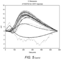
- the raw ISFET sensor voltage output data and Anchored primer spotting map was processed together by a custom data analysis pipeline (Galaxy Runoff pipeline v1.3.15). Voltage signal peak heights from ISFETs were detected and Anchored primer ISFET voltage signal peak heights were subtracted from that of the adjacent upstream blank ISFET. This is done to remove system noise including noise from the fluidic flow which may mask detection. This delta ISFET sensor voltage output from all the specific Anchored primers sensors were averaged to determine signal detection.
- E-gels 48 well 2% agarose gels stained with Ethidium Bromide
- Samples were collected post PCR from the chip surface and 1 uL of the sample was adding to 1x E-gel loading dye before loading into the E-gel wells.
- a ladder was also loaded for size comparison of the amplicon. The gel was run for 10minutes on the E-gel base which provides an electric field for the DNA amplicon to migrate according to its size.
- Anchored primer sequences for Example 1 can be seen in Table 4.
- Table 4 Anchored Primer Sequences for Example 1 Anchored Primer target Positive NH2_iSp18_SmR Anchored Primer Negative NH2_iSp18_SmIntRof NH2_iSp18_EfR NH2_iSp18_EfIntRof Anchored Primer Control NH2_ddl_5'+T15
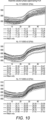
- Average delta signal from ISFETs can be seen in Figure 11 .
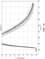
- Delta signals from all ISFETs for Anchored Primer control can be seen in Figure 14 .
- Solution phase amplicon was generated post PCR for Serratia marcescens as seen in the gel electrophoresis image ( Figure 16 ) .
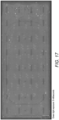
- the On-chip amplicon generated can also be seen for the Positive Anchored primer NH2-iSp18_SmR on the sensospot fluorescent image ( Figure 17 ).
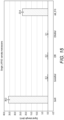
- Average delta signal from ISFETs for Example 2 can be seen in Figure 18 .
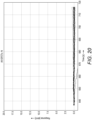
- Delta signals from all ISFETs for Anchored Primer Negative can be seen in Figure 20 .
- ISFET signal results for Positive, Negative and Control primers can be seen in Figure 22 .
- 1.5 to 4.4 dmV ISFET signal was detected for the four Positive Anchored primers. This is much lower than previous signal intensities and the Control Anchored Primer average ISFET signal which was 34.6 dmV.
- Solution phase amplicon was generated post PCR for Schizosaccharomyces pombe as seen in the gel electrophoresis image ( Figure 23 ) .
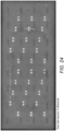
- On-chip amplicon generated cannot be seen for the Positive Anchored primers on the sensospot fluorescent image ( Figure 24 ).
- the Control Anchored primer however can be seen.
Landscapes
- Chemical & Material Sciences (AREA)
- Health & Medical Sciences (AREA)
- Life Sciences & Earth Sciences (AREA)
- Physics & Mathematics (AREA)
- Analytical Chemistry (AREA)
- Organic Chemistry (AREA)
- Immunology (AREA)
- Engineering & Computer Science (AREA)
- Molecular Biology (AREA)
- General Health & Medical Sciences (AREA)
- Biochemistry (AREA)
- Zoology (AREA)
- Proteomics, Peptides & Aminoacids (AREA)
- Wood Science & Technology (AREA)
- Pathology (AREA)
- Chemical Kinetics & Catalysis (AREA)
- General Physics & Mathematics (AREA)
- Genetics & Genomics (AREA)
- Bioinformatics & Cheminformatics (AREA)
- Biotechnology (AREA)
- General Engineering & Computer Science (AREA)
- Biophysics (AREA)
- Microbiology (AREA)
- Optics & Photonics (AREA)
- Nuclear Medicine, Radiotherapy & Molecular Imaging (AREA)
- Spectroscopy & Molecular Physics (AREA)
- Microelectronics & Electronic Packaging (AREA)
- Electrochemistry (AREA)
- Measuring Or Testing Involving Enzymes Or Micro-Organisms (AREA)
- Saccharide Compounds (AREA)
- Apparatus Associated With Microorganisms And Enzymes (AREA)
Applications Claiming Priority (3)
| Application Number | Priority Date | Filing Date | Title |
|---|---|---|---|
| GB201906461 | 2019-05-08 | ||
| PCT/GB2020/051122 WO2020225564A1 (fr) | 2019-05-08 | 2020-05-07 | Amplification d'adn en phase solide et en phase soluble combinée |
| EP20725896.3A EP3966346B1 (fr) | 2019-05-08 | 2020-05-07 | Amplification d'adn en phase solide et en phase soluble combinée |
Related Parent Applications (2)
| Application Number | Title | Priority Date | Filing Date |
|---|---|---|---|
| EP20725896.3A Division EP3966346B1 (fr) | 2019-05-08 | 2020-05-07 | Amplification d'adn en phase solide et en phase soluble combinée |
| EP20725896.3A Division-Into EP3966346B1 (fr) | 2019-05-08 | 2020-05-07 | Amplification d'adn en phase solide et en phase soluble combinée |
Publications (2)
| Publication Number | Publication Date |
|---|---|
| EP4293124A2 true EP4293124A2 (fr) | 2023-12-20 |
| EP4293124A3 EP4293124A3 (fr) | 2024-01-17 |
Family
ID=67002052
Family Applications (2)
| Application Number | Title | Priority Date | Filing Date |
|---|---|---|---|
| EP23185680.8A Pending EP4293124A3 (fr) | 2019-05-08 | 2020-05-07 | Amplification d'adn en phase solide et en phase soluble combinée |
| EP20725896.3A Active EP3966346B1 (fr) | 2019-05-08 | 2020-05-07 | Amplification d'adn en phase solide et en phase soluble combinée |
Family Applications After (1)
| Application Number | Title | Priority Date | Filing Date |
|---|---|---|---|
| EP20725896.3A Active EP3966346B1 (fr) | 2019-05-08 | 2020-05-07 | Amplification d'adn en phase solide et en phase soluble combinée |
Country Status (7)
| Country | Link |
|---|---|
| US (1) | US20220333167A1 (fr) |
| EP (2) | EP4293124A3 (fr) |
| JP (1) | JP2022531707A (fr) |
| CN (1) | CN114207146A (fr) |
| CA (1) | CA3139356A1 (fr) |
| ES (1) | ES2954784T3 (fr) |
| WO (1) | WO2020225564A1 (fr) |
Family Cites Families (4)
| Publication number | Priority date | Publication date | Assignee | Title |
|---|---|---|---|---|
| US7575865B2 (en) * | 2003-01-29 | 2009-08-18 | 454 Life Sciences Corporation | Methods of amplifying and sequencing nucleic acids |
| EP2722398B1 (fr) * | 2012-10-18 | 2017-07-12 | F. Hoffmann-La Roche AG | Analyse de sonde double pour la détection du VHC |
| GB201407334D0 (en) * | 2014-04-25 | 2014-06-11 | Dna Electronics Ltd | Integrated nucleic acid test system, instrument and method |
| WO2018046689A1 (fr) * | 2016-09-08 | 2018-03-15 | Danmarks Tekniske Universitet | Système de puce polymère et ses utilisations |
-
2020
- 2020-05-07 CA CA3139356A patent/CA3139356A1/fr active Pending
- 2020-05-07 CN CN202080034226.7A patent/CN114207146A/zh active Pending
- 2020-05-07 ES ES20725896T patent/ES2954784T3/es active Active
- 2020-05-07 EP EP23185680.8A patent/EP4293124A3/fr active Pending
- 2020-05-07 US US17/609,553 patent/US20220333167A1/en active Pending
- 2020-05-07 JP JP2021565983A patent/JP2022531707A/ja active Pending
- 2020-05-07 WO PCT/GB2020/051122 patent/WO2020225564A1/fr unknown
- 2020-05-07 EP EP20725896.3A patent/EP3966346B1/fr active Active
Also Published As
| Publication number | Publication date |
|---|---|
| CA3139356A1 (fr) | 2020-11-12 |
| EP3966346B1 (fr) | 2023-08-23 |
| WO2020225564A1 (fr) | 2020-11-12 |
| JP2022531707A (ja) | 2022-07-08 |
| CN114207146A (zh) | 2022-03-18 |
| EP3966346A1 (fr) | 2022-03-16 |
| EP4293124A3 (fr) | 2024-01-17 |
| ES2954784T3 (es) | 2023-11-24 |
| US20220333167A1 (en) | 2022-10-20 |
Similar Documents
| Publication | Publication Date | Title |
|---|---|---|
| Leamon et al. | Cramming more sequencing reactions onto microreactor chips | |
| EP1837408A1 (fr) | Procédé de conception d'amources pour la detection d'acides nucléiques et kit | |
| JP2009505651A (ja) | 微生物および抗生物質耐性マーカーの検出の方法およびそのための核酸オリゴヌクレオチド | |
| JPH02503054A (ja) | 核酸配列の増幅および検出 | |
| US9315860B2 (en) | Conjugates of nucleotides and method for the application thereof | |
| JP6126381B2 (ja) | 標的核酸の検出方法及びキット | |
| JP2011510630A (ja) | 試料中に存在する目的ヌクレオチド配列を検出するための核酸プローブの使用 | |
| EP3250711B1 (fr) | Procédé et produit de prévention des faux positifs dans des procédés utilisant des ddntp | |
| JP4256291B2 (ja) | 標的核酸配列の検出方法 | |
| KR102030244B1 (ko) | 뎅기 바이러스 검출용 올리고뉴클레오티드 세트 및 이의 용도 | |
| CA2655518C (fr) | Procede d'analyse d'acides nucleiques amplifies | |
| EP1426448A1 (fr) | Procédé pour réduire les effects des variations de séquence dans un procédé d'hybridisation diagnostique, sonde de l'usage dans un tel procédé, et procédé | |
| US20060252058A1 (en) | Chip for detection of nucleic acid | |
| KR20190021198A (ko) | 대류 유체 디바이스에서의 핵산의 표면-기반 검출 | |
| EP4293124A2 (fr) | Amplification d'adn en phase solide et en phase soluble combinée | |
| CN113840923A (zh) | 用于核酸检测的方法、系统和设备 | |
| JPWO2003102178A1 (ja) | 遺伝子多型のタイピング方法 | |
| JP6339787B2 (ja) | 核酸の分析方法 | |
| US20240141420A1 (en) | Parallel detection and quantification of nucleic acid based markers | |
| US20090275028A1 (en) | Method of detecting target nucleic acid | |
| JP4381457B2 (ja) | 標的核酸配列の検出方法 | |
| JP4397182B2 (ja) | 微量核酸の検出方法 | |
| CN106916882B (zh) | 用于辨识核苷酸基因多型性的基因型鉴定芯片的双重等位基因特异性聚合酶链锁反应的方法 | |
| WO2006070667A1 (fr) | Procede de detection d'une mutation dans le gene egfr et kit de detection | |
| KR101566402B1 (ko) | 마이크로어레이 칩을 이용한 흰반점바이러스 진단용 멀티플렉스 키트 |
Legal Events
| Date | Code | Title | Description |
|---|---|---|---|
| PUAI | Public reference made under article 153(3) epc to a published international application that has entered the european phase |
Free format text: ORIGINAL CODE: 0009012 |
|
| STAA | Information on the status of an ep patent application or granted ep patent |
Free format text: STATUS: THE APPLICATION HAS BEEN PUBLISHED |
|
| PUAL | Search report despatched |
Free format text: ORIGINAL CODE: 0009013 |
|
| AC | Divisional application: reference to earlier application |
Ref document number: 3966346 Country of ref document: EP Kind code of ref document: P |
|
| AK | Designated contracting states |
Kind code of ref document: A2 Designated state(s): AL AT BE BG CH CY CZ DE DK EE ES FI FR GB GR HR HU IE IS IT LI LT LU LV MC MK MT NL NO PL PT RO RS SE SI SK SM TR |
|
| AK | Designated contracting states |
Kind code of ref document: A3 Designated state(s): AL AT BE BG CH CY CZ DE DK EE ES FI FR GB GR HR HU IE IS IT LI LT LU LV MC MK MT NL NO PL PT RO RS SE SI SK SM TR |
|
| RIC1 | Information provided on ipc code assigned before grant |
Ipc: C12Q 1/6825 20180101AFI20231212BHEP |
|
| STAA | Information on the status of an ep patent application or granted ep patent |
Free format text: STATUS: REQUEST FOR EXAMINATION WAS MADE |
|
| 17P | Request for examination filed |
Effective date: 20240202 |
|
| RBV | Designated contracting states (corrected) |
Designated state(s): AL AT BE BG CH CY CZ DE DK EE ES FI FR GB GR HR HU IE IS IT LI LT LU LV MC MK MT NL NO PL PT RO RS SE SI SK SM TR |