EP4269581B1 - Method and kit for detecting target nucleic acid fragment - Google Patents
Method and kit for detecting target nucleic acid fragment Download PDFInfo
- Publication number
- EP4269581B1 EP4269581B1 EP21915214.7A EP21915214A EP4269581B1 EP 4269581 B1 EP4269581 B1 EP 4269581B1 EP 21915214 A EP21915214 A EP 21915214A EP 4269581 B1 EP4269581 B1 EP 4269581B1
- Authority
- EP
- European Patent Office
- Prior art keywords
- well
- nucleic acid
- acid fragment
- crispr
- target nucleic
- Prior art date
- Legal status (The legal status is an assumption and is not a legal conclusion. Google has not performed a legal analysis and makes no representation as to the accuracy of the status listed.)
- Active
Links
Images


Classifications
-
- C—CHEMISTRY; METALLURGY
- C12—BIOCHEMISTRY; BEER; SPIRITS; WINE; VINEGAR; MICROBIOLOGY; ENZYMOLOGY; MUTATION OR GENETIC ENGINEERING
- C12Q—MEASURING OR TESTING PROCESSES INVOLVING ENZYMES, NUCLEIC ACIDS OR MICROORGANISMS; COMPOSITIONS OR TEST PAPERS THEREFOR; PROCESSES OF PREPARING SUCH COMPOSITIONS; CONDITION-RESPONSIVE CONTROL IN MICROBIOLOGICAL OR ENZYMOLOGICAL PROCESSES
- C12Q1/00—Measuring or testing processes involving enzymes, nucleic acids or microorganisms; Compositions therefor; Processes of preparing such compositions
- C12Q1/34—Measuring or testing processes involving enzymes, nucleic acids or microorganisms; Compositions therefor; Processes of preparing such compositions involving hydrolase
-
- C—CHEMISTRY; METALLURGY
- C12—BIOCHEMISTRY; BEER; SPIRITS; WINE; VINEGAR; MICROBIOLOGY; ENZYMOLOGY; MUTATION OR GENETIC ENGINEERING
- C12N—MICROORGANISMS OR ENZYMES; COMPOSITIONS THEREOF; PROPAGATING, PRESERVING, OR MAINTAINING MICROORGANISMS; MUTATION OR GENETIC ENGINEERING; CULTURE MEDIA
- C12N9/00—Enzymes; Proenzymes; Compositions thereof; Processes for preparing, activating, inhibiting, separating or purifying enzymes
- C12N9/14—Hydrolases (3)
- C12N9/16—Hydrolases (3) acting on ester bonds (3.1)
- C12N9/22—Ribonucleases [RNase]; Deoxyribonucleases [DNase]
-
- C—CHEMISTRY; METALLURGY
- C12—BIOCHEMISTRY; BEER; SPIRITS; WINE; VINEGAR; MICROBIOLOGY; ENZYMOLOGY; MUTATION OR GENETIC ENGINEERING
- C12Q—MEASURING OR TESTING PROCESSES INVOLVING ENZYMES, NUCLEIC ACIDS OR MICROORGANISMS; COMPOSITIONS OR TEST PAPERS THEREFOR; PROCESSES OF PREPARING SUCH COMPOSITIONS; CONDITION-RESPONSIVE CONTROL IN MICROBIOLOGICAL OR ENZYMOLOGICAL PROCESSES
- C12Q1/00—Measuring or testing processes involving enzymes, nucleic acids or microorganisms; Compositions therefor; Processes of preparing such compositions
- C12Q1/68—Measuring or testing processes involving enzymes, nucleic acids or microorganisms; Compositions therefor; Processes of preparing such compositions involving nucleic acids
- C12Q1/6813—Hybridisation assays
- C12Q1/6816—Hybridisation assays characterised by the detection means
- C12Q1/682—Signal amplification
-
- C—CHEMISTRY; METALLURGY
- C12—BIOCHEMISTRY; BEER; SPIRITS; WINE; VINEGAR; MICROBIOLOGY; ENZYMOLOGY; MUTATION OR GENETIC ENGINEERING
- C12Q—MEASURING OR TESTING PROCESSES INVOLVING ENZYMES, NUCLEIC ACIDS OR MICROORGANISMS; COMPOSITIONS OR TEST PAPERS THEREFOR; PROCESSES OF PREPARING SUCH COMPOSITIONS; CONDITION-RESPONSIVE CONTROL IN MICROBIOLOGICAL OR ENZYMOLOGICAL PROCESSES
- C12Q1/00—Measuring or testing processes involving enzymes, nucleic acids or microorganisms; Compositions therefor; Processes of preparing such compositions
- C12Q1/68—Measuring or testing processes involving enzymes, nucleic acids or microorganisms; Compositions therefor; Processes of preparing such compositions involving nucleic acids
- C12Q1/6813—Hybridisation assays
- C12Q1/6816—Hybridisation assays characterised by the detection means
- C12Q1/6825—Nucleic acid detection involving sensors
-
- C—CHEMISTRY; METALLURGY
- C12—BIOCHEMISTRY; BEER; SPIRITS; WINE; VINEGAR; MICROBIOLOGY; ENZYMOLOGY; MUTATION OR GENETIC ENGINEERING
- C12Q—MEASURING OR TESTING PROCESSES INVOLVING ENZYMES, NUCLEIC ACIDS OR MICROORGANISMS; COMPOSITIONS OR TEST PAPERS THEREFOR; PROCESSES OF PREPARING SUCH COMPOSITIONS; CONDITION-RESPONSIVE CONTROL IN MICROBIOLOGICAL OR ENZYMOLOGICAL PROCESSES
- C12Q1/00—Measuring or testing processes involving enzymes, nucleic acids or microorganisms; Compositions therefor; Processes of preparing such compositions
- C12Q1/68—Measuring or testing processes involving enzymes, nucleic acids or microorganisms; Compositions therefor; Processes of preparing such compositions involving nucleic acids
- C12Q1/6813—Hybridisation assays
- C12Q1/6834—Enzymatic or biochemical coupling of nucleic acids to a solid phase
- C12Q1/6837—Enzymatic or biochemical coupling of nucleic acids to a solid phase using probe arrays or probe chips
-
- C—CHEMISTRY; METALLURGY
- C12—BIOCHEMISTRY; BEER; SPIRITS; WINE; VINEGAR; MICROBIOLOGY; ENZYMOLOGY; MUTATION OR GENETIC ENGINEERING
- C12Q—MEASURING OR TESTING PROCESSES INVOLVING ENZYMES, NUCLEIC ACIDS OR MICROORGANISMS; COMPOSITIONS OR TEST PAPERS THEREFOR; PROCESSES OF PREPARING SUCH COMPOSITIONS; CONDITION-RESPONSIVE CONTROL IN MICROBIOLOGICAL OR ENZYMOLOGICAL PROCESSES
- C12Q1/00—Measuring or testing processes involving enzymes, nucleic acids or microorganisms; Compositions therefor; Processes of preparing such compositions
- C12Q1/68—Measuring or testing processes involving enzymes, nucleic acids or microorganisms; Compositions therefor; Processes of preparing such compositions involving nucleic acids
- C12Q1/6876—Nucleic acid products used in the analysis of nucleic acids, e.g. primers or probes
-
- G—PHYSICS
- G01—MEASURING; TESTING
- G01N—INVESTIGATING OR ANALYSING MATERIALS BY DETERMINING THEIR CHEMICAL OR PHYSICAL PROPERTIES
- G01N33/00—Investigating or analysing materials by specific methods not covered by groups G01N1/00 - G01N31/00
- G01N33/48—Biological material, e.g. blood, urine; Haemocytometers
- G01N33/50—Chemical analysis of biological material, e.g. blood, urine; Testing involving biospecific ligand binding methods; Immunological testing
- G01N33/53—Immunoassay; Biospecific binding assay; Materials therefor
- G01N33/536—Immunoassay; Biospecific binding assay; Materials therefor with immune complex formed in liquid phase
- G01N33/542—Immunoassay; Biospecific binding assay; Materials therefor with immune complex formed in liquid phase with steric inhibition or signal modification, e.g. fluorescent quenching
-
- G—PHYSICS
- G01—MEASURING; TESTING
- G01N—INVESTIGATING OR ANALYSING MATERIALS BY DETERMINING THEIR CHEMICAL OR PHYSICAL PROPERTIES
- G01N33/00—Investigating or analysing materials by specific methods not covered by groups G01N1/00 - G01N31/00
- G01N33/48—Biological material, e.g. blood, urine; Haemocytometers
- G01N33/50—Chemical analysis of biological material, e.g. blood, urine; Testing involving biospecific ligand binding methods; Immunological testing
- G01N33/53—Immunoassay; Biospecific binding assay; Materials therefor
- G01N33/543—Immunoassay; Biospecific binding assay; Materials therefor with an insoluble carrier for immobilising immunochemicals
- G01N33/54306—Solid-phase reaction mechanisms
-
- C—CHEMISTRY; METALLURGY
- C12—BIOCHEMISTRY; BEER; SPIRITS; WINE; VINEGAR; MICROBIOLOGY; ENZYMOLOGY; MUTATION OR GENETIC ENGINEERING
- C12N—MICROORGANISMS OR ENZYMES; COMPOSITIONS THEREOF; PROPAGATING, PRESERVING, OR MAINTAINING MICROORGANISMS; MUTATION OR GENETIC ENGINEERING; CULTURE MEDIA
- C12N11/00—Carrier-bound or immobilised enzymes; Carrier-bound or immobilised microbial cells; Preparation thereof
- C12N11/14—Enzymes or microbial cells immobilised on or in an inorganic carrier
-
- C—CHEMISTRY; METALLURGY
- C12—BIOCHEMISTRY; BEER; SPIRITS; WINE; VINEGAR; MICROBIOLOGY; ENZYMOLOGY; MUTATION OR GENETIC ENGINEERING
- C12N—MICROORGANISMS OR ENZYMES; COMPOSITIONS THEREOF; PROPAGATING, PRESERVING, OR MAINTAINING MICROORGANISMS; MUTATION OR GENETIC ENGINEERING; CULTURE MEDIA
- C12N2310/00—Structure or type of the nucleic acid
- C12N2310/10—Type of nucleic acid
- C12N2310/20—Type of nucleic acid involving clustered regularly interspaced short palindromic repeats [CRISPR]
-
- C—CHEMISTRY; METALLURGY
- C12—BIOCHEMISTRY; BEER; SPIRITS; WINE; VINEGAR; MICROBIOLOGY; ENZYMOLOGY; MUTATION OR GENETIC ENGINEERING
- C12Q—MEASURING OR TESTING PROCESSES INVOLVING ENZYMES, NUCLEIC ACIDS OR MICROORGANISMS; COMPOSITIONS OR TEST PAPERS THEREFOR; PROCESSES OF PREPARING SUCH COMPOSITIONS; CONDITION-RESPONSIVE CONTROL IN MICROBIOLOGICAL OR ENZYMOLOGICAL PROCESSES
- C12Q2521/00—Reaction characterised by the enzymatic activity
- C12Q2521/30—Phosphoric diester hydrolysing, i.e. nuclease
Definitions
- the present invention relates to a method and a kit for detecting a target nucleic acid fragment.
- Priority is claimed on Japanese Patent Application No. 2020-219481 filed December 28, 2020 in Japan.
- a technology of detecting a target nucleic acid fragment in a sample with high sensitivity is required.
- blood contains free DNA (cell-free DNA, cfDNA) released from cells due to cell death.
- Blood tumor DNA circulating tumor DNA, ctDNA
- ctDNA circulating tumor DNA, which is DNA derived from cancer cells, is also contained in the cfDNA of cancer patients.
- exosomes various cells secrete membrane vesicles referred to as exosomes, and exosomes are included in biological samples such as saliva, blood, urine, amniotic fluid, and malignant ascites, or in a supernatant of cultured cells. Exosomes contain various proteins, lipids, microRNAs, DNAs, and the like, derived from cells that secrete thereof.
- cfDNA cfDNA
- miRNA microRNA
- membrane vesicles such as exosomes, DNA, or the like
- a technology of detecting a target nucleic acid fragment in a sample with high sensitivity can be applied to such fields as an example.
- Non-Patent Documents 1 to 3 report methods for detecting a target nucleic acid fragment with high sensitivity using such activities of the Cas12 and the Cas13.
- Patent Document 1 discloses detection of enzymatic activity at a single-molecule level using a microchamber of a femtoliter order size
- Bruch et al. describe a CRISPR/Cas13a-powered electrochemical microfluidic biosensor for nucleic acid amplification-free miRNA diagnostics
- WO2020/102608 describes methods for generating primers and/or probes for use in analyzing a sample which may comprise a pathogen target sequence, including pan-viral sets of primers and/or probes.
- Patent Document 1 Japanese Unexamined Patent Application, First Publication No. 2004-309405
- an object of the present invention is to provide a technology capable of detecting a target nucleic acid fragment with high sensitivity without amplification.
- the present description includes the following aspects.
- the present invention it is possible to provide a technology of capable of performing detection with high sensitivity without amplifying a target nucleic acid fragment.
- the present description provides a method for detecting a target nucleic acid fragment in a sample, including step (a) of contacting the sample with a gRNA complementary to the target nucleic acid fragment, a CRISPR/Cas family protein, and a substrate nucleic acid fragment; and a step (b) of irradiating the fluorescent substance with the excitation light to detect the fluorescent light, in which detection of the fluorescent light indicates the presence of the target nucleic acid fragment in the sample.
- the CRISPR/Cas family protein expresses nuclease activity after forming a three-part complex with the gRNA and the target nucleic acid fragment.
- the CRISPR/Cas family protein is immobilized on a solid phase.
- the substrate nucleic acid fragment is labeled with a fluorescent substance and a quencher, and in a case where the fluorescent substance cleaved by the nuclease activity of the three-part complex is separated from the quencher, fluorescent light is emitted by irradiation with excitation light.
- the contact of the sample, the gRNA, the CRISPR/Cas family protein, and the substrate nucleic acid fragment is performed in a reaction space having a volume of 10 aL to 100 pL.
- the three-part complex is formed in a case where the target nucleic acid fragment is present in the sample, the substrate nucleic acid fragment is cleaved, and the fluorescent substance is separated from the quencher.
- step (b) fluorescent light is detected in a case where the fluorescent substance is irradiated with excitation light.
- FIGS. 1(a) and 1(b) are schematic diagrams showing the method of the present aspect.
- FIGS. 1(a) and 1(b) show an example of a case where the CRISPR/Cas family protein is a Cas12a protein.
- step (a) in a case where a Cas12a protein 110 and a gRNA 120 are brought into contact with each other, these bind to form a two-part complex 130.
- the gRNA 120 partially has a base sequence complementary to a target nucleic acid fragment 140.
- the Cas12a protein 110 does not express nuclease activity, a substrate nucleic acid fragment 150 is not cleaved.
- the substrate nucleic acid fragment 150 is a single-stranded DNA fragment labeled with a fluorescent substance F and a quencher Q. Even if the substrate nucleic acid fragment 150 is irradiated with excitation light, fluorescent light is not generated.
- FIG. 1(b) is a schematic diagram showing a three-part complex 100' in which the target site of the target nucleic acid fragment 140 is cleaved.
- the three-part complex 100' expresses nuclease activity.
- the substrate nucleic acid fragment 150 existing around the three-part complex 100' is cleaved.
- the fluorescent substance F of the substrate nucleic acid fragment 150 is separated from the quencher Q.
- the fluorescent substance F separated from the quencher Q emits fluorescent light by irradiation with the excitation light.
- step (b) the fluorescent substance F is irradiated with excitation light and fluorescent light is detected.
- fluorescent light it can be determined that the target nucleic acid fragment 140 was present in the sample.
- the CRISPR/Cas family protein is immobilized on a solid phase.
- the detection sensitivity of the target nucleic acid fragment is significantly improved.
- the CRISPR/Cas family protein for example, may be immobilized on a surface of a particle, or may be immobilized on an inner surface of the well, which is the reaction space. Details will be described later.
- the sample, the gRNA 120, the CRISPR/Cas family protein 110, and the substrate nucleic acid fragment 150 may be mixed and contacted in any order.
- the gRNA 120 and the CRISPR/Cas family protein 110 may be brought into contact with each other to form the two-part complex 130 in advance, and then the sample may be brought into contact therewith.
- the target nucleic acid fragment 140 in a case where the target nucleic acid fragment 140 is present in the sample, the target nucleic acid fragment 140 binds to the two-part complex 130 to form a three-part complex 100.
- the substrate nucleic acid fragment 150 may be brought into contact therewith.
- the target nucleic acid fragment 140 and the substrate nucleic acid fragment 150 may be brought into contact simultaneously.
- the gRNA 120, the CRISPR/Cas family protein 110, and the sample may be brought into contact simultaneously. Even in this case, in a case where the target nucleic acid fragment 140 is present in the sample, the three-part complex 100 is finally formed. After that, the substrate nucleic acid fragment 150 may be brought into contact therewith.
- the sample, the gRNA 120, the CRISPR/Cas family protein 110, and the substrate nucleic acid fragment 150 may be brought into contact therewith simultaneously. Even in this case, in a case where the target nucleic acid fragment 140 is present in the sample, the three-part complex 100 is finally formed, and in the three-part complex 100, in a case where a target site of the target nucleic acid fragment 140 is cleaved, the three-part complex 100 is converted into the three-part complex 100', and expresses nuclease activity, and the substrate nucleic acid fragment 150 is cleaved.
- a biological sample containing contaminants can be used as a sample, and step (a) can be performed without purifying the target nucleic acid fragment from the biological sample.
- the target nucleic acid fragment can be detected almost unaffected by the contaminants.
- the CRISPR/Cas family protein is immobilized on a solid phase.
- the CRISPR/Cas family protein may be immobilized on a surface of a particle.
- a step of mixing the sample, the gRNA, and the CRISPR/Cas family protein in a container to form a three-part complex containing the CRISPR/Cas family protein, the gRNA, and the target nucleic acid fragment on the particle may be further included.
- the CRISPR/Cas family protein may be immobilized in a state of being in contact with the particle, or the CRISPR/Cas family protein may be immobilized on the particle via a linker
- the particle is not particularly limited, and examples thereof include a resin bead, a glass bead, and the like.
- the resin include polyethylene, polypropylene, polystyrene, polycarbonate, cyclic polyolefin, acryl, and the like.
- the particle may be a magnetic bead. If the particle is a magnetic bead, the particle can be collected using a magnetic stand and the like.
- the size of the particle can be appropriately selected, and for example, a particle having a diameter of about 0.1 to 100 ⁇ m can be used.
- the particle may be colored with a fluorescent pigment and the like.
- a gRNA having a specific base sequence binds to the CRISPR/Cas family protein immobilized on the particle colored with a specific dye. With this, it is possible to recognize the base sequence of the target nucleic acid fragment makes it possible to identify a base sequence of the target nucleic acid fragment by the CRISPR/Cas family protein immobilized on the particle, based on the color of the particle.
- Examples of a method of binding particles to the CRISPR/Cas family protein include physical adsorption, a method of performing covalent bond of a functional group present on the surface of the particle and a functional group present on the surface of the CRISPR/Cas family protein using a chemical linker, a method of using hybridization of single-stranded nucleic acid fragments, a method of using avidin-biotin bond, and the like. A combination of these methods may also be used.
- Examples of the functional groups in a case of using a chemical linker include hydroxyl group, amino group, thiol group, and the like.
- the CRISPR/Cas family protein may be immobilized on the surface of the particle using a click reaction using an azide group and an alkyne group and the like.
- avidin-biotin bond it is possible to bind the particle to the CRISPR/Cas family protein by contacting surface-biotinylated particle, avidin, and the CRISPR/Cas family protein biotinylated using a chemical linker.
- FIGS. 21 and 22 are schematic diagrams showing a method of binding the particle to the CRISPR/Cas family protein using hybridization of single-stranded nucleic acid fragments.
- FIG. 21 is a schematic diagram showing one aspect of a method of binding the particle to the CRISPR/Cas family protein.
- a single-stranded DNA fragment 2120 binds to a particle 2110.
- An end of the single-stranded DNA fragment 2120 is modified with biotin.
- a surface of the particle 2110 is coated with avidin. Therefore, in a case where the single-stranded DNA fragment 2120 is contacted with the particle 2110, the single-stranded DNA fragment 2120 binds to the surface of the particle 2110 by avidin-biotin bond.
- the single-stranded DNA fragment 2120 has a base sequence complementary to a part of a target nucleic acid fragment 140. Therefore, in a case where the particle 2110 to which the single-stranded DNA fragment 2120 binds, a sample containing the target nucleic acid fragment 140, the gRNA, and the CRISPR/Cas family protein are mixed in a container, the three-part complex 100' containing the CRISPR/Cas family protein, the gRNA, and the target nucleic acid fragment 140 are immobilized on the particle 2110 by hybridizing a part of the target nucleic acid fragment 140 with the single-stranded DNA fragment 2120.
- FIGS. 22(a) and 22(b) are schematic diagrams showing another aspect for binding the particle to the CRISPR/Cas family protein.
- the particle shown in FIG. 22(b) is different mainly in a point that the three-part complex 100' is immobilized on the particle 2110 by hybridizing a part of the target nucleic acid fragment 140, the single-stranded DNA fragment 2121, and the single-stranded DNA fragment 2120.
- FIG. 22(a) shows a state in which the number of molecules of the three-part complex formed on one molecule of the target nucleic acid fragment is increased by using a plurality of types of gRNAs for one type of the target nucleic acid fragment. As described above, with this, it is possible to increase the number of molecules of the three-part complex per molecule of the target nucleic acid fragment, and thus it is possible to further enhance detection sensitivity of the target nucleic acid fragment.
- the three-part complex 100' that recognizes each of target nucleic acid fragments (target sequences) 140, 140', and 140" binds to one molecule of a long nucleic acid fragment.
- single-stranded nucleic acid fragments 2121, 2122, and 2123 having base sequences complementary thereto are hybridized in the vicinity of each of target nucleic acid fragments (target sequences) 140, 140', and 140".
- regions of the single-stranded nucleic acid fragments 2121, 2122, and 2123 that do not hybridize with long nucleic acid fragments have base sequences complementary to single-stranded nucleic acid fragment 2120 binding to the particle 2110.
- regions of the single-stranded nucleic acid fragments 2121, 2122, and 2123 hybridize with the long nucleic acid fragments are protected from the nuclease activity of the three-part complex 100', and thus are not cleaved.
- the long nucleic acid fragment is cleaved (trans cleavage) by the nuclease activity of the three-part complex 100'.
- each of a complex of the three-part complex 100' and the single-stranded nucleic acid fragment 2121, a complex of the three-part complex 100' and the single-stranded nucleic acid fragment 2122, and a complex of the three-part complex 100' and the single-stranded nucleic acid fragment 2123 is excised from the long nucleic acid fragment.
- the complex of the excised three-part complex 100' and the single-stranded nucleic acid fragment 2121 is hybridized with the single-stranded DNA fragment 2120 binding to the particle 2110, and thus is immobilized on the particle 2110.
- the complex of the three-part complex 100' and the single-stranded nucleic acid fragment 2122 and the complex of the three-part complex 100' and the single-stranded nucleic acid fragment 2123 are also similarly hybridized with the single-stranded DNA fragment 2120, and thus is immobilized on the particle 2110.
- the CRISPR/Cas family protein can be immobilized on the particles 2110.
- the order of mixing the particles 2110, the single-stranded nucleic acid fragment 2120, a sample containing the target nucleic acid fragment 140, the gRNA, the CRISPR/Cas family protein, and the single-stranded nucleic acid fragments 2121, 2122, and 2123 is not limited.
- the single-stranded nucleic acid fragment 2120 may be mixed in the sample containing the target nucleic acid fragment 140, this may be hybridized, and then the gRNA and the CRISPR/Cas family protein may be mixed therein, and finally particles 2110 may be mixed therein.
- the gRNA and the CRISPR/Cas family protein may form a two-part complex in advance.
- the sample containing the target nucleic acid fragment 140, the gRNA, and the CRISPR/Cas family protein may be mixed to form a three-part complex, and then the particles 2110 to which the single-stranded DNA fragment 2120 binds may be mixed therein.
- the single-stranded nucleic acid fragments 2121, 2122, and 2123 may be mixed in a sample containing a long target nucleic acid, this may be hybridized, and then the gRNA and the CRISPR/Cas family protein may be mixed therein, and subsequently the particles 2110 to which the single-stranded DNA fragments 2120 bind may be mixed therein.
- the single-stranded nucleic acid fragments 2120, 2121, 2122, 2123 may be mixed in a sample containing a long target nucleic acid, this may be hybridized, the gRNA and the CRISPR/Cas family protein may be mixed, and finally particles 2110 may be mixed therein.
- the gRNA and the CRISPR/Cas family protein may form a two-part complex in advance.
- the particle 2110 is a magnetic bead
- the particle can be collected using a magnetic stand and the like. In addition, it is also easy to wash the particle.
- a container for mixing the sample, the gRNA, the CRISPR/Cas family protein, and the like is not particularly limited, and for example, a plastic tube, a well array, and the like can be used.
- the present method by immobilizing the CRISPR/Cas family protein on a solid phase, it is possible to efficiently bind the target nucleic acid fragment present at a low concentration in the sample to the CRISPR/Cas family protein and to form a three-part complex. Subsequently, the particle may be distributed into a minute reaction space and contacted with the substrate nucleic acid fragment.
- the detection sensitivity of the target nucleic acid fragment is significantly improved. That is, by immobilizing the CRISPR/Cas family protein on the solid phase, it is possible to concentrate the target nucleic acid fragment in the sample.
- the sample is not particularly limited and can be appropriately selected depending on the purpose, and examples thereof include biological samples such as saliva, blood, urine, amniotic fluid, and malignant ascites, throat swab, and nasal swab, or a supernatant of cultured cells.
- target nucleic acid fragment in a sample examples include viral genomes, cfDNA, ctDNA, microRNA, exosome-derived DNA, and the like.
- a nucleic acid fragment containing an oncogene hotspot region as a target nucleic acid fragment, it is possible to detect oncogene mutations contained in a sample.
- the CRISPR/Cas family protein is Cas12 protein
- the CRISPR/Cas family protein is Cas13 protein
- a guide RNA is not particularly limited as long as the guide RNA can be used for the CRISPR/Cas family protein, the guide RNA may be a complex of CRISPR RNA (crRNA) and transactivating CRISPR RNA (tracrRNA), may be a single gRNA (sgRNA) obtained by combining tracrRNA and crRNA, or may be crRNA alone.
- crRNA CRISPR RNA
- tracrRNA transactivating CRISPR RNA
- sgRNA single gRNA obtained by combining tracrRNA and crRNA, or may be crRNA alone.
- the crRNA can be, for example, the following base sequence.
- a base sequence obtained by removing a protospacer adjacent motif (PAM) sequence from a target base sequence is defined as a spacer base sequence.
- a base sequence obtained by linking a scaffold sequence to a 3' end of the spacer base sequence is provided, and a complementary strand thereof is used as the base sequence of crRNA.
- the crRNA can be, for example, the following base sequence.
- a base sequence obtained by linking a scaffold sequence to a 3' end of the base sequence complementary to the target base sequence is provided, and the complementary strand thereof is used as the base sequence of crRNA.
- the base sequence of crRNA for Cas13a protein can be "5'-GAUUUAGACUACCCCAAAAACGAAGGGGACUAAAACUAGAUUGCUGUUCU ACCAAGUAAUCCAU-3'" (SEQ ID NO: 4).
- a target base sequence is a partial base sequence of the base sequence of the target nucleic acid fragment. At least one type of the target base sequence is set in one type of the target nucleic acid fragment. By setting a plurality of types of target base sequences in one type of target nucleic acid fragment, it is possible to form a three-part complex of a plurality of molecules on one molecule of the target nucleic acid fragment.
- the target nucleic acid fragment by using a plurality of types of gRNAs for one type of the target nucleic acid fragment, it is possible to increase the number of molecules of the three-part complex formed on one molecule of the target nucleic acid fragment. As a result, it becomes possible to increase the number of molecules of the three-part complex expressing nuclease activity per molecule of the target nucleic acid fragment, and to further increase the detection sensitivity of the target nucleic acid fragment.
- the present description provides the above-described method of detecting a target nucleic acid fragment to increase the detection sensitivity, including a step of forming a plurality of molecules of a three-part complex on one molecule of a target nucleic acid fragment by using a plurality of types of gRNAs.
- any CRISPR/Cas family protein can be used as long as the CRISPR/Cas family protein can express nuclease activity after forming a three-part complex with a gRNA and a target nucleic acid fragment.
- the CRISPR/Cas family protein can express nuclease activity after forming a three-part complex with a gRNA and a target nucleic acid fragment.
- a three-part complex is formed, a CRISPR/Cas family protein cleaves a target nucleic acid fragment, and then nuclease activity is expressed.
- Examples of such a CRISPR/Cas family protein include the Cas12 protein, the Cas13 protein, and the like.
- the Cas12 protein and Cas13 protein may be Cas12 protein, Cas13 protein, orthologs of these proteins, variants of these proteins, and the like.
- CRISPR/Cas family protein More specific examples of the CRISPR/Cas family protein that can be used in the method of the present aspect include Lachnospiraceae bacterium ND2006-derived Cas12a protein (LbCas12a, UniProtKB Accession Number: A0A182DWE3), Acidaminococcus sp.-derived Cas12a protein (AsCas12a, UniProtKB Accession Number: U2UMQ6), Francisella tularensis subsp.
- LbCas12a Lachnospiraceae bacterium ND2006-derived Cas12a protein
- A0A182DWE3 Acidaminococcus sp.-derived Cas12a protein
- AsCas12a UniProtKB Accession Number: U2UMQ6
- Francisella tularensis subsp Francisella tularensis subsp.
- P5-125-derived Cas13b protein PspCas13b, NCBI Accession Number: WP_044065294
- Porphyromonas gingivalis-derived Cas13b protein Porphyromonas gingivalis-derived Cas13b protein
- Prevotella intermedia-derived Cas13b protein Pin3Cas13b, NCBI Accession Number: WP_050955369
- Enterococcus italicus-derived Csm6 protein EiCsm6, NCBI Accession Number: WP_007208953.1
- Lactobacillus salivarius-derived Csm6 protein LsCsm6, NCBI Accession Number: WP_081509150.1
- Thermus thermophilus-derived Csm6 protein TtCsm6, NCBI Accession Number: WP_011229148.1
- the CRISPR/Cas family protein may be a mutant of the above-described Cas family protein.
- a mutant for example, a mutant with increased nuclease activity after forming a three-part complex and the like can be used.
- the substrate nucleic acid fragment is labeled with a fluorescent substance and a quencher, and in a case where the fluorescent substance cleaved by the nuclease activity of the three-part complex is separated from the quencher, fluorescent light is emitted by irradiation with excitation light.
- the substrate nucleic acid fragment may be appropriately selected according to substrate specificity of the used CRISPR/Cas family protein.
- the Cas12 protein cleaves a single-stranded DNA as a substrate. Therefore, in a case of using the Cas12 protein, a single-stranded DNA may be used as the substrate nucleic acid fragment.
- the Cas13 protein cleaves a single-stranded RNA as a substrate. Therefore, in a case of using the Cas13 protein, a single-stranded RNA may be used as the substrate nucleic acid fragment.
- combination of the fluorescent substance and the quencher a combination that can quench the fluorescent light of the fluorescent substance in a case of being brought close to each other is used.
- a combination that can quench the fluorescent light of the fluorescent substance in a case of being brought close to each other is used.
- FAM fluorescent substance
- HEX HEX
- TAMRA TAMRA
- each of a LwaCas13a protein, a CcaCas13b protein, a LbaCas13a protein, and a PsmCas13b protein is reported to recognize and cleave AU, UC, AC, and GA base sequences in substrate nucleic acid fragments.
- each different gRNA is combined with each CRISPR/Cas family protein, as a gRNA, and by using a single-stranded RNA containing AU, UC, AC, and GA base sequences, as a substrate nucleic acid fragment, and further labeling each substrate nucleic acid fragment with different fluorescent pigments identifiable with each other, it is possible to detect four types of target nucleic acid fragments in one reaction space. That is, it is possible to perform multicolor detection.
- the method of the present aspect is preferably performed by digital measurement. More specifically, contact between the sample, the CRISPR/Cas family protein, the gRNA, and the substrate nucleic acid fragment is performed by dividing the contact of the substrate nucleic acid fragment into minute reaction spaces.
- the volume per reaction space is 10 aL to 100 pL, for example, may be 10 aL to 10 pL, for example, may be 10 aL to 1 pL, for example, may be 10 aL to 100 fL, and for example, may be 10 aL to 10 fL.
- the reaction space may be a droplet, for example.
- the reaction space may be a well formed on a substrate.
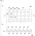
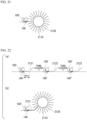
- FIG. 2(a) is a top view showing an example of a fluid device having a substrate on which wells each having a volume of 10aL to 100pL per well are formed.
- FIG. 2(b) is a cross-sectional view taken along line b-b' in an arrow direction of FIG. 2(a) .
- a fluid device 200 includes a substrate 210 on which a well 211 having a volume of 10 aL to 100 pL is formed, a spacer 220, and a lid member 230 having a liquid inlet 231 formed thereon.
- a plurality of the wells 211 are present to form a well array 212.
- a space between the substrate 210 and the lid member 230 functions as a flow path through which a sample, a gRNA, a CRISPR/Cas family protein, a substrate nucleic acid fragment, and the like flow.
- a shape of the well is not particularly limited as long as the volume is within the range described above, and for example, may be cylindrical, polyhedral formed by a plurality of faces (for example, rectangular parallelepiped, hexagonal, octagonal, and the like), and the like.
- the plurality of the wells 211 of the same shape and size form a well array 212.
- “same shape and same size” may be the same shape and the same capacity to the extent required for performing digital measurement, and variations to the extent of manufacturing error are allowed.
- a sealant 320 is introduced through the liquid inlet 231.
- an organic solvent containing a lipid 321 is used as the sealant 320.
- the lipid 32 natural lipids derived from soybeans, E. coli, and the like and artificial lipids such as dioleoylphosphatidylethanolamine (DOPE) and dioleoylphosphatidylglycerol (DOPG) can be used.
- DOPE dioleoylphosphatidylethanolamine
- DOPG dioleoylphosphatidylglycerol
- an organic solvent in this case hexadecane or chloroform can be used.
- each well 211 becomes an independent reaction space.
- the well array 212 is irradiated with excitation light to measure the fluorescent light.
- a lipid membrane can be further laminated on the first lipid membrane 322 to form a lipid bilayer membrane.
- a membrane-forming aqueous solution 330 for forming a lipid bilayer membrane 324 is introduced through the liquid inlet 231.
- a 10 mM pH buffer solution (pH 5 to 9) for example, a 10 mM sodium chloride aqueous solution, and the like can be used.
- a second lipid membrane 323 is laminated on the first lipid membrane 322 to form a lipid bilayer membrane 324.
- each well 211 becomes an independent reaction space. In this state, the well array 212 is irradiated with excitation light to measure the fluorescent light.
- lipid vesicles can be fused to the lipid bilayer membrane 324.
- the content of the exosomes can be released inside the well 211.
- the gRNA, the CRISPR/Cas family protein, and the substrate nucleic acid fragment may be first sealed to seal the opening portion of the well 211 with the lipid bilayer membrane 324, and then exosomes may be contacted with the lipid bilayer membrane 324 as a sample.
- the exosomes are fused to the lipid bilayer membrane 324 and the content of the exosomes is released inside the well 211.
- the target nucleic acid fragment is present in the content of the exosomes, a three-part complex is formed inside the well 211, the substrate nucleic acid fragment is cleaved, and fluorescent light is detected by irradiation with excitation light.
- the CRISPR/Cas family protein is immobilized on a solid phase.
- the CRISPR/Cas family protein may be immobilized on the inner surface of the well.
- the CRISPR/Cas family protein 110 may be immobilized on the inner surface of well 211 in advance.
- the CRISPR/Cas family protein 110 may bind to the gRNA 120 to form a two-part complex 130.
- Examples of a method of immobilizing the CRISPR/Cas family protein on the inner surface of the well 211 include physical adsorption, a method of performing covalent bond of a functional group present on the inner surface of the well 211 and a functional group present on the surface of the CRISPR/Cas family protein using a chemical linker, a method of using avidin-biotin bond, and the like.
- Examples of the functional groups in a case of using a chemical linker include hydroxyl group, amino group, thiol group, and the like.
- the CRISPR/Cas family protein may be immobilized on the inner surface of the well 211 using a click reaction and the like using an azide group and an alkyne group.
- the inner surface of the well 211 is biotinylated in advance, and avidin is contacted with the biotinylated CRISPR/Cas family protein using a chemical linker so that the CRISPR/Cas family protein can bind to the inner surface of the well 211.
- gRNA 120 may be immobilized on the inner surface of well 211.
- gRNA 120 may be added with an additional sequence that functions as a linker.
- the CRISPR/Cas family protein 110 binds to the gRNA 120 to form the two-part complex 130, and is immobilized on the inner surface of the well 211.
- a well on which a gRNA is immobilized is easier to store than a well on which a protein is immobilized.
- step (a) may be performed in each well of the well array.
- the reaction space in which the sample, the gRNA, the CRISPR/Cas family protein, and the substrate nucleic acid fragment are brought into contact may be a space inside each well.
- the CRISPR/Cas family protein may be immobilized on the inner surface of each well.
- Step (a) may have the following steps (a1) and (a2).
- step (a1) in a case where the CRISPR/Cas family protein has formed a two-part complex with the gRNA in advance, a sample and a substrate nucleic acid fragment are introduced into each well of the well array.
- a sample, a gRNA, and a substrate nucleic acid fragment are introduced into each well of the well array.
- each well of the well array is sealed with a sealing liquid.
- the sealing liquid will be described later.
- the three-part complex is formed in a case where the target nucleic acid fragment is present in the well, the substrate nucleic acid fragment is cleaved, and the fluorescent substance is separated from the quencher.
- step (b) the fluorescent substance is irradiated with excitation light.
- the fluorescent light is detected, it can be determined that the target nucleic acid fragment is present in the sample.
- step (a) may be performed in each well of the well array.
- Each well may have a first well and a second well arranged at a bottom of the first well and having a smaller capacity than the first well.
- the reaction space in which the sample, the gRNA, the CRISPR/Cas family protein, and the substrate nucleic acid fragment are brought into contact may be a space inside the second well.
- the CRISPR/Cas family protein may be immobilized on the inner surface of the second well.
- Step (a) may have the following steps (a1'), (a2'), and (a3').
- step (a1') in a case where the CRISPR/Cas family protein has formed a two-part complex with the gRNA in advance, a sample and a substrate nucleic acid fragment are introduced into each well of the well array.
- a sample, a gRNA, and a substrate nucleic acid fragment are introduced into each well of the well array.
- each well of the well array has a first well and a second well arranged at a bottom of the first well and having a smaller capacity than the first well, and in a case where the volume of the content of the first well becomes small, the content may accumulate in the second well.
- the volume of the first well 520 may be 1 to 1,000 pL and the volume of the second well 530 may be 0.1 to 1,000 fL.
- a shape of the first well 520 and the second well 530 is not particularly limited, and for example, may be cylindrical, polyhedral formed by a plurality of faces (for example, rectangular parallelepiped, hexagonal, octagonal, and the like), and the like.
- An example of a method of manufacturing a well array having a first well and a second well is described using a well array 600 as an example.
- Examples of the material for the substrate 510 include glass, resin, and the like.
- Examples of the resin include polyethylene, polypropylene, polystyrene, polycarbonate, cyclic polyolefin, acryl, and the like.
- polycarbonate is also used as a material for CDs and DVDs, which can be mass-produced at a low cost, and is suitable from a viewpoint of manufacturing a well array at a low cost.
- the inventors have clarified that in a case of using polycarbonate as a material of the substrate 510, a refractive index of light is close to that of glass, and thus polycarbonate is preferable when detecting fluorescent light with a microscope.
- Examples of the material for the film 700 include fluorine resin, cyclic polyolefin, silicone resin, and the like.
- a resist film 710 is laminated on a surface of the film 700.
- the resist film 710 is exposed by irradiation with active energy rays with an exposure machine.
- development is performed with a developing solution to remove a portion of the resist film 710 forming the well, as shown in FIG. 4(d) .
- the film 700 masked with the resist film 710 is etched to form a second well 530 in the film 700.
- the substrate is washed to remove the resist film 710, and thereby an array of the wells 530 is obtained.
- an array of the wells 530 is obtained. That is, the array of minute wells manufactured by the steps up to this point can also be used for detection of the target nucleic acid fragment.
- the well array of the first wells is stacked on the well array of the second wells by the following steps.
- the resist film 710 is laminated again on the array of the wells 530 obtained in FIG. 7(f) .
- a sheet-type resist can be preferably used as the resist film 710.
- FIG. 8(b) using a well array-pattern mask, the resist film 710 is exposed by irradiation with active energy rays using an exposure machine. Subsequently, development is performed with a developing solution to remove the portion of the resist film 710 forming the first well 520. As a result, a well array 600 having a first well 520 and a second well 530 is obtained.
- FIG. 9 is a photomicrograph of a well array having a first well and a second well, actually manufactured by the inventors.
- 0 or 1 of the target nucleic acid fragment is introduced per reaction space.
- Digital measurement can be performed by introducing 0 or 1 of the target nucleic acid fragment per reaction space. That is, the number of wells in which fluorescent light is detected can be made to correspond to the number of molecules of the target nucleic acid fragment in the sample.
- sealing liquid a substance that has a boiling point of about 100°C or higher and is liquid at room temperature can be used.
- a specific sealing liquid include fluorine-based liquid such as FC-40, FC-43, FC-770, FC-72, and FC-3283 (all manufactured by 3M) and Fomblin (registered trademark) Oil (Solvay Corporation), mineral oil (Sigma-Aldrich), linear or branched saturated or unsaturated hydrocarbon having 7 to 17 carbon atoms, and the like.
- fluorine-based liquid such as FC-40, FC-43, FC-770, FC-72, and FC-3283 (all manufactured by 3M) and Fomblin (registered trademark) Oil (Solvay Corporation), mineral oil (Sigma-Aldrich), linear or branched saturated or unsaturated hydrocarbon having 7 to 17 carbon atoms, and the like.
- FC-40 fluorine-based liquid
- FC-43 FC-770, FC-72, and FC-3283
- FC-3283 all manufactured by 3
- linear or branched saturated or unsaturated hydrocarbon having 7 to 17 carbon atoms examples include heptane (C 7 H 16 ), octane (C 8 H 18 ), nonane (C 9 H 20 ), decane (C 10 H 22 ), undecane (C 11 H 24 ), dodecane (C 12 H 26 ), tridecane (C 13 H 28 ), tetradecane (C 14 H 30 ), pentadecane (C 15 H 32 ), hexadecane (C 16 H 34 ), heptadecane (C 17 H 36 ), heptene (C 7 H 14 ), octene (C 8 H 16 ), nonene (C 9 H 18 ), decene (C 10 H 20 ), undecene (C 11 H 22 ), dodecene (C 12 H 24 ), tridecene (C 13 H 26 ), tetradecene (C 14 H 28 ), pentadecene
- Examples of octane isomers include 1-octane, 2-methylheptane, 3-methylheptane, 2,2-dimethylhexane, 2,3-dimethylhexane, 2,3,3-trimethylpentane, and the like.
- examples of isomers of octene include 1-octene, 2-methyl-1-heptene, 2,3-dimethyl-1-hexene, 2-ethyl-1-hexene, 2,3,3-trimethyl-1-butene, and the like.
- a water-absorbing organic solvent that has a boiling point of about 100°C or higher, is liquid at room temperature, and is immiscible with water
- examples thereof include a linear or branched saturated or unsaturated aliphatic alcohol having 4 to 11 carbon atoms.
- “Immiscible with water” means that in a case where water and an organic solvent are sufficiently mixed and then allowed to stand still, the mixture is separated into an aqueous phase and an organic phase.
- water absorption means dissolving water.
- the water-absorbing organic solvent may be a monohydric alcohol, or may be a dihydric or higher alcohol.
- Examples of the specific water-absorbing organic solvent include butanol (C 4 H 10 O), pentanol (C 5 H 12 O), hexanol (C 6 H 14 O), heptanol (C 7 H 16 O), octanol (C 8 H 18 O), nonanol (C 9 H 20 O), decanol (C 10 H 22 O), undecanol (C 11 H 24 O), pentanediol (C 5 H 12 O 2 ), and the like. These may be any isomers. In addition, one type of these may be used alone, or two or more types may be mixed and used.
- examples of isomers of octanol include 1-octanol, isooctyl alcohol, 2-ethylhexanol, and the like.
- examples of the isomers of pentanediol include 1,5-pentanediol, 1,2-pentanediol, 2,3-pentanediol, and the like.
- the inventors clarified that in a case of using 1-heptanol, 1-octanol, and 1-nonanol, in particular, an increase in fluorescence intensity due to dehydration concentration is acknowledged, and the target substance tends to be detected with high sensitivity.
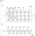
- FIG. 10(a) is a top view showing an example of a fluid device including a well array including a plurality of wells having a first well and a second well arranged at a bottom of the first well and having a smaller capacity than the first well; a substrate having a surface on which the well array is arranged; a lid member arranged to face the well array; and a spacer for separating the substrate and the lid member, in which a space between the well array and the lid member forms a flow path through which fluid flows.
- FIG. 10(b) is a cross-sectional view taken along line b-b' in an arrow direction of FIG. 10(a) .
- a fluid device 1000 includes a well array 500 including a plurality of wells having a first well 520 and a second well 530 arranged at a bottom of the first well 520 and having a smaller capacity than the first well 520, a substrate 510 having a surface on which the well array 500 is arranged, a spacer 1010, and a lid member 1020 having a liquid inlet 1021 formed thereon.
- a space 1030 between the substrate 510 and the lid member 1020 functions as a flow path through which a sample, a detection reagent, a sealing liquid, a water-absorbing organic solvent, and the like flow.
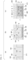
- FIGS. 11(a) to 11(c) are schematic cross-sectional views showing an example of procedures for carrying out a method of detecting a target nucleic acid fragment using the fluid device 200 shown in FIG. 2 .
- the two-part complex 130 of Cas13a protein and crRNA is immobilized on the inner surface of each well 211 of the well array 212. As shown in FIG. 11(a) , the two-part complex 130 is introduced through the liquid inlet 231 of the fluid device 200.
- the inside of the well 211 and the space between the substrate 210 and the lid member 230 are filled with the two-part complex 130 (Cas13a-crRNA).
- the Cas13a protein is biotinylated.
- the inner surface of the well 211 (here, only the bottom surface) is biotinylated and further bound with avidin. Therefore, as shown in FIG. 11(b) , the two-part complex introduced into the well 211 is immobilized on the bottom surface of the well 211.
- the sample and substrate nucleic acid fragment 150 are introduced through the liquid inlet 231 of the fluid device 200.
- the target nucleic acid fragment 140 (tgRNA) in the sample is introduced into the well 211.
- the target nucleic acid fragment 140 in the sample binds to the two-part complex to form a three-part complex 100' (Cas13a-crRNA-tgRNA).
- each well of the well array is sealed with a sealing liquid.
- a sealant 320 is introduced through the liquid inlet 231.
- the opening portion of the well 211 is sealed with the sealant 320 in a state in which the inside of the well 211 is filled with the three-part complex 100' and the substrate nucleic acid fragment 150.
- each well forms an independent reaction space.
- the substrate nucleic acid fragment 150 is cleaved by the nuclease activity of the three-part complex 100', and the fluorescent substance F is separated from the quencher Q.
- fluorescent light is generated in a case where the well into which the target nucleic acid fragment 140 has been introduced is irradiated with excitation light.
- the formation efficiency of the three-part complex is significantly improved compared to a case where Cas13a is not immobilized on a solid phase. That is, the detection sensitivity of the target nucleic acid fragment is significantly improved.
- FIGS. 12(a) to 12(d) are schematic cross-sectional views showing an example of procedures for carrying out a method for detecting a target nucleic acid fragment using the fluid device 1000 shown in FIG. 10 .
- a single-stranded RNA fragment tgRNA
- Cas13a protein is used as a CRISPR/Cas family protein.
- crRNA is used as gRNA.
- the two-part complex 130 of Cas13a protein and crRNA is immobilized on the inner surface (here, only the bottom surface) of the second well 530.
- the assay solution 310 containing a sample and the substrate nucleic acid fragment 150 is introduced through the liquid inlet 1021 of the fluid device 1000.
- the inside of the first well 520, the inside of the second well 530, and the space 1030 between the substrate 510 and the lid member 1020 are filled with the assay solution 310.
- an assay solution 310' containing the sample, crRNA and the substrate nucleic acid fragment 150 may be introduced through the liquid inlet 1021 of the fluid device 1000.
- each well of the well array is sealed with a sealant 320 (not shown).
- a sealant 320 is introduced through the liquid inlet 1021.
- an opening portion of the well 520 is sealed with the sealant 320 in a state in which the inside of the first well 520 is filled with the assay solution 310.
- each well forms an independent reaction space.
- the sealant 320 is replaced with a water-absorbing organic solvent 1230.
- a content of the well (assay solution 310) is dehydrated, a volume is reduced (concentrated), a Cas 13a-crRNA-tgRNA three-part complex 100' in the assay solution 310 is formed, and the substrate nucleic acid fragment 150 is cleaved.
- a fluorescent substance F binding to the substrate nucleic acid fragment 150 is separated from a quencher Q, and fluorescent light is generated by irradiation with excitation light. Detecting fluorescent light in a well indicates that a target substance is present in the well.
- the three-part complex 100' is not formed, and thus the substrate nucleic acid fragment 150 is not cleaved and fluorescent light is not generated.
- the water-absorbing organic solvent 1230 may be replaced with the sealant 320 again.
- the detection method of the present aspect may further include a step of replacing the water-absorbing organic solvent 1230 with the sealant 320.
- the CRISPR/Cas family protein expresses nuclease activity after forming a three-part complex with the gRNA and the target nucleic acid fragment, the substrate nucleic acid fragment is labeled with a fluorescent substance and a quencher, and in a case where the fluorescent substance cleaved by nuclease activity of the three-part complex is separated from the quencher, fluorescent light is emitted by irradiation with excitation light.
- the CRISPR/Cas family protein may be immobilized on the inner surface of the well.
- the well has a first well and a second well arranged at a bottom of a first well and having a smaller capacity than the first well, and the CRISPR/Cas family protein may be immobilized on the inner surface of the second well.
- the CRISPR/Cas family protein may be immobilized on the surface of the particle.
- the method of binding the CRISPR/Cas family protein to the particle and the surface of the particle is the same as described above.
- the CRISPR/Cas family protein may be a Cas12 protein or a Cas13 protein.
- the specific Cas12 protein or Cas13 protein is the same as described above.
- RNA fragment As the target nucleic acid fragment, a single-stranded RNA fragment (SEQ ID NO: 5) chemically synthesized by outsourcing (IDT), or a full-length SARS-CoV2 N gene RNA (SEQ ID NO: 9) synthesized by in vitro transcription (IVT) was used.
- IDCT single-stranded RNA fragment
- IVT in vitro transcription
- a well array A was prepared by the same procedure as in FIGS. 7(a) to 7(f) , described above. First, as shown in FIG. 7(a) , a glass substrate 510 was immersed in an 8 M potassium hydroxide solution for about 24 hours to form a hydroxyl group on a surface.
- a resist product name "AZ-P4903", manufactured by AZ Electronic Materials
- AZ-P4903 manufactured by AZ Electronic Materials
- a well array B having a first well and a second well was prepared by the same procedures as those shown in FIGS. 7(a) to 7(f) , 8(a), and 8(b) .
- the well array obtained in the same manner as in FIGS. 7(a) to 7(f) was dry-etched (13 sccm of O 2 , a pressure of 14 Pa, an output of 125 W) for 5 seconds using a Reactice ion etching device (manufactured by Samco) to perform hydrophilic treatment on a well array surface.
- a sheet-type resist product name "SU-8 3020CF DFR Type-S", manufactured by KAYAKU Advanced Materials, Inc.
- a laminate roller was adhered using a laminate roller to form a resist film 710, as shown in FIG. 8(a) .
- the resist film 710 was exposed by irradiation with ultraviolet rays for 20 seconds using an exposure machine (manufactured by Union Optical Co., Ltd.). Subsequently, the resist film 710 was developed by being immersed in a developing solution (product name: "SU8 developer", manufactured by KAYAKU Advanced Materials, Inc.) for 8 minutes. As a result, a portion of the resist film 710 forming the well was removed to obtain a well array B having the first well 520 and the second well 530.
- a developing solution product name: "SU8 developer”, manufactured by KAYAKU Advanced Materials, Inc.
- the well array B had a shape in which 12 to 18 wells 530 were arranged at a bottom of each well 520.
- FIG. 9 is a photomicrograph of the prepared well array B.
- a spacer 220 was arranged in the above-described well array A, and a glass plate 230 having a liquid inlet 231 formed thereon was placed to prepare a fluid device A.
- a fluid device A was obtained in which a space between the well array A and the glass plate 230 was a flow path.
- each of the above-described three-part complex solution and an assay solution in which the substrate nucleic acid fragment was mixed was prepared, and immediately introduced through the liquid inlet of each fluid device A.
- the assay solution was introduced into each well of the well array.
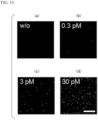
- FIG. 13(a) is a representative fluorescence micrograph showing results of an assay solution in which a final concentration of the target nucleic acid fragment is 0 pM.
- FIG. 13(b) is a representative fluorescence micrograph showing results of an assay solution in which a final concentration of the target nucleic acid fragment is 0.3 pM.
- FIG. 13(c) is a representative fluorescence micrograph showing results of an assay solution in which a final concentration of the target nucleic acid fragment is 3 pM.
- FIG. 13(d) is a representative fluorescence micrograph showing results of an assay solution in which a final concentration of the target nucleic acid fragment is 30 pM. Scale bar is 50 ⁇ m.
- a sealant (hexadecane, Sigma-Aldrich) was introduced through the liquid inlet of the fluid device B.
- a sealant hexadecane, Sigma-Aldrich
- FIG. 15 is a representative graph showing a proportion (%) of wells exhibiting predetermined fluorescence intensity (relative value) based on a photograph of the well array in which the sTG fluorescent light was detected.
- the horizontal axis of the graph indicates the concentration of alkaline phosphatase (ALP).
- Biotinylation was performed as follows. First, a thiol group was modified on a glass surface of a bottom surface of the well 530 of the fluid device A by silane coupling treatment using (3-mercaptopropyl)-trimethoxysilane. Subsequently, biotin bound to the above thiol group by maleimide reaction using biotin-dPEG 11 -maleimide. Through the above operation, the inner surface of the well 530 of the fluid device A was biotinylated.
- avidin was suspended in a buffer A having the composition shown in Table 1 above so that a final concentration was 1 mg/mL, and introduced through the liquid inlet of the fluid device A. As a result, avidin was introduced into each well of the well array and bound to biotin bound to the inner surface of the well 530.
- a lysine residue of the Cas13a protein or the N-terminus of the Cas13a protein was modified with NHS-PEG 4 -biotin and biotinylated.
- a cysteine residue of the biotinylated Cas13a protein was also labeled with Alexa488-maleimide.
- the biotinylated Cas13a protein was introduced through the liquid inlet of the fluid device A.
- the biotinylated Cas13a protein was introduced into each well of the well array, bound to avidin bound to the inner surface of the well 530, and immobilized.
- Alexa647 was dissolved in water, introduced through the liquid inlet of the fluid device A, and observed with a fluorescence microscope.
- FIG. 16 is a fluorescence micrograph of the fluid device A.
- FIG. 16 is a photograph of the fluid device A taken from above, and a bottom of FIG. 16 is a cross-sectional photograph of the fluid device A taken along a dotted line in FIG. 16 .
- the photograph in the bottom of FIG. 16 is upside down, and an upper side of the photograph is the bottom surface of the fluid device A.
- Alexa488 fluorescent light indicates the presence position of the Cas13a protein.
- Alexa647 fluorescent light indicates the presence position of water introduced into the well. As a result, it was clarified that the Cas13a protein could be immobilized only on the inner surface of the well 530.
- the Cas13a protein immobilized on the inner surface of the well was used to detect a target nucleic acid fragment.
- the lysine residue of the Cas13a protein or the N-terminus of the Cas13a protein was modified with NHS-PEG4-biotin and biotinylated.
- the biotinylated Cas13a protein and gRNA were mixed with the buffer A having the composition shown in Table 1 so that the final concentration of the biotinylated Cas13a protein is 40 nM and the final concentration of gRNA is 25 nM, thereby forming a two-part complex.
- a solution dissolved in the buffer A having the composition shown in Table 1 below was prepared so that the final concentration of the substrate nucleic acid fragment is 10 ⁇ M, the final concentration of the target nucleic acid fragment (SEQ ID NO: 5) is 3 pM, 0.3 pM, 30 fM, or 3 fM, in the buffer A, and used as an assay solution.
- each assay solution was introduced through the liquid inlet of each fluid device A.
- the assay solution was introduced into each well of the well array.
- each fluid device A was sealed with a sealant, and each well became each independent reaction space. After several minutes, the well array of each fluid device A was observed under a fluorescence microscope.
- a sealant hexadecane, Sigma-Aldrich
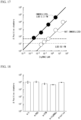
- FIG. 17 is a graph showing a relationship between the number of wells in which fluorescent light was detected and the final concentration of the target nucleic acid fragment (tgRNA).
- the vertical axis indicates the number of wells in which fluorescent light was detected, and the horizontal axis indicates the final concentration of the target nucleic acid fragment.
- the detection sensitivity was significantly improved from about 50 fM to about 3.3 fM by immobilization of the Cas13a protein.
- the influence of contaminants in detection of a target nucleic acid fragment was examined.
- the lysine residue of the Cas13a protein or the N-terminus of the Cas13a protein was modified with NHS-PEG4-biotin and biotinylated.
- the biotinylated Cas13a protein and gRNA were mixed with the buffer A having the composition shown in Table 1 so that the final concentration of the biotinylated Cas13a protein is 40 nM and the final concentration of gRNA is 25 nM, thereby forming a two-part complex.
- an assay solution was prepared by adding the substrate nucleic acid fragment to a final concentration of 10 ⁇ M, adding the target nucleic acid fragment to a final concentration of 30 pM, and further adding contaminants, to the buffer A.
- a 10 v/v% phosphate buffered saline (PBS), a 70 v/v% virus transport solution (VTM, catalog number "SGVTM-3R", Sugiyama-Gen Co., Ltd.), a non-target nucleic acid fragment having a final concentration 3 ng/ ⁇ L, and a 10 v/v% saliva were used.
- saliva contains RNase, 1 mM Triton X-100, which is a surfactant, was added in advance and then heated at 90°C for 5 minutes to deactivate RNase.
- an assay solution in which contaminants were not added was also prepared for comparison.
- each assay solution was introduced through the liquid inlet of each fluid device A.
- the assay solution was introduced into each well of the well array.
- each fluid device A was sealed with a sealant, and each well became each independent reaction space. After several minutes, the well array of each fluid device A was observed under a fluorescence microscope.
- a sealant hexadecane, Sigma-Aldrich
- FIG. 18 is a graph showing the number of wells in which fluorescent light was detected using each assay solution. The vertical axis indicates the number of wells in which fluorescent light was detected.
- "w/o” is a result of the assay solution to which contaminants were not added
- "w/PBS” is a result of the assay solution to which PBS was added
- "w/VTM” is a result of the assay solution to which virus transport solution was added
- w/ntgRNAs is a result of the assay solution to which a non-target nucleic acid fragment was added
- w/saliva is a result of the assay solution to which saliva was added.
- the influence of contaminants in detection of a target nucleic acid fragment was examined.
- Saliva was used as a contaminant.
- the lysine residue of the Cas13a protein or the N-terminus of the Cas13a protein was modified with NHS-PEG4-biotin and biotinylated.
- the biotinylated Cas13a protein and gRNA were mixed with the buffer A having the composition shown in Table 1 so that the final concentration of the biotinylated Cas13a protein is 40 nM and the final concentration of gRNA is 25 nM, thereby forming a two-part complex.
- an assay solution was prepared by adding the substrate nucleic acid fragment to a final concentration of 10 ⁇ M, and adding the target nucleic acid fragment (tgRNA) to a final concentration of 30 pM, 3 pM, 300 fM, 80 fM, 30 fM, or 8 fM, to the buffer A.
- tgRNA target nucleic acid fragment
- an assay solution was prepared by adding the substrate nucleic acid fragment to a final concentration of 5 ⁇ M, and adding the target nucleic acid fragment (tgRNA) to a final concentration of 30 pM, 3 pM, 300 fM, or 30 fM, to the saliva. Since saliva contains RNase, 1 mM Triton X-100, which is a surfactant, was added in advance and then heated at 90°C for 5 minutes to deactivate RNase.
- tgRNA target nucleic acid fragment
- each assay solution was introduced through the liquid inlet of each fluid device A.
- the assay solution was introduced into each well of the well array.
- each fluid device A was sealed with a sealant, and each well became each independent reaction space. After several minutes, the well array of each fluid device A was observed under a fluorescence microscope.
- a sealant hexadecane, Sigma-Aldrich
- FIG. 19 is a graph showing the relationship between the number of wells in which fluorescent light was detected and the final concentration of the target nucleic acid fragment (tgRNA).
- the vertical axis indicates the number of wells in which fluorescent light was detected, and the horizontal axis indicates the final concentration of the target nucleic acid fragment.
- Novel coronavirus SARS-CoV-2 specimens isolated from the Diamond Princess were used to detect novel coronavirus in a sample.
- SARS-CoV-2 virus was grown in VeroE6/TMPRSS2 cells and purified using the RNeasy Mini kit (Qiagen).
- the Cas13a protein and the gRNA were mixed with the buffer A having the composition shown in Table 1 below so that the final concentration of the Cas13a protein was 40 nM and the final concentration of the gRNA was 25 nM, thereby forming the two-part complex.
- the assay solution was prepared by adding the substrate nucleic acid fragment to a final concentration of 10 ⁇ M, and adding the novel coronavirus to a final concentration of 3 pM, 0.3 pM, 0.03 pM, 0.003 pM, or 0 pM, to the buffer A.
- each assay solution was introduced through the liquid inlet of each fluid device A.
- the assay solution was introduced into each well of the well array.
- each fluid device A was sealed with a sealant, and each well became each independent reaction space. After several minutes, the well array of each fluid device A was observed under a fluorescence microscope.
- a sealant hexadecane, Sigma-Aldrich
- FIG. 20 is a graph showing the relationship between the number of wells in which fluorescent light was detected and the final concentration of novel coronavirus.
- the vertical axis indicates the number of wells in which fluorescent light was detected, and the horizontal axis indicates the final concentration of novel coronavirus.
- SARS-CoV-2 the actual virus
- particles and the Cas13a protein were mixed to immobilize Cas13a on the surface of the particles, and the target nucleic acid fragment was detected.
- a single-stranded DNA fragment of which 3' end was modified with biotin base sequence is shown in SEQ ID NO: 7
- the target nucleic acid fragment SEQ ID NO: 5
- the single-stranded DNA fragment (SEQ ID NO: 7) had a base sequence complementary to a part of the target nucleic acid fragment (SEQ ID NO: 5).
- a solution of the hybridized single-stranded DNA fragment (SEQ ID NO: 7) and the target nucleic acid fragment (SEQ ID NO: 5) was mixed with streptavidin-coated magnetic beads (product name "Dynabeads M280", Veritas) to obtain a mixture solution.
- the solution was mixed in a buffer B (20 mM HEPES (pH 7.5), 20 mM KCl, 2 mM MgCl 2 , 50 ⁇ M Triton X-100) so that the final concentration of the single-stranded DNA fragment (SEQ ID NO: 7) was 12 nM and the final concentration of the magnetic beads was 1 mg/mL.
- a buffer B (20 mM HEPES (pH 7.5), 20 mM KCl, 2 mM MgCl 2 , 50 ⁇ M Triton X-100
- SEQ ID NO: 7 the final concentration of the single-stranded DNA fragment
- the magnetic beads was 1 mg/mL.
- four types of mixture solutions were prepared so that the final concentration of the target nucleic acid fragment (SEQ ID NO: 5) was 0.3 pM, 30 fM, 3 fM, or 0.8 fM.
- the buffer B was dispensed into four new plastic tubes, and a Cas13a protein, a gRNA (SEQ ID NO: 4), a substrate nucleic acid fragment, and each of the above mixture solutions were mixed therewith to obtain four types of assay solutions.
- the final concentration of the substrate nucleic acid fragment was adjusted to be 5 ⁇ M.
- a well array C was prepared in which 2,800,000 cylindrical wells with a diameter of 4.0 ⁇ m and a depth of 3.0 ⁇ m were arranged per 1 cm 2 .
- the volume per well of the well array C was 50 fL.
- the fluid device C was prepared in the same manner as the above-described fluid device A, except that the well array C was used instead of the well array A.
- each of the assay solutions was introduced through each of the liquid inlet of the fluid devices C and placed on a magnet sheet.
- each assay solution was introduced into each well of the well array.
- magnetic beads to which the three-part complex containing the target nucleic acid fragment bound were concentrated and captured in each well.
- each fluid device C was sealed with the sealant, and each well became each independent reaction space. After several minutes, the well array of each fluid device C was observed under a fluorescence microscope.
- a sealant mineral oil, Sigma-Aldrich
- FIG. 23 is a graph showing the relationship between the number of wells in which fluorescent light was detected and the final concentration of the target nucleic acid fragment (tgRNA).
- the vertical axis indicates the number of wells in which fluorescent light was detected, and the horizontal axis indicates the final concentration of the target nucleic acid fragment.
- the detection sensitivity was about 0.16 fM.
- FIG. 23 shows the results of a case where the two-part complex was not immobilized, measured in Experimental Example 1, and the results of a case where the two-part complex was immobilized on the inner surface of the well, measured in Experimental Example 4.
- the detection sensitivity was further significantly improved from about 3.3 fM to about 0.16 fM in a case of being immobilized on the inner surface of the well.
- FIG. 24 is a schematic diagram showing a detection method of the present experimental example.
- streptavidin-coated magnetic beads product name "Dynabeads MyOne Streptavidin T1", Veritas
- the lysine residue of the Cas13a protein or the N-terminus of the Cas13a protein was modified with NHS-PEG4-biotin and biotinylated.
- the biotinylated Cas13a protein and the gRNA were mixed in a buffer F (20 mM HEPES-KOH (pH 6.8), 60 mM NaCl, 6 mM MgCl 2 , 50 ⁇ M Triton X-100) so that the final concentration of the biotinylated Cas13a protein was 3 ⁇ M, and the final concentration of the gRNA was 0.75 ⁇ M, thereby forming a two-part complex.
- the two-part complex was diluted with the buffer F containing the substrate nucleic acid fragment and Alexa647-maleimide to obtain a mixture solution.
- concentration of the biotinylated Cas13a protein in the mixture solution was 60 nM
- concentration of the gRNA was 12 nM
- concentration of the substrate nucleic acid fragment was 12 ⁇ M
- concentration of the Alexa647-maleimide was 60 ⁇ M.
- a well array D was prepared in which 2,000,000 cylindrical wells with a diameter of 3.5 ⁇ m and a depth of 3.5 ⁇ m were arranged per 1 cm 2 .
- the volume per well of the well array D was 30 fL.
- the assay solution was dropped directly onto the well array D, and a sealant was further dropped to perform imaging while sealing each well.
- Two well arrays D were prepared, and 105 ⁇ L of each of the assay solutions was dropped to each of the well arrays D to install a magnet on a bottom of the well array D. As a result, each of the assay solutions was introduced into each well of the well array D.
- the assay solution contained magnetic beads
- the magnetic beads bound with the three-part complex containing the target nucleic acid fragment were concentrated and captured in each well.
- FIG. 25(a) is a representative fluorescence micrograph showing the results of an assay solution containing 300 fM of the target nucleic acid fragment and not containing the magnetic beads.
- FIG. 25(b) is a representative fluorescence micrograph showing the results of an assay solution containing 300 fM of the target nucleic acid fragment and the magnetic beads.
- the present invention it is possible to provide a technology of capable of performing detection with high sensitivity without amplifying a target nucleic acid fragment.
Landscapes
- Chemical & Material Sciences (AREA)
- Life Sciences & Earth Sciences (AREA)
- Health & Medical Sciences (AREA)
- Engineering & Computer Science (AREA)
- Immunology (AREA)
- Organic Chemistry (AREA)
- Molecular Biology (AREA)
- Zoology (AREA)
- Wood Science & Technology (AREA)
- Proteomics, Peptides & Aminoacids (AREA)
- Analytical Chemistry (AREA)
- General Health & Medical Sciences (AREA)
- Microbiology (AREA)
- Biochemistry (AREA)
- Biotechnology (AREA)
- Physics & Mathematics (AREA)
- Biomedical Technology (AREA)
- Genetics & Genomics (AREA)
- Bioinformatics & Cheminformatics (AREA)
- Urology & Nephrology (AREA)
- Hematology (AREA)
- General Engineering & Computer Science (AREA)
- Biophysics (AREA)
- Medicinal Chemistry (AREA)
- Cell Biology (AREA)
- Food Science & Technology (AREA)
- General Physics & Mathematics (AREA)
- Pathology (AREA)
- Chemical Kinetics & Catalysis (AREA)
- Measuring Or Testing Involving Enzymes Or Micro-Organisms (AREA)
Applications Claiming Priority (2)
| Application Number | Priority Date | Filing Date | Title |
|---|---|---|---|
| JP2020219481 | 2020-12-28 | ||
| PCT/JP2021/048095 WO2022145354A1 (ja) | 2020-12-28 | 2021-12-24 | 標的核酸断片の検出方法及びキット |
Publications (4)
| Publication Number | Publication Date |
|---|---|
| EP4269581A1 EP4269581A1 (en) | 2023-11-01 |
| EP4269581A4 EP4269581A4 (en) | 2024-10-09 |
| EP4269581B1 true EP4269581B1 (en) | 2025-06-11 |
| EP4269581C0 EP4269581C0 (en) | 2025-06-11 |
Family
ID=82259424
Family Applications (1)
| Application Number | Title | Priority Date | Filing Date |
|---|---|---|---|
| EP21915214.7A Active EP4269581B1 (en) | 2020-12-28 | 2021-12-24 | Method and kit for detecting target nucleic acid fragment |
Country Status (4)
| Country | Link |
|---|---|
| US (1) | US20240094199A1 (https=) |
| EP (1) | EP4269581B1 (https=) |
| JP (1) | JPWO2022145354A1 (https=) |
| WO (1) | WO2022145354A1 (https=) |
Families Citing this family (3)
| Publication number | Priority date | Publication date | Assignee | Title |
|---|---|---|---|---|
| EP3995833A4 (en) * | 2019-07-04 | 2023-08-09 | Riken | METHOD AND KIT FOR DETECTING A TARGET NUCLEIC ACID FRAGMENT |
| JPWO2023195542A1 (https=) * | 2022-04-08 | 2023-10-12 | ||
| JP2025033452A (ja) * | 2023-08-29 | 2025-03-13 | 国立研究開発法人産業技術総合研究所 | マルチプレックス核酸検出のためのセンサアレイ |
Family Cites Families (9)
| Publication number | Priority date | Publication date | Assignee | Title |
|---|---|---|---|---|
| JP3727026B2 (ja) | 2003-04-10 | 2005-12-14 | 博行 野地 | 一分子酵素活性検出に用いられるマイクロチャンバと1000fL以下の液滴を調製する方法 |
| US20140004539A1 (en) * | 2012-06-15 | 2014-01-02 | The Regents Of The University Of Michigan | Systems and methods for multiplex solution assays |
| JP6183471B2 (ja) * | 2014-01-31 | 2017-08-23 | 凸版印刷株式会社 | 生体分子解析キット及び生体分子解析方法 |
| US10337051B2 (en) * | 2016-06-16 | 2019-07-02 | The Regents Of The University Of California | Methods and compositions for detecting a target RNA |
| WO2019098301A1 (ja) * | 2017-11-17 | 2019-05-23 | 凸版印刷株式会社 | 標的分子の検出方法 |
| US10253365B1 (en) * | 2017-11-22 | 2019-04-09 | The Regents Of The University Of California | Type V CRISPR/Cas effector proteins for cleaving ssDNAs and detecting target DNAs |
| AU2019380590A1 (en) * | 2018-11-14 | 2021-07-01 | Massachusetts Institute Of Technology | Multiplexing highly evolving viral variants with Sherlock detection method |
| EP3995833A4 (en) * | 2019-07-04 | 2023-08-09 | Riken | METHOD AND KIT FOR DETECTING A TARGET NUCLEIC ACID FRAGMENT |
| WO2022133108A2 (en) * | 2020-12-17 | 2022-06-23 | Mammoth Biosciences, Inc. | Methods and compositions for performing a detection assay |
-
2021
- 2021-12-24 WO PCT/JP2021/048095 patent/WO2022145354A1/ja not_active Ceased
- 2021-12-24 US US18/269,264 patent/US20240094199A1/en active Pending
- 2021-12-24 EP EP21915214.7A patent/EP4269581B1/en active Active
- 2021-12-24 JP JP2022573049A patent/JPWO2022145354A1/ja active Pending
Also Published As
| Publication number | Publication date |
|---|---|
| EP4269581A4 (en) | 2024-10-09 |
| US20240094199A1 (en) | 2024-03-21 |
| WO2022145354A1 (ja) | 2022-07-07 |
| EP4269581A1 (en) | 2023-11-01 |
| JPWO2022145354A1 (https=) | 2022-07-07 |
| EP4269581C0 (en) | 2025-06-11 |
Similar Documents
| Publication | Publication Date | Title |
|---|---|---|
| Chen et al. | Microfluidics: the propellant of CRISPR-based nucleic acid detection | |
| Wang et al. | Extracellular vesicle preparation and analysis: a state‐of‐the‐art review | |
| EP4269581B1 (en) | Method and kit for detecting target nucleic acid fragment | |
| Yue et al. | Advances in clustered, regularly interspaced short palindromic repeats (CRISPR)-based diagnostic assays assisted by micro/nanotechnologies | |
| Rahimian et al. | Exosomal microRNA profiling | |
| Kalishwaralal et al. | Exosomes for non‐invasive cancer monitoring | |
| Raju et al. | Microfluidic platforms for the isolation and detection of exosomes: a brief review | |
| JP6882453B2 (ja) | 全ゲノムデジタル増幅方法 | |
| Xu et al. | Concurrent detection of protein and miRNA at the single extracellular vesicle level using a digital dual CRISPR-Cas assay | |
| US20120045748A1 (en) | Particulate labels | |
| US12522859B2 (en) | Method and kit for detecting target nucleic acid fragment | |
| Lin et al. | Recent advances in exosomal miRNA biosensing for liquid biopsy | |
| US20200363406A1 (en) | Highly-specific assays | |
| Mojtaba Mousavi et al. | Recent progress in prompt molecular detection of exosomes using CRISPR/Cas and microfluidic‐assisted approaches toward smart cancer diagnosis and analysis | |
| KR102738301B1 (ko) | 다중 진단을 위한 핵산 분리 및 검출용 미세입자 프로브 | |
| Minagawa et al. | On-chip enrichment system for digital bioassay based on aqueous two-phase system | |
| Sun et al. | Methodological approaches to study extracellular vesicle miRNAs in Epstein–Barr virus-associated cancers | |
| US20240263256A1 (en) | CRISPR-Cas13 System, Kit and Method for Detecting SARS-CoV-2 | |
| Li et al. | Ultrasensitive Nucleic Acid Testing: From Foundational Research to Clinical Translation | |
| EP4227398B1 (en) | Method for detecting target substance, fluid device, and kit | |
| CN109680343B (zh) | 一种外泌体微量dna的建库方法 | |
| EP4074823B1 (en) | Detection method | |
| WO2023195542A1 (ja) | 複数種類の標的核酸断片を検出する方法及びキット | |
| Wu et al. | Advances of nanopore sensors toward virus detection and diagnostic applications | |
| JP2022144722A (ja) | 標的粒子の分析方法、分析試薬及び分析装置 |
Legal Events
| Date | Code | Title | Description |
|---|---|---|---|
| STAA | Information on the status of an ep patent application or granted ep patent |
Free format text: STATUS: THE INTERNATIONAL PUBLICATION HAS BEEN MADE |
|
| PUAI | Public reference made under article 153(3) epc to a published international application that has entered the european phase |
Free format text: ORIGINAL CODE: 0009012 |
|
| STAA | Information on the status of an ep patent application or granted ep patent |
Free format text: STATUS: REQUEST FOR EXAMINATION WAS MADE |
|
| 17P | Request for examination filed |
Effective date: 20230623 |
|
| AK | Designated contracting states |
Kind code of ref document: A1 Designated state(s): AL AT BE BG CH CY CZ DE DK EE ES FI FR GB GR HR HU IE IS IT LI LT LU LV MC MK MT NL NO PL PT RO RS SE SI SK SM TR |
|
| DAV | Request for validation of the european patent (deleted) | ||
| DAX | Request for extension of the european patent (deleted) | ||
| A4 | Supplementary search report drawn up and despatched |
Effective date: 20240906 |
|
| RIC1 | Information provided on ipc code assigned before grant |
Ipc: C12Q 1/6837 20180101ALI20240903BHEP Ipc: C12Q 1/6825 20180101ALI20240903BHEP Ipc: C12Q 1/6876 20180101ALI20240903BHEP Ipc: C12Q 1/68 20180101ALI20240903BHEP Ipc: C12Q 1/34 20060101ALI20240903BHEP Ipc: C12N 15/09 20060101AFI20240903BHEP |
|
| GRAP | Despatch of communication of intention to grant a patent |
Free format text: ORIGINAL CODE: EPIDOSNIGR1 |
|
| STAA | Information on the status of an ep patent application or granted ep patent |
Free format text: STATUS: GRANT OF PATENT IS INTENDED |
|
| INTG | Intention to grant announced |
Effective date: 20250127 |
|
| GRAS | Grant fee paid |
Free format text: ORIGINAL CODE: EPIDOSNIGR3 |
|
| GRAA | (expected) grant |
Free format text: ORIGINAL CODE: 0009210 |
|
| STAA | Information on the status of an ep patent application or granted ep patent |
Free format text: STATUS: THE PATENT HAS BEEN GRANTED |
|
| AK | Designated contracting states |
Kind code of ref document: B1 Designated state(s): AL AT BE BG CH CY CZ DE DK EE ES FI FR GB GR HR HU IE IS IT LI LT LU LV MC MK MT NL NO PL PT RO RS SE SI SK SM TR |
|
| REG | Reference to a national code |
Ref country code: GB Ref legal event code: FG4D |
|
| REG | Reference to a national code |
Ref country code: CH Ref legal event code: EP |
|
| REG | Reference to a national code |
Ref country code: IE Ref legal event code: FG4D |
|
| REG | Reference to a national code |
Ref country code: DE Ref legal event code: R096 Ref document number: 602021032279 Country of ref document: DE |
|
| U01 | Request for unitary effect filed |
Effective date: 20250701 |
|
| U07 | Unitary effect registered |
Designated state(s): AT BE BG DE DK EE FI FR IT LT LU LV MT NL PT RO SE SI Effective date: 20250707 |
|
| PG25 | Lapsed in a contracting state [announced via postgrant information from national office to epo] |
Ref country code: ES Free format text: LAPSE BECAUSE OF FAILURE TO SUBMIT A TRANSLATION OF THE DESCRIPTION OR TO PAY THE FEE WITHIN THE PRESCRIBED TIME-LIMIT Effective date: 20250611 |
|
| PG25 | Lapsed in a contracting state [announced via postgrant information from national office to epo] |
Ref country code: NO Free format text: LAPSE BECAUSE OF FAILURE TO SUBMIT A TRANSLATION OF THE DESCRIPTION OR TO PAY THE FEE WITHIN THE PRESCRIBED TIME-LIMIT Effective date: 20250911 Ref country code: GR Free format text: LAPSE BECAUSE OF FAILURE TO SUBMIT A TRANSLATION OF THE DESCRIPTION OR TO PAY THE FEE WITHIN THE PRESCRIBED TIME-LIMIT Effective date: 20250912 |
|
| PG25 | Lapsed in a contracting state [announced via postgrant information from national office to epo] |
Ref country code: HR Free format text: LAPSE BECAUSE OF FAILURE TO SUBMIT A TRANSLATION OF THE DESCRIPTION OR TO PAY THE FEE WITHIN THE PRESCRIBED TIME-LIMIT Effective date: 20250611 |
|
| PG25 | Lapsed in a contracting state [announced via postgrant information from national office to epo] |
Ref country code: RS Free format text: LAPSE BECAUSE OF FAILURE TO SUBMIT A TRANSLATION OF THE DESCRIPTION OR TO PAY THE FEE WITHIN THE PRESCRIBED TIME-LIMIT Effective date: 20250911 |
|
| PG25 | Lapsed in a contracting state [announced via postgrant information from national office to epo] |
Ref country code: IS Free format text: LAPSE BECAUSE OF FAILURE TO SUBMIT A TRANSLATION OF THE DESCRIPTION OR TO PAY THE FEE WITHIN THE PRESCRIBED TIME-LIMIT Effective date: 20251011 |
|
| PGFP | Annual fee paid to national office [announced via postgrant information from national office to epo] |
Ref country code: GB Payment date: 20251219 Year of fee payment: 5 |
|
| PG25 | Lapsed in a contracting state [announced via postgrant information from national office to epo] |
Ref country code: SM Free format text: LAPSE BECAUSE OF FAILURE TO SUBMIT A TRANSLATION OF THE DESCRIPTION OR TO PAY THE FEE WITHIN THE PRESCRIBED TIME-LIMIT Effective date: 20250611 |
|
| PG25 | Lapsed in a contracting state [announced via postgrant information from national office to epo] |
Ref country code: CZ Free format text: LAPSE BECAUSE OF FAILURE TO SUBMIT A TRANSLATION OF THE DESCRIPTION OR TO PAY THE FEE WITHIN THE PRESCRIBED TIME-LIMIT Effective date: 20250611 |
|
| PG25 | Lapsed in a contracting state [announced via postgrant information from national office to epo] |
Ref country code: PL Free format text: LAPSE BECAUSE OF FAILURE TO SUBMIT A TRANSLATION OF THE DESCRIPTION OR TO PAY THE FEE WITHIN THE PRESCRIBED TIME-LIMIT Effective date: 20250611 |
|
| PG25 | Lapsed in a contracting state [announced via postgrant information from national office to epo] |
Ref country code: SK Free format text: LAPSE BECAUSE OF FAILURE TO SUBMIT A TRANSLATION OF THE DESCRIPTION OR TO PAY THE FEE WITHIN THE PRESCRIBED TIME-LIMIT Effective date: 20250611 |
|
| U20 | Renewal fee for the european patent with unitary effect paid |
Year of fee payment: 5 Effective date: 20251230 |
|
| PLBE | No opposition filed within time limit |
Free format text: ORIGINAL CODE: 0009261 |
|
| STAA | Information on the status of an ep patent application or granted ep patent |
Free format text: STATUS: NO OPPOSITION FILED WITHIN TIME LIMIT |
|
| REG | Reference to a national code |
Ref country code: CH Ref legal event code: L10 Free format text: ST27 STATUS EVENT CODE: U-0-0-L10-L00 (AS PROVIDED BY THE NATIONAL OFFICE) Effective date: 20260423 |