EP4257601A1 - Procédé d'aide au diagnostic du cancer du sein et kit de test du cancer du sein - Google Patents
Procédé d'aide au diagnostic du cancer du sein et kit de test du cancer du sein Download PDFInfo
- Publication number
- EP4257601A1 EP4257601A1 EP21903366.9A EP21903366A EP4257601A1 EP 4257601 A1 EP4257601 A1 EP 4257601A1 EP 21903366 A EP21903366 A EP 21903366A EP 4257601 A1 EP4257601 A1 EP 4257601A1
- Authority
- EP
- European Patent Office
- Prior art keywords
- laminin
- breast cancer
- antibody
- amount
- specimen
- Prior art date
- Legal status (The legal status is an assumption and is not a legal conclusion. Google has not performed a legal analysis and makes no representation as to the accuracy of the status listed.)
- Pending
Links
Images


Classifications
-
- G—PHYSICS
- G01—MEASURING; TESTING
- G01N—INVESTIGATING OR ANALYSING MATERIALS BY DETERMINING THEIR CHEMICAL OR PHYSICAL PROPERTIES
- G01N33/00—Investigating or analysing materials by specific methods not covered by groups G01N1/00 - G01N31/00
- G01N33/48—Biological material, e.g. blood, urine; Haemocytometers
- G01N33/50—Chemical analysis of biological material, e.g. blood, urine; Testing involving biospecific ligand binding methods; Immunological testing
- G01N33/53—Immunoassay; Biospecific binding assay; Materials therefor
- G01N33/575—Immunoassay; Biospecific binding assay; Materials therefor for cancer
- G01N33/57515—Immunoassay; Biospecific binding assay; Materials therefor for cancer of the breast
-
- C—CHEMISTRY; METALLURGY
- C07—ORGANIC CHEMISTRY
- C07K—PEPTIDES
- C07K16/00—Immunoglobulins [IG], e.g. monoclonal or polyclonal antibodies
- C07K16/18—Immunoglobulins [IG], e.g. monoclonal or polyclonal antibodies against material from animals or humans
-
- G—PHYSICS
- G01—MEASURING; TESTING
- G01N—INVESTIGATING OR ANALYSING MATERIALS BY DETERMINING THEIR CHEMICAL OR PHYSICAL PROPERTIES
- G01N33/00—Investigating or analysing materials by specific methods not covered by groups G01N1/00 - G01N31/00
- G01N33/48—Biological material, e.g. blood, urine; Haemocytometers
- G01N33/50—Chemical analysis of biological material, e.g. blood, urine; Testing involving biospecific ligand binding methods; Immunological testing
- G01N33/5005—Chemical analysis of biological material, e.g. blood, urine; Testing involving biospecific ligand binding methods; Immunological testing involving human or animal cells
- G01N33/5008—Chemical analysis of biological material, e.g. blood, urine; Testing involving biospecific ligand binding methods; Immunological testing involving human or animal cells for testing or evaluating the effect of chemical or biological compounds, e.g. drugs, cosmetics
- G01N33/5076—Chemical analysis of biological material, e.g. blood, urine; Testing involving biospecific ligand binding methods; Immunological testing involving human or animal cells for testing or evaluating the effect of chemical or biological compounds, e.g. drugs, cosmetics involving cell organelles, e.g. Golgi complex, endoplasmic reticulum
-
- G—PHYSICS
- G01—MEASURING; TESTING
- G01N—INVESTIGATING OR ANALYSING MATERIALS BY DETERMINING THEIR CHEMICAL OR PHYSICAL PROPERTIES
- G01N33/00—Investigating or analysing materials by specific methods not covered by groups G01N1/00 - G01N31/00
- G01N33/48—Biological material, e.g. blood, urine; Haemocytometers
- G01N33/50—Chemical analysis of biological material, e.g. blood, urine; Testing involving biospecific ligand binding methods; Immunological testing
- G01N33/53—Immunoassay; Biospecific binding assay; Materials therefor
- G01N33/531—Production of immunochemical test materials
- G01N33/532—Production of labelled immunochemicals
- G01N33/535—Production of labelled immunochemicals with enzyme label or co-enzymes, co-factors, enzyme inhibitors or enzyme substrates
-
- G—PHYSICS
- G01—MEASURING; TESTING
- G01N—INVESTIGATING OR ANALYSING MATERIALS BY DETERMINING THEIR CHEMICAL OR PHYSICAL PROPERTIES
- G01N33/00—Investigating or analysing materials by specific methods not covered by groups G01N1/00 - G01N31/00
- G01N33/48—Biological material, e.g. blood, urine; Haemocytometers
- G01N33/50—Chemical analysis of biological material, e.g. blood, urine; Testing involving biospecific ligand binding methods; Immunological testing
- G01N33/53—Immunoassay; Biospecific binding assay; Materials therefor
- G01N33/543—Immunoassay; Biospecific binding assay; Materials therefor with an insoluble carrier for immobilising immunochemicals
- G01N33/54313—Immunoassay; Biospecific binding assay; Materials therefor with an insoluble carrier for immobilising immunochemicals the carrier being characterised by its particulate form
-
- G—PHYSICS
- G01—MEASURING; TESTING
- G01N—INVESTIGATING OR ANALYSING MATERIALS BY DETERMINING THEIR CHEMICAL OR PHYSICAL PROPERTIES
- G01N33/00—Investigating or analysing materials by specific methods not covered by groups G01N1/00 - G01N31/00
- G01N33/48—Biological material, e.g. blood, urine; Haemocytometers
- G01N33/50—Chemical analysis of biological material, e.g. blood, urine; Testing involving biospecific ligand binding methods; Immunological testing
- G01N33/53—Immunoassay; Biospecific binding assay; Materials therefor
- G01N33/543—Immunoassay; Biospecific binding assay; Materials therefor with an insoluble carrier for immobilising immunochemicals
- G01N33/54366—Apparatus specially adapted for solid-phase testing
- G01N33/54386—Analytical elements
- G01N33/54387—Immunochromatographic test strips
-
- G—PHYSICS
- G01—MEASURING; TESTING
- G01N—INVESTIGATING OR ANALYSING MATERIALS BY DETERMINING THEIR CHEMICAL OR PHYSICAL PROPERTIES
- G01N33/00—Investigating or analysing materials by specific methods not covered by groups G01N1/00 - G01N31/00
- G01N33/48—Biological material, e.g. blood, urine; Haemocytometers
- G01N33/50—Chemical analysis of biological material, e.g. blood, urine; Testing involving biospecific ligand binding methods; Immunological testing
- G01N33/53—Immunoassay; Biospecific binding assay; Materials therefor
- G01N33/575—Immunoassay; Biospecific binding assay; Materials therefor for cancer
- G01N33/5758—Immunoassay; Biospecific binding assay; Materials therefor for cancer involving compounds serving as markers for tumours, cancers or neoplasias, e.g. cellular determinants, receptors, heat shock/stress proteins, A-protein, oligosaccharides or metabolites
- G01N33/57585—Immunoassay; Biospecific binding assay; Materials therefor for cancer involving compounds serving as markers for tumours, cancers or neoplasias, e.g. cellular determinants, receptors, heat shock/stress proteins, A-protein, oligosaccharides or metabolites involving compounds identifiable in body fluids
-
- G—PHYSICS
- G01—MEASURING; TESTING
- G01N—INVESTIGATING OR ANALYSING MATERIALS BY DETERMINING THEIR CHEMICAL OR PHYSICAL PROPERTIES
- G01N33/00—Investigating or analysing materials by specific methods not covered by groups G01N1/00 - G01N31/00
- G01N33/48—Biological material, e.g. blood, urine; Haemocytometers
- G01N33/50—Chemical analysis of biological material, e.g. blood, urine; Testing involving biospecific ligand binding methods; Immunological testing
- G01N33/68—Chemical analysis of biological material, e.g. blood, urine; Testing involving biospecific ligand binding methods; Immunological testing involving proteins, peptides or amino acids
- G01N33/6803—General methods of protein analysis not limited to specific proteins or families of proteins
- G01N33/6848—Methods of protein analysis involving mass spectrometry
-
- G—PHYSICS
- G01—MEASURING; TESTING
- G01N—INVESTIGATING OR ANALYSING MATERIALS BY DETERMINING THEIR CHEMICAL OR PHYSICAL PROPERTIES
- G01N2333/00—Assays involving biological materials from specific organisms or of a specific nature
- G01N2333/435—Assays involving biological materials from specific organisms or of a specific nature from animals; from humans
- G01N2333/705—Assays involving receptors, cell surface antigens or cell surface determinants
- G01N2333/70596—Molecules with a "CD"-designation not provided for elsewhere in G01N2333/705
-
- G—PHYSICS
- G01—MEASURING; TESTING
- G01N—INVESTIGATING OR ANALYSING MATERIALS BY DETERMINING THEIR CHEMICAL OR PHYSICAL PROPERTIES
- G01N2333/00—Assays involving biological materials from specific organisms or of a specific nature
- G01N2333/435—Assays involving biological materials from specific organisms or of a specific nature from animals; from humans
- G01N2333/78—Connective tissue peptides, e.g. collagen, elastin, laminin, fibronectin, vitronectin, cold insoluble globulin [CIG]
Definitions
- the present invention relates to a method of assisting breast cancer diagnosis and a test kit for breast cancer.
- a specimen may be subjected to immunochromatography for development (migration) and capture, followed by detection of the labelled anti-laminin 5 antibody or anti-laminin ⁇ 3 antibody by means of visible light (absorption), ICP-MS, chemiluminescence, or fluorescence.
- the method of assisting breast cancer diagnosis according to the present invention includes providing information for breast cancer diagnosis based on the amount of laminin 5 or laminin ⁇ 3 thus measured.
- this step is also called “an information-providing step”.
- an exosome-marker-capturing antibody as a capture antibody
- an anti-laminin 5 antibody or an anti-laminin ⁇ 3 antibody as a detection antibody, it is possible to detect exosomes having laminin 5 or laminin ⁇ 3 present in the specimen, allowing for quantification of laminin 5 or laminin ⁇ 3 expressed in the exosomes.
- the specimen and laminin 5 or laminin ⁇ 3 may be those as described above.
- the method for detecting the diagnostic marker according to the present invention is not particularly limited, and, for example, the above-described method may be employed for measuring the amount of laminin 5 or laminin ⁇ 3. Based on the amount of laminin 5 or laminin ⁇ 3 thus measured, and according to the above-described criteria, it is possible to judge if the specimen was derived from breast cancer, if it was derived from triple-negative-type breast cancer, and if it was derived from early-stage breast cancer.
- the measurement method is not particularly limited provided that it is capable of measuring the amount of laminin 5 or laminin ⁇ 3 contained in a specimen, and it may be the same method as mentioned above in the section regarding the measuring step.
- the specimen and the predetermined value may be those described above.
- a breast cancer diagnostic method comprises judging that a specimen was derived from breast cancer when the amount of laminin 5 or laminin ⁇ 3 contained in the specimen is equal to or higher than a predetermined value. It is also capable of diagnosing breast cancer subtype and breast cancer stage for the specimen. With the breast cancer diagnostic method according to the present invention, it is possible to diagnose if the specimen was derived from breast cancer, if it was derived from triple-negative-type breast cancer, and if it was derived from early-stage breast cancer, in an easy and simple manner.
- the specimen and the predetermined value may be those described above.
- the specimen is a biological specimen collected from a subject, it is possible, by the breast cancer diagnostic method according to the present invention, to judge if the subject is a breast cancer patient as well as to judge breast cancer subtype and breast cancer stage, in an easy and simple manner.
- the breast cancer diagnostic method according to the present invention may comprise the measuring step and the information-providing step as described above.
- Laminin ⁇ 3 was detected in a high concentration in the triple-negative-type breast cancer cell line, while it was detected in a low concentration in the luminal-A-type breast cancer cell line. Since laminin ⁇ 3 is contained only in laminin 5, it is suggested that laminin 5 is expressed in a high amount in the triple-negative-type breast cancer cell line and also contained in exosomes.
- the total luminance of the immunochromatographic test strip at or around where the antibody was fixed as well as the total luminance of a non-luminescence area of the same size were measured by ImageJ, and from the difference between them (difference in luminance), the amount of CD9 expression and the amount of laminin 5 expression were determined.
- the amount of laminin 5 expression is divided by the amount of CD9 expression, and the resulting standardized value (laminin 5/CD9) is shown in Fig. 4 .
- Laminin 5/CD9 was higher in all of the 11 types of triple-negative-type breast cancer cell lines thus investigated, than in the cell line that was not derived from triple-negative-type breast cancer (MCF7).
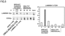
- a biological specimen derived from a triple-negative-type breast cancer patient was investigated to see if laminin 5 expression was increased.
- detection of laminin 5 and CD9 was carried out by an immunochromatography method.
- An immunochromatographic test strip was used, which had, in the same manner as in Experiment 2 above, a mouse anti-CD9 antibody fixed thereto and its membrane blocked with water-soluble polymer and BSA.
- 9 ⁇ L of a solution was developed, which contained plasma diluted three times with PBS containing 1% BSA and 0.05% Tween 20 and filtered through a syringe filter with a pore size of 0.22 ⁇ m. It was followed by developing a developer liquid containing a mouse anti-CD9 antibody to which HRP had been conjugated in the same manner as in Experiment 2.
- luminescence was generated with the use of ImmunoStar (registered trademark) LD (Product No. 290-69904, manufactured by FUJIFILM Wako Pure Chemical Corporation), and the resulting luminescence was detected on Image Quant LAS4000.
- An immunochromatographic test strip was used, which had, in the same manner as in Experiment 2 above, a rabbit anti-LAMS antibody fixed thereto and its membrane blocked with water-soluble polymer and BSA.
- a solution was developed, which contained plasma diluted three times with PBS containing 1% BSA and 0.05% Tween 20 and filtered through a syringe filter with a pore size of 0.22 ⁇ m. It was followed by developing a developer liquid containing a rabbit anti-LAM5 antibody to which HRP had been conjugated in the same manner as in Experiment 2.
- luminescence was generated with the use of ImmunoStar (registered trademark) LD (Product No. 290-69904, manufactured by FUJIFILM Wako Pure Chemical Corporation), and the resulting luminescence was detected on Image Quant LAS4000.
- laminin 5 and CD9 were detected.
- the total luminance of the immunochromatographic test strip at or around where the antibody was fixed as well as the total luminance of a non-luminescence area of the same size were measured by ImageJ, and from the difference between them (difference in luminance), the amount of laminin 5 expression and the amount of CD9 expression were determined.
- the luminance of laminin 5 is divided by the luminance of CD9, and the resulting value is shown in the right part of Fig. 5 .
- Laminin 5/CD9 in the plasma from the breast cancer patients was higher than laminin 5/CD9 of the healthy individuals, and laminin 5/CD9 of the triple-negative-type breast cancer patient was higher than those of the other-subtype breast cancer patients.
- the amount of CD9 expression and the amount of laminin 5 expression were measured.
- the experiment method was the same as in Experiment 3.
- the luminance of laminin 5 is divided by the luminance of CD9, and the resulting value is shown in Fig. 6 .
- Laminin 5/CD9 in the plasma from six out often triple-negative-type breast cancer patients was higher, at least twice, than laminin 5/CD9 in the plasma from the HER2-type breast cancer patients.
- the subtype (triple-negative-type) of breast cancer patients was successfully diagnosed with 100% specificity and 60% sensitivity. This suggested that laminin 5 is useful as a diagnostic marker for breast cancer subtyping.
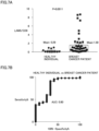
- Fig. 7A The luminance of laminin 5 is divided by the luminance of CD9, and the resulting standardized value (laminin 5/CD9) is shown in Fig. 7A .
- Fig. 7B shows a receiver operating characteristic (ROC) curve indicating the capability of laminin 5/CD9 of identifying breast cancer patients.
- the area under curve (AUC) was 0.80. From these, it is suggested that laminin 5 expression is high in the breast cancer patients and is highly capable of identifying breast cancer.
- ROC receiver operating characteristic
- a method of assisting breast cancer diagnosis comprises: a measuring step for measuring an amount of laminin 5 or laminin ⁇ 3 contained in a specimen; and an information-providing step for providing information for breast cancer diagnosis based on the amount of laminin 5 or laminin ⁇ 3 thus measured.
- the information for breast cancer diagnosis includes information for diagnosis of presence or absence of breast cancer.
- the information for breast cancer diagnosis includes information for diagnosis of breast cancer subtype.
- the information for breast cancer diagnosis includes information for diagnosis of breast cancer stage.
- the amount of laminin 5 or laminin ⁇ 3 is measured by an immunological measurement method or a mass spectrometry method.
- the immunological measurement method is an immunochromatography method.
- an antibody used in the immunological measurement method is labelled with metal particles or with an enzyme.
- the metal particles are detected by inductively coupled plasma mass spectrometry (ICP-MS).
- the amount of laminin 5 or laminin ⁇ 3 is standardized based on an amount of exosomes.
- the amount of exosomes is measured with use of CD9 and used for standardizing the amount of laminin 5 or laminin ⁇ 3.
- the specimen is a biological specimen collected from a subject.
- the specimen is blood collected from a human individual.
- the specimen is a purified exosome fraction.
- a test kit for breast cancer comprises an anti-laminin 5 antibody or an anti-laminin ⁇ 3 antibody.
- test kit for breast cancer With the test kit for breast cancer according to Item 14, it is possible to provide information for breast cancer diagnosis in an easy and simple manner by measuring the amount of laminin 5 or laminin ⁇ 3 contained in a specimen.
- the anti-laminin 5 antibody or the anti-laminin ⁇ 3 antibody is labelled with metal particles or with an enzyme.
- the amount of laminin 5 or laminin ⁇ 3 can be measured with high sensitivity.
- laminin 5 or laminin ⁇ 3 can be detected by means of visible light (absorption).
- the test kit according to Item 14 or Item 15 comprises an immunochromatographic test strip.
- test kit With the test kit according to Item 16, it is possible to measure the amount of laminin 5 or laminin ⁇ 3 in a fast, easy, and simple manner.
- a breast cancer diagnostic method comprises judging that a specimen was derived from breast cancer when the amount of laminin 5 or laminin ⁇ 3 contained in the specimen is equal to or higher than a predetermined value.
- an anti-laminin 5 antibody or an anti-laminin ⁇ 3 antibody is for breast cancer diagnosis.
Landscapes
- Health & Medical Sciences (AREA)
- Life Sciences & Earth Sciences (AREA)
- Immunology (AREA)
- Engineering & Computer Science (AREA)
- Chemical & Material Sciences (AREA)
- Molecular Biology (AREA)
- Biomedical Technology (AREA)
- Hematology (AREA)
- Urology & Nephrology (AREA)
- Physics & Mathematics (AREA)
- Medicinal Chemistry (AREA)
- Biochemistry (AREA)
- General Health & Medical Sciences (AREA)
- Cell Biology (AREA)
- Food Science & Technology (AREA)
- Biotechnology (AREA)
- Analytical Chemistry (AREA)
- General Physics & Mathematics (AREA)
- Pathology (AREA)
- Microbiology (AREA)
- Bioinformatics & Cheminformatics (AREA)
- Organic Chemistry (AREA)
- Biophysics (AREA)
- Proteomics, Peptides & Aminoacids (AREA)
- Bioinformatics & Computational Biology (AREA)
- Genetics & Genomics (AREA)
- Toxicology (AREA)
- Tropical Medicine & Parasitology (AREA)
- Spectroscopy & Molecular Physics (AREA)
- Investigating Or Analysing Biological Materials (AREA)
- Other Investigation Or Analysis Of Materials By Electrical Means (AREA)
Applications Claiming Priority (2)
| Application Number | Priority Date | Filing Date | Title |
|---|---|---|---|
| JP2020202742 | 2020-12-07 | ||
| PCT/JP2021/044729 WO2022124266A1 (fr) | 2020-12-07 | 2021-12-06 | Procédé d'aide au diagnostic du cancer du sein et kit de test du cancer du sein |
Publications (2)
| Publication Number | Publication Date |
|---|---|
| EP4257601A1 true EP4257601A1 (fr) | 2023-10-11 |
| EP4257601A4 EP4257601A4 (fr) | 2024-10-30 |
Family
ID=81973266
Family Applications (1)
| Application Number | Title | Priority Date | Filing Date |
|---|---|---|---|
| EP21903366.9A Pending EP4257601A4 (fr) | 2020-12-07 | 2021-12-06 | Procédé d'aide au diagnostic du cancer du sein et kit de test du cancer du sein |
Country Status (5)
| Country | Link |
|---|---|
| US (1) | US20240027455A1 (fr) |
| EP (1) | EP4257601A4 (fr) |
| JP (1) | JP7720060B2 (fr) |
| CN (1) | CN116802497A (fr) |
| WO (1) | WO2022124266A1 (fr) |
Cited By (1)
| Publication number | Priority date | Publication date | Assignee | Title |
|---|---|---|---|---|
| WO2025083329A1 (fr) * | 2023-10-16 | 2025-04-24 | Teknologian Tutkimuskeskus Vtt Oy | Liant d'anticorps en tant que biomarqueur pour le cancer |
Family Cites Families (13)
| Publication number | Priority date | Publication date | Assignee | Title |
|---|---|---|---|---|
| AU2001261519A1 (en) * | 2000-05-12 | 2001-11-26 | Fibrogen, Inc. | Methods of affecting laminin 5 processing |
| US20020156263A1 (en) * | 2000-10-05 | 2002-10-24 | Huei-Mei Chen | Genes expressed in breast cancer |
| JP2002148266A (ja) * | 2000-11-10 | 2002-05-22 | Rohto Pharmaceut Co Ltd | 検出装置及びその製造方法 |
| JPWO2003016907A1 (ja) * | 2001-08-17 | 2004-12-02 | エーザイ株式会社 | 生体試料中のラミニン5抗原の測定試薬及び測定方法 |
| US20100105087A1 (en) * | 2006-10-31 | 2010-04-29 | George Mason Intellectual Properties, Inc | Biomarkers for breast cancer |
| JP5754844B2 (ja) * | 2010-03-09 | 2015-07-29 | 国立大学法人 東京大学 | 泌尿器科がんの検査方法及び検査用キット |
| JP2016211925A (ja) * | 2015-05-01 | 2016-12-15 | 地方独立行政法人東京都健康長寿医療センター | がんにおいてドセタキセル又はパクリタキセルに対する耐性を評価する方法、がんの悪性化を評価する方法、及びそれら方法に用いられるキット |
| JP6637290B2 (ja) * | 2015-11-05 | 2020-01-29 | 武 永安 | Ck19に特異的なモノクローナル抗体およびこれを産生するハイブリドーマ、癌の検出キット、癌の検出方法および癌の転移の判定方法 |
| JP2017095744A (ja) * | 2015-11-19 | 2017-06-01 | 大日本塗料株式会社 | 金ナノロッドを含む被験物質を検出するための組成物及びその用途 |
| WO2018094469A1 (fr) * | 2016-11-24 | 2018-05-31 | The Council Of The Queensland Institute Of Medical Research | Détermination d'un pronostic de cancer |
| CN109270269A (zh) * | 2018-09-05 | 2019-01-25 | 河南省生物工程技术研究中心 | 一种用于早期乳腺癌多联检测的荧光免疫层析检测卡、试剂盒 |
| WO2020235424A1 (fr) * | 2019-05-17 | 2020-11-26 | 株式会社島津製作所 | Procédé de détection de vésicules extracellulaires |
| CN114127561A (zh) * | 2019-05-24 | 2022-03-01 | 索尔生物公司 | 使用开关样结合反应的亲和分离系统和方法 |
-
2021
- 2021-12-06 CN CN202180093105.4A patent/CN116802497A/zh active Pending
- 2021-12-06 WO PCT/JP2021/044729 patent/WO2022124266A1/fr not_active Ceased
- 2021-12-06 EP EP21903366.9A patent/EP4257601A4/fr active Pending
- 2021-12-06 US US18/265,547 patent/US20240027455A1/en active Pending
- 2021-12-06 JP JP2022568266A patent/JP7720060B2/ja active Active
Cited By (1)
| Publication number | Priority date | Publication date | Assignee | Title |
|---|---|---|---|---|
| WO2025083329A1 (fr) * | 2023-10-16 | 2025-04-24 | Teknologian Tutkimuskeskus Vtt Oy | Liant d'anticorps en tant que biomarqueur pour le cancer |
Also Published As
| Publication number | Publication date |
|---|---|
| WO2022124266A1 (fr) | 2022-06-16 |
| JP7720060B2 (ja) | 2025-08-07 |
| CN116802497A (zh) | 2023-09-22 |
| US20240027455A1 (en) | 2024-01-25 |
| EP4257601A4 (fr) | 2024-10-30 |
| JPWO2022124266A1 (fr) | 2022-06-16 |
Similar Documents
| Publication | Publication Date | Title |
|---|---|---|
| US20250334579A1 (en) | Nanoplasmonic quantification of tumor-derived extracellular vesicles in plasma microsamples for detection and treatment monitoring | |
| US20160370265A1 (en) | Method for isolating exosomes | |
| CN106814187B (zh) | 外周游离外泌体在制备液态活检肿瘤诊断试剂中的应用 | |
| KR102047186B1 (ko) | Maldi-tof 질량분석법을 기반으로 하는 혈액 단백질 및 대사체 핑거프린팅을 이용한 초고속 질병 진단 시스템 | |
| CN107942065B (zh) | 一种检测降钙素的试剂盒及检测方法 | |
| WO2014132869A1 (fr) | Procédé de détermination du cancer du sein | |
| CN106771258A (zh) | 一种半乳糖凝集素‑3结合蛋白的检测试剂盒及其方法和应用 | |
| US20150203892A1 (en) | Method for determining breast cancer | |
| CN116840482A (zh) | 基于外泌体突触核蛋白的帕金森病早期诊断系统 | |
| JP7507165B2 (ja) | 胃がんマーカー、及びこれを用いた検査方法 | |
| JP2019215342A (ja) | ヒト尿からのマイクロベシクルの分離方法及び分析方法 | |
| CN106066401B (zh) | 生物标志物vwf和adamts13及其在肝硬化诊断试剂中的用途 | |
| EP4257601A1 (fr) | Procédé d'aide au diagnostic du cancer du sein et kit de test du cancer du sein | |
| CN109655608A (zh) | 一种用于骨肉瘤诊断的外泌体蛋白及其即时检测方法 | |
| JP7376021B2 (ja) | ヒト血液からのマイクロベシクルの分離方法及び分析方法 | |
| CN119985972A (zh) | 辅助诊断进行性核上性麻痹的生物标志物及其应用 | |
| EP3816628B1 (fr) | Marqueur de détermination du cancer du pancréas | |
| CN116482386B (zh) | 检测糖尿病肾病的蛋白标志物及其应用、试剂或试剂盒 | |
| US20250012804A1 (en) | Use of microvesicles for benign colorectal polyps and colorectal cancer screening | |
| KR20200049647A (ko) | 반려동물에서 종양질병의 보조적 진단을 위한 바이오마커 검사방법 | |
| US20140242726A1 (en) | LUNG CANCER MARKER COMPLEMENT C3dg MOLECULE, AND METHOD FOR ANALYZING LUNG CANCER MARKER | |
| JP6145650B2 (ja) | 卵巣癌マーカー及び卵巣癌検出方法 | |
| CN111751457B (zh) | 一种痛风性关节炎诊断试剂盒及其应用 | |
| EP2534491B1 (fr) | Compositions et méthodes pour la prédiction d'événements cardiovasculaires | |
| JP2022526186A5 (fr) |
Legal Events
| Date | Code | Title | Description |
|---|---|---|---|
| STAA | Information on the status of an ep patent application or granted ep patent |
Free format text: STATUS: THE INTERNATIONAL PUBLICATION HAS BEEN MADE |
|
| PUAI | Public reference made under article 153(3) epc to a published international application that has entered the european phase |
Free format text: ORIGINAL CODE: 0009012 |
|
| STAA | Information on the status of an ep patent application or granted ep patent |
Free format text: STATUS: REQUEST FOR EXAMINATION WAS MADE |
|
| 17P | Request for examination filed |
Effective date: 20230705 |
|
| AK | Designated contracting states |
Kind code of ref document: A1 Designated state(s): AL AT BE BG CH CY CZ DE DK EE ES FI FR GB GR HR HU IE IS IT LI LT LU LV MC MK MT NL NO PL PT RO RS SE SI SK SM TR |
|
| DAV | Request for validation of the european patent (deleted) | ||
| DAX | Request for extension of the european patent (deleted) | ||
| A4 | Supplementary search report drawn up and despatched |
Effective date: 20241002 |
|
| RIC1 | Information provided on ipc code assigned before grant |
Ipc: G01N 27/62 20210101ALI20240926BHEP Ipc: G01N 33/574 20060101ALI20240926BHEP Ipc: G01N 33/543 20060101ALI20240926BHEP Ipc: C07K 16/18 20060101AFI20240926BHEP |