EP4252672B1 - Information processing device, ultrasonic diagnosis device, information processing method, and information processing program - Google Patents
Information processing device, ultrasonic diagnosis device, information processing method, and information processing program Download PDFInfo
- Publication number
- EP4252672B1 EP4252672B1 EP21897818.7A EP21897818A EP4252672B1 EP 4252672 B1 EP4252672 B1 EP 4252672B1 EP 21897818 A EP21897818 A EP 21897818A EP 4252672 B1 EP4252672 B1 EP 4252672B1
- Authority
- EP
- European Patent Office
- Prior art keywords
- blood vessel
- puncture
- information processing
- degree
- unit
- Prior art date
- Legal status (The legal status is an assumption and is not a legal conclusion. Google has not performed a legal analysis and makes no representation as to the accuracy of the status listed.)
- Active
Links
Images


Classifications
-
- A—HUMAN NECESSITIES
- A61—MEDICAL OR VETERINARY SCIENCE; HYGIENE
- A61B—DIAGNOSIS; SURGERY; IDENTIFICATION
- A61B8/00—Diagnosis using ultrasonic, sonic or infrasonic waves
- A61B8/08—Clinical applications
- A61B8/0833—Clinical applications involving detecting or locating foreign bodies or organic structures
- A61B8/085—Clinical applications involving detecting or locating foreign bodies or organic structures for locating body or organic structures, e.g. tumours, calculi, blood vessels, nodules
-
- A—HUMAN NECESSITIES
- A61—MEDICAL OR VETERINARY SCIENCE; HYGIENE
- A61B—DIAGNOSIS; SURGERY; IDENTIFICATION
- A61B8/00—Diagnosis using ultrasonic, sonic or infrasonic waves
- A61B8/52—Devices using data or image processing specially adapted for diagnosis using ultrasonic, sonic or infrasonic waves
- A61B8/5215—Devices using data or image processing specially adapted for diagnosis using ultrasonic, sonic or infrasonic waves involving processing of medical diagnostic data
-
- A—HUMAN NECESSITIES
- A61—MEDICAL OR VETERINARY SCIENCE; HYGIENE
- A61B—DIAGNOSIS; SURGERY; IDENTIFICATION
- A61B8/00—Diagnosis using ultrasonic, sonic or infrasonic waves
- A61B8/52—Devices using data or image processing specially adapted for diagnosis using ultrasonic, sonic or infrasonic waves
- A61B8/5215—Devices using data or image processing specially adapted for diagnosis using ultrasonic, sonic or infrasonic waves involving processing of medical diagnostic data
- A61B8/5223—Devices using data or image processing specially adapted for diagnosis using ultrasonic, sonic or infrasonic waves involving processing of medical diagnostic data for extracting a diagnostic or physiological parameter from medical diagnostic data
-
- G—PHYSICS
- G16—INFORMATION AND COMMUNICATION TECHNOLOGY [ICT] SPECIALLY ADAPTED FOR SPECIFIC APPLICATION FIELDS
- G16H—HEALTHCARE INFORMATICS, i.e. INFORMATION AND COMMUNICATION TECHNOLOGY [ICT] SPECIALLY ADAPTED FOR THE HANDLING OR PROCESSING OF MEDICAL OR HEALTHCARE DATA
- G16H20/00—ICT specially adapted for therapies or health-improving plans, e.g. for handling prescriptions, for steering therapy or for monitoring patient compliance
- G16H20/40—ICT specially adapted for therapies or health-improving plans, e.g. for handling prescriptions, for steering therapy or for monitoring patient compliance relating to mechanical, radiation or invasive therapies, e.g. surgery, laser therapy, dialysis or acupuncture
-
- G—PHYSICS
- G16—INFORMATION AND COMMUNICATION TECHNOLOGY [ICT] SPECIALLY ADAPTED FOR SPECIFIC APPLICATION FIELDS
- G16H—HEALTHCARE INFORMATICS, i.e. INFORMATION AND COMMUNICATION TECHNOLOGY [ICT] SPECIALLY ADAPTED FOR THE HANDLING OR PROCESSING OF MEDICAL OR HEALTHCARE DATA
- G16H30/00—ICT specially adapted for the handling or processing of medical images
- G16H30/40—ICT specially adapted for the handling or processing of medical images for processing medical images, e.g. editing
-
- G—PHYSICS
- G16—INFORMATION AND COMMUNICATION TECHNOLOGY [ICT] SPECIALLY ADAPTED FOR SPECIFIC APPLICATION FIELDS
- G16H—HEALTHCARE INFORMATICS, i.e. INFORMATION AND COMMUNICATION TECHNOLOGY [ICT] SPECIALLY ADAPTED FOR THE HANDLING OR PROCESSING OF MEDICAL OR HEALTHCARE DATA
- G16H40/00—ICT specially adapted for the management or administration of healthcare resources or facilities; ICT specially adapted for the management or operation of medical equipment or devices
- G16H40/20—ICT specially adapted for the management or administration of healthcare resources or facilities; ICT specially adapted for the management or operation of medical equipment or devices for the management or administration of healthcare resources or facilities, e.g. managing hospital staff or surgery rooms
-
- G—PHYSICS
- G16—INFORMATION AND COMMUNICATION TECHNOLOGY [ICT] SPECIALLY ADAPTED FOR SPECIFIC APPLICATION FIELDS
- G16H—HEALTHCARE INFORMATICS, i.e. INFORMATION AND COMMUNICATION TECHNOLOGY [ICT] SPECIALLY ADAPTED FOR THE HANDLING OR PROCESSING OF MEDICAL OR HEALTHCARE DATA
- G16H40/00—ICT specially adapted for the management or administration of healthcare resources or facilities; ICT specially adapted for the management or operation of medical equipment or devices
- G16H40/60—ICT specially adapted for the management or administration of healthcare resources or facilities; ICT specially adapted for the management or operation of medical equipment or devices for the operation of medical equipment or devices
- G16H40/63—ICT specially adapted for the management or administration of healthcare resources or facilities; ICT specially adapted for the management or operation of medical equipment or devices for the operation of medical equipment or devices for local operation
-
- G—PHYSICS
- G16—INFORMATION AND COMMUNICATION TECHNOLOGY [ICT] SPECIALLY ADAPTED FOR SPECIFIC APPLICATION FIELDS
- G16H—HEALTHCARE INFORMATICS, i.e. INFORMATION AND COMMUNICATION TECHNOLOGY [ICT] SPECIALLY ADAPTED FOR THE HANDLING OR PROCESSING OF MEDICAL OR HEALTHCARE DATA
- G16H50/00—ICT specially adapted for medical diagnosis, medical simulation or medical data mining; ICT specially adapted for detecting, monitoring or modelling epidemics or pandemics
- G16H50/30—ICT specially adapted for medical diagnosis, medical simulation or medical data mining; ICT specially adapted for detecting, monitoring or modelling epidemics or pandemics for calculating health indices; for individual health risk assessment
-
- A—HUMAN NECESSITIES
- A61—MEDICAL OR VETERINARY SCIENCE; HYGIENE
- A61B—DIAGNOSIS; SURGERY; IDENTIFICATION
- A61B8/00—Diagnosis using ultrasonic, sonic or infrasonic waves
- A61B8/08—Clinical applications
- A61B8/0891—Clinical applications for diagnosis of blood vessels
-
- A—HUMAN NECESSITIES
- A61—MEDICAL OR VETERINARY SCIENCE; HYGIENE
- A61B—DIAGNOSIS; SURGERY; IDENTIFICATION
- A61B8/00—Diagnosis using ultrasonic, sonic or infrasonic waves
- A61B8/48—Diagnostic techniques
- A61B8/488—Diagnostic techniques involving Doppler signals
-
- A—HUMAN NECESSITIES
- A61—MEDICAL OR VETERINARY SCIENCE; HYGIENE
- A61B—DIAGNOSIS; SURGERY; IDENTIFICATION
- A61B8/00—Diagnosis using ultrasonic, sonic or infrasonic waves
- A61B8/52—Devices using data or image processing specially adapted for diagnosis using ultrasonic, sonic or infrasonic waves
- A61B8/5292—Devices using data or image processing specially adapted for diagnosis using ultrasonic, sonic or infrasonic waves using additional data, e.g. patient information, image labeling, acquisition parameters
-
- A—HUMAN NECESSITIES
- A61—MEDICAL OR VETERINARY SCIENCE; HYGIENE
- A61B—DIAGNOSIS; SURGERY; IDENTIFICATION
- A61B8/00—Diagnosis using ultrasonic, sonic or infrasonic waves
- A61B8/58—Testing, adjusting or calibrating the diagnostic device
-
- G—PHYSICS
- G16—INFORMATION AND COMMUNICATION TECHNOLOGY [ICT] SPECIALLY ADAPTED FOR SPECIFIC APPLICATION FIELDS
- G16H—HEALTHCARE INFORMATICS, i.e. INFORMATION AND COMMUNICATION TECHNOLOGY [ICT] SPECIALLY ADAPTED FOR THE HANDLING OR PROCESSING OF MEDICAL OR HEALTHCARE DATA
- G16H30/00—ICT specially adapted for the handling or processing of medical images
- G16H30/20—ICT specially adapted for the handling or processing of medical images for handling medical images, e.g. DICOM, HL7 or PACS
-
- G—PHYSICS
- G16—INFORMATION AND COMMUNICATION TECHNOLOGY [ICT] SPECIALLY ADAPTED FOR SPECIFIC APPLICATION FIELDS
- G16H—HEALTHCARE INFORMATICS, i.e. INFORMATION AND COMMUNICATION TECHNOLOGY [ICT] SPECIALLY ADAPTED FOR THE HANDLING OR PROCESSING OF MEDICAL OR HEALTHCARE DATA
- G16H50/00—ICT specially adapted for medical diagnosis, medical simulation or medical data mining; ICT specially adapted for detecting, monitoring or modelling epidemics or pandemics
- G16H50/20—ICT specially adapted for medical diagnosis, medical simulation or medical data mining; ICT specially adapted for detecting, monitoring or modelling epidemics or pandemics for computer-aided diagnosis, e.g. based on medical expert systems
Definitions
- the present disclosure relates to an information processing apparatus, an ultrasound diagnostic apparatus, an information processing method, and an information processing program.
- An ultrasound diagnostic apparatus that captures an ultrasound image of a subject using an ultrasound probe that receives an ultrasound echo by ultrasound transmitted toward the subject and outputs a reception signal based on the received ultrasound echo is known.
- a blood vessel of the subject is punctured by inserting an insert, such as a so-called puncture needle or a catheter.
- an insert such as a so-called puncture needle or a catheter.
- a puncture method such as an echo-guided puncture method
- a puncture method a method is known in which the ultrasound image of the blood vessel of the subject is captured and a puncture is performed with reference to the blood vessel shown in the captured ultrasound image.
- JP2016-123794A discloses technology of comparing a blood vessel diameter detected from an ultrasound image with a standard blood vessel diameter, determining whether the puncture is possible or impossible from a collapsed state of the blood vessel using an ultrasound probe, and presenting whether the puncture is possible or impossible.
- Another prior art example is the publication JP2004-141346A disclosing an ultrasonic evaluation device comprising an evaluation unit for outputting a puncture difficulty level.
- JP2016-123794A whether the puncture is possible or impossible is presented as the degree of difficulty of the puncture, but the presented degree of difficulty of the puncture does not correspond to the individual user, and may not be appropriate for the user who actually performs the puncture.
- the present disclosure has been made in view of the above circumstances, and is to provide an information processing apparatus, an ultrasound diagnostic apparatus, an information processing method, and an information processing program capable of presenting information indicating an appropriate degree of difficulty of the puncture according to the user who performs the puncture of the blood vessel.
- a first aspect of the present disclosure relates to an information processing apparatus comprising an acquisition unit that acquires an ultrasound image of a tissue including a blood vessel of a subject, a detection unit that detects the blood vessel from the acquired ultrasound image, and a degree-of-difficulty information output unit that outputs degree-of-difficulty information related to a degree of difficulty of a puncture in the detected blood vessel according to a skill level of a user who performs the puncture.
- a third aspect of the present disclosure relates to the information processing apparatus according to the second aspect, in which it is determined whether the puncture is possible or impossible based on a blood vessel condition related to whether the puncture is possible or impossible according to the skill level of the user, and the detection result.
- a fourth aspect of the present disclosure relates to the information processing apparatus according to the second or third aspect, in which, in a case in which an image quality of the ultrasound image is higher than a predetermined image quality, the determination unit relaxes the blood vessel condition in which the puncture is possible.
- a fifth aspect of the present disclosure relates to the information processing apparatus according to any one of the second to fourth aspects, in which the detection unit further detects a depth from a body surface of the subject to the blood vessel, and the determination unit determines whether the puncture is possible or impossible based on the depth.
- a sixth aspect of the present disclosure relates to the information processing apparatus according to the fifth aspect, in which, in a case in which the depth is smaller than a depth threshold value, the determination unit determines that the puncture is possible.
- a seventh aspect of the present disclosure relates to the information processing apparatus according to any one of the second to sixth aspects, in which the detection unit further detects a blood vessel diameter indicating a thickness of the blood vessel, and the determination unit determines whether the puncture is possible or impossible based on the blood vessel diameter.
- An eighth aspect of the present disclosure relates to the information processing apparatus according to the seventh aspect, in which, in a case in which the blood vessel diameter is larger than a blood vessel diameter threshold value, the determination unit determines that the puncture is possible.
- a ninth aspect of the present disclosure relates to the information processing apparatus according to any one of the second to eighth aspects, in which the detection unit further detects a tissue surrounding the blood vessel, and the determination unit determines whether the puncture is possible or impossible based on the tissue.
- a tenth aspect of the present disclosure relates to the information processing apparatus according to the ninth aspect, in which, in a case in which a distance between the blood vessel and the tissue is equal to or larger than a spacing threshold value, the determination unit determines that the puncture is possible.
- a twelfth aspect of the present disclosure relates to the information processing apparatus according to any one of the first to eleventh aspects, in which the degree-of-difficulty information is information of which a display form differs depending on whether the puncture is possible or impossible.
- each reception data converted by the AD conversion unit 62 is phase-adjusted and added, and the sound ray signal in which the focus of the ultrasound echo is narrowed down is generated.
- the generated sound ray signal is output to the image generation unit 32 of the body part 12.
- the imaging control unit 30 has a function of outputting the control signal to the transmission/reception circuit 22 of the ultrasound probe 10 as described above in a case in which the ultrasound image is captured.
- the control signal output from the imaging control unit 30 By inputting the control signal output from the imaging control unit 30 to the transmission circuit 24 and the reception circuit 26, the sound ray signal is output from the reception circuit 26 of the ultrasound probe 10 to the image generation unit 32, as described above.
- the DSC 72 converts the B-mode image signal generated by the signal processing unit 70 into an image signal according to a normal television signal scanning method, by raster conversion or the like.
- the image processing unit 74 performs various necessary image processing, such as gradation processing, on the B-mode image signal input from the DSC 72, and then outputs the B-mode image signal.
- the B-mode image signal output from the image generation unit 32 corresponds to the ultrasound image U.
- the transmission/reception circuit 22 of the ultrasound probe 10 and the image generation unit 32 of the body part 12 continuously acquire the ultrasound images at a certain frame rate a plurality of times during an imaging period of the ultrasound image.
- a part at which the tomography is observed is changed depending on the ultrasound image by moving the ultrasound probe 10 in a state of being in contact with the subject, and an observation direction of the blood vessel or the like in the subject can be switched by changing a direction in which the ultrasound probe 10 is brought into contact with the subject.
- a direction in which the ultrasound probe 10 is brought into contact with the subject in a case in which the ultrasound probe 10 is brought into contact with the subject in a direction intersecting an extension direction of the blood vessel and an insert in a direction in which the plurality of oscillators are arranged (that is, a scanning direction) in the oscillator array 20, that is, in a case in which the minor axis method (intersection method) is adopted, transverse cross sections of the blood vessel and the insert are observed in the ultrasound image.
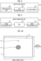
- FIG. 4A shows an example of the ultrasound image U captured by a minor axis method.
- the ultrasound image U shown in Fig. 4A shows a transverse cross section of a blood vessel B and a transverse cross section of a puncture needle N, which is an example of an insert.
- the transverse cross section of each of the blood vessel B and the puncture needle N here means a cut surface orthogonal to the extension direction of each of the blood vessel B and the puncture needle N.
- Fig. 4B shows an example of the ultrasound image U captured by a major axis method.
- the ultrasound image U shown in Fig. 4B shows a longitudinal cross section of the blood vessel B and a longitudinal cross section of the puncture needle N, which is the example of the insert.
- the longitudinal cross section of each of the blood vessel B and the puncture needle N here means a cut surface along the extension direction of each of the blood vessel B and the puncture needle N.
- a direction connecting a body surface S and an inside of the subject in the ultrasound image U is referred to as a depth direction D.
- the depth direction D corresponds to a direction in which a plurality of scanning lines extend in the ultrasound image U.
- a direction intersecting the depth direction D is referred to as a width direction H.
- the width direction H corresponds to a direction in which the plurality of scanning lines are arranged.
- the ultrasound image U generated by the image generation unit 32 is output to the acquisition unit 34.
- the acquisition unit 34 has a function of acquiring the ultrasound image U generated and output by the image generation unit 32.
- the ultrasound image U acquired by the acquisition unit 34 is output to the detection unit 36 and the degree-of-difficulty information output unit 44.
- the functions of the image generation unit 32 and the acquisition unit 34 may be integrated.
- the image generation unit 32 may further function as the acquisition unit 34.
- the detection unit 36 has a function of detecting the blood vessel B from the ultrasound image U input from the acquisition unit 34. It should be noted that the method in which the detection unit 36 detects the blood vessel B from the ultrasound image U is not particularly limited. As an example, the detection unit 36 according to the present embodiment analyzes the ultrasound image U acquired by the acquisition unit 34, in other words, the ultrasound image U generated by the image generation unit 32, according to a known algorithm, to detect the blood vessel B in the ultrasound image U.
- the detection unit 36 can store typical pattern data of a blood vessel region as a template in advance, derive a degree of similarity with respect to the pattern data while searching the ultrasound image U with the template, and consider that the blood vessel B is present in a place in which the degree of similarity is equal to or larger than a reference value and is maximized.
- the detection unit 36 may detect the blood vessel B in the ultrasound image U using a blood vessel detection model which is a trained model that has been subjected to machine learning using a plurality of ultrasound images U labeled with respect to the blood vessel B.
- the blood vessel detection model is an object detection algorithm using deep learning.
- an object detection model configured by a regional convolutional neural network (R-CNN), which is a type of a CNN, can be used.
- the blood vessel detection model detects the blood vessel B as an object from the input ultrasound image U, and outputs information indicating the blood vessel B in the ultrasound image U.
- the detection unit 36 has a function of detecting a diameter (hereinafter, referred to as a "blood vessel diameter") and a depth of the detected blood vessel B.
- a blood vessel diameter hereinafter, referred to as a "blood vessel diameter”
- the method in which the detection unit 36 derives each of the blood vessel diameter and the depth of the blood vessel B in the ultrasound image U is not particularly limited.
- the detection unit 36 according to the present embodiment applies known image analysis processing to the blood vessel B in the detected ultrasound image U to derive the blood vessel diameter and the depth of the blood vessel B.
- the blood vessel diameter of the blood vessel B means a width (length indicated by a reference numeral d2 in Fig.
- the depth of the blood vessel B means the shortest distance (distance indicated by a reference numeral d1 in Fig. 4C ) from the position corresponding to the body surface S of the subject in the ultrasound image U to the blood vessel B in the depth direction D.
- the detection unit 36 detects all the blood vessels B included in the ultrasound image U, and the depth and the blood vessel diameter thereof.
- a form may be adopted in which the detection is performed for each of the plurality of blood vessels B by applying, for example, the template matching, the general image recognition method, or the blood vessel detection model.
- a form may be adopted in which one or more blood vessels B are collectively detected by applying the template matching, the general image recognition method, or the blood vessel detection model.
- a detection result of the detection unit 36 is output to the determination unit 38. Specifically, the information indicating the blood vessel B in the ultrasound image U detected by the detection unit 36, and the depth and the blood vessel diameter of the blood vessel B are output to the determination unit 38.
- the determination unit 38 has a function of determining whether a puncture is possible or impossible based on blood vessel information according to a skill level of a user and the detection result of the detection unit 36. Specifically, the determination unit 38 determines whether the puncture is possible or impossible for the blood vessel B based on a blood vessel condition in which the puncture is possible according to the skill level of the user for the blood vessel B in the ultrasound image U, and the depth and the blood vessel diameter of the blood vessel B.
- the depth of the blood vessel B is described as another factor that determines the degree of difficulty of the puncture of the blood vessel B.
- the maximum blood vessel depth at which the puncture is possible is determined for each skill level.
- the maximum blood vessel depth in the blood vessel condition information 42B according to the present embodiment is an example of a depth threshold value according to the present disclosure.
- the determination unit 38 specifies the skill level corresponding to the user ID input from the authentication unit 40 with reference to the personal skill level information 42A.
- the determination unit 38 derives the minimum blood vessel diameter and the maximum blood vessel depth, which correspond to the specified skill level, with reference to the blood vessel condition information 42B.
- the determination unit 38 determines whether the puncture is possible or impossible by comparing each of the derived minimum blood vessel diameter and maximum blood vessel depth with the blood vessel diameter and the depth of the blood vessel B which are the detection results input from the detection unit 36. For example, in a case in which the user ID input from the authentication unit 40 is "0003", first, the determination unit 38 specifies that the skill level is "3" with reference to the personal skill level information 42A.
- the determination unit 38 derives "3 mm” for the minimum blood vessel diameter corresponding to the skill level of "3" and "15 mm” for the maximum blood vessel depth with reference to the blood vessel condition information 42B. Moreover, in a case in which the blood vessel diameter input from the detection unit 36 is equal to or larger than "3 mm" and the depth is equal to or smaller than "15 mm", the determination unit 38 determines that the puncture is possible. In other words, the determination unit 38 determines that the puncture is impossible in at least one of a case in which the blood vessel diameter input from the detection unit 36 is smaller than "3 mm” or a case in which the depth is larger than "15 mm".
- the determination unit 38 outputs, as the determination result, information indicating whether the puncture is possible or impossible to the degree-of-difficulty information output unit 44.
- a form may be adopted in which the plurality of registered user IDs are displayed on the display unit 46, and the authentication unit 40 acquires the user ID selected by the user through the input I/F unit 56 from among the plurality of registered user IDs.
- a form may be adopted in which the authentication unit 40 acquires the user ID based on the biometric information of the user read through the input I/F unit 56 or the like and the registered correspondence relationship.
- a form may be adopted in which the authentication unit 40 acquires the user ID by reading the user ID, such as an employee ID card owned by the user, through the input I/F unit 56 or the like.
- the user ID acquired by the authentication unit 40 is output to the determination unit 38.
- the degree-of-difficulty information output unit 44 has a function of outputting degree-of-difficulty information related to the degree of difficulty of the puncture in the blood vessel B detected by the detection unit 36 according to the skill level of the user who performs the puncture. Specifically, the degree-of-difficulty information output unit 44 according to the present embodiment adds, as the degree-of-difficulty information, information indicating whether the puncture is possible or impossible, which is a determination result of the determination unit 38, to the ultrasound image U input from the acquisition unit 34, and outputs the information to the display unit 46.
- the display unit 46 has a function of displaying various types of information, such as the ultrasound image U to which the degree-of-difficulty information output from the degree-of-difficulty information output unit 44, that is, the information indicating whether the puncture is possible or impossible is added.
- Examples of the display unit 46 include a liquid crystal display (LCD), an organic electro luminescence (EL) display, and a head mounted display.
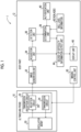
- the body part 12 can be configured by, for example, the hardware shown in Fig. 6.
- Fig. 6 shows a configuration diagram showing an example of a hardware configuration of the body part 12 according to the present embodiment.
- the body part 12 comprises the display unit 46, a control unit 50, the storage unit 52, a communication interface (I/F) unit 54, and the input I/F unit 56.
- the display unit 46, the control unit 50, the storage unit 52, the communication I/F unit 54, and the input I/F unit 56 are connected to each other through a bus 59, such as a system bus or a control bus, such that various types of information can be exchanged.
- a bus 59 such as a system bus or a control bus
- the control unit 50 controls an overall operation of the body part 12.
- the control unit 50 comprises a central processing unit (CPU) 50A, a read only memory (ROM) 50B, and a random access memory (RAM) 50C.
- the ROM 50B stores, in advance, various programs and the like, which are executed by the CPU 50A and include a puncture assist processing program 51 and an imaging program (not shown).
- the RAM 50C transitorily stores various data.
- the puncture assist processing program 51 according to the present embodiment is an example of an information processing program according to the present disclosure.
- the CPU 50A executes the imaging program stored in the ROM 50B, so that the CPU 50A functions as the imaging control unit 30.
- the CPU 50A executes the puncture assist processing program 51 stored in the ROM 50B, so that the CPU 50A functions as the acquisition unit 34, the detection unit 36, the determination unit 38, the authentication unit 40, the skill level information 42, and the degree-of-difficulty information output unit 44.
- the storage unit 52 stores image data of the ultrasound image U generated by the image generation unit 32, the skill level information 42, various other information, and the like. Specific examples of the storage unit 52 include a hard disk drive (HDD), a solid state drive (SSD), and a secure digital (SD) card.
- HDD hard disk drive
- SSD solid state drive
- SD secure digital
- the input I/F unit 56 is used for the user to input instructions and various types of information related to capturing of the ultrasound image U and the like.
- the input I/F unit 56 is not particularly limited, and examples thereof include various switches, a touch panel, a touch pen, a camera, and a mouse. It should be noted that the display unit 46 and the input I/F unit 56 may be integrated to form a touch panel display.
- the communication I/F unit 54 performs communication of various types of information with the ultrasound probe 10 or an external device of the ultrasound diagnostic apparatus 1 by wireless communication, such as WiFi (registered trademark) or Bluetooth (registered trademark) or wired communication.
- wireless communication such as WiFi (registered trademark) or Bluetooth (registered trademark) or wired communication.
- the body part 12 outputs the control signal for capturing the ultrasound image U to the ultrasound probe 10 through the communication I/F unit 54.
- the sound ray signal is input from the ultrasound probe 10 to the body part 12 through the communication I/F unit 54.
- the determination unit 38 acquires the blood vessel condition according to the skill level of the user. It should be noted that, in the present embodiment, as described above, the determination unit 38 acquires the minimum blood vessel diameter and the maximum blood vessel depth, which correspond to the user ID authenticated in step S100 with reference to the personal skill level information 42A and the blood vessel condition information 42B which are stored in the storage unit 52. Specifically, the determination unit 38 specifies the skill level corresponding to the user ID acquired in step S100 with reference to the personal skill level information 42A, as described above. Further, the determination unit 38 derives the minimum blood vessel diameter and the maximum blood vessel depth, which correspond to the skill level of the user, with reference to the blood vessel condition information 42B.
- next step S108 the determination unit 38 determines whether the puncture is possible or impossible.
- it is determined whether the puncture of the blood vessel B is possible or impossible by comparing the blood vessel diameter and the depth which are derived in step S104 for the blood vessel B in the ultrasound image U detected in step S104 with the minimum blood vessel diameter and the maximum blood vessel depth which are the blood vessel conditions according to the skill level of the user acquired in step S106.
- the blood vessel diameter of the blood vessel B derived in step S104 is 3 mm and the depth is 9 mm.
- the user ID acquired by the user authentication processing in step S100 is "0001”
- the skill level is "4" in step S106 with reference to the personal skill level information 42A (see Fig. 5A ).
- "2 mm” is derived as the minimum blood vessel diameter
- "20 mm” is derived as the maximum blood vessel depth with reference to the blood vessel condition information 42B (see Fig. 5B ).
- the blood vessel diameter of the blood vessel B is 3 mm, and is larger than the minimum blood vessel diameter.
- the depth of the blood vessel B is 9 mm, and is smaller than the maximum blood vessel depth. Therefore, the determination unit 38 determines that the puncture is possible.
- the skill level is "1" in step S106 with reference to the personal skill level information 42A (see Fig. 5A ).
- "5 mm” is derived as the minimum blood vessel diameter and "10 mm” is derived as the maximum blood vessel depth with reference to the blood vessel condition information 42B (see Fig. 5B ).
- the blood vessel diameter of the blood vessel B is 3 mm, and is smaller than the minimum blood vessel diameter.
- the depth of the blood vessel B is 9 mm, and is smaller than the maximum blood vessel depth. Therefore, the determination unit 38 determines that the puncture is impossible. The determination result of the determination unit 38 is output to the degree-of-difficulty information output unit 44.
- step S110 the degree-of-difficulty information output unit 44 determines whether the puncture is possible or impossible for the determination result of the determination unit 38. In a case of the determination result that the puncture is possible, an affirmative determination is made in the determination in step S110, and the processing proceeds to step S112.
- step S112 as described above, after the degree-of-difficulty information output unit 44 adds the degree-of-difficulty information indicating that the puncture is possible to the ultrasound image U acquired in step S102, the processing proceeds to step S116.
- Fig. 8A shows an example of the ultrasound image U to which the degree-of-difficulty information indicating that the puncture is possible is added.
- a form example is shown in which a relatively thick solid line enclosing an outer shape of the blood vessel B is applied as degree-of-difficulty information 80 1 indicating that the puncture is possible.
- step S110 a negative determination is made in the determination in step S110, and the processing proceeds to step S114.
- step S114 as described above, after the degree-of-difficulty information output unit 44 adds the degree-of-difficulty information indicating that the puncture is impossible to the ultrasound image U acquired in step S102, the processing proceeds to step S116.
- Fig. 8B shows an example of the ultrasound image U to which the degree-of-difficulty information indicating that the puncture is impossible is added. In the example shown in Fig. 8B , a form example is shown in which a relatively thick dotted line enclosing the outer shape of the blood vessel B is applied as degree-of-difficulty information 80 2 indicating that the puncture is impossible.
- next step S116 the degree-of-difficulty information output unit 44 outputs the ultrasound image U to which the degree-of-difficulty information generated in step S112 or step S114 is added, to the display unit 46.
- the ultrasound image U to which the degree-of-difficulty information 80 1 or 80 2 is added is displayed on the display unit 46.
- the form has been shown in which the types of the line (solid line or dotted line) as the display forms are made different for the degree-of-difficulty information 80 1 in a case in which the puncture is possible and the degree-of-difficulty information 80 2 in a case in which the puncture is impossible, but a form may be adopted in which other display forms are made different without being limited to the type of the line.
- a form may be adopted in which display forms, such as hue of the line, chroma saturation of the line, shading of the line, thickness of the line, and the presence or absence of turning on and off, are made different.
- a form may be adopted in which a frame enclosing a region of the blood vessel B is provided so that the display forms, such as a shape or a hue, are made different for the frame.
- a form may be adopted in which an arrow or a mark indicating whether the puncture is possible or impossible is displayed.
- next step S118 the degree-of-difficulty information output unit 44 determines whether or not to terminate the puncture assist processing.
- the puncture assist processing shown in Fig. 7 is terminated in a case in which a predetermined termination condition is satisfied, such as a case in which the user instructs the termination through the input I/F unit 56, a case in which the power of the body part 12 is cut off, or a case in which a predetermined time has elapsed after the processing of step S116 is terminated.
- a negative determination is made in the determination in step S118, and the processing proceeds to step S120.
- the degree-of-difficulty information added to the ultrasound image U displayed on the display unit 46 by the processing of step S116 corresponds to the case in which the puncture is impossible
- the user who performs the puncture may be changed from the user currently authenticated to another user, for example, the user having a high skill level.
- the authentication unit 40 determines whether or not to perform the user authentication in step S120. In a case in which the user authentication is performed, an affirmative determination is made in the determination in step S120, the processing returns to step S100, and the processing of steps S100 to S118 is repeated.
- step S116 in a case in which the degree-of-difficulty information added to the ultrasound image U displayed on the display unit 46 by the processing of step S116 corresponds to the case in which the puncture is impossible, for example, the puncture position may be changed. In such a case, it is determined whether the puncture of the blood vessel B in the ultrasound image U captured at the changed position is possible or impossible. Therefore, a negative determination is made in the determination in step S120, and the processing proceeds to step S102, and the processing of steps S102 to S118 is repeated.
- step S118 the puncture assist processing shown in Fig. 7 is terminated.
- the determination unit 38 determines that the puncture is possible in a case in which a distance r between the blood vessel B1 and the blood vessel B3 is equal to or larger than the minimum distance. In other words, the determination unit 38 determines that the puncture is impossible in a case in which the distance r between the blood vessel B1 and the blood vessel B3 is smaller than the minimum distance.
- the detection unit 36 detects the tissue surrounding the target blood vessel in the ultrasound image U by analyzing the ultrasound image U acquired by the acquisition unit 34 according to a known algorithm.
- the detection unit 36 can store the typical pattern data of the blood vessel region including the tissue surrounding the target blood vessel, in the example shown in Fig. 10 , the blood vessel B3 that is the brachial artery, as the template in advance, derive a degree of similarity with respect to the pattern data while searching the ultrasound image U with the template, and consider that the blood vessel B3 is present in a place in which the degree of similarity is equal to or larger than the reference value and is maximized.
- Fig. 11 shows a flowchart showing an example of a flow of the puncture assist processing according to the present modification example. It should be noted that, since the processing of steps S100 to S102 and the processing after steps S110 are the same as the puncture assist processing according to the form described above (see Fig. 7 ), the description thereof is omitted.
- the detection unit 36 derives the distance r between the tissue of the blood vessel B that is the target blood vessel in the ultrasound image U, and the surrounding tissue, as described above. As described above, the detection unit 36 detects the blood vessel B that is the target blood vessel in the ultrasound image U and the surrounding tissue, and derives the distance r between the blood vessel B and the surrounding tissue, which are detected. It should be noted that, in a case in which the ultrasound image U includes the plurality of blood vessels B that are the target blood vessels as described above, the detection unit 36 derives the distance r for each of the plurality of blood vessels B. The distance r derived by the detection unit 36 is output to the determination unit 38.
- next step S106A the determination unit 38 acquires the blood vessel condition according to the skill level of the user.
- the determination unit 38 acquires the minimum distance corresponding to the user ID authenticated in step S100 with reference to the personal skill level information 42A and the blood vessel condition information 42B1 which are stored in the storage unit 52.
- the determination unit 38 specifies the skill level corresponding to the user ID acquired in step S100 with reference to the personal skill level information 42A, as described above. Further, the determination unit 38 derives the minimum distance corresponding to the skill level of the user, with reference to the blood vessel condition information 42B1.
- next step S108A the determination unit 38 determines whether the puncture is possible or impossible, as described above.
- the form has been described in which the minimum distance between the target blood vessel and the surrounding tissue is applied as the blood vessel condition, but the blood vessel condition is not limited to the present modification example.
- each of the minimum blood vessel diameter and the maximum blood vessel depth which are the blood vessel conditions in the form described above, and the minimum distance according to the present modification example may be used alone as the blood vessel condition, or may be used in combination as the blood vessel conditions.
- Fig. 12A shows an example of the ultrasound image U to which the degree-of-difficulty information 80 1 indicating that the puncture is possible in the present modification example is added.
- a display example of the ultrasound image U in a case in which the blood vessel B1 is the target blood vessel and the puncture of the blood vessel B1 is possible is shown.
- the degree-of-difficulty information 80 1 is added to the blood vessel B1 in which the puncture is possible, the user can perform the puncture with reference to the degree-of-difficulty information 80 1 .
- Fig. 12B shows an example of the ultrasound image U to which the degree-of-difficulty information 80 2 indicating that the puncture is impossible in the present modification example is added.
- a display example in a case in which the blood vessel B1 is the target blood vessel and the puncture of the blood vessel B1 is impossible is shown.
- Fig. 12B shows a display example in a case in which the blood vessel B1 is the target blood vessel and the puncture of the blood vessel B1 is impossible.
- the degree-of-difficulty information output unit 44 information 80 3 indicating the surrounding tissue which is the reason why the puncture is impossible, as well as information 80 1 indicating the name of the tissue are added to the ultrasound image U and displayed on the display unit 46. It should be noted that, in the example shown in Fig. 12B , a form example is shown in which relatively thick one-dot chain lines enclosing the outer shapes of the blood vessels B2 and B3 are applied as the information 80 3 indicating the surrounding tissue.
- the user can recognize that the puncture is impossible with reference to the degree-of-difficulty information 80 2 .
- the information 80 3 and 80 4 are also added to the surrounding tissue, the user can recognize the reason why the puncture is impossible. It should be noted that a form may be adopted in which any one of the information 80 3 or 80 4 is added to the ultrasound image U and displayed. In addition, even in a case in which the puncture is possible, a form may be adopted in which the information 80 3 and 80 4 are displayed.
- a form may be adopted in which the information 80 3 and 80 4 are displayed according to the skill level of the user.
- a form may be adopted in which the information 80 3 and 80 4 are displayed in a case in which the skill level of the user is relatively low, and it is determined whether or not to display the information 80 3 and 80 4 by the selection of the user in a case in which the skill level of the user is relatively high.
- the form described above has been described in which the skill level of the user is set by the administrator or the like, but another example of the method of setting the skill level will be described in the present modification example.
- a form will be described in which a preset skill level is corrected based on a puncture record.
- the determination unit 38 in a case in which the number of punctures is considered as the skill level accumulates the number of punctures in association with the user ID.
- a form may be adopted in which the determination unit 38 performs correction to increase the skill level of the user acquired with reference to the personal skill level information 42A to a predetermined degree in a case in which the number of punctures exceeds a threshold value, and acquires the blood vessel condition with reference to the blood vessel condition information 42B based on the corrected skill level.
- the "success" and the "failure" of the puncture are determined by whether or not the puncture needle N is appropriately inserted into the blood vessel B that is the target blood vessel, the presence or absence of damage to other tissues, the presence or absence of the blood outflow to the outside of the blood vessel B, whether or not the puncture needle N penetrates the blood vessel B, and the like.
- the determination unit 38 determines whether the user succeeds or fails in the puncture and accumulates the determination result. It should be noted that the method in which the determination unit 38 determines the success or the failure of the puncture is not particularly limited. For example, a form may be adopted in which the determination unit 38 detects the puncture needle N from the ultrasound image U and determine the success or the failure of the puncture by identifying a relationship between the detected puncture needle N and the blood vessel B. It should be noted that the method in which the determination unit 38 detects the puncture needle N from the ultrasound image U is not particularly limited, and for example, the same method as the method in which the blood vessel B is detected from the ultrasound image U can be applied.
- the ultrasound image U displayed on the display unit 46 is relatively easy-to-see image, and thus there is a tendency that the puncture is easy. Therefore, in a case in which the image quality of the ultrasound image U is equal to or higher than the predetermined reference, even the user having a lower skill level than the user according to the form described above may be considered to be appropriate for the puncture.
- various processors shown below can be used as the hardware structure of processing units that execute various types of processing, such as the acquisition unit 34, the detection unit 36, the determination unit 38, the authentication unit 40, and the degree-of-difficulty information output unit 44.
- the various processors include, in addition to the CPU that is a general-purpose processor which executes software (program) and functions as various processing units, a programmable logic device (PLD) that is a processor of which a circuit configuration can be changed after manufacture, such as a field programmable gate array (FPGA), and a dedicated electric circuit that is a processor having a circuit configuration which is designed for exclusive use in order to execute specific processing, such as an application specific integrated circuit (ASIC).
- PLD programmable logic device
- FPGA field programmable gate array
- ASIC application specific integrated circuit
- a first example of the configuration in which the plurality of processing units are configured by one processor is a form in which, as represented by computers, such as a client and a server, one processor is configured by a combination of one or more CPUs and the software and this processor functions as the plurality of processing units.
- SoC system on chip
- IC integrated circuit
- circuitry in which circuit elements, such as semiconductor elements, are combined can be used as the hardware structure of the various processors.
- each of the puncture assist processing program 51 may be provided in a form of being recorded in a recording medium, such as a compact disc read only memory (CD-ROM), a digital versatile disc read only memory (DVD-ROM), and a universal serial bus (USB) memory.
- a form may be adopted in which each of the puncture assist processing program 51 is provided in a form being downloaded from an external device through a network.
Landscapes
- Health & Medical Sciences (AREA)
- Engineering & Computer Science (AREA)
- Life Sciences & Earth Sciences (AREA)
- Medical Informatics (AREA)
- Public Health (AREA)
- General Health & Medical Sciences (AREA)
- Biomedical Technology (AREA)
- Nuclear Medicine, Radiotherapy & Molecular Imaging (AREA)
- Pathology (AREA)
- Radiology & Medical Imaging (AREA)
- Surgery (AREA)
- Primary Health Care (AREA)
- Epidemiology (AREA)
- Physics & Mathematics (AREA)
- Heart & Thoracic Surgery (AREA)
- Biophysics (AREA)
- Molecular Biology (AREA)
- Veterinary Medicine (AREA)
- Animal Behavior & Ethology (AREA)
- General Business, Economics & Management (AREA)
- Business, Economics & Management (AREA)
- Computer Vision & Pattern Recognition (AREA)
- Vascular Medicine (AREA)
- Physiology (AREA)
- Urology & Nephrology (AREA)
- Data Mining & Analysis (AREA)
- Databases & Information Systems (AREA)
- Ultra Sonic Daignosis Equipment (AREA)
Applications Claiming Priority (2)
| Application Number | Priority Date | Filing Date | Title |
|---|---|---|---|
| JP2020197657 | 2020-11-27 | ||
| PCT/JP2021/042220 WO2022113844A1 (ja) | 2020-11-27 | 2021-11-17 | 情報処理装置、超音波診断装置、情報処理方法、及び情報処理プログラム |
Publications (3)
| Publication Number | Publication Date |
|---|---|
| EP4252672A1 EP4252672A1 (en) | 2023-10-04 |
| EP4252672A4 EP4252672A4 (en) | 2024-05-01 |
| EP4252672B1 true EP4252672B1 (en) | 2025-07-02 |
Family
ID=81754564
Family Applications (1)
| Application Number | Title | Priority Date | Filing Date |
|---|---|---|---|
| EP21897818.7A Active EP4252672B1 (en) | 2020-11-27 | 2021-11-17 | Information processing device, ultrasonic diagnosis device, information processing method, and information processing program |
Country Status (5)
| Country | Link |
|---|---|
| US (1) | US20230293145A1 (https=) |
| EP (1) | EP4252672B1 (https=) |
| JP (1) | JPWO2022113844A1 (https=) |
| CN (1) | CN116471996B (https=) |
| WO (1) | WO2022113844A1 (https=) |
Families Citing this family (6)
| Publication number | Priority date | Publication date | Assignee | Title |
|---|---|---|---|---|
| JP2024060247A (ja) * | 2022-10-19 | 2024-05-02 | 富士フイルム株式会社 | 超音波診断装置および超音波診断装置の制御方法 |
| JP2024060296A (ja) * | 2022-10-19 | 2024-05-02 | 富士フイルム株式会社 | 超音波診断装置および超音波診断装置の制御方法 |
| WO2025062860A1 (ja) * | 2023-09-21 | 2025-03-27 | テルモ株式会社 | 穿刺制御装置 |
| JP2025078326A (ja) * | 2023-11-08 | 2025-05-20 | 富士フイルム株式会社 | 超音波診断装置 |
| JP2025089751A (ja) * | 2023-12-04 | 2025-06-16 | 富士フイルム株式会社 | プロセッサ装置、プロセッサ装置の作動方法 |
| CN119326508B (zh) * | 2024-10-21 | 2025-10-03 | 天津市鹰泰利安康医疗科技有限责任公司 | 一种穿刺机器人的血管图像增强与分割方法及系统 |
Family Cites Families (18)
| Publication number | Priority date | Publication date | Assignee | Title |
|---|---|---|---|---|
| JPH1133026A (ja) * | 1997-07-14 | 1999-02-09 | Matsushita Electric Ind Co Ltd | 血管探査装置 |
| JPH11128224A (ja) * | 1997-10-28 | 1999-05-18 | Mitsubishi Heavy Ind Ltd | 超音波3次元画像再構成装置 |
| JP4064205B2 (ja) * | 2002-10-23 | 2008-03-19 | 学校法人東海大学 | 穿刺難易度評価装置 |
| JP4280098B2 (ja) * | 2003-03-31 | 2009-06-17 | 株式会社東芝 | 超音波診断装置及び穿刺治療支援プログラム |
| JP4820565B2 (ja) * | 2005-03-25 | 2011-11-24 | 株式会社東芝 | 超音波診断装置 |
| JP5292581B2 (ja) * | 2007-07-26 | 2013-09-18 | 学校法人藤田学園 | 血管撮影装置 |
| JP2013135776A (ja) * | 2011-12-28 | 2013-07-11 | Toshiba Corp | 超音波診断装置 |
| EP2666414A1 (en) * | 2012-05-22 | 2013-11-27 | Godert Zijlstra | Ultrasound device for guided puncture of vascular access |
| JP6073743B2 (ja) * | 2013-05-14 | 2017-02-01 | 東芝メディカルシステムズ株式会社 | 超音波診断装置及び画像処理プログラム |
| WO2015029499A1 (ja) * | 2013-08-30 | 2015-03-05 | 富士フイルム株式会社 | 超音波診断装置および超音波画像生成方法 |
| KR20160054992A (ko) * | 2014-11-07 | 2016-05-17 | 삼성전자주식회사 | 관심영역의 재검출 회피 장치 및 방법 |
| JP5920746B1 (ja) * | 2015-01-08 | 2016-05-18 | 学校法人早稲田大学 | 穿刺支援システム |
| JP6019369B1 (ja) * | 2015-08-31 | 2016-11-02 | 本多電子株式会社 | 超音波画像表示装置及びその作動方法、並びにプログラムを格納した記録媒体 |
| JP6629597B2 (ja) * | 2016-01-04 | 2020-01-15 | 株式会社日立製作所 | 超音波プローブ及びそれを用いた超音波診断装置 |
| CN106943180B (zh) * | 2017-03-31 | 2020-05-08 | 王刚 | 超声与声波引导自动助力式硬膜外穿刺辅助系统 |
| JP6897416B2 (ja) * | 2017-08-16 | 2021-06-30 | コニカミノルタ株式会社 | 超音波診断装置およびその作動方法 |
| JP6611104B1 (ja) * | 2018-12-12 | 2019-11-27 | フォルテ グロウ メディカル株式会社 | 携帯型超音波画像診断装置 |
| EP4449996B1 (en) * | 2019-04-26 | 2026-04-01 | FUJIFILM Corporation | Ultrasound diagnostic apparatus, method of controlling ultrasound diagnostic apparatus, and processor for ultrasound diagnostic apparatus |
-
2021
- 2021-11-17 EP EP21897818.7A patent/EP4252672B1/en active Active
- 2021-11-17 CN CN202180078951.9A patent/CN116471996B/zh active Active
- 2021-11-17 WO PCT/JP2021/042220 patent/WO2022113844A1/ja not_active Ceased
- 2021-11-17 JP JP2022565263A patent/JPWO2022113844A1/ja active Pending
-
2023
- 2023-05-23 US US18/322,120 patent/US20230293145A1/en active Pending
Also Published As
| Publication number | Publication date |
|---|---|
| CN116471996A (zh) | 2023-07-21 |
| CN116471996B (zh) | 2025-11-07 |
| WO2022113844A1 (ja) | 2022-06-02 |
| JPWO2022113844A1 (https=) | 2022-06-02 |
| US20230293145A1 (en) | 2023-09-21 |
| EP4252672A1 (en) | 2023-10-04 |
| EP4252672A4 (en) | 2024-05-01 |
Similar Documents
| Publication | Publication Date | Title |
|---|---|---|
| EP4252672B1 (en) | Information processing device, ultrasonic diagnosis device, information processing method, and information processing program | |
| US11823382B2 (en) | Ultrasound diagnostic apparatus and control method of ultrasound diagnostic apparatus | |
| CN103732152B (zh) | 超声波诊断装置及图像处理方法 | |
| CN114364325B (zh) | 超声波诊断装置及超声波诊断装置的控制方法 | |
| WO2020050017A1 (ja) | 超音波診断装置および超音波診断装置の制御方法 | |
| US11116475B2 (en) | Ultrasound diagnostic apparatus and method for controlling ultrasound diagnostic apparatus | |
| US20240065671A1 (en) | Ultrasound diagnostic apparatus and control method of ultrasound diagnostic apparatus | |
| US20230293144A1 (en) | Ultrasound diagnostic apparatus and control method of ultrasound diagnostic apparatus | |
| US11759173B2 (en) | Ultrasound diagnostic apparatus and control method of ultrasound diagnostic apparatus | |
| US12350094B2 (en) | Information processing apparatus, ultrasound diagnostic apparatus, information processing method, and information processing program | |
| CN116058875A (zh) | 超声成像方法和超声成像系统 | |
| US12608799B2 (en) | Information processing apparatus, information processing method, and program | |
| US12303336B2 (en) | Ultrasound diagnostic apparatus and control method of ultrasound diagnostic apparatus | |
| US11373307B2 (en) | Ultrasound diagnostic apparatus and method for controlling ultrasound diagnostic apparatus | |
| EP4344649B1 (en) | Ultrasound diagnostic apparatus and control method for ultrasound diagnostic apparatus | |
| US12599368B2 (en) | Information processing apparatus, information processing method, and program |
Legal Events
| Date | Code | Title | Description |
|---|---|---|---|
| STAA | Information on the status of an ep patent application or granted ep patent |
Free format text: STATUS: THE INTERNATIONAL PUBLICATION HAS BEEN MADE |
|
| PUAI | Public reference made under article 153(3) epc to a published international application that has entered the european phase |
Free format text: ORIGINAL CODE: 0009012 |
|
| STAA | Information on the status of an ep patent application or granted ep patent |
Free format text: STATUS: REQUEST FOR EXAMINATION WAS MADE |
|
| 17P | Request for examination filed |
Effective date: 20230523 |
|
| AK | Designated contracting states |
Kind code of ref document: A1 Designated state(s): AL AT BE BG CH CY CZ DE DK EE ES FI FR GB GR HR HU IE IS IT LI LT LU LV MC MK MT NL NO PL PT RO RS SE SI SK SM TR |
|
| DAV | Request for validation of the european patent (deleted) | ||
| DAX | Request for extension of the european patent (deleted) | ||
| A4 | Supplementary search report drawn up and despatched |
Effective date: 20240405 |
|
| RIC1 | Information provided on ipc code assigned before grant |
Ipc: A61B 8/14 20060101AFI20240328BHEP |
|
| GRAP | Despatch of communication of intention to grant a patent |
Free format text: ORIGINAL CODE: EPIDOSNIGR1 |
|
| STAA | Information on the status of an ep patent application or granted ep patent |
Free format text: STATUS: GRANT OF PATENT IS INTENDED |
|
| INTG | Intention to grant announced |
Effective date: 20250220 |
|
| GRAS | Grant fee paid |
Free format text: ORIGINAL CODE: EPIDOSNIGR3 |
|
| GRAA | (expected) grant |
Free format text: ORIGINAL CODE: 0009210 |
|
| STAA | Information on the status of an ep patent application or granted ep patent |
Free format text: STATUS: THE PATENT HAS BEEN GRANTED |
|
| AK | Designated contracting states |
Kind code of ref document: B1 Designated state(s): AL AT BE BG CH CY CZ DE DK EE ES FI FR GB GR HR HU IE IS IT LI LT LU LV MC MK MT NL NO PL PT RO RS SE SI SK SM TR |
|
| P01 | Opt-out of the competence of the unified patent court (upc) registered |
Free format text: CASE NUMBER: APP_24695/2025 Effective date: 20250523 |
|
| REG | Reference to a national code |
Ref country code: GB Ref legal event code: FG4D |
|
| REG | Reference to a national code |
Ref country code: CH Ref legal event code: EP |
|
| REG | Reference to a national code |
Ref country code: DE Ref legal event code: R096 Ref document number: 602021033585 Country of ref document: DE |
|
| REG | Reference to a national code |
Ref country code: IE Ref legal event code: FG4D |
|
| PGFP | Annual fee paid to national office [announced via postgrant information from national office to epo] |
Ref country code: FR Payment date: 20250930 Year of fee payment: 5 |
|
| REG | Reference to a national code |
Ref country code: NL Ref legal event code: MP Effective date: 20250702 |
|
| PG25 | Lapsed in a contracting state [announced via postgrant information from national office to epo] |
Ref country code: PT Free format text: LAPSE BECAUSE OF FAILURE TO SUBMIT A TRANSLATION OF THE DESCRIPTION OR TO PAY THE FEE WITHIN THE PRESCRIBED TIME-LIMIT Effective date: 20251103 |
|
| PG25 | Lapsed in a contracting state [announced via postgrant information from national office to epo] |
Ref country code: NL Free format text: LAPSE BECAUSE OF FAILURE TO SUBMIT A TRANSLATION OF THE DESCRIPTION OR TO PAY THE FEE WITHIN THE PRESCRIBED TIME-LIMIT Effective date: 20250702 |
|
| REG | Reference to a national code |
Ref country code: AT Ref legal event code: MK05 Ref document number: 1808328 Country of ref document: AT Kind code of ref document: T Effective date: 20250702 |
|
| PG25 | Lapsed in a contracting state [announced via postgrant information from national office to epo] |
Ref country code: IS Free format text: LAPSE BECAUSE OF FAILURE TO SUBMIT A TRANSLATION OF THE DESCRIPTION OR TO PAY THE FEE WITHIN THE PRESCRIBED TIME-LIMIT Effective date: 20251102 |
|
| PGFP | Annual fee paid to national office [announced via postgrant information from national office to epo] |
Ref country code: DE Payment date: 20250930 Year of fee payment: 5 |
|
| PGFP | Annual fee paid to national office [announced via postgrant information from national office to epo] |
Ref country code: GB Payment date: 20251001 Year of fee payment: 5 |
|
| PG25 | Lapsed in a contracting state [announced via postgrant information from national office to epo] |
Ref country code: NO Free format text: LAPSE BECAUSE OF FAILURE TO SUBMIT A TRANSLATION OF THE DESCRIPTION OR TO PAY THE FEE WITHIN THE PRESCRIBED TIME-LIMIT Effective date: 20251002 |
|
| REG | Reference to a national code |
Ref country code: LT Ref legal event code: MG9D |
|
| PG25 | Lapsed in a contracting state [announced via postgrant information from national office to epo] |
Ref country code: AT Free format text: LAPSE BECAUSE OF FAILURE TO SUBMIT A TRANSLATION OF THE DESCRIPTION OR TO PAY THE FEE WITHIN THE PRESCRIBED TIME-LIMIT Effective date: 20250702 |
|
| PG25 | Lapsed in a contracting state [announced via postgrant information from national office to epo] |
Ref country code: FI Free format text: LAPSE BECAUSE OF FAILURE TO SUBMIT A TRANSLATION OF THE DESCRIPTION OR TO PAY THE FEE WITHIN THE PRESCRIBED TIME-LIMIT Effective date: 20250702 |
|
| PG25 | Lapsed in a contracting state [announced via postgrant information from national office to epo] |
Ref country code: HR Free format text: LAPSE BECAUSE OF FAILURE TO SUBMIT A TRANSLATION OF THE DESCRIPTION OR TO PAY THE FEE WITHIN THE PRESCRIBED TIME-LIMIT Effective date: 20250702 |
|
| PG25 | Lapsed in a contracting state [announced via postgrant information from national office to epo] |
Ref country code: GR Free format text: LAPSE BECAUSE OF FAILURE TO SUBMIT A TRANSLATION OF THE DESCRIPTION OR TO PAY THE FEE WITHIN THE PRESCRIBED TIME-LIMIT Effective date: 20251003 |
|
| PG25 | Lapsed in a contracting state [announced via postgrant information from national office to epo] |
Ref country code: CZ Free format text: LAPSE BECAUSE OF FAILURE TO SUBMIT A TRANSLATION OF THE DESCRIPTION OR TO PAY THE FEE WITHIN THE PRESCRIBED TIME-LIMIT Effective date: 20250702 Ref country code: SE Free format text: LAPSE BECAUSE OF FAILURE TO SUBMIT A TRANSLATION OF THE DESCRIPTION OR TO PAY THE FEE WITHIN THE PRESCRIBED TIME-LIMIT Effective date: 20250702 |
|
| PG25 | Lapsed in a contracting state [announced via postgrant information from national office to epo] |
Ref country code: LV Free format text: LAPSE BECAUSE OF FAILURE TO SUBMIT A TRANSLATION OF THE DESCRIPTION OR TO PAY THE FEE WITHIN THE PRESCRIBED TIME-LIMIT Effective date: 20250702 |
|
| PG25 | Lapsed in a contracting state [announced via postgrant information from national office to epo] |
Ref country code: BG Free format text: LAPSE BECAUSE OF FAILURE TO SUBMIT A TRANSLATION OF THE DESCRIPTION OR TO PAY THE FEE WITHIN THE PRESCRIBED TIME-LIMIT Effective date: 20250702 Ref country code: PL Free format text: LAPSE BECAUSE OF FAILURE TO SUBMIT A TRANSLATION OF THE DESCRIPTION OR TO PAY THE FEE WITHIN THE PRESCRIBED TIME-LIMIT Effective date: 20250702 |
|
| PG25 | Lapsed in a contracting state [announced via postgrant information from national office to epo] |
Ref country code: RS Free format text: LAPSE BECAUSE OF FAILURE TO SUBMIT A TRANSLATION OF THE DESCRIPTION OR TO PAY THE FEE WITHIN THE PRESCRIBED TIME-LIMIT Effective date: 20251002 |
|
| PG25 | Lapsed in a contracting state [announced via postgrant information from national office to epo] |
Ref country code: ES Free format text: LAPSE BECAUSE OF FAILURE TO SUBMIT A TRANSLATION OF THE DESCRIPTION OR TO PAY THE FEE WITHIN THE PRESCRIBED TIME-LIMIT Effective date: 20250702 |
|
| PG25 | Lapsed in a contracting state [announced via postgrant information from national office to epo] |
Ref country code: RO Free format text: LAPSE BECAUSE OF FAILURE TO SUBMIT A TRANSLATION OF THE DESCRIPTION OR TO PAY THE FEE WITHIN THE PRESCRIBED TIME-LIMIT Effective date: 20250702 |
|
| PG25 | Lapsed in a contracting state [announced via postgrant information from national office to epo] |
Ref country code: SM Free format text: LAPSE BECAUSE OF FAILURE TO SUBMIT A TRANSLATION OF THE DESCRIPTION OR TO PAY THE FEE WITHIN THE PRESCRIBED TIME-LIMIT Effective date: 20250702 |
|
| PG25 | Lapsed in a contracting state [announced via postgrant information from national office to epo] |
Ref country code: DK Free format text: LAPSE BECAUSE OF FAILURE TO SUBMIT A TRANSLATION OF THE DESCRIPTION OR TO PAY THE FEE WITHIN THE PRESCRIBED TIME-LIMIT Effective date: 20250702 |
|
| PG25 | Lapsed in a contracting state [announced via postgrant information from national office to epo] |
Ref country code: IT Free format text: LAPSE BECAUSE OF FAILURE TO SUBMIT A TRANSLATION OF THE DESCRIPTION OR TO PAY THE FEE WITHIN THE PRESCRIBED TIME-LIMIT Effective date: 20250702 |
|
| PG25 | Lapsed in a contracting state [announced via postgrant information from national office to epo] |
Ref country code: SK Free format text: LAPSE BECAUSE OF FAILURE TO SUBMIT A TRANSLATION OF THE DESCRIPTION OR TO PAY THE FEE WITHIN THE PRESCRIBED TIME-LIMIT Effective date: 20250702 Ref country code: EE Free format text: LAPSE BECAUSE OF FAILURE TO SUBMIT A TRANSLATION OF THE DESCRIPTION OR TO PAY THE FEE WITHIN THE PRESCRIBED TIME-LIMIT Effective date: 20250702 |