EP4219721A2 - Compositions and methods for selective protein expression - Google Patents
Compositions and methods for selective protein expression Download PDFInfo
- Publication number
- EP4219721A2 EP4219721A2 EP23151319.3A EP23151319A EP4219721A2 EP 4219721 A2 EP4219721 A2 EP 4219721A2 EP 23151319 A EP23151319 A EP 23151319A EP 4219721 A2 EP4219721 A2 EP 4219721A2
- Authority
- EP
- European Patent Office
- Prior art keywords
- domain
- cell
- protein
- seq
- fusion protein
- Prior art date
- Legal status (The legal status is an assumption and is not a legal conclusion. Google has not performed a legal analysis and makes no representation as to the accuracy of the status listed.)
- Pending
Links
- 230000014509 gene expression Effects 0.000 title claims abstract description 213
- 108090000623 proteins and genes Proteins 0.000 title claims abstract description 172
- 102000004169 proteins and genes Human genes 0.000 title claims abstract description 120
- 238000000034 method Methods 0.000 title claims abstract description 46
- 239000000203 mixture Substances 0.000 title description 10
- 210000004027 cell Anatomy 0.000 claims abstract description 224
- 108020001507 fusion proteins Proteins 0.000 claims abstract description 187
- 102000037865 fusion proteins Human genes 0.000 claims abstract description 187
- 210000001744 T-lymphocyte Anatomy 0.000 claims abstract description 122
- 238000003776 cleavage reaction Methods 0.000 claims abstract description 110
- 230000007017 scission Effects 0.000 claims abstract description 110
- 108091005804 Peptidases Proteins 0.000 claims abstract description 63
- 239000004365 Protease Substances 0.000 claims abstract description 63
- 108020001580 protein domains Proteins 0.000 claims abstract description 53
- 208000037265 diseases, disorders, signs and symptoms Diseases 0.000 claims abstract description 44
- 210000003719 b-lymphocyte Anatomy 0.000 claims abstract description 38
- 201000010099 disease Diseases 0.000 claims abstract description 38
- 150000001875 compounds Chemical class 0.000 claims description 168
- 239000000427 antigen Substances 0.000 claims description 167
- 102000036639 antigens Human genes 0.000 claims description 165
- 108091007433 antigens Proteins 0.000 claims description 165
- 230000015556 catabolic process Effects 0.000 claims description 120
- 238000006731 degradation reaction Methods 0.000 claims description 120
- 206010028980 Neoplasm Diseases 0.000 claims description 106
- 108090000765 processed proteins & peptides Proteins 0.000 claims description 67
- 150000007523 nucleic acids Chemical class 0.000 claims description 63
- 102000035195 Peptidases Human genes 0.000 claims description 61
- 241000282414 Homo sapiens Species 0.000 claims description 59
- 230000006641 stabilisation Effects 0.000 claims description 58
- 238000011105 stabilization Methods 0.000 claims description 58
- 102100035233 Furin Human genes 0.000 claims description 55
- 108090001126 Furin Proteins 0.000 claims description 55
- 125000003275 alpha amino acid group Chemical group 0.000 claims description 55
- 102000039446 nucleic acids Human genes 0.000 claims description 49
- 108020004707 nucleic acids Proteins 0.000 claims description 49
- 102000004196 processed proteins & peptides Human genes 0.000 claims description 48
- 201000011510 cancer Diseases 0.000 claims description 44
- 229920001184 polypeptide Polymers 0.000 claims description 42
- 239000013598 vector Substances 0.000 claims description 34
- 102000035160 transmembrane proteins Human genes 0.000 claims description 27
- 108091005703 transmembrane proteins Proteins 0.000 claims description 27
- 230000003834 intracellular effect Effects 0.000 claims description 25
- 238000011282 treatment Methods 0.000 claims description 23
- NMFHJNAPXOMSRX-PUPDPRJKSA-N [(1r)-3-(3,4-dimethoxyphenyl)-1-[3-(2-morpholin-4-ylethoxy)phenyl]propyl] (2s)-1-[(2s)-2-(3,4,5-trimethoxyphenyl)butanoyl]piperidine-2-carboxylate Chemical compound C([C@@H](OC(=O)[C@@H]1CCCCN1C(=O)[C@@H](CC)C=1C=C(OC)C(OC)=C(OC)C=1)C=1C=C(OCCN2CCOCC2)C=CC=1)CC1=CC=C(OC)C(OC)=C1 NMFHJNAPXOMSRX-PUPDPRJKSA-N 0.000 claims description 19
- 229960000817 bazedoxifene Drugs 0.000 claims description 17
- UCJGJABZCDBEDK-UHFFFAOYSA-N bazedoxifene Chemical compound C=1C=C(OCCN2CCCCCC2)C=CC=1CN1C2=CC=C(O)C=C2C(C)=C1C1=CC=C(O)C=C1 UCJGJABZCDBEDK-UHFFFAOYSA-N 0.000 claims description 17
- 230000001965 increasing effect Effects 0.000 claims description 14
- 108010038795 estrogen receptors Proteins 0.000 claims description 13
- 108010022394 Threonine synthase Proteins 0.000 claims description 12
- 230000006800 cellular catabolic process Effects 0.000 claims description 12
- 102000004419 dihydrofolate reductase Human genes 0.000 claims description 12
- 108010076504 Protein Sorting Signals Proteins 0.000 claims description 11
- 210000004899 c-terminal region Anatomy 0.000 claims description 10
- 208000024893 Acute lymphoblastic leukemia Diseases 0.000 claims description 9
- 208000014697 Acute lymphocytic leukaemia Diseases 0.000 claims description 9
- 101710089384 Extracellular protease Proteins 0.000 claims description 9
- 208000006664 Precursor Cell Lymphoblastic Leukemia-Lymphoma Diseases 0.000 claims description 9
- 201000008129 pancreatic ductal adenocarcinoma Diseases 0.000 claims description 9
- 238000011301 standard therapy Methods 0.000 claims description 9
- 206010033128 Ovarian cancer Diseases 0.000 claims description 8
- 206010012818 diffuse large B-cell lymphoma Diseases 0.000 claims description 8
- 210000000822 natural killer cell Anatomy 0.000 claims description 8
- 206010025323 Lymphomas Diseases 0.000 claims description 7
- 201000003793 Myelodysplastic syndrome Diseases 0.000 claims description 7
- 239000003814 drug Substances 0.000 claims description 7
- WTAASEZRCHZOHF-OPXVNJCFSA-N (2s)-3-(4-hydroxyphenyl)-2-[[(2s)-3-(1h-imidazol-5-yl)-2-[[(2s)-2-[[(2s)-2-[[2-[[(2s)-pyrrolidine-2-carbonyl]amino]acetyl]amino]propanoyl]amino]propanoyl]amino]propanoyl]amino]propanoic acid Chemical compound N([C@@H](C)C(=O)N[C@@H](C)C(=O)N[C@@H](CC=1NC=NC=1)C(=O)N[C@@H](CC=1C=CC(O)=CC=1)C(O)=O)C(=O)CNC(=O)[C@@H]1CCCN1 WTAASEZRCHZOHF-OPXVNJCFSA-N 0.000 claims description 6
- 208000031261 Acute myeloid leukaemia Diseases 0.000 claims description 6
- YNCHFVRXEQFPBY-BQBZGAKWSA-N Asp-Gly-Arg Chemical compound OC(=O)C[C@H](N)C(=O)NCC(=O)N[C@H](C(O)=O)CCCN=C(N)N YNCHFVRXEQFPBY-BQBZGAKWSA-N 0.000 claims description 6
- 102100035371 Chymotrypsin-like elastase family member 1 Human genes 0.000 claims description 6
- 101710138848 Chymotrypsin-like elastase family member 1 Proteins 0.000 claims description 6
- 101710099240 Elastase-1 Proteins 0.000 claims description 6
- 102100029727 Enteropeptidase Human genes 0.000 claims description 6
- 108010013369 Enteropeptidase Proteins 0.000 claims description 6
- 108010074860 Factor Xa Proteins 0.000 claims description 6
- 125000000899 L-alpha-glutamyl group Chemical group [H]N([H])[C@]([H])(C(=O)[*])C([H])([H])C([H])([H])C(O[H])=O 0.000 claims description 6
- 208000031671 Large B-Cell Diffuse Lymphoma Diseases 0.000 claims description 6
- 108010076818 TEV protease Proteins 0.000 claims description 6
- 108090000190 Thrombin Proteins 0.000 claims description 6
- 239000012636 effector Substances 0.000 claims description 6
- 201000003444 follicular lymphoma Diseases 0.000 claims description 6
- 230000037361 pathway Effects 0.000 claims description 6
- 229960004072 thrombin Drugs 0.000 claims description 6
- 102000027257 transmembrane receptors Human genes 0.000 claims description 6
- 108091008578 transmembrane receptors Proteins 0.000 claims description 6
- 108010060175 trypsinogen activation peptide Proteins 0.000 claims description 6
- 230000003612 virological effect Effects 0.000 claims description 6
- DODQJNMQWMSYGS-QPLCGJKRSA-N 4-[(z)-1-[4-[2-(dimethylamino)ethoxy]phenyl]-1-phenylbut-1-en-2-yl]phenol Chemical compound C=1C=C(O)C=CC=1C(/CC)=C(C=1C=CC(OCCN(C)C)=CC=1)/C1=CC=CC=C1 DODQJNMQWMSYGS-QPLCGJKRSA-N 0.000 claims description 5
- 208000025205 Mantle-Cell Lymphoma Diseases 0.000 claims description 5
- 208000034578 Multiple myelomas Diseases 0.000 claims description 5
- 206010061535 Ovarian neoplasm Diseases 0.000 claims description 5
- 206010061902 Pancreatic neoplasm Diseases 0.000 claims description 5
- 206010035226 Plasma cell myeloma Diseases 0.000 claims description 5
- 230000003247 decreasing effect Effects 0.000 claims description 5
- 230000002489 hematologic effect Effects 0.000 claims description 5
- 208000032839 leukemia Diseases 0.000 claims description 5
- 208000015486 malignant pancreatic neoplasm Diseases 0.000 claims description 5
- 201000002528 pancreatic cancer Diseases 0.000 claims description 5
- 208000008443 pancreatic carcinoma Diseases 0.000 claims description 5
- 208000003950 B-cell lymphoma Diseases 0.000 claims description 4
- 206010006187 Breast cancer Diseases 0.000 claims description 4
- 208000026310 Breast neoplasm Diseases 0.000 claims description 4
- 206010009944 Colon cancer Diseases 0.000 claims description 4
- 208000001333 Colorectal Neoplasms Diseases 0.000 claims description 4
- 206010058467 Lung neoplasm malignant Diseases 0.000 claims description 4
- 201000003791 MALT lymphoma Diseases 0.000 claims description 4
- 102000018697 Membrane Proteins Human genes 0.000 claims description 4
- 108010052285 Membrane Proteins Proteins 0.000 claims description 4
- 206010027406 Mesothelioma Diseases 0.000 claims description 4
- 241001465754 Metazoa Species 0.000 claims description 4
- 206010029461 Nodal marginal zone B-cell lymphomas Diseases 0.000 claims description 4
- 208000007452 Plasmacytoma Diseases 0.000 claims description 4
- 230000008859 change Effects 0.000 claims description 4
- 201000009277 hairy cell leukemia Diseases 0.000 claims description 4
- 201000005202 lung cancer Diseases 0.000 claims description 4
- 208000020816 lung neoplasm Diseases 0.000 claims description 4
- 208000006178 malignant mesothelioma Diseases 0.000 claims description 4
- 201000005282 malignant pleural mesothelioma Diseases 0.000 claims description 4
- 230000001394 metastastic effect Effects 0.000 claims description 4
- 206010061289 metastatic neoplasm Diseases 0.000 claims description 4
- 201000006039 nodal marginal zone lymphoma Diseases 0.000 claims description 4
- 208000036764 Adenocarcinoma of the esophagus Diseases 0.000 claims description 3
- 208000010839 B-cell chronic lymphocytic leukemia Diseases 0.000 claims description 3
- 206010005003 Bladder cancer Diseases 0.000 claims description 3
- 108090000712 Cathepsin B Proteins 0.000 claims description 3
- 102000004225 Cathepsin B Human genes 0.000 claims description 3
- 102000001398 Granzyme Human genes 0.000 claims description 3
- 108060005986 Granzyme Proteins 0.000 claims description 3
- 208000017604 Hodgkin disease Diseases 0.000 claims description 3
- 208000021519 Hodgkin lymphoma Diseases 0.000 claims description 3
- 208000010747 Hodgkins lymphoma Diseases 0.000 claims description 3
- 101001128694 Homo sapiens Neuroendocrine convertase 1 Proteins 0.000 claims description 3
- 101001072081 Homo sapiens Proprotein convertase subtilisin/kexin type 5 Proteins 0.000 claims description 3
- 101001098833 Homo sapiens Proprotein convertase subtilisin/kexin type 6 Proteins 0.000 claims description 3
- 101001098872 Homo sapiens Proprotein convertase subtilisin/kexin type 7 Proteins 0.000 claims description 3
- 206010023774 Large cell lung cancer Diseases 0.000 claims description 3
- 102100032132 Neuroendocrine convertase 1 Human genes 0.000 claims description 3
- 206010030137 Oesophageal adenocarcinoma Diseases 0.000 claims description 3
- 208000007571 Ovarian Epithelial Carcinoma Diseases 0.000 claims description 3
- 102100036365 Proprotein convertase subtilisin/kexin type 5 Human genes 0.000 claims description 3
- 102100038946 Proprotein convertase subtilisin/kexin type 6 Human genes 0.000 claims description 3
- 102100038950 Proprotein convertase subtilisin/kexin type 7 Human genes 0.000 claims description 3
- 206010041067 Small cell lung cancer Diseases 0.000 claims description 3
- 208000007097 Urinary Bladder Neoplasms Diseases 0.000 claims description 3
- 208000028653 esophageal adenocarcinoma Diseases 0.000 claims description 3
- 201000009546 lung large cell carcinoma Diseases 0.000 claims description 3
- 201000005243 lung squamous cell carcinoma Diseases 0.000 claims description 3
- 208000002154 non-small cell lung carcinoma Diseases 0.000 claims description 3
- 239000002245 particle Substances 0.000 claims description 3
- 208000000587 small cell lung carcinoma Diseases 0.000 claims description 3
- 238000002560 therapeutic procedure Methods 0.000 claims description 3
- 208000029729 tumor suppressor gene on chromosome 11 Diseases 0.000 claims description 3
- 201000005112 urinary bladder cancer Diseases 0.000 claims description 3
- 239000013603 viral vector Substances 0.000 claims description 3
- 208000017726 ALK-positive large B-cell lymphoma Diseases 0.000 claims description 2
- 206010002412 Angiocentric lymphomas Diseases 0.000 claims description 2
- 208000036170 B-Cell Marginal Zone Lymphoma Diseases 0.000 claims description 2
- 208000032568 B-cell prolymphocytic leukaemia Diseases 0.000 claims description 2
- 208000032791 BCR-ABL1 positive chronic myelogenous leukemia Diseases 0.000 claims description 2
- 208000011691 Burkitt lymphomas Diseases 0.000 claims description 2
- 208000016778 CD4+/CD56+ hematodermic neoplasm Diseases 0.000 claims description 2
- 208000010833 Chronic myeloid leukaemia Diseases 0.000 claims description 2
- 206010061850 Extranodal marginal zone B-cell lymphoma (MALT type) Diseases 0.000 claims description 2
- 201000000439 HCL-V Diseases 0.000 claims description 2
- 208000035481 HHV-8-associated multicentric Castleman disease Diseases 0.000 claims description 2
- 208000010956 Hairy cell leukemia variant Diseases 0.000 claims description 2
- 208000028018 Lymphocytic leukaemia Diseases 0.000 claims description 2
- 208000033761 Myelogenous Chronic BCR-ABL Positive Leukemia Diseases 0.000 claims description 2
- 201000007224 Myeloproliferative neoplasm Diseases 0.000 claims description 2
- 208000015914 Non-Hodgkin lymphomas Diseases 0.000 claims description 2
- 206010065857 Primary Effusion Lymphoma Diseases 0.000 claims description 2
- 208000024588 Primary cutaneous follicle center lymphoma Diseases 0.000 claims description 2
- 206010036711 Primary mediastinal large B-cell lymphomas Diseases 0.000 claims description 2
- 208000035416 Prolymphocytic B-Cell Leukemia Diseases 0.000 claims description 2
- 208000011783 Splenic diffuse red pulp small B-cell lymphoma Diseases 0.000 claims description 2
- 208000016025 Waldenstroem macroglobulinemia Diseases 0.000 claims description 2
- 208000033559 Waldenström macroglobulinemia Diseases 0.000 claims description 2
- 210000000988 bone and bone Anatomy 0.000 claims description 2
- 208000035269 cancer or benign tumor Diseases 0.000 claims description 2
- 208000037976 chronic inflammation Diseases 0.000 claims description 2
- 230000006020 chronic inflammation Effects 0.000 claims description 2
- 208000025750 heavy chain disease Diseases 0.000 claims description 2
- 208000026876 intravascular large B-cell lymphoma Diseases 0.000 claims description 2
- 208000003747 lymphoid leukemia Diseases 0.000 claims description 2
- 208000006116 lymphomatoid granulomatosis Diseases 0.000 claims description 2
- 201000007919 lymphoplasmacytic lymphoma Diseases 0.000 claims description 2
- 230000001589 lymphoproliferative effect Effects 0.000 claims description 2
- 230000003211 malignant effect Effects 0.000 claims description 2
- 201000007924 marginal zone B-cell lymphoma Diseases 0.000 claims description 2
- 208000021937 marginal zone lymphoma Diseases 0.000 claims description 2
- 208000015325 multicentric Castleman disease Diseases 0.000 claims description 2
- 208000007525 plasmablastic lymphoma Diseases 0.000 claims description 2
- 210000005134 plasmacytoid dendritic cell Anatomy 0.000 claims description 2
- 206010062113 splenic marginal zone lymphoma Diseases 0.000 claims description 2
- 102000015694 estrogen receptors Human genes 0.000 claims 5
- 108010019670 Chimeric Antigen Receptors Proteins 0.000 abstract description 207
- 230000001105 regulatory effect Effects 0.000 abstract description 11
- 230000001404 mediated effect Effects 0.000 abstract description 8
- 230000008685 targeting Effects 0.000 abstract description 8
- 239000008194 pharmaceutical composition Substances 0.000 abstract description 6
- 238000012546 transfer Methods 0.000 abstract description 5
- 102100037486 Reverse transcriptase/ribonuclease H Human genes 0.000 abstract 2
- 235000018102 proteins Nutrition 0.000 description 107
- 230000027455 binding Effects 0.000 description 103
- 230000002776 aggregation Effects 0.000 description 88
- 238000004220 aggregation Methods 0.000 description 88
- 230000004068 intracellular signaling Effects 0.000 description 55
- 102100024222 B-lymphocyte antigen CD19 Human genes 0.000 description 52
- 101000980825 Homo sapiens B-lymphocyte antigen CD19 Proteins 0.000 description 52
- -1 ICOS Proteins 0.000 description 46
- 230000011664 signaling Effects 0.000 description 45
- 230000000139 costimulatory effect Effects 0.000 description 36
- 102000008394 Immunoglobulin Fragments Human genes 0.000 description 30
- 108010021625 Immunoglobulin Fragments Proteins 0.000 description 29
- 108091008874 T cell receptors Proteins 0.000 description 27
- 102000016266 T-Cell Antigen Receptors Human genes 0.000 description 27
- 239000003446 ligand Substances 0.000 description 27
- 241000699670 Mus sp. Species 0.000 description 26
- 230000000087 stabilizing effect Effects 0.000 description 26
- 230000004913 activation Effects 0.000 description 25
- 230000006870 function Effects 0.000 description 25
- 108060003951 Immunoglobulin Proteins 0.000 description 24
- 102000018358 immunoglobulin Human genes 0.000 description 24
- 102100022005 B-lymphocyte antigen CD20 Human genes 0.000 description 22
- 101000897405 Homo sapiens B-lymphocyte antigen CD20 Proteins 0.000 description 22
- 230000004936 stimulating effect Effects 0.000 description 22
- 108091028043 Nucleic acid sequence Proteins 0.000 description 21
- 238000006471 dimerization reaction Methods 0.000 description 20
- 239000012634 fragment Substances 0.000 description 20
- 108020004999 messenger RNA Proteins 0.000 description 20
- 102000053602 DNA Human genes 0.000 description 19
- 108020004414 DNA Proteins 0.000 description 19
- 239000000047 product Substances 0.000 description 19
- 102000017420 CD3 protein, epsilon/gamma/delta subunit Human genes 0.000 description 17
- 108050005493 CD3 protein, epsilon/gamma/delta subunit Proteins 0.000 description 17
- 230000001419 dependent effect Effects 0.000 description 17
- 102000005962 receptors Human genes 0.000 description 17
- 108020003175 receptors Proteins 0.000 description 17
- 101000914514 Homo sapiens T-cell-specific surface glycoprotein CD28 Proteins 0.000 description 16
- 108700018351 Major Histocompatibility Complex Proteins 0.000 description 16
- 102100027213 T-cell-specific surface glycoprotein CD28 Human genes 0.000 description 16
- 239000002773 nucleotide Substances 0.000 description 16
- 125000003729 nucleotide group Chemical group 0.000 description 16
- 102000004039 Caspase-9 Human genes 0.000 description 15
- 108090000566 Caspase-9 Proteins 0.000 description 15
- 235000001014 amino acid Nutrition 0.000 description 15
- 239000012642 immune effector Substances 0.000 description 15
- 230000028993 immune response Effects 0.000 description 15
- 229940121354 immunomodulator Drugs 0.000 description 15
- 230000004044 response Effects 0.000 description 15
- 229920002477 rna polymer Polymers 0.000 description 15
- 230000020382 suppression by virus of host antigen processing and presentation of peptide antigen via MHC class I Effects 0.000 description 15
- 102100027207 CD27 antigen Human genes 0.000 description 14
- 102100035932 Cocaine- and amphetamine-regulated transcript protein Human genes 0.000 description 14
- 206010010144 Completed suicide Diseases 0.000 description 14
- 101000914511 Homo sapiens CD27 antigen Proteins 0.000 description 14
- 101000715592 Homo sapiens Cocaine- and amphetamine-regulated transcript protein Proteins 0.000 description 14
- 125000000539 amino acid group Chemical group 0.000 description 14
- 108010005327 CD19-specific chimeric antigen receptor Proteins 0.000 description 13
- 229940024606 amino acid Drugs 0.000 description 13
- 230000001086 cytosolic effect Effects 0.000 description 13
- 230000001939 inductive effect Effects 0.000 description 13
- 230000000638 stimulation Effects 0.000 description 13
- 150000001413 amino acids Chemical class 0.000 description 12
- 238000013518 transcription Methods 0.000 description 12
- 230000035897 transcription Effects 0.000 description 12
- 210000004881 tumor cell Anatomy 0.000 description 12
- 102100038595 Estrogen receptor Human genes 0.000 description 11
- 101000851370 Homo sapiens Tumor necrosis factor receptor superfamily member 9 Proteins 0.000 description 11
- 108010002350 Interleukin-2 Proteins 0.000 description 11
- 102000000588 Interleukin-2 Human genes 0.000 description 11
- 102100036856 Tumor necrosis factor receptor superfamily member 9 Human genes 0.000 description 11
- 230000001413 cellular effect Effects 0.000 description 11
- 230000000694 effects Effects 0.000 description 11
- 238000000338 in vitro Methods 0.000 description 11
- 230000002147 killing effect Effects 0.000 description 11
- 102000040430 polynucleotide Human genes 0.000 description 11
- 108091033319 polynucleotide Proteins 0.000 description 11
- 239000002157 polynucleotide Substances 0.000 description 11
- 108020004705 Codon Proteins 0.000 description 10
- 102100038083 Endosialin Human genes 0.000 description 10
- 102100031585 ADP-ribosyl cyclase/cyclic ADP-ribose hydrolase 1 Human genes 0.000 description 9
- 102100026094 C-type lectin domain family 12 member A Human genes 0.000 description 9
- 101710188619 C-type lectin domain family 12 member A Proteins 0.000 description 9
- 102000001301 EGF receptor Human genes 0.000 description 9
- 101000777636 Homo sapiens ADP-ribosyl cyclase/cyclic ADP-ribose hydrolase 1 Proteins 0.000 description 9
- 206010034277 Pemphigoid Diseases 0.000 description 9
- 239000011324 bead Substances 0.000 description 9
- 238000000684 flow cytometry Methods 0.000 description 9
- 238000001727 in vivo Methods 0.000 description 9
- 210000001519 tissue Anatomy 0.000 description 9
- 102100025064 Cellular tumor antigen p53 Human genes 0.000 description 8
- 108091007741 Chimeric antigen receptor T cells Proteins 0.000 description 8
- 108060006698 EGF receptor Proteins 0.000 description 8
- 101000721661 Homo sapiens Cellular tumor antigen p53 Proteins 0.000 description 8
- 101000884275 Homo sapiens Endosialin Proteins 0.000 description 8
- 101001133056 Homo sapiens Mucin-1 Proteins 0.000 description 8
- 102100034256 Mucin-1 Human genes 0.000 description 8
- 241000699666 Mus <mouse, genus> Species 0.000 description 8
- 239000000463 material Substances 0.000 description 8
- 201000001441 melanoma Diseases 0.000 description 8
- 239000002904 solvent Substances 0.000 description 8
- MTCFGRXMJLQNBG-REOHCLBHSA-N (2S)-2-Amino-3-hydroxypropansäure Chemical compound OC[C@H](N)C(O)=O MTCFGRXMJLQNBG-REOHCLBHSA-N 0.000 description 7
- 102100038080 B-cell receptor CD22 Human genes 0.000 description 7
- 102100037086 Bone marrow stromal antigen 2 Human genes 0.000 description 7
- 102100025570 Cancer/testis antigen 1 Human genes 0.000 description 7
- 241000282693 Cercopithecidae Species 0.000 description 7
- 102000003859 Claudin-6 Human genes 0.000 description 7
- 108090000229 Claudin-6 Proteins 0.000 description 7
- 102100031507 Fc receptor-like protein 5 Human genes 0.000 description 7
- 101000884305 Homo sapiens B-cell receptor CD22 Proteins 0.000 description 7
- 101000846908 Homo sapiens Fc receptor-like protein 5 Proteins 0.000 description 7
- 101000878602 Homo sapiens Immunoglobulin alpha Fc receptor Proteins 0.000 description 7
- 101000998120 Homo sapiens Interleukin-3 receptor subunit alpha Proteins 0.000 description 7
- 101000655352 Homo sapiens Telomerase reverse transcriptase Proteins 0.000 description 7
- 101000851376 Homo sapiens Tumor necrosis factor receptor superfamily member 8 Proteins 0.000 description 7
- 102100038005 Immunoglobulin alpha Fc receptor Human genes 0.000 description 7
- 102100039688 Insulin-like growth factor 1 receptor Human genes 0.000 description 7
- 102100033493 Interleukin-3 receptor subunit alpha Human genes 0.000 description 7
- 241000713666 Lentivirus Species 0.000 description 7
- 102100020943 Leukocyte-associated immunoglobulin-like receptor 1 Human genes 0.000 description 7
- 102100033486 Lymphocyte antigen 75 Human genes 0.000 description 7
- 241000283984 Rodentia Species 0.000 description 7
- 108010003723 Single-Domain Antibodies Proteins 0.000 description 7
- 102100036857 Tumor necrosis factor receptor superfamily member 8 Human genes 0.000 description 7
- 210000004369 blood Anatomy 0.000 description 7
- 239000008280 blood Substances 0.000 description 7
- 230000004927 fusion Effects 0.000 description 7
- 239000000523 sample Substances 0.000 description 7
- 150000003384 small molecules Chemical class 0.000 description 7
- RJBDSRWGVYNDHL-XNJNKMBASA-N (2S,4R,5S,6S)-2-[(2S,3R,4R,5S,6R)-5-[(2S,3R,4R,5R,6R)-3-acetamido-4,5-dihydroxy-6-(hydroxymethyl)oxan-2-yl]oxy-2-[(2R,3S,4R,5R,6R)-4,5-dihydroxy-2-(hydroxymethyl)-6-[(E,2R,3S)-3-hydroxy-2-(octadecanoylamino)octadec-4-enoxy]oxan-3-yl]oxy-3-hydroxy-6-(hydroxymethyl)oxan-4-yl]oxy-5-amino-6-[(1S,2R)-2-[(2S,4R,5S,6S)-5-amino-2-carboxy-4-hydroxy-6-[(1R,2R)-1,2,3-trihydroxypropyl]oxan-2-yl]oxy-1,3-dihydroxypropyl]-4-hydroxyoxane-2-carboxylic acid Chemical compound CCCCCCCCCCCCCCCCCC(=O)N[C@H](CO[C@@H]1O[C@H](CO)[C@@H](O[C@@H]2O[C@H](CO)[C@H](O[C@@H]3O[C@H](CO)[C@H](O)[C@H](O)[C@H]3NC(C)=O)[C@H](O[C@@]3(C[C@@H](O)[C@H](N)[C@H](O3)[C@H](O)[C@@H](CO)O[C@@]3(C[C@@H](O)[C@H](N)[C@H](O3)[C@H](O)[C@H](O)CO)C(O)=O)C(O)=O)[C@H]2O)[C@H](O)[C@H]1O)[C@@H](O)\C=C\CCCCCCCCCCCCC RJBDSRWGVYNDHL-XNJNKMBASA-N 0.000 description 6
- 102100033793 ALK tyrosine kinase receptor Human genes 0.000 description 6
- 101710168331 ALK tyrosine kinase receptor Proteins 0.000 description 6
- 102100026423 Adhesion G protein-coupled receptor E5 Human genes 0.000 description 6
- 102100034159 Beta-3 adrenergic receptor Human genes 0.000 description 6
- 102100039510 Cancer/testis antigen 2 Human genes 0.000 description 6
- 108010022366 Carcinoembryonic Antigen Proteins 0.000 description 6
- 102100025475 Carcinoembryonic antigen-related cell adhesion molecule 5 Human genes 0.000 description 6
- 102100025278 Coxsackievirus and adenovirus receptor Human genes 0.000 description 6
- 102000012466 Cytochrome P450 1B1 Human genes 0.000 description 6
- 108050002014 Cytochrome P450 1B1 Proteins 0.000 description 6
- 108010066687 Epithelial Cell Adhesion Molecule Proteins 0.000 description 6
- 102000018651 Epithelial Cell Adhesion Molecule Human genes 0.000 description 6
- 102100036939 G-protein coupled receptor 20 Human genes 0.000 description 6
- 101710108873 G-protein coupled receptor 20 Proteins 0.000 description 6
- 102100041003 Glutamate carboxypeptidase 2 Human genes 0.000 description 6
- 108010007712 Hepatitis A Virus Cellular Receptor 1 Proteins 0.000 description 6
- 102100034459 Hepatitis A virus cellular receptor 1 Human genes 0.000 description 6
- 101000718243 Homo sapiens Adhesion G protein-coupled receptor E5 Proteins 0.000 description 6
- 101000780539 Homo sapiens Beta-3 adrenergic receptor Proteins 0.000 description 6
- 101000892862 Homo sapiens Glutamate carboxypeptidase 2 Proteins 0.000 description 6
- 101001136981 Homo sapiens Proteasome subunit beta type-9 Proteins 0.000 description 6
- 101000824971 Homo sapiens Sperm surface protein Sp17 Proteins 0.000 description 6
- 101000874179 Homo sapiens Syndecan-1 Proteins 0.000 description 6
- 102100031413 L-dopachrome tautomerase Human genes 0.000 description 6
- 108010069196 Neural Cell Adhesion Molecules Proteins 0.000 description 6
- 102100023616 Neural cell adhesion molecule L1-like protein Human genes 0.000 description 6
- 102100025128 Olfactory receptor 51E2 Human genes 0.000 description 6
- 101710187841 Olfactory receptor 51E2 Proteins 0.000 description 6
- 102100032364 Pannexin-3 Human genes 0.000 description 6
- 101710165197 Pannexin-3 Proteins 0.000 description 6
- 102100026181 Placenta-specific protein 1 Human genes 0.000 description 6
- 108050005093 Placenta-specific protein 1 Proteins 0.000 description 6
- 102100026547 Platelet-derived growth factor receptor beta Human genes 0.000 description 6
- 101710164680 Platelet-derived growth factor receptor beta Proteins 0.000 description 6
- 102100040678 Programmed cell death protein 1 Human genes 0.000 description 6
- 102100023832 Prolyl endopeptidase FAP Human genes 0.000 description 6
- 102100036735 Prostate stem cell antigen Human genes 0.000 description 6
- 101710120463 Prostate stem cell antigen Proteins 0.000 description 6
- 102100035764 Proteasome subunit beta type-9 Human genes 0.000 description 6
- 108010045108 Receptor for Advanced Glycation End Products Proteins 0.000 description 6
- 102000005622 Receptor for Advanced Glycation End Products Human genes 0.000 description 6
- 102100020718 Receptor-type tyrosine-protein kinase FLT3 Human genes 0.000 description 6
- 102100029215 Signaling lymphocytic activation molecule Human genes 0.000 description 6
- 102100035721 Syndecan-1 Human genes 0.000 description 6
- 102000003911 Thyrotropin Receptors Human genes 0.000 description 6
- 108090000253 Thyrotropin Receptors Proteins 0.000 description 6
- 108010065940 Uroplakin II Proteins 0.000 description 6
- 102000013532 Uroplakin II Human genes 0.000 description 6
- 108010053099 Vascular Endothelial Growth Factor Receptor-2 Proteins 0.000 description 6
- 102100033177 Vascular endothelial growth factor receptor 2 Human genes 0.000 description 6
- 241000700605 Viruses Species 0.000 description 6
- 102100022748 Wilms tumor protein Human genes 0.000 description 6
- 101710127857 Wilms tumor protein Proteins 0.000 description 6
- 230000003213 activating effect Effects 0.000 description 6
- 230000006907 apoptotic process Effects 0.000 description 6
- 230000007423 decrease Effects 0.000 description 6
- 208000035475 disorder Diseases 0.000 description 6
- 108010051081 dopachrome isomerase Proteins 0.000 description 6
- 108010072257 fibroblast activation protein alpha Proteins 0.000 description 6
- 108010003374 fms-Like Tyrosine Kinase 3 Proteins 0.000 description 6
- NOESYZHRGYRDHS-UHFFFAOYSA-N insulin Substances N1C(=O)C(NC(=O)C(CCC(N)=O)NC(=O)C(CCC(O)=O)NC(=O)C(C(C)C)NC(=O)C(NC(=O)CN)C(C)CC)CSSCC(C(NC(CO)C(=O)NC(CC(C)C)C(=O)NC(CC=2C=CC(O)=CC=2)C(=O)NC(CCC(N)=O)C(=O)NC(CC(C)C)C(=O)NC(CCC(O)=O)C(=O)NC(CC(N)=O)C(=O)NC(CC=2C=CC(O)=CC=2)C(=O)NC(CSSCC(NC(=O)C(C(C)C)NC(=O)C(CC(C)C)NC(=O)C(CC=2C=CC(O)=CC=2)NC(=O)C(CC(C)C)NC(=O)C(C)NC(=O)C(CCC(O)=O)NC(=O)C(C(C)C)NC(=O)C(CC(C)C)NC(=O)C(CC=2NC=NC=2)NC(=O)C(CO)NC(=O)CNC2=O)C(=O)NCC(=O)NC(CCC(O)=O)C(=O)NC(CCCNC(N)=N)C(=O)NCC(=O)NC(CC=3C=CC=CC=3)C(=O)NC(CC=3C=CC=CC=3)C(=O)NC(CC=3C=CC(O)=CC=3)C(=O)NC(C(C)O)C(=O)N3C(CCC3)C(=O)NC(CCCCN)C(=O)NC(C)C(O)=O)C(=O)NC(CC(N)=O)C(O)=O)=O)NC(=O)C(C(C)CC)NC(=O)C(CO)NC(=O)C(C(C)O)NC(=O)C1CSSCC2NC(=O)C(CC(C)C)NC(=O)C(NC(=O)C(CCC(N)=O)NC(=O)C(CC(N)=O)NC(=O)C(NC(=O)C(N)CC=1C=CC=CC=1)C(C)C)CC1=CN=CN1 NOESYZHRGYRDHS-UHFFFAOYSA-N 0.000 description 6
- 229940125396 insulin Drugs 0.000 description 6
- 238000004519 manufacturing process Methods 0.000 description 6
- 230000035772 mutation Effects 0.000 description 6
- 230000008569 process Effects 0.000 description 6
- 230000002062 proliferating effect Effects 0.000 description 6
- 230000019491 signal transduction Effects 0.000 description 6
- 241000894007 species Species 0.000 description 6
- LTHJXDSHSVNJKG-UHFFFAOYSA-N 2-[2-[2-[2-(2-methylprop-2-enoyloxy)ethoxy]ethoxy]ethoxy]ethyl 2-methylprop-2-enoate Chemical compound CC(=C)C(=O)OCCOCCOCCOCCOC(=O)C(C)=C LTHJXDSHSVNJKG-UHFFFAOYSA-N 0.000 description 5
- 101710109924 A-kinase anchor protein 4 Proteins 0.000 description 5
- 102100026402 Adhesion G protein-coupled receptor E2 Human genes 0.000 description 5
- 102100037982 Alpha-1,6-mannosylglycoprotein 6-beta-N-acetylglucosaminyltransferase A Human genes 0.000 description 5
- 108010051118 Bone Marrow Stromal Antigen 2 Proteins 0.000 description 5
- 108700012439 CA9 Proteins 0.000 description 5
- 102100038078 CD276 antigen Human genes 0.000 description 5
- 102100035793 CD83 antigen Human genes 0.000 description 5
- 102100029390 CMRF35-like molecule 1 Human genes 0.000 description 5
- 102100024423 Carbonic anhydrase 9 Human genes 0.000 description 5
- 102100032768 Complement receptor type 2 Human genes 0.000 description 5
- 102000004127 Cytokines Human genes 0.000 description 5
- 108090000695 Cytokines Proteins 0.000 description 5
- 102100030340 Ephrin type-A receptor 2 Human genes 0.000 description 5
- 101710116743 Ephrin type-A receptor 2 Proteins 0.000 description 5
- 102000010956 Glypican Human genes 0.000 description 5
- 108050001154 Glypican Proteins 0.000 description 5
- 108050007237 Glypican-3 Proteins 0.000 description 5
- 101000946856 Homo sapiens CD83 antigen Proteins 0.000 description 5
- 101000990055 Homo sapiens CMRF35-like molecule 1 Proteins 0.000 description 5
- 101000856237 Homo sapiens Cancer/testis antigen 1 Proteins 0.000 description 5
- 101000941929 Homo sapiens Complement receptor type 2 Proteins 0.000 description 5
- 101000994375 Homo sapiens Integrin alpha-4 Proteins 0.000 description 5
- 101000935040 Homo sapiens Integrin beta-2 Proteins 0.000 description 5
- 101000971605 Homo sapiens Kita-kyushu lung cancer antigen 1 Proteins 0.000 description 5
- 101000633786 Homo sapiens SLAM family member 6 Proteins 0.000 description 5
- 101000884271 Homo sapiens Signal transducer CD24 Proteins 0.000 description 5
- 101001047681 Homo sapiens Tyrosine-protein kinase Lck Proteins 0.000 description 5
- 101000814512 Homo sapiens X antigen family member 1 Proteins 0.000 description 5
- 102100029616 Immunoglobulin lambda-like polypeptide 1 Human genes 0.000 description 5
- 101710107067 Immunoglobulin lambda-like polypeptide 1 Proteins 0.000 description 5
- 102100032818 Integrin alpha-4 Human genes 0.000 description 5
- 102100025390 Integrin beta-2 Human genes 0.000 description 5
- 102100037877 Intercellular adhesion molecule 1 Human genes 0.000 description 5
- 102100021533 Kita-kyushu lung cancer antigen 1 Human genes 0.000 description 5
- 102100025586 Leukocyte immunoglobulin-like receptor subfamily A member 2 Human genes 0.000 description 5
- 101710196509 Leukocyte immunoglobulin-like receptor subfamily A member 2 Proteins 0.000 description 5
- 108010064548 Lymphocyte Function-Associated Antigen-1 Proteins 0.000 description 5
- 102100032129 Lymphocyte antigen 6K Human genes 0.000 description 5
- 101710157884 Lymphocyte antigen 75 Proteins 0.000 description 5
- 102000003735 Mesothelin Human genes 0.000 description 5
- 108090000015 Mesothelin Proteins 0.000 description 5
- 241001529936 Murinae Species 0.000 description 5
- 102100035703 Prostatic acid phosphatase Human genes 0.000 description 5
- 102100037686 Protein SSX2 Human genes 0.000 description 5
- 101710149284 Protein SSX2 Proteins 0.000 description 5
- 102100038098 Protein-glutamine gamma-glutamyltransferase 5 Human genes 0.000 description 5
- 108010024221 Proto-Oncogene Proteins c-bcr Proteins 0.000 description 5
- 102000015690 Proto-Oncogene Proteins c-bcr Human genes 0.000 description 5
- 102100027610 Rho-related GTP-binding protein RhoC Human genes 0.000 description 5
- 102100029197 SLAM family member 6 Human genes 0.000 description 5
- 206010039491 Sarcoma Diseases 0.000 description 5
- 102100038081 Signal transducer CD24 Human genes 0.000 description 5
- 102100035748 Squamous cell carcinoma antigen recognized by T-cells 3 Human genes 0.000 description 5
- 101800001271 Surface protein Proteins 0.000 description 5
- 102100031989 Transmembrane protease serine 2 Human genes 0.000 description 5
- 108060008683 Tumor Necrosis Factor Receptor Proteins 0.000 description 5
- 102100028785 Tumor necrosis factor receptor superfamily member 14 Human genes 0.000 description 5
- 102100024036 Tyrosine-protein kinase Lck Human genes 0.000 description 5
- 102100039490 X antigen family member 1 Human genes 0.000 description 5
- 108010034034 alpha-1,6-mannosylglycoprotein beta 1,6-N-acetylglucosaminyltransferase Proteins 0.000 description 5
- 210000001185 bone marrow Anatomy 0.000 description 5
- 231100000673 dose–response relationship Toxicity 0.000 description 5
- 210000002472 endoplasmic reticulum Anatomy 0.000 description 5
- 238000001943 fluorescence-activated cell sorting Methods 0.000 description 5
- 238000001802 infusion Methods 0.000 description 5
- 238000002347 injection Methods 0.000 description 5
- 239000007924 injection Substances 0.000 description 5
- 108010025001 leukocyte-associated immunoglobulin-like receptor 1 Proteins 0.000 description 5
- 206010025135 lupus erythematosus Diseases 0.000 description 5
- 230000004048 modification Effects 0.000 description 5
- 238000012986 modification Methods 0.000 description 5
- 238000006384 oligomerization reaction Methods 0.000 description 5
- 239000013612 plasmid Substances 0.000 description 5
- 230000008488 polyadenylation Effects 0.000 description 5
- 108010043671 prostatic acid phosphatase Proteins 0.000 description 5
- 230000009467 reduction Effects 0.000 description 5
- 108010073531 rhoC GTP-Binding Protein Proteins 0.000 description 5
- 230000028327 secretion Effects 0.000 description 5
- 208000024891 symptom Diseases 0.000 description 5
- 108010058721 transglutaminase 5 Proteins 0.000 description 5
- 238000013519 translation Methods 0.000 description 5
- 102000003298 tumor necrosis factor receptor Human genes 0.000 description 5
- 239000003981 vehicle Substances 0.000 description 5
- 101710117290 Aldo-keto reductase family 1 member C4 Proteins 0.000 description 4
- 102100023003 Ankyrin repeat domain-containing protein 30A Human genes 0.000 description 4
- 208000008958 Anti-N-Methyl-D-Aspartate Receptor Encephalitis Diseases 0.000 description 4
- 102000030431 Asparaginyl endopeptidase Human genes 0.000 description 4
- 208000023275 Autoimmune disease Diseases 0.000 description 4
- 102100025218 B-cell differentiation antigen CD72 Human genes 0.000 description 4
- 101150013553 CD40 gene Proteins 0.000 description 4
- 108010058905 CD44v6 antigen Proteins 0.000 description 4
- 102100025222 CD63 antigen Human genes 0.000 description 4
- 108091079001 CRISPR RNA Proteins 0.000 description 4
- 108010051152 Carboxylesterase Proteins 0.000 description 4
- 102000013392 Carboxylesterase Human genes 0.000 description 4
- 108010076667 Caspases Proteins 0.000 description 4
- 102000011727 Caspases Human genes 0.000 description 4
- 102000000844 Cell Surface Receptors Human genes 0.000 description 4
- 108010001857 Cell Surface Receptors Proteins 0.000 description 4
- 101710178046 Chorismate synthase 1 Proteins 0.000 description 4
- 102100035167 Coiled-coil domain-containing protein 54 Human genes 0.000 description 4
- 101710152695 Cysteine synthase 1 Proteins 0.000 description 4
- 101150029707 ERBB2 gene Proteins 0.000 description 4
- 102100023721 Ephrin-B2 Human genes 0.000 description 4
- 108010044090 Ephrin-B2 Proteins 0.000 description 4
- 108010087819 Fc receptors Proteins 0.000 description 4
- 102000009109 Fc receptors Human genes 0.000 description 4
- 102000010451 Folate receptor alpha Human genes 0.000 description 4
- 108050001931 Folate receptor alpha Proteins 0.000 description 4
- 102000010449 Folate receptor beta Human genes 0.000 description 4
- 108050001930 Folate receptor beta Proteins 0.000 description 4
- 102100021197 G-protein coupled receptor family C group 5 member D Human genes 0.000 description 4
- 102100039554 Galectin-8 Human genes 0.000 description 4
- DHMQDGOQFOQNFH-UHFFFAOYSA-N Glycine Chemical compound NCC(O)=O DHMQDGOQFOQNFH-UHFFFAOYSA-N 0.000 description 4
- 208000024869 Goodpasture syndrome Diseases 0.000 description 4
- 108020005004 Guide RNA Proteins 0.000 description 4
- 208000030836 Hashimoto thyroiditis Diseases 0.000 description 4
- 101000757191 Homo sapiens Ankyrin repeat domain-containing protein 30A Proteins 0.000 description 4
- 101000934359 Homo sapiens B-cell differentiation antigen CD72 Proteins 0.000 description 4
- 101000934368 Homo sapiens CD63 antigen Proteins 0.000 description 4
- 101000889345 Homo sapiens Cancer/testis antigen 2 Proteins 0.000 description 4
- 101000737052 Homo sapiens Coiled-coil domain-containing protein 54 Proteins 0.000 description 4
- 101001040713 Homo sapiens G-protein coupled receptor family C group 5 member D Proteins 0.000 description 4
- 101000608769 Homo sapiens Galectin-8 Proteins 0.000 description 4
- 101000935043 Homo sapiens Integrin beta-1 Proteins 0.000 description 4
- 101001055144 Homo sapiens Interleukin-2 receptor subunit alpha Proteins 0.000 description 4
- 101000934338 Homo sapiens Myeloid cell surface antigen CD33 Proteins 0.000 description 4
- 101001109501 Homo sapiens NKG2-D type II integral membrane protein Proteins 0.000 description 4
- 101001051490 Homo sapiens Neural cell adhesion molecule L1 Proteins 0.000 description 4
- 101000633784 Homo sapiens SLAM family member 7 Proteins 0.000 description 4
- 101000633780 Homo sapiens Signaling lymphocytic activation molecule Proteins 0.000 description 4
- 101000934346 Homo sapiens T-cell surface antigen CD2 Proteins 0.000 description 4
- 101000946843 Homo sapiens T-cell surface glycoprotein CD8 alpha chain Proteins 0.000 description 4
- 101000795169 Homo sapiens Tumor necrosis factor receptor superfamily member 13C Proteins 0.000 description 4
- 101000801234 Homo sapiens Tumor necrosis factor receptor superfamily member 18 Proteins 0.000 description 4
- 241000701806 Human papillomavirus Species 0.000 description 4
- 102100022338 Integrin alpha-M Human genes 0.000 description 4
- 102100022297 Integrin alpha-X Human genes 0.000 description 4
- 102100025304 Integrin beta-1 Human genes 0.000 description 4
- 102100020793 Interleukin-13 receptor subunit alpha-2 Human genes 0.000 description 4
- 102100026878 Interleukin-2 receptor subunit alpha Human genes 0.000 description 4
- AYFVYJQAPQTCCC-GBXIJSLDSA-N L-threonine Chemical compound C[C@@H](O)[C@H](N)C(O)=O AYFVYJQAPQTCCC-GBXIJSLDSA-N 0.000 description 4
- OUYCCCASQSFEME-QMMMGPOBSA-N L-tyrosine Chemical compound OC(=O)[C@@H](N)CC1=CC=C(O)C=C1 OUYCCCASQSFEME-QMMMGPOBSA-N 0.000 description 4
- 108060001084 Luciferase Proteins 0.000 description 4
- 239000005089 Luciferase Substances 0.000 description 4
- 102100025243 Myeloid cell surface antigen CD33 Human genes 0.000 description 4
- 102100022680 NKG2-D type II integral membrane protein Human genes 0.000 description 4
- 102100024964 Neural cell adhesion molecule L1 Human genes 0.000 description 4
- 101710089372 Programmed cell death protein 1 Proteins 0.000 description 4
- 102100032831 Protein ITPRID2 Human genes 0.000 description 4
- 102100030086 Receptor tyrosine-protein kinase erbB-2 Human genes 0.000 description 4
- 102100029198 SLAM family member 7 Human genes 0.000 description 4
- 102100027744 Semaphorin-4D Human genes 0.000 description 4
- 102100025237 T-cell surface antigen CD2 Human genes 0.000 description 4
- 102100034922 T-cell surface glycoprotein CD8 alpha chain Human genes 0.000 description 4
- 102100029690 Tumor necrosis factor receptor superfamily member 13C Human genes 0.000 description 4
- 102100033728 Tumor necrosis factor receptor superfamily member 18 Human genes 0.000 description 4
- 102100040245 Tumor necrosis factor receptor superfamily member 5 Human genes 0.000 description 4
- 239000002253 acid Substances 0.000 description 4
- 238000007792 addition Methods 0.000 description 4
- 230000000735 allogeneic effect Effects 0.000 description 4
- 230000003432 anti-folate effect Effects 0.000 description 4
- 229940127074 antifolate Drugs 0.000 description 4
- 108010055066 asparaginylendopeptidase Proteins 0.000 description 4
- 238000003556 assay Methods 0.000 description 4
- 201000000448 autoimmune hemolytic anemia Diseases 0.000 description 4
- 230000009286 beneficial effect Effects 0.000 description 4
- 238000005415 bioluminescence Methods 0.000 description 4
- 230000029918 bioluminescence Effects 0.000 description 4
- 230000015572 biosynthetic process Effects 0.000 description 4
- 239000002299 complementary DNA Substances 0.000 description 4
- 231100000433 cytotoxic Toxicity 0.000 description 4
- 230000001472 cytotoxic effect Effects 0.000 description 4
- 230000006378 damage Effects 0.000 description 4
- 238000005516 engineering process Methods 0.000 description 4
- 239000004052 folic acid antagonist Substances 0.000 description 4
- 230000000968 intestinal effect Effects 0.000 description 4
- 239000003550 marker Substances 0.000 description 4
- 208000008795 neuromyelitis optica Diseases 0.000 description 4
- 230000035755 proliferation Effects 0.000 description 4
- 230000002829 reductive effect Effects 0.000 description 4
- 208000016691 refractory malignant neoplasm Diseases 0.000 description 4
- 238000006467 substitution reaction Methods 0.000 description 4
- 210000001550 testis Anatomy 0.000 description 4
- 230000001225 therapeutic effect Effects 0.000 description 4
- 210000001541 thymus gland Anatomy 0.000 description 4
- OUYCCCASQSFEME-UHFFFAOYSA-N tyrosine Natural products OC(=O)C(N)CC1=CC=C(O)C=C1 OUYCCCASQSFEME-UHFFFAOYSA-N 0.000 description 4
- NFGXHKASABOEEW-UHFFFAOYSA-N 1-methylethyl 11-methoxy-3,7,11-trimethyl-2,4-dodecadienoate Chemical compound COC(C)(C)CCCC(C)CC=CC(C)=CC(=O)OC(C)C NFGXHKASABOEEW-UHFFFAOYSA-N 0.000 description 3
- WEVYNIUIFUYDGI-UHFFFAOYSA-N 3-[6-[4-(trifluoromethoxy)anilino]-4-pyrimidinyl]benzamide Chemical compound NC(=O)C1=CC=CC(C=2N=CN=C(NC=3C=CC(OC(F)(F)F)=CC=3)C=2)=C1 WEVYNIUIFUYDGI-UHFFFAOYSA-N 0.000 description 3
- XGWFJBFNAQHLEF-UHFFFAOYSA-N 9-anthroic acid Chemical compound C1=CC=C2C(C(=O)O)=C(C=CC=C3)C3=CC2=C1 XGWFJBFNAQHLEF-UHFFFAOYSA-N 0.000 description 3
- 206010056508 Acquired epidermolysis bullosa Diseases 0.000 description 3
- 206010067484 Adverse reaction Diseases 0.000 description 3
- 102100032187 Androgen receptor Human genes 0.000 description 3
- 102000006306 Antigen Receptors Human genes 0.000 description 3
- 108010083359 Antigen Receptors Proteins 0.000 description 3
- 208000001930 Autoimmune limbic encephalitis Diseases 0.000 description 3
- 208000022715 Autoinflammatory syndrome Diseases 0.000 description 3
- 102100029822 B- and T-lymphocyte attenuator Human genes 0.000 description 3
- 241000894006 Bacteria Species 0.000 description 3
- 102100024263 CD160 antigen Human genes 0.000 description 3
- 102100027217 CD82 antigen Human genes 0.000 description 3
- 108091033409 CRISPR Proteins 0.000 description 3
- 101100228196 Caenorhabditis elegans gly-4 gene Proteins 0.000 description 3
- 101100518995 Caenorhabditis elegans pax-3 gene Proteins 0.000 description 3
- 208000015943 Coeliac disease Diseases 0.000 description 3
- 108010060385 Cyclin B1 Proteins 0.000 description 3
- 102100027816 Cytotoxic and regulatory T-cell molecule Human genes 0.000 description 3
- 101100481408 Danio rerio tie2 gene Proteins 0.000 description 3
- 206010012468 Dermatitis herpetiformis Diseases 0.000 description 3
- 102100025137 Early activation antigen CD69 Human genes 0.000 description 3
- 101710196141 Estrogen receptor Proteins 0.000 description 3
- 102000003817 Fos-related antigen 1 Human genes 0.000 description 3
- 108090000123 Fos-related antigen 1 Proteins 0.000 description 3
- 102100032340 G2/mitotic-specific cyclin-B1 Human genes 0.000 description 3
- 108090000288 Glycoproteins Proteins 0.000 description 3
- 102000003886 Glycoproteins Human genes 0.000 description 3
- 206010072579 Granulomatosis with polyangiitis Diseases 0.000 description 3
- 208000035186 Hemolytic Autoimmune Anemia Diseases 0.000 description 3
- 101000718211 Homo sapiens Adhesion G protein-coupled receptor E2 Proteins 0.000 description 3
- 101000864344 Homo sapiens B- and T-lymphocyte attenuator Proteins 0.000 description 3
- 101000761938 Homo sapiens CD160 antigen Proteins 0.000 description 3
- 101000884279 Homo sapiens CD276 antigen Proteins 0.000 description 3
- 101000934374 Homo sapiens Early activation antigen CD69 Proteins 0.000 description 3
- 101001046686 Homo sapiens Integrin alpha-M Proteins 0.000 description 3
- 101000971538 Homo sapiens Killer cell lectin-like receptor subfamily F member 1 Proteins 0.000 description 3
- 101001065550 Homo sapiens Lymphocyte antigen 6K Proteins 0.000 description 3
- 101000738771 Homo sapiens Receptor-type tyrosine-protein phosphatase C Proteins 0.000 description 3
- 101000633778 Homo sapiens SLAM family member 5 Proteins 0.000 description 3
- 101000633782 Homo sapiens SLAM family member 8 Proteins 0.000 description 3
- 101000873927 Homo sapiens Squamous cell carcinoma antigen recognized by T-cells 3 Proteins 0.000 description 3
- 101000894428 Homo sapiens Transcriptional repressor CTCFL Proteins 0.000 description 3
- 101000638154 Homo sapiens Transmembrane protease serine 2 Proteins 0.000 description 3
- 101000648507 Homo sapiens Tumor necrosis factor receptor superfamily member 14 Proteins 0.000 description 3
- 206010020772 Hypertension Diseases 0.000 description 3
- 208000013016 Hypoglycemia Diseases 0.000 description 3
- 108010031794 IGF Type 1 Receptor Proteins 0.000 description 3
- 102100032816 Integrin alpha-6 Human genes 0.000 description 3
- 108010064593 Intercellular Adhesion Molecule-1 Proteins 0.000 description 3
- 102100034872 Kallikrein-4 Human genes 0.000 description 3
- 102100021458 Killer cell lectin-like receptor subfamily F member 1 Human genes 0.000 description 3
- 208000001244 Linear IgA Bullous Dermatosis Diseases 0.000 description 3
- 208000012309 Linear IgA disease Diseases 0.000 description 3
- 108010010995 MART-1 Antigen Proteins 0.000 description 3
- 241000124008 Mammalia Species 0.000 description 3
- 102100022430 Melanocyte protein PMEL Human genes 0.000 description 3
- 102100028389 Melanoma antigen recognized by T-cells 1 Human genes 0.000 description 3
- 101100518997 Mus musculus Pax3 gene Proteins 0.000 description 3
- 101100351020 Mus musculus Pax5 gene Proteins 0.000 description 3
- 101100481410 Mus musculus Tek gene Proteins 0.000 description 3
- KUIFHYPNNRVEKZ-VIJRYAKMSA-N O-(N-acetyl-alpha-D-galactosaminyl)-L-threonine Chemical compound OC(=O)[C@@H](N)[C@@H](C)O[C@H]1O[C@H](CO)[C@H](O)[C@H](O)[C@H]1NC(C)=O KUIFHYPNNRVEKZ-VIJRYAKMSA-N 0.000 description 3
- 108091036407 Polyadenylation Proteins 0.000 description 3
- 206010064911 Pulmonary arterial hypertension Diseases 0.000 description 3
- 238000011529 RT qPCR Methods 0.000 description 3
- 102100037422 Receptor-type tyrosine-protein phosphatase C Human genes 0.000 description 3
- 206010070308 Refractory cancer Diseases 0.000 description 3
- 102100029216 SLAM family member 5 Human genes 0.000 description 3
- 102100029214 SLAM family member 8 Human genes 0.000 description 3
- MTCFGRXMJLQNBG-UHFFFAOYSA-N Serine Natural products OCC(N)C(O)=O MTCFGRXMJLQNBG-UHFFFAOYSA-N 0.000 description 3
- 208000021386 Sjogren Syndrome Diseases 0.000 description 3
- 102100037253 Solute carrier family 45 member 3 Human genes 0.000 description 3
- 108010017842 Telomerase Proteins 0.000 description 3
- 102100021393 Transcriptional repressor CTCFL Human genes 0.000 description 3
- 102100033733 Tumor necrosis factor receptor superfamily member 1B Human genes 0.000 description 3
- 102000003425 Tyrosinase Human genes 0.000 description 3
- 108060008724 Tyrosinase Proteins 0.000 description 3
- 101100351021 Xenopus laevis pax5 gene Proteins 0.000 description 3
- OIRDTQYFTABQOQ-KQYNXXCUSA-N adenosine Chemical compound C1=NC=2C(N)=NC=NC=2N1[C@@H]1O[C@H](CO)[C@@H](O)[C@H]1O OIRDTQYFTABQOQ-KQYNXXCUSA-N 0.000 description 3
- 230000006838 adverse reaction Effects 0.000 description 3
- 108010080146 androgen receptors Proteins 0.000 description 3
- 230000001466 anti-adreneric effect Effects 0.000 description 3
- 230000002482 anti-endothelial effect Effects 0.000 description 3
- 230000000259 anti-tumor effect Effects 0.000 description 3
- 238000002617 apheresis Methods 0.000 description 3
- 230000001363 autoimmune Effects 0.000 description 3
- 230000033228 biological regulation Effects 0.000 description 3
- 208000000594 bullous pemphigoid Diseases 0.000 description 3
- 230000003915 cell function Effects 0.000 description 3
- 208000030949 chronic idiopathic urticaria Diseases 0.000 description 3
- 206010072757 chronic spontaneous urticaria Diseases 0.000 description 3
- 208000024376 chronic urticaria Diseases 0.000 description 3
- 208000018631 connective tissue disease Diseases 0.000 description 3
- 201000003278 cryoglobulinemia Diseases 0.000 description 3
- 201000001981 dermatomyositis Diseases 0.000 description 3
- 206010012601 diabetes mellitus Diseases 0.000 description 3
- 230000004069 differentiation Effects 0.000 description 3
- 229940079593 drug Drugs 0.000 description 3
- 201000011114 epidermolysis bullosa acquisita Diseases 0.000 description 3
- 239000013604 expression vector Substances 0.000 description 3
- 125000002446 fucosyl group Chemical group C1([C@@H](O)[C@H](O)[C@H](O)[C@@H](O1)C)* 0.000 description 3
- PFJKOHUKELZMLE-VEUXDRLPSA-N ganglioside GM3 Chemical compound O[C@@H]1[C@@H](O)[C@H](OC[C@@H]([C@H](O)/C=C/CCCCCCCCCCCCC)NC(=O)CCCCCCCCCCCCC\C=C/CCCCCCCC)O[C@H](CO)[C@H]1O[C@H]1[C@H](O)[C@@H](O[C@]2(O[C@H]([C@H](NC(C)=O)[C@@H](O)C2)[C@H](O)[C@H](O)CO)C(O)=O)[C@@H](O)[C@@H](CO)O1 PFJKOHUKELZMLE-VEUXDRLPSA-N 0.000 description 3
- 230000002218 hypoglycaemic effect Effects 0.000 description 3
- 210000002865 immune cell Anatomy 0.000 description 3
- 230000008105 immune reaction Effects 0.000 description 3
- 210000000987 immune system Anatomy 0.000 description 3
- 229940072221 immunoglobulins Drugs 0.000 description 3
- 108010024383 kallikrein 4 Proteins 0.000 description 3
- 210000004185 liver Anatomy 0.000 description 3
- 229920002521 macromolecule Polymers 0.000 description 3
- 239000012528 membrane Substances 0.000 description 3
- 201000006417 multiple sclerosis Diseases 0.000 description 3
- 206010028417 myasthenia gravis Diseases 0.000 description 3
- 210000000056 organ Anatomy 0.000 description 3
- 230000002018 overexpression Effects 0.000 description 3
- 230000036961 partial effect Effects 0.000 description 3
- 230000002093 peripheral effect Effects 0.000 description 3
- 229920000642 polymer Polymers 0.000 description 3
- 210000004986 primary T-cell Anatomy 0.000 description 3
- 108010079891 prostein Proteins 0.000 description 3
- 230000004043 responsiveness Effects 0.000 description 3
- 206010039073 rheumatoid arthritis Diseases 0.000 description 3
- 230000003381 solubilizing effect Effects 0.000 description 3
- 125000006850 spacer group Chemical group 0.000 description 3
- 210000000952 spleen Anatomy 0.000 description 3
- 238000003786 synthesis reaction Methods 0.000 description 3
- 230000005945 translocation Effects 0.000 description 3
- FWMNVWWHGCHHJJ-SKKKGAJSSA-N 4-amino-1-[(2r)-6-amino-2-[[(2r)-2-[[(2r)-2-[[(2r)-2-amino-3-phenylpropanoyl]amino]-3-phenylpropanoyl]amino]-4-methylpentanoyl]amino]hexanoyl]piperidine-4-carboxylic acid Chemical compound C([C@H](C(=O)N[C@H](CC(C)C)C(=O)N[C@H](CCCCN)C(=O)N1CCC(N)(CC1)C(O)=O)NC(=O)[C@H](N)CC=1C=CC=CC=1)C1=CC=CC=C1 FWMNVWWHGCHHJJ-SKKKGAJSSA-N 0.000 description 2
- 102100033400 4F2 cell-surface antigen heavy chain Human genes 0.000 description 2
- 102100022464 5'-nucleotidase Human genes 0.000 description 2
- 102100022907 Acrosin-binding protein Human genes 0.000 description 2
- 101710107749 Acrosin-binding protein Proteins 0.000 description 2
- GFFGJBXGBJISGV-UHFFFAOYSA-N Adenine Chemical compound NC1=NC=NC2=C1N=CN2 GFFGJBXGBJISGV-UHFFFAOYSA-N 0.000 description 2
- 102100034540 Adenomatous polyposis coli protein Human genes 0.000 description 2
- 101710096292 Adhesion G protein-coupled receptor E2 Proteins 0.000 description 2
- 241000059559 Agriotes sordidus Species 0.000 description 2
- 102000009840 Angiopoietins Human genes 0.000 description 2
- 108010009906 Angiopoietins Proteins 0.000 description 2
- 101710145634 Antigen 1 Proteins 0.000 description 2
- 108091007065 BIRCs Proteins 0.000 description 2
- 102100032412 Basigin Human genes 0.000 description 2
- 102100022595 Broad substrate specificity ATP-binding cassette transporter ABCG2 Human genes 0.000 description 2
- 102100027138 Butyrophilin subfamily 3 member A1 Human genes 0.000 description 2
- 102100031151 C-C chemokine receptor type 2 Human genes 0.000 description 2
- 102100036302 C-C chemokine receptor type 6 Human genes 0.000 description 2
- 102100036301 C-C chemokine receptor type 7 Human genes 0.000 description 2
- 102100031650 C-X-C chemokine receptor type 4 Human genes 0.000 description 2
- 102100031658 C-X-C chemokine receptor type 5 Human genes 0.000 description 2
- 102100024217 CAMPATH-1 antigen Human genes 0.000 description 2
- 102100024210 CD166 antigen Human genes 0.000 description 2
- 102100024220 CD180 antigen Human genes 0.000 description 2
- 101710185679 CD276 antigen Proteins 0.000 description 2
- 102100032912 CD44 antigen Human genes 0.000 description 2
- 102100036008 CD48 antigen Human genes 0.000 description 2
- 108010065524 CD52 Antigen Proteins 0.000 description 2
- 102100025221 CD70 antigen Human genes 0.000 description 2
- 102100027221 CD81 antigen Human genes 0.000 description 2
- 102000024905 CD99 Human genes 0.000 description 2
- 108060001253 CD99 Proteins 0.000 description 2
- 238000010453 CRISPR/Cas method Methods 0.000 description 2
- 101710120600 Cancer/testis antigen 1 Proteins 0.000 description 2
- 101710120595 Cancer/testis antigen 2 Proteins 0.000 description 2
- 102100025466 Carcinoembryonic antigen-related cell adhesion molecule 3 Human genes 0.000 description 2
- 101710181340 Chaperone protein DnaK2 Proteins 0.000 description 2
- 102100031699 Choline transporter-like protein 1 Human genes 0.000 description 2
- 108091026890 Coding region Proteins 0.000 description 2
- 108091033380 Coding strand Proteins 0.000 description 2
- 102100025680 Complement decay-accelerating factor Human genes 0.000 description 2
- 102100030886 Complement receptor type 1 Human genes 0.000 description 2
- 102100026234 Cytokine receptor common subunit gamma Human genes 0.000 description 2
- 102100039498 Cytotoxic T-lymphocyte protein 4 Human genes 0.000 description 2
- 230000007018 DNA scission Effects 0.000 description 2
- 108090000626 DNA-directed RNA polymerases Proteins 0.000 description 2
- 102000004163 DNA-directed RNA polymerases Human genes 0.000 description 2
- 241000702421 Dependoparvovirus Species 0.000 description 2
- 102100025012 Dipeptidyl peptidase 4 Human genes 0.000 description 2
- 102100029722 Ectonucleoside triphosphate diphosphohydrolase 1 Human genes 0.000 description 2
- 102100031334 Elongation factor 2 Human genes 0.000 description 2
- 101710144543 Endosialin Proteins 0.000 description 2
- 108010007005 Estrogen Receptor alpha Proteins 0.000 description 2
- 102000007594 Estrogen Receptor alpha Human genes 0.000 description 2
- 102100031517 Fc receptor-like protein 1 Human genes 0.000 description 2
- 102100031511 Fc receptor-like protein 2 Human genes 0.000 description 2
- 102100031512 Fc receptor-like protein 3 Human genes 0.000 description 2
- 102100031513 Fc receptor-like protein 4 Human genes 0.000 description 2
- 102100037362 Fibronectin Human genes 0.000 description 2
- 108010067306 Fibronectins Proteins 0.000 description 2
- 108010084795 Fusion Oncogene Proteins Proteins 0.000 description 2
- 102000005668 Fusion Oncogene Proteins Human genes 0.000 description 2
- 102000027583 GPCRs class C Human genes 0.000 description 2
- 108091008882 GPCRs class C Proteins 0.000 description 2
- 102100033366 Glutathione hydrolase 1 proenzyme Human genes 0.000 description 2
- 239000004471 Glycine Substances 0.000 description 2
- NYHBQMYGNKIUIF-UUOKFMHZSA-N Guanosine Chemical compound C1=NC=2C(=O)NC(N)=NC=2N1[C@@H]1O[C@H](CO)[C@@H](O)[C@H]1O NYHBQMYGNKIUIF-UUOKFMHZSA-N 0.000 description 2
- 102100030595 HLA class II histocompatibility antigen gamma chain Human genes 0.000 description 2
- 101710178419 Heat shock protein 70 2 Proteins 0.000 description 2
- 102100029360 Hematopoietic cell signal transducer Human genes 0.000 description 2
- 102100038030 High affinity immunoglobulin alpha and immunoglobulin mu Fc receptor Human genes 0.000 description 2
- 241000282412 Homo Species 0.000 description 2
- 101000800023 Homo sapiens 4F2 cell-surface antigen heavy chain Proteins 0.000 description 2
- 101000678236 Homo sapiens 5'-nucleotidase Proteins 0.000 description 2
- 101000924577 Homo sapiens Adenomatous polyposis coli protein Proteins 0.000 description 2
- 101000798441 Homo sapiens Basigin Proteins 0.000 description 2
- 101000868273 Homo sapiens CD44 antigen Proteins 0.000 description 2
- 101000716130 Homo sapiens CD48 antigen Proteins 0.000 description 2
- 101000934356 Homo sapiens CD70 antigen Proteins 0.000 description 2
- 101000914479 Homo sapiens CD81 antigen Proteins 0.000 description 2
- 101000914469 Homo sapiens CD82 antigen Proteins 0.000 description 2
- 101000914337 Homo sapiens Carcinoembryonic antigen-related cell adhesion molecule 3 Proteins 0.000 description 2
- 101000940912 Homo sapiens Choline transporter-like protein 1 Proteins 0.000 description 2
- 101000856022 Homo sapiens Complement decay-accelerating factor Proteins 0.000 description 2
- 101000727061 Homo sapiens Complement receptor type 1 Proteins 0.000 description 2
- 101000889276 Homo sapiens Cytotoxic T-lymphocyte protein 4 Proteins 0.000 description 2
- 101000908391 Homo sapiens Dipeptidyl peptidase 4 Proteins 0.000 description 2
- 101001012447 Homo sapiens Ectonucleoside triphosphate diphosphohydrolase 1 Proteins 0.000 description 2
- 101000846913 Homo sapiens Fc receptor-like protein 1 Proteins 0.000 description 2
- 101000846911 Homo sapiens Fc receptor-like protein 2 Proteins 0.000 description 2
- 101000846910 Homo sapiens Fc receptor-like protein 3 Proteins 0.000 description 2
- 101000846909 Homo sapiens Fc receptor-like protein 4 Proteins 0.000 description 2
- 101000997558 Homo sapiens Glutathione hydrolase 1 proenzyme Proteins 0.000 description 2
- 101001082627 Homo sapiens HLA class II histocompatibility antigen gamma chain Proteins 0.000 description 2
- 101000990188 Homo sapiens Hematopoietic cell signal transducer Proteins 0.000 description 2
- 101000852815 Homo sapiens Insulin receptor Proteins 0.000 description 2
- 101001034652 Homo sapiens Insulin-like growth factor 1 receptor Proteins 0.000 description 2
- 101001078158 Homo sapiens Integrin alpha-1 Proteins 0.000 description 2
- 101001078133 Homo sapiens Integrin alpha-2 Proteins 0.000 description 2
- 101000994378 Homo sapiens Integrin alpha-3 Proteins 0.000 description 2
- 101000994365 Homo sapiens Integrin alpha-6 Proteins 0.000 description 2
- 101001035237 Homo sapiens Integrin alpha-D Proteins 0.000 description 2
- 101001046687 Homo sapiens Integrin alpha-E Proteins 0.000 description 2
- 101000599852 Homo sapiens Intercellular adhesion molecule 1 Proteins 0.000 description 2
- 101000599862 Homo sapiens Intercellular adhesion molecule 3 Proteins 0.000 description 2
- 101001034844 Homo sapiens Interferon-induced transmembrane protein 1 Proteins 0.000 description 2
- 101001057504 Homo sapiens Interferon-stimulated gene 20 kDa protein Proteins 0.000 description 2
- 101001076422 Homo sapiens Interleukin-1 receptor type 2 Proteins 0.000 description 2
- 101001083151 Homo sapiens Interleukin-10 receptor subunit alpha Proteins 0.000 description 2
- 101001003142 Homo sapiens Interleukin-12 receptor subunit beta-1 Proteins 0.000 description 2
- 101001003135 Homo sapiens Interleukin-13 receptor subunit alpha-1 Proteins 0.000 description 2
- 101001003132 Homo sapiens Interleukin-13 receptor subunit alpha-2 Proteins 0.000 description 2
- 101001003140 Homo sapiens Interleukin-15 receptor subunit alpha Proteins 0.000 description 2
- 101001019598 Homo sapiens Interleukin-17 receptor A Proteins 0.000 description 2
- 101000961065 Homo sapiens Interleukin-18 receptor 1 Proteins 0.000 description 2
- 101001019615 Homo sapiens Interleukin-18 receptor accessory protein Proteins 0.000 description 2
- 101001043809 Homo sapiens Interleukin-7 receptor subunit alpha Proteins 0.000 description 2
- 101001018097 Homo sapiens L-selectin Proteins 0.000 description 2
- 101000605020 Homo sapiens Large neutral amino acids transporter small subunit 1 Proteins 0.000 description 2
- 101000777628 Homo sapiens Leukocyte antigen CD37 Proteins 0.000 description 2
- 101000984196 Homo sapiens Leukocyte immunoglobulin-like receptor subfamily A member 5 Proteins 0.000 description 2
- 101000984190 Homo sapiens Leukocyte immunoglobulin-like receptor subfamily B member 1 Proteins 0.000 description 2
- 101000868279 Homo sapiens Leukocyte surface antigen CD47 Proteins 0.000 description 2
- 101000980823 Homo sapiens Leukocyte surface antigen CD53 Proteins 0.000 description 2
- 101000878605 Homo sapiens Low affinity immunoglobulin epsilon Fc receptor Proteins 0.000 description 2
- 101001063392 Homo sapiens Lymphocyte function-associated antigen 3 Proteins 0.000 description 2
- 101000934372 Homo sapiens Macrosialin Proteins 0.000 description 2
- 101000961414 Homo sapiens Membrane cofactor protein Proteins 0.000 description 2
- 101000576802 Homo sapiens Mesothelin Proteins 0.000 description 2
- 101001109503 Homo sapiens NKG2-C type II integral membrane protein Proteins 0.000 description 2
- 101000589305 Homo sapiens Natural cytotoxicity triggering receptor 2 Proteins 0.000 description 2
- 101001012157 Homo sapiens Receptor tyrosine-protein kinase erbB-2 Proteins 0.000 description 2
- 101000606506 Homo sapiens Receptor-type tyrosine-protein phosphatase eta Proteins 0.000 description 2
- 101000914484 Homo sapiens T-lymphocyte activation antigen CD80 Proteins 0.000 description 2
- 101000809875 Homo sapiens TYRO protein tyrosine kinase-binding protein Proteins 0.000 description 2
- 101000801255 Homo sapiens Tumor necrosis factor receptor superfamily member 17 Proteins 0.000 description 2
- 101000801228 Homo sapiens Tumor necrosis factor receptor superfamily member 1A Proteins 0.000 description 2
- 101000801232 Homo sapiens Tumor necrosis factor receptor superfamily member 1B Proteins 0.000 description 2
- 101000611023 Homo sapiens Tumor necrosis factor receptor superfamily member 6 Proteins 0.000 description 2
- 102100034980 ICOS ligand Human genes 0.000 description 2
- 108010073816 IgE Receptors Proteins 0.000 description 2
- 102000009438 IgE Receptors Human genes 0.000 description 2
- 102100036489 Immunoglobulin superfamily member 8 Human genes 0.000 description 2
- 102000055031 Inhibitor of Apoptosis Proteins Human genes 0.000 description 2
- 102100036721 Insulin receptor Human genes 0.000 description 2
- 101710184277 Insulin-like growth factor 1 receptor Proteins 0.000 description 2
- 102100025323 Integrin alpha-1 Human genes 0.000 description 2
- 102100025305 Integrin alpha-2 Human genes 0.000 description 2
- 102100032819 Integrin alpha-3 Human genes 0.000 description 2
- 102100039904 Integrin alpha-D Human genes 0.000 description 2
- 102100022341 Integrin alpha-E Human genes 0.000 description 2
- 102100037872 Intercellular adhesion molecule 2 Human genes 0.000 description 2
- 102100037871 Intercellular adhesion molecule 3 Human genes 0.000 description 2
- 102100035678 Interferon gamma receptor 1 Human genes 0.000 description 2
- 102100040021 Interferon-induced transmembrane protein 1 Human genes 0.000 description 2
- 102100026017 Interleukin-1 receptor type 2 Human genes 0.000 description 2
- 102100030236 Interleukin-10 receptor subunit alpha Human genes 0.000 description 2
- 108010017521 Interleukin-11 Receptors Proteins 0.000 description 2
- 102000004553 Interleukin-11 Receptors Human genes 0.000 description 2
- 102100020790 Interleukin-12 receptor subunit beta-1 Human genes 0.000 description 2
- 102100020791 Interleukin-13 receptor subunit alpha-1 Human genes 0.000 description 2
- 101710112634 Interleukin-13 receptor subunit alpha-2 Proteins 0.000 description 2
- 102100020789 Interleukin-15 receptor subunit alpha Human genes 0.000 description 2
- 102100035018 Interleukin-17 receptor A Human genes 0.000 description 2
- 102100039340 Interleukin-18 receptor 1 Human genes 0.000 description 2
- 102100035010 Interleukin-18 receptor accessory protein Human genes 0.000 description 2
- 102100026879 Interleukin-2 receptor subunit beta Human genes 0.000 description 2
- 102100030699 Interleukin-21 receptor Human genes 0.000 description 2
- 102100039078 Interleukin-4 receptor subunit alpha Human genes 0.000 description 2
- 102100039881 Interleukin-5 receptor subunit alpha Human genes 0.000 description 2
- 102100037792 Interleukin-6 receptor subunit alpha Human genes 0.000 description 2
- 102100037795 Interleukin-6 receptor subunit beta Human genes 0.000 description 2
- 102100021593 Interleukin-7 receptor subunit alpha Human genes 0.000 description 2
- 102100022304 Junctional adhesion molecule A Human genes 0.000 description 2
- HNDVDQJCIGZPNO-YFKPBYRVSA-N L-histidine Chemical compound OC(=O)[C@@H](N)CC1=CN=CN1 HNDVDQJCIGZPNO-YFKPBYRVSA-N 0.000 description 2
- AGPKZVBTJJNPAG-WHFBIAKZSA-N L-isoleucine Chemical compound CC[C@H](C)[C@H](N)C(O)=O AGPKZVBTJJNPAG-WHFBIAKZSA-N 0.000 description 2
- COLNVLDHVKWLRT-QMMMGPOBSA-N L-phenylalanine Chemical compound OC(=O)[C@@H](N)CC1=CC=CC=C1 COLNVLDHVKWLRT-QMMMGPOBSA-N 0.000 description 2
- 102100033467 L-selectin Human genes 0.000 description 2
- QIVBCDIJIAJPQS-VIFPVBQESA-N L-tryptophane Chemical compound C1=CC=C2C(C[C@H](N)C(O)=O)=CNC2=C1 QIVBCDIJIAJPQS-VIFPVBQESA-N 0.000 description 2
- KZSNJWFQEVHDMF-BYPYZUCNSA-N L-valine Chemical compound CC(C)[C@H](N)C(O)=O KZSNJWFQEVHDMF-BYPYZUCNSA-N 0.000 description 2
- 102100031775 Leptin receptor Human genes 0.000 description 2
- 102100031586 Leukocyte antigen CD37 Human genes 0.000 description 2
- 102100025574 Leukocyte immunoglobulin-like receptor subfamily A member 5 Human genes 0.000 description 2
- 102100032913 Leukocyte surface antigen CD47 Human genes 0.000 description 2
- 102100024221 Leukocyte surface antigen CD53 Human genes 0.000 description 2
- 102100020858 Leukocyte-associated immunoglobulin-like receptor 2 Human genes 0.000 description 2
- 102100038007 Low affinity immunoglobulin epsilon Fc receptor Human genes 0.000 description 2
- 101710158212 Lymphocyte antigen 6K Proteins 0.000 description 2
- 102100030984 Lymphocyte function-associated antigen 3 Human genes 0.000 description 2
- 102100025136 Macrosialin Human genes 0.000 description 2
- 102000008840 Melanoma-associated antigen 1 Human genes 0.000 description 2
- 108050000731 Melanoma-associated antigen 1 Proteins 0.000 description 2
- 108010061593 Member 14 Tumor Necrosis Factor Receptors Proteins 0.000 description 2
- 102100039373 Membrane cofactor protein Human genes 0.000 description 2
- 206010027476 Metastases Diseases 0.000 description 2
- 101100519207 Mus musculus Pdcd1 gene Proteins 0.000 description 2
- 102100030124 N-myc proto-oncogene protein Human genes 0.000 description 2
- 102100022683 NKG2-C type II integral membrane protein Human genes 0.000 description 2
- 108010004217 Natural Cytotoxicity Triggering Receptor 1 Proteins 0.000 description 2
- 108010004222 Natural Cytotoxicity Triggering Receptor 3 Proteins 0.000 description 2
- 102100032870 Natural cytotoxicity triggering receptor 1 Human genes 0.000 description 2
- 102100032851 Natural cytotoxicity triggering receptor 2 Human genes 0.000 description 2
- 102100032852 Natural cytotoxicity triggering receptor 3 Human genes 0.000 description 2
- 102100038082 Natural killer cell receptor 2B4 Human genes 0.000 description 2
- 108090000028 Neprilysin Proteins 0.000 description 2
- 102000003729 Neprilysin Human genes 0.000 description 2
- 102100037589 OX-2 membrane glycoprotein Human genes 0.000 description 2
- 108010038807 Oligopeptides Proteins 0.000 description 2
- 102000015636 Oligopeptides Human genes 0.000 description 2
- 102100040891 Paired box protein Pax-3 Human genes 0.000 description 2
- 101710149060 Paired box protein Pax-3 Proteins 0.000 description 2
- 102100037504 Paired box protein Pax-5 Human genes 0.000 description 2
- 101710149067 Paired box protein Pax-5 Proteins 0.000 description 2
- 108010077519 Peptide Elongation Factor 2 Proteins 0.000 description 2
- 102100024616 Platelet endothelial cell adhesion molecule Human genes 0.000 description 2
- 102100037891 Plexin domain-containing protein 1 Human genes 0.000 description 2
- 108050009432 Plexin domain-containing protein 1 Proteins 0.000 description 2
- 102100035381 Plexin-C1 Human genes 0.000 description 2
- 208000006994 Precancerous Conditions Diseases 0.000 description 2
- 102100024216 Programmed cell death 1 ligand 1 Human genes 0.000 description 2
- 102100020864 Prostaglandin F2 receptor negative regulator Human genes 0.000 description 2
- 102100027249 Protein EVI2B Human genes 0.000 description 2
- 102000014128 RANK Ligand Human genes 0.000 description 2
- 108010025832 RANK Ligand Proteins 0.000 description 2
- 108091034057 RNA (poly(A)) Proteins 0.000 description 2
- 102000005435 Receptor Tyrosine Kinase-like Orphan Receptors Human genes 0.000 description 2
- 108010006700 Receptor Tyrosine Kinase-like Orphan Receptors Proteins 0.000 description 2
- 101710100968 Receptor tyrosine-protein kinase erbB-2 Proteins 0.000 description 2
- 102100039808 Receptor-type tyrosine-protein phosphatase eta Human genes 0.000 description 2
- 108020004511 Recombinant DNA Proteins 0.000 description 2
- 208000007660 Residual Neoplasm Diseases 0.000 description 2
- 102100037545 Semaphorin-7A Human genes 0.000 description 2
- 102100029957 Sialic acid-binding Ig-like lectin 5 Human genes 0.000 description 2
- 102100029947 Sialic acid-binding Ig-like lectin 6 Human genes 0.000 description 2
- 102100029946 Sialic acid-binding Ig-like lectin 7 Human genes 0.000 description 2
- 102100029965 Sialic acid-binding Ig-like lectin 9 Human genes 0.000 description 2
- 108010074687 Signaling Lymphocytic Activation Molecule Family Member 1 Proteins 0.000 description 2
- 102100022792 Sodium/potassium-transporting ATPase subunit beta-3 Human genes 0.000 description 2
- 102100022441 Sperm surface protein Sp17 Human genes 0.000 description 2
- 102100025750 Sphingosine 1-phosphate receptor 1 Human genes 0.000 description 2
- 101710185775 Squamous cell carcinoma antigen recognized by T-cells 3 Proteins 0.000 description 2
- 102100026087 Syndecan-2 Human genes 0.000 description 2
- 102100027208 T-cell antigen CD7 Human genes 0.000 description 2
- 102100027222 T-lymphocyte activation antigen CD80 Human genes 0.000 description 2
- 102100038717 TYRO protein tyrosine kinase-binding protein Human genes 0.000 description 2
- QJJXYPPXXYFBGM-LFZNUXCKSA-N Tacrolimus Chemical compound C1C[C@@H](O)[C@H](OC)C[C@@H]1\C=C(/C)[C@@H]1[C@H](C)[C@@H](O)CC(=O)[C@H](CC=C)/C=C(C)/C[C@H](C)C[C@H](OC)[C@H]([C@H](C[C@H]2C)OC)O[C@@]2(O)C(=O)C(=O)N2CCCC[C@H]2C(=O)O1 QJJXYPPXXYFBGM-LFZNUXCKSA-N 0.000 description 2
- NKANXQFJJICGDU-QPLCGJKRSA-N Tamoxifen Chemical compound C=1C=CC=CC=1C(/CC)=C(C=1C=CC(OCCN(C)C)=CC=1)/C1=CC=CC=C1 NKANXQFJJICGDU-QPLCGJKRSA-N 0.000 description 2
- AYFVYJQAPQTCCC-UHFFFAOYSA-N Threonine Natural products CC(O)C(N)C(O)=O AYFVYJQAPQTCCC-UHFFFAOYSA-N 0.000 description 2
- 239000004473 Threonine Substances 0.000 description 2
- IQFYYKKMVGJFEH-XLPZGREQSA-N Thymidine Chemical compound O=C1NC(=O)C(C)=CN1[C@@H]1O[C@H](CO)[C@@H](O)C1 IQFYYKKMVGJFEH-XLPZGREQSA-N 0.000 description 2
- 102100027009 Toll-like receptor 10 Human genes 0.000 description 2
- 102100024324 Toll-like receptor 3 Human genes 0.000 description 2
- 102100033117 Toll-like receptor 9 Human genes 0.000 description 2
- 108091028113 Trans-activating crRNA Proteins 0.000 description 2
- 101710081844 Transmembrane protease serine 2 Proteins 0.000 description 2
- 102100029681 Triggering receptor expressed on myeloid cells 1 Human genes 0.000 description 2
- QIVBCDIJIAJPQS-UHFFFAOYSA-N Tryptophan Natural products C1=CC=C2C(CC(N)C(O)=O)=CNC2=C1 QIVBCDIJIAJPQS-UHFFFAOYSA-N 0.000 description 2
- 102100024598 Tumor necrosis factor ligand superfamily member 10 Human genes 0.000 description 2
- 102100036922 Tumor necrosis factor ligand superfamily member 13B Human genes 0.000 description 2
- 102100024586 Tumor necrosis factor ligand superfamily member 14 Human genes 0.000 description 2
- 102100026890 Tumor necrosis factor ligand superfamily member 4 Human genes 0.000 description 2
- 102100040113 Tumor necrosis factor receptor superfamily member 10A Human genes 0.000 description 2
- 102100040112 Tumor necrosis factor receptor superfamily member 10B Human genes 0.000 description 2
- 102100040115 Tumor necrosis factor receptor superfamily member 10C Human genes 0.000 description 2
- 102100040110 Tumor necrosis factor receptor superfamily member 10D Human genes 0.000 description 2
- 102100029675 Tumor necrosis factor receptor superfamily member 13B Human genes 0.000 description 2
- 102100033726 Tumor necrosis factor receptor superfamily member 17 Human genes 0.000 description 2
- 102100033732 Tumor necrosis factor receptor superfamily member 1A Human genes 0.000 description 2
- 102100022205 Tumor necrosis factor receptor superfamily member 21 Human genes 0.000 description 2
- 102100022153 Tumor necrosis factor receptor superfamily member 4 Human genes 0.000 description 2
- 101710165473 Tumor necrosis factor receptor superfamily member 4 Proteins 0.000 description 2
- 102100040403 Tumor necrosis factor receptor superfamily member 6 Human genes 0.000 description 2
- 102100022596 Tyrosine-protein kinase ABL1 Human genes 0.000 description 2
- 101710098624 Tyrosine-protein kinase ABL1 Proteins 0.000 description 2
- DRTQHJPVMGBUCF-XVFCMESISA-N Uridine Chemical compound O[C@@H]1[C@H](O)[C@@H](CO)O[C@H]1N1C(=O)NC(=O)C=C1 DRTQHJPVMGBUCF-XVFCMESISA-N 0.000 description 2
- KZSNJWFQEVHDMF-UHFFFAOYSA-N Valine Natural products CC(C)C(N)C(O)=O KZSNJWFQEVHDMF-UHFFFAOYSA-N 0.000 description 2
- 230000009471 action Effects 0.000 description 2
- 238000004458 analytical method Methods 0.000 description 2
- 230000000890 antigenic effect Effects 0.000 description 2
- 102000015736 beta 2-Microglobulin Human genes 0.000 description 2
- 108010081355 beta 2-Microglobulin Proteins 0.000 description 2
- 230000004071 biological effect Effects 0.000 description 2
- 239000012472 biological sample Substances 0.000 description 2
- 230000030833 cell death Effects 0.000 description 2
- 230000022534 cell killing Effects 0.000 description 2
- 239000002771 cell marker Substances 0.000 description 2
- 230000011748 cell maturation Effects 0.000 description 2
- 230000004663 cell proliferation Effects 0.000 description 2
- 230000005754 cellular signaling Effects 0.000 description 2
- 239000003795 chemical substances by application Substances 0.000 description 2
- 210000000349 chromosome Anatomy 0.000 description 2
- 230000000295 complement effect Effects 0.000 description 2
- 230000001461 cytolytic effect Effects 0.000 description 2
- OPTASPLRGRRNAP-UHFFFAOYSA-N cytosine Chemical compound NC=1C=CNC(=O)N=1 OPTASPLRGRRNAP-UHFFFAOYSA-N 0.000 description 2
- 238000012217 deletion Methods 0.000 description 2
- 230000037430 deletion Effects 0.000 description 2
- 230000000779 depleting effect Effects 0.000 description 2
- 230000001687 destabilization Effects 0.000 description 2
- 238000002474 experimental method Methods 0.000 description 2
- 150000002270 gangliosides Chemical class 0.000 description 2
- 238000001476 gene delivery Methods 0.000 description 2
- 230000012010 growth Effects 0.000 description 2
- 238000005734 heterodimerization reaction Methods 0.000 description 2
- 210000003630 histaminocyte Anatomy 0.000 description 2
- HNDVDQJCIGZPNO-UHFFFAOYSA-N histidine Natural products OC(=O)C(N)CC1=CN=CN1 HNDVDQJCIGZPNO-UHFFFAOYSA-N 0.000 description 2
- 238000006460 hydrolysis reaction Methods 0.000 description 2
- 210000003297 immature b lymphocyte Anatomy 0.000 description 2
- 230000005764 inhibitory process Effects 0.000 description 2
- 230000010039 intracellular degradation Effects 0.000 description 2
- 229960000310 isoleucine Drugs 0.000 description 2
- AGPKZVBTJJNPAG-UHFFFAOYSA-N isoleucine Natural products CCC(C)C(N)C(O)=O AGPKZVBTJJNPAG-UHFFFAOYSA-N 0.000 description 2
- 150000002632 lipids Chemical class 0.000 description 2
- 239000002502 liposome Substances 0.000 description 2
- 230000004807 localization Effects 0.000 description 2
- 210000004698 lymphocyte Anatomy 0.000 description 2
- 230000036210 malignancy Effects 0.000 description 2
- 210000004962 mammalian cell Anatomy 0.000 description 2
- 210000005075 mammary gland Anatomy 0.000 description 2
- 210000003519 mature b lymphocyte Anatomy 0.000 description 2
- 210000001806 memory b lymphocyte Anatomy 0.000 description 2
- 210000004985 myeloid-derived suppressor cell Anatomy 0.000 description 2
- 108091008800 n-Myc Proteins 0.000 description 2
- 210000004940 nucleus Anatomy 0.000 description 2
- 238000005457 optimization Methods 0.000 description 2
- 101710135378 pH 6 antigen Proteins 0.000 description 2
- 210000005259 peripheral blood Anatomy 0.000 description 2
- 239000011886 peripheral blood Substances 0.000 description 2
- COLNVLDHVKWLRT-UHFFFAOYSA-N phenylalanine Natural products OC(=O)C(N)CC1=CC=CC=C1 COLNVLDHVKWLRT-UHFFFAOYSA-N 0.000 description 2
- 210000004180 plasmocyte Anatomy 0.000 description 2
- 230000035935 pregnancy Effects 0.000 description 2
- 230000002265 prevention Effects 0.000 description 2
- ZAHRKKWIAAJSAO-UHFFFAOYSA-N rapamycin Natural products COCC(O)C(=C/C(C)C(=O)CC(OC(=O)C1CCCCN1C(=O)C(=O)C2(O)OC(CC(OC)C(=CC=CC=CC(C)CC(C)C(=O)C)C)CCC2C)C(C)CC3CCC(O)C(C3)OC)C ZAHRKKWIAAJSAO-UHFFFAOYSA-N 0.000 description 2
- 238000003753 real-time PCR Methods 0.000 description 2
- 238000009256 replacement therapy Methods 0.000 description 2
- 230000000754 repressing effect Effects 0.000 description 2
- 238000012216 screening Methods 0.000 description 2
- 125000005630 sialyl group Chemical group 0.000 description 2
- 229960002930 sirolimus Drugs 0.000 description 2
- QFJCIRLUMZQUOT-HPLJOQBZSA-N sirolimus Chemical compound C1C[C@@H](O)[C@H](OC)C[C@@H]1C[C@@H](C)[C@H]1OC(=O)[C@@H]2CCCCN2C(=O)C(=O)[C@](O)(O2)[C@H](C)CC[C@H]2C[C@H](OC)/C(C)=C/C=C/C=C/[C@@H](C)C[C@@H](C)C(=O)[C@H](OC)[C@H](O)/C(C)=C/[C@@H](C)C(=O)C1 QFJCIRLUMZQUOT-HPLJOQBZSA-N 0.000 description 2
- 230000009870 specific binding Effects 0.000 description 2
- 238000010561 standard procedure Methods 0.000 description 2
- 238000007920 subcutaneous administration Methods 0.000 description 2
- 230000004083 survival effect Effects 0.000 description 2
- QJJXYPPXXYFBGM-SHYZHZOCSA-N tacrolimus Natural products CO[C@H]1C[C@H](CC[C@@H]1O)C=C(C)[C@H]2OC(=O)[C@H]3CCCCN3C(=O)C(=O)[C@@]4(O)O[C@@H]([C@H](C[C@H]4C)OC)[C@@H](C[C@H](C)CC(=C[C@@H](CC=C)C(=O)C[C@H](O)[C@H]2C)C)OC QJJXYPPXXYFBGM-SHYZHZOCSA-N 0.000 description 2
- 230000001052 transient effect Effects 0.000 description 2
- IEDVJHCEMCRBQM-UHFFFAOYSA-N trimethoprim Chemical compound COC1=C(OC)C(OC)=CC(CC=2C(=NC(N)=NC=2)N)=C1 IEDVJHCEMCRBQM-UHFFFAOYSA-N 0.000 description 2
- 241001430294 unidentified retrovirus Species 0.000 description 2
- 239000004474 valine Substances 0.000 description 2
- VGONTNSXDCQUGY-RRKCRQDMSA-N 2'-deoxyinosine Chemical group C1[C@H](O)[C@@H](CO)O[C@H]1N1C(N=CNC2=O)=C2N=C1 VGONTNSXDCQUGY-RRKCRQDMSA-N 0.000 description 1
- 108010082808 4-1BB Ligand Proteins 0.000 description 1
- 102000002627 4-1BB Ligand Human genes 0.000 description 1
- OGHAROSJZRTIOK-KQYNXXCUSA-O 7-methylguanosine Chemical compound C1=2N=C(N)NC(=O)C=2[N+](C)=CN1[C@@H]1O[C@H](CO)[C@@H](O)[C@H]1O OGHAROSJZRTIOK-KQYNXXCUSA-O 0.000 description 1
- 229930024421 Adenine Natural products 0.000 description 1
- 108700028369 Alleles Proteins 0.000 description 1
- 102100023635 Alpha-fetoprotein Human genes 0.000 description 1
- 206010001881 Alveolar proteinosis Diseases 0.000 description 1
- 241000203069 Archaea Species 0.000 description 1
- 239000004475 Arginine Substances 0.000 description 1
- DCXYFEDJOCDNAF-UHFFFAOYSA-N Asparagine Natural products OC(=O)C(N)CC(N)=O DCXYFEDJOCDNAF-UHFFFAOYSA-N 0.000 description 1
- 108091008875 B cell receptors Proteins 0.000 description 1
- 102100021663 Baculoviral IAP repeat-containing protein 5 Human genes 0.000 description 1
- DWRXFEITVBNRMK-UHFFFAOYSA-N Beta-D-1-Arabinofuranosylthymine Natural products O=C1NC(=O)C(C)=CN1C1C(O)C(O)C(CO)O1 DWRXFEITVBNRMK-UHFFFAOYSA-N 0.000 description 1
- 208000008439 Biliary Liver Cirrhosis Diseases 0.000 description 1
- 208000033222 Biliary cirrhosis primary Diseases 0.000 description 1
- 102100037674 Bis(5'-adenosyl)-triphosphatase Human genes 0.000 description 1
- 241000283690 Bos taurus Species 0.000 description 1
- 208000003174 Brain Neoplasms Diseases 0.000 description 1
- 102100023458 C-type lectin-like domain family 1 Human genes 0.000 description 1
- 239000002126 C01EB10 - Adenosine Substances 0.000 description 1
- 108010056102 CD100 antigen Proteins 0.000 description 1
- 108010017009 CD11b Antigen Proteins 0.000 description 1
- 102100038077 CD226 antigen Human genes 0.000 description 1
- 108010062802 CD66 antigens Proteins 0.000 description 1
- 101710139831 CD82 antigen Proteins 0.000 description 1
- 238000010354 CRISPR gene editing Methods 0.000 description 1
- 238000010446 CRISPR interference Methods 0.000 description 1
- 241000282465 Canis Species 0.000 description 1
- 102100024533 Carcinoembryonic antigen-related cell adhesion molecule 1 Human genes 0.000 description 1
- 102100026548 Caspase-8 Human genes 0.000 description 1
- 108090000538 Caspase-8 Proteins 0.000 description 1
- 206010008342 Cervix carcinoma Diseases 0.000 description 1
- 108010062540 Chorionic Gonadotropin Proteins 0.000 description 1
- 102000011022 Chorionic Gonadotropin Human genes 0.000 description 1
- VYZAMTAEIAYCRO-UHFFFAOYSA-N Chromium Chemical compound [Cr] VYZAMTAEIAYCRO-UHFFFAOYSA-N 0.000 description 1
- 208000030939 Chronic inflammatory demyelinating polyneuropathy Diseases 0.000 description 1
- 208000032544 Cicatrix Diseases 0.000 description 1
- 108091062157 Cis-regulatory element Proteins 0.000 description 1
- 108010047041 Complementarity Determining Regions Proteins 0.000 description 1
- MIKUYHXYGGJMLM-GIMIYPNGSA-N Crotonoside Natural products C1=NC2=C(N)NC(=O)N=C2N1[C@H]1O[C@@H](CO)[C@H](O)[C@@H]1O MIKUYHXYGGJMLM-GIMIYPNGSA-N 0.000 description 1
- 102000000311 Cytosine Deaminase Human genes 0.000 description 1
- 108010080611 Cytosine Deaminase Proteins 0.000 description 1
- NYHBQMYGNKIUIF-UHFFFAOYSA-N D-guanosine Natural products C1=2NC(N)=NC(=O)C=2N=CN1C1OC(CO)C(O)C1O NYHBQMYGNKIUIF-UHFFFAOYSA-N 0.000 description 1
- BWGNESOTFCXPMA-UHFFFAOYSA-N Dihydrogen disulfide Chemical compound SS BWGNESOTFCXPMA-UHFFFAOYSA-N 0.000 description 1
- 101100095895 Drosophila melanogaster sle gene Proteins 0.000 description 1
- 102100031780 Endonuclease Human genes 0.000 description 1
- 108010042407 Endonucleases Proteins 0.000 description 1
- 102000004190 Enzymes Human genes 0.000 description 1
- 108090000790 Enzymes Proteins 0.000 description 1
- 101000585551 Equus caballus Pregnancy-associated glycoprotein Proteins 0.000 description 1
- 241000206602 Eukaryota Species 0.000 description 1
- 108060002716 Exonuclease Proteins 0.000 description 1
- 206010051841 Exposure to allergen Diseases 0.000 description 1
- 229940124602 FDA-approved drug Drugs 0.000 description 1
- 108010021468 Fc gamma receptor IIA Proteins 0.000 description 1
- 241000282324 Felis Species 0.000 description 1
- 102000002090 Fibronectin type III Human genes 0.000 description 1
- 108050009401 Fibronectin type III Proteins 0.000 description 1
- 108091092584 GDNA Proteins 0.000 description 1
- 102100022086 GRB2-related adapter protein 2 Human genes 0.000 description 1
- 108700028146 Genetic Enhancer Elements Proteins 0.000 description 1
- WHUUTDBJXJRKMK-UHFFFAOYSA-N Glutamic acid Natural products OC(=O)C(N)CCC(O)=O WHUUTDBJXJRKMK-UHFFFAOYSA-N 0.000 description 1
- BCCRXDTUTZHDEU-VKHMYHEASA-N Gly-Ser Chemical compound NCC(=O)N[C@@H](CO)C(O)=O BCCRXDTUTZHDEU-VKHMYHEASA-N 0.000 description 1
- 208000003807 Graves Disease Diseases 0.000 description 1
- 208000015023 Graves' disease Diseases 0.000 description 1
- 102000025850 HLA-A2 Antigen Human genes 0.000 description 1
- 108010074032 HLA-A2 Antigen Proteins 0.000 description 1
- 208000001204 Hashimoto Disease Diseases 0.000 description 1
- 102000008949 Histocompatibility Antigens Class I Human genes 0.000 description 1
- 108010088652 Histocompatibility Antigens Class I Proteins 0.000 description 1
- 101000906643 Homo sapiens C-type lectin-like domain family 1 Proteins 0.000 description 1
- 101000884298 Homo sapiens CD226 antigen Proteins 0.000 description 1
- 101000851181 Homo sapiens Epidermal growth factor receptor Proteins 0.000 description 1
- 101000900690 Homo sapiens GRB2-related adapter protein 2 Proteins 0.000 description 1
- 101001046683 Homo sapiens Integrin alpha-L Proteins 0.000 description 1
- 101001046668 Homo sapiens Integrin alpha-X Proteins 0.000 description 1
- 101001015037 Homo sapiens Integrin beta-7 Proteins 0.000 description 1
- 101001047640 Homo sapiens Linker for activation of T-cells family member 1 Proteins 0.000 description 1
- 101001090688 Homo sapiens Lymphocyte cytosolic protein 2 Proteins 0.000 description 1
- 101001014223 Homo sapiens MAPK/MAK/MRK overlapping kinase Proteins 0.000 description 1
- 101000578784 Homo sapiens Melanoma antigen recognized by T-cells 1 Proteins 0.000 description 1
- 101000873418 Homo sapiens P-selectin glycoprotein ligand 1 Proteins 0.000 description 1
- 101001124867 Homo sapiens Peroxiredoxin-1 Proteins 0.000 description 1
- 101000692259 Homo sapiens Phosphoprotein associated with glycosphingolipid-enriched microdomains 1 Proteins 0.000 description 1
- 101000702132 Homo sapiens Protein spinster homolog 1 Proteins 0.000 description 1
- 101001094545 Homo sapiens Retrotransposon-like protein 1 Proteins 0.000 description 1
- 101000914496 Homo sapiens T-cell antigen CD7 Proteins 0.000 description 1
- 101000716102 Homo sapiens T-cell surface glycoprotein CD4 Proteins 0.000 description 1
- 101000596234 Homo sapiens T-cell surface protein tactile Proteins 0.000 description 1
- 101000714168 Homo sapiens Testisin Proteins 0.000 description 1
- 101000679857 Homo sapiens Tumor necrosis factor receptor superfamily member 3 Proteins 0.000 description 1
- 101000662009 Homo sapiens UDP-N-acetylglucosamine pyrophosphorylase Proteins 0.000 description 1
- 241000725303 Human immunodeficiency virus Species 0.000 description 1
- 206010020751 Hypersensitivity Diseases 0.000 description 1
- 101710123134 Ice-binding protein Proteins 0.000 description 1
- 101710082837 Ice-structuring protein Proteins 0.000 description 1
- 206010021245 Idiopathic thrombocytopenic purpura Diseases 0.000 description 1
- 208000021330 IgG4-related disease Diseases 0.000 description 1
- 208000037142 IgG4-related systemic disease Diseases 0.000 description 1
- 208000001718 Immediate Hypersensitivity Diseases 0.000 description 1
- 208000028622 Immune thrombocytopenia Diseases 0.000 description 1
- 102000009786 Immunoglobulin Constant Regions Human genes 0.000 description 1
- 108010009817 Immunoglobulin Constant Regions Proteins 0.000 description 1
- 108010054477 Immunoglobulin Fab Fragments Proteins 0.000 description 1
- 102000001706 Immunoglobulin Fab Fragments Human genes 0.000 description 1
- 208000004187 Immunoglobulin G4-Related Disease Diseases 0.000 description 1
- 102000012745 Immunoglobulin Subunits Human genes 0.000 description 1
- 108010079585 Immunoglobulin Subunits Proteins 0.000 description 1
- 108010067060 Immunoglobulin Variable Region Proteins 0.000 description 1
- 102000017727 Immunoglobulin Variable Region Human genes 0.000 description 1
- 102100022339 Integrin alpha-L Human genes 0.000 description 1
- 108010041100 Integrin alpha6 Proteins 0.000 description 1
- 108010030465 Integrin alpha6beta1 Proteins 0.000 description 1
- 102100033016 Integrin beta-7 Human genes 0.000 description 1
- 108091092195 Intron Proteins 0.000 description 1
- 208000008839 Kidney Neoplasms Diseases 0.000 description 1
- XUJNEKJLAYXESH-REOHCLBHSA-N L-Cysteine Chemical compound SC[C@H](N)C(O)=O XUJNEKJLAYXESH-REOHCLBHSA-N 0.000 description 1
- ONIBWKKTOPOVIA-BYPYZUCNSA-N L-Proline Chemical compound OC(=O)[C@@H]1CCCN1 ONIBWKKTOPOVIA-BYPYZUCNSA-N 0.000 description 1
- QNAYBMKLOCPYGJ-REOHCLBHSA-N L-alanine Chemical compound C[C@H](N)C(O)=O QNAYBMKLOCPYGJ-REOHCLBHSA-N 0.000 description 1
- ODKSFYDXXFIFQN-BYPYZUCNSA-P L-argininium(2+) Chemical compound NC(=[NH2+])NCCC[C@H]([NH3+])C(O)=O ODKSFYDXXFIFQN-BYPYZUCNSA-P 0.000 description 1
- DCXYFEDJOCDNAF-REOHCLBHSA-N L-asparagine Chemical compound OC(=O)[C@@H](N)CC(N)=O DCXYFEDJOCDNAF-REOHCLBHSA-N 0.000 description 1
- CKLJMWTZIZZHCS-REOHCLBHSA-N L-aspartic acid Chemical compound OC(=O)[C@@H](N)CC(O)=O CKLJMWTZIZZHCS-REOHCLBHSA-N 0.000 description 1
- WHUUTDBJXJRKMK-VKHMYHEASA-N L-glutamic acid Chemical compound OC(=O)[C@@H](N)CCC(O)=O WHUUTDBJXJRKMK-VKHMYHEASA-N 0.000 description 1
- ZDXPYRJPNDTMRX-VKHMYHEASA-N L-glutamine Chemical compound OC(=O)[C@@H](N)CCC(N)=O ZDXPYRJPNDTMRX-VKHMYHEASA-N 0.000 description 1
- ROHFNLRQFUQHCH-YFKPBYRVSA-N L-leucine Chemical compound CC(C)C[C@H](N)C(O)=O ROHFNLRQFUQHCH-YFKPBYRVSA-N 0.000 description 1
- KDXKERNSBIXSRK-YFKPBYRVSA-N L-lysine Chemical compound NCCCC[C@H](N)C(O)=O KDXKERNSBIXSRK-YFKPBYRVSA-N 0.000 description 1
- FFEARJCKVFRZRR-BYPYZUCNSA-N L-methionine Chemical compound CSCC[C@H](N)C(O)=O FFEARJCKVFRZRR-BYPYZUCNSA-N 0.000 description 1
- ROHFNLRQFUQHCH-UHFFFAOYSA-N Leucine Natural products CC(C)CC(N)C(O)=O ROHFNLRQFUQHCH-UHFFFAOYSA-N 0.000 description 1
- 108010028275 Leukocyte Elastase Proteins 0.000 description 1
- 102100024032 Linker for activation of T-cells family member 1 Human genes 0.000 description 1
- 102100029204 Low affinity immunoglobulin gamma Fc region receptor II-a Human genes 0.000 description 1
- 102100034709 Lymphocyte cytosolic protein 2 Human genes 0.000 description 1
- 208000031422 Lymphocytic Chronic B-Cell Leukemia Diseases 0.000 description 1
- KDXKERNSBIXSRK-UHFFFAOYSA-N Lysine Natural products NCCCCC(N)C(O)=O KDXKERNSBIXSRK-UHFFFAOYSA-N 0.000 description 1
- 239000004472 Lysine Substances 0.000 description 1
- 102100031520 MAPK/MAK/MRK overlapping kinase Human genes 0.000 description 1
- 102000016200 MART-1 Antigen Human genes 0.000 description 1
- 108091054437 MHC class I family Proteins 0.000 description 1
- 102000043129 MHC class I family Human genes 0.000 description 1
- 102100025818 Major prion protein Human genes 0.000 description 1
- 102000000440 Melanoma-associated antigen Human genes 0.000 description 1
- 108050008953 Melanoma-associated antigen Proteins 0.000 description 1
- 101100182730 Mus musculus Ly6k gene Proteins 0.000 description 1
- 101100236305 Mus musculus Ly9 gene Proteins 0.000 description 1
- 208000033776 Myeloid Acute Leukemia Diseases 0.000 description 1
- 108091008877 NK cell receptors Proteins 0.000 description 1
- 102000010648 Natural Killer Cell Receptors Human genes 0.000 description 1
- 101710141230 Natural killer cell receptor 2B4 Proteins 0.000 description 1
- 102000048850 Neoplasm Genes Human genes 0.000 description 1
- 108700019961 Neoplasm Genes Proteins 0.000 description 1
- 102100033174 Neutrophil elastase Human genes 0.000 description 1
- 108091005461 Nucleic proteins Proteins 0.000 description 1
- 102220487048 Olfactory receptor 8H1_G2S_mutation Human genes 0.000 description 1
- 108700026244 Open Reading Frames Proteins 0.000 description 1
- 101100095102 Oryctes rhinoceros scar gene Proteins 0.000 description 1
- 241000283973 Oryctolagus cuniculus Species 0.000 description 1
- 102100034925 P-selectin glycoprotein ligand 1 Human genes 0.000 description 1
- 108010077524 Peptide Elongation Factor 1 Proteins 0.000 description 1
- 102000010292 Peptide Elongation Factor 1 Human genes 0.000 description 1
- 102100027913 Peptidyl-prolyl cis-trans isomerase FKBP1A Human genes 0.000 description 1
- 102100026066 Phosphoprotein associated with glycosphingolipid-enriched microdomains 1 Human genes 0.000 description 1
- 102000007982 Phosphoproteins Human genes 0.000 description 1
- 108010089430 Phosphoproteins Proteins 0.000 description 1
- 108010039918 Polylysine Proteins 0.000 description 1
- 102000015623 Polynucleotide Adenylyltransferase Human genes 0.000 description 1
- 108010024055 Polynucleotide adenylyltransferase Proteins 0.000 description 1
- 208000007541 Preleukemia Diseases 0.000 description 1
- 208000012654 Primary biliary cholangitis Diseases 0.000 description 1
- ONIBWKKTOPOVIA-UHFFFAOYSA-N Proline Natural products OC(=O)C1CCCN1 ONIBWKKTOPOVIA-UHFFFAOYSA-N 0.000 description 1
- 102000006437 Proprotein Convertases Human genes 0.000 description 1
- 108010044159 Proprotein Convertases Proteins 0.000 description 1
- 206010060862 Prostate cancer Diseases 0.000 description 1
- 208000000236 Prostatic Neoplasms Diseases 0.000 description 1
- 102000007568 Proto-Oncogene Proteins c-fos Human genes 0.000 description 1
- 108010071563 Proto-Oncogene Proteins c-fos Proteins 0.000 description 1
- 108020005161 RNA Caps Proteins 0.000 description 1
- 241000700159 Rattus Species 0.000 description 1
- 108010008281 Recombinant Fusion Proteins Proteins 0.000 description 1
- 102000007056 Recombinant Fusion Proteins Human genes 0.000 description 1
- 206010038389 Renal cancer Diseases 0.000 description 1
- 241000712907 Retroviridae Species 0.000 description 1
- 240000004808 Saccharomyces cerevisiae Species 0.000 description 1
- 102000008115 Signaling Lymphocytic Activation Molecule Family Member 1 Human genes 0.000 description 1
- 101710163413 Signaling lymphocytic activation molecule Proteins 0.000 description 1
- 241000700584 Simplexvirus Species 0.000 description 1
- 208000000453 Skin Neoplasms Diseases 0.000 description 1
- 108010002687 Survivin Proteins 0.000 description 1
- 230000006044 T cell activation Effects 0.000 description 1
- 230000005867 T cell response Effects 0.000 description 1
- 102100036011 T-cell surface glycoprotein CD4 Human genes 0.000 description 1
- 102100035268 T-cell surface protein tactile Human genes 0.000 description 1
- 108010006877 Tacrolimus Binding Protein 1A Proteins 0.000 description 1
- 102100036494 Testisin Human genes 0.000 description 1
- 201000007023 Thrombotic Thrombocytopenic Purpura Diseases 0.000 description 1
- 102000006601 Thymidine Kinase Human genes 0.000 description 1
- 108020004440 Thymidine kinase Proteins 0.000 description 1
- 102100033504 Thyroglobulin Human genes 0.000 description 1
- 108010034949 Thyroglobulin Proteins 0.000 description 1
- 108020004566 Transfer RNA Proteins 0.000 description 1
- 102000004887 Transforming Growth Factor beta Human genes 0.000 description 1
- 108090001012 Transforming Growth Factor beta Proteins 0.000 description 1
- 108700019146 Transgenes Proteins 0.000 description 1
- 101710187830 Tumor necrosis factor receptor superfamily member 1B Proteins 0.000 description 1
- 102100022156 Tumor necrosis factor receptor superfamily member 3 Human genes 0.000 description 1
- 206010045240 Type I hypersensitivity Diseases 0.000 description 1
- 101710107540 Type-2 ice-structuring protein Proteins 0.000 description 1
- 102100037921 UDP-N-acetylglucosamine pyrophosphorylase Human genes 0.000 description 1
- 208000006105 Uterine Cervical Neoplasms Diseases 0.000 description 1
- 108091093126 WHP Posttrascriptional Response Element Proteins 0.000 description 1
- 101001038499 Yarrowia lipolytica (strain CLIB 122 / E 150) Lysine acetyltransferase Proteins 0.000 description 1
- 230000001594 aberrant effect Effects 0.000 description 1
- 230000002378 acidificating effect Effects 0.000 description 1
- 230000003044 adaptive effect Effects 0.000 description 1
- 229960000643 adenine Drugs 0.000 description 1
- 229960005305 adenosine Drugs 0.000 description 1
- 150000003838 adenosines Chemical class 0.000 description 1
- 230000004520 agglutination Effects 0.000 description 1
- 235000004279 alanine Nutrition 0.000 description 1
- 208000026935 allergic disease Diseases 0.000 description 1
- 230000007815 allergy Effects 0.000 description 1
- 230000003466 anti-cipated effect Effects 0.000 description 1
- 230000005875 antibody response Effects 0.000 description 1
- 210000000612 antigen-presenting cell Anatomy 0.000 description 1
- ODKSFYDXXFIFQN-UHFFFAOYSA-N arginine Natural products OC(=O)C(N)CCCNC(N)=N ODKSFYDXXFIFQN-UHFFFAOYSA-N 0.000 description 1
- 125000003118 aryl group Chemical group 0.000 description 1
- 235000009582 asparagine Nutrition 0.000 description 1
- 229960001230 asparagine Drugs 0.000 description 1
- 235000003704 aspartic acid Nutrition 0.000 description 1
- 208000006673 asthma Diseases 0.000 description 1
- 208000010216 atopic IgE responsiveness Diseases 0.000 description 1
- 230000006472 autoimmune response Effects 0.000 description 1
- 201000003710 autoimmune thrombocytopenic purpura Diseases 0.000 description 1
- 230000005784 autoimmunity Effects 0.000 description 1
- 210000003651 basophil Anatomy 0.000 description 1
- IQFYYKKMVGJFEH-UHFFFAOYSA-N beta-L-thymidine Natural products O=C1NC(=O)C(C)=CN1C1OC(CO)C(O)C1 IQFYYKKMVGJFEH-UHFFFAOYSA-N 0.000 description 1
- DRTQHJPVMGBUCF-PSQAKQOGSA-N beta-L-uridine Natural products O[C@H]1[C@@H](O)[C@H](CO)O[C@@H]1N1C(=O)NC(=O)C=C1 DRTQHJPVMGBUCF-PSQAKQOGSA-N 0.000 description 1
- OQFSQFPPLPISGP-UHFFFAOYSA-N beta-carboxyaspartic acid Natural products OC(=O)C(N)C(C(O)=O)C(O)=O OQFSQFPPLPISGP-UHFFFAOYSA-N 0.000 description 1
- 238000005842 biochemical reaction Methods 0.000 description 1
- 230000031018 biological processes and functions Effects 0.000 description 1
- 108010005713 bis(5'-adenosyl)triphosphatase Proteins 0.000 description 1
- 150000001720 carbohydrates Chemical class 0.000 description 1
- 235000014633 carbohydrates Nutrition 0.000 description 1
- 239000002458 cell surface marker Substances 0.000 description 1
- 230000003833 cell viability Effects 0.000 description 1
- 230000036755 cellular response Effects 0.000 description 1
- 201000010881 cervical cancer Diseases 0.000 description 1
- 230000007073 chemical hydrolysis Effects 0.000 description 1
- 238000006243 chemical reaction Methods 0.000 description 1
- 239000011651 chromium Substances 0.000 description 1
- 229910052804 chromium Inorganic materials 0.000 description 1
- 201000005795 chronic inflammatory demyelinating polyneuritis Diseases 0.000 description 1
- 208000032852 chronic lymphocytic leukemia Diseases 0.000 description 1
- 230000004087 circulation Effects 0.000 description 1
- 108010072917 class-I restricted T cell-associated molecule Proteins 0.000 description 1
- 238000012790 confirmation Methods 0.000 description 1
- 239000000470 constituent Substances 0.000 description 1
- 235000018417 cysteine Nutrition 0.000 description 1
- XUJNEKJLAYXESH-UHFFFAOYSA-N cysteine Natural products SCC(N)C(O)=O XUJNEKJLAYXESH-UHFFFAOYSA-N 0.000 description 1
- 230000016396 cytokine production Effects 0.000 description 1
- 102000003675 cytokine receptors Human genes 0.000 description 1
- 108010057085 cytokine receptors Proteins 0.000 description 1
- 210000000805 cytoplasm Anatomy 0.000 description 1
- 210000005220 cytoplasmic tail Anatomy 0.000 description 1
- 229940104302 cytosine Drugs 0.000 description 1
- 230000003436 cytoskeletal effect Effects 0.000 description 1
- 210000000172 cytosol Anatomy 0.000 description 1
- 230000002354 daily effect Effects 0.000 description 1
- 230000007123 defense Effects 0.000 description 1
- 210000004443 dendritic cell Anatomy 0.000 description 1
- 238000001514 detection method Methods 0.000 description 1
- 230000003828 downregulation Effects 0.000 description 1
- 230000009977 dual effect Effects 0.000 description 1
- 210000003162 effector t lymphocyte Anatomy 0.000 description 1
- 230000008030 elimination Effects 0.000 description 1
- 238000003379 elimination reaction Methods 0.000 description 1
- 239000006274 endogenous ligand Substances 0.000 description 1
- 230000002255 enzymatic effect Effects 0.000 description 1
- 230000007071 enzymatic hydrolysis Effects 0.000 description 1
- 238000006047 enzymatic hydrolysis reaction Methods 0.000 description 1
- 229940088598 enzyme Drugs 0.000 description 1
- 230000008029 eradication Effects 0.000 description 1
- 102000013165 exonuclease Human genes 0.000 description 1
- 238000013401 experimental design Methods 0.000 description 1
- 230000001605 fetal effect Effects 0.000 description 1
- 239000012530 fluid Substances 0.000 description 1
- 238000009472 formulation Methods 0.000 description 1
- 238000002825 functional assay Methods 0.000 description 1
- 239000007789 gas Substances 0.000 description 1
- 230000002496 gastric effect Effects 0.000 description 1
- 230000030279 gene silencing Effects 0.000 description 1
- 230000002068 genetic effect Effects 0.000 description 1
- 238000010362 genome editing Methods 0.000 description 1
- 235000013922 glutamic acid Nutrition 0.000 description 1
- 239000004220 glutamic acid Substances 0.000 description 1
- ZDXPYRJPNDTMRX-UHFFFAOYSA-N glutamine Natural products OC(=O)C(N)CCC(N)=O ZDXPYRJPNDTMRX-UHFFFAOYSA-N 0.000 description 1
- 125000003630 glycyl group Chemical group [H]N([H])C([H])([H])C(*)=O 0.000 description 1
- 108010033706 glycylserine Proteins 0.000 description 1
- 210000002288 golgi apparatus Anatomy 0.000 description 1
- UYTPUPDQBNUYGX-UHFFFAOYSA-N guanine Chemical class O=C1NC(N)=NC2=C1N=CN2 UYTPUPDQBNUYGX-UHFFFAOYSA-N 0.000 description 1
- 229940029575 guanosine Drugs 0.000 description 1
- 230000036541 health Effects 0.000 description 1
- 230000003118 histopathologic effect Effects 0.000 description 1
- 229940084986 human chorionic gonadotropin Drugs 0.000 description 1
- 235000020256 human milk Nutrition 0.000 description 1
- 210000004251 human milk Anatomy 0.000 description 1
- 238000011577 humanized mouse model Methods 0.000 description 1
- 230000007062 hydrolysis Effects 0.000 description 1
- 230000008004 immune attack Effects 0.000 description 1
- 230000007124 immune defense Effects 0.000 description 1
- 230000001900 immune effect Effects 0.000 description 1
- 230000006058 immune tolerance Effects 0.000 description 1
- 230000036039 immunity Effects 0.000 description 1
- 238000003119 immunoblot Methods 0.000 description 1
- 229940127121 immunoconjugate Drugs 0.000 description 1
- 230000002163 immunogen Effects 0.000 description 1
- 230000016784 immunoglobulin production Effects 0.000 description 1
- 238000009169 immunotherapy Methods 0.000 description 1
- 238000002513 implantation Methods 0.000 description 1
- 230000006872 improvement Effects 0.000 description 1
- 239000000411 inducer Substances 0.000 description 1
- 208000015181 infectious disease Diseases 0.000 description 1
- 208000027866 inflammatory disease Diseases 0.000 description 1
- 230000002401 inhibitory effect Effects 0.000 description 1
- 230000010354 integration Effects 0.000 description 1
- 102000006495 integrins Human genes 0.000 description 1
- 108010044426 integrins Proteins 0.000 description 1
- 230000007326 intracellular aggregation Effects 0.000 description 1
- 230000008316 intracellular mechanism Effects 0.000 description 1
- 238000007918 intramuscular administration Methods 0.000 description 1
- 230000002601 intratumoral effect Effects 0.000 description 1
- 238000001990 intravenous administration Methods 0.000 description 1
- 238000005304 joining Methods 0.000 description 1
- 201000010982 kidney cancer Diseases 0.000 description 1
- 229960002367 lasofoxifene Drugs 0.000 description 1
- GXESHMAMLJKROZ-IAPPQJPRSA-N lasofoxifene Chemical compound C1([C@@H]2[C@@H](C3=CC=C(C=C3CC2)O)C=2C=CC(OCCN3CCCC3)=CC=2)=CC=CC=C1 GXESHMAMLJKROZ-IAPPQJPRSA-N 0.000 description 1
- 210000000265 leukocyte Anatomy 0.000 description 1
- 230000000670 limiting effect Effects 0.000 description 1
- 201000007270 liver cancer Diseases 0.000 description 1
- 208000014018 liver neoplasm Diseases 0.000 description 1
- 244000144972 livestock Species 0.000 description 1
- 210000004324 lymphatic system Anatomy 0.000 description 1
- 210000005210 lymphoid organ Anatomy 0.000 description 1
- 230000008774 maternal effect Effects 0.000 description 1
- 201000008350 membranous glomerulonephritis Diseases 0.000 description 1
- 230000009401 metastasis Effects 0.000 description 1
- 229930182817 methionine Natural products 0.000 description 1
- 238000012737 microarray-based gene expression Methods 0.000 description 1
- 238000010172 mouse model Methods 0.000 description 1
- 210000003097 mucus Anatomy 0.000 description 1
- 238000012243 multiplex automated genomic engineering Methods 0.000 description 1
- 238000002703 mutagenesis Methods 0.000 description 1
- 231100000350 mutagenesis Toxicity 0.000 description 1
- 239000013642 negative control Substances 0.000 description 1
- 230000017066 negative regulation of growth Effects 0.000 description 1
- 231100000590 oncogenic Toxicity 0.000 description 1
- 230000002246 oncogenic effect Effects 0.000 description 1
- 238000011275 oncology therapy Methods 0.000 description 1
- 230000003204 osmotic effect Effects 0.000 description 1
- 230000001717 pathogenic effect Effects 0.000 description 1
- 230000003836 peripheral circulation Effects 0.000 description 1
- 210000001539 phagocyte Anatomy 0.000 description 1
- 239000002831 pharmacologic agent Substances 0.000 description 1
- 230000004962 physiological condition Effects 0.000 description 1
- 229920000656 polylysine Polymers 0.000 description 1
- 238000003752 polymerase chain reaction Methods 0.000 description 1
- 238000012545 processing Methods 0.000 description 1
- 238000011321 prophylaxis Methods 0.000 description 1
- 230000001681 protective effect Effects 0.000 description 1
- 230000005180 public health Effects 0.000 description 1
- 201000003489 pulmonary alveolar proteinosis Diseases 0.000 description 1
- 238000011002 quantification Methods 0.000 description 1
- 238000010188 recombinant method Methods 0.000 description 1
- 230000022532 regulation of transcription, DNA-dependent Effects 0.000 description 1
- 230000008521 reorganization Effects 0.000 description 1
- 230000003362 replicative effect Effects 0.000 description 1
- 230000000241 respiratory effect Effects 0.000 description 1
- 230000001177 retroviral effect Effects 0.000 description 1
- 230000002441 reversible effect Effects 0.000 description 1
- 230000000630 rising effect Effects 0.000 description 1
- 102220191892 rs199825512 Human genes 0.000 description 1
- 210000003296 saliva Anatomy 0.000 description 1
- 231100000241 scar Toxicity 0.000 description 1
- 230000037387 scars Effects 0.000 description 1
- 238000000926 separation method Methods 0.000 description 1
- 230000009919 sequestration Effects 0.000 description 1
- 125000003607 serino group Chemical group [H]N([H])[C@]([H])(C(=O)[*])C(O[H])([H])[H] 0.000 description 1
- 230000008054 signal transmission Effects 0.000 description 1
- 238000004088 simulation Methods 0.000 description 1
- 238000002741 site-directed mutagenesis Methods 0.000 description 1
- 210000003491 skin Anatomy 0.000 description 1
- 201000000849 skin cancer Diseases 0.000 description 1
- 239000007787 solid Substances 0.000 description 1
- 210000002536 stromal cell Anatomy 0.000 description 1
- 239000006228 supernatant Substances 0.000 description 1
- 230000001629 suppression Effects 0.000 description 1
- 229960001603 tamoxifen Drugs 0.000 description 1
- 210000001138 tear Anatomy 0.000 description 1
- 230000002123 temporal effect Effects 0.000 description 1
- ZRKFYGHZFMAOKI-QMGMOQQFSA-N tgfbeta Chemical compound C([C@H](NC(=O)[C@H](C(C)C)NC(=O)CNC(=O)[C@H](CCC(O)=O)NC(=O)[C@H](CCCNC(N)=N)NC(=O)[C@H](CC(N)=O)NC(=O)[C@H](CC(C)C)NC(=O)[C@H]([C@@H](C)O)NC(=O)[C@H](CCC(O)=O)NC(=O)[C@H]([C@@H](C)O)NC(=O)[C@H](CC(C)C)NC(=O)CNC(=O)[C@H](C)NC(=O)[C@H](CO)NC(=O)[C@H](CCC(N)=O)NC(=O)[C@@H](NC(=O)[C@H](C)NC(=O)[C@H](C)NC(=O)[C@@H](NC(=O)[C@H](CC(C)C)NC(=O)[C@@H](N)CCSC)C(C)C)[C@@H](C)CC)C(=O)N[C@@H]([C@@H](C)O)C(=O)N[C@@H](C(C)C)C(=O)N[C@@H](CC=1C=CC=CC=1)C(=O)N[C@@H](C)C(=O)N1[C@@H](CCC1)C(=O)N[C@@H]([C@@H](C)O)C(=O)N[C@@H](CC(N)=O)C(=O)N[C@@H](CCC(O)=O)C(=O)N[C@@H](C)C(=O)N[C@@H](CC=1C=CC=CC=1)C(=O)N[C@@H](CCCNC(N)=N)C(=O)N[C@@H](C)C(=O)N[C@@H](CC(C)C)C(=O)N1[C@@H](CCC1)C(=O)N1[C@@H](CCC1)C(=O)N[C@@H](CCCNC(N)=N)C(=O)N[C@@H](CCC(O)=O)C(=O)N[C@@H](CCCNC(N)=N)C(=O)N[C@@H](CO)C(=O)N[C@@H](CCCNC(N)=N)C(=O)N[C@@H](CC(C)C)C(=O)N[C@@H](CC(C)C)C(O)=O)C1=CC=C(O)C=C1 ZRKFYGHZFMAOKI-QMGMOQQFSA-N 0.000 description 1
- 229940124597 therapeutic agent Drugs 0.000 description 1
- 229940104230 thymidine Drugs 0.000 description 1
- 229960002175 thyroglobulin Drugs 0.000 description 1
- 238000004448 titration Methods 0.000 description 1
- 230000005030 transcription termination Effects 0.000 description 1
- 230000002103 transcriptional effect Effects 0.000 description 1
- 230000010474 transient expression Effects 0.000 description 1
- 238000002054 transplantation Methods 0.000 description 1
- 229960001082 trimethoprim Drugs 0.000 description 1
- 230000004614 tumor growth Effects 0.000 description 1
- 230000004222 uncontrolled growth Effects 0.000 description 1
- 241000701161 unidentified adenovirus Species 0.000 description 1
- 241001515965 unidentified phage Species 0.000 description 1
- 238000011144 upstream manufacturing Methods 0.000 description 1
- DRTQHJPVMGBUCF-UHFFFAOYSA-N uracil arabinoside Natural products OC1C(O)C(CO)OC1N1C(=O)NC(=O)C=C1 DRTQHJPVMGBUCF-UHFFFAOYSA-N 0.000 description 1
- 229940045145 uridine Drugs 0.000 description 1
- 238000010200 validation analysis Methods 0.000 description 1
- 108700026220 vif Genes Proteins 0.000 description 1
Images
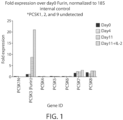


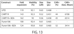


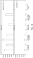

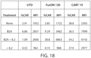

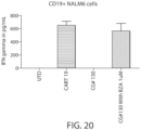


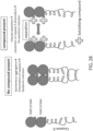
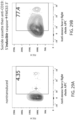


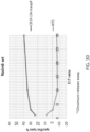


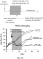
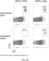


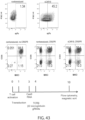


Classifications
-
- C—CHEMISTRY; METALLURGY
- C07—ORGANIC CHEMISTRY
- C07K—PEPTIDES
- C07K14/00—Peptides having more than 20 amino acids; Gastrins; Somatostatins; Melanotropins; Derivatives thereof
- C07K14/435—Peptides having more than 20 amino acids; Gastrins; Somatostatins; Melanotropins; Derivatives thereof from animals; from humans
- C07K14/705—Receptors; Cell surface antigens; Cell surface determinants
- C07K14/70503—Immunoglobulin superfamily
- C07K14/7051—T-cell receptor (TcR)-CD3 complex
-
- A—HUMAN NECESSITIES
- A61—MEDICAL OR VETERINARY SCIENCE; HYGIENE
- A61K—PREPARATIONS FOR MEDICAL, DENTAL OR TOILETRY PURPOSES
- A61K35/00—Medicinal preparations containing materials or reaction products thereof with undetermined constitution
- A61K35/12—Materials from mammals; Compositions comprising non-specified tissues or cells; Compositions comprising non-embryonic stem cells; Genetically modified cells
- A61K35/14—Blood; Artificial blood
- A61K35/15—Cells of the myeloid line, e.g. granulocytes, basophils, eosinophils, neutrophils, leucocytes, monocytes, macrophages or mast cells; Myeloid precursor cells; Antigen-presenting cells, e.g. dendritic cells
-
- A—HUMAN NECESSITIES
- A61—MEDICAL OR VETERINARY SCIENCE; HYGIENE
- A61P—SPECIFIC THERAPEUTIC ACTIVITY OF CHEMICAL COMPOUNDS OR MEDICINAL PREPARATIONS
- A61P17/00—Drugs for dermatological disorders
-
- A—HUMAN NECESSITIES
- A61—MEDICAL OR VETERINARY SCIENCE; HYGIENE
- A61P—SPECIFIC THERAPEUTIC ACTIVITY OF CHEMICAL COMPOUNDS OR MEDICINAL PREPARATIONS
- A61P19/00—Drugs for skeletal disorders
- A61P19/02—Drugs for skeletal disorders for joint disorders, e.g. arthritis, arthrosis
-
- A—HUMAN NECESSITIES
- A61—MEDICAL OR VETERINARY SCIENCE; HYGIENE
- A61P—SPECIFIC THERAPEUTIC ACTIVITY OF CHEMICAL COMPOUNDS OR MEDICINAL PREPARATIONS
- A61P21/00—Drugs for disorders of the muscular or neuromuscular system
- A61P21/04—Drugs for disorders of the muscular or neuromuscular system for myasthenia gravis
-
- A—HUMAN NECESSITIES
- A61—MEDICAL OR VETERINARY SCIENCE; HYGIENE
- A61P—SPECIFIC THERAPEUTIC ACTIVITY OF CHEMICAL COMPOUNDS OR MEDICINAL PREPARATIONS
- A61P25/00—Drugs for disorders of the nervous system
-
- A—HUMAN NECESSITIES
- A61—MEDICAL OR VETERINARY SCIENCE; HYGIENE
- A61P—SPECIFIC THERAPEUTIC ACTIVITY OF CHEMICAL COMPOUNDS OR MEDICINAL PREPARATIONS
- A61P29/00—Non-central analgesic, antipyretic or antiinflammatory agents, e.g. antirheumatic agents; Non-steroidal antiinflammatory drugs [NSAID]
-
- A—HUMAN NECESSITIES
- A61—MEDICAL OR VETERINARY SCIENCE; HYGIENE
- A61P—SPECIFIC THERAPEUTIC ACTIVITY OF CHEMICAL COMPOUNDS OR MEDICINAL PREPARATIONS
- A61P31/00—Antiinfectives, i.e. antibiotics, antiseptics, chemotherapeutics
- A61P31/12—Antivirals
-
- A—HUMAN NECESSITIES
- A61—MEDICAL OR VETERINARY SCIENCE; HYGIENE
- A61P—SPECIFIC THERAPEUTIC ACTIVITY OF CHEMICAL COMPOUNDS OR MEDICINAL PREPARATIONS
- A61P35/00—Antineoplastic agents
-
- A—HUMAN NECESSITIES
- A61—MEDICAL OR VETERINARY SCIENCE; HYGIENE
- A61P—SPECIFIC THERAPEUTIC ACTIVITY OF CHEMICAL COMPOUNDS OR MEDICINAL PREPARATIONS
- A61P35/00—Antineoplastic agents
- A61P35/02—Antineoplastic agents specific for leukemia
-
- A—HUMAN NECESSITIES
- A61—MEDICAL OR VETERINARY SCIENCE; HYGIENE
- A61P—SPECIFIC THERAPEUTIC ACTIVITY OF CHEMICAL COMPOUNDS OR MEDICINAL PREPARATIONS
- A61P37/00—Drugs for immunological or allergic disorders
- A61P37/02—Immunomodulators
-
- A—HUMAN NECESSITIES
- A61—MEDICAL OR VETERINARY SCIENCE; HYGIENE
- A61P—SPECIFIC THERAPEUTIC ACTIVITY OF CHEMICAL COMPOUNDS OR MEDICINAL PREPARATIONS
- A61P5/00—Drugs for disorders of the endocrine system
- A61P5/14—Drugs for disorders of the endocrine system of the thyroid hormones, e.g. T3, T4
-
- A—HUMAN NECESSITIES
- A61—MEDICAL OR VETERINARY SCIENCE; HYGIENE
- A61P—SPECIFIC THERAPEUTIC ACTIVITY OF CHEMICAL COMPOUNDS OR MEDICINAL PREPARATIONS
- A61P7/00—Drugs for disorders of the blood or the extracellular fluid
- A61P7/06—Antianaemics
-
- A—HUMAN NECESSITIES
- A61—MEDICAL OR VETERINARY SCIENCE; HYGIENE
- A61P—SPECIFIC THERAPEUTIC ACTIVITY OF CHEMICAL COMPOUNDS OR MEDICINAL PREPARATIONS
- A61P9/00—Drugs for disorders of the cardiovascular system
-
- A—HUMAN NECESSITIES
- A61—MEDICAL OR VETERINARY SCIENCE; HYGIENE
- A61P—SPECIFIC THERAPEUTIC ACTIVITY OF CHEMICAL COMPOUNDS OR MEDICINAL PREPARATIONS
- A61P9/00—Drugs for disorders of the cardiovascular system
- A61P9/12—Antihypertensives
-
- C—CHEMISTRY; METALLURGY
- C07—ORGANIC CHEMISTRY
- C07K—PEPTIDES
- C07K14/00—Peptides having more than 20 amino acids; Gastrins; Somatostatins; Melanotropins; Derivatives thereof
- C07K14/435—Peptides having more than 20 amino acids; Gastrins; Somatostatins; Melanotropins; Derivatives thereof from animals; from humans
- C07K14/705—Receptors; Cell surface antigens; Cell surface determinants
- C07K14/70503—Immunoglobulin superfamily
- C07K14/70517—CD8
-
- C—CHEMISTRY; METALLURGY
- C07—ORGANIC CHEMISTRY
- C07K—PEPTIDES
- C07K14/00—Peptides having more than 20 amino acids; Gastrins; Somatostatins; Melanotropins; Derivatives thereof
- C07K14/435—Peptides having more than 20 amino acids; Gastrins; Somatostatins; Melanotropins; Derivatives thereof from animals; from humans
- C07K14/705—Receptors; Cell surface antigens; Cell surface determinants
- C07K14/70575—NGF/TNF-superfamily, e.g. CD70, CD95L, CD153, CD154
-
- C—CHEMISTRY; METALLURGY
- C07—ORGANIC CHEMISTRY
- C07K—PEPTIDES
- C07K16/00—Immunoglobulins [IGs], e.g. monoclonal or polyclonal antibodies
- C07K16/18—Immunoglobulins [IGs], e.g. monoclonal or polyclonal antibodies against material from animals or humans
- C07K16/28—Immunoglobulins [IGs], e.g. monoclonal or polyclonal antibodies against material from animals or humans against receptors, cell surface antigens or cell surface determinants
- C07K16/2803—Immunoglobulins [IGs], e.g. monoclonal or polyclonal antibodies against material from animals or humans against receptors, cell surface antigens or cell surface determinants against the immunoglobulin superfamily
-
- A—HUMAN NECESSITIES
- A61—MEDICAL OR VETERINARY SCIENCE; HYGIENE
- A61K—PREPARATIONS FOR MEDICAL, DENTAL OR TOILETRY PURPOSES
- A61K38/00—Medicinal preparations containing peptides
-
- C—CHEMISTRY; METALLURGY
- C07—ORGANIC CHEMISTRY
- C07K—PEPTIDES
- C07K2317/00—Immunoglobulins specific features
- C07K2317/60—Immunoglobulins specific features characterized by non-natural combinations of immunoglobulin fragments
- C07K2317/62—Immunoglobulins specific features characterized by non-natural combinations of immunoglobulin fragments comprising only variable region components
- C07K2317/622—Single chain antibody (scFv)
-
- C—CHEMISTRY; METALLURGY
- C07—ORGANIC CHEMISTRY
- C07K—PEPTIDES
- C07K2319/00—Fusion polypeptide
- C07K2319/01—Fusion polypeptide containing a localisation/targetting motif
- C07K2319/03—Fusion polypeptide containing a localisation/targetting motif containing a transmembrane segment
-
- C—CHEMISTRY; METALLURGY
- C07—ORGANIC CHEMISTRY
- C07K—PEPTIDES
- C07K2319/00—Fusion polypeptide
- C07K2319/50—Fusion polypeptide containing protease site
-
- C—CHEMISTRY; METALLURGY
- C07—ORGANIC CHEMISTRY
- C07K—PEPTIDES
- C07K2319/00—Fusion polypeptide
- C07K2319/70—Fusion polypeptide containing domain for protein-protein interaction
-
- C—CHEMISTRY; METALLURGY
- C07—ORGANIC CHEMISTRY
- C07K—PEPTIDES
- C07K2319/00—Fusion polypeptide
- C07K2319/95—Fusion polypeptide containing a motif/fusion for degradation (ubiquitin fusions, PEST sequence)
Definitions
- constructs have been developed to enable selective expression of a desired protein of interest.
- Recent constructs have been tested which incorporate a domain engineered to degrade in the absence of a stabilizing ligand (known in the art as "degrons").
- Other constructs incorporate a domain engineered to aggregate in the absence of a deaggregating ligand (e.g., an aggregation domain).
- Such domains can be fused to proteins of interest to permit selective expression of such proteins only in the presence of the stabilizing or deaggregating ligand.
- Degrons and aggregation domains known in the art can, in certain circumstances, have certain disadvantages associated with disrupting the desired function of the protein of interest due to the size and/or conformation of the fusion protein. Such disadvantages can be particularly evident when the protein of interest is a transmembrane protein. Thus, there is a need for improving the degron/aggregation domain technology. Furthermore, a need exists for methods of modifying T cells to treat various diseases and conditions such as but not limited to cancer, autoimmunity and alloimmunity without permanently modifying or suppressing the immune system.
- the invention features fusion proteins including two protein domains separated by a heterologous protease cleavage site (e.g., a protease cleave site that is cleaved by a mammalian intracellular or extracellular protease), wherein a first of the protein domains is a conditional expression domain, e.g., a degradation domain or an aggregation domain, and the second protein is a protein of interest, e.g., a transmembrane protein (e.g., a CAR).
- a heterologous protease cleavage site e.g., a protease cleave site that is cleaved by a mammalian intracellular or extracellular protease
- a first of the protein domains is a conditional expression domain, e.g., a degradation domain or an aggregation domain
- the second protein is a protein of interest, e.g., a transmembr
- the conditional expression domain is a degradation domain, e.g., a degradation domain as described herein.
- the degradation domain is unstable and/or unable to fold into a stable conformation in the absence of an expression compound, e.g., a stabilization compound.
- the misfolded/unfolded degradation domain can be degraded by intracellular degradation pathways along with the other domain(s) of the fusion protein (see, for example, Figure 25 ).
- the degradation domain is able to fold into a stable conformation and is less susceptible to intracellular degradation pathways, e.g., relative to the degradation domain in the absence of the expression compound.
- the level and/or rate of cell surface expression or extracellular expression of the fusion protein is enhanced, e.g., by at least 2-, 3-, 4-, 5-, 6-, 7-, 8-, 9-, 10-, 20-, or 30-fold, in the presence of the expression compound relative to the absence of the expression compound.
- the heterologous cleavage site is exposed, leading to removal of the degradation domain, and thus, freeing the second protein domain.
- the conditional expression domain is an aggregation domain.
- the aggregation domain of the fusion protein associates with one or more other aggregation domains into oligomers and aggregates (see, for example, Figure 26 ).
- the aggregated fusion protein can be sequestered in the cellular compartment in which it is aggregated.
- the aggregation domains dissociate from one another, and the fusion proteins are solubilized (e.g., assumes a monomeric configuration).
- the level and/or rate of cell surface expression or extracellular expression of the fusion protein is enhanced, e.g., by at least 2-, 3-, 4-, 5-, 6-, 7-, 8-, 9-, 10-, 20-, or 30-fold, in the presence of the expression compound relative to the absence of the expression compound.
- the heterologous cleavage site is exposed, leading to removal of the aggregation domain, and thus, freeing the second protein domain.
- the fusion protein includes a first protein domain that is or comprises a conditional expression domain, e.g., a degradation domain or an aggregation domain, and a second protein domain that is or comprises a protein of interest, e.g., a transmembrane protein (e.g., a CAR), wherein the first and the second domains of the fusion protein are separated by a heterologous protease cleavage site (e.g., a protease cleave site that is cleaved by a mammalian intracellular or extracellular protease).
- a heterologous protease cleavage site e.g., a protease cleave site that is cleaved by a mammalian intracellular or extracellular protease.
- conditional expression domain e.g., the degradation or the aggregation domain
- the conditional expression domain is located N-terminal to the second protein domain.
- the fusion protein further includes a signal peptide N-terminal to the degradation domain.
- the invention pertains to a fusion protein, comprising two protein domains separated by a heterologous protease cleavage site, wherein a first of said protein domains is a conditional expression domain, and a second of said protein domains is a transmembrane protein, wherein the conditional expression domain has a first state associated with a first level of surface expression and/or extracellular expression of the fusion protein and a second state associated with a second level of surface expression and/or extracellular expression of the fusion protein, wherein the second level is increased, e.g., by at least 2, 3, 4, 5, 10, 20 or 30 fold over the first level in the presence of an expression compound.
- the invention pertains to a fusion protein, comprising two protein domains separated by a heterologous protease cleavage site, wherein a first of said protein domains is a conditional expression domain, and a second of said protein domains is a transmembrane protein, wherein the heterologous protease cleavage site is a furin cleavage site, provided that the furin cleavage site does not comprise the amino acid sequence SARNRQKR (SEQ ID NO: 981).
- the invention pertains to a fusion protein, comprising two protein domains separated by a heterologous protease cleavage site, wherein a first of said protein domains is a conditional expression domain, and a second of said protein domains is a chimeric antigen receptor (CAR).
- CAR chimeric antigen receptor
- conditional expression domain is a degradation domain.
- conditional expression domain is an aggregation domain.
- the fusion protein comprises two protein domains separated by a heterologous protease cleavage site, wherein the first of said protein domains (also referred to herein as the first protein domain) is or comprises a degradation domain, e.g., a degradation domain as described herein, and the second of said protein domains (also referred to herein as the second protein domain) is a protein of interest.
- the protein of interest is a transmembrane protein, e.g., a CAR.
- the degradation domain is chosen from an estrogen receptor (ER) domain, an FKB protein (FKBP) domain or an dihydrofolate reductase (DHFR).
- ER estrogen receptor
- FKBP FKB protein
- DHFR dihydrofolate reductase
- the degradation domain has a first state associated with a first level of surface expression and/or extracellular expression of the fusion protein and a second state associated with a second level of surface expression and/or extracellular expression of the fusion protein, wherein the second level is increased, e.g., by at least 2, 3, 4, 5, 10, 20 or 30 fold over the first level in the presence of a stabilization compound.
- the degradation domain is derived from an estrogen receptor.
- the degradation domain can comprise an amino acid sequence selected from SEQ ID NO: 58 or a sequence having at least 90%, 95%, 97%, 98%, or 99% identity thereto, or SEQ ID NO: 121 or a sequence having at least 90%, 95%, 97%, 98%, or 99% identity thereto.
- the degradation domain comprises an amino acid sequence selected from SEQ ID NO: 58 or SEQ ID NO: 121.
- the stabilization compound can be selected from Bazedoxifene or 4-hydroxy tamoxifen (4-OHT).
- the stabilization compound is Bazedoxifene. Tamoxifen and Bazedoxifene are FDA approved drugs, and thus are safe to use in a human.
- the degradation domain is derived from an FKB protein (FKBP).
- FKBP FKB protein
- the degradation domain can comprise an amino acid sequence of SEQ ID NO: 56 or a sequence having at least 90%, 95%, 97%, 98%, or 99% identity thereto.
- the degradation domain comprises SEQ ID NO: 56.
- the stabilization compound can be Shield-1.
- the degradation domain is derived from dihydrofolate reductase (DHFR).
- the degradation domain comprises an amino acid sequence selected from SEQ ID NO: 57 or a sequence having at least 90%, 95%, 97%, 98%, or 99% identity thereto.
- the degradation domain comprises SEQ ID NO: 57.
- the stabilization compound can be Trimethoprim.
- the degradation domain is not derived from an FKB protein or estrogen receptor.
- the fusion protein comprises two protein domains separated by a heterologous protease cleavage site, wherein the first of said protein domains (also referred to herein as the first protein domain) is or comprises a aggregation domain, e.g., an aggregationdomain as described herein, and the second of said protein domains (also referred to herein as the second protein domain) is a protein of interest.
- the protein of interest is a transmembrane protein, e.g., a CAR.
- the fusion protein comprises two protein domains separated by a heterologous protease cleavage site, wherein a first of said protein domains is an aggregation domain, and a second of said protein domains is a transmembrane protein, wherein the aggregation domain has a first state associated with a first level of surface expression and/or extracellular expression of the fusion protein and a second state associated with a second level of surface expression and/or extracellular expression of the fusion protein, and wherein the second level is increased, e.g., by at least 2, 3, 4, 5, 10, 20 or 30 fold over the first level in the presence of a deaggregation compound.
- the fusion protein comprises two protein domains separated by a heterologous protease cleavage site, wherein a first of said protein domains is an aggregation domain, and a second of said protein domains is a transmembrane protein, wherein the heterologous protease cleavage site is a furin cleavage site, provided that the furin cleavage site does not comprise the amino acid sequence SARNRQKR (SEQ ID NO: 981).
- the fusion protein comprises two protein domains separated by a heterologous protease cleavage site, wherein a first of said protein domains is an aggregation domain, and a second of said protein domains is a chimeric antigen receptor (CAR).
- CAR chimeric antigen receptor
- the aggregation domain comprises 1, 2, 3, 4 5, 6, 7, 8, or more repeats of a dimerization domain, e.g., a homodimerization or a heterodimerization domain.
- the aggregation domain is from an FKB protein (FKBP).
- FKBP FKB protein
- the aggregation domain is an FKBP F36M domain.
- the aggregation domain is from an FKB protein (FKBP) and comprises an amino acid sequence that is at least 90, 95, 97, 98, 99, or 100% identical to either of SEQ ID NOs: 975 or 976.
- FKBP FKB protein
- the fusion protein comprises a further 2nd, 3 rd , 4 th , 5 th , 6 th , 7 th , 8 th , 9 th , or 10 th aggregation domain.
- the 2nd, 3 rd , 4 th , 5 th , 6 th , 7 th , 8 th , 9 th , or 10 th aggregation domain is the same type of aggregation domain as the first aggregation domain.
- the aggregation domain forms homodimers with the same aggregation domain.
- the fusion protein comprises a plurality of aggregation domains, wherein the plurality comprises more than one, e.g., two, types of aggregation domains, and a first type of aggregation domain forms heterodimers with a second type of aggregation domain.
- the fusion protein comprises 2, 4, 6, 8, or 10 aggregation domains, wherein the fusion protein comprises equal numbers of the first type of aggregation domain and the second type of aggregation domain.
- the aggregation domains are disposed in the fusion protein in an alternating order of first type and second type, e.g., first, second, first, second, or second, first, second, first.
- said deaggregation compound is selected from FK506, rapamycin, AP22542, AP21998, and Shield-1 when the fusion protein comprises an aggregation domain derived from FKB protein (FKBP), e.g., FKBP F36M.
- FKBP FKB protein
- said heterologous cleavage site is cleaved by a mammalian intracellular protease.
- said cleavage site is cleaved by a protease selected from the group consisting of furin, PCSK1, PCSK5, PCSK6, PCSK7, cathepsin B, Granzyme B, Factor XA, Enterokinase, genenase, sortase, precission protease, thrombin, TEV protease, and elastase 1.
- a protease selected from the group consisting of furin, PCSK1, PCSK5, PCSK6, PCSK7, cathepsin B, Granzyme B, Factor XA, Enterokinase, genenase, sortase, precission protease, thrombin, TEV protease, and elastase 1.
- said cleavage site comprises a polypeptide having an cleavage motif selected from the group consisting of RX(K/R)R consensus motif, RXXX[KR]R consensus motif, RRX consensus motif, I-E-P-D-X consensus motif (SEQ ID NO: 35), Glu/Asp-Gly-Arg, Asp-Asp-Asp-Asp-Lys (SEQ ID NO: 36), Pro-Gly-Ala-Ala-His-Tyr (SEQ ID NO: 37), LPXTG/A consensus motif, Leu-Glu-Val-Phe-Gln-Gly-Pro (SEQ ID NO: 38), Leu-Val-Pro-Arg-Gly-Ser (SEQ ID NO: 40), E-N-L-Y-F-Q-G (SEQ ID NO: 41), and [AGSV]-x (SEQ ID NO: 42).
- RX(K/R)R consensus motif RXXXX[KR]R consensus motif
- said cleavage site is cleaved by furin.
- the fusion protein comprises a furin cleavage site selected from RTKR (SEQ ID NO: 123); GTGAEDPRPSRKRRSLGDVG (SEQ ID NO: 125); GTGAEDPRPSRKRR (SEQ ID NO: 127); LQWLEQQVAKRRTKR (SEQ ID NO: 129); GTGAEDPRPSRKRRSLGG (SEQ ID NO: 131); GTGAEDPRPSRKRRSLG (SEQ ID NO: 133); SLNLTESHNSRKKR (SEQ ID NO: 135); or CKINGYPKRGRKRR (SEQ ID NO: 137).
- RTKR SEQ ID NO: 123
- GTGAEDPRPSRKRRSLGDVG SEQ ID NO: 125
- GTGAEDPRPSRKRR SEQ ID NO: 127
- LQWLEQQVAKRRTKR SEQ ID NO: 129
- GTGAEDPRPSRKRRSLGG SEQ ID NO: 131
- the fusion protein comprises a furin cleavage site selected from GTGAEDPRPSRKRRSLGDVG (SEQ ID NO: 125); or GTGAEDPRPSRKRR (SEQ ID NO: 127).
- the fusion protein comprises the furin cleavage site of GTGAEDPRPSRKRRSLGDVG (SEQ ID NO: 125).
- said heterologous protease cleavage site is cleaved by a mammalian extracellular protease.
- said mammalian extracellular protease is selected from the group consisting of Factor XA, Enterokinase, genenase, sortase, precission protease, thrombin, TEV protease, and elastase 1.
- said cleavage site comprises a polypeptide having an amino acid sequence selected from the group consisting of Glu/Asp-Gly-Arg, Asp-Asp-Asp-Asp-Lys (SEQ ID NO: 36), Pro-Gly-Ala-Ala-His-Tyr (SEQ ID NO: 37), LPXTG/A consensus motif, Leu-Glu-Val-Phe-Gln-Gly-Pro (SEQ ID NO: 38), Leu-Val-Pro-Arg-Gly-Ser (SEQ ID NO: 40), E-N-L-Y-F-Q-G (SEQ ID NO: 41), and [AGSV]-x (SEQ ID NO: 42).
- the heterologous cleavage site is cleaved by furin, PCSK1, PCSK5, PCSK6, PCSK7, cathepsin B, Granzyme B, Factor XA, Enterokinase, genenase, sortase, precission protease, thrombin, TEV protease, or elastase 1.
- the protease cleave site can include a polypeptide having an cleavage motif selected from RX(K/R)R consensus motif, RXXX[KR]R consensus motif, RRX consensus motif, I-E-P-D-X consensus motif (SEQ ID NO: 35), Glu/Asp-Gly-Arg, Asp-Asp-Asp-Asp-Lys (SEQ ID NO: 36), Pro-Gly-Ala-Ala-His-Tyr (SEQ ID NO: 37), LPXTG/A consensus motif, Leu-Glu-Val-Phe-Gln-Gly-Pro (SEQ ID NO: 38), Leu-Val-Pro-Arg-Gly-Ser (SEQ ID NO: 40), E-N-L-Y-F-Q-G (SEQ ID NO: 41), or [AGSV]-x (SEQ ID NO: 42).
- RX(K/R)R consensus motif RXXXX[KR]R consensus motif
- the mammalian extracellular protease is selected from Factor XA, Enterokinase, genenase, sortase, precission protease, thrombin, TEV protease, or elastase 1
- the cleavage site can include a polypeptide having an amino acid sequence selected from Glu/Asp-Gly-Arg, Asp-Asp-Asp-Asp-Lys (SEQ ID NO: 36), Pro-Gly-Ala-Ala-His-Tyr (SEQ ID NO: 37), LPXTG/A consensus motif, Leu-Glu-Val-Phe-Gln-Gly-Pro (SEQ ID NO: 38), Leu-Val-Pro-Arg-Gly-Ser (SEQ ID NO: 40), E-N-L-Y-F-Q-G (SEQ ID NO: 41), or [AGSV]-x (SEQ ID NO: 42)).
- the fusion protein described herein includes a furin cleavage site.
- the fusion proteins described herein include any one of furin cleavage sites listed in Table 20.
- the fusion proteins described herein include a furin cleavage site selected from RTKR (SEQ ID NO: 123) or a sequence having at least 90%, 95%, 97%, 98%, or 99% identity thereto; GTGAEDPRPSRKRRSLGDVG (SEQ ID NO: 125) or a sequence having at least 90%, 95%, 97%, 98%, or 99% identity thereto; GTGAEDPRPSRKRR (SEQ ID NO: 127) or a sequence having at least 90%, 95%, 97%, 98%, or 99% identity thereto; LQWLEQQVAKRRTKR (SEQ ID NO: 129) or a sequence having at least 90%, 95%, 97%, 98%, or 99% identity thereto; GTGAEDPRPSRKRRSLGG (SEQ ID NO: 123) or a sequence having at least
- the fusion proteins described herein include a furin cleavage site selected from GTGAEDPRPSRKRRSLGDVG (SEQ ID NO: 125) or a sequence having at least 90%, 95%, 97%, 98%, or 99% identity thereto , or GTGAEDPRPSRKRR (SEQ ID NO: 127) or a sequence having at least 90%, 95%, 97%, 98%, or 99% identity thereto.
- the fusion proteins described herein include the furin cleavage site of GTGAEDPRPSRKRRSLGDVG (SEQ ID NO: 125) or a sequence having at least 90%, 95%, 97%, 98%, or 99% identity thereto.
- the fusion proteins described herein include a furin cleavage site selected from RTKR (SEQ ID NO: 123); GTGAEDPRPSRKRRSLGDVG (SEQ ID NO: 125); GTGAEDPRPSRKRR (SEQ ID NO: 127); LQWLEQQVAKRRTKR (SEQ ID NO: 129); GTGAEDPRPSRKRRSLGG (SEQ ID NO: 131); GTGAEDPRPSRKRRSLG (SEQ ID NO: 133); SLNLTESHNSRKKR (SEQ ID NO: 135); or CKINGYPKRGRKRR (SEQ ID NO: 137).
- RTKR SEQ ID NO: 123
- GTGAEDPRPSRKRRSLGDVG SEQ ID NO: 125
- GTGAEDPRPSRKRR SEQ ID NO: 127
- LQWLEQQVAKRRTKR SEQ ID NO: 129
- GTGAEDPRPSRKRRSLGG SEQ ID NO: 131
- the fusion proteins described herein include a furin cleavage site selected from GTGAEDPRPSRKRRSLGDVG (SEQ ID NO: 125) or GTGAEDPRPSRKRR (SEQ ID NO: 127). In some embodiments, the fusion proteins described herein include the furin cleavage site of GTGAEDPRPSRKRRSLGDVG (SEQ ID NO: 125).
- conditional expression domain e.g., aggregation domain or degradation domain
- the conditional expression domain is located N-terminal to said second protein domain or C-terminal to said second protein domain.
- said fusion protein further comprises a signal peptide N-terminal to said conditional expression domain, e.g., aggregation domain or degradation domain.
- the fusion protein further comprises a linker positioned between the signal peptide and said conditional expression domain, e.g., aggregation domain or degradation domain.
- the linker is a linker in any fusion protein listed in Tables 23 and 24.
- the fusion protein comprises an amino acid sequence of any fusion protein listed in Tables 22, 23, or 24.
- a second of the protein domains is a transmembrane protein (e.g., a transmembrane receptor).
- the transmembrane receptor can be, e.g., a synthetic protein (e.g., a chimeric antigen receptor).
- Chimeric antigen receptors can include, e.g., in a N-terminal to C-terminal direction, an antigen binding domain, a transmembrane domain, and one or more intracellular signaling domains.
- the signaling domain may include one or more primary signaling domains (e.g., a CD3-zeta stimulatory domain) and, optionally, one or more costimulatory signaling domains (e.g., an intracellular domain from a costimulatory protein selected from CD27, CD28, 4-1BB (CD137), OX40, GITR, CD30, CD40, ICOS, BAFFR, HVEM, ICAM-1, lymphocyte function-associated antigen-1 (LFA-1), CD2, CDS, CD7, CD287, LIGHT, NKG2C, NKG2D, SLAMF7, NKp80, NKp30, NKp44, NKp46, CD160, B7-H3, or a ligand that specifically binds CD83).
- a costimulatory signaling domains e.g., an intracellular domain from a costimulatory protein selected from CD27, CD28, 4-1BB (CD137), OX40, GITR, CD30, CD40
- the antigen binding domain is an scFv.
- the antigen binding domain may binds an antigen selected from CD19; CD123; CD22; CD30; CD171; CS-1; C-type lectin-like molecule-1, CD33; epidermal growth factor receptor variant III (EGFRvlll); ganglioside G2 (GD2); ganglioside GD3; TNF receptor family member B cell maturation (BCMA); Tn antigen ((Tn Ag) or (GaINAc ⁇ -Ser/Thr)); prostate-specific membrane antigen (PSMA); Receptor tyrosine kinase-like orphan receptor 1 (ROR1); Fms-Like Tyrosine Kinase 3 (FLT3); Tumor-associated glycoprotein 72 (TAG72); CD38; CD44v6; Carcinoembryonic antigen (CEA); Epithelial cell adhesion molecule (EPCAM); B7H3 (CD276); KIT (
- the fusion protein comprises an antigen binding domain that binds CD19. In some embodiments, the fusion protein comprises an antigen binding domain comprising an amino acid sequence selected from any one of SEQ ID NOs: 356-368 or 381. In some embodiments, the fusion protein comprises a chimeric antigen receptor comprising an amino acid sequence selected from any one of SEQ ID NOs: 897, 902, 907, 912, 917, 922, 927, 932, 937, 942, 947, 952, 956.
- the fusion protein comprises an antigen binding domain that binds CD123. In some embodiments, the fusion protein comprises an antigen binding domain comprising an amino acid sequence selected from any one of SEQ ID NOs: 751, 756, 761, or 766. In some embodiments, the fusion protein comprises a chimeric antigen receptor comprising an amino acid sequence selected from any one of SEQ ID NOs: 750, 755, 760, or 765.
- the fusion protein comprises an antigen binding domain that binds BCMA.
- the fusion protein comprises an antigen binding domain comprising an amino acid sequence selected from any one of SEQ ID NOs: 382, 386, 390, 394, 398, 402, 406, 410, 414, 418, 422, 426, 430, 434, 438, 442, 446, 450, 454, 458, 462, 466, 470, 474, 478, 482, 486, 490, 494, 498, 502, 506, 510, 514, 518, 522, 528, 531, 534, or 537.
- the fusion protein comprises a chimeric antigen receptor comprising an amino acid sequence selected from any one of SEQ ID NOs: 789, 791, 793, 795, 797, 799, 801, 803, 805, 807, 809, 811, 813, 815, 817, 819, 821, 823, 825, 827, 829, 831, 833, 835, 837, 839, 841, 843, 845, 847, 849, 851, 853, 855, 857, or 859.
- the fusion protein comprises an antigen binding domain that binds CD20. In some embodiments, the fusion protein comprises an antigen binding domain comprising an amino acid sequence located at positions 470-712 or 470-939 of SEQ ID NO: 3033. In some embodiments, the fusion protein comprises a chimeric antigen receptor comprising an amino acid sequence of SEQ ID NO: 3033.
- the invention features a cell, e.g., a host cell including any one of the foregoing fusion proteins.
- the cell e.g., the host cell
- is an immune cell e.g., an immune effector cell.
- the cell is a T cell or an NK cell.
- the invention features a nucleic acid (e.g., an mRNA or DNA molecule) encoding any one of the foregoing fusion proteins.
- the invention features a vector (e.g., a viral vector (such as a lentiviral vector)) containing such a nucleic acid.
- the invention also features a viral particle including such a viral vector.
- the invention features a cell, e.g., host cell (e.g., a human T cell) containing any of the foregoing vectors, nucleic acids, or fusion proteins.
- host cell e.g., a human T cell
- the cell further includes a protease capable of cleaving the heterologous protease cleavage site.
- the host cell can further include a stabilization compound (e.g., Bazedoxifene, Shield1 or 1 ⁇ M 4-OHT (4-hydroxy tamoxifen)) wherein said degradation domain assumes a conformation permissive to cellular degradation in the absence of said stabilization compound.
- a stabilization compound e.g., Bazedoxifene, Shield1 or 1 ⁇ M 4-OHT (4-hydroxy tamoxifen
- the fusion protein in the absence of an expression compound, e.g., a stabilization compound, is degraded by cellular degradation pathways, e.g., at least 50%, 60%, 70%, 80%, 90% or greater of the fusion protein is degraded.
- the fusion protein in the absence of an expression compound, e.g., deaggregation compound, is in an aggregated state in the cell, e.g., in the endoplasmic reticulum or the cytosol, e.g., at least 50%, 60%, 70%, 80%, 90% or greater is in the aggregated state.
- an expression compound e.g., deaggregation compound
- said cell further comprises an expression compound, e.g., a stabilization compound.
- an expression compound e.g., a stabilization compound.
- conditional expression domain e.g., degradation domain
- the conformation of the fusion protein is more permissive to cleavage at the heterologous protease cleavage site in the presence of the expression compound, e.g., stabilization compound, relative to a conformation in the absence of the expression compound.
- the level of cell surface expression or extracellular expression of the fusion protein is greater, e.g., 2, 3, 4, 5, 6, 7, 8, 9, 10, 20, or 30 fold greater, than the level of cell surface expression or extracellular expression of the fusion protein in a cell not comprising an expression compound, e.g., a stabilization compound.
- said cell further comprises an expression compound, e.g., a deaggregation compound.
- an expression compound e.g., a deaggregation compound.
- conditional expression domain e.g., aggregation domain
- the conformation of the fusion protein is more permissive to cleavage at the heterologous protease cleavage site in the presence of the expression compound, e.g., deaggregation compound, relative to a conformation in the absence of the expression compound.
- the level of cell surface expression or extracellular expression of the fusion protein is greater, e.g., 2, 3, 4, 5, 6, 7, 8, 9, 10, 20, or 30 fold greater, than the level of cell surface expression or extracellular expression of the fusion protein in a cell not comprising an expression compound, e.g., a deaggregation compound.
- a method of making a fusion protein as described herein includes providing a cell, e.g., a host cell as described herein, e.g., a host cell comprising any of the foregoing vectors, nucleic acids, or fusion proteins, under conditions suitable for expression.
- the invention also features a method of conditionally expressing a protein of interest.
- the protein of interest is a transmembrane protein, e.g., a CAR.
- the invention also features a method for conditionally expressing a protein of interest, transmembrane protein, or CAR on the surface of a cell (e.g., an immune cell, e.g., a host cell).
- a cell e.g., an immune cell, e.g., a host cell. The method includes:
- the presence of said expression compound is associated with, e.g., causes, a change in conformation of the conditional expression domain from a first folding state to second folding state, wherein the first folding state is more susceptible to degradation, e.g., cellular degradation, or aggregation relative to the second folding state.
- the presence of said expression compound exposes the heterologous protease cleavage site, e.g., to a greater extent, e.g., 2, 3, 4, 5, 6, 7, 8, 9, 10, 20, or 30 fold greater, relative the exposure of the protease cleavage site in the absence of said expression compound.
- the invention also features a method of conditionally expressing a protein of interest, transmembrane protein, or CAR, said method comprising contacting a cell, e.g., a host cell and/or a cell described herein, with a stabilization compound, wherein:
- said cell is contacted with said stabilization compound ex vivo.
- said cell is contacted with said stabilization compound in vivo.
- the invention also features a method of conditionally expressing a protein of interest, transmembrane protein, or CAR, said method comprising contacting a cell, e.g., a host cell and/or a cell described herein, with a deaggregation compound, wherein:
- said cell is contacted with said deaggregation compound ex vivo.
- said cell is contacted with said deaggregation compound in vivo.
- the invention features a method of treating a subject having a disease associated with expression of a tumor antigen, including administering to the subject an effective amount of any of the foregoing host cells, wherein the second protein is a chimeric antigen receptor and includes, in a N-terminal to C-terminal direction, an antigen binding domain, a transmembrane domain, and one or more intracellular signaling domains and the antigen binding domain specifically binds the tumor antigen.
- the invention features a method of treating an autoantibody or alloantibody disease or condition, the method comprising administering to the subject an effective amount of the foregoing host cell, wherein said second protein is a chimeric antigen receptor and comprises, in a N-terminal to C-terminal direction, an antigen binding domain, a transmembrane domain, and one or more intracellular signaling domains and said antigen binding domain specifically binds an antigen specific of said autoantibody or alloantibody disease.
- the host cell can be either autologous or non-autologous, e.g., allogeneic, to the subject.
- Such methods can further include the step of contacting the host cell, in vivo or ex vivo, with the foregoing stabilization compounds.
- the cell is contacted with an expression compound, and:
- the cell e.g., host cell
- a stabilization compound e.g., a stabilization compound
- said stabilization compound is selected from Bazedoxifene or 4-hydroxy tamoxifen (4-OHT) when the fusion protein comprises a degradation domain derived from estrogen receptor.
- said stabilization compound is Shield-1 when the fusion protein comprises a degradation domain derived from an FKB protein.
- the cell is contacted with a deaggregation compound, and:
- said deaggregation compound is selected from FK506, rapamycin, AP22542, and AP21998 when the fusion protein comprises an aggregation domain derived from FKB protein (FKBP), e.g., FKBP F36M.
- FKBP FKB protein
- the autoantibody disease or condition is selected from the group consisting of bullous pemphigoid, epidermolysis bullosa acquisita, p200 pemphigoid, linear IgA bullous dermatosis, other pemphigoid group diseases, dermatitis herpetiformis, celiac disease, myasthenia gravis, Goodpasture's syndrome, granulomatosis with polyangiitis and other ANCA+ vasculitidies, autoimmune limbic encephalitis, anti-N-methyl-D-aspartate receptor encephalitis, neuromyelitis optica, autoimmune hemolytic anemia, autoantibody-associated end-organ damage in lupus and other connective tissue diseases (due to anti-dsDNA, anti-Ro, and other autoantibodies), Graves' and Hashimoto's thyroiditis, anti-insulin antibodies in diabetes, anti-insulin receptor antibodies in autoimmune hypoglycemia, cryoglobulin
- the cancer is mesothelioma (e.g., malignant pleural mesothelioma), e.g., in a subject who has progressed on at least one prior standard therapy; lung cancer (e.g., non-small cell lung cancer, small cell lung cancer, squamous cell lung cancer, or large cell lung cancer); pancreatic cancer (e.g., pancreatic ductal adenocarcinoma, or metastatic pancreatic ductal adenocarcinoma (PDA), e.g., in a subject who has progressed on at least one prior standard therapy); esophageal adenocarcinoma, ovarian cancer (e.g., serous epithelial ovarian cancer, e.g., in a subject who has progressed after at least one prior regimen of standard therapy), breast cancer, colorectal cancer, bladder cancer or any combination thereof.
- lung cancer e.g., non-small cell lung cancer, small cell
- the disease associated with expression of a tumor antigen is a cancer.
- the disease associated with expression of the tumor antigen is a hematological cancer, e.g., a hematological cancer chosen from a leukemia or lymphoma.
- the cancer is chosen from: chronic lymphocytic leukemia (CLL), mantle cell lymphoma (MCL), multiple myeloma, acute lymphoid leukemia (ALL), Hodgkin lymphoma, B-cell acute lymphoid leukemia (BALL), T-cell acute lymphoid leukemia (TALL), small lymphocytic leukemia (SLL), B cell prolymphocytic leukemia, blastic plasmacytoid dendritic cell neoplasm, Burkitt's lymphoma, diffuse large B cell lymphoma (DLBCL), DLBCL associated with chronic inflammation, chronic myeloid leukemia, myeloproliferative neoplasms, follicular lymphoma, pediatric follicular lymphoma, hairy cell leukemia, small cell- or a large cell-follicular lymphoma, malignant lymphoproliferative conditions, MALT lymphoma (extranodal marginal).
- the cancer is chosen from MCL, CLL, ALL, Hodgkin lymphoma, AML, or multiple myeloma.
- the invention features a fusion protein, cell, nucleic acid, viral particle, or vector described herein for use as a medicament.
- the invention features a fusion protein, cell, nucleic acid, vector, or method described herein for use in the treatment of a disease expressing a tumor antigen.
- the invention features methods of treating an autoantibody or alloantibody disease or condition in a subject in need thereof comprising administering an effective amount of a pharmaceutical composition comprising a modified T cell to the subject, wherein the modified T cell comprises a nucleic acid comprising a suicide gene and a nucleic acid encoding a chimeric antigen receptor (CAR) comprising an anti-B cell binding domain, a transmembrane domain, a costimulatory domain and an intracellular signaling domain.
- the invention features methods of treating an autoantibody or alloantibody disease or condition in a subject in need thereof comprising administering an effective amount of a pharmaceutical composition comprising a modified T cell to the subject, wherein the modified T cell comprises a nucleic acid encoding a dimerization domain and a chimeric antigen receptor (CAR) comprising an anti-B cell binding domain, a transmembrane domain, a costimulatory domain and an intracellular signaling domain.
- CAR chimeric antigen receptor
- the suicide gene encodes the amino acid sequence selected from the group consisting of SEQ ID Nos: 3005-3007.
- the suicide gene further comprises a dimerization domain comprising the amino acid sequence selected from the group consisting of SEQ ID NO: 3013 and 3014.
- the dimerization domain comprises the amino acid sequence of SEQ ID NO: 980.
- the dimerization domain further comprises a furin cleavage site comprising the amino acid sequence of SEQ ID NO: 980.
- the CAR further comprises a signal peptide.
- the signal peptide comprises the amino acid sequence of SEQ ID NO: 3035.
- administering the effective amount comprises activating the modified T cell to effect cytotoxic function against B cells.
- the method further comprises activating a suicide gene product of the suicide gene to induce cell death of the modified T cell.
- activating the suicide gene product further comprises administering a dimerization agent to promote dimerization of the suicide gene product.
- activating the suicide gene product occurs after the modified T cell exerts cytotoxic function against B cells.
- activating the suicide gene product occurs after an onset of an adverse reaction in the subject to the modified T cell.
- the method further comprises repressing activation of a suicide gene product of the suicide gene to repress cell death of the modified T cell.
- repressing activation of the suicide gene product further comprises administering a solubilizing agent to prevent dimerization of the suicide gene product.
- administering the solubilizing agent occurs concurrently with administration of the modified T cell and continues as the modified T cell exerts cytotoxic function against B cells.
- administering the solubilizing agent is ceased after an onset of an adverse reaction in the subject to the modified T cell.
- the anti-B cell binding domain of the CAR comprises an antibody selected from the group consisting of a monoclonal antibody, a polyclonal antibody, a synthetic antibody, human antibody, humanized antibody, single domain antibody, single chain variable fragment, and antigen-binding fragments thereof.
- the anti-B cell binding domain of the CAR specifically binds a B cell marker selected from the group consisting of CD19, BCMA, CD20, CD21, CD27, CD38, CD138 and any combination thereof.
- the anti-B cell binding domain of the CAR specifically binds a B cell marker selected from the group consisting of CD20, CD21, CD27, CD38, CD138, any combination thereof, and at least one surface marker selectively found on a pro-B cell, pre-B cell, immature B cell, mature B cell, memory B cell, and plasma cell.
- the intracellular domain of the CAR comprises dual signaling domains.
- the costimulatory domain is selected from the group consisting of CD3, CD27, CD28, CD83, CD86, CD127, 4-1BB, 4-1BBL, PD1, PD1L, T cell receptor (TCR), any derivative or variant thereof, any synthetic sequence thereof that has the same functional capability, and any combination thereof.
- the method further comprises administering a solubilizing agent to prevent dimerization of the CAR.
- administering the solubilizing agent occurs concurrently with administration of the modified T cell and continues as the modified T cell exerts cytotoxic function against B cells.
- administering the solubilizing agent is ceased after an onset of an adverse reaction in the subject to the modified T cell.
- the autoantibody disease or condition is selected from the group consisting of bullous pemphigoid, epidermolysis bullosa acquisita, p200 pemphigoid, linear IgA bullous dermatosis, other pemphigoid group diseases, dermatitis herpetiformis, celiac disease, myasthenia gravis, Goodpasture's syndrome, granulomatosis with polyangiitis and other ANCA+ vasculitidies, autoimmune limbic encephalitis, anti-N-methyl-D-aspartate receptor encephalitis, neuromyelitis optica, autoimmune hemolytic anemia, autoantibody-associated end-organ damage in lupus and other connective tissue diseases (due to anti-dsDNA, anti-Ro, and other autoantibodies), Graves' and Hashimoto's thyroiditis, anti-insulin antibodies in diabetes, anti-insulin receptor antibodies in autoimmune hypoglycemia, cryoglobulin
- the alloantibody disease or condition is an immune reaction in response to an organ transplant, blood transfusion, pregnancy, and protein replacement therapy.
- the modified T cell is further modified by deleting a gene selected from the group consisting of a T cell receptor (TCR) chain, a major histocompatibility complex protein, and any combination thereof.
- TCR T cell receptor
- the modified T cell is further modified before administration to the subject in need thereof.
- the modified T cell is further modified by inducing a CRISPR/Cas system.
- the invention features a pharmaceutical composition formulated for use in a method described herein, the composition comprising a modified T cell comprising a nucleic acid encoding a suicide gene and a nucleic acid encoding a chimeric antigen receptor (CAR) comprising an anti-B cell binding domain, a transmembrane domain, a costimulatory domain and an intracellular signaling domain.
- a modified T cell comprising a nucleic acid encoding a suicide gene and a nucleic acid encoding a chimeric antigen receptor (CAR) comprising an anti-B cell binding domain, a transmembrane domain, a costimulatory domain and an intracellular signaling domain.
- CAR chimeric antigen receptor
- the invention features a pharmaceutical composition formulated for use in a method described herein, the composition comprising a modified T cell comprising a nucleic acid encoding a dimerization domain and a chimeric antigen receptor (CAR) comprising an anti-B cell binding domain, a transmembrane domain, a costimulatory domain and an intracellular signaling domain.
- a modified T cell comprising a nucleic acid encoding a dimerization domain and a chimeric antigen receptor (CAR) comprising an anti-B cell binding domain, a transmembrane domain, a costimulatory domain and an intracellular signaling domain.
- CAR chimeric antigen receptor
- the suicide gene encodes the amino acid sequence selected from the group consisting of SEQ ID Nos: 3005-3007.
- the suicide gene further comprises a dimerization domain comprising the amino acid sequence selected from the group consisting of SEQ ID NOs: 3013 and 3014.
- the dimerization domain comprises the amino acid sequence of SEQ ID NO: 980.
- the dimerization domain further comprises a furin cleavage site comprising the amino acid sequence of SEQ ID NO: 980.
- the CAR further comprises a signal peptide.
- the signal peptide comprises the amino acid sequence of SEQ ID NO: 3035.
- the composition further comprises an inducing agent to induce activation of the suicide gene.
- the modified T cell lacks at least one gene encoding a T cell receptor (TCR) chain, and a major histocompatibility complex protein.
- TCR T cell receptor
- the invention features an isolated nucleic acid sequence comprising a nucleic acid sequence comprising (i) a suicide gene comprising a nucleic acid sequence selected from the group consisting of SEQ ID NOs: 3001-3004; and (ii) a nucleic acid sequence encoding a chimeric antigen receptor (CAR) comprising an anti-B cell binding domain, a transmembrane domain, a costimulatory domain and an intracellular signaling domain.
- CAR chimeric antigen receptor
- the isolated nucleic acid sequence comprises SEQ ID NO: 3018, 3020, 3024, 3026, 3028 or 3030.
- the invention features an isolated polypeptide comprising (i) an amino acid sequence encoded by a suicide gene wherein the amino acid sequence is selected from the group consisting of SEQ ID NOs: 3005-3007; and (ii) a chimeric antigen receptor (CAR) comprising an anti-B cell binding domain, a transmembrane domain, a costimulatory domain and an intracellular signaling domain.
- CAR chimeric antigen receptor
- the isolated polypeptide comprises an amino acid sequence of SEQ ID NO: 3019, 3021, 3026, 3028, 3030 or 3034.
- the invention features an isolated nucleic acid sequence comprising (i) a nucleic acid encoding a dimerization domain; and (ii) a chimeric antigen receptor (CAR) comprising an anti-B cell binding domain, a transmembrane domain, a costimulatory domain and an intracellular signaling domain.
- CAR chimeric antigen receptor
- the dimerization domain comprises the amino acid sequence of SEQ ID NO: 980.
- the isolated nucleic acid sequence comprises SEQ ID NO: 977 or 3032.
- the invention features an isolated polypeptide comprising (i) a dimerization domain; and (ii) a chimeric antigen receptor (CAR) comprising an anti-B cell binding domain, a transmembrane domain, a costimulatory domain and an intracellular signaling domain.
- CAR chimeric antigen receptor
- the isolated polypeptide comprises an amino acid sequence of SEQ ID NO: 978 or 3033.
- the invention features the regulated expression of recombinant fusion proteins.
- This regulation features expression of fusion proteins containing a protein of interest fused to a conditional expression domain, e.g., degradation domain (frequently termed a "degron" in the art) or an aggregation domain.
- a conditional expression domain e.g., degradation domain (frequently termed a "degron” in the art) or an aggregation domain.
- degradation domains fold into a stable confirmation only in the presence of a specific ligand (e.g., a soluble ligand or a ligand tethered to the fusion protein).
- a specific ligand e.g., a soluble ligand or a ligand tethered to the fusion protein.
- the degradation domain assumes a disorganized structure, leading to degradation of the entire fusion protein by native intracellular mechanisms.
- such aggregation domains associate into oligomers and/or aggregates in the absence of a specific ligand (e.g., a soluble ligand or a ligand tethered to the fusion protein), leading to the aggregation and/or sequestration of the entire fusion protein.
- a specific ligand e.g., a soluble ligand or a ligand tethered to the fusion protein
- the invention is based on the insight that the degradation domain or aggregation domain can be separated from the protein of interest by a cleavage site (e.g., a protease cleavage site) when expressed under stabilizing conditions (e.g., in the presence of a stabilization compound) or deaggregating conditions (e.g., in the presence of a deaggregation compound).
- stabilizing conditions e.g., in the presence of a stabilization compound
- deaggregating conditions e.g., in the presence of a deaggregat
- the fusion protein comprising a degradation domain will escape degradation only in the presence of the stabilizing compound and the fusion protein comprising an aggregation domain will escape aggregation only in the presence of the deaggregation compound.
- the presence of a stabilizing compound or deaggregating compound is no longer necessary to allow the protein of interest to escape degradation or aggregation because it is no longer associated with the degradation domain or aggregation domain.
- the fusion protein comprising a degradation domain is susceptible to degradation and the fusion protein comprising an aggregation domain is susceptible to aggregation, however, once the cleavage has occurred, the resulting protein of interest can be otherwise indistinguishable from its non-fusion protein counterpart.
- the fusion proteins of the invention thus have three essential elements: a conditional expression domain (e.g., degradation domain or aggregation domain), a domain containing the protein of interest, and a cleavage domain separating the two. Within each of these elements, the specific domains are interchangeable and discussed below.
- the fusion protein can be arranged such that conditional expression domain is located either N-terminal or C-terminal to the protein of interest. However, in certain embodiments, the conditional expression domain is N-terminal to the protein of interest.
- the conditional expression domain is a degradation domain
- the degradation domain where disorganized, targets the entire fusion protein for degradation prior to the cleavage of the cleavage domain.
- the protease cleavage site has to be oriented towards the compartment where the protease resides.
- the C-terminal degron needs to face the lumen of the endoplasmic reticulum and Golgi.
- conditional expression domain refers to a domain of a fusion protein that has a first state and a second state, e.g., states of aggregation or conformational states, e.g., states of stabilization/destabilization, or states of folding/misfolding.
- the first state is associated with, causes, or mediates cell surface expression or extracellular expression of one or more (e.g., all) portions of the fusion protein at a first rate or level
- the second state is associated with, causes, or mediates cell surface expression or extracellular expression of one or more (e.g., all) portions of the fusion protein at a second rate or level.
- a conditional expression domain is identifiable by the following characteristics: (1) it is not naturally occurring in the context of the fusion protein; (2) surface expression and/or extracellular expression is regulated co-translationally or post-translationally; (3) the rate of surface expression and/or extracellular expression is substantially increased in the presence of an expression compound.
- a conditional expression domain is a degradation domain, for example, as described herein.
- a conditional expression domain is an aggregation domain, for example as described herein.
- the fusion protein comprising the conditional expression domain comprises a chimeric antigen receptor (CAR), for example, as described herein.
- the fusion protein comprises a heterologous protease cleavage site disposed between the conditional expression domain and a second domain. In such embodiments, in the absence of an expression compound, the protease cleavage site of a large fraction of the fusion proteins is not accessible by the congnate protease, and the cellular fate of the fusion protein is directed by the conditional expression domain.
- the protease cleavage site of a large fraction of the fusion proteins become accessible to the cognate protease, the conditional expression domain is cleaved from the fusion protein, and the cellular fate of the remainder of the fusion protein proceeds as is uninterrupted by the conditional expression domain.
- aggregation domain refers to a domain of a fusion protein that causes intracellular aggregation of the fusion protein. Absent a deaggregation compound, when expressed in a cell of interest, a large fraction of the fusion protein comprising an aggregation domain is present in aggregates which, for example, are sequestered within the cell before reaching mature expression, for example, before being expressed on the surface of the cell or before being secreted extracellulary. Conversely, in the presence of a deaggregation compound, surface expression and/or extracellular expression of one or more (e.g., all) domains of the fusion protein is substantially increased.
- an aggregation domain is identifiable by the following characteristics: (1) it is not naturally occurring in the context of the fusion protein; (2) surface expression and/or extracellular expression is regulated co-translationally or post-translationally; (3) the rate of surface expression and/or extracellular expression is substantially increased in the presence of a deaggregation compound.
- the aggregation domain comprises one or more, e.g., 1, 2, 3, 4, 5, 6, 7, or more repeats of a dimerization domain.
- the dimerization domain is a homodimerization domain. In other embodiments, the dimerization domain is a heterodimerization domain.
- expression compound refers to a compound that, when added to a cell expressing a fusion protein comprising a conditional expression domain, binds to a conditional expression domain and results in increased surface expression and/or extracellular expression of one or more (e.g., all) domains of the fusion protein.
- Expression compounds can be naturally occurring or synthetic.
- deaggregation compound refers to a compound that, when added to a cell expressing a fusion protein comprising an aggregation domain, binds to an aggregation domain and results in increased surface expression and/or extracellular expression of one or more (e.g., all) domains of the fusion protein.
- Deaggregation compounds can be naturally occurring or synthetic.
- degradation domain is meant a domain of a fusion protein that assumes a stable conformation when expressed in the presence of a stabilizing compound. Absent the stable conformation when expressed in a cell of interest, a large fraction of degradation domains (and, typically, any protein to which they are fused) will be degraded by endogenous cellular machinery.
- a degradation domain is not a naturally occurring domain of a protein but is rather engineered to be unstable absent contact with the stabilizing compound.
- a degradation domain is identifiable by the following characteristics: (1) it is not naturally occurring; (2) its expression is regulated co-translationally or post-translationally through increased or decreased degradation rates; (3) the rate of degradation is substantially decreased in the presence of a stabilizing compound.
- the degradation domain does not lead to aggregation of the fusion proteins.
- the degradation domain or other domain of the fusion protein is not substantially detectable in or on the cell.
- fusion protein or “chimeric protein” refers to a protein created through the joining of two or more heterologous protein domains into a single, continuous protein.
- heterologous is meant a domain (e.g., a protein domain) that has a different origin (i.e., does not naturally occur fused to at least one of the other referenced domains) than one or more protein domains to which it is fused.
- heterologous protease cleavage site is meant a protease cleavage site that is not cleaved by any domain of the fusion protein in which it is present.
- protease is meant a protein that cleaves another protein based on the presence of a cleavage site in the to-be-cleaved protein.
- intracellular protease a protease that is natively expressed inside a cell of interest.
- extracellular protease is meant a protease that is natively expressed in an organism (e.g., a mammal) and secreted or exposed to the outside of cells (e.g., in the blood or the surface of the skin).
- stabilization compound or “stabilizing compound” is meant a compound that, when added to a cell expressing a degradation domain, stabilizes the degradation domain and decreases the rate at which it is subsequently degraded. Stabilization compounds or stabilizing compounds can be naturally occurring or synthetic.
- an element means one element or more than one element.
- CAR Chimeric Antigen Receptor
- a recombinant polypeptide construct comprising at least an extracellular antigen binding domain, a transmembrane domain and a cytoplasmic signaling domain (also referred to herein as "an intracellular signaling domain") comprising a functional signaling domain derived from a stimulatory molecule as defined below.
- the domains in the CAR polypeptide construct are in the same polypeptide chain, e.g., comprise a chimeric fusion protein.
- the domains in the CAR polypeptide construct are not contiguous with each other, e.g., are in different polypeptide chains, e.g., in regulatable chimeric antigen receptor (RCAR).
- RCR regulatable chimeric antigen receptor
- the stimulatory molecule is the zeta chain associated with the T cell receptor complex.
- the cytoplasmic signaling domain comprises a primary signaling domain (e.g., a primary signaling domain of CD3-zeta).
- the cytoplasmic signaling domain further comprises one or more functional signaling domains derived from at least one costimulatory molecule as defined below.
- the costimulatory molecule is chosen from 4-1BB (i.e., CD137), CD27, ICOS, and/or CD28.
- the CAR comprises a chimeric fusion protein comprising an extracellular antigen binding domain, a transmembrane domain and an intracellular signaling domain comprising a functional signaling domain derived from a stimulatory molecule. In one aspect, the CAR comprises a chimeric fusion protein comprising an extracellular antigen binding domain, a transmembrane domain and an intracellular signaling domain comprising a functional signaling domain derived from a co-stimulatory molecule and a functional signaling domain derived from a stimulatory molecule.
- the CAR comprises a chimeric fusion protein comprising an extracellular antigen binding domain, a transmembrane domain and an intracellular signaling domain comprising two functional signaling domains derived from one or more co-stimulatory molecule(s) and a functional signaling domain derived from a stimulatory molecule.
- the CAR comprises a chimeric fusion protein comprising an extracellular antigen binding domain, a transmembrane domain and an intracellular signaling domain comprising at least two functional signaling domains derived from one or more co-stimulatory molecule(s) and a functional signaling domain derived from a stimulatory molecule.
- the CAR comprises an optional leader sequence at the amino-terminus (N-ter) of the CAR fusion protein.
- the CAR further comprises a leader sequence at the N-terminus of the extracellular antigen binding domain, wherein the leader sequence is optionally cleaved from the antigen recognition domain (e.g., a scFv) during cellular processing and localization of the CAR to the cellular membrane.
- the antigen recognition domain e.g., a scFv
- a CAR that comprises an antigen binding domain (e.g., a scFv, or TCR) that targets, e.g., binds to, a specific antigen X, such as those described herein, is also referred to as XCAR, X-CAR or X-targeing CAR.
- a CAR that comprises an antigen binding domain that targets CD19 is referred to as CD19CAR.
- signaling domain refers to the functional portion of a protein which acts by transmitting information within the cell to regulate cellular activity via defined signaling pathways by generating second messengers or functioning as effectors by responding to such messengers.
- the signaling domain of the CAR described herein is derived from a stimulatory molecule or co-stimulatory molecule described herein, or is a synthesized or engineered signaling domain.
- antibody refers to a protein, or polypeptide sequence derived from an immunoglobulin molecule which specifically binds with an antigen.
- Antibodies can be polyclonal or monoclonal, multiple or single chain, or intact immunoglobulins, and may be derived from natural sources or from recombinant sources. Antibodies can be tetramers of immunoglobulin molecules.
- antibody fragment refers to at least one portion of an intact antibody, or recombinant variants thereof, and refers to the antigen binding domain, e.g., an antigenic determining variable region of an intact antibody, that is sufficient to confer recognition and specific binding of the antibody fragment to a target, such as an antigen.
- antibody fragments include, but are not limited to, Fab, Fab', F(ab') 2 , and Fv fragments, scFv antibody fragments, linear antibodies, single domain antibodies such as sdAb (either VL or VH), camelid VHH domains, and multi-specific antibodies formed from antibody fragments such as a bivalent fragment comprising two Fab fragments linked by a disulfide brudge at the hinge region, and an isolated CDR or other epitope binding fragments of an antibody.
- An antigen binding fragment can also be incorporated into single domain antibodies, maxibodies, minibodies, nanobodies, intrabodies, diabodies, triabodies, tetrabodies, v-NAR and bis-scFv (see, e.g., Hollinger and Hudson, Nature Biotechnology 23:1126-1136, 2005 ).
- Antigen binding fragments can also be grafted into scaffolds based on polypeptides such as a fibronectin type III (Fn3)(see U.S. Patent No.: 6,703,199 , which describes fibronectin polypeptide minibodies).
- Fn3 fibronectin type III
- scFv refers to a fusion protein comprising at least one antibody fragment comprising a variable region of a light chain and at least one antibody fragment comprising a variable region of a heavy chain, wherein the light and heavy chain variable regions are contiguously linked via a short flexible polypeptide linker, and capable of being expressed as a single chain polypeptide, and wherein the scFv retains the specificity of the intact antibody from which it is derived.
- an scFv may have the VL and VH variable regions in either order, e.g., with respect to the N-terminal and C-terminal ends of the polypeptide, the scFv may comprise VL-linker-VH or may comprise VH-linker-VL.
- CDR complementarity determining region
- HCDR1, HCDR2, and HCDR3 three CDRs in each heavy chain variable region
- LCDR1, LCDR2, and LCDR3 three CDRs in each light chain variable region
- the precise amino acid sequence boundaries of a given CDR can be determined using any of a number of well-known schemes, including those described by Kabat et al. (1991), “Sequences of Proteins of Immunological Interest,” 5th Ed.
- the CDR amino acid residues in the heavy chain variable domain (VH) are numbered 31-35 (HCDR1), 50-65 (HCDR2), and 95-102 (HCDR3); and the CDR amino acid residues in the light chain variable domain (VL) are numbered 24-34 (LCDR1), 50-56 (LCDR2), and 89-97 (LCDR3).
- the CDR amino acids in the VH are numbered 26-32 (HCDR1), 52-56 (HCDR2), and 95-102 (HCDR3); and the CDR amino acid residues in the VL are numbered 26-32 (LCDR1), 50-52 (LCDR2), and 91-96 (LCDR3).
- the CDRs correspond to the amino acid residues that are part of a Kabat CDR, a Chothia CDR, or both.
- the CDRs correspond to amino acid residues 26-35 (HCDR1), 50-65 (HCDR2), and 95-102 (HCDR3) in a VH, e.g., a mammalian VH, e.g., a human VH; and amino acid residues 24-34 (LCDR1), 50-56 (LCDR2), and 89-97 (LCDR3) in a VL, e.g., a mammalian VL, e.g., a human VL.
- the portion of the CAR of the invention comprising an antibody or antibody fragment thereof may exist in a variety of forms where the antigen binding domain is expressed as part of a contiguous polypeptide chain including, for example, scFv antibody fragments, linear antibodies, single domain antibodies such as sdAb (either VL or VH), camelid VHH domains ,a humanized antibody, a bispecific antibody, an antibody conjugate ( Harlow et al., 1999, In: Using Antibodies: A Laboratory Manual, Cold Spring Harbor Laboratory Press, NY ; Harlow et al., 1989, In: Antibodies: A Laboratory Manual, Cold Spring Harbor, New York ; Houston et al., 1988, Proc. Natl. Acad. Sci.
- the antigen binding domain of a CAR of the invention comprises an antibody fragment.
- the CAR comprises an antibody fragment that comprises a scFv.
- binding domain or "antibody molecule” (also referred to herein as “anti-target (e.g., CD19) binding domain”) refers to a protein, e.g., an immunoglobulin chain or fragment thereof, comprising at least one immunoglobulin variable domain sequence.
- binding domain or “antibody molecule” encompasses antibodies and antibody fragments.
- an antibody molecule is a multispecific antibody molecule, e.g., it comprises a plurality of immunoglobulin variable domain sequences, wherein a first immunoglobulin variable domain sequence of the plurality has binding specificity for a first epitope and a second immunoglobulin variable domain sequence of the plurality has binding specificity for a second epitope.
- a multispecific antibody molecule is a bispecific antibody molecule.
- a bispecific antibody has specificity for no more than two antigens.
- a bispecific antibody molecule is characterized by a first immunoglobulin variable domain sequence which has binding specificity for a first epitope and a second immunoglobulin variable domain sequence that has binding specificity for a second epitope.
- antibody heavy chain refers to the larger of the two types of polypeptide chains present in antibody molecules in their naturally occurring conformations, and which normally determines the class to which the antibody belongs.
- antibody light chain refers to the smaller of the two types of polypeptide chains present in antibody molecules in their naturally occurring conformations. Kappa ( ⁇ ) and lambda ( ⁇ ) light chains refer to the two major antibody light chain isotypes.
- recombinant antibody refers to an antibody which is generated using recombinant DNA technology, such as, for example, an antibody expressed by a bacteriophage or yeast expression system.
- the term should also be construed to mean an antibody which has been generated by the synthesis of a DNA molecule encoding the antibody and which DNA molecule expresses an antibody protein, or an amino acid sequence specifying the antibody, wherein the DNA or amino acid sequence has been obtained using recombinant DNA or amino acid sequence technology which is available and well known in the art.
- immunoglobulin or "Ig,” as used herein is defined as a class of proteins, which function as antibodies. Antibodies expressed by B cells are sometimes referred to as the BCR (B cell receptor) or antigen receptor. The five members included in this class of proteins are IgA, IgG, IgM, IgD, and IgE.
- IgA is the primary antibody that is present in body secretions, such as saliva, tears, breast milk, gastrointestinal secretions and mucus secretions of the respiratory and genitourinary tracts.
- IgG is the most common circulating antibody.
- IgM is the main immunoglobulin produced in the primary immune response in most subjects.
- IgD is the immunoglobulin that has no known antibody function, but may serve as an antigen receptor.
- IgE is the immunoglobulin that mediates immediate hypersensitivity by causing release of mediators from mast cells and basophils upon exposure to allergen.
- antigen refers to a molecule that provokes an immune response. This immune response may involve either antibody production, or the activation of specific immunologically-competent cells, or both.
- antigens can be derived from recombinant or genomic DNA. A skilled artisan will understand that any DNA, which comprises a nucleotide sequences or a partial nucleotide sequence encoding a protein that elicits an immune response therefore encodes an "antigen" as that term is used herein.
- an antigen need not be encoded solely by a full length nucleotide sequence of a gene. It is readily apparent that the present disclosure includes, but is not limited to, the use of partial nucleotide sequences of more than one gene and that these nucleotide sequences are arranged in various combinations to encode polypeptides that elicit the desired immune response. Moreover, a skilled artisan will understand that an antigen need not be encoded by a "gene" at all. It is readily apparent that an antigen can be generated or can be derived from a biological sample, or might be macromolecule besides a polypeptide. Such a biological sample can include, but is not limited to a tissue sample, a tumor sample, a cell or a fluid with other biological components.
- autoantigen means, in accordance with the present invention, any self-antigen which is recognized by the immune system as being foreign.
- Autoantigens comprise, but are not limited to, cellular proteins, phosphoproteins, cellular surface proteins, cellular lipids, nucleic acids, glycoproteins, including cell surface receptors.
- Autoantibody refers to an antibody that is produced by a B cell specific for an autoantigen.
- autoantibody disease as used herein is defined as a disorder that results from an autoantibody immune response.
- An autoantibody disease is the result of an inappropriate and excessive response production of autoantibodies.
- autoantibody diseases include but are not limited to, bullous pemphigoid, epidermolysis bullosa acquisita, p200 pemphigoid, linear IgA bullous dermatosis, other pemphigoid group diseases, dermatitis herpetiformis, celiac disease, myasthenia gravis, Goodpasture's syndrome, granulomatosis with polyangiitis and other ANCA+ vasculitidies, autoimmune limbic encephalitis, anti-N-methyl-D-aspartate receptor encephalitis, neuromyelitis optica, autoimmune hemolytic anemia, autoantibody-associated end-organ damage in lupus and other connective tissue diseases (due to anti-dsDNA, anti-Ro, and other autoantibodies),
- autoimmune disease as used herein is defined as a disorder or condition that results from an antibody mediated autoimmune response against autoantigens.
- An autoimmune disease results in the production of autoantibodies that are inappropriately produced and/or excessively produced to a self-antigen or autoantigen.
- autologous refers to any material derived from the same individual to whom it is later to be reintroduced into the individual.
- anti-tumor effect refers to a biological effect which can be manifested by various means, including but not limited to, e.g., a decrease in tumor volume, a decrease in the number of tumor cells, a decrease in the number of metastases, an increase in life expectancy, decrease in tumor cell proliferation, decrease in tumor cell survival, or amelioration of various physiological symptoms associated with the cancerous condition.
- An "anti-tumor effect” can also be manifested by the ability of the peptides, polynucleotides, cells and antibodies of the invention in prevention of the occurrence of tumor in the first place.
- allogeneic refers to any material derived from a different animal of the same species as the individual to whom the material is introduced. Two or more individuals are said to be allogeneic to one another when the genes at one or more loci are not identical. In some aspects, allogeneic material from individuals of the same species may be sufficiently unlike genetically to interact antigenically
- xenogeneic refers to a graft derived from an animal of a different species.
- an apheresis sample refers to a sample obtained using apheresis.
- cleavage refers to the breakage of covalent bonds, such as in the backbone of a nucleic acid molecule or the hydrolysis of peptide bonds. Cleavage can be initiated by a variety of methods, including, but not limited to, enzymatic or chemical hydrolysis of a phosphodiester bond. Both single-stranded cleavage and double-stranded cleavage are possible. Double-stranded cleavage can occur as a result of two distinct single-stranded cleavage events. DNA cleavage can result in the production of either blunt ends or staggered ends. In certain embodiments, fusion polypeptides may be used for targeting cleaved double-stranded DNA.
- cancer refers to a disease characterized by the uncontrolled growth of aberrant cells.
- Cancer includes all types of cancerous growths or oncogenic processes, metastatic tissues or malignantly transformed cells, tissues or organs irrespective of the histopathologic type or stage of invasiveness. Cancer cells can spread locally or through the bloodstream and lymphatic system to other parts of the body. Examples of various cancers are described herein and include but are not limited to, breast cancer, prostate cancer, ovarian cancer, cervical cancer, skin cancer, pancreatic cancer, colorectal cancer, renal cancer, liver cancer, brain cancer, lymphoma, leukemia, lung cancer and the like.
- CRISPR/CAS "clustered regularly interspaced short palindromic repeats system,” or “CRISPR” refers to DNA loci containing short repetitions of base sequences. Each repetition is followed by short segments of spacer DNA from previous exposures to a virus.
- Bacteria and archaea have evolved adaptive immune defenses termed CRISPR-CRISPR-associated (Gas) systems that use short RNA to direct degradation of foreign nucleic acids.
- the CRISPR system provides acquired immunity against invading foreign DNA via RNA-guided DNA cleavage.
- spacers short segments of foreign DNA, termed “spacers” are integrated within the CRISPR genomic loci and transcribed and processed into short CRISPR RNA (crRNA). These crRNAs anneal to trans-activating crRNAs (tracrRNAs) and direct sequence-specific cleavage and silencing of pathogenic DNA by Cas proteins. Recent work has shown that target recognition by the Cas9 protein requires a "seed” sequence within the crRNA and a conserved dinucleotide-containing protospacer adjacent motif (PAM) sequence upstream of the crRNA-binding region.
- PAM protospacer adjacent motif
- crRNA-tracrRNA fusion transcripts hereafter referred to as "guide RNAs” or “gRNAs” may be designed, from human U6 polymerase III promoter.
- guide RNAs CRISPR/CAS mediated genome editing and regulation, highlighted its transformative potential for basic science, cellular engineering and therapeutics.
- CRISPRi refers to a CRISPR system for sequence specific gene repression or inhibition of gene expression, such as at the transcriptional level.
- “Derived from” indicates a relationship between a first and a second molecule. It generally refers to structural similarity between the first molecule and a second molecule and does not connotate or include a process or source limitation on a first molecule that is derived from a second molecule. For example, in the case of an intracellular signaling domain that is derived from a CD3zeta molecule, the intracellular signaling domain retains sufficient CD3zeta structure such that is has the required function, namely, the ability to generate a signal under the appropriate conditions.
- disease associated with expression of a tumor antigen includes, but is not limited to, a disease associated with expression of a tumor antigen as described herein or condition associated with cells which express a tumor antigen as described herein including, e.g., proliferative diseases such as a cancer or malignancy or a precancerous condition such as a myelodysplasia, a myelodysplastic syndrome or a preleukemia; or a noncancer related indication associated with cells which express a tumor antigen as described herein.
- a cancer associated with expression of a tumor antigen as described herein is a hematological cancer.
- a cancer associated with expression of a tumor antigen as described herein is a solid cancer.
- Further diseases associated with expression of a tumor antigen described herein include, but not limited to, e.g., atypical and/or non-classical cancers, malignancies, precancerous conditions or proliferative diseases associated with expression of a tumor antigen as described herein.
- Non-cancer related indications associated with expression of a tumor antigen as described herein include, but are not limited to, e.g., autoimmune disease, (e.g., lupus), inflammatory disorders (allergy and asthma) and transplantation.
- conservative sequence modifications refers to amino acid modifications that do not significantly affect or alter the binding characteristics of the antibody or antibody fragment containing the amino acid sequence. Such conservative modifications include amino acid substitutions, additions and deletions. Modifications can be introduced into an antibody or antibody fragment of the invention by standard techniques known in the art, such as site-directed mutagenesis and PCR-mediated mutagenesis. Conservative amino acid substitutions are ones in which the amino acid residue is replaced with an amino acid residue having a similar side chain. Families of amino acid residues having similar side chains have been defined in the art.
- amino acids with basic side chains e.g., lysine, arginine, histidine
- acidic side chains e.g., aspartic acid, glutamic acid
- uncharged polar side chains e.g., glycine, asparagine, glutamine, serine, threonine, tyrosine, cysteine, tryptophan
- nonpolar side chains e.g., alanine, valine, leucine, isoleucine, proline, phenylalanine, methionine
- beta-branched side chains e.g., threonine, valine, isoleucine
- aromatic side chains e.g., tyrosine, phenylalanine, tryptophan, histidine.
- one or more amino acid residues within a CAR of the invention can be replaced with other amino acid residues from the same side chain family and the altered CAR can be tested using the functional assays described herein.
- stimulation refers to a primary response induced by binding of a stimulatory molecule (e.g., a TCR/CD3 complex or CAR) with its cognate ligand (or tumor antigen in the case of a CAR) thereby mediating a signal transduction event, such as, but not limited to, signal transduction via the TCR/CD3 complex or signal transduction via the appropriate NK receptor or signaling domains of the CAR.
- a stimulatory molecule e.g., a TCR/CD3 complex or CAR
- its cognate ligand or tumor antigen in the case of a CAR
- Stimulation can mediate altered expression of certain molecules, such as downregulation of TGF- ⁇ , and/or reorganization of cytoskeletal structures, and the like.
- the term "stimulatory molecule,” refers to a molecule expressed by an immune effector cell (e.g., a T cell, NK cell, B cell) that provides the cytoplasmic signaling sequence(s) that regulate activation of the immune effector cell in a stimulatory way for at least some aspect of the immune effector cell signaling pathway, e.g., the T cell signaling pathway.
- the signal is a primary signal that is initiated by, for instance, binding of a TCR/CD3 complex with an MHC molecule loaded with peptide, and which leads to mediation of a T cell response, including, but not limited to, proliferation, activation, differentiation, and the like.
- a primary cytoplasmic signaling sequence (also referred to as a "primary signaling domain") that acts in a stimulatory manner may contain a signaling motif which is known as immunoreceptor tyrosine-based activation motif or ITAM.
- ITAM immunoreceptor tyrosine-based activation motif
- Examples of an ITAM containing primary cytoplasmic signaling sequence that is of particular use in the invention includes, but is not limited to, those derived from CD3 zeta, common FcR gamma (FCER1G), Fc gamma RIIa, FcR beta (Fc epsilon R1b), CD3 gamma, CD3 delta , CD3 epsilon, CD5, CD22, CD79a, CD79b, CD278 (also known as "ICOS”), Fc ⁇ RI, DAP10, DAP12, and CD66d.
- FCER1G common FcR gamma
- the intracellular signaling domain in any one or more CARs of the invention comprises an intracellular signaling sequence, e.g., a primary signaling sequence of CD3-zeta.
- the primary signaling sequence of CD3-zeta is the sequence provided as SEQ ID NO:18, or the equivalent residues from a non-human species, e.g., mouse, rodent, monkey, ape and the like.
- the primary signaling sequence of CD3-zeta is the sequence as provided in SEQ ID NO:20, or the equivalent residues from a non-human species, e.g., mouse, rodent, monkey, ape and the like.
- an immune system cell such as an accessory cell (e.g., a B-cell, a dendritic cell, and the like) that displays a foreign antigen complexed with major histocompatibility complexes (MHC's) on its surface.
- MHC's major histocompatibility complexes
- T-cells may recognize these complexes using their T-cell receptors (TCRs).
- APCs process antigens and present them to T-cells.
- intracellular signaling domain refers to an intracellular portion of a molecule.
- the intracellular signaling domain generates a signal that promotes an immune effector function of the CAR-expressingcell, e.g., a CART cell or CAR-expressing NK cell.
- immune effector function e.g., in a CART cell or CAR-expressing NK cell, include cytolytic activity and helper activity, including the secretion of cytokines. While the entire intracellular signaling domain can be employed, in many cases it is not necessary to use the entire chain.
- intracellular signaling domain is thus meant to include any truncated portion of the intracellular signaling domain sufficient to transduce the effector function signal.
- the intracellular signaling domain can comprise a primary intracellular signaling domain.
- Exemplary primary intracellular signaling domains include those derived from the molecules responsible for primary stimulation, or antigen dependent simulation.
- the intracellular signaling domain can comprise a costimulatory intracellular domain.
- Exemplary costimulatory intracellular signaling domains include those derived from molecules responsible for costimulatory signals, or antigen independent stimulation.
- the intracellular signaling domain is synthesized or engineered.
- a primary intracellular signaling domain can comprise a cytoplasmic sequence of a T cell receptor
- a primary intracellular signaling domain can comprise a cytoplasmic sequence of a T cell receptor
- a costimulatory intracellular signaling domain can comprise cytoplasmic sequence from co-receptor or costimulatory molecule.
- a primary intracellular signaling domain can comprise a signaling motif which is known as an immunoreceptor tyrosine-based activation motif or ITAM.
- ITAM containing primary cytoplasmic signaling sequences include, but are not limited to, those derived from CD3 zeta, common FcR gamma (FCER1G), Fc gamma Rlla, FcR beta, CD3 gamma, CD3 delta, CD3 epsilon, CD5, CD22, CD79a, CD79b, CD278 (“ICOS”), Fc ⁇ RI CD66d, DAP10 and DAP12.
- zeta or alternatively “zeta chain”, “CD3-zeta” or “TCR-zeta” is defined as the protein provided as GenBan Acc. No. BAG36664.1, or the equivalent residues from a non-human species, e.g., mouse, rodent, monkey, ape and the like, and a "zeta stimulatory domain” or alternatively a "CD3-zeta stimulatory domain” or a “TCR-zeta stimulatory domain” is defined as the amino acid residues from the cytoplasmic domain of the zeta chain that are sufficient to functionally transmit an initial signal necessary for T cell activation.
- the cytoplasmic domain of zeta comprises residues 52 through 164 of GenBank Acc. No. BAG36664.1 or the equivalent residues from a non-human species, e.g., mouse, rodent, monkey, ape and the like, that are functional orthologs thereof.
- the "zeta stimulatory domain” or a "CD3-zeta stimulatory domain” is the sequence provided as SEQ ID NO:18.
- the "zeta stimulatory domain” or a "CD3-zeta stimulatory domain” is the sequence provided as SEQ ID NO:20.
- CD3 zeta domains comprising one or more mutations to the amino acid sequences described herein, e.g., SEQ ID NO: 20.
- costimulatory molecule refers to the cognate binding partner on a T cell that specifically binds with a costimulatory ligand, thereby mediating a costimulatory response by the T cell, such as, but not limited to, proliferation.
- Costimulatory molecules are cell surface molecules other than antigen receptors or their ligands that are required for an efficient immune response.
- Costimulatory molecules include, but are not limited to an MHC class I molecule, a TNF receptor protein, an Immunoglobulin-like protein, a cytokine receptor, an integrin, a signaling lymphocytic activation molecule (SLAM protein), an activating NK cell receptor, BTLA, a Toll ligand receptor, OX40, CD2, CD7, CD27, CD28, CD30, CD40, CDS, ICAM-1, LFA-1 (CD11a/CD18), 4-1BB (CD137), B7-H3, CDS, ICAM-1, ICOS (CD278), GITR, BAFFR, LIGHT, HVEM (LIGHTR), KIRDS2, SLAMF7, NKp80 (KLRF1), NKp44, NKp30, NKp46, CD19, CD4, CD8alpha, CD8beta, IL2R beta, IL2R gamma, IL7R alpha, ITGA4, VLA1, CD49
- a costimulatory intracellular signaling domain or costimulatory signaling domain can be the intracellular portion of a costimulatory molecule.
- the intracellular signaling domain can comprise the entire intracellular portion, or the entire native intracellular signaling domain, of the molecule from which it is derived, or a functional fragment thereof.
- 4-1BB refers to a member of the TNFR superfamily with an amino acid sequence provided as GenBank Acc. No. AAA62478.2, or the equivalent residues from a non-human species, e.g., mouse, rodent, monkey, ape and the like; and a "4-1BB costimulatory domain” is defined as amino acid residues 214-255 of GenBank Acc. No. AAA62478.2, or the equivalent residues from a non-human species, e.g., mouse, rodent, monkey, ape and the like.
- the "4-1BB costimulatory domain” is the sequence provided as SEQ ID NO:14 or the equivalent residues from a non-human species, e.g., mouse, rodent, monkey, ape and the like.
- immune response is defined as a cellular response to an antigen that occurs when lymphocytes identify antigenic molecules as foreign and induce the formation of antibodies and/or activate lymphocytes to remove the antigen.
- compositions of the present invention can be administered by a physician or researcher with consideration of individual differences in age, weight, tumor size, extent of infection or metastasis, and condition of the patient (subject).
- Immuno effector cell refers to a cell that is involved in an immune response, e.g., in the promotion of an immune effector response.
- immune effector cells include T cells, e.g., alpha/beta T cells and gamma/delta T cells, B cells, natural killer (NK) cells, natural killer T (NKT) cells, mast cells, and myeloid-derived phagocytes.
- Immuno effector function or immune effector response refers to function or response, e.g., of an immune effector cell, that enhances or promotes an immune attack of a target cell.
- an immune effector function or response refers a property of a T or NK cell that promotes killing or the inhibition of growth or proliferation, of a target cell.
- primary stimulation and co-stimulation are examples of immune effector function or response.
- effector function refers to a specialized function of a cell. Effector function of a T cell, for example, may be cytolytic activity or helper activity including the secretion of cytokines.
- encoding refers to the inherent property of specific sequences of nucleotides in a polynucleotide, such as a gene, a cDNA, or an mRNA, to serve as templates for synthesis of other polymers and macromolecules in biological processes having either a defined sequence of nucleotides (e.g., rRNA, tRNA and mRNA) or a defined sequence of amino acids and the biological properties resulting therefrom.
- a gene, cDNA, or RNA encodes a protein if transcription and translation of mRNA corresponding to that gene produces the protein in a cell or other biological system.
- Both the coding strand the nucleotide sequence of which is identical to the mRNA sequence and is usually provided in sequence listings, and the non-coding strand, used as the template for transcription of a gene or cDNA, can be referred to as encoding the protein or other product of that gene or cDNA.
- nucleotide sequence encoding an amino acid sequence includes all nucleotide sequences that are degenerate versions of each other and that encode the same amino acid sequence.
- the phrase nucleotide sequence that encodes a protein or a RNA may also include introns to the extent that the nucleotide sequence encoding the protein may in some version contain an intron(s).
- an effective amount or “therapeutically effective amount” are used interchangeably herein, and refer to an amount of a compound, formulation, material, or composition, as described herein effective to achieve a particular biological result.
- endogenous refers to any material from or produced inside an organism, cell, tissue or system.
- exogenous refers to any material introduced from or produced outside an organism, cell, tissue or system.
- expression refers to the transcription and/or translation of a particular nucleotide sequence driven by a promoter.
- transfer vector refers to a composition of matter which comprises an isolated nucleic acid and which can be used to deliver the isolated nucleic acid to the interior of a cell.
- Numerous vectors are known in the art including, but not limited to, linear polynucleotides, polynucleotides associated with ionic or amphiphilic compounds, plasmids, and viruses.
- the term "transfer vector” includes an autonomously replicating plasmid or a virus.
- the term should also be construed to further include non-plasmid and non-viral compounds which facilitate transfer of nucleic acid into cells, such as, for example, a polylysine compound, liposome, and the like.
- Examples of viral transfer vectors include, but are not limited to, adenoviral vectors, adeno-associated virus vectors, retroviral vectors, lentiviral vectors, and the like.
- expression vector refers to a vector comprising a recombinant polynucleotide comprising expression control sequences operatively linked to a nucleotide sequence to be expressed.
- An expression vector comprises sufficient cis-acting elements for expression; other elements for expression can be supplied by the host cell or in an in vitro expression system.
- Expression vectors include all those known in the art, including cosmids, plasmids (e.g., naked or contained in liposomes) and viruses (e.g., lentiviruses, retroviruses, adenoviruses, and adeno-associated viruses) that incorporate the recombinant polynucleotide.
- lentivirus refers to a genus of the Retroviridae family. Lentiviruses are unique among the retroviruses in being able to infect non-dividing cells; they can deliver a significant amount of genetic information into the DNA of the host cell, so they are one of the most efficient methods of a gene delivery vector. HIV, SIV, and FIV are all examples of lentiviruses.
- lentiviral vector refers to a vector derived from at least a portion of a lentivirus genome, including especially a self-inactivating lentiviral vector as provided in Milone et al., Mol. Ther. 17(8): 1453-1464 (2009 ).
- Other examples of lentivirus vectors that may be used in the clinic include but are not limited to, e.g., the LENTIVECTORO gene delivery technology from Oxford BioMedica, the LENTIMAX TM vector system from Lentigen and the like. Nonclinical types of lentiviral vectors are also available and would be known to one skilled in the art.
- homologous refers to the subunit sequence identity between two polymeric molecules, e.g., between two nucleic acid molecules, such as, two DNA molecules or two RNA molecules, or between two polypeptide molecules.
- two nucleic acid molecules such as, two DNA molecules or two RNA molecules
- polypeptide molecules between two polypeptide molecules.
- a subunit position in both of the two molecules is occupied by the same monomeric subunit; e.g., if a position in each of two DNA molecules is occupied by adenine, then they are homologous or identical at that position.
- the homology between two sequences is a direct function of the number of matching or homologous positions; e.g., if half (e.g., five positions in a polymer ten subunits in length) of the positions in two sequences are homologous, the two sequences are 50% homologous; if 90% of the positions (e.g., 9 of 10), are matched or homologous, the two sequences are 90% homologous.
- Humanized forms of non-human (e.g., murine) antibodies are chimeric immunoglobulins, immunoglobulin chains or antibody fragments thereof (such as Fv, Fab, Fab', F(ab')2 or other antigen-binding subsequences of antibodies) which contain minimal sequence derived from non-human immunoglobulin.
- humanized antibodies and antibody fragments thereof are human immunoglobulins (recipient antibody or antibody fragment) in which residues from a complementary-determining region (CDR) of the recipient are replaced by residues from a CDR of a non-human species (donor antibody) such as mouse, rat or rabbit having the desired specificity, affinity, and capacity.
- CDR complementary-determining region
- Fv framework region (FR) residues of the human immunoglobulin are replaced by corresponding non-human residues.
- a humanized antibody/antibody fragment can comprise residues which are found neither in the recipient antibody nor in the imported CDR or framework sequences. These modifications can further refine and optimize antibody or antibody fragment performance.
- the humanized antibody or antibody fragment thereof will comprise substantially all of at least one, and typically two, variable domains, in which all or substantially all of the CDR regions correspond to those of a non-human immunoglobulin and all or a significant portion of the FR regions are those of a human immunoglobulin sequence.
- the humanized antibody or antibody fragment can also comprise at least a portion of an immunoglobulin constant region (Fc), typically that of a human immunoglobulin.
- Fully human refers to an immunoglobulin, such as an antibody or antibody fragment, where the whole molecule is of human origin or consists of an amino acid sequence identical to a human form of the antibody or immunoglobulin.
- isolated means altered or removed from the natural state.
- a nucleic acid or a peptide naturally present in a living animal is not “isolated,” but the same nucleic acid or peptide partially or completely separated from the coexisting materials of its natural state is “isolated.”
- An isolated nucleic acid or protein can exist in substantially purified form, or can exist in a non-native environment such as, for example, a host cell.
- A refers to adenosine
- C refers to cytosine
- G refers to guanosine
- T refers to thymidine
- U refers to uridine.
- operably linked refers to functional linkage between a regulatory sequence and a heterologous nucleic acid sequence resulting in expression of the latter.
- a first nucleic acid sequence is operably linked with a second nucleic acid sequence when the first nucleic acid sequence is placed in a functional relationship with the second nucleic acid sequence.
- a promoter is operably linked to a coding sequence if the promoter affects the transcription or expression of the coding sequence.
- Operably linked DNA sequences can be contiguous with each other and, e.g., where necessary to join two protein coding regions, are in the same reading frame.
- parenteral administration of an immunogenic composition includes, e.g., subcutaneous (s.c.), intravenous (i.v.), intramuscular (i.m.), or intrasternal injection, intratumoral, or infusion techniques.
- nucleic acid refers to deoxyribonucleic acids (DNA) or ribonucleic acids (RNA) and polymers thereof in either single- or double-stranded form. Unless specifically limited, the term encompasses nucleic acids containing known analogues of natural nucleotides that have similar binding properties as the reference nucleic acid and are metabolized in a manner similar to naturally occurring nucleotides. Unless otherwise indicated, a particular nucleic acid sequence also implicitly encompasses conservatively modified variants thereof (e.g., degenerate codon substitutions), alleles, orthologs, SNPs, and complementary sequences as well as the sequence explicitly indicated.
- DNA deoxyribonucleic acids
- RNA ribonucleic acids
- degenerate codon substitutions may be achieved by generating sequences in which the third position of one or more selected (or all) codons is substituted with mixed-base and/or deoxyinosine residues ( Batzer et al., Nucleic Acid Res. 19:5081 (1991 ); Ohtsuka et al., J. Biol. Chem. 260:2605-2608 (1985 ); and Rossolini et al., Mol. Cell. Probes 8:91-98 (1994 )).
- peptide refers to a compound comprised of amino acid residues covalently linked by peptide bonds.
- a protein or peptide must contain at least two amino acids, and no limitation is placed on the maximum number of amino acids that can comprise a protein's or peptide's sequence.
- Polypeptides include any peptide or protein comprising two or more amino acids joined to each other by peptide bonds.
- the term refers to both short chains, which also commonly are referred to in the art as peptides, oligopeptides and oligomers, for example, and to longer chains, which generally are referred to in the art as proteins, of which there are many types.
- Polypeptides include, for example, biologically active fragments, substantially homologous polypeptides, oligopeptides, homodimers, heterodimers, variants of polypeptides, modified polypeptides, derivatives, analogs, fusion proteins, among others.
- a polypeptide includes a natural peptide, a recombinant peptide, or a combination thereof.
- promoter refers to a DNA sequence recognized by the synthetic machinery of the cell, or introduced synthetic machinery, required to initiate the specific transcription of a polynucleotide sequence.
- promoter/regulatory sequence refers to a nucleic acid sequence which is required for expression of a gene product operably linked to the promoter/regulatory sequence. In some instances, this sequence may be the core promoter sequence and in other instances, this sequence may also include an enhancer sequence and other regulatory elements which are required for expression of the gene product.
- the promoter/regulatory sequence may, for example, be one which expresses the gene product in a tissue specific manner.
- constitutive promoter refers to a nucleotide sequence which, when operably linked with a polynucleotide which encodes or specifies a gene product, causes the gene product to be produced in a cell under most or all physiological conditions of the cell.
- inducible promoter refers to a nucleotide sequence which, when operably linked with a polynucleotide which encodes or specifies a gene product, causes the gene product to be produced in a cell substantially only when an inducer which corresponds to the promoter is present in the cell.
- tissue-specific promoter refers to a nucleotide sequence which, when operably linked with a polynucleotide encodes or specified by a gene, causes the gene product to be produced in a cell substantially only if the cell is a cell of the tissue type corresponding to the promoter.
- cancer associated antigen or “tumor antigen” interchangeably refers to a molecule (typically a protein, carbohydrate or lipid) that is expressed on the surface of a cancer cell, either entirely or as a fragment (e.g., MHC/peptide), and which is useful for the preferential targeting of a pharmacological agent to the cancer cell.
- a tumor antigen is a marker expressed by both normal cells and cancer cells, e.g., a lineage marker, e.g., CD19 on B cells.
- a tumor antigen is a cell surface molecule that is overexpressed in a cancer cell in comparison to a normal cell, for instance, 1-fold over expression, 2-fold overexpression, 3-fold overexpression or more in comparison to a normal cell.
- a tumor antigen is a cell surface molecule that is inappropriately synthesized in the cancer cell, for instance, a molecule that contains deletions, additions or mutations in comparison to the molecule expressed on a normal cell.
- a tumor antigen will be expressed exclusively on the cell surface of a cancer cell, entirely or as a fragment (e.g., MHC/peptide), and not synthesized or expressed on the surface of a normal cell.
- exemplary tumor antigens include: CD19; CD123; CD22; CD30; CD171; CS-1; C-type lectin-like molecule-1, CD33; epidermal growth factor receptor variant III (EGFRvIII); ganglioside G2 (GD2); ganglioside GD3; TNF receptor family member B cell maturation (BCMA); Tn antigen ((Tn Ag) or (GaINAc ⁇ -Ser/Thr)); prostate-specific membrane antigen (PSMA); Receptor tyrosine kinase-like orphan receptor 1 (ROR1); Fms-Like Tyrosine Kinase 3 (FLT3); Tumor-associated glycoprotein 72 (TAG72); CD38; CD44v6; Carcinoembryonic antigen (CEA); Epithelial cell adhesion molecule (EPCAM); B7H3 (CD276); KIT (CD117); Interleukin-13 receptor subunit alpha-2; Mesothelin; Interleukin
- the CARs of the present disclosure includes CARs comprising an antigen binding domain (e.g., antibody or antibody fragment) that binds to a MHC presented peptide.
- an antigen binding domain e.g., antibody or antibody fragment
- peptides derived from endogenous proteins fill the pockets of Major histocompatibility complex (MHC) class I molecules, and are recognized by T cell receptors (TCRs) on CD8 + T lymphocytes.
- MHC Major histocompatibility complex
- TCRs T cell receptors
- virus-specific and/or tumor-specific peptide/MHC complexes represent a unique class of cell surface targets for immunotherapy.
- TCR-like antibodies targeting peptides derived from viral or tumor antigens in the context of human leukocyte antigen (HLA)-A1 or HLA-A2 have been described (see, e.g., Sastry et al., J Virol.
- TCR-like antibody can be identified from screening a library, such as a human scFv phage displayed library.
- the present disclosure provides CARs that comprise an antigen binding domain that binds to a MHC presented peptide of a molecule selected from the group of WT1, NY-ESO-1, LAGE-1a, MAGE-A1 and RAGE-1.
- flexible polypeptide linker or "linker” as used in the context of a scFv refers to a peptide linker that consists of amino acids such as glycine and/or serine residues used alone or in combination, to link variable heavy and variable light chain regions together.
- the flexible polypeptide linkers include, but are not limited to, (Gly4 Ser)4 (SEQ ID NO:29) or (Gly4 Ser)3 (SEQ ID NO:30).
- the linkers include multiple repeats of (Gly2Ser), (GlySer) or (Gly3Ser) (SEQ ID NO:31). Also included within the scope of the invention are linkers described in WO2012/138475 , incorporated herein by reference).
- a 5' cap (also termed an RNA cap, an RNA 7-methylguanosine cap or an RNA m 7 G cap) is a modified guanine nucleotide that has been added to the "front" or 5' end of a eukaryotic messenger RNA shortly after the start of transcription.
- the 5' cap consists of a terminal group which is linked to the first transcribed nucleotide. Its presence is critical for recognition by the ribosome and protection from RNases. Cap addition is coupled to transcription, and occurs co-transcriptionally, such that each influences the other.
- RNA polymerase Shortly after the start of transcription, the 5' end of the mRNA being synthesized is bound by a cap-synthesizing complex associated with RNA polymerase. This enzymatic complex catalyzes the chemical reactions that are required for mRNA capping. Synthesis proceeds as a multi-step biochemical reaction.
- the capping moiety can be modified to modulate functionality of mRNA such as its stability or efficiency of translation.
- in vitro transcribed RNA refers to RNA, preferably mRNA, that has been synthesized in vitro.
- the in vitro transcribed RNA is generated from an in vitro transcription vector.
- the in vitro transcription vector comprises a template that is used to generate the in vitro transcribed RNA.
- a "poly(A)” is a series of adenosines attached by polyadenylation to the mRNA.
- the polyA is between 50 and 5000 (SEQ ID NO: 32), preferably greater than 64, more preferably greater than 100, most preferably greater than 300 or 400.
- Poly(A) sequences can be modified chemically or enzymatically to modulate mRNA functionality such as localization, stability or efficiency of translation.
- polyadenylation refers to the covalent linkage of a polyadenylyl moiety, or its modified variant, to a messenger RNA molecule.
- mRNA messenger RNA
- the 3' poly(A) tail is a long sequence of adenine nucleotides (often several hundred) added to the pre-mRNA through the action of an enzyme, polyadenylate polymerase.
- poly(A) tail is added onto transcripts that contain a specific sequence, the polyadenylation signal.
- Polyadenylation is also important for transcription termination, export of the mRNA from the nucleus, and translation. Polyadenylation occurs in the nucleus immediately after transcription of DNA into RNA, but additionally can also occur later in the cytoplasm.
- the mRNA chain is cleaved through the action of an endonuclease complex associated with RNA polymerase.
- the cleavage site is usually characterized by the presence of the base sequence AAUAAA near the cleavage site.
- adenosine residues are added to the free 3' end at the cleavage site.
- transient refers to expression of a non-integrated transgene for a period of hours, days or weeks, wherein the period of time of expression is less than the period of time for expression of the gene if integrated into the genome or contained within a stable plasmid replicon in the host cell.
- the terms “treat”, “treatment” and “treating” refer to the reduction or amelioration of the progression, severity and/or duration of a proliferative disorder, or the amelioration of one or more symptoms (preferably, one or more discernible symptoms) of a proliferative disorder resulting from the administration of one or more therapies (e.g., one or more therapeutic agents such as a CAR of the invention).
- the terms “treat,” “treatment” and “treating” refer to the amelioration of at least one measurable physical parameter of a proliferative disorder, such as growth of a tumor, not necessarily discernible by the patient.
- the terms “treat”, “treatment” and “treating” - refer to the inhibition of the progression of a proliferative disorder, either physically by, e.g., stabilization of a discernible symptom, physiologically by, e.g., stabilization of a physical parameter, or both.
- the terms “treat”, “treatment” and “treating” refer to the reduction or stabilization of tumor size or cancerous cell count.
- signal transduction pathway refers to the biochemical relationship between a variety of signal transduction molecules that play a role in the transmission of a signal from one portion of a cell to another portion of a cell.
- cell surface receptor includes molecules and complexes of molecules capable of receiving a signal and transmitting signal across the membrane of a cell.
- subject is intended to include living organisms in which an immune response can be elicited (e.g., mammals, human).
- a "subject” or “patient,” as used therein, may be a human or non-human mammal.
- Non-human mammals include, for example, livestock and pets, such as ovine, bovine, porcine, canine, feline and murine mammals.
- the subject is human.
- substantially purified cell refers to a cell that is essentially free of other cell types.
- a substantially purified cell also refers to a cell which has been separated from other cell types with which it is normally associated in its naturally occurring state.
- a population of substantially purified cells refers to a homogenous population of cells. In other instances, this term refers simply to cell that have been separated from the cells with which they are naturally associated in their natural state.
- the cells are cultured in vitro. In other aspects, the cells are not cultured in vitro.
- suicide gene refers to a suicide or apoptosis-inducing gene operably linked to a promoter, which may be constitutive or inducible.
- a suicide gene include, but are not limited to, a herpes simplex virus thymidine kinase (HSV-TK), the cytoplasmic domain of Fas, a caspase such as caspase-8 or caspase-9, cytosine deaminase, E1A, FHIT, and other known suicide or apoptosis-inducing genes.
- HSV-TK herpes simplex virus thymidine kinase
- caspase such as caspase-8 or caspase-9
- cytosine deaminase E1A
- FHIT FHIT
- suicide gene product or "suicide domain” as used herein refers to the expression product of the suicide gene.
- terapéutica as used herein means a treatment.
- a therapeutic effect is obtained by reduction, suppression, remission, or eradication of a disease state.
- tolerance refers to a state in which a subject has a reduced or absent immune response to a specific antigen or group of antigens to which the subject is normally responsive to. Tolerance is achieved under conditions that suppress the immune reaction and is not just the absence of an immune response.
- tolerance in a subject can be characterized by one or more of the following: a decreased level of a specific immunological response (e.g., mediated by antigen-specific effector T lymphocytes, B lymphocytes, or antibody); a delay in the onset or progression of a specific immunological response; or a reduced risk of the onset or progression of a specific immunological response, as compared to untreated subjects.
- prophylaxis means the prevention of or protective treatment for a disease or disease state.
- transfected or “transformed” or “transduced” refers to a process by which exogenous nucleic acid is transferred or introduced into the host cell.
- a “transfected” or “transformed” or “transduced” cell is one which has been transfected, transformed or transduced with exogenous nucleic acid.
- the cell includes the primary subject cell and its progeny.
- the term “specifically binds,” refers to an antibody, or a ligand, which recognizes and binds with a cognate binding partner (e.g., a stimulatory and/or costimulatory molecule present on a T cell) protein present in a sample, but which antibody or ligand, does not substantially recognize or bind other molecules in the sample.
- a cognate binding partner e.g., a stimulatory and/or costimulatory molecule present on a T cell
- Refractory refers to a disease, e.g., cancer, that does not respond to a treatment.
- a refractory cancer can be resistant to a treatment before or at the beginning of the treatment. In other embodiments, the refractory cancer can become resistant during a treatment.
- a refractory cancer is also called a resistant cancer.
- Relapsed refers to the return or reappearance of a disease (e.g., cancer) or the signs and symptoms of a disease such as cancer after a period of improvement or responsiveness, e.g., after prior treatment of a therapy, e.g., cancer therapy.
- the initial period of responsiveness may involve the level of cancer cells falling below a certain threshold, e.g., below 20%, 1%, 10%, 5%, 4%, 3%, 2%, or 1%.
- the reappearance may involve the level of cancer cells rising above a certain threshold, e.g., above 20%, 1%, 10%, 5%, 4%, 3%, 2%, or 1%.
- the reappearance may involve, e.g., a reappearance of blasts in the blood, bone marrow (> 5%), or any extramedullary site, after a complete response.
- a complete response in this context, may involve ⁇ 5% BM blast.
- a response e.g., complete response or partial response
- the initial period of responsiveness lasts at least 1, 2, 3, 4, 5, or 6 days; at least 1, 2, 3, or 4 weeks; at least 1, 2, 3, 4, 6, 8, 10, or 12 months; or at least 1, 2, 3, 4, or 5 years.
- ranges throughout this disclosure, various aspects of the invention can be presented in a range format. It should be understood that the description in range format is merely for convenience and brevity and should not be construed as an inflexible limitation on the scope of the invention. Accordingly, the description of a range should be considered to have specifically disclosed all the possible subranges as well as individual numerical values within that range. For example, description of a range such as from 1 to 6 should be considered to have specifically disclosed subranges such as from 1 to 3, from 1 to 4, from 1 to 5, from 2 to 4, from 2 to 6, from 3 to 6 etc., as well as individual numbers within that range, for example, 1, 2, 2.7, 3, 4, 5, 5.3, and 6.
- a range such as 95-99% identity includes something with 95%, 96%, 97%, 98% or 99% identity, and includes subranges such as 96-99%, 96-98%, 96-97%, 97-99%, 97-98% and 98-99% identity. This applies regardless of the breadth of the range.
- fusion proteins including two protein domains separated by a heterologous protease cleavage site (e.g., a protease cleave site that is cleaved by a mammalian intracellular or extracellular protease), wherein a first of the protein domains is a conditional expression domain, e.g., a degradation domain or an aggregation domain.
- the fusion proteins described herein comprise three essential elements: a conditional expression domain, e.g., a degradation domain or an aggregation domain, a domain containing the protein of interest, and a protease cleavage domain separating the two. These elements can be arranged such that the conditional expression domain, e.g., a degradation domain or an aggregation domain, is located either N-terminal or C-terminal to the protein of interest.
- the fusion protein described herein includes a furin cleavage site.
- the fusion proteins described herein include any one of furin cleavage sites listed in Table 20.
- the fusion proteins described herein include a furin cleavage site selected from RTKR (SEQ ID NO: 123); GTGAEDPRPSRKRRSLGDVG (SEQ ID NO: 125) or a sequence having at least 90%, 95%, 97%, 98%, or 99% identity thereto; GTGAEDPRPSRKRR (SEQ ID NO: 127) or a sequence having at least 90%, 95%, 97%, 98%, or 99% identity thereto; LQWLEQQVAKRRTKR (SEQ ID NO: 129) or a sequence having at least 90%, 95%, 97%, 98%, or 99% identity thereto; GTGAEDPRPSRKRRSLGG (SEQ ID NO: 131) or a sequence having at least 90%, 95%, 97%, 98%, or 99% identity thereto; GTGAEDPRPSR
- the fusion proteins described herein include a furin cleavage site selected from GTGAEDPRPSRKRRSLGDVG (SEQ ID NO: 125) or a sequence having at least 90%, 95%, 97%, 98%, or 99% identity thereto, or GTGAEDPRPSRKRR (SEQ ID NO: 127) or a sequence having at least 90%, 95%, 97%, 98%, or 99% identity thereto.
- the fusion proteins described herein include the furin cleavage site of GTGAEDPRPSRKRRSLGDVG (SEQ ID NO: 125) or a sequence having at least 90%, 95%, 97%, 98%, or 99% identity thereto.
- the fusion proteins described herein include a furin cleavage site selected from RTKR (SEQ ID NO: 123); GTGAEDPRPSRKRRSLGDVG (SEQ ID NO: 125); GTGAEDPRPSRKRR (SEQ ID NO: 127); LQWLEQQVAKRRTKR (SEQ ID NO: 129); GTGAEDPRPSRKRRSLGG (SEQ ID NO: 131); GTGAEDPRPSRKRRSLG (SEQ ID NO: 133); SLNLTESHNSRKKR (SEQ ID NO: 135); or CKINGYPKRGRKRR (SEQ ID NO: 137).
- RTKR SEQ ID NO: 123
- GTGAEDPRPSRKRRSLGDVG SEQ ID NO: 125
- GTGAEDPRPSRKRR SEQ ID NO: 127
- LQWLEQQVAKRRTKR SEQ ID NO: 129
- GTGAEDPRPSRKRRSLGG SEQ ID NO: 131
- the fusion proteins described herein include a furin cleavage site selected from GTGAEDPRPSRKRRSLGDVG (SEQ ID NO: 125) or GTGAEDPRPSRKRR (SEQ ID NO: 127). In some embodiments, the fusion proteins described herein include the furin cleavage site of GTGAEDPRPSRKRRSLGDVG (SEQ ID NO: 125).
- the fusion protein described herein includes a furin degron (FurON) domain, which comprises two components: a degron or degradation domain, which is a mutated protein domain unable to acquire a proper conformation in the absence of a small molecule ligand, and a furin cleavage site.
- the furin degron domain can be fused to a protein of interest that is expressed in the endoplasmic reticulum.
- the N- and/ or C-termini of the protein of interest can be used as site of fusion, provided that the furin degron domain faces the lumen of the endoplasmic reticulum.
- the protein of interest can be either a membrane protein (regardless of the number of transmembrane domains) or a soluble protein.
- the orientation of the furin cleavage site relative to the degron domain should be such that the cleavage results in the separation of the furin degron domain and protein of interest.
- the furin degron domain leads to the destabilization or degradation of the entire fusion protein.
- the fusion protein is spared from degradation.
- the furin degron domain is mutated such that the affinity to its natural endogenous ligand is abolished; the affinity to the small molecule ligand or stabilization compound is preserved; and protein instability is conferred when the small molecule is absent.
- the degron or degradation domain is derived from a protein listed in Table 21. In some embodiments, the degron or degradation domain is derived from an estrogen receptor. In some embodiments, the degron or degradation domain comprises an amino acid sequence selected from SEQ ID NO: 58 or a sequence having at least 90%, 95%, 97%, 98%, or 99% identity thereto, or SEQ ID NO: 121 or a sequence having at least 90%, 95%, 97%, 98%, or 99% identity thereto. In some embodiments, the degron or degradation domain comprises an amino acid sequence selected from SEQ ID NO: 58 or SEQ ID NO: 121.
- the degron or degradation domain is derived from an FKB protein (FKBP).
- FKBP FKB protein
- the degradation domain comprises an amino acid sequence of SEQ ID NO: 56 or a sequence having at least 90%, 95%, 97%, 98%, or 99% identity thereto. In some embodiments, the degradation domain comprises an amino acid sequence of SEQ ID NO: 56.
- the degron or degradation domain is derived from dihydrofolate reductase (DHFR).
- the degradation domain comprises an amino acid sequence selected from SEQ ID NO: 57 or a sequence having at least 90%, 95%, 97%, 98%, or 99% identity thereto.
- the degradation domain comprises an amino acid sequence selected from SEQ ID NO: 57.
- the degron or degradation domain is not derived from an FKB protein, estrogen receptor, or DHFR.
- the fusion proteins of the invention can include virtually any protein of interest.
- the protein of interest can be a transmembrane protein (e.g., a transmembrane receptor).
- the format of the fusion proteins of the invention are of particular use in such transmembrane proteins because inclusion of an intact conditional expression domain, e.g., degradation domain or, e.g., aggregation domain, on the N-terminus of a transmembrane receptor would possibly result in disruption of the activity of the protein of interest, while inclusion on the C-terminus may result in poor regulation of the protein of interest (because, e.g., integration into the membrane may result in sufficient stabilization of the protein or degradation domain to avoid degradation and/or aggreggation).
- a class of proteins of interest is Chimeric Antigen T Cell Receptors as described in the section below.
- the protein of interest is a chimeric antigen receptor (CAR), wherein the CAR comprises an antigen binding domain (e.g., antibody or antibody fragment, TCR or TCR fragment) that binds to a tumor antigen as described herein, a transmembrane domain (e.g., a transmembrane domain described herein), and an intracellular signaling domain (e.g., an intracellular signaling domain described herein) (e.g., an intracellular signaling domain comprising a costimulatory domain (e.g., a costimulatory domain described herein) and/or a primary signaling domain (e.g., a primary signaling domain described herein).
- an antigen binding domain e.g., antibody or antibody fragment, TCR or TCR fragment
- TCR or TCR fragment binds to a tumor antigen as described herein
- a transmembrane domain e.g., a transmembrane domain described herein
- an intracellular signaling domain
- the protein of interest is a chimeric antigen receptor (CAR), wherein the CAR comprises an antigen binding domain (e.g., antibody or antibody fragment, TCR or TCR fragment) that binds to a tumor-supporting antigen (e.g., a tumor-supporting antigen as described herein), a transmembrane domain (e.g., a transmembrane domain described herein), and an intracellular signaling domain (e.g., an intracellular signaling domain described herein) (e.g., an intracellular signaling domain comprising a costimulatory domain (e.g., a costimulatory domain described herein) and/or a primary signaling domain (e.g., a primary signaling domain described herein).
- an antigen binding domain e.g., antibody or antibody fragment, TCR or TCR fragment
- TCR or TCR fragment binds to a tumor-supporting antigen
- a transmembrane domain e.g., a transme
- the tumor-supporting antigen is an antigen present on a stromal cell or a myeloid-derived suppressor cell (MDSC).
- the invention features polypeptides encoded by such nucleic acids and host cells containing such nucleic acids and/or polypeptides.
- a CAR molecule comprises at least one intracellular signaling domain selected from a CD137 (4-1 BB) signaling domain, a CD28 signaling domain, a CD27 signaling domain, an ICOS signaling domain, a CD3zeta signal domain, or any combination thereof. In some embodiments, a CAR molecule comprises at least one intracellular signaling domain selected from one or more costimulatory molecule(s) selected from CD137 (4-1BB), CD28, CD27, or ICOS.
- the present invention in some aspects, also includes a method of modifying a T cell with a chimeric antigen receptor (CAR) and a suicide gene.
- CAR chimeric antigen receptor
- the present invention encompasses a nucleic acid encoding a CAR or a modified T cell comprising a CAR, wherein the CAR includes an antigen binding domain, a transmembrane domain and an intracellular domain.
- Example of CARs are described in U.S. Patent Nos.: 8,911,993 , 8,906,682 , 8,975,071 , 8,916,381 , 9,102,760 , 9,101,584 , and 9,102,761 , all of which are incorporated herein by reference in their entireties.
- Sequences of non-limiting examples of various components that can be part of a CAR molecule are listed in Table 2, where "aa” stands for amino acids, and “na” stands for nucleic acids that encode the corresponding peptide. Table 2. Sequences of various components of CAR (aa - amino acids, na - nucleic acids that encodes the corresponding protein) SEQ ID NO description Sequence 1 EF-1 promoter 2 Leader (aa) MALPVTALLLALLLHAARP 3 Leader (na) 33 Leader (na) 4 CD 8 hinge (aa) 5 CD8 hinge (na) 6 Ig4 hinge (aa) 7 Ig4 hinge (na) 8 IgD hinge (aa) 9 IgD hinge (na) 10 GS hinge/linker (aa) GGGGSGGGGS 11 GS hinge/linker (na) GGTGGCGGAGGTTCTGGAGGTGGAGGTTCC 12 CD8TM (aa) IYIWAPLAGTCGVLLLSLVITLYC 13
- a CAR construct comprises a scFv domain, wherein the scFv may be preceded by an optional leader sequence such as provided in SEQ ID NO: 2, and followed by an optional hinge sequence such as provided in SEQ ID NO:4 or SEQ ID NO:6 or SEQ ID NO:8 or SEQ ID NO:10, a transmembrane region such as provided in SEQ ID NO:12, an intracellular signalling domain that includes any one of SEQ ID NO:14, SEQ ID NO:16, SEQ ID NO: 120, or SEQ ID NO: 124, and a CD3 zeta sequence that includes SEQ ID NO: 18 or SEQ ID NO: 20, e.g., wherein the domains are contiguous with and in the same reading frame to form a single fusion protein.
- an optional leader sequence such as provided in SEQ ID NO: 2
- an optional hinge sequence such as provided in SEQ ID NO:4 or SEQ ID NO:6 or SEQ ID NO:8 or SEQ ID NO:10
- an exemplary CAR constructs comprise an optional leader sequence (e.g., a leader sequence described herein), an extracellular antigen binding domain (e.g., an antigen binding domain described herein), a hinge (e.g., a hinge region described herein), a transmembrane domain (e.g., a transmembrane domain described herein), and an intracellular stimulatory domain (e.g., an intracellular stimulatory domain described herein).
- an optional leader sequence e.g., a leader sequence described herein
- an extracellular antigen binding domain e.g., an antigen binding domain described herein
- a hinge e.g., a hinge region described herein
- a transmembrane domain e.g., a transmembrane domain described herein
- an intracellular stimulatory domain e.g., an intracellular stimulatory domain described herein
- an exemplary CAR construct comprises an optional leader sequence (e.g., a leader sequence described herein), an extracellular antigen binding domain (e.g., an antigen binding domain described herein), a hinge (e.g., a hinge region described herein), a transmembrane domain (e.g., a transmembrane domain described herein), an intracellular costimulatory signaling domain (e.g., a costimulatory signaling domain described herein) and/or an intracellular primary signaling domain (e.g., a primary signaling domain described herein).
- an optional leader sequence e.g., a leader sequence described herein
- an extracellular antigen binding domain e.g., an antigen binding domain described herein
- a hinge e.g., a hinge region described herein
- a transmembrane domain e.g., a transmembrane domain described herein
- an intracellular costimulatory signaling domain e.g., a costim
- An exemplary leader sequence is provided as SEQ ID NO: 2.
- An exemplary hinge/spacer sequence is provided as SEQ ID NO: 4 or SEQ ID NO: 6 or SEQ ID NO: 8 or SEQ ID NO: 10.
- An exemplary transmembrane domain sequence is provided as SEQ ID NO: 12.
- An exemplary sequence of the intracellular signaling domain of the 4-1BB protein is provided as SEQ ID NO: 14.
- An exemplary sequence of the intracellular signaling domain of CD27 is provided as SEQ ID NO: 16.
- An exemplary sequence of the intracellular signaling domain of ICOS is provided as SEQ ID NO: 120.
- An exemplary sequence of the intracellular signaling domain of CD28 is provided as SEQ ID NO: 124.
- An exemplary CD3zeta domain sequence is provided as SEQ ID NO: 18 or SEQ ID NO: 20.
- nucleic acid sequences coding for the desired molecules can be obtained using recombinant methods known in the art, such as, for example by screening libraries from cells expressing the nucleic acid molecule, by deriving the nucleic acid molecule from a vector known to include the same, or by isolating directly from cells and tissues containing the same, using standard techniques.
- the nucleic acid of interest can be produced synthetically, rather than cloned.
- a chimeric antigen receptor comprises a target-specific binding element otherwise referred to as an antigen binding domain.
- the choice of moiety depends upon the type and number of ligands that define the surface of a target cell.
- the antigen binding domain may be chosen or engineered to recognize a ligand that acts as a cell surface marker on target cells associated with a particular disease state, e.g., a tumor antigen associated with a particular cancer (e.g., an antigen binding domain that binds to a tumor antigen).
- the antigen binding domain is chosen or engineered to recognize normal B cells, or a subpopulation of B cells, for depleting normal B cells or a target B cell population (e.g., an antigen binding domain that binds to a B cell antigen).
- the antigen binding domain can be any domain that binds to the antigen including but not limited to a monoclonal antibody, a polyclonal antibody, a recombinant antibody, a bispecific antibody, a conjugated antibody, a human antibody, a humanized antibody, and a functional fragment thereof, including but not limited to a single-domain antibody such as a heavy chain variable domain (VH), a light chain variable domain (VL) and a variable domain (VHH) of camelid derived nanobody, and to an alternative scaffold known in the art to function as antigen binding domain, such as a recombinant fibronectin domain, a T cell receptor (TCR), a recombinant TCR with enhanced affinity, or a fragment there of, e.g., single chain TCR, and the like.
- VH heavy chain variable domain
- VL light chain variable domain
- VHH variable domain of camelid derived nanobody
- the antigen binding domain it is beneficial for the antigen binding domain to be derived from the same species in which the CAR will ultimately be used in.
- the antigen binding domain of the CAR it may be beneficial for the antigen binding domain of the CAR to comprise human or humanized residues for the antigen binding domain of an antibody or antibody fragment.
- the antigen binding domain of the encoded CAR molecule comprises an antibody, an antibody fragment, an scFv, a Fv, a Fab, a (Fab')2, a single domain antibody (SDAB), a VH or VL domain, a camelid VHH domain or a bi-functional (e.g. bi-specific) hybrid antibody (e.g., Lanzavecchia et al., Eur. J. Immunol. 17, 105 (1987 )).
- the antigen binding domain of the encoded CAR molecule comprises an scFv.
- scFvs can be prepared according to method known in the art (see, for example, Bird et al., (1988) Science 242:423-426 and Huston et al., (1988) Proc. Natl. Acad. Sci. USA 85:5879-5883 ).
- ScFv molecules can be produced by linking VH and VL regions together using flexible polypeptide linkers.
- the antigen binding domain of the CAR is a scFv antibody fragment that is humanized compared to the murine sequence of the scFv from which it is derived.
- the antigen binding domain of a CAR of the invention is encoded by a nucleic acid molecule whose sequence has been codon optimized for expression in a mammalian cell.
- entire CAR construct of the invention is encoded by a nucleic acid molecule whose entire sequence has been codon optimized for expression in a mammalian cell. Codon optimization refers to the discovery that the frequency of occurrence of synonymous codons (i.e., codons that code for the same amino acid) in coding DNA is biased in different species. Such codon degeneracy allows an identical polypeptide to be encoded by a variety of nucleotide sequences.
- the CAR comprises an antigen binding domain that binds to a B cell.
- Cell surface markers selectively found on B cells may act as an antigen that binds to the antigen binding domain of the CAR.
- the anti-B cell antigen binding domain can include any domain that binds to the B cell and may include, but is not limited to, a monoclonal antibody, a polyclonal antibody, a synthetic antibody, a human antibody, a humanized antibody, a non-human antibody, and any fragment thereof.
- the antigen binding domain portion comprises a mammalian antibody or a fragment thereof, such as a single chain variable fragment (scFv).
- the antigen binding domain may bind one or more antigens, such as, but not limited to, any surface marker selectively found on a B cell, such as a pro-B cell, pre-B cell, immature B cell, mature B cell, memory B cell, and plasma cell.
- the antigen binding domain binds at least one antigen, such as CD19, BCMA, and any combination thereof.
- the antigen binding domain binds at least one antigen, such as CD20, CD21, CD27, CD38, CD138, , and any combination thereof.
- the antigen binding domain it is beneficial for the antigen binding domain to be derived from the same species in which the CAR will ultimately be used in.
- the antigen binding domain of the CAR may be beneficial for the antigen binding domain of the CAR to comprise a human antibody, humanized antibody as described elsewhere herein, or a fragment thereof.
- an antigen binding domain of the CAR specifically targets a tumor antigen.
- tumor antigens tumor antigens
- TA tumor antigens
- MHC major histocompatibility complex
- the tumor antigen is expressed on both normal cells and cancer cells, but is expressed at lower levels on normal cells.
- the method further comprises selecting a CAR that binds a tumor antigen with an affinity that allows the cell engineered to express the CAR to bind and kill the cancer cells expressing a tumor antigen but less than 30%, 25%, 20%, 15%, 10%, 5% or less of the normal cells expressing a tumor antigen are killed, e.g., as determined by an assay described herein.
- a killing assay such as flow cytometry based on Cr51 CTL can be used.
- the selected CAR has an antigen binding domain that has a binding affinity K D of 10 -4 M to 10 -8 M, e.g., 10 -5 M to 10 -7 M, e.g., 10 -6 M or 10 -7 M, for the target antigen.
- the selected antigen binding domain has a binding affinity that is at least five-fold, 10-fold, 20-fold, 30-fold, 50-fold, 100-fold or 1,000-fold less than a reference antibody, e.g., an antibody described herein.
- a cell can be engineered to express a CAR comprising an antigen binding domain that can target, e.g., bind to, any one of the exemplary tumor antigens (tumour antigens): CD123, CD30, CD171, CS-1, CLL-1 (CLECL1), CD33, EGFRvlll, GD2, GD3, Tn Ag , sTn Ag, Tn-O-Glycopeptides, Stn-O-Glycopeptides, PSMA, FLT3, FAP, TAG72, CD44v6, CEA, EPCAM, B7H3, KIT, IL-13Ra2, Mesothelin, IL-11Ra, PSCA, VEGFR2, LewisY, PDGFR-beta, PRSS21, SSEA-4, Folate receptor alpha, ERBB2 (Her2/neu), MUC1, EGFR, NCAM, Prostase, PAP, ELF2M, Ephrin B2, IGF-I receptor, CAIX
- B cell antigens that can be targeted by a CAR described herein include: CD1a, CD1b, CD1c, CD1d, CD2, CD5, CD6, CD9, CD11a, CD11b, CD11c, CD17, CD18, CD26, CD27, CD29, CD30, CD31, CD32a, CD32b, CD35, CD38, CD39, CD40, CD44, CD45, CD45RA, CD45RB, CD45RC, CD45RO, CD46, CD47, CD48, CD49b, CD49c, CD49d, CD50, CD52, CD54, CD55, CD58, CD60a, CD62L, CD63, CD63, CD68 CD69, CD70, CD85E, CD85I, CD85J, CD92, CD95, CD97, CD98, CD99, CD100, CD102, CD108, CD119, CD120a, CD120b, CD121b, CD122, CD124, CD125, CD126, CD130, CD132, CD137, CD138, CD139
- the antigen binding domain of a CAR targets a tumor antigen that is associated with a solid tumor, e.g., expressed by a solid tumor cell, referred to herein as a solid tumor associated antigen, e.g., an antigen associated with mesothelioma (e.g., malignant pleural mesothelioma), lung cancer (e.g., non-small cell lung cancer, small cell lung cancer, squamous cell lung cancer, or large cell lung cancer), pancreatic cancer (e.g., pancreatic ductal adenocarcinoma), esophageal adenocarcinoma, ovarian cancer, breast cancer, colorectal cancer and bladder cancer or any combination thereof.
- a solid tumor associated antigen e.g., an antigen associated with mesothelioma (e.g., malignant pleural mesothelioma)
- lung cancer e.g., non-small cell lung cancer, small
- the disease is pancreatic cancer, e.g., metastatic pancreatic ductal adenocarcinoma (PDA), e.g., in a subject who has progressed on at least one prior standard therapy.
- the disease is mesothelioma (e.g., malignant pleural mesothelioma), e.g., in a subject who has progressed on at least one prior standard therapy.
- the disease is ovarian cancer, e.g., serous epithelial ovarian cancer, e.g., in a subject who has progressed after at least one prior regimen of standard therapy.
- tumor associated antigens include, without limitation: EGFRvlll, mesothelin, GD2, Tn antigen, sTn antigen, Tn-O-Glycopeptides, sTn-O-Glycopeptides, PSMA, CD97, TAG72, CD44v6, CEA, EPCAM, KIT, IL-13Ra2, leguman, GD3, CD171, IL-11Ra, PSCA, MAD-CT-1, MAD-CT-2, VEGFR2, LewisY, CD24, PDGFR-beta, SSEA-4, folate receptor alpha, ERBBs (e.g., ERBB2), Her2/neu, MUC1, EGFR, NCAM, Ephrin B2, CAIX, LMP2, sLe, HMWMAA, o-acetyl-GD2, folate receptor beta, TEM1/CD248, TEM7R, FAP, Legumain, HPV E6
- EGFRvlll mesothel
- the antigen binding domain of a CAR binds to human mesothelin.
- the antigen binding domain is a murine scFv domain that binds to human mesothelin, e.g., SS1 as shown in Table 3.
- the antigen binding domain is a humanized antibody or antibody fragment, e.g., scFv domain, derived from the murine SS1 scFv.
- the antigen binding domain is a human antibody or antibody fragment that binds to human mesothelin.
- Exemplary human scFv domains (and their sequences) and the murine SS1 scFv that bind to mesothelin are provided in Table 3. CDR sequences are underlined.
- the scFv domain sequences provided in Table 3 include a light chain variable region (VL) and a heavy chain variable region (VH).
- the VL and VH are attached by a linker comprising the sequence GGGGSGGGGSGGGGS (SEQ ID NO: 30) (e.g., as shown in SS1 scFv domains) or GGGGSGGGGSGGGGSGGGGS (SEQ ID NO: 29) (e.g., as shown in M1, M2, M3, M4, M5, M6, M7, M8, M9, M10, M11, M12, M13, M14, M15, M16, M17, M18, M19, M20, M21, M22, M23, or M24 scFv domains).
- the scFv domains listed in Table 3 are in the following orientation: VL-linker-VH. Table 3.
- Antigen binding domains that bind to mesothelin Tumor antigen Name Amino acid sequence SEQ ID NO: mesothelin M5 (human) 201 mesothelin M11 (human) 202 mesothelin ss1 (murine) 203 mesothelin M1 (human) 204 mesothelin M2 (human) 205 mesothelin M3 (human) 206 mesothelin M4 (human) 207 mesothelin M6 (human) 208 mesothelin M7 (human) 209 mesothelin M8 (human) 210 mesothelin M9 (human) 211 mesothelin M10 (human) 212 mesothelin M12 (human) 213 mesothelin M13 (human) 214 mesothelin M14 (human) 215 mesothelin M15 (human) 216 mesothelin M16 (human) 217
- the CDR sequences of the scFv domains of the mesothelin antigen binding domains provided in Table 3 are shown in Table 4 for the heavy chain variable domains and in Table 5 for the light chain variable domains. Table 4. Amino acid sequences for the heavy chain (HC) CDR1, CDR2, and CDR3 regions of human anti-mesothelin scFvs Descrip.
- HC-CDR1 SEQ ID NO: HC-CDR2 SEQ ID NO: HC-CDR3 SEQ ID NO: M5 GYTFTDYYMH 226 WINPNSGGTNYAQKFQG 227 GWDFDY 228 M11 GYTFTGYYMH 229 WINPNSGGTNYAQNFQG 230 GWDFDY 228 SS1 GYSFTGYTMN 231 LITPYNGASSYNQKFRG 232 GGYDGRGFDY 233 M1 GYTFTGYYMH 229 RINPNSGGTNYAQKFQG 234 GRYYGMDV 235 M2 GYTFTGYYMH 229 WINPNSGGTNYAQKFQG 227 236 M3 GYTFTGYYMH 229 WINPNSGGTNYAQKFQG 227 GEWDGSYYYDY 237 M4 GFTFSSYWMH 238 RINTDGSTTTYADSVEG 239 GHWAV 240 M6 GYTFTSYY
- LC-CDR1 SEQ ID NO: LC-CDR2 SEQ ID NO: LC-CDR3 SEQ ID NO: M5 RASQSIRYYLS 284 TASILQN 285 LQTYTTPD 286 M11 RASQSIRYYLS 284 TASILQN 285 LQTYTTPD 286 SS1 SASSSVSYMH 287 DTSKLAS 288 QQWSGYPLT 289 M1 RASQSVSSNFA 290 DASNRAT 291 HQRSNWLYT 292 M2 QASQDISNSLN 293 DASTLET 294 QQHDNLPLT 295 M3 RASQSINTYLN 296 AASSLQS 297 QQSFSPLT 298 M4 RASQSISDRLA 299 KASSLES 300 QQYGHLPMYT 301 M6 RAS
- any known anti-mesothelin binding domain from, for example, a known antibody, bispecific molecule or CAR, may be suitable for use in the CAR of the present invention.
- the antigen binding domain against mesothelin is or may be derived from an antigen binding, e.g., CDRs or VH and VL, of an antibody, antigen-binding fragment or CAR described in, e.g., PCT publication WO2015/090230 .
- the antigen binding domain against mesothelin is or is derived from an antigen binding portion, e.g., CDRs or VH and VL, of an antibody, antigen-binding fragment, or CAR described in, e.g., PCT publication WO1997/025068 , WO1999/028471 , WO2005/014652 , WO2006/099141 , WO2009/045957 , WO2009/068204 , WO2013/142034 , WO2013/040557 , or WO2013/063419 .
- an antigen binding portion e.g., CDRs or VH and VL
- the mesothelin binding domain comprises one or more (e.g., all three) light chain complementary determining region 1 (LC CDR1), light chain complementary determining region 2 (LC CDR2), and light chain complementary determining region 3 (LC CDR3) of a mesothelin binding domain described herein, e.g., provided in Table 3 or 5, and/or one or more (e.g., all three) heavy chain complementary determining region 1 (HC CDR1), heavy chain complementary determining region 2 (HC CDR2), and heavy chain complementary determining region 3 (HC CDR3) of a mesothelin binding domain described herein, e.g., provided in Table 3 or 4.
- LC CDR1 light chain complementary determining region 1
- HC CDR2 light chain complementary determining region 2
- HC CDR3 light chain complementary determining region 3
- the mesothelin binding domain comprises one, two, or all of LC CDR1, LC CDR2, and LC CDR3 of any amino acid sequences as provided in Table 5; and one, two or three of all of HC CDR1, HC CDR2 and HC CDR3, of any amino acid acid sequences as provided in Table 4.
- the mesothelin binding domain comprises a light chain variable region described herein (e.g., in Table 3) and/or a heavy chain variable region described herein (e.g., in Table 3).
- the mesothelin binding domain is a scFv comprising a light chain and a heavy chain of an amino acid sequence listed in Table 3.
- the mesothelin binding domain (e.g., an scFv) comprises: a light chain variable region comprising an amino acid sequence having at least one, two or three modifications (e.g., substitutions, e.g., conservative substitutions) but not more than 30, 20 or 10 modifications (e.g., substitutions, e.g., conservative substitutions) of an amino acid sequence of a light chain variable region provided in Table 3, or a sequence with 95-99% identity with an amino acid sequence provided in Table 3; and/or a heavy chain variable region comprising an amino acid sequence having at least one, two or three modifications (e.g., substitutions, e.g., conservative substitutions) but not more than 30, 20 or 10 modifications (e.g., substitutions, e.g., conservative substitutions) of an amino acid sequence of a heavy chain variable region provided in Table 3, or a sequence with 95-99% identity to an amino acid sequence provided in Table 3.
- a light chain variable region comprising an amino acid sequence having at least one, two or three
- the mesothelin binding domain comprises an amino acid sequence selected from any one of SEQ ID NOs: 201-225; or an amino acid sequence having at least one, two or three modifications (e.g., substitutions, e.g., conservative substitutions) but not more than 30, 20 or 10 modifications (e.g., substitutions, e.g., conservative substitutions) to any of the aforesaid sequences; or a sequence with 95-99% identity to any of the aforesaid sequences.
- modifications e.g., substitutions, e.g., conservative substitutions
- the mesothelin binding domain is a scFv, and a light chain variable region comprising an amino acid sequence described herein, e.g., in Table 3, is attached to a heavy chain variable region comprising an amino acid sequence described herein, e.g., in Table 3, via a linker, e.g., a linker described herein.
- the mesothelin binding domain includes a (Gly4-Ser)n linker, wherein n is 1, 2, 3, 4, 5, or 6, preferably 4 (SEQ ID NO: 967).
- the light chain variable region and heavy chain variable region of a scFv can be, e.g., in any of the following orientations: light chain variable region-linker-heavy chain variable region or heavy chain variable region-linker-light chain variable region.
- the antigen binding domain of a CAR binds to human EGFRvIII.
- the antigen binding domain is a murine scFv domain that binds to human EGFRvIII such as, e.g., mu310C.
- the antigen binding domain is a humanized antibody or antibody fragment, e.g., scFv domain, derived from the murine mu310C scFv. Exemplary humanized scFv domains (and their sequences) and murine SS1 scFv that bind to EGFRvIII are provided in Table 6.
- the antigen binding domain of a CAR binds to human claudin 6 (CLDN6).
- the antigen binding domain is a murine scFv domain that binds to human CLDN6.
- the antigen binding domain is a humanized antibody or antibody fragment.
- Exemplary scFv domains (and their sequences) that bind to CLDN6 are provided in Table 6.
- the scFv domain sequences provided in Table 6 include a light chain variable region (VL) and a heavy chain variable region (VH).
- VL and VH are attached by a linker comprising the sequence GGGGSGGGGSGGGGSGGGGS (SEQ ID NO: 29), e.g., in the following orientation: VL-linker-VH.
- Table 6 Antigen binding domains that bind to the tumor antigen EGFRvIII or CLDN6 Tumor antigen Name Amino acid sequence SEQ ID NO: EGFR vIII huscFv1 344 EGFR vIII huscFv2 345 EGFR vIII huscFv3 346 EGFR vIII huscFv4 347 EGFR vIII huscFv5 348 EGFR vIII huscFv6 349 EGFR vIII huscFv7 350 EGFR vIII huscFv8 351 EGFR vIII Mu310C 352 Claudin6 muMAB 64A 353 Claudin6 mAb206-LCC 354 Claudin6 mAb206-SUBG 3
- the EGFRvIII binding domain comprises one or more (e.g., all three) light chain complementary determining region 1 (LC CDR1), light chain complementary determining region 2 (LC CDR2), and light chain complementary determining region 3 (LC CDR3) of an EGFRvIII binding domain described herein, e.g., provided in Table 6, and/or one or more (e.g., all three) heavy chain complementary determining region 1 (HC CDR1), heavy chain complementary determining region 2 (HC CDR2), and heavy chain complementary determining region 3 (HC CDR3) of an EGFRvIII binding domain described herein, e.g., provided in Table 6.
- LC CDR1 light chain complementary determining region 1
- HC CDR2 light chain complementary determining region 2
- HC CDR3 heavy chain complementary determining region 3
- the EGFRvIII binding domain comprises a light chain variable region described herein (e.g., in Table 6) and/or a heavy chain variable region described herein (e.g., in Table 6).
- the EGFRvIII binding domain is a scFv comprising a light chain and a heavy chain of an amino acid sequence listed in Table 6.
- the EGFRvIII binding domain (e.g., an scFv) comprises: a light chain variable region comprising an amino acid sequence having at least one, two or three modifications (e.g., substitutions, e.g., conservative substitutions) but not more than 30, 20 or 10 modifications (e.g., substitutions, e.g., conservative substitutions) of an amino acid sequence of a light chain variable region provided in Table 6, or a sequence with 95-99% identity with an amino acid sequence provided in Table 6; and/or a heavy chain variable region comprising an amino acid sequence having at least one, two or three modifications (e.g., substitutions, e.g., conservative substitutions) but not more than 30, 20 or 10 modifications (e.g., substitutions, e.g., conservative substitutions) of an amino acid sequence of a heavy chain variable region provided in Table 6, or a sequence with 95-99% identity to an amino acid sequence provided in Table 6.
- a light chain variable region comprising an amino acid sequence having at least one, two or three modifications
- the EGFRvIII binding domain comprises an amino acid sequence selected from any one of SEQ ID NOs: 344-352; or an amino acid sequence having at least one, two or three modifications (e.g., substitutions, e.g., conservative substitutions) but not more than 30, 20 or 10 modifications (e.g., substitutions, e.g., conservative substitutions) to any of the aforesaid sequences; or a sequence with 95-99% identity to any of the aforesaid sequences.
- modifications e.g., substitutions, e.g., conservative substitutions
- substitutions e.g., conservative substitutions
- the EGFRvIII binding domain is a scFv, and a light chain variable region comprising an amino acid sequence described herein, e.g., in Table 6, is attached to a heavy chain variable region comprising an amino acid sequence described herein, e.g., in Table 6, via a linker, e.g., a linker described herein.
- the EGFRvIII binding domain includes a (Gly4-Ser)n linker, wherein n is 1, 2, 3, 4, 5, or 6, preferably 4 (SEQ ID NO: 967).
- the light chain variable region and heavy chain variable region of a scFv can be, e.g., in any of the following orientations: light chain variable region-linker-heavy chain variable region or heavy chain variable region-linker-light chain variable region.
- the Claudin6 binding domain comprises one or more (e.g., all three) light chain complementary determining region 1 (LC CDR1), light chain complementary determining region 2 (LC CDR2), and light chain complementary determining region 3 (LC CDR3) of a Claudin6 binding domain described herein, e.g., provided in Table 6, and/or one or more (e.g., all three) heavy chain complementary determining region 1 (HC CDR1), heavy chain complementary determining region 2 (HC CDR2), and heavy chain complementary determining region 3 (HC CDR3) of a Claudin6 binding domain described herein, e.g., provided in Table 6.
- LC CDR1 light chain complementary determining region 1
- HC CDR2 light chain complementary determining region 2
- HC CDR3 heavy chain complementary determining region 3
- the Claudin-6 binding domain comprises a light chain variable region described herein (e.g., in Table 6) and/or a heavy chain variable region described herein (e.g., in Table 6).
- the Claudin-6 binding domain is a scFv comprising a light chain and a heavy chain of an amino acid sequence listed in Table 6.
- the Claudin-6 binding domain (e.g., an scFv) comprises: a light chain variable region comprising an amino acid sequence having at least one, two or three modifications (e.g., substitutions, e.g., conservative substitutions) but not more than 30, 20 or 10 modifications (e.g., substitutions, e.g., conservative substitutions) of an amino acid sequence of a light chain variable region provided in Table 6, or a sequence with 95-99% identity with an amino acid sequence provided in Table 6; and/or a heavy chain variable region comprising an amino acid sequence having at least one, two or three modifications (e.g., substitutions, e.g., conservative substitutions) but not more than 30, 20 or 10 modifications (e.g., substitutions, e.g., conservative substitutions) of an amino acid sequence of a heavy chain variable region provided in Table 6, or a sequence with 95-99% identity to an amino acid sequence provided in Table 6.
- a light chain variable region comprising an amino acid sequence having at least one, two or three modifications (
- the Claudin6 binding domain comprises an amino acid sequence selected from any one of SEQ ID NOs: 353-355; or an amino acid sequence having at least one, two or three modifications (e.g., substitutions, e.g., conservative substitutions) but not more than 30, 20 or 10 modifications (e.g., substitutions, e.g., conservative substitutions) to any of the aforesaid sequences; or a sequence with 95-99% identity to any of the aforesaid sequences.
- modifications e.g., substitutions, e.g., conservative substitutions
- the Claudin6 binding domain is a scFv, and a light chain variable region comprising an amino acid sequence described herein, e.g., in Table 6, is attached to a heavy chain variable region comprising an amino acid sequence described herein, e.g., in Table 6, via a linker, e.g., a linker described herein.
- the Claudin6 binding domain includes a (Gly4-Ser)n linker, wherein n is 1, 2, 3, 4, 5, or 6, preferably 4 (SEQ ID NO: 967).
- the light chain variable region and heavy chain variable region of a scFv can be, e.g., in any of the following orientations: light chain variable region-linker-heavy chain variable region or heavy chain variable region-linker-light chain variable region.
- an antigen binding domain against GD2 is an antigen binding portion, e.g., CDRs, of an antibody described in, e.g., Mujoo et al., Cancer Res. 47(4):1098-1104 (1987 ); Cheung et al., Cancer Res 45(6):2642-2649 (1985 ), Cheung et al., J Clin Oncol 5(9):1430-1440 (1987 ), Cheung et al., J Clin Oncol 16(9):3053-3060 (1998 ), Handgretinger et al., Cancer Immunol Immunother 35(3):199-204 (1992 ).
- CDRs an antigen binding portion
- an antigen binding domain against GD2 is an antigen binding portion of an antibody selected from mAb 14.18, 14G2a, ch14.18, hu14.18, 3F8, hu3F8, 3G6, 8B6, 60C3, 10B8, ME36.1, and 8H9, see e.g., WO2012033885 , WO2013040371 , WO2013192294 , WO2013061273 , WO2013123061 , WO2013074916 , and WO201385552 .
- an antigen binding domain against GD2 is an antigen binding portion of an antibody described in US Publication No.: 20100150910 or PCT Publication No.: WO 2011160119 .
- an antigen binding domain against the Tn antigen, the sTn antigen, a Tn-O-glycopeptide antigen, or a sTn-O-glycopeptide antigen is an antigen binding portion, e.g., CDRs, of an antibody described in, e.g., US 2014/0178365 , US8,440,798 , EP 2083868 A2 , Brooks et al., PNAS 107(22):10056-10061 (2010 ), and Stone et al., Oncolmmunology 1(6):863-873(2012 ).
- an antigen binding domain against PSMA is an antigen binding portion, e.g., CDRs, of an antibody described in, e.g., Parker et al., Protein Expr Purif 89(2):136-145 (2013 ), US 20110268656 (J591 ScFv); Frigerio et al, European J Cancer 49(9):2223-2232 (2013 ) (scFvD2B); WO 2006125481 (mAbs 3/A12, 3/E7 and 3/F11) and single chain antibody fragments (scFv A5 and D7).
- CDRs antigen binding portion
- an antigen binding domain against CD97 is an antigen binding portion, e.g., CDRs, of an antibody described in, e.g., US6,846,911 ; de Groot et al., J Immunol 183(6):4127-4134 (2009 ); or an antibody from R&D:MAB3734.
- an antigen binding portion e.g., CDRs
- an antigen binding domain against TAG72 is an antigen binding portion, e.g., CDRs, of an antibody described in, e.g., Hombach et al., Gastroenterology 113(4):1163-1170 (1997 ); and Abcam ab691.
- an antigen binding domain against CD44v6 is an antigen binding portion, e.g., CDRs, of an antibody described in, e.g., Casucci et al., Blood 122(20):3461-3472 (2013 ).
- an antigen binding domain against CEA is an antigen binding portion, e.g., CDRs, of an antibody described in, e.g., Chmielewski et al., Gastoenterology 143(4):1095-1107 (2012 ).
- an antigen binding domain against EPCAM is an antigen binding portion, e.g., CDRS, of an antibody selected from MT110, EpCAM-CD3 bispecific Ab (see, e.g., clinicaltrials.gov/ct2/show/NCT00635596); Edrecolomab; 3622W94; ING-1; and adecatumumab (MT201).
- CDRS antigen binding portion
- EpCAM-CD3 bispecific Ab see, e.g., clinicaltrials.gov/ct2/show/NCT00635596
- Edrecolomab 3622W94
- ING-1 adecatumumab
- an antigen binding domain against KIT is an antigen binding portion, e.g., CDRs, of an antibody described in, e.g., US7915391 , US20120288506 , and several commercial catalog antibodies.
- an antigen binding domain against IL-13Ra2 is an antigen binding portion, e.g., CDRs, of an antibody described in, e.g., WO2008/146911 , WO2004087758 , several commercial catalog antibodies, and WO2004087758 .
- an antigen binding domain against CD171 is an antigen binding portion, e.g., CDRs, of an antibody described in, e.g., Hong et al., J Immunother 37(2):93-104 (2014 ).
- an antigen binding domain against PSCA is an antigen binding portion, e.g., CDRs, of an antibody described in, e.g., Morgenroth et al., Prostate 67(10):1121-1131 (2007 ) (scFv 7F5); Nejatollahi et al., J of Oncology 2013(2013), article ID 839831 (scFv C5-II); and US Pat Publication No. 20090311181 .
- CDRs antigen binding portion
- an antigen binding domain against MAD-CT-2 is an antigen binding portion, e.g., CDRs, of an antibody described in, e.g., PMID: 2450952; US7635753 .
- an antigen binding domain against Folate receptor alpha is an antigen binding portion, e.g., CDRs, of the antibody IMGN853, or an antibody described in US20120009181 ; US4851332 , LK26: US5952484 .
- an antigen binding domain against ERBB2 is an antigen binding portion, e.g., CDRs, of the antibody trastuzumab, or pertuzumab.
- an antigen binding domain against MUC1 is an antigen binding portion, e.g., CDRs, of the antibody SAR566658.
- the antigen binding domain against EGFR is antigen binding portion, e.g., CDRs, of the antibody cetuximab, panitumumab, zalutumumab, nimotuzumab, or matuzumab.
- an antigen binding domain against NCAM is an antigen binding portion, e.g., CDRs, of the antibody clone 2-2B: MAB5324 (EMD Millipore)
- an antigen binding domain against CAIX is an antigen binding portion, e.g., CDRs, of the antibody clone 303123 (R&D Systems).
- an antigen binding domain against Fos-related antigen 1 is an antigen binding portion, e.g., CDRs, of the antibody 12F9 (Novus Biologicals).
- an antigen binding domain against SSEA-4 is an antigen binding portion, e.g., CDRs, of antibody MC813 (Cell Signaling), or other commercially available antibodies.
- an antigen binding domain against PDGFR-beta is an antigen binding portion, e.g., CDRs, of an antibody Abcam ab32570.
- an antigen binding domain against ALK is an antigen binding portion, e.g., CDRs, of an antibody described in, e.g., Mino-Kenudson et al., Clin Cancer Res 16(5):1561-1571 (2010 ).
- an antigen binding domain against plysialic acid is an antigen binding portion, e.g., CDRs, of an antibody described in, e.g., Nagae et al., J Biol Chem 288(47):33784-33796 (2013 ).
- an antigen binding domain against PLAC1 is an antigen binding portion, e.g., CDRs, of an antibody described in, e.g., Ghods et al., Biotechnol Appl Biochem 2013 doi:10.1002/bab.1177 .
- an antigen binding domain against GloboH is an antigen binding portion of the antibody VK9; or an antibody described in, e.g., Kudryashov V et al, Glycoconj J.15(3):243-9 ( 1998 ), Lou et al., Proc Natl Acad Sci USA 111(7):2482-2487 (2014 ) ; MBr1: Bremer E-G et al. J Biol Chem 259:14773-14777 (1984 ).
- an antigen binding domain against NY-BR-1 is an antigen binding portion, e.g., CDRs of an antibody described in, e.g., Jager et al., Appl Immunohistochem Mol Morphol 15(1):77-83 (2007 ).
- an antigen binding domain against sperm protein 17 is an antigen binding portion, e.g., CDRs, of an antibody described in, e.g., Song et al., Target Oncol 2013 Aug 14 (PMID: 23943313 ); Song et al., Med Oncol 29(4):2923-2931 (2012 ).
- an antigen binding domain against TRP-2 is an antigen binding portion, e.g., CDRs, of an antibody described in, e.g., Wang et al, J Exp Med. 184(6):2207-16 (1996 ).
- an antigen binding domain against CYP1B1 is an antigen binding portion, e.g., CDRs, of an antibody described in, e.g., Maecker et al, Blood 102 (9): 3287-3294 (2003 ).
- an antigen binding domain against RAGE-1 is an antigen binding portion, e.g., CDRs, of the antibody MAB5328 (EMD Millipore).
- an antigen binding domain against human telomerase reverse transcriptase is an antigen binding portion, e.g., CDRs, of the antibody cat no: LS-B95-100 (Lifespan Biosciences)
- an antigen binding domain against intestinal carboxyl esterase is an antigen binding portion, e.g., CDRs, of the antibody 4F12: cat no: LS-B6190-50 (Lifespan Biosciences).
- an antigen binding domain against mut hsp70-2 is an antigen binding portion, e.g., CDRs, of the antibody Lifespan Biosciences: monoclonal: cat no: LS-C133261-100 (Lifespan Biosciences).
- an antigen binding domain against MAD-CT-2 is an antigen binding portion, e.g., CDRs, of an antibody described in, e.g., PMID: 2450952; US7635753 .
- the antigen binding domain comprises one, two three (e.g., all three) heavy chain CDRs, HC CDR1, HC CDR2 and HC CDR3, from an antibody listed above, and/or one, two, three (e.g., all three) light chain CDRs, LC CDR1, LC CDR2 and LC CDR3, from an antibody listed above.
- the antigen binding domain comprises a heavy chain variable region and/or a variable light chain region of an antibody listed above.
- the antigen binding domain of a CAR targets a tumor antigen that is an antigen expressed on a myeloid tumor (either a surface antigen or presented by MHC), and a cell comprising such a CAR recognizes a myeloid tumor antigen.
- the myeloid tumor antigen is an antigen that is preferentially or specifically expressed on the surface of a myeloid tumor cell.
- the antigen-binding domain of a CAR can be chosen such that a myeloid tumor population is targeted.
- an antigen binding domain that targets a myeloid tumor antigen that is expressed by more than one, e.g., all, of the myeloid tumors to be targeted can be selected.
- a CAR can target the following additional tumor antigens: CD123, CD34, Flt3, CD33 and CLL-1.
- the tumor antigen is selected from CD123, CD33 and CLL-1.
- the tumor antigen is CD123.
- the tumor antigen is CD33.
- the tumor antigen is CD34.
- the tumor antigen is Flt3.
- the tumor antigen is CLL-1.
- the antigen binding domain targets the human antigen.
- the antigen-binding domain of a CAR binds to CD123, e.g., human CD123. Any known CD123 binding domain may be used in the invention.
- an antigen binding domain against CD123 is an antigen binding portion, e.g., CDRs or VH and VL, of an antibody, antigen-binding fragment or CAR described in, e.g., PCT publication WO2014/130635 .
- an antigen binding domain against CD123 is an antigen binding portion, e.g., CDRs or VH and VL, of an antibody, antigen-binding fragment or CAR described in, e.g., PCT publication WO/2017/028896 .
- an antigen binding domain against CD123 is an antigen binding portion, e.g., CDRs, of an antibody, antigen-binding fragment, or CAR described in, e.g., PCT publication WO1997/024373 , WO2008/127735 (e.g., a CD123 binding domain of 26292, 32701, 37716 or 32703), WO2014/138805 (e.g., a CD123 binding domain of CSL362), WO2014/138819 , WO2013/173820 , WO2014/144622 , WO2001/66139 , WO2010/126066 (e.g., the CD123 binding domain of any of Old4, Old5, Old17, Old19, New102, or Old6), WO2014/144622 , WO2016/028896 , or US2009/0252742 .
- the antigen binding domain is or is derived from a murine anti-human CD123 binding domain.
- the antigen binding domain is a humanized antibody or antibody fragment, e.g., scFv domain.
- the antigen binding domain is a human antibody or antibody fragment that binds to human CD123.
- the antigen binding domain is an scFv domain which includes a light chain variable region (VL) and a heavy chain variable region (VH).
- VL and VH may attached by a linker described herein, e.g., comprising the sequence GGGGSGGGGSGGGGS (SEQ ID NO: 30), and may be in any orientation, e.g., VL-linker-VH, or VH-linker-VL.
- the antigen binding domain that binds to CD123 is a scFv domain. In some embodiments, the antigen binding domain that binds to CD123 is a murine scFv domain. In an embodiment, the antigen binding domain that binds to CD123 is a human or humanized scFv domain. Exemplary scFv domains (and their sequences) that bind to CLDN6 are provided in Table 19. The scFv domain sequences provided in Table 19 include a light chain variable region (VL) and a heavy chain variable region (VH).
- VL light chain variable region
- VH heavy chain variable region
- the VL and VH are attached by a linker comprising the sequence GGGGSGGGGSGGGGSGGGGS (SEQ ID NO: 29), e.g., in the following orientation: VL-linker-VH.
- the CD123 binding domain comprises an amino acid sequence selected from any one of SEQ ID NOs: 751, 756, 761, or 766; or an amino acid sequence having at least one, two or three modifications (e.g., substitutions, e.g., conservative substitutions) but not more than 30, 20 or 10 modifications (e.g., substitutions, e.g., conservative substitutions) to any of the aforesaid sequences; or a sequence with 95-99% identity to an amino acid sequence selected from any one of SEQ ID NOs: 751, 756, 761, or 766.
- the antigen-binding domain of a CAR binds to CD33, e.g., human CD33. Any known CD33 binding domain may be used in the invention.
- an antigen binding domain against CD33 is an antigen binding portion, e.g., CDRs or VH and VL, of an antibody, antigen-binding fragment or CAR described in, e.g., PCT publication WO2016/014576 or US Patent Publication 2016-0096892-A1 , the contents of which are incorporated herein in their entirety.
- an antigen binding domain against CD33 is an antigen binding portion of or derived from Gemtuzumab ozogamicin (e.g., comprising an antigen binding domain comprising one or more, e.g., one, two, or three, CDRs of the heavy chain variable domain and/or one or more, e.g., one, two, or three, CDRs of the light chain variable domain, or the VH or VL, or the scFv sequence, of the scFv sequence of Gemtuzumab ozogamicin) (previously marketed as Mylotarg), e.g., Bross et al., Clin Cancer Res 7(6):1490-1496 (2001 ) (Gemtuzumab Ozogamicin, hP67.6).
- Gemtuzumab ozogamicin e.g., comprising an antigen binding domain comprising one or more, e.g., one, two, or three, CDRs of the heavy chain variable domain
- an antigen binding domain against CD33 is an antigen binding portion of or derived from (e.g., comprising an antigen binding domain comprising one or more, e.g., one, two, or three, CDRs of the heavy chain variable domain and/or one or more, e.g., one, two, or three, CDRs of the light chain variable domain, or the VH or VL, or the scFv sequence) of the scFv sequence encoded by GenBank reference no. AM402974.1 (See, Wang et al., Mol. Ther., vol. 23:1, pp. 184-191 (2015 ), hereby incorporated by reference.
- an antigen binding domain against CD33 is an antigen binding portion, e.g., CDRs, of an antibody described in,, e.g., Caron et al., Cancer Res 52(24):6761-6767 (1992 ) (Lintuzumab, HuM195), Lapusan et al., Invest New Drugs 30(3):1121-1131 (2012 ) (AVE9633), Aigner et al., Leukemia 27(5): 1107-1115 (2013 ) (AMG330, CD33 BiTE), Dutour et al., Adv hematol 2012:683065 (2012 ), and Pizzitola et al., Leukemia doi:10.1038/Lue.2014.62 (2014 ).
- CDRs antigen binding portion
- the antigen binding domain is or is derived from a murine anti-human CD33 binding domain.
- the antigen binding domain is a humanized antibody or antibody fragment, e.g., scFv domain.
- the antigen binding domain is a human antibody or antibody fragment that binds to human CD33.
- the antigen binding domain is an scFv domain which includes a light chain variable region (VL) and a heavy chain variable region (VH).
- VL and VH may attached by a linker described herein, e.g., comprising the sequence GGGGSGGGGSGGGGS (SEQ ID NO: 30), and may be in any orientation, e.g., VL-linker-VH, or VH-linker-VL.
- the antigen-binding domain of a CAR binds to CLL-1, e.g., human CLL-1. Any known CLL-1 binding domain may be used in the invention.
- an antigen binding domain against CLL-1 is an antigen binding portion, e.g., CDRs or VH and VL, of an antibody, antigen-binding fragment or CAR described in, e.g., PCT publication WO2016/014535 or US Patent Publication 2016-0051651-A1 , the contents of which are incorporated herein in their entirety.
- an antigen binding domain against CLL-1 is an antigen binding portion, e.g., CDRs, of an antibody available from R&D, ebiosciences, Abcam, for example, PE-CLL1-hu Cat# 353604 (BioLegend); and PE-CLL1 (CLEC12A) Cat# 562566 (BD).
- the antigen binding domain is or is derived from a murine anti-human CLL-1 binding domain.
- the antigen binding domain is a humanized antibody or antibody fragment, e.g., scFv domain.
- the antigen binding domain is a human antibody or antibody fragment that binds to human CLL-1.
- the antigen binding domain is an scFv domain which includes a light chain variable region (VL) and a heavy chain variable region (VH).
- VL and VH may attached by a linker described herein, e.g., comprising the sequence GGGGSGGGGSGGGGS (SEQ ID NO: 30), and may be in any orientation, e.g., VL-linker-VH, or VH-linker-VL.
- the antigen binding domain of a CAR targets a B-Cell antigen.
- the B cell antigen is an antigen that is preferentially or specifically expressed on the surface of the B cell.
- the antigen can be expressed on the surface of any one of the following types of B cells: progenitor B cells (e.g., pre-B cells or pro-B cells), early pro-B cells, late pro-B cells, large pre-B cells, small pre-B cells, immature B cells, e.g., naive B cells, mature B cells, plama B cells, plasmablasts, memory B cells, B-1 cells, B-2 cells, marginal-zone B cells, follicular B cells, germinal center B cells, or regulatory B cells (Bregs).
- progenitor B cells e.g., pre-B cells or pro-B cells
- early pro-B cells e.g., late pro-B cells
- large pre-B cells e.g., large pre-B cells
- CARs that can target the following antigens: CD19; CD123; CD22; CD30; CD171; CS-1; C-type lectin-like molecule-1, CD33; epidermal growth factor receptor variant III (EGFRvlll); ganglioside G2 (GD2); ganglioside GD3; TNF receptor family member B cell maturation (BCMA); Tn antigen ((Tn Ag) or (GalNAca-Ser/Thr)); prostate-specific membrane antigen (PSMA); Receptor tyrosine kinase-like orphan receptor 1 (ROR1); Fms-Like Tyrosine Kinase 3 (FLT3); Tumor-associated glycoprotein 72 (TAG72); CD38; CD44v6; Carcinoembryonic antigen (CEA); Epithelial cell adhesion molecule (EPCAM); B7H3 (CD276); KIT (CD117); Interleukin-13 receptor subunit alpha-2; Mesothelin; Interleukin
- B cell antigens that can be targeted by a CAR described herein include: CD1a, CD1b, CD1c, CD1d, CD2, CD5, CD6, CD9, CD11a, CD11b, CD11c, CD17, CD18, CD26, CD27, CD29, CD30, CD31, CD32a, CD32b, CD35, CD38, CD39, CD40, CD44, CD45, CD45RA, CD45RB, CD45RC, CD45RO, CD46, CD47, CD48, CD49b, CD49c, CD49d, CD50, CD52, CD54, CD55, CD58, CD60a, CD62L, CD63, CD63, CD68 CD69, CD70, CD85E, CD85I, CD85J, CD92, CD95, CD97, CD98, CD99, CD100, CD102, CD108, CD119, CD120a, CD120b, CD121b, CD122, CD124, CD125, CD126, CD130, CD132, CD137, CD138, CD139
- the antigen targeted by the CAR is chosen from CD19, BCMA, CD20, CD22, FcRn5, FcRn2, CS-1 and CD138.
- the antigen targeted by the CAR is CD19.
- the antigen targeted by the CAR is CD20.
- the antigen targeted by the CAR is CD22.
- the antigen targeted by the CAR is BCMA.
- the antigen targeted by the CAR is FcRn5.
- the antigen targeted by the CAR is FcRn2.
- the antigen targeted by the CAR is CS-1.
- the antigen targeted by the CAR is CD138.
- the antigen-binding domain of a CAR e.g., the CAR expressed by a cell of the invention (e.g., a cell that also expresses a CAR)
- a preferred B cell population is targeted.
- an antigen binding domain is selected that targets an antigen that is expressed on regulatory B cells and not on other B cell populations, e.g., plasma B cells and memory B cells.
- Cell surface markers expressed on regulatory B cells include: CD19, CD24, CD25, CD38, or CD86, or markers described in He et al., 2014, J Immunology Research, Article ID 215471 .
- an antigen binding domain that targets an antigen that is expressed by all of the B cells to be targeted can be selected.
- the antigen-binding domain of a CAR binds to CD19.
- CD19 is found on B cells throughout differentiation of the lineage from the pro/pre-B cell stage through the terminally differentiated plasma cell stage.
- the antigen binding domain is a murine scFv domain that binds to human CD19, e.g., CTL019 (e.g., SEQ ID NO: 356).
- the antigen binding domain is a humanized antibody or antibody fragment, e.g., scFv domain, derived from the murine CTL019 scFv.
- the antigen binding domain is a human antibody or antibody fragment that binds to human CD19.
- Exemplary scFv domains (and their sequences, e.g., CDRs, VL and VH sequences) that bind to CD19 are provided in Table 7 and Table 15A.
- the scFv domain sequences provided in Table 7 include a light chain variable region (VL) and a heavy chain variable region (VH).
- the VL and VH are attached by a linker comprising the sequence GGGGSGGGGSGGGGS (SEQ ID NO: 30), e.g., in the following orientation: VL-linker-VH.
- Antigen Binding domains that bind CD19 Antigen Name Amino Acid Sequence SEQ ID NO: CD19 muCTL019 356 CD19 huscFv1 357 CD19 huscFv2 358 CD19 huscFv3 359 CD19 huscFv4 360 CD19 huscFv5 361 CD19 huscFv6 362 CD19 huscFv7 363 CD19 huscFv8 364 CD19 huscFv9 365 CD19 Hu scFv10 366 CD19 Hu scFv11 367 CD19 Hu scFv12 368
- the CDR sequences of the scFv domains of the CD19 antigen binding domains provided in Table 7 are shown in Tables 8 and 15B for the heavy chain variable domains and in Tables 9 and 15C for the light chain variable domains. "ID” stands for the respective SEQ ID NO for each CDR. Table 8.
- HCDR1 SEQ ID NO HCDR2 SEQ ID NO HCDR3 SEQ ID NO murine_CART19 GVSLPDYGVS 369 VIWGSETTYYNSALKS 370 HYYYGGSYAMDY 371 humanized_CART19 a VH4 GVSLPDYGVS 369 VIWGSETTYY S S S LKS 372 HYYYGGSYAMDY 371 humanized_CART19 b VH4 GVSLPDYGVS 369 VIWGSETTYY Q S S LKS 373 HYYYGGSYAMDY 371 humanized_CART19 c VH4 GVSLPDYGVS 369 VIWGSETTYYNS S LKS 374 HYYYGGSYAMDY 371 Table 9.
- the antigen binding domain comprises an anti-CD19 antibody, or fragment thereof, e.g., an scFv.
- the antigen binding domain comprises a variable heavy chain and a variable light chain listed in Table 10.
- the linker sequence joining the variable heavy and variable light chains can be any of the linker sequences described herein, or alternatively, can be GSTSGSGKPGSGEGSTKG (SEQ ID NO: 378).
- the light chain variable region and heavy chain variable region of a scFv can be, e.g., in any of the following orientations: light chain variable region-linker-heavy chain variable region or heavy chain variable region-linker-light chain variable region.
- Table 10 Additional Anti-CD19 antibody binding domains Ab Name VH Sequence VL Sequence SJ25-C1 ScFv Sequence SJ25-C1 scFv
- the CD19 binding domain comprises one or more (e.g., all three) light chain complementary determining region 1 (LC CDR1), light chain complementary determining region 2 (LC CDR2), and light chain complementary determining region 3 (LC CDR3) of a CD19 binding domain described herein, e.g., provided in Table 7 or 9, and/or one or more (e.g., all three) heavy chain complementary determining region 1 (HC CDR1), heavy chain complementary determining region 2 (HC CDR2), and heavy chain complementary determining region 3 (HC CDR3) of a CD19 binding domain described herein, e.g., provided in Table 7 or 8.
- LC CDR1 light chain complementary determining region 1
- HC CDR2 light chain complementary determining region 2
- HC CDR3 light chain complementary determining region 3
- the CD19 binding domain comprises one, two, or all of LC CDR1, LC CDR2, and LC CDR3 of any amino acid sequences as provided in Table 9, incorporated herein by reference; and one, two or all of HC CDR1, HC CDR2, and HC CDR3 of any amino acid sequences as provided in Table 8.
- the CD19 binding domain comprises a light chain variable region described herein (e.g., in Table 7 or 10) and/or a heavy chain variable region described herein (e.g., in Table 7 or 10).
- the CD19 binding domain is a scFv comprising a light chain and a heavy chain of an amino acid sequence listed in Table 7 or 10.
- the CD19 binding domain (e.g., an scFv) comprises: a light chain variable region comprising an amino acid sequence having at least one, two or three modifications (e.g., substitutions, e.g., conservative substitutions) but not more than 30, 20 or 10 modifications (e.g., substitutions, e.g., conservative substitutions) of an amino acid sequence of a light chain variable region provided in Table 7 or 10, or a sequence with 95-99% identity with an amino acid sequence provided in Table 7 or 10; and/or a heavy chain variable region comprising an amino acid sequence having at least one, two or three modifications (e.g., substitutions, e.g., conservative substitutions) but not more than 30, 20 or 10 modifications (e.g., substitutions, e.g., conservative substitutions) of an amino acid sequence of a heavy chain variable region provided in Table 7 or 10, or a sequence with 95-99% identity to an amino acid sequence provided in Table 7 or 10.
- a light chain variable region comprising an amino acid sequence having at least one
- the CD19 binding domain comprises an amino acid sequence selected from any one of SEQ ID NOs: 356-368 and 381; or an amino acid sequence having at least one, two or three modifications (e.g., substitutions, e.g., conservative substitutions) but not more than 30, 20 or 10 modifications (e.g., substitutions, e.g., conservative substitutions) to any of the aforesaid sequences; or a sequence with 95-99% identity to any of the aforesaid sequences.
- modifications e.g., substitutions, e.g., conservative substitutions
- the CD19 binding domain is a scFv, and a light chain variable region comprising an amino acid sequence described herein, e.g., in Table 7 or 10, is attached to a heavy chain variable region comprising an amino acid sequence described herein, e.g., in Table 7 or 10, via a linker, e.g., a linker described herein.
- the CD19 binding domain includes a (Gly4-Ser)n linker, wherein n is 1, 2, 3, 4, 5, or 6, preferably 4 (SEQ ID NO: 967).
- the light chain variable region and heavy chain variable region of a scFv can be, e.g., in any of the following orientations: light chain variable region-linker-heavy chain variable region or heavy chain variable region-linker-light chain variable region.
- Any known CAR e.g., the CD19 antigen binding domain of any known CAR, in the art can be used in accordance with the instant invention to construct a CAR.
- CAR e.g., LG-740; CAR described in the US Pat. No. 8,399,645 ; US Pat. No. 7,446,190 ; Xu et al., Leuk Lymphoma.
- an antigen binding domain against CD19 is an antigen binding portion, e.g., CDRs, of a CAR, antibody or antigen-binding fragment thereof described in, e.g., PCT publication WO2012/079000 ; PCT publication WO2014/153270 ; Kochenderfer, J.N. et al., J. Immunother. 32 (7), 689-702 (2009 ); Kochenderfer, J.N., et al., Blood, 116 (20), 4099-4102 (2010 ); PCT publication WO2014/031687 ; Bejcek, Cancer Research, 55, 2346-2351, 1995 ; or U.S. Patent No. 7,446,190 .
- CDRs antigen binding portion, e.g., CDRs, of a CAR, antibody or antigen-binding fragment thereof described in, e.g., PCT publication WO2012/079000 ; PCT publication WO2014/153270 ; Kochenderfer, J.N. et
- the antigen-binding domain of a CAR binds to BCMA.
- BCMA is found preferentially expressed in mature B lymphocytes.
- the antigen binding domain is a murine scFv domain that binds to human BCMA.
- the antigen binding domain is a humanized antibody or antibody fragment, e.g., scFv domain, that binds human BCMA.
- the antigen binding domain is a human antibody or antibody fragment that binds to human BCMA.
- Exemplary scFv domains (and their sequences, e.g., CDRs, VL and VH sequences) that bind to BCMA are provided in Table 11, Table 12, Table 13 and Table 14.
- the scFv domain sequences provided in Table 11 and Table 12 include a light chain variable region (VL) and a heavy chain variable region (VH). The VL and VH are attached by a linker, e.g., in the following orientation: VH-linker-VL. Table 11.
- Antigen Binding domains that bind BCMA The amino acid sequences variable heavy chain and variable light chain sequences for each scFv is also provided.
- additional exemplary CAR constructs are generated using the VH and VL sequences from PCT Publication WO2012/0163805 (the contents of which are hereby incorporated by reference in its entirety). In some embodiments, additional exemplary CAR constructs are generated using the VH and VL sequences from PCT Publication WO2016/014565 (the contents of which are hereby incorporated by reference in its entirety). In some embodiments, additional exemplary CAR constructs are generated using the VH and VL sequences from PCT Publication WO2014/122144 (the contents of which are hereby incorporated by reference in its entirety).
- additional exemplary CAR constructs are generated using the CAR molecules, and/or the VH and VL sequences from PCT Publication WO2016/014789 (the contents of which are hereby incorporated by reference in its entirety). In some embodiments, additional exemplary CAR constructs are generated using the CAR molecules, and/or the VH and VL sequences from PCT Publication WO2014/089335 (the contents of which are hereby incorporated by reference in its entirety). In some embodiments, additional exemplary CAR constructs are generated using the CAR molecules, and/or the VH and VL sequences from PCT Publication WO2014/140248 (the contents of which are hereby incorporated by reference in its entirety).
- additional exemplary CAR constructs can also be generated using the VH and VL sequences found in Table 12.
- the amino acid sequences of exemplary scFv domains comprising the VH and VL domains and a linker sequence, and full-length CARs are also found in Table 12. Table 12.
- BCMA binding domain sequences Name Sequence SEQ ID NO: A7D12.2 VH 526 A7D12.2 VL 527 A7D12.2 scFv domain 528 C11D5.3 VH 529 C11D5.3 VL 530 C11D5.3 scFv domain 531 C12A3.2 VH 532 C12A3.2 VL 533 C12A3.2 scFv domain 534 C13F12.1 VH 535 C13F12.1 VL 536 C13F12.1 scFv domain 537
- the human CDR sequences of the scFv domains are shown in Table 13 for the heavy chain variable domains and in Table 14 for the light chain variable domains. "ID” stands for the respective SEQ ID NO for each CDR.
- the CDRs are shown according to the Kabat definition, however, the CDRs under other convention, for example, Chothia or the combined Kabat/Chothia definitions may be readily deduced based on the VH and VL sequences above. Table 13.
- Heavy Chain Variable Domain CDRs according to the Kabat numbering scheme (Kabat et al. (1991), "Sequences of Proteins of Immunological Interest," 5th Ed.
- the BCMA binding domain comprises one or more (e.g., all three) light chain complementary determining region 1 (LC CDR1), light chain complementary determining region 2 (LC CDR2), and light chain complementary determining region 3 (LC CDR3) of a BCMA binding domain described herein, e.g., provided in Table 11, 12 or 14, and/or one or more (e.g., all three) heavy chain complementary determining region 1 (HC CDR1), heavy chain complementary determining region 2 (HC CDR2), and heavy chain complementary determining region 3 (HC CDR3) of a BCMA binding domain described herein, e.g., provided in Table 11, 12 or 13.
- LC CDR1 light chain complementary determining region 1
- HC CDR2 light chain complementary determining region 2
- HC CDR3 light chain complementary determining region 3
- the BCMA binding domain comprises one, two, or all of LC CDR1, LC CDR2, and LC CDR3 of any amino acid sequences as provided in Table 11, incorporated herein by reference; and one, two or all of HC CDR1, HC CDR2, and HC CDR3 of any amino acid sequences as provided in Table 11.
- the BCMA binding domain comprises a light chain variable region described herein (e.g., in Table 11 or 12) and/or a heavy chain variable region described herein (e.g., in Table 11 or 12).
- the BCMA binding domain is a scFv comprising a light chain and a heavy chain of an amino acid sequence listed in Table 11 or 12.
- the BCMA binding domain (e.g., an scFv) comprises: a light chain variable region comprising an amino acid sequence having at least one, two or three modifications (e.g., substitutions, e.g., conservative substitutions) but not more than 30, 20 or 10 modifications (e.g., substitutions, e.g., conservative substitutions) of an amino acid sequence of a light chain variable region provided in Table 11 or 12, or a sequence with 95-99% identity with an amino acid sequence provided in Table 11 or 12; and/or a heavy chain variable region comprising an amino acid sequence having at least one, two or three modifications (e.g., substitutions, e.g., conservative substitutions) but not more than 30, 20 or 10 modifications (e.g., substitutions, e.g., conservative substitutions) of an amino acid sequence of a heavy chain variable region provided in Table 11 or 12, or a sequence with 95-99% identity to an amino acid sequence provided in Table 11 or 12.
- a light chain variable region comprising an amino acid sequence having at least one
- the BCMA binding domain comprises an amino acid sequence selected from any one of SEQ ID NOs: 382, 386, 390, 394, 398, 402, 406, 410, 414, 418, 422, 426, 430, 434, 438, 442, 446, 450, 454, 458, 462, 466, 470, 474, 478, 482, 486, 490, 494, 498, 502, 506, 510, 514, 518, 522, 528, 531, 534, or 537; or an amino acid sequence having at least one, two or three modifications (e.g., substitutions, e.g., conservative substitutions) but not more than 30, 20 or 10 modifications (e.g., substitutions, e.g., conservative substitutions) to any of the aforesaid sequences; or a sequence with 95-99% identity to any of the aforesaid sequences.
- the BCMA binding domain is a scFv, and a light chain variable region comprising an amino acid sequence described herein, e.g., in Table 11 or 12, is attached to a heavy chain variable region comprising an amino acid sequence described herein, e.g., in Table 11 or 12, via a linker, e.g., a linker described herein.
- the BCMA binding domain includes a (Gly4-Ser)n linker, wherein n is 1, 2, 3, 4, 5, or 6, preferably 4 (SEQ ID NO: 967).
- the light chain variable region and heavy chain variable region of a scFv can be, e.g., in any of the following orientations: light chain variable region-linker-heavy chain variable region or heavy chain variable region-linker-light chain variable region.
- CAR e.g., the BCMA antigen binding domain of any known CAR
- any known CAR e.g., the BCMA antigen binding domain of any known CAR
- an antigen binding domain against ROR1 is an antigen binding portion, e.g., CDRs, of an antibody described in, e.g., Hudecek et al., Clin Cancer Res 19(12):3153-3164 (2013 ); WO 2011159847 ; and US20130101607 .
- an antigen binding domain against CD22 is an antigen binding portion, e.g., CDRs, of an antibody described in, e.g., Haso et al., Blood, 121(7): 1165-1174 (2013 ); Wayne et al., Clin Cancer Res 16(6): 1894-1903 (2010 ); Kato et al., Leuk Res 37(1):83-88 (2013 ); Creative BioMart (creativebiomart.net): MOM-18047-S(P).
- an antigen binding portion e.g., CDRs
- an antigen binding domain against CD20 is an antigen binding portion, e.g., CDRs, of the antibody Rituximab, Ofatumumab, Ocrelizumab, Veltuzumab, or GA101, or derivatives thereof.
- an antigen binding domain against CD20 is an antigen binding portion as described in WO2016/164731 , hereby incorporated by reference in its entirety.
- the antigen binding domain comprises one, two three (e.g., all three) heavy chain CDRs, HC CDR1, HC CDR2 and HC CDR3, from an antibody listed above, and/or one, two, three (e.g., all three) light chain CDRs, LC CDR1, LC CDR2 and LC CDR3, from an antibody that binds a tumor antigen listed above.
- the antigen binding domain comprises a heavy chain variable region and/or a variable light chain region of an antibody that binds a tumor antigen listed above.
- the antigen binding domain of a CAR described herein is a scFv antibody fragment.
- such antibody fragments are functional in that they retain the equivalent binding affinity, e.g., they bind the same antigen with comparable efficacy, as the IgG antibody from which it is derived.
- the antibody fragment has a lower binding affinity, e.g., it binds the same antigen with a lower binding affinity than the antibody from which it is derived, but is functional in that it provides a biological response described herein.
- the CAR molecule comprises an antibody fragment that has a binding affinity K D of 10 -4 M to 10 -8 M, e.g., 10 -5 M to 10 -7 M, e.g., 10 -6 M or 10 -7 M, for the target antigen.
- the antibody fragment has a binding affinity that is at least fivefold, 10-fold, 20-fold, 30-fold, 50-fold, 100-fold or 1,000-fold less than a reference antibody, e.g., an antibody described herein.
- the antigen binding domain comprises a non-human antibody or antibody fragment, e.g., a mouse antibody or antibody fragment.
- the antigen binding domain comprises a humanized antibody or an antibody fragment.
- a non-human antibody is humanized, where specific sequences or regions of the antibody are modified to increase similarity to an antibody naturally produced in a human or fragment thereof.
- the antigen binding domain is humanized compared to the murine sequence of the antibody or antibody fragment, e.g., scFv, from which it is derived.
- a humanized antibody can be produced using a variety of techniques known in the art, including but not limited to, CDR-grafting (see, e.g., European Patent No. EP 239,400 ; International Publication No. WO 91/09967 ; and U.S. Pat. Nos. 5,225,539 , 5,530,101 , and 5,585,089 , each of which is incorporated herein in its entirety by reference), veneering or resurfacing (see, e.g., European Patent Nos.
- framework residues in the framework regions will be substituted with the corresponding residue from the CDR donor antibody to alter, for example improve, antigen binding.
- These framework substitutions are identified by methods well-known in the art, e.g., by modeling of the interactions of the CDR and framework residues to identify framework residues important for antigen binding and sequence comparison to identify unusual framework residues at particular positions. (See, e.g., Queen et al., U.S. Pat. No. 5,585,089 ; and Riechmann et al., 1988, Nature, 332:323 , which are incorporated herein by reference in their entireties.)
- a humanized antibody or antibody fragment has one or more amino acid residues remaining in it from a source which is nonhuman. These nonhuman amino acid residues are often referred to as "import” residues, which are typically taken from an “import” variable domain.
- humanized antibodies or antibody fragments comprise one or more CDRs from nonhuman immunoglobulin molecules and framework regions wherein the amino acid residues comprising the framework are derived completely or mostly from human germline.
- variable domains both light and heavy
- the choice of human variable domains, both light and heavy, to be used in making the humanized antibodies is to reduce antigenicity.
- sequence of the variable domain of a rodent antibody is screened against the entire library of known human variable-domain sequences.
- the human sequence which is closest to that of the rodent is then accepted as the human framework (FR) for the humanized antibody ( Sims et al., J. Immunol., 151 :2296 (1993 ); Chothia et al., J. Mol. Biol., 196:901 (1987 ), the contents of which are incorporated herein by reference herein in their entirety).
- Another method uses a particular framework derived from the consensus sequence of all human antibodies of a particular subgroup of light or heavy chains.
- the same framework may be used for several different humanized antibodies (see, e.g., Nicholson et al. Mol. Immun. 34 (16-17): 1157-1165 (1997 ); Carter et al., Proc. Natl. Acad. Sci. USA, 89:4285 (1992 ); Presta et al., J. Immunol., 151:2623 (1993 ), the contents of which are incorporated herein by reference herein in their entirety).
- the framework region e.g., all four framework regions, of the heavy chain variable region are derived from a VH4_4-59 germline sequence.
- the framework region can comprise, one, two, three, four or five modifications, e.g., substitutions, e.g., from the amino acid at the corresponding murine sequence.
- the framework region e.g., all four framework regions of the light chain variable region are derived from a VK3_1.25 germline sequence.
- the framework region can comprise, one, two, three, four or five modifications, e.g., substitutions, e.g., from the amino acid at the corresponding murine sequence.
- the antibody fragment of a CAR is humanized with retention of high affinity for the target antigen and other favorable biological properties.
- humanized antibodies and antibody fragments are prepared by a process of analysis of the parental sequences and various conceptual humanized products using three-dimensional models of the parental and humanized sequences. Three-dimensional immunoglobulin models are commonly available and are familiar to those skilled in the art. Computer programs are available which illustrate and display probable three-dimensional conformational structures of selected candidate immunoglobulin sequences. Inspection of these displays permits analysis of the likely role of the residues in the functioning of the candidate immunoglobulin sequence, e.g., the analysis of residues that influence the ability of the candidate immunoglobulin to bind the target antigen.
- FR residues can be selected and combined from the recipient and import sequences so that the desired antibody or antibody fragment characteristic, such as increased affinity for the target antigen, is achieved.
- the CDR residues are directly and most substantially involved in influencing antigen binding.
- a humanized antibody or antibody fragment may retain a similar antigenic specificity as the original antibody, e.g., in the present disclosure, the ability to bind human a tumor antigen as described herein.
- a humanized antibody or antibody fragment may have improved affinity and/or specificity of binding to a tumor antigen as described herein.
- a humanized antibody or antibody fragment may have lower affinity and/or specificity of a tumor antigen as described herein or a B cell antigen as described herein.
- the antigen binding domain of the invention is characterized by particular functional features or properties of an antibody or antibody fragment.
- the portion of a CAR of the invention that comprises an antigen binding domain specifically binds a tumor antigen as described herein.
- the antigen binding domain is a fragment, e.g., a single chain variable fragment (scFv).
- the anti- tumor antigen as described herein binding domain is a Fv, a Fab, a (Fab')2, or a bifunctional (e.g. bi-specific) hybrid antibody (e.g., Lanzavecchia et al., Eur. J. Immunol. 17, 105 (1987 )).
- the antibodies and fragments thereof of the invention binds a tumor antigen as described herein protein with wild-type or enhanced affinity.
- scFvs can be prepared according to method known in the art (see, for example, Bird et al., (1988) Science 242:423-426 and Huston et al., (1988) Proc. Natl. Acad. Sci. USA 85:5879-5883 ).
- ScFv molecules can be produced by linking VH and VL regions together using flexible polypeptide linkers.
- the scFv molecules comprise a linker (e.g., a Ser-Gly linker) with an optimized length and/or amino acid composition. The linker length can greatly affect how the variable regions of a scFv fold and interact.
- a short polypeptide linker e.g., between 5-10 amino acids
- intrachain folding is prevented.
- Interchain folding is also required to bring the two variable regions together to form a functional epitope binding site.
- linker orientation and size see, e.g., Hollinger et al. 1993 Proc Natl Acad. Sci. U.S.A. 90:6444-6448 , U.S. Patent Application Publication Nos. 2005/0100543 , 2005/0175606 , 2007/0014794 , and PCT publication Nos. WO2006/020258 and WO2007/024715 , is incorporated herein by reference.
- An scFv can comprise a linker of at least 1, 2, 3, 4, 5, 6, 7, 8, 9, 10, 11, 12, 13, 14, 15, 16, 17, 18, 19, 20, 25, 30, 35, 40, 45, 50, or more amino acid residues between its VL and VH regions.
- the linker sequence may comprise any naturally occurring amino acid.
- the linker sequence comprises amino acids glycine and serine.
- the linker sequence comprises sets of glycine and serine repeats such as (Gly 4 Ser)n, where n is a positive integer equal to or greater than 1 (SEQ ID NO:22).
- the linker can be (Gly 4 Ser) 4 (SEQ ID NO:29) or (Gly 4 Ser) 3 (SEQ ID NO:30). Variation in the linker length may retain or enhance activity, giving rise to superior efficacy in activity studies.
- the antigen binding domain is a T cell receptor ("TCR"), an engineered TCR, or a fragment thereof, for example, a single chain TCR (scTCR).
- TCR T cell receptor
- scTCR single chain TCR
- scTCR can be engineered that contains the V ⁇ and V ⁇ genes from a T cell clone linked by a linker (e.g., a flexible peptide).
- a linker e.g., a flexible peptide
- the antigen binding domain of the CAR comprises an amino acid sequence that is homologous to an antigen binding domain amino acid sequence described herein, and the antigen binding domain retains the desired functional properties of the antigen binding domain described herein.
- the antigen binding domain of the CAR comprises an antibody fragment.
- the antibody fragment comprises a scFv.
- the antibody fragment comprises a variable heavy chain (VH) only.
- the antigen binding domain of the CAR is engineered by modifying one or more amino acids within one or both variable regions (e.g., VH and/or VL), for example within one or more CDR regions and/or within one or more framework regions.
- the CAR composition of the invention comprises an antibody fragment.
- the antibody fragment comprises an scFv. It will be understood by one of ordinary skill in the art that the antibody or antibody fragment of the invention may further be modified such that they vary in amino acid sequence (e.g., from wild-type), but not in desired activity. For example, additional nucleotide substitutions leading to amino acid substitutions at "non-essential" amino acid residues may be made to the protein.
- a nonessential amino acid residue in a molecule may be replaced with another amino acid residue from the same side chain family.
- a string of amino acids can be replaced with a structurally similar string that differs in order and/or composition of side chain family members, e.g., a conservative substitution, in which an amino acid residue is replaced with an amino acid residue having a similar side chain, may be made.
- Families of amino acid residues having similar side chains have been defined in the art, including basic side chains (e.g., lysine, arginine, histidine), acidic side chains (e.g., aspartic acid, glutamic acid), uncharged polar side chains (e.g., glycine, asparagine, glutamine, serine, threonine, tyrosine, cysteine), nonpolar side chains (e.g., alanine, valine, leucine, isoleucine, proline, phenylalanine, methionine, tryptophan), beta-branched side chains (e.g., threonine, valine, isoleucine) and aromatic side chains (e.g., tyrosine, phenylalanine, tryptophan, histidine).
- basic side chains e.g., lysine, arginine, histidine
- acidic side chains e.g., aspartic acid
- Percent identity in the context of two or more nucleic acids or polypeptide sequences refers to two or more sequences that are the same. Two sequences are "substantially identical" if two sequences have a specified percentage of amino acid residues or nucleotides that are the same (e.g., 60% identity, optionally 70%, 71%, 72%, 73%, 74%, 75%, 76%, 77%, 78%, 79%, 80%, 81%, 82%, 83%, 84%, 85%, 86%, 87%, 88%, 89%, 90%, 91%, 92%, 93%, 94%, 95%, 96%, 97%, 98%, 99% identity over a specified region, or, when not specified, over the entire sequence), when compared and aligned for maximum correspondence over a comparison window, or designated region as measured using one of the following sequence comparison algorithms or by manual alignment and visual inspection.
- the identity exists over a region that is at least about 50 nucleotides (or 10 amino acids) in length, or more preferably over a region that is 100 to 500 or 1000 or more nucleotides (or 20, 50, 200 or more amino acids) in length.
- sequence comparison typically one sequence acts as a reference sequence, to which test sequences are compared.
- test and reference sequences are entered into a computer, subsequence coordinates are designated, if necessary, and sequence algorithm program parameters are designated. Default program parameters can be used, or alternative parameters can be designated.
- sequence comparison algorithm then calculates the percent sequence identities for the test sequences relative to the reference sequence, based on the program parameters.
- Methods of alignment of sequences for comparison are well known in the art. Optimal alignment of sequences for comparison can be conducted, e.g., by the local homology algorithm of Smith and Waterman, (1970) Adv. Appl. Math. 2:482c , by the homology alignment algorithm of Needleman and Wunsch, (1970) J.
- BLAST and BLAST 2.0 algorithms Two examples of algorithms that are suitable for determining percent sequence identity and sequence similarity are the BLAST and BLAST 2.0 algorithms, which are described in Altschul et al., (1977) Nuc. Acids Res. 25:3389-3402 ; and Altschul et al., (1990) J. Mol. Biol. 215:403-410 , respectively.
- Software for performing BLAST analyses is publicly available through the National Center for Biotechnology Information.
- the percent identity between two amino acid sequences can also be determined using the algorithm of E. Meyers and W. Miller, (1988) Comput. Appl. Biosci. 4:11-17 ) which has been incorporated into the ALIGN program (version 2.0), using a PAM120 weight residue table, a gap length penalty of 12 and a gap penalty of 4.
- the percent identity between two amino acid sequences can be determined using the Needleman and Wunsch (1970) J. Mol. Biol.
- the present disclosure contemplates modifications of the starting antibody or fragment (e.g., scFv) amino acid sequence that generate functionally equivalent molecules.
- the VH or VL of an antigen binding domain to a tumor antigen described herein, e.g., scFv, comprised in the CAR can be modified to retain at least about 70%, 71%. 72%.
- the present disclosure contemplates modifications of the entire CAR construct, e.g., modifications in one or more amino acid sequences of the various domains of the CAR construct in order to generate functionally equivalent molecules.
- the CAR construct can be modified to retain at least about 70%, 71%. 72%.
- a multispecific antibody molecule is a bispecific antibody molecule.
- a bispecific antibody has specificity for no more than two antigens.
- a bispecific antibody molecule is characterized by a first immunoglobulin variable domain sequence which has binding specificity for a first epitope and a second immunoglobulin variable domain sequence that has binding specificity for a second epitope.
- the first and second epitopes are on the same antigen, e.g., the same protein (or subunit of a multimeric protein).
- the first and second epitopes overlap.
- the first and second epitopes do not overlap.
- first and second epitopes are on different antigens, e.g., different proteins (or different subunits of a multimeric protein).
- a bispecific antibody molecule comprises a heavy chain variable domain sequence and a light chain variable domain sequence which have binding specificity for a first epitope and a heavy chain variable domain sequence and a light chain variable domain sequence which have binding specificity for a second epitope.
- a bispecific antibody molecule comprises a half antibody having binding specificity for a first epitope and a half antibody having binding specificity for a second epitope.
- a bispecific antibody molecule comprises a half antibody, or fragment thereof, having binding specificity for a first epitope and a half antibody, or fragment thereof, having binding specificity for a second epitope.
- a bispecific antibody molecule comprises a scFv, or fragment thereof, have binding specificity for a first epitope and a scFv, or fragment thereof, have binding specificity for a second epitope.
- the antibody molecule is a multi-specific (e.g., a bispecific or a trispecific) antibody molecule.
- Protocols for generating bispecific or heterodimeric antibody molecules are known in the art; including but not limited to, for example, the "knob in a hole" approach described in, e.g., US 5731168 ; the electrostatic steering Fc pairing as described in, e.g., WO 09/089004 , WO 06/106905 and WO 2010/129304 ; Strand Exchange Engineered Domains (SEED) heterodimer formation as described in, e.g., WO 07/110205 ; Fab arm exchange as described in, e.g., WO 08/119353 , WO 2011/131746 , and WO 2013/060867 ; double antibody conjugate, e.g., by antibody cross-linking to generate a bi-specific structure using a heterobifunctional reagent having an amine-reactive group and a s
- multivalent and multispecific binding proteins e.g., dimer of polypeptides having first domain with binding region of Ig heavy chain variable region, and second domain with binding region of Ig light chain variable region, generally termed diabodies (higher order structures are also encompassed creating for bispecifc, trispecific, or tetraspecific molecules, as described in, e.g., US5837242 ; minibody constructs with linked VL and VH chains further connected with peptide spacers to an antibody hinge region and CH3 region, which can be dimerized to form bispecific/multivalent molecules, as described in, e.g., US5837821 ; VH and VL domains linked with a short peptide linker ( e.g ., 5 or 10 amino acids) or no linker at all in either orientation, which can form dimers to form bispecific diabodies; trimers and tetramers, as described in, e.g., US5844094 ; String
- the VH can be upstream or downstream of the VL.
- the upstream antibody or antibody fragment e.g., scFv
- the downstream antibody or antibody fragment is arranged with its VL (VL 2 ) upstream of its VH (VH 2 ), such that the overall bispecific antibody molecule has the arrangement VH 1 -VL 1 -VL 2 -VH 2 .
- the upstream antibody or antibody fragment (e.g., scFv) is arranged with its VL (VL 1 ) upstream of its VH (VH 1 ) and the downstream antibody or antibody fragment (e.g., scFv) is arranged with its VH (VH 2 ) upstream of its VL (VL 2 ), such that the overall bispecific antibody molecule has the arrangement VL 1- VH 1- VH 2 -VL 2 .
- a linker is disposed between the two antibodies or antibody fragments (e.g., scFvs), e.g., between VL 1 and VL 2 if the construct is arranged as VH 1 -VL 1 -VL 2 -VH 2 , or between VH 1 and VH 2 if the construct is arranged as VL 1 -VH 1 -VH 2 -VL 2 .
- the linker may be a linker as described herein, e.g., a (Gly 4 -Ser)n linker, wherein n is 1, 2, 3, 4, 5, or 6, preferably 4 (SEQ ID NO: 967).
- the linker between the two scFvs should be long enough to avoid mispairing between the domains of the two scFvs.
- a linker is disposed between the VL and VH of the first scFv.
- a linker is disposed between the VL and VH of the second scFv.
- any two or more of the linkers can be the same or different.
- a bispecific CAR comprises VLs, VHs, and optionally one or more linkers in an arrangement as described herein.
- the chimeric antigen receptor comprises a bispecific antigen binding domain, a transmembrane domain (e.g., as described herein), and an intracellular signaling domain (e.g., as described herein).
- the bispecific antigen binding domain comprises a first immunoglobulin variable domain sequence, e.g., an scFv (or comprises the light chain CDRs and/or heavy chain CDRs from a scFv described herein), which binds an antigen, e.g., as described herein, e.g., (a CD19 binding domain or BCMA binding domain described herein, e.g., in Table 6 or Table 12), and a second immunoglobulin variable domain sequence, e.g., a scFv (or comprises the light chain CDRs and/or heavy chain CDRs from a scFv described herein), which has binding specificity for one or more antigens described herein, e.g., comprises a scFv (
- the bispecific antigen binding domain comprises a CD19 binding domain described herein and a mesothelin binding domain described herein. In embodiments, the bispecific antigen binding domain comprises a BCMA binding domain described herein and a mesothelin binding domain described herein. In embodiments, the bispecific antigen binding domain comprises a CD19 binding domain described herein and a EGFRvIII binding domain described herein. In embodiments, the bispecific antigen binding domain comprises a BCMA binding domain described herein and a EGFRvIII binding domain described herein.
- the invention provides a cell (e.g., a population of cells), e.g., an immune effector cell, e.g., a T cell or NK cell, e.g., as described herein, which is engineered to express (e.g., comprises) a bispecific CAR as described herein, e.g., a bispecific CAR comprising two antigen binding domains described herein.
- a bispecific CAR as described herein
- cells expressing such bispecific CARs are useful in the methods and compositions described herein.
- the antigen binding domains described herein can be grafted to one or more constant domain of a T cell receptor (“TCR") chain, for example, a TCR alpha or TCR beta chain, to create an chimeric TCR that binds specificity to a tumor antigen described herein.
- TCR T cell receptor
- chimeric TCRs will signal through the TCR complex upon antigen binding.
- a mesothelin or CD19 scFv or a fragment there of, e.g., a VL domain, or VH domain, as disclosed herein can be grafted to the constant domain, e.g., at least a portion of the extracellular constant domain, the transmembrane domain and the cytoplasmic domain, of a TCR chain, for example, the TCR alpha chain and/or the TCR beta chain.
- the CDRs of an antibody or antibody fragment may be grafted into a TCR alpha and/or beta chain to create a chimeric TCR that binds specifically to a tumor antigen described herein.
- the LCDRs disclosed herein may be grafted into the variable domain of a TCR alpha chain and the HCDRs disclosed herein may be grafted to the variable domain of a TCR beta chain, or vice versa.
- Such chimeric TCRs may be produced by methods known in the art (For example, Willemsen RA et al, Gene Therapy 2000; 7: 1369-1377 ; Zhang T et al, Cancer Gene Ther 2004; 11 : 487-496 ; Aggen et al, Gene Ther. 2012 Apr;19(4):365-74 ).
- a CAR can be designed to comprise a transmembrane domain that is attached to the extracellular domain of the CAR, e.g., the antigen binding domain.
- a transmembrane domain can include one or more additional amino acids adjacent to the transmembrane region, e.g., one or more amino acid associated with the extracellular region of the protein from which the transmembrane was derived (e.g., 1, 2, 3, 4, 5, 6, 7, 8, 9, 10 up to 15 amino acids of the extracellular region) and/or one or more additional amino acids associated with the intracellular region of the protein from which the transmembrane protein is derived (e.g., 1, 2, 3, 4, 5, 6, 7, 8, 9, 10 up to 15 amino acids of the intracellular region).
- the transmembrane domain is one that is associated with one of the other domains of the CAR, for example, the transmembrane domain is from the same protein as the intracellular signalling domain, e.g., the costimulatory domain.
- the transmembrane domain can be selected or modified by amino acid substitution to avoid binding of such domains to the transmembrane domains of the same or different surface membrane proteins, e.g., to minimize interactions with other members of the receptor complex.
- the transmembrane domain is capable of homodimerization with another CAR on the cell surface of a CAR-expressing cell.
- the amino acid sequence of the transmembrane domain may be modified or substituted so as to minimize interactions with the binding domains of the native binding partner present in the same CAR-expressing cell.
- the transmembrane domain may be derived either from a natural or from a recombinant source. Where the source is natural, the domain may be derived from any membrane-bound or transmembrane protein. In one aspect the transmembrane domain is capable of signaling to the intracellular domain(s) whenever the CAR has bound to a target.
- a transmembrane domain of particular use in this invention may include at least the transmembrane region(s) of e.g., the alpha, beta or zeta chain of the T-cell receptor, CD28, CD27, CD3 epsilon, CD45, CD4, CD5, CD8, CD9, CD16, CD22, CD33, CD37, CD64, CD80, CD86, CD134, CD137, CD154.
- a transmembrane domain may include at least the transmembrane region(s) of, e.g., KIRDS2, OX40, CD2, CD27, LFA-1 (CD11a, CD18), ICOS (CD278), 4-1BB (CD137), GITR, CD40, BAFFR, HVEM (LIGHTR), SLAMF7, NKp80 (KLRF1), NKp44, NKp30, NKp46, CD160, CD19, IL2R beta, IL2R gamma, IL7R ⁇ , ITGA1, VLA1, CD49a, ITGA4, IA4, CD49D, ITGA6, VLA-6, CD49f, ITGAD, CD11d, ITGAE, CD103, ITGAL, CD11a, LFA-1, ITGAM, CD11b, ITGAX, CD11c, ITGB1, CD29, ITGB2, CD18, LFA-1, ITGB7, TNFR2, DNAM1 (CD22
- the transmembrane domain can be attached to the extracellular region of the CAR, e.g., the antigen binding domain of the CAR, via a hinge, e.g., a hinge from a human protein.
- the hinge can be a human Ig (immunoglobulin) hinge, e.g., an IgG4 hinge, or a CD8a hinge.
- the hinge or spacer comprises (e.g., consists of) the amino acid sequence of SEQ ID NO:4.
- the transmembrane domain comprises (e.g., consists of) a transmembrane domain of SEQ ID NO: 12.
- the hinge or spacer comprises an IgG4 hinge.
- the hinge or spacer comprises a hinge of the amino acid sequence SEQ ID NO: 6.
- the hinge or spacer comprises a hinge encoded by a nucleotide sequence of SEQ ID NO: 7.
- the hinge or spacer comprises an IgD hinge.
- the hinge or spacer comprises a hinge of the amino acid sequence SEQ ID NO: 8.
- the hinge or spacer comprises a hinge encoded by a nucleotide sequence of SEQ ID NO: 9.
- the transmembrane domain may be recombinant, in which case it will comprise predominantly hydrophobic residues such as leucine and valine.
- a triplet of phenylalanine, tryptophan and valine can be found at each end of a recombinant transmembrane domain.
- a short oligo- or polypeptide linker may form the linkage between the transmembrane domain and the cytoplasmic region of the CAR.
- a glycine-serine doublet provides a particularly suitable linker.
- the linker comprises the amino acid sequence of GGGGSGGGGS (SEQ ID NO:10).
- the linker is encoded by a nucleotide sequence of GGTGGCGGAGGTTCTGGAGGTGGAGGTTCC (SEQ ID NO:11).
- the hinge or spacer comprises a KIR2DS2 hinge.
- the cytoplasmic domain or region of the CAR includes an intracellular signaling domain.
- An intracellular signaling domain is generally responsible for activation of at least one of the normal effector functions of the immune cell in which the CAR has been introduced.
- effector function refers to a specialized function of a cell. Effector function of a T cell, for example, may be cytolytic activity or helper activity including the secretion of cytokines.
- intracellular signaling domain refers to the portion of a protein which transduces the effector function signal and directs the cell to perform a specialized function. While usually the entire intracellular signaling domain can be employed, in many cases it is not necessary to use the entire chain.
- intracellular signaling domain is thus meant to include any truncated portion of the intracellular signaling domain sufficient to transduce the effector function signal.
- intracellular signaling domains for use in the CAR of the invention include the cytoplasmic sequences of the T cell receptor (TCR) and co-receptors that act in concert to initiate signal transduction following antigen receptor engagement, as well as any derivative or variant of these sequences and any recombinant sequence that has the same functional capability.
- TCR T cell receptor
- T cell activation can be said to be mediated by two distinct classes of cytoplasmic signaling sequences: those that initiate antigen-dependent primary activation through the TCR (primary intracellular signaling domains) and those that act in an antigen-independent manner to provide a secondary or costimulatory signal (secondary cytoplasmic domain, e.g., a costimulatory domain).
- a primary signaling domain regulates primary activation of the TCR complex either in a stimulatory way, or in an inhibitory way.
- Primary intracellular signaling domains that act in a stimulatory manner may contain signaling motifs which are known as immunoreceptor tyrosine-based activation motifs or ITAMs.
- a CAR of the invention comprises an intracellular signaling domain, e.g., a primary signaling domain of CD3-zeta, e.g., a CD3-zeta sequence described herein.
- a primary signaling domain comprises a modified ITAM domain, e.g., a mutated ITAM domain which has altered (e.g., increased or decreased) activity as compared to the native ITAM domain.
- a primary signaling domain comprises a modified ITAM-containing primary intracellular signaling domain, e.g., an optimized and/or truncated ITAM-containing primary intracellular signaling domain.
- a primary signaling domain comprises one, two, three, four or more ITAM motifs.
- the intracellular signaling domain of the CAR can comprise the CD3-zeta signaling domain by itself or it can be combined with any other desired intracellular signaling domain(s) useful in the context of a CAR of the invention.
- the intracellular signaling domain of the CAR can comprise a CD3 zeta chain portion and a costimulatory signaling domain.
- the costimulatory signaling domain refers to a portion of the CAR comprising the intracellular domain of a costimulatory molecule.
- a costimulatory molecule is a cell surface molecule other than an antigen receptor or its ligands that is required for an efficient response of lymphocytes to an antigen.
- CD27 costimulation has been demonstrated to enhance expansion, effector function, and survival of human CART cells in vitro and augments human T cell persistence and antitumor activity in vivo ( Song et al. Blood. 2012; 119(3):696-706 ).
- costimulatory molecules include an MHC class I molecule, a TNF receptor protein, an Immunoglobulin-like protein, a cytokine receptor, an integrin, a signaling lymphocytic activation molecule (SLAM protein), an activating NK cell receptor, BTLA, a Toll ligand receptor, OX40, CD2, CD7, CD27, CD28, CD30, CD40, CDS, ICAM-1, LFA-1 (CD11a/CD18), 4-1BB (CD137), B7-H3, CDS, ICAM-1, ICOS (CD278), GITR, BAFFR, LIGHT, HVEM (LIGHTR), KIRDS2, SLAMF7, NKp80 (KLRF1), NKp44, NKp30, NKp46, CD19, CD4, CD8alpha, CD8beta, IL2R beta, IL2R gamma, IL7R alpha, ITGA4, VLA1, CD49a, IT
- the intracellular signaling sequences within the cytoplasmic portion of the CAR of the invention may be linked to each other in a random or specified order.
- a short oligo- or polypeptide linker for example, between 2 and 10 amino acids (e.g., 2, 3, 4, 5, 6, 7, 8, 9, or 10 amino acids) in length may form the linkage between intracellular signaling sequence.
- a glycine-serine doublet can be used as a suitable linker.
- a single amino acid e.g., an alanine, a glycine, can be used as a suitable linker.
- the intracellular signaling domain is designed to comprise two or more, e.g., 2, 3, 4, 5, or more, costimulatory signaling domains.
- the two or more, e.g., 2, 3, 4, 5, or more, costimulatory signaling domains are separated by a linker molecule, e.g., a linker molecule described herein.
- the intracellular signaling domain comprises two costimulatory signaling domains.
- the linker molecule is a glycine residue. In some embodiments, the linker is an alanine residue.
- the intracellular signaling domain is designed to comprise the signaling domain of CD3-zeta and the signaling domain of CD28. In one aspect, the intracellular signaling domain is designed to comprise the signaling domain of CD3-zeta and the signaling domain of 4-1BB. In one aspect, the signaling domain of 4-1BB is a signaling domain of SEQ ID NO: 14. In one aspect, the signaling domain of CD3-zeta is a signaling domain of SEQ ID NO: 18.
- the intracellular signaling domain is designed to comprise the signaling domain of CD3-zeta and the signaling domain of CD27.
- the signaling domain of CD27 comprises an amino acid sequence of SEQ ID NO:16.
- the signalling domain of CD27 is encoded by a nucleic acid sequence of SEQ ID NO:17.
- the intracellular is designed to comprise the signaling domain of CD3-zeta and the signaling domain of CD28.
- the signaling domain of CD28 comprises an amino acid sequence of SEQ ID NO: 124.
- the signaling domain of CD28 is encoded by a nucleic acid sequence of SEQ ID NO: 1113.
- the intracellular is designed to comprise the signaling domain of CD3-zeta and the signaling domain of ICOS.
- the signaling domain of ICOS comprises an amino acid sequence of SEQ ID NO: 120.
- the signaling domain of ICOS is encoded by a nucleic acid sequence of SEQ ID NO: 1111.
- the cell of the invention e.g., described herein, e.g., a cell expressing two different CARs, includes CARs that each include an antigen binding domain that binds a target antigen described herein, a transmembrane domain, a primary signaling domain, and a costimulatory signaling domain.
- the costimulatory domain of the two different CARs may be the same or different.
- one or more of the two different CARs comprise more than one costimulatory signalling domains.
- the cell of the invention e.g., described herein, e.g., a cell expressing two different CARs, includes a first CAR that includes an antigen binding domain that binds a target antigen described herein, a transmembrane domain, a primary signaling domain, but does not include a costimulatory signaling domain, and a second CAR that includes an antigen binding domain that binds a target antigen described herein, a transmembrane domain, and a costimulatory signaling domain, but does not include a primary signaling domain.
- the CAR-expressing cell described herein e.g. a cell expressing two different CARs can further comprise another, e.g., a third, CAR, e.g., another CAR that includes a different antigen binding domain, e.g., to the same target or a different target (e.g., a target other than a tumor antigen described herein or a different tumor antigen described herein).
- a third CAR comprising an antigen binding domain that binds a second target tumor antigen
- the third CAR includes an antigen binding domain to a target expressed the same cancer cell type as the tumor antigen targeted by the first or second CAR.
- the CAR-expressing cell comprises a first CAR that targets a first tumor antigen and includes an intracellular signaling domain having a costimulatory signaling domain but not a primary signaling domain, and a third CAR that targets a second, different, tumor antigen and includes an intracellular signaling domain having a primary signaling domain but not a costimulatory signaling domain.
- a costimulatory signaling domain e.g., 4-1BB, CD28, CD27 or OX-40
- the primary signaling domain e.g., CD3 zeta
- the cell of the invention comprises a first CAR that includes an antigen binding domain that binds a target antigen described herein, a transmembrane domain and a costimulatory domain and a second CAR that targets a different target antigen (e.g., an antigen expressed on that same cancer cell type as the first target antigen) and includes an antigen binding domain, a transmembrane domain and a primary signaling domain.
- a target antigen e.g., an antigen expressed on that same cancer cell type as the first target antigen
- the cell of the invention comprises (i.e., is genetically engineered to express) a first CAR that includes an antigen binding domain that binds a target antigen described herein, a transmembrane domain and a primary signaling domain and a second CAR that targets a tumor antigen other than the first target antigen (e.g., an antigen expressed on the same cancer cell type as the first target antigen) and includes an antigen binding domain to the antigen, a transmembrane domain and a costimulatory signaling domain.
- a first CAR that includes an antigen binding domain that binds a target antigen described herein, a transmembrane domain and a primary signaling domain
- a second CAR that targets a tumor antigen other than the first target antigen (e.g., an antigen expressed on the same cancer cell type as the first target antigen) and includes an antigen binding domain to the antigen, a transmembrane domain and a costimulatory signaling domain.
- the cell of the invention comprises (i.e., is genetically engineered to express) a first CAR that includes an antigen binding domain that binds a target antigen described herein, a transmembrane domain, a costimulatory signaling domain and a primary signaling domain, and a second CAR that targets a tumor antigen other than the first target antigen (e.g., an antigen expressed on the same cancer cell type as the first target antigen) and includes an antigen binding domain to the antigen, a transmembrane domain, a costimulatory signaling domain and a primary signaling domain.
- a tumor antigen other than the first target antigen e.g., an antigen expressed on the same cancer cell type as the first target antigen
- the costimulatory signaling domain of the first CAR and the second CAR may be derived from the same protein, e.g., from a costimulatory protein described herein, e.g., 4-1BB, CD28, or ICOS.
- the costimulatory signaling domain of the first CAR and the second CAR may be derived from the different proteins, e.g., the first CAR includes a costimulatory signaling domain described herein, e.g., of 4-1BB, and the second CAR includes a different costimulatory signaling domain described herein, e.g., of CD28.
- the CAR-expressing cell comprises a CAR comprising antigen binding domain that bind target antigens described herein and an inhibitory CAR.
- the inhibitory CAR comprises an antigen binding domain that binds an antigen found on normal cells but not cancer cells, e.g., normal cells that also express the tumor antigens targeted by the CAR comprising an antigen binding domain that binds a target antigen.
- the inhibitory CAR comprises the antigen binding domain, a transmembrane domain and an intracellular domain of an inhibitory molecule.
- the intracellular domain of the inhibitory CAR can be an intracellular domain of PD1, PD-L1, CTLA4, TIM3, LAG3, VISTA, BTLA, TIGIT, LAIR1, CD160, 2B4, CD80, CD86, B7-H3 (CD276), B7-H4 (VTCN1), HVEM (TNFRSF14 or CD270), KIR, A2aR, MHC class I, MHC class II, GAL9, adenosine, or TGFR beta.
- the antigen binding domains of the different CARs can be such that the antigen binding domains do not interact with one another.
- a cell expressing a first and second CAR can have an antigen binding domain of the first CAR, e.g., as a fragment, e.g., an scFv, that does not form an association with the antigen binding domain of the second CAR, e.g., the antigen binding domain of the second CAR is a VHH.
- the antigen binding domain comprises a single domain antigen binding (SDAB) molecules include molecules whose complementary determining regions are part of a single domain polypeptide. Examples include, but are not limited to, heavy chain variable domains, binding molecules naturally devoid of light chains, single domains derived from conventional 4-chain antibodies, engineered domains and single domain scaffolds other than those derived from antibodies. SDAB molecules may be any of the art, or any future single domain molecules. SDAB molecules may be derived from any species including, but not limited to mouse, human, camel, llama, lamprey, fish, shark, goat, rabbit, and bovine. This term also includes naturally occurring single domain antibody molecules from species other than Camelidae and sharks.
- SDAB single domain antigen binding
- an SDAB molecule can be derived from a variable region of the immunoglobulin found in fish, such as, for example, that which is derived from the immunoglobulin isotype known as Novel Antigen Receptor (NAR) found in the serum of shark.
- NAR Novel Antigen Receptor
- an SDAB molecule is a naturally occurring single domain antigen binding molecule known as heavy chain devoid of light chains.
- single domain molecules are disclosed in WO 9404678 and Hamers-Casterman, C. et al. (1993) Nature 363:446-448 , for example.
- this variable domain derived from a heavy chain molecule naturally devoid of light chain is known herein as a VHH or nanobody to distinguish it from the conventional VH of four chain immunoglobulins.
- a VHH molecule can be derived from Camelidae species, for example in camel, llama, dromedary, alpaca and guanaco. Other species besides Camelidae may produce heavy chain molecules naturally devoid of light chain; such VHHs are within the scope of the invention.
- the SDAB molecules can be recombinant, CDR-grafted, humanized, camelized, de-immunized and/or in vitro generated (e.g., selected by phage display).
- cells having a plurality of chimeric membrane embedded receptors comprising an antigen binding domain that interactions between the antigen binding domain of the receptors can be undesirable, e.g., because it inhibits the ability of one or more of the antigen binding domains to bind its cognate antigen.
- cells having a first and a second non-naturally occurring chimeric membrane embedded receptor comprising antigen binding domains that minimize such interactions are also disclosed herein.
- nucleic acids encoding a first and a second non-naturally occurring chimeric membrane embedded receptor comprising a antigen binding domains that minimize such interactions, as well as methods of making and using such cells and nucleic acids.
- the antigen binding domain of one of said first said second non-naturally occurring chimeric membrane embedded receptor comprises an scFv, and the other comprises a single VH domain, e.g., a camelid, shark, or lamprey single VH domain, or a single VH domain derived from a human or mouse sequence.
- the CAR-expressing cell described herein can further express another agent, e.g., an agent which enhances the activity of a CAR-expressing cell.
- the agent can be an agent which inhibits an inhibitory molecule.
- Inhibitory molecules e.g., PD1
- PD1 can, in some embodiments, decrease the ability of a CAR-expressing cell to mount an immune effector response.
- inhibitory molecules include PD1, PD-L1, CTLA4, TIM3, CEACAM (e.g., CEACAM-1, CEACAM-3 and/or CEACAM-5), LAG3, VISTA, BTLA, TIGIT, LAIR1, CD160, 2B4, CD80, CD86, B7-H3 (CD276), B7-H4 (VTCN1), HVEM (TNFRSF14 or CD270), KIR, A2aR, MHC class I, MHC class II, GAL9, adenosine, and TGFR beta.
- CEACAM e.g., CEACAM-1, CEACAM-3 and/or CEACAM-5
- LAG3, VISTA e.g., VISTA, BTLA, TIGIT, LAIR1, CD160, 2B4, CD80, CD86, B7-H3 (CD276), B7-H4 (VTCN1), HVEM (TNFRSF14 or CD270), KIR, A2aR, MHC class I
- the agent which inhibits an inhibitory molecule is a molecule described herein, e.g., an agent that comprises a first polypeptide, e.g., an inhibitory molecule, associated with a second polypeptide that provides a positive signal to the cell, e.g., an intracellular signaling domain described herein.
- the agent comprises a first polypeptide, e.g., of an inhibitory molecule such as PD1, PD-L1, CTLA4, TIM3, CEACAM (e.g., CEACAM-1, CEACAM-3 and/or CEACAM-5), LAG3, VISTA, BTLA, TIGIT, LAIR1, CD160, 2B4, CD80, CD86, B7-H3 (CD276), B7-H4 (VTCN1), HVEM (TNFRSF14 or CD270), KIR, A2aR, MHC class I, MHC class II, GAL9, adenosine, and TGFR beta, or a fragment of any of these (e.g., at least a portion of an extracellular domain of any of these), and a second polypeptide which is an intracellular signaling domain described herein (e.g., comprising a costimulatory domain (e.g., 41BB, CD27 or CD28, e.g., as described herein) and/
- the agent comprises a first polypeptide of PD1 or a fragment thereof (e.g., at least a portion of an extracellular domain of PD1), and a second polypeptide of an intracellular signaling domain described herein (e.g., a CD28 signaling domain described herein and/or a CD3 zeta signaling domain described herein).
- PD1 is an inhibitory member of the CD28 family of receptors that also includes CD28, CTLA-4, ICOS, and BTLA.
- PD-1 is expressed on activated B cells, T cells and myeloid cells ( Agata et al. 1996 Int. Immunol 8:765-75 ).
- PD-L1 Two ligands for PD1, PD-L1 and PD-L2 have been shown to downregulate T cell activation upon binding to PD1 ( Freeman et a. 2000 J Exp Med 192:1027-34 ; Latchman et al. 2001 Nat Immunol 2:261-8 ; Carter et al. 2002 Eur J Immunol 32:634-43 ).
- PD-L1 is abundant in human cancers ( Dong et al. 2003 J Mol Med 81:281-7 ; Blank et al. 2005 Cancer Immunol. Immunother 54:307-314 ; Konishi et al. 2004 Clin Cancer Res 10:5094 ). Immune suppression can be reversed by inhibiting the local interaction of PD1 with PD-L1.
- the agent comprises the extracellular domain (ECD) of an inhibitory molecule, e.g., Programmed Death 1 (PD1), fused to a transmembrane domain and intracellular signaling domains such as 41BB and CD3 zeta (also referred to herein as a PD1 CAR).
- the PD1 CAR when used incombinations with a XCAR described herein, improves the persistence of the T cell.
- the CAR is a PD1 CAR comprising the extracellular domain of PD1 indicated as underlined in SEQ ID NO: 26.
- the PD1 CAR comprises the amino acid sequence of SEQ ID NO:26.
- the PD1 CAR comprises the amino acid sequence of SEQ ID NO:39).
- the agent comprises a nucleic acid sequence encoding the PD1 CAR, e.g., the PD1 CAR described herein.
- the nucleic acid sequence for the PD1 CAR is shown as SEQ ID NO: 27 in Table 1, with the sequence for PD1 ECD underlined.
- the present disclosure provides a population of CAR-expressing cells.
- the population of CAR-expressing cells comprises a mixture of cells expressing different CARs.
- the population of CART cells can include a first cell expressing a CAR having an antigen binding domain to a antigen described herein, and a second cell expressing a CAR having a different antigen binding domain, e.g., an antigen binding domain to a different antigen described herein, e.g., an antigen binding domain to a tumor antigen described herein that differs from the tumor antigen bound by the antigen binding domain of the CAR expressed by the first cell.
- the population of CAR-expressing cells can include a first cell expressing a CAR that includes an antigen binding domain to a tumor antigen described herein, and a second cell expressing a CAR that includes an antigen binding domain to a target other than a tumor antigen as described herein.
- the population of CAR-expressing cells includes, e.g., a first cell expressing a CAR that includes a primary intracellular signaling domain, and a second cell expressing a CAR that includes a secondary signaling domain.
- the present disclosure provides a population of cells wherein at least one cell in the population expresses a CAR having an antigen binding domain to a tumor antigen described herein, and a second cell expressing another agent, e.g., an agent which enhances the activity of a CAR-expressing cell.
- the agent can be an agent which inhibits an inhibitory molecule.
- Inhibitory molecules e.g., PD-1, can, in some embodiments, decrease the ability of a CAR-expressing cell to mount an immune effector response.
- inhibitory molecules include PD-1, PD-L1, CTLA4, TIM3, CEACAM (e.g., CEACAM-1, CEACAM-3 and/or CEACAM-5), LAG3, VISTA, BTLA, TIGIT, LAIR1, CD160, 2B4, CD80, CD86, B7-H3 (CD276), B7-H4 (VTCN1), HVEM (TNFRSF14 or CD270), KIR, A2aR, MHC class I, MHC class II, GAL9, adenosine, and TGFR beta.
- CEACAM e.g., CEACAM-1, CEACAM-3 and/or CEACAM-5
- LAG3, VISTA e.g., VISTA, BTLA, TIGIT, LAIR1, CD160, 2B4, CD80, CD86, B7-H3 (CD276), B7-H4 (VTCN1), HVEM (TNFRSF14 or CD270)
- KIR KIR
- A2aR
- the agent which inhibits an inhibitory molecule is a molecule described herein, e.g., an agent that comprises a first polypeptide, e.g., an inhibitory molecule, associated with a second polypeptide that provides a positive signal to the cell, e.g., an intracellular signaling domain described herein.
- the agent comprises a first polypeptide, e.g., of an inhibitory molecule such as PD-1, PD-L1, CTLA4, TIM3, CEACAM (e.g., CEACAM-1, CEACAM-3 and/or CEACAM-5), LAG3, VISTA, BTLA, TIGIT, LAIR1, CD160, 2B4, CD80, CD86, B7-H3 (CD276), B7-H4 (VTCN1), HVEM (TNFRSF14 or CD270), KIR, A2aR, MHC class I, MHC class II, GAL9, adenosine, and TGFR beta, or a fragment of any of these, and a second polypeptide which is an intracellular signaling domain described herein (e.g., comprising a costimulatory domain (e.g., 41BB, CD27, OX40 or CD28, e.g., as described herein) and/or a primary signaling domain (e.g., a
- the agent comprises a first polypeptide of PD-1 or a fragment thereof, and a second polypeptide of an intracellular signaling domain described herein (e.g., a CD28 signaling domain described herein and/or a CD3 zeta signaling domain described herein).
- a second polypeptide of an intracellular signaling domain described herein e.g., a CD28 signaling domain described herein and/or a CD3 zeta signaling domain described herein.
- the present disclosure provides methods comprising administering a population of CAR-expressing cells, e.g., a mixture of cells expressing different CARs, in combination with another agent, e.g., a kinase inhibitor, such as a kinase inhibitor described herein.
- a population of CAR-expressing cells e.g., a mixture of cells expressing different CARs
- another agent e.g., a kinase inhibitor, such as a kinase inhibitor described herein.
- the present disclosure provides methods comprising administering a population of cells wherein at least one cell in the population expresses a CAR having an antigen binding domain of a tumor antigen described herein, and a second cell expressing another agent, e.g., an agent which enhances the activity of a CAR-expressing cell, in combination with another agent, e.g., a kinase inhibitor, such as a kinase inhibitor described herein.
- another agent e.g., an agent which enhances the activity of a CAR-expressing cell
- another agent e.g., a kinase inhibitor, such as a kinase inhibitor described herein.
- the CAR molecules disclosed herein can comprise a binding domain that binds to a target, e.g., a target as described herein; a transmembrane domain, e.g., a transmembrane domain as described herein; and an intracellular signaling domain, e.g., an intracellular domain as described herein.
- the binding domain comprises a heavy chain complementary determining region 1 (HC CDR1), a heavy chain complementary determining region 2 (HC CDR2), and a heavy chain complementary determining region 3 (HC CDR3) of a heavy chain binding domain described herein, and/or a light chain complementary determining region 1 (LC CDR1), a light chain complementary determining region 2 (LC CDR2), and a light chain complementary determining region 3 (LC CDR3) of a light chain binding domain described herein.
- HC CDR1 heavy chain complementary determining region 1
- HC CDR2 heavy chain complementary determining region 2
- HC CDR3 heavy chain complementary determining region 3
- the CAR molecule comprises a CD19 CAR molecule described herein, e.g., a CD19 CAR molecule described in US-2015-0283178-A1 , e.g., CTL019.
- the CD19 CAR comprises an amino acid, or has a nucleotide sequence shown in US-2015-0283178-A1 , incorporated herein by reference, or a sequence substantially identical thereto (e.g., at least 85%, 90%, 95% or more identical thereto).
- the CAR T cell that specifically binds to CD19 has the USAN designation TISAGENLECLEUCEL-T.
- CTL019 is made by a gene modification of T cells is mediated by stable insertion via transduction with a self-inactivating, replication deficient Lentiviral (LV) vector containing the CTL019 transgene under the control of the EF-1 alpha promoter.
- LV replication deficient Lentiviral
- CTL019 can be a mixture of transgene positive and negative T cells that are delivered to the subject on the basis of percent transgene positive T cells.
- the CAR19 molecule comprises (e.g., consists of) an amino acid sequence as provided in Table 15A, or in Table 3 of International Publication No. WO2014/153270, filed March 15, 2014 ; incorporated herein by reference.
- the amino acid and nucleotide sequences encoding the CD19 CAR molecules and antigen binding domains are specified in WO2014/153270 .
- the CD19 CAR comprises an amino acid, or has a nucleotide sequence shown in WO2014/153270 incorporated herein by reference, or a sequence substantially identical to any of the aforesaid sequences (e.g., at least 85%, 90%, 95% or more identical to any of the aforesaid CD19 CAR sequences).
- the CAR molecule comprises (e.g., consists of) an amino acid sequence selected from any one of SEQ ID NOs: 897, 902, 907, 912, 917, 922, 927, 932, 937, 942, 947, 952, 956; or an amino acid sequence having at least one, two, three, four, five, 10, 15, 20 or 30 modifications (e.g., substitutions, e.g., conservative substitutions) but not more than 60, 50, or 40 modifications (e.g., substitutions, e.g., conservative substitutions) of an amino acid sequence selected from any one of SEQ ID NOs: 897, 902, 907, 912, 917, 922, 927, 932, 937, 942, 947, 952, 956; or an amino acid sequence having 85%, 90%, 95%, 96%, 97%, 98%, 99% identity to an amino acid sequence selected from any one of SEQ ID NOs: 897, 902, 907, 912, 917, 917, 9
- the parental murine scFv sequence is the CAR19 construct provided in PCT publication WO2012/079000 (incorporated herein by reference) and provided herein in Table 15A.
- the anti-CD19 binding domain is a scFv described in WO2012/079000 and provided herein in Table 15A.
- the CD19 CAR comprises an amino acid sequence provided as SEQ ID NO: 12 in PCT publication WO2012/079000 .
- the amino acid sequence is: MALPVTALLLPLALLLHAARPdiqmtqttsslsaslgdrvtiscrasqdiskylnwyqqkpdgtvklliyhtsrlhsgvpsrfsgsgsgtdyslti snleqediatyfcqqgntlpytfgggtkleitggggsggggsggggsevklqesgpglvapsqslsvtctvsgvslpdygvswirqpprkglewlgvi wgsettyynsalksrltiikdnsksqvflkmnslqtddtaiyycakhyyyggsyamdywgq
- the amino acid sequence is: diqmtqttsslsaslgdrvtiscrasqdiskylnwyqqkpdgtvklliyhtsrlhsgvpsrfsgsgtdysltisnleqediatyfcqqgntlpytfgggtkle itggggsggggsggggsevklqesgpglvapsqslsvtctvsgvslpdygvswirqpprkglewlgviwgsettyynsalksrltiikdnsksqvflkm nslqtddtaiyycakhyyyggsyamdywgqgtsvtvssttpaprpptpaptiasqplslrpeacrpaaggavhtrgldfacdiyi
- the CAR molecule is a CD19 CAR molecule described herein, e.g., a humanized CAR molecule described herein, e.g., a humanized CD19 CAR molecule of Table 15A or having CDRs as set out in Tables 15B and 15C.
- the CAR molecule is a CD19 CAR molecule described herein, e.g., a murine CAR molecule described herein, e.g., a murine CD19 CAR molecule of Table 15A or having CDRs as set out in Tables 15B and 15C.
- the CAR molecule comprises one, two, and/or three CDRs from the heavy chain variable region and/or one, two, and/or three CDRs from the light chain variable region of the murine or humanized CD19 CAR of Tables 15B and 15C.
- the antigen binding domain comprises one, two three (e.g., all three) heavy chain CDRs, HC CDR1, HC CDR2 and HC CDR3, from an antibody listed herein, and/or one, two, three (e.g., all three) light chain CDRs, LC CDR1, LC CDR2 and LC CDR3, from an antibody listed herein.
- the antigen binding domain comprises a heavy chain variable region and/or a variable light chain region of an antibody listed or described herein.
- Exemplary CD19 CARs include any of the CD19 CARs or anti-CD19 binding domains described herein, e.g., in one or more tables (e.g., Table 15A) described herein (e.g., , or an anti-CD19 CAR described in Xu et al. Blood 123.24(2014):3750-9 ; Kochenderfer et al. Blood 122.25(2013):4129-39 , Cruz et al.
- CD19 CAR and antigen binding domain constructs that can be used in the methods described herein are shown in Table 15A.
- the light and heavy chain CDR sequences according to Kabat are shown by the bold and underlined text, and are also summarized in Tables 15A-C.
- the location of the signal sequence and histidine tag are also underlined.
- the CD19 CAR sequences and antigen binding fragments thereof do not include the signal sequence and/or histidine tag sequences.
- the CD19 CAR comprises an anti- CD19 binding domain (e.g., murine or humanized anti- CD19 binding domain), a transmembrane domain, and an intracellular signaling domain, and wherein said anti- CD19 binding domain comprises a heavy chain complementary determining region 1 (HC CDR1), a heavy chain complementary determining region 2 (HC CDR2), and a heavy chain complementary determining region 3 (HC CDR3) of any anti- CD19 heavy chain binding domain amino acid sequences listed in Table 15A-C, or a sequence at least 85%, 90%, 95% or more identical thereto (e.g., having less than 5, 4, 3, 2 or 1 amino acid substitutions, e.g., conservative substitutions).
- HC CDR1 heavy chain complementary determining region 1
- HC CDR2 heavy chain complementary determining region 2
- HC CDR3 heavy chain complementary determining region 3
- the anti- CD19 binding domain comprises a light chain variable region described herein (e.g., in Table 15A) and/or a heavy chain variable region described herein (e.g., in Table 15A), or a sequence at least 85%, 90%, 95% or more identical thereto.
- the encoded anti- CD19 binding domain is a scFv comprising a light chain and a heavy chain of an amino acid sequence of Tables 5, or a sequence at least 85%, 90%, 95% or more identical thereto.
- the human or humanized anti- CD19 binding domain comprises: a light chain variable region comprising an amino acid sequence having at least one, two or three modifications (e.g., substitutions, e.g., conservative substitutions) but not more than 30, 20 or 10 modifications (e.g., substitutions, e.g., conservative substitutions) of an amino acid sequence of a light chain variable region provided in Table 15A, or a sequence at least 85%, 90%, 95% or more identical thereto; and/or a heavy chain variable region comprising an amino acid sequence having at least one, two or three modifications (e.g., substitutions, e.g., conservative substitutions) but not more than 30, 20 or 10 modifications (e.g., substitutions, e.g., conservative substitutions) of an amino acid sequence of a heavy chain variable region provided in Table 15A, or a sequence at least 85%, 90%, 95% or more identical thereto.
- a light chain variable region comprising an amino acid sequence having at least one, two or three modifications (e.g.
- the CAR molecule comprises a CD19 antigen binding domain (e.g., a murine, human or humanized antibody or antibody fragment that specifically binds to CD19), a transmembrane domain, and an intracellular signaling domain (e.g., an intracellular signaling domain comprising a costimulatory domain and/or a primary signaling domain).
- a CD19 antigen binding domain e.g., a murine, human or humanized antibody or antibody fragment that specifically binds to CD19
- a transmembrane domain e.g., a transmembrane domain
- an intracellular signaling domain e.g., an intracellular signaling domain comprising a costimulatory domain and/or a primary signaling domain.
- CAR19 molecules are provided in Table 15A (and also Table 7).
- the CAR molecules in Table 15A comprise a CD19 antigen binding domain, e.g., an amino acid sequence of any CD19 antigen binding domain provided in Table 7 or 10. Table 15A.
- Exemplary CD19 CAR molecules Name SEQ ID NO: Sequence CAR 1 CAR1 scFv domain 893 103101 894 CAR1 Soluble scFv - nt 103101 895 CAR1 Soluble scFv - aa 104875 896 CAR 1 - Full - nt 104875 897 CAR 1 - Full - aa CAR 2 CAR2 scFv domain 898 103102 899 CAR2 - Soluble scFv - nt 103102 900 CAR2 - Soluble scFv - aa 104876 901 CAR 2 - Full - nt 104876 902 CAR 2 - Full - aa CAR 3 CAR3 scFv domain 903 103104 904 CAR 3 - Soluble scfv - nt 103104 905 CAR 3 - Soluble scFv - a
- the CD19 CAR or binding domain includes the amino acid sequence of CTL019, or is encoded by the nucleotide sequence of CTL019 according to Table 5 with or without the leader sequence or the his tag, or a sequence substantially identical thereto (e.g., at least 85%, 90%, 95% or higher identity).
- the CDRs are defined according to the Kabat numbering scheme, the Chothia numbering scheme, or a combination thereof.
- the CAR comprises a CAR molecule comprising a BCMA antigen binding domain (e.g., a murine, human or humanized antibody or antibody fragment that specifically binds to BCMA, e.g., human BCMA), a transmembrane domain, and an intracellular signaling domain (e.g., an intracellular signaling domain comprising a costimulatory domain and/or a primary signaling domain).
- BCMA antigen binding domain e.g., a murine, human or humanized antibody or antibody fragment that specifically binds to BCMA, e.g., human BCMA
- a transmembrane domain e.g., a transmembrane domain
- an intracellular signaling domain e.g., an intracellular signaling domain comprising a costimulatory domain and/or a primary signaling domain.
- Exemplary CAR molecules are provided in Table 16, or Table 1 of WO2016/014565 , or as otherwise described herein.
- the CAR molecules in Table 16 comprise a BCMA antigen binding domain, e.g., an amino acid sequence of any BCMA antigen binding domain provided in Table 11 or 12.
- Table 16 Exemplary CAR molecules. Sequences are provided with a leader sequence.
- the CAR comprises (e.g., consists of) an amino acid sequence provided in Table 16, or Table 1 of WO2016/014565 , or as otherwise described herein.
- the CAR comprises (e.g., consists of) an amino acid sequence selected from any one of SEQ ID NOs: 789, 791, 793, 795, 797, 799, 801, 803, 805, 807, 809, 811, 813, 815, 817, 819, 821, 823, 825, 827, 829, 831, 833, 835, 837, 839, 841, 843, 845, 847, 849, 851, 853, 855, 857, 859; or an amino acid sequence having at least one, two, three, four, five, 10, 15, 20 or 30 modifications (e.g., substitutions, e.g., conservative substitutions) but not more than 60, 50, or 40 modifications (e.g., substitutions, e.g., conservative substitutions)
- the CAR molecule comprises a mesothelin antigen binding domain (e.g., a murine, human or humanized antibody or antibody fragment that specifically binds to mesothelin), a transmembrane domain, and an intracellular signaling domain (e.g., an intracellular signaling domain comprising a costimulatory domain and/or a primary signaling domain).
- a mesothelin antigen binding domain e.g., a murine, human or humanized antibody or antibody fragment that specifically binds to mesothelin
- a transmembrane domain e.g., a transmembrane domain
- an intracellular signaling domain e.g., an intracellular signaling domain comprising a costimulatory domain and/or a primary signaling domain.
- CAR molecules that target mesothelin are described herein, and are provided in Table 17.
- the CAR molecules in Table 17 comprise a mesothelin antigen binding domain, e.g., an amino acid sequence of any mesothelin antigen binding domain provided in Table 3.
- the leader sequence is in bold and underlined, CDRs are underlined, and the linker sequence between the heavy and light chain of the antigen binding region is shaded in grey.
- Exemplary CAR molecules Name Amino Acid Sequence SEQ ID NO: M5 CAR 724 M11 CAR 725 SS1 CAR 726 M1 CAR 727 M2 CAR 728 M3 CAR 729 M4 CAR 730 M6 CAR 731 M7 CAR 732 M8 CAR 733 M9 CAR 734 M10 CAR 735 M12 CAR 736 M13 CAR 737 M14 CAR 738 M15 CAR 739 M16 CAR 740 M17 CAR 741 M18 CAR 742 M19 CAR 743 M20 CAR 744 M21 CAR 745 M22 CAR 746 M23 CAR 747 M24 CAR 748
- a CAR molecule that binds mesothelin comprises (e.g., consists of) an amino acid sequence as provided in Table 17, or Table 2 of International Publication No. WO2015/090230, filed December 19, 2014 ; incorporated herein by reference.
- the CAR molecule comprises (e.g., consists of) an amino acid sequence selected from any one of SEQ ID NOs: 724-748; or an amino acid sequence having at least one, two, three, four, five, 10, 15, 20 or 30 modifications (e.g., substitutions, e.g., conservative substitutions) but not more than 60, 50, or 40 modifications (e.g., substitutions, e.g., conservative substitutions) of an amino acid sequence selected from any one of SEQ ID NOs: 724-748; or an amino acid sequence having 85%, 90%, 95%, 96%, 97%, 98%, 99% identity to an amino acid sequence selected from any one of SEQ ID NOs: 724-748.
- the CAR molecule comprises an EGFRvIII antigen binding domain (e.g., a murine, human or humanized antibody or antibody fragment that specifically binds to EGFRvIII), a transmembrane domain, and an intracellular signaling domain (e.g., an intracellular signaling domain comprising a costimulatory domain and/or a primary signaling domain).
- an EGFRvIII antigen binding domain e.g., a murine, human or humanized antibody or antibody fragment that specifically binds to EGFRvIII
- a transmembrane domain e.g., a transmembrane domain
- an intracellular signaling domain e.g., an intracellular signaling domain comprising a costimulatory domain and/or a primary signaling domain.
- Exemplary CAR molecules that target EGFRvIII are described herein, and are provided in Table 18, or in Table 2 of WO/2014/130657 or as described in WO2016/014789 .
- Table 18 Exemplary Humanized CAR Constructs. Sequences are provided with a leader, and the CDRs are underlined.
- Nt stands for nucleic acid and aa stands for amino acid Name SEQ ID NO: Sequence CAR 1 CAR 1 - Full - nt 769 CAR 1 - Full - aa 770 CAR 2 CAR 2 - Full - nt 771 CAR 2 - Full - aa 772 CAR 3 CAR 3 - Full - nt 773 CAR 3 - Full - aa 774 CAR 4 CAR 4 - Full - nt 775 CAR 4 - Full - aa 776 CAR 5 CAR 5 - Full - nt 777 CAR 5 - Full - aa 778 CAR 6 CAR6 - Full - nt 779 CAR6 - Full - aa 780 CAR 7 CAR 7 Full - nt 781 CAR 7 Full - aa 782 CAR 8 CAR 8 - Full - nt 783 CAR 8 - Full - aa 784 CAR
- a CAR molecule that binds EGFRvIII comprises (e.g., consists of) an amino acid sequence as provided in Table 18.
- the CAR that binds EGFRvIII comprises (e.g., consists of) an amino acid sequence selected from any one of SEQ ID NOs: 770, 772, 774, 776, 778, 780, 782, 784, 786, 788; or an amino acid sequence having at least one, two, three, four, five, 10, 15, 20 or 30 modifications (e.g., substitutions, e.g., conservative substitutions) but not more than 60, 50, or 40 modifications (e.g., substitutions, e.g., conservative substitutions) of an amino acid sequence selected from any one of SEQ ID NOs: 770, 772, 774, 776, 778, 780, 782, 784, 786, 788; or an amino acid sequence having 85%, 90%, 95%, 96%, 97%, 98%, 99%
- a CAR molecule comprises a CD123 antigen binding domain (e.g., a murine, human or humanized antibody or antibody fragment that specifically binds to CD123), a transmembrane domain, and an intracellular signaling domain (e.g., an intracellular signaling domain comprising a costimulatory domain and/or a primary signaling domain).
- CD123 antigen binding domain e.g., a murine, human or humanized antibody or antibody fragment that specifically binds to CD123
- a transmembrane domain e.g., an intracellular signaling domain comprising a costimulatory domain and/or a primary signaling domain.
- intracellular signaling domain e.g., an intracellular signaling domain comprising a costimulatory domain and/or a primary signaling domain.
- Exemplary CAR molecules that target CD123 include those provided in Table 19, and those provided in Tables 2, 6 and 9 of WO2016/028896 .
- Other exemplary CAR molecules that target CD123 are
- a CAR molecule that binds CD123 comprises (e.g., consists of) an amino acid sequence as provided in Table 19.
- the CAR that binds CD123 comprises (e.g., consists of) an amino acid sequence selected from any one of SEQ ID NOs: 750, 755, 760, 765; or an amino acid sequence having at least one, two, three, four, five, 10, 15, 20 or 30 modifications (e.g., substitutions, e.g., conservative substitutions) but not more than 60, 50, or 40 modifications (e.g., substitutions, e.g., conservative substitutions) of an amino acid sequence selected from any one of SEQ ID NOs: 750, 755, 760, 765; or an amino acid sequence having 85%, 90%, 95%, 96%, 97%, 98%, 99% identity to an amino acid sequence selected from any one of SEQ ID NOs: 750, 755, 760, 765.
- the CAR molecule comprises a CD33 antigen binding domain (e.g., a murine, human or humanized antibody or antibody fragment that specifically binds to CD33), a transmembrane domain, and an intracellular signaling domain (e.g., an intracellular signaling domain comprising a costimulatory domain and/or a primary signaling domain).
- CD33 antigen binding domain e.g., a murine, human or humanized antibody or antibody fragment that specifically binds to CD33
- a transmembrane domain e.g., a transmembrane domain
- an intracellular signaling domain e.g., an intracellular signaling domain comprising a costimulatory domain and/or a primary signaling domain.
- the CAR molecule comprises a CLL-1 antigen binding domain (e.g., a murine, human or humanized antibody or antibody fragment that specifically binds to CLL-1), a transmembrane domain, and an intracellular signaling domain (e.g., an intracellular signaling domain comprising a costimulatory domain and/or a primary signaling domain).
- CLL-1 antigen binding domain e.g., a murine, human or humanized antibody or antibody fragment that specifically binds to CLL-1
- a transmembrane domain e.g., a transmembrane domain
- an intracellular signaling domain e.g., an intracellular signaling domain comprising a costimulatory domain and/or a primary signaling domain.
- the CAR molecule described herein comprises one or more components of a natural killer cell receptor (NKR).
- the NKR component can be a transmembrane domain, a hinge domain, or a cytoplasmic domain from any of the following natural killer cell receptors: killer cell immunoglobulin-like receptor (KIR), e.g., KIR2DL1, KIR2DL2/L3, KIR2DL4, KIR2DL5A, KIR2DL5B, KIR2DS1, KIR2DS2, KIR2DS3, KIR2DS4, DIR2DS5, KIR3DL1/S1, KIR3DL2, KIR3DL3, KIR2DP1, and KIR3DP1; natural cyotoxicity receptor (NCR), e.g., NKp30, NKp44, NKp46; signaling lymphocyte activation molecule (SLAM) family of immune cell receptors, e.g., CD48, CD229, 2B4, CD84, NTB-A, CRACC, BLAME,
- the CAR molecules comprising components of NKRs described herein may interact with an adaptor molecule or intracellular signaling domain, e.g., DAP12.
- an adaptor molecule or intracellular signaling domain e.g., DAP12.
- DAP12 intracellular signaling domain
- Exemplary configurations and sequences of CAR molecules comprising NKR components are described in International Publication No. WO2014/145252 , the contents of which are hereby incorporated by reference.
- a CAR-expressing cell described herein uses a split CAR.
- the split CAR approach is described in more detail in publications WO2014/055442 and WO2014/055657 , incorporated herein by reference.
- a split CAR system comprises a cell expressing a first CAR having a first antigen binding domain and a costimulatory domain (e.g., 41BB), and the cell also expresses a second CAR having a second antigen binding domain and an intracellular signaling domain (e.g., CD3 zeta).
- a costimulatory domain e.g. 41BB
- CD3 zeta intracellular signaling domain
- the intracellular signaling domain When the cell encounters the second antigen, the intracellular signaling domain is activated and cell-killing activity begins. Thus, the CAR-expressing cell is only fully activated in the presence of both antigens.
- the first antigen binding domain recognizes an antigen described herein, e.g., comprises an antigen binding domain described herein, and the second antigen binding domain recognizes a second antigen described herein.
- the antigen binding domain is operably linked to another domain of the CAR, such as the transmembrane domain or the intracellular domain, both described elsewhere herein, for expression in the cell.
- a nucleic acid encoding the antigen binding domain is operably linked to a nucleic acid encoding a transmembrane domain and a nucleic acid encoding an intracellular domain.
- a spacer domain may be incorporated between the antigen binding domain and the transmembrane domain of the CAR, or between the intracellular domain and the transmembrane domain of the CAR.
- the term "spacer domain” generally means any oligo- or polypeptide that functions to link the transmembrane domain to, either the antigen binding domain or, the intracellular domain in the polypeptide chain.
- the spacer domain may comprise up to 300 amino acids, preferably 10 to 100 amino acids and most preferably 25 to 50 amino acids.
- a short oligo- or polypeptide linker preferably between 2 and 10 amino acids in length may form the linkage between the transmembrane domain and the intracellular domain of the CAR.
- An example of a linker includes a glycine-serine doublet.
- the CAR further comprises a signal peptide.
- the signal peptide comprises a nucleic acid sequence comprising ATGGCCTTACCAGTGACCGCCTTGCTCCTGCCGCTGGCCTTGCTGCTCCACGCCGCCAGGCCG or SEQ ID NO: 3034.
- the signal peptide comprises the amino acid sequence comprising MALPVTALLLPLALLLHAARP or SEQ ID NO: 3035.
- the CAR further comprises a dimerization domain, such a dimerization domain from FKBP or similar molecule.
- a dimerization domain such as a dimerization domain from FKBP or similar molecule.
- the dimerization domain comprises the nucleic acid sequence comprising
- the dimerization domain comprises the amino acid sequence comprising
- Solubilization of the dimerization domains with a solubilizing agent administered to the cell or to a mammal comprising the cell, prevents dimerization and allows the construct to egress through the secretory system, where it is processed via furin cleavage to express a functional cell surface CAR.
- the furin cleavage site comprises the nucleic acid sequence comprising TCAGCCCGGAACAGGCGGAAGAGA or SEQ ID NO:3016. In another embodiment, the furin cleavage site comprises the amino acid sequence SARNRRKR or SEQ ID NO: 3017.
- the invention includes an isolated nucleic acid sequence comprising a nucleic acid sequence comprising a suicide gene comprising a nucleic acid sequence selected from the group consisting of SEQ ID NOs: 3001-3004; and a nucleic acid sequence encoding a CAR comprising an anti-B cell binding domain, a transmembrane domain, a costimulatory domain and an intracellular signaling domain.
- the isolated nucleic acid sequence comprises SEQ ID NO: 3018, 3020, 3024, 3026, 3028 or 3030.
- the invention includes an isolated polypeptide comprising an amino acid sequence encoded by a suicide gene wherein the amino acid sequence is selected from the group consisting of SEQ ID NOs: 3005-3007; and a CAR comprising an anti-B cell binding domain, a transmembrane domain, a costimulatory domain and an intracellular signaling domain.
- the isolated polypeptide comprises an amino acid sequence of SEQ ID NO: 3019, 3021, 3026, 3028, 3030 or 3034.
- the invention includes an isolated nucleic acid sequence comprising a nucleic acid encoding a dimerization domain; and a CAR comprising an anti-B cell binding domain, a transmembrane domain, a costimulatory domain and an intracellular signaling domain.
- the dimerization domain comprises the amino acid sequence of SEQ ID NO: 980.
- the isolated nucleic acid sequence comprises SEQ ID NO: 977 or 3032.
- the invention includes an isolated polypeptide comprising a dimerization domain; and a CAR comprising an anti-B cell binding domain, a transmembrane domain, a costimulatory domain and an intracellular signaling domain.
- the isolated polypeptide comprises an amino acid sequence of SEQ ID NO: 978 or 3033.
- the fusion proteins of the invention further include a signal peptide.
- Signal peptides are useful if it is desirable to have the protein follow the secretory pathway. In preferred embodiments, this signal peptide will be engineered to be present at the very N-terminus of the fusion protein. Exemplary signal peptides are set forth below:
- KDEL SEQ ID NO: 47
- KKXX and derivatives at the very C-terminus of the protein of interest can be engineered if the protein of interest is an ER-resident protein. These sequences must be inserted together with the signal peptide.
- Proteins of interest can be engineered to include glycosylation patterns for internalization via mannose-6-phosphate receptor and targeting to the endosomal/ lysosomal system. These should be included in the protein of interest itself, if this is a protein resident in that compartment. Consensus for N-glycosylation is Asn-X-Ser/Thr, where X is any aminoacid except proline (Pro), serine (Ser), and threonine (Thr).
- the fusion protein can be engineered to include a C-terminal peroxisomal targeting signal (e.g., PTS1: -SKL).
- a C-terminal peroxisomal targeting signal e.g., PTS1: -SKL
- Each fusion protein of the invention includes a cleavage site.
- the cleavage site can either be self-cleavage sites and/or protease cleavage sites.
- the cleavage site can be designed to be cleaved by any site-specific protease that is expressed in a cell of interest (either through recombinant expression or endogenous expression) at adequate levels to cleave off the conditional expression domain, e.g., a degradation domain or an aggregation domain.
- the protease cleavage site is chosen to correspond to a protease natively (or by virtue of cell engineering) to be present in a cellular compartment relevant to the expression of the protein of interest.
- the intracellular trafficking of the protease should overlap or partially overlap with the intracellular trafficking of the protein of interest that contains the conditional expression domain, e.g., a degradation domain or an aggregation domain employed.
- the enzyme to cleave it can be added exogenous to the cell.
- protease cleavage site for an enzyme resident in those compartments can be used.
- protease/consensus motifs include, e.g.,
- the fusion protein described herein includes a furin cleavage site. In some embodiments, the fusion proteins described herein include any one of furin cleavage sites listed in Table 20.
- the fusion proteins described herein include a furin cleavage site selected from RTKR (SEQ ID NO: 123) or a sequence having at least 90%, 95%, 97%, 98%, or 99% identity thereto; GTGAEDPRPSRKRRSLGDVG (SEQ ID NO: 125) or a sequence having at least 90%, 95%, 97%, 98%, or 99% identity thereto; GTGAEDPRPSRKRR (SEQ ID NO: 127) or a sequence having at least 90%, 95%, 97%, 98%, or 99% identity thereto; LQWLEQQVAKRRTKR (SEQ ID NO: 129) or a sequence having at least 90%, 95%, 97%, 98%, or 99% identity thereto; GTGAEDPRPSRKRRSLGG (SEQ ID NO: 131) or a sequence having at least 90%, 95%, 97%, 98%, or 99% identity thereto; GTGAEDPRPSRKRRSLG (SEQ ID NO: 133) or a sequence having
- the fusion proteins described herein include a furin cleavage site selected from RTKR (SEQ ID NO: 123); GTGAEDPRPSRKRRSLGDVG (SEQ ID NO: 125); GTGAEDPRPSRKRR (SEQ ID NO: 127); LQWLEQQVAKRRTKR (SEQ ID NO: 129); GTGAEDPRPSRKRRSLGG (SEQ ID NO: 131); GTGAEDPRPSRKRRSLG (SEQ ID NO: 133); SLNLTESHNSRKKR (SEQ ID NO: 135); or CKINGYPKRGRKRR (SEQ ID NO: 137).
- RTKR SEQ ID NO: 123
- GTGAEDPRPSRKRRSLGDVG SEQ ID NO: 125
- GTGAEDPRPSRKRR SEQ ID NO: 127
- LQWLEQQVAKRRTKR SEQ ID NO: 129
- GTGAEDPRPSRKRRSLGG SEQ ID NO: 131
- the fusion proteins described herein include a furin cleavage site selected from GTGAEDPRPSRKRRSLGDVG (SEQ ID NO: 125) or a sequence having at least 90%, 95%, 97%, 98%, or 99% identity thereto, or GTGAEDPRPSRKRR (SEQ ID NO: 127) or a sequence having at least 90%, 95%, 97%, 98%, or 99% identity thereto.
- the fusion proteins described herein include a furin cleavage site selected from GTGAEDPRPSRKRRSLGDVG (SEQ ID NO: 125) or GTGAEDPRPSRKRR (SEQ ID NO: 127).
- the fusion proteins described herein include the furin cleavage site of GTGAEDPRPSRKRRSLGDVG (SEQ ID NO: 125).
- Table 20 Exemplary furin cleavage site Amino acid sequence Nucleic acid sequence Furin cleavage site1 RTKR (SEQ ID NO: 123) cgtactaaaaga (SEQ ID NO: 1112) Furin cleavage site2 GTGAEDPRPSRKRRSLGDVG (SEQ ID NO: 125) Furin cleavage site3 GTGAEDPRPSRKRR (SEQ ID NO: 127) Furin cleavage site4 LQWLEQQVAKRRTKR (SEQ ID NO: 129) Furin cleavage site5 GTGAEDPRPSRKRRSLGG (SEQ ID NO: 131) Furin cleavage site6 GTGAEDPRPSRKRRSLG (SEQ ID NO: 133) Furin cleavage site7 SLNLTESHNSRKKR (SEQ ID NO: 13
- the fusion protein of the invention can include a conditional expression domain.
- a conditional expression domain has a first state and a second state, e.g., states of aggregation or conformational states, e.g., states of stabilization/destabilization, or states of folding/misfolding.
- the first state is associated with, causes, or mediates cell surface expression or extracellular expression of one or more (e.g., all) portions of the fusion protein at a first rate or level
- the second state is associated with, causes, or mediates cell surface expression or extracellular expression of one or more (e.g., all) portions of the fusion protein at a second rate or level.
- the second state has a level or rate that is greater, e.g., 2, 3, 4, 5, 6, 7, 8, 9, 10, 20, or 30 fold greater, than the rate or level of the first state.
- one of the states, e.g., the second state is associated with, maintained by, or caused by the presence of an expression compound.
- the presence of the expression compound can be associated with, cause, or mediate a change in a physical property, e.g., the second state is associated with an altered level of the physical property, e.g., presence of the expression compound is associated with, causes, or mediates the transformation of a first state with first value for a physical property to a second state with a second value for a physical property, e.g., wherein the property can comprise molecular weight, stability, absorbance at a preselected wavelength, the ability to interact with another molecule, or solubility.
- the presence of the expression compound can be associated with, cause, or mediate the transformation of a first folding state to a second folding state, e.g., from misfolded to more properly folded state, e.g., a first state susceptable to degradation to a second state less susceptable to degradation than the first state; or from a first folding state that has a first level of degradation to a second folding state what has a second, lessor, level of degradation, e.g., in a cell of interest.
- the presence of the expression compound can be associated with, cause, or mediate disassociation or disaggregation of a first and a second conditional expression domains, e.g., from a higher order association to a lower order association, e.g., from a dimer to monomers, or from tetramers to lower order structures, e.g., dimers or monomers.
- the presence of the expression compound can be associated with, cause, or mediate a change in solubility, e.g., the second state is associated with a higher level of solubility, e.g., presence of the expression compound is associated with, causes, or mediates the transformation of a first state with lower solubility to a second state with higher solubility.
- the presence of the expression compound can be associated with, cause, or mediate the transformation of a first state of localization or compartmentalization of one or more or all portions of the fusion protein to a second state of localization or compartmentalization of one or more or all portions of the fusion protein, e.g., from the golgi or ER to another compartment or localization, e.g., the cytosol, a membrane, the cell surface, or extracellular space.
- the presence of the expression compound can be associated with, cause, or mediate, the transformation from or of a first folding state to a second folding state, e.g., from a misfolded state to a more properly folded state, e.g., a first state susceptable to degradation to a second state less susceptable to degradation than the first state; or from a first folding state that has a first level of degradation to a second folding state that has a second, lessor, level of degradation, e.g., in a cell of interest.
- one of the states e.g., the second state
- fusion protein e.g., a first level, e.g., a large fraction, of the fusion protein, comprising a conditional expression domain is not detectable either on the cell surface or extracellularly, e.g., by steric hinderance of the cleavage by the another entity, e.g., the conditional expression domain.
- surface expression and/or extracellular expression of one or more (e.g., all) domains of the fusion protein is transformed to a second level which is higher than the first level, e.g., substantially increased, e.g., by 2, 3, 4, 5, or 10 fold over the first level or increased, e.g., by at least 10, 20, 30, 40, 50, 60, 70, 80, 90, 100, 150, 200, 300, 400, 500, 600, 700, 800, 900, or 1000% over the first level.
- a conditional expression domain is identifiable or characterized by the following characteristics: (1) it is not naturally occurring in the context of the fusion protein, e.g., it is recombinant or engineered; (2) surface expression and/or extracellular expression is regulated co-translationally or post-translationally; (3) the rate of surface expression and/or extracellular expression is substantially increased in the presence of an expression compound.
- addition of expression compound, e.g., deaggregation compound or stabilization compound, to a plurality of cells, e.g., host cells or cells comprising fusion proteins described herein, causes a transformation of a sub-plurality of cells from the first state to the second state, e.g., states of aggregation or conformational states, e.g., states of stabilization/destabilization, or states of folding/misfolding as described herein.
- the absence of expression compound, e.g., deaggregation compound or stabilization compound less than 10, 9, 8, 7, 6, 5, 4, 3, 2, or 1% of the cells in the plurality comprise the second state, and greater than or equal to 90, 91, 92, 93, 94, 95, 96, 97, 98, or 99% of the cells in the plurality comprise the first state.
- the presence of expression compound, e.g., deaggregation compound or stabilization compound greater than or equal to 20, 30, 40, 50, 60, 70, 80, 90, or 95% of the cells in the plurality comprise the second state, and less than 20, 15, 10, 9, 8, 7, 6, 5, 4, 3, 2, or 1 % of the cells in the plurality comprise the first state. Determination of the percentages of cells in a plurality comprising a state can be made using methods described throughout the specification, for example, as used in Figure 4 .
- a conditional expression domain is a degradation domain, for example, as described herein. In other embodiments, a conditional expression domain is an aggregation domain, for example as described herein. In an embodiment, a conditional expression domain does not comprise a degradation domain. In an embodiment, a conditional expression domain does not comprise an aggregation domain. In embodiments, the fusion protein comprising the conditional expression domain comprises a transmembrane protein, e.g., a chimeric antigen receptor (CAR), e.g., as described herein. In some embodiments, the conditional expression domain is a degradation domain. In some embodiments, the conditional expression domain is an aggregation domain.
- CAR chimeric antigen receptor
- conditional expression domain is an aggregation domain.
- Methods of generating aggregation domains that cause intracellular aggregation in the absence of an expression compound, e.g., a deaggregation compound, are known in the art and discussed further below.
- fusion proteins of the invention will include an aggregation domain for which no deaggregation compound is natively expressed or provided in the cell compartments of interest.
- the fusion protein is designed for expression in T-cells, it is preferable to select an aggregation domain which is paired with a deaggregation compound that is not naturally present in T-cells.
- the aggregation domain when expressed in the cell of interest, will only be deaggregated in the presence of deaggregation compound.
- this property can be engineered by either engineering the aggregation domain to no longer bind a natively expressed/present deaggregation compound (in which case the aggregation domain will only deaggregate in the presence of a synthetic compound) or by expressing the aggregation domain in a compartment where the natively expressed/present ligand does not occur (e.g., the aggregation domain can be derived from a species other than the species in which the fusion protein will be expressed).
- Aggregation domain -deaggregation compound pairs can be derived from any naturally occurring or synthetically developed protein.
- Deaggregation compounds can be any naturally occurring or synthetic compounds. In certain embodiments, the deaggregation compounds will be existing prescription or over-the-counter medicines. Examples of proteins that can be engineered to possess the properties of an aggregation domain are set forth below along with a corresponding deaggregation compound.
- the aggregation domain is a dimerization domain, e.g., is derived from an an FKB protein (FKBP).
- FKBP FKB protein
- the aggregation domain comprises an amino acid sequence selected from SEQ ID NO: 975 or a sequence having at least 90%, 95%, 97%, 98%, or 99% identity thereto, or SEQ ID NO: 976 or a sequence having at least 90%, 95%, 97%, 98%, or 99% identity thereto.
- the aggregation domain comprises an amino acid sequence selected from SEQ ID NO: 979 or a sequence having at least 90%, 95%, 97%, 98%, or 99% identity thereto, or SEQ ID NO: 980 or a sequence having at least 90%, 95%, 97%, 98%, or 99% identity thereto.
- the deaggregation compound can be selected from AP21998 or AP22542, known in the art and described in Rollins et al 2000 PNAS vol. 97 no. 13, 7096-7101 , WO/2000/023600 , and Rivera, V. M., et al.
- FKBP domains comprising F36M substitutions associate into dimers.
- the FKBP F36M dimers can be disassociated, e.g., deaggregated, by addition of ligand, e.g., FK506, rapamycin, AP22542, AP21998, or Shield-1.
- the aggregation domain is derived from a UVR8 protein.
- UVR8 is a plant photoreceptor protein ( Chen et al. 2013. J. Cell. Biol. v201 no. 4: 631-640 ; Rizzini, L., et al. 2011. Science. 332:103-106 ; Christie, J.M., et al. 2012. Science. 335:1492-1496 ; Wu, D., et al. 2012. Nature. 484:214-219 ).
- UVR8 constitutively forms photolabile homodimers which dissociate upon absorption of ultraviolet B (UVB) radiation (280-320 nm).
- the aggregation domain is derived from a UVR8 domain, and the deaggregation compound is a suitable dose of UVB radiation.
- the suitable dose of UVB radiation is sufficient to dissociate at least 25, 50, 75, or 100% of the UVR8 homodimers.
- the suitable dose of UVB is minimally toxic to a cell comprising the fusion protein.
- a fusion protein of the present invention comprises multiple, e.g., 2, 3, 4, 5, 6, 7, 8, 9, 10, or more, copies of a conditional expression domain, e.g., an aggregation domain or degradation domain. In some embodiments, a fusion protein of the present invention comprises multiple, e.g., 2, 3, 4, 5, 6, 7, 8, 9, 10, or more, aggregation domains.
- each of the multiple aggregation domains is a copy of the same aggregation domain. Such aggregation domains would bind to each other to form homooligomers, e.g., homodimers,
- a fusion protein of the present invention comprises a plurality, e.g., more than one, of aggregation domains, wherein the plurality comprises more than one type, e.g., a first type and a second type, of aggregation domain.
- the first type of aggregation domain associates, e.g., binds to and promotes oligomerization or aggregation, with the second type of aggregation domain; such association represents heteromeric association or heterooligomerization of aggregation domains, e.g., into heterodimers.
- a fusion protein of the present invention comprises two, four, six, or eight aggregation domains, wherein the fusion protein comprises at least one of each type, e.g., a first type and a second type, of aggregation domain.
- a fusion protein comprising a plurality of aggregation domains, wherein the plurality comprises more than one type, e.g., a first type and a second type, of aggregation domain, comprises equal numbers of each type of aggregation domain.
- a fusion protein comprises four or more (e.g., six, eight, ten, or more) aggregation domains
- the aggregation domains are disposed in an alternating pattern in the fusion protein, e.g., type 1, type 2, type 1, type 2.
- a fusion protein comprises four or more (e.g., six, eight, ten, or more) aggregation domains
- the aggregation domains are disposed in a block pattern in the fusion protein, e.g., type 1, type 1, type 2, type 2.
- the first type of aggregation domain does not appreciably associate with other copies of the first type of aggregation domain and the second type of aggregation domain does not appreciably associate with other copies of the second type of aggregation domain.
- the first type of aggregation domain only appreciably associates with other copies of the first type of aggregation domain and the second type of aggregation domain only appreciably associates with other copies of the second type of aggregation domain.
- the expression compound e.g., deaggregation compound
- AP21998 and AP22542 Preparation and use of AP21998 and AP22542 can be found in the art and in Rollins et al. 2000, Amara, J. F., et al. (1997) Proc. Natl. Acad. Sci. USA 94, 10618-10623 , Clackson, T., et al. (1998) Proc. Natl. Acad. Sci. USA 95, 10437-10442 , and Yang, W., et al. (2000) J. Med. Chem. 43, 1135-1142 . Each of these references is incorporated by reference in its entirety.
- the expression compound e.g., deaggregation compound
- the conditional expression domain e.g., aggregation domain
- the expression compound, e.g., deaggregation compound is FK506 (tacrolimus) or rapamycin (sirolimus).
- FK506 tacrolimus
- rapamycin rapamycin
- the expression compound e.g., deaggregation compound, inhibits interaction between aggregation domains.
- the deaggregation compound is FK506, rapamycin, AP22542, or AP21998, the deaggregation compound inhibits interaction between fusion proteins comprising an aggregation domain derived from FKBP F36M.
- An exemplary aggregation domain-deaggregation compound pair has been generated and is featured in the present invention (SEQ ID NOs: 977 (nucleic acid) and 978 (amino acid)).
- This aggregation domain comprises an FKBP F36M domain as described in: " A ligand-reversible dimerization system for controlling protein-protein interactions.” Rollins et al. PNAS vol. 97 no. 13, 7096-7101 , and WO/2000/023600 . Each of these references is incorporated by reference in its entirety.
- CD19bbz on-CAR nucleotide sequence CD19 on-CAR (SEQ ID NO:977)
- CD19bbz on-CAR amino acid sequence (SEQ ID NO:978)
- the conditional expression domain is a degradation domain.
- Methods of generating degradation domains that are selectively stable in the presence of a stabilization compound are well known in the art and discussed further below.
- Several such domain-stabilization compound pairs have been generated to date and are featured in the present invention. These include degradation domains based on FKBP (e.g., using a "Shield” stabilization compound) as described in: A Rapid, Reversible, and Tunable Method to Regulate Protein Function in Living Cells Using Synthetic Small Molecules.” Banaszynski, L. A.; Chen, L.-C.; Maynard-Smith, L. A.; Ooi, A. G. L.; Wandless, T. J.
- fusion proteins of the invention will include a degradation domain for which there is no ligand natively expressed in the cell compartments of interest.
- the fusion protein is designed for expression in T-cells, it is preferable to select a degradation domain for which there is no naturally occurring ligand present in T cells.
- the degradation domain when expressed in the cell of interest, will only be stabilized in the presence of an exogenously added compound.
- this property can be engineered by either engineering the degradation domain to no longer bind a natively expressed ligand (in which case the degradation domain will only be stable in the presence of a synthetic compound) or by expressing the degradation domain in a compartment where the natively expressed ligand does not occur (e.g., the degradation domain can be derived from a species other than the species in which the fusion protein will be expressed).
- Degradation domain -stabilization compound pairs can be derived from any naturally occurring or synthetically developed protein.
- Stabilization compounds can be any naturally occurring or synthetic compounds. In certain embodiments, the stabilization compounds will be existing prescription or over-the-counter medicines. Examples of proteins that can be engineered to possess the properties of a degradation domain are set forth in Table 21 below along with a corresponding stabilization compound.
- the degradation domain is derived from a protein listed in Table 21.
- the degradation domain is derived from an estrogen receptor (ER).
- the degradation domain comprises an amino acid sequence selected from SEQ ID NO: 58 or a sequence having at least 90%, 95%, 97%, 98%, or 99% identity thereto, or SEQ ID NO: 121 or a sequence having at least 90%, 95%, 97%, 98%, or 99% identity thereto.
- the degradation domain comprises an amino acid sequence selected from SEQ ID NO: 58, or SEQ ID NO: 121.
- the stabilization compound can be selected from Bazedoxifene or 4-hydroxy tamoxifen (4-OHT).
- the stabilization compound is Bazedoxifene. Tamoxifen and Bazedoxifene are FDA approved drugs, and thus are safe to use in human.
- the degradation domain is derived from an FKB protein (FKBP).
- FKBP FKB protein
- the degradation domain comprises an amino acid sequence of SEQ ID NO: 56 or a sequence having at least 90%, 95%, 97%, 98%, or 99% identity thereto.
- the degradation domain comprises an amino acid sequence of SEQ ID NO: 56.
- the stabilization compound can be Shield-1.
- the degradation domain is derived from dihydrofolate reductase (DHFR).
- the degradation domain comprises an amino acid sequence selected from SEQ ID NO: 57 or a sequence having at least 90%, 95%, 97%, 98%, or 99% identity thereto.
- the degradation domain comprises an amino acid sequence selected from SEQ ID NO: 57.
- the stabilization compound can be Trimethoprim.
- the degradation domain is not derived from an FKB protein, estrogen receptor, or DHFR.
- Table 21 Exemplary proteins for generating degradation domains Type Activity of drug Drug examples
- Oxidoreductases Aldehyde dehydrogenase Inhibitor Disulfiram Monoamine oxidases (MAOs) MAO-A inhibitor Tranylcypromine, moclobemide MAO-B inhibitor Tranylcypromine Cyclooxygenases (COXs) COX1 inhibitor Acetylsalicylic acid, profens, acetaminophen and dipyrone (as arachidonylamides) COX2 inhibitor Acetylsalicylic acid, profens, acetaminophen and dipyrone (as arachidonylamides) Vitamin K epoxide reductase Inhibitor Warfarin, phenprocoumon Aromatase Inhibitor Exemestane Lanosterol demethylase (fungal)
- Inhibitor Praziquantel L-type channels Inhibitor Dihydropyridines, diltiazem, lercanidipine, pregabalin, verapamil T-type channels Inhibitor Succinimides K+ channels Epithelial K + channels Opener Inhibitor Diazoxide, minoxidil Nateglinide, sulphonylureas Voltage-gated K + channels Inhibitor Amiodarone Na + channels Epithelial Na+ channels (ENaC) Inhibitor Amiloride, bupivacaine, lidocaine, procainamide, quinidine Voltage-gated Na + channels Inhibitor Carbamazepine, flecainide, lamotrigine, phenytoin, propafenone, topiramate, valproic acid Ryanodine-inositol 1,4,5-triphosphate receptor Ca 2+ channel (RIR-CaC) family Ryanodine receptors Inhibitor Dant
- Degradation domains can be engineered from known proteins (e.g., those proteins set forth in the above table) through any of a variety of routine methods known in the art. Generally, such methods employ first creating a library of interest including proteins derived from the, e.g., naturally occurring protein. Second, cells or cell populations expressing proteins from individual library constructs will be selected on the basis of whether the expression of the derived protein is dependent on the presence of the desired stabilization compound. The process of derivation and selection can be repeated in as many cycles necessary to identify a suitable candidate.
- a library can be created through rational protein design based on sampling different structures and putative affinities of the protein domain to the selected compound.
- a library can be generated by random mutagenesis of the target protein.
- Jurkat cells can be transduced with a lentiviral library generated from the constructs.
- Jurkat cells can then undergo a round of FACS sorting, to eliminate cells that constitutively express the protein of interest.
- the sorted cells are incubated with the compound of choice for 24 hrs and positive cells are FACS sorted. These are expanded through single cell cloning. From there, individual transduced clones will be assessed for the ability to induce expression of the protein of interest in a compound-dependent manner.
- the present invention utilizes CAR technology for methods of selectively ablating or activating modified T cells from a subject after adoptive transfer.
- a modified CAR T cell is selectively ablated in the subject by inducing activation of a suicide domain in the modified T cell.
- a modified T cell is selectively activated in the subject by actively preventing the apoptosis of modified CAR T cell during therapy.
- the modified T cell is selectively activated in the subject by allowing cell-surface expression of the CAR construct.
- CAR T cells Some of the potential side effects of non-target cell recognition by CAR T cells can be overcome by the co-expression of a suicide gene in the CAR T cell.
- the CAR T cells can be selectively ablated after targeting B cells for depletion to treat autoantibody or alloantibody diseases or conditions.
- the invention includes an isolated nucleic acid comprising a suicide gene.
- suicide genes include, but are not limited to, herpes simplex virus thymidine kinase (HSV-TK), the cytoplasmic domain of Fas, a caspase such as caspase-8 or caspase-9, cytosine deaminase, E1A, FHIT, and other known suicide or apoptosis-inducing genes ( Straathof et al., 2005, Blood 105:4247-4254 ; Cohen et al., 1999, Leuk.
- the suicide gene may be operably linked to a promoter, such as an inducible promoter sequence.
- inducible promoters include, but are not limited to, a heat shock promoter, a tetracycline-regulated promoter, a steroid-regulated promoter, a metal-regulated promoter, an estrogen receptor-regulated promoter, and others known in the art.
- the invention includes an isolated nucleic acid sequence comprising a nucleic acid sequence comprising a suicide gene and a nucleic acid encoding a chimeric antigen receptor.
- the invention includes an isolated nucleic acid sequence comprising a nucleic acid sequence comprising a suicide gene and a nucleic acid encoding a chimeric antigen receptor.
- the suicide gene comprises the nucleic acid selected from the group consisting of
- the suicide gene encodes an amino acid sequence selected from the group consisting of RGVQVETISPGDGRTFPKRGQTCVVHYTGMLEDGKKMDSSRDRNKPFKFMLGKQEVIRGWEEGVAQM SVGQRAKLTISPDYAYGATGHPGIIPPHATLVFDVELLKLETRGVQVETISPGDGRTFPKRGQTCVVHYTG MLEDGKKMDSSRDRNKPFKFMLGKQEVIRGWEEGVAQMSVGQRAKLTISPDYAYGATGHPGIIPPHATL VFDVELLKLETSGGGSGGFGDVGALESLRGNADLAYILSMEPCGHCLIINNVNFCRESGLRTRTGSNIDC EKLRRRFSSLHFMVEVKGDLTAKKMVLALLELAQQDHGALDCCVVVILSHGCQASHLQFPGAVYGTDGC PVSVEKIVNIFNGTSCPSLGGKPKLFFIQACGGEQKDHGFEVASTSPEDESPGSNPEPDATPFQEGLRTF DQLDAISSLP
- the suicide gene is in an expression vector.
- the present invention includes a vector comprising a nucleic acid sequence comprising a suicide gene comprising SEQ ID NOs: 3001, 3002, 3003 or 3004.
- the expression vector may also include other genes, such as a chimeric antigen receptor and/or CRISPR system disclosed elsewhere herein.
- the invention also includes a cell comprising the suicide gene.
- the present invention includes a modified cell comprising a nucleic acid comprising a suicide gene encoding an amino acid sequence selected from the group consisting of SEQ ID NO: 3005, 3006, and 3007 and a nucleic acid encoding a chimeric antigen receptor.
- the present invention includes a modified cell comprising a nucleic acid comprising a suicide gene selected from the group consisting of SEQ ID NO: 3001, 3002, 3003 and 3004 and a nucleic acid encoding a chimeric antigen receptor.
- the suicide gene encodes an amino acid sequence selected from the group consisting of SEQ ID NO: 3005, 3006, and 3007.
- the suicide gene has an inducible promoter.
- the CAR modified T cell comprises nucleic acids encoding a suicide gene as a separate nucleic acid sequence from the CAR construct described elsewhere herein.
- a suicide gene for example, HSV-TK, i-Casp9, the cytoplasmic domain of Fas, or a caspase can be incorporated into genetically engineered T cells separate from the CAR construct.
- the CAR modified T cell comprises a suicide gene in the same construct as the nucleic acids encoding the CAR.
- the nucleic acids comprising the suicide gene may be upstream or downstream of the nucleic acids encoding the CAR.
- expression of the suicide gene is activated in the cell by contacting the cell with an inducing agent administered to the cell or to a mammal comprising the cell.
- the inducing agent then activates an inducible promoter to express the suicide gene.
- the inducing agent is administered to the subject to induce expression of the suicide gene.
- a suicide gene product that is expressed from the suicide gene is activated by an activating agent, such as a dimerization agent.
- an activating agent such as a dimerization agent.
- the dimerization agent such as AP20187, promotes dimerization and activation of caspase-9 molecules.
- expression of the suicide gene may be turned off in the cell by contacting the cell with an inhibiting agent administered to the cell or to a mammal comprising the cell.
- the inhibiting agent selectively turns off expression.
- caspase-9 is constitutively expressed in the cell and the addition of an inhibiting agent represses expression or activation of caspase-9.
- the inhibiting agent is administered to the subject to repress expression of the suicide gene.
- activation of the suicide gene product may be repressed in the cell by contacting the cell with an inhibiting agent, such as a solubilizing agent, administered to the cell or to a mammal comprising the cell.
- an inhibiting agent such as a solubilizing agent
- the inhibiting agent represses activation of the suicide gene product, such as by preventing dimerization of the caspase-9 molecules.
- the solubilizing agent is administered to the subject to repress activation of the suicide gene product.
- the suicide gene is not immunogenic to the cell comprising the suicide gene or host harboring the suicide gene.
- thymidine kinase TK
- examples of suicide genes that are not immunogenic to the host include caspase-9, caspase-8, and cytosine deaminase.
- suicide gene expression is linked in tandem to one or more dimerization domains, which cause aggregation of the fusion protein containing the suicide domain and the dimerization domain, preventing cell-surface expression and hence function of the suicide gene.
- nucleic acid sequence encoding a dimerization domain, linked to a suicide gene comprises the nucleic acid sequence selected from the group consisting of
- the present invention includes a method of modifying a T cell with a chimeric antigen receptor (CAR) and a suicide gene.
- CAR chimeric antigen receptor
- the present invention encompasses a nucleic acid encoding a CAR or a modified T cell comprising a CAR, wherein the CAR includes an antigen binding domain, a transmembrane domain and an intracellular domain.
- One or more domains or a fragment of a domain of the CAR may be human.
- the present invention includes a fully human CAR.
- the nucleic acid sequences coding for the desired domains can be obtained using recombinant methods known in the art, such as, for example by screening libraries from cells expressing the gene, by deriving the gene from a vector known to include the same, or by isolating directly from cells and tissues containing the same, using standard techniques.
- the gene of interest can be produced synthetically, rather than as a cloned molecule.
- Example of CARs are described in U.S. Patent Nos.: 8,911,993 , 8,906,682 , 8,975,071 , 8,916,381 , 9,102,760 , 9,101,584 , and 9,102,761 , all of which are incorporated herein by reference in their entireties.
- the invention pertains to a nucleic acid encoding any of the fusion proteins described herein, or a vector comprising such a nucleic acid.
- the vector is chosen from a DNA vector, an RNA vector, a plasmid, a lentivirus vector, adenoviral vector, or a retrovirus vector.
- the vector is a lentivirus vector.
- the nucleic acids described herein include a sequence encoding a furin cleavage site. In some embodiments, the nucleic acids described herein include any one of SEQ ID NOs: 1112, 126, 128, 130, 132, 134, 136, or 138, or a sequence having at least 90%, 95%, 97%, 98%, or 99% identity thereto. In some embodiments, the nucleic acids described herein include any one of SEQ ID NOs: 1112, 126, 128, 130, 132, 134, 136, or 138. In some embodiments, the nucleic acids described herein include SEQ ID NO: 126 or SEQ ID NO: 128. In some embodiments, the nucleic acids described herein include SEQ ID NO: 126.
- the nucleic acids described herein include a sequence encoding a conditional expression domain, e.g., an aggregation domain or a degradation domain. In some embodiments, the nucleic acids described herein include a sequence encoding an aggregation domain derived from FKBP, e.g., FKBP F36M. In some embodiments, the nucleic acids described herein include a sequence encoding a degradation domain derived from an estrogen receptor (ER). In some embodiments, the nucleic acids described herein include SEQ ID NO: 1110 or SEQ ID NO: 122, or a sequence having at least 90%, 95%, 97%, 98%, or 99% identity thereto. In some embodiments, the nucleic acids described herein include SEQ ID NO: 1110 or SEQ ID NO: 122. In some embodiments, the nucleic acids described herein include SEQ ID NO: 122.
- the present disclosure also provides vectors in which a DNA of the present disclosureis inserted.
- Vectors derived from retroviruses such as the lentivirus are suitable tools to achieve long-term gene transfer since they allow long-term, stable integration of a transgene and its propagation in daughter cells.
- Lentiviral vectors have the added advantage over vectors derived from onco-retroviruses such as murine leukemia viruses in that they can transduce non-proliferating cells, such as hepatocytes. They also have the added advantage of low immunogenicity.
- a retroviral vector may also be, e.g., a gammaretroviral vector.
- a gammaretroviral vector may include, e.g., a promoter, a packaging signal ( ⁇ ), a primer binding site (PBS), one or more (e.g., two) long terminal repeats (LTR), and a transgene of interest, e.g., a gene encoding a CAR.
- a gammaretroviral vector may lack viral structural gens such as gag, pol, and env.
- Exemplary gammaretroviral vectors include Murine Leukemia Virus (MLV), Spleen-Focus Forming Virus (SFFV), and Myeloproliferative Sarcoma Virus (MPSV), and vectors derived therefrom.
- gammaretroviral vectors are described, e.g., in Tobias Maetzig et al., "Gammaretroviral Vectors: Biology, Technology and Application” Viruses. 2011 Jun; 3(6): 677-713 .
- the vector comprising the nucleic acid encoding the desired fusion protein of the invention is an adenoviral vector (A5/35).
- the expression of nucleic acids encoding chimeric molecules can be accomplished using of transposons such as sleeping beauty, crisper, CAS9, and zinc finger nucleases. See below June et al. 2009 Nature Reviews Immunology 9.10: 704-716 , is incorporated herein by reference.
- the nucleic acid can be cloned into a number of types of vectors.
- the nucleic acid can be cloned into a vector including, but not limited to a plasmid, a phagemid, a phage derivative, an animal virus, and a cosmid.
- Vectors of particular interest include expression vectors, replication vectors, probe generation vectors, and sequencing vectors.
- RNA chimeric molecule encoding RNA construct that can be directly transfected into a cell.
- a method for generating mRNA for use in transfection can involve in vitro transcription (IVT) of a template with specially designed primers, followed by polyA addition, to produce a construct containing 3' and 5' untranslated sequence ("UTR"), a 5' cap and/or Internal Ribosome Entry Site (IRES), the nucleic acid to be expressed, and a polyA tail, typically 50-5000 bases in length (SEQ ID NO:32).
- RNA so produced can efficiently transfect different kinds of cells.
- the template includes sequences for the CAR.
- the present disclosure also provides nucleic acid molecules encoding a fusion protein, e.g., as described herein, comprising a domain that includes one or more of the CAR constructs targeting an antigen described herein.
- the nucleic acid molecule is provided as a messenger RNA transcript.
- the nucleic acid molecule is provided as a DNA construct.
- the invention pertains to a nucleic acid molecule encoding a fusion protein, e.g., as described herein, comprising a chimeric antigen receptor (CAR), wherein the CAR comprises an antigen binding domain that binds to an antigen described herein, a transmembrane domain (e.g., a transmembrane domain described herein), and an intracellular signaling domain (e.g., an intracellular signaling domain described herein) comprising a stimulatory domain, e.g., a costimulatory signaling domain (e.g., a costimulatory signaling domain described herein) and/or a primary signaling domain (e.g., a primary signaling domain described herein, e.g., a zeta chain described herein).
- CAR chimeric antigen receptor
- the transmembrane domain is transmembrane domain of a protein selected from the group consisting of the alpha, beta or zeta chain of the T-cell receptor, CD28, CD3 epsilon, CD45, CD4, CD5, CD8, CD9, CD16, CD22, CD33, CD37, CD64, CD80, CD86, CD134, CD137 and CD154.
- a transmembrane domain may include at least the transmembrane region(s) of, e.g., KIRDS2, OX40, CD2, CD27, LFA-1 (CD11a, CD18), ICOS (CD278), 4-1BB (CD137), GITR, CD40, BAFFR, HVEM (LIGHTR), SLAMF7, NKp80 (KLRF1), NKp44, NKp30, NKp46, CD160, CD19, IL2R beta, IL2R gamma, IL7R ⁇ , ITGA1, VLA1, CD49a, ITGA4, IA4, CD49D, ITGA6, VLA-6, CD49f, ITGAD, CD11d, ITGAE, CD103, ITGAL, CD11a, LFA-1, ITGAM, CD11b, ITGAX, CD11c, ITGB1, CD29, ITGB2, CD18, LFA-1, ITGB7, NKG2D, NKG2C,
- the transmembrane domain comprises a sequence of SEQ ID NO: 12, or a sequence with 95-99% identity thereof.
- the antigen binding domain is connected to the transmembrane domain by a hinge region, e.g., a hinge described herein.
- the hinge region comprises SEQ ID NO:4 or SEQ ID NO:6 or SEQ ID NO:8 or SEQ ID NO:10, or a sequence with 95-99% identity thereof.
- the isolated nucleic acid molecule further comprises a sequence encoding a costimulatory domain.
- the costimulatory domain is a functional signaling domain of a protein selected from the group consisting of OX40, CD27, CD28, CDS, ICAM-1, LFA-1 (CD11a/CD18), ICOS (CD278), and 4-1BB (CD137).
- costimulatory molecules include CDS, ICAM-1, GITR, BAFFR, HVEM (LIGHTR), SLAMF7, NKp80 (KLRF1), NKp44, NKp30, NKp46, CD160, CD19, CD4, CD8alpha, CD8beta, IL2R beta, IL2R gamma, IL7R alpha, ITGA4, VLA1, CD49a, ITGA4, IA4, CD49D, ITGA6, VLA-6, CD49f, ITGAD, CD11d, ITGAE, CD103, ITGAL, CD11a, LFA-1, ITGAM, CD11b, ITGAX, CD11c, ITGB1, CD29, ITGB2, CD18, LFA-1, ITGB7, NKG2D, NKG2C, TNFR2, TRANCE/RANKL, DNAM1 (CD226), SLAMF4 (CD244, 2B4), CD84, CD96 (Tactile), CEACAM1, CRT
- the intracellular signaling domain comprises a functional signaling domain of 4-1BB and a functional signaling domain of CD3 zeta.
- the intracellular signaling domain comprises the sequence of SEQ ID NO: 14, 16, 120, or 124, or a sequence with 95-99% identity thereof, and the sequence of SEQ ID NO: 18 or SEQ ID NO:20, or a sequence with 95-99% identity thereof, wherein the sequences comprising the intracellular signaling domain are expressed in the same frame and as a single polypeptide chain.
- the invention pertains to an isolated nucleic acid molecule encoding a fusion protein, e.g., as described herein, comprising a domain that includes a CAR construct comprising a leader sequence of SEQ ID NO: 2, a scFv domain as described herein, a hinge region of SEQ ID NO:4 or SEQ ID NO:6 or SEQ ID NO:8 or SEQ ID NO:10 (or a sequence with 95-99% identity thereof), a transmembrane domain having a sequence of SEQ ID NO: 12 (or a sequence with 95-99% identity thereof), a 4-1BB costimulatory domain having a sequence of SEQ ID NO:14, a CD27 costimulatory domain having a sequence of SEQ ID NO:16 (or a sequence with 95-99% identity thereof), a ICOS costimulatory domain having a sequence of SEQ ID NO: 120 (or a sequence with 95-99% identity thereof) or a CD28 costimulatory domain having a sequence of SEQ ID NO:
- nucleic acid sequences coding for the desired molecules can be obtained using recombinant methods known in the art, such as, for example by screening libraries from cells expressing the gene, by deriving the gene from a vector known to include the same, or by isolating directly from cells and tissues containing the same, using standard techniques.
- the gene of interest can be produced synthetically, rather than cloned.
- the present disclosure also provides vectors in which a nucleic acid of the present disclosure is inserted.
- Vectors derived from retroviruses such as the lentivirus are suitable tools to achieve long-term gene transfer since they allow long-term, stable integration of a transgene and its propagation in daughter cells.
- Lentiviral vectors have the added advantage over vectors derived from onco-retroviruses such as murine leukemia viruses in that they can transduce non-proliferating cells, such as hepatocytes. They also have the added advantage of low immunogenicity.
- the vector comprising the nucleic acid encoding a fusion protein, e.g., as described herein, comprising a domain that includes aCAR of the invention is an adenoviral vector (A5/35).
- the expression of nucleic acids encoding CARs can be accomplished using of transposons such as sleeping beauty, crispr/CAS9, and zinc finger nucleases. See below June et al. 2009Nature Reviews Immunology 9.10: 704-716 , is incorporated herein by reference.
- nucleic acids encoding a fusion protein comprising a domain that includes CARs
- expression of natural or synthetic nucleic acids encoding a fusion protein, e.g., as described herein, comprising a domain that includes CARs is typically achieved by operably linking a nucleic acid encoding the CAR polypeptide or portions thereof to a promoter, and incorporating the construct into an expression vector.
- the vectors can be suitable for replication and integration eukaryotes.
- Typical cloning vectors contain transcription and translation terminators, initiation sequences, and promoters useful for regulation of the expression of the desired nucleic acid sequence.
- the expression constructs of the present disclosure may also be used for nucleic acid immunization and gene therapy, using standard gene delivery protocols. Methods for gene delivery are known in the art. See, e.g., U.S. Pat. Nos. 5,399,346 , 5,580,859 , 5,589,466 , incorporated by reference herein in their entireties.
- the invention provides a gene therapy vector.
- the nucleic acid can be cloned into a number of types of vectors.
- the nucleic acid can be cloned into a vector including, but not limited to a plasmid, a phagemid, a phage derivative, an animal virus, and a cosmid.
- Vectors of particular interest include expression vectors, replication vectors, probe generation vectors, and sequencing vectors.
- the expression vector may be provided to a cell in the form of a viral vector.
- Viral vector technology is well known in the art and is described, for example, in Sambrook et al., 2012, MOLECULAR CLONING: A LABORATORY MANUAL, volumes 1 -4, Cold Spring Harbor Press, NY ), and in other virology and molecular biology manuals.
- Viruses, which are useful as vectors include, but are not limited to, retroviruses, adenoviruses, adeno- associated viruses, herpes viruses, and lentiviruses.
- a suitable vector contains an origin of replication functional in at least one organism, a promoter sequence, convenient restriction endonuclease sites, and one or more selectable markers, (e.g., WO 01/96584 ; WO 01/29058 ; and U.S. Pat. No. 6,326,193 ).
- retroviruses provide a convenient platform for gene delivery systems.
- a selected gene can be inserted into a vector and packaged in retroviral particles using techniques known in the art.
- the recombinant virus can then be isolated and delivered to cells of the subject either in vivo or ex vivo.
- retroviral systems are known in the art.
- adenovirus vectors are used.
- a number of adenovirus vectors are known in the art.
- lentivirus vectors are used.
- promoter elements regulate the frequency of transcriptional initiation.
- these are located in the region 30-110 bp upstream of the start site, although a number of promoters have been shown to contain functional elements downstream of the start site as well.
- the spacing between promoter elements frequently is flexible, so that promoter function is preserved when elements are inverted or moved relative to one another.
- tk thymidine kinase
- the spacing between promoter elements can be increased to 50 bp apart before activity begins to decline.
- individual elements can function either cooperatively or independently to activate transcription.
- Exemplary promoters include the CMV IE gene, EF-1 ⁇ , ubiquitin C, or phosphoglycerokinase (PGK) promoters.
- a promoter that is capable of expressing a fusion protein, e.g., as described herein, comprising a domain that includes aCAR encoding nucleic acid molecule in a mammalian T cell is the EF1a promoter.
- the native EF1a promoter drives expression of the alpha subunit of the elongation factor-1 complex, which is responsible for the enzymatic delivery of aminoacyl tRNAs to the ribosome.
- the EF1a promoter has been extensively used in mammalian expression plasmids and has been shown to be effective in driving expression of a fusion protein, e.g., as described herein, a fusion protein comprising a domain that includes a CAR, from nucleic acid molecules cloned into a lentiviral vector. See, e.g., Milone et al., Mol. Ther. 17(8): 1453-1464 (2009 ).
- the EF1a promoter comprises the sequence provided as SEQ ID NO:1.
- CMV immediate early cytomegalovirus
- This promoter sequence is a strong constitutive promoter sequence capable of driving high levels of expression of any polynucleotide sequence operatively linked thereto.
- other constitutive promoter sequences may also be used, including, but not limited to the simian virus 40 (SV40) early promoter, mouse mammary tumor virus (MMTV), human immunodeficiency virus (HIV) long terminal repeat (LTR) promoter, MoMuLV promoter, an avian leukemia virus promoter, an Epstein-Barr virus immediate early promoter, a Rous sarcoma virus promoter, as well as human gene promoters such as, but not limited to, the actin promoter, the myosin promoter, the elongation factor-1 ⁇ promoter, the hemoglobin promoter, and the creatine kinase promoter.
- SV40 simian virus 40
- MMTV mouse mammary tumor virus
- HSV human immunodeficiency virus
- inducible promoters are also contemplated as part of the invention.
- the use of an inducible promoter provides a molecular switch capable of turning on expression of the polynucleotide sequence which it is operatively linked when such expression is desired, or turning off the expression when expression is not desired.
- inducible promoters include, but are not limited to a metallothionine promoter, a glucocorticoid promoter, a progesterone promoter, and a tetracycline promoter.
- a promoter is the phosphoglycerate kinase (PGK) promoter.
- PGK phosphoglycerate kinase
- a truncated PGK promoter e.g., a PGK promoter with one or more, e.g., 1, 2, 5, 10, 100, 200, 300, or 400, nucleotide deletions when compared to the wild-type PGK promoter sequence
- the nucleotide sequences of exemplary PGK promoters are provided below.
- a vector may also include, e.g., a signal sequence to facilitate secretion, a polyadenylation signal and transcription terminator (e.g., from Bovine Growth Hormone (BGH) gene), an element allowing episomal replication and replication in prokaryotes (e.g. SV40 origin and ColE1 or others known in the art) and/or elements to allow selection (e.g., ampicillin resistance gene and/or zeocin marker).
- BGH Bovine Growth Hormone
- the expression vector to be introduced into a cell can also contain either a selectable marker gene or a reporter gene or both to facilitate identification and selection of expressing cells from the population of cells sought to be transfected or infected through viral vectors.
- the selectable marker may be carried on a separate piece of DNA and used in a co-transfection procedure. Both selectable markers and reporter genes may be flanked with appropriate regulatory sequences to enable expression in the host cells.
- Useful selectable markers include, for example, antibiotic-resistance genes, such as neo and the like.
- Reporter genes are used for identifying potentially transfected cells and for evaluating the functionality of regulatory sequences.
- a reporter gene is a gene that is not present in or expressed by the recipient organism or tissue and that encodes a polypeptide whose expression is manifested by some easily detectable property, e.g., enzymatic activity. Expression of the reporter gene is assayed at a suitable time after the DNA has been introduced into the recipient cells.
- Suitable reporter genes may include genes encoding luciferase, beta-galactosidase, chloramphenicol acetyl transferase, secreted alkaline phosphatase, or the green fluorescent protein gene (e.g., Ui-Tei et al., 2000 FEBS Letters 479: 79-82 ).
- Suitable expression systems are well known and may be prepared using known techniques or obtained commercially.
- the construct with the minimal 5' flanking region showing the highest level of expression of reporter gene is identified as the promoter.
- Such promoter regions may be linked to a reporter gene and used to evaluate agents for the ability to modulate promoter- driven transcription.
- the a vector comprising a nucleic acid sequence encoding a fusion protein described herein can further comprise a second nucleic acid sequence encoding a polypeptide, e.g., an agent that increases the activity of thefusion protein, e.g., as described herein, comprising a domain that includes CAR molecule.
- a single nucleic acid molecule, or vector comprising said nucleic acid molecule encodes multiple fusion proteins, e.g., as described herein, each comprising domains that include a CAR, described herein.
- the nucleic acid encoding the a first fusion protein is under separate regulatory control (e.g., by a promoter described herein) from the nucleic acid endocing a second fusion protein (e.g., by a promoter described herein).
- the two or more nucleic acid sequences are encoded by a single nucleic molecule in the same frame and as a single polypeptide chain.
- the two or more fusion proteins e.g., as described herein, each comprising a domain that includes a CAR, can, e.g., be separated by one or more peptide cleavage sites (e.g., an auto-cleavage site or a substrate for an intracellular protease).
- peptide cleavage sites e.g., an auto-cleavage site or a substrate for an intracellular protease.
- Examples of peptide cleavage sites include the following, wherein the GSG residues are optional: T2A: (GSG) E G R G S L L T C G D V E E N P G P (SEQ ID NO: 190) P2A: (GSG) A T N F S L L K Q A G D V E E N P G P (SEQ ID NO: 191) E2A: (GSG) Q C T N Y A L L K L A G D V E S N P G P (SEQ ID NO: 192) F2A: (GSG) V K Q T L N F D L L K L A G D V E S N P G P (SEQ ID NO: 193)
- the vector can be readily introduced into a host cell, e.g., mammalian, bacterial, yeast, or insect cell by any method in the art.
- the expression vector can be transferred into a host cell by physical, chemical, or biological means.
- Physical methods for introducing a polynucleotide into a host cell include calcium phosphate precipitation, lipofection, particle bombardment, microinjection, electroporation, and the like. Methods for producing cells comprising vectors and/or exogenous nucleic acids are well-known in the art. See, for example, Sambrook et al., 2012, MOLECULAR CLONING: A LABORATORY MANUAL, volumes 1 -4, Cold Spring Harbor Press, NY ). A preferred method for the introduction of a polynucleotide into a host cell is calcium phosphate transfection or electroporation.
- Biological methods for introducing a polynucleotide of interest into a host cell include the use of DNA and RNA vectors.
- Viral vectors, and especially retroviral vectors have become the most widely used method for inserting genes into mammalian, e.g., human cells.
- Other viral vectors can be derived from lentivirus, poxviruses, herpes simplex virus I, adenoviruses and adeno-associated viruses, and the like. See, for example, U.S. Pat. Nos. 5,350,674 and 5,585,362 .
- Chemical means for introducing a polynucleotide into a host cell include colloidal dispersion systems, such as macromolecule complexes, nanocapsules, microspheres, beads, and lipid-based systems including oil-in-water emulsions, micelles, mixed micelles, and liposomes.
- An exemplary colloidal system for use as a delivery vehicle in vitro and in vivo is a liposome (e.g., an artificial membrane vesicle).
- Other methods of state-of-the-art targeted delivery of nucleic acids are available, such as delivery of polynucleotides with targeted nanoparticles or other suitable sub-micron sized delivery system.
- an exemplary delivery vehicle is a liposome.
- lipid formulations is contemplated for the introduction of the nucleic acids into a host cell (in vitro, ex vivo or in vivo).
- the nucleic acid may be associated with a lipid.
- the nucleic acid associated with a lipid may be encapsulated in the aqueous interior of a liposome, interspersed within the lipid bilayer of a liposome, attached to a liposome via a linking molecule that is associated with both the liposome and the oligonucleotide, entrapped in a liposome, complexed with a liposome, dispersed in a solution containing a lipid, mixed with a lipid, combined with a lipid, contained as a suspension in a lipid, contained or complexed with a micelle, or otherwise associated with a lipid.
- Lipid, lipid/DNA or lipid/expression vector associated compositions are not limited to any particular structure in solution.
- Lipids are fatty substances which may be naturally occurring or synthetic lipids.
- lipids include the fatty droplets that naturally occur in the cytoplasm as well as the class of compounds which contain long-chain aliphatic hydrocarbons and their derivatives, such as fatty acids, alcohols, amines, amino alcohols, and aldehydes.
- Lipids suitable for use can be obtained from commercial sources.
- DMPC dimyristyl phosphatidylcholine
- DCP dicetyl phosphate
- Choi cholesterol
- DMPG dimyristyl phosphatidylglycerol
- Stock solutions of lipids in chloroform or chloroform/methanol can be stored at about -20oC. Chloroform is used as the only solvent since it is more readily evaporated than methanol.
- Liposome is a generic term encompassing a variety of single and multilamellar lipid vehicles formed by the generation of enclosed lipid bilayers or aggregates. Liposomes can be characterized as having vesicular structures with a phospholipid bilayer membrane and an inner aqueous medium. Multilamellar liposomes have multiple lipid layers separated by aqueous medium. They form spontaneously when phospholipids are suspended in an excess of aqueous solution. The lipid components undergo self-rearrangement before the formation of closed structures and entrap water and dissolved solutes between the lipid bilayers ( Ghosh et al., 1991 Glycobiology 5: 505-10 ).
- compositions that have different structures in solution than the normal vesicular structure are also encompassed.
- the lipids may assume a micellar structure or merely exist as nonuniform aggregates of lipid molecules.
- lipofectamine-nucleic acid complexes are also contemplated.
- assays include, for example, "molecular biological” assays well known to those of skill in the art, such as Southern and Northern blotting, RT-PCR and PCR; "biochemical” assays, such as detecting the presence or absence of a particular peptide, e.g., by immunological means (ELISAs and Western blots) or by assays described herein to identify agents falling within the scope of the invention.
- molecular biological assays well known to those of skill in the art, such as Southern and Northern blotting, RT-PCR and PCR
- biochemical assays, such as detecting the presence or absence of a particular peptide, e.g., by immunological means (ELISAs and Western blots) or by assays described herein to identify agents falling within the scope of the invention.
- the present disclosure further provides a vector comprising a fusion protein, e.g., as described herein, comprising a domain that includes a CAR, -encoding nucleic acid molecule.
- the vector comprises a CAR encoding nucleic acid molecule, e.g., as described herein.
- the vector comprises twoCAR encoding nucleic acid molecules.
- the one or more CAR vectors can be directly transduced into a cell, e.g., a T cell or a NK cell.
- the vector is a cloning or expression vector, e.g., a vector including, but not limited to, one or more plasmids (e.g., expression plasmids, cloning vectors, minicircles, minivectors, double minute chromosomes), retroviral and lentiviral vector constructs.
- the vector is capable of expressing the CAR construct in mammalian immune effector cells (e.g., T cells, NK cells).
- a vector comprising one or more (e.g., one or two) CAR-encoding nucleic acid molecules is transduced into an immune effector cell.
- immune effector cells with stable expression of two fusion proteins, e.g., as described herein, each comprising a domain that include a CAR can be generated using lentiviral vectors.
- Cells that exhibit stable expression of a two fusion proteins, e.g., as described herein, each comprising a domain that includes a CAR express the CARs for at least 1 week, 2 weeks, 3 weeks, 4 weeks, 5 weeks, 6 weeks, 7 weeks, 8 weeks, 3 months, 6 months, 9 months, or 12 months after transduction.
- one or more (e.g., one or two) fusion proteins e.g., as described herein, comprising a domain that includes a CAR
- one or more (e.g., one or two)a fusion protein -encoding nucleic acid molecules is transfected into an immune effector cell.
- the one or more (e.g., one or two) fusion proteins, e.g., as described herein, comprising a domain that includes a CAR, -encoding nucleic acid molecules may be a vector comprising a one or more (e.g., one or two) CAR encoding nucleic acid molecules, or an in vitro transcribed RNA one or more (e.g., one or two) CARs.
- in vitro transcribed RNA CARs and methods for transfection into immune effector cells are further described below.
- Cells that exhibit transient expression of a one or more (e.g., one or two) CAR express the one or more (e.g., one or two) CAR for 4, 5, 6, 7, 8, 9, 10, 11, 12, 13, 14, 15 days after transfection.
- RNA encoding a fusion protein e.g., as described herein, comprising a domain that includes a CAR.
- the present disclosure also includes a fusion protein, e.g., as described herein, comprising a domain that includes CAR, -encoding RNA construct that can be directly transfected into a cell.
- a method for generating mRNA for use in transfection can involve in vitro transcription (IVT) of a template with specially designed primers, followed by polyA addition, to produce a construct containing 3' and 5' untranslated sequence ("UTR"), a 5' cap and/or Internal Ribosome Entry Site (IRES), the nucleic acid to be expressed, and a polyA tail, typically 50-5000 bases in length (SEQ ID NO: 32).
- RNA so produced can efficiently transfect different kinds of cells.
- the template includes sequences for the CAR.
- a fusion protein e.g., as described herein, comprising a domain that includes a CAR, of the present disclosure is encoded by a messenger RNA (mRNA).
- mRNA messenger RNA
- the mRNA encoding a CAR described herein is introduced into a T cell or a NK cell.
- the in vitro transcribed RNA encoding a fusion protein, e.g., as described herein, comprising a domain that includes a CAR can be introduced to a cell as a form of transient transfection.
- the RNA is produced by in vitro transcription using a polymerase chain reaction (PCR)-generated template.
- DNA of interest from any source can be directly converted by PCR into a template for in vitro mRNA synthesis using appropriate primers and RNA polymerase.
- the source of the DNA can be, for example, genomic DNA, plasmid DNA, phage DNA, cDNA, synthetic DNA sequence or any other appropriate source of DNA.
- the desired template for in vitro transcription is a fusion protein, e.g., as described herein, comprising a domain that includes a CAR,described herein.
- the template for the RNA CAR comprises an extracellular region comprising a single chain variable domain of an antibody to an antigen described herein; a hinge region (e.g., a hinge region described herein), a transmembrane domain (e.g., a transmembrane domain described herein such as a transmembrane domain of CD8a); and a cytoplasmic region that includes an intracellular signaling domain, e.g., an intracellular signaling domain described herein, e.g., comprising the signaling domain of CD3-zeta and the signaling domain of 4-1BB.
- an intracellular signaling domain e.g., an intracellular signaling domain described herein, e.g., comprising the signaling domain of CD3-zeta and the signaling domain of 4-1BB.
- the DNA to be used for PCR contains an open reading frame.
- the DNA can be from a naturally occurring DNA sequence from the genome of an organism.
- the nucleic acid can include some or all of the 5' and/or 3' untranslated regions (UTRs).
- the nucleic acid can include exons and introns.
- the DNA to be used for PCR is a human nucleic acid sequence.
- the DNA to be used for PCR is a human nucleic acid sequence including the 5' and 3' UTRs.
- the DNA can alternatively be an artificial DNA sequence that is not normally expressed in a naturally occurring organism.
- An exemplary artificial DNA sequence is one that contains portions of genes that are ligated together to form an open reading frame that encodes a fusion protein. The portions of DNA that are ligated together can be from a single organism or from more than one organism.
- PCR is used to generate a template for in vitro transcription of mRNA which is used for transfection.
- Methods for performing PCR are well known in the art.
- Primers for use in PCR are designed to have regions that are substantially complementary to regions of the DNA to be used as a template for the PCR.
- “Substantially complementary,” as used herein, refers to sequences of nucleotides where a majority or all of the bases in the primer sequence are complementary, or one or more bases are non-complementary, or mismatched. Substantially complementary sequences are able to anneal or hybridize with the intended DNA target under annealing conditions used for PCR.
- the primers can be designed to be substantially complementary to any portion of the DNA template.
- the primers can be designed to amplify the portion of a nucleic acid that is normally transcribed in cells (the open reading frame), including 5' and 3' UTRs.
- the primers can also be designed to amplify a portion of a nucleic acid that encodes a particular domain of interest.
- the primers are designed to amplify the coding region of a human cDNA, including all or portions of the 5' and 3' UTRs.
- Primers useful for PCR can be generated by synthetic methods that are well known in the art.
- "Forward primers" are primers that contain a region of nucleotides that are substantially complementary to nucleotides on the DNA template that are upstream of the DNA sequence that is to be amplified.
- Upstream is used herein to refer to a location 5, to the DNA sequence to be amplified relative to the coding strand.
- reverse primers are primers that contain a region of nucleotides that are substantially complementary to a double-stranded DNA template that are downstream of the DNA sequence that is to be amplified.
- Downstream is used herein to refer to a location 3' to the DNA sequence to be amplified relative to the coding strand.
- DNA polymerase useful for PCR can be used in the methods disclosed herein.
- the reagents and polymerase are commercially available from a number of sources.
- the RNA preferably has 5' and 3' UTRs.
- the 5' UTR is between one and 3000 nucleotides in length.
- the length of 5' and 3' UTR sequences to be added to the coding region can be altered by different methods, including, but not limited to, designing primers for PCR that anneal to different regions of the UTRs. Using this approach, one of ordinary skill in the art can modify the 5' and 3' UTR lengths required to achieve optimal translation efficiency following transfection of the transcribed RNA.
- the 5' and 3' UTRs can be the naturally occurring, endogenous 5' and 3' UTRs for the nucleic acid of interest.
- UTR sequences that are not endogenous to the nucleic acid of interest can be added by incorporating the UTR sequences into the forward and reverse primers or by any other modifications of the template.
- the use of UTR sequences that are not endogenous to the nucleic acid of interest can be useful for modifying the stability and/or translation efficiency of the RNA. For example, it is known that AU-rich elements in 3' UTR sequences can decrease the stability of mRNA. Therefore, 3' UTRs can be selected or designed to increase the stability of the transcribed RNA based on properties of UTRs that are well known in the art.
- the 5' UTR can contain the Kozak sequence of the endogenous nucleic acid.
- a consensus Kozak sequence can be redesigned by adding the 5' UTR sequence.
- Kozak sequences can increase the efficiency of translation of some RNA transcripts, but does not appear to be required for all RNAs to enable efficient translation. The requirement for Kozak sequences for many mRNAs is known in the art.
- the 5' UTR can be 5'UTR of an RNA virus whose RNA genome is stable in cells.
- various nucleotide analogues can be used in the 3' or 5' UTR to impede exonuclease degradation of the mRNA.
- a promoter of transcription should be attached to the DNA template upstream of the sequence to be transcribed.
- the RNA polymerase promoter becomes incorporated into the PCR product upstream of the open reading frame that is to be transcribed.
- the promoter is a T7 polymerase promoter, as described elsewhere herein.
- Other useful promoters include, but are not limited to, T3 and SP6 RNA polymerase promoters. Consensus nucleotide sequences for T7, T3 and SP6 promoters are known in the art.
- the mRNA has both a cap on the 5' end and a 3' poly(A) tail which determine ribosome binding, initiation of translation and stability mRNA in the cell.
- RNA polymerase produces a long concatameric product which is not suitable for expression in eukaryotic cells.
- the transcription of plasmid DNA linearized at the end of the 3' UTR results in normal sized mRNA which is not effective in eukaryotic transfection even if it is polyadenylated after transcription.
- phage T7 RNA polymerase can extend the 3' end of the transcript beyond the last base of the template ( Schenborn and Mierendorf, Nuc Acids Res., 13:6223-36 (1985 ); Nacheva and Berzal-Herranz, Eur. J. Biochem., 270:1485-65 (2003 ).
- the polyAlT segment of the transcriptional DNA template can be produced during PCR by using a reverse primer containing a polyT tail, such as 100T tail (SEQ ID NO: 968) (size can be 50-5000 T (SEQ ID NO: 974)), or after PCR by any other method, including, but not limited to, DNA ligation or in vitro recombination.
- Poly(A) tails also provide stability to RNAs and reduce their degradation. Generally, the length of a poly(A) tail positively correlates with the stability of the transcribed RNA. In one embodiment, the poly(A) tail is between 100 and 5000 adenosines (SEQ ID NO: 82).
- Poly(A) tails of RNAs can be further extended following in vitro transcription with the use of a poly(A) polymerase, such as E. coli polyA polymerase (E-PAP).
- E-PAP E. coli polyA polymerase
- increasing the length of a poly(A) tail from 100 nucleotides to between 300 and 400 nucleotides (SEQ ID NO: 969) results in about a two-fold increase in the translation efficiency of the RNA.
- the attachment of different chemical groups to the 3' end can increase mRNA stability. Such attachment can contain modified/artificial nucleotides, aptamers and other compounds.
- ATP analogs can be incorporated into the poly(A) tail using poly(A) polymerase. ATP analogs can further increase the stability of the RNA.
- RNAs produced by the methods disclosed herein include a 5' cap.
- the 5' cap is provided using techniques known in the art and described herein ( Cougot, et al., Trends in Biochem. Sci., 29:436-444 (2001 ); Stepinski, et al., RNA, 7:1468-95 (2001 ); Elango, et al., Biochim. Biophys. Res. Commun., 330:958-966 (2005 )).
- RNAs produced by the methods disclosed herein can also contain an internal ribosome entry site (IRES) sequence.
- IRES sequence may be any viral, chromosomal or artificially designed sequence which initiates cap-independent ribosome binding to mRNA and facilitates the initiation of translation. Any solutes suitable for cell electroporation, which can contain factors facilitating cellular permeability and viability such as sugars, peptides, lipids, proteins, antioxidants, and surfactants can be included.
- RNA can be introduced into target cells using any of a number of different methods, for instance, commercially available methods which include, but are not limited to, electroporation (Amaxa Nucleofector-II (Amaxa Biosystems, Cologne, Germany)), (ECM 830 (BTX) (Harvard Instruments, Boston, Mass.) or the Gene Pulser II (BioRad, Denver, Colo.), Multiporator (Eppendort, Hamburg Germany), cationic liposome mediated transfection using lipofection, polymer encapsulation, peptide mediated transfection, or biolistic particle delivery systems such as "gene guns” (see, for example, Nishikawa, et al. Hum Gene Ther., 12(8):861-70 (2001 ).
- non-viral methods can be used to deliver a nucleic acid encoding a chimeric molecule or fusion protein described herein into a cell or tissue or a subject.
- the non-viral method includes the use of a transposon (also called a transposable element).
- a transposon is a piece of DNA that can insert itself at a location in a genome, for example, a piece of DNA that is capable of self-replicating and inserting its copy into a genome, or a piece of DNA that can be spliced out of a longer nucleic acid and inserted into another place in a genome.
- a transposon comprises a DNA sequence made up of inverted repeats flanking genes for transposition.
- cells e.g., T or NK cells
- a chimeric molecule or fusion protein e.g., as described herein, by using a combination of gene insertion using the SBTS and genetic editing using a nuclease (e.g., Zinc finger nucleases (ZFNs), Transcription Activator-Like Effector Nucleases (TALENs), the CRISPR/Cas system, or engineered meganuclease re-engineered homing endonucleases).
- ZFNs Zinc finger nucleases
- TALENs Transcription Activator-Like Effector Nucleases
- use of a non-viral method of delivery permits reprogramming of cells, e.g., T or NK cells, and direct infusion of the cells into a subject.
- Advantages of non-viral vectors include but are not limited to the ease and relatively low cost of producing sufficient amounts required to meet a patient population, stability during storage, and lack of immunogenicity.
- immune effector cells e.g., a population of cells, e.g., a population of immune effector cells
- a nucleic acid molecule e.g., a fusion protein molecule
- a vector e.g., as described herein.
- the provided cells comprise a fusion protein, e.g., as described herein, comprising a domain that includes a CAR, a nucleic acid molecule encoding a fusion protein comprising a domain that includes a CAR, or a vector comprising the same.
- immune effector cells e.g., T cells or NK cells
- T cells or NK cells can be obtained from a unit of blood collected from a subject using any number of techniques known to the skilled artisan, such as Ficoll TM separation.
- cells from the circulating blood of an individual are obtained by apheresis.
- the apheresis product typically contains lymphocytes, including T cells, monocytes, granulocytes, B cells, other nucleated white blood cells, red blood cells, and platelets.
- the cells collected by apheresis may be washed to remove the plasma fraction and, optionally, to place the cells in an appropriate buffer or media for subsequent processing steps.
- the cells are washed with phosphate buffered saline (PBS).
- the wash solution lacks calcium and may lack magnesium or may lack many if not all divalent cations.
- a washing step may be accomplished by methods known to those in the art, such as by using a semi-automated "flow-through” centrifuge (for example, the Cobe 2991 cell processor, the Baxter CytoMate, or the Haemonetics Cell Saver 5) according to the manufacturer's instructions.
- a semi-automated "flow-through" centrifuge for example, the Cobe 2991 cell processor, the Baxter CytoMate, or the Haemonetics Cell Saver 5
- the cells may be resuspended in a variety of biocompatible buffers, such as, for example, Ca-free, Mg-free PBS, PlasmaLyte A, or other saline solution with or without buffer.
- the undesirable components of the apheresis sample may be removed and the cells directly resuspended in culture media.
- the methods of the application can utilize culture media conditions comprising 5% or less, for example 2%, human AB serum, and employ known culture media conditions and compositions, for example those described in Smith et al., "Ex vivo expansion of human T cells for adoptive immunotherapy using the novel Xeno-free CTS Immune Cell Serum Replacement” Clinical & Translational Immunology (2015) 4, e31; doi:10.1038/cti.2014.31 .
- T cells are isolated from peripheral blood lymphocytes by lysing the red blood cells and depleting the monocytes, for example, by centrifugation through a PERCOLL TM gradient or by counterflow centrifugal elutriation.
- the methods described herein can include, e.g., selection of a specific subpopulation of immune effector cells, e.g., T cells, that are a T regulatory cell-depleted population, CD25+ depleted cells, using, e.g., a negative selection technique, e.g., described herein.
- the population of T regulatory depleted cells contains less than 30%, 25%, 20%, 15%, 10%, 5%, 4%, 3%, 2%, 1% of CD25+ cells.
- T regulatory cells e.g., CD25+ T cells
- T regulatory cells are removed from the population using an anti-CD25 antibody, or fragment thereof, or a CD25-binding ligand, IL-2.
- the anti-CD25 antibody, or fragment thereof, or CD25-binding ligand is conjugated to a substrate, e.g., a bead, or is otherwise coated on a substrate, e.g., a bead.
- the anti-CD25 antibody, or fragment thereof is conjugated to a substrate as described herein.
- the T regulatory cells are removed from the population using CD25 depletion reagent from Miltenyi TM .
- the ratio of cells to CD25 depletion reagent is 1e7 cells to 20 uL, or 1e7 cells to15 uL, or 1e7 cells to 10 uL, or 1e7 cells to 5 uL, or 1e7 cells to 2.5 uL, or 1e7 cells to 1.25 uL.
- for T regulatory cells, e.g., CD25+ depletion greater than 500 million cells/ml is used.
- a concentration of cells of 600, 700, 800, or 900 million cells/ml is used.
- the population of immune effector cells to be depleted includes about 6 x 10 9 CD25+ T cells. In other aspects, the population of immune effector cells to be depleted include about 1 x 10 9 to 1 ⁇ 10 10 CD25+ T cell, and any integer value in between. In one embodiment, the resulting population T regulatory depleted cells has 2 x 10 9 T regulatory cells, e.g., CD25+ cells, or less (e.g., 1 x 10 9 , 5 x 10 8 , 1 x 10 8 , 5 x 10 7 , 1 x 10 7 , or less CD25+ cells).
- the T regulatory cells e.g., CD25+ cells
- a depletion tubing set such as, e.g., tubing 162-01.
- the CliniMAC system is run on a depletion setting such as, e.g., DEPLETION2.1.
- decreasing the level of negative regulators of immune cells e.g., decreasing the number of unwanted immune cells, e.g., T REG cells
- a fusion protein e.g., as described herein, comprising a domain that includes a CAR, -expressing cell product
- methods of depleting T REG cells are known in the art. Methods of decreasing T REG cells include, but are not limited to, cyclophosphamide, anti-GITR antibody (an anti-GITR antibody described herein), CD25-depletion, and combinations thereof.
- the manufacturing methods comprise reducing the number of (e.g., depleting) T REG cells prior to manufacturing of the fusion protein, e.g., as described herein, comprising a domain that includes a CAR, -expressing cell.
- manufacturing methods comprise contacting the sample, e.g., the apheresis sample, with an anti-GITR antibody and/or an anti-CD25 antibody (or fragment thereof, or a CD25-binding ligand), e.g., to deplete T REG cells prior to manufacturing of the CAR-expressing cell (e.g., T cell, NK cell) product.
- a subject is pre-treated with one or more therapies that reduce T REG cells prior to collection of cells for fusion protein, e.g., as described herein, comprising a domain that includes a CAR, - expressing cell product manufacturing, thereby reducing the risk of subject relapse to fusion protein, e.g., as described herein, comprising a domain that includes a CAR, -expressing cell treatment.
- methods of decreasing T REG cells include, but are not limited to, administration to the subject of one or more of cyclophosphamide, anti-GITR antibody, CD25-depletion, or a combination thereof. Administration of one or more of cyclophosphamide, anti-GITR antibody, CD25-depletion, or a combination thereof, can occur before, during or after an infusion of the CAR-expressing cell product.
- a subject is pre-treated with cyclophosphamide prior to collection of cells for CAR-expressing cell product manufacturing, thereby reducing the risk of subject relapse to fusion protein, e.g., as described herein, comprising a domain that includes a CAR, -expressing cell treatment.
- a subject is pre-treated with an anti-GITR antibody prior to collection of cells for CAR-expressing cell product manufacturing, thereby reducing the risk of subject relapse to CAR-expressing cell treatment.
- the population of cells to be removed are neither the regulatory T cells or tumor cells, but cells that otherwise negatively affect the expansion and/or function of CART cells, e.g. cells expressing CD14, CD11b, CD33, CD15, or other markers expressed by potentially immune suppressive cells.
- such cells are envisioned to be removed concurrently with regulatory T cells and/or tumor cells, or following said depletion, or in another order.
- the methods described herein can include more than one selection step, e.g., more than one depletion step.
- Enrichment of a T cell population by negative selection can be accomplished, e.g., with a combination of antibodies directed to surface markers unique to the negatively selected cells.
- One method is cell sorting and/or selection via negative magnetic immunoadherence or flow cytometry that uses a cocktail of monoclonal antibodies directed to cell surface markers present on the cells negatively selected.
- a monoclonal antibody cocktail can include antibodies to CD14, CD20, CD11b, CD16, HLA-DR, and CD8.
- the methods described herein can further include removing cells from the population which express a tumor antigen, e.g., a tumor antigen that does not comprise CD25, e.g., CD19, CD30, CD38, CD123, CD20, CD14 or CD11b, to thereby provide a population of T regulatory depleted, e.g., CD25+ depleted, and tumor antigen depleted cells that are suitable for expression of a fusion protein, e.g., as described herein, comprising a domain that includes a CAR, e.g., a CAR described herein.
- tumor antigen expressing cells are removed simultaneously with the T regulatory, e.g., CD25+ cells.
- an anti-CD25 antibody, or fragment thereof, and an anti-tumor antigen antibody, or fragment thereof can be attached to the same substrate, e.g., bead, which can be used to remove the cells or an anti-CD25 antibody, or fragment thereof, or the anti-tumor antigen antibody, or fragment thereof, can be attached to separate beads, a mixture of which can be used to remove the cells.
- the removal of T regulatory cells, e.g., CD25+ cells, and the removal of the tumor antigen expressing cells is sequential, and can occur, e.g., in either order.
- a check point inhibitor e.g., a check point inhibitor described herein, e.g., one or more of PD1+ cells, LAG3+ cells, and TIM3+ cells
- check point inhibitors include B7-H1, B7-1, CD160, P1H, 2B4, PD1, TIM3, CEACAM (e.g., CEACAM-1, CEACAM-3 and/or CEACAM-5), LAG3, TIGIT, CTLA-4, BTLA and LAIR1.
- check point inhibitor expressing cells are removed simultaneously with the T regulatory, e.g., CD25+ cells.
- an anti-CD25 antibody, or fragment thereof, and an anti-check point inhibitor antibody, or fragment thereof can be attached to the same bead which can be used to remove the cells, or an anti-CD25 antibody, or fragment thereof, and the anti-check point inhibitor antibody, or fragment there, can be attached to separate beads, a mixture of which can be used to remove the cells.
- the removal of T regulatory cells, e.g., CD25+ cells, and the removal of the check point inhibitor expressing cells is sequential, and can occur, e.g., in either order.
- T cells can isolated by incubation with anti-CD3/anti-CD28 (e.g., 3x28)-conjugated beads, such as DYNABEADS ® M-450 CD3/CD28 T, for a time period sufficient for positive selection of the desired T cells.
- the time period is about 30 minutes.
- the time period ranges from 30 minutes to 36 hours or longer and all integer values there between.
- the time period is at least 1, 2, 3, 4, 5, or 6 hours.
- the time period is 10 to 24 hours, e.g., 24 hours.
- TIL tumor infiltrating lymphocytes
- use of longer incubation times can increase the efficiency of capture of CD8+ T cells.
- T cells by simply shortening or lengthening the time T cells are allowed to bind to the CD3/CD28 beads and/or by increasing or decreasing the ratio of beads to T cells (as described further herein), subpopulations of T cells can be preferentially selected for or against at culture initiation or at other time points during the process.
- subpopulations of T cells can be preferentially selected for or against at culture initiation or at other desired time points.
- a T cell population can be selected that expresses one or more of IFN- ⁇ , TNF ⁇ , IL-17A, IL-2, IL-3, IL-4, GM-CSF, IL-10, IL-13, granzyme B, and perforin, or other appropriate molecules, e.g., other cytokines.
- Methods for screening for cell expression can be determined, e.g., by the methods described in PCT Publication No.: WO 2013/126712 .
- the concentration of cells and surface can be varied.
- it may be desirable to significantly decrease the volume in which beads and cells are mixed together e.g., increase the concentration of cells, to ensure maximum contact of cells and beads.
- a concentration of 10 billion cells/ml, 9 billion/ml, 8 billion/ml, 7 billion/ml, 6 billion/ml, or 5 billion/ml is used.
- a concentration of 1 billion cells/ml is used.
- a concentration of cells from 75, 80, 85, 90, 95, or 100 million cells/ml is used.
- concentrations of 125 or 150 million cells/ml can be used.
- Using high concentrations can result in increased cell yield, cell activation, and cell expansion. Further, use of high cell concentrations allows more efficient capture of cells that may weakly express target antigens of interest, such as CD28-negative T cells, or from samples where there are many tumor cells present (e.g., leukemic blood, tumor tissue, etc.). Such populations of cells may have therapeutic value and would be desirable to obtain. For example, using high concentration of cells allows more efficient selection of CD8+ T cells that normally have weaker CD28 expression.
- the concentration of cells used is 5 x 10 6 /ml. In other aspects, the concentration used can be from about 1 x 10 5 /ml to 1 x 10 6 /ml, and any integer value in between.
- the cells may be incubated on a rotator for varying lengths of time at varying speeds at either 2-10°C or at room temperature.
- T cells for stimulation can also be frozen after a washing step.
- the freeze and subsequent thaw step provides a more uniform product by removing granulocytes and to some extent monocytes in the cell population.
- the cells may be suspended in a freezing solution.
- one method involves using PBS containing 20% DMSO and 8% human serum albumin, or culture media containing 10% Dextran 40 and 5% Dextrose, 20% Human Serum Albumin and 7.5% DMSO, or 31.25% Plasmalyte-A, 31.25% Dextrose 5%, 0.45% NaCl, 10% Dextran 40 and 5% Dextrose, 20% Human Serum Albumin, and 7.5% DMSO or other suitable cell freezing media containing for example, Hespan and PlasmaLyte A, the cells then are frozen to -80°C at a rate of 1° per minute and stored in the vapor phase of a liquid nitrogen storage tank. Other methods of controlled freezing may be used as well as uncontrolled freezing immediately at -20° C or in liquid nitrogen.
- cryopreserved cells are thawed and washed as described herein and allowed to rest for one hour at room temperature prior to activation using the methods of the present invention.
- a blood sample or an apheresis product is taken from a generally healthy subject.
- a blood sample or an apheresis is taken from a generally healthy subject who is at risk of developing a disease, but who has not yet developed a disease, and the cells of interest are isolated and frozen for later use.
- the T cells may be expanded, frozen, and used at a later time.
- samples are collected from a patient shortly after diagnosis of a particular disease as described herein but prior to any treatments.
- the cells are isolated from a blood sample or an apheresis from a subject prior to any number of relevant treatment modalities, including but not limited to treatment with agents such as natalizumab, efalizumab, antiviral agents, chemotherapy, radiation, immunosuppressive agents, such as cyclosporin, azathioprine, methotrexate, mycophenolate, and FK506, antibodies, or other immunoablative agents such as CAMPATH, anti-CD3 antibodies, cytoxan, fludarabine, cyclosporin, FK506, rapamycin, mycophenolic acid, steroids, FR901228, and irradiation.
- agents such as natalizumab, efalizumab, antiviral agents, chemotherapy, radiation, immunosuppressive agents, such as cyclosporin, azathioprine, methotrexate, mycophenolate, and FK506, antibodies, or other immunoablative agents such as CAMPATH, anti-CD3
- T cells are obtained from a patient directly following treatment that leaves the subject with functional T cells.
- the quality of T cells obtained may be optimal or improved for their ability to expand ex vivo.
- these cells may be in a preferred state for enhanced engraftment and in vivo expansion.
- mobilization for example, mobilization with GM-CSF
- conditioning regimens can be used to create a condition in a subject wherein repopulation, recirculation, regeneration, and/or expansion of particular cell types is favored, especially during a defined window of time following therapy.
- Illustrative cell types include T cells, B cells, dendritic cells, and other cells of the immune system.
- the immune effector cells expressing a fusion protein, e.g., as described herein, comprising a domain that includes a CAR molecule, e.g., a CAR molecule described herein, are obtained from a subject that has received a low, immune enhancing dose of an mTOR inhibitor.
- the population of immune effector cells, e.g., T cells, to be engineered to express a CAR are harvested after a sufficient time, or after sufficient dosing of the low, immune enhancing, dose of an mTOR inhibitor, such that the level of PD1 negative immune effector cells, e.g., T cells, or the ratio of PD1 negative immune effector cells, e.g., T cells/ PD1 positive immune effector cells, e.g., T cells, in the subject or harvested from the subject has been, at least transiently, increased.
- population of immune effector cells e.g., T cells, which have, or will be engineered to express a fusion protein, e.g., as described herein, comprising a domain that includes a CAR
- a fusion protein e.g., as described herein, comprising a domain that includes a CAR
- an mTOR inhibitor that increases the number of PD1 negative immune effector cells, e.g., T cells or increases the ratio of PD1 negative immune effector cells, e.g., T cells/ PD1 positive immune effector cells, e.g., T cells.
- a T cell population is diaglycerol kinase (DGK)-deficient.
- DGK-deficient cells include cells that do not express DGK RNA or protein, or have reduced or inhibited DGK activity.
- DGK-deficient cells can be generated by genetic approaches, e.g., administering RNA-interfering agents, e.g., siRNA, shRNA, miRNA, to reduce or prevent DGK expression.
- RNA-interfering agents e.g., siRNA, shRNA, miRNA
- DGK-deficient cells can be generated by treatment with DGK inhibitors described herein.
- a T cell population is Ikaros-deficient.
- Ikaros-deficient cells include cells that do not express Ikaros RNA or protein, or have reduced or inhibited Ikaros activity, Ikaros-deficient cells can be generated by genetic approaches, e.g., administering RNA-interfering agents, e.g., siRNA, shRNA, miRNA, to reduce or prevent Ikaros expression.
- RNA-interfering agents e.g., siRNA, shRNA, miRNA
- Ikaros-deficient cells can be generated by treatment with Ikaros inhibitors, e.g., lenalidomide.
- a T cell population is DGK-deficient and Ikaros-deficient, e.g., does not express DGK and Ikaros, or has reduced or inhibited DGK and Ikaros activity.
- DGK and Ikaros-deficient cells can be generated by any of the methods described herein.
- the NK cells are obtained from the subject.
- the NK cells are an NK cell line, e.g., NK-92 cell line (Conkwest).
- a fusion protein comprising a domain that includes a CAR, -expressing immune effector cell described herein can further express another agent, e.g., an agent which enhances the activity of a CAR-expressing cell.
- the agent can be an agent which inhibits an inhibitory molecule.
- inhibitory molecules include PD-1, PD-L1, CTLA-4, TIM-3, CEACAM (e.g., CEACAM-1, CEACAM-3 and/or CEACAM-5), LAG-3, VISTA, BTLA, TIGIT, LAIR1, CD160, 2B4 and TGFR beta, e.g., as described herein.
- the agent that inhibits an inhibitory molecule comprises a first polypeptide, e.g., an inhibitory molecule, associated with a second polypeptide that provides a positive signal to the cell, e.g., an intracellular signaling domain described herein.
- the agent comprises a first polypeptide, e.g., of an inhibitory molecule such as PD-1, PD-L1, CTLA-4, TIM-3, CEACAM (e.g., CEACAM-1, CEACAM-3 and/or CEACAM-5), LAG-3, VISTA, BTLA, TIGIT, LAIR1, CD160, 2B4 or TGFR beta, or a fragment of any of these, and a second polypeptide which is an intracellular signaling domain described herein (e.g., comprising a costimulatory domain (e.g., 41BB, CD27 or CD28, e.g., as described herein) and/or a primary signaling domain (e.g., a CD3 zeta signaling domain described herein).
- an inhibitory molecule such as PD-1, PD-L1, CTLA-4, TIM-3, CEACAM (e.g., CEACAM-1, CEACAM-3 and/or CEACAM-5), LAG-3, VIS
- the agent comprises a first polypeptide of PD-1 or a fragment thereof, and a second polypeptide of an intracellular signaling domain described herein (e.g., a CD28, CD27, OX40 or 4-IBB signaling domain described herein and/or a CD3 zeta signaling domain described herein).
- a first polypeptide of PD-1 or a fragment thereof and a second polypeptide of an intracellular signaling domain described herein (e.g., a CD28, CD27, OX40 or 4-IBB signaling domain described herein and/or a CD3 zeta signaling domain described herein).
- the fusion protein e.g., as described herein, comprising a domain that includes a CAR
- -expressing immune effector cell described herein can further comprise a second fusion protein, e.g., as described herein, comprising a domain that includes a CAR, e.g., a second CAR that includes a different antigen binding domain, e.g., to the same target (e.g., a target described above) or a different target.
- the second CAR includes an antigen binding domain to a target expressed on the same cancer cell type as the target of the first CAR.
- the CAR-expressing immune effector cell comprises a first CAR that targets a first antigen and includes an intracellular signaling domain having a costimulatory signaling domain but not a primary signaling domain, and a second CAR that targets a second, different, antigen and includes an intracellular signaling domain having a primary signaling domain but not a costimulatory signaling domain.
- a costimulatory signaling domain e.g., 4-1BB, CD28, CD27 or OX-40
- the primary signaling domain e.g., CD3 zeta
- the CAR expressing immune effector cell comprises a first CAR that includes an antigen binding domain that targets, e.g., a target described above, a transmembrane domain and a costimulatory domain and a second CAR that targets an antigen other than antigen targeted by the first CAR (e.g., an antigen expressed on the same cancer cell type as the first target) and includes an antigen binding domain, a transmembrane domain and a primary signaling domain.
- a first CAR that includes an antigen binding domain that targets, e.g., a target described above, a transmembrane domain and a costimulatory domain
- a second CAR that targets an antigen other than antigen targeted by the first CAR e.g., an antigen expressed on the same cancer cell type as the first target
- the CAR expressing immune effector cell comprises a first CAR that includes an antigen binding domain that targets, e.g., a target described above, a transmembrane domain and a primary signaling domain and a second CAR that targets an antigen other than antigen targeted by the first CAR (e.g., an antigen expressed on the same cancer cell type as the first target) and includes an antigen binding domain to the antigen, a transmembrane domain and a costimulatory signaling domain.
- a first CAR that includes an antigen binding domain that targets, e.g., a target described above, a transmembrane domain and a primary signaling domain
- a second CAR that targets an antigen other than antigen targeted by the first CAR (e.g., an antigen expressed on the same cancer cell type as the first target) and includes an antigen binding domain to the antigen, a transmembrane domain and a costimulatory signaling domain.
- the CAR-expressing immune effector cell comprises a CAR described herein, e.g., a CAR to a target described above, and an inhibitory CAR.
- the inhibitory CAR comprises an antigen binding domain that binds an antigen found on normal cells but not cancer cells, e.g., normal cells that also express the target.
- the inhibitory CAR comprises the antigen binding domain, a transmembrane domain and an intracellular domain of an inhibitory molecule.
- the intracellular domain of the inhibitory CAR can be an intracellular domain of PD1, PD-L1, CTLA-4, TIM-3, CEACAM (e.g., CEACAM-1, CEACAM-3 and/or CEACAM-5), LAG-3, VISTA, BTLA, TIGIT, LAIR1, CD160, 2B4 or TGFR beta.
- CEACAM e.g., CEACAM-1, CEACAM-3 and/or CEACAM-5
- LAG-3 e.g., VISTA, BTLA, TIGIT, LAIR1, CD160, 2B4 or TGFR beta.
- an immune effector cell (e.g., T cell, NK cell) comprises a first CAR comprising an antigen binding domain that binds to a tumor antigen as described herein, and a second CAR comprising a PD1 extracellular domain or a fragment thereof.
- the cell further comprises an inhibitory molecule as described above.
- the second CAR in the cell is an inhibitory CAR, wherein the inhibitory CAR comprises an antigen binding domain, a transmembrane domain, and an intracellular domain of an inhibitory molecule.
- the inhibitory molecule can be chosen from one or more of: PD1, PD-L1, CTLA-4, TIM-3, LAG-3, VISTA, BTLA, TIGIT, LAIR1, CD160, 2B4, TGFR beta, CEACAM-1, CEACAM-3, and CEACAM-5.
- the second CAR molecule comprises the extracellular domain of PD1 or a fragment thereof.
- the second CAR molecule in the cell further comprises an intracellular signaling domain comprising a primary signaling domain and/or an intracellular signaling domain.
- the intracellular signaling domain in the cell comprises a primary signaling domain comprising the functional domain of CD3 zeta and a costimulatory signaling domain comprising the functional domain of 4-1BB.
- the second CAR molecule in the cell comprises the amino acid sequence of SEQ ID NO: 26.
- the antigen binding domain of the first CAR molecule comprises a scFv and the antigen binding domain of the second CAR molecule does not comprise a scFv.
- the antigen binding domain of the first CAR molecule comprises a scFv and the antigen binding domain of the second CAR molecule comprises a camelid VHH domain.
- the CAR-expressing cell uses a split CAR.
- the split CAR approach is described in more detail in publications WO2014/055442 and WO2014/055657 .
- a split CAR system comprises a cell expressing a first CAR having a first antigen binding domain and a costimulatory domain (e.g., 41BB), and the cell also expresses a second CAR having a second antigen binding domain and an intracellular signaling domain (e.g., CD3 zeta).
- the costimulatory domain is activated, and the cell proliferates.
- the intracellular signaling domain is activated and cell-killing activity begins.
- the CAR-expressing cell is only fully activated in the presence of both antigens.
- the fusion protein e.g., as described herein, comprising a domain that includes a CAR
- - expressing cell described herein can further comprise a second fusion protein, e.g., as described herein, comprising a domain that includes a CAR, e.g., a second CAR that includes a different antigen binding domain, e.g., to the same target or a different target (e.g., a target other than a cancer associated antigen described herein or a different cancer associated antigen described herein).
- the second CAR includes an antigen binding domain to a target expressed the same cancer cell type as the cancer associated antigen.
- the CAR-expressing cell comprises a first CAR that targets a first antigen and includes an intracellular signaling domain having a costimulatory signaling domain but not a primary signaling domain, and a second CAR that targets a second, different, antigen and includes an intracellular signaling domain having a primary signaling domain but not a costimulatory signaling domain.
- a costimulatory signaling domain e.g., 4-1BB, CD28, CD27 or OX-40
- the CAR expressing cell comprises a first cancer associated antigen CAR that includes an antigen binding domain that binds a target antigen described herein, a transmembrane domain and a costimulatory domain and a second CAR that targets a different target antigen (e.g., an antigen expressed on that same cancer cell type as the first target antigen) and includes an antigen binding domain, a transmembrane domain and a primary signaling domain.
- a target antigen e.g., an antigen expressed on that same cancer cell type as the first target antigen
- the CAR expressing cell comprises a first CAR that includes an antigen binding domain that binds a target antigen described herein, a transmembrane domain and a primary signaling domain and a second CAR that targets an antigen other than the first target antigen (e.g., an antigen expressed on the same cancer cell type as the first target antigen) and includes an antigen binding domain to the antigen, a transmembrane domain and a costimulatory signaling domain.
- a first CAR that includes an antigen binding domain that binds a target antigen described herein, a transmembrane domain and a primary signaling domain
- a second CAR that targets an antigen other than the first target antigen e.g., an antigen expressed on the same cancer cell type as the first target antigen
- the claimed invention comprises a first and second CAR, wherein the antigen binding domain of one of said first CAR said second CAR does not comprise a variable light domain and a variable heavy domain.
- the antigen binding domain of one of said first CAR said second CAR is an scFv, and the other is not an scFv.
- the antigen binding domain of one of said first CAR said second CAR comprises a single VH domain, e.g., a camelid, shark, or lamprey single VH domain, or a single VH domain derived from a human or mouse sequence.
- the antigen binding domain of one of said first CAR said second CAR comprises a nanobody.
- the antigen binding domain of one of said first CAR said second CAR comprises a camelid VHH domain.
- the immune effector cell can be an allogeneic immune effector cell, e.g., T cell or NK cell.
- the cell can be an allogeneic T cell, e.g., an allogeneic T cell lacking expression of a functional T cell receptor (TCR) and/or human leukocyte antigen (HLA), e.g., HLA class I and/or HLA class II or beta 2 microglobulin (B2M).
- TCR T cell receptor
- HLA human leukocyte antigen
- B2M beta 2 microglobulin
- a T cell lacking a functional TCR can be, e.g., engineered such that it does not express any functional TCR on its surface, engineered such that it does not express one or more subunits that comprise a functional TCR, e.g., TRAC, TRBC1, TRBC2, CD3E, CD3G, or CD3D, or engineered such that it produces very little functional TCR on its surface.
- the T cell can express a substantially impaired TCR, e.g., by expression of mutated or truncated forms of one or more of the subunits of the TCR.
- substantially impaired TCR means that this TCR will not elicit an adverse immune reaction in a host.
- a T cell described herein can be, e.g., engineered such that it does not express a functional HLA on its surface.
- a T cell described herein can be engineered such that cell surface expression HLA, e.g., HLA class 1 and/or HLA class II, or subunit or regulator of HLA expression, e.g., B2M, is downregulated.
- a T cell described herein can be, e.g., engineered such that it does not express a functional B2M on its surface.
- a T cell described herein can be engineered such that cell surface expression of B2M is downregulated.
- the T cell can lack a functional TCR and a functional HLA, e.g., HLA class I and/or HLA class II.
- a functional TCR e.g., HLA class I and/or HLA class II.
- Modified T cells that lack expression of a functional TCR and/or HLA can be obtained by any suitable means, including a knock out or knock down of one or more subunit of TCR or HLA.
- the T cell can include a knock down of TCR and/or HLA using siRNA, shRNA, clustered regularly interspaced short palindromic repeats (CRISPR) transcription-activator like effector nuclease (TALEN), or zinc finger endonuclease (ZFN).
- siRNA siRNA
- shRNA clustered regularly interspaced short palindromic repeats
- CRISPR clustered regularly interspaced short palindromic repeats
- TALEN transcription-activator like effector nuclease
- ZFN zinc finger endonuclease
- the allogeneic cell can be a cell which does not express or expresses at low levels an inhibitory molecule, e.g. by any mehod described herein.
- the cell can be a cell that does not express or expresses at low levels an inhibitory molecule, e.g., that can decrease the ability of a CAR-expressing cell to mount an immune effector response.
- inhibitory molecules include PD1, PD-L1, CTLA4, TIM3, CEACAM (e.g., CEACAM-1, CEACAM-3 and/or CEACAM-5), LAG3, VISTA, BTLA, TIGIT, LAIR1, CD160, 2B4 and TGFR beta.
- an inhibitory nucleic acid e.g., an inhibitory nucleic acid, e.g., a dsRNA, e.g., an siRNA or shRNA, a clustered regularly interspaced short palindromic repeats (CRISPR), a transcription-activator like effector nuclease (TALEN), or a zinc finger endonuclease (ZFN), e.g., as described herein, can be used.
- an inhibitory nucleic acid e.g., an inhibitory nucleic acid, e.g., a dsRNA, e.g., an siRNA or shRNA, a clustered regularly interspaced short palindromic repeats (CRISPR), a transcription-activator like effector nuclease (TALEN), or a zinc finger endonuclease (ZFN), e.g., as described herein, can be used.
- CRISPR clustered regularly interspaced short palindromic repeat
- TCR expression and/or HLA or B2M expression can be inhibited using siRNA or shRNA that targets a nucleic acid encoding a TCR and/or HLA in a T cell.
- CRISPR or “CRISPR to TCR and/or HLA” or “CRISPR to inhibit TCR and/or HLA” as used herein refers to a set of clustered regularly interspaced short palindromic repeats, or a system comprising such a set of repeats.
- Cas refers to a CRISPR-associated protein.
- a “CRISPR/Cas” system refers to a system derived from CRISPR and Cas which can be used to silence or mutate a TCR and/or HLA or B2M gene.
- TALEN or "TALEN to HLA and/or TCR” or “TALEN to inhibit HLA and/or TCR” refers to a transcription activator-like effector nuclease, an artificial nuclease which can be used to edit the HLA or B2M and/or TCR gene.
- TALENs are produced artificially by fusing a TAL effector DNA binding domain to a DNA cleavage domain.
- Transcription activator-like effects can be engineered to bind any desired DNA sequence, including a portion of the HLA or TCR gene.
- TALEs Transcription activator-like effects
- a restriction enzyme By combining an engineered TALE with a DNA cleavage domain, a restriction enzyme can be produced which is specific to any desired DNA sequence, including a HLA or TCR sequence. These can then be introduced into a cell, wherein they can be used for genome editing. Boch (2011) Nature Biotech. 29: 135-6 ; and Boch et al. (2009) Science 326: 1509-12 ; Moscou et al. (2009) Science 326: 3501 .
- TALENs specific to sequences in HLA or TCR can be constructed using any method known in the art, including various schemes using modular components. Zhang et al. (2011) Nature Biotech. 29: 149-53 ; Geibler et al. (2011) PLoS ONE 6: e19509 .
- Zinc finger nuclease to inhibit HLA and/or TCR
- ZFN Zinc Finger Nuclease or “ZFN to HLA and/or TCR” or “ZFN to inhibit HLA and/or TCR” refer to a zinc finger nuclease, an artificial nuclease which can be used to edit the HLA and/or TCR or B2M gene.
- ZFNs specific to sequences in HLA AND/OR TCR can be constructed using any method known in the art. See, e.g., Provasi (2011) Nature Med. 18: 807-815 ; Torikai (2013) Blood 122: 1341-1349 ; Cathomen et al. (2008) Mol. Ther. 16: 1200-7 ; Guo et al. (2010) J. Mol. Biol. 400: 96 ; U.S. Patent Publication 2011/0158957 ; and U.S. Patent Publication 2012/0060230 .
- a therapeutic T cell has short term persistence in a patient, due to shortened telomeres in the T cell; accordingly, transfection with a telomerase gene can lengthen the telomeres of the T cell and improve persistence of the T cell in the patient. See Carl June, "Adoptive T cell therapy for cancer in the clinic", Journal of Clinical Investigation, 117:1466-1476 (2007 ).
- an immune effector cell e.g., a T cell
- ectopically expresses a telomerase subunit, e.g., the catalytic subunit of telomerase, e.g., TERT, e.g., hTERT.
- this disclosure provides a method of producing a CAR-expressing cell, comprising contacting a cell with a nucleic acid encoding a telomerase subunit, e.g., the catalytic subunit of telomerase, e.g., TERT, e.g., hTERT.
- the cell may be contacted with the nucleic acid before, simultaneous with, or after being contacted with a construct encoding a CAR.
- Immune effector cells such as T cells may be activated and expanded generally using methods as described, for example, in U.S. Patents 6,352,694 ; 6,534,055 ; 6,905,680 ; 6,692,964 ; 5,858,358 ; 6,887,466 ; 6,905,681 ; 7,144,575 ; 7,067,318 ; 7,172,869 ; 7,232,566 ; 7,175,843 ; 5,883,223 ; 6,905,874 ; 6,797,514 ; 6,867,041 ; and U.S. Patent Application Publication No. 20060121005 , each of which is incorporated by reference in its entirety.
- a population of immune effector cells e.g., T regulatory cell depleted cells
- T regulatory cell depleted cells may be expanded by contact with a surface having attached thereto an agent that stimulates a CD3/TCR complex associated signal and a ligand that stimulates a costimulatory molecule on the surface of the T cells.
- T cell populations may be stimulated as described herein, such as by contact with an anti-CD3 antibody, or antigen-binding fragment thereof, or an anti-CD2 antibody immobilized on a surface, or by contact with a protein kinase C activator (e.g., bryostatin) in conjunction with a calcium ionophore.
- a protein kinase C activator e.g., bryostatin
- a ligand that binds the accessory molecule is used.
- a population of T cells can be contacted with an anti-CD3 antibody and an anti-CD28 antibody, under conditions appropriate for stimulating proliferation of the T cells.
- an anti-CD3 antibody and an anti-CD28 antibody can be used.
- Examples of an anti-CD28 antibody include 9.3, B-T3, XR-CD28 (Diaclone, Besançon, France) can be used as can other methods commonly known in the art ( Berg et al., Transplant Proc. 30(8):3975-3977, 1998 ; Haanen et al., J. Exp. Med. 190(9):13191328, 1999 ; Garland et al., J. Immunol Meth. 227(1-2):53-63, 1999 ).
- the primary stimulatory signal and the costimulatory signal for the T cell may be provided by different protocols.
- the agents providing each signal may be in solution or coupled to a surface. When coupled to a surface, the agents may be coupled to the same surface (i.e., in "cis” formation) or to separate surfaces (i.e., in "trans” formation). Alternatively, one agent may be coupled to a surface and the other agent in solution.
- the agent providing the costimulatory signal is bound to a cell surface and the agent providing the primary activation signal is in solution or coupled to a surface. In certain aspects, both agents can be in solution.
- the agents may be in soluble form, and then cross-linked to a surface, such as a cell expressing Fc receptors or an antibody or other binding agent which will bind to the agents.
- a surface such as a cell expressing Fc receptors or an antibody or other binding agent which will bind to the agents.
- aAPCs artificial antigen presenting cells
- the two agents are immobilized on beads, either on the same bead, i.e., "cis," or to separate beads, i.e., "trans.”
- the agent providing the primary activation signal is an anti-CD3 antibody or an antigen-binding fragment thereof and the agent providing the costimulatory signal is an anti-CD28 antibody or antigen-binding fragment thereof; and both agents are co-immobilized to the same bead in equivalent molecular amounts.
- a 1:1 ratio of each antibody bound to the beads for CD4+ T cell expansion and T cell growth is used.
- a ratio of anti CD3:CD28 antibodies bound to the beads is used such that an increase in T cell expansion is observed as compared to the expansion observed using a ratio of 1:1. In one particular aspect an increase of from about 1 to about 3 fold is observed as compared to the expansion observed using a ratio of 1:1.
- the ratio of CD3:CD28 antibody bound to the beads ranges from 100:1 to 1:100 and all integer values there between. In one aspect, more anti-CD28 antibody is bound to the particles than anti-CD3 antibody, i.e., the ratio of CD3:CD28 is less than one. In certain aspects, the ratio of anti CD28 antibody to anti CD3 antibody bound to the beads is greater than 2:1.
- a 1:100 CD3:CD28 ratio of antibody bound to beads is used. In one aspect, a 1:75 CD3:CD28 ratio of antibody bound to beads is used. In a further aspect, a 1:50 CD3:CD28 ratio of antibody bound to beads is used. In one aspect, a 1:30 CD3:CD28 ratio of antibody bound to beads is used. In one preferred aspect, a 1:10 CD3:CD28 ratio of antibody bound to beads is used. In one aspect, a 1:3 CD3:CD28 ratio of antibody bound to the beads is used. In yet one aspect, a 3:1 CD3:CD28 ratio of antibody bound to the beads is used.
- Ratios of particles to cells from 1:500 to 500:1 and any integer values in between may be used to stimulate T cells or other target cells.
- the ratio of particles to cells may depend on particle size relative to the target cell. For example, small sized beads could only bind a few cells, while larger beads could bind many.
- the ratio of cells to particles ranges from 1:100 to 100:1 and any integer values in-between and in further aspects the ratio comprises 1:9 to 9:1 and any integer values in between, can also be used to stimulate T cells.
- the ratio of anti-CD3- and anti-CD28-coupled particles to T cells that result in T cell stimulation can vary as noted above, however certain preferred values include 1:100, 1:50, 1:40, 1:30, 1:20, 1:10, 1:9, 1:8, 1:7, 1:6, 1:5, 1:4, 1:3, 1:2, 1:1, 2:1, 3:1, 4:1, 5:1, 6:1, 7:1, 8:1, 9:1, 10:1, and 15:1 with one preferred ratio being at least 1:1 particles per T cell.
- a ratio of particles to cells of 1:1 or less is used.
- a preferred particle: cell ratio is 1:5.
- the ratio of particles to cells can be varied depending on the day of stimulation.
- the ratio of particles to cells is from 1:1 to 10:1 on the first day and additional particles are added to the cells every day or every other day thereafter for up to 10 days, at final ratios of from 1:1 to 1:10 (based on cell counts on the day of addition).
- the ratio of particles to cells is 1:1 on the first day of stimulation and adjusted to 1:5 on the third and fifth days of stimulation.
- particles are added on a daily or every other day basis to a final ratio of 1:1 on the first day, and 1:5 on the third and fifth days of stimulation.
- the ratio of particles to cells is 2:1 on the first day of stimulation and adjusted to 1:10 on the third and fifth days of stimulation.
- particles are added on a daily or every other day basis to a final ratio of 1:1 on the first day, and 1:10 on the third and fifth days of stimulation.
- ratios will vary depending on particle size and on cell size and type.
- the most typical ratios for use are in the neighborhood of 1:1, 2:1 and 3:1 on the first day.
- the cells such as T cells
- the cells are combined with agent-coated beads, the beads and the cells are subsequently separated, and then the cells are cultured.
- the agent-coated beads and cells prior to culture, are not separated but are cultured together.
- the beads and cells are first concentrated by application of a force, such as a magnetic force, resulting in increased ligation of cell surface markers, thereby inducing cell stimulation.
- cell surface proteins may be ligated by allowing paramagnetic beads to which anti-CD3 and anti-CD28 are attached (3x28 beads) to contact the T cells.
- the cells for example, 10 4 to 10 9 T cells
- beads for example, DYNABEADS ® M-450 CD3/CD28 T paramagnetic beads at a ratio of 1:1
- a buffer for example PBS (without divalent cations such as, calcium and magnesium).
- the target cell may be very rare in the sample and comprise only 0.01% of the sample or the entire sample (i.e., 100%) may comprise the target cell of interest.
- any cell number is within the context of the present invention.
- it may be desirable to significantly decrease the volume in which particles and cells are mixed together i.e., increase the concentration of cells, to ensure maximum contact of cells and particles.
- a concentration of about 10 billion cells/ml, 9 billion/ml, 8 billion/ml, 7 billion/ml, 6 billion/ml, 5 billion/ml, or 2 billion cells/ml is used.
- greater than 100 million cells/ml is used.
- a concentration of cells of 10, 15, 20, 25, 30, 35, 40, 45, or 50 million cells/ml is used.
- a concentration of cells from 75, 80, 85, 90, 95, or 100 million cells/ml is used. In further aspects, concentrations of 125 or 150 million cells/ml can be used. Using high concentrations can result in increased cell yield, cell activation, and cell expansion. Further, use of high cell concentrations allows more efficient capture of cells that may weakly express target antigens of interest, such as CD28-negative T cells. Such populations of cells may have therapeutic value and would be desirable to obtain in certain aspects. For example, using high concentration of cells allows more efficient selection of CD8+ T cells that normally have weaker CD28 expression.
- cells transduced with a nucleic acid encoding a fusion protein, e.g., as described herein, comprising a domain that includes a CAR, e.g., a CAR described herein are expanded, e.g., by a method described herein.
- the cells are expanded in culture for a period of several hours (e.g., about 2, 3, 4, 5, 6, 7, 8, 9, 10, 15, 18, 21 hours) to about 14 days (e.g., 1, 2, 3, 4, 5, 6, 7, 8, 9, 10, 11, 12, 13 or 14 days).
- the cells are expanded for a period of 4 to 9 days.
- the cells are expanded for a period of 8 days or less, e.g., 7, 6 or 5 days.
- the cells are expanded in culture for 5 days, and the resulting cells are more potent than the same cells expanded in culture for 9 days under the same culture conditions. Potency can be defined, e.g., by various T cell functions, e.g. proliferation, target cell killing, cytokine production, activation, migration, or combinations thereof.
- the cells are expanded for 5 days show at least a one, two, three or four fold increase in cells doublings upon antigen stimulation as compared to the same cells expanded in culture for 9 days under the same culture conditions.
- the cells are expanded in culture for 5 days, and the resulting cells exhibit higher proinflammatory cytokine production, e.g., IFN- ⁇ and/or GM-CSF levels, as compared to the same cells expanded in culture for 9 days under the same culture conditions.
- the cells expanded for 5 days show at least a one, two, three, four, five, ten fold or more increase in pg/ml of proinflammatory cytokine production, e.g., IFN- ⁇ and/or GM-CSF levels, as compared to the same cells expanded in culture for 9 days under the same culture conditions.
- T cell culture includes an appropriate media (e.g., Minimal Essential Media or RPMI Media 1640 or, X-vivo 15, (Lonza)) that may contain factors necessary for proliferation and viability, including serum (e.g., fetal bovine or human serum), interleukin-2 (IL-2), insulin, IFN-y, IL-4, IL-7, GM-CSF, IL-10, IL-12, IL-15, TGF ⁇ , and TNF- ⁇ or any other additives for the growth of cells known to the skilled artisan.
- serum e.g., fetal bovine or human serum
- IL-2 interleukin-2
- insulin IFN-y
- IL-4 interleukin-7
- GM-CSF GM-CSF
- IL-10 interleukin-12
- IL-15 IL-15
- TGF ⁇ TGF ⁇
- TNF- ⁇ any other additives for the growth of cells known to the skilled artisan.
- additives for the growth of cells include, but are not limited to, surfactant, plasmanate, and reducing agents such as N-acetyl-cysteine and 2-mercaptoethanol.
- Media can include RPMI 1640, AIM-V, DMEM, MEM, ⁇ -MEM, F-12, X-Vivo 15, and X-Vivo 20, Optimizer, with added amino acids, sodium pyruvate, and vitamins, either serum-free or supplemented with an appropriate amount of serum (or plasma) or a defined set of hormones, and/or an amount of cytokine(s) sufficient for the growth and expansion of T cells.
- Antibiotics e.g., penicillin and streptomycin
- the target cells are maintained under conditions necessary to support growth, for example, an appropriate temperature (e.g., 37° C) and atmosphere (e.g., air plus 5% CO 2 ).
- the cells are expanded in an appropriate media (e.g., media described herein) that includes one or more interleukin that result in at least a 200-fold (e.g., 200-fold, 250-fold, 300-fold, 350-fold) increase in cells over a 14 day expansion period, e.g., as measured by a method described herein such as flow cytometry.
- the cells are expanded in the presence of IL-15 and/or IL-7 (e.g., IL-15 and IL-7).
- methods described herein e.g., fusion protein, e.g., as described herein, comprising a domain that includes a CAR, -expressing cell manufacturing methods, comprise removing T regulatory cells, e.g., CD25+ T cells, from a cell population, e.g., using an anti-CD25 antibody, or fragment thereof, or a CD25-binding ligand, IL-2.
- T regulatory cells e.g., CD25+ T cells
- the methods further comprise contacting a cell population (e.g., a cell population in which T regulatory cells, such as CD25+ T cells, have been depleted; or a cell population that has previously contacted an anti-CD25 antibody, fragment thereof, or CD25-binding ligand) with IL-15 and/or IL-7.
- a cell population e.g., a cell population in which T regulatory cells, such as CD25+ T cells, have been depleted; or a cell population that has previously contacted an anti-CD25 antibody, fragment thereof, or CD25-binding ligand
- the cell population e.g., that has previously contacted an anti-CD25 antibody, fragment thereof, or CD25-binding ligand
- a fusion protein comprising a domain that includes a CAR, -expressing cell described herein is contacted with a composition comprising a interleukin-15 (IL-15) polypeptide, a interleukin-15 receptor alpha (IL-15Ra) polypeptide, or a combination of both a IL-15 polypeptide and a IL-15Ra polypeptide e.g., hetIL-15, during the manufacturing of the CAR-expressing cell, e.g., ex vivo.
- IL-15 interleukin-15
- IL-15Ra interleukin-15 receptor alpha
- a CAR-expressing cell described herein is contacted with a composition comprising a IL-15 polypeptide during the manufacturing of the CAR-expressing cell, e.g., ex vivo.
- a CAR-expressing cell described herein is contacted with a composition comprising a combination of both a IL-15 polypeptide and a IL-15 Ra polypeptide during the manufacturing of the CAR-expressing cell, e.g., ex vivo.
- a CAR-expressing cell described herein is contacted with a composition comprising hetlL-15 during the manufacturing of the CAR-expressing cell, e.g., ex vivo.
- the fusion protein e.g., as described herein, comprising a domain that includes a CAR, -expressing cell described herein is contacted with a composition comprising hetlL-15 during ex vivo expansion.
- the CAR-expressing cell described herein is contacted with a composition comprising an IL-15 polypeptide during ex vivo expansion.
- the CAR-expressing cell described herein is contacted with a composition comprising both an IL-15 polypeptide and an IL-15Ra polypeptide during ex vivo expansion.
- the contacting results in the survival and proliferation of a lymphocyte subpopulation, e.g., CD8+ T cells.
- T cells that have been exposed to varied stimulation times may exhibit different characteristics.
- typical blood or apheresed peripheral blood mononuclear cell products have a helper T cell population (TH, CD4+) that is greater than the cytotoxic or suppressor T cell population (TC, CD8+).
- TH, CD4+ helper T cell population
- TC cytotoxic or suppressor T cell population
- Ex vivo expansion of T cells by stimulating CD3 and CD28 receptors produces a population of T cells that prior to about days 8-9 consists predominately of TH cells, while after about days 8-9, the population of T cells comprises an increasingly greater population of TC cells.
- infusing a subject with a T cell population comprising predominately of TH cells may be advantageous.
- an antigen-specific subset of TC cells has been isolated it may be beneficial to expand this subset to a greater degree.
- CD4 and CD8 markers vary significantly, but in large part, reproducibly during the course of the cell expansion process. Thus, such reproducibility enables the ability to tailor an activated T cell product for specific purposes.
- fusion protein e.g., as described herein, comprising a domain that includes a CAR
- various assays can be used to evaluate the activity of the molecule, such as but not limited to, the ability to expand T cells following antigen stimulation, sustain T cell expansion in the absence of re-stimulation, and anti-cancer activities in appropriate in vitro and animal models.
- Assays to evaluate the effects of a cars of the present disclosure are described in further detail below
- Western blot analysis of fusion protein, e.g., as described herein, comprising a domain that includes a CAR, expression in primary T cells can be used to detect the presence of monomers and dimers.
- T cells (1:1 mixture of CD4 + and CD8 + T cells) expressing the CARs are expanded in vitro for more than 10 days followed by lysis and SDS-PAGE under reducing conditions.
- CARs containing the full length TCR- ⁇ cytoplasmic domain and the endogenous TCR- ⁇ chain are detected by western blotting using an antibody to the TCR- ⁇ chain.
- the same T cell subsets are used for SDS-PAGE analysis under non-reducing conditions to permit evaluation of covalent dimer formation.
- T cells following antigen stimulation can be measured by flow cytometry.
- a mixture of CD4 + and CD8 + T cells are stimulated with ⁇ CD3/ ⁇ CD28 aAPCs followed by transduction with lentiviral vectors expressing GFP under the control of the promoters to be analyzed.
- promoters include the CMV IE gene, EF-1 ⁇ , ubiquitin C, or phosphoglycerokinase (PGK) promoters.
- GFP fluorescence is evaluated on day 6 of culture in the CD4 + and/or CD8 + T cell subsets by flow cytometry. See, e.g., Milone et al., Molecular Therapy 17(8): 1453-1464 (2009 ).
- a mixture of CD4 + and CD8 + T cells are stimulated with ⁇ CD3/ ⁇ CD28 coated magnetic beads on day 0, and transduced with CAR on day 1 using a bicistronic lentiviral vector expressing CAR along with eGFP using a 2A ribosomal skipping sequence.
- Cultures are re-stimulated with either a cancer associated antigen as described herein + K562 cells (K562 expressing a cancer associated antigen as described herein), wild-type K562 cells (K562 wild type) or K562 cells expressing hCD32 and 4-1BBL in the presence of antiCD3 and anti-CD28 antibody (K562-BBL-3/28) following washing.
- Exogenous IL-2 is added to the cultures every other day at 100 IU/ml.
- GFP + T cells are enumerated by flow cytometry using bead-based counting. See, e.g., Milone et al., Molecular Therapy 17(8): 1453-1464 (2009 ).
- Sustained fusion protein + e.g., as described herein, comprising a domain that includes a CAR, e.g., CAR +
- T cell expansion in the absence of re-stimulation can also be measured.
- mean T cell volume (fl) is measured on day 8 of culture using a Coulter Multisizer III particle counter, a Nexcelom Cellometer Vision or Millipore Scepter, following stimulation with ⁇ CD3/ ⁇ CD28 coated magnetic beads on day 0, and transduction with the indicated CAR on day 1.
- mice can also be used to measure a CART activity.
- xenograft model using human a cancer associated antigen described herein-specific CAR + T cells to treat a primary human pre-B ALL in immunodeficient mice can be used. See, e.g., Milone et al., Molecular Therapy 17(8): 1453-1464 (2009 ).
- mice are randomized as to treatment groups. Different numbers of a cancer associated antigen -specific CARengineered T cells are coinjected at a 1:1 ratio into NOD-SCID- ⁇ -/- mice bearing B-ALL. The number of copies of a cancer associated antigen - specific CAR vector in spleen DNA from mice is evaluated at various times following T cell injection.
- mice are assessed for leukemia at weekly intervals.
- Peripheral blood a cancer associate antigen as described herein + B-ALL blast cell counts are measured in mice that are injected with a cancer associated antigen described herein- ⁇ CAR + T cells or mock-transduced T cells. Survival curves for the groups are compared using the log-rank test.
- absolute peripheral blood CD4 + and CD8 + T cell counts 4 weeks following T cell injection in NOD-SCID- ⁇ -/- mice can also be analyzed. Mice are injected with leukemic cells and 3 weeks later are injected with T cells engineered to express CAR by a bicistronic lentiviral vector that encodes the CAR linked to eGFP.
- T cells are normalized to 45-50% input GFP + T cells by mixing with mock-transduced cells prior to injection, and confirmed by flow cytometry. Animals are assessed for leukemia at 1-week intervals. Survival curves for the CAR + T cell groups are compared using the log-rank test.
- Dose dependent fusion protein e.g., as described herein, comprising a domain that includes a CAR
- treatment response can be evaluated. See, e.g., Milone et al., Molecular Therapy 17(8): 1453-1464 (2009 ).
- peripheral blood is obtained 35-70 days after establishing leukemia in mice injected on day 21 with CAR T cells, an equivalent number of mock-transduced T cells, or no T cells. Mice from each group are randomly bled for determination of peripheral blood a cancer associate antigen as described herein + ALL blast counts and then killed on days 35 and 49. The remaining animals are evaluated on days 57 and 70.
- Anti-CD3 (clone OKT3) and anti- CD28 (clone 9.3) monoclonal antibodies are added to cultures with KT32-BBL cells to serve as a positive control for stimulating T-cell proliferation since these signals support long-term CD8 + T cell expansion ex vivo.
- T cells are enumerated in cultures using CountBright TM fluorescent beads (Invitrogen, Carlsbad, CA) and flow cytometry as described by the manufacturer.
- CAR + T cells are identified by GFP expression using T cells that are engineered with eGFP-2A linked CAR-expressing lentiviral vectors.
- CAR+ T cells not expressing GFP the CAR+ T cells are detected with biotinylated recombinant a cancer associate antigen as described herein protein and a secondary avidin-PE conjugate.
- CD4+ and CD8 + expression on T cells are also simultaneously detected with specific monoclonal antibodies (BD Biosciences). Cytokine measurements are performed on supernatants collected 24 hours following re-stimulation using the human TH1/TH2 cytokine cytometric bead array kit (BD Biosciences, San Diego, CA) according the manufacturer's instructions. Fluorescence is assessed using a FACScalibur flow cytometer, and data is analyzed according to the manufacturer's instructions.
- Cytotoxicity can be assessed by a standard 51 Cr-release assay. See, e.g., Milone et al., Molecular Therapy 17(8): 1453-1464 (2009 ). Briefly, target cells (K562 lines and primary pro-B-ALL cells) are loaded with 51 Cr (as NaCrO4, New England Nuclear, Boston, MA) at 37°C for 2 hours with frequent agitation, washed twice in complete RPMI and plated into microtiter plates. Effector T cells are mixed with target cells in the wells in complete RPMI at varying ratios of effector cell:target cell (E:T).
- 51 Cr as NaCrO4, New England Nuclear, Boston, MA
- Imaging technologies can be used to evaluate specific trafficking and proliferation of CARs in tumor-bearing animal models. Such assays have been described, for example, in Barrett et al., Human Gene Therapy 22:1575-1586 (2011 ). Briefly, NOD/SCID/ ⁇ c -/- (NSG) mice are injected IV with Nalm-6 cells followed 7 days later with T cells 4 hour after electroporation with the CAR constructs. The T cells are stably transfected with a lentiviral construct to express firefly luciferase, and mice are imaged for bioluminescence.
- therapeutic efficacy and specificity of a single injection of CAR + T cells in Nalm-6 xenograft model can be measured as the following: NSG mice are injected with Nalm-6 transduced to stably express firefly luciferase, followed by a single tail-vein injection of T cells electroporated with cars of the present disclosure7 days later. Animals are imaged at various time points post injection. For example, photon-density heat maps of firefly luciferasepositive leukemia in representative mice at day 5 (2 days before treatment) and day 8 (24 hr post CAR + PBLs) can be generated.
- the invention pertains to a method of making a cell (e.g., an immune effector cell or population thereof) comprising introducing into (e.g., transducing) a cell, e.g., a T cell or a NK cell described herein, with a vector of comprising a nucleic acid encoding a fusion protein, e.g., as described herein, comprising a domain that includes a CAR, e.g., a CAR described herein; or a nucleic acid encoding a CAR molecule e.g., a CAR described herein.
- the cell in the methods is an immune effector cell (e.g., aT cell or a NK cell, or a combination thereof).
- the cell in the methods is diaglycerol kinase (DGK) and/or Ikaros deficient.
- DGK diaglycerol kinase
- the introducing the nucleic acid molecule encoding a CAR comprises transducing a vector comprising the nucleic acid molecule encoding a fusion protein, e.g., as described herein, comprising a domain that includes a CAR, or transfecting the nucleic acid molecule encoding a CAR, wherein the nucleic acid molecule is an in vitro transcribed RNA.
- the method further comprises:
- the T regulatory cells comprise CD25+ T cells, and are removed from the cell population using an anti-CD25 antibody, or fragment thereof.
- the anti-CD25 antibody, or fragment thereof can be conjugated to a substrate, e.g., a bead.
- the population of T regulatory-depleted cells provided from step (b) contains less than 30%, 25%, 20%, 15%, 10%, 5%, 4%, 3%, 2%, 1% of CD25+ cells.
- the method further comprises removing cells from the population which express a tumor antigen that does not comprise CD25 to provide a population of T regulatory-depleted and tumor antigen depleted cells prior to introducing the nucleic acid encoding a fusion protein, e.g., as described herein, comprising a domain that includes a CAR, to the population.
- the tumor antigen can be selected from CD19, CD30, CD38, CD123, CD20, CD14 or CD11b, or a combination thereof.
- the method further comprises removing cells from the population which express a checkpoint inhibitor, to provide a population of T regulatory-depleted and inhibitory molecule depleted cells prior to introducing the nucleic acid encoding a fusion protein, e.g., as described herein, comprising a domain that includes a CAR, to the population.
- the checkpoint inhibitor can be chosen from PD-1, LAG-3, TIM3, B7-H1, CD160, P1H, 2B4, CEACAM (e.g., CEACAM-1, CEACAM-3, and/or CEACAM-5), TIGIT, CTLA-4, BTLA, and LAIR1.
- Further embodiments disclosed herein encompass providing a population of immune effector cells.
- the population of immune effector cells provided can be selected based upon the expression of one or more of CD3, CD28, CD4, CD8, CD45RA, and/or CD45RO.
- the population of immune effector cells provided are CD3+ and/or CD28+.
- the method further comprises expanding the population of cells after the nucleic acid molecule encoding a fusion protein, e.g., as described herein, comprising a domain that includes a CAR, has been introduced.
- the population of cells is expanded for a period of 8 days or less.
- the population of cells is expanded in culture for 5 days, and the resulting cells are more potent than the same cells expanded in culture for 9 days under the same culture conditions.
- the population of cells is expanded in culture for 5 days show at least a one, two, three or four fold increase in cell doublings upon antigen stimulation as compared to the same cells expanded in culture for 9 days under the same culture conditions.
- the population of cells is expanded in culture for 5 days, and the resulting cells exhibit higher proinflammatory IFN- ⁇ and/or GM-CSF levels, as compared to the same cells expanded in culture for 9 days under the same culture conditions.
- the population of cells is expanded by culturing the cells in the presence of an agent that stimulates a CD3/TCR complex associated signal and/or a ligand that stimulates a costimulatory molecule on the surface of the cells.
- the agent can be a bead conjugated with anti-CD3 antibody, or a fragment thereof, and/or anti-CD28 antibody, or a fragment thereof.
- the population of cells is expanded in an appropriate media that includes one or more interleukin that result in at least a 200-fold, 250-fold, 300-fold, or 350-fold increase in cells over a 14 day expansion period, as measured by flow cytometry.
- the population of cells is expanded in the presence IL-15 and/or IL-7.
- the method further includes cryopresercing he population of the cells after the appropriate expansion period.
- the method of making discosed herein further comprises contacting the population of immune effector cells with a nucleic acid encoding a telomerase subunit, e.g., hTERT.
- a nucleic acid encoding a telomerase subunit e.g., hTERT.
- the the nucleic acid encoding the telomerase subunit can be DNA.
- the present disclosure also provides a method of generating a population of RNA-engineered cells, e.g., cells described herein, e.g., immune effector cells (e.g., T cells, NK cells), transiently expressing exogenous RNA.
- the method comprises introducing an in vitro transcribed RNA or synthetic RNA into a cell, where the RNA comprises a nucleic acid encoding a fusion protein, e.g., as described herein, comprising a domain that includes a CAR molecule, described herein.
- the invention pertains to a method of providing an anti-tumor immunity in a subject comprising administering to the subject an effective amount of a cell comprising a fusion protein, e.g., as described herein, comprising a domain that includes a CAR molecule, e.g., a cell expressing a CAR molecule described herein.
- the cell is an autologous T cell or NK cell.
- the cell is an allogeneic T cell or NK cell.
- the subject is a human.
Abstract
Description
- This application claims priority to
U.S. Serial No.: 62/322,931, filed April 15, 2016 U.S. Serial No.: 62/481,094 filed April 3, 2017 - The instant application contains a Sequence Listing which has been submitted electronically in ASCII format and is hereby incorporated by reference in its entirety. Said ASCII copy, created on April 14, 2017, is named N2067-7128WO_SL.txt and is 2,326,027 bytes in size.
- A variety of constructs have been developed to enable selective expression of a desired protein of interest. Recent constructs have been tested which incorporate a domain engineered to degrade in the absence of a stabilizing ligand (known in the art as "degrons"). Other constructs incorporate a domain engineered to aggregate in the absence of a deaggregating ligand (e.g., an aggregation domain). Such domains can be fused to proteins of interest to permit selective expression of such proteins only in the presence of the stabilizing or deaggregating ligand.
- Degrons and aggregation domains known in the art can, in certain circumstances, have certain disadvantages associated with disrupting the desired function of the protein of interest due to the size and/or conformation of the fusion protein. Such disadvantages can be particularly evident when the protein of interest is a transmembrane protein. Thus, there is a need for improving the degron/aggregation domain technology. Furthermore, a need exists for methods of modifying T cells to treat various diseases and conditions such as but not limited to cancer, autoimmunity and alloimmunity without permanently modifying or suppressing the immune system.
- In one aspect, the invention features fusion proteins including two protein domains separated by a heterologous protease cleavage site (e.g., a protease cleave site that is cleaved by a mammalian intracellular or extracellular protease), wherein a first of the protein domains is a conditional expression domain, e.g., a degradation domain or an aggregation domain, and the second protein is a protein of interest, e.g., a transmembrane protein (e.g., a CAR).
- In some embodiments, the conditional expression domain is a degradation domain, e.g., a degradation domain as described herein. Without wishing to be bound by theory, in some embodiments, the degradation domain is unstable and/or unable to fold into a stable conformation in the absence of an expression compound, e.g., a stabilization compound. The misfolded/unfolded degradation domain can be degraded by intracellular degradation pathways along with the other domain(s) of the fusion protein (see, for example,
Figure 25 ). In the presence of the expression compound, the degradation domain is able to fold into a stable conformation and is less susceptible to intracellular degradation pathways, e.g., relative to the degradation domain in the absence of the expression compound. Thus, the level and/or rate of cell surface expression or extracellular expression of the fusion protein is enhanced, e.g., by at least 2-, 3-, 4-, 5-, 6-, 7-, 8-, 9-, 10-, 20-, or 30-fold, in the presence of the expression compound relative to the absence of the expression compound. In some embodiments, once the degradation domain of the fusion protein assumes a proper conformation, the heterologous cleavage site is exposed, leading to removal of the degradation domain, and thus, freeing the second protein domain. - In other embodiments, the conditional expression domain is an aggregation domain. Without wishing to be bound by theory, in some embodiments in the absence of expression compound, e.g., deaggregation compound, the aggregation domain of the fusion protein associates with one or more other aggregation domains into oligomers and aggregates (see, for example,
Figure 26 ). The aggregated fusion protein can be sequestered in the cellular compartment in which it is aggregated. In the presence of expression compound, e.g., deaggregation compound, the aggregation domains dissociate from one another, and the fusion proteins are solubilized (e.g., assumes a monomeric configuration). Thus, the level and/or rate of cell surface expression or extracellular expression of the fusion protein is enhanced, e.g., by at least 2-, 3-, 4-, 5-, 6-, 7-, 8-, 9-, 10-, 20-, or 30-fold, in the presence of the expression compound relative to the absence of the expression compound. In some embodiments, once the aggregation domain of the fusion protein is solubilized, the heterologous cleavage site is exposed, leading to removal of the aggregation domain, and thus, freeing the second protein domain. - In any of the foregoing aspects and embodiments, the fusion protein includes a first protein domain that is or comprises a conditional expression domain, e.g., a degradation domain or an aggregation domain, and a second protein domain that is or comprises a protein of interest, e.g., a transmembrane protein (e.g., a CAR), wherein the first and the second domains of the fusion protein are separated by a heterologous protease cleavage site (e.g., a protease cleave site that is cleaved by a mammalian intracellular or extracellular protease). In some embodiments, the conditional expression domain, e.g., the degradation or the aggregation domain, is located N-terminal to the second protein domain. In certain embodiments, the fusion protein further includes a signal peptide N-terminal to the degradation domain.
- In one aspect, the invention pertains to a fusion protein, comprising two protein domains separated by a heterologous protease cleavage site, wherein a first of said protein domains is a conditional expression domain, and a second of said protein domains is a transmembrane protein, wherein the conditional expression domain has a first state associated with a first level of surface expression and/or extracellular expression of the fusion protein and a second state associated with a second level of surface expression and/or extracellular expression of the fusion protein, wherein the second level is increased, e.g., by at least 2, 3, 4, 5, 10, 20 or 30 fold over the first level in the presence of an expression compound.
- In another aspect, the invention pertains to a fusion protein, comprising two protein domains separated by a heterologous protease cleavage site, wherein a first of said protein domains is a conditional expression domain, and a second of said protein domains is a transmembrane protein, wherein the heterologous protease cleavage site is a furin cleavage site, provided that the furin cleavage site does not comprise the amino acid sequence SARNRQKR (SEQ ID NO: 981).
- In another aspect, the invention pertains to a fusion protein, comprising two protein domains separated by a heterologous protease cleavage site, wherein a first of said protein domains is a conditional expression domain, and a second of said protein domains is a chimeric antigen receptor (CAR).
- In some embodiments, the conditional expression domain is a degradation domain.
- In some embodiments, the conditional expression domain is an aggregation domain.
- In some embodiments, the fusion protein comprises two protein domains separated by a heterologous protease cleavage site, wherein the first of said protein domains (also referred to herein as the first protein domain) is or comprises a degradation domain, e.g., a degradation domain as described herein, and the second of said protein domains (also referred to herein as the second protein domain) is a protein of interest. In one embodiment, the protein of interest is a transmembrane protein, e.g., a CAR.
- In some embodiments, the degradation domain is chosen from an estrogen receptor (ER) domain, an FKB protein (FKBP) domain or an dihydrofolate reductase (DHFR).
- In some embodiments, the degradation domain has a first state associated with a first level of surface expression and/or extracellular expression of the fusion protein and a second state associated with a second level of surface expression and/or extracellular expression of the fusion protein, wherein the second level is increased, e.g., by at least 2, 3, 4, 5, 10, 20 or 30 fold over the first level in the presence of a stabilization compound.
- In certain embodiments, the degradation domain is derived from an estrogen receptor. For example, the degradation domain can comprise an amino acid sequence selected from SEQ ID NO: 58 or a sequence having at least 90%, 95%, 97%, 98%, or 99% identity thereto, or SEQ ID NO: 121 or a sequence having at least 90%, 95%, 97%, 98%, or 99% identity thereto. In some embodiments, the degradation domain comprises an amino acid sequence selected from SEQ ID NO: 58 or SEQ ID NO: 121. When the degradation domain is derived from an estrogen receptor, the stabilization compound can be selected from Bazedoxifene or 4-hydroxy tamoxifen (4-OHT). In some embodiments, the stabilization compound is Bazedoxifene. Tamoxifen and Bazedoxifene are FDA approved drugs, and thus are safe to use in a human.
- In certain embodiments, the degradation domain is derived from an FKB protein (FKBP). For example, the degradation domain can comprise an amino acid sequence of SEQ ID NO: 56 or a sequence having at least 90%, 95%, 97%, 98%, or 99% identity thereto. In some embodiments, the degradation domain comprises SEQ ID NO: 56. When the degradation domain is derived from a FKBP, the stabilization compound can be Shield-1.
- In some embodiments, the degradation domain is derived from dihydrofolate reductase (DHFR). In some embodiments, the degradation domain comprises an amino acid sequence selected from SEQ ID NO: 57 or a sequence having at least 90%, 95%, 97%, 98%, or 99% identity thereto. In some embodiments, the degradation domain comprises SEQ ID NO: 57. When the degradation domain is derived from a DHFR, the stabilization compound can be Trimethoprim.
- In other embodiments, the degradation domain is not derived from an FKB protein or estrogen receptor.
- In some embodiments, the fusion protein comprises two protein domains separated by a heterologous protease cleavage site, wherein the first of said protein domains (also referred to herein as the first protein domain) is or comprises a aggregation domain, e.g., an aggregationdomain as described herein, and the second of said protein domains (also referred to herein as the second protein domain) is a protein of interest. In one embodiment, the protein of interest is a transmembrane protein, e.g., a CAR.
- In some embodiments, the fusion protein comprises two protein domains separated by a heterologous protease cleavage site, wherein a first of said protein domains is an aggregation domain, and a second of said protein domains is a transmembrane protein, wherein the aggregation domain has a first state associated with a first level of surface expression and/or extracellular expression of the fusion protein and a second state associated with a second level of surface expression and/or extracellular expression of the fusion protein, and wherein the second level is increased, e.g., by at least 2, 3, 4, 5, 10, 20 or 30 fold over the first level in the presence of a deaggregation compound.
- In some embodiments, the fusion protein, comprises two protein domains separated by a heterologous protease cleavage site, wherein a first of said protein domains is an aggregation domain, and a second of said protein domains is a transmembrane protein, wherein the heterologous protease cleavage site is a furin cleavage site, provided that the furin cleavage site does not comprise the amino acid sequence SARNRQKR (SEQ ID NO: 981).
- In some embodiments, the fusion protein, comprises two protein domains separated by a heterologous protease cleavage site, wherein a first of said protein domains is an aggregation domain, and a second of said protein domains is a chimeric antigen receptor (CAR).
- In some embodiments, the aggregation domain comprises 1, 2, 3, 4 5, 6, 7, 8, or more repeats of a dimerization domain, e.g., a homodimerization or a heterodimerization domain.
- In some embodiments, the aggregation domain is from an FKB protein (FKBP).
- In some embodiments, the aggregation domain is an FKBP F36M domain.
- In some embodiments, the aggregation domain is from an FKB protein (FKBP) and comprises an amino acid sequence that is at least 90, 95, 97, 98, 99, or 100% identical to either of SEQ ID NOs: 975 or 976.
- In some embodiments, the fusion protein comprises a further 2nd, 3rd, 4th, 5th, 6th, 7th, 8th, 9th, or 10th aggregation domain.
- In some embodiments, the 2nd, 3rd, 4th, 5th, 6th, 7th, 8th, 9th, or 10th aggregation domain is the same type of aggregation domain as the first aggregation domain.
- In some embodiments, the aggregation domain forms homodimers with the same aggregation domain.
- In some embodiments, the fusion protein comprises a plurality of aggregation domains, wherein the plurality comprises more than one, e.g., two, types of aggregation domains, and a first type of aggregation domain forms heterodimers with a second type of aggregation domain.
- In some embodiments, the fusion protein comprises 2, 4, 6, 8, or 10 aggregation domains, wherein the fusion protein comprises equal numbers of the first type of aggregation domain and the second type of aggregation domain.
- In some embodiments, the aggregation domains are disposed in the fusion protein in an alternating order of first type and second type, e.g., first, second, first, second, or second, first, second, first.
- In some embodiments, said deaggregation compound is selected from FK506, rapamycin, AP22542, AP21998, and Shield-1 when the fusion protein comprises an aggregation domain derived from FKB protein (FKBP), e.g., FKBP F36M.
- In some embodiments, said heterologous cleavage site is cleaved by a mammalian intracellular protease.
- In some embodiments, said cleavage site is cleaved by a protease selected from the group consisting of furin, PCSK1, PCSK5, PCSK6, PCSK7, cathepsin B, Granzyme B, Factor XA, Enterokinase, genenase, sortase, precission protease, thrombin, TEV protease, and
elastase 1. - In some embodiments, said cleavage site comprises a polypeptide having an cleavage motif selected from the group consisting of RX(K/R)R consensus motif, RXXX[KR]R consensus motif, RRX consensus motif, I-E-P-D-X consensus motif (SEQ ID NO: 35), Glu/Asp-Gly-Arg, Asp-Asp-Asp-Asp-Lys (SEQ ID NO: 36), Pro-Gly-Ala-Ala-His-Tyr (SEQ ID NO: 37), LPXTG/A consensus motif, Leu-Glu-Val-Phe-Gln-Gly-Pro (SEQ ID NO: 38), Leu-Val-Pro-Arg-Gly-Ser (SEQ ID NO: 40), E-N-L-Y-F-Q-G (SEQ ID NO: 41), and [AGSV]-x (SEQ ID NO: 42).
- In some embodiments, said cleavage site is cleaved by furin.
- In some embodiments, the fusion protein comprises a furin cleavage site selected from RTKR (SEQ ID NO: 123); GTGAEDPRPSRKRRSLGDVG (SEQ ID NO: 125); GTGAEDPRPSRKRR (SEQ ID NO: 127); LQWLEQQVAKRRTKR (SEQ ID NO: 129); GTGAEDPRPSRKRRSLGG (SEQ ID NO: 131); GTGAEDPRPSRKRRSLG (SEQ ID NO: 133); SLNLTESHNSRKKR (SEQ ID NO: 135); or CKINGYPKRGRKRR (SEQ ID NO: 137).
- In some embodiments, the fusion protein comprises a furin cleavage site selected from GTGAEDPRPSRKRRSLGDVG (SEQ ID NO: 125); or GTGAEDPRPSRKRR (SEQ ID NO: 127).
- In some embodiments, the fusion protein comprises the furin cleavage site of GTGAEDPRPSRKRRSLGDVG (SEQ ID NO: 125).
- In some embodiments, said heterologous protease cleavage site is cleaved by a mammalian extracellular protease.
- In some embodiments, said mammalian extracellular protease is selected from the group consisting of Factor XA, Enterokinase, genenase, sortase, precission protease, thrombin, TEV protease, and
elastase 1. - In some embodiments, said cleavage site comprises a polypeptide having an amino acid sequence selected from the group consisting of Glu/Asp-Gly-Arg, Asp-Asp-Asp-Asp-Lys (SEQ ID NO: 36), Pro-Gly-Ala-Ala-His-Tyr (SEQ ID NO: 37), LPXTG/A consensus motif, Leu-Glu-Val-Phe-Gln-Gly-Pro (SEQ ID NO: 38), Leu-Val-Pro-Arg-Gly-Ser (SEQ ID NO: 40), E-N-L-Y-F-Q-G (SEQ ID NO: 41), and [AGSV]-x (SEQ ID NO: 42).
- In any of the foregoing aspects and embodiments, the heterologous cleavage site is cleaved by furin, PCSK1, PCSK5, PCSK6, PCSK7, cathepsin B, Granzyme B, Factor XA, Enterokinase, genenase, sortase, precission protease, thrombin, TEV protease, or
elastase 1. For example, the protease cleave site can include a polypeptide having an cleavage motif selected from RX(K/R)R consensus motif, RXXX[KR]R consensus motif, RRX consensus motif, I-E-P-D-X consensus motif (SEQ ID NO: 35), Glu/Asp-Gly-Arg, Asp-Asp-Asp-Asp-Lys (SEQ ID NO: 36), Pro-Gly-Ala-Ala-His-Tyr (SEQ ID NO: 37), LPXTG/A consensus motif, Leu-Glu-Val-Phe-Gln-Gly-Pro (SEQ ID NO: 38), Leu-Val-Pro-Arg-Gly-Ser (SEQ ID NO: 40), E-N-L-Y-F-Q-G (SEQ ID NO: 41), or [AGSV]-x (SEQ ID NO: 42). In certain embodiments, the mammalian extracellular protease is selected from Factor XA, Enterokinase, genenase, sortase, precission protease, thrombin, TEV protease, or elastase 1 (e.g., the cleavage site can include a polypeptide having an amino acid sequence selected from Glu/Asp-Gly-Arg, Asp-Asp-Asp-Asp-Lys (SEQ ID NO: 36), Pro-Gly-Ala-Ala-His-Tyr (SEQ ID NO: 37), LPXTG/A consensus motif, Leu-Glu-Val-Phe-Gln-Gly-Pro (SEQ ID NO: 38), Leu-Val-Pro-Arg-Gly-Ser (SEQ ID NO: 40), E-N-L-Y-F-Q-G (SEQ ID NO: 41), or [AGSV]-x (SEQ ID NO: 42)). - In some embodiments, the fusion protein described herein includes a furin cleavage site. In some embodiments, the fusion proteins described herein include any one of furin cleavage sites listed in Table 20. In some embodiments, the fusion proteins described herein include a furin cleavage site selected from RTKR (SEQ ID NO: 123) or a sequence having at least 90%, 95%, 97%, 98%, or 99% identity thereto; GTGAEDPRPSRKRRSLGDVG (SEQ ID NO: 125) or a sequence having at least 90%, 95%, 97%, 98%, or 99% identity thereto; GTGAEDPRPSRKRR (SEQ ID NO: 127) or a sequence having at least 90%, 95%, 97%, 98%, or 99% identity thereto; LQWLEQQVAKRRTKR (SEQ ID NO: 129) or a sequence having at least 90%, 95%, 97%, 98%, or 99% identity thereto; GTGAEDPRPSRKRRSLGG (SEQ ID NO: 131) or a sequence having at least 90%, 95%, 97%, 98%, or 99% identity thereto; GTGAEDPRPSRKRRSLG (SEQ ID NO: 133) or a sequence having at least 90%, 95%, 97%, 98%, or 99% identity thereto; SLNLTESHNSRKKR (SEQ ID NO: 135) or a sequence having at least 90%, 95%, 97%, 98%, or 99% identity thereto; or CKINGYPKRGRKRR (SEQ ID NO: 137) or a sequence having at least 90%, 95%, 97%, 98%, or 99% identity thereto. In some embodiments, the fusion proteins described herein include a furin cleavage site selected from GTGAEDPRPSRKRRSLGDVG (SEQ ID NO: 125) or a sequence having at least 90%, 95%, 97%, 98%, or 99% identity thereto , or GTGAEDPRPSRKRR (SEQ ID NO: 127) or a sequence having at least 90%, 95%, 97%, 98%, or 99% identity thereto. In some embodiments, the fusion proteins described herein include the furin cleavage site of GTGAEDPRPSRKRRSLGDVG (SEQ ID NO: 125) or a sequence having at least 90%, 95%, 97%, 98%, or 99% identity thereto.
- In some embodiments, the fusion proteins described herein include a furin cleavage site selected from RTKR (SEQ ID NO: 123); GTGAEDPRPSRKRRSLGDVG (SEQ ID NO: 125); GTGAEDPRPSRKRR (SEQ ID NO: 127); LQWLEQQVAKRRTKR (SEQ ID NO: 129); GTGAEDPRPSRKRRSLGG (SEQ ID NO: 131); GTGAEDPRPSRKRRSLG (SEQ ID NO: 133); SLNLTESHNSRKKR (SEQ ID NO: 135); or CKINGYPKRGRKRR (SEQ ID NO: 137). In some embodiments, the fusion proteins described herein include a furin cleavage site selected from GTGAEDPRPSRKRRSLGDVG (SEQ ID NO: 125) or GTGAEDPRPSRKRR (SEQ ID NO: 127). In some embodiments, the fusion proteins described herein include the furin cleavage site of GTGAEDPRPSRKRRSLGDVG (SEQ ID NO: 125).
- In some embodiments, the conditional expression domain, e.g., aggregation domain or degradation domain, is located N-terminal to said second protein domain or C-terminal to said second protein domain.
- In some embodiments, said fusion protein further comprises a signal peptide N-terminal to said conditional expression domain, e.g., aggregation domain or degradation domain. In some embodiments, the fusion protein further comprises a linker positioned between the signal peptide and said conditional expression domain, e.g., aggregation domain or degradation domain. In some embodiments, the linker is a linker in any fusion protein listed in Tables 23 and 24.
- In some embodiments, the fusion protein comprises an amino acid sequence of any fusion protein listed in Tables 22, 23, or 24.
- In any of the foregoing aspects and embodiments, a second of the protein domains is a transmembrane protein (e.g., a transmembrane receptor). In any of the foregoing aspects, the transmembrane receptor can be, e.g., a synthetic protein (e.g., a chimeric antigen receptor). Chimeric antigen receptors can include, e.g., in a N-terminal to C-terminal direction, an antigen binding domain, a transmembrane domain, and one or more intracellular signaling domains. The signaling domain may include one or more primary signaling domains (e.g., a CD3-zeta stimulatory domain) and, optionally, one or more costimulatory signaling domains (e.g., an intracellular domain from a costimulatory protein selected from CD27, CD28, 4-1BB (CD137), OX40, GITR, CD30, CD40, ICOS, BAFFR, HVEM, ICAM-1, lymphocyte function-associated antigen-1 (LFA-1), CD2, CDS, CD7, CD287, LIGHT, NKG2C, NKG2D, SLAMF7, NKp80, NKp30, NKp44, NKp46, CD160, B7-H3, or a ligand that specifically binds CD83).
- In certain of the above embodiments, the antigen binding domain is an scFv. Also, the antigen binding domain may binds an antigen selected from CD19; CD123; CD22; CD30; CD171; CS-1; C-type lectin-like molecule-1, CD33; epidermal growth factor receptor variant III (EGFRvlll); ganglioside G2 (GD2); ganglioside GD3; TNF receptor family member B cell maturation (BCMA); Tn antigen ((Tn Ag) or (GaINAcα-Ser/Thr)); prostate-specific membrane antigen (PSMA); Receptor tyrosine kinase-like orphan receptor 1 (ROR1); Fms-Like Tyrosine Kinase 3 (FLT3); Tumor-associated glycoprotein 72 (TAG72); CD38; CD44v6; Carcinoembryonic antigen (CEA); Epithelial cell adhesion molecule (EPCAM); B7H3 (CD276); KIT (CD117); Interleukin-13 receptor subunit alpha-2; Mesothelin; Interleukin 11 receptor alpha (IL-11Ra); prostate stem cell antigen (PSCA); Protease Serine 21; vascular endothelial growth factor receptor 2 (VEGFR2); Lewis(Y) antigen; CD24; Platelet-derived growth factor receptor beta (PDGFR-beta); Stage-specific embryonic antigen-4 (SSEA-4); CD20; Folate receptor alpha; Receptor tyrosine-protein kinase ERBB2 (Her2/neu); Mucin 1, cell surface associated (MUC1); epidermal growth factor receptor (EGFR); neural cell adhesion molecule (NCAM); Prostase; prostatic acid phosphatase (PAP); elongation factor 2 mutated (ELF2M); Ephrin B2; fibroblast activation protein alpha (FAP); insulin-like growth factor 1 receptor (IGF-I receptor), carbonic anhydrase IX (CA!X); Proteasome (Prosome, Macropain) Subunit, Beta Type, 9 (LMP2); glycoprotein 100 (gp100); oncogene fusion protein including breakpoint cluster region (BCR) and Abelson murine leukemia viral oncogene homolog 1 (Abl) (bcr-abl); tyrosinase; ephrin type-A receptor 2 (EphA2); Fucosyl GM1; sialyl Lewis adhesion molecule (sLe); ganglioside GM3; transglutaminase 5 (TGS5); high molecular weight-melanoma-associated antigen (HMWMAA); o-acetyl-GD2 ganglioside (OAcGD2); Folate receptor beta; tumor endothelial marker 1 (TEM1/CD248); tumor endothelial marker 7-related (TEM7R); claudin 6 (CLDN6); thyroid stimulating hormone receptor (TSHR); G protein-coupled receptor class C group 5, member D (GPRC5D); chromosome X open reading frame 61 (CXORF61); CD97; CD179a; anaplastic lymphoma kinase (ALK); Polysialic acid; placenta-specific 1 (PLAC1); hexasaccharide portion of globoH glycoceramide (GloboH); mammary gland differentiation antigen (NY-BR-1); uroplakin 2 (UPK2); Hepatitis A virus cellular receptor 1 (HAVCR1); adrenoceptor beta 3 (ADRB3); pannexin 3 (PANX3); G protein-coupled receptor 20 (GPR20); lymphocyte antigen 6 complex, locus K 9 (LY6K); Olfactory receptor 51 E2 (OR51E2); TCR Gamma Alternate Reading Frame Protein (TARP); Wilms tumor protein (WT1); Cancer/testis antigen 1 (NY-ESO-1); Cancer/testis antigen 2 (LAGE-1a); Melanoma-associated antigen 1 (MAGE-A1); ETS translocation-variant gene 6, located on chromosome 12p (ETV6-AML); sperm protein 17 (SPA17); X Antigen Family, Member 1A (XAGE1); angiopoietin-binding cell surface receptor 2 (Tie 2); melanoma cancer testis antigen-1 (MAD-CT-1); melanoma cancer testis antigen-2 (MAD-CT-2); Fos-related antigen 1; tumor protein p53 (p53); p53 mutant; prostein; surviving; telomerase; prostate carcinoma tumor antigen-1, melanoma antigen recognized by T cells 1; Rat sarcoma (Ras) mutant; human Telomerase reverse transcriptase (hTERT); sarcoma translocation breakpoints; melanoma inhibitor of apoptosis (ML-IAP); ERG (transmembrane protease, serine 2 (TMPRSS2) ETS fusion gene); N-Acetyl glucosaminyl-transferase V (NA17); paired box protein Pax-3 (PAX3); Androgen receptor; Cyclin B1; v-myc avian myelocytomatosis viral oncogene neuroblastoma derived homolog (MYCN); Ras Homolog Family Member C (RhoC); Tyrosinase-related protein 2 (TRP-2); Cytochrome P450 1B1 (CYP1B1); CCCTC-Binding Factor (Zinc Finger Protein)-Like, Squamous Cell Carcinoma Antigen Recognized By T Cells 3 (SART3); Paired box protein Pax-5 (PAX5); proacrosin binding protein sp32 (OY-TES1); lymphocyte-specific protein tyrosine kinase (LCK); A kinase anchor protein 4 (AKAP-4); synovial sarcoma, X breakpoint 2 (SSX2); Receptor for Advanced Glycation Endproducts (RAGE-1); renal ubiquitous 1 (RU1); renal ubiquitous 2 (RU2); legumain; human papilloma virus E6 (HPV E6); human papilloma virus E7 (HPV E7); intestinal carboxyl esterase; heat shock protein 70-2 mutated (mut hsp70-2); CD79a; CD79b; CD72; Leukocyte-associated immunoglobulin-like receptor 1 (LAIR1); Fc fragment of IgA receptor (FCAR or CD89); Leukocyte immunoglobulin-like receptor subfamily A member 2 (LILRA2); CD300 molecule-like family member f (CD300LF); C-type lectin domain family 12 member A (CLEC12A); bone marrow stromal cell antigen 2 (BST2); EGF-like module-containing mucin-like hormone receptor-like 2 (EMR2); lymphocyte antigen 75 (LY75); Glypican-3 (GPC3); Fc receptor-like 5 (FCRL5); or immunoglobulin lambda-like polypeptide 1 (IGLL1).
- In some embodiments, the fusion protein comprises an antigen binding domain that binds CD19. In some embodiments, the fusion protein comprises an antigen binding domain comprising an amino acid sequence selected from any one of SEQ ID NOs: 356-368 or 381. In some embodiments, the fusion protein comprises a chimeric antigen receptor comprising an amino acid sequence selected from any one of SEQ ID NOs: 897, 902, 907, 912, 917, 922, 927, 932, 937, 942, 947, 952, 956.
- In some embodiments, the fusion protein comprises an antigen binding domain that binds CD123. In some embodiments, the fusion protein comprises an antigen binding domain comprising an amino acid sequence selected from any one of SEQ ID NOs: 751, 756, 761, or 766. In some embodiments, the fusion protein comprises a chimeric antigen receptor comprising an amino acid sequence selected from any one of SEQ ID NOs: 750, 755, 760, or 765.
- In some embodiments, the fusion protein comprises an antigen binding domain that binds BCMA. In some embodiments, the fusion protein comprises an antigen binding domain comprising an amino acid sequence selected from any one of SEQ ID NOs: 382, 386, 390, 394, 398, 402, 406, 410, 414, 418, 422, 426, 430, 434, 438, 442, 446, 450, 454, 458, 462, 466, 470, 474, 478, 482, 486, 490, 494, 498, 502, 506, 510, 514, 518, 522, 528, 531, 534, or 537. In some embodiments, the fusion protein comprises a chimeric antigen receptor comprising an amino acid sequence selected from any one of SEQ ID NOs: 789, 791, 793, 795, 797, 799, 801, 803, 805, 807, 809, 811, 813, 815, 817, 819, 821, 823, 825, 827, 829, 831, 833, 835, 837, 839, 841, 843, 845, 847, 849, 851, 853, 855, 857, or 859.
- In some embodiments, the fusion protein comprises an antigen binding domain that binds CD20. In some embodiments, the fusion protein comprises an antigen binding domain comprising an amino acid sequence located at positions 470-712 or 470-939 of SEQ ID NO: 3033. In some embodiments, the fusion protein comprises a chimeric antigen receptor comprising an amino acid sequence of SEQ ID NO: 3033.
- In another aspect, the invention features a cell, e.g., a host cell including any one of the foregoing fusion proteins. In some embodiments, the cell, e.g., the host cell, is an immune cell, e.g., an immune effector cell. In some embodiments, the cell is a T cell or an NK cell.
- In yet another aspect, the invention features a nucleic acid (e.g., an mRNA or DNA molecule) encoding any one of the foregoing fusion proteins. In another aspect, the invention features a vector (e.g., a viral vector (such as a lentiviral vector)) containing such a nucleic acid. The invention also features a viral particle including such a viral vector.
- In yet another aspect, the invention features a cell, e.g., host cell (e.g., a human T cell) containing any of the foregoing vectors, nucleic acids, or fusion proteins.
- In certain embodiments, the cell further includes a protease capable of cleaving the heterologous protease cleavage site. In certain embodiments, the host cell can further include a stabilization compound (e.g., Bazedoxifene, Shield1 or 1 µM 4-OHT (4-hydroxy tamoxifen)) wherein said degradation domain assumes a conformation permissive to cellular degradation in the absence of said stabilization compound.
- In some embodiments, in the absence of an expression compound, e.g., a stabilization compound, the fusion protein is degraded by cellular degradation pathways, e.g., at least 50%, 60%, 70%, 80%, 90% or greater of the fusion protein is degraded.
- In some embodiments, in the absence of an expression compound, e.g., deaggregation compound, the fusion protein is in an aggregated state in the cell, e.g., in the endoplasmic reticulum or the cytosol, e.g., at least 50%, 60%, 70%, 80%, 90% or greater is in the aggregated state.
- In some embodiments, said cell further comprises an expression compound, e.g., a stabilization compound.
- In some embodiments, the conditional expression domain, e.g., degradation domain, assumes a conformation more resistant to cellular degradation in the presence of the expression compound, e.g., stabilization compound, relative to a conformation in the absence of the expression compound.
- In some embodiments, the conformation of the fusion protein is more permissive to cleavage at the heterologous protease cleavage site in the presence of the expression compound, e.g., stabilization compound, relative to a conformation in the absence of the expression compound.
- In some embodiments, the level of cell surface expression or extracellular expression of the fusion protein is greater, e.g., 2, 3, 4, 5, 6, 7, 8, 9, 10, 20, or 30 fold greater, than the level of cell surface expression or extracellular expression of the fusion protein in a cell not comprising an expression compound, e.g., a stabilization compound.
- In some embodiments, said cell further comprises an expression compound, e.g., a deaggregation compound.
- In some embodiments, the conditional expression domain, e.g., aggregation domain, assumes a conformation more resistant to oligomerization or aggregation in the presence of the expression compound, e.g., deaggregation compound, relative to a conformation in the absence of the expression compound.
- In some embodiments, the conformation of the fusion protein is more permissive to cleavage at the heterologous protease cleavage site in the presence of the expression compound, e.g., deaggregation compound, relative to a conformation in the absence of the expression compound.
- In some embodiments, the level of cell surface expression or extracellular expression of the fusion protein is greater, e.g., 2, 3, 4, 5, 6, 7, 8, 9, 10, 20, or 30 fold greater, than the level of cell surface expression or extracellular expression of the fusion protein in a cell not comprising an expression compound, e.g., a deaggregation compound.
- In yet another aspect, a method of making a fusion protein as described herein is disclosed. The method includes providing a cell, e.g., a host cell as described herein, e.g., a host cell comprising any of the foregoing vectors, nucleic acids, or fusion proteins, under conditions suitable for expression.
- In another aspect, the invention also features a method of conditionally expressing a protein of interest. In one embodiment, the protein of interest is a transmembrane protein, e.g., a CAR.
- In some embodiments, the invention also features a method for conditionally expressing a protein of interest, transmembrane protein, or CAR on the surface of a cell (e.g., an immune cell, e.g., a host cell). The method includes:
- providing a cell, e.g., an immune cell (e.g., a host cell) comprising a fusion protein or a nucleic acid encoding the fusion protein (e.g., any of the fusion proteins described herein);
- contacting the fusion protein or the cell comprising said fusion protein with an expression compound, wherein:
- (a) in the presence of said expression compound, the surface expression of said protein of interest, transmembrane protein, or CAR is increased, e.g., 2, 3, 4, 5, 6, 7, 8, 9, 10, 20, or 30 fold greater, relative a reference value, e.g., relative to the level of surface expression of said protein of interest, transmembrane protein, or CAR in the absence of said expression compound; and
- (b) in the absence of said expression compound, the surface expression of said protein of interest, transmembrane protein, or CAR is substantially decreased, e.g., e.g., 2, 3, 4, 5, 6, 7, 8, 9, 10, 20, or 30 fold less, relative a reference value, e.g., relative relative to the level of surface expression of said protein of interest, transmembrane protein, or CAR in the presence of the expression compound.
- In some embodiments, the presence of said expression compound is associated with, e.g., causes, a change in conformation of the conditional expression domain from a first folding state to second folding state, wherein the first folding state is more susceptible to degradation, e.g., cellular degradation, or aggregation relative to the second folding state.
- In some embodiments, the presence of said expression compound exposes the heterologous protease cleavage site, e.g., to a greater extent, e.g., 2, 3, 4, 5, 6, 7, 8, 9, 10, 20, or 30 fold greater, relative the exposure of the protease cleavage site in the absence of said expression compound.
- In some embodiments, the invention also features a method of conditionally expressing a protein of interest, transmembrane protein, or CAR, said method comprising contacting a cell, e.g., a host cell and/or a cell described herein, with a stabilization compound, wherein:
- (a) in the presence of said stabilization compound,
- (i) said degradation domain assumes a conformation more resistant to cellular degradation relative to a conformation in the absence of the stabilization compound,
- (b) in the absence of said stabilization compound, said degradation domain assumes a conformation more permissive to cellular degradation relative to a conformation in the presence of stabilization compound, thereby resulting in degradation of said protein of interest, transmembrane protein, or CAR.
- In some embodiments, said cell is contacted with said stabilization compound ex vivo.
- In some embodiments, said cell is contacted with said stabilization compound in vivo.
- In some embodiments, the invention also features a method of conditionally expressing a protein of interest, transmembrane protein, or CAR, said method comprising contacting a cell, e.g., a host cell and/or a cell described herein, with a deaggregation compound, wherein:
- (a) in the presence of said deaggregation compound,
- (i) said aggregation domain assumes a conformation more resistant to aggregation or oligomerization relative to a conformation in the absence of the deaggregation compound,
- (b) in the absence of said deaggregation compound, said aggregation domain assumes a conformation more permissive to aggregation or oligomerization relative to a conformation in the presence of the deaggregation compound, thereby resulting in aggregation of said protein of interest, transmembrane protein, or CAR.
- In some embodiments, said cell is contacted with said deaggregation compound ex vivo.
- In some embodiments, said cell is contacted with said deaggregation compound in vivo.
- In yet another aspect, the invention features a method of treating a subject having a disease associated with expression of a tumor antigen, including administering to the subject an effective amount of any of the foregoing host cells, wherein the second protein is a chimeric antigen receptor and includes, in a N-terminal to C-terminal direction, an antigen binding domain, a transmembrane domain, and one or more intracellular signaling domains and the antigen binding domain specifically binds the tumor antigen. In yet another aspect, the invention features a method of treating an autoantibody or alloantibody disease or condition, the method comprising administering to the subject an effective amount of the foregoing host cell, wherein said second protein is a chimeric antigen receptor and comprises, in a N-terminal to C-terminal direction, an antigen binding domain, a transmembrane domain, and one or more intracellular signaling domains and said antigen binding domain specifically binds an antigen specific of said autoantibody or alloantibody disease. In such methods, the host cell can be either autologous or non-autologous, e.g., allogeneic, to the subject. Such methods can further include the step of contacting the host cell, in vivo or ex vivo, with the foregoing stabilization compounds.
- In some embodiments, the cell is contacted with an expression compound, and:
- (a) in the presence of said expression compound,
- (i) said conditional expression domain assumes a conformation more resistant to cellular degradation or aggregation relative to a conformation in the absence of said expression compound, thereby resulting in cleavage of said conditional expression domain from said chimeric antigen receptor (CAR) and the expression of said CAR; and
- (b) in the absence of said expression compound, said conditional expression domain assumes a conformation more permissive to cellular degradation or aggregation relative to a conformation in the presence of said expression compound, thereby resulting in degradation or aggregation of said fusion protein.
- In some embodiments, the cell, e.g., host cell, is contacted with a stabilization compound, and:
- (a) in the presence of said stabilization compound,
- (i) said degradation domain assumes a conformation more resistant to cellular degradation relative to a conformation in the absence of said stabilization compound,
- (b) in the absence of said stabilization compound, said degradation domain assumes a conformation more permissive to cellular degradation relative to a conformation in the presence of said stabilization compound, thereby resulting in degradation of said fusion protein.
- In some embodiments, said stabilization compound is selected from Bazedoxifene or 4-hydroxy tamoxifen (4-OHT) when the fusion protein comprises a degradation domain derived from estrogen receptor.
- In some embodiments, said stabilization compound is Shield-1 when the fusion protein comprises a degradation domain derived from an FKB protein.
- In some embodiments, the cell is contacted with a deaggregation compound, and:
- (a) in the presence of said deaggregation compound,
- (i) said aggregation domain assumes a conformation more resistant to aggregation or oligomerization relative to a conformation in the absence of said deaggregation compound,
- (b) in the absence of said deaggregation compound, said aggregation domain assumes a conformation more permissive to aggregation or oligomerization relative to a conformation in the presence of said deaggregation compound, thereby resulting in aggregation of said fusion protein.
- In some embodiments, said deaggregation compound is selected from FK506, rapamycin, AP22542, and AP21998 when the fusion protein comprises an aggregation domain derived from FKB protein (FKBP), e.g., FKBP F36M.
- In some embodiments, the autoantibody disease or condition is selected from the group consisting of bullous pemphigoid, epidermolysis bullosa acquisita, p200 pemphigoid, linear IgA bullous dermatosis, other pemphigoid group diseases, dermatitis herpetiformis, celiac disease, myasthenia gravis, Goodpasture's syndrome, granulomatosis with polyangiitis and other ANCA+ vasculitidies, autoimmune limbic encephalitis, anti-N-methyl-D-aspartate receptor encephalitis, neuromyelitis optica, autoimmune hemolytic anemia, autoantibody-associated end-organ damage in lupus and other connective tissue diseases (due to anti-dsDNA, anti-Ro, and other autoantibodies), Graves' and Hashimoto's thyroiditis, anti-insulin antibodies in diabetes, anti-insulin receptor antibodies in autoimmune hypoglycemia, cryoglobulinemia, rheumatoid arthritis, multiple sclerosis, Sjogren's syndrome, dermatomyositis, anti-Fc-epsilon receptor antibodies in chronic idiopathic urticaria, anti-folate receptor antibodies, anti-endothelial receptor or anti-adrenergic receptor antibodies in pulmonary arterial hypertension, refractory hypertension, dilated cadiomyopathy, an autoinflammatory syndrome, neuromyelitis optica, Goodpasture's syndrome, anti-NMDAR encephalitis, AIHA, ITP, TTP, Graves'/Hashimoto's disease, Primary biliary cirrhosis, Neonatal lupus, Maternal autoabs causing T cell destruction, Pulmonary alveolar proteinosis, Anti-folate receptor, Chronic inflammatory demyelinating polyneuropathy, and Idiopathic membranous nephropathy. In some embodiments, the alloantibody disease or condition is an immune reaction in response to an organ transplant, blood transfusion, pregnancy, or protein replacement therapy.
- In some embodiments, the cancer is mesothelioma (e.g., malignant pleural mesothelioma), e.g., in a subject who has progressed on at least one prior standard therapy; lung cancer (e.g., non-small cell lung cancer, small cell lung cancer, squamous cell lung cancer, or large cell lung cancer); pancreatic cancer (e.g., pancreatic ductal adenocarcinoma, or metastatic pancreatic ductal adenocarcinoma (PDA), e.g., in a subject who has progressed on at least one prior standard therapy); esophageal adenocarcinoma, ovarian cancer (e.g., serous epithelial ovarian cancer, e.g., in a subject who has progressed after at least one prior regimen of standard therapy), breast cancer, colorectal cancer, bladder cancer or any combination thereof.
- In some embodiments, the disease associated with expression of a tumor antigen is a cancer.
- In some embodiments, the disease associated with expression of the tumor antigen is a hematological cancer, e.g., a hematological cancer chosen from a leukemia or lymphoma.
- In some embodiments, the cancer is chosen from: chronic lymphocytic leukemia (CLL), mantle cell lymphoma (MCL), multiple myeloma, acute lymphoid leukemia (ALL), Hodgkin lymphoma, B-cell acute lymphoid leukemia (BALL), T-cell acute lymphoid leukemia (TALL), small lymphocytic leukemia (SLL), B cell prolymphocytic leukemia, blastic plasmacytoid dendritic cell neoplasm, Burkitt's lymphoma, diffuse large B cell lymphoma (DLBCL), DLBCL associated with chronic inflammation, chronic myeloid leukemia, myeloproliferative neoplasms, follicular lymphoma, pediatric follicular lymphoma, hairy cell leukemia, small cell- or a large cell-follicular lymphoma, malignant lymphoproliferative conditions, MALT lymphoma (extranodal marginal zone lymphoma of mucosa-associated lymphoid tissue), Marginal zone lymphoma, myelodysplasia and myelodysplastic syndrome, non-Hodgkin lymphoma, plasmablastic lymphoma, plasmacytoid dendritic cell neoplasm, Waldenstrom macroglobulinemia, splenic marginal zone lymphoma, splenic lymphoma/leukemia, splenic diffuse red pulp small B-cell lymphoma, hairy cell leukemia-variant, lymphoplasmacytic lymphoma, a heavy chain disease, plasma cell myeloma, solitary plasmocytoma of bone, extraosseous plasmocytoma, nodal marginal zone lymphoma, pediatric nodal marginal zone lymphoma, primary cutaneous follicle center lymphoma, lymphomatoid granulomatosis, primary mediastinal (thymic) large B-cell lymphoma, intravascular large B-cell lymphoma, ALK+ large B-cell lymphoma, large B-cell lymphoma arising in HHV8-associated multicentric Castleman disease, primary effusion lymphoma, B-cell lymphoma, acute myeloid leukemia (AML), or unclassifiable lymphoma.
- In some embodiments, the cancer is chosen from MCL, CLL, ALL, Hodgkin lymphoma, AML, or multiple myeloma.
- In yet another aspect, the invention features a fusion protein, cell, nucleic acid, viral particle, or vector described herein for use as a medicament.
- In yet another aspect, the invention features a fusion protein, cell, nucleic acid, vector, or method described herein for use in the treatment of a disease expressing a tumor antigen.
- In one aspect, the invention features methods of treating an autoantibody or alloantibody disease or condition in a subject in need thereof comprising administering an effective amount of a pharmaceutical composition comprising a modified T cell to the subject, wherein the modified T cell comprises a nucleic acid comprising a suicide gene and a nucleic acid encoding a chimeric antigen receptor (CAR) comprising an anti-B cell binding domain, a transmembrane domain, a costimulatory domain and an intracellular signaling domain.In another aspect, the invention features methods of treating an autoantibody or alloantibody disease or condition in a subject in need thereof comprising administering an effective amount of a pharmaceutical composition comprising a modified T cell to the subject, wherein the modified T cell comprises a nucleic acid encoding a dimerization domain and a chimeric antigen receptor (CAR) comprising an anti-B cell binding domain, a transmembrane domain, a costimulatory domain and an intracellular signaling domain.
- In some embodiments, the suicide gene encodes the amino acid sequence selected from the group consisting of SEQ ID NOs: 3005-3007.
- In some embodiments, the suicide gene further comprises a dimerization domain comprising the amino acid sequence selected from the group consisting of SEQ ID NO: 3013 and 3014.
- In some embodiments, the dimerization domain comprises the amino acid sequence of SEQ ID NO: 980.
- In some embodiments, the dimerization domain further comprises a furin cleavage site comprising the amino acid sequence of SEQ ID NO: 980.
- In some embodiments, the CAR further comprises a signal peptide.
- In some embodiments, the signal peptide comprises the amino acid sequence of SEQ ID NO: 3035.
- In some embodiments, administering the effective amount comprises activating the modified T cell to effect cytotoxic function against B cells.
- In some embodiments, the method further comprises activating a suicide gene product of the suicide gene to induce cell death of the modified T cell.
- In some embodiments, activating the suicide gene product further comprises administering a dimerization agent to promote dimerization of the suicide gene product.
- In some embodiments, activating the suicide gene product occurs after the modified T cell exerts cytotoxic function against B cells.
- In some embodiments, activating the suicide gene product occurs after an onset of an adverse reaction in the subject to the modified T cell.
- In some embodiments, the method further comprises repressing activation of a suicide gene product of the suicide gene to repress cell death of the modified T cell.
- In some embodiments, repressing activation of the suicide gene product further comprises administering a solubilizing agent to prevent dimerization of the suicide gene product.
- In some embodiments, administering the solubilizing agent occurs concurrently with administration of the modified T cell and continues as the modified T cell exerts cytotoxic function against B cells.
- In some embodiments, administering the solubilizing agent is ceased after an onset of an adverse reaction in the subject to the modified T cell.
- In some embodiments, the anti-B cell binding domain of the CAR comprises an antibody selected from the group consisting of a monoclonal antibody, a polyclonal antibody, a synthetic antibody, human antibody, humanized antibody, single domain antibody, single chain variable fragment, and antigen-binding fragments thereof.
- In some embodiments, the anti-B cell binding domain of the CAR specifically binds a B cell marker selected from the group consisting of CD19, BCMA, CD20, CD21, CD27, CD38, CD138 and any combination thereof.
- In some embodiments, the anti-B cell binding domain of the CAR specifically binds a B cell marker selected from the group consisting of CD20, CD21, CD27, CD38, CD138, any combination thereof, and at least one surface marker selectively found on a pro-B cell, pre-B cell, immature B cell, mature B cell, memory B cell, and plasma cell.
- In some embodiments, the intracellular domain of the CAR comprises dual signaling domains.
- In some embodiments, the costimulatory domain is selected from the group consisting of CD3, CD27, CD28, CD83, CD86, CD127, 4-1BB, 4-1BBL, PD1, PD1L, T cell receptor (TCR), any derivative or variant thereof, any synthetic sequence thereof that has the same functional capability, and any combination thereof.
- In some embodiments, the method further comprises administering a solubilizing agent to prevent dimerization of the CAR.
- In some embodiments, administering the solubilizing agent occurs concurrently with administration of the modified T cell and continues as the modified T cell exerts cytotoxic function against B cells.
- In some embodiments, administering the solubilizing agent is ceased after an onset of an adverse reaction in the subject to the modified T cell.
- In some embodiments, the autoantibody disease or condition is selected from the group consisting of bullous pemphigoid, epidermolysis bullosa acquisita, p200 pemphigoid, linear IgA bullous dermatosis, other pemphigoid group diseases, dermatitis herpetiformis, celiac disease, myasthenia gravis, Goodpasture's syndrome, granulomatosis with polyangiitis and other ANCA+ vasculitidies, autoimmune limbic encephalitis, anti-N-methyl-D-aspartate receptor encephalitis, neuromyelitis optica, autoimmune hemolytic anemia, autoantibody-associated end-organ damage in lupus and other connective tissue diseases (due to anti-dsDNA, anti-Ro, and other autoantibodies), Graves' and Hashimoto's thyroiditis, anti-insulin antibodies in diabetes, anti-insulin receptor antibodies in autoimmune hypoglycemia, cryoglobulinemia, rheumatoid arthritis, multiple sclerosis, Sjogren's syndrome, dermatomyositis, anti-Fc-epsilon receptor antibodies in chronic idiopathic urticaria, anti-folate receptor antibodies, anti-endothelial receptor or anti-adrenergic receptor antibodies in pulmonary arterial hypertension, refractory hypertension, dilated cadiomyopathy, and an autoinflammatory syndrome.
- In some embodiments, the alloantibody disease or condition is an immune reaction in response to an organ transplant, blood transfusion, pregnancy, and protein replacement therapy.
- In some embodiments, the modified T cell is further modified by deleting a gene selected from the group consisting of a T cell receptor (TCR) chain, a major histocompatibility complex protein, and any combination thereof.
- In some embodiments, the modified T cell is further modified before administration to the subject in need thereof.
- In some embodiments, the modified T cell is further modified by inducing a CRISPR/Cas system.
- In one aspect, the invention features a pharmaceutical composition formulated for use in a method described herein, the composition comprising a modified T cell comprising a nucleic acid encoding a suicide gene and a nucleic acid encoding a chimeric antigen receptor (CAR) comprising an anti-B cell binding domain, a transmembrane domain, a costimulatory domain and an intracellular signaling domain.
- In one aspect, the invention features a pharmaceutical composition formulated for use in a method described herein, the composition comprising a modified T cell comprising a nucleic acid encoding a dimerization domain and a chimeric antigen receptor (CAR) comprising an anti-B cell binding domain, a transmembrane domain, a costimulatory domain and an intracellular signaling domain.
- In some embodiments, the suicide gene encodes the amino acid sequence selected from the group consisting of SEQ ID NOs: 3005-3007.
- In some embodiments, the suicide gene further comprises a dimerization domain comprising the amino acid sequence selected from the group consisting of SEQ ID NOs: 3013 and 3014.
- In some embodiments, the dimerization domain comprises the amino acid sequence of SEQ ID NO: 980.
- In some embodiments, the dimerization domain further comprises a furin cleavage site comprising the amino acid sequence of SEQ ID NO: 980.
- In some embodiments, the CAR further comprises a signal peptide.
- In some embodiments, the signal peptide comprises the amino acid sequence of SEQ ID NO: 3035.
- In some embodiments, the composition further comprises an inducing agent to induce activation of the suicide gene.
- In some embodiments, the modified T cell lacks at least one gene encoding a T cell receptor (TCR) chain, and a major histocompatibility complex protein.
- In one aspect, the invention features an isolated nucleic acid sequence comprising a nucleic acid sequence comprising (i) a suicide gene comprising a nucleic acid sequence selected from the group consisting of SEQ ID NOs: 3001-3004; and (ii) a nucleic acid sequence encoding a chimeric antigen receptor (CAR) comprising an anti-B cell binding domain, a transmembrane domain, a costimulatory domain and an intracellular signaling domain.
- In some embodiments, the isolated nucleic acid sequence comprises SEQ ID NO: 3018, 3020, 3024, 3026, 3028 or 3030.
- In one aspect, the invention features an isolated polypeptide comprising (i) an amino acid sequence encoded by a suicide gene wherein the amino acid sequence is selected from the group consisting of SEQ ID NOs: 3005-3007; and (ii) a chimeric antigen receptor (CAR) comprising an anti-B cell binding domain, a transmembrane domain, a costimulatory domain and an intracellular signaling domain.
- In some embodiments, the isolated polypeptide comprises an amino acid sequence of SEQ ID NO: 3019, 3021, 3026, 3028, 3030 or 3034.
- In one aspect, the invention features an isolated nucleic acid sequence comprising (i) a nucleic acid encoding a dimerization domain; and (ii) a chimeric antigen receptor (CAR) comprising an anti-B cell binding domain, a transmembrane domain, a costimulatory domain and an intracellular signaling domain.
- In some embodiments, the dimerization domain comprises the amino acid sequence of SEQ ID NO: 980.
- In some embodiments, the isolated nucleic acid sequence comprises SEQ ID NO: 977 or 3032.
- In one aspect, the invention features an isolated polypeptide comprising (i) a dimerization domain; and (ii) a chimeric antigen receptor (CAR) comprising an anti-B cell binding domain, a transmembrane domain, a costimulatory domain and an intracellular signaling domain.
- In some embodiments, the isolated polypeptide comprises an amino acid sequence of SEQ ID NO: 978 or 3033.
-
-
FIG. 1 is a graph showing expression of PCSK (proprotein convertase) family members in primary human T cells. The expression of the PCSK family members was measured by qRT-PCR. RNA was harvested from normal donor T cells ondays -
FIG. 2 is a series of graphs showing compound-dependent CAR expression in Jurkat T cells transduced with an anti-CD19 scFv CAR construct fused to the indicated furin degradation domain (FKBPFD, ERαFD, or DHFRFD) followed by treatment with the corresponding stabilizing compound. FKBPFD transduced cells treated with 1µM Shield1; ERαFD transduced cells treated with 1 µM Bazedoxifene; DHFRFD transduced cells treated with 1mM Trimethoprim (TMP). Expression of the anti-CD19 scFv is inducible by the stabilizing compound. Black=UTD (untransduced cells); Gray=construct, no compound; White=construct, with compound. -
FIG. 3 is a graph showing kinetics of CAR expression in Jurkat T cells transduced with an anti-CD19 scFv CAR construct fused to the indicated furin degradation domain (FKBPFD or ERAFD) following addition of a stabilization compound. The FKBPFD transduced cells were treated with 1 µM Shield1 and the ERαFD transduced cells were treated with 1 µM 4-OHT (4-hydroxy tamoxifen) for the time indicated; and CAR expression was determined by FACS. -
FIG. 4 is a series of histograms showing a furin degron domain ERαFD can regulate CAR19 expression in a Bazedoxifene -dependent manner in primary human T cells, and that stabilization is enhanced in the presence of IL-2 in vitro. Primary human T cells were transduced with ERαFD domain fused to an anti-CD19 scFv CAR construct. 100 U/mL IL-2 was added onday 9 following activation with anti-CD3/CD28 stimulation beads. Bazedoxifene was added onday 10, and CAR expression was determined by FACS on day 11. -
FIG. 5 is a pair of graphs showing kinetics of CAR expression following compound washout in primary T cells transduced with CAR construct fused to a furin degradation domain. Primary human T cells were transduced with the indicated furin degradation domain (FKBPFD or ERαFD) fused to an anti-CD19 scFv CAR construct. 100 U/mL IL-2 was added onday 9 following activation with anti-CD3/CD28 stimulation beads. Bazedoxifene was added onday 10, and T cells were frozen on day 11. T cells were thawed, extensively washed, and placed in culture for the times indicated followed by determination of CAR expression by FACS. -
FIG. 6 is a series of plots showing multiple ERα targeting drugs stabilize FurOn CARTs. Jurkat T cells were transduced with ERαFD degradation domain fused to an anti-CD19 scFv CAR construct followed by treatment with the indicated compounds for 24hours. ERα targeting drugs used were: 10 µM 4-OHT, 1 µM Bazedoxifene, or 1 µM Lasofoxifene. -
FIG. 7 is a graph showing dose response of ERαFD fused CAR expression to Bazedoxifene. Primary human T cells were transduced with ERαFD degradation domain fused to an anti-CD19 scFv CAR construct or the parental CD19 CAR construct. 100 U/mL IL-2 was added onday 9 following activation with anti-CD3/CD28 stimulation beads, and T cells were frozen on day 11. T cells were thawed and placed in culture with Bazedoxifene for 48 hours at the concentrations indicated. CAR expression was determined by FACS. -
FIG. 8 is a pair of graphs showing compound-dependent target specific cell killing by ER-alpha based FurON CART. Primary human T cells were transduced with ERαFD degradation domain fused to an anti-CD19 scFv CAR construct or the parental CD19 CAR construct. 100 U/mL IL-2 and Bazedoxifene were added onday 9 following activation with anti-CD3/CD28 stimulation beads, and T cells were frozen on day 11. T cells were thawed and incubated for 20 hours with the indicated luciferized cell line targets, K562 (CD19-) or NALM6 (CD19+). Percent killing was determined by analysis of remaining luciferase activity. -
FIG. 9 is a pair of graphs showing compound-dependent target specific cell killing by FKBP based FurON CART. Primary human T cells were transduced with FKBPFD furin degradation domain fused to an anti-CD19 scFv CAR construct or the parental CD19 CAR construct. 100 U/mL IL-2 and Shield1 were added onday 9 following activation with anti-CD3/CD28 stimulation beads, and T cells were frozen on day 11. T cells were thawed and incubated for 20 hours with the indicated luciferized cell line targets, K562 (CD19-) or NALM6 (CD19+). Percent killing was determined by analysis of remaining luciferase activity. -
FIG. 10 is a pair of graphs showing compound-dependent cytokine production of ER-alpha FurOn CART. Primary human T cells were transduced with ERαFD furin degradation domain fused to an anti-CD19 scFv CAR construct or the parental CD19 CAR construct. 100 U/mL IL-2 and Bazedoxifene were added onday 9 following activation with anti-CD3/CD28 stimulation beads, and T cells were frozen on day 11. T cells were thawed and incubated with the indicated cell line targets for 20 hrs. Supernatants were harvested and analyzed by cytokine bead array. -
FIG. 11 is a graph showing compound dependent proliferation of ER-alpha FurOn CART. Primary human T cells were transduced with ERαFD degradation domain fused to an anti-CD19 scFv CAR construct or the parental CD19 CAR construct. 100 U/mL IL-2 and Bazedoxifene were added onday 9 following activation with anti-CD3/CD28 stimulation beads, and T cells were frozen on day 11. T cells were thawed and incubated with the indicated cell line targets for 4 days. T cell FurON-CAR numbers were analyzed by FACS. -
FIGs. 12A-12B are representative immunoblots showing the degree of furin cleavage across the various tested furin cleavage sites in ERα-FurON CAR19 constructs. The tested furin cleavage sites are: #105 - LQWLEQQVAKRRTKR (SEQ ID NO: 129); #106 - GTGAEDPRPSRKRRSLGDVG (SEQ ID NO: 125); #107 - GTGAEDPRPSRKRRSLGG (SEQ ID NO: 131); #108 - GTGAEDPRPSRKRRSLG (SEQ ID NO: 133); #102 - SLNLTESHNSRKKR (SEQ ID NO: 135); #103 - GTGAEDPRPSRKRR (SEQ ID NO: 127); #104 - CKINGYPKRGRKRR (SEQ ID NO: 137); #73 - RTKR (SEQ ID NO: 123). -
FIG. 13 is a table showing the furon (furin degron) domain regulates CAR19 expression in a stabilizing compound-dependent manner in human primary T cells, but had no impact on cell viability and cell proliferation. -
FIG. 14 is a series of line graphs showing that CAR19 with the furin degron domain kills CD19+ tumor cells in a stabilizing compound dose-dependent manner and is noninferior to the parental CAR construct. -
FIG. 15 is a bar graph showing primary human T cells expressing ERαFurON CAR19 secrete IFNγ in the presence of CD19+ tumor cells, in a manner that is stabilizing compound dose-dependent and is noninferior to the parental CAR construct. -
FIG. 16 is a series of bar graphs showing primary human T cells expressing ERαFurON CAR19 proliferate in the presence of CD19+ tumor cells, in a manner that is stabilizing compound-dependent and is noninferior to the parental CAR19 construct. -
FIG. 17 is a series of graphs showing that the tested FurON domains regulate CAR19 expression in Jurkat cells in a compound-dependent manner, regardless of the number of mutations; and no CAR surface expression is detected in the absence of stabilizing compound. -
FIG. 18 is a table showing that the FurON domain regulates CAR19 expression in a stabilizing compound- and IL-2-dependent manner in human primary T cells. No surface CAR expression is detected in the absence of the stabilizing compound bazedoxifene, when the FurON moiety is fused to CAR19. -
FIGs. 19A-19B are line graphs showing that CART cells expressing FurON CAR19, comprising a limited number of mutations in the degron domain, kill CD19+ tumor cells, in a stabilizing compound- and target-dependent manner and is noninferior to the parental CAR construct. -
FIG. 20 is a bar graph showing that CART cells expressing FurON CAR19, comprising a limited number of mutations in the degron domain, secrete cytokines in the presence of CD19+ tumor cells, in a manner that is stabilizing compound-dependent and is noninferior to the parental CAR construct. -
FIG. 21 is a bar graph showing that CD3+ T cells comprising FurON CAR19 proliferate in the presence of CD19+ tumor cells, in a manner that is stabilizing compound-dependent and is noninferior to the parental CAR construct. -
FIG. 22 is a series of graphs showing that the FurON domain regulates CAR123 expression in primary human T cells, in a stabilizing compound-dependent manner. -
FIG. 23 is a series of bar graphs showing that CART cells expressing FurON CAR123 kill CD123+ tumor cells, in a stabilizing compound- and target-dependent manner and is noninferior to the parental CAR construct. -
FIG. 24 is a series of bar graphs showing that CART cells expressing FurON CAR123 secrete cytokines in the presence of CD123+ tumor cells, in a manner that is stabilizing compound-dependent and is noninferior to the parental CAR construct. -
FIG. 25 is a schematic showing an exemplary fusion protein comprising a degradation domain (degron), protease cleavage site, and a second protein domain (a CAR), and the change in degradation of the fusion protein in the presence of a drug, e.g., stabilization compound. -
FIG. 26 is a schematic showing an exemplary fusion protein comprising four copies of an aggregation domain (FKBP12F36M), a protease cleavage site (Furin site), and a second protein domain (ScFv with cytoplasmic tail), and the change in aggregation of the fusion protein in the presence of a compound, e.g., deaggregation compound. This figure is an exemplary illustration of a regulatory on-CAR system, which is yet another embodiment to control the cell surface expression and hence function of the CAR. The CAR is expressed downstream of modified FKBP12 domains, separated by a furin site. Without a solubilizing compound present, the CAR molecules spontaneously aggregate in the endoplasmic reticulum and a reduced number of CAR molecules, e.g., no CAR molecule, can egress to the surface of the T cell. This results in a reduced CAR mediated T cell function, e.g., no CAR mediated T cell function. When the solubilizing compound is present, the CAR aggregation domains are separated and aggregation is prevented. A furin site which is located N-terminal of the scFv becomes accessible for cleavage. After removal of the aggregation domains by furin in the late Golgi apparatus, CAR molecules egress to the surface of the T cells and CAR mediated T cell function can occur. -
FIG. 27 is an illustration of an anti-CD19 or anti-CD20 chimeric antigen receptor construct with a suicide switch (inducible caspase-9 or iCasp9 or iC9), collectively referred to as CD19 sCAR or CD20 sCAR, designed to cause complete but transient B cell depletion. CD19/CD20 sCAR T cells exert their therapeutic effect by depleting B cells upon infusion. Subsequent treatment with small molecule compounds allow caspase-9 to dimerize and activate caspase activity to induce suicide of CD19/CD20 sCAR T cells, thereby allowing B cell repopulation to occur. -
FIG. 28 is an illustration of a reversible caspase-9 suicide cassette, which is another embodiment of a caspase-9 suicide cassette. In the CD19/CD20 revCAR system, caspase-9 is constitutively expressed and spontaneously dimerizes, thus inducing apoptosis as the default in CD19 revCAR T cells. In the presence of a small molecule compound that solubilizes the caspase-9 molecules (i.e., inhibits dimerization), caspase activity is inhibited. Thus, small molecule solubilizer treatment inhibits caspase-9 activity during expansion and infusion of revCAR T cells, and withdrawal of the small molecule solubilizer results in activation of caspase-9 activity and suicide of revCAR T cells, thereby allowing B cell repopulation to occur. -
FIGS. 29A-29F are a series of graphs showing efficient expression of CD19 sCAR, CD19revCAR, and CD20 CAR constructs in primary human T cells. -
FIG. 30 is a graph showing specific killing in vitro by CD19 sCARs. Nalm6 is a B cell line that expresses CD19 (wt, wildtype). CD19 sCAR T cells killed CD19+ Nalm6 cells, whereas nontransduced (NTD) T cells or T cells expressing a control sCAR (negative control) did not recognize Nalm6 wt cells . -
FIG. 31 is a graph showing specific and robust elimination of CD19 sCAR T cells after activation of the caspase-9 suicide switch with AP20187. CAR T cells were incubated in the presence of the indicated concentrations of AP20187 for 16 hours at 37°C. Dead cells were detected with live/dead-violet dye and quantified by flow cytometry. Only T cells that expressed the inducible caspase were eliminated (FMC63 iC9 or CD19 sCAR, as well as iC9 control CAR). T cells that did not express control CAR were not affected (nontransduced, NTD, or a control CAR not expressing iC9). -
FIG. 32 is a graph showing that CD19 sCAR T cells expressing the inducible caspase-9 are effective in vivo. NSG mice were injected with 1×10⁶ CD19+ Nalm6 cells. Five days later, mice were treated either with CD19 CAR T cells that express an inducible caspase-9 or with non-transduced control T cells. -
FIG. 33 is a graph showing detection of CD19 sCAR T cells expressing an inducible caspase-9 in the blood and spleen by flow cytometry at the conclusion of the in vivo experiment, indicating engraftment. -
FIG. 34 is a series of graph depicting Killing assay: CD20sCAR, 20revCAR and CD20 onCAR. 4h chromium release assay to assess killing of Nalm6 cell that were previously engineered to express CD20. Kwt = K562 wild-type cells that are CD20 negative and serve as irrelevant target to demonstrate specific killing. CD20sCAR, 20revCAR and CD20 onCAR show equivalent and specific killing of the CD20+ target cells. On-CAR was tested in presence of 500nM Shield-1. -
FIG. 35 is a series of histograms demonstrating that the absence of solubilizing FKBP ligand (e.g. shield-1) inhibits CAR function. Shown is the percent reduction of killing in absence of shield-1 compared to killing in presence of 500nM shield-1. Jeko cells express CD20. At lower E:T ratio's the On-CAR function is strongly inhibited if no FKBP ligand is present. -
FIG. 36 is series of graphs illustrating the modulation of CAR surface expression with FKBP ligand Shield-1. CD20 on-CAR T cells were stained for CAR expression and the mean fluorescence intensity (MFI, assessed by flow cytometry) of the CAR signal was plotted against the Shield-1 concentration. Shield-1 results in a dose-dependent increase in CAR expression. Absence of Shield-1 results in -60% reduction of CD20 on-CAR expression (bottom graph). -
FIG. 37 is a series of flow cytometry graphs depicting the titration of Shield-1 in CD19rev CAR. CD19rev CAR T cells (-38% transduced) were incubated in different doses of Shield-1 for 24h to assess dose-dependent caspase-9 acitvation. The remaining CAR+ cells were quantified by flow cytometry. In the CD19revCAR system, lower doses of Shield-1 result in higher caspase-9 activation and increased apoptosis (lower proportion of CAR+ cells). -
FIG. 38 is a graph depicting the in vivo assessment of apoptosis efficiency in anti CD19 revCAR T cells. Nalm6 cells (expressing click-beetle luciferase) that are CD19 positive were injected (i.v.) into NSG mice onday 0. Onday 4, the mice were injected with CD19 targeting revCAR, 19sCAR, or irrelevant (nontransduced) T cells. Bioluminescence signals of Nalm6 cells were quantified at indicated time points. revCAR treated mice were injected twice with 10mg/kg aquashield-1 at indicated time points, these injections were anticipated to be insufficient to keep the revCAR T cells alive. Therefore the bioluminescence curve for revCAR and NTD mice is vastly overlapping, indicating sufficient in vivo apoptosis of revCAR T cells. -
FIG. 39 is a series of a schematic timeline and a graph showing the in vivo efficacy of CD19 revCAR T cells. Nalm6 cells (expressing click-beetle luciferase) that are CD19 positive were injected (i.v.) into NSG mice onday 0. Onday 4, the osmotic infusion pumps were implanted into the mice to secrete aquashield-1 at a dose of 20mg/kg/day. CD19rev CAR T cells were injected 4 hours after successful pump implantation (Bottom graph, black arrow). Bioluminescence signals of Nalm6 cells was quantified at indicated time points. The infusion pumps secreted aquashield-1 for 7 days, after which the bioluminescence of Nalm6 cells started to rise (CD19 revCAR curves). -
FIG. 40 is a series of flow cytometry graphs depicting that the suicide activation results in pheripheral depletion of the suicide CAR T cells. Mice were injected with Nalm6 cells and after 5 days were treated with 19s CAR T cells. CD19s CAR T cells were sorted before injection based on scFV expression. Flow plots show peripheral T cells (CD3+, CD45+) onday 8 and day26. CD19 sCAR T cells were depleted onday 10 by injection of AP1903 (10mg/kg). In the AP1903 treated mice, the 2 plots on the left show that T cells are hardly detectable after suicide activation and In the vehicle treated mice, the 2 plots on the right showed stable T cell percentages. -
FIG. 41 is a series of graphs demonstrating that suicide activation results in the peripheral depletion of sCAR T cells. The quantification of T cells was done from several mice. Mice were treated with AP1903 onday 10 to deplete sCAR T cells. After treatment the T cell percentages drop in the AP1903 group as opposed to the vehicle treated group. -
FIG. 42 is a series of images and graphs demonstrating that suicide activation results in the depletion of sCAR T cells from lymphoid organs. Top image sections: spleens of mice were harvested >2 weeks after AP1903 treatment and stained for human CD3; in AP1903 treated mice, T cells are not detectable as opposed to vehicle treated mice. Bottom graph: quantitative PCR was performed to detect sCAR T cells in spleens of AP1903 and vehicle treated mice, demonstrating near-total sCAR absence in most mice. -
FIG. 43 is a series of a schematic timeline and graphs showing that using BT (bone marrow, liver, thymus) mice as host requires 'universal' T cells lacking TCR and MHCI expression. T cells were transduced onday 1 after activation with CD19 sCAR encoding lentivirus, the cells were electroporated with Cas9 guide RNA on day 3 (10ug/10^6 cells) and again electroporated onday 4 with guide RNAs targeting the TCRbeta chain and beta-2 microglobulin. Flow plots show sCAR19 expression in 45.2% of thecells 10 days after activation and double knock-out for TCRbeta and beta-2 microglobulin in -20% of the cells. The schematic outlines the timeline for T cell production. -
FIG. 44 is a schematic timeline of the experimental design of the in vivo experiment of the present invention. Using BT (bone marrow, liver, and thymus) mice for assessment of universal sCAR function in a non-oncology mouse model. Human fetal bone marrow and thymus were implanted into NSG mice on day -91. Validation of engraftment was done by flow cytometry on day -27. Universal T cells expressing CD19 sCAR T cells were injected onday 0, B cell depletion was assessed onday 10 and mice were treated with AP1903 (3 daily injections with 10mg/kg). sCAR survival was assessed with qPCR. -
FIG. 45 is a series of flow cytometry graphs depicting that 'Universal' sCAR T cells deplete peripheral B cells in non-autologous BT (bone marrow, liver, and thymus) mice. Universal CAR T cells were able to deplete human B cells in a non-cancer humanized mouse model. Flow plots onday 0 andday 10 demonstrate complete absence of CD19 positive cells in the peripheral circulation. -
FIG. 46 is a graph showing that the universal sCAR T cells can be depleted with AP1903 treatment. A quantitative PCR was performed to detect sCAR T cells in peripheral blood of AP1903 and vehicle treated mice, demonstrating near-total sCAR absence in 2/3 mice. A qPCR from peripheral blood WPRE copy number was performed with a gDNA, 4 weeks after AP1903 treatment. -
FIG. 47 is a graph showing that CD19+ NALM6 tumor growth was inhibited in mice infused withCART 19, or infused withFurON CART 19 and treated with BZA, with or without cotreatment with IL2. - In general, the invention features the regulated expression of recombinant fusion proteins. This regulation features expression of fusion proteins containing a protein of interest fused to a conditional expression domain, e.g., degradation domain (frequently termed a "degron" in the art) or an aggregation domain. Typically, such degradation domains fold into a stable confirmation only in the presence of a specific ligand (e.g., a soluble ligand or a ligand tethered to the fusion protein). In the absence of such a ligand, the degradation domain assumes a disorganized structure, leading to degradation of the entire fusion protein by native intracellular mechanisms. Typically, such aggregation domains associate into oligomers and/or aggregates in the absence of a specific ligand (e.g., a soluble ligand or a ligand tethered to the fusion protein), leading to the aggregation and/or sequestration of the entire fusion protein. The invention is based on the insight that the degradation domain or aggregation domain can be separated from the protein of interest by a cleavage site (e.g., a protease cleavage site) when expressed under stabilizing conditions (e.g., in the presence of a stabilization compound) or deaggregating conditions (e.g., in the presence of a deaggregation compound). Thus, in the absence of a cognate protease, the fusion protein comprising a degradation domain will escape degradation only in the presence of the stabilizing compound and the fusion protein comprising an aggregation domain will escape aggregation only in the presence of the deaggregation compound. However, once the cleavage site is cleaved by a cognate protease, the presence of a stabilizing compound or deaggregating compound is no longer necessary to allow the protein of interest to escape degradation or aggregation because it is no longer associated with the degradation domain or aggregation domain. Thus, at the time of initial expression, the fusion protein comprising a degradation domain is susceptible to degradation and the fusion protein comprising an aggregation domain is susceptible to aggregation, however, once the cleavage has occurred, the resulting protein of interest can be otherwise indistinguishable from its non-fusion protein counterpart.
- The fusion proteins of the invention thus have three essential elements: a conditional expression domain (e.g., degradation domain or aggregation domain), a domain containing the protein of interest, and a cleavage domain separating the two. Within each of these elements, the specific domains are interchangeable and discussed below. Notably, the fusion protein can be arranged such that conditional expression domain is located either N-terminal or C-terminal to the protein of interest. However, in certain embodiments, the conditional expression domain is N-terminal to the protein of interest. In such embodiments, where the conditional expression domain is a degradation domain, the degradation domain, where disorganized, targets the entire fusion protein for degradation prior to the cleavage of the cleavage domain. In case of a C-terminal fusion, the protease cleavage site has to be oriented towards the compartment where the protease resides. In the particular case of furin, the C-terminal degron needs to face the lumen of the endoplasmic reticulum and Golgi.
- As used herein, the term "conditional expression domain" refers to a domain of a fusion protein that has a first state and a second state, e.g., states of aggregation or conformational states, e.g., states of stabilization/destabilization, or states of folding/misfolding. The first state is associated with, causes, or mediates cell surface expression or extracellular expression of one or more (e.g., all) portions of the fusion protein at a first rate or level and the second state is associated with, causes, or mediates cell surface expression or extracellular expression of one or more (e.g., all) portions of the fusion protein at a second rate or level. Absent an expression compound, when expressed in a cell of interest, a large fraction of the fusion protein comprising a conditional expression domain is not detectable either on the cell surface or extracellularly. Conversely, in the presence of an expression compound, surface expression and/or extracellular expression of one or more (e.g., all) domains of the fusion protein is substantially increased. Thus, a conditional expression domain is identifiable by the following characteristics: (1) it is not naturally occurring in the context of the fusion protein; (2) surface expression and/or extracellular expression is regulated co-translationally or post-translationally; (3) the rate of surface expression and/or extracellular expression is substantially increased in the presence of an expression compound. In embodiments, a conditional expression domain is a degradation domain, for example, as described herein. In other embodiments, a conditional expression domain is an aggregation domain, for example as described herein. In embodiments, the fusion protein comprising the conditional expression domain comprises a chimeric antigen receptor (CAR), for example, as described herein. In embodiments, the fusion protein comprises a heterologous protease cleavage site disposed between the conditional expression domain and a second domain. In such embodiments, in the absence of an expression compound, the protease cleavage site of a large fraction of the fusion proteins is not accessible by the congnate protease, and the cellular fate of the fusion protein is directed by the conditional expression domain. In the presence of an expression compound, the protease cleavage site of a large fraction of the fusion proteins become accessible to the cognate protease, the conditional expression domain is cleaved from the fusion protein, and the cellular fate of the remainder of the fusion protein proceeds as is uninterrupted by the conditional expression domain.
- As used herein, the term "aggregation domain" refers to a domain of a fusion protein that causes intracellular aggregation of the fusion protein. Absent a deaggregation compound, when expressed in a cell of interest, a large fraction of the fusion protein comprising an aggregation domain is present in aggregates which, for example, are sequestered within the cell before reaching mature expression, for example, before being expressed on the surface of the cell or before being secreted extracellulary. Conversely, in the presence of a deaggregation compound, surface expression and/or extracellular expression of one or more (e.g., all) domains of the fusion protein is substantially increased. Thus, an aggregation domain is identifiable by the following characteristics: (1) it is not naturally occurring in the context of the fusion protein; (2) surface expression and/or extracellular expression is regulated co-translationally or post-translationally; (3) the rate of surface expression and/or extracellular expression is substantially increased in the presence of a deaggregation compound. In embodiments, the aggregation domain comprises one or more, e.g., 1, 2, 3, 4, 5, 6, 7, or more repeats of a dimerization domain. In embodiments, the dimerization domain is a homodimerization domain. In other embodiments, the dimerization domain is a heterodimerization domain.
- As used herein, the term "expression compound" refers to a compound that, when added to a cell expressing a fusion protein comprising a conditional expression domain, binds to a conditional expression domain and results in increased surface expression and/or extracellular expression of one or more (e.g., all) domains of the fusion protein. Expression compounds can be naturally occurring or synthetic.
- As used herein, the term "deaggregation compound" refers to a compound that, when added to a cell expressing a fusion protein comprising an aggregation domain, binds to an aggregation domain and results in increased surface expression and/or extracellular expression of one or more (e.g., all) domains of the fusion protein. Deaggregation compounds can be naturally occurring or synthetic.
- By the term "degradation domain" is meant a domain of a fusion protein that assumes a stable conformation when expressed in the presence of a stabilizing compound. Absent the stable conformation when expressed in a cell of interest, a large fraction of degradation domains (and, typically, any protein to which they are fused) will be degraded by endogenous cellular machinery. Notably, a degradation domain is not a naturally occurring domain of a protein but is rather engineered to be unstable absent contact with the stabilizing compound. Thus, a degradation domain is identifiable by the following characteristics: (1) it is not naturally occurring; (2) its expression is regulated co-translationally or post-translationally through increased or decreased degradation rates; (3) the rate of degradation is substantially decreased in the presence of a stabilizing compound. In some embodiments, the degradation domain does not lead to aggregation of the fusion proteins. In some embodiments, absent a stabilizing compound, the degradation domain or other domain of the fusion protein is not substantially detectable in or on the cell.
- As used herein, the terms "deaggregation" and "disaggregation" are used interchangeably.
- As used herein, a "fusion protein" or "chimeric protein" refers to a protein created through the joining of two or more heterologous protein domains into a single, continuous protein.
- By the term "heterologous" is meant a domain (e.g., a protein domain) that has a different origin (i.e., does not naturally occur fused to at least one of the other referenced domains) than one or more protein domains to which it is fused. Furthermore, by "heterologous protease cleavage site" is meant a protease cleavage site that is not cleaved by any domain of the fusion protein in which it is present.
- By "protease" is meant a protein that cleaves another protein based on the presence of a cleavage site in the to-be-cleaved protein.
- By "intracellular protease" is meant a protease that is natively expressed inside a cell of interest.
- By "extracellular protease" is meant a protease that is natively expressed in an organism (e.g., a mammal) and secreted or exposed to the outside of cells (e.g., in the blood or the surface of the skin).
- By "stabilization compound" or "stabilizing compound" is meant a compound that, when added to a cell expressing a degradation domain, stabilizes the degradation domain and decreases the rate at which it is subsequently degraded. Stabilization compounds or stabilizing compounds can be naturally occurring or synthetic.
- The term "a" and "an" refers to one or to more than one (i.e., to at least one) of the grammatical object of the article. By way of example, "an element" means one element or more than one element.
- The term "about" when referring to a measurable value such as an amount, a temporal duration, and the like, is meant to encompass variations of ±20% or in some instances ±10%, or in some instances ±5%, or in some instances ±1%, or in some instances ±0.1% from the specified value, as such variations are appropriate to perform the disclosed methods.
- The term "Chimeric Antigen Receptor" or alternatively a "CAR" refers to a recombinant polypeptide construct comprising at least an extracellular antigen binding domain, a transmembrane domain and a cytoplasmic signaling domain (also referred to herein as "an intracellular signaling domain") comprising a functional signaling domain derived from a stimulatory molecule as defined below. In some embodiments, the domains in the CAR polypeptide construct are in the same polypeptide chain, e.g., comprise a chimeric fusion protein. In some embodiments, the domains in the CAR polypeptide construct are not contiguous with each other, e.g., are in different polypeptide chains, e.g., in regulatable chimeric antigen receptor (RCAR).
- In one aspect, the stimulatory molecule is the zeta chain associated with the T cell receptor complex. In one aspect, the cytoplasmic signaling domain comprises a primary signaling domain (e.g., a primary signaling domain of CD3-zeta). In one aspect, the cytoplasmic signaling domain further comprises one or more functional signaling domains derived from at least one costimulatory molecule as defined below. In one aspect, the costimulatory molecule is chosen from 4-1BB (i.e., CD137), CD27, ICOS, and/or CD28. In one aspect, the CAR comprises a chimeric fusion protein comprising an extracellular antigen binding domain, a transmembrane domain and an intracellular signaling domain comprising a functional signaling domain derived from a stimulatory molecule. In one aspect, the CAR comprises a chimeric fusion protein comprising an extracellular antigen binding domain, a transmembrane domain and an intracellular signaling domain comprising a functional signaling domain derived from a co-stimulatory molecule and a functional signaling domain derived from a stimulatory molecule. In one aspect, the CAR comprises a chimeric fusion protein comprising an extracellular antigen binding domain, a transmembrane domain and an intracellular signaling domain comprising two functional signaling domains derived from one or more co-stimulatory molecule(s) and a functional signaling domain derived from a stimulatory molecule. In one aspect, the CAR comprises a chimeric fusion protein comprising an extracellular antigen binding domain, a transmembrane domain and an intracellular signaling domain comprising at least two functional signaling domains derived from one or more co-stimulatory molecule(s) and a functional signaling domain derived from a stimulatory molecule. In one aspect the CAR comprises an optional leader sequence at the amino-terminus (N-ter) of the CAR fusion protein. In one aspect, the CAR further comprises a leader sequence at the N-terminus of the extracellular antigen binding domain, wherein the leader sequence is optionally cleaved from the antigen recognition domain (e.g., a scFv) during cellular processing and localization of the CAR to the cellular membrane.
- A CAR that comprises an antigen binding domain (e.g., a scFv, or TCR) that targets, e.g., binds to, a specific antigen X, such as those described herein, is also referred to as XCAR, X-CAR or X-targeing CAR. For example, a CAR that comprises an antigen binding domain that targets CD19 is referred to as CD19CAR.
- The term "signaling domain" refers to the functional portion of a protein which acts by transmitting information within the cell to regulate cellular activity via defined signaling pathways by generating second messengers or functioning as effectors by responding to such messengers. In some aspects, the signaling domain of the CAR described herein is derived from a stimulatory molecule or co-stimulatory molecule described herein, or is a synthesized or engineered signaling domain.
- The term "antibody," as used herein, refers to a protein, or polypeptide sequence derived from an immunoglobulin molecule which specifically binds with an antigen. Antibodies can be polyclonal or monoclonal, multiple or single chain, or intact immunoglobulins, and may be derived from natural sources or from recombinant sources. Antibodies can be tetramers of immunoglobulin molecules.
- The term "antibody fragment" refers to at least one portion of an intact antibody, or recombinant variants thereof, and refers to the antigen binding domain, e.g., an antigenic determining variable region of an intact antibody, that is sufficient to confer recognition and specific binding of the antibody fragment to a target, such as an antigen. Examples of antibody fragments include, but are not limited to, Fab, Fab', F(ab')2, and Fv fragments, scFv antibody fragments, linear antibodies, single domain antibodies such as sdAb (either VL or VH), camelid VHH domains, and multi-specific antibodies formed from antibody fragments such as a bivalent fragment comprising two Fab fragments linked by a disulfide brudge at the hinge region, and an isolated CDR or other epitope binding fragments of an antibody. An antigen binding fragment can also be incorporated into single domain antibodies, maxibodies, minibodies, nanobodies, intrabodies, diabodies, triabodies, tetrabodies, v-NAR and bis-scFv (see, e.g., Hollinger and Hudson, Nature Biotechnology 23:1126-1136, 2005). Antigen binding fragments can also be grafted into scaffolds based on polypeptides such as a fibronectin type III (Fn3)(see
U.S. Patent No.: 6,703,199 , which describes fibronectin polypeptide minibodies). - The term "scFv" refers to a fusion protein comprising at least one antibody fragment comprising a variable region of a light chain and at least one antibody fragment comprising a variable region of a heavy chain, wherein the light and heavy chain variable regions are contiguously linked via a short flexible polypeptide linker, and capable of being expressed as a single chain polypeptide, and wherein the scFv retains the specificity of the intact antibody from which it is derived. Unless specified, as used herein an scFv may have the VL and VH variable regions in either order, e.g., with respect to the N-terminal and C-terminal ends of the polypeptide, the scFv may comprise VL-linker-VH or may comprise VH-linker-VL.
- The term "complementarity determining region" or "CDR," as used herein, refers to the sequences of amino acids within antibody variable regions which confer antigen specificity and binding affinity. For example, in general, there are three CDRs in each heavy chain variable region (e.g., HCDR1, HCDR2, and HCDR3) and three CDRs in each light chain variable region (LCDR1, LCDR2, and LCDR3). The precise amino acid sequence boundaries of a given CDR can be determined using any of a number of well-known schemes, including those described by Kabat et al. (1991), "Sequences of Proteins of Immunological Interest," 5th Ed. Public Health Service, National Institutes of Health, Bethesda, MD ("Kabat" numbering scheme), Al-Lazikani et al., (1997) JMB 273,927-948 ("Chothia" numbering scheme), or a combination thereof. Under the Kabat numbering scheme, in some embodiments, the CDR amino acid residues in the heavy chain variable domain (VH) are numbered 31-35 (HCDR1), 50-65 (HCDR2), and 95-102 (HCDR3); and the CDR amino acid residues in the light chain variable domain (VL) are numbered 24-34 (LCDR1), 50-56 (LCDR2), and 89-97 (LCDR3). Under the Chothia numbering scheme, in some embodiments, the CDR amino acids in the VH are numbered 26-32 (HCDR1), 52-56 (HCDR2), and 95-102 (HCDR3); and the CDR amino acid residues in the VL are numbered 26-32 (LCDR1), 50-52 (LCDR2), and 91-96 (LCDR3). In a combined Kabat and Chothia numbering scheme, in some embodiments, the CDRs correspond to the amino acid residues that are part of a Kabat CDR, a Chothia CDR, or both. For instance, in some embodiments, the CDRs correspond to amino acid residues 26-35 (HCDR1), 50-65 (HCDR2), and 95-102 (HCDR3) in a VH, e.g., a mammalian VH, e.g., a human VH; and amino acid residues 24-34 (LCDR1), 50-56 (LCDR2), and 89-97 (LCDR3) in a VL, e.g., a mammalian VL, e.g., a human VL.
- The portion of the CAR of the invention comprising an antibody or antibody fragment thereof may exist in a variety of forms where the antigen binding domain is expressed as part of a contiguous polypeptide chain including, for example, scFv antibody fragments, linear antibodies, single domain antibodies such as sdAb (either VL or VH), camelid VHH domains ,a humanized antibody, a bispecific antibody, an antibody conjugate (Harlow et al., 1999, In: Using Antibodies: A Laboratory Manual, Cold Spring Harbor Laboratory Press, NY; Harlow et al., 1989, In: Antibodies: A Laboratory Manual, Cold Spring Harbor, New York; Houston et al., 1988, Proc. Natl. Acad. Sci. USA 85:5879-5883; Bird et al., 1988, Science 242:423-426). In one aspect, the antigen binding domain of a CAR of the invention comprises an antibody fragment. In a further aspect, the CAR comprises an antibody fragment that comprises a scFv.
- As used herein, the term "binding domain" or "antibody molecule" (also referred to herein as "anti-target (e.g., CD19) binding domain") refers to a protein, e.g., an immunoglobulin chain or fragment thereof, comprising at least one immunoglobulin variable domain sequence. The term "binding domain" or "antibody molecule" encompasses antibodies and antibody fragments. In an embodiment, an antibody molecule is a multispecific antibody molecule, e.g., it comprises a plurality of immunoglobulin variable domain sequences, wherein a first immunoglobulin variable domain sequence of the plurality has binding specificity for a first epitope and a second immunoglobulin variable domain sequence of the plurality has binding specificity for a second epitope. In an embodiment, a multispecific antibody molecule is a bispecific antibody molecule. A bispecific antibody has specificity for no more than two antigens. A bispecific antibody molecule is characterized by a first immunoglobulin variable domain sequence which has binding specificity for a first epitope and a second immunoglobulin variable domain sequence that has binding specificity for a second epitope.
- The term "antibody heavy chain," refers to the larger of the two types of polypeptide chains present in antibody molecules in their naturally occurring conformations, and which normally determines the class to which the antibody belongs.
- The term "antibody light chain," refers to the smaller of the two types of polypeptide chains present in antibody molecules in their naturally occurring conformations. Kappa (κ) and lambda (λ) light chains refer to the two major antibody light chain isotypes.
- The term "recombinant antibody" refers to an antibody which is generated using recombinant DNA technology, such as, for example, an antibody expressed by a bacteriophage or yeast expression system. The term should also be construed to mean an antibody which has been generated by the synthesis of a DNA molecule encoding the antibody and which DNA molecule expresses an antibody protein, or an amino acid sequence specifying the antibody, wherein the DNA or amino acid sequence has been obtained using recombinant DNA or amino acid sequence technology which is available and well known in the art.
- The term "immunoglobulin" or "Ig," as used herein is defined as a class of proteins, which function as antibodies. Antibodies expressed by B cells are sometimes referred to as the BCR (B cell receptor) or antigen receptor. The five members included in this class of proteins are IgA, IgG, IgM, IgD, and IgE. IgA is the primary antibody that is present in body secretions, such as saliva, tears, breast milk, gastrointestinal secretions and mucus secretions of the respiratory and genitourinary tracts. IgG is the most common circulating antibody. IgM is the main immunoglobulin produced in the primary immune response in most subjects. It is the most efficient immunoglobulin in agglutination, complement fixation, and other antibody responses, and is important in defense against bacteria and viruses. IgD is the immunoglobulin that has no known antibody function, but may serve as an antigen receptor. IgE is the immunoglobulin that mediates immediate hypersensitivity by causing release of mediators from mast cells and basophils upon exposure to allergen.
- The term "antigen" or "Ag" refers to a molecule that provokes an immune response. This immune response may involve either antibody production, or the activation of specific immunologically-competent cells, or both. The skilled artisan will understand that any macromolecule, including virtually all proteins or peptides, can serve as an antigen. Furthermore, antigens can be derived from recombinant or genomic DNA. A skilled artisan will understand that any DNA, which comprises a nucleotide sequences or a partial nucleotide sequence encoding a protein that elicits an immune response therefore encodes an "antigen" as that term is used herein. Furthermore, one skilled in the art will understand that an antigen need not be encoded solely by a full length nucleotide sequence of a gene. It is readily apparent that the present disclosure includes, but is not limited to, the use of partial nucleotide sequences of more than one gene and that these nucleotide sequences are arranged in various combinations to encode polypeptides that elicit the desired immune response. Moreover, a skilled artisan will understand that an antigen need not be encoded by a "gene" at all. It is readily apparent that an antigen can be generated or can be derived from a biological sample, or might be macromolecule besides a polypeptide. Such a biological sample can include, but is not limited to a tissue sample, a tumor sample, a cell or a fluid with other biological components.
- The term "autoantigen" means, in accordance with the present invention, any self-antigen which is recognized by the immune system as being foreign. Autoantigens comprise, but are not limited to, cellular proteins, phosphoproteins, cellular surface proteins, cellular lipids, nucleic acids, glycoproteins, including cell surface receptors.
- "Autoantibody" refers to an antibody that is produced by a B cell specific for an autoantigen.
- The term "autoantibody disease" as used herein is defined as a disorder that results from an autoantibody immune response. An autoantibody disease is the result of an inappropriate and excessive response production of autoantibodies. Examples of autoantibody diseases include but are not limited to, bullous pemphigoid, epidermolysis bullosa acquisita, p200 pemphigoid, linear IgA bullous dermatosis, other pemphigoid group diseases, dermatitis herpetiformis, celiac disease, myasthenia gravis, Goodpasture's syndrome, granulomatosis with polyangiitis and other ANCA+ vasculitidies, autoimmune limbic encephalitis, anti-N-methyl-D-aspartate receptor encephalitis, neuromyelitis optica, autoimmune hemolytic anemia, autoantibody-associated end-organ damage in lupus and other connective tissue diseases (due to anti-dsDNA, anti-Ro, and other autoantibodies), Graves' and Hashimoto's thyroiditis, anti-insulin antibodies in diabetes, anti-insulin receptor antibodies in autoimmune hypoglycemia, cryoglobulinemia, rheumatoid arthritis, multiple sclerosis, Sjogren's syndrome, dermatomyositis, anti-Fc-epsilon receptor antibodies in chronic idiopathic urticaria, anti-folate receptor antibodies, anti-endothelial receptor or anti-adrenergic receptor antibodies in pulmonary arterial hypertension, refractory hypertension, dilated cadiomyopathy, and autoinflammatory syndromes such as IgG4-related disease, among others.
- The term "autoimmune disease" as used herein is defined as a disorder or condition that results from an antibody mediated autoimmune response against autoantigens. An autoimmune disease results in the production of autoantibodies that are inappropriately produced and/or excessively produced to a self-antigen or autoantigen.
- The term "autologous" refers to any material derived from the same individual to whom it is later to be reintroduced into the individual.
- The term "anti-tumor effect" or "anti-tumor activity" refers to a biological effect which can be manifested by various means, including but not limited to, e.g., a decrease in tumor volume, a decrease in the number of tumor cells, a decrease in the number of metastases, an increase in life expectancy, decrease in tumor cell proliferation, decrease in tumor cell survival, or amelioration of various physiological symptoms associated with the cancerous condition. An "anti-tumor effect" can also be manifested by the ability of the peptides, polynucleotides, cells and antibodies of the invention in prevention of the occurrence of tumor in the first place.
- The term "allogeneic" refers to any material derived from a different animal of the same species as the individual to whom the material is introduced. Two or more individuals are said to be allogeneic to one another when the genes at one or more loci are not identical. In some aspects, allogeneic material from individuals of the same species may be sufficiently unlike genetically to interact antigenically The term "xenogeneic" refers to a graft derived from an animal of a different species.
- The term "apheresis" as used herein refers to an extracorporeal process by which the blood of a donor or patient is removed from the donor or patient and passed through an apparatus that separates out selected particular constituent(s) and returns the remainder to the circulation of the donor or patient, e.g., by retransfusion. Thus, in the context of "an apheresis sample" refers to a sample obtained using apheresis.
- The term "cleavage" refers to the breakage of covalent bonds, such as in the backbone of a nucleic acid molecule or the hydrolysis of peptide bonds. Cleavage can be initiated by a variety of methods, including, but not limited to, enzymatic or chemical hydrolysis of a phosphodiester bond. Both single-stranded cleavage and double-stranded cleavage are possible. Double-stranded cleavage can occur as a result of two distinct single-stranded cleavage events. DNA cleavage can result in the production of either blunt ends or staggered ends. In certain embodiments, fusion polypeptides may be used for targeting cleaved double-stranded DNA.
- The term "cancer" or "tumor" refers to a disease characterized by the uncontrolled growth of aberrant cells. Cancer includes all types of cancerous growths or oncogenic processes, metastatic tissues or malignantly transformed cells, tissues or organs irrespective of the histopathologic type or stage of invasiveness. Cancer cells can spread locally or through the bloodstream and lymphatic system to other parts of the body. Examples of various cancers are described herein and include but are not limited to, breast cancer, prostate cancer, ovarian cancer, cervical cancer, skin cancer, pancreatic cancer, colorectal cancer, renal cancer, liver cancer, brain cancer, lymphoma, leukemia, lung cancer and the like.
- The term "CRISPR/CAS," "clustered regularly interspaced short palindromic repeats system," or "CRISPR" refers to DNA loci containing short repetitions of base sequences. Each repetition is followed by short segments of spacer DNA from previous exposures to a virus. Bacteria and archaea have evolved adaptive immune defenses termed CRISPR-CRISPR-associated (Gas) systems that use short RNA to direct degradation of foreign nucleic acids. In bacteria, the CRISPR system provides acquired immunity against invading foreign DNA via RNA-guided DNA cleavage.
- In the type II CRISPR/Cas system, short segments of foreign DNA, termed "spacers" are integrated within the CRISPR genomic loci and transcribed and processed into short CRISPR RNA (crRNA). These crRNAs anneal to trans-activating crRNAs (tracrRNAs) and direct sequence-specific cleavage and silencing of pathogenic DNA by Cas proteins. Recent work has shown that target recognition by the Cas9 protein requires a "seed" sequence within the crRNA and a conserved dinucleotide-containing protospacer adjacent motif (PAM) sequence upstream of the crRNA-binding region.
- To direct Cas9 to cleave sequences of interest, crRNA-tracrRNA fusion transcripts, hereafter referred to as "guide RNAs" or "gRNAs" may be designed, from human U6 polymerase III promoter. CRISPR/CAS mediated genome editing and regulation, highlighted its transformative potential for basic science, cellular engineering and therapeutics.
- The term "CRISPRi" refers to a CRISPR system for sequence specific gene repression or inhibition of gene expression, such as at the transcriptional level.
- "Derived from" as that term is used herein, indicates a relationship between a first and a second molecule. It generally refers to structural similarity between the first molecule and a second molecule and does not connotate or include a process or source limitation on a first molecule that is derived from a second molecule. For example, in the case of an intracellular signaling domain that is derived from a CD3zeta molecule, the intracellular signaling domain retains sufficient CD3zeta structure such that is has the required function, namely, the ability to generate a signal under the appropriate conditions. It does not connotate or include a limitation to a particular process of producing the intracellular signaling domain, e.g., it does not mean that, to provide the intracellular signaling domain, one must start with a CD3zeta sequence and delete unwanted sequence, or impose mutations, to arrive at the intracellular signaling domain.
- The phrase "disease associated with expression of a tumor antigen" includes, but is not limited to, a disease associated with expression of a tumor antigen as described herein or condition associated with cells which express a tumor antigen as described herein including, e.g., proliferative diseases such as a cancer or malignancy or a precancerous condition such as a myelodysplasia, a myelodysplastic syndrome or a preleukemia; or a noncancer related indication associated with cells which express a tumor antigen as described herein. In one aspect, a cancer associated with expression of a tumor antigen as described herein is a hematological cancer. In one aspect, a cancer associated with expression of a tumor antigen as described herein is a solid cancer. Further diseases associated with expression of a tumor antigen described herein include, but not limited to, e.g., atypical and/or non-classical cancers, malignancies, precancerous conditions or proliferative diseases associated with expression of a tumor antigen as described herein. Non-cancer related indications associated with expression of a tumor antigen as described herein include, but are not limited to, e.g., autoimmune disease, (e.g., lupus), inflammatory disorders (allergy and asthma) and transplantation.
- The term "conservative sequence modifications" refers to amino acid modifications that do not significantly affect or alter the binding characteristics of the antibody or antibody fragment containing the amino acid sequence. Such conservative modifications include amino acid substitutions, additions and deletions. Modifications can be introduced into an antibody or antibody fragment of the invention by standard techniques known in the art, such as site-directed mutagenesis and PCR-mediated mutagenesis. Conservative amino acid substitutions are ones in which the amino acid residue is replaced with an amino acid residue having a similar side chain. Families of amino acid residues having similar side chains have been defined in the art. These families include amino acids with basic side chains (e.g., lysine, arginine, histidine), acidic side chains (e.g., aspartic acid, glutamic acid), uncharged polar side chains (e.g., glycine, asparagine, glutamine, serine, threonine, tyrosine, cysteine, tryptophan), nonpolar side chains (e.g., alanine, valine, leucine, isoleucine, proline, phenylalanine, methionine), beta-branched side chains (e.g., threonine, valine, isoleucine) and aromatic side chains (e.g., tyrosine, phenylalanine, tryptophan, histidine). Thus, one or more amino acid residues within a CAR of the invention can be replaced with other amino acid residues from the same side chain family and the altered CAR can be tested using the functional assays described herein.
- The term "stimulation," refers to a primary response induced by binding of a stimulatory molecule (e.g., a TCR/CD3 complex or CAR) with its cognate ligand (or tumor antigen in the case of a CAR) thereby mediating a signal transduction event, such as, but not limited to, signal transduction via the TCR/CD3 complex or signal transduction via the appropriate NK receptor or signaling domains of the CAR. Stimulation can mediate altered expression of certain molecules, such as downregulation of TGF-β, and/or reorganization of cytoskeletal structures, and the like.
- The term "stimulatory molecule," refers to a molecule expressed by an immune effector cell (e.g., a T cell, NK cell, B cell) that provides the cytoplasmic signaling sequence(s) that regulate activation of the immune effector cell in a stimulatory way for at least some aspect of the immune effector cell signaling pathway, e.g., the T cell signaling pathway. In one aspect, the signal is a primary signal that is initiated by, for instance, binding of a TCR/CD3 complex with an MHC molecule loaded with peptide, and which leads to mediation of a T cell response, including, but not limited to, proliferation, activation, differentiation, and the like. A primary cytoplasmic signaling sequence (also referred to as a "primary signaling domain") that acts in a stimulatory manner may contain a signaling motif which is known as immunoreceptor tyrosine-based activation motif or ITAM. Examples of an ITAM containing primary cytoplasmic signaling sequence that is of particular use in the invention includes, but is not limited to, those derived from CD3 zeta, common FcR gamma (FCER1G), Fc gamma RIIa, FcR beta (Fc epsilon R1b), CD3 gamma, CD3 delta , CD3 epsilon, CD5, CD22, CD79a, CD79b, CD278 (also known as "ICOS"), FcεRI, DAP10, DAP12, and CD66d. In a specific CAR of the invention, the intracellular signaling domain in any one or more CARs of the invention comprises an intracellular signaling sequence, e.g., a primary signaling sequence of CD3-zeta. In a specific CAR of the invention, the primary signaling sequence of CD3-zeta is the sequence provided as SEQ ID NO:18, or the equivalent residues from a non-human species, e.g., mouse, rodent, monkey, ape and the like. In a specific CAR of the invention, the primary signaling sequence of CD3-zeta is the sequence as provided in SEQ ID NO:20, or the equivalent residues from a non-human species, e.g., mouse, rodent, monkey, ape and the like.
- The term "antigen presenting cell" or "APC" refers to an immune system cell such as an accessory cell (e.g., a B-cell, a dendritic cell, and the like) that displays a foreign antigen complexed with major histocompatibility complexes (MHC's) on its surface. T-cells may recognize these complexes using their T-cell receptors (TCRs). APCs process antigens and present them to T-cells.
- An "intracellular signaling domain," as the term is used herein, refers to an intracellular portion of a molecule. The intracellular signaling domain generates a signal that promotes an immune effector function of the CAR-expressingcell, e.g., a CART cell or CAR-expressing NK cell. Examples of immune effector function, e.g., in a CART cell or CAR-expressing NK cell, include cytolytic activity and helper activity, including the secretion of cytokines. While the entire intracellular signaling domain can be employed, in many cases it is not necessary to use the entire chain. To the extent that a truncated portion of the intracellular signaling domain is used, such truncated portion may be used in place of the intact chain as long as it transduces the effector function signal. The term intracellular signaling domain is thus meant to include any truncated portion of the intracellular signaling domain sufficient to transduce the effector function signal.
- In an embodiment, the intracellular signaling domain can comprise a primary intracellular signaling domain. Exemplary primary intracellular signaling domains include those derived from the molecules responsible for primary stimulation, or antigen dependent simulation. In an embodiment, the intracellular signaling domain can comprise a costimulatory intracellular domain. Exemplary costimulatory intracellular signaling domains include those derived from molecules responsible for costimulatory signals, or antigen independent stimulation. In an embodiment, the intracellular signaling domain is synthesized or engineered. For example, in the case of a CAR-expressing immune effector cell, e.g., CART cell or CAR-expressing NK cell, a primary intracellular signaling domain can comprise a cytoplasmic sequence of a T cell receptor, a primary intracellular signaling domain can comprise a cytoplasmic sequence of a T cell receptor, and a costimulatory intracellular signaling domain can comprise cytoplasmic sequence from co-receptor or costimulatory molecule.
- A primary intracellular signaling domain can comprise a signaling motif which is known as an immunoreceptor tyrosine-based activation motif or ITAM. Examples of ITAM containing primary cytoplasmic signaling sequences include, but are not limited to, those derived from CD3 zeta, common FcR gamma (FCER1G), Fc gamma Rlla, FcR beta, CD3 gamma, CD3 delta, CD3 epsilon, CD5, CD22, CD79a, CD79b, CD278 ("ICOS"), FcεRI CD66d, DAP10 and DAP12.
- The term "zeta" or alternatively "zeta chain", "CD3-zeta" or "TCR-zeta" is defined as the protein provided as GenBan Acc. No. BAG36664.1, or the equivalent residues from a non-human species, e.g., mouse, rodent, monkey, ape and the like, and a "zeta stimulatory domain" or alternatively a "CD3-zeta stimulatory domain" or a "TCR-zeta stimulatory domain" is defined as the amino acid residues from the cytoplasmic domain of the zeta chain that are sufficient to functionally transmit an initial signal necessary for T cell activation. In one aspect the cytoplasmic domain of zeta comprises
residues 52 through 164 of GenBank Acc. No. BAG36664.1 or the equivalent residues from a non-human species, e.g., mouse, rodent, monkey, ape and the like, that are functional orthologs thereof. In one aspect, the "zeta stimulatory domain" or a "CD3-zeta stimulatory domain" is the sequence provided as SEQ ID NO:18. In one aspect, the "zeta stimulatory domain" or a "CD3-zeta stimulatory domain" is the sequence provided as SEQ ID NO:20. Also encompassed herein are CD3 zeta domains comprising one or more mutations to the amino acid sequences described herein, e.g., SEQ ID NO: 20. - The term "costimulatory molecule" refers to the cognate binding partner on a T cell that specifically binds with a costimulatory ligand, thereby mediating a costimulatory response by the T cell, such as, but not limited to, proliferation. Costimulatory molecules are cell surface molecules other than antigen receptors or their ligands that are required for an efficient immune response. Costimulatory molecules include, but are not limited to an MHC class I molecule, a TNF receptor protein, an Immunoglobulin-like protein, a cytokine receptor, an integrin, a signaling lymphocytic activation molecule (SLAM protein), an activating NK cell receptor, BTLA, a Toll ligand receptor, OX40, CD2, CD7, CD27, CD28, CD30, CD40, CDS, ICAM-1, LFA-1 (CD11a/CD18), 4-1BB (CD137), B7-H3, CDS, ICAM-1, ICOS (CD278), GITR, BAFFR, LIGHT, HVEM (LIGHTR), KIRDS2, SLAMF7, NKp80 (KLRF1), NKp44, NKp30, NKp46, CD19, CD4, CD8alpha, CD8beta, IL2R beta, IL2R gamma, IL7R alpha, ITGA4, VLA1, CD49a, ITGA4, IA4, CD49D, ITGA6, VLA-6, CD49f, ITGAD, CD11d, ITGAE, CD103, ITGAL, CD11a, LFA-1, ITGAM, CD11b, ITGAX, CD11c, ITGB1, CD29, ITGB2, CD18, LFA-1, ITGB7, NKG2D, NKG2C, TNFR2, TRANCE/RANKL, DNAM1 (CD226), SLAMF4 (CD244, 2B4), CD84, CD96 (Tactile), CEACAM1, CRTAM, Ly9 (CD229), CD160 (BY55), PSGL1, CD100 (SEMA4D), CD69, SLAMF6 (NTB-A, Ly108), SLAM (SLAMF1, CD150, IPO-3), BLAME (SLAMF8), SELPLG (CD162), LTBR, LAT, GADS, SLP-76, PAG/Cbp, CD19a, and a ligand that specifically binds with CD83.
- A costimulatory intracellular signaling domain or costimulatory signaling domain can be the intracellular portion of a costimulatory molecule. The intracellular signaling domain can comprise the entire intracellular portion, or the entire native intracellular signaling domain, of the molecule from which it is derived, or a functional fragment thereof.
- The term "4-1BB" refers to a member of the TNFR superfamily with an amino acid sequence provided as GenBank Acc. No. AAA62478.2, or the equivalent residues from a non-human species, e.g., mouse, rodent, monkey, ape and the like; and a "4-1BB costimulatory domain" is defined as amino acid residues 214-255 of GenBank Acc. No. AAA62478.2, or the equivalent residues from a non-human species, e.g., mouse, rodent, monkey, ape and the like. In one aspect, the "4-1BB costimulatory domain" is the sequence provided as SEQ ID NO:14 or the equivalent residues from a non-human species, e.g., mouse, rodent, monkey, ape and the like.
- The term "immune response" as used herein is defined as a cellular response to an antigen that occurs when lymphocytes identify antigenic molecules as foreign and induce the formation of antibodies and/or activate lymphocytes to remove the antigen.
- When "an immunologically effective amount," "an autoimmune disease-inhibiting effective amount," or "therapeutic amount" is indicated, the precise amount of the compositions of the present invention to be administered can be determined by a physician or researcher with consideration of individual differences in age, weight, tumor size, extent of infection or metastasis, and condition of the patient (subject).
- "Immune effector cell," as that term is used herein, refers to a cell that is involved in an immune response, e.g., in the promotion of an immune effector response. Examples of immune effector cells include T cells, e.g., alpha/beta T cells and gamma/delta T cells, B cells, natural killer (NK) cells, natural killer T (NKT) cells, mast cells, and myeloid-derived phagocytes.
- "Immune effector function or immune effector response," as that term is used herein, refers to function or response, e.g., of an immune effector cell, that enhances or promotes an immune attack of a target cell. E.g., an immune effector function or response refers a property of a T or NK cell that promotes killing or the inhibition of growth or proliferation, of a target cell. In the case of a T cell, primary stimulation and co-stimulation are examples of immune effector function or response.
- The term "effector function" refers to a specialized function of a cell. Effector function of a T cell, for example, may be cytolytic activity or helper activity including the secretion of cytokines.
- The term "encoding" refers to the inherent property of specific sequences of nucleotides in a polynucleotide, such as a gene, a cDNA, or an mRNA, to serve as templates for synthesis of other polymers and macromolecules in biological processes having either a defined sequence of nucleotides (e.g., rRNA, tRNA and mRNA) or a defined sequence of amino acids and the biological properties resulting therefrom. Thus, a gene, cDNA, or RNA, encodes a protein if transcription and translation of mRNA corresponding to that gene produces the protein in a cell or other biological system. Both the coding strand, the nucleotide sequence of which is identical to the mRNA sequence and is usually provided in sequence listings, and the non-coding strand, used as the template for transcription of a gene or cDNA, can be referred to as encoding the protein or other product of that gene or cDNA.
- Unless otherwise specified, a "nucleotide sequence encoding an amino acid sequence" includes all nucleotide sequences that are degenerate versions of each other and that encode the same amino acid sequence. The phrase nucleotide sequence that encodes a protein or a RNA may also include introns to the extent that the nucleotide sequence encoding the protein may in some version contain an intron(s).
- The term "effective amount" or "therapeutically effective amount" are used interchangeably herein, and refer to an amount of a compound, formulation, material, or composition, as described herein effective to achieve a particular biological result.
- The term "endogenous" refers to any material from or produced inside an organism, cell, tissue or system.
- The term "exogenous" refers to any material introduced from or produced outside an organism, cell, tissue or system.
- The term "expression" refers to the transcription and/or translation of a particular nucleotide sequence driven by a promoter.
- The term "transfer vector" refers to a composition of matter which comprises an isolated nucleic acid and which can be used to deliver the isolated nucleic acid to the interior of a cell. Numerous vectors are known in the art including, but not limited to, linear polynucleotides, polynucleotides associated with ionic or amphiphilic compounds, plasmids, and viruses. Thus, the term "transfer vector" includes an autonomously replicating plasmid or a virus. The term should also be construed to further include non-plasmid and non-viral compounds which facilitate transfer of nucleic acid into cells, such as, for example, a polylysine compound, liposome, and the like. Examples of viral transfer vectors include, but are not limited to, adenoviral vectors, adeno-associated virus vectors, retroviral vectors, lentiviral vectors, and the like.
- The term "expression vector" refers to a vector comprising a recombinant polynucleotide comprising expression control sequences operatively linked to a nucleotide sequence to be expressed. An expression vector comprises sufficient cis-acting elements for expression; other elements for expression can be supplied by the host cell or in an in vitro expression system. Expression vectors include all those known in the art, including cosmids, plasmids (e.g., naked or contained in liposomes) and viruses (e.g., lentiviruses, retroviruses, adenoviruses, and adeno-associated viruses) that incorporate the recombinant polynucleotide.
- The term "lentivirus" refers to a genus of the Retroviridae family. Lentiviruses are unique among the retroviruses in being able to infect non-dividing cells; they can deliver a significant amount of genetic information into the DNA of the host cell, so they are one of the most efficient methods of a gene delivery vector. HIV, SIV, and FIV are all examples of lentiviruses.
- The term "lentiviral vector" refers to a vector derived from at least a portion of a lentivirus genome, including especially a self-inactivating lentiviral vector as provided in Milone et al., Mol. Ther. 17(8): 1453-1464 (2009). Other examples of lentivirus vectors that may be used in the clinic, include but are not limited to, e.g., the LENTIVECTORO gene delivery technology from Oxford BioMedica, the LENTIMAX™ vector system from Lentigen and the like. Nonclinical types of lentiviral vectors are also available and would be known to one skilled in the art.
- The term "homologous" or "identity" refers to the subunit sequence identity between two polymeric molecules, e.g., between two nucleic acid molecules, such as, two DNA molecules or two RNA molecules, or between two polypeptide molecules. When a subunit position in both of the two molecules is occupied by the same monomeric subunit; e.g., if a position in each of two DNA molecules is occupied by adenine, then they are homologous or identical at that position. The homology between two sequences is a direct function of the number of matching or homologous positions; e.g., if half (e.g., five positions in a polymer ten subunits in length) of the positions in two sequences are homologous, the two sequences are 50% homologous; if 90% of the positions (e.g., 9 of 10), are matched or homologous, the two sequences are 90% homologous.
- "Humanized" forms of non-human (e.g., murine) antibodies are chimeric immunoglobulins, immunoglobulin chains or antibody fragments thereof (such as Fv, Fab, Fab', F(ab')2 or other antigen-binding subsequences of antibodies) which contain minimal sequence derived from non-human immunoglobulin. For the most part, humanized antibodies and antibody fragments thereof are human immunoglobulins (recipient antibody or antibody fragment) in which residues from a complementary-determining region (CDR) of the recipient are replaced by residues from a CDR of a non-human species (donor antibody) such as mouse, rat or rabbit having the desired specificity, affinity, and capacity. In some instances, Fv framework region (FR) residues of the human immunoglobulin are replaced by corresponding non-human residues. Furthermore, a humanized antibody/antibody fragment can comprise residues which are found neither in the recipient antibody nor in the imported CDR or framework sequences. These modifications can further refine and optimize antibody or antibody fragment performance. In general, the humanized antibody or antibody fragment thereof will comprise substantially all of at least one, and typically two, variable domains, in which all or substantially all of the CDR regions correspond to those of a non-human immunoglobulin and all or a significant portion of the FR regions are those of a human immunoglobulin sequence. The humanized antibody or antibody fragment can also comprise at least a portion of an immunoglobulin constant region (Fc), typically that of a human immunoglobulin. For further details, see Jones et al., Nature, 321: 522-525, 1986; Reichmann et al., Nature, 332: 323-329, 1988; Presta, Curr. Op. Struct. Biol., 2: 593-596, 1992.
- "Fully human" refers to an immunoglobulin, such as an antibody or antibody fragment, where the whole molecule is of human origin or consists of an amino acid sequence identical to a human form of the antibody or immunoglobulin.
- The term "isolated" means altered or removed from the natural state. For example, a nucleic acid or a peptide naturally present in a living animal is not "isolated," but the same nucleic acid or peptide partially or completely separated from the coexisting materials of its natural state is "isolated." An isolated nucleic acid or protein can exist in substantially purified form, or can exist in a non-native environment such as, for example, a host cell.
- In the context of the present disclosure, the following abbreviations for the commonly occurring nucleic acid bases are used. "A" refers to adenosine, "C" refers to cytosine, "G" refers to guanosine, "T" refers to thymidine, and "U" refers to uridine.
- The term "operably linked" or "transcriptional control" refers to functional linkage between a regulatory sequence and a heterologous nucleic acid sequence resulting in expression of the latter. For example, a first nucleic acid sequence is operably linked with a second nucleic acid sequence when the first nucleic acid sequence is placed in a functional relationship with the second nucleic acid sequence. For instance, a promoter is operably linked to a coding sequence if the promoter affects the transcription or expression of the coding sequence. Operably linked DNA sequences can be contiguous with each other and, e.g., where necessary to join two protein coding regions, are in the same reading frame.
- The term "parenteral" administration of an immunogenic composition includes, e.g., subcutaneous (s.c.), intravenous (i.v.), intramuscular (i.m.), or intrasternal injection, intratumoral, or infusion techniques.
- The term "nucleic acid" or "polynucleotide" refers to deoxyribonucleic acids (DNA) or ribonucleic acids (RNA) and polymers thereof in either single- or double-stranded form. Unless specifically limited, the term encompasses nucleic acids containing known analogues of natural nucleotides that have similar binding properties as the reference nucleic acid and are metabolized in a manner similar to naturally occurring nucleotides. Unless otherwise indicated, a particular nucleic acid sequence also implicitly encompasses conservatively modified variants thereof (e.g., degenerate codon substitutions), alleles, orthologs, SNPs, and complementary sequences as well as the sequence explicitly indicated. Specifically, degenerate codon substitutions may be achieved by generating sequences in which the third position of one or more selected (or all) codons is substituted with mixed-base and/or deoxyinosine residues (Batzer et al., Nucleic Acid Res. 19:5081 (1991); Ohtsuka et al., J. Biol. Chem. 260:2605-2608 (1985); and Rossolini et al., Mol. Cell. Probes 8:91-98 (1994)).
- The terms "peptide," "polypeptide," and "protein" are used interchangeably, and refer to a compound comprised of amino acid residues covalently linked by peptide bonds. A protein or peptide must contain at least two amino acids, and no limitation is placed on the maximum number of amino acids that can comprise a protein's or peptide's sequence. Polypeptides include any peptide or protein comprising two or more amino acids joined to each other by peptide bonds. As used herein, the term refers to both short chains, which also commonly are referred to in the art as peptides, oligopeptides and oligomers, for example, and to longer chains, which generally are referred to in the art as proteins, of which there are many types. "Polypeptides" include, for example, biologically active fragments, substantially homologous polypeptides, oligopeptides, homodimers, heterodimers, variants of polypeptides, modified polypeptides, derivatives, analogs, fusion proteins, among others. A polypeptide includes a natural peptide, a recombinant peptide, or a combination thereof.
- The term "promoter" refers to a DNA sequence recognized by the synthetic machinery of the cell, or introduced synthetic machinery, required to initiate the specific transcription of a polynucleotide sequence.
- The term "promoter/regulatory sequence" refers to a nucleic acid sequence which is required for expression of a gene product operably linked to the promoter/regulatory sequence. In some instances, this sequence may be the core promoter sequence and in other instances, this sequence may also include an enhancer sequence and other regulatory elements which are required for expression of the gene product. The promoter/regulatory sequence may, for example, be one which expresses the gene product in a tissue specific manner.
- The term "constitutive" promoter refers to a nucleotide sequence which, when operably linked with a polynucleotide which encodes or specifies a gene product, causes the gene product to be produced in a cell under most or all physiological conditions of the cell.
- The term "inducible" promoter refers to a nucleotide sequence which, when operably linked with a polynucleotide which encodes or specifies a gene product, causes the gene product to be produced in a cell substantially only when an inducer which corresponds to the promoter is present in the cell.
- The term "tissue-specific" promoter refers to a nucleotide sequence which, when operably linked with a polynucleotide encodes or specified by a gene, causes the gene product to be produced in a cell substantially only if the cell is a cell of the tissue type corresponding to the promoter.
- The terms "cancer associated antigen" or "tumor antigen" interchangeably refers to a molecule (typically a protein, carbohydrate or lipid) that is expressed on the surface of a cancer cell, either entirely or as a fragment (e.g., MHC/peptide), and which is useful for the preferential targeting of a pharmacological agent to the cancer cell. In some embodiments, a tumor antigen is a marker expressed by both normal cells and cancer cells, e.g., a lineage marker, e.g., CD19 on B cells. In some embodiments, a tumor antigen is a cell surface molecule that is overexpressed in a cancer cell in comparison to a normal cell, for instance, 1-fold over expression, 2-fold overexpression, 3-fold overexpression or more in comparison to a normal cell. In some enbodiments, a tumor antigen is a cell surface molecule that is inappropriately synthesized in the cancer cell, for instance, a molecule that contains deletions, additions or mutations in comparison to the molecule expressed on a normal cell. In some embodiments, a tumor antigen will be expressed exclusively on the cell surface of a cancer cell, entirely or as a fragment (e.g., MHC/peptide), and not synthesized or expressed on the surface of a normal cell. In some embodiments, exemplary tumor antigens include: CD19; CD123; CD22; CD30; CD171; CS-1; C-type lectin-like molecule-1, CD33; epidermal growth factor receptor variant III (EGFRvIII); ganglioside G2 (GD2); ganglioside GD3; TNF receptor family member B cell maturation (BCMA); Tn antigen ((Tn Ag) or (GaINAcα-Ser/Thr)); prostate-specific membrane antigen (PSMA); Receptor tyrosine kinase-like orphan receptor 1 (ROR1); Fms-Like Tyrosine Kinase 3 (FLT3); Tumor-associated glycoprotein 72 (TAG72); CD38; CD44v6; Carcinoembryonic antigen (CEA); Epithelial cell adhesion molecule (EPCAM); B7H3 (CD276); KIT (CD117); Interleukin-13 receptor subunit alpha-2; Mesothelin; Interleukin 11 receptor alpha (IL-11Ra); prostate stem cell antigen (PSCA); Protease Serine 21; vascular endothelial growth factor receptor 2 (VEGFR2); Lewis(Y) antigen; CD24; Platelet-derived growth factor receptor beta (PDGFR-beta); Stage-specific embryonic antigen-4 (SSEA-4); CD20; Folate receptor alpha; Receptor tyrosine-protein kinase ERBB2 (Her2/neu); Mucin 1, cell surface associated (MUC1); epidermal growth factor receptor (EGFR); neural cell adhesion molecule (NCAM); Prostase; prostatic acid phosphatase (PAP); elongation factor 2 mutated (ELF2M); Ephrin B2; fibroblast activation protein alpha (FAP); insulin-like growth factor 1 receptor (IGF-I receptor), carbonic anhydrase IX (CAIX); Proteasome (Prosome, Macropain) Subunit, Beta Type, 9 (LMP2); glycoprotein 100 (gp100); oncogene fusion protein consisting of breakpoint cluster region (BCR) and Abelson murine leukemia viral oncogene homolog 1 (Abl) (bcr-abl); tyrosinase; ephrin type-A receptor 2 (EphA2); Fucosyl GM1; sialyl Lewis adhesion molecule (sLe); ganglioside GM3; transglutaminase 5 (TGS5); high molecular weight-melanoma-associated antigen (HMWMAA); o-acetyl-GD2 ganglioside (OAcGD2); Folate receptor beta; tumor endothelial marker 1 (TEM1/CD248); tumor endothelial marker 7-related (TEM7R); claudin 6 (CLDN6); thyroid stimulating hormone receptor (TSHR); G protein-coupled receptor class C group 5, member D (GPRC5D); chromosome X open reading frame 61 (CXORF61); CD97; CD179a; anaplastic lymphoma kinase (ALK); Polysialic acid; placenta-specific 1 (PLAC1); hexasaccharide portion of globoH glycoceramide (GloboH); mammary gland differentiation antigen (NY-BR-1); uroplakin 2 (UPK2); Hepatitis A virus cellular receptor 1 (HAVCR1); adrenoceptor beta 3 (ADRB3); pannexin 3 (PANX3); G protein-coupled receptor 20 (GPR20); lymphocyte antigen 6 complex, locus K 9 (LY6K); Olfactory receptor 51 E2 (OR51E2); TCR Gamma Alternate Reading Frame Protein (TARP); Wilms tumor protein (WT1); Cancer/testis antigen 1 (NY-ESO-1); Cancer/testis antigen 2 (LAGE-1a); Melanoma-associated antigen 1 (MAGE-A1); ETS translocation-variant gene 6, located on chromosome 12p (ETV6-AML); sperm protein 17 (SPA17); X Antigen Family, Member 1A (XAGE1); angiopoietin-binding cell surface receptor 2 (Tie 2); melanoma cancer testis antigen-1 (MAD-CT-1); melanoma cancer testis antigen-2 (MAD-CT-2); Fos-related antigen 1; tumor protein p53 (p53); p53 mutant; prostein; surviving; telomerase; prostate carcinoma tumor antigen-1, melanoma antigen recognized by T cells 1; Rat sarcoma (Ras) mutant; human Telomerase reverse transcriptase (hTERT); sarcoma translocation breakpoints; melanoma inhibitor of apoptosis (ML-IAP); ERG (transmembrane protease, serine 2 (TMPRSS2) ETS fusion gene); N-Acetyl glucosaminyl-transferase V (NA17); paired box protein Pax-3 (PAX3); Androgen receptor; Cyclin B1; v-myc avian myelocytomatosis viral oncogene neuroblastoma derived homolog (MYCN); Ras Homolog Family Member C (RhoC); Tyrosinase-related protein 2 (TRP-2); Cytochrome P450 1B1 (CYP1B1); CCCTC-Binding Factor (Zinc Finger Protein)-Like, Squamous Cell Carcinoma Antigen Recognized By T Cells 3 (SART3); Paired box protein Pax-5 (PAX5); proacrosin binding protein sp32 (OY-TES1); lymphocyte-specific protein tyrosine kinase (LCK); A kinase anchor protein 4 (AKAP-4); synovial sarcoma, X breakpoint 2 (SSX2); Receptor for Advanced Glycation Endproducts (RAGE-1); renal ubiquitous 1 (RU1); renal ubiquitous 2 (RU2); legumain; human papilloma virus E6 (HPV E6); human papilloma virus E7 (HPV E7); intestinal carboxyl esterase; heat shock protein 70-2 mutated (mut hsp70-2); CD79a; CD79b; CD72; Leukocyte-associated immunoglobulin-like receptor 1 (LAIR1); Fc fragment of IgA receptor (FCAR or CD89); Leukocyte immunoglobulin-like receptor subfamily A member 2 (LILRA2); CD300 molecule-like family member f (CD300LF); C-type lectin domain family 12 member A (CLEC12A); bone marrow stromal cell antigen 2 (BST2); EGF-like module-containing mucin-like hormone receptor-like 2 (EMR2); lymphocyte antigen 75 (LY75); Glypican-3 (GPC3); Fc receptor-like 5 (FCRL5); immunoglobulin lambda-like polypeptide 1 (IGLL1); TNF receptor family member; Fms-Like Tyrosine Kinase 3 (FL T3); CD10; CD19; CD20; CD21; CD22; CD23; CD24; CD25; CD37; CD38; CD53; CD72; CD73; CD74; CD75; CD77; CD79a; CD79b; CD80; CD81; CD82; CD83; CD84; CD85; ROR1; BCMA; CD86; CD179b; CD1a; CD1b; CD1c; CD1d; CD2; CD5; CD6; CD9; CD11a; CD11b; CD11c; CD17; CD18; CD26; CD27; CD29; CD30; CD31; CD32a; CD32b; CD35; CD38; CD39; CD40; CD44; CD45; CD45RA; CD45RB; CD45RC; CD45RO; CD46; CD47; CD48; CD49b; CD49c; CD49d; CD50; CD52; CD54; CD55; CD58; CD60a; CD62L; CD63; CD63; CD68 CD69; CD70; CD85E; CD85I; CD85J; CD92; CD95; CD97; CD98; CD99; CD100; CD102; CD108; CD119; CD120a; CD120b; CD121b; CD122; CD124; CD125; CD126; CD130; CD132; CD137; CD138; CD139; CD147; CD148; CD150; CD152; CD162; CD164; CD166; CD167a; CD170; CD175; CD175s; CD180; CD184; CD185; CD192; CD196; CD197; CD200; CD205; CD210a; CDw210b; CD212; CD213a1; CD213a2; CD215; CD217; CD218a; CD218b; CD220; CD221; CD224; CD225; CD226; CD227; CD229; CD230; CD232; CD252; CD253; CD257; CD258; CD261; CD262; CD263; CD264; CD267; CD268; CD269; CD270; CD272; CD274; CD275; CD277; CD279; CD283; CD289; CD290; CD295; CD298; CD300a; CD300c; CD305; CD306; CD307a; CD307b; CD307c; CD307d; CD307e; CD314; CD315; CD316; CD317; CD319; CD321; CD327; CD328; CD329; CD338; CD351; CD352; CD353; CD354; CD355; CD357; CD358; CD360; CD361; CD362; CD363; and a peptide of any of these antigens presented on MHC. Particularly preferred antigens include: CD19, CD20, CD22, FcRn5, FcRn2, BCMA, CS-1, CD138, CLDN6, mesothelin, EGFRvIII, CD123, CD33 and CLL-1. In some embodiments, the CARs of the present disclosure includes CARs comprising an antigen binding domain (e.g., antibody or antibody fragment) that binds to a MHC presented peptide. Normally, peptides derived from endogenous proteins fill the pockets of Major histocompatibility complex (MHC) class I molecules, and are recognized by T cell receptors (TCRs) on CD8 + T lymphocytes. The MHC class I complexes are constitutively expressed by all nucleated cells. In cancer, virus-specific and/or tumor-specific peptide/MHC complexes represent a unique class of cell surface targets for immunotherapy. TCR-like antibodies targeting peptides derived from viral or tumor antigens in the context of human leukocyte antigen (HLA)-A1 or HLA-A2 have been described (see, e.g., Sastry et al., J Virol. 2011 85(5):1935-1942; Sergeeva et al., Blood, 2011 117(16):4262-4272; Verma et al., J Immunol 2010 184(4):2156-2165; Willemsen et al., ; Dao et al., Sci Transl Med 2013 5(176) :176ra33 ; Tassev et al., Cancer Gene Ther 2012 19(2):84-100). For example, TCR-like antibody can be identified from screening a library, such as a human scFv phage displayed library. Accordingly, the present disclosure provides CARs that comprise an antigen binding domain that binds to a MHC presented peptide of a molecule selected from the group of WT1, NY-ESO-1, LAGE-1a, MAGE-A1 and RAGE-1.
- The term "flexible polypeptide linker" or "linker" as used in the context of a scFv refers to a peptide linker that consists of amino acids such as glycine and/or serine residues used alone or in combination, to link variable heavy and variable light chain regions together. In one embodiment, the flexible polypeptide linker is a Gly/Ser linker and comprises the amino acid sequence (Gly-Gly-Gly-Ser)n, where n is a positive integer equal to or greater than 1, for example, n=1, n=2, n=3, n=4, n=5 and n=6, n=7, n=8, n=9 or n=10 (SEQ ID NO:28). In one embodiment, the flexible polypeptide linkers include, but are not limited to, (Gly4 Ser)4 (SEQ ID NO:29) or (Gly4 Ser)3 (SEQ ID NO:30). In another embodiment, the linkers include multiple repeats of (Gly2Ser), (GlySer) or (Gly3Ser) (SEQ ID NO:31). Also included within the scope of the invention are linkers described in
WO2012/138475 , incorporated herein by reference). - As used herein, a 5' cap (also termed an RNA cap, an RNA 7-methylguanosine cap or an RNA m7G cap) is a modified guanine nucleotide that has been added to the "front" or 5' end of a eukaryotic messenger RNA shortly after the start of transcription. The 5' cap consists of a terminal group which is linked to the first transcribed nucleotide. Its presence is critical for recognition by the ribosome and protection from RNases. Cap addition is coupled to transcription, and occurs co-transcriptionally, such that each influences the other. Shortly after the start of transcription, the 5' end of the mRNA being synthesized is bound by a cap-synthesizing complex associated with RNA polymerase. This enzymatic complex catalyzes the chemical reactions that are required for mRNA capping. Synthesis proceeds as a multi-step biochemical reaction. The capping moiety can be modified to modulate functionality of mRNA such as its stability or efficiency of translation.
- As used herein, "in vitro transcribed RNA" refers to RNA, preferably mRNA, that has been synthesized in vitro. Generally, the in vitro transcribed RNA is generated from an in vitro transcription vector. The in vitro transcription vector comprises a template that is used to generate the in vitro transcribed RNA.
- As used herein, a "poly(A)" is a series of adenosines attached by polyadenylation to the mRNA. In the preferred embodiment of a construct for transient expression, the polyA is between 50 and 5000 (SEQ ID NO: 32), preferably greater than 64, more preferably greater than 100, most preferably greater than 300 or 400. Poly(A) sequences can be modified chemically or enzymatically to modulate mRNA functionality such as localization, stability or efficiency of translation.
- As used herein, "polyadenylation" refers to the covalent linkage of a polyadenylyl moiety, or its modified variant, to a messenger RNA molecule. In eukaryotic organisms, most messenger RNA (mRNA) molecules are polyadenylated at the 3' end. The 3' poly(A) tail is a long sequence of adenine nucleotides (often several hundred) added to the pre-mRNA through the action of an enzyme, polyadenylate polymerase. In higher eukaryotes, the poly(A) tail is added onto transcripts that contain a specific sequence, the polyadenylation signal. The poly(A) tail and the protein bound to it aid in protecting mRNA from degradation by exonucleases. Polyadenylation is also important for transcription termination, export of the mRNA from the nucleus, and translation. Polyadenylation occurs in the nucleus immediately after transcription of DNA into RNA, but additionally can also occur later in the cytoplasm. After transcription has been terminated, the mRNA chain is cleaved through the action of an endonuclease complex associated with RNA polymerase. The cleavage site is usually characterized by the presence of the base sequence AAUAAA near the cleavage site. After the mRNA has been cleaved, adenosine residues are added to the free 3' end at the cleavage site.
- As used herein, "transient" refers to expression of a non-integrated transgene for a period of hours, days or weeks, wherein the period of time of expression is less than the period of time for expression of the gene if integrated into the genome or contained within a stable plasmid replicon in the host cell.
- As used herein, the terms "treat", "treatment" and "treating" refer to the reduction or amelioration of the progression, severity and/or duration of a proliferative disorder, or the amelioration of one or more symptoms (preferably, one or more discernible symptoms) of a proliferative disorder resulting from the administration of one or more therapies (e.g., one or more therapeutic agents such as a CAR of the invention). In specific embodiments, the terms "treat," "treatment" and "treating" refer to the amelioration of at least one measurable physical parameter of a proliferative disorder, such as growth of a tumor, not necessarily discernible by the patient. In other embodiments the terms "treat", "treatment" and "treating" - refer to the inhibition of the progression of a proliferative disorder, either physically by, e.g., stabilization of a discernible symptom, physiologically by, e.g., stabilization of a physical parameter, or both. In other embodiments the terms "treat", "treatment" and "treating" refer to the reduction or stabilization of tumor size or cancerous cell count.
- The term "signal transduction pathway" refers to the biochemical relationship between a variety of signal transduction molecules that play a role in the transmission of a signal from one portion of a cell to another portion of a cell. The phrase "cell surface receptor" includes molecules and complexes of molecules capable of receiving a signal and transmitting signal across the membrane of a cell.
- The term "subject" is intended to include living organisms in which an immune response can be elicited (e.g., mammals, human). A "subject" or "patient," as used therein, may be a human or non-human mammal. Non-human mammals include, for example, livestock and pets, such as ovine, bovine, porcine, canine, feline and murine mammals. Preferably, the subject is human.
- The term, a "substantially purified" cell refers to a cell that is essentially free of other cell types. A substantially purified cell also refers to a cell which has been separated from other cell types with which it is normally associated in its naturally occurring state. In some instances, a population of substantially purified cells refers to a homogenous population of cells. In other instances, this term refers simply to cell that have been separated from the cells with which they are naturally associated in their natural state. In some aspects, the cells are cultured in vitro. In other aspects, the cells are not cultured in vitro.
- The term "suicide gene" as used herein refers to a suicide or apoptosis-inducing gene operably linked to a promoter, which may be constitutive or inducible. Examples of a suicide gene include, but are not limited to, a herpes simplex virus thymidine kinase (HSV-TK), the cytoplasmic domain of Fas, a caspase such as caspase-8 or caspase-9, cytosine deaminase, E1A, FHIT, and other known suicide or apoptosis-inducing genes.
- The term "suicide gene product" or "suicide domain" as used herein refers to the expression product of the suicide gene.
- The term "therapeutic" as used herein means a treatment. A therapeutic effect is obtained by reduction, suppression, remission, or eradication of a disease state.
- The term "tolerance" or "immune tolerance" as used herein refers to a state in which a subject has a reduced or absent immune response to a specific antigen or group of antigens to which the subject is normally responsive to. Tolerance is achieved under conditions that suppress the immune reaction and is not just the absence of an immune response. In an embodiment, tolerance in a subject can be characterized by one or more of the following: a decreased level of a specific immunological response (e.g., mediated by antigen-specific effector T lymphocytes, B lymphocytes, or antibody); a delay in the onset or progression of a specific immunological response; or a reduced risk of the onset or progression of a specific immunological response, as compared to untreated subjects.
- The term "prophylaxis" as used herein means the prevention of or protective treatment for a disease or disease state.
- The term "transfected" or "transformed" or "transduced" refers to a process by which exogenous nucleic acid is transferred or introduced into the host cell. A "transfected" or "transformed" or "transduced" cell is one which has been transfected, transformed or transduced with exogenous nucleic acid. The cell includes the primary subject cell and its progeny.
- The term "specifically binds," refers to an antibody, or a ligand, which recognizes and binds with a cognate binding partner (e.g., a stimulatory and/or costimulatory molecule present on a T cell) protein present in a sample, but which antibody or ligand, does not substantially recognize or bind other molecules in the sample.
- "Refractory" as used herein refers to a disease, e.g., cancer, that does not respond to a treatment. In embodiments, a refractory cancer can be resistant to a treatment before or at the beginning of the treatment. In other embodiments, the refractory cancer can become resistant during a treatment. A refractory cancer is also called a resistant cancer.
- "Relapsed" or "relapse" as used herein refers to the return or reappearance of a disease (e.g., cancer) or the signs and symptoms of a disease such as cancer after a period of improvement or responsiveness, e.g., after prior treatment of a therapy, e.g., cancer therapy. The initial period of responsiveness may involve the level of cancer cells falling below a certain threshold, e.g., below 20%, 1%, 10%, 5%, 4%, 3%, 2%, or 1%. The reappearance may involve the level of cancer cells rising above a certain threshold, e.g., above 20%, 1%, 10%, 5%, 4%, 3%, 2%, or 1%. For example, e.g., in the context of B-ALL, the reappearance may involve, e.g., a reappearance of blasts in the blood, bone marrow (> 5%), or any extramedullary site, after a complete response. A complete response, in this context, may involve < 5% BM blast. More generally, in an embodiment, a response (e.g., complete response or partial response) can involve the absence of detectable MRD (minimal residual disease). In an embodiment, the initial period of responsiveness lasts at least 1, 2, 3, 4, 5, or 6 days; at least 1, 2, 3, or 4 weeks; at least 1, 2, 3, 4, 6, 8, 10, or 12 months; or at least 1, 2, 3, 4, or 5 years.
- Ranges: throughout this disclosure, various aspects of the invention can be presented in a range format. It should be understood that the description in range format is merely for convenience and brevity and should not be construed as an inflexible limitation on the scope of the invention. Accordingly, the description of a range should be considered to have specifically disclosed all the possible subranges as well as individual numerical values within that range. For example, description of a range such as from 1 to 6 should be considered to have specifically disclosed subranges such as from 1 to 3, from 1 to 4, from 1 to 5, from 2 to 4, from 2 to 6, from 3 to 6 etc., as well as individual numbers within that range, for example, 1, 2, 2.7, 3, 4, 5, 5.3, and 6. As another example, a range such as 95-99% identity, includes something with 95%, 96%, 97%, 98% or 99% identity, and includes subranges such as 96-99%, 96-98%, 96-97%, 97-99%, 97-98% and 98-99% identity. This applies regardless of the breadth of the range.
- Provided herein are fusion proteins including two protein domains separated by a heterologous protease cleavage site (e.g., a protease cleave site that is cleaved by a mammalian intracellular or extracellular protease), wherein a first of the protein domains is a conditional expression domain, e.g., a degradation domain or an aggregation domain. In some embodiments, the fusion proteins described herein comprise three essential elements: a conditional expression domain, e.g., a degradation domain or an aggregation domain, a domain containing the protein of interest, and a protease cleavage domain separating the two. These elements can be arranged such that the conditional expression domain, e.g., a degradation domain or an aggregation domain, is located either N-terminal or C-terminal to the protein of interest.
- In some embodiments, the fusion protein described herein includes a furin cleavage site. In some embodiments, the fusion proteins described herein include any one of furin cleavage sites listed in Table 20. In some embodiments, the fusion proteins described herein include a furin cleavage site selected from RTKR (SEQ ID NO: 123); GTGAEDPRPSRKRRSLGDVG (SEQ ID NO: 125) or a sequence having at least 90%, 95%, 97%, 98%, or 99% identity thereto; GTGAEDPRPSRKRR (SEQ ID NO: 127) or a sequence having at least 90%, 95%, 97%, 98%, or 99% identity thereto; LQWLEQQVAKRRTKR (SEQ ID NO: 129) or a sequence having at least 90%, 95%, 97%, 98%, or 99% identity thereto; GTGAEDPRPSRKRRSLGG (SEQ ID NO: 131) or a sequence having at least 90%, 95%, 97%, 98%, or 99% identity thereto; GTGAEDPRPSRKRRSLG (SEQ ID NO: 133) or a sequence having at least 90%, 95%, 97%, 98%, or 99% identity thereto; SLNLTESHNSRKKR (SEQ ID NO: 135) or a sequence having at least 90%, 95%, 97%, 98%, or 99% identity thereto; or CKINGYPKRGRKRR (SEQ ID NO: 137) or a sequence having at least 90%, 95%, 97%, 98%, or 99% identity thereto. In some embodiments, the fusion proteins described herein include a furin cleavage site selected from GTGAEDPRPSRKRRSLGDVG (SEQ ID NO: 125) or a sequence having at least 90%, 95%, 97%, 98%, or 99% identity thereto, or GTGAEDPRPSRKRR (SEQ ID NO: 127) or a sequence having at least 90%, 95%, 97%, 98%, or 99% identity thereto. In some embodiments, the fusion proteins described herein include the furin cleavage site of GTGAEDPRPSRKRRSLGDVG (SEQ ID NO: 125) or a sequence having at least 90%, 95%, 97%, 98%, or 99% identity thereto.
- In some embodiments, the fusion proteins described herein include a furin cleavage site selected from RTKR (SEQ ID NO: 123); GTGAEDPRPSRKRRSLGDVG (SEQ ID NO: 125); GTGAEDPRPSRKRR (SEQ ID NO: 127); LQWLEQQVAKRRTKR (SEQ ID NO: 129); GTGAEDPRPSRKRRSLGG (SEQ ID NO: 131); GTGAEDPRPSRKRRSLG (SEQ ID NO: 133); SLNLTESHNSRKKR (SEQ ID NO: 135); or CKINGYPKRGRKRR (SEQ ID NO: 137). In some embodiments, the fusion proteins described herein include a furin cleavage site selected from GTGAEDPRPSRKRRSLGDVG (SEQ ID NO: 125) or GTGAEDPRPSRKRR (SEQ ID NO: 127). In some embodiments, the fusion proteins described herein include the furin cleavage site of GTGAEDPRPSRKRRSLGDVG (SEQ ID NO: 125).
- In some embodiments, the fusion protein described herein includes a furin degron (FurON) domain, which comprises two components: a degron or degradation domain, which is a mutated protein domain unable to acquire a proper conformation in the absence of a small molecule ligand, and a furin cleavage site. The furin degron domain can be fused to a protein of interest that is expressed in the endoplasmic reticulum. The N- and/ or C-termini of the protein of interest can be used as site of fusion, provided that the furin degron domain faces the lumen of the endoplasmic reticulum. The protein of interest can be either a membrane protein (regardless of the number of transmembrane domains) or a soluble protein. The orientation of the furin cleavage site relative to the degron domain should be such that the cleavage results in the separation of the furin degron domain and protein of interest. In the absence of the small molecule ligand or stabilization compound, the furin degron domain leads to the destabilization or degradation of the entire fusion protein. In the presence of the small molecule ligand or stabilization compound, the fusion protein is spared from degradation. The furin degron domain is mutated such that the affinity to its natural endogenous ligand is abolished; the affinity to the small molecule ligand or stabilization compound is preserved; and protein instability is conferred when the small molecule is absent.
- In some embodiments, the degron or degradation domain is derived from a protein listed in Table 21. In some embodiments, the degron or degradation domain is derived from an estrogen receptor. In some embodiments, the degron or degradation domain comprises an amino acid sequence selected from SEQ ID NO: 58 or a sequence having at least 90%, 95%, 97%, 98%, or 99% identity thereto, or SEQ ID NO: 121 or a sequence having at least 90%, 95%, 97%, 98%, or 99% identity thereto. In some embodiments, the degron or degradation domain comprises an amino acid sequence selected from SEQ ID NO: 58 or SEQ ID NO: 121.
- In some embodiments, the degron or degradation domain is derived from an FKB protein (FKBP). In some embodiments, the degradation domain comprises an amino acid sequence of SEQ ID NO: 56 or a sequence having at least 90%, 95%, 97%, 98%, or 99% identity thereto. In some embodiments, the degradation domain comprises an amino acid sequence of SEQ ID NO: 56.
- In some embodiments, the degron or degradation domain is derived from dihydrofolate reductase (DHFR). In some embodiments, the degradation domain comprises an amino acid sequence selected from SEQ ID NO: 57 or a sequence having at least 90%, 95%, 97%, 98%, or 99% identity thereto. In some embodiments, the degradation domain comprises an amino acid sequence selected from SEQ ID NO: 57. In some embodiments, the degron or degradation domain is not derived from an FKB protein, estrogen receptor, or DHFR.
- The fusion proteins of the invention can include virtually any protein of interest. In certain preferred embodiments, the protein of interest can be a transmembrane protein (e.g., a transmembrane receptor). The format of the fusion proteins of the invention are of particular use in such transmembrane proteins because inclusion of an intact conditional expression domain, e.g., degradation domain or, e.g., aggregation domain, on the N-terminus of a transmembrane receptor would possibly result in disruption of the activity of the protein of interest, while inclusion on the C-terminus may result in poor regulation of the protein of interest (because, e.g., integration into the membrane may result in sufficient stabilization of the protein or degradation domain to avoid degradation and/or aggreggation). One example of a class of proteins of interest is Chimeric Antigen T Cell Receptors as described in the section below.
- In some embodiments, the protein of interest is a chimeric antigen receptor (CAR), wherein the CAR comprises an antigen binding domain (e.g., antibody or antibody fragment, TCR or TCR fragment) that binds to a tumor antigen as described herein, a transmembrane domain (e.g., a transmembrane domain described herein), and an intracellular signaling domain (e.g., an intracellular signaling domain described herein) (e.g., an intracellular signaling domain comprising a costimulatory domain (e.g., a costimulatory domain described herein) and/or a primary signaling domain (e.g., a primary signaling domain described herein). CAR nucleic acid constructs, encoded proteins, containing vectors, host cells, pharmaceutical compositions, and methods of administration and treatment related to the present disclosureare disclosed in detail in International Patent Application Publication No.
WO2015142675 , which is incorporated by reference in its entirety. - In some embodiments, the protein of interest is a chimeric antigen receptor (CAR), wherein the CAR comprises an antigen binding domain (e.g., antibody or antibody fragment, TCR or TCR fragment) that binds to a tumor-supporting antigen (e.g., a tumor-supporting antigen as described herein), a transmembrane domain (e.g., a transmembrane domain described herein), and an intracellular signaling domain (e.g., an intracellular signaling domain described herein) (e.g., an intracellular signaling domain comprising a costimulatory domain (e.g., a costimulatory domain described herein) and/or a primary signaling domain (e.g., a primary signaling domain described herein). In some embodiments, the tumor-supporting antigen is an antigen present on a stromal cell or a myeloid-derived suppressor cell (MDSC). In other aspects, the invention features polypeptides encoded by such nucleic acids and host cells containing such nucleic acids and/or polypeptides.
- In some embodiments, a CAR molecule comprises at least one intracellular signaling domain selected from a CD137 (4-1 BB) signaling domain, a CD28 signaling domain, a CD27 signaling domain, an ICOS signaling domain, a CD3zeta signal domain, or any combination thereof. In some embodiments, a CAR molecule comprises at least one intracellular signaling domain selected from one or more costimulatory molecule(s) selected from CD137 (4-1BB), CD28, CD27, or ICOS.
- The present invention, in some aspects, also includes a method of modifying a T cell with a chimeric antigen receptor (CAR) and a suicide gene. Thus, the present invention encompasses a nucleic acid encoding a CAR or a modified T cell comprising a CAR, wherein the CAR includes an antigen binding domain, a transmembrane domain and an intracellular domain.
- Example of CARs are described in
U.S. Patent Nos.: 8,911,993 ,8,906,682 ,8,975,071 ,8,916,381 ,9,102,760 9,101,584 9,102,761 - Sequences of non-limiting examples of various components that can be part of a CAR molecule are listed in Table 2, where "aa" stands for amino acids, and "na" stands for nucleic acids that encode the corresponding peptide.
Table 2. Sequences of various components of CAR (aa - amino acids, na - nucleic acids that encodes the corresponding protein) SEQ ID NO description Sequence 1 EF-1 promoter 

2 Leader (aa) MALPVTALLLALLLHAARP 3 Leader (na) 
33 Leader (na) 
4 CD 8 hinge (aa) 
5 CD8 hinge (na) 
6 Ig4 hinge (aa) 
7 Ig4 hinge (na) 
8 IgD hinge (aa) 
9 IgD hinge (na) 

10 GS hinge/linker (aa) GGGGSGGGGS 11 GS hinge/linker (na) GGTGGCGGAGGTTCTGGAGGTGGAGGTTCC 12 CD8TM (aa) IYIWAPLAGTCGVLLLSLVITLYC 13 CD8 TM (na) 
34 CD8 TM (na) 
14 4-1 BB intracellular domain (aa) KRGRKKLLYIFKQPFMRPVQTTQEEDGCSCRFPEEEEGGCEL 15 4-1BB intracellular domain (na) 
118 4-1BB intracellular domain (na) 
16 CD27 (aa) 
17 CD27 (na) 
18 CD3-zeta (aa) 
19 CD3-zeta (na) 
119 CD3-zeta (na) 

20 CD3-zeta (aa) 
21 CD3-zeta (na) 
22 linker GGGGS 23 linker GGTGGCGGAGGTTCTGGAGGTGGAGGTTCC 24 PD-1 extracellular domain (aa) 
25 PD-1 extracellular domain (na) 
26 PD-1 CAR (aa) with signal 
27 PD-1 CAR (na) 

28 linker (Gly-Gly-Gly-Ser)n, where n = 1-10 29 linker (Gly4 Ser)4 30 linker (Glv4 Ser)3 31 linker (Glv3Ser) 32 polvA [a]50-5000 39 PD1 CAR (aa) 
120 ICOS intracellular domain (aa) TKKKYSSSVHDPNGEYMFMRAVNTAKKSRLTDVTL 1111 ICOS intracellular domain (na) 
972 ICOS TM domain (aa) 
973 ICOS TM domain (na) 
124 CD28 intracellular domain RSKRSRLLHSDYMNMTPRRPGPTRKHYQPYAPPRDFAAYRS 1113 (aa) CD28 intracellular domain (na) 
- In specific aspects, a CAR construct comprises a scFv domain, wherein the scFv may be preceded by an optional leader sequence such as provided in SEQ ID NO: 2, and followed by an optional hinge sequence such as provided in SEQ ID NO:4 or SEQ ID NO:6 or SEQ ID NO:8 or SEQ ID NO:10, a transmembrane region such as provided in SEQ ID NO:12, an intracellular signalling domain that includes any one of SEQ ID NO:14, SEQ ID NO:16, SEQ ID NO: 120, or SEQ ID NO: 124, and a CD3 zeta sequence that includes SEQ ID NO: 18 or SEQ ID NO: 20, e.g., wherein the domains are contiguous with and in the same reading frame to form a single fusion protein.
- In one aspect, an exemplary CAR constructs comprise an optional leader sequence (e.g., a leader sequence described herein), an extracellular antigen binding domain (e.g., an antigen binding domain described herein), a hinge (e.g., a hinge region described herein), a transmembrane domain (e.g., a transmembrane domain described herein), and an intracellular stimulatory domain (e.g., an intracellular stimulatory domain described herein). In one aspect, an exemplary CAR construct comprises an optional leader sequence (e.g., a leader sequence described herein), an extracellular antigen binding domain (e.g., an antigen binding domain described herein), a hinge (e.g., a hinge region described herein), a transmembrane domain (e.g., a transmembrane domain described herein), an intracellular costimulatory signaling domain (e.g., a costimulatory signaling domain described herein) and/or an intracellular primary signaling domain (e.g., a primary signaling domain described herein).
- An exemplary leader sequence is provided as SEQ ID NO: 2. An exemplary hinge/spacer sequence is provided as SEQ ID NO: 4 or SEQ ID NO: 6 or SEQ ID NO: 8 or SEQ ID NO: 10. An exemplary transmembrane domain sequence is provided as SEQ ID NO: 12. An exemplary sequence of the intracellular signaling domain of the 4-1BB protein is provided as SEQ ID NO: 14. An exemplary sequence of the intracellular signaling domain of CD27 is provided as SEQ ID NO: 16. An exemplary sequence of the intracellular signaling domain of ICOS is provided as SEQ ID NO: 120. An exemplary sequence of the intracellular signaling domain of CD28 is provided as SEQ ID NO: 124. An exemplary CD3zeta domain sequence is provided as SEQ ID NO: 18 or SEQ ID NO: 20.
- The nucleic acid sequences coding for the desired molecules can be obtained using recombinant methods known in the art, such as, for example by screening libraries from cells expressing the nucleic acid molecule, by deriving the nucleic acid molecule from a vector known to include the same, or by isolating directly from cells and tissues containing the same, using standard techniques. Alternatively, the nucleic acid of interest can be produced synthetically, rather than cloned.
- In one aspect, a chimeric antigen receptor (CAR) comprises a target-specific binding element otherwise referred to as an antigen binding domain. The choice of moiety depends upon the type and number of ligands that define the surface of a target cell. For example, the antigen binding domain may be chosen or engineered to recognize a ligand that acts as a cell surface marker on target cells associated with a particular disease state, e.g., a tumor antigen associated with a particular cancer (e.g., an antigen binding domain that binds to a tumor antigen). In other embodiments, the antigen binding domain is chosen or engineered to recognize normal B cells, or a subpopulation of B cells, for depleting normal B cells or a target B cell population (e.g., an antigen binding domain that binds to a B cell antigen).
- The antigen binding domain can be any domain that binds to the antigen including but not limited to a monoclonal antibody, a polyclonal antibody, a recombinant antibody, a bispecific antibody, a conjugated antibody, a human antibody, a humanized antibody, and a functional fragment thereof, including but not limited to a single-domain antibody such as a heavy chain variable domain (VH), a light chain variable domain (VL) and a variable domain (VHH) of camelid derived nanobody, and to an alternative scaffold known in the art to function as antigen binding domain, such as a recombinant fibronectin domain, a T cell receptor (TCR), a recombinant TCR with enhanced affinity, or a fragment there of, e.g., single chain TCR, and the like. In some instances, it is beneficial for the antigen binding domain to be derived from the same species in which the CAR will ultimately be used in. For example, for use in humans, it may be beneficial for the antigen binding domain of the CAR to comprise human or humanized residues for the antigen binding domain of an antibody or antibody fragment.
- In some embodiments, the antigen binding domain of the encoded CAR molecule comprises an antibody, an antibody fragment, an scFv, a Fv, a Fab, a (Fab')2, a single domain antibody (SDAB), a VH or VL domain, a camelid VHH domain or a bi-functional (e.g. bi-specific) hybrid antibody (e.g., Lanzavecchia et al., Eur. J. Immunol. 17, 105 (1987)).
- In some embodiments, the antigen binding domain of the encoded CAR molecule comprises an scFv. In some instances, scFvs can be prepared according to method known in the art (see, for example, Bird et al., (1988) Science 242:423-426 and Huston et al., (1988) Proc. Natl. Acad. Sci. USA 85:5879-5883). ScFv molecules can be produced by linking VH and VL regions together using flexible polypeptide linkers.
- In one aspect, the antigen binding domain of the CAR is a scFv antibody fragment that is humanized compared to the murine sequence of the scFv from which it is derived.
- In one aspect, the antigen binding domain of a CAR of the invention (e.g., a scFv) is encoded by a nucleic acid molecule whose sequence has been codon optimized for expression in a mammalian cell. In one aspect, entire CAR construct of the invention is encoded by a nucleic acid molecule whose entire sequence has been codon optimized for expression in a mammalian cell. Codon optimization refers to the discovery that the frequency of occurrence of synonymous codons (i.e., codons that code for the same amino acid) in coding DNA is biased in different species. Such codon degeneracy allows an identical polypeptide to be encoded by a variety of nucleotide sequences. A variety of codon optimization methods is known in the art, and include, e.g., methods disclosed in at least
US Patent Numbers 5,786,464 and6,114,148 . In one embodiment, the CAR comprises an antigen binding domain that binds to a B cell. Cell surface markers selectively found on B cells may act as an antigen that binds to the antigen binding domain of the CAR. The anti-B cell antigen binding domain can include any domain that binds to the B cell and may include, but is not limited to, a monoclonal antibody, a polyclonal antibody, a synthetic antibody, a human antibody, a humanized antibody, a non-human antibody, and any fragment thereof. Thus, in one embodiment, the antigen binding domain portion comprises a mammalian antibody or a fragment thereof, such as a single chain variable fragment (scFv). The antigen binding domain may bind one or more antigens, such as, but not limited to, any surface marker selectively found on a B cell, such as a pro-B cell, pre-B cell, immature B cell, mature B cell, memory B cell, and plasma cell. In one embodiment, the antigen binding domain binds at least one antigen, such as CD19, BCMA, and any combination thereof. In another embodiment, the antigen binding domain binds at least one antigen, such as CD20, CD21, CD27, CD38, CD138, , and any combination thereof. In some instances, it is beneficial for the antigen binding domain to be derived from the same species in which the CAR will ultimately be used in. For example, for use in humans, it may be beneficial for the antigen binding domain of the CAR to comprise a human antibody, humanized antibody as described elsewhere herein, or a fragment thereof. - In some embodiments, an antigen binding domain of the CAR specifically targets a tumor antigen. There are two classes of tumor antigens (tumour antigens, TA) that can be targeted by CARs: (1) a tumor antigen that is expressed on the surface of cancer cells; and (2) a tumor antigen that itself is intracellular, however, a fragment of such antigen (peptide) is presented on the surface of the cancer cells by MHC (major histocompatibility complex).
- In one embodiment, the tumor antigen is expressed on both normal cells and cancer cells, but is expressed at lower levels on normal cells. In one embodiment, the method further comprises selecting a CAR that binds a tumor antigen with an affinity that allows the cell engineered to express the CAR to bind and kill the cancer cells expressing a tumor antigen but less than 30%, 25%, 20%, 15%, 10%, 5% or less of the normal cells expressing a tumor antigen are killed, e.g., as determined by an assay described herein. For example, a killing assay such as flow cytometry based on Cr51 CTL can be used. In one embodiment, the selected CAR has an antigen binding domain that has a binding affinity KD of 10-4 M to 10-8 M, e.g., 10-5 M to 10-7 M, e.g., 10-6 M or 10-7 M, for the target antigen. In one embodiment, the selected antigen binding domain has a binding affinity that is at least five-fold, 10-fold, 20-fold, 30-fold, 50-fold, 100-fold or 1,000-fold less than a reference antibody, e.g., an antibody described herein.
- Accordingly, a cell can be engineered to express a CAR comprising an antigen binding domain that can target, e.g., bind to, any one of the exemplary tumor antigens (tumour antigens): CD123, CD30, CD171, CS-1, CLL-1 (CLECL1), CD33, EGFRvlll, GD2, GD3, Tn Ag , sTn Ag, Tn-O-Glycopeptides, Stn-O-Glycopeptides, PSMA, FLT3, FAP, TAG72, CD44v6, CEA, EPCAM, B7H3, KIT, IL-13Ra2, Mesothelin, IL-11Ra, PSCA, VEGFR2, LewisY, PDGFR-beta, PRSS21, SSEA-4, Folate receptor alpha, ERBB2 (Her2/neu), MUC1, EGFR, NCAM, Prostase, PAP, ELF2M, Ephrin B2, IGF-I receptor, CAIX, LMP2, gp100, bcr-abl, tyrosinase, EphA2, Fucosyl GM1, sLe, GM3, TGS5, HMWMAA, o-acetyl-GD2, Folate receptor beta, TEM1/CD248, TEM7R, CLDN6, TSHR, GPRC5D, CXORF61, CD97, CD179a, ALK, Plysialic acid, PLAC1, GloboH, NY-BR-1, UPK2, HAVCR1, ADRB3, PANX3, GPR20, LY6K, OR51E2, TARP, WT1, NY-ESO-1, LAGE-1a, legumain, HPV E6,E7, MAGE-A1, MAGE A1, ETV6-AML, sperm protein 17, XAGE1, Tie 2, MAD-CT-1, MAD-CT-2, Fos-related antigen 1, p53, p53 mutant, prostein, survivin and telomerase, PCTA-1/Galectin 8, MelanA/MART1, Ras mutant, hTERT, sarcoma translocation breakpoints, ML-IAP, ERG (TMPRSS2 ETS fusion gene), NA17, PAX3, Androgen receptor, Cyclin B1, MYCN, RhoC, TRP-2, CYP1B1, BORIS, SART3, PAX5, OY-TES1, LCK, AKAP-4, SSX2, RAGE-1, human telomerase reverse transcriptase, RU1, RU2, intestinal carboxyl esterase, mut hsp70-2, LAIR1, FCAR, LILRA2, CD300LF, CLEC12A, BST2, EMR2, LY75, GPC3, FCRL5, IGLL1, CD10, CD19, CD20, CD21, CD22, CD23, CD24, CD25, CD37, CD38, CD53, CD72, CD73, CD74, CD75, CD77, CD79a, CD79b, CD80, CD81, CD82, CD83, CD84, CD85, ROR1, BCMA, CD86, and CD179b. Other B cell antigens that can be targeted by a CAR described herein include: CD1a, CD1b, CD1c, CD1d, CD2, CD5, CD6, CD9, CD11a, CD11b, CD11c, CD17, CD18, CD26, CD27, CD29, CD30, CD31, CD32a, CD32b, CD35, CD38, CD39, CD40, CD44, CD45, CD45RA, CD45RB, CD45RC, CD45RO, CD46, CD47, CD48, CD49b, CD49c, CD49d, CD50, CD52, CD54, CD55, CD58, CD60a, CD62L, CD63, CD63, CD68 CD69, CD70, CD85E, CD85I, CD85J, CD92, CD95, CD97, CD98, CD99, CD100, CD102, CD108, CD119, CD120a, CD120b, CD121b, CD122, CD124, CD125, CD126, CD130, CD132, CD137, CD138, CD139, CD147, CD148, CD150, CD152, CD162, CD164, CD166, CD167a, CD170, CD175, CD175s, CD180, CD184, CD185, CD192, CD196, CD197, CD200, CD205, CD210a, CDw210b, CD212, CD213a1, CD213a2, CD215, CD217, CD218a, CD218b, CD220, CD221, CD224, CD225, CD226, CD227, CD229, CD230, CD232, CD252, CD253, CD257, CD258, CD261, CD262, CD263, CD264, CD267, CD268, CD269, CD270, CD272, CD274, CD275, CD277, CD279, CD283, CD289, CD290, CD295, CD298, CD300a, CD300c, CD305, CD306, CD307a, CD307b, CD307c, CD307d, CD307e, CD314, CD315, CD316, CD317, CD319, CD321, CD327, CD328, CD329, CD338, CD351, CD352, CD353, CD354, CD355, CD357, CD358, CD360, CD361, CD362, CD363 and peptides of these antigens presented on MHC.
- In some embodiments, the antigen binding domain of a CAR targets a tumor antigen that is associated with a solid tumor, e.g., expressed by a solid tumor cell, referred to herein as a solid tumor associated antigen, e.g., an antigen associated with mesothelioma (e.g., malignant pleural mesothelioma), lung cancer (e.g., non-small cell lung cancer, small cell lung cancer, squamous cell lung cancer, or large cell lung cancer), pancreatic cancer (e.g., pancreatic ductal adenocarcinoma), esophageal adenocarcinoma, ovarian cancer, breast cancer, colorectal cancer and bladder cancer or any combination thereof. In one embodiment, the disease is pancreatic cancer, e.g., metastatic pancreatic ductal adenocarcinoma (PDA), e.g., in a subject who has progressed on at least one prior standard therapy. In one embodiment, the disease is mesothelioma (e.g., malignant pleural mesothelioma), e.g., in a subject who has progressed on at least one prior standard therapy. In one embodiment, the disease is ovarian cancer, e.g., serous epithelial ovarian cancer, e.g., in a subject who has progressed after at least one prior regimen of standard therapy.
- Examples of tumor associated antigens (e.g., solid tumor antigens) include, without limitation: EGFRvlll, mesothelin, GD2, Tn antigen, sTn antigen, Tn-O-Glycopeptides, sTn-O-Glycopeptides, PSMA, CD97, TAG72, CD44v6, CEA, EPCAM, KIT, IL-13Ra2, leguman, GD3, CD171, IL-11Ra, PSCA, MAD-CT-1, MAD-CT-2, VEGFR2, LewisY, CD24, PDGFR-beta, SSEA-4, folate receptor alpha, ERBBs (e.g., ERBB2), Her2/neu, MUC1, EGFR, NCAM, Ephrin B2, CAIX, LMP2, sLe, HMWMAA, o-acetyl-GD2, folate receptor beta, TEM1/CD248, TEM7R, FAP, Legumain, HPV E6 or E7, ML-IAP, CLDN6, TSHR, GPRC5D, ALK, Polysialic acid, Fos-related antigen, neutrophil elastase, TRP-2, CYP1B1, sperm protein 17, beta human chorionic gonadotropin, AFP, thyroglobulin, PLAC1, globoH, RAGE1, MN-CA IX, human telomerase reverse transcriptase, intestinal carboxyl esterase, mut hsp 70-2, NA-17, NY-BR-1, UPK2, HAVCR1, ADRB3, PANX3, NY-ESO-1, GPR20, Ly6k, OR51E2, TARP, GFRα4, and a peptide of any of these antigens presented on MHC.
- In an embodiment, the antigen binding domain of a CAR binds to human mesothelin. In an embodiment, the antigen binding domain is a murine scFv domain that binds to human mesothelin, e.g., SS1 as shown in Table 3. In an embodiment, the antigen binding domain is a humanized antibody or antibody fragment, e.g., scFv domain, derived from the murine SS1 scFv. In an embodiment, the antigen binding domain is a human antibody or antibody fragment that binds to human mesothelin. Exemplary human scFv domains (and their sequences) and the murine SS1 scFv that bind to mesothelin are provided in Table 3. CDR sequences are underlined. The scFv domain sequences provided in Table 3 include a light chain variable region (VL) and a heavy chain variable region (VH). The VL and VH are attached by a linker comprising the sequence GGGGSGGGGSGGGGS (SEQ ID NO: 30) (e.g., as shown in SS1 scFv domains) or GGGGSGGGGSGGGGSGGGGS (SEQ ID NO: 29) (e.g., as shown in M1, M2, M3, M4, M5, M6, M7, M8, M9, M10, M11, M12, M13, M14, M15, M16, M17, M18, M19, M20, M21, M22, M23, or M24 scFv domains). The scFv domains listed in Table 3 are in the following orientation: VL-linker-VH.
Table 3. Antigen binding domains that bind to mesothelin Tumor antigen Name Amino acid sequence SEQ ID NO: mesothelin M5 (human) 
201 mesothelin M11 (human) 
202 mesothelin ss1 (murine) 
203 mesothelin M1 (human) 
204 
mesothelin M2 (human) 
205 mesothelin M3 (human) 
206 mesothelin M4 (human) 
207 mesothelin M6 (human) 
208 mesothelin M7 (human) 
209 mesothelin M8 (human) 
210 mesothelin M9 (human) 
211 mesothelin M10 (human) 
212 
mesothelin M12 (human) 
213 mesothelin M13 (human) 
214 mesothelin M14 (human) 
215 mesothelin M15 (human) 
216 mesothelin M16 (human) 
217 mesothelin M17 (human) 
218 mesothelin M18 (human) 
219 mesothelin M19 (human) 
220 
mesothelin M20 (human) 
221 mesothelin M21 (human) 
222 mesothelin M22 (human) 
223 mesothelin M23 (human) 
224 mesothelin M24 (human) 
225 - The CDR sequences of the scFv domains of the mesothelin antigen binding domains provided in Table 3 are shown in Table 4 for the heavy chain variable domains and in Table 5 for the light chain variable domains.
Table 4. Amino acid sequences for the heavy chain (HC) CDR1, CDR2, and CDR3 regions of human anti-mesothelin scFvs Descrip. HC-CDR1 SEQ ID NO: HC-CDR2 SEQ ID NO: HC-CDR3 SEQ ID NO: M5 GYTFTDYYMH 226 WINPNSGGTNYAQKFQG 227 GWDFDY 228 M11 GYTFTGYYMH 229 WINPNSGGTNYAQNFQG 230 GWDFDY 228 SS1 GYSFTGYTMN 231 LITPYNGASSYNQKFRG 232 GGYDGRGFDY 233 M1 GYTFTGYYMH 229 RINPNSGGTNYAQKFQG 234 GRYYGMDV 235 M2 GYTFTGYYMH 229 WINPNSGGTNYAQKFQG 227 
236 M3 GYTFTGYYMH 229 WINPNSGGTNYAQKFQG 227 GEWDGSYYYDY 237 M4 GFTFSSYWMH 238 RINTDGSTTTYADSVEG 239 GHWAV 240 M6 GYTFTSYYMH 241 IINPSGGSTSYAQKFQ 242 
243 M7 GFTFSSYAMH 244 VISYDGSNKYYADSVKG 245 WKVSSSSPAFDY 246 M8 GYPFTGYSLH 247 WINPNSGGTNYAQKFQG 227 DHYGGNSLFY 248 M9 GYTFTSYYMH 241 IINPSGGSTGYAQKFQG 249 GGYSSSSDAFDI 250 M10 GYTFTSYGIS 251 WISAYNGNTNYAQKLQ 252 
253 M12 GYTFTGYYMH 229 RINPNSGGTNYAQKFQG 234 TTTSYAFDI 254 M13 GFIFSDYYMG 255 YIGRSGSSMYYADSVKG 256 SPVVAATEDFQH 257 M14 GFTFRGYYIH 258 IINPSGGSRAYAQKFQG 259 
260 M15 GFTFDDYAMH 261 GISWNSGSIGYADSVK 262 
263 M16 GFTFDDYAMH 261 GISWNSGSTGYADSVKG 264 
265 M17 GFTFDDYAMH 261 GISWNSGSTGYADSVKG 264 
265 M18 GFTFSSYWMH 238 RINSDGSSTSYADSVKG 266 
267 M19 GFTFSSYGMH 268 VISYDGSNKYYADSVKG 245 GYSRYYYYGMDV 269 M20 GFTFSSYAMS 270 AISGSGGSTYYADSVKG 271 
272 M21 GYTFTSYYMH 241 IINPSGGSTSYAQKFQG 273 SPRVTTGYFDY 274 M22 GDTSTRHYIH 275 
276 
277 M23 GYTFTNYYMH 278 IINPSGGYTTYAQKFQG 279 IRSCGGDCYYFDN 280 M24 
281 LISWADDKRYRPSLRS 282 QGFDGYEAN 283 Table 5. Amino acid sequences for the light chain (LC) CDR1, CDR2, and CDR3 regions of human anti-mesothelin scFvs Description LC-CDR1 SEQ ID NO: LC-CDR2 SEQ ID NO: LC-CDR3 SEQ ID NO: M5 RASQSIRYYLS 284 TASILQN 285 LQTYTTPD 286 M11 RASQSIRYYLS 284 TASILQN 285 LQTYTTPD 286 SS1 SASSSVSYMH 287 DTSKLAS 288 QQWSGYPLT 289 M1 RASQSVSSNFA 290 DASNRAT 291 HQRSNWLYT 292 M2 QASQDISNSLN 293 DASTLET 294 QQHDNLPLT 295 M3 RASQSINTYLN 296 AASSLQS 297 QQSFSPLT 298 M4 RASQSISDRLA 299 KASSLES 300 QQYGHLPMYT 301 M6 RASQGVGRWLA 302 AASTLQS 303 QQANSFPLT 304 M7 RASQSVYTKYLG 305 DASTRAT 306 QHYGGSPLIT 307 M8 RASQDSGTWLA 308 DASTLED 309 QQYNSYPLT 310 M9 RASQDISSALA 311 DASSLES 312 QQFSSYPLT 313 M10 KSSHSVLYNRNNKNYLA 314 WASTRKS 315 QQTQTFPLT 316 M12 RASQSISTWLA 317 KASTLES 318 QQYNTYSPYT 319 M13 RASQSVTSNYLA 320 GASTRAT 321 QQYGSAPVT 322 M14 RASENVNIWLA 323 KSSSLAS 324 QQYQSYPLT 325 M15 QGDALRSYYAS 326 GKNNRPS 327 NSRDSSGYPV 328 M16 QGDSLRSYYAS 329 GRSRRPS 330 NSRDNTANHYV 331 M17 QGDSLRSYYAS 329 GKNNRPS 327 NSRGSSGNHYV 332 M18 RASQSVSSNYLA 333 DVSTRAT 334 QQRSNWPPWT 335 M19 RASQSVYTKYLG 305 DASTRAT 306 QHYGGSPLIT 307 M20 RASQSISSYLN 336 AASSLQS 297 QQSYSIPLT 337 M21 RASQSISSWLA 338 KASSLES 300 QQYSSYPLT 339 M22 RASQGISDYS 340 AASTLQS 303 QQYYSYPLT 341 M23 RASENVNIWLA 323 KSSSLAS 324 QQYQSYPLT 325 M24 RASRGISSALA 342 DASSLES 312 QQSYSTPWT 343 - Any known anti-mesothelin binding domain, from, for example, a known antibody, bispecific molecule or CAR, may be suitable for use in the CAR of the present invention. For example, the antigen binding domain against mesothelin is or may be derived from an antigen binding, e.g., CDRs or VH and VL, of an antibody, antigen-binding fragment or CAR described in, e.g.,
PCT publication WO2015/090230 . In embodiments, the antigen binding domain against mesothelin is or is derived from an antigen binding portion, e.g., CDRs or VH and VL, of an antibody, antigen-binding fragment, or CAR described in, e.g.,PCT publication WO1997/025068 ,WO1999/028471 ,WO2005/014652 ,WO2006/099141 ,WO2009/045957 ,WO2009/068204 ,WO2013/142034 ,WO2013/040557 , orWO2013/063419 . - In one embodiment, the mesothelin binding domain comprises one or more (e.g., all three) light chain complementary determining region 1 (LC CDR1), light chain complementary determining region 2 (LC CDR2), and light chain complementary determining region 3 (LC CDR3) of a mesothelin binding domain described herein, e.g., provided in Table 3 or 5, and/or one or more (e.g., all three) heavy chain complementary determining region 1 (HC CDR1), heavy chain complementary determining region 2 (HC CDR2), and heavy chain complementary determining region 3 (HC CDR3) of a mesothelin binding domain described herein, e.g., provided in Table 3 or 4. In one embodiment, the mesothelin binding domain comprises one, two, or all of LC CDR1, LC CDR2, and LC CDR3 of any amino acid sequences as provided in Table 5; and one, two or three of all of HC CDR1, HC CDR2 and HC CDR3, of any amino acid acid sequences as provided in Table 4.
- In one embodiment, the mesothelin binding domain comprises a light chain variable region described herein (e.g., in Table 3) and/or a heavy chain variable region described herein (e.g., in Table 3). In one embodiment, the mesothelin binding domain is a scFv comprising a light chain and a heavy chain of an amino acid sequence listed in Table 3. In an embodiment, the mesothelin binding domain (e.g., an scFv) comprises: a light chain variable region comprising an amino acid sequence having at least one, two or three modifications (e.g., substitutions, e.g., conservative substitutions) but not more than 30, 20 or 10 modifications (e.g., substitutions, e.g., conservative substitutions) of an amino acid sequence of a light chain variable region provided in Table 3, or a sequence with 95-99% identity with an amino acid sequence provided in Table 3; and/or a heavy chain variable region comprising an amino acid sequence having at least one, two or three modifications (e.g., substitutions, e.g., conservative substitutions) but not more than 30, 20 or 10 modifications (e.g., substitutions, e.g., conservative substitutions) of an amino acid sequence of a heavy chain variable region provided in Table 3, or a sequence with 95-99% identity to an amino acid sequence provided in Table 3.
- In one embodiment, the mesothelin binding domain comprises an amino acid sequence selected from any one of SEQ ID NOs: 201-225; or an amino acid sequence having at least one, two or three modifications (e.g., substitutions, e.g., conservative substitutions) but not more than 30, 20 or 10 modifications (e.g., substitutions, e.g., conservative substitutions) to any of the aforesaid sequences; or a sequence with 95-99% identity to any of the aforesaid sequences. In one embodiment, the mesothelin binding domain is a scFv, and a light chain variable region comprising an amino acid sequence described herein, e.g., in Table 3, is attached to a heavy chain variable region comprising an amino acid sequence described herein, e.g., in Table 3, via a linker, e.g., a linker described herein. In one embodiment, the mesothelin binding domain includes a (Gly4-Ser)n linker, wherein n is 1, 2, 3, 4, 5, or 6, preferably 4 (SEQ ID NO: 967). The light chain variable region and heavy chain variable region of a scFv can be, e.g., in any of the following orientations: light chain variable region-linker-heavy chain variable region or heavy chain variable region-linker-light chain variable region.
- In an embodiment, the antigen binding domain of a CAR, e.g., a CAR expressed by a cell of the invention, binds to human EGFRvIII. In an embodiment, the antigen binding domain is a murine scFv domain that binds to human EGFRvIII such as, e.g., mu310C. In an embodiment, the antigen binding domain is a humanized antibody or antibody fragment, e.g., scFv domain, derived from the murine mu310C scFv. Exemplary humanized scFv domains (and their sequences) and murine SS1 scFv that bind to EGFRvIII are provided in Table 6.
- In an embodiment, the antigen binding domain of a CAR binds to human claudin 6 (CLDN6). In an embodiment, the antigen binding domain is a murine scFv domain that binds to human CLDN6. In an embodiment, the antigen binding domain is a humanized antibody or antibody fragment. Exemplary scFv domains (and their sequences) that bind to CLDN6 are provided in Table 6. The scFv domain sequences provided in Table 6 include a light chain variable region (VL) and a heavy chain variable region (VH). The VL and VH are attached by a linker comprising the sequence GGGGSGGGGSGGGGSGGGGS (SEQ ID NO: 29), e.g., in the following orientation: VL-linker-VH.
Table 6. Antigen binding domains that bind to the tumor antigen EGFRvIII or CLDN6 Tumor antigen Name Amino acid sequence SEQ ID NO: EGFR vIII huscFv1 
344 EGFR vIII huscFv2 
345 EGFR vIII huscFv3 
346 EGFR vIII huscFv4 
347 
EGFR vIII huscFv5 
348 EGFR vIII huscFv6 
349 EGFR vIII huscFv7 
350 EGFR vIII huscFv8 
351 EGFR vIII Mu310C 
352 Claudin6 muMAB 64A 
353 Claudin6 mAb206-LCC 
354 Claudin6 mAb206-SUBG 
355 - In one embodiment, the EGFRvIII binding domain comprises one or more (e.g., all three) light chain complementary determining region 1 (LC CDR1), light chain complementary determining region 2 (LC CDR2), and light chain complementary determining region 3 (LC CDR3) of an EGFRvIII binding domain described herein, e.g., provided in Table 6, and/or one or more (e.g., all three) heavy chain complementary determining region 1 (HC CDR1), heavy chain complementary determining region 2 (HC CDR2), and heavy chain complementary determining region 3 (HC CDR3) of an EGFRvIII binding domain described herein, e.g., provided in Table 6.
- In one embodiment, the EGFRvIII binding domain comprises a light chain variable region described herein (e.g., in Table 6) and/or a heavy chain variable region described herein (e.g., in Table 6). In one embodiment, the EGFRvIII binding domain is a scFv comprising a light chain and a heavy chain of an amino acid sequence listed in Table 6. In an embodiment, the EGFRvIII binding domain (e.g., an scFv) comprises: a light chain variable region comprising an amino acid sequence having at least one, two or three modifications (e.g., substitutions, e.g., conservative substitutions) but not more than 30, 20 or 10 modifications (e.g., substitutions, e.g., conservative substitutions) of an amino acid sequence of a light chain variable region provided in Table 6, or a sequence with 95-99% identity with an amino acid sequence provided in Table 6; and/or a heavy chain variable region comprising an amino acid sequence having at least one, two or three modifications (e.g., substitutions, e.g., conservative substitutions) but not more than 30, 20 or 10 modifications (e.g., substitutions, e.g., conservative substitutions) of an amino acid sequence of a heavy chain variable region provided in Table 6, or a sequence with 95-99% identity to an amino acid sequence provided in Table 6.
- In one embodiment, the EGFRvIII binding domain comprises an amino acid sequence selected from any one of SEQ ID NOs: 344-352; or an amino acid sequence having at least one, two or three modifications (e.g., substitutions, e.g., conservative substitutions) but not more than 30, 20 or 10 modifications (e.g., substitutions, e.g., conservative substitutions) to any of the aforesaid sequences; or a sequence with 95-99% identity to any of the aforesaid sequences. In one embodiment, the EGFRvIII binding domain is a scFv, and a light chain variable region comprising an amino acid sequence described herein, e.g., in Table 6, is attached to a heavy chain variable region comprising an amino acid sequence described herein, e.g., in Table 6, via a linker, e.g., a linker described herein. In one embodiment, the EGFRvIII binding domain includes a (Gly4-Ser)n linker, wherein n is 1, 2, 3, 4, 5, or 6, preferably 4 (SEQ ID NO: 967). The light chain variable region and heavy chain variable region of a scFv can be, e.g., in any of the following orientations: light chain variable region-linker-heavy chain variable region or heavy chain variable region-linker-light chain variable region.
- In one embodiment, the Claudin6 binding domain comprises one or more (e.g., all three) light chain complementary determining region 1 (LC CDR1), light chain complementary determining region 2 (LC CDR2), and light chain complementary determining region 3 (LC CDR3) of a Claudin6 binding domain described herein, e.g., provided in Table 6, and/or one or more (e.g., all three) heavy chain complementary determining region 1 (HC CDR1), heavy chain complementary determining region 2 (HC CDR2), and heavy chain complementary determining region 3 (HC CDR3) of a Claudin6 binding domain described herein, e.g., provided in Table 6.
- In one embodiment, the Claudin-6 binding domain comprises a light chain variable region described herein (e.g., in Table 6) and/or a heavy chain variable region described herein (e.g., in Table 6). In one embodiment, the Claudin-6 binding domain is a scFv comprising a light chain and a heavy chain of an amino acid sequence listed in Table 6. In an embodiment, the Claudin-6 binding domain (e.g., an scFv) comprises: a light chain variable region comprising an amino acid sequence having at least one, two or three modifications (e.g., substitutions, e.g., conservative substitutions) but not more than 30, 20 or 10 modifications (e.g., substitutions, e.g., conservative substitutions) of an amino acid sequence of a light chain variable region provided in Table 6, or a sequence with 95-99% identity with an amino acid sequence provided in Table 6; and/or a heavy chain variable region comprising an amino acid sequence having at least one, two or three modifications (e.g., substitutions, e.g., conservative substitutions) but not more than 30, 20 or 10 modifications (e.g., substitutions, e.g., conservative substitutions) of an amino acid sequence of a heavy chain variable region provided in Table 6, or a sequence with 95-99% identity to an amino acid sequence provided in Table 6.
- In one embodiment, the Claudin6 binding domain comprises an amino acid sequence selected from any one of SEQ ID NOs: 353-355; or an amino acid sequence having at least one, two or three modifications (e.g., substitutions, e.g., conservative substitutions) but not more than 30, 20 or 10 modifications (e.g., substitutions, e.g., conservative substitutions) to any of the aforesaid sequences; or a sequence with 95-99% identity to any of the aforesaid sequences. In one embodiment, the Claudin6 binding domain is a scFv, and a light chain variable region comprising an amino acid sequence described herein, e.g., in Table 6, is attached to a heavy chain variable region comprising an amino acid sequence described herein, e.g., in Table 6, via a linker, e.g., a linker described herein. In one embodiment, the Claudin6 binding domain includes a (Gly4-Ser)n linker, wherein n is 1, 2, 3, 4, 5, or 6, preferably 4 (SEQ ID NO: 967). The light chain variable region and heavy chain variable region of a scFv can be, e.g., in any of the following orientations: light chain variable region-linker-heavy chain variable region or heavy chain variable region-linker-light chain variable region.
- In one embodiment, an antigen binding domain against GD2 is an antigen binding portion, e.g., CDRs, of an antibody described in, e.g., Mujoo et al., Cancer Res. 47(4):1098-1104 (1987); Cheung et al., Cancer Res 45(6):2642-2649 (1985), Cheung et al., J Clin Oncol 5(9):1430-1440 (1987), Cheung et al., J Clin Oncol 16(9):3053-3060 (1998), Handgretinger et al., Cancer Immunol Immunother 35(3):199-204 (1992). In some embodiments, an antigen binding domain against GD2 is an antigen binding portion of an antibody selected from mAb 14.18, 14G2a, ch14.18, hu14.18, 3F8, hu3F8, 3G6, 8B6, 60C3, 10B8, ME36.1, and 8H9, see e.g.,
WO2012033885 ,WO2013040371 ,WO2013192294 ,WO2013061273 ,WO2013123061 ,WO2013074916 , andWO201385552 US Publication No.: 20100150910 orPCT Publication No.: WO 2011160119 . - In one embodiment, an antigen binding domain against the Tn antigen, the sTn antigen, a Tn-O-glycopeptide antigen, or a sTn-O-glycopeptide antigen is an antigen binding portion, e.g., CDRs, of an antibody described in, e.g.,
US 2014/0178365 ,US8,440,798 ,EP 2083868 A2 , Brooks et al., PNAS 107(22):10056-10061 (2010), and Stone et al., Oncolmmunology 1(6):863-873(2012). - In one embodiment, an antigen binding domain against PSMA is an antigen binding portion, e.g., CDRs, of an antibody described in, e.g., Parker et al., Protein Expr Purif 89(2):136-145 (2013),
US 20110268656 (J591 ScFv); Frigerio et al, European J Cancer 49(9):2223-2232 (2013) (scFvD2B);WO 2006125481 (mAbs 3/A12, 3/E7 and 3/F11) and single chain antibody fragments (scFv A5 and D7). - In one embodiment, an antigen binding domain against CD97 is an antigen binding portion, e.g., CDRs, of an antibody described in, e.g.,
US6,846,911 ;de Groot et al., J Immunol 183(6):4127-4134 (2009); or an antibody from R&D:MAB3734. - In one embodiment, an antigen binding domain against TAG72 is an antigen binding portion, e.g., CDRs, of an antibody described in, e.g., Hombach et al., Gastroenterology 113(4):1163-1170 (1997); and Abcam ab691.
- In one embodiment, an antigen binding domain against CD44v6 is an antigen binding portion, e.g., CDRs, of an antibody described in, e.g., Casucci et al., Blood 122(20):3461-3472 (2013).
- In one embodiment, an antigen binding domain against CEA is an antigen binding portion, e.g., CDRs, of an antibody described in, e.g., Chmielewski et al., Gastoenterology 143(4):1095-1107 (2012).
- In one embodiment, an antigen binding domain against EPCAM is an antigen binding portion, e.g., CDRS, of an antibody selected from MT110, EpCAM-CD3 bispecific Ab (see, e.g., clinicaltrials.gov/ct2/show/NCT00635596); Edrecolomab; 3622W94; ING-1; and adecatumumab (MT201).
- In one embodiment, an antigen binding domain against KIT is an antigen binding portion, e.g., CDRs, of an antibody described in, e.g.,
US7915391 ,US20120288506 , and several commercial catalog antibodies. - In one embodiment, an antigen binding domain against IL-13Ra2 is an antigen binding portion, e.g., CDRs, of an antibody described in, e.g.,
WO2008/146911 ,WO2004087758 , several commercial catalog antibodies, andWO2004087758 . - In one embodiment, an antigen binding domain against CD171 is an antigen binding portion, e.g., CDRs, of an antibody described in, e.g., Hong et al., J Immunother 37(2):93-104 (2014).
- In one embodiment, an antigen binding domain against PSCA is an antigen binding portion, e.g., CDRs, of an antibody described in, e.g., Morgenroth et al., Prostate 67(10):1121-1131 (2007) (scFv 7F5); Nejatollahi et al., J of Oncology 2013(2013), article ID 839831 (scFv C5-II); and
US Pat Publication No. 20090311181 . - In one embodiment, an antigen binding domain against MAD-CT-2 is an antigen binding portion, e.g., CDRs, of an antibody described in, e.g., PMID: 2450952;
US7635753 . - In one embodiment, an antigen binding domain against Folate receptor alpha is an antigen binding portion, e.g., CDRs, of the antibody IMGN853, or an antibody described in
US20120009181 ;US4851332 , LK26:US5952484 . - In one embodiment, an antigen binding domain against ERBB2 (Her2/neu) is an antigen binding portion, e.g., CDRs, of the antibody trastuzumab, or pertuzumab.
- In one embodiment, an antigen binding domain against MUC1 is an antigen binding portion, e.g., CDRs, of the antibody SAR566658.
- In one embodiment, the antigen binding domain against EGFR is antigen binding portion, e.g., CDRs, of the antibody cetuximab, panitumumab, zalutumumab, nimotuzumab, or matuzumab.
- In one embodiment, an antigen binding domain against NCAM is an antigen binding portion, e.g., CDRs, of the antibody clone 2-2B: MAB5324 (EMD Millipore)
- In one embodiment, an antigen binding domain against CAIX is an antigen binding portion, e.g., CDRs, of the antibody clone 303123 (R&D Systems).
- In one embodiment, an antigen binding domain against Fos-related
antigen 1 is an antigen binding portion, e.g., CDRs, of the antibody 12F9 (Novus Biologicals). - In one embodiment, an antigen binding domain against SSEA-4 is an antigen binding portion, e.g., CDRs, of antibody MC813 (Cell Signaling), or other commercially available antibodies.
- In one embodiment, an antigen binding domain against PDGFR-beta is an antigen binding portion, e.g., CDRs, of an antibody Abcam ab32570.
- In one embodiment, an antigen binding domain against ALK is an antigen binding portion, e.g., CDRs, of an antibody described in, e.g., Mino-Kenudson et al., Clin Cancer Res 16(5):1561-1571 (2010).
- In one embodiment, an antigen binding domain against plysialic acid is an antigen binding portion, e.g., CDRs, of an antibody described in, e.g., Nagae et al., J Biol Chem 288(47):33784-33796 (2013).
- In one embodiment, an antigen binding domain against PLAC1 is an antigen binding portion, e.g., CDRs, of an antibody described in, e.g., Ghods et al., Biotechnol Appl Biochem 2013 doi:10.1002/bab.1177.
- In one embodiment, an antigen binding domain against GloboH is an antigen binding portion of the antibody VK9; or an antibody described in, e.g., Kudryashov V et al, Glycoconj J.15(3):243-9 ( 1998), Lou et al., Proc Natl Acad Sci USA 111(7):2482-2487 (2014) ; MBr1: Bremer E-G et al. J Biol Chem 259:14773-14777 (1984).
- In one embodiment, an antigen binding domain against NY-BR-1 is an antigen binding portion, e.g., CDRs of an antibody described in, e.g., Jager et al., Appl Immunohistochem Mol Morphol 15(1):77-83 (2007).
- In one embodiment, an antigen binding domain against sperm protein 17 is an antigen binding portion, e.g., CDRs, of an antibody described in, e.g., Song et al., Target Oncol 2013 Aug 14 (PMID: 23943313); Song et al., Med Oncol 29(4):2923-2931 (2012).
- In one embodiment, an antigen binding domain against TRP-2 is an antigen binding portion, e.g., CDRs, of an antibody described in, e.g., Wang et al, J Exp Med. 184(6):2207-16 (1996).
- In one embodiment, an antigen binding domain against CYP1B1 is an antigen binding portion, e.g., CDRs, of an antibody described in, e.g., Maecker et al, Blood 102 (9): 3287-3294 (2003).
- In one embodiment, an antigen binding domain against RAGE-1 is an antigen binding portion, e.g., CDRs, of the antibody MAB5328 (EMD Millipore).
- In one embodiment, an antigen binding domain against human telomerase reverse transcriptase is an antigen binding portion, e.g., CDRs, of the antibody cat no: LS-B95-100 (Lifespan Biosciences)
- In one embodiment, an antigen binding domain against intestinal carboxyl esterase is an antigen binding portion, e.g., CDRs, of the antibody 4F12: cat no: LS-B6190-50 (Lifespan Biosciences).
- In one embodiment, an antigen binding domain against mut hsp70-2 is an antigen binding portion, e.g., CDRs, of the antibody Lifespan Biosciences: monoclonal: cat no: LS-C133261-100 (Lifespan Biosciences).
- In one embodiment, an antigen binding domain against MAD-CT-2 is an antigen binding portion, e.g., CDRs, of an antibody described in, e.g., PMID: 2450952;
US7635753 . - In one embodiment, the antigen binding domain comprises one, two three (e.g., all three) heavy chain CDRs, HC CDR1, HC CDR2 and HC CDR3, from an antibody listed above, and/or one, two, three (e.g., all three) light chain CDRs, LC CDR1, LC CDR2 and LC CDR3, from an antibody listed above. In one embodiment, the antigen binding domain comprises a heavy chain variable region and/or a variable light chain region of an antibody listed above.
- In some embodiments, the antigen binding domain of a CAR targets a tumor antigen that is an antigen expressed on a myeloid tumor (either a surface antigen or presented by MHC), and a cell comprising such a CAR recognizes a myeloid tumor antigen.
- In an embodiment, the myeloid tumor antigen is an antigen that is preferentially or specifically expressed on the surface of a myeloid tumor cell.
- In one embodiment, the antigen-binding domain of a CAR can be chosen such that a myeloid tumor population is targeted. Alternatively, when targeting of more than one type of myeloid tumor is desired, an antigen binding domain that targets a myeloid tumor antigen that is expressed by more than one, e.g., all, of the myeloid tumors to be targeted can be selected.
- A CAR can target the following additional tumor antigens: CD123, CD34, Flt3, CD33 and CLL-1. In embodiments, the tumor antigen is selected from CD123, CD33 and CLL-1. In some embodiments, the tumor antigen is CD123. In some embodiments, the tumor antigen is CD33. In some embodiments, the tumor antigen is CD34. In some embodiments, the tumor antigen is Flt3. In embodiments, the tumor antigen is CLL-1. In embodiments, the antigen binding domain targets the human antigen.
- In one aspect, the antigen-binding domain of a CAR binds to CD123, e.g., human CD123. Any known CD123 binding domain may be used in the invention. In one embodiment, an antigen binding domain against CD123 is an antigen binding portion, e.g., CDRs or VH and VL, of an antibody, antigen-binding fragment or CAR described in, e.g.,
PCT publication WO2014/130635 . In one embodiment, an antigen binding domain against CD123 is an antigen binding portion, e.g., CDRs or VH and VL, of an antibody, antigen-binding fragment or CAR described in, e.g.,PCT publication WO/2016/028896 . In one embodiment, an antigen binding domain against CD123 is an antigen binding portion, e.g., CDRs, of an antibody, antigen-binding fragment, or CAR described in, e.g.,PCT publication WO1997/024373 ,WO2008/127735 (e.g., a CD123 binding domain of 26292, 32701, 37716 or 32703),WO2014/138805 (e.g., a CD123 binding domain of CSL362),WO2014/138819 ,WO2013/173820 ,WO2014/144622 ,WO2001/66139 WO2010/126066 (e.g., the CD123 binding domain of any of Old4, Old5, Old17, Old19, New102, or Old6),WO2014/144622 ,WO2016/028896 , orUS2009/0252742 . In embodiments, the antigen binding domain is or is derived from a murine anti-human CD123 binding domain. In embodiments, the antigen binding domain is a humanized antibody or antibody fragment, e.g., scFv domain. In an embodiment, the antigen binding domain is a human antibody or antibody fragment that binds to human CD123. In embodiments, the antigen binding domain is an scFv domain which includes a light chain variable region (VL) and a heavy chain variable region (VH). The VL and VH may attached by a linker described herein, e.g., comprising the sequence GGGGSGGGGSGGGGS (SEQ ID NO: 30), and may be in any orientation, e.g., VL-linker-VH, or VH-linker-VL. - In some embodiments, the antigen binding domain that binds to CD123 is a scFv domain. In some embodiments, the antigen binding domain that binds to CD123 is a murine scFv domain. In an embodiment, the antigen binding domain that binds to CD123 is a human or humanized scFv domain. Exemplary scFv domains (and their sequences) that bind to CLDN6 are provided in Table 19. The scFv domain sequences provided in Table 19 include a light chain variable region (VL) and a heavy chain variable region (VH). The VL and VH are attached by a linker comprising the sequence GGGGSGGGGSGGGGSGGGGS (SEQ ID NO: 29), e.g., in the following orientation: VL-linker-VH. In one embodiment, the CD123 binding domain comprises an amino acid sequence selected from any one of SEQ ID NOs: 751, 756, 761, or 766; or an amino acid sequence having at least one, two or three modifications (e.g., substitutions, e.g., conservative substitutions) but not more than 30, 20 or 10 modifications (e.g., substitutions, e.g., conservative substitutions) to any of the aforesaid sequences; or a sequence with 95-99% identity to an amino acid sequence selected from any one of SEQ ID NOs: 751, 756, 761, or 766.
- In one aspect, the antigen-binding domain of a CAR binds to CD33, e.g., human CD33. Any known CD33 binding domain may be used in the invention. In one embodiment, an antigen binding domain against CD33 is an antigen binding portion, e.g., CDRs or VH and VL, of an antibody, antigen-binding fragment or CAR described in, e.g.,
PCT publication WO2016/014576 orUS Patent Publication 2016-0096892-A1 , the contents of which are incorporated herein in their entirety. In one embodiment, an antigen binding domain against CD33 is an antigen binding portion of or derived from Gemtuzumab ozogamicin (e.g., comprising an antigen binding domain comprising one or more, e.g., one, two, or three, CDRs of the heavy chain variable domain and/or one or more, e.g., one, two, or three, CDRs of the light chain variable domain, or the VH or VL, or the scFv sequence, of the scFv sequence of Gemtuzumab ozogamicin) (previously marketed as Mylotarg), e.g., Bross et al., Clin Cancer Res 7(6):1490-1496 (2001) (Gemtuzumab Ozogamicin, hP67.6). In one embodiment, an antigen binding domain against CD33 is an antigen binding portion of or derived from (e.g., comprising an antigen binding domain comprising one or more, e.g., one, two, or three, CDRs of the heavy chain variable domain and/or one or more, e.g., one, two, or three, CDRs of the light chain variable domain, or the VH or VL, or the scFv sequence) of the scFv sequence encoded by GenBank reference no. AM402974.1 (See, Wang et al., Mol. Ther., vol. 23:1, pp. 184-191 (2015), hereby incorporated by reference. In one embodiment, an antigen binding domain against CD33 is an antigen binding portion, e.g., CDRs, of an antibody described in,, e.g., Caron et al., Cancer Res 52(24):6761-6767 (1992) (Lintuzumab, HuM195), Lapusan et al., Invest New Drugs 30(3):1121-1131 (2012) (AVE9633), Aigner et al., Leukemia 27(5): 1107-1115 (2013) (AMG330, CD33 BiTE), Dutour et al., Adv hematol 2012:683065 (2012), and Pizzitola et al., Leukemia doi:10.1038/Lue.2014.62 (2014). In embodiments, the antigen binding domain is or is derived from a murine anti-human CD33 binding domain. In embodiments, the antigen binding domain is a humanized antibody or antibody fragment, e.g., scFv domain. In an embodiment, the antigen binding domain is a human antibody or antibody fragment that binds to human CD33. In embodiments, the antigen binding domain is an scFv domain which includes a light chain variable region (VL) and a heavy chain variable region (VH). The VL and VH may attached by a linker described herein, e.g., comprising the sequence GGGGSGGGGSGGGGS (SEQ ID NO: 30), and may be in any orientation, e.g., VL-linker-VH, or VH-linker-VL. - In one aspect, the antigen-binding domain of a CAR binds to CLL-1, e.g., human CLL-1. Any known CLL-1 binding domain may be used in the invention. In one embodiment, an antigen binding domain against CLL-1 is an antigen binding portion, e.g., CDRs or VH and VL, of an antibody, antigen-binding fragment or CAR described in, e.g.,
PCT publication WO2016/014535 orUS Patent Publication 2016-0051651-A1 , the contents of which are incorporated herein in their entirety. In one embodiment, an antigen binding domain against CLL-1 is an antigen binding portion, e.g., CDRs, of an antibody available from R&D, ebiosciences, Abcam, for example, PE-CLL1-hu Cat# 353604 (BioLegend); and PE-CLL1 (CLEC12A) Cat# 562566 (BD). In embodiments, the antigen binding domain is or is derived from a murine anti-human CLL-1 binding domain. In embodiments, the antigen binding domain is a humanized antibody or antibody fragment, e.g., scFv domain. In an embodiment, the antigen binding domain is a human antibody or antibody fragment that binds to human CLL-1. In embodiments, the antigen binding domain is an scFv domain which includes a light chain variable region (VL) and a heavy chain variable region (VH). The VL and VH may attached by a linker described herein, e.g., comprising the sequence GGGGSGGGGSGGGGS (SEQ ID NO: 30), and may be in any orientation, e.g., VL-linker-VH, or VH-linker-VL. - In some embodiments, the antigen binding domain of a CAR targets a B-Cell antigen. In an embodiment, the B cell antigen is an antigen that is preferentially or specifically expressed on the surface of the B cell. The antigen can be expressed on the surface of any one of the following types of B cells: progenitor B cells (e.g., pre-B cells or pro-B cells), early pro-B cells, late pro-B cells, large pre-B cells, small pre-B cells, immature B cells, e.g., naive B cells, mature B cells, plama B cells, plasmablasts, memory B cells, B-1 cells, B-2 cells, marginal-zone B cells, follicular B cells, germinal center B cells, or regulatory B cells (Bregs).
- The present disclosure provides CARs that can target the following antigens: CD19; CD123; CD22; CD30; CD171; CS-1; C-type lectin-like molecule-1, CD33; epidermal growth factor receptor variant III (EGFRvlll); ganglioside G2 (GD2); ganglioside GD3; TNF receptor family member B cell maturation (BCMA); Tn antigen ((Tn Ag) or (GalNAca-Ser/Thr)); prostate-specific membrane antigen (PSMA); Receptor tyrosine kinase-like orphan receptor 1 (ROR1); Fms-Like Tyrosine Kinase 3 (FLT3); Tumor-associated glycoprotein 72 (TAG72); CD38; CD44v6; Carcinoembryonic antigen (CEA); Epithelial cell adhesion molecule (EPCAM); B7H3 (CD276); KIT (CD117); Interleukin-13 receptor subunit alpha-2; Mesothelin; Interleukin 11 receptor alpha (IL-11Ra); prostate stem cell antigen (PSCA); Protease Serine 21; vascular endothelial growth factor receptor 2 (VEGFR2); Lewis(Y) antigen; CD24; Platelet-derived growth factor receptor beta (PDGFR-beta); Stage-specific embryonic antigen-4 (SSEA-4); CD20; Folate receptor alpha; Receptor tyrosine-protein kinase ERBB2 (Her2/neu); Mucin 1, cell surface associated (MUC1); epidermal growth factor receptor (EGFR); neural cell adhesion molecule (NCAM); Prostase; prostatic acid phosphatase (PAP); elongation factor 2 mutated (ELF2M); Ephrin B2; fibroblast activation protein alpha (FAP); insulin-like growth factor 1 receptor (IGF-I receptor), carbonic anhydrase IX (CA!X); Proteasome (Prosome, Macropain) Subunit, Beta Type, 9 (LMP2); glycoprotein 100 (gp100); oncogene fusion protein consisting of breakpoint cluster region (BCR) and Abelson murine leukemia viral oncogene homolog 1 (Abl) (bcr-abl); tyrosinase; ephrin type-A receptor 2 (EphA2); Fucosyl GM1; sialyl Lewis adhesion molecule (sLe); ganglioside GM3; transglutaminase 5 (TGS5); high molecular weight-melanoma-associated antigen (HMWMAA); o-acetyl-GD2 ganglioside (OAcGD2); Folate receptor beta; tumor endothelial marker 1 (TEM1/CD248); tumor endothelial marker 7-related (TEM7R); claudin 6 (CLDN6); thyroid stimulating hormone receptor (TSHR); G protein-coupled receptor class C group 5, member D (GPRC5D); chromosome X open reading frame 61 (CXORF61); CD97; CD179a; anaplastic lymphoma kinase (ALK); Polysialic acid; placenta-specific 1 (PLAC1); hexasaccharide portion of globoH glycoceramide (GloboH); mammary gland differentiation antigen (NY-BR-1); uroplakin 2 (UPK2); Hepatitis A virus cellular receptor 1 (HAVCR1); adrenoceptor beta 3 (ADRB3); pannexin 3 (PANX3); G protein-coupled receptor 20 (GPR20); lymphocyte antigen 6 complex, locus K 9 (LY6K); Olfactory receptor 51 E2 (OR51E2); TCR Gamma Alternate Reading Frame Protein (TARP); Wilms tumor protein (WT1); Cancer/testis antigen 1 (NY-ESO-1); Cancer/testis antigen 2 (LAGE-1a); Melanoma-associated antigen 1 (MAGE-A1); ETS translocation-variant gene 6, located on chromosome 12p (ETV6-AML); sperm protein 17 (SPA17); X Antigen Family, Member 1A (XAGE1); angiopoietin-binding cell surface receptor 2 (Tie 2); melanoma cancer testis antigen-1 (MAD-CT-1); melanoma cancer testis antigen-2 (MAD-CT-2); Fos-related antigen 1; tumor protein p53 (p53); p53 mutant; prostein; surviving; telomerase; prostate carcinoma tumor antigen-1, melanoma antigen recognized by T cells 1; Rat sarcoma (Ras) mutant; human Telomerase reverse transcriptase (hTERT); sarcoma translocation breakpoints; melanoma inhibitor of apoptosis (ML-IAP); ERG (transmembrane protease, serine 2 (TMPRSS2) ETS fusion gene); N-Acetyl glucosaminyl-transferase V (NA17); paired box protein Pax-3 (PAX3); Androgen receptor; Cyclin B1; v-myc avian myelocytomatosis viral oncogene neuroblastoma derived homolog (MYCN); Ras Homolog Family Member C (RhoC); Tyrosinase-related protein 2 (TRP-2); Cytochrome P450 1B1 (CYP1B1); CCCTC-Binding Factor (Zinc Finger Protein)-Like, Squamous Cell Carcinoma Antigen Recognized By T Cells 3 (SART3); Paired box protein Pax-5 (PAX5); proacrosin binding protein sp32 (OY-TES1); lymphocyte-specific protein tyrosine kinase (LCK); A kinase anchor protein 4 (AKAP-4); synovial sarcoma, X breakpoint 2 (SSX2); Receptor for Advanced Glycation Endproducts (RAGE-1); renal ubiquitous 1 (RU1); renal ubiquitous 2 (RU2); legumain; human papilloma virus E6 (HPV E6); human papilloma virus E7 (HPV E7); intestinal carboxyl esterase; heat shock protein 70-2 mutated (mut hsp70-2); CD79a; CD79b; CD72; Leukocyte-associated immunoglobulin-like receptor 1 (LAIR1); Fc fragment of IgA receptor (FCAR or CD89); Leukocyte immunoglobulin-like receptor subfamily A member 2 (LILRA2); CD300 molecule-like family member f (CD300LF); C-type lectin domain family 12 member A (CLEC12A); bone marrow stromal cell antigen 2 (BST2); EGF-like module-containing mucin-like hormone receptor-like 2 (EMR2); lymphocyte antigen 75 (LY75); Glypican-3 (GPC3); Fc receptor-like 5 (FCRL5); immunoglobulin lambda-like polypeptide 1 (IGLL1); TNF receptor family member; Fms-Like Tyrosine Kinase 3 (FL T3); CD10, CD19, CD20, CD21, CD22, CD23, CD24, CD25, CD37, CD38, CD53, CD72, CD73, CD74, CD75, CD77, CD79a, CD79b, CD80, CD81, CD82, CD83, CD84, CD85, ROR1, BCMA, CD86, and CD179b. Other B cell antigens that can be targeted by a CAR described herein include: CD1a, CD1b, CD1c, CD1d, CD2, CD5, CD6, CD9, CD11a, CD11b, CD11c, CD17, CD18, CD26, CD27, CD29, CD30, CD31, CD32a, CD32b, CD35, CD38, CD39, CD40, CD44, CD45, CD45RA, CD45RB, CD45RC, CD45RO, CD46, CD47, CD48, CD49b, CD49c, CD49d, CD50, CD52, CD54, CD55, CD58, CD60a, CD62L, CD63, CD63, CD68 CD69, CD70, CD85E, CD85I, CD85J, CD92, CD95, CD97, CD98, CD99, CD100, CD102, CD108, CD119, CD120a, CD120b, CD121b, CD122, CD124, CD125, CD126, CD130, CD132, CD137, CD138, CD139, CD147, CD148, CD150, CD152, CD162, CD164, CD166, CD167a, CD170, CD175, CD175s, CD180, CD184, CD185, CD192, CD196, CD197, CD200, CD205, CD210a, CDw210b, CD212, CD213a1, CD213a2, CD215, CD217, CD218a, CD218b, CD220, CD221, CD224, CD225, CD226, CD227, CD229, CD230, CD232, CD252, CD253, CD257, CD258, CD261, CD262, CD263, CD264, CD267, CD268, CD269, CD270, CD272, CD274, CD275, CD277, CD279, CD283, CD289, CD290, CD295, CD298, CD300a, CD300c, CD305, CD306, CD307a, CD307b, CD307c, CD307d, CD307e, CD314, CD315, CD316, CD317, CD319, CD321, CD327, CD328, CD329, CD338, CD351, CD352, CD353, CD354, CD355, CD357, CD358, CD360, CD361, CD362, and CD363.
- In another embodiment, the antigen targeted by the CAR is chosen from CD19, BCMA, CD20, CD22, FcRn5, FcRn2, CS-1 and CD138. In an embodiment, the antigen targeted by the CAR is CD19. In an embodiment, the antigen targeted by the CAR is CD20. In an embodiment, the antigen targeted by the CAR is CD22. In an embodiment, the antigen targeted by the CAR is BCMA. In an embodiment, the antigen targeted by the CAR is FcRn5. In an embodiment, the antigen targeted by the CAR is FcRn2. In an embodiment, the antigen targeted by the CAR is CS-1. In an embodiment, the antigen targeted by the CAR is CD138.
- In one embodiment, the antigen-binding domain of a CAR, e.g., the CAR expressed by a cell of the invention (e.g., a cell that also expresses a CAR), can be chosen such that a preferred B cell population is targeted. For example, in an embodiment where targeting of B regulatory cells is desired, an antigen binding domain is selected that targets an antigen that is expressed on regulatory B cells and not on other B cell populations, e.g., plasma B cells and memory B cells. Cell surface markers expressed on regulatory B cells include: CD19, CD24, CD25, CD38, or CD86, or markers described in He et al., 2014, J Immunology Research, Article ID 215471. When targeting of more than one type of B cells is desired, an antigen binding domain that targets an antigen that is expressed by all of the B cells to be targeted can be selected.
- In an embodiment, the antigen-binding domain of a CAR, e.g., the CAR expressed by a cell of the invention, binds to CD19. CD19 is found on B cells throughout differentiation of the lineage from the pro/pre-B cell stage through the terminally differentiated plasma cell stage. In an embodiment, the antigen binding domain is a murine scFv domain that binds to human CD19, e.g., CTL019 (e.g., SEQ ID NO: 356). In an embodiment, the antigen binding domain is a humanized antibody or antibody fragment, e.g., scFv domain, derived from the murine CTL019 scFv. In an embodiment, the antigen binding domain is a human antibody or antibody fragment that binds to human CD19. Exemplary scFv domains (and their sequences, e.g., CDRs, VL and VH sequences) that bind to CD19 are provided in Table 7 and Table 15A. The scFv domain sequences provided in Table 7 include a light chain variable region (VL) and a heavy chain variable region (VH). The VL and VH are attached by a linker comprising the sequence GGGGSGGGGSGGGGS (SEQ ID NO: 30), e.g., in the following orientation: VL-linker-VH.
Table 7. Antigen Binding domains that bind CD19 Antigen Name Amino Acid Sequence SEQ ID NO: CD19 muCTL019 
356 CD19 huscFv1 
357 CD19 huscFv2 
358 CD19 huscFv3 
359 CD19 huscFv4 
360 CD19 huscFv5 
361 CD19 huscFv6 
362 
CD19 huscFv7 
363 CD19 huscFv8 
364 CD19 huscFv9 
365 CD19 Hu scFv10 
366 CD19 Hu scFv11 
367 CD19 Hu scFv12 
368 - The CDR sequences of the scFv domains of the CD19 antigen binding domains provided in Table 7 are shown in Tables 8 and 15B for the heavy chain variable domains and in Tables 9 and 15C for the light chain variable domains. "ID" stands for the respective SEQ ID NO for each CDR.
Table 8. Heavy Chain Variable Domain CDRs Description FW HCDR1 SEQ ID NO HCDR2 SEQ ID NO HCDR3 SEQ ID NO murine_CART19 GVSLPDYGVS 369 VIWGSETTYYNSALKS 370 HYYYGGSYAMDY 371 humanized_CART19 a VH4 GVSLPDYGVS 369 VIWGSETTYY S S S LKS 372 HYYYGGSYAMDY 371 humanized_CART19 b VH4 GVSLPDYGVS 369 VIWGSETTYY Q S S LKS 373 HYYYGGSYAMDY 371 humanized_CART19 c VH4 GVSLPDYGVS 369 VIWGSETTYYNS S LKS 374 HYYYGGSYAMDY 371 Table 9. Light Chain Variable Domain CDRs Description FW LCDR1 SEQ ID NO LCDR2 SEQ ID NO LCDR3 SEQ ID NO murine_CART19 RASQDISKYLN 375 HTSRLHS 376 QQGNTLPYT 377 humanized_CART19 a VK3 RASQDISKYLN 375 HTSRLHS 376 QQGNTLPYT 377 humanized_CART19 b VK3 RASQDISKYLN 375 HTSRLHS 376 QQGNTLPYT 377 humanized_CART19 c VK3 RASQDISKYLN 375 HTSRLHS 376 QQGNTLPYT 377 - In an embodiment, the antigen binding domain comprises an anti-CD19 antibody, or fragment thereof, e.g., an scFv. For example, the antigen binding domain comprises a variable heavy chain and a variable light chain listed in Table 10. The linker sequence joining the variable heavy and variable light chains can be any of the linker sequences described herein, or alternatively, can be GSTSGSGKPGSGEGSTKG (SEQ ID NO: 378). The light chain variable region and heavy chain variable region of a scFv can be, e.g., in any of the following orientations: light chain variable region-linker-heavy chain variable region or heavy chain variable region-linker-light chain variable region.
Table 10. Additional Anti-CD19 antibody binding domains Ab Name VH Sequence VL Sequence SJ25-C1 

ScFv Sequence SJ25-C1 scFv 
- In one embodiment, the CD19 binding domain comprises one or more (e.g., all three) light chain complementary determining region 1 (LC CDR1), light chain complementary determining region 2 (LC CDR2), and light chain complementary determining region 3 (LC CDR3) of a CD19 binding domain described herein, e.g., provided in Table 7 or 9, and/or one or more (e.g., all three) heavy chain complementary determining region 1 (HC CDR1), heavy chain complementary determining region 2 (HC CDR2), and heavy chain complementary determining region 3 (HC CDR3) of a CD19 binding domain described herein, e.g., provided in Table 7 or 8. In one embodiment, the CD19 binding domain comprises one, two, or all of LC CDR1, LC CDR2, and LC CDR3 of any amino acid sequences as provided in Table 9, incorporated herein by reference; and one, two or all of HC CDR1, HC CDR2, and HC CDR3 of any amino acid sequences as provided in Table 8.
- In one embodiment, the CD19 binding domain comprises a light chain variable region described herein (e.g., in Table 7 or 10) and/or a heavy chain variable region described herein (e.g., in Table 7 or 10). In one embodiment, the CD19 binding domain is a scFv comprising a light chain and a heavy chain of an amino acid sequence listed in Table 7 or 10. In an embodiment, the CD19 binding domain (e.g., an scFv) comprises: a light chain variable region comprising an amino acid sequence having at least one, two or three modifications (e.g., substitutions, e.g., conservative substitutions) but not more than 30, 20 or 10 modifications (e.g., substitutions, e.g., conservative substitutions) of an amino acid sequence of a light chain variable region provided in Table 7 or 10, or a sequence with 95-99% identity with an amino acid sequence provided in Table 7 or 10; and/or a heavy chain variable region comprising an amino acid sequence having at least one, two or three modifications (e.g., substitutions, e.g., conservative substitutions) but not more than 30, 20 or 10 modifications (e.g., substitutions, e.g., conservative substitutions) of an amino acid sequence of a heavy chain variable region provided in Table 7 or 10, or a sequence with 95-99% identity to an amino acid sequence provided in Table 7 or 10.
- In one embodiment, the CD19 binding domain comprises an amino acid sequence selected from any one of SEQ ID NOs: 356-368 and 381; or an amino acid sequence having at least one, two or three modifications (e.g., substitutions, e.g., conservative substitutions) but not more than 30, 20 or 10 modifications (e.g., substitutions, e.g., conservative substitutions) to any of the aforesaid sequences; or a sequence with 95-99% identity to any of the aforesaid sequences. In one embodiment, the CD19 binding domain is a scFv, and a light chain variable region comprising an amino acid sequence described herein, e.g., in Table 7 or 10, is attached to a heavy chain variable region comprising an amino acid sequence described herein, e.g., in Table 7 or 10, via a linker, e.g., a linker described herein. In one embodiment, the CD19 binding domain includes a (Gly4-Ser)n linker, wherein n is 1, 2, 3, 4, 5, or 6, preferably 4 (SEQ ID NO: 967). The light chain variable region and heavy chain variable region of a scFv can be, e.g., in any of the following orientations: light chain variable region-linker-heavy chain variable region or heavy chain variable region-linker-light chain variable region.
- Any known CAR, e.g., the CD19 antigen binding domain of any known CAR, in the art can be used in accordance with the instant invention to construct a CAR. For example, LG-740; CAR described in the
US Pat. No. 8,399,645 ;US Pat. No. 7,446,190 ; Xu et al., Leuk Lymphoma. 2013 54(2):255-260(2012); Cruz et al., Blood 122(17):2965-2973 (2013); Brentjens et al., Blood, 118(18):4817-4828 (2011); Kochenderfer et al., Blood 116(20):4099-102 (2010); Kochenderfer et al., Blood 122 (25):4129-39(2013); and 16th Annu Meet Am Soc Gen Cell Ther (ASGCT) (May 15-18, Salt Lake City) 2013, . In one embodiment, an antigen binding domain against CD19 is an antigen binding portion, e.g., CDRs, of a CAR, antibody or antigen-binding fragment thereof described in, e.g.,PCT publication WO2012/079000 ;PCT publication WO2014/153270 ; Kochenderfer, J.N. et al., J. Immunother. 32 (7), 689-702 (2009); Kochenderfer, J.N., et al., Blood, 116 (20), 4099-4102 (2010);PCT publication WO2014/031687 ; Bejcek, Cancer Research, 55, 2346-2351, 1995; orU.S. Patent No. 7,446,190 . - In an embodiment, the antigen-binding domain of a CAR, e.g., the CAR expressed by a cell of the invention, binds to BCMA. BCMA is found preferentially expressed in mature B lymphocytes. In an embodiment, the antigen binding domain is a murine scFv domain that binds to human BCMA. In an embodiment, the antigen binding domain is a humanized antibody or antibody fragment, e.g., scFv domain, that binds human BCMA. In an embodiment, the antigen binding domain is a human antibody or antibody fragment that binds to human BCMA. Exemplary scFv domains (and their sequences, e.g., CDRs, VL and VH sequences) that bind to BCMA are provided in Table 11, Table 12, Table 13 and Table 14. The scFv domain sequences provided in Table 11 and Table 12 include a light chain variable region (VL) and a heavy chain variable region (VH). The VL and VH are attached by a linker, e.g., in the following orientation: VH-linker-VL.
Table 11. Antigen Binding domains that bind BCMA The amino acid sequences variable heavy chain and variable light chain sequences for each scFv is also provided. Name/ Description SEQ ID NO: Sequence 139109 139109- aa ScFv domain 382 
139109- nt ScFv domain 383 

139109- aa VH 384 
139109- aa VL 385 
139103 139103- aa ScFv domain 386 
139103- nt ScFv domain 387 
139103- aa VH 388 
139103- aa VL 389 
139105 139105- aa ScFv domain 390 
139105- nt ScFv domain 391 

139105- aa VH 392 
139105- aa VL 393 
139111 139111- aa ScFv domain 394 
139111- nt ScFv domain 395 
139111- aa VH 396 
139111- aa VL 397 
139100 139100- aa ScFv domain 398 
139100- nt ScFv domain 399 

139100- aa VH 400 
139100- aa VL 401 
139101 139101- aa ScFv domain 402 
139101- nt ScFv domain 403 
139101- aa VH 404 
139101- aa VL 405 
139102 139102- aa ScFv domain 406 

139102- nt ScFv domain 407 
139102- aa VH 408 
139102- aa VL 409 
139104 139104- aa ScFv domain 410 
139104- nt ScFv domain 411 
139104- aa VH 412 
139104- aa VL 413 
139106 139106- aa ScFv domain 414 

139106- nt ScFv domain 415 
139106- aa VH 416 
139106- aa VL 417 
139107 139107- aa ScFv domain 418 
139107- nt ScFv domain 419 
139107- aa VH 420 
139107- aa VL 421 
139108 139108- aa 422 
ScFv domain 
139108- nt ScFv domain 423 
139108- aa VH 424 
139108- aa VL 425 
139110 139110- aa ScFv domain 426 
139110- nt ScFv domain 427 
139110- aa VH 428 
139110- aa VL 429 
139112 139112- aa ScFv domain 430 
139112- nt ScFv domain 431 
139112- aa VH 432 
139112- aa VL 433 
139113 139113- aa ScFv domain 434 
139113- nt ScFv domain 435 
139113- aa VH 436 
139113- aa VL 437 

139114 139114- aa ScFv domain 438 
139114- nt ScFv domain 439 
139114- aa VH 440 
139114- aa VL 441 
149362 149362-aa ScFv domain 442 
149362-nt ScFv domain 443 
149362-aa VH 444 
149362-aa VL 445 
149363 149363-aa ScFv domain 446 

149363-nt ScFv domain 447 
149363-aa VH 448 
149363-aa VL 449 
149364 149364-aa ScFv domain 450 
149364-nt ScFv domain 451 
149364-aa VH 452 
149364-aa VL 453 
149365 149365-aa ScFv domain 454 
149365-nt ScFv domain 455 

149365-aa VH 456 
149365-aa VL 457 
149366 149366-aa ScFv domain 458 
149366-nt ScFv domain 459 
149366-aa VH 460 
149366-aa VL 461 
149367 149367-aa ScFv domain 462 
149367-nt ScFv domain 463 
149367-aa VH 464 
149367-aa VL 465 
149368 149368-aa ScFv domain 466 
149368-nt ScFv domain 467 
149368-aa VH 468 
149368-aa VL 469 
149369 149369-aa ScFv domain 470 
149369-nt ScFv domain 471 
149369-aa VH 472 
149369-aa VL 473 
BCMA_EBB-C1978-A4 BCMA_EBB-C1978-A4 - aa ScFv domain 474 
BCMA_EBB-C1978-A4 - nt 475 
ScFv domain 
BCMA_EBB-C1978-A4 - aa VH 476 
BCMA_EBB-C1978-A4 - aa VL 477 
BCMA EBB-C1978-G1 BCMA_EBB-C1978-G1 - aa ScFv domain 478 
BCMA_EBB-C1978-G1 - nt ScFv domain 479 
BCMA_EBB-C1978-G1 - aa VH 480 
BCMA_EBB-C1978-G1 - aa VL 481 
BCMA EBB-C1979-C1 BCMA_EBB-C1979-C1 - aa ScFv domain 482 

BCMA_EBB-C1979-C1 - nt ScFv domain 483 
BCMA_EBB-C1979-C1 - aa VH 484 
BCMA_EBB-C1979-C1 - aa VL 485 
BCMA EBB-C1978-C7 BCMA_EBB-C1978-C7 - aa ScFv domain 486 
BCMA_EBB-C1978-C7 - nt ScFv domain 487 
BCMA_EBB-C1978-C7 - aa VH 488 
BCMA_EBB-C1978-C7 - aa VL 489 
BCMA_EBB-C1978-D10 BCMA_EBB-C1978-D10 -aa ScFv domain 490 
BCMA_EBB-C1978-D10-nt ScFv domain 491 
BCMA_EBB-C1978-D10 - aa VH 492 
BCMA_EBB-C1978-D10-aa VL 493 
BCMA_EBB-C1979-C12 BCMA_EBB-C1979-C12-aa ScFv domain 494 
BCMA_EBB-C1979-C12 -nt ScFv domain 495 
BCMA_EBB-C1979-C12 -aa VH 496 
BCMA_EBB-C1979-C12 - aa VL 497 
BCMA EBB-C1980-G4 BCMA_EBB-C1980-G4- aa ScFv domain 498 
BCMA_EBB-C1980-G4- nt ScFv domain 499 
BCMA_EBB-C1980-G4- aa VH 500 
BCMA_EBB-C1980-G4- aa VL 501 
BCMA EBB-C1980-D2 BCMA_EBB-C1980-D2- aa ScFv domain 502 
BCMA_EBB-C1980-D2- nt ScFv domain 503 
BCMA_EBB-C1980-D2- aa VH 504 
BCMA_EBB-C1980-D2- aa VL 505 
BCMA EBB-C1978-A10 BCMA_EBB-C1978-A10-aa ScFv domain 506 
BCMA_EBB-C1978-A10-nt ScFv domain 507 
BCMA_EBB-C1978-A10-aa VH 508 
BCMA_EBB-C1978-A10-aa VL 509 
BCMA EBB-C1978-D4 BCMA_EBB-C1978-D4- aa ScFv domain 510 
BCMA_EBB-C1978-D4- nt ScFv domain 511 

BCMA_EBB-C1978-D4- aa VH 512 
BCMA_EBB-C1978-D4- aa VL 513 
BCMA_EBB-C1980-A2 BCMA_EBB-C1980-A2- aa ScFv domain 514 
BCMA_EBB-C1980-A2- nt ScFv domain 515 
BCMA_EBB-C1980-A2- aa VH 516 
BCMA_EBB-C1980-A2- aa VL 517 
BCMA_EBB-C1981-C3 BCMA_EBB-C1981-C3- aa ScFv domain 518 
BCMA_EBB-C1981-C3- nt ScFv domain 519 

BCMA_EBB-C1981-C3- aa VH 520 
BCMA_EBB-C1981-C3- aa VL 521 
BCMA_EBB-C1978-G4 BCMA_EBB-C1978-G4- aa ScFv domain 522 
BCMA_EBB-C1978-G4- nt ScFv domain 523 
BCMA_EBB-C1978-G4- aa VH 524 
BCMA_EBB-C1978-G4- aa VL 525 
- In some embodiments, additional exemplary CAR constructs are generated using the VH and VL sequences from
PCT Publication WO2012/0163805 PCT Publication WO2016/014565 (the contents of which are hereby incorporated by reference in its entirety). In some embodiments, additional exemplary CAR constructs are generated using the VH and VL sequences fromPCT Publication WO2014/122144 (the contents of which are hereby incorporated by reference in its entirety). In some embodiments, additional exemplary CAR constructs are generated using the CAR molecules, and/or the VH and VL sequences fromPCT Publication WO2016/014789 (the contents of which are hereby incorporated by reference in its entirety). In some embodiments, additional exemplary CAR constructs are generated using the CAR molecules, and/or the VH and VL sequences fromPCT Publication WO2014/089335 (the contents of which are hereby incorporated by reference in its entirety). In some embodiments, additional exemplary CAR constructs are generated using the CAR molecules, and/or the VH and VL sequences fromPCT Publication WO2014/140248 (the contents of which are hereby incorporated by reference in its entirety). - In some embodiments, additional exemplary CAR constructs can also be generated using the VH and VL sequences found in Table 12. The amino acid sequences of exemplary scFv domains comprising the VH and VL domains and a linker sequence, and full-length CARs are also found in Table 12.
Table 12. Additional exemplary BCMA binding domain sequences Name Sequence SEQ ID NO: A7D12.2 VH 
526 A7D12.2 VL 
527 A7D12.2 scFv domain 
528 C11D5.3 VH 
529 C11D5.3 VL 
530 C11D5.3 scFv domain 
531 C12A3.2 VH 
532 C12A3.2 VL 
533 C12A3.2 scFv domain 
534 C13F12.1 VH 
535 
C13F12.1 VL 
536 C13F12.1 scFv domain 
537 - The human CDR sequences of the scFv domains are shown in Table 13 for the heavy chain variable domains and in Table 14 for the light chain variable domains. "ID" stands for the respective SEQ ID NO for each CDR. The CDRs are shown according to the Kabat definition, however, the CDRs under other convention, for example, Chothia or the combined Kabat/Chothia definitions may be readily deduced based on the VH and VL sequences above.
Table 13. Heavy Chain Variable Domain CDRs according to the Kabat numbering scheme (Kabat et al. (1991), "Sequences of Proteins of Immunological Interest," 5th Ed. Public Health Service, National Institutes of Health, Bethesda, MD) Candidate HCDR1 ID HCDR2 ID HCDR3 ID 139109 NHGMS 538 
539 HGGESDV 540 139103 NYAMS 541 
542 SPAHYYGGMDV 543 139105 DYAMH 544 
545 HSFLAY 546 139111 NHGMS 538 
539 HGGESDV 540 139100 NFGIN 547 
548 GPYYYQSYMDV 549 139101 SDAMT 550 
551 LDSSGYYYARGPRY 552 139102 NYGIT 553 
554 GPYYYYMDV 555 139104 NHGMS 538 
539 HGGESDV 540 139106 NHGMS 538 
539 HGGESDV 540 139107 NHGMS 538 
539 HGGESDV 540 139108 DYYMS 556 
557 ESGDGMDV 558 139110 DYYMS 556 
559 STMVREDY 560 139112 NHGMS 538 
539 HGGESDV 540 139113 NHGMS 538 
539 HGGESDV 540 139114 NHGMS 538 
539 HGGESDV 540 149362 SSYYYWG 561 
562 HWQEWPDAFDI 563 149363 TSGMCVS 564 
565 SGAGGTSATAFDI 566 149364 SYSMN 567 
568 TIAA VY AFDI 569 149365 DYYMS 556 
557 DLRGAFDI 570 149366 SHYIH 571 
572 EGSGSGWYFDF 573 149367 SGGYYWS 574 
575 AGIAARLRGAFDI 576 149368 SYAIS 577 
578 RGGYQLLRWDVGLL RSAFDI 579 149369 SNSAAWN 580 
581 SSPEGLFLYWFDP 582 BCMA_EBB -C1978-A4 SYAMS 583 
584 VEGSGSLDY 585 BCMA_EBB -C1978-G1 RYPMS 586 
587 RAGSEASDI 588 BCMA_EBB -C1979-C1 SYAMS 583 
584 
589 BCMA_EBB -C1978-C7 SYAMS 583 
584 
589 BCMA_EBB -C1978-D10 DYAMH 544 
545 VGKAVPDV 590 BCMA_EBB -C1979-C12 DYAMH 544 
591 HQGVAYYNYAMDV 592 BCMA_EBB -C1980-G4 SYAMS 583 
584 VVRDGMDV 593 BCMA_EBB -C1980-D2 SYAMS 583 
584 IPQTGTFDY 594 BCMA_EBB -C1978-A10 SYAMS 583 
584 
595 BCMA_EBB -C1978-D4 SYAMS 583 
584 ALVGATGAFDI 596 BCMA_EBB -C1980-A2 SYAMS 583 
584 WFGEGFDP 597 BCMA_EBB -C1981-C3 SYAMS 583 
584 
598 BCMA_EBB -C1978-G4 SYAMS 583 
584 MGWSSGYLGAFDI 599 A7D12.2 NFGMN 600 
601 GEIYYGYDGGFAY 602 C11D5.3 DYSIN 603 
604 DYSYAMDY 605 C12A3.2 HYSMN 606 
607 DYLYSLDF 608 C13F12.1 HYSMN 606 
609 DYLYSCDY 610 Table 14. Light Chain Variable Domain CDRs according to the Kabat numbering scheme (Kabat et al. (1991), "Sequences of Proteins of Immunological Interest," 5th Ed. Public Health Service, National Institutes of Health, Bethesda, MD) Candidate LCDR1 ID LCDR2 ID LCDR3 ID 139109 RASQSISSYLN 611 AASSLQS 612 QQSYSTPYT 613 139103 RASQSISSSFLA 614 GASRRAT 615 QQYHSSPSWT 616 139105 
617 LGSNRAS 618 MQALQTPYT 619 139111 
620 EVSNRFS 621 MQNIQFPS 622 139100 
623 LGSKRAS 624 MQALQTPYT 619 139101 RASQSISSYLN 611 GASTLAS 625 QQSYKRAS 626 139102 
627 LGSNRAS 618 MQGRQFPYS 628 139104 RASQSVSSNLA 629 GASTRAS 630 QQYGSSLT 631 139106 RASQSVSSKLA 632 GASIRAT 636 QQYGSSSWT 637 139107 RASQSVGSTNLA 638 DASNRAT 639 QQYGSSPPWT 640 139108 RASQSISSYLN 611 AASSLQS 612 QQSYTLA 641 139110 
642 EVSNRDS 643 MQGTHWPGT 644 139112 QASEDINKFLN 645 DASTLQT 646 QQYESLPLT 647 139113 RASQSVGSNLA 648 GASTRAT 649 QQYNDWLPVT 650 139114 RASQSIGSSSLA 651 GASSRAS 652 QQYAGSPPFT 653 149362 KASQDIDDAMN 654 SATSPVP 655 LQHDNFPLT 656 149363 RASQDIYNNLA 657 AANKSQS 658 QHYYRFPYS 659 149364 
617 LGSNRAS 618 MQALQTPYT 619 149365 GGNNIGTKSVH 660 DDSVRPS 661 
662 149366 SGDGLSKKYVS 663 RDKERPS 664 QAW DDTTVV 665 149367 RASQGIRNWLA 666 AASNLQS 667 QKYNSAPFT 668 149368 GGNNIGSKSVH 669 GKNNRPS 670 
671 149369 QGDSLGNYYAT 672 GTNNRPS 673 
674 BCMA_EBB-C1978-A4 RASQSVSSAYLA 675 GASTRAT 649 
676 BCMA_EBB-C1978-G1 RASQSVSNSLA 677 DASSRAT 678 QQFGTSSGLT 679 BCMA_EBB-C1979-C1 RASQSVSSSFLA 680 GASSRAT 681 QQYHSSPSWT 616 BCMA_EBB-C1978-C7 RASQSVSTTFLA 682 GSSNRAT 683 QQYHSSPSWT 616 BCMA_EBB-C1978-D10 RASQSISSYLN 611 AASSLQS 612 QQSYSTPYS 684 BCMA_EBB-C1979-C12 RATQSIGSSFLA 685 GASQRAT 686 QHYESSPSWT 687 BCMA_EBB-C1980-G4 RASQSVSSSYLA 688 GASSRAT 681 QQYGSPPRFT 689 BCMA_EBB-C1980-D2 RASQSVSSSYLA 688 GASSRAT 681 QHYGSSPSWT 690 BCMA_EBB-C1978-A10 RASQRVASNYLA 691 GASSRAT 681 QHYDSSPSWT 692 BCMA_EBB-C1978-D4 RASQSLSSNFLA 693 GASNWAT 694 QYYGTSPMYT 695 BCMA_EBB-C1980-A2 
617 LGSNRAS 618 MQALQTPLT 696 BCMA_EBB-C1981-C3 RASQSVSSSYLA 688 GTSSRAT 697 
698 BCMA_EBB-C1978-G4 RASQSVASSFLA 699 GASGRAT 700 QHYGGSPRLT 701 A7D12.2 RASQDVNTAVS 702 SASYRYT 703 QQHYSTPWT 704 C11D5.3 RASESVSVIGAHLIH 705 LASNLET 706 LQSRIFPRT 707 C12A3.2 RASESVTILGSHLIY 708 LASNVQT 709 LQSRTIPRT 710 C13F12.1 RASESVTILGSHLIY 708 LASNVQT 709 LQSRTIPRT 710 - In one embodiment, the BCMA binding domain comprises one or more (e.g., all three) light chain complementary determining region 1 (LC CDR1), light chain complementary determining region 2 (LC CDR2), and light chain complementary determining region 3 (LC CDR3) of a BCMA binding domain described herein, e.g., provided in Table 11, 12 or 14, and/or one or more (e.g., all three) heavy chain complementary determining region 1 (HC CDR1), heavy chain complementary determining region 2 (HC CDR2), and heavy chain complementary determining region 3 (HC CDR3) of a BCMA binding domain described herein, e.g., provided in Table 11, 12 or 13. In one embodiment, the BCMA binding domain comprises one, two, or all of LC CDR1, LC CDR2, and LC CDR3 of any amino acid sequences as provided in Table 11, incorporated herein by reference; and one, two or all of HC CDR1, HC CDR2, and HC CDR3 of any amino acid sequences as provided in Table 11.
- In one embodiment, the BCMA binding domain comprises a light chain variable region described herein (e.g., in Table 11 or 12) and/or a heavy chain variable region described herein (e.g., in Table 11 or 12). In one embodiment, the BCMA binding domain is a scFv comprising a light chain and a heavy chain of an amino acid sequence listed in Table 11 or 12. In an embodiment, the BCMA binding domain (e.g., an scFv) comprises: a light chain variable region comprising an amino acid sequence having at least one, two or three modifications (e.g., substitutions, e.g., conservative substitutions) but not more than 30, 20 or 10 modifications (e.g., substitutions, e.g., conservative substitutions) of an amino acid sequence of a light chain variable region provided in Table 11 or 12, or a sequence with 95-99% identity with an amino acid sequence provided in Table 11 or 12; and/or a heavy chain variable region comprising an amino acid sequence having at least one, two or three modifications (e.g., substitutions, e.g., conservative substitutions) but not more than 30, 20 or 10 modifications (e.g., substitutions, e.g., conservative substitutions) of an amino acid sequence of a heavy chain variable region provided in Table 11 or 12, or a sequence with 95-99% identity to an amino acid sequence provided in Table 11 or 12.
- In one embodiment, the BCMA binding domain comprises an amino acid sequence selected from any one of SEQ ID NOs: 382, 386, 390, 394, 398, 402, 406, 410, 414, 418, 422, 426, 430, 434, 438, 442, 446, 450, 454, 458, 462, 466, 470, 474, 478, 482, 486, 490, 494, 498, 502, 506, 510, 514, 518, 522, 528, 531, 534, or 537; or an amino acid sequence having at least one, two or three modifications (e.g., substitutions, e.g., conservative substitutions) but not more than 30, 20 or 10 modifications (e.g., substitutions, e.g., conservative substitutions) to any of the aforesaid sequences; or a sequence with 95-99% identity to any of the aforesaid sequences. In one embodiment, the BCMA binding domain is a scFv, and a light chain variable region comprising an amino acid sequence described herein, e.g., in Table 11 or 12, is attached to a heavy chain variable region comprising an amino acid sequence described herein, e.g., in Table 11 or 12, via a linker, e.g., a linker described herein. In one embodiment, the BCMA binding domain includes a (Gly4-Ser)n linker, wherein n is 1, 2, 3, 4, 5, or 6, preferably 4 (SEQ ID NO: 967). The light chain variable region and heavy chain variable region of a scFv can be, e.g., in any of the following orientations: light chain variable region-linker-heavy chain variable region or heavy chain variable region-linker-light chain variable region.
- Any known CAR, e.g., the BCMA antigen binding domain of any known CAR, in the art can be used in accordance with the instant invention to construct a CAR. For example, those described herein.
- In one embodiment, an antigen binding domain against ROR1 is an antigen binding portion, e.g., CDRs, of an antibody described in, e.g., Hudecek et al., Clin Cancer Res 19(12):3153-3164 (2013);
WO 2011159847 ; andUS20130101607 . - In one embodiment, an antigen binding domain against CD22 is an antigen binding portion, e.g., CDRs, of an antibody described in, e.g., Haso et al., Blood, 121(7): 1165-1174 (2013); Wayne et al., Clin Cancer Res 16(6): 1894-1903 (2010); Kato et al., Leuk Res 37(1):83-88 (2013); Creative BioMart (creativebiomart.net): MOM-18047-S(P).
- In one embodiment, an antigen binding domain against CD20 is an antigen binding portion, e.g., CDRs, of the antibody Rituximab, Ofatumumab, Ocrelizumab, Veltuzumab, or GA101, or derivatives thereof. In one embodiment, an antigen binding domain against CD20 is an antigen binding portion as described in
WO2016/164731 , hereby incorporated by reference in its entirety. - In one embodiment, the antigen binding domain comprises one, two three (e.g., all three) heavy chain CDRs, HC CDR1, HC CDR2 and HC CDR3, from an antibody listed above, and/or one, two, three (e.g., all three) light chain CDRs, LC CDR1, LC CDR2 and LC CDR3, from an antibody that binds a tumor antigen listed above. In one embodiment, the antigen binding domain comprises a heavy chain variable region and/or a variable light chain region of an antibody that binds a tumor antigen listed above.
- In one embodiment, the antigen binding domain of a CAR described herein is a scFv antibody fragment. In one aspect, such antibody fragments are functional in that they retain the equivalent binding affinity, e.g., they bind the same antigen with comparable efficacy, as the IgG antibody from which it is derived. In other embodiments, the antibody fragment has a lower binding affinity, e.g., it binds the same antigen with a lower binding affinity than the antibody from which it is derived, but is functional in that it provides a biological response described herein. In one embodiment, the CAR molecule comprises an antibody fragment that has a binding affinity KD of 10-4 M to 10-8 M, e.g., 10-5 M to 10-7 M, e.g., 10-6 M or 10-7 M, for the target antigen. In one embodiment, the antibody fragment has a binding affinity that is at least fivefold, 10-fold, 20-fold, 30-fold, 50-fold, 100-fold or 1,000-fold less than a reference antibody, e.g., an antibody described herein.
- In one embodiment, the antigen binding domain comprises a non-human antibody or antibody fragment, e.g., a mouse antibody or antibody fragment.
- In another embodiment, the antigen binding domain comprises a humanized antibody or an antibody fragment. In some aspects, a non-human antibody is humanized, where specific sequences or regions of the antibody are modified to increase similarity to an antibody naturally produced in a human or fragment thereof. In one aspect, the antigen binding domain is humanized compared to the murine sequence of the antibody or antibody fragment, e.g., scFv, from which it is derived.
- A humanized antibody can be produced using a variety of techniques known in the art, including but not limited to, CDR-grafting (see, e.g., European Patent No.
EP 239,400 WO 91/09967 U.S. Pat. Nos. 5,225,539 ,5,530,101 , and5,585,089 , each of which is incorporated herein in its entirety by reference), veneering or resurfacing (see, e.g., European Patent Nos.EP 592,106 EP 519,596 U.S. Pat. No. 5,565,332 , which is incorporated herein in its entirety by reference), and techniques disclosed in, e.g., U.S. Patent Application Publication No.US2005/0042664 , U.S. Patent Application Publication No.US2005/0048617 ,U.S. Pat. No. 6,407,213 ,U.S. Pat. No. 5,766,886 ,International Publication No. WO 9317105 Queen et al., U.S. Pat. No. 5,585,089 ; and Riechmann et al., 1988, Nature, 332:323, which are incorporated herein by reference in their entireties.) - A humanized antibody or antibody fragment has one or more amino acid residues remaining in it from a source which is nonhuman. These nonhuman amino acid residues are often referred to as "import" residues, which are typically taken from an "import" variable domain. As provided herein, humanized antibodies or antibody fragments comprise one or more CDRs from nonhuman immunoglobulin molecules and framework regions wherein the amino acid residues comprising the framework are derived completely or mostly from human germline. Multiple techniques for humanization of antibodies or antibody fragments are well-known in the art and can essentially be performed following the method of Winter and co-workers (Jones et al., Nature, 321:522-525 (1986); Riechmann et al., Nature, 332:323-327 (1988); Verhoeyen et al., Science, 239:1534-1536 (1988)), by substituting rodent CDRs or CDR sequences for the corresponding sequences of a human antibody, i.e., CDR-grafting (
EP 239,400 PCT Publication No. WO 91/09967 U.S. Pat. Nos. 4,816,567 ;6,331,415 ;5,225,539 ;5,530,101 ;5,585,089 ;6,548,640 , the contents of which are incorporated herein by reference herein in their entirety). In such humanized antibodies and antibody fragments, substantially less than an intact human variable domain has been substituted by the corresponding sequence from a nonhuman species. Humanized antibodies are often human antibodies in which some CDR residues and possibly some framework (FR) residues are substituted by residues from analogous sites in rodent antibodies. Humanization of antibodies and antibody fragments can also be achieved by veneering or resurfacing (EP 592,106 EP 519,596 U.S. Pat. No. 5,565,332 ), the contents of which are incorporated herein by reference herein in their entirety. - The choice of human variable domains, both light and heavy, to be used in making the humanized antibodies is to reduce antigenicity. According to the so-called "best-fit" method, the sequence of the variable domain of a rodent antibody is screened against the entire library of known human variable-domain sequences. The human sequence which is closest to that of the rodent is then accepted as the human framework (FR) for the humanized antibody (Sims et al., J. Immunol., 151 :2296 (1993); Chothia et al., J. Mol. Biol., 196:901 (1987), the contents of which are incorporated herein by reference herein in their entirety). Another method uses a particular framework derived from the consensus sequence of all human antibodies of a particular subgroup of light or heavy chains. The same framework may be used for several different humanized antibodies (see, e.g., Nicholson et al. Mol. Immun. 34 (16-17): 1157-1165 (1997); Carter et al., Proc. Natl. Acad. Sci. USA, 89:4285 (1992); Presta et al., J. Immunol., 151:2623 (1993), the contents of which are incorporated herein by reference herein in their entirety). In some embodiments, the framework region, e.g., all four framework regions, of the heavy chain variable region are derived from a VH4_4-59 germline sequence. In one embodiment, the framework region can comprise, one, two, three, four or five modifications, e.g., substitutions, e.g., from the amino acid at the corresponding murine sequence. In one embodiment, the framework region, e.g., all four framework regions of the light chain variable region are derived from a VK3_1.25 germline sequence. In one embodiment, the framework region can comprise, one, two, three, four or five modifications, e.g., substitutions, e.g., from the amino acid at the corresponding murine sequence.
- In some aspects, the antibody fragment of a CAR is humanized with retention of high affinity for the target antigen and other favorable biological properties. According to one aspect of the invention, humanized antibodies and antibody fragments are prepared by a process of analysis of the parental sequences and various conceptual humanized products using three-dimensional models of the parental and humanized sequences. Three-dimensional immunoglobulin models are commonly available and are familiar to those skilled in the art. Computer programs are available which illustrate and display probable three-dimensional conformational structures of selected candidate immunoglobulin sequences. Inspection of these displays permits analysis of the likely role of the residues in the functioning of the candidate immunoglobulin sequence, e.g., the analysis of residues that influence the ability of the candidate immunoglobulin to bind the target antigen. In this way, FR residues can be selected and combined from the recipient and import sequences so that the desired antibody or antibody fragment characteristic, such as increased affinity for the target antigen, is achieved. In general, the CDR residues are directly and most substantially involved in influencing antigen binding.
- A humanized antibody or antibody fragment may retain a similar antigenic specificity as the original antibody, e.g., in the present disclosure, the ability to bind human a tumor antigen as described herein. In some embodiments, a humanized antibody or antibody fragment may have improved affinity and/or specificity of binding to a tumor antigen as described herein. In some embodiments, a humanized antibody or antibody fragment may have lower affinity and/or specificity of a tumor antigen as described herein or a B cell antigen as described herein.
- In one aspect, the antigen binding domain of the invention is characterized by particular functional features or properties of an antibody or antibody fragment. For example, in one aspect, the portion of a CAR of the invention that comprises an antigen binding domain specifically binds a tumor antigen as described herein.
- In one aspect, the antigen binding domain is a fragment, e.g., a single chain variable fragment (scFv). In one aspect, the anti- tumor antigen as described herein binding domain is a Fv, a Fab, a (Fab')2, or a bifunctional (e.g. bi-specific) hybrid antibody (e.g., Lanzavecchia et al., Eur. J. Immunol. 17, 105 (1987)). In one aspect, the antibodies and fragments thereof of the invention binds a tumor antigen as described herein protein with wild-type or enhanced affinity.
- In some instances, scFvs can be prepared according to method known in the art (see, for example, Bird et al., (1988) Science 242:423-426 and Huston et al., (1988) Proc. Natl. Acad. Sci. USA 85:5879-5883). ScFv molecules can be produced by linking VH and VL regions together using flexible polypeptide linkers. The scFv molecules comprise a linker (e.g., a Ser-Gly linker) with an optimized length and/or amino acid composition. The linker length can greatly affect how the variable regions of a scFv fold and interact. In fact, if a short polypeptide linker is employed (e.g., between 5-10 amino acids) intrachain folding is prevented. Interchain folding is also required to bring the two variable regions together to form a functional epitope binding site. For examples of linker orientation and size see, e.g., Hollinger et al. 1993 Proc Natl Acad. Sci. U.S.A. 90:6444-6448,
U.S. Patent Application Publication Nos. 2005/0100543 ,2005/0175606 ,2007/0014794 , andPCT publication Nos. WO2006/020258 andWO2007/024715 , is incorporated herein by reference. - An scFv can comprise a linker of at least 1, 2, 3, 4, 5, 6, 7, 8, 9, 10, 11, 12, 13, 14, 15, 16, 17, 18, 19, 20, 25, 30, 35, 40, 45, 50, or more amino acid residues between its VL and VH regions. The linker sequence may comprise any naturally occurring amino acid. In some embodiments, the linker sequence comprises amino acids glycine and serine. In another embodiment, the linker sequence comprises sets of glycine and serine repeats such as (Gly4Ser)n, where n is a positive integer equal to or greater than 1 (SEQ ID NO:22). In one embodiment, the linker can be (Gly4Ser)4 (SEQ ID NO:29) or (Gly4Ser)3(SEQ ID NO:30). Variation in the linker length may retain or enhance activity, giving rise to superior efficacy in activity studies.
- In another aspect, the antigen binding domain is a T cell receptor ("TCR"), an engineered TCR, or a fragment thereof, for example, a single chain TCR (scTCR). Methods to make such TCRs are known in the art. See, e.g., Willemsen RA et al, Gene Therapy 7: 1369-1377 (2000); Zhang T et al, Cancer Gene Ther 11: 487-496 (2004); Aggen et al, Gene Ther. 19(4):365-74 (2012) (references are incorporated herein by its entirety). For example, scTCR can be engineered that contains the Vα and Vβ genes from a T cell clone linked by a linker (e.g., a flexible peptide). This approach is very useful to cancer associated target that itself is intracellular, however, a fragment of such antigen (peptide) is presented on the surface of the cancer cells by MHC.
- In one aspect, the antigen binding domain of the CAR comprises an amino acid sequence that is homologous to an antigen binding domain amino acid sequence described herein, and the antigen binding domain retains the desired functional properties of the antigen binding domain described herein.
- In one specific aspect, the antigen binding domain of the CAR comprises an antibody fragment. In a further aspect, the antibody fragment comprises a scFv. In a further aspect, the antibody fragment comprises a variable heavy chain (VH) only.
- In various aspects, the antigen binding domain of the CAR is engineered by modifying one or more amino acids within one or both variable regions (e.g., VH and/or VL), for example within one or more CDR regions and/or within one or more framework regions. In one specific aspect, the CAR composition of the invention comprises an antibody fragment. In a further aspect, the antibody fragment comprises an scFv. It will be understood by one of ordinary skill in the art that the antibody or antibody fragment of the invention may further be modified such that they vary in amino acid sequence (e.g., from wild-type), but not in desired activity. For example, additional nucleotide substitutions leading to amino acid substitutions at "non-essential" amino acid residues may be made to the protein. For example, a nonessential amino acid residue in a molecule may be replaced with another amino acid residue from the same side chain family. In another embodiment, a string of amino acids can be replaced with a structurally similar string that differs in order and/or composition of side chain family members, e.g., a conservative substitution, in which an amino acid residue is replaced with an amino acid residue having a similar side chain, may be made.
- Families of amino acid residues having similar side chains have been defined in the art, including basic side chains (e.g., lysine, arginine, histidine), acidic side chains (e.g., aspartic acid, glutamic acid), uncharged polar side chains (e.g., glycine, asparagine, glutamine, serine, threonine, tyrosine, cysteine), nonpolar side chains (e.g., alanine, valine, leucine, isoleucine, proline, phenylalanine, methionine, tryptophan), beta-branched side chains (e.g., threonine, valine, isoleucine) and aromatic side chains (e.g., tyrosine, phenylalanine, tryptophan, histidine).
- Percent identity in the context of two or more nucleic acids or polypeptide sequences, refers to two or more sequences that are the same. Two sequences are "substantially identical" if two sequences have a specified percentage of amino acid residues or nucleotides that are the same (e.g., 60% identity, optionally 70%, 71%, 72%, 73%, 74%, 75%, 76%, 77%, 78%, 79%, 80%, 81%, 82%, 83%, 84%, 85%, 86%, 87%, 88%, 89%, 90%, 91%, 92%, 93%, 94%, 95%, 96%, 97%, 98%, 99% identity over a specified region, or, when not specified, over the entire sequence), when compared and aligned for maximum correspondence over a comparison window, or designated region as measured using one of the following sequence comparison algorithms or by manual alignment and visual inspection. Optionally, the identity exists over a region that is at least about 50 nucleotides (or 10 amino acids) in length, or more preferably over a region that is 100 to 500 or 1000 or more nucleotides (or 20, 50, 200 or more amino acids) in length.
- For sequence comparison, typically one sequence acts as a reference sequence, to which test sequences are compared. When using a sequence comparison algorithm, test and reference sequences are entered into a computer, subsequence coordinates are designated, if necessary, and sequence algorithm program parameters are designated. Default program parameters can be used, or alternative parameters can be designated. The sequence comparison algorithm then calculates the percent sequence identities for the test sequences relative to the reference sequence, based on the program parameters. Methods of alignment of sequences for comparison are well known in the art. Optimal alignment of sequences for comparison can be conducted, e.g., by the local homology algorithm of Smith and Waterman, (1970) Adv. Appl. Math. 2:482c, by the homology alignment algorithm of Needleman and Wunsch, (1970) J. Mol. Biol. 48:443, by the search for similarity method of Pearson and Lipman, (1988) Proc. Nat'I. Acad. Sci. USA 85:2444, by computerized implementations of these algorithms (GAP, BESTFIT, FASTA, and TFASTA in the Wisconsin Genetics Software Package, Genetics Computer Group, 575 Science Dr., Madison, WI), or by manual alignment and visual inspection (see, e.g., Brent et al., (2003) Current Protocols in Molecular Biology).
- Two examples of algorithms that are suitable for determining percent sequence identity and sequence similarity are the BLAST and BLAST 2.0 algorithms, which are described in Altschul et al., (1977) Nuc. Acids Res. 25:3389-3402; and Altschul et al., (1990) J. Mol. Biol. 215:403-410, respectively. Software for performing BLAST analyses is publicly available through the National Center for Biotechnology Information.
- The percent identity between two amino acid sequences can also be determined using the algorithm of E. Meyers and W. Miller, (1988) Comput. Appl. Biosci. 4:11-17) which has been incorporated into the ALIGN program (version 2.0), using a PAM120 weight residue table, a gap length penalty of 12 and a gap penalty of 4. In addition, the percent identity between two amino acid sequences can be determined using the Needleman and Wunsch (1970) J. Mol. Biol. 48:444-453) algorithm which has been incorporated into the GAP program in the GCG software package (available at www.gcg.com), using either a Blossom 62 matrix or a PAM250 matrix, and a gap weight of 16, 14, 12, 10, 8, 6, or 4 and a length weight of 1, 2, 3, 4, 5, or 6.
- In one aspect, the present disclosure contemplates modifications of the starting antibody or fragment (e.g., scFv) amino acid sequence that generate functionally equivalent molecules. For example, the VH or VL of an antigen binding domain to a tumor antigen described herein, e.g., scFv, comprised in the CAR can be modified to retain at least about 70%, 71%. 72%. 73%, 74%, 75%, 76%, 77%, 78%, 79%, 80%,81%, 82%, 83%, 84%, 85%, 86%, 87%, 88%, 89%, 90%, 91%, 92%, 93%, 94%, 95%, 96%, 97%, 98%, 99% identity of the starting VH or VL framework region of the antigen binding domain to the tumor antigen described herein, e.g., scFv. The present disclosure contemplates modifications of the entire CAR construct, e.g., modifications in one or more amino acid sequences of the various domains of the CAR construct in order to generate functionally equivalent molecules. The CAR construct can be modified to retain at least about 70%, 71%. 72%. 73%, 74%, 75%, 76%, 77%, 78%, 79%, 80%, 81%, 82%, 83%, 84%, 85%, 86%, 87%, 88%, 89%, 90%, 91%, 92%, 93%, 94%, 95%, 96%, 97%, 98%, 99% identity of the starting CAR construct.
- In an embodiment a multispecific antibody molecule is a bispecific antibody molecule. A bispecific antibody has specificity for no more than two antigens. A bispecific antibody molecule is characterized by a first immunoglobulin variable domain sequence which has binding specificity for a first epitope and a second immunoglobulin variable domain sequence that has binding specificity for a second epitope. In an embodiment the first and second epitopes are on the same antigen, e.g., the same protein (or subunit of a multimeric protein). In an embodiment the first and second epitopes overlap. In an embodiment the first and second epitopes do not overlap. In an embodiment the first and second epitopes are on different antigens, e.g., different proteins (or different subunits of a multimeric protein). In an embodiment a bispecific antibody molecule comprises a heavy chain variable domain sequence and a light chain variable domain sequence which have binding specificity for a first epitope and a heavy chain variable domain sequence and a light chain variable domain sequence which have binding specificity for a second epitope. In an embodiment a bispecific antibody molecule comprises a half antibody having binding specificity for a first epitope and a half antibody having binding specificity for a second epitope. In an embodiment a bispecific antibody molecule comprises a half antibody, or fragment thereof, having binding specificity for a first epitope and a half antibody, or fragment thereof, having binding specificity for a second epitope. In an embodiment a bispecific antibody molecule comprises a scFv, or fragment thereof, have binding specificity for a first epitope and a scFv, or fragment thereof, have binding specificity for a second epitope.
- In certain embodiments, the antibody molecule is a multi-specific (e.g., a bispecific or a trispecific) antibody molecule. Protocols for generating bispecific or heterodimeric antibody molecules are known in the art; including but not limited to, for example, the "knob in a hole" approach described in, e.g.,
US 5731168 ; the electrostatic steering Fc pairing as described in, e.g.,WO 09/089004 WO 06/106905 WO 2010/129304 ; Strand Exchange Engineered Domains (SEED) heterodimer formation as described in, e.g.,WO 07/110205 WO 08/119353 WO 2011/131746 , andWO 2013/060867 ; double antibody conjugate, e.g., by antibody cross-linking to generate a bi-specific structure using a heterobifunctional reagent having an amine-reactive group and a sulfhydryl reactive group as described in, e.g.,US 4433059 ; bispecific antibody determinants generated by recombining half antibodies (heavy-light chain pairs or Fabs) from different antibodies through cycle of reduction and oxidation of disulfide bonds between the two heavy chains, as described in, e.g.,US 4444878 ; trifunctional antibodies, e.g., three Fab' fragments cross-linked through sulfhdryl reactive groups, as described in, e.g.,US5273743 ; biosynthetic binding proteins, e.g., pair of scFvs cross-linked through C-terminal tails preferably through disulfide or amine-reactive chemical cross-linking, as described in, e.g.,US5534254 ; bifunctional antibodies, e.g., Fab fragments with different binding specificities dimerized through leucine zippers (e.g., c-fos and c-jun) that have replaced the constant domain, as described in, e.g.,US5582996 ; bispecific and oligospecific mono-and oligovalent receptors, e.g., VH-CH1 regions of two antibodies (two Fab fragments) linked through a polypeptide spacer between the CH1 region of one antibody and the VH region of the other antibody typically with associated light chains, as described in, e.g.,US5591828 ; bispecific DNA-antibody conjugates, e.g., crosslinking of antibodies or Fab fragments through a double stranded piece of DNA, as described in, e.g.,US5635602 ; bispecific fusion proteins, e.g., an expression construct containing two scFvs with a hydrophilic helical peptide linker between them and a full constant region, as described in, e.g.,US5637481 ; multivalent and multispecific binding proteins, e.g., dimer of polypeptides having first domain with binding region of Ig heavy chain variable region, and second domain with binding region of Ig light chain variable region, generally termed diabodies (higher order structures are also encompassed creating for bispecifc, trispecific, or tetraspecific molecules, as described in, e.g.,US5837242 ; minibody constructs with linked VL and VH chains further connected with peptide spacers to an antibody hinge region and CH3 region, which can be dimerized to form bispecific/multivalent molecules, as described in, e.g.,US5837821 ; VH and VL domains linked with a short peptide linker (e.g., 5 or 10 amino acids) or no linker at all in either orientation, which can form dimers to form bispecific diabodies; trimers and tetramers, as described in, e.g.,US5844094 ; String of VH domains (or VL domains in family members) connected by peptide linkages with crosslinkable groups at the C-terminus futher associated with VL domains to form a series of FVs (or scFvs), as described in, e.g.,US5864019 ; and single chain binding polypeptides with both a VH and a VL domain linked through a peptide linker are combined into multivalent structures through non-covalent or chemical crosslinking to form, e.g., homobivalent, heterobivalent, trivalent, and tetravalent structures using both scFV or diabody type format, as described in, e.g.,US5869620 . Additional exemplary multispecific and bispecific molecules and methods of making the same are found, for example, inUS5910573 ,US5932448 ,US5959083 ,US5989830 ,US6005079 ,US6239259 ,US6294353 ,US6333396 ,US6476198 ,US6511663 ,US6670453 ,US6743896 ,US6809185 ,US6833441 ,US7129330 ,US7183076 ,US7521056 ,US7527787 ,US7534866 ,US7612181 ,US2002004587A1 ,US2002076406A1 ,US2002103345A1 ,US2003207346A1 ,US2003211078A1 ,US2004219643A1 ,US2004220388A1 ,US2004242847A1 ,US2005003403A1 ,US2005004352A1 ,US2005069552A1 ,US2005079170A1 ,US2005100543A1 ,US2005136049A1 ,US2005136051A1 ,US2005163782A1 ,US2005266425A1 ,US2006083747A1 ,US2006120960A1 ,US2006204493A1 ,US2006263367A1 ,US2007004909A1 ,US2007087381A1 ,US2007128150A1 ,US2007141049A1 ,US2007154901A1 ,US2007274985A1 ,US2008050370A1 ,US2008069820A1 ,US2008152645A1 ,US2008171855A1 ,US2008241884A1 ,US2008254512A1 ,US2008260738A1 ,US2009130106A1 ,US2009148905A1 ,US2009155275A1 ,US2009162359A1 ,US2009162360A1 ,US2009175851 A1 ,US2009175867A1 ,US2009232811A1 ,US2009234105A1 ,US2009263392A1 ,US2009274649A1 ,EP346087A2 WO0006605A2 WO02072635A2 WO04081051 A1 WO06020258A2 WO2007044887A2 ,WO2007095338A2 ,WO2007137760A2 ,WO2008119353A1 ,WO2009021754A2 ,WO2009068630A1 ,WO9103493A1 WO9323537A1 WO9409131A1 WO9412625A2 WO9509917A1 WO9637621A2 WO9964460A1 - Within each antibody or antibody fragment (e.g., scFv) of a bispecific antibody molecule, the VH can be upstream or downstream of the VL. In some embodiments, the upstream antibody or antibody fragment (e.g., scFv) is arranged with its VH (VH1) upstream of its VL (VL1) and the downstream antibody or antibody fragment (e.g., scFv) is arranged with its VL (VL2) upstream of its VH (VH2), such that the overall bispecific antibody molecule has the arrangement VH1-VL1-VL2-VH2. In other embodiments, the upstream antibody or antibody fragment (e.g., scFv) is arranged with its VL (VL1) upstream of its VH (VH1) and the downstream antibody or antibody fragment (e.g., scFv) is arranged with its VH (VH2) upstream of its VL (VL2), such that the overall bispecific antibody molecule has the arrangement VL1-VH1-VH2-VL2. Optionally, a linker is disposed between the two antibodies or antibody fragments (e.g., scFvs), e.g., between VL1 and VL2 if the construct is arranged as VH1-VL1-VL2-VH2, or between VH1 and VH2 if the construct is arranged as VL1-VH1-VH2-VL2. The linker may be a linker as described herein, e.g., a (Gly4-Ser)n linker, wherein n is 1, 2, 3, 4, 5, or 6, preferably 4 (SEQ ID NO: 967). In general, the linker between the two scFvs should be long enough to avoid mispairing between the domains of the two scFvs. Optionally, a linker is disposed between the VL and VH of the first scFv. Optionally, a linker is disposed between the VL and VH of the second scFv. In constructs that have multiple linkers, any two or more of the linkers can be the same or different. Accordingly, in some embodiments, a bispecific CAR comprises VLs, VHs, and optionally one or more linkers in an arrangement as described herein.
- In one aspect, the chimeric antigen receptor comprises a bispecific antigen binding domain, a transmembrane domain (e.g., as described herein), and an intracellular signaling domain (e.g., as described herein). In embodiments, the bispecific antigen binding domain comprises a first immunoglobulin variable domain sequence, e.g., an scFv (or comprises the light chain CDRs and/or heavy chain CDRs from a scFv described herein), which binds an antigen, e.g., as described herein, e.g., (a CD19 binding domain or BCMA binding domain described herein, e.g., in Table 6 or Table 12), and a second immunoglobulin variable domain sequence, e.g., a scFv (or comprises the light chain CDRs and/or heavy chain CDRs from a scFv described herein), which has binding specificity for one or more antigens described herein, e.g., comprises a scFv as described herein, e.g., comprising a mesothelin binding domain or EGFRvIII binding domain (e.g., as described in Table 2 or Table 5). In embodiments, the bispecific antigen binding domain comprises a CD19 binding domain described herein and a mesothelin binding domain described herein. In embodiments, the bispecific antigen binding domain comprises a BCMA binding domain described herein and a mesothelin binding domain described herein. In embodiments, the bispecific antigen binding domain comprises a CD19 binding domain described herein and a EGFRvIII binding domain described herein. In embodiments, the bispecific antigen binding domain comprises a BCMA binding domain described herein and a EGFRvIII binding domain described herein. In another aspect, the invention provides a cell (e.g., a population of cells), e.g., an immune effector cell, e.g., a T cell or NK cell, e.g., as described herein, which is engineered to express (e.g., comprises) a bispecific CAR as described herein, e.g., a bispecific CAR comprising two antigen binding domains described herein. Without being bound by any theory, it is believed that cells expressing such bispecific CARs (e.g., comprising a two antigen binding domains, e.g., as described herein) are useful in the methods and compositions described herein.
- In one aspect, the antigen binding domains described herein, e.g., the antibodies and antibody fragments, e.g., provided in the Tables herein, can be grafted to one or more constant domain of a T cell receptor ("TCR") chain, for example, a TCR alpha or TCR beta chain, to create an chimeric TCR that binds specificity to a tumor antigen described herein. Without being bound by theory, it is believed that chimeric TCRs will signal through the TCR complex upon antigen binding. For example, a mesothelin or CD19 scFv or a fragment there of, e.g., a VL domain, or VH domain, as disclosed herein, can be grafted to the constant domain, e.g., at least a portion of the extracellular constant domain, the transmembrane domain and the cytoplasmic domain, of a TCR chain, for example, the TCR alpha chain and/or the TCR beta chain. As another example, the CDRs of an antibody or antibody fragment, e.g., the CDRs of anyantibody or antibody fragment as described in Tables provided herein may be grafted into a TCR alpha and/or beta chain to create a chimeric TCR that binds specifically to a tumor antigen described herein. For example, the LCDRs disclosed herein may be grafted into the variable domain of a TCR alpha chain and the HCDRs disclosed herein may be grafted to the variable domain of a TCR beta chain, or vice versa. Such chimeric TCRs may be produced by methods known in the art (For example, Willemsen RA et al, ; Zhang T et al, Cancer Gene Ther 2004; 11 : 487-496; Aggen et al, Gene Ther. 2012 Apr;19(4):365-74).
- With respect to the transmembrane domain, in various embodiments, a CAR can be designed to comprise a transmembrane domain that is attached to the extracellular domain of the CAR, e.g., the antigen binding domain. A transmembrane domain can include one or more additional amino acids adjacent to the transmembrane region, e.g., one or more amino acid associated with the extracellular region of the protein from which the transmembrane was derived (e.g., 1, 2, 3, 4, 5, 6, 7, 8, 9, 10 up to 15 amino acids of the extracellular region) and/or one or more additional amino acids associated with the intracellular region of the protein from which the transmembrane protein is derived (e.g., 1, 2, 3, 4, 5, 6, 7, 8, 9, 10 up to 15 amino acids of the intracellular region). In one aspect, the transmembrane domain is one that is associated with one of the other domains of the CAR, for example, the transmembrane domain is from the same protein as the intracellular signalling domain, e.g., the costimulatory domain. In some instances, the transmembrane domain can be selected or modified by amino acid substitution to avoid binding of such domains to the transmembrane domains of the same or different surface membrane proteins, e.g., to minimize interactions with other members of the receptor complex. In one aspect, the transmembrane domain is capable of homodimerization with another CAR on the cell surface of a CAR-expressing cell. In a different aspect, the amino acid sequence of the transmembrane domain may be modified or substituted so as to minimize interactions with the binding domains of the native binding partner present in the same CAR-expressing cell.
- The transmembrane domain may be derived either from a natural or from a recombinant source. Where the source is natural, the domain may be derived from any membrane-bound or transmembrane protein. In one aspect the transmembrane domain is capable of signaling to the intracellular domain(s) whenever the CAR has bound to a target. A transmembrane domain of particular use in this invention may include at least the transmembrane region(s) of e.g., the alpha, beta or zeta chain of the T-cell receptor, CD28, CD27, CD3 epsilon, CD45, CD4, CD5, CD8, CD9, CD16, CD22, CD33, CD37, CD64, CD80, CD86, CD134, CD137, CD154. In some embodiments, a transmembrane domain may include at least the transmembrane region(s) of, e.g., KIRDS2, OX40, CD2, CD27, LFA-1 (CD11a, CD18), ICOS (CD278), 4-1BB (CD137), GITR, CD40, BAFFR, HVEM (LIGHTR), SLAMF7, NKp80 (KLRF1), NKp44, NKp30, NKp46, CD160, CD19, IL2R beta, IL2R gamma, IL7R α, ITGA1, VLA1, CD49a, ITGA4, IA4, CD49D, ITGA6, VLA-6, CD49f, ITGAD, CD11d, ITGAE, CD103, ITGAL, CD11a, LFA-1, ITGAM, CD11b, ITGAX, CD11c, ITGB1, CD29, ITGB2, CD18, LFA-1, ITGB7, TNFR2, DNAM1 (CD226), SLAMF4 (CD244, 2B4), CD84, CD96 (Tactile), CEACAM1, CRTAM, Ly9 (CD229), CD160 (BY55), PSGL1, CD100 (SEMA4D), SLAMF6 (NTB-A, Ly108), SLAM (SLAMF1, CD150, IPO-3), BLAME (SLAMF8), SELPLG (CD162), LTBR, PAG/Cbp, NKG2D, NKG2C.
- In some instances, the transmembrane domain can be attached to the extracellular region of the CAR, e.g., the antigen binding domain of the CAR, via a hinge, e.g., a hinge from a human protein. For example, in one embodiment, the hinge can be a human Ig (immunoglobulin) hinge, e.g., an IgG4 hinge, or a CD8a hinge. In one embodiment, the hinge or spacer comprises (e.g., consists of) the amino acid sequence of SEQ ID NO:4. In one aspect, the transmembrane domain comprises (e.g., consists of) a transmembrane domain of SEQ ID NO: 12.
- In one aspect, the hinge or spacer comprises an IgG4 hinge. For example, in one embodiment, the hinge or spacer comprises a hinge of the amino acid sequence SEQ ID NO: 6. In some embodiments, the hinge or spacer comprises a hinge encoded by a nucleotide sequence of SEQ ID NO: 7. In one aspect, the hinge or spacer comprises an IgD hinge. For example, in one embodiment, the hinge or spacer comprises a hinge of the amino acid sequence SEQ ID NO: 8. In some embodiments, the hinge or spacer comprises a hinge encoded by a nucleotide sequence of SEQ ID NO: 9.
- In one aspect, the transmembrane domain may be recombinant, in which case it will comprise predominantly hydrophobic residues such as leucine and valine. In one aspect a triplet of phenylalanine, tryptophan and valine can be found at each end of a recombinant transmembrane domain.
- Optionally, a short oligo- or polypeptide linker, between 2 and 10 amino acids in length may form the linkage between the transmembrane domain and the cytoplasmic region of the CAR. A glycine-serine doublet provides a particularly suitable linker. For example, in one aspect, the linker comprises the amino acid sequence of GGGGSGGGGS (SEQ ID NO:10). In some embodiments, the linker is encoded by a nucleotide sequence of GGTGGCGGAGGTTCTGGAGGTGGAGGTTCC (SEQ ID NO:11).
- In one aspect, the hinge or spacer comprises a KIR2DS2 hinge.
- The cytoplasmic domain or region of the CAR includes an intracellular signaling domain. An intracellular signaling domain is generally responsible for activation of at least one of the normal effector functions of the immune cell in which the CAR has been introduced. The term "effector function" refers to a specialized function of a cell. Effector function of a T cell, for example, may be cytolytic activity or helper activity including the secretion of cytokines. Thus the term "intracellular signaling domain" refers to the portion of a protein which transduces the effector function signal and directs the cell to perform a specialized function. While usually the entire intracellular signaling domain can be employed, in many cases it is not necessary to use the entire chain. To the extent that a truncated portion of the intracellular signaling domain is used, such truncated portion may be used in place of the intact chain as long as it transduces the effector function signal. The term intracellular signaling domain is thus meant to include any truncated portion of the intracellular signaling domain sufficient to transduce the effector function signal.
- Examples of intracellular signaling domains for use in the CAR of the invention include the cytoplasmic sequences of the T cell receptor (TCR) and co-receptors that act in concert to initiate signal transduction following antigen receptor engagement, as well as any derivative or variant of these sequences and any recombinant sequence that has the same functional capability.
- It is known that signals generated through the TCR alone are insufficient for full activation of the T cell and that a secondary and/or costimulatory signal is also required. Thus, T cell activation can be said to be mediated by two distinct classes of cytoplasmic signaling sequences: those that initiate antigen-dependent primary activation through the TCR (primary intracellular signaling domains) and those that act in an antigen-independent manner to provide a secondary or costimulatory signal (secondary cytoplasmic domain, e.g., a costimulatory domain).
- A primary signaling domain regulates primary activation of the TCR complex either in a stimulatory way, or in an inhibitory way. Primary intracellular signaling domains that act in a stimulatory manner may contain signaling motifs which are known as immunoreceptor tyrosine-based activation motifs or ITAMs.
- Examples of ITAM containing primary intracellular signaling domains that are of particular use in the invention include those of TCR zeta, FcR gamma, FcR beta, CD3 gamma, CD3 delta , CD3 epsilon, CD5, CD22, CD79a, CD79b, CD278 (also known as "ICOS"), FcεRI, DAP10, DAP12,and CD66d. In one embodiment, a CAR of the invention comprises an intracellular signaling domain, e.g., a primary signaling domain of CD3-zeta, e.g., a CD3-zeta sequence described herein.
- In one embodiment, a primary signaling domain comprises a modified ITAM domain, e.g., a mutated ITAM domain which has altered (e.g., increased or decreased) activity as compared to the native ITAM domain. In one embodiment, a primary signaling domain comprises a modified ITAM-containing primary intracellular signaling domain, e.g., an optimized and/or truncated ITAM-containing primary intracellular signaling domain. In an embodiment, a primary signaling domain comprises one, two, three, four or more ITAM motifs.
- The intracellular signaling domain of the CAR can comprise the CD3-zeta signaling domain by itself or it can be combined with any other desired intracellular signaling domain(s) useful in the context of a CAR of the invention. For example, the intracellular signaling domain of the CAR can comprise a CD3 zeta chain portion and a costimulatory signaling domain. The costimulatory signaling domain refers to a portion of the CAR comprising the intracellular domain of a costimulatory molecule. A costimulatory molecule is a cell surface molecule other than an antigen receptor or its ligands that is required for an efficient response of lymphocytes to an antigen. Examples of such molecules include CD27, CD28, 4-1BB (CD137), OX40, CD30, CD40, PD-1, ICOS, lymphocyte function-associated antigen-1 (LFA-1), CD2, CD7, LIGHT, NKG2C, B7-H3, and a ligand that specifically binds with CD83, and the like. For example, CD27 costimulation has been demonstrated to enhance expansion, effector function, and survival of human CART cells in vitro and augments human T cell persistence and antitumor activity in vivo (Song et al. Blood. 2012; 119(3):696-706). Further examples of such costimulatory molecules include an MHC class I molecule, a TNF receptor protein, an Immunoglobulin-like protein, a cytokine receptor, an integrin, a signaling lymphocytic activation molecule (SLAM protein), an activating NK cell receptor, BTLA, a Toll ligand receptor, OX40, CD2, CD7, CD27, CD28, CD30, CD40, CDS, ICAM-1, LFA-1 (CD11a/CD18), 4-1BB (CD137), B7-H3, CDS, ICAM-1, ICOS (CD278), GITR, BAFFR, LIGHT, HVEM (LIGHTR), KIRDS2, SLAMF7, NKp80 (KLRF1), NKp44, NKp30, NKp46, CD19, CD4, CD8alpha, CD8beta, IL2R beta, IL2R gamma, IL7R alpha, ITGA4, VLA1, CD49a, ITGA4, IA4, CD49D, ITGA6, VLA-6, CD49f, ITGAD, CD11d, ITGAE, CD103, ITGAL, CD11a, LFA-1, ITGAM, CD11b, ITGAX, CD11c, ITGB1, CD29, ITGB2, CD18, LFA-1, ITGB7, NKG2D, NKG2C, TNFR2, TRANCE/RANKL, DNAM1 (CD226), SLAMF4 (CD244, 2B4), CD84, CD96 (Tactile), CEACAM1, CRTAM, Ly9 (CD229), CD160 (BY55), PSGL1, CD100 (SEMA4D), CD69, SLAMF6 (NTB-A, Ly108), SLAM (SLAMF1, CD150, IPO-3), BLAME (SLAMF8), SELPLG (CD162), LTBR, LAT, GADS, SLP-76, PAG/Cbp, CD19a, and a ligand that specifically binds with CD83.
- The intracellular signaling sequences within the cytoplasmic portion of the CAR of the invention may be linked to each other in a random or specified order. Optionally, a short oligo- or polypeptide linker, for example, between 2 and 10 amino acids (e.g., 2, 3, 4, 5, 6, 7, 8, 9, or 10 amino acids) in length may form the linkage between intracellular signaling sequence. In one embodiment, a glycine-serine doublet can be used as a suitable linker. In one embodiment, a single amino acid, e.g., an alanine, a glycine, can be used as a suitable linker.
- In one aspect, the intracellular signaling domain is designed to comprise two or more, e.g., 2, 3, 4, 5, or more, costimulatory signaling domains. In an embodiment, the two or more, e.g., 2, 3, 4, 5, or more, costimulatory signaling domains, are separated by a linker molecule, e.g., a linker molecule described herein. In one embodiment, the intracellular signaling domain comprises two costimulatory signaling domains. In some embodiments, the linker molecule is a glycine residue. In some embodiments, the linker is an alanine residue.
- In one aspect, the intracellular signaling domain is designed to comprise the signaling domain of CD3-zeta and the signaling domain of CD28. In one aspect, the intracellular signaling domain is designed to comprise the signaling domain of CD3-zeta and the signaling domain of 4-1BB. In one aspect, the signaling domain of 4-1BB is a signaling domain of SEQ ID NO: 14. In one aspect, the signaling domain of CD3-zeta is a signaling domain of SEQ ID NO: 18.
- In one aspect, the intracellular signaling domain is designed to comprise the signaling domain of CD3-zeta and the signaling domain of CD27. In one aspect, the signaling domain of CD27 comprises an amino acid sequence of SEQ ID NO:16. In one aspect, the signalling domain of CD27 is encoded by a nucleic acid sequence of SEQ ID NO:17.
- In one aspect, the intracellular is designed to comprise the signaling domain of CD3-zeta and the signaling domain of CD28. In one aspect, the signaling domain of CD28 comprises an amino acid sequence of SEQ ID NO: 124. In one aspect, the signaling domain of CD28 is encoded by a nucleic acid sequence of SEQ ID NO: 1113.
- In one aspect, the intracellular is designed to comprise the signaling domain of CD3-zeta and the signaling domain of ICOS. In one aspect, the signaling domain of ICOS comprises an amino acid sequence of SEQ ID NO: 120. In one aspect, the signaling domain of ICOS is encoded by a nucleic acid sequence of SEQ ID NO: 1111.
- In one aspect, the cell of the invention, e.g., described herein, e.g., a cell expressing two different CARs, includes CARs that each include an antigen binding domain that binds a target antigen described herein, a transmembrane domain, a primary signaling domain, and a costimulatory signaling domain. In embodiments of the aforementioned aspect, the costimulatory domain of the two different CARs may be the same or different. In embodiments, one or more of the two different CARs comprise more than one costimulatory signalling domains. In other aspects, the cell of the invention, e.g., described herein, e.g., a cell expressing two different CARs, includes a first CAR that includes an antigen binding domain that binds a target antigen described herein, a transmembrane domain, a primary signaling domain, but does not include a costimulatory signaling domain, and a second CAR that includes an antigen binding domain that binds a target antigen described herein, a transmembrane domain, and a costimulatory signaling domain, but does not include a primary signaling domain.
- In one aspect, the CAR-expressing cell described herein, e.g. a cell expressing two different CARs can further comprise another, e.g., a third, CAR, e.g., another CAR that includes a different antigen binding domain, e.g., to the same target or a different target (e.g., a target other than a tumor antigen described herein or a different tumor antigen described herein). For example, in an embodiment where the cell of the invention expresses a third CAR comprising an antigen binding domain that binds a second target tumor antigen, the third CAR includes an antigen binding domain to a target expressed the same cancer cell type as the tumor antigen targeted by the first or second CAR. In one embodiment, the CAR-expressing cell comprises a first CAR that targets a first tumor antigen and includes an intracellular signaling domain having a costimulatory signaling domain but not a primary signaling domain, and a third CAR that targets a second, different, tumor antigen and includes an intracellular signaling domain having a primary signaling domain but not a costimulatory signaling domain. While not wishing to be bound by theory, placement of a costimulatory signaling domain, e.g., 4-1BB, CD28, CD27 or OX-40, onto the first CAR, and the primary signaling domain, e.g., CD3 zeta, on the third CAR can limit the CAR activity to cells where both targets are expressed. In one embodiment, the cell of the invention comprises a first CAR that includes an antigen binding domain that binds a target antigen described herein, a transmembrane domain and a costimulatory domain and a second CAR that targets a different target antigen (e.g., an antigen expressed on that same cancer cell type as the first target antigen) and includes an antigen binding domain, a transmembrane domain and a primary signaling domain. In another embodiment, the cell of the invention comprises (i.e., is genetically engineered to express) a first CAR that includes an antigen binding domain that binds a target antigen described herein, a transmembrane domain and a primary signaling domain and a second CAR that targets a tumor antigen other than the first target antigen (e.g., an antigen expressed on the same cancer cell type as the first target antigen) and includes an antigen binding domain to the antigen, a transmembrane domain and a costimulatory signaling domain. In another embodiment, the cell of the invention comprises (i.e., is genetically engineered to express) a first CAR that includes an antigen binding domain that binds a target antigen described herein, a transmembrane domain, a costimulatory signaling domain and a primary signaling domain, and a second CAR that targets a tumor antigen other than the first target antigen (e.g., an antigen expressed on the same cancer cell type as the first target antigen) and includes an antigen binding domain to the antigen, a transmembrane domain, a costimulatory signaling domain and a primary signaling domain. In embodiments where both the first and second CAR include a costimulatory signaling domain, the costimulatory signaling domain of the first CAR and the second CAR may be derived from the same protein, e.g., from a costimulatory protein described herein, e.g., 4-1BB, CD28, or ICOS. In other embodiments, the costimulatory signaling domain of the first CAR and the second CAR may be derived from the different proteins, e.g., the first CAR includes a costimulatory signaling domain described herein, e.g., of 4-1BB, and the second CAR includes a different costimulatory signaling domain described herein, e.g., of CD28.
- In one embodiment, the CAR-expressing cell comprises a CAR comprising antigen binding domain that bind target antigens described herein and an inhibitory CAR. In one embodiment, the inhibitory CAR comprises an antigen binding domain that binds an antigen found on normal cells but not cancer cells, e.g., normal cells that also express the tumor antigens targeted by the CAR comprising an antigen binding domain that binds a target antigen. In one embodiment, the inhibitory CAR comprises the antigen binding domain, a transmembrane domain and an intracellular domain of an inhibitory molecule. For example, the intracellular domain of the inhibitory CAR can be an intracellular domain of PD1, PD-L1, CTLA4, TIM3, LAG3, VISTA, BTLA, TIGIT, LAIR1, CD160, 2B4, CD80, CD86, B7-H3 (CD276), B7-H4 (VTCN1), HVEM (TNFRSF14 or CD270), KIR, A2aR, MHC class I, MHC class II, GAL9, adenosine, or TGFR beta.
- In one embodiment, the antigen binding domains of the different CARs can be such that the antigen binding domains do not interact with one another. For example, a cell expressing a first and second CAR can have an antigen binding domain of the first CAR, e.g., as a fragment, e.g., an scFv, that does not form an association with the antigen binding domain of the second CAR, e.g., the antigen binding domain of the second CAR is a VHH.
- In some embodiments, the antigen binding domain comprises a single domain antigen binding (SDAB) molecules include molecules whose complementary determining regions are part of a single domain polypeptide. Examples include, but are not limited to, heavy chain variable domains, binding molecules naturally devoid of light chains, single domains derived from conventional 4-chain antibodies, engineered domains and single domain scaffolds other than those derived from antibodies. SDAB molecules may be any of the art, or any future single domain molecules. SDAB molecules may be derived from any species including, but not limited to mouse, human, camel, llama, lamprey, fish, shark, goat, rabbit, and bovine. This term also includes naturally occurring single domain antibody molecules from species other than Camelidae and sharks.
- In one aspect, an SDAB molecule can be derived from a variable region of the immunoglobulin found in fish, such as, for example, that which is derived from the immunoglobulin isotype known as Novel Antigen Receptor (NAR) found in the serum of shark. Methods of producing single domain molecules derived from a variable region of NAR ("IgNARs") are described in
WO 03/014161 - According to another aspect, an SDAB molecule is a naturally occurring single domain antigen binding molecule known as heavy chain devoid of light chains. Such single domain molecules are disclosed in
WO 9404678 - The SDAB molecules can be recombinant, CDR-grafted, humanized, camelized, de-immunized and/or in vitro generated (e.g., selected by phage display).
- It has also been discovered, that cells having a plurality of chimeric membrane embedded receptors comprising an antigen binding domain that interactions between the antigen binding domain of the receptors can be undesirable, e.g., because it inhibits the ability of one or more of the antigen binding domains to bind its cognate antigen. Accordingly, disclosed herein are cells having a first and a second non-naturally occurring chimeric membrane embedded receptor comprising antigen binding domains that minimize such interactions. Also disclosed herein are nucleic acids encoding a first and a second non-naturally occurring chimeric membrane embedded receptor comprising a antigen binding domains that minimize such interactions, as well as methods of making and using such cells and nucleic acids. In an embodiment the antigen binding domain of one of said first said second non-naturally occurring chimeric membrane embedded receptor, comprises an scFv, and the other comprises a single VH domain, e.g., a camelid, shark, or lamprey single VH domain, or a single VH domain derived from a human or mouse sequence.
- In another aspect, the CAR-expressing cell described herein can further express another agent, e.g., an agent which enhances the activity of a CAR-expressing cell. For example, in one embodiment, the agent can be an agent which inhibits an inhibitory molecule. Inhibitory molecules, e.g., PD1, can, in some embodiments, decrease the ability of a CAR-expressing cell to mount an immune effector response. Examples of inhibitory molecules include PD1, PD-L1, CTLA4, TIM3, CEACAM (e.g., CEACAM-1, CEACAM-3 and/or CEACAM-5), LAG3, VISTA, BTLA, TIGIT, LAIR1, CD160, 2B4, CD80, CD86, B7-H3 (CD276), B7-H4 (VTCN1), HVEM (TNFRSF14 or CD270), KIR, A2aR, MHC class I, MHC class II, GAL9, adenosine, and TGFR beta.
- In one embodiment, the agent which inhibits an inhibitory molecule, e.g., is a molecule described herein, e.g., an agent that comprises a first polypeptide, e.g., an inhibitory molecule, associated with a second polypeptide that provides a positive signal to the cell, e.g., an intracellular signaling domain described herein. In one embodiment, the agent comprises a first polypeptide, e.g., of an inhibitory molecule such as PD1, PD-L1, CTLA4, TIM3, CEACAM (e.g., CEACAM-1, CEACAM-3 and/or CEACAM-5), LAG3, VISTA, BTLA, TIGIT, LAIR1, CD160, 2B4, CD80, CD86, B7-H3 (CD276), B7-H4 (VTCN1), HVEM (TNFRSF14 or CD270), KIR, A2aR, MHC class I, MHC class II, GAL9, adenosine, and TGFR beta, or a fragment of any of these (e.g., at least a portion of an extracellular domain of any of these), and a second polypeptide which is an intracellular signaling domain described herein (e.g., comprising a costimulatory domain (e.g., 41BB, CD27 or CD28, e.g., as described herein) and/or a primary signaling domain (e.g., a CD3 zeta signaling domain described herein). In one embodiment, the agent comprises a first polypeptide of PD1 or a fragment thereof (e.g., at least a portion of an extracellular domain of PD1), and a second polypeptide of an intracellular signaling domain described herein (e.g., a CD28 signaling domain described herein and/or a CD3 zeta signaling domain described herein). PD1 is an inhibitory member of the CD28 family of receptors that also includes CD28, CTLA-4, ICOS, and BTLA. PD-1 is expressed on activated B cells, T cells and myeloid cells (Agata et al. 1996 Int. Immunol 8:765-75). Two ligands for PD1, PD-L1 and PD-L2 have been shown to downregulate T cell activation upon binding to PD1 (Freeman et a. 2000 J Exp Med 192:1027-34; Latchman et al. 2001 Nat Immunol 2:261-8; Carter et al. 2002 Eur J Immunol 32:634-43). PD-L1 is abundant in human cancers (Dong et al. 2003 J Mol Med 81:281-7; Blank et al. 2005 Cancer Immunol. Immunother 54:307-314; Konishi et al. 2004 Clin Cancer Res 10:5094). Immune suppression can be reversed by inhibiting the local interaction of PD1 with PD-L1.
- In one embodiment, the agent comprises the extracellular domain (ECD) of an inhibitory molecule, e.g., Programmed Death 1 (PD1), fused to a transmembrane domain and intracellular signaling domains such as 41BB and CD3 zeta (also referred to herein as a PD1 CAR). In one embodiment, the PD1 CAR, when used incombinations with a XCAR described herein, improves the persistence of the T cell. In one embodiment, the CAR is a PD1 CAR comprising the extracellular domain of PD1 indicated as underlined in SEQ ID NO: 26. In one embodiment, the PD1 CAR comprises the amino acid sequence of SEQ ID NO:26. In one embodiment, the PD1 CAR comprises the amino acid sequence of SEQ ID NO:39).
- In one embodiment, the agent comprises a nucleic acid sequence encoding the PD1 CAR, e.g., the PD1 CAR described herein. In one embodiment, the nucleic acid sequence for the PD1 CAR is shown as SEQ ID NO: 27 in Table 1, with the sequence for PD1 ECD underlined.
- In another aspect, the present disclosure provides a population of CAR-expressing cells. In some embodiments, the population of CAR-expressing cells comprises a mixture of cells expressing different CARs. For example, in one embodiment, the population of CART cells can include a first cell expressing a CAR having an antigen binding domain to a antigen described herein, and a second cell expressing a CAR having a different antigen binding domain, e.g., an antigen binding domain to a different antigen described herein, e.g., an antigen binding domain to a tumor antigen described herein that differs from the tumor antigen bound by the antigen binding domain of the CAR expressed by the first cell. As another example, the population of CAR-expressing cells can include a first cell expressing a CAR that includes an antigen binding domain to a tumor antigen described herein, and a second cell expressing a CAR that includes an antigen binding domain to a target other than a tumor antigen as described herein. In one embodiment, the population of CAR-expressing cells includes, e.g., a first cell expressing a CAR that includes a primary intracellular signaling domain, and a second cell expressing a CAR that includes a secondary signaling domain.
- In another aspect, the present disclosure provides a population of cells wherein at least one cell in the population expresses a CAR having an antigen binding domain to a tumor antigen described herein, and a second cell expressing another agent, e.g., an agent which enhances the activity of a CAR-expressing cell. For example, in one embodiment, the agent can be an agent which inhibits an inhibitory molecule. Inhibitory molecules, e.g., PD-1, can, in some embodiments, decrease the ability of a CAR-expressing cell to mount an immune effector response. Examples of inhibitory molecules include PD-1, PD-L1, CTLA4, TIM3, CEACAM (e.g., CEACAM-1, CEACAM-3 and/or CEACAM-5), LAG3, VISTA, BTLA, TIGIT, LAIR1, CD160, 2B4, CD80, CD86, B7-H3 (CD276), B7-H4 (VTCN1), HVEM (TNFRSF14 or CD270), KIR, A2aR, MHC class I, MHC class II, GAL9, adenosine, and TGFR beta. In one embodiment, the agent which inhibits an inhibitory molecule, e.g., is a molecule described herein, e.g., an agent that comprises a first polypeptide, e.g., an inhibitory molecule, associated with a second polypeptide that provides a positive signal to the cell, e.g., an intracellular signaling domain described herein. In one embodiment, the agent comprises a first polypeptide, e.g., of an inhibitory molecule such as PD-1, PD-L1, CTLA4, TIM3, CEACAM (e.g., CEACAM-1, CEACAM-3 and/or CEACAM-5), LAG3, VISTA, BTLA, TIGIT, LAIR1, CD160, 2B4, CD80, CD86, B7-H3 (CD276), B7-H4 (VTCN1), HVEM (TNFRSF14 or CD270), KIR, A2aR, MHC class I, MHC class II, GAL9, adenosine, and TGFR beta, or a fragment of any of these, and a second polypeptide which is an intracellular signaling domain described herein (e.g., comprising a costimulatory domain (e.g., 41BB, CD27, OX40 or CD28, e.g., as described herein) and/or a primary signaling domain (e.g., a CD3 zeta signaling domain described herein). In one embodiment, the agent comprises a first polypeptide of PD-1 or a fragment thereof, and a second polypeptide of an intracellular signaling domain described herein (e.g., a CD28 signaling domain described herein and/or a CD3 zeta signaling domain described herein).
- In one aspect, the present disclosure provides methods comprising administering a population of CAR-expressing cells, e.g., a mixture of cells expressing different CARs, in combination with another agent, e.g., a kinase inhibitor, such as a kinase inhibitor described herein. In another aspect, the present disclosure provides methods comprising administering a population of cells wherein at least one cell in the population expresses a CAR having an antigen binding domain of a tumor antigen described herein, and a second cell expressing another agent, e.g., an agent which enhances the activity of a CAR-expressing cell, in combination with another agent, e.g., a kinase inhibitor, such as a kinase inhibitor described herein.
- The CAR molecules disclosed herein can comprise a binding domain that binds to a target, e.g., a target as described herein; a transmembrane domain, e.g., a transmembrane domain as described herein; and an intracellular signaling domain, e.g., an intracellular domain as described herein. In embodiments, the binding domain comprises a heavy chain complementary determining region 1 (HC CDR1), a heavy chain complementary determining region 2 (HC CDR2), and a heavy chain complementary determining region 3 (HC CDR3) of a heavy chain binding domain described herein, and/or a light chain complementary determining region 1 (LC CDR1), a light chain complementary determining region 2 (LC CDR2), and a light chain complementary determining region 3 (LC CDR3) of a light chain binding domain described herein.
- In other embodiments, the CAR molecule comprises a CD19 CAR molecule described herein, e.g., a CD19 CAR molecule described in
US-2015-0283178-A1 , e.g., CTL019. In embodiments, the CD19 CAR comprises an amino acid, or has a nucleotide sequence shown inUS-2015-0283178-A1 , incorporated herein by reference, or a sequence substantially identical thereto (e.g., at least 85%, 90%, 95% or more identical thereto). - In one embodiment, the CAR T cell that specifically binds to CD19 has the USAN designation TISAGENLECLEUCEL-T. CTL019 is made by a gene modification of T cells is mediated by stable insertion via transduction with a self-inactivating, replication deficient Lentiviral (LV) vector containing the CTL019 transgene under the control of the EF-1 alpha promoter. CTL019 can be a mixture of transgene positive and negative T cells that are delivered to the subject on the basis of percent transgene positive T cells.
- In one embodiment, the CAR19 molecule comprises (e.g., consists of) an amino acid sequence as provided in Table 15A, or in Table 3 of International Publication No.
WO2014/153270, filed March 15, 2014 ; incorporated herein by reference. The amino acid and nucleotide sequences encoding the CD19 CAR molecules and antigen binding domains (e.g., including one, two, three VH CDRs; and one, two, three VL CDRs according to Kabat or Chothia), are specified inWO2014/153270 . In embodiments, the CD19 CAR comprises an amino acid, or has a nucleotide sequence shown inWO2014/153270 incorporated herein by reference, or a sequence substantially identical to any of the aforesaid sequences (e.g., at least 85%, 90%, 95% or more identical to any of the aforesaid CD19 CAR sequences). In one embodiment, the CAR molecule comprises (e.g., consists of) an amino acid sequence selected from any one of SEQ ID NOs: 897, 902, 907, 912, 917, 922, 927, 932, 937, 942, 947, 952, 956; or an amino acid sequence having at least one, two, three, four, five, 10, 15, 20 or 30 modifications (e.g., substitutions, e.g., conservative substitutions) but not more than 60, 50, or 40 modifications (e.g., substitutions, e.g., conservative substitutions) of an amino acid sequence selected from any one of SEQ ID NOs: 897, 902, 907, 912, 917, 922, 927, 932, 937, 942, 947, 952, 956; or an amino acid sequence having 85%, 90%, 95%, 96%, 97%, 98%, 99% identity to an amino acid sequence selected from any one of SEQ ID NOs: 897, 902, 907, 912, 917, 922, 927, 932, 937, 942, 947, 952, 956. - In one embodiment, the parental murine scFv sequence is the CAR19 construct provided in
PCT publication WO2012/079000 (incorporated herein by reference) and provided herein in Table 15A. In one embodiment, the anti-CD19 binding domain is a scFv described inWO2012/079000 and provided herein in Table 15A. - In one embodiment, the CD19 CAR comprises an amino acid sequence provided as SEQ ID NO: 12 in
PCT publication WO2012/079000 . In embodiment, the amino acid sequence is:
MALPVTALLLPLALLLHAARPdiqmtqttsslsaslgdrvtiscrasqdiskylnwyqqkpdgtvklliyhtsrlhsgvpsrfsgsgsgtdyslti snleqediatyfcqqgntlpytfgggtkleitggggsggggsggggsevklqesgpglvapsqslsvtctvsgvslpdygvswirqpprkglewlgvi wgsettyynsalksrltiikdnsksqvflkmnslqtddtaiyycakhyyyggsyamdywgqgtsvtvsstttpaprpptpaptiasqplslrpeacrpa aggavhtrgldfacdiyiwaplagtcgvlllslvitlyckrgrkkllyifkqpfmrpvqttqeedgcscrfpeeeeggcelrvkfsrsadapaykqgqnqly nelnlgrreeydvldkrrgrdpemggkprrknpqeglynelqkdkmaeayseigmkgerrrgkghdglyqglstatkdtydalhmqalppr (SEQ ID NO: 891), or a sequence substantially identical thereto (e.g., at least 85%, 90% or 95% or higher identical thereto), with or without the signal peptide sequence indicated in capital letters. - In embodiment, the amino acid sequence is:
diqmtqttsslsaslgdrvtiscrasqdiskylnwyqqkpdgtvklliyhtsrlhsgvpsrfsgsgsgtdysltisnleqediatyfcqqgntlpytfgggtkle itggggsggggsggggsevklqesgpglvapsqslsvtctvsgvslpdygvswirqpprkglewlgviwgsettyynsalksrltiikdnsksqvflkm nslqtddtaiyycakhyyyggsyamdywgqgtsvtvsstttpaprpptpaptiasqplslrpeacrpaaggavhtrgldfacdiyiwaplagtcgvllls Ivitlyckrgrkkllyifkqpfmrpvqttqeedgcscrfpeeeeggcelrvkfsrsadapaykqgqnqlynelnlgrreeydvldkrrgrdpemggkprrk npqeglynelqkdkmaeayseigmkgerrrgkghdglyqglstatkdtydalhmqalppr (SEQ ID NO: 892), or a sequence substantially homologous thereto (e.g., at least 85%, 90% or 95% or higher identical thereto). - In embodiments, the CAR molecule is a CD19 CAR molecule described herein, e.g., a humanized CAR molecule described herein, e.g., a humanized CD19 CAR molecule of Table 15A or having CDRs as set out in Tables 15B and 15C.
- In embodiments, the CAR molecule is a CD19 CAR molecule described herein, e.g., a murine CAR molecule described herein, e.g., a murine CD19 CAR molecule of Table 15A or having CDRs as set out in Tables 15B and 15C.
- In some embodiments, the CAR molecule comprises one, two, and/or three CDRs from the heavy chain variable region and/or one, two, and/or three CDRs from the light chain variable region of the murine or humanized CD19 CAR of Tables 15B and 15C.
- In one embodiment, the antigen binding domain comprises one, two three (e.g., all three) heavy chain CDRs, HC CDR1, HC CDR2 and HC CDR3, from an antibody listed herein, and/or one, two, three (e.g., all three) light chain CDRs, LC CDR1, LC CDR2 and LC CDR3, from an antibody listed herein. In one embodiment, the antigen binding domain comprises a heavy chain variable region and/or a variable light chain region of an antibody listed or described herein.
- Exemplary CD19 CARs include any of the CD19 CARs or anti-CD19 binding domains described herein, e.g., in one or more tables (e.g., Table 15A) described herein (e.g., , or an anti-CD19 CAR described in Xu et al. Blood 123.24(2014):3750-9; Kochenderfer et al. Blood 122.25(2013):4129-39, Cruz et al. Blood 122.17(2013):2965-73, NCT00586391, NCT01087294, NCT02456350, NCT00840853, NCT02659943, NCT02650999, NCT02640209, NCT01747486, NCT02546739, NCT02656147, NCT02772198, NCT00709033, NCT02081937, NCT00924326, NCT02735083, NCT02794246, NCT02746952, NCT01593696, NCT02134262, NCT01853631, NCT02443831, NCT02277522, NCT02348216, NCT02614066, NCT02030834, NCT02624258, NCT02625480, NCT02030847, NCT02644655, NCT02349698, NCT02813837, NCT02050347, NCT01683279, NCT02529813, NCT02537977, NCT02799550, NCT02672501, NCT02819583, NCT02028455, NCT01840566, NCT01318317, NCT01864889, NCT02706405, NCT01475058, NCT01430390, NCT02146924, NCT02051257, NCT02431988, NCT01815749, NCT02153580, NCT01865617, NCT02208362, NCT02685670, NCT02535364, NCT02631044, NCT02728882, NCT02735291, NCT01860937, NCT02822326, NCT02737085, NCT02465983, NCT02132624, NCT02782351, NCT01493453, NCT02652910, NCT02247609, NCT01029366, NCT01626495, NCT02721407, NCT01044069, NCT00422383, NCT01680991, NCT02794961, or NCT02456207, each of which is incorporated herein by reference in its entirety.
- Exemplary CD19 CAR and antigen binding domain constructs that can be used in the methods described herein are shown in Table 15A. The light and heavy chain CDR sequences according to Kabat are shown by the bold and underlined text, and are also summarized in Tables 15A-C. The location of the signal sequence and histidine tag are also underlined. In embodiments, the CD19 CAR sequences and antigen binding fragments thereof do not include the signal sequence and/or histidine tag sequences.
- In embodiments, the CD19 CAR comprises an anti- CD19 binding domain (e.g., murine or humanized anti- CD19 binding domain), a transmembrane domain, and an intracellular signaling domain, and wherein said anti- CD19 binding domain comprises a heavy chain complementary determining region 1 (HC CDR1), a heavy chain complementary determining region 2 (HC CDR2), and a heavy chain complementary determining region 3 (HC CDR3) of any anti- CD19 heavy chain binding domain amino acid sequences listed in Table 15A-C, or a sequence at least 85%, 90%, 95% or more identical thereto (e.g., having less than 5, 4, 3, 2 or 1 amino acid substitutions, e.g., conservative substitutions).
- In one embodiment, the anti- CD19 binding domain comprises a light chain variable region described herein (e.g., in Table 15A) and/or a heavy chain variable region described herein (e.g., in Table 15A), or a sequence at least 85%, 90%, 95% or more identical thereto.
- In one embodiment, the encoded anti- CD19 binding domain is a scFv comprising a light chain and a heavy chain of an amino acid sequence of Tables 5, or a sequence at least 85%, 90%, 95% or more identical thereto.
- In an embodiment, the human or humanized anti- CD19 binding domain (e.g., an scFv) comprises: a light chain variable region comprising an amino acid sequence having at least one, two or three modifications (e.g., substitutions, e.g., conservative substitutions) but not more than 30, 20 or 10 modifications (e.g., substitutions, e.g., conservative substitutions) of an amino acid sequence of a light chain variable region provided in Table 15A, or a sequence at least 85%, 90%, 95% or more identical thereto; and/or a heavy chain variable region comprising an amino acid sequence having at least one, two or three modifications (e.g., substitutions, e.g., conservative substitutions) but not more than 30, 20 or 10 modifications (e.g., substitutions, e.g., conservative substitutions) of an amino acid sequence of a heavy chain variable region provided in Table 15A, or a sequence at least 85%, 90%, 95% or more identical thereto.
- In one embodiment, the CAR molecule comprises a CD19 antigen binding domain (e.g., a murine, human or humanized antibody or antibody fragment that specifically binds to CD19), a transmembrane domain, and an intracellular signaling domain (e.g., an intracellular signaling domain comprising a costimulatory domain and/or a primary signaling domain).
- Exemplary CAR19 molecules are provided in Table 15A (and also Table 7). The CAR molecules in Table 15A comprise a CD19 antigen binding domain, e.g., an amino acid sequence of any CD19 antigen binding domain provided in Table 7 or 10.
Table 15A. Exemplary CD19 CAR molecules Name SEQ ID NO: Sequence CAR 1 CAR1 scFv domain 893 
103101 894 
CAR1 Soluble scFv - nt 103101 895 
CAR1 Soluble scFv - aa 104875 896 
CAR 1 - Full - nt 104875 897 
CAR 1 - Full - aa CAR 2 CAR2 scFv domain 898 
103102 899 
CAR2 - Soluble scFv - nt 
103102 900 
CAR2 - Soluble scFv - aa 104876 901 
CAR 2 - Full - nt 104876 902 
CAR 2 - Full - aa CAR 3 CAR3 scFv domain 903 
103104 904 
CAR 3 - Soluble scfv - nt 103104 905 
CAR 3 - Soluble scFv - aa 104877 906 
CAR 3 - Full - nt 
104877 907 
CAR 3 - Full - aa CAR 4 CAR4 scFv domain 908 
103106 909 
CAR4 - Soluble scFv - nt 
103106 910 
CAR4 - Soluble scFv -aa 104878 911 
CAR 4 - Full - nt 104878 912 
CAR 4 - Full - aa CAR 5 CAR5 scFv domain 913 
99789 914 
CAR5 - Soluble scFv - nt 99789 915 
CAR5 - Soluble scFv -aa 104879 916 
CAR 5 - Full - nt 
104879 917 
CAR 5 - Full - aa CAR 6 CAR6 918 
scFv domain 99790 919 
CAR6-Soluble scFv - nt 99790 920 
CAR6-Soluble scFv - aa 104880 921 
CAR6 - Full - nt 104880 922 
CAR6-Full - aa CAR 7 CAR7 scFv domain 923 
100796 CAR7 - 924 
Soluble scFv - nt 
100796 925 
CAR7 - Soluble scFv - aa 104881 926 
CAR 7 Full - nt 104881 927 
CAR 7 Full - aa 
CAR 8 CAR8 scFv domain 928 
100798 929 
CAR8 - Soluble scFv - nt 100798 930 
CAR8 - Soluble scFv - aa 104882 CAR 8-Full-nt 931 

104882 932 
CAR 8 - Full - aa CAR 9 CAR9 scFv domain 933 
99789 934 
CAR9 - Soluble scFv - nt 
99789 935 
CAR9 - Soluble scFv - aa 105974 936 
CAR 9 - Full - nt 105974 937 
CAR 9 - Full - aa CAR10 CAR10 scFv domain 938 
100796 CAR10 - Soluble scFv - nt 939 
100796 940 
CAR10 - Soluble scFv - aa 105975 CAR 10 Full - nt 941 

105975 942 
CAR 10 Full - aa CAR11 CAR11 scFv domain 943 
103101 944 
CAR11 - Soluble scFv - nt 103101 945 
CAR11 - Soluble scFv - aa 
105976 CAR 11 Full - nt 946 
105976 947 
CAR 11 Full - aa CAR12 CAR12 948 
scFv domain 
103104 949 
CAR12 - Soluble scFv - nt 103104 950 
CAR12 - Soluble scFv -aa 105977 951 
CAR 12 - Full - nt 
105977 952 
CAR 12 - Full - aa CTL019 CTL019 - 953 
Soluble scFv-Histag - nt CTL019 - 954 
Soluble scFv-Histag - aa CTL019 Full-nt 955 

CTL019 Full - aa 956 
CTL019 scFv domain 957 
mCAR1 scFv 983 
mCAR1 Full - aa 984 

mCAR2 scFv 985 
mCAR2 CAR - aa 986 
mCAR2 Full - aa 987 
mCAR3 scFv 988 
mCAR3 Full - aa 989 

SSJ25-C1 VH sequence 379 
SSJ25-C1 VL sequence 380 
- In some embodiments, the CD19 CAR or binding domain includes the amino acid sequence of CTL019, or is encoded by the nucleotide sequence of CTL019 according to Table 5 with or without the leader sequence or the his tag, or a sequence substantially identical thereto (e.g., at least 85%, 90%, 95% or higher identity).
- In some embodiments, the CDRs are defined according to the Kabat numbering scheme, the Chothia numbering scheme, or a combination thereof.
- The sequences of humanized CDR sequences of the scFv domains are shown in Table 15B for the heavy chain variable domains and in Table 15C for the light chain variable domains. "ID" stands for the respective SEQ ID NO for each CDR.
Table 15B. Heavy Chain Variable Domain CDRs (according to Kabat) Candidate FW HCDR1 SEQ ID HCDR2 SEQ ID HCDR3 SEQ ID murine_CART19 DYGVS 958 VIWGSETTYYNSALKS 959 HYYYGGSYAMDY 960 humanized_CART19 a VH4 DYGVS 958 VIWGSETTYY S S S LKS 961 HYYYGGSYAMDY 960 humanized_CART19 b VH4 DYGVS 958 VIWGSETTYY Q S S LKS 962 HYYYGGSYAMDY 960 humanized_CART19 c VH4 DYGVS 958 VIWGSETTYYNS S LKS 963 HYYYGGSYAMDY 960 Table 15C Light Chain Variable Domain CDRs (according to Kabat) Candidate FW LCDR1 SEQ ID LCDR2 SEQ ID LCDR3 SEQ murine_CART19 RASQDISKYLN 964 HTSRLHS 965 QQGNTLPYT 966 humanized_CART19 a VK3 RASQDISKYLN 964 HTSRLHS 965 QQGNTLPYT 966 humanized CART19 b VK3 RASQDISKYLN 964 HTSRLHS 965 QQGNTLPYT 966 humanized_CART19 c VK3 RASQDISKYLN 964 HTSRLHS 965 QQGNTLPYT 966 - In one embodiment, the CAR comprises a CAR molecule comprising a BCMA antigen binding domain (e.g., a murine, human or humanized antibody or antibody fragment that specifically binds to BCMA, e.g., human BCMA), a transmembrane domain, and an intracellular signaling domain (e.g., an intracellular signaling domain comprising a costimulatory domain and/or a primary signaling domain).
- Exemplary CAR molecules are provided in Table 16, or Table 1 of
WO2016/014565 , or as otherwise described herein. The CAR molecules in Table 16 comprise a BCMA antigen binding domain, e.g., an amino acid sequence of any BCMA antigen binding domain provided in Table 11 or 12.Table 16. Exemplary CAR molecules. Sequences are provided with a leader sequence. Name/ Description SEQ ID NO: Sequence 139109 139109- aa Full CAR 789 
139109- nt Full CAR 790 

139103 139103- aa Full CAR 791 
139103- nt Full CAR 792 

139105 139105-aa Full CAR 793 
139105-nt Full CAR 794 
139111 139111-aa Full CAR 795 

139111- nt Full CAR 796 
139100 139100- aa Full CAR 797 
139100- nt Full CAR 798 

139101 139101- aa Full CAR 799 
139101- nt Full CAR 800 

139102 139102- aa Full CAR 801 
139102- nt Full CAR 802 
139104 139104- aa Full CAR 803 

139104- nt Full CAR 804 
139106 139106- aa Full CAR 805 
139106- nt Full CAR 806 

139107 139107- aa Full CAR 807 
139107- nt Full CAR 808 

139108 139108-aa Full CAR 809 
139108-nt Full CAR 810 
139110 139110-aa Full CAR 811 

139110- nt Full CAR 812 
139112 139112-aa Full CAR 813 
139112- nt Full CAR 814 

139113 139113-aa Full CAR 815 
139113-nt Full CAR 816 

139114 139114- aa Full CAR 817 
139114- nt Full CAR 818 
149362 149362-aa Full CAR 819 
149362-nt Full CAR 820 
149363 149363-aa Full CAR 821 
149363-nt Full CAR 822 
149364 149364-aa Full CAR 823 

149364-nt Full CAR 824 
149365 149365-aa Full CAR 825 
149365-nt Full CAR 826 
149366 149366-aa Full CAR 827 
149366-nt Full CAR 828 
149367 149367-aa Full CAR 829 
149367-nt Full CAR 830 

149368 149368-aa Full CAR 831 
149368-nt Full CAR 832 
149369 149369-aa Full CAR 833 
149369-nt Full CAR 834 

BCMA_EBB-C1978-A4 BCMA_EBB-C1978-A4 - aa Full CART 835 
BCMA_EBB-C1978-A4 - nt Full CART 836 
BCMA EBB-C1978-G1 BCMA_EBB-C1978-G1 - aa Full CART 837 

BCMA_EBB-C1978-G1 - nt Full CART 838 
BCMA_EBB-C1979-C1 BCMA_EBB-C1979-C1 - aa Full CART 839 
BCMA_EBB-C1979-C1 - nt Full CART 840 

BCMA_EBB-C1978-C7 BCMA_EBB-C1978-C7 - aa Full CART 841 
BCMA_EBB-C1978-C7 - nt Full CART 842 
BCMA_EBB-C1978-D10 BCMA_EBB-C1978-D10 - aa Full CART 843 

BCMA_EBB-C1978-D10 - nt Full CART 844 
BCMA_EBB-C1979-C12 BCMA_EBB-C1979-C12 - aa Full CART 845 
BCMA_EBB-C1979-C12 - nt Full CART 846 

BCMA_EBB-C1980-G4 BCMA_EBB-C1980-G4- aa Full CART 847 
BCMA_EBB-C1980-G4- nt Full CART 848 
BCMA_EBB-C1980-D2 BCMA_EBB-C1980-D2- aa 849 
Full CART 
BCMA_EBB-C1980-D2- nt Full CART 850 
BCMA_EBB-C1978-A10 BCMA_EBB-C1978-A10-aa Full CART 851 
BCMA_EBB-C1978-A10-nt Full CART 852 

BCMA_EBB-C1978-D4 BCMA_EBB-C1978-D4- aa Full CART 853 
BCMA_EBB-C1978-D4- nt Full CART 854 
BCMA_EBB-C1980-A2 BCMA_EBB-C1980-A2- aa Full CART 855 
BCMA_EBB-C1980-A2- nt Full CART 856 
BCMA_EBB-C1981-C3 BCMA_EBB-C1981-C3- aa Full CART 857 
BCMA_EBB-C1981-C3- nt Full CART 858 

BCMA_EBB-C1978-G4 BCMA_EBB-C1978-G4- aa Full CART 859 
BCMA_EBB-C1978-G4- nt Full CART 860 

- In one embodiment, the CAR comprises (e.g., consists of) an amino acid sequence provided in Table 16, or Table 1 of
WO2016/014565 , or as otherwise described herein. In one embodiment, the CAR comprises (e.g., consists of) an amino acid sequence selected from any one of SEQ ID NOs: 789, 791, 793, 795, 797, 799, 801, 803, 805, 807, 809, 811, 813, 815, 817, 819, 821, 823, 825, 827, 829, 831, 833, 835, 837, 839, 841, 843, 845, 847, 849, 851, 853, 855, 857, 859; or an amino acid sequence having at least one, two, three, four, five, 10, 15, 20 or 30 modifications (e.g., substitutions, e.g., conservative substitutions) but not more than 60, 50, or 40 modifications (e.g., substitutions, e.g., conservative substitutions) of an amino acid sequence selected from any one of SEQ ID NOs: 789, 791, 793, 795, 797, 799, 801, 803, 805, 807, 809, 811, 813, 815, 817, 819, 821, 823, 825, 827, 829, 831, 833, 835, 837, 839, 841, 843, 845, 847, 849, 851, 853, 855, 857, 859; or an amino acid sequence having 85%, 90%, 95%, 96%, 97%, 98%, 99% identity to an amino acid sequence selected from any one of SEQ ID NOs: 789, 791, 793, 795, 797, 799, 801, 803, 805, 807, 809, 811, 813, 815, 817, 819, 821, 823, 825, 827, 829, 831, 833, 835, 837, 839, 841, 843, 845, 847, 849, 851, 853, 855, 857, 859. - In one embodiment, the CAR molecule comprises a mesothelin antigen binding domain (e.g., a murine, human or humanized antibody or antibody fragment that specifically binds to mesothelin), a transmembrane domain, and an intracellular signaling domain (e.g., an intracellular signaling domain comprising a costimulatory domain and/or a primary signaling domain).
- Exemplary CAR molecules that target mesothelin are described herein, and are provided in Table 17. The CAR molecules in Table 17 comprise a mesothelin antigen binding domain, e.g., an amino acid sequence of any mesothelin antigen binding domain provided in Table 3. The leader sequence is in bold and underlined, CDRs are underlined, and the linker sequence between the heavy and light chain of the antigen binding region is shaded in grey.
Table 17. Exemplary CAR molecules Name Amino Acid Sequence SEQ ID NO: M5 CAR 
724 M11 CAR 
725 SS1 CAR 
726 M1 CAR 
727 M2 CAR 
728 M3 CAR 
729 M4 CAR 
730 M6 CAR 
731 
M7 CAR 
732 M8 CAR 
733 M9 CAR 
734 M10 CAR 
735 M12 CAR 
736 
M13 CAR 
737 M14 CAR 
738 M15 CAR 
739 M16 CAR 
740 M17 CAR 
741 M18 CAR 
742 
M19 CAR 
743 M20 CAR 
744 M21 CAR 
745 M22 CAR 
746 M23 CAR 
747 M24 CAR 
748 
- In one embodiment, a CAR molecule that binds mesothelin, comprises (e.g., consists of) an amino acid sequence as provided in Table 17, or Table 2 of
International Publication No. WO2015/090230, filed December 19, 2014 ; incorporated herein by reference. In one embodiment, the CAR molecule comprises (e.g., consists of) an amino acid sequence selected from any one of SEQ ID NOs: 724-748; or an amino acid sequence having at least one, two, three, four, five, 10, 15, 20 or 30 modifications (e.g., substitutions, e.g., conservative substitutions) but not more than 60, 50, or 40 modifications (e.g., substitutions, e.g., conservative substitutions) of an amino acid sequence selected from any one of SEQ ID NOs: 724-748; or an amino acid sequence having 85%, 90%, 95%, 96%, 97%, 98%, 99% identity to an amino acid sequence selected from any one of SEQ ID NOs: 724-748. - In one embodiment, the CAR molecule comprises an EGFRvIII antigen binding domain (e.g., a murine, human or humanized antibody or antibody fragment that specifically binds to EGFRvIII), a transmembrane domain, and an intracellular signaling domain (e.g., an intracellular signaling domain comprising a costimulatory domain and/or a primary signaling domain).
- Exemplary CAR molecules that target EGFRvIII are described herein, and are provided in Table 18, or in Table 2 of
WO/2014/130657 or as described inWO2016/014789 .Table 18. Exemplary Humanized CAR Constructs. Sequences are provided with a leader, and the CDRs are underlined. Nt stands for nucleic acid and aa stands for amino acid Name SEQ ID NO: Sequence CAR 1 CAR 1 - Full - nt 769 

CAR 1 - Full - aa 770 
CAR 2 CAR 2 - Full - nt 771 
CAR 2 - Full - aa 772 
CAR 3 CAR 3 - Full - nt 773 

CAR 3 - Full - aa 774 
CAR 4 CAR 4 - Full - nt 775 
CAR 4 - Full - aa 776 
CAR 5 CAR 5 - Full - nt 777 
CAR 5 - Full - aa 778 
CAR 6 CAR6 - Full - nt 779 
CAR6 - Full - aa 780 

CAR 7 CAR 7 Full - nt 781 
CAR 7 Full - aa 782 
CAR 8 CAR 8 - Full - nt 783 

CAR 8 - Full - aa 784 
CAR 9 Mouse anti-EGFRvIII clone 3C10 CAR 9 - Full - nt 785 
CAR 9 - Full - aa 786 
CAR10 Anti-EGFRvIII clone 139 CAR 10 Full - nt 787 

CAR 10 Full - aa 788 
- In one embodiment, a CAR molecule that binds EGFRvIII comprises (e.g., consists of) an amino acid sequence as provided in Table 18. In one embodiment, the CAR that binds EGFRvIII comprises (e.g., consists of) an amino acid sequence selected from any one of SEQ ID NOs: 770, 772, 774, 776, 778, 780, 782, 784, 786, 788; or an amino acid sequence having at least one, two, three, four, five, 10, 15, 20 or 30 modifications (e.g., substitutions, e.g., conservative substitutions) but not more than 60, 50, or 40 modifications (e.g., substitutions, e.g., conservative substitutions) of an amino acid sequence selected from any one of SEQ ID NOs: 770, 772, 774, 776, 778, 780, 782, 784, 786, 788; or an amino acid sequence having 85%, 90%, 95%, 96%, 97%, 98%, 99% identity to an amino acid sequence selected from any one of SEQ ID NOs: 770, 772, 774, 776, 778, 780, 782, 784, 786, 788.
- In one embodiment, a CAR molecule comprises a CD123 antigen binding domain (e.g., a murine, human or humanized antibody or antibody fragment that specifically binds to CD123), a transmembrane domain, and an intracellular signaling domain (e.g., an intracellular signaling domain comprising a costimulatory domain and/or a primary signaling domain). Exemplary CAR molecules that target CD123 include those provided in Table 19, and those provided in Tables 2, 6 and 9 of
WO2016/028896 . Other exemplary CAR molecules that target CD123 are described inWO/2014/130635 (e.g., Table 1 ofWO/2014/130635 ). Other exemplary CAR molecules that target CD123 are described inWO/2014/144622 . - In one embodiment, a CAR molecule that binds CD123 comprises (e.g., consists of) an amino acid sequence as provided in Table 19. In one embodiment, the CAR that binds CD123 comprises (e.g., consists of) an amino acid sequence selected from any one of SEQ ID NOs: 750, 755, 760, 765; or an amino acid sequence having at least one, two, three, four, five, 10, 15, 20 or 30 modifications (e.g., substitutions, e.g., conservative substitutions) but not more than 60, 50, or 40 modifications (e.g., substitutions, e.g., conservative substitutions) of an amino acid sequence selected from any one of SEQ ID NOs: 750, 755, 760, 765; or an amino acid sequence having 85%, 90%, 95%, 96%, 97%, 98%, 99% identity to an amino acid sequence selected from any one of SEQ ID NOs: 750, 755, 760, 765.
Table 19. Exemplary CAR sequences Name SEQ ID Sequence CAR123-2 NT 749 
CAR123-2 AA 750 
CAR123-2 scFv 751 
CAR123-2 VH 752 
CAR123-2 VL 753 
CAR123-3 NT 754 

CAR123-3 AA 755 
CAR123-3 scFv 756 
CAR123-3 VH 757 
CAR123-3 VL 758 
CAR123-4 NT 759 
CAR123-4 AA 760 
CAR123-4 scFv 761 
CAR123-4 VH 762 
CAR123-4 VL 763 
CAR123-1 NT 764 
CAR123-1 AA 765 
CAR123-1 scFv 766 
CAR123-1 VH 767 
CAR123-1 VL 768 

- In one embodiment, the CAR molecule comprises a CD33 antigen binding domain (e.g., a murine, human or humanized antibody or antibody fragment that specifically binds to CD33), a transmembrane domain, and an intracellular signaling domain (e.g., an intracellular signaling domain comprising a costimulatory domain and/or a primary signaling domain). Exemplary CAR molecules that target CD33 are described herein, and are provided in
WO2016/014576 , e.g., in Table 2 ofWO2016/014576 . - In one embodiment, the CAR molecule comprises a CLL-1 antigen binding domain (e.g., a murine, human or humanized antibody or antibody fragment that specifically binds to CLL-1), a transmembrane domain, and an intracellular signaling domain (e.g., an intracellular signaling domain comprising a costimulatory domain and/or a primary signaling domain). Exemplary CAR molecules that target CLL-1 are described herein, and are provided in
WO/2016/014535 , e.g., in Table 2 ofWO2016/014535 . - In an embodiment, the CAR molecule described herein comprises one or more components of a natural killer cell receptor (NKR). The NKR component can be a transmembrane domain, a hinge domain, or a cytoplasmic domain from any of the following natural killer cell receptors: killer cell immunoglobulin-like receptor (KIR), e.g., KIR2DL1, KIR2DL2/L3, KIR2DL4, KIR2DL5A, KIR2DL5B, KIR2DS1, KIR2DS2, KIR2DS3, KIR2DS4, DIR2DS5, KIR3DL1/S1, KIR3DL2, KIR3DL3, KIR2DP1, and KIR3DP1; natural cyotoxicity receptor (NCR), e.g., NKp30, NKp44, NKp46; signaling lymphocyte activation molecule (SLAM) family of immune cell receptors, e.g., CD48, CD229, 2B4, CD84, NTB-A, CRACC, BLAME, and CD2F-10; Fc receptor (FcR), e.g., CD16, and CD64; and Ly49 receptors, e.g., LY49A, LY49C. The CAR molecules comprising components of NKRs described herein may interact with an adaptor molecule or intracellular signaling domain, e.g., DAP12. Exemplary configurations and sequences of CAR molecules comprising NKR components are described in International Publication No.
WO2014/145252 , the contents of which are hereby incorporated by reference. - In some embodiments, a CAR-expressing cell described herein uses a split CAR. The split CAR approach is described in more detail in publications
WO2014/055442 andWO2014/055657 , incorporated herein by reference. Briefly, a split CAR system comprises a cell expressing a first CAR having a first antigen binding domain and a costimulatory domain (e.g., 41BB), and the cell also expresses a second CAR having a second antigen binding domain and an intracellular signaling domain (e.g., CD3 zeta). When the cell encounters the first antigen, the costimulatory domain is activated, and the cell proliferates. When the cell encounters the second antigen, the intracellular signaling domain is activated and cell-killing activity begins. Thus, the CAR-expressing cell is only fully activated in the presence of both antigens. In embodiments the first antigen binding domain recognizes an antigen described herein, e.g., comprises an antigen binding domain described herein, and the second antigen binding domain recognizes a second antigen described herein. - In some embodiments, it is beneficial that the antigen binding domain is operably linked to another domain of the CAR, such as the transmembrane domain or the intracellular domain, both described elsewhere herein, for expression in the cell. In one embodiment, a nucleic acid encoding the antigen binding domain is operably linked to a nucleic acid encoding a transmembrane domain and a nucleic acid encoding an intracellular domain.
- Between the antigen binding domain and the transmembrane domain of the CAR, or between the intracellular domain and the transmembrane domain of the CAR, a spacer domain may be incorporated. As used herein, the term "spacer domain" generally means any oligo- or polypeptide that functions to link the transmembrane domain to, either the antigen binding domain or, the intracellular domain in the polypeptide chain. In one embodiment, the spacer domain may comprise up to 300 amino acids, preferably 10 to 100 amino acids and most preferably 25 to 50 amino acids. In another embodiment, a short oligo- or polypeptide linker, preferably between 2 and 10 amino acids in length may form the linkage between the transmembrane domain and the intracellular domain of the CAR. An example of a linker includes a glycine-serine doublet.
- In some embodiments, the CAR further comprises a signal peptide. In one embodiment, the signal peptide comprises a nucleic acid sequence comprising
ATGGCCTTACCAGTGACCGCCTTGCTCCTGCCGCTGGCCTTGCTGCTCCACGCCGCCAGGCCG or SEQ ID NO: 3034. In another embodiment, the signal peptide comprises the amino acid sequence comprising MALPVTALLLPLALLLHAARP or SEQ ID NO: 3035. - In some embodiments, the CAR further comprises a dimerization domain, such a dimerization domain from FKBP or similar molecule. In such an embodiment, the presence of CAR molecules on the surface of the modified T cell is prevented by spontaneous aggregation of the CAR molecules in the cytoplasm or other internal location in the cell.
- In yet another embodiment, the dimerization domain comprises the nucleic acid sequence comprising


- In another such embodiment, the dimerization domain comprises the amino acid sequence comprising

- Solubilization of the dimerization domains with a solubilizing agent, administered to the cell or to a mammal comprising the cell, prevents dimerization and allows the construct to egress through the secretory system, where it is processed via furin cleavage to express a functional cell surface CAR.
- In the presence of a solubilizing agent, the CAR molecules are separated and aggregation is prevented. Administration of a solubilizing agent would prevent dimerization or aggregation of the CAR molecules and allow the CAR molecules to be presented on the surface of the T cell. In one embodiment, the furin cleavage site comprises the nucleic acid sequence comprising TCAGCCCGGAACAGGCGGAAGAGA or SEQ ID NO:3016. In another embodiment, the furin cleavage site comprises the amino acid sequence SARNRRKR or SEQ ID NO: 3017.
- In one embodiment, the invention includes an isolated nucleic acid sequence comprising a nucleic acid sequence comprising a suicide gene comprising a nucleic acid sequence selected from the group consisting of SEQ ID NOs: 3001-3004; and a nucleic acid sequence encoding a CAR comprising an anti-B cell binding domain, a transmembrane domain, a costimulatory domain and an intracellular signaling domain. In some embodiments, the isolated nucleic acid sequence comprises SEQ ID NO: 3018, 3020, 3024, 3026, 3028 or 3030.
- In one embodiment, the invention includes an isolated polypeptide comprising an amino acid sequence encoded by a suicide gene wherein the amino acid sequence is selected from the group consisting of SEQ ID NOs: 3005-3007; and a CAR comprising an anti-B cell binding domain, a transmembrane domain, a costimulatory domain and an intracellular signaling domain. In some embodiments, the isolated polypeptide comprises an amino acid sequence of SEQ ID NO: 3019, 3021, 3026, 3028, 3030 or 3034.
- In another embodiment, the invention includes an isolated nucleic acid sequence comprising a nucleic acid encoding a dimerization domain; and a CAR comprising an anti-B cell binding domain, a transmembrane domain, a costimulatory domain and an intracellular signaling domain. In some embodiments, the dimerization domain comprises the amino acid sequence of SEQ ID NO: 980. In other embodiments, the isolated nucleic acid sequence comprises SEQ ID NO: 977 or 3032.
- In yet another embodiment, the invention includes an isolated polypeptide comprising a dimerization domain; and a CAR comprising an anti-B cell binding domain, a transmembrane domain, a costimulatory domain and an intracellular signaling domain. In some embodiments, the isolated polypeptide comprises an amino acid sequence of SEQ ID NO: 978 or 3033.
- In certain preferred embodiments, the fusion proteins of the invention further include a signal peptide. Signal peptides are useful if it is desirable to have the protein follow the secretory pathway. In preferred embodiments, this signal peptide will be engineered to be present at the very N-terminus of the fusion protein. Exemplary signal peptides are set forth below:
- CD8: MALPVTALLLPLALLLHAARP (SEQ ID NO: 2)
- GMCSFR: MLLLVTSLLLCELPHPAFLLIP (SEQ ID NO: 43)
- IL2: MYRMQLLSCIALSLALVTNS (SEQ ID NO: 44)
- IgK chain: MAQVKLQESGTELAKPGAAVK (SEQ ID NO: 45)
- NPC2: MRFLAATFLLLALSTAAQA (SEQ ID NO: 46)
- LAMB1: MGLLQLLAFSFLALCRARVRA (SEQ ID NO: 1114)
- P3IP1: MLLAWVQAFLVSNMLLAEAYG (SEQ ID NO: 1115)
- DMKN: MKFQGPLACLLLALCLGSGEA (SEQ ID NO: 1116)
- TPA: MDAMKRGLCCVLLLCGAVFVSP (SEQ ID NO: 1117)
- PCSK9: MGTVSSRRSWWPLPLLLLLLLLLGPAGARAQEDED (SEQ ID NO: 1118)
- KDEL (SEQ ID NO: 47) or KKXX and derivatives at the very C-terminus of the protein of interest can be engineered if the protein of interest is an ER-resident protein. These sequences must be inserted together with the signal peptide.
- Proteins of interest can be engineered to include glycosylation patterns for internalization via mannose-6-phosphate receptor and targeting to the endosomal/ lysosomal system. These should be included in the protein of interest itself, if this is a protein resident in that compartment. Consensus for N-glycosylation is Asn-X-Ser/Thr, where X is any aminoacid except proline (Pro), serine (Ser), and threonine (Thr).
- In embodiments where it is desirable to have proteins of interested targeted at the peroxisome, the fusion protein can be engineered to include a C-terminal peroxisomal targeting signal (e.g., PTS1: -SKL).
- Each fusion protein of the invention includes a cleavage site. The cleavage site can either be self-cleavage sites and/or protease cleavage sites. The cleavage site can be designed to be cleaved by any site-specific protease that is expressed in a cell of interest (either through recombinant expression or endogenous expression) at adequate levels to cleave off the conditional expression domain, e.g., a degradation domain or an aggregation domain. In important aspects of the invention, the protease cleavage site is chosen to correspond to a protease natively (or by virtue of cell engineering) to be present in a cellular compartment relevant to the expression of the protein of interest. I.e., the intracellular trafficking of the protease should overlap or partially overlap with the intracellular trafficking of the protein of interest that contains the conditional expression domain, e.g., a degradation domain or an aggregation domain employed. For example, if the protein of interest is located at the cell surface, the enzyme to cleave it can be added exogenous to the cell.
- If the protein of interest resides in the endosomal/lysosomal system a protease cleavage site for an enzyme resident in those compartments can be used. Such protease/consensus motifs include, e.g.,
- Furin: RX(K/R)R consensus motif
- PCSK1: RX(K/R)R consensus motif
- PCSK5: RX(K/R)R consensus motif
- PCSK6: RX(K/R)R consensus motif
- PCSK7: RXXX[KR]R consensus motif
- Cathepsin B : RRX
- Granzyme B : I-E-P-D-X (SEQ ID NO: 35)
- Factor XA: lie - Glu/Asp-Gly-Arg
- Enterokinase: Asp-Asp-Asp-Asp-Lys (SEQ ID NO: 36)
- Genenase: Pro-Gly-Ala-Ala-His-Tyr (SEQ ID NO: 37)
- Sortase: LPXTG/A
- PreScission protease: Leu-Glu-Val-Phe-Gln-Gly-Pro (SEQ ID NO: 38)
- Thrombin: Leu-Val-Pro-Arg-Gly-Ser (SEQ ID NO: 40)
- TEV protease: E-N-L-Y-F-Q-G (SEQ ID NO: 41)
- Elastase 1 [AGSV]-x (SEQ ID NO: 42)
- In some embodiments, the fusion protein described herein includes a furin cleavage site. In some embodiments, the fusion proteins described herein include any one of furin cleavage sites listed in Table 20.
- In some embodiments, the fusion proteins described herein include a furin cleavage site selected from RTKR (SEQ ID NO: 123) or a sequence having at least 90%, 95%, 97%, 98%, or 99% identity thereto; GTGAEDPRPSRKRRSLGDVG (SEQ ID NO: 125) or a sequence having at least 90%, 95%, 97%, 98%, or 99% identity thereto; GTGAEDPRPSRKRR (SEQ ID NO: 127) or a sequence having at least 90%, 95%, 97%, 98%, or 99% identity thereto; LQWLEQQVAKRRTKR (SEQ ID NO: 129) or a sequence having at least 90%, 95%, 97%, 98%, or 99% identity thereto; GTGAEDPRPSRKRRSLGG (SEQ ID NO: 131) or a sequence having at least 90%, 95%, 97%, 98%, or 99% identity thereto; GTGAEDPRPSRKRRSLG (SEQ ID NO: 133) or a sequence having at least 90%, 95%, 97%, 98%, or 99% identity thereto; SLNLTESHNSRKKR (SEQ ID NO: 135) or a sequence having at least 90%, 95%, 97%, 98%, or 99% identity thereto; or CKINGYPKRGRKRR (SEQ ID NO: 137) or a sequence having at least 90%, 95%, 97%, 98%, or 99% identity thereto.
- In some embodiments, the fusion proteins described herein include a furin cleavage site selected from RTKR (SEQ ID NO: 123); GTGAEDPRPSRKRRSLGDVG (SEQ ID NO: 125); GTGAEDPRPSRKRR (SEQ ID NO: 127); LQWLEQQVAKRRTKR (SEQ ID NO: 129); GTGAEDPRPSRKRRSLGG (SEQ ID NO: 131); GTGAEDPRPSRKRRSLG (SEQ ID NO: 133); SLNLTESHNSRKKR (SEQ ID NO: 135); or CKINGYPKRGRKRR (SEQ ID NO: 137).
- In some embodiments, the fusion proteins described herein include a furin cleavage site selected from GTGAEDPRPSRKRRSLGDVG (SEQ ID NO: 125) or a sequence having at least 90%, 95%, 97%, 98%, or 99% identity thereto, or GTGAEDPRPSRKRR (SEQ ID NO: 127) or a sequence having at least 90%, 95%, 97%, 98%, or 99% identity thereto.
- In some embodiments, the fusion proteins described herein include a furin cleavage site selected from GTGAEDPRPSRKRRSLGDVG (SEQ ID NO: 125) or GTGAEDPRPSRKRR (SEQ ID NO: 127).
- In some embodiments, the fusion proteins described herein include the furin cleavage site of GTGAEDPRPSRKRRSLGDVG (SEQ ID NO: 125).
Table 20. Exemplary furin cleavage site Amino acid sequence Nucleic acid sequence Furin cleavage site1 RTKR (SEQ ID NO: 123) cgtactaaaaga (SEQ ID NO: 1112) Furin cleavage site2 GTGAEDPRPSRKRRSLGDVG (SEQ ID NO: 125) 
Furin cleavage site3 GTGAEDPRPSRKRR (SEQ ID NO: 127) 
Furin cleavage site4 LQWLEQQVAKRRTKR (SEQ ID NO: 129) 
Furin cleavage site5 GTGAEDPRPSRKRRSLGG (SEQ ID NO: 131) 
Furin cleavage site6 GTGAEDPRPSRKRRSLG (SEQ ID NO: 133) 
Furin cleavage site7 SLNLTESHNSRKKR (SEQ ID NO: 135) 
Furin cleavage site8 CKINGYPKRGRKRR (SEQ ID NO: 137) 
- The fusion protein of the invention can include a conditional expression domain. In some embodiments a conditional expression domain has a first state and a second state, e.g., states of aggregation or conformational states, e.g., states of stabilization/destabilization, or states of folding/misfolding. The first state is associated with, causes, or mediates cell surface expression or extracellular expression of one or more (e.g., all) portions of the fusion protein at a first rate or level and the second state is associated with, causes, or mediates cell surface expression or extracellular expression of one or more (e.g., all) portions of the fusion protein at a second rate or level. In an embodiment, the second state has a level or rate that is greater, e.g., 2, 3, 4, 5, 6, 7, 8, 9, 10, 20, or 30 fold greater, than the rate or level of the first state. In an embodiment one of the states, e.g., the second state, is associated with, maintained by, or caused by the presence of an expression compound. In an embodiment, the presence of the expression compound can be associated with, cause, or mediate a change in a physical property, e.g., the second state is associated with an altered level of the physical property, e.g., presence of the expression compound is associated with, causes, or mediates the transformation of a first state with first value for a physical property to a second state with a second value for a physical property, e.g., wherein the property can comprise molecular weight, stability, absorbance at a preselected wavelength, the ability to interact with another molecule, or solubility. In an embodiment, the presence of the expression compound can be associated with, cause, or mediate the transformation of a first folding state to a second folding state, e.g., from misfolded to more properly folded state, e.g., a first state susceptable to degradation to a second state less susceptable to degradation than the first state; or from a first folding state that has a first level of degradation to a second folding state what has a second, lessor, level of degradation, e.g., in a cell of interest. In an embodiment, the presence of the expression compound can be associated with, cause, or mediate disassociation or disaggregation of a first and a second conditional expression domains, e.g., from a higher order association to a lower order association, e.g., from a dimer to monomers, or from tetramers to lower order structures, e.g., dimers or monomers. In an embodiment, the presence of the expression compound can be associated with, cause, or mediate a change in solubility, e.g., the second state is associated with a higher level of solubility, e.g., presence of the expression compound is associated with, causes, or mediates the transformation of a first state with lower solubility to a second state with higher solubility. In an embodiment, the presence of the expression compound can be associated with, cause, or mediate the transformation of a first state of localization or compartmentalization of one or more or all portions of the fusion protein to a second state of localization or compartmentalization of one or more or all portions of the fusion protein, e.g., from the golgi or ER to another compartment or localization, e.g., the cytosol, a membrane, the cell surface, or extracellular space.
- In an embodiment, the presence of the expression compound can be associated with, cause, or mediate, the transformation from or of a first folding state to a second folding state, e.g., from a misfolded state to a more properly folded state, e.g., a first state susceptable to degradation to a second state less susceptable to degradation than the first state; or from a first folding state that has a first level of degradation to a second folding state that has a second, lessor, level of degradation, e.g., in a cell of interest.
- In an embodiment, one of the states, e.g., the second state, is associated with, causes, or mediates cleavage at the cleavage site. While not wishing to be bound by theory, it is believed, that in some embodiments, cleavage results or is associated with cell surface expression or extracellular expresson of one or more (e.g., all) portions of the fusion protein. In an embodiment, absent an expression compound, when expressed in a cell of interest, fusion protein, e.g., a first level, e.g., a large fraction, of the fusion protein, comprising a conditional expression domain is not detectable either on the cell surface or extracellularly, e.g., by steric hinderance of the cleavage by the another entity, e.g., the conditional expression domain. Conversely, in the presence of an expression compound, surface expression and/or extracellular expression of one or more (e.g., all) domains of the fusion protein is transformed to a second level which is higher than the first level, e.g., substantially increased, e.g., by 2, 3, 4, 5, or 10 fold over the first level or increased, e.g., by at least 10, 20, 30, 40, 50, 60, 70, 80, 90, 100, 150, 200, 300, 400, 500, 600, 700, 800, 900, or 1000% over the first level. Thus, in an embodiment, a conditional expression domain is identifiable or characterized by the following characteristics: (1) it is not naturally occurring in the context of the fusion protein, e.g., it is recombinant or engineered; (2) surface expression and/or extracellular expression is regulated co-translationally or post-translationally; (3) the rate of surface expression and/or extracellular expression is substantially increased in the presence of an expression compound.
- In an embodiment, addition of expression compound, e.g., deaggregation compound or stabilization compound, to a plurality of cells, e.g., host cells or cells comprising fusion proteins described herein, causes a transformation of a sub-plurality of cells from the first state to the second state, e.g., states of aggregation or conformational states, e.g., states of stabilization/destabilization, or states of folding/misfolding as described herein. In an embodiment, in the absence of expression compound, e.g., deaggregation compound or stabilization compound, less than 10, 9, 8, 7, 6, 5, 4, 3, 2, or 1% of the cells in the plurality comprise the second state, and greater than or equal to 90, 91, 92, 93, 94, 95, 96, 97, 98, or 99% of the cells in the plurality comprise the first state. In an embodiment, in the presence of expression compound, e.g., deaggregation compound or stabilization compound, greater than or equal to 20, 30, 40, 50, 60, 70, 80, 90, or 95% of the cells in the plurality comprise the second state, and less than 20, 15, 10, 9, 8, 7, 6, 5, 4, 3, 2, or 1 % of the cells in the plurality comprise the first state. Determination of the percentages of cells in a plurality comprising a state can be made using methods described throughout the specification, for example, as used in
Figure 4 . - In embodiments, a conditional expression domain is a degradation domain, for example, as described herein. In other embodiments, a conditional expression domain is an aggregation domain, for example as described herein. In an embodiment, a conditional expression domain does not comprise a degradation domain. In an embodiment, a conditional expression domain does not comprise an aggregation domain. In embodiments, the fusion protein comprising the conditional expression domain comprises a transmembrane protein, e.g., a chimeric antigen receptor (CAR), e.g., as described herein. In some embodiments, the conditional expression domain is a degradation domain. In some embodiments, the conditional expression domain is an aggregation domain.
- In some embodiments, the conditional expression domain is an aggregation domain. Methods of generating aggregation domains that cause intracellular aggregation in the absence of an expression compound, e.g., a deaggregation compound, are known in the art and discussed further below.
- The present disclosure encompasses aggregation domains derived from any naturally occurring protein. Preferably, fusion proteins of the invention will include an aggregation domain for which no deaggregation compound is natively expressed or provided in the cell compartments of interest. For example, if the fusion protein is designed for expression in T-cells, it is preferable to select an aggregation domain which is paired with a deaggregation compound that is not naturally present in T-cells. Thus, the aggregation domain, when expressed in the cell of interest, will only be deaggregated in the presence of deaggregation compound. Notably, this property can be engineered by either engineering the aggregation domain to no longer bind a natively expressed/present deaggregation compound (in which case the aggregation domain will only deaggregate in the presence of a synthetic compound) or by expressing the aggregation domain in a compartment where the natively expressed/present ligand does not occur (e.g., the aggregation domain can be derived from a species other than the species in which the fusion protein will be expressed).
- Aggregation domain -deaggregation compound pairs can be derived from any naturally occurring or synthetically developed protein. Deaggregation compounds can be any naturally occurring or synthetic compounds. In certain embodiments, the deaggregation compounds will be existing prescription or over-the-counter medicines. Examples of proteins that can be engineered to possess the properties of an aggregation domain are set forth below along with a corresponding deaggregation compound.
- In one embodiment, the aggregation domain is a dimerization domain, e.g., is derived from an an FKB protein (FKBP). In some embodiments, the aggregation domain comprises an amino acid sequence selected from SEQ ID NO: 975 or a sequence having at least 90%, 95%, 97%, 98%, or 99% identity thereto, or SEQ ID NO: 976 or a sequence having at least 90%, 95%, 97%, 98%, or 99% identity thereto. In some embodiments, the aggregation domain comprises an amino acid sequence selected from SEQ ID NO: 979 or a sequence having at least 90%, 95%, 97%, 98%, or 99% identity thereto, or SEQ ID NO: 980 or a sequence having at least 90%, 95%, 97%, 98%, or 99% identity thereto. When the aggregation domain is derived from an FKBP, the deaggregation compound can be selected from AP21998 or AP22542, known in the art and described in Rollins et al 2000 PNAS vol. 97 no. 13, 7096-7101,
WO/2000/023600 , and Rivera, V. M., et al. (2000) Regulation of protein secretion through controlled aggregation in the endoplasmic reticulum. Science 287: 826-30. Each of these references is incorporated by reference in its entirety. While not wishing to be bound by theory, FKBP domains comprising F36M substitutions associate into dimers. The FKBP F36M dimers can be disassociated, e.g., deaggregated, by addition of ligand, e.g., FK506, rapamycin, AP22542, AP21998, or Shield-1.


- In another embodiment, the aggregation domain is derived from a UVR8 protein. UVR8 is a plant photoreceptor protein (Chen et al. 2013. J. Cell. Biol. v201 no. 4: 631-640; Rizzini, L., et al. 2011. Science. 332:103-106; Christie, J.M., et al. 2012. Science. 335:1492-1496; Wu, D., et al. 2012. Nature. 484:214-219). Each of these references is incorporated by reference in its entirety. UVR8 constitutively forms photolabile homodimers which dissociate upon absorption of ultraviolet B (UVB) radiation (280-320 nm). In an embodiment, the aggregation domain is derived from a UVR8 domain, and the deaggregation compound is a suitable dose of UVB radiation. In an embodiment, the suitable dose of UVB radiation is sufficient to dissociate at least 25, 50, 75, or 100% of the UVR8 homodimers. In an embodiment, the suitable dose of UVB is minimally toxic to a cell comprising the fusion protein.
- In some embodiments, a fusion protein of the present invention comprises multiple, e.g., 2, 3, 4, 5, 6, 7, 8, 9, 10, or more, copies of a conditional expression domain, e.g., an aggregation domain or degradation domain. In some embodiments, a fusion protein of the present invention comprises multiple, e.g., 2, 3, 4, 5, 6, 7, 8, 9, 10, or more, aggregation domains.
- In some embodiments, each of the multiple aggregation domains is a copy of the same aggregation domain. Such aggregation domains would bind to each other to form homooligomers, e.g., homodimers, In some embodiments, a fusion protein of the present invention comprises a plurality, e.g., more than one, of aggregation domains, wherein the plurality comprises more than one type, e.g., a first type and a second type, of aggregation domain. In some embodiments, the first type of aggregation domain associates, e.g., binds to and promotes oligomerization or aggregation, with the second type of aggregation domain; such association represents heteromeric association or heterooligomerization of aggregation domains, e.g., into heterodimers. In some embodiments, a fusion protein of the present invention comprises two, four, six, or eight aggregation domains, wherein the fusion protein comprises at least one of each type, e.g., a first type and a second type, of aggregation domain. In one embodiment, a fusion protein comprising a plurality of aggregation domains, wherein the plurality comprises more than one type, e.g., a first type and a second type, of aggregation domain, comprises equal numbers of each type of aggregation domain. In some embodiments, wherein a fusion protein comprises four or more (e.g., six, eight, ten, or more) aggregation domains, the aggregation domains are disposed in an alternating pattern in the fusion protein, e.g.,
type 1,type 2,type 1,type 2. In some embodiments, wherein a fusion protein comprises four or more (e.g., six, eight, ten, or more) aggregation domains, the aggregation domains are disposed in a block pattern in the fusion protein, e.g.,type 1,type 1,type 2,type 2. In some embodiments, the first type of aggregation domain does not appreciably associate with other copies of the first type of aggregation domain and the second type of aggregation domain does not appreciably associate with other copies of the second type of aggregation domain. In some embodiments, the first type of aggregation domain only appreciably associates with other copies of the first type of aggregation domain and the second type of aggregation domain only appreciably associates with other copies of the second type of aggregation domain. - In some embodiments, the expression compound, e.g., deaggregation compound, is AP21998 or AP22542 (chemical structure shown here; AP21998 and AP22542 are the same in the portion left of the dashed line and differ in the portion right of the dashed line). Preparation and use of AP21998 and AP22542 can be found in the art and in Rollins et al. 2000, Amara, J. F., et al. (1997) Proc. Natl. Acad. Sci. USA 94, 10618-10623, Clackson, T., et al. (1998) Proc. Natl. Acad. Sci. USA 95, 10437-10442, and Yang, W., et al. (2000) J. Med. Chem. 43, 1135-1142. Each of these references is incorporated by reference in its entirety.

- In some embodiments, the expression compound, e.g., deaggregation compound, is FK506 (tacrolimus) or rapamycin (sirolimus). In some embodiments, the conditional expression domain, e.g., aggregation domain, is derived from FKBP and the expression compound, e.g., deaggregation compound, is FK506 (tacrolimus) or rapamycin (sirolimus). Preparation and use of FK506 and rapamycin can be found in the art.
- In some embodiments, the expression compound, e.g., deaggregation compound, inhibits interaction between aggregation domains. In some embodiments, wherein the deaggregation compound is FK506, rapamycin, AP22542, or AP21998, the deaggregation compound inhibits interaction between fusion proteins comprising an aggregation domain derived from FKBP F36M.
- An exemplary aggregation domain-deaggregation compound pair has been generated and is featured in the present invention (SEQ ID NOs: 977 (nucleic acid) and 978 (amino acid)). This aggregation domain comprises an FKBP F36M domain as described in: "A ligand-reversible dimerization system for controlling protein-protein interactions." Rollins et al. PNAS vol. 97 no. 13, 7096-7101, and
WO/2000/023600 . Each of these references is incorporated by reference in its entirety. - Signal peptide-conditional aggregation domains (4 repeats)- Furin site-FMC63bbz


- Signal peptide-conditional aggregation domains (4 repeats)-Furin site-FMC63bbz


- In some embodiments, the conditional expression domain is a degradation domain. Methods of generating degradation domains that are selectively stable in the presence of a stabilization compound are well known in the art and discussed further below. Several such domain-stabilization compound pairs have been generated to date and are featured in the present invention. These include degradation domains based on FKBP (e.g., using a "Shield" stabilization compound) as described in: A Rapid, Reversible, and Tunable Method to Regulate Protein Function in Living Cells Using Synthetic Small Molecules." Banaszynski, L. A.; Chen, L.-C.; Maynard-Smith, L. A.; Ooi, A. G. L.; Wandless, T. J. Cell, 2006, 126, 995-1004; domains based on DHFR (e.g., using trimethoprim as a stabilization compound) as described in A general chemical method to regulate protein stability in the mammalian central nervous system. Iwamoto, M.; Björklund, T.; Lundberg, C.; Kirik, D.; Wandless, T. J. Chemistry & Biology, 2010, 17, 981-988; and domains based on estrogen receptor alpha (e.g., where 4OHT is used as a stabilization compound) as described in Destabilizing domains derived from the human estrogen receptor Y Miyazaki, H Imoto, L-c Chen, TJ Wandless J. Am. Chem. Soc. 2012, 134, 3942-3945. Each of these references is incorporated by reference in its entirety.
- The present disclosure encompasses degradation domains derived from any naturally occurring protein. Preferably, fusion proteins of the invention will include a degradation domain for which there is no ligand natively expressed in the cell compartments of interest. For example, if the fusion protein is designed for expression in T-cells, it is preferable to select a degradation domain for which there is no naturally occurring ligand present in T cells. Thus, the degradation domain, when expressed in the cell of interest, will only be stabilized in the presence of an exogenously added compound. Notably, this property can be engineered by either engineering the degradation domain to no longer bind a natively expressed ligand (in which case the degradation domain will only be stable in the presence of a synthetic compound) or by expressing the degradation domain in a compartment where the natively expressed ligand does not occur (e.g., the degradation domain can be derived from a species other than the species in which the fusion protein will be expressed).
- Degradation domain -stabilization compound pairs can be derived from any naturally occurring or synthetically developed protein. Stabilization compounds can be any naturally occurring or synthetic compounds. In certain embodiments, the stabilization compounds will be existing prescription or over-the-counter medicines. Examples of proteins that can be engineered to possess the properties of a degradation domain are set forth in Table 21 below along with a corresponding stabilization compound.
- In some embodiments, the degradation domain is derived from a protein listed in Table 21.
- In some embodiments, the degradation domain is derived from an estrogen receptor (ER). In some embodiments, the degradation domain comprises an amino acid sequence selected from SEQ ID NO: 58 or a sequence having at least 90%, 95%, 97%, 98%, or 99% identity thereto, or SEQ ID NO: 121 or a sequence having at least 90%, 95%, 97%, 98%, or 99% identity thereto. In some embodiments, the degradation domain comprises an amino acid sequence selected from SEQ ID NO: 58, or SEQ ID NO: 121. When the degradation domain is derived from an estrogen receptor, the stabilization compound can be selected from Bazedoxifene or 4-hydroxy tamoxifen (4-OHT). In some embodiments, the stabilization compound is Bazedoxifene. Tamoxifen and Bazedoxifene are FDA approved drugs, and thus are safe to use in human.
- In some embodiments, the degradation domain is derived from an FKB protein (FKBP). In some embodiments, the degradation domain comprises an amino acid sequence of SEQ ID NO: 56 or a sequence having at least 90%, 95%, 97%, 98%, or 99% identity thereto. In some embodiments, the degradation domain comprises an amino acid sequence of SEQ ID NO: 56. When the degradation domain is derived from a FKBP, the stabilization compound can be Shield-1.
- In some embodiments, the degradation domain is derived from dihydrofolate reductase (DHFR). In some embodiments, the degradation domain comprises an amino acid sequence selected from SEQ ID NO: 57 or a sequence having at least 90%, 95%, 97%, 98%, or 99% identity thereto. In some embodiments, the degradation domain comprises an amino acid sequence selected from SEQ ID NO: 57. When the degradation domain is derived from a DHFR, the stabilization compound can be Trimethoprim.
- In some embodiments, the degradation domain is not derived from an FKB protein, estrogen receptor, or DHFR.
Table 21. Exemplary proteins for generating degradation domains Type Activity of drug Drug examples Oxidoreductases Aldehyde dehydrogenase Inhibitor Disulfiram Monoamine oxidases (MAOs) MAO-A inhibitor Tranylcypromine, moclobemide MAO-B inhibitor Tranylcypromine Cyclooxygenases (COXs) COX1 inhibitor Acetylsalicylic acid, profens, acetaminophen and dipyrone (as arachidonylamides) COX2 inhibitor Acetylsalicylic acid, profens, acetaminophen and dipyrone (as arachidonylamides) Vitamin K epoxide reductase Inhibitor Warfarin, phenprocoumon Aromatase Inhibitor Exemestane Lanosterol demethylase (fungal) Inhibitor Azole antifungals Lipoxygenases Inhibitor Mesalazine 5-lipoxygenase inhibitor Zileuton Thyroidal peroxidase Inhibitor Thiouracils lodothyronine-5' deiodinase Inhibitor Propylthiouracil Inosine monophosphate dehydrogenase Inhibitor Mycophenolate mofetil HMG-CoA reductase Inhibitor Statins α-5-Testosterone reductase Inhibitor Finasteride, dutasteride Dihydrofolate reductase (bacterial) Inhibitor Trimethoprim Dihydrofolate reductase (human) Inhibitor Methotrexate, pemetrexed Dihydrofolate reductase (parasitic) Inhibitor Proguanil Dihydroorotate reductase Inhibitor Leflunomide Enoyl reductase (mycobacterial) Inhibitor Isoniazid Squalene epoxidase (fungal) Inhibitor Terbinafin Δ-14 reductase (fungal) Inhibitor Amorolfin Xanthine oxidase Inhibitor Allopurinol 4-Hydroxyphenylpyruvate dioxygenase Inhibitor Nitisinone Ribonucleoside diphosphate reductase Inhibitor Hydroxycarbam ide Transferases Protein kinase C Inhibitor Miltefosine Bacterial peptidyl transferase Inhibitor Chloramphenicol Catecholamine-O-methyltransferase Inhibitor Entacapone RNA polymerase (bacterial) Inhibitor Ansamycins Reverse transcriptases (viral) Competitive inhibitors Zidovudine Allosteric inhibitors Efavirenz DNA polymerases Inhibitor Acyclovir, suramin GABA transaminase Inhibitor Valproic acid, vigabatrin Tyrosine kinases PDGFR/ABL/KIT inhibitor Imatinib EGFR inhibitor Erlotinib β-VEG FR2/PDG FR/KIT/FLT3 Sunitinib β-VEGFR2/PDGFR/RAF Sorafenib Glycinamide ribonucleotide formyl transferase Inhibitor Pemetrexed Phosphoenolpyruvate transferase (MurA, bacterial) Inhibitor Fosfomycin Human cytosolic branched-chain aminotransferase (hBCATc) Inhibitor Gabapentin Hydrolases (proteases) Aspartyl proteases (viral) HIV protease inhibitor Saquinavir, indinavir Hydrolases (serine proteases) Unspecific Unspecific inhibitors Aprotinine Bacterial serine protease Direct inhibitor β-lactams Bacterial serine protease Indirect inhibitor Glycopeptides Bacterial lactamases Direct inhibitor Sulbactam Human antithrombin Activator Heparins Human plasminogen Activator Streptokinase Human coagulation factor Activator Factor IX complex, Factor VIII Human factor Xa Inhibitor Fondaparinux Hydrolases (metalloproteases) Human ACE Inhibitor Captopril Human HRD Inhibitor Cilastatin Human carboxypeptidase A (Zn) Inhibitor Penicillamine Human enkephalinase Inhibitor Racecadotril Hydrolases (other) 26S proteasome Inhibitor Bortezomib Esterases AChE inhibitor Physostigmine AChE reactivators Obidoxime PDE inhibitor Caffeine PDE3 inhibitor Amrinon, milrinone PDE4 inhibitor Papaverine PDE5 inhibitor Sildenafil HDAC inhibitor Valproic acid HDAC3/HDAC7 inhibitor Carbamezepine Glycosidases (viral) α-glycosidase inhibitor Zanamivir, oseltamivir Glycosidases (human) α-glycosidase inhibitor Acarbose Lipases Gastrointestinal lipases inhibitor Orlistat Phosphatases Calcineurin inhibitor Cyclosporin Inositol polyphosphate phosphatase inhibitor Lithium ions GTPases Rac1 inhibitor 6-Thio-GTP (azathioprine metabolite) Phosphorylases Bacterial C55-lipid phosphate dephosphorylase inhibitor Bacitracin Lyases DOPA decarboxylase Inhibitor Carbidopa Carbonic anhydrase Inhibitor Acetazolamide Histidine decarboxylase Inhibitor Tritoqualine Ornithine decarboxylase Inhibitor Eflornithine Soluble guanylyl cyclase Activator Nitric acid esters, molsidomine Isomerases Alanine racemase Inhibitor D-Cycloserine DNA gyrases (bacterial) Inhibitor Fluoroquinolones Topoisomerases Topoisomerase I inhibitor Irinotecan Topoisomerase II inhibitor Etoposide 8,7 isomerase (fungal) Inhibitor Amorolfin Ligases (also known as synthases) Dihydropteroate synthase Inhibitor Sulphonamides Thymidylate synthase (fungal and human) Inhibitor Fluorouracil Thymidylate synthase (human) Inhibitor Methotrexate, pemetrexed Phosphofructokinase Inhibitor Antimony compounds mTOR Inhibitor Rapamycin Haem polymerase (Plasmodium) Inhibitor Quinoline antimalarials β-1,3--D-glucansynthase (fungi) Inhibitor Caspofungin Glucosylceramide synthase Inhibitor Miglustat Substrate Drug substance Asparagine Asparaginase Urate Rasburicase (a urate oxidase) VAMP-synaptobrevin, SNAP25, Syntaxin Light chain of the botulinum neurotoxin (Zn-endopeptidase) Type Activity of drug Drug examples Direct ligand-gated ion channel receptors GABAA receptors Barbiturate binding site agonists Barbiturate Benzodiazepine binding site agonists Benzodiazepines Benzodiazepine binding site antagonists Flumazenil Acetylcholine receptors Nicotinic receptor agonists Pyrantel (of Angiostrongylus), levamisole Nicotinic receptor stabilizing antagonists Alcuronium Nicotinic receptor depolarizing antagonists Suxamethonium Nicotinic receptor allosteric modulators Galantamine Glutamate receptors (ionotropic) NMDA subtype antagonists Memantine NMDA subtype expression modulators Acamprosate NMDA subtype phencyclidine binding site antagonists Ketamine G-protein-coupled receptors Acetylcholine receptors Muscarinic receptor agonists Pilocarpine Muscarinic receptor antagonists Tropane derivatives Muscarinic receptor M3 antagonists Darifenacine Adenosine receptors Agonists Adenosine Adenosine A1 receptor agonists Lignans from valerian Adenosine A1 receptor antagonists Caffeine, theophylline Adenosine A2A receptor antagonists Caffeine, theophylline Adrenoceptors Agonists Adrenaline, noradrenaline, ephedrine α1- and α2-receptors agonists Xylometazoline α1-receptor antagonists Ergotamine α2-receptor, central agonists Methyldopa (as methylnoradrenaline) β-adrenoceptor antagonists Isoprenaline β1-receptor antagonists Propranolol, atenolol β2-receptor agonists Salbutamol β2-receptor antagonists Propranolol Angiotensin receptors AT1-receptors antagonists Sartans Calcium-sensing receptor Agonists Strontium ions Allosteric activators Cinacalcet Cannabinoid receptors CB1 - and CB2-receptors agonists Dronabinol Cysteinyl-leukotriene receptors Antagonists Montelukast Dopamine receptors Dopamine receptor subtype direct agonists Dopamine, levodopa D2, D3 and D4 agonists Apomorphine D2, D3 and D4 antagonists Chlorpromazine, fluphenazine, haloperidol, metoclopramide, ziprasidone Endothelin receptors (ETA, ETB) Antagonists Bosentan GABAB receptors Agonists Baclofen Glucagon receptors Agonists Glucagon Glucagon-like peptide-1 receptor Agonists Exenatide Histamine receptors H1-antagonists Diphenhydramine H2-antagonists Cimetidine Opioid receptors µ-opioid agonists Morphine, buprenorphine µ-, κ:- and δ-opioid antagonists Naltrexone κ-opioid antagonists Buprenorphine Neurokinin receptors NK1 receptor antagonists Aprepitant Prostanoid receptors Agonists Misoprostol, sulprostone, iloprost Prostamide receptors Agonists Bimatoprost Purinergic receptors P2Y12 antagonists Clopidogrel Serotonin receptors Subtype-specific (partial) agonists Ergometrine, ergotamine 5-HT1A partial agonists Buspirone 5-HT1B/1D agonists Triptans 5-HT2A antagonists Quetiapine, ziprasidone 5-HT3 antagonists Granisetron 5-HT4 partial agonists Tegaserode Vasopressin receptors Agonists Vasopressin V1 agonists Terlipressin V2 agonists Desmopressin OT agonists Oxytocin OT antagonists Atosiban Cytokine receptors Class cytokine receptors Growth hormone receptor antagonists Pegvisomant Erythropoietin receptor agonists Erythropoietin Granulocyte colony stimulating factor agonists Filgrastim Granulocyte-macrophage colony stimulating factor agonists Molgramostim Interleukin-1 receptor antagonists Anakinra Interleukin-2 receptor agonists Aldesleukin TNFα receptors Mimetics (soluble) Etanercept Integrin receptors Glycoprotein IIb/IIIa receptor Antagonists Tirofiban Receptors associated with a tyrosine kinase Insulin receptor Direct agonists Insulin Insulin receptor Sensitizers Biguanides Nuclear receptors (steroid hormone receptors) Mineralocorticoid receptor Agonists Aldosterone Antagonists Spironolactone Glucocorticoid receptor Agonists Glucocorticoids Progesterone receptor Agonists Gestagens Estrogen receptor Agonists Oestrogens (Partial) antagonists Clomifene Antagonists Fulvestrant Modulators Tamoxifen, raloxifene Androgen receptor Agonists Testosterone Antagonists Cyproterone acetate Vitamin D receptor Agonists Retinoids ACTH receptor agonists Agonists Tetracosactide (also known as cosyntropin) Nuclear receptors (other) α-Retinoic acid receptors RAR agonists Isotretinoin β-RAR agonists Adapalene, isotretinoin γ-RAR agonists Adapalene, isotretinoin Peroxisome proliferator-activated receptor (PPAR) α-PPAR agonists Fibrates γ-PPAR agonists Glitazones Thyroid hormone receptors Agonists L-Thyroxine Voltage-gated Ca2+ channels General Inhibitor Oxcarbazepine In Schistosoma sp. Inhibitor Praziquantel L-type channels Inhibitor Dihydropyridines, diltiazem, lercanidipine, pregabalin, verapamil T-type channels Inhibitor Succinimides K+ channels Epithelial K+ channels Opener Inhibitor Diazoxide, minoxidil Nateglinide, sulphonylureas Voltage-gated K+ channels Inhibitor Amiodarone Na+ channels Epithelial Na+ channels (ENaC) Inhibitor Amiloride, bupivacaine, lidocaine, procainamide, quinidine Voltage-gated Na+ channels Inhibitor Carbamazepine, flecainide, lamotrigine, phenytoin, propafenone, topiramate, valproic acid Ryanodine-inositol 1,4,5-triphosphate receptor Ca2+ channel (RIR-CaC) family Ryanodine receptors Inhibitor Dantrolene Transient receptor potential Ca2+ channel (TRP-CC) family TRPV1 receptors Inhibitor Acetaminophen (as arachidonylamide) Cl- channels Cl-channel Inhibitor (mast cells) Opener (parasites) Cromolyn sodium Ivermectin Cation-chloride cotransporter (CCC) family Thiazide-sensitive NaCl symporter, human inhibitor Thiazide diuretics Bumetanide-sensitive NaCl/KCl symporters, human inhibitor Furosemide Na+/H+ antiporters Inhibitor Amiloride, triamterene Proton pumps Ca2+-dependent ATPase (PfATP6; Plasmodia) inhibitor Artemisinin and derivatives H+/K+-ATPase Inhibitor Omeprazole Na+/K+ ATPase Inhibitor Cardiac glycosides Eukaryotic (putative) sterol transporter (EST) family Niemann-Pick C1 like 1 (NPC1L1) protein inhibitor Ezetimibe Neurotransmitter/Na+ symporter (NSS) family Serotonin/Na+ symporter inhibitor Cocaine, tricyclic antidepressants, paroxetine Noradrenaline/Na+ symporter inhibitor Bupropion, venlafaxine Dopamine/Na+ symporter inhibitor Tricyclic antidepressants, cocaine, amphetamines Vesicular monoamine transporter inhibitor Reserpine Nucleic acids DNA and RNA Alkylation Chlorambucil, cyclophosphamide, dacarbazine Complexation Cisplatin Intercalation Doxorubicin Oxidative degradation Bleomycin Strand breaks Nitroimidazoles RNA Interaction with 16S-rRNA Aminoglycoside antiinfectives Interaction with 23S-rRNA Macrolide antiinfectives 23S-rRNA/tRNA/2-polypeptide complex Oxazolidinone antiinfectives Spindle Inhibition of development Vinca alkaloids Inhibition of desaggregation Taxanes Inhibition of mitosis - Colchicine Ribosome 30S subunit (bacterial) Inhibitors Tetracyclines 50S subunit (bacterial) Inhibitors Lincosamides, quinupristin-dalfopristin - Degradation domains can be engineered from known proteins (e.g., those proteins set forth in the above table) through any of a variety of routine methods known in the art. Generally, such methods employ first creating a library of interest including proteins derived from the, e.g., naturally occurring protein. Second, cells or cell populations expressing proteins from individual library constructs will be selected on the basis of whether the expression of the derived protein is dependent on the presence of the desired stabilization compound. The process of derivation and selection can be repeated in as many cycles necessary to identify a suitable candidate.
- For example, a library can be created through rational protein design based on sampling different structures and putative affinities of the protein domain to the selected compound. Alternatively, a library can be generated by random mutagenesis of the target protein. In either case, e.g., Jurkat cells can be transduced with a lentiviral library generated from the constructs. Jurkat cells can then undergo a round of FACS sorting, to eliminate cells that constitutively express the protein of interest. In the next stage, the sorted cells are incubated with the compound of choice for 24 hrs and positive cells are FACS sorted. These are expanded through single cell cloning. From there, individual transduced clones will be assessed for the ability to induce expression of the protein of interest in a compound-dependent manner.
- The present invention, in part, utilizes CAR technology for methods of selectively ablating or activating modified T cells from a subject after adoptive transfer. In one embodiment, a modified CAR T cell is selectively ablated in the subject by inducing activation of a suicide domain in the modified T cell. In another embodiment, a modified T cell is selectively activated in the subject by actively preventing the apoptosis of modified CAR T cell during therapy. In yet another embodiment, the modified T cell is selectively activated in the subject by allowing cell-surface expression of the CAR construct.
- Some of the potential side effects of non-target cell recognition by CAR T cells can be overcome by the co-expression of a suicide gene in the CAR T cell. In addition, the CAR T cells can be selectively ablated after targeting B cells for depletion to treat autoantibody or alloantibody diseases or conditions.
- The invention includes an isolated nucleic acid comprising a suicide gene. Examples of suicide genes include, but are not limited to, herpes simplex virus thymidine kinase (HSV-TK), the cytoplasmic domain of Fas, a caspase such as caspase-8 or caspase-9, cytosine deaminase, E1A, FHIT, and other known suicide or apoptosis-inducing genes (Straathof et al., 2005, Blood 105:4247-4254; Cohen et al., 1999, Leuk. Lymphoma 34:473-480; Thomis et al., 2001, Blood 97:1249-1257; Tey et al., 2007, Biol. Blood Marrow Transplant 13:913-924; and Di Stasi et al., 2011, N. Engl. J. Med. 365:1673-1683).
- The suicide gene may be operably linked to a promoter, such as an inducible promoter sequence. Examples of inducible promoters include, but are not limited to, a heat shock promoter, a tetracycline-regulated promoter, a steroid-regulated promoter, a metal-regulated promoter, an estrogen receptor-regulated promoter, and others known in the art. In one aspect, the invention includes an isolated nucleic acid sequence comprising a nucleic acid sequence comprising a suicide gene and a nucleic acid encoding a chimeric antigen receptor. In another aspect, the invention includes an isolated nucleic acid sequence comprising a nucleic acid sequence comprising a suicide gene and a nucleic acid encoding a chimeric antigen receptor. In one embodiment, the suicide gene comprises the nucleic acid selected from the group consisting of


- In another embodiment, the suicide gene encodes an amino acid sequence selected from the group consisting of
 RGVQVETISPGDGRTFPKRGQTCVVHYTGMLEDGKKMDSSRDRNKPFKFMLGKQEVIRGWEEGVAQM SVGQRAKLTISPDYAYGATGHPGIIPPHATLVFDVELLKLETRGVQVETISPGDGRTFPKRGQTCVVHYTG MLEDGKKMDSSRDRNKPFKFMLGKQEVIRGWEEGVAQMSVGQRAKLTISPDYAYGATGHPGIIPPHATL VFDVELLKLETSGGGSGGFGDVGALESLRGNADLAYILSMEPCGHCLIINNVNFCRESGLRTRTGSNIDC EKLRRRFSSLHFMVEVKGDLTAKKMVLALLELAQQDHGALDCCVVVILSHGCQASHLQFPGAVYGTDGC PVSVEKIVNIFNGTSCPSLGGKPKLFFIQACGGEQKDHGFEVASTSPEDESPGSNPEPDATPFQEGLRTF DQLDAISSLPTPSDIFVSYSTFPGFVSWRDPKSGSWYVETLDDIFEQWAHSEDLQSLLLRVANAVSVKGIY KQMPGCFNFLRKKLFFKTS or SEQ ID NO: 3007. In yet another embodiment, the suicide gene has an inducible promoter.
- In some embodiments, the suicide gene is in an expression vector. In an exemplary embodiment, the present invention includes a vector comprising a nucleic acid sequence comprising a suicide gene comprising SEQ ID NOs: 3001, 3002, 3003 or 3004. The expression vector may also include other genes, such as a chimeric antigen receptor and/or CRISPR system disclosed elsewhere herein.
- The invention also includes a cell comprising the suicide gene. In an exemplary aspect, the present invention includes a modified cell comprising a nucleic acid comprising a suicide gene encoding an amino acid sequence selected from the group consisting of SEQ ID NO: 3005, 3006, and 3007 and a nucleic acid encoding a chimeric antigen receptor. In another aspect, the present invention includes a modified cell comprising a nucleic acid comprising a suicide gene selected from the group consisting of SEQ ID NO: 3001, 3002, 3003 and 3004 and a nucleic acid encoding a chimeric antigen receptor. In one embodiment, the suicide gene encodes an amino acid sequence selected from the group consisting of SEQ ID NO: 3005, 3006, and 3007. In another embodiment, the suicide gene has an inducible promoter.
- In one embodiment, the CAR modified T cell comprises nucleic acids encoding a suicide gene as a separate nucleic acid sequence from the CAR construct described elsewhere herein. For example, HSV-TK, i-Casp9, the cytoplasmic domain of Fas, or a caspase can be incorporated into genetically engineered T cells separate from the CAR construct. In another embodiment, the CAR modified T cell comprises a suicide gene in the same construct as the nucleic acids encoding the CAR. In this embodiment, the nucleic acids comprising the suicide gene may be upstream or downstream of the nucleic acids encoding the CAR.
- In one embodiment, expression of the suicide gene is activated in the cell by contacting the cell with an inducing agent administered to the cell or to a mammal comprising the cell. The inducing agent then activates an inducible promoter to express the suicide gene. In such an embodiment, the inducing agent is administered to the subject to induce expression of the suicide gene.
- In another embodiment, a suicide gene product that is expressed from the suicide gene is activated by an activating agent, such as a dimerization agent. For example, the dimerization agent, such as AP20187, promotes dimerization and activation of caspase-9 molecules.
- In some embodiments with constitutive expression of the suicide gene, expression of the suicide gene may be turned off in the cell by contacting the cell with an inhibiting agent administered to the cell or to a mammal comprising the cell. The inhibiting agent selectively turns off expression. For example, caspase-9 is constitutively expressed in the cell and the addition of an inhibiting agent represses expression or activation of caspase-9. In one embodiment, the inhibiting agent is administered to the subject to repress expression of the suicide gene.
- In some embodiments with constitutive expression of the suicide gene, activation of the suicide gene product may be repressed in the cell by contacting the cell with an inhibiting agent, such as a solubilizing agent, administered to the cell or to a mammal comprising the cell. The inhibiting agent represses activation of the suicide gene product, such as by preventing dimerization of the caspase-9 molecules. In one embodiment, the solubilizing agent is administered to the subject to repress activation of the suicide gene product.
- In some aspects, the suicide gene is not immunogenic to the cell comprising the suicide gene or host harboring the suicide gene. Although thymidine kinase (TK) may be employed, it can be immunogenic. Alternatively, examples of suicide genes that are not immunogenic to the host include caspase-9, caspase-8, and cytosine deaminase.
- In yet another embodiment, suicide gene expression is linked in tandem to one or more dimerization domains, which cause aggregation of the fusion protein containing the suicide domain and the dimerization domain, preventing cell-surface expression and hence function of the suicide gene.
- In yet another embodiment, the nucleic acid sequence encoding a dimerization domain, linked to a suicide gene, comprises the nucleic acid sequence selected from the group consisting of
- GGAAGCGGCGCCACCAATTTCAGCCTGCTGAAACAGGCCGGCGACGTGGAAGAGAACCCTGGCCC Tor SEQ ID NO: 3008;
- AGTGGCTCCGGCGCAACAAATTTCTCCTTGCTGAAACAGGCAGGCGACGTTGAGGAAAATCCCGGC CCA or SEQ ID NO: 3009;
- GGCTCCGGCGCAACAAATTTCTCCTTGCTGAAACAGGCAGGCGACGTTGAGGAAAATCCCGGCCCA or SEQ ID NO: 3010; GGGGTTCAGGTGGAGACTATCAGCCCGGGCGA or SEQ ID NO: 3011; and
- GGCTCCGGCGCAACAAATTTCTCCTTGCTGAAACAGGCAGGCGACGTTGAGGAAAATCCCGGCCCA or SEQ ID NO: 3012. In another such embodiment, the dimerization domain, linked to a suicide domain, comprises the amino acid sequence selected from the group consisting of GSGATNFSLLKQAGDVEENPGP or SEQ ID NO: 3013; and RKRRGSGATNFSLLKQAGDVEENPGP or SEQ ID NO: 3014.
- Solubilization of the dimerization domains, with a solubilizing agent, administered to the cell or to a mammal comprising the cell, prevents aggregation and allows the construct to egress through the secretory system.
- As described herein, the present invention includes a method of modifying a T cell with a chimeric antigen receptor (CAR) and a suicide gene. Thus, the present invention encompasses a nucleic acid encoding a CAR or a modified T cell comprising a CAR, wherein the CAR includes an antigen binding domain, a transmembrane domain and an intracellular domain.
- One or more domains or a fragment of a domain of the CAR may be human. In one embodiment, the present invention includes a fully human CAR. The nucleic acid sequences coding for the desired domains can be obtained using recombinant methods known in the art, such as, for example by screening libraries from cells expressing the gene, by deriving the gene from a vector known to include the same, or by isolating directly from cells and tissues containing the same, using standard techniques. Alternatively, the gene of interest can be produced synthetically, rather than as a cloned molecule.
- Example of CARs are described in
U.S. Patent Nos.: 8,911,993 ,8,906,682 ,8,975,071 ,8,916,381 ,9,102,760 9,101,584 9,102,761 - In another aspect, the invention pertains to a nucleic acid encoding any of the fusion proteins described herein, or a vector comprising such a nucleic acid. In one embodiment, the vector is chosen from a DNA vector, an RNA vector, a plasmid, a lentivirus vector, adenoviral vector, or a retrovirus vector. In one embodiment, the vector is a lentivirus vector.
- In some embodiments, the nucleic acids described herein include a sequence encoding a furin cleavage site. In some embodiments, the nucleic acids described herein include any one of SEQ ID NOs: 1112, 126, 128, 130, 132, 134, 136, or 138, or a sequence having at least 90%, 95%, 97%, 98%, or 99% identity thereto. In some embodiments, the nucleic acids described herein include any one of SEQ ID NOs: 1112, 126, 128, 130, 132, 134, 136, or 138. In some embodiments, the nucleic acids described herein include SEQ ID NO: 126 or SEQ ID NO: 128. In some embodiments, the nucleic acids described herein include SEQ ID NO: 126.
- In some embodiments, the nucleic acids described herein include a sequence encoding a conditional expression domain, e.g., an aggregation domain or a degradation domain. In some embodiments, the nucleic acids described herein include a sequence encoding an aggregation domain derived from FKBP, e.g., FKBP F36M. In some embodiments, the nucleic acids described herein include a sequence encoding a degradation domain derived from an estrogen receptor (ER). In some embodiments, the nucleic acids described herein include SEQ ID NO: 1110 or SEQ ID NO: 122, or a sequence having at least 90%, 95%, 97%, 98%, or 99% identity thereto. In some embodiments, the nucleic acids described herein include SEQ ID NO: 1110 or SEQ ID NO: 122. In some embodiments, the nucleic acids described herein include SEQ ID NO: 122.
- The present disclosure also provides vectors in which a DNA of the present disclosureis inserted. Vectors derived from retroviruses such as the lentivirus are suitable tools to achieve long-term gene transfer since they allow long-term, stable integration of a transgene and its propagation in daughter cells. Lentiviral vectors have the added advantage over vectors derived from onco-retroviruses such as murine leukemia viruses in that they can transduce non-proliferating cells, such as hepatocytes. They also have the added advantage of low immunogenicity. A retroviral vector may also be, e.g., a gammaretroviral vector. A gammaretroviral vector may include, e.g., a promoter, a packaging signal (ψ), a primer binding site (PBS), one or more (e.g., two) long terminal repeats (LTR), and a transgene of interest, e.g., a gene encoding a CAR. A gammaretroviral vector may lack viral structural gens such as gag, pol, and env. Exemplary gammaretroviral vectors include Murine Leukemia Virus (MLV), Spleen-Focus Forming Virus (SFFV), and Myeloproliferative Sarcoma Virus (MPSV), and vectors derived therefrom. Other gammaretroviral vectors are described, e.g., in Tobias Maetzig et al., "Gammaretroviral Vectors: Biology, Technology and Application" Viruses. 2011 Jun; 3(6): 677-713.
- In another embodiment, the vector comprising the nucleic acid encoding the desired fusion protein of the invention is an adenoviral vector (A5/35). In another embodiment, the expression of nucleic acids encoding chimeric molecules can be accomplished using of transposons such as sleeping beauty, crisper, CAS9, and zinc finger nucleases. See below June et al. 2009 Nature Reviews Immunology 9.10: 704-716, is incorporated herein by reference.
- The nucleic acid can be cloned into a number of types of vectors. For example, the nucleic acid can be cloned into a vector including, but not limited to a plasmid, a phagemid, a phage derivative, an animal virus, and a cosmid. Vectors of particular interest include expression vectors, replication vectors, probe generation vectors, and sequencing vectors.
- Disclosed herein are methods for producing an in vitro transcribed RNA chimeric molecule. The present disclosurealso includes a chimeric molecule encoding RNA construct that can be directly transfected into a cell. A method for generating mRNA for use in transfection can involve in vitro transcription (IVT) of a template with specially designed primers, followed by polyA addition, to produce a construct containing 3' and 5' untranslated sequence ("UTR"), a 5' cap and/or Internal Ribosome Entry Site (IRES), the nucleic acid to be expressed, and a polyA tail, typically 50-5000 bases in length (SEQ ID NO:32). RNA so produced can efficiently transfect different kinds of cells. In one aspect, the template includes sequences for the CAR.
- The present disclosure also provides nucleic acid molecules encoding a fusion protein, e.g., as described herein, comprising a domain that includes one or more of the CAR constructs targeting an antigen described herein. In one aspect, the nucleic acid molecule is provided as a messenger RNA transcript. In one aspect, the nucleic acid molecule is provided as a DNA construct.
- Accordingly, in one aspect, the invention pertains to a nucleic acid molecule encoding a fusion protein, e.g., as described herein, comprising a chimeric antigen receptor (CAR), wherein the CAR comprises an antigen binding domain that binds to an antigen described herein, a transmembrane domain (e.g., a transmembrane domain described herein), and an intracellular signaling domain (e.g., an intracellular signaling domain described herein) comprising a stimulatory domain, e.g., a costimulatory signaling domain (e.g., a costimulatory signaling domain described herein) and/or a primary signaling domain (e.g., a primary signaling domain described herein, e.g., a zeta chain described herein). In one embodiment, the transmembrane domain is transmembrane domain of a protein selected from the group consisting of the alpha, beta or zeta chain of the T-cell receptor, CD28, CD3 epsilon, CD45, CD4, CD5, CD8, CD9, CD16, CD22, CD33, CD37, CD64, CD80, CD86, CD134, CD137 and CD154. In some embodiments, a transmembrane domain may include at least the transmembrane region(s) of, e.g., KIRDS2, OX40, CD2, CD27, LFA-1 (CD11a, CD18), ICOS (CD278), 4-1BB (CD137), GITR, CD40, BAFFR, HVEM (LIGHTR), SLAMF7, NKp80 (KLRF1), NKp44, NKp30, NKp46, CD160, CD19, IL2R beta, IL2R gamma, IL7R α, ITGA1, VLA1, CD49a, ITGA4, IA4, CD49D, ITGA6, VLA-6, CD49f, ITGAD, CD11d, ITGAE, CD103, ITGAL, CD11a, LFA-1, ITGAM, CD11b, ITGAX, CD11c, ITGB1, CD29, ITGB2, CD18, LFA-1, ITGB7, NKG2D, NKG2C, TNFR2, DNAM1 (CD226), SLAMF4 (CD244, 2B4), CD84, CD96 (Tactile), CEACAM1, CRTAM, Ly9 (CD229), CD160 (BY55), PSGL1, CD100 (SEMA4D), SLAMF6 (NTB-A, Ly108), SLAM (SLAMF1, CD150, IPO-3), BLAME (SLAMF8), SELPLG (CD162), LTBR, PAG/Cbp , NKG2D, and NKG2C.
- In one embodiment, the transmembrane domain comprises a sequence of SEQ ID NO: 12, or a sequence with 95-99% identity thereof. In one embodiment, the antigen binding domain is connected to the transmembrane domain by a hinge region, e.g., a hinge described herein. In one embodiment, the hinge region comprises SEQ ID NO:4 or SEQ ID NO:6 or SEQ ID NO:8 or SEQ ID NO:10, or a sequence with 95-99% identity thereof. In one embodiment, the isolated nucleic acid molecule further comprises a sequence encoding a costimulatory domain. In one embodiment, the costimulatory domain is a functional signaling domain of a protein selected from the group consisting of OX40, CD27, CD28, CDS, ICAM-1, LFA-1 (CD11a/CD18), ICOS (CD278), and 4-1BB (CD137). Further examples of such costimulatory molecules include CDS, ICAM-1, GITR, BAFFR, HVEM (LIGHTR), SLAMF7, NKp80 (KLRF1), NKp44, NKp30, NKp46, CD160, CD19, CD4, CD8alpha, CD8beta, IL2R beta, IL2R gamma, IL7R alpha, ITGA4, VLA1, CD49a, ITGA4, IA4, CD49D, ITGA6, VLA-6, CD49f, ITGAD, CD11d, ITGAE, CD103, ITGAL, CD11a, LFA-1, ITGAM, CD11b, ITGAX, CD11c, ITGB1, CD29, ITGB2, CD18, LFA-1, ITGB7, NKG2D, NKG2C, TNFR2, TRANCE/RANKL, DNAM1 (CD226), SLAMF4 (CD244, 2B4), CD84, CD96 (Tactile), CEACAM1, CRTAM, Ly9 (CD229), CD160 (BY55), PSGL1, CD100 (SEMA4D), CD69, SLAMF6 (NTB-A, Ly108), SLAM (SLAMF1, CD150, IPO-3), BLAME (SLAMF8), SELPLG (CD162), LTBR, LAT, GADS, SLP-76, PAG/Cbp, NKG2D, and NKG2C. In one embodiment, the intracellular signaling domain comprises a functional signaling domain of 4-1BB and a functional signaling domain of CD3 zeta. In one embodiment, the intracellular signaling domain comprises the sequence of SEQ ID NO: 14, 16, 120, or 124, or a sequence with 95-99% identity thereof, and the sequence of SEQ ID NO: 18 or SEQ ID NO:20, or a sequence with 95-99% identity thereof, wherein the sequences comprising the intracellular signaling domain are expressed in the same frame and as a single polypeptide chain.
- In another aspect, the invention pertains to an isolated nucleic acid molecule encoding a fusion protein, e.g., as described herein, comprising a domain that includes a CAR construct comprising a leader sequence of SEQ ID NO: 2, a scFv domain as described herein, a hinge region of SEQ ID NO:4 or SEQ ID NO:6 or SEQ ID NO:8 or SEQ ID NO:10 (or a sequence with 95-99% identity thereof), a transmembrane domain having a sequence of SEQ ID NO: 12 (or a sequence with 95-99% identity thereof), a 4-1BB costimulatory domain having a sequence of SEQ ID NO:14, a CD27 costimulatory domain having a sequence of SEQ ID NO:16 (or a sequence with 95-99% identity thereof), a ICOS costimulatory domain having a sequence of SEQ ID NO: 120 (or a sequence with 95-99% identity thereof) or a CD28 costimulatory domain having a sequence of SEQ ID NO:124, and a CD3 zeta stimulatory domain having a sequence of SEQ ID NO:18 or SEQ ID NO:20 (or a sequence with 95-99% identity thereof).
- The nucleic acid sequences coding for the desired molecules can be obtained using recombinant methods known in the art, such as, for example by screening libraries from cells expressing the gene, by deriving the gene from a vector known to include the same, or by isolating directly from cells and tissues containing the same, using standard techniques. Alternatively, the gene of interest can be produced synthetically, rather than cloned.
- The present disclosure also provides vectors in which a nucleic acid of the present disclosure is inserted. Vectors derived from retroviruses such as the lentivirus are suitable tools to achieve long-term gene transfer since they allow long-term, stable integration of a transgene and its propagation in daughter cells. Lentiviral vectors have the added advantage over vectors derived from onco-retroviruses such as murine leukemia viruses in that they can transduce non-proliferating cells, such as hepatocytes. They also have the added advantage of low immunogenicity.
- In another embodiment, the vector comprising the nucleic acid encoding a fusion protein, e.g., as described herein, comprising a domain that includes aCAR of the invention is an adenoviral vector (A5/35). In another embodiment, the expression of nucleic acids encoding CARs can be accomplished using of transposons such as sleeping beauty, crispr/CAS9, and zinc finger nucleases. See below June et al. 2009Nature Reviews Immunology 9.10: 704-716, is incorporated herein by reference.
- In brief summary, the expression of natural or synthetic nucleic acids encoding a fusion protein, e.g., as described herein, comprising a domain that includes CARs is typically achieved by operably linking a nucleic acid encoding the CAR polypeptide or portions thereof to a promoter, and incorporating the construct into an expression vector. The vectors can be suitable for replication and integration eukaryotes. Typical cloning vectors contain transcription and translation terminators, initiation sequences, and promoters useful for regulation of the expression of the desired nucleic acid sequence.
- The expression constructs of the present disclosure may also be used for nucleic acid immunization and gene therapy, using standard gene delivery protocols. Methods for gene delivery are known in the art. See, e.g.,
U.S. Pat. Nos. 5,399,346 ,5,580,859 ,5,589,466 , incorporated by reference herein in their entireties. In another embodiment, the invention provides a gene therapy vector. - The nucleic acid can be cloned into a number of types of vectors. For example, the nucleic acid can be cloned into a vector including, but not limited to a plasmid, a phagemid, a phage derivative, an animal virus, and a cosmid. Vectors of particular interest include expression vectors, replication vectors, probe generation vectors, and sequencing vectors.
- Further, the expression vector may be provided to a cell in the form of a viral vector. Viral vector technology is well known in the art and is described, for example, in Sambrook et al., 2012, MOLECULAR CLONING: A LABORATORY MANUAL, volumes 1 -4, Cold Spring Harbor Press, NY), and in other virology and molecular biology manuals. Viruses, which are useful as vectors include, but are not limited to, retroviruses, adenoviruses, adeno- associated viruses, herpes viruses, and lentiviruses. In general, a suitable vector contains an origin of replication functional in at least one organism, a promoter sequence, convenient restriction endonuclease sites, and one or more selectable markers, (e.g.,
WO 01/96584 WO 01/29058 U.S. Pat. No. 6,326,193 ). - A number of viral based systems have been developed for gene transfer into mammalian cells. For example, retroviruses provide a convenient platform for gene delivery systems. A selected gene can be inserted into a vector and packaged in retroviral particles using techniques known in the art. The recombinant virus can then be isolated and delivered to cells of the subject either in vivo or ex vivo. A number of retroviral systems are known in the art. In some embodiments, adenovirus vectors are used. A number of adenovirus vectors are known in the art. In one embodiment, lentivirus vectors are used.
- Additional promoter elements, e.g., enhancers, regulate the frequency of transcriptional initiation. Typically, these are located in the region 30-110 bp upstream of the start site, although a number of promoters have been shown to contain functional elements downstream of the start site as well. The spacing between promoter elements frequently is flexible, so that promoter function is preserved when elements are inverted or moved relative to one another. In the thymidine kinase (tk) promoter, the spacing between promoter elements can be increased to 50 bp apart before activity begins to decline. Depending on the promoter, it appears that individual elements can function either cooperatively or independently to activate transcription. Exemplary promoters include the CMV IE gene, EF-1α, ubiquitin C, or phosphoglycerokinase (PGK) promoters.
- An example of a promoter that is capable of expressing a fusion protein, e.g., as described herein, comprising a domain that includes aCAR encoding nucleic acid molecule in a mammalian T cell is the EF1a promoter. The native EF1a promoter drives expression of the alpha subunit of the elongation factor-1 complex, which is responsible for the enzymatic delivery of aminoacyl tRNAs to the ribosome. The EF1a promoter has been extensively used in mammalian expression plasmids and has been shown to be effective in driving expression of a fusion protein, e.g., as described herein, a fusion protein comprising a domain that includes a CAR, from nucleic acid molecules cloned into a lentiviral vector. See, e.g., Milone et al., Mol. Ther. 17(8): 1453-1464 (2009). In one aspect, the EF1a promoter comprises the sequence provided as SEQ ID NO:1.
- Another example of a promoter is the immediate early cytomegalovirus (CMV) promoter sequence. This promoter sequence is a strong constitutive promoter sequence capable of driving high levels of expression of any polynucleotide sequence operatively linked thereto. However, other constitutive promoter sequences may also be used, including, but not limited to the simian virus 40 (SV40) early promoter, mouse mammary tumor virus (MMTV), human immunodeficiency virus (HIV) long terminal repeat (LTR) promoter, MoMuLV promoter, an avian leukemia virus promoter, an Epstein-Barr virus immediate early promoter, a Rous sarcoma virus promoter, as well as human gene promoters such as, but not limited to, the actin promoter, the myosin promoter, the elongation factor-1 □ promoter, the hemoglobin promoter, and the creatine kinase promoter. Further, the invention should not be limited to the use of constitutive promoters. Inducible promoters are also contemplated as part of the invention. The use of an inducible promoter provides a molecular switch capable of turning on expression of the polynucleotide sequence which it is operatively linked when such expression is desired, or turning off the expression when expression is not desired. Examples of inducible promoters include, but are not limited to a metallothionine promoter, a glucocorticoid promoter, a progesterone promoter, and a tetracycline promoter.
- Another example of a promoter is the phosphoglycerate kinase (PGK) promoter. In embodiments, a truncated PGK promoter (e.g., a PGK promoter with one or more, e.g., 1, 2, 5, 10, 100, 200, 300, or 400, nucleotide deletions when compared to the wild-type PGK promoter sequence) may be desired. The nucleotide sequences of exemplary PGK promoters are provided below.
-

-
- PGK100:

- PGK200:

- PGK300:

- PGK400:

- A vector may also include, e.g., a signal sequence to facilitate secretion, a polyadenylation signal and transcription terminator (e.g., from Bovine Growth Hormone (BGH) gene), an element allowing episomal replication and replication in prokaryotes (e.g. SV40 origin and ColE1 or others known in the art) and/or elements to allow selection (e.g., ampicillin resistance gene and/or zeocin marker).
- In order to assess the expression of a fusion protein, e.g., as described herein, comprising a domain that includes a CAR polypeptide or portions thereof, the expression vector to be introduced into a cell can also contain either a selectable marker gene or a reporter gene or both to facilitate identification and selection of expressing cells from the population of cells sought to be transfected or infected through viral vectors. In other aspects, the selectable marker may be carried on a separate piece of DNA and used in a co-transfection procedure. Both selectable markers and reporter genes may be flanked with appropriate regulatory sequences to enable expression in the host cells. Useful selectable markers include, for example, antibiotic-resistance genes, such as neo and the like.
- Reporter genes are used for identifying potentially transfected cells and for evaluating the functionality of regulatory sequences. In general, a reporter gene is a gene that is not present in or expressed by the recipient organism or tissue and that encodes a polypeptide whose expression is manifested by some easily detectable property, e.g., enzymatic activity. Expression of the reporter gene is assayed at a suitable time after the DNA has been introduced into the recipient cells. Suitable reporter genes may include genes encoding luciferase, beta-galactosidase, chloramphenicol acetyl transferase, secreted alkaline phosphatase, or the green fluorescent protein gene (e.g., Ui-Tei et al., 2000 FEBS Letters 479: 79-82). Suitable expression systems are well known and may be prepared using known techniques or obtained commercially. In general, the construct with the minimal 5' flanking region showing the highest level of expression of reporter gene is identified as the promoter. Such promoter regions may be linked to a reporter gene and used to evaluate agents for the ability to modulate promoter- driven transcription.
- In some embodiments, the a vector comprising a nucleic acid sequence encoding a fusion protein described herein, e.g., a fusion protein comprising a CAR molecule described herein, can further comprise a second nucleic acid sequence encoding a polypeptide, e.g., an agent that increases the activity of thefusion protein, e.g., as described herein, comprising a domain that includes CAR molecule. In some embodiments a single nucleic acid molecule, or vector comprising said nucleic acid molecule, encodes multiple fusion proteins, e.g., as described herein, each comprising domains that include a CAR, described herein. In some embodiments, the nucleic acid encoding the a first fusion protein is under separate regulatory control (e.g., by a promoter described herein) from the nucleic acid endocing a second fusion protein (e.g., by a promoter described herein). In other embodiments, the two or more nucleic acid sequences are encoded by a single nucleic molecule in the same frame and as a single polypeptide chain. In this aspect, the two or more fusion proteins, e.g., as described herein, each comprising a domain that includes a CAR, can, e.g., be separated by one or more peptide cleavage sites (e.g., an auto-cleavage site or a substrate for an intracellular protease). Examples of peptide cleavage sites include the following, wherein the GSG residues are optional:
T2A: (GSG) E G R G S L L T C G D V E E N P G P (SEQ ID NO: 190) P2A: (GSG) A T N F S L L K Q A G D V E E N P G P (SEQ ID NO: 191) E2A: (GSG) Q C T N Y A L L K L A G D V E S N P G P (SEQ ID NO: 192) F2A: (GSG) V K Q T L N F D L L K L A G D V E S N P G P (SEQ ID NO: 193) - Methods of introducing and expressing genes into a cell are known in the art. In the context of an expression vector, the vector can be readily introduced into a host cell, e.g., mammalian, bacterial, yeast, or insect cell by any method in the art. For example, the expression vector can be transferred into a host cell by physical, chemical, or biological means.
- Physical methods for introducing a polynucleotide into a host cell include calcium phosphate precipitation, lipofection, particle bombardment, microinjection, electroporation, and the like. Methods for producing cells comprising vectors and/or exogenous nucleic acids are well-known in the art. See, for example, Sambrook et al., 2012, MOLECULAR CLONING: A LABORATORY MANUAL, volumes 1 -4, Cold Spring Harbor Press, NY). A preferred method for the introduction of a polynucleotide into a host cell is calcium phosphate transfection or electroporation.
- Biological methods for introducing a polynucleotide of interest into a host cell include the use of DNA and RNA vectors. Viral vectors, and especially retroviral vectors, have become the most widely used method for inserting genes into mammalian, e.g., human cells. Other viral vectors can be derived from lentivirus, poxviruses, herpes simplex virus I, adenoviruses and adeno-associated viruses, and the like. See, for example,
U.S. Pat. Nos. 5,350,674 and5,585,362 . - Chemical means for introducing a polynucleotide into a host cell include colloidal dispersion systems, such as macromolecule complexes, nanocapsules, microspheres, beads, and lipid-based systems including oil-in-water emulsions, micelles, mixed micelles, and liposomes. An exemplary colloidal system for use as a delivery vehicle in vitro and in vivo is a liposome (e.g., an artificial membrane vesicle). Other methods of state-of-the-art targeted delivery of nucleic acids are available, such as delivery of polynucleotides with targeted nanoparticles or other suitable sub-micron sized delivery system.
- In the case where a non-viral delivery system is utilized, an exemplary delivery vehicle is a liposome. The use of lipid formulations is contemplated for the introduction of the nucleic acids into a host cell (in vitro, ex vivo or in vivo). In another aspect, the nucleic acid may be associated with a lipid. The nucleic acid associated with a lipid may be encapsulated in the aqueous interior of a liposome, interspersed within the lipid bilayer of a liposome, attached to a liposome via a linking molecule that is associated with both the liposome and the oligonucleotide, entrapped in a liposome, complexed with a liposome, dispersed in a solution containing a lipid, mixed with a lipid, combined with a lipid, contained as a suspension in a lipid, contained or complexed with a micelle, or otherwise associated with a lipid. Lipid, lipid/DNA or lipid/expression vector associated compositions are not limited to any particular structure in solution. For example, they may be present in a bilayer structure, as micelles, or with a "collapsed" structure. They may also simply be interspersed in a solution, possibly forming aggregates that are not uniform in size or shape. Lipids are fatty substances which may be naturally occurring or synthetic lipids. For example, lipids include the fatty droplets that naturally occur in the cytoplasm as well as the class of compounds which contain long-chain aliphatic hydrocarbons and their derivatives, such as fatty acids, alcohols, amines, amino alcohols, and aldehydes.
- Lipids suitable for use can be obtained from commercial sources. For example, dimyristyl phosphatidylcholine ("DMPC") can be obtained from Sigma, St. Louis, MO; dicetyl phosphate ("DCP") can be obtained from K & K Laboratories (Plainview, NY); cholesterol ("Choi") can be obtained from Calbiochem-Behring; dimyristyl phosphatidylglycerol ("DMPG") and other lipids may be obtained from Avanti Polar Lipids, Inc. (Birmingham, AL.). Stock solutions of lipids in chloroform or chloroform/methanol can be stored at about -20ºC. Chloroform is used as the only solvent since it is more readily evaporated than methanol. "Liposome" is a generic term encompassing a variety of single and multilamellar lipid vehicles formed by the generation of enclosed lipid bilayers or aggregates. Liposomes can be characterized as having vesicular structures with a phospholipid bilayer membrane and an inner aqueous medium. Multilamellar liposomes have multiple lipid layers separated by aqueous medium. They form spontaneously when phospholipids are suspended in an excess of aqueous solution. The lipid components undergo self-rearrangement before the formation of closed structures and entrap water and dissolved solutes between the lipid bilayers (Ghosh et al., 1991 Glycobiology 5: 505-10). However, compositions that have different structures in solution than the normal vesicular structure are also encompassed. For example, the lipids may assume a micellar structure or merely exist as nonuniform aggregates of lipid molecules. Also contemplated are lipofectamine-nucleic acid complexes.
- Regardless of the method used to introduce exogenous nucleic acids into a host cell or otherwise expose a cell to the inhibitor of the present disclosure, in order to confirm the presence of the recombinant DNA sequence in the host cell, a variety of assays may be performed. Such assays include, for example, "molecular biological" assays well known to those of skill in the art, such as Southern and Northern blotting, RT-PCR and PCR; "biochemical" assays, such as detecting the presence or absence of a particular peptide, e.g., by immunological means (ELISAs and Western blots) or by assays described herein to identify agents falling within the scope of the invention.
- The present disclosure further provides a vector comprising a fusion protein, e.g., as described herein, comprising a domain that includes a CAR, -encoding nucleic acid molecule. In one embodiment, the vector comprises a CAR encoding nucleic acid molecule, e.g., as described herein. In one embodiment, the vector comprises twoCAR encoding nucleic acid molecules. In one aspect, the one or more CAR vectors (e.g., the vector comprising a firstCAR encoding nucleic acid molecule and the vector comprising a second CAR encoding nucleic acid molecule, or the vector comprising both a first and second CAR encoding nucleic acids) can be directly transduced into a cell, e.g., a T cell or a NK cell. In one aspect, the vector is a cloning or expression vector, e.g., a vector including, but not limited to, one or more plasmids (e.g., expression plasmids, cloning vectors, minicircles, minivectors, double minute chromosomes), retroviral and lentiviral vector constructs. In one aspect, the vector is capable of expressing the CAR construct in mammalian immune effector cells (e.g., T cells, NK cells).
- In one embodiment, where stable expression of one or more (e.g., one or two)fusion proteins, e.g., as described herein, each comprising a domain that includes a CAR, is desired, a vector comprising one or more (e.g., one or two) CAR-encoding nucleic acid molecules is transduced into an immune effector cell. For example, immune effector cells with stable expression of two fusion proteins, e.g., as described herein, each comprising a domain that include a CAR, can be generated using lentiviral vectors. Cells that exhibit stable expression of a two fusion proteins, e.g., as described herein, each comprising a domain that includes a CAR, express the CARs for at least 1 week, 2 weeks, 3 weeks, 4 weeks, 5 weeks, 6 weeks, 7 weeks, 8 weeks, 3 months, 6 months, 9 months, or 12 months after transduction.
- In one embodiment, where transient expression of one or more (e.g., one or two) fusion proteins, e.g., as described herein, comprising a domain that includes a CAR is desired, one or more (e.g., one or two)a fusion protein -encoding nucleic acid molecules is transfected into an immune effector cell. The one or more (e.g., one or two) fusion proteins, e.g., as described herein, comprising a domain that includes a CAR, -encoding nucleic acid molecules may be a vector comprising a one or more (e.g., one or two) CAR encoding nucleic acid molecules, or an in vitro transcribed RNA one or more (e.g., one or two) CARs. In vitro transcribed RNA CARs and methods for transfection into immune effector cells are further described below. Cells that exhibit transient expression of a one or more (e.g., one or two) CAR express the one or more (e.g., one or two) CAR for 4, 5, 6, 7, 8, 9, 10, 11, 12, 13, 14, 15 days after transfection.
- Disclosed herein are methods for producing an in vitro transcribed RNA encoding a fusion protein, e.g., as described herein, comprising a domain that includes a CAR. The present disclosure also includes a fusion protein, e.g., as described herein, comprising a domain that includes CAR, -encoding RNA construct that can be directly transfected into a cell. A method for generating mRNA for use in transfection can involve in vitro transcription (IVT) of a template with specially designed primers, followed by polyA addition, to produce a construct containing 3' and 5' untranslated sequence ("UTR"), a 5' cap and/or Internal Ribosome Entry Site (IRES), the nucleic acid to be expressed, and a polyA tail, typically 50-5000 bases in length (SEQ ID NO: 32). RNA so produced can efficiently transfect different kinds of cells. In one aspect, the template includes sequences for the CAR.
- In one aspect, a fusion protein, e.g., as described herein, comprising a domain that includes a CAR, of the present disclosure is encoded by a messenger RNA (mRNA). In one aspect, the mRNA encoding a CAR described herein is introduced into a T cell or a NK cell.
- In one embodiment, the in vitro transcribed RNA encoding a fusion protein, e.g., as described herein, comprising a domain that includes a CAR can be introduced to a cell as a form of transient transfection. The RNA is produced by in vitro transcription using a polymerase chain reaction (PCR)-generated template. DNA of interest from any source can be directly converted by PCR into a template for in vitro mRNA synthesis using appropriate primers and RNA polymerase. The source of the DNA can be, for example, genomic DNA, plasmid DNA, phage DNA, cDNA, synthetic DNA sequence or any other appropriate source of DNA. The desired template for in vitro transcription is a fusion protein, e.g., as described herein, comprising a domain that includes a CAR,described herein. For example, the template for the RNA CAR comprises an extracellular region comprising a single chain variable domain of an antibody to an antigen described herein; a hinge region (e.g., a hinge region described herein), a transmembrane domain (e.g., a transmembrane domain described herein such as a transmembrane domain of CD8a); and a cytoplasmic region that includes an intracellular signaling domain, e.g., an intracellular signaling domain described herein, e.g., comprising the signaling domain of CD3-zeta and the signaling domain of 4-1BB.
- In one embodiment, the DNA to be used for PCR contains an open reading frame. The DNA can be from a naturally occurring DNA sequence from the genome of an organism. In one embodiment, the nucleic acid can include some or all of the 5' and/or 3' untranslated regions (UTRs). The nucleic acid can include exons and introns. In one embodiment, the DNA to be used for PCR is a human nucleic acid sequence. In another embodiment, the DNA to be used for PCR is a human nucleic acid sequence including the 5' and 3' UTRs. The DNA can alternatively be an artificial DNA sequence that is not normally expressed in a naturally occurring organism. An exemplary artificial DNA sequence is one that contains portions of genes that are ligated together to form an open reading frame that encodes a fusion protein. The portions of DNA that are ligated together can be from a single organism or from more than one organism.
- PCR is used to generate a template for in vitro transcription of mRNA which is used for transfection. Methods for performing PCR are well known in the art. Primers for use in PCR are designed to have regions that are substantially complementary to regions of the DNA to be used as a template for the PCR. "Substantially complementary," as used herein, refers to sequences of nucleotides where a majority or all of the bases in the primer sequence are complementary, or one or more bases are non-complementary, or mismatched. Substantially complementary sequences are able to anneal or hybridize with the intended DNA target under annealing conditions used for PCR. The primers can be designed to be substantially complementary to any portion of the DNA template. For example, the primers can be designed to amplify the portion of a nucleic acid that is normally transcribed in cells (the open reading frame), including 5' and 3' UTRs. The primers can also be designed to amplify a portion of a nucleic acid that encodes a particular domain of interest. In one embodiment, the primers are designed to amplify the coding region of a human cDNA, including all or portions of the 5' and 3' UTRs. Primers useful for PCR can be generated by synthetic methods that are well known in the art. "Forward primers" are primers that contain a region of nucleotides that are substantially complementary to nucleotides on the DNA template that are upstream of the DNA sequence that is to be amplified. "Upstream" is used herein to refer to a
location 5, to the DNA sequence to be amplified relative to the coding strand. "Reverse primers" are primers that contain a region of nucleotides that are substantially complementary to a double-stranded DNA template that are downstream of the DNA sequence that is to be amplified. "Downstream" is used herein to refer to a location 3' to the DNA sequence to be amplified relative to the coding strand. - Any DNA polymerase useful for PCR can be used in the methods disclosed herein. The reagents and polymerase are commercially available from a number of sources.
- Chemical structures with the ability to promote stability and/or translation efficiency may also be used. The RNA preferably has 5' and 3' UTRs. In one embodiment, the 5' UTR is between one and 3000 nucleotides in length. The length of 5' and 3' UTR sequences to be added to the coding region can be altered by different methods, including, but not limited to, designing primers for PCR that anneal to different regions of the UTRs. Using this approach, one of ordinary skill in the art can modify the 5' and 3' UTR lengths required to achieve optimal translation efficiency following transfection of the transcribed RNA.
- The 5' and 3' UTRs can be the naturally occurring, endogenous 5' and 3' UTRs for the nucleic acid of interest. Alternatively, UTR sequences that are not endogenous to the nucleic acid of interest can be added by incorporating the UTR sequences into the forward and reverse primers or by any other modifications of the template. The use of UTR sequences that are not endogenous to the nucleic acid of interest can be useful for modifying the stability and/or translation efficiency of the RNA. For example, it is known that AU-rich elements in 3' UTR sequences can decrease the stability of mRNA. Therefore, 3' UTRs can be selected or designed to increase the stability of the transcribed RNA based on properties of UTRs that are well known in the art.
- In one embodiment, the 5' UTR can contain the Kozak sequence of the endogenous nucleic acid. Alternatively, when a 5' UTR that is not endogenous to the nucleic acid of interest is being added by PCR as described above, a consensus Kozak sequence can be redesigned by adding the 5' UTR sequence. Kozak sequences can increase the efficiency of translation of some RNA transcripts, but does not appear to be required for all RNAs to enable efficient translation. The requirement for Kozak sequences for many mRNAs is known in the art. In other embodiments the 5' UTR can be 5'UTR of an RNA virus whose RNA genome is stable in cells. In other embodiments various nucleotide analogues can be used in the 3' or 5' UTR to impede exonuclease degradation of the mRNA.
- To enable synthesis of RNA from a DNA template without the need for gene cloning, a promoter of transcription should be attached to the DNA template upstream of the sequence to be transcribed. When a sequence that functions as a promoter for an RNA polymerase is added to the 5' end of the forward primer, the RNA polymerase promoter becomes incorporated into the PCR product upstream of the open reading frame that is to be transcribed. In one preferred embodiment, the promoter is a T7 polymerase promoter, as described elsewhere herein. Other useful promoters include, but are not limited to, T3 and SP6 RNA polymerase promoters. Consensus nucleotide sequences for T7, T3 and SP6 promoters are known in the art.
- In a preferred embodiment, the mRNA has both a cap on the 5' end and a 3' poly(A) tail which determine ribosome binding, initiation of translation and stability mRNA in the cell. On a circular DNA template, for instance, plasmid DNA, RNA polymerase produces a long concatameric product which is not suitable for expression in eukaryotic cells. The transcription of plasmid DNA linearized at the end of the 3' UTR results in normal sized mRNA which is not effective in eukaryotic transfection even if it is polyadenylated after transcription.
- On a linear DNA template, phage T7 RNA polymerase can extend the 3' end of the transcript beyond the last base of the template (Schenborn and Mierendorf, Nuc Acids Res., 13:6223-36 (1985); Nacheva and Berzal-Herranz, Eur. J. Biochem., 270:1485-65 (2003).
- The conventional method of integration of polyA/T stretches into a DNA template is molecular cloning. However polyA/T sequence integrated into plasmid DNA can cause plasmid instability, which is why plasmid DNA templates obtained from bacterial cells are often highly contaminated with deletions and other aberrations. This makes cloning procedures not only laborious and time consuming but often not reliable. That is why a method which allows construction of DNA templates with polyA/T 3' stretch without cloning highly desirable.
- The polyAlT segment of the transcriptional DNA template can be produced during PCR by using a reverse primer containing a polyT tail, such as 100T tail (SEQ ID NO: 968) (size can be 50-5000 T (SEQ ID NO: 974)), or after PCR by any other method, including, but not limited to, DNA ligation or in vitro recombination. Poly(A) tails also provide stability to RNAs and reduce their degradation. Generally, the length of a poly(A) tail positively correlates with the stability of the transcribed RNA. In one embodiment, the poly(A) tail is between 100 and 5000 adenosines (SEQ ID NO: 82).
- Poly(A) tails of RNAs can be further extended following in vitro transcription with the use of a poly(A) polymerase, such as E. coli polyA polymerase (E-PAP). In one embodiment, increasing the length of a poly(A) tail from 100 nucleotides to between 300 and 400 nucleotides (SEQ ID NO: 969) results in about a two-fold increase in the translation efficiency of the RNA. Additionally, the attachment of different chemical groups to the 3' end can increase mRNA stability. Such attachment can contain modified/artificial nucleotides, aptamers and other compounds. For example, ATP analogs can be incorporated into the poly(A) tail using poly(A) polymerase. ATP analogs can further increase the stability of the RNA.
- 5' caps on also provide stability to RNA molecules. In a preferred embodiment, RNAs produced by the methods disclosed herein include a 5' cap. The 5' cap is provided using techniques known in the art and described herein (Cougot, et al., Trends in Biochem. Sci., 29:436-444 (2001); Stepinski, et al., RNA, 7:1468-95 (2001); Elango, et al., Biochim. Biophys. Res. Commun., 330:958-966 (2005)).
- The RNAs produced by the methods disclosed herein can also contain an internal ribosome entry site (IRES) sequence. The IRES sequence may be any viral, chromosomal or artificially designed sequence which initiates cap-independent ribosome binding to mRNA and facilitates the initiation of translation. Any solutes suitable for cell electroporation, which can contain factors facilitating cellular permeability and viability such as sugars, peptides, lipids, proteins, antioxidants, and surfactants can be included.
- RNA can be introduced into target cells using any of a number of different methods, for instance, commercially available methods which include, but are not limited to, electroporation (Amaxa Nucleofector-II (Amaxa Biosystems, Cologne, Germany)), (ECM 830 (BTX) (Harvard Instruments, Boston, Mass.) or the Gene Pulser II (BioRad, Denver, Colo.), Multiporator (Eppendort, Hamburg Germany), cationic liposome mediated transfection using lipofection, polymer encapsulation, peptide mediated transfection, or biolistic particle delivery systems such as "gene guns" (see, for example, Nishikawa, et al. Hum Gene Ther., 12(8):861-70 (2001).
- In some aspects, non-viral methods can be used to deliver a nucleic acid encoding a chimeric molecule or fusion protein described herein into a cell or tissue or a subject.
- In some embodiments, the non-viral method includes the use of a transposon (also called a transposable element). In some embodiments, a transposon is a piece of DNA that can insert itself at a location in a genome, for example, a piece of DNA that is capable of self-replicating and inserting its copy into a genome, or a piece of DNA that can be spliced out of a longer nucleic acid and inserted into another place in a genome. For example, a transposon comprises a DNA sequence made up of inverted repeats flanking genes for transposition.
- In some embodiments, cells, e.g., T or NK cells, are generated that express a chimeric molecule or fusion protein, e.g., as described herein, by using a combination of gene insertion using the SBTS and genetic editing using a nuclease (e.g., Zinc finger nucleases (ZFNs), Transcription Activator-Like Effector Nucleases (TALENs), the CRISPR/Cas system, or engineered meganuclease re-engineered homing endonucleases).
- In some embodiments, use of a non-viral method of delivery permits reprogramming of cells, e.g., T or NK cells, and direct infusion of the cells into a subject. Advantages of non-viral vectors include but are not limited to the ease and relatively low cost of producing sufficient amounts required to meet a patient population, stability during storage, and lack of immunogenicity.
- Also provided herein are cells, e.g., immune effector cells (e.g., a population of cells, e.g., a population of immune effector cells) comprising a nucleic acid molecule, a fusion protein molecule, or a vector, e.g., as described herein. In some embodiments, the provided cells comprise a fusion protein, e.g., as described herein, comprising a domain that includes a CAR, a nucleic acid molecule encoding a fusion protein comprising a domain that includes a CAR, or a vector comprising the same.
- In certain aspects, immune effector cells, e.g., T cells or NK cells, can be obtained from a unit of blood collected from a subject using any number of techniques known to the skilled artisan, such as Ficoll™ separation. In one preferred aspect, cells from the circulating blood of an individual are obtained by apheresis. The apheresis product typically contains lymphocytes, including T cells, monocytes, granulocytes, B cells, other nucleated white blood cells, red blood cells, and platelets. In one aspect, the cells collected by apheresis may be washed to remove the plasma fraction and, optionally, to place the cells in an appropriate buffer or media for subsequent processing steps. In one embodiment, the cells are washed with phosphate buffered saline (PBS). In an alternative embodiment, the wash solution lacks calcium and may lack magnesium or may lack many if not all divalent cations.
- Initial activation steps in the absence of calcium can lead to magnified activation. As those of ordinary skill in the art would readily appreciate a washing step may be accomplished by methods known to those in the art, such as by using a semi-automated "flow-through" centrifuge (for example, the Cobe 2991 cell processor, the Baxter CytoMate, or the Haemonetics Cell Saver 5) according to the manufacturer's instructions. After washing, the cells may be resuspended in a variety of biocompatible buffers, such as, for example, Ca-free, Mg-free PBS, PlasmaLyte A, or other saline solution with or without buffer. Alternatively, the undesirable components of the apheresis sample may be removed and the cells directly resuspended in culture media.
- It is recognized that the methods of the application can utilize culture media conditions comprising 5% or less, for example 2%, human AB serum, and employ known culture media conditions and compositions, for example those described in Smith et al., "Ex vivo expansion of human T cells for adoptive immunotherapy using the novel Xeno-free CTS Immune Cell Serum Replacement" Clinical & Translational Immunology (2015) 4, e31; doi:10.1038/cti.2014.31.
- In one aspect, T cells are isolated from peripheral blood lymphocytes by lysing the red blood cells and depleting the monocytes, for example, by centrifugation through a PERCOLL™ gradient or by counterflow centrifugal elutriation.
- The methods described herein can include, e.g., selection of a specific subpopulation of immune effector cells, e.g., T cells, that are a T regulatory cell-depleted population, CD25+ depleted cells, using, e.g., a negative selection technique, e.g., described herein. Preferably, the population of T regulatory depleted cells contains less than 30%, 25%, 20%, 15%, 10%, 5%, 4%, 3%, 2%, 1% of CD25+ cells.
- In one embodiment, T regulatory cells, e.g., CD25+ T cells, are removed from the population using an anti-CD25 antibody, or fragment thereof, or a CD25-binding ligand, IL-2. In one embodiment, the anti-CD25 antibody, or fragment thereof, or CD25-binding ligand is conjugated to a substrate, e.g., a bead, or is otherwise coated on a substrate, e.g., a bead. In one embodiment, the anti-CD25 antibody, or fragment thereof, is conjugated to a substrate as described herein.
- In one embodiment, the T regulatory cells, e.g., CD25+ T cells, are removed from the population using CD25 depletion reagent from Miltenyi™. In one embodiment, the ratio of cells to CD25 depletion reagent is 1e7 cells to 20 uL, or 1e7 cells to15 uL, or 1e7 cells to 10 uL, or 1e7 cells to 5 uL, or 1e7 cells to 2.5 uL, or 1e7 cells to 1.25 uL. In one embodiment, e.g., for T regulatory cells, e.g., CD25+ depletion, greater than 500 million cells/ml is used. In a further aspect, a concentration of cells of 600, 700, 800, or 900 million cells/ml is used.
- In one embodiment, the population of immune effector cells to be depleted includes about 6 x 10⁹ CD25+ T cells. In other aspects, the population of immune effector cells to be depleted include about 1 x 10⁹ to 1× 10¹⁰ CD25+ T cell, and any integer value in between. In one embodiment, the resulting population T regulatory depleted cells has 2 x 10⁹ T regulatory cells, e.g., CD25+ cells, or less (e.g., 1 x 10⁹, 5 x 10⁸, 1 x 10⁸, 5 x 10⁷, 1 x 10⁷, or less CD25+ cells).
- In one embodiment, the T regulatory cells, e.g., CD25+ cells, are removed from the population using the CliniMAC system with a depletion tubing set, such as, e.g., tubing 162-01. In one embodiment, the CliniMAC system is run on a depletion setting such as, e.g., DEPLETION2.1.
- Without wishing to be bound by a particular theory, decreasing the level of negative regulators of immune cells (e.g., decreasing the number of unwanted immune cells, e.g., TREG cells), in a subject prior to apheresis or during manufacturing of a fusion protein, e.g., as described herein, comprising a domain that includes a CAR, -expressing cell product can reduce the risk of subject relapse. For example, methods of depleting TREG cells are known in the art. Methods of decreasing TREG cells include, but are not limited to, cyclophosphamide, anti-GITR antibody (an anti-GITR antibody described herein), CD25-depletion, and combinations thereof.
- In some embodiments, the manufacturing methods comprise reducing the number of (e.g., depleting) TREG cells prior to manufacturing of the fusion protein, e.g., as described herein, comprising a domain that includes a CAR, -expressing cell. For example, manufacturing methods comprise contacting the sample, e.g., the apheresis sample, with an anti-GITR antibody and/or an anti-CD25 antibody (or fragment thereof, or a CD25-binding ligand), e.g., to deplete TREG cells prior to manufacturing of the CAR-expressing cell (e.g., T cell, NK cell) product.
- In an embodiment, a subject is pre-treated with one or more therapies that reduce TREG cells prior to collection of cells for fusion protein, e.g., as described herein, comprising a domain that includes a CAR, - expressing cell product manufacturing, thereby reducing the risk of subject relapse to fusion protein, e.g., as described herein, comprising a domain that includes a CAR, -expressing cell treatment. In an embodiment, methods of decreasing TREG cells include, but are not limited to, administration to the subject of one or more of cyclophosphamide, anti-GITR antibody, CD25-depletion, or a combination thereof. Administration of one or more of cyclophosphamide, anti-GITR antibody, CD25-depletion, or a combination thereof, can occur before, during or after an infusion of the CAR-expressing cell product.
- In an embodiment, a subject is pre-treated with cyclophosphamide prior to collection of cells for CAR-expressing cell product manufacturing, thereby reducing the risk of subject relapse to fusion protein, e.g., as described herein, comprising a domain that includes a CAR, -expressing cell treatment. In an embodiment, a subject is pre-treated with an anti-GITR antibody prior to collection of cells for CAR-expressing cell product manufacturing, thereby reducing the risk of subject relapse to CAR-expressing cell treatment.
- In one embodiment, the population of cells to be removed are neither the regulatory T cells or tumor cells, but cells that otherwise negatively affect the expansion and/or function of CART cells, e.g. cells expressing CD14, CD11b, CD33, CD15, or other markers expressed by potentially immune suppressive cells. In one embodiment, such cells are envisioned to be removed concurrently with regulatory T cells and/or tumor cells, or following said depletion, or in another order.
- The methods described herein can include more than one selection step, e.g., more than one depletion step. Enrichment of a T cell population by negative selection can be accomplished, e.g., with a combination of antibodies directed to surface markers unique to the negatively selected cells. One method is cell sorting and/or selection via negative magnetic immunoadherence or flow cytometry that uses a cocktail of monoclonal antibodies directed to cell surface markers present on the cells negatively selected. For example, to enrich for CD4+ cells by negative selection, a monoclonal antibody cocktail can include antibodies to CD14, CD20, CD11b, CD16, HLA-DR, and CD8.
- The methods described herein can further include removing cells from the population which express a tumor antigen, e.g., a tumor antigen that does not comprise CD25, e.g., CD19, CD30, CD38, CD123, CD20, CD14 or CD11b, to thereby provide a population of T regulatory depleted, e.g., CD25+ depleted, and tumor antigen depleted cells that are suitable for expression of a fusion protein, e.g., as described herein, comprising a domain that includes a CAR, e.g., a CAR described herein. In one embodiment, tumor antigen expressing cells are removed simultaneously with the T regulatory, e.g., CD25+ cells. For example, an anti-CD25 antibody, or fragment thereof, and an anti-tumor antigen antibody, or fragment thereof, can be attached to the same substrate, e.g., bead, which can be used to remove the cells or an anti-CD25 antibody, or fragment thereof, or the anti-tumor antigen antibody, or fragment thereof, can be attached to separate beads, a mixture of which can be used to remove the cells. In other embodiments, the removal of T regulatory cells, e.g., CD25+ cells, and the removal of the tumor antigen expressing cells is sequential, and can occur, e.g., in either order.
- Also provided are methods that include removing cells from the population which express a check point inhibitor, e.g., a check point inhibitor described herein, e.g., one or more of PD1+ cells, LAG3+ cells, and TIM3+ cells, to thereby provide a population of T regulatory depleted, e.g., CD25+ depleted cells, and check point inhibitor depleted cells, e.g., PD1+, LAG3+ and/or TIM3+ depleted cells. Exemplary check point inhibitors include B7-H1, B7-1, CD160, P1H, 2B4, PD1, TIM3, CEACAM (e.g., CEACAM-1, CEACAM-3 and/or CEACAM-5), LAG3, TIGIT, CTLA-4, BTLA and LAIR1. In one embodiment, check point inhibitor expressing cells are removed simultaneously with the T regulatory, e.g., CD25+ cells. For example, an anti-CD25 antibody, or fragment thereof, and an anti-check point inhibitor antibody, or fragment thereof, can be attached to the same bead which can be used to remove the cells, or an anti-CD25 antibody, or fragment thereof, and the anti-check point inhibitor antibody, or fragment there, can be attached to separate beads, a mixture of which can be used to remove the cells. In other embodiments, the removal of T regulatory cells, e.g., CD25+ cells, and the removal of the check point inhibitor expressing cells is sequential, and can occur, e.g., in either order.
- Methods described herein can include a positive selection step. For example, T cells can isolated by incubation with anti-CD3/anti-CD28 (e.g., 3x28)-conjugated beads, such as DYNABEADS® M-450 CD3/CD28 T, for a time period sufficient for positive selection of the desired T cells. In one embodiment, the time period is about 30 minutes. In a further embodiment, the time period ranges from 30 minutes to 36 hours or longer and all integer values there between. In a further embodiment, the time period is at least 1, 2, 3, 4, 5, or 6 hours. In yet another embodiment, the time period is 10 to 24 hours, e.g., 24 hours. Longer incubation times may be used to isolate T cells in any situation where there are few T cells as compared to other cell types, such in isolating tumor infiltrating lymphocytes (TIL) from tumor tissue or from immunocompromised individuals. Further, use of longer incubation times can increase the efficiency of capture of CD8+ T cells. Thus, by simply shortening or lengthening the time T cells are allowed to bind to the CD3/CD28 beads and/or by increasing or decreasing the ratio of beads to T cells (as described further herein), subpopulations of T cells can be preferentially selected for or against at culture initiation or at other time points during the process. Additionally, by increasing or decreasing the ratio of anti-CD3 and/or anti-CD28 antibodies on the beads or other surface, subpopulations of T cells can be preferentially selected for or against at culture initiation or at other desired time points.
- In one embodiment, a T cell population can be selected that expresses one or more of IFN-γ, TNFα, IL-17A, IL-2, IL-3, IL-4, GM-CSF, IL-10, IL-13, granzyme B, and perforin, or other appropriate molecules, e.g., other cytokines. Methods for screening for cell expression can be determined, e.g., by the methods described in
PCT Publication No.: WO 2013/126712 . - For isolation of a desired population of cells by positive or negative selection, the concentration of cells and surface (e.g., particles such as beads) can be varied. In certain aspects, it may be desirable to significantly decrease the volume in which beads and cells are mixed together (e.g., increase the concentration of cells), to ensure maximum contact of cells and beads. For example, in one aspect, a concentration of 10 billion cells/ml, 9 billion/ml, 8 billion/ml, 7 billion/ml, 6 billion/ml, or 5 billion/ml is used. In one aspect, a concentration of 1 billion cells/ml is used. In yet one aspect, a concentration of cells from 75, 80, 85, 90, 95, or 100 million cells/ml is used. In further aspects, concentrations of 125 or 150 million cells/ml can be used.
- Using high concentrations can result in increased cell yield, cell activation, and cell expansion. Further, use of high cell concentrations allows more efficient capture of cells that may weakly express target antigens of interest, such as CD28-negative T cells, or from samples where there are many tumor cells present (e.g., leukemic blood, tumor tissue, etc.). Such populations of cells may have therapeutic value and would be desirable to obtain. For example, using high concentration of cells allows more efficient selection of CD8+ T cells that normally have weaker CD28 expression.
- In a related aspect, it may be desirable to use lower concentrations of cells. By significantly diluting the mixture of T cells and surface (e.g., particles such as beads), interactions between the particles and cells is minimized. This selects for cells that express high amounts of desired antigens to be bound to the particles. For example, CD4+ T cells express higher levels of CD28 and are more efficiently captured than CD8+ T cells in dilute concentrations. In one aspect, the concentration of cells used is 5 x 10⁶/ml. In other aspects, the concentration used can be from about 1 x 10⁵/ml to 1 x 10⁶/ml, and any integer value in between.
- In other aspects, the cells may be incubated on a rotator for varying lengths of time at varying speeds at either 2-10°C or at room temperature.
- T cells for stimulation can also be frozen after a washing step. Wishing not to be bound by theory, the freeze and subsequent thaw step provides a more uniform product by removing granulocytes and to some extent monocytes in the cell population. After the washing step that removes plasma and platelets, the cells may be suspended in a freezing solution. While many freezing solutions and parameters are known in the art and will be useful in this context, one method involves using PBS containing 20% DMSO and 8% human serum albumin, or culture media containing 10
% Dextran % Dextran - In certain aspects, cryopreserved cells are thawed and washed as described herein and allowed to rest for one hour at room temperature prior to activation using the methods of the present invention.
- Also contemplated in the context of the invention is the collection of blood samples or apheresis product from a subject at a time period prior to when the expanded cells as described herein might be needed. As such, the source of the cells to be expanded can be collected at any time point necessary, and desired cells, such as T cells, isolated and frozen for later use in immune effector cell therapy for any number of diseases or conditions that would benefit from immune effector cell therapy, such as those described herein. In one aspect a blood sample or an apheresis is taken from a generally healthy subject. In certain aspects, a blood sample or an apheresis is taken from a generally healthy subject who is at risk of developing a disease, but who has not yet developed a disease, and the cells of interest are isolated and frozen for later use. In certain aspects, the T cells may be expanded, frozen, and used at a later time. In certain aspects, samples are collected from a patient shortly after diagnosis of a particular disease as described herein but prior to any treatments. In a further aspect, the cells are isolated from a blood sample or an apheresis from a subject prior to any number of relevant treatment modalities, including but not limited to treatment with agents such as natalizumab, efalizumab, antiviral agents, chemotherapy, radiation, immunosuppressive agents, such as cyclosporin, azathioprine, methotrexate, mycophenolate, and FK506, antibodies, or other immunoablative agents such as CAMPATH, anti-CD3 antibodies, cytoxan, fludarabine, cyclosporin, FK506, rapamycin, mycophenolic acid, steroids, FR901228, and irradiation.
- In a further aspect of the present invention, T cells are obtained from a patient directly following treatment that leaves the subject with functional T cells. In this regard, it has been observed that following certain cancer treatments, in particular treatments with drugs that damage the immune system, shortly after treatment during the period when patients would normally be recovering from the treatment, the quality of T cells obtained may be optimal or improved for their ability to expand ex vivo. Likewise, following ex vivo manipulation using the methods described herein, these cells may be in a preferred state for enhanced engraftment and in vivo expansion. Thus, it is contemplated within the context of the present disclosureto collect blood cells, including T cells, dendritic cells, or other cells of the hematopoietic lineage, during this recovery phase. Further, in certain aspects, mobilization (for example, mobilization with GM-CSF) and conditioning regimens can be used to create a condition in a subject wherein repopulation, recirculation, regeneration, and/or expansion of particular cell types is favored, especially during a defined window of time following therapy. Illustrative cell types include T cells, B cells, dendritic cells, and other cells of the immune system.
- In one embodiment, the immune effector cells expressing a fusion protein, e.g., as described herein, comprising a domain that includes a CAR molecule, e.g., a CAR molecule described herein, are obtained from a subject that has received a low, immune enhancing dose of an mTOR inhibitor. In an embodiment, the population of immune effector cells, e.g., T cells, to be engineered to express a CAR, are harvested after a sufficient time, or after sufficient dosing of the low, immune enhancing, dose of an mTOR inhibitor, such that the level of PD1 negative immune effector cells, e.g., T cells, or the ratio of PD1 negative immune effector cells, e.g., T cells/ PD1 positive immune effector cells, e.g., T cells, in the subject or harvested from the subject has been, at least transiently, increased.
- In other embodiments, population of immune effector cells, e.g., T cells, which have, or will be engineered to express a fusion protein, e.g., as described herein, comprising a domain that includes a CAR, can be treated ex vivo by contact with an amount of an mTOR inhibitor that increases the number of PD1 negative immune effector cells, e.g., T cells or increases the ratio of PD1 negative immune effector cells, e.g., T cells/ PD1 positive immune effector cells, e.g., T cells.
- In one embodiment, a T cell population is diaglycerol kinase (DGK)-deficient. DGK-deficient cells include cells that do not express DGK RNA or protein, or have reduced or inhibited DGK activity. DGK-deficient cells can be generated by genetic approaches, e.g., administering RNA-interfering agents, e.g., siRNA, shRNA, miRNA, to reduce or prevent DGK expression. Alternatively, DGK-deficient cells can be generated by treatment with DGK inhibitors described herein.
- In one embodiment, a T cell population is Ikaros-deficient. Ikaros-deficient cells include cells that do not express Ikaros RNA or protein, or have reduced or inhibited Ikaros activity, Ikaros-deficient cells can be generated by genetic approaches, e.g., administering RNA-interfering agents, e.g., siRNA, shRNA, miRNA, to reduce or prevent Ikaros expression. Alternatively, Ikaros-deficient cells can be generated by treatment with Ikaros inhibitors, e.g., lenalidomide.
- In embodiments, a T cell population is DGK-deficient and Ikaros-deficient, e.g., does not express DGK and Ikaros, or has reduced or inhibited DGK and Ikaros activity. Such DGK and Ikaros-deficient cells can be generated by any of the methods described herein.
- In an embodiment, the NK cells are obtained from the subject. In another embodiment, the NK cells are an NK cell line, e.g., NK-92 cell line (Conkwest).
- In another embodiment, a fusion protein, e.g., as described herein, comprising a domain that includes a CAR, -expressing immune effector cell described herein can further express another agent, e.g., an agent which enhances the activity of a CAR-expressing cell. For example, in one embodiment, the agent can be an agent which inhibits an inhibitory molecule. Examples of inhibitory molecules include PD-1, PD-L1, CTLA-4, TIM-3, CEACAM (e.g., CEACAM-1, CEACAM-3 and/or CEACAM-5), LAG-3, VISTA, BTLA, TIGIT, LAIR1, CD160, 2B4 and TGFR beta, e.g., as described herein. In one embodiment, the agent that inhibits an inhibitory molecule comprises a first polypeptide, e.g., an inhibitory molecule, associated with a second polypeptide that provides a positive signal to the cell, e.g., an intracellular signaling domain described herein. In one embodiment, the agent comprises a first polypeptide, e.g., of an inhibitory molecule such as PD-1, PD-L1, CTLA-4, TIM-3, CEACAM (e.g., CEACAM-1, CEACAM-3 and/or CEACAM-5), LAG-3, VISTA, BTLA, TIGIT, LAIR1, CD160, 2B4 or TGFR beta, or a fragment of any of these, and a second polypeptide which is an intracellular signaling domain described herein (e.g., comprising a costimulatory domain (e.g., 41BB, CD27 or CD28, e.g., as described herein) and/or a primary signaling domain (e.g., a CD3 zeta signaling domain described herein). In one embodiment, the agent comprises a first polypeptide of PD-1 or a fragment thereof, and a second polypeptide of an intracellular signaling domain described herein (e.g., a CD28, CD27, OX40 or 4-IBB signaling domain described herein and/or a CD3 zeta signaling domain described herein).
- In one embodiment, the fusion protein, e.g., as described herein, comprising a domain that includes a CAR, -expressing immune effector cell described herein can further comprise a second fusion protein, e.g., as described herein, comprising a domain that includes a CAR, e.g., a second CAR that includes a different antigen binding domain, e.g., to the same target (e.g., a target described above) or a different target. In one embodiment, the second CAR includes an antigen binding domain to a target expressed on the same cancer cell type as the target of the first CAR. In one embodiment, the CAR-expressing immune effector cell comprises a first CAR that targets a first antigen and includes an intracellular signaling domain having a costimulatory signaling domain but not a primary signaling domain, and a second CAR that targets a second, different, antigen and includes an intracellular signaling domain having a primary signaling domain but not a costimulatory signaling domain.
- While not wishing to be bound by theory, placement of a costimulatory signaling domain, e.g., 4-1BB, CD28, CD27 or OX-40, onto the first CAR, and the primary signaling domain, e.g., CD3 zeta, on the second CAR can limit the CAR activity to cells where both targets are expressed. In one embodiment, the CAR expressing immune effector cell comprises a first CAR that includes an antigen binding domain that targets, e.g., a target described above, a transmembrane domain and a costimulatory domain and a second CAR that targets an antigen other than antigen targeted by the first CAR (e.g., an antigen expressed on the same cancer cell type as the first target) and includes an antigen binding domain, a transmembrane domain and a primary signaling domain. In another embodiment, the CAR expressing immune effector cell comprises a first CAR that includes an antigen binding domain that targets, e.g., a target described above, a transmembrane domain and a primary signaling domain and a second CAR that targets an antigen other than antigen targeted by the first CAR (e.g., an antigen expressed on the same cancer cell type as the first target) and includes an antigen binding domain to the antigen, a transmembrane domain and a costimulatory signaling domain.
- In one embodiment, the CAR-expressing immune effector cell comprises a CAR described herein, e.g., a CAR to a target described above, and an inhibitory CAR. In one embodiment, the inhibitory CAR comprises an antigen binding domain that binds an antigen found on normal cells but not cancer cells, e.g., normal cells that also express the target. In one embodiment, the inhibitory CAR comprises the antigen binding domain, a transmembrane domain and an intracellular domain of an inhibitory molecule. For example, the intracellular domain of the inhibitory CAR can be an intracellular domain of PD1, PD-L1, CTLA-4, TIM-3, CEACAM (e.g., CEACAM-1, CEACAM-3 and/or CEACAM-5), LAG-3, VISTA, BTLA, TIGIT, LAIR1, CD160, 2B4 or TGFR beta.
- In one embodiment, an immune effector cell (e.g., T cell, NK cell) comprises a first CAR comprising an antigen binding domain that binds to a tumor antigen as described herein, and a second CAR comprising a PD1 extracellular domain or a fragment thereof.
- In one embodiment, the cell further comprises an inhibitory molecule as described above.
- In one embodiment, the second CAR in the cell is an inhibitory CAR, wherein the inhibitory CAR comprises an antigen binding domain, a transmembrane domain, and an intracellular domain of an inhibitory molecule. The inhibitory molecule can be chosen from one or more of: PD1, PD-L1, CTLA-4, TIM-3, LAG-3, VISTA, BTLA, TIGIT, LAIR1, CD160, 2B4, TGFR beta, CEACAM-1, CEACAM-3, and CEACAM-5. In one embodiment, the second CAR molecule comprises the extracellular domain of PD1 or a fragment thereof.
- In embodiments, the second CAR molecule in the cell further comprises an intracellular signaling domain comprising a primary signaling domain and/or an intracellular signaling domain.
- In other embodiments, the intracellular signaling domain in the cell comprises a primary signaling domain comprising the functional domain of CD3 zeta and a costimulatory signaling domain comprising the functional domain of 4-1BB.
- In one embodiment, the second CAR molecule in the cell comprises the amino acid sequence of SEQ ID NO: 26.
- In certain embodiments, the antigen binding domain of the first CAR molecule comprises a scFv and the antigen binding domain of the second CAR molecule does not comprise a scFv. For example, the antigen binding domain of the first CAR molecule comprises a scFv and the antigen binding domain of the second CAR molecule comprises a camelid VHH domain.
- In some embodiments, the CAR-expressing cell uses a split CAR. The split CAR approach is described in more detail in
publications WO2014/055442 andWO2014/055657 . Briefly, a split CAR system comprises a cell expressing a first CAR having a first antigen binding domain and a costimulatory domain (e.g., 41BB), and the cell also expresses a second CAR having a second antigen binding domain and an intracellular signaling domain (e.g., CD3 zeta). When the cell encounters the first antigen, the costimulatory domain is activated, and the cell proliferates. When the cell encounters the second antigen, the intracellular signaling domain is activated and cell-killing activity begins. Thus, the CAR-expressing cell is only fully activated in the presence of both antigens. - In one aspect, the fusion protein, e.g., as described herein, comprising a domain that includes a CAR, - expressing cell described herein can further comprise a second fusion protein, e.g., as described herein, comprising a domain that includes a CAR, e.g., a second CAR that includes a different antigen binding domain, e.g., to the same target or a different target (e.g., a target other than a cancer associated antigen described herein or a different cancer associated antigen described herein). In one embodiment, the second CAR includes an antigen binding domain to a target expressed the same cancer cell type as the cancer associated antigen. In one embodiment, the CAR-expressing cell comprises a first CAR that targets a first antigen and includes an intracellular signaling domain having a costimulatory signaling domain but not a primary signaling domain, and a second CAR that targets a second, different, antigen and includes an intracellular signaling domain having a primary signaling domain but not a costimulatory signaling domain. While not wishing to be bound by theory, placement of a costimulatory signaling domain, e.g., 4-1BB, CD28, CD27 or OX-40, onto the first CAR, and the primary signaling domain, e.g.,CD3 zeta, on the second CAR can limit the CAR activity to cells where both targets are expressed. In one embodiment, the CAR expressing cell comprises a first cancer associated antigen CAR that includes an antigen binding domain that binds a target antigen described herein, a transmembrane domain and a costimulatory domain and a second CAR that targets a different target antigen (e.g., an antigen expressed on that same cancer cell type as the first target antigen) and includes an antigen binding domain, a transmembrane domain and a primary signaling domain. In another embodiment, the CAR expressing cell comprises a first CAR that includes an antigen binding domain that binds a target antigen described herein, a transmembrane domain and a primary signaling domain and a second CAR that targets an antigen other than the first target antigen (e.g., an antigen expressed on the same cancer cell type as the first target antigen) and includes an antigen binding domain to the antigen, a transmembrane domain and a costimulatory signaling domain.
- In some embodiments, the claimed invention comprises a first and second CAR, wherein the antigen binding domain of one of said first CAR said second CAR does not comprise a variable light domain and a variable heavy domain. In some embodiments, the antigen binding domain of one of said first CAR said second CAR is an scFv, and the other is not an scFv. In some embodiments, the antigen binding domain of one of said first CAR said second CAR comprises a single VH domain, e.g., a camelid, shark, or lamprey single VH domain, or a single VH domain derived from a human or mouse sequence. In some embodiments, the antigen binding domain of one of said first CAR said second CAR comprises a nanobody. In some embodiments, the antigen binding domain of one of said first CAR said second CAR comprises a camelid VHH domain.
- In embodiments described herein, the immune effector cell can be an allogeneic immune effector cell, e.g., T cell or NK cell. For example, the cell can be an allogeneic T cell, e.g., an allogeneic T cell lacking expression of a functional T cell receptor (TCR) and/or human leukocyte antigen (HLA), e.g., HLA class I and/or HLA class II or
beta 2 microglobulin (B2M). - A T cell lacking a functional TCR can be, e.g., engineered such that it does not express any functional TCR on its surface, engineered such that it does not express one or more subunits that comprise a functional TCR, e.g., TRAC, TRBC1, TRBC2, CD3E, CD3G, or CD3D, or engineered such that it produces very little functional TCR on its surface. Alternatively, the T cell can express a substantially impaired TCR, e.g., by expression of mutated or truncated forms of one or more of the subunits of the TCR. The term "substantially impaired TCR" means that this TCR will not elicit an adverse immune reaction in a host.
- A T cell described herein can be, e.g., engineered such that it does not express a functional HLA on its surface. For example, a T cell described herein, can be engineered such that cell surface expression HLA, e.g.,
HLA class 1 and/or HLA class II, or subunit or regulator of HLA expression, e.g., B2M, is downregulated. - A T cell described herein can be, e.g., engineered such that it does not express a functional B2M on its surface. For example, a T cell described herein, can be engineered such that cell surface expression of B2M is downregulated.
- In some embodiments, the T cell can lack a functional TCR and a functional HLA, e.g., HLA class I and/or HLA class II.
- Modified T cells that lack expression of a functional TCR and/or HLA can be obtained by any suitable means, including a knock out or knock down of one or more subunit of TCR or HLA. For example, the T cell can include a knock down of TCR and/or HLA using siRNA, shRNA, clustered regularly interspaced short palindromic repeats (CRISPR) transcription-activator like effector nuclease (TALEN), or zinc finger endonuclease (ZFN).
- In some embodiments, the allogeneic cell can be a cell which does not express or expresses at low levels an inhibitory molecule, e.g. by any mehod described herein. For example, the cell can be a cell that does not express or expresses at low levels an inhibitory molecule, e.g., that can decrease the ability of a CAR-expressing cell to mount an immune effector response. Examples of inhibitory molecules include PD1, PD-L1, CTLA4, TIM3, CEACAM (e.g., CEACAM-1, CEACAM-3 and/or CEACAM-5), LAG3, VISTA, BTLA, TIGIT, LAIR1, CD160, 2B4 and TGFR beta. Inhibition of an inhibitory molecule, e.g., by inhibition at the DNA, RNA or protein level, can optimize a CAR-expressing cell performance. In embodiments, an inhibitory nucleic acid, e.g., an inhibitory nucleic acid, e.g., a dsRNA, e.g., an siRNA or shRNA, a clustered regularly interspaced short palindromic repeats (CRISPR), a transcription-activator like effector nuclease (TALEN), or a zinc finger endonuclease (ZFN), e.g., as described herein, can be used.
- In some embodiments, TCR expression and/or HLA or B2M expression can be inhibited using siRNA or shRNA that targets a nucleic acid encoding a TCR and/or HLA in a T cell.
- "CRISPR" or "CRISPR to TCR and/or HLA" or "CRISPR to inhibit TCR and/or HLA" as used herein refers to a set of clustered regularly interspaced short palindromic repeats, or a system comprising such a set of repeats. "Cas", as used herein, refers to a CRISPR-associated protein. A "CRISPR/Cas" system refers to a system derived from CRISPR and Cas which can be used to silence or mutate a TCR and/or HLA or B2M gene.
- Artificial CRISPR/Cas systems can be generated which inhibit TCR and/or HLA, using technology known in the art, e.g., that described in
U.S. Publication No. 20140068797 , and Cong (2013) Science 339: 819-823. Other artificial CRISPR/Cas systems that are known in the art may also be generated which inhibit TCR and/or HLA, e.g., that described in Tsai (2014) Nature Biotechnol., 32:6 569-576,U.S. Patent No.: 8,871,445 ;8,865,406 ;8,795,965 ;8,771,945 ; and8,697,359 . - "TALEN" or "TALEN to HLA and/or TCR" or "TALEN to inhibit HLA and/or TCR" refers to a transcription activator-like effector nuclease, an artificial nuclease which can be used to edit the HLA or B2M and/or TCR gene.
- TALENs are produced artificially by fusing a TAL effector DNA binding domain to a DNA cleavage domain. Transcription activator-like effects (TALEs) can be engineered to bind any desired DNA sequence, including a portion of the HLA or TCR gene. By combining an engineered TALE with a DNA cleavage domain, a restriction enzyme can be produced which is specific to any desired DNA sequence, including a HLA or TCR sequence. These can then be introduced into a cell, wherein they can be used for genome editing. Boch (2011) Nature Biotech. 29: 135-6; and Boch et al. (2009) Science 326: 1509-12; Moscou et al. (2009) Science 326: 3501.
- TALENs specific to sequences in HLA or TCR can be constructed using any method known in the art, including various schemes using modular components. Zhang et al. (2011) Nature Biotech. 29: 149-53; Geibler et al. (2011) PLoS ONE 6: e19509.
- "ZFN" or "Zinc Finger Nuclease" or "ZFN to HLA and/or TCR" or "ZFN to inhibit HLA and/or TCR" refer to a zinc finger nuclease, an artificial nuclease which can be used to edit the HLA and/or TCR or B2M gene.
- ZFNs specific to sequences in HLA AND/OR TCR can be constructed using any method known in the art. See, e.g., Provasi (2011) Nature Med. 18: 807-815; Torikai (2013) Blood 122: 1341-1349; Cathomen et al. (2008) Mol. Ther. 16: 1200-7; Guo et al. (2010) J. Mol. Biol. 400: 96;
U.S. Patent Publication 2011/0158957 ; andU.S. Patent Publication 2012/0060230 . - While not wishing to be bound by any particular theory, in some embodiments, a therapeutic T cell has short term persistence in a patient, due to shortened telomeres in the T cell; accordingly, transfection with a telomerase gene can lengthen the telomeres of the T cell and improve persistence of the T cell in the patient. See Carl June, "Adoptive T cell therapy for cancer in the clinic", Journal of Clinical Investigation, 117:1466-1476 (2007). Thus, in an embodiment, an immune effector cell, e.g., a T cell, ectopically expresses a telomerase subunit, e.g., the catalytic subunit of telomerase, e.g., TERT, e.g., hTERT. In some aspects, this disclosure provides a method of producing a CAR-expressing cell, comprising contacting a cell with a nucleic acid encoding a telomerase subunit, e.g., the catalytic subunit of telomerase, e.g., TERT, e.g., hTERT. The cell may be contacted with the nucleic acid before, simultaneous with, or after being contacted with a construct encoding a CAR.
- Immune effector cells such as T cells may be activated and expanded generally using methods as described, for example, in
U.S. Patents 6,352,694 ;6,534,055 ;6,905,680 ;6,692,964 ;5,858,358 ;6,887,466 ;6,905,681 ;7,144,575 ;7,067,318 ;7,172,869 ;7,232,566 ;7,175,843 ;5,883,223 ;6,905,874 ;6,797,514 ;6,867,041 ; andU.S. Patent Application Publication No. 20060121005 , each of which is incorporated by reference in its entirety. - Generally, a population of immune effector cells e.g., T regulatory cell depleted cells, may be expanded by contact with a surface having attached thereto an agent that stimulates a CD3/TCR complex associated signal and a ligand that stimulates a costimulatory molecule on the surface of the T cells. In particular, T cell populations may be stimulated as described herein, such as by contact with an anti-CD3 antibody, or antigen-binding fragment thereof, or an anti-CD2 antibody immobilized on a surface, or by contact with a protein kinase C activator (e.g., bryostatin) in conjunction with a calcium ionophore. For costimulation of an accessory molecule on the surface of the T cells, a ligand that binds the accessory molecule is used. For example, a population of T cells can be contacted with an anti-CD3 antibody and an anti-CD28 antibody, under conditions appropriate for stimulating proliferation of the T cells. To stimulate proliferation of either CD4+ T cells or CD8+ T cells, an anti-CD3 antibody and an anti-CD28 antibody can be used. Examples of an anti-CD28 antibody include 9.3, B-T3, XR-CD28 (Diaclone, Besançon, France) can be used as can other methods commonly known in the art (Berg et al., Transplant Proc. 30(8):3975-3977, 1998; Haanen et al., J. Exp. Med. 190(9):13191328, 1999; Garland et al., J. Immunol Meth. 227(1-2):53-63, 1999).
- In certain aspects, the primary stimulatory signal and the costimulatory signal for the T cell may be provided by different protocols. For example, the agents providing each signal may be in solution or coupled to a surface. When coupled to a surface, the agents may be coupled to the same surface (i.e., in "cis" formation) or to separate surfaces (i.e., in "trans" formation). Alternatively, one agent may be coupled to a surface and the other agent in solution. In one aspect, the agent providing the costimulatory signal is bound to a cell surface and the agent providing the primary activation signal is in solution or coupled to a surface. In certain aspects, both agents can be in solution. In one aspect, the agents may be in soluble form, and then cross-linked to a surface, such as a cell expressing Fc receptors or an antibody or other binding agent which will bind to the agents. In this regard, see for example,
U.S. Patent Application Publication Nos. 20040101519 and20060034810 for artificial antigen presenting cells (aAPCs) that are contemplated for use in activating and expanding T cells in the present invention. - In one aspect, the two agents are immobilized on beads, either on the same bead, i.e., "cis," or to separate beads, i.e., "trans." By way of example, the agent providing the primary activation signal is an anti-CD3 antibody or an antigen-binding fragment thereof and the agent providing the costimulatory signal is an anti-CD28 antibody or antigen-binding fragment thereof; and both agents are co-immobilized to the same bead in equivalent molecular amounts. In one aspect, a 1:1 ratio of each antibody bound to the beads for CD4+ T cell expansion and T cell growth is used. In certain aspects of the present invention, a ratio of anti CD3:CD28 antibodies bound to the beads is used such that an increase in T cell expansion is observed as compared to the expansion observed using a ratio of 1:1. In one particular aspect an increase of from about 1 to about 3 fold is observed as compared to the expansion observed using a ratio of 1:1. In one aspect, the ratio of CD3:CD28 antibody bound to the beads ranges from 100:1 to 1:100 and all integer values there between. In one aspect, more anti-CD28 antibody is bound to the particles than anti-CD3 antibody, i.e., the ratio of CD3:CD28 is less than one. In certain aspects, the ratio of anti CD28 antibody to anti CD3 antibody bound to the beads is greater than 2:1. In one particular aspect, a 1:100 CD3:CD28 ratio of antibody bound to beads is used. In one aspect, a 1:75 CD3:CD28 ratio of antibody bound to beads is used. In a further aspect, a 1:50 CD3:CD28 ratio of antibody bound to beads is used. In one aspect, a 1:30 CD3:CD28 ratio of antibody bound to beads is used. In one preferred aspect, a 1:10 CD3:CD28 ratio of antibody bound to beads is used. In one aspect, a 1:3 CD3:CD28 ratio of antibody bound to the beads is used. In yet one aspect, a 3:1 CD3:CD28 ratio of antibody bound to the beads is used.
- Ratios of particles to cells from 1:500 to 500:1 and any integer values in between may be used to stimulate T cells or other target cells. As those of ordinary skill in the art can readily appreciate, the ratio of particles to cells may depend on particle size relative to the target cell. For example, small sized beads could only bind a few cells, while larger beads could bind many. In certain aspects the ratio of cells to particles ranges from 1:100 to 100:1 and any integer values in-between and in further aspects the ratio comprises 1:9 to 9:1 and any integer values in between, can also be used to stimulate T cells. The ratio of anti-CD3- and anti-CD28-coupled particles to T cells that result in T cell stimulation can vary as noted above, however certain preferred values include 1:100, 1:50, 1:40, 1:30, 1:20, 1:10, 1:9, 1:8, 1:7, 1:6, 1:5, 1:4, 1:3, 1:2, 1:1, 2:1, 3:1, 4:1, 5:1, 6:1, 7:1, 8:1, 9:1, 10:1, and 15:1 with one preferred ratio being at least 1:1 particles per T cell. In one aspect, a ratio of particles to cells of 1:1 or less is used. In one particular aspect, a preferred particle: cell ratio is 1:5. In further aspects, the ratio of particles to cells can be varied depending on the day of stimulation. For example, in one aspect, the ratio of particles to cells is from 1:1 to 10:1 on the first day and additional particles are added to the cells every day or every other day thereafter for up to 10 days, at final ratios of from 1:1 to 1:10 (based on cell counts on the day of addition). In one particular aspect, the ratio of particles to cells is 1:1 on the first day of stimulation and adjusted to 1:5 on the third and fifth days of stimulation. In one aspect, particles are added on a daily or every other day basis to a final ratio of 1:1 on the first day, and 1:5 on the third and fifth days of stimulation. In one aspect, the ratio of particles to cells is 2:1 on the first day of stimulation and adjusted to 1:10 on the third and fifth days of stimulation. In one aspect, particles are added on a daily or every other day basis to a final ratio of 1:1 on the first day, and 1:10 on the third and fifth days of stimulation. One of skill in the art will appreciate that a variety of other ratios may be suitable for use in the present invention. In particular, ratios will vary depending on particle size and on cell size and type. In one aspect, the most typical ratios for use are in the neighborhood of 1:1, 2:1 and 3:1 on the first day.
- In further aspects, the cells, such as T cells, are combined with agent-coated beads, the beads and the cells are subsequently separated, and then the cells are cultured. In an alternative aspect, prior to culture, the agent-coated beads and cells are not separated but are cultured together. In a further aspect, the beads and cells are first concentrated by application of a force, such as a magnetic force, resulting in increased ligation of cell surface markers, thereby inducing cell stimulation.
- By way of example, cell surface proteins may be ligated by allowing paramagnetic beads to which anti-CD3 and anti-CD28 are attached (3x28 beads) to contact the T cells. In one aspect the cells (for example, 104 to 109 T cells) and beads (for example, DYNABEADS® M-450 CD3/CD28 T paramagnetic beads at a ratio of 1:1) are combined in a buffer, for example PBS (without divalent cations such as, calcium and magnesium). Again, those of ordinary skill in the art can readily appreciate any cell concentration may be used. For example, the target cell may be very rare in the sample and comprise only 0.01% of the sample or the entire sample (i.e., 100%) may comprise the target cell of interest. Accordingly, any cell number is within the context of the present invention. In certain aspects, it may be desirable to significantly decrease the volume in which particles and cells are mixed together (i.e., increase the concentration of cells), to ensure maximum contact of cells and particles. For example, in one aspect, a concentration of about 10 billion cells/ml, 9 billion/ml, 8 billion/ml, 7 billion/ml, 6 billion/ml, 5 billion/ml, or 2 billion cells/ml is used. In one aspect, greater than 100 million cells/ml is used. In a further aspect, a concentration of cells of 10, 15, 20, 25, 30, 35, 40, 45, or 50 million cells/ml is used. In yet one aspect, a concentration of cells from 75, 80, 85, 90, 95, or 100 million cells/ml is used. In further aspects, concentrations of 125 or 150 million cells/ml can be used. Using high concentrations can result in increased cell yield, cell activation, and cell expansion. Further, use of high cell concentrations allows more efficient capture of cells that may weakly express target antigens of interest, such as CD28-negative T cells. Such populations of cells may have therapeutic value and would be desirable to obtain in certain aspects. For example, using high concentration of cells allows more efficient selection of CD8+ T cells that normally have weaker CD28 expression.
- In one embodiment, cells transduced with a nucleic acid encoding a fusion protein, e.g., as described herein, comprising a domain that includes a CAR, e.g., a CAR described herein, are expanded, e.g., by a method described herein. In one embodiment, the cells are expanded in culture for a period of several hours (e.g., about 2, 3, 4, 5, 6, 7, 8, 9, 10, 15, 18, 21 hours) to about 14 days (e.g., 1, 2, 3, 4, 5, 6, 7, 8, 9, 10, 11, 12, 13 or 14 days). In one embodiment, the cells are expanded for a period of 4 to 9 days. In one embodiment, the cells are expanded for a period of 8 days or less, e.g., 7, 6 or 5 days. In one embodiment, the cells are expanded in culture for 5 days, and the resulting cells are more potent than the same cells expanded in culture for 9 days under the same culture conditions. Potency can be defined, e.g., by various T cell functions, e.g. proliferation, target cell killing, cytokine production, activation, migration, or combinations thereof. In one embodiment, the cells are expanded for 5 days show at least a one, two, three or four fold increase in cells doublings upon antigen stimulation as compared to the same cells expanded in culture for 9 days under the same culture conditions. In one embodiment, the cells are expanded in culture for 5 days, and the resulting cells exhibit higher proinflammatory cytokine production, e.g., IFN-γ and/or GM-CSF levels, as compared to the same cells expanded in culture for 9 days under the same culture conditions. In one embodiment, the cells expanded for 5 days show at least a one, two, three, four, five, ten fold or more increase in pg/ml of proinflammatory cytokine production, e.g., IFN-γ and/or GM-CSF levels, as compared to the same cells expanded in culture for 9 days under the same culture conditions.
- Several cycles of stimulation may also be desired such that culture time of T cells can be 60 days or more. Conditions appropriate for T cell culture include an appropriate media (e.g., Minimal Essential Media or RPMI Media 1640 or,
X-vivo 15, (Lonza)) that may contain factors necessary for proliferation and viability, including serum (e.g., fetal bovine or human serum), interleukin-2 (IL-2), insulin, IFN-y, IL-4, IL-7, GM-CSF, IL-10, IL-12, IL-15, TGFβ, and TNF-α or any other additives for the growth of cells known to the skilled artisan. Other additives for the growth of cells include, but are not limited to, surfactant, plasmanate, and reducing agents such as N-acetyl-cysteine and 2-mercaptoethanol. Media can include RPMI 1640, AIM-V, DMEM, MEM, α-MEM, F-12, X-Vivo 15, and X-Vivo 20, Optimizer, with added amino acids, sodium pyruvate, and vitamins, either serum-free or supplemented with an appropriate amount of serum (or plasma) or a defined set of hormones, and/or an amount of cytokine(s) sufficient for the growth and expansion of T cells. Antibiotics, e.g., penicillin and streptomycin, are included only in experimental cultures, not in cultures of cells that are to be infused into a subject. The target cells are maintained under conditions necessary to support growth, for example, an appropriate temperature (e.g., 37° C) and atmosphere (e.g., air plus 5% CO2). - In one embodiment, the cells are expanded in an appropriate media (e.g., media described herein) that includes one or more interleukin that result in at least a 200-fold (e.g., 200-fold, 250-fold, 300-fold, 350-fold) increase in cells over a 14 day expansion period, e.g., as measured by a method described herein such as flow cytometry. In one embodiment, the cells are expanded in the presence of IL-15 and/or IL-7 (e.g., IL-15 and IL-7).
- In embodiments, methods described herein, e.g., fusion protein, e.g., as described herein, comprising a domain that includes a CAR, -expressing cell manufacturing methods, comprise removing T regulatory cells, e.g., CD25+ T cells, from a cell population, e.g., using an anti-CD25 antibody, or fragment thereof, or a CD25-binding ligand, IL-2. Methods of removing T regulatory cells, e.g., CD25+ T cells, from a cell population are described herein. In embodiments, the methods, e.g., manufacturing methods, further comprise contacting a cell population (e.g., a cell population in which T regulatory cells, such as CD25+ T cells, have been depleted; or a cell population that has previously contacted an anti-CD25 antibody, fragment thereof, or CD25-binding ligand) with IL-15 and/or IL-7. For example, the cell population (e.g., that has previously contacted an anti-CD25 antibody, fragment thereof, or CD25-binding ligand) is expanded in the presence of IL-15 and/or IL-7.
- In some embodiments a fusion protein, e.g., as described herein, comprising a domain that includes a CAR, -expressing cell described herein is contacted with a composition comprising a interleukin-15 (IL-15) polypeptide, a interleukin-15 receptor alpha (IL-15Ra) polypeptide, or a combination of both a IL-15 polypeptide and a IL-15Ra polypeptide e.g., hetIL-15, during the manufacturing of the CAR-expressing cell, e.g., ex vivo. In embodiments, a CAR-expressing cell described herein is contacted with a composition comprising a IL-15 polypeptide during the manufacturing of the CAR-expressing cell, e.g., ex vivo. In embodiments, a CAR-expressing cell described herein is contacted with a composition comprising a combination of both a IL-15 polypeptide and a IL-15 Ra polypeptide during the manufacturing of the CAR-expressing cell, e.g., ex vivo. In embodiments, a CAR-expressing cell described herein is contacted with a composition comprising hetlL-15 during the manufacturing of the CAR-expressing cell, e.g., ex vivo.
- In one embodiment the fusion protein, e.g., as described herein, comprising a domain that includes a CAR, -expressing cell described herein is contacted with a composition comprising hetlL-15 during ex vivo expansion. In an embodiment, the CAR-expressing cell described herein is contacted with a composition comprising an IL-15 polypeptide during ex vivo expansion. In an embodiment, the CAR-expressing cell described herein is contacted with a composition comprising both an IL-15 polypeptide and an IL-15Ra polypeptide during ex vivo expansion. In one embodiment the contacting results in the survival and proliferation of a lymphocyte subpopulation, e.g., CD8+ T cells.
- T cells that have been exposed to varied stimulation times may exhibit different characteristics. For example, typical blood or apheresed peripheral blood mononuclear cell products have a helper T cell population (TH, CD4+) that is greater than the cytotoxic or suppressor T cell population (TC, CD8+). Ex vivo expansion of T cells by stimulating CD3 and CD28 receptors produces a population of T cells that prior to about days 8-9 consists predominately of TH cells, while after about days 8-9, the population of T cells comprises an increasingly greater population of TC cells. Accordingly, depending on the purpose of treatment, infusing a subject with a T cell population comprising predominately of TH cells may be advantageous. Similarly, if an antigen-specific subset of TC cells has been isolated it may be beneficial to expand this subset to a greater degree.
- Further, in addition to CD4 and CD8 markers, other phenotypic markers vary significantly, but in large part, reproducibly during the course of the cell expansion process. Thus, such reproducibility enables the ability to tailor an activated T cell product for specific purposes.
- Once a fusion protein, e.g., as described herein, comprising a domain that includes a CAR, described herein is constructed, various assays can be used to evaluate the activity of the molecule, such as but not limited to, the ability to expand T cells following antigen stimulation, sustain T cell expansion in the absence of re-stimulation, and anti-cancer activities in appropriate in vitro and animal models. Assays to evaluate the effects of a cars of the present disclosure are described in further detail below
Western blot analysis of fusion protein, e.g., as described herein, comprising a domain that includes a CAR, expression in primary T cells can be used to detect the presence of monomers and dimers. See, e.g., Milone et al., Molecular Therapy 17(8): 1453-1464 (2009). Very briefly, T cells (1:1 mixture of CD4+ and CD8+ T cells) expressing the CARs are expanded in vitro for more than 10 days followed by lysis and SDS-PAGE under reducing conditions. CARs containing the full length TCR-ζ cytoplasmic domain and the endogenous TCR-ζ chain are detected by western blotting using an antibody to the TCR-ζ chain. The same T cell subsets are used for SDS-PAGE analysis under non-reducing conditions to permit evaluation of covalent dimer formation. - In vitro expansion of fusion protein+, e.g., as described herein, comprising a domain that includes a CAR, e.g., CAR+, T cells following antigen stimulation can be measured by flow cytometry. For example, a mixture of CD4+ and CD8+ T cells are stimulated with αCD3/αCD28 aAPCs followed by transduction with lentiviral vectors expressing GFP under the control of the promoters to be analyzed. Exemplary promoters include the CMV IE gene, EF-1α, ubiquitin C, or phosphoglycerokinase (PGK) promoters. GFP fluorescence is evaluated on day 6 of culture in the CD4+ and/or CD8+ T cell subsets by flow cytometry. See, e.g., Milone et al., Molecular Therapy 17(8): 1453-1464 (2009). Alternatively, a mixture of CD4+ and CD8+ T cells are stimulated with αCD3/αCD28 coated magnetic beads on
day 0, and transduced with CAR onday 1 using a bicistronic lentiviral vector expressing CAR along with eGFP using a 2A ribosomal skipping sequence. Cultures are re-stimulated with either a cancer associated antigen as described herein+ K562 cells (K562 expressing a cancer associated antigen as described herein), wild-type K562 cells (K562 wild type) or K562 cells expressing hCD32 and 4-1BBL in the presence of antiCD3 and anti-CD28 antibody (K562-BBL-3/28) following washing. Exogenous IL-2 is added to the cultures every other day at 100 IU/ml. GFP+ T cells are enumerated by flow cytometry using bead-based counting. See, e.g., Milone et al., Molecular Therapy 17(8): 1453-1464 (2009). - Sustained fusion protein+, e.g., as described herein, comprising a domain that includes a CAR, e.g., CAR+, T cell expansion in the absence of re-stimulation can also be measured. See, e.g., Milone et al., Molecular Therapy 17(8): 1453-1464 (2009). Briefly, mean T cell volume (fl) is measured on
day 8 of culture using a Coulter Multisizer III particle counter, a Nexcelom Cellometer Vision or Millipore Scepter, following stimulation with αCD3/αCD28 coated magnetic beads onday 0, and transduction with the indicated CAR onday 1. - Animal models can also be used to measure a CART activity. For example, xenograft model using human a cancer associated antigen described herein-specific CAR+ T cells to treat a primary human pre-B ALL in immunodeficient mice can be used. See, e.g., Milone et al., Molecular Therapy 17(8): 1453-1464 (2009). Very briefly, after establishment of ALL, mice are randomized as to treatment groups. Different numbers of a cancer associated antigen -specific CARengineered T cells are coinjected at a 1:1 ratio into NOD-SCID-γ-/- mice bearing B-ALL. The number of copies of a cancer associated antigen - specific CAR vector in spleen DNA from mice is evaluated at various times following T cell injection. Animals are assessed for leukemia at weekly intervals. Peripheral blood a cancer associate antigen as described herein+ B-ALL blast cell counts are measured in mice that are injected with a cancer associated antigen described herein-ζ CAR+ T cells or mock-transduced T cells. Survival curves for the groups are compared using the log-rank test. In addition, absolute peripheral blood CD4+ and CD8+ T cell counts 4 weeks following T cell injection in NOD-SCID-γ-/- mice can also be analyzed. Mice are injected with leukemic cells and 3 weeks later are injected with T cells engineered to express CAR by a bicistronic lentiviral vector that encodes the CAR linked to eGFP. T cells are normalized to 45-50% input GFP+ T cells by mixing with mock-transduced cells prior to injection, and confirmed by flow cytometry. Animals are assessed for leukemia at 1-week intervals. Survival curves for the CAR+ T cell groups are compared using the log-rank test.
- Dose dependent fusion protein, e.g., as described herein, comprising a domain that includes a CAR, treatment response can be evaluated. See, e.g., Milone et al., Molecular Therapy 17(8): 1453-1464 (2009). For example, peripheral blood is obtained 35-70 days after establishing leukemia in mice injected on day 21 with CAR T cells, an equivalent number of mock-transduced T cells, or no T cells. Mice from each group are randomly bled for determination of peripheral blood a cancer associate antigen as described herein+ ALL blast counts and then killed on
days 35 and 49. The remaining animals are evaluated on days 57 and 70. - Assessment of cell proliferation and cytokine production has been previously described, e.g., at Milone et al., Molecular Therapy 17(8): 1453-1464 (2009). Briefly, assessment of CAR-mediated proliferation is performed in microtiter plates by mixing washed T cells with K562 cells expressing a cancer associated antigen described herein (K19) or CD32 and CD137 (KT32-BBL) for a final T-cell:K562 ratio of 2:1. K562 cells are irradiated with gamma-radiation prior to use. Anti-CD3 (clone OKT3) and anti- CD28 (clone 9.3) monoclonal antibodies are added to cultures with KT32-BBL cells to serve as a positive control for stimulating T-cell proliferation since these signals support long-term CD8+ T cell expansion ex vivo. T cells are enumerated in cultures using CountBright™ fluorescent beads (Invitrogen, Carlsbad, CA) and flow cytometry as described by the manufacturer. CAR+ T cells are identified by GFP expression using T cells that are engineered with eGFP-2A linked CAR-expressing lentiviral vectors. For CAR+ T cells not expressing GFP, the CAR+ T cells are detected with biotinylated recombinant a cancer associate antigen as described herein protein and a secondary avidin-PE conjugate. CD4+ and CD8+ expression on T cells are also simultaneously detected with specific monoclonal antibodies (BD Biosciences). Cytokine measurements are performed on supernatants collected 24 hours following re-stimulation using the human TH1/TH2 cytokine cytometric bead array kit (BD Biosciences, San Diego, CA) according the manufacturer's instructions. Fluorescence is assessed using a FACScalibur flow cytometer, and data is analyzed according to the manufacturer's instructions.
- Cytotoxicity can be assessed by a standard 51 Cr-release assay. See, e.g., Milone et al., Molecular Therapy 17(8): 1453-1464 (2009). Briefly, target cells (K562 lines and primary pro-B-ALL cells) are loaded with 51 Cr (as NaCrO4, New England Nuclear, Boston, MA) at 37°C for 2 hours with frequent agitation, washed twice in complete RPMI and plated into microtiter plates. Effector T cells are mixed with target cells in the wells in complete RPMI at varying ratios of effector cell:target cell (E:T). Additional wells containing media only (spontaneous release, SR) or a 1% solution of triton-
X 100 detergent (total release, TR) are also prepared. After 4 hours of incubation at 37°C, supernatant from each well is harvested. Released 51Cr is then measured using a gamma particle counter (Packard Instrument Co., Waltham, MA). Each condition is performed in at least triplicate, and the percentage of lysis is calculated using the formula: % Lysis = (ER- SR) / (TR - SR), where ER represents the average 51Cr released for each experimental condition. - Imaging technologies can be used to evaluate specific trafficking and proliferation of CARs in tumor-bearing animal models. Such assays have been described, for example, in Barrett et al., Human Gene Therapy 22:1575-1586 (2011). Briefly, NOD/SCID/γc-/- (NSG) mice are injected IV with Nalm-6 cells followed 7 days later with
T cells 4 hour after electroporation with the CAR constructs. The T cells are stably transfected with a lentiviral construct to express firefly luciferase, and mice are imaged for bioluminescence. Alternatively, therapeutic efficacy and specificity of a single injection of CAR+ T cells in Nalm-6 xenograft model can be measured as the following: NSG mice are injected with Nalm-6 transduced to stably express firefly luciferase, followed by a single tail-vein injection of T cells electroporated with cars of the present disclosure7 days later. Animals are imaged at various time points post injection. For example, photon-density heat maps of firefly luciferasepositive leukemia in representative mice at day 5 (2 days before treatment) and day 8 (24 hr post CAR+ PBLs) can be generated. - Other assays, including those described in the Example section herein as well as those that are known in the art can also be used to evaluate the CARs described herein.
- In another aspect, the invention pertains to a method of making a cell (e.g., an immune effector cell or population thereof) comprising introducing into (e.g., transducing) a cell, e.g., a T cell or a NK cell described herein, with a vector of comprising a nucleic acid encoding a fusion protein, e.g., as described herein, comprising a domain that includes a CAR, e.g., a CAR described herein; or a nucleic acid encoding a CAR molecule e.g., a CAR described herein.
- The cell in the methods is an immune effector cell (e.g., aT cell or a NK cell, or a combination thereof). In some embodiments, the cell in the methods is diaglycerol kinase (DGK) and/or Ikaros deficient.
- In some embodiment, the introducing the nucleic acid molecule encoding a CAR comprises transducing a vector comprising the nucleic acid molecule encoding a fusion protein, e.g., as described herein, comprising a domain that includes a CAR, or transfecting the nucleic acid molecule encoding a CAR, wherein the nucleic acid molecule is an in vitro transcribed RNA.
- In some embodiments, the method further comprises:
- providing a population of immune effector cells (e.g., T cells or NK cells); and
- removing T regulatory cells from the population, thereby providing a population of T regulatory-depleted cells;
- wherein steps a) and b) are performed prior to introducing the nucleic acid encoding the fusion protein, e.g., as described herein, comprising a domain that includes a CAR, to the population.
- In embodiments of the methods, the T regulatory cells comprise CD25+ T cells, and are removed from the cell population using an anti-CD25 antibody, or fragment thereof. The anti-CD25 antibody, or fragment thereof, can be conjugated to a substrate, e.g., a bead.
- In other embodiments, the population of T regulatory-depleted cells provided from step (b) contains less than 30%, 25%, 20%, 15%, 10%, 5%, 4%, 3%, 2%, 1% of CD25+ cells.
- In yet other embodiments, the method further comprises removing cells from the population which express a tumor antigen that does not comprise CD25 to provide a population of T regulatory-depleted and tumor antigen depleted cells prior to introducing the nucleic acid encoding a fusion protein, e.g., as described herein, comprising a domain that includes a CAR, to the population. The tumor antigen can be selected from CD19, CD30, CD38, CD123, CD20, CD14 or CD11b, or a combination thereof.
- In other embodiments, the method further comprises removing cells from the population which express a checkpoint inhibitor, to provide a population of T regulatory-depleted and inhibitory molecule depleted cells prior to introducing the nucleic acid encoding a fusion protein, e.g., as described herein, comprising a domain that includes a CAR, to the population. The checkpoint inhibitor can be chosen from PD-1, LAG-3, TIM3, B7-H1, CD160, P1H, 2B4, CEACAM (e.g., CEACAM-1, CEACAM-3, and/or CEACAM-5), TIGIT, CTLA-4, BTLA, and LAIR1.
- Further embodiments disclosed herein encompass providing a population of immune effector cells. The population of immune effector cells provided can be selected based upon the expression of one or more of CD3, CD28, CD4, CD8, CD45RA, and/or CD45RO. In certain embodiments, the population of immune effector cells provided are CD3+ and/or CD28+.
- In certain embodiments of the method, the method further comprises expanding the population of cells after the nucleic acid molecule encoding a fusion protein, e.g., as described herein, comprising a domain that includes a CAR, has been introduced.
- In embodiments, the population of cells is expanded for a period of 8 days or less.
- In certain embodiments, the population of cells is expanded in culture for 5 days, and the resulting cells are more potent than the same cells expanded in culture for 9 days under the same culture conditions.
- In other embodiments, the population of cells is expanded in culture for 5 days show at least a one, two, three or four fold increase in cell doublings upon antigen stimulation as compared to the same cells expanded in culture for 9 days under the same culture conditions.
- In yet other embodiments, the population of cells is expanded in culture for 5 days, and the resulting cells exhibit higher proinflammatory IFN-γ and/or GM-CSF levels, as compared to the same cells expanded in culture for 9 days under the same culture conditions.
- In other embodiments, the population of cells is expanded by culturing the cells in the presence of an agent that stimulates a CD3/TCR complex associated signal and/or a ligand that stimulates a costimulatory molecule on the surface of the cells. The agent can be a bead conjugated with anti-CD3 antibody, or a fragment thereof, and/or anti-CD28 antibody, or a fragment thereof.
- In other embodiments, the population of cells is expanded in an appropriate media that includes one or more interleukin that result in at least a 200-fold, 250-fold, 300-fold, or 350-fold increase in cells over a 14 day expansion period, as measured by flow cytometry.
- In other embodiments, the population of cells is expanded in the presence IL-15 and/or IL-7.
- In certain embodiments, the method further includes cryopresercing he population of the cells after the appropriate expansion period.
- In yet other embodiments, the method of making discosed herein further comprises contacting the population of immune effector cells with a nucleic acid encoding a telomerase subunit, e.g., hTERT. The the nucleic acid encoding the telomerase subunit can be DNA.
- The present disclosurealso provides a method of generating a population of RNA-engineered cells, e.g., cells described herein, e.g., immune effector cells (e.g., T cells, NK cells), transiently expressing exogenous RNA. The method comprises introducing an in vitro transcribed RNA or synthetic RNA into a cell, where the RNA comprises a nucleic acid encoding a fusion protein, e.g., as described herein, comprising a domain that includes a CAR molecule, described herein.
- In another aspect, the invention pertains to a method of providing an anti-tumor immunity in a subject comprising administering to the subject an effective amount of a cell comprising a fusion protein, e.g., as described herein, comprising a domain that includes a CAR molecule, e.g., a cell expressing a CAR molecule described herein. In one embodiment, the cell is an autologous T cell or NK cell. In one embodiment, the cell is an allogeneic T cell or NK cell. In one embodiment, the subject is a human.
- In one aspect, the invention includes a population of autologous cells that are transfected or transduced with a vector comprising a nucleic acid molecule encoding a fusion protein, e.g., as described herein, comprising a domain that includes a CAR molecule, e.g., as described herein. In one embodiment, the vector is a retroviral vector. In one embodiment, the vector is a self-inactivating lentiviral vector as described elsewhere herein. In one embodiment, the vector is delivered (e.g., by transfecting or electroporating) to a cell, e.g., a T cell or a NK cell, wherein the vector comprises a nucleic acid molecule encoding a CAR of the present disclosureas described herein, which is transcribed as an mRNA molecule, and the CARs of the present disclosureis translated from the RNA molecule and expressed on the surface of the cell.
- In another aspect, the present disclosureprovides a population of fusion protein, e.g., as described herein, comprising a domain that includes a CAR, -expressing cells, e.g., CAR-expressing immune effector cells (e.g., T cells or NK cells). In some embodiments, the population of CAR-expressing cells comprises a mixture of cells expressing different CARs. For example, in one embodiment, the population of CAR-expressing immune effector cells (e.g., T cells or NK cells) can include a first cell expressing a CAR having an antigen binding domain that binds to a first tumor antigen as described herein, and a second cell expressing a CAR having a different antigen binding domain that binds to a second tumor antigen as described herein. As another example, the population of CAR-expressing cells can include a first cell expressing a CAR that includes an antigen binding domain that binds to a tumor antigen as described herein, and a second cell expressing a CAR that includes an antigen binding domain to a target other than a tumor antigen as described herein. In one embodiment, the population of CAR-expressing cells includes, e.g., a first cell expressing a CAR that includes a primary intracellular signaling domain, and a second cell expressing a CAR that includes a secondary signaling domain, e.g., a costimulatory signaling domain.
- In another aspect, the present disclosureprovides a population of cells wherein at least one cell in the population expresses a fusion protein, e.g., as described herein, comprising a domain that includes a CAR having an antigen binding domain that binds to a tumor antigen as described herein, and a second cell expressing another agent, e.g., an agent which enhances the activity of a CAR-expressing cell. For example, in one embodiment, the agent can be an agent which inhibits an inhibitory molecule. Examples of inhibitory molecules include PD-1, PD-L1, CTLA-4, TIM-3, CEACAM (e.g., CEACAM-1, CEACAM-3 and/or CEACAM-5), LAG-3, VISTA, BTLA, TIGIT, LAIR1, CD160, 2B4 and TGFR beta. In one embodiment, the agent which inhibits an inhibitory molecule, e.g., is a molecule described herein, e.g., an agent that comprises a first polypeptide, e.g., an inhibitory molecule, associated with a second polypeptide that provides a positive signal to the cell, e.g., an intracellular signaling domain described herein. In one embodiment, the agent comprises a first polypeptide, e.g., of an inhibitory molecule such as PD-1, LAG-3, CTLA-4, CD160, BTLA, LAIR1, TIM-3, CEACAM (e.g., CEACAM-1, CEACAM-3 and/or CEACAM-5), 2B4 and TIGIT, or a fragment of any of these, and a second polypeptide which is an intracellular signaling domain described herein (e.g., comprising a costimulatory domain (e.g., 41BB, CD27 or CD28, e.g., as described herein) and/or a primary signaling domain (e.g., a CD3 zeta signaling domain described herein). In one embodiment, the agent comprises a first polypeptide of PD-1 or a fragment thereof, and a second polypeptide of an intracellular signaling domain described herein (e.g., a CD28, CD27, OX40 or 4-IBB signaling domain described herein and/or a CD3 zeta signaling domain described herein).
- In one embodiment, the nucleic acid molecule encoding a fusion protein, e.g., as described herein, comprising a domain that includes a CAR of the present disclosuremolecule, e.g., as described herein, is expressed as an mRNA molecule. In one embodiment, the genetically modified CAR of the present invention-expressing cells, e.g., immune effector cells (e.g., T cells, NK cells), can be generated by transfecting or electroporating an RNA molecule encoding the desired CARs (e.g., without a vector sequence) into the cell. In one embodiment, a CAR of the present disclosuremolecule is translated from the RNA molecule once it is incorporated and expressed on the surface of the recombinant cell.
- A method for generating mRNA for use in transfection involves in vitro transcription (IVT) of a template with specially designed primers, followed by polyA addition, to produce a construct containing 3' and 5' untranslated sequence ("UTR") (e.g., a 3' and/or 5' UTR described herein), a 5' cap (e.g., a 5' cap described herein) and/or Internal Ribosome Entry Site (IRES) (e.g., an IRES described herein), the nucleic acid to be expressed, and a polyA tail, typically 50-5000 bases in length (SEQ ID NO:32). RNA so produced can efficiently transfect different kinds of cells. In one embodiment, the template includes sequences for the fusion protein, e.g., as described herein, comprising a domain that includes a CAR. In an embodiment, an RNA fusion protein, e.g., as described herein, comprising a domain that includes a CAR, vector is transduced into a cell, e.g., a T cell or a NK cell, by electroporation.
- In one embodiment, the fusion protein, e.g., as described herein, comprising a domain that includes a CAR, is introduced into immune effector cells (e.g., T cells, NK cells), e.g., using in vitro transcription, and the subject (e.g., human) receives an initial administration of fusion protein, e.g., as described herein, comprising a domain that includes a CAR, immune effector cells (e.g., T cells, NK cells) of the invention, and one or more subsequent administrations of the fusion protein, e.g., as described herein, comprising a domain that includes a CAR, immune effector cells (e.g., T cells, NK cells) of the invention, wherein the one or more subsequent administrations are administered less than 15 days, e.g., 14, 13, 12, 11, 10, 9, 8, 7, 6, 5, 4, 3, or 2 days after the previous administration. In one embodiment, more than one administration of the CAR immune effector cells (e.g., T cells, NK cells) of the invention are administered to the subject (e.g., human) per week, e.g., 2, 3, or 4 administrations of the CAR immune effector cells (e.g., T cells, NK cells) of the invention are administered per week. In one embodiment, the subject (e.g., human subject) receives more than one administration of the CAR immune effector cells (e.g., T cells, NK cells) per week (e.g., 2, 3 or 4 administrations per week) (also referred to herein as a cycle), followed by a week of no CAR immune effector cells (e.g., T cells, NK cells) administrations, and then one or more additional administration of the CAR immune effector cells (e.g., T cells, NK cells) (e.g., more than one administration of the CAR immune effector cells (e.g., T cells, NK cells) per week) is administered to the subject. In another embodiment, the subject (e.g., human subject) receives more than one cycle of CAR immune effector cells (e.g., T cells, NK cells), and the time between each cycle is less than 10, 9, 8, 7, 6, 5, 4, or 3 days. In one embodiment, the CAR immune effector cells (e.g., T cells, NK cells) are administered every other day for 3 administrations per week. In one embodiment, the CAR immune effector cells (e.g., T cells, NK cells) of the invention are administered for at least two, three, four, five, six, seven, eight or more weeks.
- In one aspect, CAR-expressing cells are generated using lentiviral viral vectors, such as lentivirus. Cells, e.g., CARTs, generated that way will have stable CAR expression.
- In one aspect, CAR-expressing cells, e.g., CARTs, are generated using a viral vector such as a gammaretroviral vector, e.g., a gammaretroviral vector described herein. CARTs generated using these vectors can have stable CAR expression.
- In one aspect, CARTs transiently express CAR vectors for 4, 5, 6, 7, 8, 9, 10, 11, 12, 13, 14, 15 days after transduction. Transient expression of CARs can be effected by RNA CAR vector delivery. In one aspect, the CAR RNA is transduced into the T cell by electroporation.
- A potential issue that can arise in patients being treated using transiently expressing CAR immune effector cells (e.g., T cells, NK cells) (particularly with murine scFv bearing CARTs) is anaphylaxis after multiple treatments.
- Without being bound by this theory, it is believed that such an anaphylactic response might be caused by a patient developing humoral anti-CAR response, i.e., anti-CAR antibodies having an anti-IgE isotype. It is thought that a patient's antibody producing cells undergo a class switch from IgG isotype (that does not cause anaphylaxis) to IgE isotype when there is a ten to fourteen day break in exposure to antigen. If a patient is at high risk of generating an anti-CAR antibody response during the course of transient CAR therapy (such as those generated by RNA transductions), CART infusion breaks should not last more than ten to fourteen days.
- Provided herein are also methods of conditionally expressing a fusion protein (e.g., comprising a CAR polypeptide). Such methods can include contacting a host cell comprising any of the fusion proteins described herein or a nucleic acid encoding such a fusion protein with a expression compound, e.g., a deaggregation compound or stabilization compound.
- In the presence of the stabilization compound, the degradation domain of the fusion protein assumes a conformation resistant to cellular degradation, and a protease cleaves the protease cleavage site within the fusion protein, thereby resulting in cleavage of the degradation domain from the desired protein and the expression of the desired protein. In the absence of the stabilization compound, the degradation domain of the fusion protein assumes a conformation permissive to cellular degradation, which results in degradation of the fusion protein. In some embodiments, the host cell is contacted with the stabilization compound in vivo. In some embodiments, the host cell is contacted with the stabilization compound ex vivo.
- In the presence of the deaggregation compound, the deaggregation domain of the fusion protein assumes a conformation resistant to oligomerization (e.g., dimerization), causing the fusion protein to enter a monomeric state accessible to a protease, and a protease cleaves the protease cleavage site within the fusion protein, thereby resulting in cleavage of the aggregation domain from the desired protein and the expression of the desired protein. In the absence of the deaggregation compound, the deaggregation domain of the fusion protein assumes a conformation permissive to dimerization, causing the fusion protein to enter into an oligomeric, e.g., dimeric, e.g., aggregated state. The oligomeric, e.g., dimeric, e.g., aggregated state prevents the fusion protein from moving between cellular compartments or from being secreted. The oligomeric, e.g., dimeric, e.g., aggregated state is inaccessible to protease. In some embodiments, the host cell is contacted with the deaggregation compound in vivo. In some embodiments, the host cell is contacted with the deaggregation compound ex vivo.
- The stabilization compound can be selected based on the degradation domain. For example, when the degradation domain is derived from an estrogen receptor, the stabilization compound can be selected from Bazedoxifene or 4-hydroxy tamoxifen (4-OHT). Tamoxifen and Bazedoxifene are FDA approved drugs, and thus are safe to use in human. If the degradation domain is derived from an FKB protein, the stabilization compound can be Shield1.
- The deaggregation compound can be selected based on the deaggregation domain. For example, when the deaggregation domain is derived from FKBP, the deaggregation compound can be selected from FK506, rapamycin, AP22542, or AP21998.
- In another aspect, the present disclosure provides methods comprising administering a fusion protein of the invention as a therapy. Typically, such administration will be in the form of host cells (e.g., autologous or allogeneic host cells) expressing the fusion protein of invention to the subject. Accordingly, through administration of a corresponding stabilization compound (either in vivo or ex vivo), the expression of the therapeutic (i.e., the protein remaining after cleavage of the degradation domain) can be regulated. Accordingly, through administration of a corresponding deaggregation compound (either in vivo or ex vivo), the expression of the therapeutic (i.e., the protein remaining after cleavage of the aggregation domain) can be regulated. Thus, expression of known synthetic therapeutic proteinsor transmembrane receptors (e.g., a fusion protein, e.g., as described herein, comprising a domain that includes a CAR molecule described herein) can be regulated. In one embodiment, the subject has a disorder described herein, e.g., the subject has cancer, e.g., the subject has a cancer which expresses a target antigen described herein. In one embodiment, the subject is a human.
- Provided herein are methods of treating a subject having a disease associated with expression of a tumor antigen by administering to the subject an effective amount of a host cell comprising any of the fusion proteins described herein or a nucleic acid encoding such a fusion protein. In some embodiments, the fusion protein comprises a chimeric antigen receptor (CAR), which comprises, in an N-terminal to C-terminal direction, an antigen binding domain that specifically binds the tumor antigen, a transmembrane domain, and one or more intracellular signaling domains. In some embodiments, the host cell is autologous to the subject. In some embodiments, the host cell is allogenic to said subject. In some embodiments, the host cell is contacted with an expression compound, e.g., stabilization compound or deaggregation compound. In the presence of the stabilization compound, the degradation domain of the fusion protein assumes a conformation resistant to cellular degradation, and a protease cleaves the protease cleavage site within the fusion protein, thereby results in cleavage of the degradation domain from the desired protein and the expression of the desired protein. In the absence of the stabilization compound, the degradation domain of the fusion protein assumes a conformation permissive to cellular degradation, which results in degradation of the fusion protein. In the presence of the deaggregation compound, the aggregation domain of the fusion protein assumes a conformation resistant to oligomerization or aggregation, and a protease cleaves the protease cleavage site within the fusion protein, thereby resulting in cleavage of the aggregation domain from the desired protein and the expression of the desired protein. In the absence of the deaggregation compound, the aggregation domain of the fusion protein assumes a conformation permissive to oligomerization or aggregation, which results in aggregation of the fusion protein.
- In another aspect, the invention pertains to a method of treating a subject having a disease associated with expression of a cancer associated antigen as described herein comprising administering to the subject an effective amount of a cell comprising a fusion protein comprising a CAR molecule, e.g., a fusion protein comprising a CAR molecule described herein.
- In yet another aspect, the invention features a method of treating a subject having a disease associated with expression of a tumor antigen (e.g., an antigen described herein), comprising administering to the subject an effective amount of a cell, e.g., an immune effector cell (e.g., a population of immune effector cells) comprising a fusion protein comprising a CAR molecule, wherein the CAR molecule comprises an antigen binding domain, a transmembrane domain, and an intracellular domain, said intracellular domain comprises a costimulatory domain and/or a primary signaling domain, wherein said antigen binding domain binds to the tumor antigen associated with the disease, e.g. a tumor antigen as described herein.
- In a related aspect, the invention features a method of treating a subject having a disease associated with expression of a tumor antigen. The method comprises administering to the subject an effective amount of a cell, e.g., an immune effector cell (e.g., a population of immune effector cells), comprising a fusion protein comprising a CAR molecule, in combination with an agent that increases the efficacy of the immune cell, wherein:
the agent that increases the efficacy of the immune cell is chosen from one or more of: - (i) a protein phosphatase inhibitor;
- (ii) a kinase inhibitor;
- (iii) a cytokine;
- (iv) an inhibitor of an immune inhibitory molecule; or
- (v) an agent that decreases the level or activity of a TREG cell.
- In another aspect, the invention features a composition comprising an immune effector cell (e.g., a population of immune effector cells) comprising a fusion protein comprising a CAR molecule (e.g., a fusion protein comprising a CAR molecule as described herein) for use in the treatment of a subject having a disease associated with expression of a tumor antigen, e.g., a disorder as described herein.
- In certain embodiments of any of the aforesaid methods or uses, the disease associated with a tumor antigen, e.g., a tumor antigen described herein, is selected from a proliferative disease such as a cancer or malignancy or a precancerous condition such as a myelodysplasia, a myelodysplastic syndrome or a preleukemia, or is a non-cancer related indication associated with expression of a tumor antigen described herein. In one embodiment, the disease is a cancer described herein, e.g., a cancer described herein as being associated with a target described herein. In one embodiment, the disease is a hematologic cancer. In one embodiment, the hematologic cancer is leukemia. In one embodiment, the cancer is selected from the group consisting of one or more acute leukemias including but not limited to B-cell acute lymphoid leukemia ("BALL"), T-cell acute lymphoid leukemia ("TALL"), acute lymphoid leukemia (ALL); one or more chronic leukemias including but not limited to chronic myelogenous leukemia (CML), chronic lymphocytic leukemia (CLL); additional hematologic cancers or hematologic conditions including, but not limited to B cell prolymphocytic leukemia, blastic plasmacytoid dendritic cell neoplasm, Burkitt's lymphoma, diffuse large B cell lymphoma, follicular lymphoma, hairy cell leukemia, small cell- or a large cell-follicular lymphoma, malignant lymphoproliferative conditions, MALT lymphoma, mantle cell lymphoma, Marginal zone lymphoma, multiple myeloma, myelodysplasia and myelodysplastic syndrome, non-Hodgkin lymphoma, Hodgkin lymphoma, plasmablastic lymphoma, plasmacytoid dendritic cell neoplasm, Waldenstrom macroglobulinemia, and "preleukemia" which are a diverse collection of hematological conditions united by ineffective production (or dysplasia) of myeloid blood cells, and to disease associated with expression of a tumor antigen described herein include, but not limited to, atypical and/or non-classical cancers, malignancies, precancerous conditions or proliferative diseases expressing a tumor antigen as described herein; and any combination thereof. In another embodiment, the disease associated with a tumor antigen described herein is a solid tumor.
- In certain embodiments, the methods or uses are carried out in combination with an agent that increases the efficacy of the immune effector cell, e.g., an agent as described herein.
- In any of the aforesaid methods or uses, the disease associated with expression of the tumor antigen is selected from the group consisting of a proliferative disease, a precancerous condition, a cancer, and a non-cancer related indication associated with expression of the tumor antigen.
- The cancer can be a hematologic cancer, e.g., a cancer chosen from one or more of chronic lymphocytic leukemia (CLL), acute leukemias, acute lymphoid leukemia (ALL), B-cell acute lymphoid leukemia (B-ALL), T-cell acute lymphoid leukemia (T-ALL), chronic myelogenous leukemia (CML), B cell prolymphocytic leukemia, blastic plasmacytoid dendritic cell neoplasm, Burkitt's lymphoma, diffuse large B cell lymphoma, follicular lymphoma, hairy cell leukemia, small cell- or a large cell-follicular lymphoma, malignant lymphoproliferative conditions, MALT lymphoma, mantle cell lymphoma, marginal zone lymphoma, multiple myeloma, myelodysplasia and myelodysplastic syndrome, non-Hodgkin's lymphoma, Hodgkin's lymphoma, plasmablastic lymphoma, plasmacytoid dendritic cell neoplasm, Waldenstrom macroglobulinemia, or pre-leukemia.
- The cancer can also be chosen from colon cancer, rectal cancer, renal-cell carcinoma, liver cancer, non-small cell carcinoma of the lung, cancer of the small intestine, cancer of the esophagus, melanoma, bone cancer, pancreatic cancer, skin cancer, cancer of the head or neck, cutaneous or intraocular malignant melanoma, uterine cancer, ovarian cancer, rectal cancer, cancer of the anal region, stomach cancer, testicular cancer, uterine cancer, carcinoma of the fallopian tubes, carcinoma of the endometrium, carcinoma of the cervix, carcinoma of the vagina, carcinoma of the vulva, Hodgkin's Disease, non-Hodgkin's lymphoma, cancer of the endocrine system, cancer of the thyroid gland, cancer of the parathyroid gland, cancer of the adrenal gland, sarcoma of soft tissue, cancer of the urethra, cancer of the penis, solid tumors of childhood, cancer of the bladder, cancer of the kidney or ureter, carcinoma of the renal pelvis, neoplasm of the central nervous system (CNS), primary CNS lymphoma, tumor angiogenesis, spinal axis tumor, brain stem glioma, pituitary adenoma, Kaposi's sarcoma, epidermoid cancer, squamous cell cancer, T-cell lymphoma, environmentally induced cancers, combinations of said cancers, and metastatic lesions of said cancers.
- In certain embodiments of the methods or uses described herein, the cell is administered in combination with an agent that increases the efficacy of the cell, e.g., one or more of a protein phosphatase inhibitor, a kinase inhibitor, a cytokine, an inhibitor of an immune inhibitory molecule; or an agent that decreases the level or activity of a TREG cell.
- In certain embodiments of the methods or uses described herein, the protein phosphatase inhibitor is a SHP-1 inhibitor and/or an SHP-2 inhibitor.
- In other embodiments of the methods or uses described herein, kinase inhibitor is chosen from one or more of a CDK4 inhibitor, a CDK4/6 inhibitor (e.g., palbociclib), a BTK inhibitor (e.g., ibrutinib or RN-486), an mTOR inhibitor (e.g., rapamycin or everolimus (RAD001)), an MNK inhibitor, or a dual P13K/mTOR inhibitor. In one embodiment, the BTK inhibitor does not reduce or inhibit the kinase activity of interleukin-2-inducible kinase (ITK).
- In other embodiments of the methods or uses described herein, the agent that inhibits the immune inhibitory molecule comprises an antibody or antibody fragment, an inhibitory nucleic acid, a clustered regularly interspaced short palindromic repeats (CRISPR), a transcription-activator like effector nuclease (TALEN), or a zinc finger endonuclease (ZFN) that inhibits the expression of the inhibitory molecule.
- In other embodiments of the methods or uses described herein, the agent that decreases the level or activity of the TREG cells is chosen from cyclophosphamide, anti-GITR antibody, CD25-depletion, or a combination thereof.
- In certain embodiments of the methods or uses described herein, the immune inhibitory molecule is selected from the group consisting of PD1, PD-L1, CTLA-4, TIM-3, LAG-3, VISTA, BTLA, TIGIT, LAIR1, CD160, 2B4, TGFR beta, CEACAM-1, CEACAM-3, and CEACAM-5.
- In other embodiments, the agent that inhibits the inhibitory molecule comprises a first polypeptide comprising an inhibitory molecule or a fragment thereof and a second polypeptide that provides a positive signal to the cell, and wherein the first and second polypeptides are expressed on the CAR-containing immune cells, wherein (i) the first polypeptide comprises PD1, PD-L1, CTLA-4, TIM-3, LAG3, VISTA, BTLA, TIGIT, LAIR1, CD160, 2B4, TGFR beta, CEACAM-1, CEACAM-3, and CEACAM-5 or a fragment thereof; and/or (ii) the second polypeptide comprises an intracellular signaling domain comprising a primary signaling domain and/or a costimulatory signaling domain. In one embodiment, the primary signaling domain comprises a functional domain of CD3 zeta; and/or the costimulatory signaling domain comprises a functional domain of a protein selected from 41BB, CD27 and CD28.
- In other embodiments, the cytokine is chosen from IL-7, IL-15, IL-18, or IL-21, or a combination thereof.
- In other embodiments, the immune effector cell comprising the fusion protein and a second, e.g., any of the combination therapies disclosed herein (e.g., the agent that that increases the efficacy of the immune effector cell) are administered substantially simultaneously or sequentially.
- In other embodiments, the immune cell comprising the fusion protein is administered in combination with a molecule that targets GITR and/or modulates GITR function. In certain embodiments, the molecule targeting GITR and/or modulating GITR function is administered prior to the CAR-expressing cell or population of cells, or prior to apheresis.
- In one embodiment, lymphocyte infusion, for example allogeneic lymphocyte infusion, is used in the treatment of the cancer, wherein the lymphocyte infusion comprises at least one CAR-expressing cell of the present invention. In one embodiment, autologous lymphocyte infusion is used in the treatment of the cancer, wherein the autologous lymphocyte infusion comprises at least one CAR-expressing cell described herein.
- In one embodiment, the cell is a T cell and the T cell is diaglycerol kinase (DGK) deficient. In one embodiment, the cell is a T cell and the T cell is Ikaros deficient. In one embodiment, the cell is a T cell and the T cell is both DGK and Ikaros deficient.
- In one embodiment, the method includes administering a cell expressing the fusion protein comprising a CAR molecule, as described herein, in combination with an agent which enhances the activity of a CAR-expressing cell, wherein the agent is a cytokine, e.g., IL-7, IL-15, IL-18, IL-21, or a combination thereof. The cytokine can be delivered in combination with, e.g., simultaneously or shortly after, administration of the CAR-expressing cell. Alternatively, the cytokine can be delivered after a prolonged period of time after administration of the CAR-expressing cell, e.g., after assessment of the subject's response to the CAR-expressing cell. In one embodiment the cytokine is administered to the subject simultaneously (e.g., administered on the same day) with or shortly after administration (e.g., administered 1 day, 2 days, 3 days, 4 days, 5 days, 6 days, or 7 days after administration) of the cell or population of cells of any of claims 61-80. In other embodiments, the cytokine is administered to the subject after a prolonged period of time (e.g., e.g., at least 2 weeks, 3 weeks, 4 weeks, 6 weeks, 8 weeks, 10 weeks, or more) after administration of the cell or population of cells of any of claims 61-80, or after assessment of the subject's response to the cell.
- In other embodiments, the cells expressing a fusion protein comprising a CAR molecule are administered in combination with an agent that ameliorates one or more side effects associated with administration of a cell expressing a CAR molecule. Side effects associated with the CAR-expressing cell can be chosen from cytokine release syndrome (CRS) or hemophagocytic lymphohistiocytosis (HLH).
- In embodiments of any of the aforeseaid methods or uses, the cells expressing the CAR molecule are administered in combination with an agent that treats the disease associated with expression of the tumor antigen, e.g., any of the second or third therapies disclosed herein. Additional exemplary combinations include one or more of the following.
- In another embodiment, the cell expressing the CAR molecule, e.g., as described herein, can be administered in combination with another agent, e.g., a kinase inhibitor and/or checkpoint inhibitor described herein. In an embodiment, a cell expressing the CAR molecule can further express another agent, e.g., an agent which enhances the activity of a CAR-expressing cell.
- For example, in one embodiment, the agent that enhances the activity of a CAR-expressing cell can be an agent which inhibits an inhibitory molecule (e.g., an immune inhibitor molecule). Examples of inhibitory molecules include PD1, PD-L1, CTLA-4, TIM-3, CEACAM (e.g., CEACAM-1, CEACAM-3 and/or CEACAM-5), LAG-3, VISTA, BTLA, TIGIT, LAIR1, CD160, 2B4 and TGFR beta.
- In one embodiment, the agent that inhibits the inhibitory molecule is an inhibitory nucleic acid is a dsRNA, a siRNA, or a shRNA. In embodiments, the inhibitory nucleic acid is linked to the nucleic acid that encodes a component of the CAR molecule. For example, the inhibitory molecule can be expressed on the CAR-expressing cell.
- In another embodiment, the agent which inhibits an inhibitory molecule, e.g., is a molecule described herein, e.g., an agent that comprises a first polypeptide, e.g., an inhibitory molecule, associated with a second polypeptide that provides a positive signal to the cell, e.g., an intracellular signaling domain described herein. In one embodiment, the agent comprises a first polypeptide, e.g., of an inhibitory molecule such as PD-1, PD-L1, CTLA-4, TIM-3, CEACAM (e.g., CEACAM-1, CEACAM-3 and/or CEACAM-5), LAG-3, VISTA, BTLA, TIGIT, LAIR1, CD160, 2B4 or TGFR beta, or a fragment of any of these (e.g., at least a portion of the extracellular domain of any of these), and a second polypeptide which is an intracellular signaling domain described herein (e.g., comprising a costimulatory domain (e.g., 41BB, CD27 or CD28, e.g., as described herein) and/or a primary signaling domain (e.g., a CD3 zeta signaling domain described herein). In one embodiment, the agent comprises a first polypeptide of PD1 or a fragment thereof (e.g., at least a portion of the extracellular domain of PD1), and a second polypeptide of an intracellular signaling domain described herein (e.g., a CD28 signaling domain described herein and/or a CD3 zeta signaling domain described herein).
- In one embodiment, the cell of the present invention, e.g., T cell or NK cell, is administered to a subject that has received a previous stem cell transplantation, e.g., autologous stem cell transplantation.
- In one embodiment, the cell of the present invention, e.g., T cell or NK cells, is administered to a subject that has received a previous dose of melphalan.
- In one embodiment, the cell expressing a fusion protein comprising a CAR molecule, e.g., a CAR molecule described herein, is administered in combination with an agent that increases the efficacy of a cell expressing a CAR molecule, e.g., an agent described herein.
- In one embodiment, the cells expressing a fusion protein comprising a CAR molecule, e.g., a CAR molecule described herein, are administered in combination with a low, immune enhancing dose of an mTOR inhibitor. While not wishing to be bound by theory, it is believed that treatment with a low, immune enhancing, dose (e.g., a dose that is insufficient to completely suppress the immune system but sufficient to improve immune function) is accompanied by a decrease in PD-1 positive T cells or an increase in PD-1 negative cells. PD-1 positive T cells, but not PD-1 negative T cells, can be exhausted by engagement with cells which express a PD-1 ligand, e.g., PD-L1 or PD-L2.
- In an embodiment this approach can be used to optimize the performance of CAR cells described herein in the subject. While not wishing to be bound by theory, it is believed that, in an embodiment, the performance of endogenous, non-modified immune effector cells, e.g., T cells or NK cells, is improved. While not wishing to be bound by theory, it is believed that, in an embodiment, the performance of a target antigen CAR- expressing cell is improved. In other embodiments, cells, e.g., T cells or NK cells, which have, or will be engineered to express a CAR, can be treated ex vivo by contact with an amount of an mTOR inhibitor that increases the number of PD1 negative immune effector cells, e.g., T cells or increases the ratio of PD1 negative immune effector cells, e.g., T cells/ PD1 positive immune effector cells, e.g., T cells.
- In an embodiment, administration of a low, immune enhancing, dose of an mTOR inhibitor, e.g., an allosteric inhibitor, e.g., RAD001, or a catalytic inhibitor, is initiated prior to administration of an CAR expressing cell described herein, e.g., T cells or NK cells. In an embodiment, the CAR cells are administered after a sufficient time, or sufficient dosing, of an mTOR inhibitor, such that the level of PD1 negative immune effector cells, e.g., T cells or NK cells, or the ratio of PD1 negative immune effector cells, e.g., T cells/ PD1 positive immune effector cells, e.g., T cells, has been, at least transiently, increased.
- In an embodiment, the cell, e.g., T cell or NK cell, to be engineered to express a CAR, is harvested after a sufficient time, or after sufficient dosing of the low, immune enhancing, dose of an mTOR inhibitor, such that the level of PD1 negative immune effector cells, e.g., T cells, or the ratio of PD1 negative immune effector cells, e.g., T cells/ PD1 positive immune effector cells, e.g., T cells, in the subject or harvested from the subject has been, at least transiently, increased.
- In one embodiment, the cell expressing a fusion protein comprising a CAR molecule, e.g., a CAR molecule described herein, is administered in combination with an agent that ameliorates one or more side effect associated with administration of a cell expressing a CAR molecule, e.g., an agent described herein.
- In one embodiment, the cell expressing a fusion protein comprising a CAR molecule, e.g., a CAR molecule described herein, is administered in combination with an agent that treats the disease associated with a cancer associated antigen as described herein, e.g., an agent described herein.
- In one embodiment, a cell expressing two or more fusion proteins comprising CAR molecules, e.g., as described herein, is administered to a subject in need thereof to treat cancer. In one embodiment, a population of cells including a fusion protein comprising a CAR expressing cell, e.g., as described herein, is administered to a subject in need thereof to treat cancer.
- In one embodiment, the cell expressing a fusion protein comprising a CAR molecule, e.g., a CAR molecule described herein, is administered at a dose and/or dosing schedule described herein.
- In one embodiment, the fusion protein comprising a CAR molecule is introduced into immune effector cells (e.g., T cells, NK cells), e.g., using in vitro transcription, and the subject (e.g., human) receives an initial administration of cells comprising a fusion protein comprising a CAR molecule, and one or more subsequent administrations of cells comprising a fusion protein comprising a CAR molecule, wherein the one or more subsequent administrations are administered less than 15 days, e.g., 14, 13, 12, 11, 10, 9, 8, 7, 6, 5, 4, 3, or 2 days after the previous administration. In one embodiment, more than one administration of cells comprising a fusion protein comprising a CAR molecule are administered to the subject (e.g., human) per week, e.g., 2, 3, or 4 administrations of cells comprising a fusion protein comprising a CAR molecule are administered per week. In one embodiment, the subject (e.g., human subject) receives more than one administration of cells comprising a fusion protein comprising a CAR molecule per week (e.g., 2, 3 or 4 administrations per week) (also referred to herein as a cycle), followed by a week of no administration of cells comprising a fusion protein comprising a CAR molecule, and then one or more additional administration of cells comprising a CAR molecule (e.g., more than one administration of the cells comprising a CAR molecule per week) is administered to the subject. In another embodiment, the subject (e.g., human subject) receives more than one cycle of cells comprising a fusion protein comprising a CAR molecule, and the time between each cycle is less than 10, 9, 8, 7, 6, 5, 4, or 3 days. In one embodiment, the cells comprising a fusion protein comprising a CAR molecule are administered every other day for 3 administrations per week. In one embodiment, the cells comprising a fusion protein comprising a CAR molecule are administered for at least two, three, four, five, six, seven, eight or more weeks.
- In one embodiment, the cells expressing a fusion protein comprising a CAR molecule, e.g., a CAR molecule described herein, are administered as a first line treatment for the disease, e.g., the cancer, e.g., the cancer described herein. In another embodiment, the cells expressing a fusion protein comprising a CAR molecule, e.g., a CAR molecule described herein, are administered as a second, third, fourth line treatment for the disease, e.g., the cancer, e.g., the cancer described herein.
- In one embodiment, a population of cells described herein is administered.
- In another aspect, the invention pertains to a cell expressing a fusion protein comprising a CAR molecule described herein for use as a medicament in combination with a kinase inhibitor and/or a checkpoint inhibitor as described herein. In another aspect, the invention pertains to a kinase inhibitor and/or a checkpoint inhibitor described herein for use as a medicament in combination with a cell expressing a CAR molecule described herein.
- In another aspect, the invention pertains to a cell expressing a fusion protein comprising a CAR molecule described herein for use in combination with a cytokine, e.g., IL-7, IL-15 and/or IL-21 as described herein, in the treatment of a disease expressing a tumor antigen targeted by the CAR. In another aspect, the invention pertains to a cytokine described herein for use in combination with a cell expressing a fusion protein comprising a CAR molecule described herein, in the treatment of a disease expressing a tumor antigen targeted by the CAR.
- In another aspect, the invention pertains to a cell expressing a fusion protein comprising a CAR molecule described herein for use in combination with a kinase inhibitor and/or a checkpoint inhibitor as described herein, in the treatment of a disease expressing a tumor antigen targeted by the CAR. In another aspect, the invention pertains to a kinase inhibitor and/or a checkpoint inhibitor described herein for use in combination with a cell expressing a fusion protein comprising a CAR molecule described herein, in the treatment of a disease expressing a tumor antigen targeted by the CAR.
- In another aspect, the present disclosure provides a method comprising administering a fusion protein comprising a CAR molecule, e.g., a CAR molecule described herein, or a cell comprising a nucleic acid encoding a fusion protein comprising a CAR molecule, e.g., a CAR molecule described herein. In one embodiment, the subject has a disorder described herein, e.g., the subject has cancer, e.g., the subject has a cancer and has tumor-supporting cells which express a tumor-supporting antigen described herein. In one embodiment, the subject is a human.
- In one embodiment of the methods or uses described herein, the fusion protein comprising CAR molecule is administered in combination with another agent. In one embodiment, the agent can be a kinase inhibitor, e.g., a CDK4/6 inhibitor, a BTK inhibitor, an mTOR inhibitor, a MNK inhibitor, or a dual PI3K/mTOR inhibitor, and combinations thereof. In one embodiment, the kinase inhibitor is a CDK4 inhibitor, e.g., a CDK4 inhibitor described herein, e.g., a CD4/6 inhibitor, such as, e.g., 6-Acetyl-8-cyclopentyl-5-methyl-2-(5-piperazin-1-yl-pyridin-2-ylamino)-8H-pyrido[2,3-d|pyrimidin-7-one, hydrochloride (also referred to as palbociclib or PD0332991). In one embodiment, the kinase inhibitor is a BTK inhibitor, e.g., a BTK inhibitor described herein, such as, e.g., ibrutinib. In one embodiment, the kinase inhibitor is an mTOR inhibitor, e.g., an mTOR inhibitor described herein, such as, e.g., rapamycin, a rapamycin analog, OSI-027. The mTOR inhibitor can be, e.g., an mTORC1 inhibitor and/or an mTORC2 inhibitor, e.g., an mTORC1 inhibitor and/or mTORC2 inhibitor described herein. In one embodiment, the kinase inhibitor is a MNK inhibitor, e.g., a MNK inhibitor described herein, such as, e.g., 4-amino-5-(4-fluoroanilino)-pyrazolo [3,4-d] pyrimidine. The MNK inhibitor can be, e.g., a MNK1a, MNK1b, MNK2a and/or MNK2b inhibitor. The dual PI3K/mTOR inhibitor can be, e.g., PF-04695102.
- In one embodiment of the methods or uses described herein, the kinase inhibitor is a CDK4 inhibitor selected from aloisine A; flavopiridol or HMR-1275, 2-(2-chlorophenyl)-5,7-dihydroxy-8-[(3S,4R)-3-hydroxy-1-methyl-4-piperidinyl]-4-chromenone; crizotinib (PF-02341066; 2-(2-Chlorophenyl)-5,7-dihydroxy-8-[(2R,3S)-2-(hydroxymethyl)-1-methyl-3-pyrrolidinyl]-4H-1-benzopyran-4-one, hydrochloride (P276-00); 1-methyl-5-[[2-[5-(trifluoromethyl)-1H-imidazol-2-yl]-4-pyridinyl]oxy]-N-[4-(trifluoromethyl)phenyl]-1H-benzimidazol-2-amine (RAF265); indisulam (E7070); roscovitine (CYC202); palbociclib (PD0332991); dinaciclib (SCH727965); N-[5-[[(5-tert-butyloxazol-2-yl)methyl]thio]thiazol-2-yl]piperidine-4-carboxamide (BMS 387032); 4-[[9-chloro-7-(2,6-difluorophenyl)-5H-pyrimido[5,4-d][2]benzazepin-2-yl]amino]-benzoic acid (MLN8054); 5-[3-(4,6-difluoro-1H-benzimidazol-2-yl)-1H-indazol-5-yl]-N-ethyl-4-methyl-3-pyridinemethanamine (AG-024322); 4-(2,6-dichlorobenzoylamino)-1H-pyrazole-3-carboxylic acid N-(piperidin-4-yl)amide (AT7519); 4-[2-methyl-1-(1-methylethyl)-1H-imidazol-5-yl]-N-[4-(methylsulfonyl)phenyl]- 2-pyrimidinamine (AZD5438); and XL281 (BMS908662).
- In one embodiment of the methods or uses described herein, the kinase inhibitor is a CDK4 inhibitor, e.g., palbociclib (PD0332991), and the palbociclib is administered at a dose of about 50 mg, 60 mg, 70 mg, 75 mg, 80 mg, 90 mg, 100 mg, 105 mg, 110 mg, 115 mg, 120 mg, 125 mg, 130 mg, 135 mg (e.g., 75 mg, 100 mg or 125 mg) daily for a period of time, e.g., daily for 14-21 days of a 28 day cycle, or daily for 7-12 days of a 21 day cycle. In one embodiment, 1, 2, 3, 4, 5, 6, 7, 8, 9, 10, 11, 12 or more cycles of palbociclib are administered.
- In one embodiment of the methods or uses described herein, the kinase inhibitor is a BTK inhibitor selected from ibrutinib (PCI-32765); GDC-0834; RN-486; CGI-560; CGI-1764; HM-71224; CC-292; ONO-4059; CNX-774; and LFM-A13. In one embodiment, the BTK inhibitor does not reduce or inhibit the kinase activity of interleukin-2-inducible kinase (ITK), and is selected from GDC-0834; RN-486; CGI-560; CGI-1764; HM-71224; CC-292; ONO-4059; CNX-774; and LFM-A13.
- In one embodiment of the methods or uses described herein, the kinase inhibitor is a BTK inhibitor, e.g., ibrutinib (PCI-32765), and the ibrutinib is administered at a dose of about 250 mg, 300 mg, 350 mg, 400 mg, 420 mg, 440 mg, 460 mg, 480 mg, 500 mg, 520 mg, 540 mg, 560 mg, 580 mg, 600 mg (e.g., 250 mg, 420 mg or 560 mg) daily for a period of time, e.g., daily for 21 day cycle, or daily for 28 day cycle. In one embodiment, 1, 2, 3, 4, 5, 6, 7, 8, 9, 10, 11, 12 or more cycles of ibrutinib are administered.
- In one embodiment of the methods or uses described herein, the kinase inhibitor is a BTK inhibitor that does not inhibit the kinase activity of ITK, e.g., RN-486, and RN-486 is administered at a dose of about 100 mg, 110 mg, 120 mg, 130 mg, 140 mg, 150 mg, 160 mg, 170 mg, 180 mg, 190 mg, 200 mg, 210 mg, 220 mg, 230 mg, 240 mg, 250 mg (e.g., 150 mg, 200 mg or 250 mg) daily for a period of time, e.g., daily a 28 day cycle. In one embodiment, 1, 2, 3, 4, 5, 6, 7, or more cycles of RN-486 are administered.
- In one embodiment of the methods or uses described herein, the kinase inhibitor is an mTOR inhibitor selected from temsirolimus; ridaforolimus (1R,2R,4S)-4-[(2R)-2[(1R,9S,12S,15R,16E,18R,19R,21R, 23S,24E,26E,28Z,30S,32S,35R)-1,18-dihydroxy-19,30-dimethoxy-15,17,21,23, 29,35-hexamethyl-2,3,10,14,20-pentaoxo-11,36-dioxa-4-azatricyclo[30.3.1.04,9] hexatriaconta-16,24,26,28-tetraen-12-yl]propyl]-2-methoxycyclohexyl dimethylphosphinate, also known as AP23573 and MK8669; everolimus (RAD001); rapamycin (AY22989); simapimod; (5-{2,4-bis[(3S)-3-methylmorpholin-4-yl]pyrido[2,3-d]pyrimidin-7-yl}-2-methoxyphenyl)methanol (AZD8055); 2-amino-8-[trans-4-(2-hydroxyethoxy)cyclohexyl]-6-(6-methoxy-3-pyridinyl)-4-methyl-pyrido[2,3-d]pyrimidin-7(8H-one (PF04691502); and N 2-[1,4-dioxo-4-[[4-(4-oxo-8-phenyl-4H-1-benzopyran-2-yl)morpholinium-4-yl]methoxy]butyl]-L-arginylglycyl-L-α-aspartylL-serine-(SEQ ID NO: 982), inner salt (SF1126); and XL765.
- In one embodiment of the methods or uses described herein, the kinase inhibitor is an mTOR inhibitor, e.g., rapamycin, and the rapamycin is administered at a dose of about 3 mg, 4 mg, 5 mg, 6 mg, 7 mg, 8 mg, 9 mg, 10 mg (e.g., 6 mg) daily for a period of time, e.g., daily for 21 day cycle, or daily for 28 day cycle. In one embodiment, 1, 2, 3, 4, 5, 6, 7, 8, 9, 10, 11, 12 or more cycles of rapamycin are administered. In one embodiment, the kinase inhibitor is an mTOR inhibitor, e.g., everolimus and the everolimus is administered at a dose of about 2 mg, 2.5 mg, 3 mg, 4 mg, 5 mg, 6 mg, 7 mg, 8 mg, 9 mg, 10 mg, 11 mg, 12 mg, 13 mg, 14 mg, 15 mg (e.g., 10 mg) daily for a period of time, e.g., daily for 28 day cycle. In one embodiment, 1, 2, 3, 4, 5, 6, 7, 8, 9, 10, 11, 12 or more cycles of everolimus are administered.
- In one embodiment of the methods or uses described herein, the kinase inhibitor is an MNK inhibitor selected from CGP052088; 4-amino-3-(p-fluorophenylamino)-pyrazolo [3,4-d] pyrimidine (CGP57380); cercosporamide; ETC-1780445-2; and 4-amino-5-(4-fluoroanilino)-pyrazolo [3,4-d] pyrimidine.
- In one embodiment of the methods or uses described herein, the kinase inhibitor is a dual phosphatidylinositol 3-kinase (PI3K) and mTOR inhibitor selected from 2-Amino-8-[trans-4-(2-hydroxyethoxy)cyclohexyl]-6-(6-methoxy-3-pyridinyl)-4-methyl-pyrido[2,3-d]pyrimidin-7(8H)-one (PF-04691502); N-[4-[[4-(Dimethylamino)-1-piperidinyl]carbonyl]phenyl]-N'-[4-(4,6-di-4-morpholinyl-1,3,5-triazin-2-yl)phenyl]urea (PF-05212384, PKI-587); 2-Methyl-2-{4-[3-methyl-2-oxo-8-(quinolin-3-yl)-2,3-dihydro-1H-imidazo[4,5-c]quinolin-1-yl]phenyl}propanenitrile (BEZ-235); apitolisib (GDC-0980, RG7422); 2,4-Difluoro-N-{2-(methyloxy)-5-[4-(4-pyridazinyl)-6-quinolinyl]-3-pyridinyl}benzenesulfonamide (GSK2126458); 8-(6-methoxypyridin-3-yl)-3-methyl-1-(4-(piperazin-1-yl)-3-(trifluoromethyl)phenyl)-1H-imidazo[4,5-c]quinolin-2(3H)-one Maleic acid (NVP-BGT226); 3-[4-(4-Morpholinylpyrido[3',2':4,5]furo[3,2-d]pyrimidin-2-yl]phenol (PI-103); 5-(9-isopropyl-8-methyl-2-morpholino-9H-purin-6-yl)pyrimidin-2-amine (VS-5584, SB2343); and N-[2-[(3,5-Dimethoxyphenyl)amino]quinoxalin-3-yl]-4-[(4-methyl-3-methoxyphenyl)carbonyl]aminophenylsulfonamide (XL765).
- In one embodiment of the methods or uses described herein, a cell comprising a fusion protein described herein is administered to a subject in combination with a protein tyrosine phosphatase inhibitor, e.g., a protein tyrosine phosphatase inhibitor described herein. In one embodiment, the protein tyrosine phosphatase inhibitor is an SHP-1 inhibitor, e.g., an SHP-1 inhibitor described herein, such as, e.g., sodium stibogluconate. In one embodiment, the protein tyrosine phosphatase inhibitor is an SHP-2 inhibitor.
- In one embodiment of the methods or uses described herein, a cell comprising a fusion protein described herein is administered in combination with another agent, and the agent is a cytokine. The cytokine can be, e.g., IL-7, IL-15, IL-21, or a combination thereof. In another embodiment, a cell comprising a fusion protein described herein is administered in combination with a checkpoint inhibitor, e.g., a checkpoint inhibitor described herein. For example, in one embodiment, the check point inhibitor inhibits an inhibitory molecule selected from PD-1, PD-L1, CTLA-4, TIM-3, CEACAM (e.g., CEACAM-1, CEACAM-3 and/or CEACAM-5), LAG-3, VISTA, BTLA, TIGIT, LAIR1, CD160, 2B4 and TGFR beta.
- In one aspect, the fusion protein described herein can be used to eradicate a normal cell that express a tumor antigen as described herein, thereby applicable for use as a cellular conditioning therapy prior to cell transplantation. In one aspect, the normal cell that expresses a tumor antigen as described herein is a normal stem cell and the cell transplantation is a stem cell transplantation.
- The present invention includes methods for treating autoantibody or alloantibody diseases or conditions in a subject by targeting B cells, including the autoantibody or alloantibody producing B cells, with anti-B cell modified T cells. In one embodiment, after B cell depletion by the modified T cells, or following the onset of an adverse reaction to the modified T cells, the modified T cells are then selectively ablated in the subject by activating a suicide gene that has been inserted into the modified T cells.
- In one aspect, the invention includes a method for treating an autoantibody or alloantibody disease or condition in a subject in need thereof comprising administering an effective amount of a pharmaceutical composition comprising a modified T cell to the subject. The modified T cell comprises a nucleic acid comprising a suicide gene and a nucleic acid encoding a chimeric antigen receptor comprising an anti-B cell binding domain, a transmembrane domain, a costimulatory domain and an intracellular signaling domain.
- In one embodiment, the method further comprises inducing expression of the suicide gene to produce a suicide gene product that induces cell death of the modified T cell. In one such embodiment, administering an inducing agent induces expression of the suicide gene. In another embodiment, inducing expression of the suicide gene occurs after the modified T cell exerts cytotoxic function against B cells or after an onset of an adverse reaction in the subject to the modified T cell.
- In another embodiment, the method further comprises activating a suicide gene product of the suicide gene to induce cell death of the modified T cell. In one such embodiment, administering an activating agent to promote dimerization of the suicide gene product to activate the suicide gene product. In another embodiment, activating the suicide gene product occurs after the modified T cell exerts cytotoxic function against B cells or after an onset of an adverse reaction in the subject to the modified T cell or after therapeutic effect is achieved.
- In yet another embodiment, the method further comprises inhibiting expression of the suicide gene to inhibit cell death of the modified T cell. In one such embodiment, administering an inhibiting agent inhibits expression of the suicide gene. In another embodiment, administering the inhibiting agent occurs concurrently with administration of the modified T cell and continues as the modified T cell exerts cytotoxic function against B cells and may be ceased after an onset of an adverse reaction in the subject to the modified T cell.
- In still another embodiment, the method comprises repressing activation of a suicide gene product of the suicide gene to repress cell death of the modified T cell. In one such embodiment, administering a solubilizing agent prevents dimerization of the suicide gene product to prevent activation the suicide gene product. In another embodiment, administering the solubilizing agent occurs concurrently with administration of the modified T cell and continues as the modified T cell exerts cytotoxic function against B cells and may be ceased after an onset of an adverse reaction in the subject to the modified T cell or after therapeutic effect is achieved.
- In yet another embodiment, the method further comprises administering a solubilizing agent to prevent dimerization of the CAR, wherein the CAR is linked to a dimerization domain, such as a dimerization domain of FKBP or similar molecule. In such an embodiment, the CAR molecules spontaneously aggregate in the cytoplasm or other internal location, thereby preventing presentation of the CAR molecules on the surface of the modified T cell. In another embodiment, administering the solubilizing agent occurs concurrently with administration of the modified T cell and continues as the modified T cell exerts cytotoxic function against B cells and may be ceased after an onset of an adverse reaction in the subject to the modified T cell or after therapeutic effect is achieved.
- The modified T cells can be administered to an animal, preferably a mammal, even more preferably a human, to suppress an immune reaction, such as those common to an autoimmune disease or condition or an alloantibody disease or condition.
- In addition, the modified T cells of the present invention can be used for the treatment of any condition in which a diminished or otherwise inhibited immune response, especially a cell-mediated immune response, is desired in order to treat or alleviate the disease. In one aspect, the invention includes treating a condition, such as an autoantibody or alloantibody disease, in a subject, comprising administering to the subject a therapeutically effective amount of a pharmaceutical composition comprising the modified T cell described herein.
- Examples of autoantibody diseases or conditions include, but are not limited to, bullous pemphigoid, epidermolysis bullosa acquisita, p200 pemphigoid, linear IgA bullous dermatosis, other pemphigoid group diseases, dermatitis herpetiformis, celiac disease, myasthenia gravis, Goodpasture's syndrome, granulomatosis with polyangiitis and other ANCA+ vasculitidies, autoimmune limbic encephalitis, anti-N-methyl-D-aspartate receptor encephalitis, neuromyelitis optica, autoimmune hemolytic anemia, autoantibody-associated end-organ damage in lupus and other connective tissue diseases (due to anti-dsDNA, anti-Ro, and other autoantibodies), Graves' and Hashimoto's thyroiditis, anti-insulin antibodies in diabetes, anti-insulin receptor antibodies in autoimmune hypoglycemia, cryoglobulinemia, rheumatoid arthritis, multiple sclerosis, Sjogren's syndrome, dermatomyositis, anti-Fc-epsilon receptor antibodies in chronic idiopathic urticaria, anti-folate receptor antibodies, anti-endothelial receptor or anti-adrenergic receptor antibodies in pulmonary arterial hypertension, refractory hypertension, dilated cadiomyopathy, and an autoinflammatory syndrome.
- An example of alloantibody disease or condition, includes, but are not limited to, an immune reaction resulting from organ transplant, blood transfusion and production of donor-specific HLA and/or b2-microglobulin antibodies, pregnancy sensitization against blood group or Rh antigens or protein replacement therapy, such as with Factor VIII, α-L-iduronidase, type VII collagen, etc. including, but not limited to, the replacement proteins listed in Gorzelany, et al. (Sci. Transi. Med., 5(178):178s10, 2013).
- In one embodiment, the disease or condition is pemphigus. Longitudinal analysis of B cell repertoires in pemphigus patients during treatment has shown that pemphigus patients relapse with the same anti-Dsg B cell clones observed in active disease. Collectively these data indicate that a single, but truly complete, B cell depletion eliminating all anti-Dsg B cell clones by targeting a pan-B cell marker, such as CD20 or alternatively CD19, may be curative in pemphigus patients. The pathologic clones will be eliminated, a healthy B cell repertoire will reform, and another rare break in tolerance is unlikely to recur.
- By analogy, this same approach should also be effective for any B-cell or antibody-mediated disease; for example diseases caused by alloantibodies resulting from blood transfusion or sensitization during pregnancy, donor-specific HLA alloantibodies that complicate organ transplant, and alloantibodies resulting from protein replacement therapy for genetic diseases. Even if alloantibodies recur due to re-exposure to alloantigen, intermittent re-treatment to completely, but transiently, eliminate B cells (and consequently alloantibodies) will be beneficial in these diseases.
- Pharmaceutical compositions of the present disclosure may comprise any fusion protein, nucleic acid encoding such a fusion protein, as described herein, and one or more pharmaceutically or physiologically acceptable carriers, diluents or excipients. Such compositions may comprise buffers such as neutral buffered saline, phosphate buffered saline and the like; carbohydrates such as glucose, mannose, sucrose or dextrans, mannitol; proteins; polypeptides or amino acids such as glycine; antioxidants; chelating agents such as EDTA or glutathione; adjuvants (e.g., aluminum hydroxide); and preservatives. Compositions of the present disclosure are in one aspect formulated for intravenous administration. Pharmaceutical compositions of the present disclosure may be administered in a manner appropriate to the disease to be treated (or prevented). The quantity and frequency of administration will be determined by such factors as the condition of the patient, and the type and severity of the patient's disease, although appropriate dosages may be determined by clinical trials.
- In one aspect, the invention includes a pharmaceutical composition formulated for use in the method as described herein, the composition comprising a modified T cell comprising a nucleic acid encoding a suicide gene and a nucleic acid encoding a chimeric antigen receptor comprising an anti-B cell binding domain, a transmembrane domain, a costimulatory domain and an intracellular signaling domain.
- In another aspect, the invention includes a pharmaceutical composition formulated for use in the method as described herein, the composition comprising a modified T cell comprising a nucleic acid encoding a dimerization domain and a chimeric antigen receptor (CAR) comprising an anti-B cell binding domain, a transmembrane domain, a costimulatory domain and an intracellular signaling domain
- In one embodiment, the pharmaceutical composition is substantially free of, e.g., there are no detectable levels of a contaminant, e.g., selected from the group consisting of endotoxin, mycoplasma, replication competent lentivirus (RCL), p24, VSV-G nucleic acid, HIV gag, residual anti-CD3/anti-CD28 coated beads, mouse antibodies, pooled human serum, bovine serum albumin, bovine serum, culture media components, vector packaging cell or plasmid components, a bacterium and a fungus. In one embodiment, the bacterium is at least one selected from the group consisting of Alcaligenes faecalis, Candida albicans, Escherichia coli, Haemophilus influenza, Neisseria meningitides, Pseudomonas aeruginosa, Staphylococcus aureus, Streptococcus pneumonia, and Streptococcus pyogenes group A.
- When "an immunologically effective amount," "an anti-tumor effective amount," "a tumor-inhibiting effective amount," or "therapeutic amount" is indicated, the precise amount of the compositions of the present disclosureto be administered can be determined by a physician with consideration of individual differences in age, weight, tumor size, extent of infection or metastasis, and condition of the patient (subject). It can generally be stated that a pharmaceutical composition comprising the immune effector cells (e.g., T cells, NK cells) described herein may be administered at a dosage of 104 to 109 cells/kg body weight, in some
instances 105 to 106 cells/kg body weight, including all integer values within those ranges. T cell compositions may also be administered multiple times at these dosages. The cells can be administered by using infusion techniques that are commonly known in immunotherapy (see, e.g., Rosenberg et al., New Eng. J. of Med. 319:1676, 1988). - In certain aspects, it may be desired to administer activated immune effector cells (e.g., T cells, NK cells) to a subject and then subsequently redraw blood (or have an apheresis performed), activate immune effector cells (e.g., T cells, NK cells) therefrom according to the present invention, and reinfuse the patient with these activated and expanded immune effector cells (e.g., T cells, NK cells). This process can be carried out multiple times every few weeks. In certain aspects, immune effector cells (e.g., T cells, NK cells) can be activated from blood draws of from 10cc to 400cc. In certain aspects, immune effector cells (e.g., T cells, NK cells) are activated from blood draws of 20cc, 30cc, 40cc, 50cc, 60cc, 70cc, 80cc, 90cc, or 100cc.
- The administration of the subject compositions may be carried out in any convenient manner, including by aerosol inhalation, injection, ingestion, transfusion, implantation or transplantation. The compositions described herein may be administered to a patient transarterially, subcutaneously, intradermally, intratumorally, intranodally, intramedullary, intramuscularly, by intravenous (i.v.) injection, or intraperitoneally. In one aspect, the cell compositions of the present disclosure are administered to a patient by intradermal or subcutaneous injection. In one aspect, the cell compositions of the present disclosure are administered by i.v. injection. The compositions of immune effector cells (e.g., T cells, NK cells) may be injected directly into a tumor, lymph node, or site of infection.
- In a particular exemplary aspect, subjects may undergo leukapheresis, wherein leukocytes are collected, enriched, or depleted ex vivo to select and/or isolate the cells of interest, e.g., T cells. These T cell isolates may be expanded by methods known in the art and treated such that one or more CAR constructs of the invention may be introduced, thereby creating a CAR T cell of the invention. Subjects in need thereof may subsequently undergo standard treatment with high dose chemotherapy followed by peripheral blood stem cell transplantation. In certain aspects, following or concurrent with the transplant, subjects receive an infusion of the expanded CAR T cells of the present invention. In an additional aspect, expanded cells are administered before or following surgery.
- The dosage of the above treatments to be administered to a patient will vary with the precise nature of the condition being treated and the recipient of the treatment. The scaling of dosages for human administration can be performed according to art-accepted practices.
- The following constructs shown in Table 22 were used in Examples 1-13.
- Note that the invention encompasses both orientations of heavy and light chain.
Table 22. Sequences of various constructs (in bold is the signal peptide used; underlined are the furin cleavage sites.) Construct Protein Sequence DNA sequence ERmut_Furin_ Flexi-12 

linker GGGSGGGSGGGSRNLPV (SEQ ID NO: 52) 
FKBP L106P 
DHFR R12Y/G27S/ Y100I 
ER 
T371A_L384 M_M421G_N 519S_G521R _Y537S 
CG#c43 FKBPmut_HA_ Furin_CAR19 

CG#c44 FKBPmut _HA_Furinmu 

t_CAR19 

CG#c45 DHFRmut_HA _Furin_CAR1 9 


CG#c46 DHFRmut _HA_Furinmu t_CAR19 


CG#c47 ERmut_HA_Fu rin-CAR19 


CG#c48 ERmut_HA_Fu rinMmut_CA R19 


CG#c71 FKBPmut_HA_ noFurin_CAR 19 

CG#c72 ERmut_HA_no FurinCAR19 


CG#c73 ERmut_noHa_ FurinCAR19 


CG#c74 ERmut_noHa_ noFurin-CAR19 


ERmut.FurinCAR19=Alt1 


ERmut.FurinCAR19=Alt2 


ERmut.FurinCAR19=Alt3 


ERmut.FurinCAR19=Alt4 


ERmut.FurinCAR19=Alt5 


ERmut.FurinCAR19=Alt6 


ERmut.FurinCAR19=Alt7 


- The following general materials and methods were used for the assays described in Examples 1-13.
- The scFvs used in the CAR constructs that were assessed in Examples 3 were independently synthesized based on an anti-CD19 scFv sequence described above. The scFv was cloned in a heavy-to-light chain direction, with a flexible linker connecting the VH and VL domains (e.g., GGGGSGGGGSGGGGSGGGGS (SEQ ID NO: 29)), into a vector backbone containing the CD8 hinge region along with the 4-1BB molecule and the CD3zeta molecule. To this, an N-terminal furin cleavage site fused to a destabilization domain was added upstream of the scFv. The proteins used in the furin-degron constructs that were assessed in Examples 4-5 were independently synthesized based on publicly available sequence data. To this, an N-terminal furin cleavage site fused to a destabilization domain was added upstream of the protein.
- Jurkat cells were obtained from the ATCC and maintained in media according to supplier's recommendations. Jurkat cell lines were transduced by spinoculation with lentiviral supernatant from 293T cells transfected with pELPS using the protein-furin domain regulated plasmid DNA.
- NALM6 and K562 cell lines were obtained from the ATCC and maintained in media according to supplier's recommendations. To produce a bioluminescent model for T cell killing assays, all cells were transduced with a luciferase lentiviral construct and kept under antibiotic selection.
- Cells were isolated from in vitro culture, washed once in PBS supplemented with 0.5% bovine serum albumin, and stained on ice using either biotinylated Protein L followed by incubated with a fluorochrome-conjugated streptavidin reagent or an antibody recognizing the given target antigen. In all analyses, the population of interest was gated based on forward vs side scatter characteristics, followed by singlet gating, and live cells were gated. Flow cytometry was performed on a four laser Fortessa (Becton-Dickinson).
- In all instances, either 4-OHT or Shield1 was resuspended in ethanol. Trimethoprim (TMP) was resuspended in DMSO. In all instances where concentrations not indicated, compounds were added for 24 hours in total prior to analysis by flow cytometry. 3 µM 4-OHT (ERα), 1 pM Shield1 (FKBP), or 100 pM TMP (ecDHFR) was used.
- In brief, luciferase transduced target cell lines were incubated at the indicated ratios with effector CD19CAR or CD19CAR-FurON T cells for 18 hours. Remaining luciferase activity was measured by adding Bright-Glo (Promega) on a luminescence reader (Envision). Percentage killing was calculated using negative controls.
- Effector and target cells were incubated at a 3:1 ratio in RPMI containing 10% FBS for 18 hours. Supernatant was analysed by 3-plex array according the manufacturer's instructions (Invitrogen).
- Literature sources indicated that FURIN (FUR; PACE; PCSK3; SPC1) has a broad expression profile (Seidah, et al Nature Reviews Drug Discovery 11, 367-383 (May 2012)). The expression of the proprotein convertase family members was analysed by qRT-PCR. RNA was harvested from normal donor T cells on
days FIG. 1 ). - Jurkat T cells were transduced with a furin degron domain (FKBPFD, ERαFD, or DHFRFD) fused to an anti-CD19 scFv CAR construct followed by treatment with the corresponding compound. FKBPFD transduced cells treated with 1 pM Shield1. ERαFD transduced cells treated with 1µM Bazedoxifene. DHFRFD transduced cells treated with 1mM TMP. Expression of the anti-CD19 scFv is inducible in the presence of compound (
FIG. 2 ). Black=UTD; Gray=construct, no compound; White=construct, compound. These data demonstrate that multiple degron domains, when combined with a furin cleavage domain, result in strict compound-dependent regulation ofCAR 19 expression. - Jurkat T cells were transduced with the indicated furin degron domain (FKBPFD or ERαFD) fused to an anti-CD19 scFv CAR construct. These cells were treated with either 1 pM Shield1 or 1 pM 4-OHT for the time indicated and CAR expression was determined by FACS. These data demonstrate that induction of expression using a
furin degron CAR 19 occurs rapidly following compound addition and is stable throughout the period of compound treatment (FIG. 3 ). - Primary human T cells were transduced with ERα furin degron domain (ERαFD domain) fused to an anti-CD19 scFv CAR construct. 100 U/mL IL-2 was added on
day 9 following activation with anti-CD3/CD28 stimulation beads. Bazedoxifene was added onday 10, and CAR expression was determined by FACS on day 11. These data demonstrate that a furin degron domain (e.g., ERαFD domain) can regulateCAR 19 expression in a compound-dependent manner in primary human T cells (FIG. 4 ), and that stabilization is enhanced in the presence of IL-2 in vitro (FIG. 4 ). - Primary human T cells were transduced with the indicated furin degron domain (FKBPFD or ERαFD) fused to an anti-CD19 scFv CAR construct. 100 U/mL IL-2 was added on
day 9 following activation with anti-CD3/CD28 stimulation beads. Bazedoxifene was added onday 10, and T cells were frozen on day 11. T cells were thawed, extensively washed, and placed in culture for the times indicated followed by determination of CAR expression by FACS. These data demonstrate that removal of compound results in a reduction ofCAR 19 expression using a furin degron domain (FIG. 5 ). - Jurkat T cells were transduced with the ERαFD degradation domain fused to an anti-CD19 scFv CAR construct followed by treatment with the indicated compounds for 24 hours. Compounds were used at 10 pM for 4-OHT, 1 pM for Bazedoxifene, or 1 pM for Lasofoxifene. These data demonstrate that multiple ER-alpha targeting compounds can induce
CAR 19 expression using an estrogen receptor-based furin degron (FurON) system (FIG. 6 ). - Primary human T cells were transduced with ERαFD furin degron domain fused to an anti-CD19 scFv CAR construct or the parental CD19 CAR construct. 100 U/mL IL-2 was added on
day 9 following activation with anti-CD3/CD28 stimulation beads, and T cells were frozen on day 11. T cells were thawed and placed in culture for 48 hrs with Bazedoxifene at the concenrations indicated. CAR expression was determined by FACS. These data demonstrate that physiologically relevant concentrations of Bazedoxifene can stabilizefurin degron CAR 19 expression in a level that is similar to parental CAR 19 (FIG. 7 ). - Primary human T cells were transduced with ERαFD furin degron domain fused to an anti-CD19 scFv CAR construct or the parental CD19 CAR construct. 100 U/mL IL-2 and Bazedoxifene were added on
day 9 following activation with anti-CD3/CD28 stimulation beads, and T cells were frozen on day 11. T cells were thawed and incubated for 20 hours with the indicated luciferized cell line targets, K562 (CD19-) or NALM6 (CD19+). Percent killing was determined by analysis of remaining luciferase activity. These data demonstrate that CAR19 with an estrogen receptor-based furin degron domain specifically kills CD19+ tumor cells in a manner that is compound-dependent and is noninferior to the parental CAR19 (FIG. 8 ). - Primary human T cells were transduced with FKBPFD furin degron domain fused to an anti-CD19 scFv CAR construct or the parental CD19 CAR construct. 100U/mL IL-2 and Shield1 were added on
day 9 following activation with anti-CD3/CD28 stimulation beads, and T cells were frozen on day 11. T cells were thawed and incubated for 20 hours with the indicated luciferized cell line targets, K562 (CD19-) or NALM6 (CD19+). Percent killing was determined by analysis of remaining luciferase activity. These data demonstrate that CAR19 with an FKBP-based furin degron domain specifically kills CD19+ tumor cells in a manner that is compound-dependent and is noninferior to the parental CAR19 (FIG. 9 ). - Primary human T cells were transduced with ERαFD furin degron domain fused to an anti-CD19 scFv CAR construct or the parental CD19 CAR construct. 100 U/mL IL-2 and Bazedoxifene were added on
day 9 following activation with anti-CD3/CD28 stimulation beads, and T cells were frozen on day 11. T cells were thawed and incubated with the indicated cell line targets for 20 hours. Supernatants were harvested and analyzed by cytokine bead array. These data demonstrate that CAR19 with an estrogen receptor-based furin degron domain specifically produces cytokines in the presence of CD19+ tumor cells in a manner that is compound-dependent and is noninferior to the parental CAR19 (FIG. 10 ). - Primary human T cells were transduced with ERαFD furin degron domain fused to an anti-CD19 scFv CAR construct or the parental CD19 CAR construct. 100 U/mL IL-2 and Bazedoxifene were added on
day 9 following activation with anti-CD3/CD28 stimulation beads, and T cells were frozen on day 11. T cells were thawed and incubated with the indicated cell line targets for 4 days. T cell FurON-CAR numbers were analyzed by FACS. These data demonstrate that CAR19 with an estrogen receptor-based furin degron domain specifically proliferates in the presence of CD19+ tumor cells in a manner that is compound-dependent and is noninferior to the parental CAR19 (FIG. 11 ). - The following general materials and methods were used for the assays described in Examples 14-26.
- In some embodiments, the fusion protein described herein includes a furin degron (FurON) domain, which comprises two components: a degron or degradation domain, which is a mutated protein domain unable to acquire a proper conformation in the absence of a small molecule ligand, and a furin cleavage site. The furin degron domain can be fused to a protein of interest that is expressed in the endoplasmic reticulum. The N- and/ or C-termini of the protein of interest can be used as site of fusion, provided that the furin degron domain faces the lumen of the endoplasmic reticulum. The protein of interest can be either a membrane protein (regardless of the number of transmembrane domains) or a soluble protein . The orientation of the furin cleavage site relative to the degron domain should be such that the cleavage results in the separation of the furin degron domain and protein of interest. In the absence of the small molecule ligand or stabilization compound, the furin degron domain leads to the destabilization or degradation of the entire fusion protein. In the presence of the small molecule ligand or stabilization compound, the fusion protein is spared from degradation. The furin degron domain is mutated such that the affinity to its natural endogenous ligand is abolished; the affinity to the small molecule ligand or stabilization compound is preserved; and protein instability is conferred when the small molecule is absent.
- Several constructs comprising different mutations and different furin cleavage sites within the furin degron domain were created and fused to the N-terminus of the anti-CD19 scFV CAR or anti-CD123 scFV CAR. Regardless the orientation of the scFV heavy and light chains, the fusion of the furin degron domain occurs between the signal peptide leader and the scFV. The mutated protein selected within the furin degron domain was a truncated version of the estrogen receptor alpha. The small molecule ligands chosen to stabilize the furin degron domain belong to the family of selective estrogen receptor modulators or downregulators.
- The efficacy of the furin degron domain at destabilizing or stabilizing the CAR structure was assessed, in the absence or presence of a small molecule ligand. The potency of the CART cell effector function, when the furin degron domain was fused, was also evaluated in the absence or presence of a small molecule ligand, and compared to the parental CAR19 construct. Cells were normalized to the same percentage of CAR expressing cells, with the addition of untransduced T cells (UTDs) from the same donor T cell expansion. The normalized populations were then used in co-cultured assays to measure CAR-mediated cytolytic function, cytokine production, and proliferation.
- The scFvs used in the CAR constructs assessed in the examples were independently synthesized based on anti-CD19 or anti-CD123 scFV sequences described above. The scFVs were cloned in a light-to-heavy chain direction (anti-CD19) or heavy-to-light chain direction (anti-CD123), with a flexible linker connecting the VH and VL domains (e.g., GGGGSGGGGSGGGGSGGGGS, SEQ ID NO: 29), into a vector backbone containing the CD8 hinge region along with the 4-1BB molecule and the CD3zeta molecule. To this, an N-terminal furin cleavage site fused to a destabilization domain was added upstream of the scFV. All constructs are generated in a lentiviral vector. Sequences of various FurON CAR constructs and components thereof are provided in Table 23.
Table 23. Sequences of FurON CAR constructs and components thereof Amino acid sequence DNA sequence ER1 WT (305aa-549aa) 

ERmut1 (6 mutations) 

ERmut2 (4 mutations) 


Furin cleavage site1 RTKR (SEQ ID NO: 123) cgtactaaaaga (SEQ IU NO: 1112) Furin cleavage site2 GTGAEDPRPSRKRRSLGDVG (SEQ ID NO: 125) 
Furin cleavage site3 GTGAEDPRPSRKRR (SEQ ID NO: 127) 
Furin cleavage site4 LQWLEQQVAKRRTKR (SEQ ID NO: 129) 
Furin cleavage site5 GTGAEDPRPSRKRRSLGG (SEQ ID NO: 131) 
Furin cleavage site6 GTGAEDPRPSRKRRSLG (SEQ ID NO: 133) 
Furin cleavage site7 SLNLTESHNSRKKR (SEQ ID NO: 135) 
Furin cleavage site8 CKINGYPKRGRKRR (SEQ ID NO: 137) 
Leader sequence/ Signal peptide (SP) MALPVTALLLPLALLLHAARP (SEQ ID NO: 139) 
SP-Linker 1-ERmut1-Furin cleavage site 1-CD19 CAR1 


SP-Linker 1-ERmut1-Furin cleavage site 2-CD19 CAR1 (construct 106) 


SP-Linker 1-ERmut1-Furin cleavage site 2-CD19 CAR2 


SP-Linker 1-ERmut1-Furin cleavage site 3-CD19 CAR1 (construct 103) 


SP-Linker 1-ERmut1-Furin cleavage site 3-CD19 CAR2 


SP-Linker 1-ERmut2-Furin cleavage site 1-CD19 CAR1 


SP-Linker 1-ERmut2-Furin cleavage site 2-CD19 CAR1 (construct 130) 


SP-Linker 1-ERmut2-Furin cleavage site 2-CD19 CAR2 


SP-Linker 1-ERmut2-Furin cleavage site 3-CD19 CAR1 


SP-Linker 1-ERmut2-Furin cleavage site 3-CD19 CAR2 


SP-Linker 1-ERmut1-Furin cleavage site 1-CD123 CAR 


SP-Linker 1-ERmut1-Furin cleavage site 2-CD123 CAR (construct 119) 


SP-Linker 1-ERmut1-Furin cleavage site 3-CD123 CAR (construct 122) 


SP-Linker 1-ERmut2-Furin cleavage site 1-CD123 CAR 


SP-Linker 1-ERmut2-Furin cleavage site 2-CD123 CAR 


SP-Linker 1-ERmut2-Furin cleavage site 3-CD123 CAR 


SP-Linker 1-ERmut1-Furin cleavage site 1-BCMA CAR 


SP-Linker 1-ERmut1-Furin cleavage site 2-BCMA CAR (construct 164) 


SP-Linker 1-ERmut1-Furin cleavage site 3-BCMA CAR 


SP-Linker 1-ERmut2-Furin cleavage site 1-BCMA CAR 


SP-Linker 1-ERmut2-Furin cleavage site 2-BCMA CAR 


SP-Linker 1-ERmut2-Furin cleavage site 3- 

BCMA CAR 

- Jurkat cells were obtained from the ATCC and maintained in media according to supplier's recommendations. Jurkat cell lines were transduced by spinoculation with lentiviral supernatant from 293T cells transfected with the various plasmid DNAs.
- NALM6, K562, Molm13 cell lines were obtained from the ATCC and maintained in media according to supplier's recommendations. To produce a bioluminescent model for T cell killing assays, all cells were transduced with a luciferase lentiviral construct and kept under antibiotic selection.
- 4-hydroxytamoxifen (4-OHT) was resuspended in ethanol. Bazedoxifene and raloxifene were resuspended in DMSO. In all instances, where concentrations not indicated, compounds were added at 1uM for 24 hours prior to analysis by flow cytometry.
- Cells were isolated from in vitro culture, washed once in PBS supplemented with 0.5% bovine serum albumin, and stained on ice using either biotinylated Protein L followed by incubated with a fluorochrome-conjugated streptavidin reagent or an antibody recognizing the given target antigen. In all analyses, the population of interest was gated based on forward vs side scatter characteristics, followed by singlet gating, and live cells were gated. Flow cytometry was performed on a four laser Fortessa (Becton-Dickinson).
- In brief, luciferase transduced target cell lines were co-cultured at the indicated ratios with effector CD19CAR, FurON-CD19CAR, CD123CAR, FurON-CD123 CAR T cells for 20 hours, in the presence or absence of bazedoxifene. Remaining luciferase activity was measured by adding Bright-Glo (Promega) on a luminescence reader (Envision). Percentage killing was calculated using negative controls.
- Effector and luciferase transduced target cell lines were co-cultured at a 3:1 ratio in RPMI containing 10% FBS for 20 hours, in the presence or absence of bazedoxifene. Supernatant was analysed by 3-plex array according the manufacturer's instructions (Invitrogen).
- Effector and irradiated target cells were co-cultured for 4 days, in the presence or absence of bazedoxifene. Cells were then harvested, washed, and stained with an anti-CD3 antibody and/or protein L reagent to detect CAR surface expression. Cells were then washed, resuspended in FACS buffer and a fixed volume of Absolute Counting Beads are added to each sample. Samples were run on a Fortessa FACS analyzer and the same number of events in the Counting Beads gate was collected for all samples.
- Gene fragments, comprising a furin degron domain fused to an anti-CD19 scFV CAR and preceded by a T7 promoter, were subjected to in vitro transcription/ translation. The differences between gene fragments lay in the furin cleavage sites created, as annotated. The in vitro synthesized proteins were incubated with furin as indicated, at 37°C for 1hr. The samples were loaded onto a reducing SDS-PAGE gel and analyzed by immunoblotting using an anti-CD3ζ antibody.
-
FIGs. 12A-12B showed the degree of furin cleavage across the various tested furin cleavage sites, in the CAR19 context. The tested furin cleavage sites are: #105 - LQWLEQQVAKRRTKR (SEQ ID NO: 129); #106 - GTGAEDPRPSRKRRSLGDVG (SEQ ID NO: 125); #107 - GTGAEDPRPSRKRRSLGG (SEQ ID NO: 131); #108 - GTGAEDPRPSRKRRSLG (SEQ ID NO: 133); #102 - SLNLTESHNSRKKR (SEQ ID NO: 135); #103 - GTGAEDPRPSRKRR (SEQ ID NO: 127); #104 - CKINGYPKRGRKRR (SEQ ID NO: 137); #73 - RTKR (SEQ ID NO: 123). A gene fragment derived from the parental CAR19 was used as a control (Ctrl) and no cleavage was observed (FIGs. 12A-12B ). - Primary human T cells were transduced with a furin degron domain fused to an anti-CD19 scFV CAR construct (construct #106). Untransduced T cells (UTD) and the parental CAR19 were used as controls. Cells were expanded for 10 days in the presence of anti-CD3/CD28 stimulation beads. 100U/mL IL-2 was added on
day 7, bazedoxifene (BZA) was added onday 8. Prior to freezing down the cells, CAR expression was determined onday 10 by FACS using protein L staining. Cell number, size, and viability were also assessed. - The data demonstrated that the furin degron domain regulates CAR19 expression in a stabilizing compound-dependent manner in human primary T cells, with the MFI (mean fluorescence intensity) of the CAR being lower compared to the parental CAR construct (
FIG. 13 ). Also, the furin degron domain had no impact on cell viability and cell proliferation (FIG. 13 ). - Primary human T cells were transduced with a furin degron domain fused to an anti-CD19 scFV CAR (construct #106). Untransduced T cells (UTD) were used as control. The parental CAR19 construct was included as a benchmark for full CAR functional activity. Cells are expanded for 10 days in the presence of anti-CD3/CD28 stimulation beads. 100U/mL IL-2 was added on
day 7, bazedoxifene was added on day 8 (samples CD19CAR+ andFurON 106+) or omitted (samples CD19CAR and FurON 106). Cells were frozen onday 10. T cells were thawed and co-cultured with the luciferized target cell lines (CD19+ NALM6 cells or CD19- K562 cells) and bazedoxifene, for 20 hours. CART cells were normalized to the same percentage of CAR expressing cells. Percent tumor lysis was determined by analysis of remaining luciferase activity. - These data demonstrated that CAR19 with the furin degron domain kills CD19+ tumor cells in a stabilizing compound dose-dependent manner and is noninferior to the parental CAR construct (
FIG. 14 ). - Primary human T cells were transduced with a furin degron domain fused to an anti-CD19 scFV CAR (construct 106). Untransduced T cells (UTD) were used as control. The parental CAR19 construct was included as a benchmark for full CAR functional activity. Cells were expanded for 10 days in the presence of anti-CD3/CD28 stimulation beads. 100U/mL IL-2 was added on
day 7, bazedoxifene (BAZ) was added on day 8 (samples CD19CAR+BAZ andFurON 106+BAZ) or omitted (samples CD19CAR and FurON 106). Cells were frozen onday 10. T cells were thawed and co-cultured with luciferized NALM6, a CD19+ target cell line, for 20 hours, and bazedoxifene as indicated. CART cells were normalized to the same percentage of CAR expressing cells. Supernatants were harvested and evaluated for levels of IFNγ. - These data demonstrated that primary human T cells expressing ERαFurON CAR19 secrete IFNγ in the presence of CD19+ tumor cells, in a manner that is stabilizing compound dose-dependent and is noninferior to the parental CAR19 construct (
FIG. 15 ). - Primary human T cells were transduced with a furin degron domain fused to an anti-CD19 scFV CAR (construct 106). Untransduced T cells (UTD) were used as control. The parental CAR19 construct was included as a benchmark for full CAR functional activity. Cells were expanded for 10 days in the presence of anti-CD3/CD28 stimulation beads. 100U/mL IL-2 was added on
day 7, bazedoxifene (Baz) was added on day 8 (samples CD19CAR+BAZ andFurON 106+BAZ) or omitted (samples CD19CAR and FurON 106). Cells were frozen onday 10. T cells were thawed and co-cultured with the luciferized target cell lines (CD19+ NALM6 cells or CD19- K562 cells) and bazedoxifene. CART cells were normalized to the same percentage of CAR expressing cells. After 4 days, the samples were analyzed by FACS and 3000 events in the counting beads gate were collected for all samples. - These data demonstrated that primary human T cells expressing ERαFurON CAR19 proliferate in the presence of CD19+ tumor cells, in a manner that is stabilizing compound-dependent and is noninferior to the parental CAR19 construct (
FIG. 16 ). - Jurkat T cells were transduced with one of two furin degron domains fused to an anti-CD19 scFV CAR constructs. The two constructs share the same furin cleavage sequence but differ in the number of mutations in the degradation domain (ERmut1 or
ERmut 2 shown in Table 23). The construct with more mutations was FurONCART19 #106 (with ERmut1); and the construct with fewer was FurONCART19 #130 (with ERmut2). The transduced cells were incubated with 4-OH tamoxifen, bazedoxifene, or raloxifene, at the indicated concentrations, for 42 hours. Expression of the anti-CD19 scFV was determined by FACS, using protein L. - These data demonstrated that the tested furin degron domains can regulate CAR19 expression in a stabilizing compound-dependent manner, regardless of the number of mutations; and no CAR surface expression is detected in the absence of compound (
FIG. 17 ). - Primary human T cells were transduced with a furin degron domain fused to an anti-CD19 scFV CAR construct (construct #130). Untransduced T cells (UTDs) and the parental CAR19 were used as controls. Cells were expanded for 10 days in the presence of anti-CD3/CD28 stimulation beads. 100U/mL IL-2 was added on
day 7 or omitted, bazedoxifene (BZA) was added onday 8. Prior to freezing down the cells, CAR expression was determined onday 10 by FACS, using protein L staining. - These data demonstrated that the FurON domain - comprising a limited number of mutations in the degron domain - regulates CAR19 expression in a stabilizing compound- and IL-2-dependent manner in human primary T cells (
FIG. 18 ). Also, no surface CAR expression is detected in the absence of the stabilizing compound bazedoxifene, when the FurON moiety is fused to CAR19 (FIG. 18 ). - Primary human T cells were transduced with a furin degron domain fused to an anti-CD19 scFV CAR construct (construct #130). Untransduced T cells (UTDs) were used as control. The parental CAR19 construct was included as a benchmark for full CAR functional activity. Cells were expanded for 10 days in the presence of anti-CD3/CD28 stimulation beads. 100U/mL IL-2 was added on
day 7. Bazedoxifene (BZA) was added onday 8 to a fraction of theFurON 130 BZA sample, so CAR expression can be determined onday 10 by FACS, using protein L staining, prior to freezing down the cells. T cells were thawed and co-cultured with the luciferized target cell lines (CD19+ NALM6 cells or CD19- K562 cells) for 20 hourrs, in the presence or absence of bazedoxifene. Different ratios of CART ("effector") and target cells are used as indicated in the x-axis ofFIGs. 19A-19B . CART cells were normalized to the same percentage of CAR expressing cells. Percent tumor lysis was determined by analysis of remaining luciferase activity. - These data demonstrated that CART cells expressing FurON CAR19 kill CD19+ tumor cells, comprising a limited number of mutations in the degron domain, in a stabilizing compound- and target-dependent manner and is noninferior to the parental CAR construct (
FIGs. 19A-19B ). - Primary human T cells were transduced with a furin degron domain fused to an anti-CD19 scFV CAR construct (construct #130). Untransduced T cells (UTDs) were used as control. The parental CAR19 construct was included as a benchmark for full CAR functional activity. Cells were expanded for 10 days in the presence of anti-CD3/CD28 stimulation beads. 100U/mL IL-2 was added on
day 7. Bazedoxifene (BZA) was added onday 8 to a fraction of theFurON 130 BZA sample, so CAR expression can be determined onday 10 by FACS, using protein L staining, prior to freezing down the cells. Cells were frozen onday 10. T cells were thawed and co-cultured with the luciferized target cell lines (CD19+ NALM6 cells) for 20 hours, at a 5:1 effector:target ratio, in the presence or absence of bazedoxifene. CART cells were normalized to the same percentage of CAR expressing cells. Supernatants were harvested and evaluated for levels of IFNγ. - These data demonstrated that CART cells expressing FurON CAR19, comprising a limited number of mutations in the degron domain, secrete cytokines in the presence of CD19+ tumor cells, in a manner that is stabilizing compound-dependent and is noninferior to the parental CAR construct (
FIG. 20 ). - Primary human T cells were transduced with a furin degron domain fused to an anti-CD19 scFV CAR (construct #130). Untransduced T cells (UTD) were used as control. The parental CAR19 construct was included as a benchmark for full CAR functional activity. Cells were expanded for 10 days in the presence of anti-CD3/CD28 stimulation beads. 100U/mL IL-2 was added on
day 7. Bazedoxifene was added onday 8 to a fraction of theFurON 130 BZA sample, so CAR expression can be determined onday 10 by FACS, using protein L staining, prior to freezing down the cells. Cells were frozen onday 10. T cells were thawed and co-cultured with the indicated luciferized target cell lines for 4 days, in the presence or absence of bazedoxifene. CART cells were normalized to the same percentage of CAR expressing cells. After 4 days, the samples were analyzed by FACS and 3000 events in the counting beads gate are collected for all samples. - These data demonstrated that CD3+ T cells comprising FurON CAR19 proliferate in the presence of CD19+ tumor cells, in a manner that is stabilizing compound-dependent and is noninferior to the parental CAR construct (
FIG. 21 ). - Primary human T cells were transduced with a furin degron domain fused to an anti-CD123 scFV CAR construct. Untransduced T cells (UTD) and the parental CAR123 were used as controls. Cells were expanded for 10 days in the presence of anti-CD3/CD28 stimulation beads. 100U/mL IL-2 was added on
day 7, bazedoxifene (BZA) was added on day 8 (Furon123+BZA) or omitted (Furon123). Prior to freezing down the cells, CAR expression was determined onday 10 by FACS using protein L staining. - These data demonstrated that the FurON domain regulates CAR123 expression in primary human T cells, in a stabilizing compound-dependent manner (
FIG. 22 ). - Primary human T cells were transduced with a furin degron domain fused to an anti-CD123 scFV CAR construct. Untransduced T cells (UTD) were used as control. The parental CAR123 construct was included as a benchmark for full CAR functional activity. Cells were expanded for 10 days in the presence of anti-CD3/CD28 stimulation beads. 100U/mL IL-2 was added on
day 7, bazedoxifene (BZA) was added on day 8 (FurON123+ BZA) or omitted (FurON123). Prior to freezing down the cells, CAR expression was determined onday 10 by FACS, using protein L staining. T cells were thawed and co-cultured with the luciferized target cell lines for 20 hours, in the presence or absence of bazedoxifene. Molm13 is a CD123+ cell line; very low expression of CD123+ observed in K562cells. Different ratios of CART and target cells were used as indicated in the x-axis (E:T ratios) ofFIG. 25 . CART cells were normalized to the same percentage of CAR expressing cells. Percent tumor lysis was determined by analysis of remaining luciferase activity. - These data demonstrated that CART cells expressing FurON CAR123 kill CD123+ tumor cells, in a stabilizing compound- and target-dependent manner and is noninferior to the parental CAR construct (
FIG. 23 ). - Primary human T cells were transduced with a furin degron domain fused to an anti-CD123 scFV CAR. Untransduced T cells (UTD) were used as control. The parental CAR123 construct was included as a benchmark for full CAR functional activity. Cells were expanded for 10 days in the presence of anti-CD3/CD28 stimulation beads. 100U/mL IL-2 was added on
day 7, bazedoxifene (BZA) was added on day 8 (FurON123+ BZA) or omitted (FurON123). Cells were frozen onday 10. T cells were thawed and co-cultured with the luciferized target cell lines for 20hrs at a 5:1 effector to target ratio, in the presence or absence of bazedoxifene. Molm13 is a CD123+ cell line; very low expression of CD123+ observed in K562cells. Supernatants were harvested and evaluated for levels of IFNγ. - These data demonstrated that CART cells expressing FurON CAR123 secrete cytokines in the presence of CD123+ tumor cells, in a manner that is stabilizing compound-dependent and is noninferior to the parental CAR construct (
FIG. 24 ). - NALM6 (RRID: CVCL_0092) is a human leukemia cell line that was derived from the peripheral blood of a 19-year-old man with acute lymphoblastic leukemia (ALL) in relapse in 1976. Cells were grown in RMPI medium containing 10% fetal bovine serum. This cell line grows in suspension in tissue culture flasks. This cell line persists and expands in mice when implanted intravenously. The NALM6 cells have been modified to express luciferase, so that that tumor cell growth can also be monitored by imaging the mice.
- 6 week old NSG (NOD.Cg-PrkdcscidIl2rgtm1Wjl /SzJ) mice were received from the Jackson Laboratory. Animals were allowed to acclimate in the NIBR animal facility for at least 3 days prior to experimentation. Animals were handled in accordance with Novartis ACUC regulations and guidelines. Electronic transponders for animal identification were implanted on the left flank one day prior to tumor implantation.
- NALM6 cells in logarithmic growth phase were harvested and washed in 50ml falcon tubes at 1200rpm for 5 minutes, once in growth media and then two times in cold sterile PBS. The cells were resuspended in PBS at a concentration of 5×10⁶ per ml, placed on ice, and immediately injected in mice. Tumor cells were injected intravenously in 200ul through the caudal vein. The NALM6 model endogenously expresses CD19 and thus, can be used to test the in vivo efficacy of CD19-directed CAR T cells. This model grows well when implanted intravenously in mice and can be imaged for tumor burden measurements.
- CART cells were generated by starting with blood from healthy apheresed donors whose naive T cells were obtained by negative selection for T cells, CD4+ and CD8+ lymphocytes. These cells were activated by the addition of CD3/CD28 beads (Dynabeads® Human T-Expander CD3/CD28, Thermo Fisher Scientific) at a ratio of 1:3 (T cell to bead) in T cell medium (RPMI1640, 10% heat-inactivated fetal calf serum (FCS), 2 mM L-glutamine, 1x Penicillin/Streptomycin, 100 µM non-essential amino acids, 1 mM Sodium Pyruvate, 10 mM Hepes, and 55 µM 2-mercaptoethanol) at 37°C, 5% CO2. T cells were cultured at 4×10⁶ T cells (UTDs) or 5×10⁶ (CART19 and FurON CART 19) in 1 mL medium per well of a 24-well plate. After 24 hours, when T cells were blasting, 0.5 mL of non-concentrated, or smaller volumes of concentrated viral supernatant were added; T cells were transduced at a multiplicity of infection (MOI) of 5. T cells began to divide in a logarithmic growth pattern, which is monitored by measuring the cell counts per mL, and T cells are diluted in fresh medium every two days. On
day 7, 100U/ml of IL2 (Peprotech, Rocky Hill, NJ) and 1uM bazedoxifene is added to the cultures. Onday 9, 1uM bazedoxifene is added. Onday 10 the cells are collected and the percentage of cells transduced (expressing the CD19-specific CAR on the cell surface) was determined by flow cytometry analysis on a FACS Fortessa (BD). The viral transduction shows similar transduction efficiency; the surface expression of the FurON CAR19 (mean fluorescence intensity, MFI) is lower than the observed forCART 19. All CAR T cells were produced in research grade (i.e., not clinical grade) manufacturing conditions. - CD19+ NALM6 tumor cells expressing luciferase, were infused into NSG mice. Seven days later, the tumor burden was measured by whole body bioluminescence, the animals were randomized into treatment groups (5 mice per group), and infused with PBS (vehicle), CART19 (1×10⁶ or 5×10⁶ CAR+ T cells), FurON CART19 cells (5×10⁶ cells CAR+ T cells), or untransduced T cells (UTD), intravenously via the lateral tail vein. All animals received the same dose of total T cells (16.4×10⁶/ mouse). Some treatment groups were dosed with bazedoxifene (BZA), bazedoxifene plus interleukin 2 (BZA/IL2), or vehicle, as indicated by the line. BZA was dosed for 10 days (single dose 200mg/kg; p.o). IL2 was dosed for 5 days, rested for two, and dosed again for 3 days (single dose 18×10^6 IU/kg; i.p.). Tumor growth and animal health were monitored over time. The mean bioluminescence for all treatment groups is plotted in
FIG. 27 ; the arrow indicates when administration of BZA and IL2 was discontinued. - As shown in
FIG. 47 , while the tumor progressed in the negative control mice and mice treated with FurON CART19 only, tumor growth was inhibited in mice infused withCART 19, or infused with FurON CART19 and treated with BZA alone or both BZA and IL2. Once dosing of bazedoxifene and IL2 is discontinued, only the constitutively active CART19 at the highest dose was able to control tumor growth. BZA was previously shown to have no effect on the growth of NALM6 tumor cells at the same dose and dose regimen (data unshown). Thus, FurON CART19 in the presence of bazedoxifene shows comparable tumor inhibitory activity as the positive control CART19. -
Table 24. Sequences of additional FurON CAR constructs and components thereof Amino acid sequence DNA sequence Leader sequence/ Signal peptide (SP) MALPVTALLLPLALLLHAARP (SEQ ID NO: 139) 
SP-Linker 2-ERmut1-Furin cleavage site 2 - CD19 CAR2 


SP-Linker 2-ERmut2-Furin cleavage site 2 - CD19 CAR2 


SP-Linker 2 - ERmut1 - Furin cleavage site 3 - CD19 CAR2 


SP-Linker 2 - ERmut2 - Furin cleavage site 3 - CD19 CAR2 


SP-Linker 2 - ERmut1 - Furin cleavage site 2 - CD123 CAR 


SP-Linker 2 - ERmut2 - Furin cleavage site 2 - CD123 CAR 


SP-Linker 2 - ERmut1 - Furin cleavage site 3 - CD123 CAR 


SP-Linker 2 - ERmut2 - Furin cleavage site 3 - CD123 CAR 


SP-Linker 2 - ERmut1 - Furin cleavage site 2 - BCMA CAR 


SP-Linker 2 - ERmut2 - Furin cleavage site 2 - BCMA CAR 


SP-Linker 2 - ERmut1 - Furin cleavage site 3 - BCMA CAR 


SP-Linker 2 - ERmut2 - Furin cleavage site 3 - BCMA CAR 


SP-Linker 3 - ERmut1 - Furin cleavage site 2 - CD19 CAR2 


SP-Linker 3 - ERmut2 - Furin cleavage site 2 - CD19 CAR2 


SP-Linker 3 - ERmut1 - Furin cleavage site 3 - CD19 CAR2 


SP-Linker 3 - ERmut2 - Furin cleavage site 3 - CD19 CAR2 


SP-Linker 3 - ERmut1 - Furin cleavage site 2 - CD123 CAR 


SP-Linker 3 - ERmut2 - Furin cleavage site 2 - CD123 CAR 


SP-Linker 3 - ERmut1 - Furin cleavage site 3 - CD123 CAR 


SP-Linker 3 - ERmut2 - Furin cleavage site 3 - CD123 CAR 


SP-Linker 3 - ERmut1 - Furin cleavage site 2 - BCMA CAR 


SP-Linker 3 - ERmut2 - Furin cleavage site 2 - BCMA CAR 


SP-Linker 3 - ERmut1 - Furin cleavage site 3 - BCMA CAR 


SP-Linker 3 - ERmut2 - Furin cleavage site 3 - BCMA CAR 


SP-Linker 4 - ERmut1 - Furin cleavage site 2 - CD19 CAR2 


SP-Linker 4 - ERmut2 - Furin cleavage site 2 - CD19 CAR2 


SP-Linker 4 - ERmut1 - Furin cleavage site 3 - CD19 CAR2 


SP-Linker 4 - ERmut2 - Furin cleavage site 3 - CD19 CAR2 


SP-Linker 4 - ERmut1 - Furin cleavage site 2 - CD123 CAR 


SP-Linker 4 - ERmut2 - Furin cleavage site 2 - CD123 CAR 


SP-Linker 4 - ERmut1 - Furin cleavage site 3 - CD123 CAR 


SP-Linker 4 - ERmut2 - Furin cleavage site 3 - CD123 CAR 


SP-Linker 4 - ERmut1 - Furin cleavage site 2 - BCMA CAR 


SP-Linker 4 - ERmut2 - Furin cleavage site 2 - BCMA CAR 


SP-Linker 4 - ERmut1 - Furin cleavage site 3 - BCMA CAR 

SP-Linker 4 - 

ERmut2 - Furin cleavage site 3 - BCMA CAR 


SP-Linker 5 - ERmut1 - Furin cleavage site 2 - CD19 CAR2 


SP-Linker 5 - ERmut2 - Furin cleavage site 2 - CD19 CAR2 


SP-Linker 5 - ERmut1 - Furin cleavage site 3 - CD19 CAR2 


SP-Linker 5 - ERmut2 - Furin cleavage site 3 - CD19 CAR2 


SP-Linker 5 - ERmut1 - Furin cleavage site 2 - CD123 CAR 


SP-Linker 5 - ERmut2 - Furin cleavage site 2 - CD123 CAR 


SP-Linker 5 - ERmut1 - Furin cleavage site 3 - CD123 CAR 


SP-Linker 5 - ERmut2 - Furin cleavage site 3 - CD123 CAR 


SP-Linker 5 - ERmut1 - Furin cleavage site 2 - BCMA CAR 


SP-Linker 5 - ERmut2 - Furin cleavage site 2 - BCMA CAR 


SP-Linker 5 - ERmut1 - Furin cleavage site 3 - BCMA CAR 


SP-Linker 5 - ERmut2 - Furin cleavage site 3 - BCMA CAR 


SP-Linker 6 - ERmut1 - Furin 

cleavage site 2 - CD19 CAR2 

SP-Linker 6 - 

ERmut2 - Furin cleavage site 2 - CD19 CAR2 

SP-Linker 6 - ERmut1 - Furin cleavage site 3 - CD19 CAR2 


SP-Linker 6 - ERmut2 - Furin cleavage site 3 - CD19 CAR2 


SP-Linker 6 - ERmut1 - Furin cleavage site 2 - CD123 CAR 


SP-Linker 6 - ERmut2 - Furin cleavage site 2 - CD123 CAR 


SP-Linker 6 - ERmut1 - Furin cleavage site 3 - CD123 CAR 


SP-Linker 6 - ERmut2 - Furin cleavage site 3 - CD123 CAR 


SP-Linker 6 - ERmut1 - Furin cleavage site 2 - BCMA CAR 


SP-Linker 6 - ERmut2 - Furin cleavage site 2 - BCMA CAR 


SP-Linker 6 - ERmut1 - Furin cleavage site 3 - BCMA CAR 


SP-Linker 6 - ERmut2 - Furin cleavage site 3 - BCMA CAR 


- The materials and methods employed in these experiments are now described.
- Construction of CAR constructs including suicide genes. Constructs using the FMC63 (anti-CD19) single-chain variable fragment (scFv) used the previously described vector (pRRLSIN.cPPT.geneof interest-WPRE with a human EF1alpha promoter), which was modified for caspase-9 expression as follows:
- 1. For expression of the inducible caspase-9 3' of the FMC63-bbz CAR, the transmembrane domain and the cytoplasmic tail were removed by digestion with EcoRV and Sail and replaced with a geneblock (IDT), that encoded the same transmembrane domain and cytoplasmic tail followed by a 2A site and FKBP12F36V followed by a short GS-linker and the caspase-9 promolecule domain.
- 2. For expression of the inducible caspase-9 5' of the FMC63-bbz CAR, the signal peptide and the part of FMC63 ScFv were removed by digestion with Xbal and Tth111I and replaced with a geneblock (IDT), that encoded the same inducible caspase-9 domain as described in 1. followed by a 2A site and the sequence encoding the previously removed part of the original plasmid.
- 3. For expression of the reversible caspase-9 3' of the FMC63-bbz CAR, the transmembrane domain and the cytoplasmic tail were removed by digestion with EcoRV and Sail and replaced with a geneblock (geneart, Thermofisher), that encoded the same transmembrane domain and cytoplasmic tail followed by a 2A site and 2 repeats of FKBP12F36M followed by a short GS-linker (SGGGS, SEQ ID NO:3036) and the caspase-9 promolecule domain.
- 4. For expression of an anti-human CD20 ScFv, the FMC63 ScFv and the CD8 hinge were removed from the constructs described in 1-3 and replaced with a codon-optimized gene block (IDT) encoding the amino acid sequence of veltuzumab.
- Construction of CAR constructs including conditional aggregation domains. For expression of the conditional aggregation domains 5' of the FMC63-bbz CAR, the plasmid was cut with BamHI between signal peptide and ScFv and a gene block encoding (geneart) 4 repeats of FKBP12F36M followed by a furin recognition sequence were inserted between signal peptide and ScFv.
- Lentiviral production. VSV-G pseudotyped lentiviral particles were produced using a 4th generation packaging system. 293T cells were transfected at a confluency of 90% with a mixture of the pRRLSIN.cPPT.EF1α-gene-of -interest.WPRE, the envelope plasmid pMD2.G (Addgene #12252), the packaging plasmids pRSVRev (Addgene #12253) and pMDLgm/pRRE (Addgene #12251) in a complex with Lipofectamine 2000 (Life Technologies). Lentivirus containing supernatant was harvested after 24 and 48 hours, filtered through a 0.4 micron PES membrane, concentrated at 12,000xg for 12 hours at 4°C and stored at -80°C.
- Stimulation and expansion of primary human T cells. Primary human T cells were cultured in RPMI1640, 10% FBS, 10 mM HEPES, 1% penicillin/streptomycin. Bulk T cells (CD4+ and CD8+) were stimulated with anti-CD3 and anti-CD28 beads (Dynabeads, Life Technologies) at a bead:cell ratio of 3:1. The culture medium was supplemented with 100 IU/
mL interleukin 2. 24 hours after stimulation, 106 T cells were transduced with lentiviral particles encoding various CAR constructs or control constructs or mock transduced. Expansion of the T cells was monitored for 8-12 days by measurement of cell volume and concentration (Coulter counter, Beckman Coulter). Cell surface expression of the CAR constructs was validated by flow cytometry (BD LSRII) with polyconal antibodies against the human or mouse heavy and light chain. CAR staining was performed for 25 minutes at room temperature. All staining procedures were performed under saturating conditions. For some experiments, transduced T cells were previously frozen in 90% FBS and 10% DMSO. Experiments were performed when cells were rested after stimulation (i.e. volume of <450fl). - In vitro cytotoxicity and cytokine assays. In vitro killing of Nalm-6 cells was tested with a 51Cr-release assay. 5×10⁵ target cells were loaded with 50 □Ci of Na2 51CrO4 (Perkin Elmer) for 90-120 minutes, washed twice and resuspended in phenol red-free medium with 5% FBS. CAR, control CAR, or mock transduced T cells (8-10 days after initial activation) were co-incubated with loaded target cells for 4 hours at various effector:target (E:T) ratios, and chromium release into the supernatant was measured with a MicroBeta2 plate counter (Perkin Elmer). Spontaneous release by target cells (without effector cells) was analyzed in the same volume, and maximum release was assessed by lysing target cells with SDS at a final concentration of 5%. Percent specific lysis = [(Experimental Release - Spontaneous Release)/ (Maximum Release - Spontaneous Release)] *100. All experiments were performed with at least 3 replicates.
- Elimination of CD19 CAR T cells. CD19 sCAR T cells were incubated in the presence of the indicated concentrations AP20187 for 16 hours at 37°C. Dead cells were detected with live/dead-violet dye and quantified by flow cytometry.
- CD19 sCAR T Cells Retain Biologic Efficacy In Vivo. Mice were pretreated with IVIG (Privigen, CSL Behring) at a dose of 600 mg/kg i.v. for 2 days in order to block Fc□Rs on monocytes and neutrophils. NSG mice were then injected with 1×10⁶ CD19+ Nalm6 cells expressing click-beetle luciferase to allow for bioluminescence detection. Five days later, mice were treated either with 5×10⁶ CD19 CAR T cells that express an inducible caspase9 or with nontransduced control T cells. Peripheral blood was obtained by retro-orbital bleeding, and the presence of B cells and T-cell engraftment was determined by flow cytometry using BD TruCOUNT (BD Biosciences) tubes as described in the manufacturer's instructions. Bioluminescence was assessed on
day - The results of the experiments are now described.
- B cell depletion, if 100% complete, should be curative in pemphigus vulgaris (PV) and other antibody-mediated diseases, because the pathogenic B cells should be eliminated, and a brand new B cell repertoire must form. Given the multiple tolerance checkpoints that prevent autoimmunity, it is unlikely that B cell autoreactivity will recur.
- The advantage of the CD19 CAR T cell approach is that it is a universal approach to the treatment of any B cell or antibody-mediated disease and has been validated in human clinical trials to be highly effective at CD19+ B cell depletion. Thus, the target antigen for the disease does not need to be known. This therapeutic approach could be applied to any disease in which B cells or the antibodies they produce cause autoimmunity or disease. In addition, the CD19 CAR could be used to eliminate alloantibodies to non-self proteins that prevent functional rescue of genetic diseases, such as HLA or beta2 microglobulin antibodies from organ transplant, blood group or Rh antibodies from transfusion or pregnancy, or Factor VIII, α-L-iduronidase, and many other protein replacement therapies in clinical use or development.
- CD19 CAR T cells have shown efficacy for B cell depletion in human clinical trials without significant off-target cytotoxicity, although B cell depletion is permanent, resulting in lifelong immunosuppression. CD20 is also a clinical target for B cell depletion. Thus, the incorporation of a suicide gene into the anti-CD19 or anti-CD20 CAR design (sCAR), or selective activation of CAR-T function (via a reversible suicide casssette or conditional expression of the CAR), will allow temporal control of CAR-mediated B cell depletion as an important safety measure.
- An example of a suicide gene in a sCAR includes fusion of
human caspase 9 to a modified human FK-binding protein, which allows conditional dimerization upon exposure to a synthetic dimerizing drug such as AP1903. Upondimerization caspase 9 becomes activated, leading to death of cells expressing the suicide gene. Thus, infusion of CD19 sCAR T cells would result in complete B cell depletion, which would cure B cell- or antibody-mediated disease. Subsequent activation of the suicide gene with AP1903, AP20187, or similar agent would then eliminate the sCAR T cells, allowing B cell repopulation to occur, thereby regenerating normal immune function. An example of a reverse suicide gene in a revCAR includes fusion ofhuman caspase 9 to a modified human FK-binding protein, whose default state is to dimerize and cause death of cells expressing the suicide gene. Upon solubilization of dimerization domains,caspase 9 activity is inhibited and cell death is prevented, allowing CAR T cell function to occur. An example of a regulatory on-CAR system includes fusion of the CAR to modified human FK-binding protein domains, whose default state is to dimerize and cause aggregation and degradation of proteins in the secretory system of the cell. Solubilization of the dimerization domains allows egress and processing of the CAR in the secretory system of the cell, including furin cleavage of the CAR from the FKBP domains to allow functional cell surface expression of the CAR. - To evaluate these approaches for temporal control of CAR T cell function, several constructs were engineered; for one embodiment, one with a suicide gene in front of the CAR and one with the suicide gene after the CAR construct (
FIG. 27 ). For another embodiment, a reverse suicide gene (either in front of or after the CAR construct) (FIG. 28 ). For yet another embodiment, a regulatory onCAR (FIG. 26 ). Primary human T cells were shown to efficiently express the CD19 sCARs, CD19 revCAR, and CD20 CAR (FIGS. 29A-29F ) and CD19 sCAR T cells demonstrated specific cytotoxicity against their intended targets (FIG. 30 ). Additionally, primary human T cells expressing the CD19 sCAR were specifically killed by activation of the suicide switch with AP20187 in vitro, whereas nontransduced T cells showed no cell death after AP20187 treatment (FIG. 31 ). Additionally, CD19 sCAR T cells remained effective at eliminating CD19+ B cells in an in vivo mouse model (FIG. 32 ) and demonstrated engraftment (FIG. 33 ). - As seen in the killing assay in
FIG. 34 , CD20sCAR, 20revCAR and CD20 onCAR show equivalent and specific killing of the CD20+ target cells. On-CAR was tested in presence of 500nM Shield-1. - As seen in
FIG. 35 , the absence of solubilizing FKBP ligand (e.g. shield-1) inhibits CAR function. At lower E:T ratio's the On-CAR function is strongly inhibited if no FKBP ligand is present. - As seen in
FIG. 36 , the CAR surface expression could be modulated with FKBP ligand Shield-1. Shield-1 resulted in a dose-dependent increase in CAR expression while the absence of Shield-1 resulted in -60% reduction of CD20 on-CAR expression. - As seen in
FIG. 37 , Shield-1 was titrated in CD19rev CAR system and lower doses of Shield-1 resulted in higher caspase-9 activation and increased apoptosis. - As seen in
FIG. 38 , the in vivo assessment of apoptosis efficiency in anti CD19 revCAR T cells indicated a sufficient in vivo apoptosis of revCAR T cells (FIG. 38 ). The in vivo efficacy of CD19 revCAR T cells was also evaluated (FIG. 39 ). - As seen in
FIGS. 40 ,41 and42 , the suicide gene activation resulted in peripheral depletion of the suicide CAR T cells (e.g. depletion of sCAR T cells from lymphoid organs inFIG. 42 ). - The timeline for T cell production of the present invention is outlined in a schematic in
FIG. 43 and the timeline of the experimental design of the in vivo experiment of the present invention is outlined in a schematic inFIG. 44 . - As shown in
FIG. 45 , 'Universal' sCAR T cells can deplete peripheral B cells in non-autologous BT mice and human B cells in a non-cancer humanized mouse model. Furthermore,FIG. 46 demonstrates that the universal sCAR T cells can be depleted with AP1903 treatment. - The relevant sequences of the some experimental results presented herein are as follows:
-
- Suicide gene followed by CD19 CAR (nt)
- Suicide gene followed by CD19 CAR (aa)
- CD19 CAR followed by suicide gene (nt)
- CD19 CAR followed by suicide gene (aa)
-
- CD20bbz CAR (nt and aa)
- CD20 CAR followed by suicide gene (nt and aa)
- suicide gene followed by CD20 CAR (nt and aa)
- CD19 CAR followed by reversible suicide cassette (nt and aa)
- CD19 on-CAR (nt and aa)
- CD20 CAR followed by reversible suicide cassette (nt and aa)
- CD20 on-CAR (nt and aa)
- Suicide gene = inducible caspase9 (activates apoptosis upon dimerization with small molecule activators) 2A site = ribosome skipping site
- CD19 CAR, comprising a CD8 transmembrane domain, CD137 and CD3-zeta (CD3-zeta is also known as CD247, collectively the CD137-CD3zeta is also known as bbz) cytoplasmic signaling domains
- Inducible caspase9-2A-CD19bbz nucleotide sequence (SEQ ID NO: 3018)


- Inducible caspase9-2A-CD19bbz amino acid sequence (SEQ. ID NO: 3019)


- CD19bbz -2A-inducible caspase9 nucleotide sequence (SEQ ID NO: 3020)


- CD19bbz -2A-inducible caspase9 amino acid sequence (SEQ ID NO: 3021)

- Signal peptide-Anti-human CD20-CD8 hinge -CD8transmembrane-CD137-CD247 nucleotide sequence (SEQ ID NO:3022)


- Signal peptide-Anti-human CD20-CD8 hinge -CD8transmembrane-CD137-CD247 amino acid sequence (SEQ ID NO: 3023)

- Anti-human CD20bbz-2A-iCasp9 nucleotide sequence (SEQ ID NO: 3024)


- Anti-human CD20bbz-2A-iCasp9 amino acid sequence (SEQ ID NO: 3025)

- iCasp9-2A-anti-human CD20bbz nucleotide sequence (SEQ ID NO: 3026)


- iCasp9-2A-anti-human CD20bbz amino acid sequence (SEQ ID NO: 3027)


- FMC63-bbz-2A- revCasp9 nucleotide sequence (SEQ ID NO: 3028)


- FMC63-bbz-2A- revCasp9 amino acid sequence (SEQ ID NO:3029)

- CD19bbz on-CAR nucleotide sequence, CD19 on-CAR (SEQ ID NO: 977, also provided previously herein) Signal peptide- condicional aggregation domains (4 repeats)- Furin site -FMC63bbz


- CD19bbz on-CAR amino acid sequence (SEQ ID NO: 978, also provided previously herein) Signal peptide-conditional aggregation domains (4 repeats)-Furin site-FMC63bbz


- Signal peptide-Anti-human CD20-CD8 hinge-CD8transmembrane-CD137-CD247-2A- revCasp9 amino acid sequence nucleotide sequence (SEQ ID NO: 3030)


- Signal peptide-Anti-human-CD8 hinge-CD8transmembrane-CD137-CD247-2A- revCasp9 amino acid sequence (SEQ ID NO: 3031)

- CD20bbz on-CAR nucleotide sequence, CD20 on-CAR (SEQ ID NO: 3032)
- Signal peptide-conditional aggregation domains (4 repeats)-Furin site-anti-CD20bbz


- CD20bbz on-CAR amino acid sequence (SEQ ID NO: 3033)
Signal peptide-conditional aggregation domains (4 repeats)-Furin site-anti-CD20bbz

- The disclosures of each and every patent, patent application, and publication cited herein are hereby incorporated herein by reference in their entirety. While this invention has been disclosed with reference to specific embodiments, it is apparent that other embodiments and variations of this invention may be devised by others skilled in the art without departing from the true spirit and scope of the invention. The appended claims are intended to be construed to include all such embodiments and equivalent variations.
- The following numbered clauses, describing aspects of the invention, are part of the description.
- 1. A fusion protein, comprising two protein domains separated by a heterologous protease cleavage site, wherein a first of said protein domains is a conditional expression domain, and a second of said protein domains is a transmembrane protein, wherein the conditional expression domain has a first state associated with a first level of surface expression and/or extracellular expression of the fusion protein and a second state associated with a second level of surface expression and/or extracellular expression of the fusion protein, wherein the second level is increased, e.g., by at least 2, 3, 4, 5, 10, 20 or 30 fold over the first level in the presence of an expression compound.
- 2. A fusion protein, comprising two protein domains separated by a heterologous protease cleavage site, wherein a first of said protein domains is a conditional expression domain, and a second of said protein domains is a transmembrane protein, wherein the heterologous protease cleavage site is a furin cleavage site, provided that the furin cleavage site does not comprise the amino acid sequence SARNRQKR (SEQ ID NO: 981).
- 3. A fusion protein, comprising two protein domains separated by a heterologous protease cleavage site, wherein a first of said protein domains is a conditional expression domain, and a second of said protein domains is a chimeric antigen receptor (CAR).
- 4. The fusion protein of any of clauses 1-3, wherein the conditional expression domain is a degradation domain.
- 5. The fusion protein of any of clauses 1-3, wherein the conditional expression domain is an aggregation domain.
- 6. A fusion protein, comprising two protein domains separated by a heterologous protease cleavage site, wherein a first of said protein domains is a degradation domain, and a second of said protein domains is a protein of interest.
- 7. A fusion protein, comprising two protein domains separated by a heterologous protease cleavage site, wherein a first of said protein domains is a degradation domain, and a second of said protein domains is a transmembrane protein.
- 8. A fusion protein, comprising two protein domains separated by a heterologous protease cleavage site, wherein a first of said protein domains is a degradation domain, and a second of said protein domains is a chimeric antigen receptor (CAR).
- 9. The fusion protein of any of clauses 6-8, wherein the degradation domain is chosen from an estrogen receptor (ER) domain, an FKB protein (FKBP) domain or an dihydrofolate reductase (DHFR).
- 10. The fusion protein of any of
clauses 4 or 6-9, wherein the degradation domain is from an estrogen receptor (ER). - 11. The fusion protein of any of
clauses 4 or 6-10, wherein the degradation domain is from an estrogen receptor (ER) and comprises an amino acid sequence that is at least 90, 95, 97, 98, 99, or 100% identical to either of SEQ ID NOs: 58 or 121. - 12. The fusion protein of any of
clauses 4 or 6-9, wherein the degradation domain is from an FKB protein (FKBP). - 13. The fusion protein of any of
clauses 4, 6-9, or 12, wherein the degradation domain is from an FKB protein (FKBP) and comprises an amino acid sequence that is at least 90, 95, 97, 98, 99, or 100% identical to SEQ ID NO: 56. - 14. The fusion protein of any of
clauses 4 or 6-9, wherein the degradation domain is from a dihydrofolate reductase (DHFR). - 15. The fusion protein of any of
clauses 4, 6-9, or 14, wherein the degradation domain is from an dihydrofolate reductase (DHFR) and comprises an amino acid sequence that is at least 90, 95, 97, 98, 99, or 100% identical to SEQ ID NO: 57. - 16. The fusion protein of any of
clauses 4 or 6-15, wherein the degradation domain has a first state associated with a first level of surface expression and/or extracellular expression of the fusion protein and a second state associated with a second level of surface expression and/or extracellular expression of the fusion protein, wherein the second level is increased, e.g., by at least 2, 3, 4, 5, 10, 20 or 30 fold over the first level in the presence of a stabilization compound. - 17. The fusion protein of
clause 16, wherein said stabilization compound is selected from Bazedoxifene or 4-hydroxy tamoxifen (4-OHT) when the fusion protein comprises a degradation domain derived from estrogen receptor. - 18. The fusion protein of
clause 16, wherein said stabilization compound is Shield-1 when the fusion protein comprises a degration domain derived from an FKB protein. - 19. A fusion protein, comprising two protein domains separated by a heterologous protease cleavage site, wherein a first of said protein domains is an aggregation domain, and a second of said protein domains is a transmembrane protein, wherein the aggregation domain has a first state associated with a first level of surface expression and/or extracellular expression of the fusion protein and a second state associated with a second level of surface expression and/or extracellular expression of the fusion protein, and wherein the second level is increased, e.g., by at least 2, 3, 4, 5, 10, 20 or 30 fold over the first level in the presence of a deaggregation compound.
- 20. A fusion protein, comprising two protein domains separated by a heterologous protease cleavage site, wherein a first of said protein domains is an aggregation domain, and a second of said protein domains is a transmembrane protein, wherein the heterologous protease cleavage site is a furin cleavage site, provided that the furin cleavage site does not comprise the amino acid sequence SARNRQKR (SEQ ID NO: 981).
- 21. A fusion protein, comprising two protein domains separated by a heterologous protease cleavage site, wherein a first of said protein domains is an aggregation domain, and a second of said protein domains is a chimeric antigen receptor (CAR).
- 22. The fusion protein of any of clauses 19-21, wherein the aggregation domain comprises 1, 2, 3, 4, 5, 6, 7, 8, or more repeats of a dimerization domain, e.g., a homodimerization or a heterodimerization domain.
- 23. The fusion protein of any of clauses 19-22, wherein the aggregation domain is from an FKB protein (FKBP).
- 24. The fusion protein of any of clauses 19-22, wherein the aggregation domain is an FKBP F36M domain.
- 25. The fusion protein of any of clauses 19-22, wherein the aggregation domain is from an FKB protein (FKBP) and comprises an amino acid sequence that is at least 90, 95, 97, 98, 99, or 100% identical to either of SEQ ID NOs: 975 or 976.
- 26. The fusion protein of any of clauses 19-25, wherein the fusion protein comprises a further 2nd, 3rd, 4th, 5th, 6th, 7th, 8th, 9th, or 10th aggregation domain.
- 27. The fusion protein of clause 26, wherein the 2nd, 3rd, 4th, 5th, 6th, 7th, 8th, 9th, or 10th aggregation domain is the same type of aggregation domain as the first aggregation domain.
- 28. The fusion protein of any of clauses 19-25, wherein the aggregation domain forms homodimers with the same aggregation domain.
- 29. The fusion protein of any of clauses 19-25, wherein the fusion protein comprises a plurality of aggregation domains, wherein the plurality comprises more than one, e.g., two, types of aggregation domains, and a first type of aggregation domain forms heterodimers with a second type of aggregation domain.
- 30. The fusion protein of clause 29, wherein the fusion protein comprises 2, 4, 6, 8, or 10 aggregation domains, and wherein the fusion protein comprises equal numbers of the first type of aggregation domain and the second type of aggregation domain.
- 31. The fusion protein of
clause 30, wherein the aggregation domains are disposed in the fusion protein in an alternating order of first type and second type, e.g., first, second, first, second, or second, first, second, first. - 32. The fusion protein of
clause 19, wherein said deaggregation compound is selected from FK506, rapamycin, AP22542, AP21998, and Shield-1 when the fusion protein comprises an aggregation domain derived from FKB protein (FKBP), e.g., FKBP F36M. - 33. The fusion protein of any preceding clause, wherein said heterologous cleavage site is cleaved by a mammalian intracellular protease.
- 34. The fusion protein of clause 33, wherein said cleavage site is cleaved by a protease selected from the group consisting of furin, PCSK1, PCSK5, PCSK6, PCSK7, cathepsin B, Granzyme B, Factor XA, Enterokinase, genenase, sortase, precission protease, thrombin, TEV protease, and
elastase 1. - 35. The fusion protein of any preceding clause, wherein said cleavage site comprises a polypeptide having an cleavage motif selected from the group consisting of RX(K/R)R consensus motif, RXXX[KR]R consensus motif, RRX consensus motif, I-E-P-D-X consensus motif (SEQ ID NO: 35), Glu/Asp-Gly-Arg, Asp-Asp-Asp-Asp-Lys (SEQ ID NO: 36), Pro-Gly-Ala-Ala-His-Tyr (SEQ ID NO: 37), LPXTG/A consensus motif, Leu-Glu-Val-Phe-Gln-Gly-Pro (SEQ ID NO: 38), Leu-Val-Pro-Arg-Gly-Ser (SEQ ID NO: 40), E-N-L-Y-F-Q-G (SEQ ID NO: 41), and [AGSV]-x (SEQ ID NO: 42).
- 36. The fusion protein of
clause 34 or 35, wherein said cleavage site is cleaved by furin. - 37. The fusion protein of clause 36, wherein the fusion protein comprises a furin cleavage site selected from RTKR (SEQ ID NO: 123); GTGAEDPRPSRKRRSLGDVG (SEQ ID NO: 125); GTGAEDPRPSRKRR (SEQ ID NO: 127); LQWLEQQVAKRRTKR (SEQ ID NO: 129); GTGAEDPRPSRKRRSLGG (SEQ ID NO: 131); GTGAEDPRPSRKRRSLG (SEQ ID NO: 133); SLNLTESHNSRKKR (SEQ ID NO: 135); or CKINGYPKRGRKRR (SEQ ID NO: 137).
- 38. The fusion protein of clause 36, wherein the fusion protein comprises the furin cleavage site of GTGAEDPRPSRKRRSLGDVG (SEQ ID NO: 125).
- 39. The fusion protein of any of clauses 1-32, wherein said heterologous protease cleavage site is cleaved by a mammalian extracellular protease.
- 40. The fusion protein of clause 39, wherein said mammalian extracellular protease is selected from the group consisting of Factor XA, Enterokinase, genenase, sortase, precission protease, thrombin, TEV protease, and
elastase 1. - 41. The fusion protein of clause 39, wherein said cleavage site comprises a polypeptide having an amino acid sequence selected from the group consisting of Glu/Asp-Gly-Arg, Asp-Asp-Asp-Asp-Lys (SEQ ID NO: 36), Pro-Gly-Ala-Ala-His-Tyr (SEQ ID NO: 37), LPXTG/A consensus motif, Leu-Glu-Val-Phe-Gln-Gly-Pro (SEQ ID NO: 38), Leu-Val-Pro-Arg-Gly-Ser (SEQ ID NO: 40), E-N-L-Y-F-Q-G (SEQ ID NO: 41), and [AGSV]-x (SEQ ID NO: 42).
- 42. The fusion protein of any preceding clause, wherein said second protein domain is a chimeric antigen receptor (CAR).
- 43. The fusion protein of clause 42, wherein said chimeric antigen receptor (CAR) comprises, in a N-terminal to C-terminal direction, an antigen binding domain, a transmembrane domain, and one or more intracellular signaling domains.
- 44. The fusion protein of
clause 43, wherein said intracellular signaling domain comprises one or more primary signaling domains. - 45. The fusion protein of
clause - 46. The fusion protein of
clause 44, wherein one of said one or more primary signaling domains comprises a CD3-zeta stimulatory domain. - 47. The fusion protein of
clause 45, wherein one or more of said costimulatory signaling domains is an intracellular domain from a costimulatory protein selected from the group consisting of CD27, CD28, 4-1BB (CD137), OX40, GITR, CD30, CD40, ICOS, BAFFR, HVEM, ICAM-1, lymphocyte function-associated antigen-1 (LFA-1), CD2, CDS, CD7, CD287, LIGHT, NKG2C, NKG2D, SLAMF7, NKp80, NKp30, NKp44, NKp46, CD160, B7-H3, and a ligand that specifically binds with CD83. - 48. The fusion protein of clause 47, wherein said one or more of said costimulatory signaling domains comprises the 4-1BB costimulatory domain.
- 49. The fusion protein of clause 47, wherein said one or more of said costimulatory domains comprises the CD28 costimulatory domain.
- 50. The fusion protein of any one of clauses 43-49, wherein said antigen binding domain is an scFv.
- 51. The fusion protein of any one of clauses 43-49, wherein said antigen binding domain binds an antigen selected from the group consisting of CD19; CD123; CD22; CD30; CD171; CS-1; C-type lectin-like molecule-1, CD33; epidermal growth factor receptor variant III (EGFRvIII); ganglioside G2 (GD2); ganglioside GD3; TNF receptor family member B cell maturation (BCMA); Tn antigen ((Tn Ag) or (GalNAcα-Ser/Thr)); prostate-specific membrane antigen (PSMA); Receptor tyrosine kinase-like orphan receptor 1 (ROR1); Fms-Like Tyrosine Kinase 3 (FLT3); Tumor-associated glycoprotein 72 (TAG72); CD38; CD44v6; Carcinoembryonic antigen (CEA); Epithelial cell adhesion molecule (EPCAM); B7H3 (CD276); KIT (CD117); Interleukin-13 receptor subunit alpha-2; Mesothelin; Interleukin 11 receptor alpha (IL-11Ra); prostate stem cell antigen (PSCA); Protease Serine 21; vascular endothelial growth factor receptor 2 (VEGFR2); Lewis(Y) antigen; CD24; Platelet-derived growth factor receptor beta (PDGFR-beta); Stage-specific embryonic antigen-4 (SSEA-4); CD20; Folate receptor alpha; Receptor tyrosine-protein kinase ERBB2 (Her2/neu); Mucin 1, cell surface associated (MUC1); epidermal growth factor receptor (EGFR); neural cell adhesion molecule (NCAM); Prostase; prostatic acid phosphatase (PAP); elongation factor 2 mutated (ELF2M); Ephrin B2; fibroblast activation protein alpha (FAP); insulin-like growth factor 1 receptor (IGF-I receptor), carbonic anhydrase IX (CAIX); Proteasome (Prosome, Macropain) Subunit, Beta Type, 9 (LMP2); glycoprotein 100 (gp100); oncogene fusion protein consisting of breakpoint cluster region (BCR) and Abelson murine leukemia viral oncogene homolog 1 (Abl) (bcr-abl); tyrosinase; ephrin type-A receptor 2 (EphA2); Fucosyl GM1; sialyl Lewis adhesion molecule (sLe); ganglioside GM3; transglutaminase 5 (TGS5); high molecular weight-melanoma-associated antigen (HMWMAA); o-acetyl-GD2 ganglioside (OAcGD2); Folate receptor beta; tumor endothelial marker 1 (TEM1/CD248); tumor endothelial marker 7-related (TEM7R); claudin 6 (CLDN6); thyroid stimulating hormone receptor (TSHR); G protein-coupled receptor class C group 5, member D (GPRC5D); chromosome X open reading frame 61 (CXORF61); CD97; CD179a; anaplastic lymphoma kinase (ALK); Polysialic acid; placenta-specific 1 (PLAC1); hexasaccharide portion of globoH glycoceramide (GloboH); mammary gland differentiation antigen (NY-BR-1); uroplakin 2 (UPK2); Hepatitis A virus cellular receptor 1 (HAVCR1); adrenoceptor beta 3 (ADRB3); pannexin 3 (PANX3); G protein-coupled receptor 20 (GPR20); lymphocyte antigen 6 complex, locus K 9 (LY6K); Olfactory receptor 51 E2 (OR51E2); TCR Gamma Alternate Reading Frame Protein (TARP); Wilms tumor protein (WT1); Cancer/testis antigen 1 (NY-ESO-1); Cancer/testis antigen 2 (LAGE-1a); Melanoma-associated antigen 1 (MAGE-A1); ETS translocation-variant gene 6, located on chromosome 12p (ETV6-AML); sperm protein 17 (SPA17); X Antigen Family, Member 1A (XAGE1); angiopoietin-binding cell surface receptor 2 (Tie 2); melanoma cancer testis antigen-1 (MAD-CT-1); melanoma cancer testis antigen-2 (MAD-CT-2); Fos-related antigen 1; tumor protein p53 (p53); p53 mutant; prostein; surviving; telomerase; prostate carcinoma tumor antigen-1, melanoma antigen recognized by T cells 1; Rat sarcoma (Ras) mutant; human Telomerase reverse transcriptase (hTERT); sarcoma translocation breakpoints; melanoma inhibitor of apoptosis (ML-IAP); ERG (transmembrane protease, serine 2 (TMPRSS2) ETS fusion gene); N-Acetyl glucosaminyl-transferase V (NA17); paired box protein Pax-3 (PAX3); Androgen receptor; Cyclin B1; v-myc avian myelocytomatosis viral oncogene neuroblastoma derived homolog (MYCN); Ras Homolog Family Member C (RhoC); Tyrosinase-related protein 2 (TRP-2); Cytochrome P450 1B1 (CYP1B1); CCCTC-Binding Factor (Zinc Finger Protein)-Like, Squamous Cell Carcinoma Antigen Recognized By T Cells 3 (SART3); Paired box protein Pax-5 (PAX5); proacrosin binding protein sp32 (OY-TES1); lymphocyte-specific protein tyrosine kinase (LCK); A kinase anchor protein 4 (AKAP-4); synovial sarcoma, X breakpoint 2 (SSX2); Receptor for Advanced Glycation Endproducts (RAGE-1); renal ubiquitous 1 (RU1); renal ubiquitous 2 (RU2); legumain; human papilloma virus E6 (HPV E6); human papilloma virus E7 (HPV E7); intestinal carboxyl esterase; heat shock protein 70-2 mutated (mut hsp70-2); CD79a; CD79b; CD72; Leukocyte-associated immunoglobulin-like receptor 1 (LAIR1); Fc fragment of IgA receptor (FCAR or CD89); Leukocyte immunoglobulin-like receptor subfamily A member 2 (LILRA2); CD300 molecule-like family member f (CD300LF); C-type lectin domain family 12 member A (CLEC12A); bone marrow stromal cell antigen 2 (BST2); EGF-like module-containing mucin-like hormone receptor-like 2 (EMR2); lymphocyte antigen 75 (LY75); Glypican-3 (GPC3); Fc receptor-like 5 (FCRL5); and immunoglobulin lambda-like polypeptide 1 (IGLL1).
- 52. The fusion protein of clause 51, wherein said antigen is CD19.
- 53. The fusion protein of
clause 52, wherein the fusion protein comprises an antigen binding domain comprising an amino acid sequence selected from any one of SEQ ID NOs: 356-368 or 381. - 54. The fusion protein of
clause 52, wherein the fusion protein comprises a chimeric antigen receptor comprising an amino acid sequence selected from any one of SEQ ID NOs: 897, 902, 907, 912, 917, 922, 927, 932, 937, 942, 947, 952, 956. - 55. The fusion protein of clause 51, wherein said antigen is CD123.
- 56. The fusion protein of clause 55, wherein the fusion protein comprises an antigen binding domain comprising an amino acid sequence selected from any one of SEQ ID NOs: 751, 756, 761, or 766.
- 57. The fusion protein of clause 55, wherein the fusion protein comprises a chimeric antigen receptor comprising an amino acid sequence selected from any one of SEQ ID NOs: 750, 755, 760, or 765.
- 58. The fusion protein of clause 51, wherein said antigen is BCMA.
- 59. The fusion protein of clause 58, wherein the fusion protein comprises an antigen binding domain comprising an amino acid sequence selected from any one of SEQ ID NOs: 382, 386, 390, 394, 398, 402, 406, 410, 414, 418, 422, 426, 430, 434, 438, 442, 446, 450, 454, 458, 462, 466, 470, 474, 478, 482, 486, 490, 494, 498, 502, 506, 510, 514, 518, 522, 528, 531, 534, or 537.
- 60. The fusion protein of clause 58, wherein the fusion protein comprises a chimeric antigen receptor comprising an amino acid sequence selected from any one of SEQ ID NOs: 789, 791, 793, 795, 797, 799, 801, 803, 805, 807, 809, 811, 813, 815, 817, 819, 821, 823, 825, 827, 829, 831, 833, 835, 837, 839, 841, 843, 845, 847, 849, 851, 853, 855, 857, or 859.
- 61. The fusion protein of clause 51, wherein said antigen is CD20.
- 62. The fusion protein of clause 61, wherein the fusion protein comprises an antigen binding domain comprising an amino acid sequence located at positions 470-712 or 470-939 of SEQ ID NO: 3033.
- 63. The fusion protein of clause 61, wherein the fusion protein comprises a chimeric antigen receptor comprising an amino acid sequence of SEQ ID NO: 3033.
- 64. The fusion protein of any one of the preceding clauses, wherein the conditional expression domain, e.g., aggregation domain or degradation domain, is located:
- a) N-terminal to said second protein domain; or
- b) C-terminal to said second protein domain.
- 65. The fusion protein of any of clauses 1-64, wherein said fusion protein further comprises a signal peptide, e.g., comprises a signal peptide N-terminal to said conditional expression domain, e.g., aggregation domain or degradation domain.
- 66. The fusion protein of clause 65, wherein the fusion protein further comprises a linker positioned between the signal peptide and another domain of the fusion protein, e.g., positioned between the signal peptide and said conditional expression domain, e.g., aggregation domain or degradation domain.
- 67. The fusion protein of clause 66, wherein the linker is a linker in any fusion protein listed in Tables 23 and 24.
- 68. A fusion protein comprising an amino acid sequence of any fusion protein listed in Tables 22, 23 and 24.
- 69. A nucleic acid encoding the fusion protein of any one of clauses 1-68.
- 70. A vector comprising the nucleic acid of clause 69.
- 71. The vector of clause 70, wherein said vector is a viral vector.
- 72. The vector of clause 70, wherein said vector is a lentiviral vector.
- 73. A viral particle comprising the vector of any one of clauses 70-72.
- 74. A cell, e.g., a host cell, comprising the fusion protein of any one of clauses 1-68, the nucleic acid of clause 69, or the vector of any one of clauses 70-72.
- 75. The cell of clause 74, wherein said cell, e.g., host cell, is a human effector cell, e.g., a human T cell or a human NK cell.
- 76. The cell of either of
clauses 74 or 75, wherein said cell, e.g., host cell, further comprises a protease capable of cleaving said heterologous protease cleavage site. - 77. The cell of any of clauses 74-76, wherein in the absence of an expression compound, e.g., a stabilization compound, the fusion protein is degraded by cellular degradation pathways, e.g., at least 50%, 60%, 70%, 80%, 90% or greater of the fusion protein is degraded.
- 78. The cell of any of clauses 74-76, wherein in the absence of an expression compound, e.g., deaggregation compound, the fusion protein is in an aggregated state in the cell, e.g., in the endoplasmic reticulum or the cytosol, e.g., at least 50%, 60%, 70%, 80%, 90% or greater is in the aggregated state.
- 79. The cell of any one of clauses 74-78, wherein said cell further comprises an expression compound, e.g., a stabilization compound.
- 80. The cell of clause 79, wherein the conditional expression domain, e.g., degradation domain, assumes a conformation more resistant to cellular degradation in the presence of the expression compound, e.g., stabilization compound, relative to a conformation in the absence of the expression compound.
- 81. The cell of either
clause 79 or 80, wherein the conformation of the fusion protein is more permissive to cleavage at the heterologous protease cleavage site in the presence of the expression compound, e.g., stabilization compound, relative to a conformation in the absence of the expression compound. - 82. The cell of either
clause 79 or 80, wherein the level of cell surface expression or extracellular expression of the fusion protein is greater, e.g., 2, 3, 4, 5, 6, 7, 8, 9, 10, 20, or 30 fold greater, than the level of cell surface expression or extracellular expression of the fusion protein in a cell not comprising an expression compound, e.g., a stabilization compound. - 83. The cell of any one of clauses 74-78, wherein said cell further comprises an expression compound, e.g., a deaggregation compound.
- 84. The cell of clause 83, wherein the conditional expression domain, e.g., aggregation domain, assumes a conformation more resistant to oligomerization or aggregation in the presence of the expression compound, e.g., deaggregation compound, relative to a conformation in the absence of the expression compound.
- 85. The cell of either clause 83 or 84, wherein the conformation of the fusion protein is more permissive to cleavage at the heterologous protease cleavage site in the presence of the expression compound, e.g., deaggregation compound, relative to a conformation in the absence of the expression compound.
- 86. The cell of either clause 83 or 84, wherein the level of cell surface expression or extracellular expression of the fusion protein is greater, e.g., 2, 3, 4, 5, 6, 7, 8, 9, 10, 20, or 30 fold greater, than the level of cell surface expression or extracellular expression of the fusion protein in a cell not comprising an expression compound, e.g., a deaggregation compound.
- 87. A method of conditionally expressing a protein of interest, transmembrane protein, or CAR, on the surface of a cell, said method comprising: contacting a fusion protein of any of clauses 1-68, or a cell comprising said fusion protein (e.g., a cell of any of clauses 74-86), with an expression compound, wherein:
- (a) in the presence of said expression compound, the surface expression of said protein of interest, transmembrane protein, or CAR, is increased, e.g., 2, 3, 4, 5, 6, 7, 8, 9, 10, 20, or 30 fold greater, relative to the level of surface expression of said protein of interest, transmembrane protein, or CAR, in the absence of said expression compound; and
- (b) in the absence of said expression compound, surface expression of said protein of interest, transmembrane protein, or CAR, is substantially decreased, e.g., 2, 3, 4, 5, 6, 7, 8, 9, 10, 20, or 30 fold less, relative to the level of surface expression of said protein of interest, transmembrane protein, or CAR, in the presence of the expression compound.
- 88. The method of clause 87, wherein the presence of said expression compound is associated with, e.g., causes, a change in conformation of the conditional expression domain from a first folding state to second folding state, wherein the first folding state is more susceptible to degradation, e.g., cellular degradation, or aggregation relative to the second folding state.
- 89. The method of either of clause 87 or 88, wherein the presence of said expression compound exposes said protease cleavage site, e.g., to a greater extent, e.g., 2, 3, 4, 5, 6, 7, 8, 9, 10, 20, or 30 fold greater, relative to the exposure of the protease cleavage site in the absence of said expression compound.
- 90. A method of conditionally expressing a protein of interest, transmembrane protein, or CAR, said method comprising contacting the cell, e.g., a cell of any of clauses 74-86 with an expression compound, e.g., stabilization compound or deaggregation compound, wherein:
- (a) in the presence of said expression compound, said conditional expression domain assumes a conformation more permissive to cleavage of the heterologous protease cleavage site relative to a conformation in the absence of expression compound,
thereby resulting in cleavage of said conditional expression domain from the protein of interest, transmembrane protein, or CAR and the expression of said protein of interest, transmembrane protein, or CAR; and - (b) in the absence of said expression compound, said conditional expression domain assumes a conformation more resistant to cleavage of the heterologous protease cleavage site relative to a conformation in the presence of expression compound, thereby resulting in degradation or aggregation of said protein of interest, transmembrane protein, or CAR.
- (a) in the presence of said expression compound, said conditional expression domain assumes a conformation more permissive to cleavage of the heterologous protease cleavage site relative to a conformation in the absence of expression compound,
- 91. The method of clause 90, wherein said cell is contacted with said expression compound ex vivo.
- 92. The method of clause 90, wherein said cell is contacted with said expression compound in vivo.
- 93. A method of conditionally expressing a protein of interest, transmembrane protein, or CAR, said method comprising contacting the cell, e.g., a cell of any of clauses 74-86, with a stabilization compound, wherein:
- (a) in the presence of said stabilization compound,
- (i) said degradation domain assumes a conformation more resistant to cellular degradation relative to a conformation in the absence of the stabilization compound,
- (b) in the absence of said stabilization compound, said degradation domain assumes a conformation more permissive to cellular degradation relative to a conformation in the presence of stabilization compound, thereby resulting in degradation of said protein of interest, transmembrane protein, or CAR.
- (a) in the presence of said stabilization compound,
- 94. The method of clause 93, wherein said cell is contacted with said stabilization compound ex vivo.
- 95. The method of clause 93, wherein said cell is contacted with said stabilization compound in vivo.
- 96. A method of conditionally expressing a protein of interest, transmembrane protein, or CAR, said method comprising contacting the cell, e.g., a cell of any of clauses 74-86, with a deaggregation compound, wherein:
- (a) in the presence of said deaggregation compound,
- (i) said aggregation domain assumes a conformation more resistant to aggregation or oligomerization relative to a conformation in the absence of the deaggregation compound,
- (b) in the absence of said deaggregation compound, said aggregation domain assumes a conformation more permissive to aggregation or oligomerization relative to a conformation in the presence of the deaggregation compound, thereby resulting in aggregation of said protein of interest, transmembrane protein, or CAR.
- (a) in the presence of said deaggregation compound,
- 97. The method of clause 96, wherein said cell is contacted with said deaggregation compound ex vivo.
- 98. The method of clause 96, wherein said cell is contacted with said deaggregation compound in vivo.
- 99. A method of treating a subject having a disease associated with expression of a tumor antigen, comprising administering to the subject an effective amount of a cell, e.g., a cell of any one of clauses 74-86, wherein said second protein is a chimeric antigen receptor and comprises, in a N-terminal to C-terminal direction, an antigen binding domain, a transmembrane domain, and one or more intracellular signaling domains and said antigen binding domain specifically binds said tumor antigen.
- 100. A method of treating an autoantibody or alloantibody disease or condition, the method comprising administering to the subject an effective amount of a cell, e.g., a cell of any one of clauses 74-86, wherein said second protein is a chimeric antigen receptor and comprises, in a N-terminal to C-terminal direction, an antigen binding domain, a transmembrane domain, and one or more intracellular signaling domains and said antigen binding domain specifically binds an antigen specific of said autoantibody or alloantibody disease.
- 101. The method of
clause - 102. The method of
clause - 103. The method of any one of clauses 99-102, wherein the cell is contacted with an expression compound, and:
- (a) in the presence of said expression compound,
- (i) said conditional expression domain assumes a conformation more resistant to cellular degradation or aggregation relative to a conformation in the absence of said expression compound, thereby resulting in cleavage of said conditional expression domain from said chimeric antigen receptor (CAR) and the expression of said CAR; and
- (b) in the absence of said expression compound, said conditional expression domain assumes a conformation more permissive to cellular degradation or aggregation relative to a conformation in the presence of said expression compound, thereby resulting in degradation or aggregation of said fusion protein.
- (a) in the presence of said expression compound,
- 104. The method of any one of clauses 99-102, wherein the cell, e.g., host cell, is contacted with a stabilization compound, and:
- (a) in the presence of said stabilization compound,
- (i) said degradation domain assumes a conformation more resistant to cellular degradation relative to a conformation in the absence of said stabilization compound,
- (b) in the absence of said stabilization compound, said degradation domain assumes a conformation more permissive to cellular degradation relative to a conformation in the presence of said stabilization compound, thereby resulting in degradation of said fusion protein.
- (a) in the presence of said stabilization compound,
- 105. The method of any one of clauses 90-98 and 104, wherein said stabilization compound is selected from Bazedoxifene or 4-hydroxy tamoxifen (4-OHT) when the fusion protein comprises a degradation domain derived from estrogen receptor.
- 106. The method of any one of clauses 90-98 and 104, wherein said stabilization compound is Shield-1 when the fusion protein comprises a degradation domain derived from an FKB protein.
- 107. The method of any one of clauses 99-102, wherein the cell is contacted with a deaggregation compound, and:
- (a) in the presence of said deaggregation compound,
- (i) said aggregation domain assumes a conformation more resistant to aggregation or oligomerization relative to a conformation in the absence of said deaggregation compound,
- (b) in the absence of said deaggregation compound, said aggregation domain assumes a conformation more permissive to aggregation or oligomerization relative to a conformation in the presence of said deaggregation compound, thereby resulting in aggregation of said fusion protein.
- (a) in the presence of said deaggregation compound,
- 108. The method of any one of clauses 90-98 and 107, wherein said deaggregation compound is selected from FK506, rapamycin, AP22542, and AP21998 when the fusion protein comprises an aggregation domain derived from FKB protein (FKBP), e.g., FKBP F36M.
- 109. The method of any one of clauses 100-108, wherein the autoantibody disease or condition is selected from the group consisting of bullous pemphigoid, epidermolysis bullosa acquisita, p200 pemphigoid, linear IgA bullous dermatosis, other pemphigoid group diseases, dermatitis herpetiformis, celiac disease, myasthenia gravis, Goodpasture's syndrome, granulomatosis with polyangiitis and other ANCA+ vasculitidies, autoimmune limbic encephalitis, anti-N-methyl-D-aspartate receptor encephalitis, neuromyelitis optica, autoimmune hemolytic anemia, autoantibody-associated end-organ damage in lupus and other connective tissue diseases (due to anti-dsDNA, anti-Ro, and other autoantibodies), Graves' and Hashimoto's thyroiditis, anti-insulin antibodies in diabetes, anti-insulin receptor antibodies in autoimmune hypoglycemia, cryoglobulinemia, rheumatoid arthritis, multiple sclerosis, Sjogren's syndrome, dermatomyositis, anti-Fc-epsilon receptor antibodies in chronic idiopathic urticaria, anti-folate receptor antibodies, anti-endothelial receptor or anti-adrenergic receptor antibodies in pulmonary arterial hypertension, refractory hypertension, dilated cadiomyopathy, and an autoinflammatory syndrome.
- 110. The method of any one of clauses 100-108, wherein the alloantibody disease or condition is an immune reaction in response to an organ transplant, blood transfusion, pregnancy, or protein replacement therapy.
- 111. The method of any of clauses 99-108, wherein the disease associated with expression of a tumor antigen is a cancer.
- 112. The method of clause 111, wherein the cancer is mesothelioma (e.g., malignant pleural mesothelioma), e.g., in a subject who has progressed on at least one prior standard therapy; lung cancer (e.g., non-small cell lung cancer, small cell lung cancer, squamous cell lung cancer, or large cell lung cancer); pancreatic cancer (e.g., pancreatic ductal adenocarcinoma, or metastatic pancreatic ductal adenocarcinoma (PDA), e.g., in a subject who has progressed on at least one prior standard therapy); esophageal adenocarcinoma, ovarian cancer (e.g., serous epithelial ovarian cancer, e.g., in a subject who has progressed after at least one prior regimen of standard therapy), breast cancer, colorectal cancer, bladder cancer or any combination thereof.
- 113. The method of any of clauses 99-111, wherein the disease associated with expression of the tumor antigen is a hematological cancer, e.g., a hematological cancer chosen from a leukemia or lymphoma.
- 114. The method of clause 111, wherein the cancer is chosen from: chronic lymphocytic leukemia (CLL), mantle cell lymphoma (MCL), multiple myeloma, acute lymphoid leukemia (ALL), Hodgkin lymphoma, B-cell acute lymphoid leukemia (BALL), T-cell acute lymphoid leukemia (TALL), small lymphocytic leukemia (SLL), B cell prolymphocytic leukemia, blastic plasmacytoid dendritic cell neoplasm, Burkitt's lymphoma, diffuse large B cell lymphoma (DLBCL), DLBCL associated with chronic inflammation, chronic myeloid leukemia, myeloproliferative neoplasms, follicular lymphoma, pediatric follicular lymphoma, hairy cell leukemia, small cell- or a large cell-follicular lymphoma, malignant lymphoproliferative conditions, MALT lymphoma (extranodal marginal zone lymphoma of mucosa-associated lymphoid tissue), Marginal zone lymphoma, myelodysplasia, myelodysplastic syndrome, non-Hodgkin lymphoma, plasmablastic lymphoma, plasmacytoid dendritic cell neoplasm, Waldenstrom macroglobulinemia, splenic marginal zone lymphoma, splenic lymphoma/leukemia, splenic diffuse red pulp small B-cell lymphoma, hairy cell leukemia-variant, lymphoplasmacytic lymphoma, a heavy chain disease, plasma cell myeloma, solitary plasmocytoma of bone, extraosseous plasmocytoma, nodal marginal zone lymphoma, pediatric nodal marginal zone lymphoma, primary cutaneous follicle center lymphoma, lymphomatoid granulomatosis, primary mediastinal (thymic) large B-cell lymphoma, intravascular large B-cell lymphoma, ALK+ large B-cell lymphoma, large B-cell lymphoma arising in HHV8-associated multicentric Castleman disease, primary effusion lymphoma, B-cell lymphoma, acute myeloid leukemia (AML), or unclassifiable lymphoma.
- 115. The method of clause 111, wherein the cancer is chosen from MCL, CLL, ALL, Hodgkin lymphoma, AML, or multiple myeloma.
- 116. The fusion protein, nucleic acid, vector, viral particle, or cell of any preceding clause for use as a medicament.
- 117. The fusion protein, nucleic acid, vector, cell, composition, or method of any preceding clause for use in the treatment of a disease associated with expression of a tumor antigen, e.g., cancer.
- 118. A method of treating an autoantibody or alloantibody disease or condition in a subject in need thereof comprising administering an effective amount of a pharmaceutical composition comprising a modified T cell to the subject, wherein the modified T cell comprises a nucleic acid comprising a suicide gene and a nucleic acid encoding a chimeric antigen receptor (CAR) comprising an anti-B cell binding domain, a transmembrane domain, a costimulatory domain and an intracellular signaling domain.
- 119. A method of treating an autoantibody or alloantibody disease or condition in a subject in need thereof comprising administering an effective amount of a pharmaceutical composition comprising a modified T cell to the subject, wherein the modified T cell comprises a nucleic acid encoding a dimerization domain and a chimeric antigen receptor (CAR) comprising an anti-B cell binding domain, a transmembrane domain, a costimulatory domain and an intracellular signaling domain.
- 120. The method of clause 118, wherein the suicide gene encodes the amino acid sequence selected from the group consisting of SEQ ID NOs: 3005-3007.
- 121. The method of clause 118, wherein the suicide gene further comprises a dimerization domain comprising the amino acid sequence selected from the group consisting of SEQ ID NO: 3013 and 3014.
- 122. The method of
clause 119, wherein the dimerization domain comprises the amino acid sequence of SEQ ID NO: 980. - 123. The method of clause 121, wherein the dimerization domain further comprises a furin cleavage site comprising the amino acid sequence of SEQ ID NO: 980.
- 124. The method of
clause 119, wherein the CAR further comprises a signal peptide. - 125. The method of
clause 124, wherein the signal peptide comprises the amino acid sequence of SEQ ID NO: 3035. - 126. The method of any one of
clauses 118 or 119, wherein administering the effective amount comprises activating the modified T cell to effect cytotoxic function against B cells. - 127. The method of clause 118 further comprising activating a suicide gene product of the suicide gene to induce cell death of the modified T cell.
- 128. The method of
clause 127, wherein activating the suicide gene product further comprises administering a dimerization agent to promote dimerization of the suicide gene product. - 129. The method of
clause 127, wherein activating the suicide gene product occurs after the modified T cell exerts cytotoxic function against B cells. - 130. The method of
clause 127, wherein activating the suicide gene product occurs after an onset of an adverse reaction in the subject to the modified T cell. - 131. The method of clause 118 further comprising repressing activation of a suicide gene product of the suicide gene to repress cell death of the modified T cell.
- 132. The method of clause 131, wherein repressing activation of the suicide gene product further comprises administering a solubilizing agent to prevent dimerization of the suicide gene product.
- 133. The method of
clause 132, wherein administering the solubilizing agent occurs concurrently with administration of the modified T cell and continues as the modified T cell exerts cytotoxic function against B cells. - 134. The method of clause 133, wherein administering the solubilizing agent is ceased after an onset of an adverse reaction in the subject to the modified T cell.
- 135. The method of any one of
clauses 118 or 119, wherein the anti-B cell binding domain of the CAR comprises an antibody selected from the group consisting of a monoclonal antibody, a polyclonal antibody, a synthetic antibody, human antibody, humanized antibody, single domain antibody, single chain variable fragment, and antigen-binding fragments thereof. - 136. The method of any one of
clauses 118 or 119, wherein the anti-B cell binding domain of the CAR specifically binds a B cell marker selected from the group consisting of CD19, BCMA, CD20, CD21, CD27, CD38, CD138 and any combination thereof. - 137. The method of any one of
clauses 118 or 119, wherein the anti-B cell binding domain of the CAR specifically binds a B cell marker selected from the group consisting of CD20, CD21, CD27, CD38, CD138, any combination thereof, and at least one surface marker selectively found on a pro-B cell, pre-B cell, immature B cell, mature B cell, memory B cell, and plasma cell. - 138. The method of any one of
clauses 118 or 119, wherein the intracellular domain of the CAR comprises dual signaling domains. - 139. The method of any one of
clauses 118 or 119, wherein the costimulatory domain is selected from the group consisting of CD3, CD27, CD28, CD83, CD86, CD127, 4-1BB, 4-1BBL, PD1, PD1L, T cell receptor (TCR), any derivative or variant thereof, any synthetic sequence thereof that has the same functional capability, and any combination thereof. - 140. The method of any one of
clauses 119 or 139 further comprising administering a solubilizing agent to prevent dimerization of the CAR. - 141. The method of clause 140, wherein administering the solubilizing agent occurs concurrently with administration of the modified T cell and continues as the modified T cell exerts cytotoxic function against B cells.
- 142. The method of clause 138, wherein administering the solubilizing agent is ceased after an onset of an adverse reaction in the subject to the modified T cell.
- 143. The method of any one of
clauses 118 or 119, wherein the autoantibody disease or condition is selected from the group consisting of bullous pemphigoid, epidermolysis bullosa acquisita, p200 pemphigoid, linear IgA bullous dermatosis, other pemphigoid group diseases, dermatitis herpetiformis, celiac disease, myasthenia gravis, Goodpasture's syndrome, granulomatosis with polyangiitis and other ANCA+ vasculitidies, autoimmune limbic encephalitis, anti-N-methyl-D-aspartate receptor encephalitis, neuromyelitis optica, autoimmune hemolytic anemia, autoantibody-associated end-organ damage in lupus and other connective tissue diseases (due to anti-dsDNA, anti-Ro, and other autoantibodies), Graves' and Hashimoto's thyroiditis, anti-insulin antibodies in diabetes, anti-insulin receptor antibodies in autoimmune hypoglycemia, cryoglobulinemia, rheumatoid arthritis, multiple sclerosis, Sjogren's syndrome, dermatomyositis, anti-Fc-epsilon receptor antibodies in chronic idiopathic urticaria, anti-folate receptor antibodies, anti-endothelial receptor or anti-adrenergic receptor antibodies in pulmonary arterial hypertension, refractory hypertension, dilated cadiomyopathy, an autoinflammatory syndrome, neuromyelitis optica, Goodpasture's syndrome, anti-NMDAR encephalitis, AIHA, ITP, TTP, Graves'/Hashimoto's disease, Primary biliary cirrhosis, Neonatal lupus, Maternal autoabs causing T cell destruction, Pulmonary alveolar proteinosis, Anti-folate receptor, Chronic inflammatory demyelinating polyneuropathy, and Idiopathic membranous nephropathy. - 144. The method of any one of
clauses 118 or 119, wherein the alloantibody disease or condition is an immune reaction in response to an organ transplant, blood transfusion, pregnancy, and protein replacement therapy. - 145. The method of any one of
clauses 118 or 119, wherein the modified T cell is further modified by deleting a gene selected from the group consisting of a T cell receptor (TCR) chain, a major histocompatibility complex protein, and any combination thereof. - 146. The method of clause 145, wherein the modified T cell is further modified before administration to the subject in need thereof.
- 147. The method of clause 145, wherein the modified I cell is further modified by inducing a CRISPR/Cas system.
- 148. A pharmaceutical composition formulated for use in the method of clause 118, the composition comprising a modified T cell comprising a nucleic acid encoding a suicide gene and a nucleic acid encoding a chimeric antigen receptor (CAR) comprising an anti-B cell binding domain, a transmembrane domain, a costimulatory domain and an intracellular signaling domain.
- 149. A pharmaceutical composition formulated for use in the method of
clause 119, the composition comprising a modified T cell comprising a nucleic acid encoding a dimerization domain and a chimeric antigen receptor (CAR) comprising an anti-B cell binding domain, a transmembrane domain, a costimulatory domain and an intracellular signaling domain. - 150. The composition of clause 148, wherein the suicide gene encodes the amino acid sequence selected from the group consisting of SEQ ID NOs: 3005-3007.
- 151. The composition of clause 148, wherein the suicide gene further comprises a dimerization domain comprising the amino acid sequence selected from the group consisting of SEQ ID NOs: 3013 and 3014.
- 152. The composition of clause 148, wherein the dimerization domain comprises the amino acid sequence of SEQ ID NO: 980.
- 153. The composition of clause 149, wherein the dimerization domain further comprises a furin cleavage site comprising the amino acid sequence of SEQ ID NO: 980.
- 154. The composition of clause 149, wherein the CAR further comprises a signal peptide.
- 155. The composition of clause 154, wherein the signal peptide comprises the amino acid sequence of SEQ ID NO: 3035.
- 156. The composition of clause 148 further comprising an inducing agent to induce activation of the suicide gene.
- 157. The composition of any one of clauses 148 or 149, wherein the modified T cell lacks at least one gene encoding a T cell receptor (TCR) chain, and a major histocompatibility complex protein.
- 158. An isolated nucleic acid sequence comprising a nucleic acid sequence comprising (i) a suicide gene comprising a nucleic acid sequence selected from the group consisting of SEQ ID NOs: 3001-3004; and (ii) a nucleic acid sequence encoding a chimeric antigen receptor (CAR) comprising an anti-B cell binding domain, a transmembrane domain, a costimulatory domain and an intracellular signaling domain.
- 159. The isolated nucleic acid sequence of clause 158, wherein the isolated nucleic acid sequence comprises SEQ ID NO: 3018, 3020, 3024, 3026, 3028 or 3030.
- 160. An isolated polypeptide comprising (i) an amino acid sequence encoded by a suicide gene wherein the amino acid sequence is selected from the group consisting of SEQ ID NOs: 3005-3007; and (ii) a chimeric antigen receptor (CAR) comprising an anti-B cell binding domain, a transmembrane domain, a costimulatory domain and an intracellular signaling domain.
- 161. The isolated polypeptide of clause 160, wherein the isolated polypeptide comprises an amino acid sequence of SEQ ID NO: 3019, 3021, 3026, 3028, 3030 or 3034.
- 162. An isolated nucleic acid sequence comprising (i) a nucleic acid encoding a dimerization domain; and (ii) a chimeric antigen receptor (CAR) comprising an anti-B cell binding domain, a transmembrane domain, a costimulatory domain and an intracellular signaling domain.
- 163. The isolated nucleic acid sequence of
clause 162, wherein the dimerization domain comprises the amino acid sequence of SEQ ID NO: 980. - 164. The isolated nucleic acid sequence of
clause 162, wherein the isolated nucleic acid sequence comprises SEQ ID NO: 977 or 3032. - 165. An isolated polypeptide comprising (i) a dimerization domain; and (ii) a chimeric antigen receptor (CAR) comprising an anti-B cell binding domain, a transmembrane domain, a costimulatory domain and an intracellular signaling domain.
- 166. The isolated polypeptide of clause 165, wherein the isolated polypeptide comprises an amino acid sequence of SEQ ID NO: 978 or 3033.
Claims (17)
- A fusion protein, comprising two protein domains separated by a heterologous protease cleavage site, wherein a first of said protein domains is a degradation domain, and a second of said protein domains is a protein of interest, wherein the degradation domain is chosen from an estrogen receptor (ER) domain, an FKB protein (FKBP) domain or an dihydrofolate reductase (DHFR).
- The fusion protein of claim 1, wherein the protein of interest is a membrane protein or a soluble protein.
- The fusion protein of claim 2, wherein the membrane protein is a transmembrane protein (e.g., a transmembrane receptor, e.g., a synthetic transmembrane receptor).
- The fusion protein of any one of claims 1-3, wherein:(i) the degradation domain is from an estrogen receptor (ER) and comprises an amino acid sequence that is at least 90, 95, 97, 98, 99, or 100% identical to either of SEQ ID NOs: 58 or 121;(ii) the degradation domain is from an FKB protein (FKBP) and comprises an amino acid sequence that is at least 90, 95, 97, 98, 99, or 100% identical to SEQ ID NO: 56; or(iii) the degradation domain is from an dihydrofolate reductase (DHFR) and comprises an amino acid sequence that is at least 90, 95, 97, 98, 99, or 100% identical to SEQ ID NO: 57.
- The fusion protein of any one of claims 1-4, wherein the degradation domain has a first state associated with a first level of expression of the fusion protein and a second state associated with a second level of expression of the fusion protein, wherein the second level is increased, e.g., by at least 2, 3, 4, 5, 10, 20 or 30 fold over the first level in the presence of a stabilization compound, optionally wherein said stabilization compound is:(i) selected from Bazedoxifene or 4-hydroxy tamoxifen (4-OHT) when the fusion protein comprises a degradation domain derived from estrogen receptor; or(ii) Shield-1 when the fusion protein comprises a degration domain derived from an FKB protein.
- The fusion protein of any one of claims 1-5, wherein the cleavage site:(i) is cleaved by a mammalian intracellular protease, optionally wherein said cleavage site is cleaved by a protease selected from the group consisting of furin, PCSK1, PCSK5, PCSK6, PCSK7, cathepsin B, Granzyme B, Factor XA, Enterokinase, genenase, sortase, precission protease, thrombin, TEV protease, and elastase 1, the furin cleavage site e.g., being selected from the group consisting of RTKR (SEQ ID NO: 123); GTGAEDPRPSRKRRSLGDVG (SEQ ID NO: 125); GTGAEDPRPSRKRR (SEQ ID NO: 127); LQWLEQQVAKRRTKR (SEQ ID NO: 129); GTGAEDPRPSRKRRSLGG (SEQ ID NO: 131); GTGAEDPRPSRKRRSLG (SEQ ID NO: 133); SLNLTESHNSRKKR (SEQ ID NO: 135); or CKINGYPKRGRKRR (SEQ ID NO: 137); and/or(ii) comprises a polypeptide having a cleavage motif selected from the group consisting of RX(K/R)R consensus motif, RXXX[KR]R consensus motif, RRX consensus motif, I-E-P-D-X consensus motif (SEQ ID NO: 35), Glu/Asp-Gly-Arg, Asp-Asp-Asp-Asp-Lys (SEQ ID NO: 36), Pro-Gly-Ala-Ala-His-Tyr (SEQ ID NO: 37), LPXTG/A consensus motif, Leu-Glu-Val-Phe-Gln-Gly-Pro (SEQ ID NO: 38), Leu-Val-Pro-Arg-Gly-Ser (SEQ ID NO: 40), E-N-L-Y-F-Q-G (SEQ ID NO: 41), and [AGSV]-x (SEQ ID NO: 42).
- The fusion protein of any one of claims 1-5, wherein the cleavage site is cleaved by a mammalian extracellular protease, optionally wherein:(i) said mammalian extracellular protease is selected from the group consisting of Factor XA, Enterokinase, genenase, sortase, precission protease, thrombin, TEV protease, and elastase 1; or(ii) said cleavage site comprises a polypeptide having an amino acid sequence selected from the group consisting of Glu/Asp-Gly-Arg, Asp-Asp-Asp-Asp-Lys (SEQ ID NO: 36), Pro-Gly-Ala-Ala-His-Tyr (SEQ ID NO: 37), LPXTG/A consensus motif, Leu-Glu-Val-Phe-Gln-Gly-Pro (SEQ ID NO: 38), Leu-Val-Pro-Arg-Gly-Ser (SEQ ID NO: 40), E-N-L-Y-F-Q-G (SEQ ID NO: 41), and [AGSV]-x (SEQ ID NO: 42).
- The fusion protein of any one of claims 1-7, wherein(i) the degradation domain, is located:a) N-terminal to said second protein domain; orb) C-terminal to said second protein domain; and/or(ii) the fusion protein further comprises a signal peptide, e.g., comprises a signal peptide N-terminal to said degradation domain, optionally wherein the fusion protein further comprises a linker positioned between the signal peptide and another domain of the fusion protein, e.g., positioned between the signal peptide and said degradation domain.
- A nucleic acid encoding the fusion protein of any one of claims 1-8.
- A vector comprising the nucleic acid of claim 9, optionally wherein:(i) said vector is a viral vector, e.g., a lentiviral vector; and/or(ii) said vector is present in a viral particle.
- A cell, e.g., a host cell, comprising the fusion protein of any one of claims 1-8, the nucleic acid of claim 9, or the vector of claim 10, optionally wherein said cell, e.g., host cell:(i) is a human effector cell, e.g., a human T cell or a human NK cell; and/or(ii) further comprises a protease capable of cleaving said heterologous protease cleavage site.
- The cell of claim 11, wherein:(a) in the absence of a stabilization compound, the fusion protein is degraded by cellular degradation pathways, e.g., at least 50%, 60%, 70%, 80%, 90% or greater of the fusion protein is degraded; and/or(b) said cell further comprises a stabilization compound, optionally wherein:(i) the degradation domain assumes a conformation more resistant to cellular degradation in the presence of the stabilization compound, relative to a conformation in the absence of the stabilization compound; and/or(ii) the conformation of the fusion protein is more permissive to cleavage at the heterologous protease cleavage site in the presence of the stabilization compound, relative to a conformation in the absence of the stabilization compound, or the level of expression of the fusion protein is greater, e.g., 2, 3, 4, 5, 6, 7, 8, 9, 10, 20, or 30 fold greater, than the level of expression of the fusion protein in a cell not comprising the stabilization compound.
- A method of conditionally expressing a protein of interest, said method comprising: contacting a fusion protein of any of claims 1-8, or a cell of claim 11 or 12, with a stabilization compound, wherein:(a) in the presence of said stabilization compound, the expression of said protein of interest is increased, e.g., 2, 3, 4, 5, 6, 7, 8, 9, 10, 20, or 30 fold greater, relative to the level of expression of said protein of interest in the absence of said stabilization compound; and(b) in the absence of said stabilization compound, the expression of said protein of interest is substantially decreased, e.g., 2, 3, 4, 5, 6, 7, 8, 9, 10, 20, or 30 fold less, relative to the level of expression of said protein of interest in the presence of the stabilization compound,further wherein said method is not a method for treatment of the human or animal body by therapy.
- The method of claim 13, wherein the presence of said stabilization compound:(i) is associated with, e.g., causes, a change in conformation of the degradation domain from a first folding state to second folding state, wherein the first folding state is more susceptible to degradation relative to the second folding state; and/or(ii) exposes said protease cleavage site, e.g., to a greater extent, e.g., 2, 3, 4, 5, 6, 7, 8, 9, 10, 20, or 30 fold greater, relative to the exposure of the protease cleavage site in the absence of said stabilization compound.
- The fusion protein of any one of claims 1-8, the nucleic acid of claim 9, the vector of claim 10, or the cell of claim 11 or 12 for use in the treatment of a disease associated with expression of a tumor antigen in a subject, e.g., a cancer.
- The fusion protein, nucleic acid, vector or cell for use according to claim 15, wherein the cancer is:(i) mesothelioma (e.g., malignant pleural mesothelioma), e.g., in a subject who has progressed on at least one prior standard therapy; lung cancer (e.g., non-small cell lung cancer, small cell lung cancer, squamous cell lung cancer, or large cell lung cancer); pancreatic cancer (e.g., pancreatic ductal adenocarcinoma, or metastatic pancreatic ductal adenocarcinoma (PDA), e.g., in a subject who has progressed on at least one prior standard therapy); esophageal adenocarcinoma, ovarian cancer (e.g., serous epithelial ovarian cancer, e.g., in a subject who has progressed after at least one prior regimen of standard therapy), breast cancer, colorectal cancer, bladder cancer or any combination thereof;(ii) a hematological cancer, e.g., a hematological cancer chosen from a leukemia or lymphoma; or(iii) chosen from: chronic lymphocytic leukemia (CLL), mantle cell lymphoma (MCL), multiple myeloma, acute lymphoid leukemia (ALL), Hodgkin lymphoma, B-cell acute lymphoid leukemia (BALL), T-cell acute lymphoid leukemia (TALL), small lymphocytic leukemia (SLL), B cell prolymphocytic leukemia, blastic plasmacytoid dendritic cell neoplasm, Burkitt's lymphoma, diffuse large B cell lymphoma (DLBCL), DLBCL associated with chronic inflammation, chronic myeloid leukemia, myeloproliferative neoplasms, follicular lymphoma, pediatric follicular lymphoma, hairy cell leukemia, small cell- or a large cell-follicular lymphoma, malignant lymphoproliferative conditions, MALT lymphoma (extranodal marginal zone lymphoma of mucosa-associated lymphoid tissue), Marginal zone lymphoma, myelodysplasia, myelodysplastic syndrome, non-Hodgkin lymphoma, plasmablastic lymphoma, plasmacytoid dendritic cell neoplasm, Waldenstrom macroglobulinemia, splenic marginal zone lymphoma, splenic lymphoma/leukemia, splenic diffuse red pulp small B-cell lymphoma, hairy cell leukemia-variant, lymphoplasmacytic lymphoma, a heavy chain disease, plasma cell myeloma, solitary plasmocytoma of bone, extraosseous plasmocytoma, nodal marginal zone lymphoma, pediatric nodal marginal zone lymphoma, primary cutaneous follicle center lymphoma, lymphomatoid granulomatosis, primary mediastinal (thymic) large B-cell lymphoma, intravascular large B-cell lymphoma, ALK+ large B-cell lymphoma, large B-cell lymphoma arising in HHV8-associated multicentric Castleman disease, primary effusion lymphoma, B-cell lymphoma, acute myeloid leukemia (AML), or unclassifiable lymphoma.
- The fusion protein of any one of claims 1-8, the nucleic acid of claim 9, the vector of claim 10, or the cell of claim 11 or 12 for use as a medicament.
Applications Claiming Priority (4)
| Application Number | Priority Date | Filing Date | Title |
|---|---|---|---|
| US201662322931P | 2016-04-15 | 2016-04-15 | |
| US201762481094P | 2017-04-03 | 2017-04-03 | |
| EP17737079.8A EP3443096B1 (en) | 2016-04-15 | 2017-04-14 | Compositions and methods for selective expression of chimeric antigen receptors |
| PCT/US2017/027778 WO2017181119A2 (en) | 2016-04-15 | 2017-04-14 | Compositions and methods for selective protein expression |
Related Parent Applications (2)
| Application Number | Title | Priority Date | Filing Date |
|---|---|---|---|
| EP17737079.8A Division EP3443096B1 (en) | 2016-04-15 | 2017-04-14 | Compositions and methods for selective expression of chimeric antigen receptors |
| EP17737079.8A Division-Into EP3443096B1 (en) | 2016-04-15 | 2017-04-14 | Compositions and methods for selective expression of chimeric antigen receptors |
Publications (2)
| Publication Number | Publication Date |
|---|---|
| EP4219721A2 true EP4219721A2 (en) | 2023-08-02 |
| EP4219721A3 EP4219721A3 (en) | 2023-09-06 |
Family
ID=59297341
Family Applications (2)
| Application Number | Title | Priority Date | Filing Date |
|---|---|---|---|
| EP23151319.3A Pending EP4219721A3 (en) | 2016-04-15 | 2017-04-14 | Compositions and methods for selective protein expression |
| EP17737079.8A Active EP3443096B1 (en) | 2016-04-15 | 2017-04-14 | Compositions and methods for selective expression of chimeric antigen receptors |
Family Applications After (1)
| Application Number | Title | Priority Date | Filing Date |
|---|---|---|---|
| EP17737079.8A Active EP3443096B1 (en) | 2016-04-15 | 2017-04-14 | Compositions and methods for selective expression of chimeric antigen receptors |
Country Status (19)
| Country | Link |
|---|---|
| US (1) | US20230071283A1 (en) |
| EP (2) | EP4219721A3 (en) |
| JP (2) | JP2019518424A (en) |
| KR (2) | KR20220133318A (en) |
| CN (1) | CN109715808A (en) |
| AU (1) | AU2017250304B2 (en) |
| BR (1) | BR112018071096A2 (en) |
| CA (1) | CA3021027A1 (en) |
| DK (1) | DK3443096T3 (en) |
| ES (1) | ES2944607T3 (en) |
| HR (1) | HRP20230457T1 (en) |
| IL (1) | IL262321A (en) |
| MX (2) | MX2018012615A (en) |
| PH (1) | PH12018502191A1 (en) |
| PL (1) | PL3443096T3 (en) |
| PT (1) | PT3443096T (en) |
| SG (1) | SG11201809041TA (en) |
| SI (1) | SI3443096T1 (en) |
| WO (1) | WO2017181119A2 (en) |
Families Citing this family (49)
| Publication number | Priority date | Publication date | Assignee | Title |
|---|---|---|---|---|
| CA2901960C (en) | 2013-02-20 | 2022-10-04 | Novartis Ag | Treatment of cancer using humanized anti-egfrviii chimeric antigen receptor |
| ES2769574T3 (en) | 2013-03-15 | 2020-06-26 | Michael C Milone | Recognition of cytotoxic cells with chimeric receptors for adoptive immunotherapy |
| JP7114457B2 (en) | 2015-04-17 | 2022-08-08 | ザ トラスティーズ オブ ザ ユニバーシティ オブ ペンシルバニア | Methods for Improving Efficacy and Growth of Chimeric Antigen Receptor-Expressing Cells |
| EP3443001A4 (en) | 2016-04-11 | 2020-04-29 | Obsidian Therapeutics, Inc. | Regulated biocircuit systems |
| WO2018067992A1 (en) | 2016-10-07 | 2018-04-12 | Novartis Ag | Chimeric antigen receptors for the treatment of cancer |
| HUE057326T2 (en) | 2017-01-05 | 2022-04-28 | Kahr Medical Ltd | A sirp1 alpha-41bbl fusion protein and methods of use thereof |
| AU2018243571B2 (en) | 2017-03-31 | 2024-03-07 | The Board Of Trustees Of The Leland Standford Junior University | Methods of treating T cell exhaustion by inhibiting or modulating T cell receptor signaling |
| EP3687546A4 (en) | 2017-09-26 | 2022-01-26 | The Trustees Of The University Of Pennsylvania | Compositions and methods for treating heart disease via redirected t cell immunotherapies |
| MX2020004243A (en) | 2017-11-01 | 2020-09-25 | Juno Therapeutics Inc | Antibodies and chimeric antigen receptors specific for b-cell maturation antigen. |
| WO2019118518A2 (en) * | 2017-12-11 | 2019-06-20 | Senti Biosciences, Inc. | Inducible cell receptors for cell-based therapeutics |
| CN109971724B (en) * | 2017-12-28 | 2023-10-31 | 上海细胞治疗研究院 | CAR-T cells targeting the ErbB receptor family and self-expressing PD-1 antibodies and uses thereof |
| EP3765054A4 (en) * | 2018-01-19 | 2022-01-12 | The Trustees Of The University Of Pennsylvania | Compositions and methods for targeting gamma delta t cells with chimeric antigen receptors |
| CN116082518A (en) * | 2018-02-01 | 2023-05-09 | 南京驯鹿生物技术股份有限公司 | Chimeric Antigen Receptor (CAR) combined with BCMA and application thereof |
| CN110662771B (en) | 2018-02-01 | 2023-07-28 | 南京驯鹿生物技术股份有限公司 | Chimeric Antigen Receptor (CAR) combined with BCMA and application thereof |
| TW201934575A (en) * | 2018-02-11 | 2019-09-01 | 大陸商江蘇恆瑞醫藥股份有限公司 | An isolated chimeric antigen receptor and modified T cell contacting same and use thereof |
| US20210123016A1 (en) | 2018-05-02 | 2021-04-29 | Novartis Ag | Regulators of human pluripotent stem cells and uses thereof |
| EP3802825A1 (en) | 2018-06-08 | 2021-04-14 | Intellia Therapeutics, Inc. | Compositions and methods for immunooncology |
| CA3100724A1 (en) | 2018-06-13 | 2019-12-19 | Novartis Ag | B-cell maturation antigen protein (bcma) chimeric antigen receptors and uses thereof |
| CN112739817A (en) * | 2018-09-21 | 2021-04-30 | 佧珐药业有限公司 | T cells expressing chimeric receptors |
| US20210214416A1 (en) * | 2018-10-01 | 2021-07-15 | Caribou Biosciences, Inc. | Suicide module compositions and methods |
| EP3870600A1 (en) | 2018-10-24 | 2021-09-01 | Obsidian Therapeutics, Inc. | Er tunable protein regulation |
| WO2020102709A1 (en) * | 2018-11-16 | 2020-05-22 | The Regents Of The University Of California | Compositions and methods for delivering crispr/cas effector polypeptides |
| KR20210148106A (en) | 2019-03-08 | 2021-12-07 | 옵시디안 테라퓨틱스, 인크. | CD40L compositions and methods for tunable modulation |
| WO2020219742A1 (en) | 2019-04-24 | 2020-10-29 | Novartis Ag | Compositions and methods for selective protein degradation |
| CN110229849B (en) * | 2019-05-31 | 2021-09-17 | 中国科学院深圳先进技术研究院 | Time-air-conditioning control type uPA gene expression non-viral vector and preparation method and application thereof |
| CN110592014A (en) * | 2019-08-14 | 2019-12-20 | 广东美赛尔细胞生物科技有限公司 | Method for continuously removing feeder cells in vitro and in vivo without irradiation in NK cell therapy |
| JP2022549085A (en) * | 2019-09-18 | 2022-11-24 | ダナ-ファーバー キャンサー インスティテュート,インコーポレイテッド | Protein tags for inducing ligand-dependent degradation of proteins/protein fusions |
| US20210087295A1 (en) * | 2019-09-23 | 2021-03-25 | The Trustees Of The University Of Pennsylvania | Disrupting tumor tissues by targeting fibroblast activation protein (fap) |
| IL292387A (en) * | 2019-10-23 | 2022-06-01 | Stichting Het Nederlands Kanker Inst Antoni Van Leeuwenhoek Ziekenhuis | Chimeric polypeptide for regulating immune cells |
| MX2022008415A (en) | 2020-01-08 | 2022-08-08 | Obsidian Therapeutics Inc | Compositions and methods for tunable regulation of transcription. |
| CN111548422B (en) * | 2020-05-26 | 2023-04-07 | 郑州伊美诺生物技术有限公司 | Human thyroid stimulating hormone receptor fusion protein and preparation method and application thereof |
| WO2021262773A1 (en) | 2020-06-22 | 2021-12-30 | Obsidian Therapeutics, Inc. | Compositions and methods for tunable regulation of cas nucleases |
| CN112063608B (en) * | 2020-08-27 | 2022-06-17 | 浙江工业大学 | Fatty acid light decarboxylase mutant and application thereof in synthesis of L-glufosinate-ammonium |
| US20240016933A1 (en) * | 2020-11-09 | 2024-01-18 | The General Hospital Corporation | Methods of Anchoring or Reconstituting Active Molecules on Metabolically Labeled Cells |
| WO2022113075A1 (en) * | 2020-11-25 | 2022-06-02 | Carmon Lior Binyamin | Chimeric polypeptide compositions and encoding polynucleotides thereof |
| JP2024503113A (en) | 2021-01-19 | 2024-01-24 | オブシディアン セラピューティクス, インコーポレイテッド | Tumor-infiltrating lymphocytes with membrane-bound interleukin-15 and uses thereof |
| CN113284553B (en) * | 2021-05-28 | 2023-01-10 | 南昌大学 | Method for testing binding capacity of drug target for treating drug addiction |
| US20220403379A1 (en) * | 2021-05-28 | 2022-12-22 | The Regents Of The University Of California | Compositions and methods for targeted delivery of crispr-cas effector polypeptides and transgenes |
| CN113481169B (en) * | 2021-08-11 | 2023-06-16 | 南方医科大学南方医院 | Cell activation dependent secretion system and application |
| WO2023069418A2 (en) | 2021-10-18 | 2023-04-27 | Obsidian Therapeutics, Inc. | Compositions and systems for regulation of function/abundance and delivery of polypeptide payloads |
| CN113782103A (en) * | 2021-10-26 | 2021-12-10 | 大连大学 | DNA matrix processing method based on combined enzyme digestion mechanism |
| WO2023070527A1 (en) * | 2021-10-29 | 2023-05-04 | 上海鑫湾生物科技有限公司 | Condition-controlled spliceable chimeric antigen receptor molecule and application thereof |
| WO2023137471A1 (en) | 2022-01-14 | 2023-07-20 | Tune Therapeutics, Inc. | Compositions, systems, and methods for programming t cell phenotypes through targeted gene activation |
| WO2023137472A2 (en) | 2022-01-14 | 2023-07-20 | Tune Therapeutics, Inc. | Compositions, systems, and methods for programming t cell phenotypes through targeted gene repression |
| WO2023141436A1 (en) | 2022-01-18 | 2023-07-27 | Obsidian Therapeutics, Inc. | Methods for identifying and using allogeneic tumor infiltrating lymphocytes to treat cancer |
| WO2023225512A2 (en) * | 2022-05-17 | 2023-11-23 | The Trustees Of The University Of Pennsylvania | Methods for optimizing t cell immunotherapeutic effector and memory function |
| WO2024026407A1 (en) * | 2022-07-28 | 2024-02-01 | The Board Of Regents Of The University Of Oklahoma | Biomarkers, diagnostic methods, treatments, and therapeutics for autoimmune disorders and diseases |
| WO2024046468A1 (en) * | 2022-09-02 | 2024-03-07 | Nanjing Legend Biotech Co., Ltd. | Fusion proteins targeting lysosomal degradation pathway |
| WO2024064642A2 (en) | 2022-09-19 | 2024-03-28 | Tune Therapeutics, Inc. | Compositions, systems, and methods for modulating t cell function |
Citations (214)
| Publication number | Priority date | Publication date | Assignee | Title |
|---|---|---|---|---|
| US4433059A (en) | 1981-09-08 | 1984-02-21 | Ortho Diagnostic Systems Inc. | Double antibody conjugate |
| US4444878A (en) | 1981-12-21 | 1984-04-24 | Boston Biomedical Research Institute, Inc. | Bispecific antibody determinants |
| EP0239400A2 (en) | 1986-03-27 | 1987-09-30 | Medical Research Council | Recombinant antibodies and methods for their production |
| US4816567A (en) | 1983-04-08 | 1989-03-28 | Genentech, Inc. | Recombinant immunoglobin preparations |
| US4851332A (en) | 1985-04-01 | 1989-07-25 | Sloan-Kettering Institute For Cancer Research | Choriocarcinoma monoclonal antibodies and antibody panels |
| EP0346087A2 (en) | 1988-06-09 | 1989-12-13 | Snow Brand Milk Products Co., Ltd. | Hybrid antibody and process for the production thereof |
| WO1991003493A1 (en) | 1989-08-29 | 1991-03-21 | The University Of Southampton | Bi-or trispecific (fab)3 or (fab)4 conjugates |
| WO1991009967A1 (en) | 1989-12-21 | 1991-07-11 | Celltech Limited | Humanised antibodies |
| EP0519596A1 (en) | 1991-05-17 | 1992-12-23 | Merck & Co. Inc. | A method for reducing the immunogenicity of antibody variable domains |
| US5225539A (en) | 1986-03-27 | 1993-07-06 | Medical Research Council | Recombinant altered antibodies and methods of making altered antibodies |
| WO1993017105A1 (en) | 1992-02-19 | 1993-09-02 | Scotgen Limited | Altered antibodies, products and processes relating thereto |
| WO1993023537A1 (en) | 1992-05-08 | 1993-11-25 | Creative Biomolecules | Chimeric multivalent protein analogues and methods of use thereof |
| US5273743A (en) | 1990-03-09 | 1993-12-28 | Hybritech Incorporated | Trifunctional antibody-like compounds as a combined diagnostic and therapeutic agent |
| WO1994004678A1 (en) | 1992-08-21 | 1994-03-03 | Casterman Cecile | Immunoglobulins devoid of light chains |
| EP0592106A1 (en) | 1992-09-09 | 1994-04-13 | Immunogen Inc | Resurfacing of rodent antibodies |
| WO1994009131A1 (en) | 1992-10-15 | 1994-04-28 | Scotgen Limited | Recombinant specific binding protein |
| WO1994012625A2 (en) | 1992-11-23 | 1994-06-09 | Zeneca Limited | LIGAND BINDING VARIABLE DOMAIN (V-MIN) COMPRISING A FRAMEWORK REGION WITH A CYCLICALLY PERMUTED CENTRAL β-BARREL |
| US5350674A (en) | 1992-09-04 | 1994-09-27 | Becton, Dickinson And Company | Intrinsic factor - horse peroxidase conjugates and a method for increasing the stability thereof |
| US5399346A (en) | 1989-06-14 | 1995-03-21 | The United States Of America As Represented By The Department Of Health And Human Services | Gene therapy |
| WO1995009917A1 (en) | 1993-10-07 | 1995-04-13 | The Regents Of The University Of California | Genetically engineered bispecific tetravalent antibodies |
| US5530101A (en) | 1988-12-28 | 1996-06-25 | Protein Design Labs, Inc. | Humanized immunoglobulins |
| US5534254A (en) | 1992-02-06 | 1996-07-09 | Chiron Corporation | Biosynthetic binding proteins for immuno-targeting |
| US5565332A (en) | 1991-09-23 | 1996-10-15 | Medical Research Council | Production of chimeric antibodies - a combinatorial approach |
| WO1996037621A2 (en) | 1995-05-23 | 1996-11-28 | Morphosys Gesellschaft Für Proteinoptimierung Mbh | Multimeric proteins |
| US5580859A (en) | 1989-03-21 | 1996-12-03 | Vical Incorporated | Delivery of exogenous DNA sequences in a mammal |
| US5582996A (en) | 1990-12-04 | 1996-12-10 | The Wistar Institute Of Anatomy & Biology | Bifunctional antibodies and method of preparing same |
| US5585362A (en) | 1989-08-22 | 1996-12-17 | The Regents Of The University Of Michigan | Adenovirus vectors for gene therapy |
| US5591828A (en) | 1989-06-22 | 1997-01-07 | Behringwerke Aktiengesellschaft | Bispecific and oligospecific mono-and oligovalent receptors, the preparation and use thereof |
| US5635602A (en) | 1993-08-13 | 1997-06-03 | The Regents Of The University Of California | Design and synthesis of bispecific DNA-antibody conjugates |
| US5637481A (en) | 1993-02-01 | 1997-06-10 | Bristol-Myers Squibb Company | Expression vectors encoding bispecific fusion proteins and methods of producing biologically active bispecific fusion proteins in a mammalian cell |
| WO1997024373A1 (en) | 1995-12-29 | 1997-07-10 | Medvet Science Pty. Limited | Monoclonal antibody antagonists to haemopoietic growth factors |
| WO1997025068A2 (en) | 1996-01-05 | 1997-07-17 | The Government Of The United States Of America, Represented By The Secretary, Department Of Health And Human Services | Mesothelin antigen and methods and kits for targeting it |
| US5731168A (en) | 1995-03-01 | 1998-03-24 | Genentech, Inc. | Method for making heteromultimeric polypeptides |
| US5766886A (en) | 1991-12-13 | 1998-06-16 | Xoma Corporation | Modified antibody variable domains |
| US5786464A (en) | 1994-09-19 | 1998-07-28 | The General Hospital Corporation | Overexpression of mammalian and viral proteins |
| US5837821A (en) | 1992-11-04 | 1998-11-17 | City Of Hope | Antibody construct |
| US5837242A (en) | 1992-12-04 | 1998-11-17 | Medical Research Council | Multivalent and multispecific binding proteins, their manufacture and use |
| US5844094A (en) | 1992-09-25 | 1998-12-01 | Commonwealth Scientific And Industrial Research Organization | Target binding polypeptide |
| US5858358A (en) | 1992-04-07 | 1999-01-12 | The United States Of America As Represented By The Secretary Of The Navy | Methods for selectively stimulating proliferation of T cells |
| US5864019A (en) | 1990-06-11 | 1999-01-26 | Celltech Limited | Multivalent antigen-binding proteins |
| US5869620A (en) | 1986-09-02 | 1999-02-09 | Enzon, Inc. | Multivalent antigen-binding proteins |
| US5910573A (en) | 1992-01-23 | 1999-06-08 | Merck Patent Gesellschaft Mit Beschrankter Haftung | Monomeric and dimeric antibody-fragment fusion proteins |
| WO1999028471A2 (en) | 1997-12-01 | 1999-06-10 | The Government Of The United States Of America, Represented By The Secretary, Department Of Health And Human Services | ANTIBODIES, INCLUDING Fv MOLECULES, AND IMMUNOCONJUGATES HAVING HIGH BINDING AFFINITY FOR MESOTHELIN AND METHODS FOR THEIR USE |
| US5932448A (en) | 1991-11-29 | 1999-08-03 | Protein Design Labs., Inc. | Bispecific antibody heterodimers |
| US5952484A (en) | 1994-03-08 | 1999-09-14 | Sloan-Kettering Cancer Center | Recombinant human anti-LK26 antibodies |
| US5959083A (en) | 1991-06-03 | 1999-09-28 | Behringwerke Aktiengellschaft | Tetravalent bispecific receptors, the preparation and use thereof |
| US5989830A (en) | 1995-10-16 | 1999-11-23 | Unilever Patent Holdings Bv | Bifunctional or bivalent antibody fragment analogue |
| WO1999064460A1 (en) | 1998-06-10 | 1999-12-16 | Celltech Therapeutics Limited | Divalent antibody fragments |
| US6005079A (en) | 1992-08-21 | 1999-12-21 | Vrije Universiteit Brussels | Immunoglobulins devoid of light chains |
| WO2000006605A2 (en) | 1998-07-28 | 2000-02-10 | Micromet Ag | Heterominibodies |
| WO2000023600A1 (en) | 1998-10-19 | 2000-04-27 | Ariad Gene Therapeutics, Inc. | Materials and methods involving conditional aggregation domains |
| US6114148A (en) | 1996-09-20 | 2000-09-05 | The General Hospital Corporation | High level expression of proteins |
| WO2001029058A1 (en) | 1999-10-15 | 2001-04-26 | University Of Massachusetts | Rna interference pathway genes as tools for targeted genetic interference |
| US6239259B1 (en) | 1996-04-04 | 2001-05-29 | Unilever Patent Holdings B.V. | Multivalent and multispecific antigen-binding protein |
| WO2001066139A1 (en) | 2000-03-06 | 2001-09-13 | University Of Kentucky Research Foundation | Use of a compound that selectively binds to cd123 to impair hematologic cancer progenitor cell |
| US6294353B1 (en) | 1994-10-20 | 2001-09-25 | Morphosys Ag | Targeted hetero-association of recombinant proteins to multi-functional complexes |
| US6326193B1 (en) | 1999-11-05 | 2001-12-04 | Cambria Biosciences, Llc | Insect control agent |
| WO2001096584A2 (en) | 2000-06-12 | 2001-12-20 | Akkadix Corporation | Materials and methods for the control of nematodes |
| US6333396B1 (en) | 1998-10-20 | 2001-12-25 | Enzon, Inc. | Method for targeted delivery of nucleic acids |
| US20020004587A1 (en) | 2000-04-11 | 2002-01-10 | Genentech, Inc. | Multivalent antibodies and uses therefor |
| US6352694B1 (en) | 1994-06-03 | 2002-03-05 | Genetics Institute, Inc. | Methods for inducing a population of T cells to proliferate using agents which recognize TCR/CD3 and ligands which stimulate an accessory molecule on the surface of the T cells |
| US6407213B1 (en) | 1991-06-14 | 2002-06-18 | Genentech, Inc. | Method for making humanized antibodies |
| US20020076406A1 (en) | 2000-07-25 | 2002-06-20 | Leung Shui-On | Multivalent target binding protein |
| US20020103345A1 (en) | 2000-05-24 | 2002-08-01 | Zhenping Zhu | Bispecific immunoglobulin-like antigen binding proteins and method of production |
| WO2002072635A2 (en) | 2001-03-13 | 2002-09-19 | University College London | Specific binding members |
| US6476198B1 (en) | 1993-07-13 | 2002-11-05 | The Scripps Research Institute | Multispecific and multivalent antigen-binding polypeptide molecules |
| US6511663B1 (en) | 1991-06-11 | 2003-01-28 | Celltech R&D Limited | Tri- and tetra-valent monospecific antigen-binding proteins |
| WO2003014161A2 (en) | 2001-08-10 | 2003-02-20 | Aberdeen University | Antigen binding domains from fish |
| US6534055B1 (en) | 1988-11-23 | 2003-03-18 | Genetics Institute, Inc. | Methods for selectively stimulating proliferation of T cells |
| US6548640B1 (en) | 1986-03-27 | 2003-04-15 | Btg International Limited | Altered antibodies |
| US20030207346A1 (en) | 1997-05-02 | 2003-11-06 | William R. Arathoon | Method for making multispecific antibodies having heteromultimeric and common components |
| US20030211078A1 (en) | 2001-12-07 | 2003-11-13 | Heavner George A. | Pseudo-antibody constructs |
| US6670453B2 (en) | 1997-10-27 | 2003-12-30 | Unilever Patent Holdings B.V. | Multivalent antigen-binding proteins |
| US6692964B1 (en) | 1995-05-04 | 2004-02-17 | The United States Of America As Represented By The Secretary Of The Navy | Methods for transfecting T cells |
| US6703199B1 (en) | 1997-06-12 | 2004-03-09 | Research Corporation Technologies, Inc. | Artificial antibody polypeptides |
| US20040101519A1 (en) | 2002-01-03 | 2004-05-27 | The Trustees Of The University Of Pennsylvania | Activation and expansion of T-cells using an engineered multivalent signaling platform as a research tool |
| US6743896B2 (en) | 1997-04-30 | 2004-06-01 | Enzon, Inc. | Single-chain antigen-binding proteins capable of glycosylation, production and uses thereof |
| WO2004081051A1 (en) | 2003-03-12 | 2004-09-23 | The University Of Birmingham | Bispecific antibodies |
| US6797514B2 (en) | 2000-02-24 | 2004-09-28 | Xcyte Therapies, Inc. | Simultaneous stimulation and concentration of cells |
| WO2004087758A2 (en) | 2003-03-26 | 2004-10-14 | Neopharm, Inc. | Il 13 receptor alpha 2 antibody and methods of use |
| US6809185B1 (en) | 1998-01-23 | 2004-10-26 | Vlaams Interuniversitair Instituut Voor Biotechnologie | Multipurpose antibody derivatives |
| US20040220388A1 (en) | 2000-06-30 | 2004-11-04 | Nico Mertens | Novel heterodimeric fusion proteins |
| US20040219643A1 (en) | 2001-06-28 | 2004-11-04 | Greg Winter | Dual-specific ligand |
| US20040242847A1 (en) | 2000-10-20 | 2004-12-02 | Naoshi Fukushima | Degraded agonist antibody |
| US6833441B2 (en) | 2001-08-01 | 2004-12-21 | Abmaxis, Inc. | Compositions and methods for generating chimeric heteromultimers |
| US20050004352A1 (en) | 1998-04-09 | 2005-01-06 | Roland Kontermann | Single-chain multiple antigen-binding molecule, its preparation and use |
| US20050003403A1 (en) | 2003-04-22 | 2005-01-06 | Rossi Edmund A. | Polyvalent protein complex |
| US6846911B2 (en) | 1996-10-25 | 2005-01-25 | The United States Of America As Represented By The Department Of Health And Human Services | Methods and compositions for inhibiting inflammation and angiogenesis comprising a mammalian CD97 α subunit |
| WO2005014652A1 (en) | 2003-08-05 | 2005-02-17 | Morphotek, Inc. | A variant cell surface molecule associated with cancer |
| US20050042664A1 (en) | 2003-08-22 | 2005-02-24 | Medimmune, Inc. | Humanization of antibodies |
| US20050048617A1 (en) | 2003-08-18 | 2005-03-03 | Medimmune, Inc. | Humanization of antibodies |
| US6867041B2 (en) | 2000-02-24 | 2005-03-15 | Xcyte Therapies, Inc. | Simultaneous stimulation and concentration of cells |
| US20050069552A1 (en) | 2003-07-28 | 2005-03-31 | Bleck Gregory T. | Fusion antibodies |
| US20050079170A1 (en) | 2001-09-14 | 2005-04-14 | Fabrice Le Gall | Dimeric and multimeric antigen binding structure |
| US20050100543A1 (en) | 2003-07-01 | 2005-05-12 | Immunomedics, Inc. | Multivalent carriers of bi-specific antibodies |
| US6905874B2 (en) | 2000-02-24 | 2005-06-14 | Xcyte Therapies, Inc. | Simultaneous stimulation and concentration of cells |
| US6905680B2 (en) | 1988-11-23 | 2005-06-14 | Genetics Institute, Inc. | Methods of treating HIV infected subjects |
| US20050136049A1 (en) | 2001-01-17 | 2005-06-23 | Ledbetter Jeffrey A. | Binding constructs and methods for use thereof |
| US20050136051A1 (en) | 2003-12-22 | 2005-06-23 | Bernard Scallon | Methods for generating multimeric molecules |
| US20050163782A1 (en) | 2003-06-27 | 2005-07-28 | Biogen Idec Ma Inc. | Modified binding molecules comprising connecting peptides |
| US20050175606A1 (en) | 2001-04-11 | 2005-08-11 | Hua-Liang Huang | Cyclic single-chain trispecific antibody |
| US20050266425A1 (en) | 2003-12-31 | 2005-12-01 | Vaccinex, Inc. | Methods for producing and identifying multispecific antibodies |
| US20060034810A1 (en) | 2004-05-27 | 2006-02-16 | The Trustees Of The University Of Pennsylvania | Novel artificial antigen presenting cells and uses therefor |
| WO2006020258A2 (en) | 2004-07-17 | 2006-02-23 | Imclone Systems Incorporated | Novel tetravalent bispecific antibody |
| US20060083747A1 (en) | 2002-12-27 | 2006-04-20 | Domantis Limited | Fc fusion |
| US20060121005A1 (en) | 2000-02-24 | 2006-06-08 | Xcyte Therapies, Inc. | Activation and expansion of cells |
| US7067318B2 (en) | 1995-06-07 | 2006-06-27 | The Regents Of The University Of Michigan | Methods for transfecting T cells |
| US20060204493A1 (en) | 2004-09-02 | 2006-09-14 | Genentech, Inc. | Heteromultimeric molecules |
| WO2006099141A2 (en) | 2005-03-10 | 2006-09-21 | Morphotek, Inc. | Anti-mesothelin antibodies |
| WO2006106905A1 (en) | 2005-03-31 | 2006-10-12 | Chugai Seiyaku Kabushiki Kaisha | Process for production of polypeptide by regulation of assembly |
| US7129330B1 (en) | 1998-05-05 | 2006-10-31 | Deutsches Krebsforschungszentrum Stiftung Des Offentlichen Rechts | Multivalent antibody constructs |
| US20060263367A1 (en) | 2005-05-23 | 2006-11-23 | Fey Georg H | Bispecific antibody devoid of Fc region and method of treatment using same |
| WO2006125481A1 (en) | 2005-05-27 | 2006-11-30 | Universitätsklinikum Freiburg | Monoclonal antibodies and single chain antibody fragments against cell-surface prostate specific membrane antigen |
| US20070004909A1 (en) | 2005-04-15 | 2007-01-04 | Macrogenics, Inc. | Covalent diabodies and uses thereof |
| US7175843B2 (en) | 1994-06-03 | 2007-02-13 | Genetics Institute, Llc | Methods for selectively stimulating proliferation of T cells |
| US7183076B2 (en) | 1997-05-02 | 2007-02-27 | Genentech, Inc. | Method for making multispecific antibodies having heteromultimeric and common components |
| WO2007024715A2 (en) | 2005-08-19 | 2007-03-01 | Abbott Laboratories | Dual variable domain immunoglobin and uses thereof |
| US20070087381A1 (en) | 2002-04-15 | 2007-04-19 | Tetsuo Kojima | Methods for constructing scdb libraries |
| WO2007044887A2 (en) | 2005-10-11 | 2007-04-19 | Transtarget, Inc. | Method for producing a population of homogenous tetravalent bispecific antibodies |
| US20070128150A1 (en) | 2003-12-23 | 2007-06-07 | Norman Timothy J | Branched molecular scaffolds for linking polymer residues to biologically active moieties |
| US20070141049A1 (en) | 2005-08-26 | 2007-06-21 | Reinhard Bredehorst | Bivalent IgY antibody constructs for diagnostic and therapeutic applications |
| US20070154901A1 (en) | 1997-06-11 | 2007-07-05 | Protein Engineering Technology Aps | Trimerising module |
| WO2007095338A2 (en) | 2006-02-15 | 2007-08-23 | Imclone Systems Incorporated | Functional antibodies |
| WO2007110205A2 (en) | 2006-03-24 | 2007-10-04 | Merck Patent Gmbh | Engineered heterodimeric protein domains |
| US20070274985A1 (en) | 2006-05-26 | 2007-11-29 | Stefan Dubel | Antibody |
| WO2007137760A2 (en) | 2006-05-25 | 2007-12-06 | Bayer Schering Pharma Aktiengesellschaft | Dimeric molecular complexes |
| US20080069820A1 (en) | 2006-08-30 | 2008-03-20 | Genentech, Inc. | Multispecific antibodies |
| US20080152645A1 (en) | 2006-08-18 | 2008-06-26 | Armagen Technologies, Inc. | Genetically Encoded Multifunctional Compositions Bidrectionally Transported Between Peripheral Blood and the CNS |
| US20080241884A1 (en) | 2003-10-08 | 2008-10-02 | Kenya Shitara | Fused Protein Composition |
| WO2008119353A1 (en) | 2007-03-29 | 2008-10-09 | Genmab A/S | Bispecific antibodies and methods for production thereof |
| US20080254512A1 (en) | 2006-11-02 | 2008-10-16 | Capon Daniel J | Hybrid immunoglobulins with moving parts |
| US20080260738A1 (en) | 2007-04-18 | 2008-10-23 | Moore Margaret D | Single chain fc, methods of making and methods of treatment |
| WO2008127735A1 (en) | 2007-04-13 | 2008-10-23 | Stemline Therapeutics, Inc. | Il3ralpha antibody conjugates and uses thereof |
| US7446190B2 (en) | 2002-05-28 | 2008-11-04 | Sloan-Kettering Institute For Cancer Research | Nucleic acids encoding chimeric T cell receptors |
| WO2008146911A1 (en) | 2007-06-01 | 2008-12-04 | Sapporo Medical University | Antibody directed against il13ra2, and diagnostic/therapeutic agent comprising the antibody |
| WO2009021754A2 (en) | 2007-08-15 | 2009-02-19 | Bayer Schering Pharma Aktiengesellschaft | Monospecific and multispecific antibodies and method of use |
| WO2009045957A1 (en) | 2007-10-01 | 2009-04-09 | Medarex, Inc. | Human antibodies that bind mesothelin, and uses thereof |
| US7521056B2 (en) | 2005-04-06 | 2009-04-21 | Ibc Pharmaceuticals, Inc. | Stably tethered structures of defined compositions with multiple functions or binding specificities |
| US7527787B2 (en) | 2005-10-19 | 2009-05-05 | Ibc Pharmaceuticals, Inc. | Multivalent immunoglobulin-based bioactive assemblies |
| US7534866B2 (en) | 2005-10-19 | 2009-05-19 | Ibc Pharmaceuticals, Inc. | Methods and compositions for generating bioactive assemblies of increased complexity and uses |
| US20090130106A1 (en) | 2005-11-29 | 2009-05-21 | The University Of Sydney | Demibodies: dimerization-activated therapeutic agents |
| WO2009068204A1 (en) | 2007-11-26 | 2009-06-04 | Bayer Schering Pharma Aktiengesellschaft | Anti-mesothelin antibodies and uses therefor |
| WO2009068630A1 (en) | 2007-11-27 | 2009-06-04 | Ablynx N.V. | Immunoglobulin constructs |
| US20090148905A1 (en) | 2007-11-30 | 2009-06-11 | Claire Ashman | Antigen-binding constructs |
| US20090155275A1 (en) | 2007-07-31 | 2009-06-18 | Medimmune, Llc | Multispecific epitope binding proteins and uses thereof |
| US20090162359A1 (en) | 2007-12-21 | 2009-06-25 | Christian Klein | Bivalent, bispecific antibodies |
| US20090162360A1 (en) | 2007-12-21 | 2009-06-25 | Christian Klein | Bivalent, bispecific antibodies |
| US20090175851A1 (en) | 2007-12-21 | 2009-07-09 | Christian Klein | Bivalent, bispecific antibodies |
| US20090175867A1 (en) | 2006-06-12 | 2009-07-09 | Trubion Pharmaceuticals, Inc. | Single-Chain Multivalent Binding Proteins with Effector Function |
| WO2009089004A1 (en) | 2008-01-07 | 2009-07-16 | Amgen Inc. | Method for making antibody fc-heterodimeric molecules using electrostatic steering effects |
| EP2083868A2 (en) | 2006-10-04 | 2009-08-05 | Københavns Universitet | Generation of a cancer-specific immune response toward muc1 and cancer specific muc1 antibodies |
| US20090232811A1 (en) | 2007-12-21 | 2009-09-17 | Christian Klein | Bivalent, bispecific antibodies |
| US20090234105A1 (en) | 2006-03-24 | 2009-09-17 | The Regents Of The University Of California | Construction of a Multivalent SCFV Through Alkyne-Azide 1,3-Dipolar Cycloaddition |
| US20090263392A1 (en) | 2006-03-31 | 2009-10-22 | Chugai Seiyaku Kabushiki Kaisha | Methods of modifying antibodies for purification of bispecific antibodies |
| US7612181B2 (en) | 2005-08-19 | 2009-11-03 | Abbott Laboratories | Dual variable domain immunoglobulin and uses thereof |
| US20090274649A1 (en) | 2002-03-01 | 2009-11-05 | Immunomedics, Inc. | Bispecific Antibody Point Mutations for Enhancing Rate of Clearance |
| US20090311181A1 (en) | 2006-03-20 | 2009-12-17 | The Regents Of The University Of California | Engineered Anti-Prostate Stem Cell Antigen (PSCA) Antibodies for Cancer Targeting |
| US7635753B2 (en) | 2007-02-19 | 2009-12-22 | Wisconsin Alumni Research Foundation | Prostate cancer and melanoma antigens |
| US20100150910A1 (en) | 2006-10-10 | 2010-06-17 | Universite De Nantes | Use of monoclonal antibodies specific to the o-acetylated form of gd2 ganglioside for treatment of certain cancers |
| WO2010126066A1 (en) | 2009-04-27 | 2010-11-04 | 協和発酵キリン株式会社 | Anti-il-3rα antibody for use in treatment of blood tumor |
| WO2010129304A2 (en) | 2009-04-27 | 2010-11-11 | Oncomed Pharmaceuticals, Inc. | Method for making heteromultimeric molecules |
| US7915391B2 (en) | 2006-04-24 | 2011-03-29 | Amgen Inc. | Humanized c-Kit antibody |
| US20110158957A1 (en) | 2009-11-10 | 2011-06-30 | Sangamo Biosciences, Inc. | Targeted disruption of T cell receptor genes using engineered zinc finger protein nucleases |
| WO2011131746A2 (en) | 2010-04-20 | 2011-10-27 | Genmab A/S | Heterodimeric antibody fc-containing proteins and methods for production thereof |
| US20110268656A1 (en) | 2009-12-02 | 2011-11-03 | David Ho | J591 minibodies and cys-diabodies for targeting human prostate specific membrane antigen (psma) and methods for their use |
| WO2011160119A2 (en) | 2010-06-19 | 2011-12-22 | Memorial Sloan-Kettering Cancer Center | Anti-gd2 antibodies |
| WO2011159847A2 (en) | 2010-06-15 | 2011-12-22 | The Regents Of The University Of California | Receptor tyrosine kinase-like orphan receptor 1 (ror1) single chain fv antibody fragment conjugates and methods of use thereof |
| US20120009181A1 (en) | 2010-02-24 | 2012-01-12 | Ab Olga | Folate Receptor 1 Antibodies and Immunoconjugates and Uses Thereof |
| US20120060230A1 (en) | 2010-07-21 | 2012-03-08 | Trevor Collingwood | Methods and compositions for modification of a hla locus |
| WO2012033885A1 (en) | 2010-09-08 | 2012-03-15 | Baylor College Of Medicine | Immunotherapy of cancer using genetically engineered gd2-specific t cells |
| WO2012079000A1 (en) | 2010-12-09 | 2012-06-14 | The Trustees Of The University Of Pennsylvania | Use of chimeric antigen receptor-modified t cells to treat cancer |
| WO2012138475A1 (en) | 2011-04-08 | 2012-10-11 | The United States Of America, As Represented By The Secretary, Department Of Health And Human Services | Anti-epidermal growth factor receptor variant iii chimeric antigen receptors and use of same for the treatment of cancer |
| US20120288506A1 (en) | 2011-05-12 | 2012-11-15 | Imclone Llc | C-kit antibodies and uses thereof |
| WO2012163805A1 (en) | 2011-05-27 | 2012-12-06 | Glaxo Group Limited | Bcma (cd269/tnfrsf17) -binding proteins |
| US8399645B2 (en) | 2003-11-05 | 2013-03-19 | St. Jude Children's Research Hospital, Inc. | Chimeric receptors with 4-1BB stimulatory signaling domain |
| WO2013040557A2 (en) | 2011-09-16 | 2013-03-21 | The Trustees Of The University Of Pennsylvania | Rna engineered t cells for the treatment of cancer |
| WO2013040371A2 (en) | 2011-09-16 | 2013-03-21 | Baylor College Of Medicine | Targeting the tumor microenvironment using manipulated nkt cells |
| WO2013060867A2 (en) | 2011-10-27 | 2013-05-02 | Genmab A/S | Production of heterodimeric proteins |
| WO2013061273A1 (en) | 2011-10-25 | 2013-05-02 | Massimo Dominici | A modified effector cell (or chimeric receptor) for treating disialoganglioside gd2 -expressing neoplasia |
| WO2013063419A2 (en) | 2011-10-28 | 2013-05-02 | The Trustees Of The University Of Pennsylvania | A fully human, anti-mesothelin specific chimeric immune receptor for redirected mesothelin-expressing cell targeting |
| WO2013074916A1 (en) | 2011-11-18 | 2013-05-23 | Board Of Regents, The University Of Texas System | Car+ t cells genetically modified to eliminate expression of t- cell receptor and/or hla |
| WO2013085552A1 (en) | 2011-12-08 | 2013-06-13 | Cleveland Clinic Foundation | Glenoid vault fixation |
| WO2013123061A1 (en) | 2012-02-13 | 2013-08-22 | Seattle Children's Hospital D/B/A Seattle Children's Research Institute | Bispecific chimeric antigen receptors and therapeutic uses thereof |
| WO2013126712A1 (en) | 2012-02-22 | 2013-08-29 | The Trustees Of The University Of Pennsylvania | Compositions and methods for generating a persisting population of t cells useful for the treatment of cancer |
| WO2013142034A1 (en) | 2012-03-23 | 2013-09-26 | The United States Of America, As Represented By The Secretary, Department Of Health And Human Services | Anti-mesothelin chimeric antigen receptors |
| WO2013173820A2 (en) | 2012-05-18 | 2013-11-21 | Scott & White Healthcare | Bispecific scfv immunofusion (bif) |
| WO2013192294A1 (en) | 2012-06-20 | 2013-12-27 | Boston 3T Biotechnologies, Inc. | Cellular therapies for treating and preventing cancers and other immune system disorders |
| WO2014031687A1 (en) | 2012-08-20 | 2014-02-27 | Jensen, Michael | Method and compositions for cellular immunotherapy |
| US20140068797A1 (en) | 2012-05-25 | 2014-03-06 | University Of Vienna | Methods and compositions for rna-directed target dna modification and for rna-directed modulation of transcription |
| WO2014055442A2 (en) | 2012-10-01 | 2014-04-10 | The Trustees Of The University Of Pennsylvania | Compositions and methods for targeting stromal cells for the treatment of cancer |
| WO2014055657A1 (en) | 2012-10-05 | 2014-04-10 | The Trustees Of The University Of Pennsylvania | Use of a trans-signaling approach in chimeric antigen receptors |
| US8697359B1 (en) | 2012-12-12 | 2014-04-15 | The Broad Institute, Inc. | CRISPR-Cas systems and methods for altering expression of gene products |
| WO2014089335A2 (en) | 2012-12-07 | 2014-06-12 | Amgen Inc. | Bcma antigen binding proteins |
| US20140178365A1 (en) | 2012-04-04 | 2014-06-26 | Sialix Inc | Glycan-interacting compounds |
| US8795965B2 (en) | 2012-12-12 | 2014-08-05 | The Broad Institute, Inc. | CRISPR-Cas component systems, methods and compositions for sequence manipulation |
| WO2014122144A1 (en) | 2013-02-05 | 2014-08-14 | Engmab Ag | BISPECIFIC ANTIBODIES AGAINST CD3ε AND BCMA |
| WO2014130657A1 (en) | 2013-02-20 | 2014-08-28 | The Trustees Of The University Of Pennsylvania | Treatment of cancer using humanized anti-egfrviii chimeric antigen receptor |
| WO2014130635A1 (en) | 2013-02-20 | 2014-08-28 | Novartis Ag | Effective targeting of primary human leukemia using anti-cd123 chimeric antigen receptor engineered t cells |
| WO2014145252A2 (en) | 2013-03-15 | 2014-09-18 | Milone Michael C | Targeting cytotoxic cells with chimeric receptors for adoptive immunotherapy |
| WO2014144622A2 (en) | 2013-03-15 | 2014-09-18 | Stephen Forman | Cd123-specific chimeric antigen receptor redirected t cells and methods of their use |
| WO2014138819A1 (en) | 2013-03-14 | 2014-09-18 | Csl Limited | Agents that neutralize il-3 signaling and uses thereof |
| WO2014140248A1 (en) | 2013-03-15 | 2014-09-18 | Amgen Research (Munich) Gmbh | Binding molecules for bcma and cd3 |
| WO2014138805A1 (en) | 2013-03-14 | 2014-09-18 | Csl Limited | Anti il-3r alpha agents and uses thereof |
| WO2014153270A1 (en) | 2013-03-16 | 2014-09-25 | Novartis Ag | Treatment of cancer using humanized anti-cd19 chimeric antigen receptor |
| US8865406B2 (en) | 2012-12-12 | 2014-10-21 | The Broad Institute Inc. | Engineering and optimization of improved systems, methods and enzyme compositions for sequence manipulation |
| WO2015090230A1 (en) | 2013-12-19 | 2015-06-25 | Novartis Ag | Human mesothelin chimeric antigen receptors and uses thereof |
| WO2015142675A2 (en) | 2014-03-15 | 2015-09-24 | Novartis Ag | Treatment of cancer using chimeric antigen receptor |
| US20150283178A1 (en) | 2014-04-07 | 2015-10-08 | Carl H. June | Treatment of cancer using anti-cd19 chimeric antigen receptor |
| WO2016014576A1 (en) | 2014-07-21 | 2016-01-28 | Novartis Ag | Treatment of cancer using a cd33 chimeric antigen receptor |
| WO2016014535A1 (en) | 2014-07-21 | 2016-01-28 | Novartis Ag | Treatment of cancer using a cll-1 chimeric antigen receptor |
| WO2016014565A2 (en) | 2014-07-21 | 2016-01-28 | Novartis Ag | Treatment of cancer using humanized anti-bcma chimeric antigen receptor |
| WO2016014789A2 (en) | 2014-07-24 | 2016-01-28 | Bluebird Bio, Inc. | Bcma chimeric antigen receptors |
| WO2016028896A1 (en) | 2014-08-19 | 2016-02-25 | Novartis Ag | Anti-cd123 chimeric antigen receptor (car) for use in cancer treatment |
| WO2016164731A2 (en) | 2015-04-08 | 2016-10-13 | Novartis Ag | Cd20 therapies, cd22 therapies, and combination therapies with a cd19 chimeric antigen receptor (car) - expressing cell |
Family Cites Families (12)
| Publication number | Priority date | Publication date | Assignee | Title |
|---|---|---|---|---|
| WO2000023602A2 (en) * | 1998-10-19 | 2000-04-27 | Ariad Gene Therapeutics, Inc. | Regulated expression using conditional er retention domains |
| US7262005B1 (en) * | 2000-02-04 | 2007-08-28 | Aurora Biosciences Corporation | Methods of protein destabilization and uses thereof |
| US8383575B2 (en) | 2004-01-30 | 2013-02-26 | Paul Scherrer Institut | (DI)barnase-barstar complexes |
| NZ591252A (en) | 2006-03-17 | 2012-06-29 | Biogen Idec Inc | Methods of designing antibody or antigen binding fragments thereof with substituted non-covarying amino acids |
| AU2013243950A1 (en) * | 2012-04-02 | 2014-10-30 | Moderna Therapeutics, Inc. | Modified polynucleotides |
| CA2876734A1 (en) * | 2012-07-13 | 2014-01-16 | The Trustees Of The University Of Pennsylvania | Use of cart19 to deplete normal b cells to induce tolerance |
| CA2940460A1 (en) * | 2014-03-07 | 2015-09-11 | Bellicum Pharmaceuticals, Inc. | Caspase polypeptides having modified activity and uses thereof |
| CA2984484C (en) * | 2014-05-02 | 2024-01-23 | The Trustees Of The University Of Pennsylvania | Compositions and methods of chimeric autoantibody receptor t cells |
| PL3151672T3 (en) * | 2014-06-06 | 2021-05-31 | Bluebird Bio, Inc. | Improved t cell compositions |
| EP3313863B1 (en) * | 2015-06-29 | 2020-12-23 | The Board of Trustees of the Leland Stanford Junior University | Degron fusion constructs and methods for controlling protein production |
| EP3331905B1 (en) * | 2015-08-06 | 2022-10-05 | Dana-Farber Cancer Institute, Inc. | Targeted protein degradation to attenuate adoptive t-cell therapy associated adverse inflammatory responses |
| BR112018002189A2 (en) * | 2015-08-24 | 2018-09-18 | Cellectis | chimeric antigenic receptors with integrated controllable functions |
-
2017
- 2017-04-14 KR KR1020227032817A patent/KR20220133318A/en not_active Application Discontinuation
- 2017-04-14 US US16/093,758 patent/US20230071283A1/en active Pending
- 2017-04-14 DK DK17737079.8T patent/DK3443096T3/en active
- 2017-04-14 CN CN201780036531.8A patent/CN109715808A/en active Pending
- 2017-04-14 WO PCT/US2017/027778 patent/WO2017181119A2/en active Application Filing
- 2017-04-14 ES ES17737079T patent/ES2944607T3/en active Active
- 2017-04-14 BR BR112018071096-2A patent/BR112018071096A2/en unknown
- 2017-04-14 AU AU2017250304A patent/AU2017250304B2/en active Active
- 2017-04-14 MX MX2018012615A patent/MX2018012615A/en unknown
- 2017-04-14 PT PT177370798T patent/PT3443096T/en unknown
- 2017-04-14 KR KR1020187032686A patent/KR20180134385A/en not_active Application Discontinuation
- 2017-04-14 CA CA3021027A patent/CA3021027A1/en active Pending
- 2017-04-14 SI SI201731347T patent/SI3443096T1/en unknown
- 2017-04-14 SG SG11201809041TA patent/SG11201809041TA/en unknown
- 2017-04-14 EP EP23151319.3A patent/EP4219721A3/en active Pending
- 2017-04-14 EP EP17737079.8A patent/EP3443096B1/en active Active
- 2017-04-14 PL PL17737079.8T patent/PL3443096T3/en unknown
- 2017-04-14 JP JP2018553989A patent/JP2019518424A/en active Pending
- 2017-04-14 HR HRP20230457TT patent/HRP20230457T1/en unknown
-
2018
- 2018-10-11 IL IL262321A patent/IL262321A/en unknown
- 2018-10-12 PH PH12018502191A patent/PH12018502191A1/en unknown
- 2018-10-15 MX MX2023000082A patent/MX2023000082A/en unknown
-
2022
- 2022-07-08 JP JP2022110679A patent/JP2022160429A/en active Pending
Patent Citations (241)
| Publication number | Priority date | Publication date | Assignee | Title |
|---|---|---|---|---|
| US4433059A (en) | 1981-09-08 | 1984-02-21 | Ortho Diagnostic Systems Inc. | Double antibody conjugate |
| US4444878A (en) | 1981-12-21 | 1984-04-24 | Boston Biomedical Research Institute, Inc. | Bispecific antibody determinants |
| US6331415B1 (en) | 1983-04-08 | 2001-12-18 | Genentech, Inc. | Methods of producing immunoglobulins, vectors and transformed host cells for use therein |
| US4816567A (en) | 1983-04-08 | 1989-03-28 | Genentech, Inc. | Recombinant immunoglobin preparations |
| US4851332A (en) | 1985-04-01 | 1989-07-25 | Sloan-Kettering Institute For Cancer Research | Choriocarcinoma monoclonal antibodies and antibody panels |
| US6548640B1 (en) | 1986-03-27 | 2003-04-15 | Btg International Limited | Altered antibodies |
| US5225539A (en) | 1986-03-27 | 1993-07-06 | Medical Research Council | Recombinant altered antibodies and methods of making altered antibodies |
| EP0239400A2 (en) | 1986-03-27 | 1987-09-30 | Medical Research Council | Recombinant antibodies and methods for their production |
| US5869620A (en) | 1986-09-02 | 1999-02-09 | Enzon, Inc. | Multivalent antigen-binding proteins |
| EP0346087A2 (en) | 1988-06-09 | 1989-12-13 | Snow Brand Milk Products Co., Ltd. | Hybrid antibody and process for the production thereof |
| US6905680B2 (en) | 1988-11-23 | 2005-06-14 | Genetics Institute, Inc. | Methods of treating HIV infected subjects |
| US7144575B2 (en) | 1988-11-23 | 2006-12-05 | The Regents Of The University Of Michigan | Methods for selectively stimulating proliferation of T cells |
| US6534055B1 (en) | 1988-11-23 | 2003-03-18 | Genetics Institute, Inc. | Methods for selectively stimulating proliferation of T cells |
| US7232566B2 (en) | 1988-11-23 | 2007-06-19 | The United States As Represented By The Secretary Of The Navy | Methods for treating HIV infected subjects |
| US6887466B2 (en) | 1988-11-23 | 2005-05-03 | Genetics Institute, Inc. | Methods for selectively stimulating proliferation of T cells |
| US5883223A (en) | 1988-11-23 | 1999-03-16 | Gray; Gary S. | CD9 antigen peptides and antibodies thereto |
| US5530101A (en) | 1988-12-28 | 1996-06-25 | Protein Design Labs, Inc. | Humanized immunoglobulins |
| US5585089A (en) | 1988-12-28 | 1996-12-17 | Protein Design Labs, Inc. | Humanized immunoglobulins |
| US5589466A (en) | 1989-03-21 | 1996-12-31 | Vical Incorporated | Induction of a protective immune response in a mammal by injecting a DNA sequence |
| US5580859A (en) | 1989-03-21 | 1996-12-03 | Vical Incorporated | Delivery of exogenous DNA sequences in a mammal |
| US5399346A (en) | 1989-06-14 | 1995-03-21 | The United States Of America As Represented By The Department Of Health And Human Services | Gene therapy |
| US5591828A (en) | 1989-06-22 | 1997-01-07 | Behringwerke Aktiengesellschaft | Bispecific and oligospecific mono-and oligovalent receptors, the preparation and use thereof |
| US5585362A (en) | 1989-08-22 | 1996-12-17 | The Regents Of The University Of Michigan | Adenovirus vectors for gene therapy |
| WO1991003493A1 (en) | 1989-08-29 | 1991-03-21 | The University Of Southampton | Bi-or trispecific (fab)3 or (fab)4 conjugates |
| WO1991009967A1 (en) | 1989-12-21 | 1991-07-11 | Celltech Limited | Humanised antibodies |
| US5273743A (en) | 1990-03-09 | 1993-12-28 | Hybritech Incorporated | Trifunctional antibody-like compounds as a combined diagnostic and therapeutic agent |
| US5864019A (en) | 1990-06-11 | 1999-01-26 | Celltech Limited | Multivalent antigen-binding proteins |
| US5582996A (en) | 1990-12-04 | 1996-12-10 | The Wistar Institute Of Anatomy & Biology | Bifunctional antibodies and method of preparing same |
| EP0519596A1 (en) | 1991-05-17 | 1992-12-23 | Merck & Co. Inc. | A method for reducing the immunogenicity of antibody variable domains |
| US5959083A (en) | 1991-06-03 | 1999-09-28 | Behringwerke Aktiengellschaft | Tetravalent bispecific receptors, the preparation and use thereof |
| US6511663B1 (en) | 1991-06-11 | 2003-01-28 | Celltech R&D Limited | Tri- and tetra-valent monospecific antigen-binding proteins |
| US6407213B1 (en) | 1991-06-14 | 2002-06-18 | Genentech, Inc. | Method for making humanized antibodies |
| US5565332A (en) | 1991-09-23 | 1996-10-15 | Medical Research Council | Production of chimeric antibodies - a combinatorial approach |
| US5932448A (en) | 1991-11-29 | 1999-08-03 | Protein Design Labs., Inc. | Bispecific antibody heterodimers |
| US5766886A (en) | 1991-12-13 | 1998-06-16 | Xoma Corporation | Modified antibody variable domains |
| US5910573A (en) | 1992-01-23 | 1999-06-08 | Merck Patent Gesellschaft Mit Beschrankter Haftung | Monomeric and dimeric antibody-fragment fusion proteins |
| US5534254A (en) | 1992-02-06 | 1996-07-09 | Chiron Corporation | Biosynthetic binding proteins for immuno-targeting |
| WO1993017105A1 (en) | 1992-02-19 | 1993-09-02 | Scotgen Limited | Altered antibodies, products and processes relating thereto |
| US5858358A (en) | 1992-04-07 | 1999-01-12 | The United States Of America As Represented By The Secretary Of The Navy | Methods for selectively stimulating proliferation of T cells |
| WO1993023537A1 (en) | 1992-05-08 | 1993-11-25 | Creative Biomolecules | Chimeric multivalent protein analogues and methods of use thereof |
| WO1994004678A1 (en) | 1992-08-21 | 1994-03-03 | Casterman Cecile | Immunoglobulins devoid of light chains |
| US6005079A (en) | 1992-08-21 | 1999-12-21 | Vrije Universiteit Brussels | Immunoglobulins devoid of light chains |
| US5350674A (en) | 1992-09-04 | 1994-09-27 | Becton, Dickinson And Company | Intrinsic factor - horse peroxidase conjugates and a method for increasing the stability thereof |
| EP0592106A1 (en) | 1992-09-09 | 1994-04-13 | Immunogen Inc | Resurfacing of rodent antibodies |
| US5844094A (en) | 1992-09-25 | 1998-12-01 | Commonwealth Scientific And Industrial Research Organization | Target binding polypeptide |
| WO1994009131A1 (en) | 1992-10-15 | 1994-04-28 | Scotgen Limited | Recombinant specific binding protein |
| US5837821A (en) | 1992-11-04 | 1998-11-17 | City Of Hope | Antibody construct |
| WO1994012625A2 (en) | 1992-11-23 | 1994-06-09 | Zeneca Limited | LIGAND BINDING VARIABLE DOMAIN (V-MIN) COMPRISING A FRAMEWORK REGION WITH A CYCLICALLY PERMUTED CENTRAL β-BARREL |
| US5837242A (en) | 1992-12-04 | 1998-11-17 | Medical Research Council | Multivalent and multispecific binding proteins, their manufacture and use |
| US5637481A (en) | 1993-02-01 | 1997-06-10 | Bristol-Myers Squibb Company | Expression vectors encoding bispecific fusion proteins and methods of producing biologically active bispecific fusion proteins in a mammalian cell |
| US6476198B1 (en) | 1993-07-13 | 2002-11-05 | The Scripps Research Institute | Multispecific and multivalent antigen-binding polypeptide molecules |
| US5635602A (en) | 1993-08-13 | 1997-06-03 | The Regents Of The University Of California | Design and synthesis of bispecific DNA-antibody conjugates |
| WO1995009917A1 (en) | 1993-10-07 | 1995-04-13 | The Regents Of The University Of California | Genetically engineered bispecific tetravalent antibodies |
| US5952484A (en) | 1994-03-08 | 1999-09-14 | Sloan-Kettering Cancer Center | Recombinant human anti-LK26 antibodies |
| US6905681B1 (en) | 1994-06-03 | 2005-06-14 | Genetics Institute, Inc. | Methods for selectively stimulating proliferation of T cells |
| US7175843B2 (en) | 1994-06-03 | 2007-02-13 | Genetics Institute, Llc | Methods for selectively stimulating proliferation of T cells |
| US6352694B1 (en) | 1994-06-03 | 2002-03-05 | Genetics Institute, Inc. | Methods for inducing a population of T cells to proliferate using agents which recognize TCR/CD3 and ligands which stimulate an accessory molecule on the surface of the T cells |
| US5786464A (en) | 1994-09-19 | 1998-07-28 | The General Hospital Corporation | Overexpression of mammalian and viral proteins |
| US5786464C1 (en) | 1994-09-19 | 2012-04-24 | Gen Hospital Corp | Overexpression of mammalian and viral proteins |
| US6294353B1 (en) | 1994-10-20 | 2001-09-25 | Morphosys Ag | Targeted hetero-association of recombinant proteins to multi-functional complexes |
| US20070014794A1 (en) | 1995-03-01 | 2007-01-18 | Genentech, Inc. | Method for making heteromultimeric polypeptides |
| US5731168A (en) | 1995-03-01 | 1998-03-24 | Genentech, Inc. | Method for making heteromultimeric polypeptides |
| US6692964B1 (en) | 1995-05-04 | 2004-02-17 | The United States Of America As Represented By The Secretary Of The Navy | Methods for transfecting T cells |
| US7172869B2 (en) | 1995-05-04 | 2007-02-06 | The United States Of America As Represented By The Secretary Of The Navy | Methods for transfecting T cells |
| WO1996037621A2 (en) | 1995-05-23 | 1996-11-28 | Morphosys Gesellschaft Für Proteinoptimierung Mbh | Multimeric proteins |
| US7067318B2 (en) | 1995-06-07 | 2006-06-27 | The Regents Of The University Of Michigan | Methods for transfecting T cells |
| US5989830A (en) | 1995-10-16 | 1999-11-23 | Unilever Patent Holdings Bv | Bifunctional or bivalent antibody fragment analogue |
| WO1997024373A1 (en) | 1995-12-29 | 1997-07-10 | Medvet Science Pty. Limited | Monoclonal antibody antagonists to haemopoietic growth factors |
| WO1997025068A2 (en) | 1996-01-05 | 1997-07-17 | The Government Of The United States Of America, Represented By The Secretary, Department Of Health And Human Services | Mesothelin antigen and methods and kits for targeting it |
| US6239259B1 (en) | 1996-04-04 | 2001-05-29 | Unilever Patent Holdings B.V. | Multivalent and multispecific antigen-binding protein |
| US6114148C1 (en) | 1996-09-20 | 2012-05-01 | Gen Hospital Corp | High level expression of proteins |
| US6114148A (en) | 1996-09-20 | 2000-09-05 | The General Hospital Corporation | High level expression of proteins |
| US6846911B2 (en) | 1996-10-25 | 2005-01-25 | The United States Of America As Represented By The Department Of Health And Human Services | Methods and compositions for inhibiting inflammation and angiogenesis comprising a mammalian CD97 α subunit |
| US6743896B2 (en) | 1997-04-30 | 2004-06-01 | Enzon, Inc. | Single-chain antigen-binding proteins capable of glycosylation, production and uses thereof |
| US20030207346A1 (en) | 1997-05-02 | 2003-11-06 | William R. Arathoon | Method for making multispecific antibodies having heteromultimeric and common components |
| US7183076B2 (en) | 1997-05-02 | 2007-02-27 | Genentech, Inc. | Method for making multispecific antibodies having heteromultimeric and common components |
| US20070154901A1 (en) | 1997-06-11 | 2007-07-05 | Protein Engineering Technology Aps | Trimerising module |
| US6703199B1 (en) | 1997-06-12 | 2004-03-09 | Research Corporation Technologies, Inc. | Artificial antibody polypeptides |
| US6670453B2 (en) | 1997-10-27 | 2003-12-30 | Unilever Patent Holdings B.V. | Multivalent antigen-binding proteins |
| WO1999028471A2 (en) | 1997-12-01 | 1999-06-10 | The Government Of The United States Of America, Represented By The Secretary, Department Of Health And Human Services | ANTIBODIES, INCLUDING Fv MOLECULES, AND IMMUNOCONJUGATES HAVING HIGH BINDING AFFINITY FOR MESOTHELIN AND METHODS FOR THEIR USE |
| US6809185B1 (en) | 1998-01-23 | 2004-10-26 | Vlaams Interuniversitair Instituut Voor Biotechnologie | Multipurpose antibody derivatives |
| US20050004352A1 (en) | 1998-04-09 | 2005-01-06 | Roland Kontermann | Single-chain multiple antigen-binding molecule, its preparation and use |
| US7129330B1 (en) | 1998-05-05 | 2006-10-31 | Deutsches Krebsforschungszentrum Stiftung Des Offentlichen Rechts | Multivalent antibody constructs |
| WO1999064460A1 (en) | 1998-06-10 | 1999-12-16 | Celltech Therapeutics Limited | Divalent antibody fragments |
| WO2000006605A2 (en) | 1998-07-28 | 2000-02-10 | Micromet Ag | Heterominibodies |
| WO2000023600A1 (en) | 1998-10-19 | 2000-04-27 | Ariad Gene Therapeutics, Inc. | Materials and methods involving conditional aggregation domains |
| US6333396B1 (en) | 1998-10-20 | 2001-12-25 | Enzon, Inc. | Method for targeted delivery of nucleic acids |
| WO2001029058A1 (en) | 1999-10-15 | 2001-04-26 | University Of Massachusetts | Rna interference pathway genes as tools for targeted genetic interference |
| US6326193B1 (en) | 1999-11-05 | 2001-12-04 | Cambria Biosciences, Llc | Insect control agent |
| US6797514B2 (en) | 2000-02-24 | 2004-09-28 | Xcyte Therapies, Inc. | Simultaneous stimulation and concentration of cells |
| US20060121005A1 (en) | 2000-02-24 | 2006-06-08 | Xcyte Therapies, Inc. | Activation and expansion of cells |
| US6867041B2 (en) | 2000-02-24 | 2005-03-15 | Xcyte Therapies, Inc. | Simultaneous stimulation and concentration of cells |
| US6905874B2 (en) | 2000-02-24 | 2005-06-14 | Xcyte Therapies, Inc. | Simultaneous stimulation and concentration of cells |
| WO2001066139A1 (en) | 2000-03-06 | 2001-09-13 | University Of Kentucky Research Foundation | Use of a compound that selectively binds to cd123 to impair hematologic cancer progenitor cell |
| US20020004587A1 (en) | 2000-04-11 | 2002-01-10 | Genentech, Inc. | Multivalent antibodies and uses therefor |
| US20020103345A1 (en) | 2000-05-24 | 2002-08-01 | Zhenping Zhu | Bispecific immunoglobulin-like antigen binding proteins and method of production |
| WO2001096584A2 (en) | 2000-06-12 | 2001-12-20 | Akkadix Corporation | Materials and methods for the control of nematodes |
| US20040220388A1 (en) | 2000-06-30 | 2004-11-04 | Nico Mertens | Novel heterodimeric fusion proteins |
| US20020076406A1 (en) | 2000-07-25 | 2002-06-20 | Leung Shui-On | Multivalent target binding protein |
| US20040242847A1 (en) | 2000-10-20 | 2004-12-02 | Naoshi Fukushima | Degraded agonist antibody |
| US20050136049A1 (en) | 2001-01-17 | 2005-06-23 | Ledbetter Jeffrey A. | Binding constructs and methods for use thereof |
| WO2002072635A2 (en) | 2001-03-13 | 2002-09-19 | University College London | Specific binding members |
| US20050175606A1 (en) | 2001-04-11 | 2005-08-11 | Hua-Liang Huang | Cyclic single-chain trispecific antibody |
| US20040219643A1 (en) | 2001-06-28 | 2004-11-04 | Greg Winter | Dual-specific ligand |
| US6833441B2 (en) | 2001-08-01 | 2004-12-21 | Abmaxis, Inc. | Compositions and methods for generating chimeric heteromultimers |
| WO2003014161A2 (en) | 2001-08-10 | 2003-02-20 | Aberdeen University | Antigen binding domains from fish |
| US20050079170A1 (en) | 2001-09-14 | 2005-04-14 | Fabrice Le Gall | Dimeric and multimeric antigen binding structure |
| US20030211078A1 (en) | 2001-12-07 | 2003-11-13 | Heavner George A. | Pseudo-antibody constructs |
| US20040101519A1 (en) | 2002-01-03 | 2004-05-27 | The Trustees Of The University Of Pennsylvania | Activation and expansion of T-cells using an engineered multivalent signaling platform as a research tool |
| US20090274649A1 (en) | 2002-03-01 | 2009-11-05 | Immunomedics, Inc. | Bispecific Antibody Point Mutations for Enhancing Rate of Clearance |
| US20070087381A1 (en) | 2002-04-15 | 2007-04-19 | Tetsuo Kojima | Methods for constructing scdb libraries |
| US7446190B2 (en) | 2002-05-28 | 2008-11-04 | Sloan-Kettering Institute For Cancer Research | Nucleic acids encoding chimeric T cell receptors |
| US20060083747A1 (en) | 2002-12-27 | 2006-04-20 | Domantis Limited | Fc fusion |
| WO2004081051A1 (en) | 2003-03-12 | 2004-09-23 | The University Of Birmingham | Bispecific antibodies |
| WO2004087758A2 (en) | 2003-03-26 | 2004-10-14 | Neopharm, Inc. | Il 13 receptor alpha 2 antibody and methods of use |
| US20050003403A1 (en) | 2003-04-22 | 2005-01-06 | Rossi Edmund A. | Polyvalent protein complex |
| US20080171855A1 (en) | 2003-04-22 | 2008-07-17 | Ibc Pharmaceuticals, Inc. | Polyvalent protein complex |
| US20050163782A1 (en) | 2003-06-27 | 2005-07-28 | Biogen Idec Ma Inc. | Modified binding molecules comprising connecting peptides |
| US20050100543A1 (en) | 2003-07-01 | 2005-05-12 | Immunomedics, Inc. | Multivalent carriers of bi-specific antibodies |
| US20050069552A1 (en) | 2003-07-28 | 2005-03-31 | Bleck Gregory T. | Fusion antibodies |
| WO2005014652A1 (en) | 2003-08-05 | 2005-02-17 | Morphotek, Inc. | A variant cell surface molecule associated with cancer |
| US20050048617A1 (en) | 2003-08-18 | 2005-03-03 | Medimmune, Inc. | Humanization of antibodies |
| US20050042664A1 (en) | 2003-08-22 | 2005-02-24 | Medimmune, Inc. | Humanization of antibodies |
| US20080241884A1 (en) | 2003-10-08 | 2008-10-02 | Kenya Shitara | Fused Protein Composition |
| US8399645B2 (en) | 2003-11-05 | 2013-03-19 | St. Jude Children's Research Hospital, Inc. | Chimeric receptors with 4-1BB stimulatory signaling domain |
| US20050136051A1 (en) | 2003-12-22 | 2005-06-23 | Bernard Scallon | Methods for generating multimeric molecules |
| US20070128150A1 (en) | 2003-12-23 | 2007-06-07 | Norman Timothy J | Branched molecular scaffolds for linking polymer residues to biologically active moieties |
| US20050266425A1 (en) | 2003-12-31 | 2005-12-01 | Vaccinex, Inc. | Methods for producing and identifying multispecific antibodies |
| US20060034810A1 (en) | 2004-05-27 | 2006-02-16 | The Trustees Of The University Of Pennsylvania | Novel artificial antigen presenting cells and uses therefor |
| WO2006020258A2 (en) | 2004-07-17 | 2006-02-23 | Imclone Systems Incorporated | Novel tetravalent bispecific antibody |
| US20060204493A1 (en) | 2004-09-02 | 2006-09-14 | Genentech, Inc. | Heteromultimeric molecules |
| WO2006099141A2 (en) | 2005-03-10 | 2006-09-21 | Morphotek, Inc. | Anti-mesothelin antibodies |
| WO2006106905A1 (en) | 2005-03-31 | 2006-10-12 | Chugai Seiyaku Kabushiki Kaisha | Process for production of polypeptide by regulation of assembly |
| US7521056B2 (en) | 2005-04-06 | 2009-04-21 | Ibc Pharmaceuticals, Inc. | Stably tethered structures of defined compositions with multiple functions or binding specificities |
| US20070004909A1 (en) | 2005-04-15 | 2007-01-04 | Macrogenics, Inc. | Covalent diabodies and uses thereof |
| US20060263367A1 (en) | 2005-05-23 | 2006-11-23 | Fey Georg H | Bispecific antibody devoid of Fc region and method of treatment using same |
| WO2006125481A1 (en) | 2005-05-27 | 2006-11-30 | Universitätsklinikum Freiburg | Monoclonal antibodies and single chain antibody fragments against cell-surface prostate specific membrane antigen |
| US7612181B2 (en) | 2005-08-19 | 2009-11-03 | Abbott Laboratories | Dual variable domain immunoglobulin and uses thereof |
| WO2007024715A2 (en) | 2005-08-19 | 2007-03-01 | Abbott Laboratories | Dual variable domain immunoglobin and uses thereof |
| US20070141049A1 (en) | 2005-08-26 | 2007-06-21 | Reinhard Bredehorst | Bivalent IgY antibody constructs for diagnostic and therapeutic applications |
| WO2007044887A2 (en) | 2005-10-11 | 2007-04-19 | Transtarget, Inc. | Method for producing a population of homogenous tetravalent bispecific antibodies |
| US7534866B2 (en) | 2005-10-19 | 2009-05-19 | Ibc Pharmaceuticals, Inc. | Methods and compositions for generating bioactive assemblies of increased complexity and uses |
| US7527787B2 (en) | 2005-10-19 | 2009-05-05 | Ibc Pharmaceuticals, Inc. | Multivalent immunoglobulin-based bioactive assemblies |
| US20090130106A1 (en) | 2005-11-29 | 2009-05-21 | The University Of Sydney | Demibodies: dimerization-activated therapeutic agents |
| WO2007095338A2 (en) | 2006-02-15 | 2007-08-23 | Imclone Systems Incorporated | Functional antibodies |
| US20090311181A1 (en) | 2006-03-20 | 2009-12-17 | The Regents Of The University Of California | Engineered Anti-Prostate Stem Cell Antigen (PSCA) Antibodies for Cancer Targeting |
| WO2007110205A2 (en) | 2006-03-24 | 2007-10-04 | Merck Patent Gmbh | Engineered heterodimeric protein domains |
| US20090234105A1 (en) | 2006-03-24 | 2009-09-17 | The Regents Of The University Of California | Construction of a Multivalent SCFV Through Alkyne-Azide 1,3-Dipolar Cycloaddition |
| US20090263392A1 (en) | 2006-03-31 | 2009-10-22 | Chugai Seiyaku Kabushiki Kaisha | Methods of modifying antibodies for purification of bispecific antibodies |
| US7915391B2 (en) | 2006-04-24 | 2011-03-29 | Amgen Inc. | Humanized c-Kit antibody |
| WO2007137760A2 (en) | 2006-05-25 | 2007-12-06 | Bayer Schering Pharma Aktiengesellschaft | Dimeric molecular complexes |
| US20070274985A1 (en) | 2006-05-26 | 2007-11-29 | Stefan Dubel | Antibody |
| US20090175867A1 (en) | 2006-06-12 | 2009-07-09 | Trubion Pharmaceuticals, Inc. | Single-Chain Multivalent Binding Proteins with Effector Function |
| US20080152645A1 (en) | 2006-08-18 | 2008-06-26 | Armagen Technologies, Inc. | Genetically Encoded Multifunctional Compositions Bidrectionally Transported Between Peripheral Blood and the CNS |
| US20080069820A1 (en) | 2006-08-30 | 2008-03-20 | Genentech, Inc. | Multispecific antibodies |
| EP2083868A2 (en) | 2006-10-04 | 2009-08-05 | Københavns Universitet | Generation of a cancer-specific immune response toward muc1 and cancer specific muc1 antibodies |
| US8440798B2 (en) | 2006-10-04 | 2013-05-14 | Københavns Universitet | Generation of a cancer-specific immune response toward MUC1 and cancer specific MUC1 antibodies |
| US20100150910A1 (en) | 2006-10-10 | 2010-06-17 | Universite De Nantes | Use of monoclonal antibodies specific to the o-acetylated form of gd2 ganglioside for treatment of certain cancers |
| US20080254512A1 (en) | 2006-11-02 | 2008-10-16 | Capon Daniel J | Hybrid immunoglobulins with moving parts |
| US7635753B2 (en) | 2007-02-19 | 2009-12-22 | Wisconsin Alumni Research Foundation | Prostate cancer and melanoma antigens |
| WO2008119353A1 (en) | 2007-03-29 | 2008-10-09 | Genmab A/S | Bispecific antibodies and methods for production thereof |
| WO2008127735A1 (en) | 2007-04-13 | 2008-10-23 | Stemline Therapeutics, Inc. | Il3ralpha antibody conjugates and uses thereof |
| US20090252742A1 (en) | 2007-04-13 | 2009-10-08 | Ivan Bergstein | IL3Ralpha antibody conjugates and uses thereof |
| US20080260738A1 (en) | 2007-04-18 | 2008-10-23 | Moore Margaret D | Single chain fc, methods of making and methods of treatment |
| WO2008146911A1 (en) | 2007-06-01 | 2008-12-04 | Sapporo Medical University | Antibody directed against il13ra2, and diagnostic/therapeutic agent comprising the antibody |
| US20090155275A1 (en) | 2007-07-31 | 2009-06-18 | Medimmune, Llc | Multispecific epitope binding proteins and uses thereof |
| WO2009021754A2 (en) | 2007-08-15 | 2009-02-19 | Bayer Schering Pharma Aktiengesellschaft | Monospecific and multispecific antibodies and method of use |
| WO2009045957A1 (en) | 2007-10-01 | 2009-04-09 | Medarex, Inc. | Human antibodies that bind mesothelin, and uses thereof |
| WO2009068204A1 (en) | 2007-11-26 | 2009-06-04 | Bayer Schering Pharma Aktiengesellschaft | Anti-mesothelin antibodies and uses therefor |
| WO2009068630A1 (en) | 2007-11-27 | 2009-06-04 | Ablynx N.V. | Immunoglobulin constructs |
| US20090148905A1 (en) | 2007-11-30 | 2009-06-11 | Claire Ashman | Antigen-binding constructs |
| US20090175851A1 (en) | 2007-12-21 | 2009-07-09 | Christian Klein | Bivalent, bispecific antibodies |
| US20090162359A1 (en) | 2007-12-21 | 2009-06-25 | Christian Klein | Bivalent, bispecific antibodies |
| US20090162360A1 (en) | 2007-12-21 | 2009-06-25 | Christian Klein | Bivalent, bispecific antibodies |
| US20090232811A1 (en) | 2007-12-21 | 2009-09-17 | Christian Klein | Bivalent, bispecific antibodies |
| WO2009089004A1 (en) | 2008-01-07 | 2009-07-16 | Amgen Inc. | Method for making antibody fc-heterodimeric molecules using electrostatic steering effects |
| WO2010129304A2 (en) | 2009-04-27 | 2010-11-11 | Oncomed Pharmaceuticals, Inc. | Method for making heteromultimeric molecules |
| WO2010126066A1 (en) | 2009-04-27 | 2010-11-04 | 協和発酵キリン株式会社 | Anti-il-3rα antibody for use in treatment of blood tumor |
| US20110158957A1 (en) | 2009-11-10 | 2011-06-30 | Sangamo Biosciences, Inc. | Targeted disruption of T cell receptor genes using engineered zinc finger protein nucleases |
| US20110268656A1 (en) | 2009-12-02 | 2011-11-03 | David Ho | J591 minibodies and cys-diabodies for targeting human prostate specific membrane antigen (psma) and methods for their use |
| US20120009181A1 (en) | 2010-02-24 | 2012-01-12 | Ab Olga | Folate Receptor 1 Antibodies and Immunoconjugates and Uses Thereof |
| WO2011131746A2 (en) | 2010-04-20 | 2011-10-27 | Genmab A/S | Heterodimeric antibody fc-containing proteins and methods for production thereof |
| WO2011159847A2 (en) | 2010-06-15 | 2011-12-22 | The Regents Of The University Of California | Receptor tyrosine kinase-like orphan receptor 1 (ror1) single chain fv antibody fragment conjugates and methods of use thereof |
| US20130101607A1 (en) | 2010-06-15 | 2013-04-25 | Thomas J. Kipps | Receptor Tyrosine Kinase-Like Orphan Receptor 1 (ROR1) Single Chain FV Antibody Fragment Conjugates and Methods of Use Thereof |
| WO2011160119A2 (en) | 2010-06-19 | 2011-12-22 | Memorial Sloan-Kettering Cancer Center | Anti-gd2 antibodies |
| US20120060230A1 (en) | 2010-07-21 | 2012-03-08 | Trevor Collingwood | Methods and compositions for modification of a hla locus |
| WO2012033885A1 (en) | 2010-09-08 | 2012-03-15 | Baylor College Of Medicine | Immunotherapy of cancer using genetically engineered gd2-specific t cells |
| US8911993B2 (en) | 2010-12-09 | 2014-12-16 | The Trustees Of The University Of Pennsylvania | Compositions for treatment of cancer |
| US8916381B1 (en) | 2010-12-09 | 2014-12-23 | The Trustees Of The University Of Pennyslvania | Methods for treatment of cancer |
| US8975071B1 (en) | 2010-12-09 | 2015-03-10 | The Trustees Of The University Of Pennsylvania | Compositions for treatment of cancer |
| US9102761B2 (en) | 2010-12-09 | 2015-08-11 | The Trustees Of The University Of Pennsylvania | Compositions for treatment of cancer |
| WO2012079000A1 (en) | 2010-12-09 | 2012-06-14 | The Trustees Of The University Of Pennsylvania | Use of chimeric antigen receptor-modified t cells to treat cancer |
| US9101584B2 (en) | 2010-12-09 | 2015-08-11 | The Trustees Of The University Of Pennsylvania | Methods for treatment of cancer |
| US9102760B2 (en) | 2010-12-09 | 2015-08-11 | The Trustees Of The University Of Pennsylvania | Compositions for treatment of cancer |
| US8906682B2 (en) | 2010-12-09 | 2014-12-09 | The Trustees Of The University Of Pennsylvania | Methods for treatment of cancer |
| WO2012138475A1 (en) | 2011-04-08 | 2012-10-11 | The United States Of America, As Represented By The Secretary, Department Of Health And Human Services | Anti-epidermal growth factor receptor variant iii chimeric antigen receptors and use of same for the treatment of cancer |
| US20120288506A1 (en) | 2011-05-12 | 2012-11-15 | Imclone Llc | C-kit antibodies and uses thereof |
| WO2012163805A1 (en) | 2011-05-27 | 2012-12-06 | Glaxo Group Limited | Bcma (cd269/tnfrsf17) -binding proteins |
| WO2013040371A2 (en) | 2011-09-16 | 2013-03-21 | Baylor College Of Medicine | Targeting the tumor microenvironment using manipulated nkt cells |
| WO2013040557A2 (en) | 2011-09-16 | 2013-03-21 | The Trustees Of The University Of Pennsylvania | Rna engineered t cells for the treatment of cancer |
| WO2013061273A1 (en) | 2011-10-25 | 2013-05-02 | Massimo Dominici | A modified effector cell (or chimeric receptor) for treating disialoganglioside gd2 -expressing neoplasia |
| WO2013060867A2 (en) | 2011-10-27 | 2013-05-02 | Genmab A/S | Production of heterodimeric proteins |
| WO2013063419A2 (en) | 2011-10-28 | 2013-05-02 | The Trustees Of The University Of Pennsylvania | A fully human, anti-mesothelin specific chimeric immune receptor for redirected mesothelin-expressing cell targeting |
| WO2013074916A1 (en) | 2011-11-18 | 2013-05-23 | Board Of Regents, The University Of Texas System | Car+ t cells genetically modified to eliminate expression of t- cell receptor and/or hla |
| WO2013085552A1 (en) | 2011-12-08 | 2013-06-13 | Cleveland Clinic Foundation | Glenoid vault fixation |
| WO2013123061A1 (en) | 2012-02-13 | 2013-08-22 | Seattle Children's Hospital D/B/A Seattle Children's Research Institute | Bispecific chimeric antigen receptors and therapeutic uses thereof |
| WO2013126712A1 (en) | 2012-02-22 | 2013-08-29 | The Trustees Of The University Of Pennsylvania | Compositions and methods for generating a persisting population of t cells useful for the treatment of cancer |
| WO2013142034A1 (en) | 2012-03-23 | 2013-09-26 | The United States Of America, As Represented By The Secretary, Department Of Health And Human Services | Anti-mesothelin chimeric antigen receptors |
| US20140178365A1 (en) | 2012-04-04 | 2014-06-26 | Sialix Inc | Glycan-interacting compounds |
| WO2013173820A2 (en) | 2012-05-18 | 2013-11-21 | Scott & White Healthcare | Bispecific scfv immunofusion (bif) |
| US20140068797A1 (en) | 2012-05-25 | 2014-03-06 | University Of Vienna | Methods and compositions for rna-directed target dna modification and for rna-directed modulation of transcription |
| WO2013192294A1 (en) | 2012-06-20 | 2013-12-27 | Boston 3T Biotechnologies, Inc. | Cellular therapies for treating and preventing cancers and other immune system disorders |
| WO2014031687A1 (en) | 2012-08-20 | 2014-02-27 | Jensen, Michael | Method and compositions for cellular immunotherapy |
| WO2014055442A2 (en) | 2012-10-01 | 2014-04-10 | The Trustees Of The University Of Pennsylvania | Compositions and methods for targeting stromal cells for the treatment of cancer |
| WO2014055657A1 (en) | 2012-10-05 | 2014-04-10 | The Trustees Of The University Of Pennsylvania | Use of a trans-signaling approach in chimeric antigen receptors |
| WO2014089335A2 (en) | 2012-12-07 | 2014-06-12 | Amgen Inc. | Bcma antigen binding proteins |
| US8865406B2 (en) | 2012-12-12 | 2014-10-21 | The Broad Institute Inc. | Engineering and optimization of improved systems, methods and enzyme compositions for sequence manipulation |
| US8697359B1 (en) | 2012-12-12 | 2014-04-15 | The Broad Institute, Inc. | CRISPR-Cas systems and methods for altering expression of gene products |
| US8771945B1 (en) | 2012-12-12 | 2014-07-08 | The Broad Institute, Inc. | CRISPR-Cas systems and methods for altering expression of gene products |
| US8795965B2 (en) | 2012-12-12 | 2014-08-05 | The Broad Institute, Inc. | CRISPR-Cas component systems, methods and compositions for sequence manipulation |
| US8871445B2 (en) | 2012-12-12 | 2014-10-28 | The Broad Institute Inc. | CRISPR-Cas component systems, methods and compositions for sequence manipulation |
| WO2014122144A1 (en) | 2013-02-05 | 2014-08-14 | Engmab Ag | BISPECIFIC ANTIBODIES AGAINST CD3ε AND BCMA |
| WO2014130635A1 (en) | 2013-02-20 | 2014-08-28 | Novartis Ag | Effective targeting of primary human leukemia using anti-cd123 chimeric antigen receptor engineered t cells |
| WO2014130657A1 (en) | 2013-02-20 | 2014-08-28 | The Trustees Of The University Of Pennsylvania | Treatment of cancer using humanized anti-egfrviii chimeric antigen receptor |
| WO2014138805A1 (en) | 2013-03-14 | 2014-09-18 | Csl Limited | Anti il-3r alpha agents and uses thereof |
| WO2014138819A1 (en) | 2013-03-14 | 2014-09-18 | Csl Limited | Agents that neutralize il-3 signaling and uses thereof |
| WO2014145252A2 (en) | 2013-03-15 | 2014-09-18 | Milone Michael C | Targeting cytotoxic cells with chimeric receptors for adoptive immunotherapy |
| WO2014144622A2 (en) | 2013-03-15 | 2014-09-18 | Stephen Forman | Cd123-specific chimeric antigen receptor redirected t cells and methods of their use |
| WO2014140248A1 (en) | 2013-03-15 | 2014-09-18 | Amgen Research (Munich) Gmbh | Binding molecules for bcma and cd3 |
| WO2014153270A1 (en) | 2013-03-16 | 2014-09-25 | Novartis Ag | Treatment of cancer using humanized anti-cd19 chimeric antigen receptor |
| WO2015090230A1 (en) | 2013-12-19 | 2015-06-25 | Novartis Ag | Human mesothelin chimeric antigen receptors and uses thereof |
| WO2015142675A2 (en) | 2014-03-15 | 2015-09-24 | Novartis Ag | Treatment of cancer using chimeric antigen receptor |
| US20150283178A1 (en) | 2014-04-07 | 2015-10-08 | Carl H. June | Treatment of cancer using anti-cd19 chimeric antigen receptor |
| WO2016014576A1 (en) | 2014-07-21 | 2016-01-28 | Novartis Ag | Treatment of cancer using a cd33 chimeric antigen receptor |
| WO2016014535A1 (en) | 2014-07-21 | 2016-01-28 | Novartis Ag | Treatment of cancer using a cll-1 chimeric antigen receptor |
| WO2016014565A2 (en) | 2014-07-21 | 2016-01-28 | Novartis Ag | Treatment of cancer using humanized anti-bcma chimeric antigen receptor |
| US20160051651A1 (en) | 2014-07-21 | 2016-02-25 | The Trustees Of The University Of Pennsylvania | Treatment of cancer using a cll-1 chimeric antigen receptor |
| US20160096892A1 (en) | 2014-07-21 | 2016-04-07 | The Trustees Of The University Of Pennsylvania | Treatment of cancer using a cd33 chimeric antigen receptor |
| WO2016014789A2 (en) | 2014-07-24 | 2016-01-28 | Bluebird Bio, Inc. | Bcma chimeric antigen receptors |
| WO2016028896A1 (en) | 2014-08-19 | 2016-02-25 | Novartis Ag | Anti-cd123 chimeric antigen receptor (car) for use in cancer treatment |
| WO2016164731A2 (en) | 2015-04-08 | 2016-10-13 | Novartis Ag | Cd20 therapies, cd22 therapies, and combination therapies with a cd19 chimeric antigen receptor (car) - expressing cell |
Non-Patent Citations (147)
| Title |
|---|
| "GenBank", Database accession no. AAA62478.2 |
| 16TH ANNU MEET AM SOC GEN CELL THER (ASGCT, 15 May 2013 (2013-05-15) |
| AGATA ET AL., INT. IMMUNOL, vol. 8, 1996, pages 765 - 75 |
| AGGEN ET AL., GENE THER, vol. 19, no. 4, 2012, pages 365 - 74 |
| AGGEN ET AL., GENE THER, vol. 19, no. 4, April 2012 (2012-04-01), pages 365 - 74 |
| AIGNERET, LEUKEMIA, vol. 27, no. 5, 2013, pages 1107 - 1115 |
| AL-LAZIKANI ET AL., JMB, vol. 273, 1997, pages 927 - 948 |
| ALTSCHUL ET AL., J. MOL. BIOL., vol. 215, 1990, pages 403 - 410 |
| ALTSCHUL ET AL., NUC. ACIDS RES., vol. 25, 1977, pages 3389 - 3402 |
| AMARA, J. F. ET AL., PROC. NATL. ACAD. SCI. USA, vol. 94, 1997, pages 10618 - 10623 |
| BACA ET AL., J. BIOL. CHEM., vol. 272, no. 16, 1997, pages 10678 - 84 |
| BANASZYNSKI, L. A.CHEN, L.-C.MAYNARD-SMITH, L. A.OOI, A. G. L.WANDLESS, T. J.: "A Rapid, Reversible, and Tunable Method to Regulate Protein Function in Living Cells Using Synthetic Small Molecules", CELL, vol. 126, 2006, pages 995 - 1004, XP008119027, DOI: 10.1016/j.cell.2006.07.025 |
| BARRETT ET AL., HUMAN GENE THERAPY, vol. 22, 2011, pages 1575 - 1586 |
| BATZER ET AL., NUCLEIC ACID RES., vol. 19, 1991, pages 5081 |
| BEJCEK, CANCER RESEARCH, vol. 55, 1995, pages 2346 - 2351 |
| BERG ET AL., TRANSPLANT PROC., vol. 30, no. 8, 1998, pages 3975 - 3977 |
| BLANK ET AL., CANCER IMMUNOL. IMMUNOTHER, vol. 54, 2005, pages 307 - 314 |
| BOCH, NATURE BIOTECH, vol. 29, 2011, pages 135 - 6 |
| BREMER E-G ET AL., J BIOL CHEM, vol. 259, 1984, pages 14773 - 14777 |
| BRENT ET AL., CURRENT PROTOCOLS IN MOLECULAR BIOLOGY, 2003 |
| BRENTJENS ET AL., BLOOD, vol. 118, no. 18, 2011, pages 4817 - 4828 |
| BROOKS ET AL., PNAS, vol. 107, no. 22, 2010, pages 10056 - 10061 |
| BROSS ET AL., CLIN CANCER RES, vol. 7, no. 6, 2001, pages 1490 - 1496 |
| CALDAS ET AL., PROTEIN ENG., vol. 13, no. 5, 2000, pages 353 - 60 |
| CARL JUNE: "Adoptive T cell therapy for cancer in the clinic", JOURNAL OF CLINICAL INVESTIGATION, vol. 117, 2007, pages 1466 - 1476, XP055287551, DOI: 10.1172/JCI32446 |
| CARON ET AL., CANCER RES, vol. 52, no. 24, 1992, pages 6761 - 6767 |
| CARTER ET AL., EUR J IMMUNOL, vol. 32, 2002, pages 634 - 43 |
| CARTER ET AL., PROC. NATL. ACAD. SCI. USA, vol. 89, 1992, pages 4285 |
| CATHOMEN ET AL., MOL. THER., vol. 16, 2008, pages 1200 - 7 |
| CHEN ET AL., J. CELL. BIOL., vol. 201, no. 4, 2013, pages 631 - 640 |
| CHEUNG ET AL., CANCER RES, vol. 45, no. 6, 1985, pages 2642 - 2649 |
| CHEUNG ET AL., J CLIN ONCOL, vol. 16, no. 9, 1998, pages 3053 - 3060 |
| CHEUNG ET AL., J CLIN ONCOL, vol. 5, no. 9, 1987, pages 1430 - 1440 |
| CHMIELEWSKI ET AL., GASTOENTEROLOGY, vol. 143, no. 4, 2012, pages 1095 - 1107 |
| CHOTHIA ET AL., J. MOL. BIOL., vol. 196, 1987, pages 901 |
| CHRISTIE, J.M. ET AL., SCIENCE, vol. 335, 2012, pages 1492 - 1496 |
| CLACKSON, T. ET AL., PROC. NATL. ACAD. SCI. USA, vol. 95, 1998, pages 10437 - 10442 |
| COHEN ET AL., LEUK. LYMPHOMA, vol. 34, 1999, pages 473 - 480 |
| CONG, SCIENCE, vol. 339, 2013, pages 819 - 823 |
| COUGOT ET AL., TRENDS IN BIOCHEM. SCI., vol. 29, 2001, pages 436 - 444 |
| COUTO ET AL., CANCER RES., vol. 55, no. 8, 1995, pages 5973s - 5977s |
| DAO ET AL., SCI TRANSL MED, vol. 5, no. 176, 2013, pages 176 - 33 |
| DE GROOT ET AL., J IMMUNOL, vol. 183, no. 6, 2009, pages 4127 - 4134 |
| DI STASI ET AL., N. ENGL. J. MED., vol. 365, 2011, pages 1673 - 1683 |
| DONG ET AL., J MOL MED, vol. 81, 2003, pages 281 - 7 |
| DUTOUR ET AL., ADV HEMATOL, vol. 2012, 2012, pages 683065 |
| E. MEYERSW. MILLER, COMPUT. APPL. BIOSCI., vol. 4, 1988, pages 11 - 17 |
| ELANGO ET AL., BIOCHIM. BIOPHYS. RES. COMMUN., vol. 330, 2005, pages 958 - 966 |
| FREEMAN, J EXP MED, vol. 192, 2000, pages 1027 - 34 |
| FRIGERIO ET AL., EUROPEAN J CANCER, vol. 49, no. 9, 2013, pages 2223 - 2232 |
| GARLAND ET AL., J. IMMUNOL METH., vol. 227, no. 1-2, 1999, pages 53 - 63 |
| GEIBLER ET AL., PLOS ONE, vol. 6, 2011, pages e19509 |
| GHODS ET AL., BIOTECHNOL APPL BIOCHEM, 2013 |
| GHOSH ET AL., GLYCOBIOLOGY, vol. 5, 1991, pages 505 - 10 |
| GORZELANY ET AL., SCI. TRANSL. MED., vol. 5, no. 178, 2013, pages 178s10 |
| GUO ET AL., J. MOL. BIOL., vol. 400, 2010, pages 96 |
| HAANEN ET AL., J. EXP. MED., vol. 190, no. 9, 1999, pages 13191328 |
| HAMERS-CASTERMAN, C ET AL., NATURE, vol. 363, 1993, pages 446 - 448 |
| HANDGRETINGER ET AL., CANCER IMMUNOL IMMUNOTHER, vol. 35, no. 3, 1992, pages 199 - 204 |
| HARLOW ET AL.: "Antibodies: A Laboratory Manual", 1989, COLD SPRING HARBOR |
| HE ET AL., J IMMUNOLOGY RESEARCH, 2014 |
| HOLLINGER ET AL., PROC NATL ACAD. SCI. U.S.A., vol. 90, 1993, pages 6444 - 6448 |
| HOLLINGER, HUDSON, NATURE BIOTECHNOLOGY, vol. 23, 2005, pages 1126 - 1136 |
| HOMBACH ET AL., GASTROENTEROLOGY, vol. 113, no. 4, 1997, pages 1163 - 1170 |
| HONG ET AL., J IMMUNOTHER, vol. 37, no. 2, 2014, pages 93 - 104 |
| HOUSTON ET AL., PROC. NATL. ACAD. SCI. USA, vol. 85, 1988, pages 5879 - 5883 |
| HUDECEK ET AL., CLIN CANCER RES, vol. 19, no. 12, 2013, pages 3153 - 3164 |
| IWAMOTO, M.BJORKLUND, T.LUNDBERG, C.KIRIK, D.WANDLESS, T.: "A general chemical method to regulate protein stability in the mammalian central nervous system", J. CHEMISTRY & BIOLOGY, vol. 17, 2010, pages 981 - 988, XP055552908, DOI: 10.1016/j.chembiol.2010.07.009 |
| JAGER ET AL., APPL IMMUNOHISTOCHEM MOL MORPHOL, vol. 15, no. 1, 2007, pages 77 - 83 |
| JONES ET AL., NATURE, vol. 321, 1986, pages 522 - 525 |
| JUNE ET AL., NATURE REVIEWS IMMUNOLOGY, vol. 9, no. 10, 2009, pages 704 - 716 |
| KATO ET AL., LEUK RES, vol. 37, no. 1, 2013, pages 83 - 88 |
| KOCHENDERFER ET AL., BLOOD, vol. 122, no. 17, 2013, pages 1341 - 1349 |
| KOCHENDERFER, J.N. ET AL., BLOOD, vol. 116, no. 20, 2010, pages 4099 - 4102 |
| KOCHENDERFER, J.N. ET AL., J. IMMUNOTHER., vol. 32, no. 7, 2009, pages 689 - 702 |
| KONISHI ET AL., CLIN CANCER RES, vol. 10, 2004, pages 5094 |
| KUDRYASHOV V ET AL., GLYCOCONJ J, vol. 15, no. 3, 1998, pages 243 - 9 |
| LANZAVECCHIA ET AL., EUR. J. IMMUNOL., vol. 17, 1987, pages 105 |
| LAPUSAN ET AL., INVEST NEW DRUGS, vol. 30, no. 3, 2012, pages 1121 - 1131 |
| LATCHMAN ET AL., NAT IMMUNOL, vol. 2, 2001, pages 261 - 8 |
| LOU ET AL., PROC NATL ACAD SCI USA, vol. 111, no. 7, 2014, pages 2482 - 2487 |
| MAECKER ET AL., BLOOD, vol. 102, no. 9, 2003, pages 3287 - 3294 |
| MILONE ET AL., MOL. THER., vol. 17, no. 8, 2009, pages 1453 - 1464 |
| MILONE ET AL., MOLECULAR THERAPY, vol. 17, no. 8, 2009, pages 1453 - 1464 |
| MINO-KENUDSON ET AL., CLIN CANCER RES, vol. 16, no. 6, 2010, pages 1894 - 1903 |
| MOREA ET AL., METHODS, vol. 20, no. 3, 2000, pages 267 - 79 |
| MORGENROTH ET AL., PROSTATE, vol. 67, no. 10, 2007, pages 1121 - 1131 |
| MOSCOU ET AL., SCIENCE, vol. 326, 2009, pages 3501 - 12 |
| MUJOO ET AL., CANCER RES, vol. 47, no. 4, 1987, pages 1098 - 1104 |
| NACHEVABERZAL-HERRANZ, EUR. J. BIOCHEM., vol. 270, 2003, pages 1485 - 65 |
| NAGAE ET AL., J BIOL CHEM, vol. 288, no. 47, 2013, pages 33784 - 33796 |
| NEEDLEMANWUNSCH, J. MOL. BIOL., vol. 48, 1970, pages 444 - 453 |
| NEJATOLLAHI ET AL., J OF ONCOLOGY, no. 2013, 2013 |
| NICHOLSON ET AL., MOL. IMMUN., vol. 34, no. 16-17, 1997, pages 1157 - 1165 |
| NISHIKAWA ET AL., HUM GENE THER., vol. 12, no. 8, 2001, pages 861 - 70 |
| OHTSUKA ET AL., J. BIOL. CHEM., vol. 260, 1985, pages 2605 - 2608 |
| PADLAN, MOLECULAR IMMUNOLOGY, vol. 28, no. 4/5, 1991, pages 489 - 498 |
| PARKER ET AL., PROTEIN EXPR PURIF, vol. 89, no. 2, 2013, pages 136 - 145 |
| PEARSONLIPMAN, PROC. NAT'L. ACAD. SCI. USA, vol. 85, 1988, pages 2444 |
| PEDERSEN ET AL., J. MOL. BIOL., vol. 235, no. 3, 1994, pages 959 - 73 |
| PIZZITOLA ET AL., LEUKEMIA, 2014 |
| PRESTA ET AL., J. IMMUNOL., vol. 151, 1993, pages 2623 |
| PRESTA, CURR. OP. STRUCT. BIOL., vol. 2, 1992, pages 593 - 596 |
| PROVASI, NATURE MED., vol. 18, 2011, pages 807 - 815 |
| RIECHMANN ET AL., NATURE, vol. 332, 1988, pages 323 - 327 |
| RIVERA, V. M. ET AL.: "Regulation of protein secretion through controlled aggregation in the endoplasmic reticulum", SCIENCE, vol. 287, 2000, pages 826 - 30, XP002714215, DOI: 10.1126/science.287.5454.826 |
| RIZZINI, L. ET AL., SCIENCE, vol. 332, 2011, pages 103 - 106 |
| ROGUSKA ET AL., PNAS, vol. 91, 1994, pages 969 - 973 |
| ROGUSKA ET AL., PROTEIN ENG., vol. 9, no. 10, 1996, pages 895 - 904 |
| ROLLINS ET AL., PNAS, vol. 97, no. 13, 2000, pages 7096 - 7101 |
| ROLLINS ET AL.: "A ligand-reversible dimerization system for controlling protein-protein interactions", PNAS, vol. 97, no. 13, pages 7096 - 7101, XP055803301, DOI: 10.1073/pnas.100101997 |
| ROSENBERG ET AL., NEW ENG. J. OF MED., vol. 319, 1988, pages 1676 |
| ROSSOLINI ET AL., MOL. CELL. PROBES, vol. 8, 1994, pages 91 - 98 |
| SAMBROOK ET AL.: "MOLECULAR CLONING: A LABORATORY MANUAL", vol. 1-4, 2012, COLD SPRING HARBOR PRESS |
| SANDHU J S, GENE, vol. 150, no. 2, 1994, pages 409 - 10 |
| SASTRY ET AL., J VIROL, vol. 85, no. 5, 2011, pages 1935 - 1942 |
| SCHENBORNMIERENDORF, NUC ACIDS RES., vol. 13, 1985, pages 6223 - 36 |
| SMITH ET AL.: "Ex vivo expansion of human T cells for adoptive immunotherapy using the novel Xeno-free CTS Immune Cell Serum Replacement", CLINICAL & TRANSLATIONAL IMMUNOLOGY, vol. 4, 2015, pages e31 |
| SMITHWATERMAN, ADV. APPL. MATH., vol. 2, 1970, pages 482c |
| SONG ET AL., BLOOD, vol. 119, no. 3, 2012, pages 696 - 706 |
| SONG ET AL., MED ONCOL, vol. 29, no. 4, 2012, pages 2923 - 2931 |
| SONG ET AL., TARGET ONCOL, 14 August 2013 (2013-08-14) |
| STEPINSKI ET AL., RNA, vol. 7, 2001, pages 1468 - 95 |
| STONE ET AL., ONCOLMMUNOLOGY, vol. 1, no. 6, 2012, pages 863 - 873 |
| STRAATHOF ET AL., BLOOD, vol. 105, 2005, pages 4247 - 4254 |
| STRELTSOV, PROTEIN SCI, vol. 14, 2005, pages 2901 - 2909 |
| STUDNICKA ET AL., PROTEIN ENGINEERING, vol. 7, no. 6, 1994, pages 805 - 814 |
| TAN ET AL., J. IMMUNOL., vol. 169, 2002, pages 1119 - 25 |
| TASSEV ET AL., CANCER GENE THER, vol. 19, no. 2, 2012, pages 84 - 100 |
| TEY ET AL., BIOL. BLOOD MARROW TRANSPLANT, vol. 13, 2007, pages 913 - 924 |
| THOMIS ET AL., BLOOD, vol. 97, 2001, pages 1249 - 1257 |
| TOBIAS MAETZIG ET AL.: "Gammaretroviral Vectors: Biology, Technology and Application", VIRUSES, vol. 3, no. 6, June 2011 (2011-06-01), pages 677 - 713 |
| TSAI, NATURE BIOTECHNOL., vol. 32, no. 6, 2014, pages 569 - 576 |
| UI-TEI ET AL., FEBS LETTERS, vol. 479, 2000, pages 79 - 82 |
| VERHOEYEN ET AL., SCIENCE, vol. 239, 1988, pages 1534 - 1536 |
| VERMA ET AL., J IMMUNOL, vol. 184, no. 4, 2010, pages 2156 - 2165 |
| WANG ET AL., J EXP MED, vol. 184, no. 6, 1996, pages 2207 - 16 |
| WANG ET AL., MOL. THER., vol. 23, no. 1, 2015, pages 184 - 191 |
| WILLEMSEN ET AL., GENE THER, vol. 8, no. 21, 2001, pages 1601 - 1608 |
| WILLEMSEN RA ET AL., GENE THERAPY, vol. 7, 2000, pages 1369 - 1377 |
| WU, D. ET AL., NATURE, vol. 484, 2012, pages 214 - 219 |
| XU ET AL., BLOOD, vol. 123, no. 24, 2014, pages 3750 - 9 |
| XU ET AL., LEUK LYMPHOMA, vol. 54, no. 2, 2012, pages 255 - 260 |
| Y MIYAZAKIH IMOTOL-C CHENTJ WANDLESS: "Destabilizing domains derived from the human estrogen receptor", J. AM. CHEM. SOC., vol. 134, 2012, pages 3942 - 3945, XP055387238, DOI: 10.1021/ja209933r |
| YANG, W. ET AL., J. MED. CHEM., vol. 43, 2000, pages 1135 - 1142 |
| ZHANG ET AL., NATURE BIOTECH., vol. 29, 2011, pages 149 - 53 |
| ZHANG T ET AL., CANCER GENE THER, vol. 11, 2004, pages 487 - 496 |
Also Published As
| Publication number | Publication date |
|---|---|
| RU2018140063A (en) | 2020-05-15 |
| EP4219721A3 (en) | 2023-09-06 |
| US20230071283A1 (en) | 2023-03-09 |
| AU2017250304A1 (en) | 2018-11-08 |
| EP3443096B1 (en) | 2023-03-01 |
| BR112018071096A2 (en) | 2019-02-26 |
| MX2018012615A (en) | 2019-05-30 |
| WO2017181119A3 (en) | 2018-01-04 |
| CA3021027A1 (en) | 2017-10-19 |
| PT3443096T (en) | 2023-05-30 |
| HRP20230457T1 (en) | 2023-07-21 |
| MX2023000082A (en) | 2023-02-09 |
| WO2017181119A2 (en) | 2017-10-19 |
| SG11201809041TA (en) | 2018-11-29 |
| IL262321A (en) | 2018-11-29 |
| JP2019518424A (en) | 2019-07-04 |
| SI3443096T1 (en) | 2023-07-31 |
| ES2944607T3 (en) | 2023-06-22 |
| RU2018140063A3 (en) | 2020-10-02 |
| AU2017250304B2 (en) | 2023-08-17 |
| PH12018502191A1 (en) | 2019-09-09 |
| CN109715808A (en) | 2019-05-03 |
| PL3443096T3 (en) | 2023-06-19 |
| KR20220133318A (en) | 2022-10-04 |
| EP3443096A2 (en) | 2019-02-20 |
| DK3443096T3 (en) | 2023-05-22 |
| JP2022160429A (en) | 2022-10-19 |
| KR20180134385A (en) | 2018-12-18 |
Similar Documents
| Publication | Publication Date | Title |
|---|---|---|
| EP3443096B1 (en) | Compositions and methods for selective expression of chimeric antigen receptors | |
| US20230000964A1 (en) | Mesothelin chimeric antigen receptor (car) and antibody against pd-l1 inhibitor for combined use in anticancer therapy | |
| US20200399383A1 (en) | Chimeric antigen receptor therapy in combination with il-15r and il15 | |
| US11896614B2 (en) | Methods for improving the efficacy and expansion of chimeric antigen receptor-expressing cells | |
| US20200368268A1 (en) | Immune-enhancing rnas for combination with chimeric antigen receptor therapy | |
| US20210347851A1 (en) | Cd19 chimeric antigen receptor (car) and cd22 car combination therapies | |
| CA3057306A1 (en) | Biomarkers and car t cell therapies with enhanced efficacy | |
| AU2020394441A1 (en) | CD19 and CD22 chimeric antigen receptors and uses thereof | |
| EP3490590A2 (en) | Treatment of cancer using a chimeric antigen receptor in combination with an inhibitor of a pro-m2 macrophage molecule | |
| CA2999070A1 (en) | Car t cell therapies with enhanced efficacy | |
| CA2961636A1 (en) | Targeting cytotoxic cells with chimeric receptors for adoptive immunotherapy | |
| WO2016126608A1 (en) | Car-expressing cells against multiple tumor antigens and uses thereof | |
| US20230074800A1 (en) | Car-t cell therapies with enhanced efficacy | |
| CA2958553A1 (en) | Anti-cd123 chimeric antigen receptor (car) for use in cancer treatment | |
| WO2016090034A2 (en) | Methods for b cell preconditioning in car therapy | |
| US20210038659A1 (en) | Combination therapy using a chimeric antigen receptor | |
| RU2795467C2 (en) | Compositions and methods for selective protein expression | |
| WO2022254337A1 (en) | Cd19 and cd22 chimeric antigen receptors and uses thereof |
Legal Events
| Date | Code | Title | Description |
|---|---|---|---|
| PUAI | Public reference made under article 153(3) epc to a published international application that has entered the european phase |
Free format text: ORIGINAL CODE: 0009012 |
|
| STAA | Information on the status of an ep patent application or granted ep patent |
Free format text: STATUS: THE APPLICATION HAS BEEN PUBLISHED |
|
| AC | Divisional application: reference to earlier application |
Ref document number: 3443096 Country of ref document: EP Kind code of ref document: P |
|
| AK | Designated contracting states |
Kind code of ref document: A2 Designated state(s): AL AT BE BG CH CY CZ DE DK EE ES FI FR GB GR HR HU IE IS IT LI LT LU LV MC MK MT NL NO PL PT RO RS SE SI SK SM TR |
|
| PUAL | Search report despatched |
Free format text: ORIGINAL CODE: 0009013 |
|
| AK | Designated contracting states |
Kind code of ref document: A3 Designated state(s): AL AT BE BG CH CY CZ DE DK EE ES FI FR GB GR HR HU IE IS IT LI LT LU LV MC MK MT NL NO PL PT RO RS SE SI SK SM TR |
|
| RIC1 | Information provided on ipc code assigned before grant |
Ipc: C07K 14/725 20060101ALI20230731BHEP Ipc: C12N 15/62 20060101AFI20230731BHEP |
|
| REG | Reference to a national code |
Ref country code: HK Ref legal event code: DE Ref document number: 40096534 Country of ref document: HK |
|
| STAA | Information on the status of an ep patent application or granted ep patent |
Free format text: STATUS: REQUEST FOR EXAMINATION WAS MADE |
|
| 17P | Request for examination filed |
Effective date: 20240306 |
|
| RBV | Designated contracting states (corrected) |
Designated state(s): AL AT BE BG CH CY CZ DE DK EE ES FI FR GB GR HR HU IE IS IT LI LT LU LV MC MK MT NL NO PL PT RO RS SE SI SK SM TR |