EP4151744B1 - Automatischer analysator und automatisches analyseverfahren - Google Patents
Automatischer analysator und automatisches analyseverfahren Download PDFInfo
- Publication number
- EP4151744B1 EP4151744B1 EP20935377.0A EP20935377A EP4151744B1 EP 4151744 B1 EP4151744 B1 EP 4151744B1 EP 20935377 A EP20935377 A EP 20935377A EP 4151744 B1 EP4151744 B1 EP 4151744B1
- Authority
- EP
- European Patent Office
- Prior art keywords
- sample
- bacteria
- impurities
- filter
- concentration
- Prior art date
- Legal status (The legal status is an assumption and is not a legal conclusion. Google has not performed a legal analysis and makes no representation as to the accuracy of the status listed.)
- Active
Links
Images


Classifications
-
- C—CHEMISTRY; METALLURGY
- C12—BIOCHEMISTRY; BEER; SPIRITS; WINE; VINEGAR; MICROBIOLOGY; ENZYMOLOGY; MUTATION OR GENETIC ENGINEERING
- C12Q—MEASURING OR TESTING PROCESSES INVOLVING ENZYMES, NUCLEIC ACIDS OR MICROORGANISMS; COMPOSITIONS OR TEST PAPERS THEREFOR; PROCESSES OF PREPARING SUCH COMPOSITIONS; CONDITION-RESPONSIVE CONTROL IN MICROBIOLOGICAL OR ENZYMOLOGICAL PROCESSES
- C12Q1/00—Measuring or testing processes involving enzymes, nucleic acids or microorganisms; Compositions therefor; Processes of preparing such compositions
- C12Q1/02—Measuring or testing processes involving enzymes, nucleic acids or microorganisms; Compositions therefor; Processes of preparing such compositions involving viable microorganisms
- C12Q1/04—Determining presence or kind of microorganism; Use of selective media for testing antibiotics or bacteriocides; Compositions containing a chemical indicator therefor
- C12Q1/06—Quantitative determination
-
- C—CHEMISTRY; METALLURGY
- C12—BIOCHEMISTRY; BEER; SPIRITS; WINE; VINEGAR; MICROBIOLOGY; ENZYMOLOGY; MUTATION OR GENETIC ENGINEERING
- C12Q—MEASURING OR TESTING PROCESSES INVOLVING ENZYMES, NUCLEIC ACIDS OR MICROORGANISMS; COMPOSITIONS OR TEST PAPERS THEREFOR; PROCESSES OF PREPARING SUCH COMPOSITIONS; CONDITION-RESPONSIVE CONTROL IN MICROBIOLOGICAL OR ENZYMOLOGICAL PROCESSES
- C12Q1/00—Measuring or testing processes involving enzymes, nucleic acids or microorganisms; Compositions therefor; Processes of preparing such compositions
- C12Q1/02—Measuring or testing processes involving enzymes, nucleic acids or microorganisms; Compositions therefor; Processes of preparing such compositions involving viable microorganisms
- C12Q1/025—Measuring or testing processes involving enzymes, nucleic acids or microorganisms; Compositions therefor; Processes of preparing such compositions involving viable microorganisms for testing or evaluating the effect of chemical or biological compounds, e.g. drugs, cosmetics
-
- C—CHEMISTRY; METALLURGY
- C12—BIOCHEMISTRY; BEER; SPIRITS; WINE; VINEGAR; MICROBIOLOGY; ENZYMOLOGY; MUTATION OR GENETIC ENGINEERING
- C12Q—MEASURING OR TESTING PROCESSES INVOLVING ENZYMES, NUCLEIC ACIDS OR MICROORGANISMS; COMPOSITIONS OR TEST PAPERS THEREFOR; PROCESSES OF PREPARING SUCH COMPOSITIONS; CONDITION-RESPONSIVE CONTROL IN MICROBIOLOGICAL OR ENZYMOLOGICAL PROCESSES
- C12Q1/00—Measuring or testing processes involving enzymes, nucleic acids or microorganisms; Compositions therefor; Processes of preparing such compositions
- C12Q1/02—Measuring or testing processes involving enzymes, nucleic acids or microorganisms; Compositions therefor; Processes of preparing such compositions involving viable microorganisms
- C12Q1/18—Testing for antimicrobial activity of a material
-
- C—CHEMISTRY; METALLURGY
- C12—BIOCHEMISTRY; BEER; SPIRITS; WINE; VINEGAR; MICROBIOLOGY; ENZYMOLOGY; MUTATION OR GENETIC ENGINEERING
- C12Q—MEASURING OR TESTING PROCESSES INVOLVING ENZYMES, NUCLEIC ACIDS OR MICROORGANISMS; COMPOSITIONS OR TEST PAPERS THEREFOR; PROCESSES OF PREPARING SUCH COMPOSITIONS; CONDITION-RESPONSIVE CONTROL IN MICROBIOLOGICAL OR ENZYMOLOGICAL PROCESSES
- C12Q1/00—Measuring or testing processes involving enzymes, nucleic acids or microorganisms; Compositions therefor; Processes of preparing such compositions
- C12Q1/02—Measuring or testing processes involving enzymes, nucleic acids or microorganisms; Compositions therefor; Processes of preparing such compositions involving viable microorganisms
- C12Q1/24—Methods of sampling, or inoculating or spreading a sample; Methods of physically isolating an intact microorganisms
-
- G—PHYSICS
- G01—MEASURING; TESTING
- G01N—INVESTIGATING OR ANALYSING MATERIALS BY DETERMINING THEIR CHEMICAL OR PHYSICAL PROPERTIES
- G01N33/00—Investigating or analysing materials by specific methods not covered by groups G01N1/00 - G01N31/00
- G01N33/48—Biological material, e.g. blood, urine; Haemocytometers
- G01N33/50—Chemical analysis of biological material, e.g. blood, urine; Testing involving biospecific ligand binding methods; Immunological testing
- G01N33/52—Use of compounds or compositions for colorimetric, spectrophotometric or fluorometric investigation, e.g. use of reagent paper and including single- and multilayer analytical elements
-
- G—PHYSICS
- G01—MEASURING; TESTING
- G01N—INVESTIGATING OR ANALYSING MATERIALS BY DETERMINING THEIR CHEMICAL OR PHYSICAL PROPERTIES
- G01N35/00—Automatic analysis not limited to methods or materials provided for in any single one of groups G01N1/00 - G01N33/00; Handling materials therefor
-
- G—PHYSICS
- G01—MEASURING; TESTING
- G01N—INVESTIGATING OR ANALYSING MATERIALS BY DETERMINING THEIR CHEMICAL OR PHYSICAL PROPERTIES
- G01N35/00—Automatic analysis not limited to methods or materials provided for in any single one of groups G01N1/00 - G01N33/00; Handling materials therefor
- G01N2035/00346—Heating or cooling arrangements
- G01N2035/00356—Holding samples at elevated temperature (incubation)
-
- G—PHYSICS
- G01—MEASURING; TESTING
- G01N—INVESTIGATING OR ANALYSING MATERIALS BY DETERMINING THEIR CHEMICAL OR PHYSICAL PROPERTIES
- G01N35/00—Automatic analysis not limited to methods or materials provided for in any single one of groups G01N1/00 - G01N33/00; Handling materials therefor
- G01N2035/00465—Separating and mixing arrangements
- G01N2035/00475—Filters
Definitions
- the present invention relates to an automatic analyzer that analyzes a sample containing bacteria and blood cells.
- Sepsis is a highly lethal infection, and it is important to promptly perform diagnosis and appropriate treatment based thereon.
- a blood culture test is usually performed. This is to determine whether bacteria are present in blood that is a sterile sample. Generally, a smear test is then performed, and then an identification test and a sensitivity test are performed. In the identification test, a blood culture positive sample is isolated and cultured, and the type of bacteria is specified for the obtained colony.
- the sensitivity test measures the sensitivity of the bacteria to antimicrobials.
- the series of tests described above requires one day for the blood culture test, one day for the isolation and cultivation, and one day for the sensitivity test, and thus requires a total test time of two to three days. That is, it currently takes two to three days to determine whether treatment with an appropriate antimicrobial has been performed. Therefore, if an ineffective antimicrobial has been administered so far, the lethality of sepsis is extremely high.
- CFU colony forming unit
- Main components other than bacteria in the blood culture bottle include a resin, beads, activated carbon and the like that adsorb antibiotics, in addition to blood cell components and a medium.
- the concentration of red blood cells and white blood cells present in blood is high, about 10 9 cells/mL and 10 7 cells/mL, respectively, and is equal to or higher than the concentration of bacteria.
- a sample positive for blood culture is applied to an agar medium to grow colonies.
- a sample having no impurities other than bacteria and having a bacterial concentration generally 10 5 to 10 6 CFU/mL
- a certain concentration of an antimicrobial is introduced into a bacterial suspension containing bacteria, and the degree of growth of the bacteria is determined according to the concentration of the antimicrobial. Since the result of the sensitivity test varies, it is important to adjust in advance the bacterial concentration in the bacterial suspension to be constant. Regarding sensitivity test, studies to speed up the time until the end of test are currently in progress. The current golden standard method measures the degree of growth of bacteria by a change in turbidity, and it takes a whole day for the test.
- a method of more rapidly determining turbidity change using laser light a method of rapidly determining the degree of growth of individual bacteria by a microscope, a method of rapidly quantifying the degree of growth of bacteria by ATP (adenosine triphosphate) light emission and the like are being developed, and the time required for sensitivity test may be shortened to about several hours.
- ATP adenosine triphosphate
- a bacterial suspension with a certain concentration of 10 5 to 10 6 CFU/mL can be prepared in a short time by removing components other than bacteria (for example, blood cell components, impurities contained in the culture medium, and the like) from the blood culture-positive sample without performing the isolation and cultivation, the time required for the sensitivity test is further shortened by one day.
- components other than bacteria for example, blood cell components, impurities contained in the culture medium, and the like
- PTL 1 discloses a method of selectively destroying only blood cell components without affecting growth of bacteria by using two different surfactants.
- PTL 2 discloses a method of decomposing a blood cell by a protease, dilating the blood cell with a hypotonic solution, and selectively destroying only a blood cell component using a surfactant.
- PTL 3 discloses a method of fluorescently labeling bacteria captured on a membrane filter and detecting the number and concentration of bacteria. According to this method, even when impurities other than bacteria are contained, the concentration of bacteria can be measured.
- PTLs 1 to 2 do not disclose a means for adjusting the concentration of bacteria to be constant.
- a bacterial suspension is prepared from colonies, it is possible to adjust the concentration of bacterial suspension based on the value of turbidity.
- an absorption wavelength of a blood cell component such as a red blood cell, a white blood cell or a platelet, hemoglobin contained in a large amount in the blood cell or the like is the same as a wavelength band used for measuring scattered light of bacteria, adjustment by turbidity is difficult.
- the present invention has been made in view of such a situation, and provides a technique of estimating a bacterial concentration in a sample comprising bacteria and blood cells.
- An automatic analyzer introduces a substance that destroys blood cells in sample in which bacteria and the blood cells are mixed, separates the destroyed blood cells and the bacteria, and then takes out the bacteria by a filter, and estimates the concentration of bacteria in the sample according to correspondence data between the amount of blood cells remaining on the filter and the concentration of bacteria in the sample.
- the bacterial concentration in the sample can be estimated from the sample in which bacteria and blood cells are mixed.
- the sensitivity test can be accurately performed.
- FIG. 1 is a flowchart showing a general procedure for destroying blood cells from a blood sample containing bacteria to remove blood cells.
- a general procedure for removing blood cells from a blood cell sample will be described with reference to FIG. 1 . Thereafter, the aspects of the disclosure will be described in detail.
- a surfactant is added to the blood sample to destroy blood cells.
- the surfactant is preferably (a) an anionic surfactant having hydrophilic and hydrophobic moieties and the hydrophobic moiety being a chain hydrocarbon, or (b) a surfactant having hydrophilic and hydrophobic moieties and the hydrophobic moiety having a cyclic hydrocarbon, or a combination of (a) and (b).
- the former includes sodium dodecyl sulfate, lithium dodecyl sulfate, and sodium N-lauroyl sarcosine
- the latter includes saponin, sodium cholate, sodium deoxycholate, 3-[(3-cholamidopropyl)dimethylammonio]-1 propanesulfonate, and 3-[(3-cholamidopropyl)dimethylammonio]-2-hydroxy-1 propanesulfonate.
- the next step S11 may be performed immediately after the addition of the surfactant, but the reaction may be allowed to stand for about 5 to 15 minutes to wait for completion of the reaction.
- step S11 centrifugation is performed in order to remove components in the blood cells destroyed by the surfactant and flowed out, for example, hemoglobin and the like, and then, the supernatant is removed and cleaned.
- the centrifugation for example, it is preferable to perform centrifugation at 2000 G for about 5 to 10 minutes. However, it is sufficient as long as bacteria and blood cell components that have not been destroyed by the surfactant and hemoglobin and the like that have flowed out can be separated, and the centrifugation speed and the centrifugation time are not limited thereto.
- the cleaning is performed using pure water, physiological saline or the like, and may be performed only once or a plurality of times.
- step S12 the sample is filtered by a filter in order to further remove the blood cell component that could not be destroyed by the surfactant and impurities in the medium.
- a filter with a pore size (mesh interval) larger than that of bacteria bacteria are allowed to pass through, and impurities other than bacteria are captured by the filter.
- a filter with a pore size 1 to 40 ⁇ m.
- filtration may be performed a plurality of times, such as filtration with a filter with a large filtration pore size and then filtration with a filter with a small filtration pore size.
- a filter made of a hydrophobic material In order to prevent bacteria from being captured in the filter.
- a method that obtains a sample having a bacterial concentration adjusted to a desired value from a blood sample containing bacteria.
- Pretreatments of S10 to S12 were performed using blood samples containing E. coli and S. aureus.
- the blood samples were prepared according to the following procedure. Into a blood culture bottle containing drug-adsorbing beads, 10 mL of blood derived from a healthy volunteer and 0.1 mL of a bacterial suspension whose concentration had been previously adjusted to about 150 CFU/mL from colonies were introduced to prepare blood equivalent to that of an actual sepsis patient.
- the sample was introduced into a blood culture device and cultured, and when the blood culture became positive, the sample was taken out and used for an experiment.
- a sample corresponding to a negative control having a blood bacterial concentration of 0 CFU/mL cultured without introducing the bacterial suspension was also prepared, and the bacterial concentration was changed by appropriately diluting the sample.
- FIG. 2 is an example of an image obtained by imaging a filtration filter without staining.
- the results of treating blood samples created in advance so that the concentration of E. coli in the blood samples is 10 6 to 10 9 CFU/mL are shown.
- a filter region 20 surrounded by a broken line is a region of interest.
- the actual blood bacterial concentration is 3.9 ⁇ 10 6 CFU/mL
- a region 22 in which impurities do not exist exhibiting the same tone as that of an outer filter region 21 occupies the most part.
- an area 23 where impurities exist with strong redness occupies the most part, and the redness becomes stronger as the actual blood bacterial concentration increases.
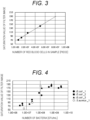
- FIG. 3 shows results of filtration using a mixed solution of a negative control blood sample having a blood bacterial concentration of 0 CFU/mL and a surfactant, and the amount of red blood cells in the sample used for filtration and the redness of the filter image were compared.
- the amount of red blood cells was calculated by a blood cell counter.
- a saturation value calculated by the following processing was used.
- the filter image is a color image, and is generally expressed in an RGB color space. RGB was converted into HSV (hue, saturation, and lightness) in order to reduce influence such as ambient brightness at the time of imaging. More specifically, the average value of the saturation values of pixels inside the filter region 20 was calculated as the redness of the filter image. From the results of FIG.
- the reason why the result of FIG. 2 was obtained is estimated as follows.
- the surfactant is added at a certain concentration regardless of the actual blood bacterial concentration, and most blood cells in the sample are destroyed by the surfactant when the actual blood bacterial concentration is low, so that most blood cells are removed in step S11 and no impurities remain on the filter.
- the concentration of bacteria and the concentration of red blood cells become approximately the same to each other, and the bacteria themselves inhibit the action of blood cell destruction by the surfactant.
- step S11 increases, and the red blood cells not completely destroyed are captured in the filtration step of step S12.
- bacteria aggregate with hemoglobin, fibrin and platelets, and impurities showing redness are captured in the filtration step of step S12.
- the redness of the filter region 20 after filtration becomes stronger. Therefore, for example, by calculating the amount of, for example, red blood cells, which are impurities remaining on the filter after filtration, from the image of the filter, the concentration of bacteria that have passed through the filter can be known.
- FIG. 4 is a graph showing a relationship between color information of a filter calculated by processing a filter image and an actual blood bacterial concentration.
- the filter image indicated in the RGB color space was converted into HSV (hue, saturation, and lightness), and the value of saturation was used. Specifically, the average value of the saturation values of pixels inside the filter region 20 is used.
- FIG. 4 also shows the results of repeated experiments with E. coli and the result with S. aureus. There is a positive correlation between the blood bacterial concentration and the saturation value of the filter image when the actual blood bacterial concentration is in the range of 10 6 to 10 9 CFU/mL.
- the number of bacteria can be estimated based on the information of the saturation value of the filter image after filtration.
- This range of the blood bacterial concentration is almost equivalent to the bacterial concentration in a sample that is usually positive in blood culture, and thus can be applied to various bacterial species and strains.
- the specific concentration of surfactant used in the treatment can be defined in the following range.
- the concentration of red blood cells in blood is on the order of 10 9 /mL, and if a 1 mL sample is pretreated, the number of red blood cells in the sample is 10 9 .
- the range of impurities that can be estimated from the tone of the filter shown in FIG. 2 for example, the amount of red blood cells is 10 6 to 10 8 . That is, in order to be able to estimate the actual bacterial concentration from the tone of the filter, the red blood cells may be destroyed until the concentration reaches 1/1000 to 1/10. That is, a surfactant at a concentration capable of destroying 90 to 99.9% of red blood cells in blood containing bacteria is required.
- the range of the surfactant concentration shown here is, for example, a case where 1 mL of sample is pretreated, and when the volume to be treated increases, it is preferable to increase the concentration of the surfactant so that the destruction rate of red blood cells increases accordingly.
- a surfactant having a concentration capable of destroying 99 to 99.99% of red blood cells is required, and the concentration may vary depending on the throughput of the sample.
- the concentration of the surfactant capable of destroying 99 to 99.99% of red blood cells in blood containing bacteria is in the range of 0.05 wt% to 0.5 wt%, assuming an example of sodium dodecyl sulfate, which is an anionic surfactant having hydrophilic and hydrophobic moieties and the hydrophobic moiety being a chain hydrocarbon.
- concentration of the surfactant is preferably a concentration capable of destroying a desired red blood cell.
- surfactants for example, regarding saponin, sodium cholate, sodium deoxycholate, 3-[(3-cholamidopropyl)dimethylammonio]-1 propanesulfonate, and 3-[(3-cholamidopropyl)dimethylammonio]-2 hydroxy-1 propanesulfonate, which are surfactants having hydrophilic and hydrophobic moieties and a cyclic hydrocarbon as a hydrophobic moiety, the types of surfactants may be mixed.
- 2.7 mL of blood sample was treated with a surfactant such as sodium dodecyl sulfate having a final concentration in the range of 0.05 wt% to 0.5 wt%, capable of destroying 99 to 99.99% of red blood cells in blood containing bacteria.
- a surfactant such as sodium dodecyl sulfate having a final concentration in the range of 0.05 wt% to 0.5 wt%, capable of destroying 99 to 99.99% of red blood cells in blood containing bacteria.
- FIG. 5 is a flowchart showing a procedure of performing bacterial concentration adjustment using a blood bacterial concentration estimated from a filter image. Steps S10 to S12 are the same as the steps shown in FIG. 1 .
- step S50 the filter is imaged, and the color of the impurities remaining on the filter is acquired.
- the amount of impurities is estimated based on the color of red blood cells remaining on the filter, it is not necessary to stain a sample and impurities.
- step S51 correspondence data describing correspondence between the color of the impurities remaining on the filter and the actual blood bacterial concentration is read out, and the estimated blood bacterial concentration is calculated by referring to the correspondence data using the color acquired from the filter image.
- the color of the impurities remaining on the filter and the actual blood bacterial concentration for example, a calibration curve represented by a single logarithm is acquired, and the blood bacterial concentration estimated from the data of the calibration curve is calculated.
- the correspondence data is illustrated in FIG. 4 , and is created in advance and stored in the storage device. Note that the method of the present disclosure can be applied as long as there is at least one piece of correspondence data regardless of the bacterial species and the type of surfactant.
- the correspondence data may be held for each of bacterial species, or may be held depending on information of resistant strains such as methicillin-resistant Staphylococcus aureus, and the type of surfactant.
- step S52 the sample is diluted to obtain a desired bacterial concentration based on the blood bacterial concentration estimated in S51.
- a desired bacterial concentration based on the blood bacterial concentration estimated in S51.
- the blood bacterial concentration estimated in S51 is 5 ⁇ 10 8 CFU/mL
- the desired bacterial concentration is 5 ⁇ 10 5 CFU/mL
- 1000-fold dilution is performed.
- the blood bacterial concentration estimated in S51 has not reached the desired bacterial concentration
- it is determined as a defective specimen it is difficult to prepare a specimen suitable for sensitivity test, and thus the blood culture bottle is further cultured to grow the bacteria, and then the process returns to step S10.
- an identification test or a sensitivity test may be performed using colonies obtained by performing isolation and cultivation.
- an identification test and a sensitivity test are performed using the prepared bacterial suspension.
- Any method may be used as the inspection method. Examples thereof include such as an identification test using an automatic device, a genetic test, a sensitivity test by a microliquid dilution method, a sensitivity test by a disk method, and a rapid sensitivity test by a microscopic image, laser scattered light measurement or the like.
- the sample after destroying the blood cells is filtered by the filtration filter, and the estimated blood bacterial concentration is calculated by referring to the correspondence data between the color component of the image of the impurities remaining on the filter and the actual blood bacterial concentration.
- an automatic analyzer for obtaining a sample having a bacterial concentration adjusted to a desired value from a blood sample containing bacteria is shown. Note that the present aspect is merely an example, and is not limited to this configuration.
- FIG. 6 is a configuration diagram of an automatic analyzer 100 according to Aspect 2.
- the automatic analyzer 100 is a device that automatically performs the pretreatment procedure described in FIG. 5 .
- a rubber stopper is used for the blood culture bottle to prevent contamination or the like, and the inside of the bottle is vacuum.
- An operator takes out a blood sample from the blood culture bottle using an injection needle, and dispenses the blood sample into a container containing a surfactant. Accordingly, S10 is performed.
- an introduction device 101 that automatically introduces a surfactant into a sample can be provided as a part of the automatic analyzer 100.
- the sample into which the surfactant is introduced is introduced into a centrifugal separator 102.
- the blood cells in the sample are destroyed by the surfactant.
- the centrifugal separator 102 separates the eluted hemoglobin and the like from bacteria, blood cells that could not be destroyed, and the like. As a result, the separation step of S11 is performed.
- the sample that has been centrifuged is introduced into a cleaning unit 103.
- the cleaning unit 103 removes the supernatant of the sample, and cleans the sample with, for example, about 1 mL of physiological saline or pure water, or a cleaning liquid such as a medium.
- a cleaning pipette 104 aspirates the supernatant.
- a portion from the bottom of the sample container to a certain reference height may be treated as the supernatant.
- the rest of S11 is performed. More strictly, it is preferable to provide a liquid position sensor or the like, detect an interface between a portion where bacteria and blood cells are coagulated into a pellet form and a liquid portion, and treat a portion up to the vicinity of the interface position as the supernatant.
- the cleaned sample is introduced into a filtration filter unit 105.
- the filtration filter unit 105 consists of, for example, a disposal filtration filter, a syringe for capturing impurities in the filter, and the like. S12 is performed by the filtration filter unit 105. In some cases, the sample may be filtered using the centrifugal separator 102.
- a camera 106 (corresponding to a sensor that detects the amount of impurities) images impurities remaining on the filtration filter unit 105.
- a storage unit 107 stores the correspondence data described in FIG. 4 .
- a computer 110 (operation unit) converts the RGB image captured by the camera 106 into an HSV color space image, detects a color component (for example, a saturation value) of the impurity region (S50), and calculates the estimated blood bacterial concentration by referring to the correspondence data using the color (S51).
- the filtered sample is introduced into a dilution unit 108.
- the dilution unit 108 adjusts the dilution ratio so as to obtain a desired bacterial concentration on the basis of the blood bacterial concentration estimated by the computer 110.
- a dilution pipette 109 introduces the diluent according to its dilution ratio. Accordingly, S52 is performed.
- the computer 110 automatically performs the above steps by controlling each unit included in the automatic analyzer 100. It is preferred that the computer 110 includes an input/output device and the operator can instruct the computer 110 about the type of bacteria, the desired bacterial concentration, and the like.
- the computer 110 can change the correspondence data to be referred according to the input bacteria type, and can also change the dilution ratio by the dilution unit 108 according to the desired bacterial concentration.
- the blood bacterial concentration value estimated by the computer 110 may be output to an output device such as a display, and when the blood bacterial concentration value is equal to or less than the desired bacterial concentration, a flag indicating a defective specimen may be displayed (S53).
- the automatic analyzer 100 may include a sensitivity test device 111 in addition to the above configuration.
- the computer 110 automatically performs the sensitivity test by controlling the sensitivity test device 111. As a result, all processes from S10 to S54 can be automatically performed.
- As the contents of the sensitivity test in addition to those described in Aspect 1, the minimum inhibitory concentration and the like described in Examples described later can also be measured.
- the method for adjusting the blood bacterial concentration in the blood sample described in Aspects 1 to 2 is influenced to some extent by the amount of red blood cells contained in the original blood.
- the concentration of human red blood cells varies depending on sex and health condition, but is about 3 ⁇ 10 9 to 6 ⁇ 10 9 /mL, and the variation is extremely small as compared with a bacterial concentration range of 10 6 to 10 10 /mL. Therefore, it is considered that the influence on the result of the estimated blood bacterial concentration is small.
- the result of step S51 can be corrected using the value.
- the blood bacterial concentration estimated more accurately can be calculated.
- the correction procedure for example, the following ones are conceivable.
- the operator or the computer 110 performs the procedure of FIG. 5 to estimate the amount of impurities on the filter one or more times, and further obtain another detection result.
- the final amount of impurities is obtained by averaging the plurality of amounts of impurities excluding abnormal values.
- the estimated blood bacterial concentration is calculated by referring to the correspondence data using the amount of impurities.
- the correspondence data is referred using another detection result obtained by measuring the amount of impurities. Therefore, the correspondence data needs to describe correspondence between the amount of impurities and the actual blood bacterial concentration.
- the saturation value of the filter image and the amount of impurities corresponding thereto can be described together in the correspondence data, or a conversion formula between the saturation value and the amount of impurities can be defined in advance, and another detection result can be converted into the saturation value using the conversion formula. That is, the correspondence data may describe correspondence between a value representing the amount of impurities remaining on the filter in some form and the actual blood bacterial concentration in the blood sample.
- the saturation value of the filter image is used to detect red blood cells remaining in the filter.
- an optical sensor that detects absorption or reflection of a specific wavelength corresponding to red can also be used. That is, by using an optical sensor capable of detecting at least the largest RGB component of impurities remaining on the filter, information similar to the saturation value of the image captured by the camera 106 can be obtained.
- the correspondence data also needs to describe a numerical value measured by the optical sensor instead of the saturation value.
- a person may visually measure the amount of impurities based on a color sample, and refer to the correspondence data based on the measurement result.
- the color sample itself may describe the correspondence between the color of the impurities and the blood bacterial concentration.
- Example 1 of the present disclosure the superiority of the pretreatment method according to the present disclosure will be described together with a comparative example.
- comparison was made between concentration adjustment using turbidity measurement and concentration adjustment according to the present disclosure, respectively adjusting the recommended bacterial concentration range of the sensitivity test.
- concentration adjustment absorbance measurement at a wavelength of 600 nm was used.
- concentration adjustment according to the present disclosure the pretreatment method shown in FIG. 1 was used.
- the type, concentration and the like of bacteria and surfactants used are the same as those in Aspect 1.
- FIG. 7 shows results of adjusting the bacterial concentration from the bacterial suspension obtained by the pretreatment method shown in FIG. 1 by turbidity measurement used in the conventional bacterial test pretreatment.
- the hatched area is a recommended bacterial concentration range when performing a sensitivity test, which is defined by the Clinical and Laboratory Standards Institute.
- the range of this hatching is an area of 5 ⁇ 10 5 CFU/mL ( ⁇ 60%), and adjustment was attempted to obtain a median value of 5 ⁇ 10 5 CFU/mL.
- a bacterial suspension adjusted to a McFarland turbidity of 0.5 corresponding to a bacterial number concentration of 1.5 ⁇ 10 8 CFU/mL was diluted 300 times by McFarland standards.
- the bacterial concentration in the blood sample is 10 8 CFU/mL or more, it is possible to adjust the bacterial concentration to the vicinity of a desired concentration range, but when the blood bacterial concentration in the blood sample is 10 6 to 10 7 CFU/mL, the number of bacteria after the adjustment is reduced by 1 to 2 digits. This is because hemoglobin that could not be removed in steps S11 and S12, fine particles contained in the medium and the like contribute to an increase in scattered light, so that the value of turbidity increases even though the bacterial concentration is low. Therefore, since the bacterial concentration is estimated excessively, it is difficult to adjust the bacterial concentration by using turbidity measurement. This is also apparent from the fact that the plot of FIG. 7 does not fall within the hatched area showing the recommended bacterial concentration range at all.
- FIG. 8 shows results of performing concentration adjustment from the blood bacterial concentration estimated using the method shown in FIG. 5 .
- the adjusted bacterial concentration does not decrease and is mostly within the hatched range.
- FIG. 8 shows results of pretreating E. coli and S. aureus based on the same correspondence data.
- FIG. 8 shows that even bacteria having greatly different properties such as gram-negative bacteria and gram-positive bacteria can keep the adjusted bacterial concentration within a certain range based on one correspondence data.
- Example 2 of the present disclosure shows an example in which bacterial concentration was adjusted from a blood culture positive sample by the pretreatment method according to the present disclosure using E. coli.
- a growth rate in the case of preparing bacterial suspension from colonies by isolation and cultivation for a whole day is used.
- the bacterial concentration in a blood culture-positive sample was adjusted to a final bacterial concentration of 5 ⁇ 10 5 CFU/mL using three different bacterial concentrations up to 10 7 to 10 9 CFU/mL. Even when bacterial suspension was prepared from colonies, the bacterial suspension was adjusted using turbidity measurement so that the final bacterial concentration was 5 ⁇ 10 5 CFU/mL.
- a temporal change in the growth rate was calculated using the area of a region determined to be a bacterium in a microscopic image as an index of the growth rate.
- FIG. 9 shows growth rates of blood culture-positive samples and a sample created from colonies. Although there is a difference of about 0.5 hours in the time at which the bacterial growth rate rises between blood culture positive_1 and isolation culture_1, the final growth rates are substantially the same. The same applies to blood culture positive_2 and blood culture positive_3. This indicates that even when the blood culture-positive sample is pretreated, the bacterial concentration can be adjusted to the same degree as the bacterial concentration adjusted from the colonies, and the bacterial growth is not affected in the sensitivity test.
- Example 3 of the present disclosure the results when E. coli in Example 2 was changed to S. aureus will be described.
- FIG. 10 shows growth rates of blood culture-positive samples and a sample created from colonies.
- the final growth rate is the same between blood culture positive and isolation culture. Therefore, as in Example 2, this indicates that even when the blood culture-positive sample is pretreated, the bacterial concentration can be adjusted to the same degree as the bacterial concentration adjusted from the colonies, and the bacterial growth is not affected in the sensitivity test.
- Example 4 of the present disclosure shows a result of performing a drug sensitivity test on both blood culture positive samples and a sample created from colonies.
- the lowest concentration (Minimum Inhibitory Concentration: MIC) among the concentrations at which the drug exerts an antibacterial action on bacteria is measured.
- MIC Minimum Inhibitory Concentration
- a microliquid dilution method was used for the sensitivity test. The bacterial suspension and different concentrations of the drug were mixed, and the MIC was determined by visually determining turbidity of each well of the cultured 96-well plate after 18 hours.
- FIG. 11 shows results of performing a drug sensitivity test.
- E. coli cefepime (CFPM), cefotaxime (CTX), gentamicin (GM), and levofloxacin (LVFX) were allowed to act, and for S. aureus, erythromycin (EM), oxacillin (MPIPC), penicillin G (PCG), and vancomycin (VCM) were allowed to act.
- CFPM cefepime
- CX cefotaxime
- GM gentamicin
- LVFX levofloxacin
- S. aureus erythromycin
- MPIPC oxacillin
- PCG penicillin G
- VCM vancomycin
- the MIC in the case of pretreating the blood culture sample falls within the range of ⁇ 1 tube (twice or half) of the MIC in the case of the sample created from the colonies, indicating that the sensitivity test can be correctly performed even from the sample obtained by pretreating the blood culture sample.
- FIG. 11 shows an example in which the MIC is determined using the microliquid dilution method.
- the MIC may be determined by a rapid sensitivity test using a microscopic image, or a rapid sensitivity test using a laser beam may be performed.
- the correspondence data is referred using the average value of the saturation values of the filter region 20, but a maximum value or a mode value may be used instead of the average value.
- the amount of impurities remaining on the filter may be represented by a feature amount represented by at least two of hue, lightness, and saturation.
- the present disclosure can also be used for other bacterial samples. That is, the present disclosure can be used for a sample having correspondence between image information obtained by imaging the impurities remaining on the filter and the bacterial concentration in the sample.
- the substance added to destroy the impurities may be appropriately changed depending on the type of impurities.
Landscapes
- Chemical & Material Sciences (AREA)
- Health & Medical Sciences (AREA)
- Life Sciences & Earth Sciences (AREA)
- Organic Chemistry (AREA)
- Engineering & Computer Science (AREA)
- Wood Science & Technology (AREA)
- Proteomics, Peptides & Aminoacids (AREA)
- Zoology (AREA)
- Immunology (AREA)
- Molecular Biology (AREA)
- General Health & Medical Sciences (AREA)
- Biochemistry (AREA)
- Physics & Mathematics (AREA)
- Analytical Chemistry (AREA)
- Microbiology (AREA)
- Bioinformatics & Cheminformatics (AREA)
- Biotechnology (AREA)
- Biophysics (AREA)
- Genetics & Genomics (AREA)
- General Engineering & Computer Science (AREA)
- Toxicology (AREA)
- Hematology (AREA)
- Pathology (AREA)
- Urology & Nephrology (AREA)
- Biomedical Technology (AREA)
- Medicinal Chemistry (AREA)
- General Physics & Mathematics (AREA)
- Cell Biology (AREA)
- Food Science & Technology (AREA)
- Measuring Or Testing Involving Enzymes Or Micro-Organisms (AREA)
- Investigating Or Analysing Biological Materials (AREA)
Claims (11)
- Automatisches Analyseverfahren zum Analysieren einer Blutprobe, die Bakterien und Verunreinigungen enthält, wobei das Verfahren umfasst:Einführen (S10) einer Substanz, die die Verunreinigungen in der Probe zerstört;Trennen (S11) der Verunreinigungen und der Bakterien voneinander in der Probe, in die die Substanz eingebracht wurde, durch einen Zentrifugalseparator (102);Entnahme (S12) der Bakterien aus der Probe unter Verwendung eines Filters (105), der die Bakterien aus der Probe, aus der die Verunreinigungen und die Bakterien abgetrennt wurden, entnimmt;Lesen von Korrespondenzdaten, die die Übereinstimmung zwischen einem numerischen Wert, der eine Menge der auf dem Filter (105) verbleibenden Verunreinigungen darstellt, und einer Konzentration der Bakterien in der Probe beschreiben, aus einer Speichereinheit (107), die die Korrespondenzdaten speichert; undSchätzen (S51) einer Konzentration der Bakterien in der Probe durch Bezugnahme auf die Korrespondenzdaten unter Verwendung eines numerischen Wertes, der die Menge der Verunreinigungen darstellt, die auf dem Filter (105) verbleiben, nachdem die Bakterien aus der Probe entfernt wurden, wobeidie Probe Blutzellen als die Verunreinigungen enthält,die Substanz ein Tensid ist,das Tensid mindestens eines der folgenden Elemente umfasst:ein anionisches Tensid mit einem hydrophilen Teil und einem hydrophoben Teil, wobei der hydrophobe Teil ein Kettenkohlenwasserstoff ist; oderein oberflächenaktives Mittel mit einem hydrophilen und einem hydrophoben Teil, wobei der hydrophobe Teil einen zyklischen Kohlenwasserstoff enthält,der Filter (105) ein Filtrationsfilter ist, der die Verunreinigungen und die Bakterien durch Filtration der Probe abtrennt, unddas Schätzen (S51) der Konzentration der Bakterien umfasst:Erfassen der Menge der auf dem Filter (105) verbliebenen Verunreinigungen unter Verwendung eines Bildes, das durch Abbilden ohne Färben der auf dem Filter (105) verbliebenen Verunreinigungen erhalten wird, undBezugnahme auf die Korrespondenzdaten unter Verwendung der Menge der anhand des Bildes erkannten Verunreinigungen.
- Automatisches Analyseverfahren nach Anspruch 1, wobeidas automatische Analyseverfahren ferner das Erfassen (S50) eines RGB-Bildes als das Bild umfasst, unddas Schätzen (S51) der Konzentration der Bakterien umfasstUmwandeln des RGB-Bildes in ein HSV-Farbraumbild, undVerwenden eines Sättigungswertes auf dem HSV-Farbraumbild der auf dem Filter (105) verbleibenden Verunreinigungen als numerischer Wert, der die Menge der auf dem Filter (105) verbleibenden Verunreinigungen darstellt.
- Automatisches Analyseverfahren nach Anspruch 1, wobeidas automatische Analyseverfahren ferner das Erfassen (S50) eines RGB-Bildes als das Bild umfasst, unddas Schätzen (S51) der Konzentration der Bakterien umfasst:Umwandeln des RGB-Bildes in ein HSV-Farbraumbild, undVerwenden eines Merkmalsbetrags, der durch einen Farbtonwert, einen Sättigungswert und einen Helligkeitswert auf dem HSV-Farbraumbild der auf dem Filter (105) verbleibenden Verunreinigungen dargestellt wird, als den numerischen Wert, der die Menge der auf dem Filter (105) verbleibenden Verunreinigungen darstellt.
- Automatisches Analyseverfahren nach Anspruch 1, wobei das Schätzen (S51) der Konzentration der Bakterien Folgendes umfasst:Erhalten eines Ergebnisses der Erfassung der Verunreinigungen von einem optischen Sensor (106), der mindestens eine der größten RGB-Farbkomponenten der Verunreinigungen erfasst, undVerwenden eines Ergebnisses der Erfassung der auf dem Filter (105)verbliebenen Verunreinigungen durch den optischen Sensor (106) als den numerischen Wert, der die Menge der Verunreinigungen auf dem Filter (105) darstellt.
- Automatisches Analyseverfahren nach Anspruch 1, wobei das Schätzen (S51) der Konzentration der Bakterien Folgendes umfasst:Erhalten eines weiteren Detektionsergebnisses durch Detektion der Menge der Verunreinigungen in der Probe, getrennt von der Menge der Verunreinigungen, die bei der Detektion der Menge der auf dem Filter (105) verbliebenen Verunreinigungen detektiert wurde, undKorrigieren der Menge der Verunreinigungen, die beim Erfassen der Menge der auf dem Filter (105) verbleibenden Verunreinigungen erfasst wurden, unter Verwendung des anderen Erfassungsergebnisses, und Bezugnahme auf die Korrespondenzdaten unter Verwendung der korrigierten Menge der Verunreinigungen.
- Automatisches Analyseverfahren nach Anspruch 1, wobeidas automatische Analyseverfahren ferner das Durchführen (S54) eines Arzneimittelempfindlichkeitstests der Bakterien gegenüber einem Arzneimittel umfasst, unddas Durchführen (S54) des Arzneimittelempfindlichkeitstests, nachdem die Konzentration der Bakterien in der Probe geschätzt wurde, die Durchführung des Arzneimittelempfindlichkeitstests für die Bakterien in der Probe ohne Kultivierung der Bakterien in der Probe umfasst.
- Automatisches Analyseverfahren nach Anspruch 6, wobeidas automatische Analyseverfahren ferner das Verdünnen (S52) der Probe umfasst, undbei dem Verdünnen (S52) der Probe, nachdem die Konzentration der Bakterien in der Probe geschätzt wurde (S51), die Probe verdünnt wird, um eine Testprobe mit einer Konzentration der Bakterien zu erzeugen, die für das Durchführen des Arzneimittelempfindlichkeitstests erforderlich ist, unddas Durchführen (S54) des Arzneimittelempfindlichkeitstests die Durchführung des Arzneimittelempfindlichkeitstests an der durch die Verdünnung der Probe erzeugten Testprobe umfasst.
- Automatisches Analyseverfahren nach Anspruch 6, wobei das Durchführen (S54) des Drogenempfindlichkeitstests Folgendes umfasst:Erfassen eines Bildes einer Probe, die in einem auf 35 bis 37°C gehaltenen Inkubator platziert ist, durch eine Abbildungsvorrichtung (106), undBestimmen einer minimalen Hemmkonzentration des Arzneimittels durch Messen einer Wachstumsrate der Bakterien unter Verwendung eines Bildes der Probe, das von der Abbildungsvorrichtung (106) aufgenommen wurde.
- Automatisches Analyseverfahren nach Anspruch 1, wobei die Filtrationsporengröße des Filters (105) 1 bis 40 µm beträgt und ein Material des Filters (105) ein hydrophobes Material ist.
- Automatisches Analyseverfahren nach Anspruch 1, wobei die Verunreinigungen mindestens rote Blutkörperchen im Blut umfassen.
- Automatisches Analysegerät (100) zum Analysieren einer Blutprobe, die Bakterien und Verunreinigungen enthält, wobei das automatische Analysegerät (100) umfasst:einen Zentrifugalseparator (102), der dazu ausgelegt ist, die Verunreinigungen und die Bakterien in der Probe voneinander zu trennen, in die eine Substanz eingebracht wurde, die die Verunreinigungen zerstört;ein Filtrationsfilter (105), das dazu ausgelegt ist, die Bakterien aus der Probe zu entfernen, von der die Verunreinigungen und die Bakterien durch Filtration der Probe getrennt wurden;eine Speichereinheit (107), die dazu ausgelegt ist, Korrespondenzdaten zu speichern, die die Korrespondenz zwischen einem numerischen Wert, der eine Menge der auf dem Filter (105) verbleibenden Verunreinigungen darstellt, und einer Konzentration der Bakterien in der Probe beschreiben; undeine Recheneinheit (110), die dazu ausgelegt ist, eine Konzentration der Bakterien in der Probe durch Bezugnahme auf die Korrespondenzdaten unter Verwendung eines numerischen Wertes zu schätzen, der die Menge der Verunreinigungen darstellt, die auf dem Filter (105) verbleiben, nachdem die Bakterien aus der Probe entfernt wurden,wobeidie Probe Blutzellen als die Verunreinigungen enthält,die Substanz ein Tensid ist,das Tensid mindestens eines der folgenden Elemente umfasst:ein anionisches Tensid mit einem hydrophilen Teil und einem hydrophoben Teil, wobei der hydrophobe Teil ein Kettenkohlenwasserstoff ist; oderein oberflächenaktives Mittel mit einem hydrophilen und einem hydrophoben Teil, wobei der hydrophobe Teil einen zyklischen Kohlenwasserstoff enthält, unddie Recheneinheit (110) dazu ausgelegt ist, die Konzentration der Bakterien in der Probe zu schätzen, indem siedie Menge der auf dem Filter (105) verbliebenen Verunreinigungen unter Verwendung eines Bildes erfasst, das durch Abbilden ohne Färben der auf dem Filter (105) verbliebenen Verunreinigungen erhalten wird, undBezug nimmt auf die Korrespondenzdaten unter Verwendung der Menge der anhand des Bildes erkannten Verunreinigungen.
Applications Claiming Priority (1)
| Application Number | Priority Date | Filing Date | Title |
|---|---|---|---|
| PCT/JP2020/018899 WO2021229667A1 (ja) | 2020-05-12 | 2020-05-12 | 自動分析装置、自動分析方法 |
Publications (3)
| Publication Number | Publication Date |
|---|---|
| EP4151744A1 EP4151744A1 (de) | 2023-03-22 |
| EP4151744A4 EP4151744A4 (de) | 2024-01-10 |
| EP4151744B1 true EP4151744B1 (de) | 2024-11-20 |
Family
ID=78525467
Family Applications (1)
| Application Number | Title | Priority Date | Filing Date |
|---|---|---|---|
| EP20935377.0A Active EP4151744B1 (de) | 2020-05-12 | 2020-05-12 | Automatischer analysator und automatisches analyseverfahren |
Country Status (6)
| Country | Link |
|---|---|
| US (1) | US20230167480A1 (de) |
| EP (1) | EP4151744B1 (de) |
| JP (1) | JP7371245B2 (de) |
| KR (1) | KR102859800B1 (de) |
| CN (1) | CN115461466B (de) |
| WO (1) | WO2021229667A1 (de) |
Families Citing this family (1)
| Publication number | Priority date | Publication date | Assignee | Title |
|---|---|---|---|---|
| KR102945517B1 (ko) * | 2023-11-30 | 2026-03-30 | 김주용 | 감염병 확산 방지를 위한 실시간 화장실 위생관리 시스템 및 방법 |
Family Cites Families (16)
| Publication number | Priority date | Publication date | Assignee | Title |
|---|---|---|---|---|
| US3928139A (en) * | 1973-02-12 | 1975-12-23 | Wadley Res Inst & Blood Bank | Detection of microbial pathogens |
| US4144134A (en) * | 1977-01-31 | 1979-03-13 | Vitatect Corp. | Method for detection of bacterial concentration by luminescence |
| FI91085C (fi) * | 1991-01-24 | 1994-05-10 | Orion Yhtymae Oy | Menetelmä gram-negatiivisten bakteerien määrittämiseksi ja erottamiseksi |
| US20020098589A1 (en) * | 2001-01-19 | 2002-07-25 | Crews Harold Richardson | Multi-purpose reagent system and method for enumeration of red blood cells, white blood cells and thrombocytes and differential determination of white blood cells |
| US6803208B2 (en) * | 2001-07-30 | 2004-10-12 | The United States Of America As Represented By The Secretary Of The Navy | Automated epifluorescence microscopy for detection of bacterial contamination in platelets |
| FR2829500B1 (fr) | 2001-09-13 | 2003-12-12 | Hemosystem | Procede de concentration et de detection de germes pathogenes a partir de produits sanguins et/ou de leurs derives et dispositif pour le mettre en oeuvre |
| JP2007006709A (ja) * | 2005-06-28 | 2007-01-18 | Matsushita Electric Ind Co Ltd | 発光物の判別方法 |
| BRPI0920114B1 (pt) * | 2008-10-31 | 2021-08-17 | Biomerieux, Inc. | Métodos para o isolamento e a identificação de micro-organismos |
| EP2333105A1 (de) | 2009-12-08 | 2011-06-15 | Koninklijke Philips Electronics N.V. | Selektive Lyse von Zellen |
| EP2684947A1 (de) * | 2012-07-09 | 2014-01-15 | Biocartis SA | Verfahren zum Filtern einer biologischen Probe |
| US20140185437A1 (en) | 2012-12-29 | 2014-07-03 | Minyoung Park | Methods and arrangements for traffic indication mapping in wireless networks |
| JP6177009B2 (ja) | 2013-06-03 | 2017-08-09 | 株式会社日立ハイテクノロジーズ | 血球破壊試薬及びそれを用いる血球破壊方法 |
| US10457975B1 (en) * | 2016-02-10 | 2019-10-29 | William George Pitt | Hollow rotating device for separating particles from blood |
| WO2019097752A1 (ja) | 2017-11-15 | 2019-05-23 | 国立大学法人 富山大学 | 血液検体の前処理方法 |
| JP2021093913A (ja) * | 2018-03-28 | 2021-06-24 | 国立大学法人千葉大学 | 血液試料中の細菌の検出方法 |
| EP3803326A4 (de) * | 2018-05-25 | 2022-02-23 | Qvella Corporation | Verfahren und zusammensetzungen zur selektiven lyse von blutzellen und trennung mikrobieller zellen |
-
2020
- 2020-05-12 JP JP2022522125A patent/JP7371245B2/ja active Active
- 2020-05-12 EP EP20935377.0A patent/EP4151744B1/de active Active
- 2020-05-12 KR KR1020227036942A patent/KR102859800B1/ko active Active
- 2020-05-12 US US17/996,962 patent/US20230167480A1/en active Pending
- 2020-05-12 CN CN202080100175.3A patent/CN115461466B/zh active Active
- 2020-05-12 WO PCT/JP2020/018899 patent/WO2021229667A1/ja not_active Ceased
Also Published As
| Publication number | Publication date |
|---|---|
| KR102859800B1 (ko) | 2025-09-16 |
| CN115461466A (zh) | 2022-12-09 |
| EP4151744A1 (de) | 2023-03-22 |
| US20230167480A1 (en) | 2023-06-01 |
| CN115461466B (zh) | 2025-10-31 |
| WO2021229667A1 (ja) | 2021-11-18 |
| EP4151744A4 (de) | 2024-01-10 |
| JP7371245B2 (ja) | 2023-10-30 |
| KR20220158051A (ko) | 2022-11-29 |
| JPWO2021229667A1 (de) | 2021-11-18 |
Similar Documents
| Publication | Publication Date | Title |
|---|---|---|
| JP6186414B2 (ja) | 固体又は半固体培地上の微生物のキャラクタリゼーション方法 | |
| JP4842473B2 (ja) | 微生物を検出し、定量し、特徴付けする装置および方法 | |
| US12241113B2 (en) | Method for determining the concentration of intact microorganisms in a sample | |
| US10900011B2 (en) | Test apparatus | |
| CN102272585A (zh) | 采用拉曼光谱法分离、表征和/或鉴定微生物的方法 | |
| US6803208B2 (en) | Automated epifluorescence microscopy for detection of bacterial contamination in platelets | |
| KR20200111720A (ko) | 미생물 농도를 결정하기 위한 방법 | |
| CN109716107B (zh) | 用于确定微生物病原体的组合的光学光谱法 | |
| EP4151744B1 (de) | Automatischer analysator und automatisches analyseverfahren | |
| JP5814259B2 (ja) | 診断解析の方法及び装置 | |
| CN120060430A (zh) | 一种血培养阳性标本直接制备用于药敏实验的菌液的方法 | |
| CN113906285A (zh) | 采用膜荧光染色和光谱强度比进行抗生素敏感性快速检测的显微镜法 | |
| JP2001149091A (ja) | 微生物測定方法及び装置 | |
| US20250084451A1 (en) | Method and Device for Detecting Presence, Concentration, and Antibiotic Resistance of Bacteria in Urine Samples | |
| EP3645734B1 (de) | Verfahren zur quantifizierung der kultivierbarkeit von individuellen bakteriellen zellen unter verwendung kulturunabhängiger parameter | |
| US20250066835A1 (en) | Microbial Image Analysis Method | |
| JP2025073626A (ja) | 微生物の性質又は状態評価を目的とする情報取得のためのマイクロチップ、該マイクロチップを用いる情報取得方法又は評価システム、及び該取得方法若しくは評価システムに用いることができるコンピュータプログラム | |
| KR20240176080A (ko) | 바이오 의약품의 안전성 입증을 위한 신속 무균 시험 방법 및 신속 무균 시험 플랫폼 | |
| EP4453234A1 (de) | Farbstoffe und deren verwendungen | |
| KR20260031495A (ko) | 광학감지형 세균배양 콘테이너에 의한 미생물 자동염색 플랫폼 시스템 | |
| JP2008035788A (ja) | 微生物数測定のための試料の前処理方法、前処理キットおよび前処理装置 |
Legal Events
| Date | Code | Title | Description |
|---|---|---|---|
| STAA | Information on the status of an ep patent application or granted ep patent |
Free format text: STATUS: THE INTERNATIONAL PUBLICATION HAS BEEN MADE |
|
| PUAI | Public reference made under article 153(3) epc to a published international application that has entered the european phase |
Free format text: ORIGINAL CODE: 0009012 |
|
| STAA | Information on the status of an ep patent application or granted ep patent |
Free format text: STATUS: REQUEST FOR EXAMINATION WAS MADE |
|
| 17P | Request for examination filed |
Effective date: 20221024 |
|
| AK | Designated contracting states |
Kind code of ref document: A1 Designated state(s): AL AT BE BG CH CY CZ DE DK EE ES FI FR GB GR HR HU IE IS IT LI LT LU LV MC MK MT NL NO PL PT RO RS SE SI SK SM TR |
|
| DAV | Request for validation of the european patent (deleted) | ||
| DAX | Request for extension of the european patent (deleted) | ||
| A4 | Supplementary search report drawn up and despatched |
Effective date: 20231212 |
|
| RIC1 | Information provided on ipc code assigned before grant |
Ipc: C12Q 1/18 20060101ALI20231206BHEP Ipc: C12Q 1/06 20060101AFI20231206BHEP |
|
| GRAP | Despatch of communication of intention to grant a patent |
Free format text: ORIGINAL CODE: EPIDOSNIGR1 |
|
| STAA | Information on the status of an ep patent application or granted ep patent |
Free format text: STATUS: GRANT OF PATENT IS INTENDED |
|
| INTG | Intention to grant announced |
Effective date: 20240731 |
|
| GRAS | Grant fee paid |
Free format text: ORIGINAL CODE: EPIDOSNIGR3 |
|
| GRAA | (expected) grant |
Free format text: ORIGINAL CODE: 0009210 |
|
| STAA | Information on the status of an ep patent application or granted ep patent |
Free format text: STATUS: THE PATENT HAS BEEN GRANTED |
|
| AK | Designated contracting states |
Kind code of ref document: B1 Designated state(s): AL AT BE BG CH CY CZ DE DK EE ES FI FR GB GR HR HU IE IS IT LI LT LU LV MC MK MT NL NO PL PT RO RS SE SI SK SM TR |
|
| REG | Reference to a national code |
Ref country code: GB Ref legal event code: FG4D |
|
| REG | Reference to a national code |
Ref country code: CH Ref legal event code: EP |
|
| REG | Reference to a national code |
Ref country code: DE Ref legal event code: R096 Ref document number: 602020041878 Country of ref document: DE |
|
| REG | Reference to a national code |
Ref country code: IE Ref legal event code: FG4D |
|
| REG | Reference to a national code |
Ref country code: LT Ref legal event code: MG9D |
|
| REG | Reference to a national code |
Ref country code: NL Ref legal event code: MP Effective date: 20241120 |
|
| PG25 | Lapsed in a contracting state [announced via postgrant information from national office to epo] |
Ref country code: HR Free format text: LAPSE BECAUSE OF FAILURE TO SUBMIT A TRANSLATION OF THE DESCRIPTION OR TO PAY THE FEE WITHIN THE PRESCRIBED TIME-LIMIT Effective date: 20241120 Ref country code: PT Free format text: LAPSE BECAUSE OF FAILURE TO SUBMIT A TRANSLATION OF THE DESCRIPTION OR TO PAY THE FEE WITHIN THE PRESCRIBED TIME-LIMIT Effective date: 20250320 Ref country code: IS Free format text: LAPSE BECAUSE OF FAILURE TO SUBMIT A TRANSLATION OF THE DESCRIPTION OR TO PAY THE FEE WITHIN THE PRESCRIBED TIME-LIMIT Effective date: 20250320 |
|
| PG25 | Lapsed in a contracting state [announced via postgrant information from national office to epo] |
Ref country code: FI Free format text: LAPSE BECAUSE OF FAILURE TO SUBMIT A TRANSLATION OF THE DESCRIPTION OR TO PAY THE FEE WITHIN THE PRESCRIBED TIME-LIMIT Effective date: 20241120 Ref country code: NL Free format text: LAPSE BECAUSE OF FAILURE TO SUBMIT A TRANSLATION OF THE DESCRIPTION OR TO PAY THE FEE WITHIN THE PRESCRIBED TIME-LIMIT Effective date: 20241120 |
|
| REG | Reference to a national code |
Ref country code: AT Ref legal event code: MK05 Ref document number: 1743612 Country of ref document: AT Kind code of ref document: T Effective date: 20241120 |
|
| PG25 | Lapsed in a contracting state [announced via postgrant information from national office to epo] |
Ref country code: BG Free format text: LAPSE BECAUSE OF FAILURE TO SUBMIT A TRANSLATION OF THE DESCRIPTION OR TO PAY THE FEE WITHIN THE PRESCRIBED TIME-LIMIT Effective date: 20241120 |
|
| PG25 | Lapsed in a contracting state [announced via postgrant information from national office to epo] |
Ref country code: ES Free format text: LAPSE BECAUSE OF FAILURE TO SUBMIT A TRANSLATION OF THE DESCRIPTION OR TO PAY THE FEE WITHIN THE PRESCRIBED TIME-LIMIT Effective date: 20241120 |
|
| PG25 | Lapsed in a contracting state [announced via postgrant information from national office to epo] |
Ref country code: NO Free format text: LAPSE BECAUSE OF FAILURE TO SUBMIT A TRANSLATION OF THE DESCRIPTION OR TO PAY THE FEE WITHIN THE PRESCRIBED TIME-LIMIT Effective date: 20250220 |
|
| PG25 | Lapsed in a contracting state [announced via postgrant information from national office to epo] |
Ref country code: AT Free format text: LAPSE BECAUSE OF FAILURE TO SUBMIT A TRANSLATION OF THE DESCRIPTION OR TO PAY THE FEE WITHIN THE PRESCRIBED TIME-LIMIT Effective date: 20241120 Ref country code: GR Free format text: LAPSE BECAUSE OF FAILURE TO SUBMIT A TRANSLATION OF THE DESCRIPTION OR TO PAY THE FEE WITHIN THE PRESCRIBED TIME-LIMIT Effective date: 20250221 Ref country code: LV Free format text: LAPSE BECAUSE OF FAILURE TO SUBMIT A TRANSLATION OF THE DESCRIPTION OR TO PAY THE FEE WITHIN THE PRESCRIBED TIME-LIMIT Effective date: 20241120 |
|
| PG25 | Lapsed in a contracting state [announced via postgrant information from national office to epo] |
Ref country code: PL Free format text: LAPSE BECAUSE OF FAILURE TO SUBMIT A TRANSLATION OF THE DESCRIPTION OR TO PAY THE FEE WITHIN THE PRESCRIBED TIME-LIMIT Effective date: 20241120 |
|
| PG25 | Lapsed in a contracting state [announced via postgrant information from national office to epo] |
Ref country code: RS Free format text: LAPSE BECAUSE OF FAILURE TO SUBMIT A TRANSLATION OF THE DESCRIPTION OR TO PAY THE FEE WITHIN THE PRESCRIBED TIME-LIMIT Effective date: 20250220 |
|
| PG25 | Lapsed in a contracting state [announced via postgrant information from national office to epo] |
Ref country code: SM Free format text: LAPSE BECAUSE OF FAILURE TO SUBMIT A TRANSLATION OF THE DESCRIPTION OR TO PAY THE FEE WITHIN THE PRESCRIBED TIME-LIMIT Effective date: 20241120 |
|
| PGFP | Annual fee paid to national office [announced via postgrant information from national office to epo] |
Ref country code: DE Payment date: 20250526 Year of fee payment: 6 |
|
| PG25 | Lapsed in a contracting state [announced via postgrant information from national office to epo] |
Ref country code: DK Free format text: LAPSE BECAUSE OF FAILURE TO SUBMIT A TRANSLATION OF THE DESCRIPTION OR TO PAY THE FEE WITHIN THE PRESCRIBED TIME-LIMIT Effective date: 20241120 |
|
| PG25 | Lapsed in a contracting state [announced via postgrant information from national office to epo] |
Ref country code: EE Free format text: LAPSE BECAUSE OF FAILURE TO SUBMIT A TRANSLATION OF THE DESCRIPTION OR TO PAY THE FEE WITHIN THE PRESCRIBED TIME-LIMIT Effective date: 20241120 |
|
| PGFP | Annual fee paid to national office [announced via postgrant information from national office to epo] |
Ref country code: FR Payment date: 20250528 Year of fee payment: 6 |
|
| PG25 | Lapsed in a contracting state [announced via postgrant information from national office to epo] |
Ref country code: RO Free format text: LAPSE BECAUSE OF FAILURE TO SUBMIT A TRANSLATION OF THE DESCRIPTION OR TO PAY THE FEE WITHIN THE PRESCRIBED TIME-LIMIT Effective date: 20241120 |
|
| PG25 | Lapsed in a contracting state [announced via postgrant information from national office to epo] |
Ref country code: SK Free format text: LAPSE BECAUSE OF FAILURE TO SUBMIT A TRANSLATION OF THE DESCRIPTION OR TO PAY THE FEE WITHIN THE PRESCRIBED TIME-LIMIT Effective date: 20241120 |
|
| PG25 | Lapsed in a contracting state [announced via postgrant information from national office to epo] |
Ref country code: CZ Free format text: LAPSE BECAUSE OF FAILURE TO SUBMIT A TRANSLATION OF THE DESCRIPTION OR TO PAY THE FEE WITHIN THE PRESCRIBED TIME-LIMIT Effective date: 20241120 |
|
| PG25 | Lapsed in a contracting state [announced via postgrant information from national office to epo] |
Ref country code: IT Free format text: LAPSE BECAUSE OF FAILURE TO SUBMIT A TRANSLATION OF THE DESCRIPTION OR TO PAY THE FEE WITHIN THE PRESCRIBED TIME-LIMIT Effective date: 20241120 |
|
| REG | Reference to a national code |
Ref country code: DE Ref legal event code: R097 Ref document number: 602020041878 Country of ref document: DE |
|
| PG25 | Lapsed in a contracting state [announced via postgrant information from national office to epo] |
Ref country code: SE Free format text: LAPSE BECAUSE OF FAILURE TO SUBMIT A TRANSLATION OF THE DESCRIPTION OR TO PAY THE FEE WITHIN THE PRESCRIBED TIME-LIMIT Effective date: 20241120 |
|
| PLBE | No opposition filed within time limit |
Free format text: ORIGINAL CODE: 0009261 |
|
| STAA | Information on the status of an ep patent application or granted ep patent |
Free format text: STATUS: NO OPPOSITION FILED WITHIN TIME LIMIT |
|
| 26N | No opposition filed |
Effective date: 20250821 |
|
| REG | Reference to a national code |
Ref country code: CH Ref legal event code: H13 Free format text: ST27 STATUS EVENT CODE: U-0-0-H10-H13 (AS PROVIDED BY THE NATIONAL OFFICE) Effective date: 20251223 |
|
| PG25 | Lapsed in a contracting state [announced via postgrant information from national office to epo] |
Ref country code: LU Free format text: LAPSE BECAUSE OF NON-PAYMENT OF DUE FEES Effective date: 20250512 |
|
| PG25 | Lapsed in a contracting state [announced via postgrant information from national office to epo] |
Ref country code: CH Free format text: LAPSE BECAUSE OF NON-PAYMENT OF DUE FEES Effective date: 20250531 |
|
| GBPC | Gb: european patent ceased through non-payment of renewal fee |
Effective date: 20250512 |
|
| REG | Reference to a national code |
Ref country code: BE Ref legal event code: MM Effective date: 20250531 |
|
| PG25 | Lapsed in a contracting state [announced via postgrant information from national office to epo] |
Ref country code: MC Free format text: LAPSE BECAUSE OF FAILURE TO SUBMIT A TRANSLATION OF THE DESCRIPTION OR TO PAY THE FEE WITHIN THE PRESCRIBED TIME-LIMIT Effective date: 20241120 |
|
| PG25 | Lapsed in a contracting state [announced via postgrant information from national office to epo] |
Ref country code: GB Free format text: LAPSE BECAUSE OF NON-PAYMENT OF DUE FEES Effective date: 20250512 |
|
| PG25 | Lapsed in a contracting state [announced via postgrant information from national office to epo] |
Ref country code: IE Free format text: LAPSE BECAUSE OF NON-PAYMENT OF DUE FEES Effective date: 20250512 |
|
| PG25 | Lapsed in a contracting state [announced via postgrant information from national office to epo] |
Ref country code: BE Free format text: LAPSE BECAUSE OF NON-PAYMENT OF DUE FEES Effective date: 20250531 |