EP3988985A2 - Focalisation automatique rapide dans imagerie microscopique - Google Patents
Focalisation automatique rapide dans imagerie microscopique Download PDFInfo
- Publication number
- EP3988985A2 EP3988985A2 EP21212720.3A EP21212720A EP3988985A2 EP 3988985 A2 EP3988985 A2 EP 3988985A2 EP 21212720 A EP21212720 A EP 21212720A EP 3988985 A2 EP3988985 A2 EP 3988985A2
- Authority
- EP
- European Patent Office
- Prior art keywords
- focal distance
- images
- location
- focus
- focal
- Prior art date
- Legal status (The legal status is an assumption and is not a legal conclusion. Google has not performed a legal analysis and makes no representation as to the accuracy of the status listed.)
- Pending
Links
- 238000003384 imaging method Methods 0.000 title claims description 60
- 238000000034 method Methods 0.000 claims abstract description 55
- 239000000758 substrate Substances 0.000 claims abstract description 32
- 241001156002 Anthonomus pomorum Species 0.000 claims description 37
- 238000005286 illumination Methods 0.000 claims description 33
- 239000003086 colorant Substances 0.000 claims description 22
- 230000006870 function Effects 0.000 description 33
- 210000004027 cell Anatomy 0.000 description 13
- 230000015654 memory Effects 0.000 description 13
- 230000008569 process Effects 0.000 description 11
- 238000004364 calculation method Methods 0.000 description 8
- 238000012545 processing Methods 0.000 description 8
- 238000004590 computer program Methods 0.000 description 7
- 210000000601 blood cell Anatomy 0.000 description 6
- 230000008901 benefit Effects 0.000 description 4
- 238000010586 diagram Methods 0.000 description 4
- 230000004069 differentiation Effects 0.000 description 4
- 238000012935 Averaging Methods 0.000 description 3
- 230000008859 change Effects 0.000 description 3
- 238000004891 communication Methods 0.000 description 3
- 230000000694 effects Effects 0.000 description 3
- 239000000463 material Substances 0.000 description 3
- 230000003287 optical effect Effects 0.000 description 3
- 230000007423 decrease Effects 0.000 description 2
- 238000005259 measurement Methods 0.000 description 2
- RBTBFTRPCNLSDE-UHFFFAOYSA-N 3,7-bis(dimethylamino)phenothiazin-5-ium Chemical compound C1=CC(N(C)C)=CC2=[S+]C3=CC(N(C)C)=CC=C3N=C21 RBTBFTRPCNLSDE-UHFFFAOYSA-N 0.000 description 1
- 102000001554 Hemoglobins Human genes 0.000 description 1
- 108010054147 Hemoglobins Proteins 0.000 description 1
- 230000002159 abnormal effect Effects 0.000 description 1
- 230000004931 aggregating effect Effects 0.000 description 1
- 238000004458 analytical method Methods 0.000 description 1
- 210000004369 blood Anatomy 0.000 description 1
- 239000008280 blood Substances 0.000 description 1
- 238000004422 calculation algorithm Methods 0.000 description 1
- 210000003855 cell nucleus Anatomy 0.000 description 1
- 230000001186 cumulative effect Effects 0.000 description 1
- 230000002380 cytological effect Effects 0.000 description 1
- 238000013500 data storage Methods 0.000 description 1
- 230000001419 dependent effect Effects 0.000 description 1
- 238000000151 deposition Methods 0.000 description 1
- 238000001514 detection method Methods 0.000 description 1
- 238000002059 diagnostic imaging Methods 0.000 description 1
- 239000000975 dye Substances 0.000 description 1
- 210000003743 erythrocyte Anatomy 0.000 description 1
- 230000001605 fetal effect Effects 0.000 description 1
- 238000007429 general method Methods 0.000 description 1
- 239000011521 glass Substances 0.000 description 1
- 230000003993 interaction Effects 0.000 description 1
- 238000012886 linear function Methods 0.000 description 1
- 239000004973 liquid crystal related substance Substances 0.000 description 1
- 230000008774 maternal effect Effects 0.000 description 1
- 229960000907 methylthioninium chloride Drugs 0.000 description 1
- 238000000386 microscopy Methods 0.000 description 1
- 238000012986 modification Methods 0.000 description 1
- 230000004048 modification Effects 0.000 description 1
- 238000002360 preparation method Methods 0.000 description 1
- 230000009467 reduction Effects 0.000 description 1
- 238000011160 research Methods 0.000 description 1
- 230000004044 response Effects 0.000 description 1
- 239000004065 semiconductor Substances 0.000 description 1
- 238000000926 separation method Methods 0.000 description 1
- 239000002356 single layer Substances 0.000 description 1
- 238000010186 staining Methods 0.000 description 1
- 238000012360 testing method Methods 0.000 description 1
- 238000012549 training Methods 0.000 description 1
- 230000007723 transport mechanism Effects 0.000 description 1
Images


Classifications
-
- G—PHYSICS
- G02—OPTICS
- G02B—OPTICAL ELEMENTS, SYSTEMS OR APPARATUS
- G02B21/00—Microscopes
- G02B21/24—Base structure
- G02B21/241—Devices for focusing
- G02B21/244—Devices for focusing using image analysis techniques
-
- G—PHYSICS
- G02—OPTICS
- G02B—OPTICAL ELEMENTS, SYSTEMS OR APPARATUS
- G02B7/00—Mountings, adjusting means, or light-tight connections, for optical elements
- G02B7/02—Mountings, adjusting means, or light-tight connections, for optical elements for lenses
- G02B7/04—Mountings, adjusting means, or light-tight connections, for optical elements for lenses with mechanism for focusing or varying magnification
- G02B7/08—Mountings, adjusting means, or light-tight connections, for optical elements for lenses with mechanism for focusing or varying magnification adapted to co-operate with a remote control mechanism
-
- G—PHYSICS
- G02—OPTICS
- G02B—OPTICAL ELEMENTS, SYSTEMS OR APPARATUS
- G02B7/00—Mountings, adjusting means, or light-tight connections, for optical elements
- G02B7/28—Systems for automatic generation of focusing signals
- G02B7/282—Autofocusing of zoom lenses
Definitions
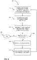
- FIG. 2 is a flow chart 200 depicting a general method of auto-focusing that includes a sequence of operations performed by a system for estimating ideal focal distances at different imaging locations on a substrate such as a microscope slide.
- Operations include acquiring a set of images at an imaging location using focal distances (step 210) at predetermined offsets from a representative focal distance and calculating focus scores for all or a subset of images from the set (step 220).
- Operations also include estimating and storing an ideal focal distance for the imaging location from the calculated focus scores (step 230), checking whether the system needs to image additional locations on the substrate (step 240), and upon such determination, moving the objective and/or stage hardware to the new imaging location (step 260).
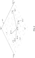
- Table 1 The calculation of the representative focal distance in the above example for a new imaging location (6, 6) is illustrated in Table 1: Table 1: Example showing calculation of representative focal distance x (mm) y (mm) Estimated ideal focal distance z ( ⁇ m) d 2 weight weight ⁇ z 5 3 3 10 0.008 0.025 8 3 1 13 0.005 0.005 8 4 -2 8 0.012 -0.025 5 8 -3 5 0.028 -0.083 1 8 -1 29 0.001 -0.001 Sum: 0.055 -0.079 Weighted average of z: -1.451 ⁇ m As illustrated above, the representative focal distance at the location (6, 6) was estimated to be -1.451 micron.
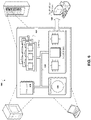
- the features can be implemented in a computer system that includes a back-end component, such as a data server, or that includes a middleware component, such as an application server or an Internet server, or that includes a front-end component, such as a client computer having a graphical user interface or an Internet browser, or any combination of them.
- the components of the system can be connected by any form or medium of digital data communication such as a communication network. Examples of communication networks include, e.g., a LAN, a WAN, and computers and networks forming the Internet.
- Table 3 Example of ideal focal distance estimation for an 8 image stack Color Focal distance Logarithm of Brenner score Difference Estimated ideal focal distance offset Red -0.35 micron 18.7 0.4 1.143 micron 0.35 micron 19.1 Yellow -0.35 micron 19.5 0.1 0.286 micron 0.35 micron 19.6 Green -0.35 micron 20.4 0.3 0.857 micron 0.35 micron 20.7 Blue -0.35 micron 17.3 -0.2 -0.571 micron 0.35 micron 17.1 Average: 0.429 micron The difference in focal distance per unit difference in the logarithm of the Brenner scores was taken as 2 micron.
Landscapes
- Physics & Mathematics (AREA)
- General Physics & Mathematics (AREA)
- Optics & Photonics (AREA)
- Engineering & Computer Science (AREA)
- Computer Vision & Pattern Recognition (AREA)
- Chemical & Material Sciences (AREA)
- Analytical Chemistry (AREA)
- Microscoopes, Condenser (AREA)
- Studio Devices (AREA)
- Automatic Focus Adjustment (AREA)
Priority Applications (1)
| Application Number | Priority Date | Filing Date | Title |
|---|---|---|---|
| EP21212720.3A EP3988985A3 (fr) | 2011-02-01 | 2011-02-01 | Focalisation automatique rapide dans imagerie microscopique |
Applications Claiming Priority (3)
| Application Number | Priority Date | Filing Date | Title |
|---|---|---|---|
| EP11702907.4A EP2671113B1 (fr) | 2011-02-01 | 2011-02-01 | Focalisation automatique rapide dans imagerie microscopique |
| PCT/US2011/023374 WO2012105966A1 (fr) | 2011-02-01 | 2011-02-01 | Focalisation automatique rapide dans imagerie microscopique |
| EP21212720.3A EP3988985A3 (fr) | 2011-02-01 | 2011-02-01 | Focalisation automatique rapide dans imagerie microscopique |
Related Parent Applications (1)
| Application Number | Title | Priority Date | Filing Date |
|---|---|---|---|
| EP11702907.4A Division EP2671113B1 (fr) | 2011-02-01 | 2011-02-01 | Focalisation automatique rapide dans imagerie microscopique |
Publications (2)
| Publication Number | Publication Date |
|---|---|
| EP3988985A2 true EP3988985A2 (fr) | 2022-04-27 |
| EP3988985A3 EP3988985A3 (fr) | 2022-07-13 |
Family
ID=44625103
Family Applications (2)
| Application Number | Title | Priority Date | Filing Date |
|---|---|---|---|
| EP21212720.3A Pending EP3988985A3 (fr) | 2011-02-01 | 2011-02-01 | Focalisation automatique rapide dans imagerie microscopique |
| EP11702907.4A Active EP2671113B1 (fr) | 2011-02-01 | 2011-02-01 | Focalisation automatique rapide dans imagerie microscopique |
Family Applications After (1)
| Application Number | Title | Priority Date | Filing Date |
|---|---|---|---|
| EP11702907.4A Active EP2671113B1 (fr) | 2011-02-01 | 2011-02-01 | Focalisation automatique rapide dans imagerie microscopique |
Country Status (8)
| Country | Link |
|---|---|
| EP (2) | EP3988985A3 (fr) |
| JP (1) | JP5727629B2 (fr) |
| KR (1) | KR101891364B1 (fr) |
| CN (1) | CN103534628B (fr) |
| AU (1) | AU2011357735B2 (fr) |
| CA (1) | CA2826372C (fr) |
| HK (1) | HK1190195A1 (fr) |
| WO (1) | WO2012105966A1 (fr) |
Cited By (1)
| Publication number | Priority date | Publication date | Assignee | Title |
|---|---|---|---|---|
| EP4345444A1 (fr) * | 2022-09-29 | 2024-04-03 | Illumina, Inc. | Étalonnage de système optique dynamique |
Families Citing this family (19)
| Publication number | Priority date | Publication date | Assignee | Title |
|---|---|---|---|---|
| CN105093479A (zh) * | 2014-04-30 | 2015-11-25 | 西门子医疗保健诊断公司 | 用于显微镜的自动对焦方法和装置 |
| JP6562547B2 (ja) * | 2015-07-16 | 2019-08-21 | オリンパス株式会社 | 顕微鏡システム、算出方法、及び、プログラム |
| JP7248833B2 (ja) * | 2015-10-19 | 2023-03-29 | モレキュラー デバイシーズ, エルエルシー | フォトルミネセンス撮像のための徹照ベースの自動フォーカシングを備えた顕微鏡システム |
| KR102381114B1 (ko) * | 2016-10-06 | 2022-03-30 | 아이리스 인터내셔널 인크. | 동적 포커스 시스템 및 방법들 |
| JPWO2018139156A1 (ja) * | 2017-01-26 | 2019-12-12 | ソニー・オリンパスメディカルソリューションズ株式会社 | 医療用観察装置、および制御方法 |
| JP2019008196A (ja) * | 2017-06-27 | 2019-01-17 | 株式会社オプティマ | 観察システム、制御装置、制御方法、及びプログラム |
| EP3625611B1 (fr) * | 2017-11-28 | 2023-11-01 | Leica Biosystems Imaging, Inc. | Traitement d'image à double processeur |
| JP2019102640A (ja) * | 2017-12-01 | 2019-06-24 | 東京エレクトロン株式会社 | プローブ針の針先位置調整方法および検査装置 |
| IL264937B (en) * | 2018-02-25 | 2022-09-01 | Orbotech Ltd | Range differences for self-focusing in optical imaging systems |
| CN110658618A (zh) * | 2018-06-28 | 2020-01-07 | 湖南爱威医疗科技有限公司 | 样本图像拟合聚焦的方法、装置、计算机设备和存储介质 |
| CN109507792B (zh) * | 2018-12-27 | 2021-07-06 | 湖南品信生物工程有限公司 | 一种基于机器学习的光学显微镜自动聚焦方法 |
| WO2020188584A1 (fr) * | 2019-03-21 | 2020-09-24 | Sigtuple Technologies Private Limited | Procédé et système pour de mise au point automatique d'un système d'imagerie microscopique |
| CN110793965A (zh) * | 2019-10-31 | 2020-02-14 | 湖南爱威医疗科技有限公司 | 图像采集方法和装置、显微镜系统、计算机可读存储介质 |
| CN110927158B (zh) * | 2019-10-31 | 2022-07-05 | 湖南爱威医疗科技有限公司 | 图像采集方法和装置、显微镜系统、计算机可读存储介质 |
| CN110763679A (zh) * | 2019-10-31 | 2020-02-07 | 湖南爱威医疗科技有限公司 | 图像采集方法和装置、显微镜系统、计算机可读存储介质 |
| US20220349806A1 (en) * | 2019-11-06 | 2022-11-03 | Sony Group Corporation | Position adjusting method, microparticle analysis device, and program |
| US11108946B1 (en) * | 2020-06-25 | 2021-08-31 | Zebra Technologies Corporation | Focus stabilization of imaging system with variable focus lens |
| WO2022086448A1 (fr) * | 2020-10-23 | 2022-04-28 | National University Of Singapore | Ensemble microscope optique et procédé de focalisation pour un tel ensemble |
| SE2250140A1 (en) * | 2022-02-11 | 2023-08-12 | Cellink Bioprinting Ab | Imaging apparatus and method for determning a focal point of a well-plate |
Family Cites Families (10)
| Publication number | Priority date | Publication date | Assignee | Title |
|---|---|---|---|---|
| JPH0779434B2 (ja) | 1986-05-16 | 1995-08-23 | キヤノン株式会社 | 合焦検出装置 |
| KR100300618B1 (ko) | 1992-12-25 | 2001-11-22 | 오노 시게오 | 노광방법,노광장치,및그장치를사용하는디바이스제조방법 |
| AU777874B2 (en) * | 1999-10-29 | 2004-11-04 | Cytyc Corporation | Method for verifying that a specific stain was used on a cytological sample |
| US6970789B2 (en) * | 2001-02-02 | 2005-11-29 | Cellomics, Inc. | Method of determining a best initial focal position estimate |
| US20050036674A1 (en) * | 2003-08-13 | 2005-02-17 | Bar Ilan University | Method and system for determining shape of an object from a planar top view thereof |
| JP4800104B2 (ja) | 2005-06-13 | 2011-10-26 | 富士フイルム株式会社 | アルバム作成装置、アルバム作成方法、及びプログラム |
| US20070211460A1 (en) | 2006-03-09 | 2007-09-13 | Ilya Ravkin | Multi-color LED light source for microscope illumination |
| US7769219B2 (en) * | 2006-12-11 | 2010-08-03 | Cytyc Corporation | Method for assessing image focus quality |
| US20100157086A1 (en) * | 2008-12-15 | 2010-06-24 | Illumina, Inc | Dynamic autofocus method and system for assay imager |
| JP5489545B2 (ja) * | 2009-06-10 | 2014-05-14 | オリンパス株式会社 | 撮像システムおよび撮像方法 |
-
2011
- 2011-02-01 WO PCT/US2011/023374 patent/WO2012105966A1/fr active Application Filing
- 2011-02-01 JP JP2013552502A patent/JP5727629B2/ja active Active
- 2011-02-01 CA CA2826372A patent/CA2826372C/fr active Active
- 2011-02-01 EP EP21212720.3A patent/EP3988985A3/fr active Pending
- 2011-02-01 AU AU2011357735A patent/AU2011357735B2/en active Active
- 2011-02-01 EP EP11702907.4A patent/EP2671113B1/fr active Active
- 2011-02-01 KR KR1020137023219A patent/KR101891364B1/ko active IP Right Grant
- 2011-02-01 CN CN201180069643.6A patent/CN103534628B/zh active Active
-
2014
- 2014-04-04 HK HK14103249.7A patent/HK1190195A1/zh unknown
Non-Patent Citations (1)
| Title |
|---|
| BRENNER ET AL.: "An Automated Microscope for Cytological Research", J. HISTOCHEM. CYTOCHEM., vol. 24, 1971, pages 100 - 111 |
Cited By (1)
| Publication number | Priority date | Publication date | Assignee | Title |
|---|---|---|---|---|
| EP4345444A1 (fr) * | 2022-09-29 | 2024-04-03 | Illumina, Inc. | Étalonnage de système optique dynamique |
Also Published As
| Publication number | Publication date |
|---|---|
| AU2011357735B2 (en) | 2015-07-30 |
| EP2671113B1 (fr) | 2021-12-08 |
| AU2011357735A1 (en) | 2013-05-02 |
| EP3988985A3 (fr) | 2022-07-13 |
| KR20140045331A (ko) | 2014-04-16 |
| EP2671113A1 (fr) | 2013-12-11 |
| CN103534628A (zh) | 2014-01-22 |
| CN103534628B (zh) | 2016-03-09 |
| CA2826372A1 (fr) | 2012-08-09 |
| HK1190195A1 (zh) | 2014-06-27 |
| CA2826372C (fr) | 2020-03-31 |
| KR101891364B1 (ko) | 2018-08-23 |
| WO2012105966A1 (fr) | 2012-08-09 |
| JP2014507685A (ja) | 2014-03-27 |
| JP5727629B2 (ja) | 2015-06-03 |
Similar Documents
| Publication | Publication Date | Title |
|---|---|---|
| US10462351B2 (en) | Fast auto-focus in imaging | |
| EP2671113B1 (fr) | Focalisation automatique rapide dans imagerie microscopique | |
| US11803968B2 (en) | Automated stereology for determining tissue characteristics | |
| TWI478101B (zh) | 評估影像聚焦品質的方法 | |
| Osibote et al. | Automated focusing in bright‐field microscopy for tuberculosis detection | |
| US20160026852A1 (en) | Imaging Blood Cells | |
| US6970789B2 (en) | Method of determining a best initial focal position estimate | |
| US20230085827A1 (en) | Single-shot autofocusing of microscopy images using deep learning | |
| CN109361849B (zh) | 一种自动对焦的方法 | |
| CN110363734B (zh) | 厚样本显微荧光图像重构方法及系统 | |
| EP3884327B1 (fr) | Mise au point en temps réel dans un système de numérisation de lames | |
| WO2022035590A1 (fr) | Mesure de contraste d'image optique à des fins de recherche de cible optique | |
| Hao et al. | Improving the performances of autofocus based on adaptive retina-like sampling model | |
| CN112969026A (zh) | 一种成像椭圆偏振仪的焦平面自动聚焦方法 | |
| Garud et al. | Volume visualization approach for depth-of-field extension in digital pathology | |
| Redondo et al. | Evaluation of autofocus measures for microscopy images of biopsy and cytology | |
| CN110455797A (zh) | 金相显微镜矩阵归一化校正方法 | |
| CN112541939B (zh) | 一种样本清晰焦面位置获取方法及装置 | |
| US20230258919A1 (en) | A method for analysing scanning efficacy | |
| US20230196533A1 (en) | Microscopy System and Method for Reducing an Image Vignetting |
Legal Events
| Date | Code | Title | Description |
|---|---|---|---|
| PUAI | Public reference made under article 153(3) epc to a published international application that has entered the european phase |
Free format text: ORIGINAL CODE: 0009012 |
|
| STAA | Information on the status of an ep patent application or granted ep patent |
Free format text: STATUS: REQUEST FOR EXAMINATION WAS MADE |
|
| 17P | Request for examination filed |
Effective date: 20211207 |
|
| AC | Divisional application: reference to earlier application |
Ref document number: 2671113 Country of ref document: EP Kind code of ref document: P |
|
| AK | Designated contracting states |
Kind code of ref document: A2 Designated state(s): AL AT BE BG CH CY CZ DE DK EE ES FI FR GB GR HR HU IE IS IT LI LT LU LV MC MK MT NL NO PL PT RO RS SE SI SK SM TR |
|
| PUAL | Search report despatched |
Free format text: ORIGINAL CODE: 0009013 |
|
| AK | Designated contracting states |
Kind code of ref document: A3 Designated state(s): AL AT BE BG CH CY CZ DE DK EE ES FI FR GB GR HR HU IE IS IT LI LT LU LV MC MK MT NL NO PL PT RO RS SE SI SK SM TR |
|
| RIC1 | Information provided on ipc code assigned before grant |
Ipc: G02B 21/24 20060101ALI20220603BHEP Ipc: G02B 7/08 20210101AFI20220603BHEP |
|
| P01 | Opt-out of the competence of the unified patent court (upc) registered |
Effective date: 20230530 |
|
| GRAP | Despatch of communication of intention to grant a patent |
Free format text: ORIGINAL CODE: EPIDOSNIGR1 |
|
| STAA | Information on the status of an ep patent application or granted ep patent |
Free format text: STATUS: GRANT OF PATENT IS INTENDED |
|
| INTG | Intention to grant announced |
Effective date: 20240403 |