EP3984546A2 - Gentherapie mit den genen hokd und ldrb für krebsbehandlungen - Google Patents
Gentherapie mit den genen hokd und ldrb für krebsbehandlungen Download PDFInfo
- Publication number
- EP3984546A2 EP3984546A2 EP20810087.5A EP20810087A EP3984546A2 EP 3984546 A2 EP3984546 A2 EP 3984546A2 EP 20810087 A EP20810087 A EP 20810087A EP 3984546 A2 EP3984546 A2 EP 3984546A2
- Authority
- EP
- European Patent Office
- Prior art keywords
- polynucleotide
- gene
- cell
- cells
- hokd
- Prior art date
- Legal status (The legal status is an assumption and is not a legal conclusion. Google has not performed a legal analysis and makes no representation as to the accuracy of the status listed.)
- Pending
Links
Images






Classifications
-
- A—HUMAN NECESSITIES
- A61—MEDICAL OR VETERINARY SCIENCE; HYGIENE
- A61K—PREPARATIONS FOR MEDICAL, DENTAL OR TOILETRY PURPOSES
- A61K48/00—Medicinal preparations containing genetic material which is inserted into cells of the living body to treat genetic diseases; Gene therapy
- A61K48/005—Medicinal preparations containing genetic material which is inserted into cells of the living body to treat genetic diseases; Gene therapy characterised by an aspect of the 'active' part of the composition delivered, i.e. the nucleic acid delivered
-
- A—HUMAN NECESSITIES
- A61—MEDICAL OR VETERINARY SCIENCE; HYGIENE
- A61P—SPECIFIC THERAPEUTIC ACTIVITY OF CHEMICAL COMPOUNDS OR MEDICINAL PREPARATIONS
- A61P35/00—Antineoplastic agents
-
- C—CHEMISTRY; METALLURGY
- C07—ORGANIC CHEMISTRY
- C07K—PEPTIDES
- C07K14/00—Peptides having more than 20 amino acids; Gastrins; Somatostatins; Melanotropins; Derivatives thereof
- C07K14/195—Peptides having more than 20 amino acids; Gastrins; Somatostatins; Melanotropins; Derivatives thereof from bacteria
- C07K14/24—Peptides having more than 20 amino acids; Gastrins; Somatostatins; Melanotropins; Derivatives thereof from bacteria from Enterobacteriaceae (F), e.g. Citrobacter, Serratia, Proteus, Providencia, Morganella, Yersinia
- C07K14/245—Escherichia (G)
-
- C—CHEMISTRY; METALLURGY
- C12—BIOCHEMISTRY; BEER; SPIRITS; WINE; VINEGAR; MICROBIOLOGY; ENZYMOLOGY; MUTATION OR GENETIC ENGINEERING
- C12N—MICROORGANISMS OR ENZYMES; COMPOSITIONS THEREOF; PROPAGATING, PRESERVING, OR MAINTAINING MICROORGANISMS; MUTATION OR GENETIC ENGINEERING; CULTURE MEDIA
- C12N15/00—Mutation or genetic engineering; DNA or RNA concerning genetic engineering, vectors, e.g. plasmids, or their isolation, preparation or purification; Use of hosts therefor
- C12N15/09—Recombinant DNA-technology
- C12N15/11—DNA or RNA fragments; Modified forms thereof; Non-coding nucleic acids having a biological activity
-
- A—HUMAN NECESSITIES
- A01—AGRICULTURE; FORESTRY; ANIMAL HUSBANDRY; HUNTING; TRAPPING; FISHING
- A01K—ANIMAL HUSBANDRY; AVICULTURE; APICULTURE; PISCICULTURE; FISHING; REARING OR BREEDING ANIMALS, NOT OTHERWISE PROVIDED FOR; NEW BREEDS OF ANIMALS
- A01K2207/00—Modified animals
- A01K2207/12—Animals modified by administration of exogenous cells
-
- A—HUMAN NECESSITIES
- A01—AGRICULTURE; FORESTRY; ANIMAL HUSBANDRY; HUNTING; TRAPPING; FISHING
- A01K—ANIMAL HUSBANDRY; AVICULTURE; APICULTURE; PISCICULTURE; FISHING; REARING OR BREEDING ANIMALS, NOT OTHERWISE PROVIDED FOR; NEW BREEDS OF ANIMALS
- A01K2227/00—Animals characterised by species
- A01K2227/10—Mammal
- A01K2227/105—Murine
-
- A—HUMAN NECESSITIES
- A01—AGRICULTURE; FORESTRY; ANIMAL HUSBANDRY; HUNTING; TRAPPING; FISHING
- A01K—ANIMAL HUSBANDRY; AVICULTURE; APICULTURE; PISCICULTURE; FISHING; REARING OR BREEDING ANIMALS, NOT OTHERWISE PROVIDED FOR; NEW BREEDS OF ANIMALS
- A01K2267/00—Animals characterised by purpose
- A01K2267/03—Animal model, e.g. for test or diseases
- A01K2267/0331—Animal model for proliferative diseases
-
- A—HUMAN NECESSITIES
- A61—MEDICAL OR VETERINARY SCIENCE; HYGIENE
- A61K—PREPARATIONS FOR MEDICAL, DENTAL OR TOILETRY PURPOSES
- A61K38/00—Medicinal preparations containing peptides
Definitions
- the present invention is comprised in the field of gene therapy. More specifically, the present invention relates to toxins, hokD and IdrB, with an application in the field of gene therapy and particularly for cancer treatment.
- Cancer is a disease that has an enormous impact on the entire world. In the United States, 1,685,210 new cases of cancer are expected to be diagnosed for 2016 and 595,690 people are expected to die from this cause. The cancer mortality rate has dropped by 23% since 1991, which translates into more than 1.7 million deaths prevented up to 2012 [1]. This is due to the improved diagnosis and intervention; however, conventional treatments (chemotherapy, radiotherapy, surgery, and hormone therapy) have certain limitations. Furthermore, patients who are subjected to current systemic therapies will experience a number of side effects ranging from nausea to infertility [2]. This shows that it is necessary to reduce the chemotherapy or radiation dose to below the most effective levels, or to entirely withdraw a particular treatment. Therefore, research in new alternative and more efficient therapies is required.
- Suicide gene therapy is based on the delivery to tumor cells of a gene encoding a cytotoxic protein. This therapy can be divided into two categories: indirect gene therapy using an enzyme-activating prodrug, which allows the conversion of a prodrug into a lethal drug in tumor cells; and direct gene therapy using a toxin gene, which incorporates genes expressing toxic molecules which may affect cell membrane stability and reduce tumor cell viability [5,6].
- HSV-TK herpes simplex virus thymidine kinase
- GCV ganciclovir
- HSV-TK converts the antiviral drug GCV into a monophosphorylated molecule which is then metabolized into the toxic form of triphosphate by cellular kinases, inhibiting DNA synthesis and causing cell death [8,9].
- CD cytosine deaminase
- 5-FC 5-fluorocytosine
- This system is based on the capacity of CD to convert non-toxic 5-FC into 5-fluorouracil (5-FU), a potent cytotoxic chemotherapeutic, which is later transformed by means of cell enzymes into potent pyrimidine antimetabolites and causes the inhibition of cell proliferation and cell death [10,11].
- 5-FU 5-fluorouracil
- One alternative in suicide gene therapy for cancer is use of the toxin gene [12]. Unlike conventional suicide gene therapy, uses of the toxin gene have more advantages. The presence of a prodrug is not required for the system to be effective. Furthermore, the toxicity of the metabolites is reduced and has not limited bioavailability due to the lack of prodrugs. Some bacterial toxins have been studied for use in gene therapy for cancer [13,14].
- TA toxin-antitoxin
- the toxin products of these modules target various cell functions to regulate cell growth and death.
- These toxins are generally co-expressed with their associated antitoxins in operons called toxin-antitoxin (TA) modules [15].
- One of the genes of the operon encodes an unstable antitoxin and another gene encodes a stable toxin, where the overlap of both loci is common. Under normal conditions of growth, the modules form stable complexes. However, stress-induced proteases preferably eliminate the unstable antitoxins, releasing free toxins to inhibit various cell functions [16,17].
- TA systems are classified into three groups (type I, II, III) based on the function of the antitoxin [18].
- the Escherichia coli K-12 genome encodes at least 36 assumed TA systems which consist of type I and type II systems.
- type I TA systems the expression of the toxin gene is regulated by an antisense RNA transcribed from the toxin module but in the reverse orientation. This antisense RNA impedes the translation of the mRNA toxin [19,20].
- the E. coli K-12 chromosome contains different type I TA systems, and one of these systems is the hok/sok family containing six homologous loci (A, B, C, D, E, and X) [21].
- the gene hokD encodes a highly toxic 52-amino acid transmembrane protein which irreversibly damages the cell membrane and, therefore, is lethal for the host cell. Effects of the toxin result in the loss of membrane potential, arrest of respiration, death of the host cell, and change in cell morphology [22,23].
- Another family of the type I TA system also encoded by E. coli K-12 is the family of the gene Idr/rdl.
- the inventors have selected two genes, hokD and IdrB, to study their behavior in eukaryotic cells, since they may be possible new candidates for being used in the suicide gene therapy in cancer given their many qualities in causing death in the host cell.
- the main objective of the study was to investigate the therapeutic potential of the genes hokD and IdrB, using the MCF-7 breast cancer cell line and the HCT-116 colon cancer cell line.
- the inventors developed a new MCF-7 and HCT-116 cell line transfected with the genes hokD and IdrB, each one through the Tet-On 3G Inducible Expression System.
- This system is the gene expression that can be induced for mammalian cells.
- the target cells expressing transactivator protein Tet-On 3G and containing a gene of interest under the control of promotor PTRE3G will express high levels of the gene of interest, but only when they are cultured in the presence of doxycycline, which allows the controlled expression of the suicide gene in the tumor cell.
- the present invention relates to the following aspects:
- a first aspect of the present invention relates to an isolated RNA or DNA polynucleotide, hereinafter first polynucleotide of the invention, having an identity of at least 80%, preferably 85%, 90%, 95%, and more preferably 99% with the polynucleotide sequence of the gene hokD (SEQ ID NO: 1) for use in medicine, or alternatively, for use as a medicinal product. More preferably, the first polynucleotide of the invention is the gene hokD, with nucleotide sequence SEQ ID NO: 1
- another aspect of the invention relates to an isolated RNA or DNA polynucleotide, hereinafter first polynucleotide of the invention, capable of being translated into an amino acid sequence comprising a peptide having an identity of at least 90% with the amino acid sequence of the protein HOKD (SEQ ID NO: 3). It more preferably relates to an isolated RNA or DNA polynucleotide capable of being translated into an amino acid sequence comprising a peptide having an identity of at least 95%, 96%, 97%, 98%, or 99% with the amino acid sequence of the protein HOKD, for use in medicine, or alternatively, for use as a medicinal product.
- Another aspect of the invention relates to a genetic DNA or RNA structure, hereinafter first genetic structure of the invention, comprising at least one of the following types of sequences:
- Another aspect of the invention relates to a cell, hereinafter first cell of the invention, comprising the first polynucleotide of the invention, or the first genetic structure of the invention.
- composition hereinafter first composition of the invention, comprising:
- the first composition of the invention is a pharmaceutical composition.
- the composition further comprises a pharmaceutically acceptable vehicle.
- the composition further comprises another active ingredient.
- the first composition of the invention comprises another active ingredient, this active ingredient being an isolated RNA or DNA polynucleotide having an identity of at least 80%, preferably 85%, 90%, 95%, and more preferably 99% with the polynucleotide sequence of the gene IdrB (SEQ ID NO: 2). for use in medicine, or alternatively, for use as a medicinal product. More preferably, the polynucleotide of the invention is the gene IdrB, with nucleotide sequence SEQ ID NO: 2
- Another aspect of the invention relates to the first polynucleotide of the invention, the first genetic structure of the invention, or the first composition of the invention, for the prevention, improvement, relief, or treatment of a proliferative disease.
- the proliferative disease is cancer.
- the proliferative disease is breast cancer.
- the proliferative disease is colon cancer.
- Another aspect of the present invention relates to an isolated RNA or DNA polynucleotide, hereinafter second polynucleotide of the invention, having an identity of at least 80%, preferably 85%, 90%, 95%, and more preferably 99% with the polynucleotide sequence of the gene IdrB (SEQ ID NO: 2) for use in medicine, or alternatively, for use as a medicinal product. More preferably, the second polynucleotide of the invention is the gene IdrB, with nucleotide sequence SEQ ID NO: 2
- another aspect of the invention relates to an isolated RNA or DNA polynucleotide, hereinafter second polynucleotide of the invention, capable of being translated into an amino acid sequence comprising a peptide having an identity of at least 90% with the amino acid sequence of the protein LDRB (SEQ ID NO: 4). It more preferably relates to an isolated RNA or DNA polynucleotide capable of being translated into an amino acid sequence comprising a peptide having an identity of at least 95%, 96%, 97%, 98%, or 99% with the amino acid sequence of the protein LDRB, for use in medicine, or alternatively, for use as a medicinal product.
- Another aspect of the invention relates to a genetic DNA or RNA structure, hereinafter second genetic structure of the invention, comprising at least one of the following types of sequences:
- Another aspect of the invention relates to a cell, hereinafter second cell of the invention, comprising the second polynucleotide of the invention, or the second genetic structure of the invention.
- composition hereinafter second composition of the invention, comprising:
- the second composition of the invention is a pharmaceutical composition.
- the composition further comprises a pharmaceutically acceptable vehicle.
- the composition further comprises another active ingredient.
- the second composition of the invention comprises another active ingredient, this active ingredient being an isolated RNA or DNA polynucleotide having an identity of at least 80%, preferably 85%, 90%, 95%, and more preferably 99% with the polynucleotide sequence of the gene hokD (SEQ ID NO: 1), for use in medicine, or alternatively, for use as a medicinal product. More preferably, the polynucleotide of the invention is the gene hokD, with nucleotide sequence SEQ ID NO: 1
- compositions of the present invention can be used together with other medicinal products in combined therapies.
- the other drugs may be part of the same composition or of another different composition, for administration at the same time or at different times.
- Another aspect of the invention relates to the second polynucleotide of the invention, the second genetic structure of the invention, or the second composition of the invention, for the prevention, improvement, relief, or treatment of a proliferative disease.
- the proliferative disease is cancer.
- the proliferative disease is breast cancer.
- the proliferative disease is colon cancer.
- Another aspect of the invention relates to a combined preparation, hereinafter combined preparation of the invention, comprising:
- Another aspect relates to the use of the combined preparation of the invention for use in medicine, or alternatively, for use as a medicinal product.
- the proliferative disease is cancer.
- the proliferative disease is breast cancer.
- the proliferative disease is colon cancer.
- Another aspect of the invention relates to a genetic DNA or RNA structure, hereinafter third genetic structure of the invention, comprising at least one of the following types of sequences:
- the proliferative disease is cancer.
- the proliferative disease is breast cancer.
- the proliferative disease is colon cancer.
- immediate product refers to any substance used for the prevention, diagnosis, relief, treatment, or healing of diseases in humans and animals.
- active ingredient means any component that potentially provides pharmacological activity or another different effect in the diagnosis, healing, mitigation, treatment, or prevention of a disease, or affects the structure or function of the body of humans or other animals.
- the term includes those components which promote a chemical change in the preparation of the drug and are present therein in an intended modified form that provides the specific activity or effect.
- the term “combined preparation”, also referred to as a “juxtaposition”, herein means that the components of the combined preparation do not need to be present bound to one another, for example in a true composition, in order to be available for their combined, separate, or sequential application. Therefore, the expression “juxtaposed” means that it is not necessarily a true combination given the physical separation of the components.
- compositions or the combined preparation of the invention can be administered to the subject who suffers from said pathologies by means of any of the following routes: intraperitoneal, intravenous, intramuscular, subcutaneous, intrathecal, intraventricular, oral, enteral, parenteral, intranasal, or dermal.
- routes of administration are, preferably, the intravenous route and the oral route.
- compositions of the present invention as well as the combined preparation or the pharmaceutical forms of the invention can be formulated for administration to an animal, and more preferably to a mammal, including humans, in a variety of forms known in the state of the art. In that sense, they can be, without limitation, in a sterile aqueous solution or in biological fluids, such as serum.
- the aqueous solutions may or may not be buffered and have additional active or inactive components.
- the additional components include salts for modulating ionic strength, preservatives including but not limited to antimicrobial agents, antioxidants, chelators, and similar agents, and nutrients including glucose, dextrose, vitamins, and minerals.
- the compositions can be prepared for administration in solid form.
- compositions can be combined with several inert excipients or vehicles, including but not limited to binders such as microcrystalline cellulose, tragacanth gum, or gelatin; excipients such as starch or lactose; dispersing agents such as alginic acid or corn starch; lubricants such as magnesium stearate; glidants such as colloidal silicon dioxide; sweetening agents such as sucrose or saccharine; or flavoring agents such as mint or methyl salicylate.
- binders such as microcrystalline cellulose, tragacanth gum, or gelatin
- excipients such as starch or lactose
- dispersing agents such as alginic acid or corn starch
- lubricants such as magnesium stearate
- glidants such as colloidal silicon dioxide
- sweetening agents such as sucrose or saccharine
- flavoring agents such as mint or methyl salicylate.
- compositions or combined preparations and/or their formulations can be administered to an animal, including a mammal and, therefore, to humans, in a variety of ways, including but not limited to intraperitoneal, intravenous, intramuscular, subcutaneous, intrathecal, intraventricular, oral, enteral, parenteral, intranasal, or dermal.
- the dosage for obtaining a therapeutically effective amount depends on a number of factors, such as for example, the age, weight, sex, tolerance, etc., of the mammal.
- the expression "therapeutically effective amount” refers to the comprised amount of active ingredient or ingredients of the invention producing the desired effect, and it will generally be determined, among other causes, by the characteristics that are typical of said prodrugs, derivatives, or analogues, and the therapeutic effect to be achieved.
- the "adjuvants” and “pharmaceutically acceptable vehicles” that can be used in said compositions are the vehicles known to those skilled in the art.
- the administered amount of a compound of the present invention will depend on the relative efficacy of the chosen compound, the seriousness of the disease to be treated, and the weight and/or other characteristics of the patient.
- compositions or the combined preparation of this invention will be administered, for example, but without limitation, one or more times per day, for example 1, 2, 3, or 4 times daily, with a total dose between 0.1 and 1000 mg/kg/day. It is important to take into account that, depending on the age and on the condition of the patient, it may be necessary to introduce variations in the dose, as well as modifications in the route of administration.
- HCT-116 human colorectal carcinoma cell line, MCF-7 human breast carcinoma cell line, and HeLa cervical cancer cell line were obtained from Biobanco (Andalusian Public Health System, Granada).
- the cells were cultured in Dulbecco's Modified Eagle medium (DMEM) (Sigma, St. Louis, MO, USA), supplemented with 10% fetal bovine serum (FBS) and 1% penicillin/streptomycin (P/S) (Sigma-Aldrich), under air containing 5% CO 2 in an incubator at 37°C.
- DMEM Dulbecco's Modified Eagle medium
- FBS fetal bovine serum
- P/S penicillin/streptomycin
- the growth medium was supplemented with geneticin (800 ⁇ g/ml) and puromycin (1.5 ⁇ g/ml).
- a growth medium supplemented with geneticin (200 ⁇ g/ml) and puromycin (0.75 ⁇ g/ml) was used to maintain the selected colonies.
- doxycycline was used (Dox, 3 ⁇ g/ml). All the antibiotics were provided by Clontech Laboratories, Inc. (UNITED STATES).
- the Tet-On gene expression system (Clontech Laboratories, Inc. (USA)) was used to express the genes hokD and IdrB.
- the HCT-116, MCF-7, and HeLa cells were transfected initially with pTet-On 3G (Xfect Polymer, Clonthech, USA), and the successfully transfected clones were selected by geneticin resistance (800 ⁇ g/ml). These clones were transfected with PTRE3G-hok and PTRE3G-ldr, selected for geneticin (200 ⁇ g/ml) and puromycin (1.5 ⁇ g/ml).
- clones were cultured in the presence of doxycycline (Dox, 3 ⁇ g/ml, 24 h) to induce expression of the genes hokD (HCT-H, MCF-H, and HeLa-H) and IdrB (HCT-L and MCF-L), which was detected by fluorescence.
- Dox doxycycline
- HCT-116 cells 1,000 cells per well
- MCF-7 cells 1,500 cells per well
- 10 ⁇ l of solution CCK-8 were added to each well of the plate and incubated at 37°C for 3 h.
- Absorbance at 450 nm was then measured using a Thermo Scientific Multiskan microplate reader.
- HCT-116 cells were seeded in a 6-well plate (50,000 cells per well). After 2, 4, and 6 days for the selection of induction with geneticin (concentration of 500 ⁇ g/ml to 800 ⁇ g/ml) and puromycin (concentration of 0.5 ⁇ g/ml to 2 ⁇ g/ml), the cells were examined using a phase contrast microscope to assess the possible effects.
- Fluorescence of the red fluorescent protein m-cherry was determined by means of FACSan flow cytometry analysis (Becton Dickinson, San Jose, CA), with a maximum excitation of 587 nm and a maximum emission of 601 nm (Clontech Laboratories, Inc. (USA)).
- the HCT-116 and MCF-7 cancer cell lines were kept in Dulbecco's Modified Eagle Medium (DMEM) without phenol red (Sigma, St. Louis, MO, USA), supplemented with 10% fetal bovine serum (FBS), 100 ⁇ g/ml of penicillin and 100 ⁇ g/ml of streptomycin.
- DMEM Dulbecco's Modified Eagle Medium
- FBS fetal bovine serum
- the cancer cells were seeded in 96-ViewPlate TM microplates (transparent bottom, white), seeding 1500 cells for the MCF-7 cell line per well and 1000 cells for the HCT-116 cell line per well. The plates were incubated at 37°C in the presence of 5% CO 2 .
- the assay plate was read in the EnSight system for 15 days using the brightfield imaging mode.
- the plates were kept under controlled conditions (5% CO 2 , 37°C, and 90% humidity) between each reading, and imaging was carried out at 37°C using the temperature control function of the EnSight reader, which eliminates the possible water condensation effects.
- the times of the reading cycle were ⁇ 5 min per plate.
- the images were analyzed using Kaleido TM data acquisition and analysis software, and the derived results were fitted to a 4 parameter logistic curve and plotted using TIBCO Spotfire ® .
- Cell cycle phases (G0/G1, S, or G2/M) were characterized according to the cell DNA content.
- the fluorescent dye propidium iodide (PI) (Sigma, USA) binds strongly to DNA at a ratio of 1:1; therefore, the DNA content of the cell cycle phases were reflected by means of variable PI fluorescent intensities.
- the cells in monolayer culture were collected, washed twice with PBS, and fixed in ice cold 70% ethanol at 4°C.
- the percentage of cells in phases subG1, G0/G1, S, and G2/M was determined by means of (fluorescence-activated cell sorting) flow cytometer FACSCalibur (BD Biosciences, USA).
- HCT-116 cell lines induced with Dox (3 ⁇ g/ml) for 6 and 3 days were fixed with a solution of 2.5% glutaraldehyde in 0.1 M sodium cacodylate buffer (pH 7.4; Sigma) for 2 hours.
- the pellet that contained dead cells and debris, as well as monolayer, were later fixed with 1% osmium tetroxide in 0.1 M sodium cacodylate buffer (Sigma) and dehydrated in ethanol.
- the cells were separated from the culture of the culture vessel by means of rapid treatment with propylene oxide and embedded in Epon 812 (EMS, Washington, USA). After polymerization, the plastic was removed, and ultrathin sections were cut parallel and perpendicular to the surface of the flask. The sections were contrasted with uranyl acetate-lead citrate and examined under a Carl Zeiss EM 902 transmission electron microscope (TEM).
- TEM Carl Zeiss EM 902 transmission electron microscope
- the cells were cultured in sterile slipcovers and induced for 3 and 6 days with Dox (3 ⁇ g/ml). They were fixed for 2 hours at 40°C with 2.5% glutaraldehyde in 0.05 M cacodylate buffer, pH 7.4, containing 0.7 M KCI, 1.24 mM CaCl 2 , and 1.24 mM MgCl 2 . They were then washed and treated for 1 hour with 1% tannic acid in 0.1 M cacodylate buffer at 4°C and briefly washed again in the same buffer. The fixed cells were concentrated again by centrifugation in 1% agarose and were later fixed in 2% OsO 4 . The cell cultures were dehydrated in amyl acetate/ethanol followed by a critical point of drying and coating with gold. A Hitachi S-800 scanning electron microscope (Hitachi, Tokyo) was used for the observations.
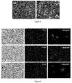
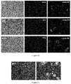
- the cells were transfected with genes of interest and their expression induced with Dox (3 ⁇ g/ml). 24 hours after induction, detection of the expression of the gene was studied by means of fluorescence microscopy through the fluorophore m-cherry, which is found in promotor PTRE3G, and its expression, therefore, acts as an indicator of the expression of the gene of interest which produces a red protein. Three clones for each of the genes where then selected ( Figures 5 and 6 ).
- the morphology of MCF-7 cells was examined for the first time under normal conditions using a phase contrast microscope.
- the cells were transfected with genes of interest and their expression induced with Dox (3 ⁇ g/ml). 24 hours later, the expression of the gene was detected by fluorescence microscopy through the fluorophore m-cherry, which is found in promotor PTRE3G, and its expression, therefore, acts as an indicator of the expression of the gene of interest which produces a red protein.
- Three clones for each of the genes where then selected ( Figures 9 and 10 ).
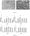
- the percentage of fluorescence after induction with doxycycline was studied.
- the data shows that the administration of Dox every two days produced constant levels of fluorescence in both cell lines ( Figure 13 ). Therefore, the constant expression of fluorescence also functions as an indicator of gene expression.
- the HCT-116 cell line transfected with the genes IdrB and hokD the cell inhibition rate was observed when Dox was administered.
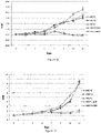
- the HCT-H cell line in the presence of Dox sustains a drop in cell growth of 48%, 70.85%, and 71.21% after 9, 12, and 15 days of treatment, respectively.
- the HCT-L line obtains a drop in cell growth of 51.18% after 9 days of treatment, and 71.45% and 71.82% after 12 and 15 days, respectively ( Figure 14 ).
- MCF-7 cell line A similar effect occurs in the MCF-7 cell line.
- a significant drop in cell growth in lines transfected with genes hokD and IdrB was observed.
- lines transfected in the absence of Dox and the control line grow as normal.
- the data shows that the MCF-L cell line induces 38.10% inhibition of proliferation after 9 days of treatment, and 69% and 72.80% after 12 and 15 days, respectively.
- the MCF-H cell line sustains a drop in cell growth of 33.93% after 9 days of treatment, and 73.66% and 77.51% after 12 and 15 days, respectively ( Figure 15 ).
- Cell confluence is also determined using the EnSight system in brightfield imaging mode.
- the degree of confluence is defined as the percentage of the area of the image covered with cells. This mode does not require any staining for the cell nuclei or cytoplasm.
- the cell line showed a drop in confluence of 33.98% after 9 days of treatment, and 57.59% and 68.77% after 12 and 15 days, respectively.
- the MCF-L cell line obtains a drop in confluence of 31.06% after 9 days of treatment, and 51.98% and 70.54% after 12 and 15 days, respectively ( Figure 17 ).
- Apoptotic cell death can be determined by means of flow cytometry through staining with propidium iodide (PI) which is determined to determine the Sub-G1 peak.
- PI propidium iodide
- HCT-116 hokD Dox observed an increase in apoptotic cells over the time of gene induction. Days 3 and 6 of gene expression did not show significant modifications in relation to the control culture. In contrast, after 9 and 12 days of induction there was a significant increase in the percentage of apoptotic cells with 17.23% and 62.86%, respectively, whereas this percentage without induced cells was 2.14% ( Figure 1A ). Similar trends were observed for induced HCT-116 IdrB cells at the same time points, where the percentage of apoptotic cells was 53.81% and 94.01% for the induction of 9 and 12 days, respectively ( Figure 1B ).
- the HCT-116 cells showed an increase in the population in phase S (21.85%) and a drop in the number of G0/G1 cells (74.21%). Furthermore, after 9 (10.37%) and 12 (14.58%) days of induction, the percentage of cells in phase G2/M increases ( Figure 1C ). In contrast, induction of the gene HCT-116 IdrB caused an increase in the proportion of cells in phase S after 9 (11.04%) and 12 (20.89%) days in comparison with the control ( Figure 1D ).
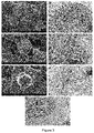
- HCT-116 hokD showed dilated mitochondria ( Figures 3D, F ), whereas HCT-116 IdrB exhibited a significant dilation of the endoplasmic reticulum (ER) ( Figures 4D, F ). Furthermore, both the expression of hokD and the expression of IdrB induced cell disintegration ( Figures 3G and 4F ).
- morphological characteristics such as "cell roughness” were also extracted, providing useful secondary readings complementing the basic data [31,32]. It would be necessary to determine whether the drop in cell proliferation and confluence is due to apoptosis phenomena or cell cycle arrest. For example, one of the characteristics that takes part in the development of colon cancer and the formation of metastasis is resistance to apoptosis phenomena.
- the tetracycline-based regulation system offers said controlled regulation of the expression of the target gene in response to the administration of doxycycline (Dox) [36,37].
- Dox doxycycline
- the Tet-On system is characterized by two regulating elements: the Tet-On transactivation proteins which bind specifically to the inducible promotor PTRE3G and activate the transcription of the gene of interest, but only in the presence of doxycycline.
- Inducible promotor PTRE3G provides a very low baseline expression and a high maximum expression after induction [38]. Therefore, gene expression can be suppressed in the absence of Dox.
- the transmembrane domain of these toxins is expected to localize by interacting with other proteins in the cell membrane or periplasm and/or localize to the internal membrane, where the oligomer form of the toxin leads to pore-like structures and membrane permeabilization [39].
- the toxin on the membrane of prokaryotic cells, the same pore forming capacity in the plasma membrane of human HCT-116 cells examined by SEM microscopy is found. Furthermore, a characteristic cell surface in the transfected cells is observed, with a loss of microvilli and filopodia that produced cells with a flat appearance, as well as a number of apoptotic bodies.
- Apoptosis as a mechanism of action of the genes hokD and IdrB was supported by TEM microscopy. Apoptosis can be triggered by different biochemical changes, such as DNA and protein degradation, membrane alteration and detection by phagocytic cells, and/or caspase stimulation [46]. It should be highlighted that these ultrastructural findings revealed dilated mitochondria in HCT-116 hokD cells, which is a distinctive sign of the intrinsic mitochondria-mediated apoptotic pathway. Interruption of the mitochondrial membrane potential leads to the release of apoptogenic factors, such as cytochrome c, which ultimately initiate the stimulation of those caspases that play a key role in apoptotic processes [9,12].
- induced HCT-116 IdrB cells have a dilated ER that can trigger ER stress. Prolonged ER stress can cause the induction of cell death mediated by the C/EBP homologous protein (CHOP) [47]. Further investigation would be needed to assess the molecular mechanism whereby the genes hokD and IdrB expressed by HCT-116 undergo apoptosis.
- C/EBP homologous protein CHOP
- phase G0/G1, S, G2/M cell cycle phases G0/G1, S, G2/M to be assessed.
- the distribution of the cell cycle phase was checked 1, 2, 3, 6, 9, and 12 days after induction of the gene Dox.
- the results revealed a notable accumulation of cells in phase S day 2 after induction of Dox;
- phase G2/M was notably higher after 9 (10.37%) and 12 (14.58%) days in comparison with the control (5.14%).
- a more obvious effect of the cell cycle was caused by the gene IdrB, where the percentage of cells in phase S was two-fold (11.04%) and three-fold (20.89%) higher after 9 and 12 days of gene induction, respectively, in comparison with the control (6.30%).
- coli verotoxin 1 demonstrated an antiproliferative effect and detained HCT-116 colon cancer cells in phase S [49].
- the gene gef which encodes a toxic protein operating in a manner similar to hok [50]
- apoptin a small protein encoded by chicken anemia virus
- the HeLa line cells were transfected with hokD and their expression induced with Dox (3 ⁇ g/ml). 24 hours later, the expression of the gene was detected by fluorescence microscopy through the fluorophore z-Green (C), which is found in promotor PTRE3G, and its expression, therefore, acts as an indicator of the expression of the gene of interest which produces, in this case, a green protein ( Figure 18 ).
- C fluorophore z-Green
- HeLa cells were less sensitive to hokD that the other lines were, although they showed a reduction in cell growth of 42.6% after 15 days of treatment ( Figure 19 ).
- HCT-116 parent cells and HCT-hokD cells were injected subcutaneously into BALB/c mice. Once the tumors were felt, Dox was administered (100 mg/kg) by means of intraperitoneal injection, established that day as day 0. A third group of mice with HCT-116 tumors without the administration of Dox was used as the control.
- the in vivo images of resected tumors confirmed expression of the gene hokD in HCT-hokD-Dox tumors as shown in the representative images of the fluorescent signal ( Figure 20, B ).
Landscapes
- Health & Medical Sciences (AREA)
- Life Sciences & Earth Sciences (AREA)
- Chemical & Material Sciences (AREA)
- Genetics & Genomics (AREA)
- Organic Chemistry (AREA)
- General Health & Medical Sciences (AREA)
- Engineering & Computer Science (AREA)
- Molecular Biology (AREA)
- Medicinal Chemistry (AREA)
- Biotechnology (AREA)
- Animal Behavior & Ethology (AREA)
- Public Health (AREA)
- Pharmacology & Pharmacy (AREA)
- Veterinary Medicine (AREA)
- Biomedical Technology (AREA)
- Biophysics (AREA)
- Biochemistry (AREA)
- Zoology (AREA)
- General Engineering & Computer Science (AREA)
- Wood Science & Technology (AREA)
- Bioinformatics & Cheminformatics (AREA)
- Chemical Kinetics & Catalysis (AREA)
- Epidemiology (AREA)
- Proteomics, Peptides & Aminoacids (AREA)
- Gastroenterology & Hepatology (AREA)
- General Chemical & Material Sciences (AREA)
- Nuclear Medicine, Radiotherapy & Molecular Imaging (AREA)
- Plant Pathology (AREA)
- Physics & Mathematics (AREA)
- Microbiology (AREA)
- Medicines That Contain Protein Lipid Enzymes And Other Medicines (AREA)
- Pharmaceuticals Containing Other Organic And Inorganic Compounds (AREA)
Applications Claiming Priority (3)
| Application Number | Priority Date | Filing Date | Title |
|---|---|---|---|
| ES201930427 | 2019-05-15 | ||
| ES201930428 | 2019-05-15 | ||
| PCT/ES2020/070315 WO2020234498A2 (es) | 2019-05-15 | 2020-05-15 | Terapia génica con los genes hokd y ldrb para el tratamiento del cáncer |
Publications (2)
| Publication Number | Publication Date |
|---|---|
| EP3984546A2 true EP3984546A2 (de) | 2022-04-20 |
| EP3984546A4 EP3984546A4 (de) | 2023-09-27 |
Family
ID=73459041
Family Applications (1)
| Application Number | Title | Priority Date | Filing Date |
|---|---|---|---|
| EP20810087.5A Pending EP3984546A4 (de) | 2019-05-15 | 2020-05-15 | Gentherapie mit den genen hokd und ldrb für krebsbehandlungen |
Country Status (3)
| Country | Link |
|---|---|
| US (1) | US20220211872A1 (de) |
| EP (1) | EP3984546A4 (de) |
| WO (1) | WO2020234498A2 (de) |
Families Citing this family (1)
| Publication number | Priority date | Publication date | Assignee | Title |
|---|---|---|---|---|
| ES2939632A1 (es) * | 2021-10-25 | 2023-04-25 | Univ Granada | Terapia génica con genes IdrB y HokD para el tratamiento del cáncer |
Family Cites Families (7)
| Publication number | Priority date | Publication date | Assignee | Title |
|---|---|---|---|---|
| US1685210A (en) | 1925-10-15 | 1928-09-25 | Niagara Electro Chemical Compa | Apparatus for electrically heating liquids |
| JP2004533827A (ja) * | 2001-05-17 | 2004-11-11 | アールフス・ユニバーシティー | 遺伝子治療用の改良されたベクター |
| SI1858920T1 (sl) * | 2005-02-18 | 2016-07-29 | Glaxosmithkline Biologicals S.A. | Proteini in nukleinske kisline iz escherichia coli, povezane z meningitisom/sepso |
| PL397167A1 (pl) * | 2011-11-28 | 2013-06-10 | Adamed Spólka Z Ograniczona Odpowiedzialnoscia | Przeciwnowotworowe bialko fuzyjne |
| US11219636B2 (en) * | 2015-05-17 | 2022-01-11 | The Medical Research, Infrastructure and Health Services Fund of the Tel Aviv Medical Center | Compositions and methods for treating cancer |
| US20190153409A1 (en) * | 2017-11-17 | 2019-05-23 | Aviv MedTech Ltd. | Compositions comprising particles and methods for treating cancer |
| AU2018379996A1 (en) * | 2017-12-05 | 2020-06-25 | BioPlx, Inc. | Methods and compositions to prevent microbial infection |
-
2020
- 2020-05-15 WO PCT/ES2020/070315 patent/WO2020234498A2/es not_active Ceased
- 2020-05-15 US US17/611,460 patent/US20220211872A1/en active Pending
- 2020-05-15 EP EP20810087.5A patent/EP3984546A4/de active Pending
Also Published As
| Publication number | Publication date |
|---|---|
| US20220211872A1 (en) | 2022-07-07 |
| EP3984546A4 (de) | 2023-09-27 |
| WO2020234498A2 (es) | 2020-11-26 |
| WO2020234498A3 (es) | 2021-02-04 |
Similar Documents
| Publication | Publication Date | Title |
|---|---|---|
| EP3900775B1 (de) | Verwendung von elektrischen wechselfeldern zur erhöhung der zellmembranpermeabilität | |
| Shen et al. | DNA damage and activation of cGAS/STING pathway induce tumor microenvironment remodeling | |
| Matuskova et al. | HSV-tk expressing mesenchymal stem cells exert bystander effect on human glioblastoma cells | |
| Zarogoulidis et al. | Management of malignant pleural effusion by suicide gene therapy in advanced stage lung cancer: a case series and literature review | |
| CA2972209C (en) | Cell death-inducing agent, cell growth-inhibiting agent, and pharmaceutical composition for treatment of disease caused by abnormal cell growth | |
| JP2018513209A (ja) | 遺伝子組換え細菌vnp20009−mの、がん転移を予防および治療するための医薬品の調製における使用 | |
| Wang et al. | Neural stem cell-based dual suicide gene delivery for metastatic brain tumors | |
| Yu et al. | Pink1/PARK2/mROS‐dependent mitophagy initiates the sensitization of cancer cells to radiation | |
| Hibino et al. | An effective approach to modulate mitochondrial function in murine primary macrophages by a mitochondria-targeted nanocapsule, MITO-Porter | |
| EP3984546A2 (de) | Gentherapie mit den genen hokd und ldrb für krebsbehandlungen | |
| Zhou et al. | Jumping the nuclear envelope barrier: Improving polyplex-mediated gene transfection efficiency by a selective CDK1 inhibitor RO-3306 | |
| US20150344887A1 (en) | siRNA FOR INHIBITION OF USP15 EXPRESSION AND PHARMACEUTICAL COMPOSITION CONTAINING THE SAME | |
| US20140351961A1 (en) | Compositions and methods for treatment of metastatic cancer | |
| Li et al. | AdHu5-apoptin induces G2/M arrest and apoptosis in p53-mutated human gastric cancer SGC-7901 cells | |
| US20210261965A1 (en) | Smac/diablo inhibitors useful for treating cancer | |
| Kaganoi et al. | STAT1 activation-induced apoptosis of esophageal squamous cell carcinoma cells in vivo | |
| Ganji et al. | Cytotoxic effects of ibuprofen on cervical cancer (Hela) cells through induction of nitric oxide synthase2 (iNOS) gene expression | |
| Ma et al. | RNAi-mediated IARS2 knockdown inhibits proliferation and promotes apoptosis in human melanoma A375 cells | |
| Isakari et al. | Efficient gene expression in megakaryocytic cell line using nucleofection | |
| Prados et al. | Combined therapy using suicide gef gene and paclitaxel enhances growth inhibition of multicellular tumour spheroids of A-549 human lung cancer cells | |
| Musa et al. | Bacteria and viruses in gene therapy: An evidence-based review | |
| Yang et al. | Antiproliferative effect of double suicide gene delivery mediated by polyamidoamine dendrimers in human Tenon's capsule fibroblasts | |
| JP2024521793A (ja) | 正確な編集修復の効率を向上させるための組成物及び方法 | |
| EP2851078B1 (de) | Pharmazeutische zusammensetzung | |
| US20230365973A1 (en) | Fats as a target for treating tumors and uses thereof |
Legal Events
| Date | Code | Title | Description |
|---|---|---|---|
| STAA | Information on the status of an ep patent application or granted ep patent |
Free format text: STATUS: THE INTERNATIONAL PUBLICATION HAS BEEN MADE |
|
| PUAI | Public reference made under article 153(3) epc to a published international application that has entered the european phase |
Free format text: ORIGINAL CODE: 0009012 |
|
| STAA | Information on the status of an ep patent application or granted ep patent |
Free format text: STATUS: REQUEST FOR EXAMINATION WAS MADE |
|
| 17P | Request for examination filed |
Effective date: 20211209 |
|
| AK | Designated contracting states |
Kind code of ref document: A2 Designated state(s): AL AT BE BG CH CY CZ DE DK EE ES FI FR GB GR HR HU IE IS IT LI LT LU LV MC MK MT NL NO PL PT RO RS SE SI SK SM TR |
|
| DAV | Request for validation of the european patent (deleted) | ||
| DAX | Request for extension of the european patent (deleted) | ||
| A4 | Supplementary search report drawn up and despatched |
Effective date: 20230828 |
|
| RIC1 | Information provided on ipc code assigned before grant |
Ipc: A61P 35/00 20060101ALI20230822BHEP Ipc: C07K 14/245 20060101ALI20230822BHEP Ipc: C12N 15/85 20060101ALI20230822BHEP Ipc: C12N 15/63 20060101ALI20230822BHEP Ipc: A61K 38/16 20060101ALI20230822BHEP Ipc: A61K 31/713 20060101ALI20230822BHEP Ipc: A61K 38/00 20060101AFI20230822BHEP |