EP3950049A1 - Utilisation d'un agent anti-inflammatoire - Google Patents
Utilisation d'un agent anti-inflammatoire Download PDFInfo
- Publication number
- EP3950049A1 EP3950049A1 EP21197806.9A EP21197806A EP3950049A1 EP 3950049 A1 EP3950049 A1 EP 3950049A1 EP 21197806 A EP21197806 A EP 21197806A EP 3950049 A1 EP3950049 A1 EP 3950049A1
- Authority
- EP
- European Patent Office
- Prior art keywords
- signal
- nerve
- patient
- neural activity
- certain embodiments
- Prior art date
- Legal status (The legal status is an assumption and is not a legal conclusion. Google has not performed a legal analysis and makes no representation as to the accuracy of the status listed.)
- Pending
Links
Images


Classifications
-
- A—HUMAN NECESSITIES
- A61—MEDICAL OR VETERINARY SCIENCE; HYGIENE
- A61N—ELECTROTHERAPY; MAGNETOTHERAPY; RADIATION THERAPY; ULTRASOUND THERAPY
- A61N1/00—Electrotherapy; Circuits therefor
- A61N1/18—Applying electric currents by contact electrodes
- A61N1/32—Applying electric currents by contact electrodes alternating or intermittent currents
- A61N1/36—Applying electric currents by contact electrodes alternating or intermittent currents for stimulation
- A61N1/36007—Applying electric currents by contact electrodes alternating or intermittent currents for stimulation of urogenital or gastrointestinal organs, e.g. for incontinence control
-
- A—HUMAN NECESSITIES
- A61—MEDICAL OR VETERINARY SCIENCE; HYGIENE
- A61K—PREPARATIONS FOR MEDICAL, DENTAL OR TOILETRY PURPOSES
- A61K31/00—Medicinal preparations containing organic active ingredients
- A61K31/56—Compounds containing cyclopenta[a]hydrophenanthrene ring systems; Derivatives thereof, e.g. steroids
-
- A—HUMAN NECESSITIES
- A61—MEDICAL OR VETERINARY SCIENCE; HYGIENE
- A61K—PREPARATIONS FOR MEDICAL, DENTAL OR TOILETRY PURPOSES
- A61K38/00—Medicinal preparations containing peptides
- A61K38/04—Peptides having up to 20 amino acids in a fully defined sequence; Derivatives thereof
- A61K38/12—Cyclic peptides, e.g. bacitracins; Polymyxins; Gramicidins S, C; Tyrocidins A, B or C
- A61K38/13—Cyclosporins
-
- A—HUMAN NECESSITIES
- A61—MEDICAL OR VETERINARY SCIENCE; HYGIENE
- A61N—ELECTROTHERAPY; MAGNETOTHERAPY; RADIATION THERAPY; ULTRASOUND THERAPY
- A61N1/00—Electrotherapy; Circuits therefor
- A61N1/02—Details
- A61N1/04—Electrodes
- A61N1/05—Electrodes for implantation or insertion into the body, e.g. heart electrode
- A61N1/0551—Spinal or peripheral nerve electrodes
- A61N1/0556—Cuff electrodes
-
- A—HUMAN NECESSITIES
- A61—MEDICAL OR VETERINARY SCIENCE; HYGIENE
- A61N—ELECTROTHERAPY; MAGNETOTHERAPY; RADIATION THERAPY; ULTRASOUND THERAPY
- A61N1/00—Electrotherapy; Circuits therefor
- A61N1/18—Applying electric currents by contact electrodes
- A61N1/32—Applying electric currents by contact electrodes alternating or intermittent currents
- A61N1/36—Applying electric currents by contact electrodes alternating or intermittent currents for stimulation
- A61N1/3605—Implantable neurostimulators for stimulating central or peripheral nerve system
- A61N1/3606—Implantable neurostimulators for stimulating central or peripheral nerve system adapted for a particular treatment
-
- A—HUMAN NECESSITIES
- A61—MEDICAL OR VETERINARY SCIENCE; HYGIENE
- A61P—SPECIFIC THERAPEUTIC ACTIVITY OF CHEMICAL COMPOUNDS OR MEDICINAL PREPARATIONS
- A61P29/00—Non-central analgesic, antipyretic or antiinflammatory agents, e.g. antirheumatic agents; Non-steroidal antiinflammatory drugs [NSAID]
-
- A—HUMAN NECESSITIES
- A61—MEDICAL OR VETERINARY SCIENCE; HYGIENE
- A61N—ELECTROTHERAPY; MAGNETOTHERAPY; RADIATION THERAPY; ULTRASOUND THERAPY
- A61N1/00—Electrotherapy; Circuits therefor
- A61N1/18—Applying electric currents by contact electrodes
- A61N1/32—Applying electric currents by contact electrodes alternating or intermittent currents
- A61N1/36—Applying electric currents by contact electrodes alternating or intermittent currents for stimulation
- A61N1/3605—Implantable neurostimulators for stimulating central or peripheral nerve system
- A61N1/36128—Control systems
- A61N1/36135—Control systems using physiological parameters
Definitions
- the autonomic nervous system acting largely autonomously in that its activities are not under direct conscious control, represents the extrinsic control of the intestine, and is divided in sympathetic and parasympathetic branches.
- the ANS sympathetic and parasympathetic components originate in the central nervous system (CNS) (with cell bodies in the brainstem and spinal cord); while the enteric nervous system (ENS), resides within the wall of the gastrointestinal tract.
- CNS central nervous system
- ENS enteric nervous system
- the ANS forms the major efferent component of the peripheral nervous system containing integrative neuronal connections and even complete reflex arcs.
- the inflammatory reflex is thought to comprise an afferent (i.e., sensory) arm that senses inflammation, and an efferent arm that inhibits systemic and local immune responses.
- Vagal parasympathetic preganglionic fibers originate from motor neurons of the dorsal nucleus of the vagus and synapse with postganglionic parasympathetic neurons within the intestine (e.g., the myenteric plexus).
- the density of the vagal innervation displays a proximodistal gradient along the intestine with the highest density observed in the duodenum and the lowest density observed in the distal part of the ileum.
- the large intestine differs from the rest of the gastrointestinal tract in that the vagus nerve provides parasympathetic innervation of the proximal colon but provides little if any neural input to the distal colon.
- Vagus (or Vagal) nerve innervation has been shown to have importance in controlling systemic inflammation.
- vagus nerve stimulation has been shown to exert an anti-inflammatory effect ( Borovikova et al. 2000, Nature; 405, 458-462 ).
- the spleen and the splenic nerve have also been shown to play an important role in the vagally-mediated inhibition of systemic inflammation ( Huston et al. 2006, J. Exp. Med, 203. 1623-1628 , incorporated herein by reference).
- Huston et al. 2006, J. Exp. Med, 203. 1623-1628 incorporated herein by reference.
- the systemic anti-inflammatory effect can be induced without vagus nerve signalling if the splenic nerve is stimulated ( Vida et al, 2011, J. Immunol. 186: 4340-4346 , incorporated herein by reference).
- splenic nerve stimulation can result in any local therapeutic mucosal anti-inflammatory effect.
- IBD Inflammatory Bowel Disease
- a potential anti-inflammatory reflex arc of the vagus nerve is observed, but the underlying mechanism is less well established than for systemic inflammation.
- pharmacologic nicotinic agonist activation ameliorates gut inflammation.
- acetylcholinesterase inhibitors ameliorates experimental colitis via central inhibition.
- VNS vagus nerve stimulation
- the present inventors have identified that the innervation of the lower intestine by the vagus nerve is restricted to the proximal colon, with no vagal innervation of the distal colon. This lack of distal innervation may lead to a limited ability to elicit an anti-inflammatory effect in the colon following vagus nerve (vagal) stimulation.
- vagal vagus nerve
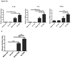
- sympathetic innervation (rather than parasympathetic) of the gut and/or spleen is able to exert an anti-inflammatory effect on the lower intestine (in particular the colon). When such sympathetic innervation is removed, histological and immunological measures of inflammation in the gut increase.
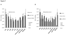
- the anti-inflammatory effect of sympathetic neural activity is further indicated by the strong inverse correlation between norepinephrine levels in the gut or spleen (indicative of sympathetic activity) and the increased measures of inflammation.
- the inventors have shown that by stimulating the superior mesenteric plexus (SMP) that includes the Supra Mesenteric Nerve, or the splenic nerve with a neuromodulatory device, it is possible to increase norepinephrine levels in the gut, which will thus reduce the level of colitis-associated inflammation.
- SMP superior mesenteric plexus
- adrenergic receptors results in a reduction in pro-inflammatory cytokine production by dendritic cells (DCs), whether that activation is by adrenergic neurotransmitters (epinephrine) or an artificial AR agonist (salbutamol)
- DCs dendritic cells
- epinephrine adrenergic neurotransmitters
- sibutamol an artificial AR agonist
- vagal nerve signaling the effect of sympathetic signaling to reduce inflammation does not require the vagus nerve, and therefore is not a downstream result of vagal nerve signaling.
- Vagotomy had no effect on levels of inflammation (indicating little or no suppressive effect derived from vagal signaling), whereas resection of the sympathetic nerves innervating the gut or the spleen exacerbated the levels of inflammation, indicating that the SMP and splenic nerve are able to suppress inflammation independently of vagal signaling.
- the inventors have demonstrated that it is possible to treat (that is, reduce) inflammation associated with colitis.
- Such a treatment has a particular advantage over stimulation of the vagus nerve in treating inflammation of the colon (i.e., "colitis"), as the large intestine and colon have little parasympathetic vagal innervation, but do have sympathetic innervation.
- Colitis inflammation of the colon
- Targeting the splenic nerve and SMP therefore allows new areas of the body to be treated, increasing the specificity and efficacy of neuromodulation.
- the present invention seeks to avoid affecting other bodily systems and thereby reduces unwanted side effects.
- the present disclosure provides an apparatus or system for treating an inflammatory disorder in a patient.
- the inflammatory disorder is an inflammatory disorder that includes colitis as a symptom, for example, IBD, such as ulcerative colitis (UC) or Crohn's Disease.
- the invention provides an apparatus or system for modulating the neural activity of the splenic nerve (or any nerve that innervates the spleen) and/or the SMP of a patient, the apparatus or system comprising: a first transducer configured to apply a first signal to the splenic nerve or SMP and optionally a second transducer to apply a second signal to the other of the SMP or splenic nerve; and a controller coupled to the transducer or transducers, the controller controlling the signal to be applied by the transducer or transducers, such that the signal modulates the neural activity of the nerve to which it is applied to produce a physiological response in the patient.
- the signal applied increases neural activity in the nerve to which the signal is applied.
- the physiological response elicited is an increase in local sympathetic tone in the spleen and/or gut (e.g., the intestines and/or colon) of the patient.
- the physiological response includes one or more of an increase in gut tissue and/or circulating nor-epinephrine, a decrease in one or more inflammatory markers in gut tissue and/or circulation, a decrease in gut pathology, a decrease in colon fibrosis, and a reduction in one or more symptoms of colitis.
- the signal is an electrical signal and the transducer is an electrode.
- the signal is a low frequency AC waveform, optionally having a frequency of 0.01-20 Hz, for example 10 Hz.
- the apparatus or system further comprises a detector element to detect one or more physiological parameters in the patient.
- the controller is coupled to the detector element, and causes the one or more transducers each to apply the signal when the physiological parameter is detected to be meeting or exceeding a predefined threshold value.
- one or more of the detected physiological parameters is selected from: sympathetic tone; gut tissue and/or circulating nor-epinephrine (NE) levels; gut tissue and/or circulating substance P levels; gut tissue and/or circulating levels of one or more inflammatory markers.
- the one or more detected physiological parameters comprise an action potential or pattern of action potentials in a nerve of the patient.
- the invention provides a method of treating colitis in a patient comprising: (i) implanting in the patient a device, an apparatus or a component of a system as described above; (ii) positioning the first transducer of the device, apparatus or system in signalling contact with the splenic nerve and/or SMP of the patient; and (iii)activating the apparatus or system, to modulate neural activity of the splenic nerve (or any nerve that innervates the spleen) and/or SMP.
- the method is a method of treating IBD, such as treating Crohn's Disease and/or UC.
- the invention provides a method of treating colitis in a patient, in particular IBD, the method comprising applying a signal to the splenic nerve (or any nerve that innervates the spleen) and/or the superior mesenteric plexus (SMP) of the patient to modulate the neural activity of the nerve in the patient.
- the signal or signals are applied by a neuromodulation device comprising a transducer configured to apply the signal.
- the neuromodulation device is at least partially implanted in the patient, optionally wholly implanted in the patient.
- the modulation in neural activity as a result of applying the signal is an increase in neural activity in the nerve to which the signal is applied.
- the signal is an electrical signal and comprises a low frequency alternating current (AC) waveform, optionally having a frequency of 0.01-20 Hz, for example 10 Hz.
- AC alternating current
- the method further comprises the step of detecting one or more physiological parameters of the patient, wherein the signal is applied only when the detected physiological parameter meets or exceeds a predefined threshold value.
- one or more detected physiological parameters is selected from: sympathetic tone; gut tissue and/or circulating nor-epinephrine (NE) levels; gut tissue and/or circulating substance P levels; gut tissue and/or circulating levels of one or more inflammatory markers.
- the one or more detected physiological parameters comprise an action potential or pattern of action potentials in a nerve of the patient.
- the invention provides an anti-inflammatory agent for use in a method of treating colitis in a patient, wherein the method comprises: (i) applying a signal to the splenic nerve (or any nerve that innervates the spleen) and/or the superior mesenteric plexus of the patient to modulate the neural activity in the nerve in the patient; and (ii) administering the anti-inflammatory agent to the patient.
- the anti-inflammatory agent is selected from a steroid, a 5-ASA, methotrexate, azathioprine, cyclosporine, and an anti-TNF agent.
- the anti-inflammatory agent is for use in a method of treating IBD, optionally UC and/or Crohn's Disease.
- the signal or signals are applied by a neuromodulation device comprising a transducer configured to apply each signal, optionally wherein the neuromodulation device is at least partially implanted in the patient, optionally wholly implanted in the patient.
- the signal or signals applied increase neural activity in the nerve to which it is applied.
- the signal is an electrical signal, optionally a low frequency AC waveform, optionally having a frequency of 0.01-20 Hz, for example 10 Hz.
- the invention provides a neuromodulatory electrical waveform for use in treating colitis in a patient, wherein the waveform is an AC waveform having a frequency of 0.01-20 Hz, such that, when applied to a splenic nerve or SMP the waveform increases neural signalling in the nerve.
- the invention provides use of a neuromodulation device for treating colitis in a patient by modulating neural activity in a splenic nerve or SMP of the patient.
- the patient is a mammalian patient, optionally a human patient.
- application of a signal may equate to the transfer of energy in a suitable form to carry out the intended effect of the signal. That is, application of a signal to a nerve or nerves may equate to the transfer of energy to (or from) the nerve(s) to carry out the intended effect.
- the energy transferred may be electrical, mechanical (including acoustic, such as ultrasound), electromagnetic (e.g., optical), magnetic or thermal energy. It is noted that application of a signal as used herein does not include a pharmaceutical intervention.
- transducer is taken to mean any element of applying a signal to the nerve or plexus, for example an electrode, diode, Peltier element or ultrasound transducer.
- a "non-destructive signal” is a signal as defined above that, when applied, does not irreversibly damage the underlying neural signal conduction ability of the nerve to which it is applied. That is, application of a non-destructive signal maintains the ability of the nerve or nerves (or fibres thereof) to conduct action potentials when application of the signal ceases, even if that conduction is in practice inhibited or blocked as a result of application of the non-destructive signal. Ablation and cauterisation of at least part of the nerve are examples of destructive signals.
- nerve activity of a nerve is taken to mean the signalling activity of the nerve, for example the amplitude, frequency and/or pattern of action potentials in the nerve.
- Modulation of neural activity is taken to mean that the signalling activity of the nerve is altered from the baseline neural activity ⁇ that is, the signalling activity of the nerve in the patient prior to any intervention. Such modulation may increase, inhibit (for example block), or otherwise change the neural activity compared to baseline activity.
- this may be an increase in the total signalling activity of the whole nerve, or it may be that the total signalling activity of a subset of nerve fibres of the nerve is increased, compared to baseline neural activity in that part of the nerve.
- such inhibition may be partial inhibition. Partial inhibition may be such that the total signalling activity of the whole nerve is partially reduced, or that the total signalling activity of a subset of nerve fibres of the nerve is fully reduced (that is, there is no neural activity in that subset of fibres of the nerve), or that the total signalling of a subset of nerve fibres of the nerve is partially reduced compared to neural activity in that subset of fibres of the nerve prior to intervention. Where the modulation of neural activity is inhibition of neural activity, this also encompasses full inhibition of neural activity in the nerve.
- Inhibition of neural activity may be a block on neural activity.
- blocking may be a partial block ⁇ that is, blocking of neural activity in a subset of nerve fibres of the nerve.
- blocking may be a full block - i.e., blocking of neural activity across the whole nerve.
- a block on neural activity is understood to be blocking neural activity from continuing past the point of the block. That is, when the block is applied, action potentials may travel along the nerve or subset of nerve fibres to the point of the block, but not beyond the block.
- Modulation of neural activity may also be an alteration in the pattern of action potentials. It will be appreciated that the pattern of action potentials can be modulated without necessarily changing the overall frequency or amplitude. For example, modulation of the neural activity may be such that the pattern of action potentials is altered to more closely resemble a healthy state rather than a disease state.
- Modulation of neural activity may comprise altering the neural activity in various other ways, for example increasing or inhibiting a particular part of the neural activity and/or stimulating new elements of activity, for example in particular intervals of time, in particular frequency bands, according to particular patterns and so forth. Such altering of neural activity may for example represent both increases and/or decreases with respect to the baseline activity.
- Modulation of the neural activity may be temporary.
- temporary is taken to mean that the modulated neural activity (whether that is an increase, inhibition, block or other modulation of neural activity or change in pattern versus baseline activity) is not permanent. That is, the neural activity following cessation of the signal is substantially the same as the neural activity prior to the signal being applied - i.e., prior to modulation.
- Neural activity prior to modulation is referred to herein as "baseline" activity.
- Modulation of the neural activity may be persistent.
- “persistent” is taken to mean that the modulated neural activity (whether that is an increase, inhibition, block or other modulation of neural activity or change in pattern versus baseline activity) has a prolonged effect. That is, upon cessation of the signal, neural activity in the nerve remains substantially the same as when the signal was being applied (that is, the neural activity during and following modulation is substantially the same).
- the prolonged effect may be temporary, as described above, such that after a period of time (e.g., minutes, for example 30 minutes or more, or hours, or even days) following cessation of the signal, the neural activity returns to baseline, or the prolonged effect may continue indefinitely or permanently.
- Modulation of the neural activity may be corrective.
- "corrective” is taken to mean that the modulated neural activity (whether that is an increase, inhibition, block or other modulation of neural activity or change in pattern versus baseline activity) alters the neural activity towards the pattern of neural activity in a healthy individual. That is, upon application (and/or following cessation) of the signal, neural activity in the nerve more closely resembles the pattern of action potentials in the nerve observed in a healthy subject than prior to modulation, preferably substantially fully resembles the pattern of action potentials in the nerve observed in a healthy subject.
- Such corrective modulation caused by the signal can be any modulation as defined herein.
- application of the signal may result in a change in neural activity, and upon cessation of the signal, the pattern of action potentials in the nerve resembles the pattern of action potentials observed in a healthy subject.
- application of the signal may result in modulation such that the neural activity resembles the pattern of action potentials observed in a healthy subject, and upon cessation of the signal, the pattern of action potentials in the nerve resembles the pattern of action potentials observed in a healthy individual.
- the "superior mesenteric plexus" is taken to refer to the plexus of neural fibres associated with the mesenteric artery.
- the splenic nerve refers to the nerve innervating the spleen as understood by the skilled person.
- colitis is used to refer to inflammation of the colon, in particular mucosal inflammation.
- Disorders causing colitis include inflammatory bowel disease (including ulcerative colitis (UC) and Crohn's disease ), ischaemic colitis, and infectious colitis.
- Colitis may be associated with infection, autoimmunity, and/or pharmacological action (e.g., through use of non-steroidal anti-inflammatory drugs (NSAIDs)).
- NSAIDs non-steroidal anti-inflammatory drugs
- IBD Inflammatory bowel disease
- Crohn's disease and UC are each chronic relapsing and remitting inflammatory diseases characterised by the symptoms including but not limited to abdominal swelling, cramping, pain or discomfort, diarrhoea, weight loss, rectal bleeding and fatigue and may also include anemia, vomiting and elevated temperature (fever).
- Crohn's disease can affect any part of the gastrointestinal (Gl) tract, for example the colon, and is characterised by patchy, transmural and mucosal inflammation.
- Gl gastrointestinal
- the inflammation is continuous, extending proximally from the rectum and generally limited to the colon.
- Treatment of colitis is characterised by a reduction in one or more symptoms exhibited by the patient suffering from the colitic disorder.
- treatment may be characterised by any one or more of a decrease in gut pathology (for example as measured by endoscopy and/or histology, e.g., crypt loss, polyps, hyperplasia, goblet cell depletion), a decrease in colon fibrosis, a reduction in one or more of abdominal swelling, cramping pain or discomfort, diarrhoea, vomiting, fever, weight loss, rectal bleeding, anemia and fatigue.
- a reduction in a symptom may be characterised by the symptom being less frequent, or less severe, or both.
- Treatment may be prophylactic or therapeutic.
- Prophylactic treatment may be characterised by the prevention of onset of symptoms. For example, colitis patients can go through periods of remission between symptomatic episodes or crises. Prophylactic treatment may prevent onset of further episodes or extend periods of remission.
- Therapeutic treatment may be characterised by amelioration of an ongoing episode of colitis. For example, therapeutic treatment may result in amelioration of the frequency and/or severity of symptoms during an episode, or cause the patient to enter a state of remission. Therapeutic treatment may entirely cure the patient of colitis.
- an "improvement in a measurable physiological parameter” is taken to mean that for any given physiological parameter, an improvement is a change in the value of that parameter in the patient towards the normal value or normal range for that value - i.e., towards the expected value in a healthy individual.
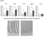
- an improvement in a measurable parameter may be one or more of an increase in sympathetic tone, an increase in gut tissue and/or circulating (i.e., blood/plasma) nor-epinephrine, a decrease in one or more inflammatory markers (for example inflammatory cytokines (e.g., TNF ⁇ , IL-6, IL-1 ⁇ , MCP-1, IL-17, C-reactive protein, and calprotectin) in gut tissue and/or circulation, a decrease in gut pathology (for example as measured by endoscopy and/or histology, e.g., crypt loss, polyps, hyperplasia, goblet cell depletion), a decrease in colon fibrosis, and a reduction in one or more symptoms such as abdominal swelling, cramping, pain or discomfort, diarrhoea, weight loss, rectal bleeding, vomiting, anemia, elevated temperature and fatigue.
- inflammatory markers for example inflammatory cytokines (e.g., TNF ⁇ , IL-6, IL-1 ⁇
- a suitable physiological parameter may be an action potential or pattern of action potentials in a nerve of the patient.
- An improvement in such a parameter is characterised by the action potential or pattern of action potentials in the nerve more closely resembling that exhibited by a healthy individual than before the intervention.
- a physiological parameter is not affected by modulation of the neural activity if the parameter does not change as a result of the modulation from the average value of that parameter exhibited by the subject or patient when no intervention has been performed - i.e., it does not depart from the baseline value for that parameter.
- the baseline for any neural activity or physiological parameter in an individual need not be a fixed or specific value, but rather can fluctuate within a normal range or may be an average value with associated error and confidence intervals. Suitable methods for determining baseline values would be well known to the skilled person.
- a measurable physiological parameter is "detected" in a patient when the value for that parameter exhibited by the patient at the time of detection is determined.
- a detector is any element able to make such a determination.
- a "predefined threshold value" for a physiological parameter is the value for that parameter where that value or beyond must be exhibited by a subject or patient before the intervention is applied.
- the threshold value may be a value indicative of colitis.
- Examples of such predefined threshold values include sympathetic tone less than a threshold sympathetic tone, for example the sympathetic tone in a healthy individual; gut tissue and/or circulating nor-epinephrine (NE) levels greater than a threshold NE level, for example the NE level in a healthy individual; gut tissue and/or circulating levels of one or more inflammatory markers (for example, TNF ⁇ , IL-6, IL-1 ⁇ , MCP-1, IL-17, C-reactive protein, calprotectin) greater than that characteristic of a healthy individual.
- inflammatory markers for example, TNF ⁇ , IL-6, IL-1 ⁇ , MCP-1, IL-17, C-reactive protein, calprotectin
- Such a threshold value for a given physiological parameter is exceeded if the value exhibited by the patient is beyond the threshold value ⁇ that is, the exhibited value is a greater departure from the normal or healthy value for that parameter than the predefined threshold value.
- sympathetic tone is used to mean the overall physiological balance resulting from sympathetic neural activity.
- Such sympathetic tone may be determined by methods known in the art, for example neurological methods, hemodynamic methods (e.g., heart rate, blood pressure, heart rate variability) or circulating plasma/urine biomarkers.
- a “neuromodulation device” as used herein is a device configured to modulate the neural activity of a nerve.
- Neuromodulation devices as described herein comprise at least one transducer capable of effectively applying a signal to a nerve.
- the elements of the device that are to be implanted in the patient are constructed such that they are suitable for such implantation. Such suitable constructions would be well known to the skilled person. Indeed, various fully implantable neuromodulation devices are currently available, such as the vagus nerve stimulator of SetPoint Medical, in clinical development for the treatment of rheumatoid arthritis ( Arthritis & Rheumatism, Volume 64, No.
- the neuromodulation device or implantable portion of an apparatus or system, can be miniaturized to a size of less than 1 cm.
- implanted is taken to mean positioned at least partially within the patient's body. Partial implantation means that only part of the device (or apparatus or system) is implanted - i.e., only part of the device is positioned within the patient's body, with other elements of the device external to the patient's body.
- the term “wholly implanted” means that the entire of the device is positioned within the patient's body. For the avoidance of doubt, the device being “wholly implanted” does not preclude additional elements, independent of the device but in practice useful for its functioning (for example, a remote wireless charging unit or a remote wireless manual override unit), being independently formed and external to the patient's body.
- charge-balanced in relation to a current (e.g., a DC current) is taken to mean that the positive or negative charge introduced into any system (e.g., a nerve) as a result of the current being applied is balanced by the introduction of the opposite charge in order to achieve overall (i.e., net) neutrality.
- a current e.g., a DC current
- colitis can be relieved and/or prevented by modulation of the neural activity of the splenic nerve and/or in the superior mesenteric plexus.
- vagus nerve sometimes referred to as the vagal nerve
- vagal nerve the vagus nerve
- cholinergic i.e., parasympathetic
- sympathetic i.e., adrenergic
- nerve the coeliac ganglion via either the splenic nerve or SMP, or both, is shown to play a role in moderating the severity of colitis.
- the present invention is able to treat colitis without the risk of unwanted side-effects often associated with vagus nerve stimulation (sometimes referred to as vagal nerve stimulation, as used herein the terms vagus nerve and vagal nerve may be used interchangeably).
- a neuromodulation device that modulates the neural activity of the splenic nerve and/or SMP will therefore provide an effective colitis treatment.
- this disclosure provides an apparatus or system for modulating the neural activity of the splenic nerve and/or the SMP of a patient, the apparatus or system comprising: a first transducer configured to apply a first signal to the splenic nerve or SMP and optionally a second transducer to apply a second signal to the other of the SMP or splenic nerve; and a controller coupled to the transducer or transducers, the controller controlling the signal to be applied by the transducer or transducers, such that the signal modulates the neural activity of the nerve to which it is applied (i.e., the splenic nerve and/or the SMP, as appropriate) to produce a physiological response in the patient.
- a first transducer configured to apply a first signal to the splenic nerve or SMP and optionally a second transducer to apply a second signal to the other of the SMP or splenic nerve
- a controller coupled to the transducer or transducers, the controller controlling the signal to be applied by the transducer
- the signal that each of the transducers is configured to apply is independently selected. In certain embodiments each transducer is configured to apply the same signal.
- any signal applied by the device ⁇ i.e., may apply independently to the first and second signals, and any further signals applied by the device.
- the signal applied by the one or more transducers is a non-destructive signal.
- the signal applied by the one or more transducers is an electrical signal, an optical signal, an ultrasonic signal, or a thermal signal.
- each of the one or more transducers may be comprised of one or more electrodes, one or more photon sources, one or more ultrasound transducers, one more sources of heat, or one or more other types of transducer arranged to put the signal into effect.
- the signal or signals applied by the one or more transducers is an electrical signal, for example a voltage or current.
- the signal applied comprises a direct current (DC) waveform, such as a charge balanced direct current waveform, or an alternating current (AC) waveform, or both a DC and an AC waveform.
- DC direct current
- AC alternating current
- the electrical signal comprises a square waveform, a sinusoidal waveform, a saw-toothed waveform or a triangular waveform, in certain preferred embodiments, the signal has a square waveform.
- the electrical signal comprises a low frequency AC waveform.
- the electrical signal is an AV waveform having a frequency of 0.01Hz-1kHz, 0.01-900 Hz, 0.01-800 Hz, 0.01-700 Hz, 0.01-600 Hz, 0.01-500Hz, 0.01-40OHz, 0.01-300 Hz, 0.01-200 Hz, 0.01-100 Hz, 0.01-50 Hz.
- the signal comprises an AC waveform having a frequency of 0.01-20 Hz, preferably 0.5-20Hz, for example 1-15Hz, preferably 5-10Hz.
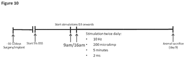
- the signal comprises an AC waveform having a frequency of 0.01 Hz, 0.05 Hz, 0.1 Hz, 0.5 Hz, 1 Hz, 2 Hz, 3 Hz, 4Hz, 5 Hz, 6 Hz, 7 Hz, 8 Hz, 9 Hz or 10 Hz, such as 10 Hz.
- the electrical signal has a current of 0.1-20 mA, 0.1-10 mA, 0.1-5 mA, 0.1-3 mA 0.1-1 mA, 0.1-0.5 mA, 0.16 mA-0.6 mA, 0.2-0.4 mA, for example 0.2-0.4 mA. In certain embodiments the electrical signal has a current of at least 0.16 mA.
- the electrical signal has a current of 0.1 mA, 0.15 mA, 0.16 mA, 0.18 mA, 0.2 mA, 0.2 mA, 0.4 mA, 0.6 mA, 1 mA, 2 mA, 3 mA, 5 mA, 10 mA, such as 0.2 mA.
- the electrical signal has a pulse width of 20-500 microseconds ( ⁇ s). In certain embodiments the signal has a pulse width of 20-500 ⁇ s, 30-400 ⁇ s, 40-300 ⁇ s, 50-200 ⁇ s, 60-150 ⁇ s, 70-120 ⁇ s, 80-100 ⁇ s.
- the electrical signal comprises a DC waveform and/or an AC waveform having a voltage of 0.1-20V.
- the signal has a voltage of 0.1-15V, optionally 0.1-10V.
- the voltage is selected from 0.1V, 0.2V, 0.3V, 0.5V, 0.6V, 0.7V, 0.8V, 0.9V, 1V, 2V, 3V, 5V and 10V.
- the electrical signal comprises an AC square biphasic waveform of 10 Hz 0.2 mA.
- the current or voltage of a signal can be varied in order to achieve the intended value of the other parameter for any given device-nerve arrangement.
- the signal comprises a DC ramp followed by a plateau and charge-balancing, followed by a first AC waveform, wherein the amplitude of the first AC waveform increases during the period in which the first AC waveform is applied, followed by a second AC waveform having a lower amplitude and/or lower frequency than the first AC waveform.
- the DC ramp, first AC waveform and second AC waveform are applied substantially sequentially.
- the signal comprises an AC waveform of kilohertz frequency. In certain embodiments, wherein the signal comprises one or more AC waveforms, each AC waveform is independently selected from an AC waveform of 5-25 kHz, optionally 10-25 kHz, optionally 15-25 kHz, optionally 20-25 kHz. In certain embodiments, the signal comprises an AC waveform signal of 5 kHz. In certain alternativeembodiments, the signal comprises an AC waveform of 25 kHz.
- At least one of the one or more transducers is an electrode configured to apply the electrical signal.
- all the transducers are electrodes configured to apply an electrical signal, optionally the same electrical signal.
- the signal applied is an electrical signal
- the signal is applied by a cuff electrode.
- the cuff is configured to encompass the nerve to which the signal is applied and optionally the associated blood vessel.
- the signal is applied by a cuff electrode configured to encompass the SMP and the superior mesenteric artery.
- the signal applied is a thermal signal
- the signal reduces the temperature of the nerve (i.e., cools the nerve).
- the signal increases the temperature of the nerve (i.e., heats the nerve).
- the signal both heats and cools the nerve.
- At least one of the one or more transducers is a transducer configured to apply a thermal signal. In certain such embodiments, all the transducers are configured to apply a thermal signal, optionally the same thermal signal.
- one or more of the one or more transducers comprise a Peltier element configured to apply a thermal signal, optionally each of the one or more transducers comprises a Peltier element.
- one or more of the one or more transducers comprise a laser diode configured to apply a thermal signal (e.g., a near infrared (NIR) diode laser), optionally all of the one or more transducers comprise a laser diode configured to apply a thermal signal.
- NIR near infrared
- one or more of the one or more transducers comprise a electrically resistive element configured to apply a thermal signal, optionally each of the one or more transducers comprises a electrically resistive element configured to apply a thermal signal.
- the signal applied is a mechanical signal, optionally an ultrasonic signal.
- the mechanical signal is a pressure signal.
- the signal is an electromagnetic signal, optionally an optical signal.
- the one or more transducers comprise a laser and/or a light emitting diode (e.g., an NIR diode laser) configured to apply the optical signal.
- the apparatus or system further comprises a fibre optic interface configured to apply the signal from the one or more of the transducers to the at least one nerve.
- the physiological response produced in the patient is one or more of an increase in sympathetic tone, an increase in gut tissue and/or circulating/plasma nor-epinephrine, a decrease in one or more inflammatory markers (for example inflammatory cytokines (e.g., TNF ⁇ , IL-6, IL-1 ⁇ , MCP-1, IL-17, C-reactive protein and calprotectin) in gut tissue and/or plasma, a change in blood flow or circulation through the colon and/or spleen, a decrease in gut pathology (for example as measured by endoscopy and/or histology, e.g., crypt loss, polyps, hyperplasia, goblet cell depletion), a decrease in colon fibrosis, and a reduction in one or more symptoms such as abdominal swelling, cramping, pain or discomfort, diarrhoea, weight loss, rectal bleeding, vomiting, anemia, elevated temperature and fatigue.
- inflammatory markers for example inflammatory cytokines (e.g., TNF ⁇ ,
- the physiological response produced in the patient may be an improvement in the pattern of action potentials in a nerve of the patient ⁇ that is the pattern of action potentials in the nerve more closely resembles that exhibited by a healthy individual than before the intervention.
- the nerve is the splenic nerve, the SMP or the vagus nerve.
- the apparatus or system further comprises a detector element to detect one or more physiological parameters in the patient.
- a detector element may be configured to detect the one or more physiological parameters. That is, in such embodiments each detector may detect more than one physiological parameter, for example all the detected physiological parameters. Alternatively, in such embodiments each of the one or more detector elements is configured to detect a separate parameter of the one or more physiological parameters detected.
- the controller is coupled to the detector element configured to detect one or more physiological parameters, and causes the transducer or transducers to apply their respective signal when the physiological parameter is detected to be meeting or exceeding a predefined threshold value.
- the one or more detected physiological parameters are one or more of sympathetic tone; gut tissue and/or circulating nor-epinephrine (NE) levels; gut tissue and/or circulating substance P levels; gut tissue and/or circulating levels of one or more inflammatory markers (for example, TNF ⁇ , IL-6, IL-1 ⁇ , MCP-1, IL-17, calprotectin).
- NE nor-epinephrine
- gut tissue and/or circulating substance P levels gut tissue and/or circulating levels of one or more inflammatory markers (for example, TNF ⁇ , IL-6, IL-1 ⁇ , MCP-1, IL-17, calprotectin).
- the one or more detected physiological parameters may include an action potential or pattern of action potentials in a nerve of the patient, wherein the action potential or pattern of action potentials is associated with colitis.
- the nerve is the splenic nerve or SMP.
- the action potential or pattern of action potentials is detected in the vagus nerve, for example the afferent fibres of the vagus nerve.
- the controller is coupled to a detector or detectors configured to detect the pattern of action potentials in the SGM and also the heart rate variability of the patient as an indication of sympathetic tone.
- the modulation in neural activity as a result of applying the signal is an increase in neural activity in the nerve to which the signal is applied. That is, in such embodiments, application of the signal results in the neural activity in at least part of the nerve being increased compared to the baseline neural activity in that part of the nerve. Such an increase in activity could equally be across the whole nerve. Therefore, in certain such embodiments, a result of applying the signal is an increase in neural activity across the whole nerve. Treatment of colitis is expected to be particularly effective when sympathetic signalling by one or both of the splenic nerve or SMP is increased.
- the signal applied to increase neural activity is an electrical signal comprising an AC waveform of low frequency.
- the electrical signal comprises a low frequency AC waveform.
- the electrical signal is an AV waveform having a frequency of 0.01Hz-1kHz, 0.01-900 Hz, 0.01-800 Hz, 0.01-700 Hz, 0.01-600 Hz, 0.01-500Hz, 0.01-400Hz, 0.01-300 Hz, 0.01-200 Hz, 0.01-100 Hz, 0.01-50 Hz.
- the signal comprises an AC waveform having a frequency of 0.01-20 Hz, for example 0.5-20Hz, 1-15Hz, 5-10Hz.
- the signal comprises an AC waveform having a frequency of 0.01 Hz, 0.05 Hz, 0.1 Hz, 0.5 Hz, 1 Hz, 2 Hz, 3 Hz, 4Hz, 5 Hz, 6 Hz, 7 Hz, 8 Hz, 9 Hz or 10 Hz, for example 10 Hz.
- the electrical signal has a current of 0.1-20 mA, 0.1-10 mA, 0.1-5 mA, 0.1-3 mA, 0.1-1 mA, 0.1-0.5 mA, 0.16 mA-0.6 mA, 0.2-0.4 mA, for example 0.2-0.4 mA. In certain embodiments the electrical signal has a current of at least 0.16 mA.
- the electrical signal has a current of 0.1 mA, 0.15 mA, 0.16 mA, 0.18 mA, 0.2 mA, 0.2 mA, 0.4 mA, 0.6 mA, 1 mA, 2 mA, 3 mA, 5 mA, 10 mA, such as 0.2 mA.
- the electrical signal has a square waveform, a sinusoidal waveform, a saw-toothed waveform or a triangular waveform. In certain embodiments, the signal has a square waveform.
- the electrical signal has a pulse width of 20-500 microseconds ( ⁇ s). In certain embodiments the signal has a pulse width of 20-500 ⁇ s, 30-400 ⁇ s, 40-300 ⁇ s, 50-200 ⁇ s, 60-150 ⁇ s, 70-120 ⁇ s, 80-100 ⁇ s.
- the electrical signal comprises a DC waveform and/or an AC waveform having a voltage of 0.1-20V.
- the signal has a voltage of 0.1-15V, optionally 0.1-10V.
- the voltage is selected from 0.1V, 0.2V, 0.3V, 0.5V, 0.6V, 0.7V, 0.8V, 0.9V, 1V, 2V, 3V, 5V and 10V.
- the electrical signal comprises an AC square biphasic waveform of 10 Hz 0.2 mA.
- the modulation in neural activity as a result of applying the signal is an alteration to the pattern of action potentials in the nerve.
- the neural activity is modulated such that the resultant pattern of action potentials in the nerve more closely resembles the pattern of action potentials in the nerve or nerves observed in a healthy subject than prior to the signal being applied.
- the modulation in neural activity as a result of applying the signal is inhibition of neural activity in the part of the nerve to which the signal is applied. That is, in such embodiments, application of the signal results in the neural activity being reduced compared to the neural activity in that part of the nerve prior to the signal being applied.
- Modulation of neural activity may comprise altering the neural activity in various other ways, for example increasing or inhibiting a particular part of the activity and stimulating new elements of activity, for example in particular intervals of time, in particular frequency bands, according to particular patterns and so forth. Such altering of neural activity may for example represent both increases and/or decreases with respect to the baseline activity.
- the controller causes the signal to be applied intermittently, In certain such embodiments, the controller causes the signal to applied for a period of time (e.g., a first time period), then stopped for a second or immediately subsequent time period, then reapplied and stopped in an alternating pattern, e.g., applied for a third time period, then stopped for a fourth time period.
- the alternating periods of signal application and cessation e.g., first, second, third and fourth periods, etc.
- multiple application cycles can run consecutively such that the signal is applied in phases, between which phases no signal is applied.
- the duration of the first, second, third and fourth time periods can be independently selected. That is, the duration of each time period may be the same or different to any of the other time periods.
- the duration of each of the first, second, third and fourth time periods is any time from 5 seconds (5s) to 24 hours (24h), 30s to 12 h, 1 min to 12 h, 5 min to 8 h, 5 min to 6 h, 10 min to 6 h, 10 min to 4 h, 30 min to 4 h, 1 h to 4 h (or any combination of such lower and upper limits).
- the duration of each of the (e.g., first, second, third and fourth) time periods is 5s, 10s, 30s, 60s, 2 min, 5 min, 10 min, 15 min, 20 min, 30 min, 40 min, 50 min, 60 min, 90 min, 2 h, 3 h, 4 h, 5 h, 6 h, 7 h, 8 h, 9 h, 10 h, 11 h, 12 h, 13 h, 14 h, 15 h, 16 h, 17 h, 18 h, 19 h, 20 h, 21 h, 22 h, 23 h, 24 h (or any time period within the limits of a preceding time period).
- the alternating period of signal application (e.g., first and third time periods, etc.) is from 1 minute to 1 hour, such as 1-20 minutes, 1-10 minutes, 2-10 minutes, for example 2 minutes, or 5 minutes, or 10 minutes, or 20 minutes, or 30 minutes or an hour (or any period in between).
- the alternating period of signal cessation (e.g., second and fourth time periods, etc.) are from 1-20 minutes, 1-10 minutes, 2-10 minutes, for example 2 minutes, or 5 minutes, or 10 minutes.
- the signal is applied up to 6 times per day, up to 4 times per day, up to 3 times per day, 2 times per day, once per day.
- the signal is not applied during alternating periods that are independently selected from 24 hours, 12 hours, 8 hours, and 6 hours (or selected to be of such duration from which the duration of the alternating period during which signal is applied, such that a diurnal repeating pattern is created and maintained).
- the second and fourth periods comprise the remainder of a period of 24 hours, once the first and third periods, cumulatively, have been subtracted from 24 hours. That is, where the first and third period are, by way of example, 5 minutes each in duration (10 minutes total), the second and third periods comprise 23 hours and 40 minutes in total, that is 11 hours and 50 minutes each.
- the first and second time periods, cumulatively are a period of 12 hours
- the third and fourth time periods, cumulatively are a period of 12 hours.
- the signal is applied for a specific amount of time per day, or a specific amount of time, twice a day.
- the signal is applied for at least 2 min, at least 5 min, at least 10 min, at least 20 min, at least 30 min, at least 40 min, at least 50 min, at least 60 min, at least 90 min, at least 2 h, at least 3 h, at least 4 h, at least 5 h, at least 6 h, at least 7 h, at least 8 h, at least 9 h, at least 10 h, at least 11 h, at least 12 h, at least 13 h, at least 14 h, at least 15 h, at least 16 h, at least 17 h, at least 18 h, at least 19 h, at least 20 h, at least 21.
- the signal is applied continuously for the specified amount of time.
- the signal may be applied discontinuously across the day, provided the total time of application amounts to the specified time.
- the signal is applied for 2 min, 5 min, 10 min, 15 min, 20 min, 30 min, 45 min or 60 min, twice a day or four times a day.
- the signal is applied only when the patient is in a specific physiological state.
- the signal is applied only when the patient is experiencing an episode of colitis - i.e., they are experiencing one or more symptoms associated with a colitis crisis or episode, such as abdominal swelling, cramping, pain or discomfort, diarrhoea, weight loss, rectal bleeding, vomiting, anemia, elevated temperature and fatigue.
- the status of the patient e.g., that they are experiencing an episode
- the status of the patient can be indicated by the patient.
- the status of the patient can be detected independently from any input from the patient.
- the device further comprises a detector configured to detect the status of the patient, wherein the signal is applied only when the detector detects that the patient is in the specific state. In certain such embodiments, the signal is applied only when the patient is exhibiting an abnormal (e.g., reduced) sympathetic tone.
- the apparatus or system further comprises a communication, or input, element via which the status of the patient (e.g., that they are experiencing symptoms of colitis) can be indicated by the patient or a physician.
- the apparatus or system further comprises a detector configured to detect the status of the patient.
- the signal is applied only when the detector detects that the patient is in the specific state.
- the controller causes the signal to be permanently applied. That is, once begun, the signal is continuously applied to the nerve or nerves. It will be appreciated that in embodiments wherein the signal is a series of pulses, gaps between pulses do not mean the signal is not continuously applied.
- the modulation in neural activity caused by the application of the signal is temporary. That is, upon cessation of the signal, neural activity in the nerve or nerves returns substantially towards baseline neural activity within 1-60 seconds, or within 1-60 minutes, or within 1-24 hours, optionally 1-12 hours, optionally 1-6 hours, optionally 1-4 hours, optionally 1-2 hours. In certain such embodiments, the neural activity returns substantially fully to baseline neural activity. That is, the neural activity following cessation of the signal is substantially the same as the neural activity prior to the signal being applied - i.e., prior to modulation.
- the modulation in neural activity caused by the application of the signal or signals is substantially persistent. That is, upon cessation of the signal, neural activity in the nerve or nerves remains substantially the same as when the signal was being applied - i.e., the neural activity during and following modulation is substantially the same.
- the modulation in neural activity caused by the application of the signal is partially corrective, preferably substantially corrective. That is, upon application (and optionally cessation) of the signal, neural activity in the nerve or nerves more closely resembles the pattern of action potentials in the nerve(s) observed in a healthy subject than in the subject prior to modulation, preferably substantially fully resembles the pattern of action potentials in the nerve(s) observed in a healthy subject.
- the modulation caused by the signal can be any modulation as defined herein.
- application of the signal may result in an increase in neural activity, and upon cessation of the signal, the pattern of action potentials in the nerve or nerves resembles the pattern of action potentials observed in a healthy individual.
- application of the signal may result in modulation such that the neural activity resembles the pattern of action potentials observed in a healthy subject, and upon cessation of the signal, the pattern of action potentials in the nerve or nerves resembles the pattern of action potentials observed in a healthy subject.
- a corrective effect is the result of a positive feedback loop - that is, the underlying inflammatory state causing the colitis is treated as result of the device and use of the claimed methods, thereby causing the afferent (i.e., sensory) signals from the gut (e.g., afferent vagal signals) to be altered such that the efferent sympathetic-vagal tone is at least partially restored to that of a healthy individual.
- afferent i.e., sensory signals from the gut
- afferent vagal signals e.g., afferent vagal signals
- Such a corrective mechanism may be mediated via an immuno-neuronal reflex.
- the apparatus or system (or at least one component thereof) is suitable for at least partial implantation into the patient. In certain such embodiments, the apparatus or system (or component thereof) is suitable to be fully implanted in the patient.
- the apparatus or system further comprises one or more power supply elements, for example a battery, and/or one or more communication elements.
- the invention provides a method for treating colitis in a patient, in particular IBD, the method comprising implanting an apparatus or at least one component of a system as described above, positioning the first transducer of the apparatus or system component in signalling contact with the splenic nerve or SMP of the patient, and activating the apparatus or system.
- the transducer is in signalling contact with the nerve when it is positioned such that the signal can be effectively applied to the nerve.
- the apparatus or system is activated when the apparatus or system is in an operating state such that the signal will be applied as determined by the controller.
- the method comprises positioning a second transducer of the apparatus or system in signalling contact with the SMP or splenic nerve of the patient, whichever is not in signalling contact with the first transducer.
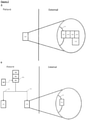
- Figures 2A-2C show how the invention may be put into effect using one or more neuromodulation devices which are implanted in, located on, or otherwise disposed with respect to a patient in order to carry out any of the various methods described herein.
- one or more neuromodulation devices can be used to treat colitis, for example IBD (Crohn's Disease or UC) by modulating neural activity in the splenic nerve and/or SMP of the patient, optionally both the splenic nerve and SMP.
- colitis for example IBD (Crohn's Disease or UC)
- a separate neuromodulation device 100 is provided in respect of each of the splenic nerve and SMP, although as discussed herein a device could be provided or used in respect of only one of the SMP or splenic nerve. Similarly, as discussed herein, one device may be provided or used in respect of both the splenic nerve and SMP - i.e., one device is configured and provided to apply a signal to both nerves.
- Each neuromodulation device 100 may be fully or partially implanted in the patient, or otherwise located, so as to provide neuromodulation of the respective nerve or nerves. Each neuromodulation device 100 may operate independently, or may operate in communication with each other.
- Figure 2A also shows schematically components of an implanted neuromodulation device 100, in which the device comprises several elements, components or functions grouped together in a single unit and implanted in the patient.
- a first such element is a first transducer, preferably an electrode, 102 which is shown in proximity to a splenic nerve or SMP 90 of the patient.
- device 100 may comprise a second transducer configured for and positioned in proximity to the SMP or splenic nerve, whichever is not in signalling contact with the first transducer (not shown).
- the transducer 102 may be operated by a controller element 104.
- the device may comprise one or more further elements such as a communication element 106, a detector element 108, a power supply element 110 and so forth.
- transducer 102 may be in the form of a cuff electrode configured such that, when implanted, it encompasses the nerve (SMP or splenic nerve) and the associated blood vessel (e.g., the SMP and associated mesenteric artery).
- nerve SMP or splenic nerve
- the associated blood vessel e.g., the SMP and associated mesenteric artery
- Each neuromodulation device 100 may carry out the required neuromodulation independently, or in response to one or more control signals.
- a control signal may be provided by the controller 104 according to an algorithm, in response to output of one or more detector elements 108, and/or in response to communications from one or more external sources received using the communications element.
- the detector element(s) could be responsive to a variety of different physiological parameters.
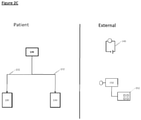
- FIG. 2B illustrates some ways in which the apparatus or system of Figure 2A may be differently distributed.
- the neuromodulation devices 100 comprise transducers 102 implanted proximally to a splenic nerve or SMP 90, but other elements such as a controller 104, a communication element 106 and a power supply 110 are implemented in a separate control unit 130 which may also be implanted in (as shown in 2B), or otherwise carried by the patient.
- the control unit 130 then controls the transducers in both of the neuromodulation devices via connections 132 which may for example comprise electrical wires and/or optical fibres for delivering signals and/or power to the transducers.
- one or more detectors 108 are located separately from the control unit, although one or more such detectors could also or instead be located within the control unit 130 and/or in one or both of the neuromodulation devices 100.
- the detectors may be used to detect one or more physiological parameters of the patient, and the controller element or control unit then causes the transducers to apply the signal in response to the detected parameter(s), for example only when a detected physiological parameter meets or exceeds a predefined threshold value.
- Physiological parameters which could be detected for such purposes include sympathetic tone; gut tissue and/or circulating nor-epinephrine (NE) levels; gut tissue and/or circulating substance P levels; gut tissue and/or circulating levels of one or more inflammatory markers (for example, TNF ⁇ , IL-6, IL-1 ⁇ , MCP-1, IL-17, C-reactive protein, calprotectin).
- a detected physiological parameter could be an action potential or pattern of action potentials in a nerve of the patient, for example in the splenic nerve, the SMP or the vagus nerve.
- the action potential or pattern of action potentials may be associated with colitis.
- Figure 2C illustrates some ways in which some functionality of the apparatus or system of Figures 2A or 2B is provided not implanted in the patient.
- an external power supply 140 is provided which can provide power to implanted elements of the apparatus or system in ways familiar to the skilled person, and an external controller 150 provides part or all of the functionality of the controller 104, and/or provides other aspects of control of the apparatus or system, and/or provides data readout from the apparatus or system, and/or provides a data input facility 152.
- the data input facility could be used by a patient or other operator in various ways, for example to input data relating to the status of the patient (e.g., if they are experiencing abdominal pain or discomfort, are undergoing a colitic episode).
- Each neuromodulation device may be adapted to carry out the neuromodulation required using one or more physical modes of operation which typically involve applying a signal to the splenic nerve and/or the SMP, such a signal typically involving a transfer of energy to (or from) the nerve(s).
- modes may comprise modulating the nerve or nerves using an electrical signal, an optical signal, an ultrasound or other mechanical signal, a thermal signal, a magnetic or electromagnetic signal, or some other use of energy to carry out the required modulation.
- Such signals may be non-destructive signals.
- Such modulation may comprise increasing, inhibiting, blocking or otherwise changing the pattern of neural activity in the nerve or nerves.
- the transducer 90 illustrated in Figure 2A could be comprised of one or more electrodes, one or more photon sources, one or more ultrasound transducers, one more sources of heat, or one or more other types of transducer arranged to put the required neuromodulation into effect.
- the neural modulation device or apparatus or system may be arranged to increase neural activity of the splenic nerve and/or SMP by using the transducer(s) to apply a voltage or current, for example an AC waveform.
- the device or apparatus or system may be arranged to use the transducer(s) to apply a low frequency AC waveform, for example an AC waveform having a frequency of 0.01 Hz-1kHz, 0.01-500 Hz, 0.01-100 Hz, 0.01-20 Hz, preferably 0.5-20Hz, 1-15Hz, 5-10Hz.
- the signal comprises an AC waveform having a frequency of 0.01 Hz, 0.05 Hz, 0.1 Hz, 0.5 Hz, 1 Hz, 2 Hz, 3 Hz, 4Hz, 5 Hz, 6 Hz, 7 Hz, 8 Hz, 9 Hz or 10 Hz, such as 10 Hz.
- the electrical signal has a current of 0.1-20 mA, 0.1-10 mA, 0.1-5 mA, 0.1-3 mA, 0.1-1 mA, 0.1-0.5 mA, 0.16 mA-0.6 mA, 0.2-0.4 mA. In certain embodiments the electrical signal has a current of at least 0.16 mA. In certain embodiments, the electrical signal has a current of 0.1 mA, 0.15 mA, 0.16 mA, 0.18 mA, 0.2 mA, 0.2 mA, 0.4 mA, 0.6 mA, 1 mA, 2 mA, 3 mA, 5 mA, 10 mA, for example 0.2 mA.
- the electrical signal has a square waveform, a sinusoidal waveform, a saw-toothed waveform or a triangular waveform. In certain preferred embodiments, the signal has a square waveform.
- the electrical signal has a pulse width of 20-500 microseconds ( ⁇ s). In certain embodiments the signal has a pulse width of 20-500 ⁇ s, 30-400 ⁇ s, 40-300 ⁇ s, 50-200 ⁇ s, 60-150 ⁇ s, 70-120 ⁇ s, 80-100 ⁇ s.
- the electrical signal comprises a DC waveform and/or an AC waveform having a voltage of 0.1-20V.
- the signal has a voltage of 0.1-15V, optionally 0.1-10V.
- the voltage is selected from 0.1V, 0.2V, 0.3V, 0.5V, 0.6V, 0.7V, 0.8V, 0.9V, 1V, 2V, 3V, 5V and 10V.
- the electrical signal comprises an AC square biphasic waveform of 10 Hz 0.2 mA.
- Neuromodulation techniques for stimulating neural activity include thermal, physical, chemical and electromagnetic techniques.
- Temperature changes affect the ion channels in the membranes of neurons and produce complex changes in function, often affecting characteristics such as resting potential and rate of depolarization. Specifically, heating regions in the hypothalamus of cats has caused increased rates of spontaneous discharge ( Teruo Nakayama , H. T. Hammel, J. D. Hardy, J. S. Eisenman American Journal of Physiology Published 1 June 1963 Vol. 204 no. 1122-1126 , incorporated herein by reference). Focused application of heat through short pulses of infrared (IR) radiation causes a transient local temperature increase and can also depolarize neurons (Goyal V, 2012 Nov; 295(11):1987-99, incorporated herein by reference).
- IR infrared
- IR radiation over electrical stimulation include higher spatial-temporal resolution and avoidance of electrical artifacts that can interfere with neural signal recoding.
- plasmonic gold nanorods injected near the membrane have been shown to transduce near IR energy into heat ( Eom K, Small. 2014 Oct 15;10(19):3853-7 (incorporated herein by reference)).
- Magnetic fields have also been shown to heat nanoparticles to produce action potentials in neighbouring neurons ( Heng Huang, Nature Nanotechnology 5, 602 ⁇ 606 (2010 ), incorporated herein by reference).
- Non-invasive magnetic stimulation has been used to stimulate the brain, spinal cord, nerve roots and peripheral nerves ( Rossini, PM et al, Clin Neurophysiol. 2015 Feb , incorporated herein by reference).
- Quickly changing magnetic fields produces electrical currents that cause depolarization within the axons of neural tissue.
- transducers may be configured to apply one or more of the blocking techniques described below.
- Thermal methods of neuromodulation may also manipulate the temperature of a nerve to inhibit signal propagation.
- Patberg et ⁇ l. Blocking of impulse conduction in peripheral nerves by local cooling as a routine in animal experimentation; Journal of Neuroscience Methods 1984;10:267 --- 75 , which is incorporated herein by reference) discuss how cooling a nerve blocks signal conduction without an onset response, the block being both reversible and fast acting, with onsets of up to tens of seconds.
- Heating the nerve can also be used to block conduction, and is generally easier to implement in a small implantable or localised transducer or device, for example using infrared radiation from laser diode or a thermal heat source such as an electrically resistive element, which can be used to provide a fast, reversible, and spatially very localised heating effect (see for example Duke et ⁇ l. J Neural Eng. 2012 Jun;9(3):036003 . Spatial and temporal variability in response to hybrid electro-optical stimulation., which is incorporated herein by reference). Either heating, or cooling, or both could be provided using a Peltier element.
- Optogenetics is a technique that genetically modifies cells to express photosensitive features, which can then be activated with light to modulate cell function. Many different optogenetic tools have been developed that can be used to inhibit neural firing. A list of optogenetic tools to suppress neural activity has been compiled ( Epilepsia. 2014 Oct 9. doi: 10.1111/epi.12804. WONOEP appraisal: Optogenetic tools to suppress seizures and explore the mechanisms of epileptogenesis. Ritter LM et al., which is incorporated herein by reference).
- Acrylamine-azobenzene-quaternary ammonium is a photochromic ligand that blocks many types of K+ channels and in the cis configuration, the relief of K+ channel block inhibits firing ( Nat Neurosci. 2013 Jul;16(7):816-23. doi: 10.1038/nn.3424 . Optogenetic pharmacology for control of native neuronal signaling proteins Kramer RHet al, which is incorporated herein by reference). By adapting Channelrhodopsin-2 and introducing it into mammalian neurons with the lentivirus, it is possible to control inhibitory synaptic transmission (Boyden ES 2005). Instead of using an external light source such as a laser or light emitting diode, light can be generated internally by introducing a gene based on firefly luciferase (Land BB 2014). The internally generated light has been sufficient to generate inhibition.
- an external light source such as a laser or light emitting diode
- Mechanical forms of neuromodulation can include the use of ultrasound which may conveniently be implemented using external instead of implanted ultrasound transducers.
- Other forms of mechanical neuromodulation include the use of pressure (for example see “The effects of compression upon conduction in myelinated axons of the isolated frog sciatic nerve” by Robert Fern and P. J. Harrison Br.j. Anaesth. (1975), 47, 1123 , incorporated herein by reference).
- Some electrical forms of neuromodulation may use direct current (DC), or alternating current (AC) waveforms applied to a nerve using one or more electrodes.
- a DC block may be accomplished by gradually ramping up the DC waveform amplitude ( Bhadra and Kilgore, IEEE Transactions on Neural systems and rehabilitation engineering, 2004 12(3) pp313-324 , which is incorporated herein by reference).
- Some AC techniques include HFAC or KHFAC (high-frequency or kilohertz frequency) to provide a reversible block (for example see Kilgore and Badra, 2004, Medical and Biological Engineering and Computing , the content of which is incorporated herein by reference for all purposes).
- HFAC or KHFAC high-frequency or kilohertz frequency
- HFAC may typically be applied at a frequency of between 1 and 50 kHz at a duty cycle of 100% ( Bhadra, N. et al., Journal of Computational Neuroscience, 2007, 22(3), pp 313-326 , which is incorporated herein by reference).
- Methods for selectively blocking activity of a nerve by application of a waveform having a frequency of 5 - 10 kHz are described in US 7,389,145 (incorporated herein by reference).
- US 8,731,676 (incorporated herein by reference) describes a method of ameliorating sensory nerve pain by applying a 5-50 kHz frequency waveform to a nerve.
- the invention also provides a method of treating colitis in a patient, in particular IBD, the method comprising applying a signal to the splenic nerve and/or the superior mesenteric plexus (SMP) of the patient to modulate the neural activity of the nerve in the patient.
- a signal is applied to the splenic nerve and a second signal is applied to the SMP.
- the signal or signals are applied by a neuromodulation device comprising a transducer configured to apply each signal, preferably an electrode to apply an electrical signal.
- the neuromodulation device is at least partially implanted in the patient. In certain preferred embodiments, the neuromodulation device is wholly implanted in the patient.
- both signals are applied by the same neuromodulation device, that device have at least two transducers, one to apply the first signal and one to apply the second signal.
- the first signal is applied by one neuromodulation device and the second signal is applied by a separate neuromodulation device.
- the signal is configured to stimulate the nerve to which it is applied. In certain embodiments, the signal is configured to increase the neural activity in the nerve to which it is applied (an increase in the total signalling activity of the whole nerve, or in a subset of nerve fibres of the nerve, compared to baseline neural activity in that nerve or subset thereof).
- the method is a method of treating IBD. In certain such embodiments, the method is a method of treating ulcerative colitis or Crohn's Disease.
- the treatment of colitis is prophylactic treatment. That is, the methods of the invention reduce the frequency of colitis episodes and/or prolong periods of remission.
- the treatment of colitis is therapeutic treatment. That is, the methods of the invention at least partially relieve or ameliorate the severity of a colitic episode. For example, in certain embodiments the method provides amelioration of the frequency and/or severity of symptoms during an episode, or causes the patient to enter a state of remission. Therapeutic treatment may entirely cure the patient of colitis.
- treatment of colitis is indicated by an improvement in a measurable physiological parameter, for example an increase in sympathetic tone, an increase in gut tissue and/or circulating nor-epinephrine, a decrease in one or more inflammatory markers (for example inflammatory cytokines (e.g., TNFalpha, IL-6, IL-1 ⁇ , MCP-1, IL-17, C-reactive protein, calprotectin) in gut tissue and/or circulation, a decrease in gut pathology (for example as measured by endoscopy and/or histology, e.g., crypt loss, polyps, hyperplasia, goblet cell depletion), a decrease in colon fibrosis, and a reduction in symptom severity (such as a reduction of one or more of abdominal swelling, cramping, pain or discomfort, diarrhoea, weight loss, rectal bleeding, vomiting, anemia, elevated temperature and fatigue).
- a measurable physiological parameter for example an increase in sympathetic tone, an increase in gut tissue and/or circulating nor-e
- treatment of the condition is indicated by an improvement in the profile of neural activity in the nerve or nerves to which a signal is applied. That is, treatment of the condition is indicated by the neural activity in the nerve(s) approaching the neural activity in a healthy individual --- i.e., the pattern of action potentials in the nerve more closely resembling that exhibited by a healthy individual than before the intervention.
- the modulation in neural activity as a result of applying a signal is an increase in neural activity in the nerve to which the signal is applied. That is, in such embodiments, application of the signal results in the neural activity in at least part of the nerve being increased compared to the baseline neural activity in that part of the nerve. Such an increase in activity could equally be achieved across the whole nerve. Therefore, in certain such embodiments, a result of applying the signal is an increase in neural activity across the whole nerve. Treatment of colitis is expected to be particularly effective when sympathetic signalling by the splenic nerve or the SMP, or both, is increased.
- the signal or signals applied by the one or more transducers to increase neural activity is an electrical signal applied by an electrode, for example a voltage or current.
- the signal applied comprises a direct current (DC) waveform, such as a charge balanced direct current waveform, or an alternating current (AC) waveform, or both a DC and an AC waveform.
- DC direct current
- AC alternating current
- the electrical signal comprises a square waveform, a sinusoidal waveform, a saw-toothed waveform or a triangular waveform. In certain preferred embodiments, the signal has a square waveform.
- the electrical signal applied to increase neural activity comprises a low frequency AC waveform.
- the electrical signal is an AV waveform having a frequency of 0.01.Hz-1kHz, 0.01-900 Hz, 0.01-800 Hz, 0.01-700 Hz, 0.01-600 Hz, 0.01-500Hz, 0.01-400Hz, 0.01-300 Hz, 0.01-200 Hz, 0.01-100 Hz, 0.01-50 Hz.
- the signal comprises an AC waveform having a frequency of 0.01-20 Hz, preferably 0.5-20Hz, 1-15Hz, 5-10Hz.
- the signal comprises an AC waveform having a frequency of about 0.01 Hz, 0.05 Hz, 0.1 Hz, 0.5 Hz, 1 Hz, 2 Hz, 3 Hz, 4Hz, 5 Hz, 6 Hz, 7 Hz, 8 Hz, 9 Hz or 10 Hz, for example 10 Hz.
- the electrical signal has a current of 0.1-20 mA, 0.1-10 mA, 0.1-5 mA, 0.1-3 mA, 0.1-1 mA, 0.1-0.5 mA, 0.16 mA-0.6 mA, 0.2-0.4 mA. In certain embodiments the electrical signal has a current of at least 0.16 mA. In certain embodiments, the electrical signal has a current of 0.1 mA, 0.15 mA, 0.16 mA, 0.18 mA, 0.2 mA, 0.2 mA, 0.4 mA, 0.6 mA, 1 mA, 2 mA, 3 mA, 5 mA, 10 mA.
- the electrical signal has a pulse width of 20-500 microseconds ( ⁇ s). In certain embodiments the signal has a pulse width of 20-500 ⁇ s, 30-400 ⁇ s, 40-300 ⁇ s, 50-200 ⁇ s, 60-150 ⁇ s, 70-120 ⁇ s, 80-100 ⁇ s.
- the electrical signal comprises a DC waveform and/or an AC waveform having a voltage of 0.1-20V.
- the signal has a voltage of 0.1-15V, optionally 0.1-10V.
- the voltage is selected from 0.1V, 0.2V, 0.3V, 0.5V, 0.6V, 0.7V, 0.8V, 0.9V, 1V, 2V, 3V, 5V and 10V.
- the electrical signal comprises an AC square biphasic waveform of 10 Hz 0.2 mA.
- the modulation in neural activity as a result of applying a signal is inhibition of neural activity in the nerve to which the signal is applied (e.g., in a nerve other than the splenic nerve or SMP). That is, in such embodiments, application of the signal results in the neural activity in at least part of the nerve being reduced compared to the neural activity in that part of the nerve prior to the signal being applied. Therefore, in certain embodiments, a result of applying the signal is at least partial inhibition of neural activity in the nerve. In certain embodiments, a result of applying the signal is full inhibition of neural activity in the nerve or nerves.
- the modulation in neural activity as a result of applying the signal is an alteration to the pattern of action potentials in nerve or nerves to which a signal is applied.
- the neural activity is modulated such that the resultant pattern of action potentials in the nerve or nerves resembles the pattern of action potentials in the nerve(s) observed in a healthy subject.
- the controller causes the signal to be applied intermittently, In certain such embodiments, the controller causes the signal to applied for a period of time (e.g., a first time period), then stopped for a second or immediately subsequent time period, then reapplied and stopped in an alternating pattern, e.g., applied for a third time period, then stopped for a fourth time period.
- the alternating periods of signal application and cessation e.g., first, second, third and fourth periods, etc.
- the series of first, second, third and fourth periods (etc. ) amounts to one application cycle.
- multiple application cycles can run consecutively such that the signal is applied in phases, between which phases no signal is applied.
- the duration of the first, second, third and fourth time periods can be independently selected. That is, the duration of each time period may be the same or different to any of the other time periods.
- the duration of each of the first, second, third and fourth time periods is any time from 5 seconds (5s) to 24 hours (24h), 30s to 12 h, 1 min to 12 h, 5 min to 8 h, 5 min to 6 h, 10 min to 6 h, 10 min to 4 h, 30 min to 4 h, 1 h to 4 h (or any combination of such lower and upper limits).
- the duration of each of the (e.g., first, second, third and fourth) time periods is 5s, 10s, 30s, 60s, 2 min, 5 min, 10 min, 15 min, 20 min, 30 min, 40 min, 50 min, 60 min, 90 min, 2 h, 3 h, 4 h, 5 h, 6 h, 7 h, 8 h, 9 h, 10 h, 11 h, 12 h, 13 h, 14 h, 15 h, 16 h, 17 h, 18 h, 19 h, 20 h, 21 h, 22 h, 23 h, 24 h (or any time period within the limits of a preceding time period).
- the alternating period of signal application (e.g., first and third time periods, etc.) is from 1 minute to 1 hour, such as 1-20 minutes, 1-10 minutes, 2-10 minutes, for example 2 minutes, or 5 minutes, or 10 minutes, or 20 minutes, or 30 minutes or an hour (or any period in between).
- the alternating period of signal cessation (e.g., second and fourth time periods, etc.) are from 1-20 minutes, 1-10 minutes, 2-10 minutes, for example 2 minutes, or 5 minutes, or 10 minutes.
- the signal is applied up to 6 times per day, up to 4 times per day, up to 3 times per day, 2 times per day, once per day.
- the signal is not applied during alternating periods that are independently selected from 24 hours, 12 hours, 8 hours, and 6 hours (or selected to be of such duration from which the duration of the alternating period during which signal is applied, such that a diurnal repeating pattern is created and maintained).
- the second and fourth periods comprise the remainder of a period of 24 hours, once the first and third periods, cumulatively, have been subtracted from 24 hours. That is, where the first and third period are, by way of example, 5 minutes each in duration (10 minutes total), the second and third periods comprise 23 hours and 40 minutes in total, that is 11 hours and 50 minutes each.
- the first and second time periods, cumulatively are a period of 12 hours
- the third and fourth time periods, cumulatively are a period of 12 hours.
- the signal is applied for a specific amount of time per day, or a specific amount of time, twice a day.
- the signal is applied for at least 2 min, at least 5 min, at least 10 min, at least 20 min, at least 30 min, at least 40 min, at least 50 min, at least 60 min, at least 90 min, at least 2 h, at least 3 h, at least 4 h, at least 5 h, at least 6 h, at least 7 h, at least 8 h, at least 9 h, at least 10 h, at least 11 h, at least 12 h, at least 13 h, at least 14 h, at least 15 h, at least 16 h, at least 17 h, at least 18 h, at least 19 h, at least 20 h, at least 21 h, at least 22 h, at least 23 h per day.
- the signal is applied continuously for the specified amount of time. In certain alternative such embodiments, the signal may be applied discontinuously across the day, provided the total time of application amounts to the specified time. In a particular embodiment, the signal is applied for 2 min, 5 min, 10 min, 15 min, 20 min, 30 min, 45 min or 60 min, twice a day or four times a day.
- the signal is applied intermittently, the signal is applied only when the patient is in a specific state.
- the signal is applied only when the patient is experiencing an episode of colitis - i.e., the patient is experiencing one or more symptoms associated with a colitis crisis, such as abdominal pain or discomfort, diarrhoea, weight loss, rectal bleeding and fatigue.
- the status of the patient e.g., that they are experiencing an episode of colitis (a colitic episode) can be indicated by the patient.
- the device may further comprise an input element enabling the patient to indicate their status.
- the status of the patient can be detected independently from any input from the patient.
- the device may further comprise a detector configured to detect the status of the patient, wherein the signal is applied only when the detector detects that the patient is in the specific state.
- the method further comprises the step of detecting one or more physiological parameters of the patient, wherein the signal is applied only when the detected physiological parameter meets or exceeds a predefined threshold value.
- the signal may be applied when any one of the detected parameters meets or exceeds its threshold value, alternatively only when all of the detected parameters meet or exceed their threshold values.
- the device further comprises at least one detector element configured to detect the one or more physiological parameters.
- the one or more detected physiological parameters are selected from: sympathetic tone; gut tissue and/or plasma nor-epinephrine (NE) levels; gut tissue and/or circulating substance P levels; gut tissue and/or plasma levels of one or more inflammatory markers (for example, TNF ⁇ , IL.-6, IL-1 ⁇ , MCP-1, IL-17, C-reactive protein, calprotectin).
- NE nor-epinephrine
- gut tissue and/or circulating substance P levels gut tissue and/or plasma levels of one or more inflammatory markers (for example, TNF ⁇ , IL.-6, IL-1 ⁇ , MCP-1, IL-17, C-reactive protein, calprotectin).
- the one or more detected physiological parameters may include an action potential or pattern of action potentials in a nerve of the patient.
- the action potential or pattern of action potentials may be associated with colitis.
- the nerve is the splenic nerve or SMP.
- the action potential or pattern of action potentials is detected in the vagus nerve, for example the afferent fibres of the vagus nerve.
- any two or more of the indicated physiological parameters may be detected in parallel or consecutively.
- the pattern of action potentials in the SMP can be detected at the same time as gut norepinephrine levels.
- the signal is permanently applied. That is, once begun, the signal is continuously applied to the nerve.
- the modulation in neural activity caused by the application of a signal is temporary. That is, upon cessation of the signal, neural activity in the nerve returns substantially towards baseline neural activity within 1-60 seconds, or within 1-60 minutes, or within 1-24 hours, optionally 1-12 hours, optionally 1-6 hours, optionally 1-4 hours, optionally 1-2 hours. In certain such embodiments, the neural activity returns substantially fully to baseline neural activity. That is, the neural activity following cessation of the signal is substantially the same as the neural activity prior to the signal being applied - i.e., prior to modulation.
- the modulation in neural activity caused by the application of a signal is substantially persistent. That is, upon cessation of the signal, neural activity in the nerve remains substantially the same as when the signal was being applied - i.e., the neural activity during and following modulation is substantially the same.
- the modulation in neural activity caused by the application of a signal is at least partially corrective, preferably substantially corrective. That is, upon cessation of the signal, neural activity in the nerve more closely resembles the pattern of action potentials observed in a healthy subject than prior to modulation, preferably substantially fully resembles the pattern of action potentials observed in a healthy subject.
- the modulation caused by the signal can be any modulation as defined herein.
- application of the signal may result in an increase in neural activity, and upon cessation of the signal, the pattern of action potentials in the nerve or nerves resembles the pattern of action potentials observed in a healthy subject.
- application of the signal may result in modulation such that the neural activity resembles the pattern of action potentials observed in a healthy subject, and upon cessation of the signal, the pattern of action potentials in the nerve resembles the pattern of action potentials observed in a healthy subject. It is hypothesised that such a corrective effect is the result of a positive feedback loop.
- the signal may be applied intermittently or permanently, as described in the embodiments above.
- the signal applied is a non-destructive signal.
- the signal applied is an electrical signal applied by a neuromodulation device comprised of one or more electrodes.
- the signal comprises a direct current (DC) waveform, such as a charge balanced DC waveform, or an alternating current (AC) waveform, or both a DC and an AC waveform.
- DC direct current
- AC alternating current
- the electrical signal comprises a square waveform, a sinusoidal waveform, a saw-toothed waveform or a triangular waveform. In certain preferred embodiments, the signal has a square waveform.
- the electrical signal comprises a low frequency AC waveform.
- the electrical signal is an AV waveform having a frequency of 0.01Hz-1kHz, 0.01-900 Hz, 0.01-800 Hz, 0.01-700 Hz, 0.01-600 Hz, 0.01--500Hz, 0.01.-400Hz, 0.01-300 Hz, 0.01-200 Hz, 0.01-100Hz, 0.01-50 Hz.
- the signal comprises an AC waveform having a frequency of 0.01-20 Hz, such as 0.5-20Hz, 1-15Hz, 5-10Hz.
- the signal comprises an AC waveform having a frequency of 0.01 Hz, 0.05 Hz, 0.1 Hz, 0.5 Hz, 1Hz, 2 Hz, 3 Hz, 4Hz, 5 Hz, 6 Hz, 7 Hz, 8 Hz, 9 Hz or 10 Hz, for example 10 Hz.
- the electrical signal has a current of 0.1-20 mA, 0.1-10 mA, 0.1-5 mA, 0.1-3 mA, 0.1-1 mA, 0.1-0.5 mA, 0.16 mA-0.6 mA, 0.2-0.4 mA. In certain embodiments the electrical signal has a current of at least 0.16 mA. In certain embodiments, the electrical signal has a current of 0.1 mA, 0.15 mA, 0.16 mA, 0.18 mA, 0.2 mA, 0.2 mA, 0.4 mA, 0.6 mA, 1 mA, 2 mA, 3 mA, 5 mA, 10 mA, for example 0.2 mA.
- the electrical signal has a pulse width of 20-500 microseconds ( ⁇ s). In certain embodiments the signal has a pulse width of 20-500 ⁇ s, 30-400 ⁇ s, 40-300 ⁇ s, 50-200 ⁇ s, 60-150 ⁇ s, 70-120 ⁇ s, 80-100 ⁇ s.
- the electrical signal comprises a DC waveform and/or an AC waveform having a voltage of 0.1-20V.
- the signal has a voltage of 0.1-15V, optionally 0.1-10V.
- the voltage is selected from 0.1V, 0.2V, 0.3V, 0.5V, 0.6V, 0.7V, 0.8V, 0.9V, 1V, 2V, 3V, 5V and 10V.
- the electrical signal comprises an AC square biphasic waveform of 10 Hz 0.2 mA.
- the signal applied is an electrical signal
- the signal is applied by an electrode, such as a cuff electrode.
- the cuff is configured to encompass the nerve to which the signal is applied and optionally the associated blood vessel.
- the signal is applied by a cuff electrode configured to encompass the SMP and the superior mesenteric artery.
- the signal applied is an electromagnetic signal (optionally an optical signal), a mechanical (optionally ultrasonic) signal, a thermal signal, a magnetic signal or any other type of signal.
- the signal is applied by a neuromodulation device comprising at least one transducer, the transducer may be comprised of one or more photon sources, one or more ultrasound transducers, one more sources of heat, or one or more other types of transducer arranged to put the respective signal into effect.
- the signal is a thermal signal
- the signal reduces the temperature of the nerve (i.e., cools the nerve).
- the signal increases the temperature of the nerve (i.e., heats the nerve).
- the signal both heats and cools the nerve.
- the signal is a mechanical signal
- the signal is an ultrasonic signal.
- the mechanical signal is a pressure signal.
- Anti-inflammatory agents such as steroids (for example corticosteroids such as prednisolone and hydrocortisone), 5 amino-salicilates (5-ASAs) (e.g., mesalazine), immunosuppresants (for example methotrexate, azathioprine, and cyclosporine); and anti-tumour necrosis factor agent (anti-TNFs,) (such as infliximab and adalimumab) are already used as treatments for colitic conditions such as IBD.
- steroids for example corticosteroids such as prednisolone and hydrocortisone
- 5-ASAs 5 amino-salicilates
- immunosuppresants for example methotrexate, azathioprine, and cyclosporine
- anti-TNFs anti-tumour necrosis factor agent
- the method further comprises administration of an anti-inflammatory agent to the patient.
- the anti-inflammatory agent is selected from a steroid, a 5-ASA, methotrexate, azathioprine, cyclosporine, and an anti-TNF agent.
- the anti-inflammatory agent and modulation of neural activity have coterminous effect ⁇ that is, an anti-inflammatory effect caused by each intervention is substantially simultaneous with an anti-inflammatory effect caused by the other intervention. In certain such embodiments, the effect may be synergistic.
- Another aspect of the invention provides an anti-inflammatory agent for use in a method of treating colitis in a patient, wherein the method comprises: (i) applying a signal to the splenic nerve and/or the superior mesenteric plexus of the patient to modulate the neural activity in the nerve in the patient; and (ii) administering the anti-inflammatory agent to the patient.
- the anti-inflammatory agent is selected from a steroid, a 5-ASA, methotrexate, azathioprine, cyclosporine, and an anti-TNF agent.
- the anti-inflammatory agent and modulation of neural activity have coterminous effect - that is, an anti-inflammatory effect caused by each intervention is substantially simultaneous with an anti-inflammatory effect caused by the other intervention.
- the effect may be synergistic.
- a first signal is applied to the splenic nerve and a second signal is applied to the SMP.
- the signal or signals are applied by a neuromodulation device comprising a transducer configured to apply each signal.
- the neuromodulation device is at least partially implanted in the patient. In certain preferred embodiments, the neuromodulation device is wholly implanted in the patient.
- both signals are applied by the same neuromodulation device, that device have at least two transducers, one to apply the first signal and one to apply the second signal.
- the first signal is applied by one neuromodulation device and the second signal is applied by a separate neuromodulation device.
- the signal or signals is/are stimulatory signals to increase neural activity in the nerve or nerves to which a signal is applied.
- the signal or signals are an electrical signal applied by a neuromodulation device comprising an electrode.
- the signal applied is an electrical signal
- the signal is applied by an electrode, such as a cuff electrode.
- the cuff is configured to encompass the nerve to which the signal is applied and optionally the associated blood vessel.
- an electrical signal is applied to the SMP
- the signal is applied by a cuff electrode configured to encompass the SMP and the superior mesenteric artery.
- the anti-inflammatory agent is for use in treating IBD. In certain such embodiments, the anti-inflammatory agent is for use in treating ulcerative colitis or Crohn's Disease.
- the treatment of colitis is prophylactic treatment. That is, the methods of the invention reduce the frequency of colitis episodes and/or prolong periods of remission.
- the treatment of colitis is therapeutic treatment. That is, the methods of the invention at least partially relieve or ameliorate the severity of a colitic episode. For example, in certain embodiments the method provides amelioration of the frequency and/or severity of symptoms during an episode, or causes the patient to enter a state of remission. Therapeutic treatment may entirely cure the patient of colitis.
- treatment of colitis is indicated by an improvement in a measurable physiological parameter, for example an increase in sympathetic tone, an increase in gut tissue and/or circulating nor-epinephrine, a decrease in one or more inflammatory markers (for example inflammatory cytokines (e.g., TNF ⁇ , IL-6, lL-1 ⁇ , IL-12p70, MCP-1, IL-17, IL-23, C-reactive protein, calprotectin) in gut tissue and/or circulation, a decrease in gut pathology (for example as measured by endoscopy and/or histology, e.g., crypt loss, polyps, hyperplasia, goblet cell depletion), a decrease in colon fibrosis, and a reduction in symptom severity (such as in abdominal swelling, cramping, pain or discomfort, diarrhoea, weight loss, rectal bleeding, vomiting, anemia, elevated temperature and fatigue).
- a measurable physiological parameter for example an increase in sympathetic tone, an increase in gut tissue and/or
- treatment of the condition is indicated by an improvement in the profile of neural activity in the nerve or nerves to which a signal is applied. That is, treatment of the condition is indicated by the neural activity in the nerve(s) approaching the neural activity in a healthy individual - i.e., the pattern of action potentials in the nerve more closely resembling that exhibited by a healthy individual than before the intervention.
- the anti-inflammatory agent is for use in a method in which step (i) is performed before step (ii), in which step (ii) is performed before step (i), or in which step (i) and step (ii) are performed concurrently.
- an anti-inflammatory effect of step (i) and an anti-inflammatory effect of step (ii) are substantially simultaneous. That is, in certain embodiments, the anti-inflammatory agent and neuromodulation exert their anti-inflammatory effect at substantially the same time.
- the modulation in neural activity as a result of applying a signal is an increase in neural activity in the nerve to which the signal is applied. That is, in such embodiments, application of the signal results in the neural activity in at least part of the nerve being increased compared to the baseline neural activity in that part of the nerve. Such an increase in activity could equally be achieved across the whole nerve. Therefore, in certain such embodiments, a result of applying the signal is an increase in neural activity across the whole nerve. Treatment of colitis is expected to be particularly effective when signalling by the splenic nerve or the SMP, or both, is increased.
- the signal applied to increase neural activity is an electrical signal comprising an AC waveform of low frequency.
- the electrical signal comprises a low frequency AC waveform.
- the electrical signal is an AV waveform having a frequency of 0.01Hz-1kHz, 0.01-900 Hz, 0.01-800 Hz, 0.01-700 Hz, 0.01-600 Hz, 0.01-500Hz, 0.01-400Hz, 0.01-300 Hz, 0.01-200 Hz, 0.01-100 Hz, 0.01-50 Hz.
- the signal comprises an AC waveform having a frequency of 0.01-2.0 Hz, for example 0.5-20Hz, 1-15Hz, 5-10Hz.
- the signal comprises an AC waveform having a frequency of 0.01 Hz, 0.05 Hz, 0.1 Hz, 0.5 Hz, 1 Hz, 2 Hz, 3 Hz, 4Hz, 5 Hz, 6 Hz, 7 Hz, 8 Hz, 9 Hz or 10 Hz, such as 10 Hz.
- the electrical signal comprises a square waveform, a sinusoidal waveform, a saw-toothed waveform or a triangular waveform. In certain preferred embodiments, the signal has a square waveform.
- the electrical signal has a current of 0.1-20 mA, 0.1-10 mA, 0.1-5 mA, 0.1-3 mA, 0.1-1 mA, 0.1-0.5 mA, 0.16 mA-0.6 mA, 0.2-0.4 mA. In certain embodiments the electrical signal has a current of at least 0.16 mA. In certain embodiments, the electrical signal has a current of 0.1 mA, 0.15 mA, 0.16 mA, 0.18 mA, 0.2 mA, 0.2 mA, 0.4 mA, 0.6 mA, 1 mA, 2 mA, 3 mA, 5 mA, 10 mA.
- the electrical signal has a pulse width of 20-500 microseconds ( ⁇ s). In certain embodiments the signal has a pulse width of 20-500 ⁇ s, 30-400 ⁇ s, 40-300 ⁇ s, 50-200 ⁇ s, 60-150 ⁇ s, 70-120 ⁇ s, 80-100 ⁇ s.
- the electrical signal comprises a DC waveform and/or an AC waveform having a voltage of 0.1-20V.
- the signal has a voltage of 0.1-15V, optionally 0.1-10V.
- the voltage is selected from 0.1V, 0.2V, 0.3V, 0.5V, 0.6V, 0.7V, 0.8V, 0.9V, 1V, 2V, 3V, 5V and 10V.
- the electrical signal comprises an AC square biphasic waveform of 10 Hz 0.2 mA.
- the modulation in neural activity as a result of applying a signal is inhibition of neural activity in the nerve (such as a nerve other than the splenic nerve or SMP) to which the signal is applied. That is, in such embodiments, application of the signal results in the neural activity in at least part of the nerve being reduced compared to the neural activity in that part of the nerve prior to the signal being applied. Therefore, in certain embodiments, a result of applying the signal is at least partial inhibition of neural activity in the nerve. In certain embodiments, a result of applying the signal is full inhibition of neural activity in the nerve or nerves.
- the modulation in neural activity as a result of applying the signal is an alteration to the pattern of action potentials in nerve or nerves to which a signal is applied.
- the neural activity is modulated such that the resultant pattern of action potentials in the nerve or nerves resembles the pattern of action potentials in the nerve(s) observed in a healthy subject.
- the controller causes the signal to be applied intermittently.
- the controller causes the signal to applied for a period of time (e.g., a first time period), then stopped for a second or immediately subsequent time period, then reapplied and stopped in an alternating pattern, e.g., applied for a third time period, then stopped for a fourth time period.
- the alternating periods of signal application and cessation e.g., first, second, third and fourth periods, etc.
- multiple application cycles can run consecutively such that the signal is applied in phases, between which phases no signal is applied.
- the duration of the first, second, third and fourth time periods can be independently selected. That is, the duration of each time period may be the same or different to any of the other time periods.
- the duration of each of the first, second, third and fourth time periods is any time from 5 seconds (5s) to 24 hours (24h), 30s to 12 h, 1 min to 12 h, 5 min to 8 h, 5 min to 6 h, 10 min to 6 h, 10 min to 4 h, 30 min to 4 h, 1 h to 4 h (or any combination of such lower and upper limits).
- the duration of each of the (e.g., first, second, third and fourth) time periods is 5s, 10s, 30s, 60s, 2 min, 5 min, 10 min, 15 min, 20 min, 30 min, 40 min, 50 min, 60 min, 90 min, 2 h, 3 h, 4 h, 5 h, 6 h, 7 h, 8 h, 9 h, 10 h, 11 h, 12 h, 13 h, 14 h, 15 h, 16 h, 17 h, 18 h, 19 h, 2.0 h, 21 h, 22 h, 23 h, 24 h (or any time period within the limits of a preceding time period).
- the alternating period of signal application (e.g., first and third time periods, etc.) is from 1 minute to 1 hour, such as 1-20 minutes, 1-10 minutes, 2-10 minutes, for example 2 minutes, or 5 minutes, or 10 minutes, or 20 minutes, or 30 minutes or an hour (or any period in between).
- the alternating period of signal cessation (e.g., second and fourth time periods, etc.) are from 1-20 minutes, 1-10 minutes, 2-10 minutes, for example 2 minutes, or 5 minutes, or 10 minutes.
- the signal is applied up to 6 times per day, up to 4 times per day, up to 3 times per day, 2 times per day, once per day.
- the signal is not applied during alternating periods that are independently selected from 24 hours, 12 hours, 8 hours, and 6 hours (or selected to be of such duration from which the duration of the alternating period during which signal is applied, such that a diurnal repeating pattern is created and maintained).
- the second and fourth periods comprise the remainder of a period of 24 hours, once the first and third periods, cumulatively, have been subtracted from 24 hours. That is, where the first and third period are, by way of example, 5 minutes each in duration (10 minutes total), the second and third periods comprise 23 hours and 40 minutes in total, that is 11 hours and 50 minutes each.
- the first and second time periods, cumulatively are a period of 12 hours
- the third and fourth time periods, cumulatively are a period of 12 hours.
- the signal is applied for a specific amount of time per day, or a specific amount of time, twice a day.
- the signal is applied for at least 2 min, at least 5 min, at least 10 min, at least 20 min, at least 30 min, at least 40 min, at least 50 min, at least 60 min, at least 90 min, at least 2 h, at least 3 h, at least 4 h, at least 5 h, at least 6 h, at least 7 h, at least 8 h, at least 9 h, at least 10 h, at least 11 h, at least 12 h, at least 13 h, at least 14 h, at least 15 h, at least 16 h, at least 17 h, at least 18 h, at least 19 h, at least 20 h, at least 21 h, at least 22 h, at least 23 h per day.
- the signal is applied continuously for the specified amount of time. In certain alternative such embodiments, the signal may be applied discontinuously across the day, provided the total time of application amounts to the specified time. In a particular embodiment, the signal is applied for 2 min, 5 min, 10 min, 15 min, 20 min, 30 min, 45 min or 60 min, twice a day or four times a day.
- the signal is applied intermittently, the signal is applied only when the patient is in a specific state.
- the signal is applied only when the patient is experiencing an episode of colitis (alternatively, a colitic episode) - e.g., they are experiencing one or more symptoms associated with a colitis crisis or flare-up, such as abdominal swelling, cramping, pain or discomfort, diarrhoea, weight loss, rectal bleeding, vomiting, anemia, elevated temperature and fatigue.
- the status of the patient e.g., that they are experiencing an episode of colitis
- the device may further comprise an input element enabling the patient to indicate their status.
- the status of the patient can be detected independently from any input from the patient.
- the device may further comprise a detector configured to detect the status of the patient, wherein the signal is applied only when the detector detects that the patient is in the specific state (e.g., in a colitic episode or flare-up).
- the method further comprises the step of detecting one or more physiological parameters of the patient, and optionally applying the signal only when the detected physiological parameter meets or exceeds a predefined threshold value.
- the signal may be applied when any one of the detected parameters meets or exceeds its threshold value, alternatively only when all of the detected parameters meet or exceed their threshold values.
- the device further comprises at least one detector element configured to detect the one or more physiological parameters.
- the one or more detected physiological parameters are selected from: sympathetic tone; gut tissue and/or circulating nor-epinephrine (NE) levels; gut tissue and/or circulating substance P levels; gut tissue and/or circulating levels of one or more inflammatory markers (for example, TNf ⁇ , IL-6, IL.-1 ⁇ , MCP-1, IL-17, C-reactive protein, calprotectin), blood flow and/or circulation through the colon and/or spleen.
- NE nor-epinephrine
- the one or more detected physiological parameters may include an action potential or pattern of action potentials in a nerve of the patient.
- the action potential or pattern of action potentials may be associated with colitis.
- the nerve is the splenic nerve or SMP.
- the action potential or pattern of action potentials is detected in the vagus nerve, for example the afferent fibres of the vagus nerve.
- any two or more of the indicated physiological parameters may be detected in parallel or consecutively.
- the pattern of action potentials in the SMP can be detected at the same time as gut tissue NE levels.
- the signal is permanently applied. That is, once begun, the signal is continuously applied to the nerve. It will be appreciated that in embodiments wherein the signal is a series of pulses, gaps between pulses do not mean the signal is not continuously applied.
- the modulation in neural activity caused by the application of a signal is temporary. That is, upon cessation of the signal, neural activity in the nerve returns substantially towards baseline neural activity within 1-60 seconds, or within 1-60 minutes, or within 1-24 hours, optionally 1-12 hours, optionally 1-6 hours, optionally 1-4 hours, optionally 1-2 hours. In certain such embodiments, the neural activity returns substantially fully to baseline neural activity. That is, the neural activity following cessation of the signal is substantially the same as the neural activity prior to the signal being applied - i.e., prior to modulation.
- the modulation in neural activity caused by the application of a signal is substantially persistent. That is, upon cessation of the signal, neural activity in the nerve remains substantially the same as when the signal was being applied ⁇ i.e., the neural activity during and following modulation is substantially the same.
- the modulation in neural activity caused by the application of a signal is at least partially corrective, preferably substantially corrective. That is, upon application (and optionally cessation) of the signal, neural activity in the nerve more closely resembles the pattern of action potentials observed in a healthy subject than prior to modulation, preferably substantially fully resembles the pattern of action potentials observed in a healthy subject, in such embodiments, the modulation caused by the signal can be any modulation as defined herein. For example, application of the signal may result in an increase in neural activity, and upon cessation of the signal, the pattern of action potentials in the nerve or nerves resembles the pattern of action potentials observed in a healthy subject.
- application of the signal may result in modulation such that the neural activity resembles the pattern of action potentials observed in a healthy subject, and upon cessation of the signal, the pattern of action potentials in the nerve resembles the pattern of action potentials observed in a healthy subject, It is hypothesised that such a corrective effect is the result of a positive feedback loop.
- the signal may be applied intermittently or permanently, as described in the embodiments above.
- the signal applied is a non-destructive signal.
- the signal applied is an electrical signal applied by a neuromodulation device comprised of one or more electrodes.
- the signal comprises a direct current (DC) waveform, such as a charge balanced DC waveform, or an alternating current (AC) waveform, or both a DC and an AC waveform.
- DC direct current
- AC alternating current
- the electrical signal comprises a square waveform, a sinusoidal waveform, a saw-toothed waveform or a triangular waveform. In certain preferred embodiments, the signal has a square waveform.
- the electrical signal comprises a low frequency AC waveform.
- the electrical signal is an AV waveform having a frequency of 0.01Hz-1kHz, 0.01-900 Hz, 0.01-800 Hz, 0.01-700 Hz, 0.01-600 Hz, 0.01-500Hz, 0.01-400Hz, 0.01--300 Hz, 0.01-200 Hz, 0.01-100 Hz, 0.01-50 Hz.
- the signal comprises an AC waveform having a frequency of 0.01-2.0 Hz, preferably 0.5-20Hz, 1-15Hz, or 5-10Hz. In certain embodiments, the signal comprises an AC waveform having a frequency of 0.01 Hz, 0.05 Hz, 0.1 Hz, 0.5 Hz, 1 Hz, 2 Hz, 3 Hz, 4Hz, 5 Hz, 6 Hz, 7 Hz, 8 Hz, 9 Hz or 10 Hz, such as 10 Hz.
- the electrical signal has a current of 0.1-20 mA, 0.1-10 mA, 0.1-5 mA, 0.1-3 mA, 0.1-1 mA, 0.1-0.5 mA, 0.16 mA-0.6 mA, 0.2-0.4 mA. In certain embodiments the electrical signal has a current of at least 0.16 mA. In certain embodiments, the electrical signal has a current of 0.1 mA, 0.15 mA, 0.1.6 mA, 0.18 mA, 0.2 mA, 0.2. mA, 0.4 mA, 0.6 mA, 1 mA, 2. mA, 3 mA, 5 mA, 10 mA.
- the electrical signal has a pulse width of 20-500 microseconds ( ⁇ s). In certain embodiments the signal has a pulse width of 20-500 ⁇ s, 30-400 ⁇ s, 40-300 ⁇ s, 50-200 ⁇ s, 60-150 ⁇ s, 70-120 ⁇ s, 80-100 ⁇ s.
- the electrical signal comprises a DC waveform and/or an AC waveform having a voltage of 0.1-20V.
- the signal has a voltage of 0.1-15V, optionally 0.1-10V.
- the voltage is selected from 0.1V, 0.2V, 0.3V, 0.5V, 0.6V, 0.7V, 0.8V, 0.9V, 1V, 2V, 3V, 5V and 10V.
- the electrical signal comprises an AC square biphasic waveform of 10 Hz 0.2 mA.
- the signal applied is an electromagnetic signal (optionally an optical signal), a mechanical (optionally ultrasonic) signal, a thermal signal, a magnetic signal or any other type of signal.
- the signal is applied by a neuromodulation device comprising at least one transducer, the transducer may be comprised of one or more photon sources, one or more ultrasound transducers, one more sources of heat, or one or more other types of transducer arranged to put the respective signal into effect.
- the signal is a thermal signal
- the signal reduces the temperature of the nerve (i.e., cools the nerve).
- the signal increases the temperature of the nerve (i.e., heats the nerve).
- the signal both heats and cools the nerve.
- the signal is a mechanical signal
- the signal is an ultrasonic signal.
- the mechanical signal is a pressure signal.
- the invention provides a neuromodulatory electrical waveform for use in treating colitis, for example IBD, in a patient, wherein the waveform is an alternating current (AC) waveform having a frequency 0.01-20 Hz, such that, when applied to a splenic nerve and/or SMP of the patient, the waveform increases neural signalling in the nerve.
- the waveform when applied to the nerve, relieves or prevents colitis.
- the invention provides use of a neuromodulation device for treating colitis, for example IBD, in a patient by modulating neural activity in the splenic nerve and/or SMP of the patient.
- the subject or patient is a mammal, more preferably a human.
- the signal or signals is/are applied substantially exclusively to the nerves or nerve fibres specified, and not to other nerves (e.g., a vagus nerve) or nerve fibres.
- Example 1 Effects of Denervation in animal models of Colitis Surgical sympathetic denervation (sympathectomy) of the colon and vagal denervation plus sympathetic denervation of the colon was performed as follows.
- DSS dextran sulfate sodium
- mice were given DSS for the first 5-7 days of the experiment after which they received normal drinking water for an additional 30 day period.
- mice were assigned an inflammatory and diarrhea score as defined by Melgar et ⁇ l.(Melgar, Karlsson, & Michaelsson 2005, incorporated herein by reference).
- mice All surgical procedures were performed on anesthetized mice by injecting intraperitoneally (i.p.) a mixture of fentanylcitrate/fluanisone (Hypnorm; Janssen, Beerse, Belgium) and midazolam (Dormicum; Roche, Mijdrecht, The Netherlands). Postoperatively fynadine was injected subcutaneously. Before the dextran sodium sulphate (DSS) colitis experiment, mice had a recovery period of approximately 7 days.
- DSS dextran sodium sulphate
- mice were operated at 10-12 weeks of age after two week of adaptation in the animal facilities. Mice were anesthetized by an intraperitoneal (i.p.) injection of a mixture of fentanylcitrate/fluanisone (Hypnorm; Janssen, Beerse, Belgium) and midozolam (Dormicum; Roche, Mijdrecht, The Netherlands). Selective denervation of the vagal innervation of the colon was performed by cutting the right celiac branch of the vagus nerve, as described in Cailotto et ⁇ l. ( Cailotto, Costes, van, V, van Bree, van Heerikhuize, Buijs, & Boeckxstaens Neurogastro and Mot, 2012 , incorporated herein by reference).
- vagal denervation was affecting solely the proximal part of the colon and not the distal part. Injection of the tracer in the proximal colon leads to labeling vagal motor neurons in the DMV. In contrast, injection in the distal colon did not label motor neurons in the DMV, confirming that vagus nerve innervates only the proximal colon.
- vagal denervation prior to retrograde tracer leads to the absence of labeled motor vagal neurons in the DMV.
- the presence of labeled sympathetic motor neurons in the mesenteric ganglion is still observed after vagal denervation showing that vagal denervation did not affect sympathetic innervation of the proximal colon.
- Sympathetic denervation was accomplished by transecting/dissecting the nerve tissue free from the superior mesenteric plexus that is located along the mesenteric artery.
- Cottons swaps were used to gently push the small intestine and expose the celiac and superior mesenteric arteries arising from abdominal aorta.
- the mesenteric artery was careful separated from the two lymph ducts that flank both side of the blood vessel. With thick forceps, the nerve bundle lying underneath the artery is isolated from the blood vessel and cut and /or dissected free from the artery.
- NE norepinephrine
- Splenic denervation was achieved by cutting noradrenergic fibers running along blood vessels supplying the spleen (splenic artery, lineal artery) and by cutting/dissecting free nerve fibers present in the conjunctive tissue located at each tip of the spleen as previously described (Cailotto et al., (2012) and Costes et al., (2014), each of which are incorporated herein by reference).
- the cuff electrode was implanted in the rat to be treated as follows: The skin of the abdomen was opened with scissors midline and skin was detached from the muscle. The connector wire is tunneled right laterally of the midline using scissor and steel rod in direction to the neck. The wire is approached via the right side of the animal. The wound is covered with moist (0.9% NaCl) gauze and the rat is turned over. A scalpel was used to make an incision on the head skin from between the eyes until between the ears over the skull. The tunnel allowing the wiring was placed from this side via the neck and the right side of the animal using the scissor and steel rod.
- the animal was placed on its left side and the tunnel using the steel rod from the abdominal side (or which way the operator prefers) was completed.
- the tip of the stripet was slid over the steel rod on the neck side and tunneled through.
- the cuff was tunneled through the stripet and tunneled underneath the skin.
- the abdomen was covered with wetted (0.9% NaCl) gauze and the rat was turned back on the abdominal side.
- the prepared pedestal was now employed and marked with a marker to mark the 4 drill points.
- Four holes were drilled (using 0.9% NaCl as coolant) in the skull (drill bit 9 was used) and 4 screws were placed with the screwdriver (two whole rounds upon first "grip").
- the pedestal was glued (Roti) and placed on the skull (skull was dried and cleaned in advance).
- the powder and fluid from the two component dental cement in the mix palette was combined and crafted with the spatula an oval form covering the screws and the pedestal. It was made sure that the outside was smooth and round.
- the head wound was cleaned to remove access chemicals.
- the wound was stitched with one stitch before and one or two behind the pedestal.
- the aluminum cap was placed on top of the pedestal. Supports were generated from gauze pads and the rat was turned on its back, supporting the head mount.
- the abdominal cavity was opened over the linea alba and wetted cotton swabs were applied to carefully takeout the intestine to make room for exploring the mesenteric artery, putting the intestine on the side (on and under wetted gauze).
- the mesenteric artery was carefully cleaned using forceps, without destroying the lymph ducts.
- a sharp scissor was applied next to puncture the abdominal wall to tunnel through the cuff electrode (using forceps).
- the cuff was placed around the SMP and artery. Cuff flaps were closed with one silk 6.0 suture.
- the intestinal mass was placed back in the abdominal cavity and the abdominal muscle was closed with continuous suture (Vycril 3.0).
- the skin was closed with U shaped sutures.
- the rat was placed back in its cage to recover. Proper aftercare, e.g., painkilling and baytril 24 hours after surgery etc, were provided, during continuous monitoring of the recovery period.
- RNAs from entire colon were extracted after homogenization of the samples in TriPure isolation reagent according to the manufacturer's instructions (Roche Applied Science).
- cDNA synthesis was performed using the Revertaid first strand cDNA synthesis kit (Fermentas) and Real-time PCR was performed using a SYBR green master mix (Roche Applied Science) on a Lightcycler 480 (Roche Applied Science).
- the primers used (synthesized by Invitrogen, Bleiswijk, The Netherlands) are described in Table 1. Analysis was performed using the LinRegPCR program (AMC, Amsterdam, The Netherlands) (Ruijter et al., 2009).
- the target gene expression was normalized over the expression of 2 reference genes selected after analysis with the Genorm software. All data are expressed in AU and represent relative expression over the control group.
- Hsd Sprague Dawley rats were purchased from Envigo (Horst, The Netherlands) and co-housed in a specified pathogen free facility with a 12/12 light/dark cycle under constant conditions of temperature (20 ⁇ 2°C) and humidity (55%) and ad libitum food and water. All experiments were performed under 3% isoflurane/O 2 anaesthesia and all efforts were made to minimize the suffering of the animals. All experiments were performed in accordance with the guidelines of the Laboratory Animal Use of the Netherlands and approved by the Ethical Animal Research Committee of the Academic Medical Centre of Amsterdam.
- the rat was anesthetized with 3% isoflurane/O 2 and pre-operatively Metacam 1 mg/kg (Boehringer, Ingelheim am Rein, Germany) and Baytril 10 mg/kg (Bayer Healthcare, Whippany, NJ, USA) were given subcutaneously.
- Metacam 1 mg/kg Boehringer, Ingelheim am Rein, Germany
- Baytril 10 mg/kg Baytril 10 mg/kg (Bayer Healthcare, Whippany, NJ, USA) were given subcutaneously.
- rats Two weeks after the operation, rats were given 5% (w/v) DSS (TdB Consultancy, Uppsala, Sweden) ad libitum in drinking water for 9 consecutive days. The DSS solution was replaced daily. Consistence of the stool, anal bleedings, general behaviour and posture, and weight were assessed daily. Animals were sacrificed at day 9 after the first day of DSS exposure.
- DSS DSS
- Superior mesenteric nerve stimulation 3 days after the start of DSS, electric stimulation was given twice a day for 5 minutes, 10 Hz, 1 ms and 200 ⁇ A till the day of sacrifice.
- the control rats were also connected to the generated but no electrical pulse was given.
- RNA samples were extracted after homogenization of the samples in TriPure isolation reagent according to the manufacturer's instructions (Roche Applied Science, Basel, Switzerland). Subsequently, 50 ⁇ g of RNA was treated with Lithium chloride and the clean-up protocol of the ISOLATE II RNA Mini Kit (Bioline, London, UK; Cat No BIO-52073) followed. 1 ⁇ g of cleaned RNA was used to synthesize cDNA by reverse-transcribing with oligo (dT) and random primers and Superscript II reverse transcriptase (Invitrogen).
- Frozen tissue was homogenized on ice in Greenberger Lysis Buffer (150 mM NaCl, 15 mM Tris, 1 mM MgCl ⁇ 6H 2 O, 1 mM CaCl 2 , 1% Triton) with cOmplete Protease Inhibitor Cocktail (Roche Applied Science, Basel, Switzerland), pH 7.4, diluted 1:1 with PBS.
- Protein concentrations of interleukin (IL-)1 beta, IL-6, and tumour necrosis factor (TNF-) ⁇ were measured by sandwich enzyme-linked immunosorbent assay (ELISA; R&D systems, Abingdon, UK) according to manufacturer's protocol.
- Figure 3 shows the effects of vagotomy and sympathectomy in the mouse DSS colitis model.
- Figure 3A shows that vagotomy has no effect on the levels of pro-inflammatory cytokines found in the gut tissue of colitic mice.
- Figure 3B shows that sympathectomy results in significantly higher levels of pro-inflammatory cytokines in the gut of colitis mice compared to those colitis mice with intact sympathetic gut innervation.
- Figure 4 shows that the physiological and histological signs of colitis are significantly worse in DSS mice that have undergone sympathectomy.
- Sympathectomy mice show greater loss of body weight, increased colon weight (a sign of pathology), hyperplasia, goblet cell depletion and a greater sum pathology score compared to normal DSS mice.
- Figure 6 shows that splenic nerve denervation results in significant increases in pro-inflammatory cytokines in the gut tissue of DSS mice.
- Figure 6B also shows that splenic nerve denervation results in an increase in the total colitis pathology, as measured according to the disease activity index.
- Gut inflammation in particular IBD and colitis, will therefore be effectively treated by inducing NE release by stimulating the SMP and/or the splenic nerve.
- neuromodulation devices were implanted in rat models.
- mice were subjected to square wave pulse electrical stimulation of the splenic nerve, superior mesenteric plexus or vagus nerves, using bipolar hook electrodes. Stimulation frequencies were kept consistent at 10Hz, biphasic pulses with a pulse width of 50 ⁇ s. Current was varied between 1mA and 10mA. The duration of stimulation was either 2 or 10 minutes. 1mA for 2 minutes was observed to be the most effective at inducing NE release (See Figure 7 and below).
- a cuff electrode was also developed for the superior mesenteric nerve that allowed encapsulation of the superior mesenteric plexus together with the mesenteric artery.
- the diameter of the cuff was determined to be optimal at 500um and 5mm in length to interface with that anatomy.
- a tripolar configuration of electrodes allows for current to be delivered within the centre of the cuff without significant current leakage out to surrounding tissue.
- Figure 8 A shows that minimal, if any, EMG response is observed with a 200 ⁇ A signal, with no clear change in tracing following the start of stimulation. This indicates that there is little or no signal leakage to other nerves that has triggered discomfort or other side-effects.
- Figure 8 B shows that application of a signal of 400 ⁇ A induced a change in EMG activity. Based on behavioural changes in the rat, such a change was not attributed to pain. Nonetheless, the signal was clearly noticeable by the animal.
- Figure 9 shows changes in venous (A) and arterial (B) perfusion of the mesentery following application of signals of 200 ⁇ A, 400 ⁇ A, and 600 ⁇ A.
- Signals of 200 ⁇ A and 400 ⁇ A had minimal effect on venous or arterial flow, but a signal of 600 ⁇ A resulted in abnormal venous and arterial flow, an indication of unwanted side-effects of the signal.
- This method can be used to identify the optimal signal to be applied to the superior mesenteric plexus in order to effectively stimulate whilst minimising unwanted side-effects.
- the optimal signal parameters for stimulating the rat SMP is 10Hz, 2 mins duty, 160-200 ⁇ A biphasic pulses, pulse width of 50 ⁇ s.
- An analogous method may be used to optimise the signal for neuromodulation of the splenic nerve and for determining appropriate signal parameters in human patients.
- Figure 7 provides data showing NE release from the stimulated nerve (either splenic nerve (shown as “splen”) or SMP (shown as “mes”)). Stimulation of both the splenic nerve and SMP resulted in increased NE release in the spleen and distal colon. Optimal NE release occurred at 1mA and 2 mins duration (much lower than conventional vagus nerve stimulation paradigms). Higher stimulation parameters yielded no further improvements in response.
- Stimulation of the splenic nerve SMP produced a more robust NE response than vagal stimulation and with few side effects. Indeed no NE was released in the spleen following vagus nerve stimulation, indicating any anti-inflammatory effect as a result of vagal stimulation is due to a different mechanism than that demonstrated herein. As a result, modulation of splenic nerve and/or SMP neural activity is not only expected to have fewer unwanted systemic effects compared to vagal signalling, but allows access to a new therapeutic anti-inflammatory mechanism.
- adrenergic receptors results in a reduction in pro-inflammatory cytokine production by dendritic cells (DCs), whether that activation is by adrenergic neurotransmitters (epinephrine) or an artificial AR agonist (salbutamol)
- DCs dendritic cells
- epinephrine adrenergic neurotransmitters
- sibutamol an artificial AR agonist
- superior mesenteric plexus and splenic nerve stimulation each produced significant NE responses in the spleen and the lower intestinal tract and colon, but with less NE release observed in the upper intestinal system. Stimulation of these nerves will therefore be particularly effective in treating forms of gut inflammation such as IBD, which are frequently (Crohn's disease) or exclusively located in the lower intestine and colon. These areas of the gut have little if any vagal innervation and, therefore, respond poorly to vagal stimulation. By stimulating the SMP or splenic nerve, it is newly possible to target inflammation in these areas of the gut.
- Stimulation of the SMP was achieved using implanted cuff electrodes attached to the Mesenteric Nerve. Implantation was performed surgically prior to experimentation.
- Figure 10 shows the timeline of surgical implantation, gift of DSS to induce colitis, stimulation paradigm for electrical stimulation of the SMP, and day of sacrifice and analyses of biomarkers of disease.
- Figure 11 shows the experimental outcomes of the SMP stimulation intervention in DSS colitis in rat, as in figure 10 . Shown are the visualisation of the improved endoscopy appearance of colon at day 9 of DSS colitis under sham and stimulated condition. Further, figure 11B shows clinical parameters of improved colon weight/length ratio measured in rat colons at day 9 of DSS colitis. Further, improved clinical parameters of colitis at day 9 of DSS were seen in rats undergoing stimulation of SMP, as parameters such as diarrhea and blood in stool. The stimulation treatment down regulated transcription of cytokines and chemokines in colitis colon homogenates as determined by QPCR.
- biomarkers for QPCR were deduced from QPCR array screening (SA Biosciences human inflammation kit) that showed up-regulation of CCL3, CCL12, CCL17, ILlb in rat DSS at day 9. Up-regulation of colonic levels of CCL3, IL1b, CCL12, and CCL17 were equally reduced; whilst CCL19 and COX 2 were unaffected by stimulation.
- Figure 12 shows the histology scores in rat colon as assessed by a blinded experienced pathologist (Dr S Meijer, AMC, Pathology). Colon histology markers for inflammation were significantly reduced by SMN stimulation, as depicted in figure 10 .
- Fig 13 shows the results of spleen denervation for colitis disease course .
- the effect of splenic nerve denervation on the course of colitis is visualized.
- Splenic denervation surgery was performed prior to DSS colitis, and expression of colon homogenate transcripts was performed.
- Elevated expression profiles for inflammatory genes are seen after splx (splenic nerve lesion) as compared to sham surgery.
- splenic nerve lesion leads to enhance in the Disease Activity Index (panel C) induced by DSS administration.
- spleen denervation leads to a significant increase in the expression level of the pro-inflammatory cytokines IL-6 and TNF ⁇ as well as of IL-1 ⁇ .
Landscapes
- Health & Medical Sciences (AREA)
- Life Sciences & Earth Sciences (AREA)
- Veterinary Medicine (AREA)
- Public Health (AREA)
- General Health & Medical Sciences (AREA)
- Animal Behavior & Ethology (AREA)
- Nuclear Medicine, Radiotherapy & Molecular Imaging (AREA)
- Engineering & Computer Science (AREA)
- Radiology & Medical Imaging (AREA)
- Biomedical Technology (AREA)
- Neurology (AREA)
- Neurosurgery (AREA)
- Heart & Thoracic Surgery (AREA)
- Cardiology (AREA)
- Orthopedic Medicine & Surgery (AREA)
- Pharmacology & Pharmacy (AREA)
- Chemical & Material Sciences (AREA)
- Medicinal Chemistry (AREA)
- Gastroenterology & Hepatology (AREA)
- Biophysics (AREA)
- Physiology (AREA)
- Epidemiology (AREA)
- Rheumatology (AREA)
- Organic Chemistry (AREA)
- Pain & Pain Management (AREA)
- Chemical Kinetics & Catalysis (AREA)
- General Chemical & Material Sciences (AREA)
- Bioinformatics & Cheminformatics (AREA)
- Immunology (AREA)
- Proteomics, Peptides & Aminoacids (AREA)
- Acyclic And Carbocyclic Compounds In Medicinal Compositions (AREA)
- Pharmaceuticals Containing Other Organic And Inorganic Compounds (AREA)
- Medicines That Contain Protein Lipid Enzymes And Other Medicines (AREA)
- Electrotherapy Devices (AREA)
Applications Claiming Priority (3)
| Application Number | Priority Date | Filing Date | Title |
|---|---|---|---|
| US201562152119P | 2015-04-24 | 2015-04-24 | |
| PCT/IB2016/052307 WO2016170510A1 (fr) | 2015-04-24 | 2016-04-22 | Dispositif de neuromodulation |
| EP16721943.5A EP3285856A1 (fr) | 2015-04-24 | 2016-04-22 | Dispositif de neuromodulation |
Related Parent Applications (1)
| Application Number | Title | Priority Date | Filing Date |
|---|---|---|---|
| EP16721943.5A Division EP3285856A1 (fr) | 2015-04-24 | 2016-04-22 | Dispositif de neuromodulation |
Publications (1)
| Publication Number | Publication Date |
|---|---|
| EP3950049A1 true EP3950049A1 (fr) | 2022-02-09 |
Family
ID=55963419
Family Applications (2)
| Application Number | Title | Priority Date | Filing Date |
|---|---|---|---|
| EP16721943.5A Withdrawn EP3285856A1 (fr) | 2015-04-24 | 2016-04-22 | Dispositif de neuromodulation |
| EP21197806.9A Pending EP3950049A1 (fr) | 2015-04-24 | 2016-04-22 | Utilisation d'un agent anti-inflammatoire |
Family Applications Before (1)
| Application Number | Title | Priority Date | Filing Date |
|---|---|---|---|
| EP16721943.5A Withdrawn EP3285856A1 (fr) | 2015-04-24 | 2016-04-22 | Dispositif de neuromodulation |
Country Status (11)
| Country | Link |
|---|---|
| US (2) | US10974041B2 (fr) |
| EP (2) | EP3285856A1 (fr) |
| JP (1) | JP2018514354A (fr) |
| KR (1) | KR20180040516A (fr) |
| CN (1) | CN108290041A (fr) |
| AU (1) | AU2016252497A1 (fr) |
| BR (1) | BR112017022941A2 (fr) |
| CA (1) | CA2983652A1 (fr) |
| MA (1) | MA41950A (fr) |
| RU (1) | RU2017140776A (fr) |
| WO (1) | WO2016170510A1 (fr) |
Families Citing this family (19)
| Publication number | Priority date | Publication date | Assignee | Title |
|---|---|---|---|---|
| US20180193643A1 (en) * | 2015-06-05 | 2018-07-12 | The Johns Hopkins University | Method and device for sacral nerve stimulation |
| WO2018009910A1 (fr) | 2016-07-07 | 2018-01-11 | The Regents Of The University Of California | Implants utilisant une rétrodiffusion ultrasonique pour détecter des signaux électrophysiologiques |
| JP2021507750A (ja) | 2017-12-20 | 2021-02-25 | ガルバニ バイオエレクトロニクス リミテッド | 急性疾患の治療 |
| US11446496B2 (en) | 2017-12-20 | 2022-09-20 | Galvani Bioelectronics Limited | Treatment of disorders associated with inflammation |
| WO2019122904A1 (fr) * | 2017-12-20 | 2019-06-27 | Galvani Bioelectronics Limited | Traitement de troubles inflammatoires |
| JP2021507762A (ja) | 2017-12-20 | 2021-02-25 | ガルバニ バイオエレクトロニクス リミテッド | 急性疾患の治療 |
| CN111787975A (zh) * | 2017-12-20 | 2020-10-16 | 加尔瓦尼生物电子有限公司 | 供应脾脏的神经的刺激 |
| MX2020011010A (es) | 2018-04-19 | 2021-01-20 | Iota Biosciences Inc | Implantes utilizando comunicación ultrasónica para detección y estimulación neuronales. |
| KR20210016346A (ko) | 2018-04-19 | 2021-02-15 | 아이오타 바이오사이언시즈 인코퍼레이티드 | 비장 신경 활동을 조절하기 위해 초음파 통신을 사용하는 이식물 |
| WO2020047152A1 (fr) | 2018-08-29 | 2020-03-05 | Iota Biosciences, Inc. | Dispositif implantable de neuromodulation en boucle fermée, systèmes et procédés d'utilisation |
| WO2020128790A1 (fr) * | 2018-12-20 | 2020-06-25 | Galvani Bioelectronics Limited | Dispositif de neurostimulation pour bloquer le flux sanguin entre des électrodes |
| US20220249840A1 (en) * | 2019-06-19 | 2022-08-11 | Galvani Bioelectronics Limited | Treatment of Acute Medical Conditions |
| WO2020254798A1 (fr) * | 2019-06-19 | 2020-12-24 | Galvani Bioelectronics Limited | Stimulation d'un nerf alimentant la rate |
| EP3986532A1 (fr) * | 2019-06-19 | 2022-04-27 | Galvani Bioelectronics Limited | Système pour le traitement de troubles associés à une inflammation |
| WO2022122833A1 (fr) * | 2020-12-09 | 2022-06-16 | Galvani Bioelectronics Limited | Effets physiologiques de l'activation de la voie de alox15 |
| CN113197587A (zh) * | 2021-04-26 | 2021-08-03 | 中国科学院深圳先进技术研究院 | 一种用于迷走神经刺激和电生理检测的装置及植入方法 |
| WO2023017253A1 (fr) * | 2021-08-10 | 2023-02-16 | Galvani Bioelectronics Limited | Dispositif neuromodulateur |
| KR20230078121A (ko) * | 2021-11-26 | 2023-06-02 | 주식회사 뉴라이브 | 동물용 미주신경 자극장치 |
| CN114177472A (zh) * | 2021-11-30 | 2022-03-15 | 中国人民解放军总医院第四医学中心 | 一种应用于下肢假肢的植入式电刺激感知反馈系统 |
Citations (6)
| Publication number | Priority date | Publication date | Assignee | Title |
|---|---|---|---|---|
| US20050075702A1 (en) * | 2003-10-01 | 2005-04-07 | Medtronic, Inc. | Device and method for inhibiting release of pro-inflammatory mediator |
| US7389145B2 (en) | 2001-02-20 | 2008-06-17 | Case Western Reserve University | Systems and methods for reversibly blocking nerve activity |
| WO2008096730A1 (fr) * | 2007-02-09 | 2008-08-14 | National University Corporation Nagoya University | Agent anti-inflammatoire et son utilisation |
| US20080215101A1 (en) * | 2006-10-13 | 2008-09-04 | The Cleveland Clinic Foundation | Systems and methods for treating medical conditions by stimulation of medial thalamic region |
| US20100174339A1 (en) * | 2004-09-27 | 2010-07-08 | Pyles Stephen T | Method of using spinal cord stimulation to treat gastrointestinal and/or eating disorders or conditions |
| US8731676B2 (en) | 2011-05-19 | 2014-05-20 | Neuros Medical, Inc. | High-frequency electrical nerve block |
Family Cites Families (12)
| Publication number | Priority date | Publication date | Assignee | Title |
|---|---|---|---|---|
| US20060116736A1 (en) * | 2001-07-23 | 2006-06-01 | Dilorenzo Daniel J | Method, apparatus, and surgical technique for autonomic neuromodulation for the treatment of obesity |
| US7418292B2 (en) * | 2003-10-01 | 2008-08-26 | Medtronic, Inc. | Device and method for attenuating an immune response |
| ATE489132T1 (de) | 2004-12-27 | 2010-12-15 | The Feinstein Inst Medical Res | Behandlung von entzündlichen erkrankungen durch elektrische stimulation des vagus-nervs |
| WO2007058780A2 (fr) * | 2005-11-10 | 2007-05-24 | Electrocore, Inc. | Traitement de la constriction bronchique par stimulation electrique |
| CN101400403A (zh) * | 2006-02-10 | 2009-04-01 | 电子核心公司 | 利用电调制来治疗过敏反应的方法和装置 |
| US20090275997A1 (en) * | 2008-05-01 | 2009-11-05 | Michael Allen Faltys | Vagus nerve stimulation electrodes and methods of use |
| US8788034B2 (en) * | 2011-05-09 | 2014-07-22 | Setpoint Medical Corporation | Single-pulse activation of the cholinergic anti-inflammatory pathway to treat chronic inflammation |
| US20120172680A1 (en) * | 2010-12-28 | 2012-07-05 | G & L Consulting, Llc | Method for sympathetic rebalancing of patient |
| CN107080561B (zh) | 2011-12-09 | 2020-09-11 | 麦特文申公司 | 用于神经调节的设备、系统和方法 |
| CN104145466A (zh) * | 2012-02-24 | 2014-11-12 | 诺基亚公司 | 用于动态服务器/客户端控制的连通性逻辑的方法和装置 |
| AU2013230774B2 (en) | 2012-03-08 | 2015-12-03 | Medtronic Af Luxembourg S.A.R.L. | Gastrointestinal neuromodulation and associated systems and methods |
| WO2014153223A1 (fr) | 2013-03-14 | 2014-09-25 | Perryman Laura Tyler | Traitement d'une inflammation, d'une douleur chronique et d'autres troubles à l'aide d'une neuromodulation |
-
2016
- 2016-04-22 AU AU2016252497A patent/AU2016252497A1/en not_active Abandoned
- 2016-04-22 CN CN201680037057.6A patent/CN108290041A/zh active Pending
- 2016-04-22 KR KR1020177034006A patent/KR20180040516A/ko unknown
- 2016-04-22 US US15/568,964 patent/US10974041B2/en active Active
- 2016-04-22 JP JP2018506492A patent/JP2018514354A/ja not_active Ceased
- 2016-04-22 WO PCT/IB2016/052307 patent/WO2016170510A1/fr active Application Filing
- 2016-04-22 RU RU2017140776A patent/RU2017140776A/ru not_active Application Discontinuation
- 2016-04-22 BR BR112017022941-2A patent/BR112017022941A2/pt not_active Application Discontinuation
- 2016-04-22 EP EP16721943.5A patent/EP3285856A1/fr not_active Withdrawn
- 2016-04-22 CA CA2983652A patent/CA2983652A1/fr not_active Abandoned
- 2016-04-22 MA MA041950A patent/MA41950A/fr unknown
- 2016-04-22 EP EP21197806.9A patent/EP3950049A1/fr active Pending
-
2021
- 2021-02-26 US US17/186,610 patent/US20210393949A1/en not_active Abandoned
Patent Citations (6)
| Publication number | Priority date | Publication date | Assignee | Title |
|---|---|---|---|---|
| US7389145B2 (en) | 2001-02-20 | 2008-06-17 | Case Western Reserve University | Systems and methods for reversibly blocking nerve activity |
| US20050075702A1 (en) * | 2003-10-01 | 2005-04-07 | Medtronic, Inc. | Device and method for inhibiting release of pro-inflammatory mediator |
| US20100174339A1 (en) * | 2004-09-27 | 2010-07-08 | Pyles Stephen T | Method of using spinal cord stimulation to treat gastrointestinal and/or eating disorders or conditions |
| US20080215101A1 (en) * | 2006-10-13 | 2008-09-04 | The Cleveland Clinic Foundation | Systems and methods for treating medical conditions by stimulation of medial thalamic region |
| WO2008096730A1 (fr) * | 2007-02-09 | 2008-08-14 | National University Corporation Nagoya University | Agent anti-inflammatoire et son utilisation |
| US8731676B2 (en) | 2011-05-19 | 2014-05-20 | Neuros Medical, Inc. | High-frequency electrical nerve block |
Non-Patent Citations (23)
| Title |
|---|
| BACHTOLD, M., ULTRASOUND MED BIOL., vol. 24, no. 4, May 1998 (1998-05-01), pages 557 - 65 |
| BHADRA, N., JOURNAL OF COMPUTATIONAL NEUROSCIENCE, vol. 22, no. 3, 2007, pages 313 - 326 |
| BHADRAKILGORE, IEEE TRANSACTIONS ON NEURAL SYSTEMS AND REHABILITATION ENGINEERING, vol. 12, no. 3, 2004, pages 313 - 324 |
| BOROVIKOVA, NATURE, vol. 405, 2000, pages 458 - 462 |
| DUKE, J NEURAL ENG, vol. 9, no. 3, 2012, pages 036003 |
| EOM K, SMALL, vol. 10, no. 19, 15 October 2014 (2014-10-15), pages 3853 - 7 |
| EPILEPSIA, 9 October 2014 (2014-10-09) |
| FRED J. JULIANDAVID E. GOLDMAN, THE JOURNAL OF GENERAL PHYSIOLOGY, 1 November 1962 (1962-11-01) |
| FRIEDA A. KOOPMAN: "Pilot Study of Stimulation of the Cholinergic Anti-Inflammatory Pathway with an Implantable Vagus Nerve Stimulation Device in Patients with Rheumatoid Arthritis", ARTHRITIS & RHEUMATISM, vol. 64, no. 10, October 2012 (2012-10-01), pages S195 |
| HENG HUANG, NATURE NANOTECHNOLOGY, vol. 5, 2010, pages 602 - 606 |
| HUSTON, J. EXP. MED, vol. 2, no. 03, 2006, pages 1623 - 1628 |
| J. A. GALBRAITH, J BIOMECH ENG, vol. 115, no. 1, 1 February 1993 (1993-02-01), pages 13 - 22 |
| JI ET AL., MUC IMMUNOL, vol. 2, pages 014 |
| KING, R, ULTRASOUND MED. BIOL., vol. 40, July 2014 (2014-07-01), pages l512 |
| L.R. GAVRILOV, ULTRASOUND MED BIOL, vol. 22, 1996, pages 179 - 192 |
| MENZ, MD, J NEUROSCI., vol. 33, no. 10, 6 March 2013 (2013-03-06), pages 4550 - 60 |
| NAT NEUROSCI., vol. 16, no. 7, July 2013 (2013-07-01), pages 816 - 23 |
| PATBERG: "Blocking of impulse conduction in peripheral nerves by local cooling as a routine in animal experimentation", JOURNAL OF NEUROSCIENCE METHODS, vol. 10, 1984, pages 267 - 75, XP024323004, DOI: 10.1016/0165-0270(84)90019-0 |
| PAVLOV ET AL., BRAIN BEH IMMUN, 2009 |
| ROBERT FERNP. J. HARRISON: "The effects of compression upon conduction in myelinated axons of the isolated frog sciatic nerve", BR.J. ANAESTH., vol. 47, 1975, pages 1123 |
| ROSSINI, PM, CLIN NEUROPHYSIOL., February 2015 (2015-02-01) |
| TERUO NAKAYAMAH. T. HAMMELJ. D. HARDYJ. S. EISENMAN, AMERICAN JOURNAL OF PHYSIOLOGY, vol. 204, 1 June 1963 (1963-06-01), pages 1122 - 1126 |
| VIDA, IMMUNOL, vol. 186, 2011, pages 4340 - 4346 |
Also Published As
| Publication number | Publication date |
|---|---|
| MA41950A (fr) | 2018-02-28 |
| US20180117319A1 (en) | 2018-05-03 |
| EP3285856A1 (fr) | 2018-02-28 |
| US20210393949A1 (en) | 2021-12-23 |
| RU2017140776A (ru) | 2019-05-24 |
| US10974041B2 (en) | 2021-04-13 |
| KR20180040516A (ko) | 2018-04-20 |
| WO2016170510A1 (fr) | 2016-10-27 |
| CN108290041A (zh) | 2018-07-17 |
| JP2018514354A (ja) | 2018-06-07 |
| AU2016252497A1 (en) | 2017-11-09 |
| CA2983652A1 (fr) | 2016-10-27 |
| BR112017022941A2 (pt) | 2018-07-17 |
Similar Documents
| Publication | Publication Date | Title |
|---|---|---|
| US20210393949A1 (en) | Neuromodulation device | |
| Ay et al. | Transcutaneous cervical vagus nerve stimulation ameliorates acute ischemic injury in rats | |
| US9707391B2 (en) | Method for modulation of effector organs | |
| EP3215218B1 (fr) | Dispositif de neuromodulation | |
| Zhang et al. | Effectiveness of neuromuscular electrical stimulation on patients with dysphagia with medullary infarction | |
| JP2005511125A (ja) | 交感神経鎖の電気刺激方法 | |
| Ahmed et al. | Strategies for precision vagus neuromodulation | |
| US20170100605A1 (en) | Systems and methods of improving an immune disorder | |
| US10201706B2 (en) | Systems and methods of improving an inflammatory disorder | |
| JP2021507759A (ja) | 炎症に関連する疾患の治療 | |
| JP2018525092A (ja) | 神経調節デバイス | |
| US20200009384A1 (en) | Neuromodulation device | |
| CN111787975A (zh) | 供应脾脏的神经的刺激 | |
| WO2018118857A1 (fr) | Systèmes et procédés d'atténuation d'un trouble immunitaire | |
| US10065037B2 (en) | Systems for treating post-traumatic stress disorder | |
| JP6796089B2 (ja) | 効果器を調節するための方法および装置 | |
| US20190201692A1 (en) | Treatment of conditions associated with impaired glucose control | |
| Seixas et al. | Chronic cluster headache treated with occipital and supraciliary nerve stimulation-a new approach: 14AP2-10 | |
| Sekiyama et al. | Effect of pulsed radiofrequency treatment for postherpetic itch: 14AP2-11 |
Legal Events
| Date | Code | Title | Description |
|---|---|---|---|
| PUAI | Public reference made under article 153(3) epc to a published international application that has entered the european phase |
Free format text: ORIGINAL CODE: 0009012 |
|
| STAA | Information on the status of an ep patent application or granted ep patent |
Free format text: STATUS: THE APPLICATION HAS BEEN PUBLISHED |
|
| AC | Divisional application: reference to earlier application |
Ref document number: 3285856 Country of ref document: EP Kind code of ref document: P |
|
| AK | Designated contracting states |
Kind code of ref document: A1 Designated state(s): AL AT BE BG CH CY CZ DE DK EE ES FI FR GB GR HR HU IE IS IT LI LT LU LV MC MK MT NL NO PL PT RO RS SE SI SK SM TR |
|
| STAA | Information on the status of an ep patent application or granted ep patent |
Free format text: STATUS: REQUEST FOR EXAMINATION WAS MADE |
|
| 17P | Request for examination filed |
Effective date: 20220809 |
|
| RBV | Designated contracting states (corrected) |
Designated state(s): AL AT BE BG CH CY CZ DE DK EE ES FI FR GB GR HR HU IE IS IT LI LT LU LV MC MK MT NL NO PL PT RO RS SE SI SK SM TR |
|
| GRAP | Despatch of communication of intention to grant a patent |
Free format text: ORIGINAL CODE: EPIDOSNIGR1 |
|
| STAA | Information on the status of an ep patent application or granted ep patent |
Free format text: STATUS: GRANT OF PATENT IS INTENDED |
|
| RIC1 | Information provided on ipc code assigned before grant |
Ipc: A61N 1/36 20060101ALN20230831BHEP Ipc: A61P 29/00 20060101ALI20230831BHEP Ipc: A61N 1/05 20060101AFI20230831BHEP |
|
| RIC1 | Information provided on ipc code assigned before grant |
Ipc: A61N 1/36 20060101ALN20230907BHEP Ipc: A61P 29/00 20060101ALI20230907BHEP Ipc: A61N 1/05 20060101AFI20230907BHEP |
|
| INTG | Intention to grant announced |
Effective date: 20230921 |