EP3947682B1 - Cellules immunitaires génétiquement modifiées comprenant un arnsh adapté au microarn (shrnamir) - Google Patents
Cellules immunitaires génétiquement modifiées comprenant un arnsh adapté au microarn (shrnamir) Download PDFInfo
- Publication number
- EP3947682B1 EP3947682B1 EP20722776.0A EP20722776A EP3947682B1 EP 3947682 B1 EP3947682 B1 EP 3947682B1 EP 20722776 A EP20722776 A EP 20722776A EP 3947682 B1 EP3947682 B1 EP 3947682B1
- Authority
- EP
- European Patent Office
- Prior art keywords
- cells
- car
- cell
- shrnamir
- nucleic acid
- Prior art date
- Legal status (The legal status is an assumption and is not a legal conclusion. Google has not performed a legal analysis and makes no representation as to the accuracy of the status listed.)
- Active
Links
- 210000002865 immune cell Anatomy 0.000 title description 41
- 108091027967 Small hairpin RNA Proteins 0.000 title description 6
- 108010019670 Chimeric Antigen Receptors Proteins 0.000 claims description 280
- 150000007523 nucleic acids Chemical group 0.000 claims description 251
- 108010081355 beta 2-Microglobulin Proteins 0.000 claims description 240
- 102000015736 beta 2-Microglobulin Human genes 0.000 claims description 239
- 210000001744 T-lymphocyte Anatomy 0.000 claims description 206
- 108091028043 Nucleic acid sequence Proteins 0.000 claims description 205
- 108090000623 proteins and genes Proteins 0.000 claims description 164
- 230000014509 gene expression Effects 0.000 claims description 142
- 108091008874 T cell receptors Proteins 0.000 claims description 76
- 102000016266 T-Cell Antigen Receptors Human genes 0.000 claims description 75
- 108020001507 fusion proteins Proteins 0.000 claims description 40
- 102000037865 fusion proteins Human genes 0.000 claims description 39
- 125000003275 alpha amino acid group Chemical group 0.000 claims description 37
- 239000002773 nucleotide Substances 0.000 claims description 27
- 125000003729 nucleotide group Chemical group 0.000 claims description 27
- 108010076504 Protein Sorting Signals Proteins 0.000 claims description 12
- 102000004961 Furin Human genes 0.000 claims description 10
- 108090001126 Furin Proteins 0.000 claims description 10
- 102100028970 HLA class I histocompatibility antigen, alpha chain E Human genes 0.000 claims description 7
- 101710197873 HLA class I histocompatibility antigen, alpha chain E Proteins 0.000 claims description 6
- 210000004027 cell Anatomy 0.000 description 462
- 108091007741 Chimeric antigen receptor T cells Proteins 0.000 description 158
- 238000003197 gene knockdown Methods 0.000 description 141
- 101710163270 Nuclease Proteins 0.000 description 123
- 101000712669 Homo sapiens TGF-beta receptor type-2 Proteins 0.000 description 98
- 102100033455 TGF-beta receptor type-2 Human genes 0.000 description 98
- 102000004169 proteins and genes Human genes 0.000 description 94
- 206010028980 Neoplasm Diseases 0.000 description 80
- 108020004414 DNA Proteins 0.000 description 68
- 108090000765 processed proteins & peptides Proteins 0.000 description 62
- 229960000390 fludarabine Drugs 0.000 description 60
- BGFTWECWAICPDG-UHFFFAOYSA-N 2-[bis(4-chlorophenyl)methyl]-4-n-[3-[bis(4-chlorophenyl)methyl]-4-(dimethylamino)phenyl]-1-n,1-n-dimethylbenzene-1,4-diamine Chemical compound C1=C(C(C=2C=CC(Cl)=CC=2)C=2C=CC(Cl)=CC=2)C(N(C)C)=CC=C1NC(C=1)=CC=C(N(C)C)C=1C(C=1C=CC(Cl)=CC=1)C1=CC=C(Cl)C=C1 BGFTWECWAICPDG-UHFFFAOYSA-N 0.000 description 59
- 108010008014 B-Cell Maturation Antigen Proteins 0.000 description 59
- 102000006942 B-Cell Maturation Antigen Human genes 0.000 description 59
- 102000017420 CD3 protein, epsilon/gamma/delta subunit Human genes 0.000 description 59
- 108050005493 CD3 protein, epsilon/gamma/delta subunit Proteins 0.000 description 59
- GIUYCYHIANZCFB-FJFJXFQQSA-N fludarabine phosphate Chemical compound C1=NC=2C(N)=NC(F)=NC=2N1[C@@H]1O[C@H](COP(O)(O)=O)[C@@H](O)[C@@H]1O GIUYCYHIANZCFB-FJFJXFQQSA-N 0.000 description 59
- 102100029588 Deoxycytidine kinase Human genes 0.000 description 54
- 108010033174 Deoxycytidine kinase Proteins 0.000 description 54
- 229920001184 polypeptide Polymers 0.000 description 53
- 102000004196 processed proteins & peptides Human genes 0.000 description 53
- 239000002679 microRNA Substances 0.000 description 52
- 108091070501 miRNA Proteins 0.000 description 50
- 102000039446 nucleic acids Human genes 0.000 description 50
- 108020004707 nucleic acids Proteins 0.000 description 50
- 239000000427 antigen Substances 0.000 description 49
- 108091007433 antigens Proteins 0.000 description 49
- 102000036639 antigens Human genes 0.000 description 49
- 238000000034 method Methods 0.000 description 49
- 238000011282 treatment Methods 0.000 description 49
- 101000634853 Homo sapiens T cell receptor alpha chain constant Proteins 0.000 description 47
- 102000003676 Glucocorticoid Receptors Human genes 0.000 description 44
- 108090000079 Glucocorticoid Receptors Proteins 0.000 description 44
- 201000011510 cancer Diseases 0.000 description 41
- 108700011259 MicroRNAs Proteins 0.000 description 40
- 108020004999 messenger RNA Proteins 0.000 description 39
- 102100024222 B-lymphocyte antigen CD19 Human genes 0.000 description 37
- 101000980825 Homo sapiens B-lymphocyte antigen CD19 Proteins 0.000 description 37
- 230000002147 killing effect Effects 0.000 description 37
- 210000000822 natural killer cell Anatomy 0.000 description 36
- 108010043645 Transcription Activator-Like Effector Nucleases Proteins 0.000 description 33
- 108700019146 Transgenes Proteins 0.000 description 32
- 238000002474 experimental method Methods 0.000 description 32
- 238000005516 engineering process Methods 0.000 description 31
- 238000003776 cleavage reaction Methods 0.000 description 30
- 230000007017 scission Effects 0.000 description 30
- 238000010361 transduction Methods 0.000 description 30
- 239000013598 vector Substances 0.000 description 30
- 101000633784 Homo sapiens SLAM family member 7 Proteins 0.000 description 29
- 102100029198 SLAM family member 7 Human genes 0.000 description 29
- 102100029452 T cell receptor alpha chain constant Human genes 0.000 description 29
- 238000003780 insertion Methods 0.000 description 29
- 230000037431 insertion Effects 0.000 description 29
- 238000010459 TALEN Methods 0.000 description 28
- 230000000694 effects Effects 0.000 description 28
- 108010042407 Endonucleases Proteins 0.000 description 27
- 230000002829 reductive effect Effects 0.000 description 27
- 101001005269 Arabidopsis thaliana Ceramide synthase 1 LOH3 Proteins 0.000 description 26
- 101001005312 Arabidopsis thaliana Ceramide synthase LOH1 Proteins 0.000 description 26
- 101000668858 Spinacia oleracea 30S ribosomal protein S1, chloroplastic Proteins 0.000 description 26
- 101000898746 Streptomyces clavuligerus Clavaminate synthase 1 Proteins 0.000 description 26
- 230000009089 cytolysis Effects 0.000 description 26
- 238000010186 staining Methods 0.000 description 26
- 206010057248 Cell death Diseases 0.000 description 25
- 101000946843 Homo sapiens T-cell surface glycoprotein CD8 alpha chain Proteins 0.000 description 25
- 102100034922 T-cell surface glycoprotein CD8 alpha chain Human genes 0.000 description 25
- 208000037265 diseases, disorders, signs and symptoms Diseases 0.000 description 25
- 230000009467 reduction Effects 0.000 description 24
- 210000000130 stem cell Anatomy 0.000 description 24
- 108091032973 (ribonucleotides)n+m Proteins 0.000 description 23
- 102100031780 Endonuclease Human genes 0.000 description 23
- 238000003501 co-culture Methods 0.000 description 23
- 201000010099 disease Diseases 0.000 description 23
- 108010002350 Interleukin-2 Proteins 0.000 description 22
- 102000000588 Interleukin-2 Human genes 0.000 description 22
- 230000027455 binding Effects 0.000 description 22
- 230000004568 DNA-binding Effects 0.000 description 21
- 102100036011 T-cell surface glycoprotein CD4 Human genes 0.000 description 21
- 239000012636 effector Substances 0.000 description 21
- 238000000684 flow cytometry Methods 0.000 description 21
- 239000002609 medium Substances 0.000 description 21
- 102000040430 polynucleotide Human genes 0.000 description 21
- 108091033319 polynucleotide Proteins 0.000 description 21
- 239000002157 polynucleotide Substances 0.000 description 21
- UREBDLICKHMUKA-CXSFZGCWSA-N dexamethasone Chemical compound C1CC2=CC(=O)C=C[C@]2(C)[C@]2(F)[C@@H]1[C@@H]1C[C@@H](C)[C@@](C(=O)CO)(O)[C@@]1(C)C[C@@H]2O UREBDLICKHMUKA-CXSFZGCWSA-N 0.000 description 20
- 239000003814 drug Substances 0.000 description 20
- 230000006870 function Effects 0.000 description 20
- 230000001404 mediated effect Effects 0.000 description 20
- 230000026683 transduction Effects 0.000 description 20
- 241000699670 Mus sp. Species 0.000 description 19
- 210000003719 b-lymphocyte Anatomy 0.000 description 19
- 230000001965 increasing effect Effects 0.000 description 19
- 239000003446 ligand Substances 0.000 description 19
- 108020001756 ligand binding domains Proteins 0.000 description 19
- 229960003957 dexamethasone Drugs 0.000 description 18
- 239000000203 mixture Substances 0.000 description 18
- 230000003248 secreting effect Effects 0.000 description 17
- 108091026890 Coding region Proteins 0.000 description 16
- 102000004190 Enzymes Human genes 0.000 description 16
- 108090000790 Enzymes Proteins 0.000 description 16
- 101000914514 Homo sapiens T-cell-specific surface glycoprotein CD28 Proteins 0.000 description 16
- 102100027213 T-cell-specific surface glycoprotein CD28 Human genes 0.000 description 16
- 238000013459 approach Methods 0.000 description 16
- 239000012091 fetal bovine serum Substances 0.000 description 16
- 239000008194 pharmaceutical composition Substances 0.000 description 16
- 108091008146 restriction endonucleases Proteins 0.000 description 16
- 125000006850 spacer group Chemical group 0.000 description 16
- 101150058049 car gene Proteins 0.000 description 15
- 239000012634 fragment Substances 0.000 description 15
- 239000013607 AAV vector Substances 0.000 description 14
- 108091003079 Bovine Serum Albumin Proteins 0.000 description 14
- 102000053602 DNA Human genes 0.000 description 14
- 108010017070 Zinc Finger Nucleases Proteins 0.000 description 14
- 229940079593 drug Drugs 0.000 description 14
- 108020004705 Codon Proteins 0.000 description 13
- 108010006025 bovine growth hormone Proteins 0.000 description 13
- 230000009368 gene silencing by RNA Effects 0.000 description 13
- 238000001727 in vivo Methods 0.000 description 13
- 230000003834 intracellular effect Effects 0.000 description 13
- 238000011144 upstream manufacturing Methods 0.000 description 13
- 102000008949 Histocompatibility Antigens Class I Human genes 0.000 description 12
- 238000012228 RNA interference-mediated gene silencing Methods 0.000 description 12
- 108010073062 Transcription Activator-Like Effectors Proteins 0.000 description 12
- 150000001413 amino acids Chemical class 0.000 description 12
- 238000002617 apheresis Methods 0.000 description 12
- 108091007428 primary miRNA Proteins 0.000 description 12
- 230000000638 stimulation Effects 0.000 description 12
- 108700028369 Alleles Proteins 0.000 description 11
- 108700018351 Major Histocompatibility Complex Proteins 0.000 description 11
- 102000009618 Transforming Growth Factors Human genes 0.000 description 11
- 108010009583 Transforming Growth Factors Proteins 0.000 description 11
- 230000022534 cell killing Effects 0.000 description 11
- 238000009169 immunotherapy Methods 0.000 description 11
- 208000032839 leukemia Diseases 0.000 description 11
- 230000020382 suppression by virus of host antigen processing and presentation of peptide antigen via MHC class I Effects 0.000 description 11
- 230000008685 targeting Effects 0.000 description 11
- 102000004127 Cytokines Human genes 0.000 description 10
- 108090000695 Cytokines Proteins 0.000 description 10
- 101150074628 HLA-E gene Proteins 0.000 description 10
- 108010021625 Immunoglobulin Fragments Proteins 0.000 description 10
- 102000008394 Immunoglobulin Fragments Human genes 0.000 description 10
- 238000006467 substitution reaction Methods 0.000 description 10
- IAZDPXIOMUYVGZ-UHFFFAOYSA-N Dimethylsulphoxide Chemical compound CS(C)=O IAZDPXIOMUYVGZ-UHFFFAOYSA-N 0.000 description 9
- 241001465754 Metazoa Species 0.000 description 9
- 102100025751 Mothers against decapentaplegic homolog 2 Human genes 0.000 description 9
- 101710143123 Mothers against decapentaplegic homolog 2 Proteins 0.000 description 9
- 241000700605 Viruses Species 0.000 description 9
- 230000000961 alloantigen Effects 0.000 description 9
- 230000000735 allogeneic effect Effects 0.000 description 9
- 108010050663 endodeoxyribonuclease CreI Proteins 0.000 description 9
- 239000002105 nanoparticle Substances 0.000 description 9
- 238000012545 processing Methods 0.000 description 9
- 230000011664 signaling Effects 0.000 description 9
- 229910052725 zinc Inorganic materials 0.000 description 9
- 239000011701 zinc Substances 0.000 description 9
- 108091054437 MHC class I family Proteins 0.000 description 8
- 108010016790 RNA-Induced Silencing Complex Proteins 0.000 description 8
- 102000000574 RNA-Induced Silencing Complex Human genes 0.000 description 8
- HCHKCACWOHOZIP-UHFFFAOYSA-N Zinc Chemical compound [Zn] HCHKCACWOHOZIP-UHFFFAOYSA-N 0.000 description 8
- 210000004369 blood Anatomy 0.000 description 8
- 239000008280 blood Substances 0.000 description 8
- 238000004113 cell culture Methods 0.000 description 8
- 238000004519 manufacturing process Methods 0.000 description 8
- 230000004048 modification Effects 0.000 description 8
- 238000012986 modification Methods 0.000 description 8
- 239000013642 negative control Substances 0.000 description 8
- 102000005962 receptors Human genes 0.000 description 8
- 108020003175 receptors Proteins 0.000 description 8
- 230000002103 transcriptional effect Effects 0.000 description 8
- 230000003612 virological effect Effects 0.000 description 8
- 108020005004 Guide RNA Proteins 0.000 description 7
- 101001018097 Homo sapiens L-selectin Proteins 0.000 description 7
- -1 ICOS Proteins 0.000 description 7
- 108090000172 Interleukin-15 Proteins 0.000 description 7
- 102100033467 L-selectin Human genes 0.000 description 7
- 108020004682 Single-Stranded DNA Proteins 0.000 description 7
- 108700042076 T-Cell Receptor alpha Genes Proteins 0.000 description 7
- 229940127174 UCHT1 Drugs 0.000 description 7
- 238000003556 assay Methods 0.000 description 7
- 230000002457 bidirectional effect Effects 0.000 description 7
- 230000003247 decreasing effect Effects 0.000 description 7
- 238000012217 deletion Methods 0.000 description 7
- 230000037430 deletion Effects 0.000 description 7
- 230000002068 genetic effect Effects 0.000 description 7
- 238000010362 genome editing Methods 0.000 description 7
- 238000004020 luminiscence type Methods 0.000 description 7
- 230000035772 mutation Effects 0.000 description 7
- 239000013612 plasmid Substances 0.000 description 7
- 230000004044 response Effects 0.000 description 7
- 239000013603 viral vector Substances 0.000 description 7
- 208000024893 Acute lymphoblastic leukemia Diseases 0.000 description 6
- 208000010839 B-cell chronic lymphocytic leukemia Diseases 0.000 description 6
- 208000031422 Lymphocytic Chronic B-Cell Leukemia Diseases 0.000 description 6
- 208000034578 Multiple myelomas Diseases 0.000 description 6
- 206010035226 Plasma cell myeloma Diseases 0.000 description 6
- 108020004511 Recombinant DNA Proteins 0.000 description 6
- 108010003723 Single-Domain Antibodies Proteins 0.000 description 6
- 108700042077 T-Cell Receptor beta Genes Proteins 0.000 description 6
- 230000000295 complement effect Effects 0.000 description 6
- 231100000433 cytotoxic Toxicity 0.000 description 6
- 230000001472 cytotoxic effect Effects 0.000 description 6
- 210000004443 dendritic cell Anatomy 0.000 description 6
- 238000006471 dimerization reaction Methods 0.000 description 6
- 230000002779 inactivation Effects 0.000 description 6
- 206010022000 influenza Diseases 0.000 description 6
- 230000000670 limiting effect Effects 0.000 description 6
- 201000001441 melanoma Diseases 0.000 description 6
- 108091043187 miR-30a stem-loop Proteins 0.000 description 6
- 230000001105 regulatory effect Effects 0.000 description 6
- 208000024891 symptom Diseases 0.000 description 6
- 238000013518 transcription Methods 0.000 description 6
- 230000035897 transcription Effects 0.000 description 6
- 239000003981 vehicle Substances 0.000 description 6
- 208000014697 Acute lymphocytic leukaemia Diseases 0.000 description 5
- 101150076800 B2M gene Proteins 0.000 description 5
- 108091033409 CRISPR Proteins 0.000 description 5
- 230000007018 DNA scission Effects 0.000 description 5
- 208000009329 Graft vs Host Disease Diseases 0.000 description 5
- 206010025323 Lymphomas Diseases 0.000 description 5
- 101100144701 Mus musculus Drosha gene Proteins 0.000 description 5
- 208000006664 Precursor Cell Lymphoblastic Leukemia-Lymphoma Diseases 0.000 description 5
- 230000004913 activation Effects 0.000 description 5
- 125000000539 amino acid group Chemical group 0.000 description 5
- 230000000259 anti-tumor effect Effects 0.000 description 5
- SCJNCDSAIRBRIA-DOFZRALJSA-N arachidonyl-2'-chloroethylamide Chemical compound CCCCC\C=C/C\C=C/C\C=C/C\C=C/CCCC(=O)NCCCl SCJNCDSAIRBRIA-DOFZRALJSA-N 0.000 description 5
- 239000003246 corticosteroid Substances 0.000 description 5
- 230000001086 cytosolic effect Effects 0.000 description 5
- 238000013461 design Methods 0.000 description 5
- 208000024908 graft versus host disease Diseases 0.000 description 5
- 238000002744 homologous recombination Methods 0.000 description 5
- 230000006801 homologous recombination Effects 0.000 description 5
- 238000003384 imaging method Methods 0.000 description 5
- 150000002632 lipids Chemical class 0.000 description 5
- 210000004698 lymphocyte Anatomy 0.000 description 5
- 210000001616 monocyte Anatomy 0.000 description 5
- 230000036961 partial effect Effects 0.000 description 5
- 239000013641 positive control Substances 0.000 description 5
- 239000002243 precursor Substances 0.000 description 5
- 239000000047 product Substances 0.000 description 5
- 238000002864 sequence alignment Methods 0.000 description 5
- 230000004083 survival effect Effects 0.000 description 5
- 230000001225 therapeutic effect Effects 0.000 description 5
- 238000001890 transfection Methods 0.000 description 5
- XLYOFNOQVPJJNP-UHFFFAOYSA-N water Substances O XLYOFNOQVPJJNP-UHFFFAOYSA-N 0.000 description 5
- 239000012114 Alexa Fluor 647 Substances 0.000 description 4
- 201000009030 Carcinoma Diseases 0.000 description 4
- 102000000844 Cell Surface Receptors Human genes 0.000 description 4
- 108010001857 Cell Surface Receptors Proteins 0.000 description 4
- 206010009944 Colon cancer Diseases 0.000 description 4
- 108010008532 Deoxyribonuclease I Proteins 0.000 description 4
- 102000007260 Deoxyribonuclease I Human genes 0.000 description 4
- 102000001301 EGF receptor Human genes 0.000 description 4
- 108060006698 EGF receptor Proteins 0.000 description 4
- 102000004533 Endonucleases Human genes 0.000 description 4
- LFQSCWFLJHTTHZ-UHFFFAOYSA-N Ethanol Chemical compound CCO LFQSCWFLJHTTHZ-UHFFFAOYSA-N 0.000 description 4
- 108060002716 Exonuclease Proteins 0.000 description 4
- 108010088652 Histocompatibility Antigens Class I Proteins 0.000 description 4
- 208000031671 Large B-Cell Diffuse Lymphoma Diseases 0.000 description 4
- 108010064548 Lymphocyte Function-Associated Antigen-1 Proteins 0.000 description 4
- 208000025205 Mantle-Cell Lymphoma Diseases 0.000 description 4
- 206010029260 Neuroblastoma Diseases 0.000 description 4
- 208000015914 Non-Hodgkin lymphomas Diseases 0.000 description 4
- 206010033128 Ovarian cancer Diseases 0.000 description 4
- 206010061535 Ovarian neoplasm Diseases 0.000 description 4
- 206010060862 Prostate cancer Diseases 0.000 description 4
- 208000000236 Prostatic Neoplasms Diseases 0.000 description 4
- 206010039491 Sarcoma Diseases 0.000 description 4
- 208000005718 Stomach Neoplasms Diseases 0.000 description 4
- 101150093886 TGFBR2 gene Proteins 0.000 description 4
- 101150087698 alpha gene Proteins 0.000 description 4
- 201000000053 blastoma Diseases 0.000 description 4
- 208000032852 chronic lymphocytic leukemia Diseases 0.000 description 4
- 229960001334 corticosteroids Drugs 0.000 description 4
- 206010052015 cytokine release syndrome Diseases 0.000 description 4
- 210000000805 cytoplasm Anatomy 0.000 description 4
- 206010012818 diffuse large B-cell lymphoma Diseases 0.000 description 4
- 201000008184 embryoma Diseases 0.000 description 4
- 239000000839 emulsion Substances 0.000 description 4
- 102000013165 exonuclease Human genes 0.000 description 4
- 230000004907 flux Effects 0.000 description 4
- 238000009472 formulation Methods 0.000 description 4
- 230000004927 fusion Effects 0.000 description 4
- 206010017758 gastric cancer Diseases 0.000 description 4
- 238000001802 infusion Methods 0.000 description 4
- 230000003993 interaction Effects 0.000 description 4
- 230000004068 intracellular signaling Effects 0.000 description 4
- 239000000178 monomer Substances 0.000 description 4
- 239000002245 particle Substances 0.000 description 4
- 229920000642 polymer Polymers 0.000 description 4
- 230000035755 proliferation Effects 0.000 description 4
- 230000004936 stimulating effect Effects 0.000 description 4
- 201000011549 stomach cancer Diseases 0.000 description 4
- 210000001519 tissue Anatomy 0.000 description 4
- 230000001988 toxicity Effects 0.000 description 4
- 231100000419 toxicity Toxicity 0.000 description 4
- 230000001052 transient effect Effects 0.000 description 4
- 238000013519 translation Methods 0.000 description 4
- 102000040650 (ribonucleotides)n+m Human genes 0.000 description 3
- 241000972680 Adeno-associated virus - 6 Species 0.000 description 3
- 102100034540 Adenomatous polyposis coli protein Human genes 0.000 description 3
- 102100023635 Alpha-fetoprotein Human genes 0.000 description 3
- 208000023275 Autoimmune disease Diseases 0.000 description 3
- 208000028564 B-cell non-Hodgkin lymphoma Diseases 0.000 description 3
- 206010006187 Breast cancer Diseases 0.000 description 3
- 208000026310 Breast neoplasm Diseases 0.000 description 3
- 101150013553 CD40 gene Proteins 0.000 description 3
- 101000889900 Enterobacteria phage T4 Intron-associated endonuclease 1 Proteins 0.000 description 3
- 102100037362 Fibronectin Human genes 0.000 description 3
- 108010067306 Fibronectins Proteins 0.000 description 3
- 102100028972 HLA class I histocompatibility antigen, A alpha chain Human genes 0.000 description 3
- 102100028967 HLA class I histocompatibility antigen, alpha chain G Human genes 0.000 description 3
- 108010075704 HLA-A Antigens Proteins 0.000 description 3
- 108010024164 HLA-G Antigens Proteins 0.000 description 3
- 208000017604 Hodgkin disease Diseases 0.000 description 3
- 208000021519 Hodgkin lymphoma Diseases 0.000 description 3
- 208000010747 Hodgkins lymphoma Diseases 0.000 description 3
- 101000924577 Homo sapiens Adenomatous polyposis coli protein Proteins 0.000 description 3
- 101000738771 Homo sapiens Receptor-type tyrosine-protein phosphatase C Proteins 0.000 description 3
- 101000635938 Homo sapiens Transforming growth factor beta-1 proprotein Proteins 0.000 description 3
- 101000851376 Homo sapiens Tumor necrosis factor receptor superfamily member 8 Proteins 0.000 description 3
- 241000725303 Human immunodeficiency virus Species 0.000 description 3
- 102100026878 Interleukin-2 receptor subunit alpha Human genes 0.000 description 3
- 206010058467 Lung neoplasm malignant Diseases 0.000 description 3
- 102100025748 Mothers against decapentaplegic homolog 3 Human genes 0.000 description 3
- 101710143111 Mothers against decapentaplegic homolog 3 Proteins 0.000 description 3
- 241000699666 Mus <mouse, genus> Species 0.000 description 3
- 102100037422 Receptor-type tyrosine-protein phosphatase C Human genes 0.000 description 3
- 208000006265 Renal cell carcinoma Diseases 0.000 description 3
- 102100030742 Transforming growth factor beta-1 proprotein Human genes 0.000 description 3
- 102100040245 Tumor necrosis factor receptor superfamily member 5 Human genes 0.000 description 3
- 102100036857 Tumor necrosis factor receptor superfamily member 8 Human genes 0.000 description 3
- 101710185494 Zinc finger protein Proteins 0.000 description 3
- 102100023597 Zinc finger protein 816 Human genes 0.000 description 3
- 108010026331 alpha-Fetoproteins Proteins 0.000 description 3
- 230000000692 anti-sense effect Effects 0.000 description 3
- 230000008901 benefit Effects 0.000 description 3
- 230000004071 biological effect Effects 0.000 description 3
- 230000037396 body weight Effects 0.000 description 3
- 210000004899 c-terminal region Anatomy 0.000 description 3
- 230000004700 cellular uptake Effects 0.000 description 3
- 239000003795 chemical substances by application Substances 0.000 description 3
- 208000029742 colonic neoplasm Diseases 0.000 description 3
- 230000001461 cytolytic effect Effects 0.000 description 3
- 239000000412 dendrimer Substances 0.000 description 3
- 229920000736 dendritic polymer Polymers 0.000 description 3
- 238000011161 development Methods 0.000 description 3
- 230000011559 double-strand break repair via nonhomologous end joining Effects 0.000 description 3
- 239000003937 drug carrier Substances 0.000 description 3
- 239000000975 dye Substances 0.000 description 3
- 238000010353 genetic engineering Methods 0.000 description 3
- 239000000833 heterodimer Substances 0.000 description 3
- 238000000338 in vitro Methods 0.000 description 3
- 238000011534 incubation Methods 0.000 description 3
- 208000015181 infectious disease Diseases 0.000 description 3
- 201000005202 lung cancer Diseases 0.000 description 3
- 208000020816 lung neoplasm Diseases 0.000 description 3
- 230000036210 malignancy Effects 0.000 description 3
- 230000007246 mechanism Effects 0.000 description 3
- 239000012528 membrane Substances 0.000 description 3
- 239000000693 micelle Substances 0.000 description 3
- 230000006780 non-homologous end joining Effects 0.000 description 3
- 239000002777 nucleoside Substances 0.000 description 3
- 201000008968 osteosarcoma Diseases 0.000 description 3
- 230000037361 pathway Effects 0.000 description 3
- 210000003819 peripheral blood mononuclear cell Anatomy 0.000 description 3
- 230000002688 persistence Effects 0.000 description 3
- 239000012071 phase Substances 0.000 description 3
- 230000026731 phosphorylation Effects 0.000 description 3
- 238000006366 phosphorylation reaction Methods 0.000 description 3
- 238000002360 preparation method Methods 0.000 description 3
- 230000002062 proliferating effect Effects 0.000 description 3
- 230000008439 repair process Effects 0.000 description 3
- 229960004641 rituximab Drugs 0.000 description 3
- 238000012216 screening Methods 0.000 description 3
- 239000000126 substance Substances 0.000 description 3
- 230000007704 transition Effects 0.000 description 3
- GPRLSGONYQIRFK-MNYXATJNSA-N triton Chemical compound [3H+] GPRLSGONYQIRFK-MNYXATJNSA-N 0.000 description 3
- FWMNVWWHGCHHJJ-SKKKGAJSSA-N 4-amino-1-[(2r)-6-amino-2-[[(2r)-2-[[(2r)-2-[[(2r)-2-amino-3-phenylpropanoyl]amino]-3-phenylpropanoyl]amino]-4-methylpentanoyl]amino]hexanoyl]piperidine-4-carboxylic acid Chemical compound C([C@H](C(=O)N[C@H](CC(C)C)C(=O)N[C@H](CCCCN)C(=O)N1CCC(N)(CC1)C(O)=O)NC(=O)[C@H](N)CC=1C=CC=CC=1)C1=CC=CC=C1 FWMNVWWHGCHHJJ-SKKKGAJSSA-N 0.000 description 2
- 102100023990 60S ribosomal protein L17 Human genes 0.000 description 2
- 241000702423 Adeno-associated virus - 2 Species 0.000 description 2
- 208000002267 Anti-neutrophil cytoplasmic antibody-associated vasculitis Diseases 0.000 description 2
- 108091008875 B cell receptors Proteins 0.000 description 2
- 102100038080 B-cell receptor CD22 Human genes 0.000 description 2
- 102100022005 B-lymphocyte antigen CD20 Human genes 0.000 description 2
- 101100263837 Bovine ephemeral fever virus (strain BB7721) beta gene Proteins 0.000 description 2
- 102100027207 CD27 antigen Human genes 0.000 description 2
- 102100035793 CD83 antigen Human genes 0.000 description 2
- 108010022366 Carcinoembryonic Antigen Proteins 0.000 description 2
- 102100025475 Carcinoembryonic antigen-related cell adhesion molecule 5 Human genes 0.000 description 2
- 102100026550 Caspase-9 Human genes 0.000 description 2
- 108090000566 Caspase-9 Proteins 0.000 description 2
- 108010051109 Cell-Penetrating Peptides Proteins 0.000 description 2
- 102000020313 Cell-Penetrating Peptides Human genes 0.000 description 2
- 102000000311 Cytosine Deaminase Human genes 0.000 description 2
- 108010080611 Cytosine Deaminase Proteins 0.000 description 2
- 230000033616 DNA repair Effects 0.000 description 2
- 230000006820 DNA synthesis Effects 0.000 description 2
- 241000702421 Dependoparvovirus Species 0.000 description 2
- BWGNESOTFCXPMA-UHFFFAOYSA-N Dihydrogen disulfide Chemical compound SS BWGNESOTFCXPMA-UHFFFAOYSA-N 0.000 description 2
- 102100035273 E3 ubiquitin-protein ligase CBL-B Human genes 0.000 description 2
- 101100316840 Enterobacteria phage P4 Beta gene Proteins 0.000 description 2
- 102000018651 Epithelial Cell Adhesion Molecule Human genes 0.000 description 2
- 108010066687 Epithelial Cell Adhesion Molecule Proteins 0.000 description 2
- 108010087819 Fc receptors Proteins 0.000 description 2
- 102000009109 Fc receptors Human genes 0.000 description 2
- 102000002090 Fibronectin type III Human genes 0.000 description 2
- 108050009401 Fibronectin type III Proteins 0.000 description 2
- 102100039554 Galectin-8 Human genes 0.000 description 2
- 206010018338 Glioma Diseases 0.000 description 2
- 102000010956 Glypican Human genes 0.000 description 2
- 108050001154 Glypican Proteins 0.000 description 2
- 108050007237 Glypican-3 Proteins 0.000 description 2
- 101000884305 Homo sapiens B-cell receptor CD22 Proteins 0.000 description 2
- 101000897405 Homo sapiens B-lymphocyte antigen CD20 Proteins 0.000 description 2
- 101100437218 Homo sapiens B2M gene Proteins 0.000 description 2
- 101000914511 Homo sapiens CD27 antigen Proteins 0.000 description 2
- 101000946856 Homo sapiens CD83 antigen Proteins 0.000 description 2
- 101000737265 Homo sapiens E3 ubiquitin-protein ligase CBL-B Proteins 0.000 description 2
- 101000608769 Homo sapiens Galectin-8 Proteins 0.000 description 2
- 101001103039 Homo sapiens Inactive tyrosine-protein kinase transmembrane receptor ROR1 Proteins 0.000 description 2
- 101001109503 Homo sapiens NKG2-C type II integral membrane protein Proteins 0.000 description 2
- 101000581981 Homo sapiens Neural cell adhesion molecule 1 Proteins 0.000 description 2
- 101001103036 Homo sapiens Nuclear receptor ROR-alpha Proteins 0.000 description 2
- 108010054477 Immunoglobulin Fab Fragments Proteins 0.000 description 2
- 102000001706 Immunoglobulin Fab Fragments Human genes 0.000 description 2
- 108010067060 Immunoglobulin Variable Region Proteins 0.000 description 2
- 102000017727 Immunoglobulin Variable Region Human genes 0.000 description 2
- 102100039615 Inactive tyrosine-protein kinase transmembrane receptor ROR1 Human genes 0.000 description 2
- 108010038453 Interleukin-2 Receptors Proteins 0.000 description 2
- 102000010789 Interleukin-2 Receptors Human genes 0.000 description 2
- 108090000978 Interleukin-4 Proteins 0.000 description 2
- 102100034872 Kallikrein-4 Human genes 0.000 description 2
- 208000008839 Kidney Neoplasms Diseases 0.000 description 2
- ROHFNLRQFUQHCH-YFKPBYRVSA-N L-leucine Chemical compound CC(C)C[C@H](N)C(O)=O ROHFNLRQFUQHCH-YFKPBYRVSA-N 0.000 description 2
- KZSNJWFQEVHDMF-BYPYZUCNSA-N L-valine Chemical compound CC(C)[C@H](N)C(O)=O KZSNJWFQEVHDMF-BYPYZUCNSA-N 0.000 description 2
- 241000713666 Lentivirus Species 0.000 description 2
- ROHFNLRQFUQHCH-UHFFFAOYSA-N Leucine Natural products CC(C)CC(N)C(O)=O ROHFNLRQFUQHCH-UHFFFAOYSA-N 0.000 description 2
- 206010027476 Metastases Diseases 0.000 description 2
- 108010059724 Micrococcal Nuclease Proteins 0.000 description 2
- 108010086093 Mung Bean Nuclease Proteins 0.000 description 2
- 241001529936 Murinae Species 0.000 description 2
- 102100022683 NKG2-C type II integral membrane protein Human genes 0.000 description 2
- 102100027347 Neural cell adhesion molecule 1 Human genes 0.000 description 2
- 108010077850 Nuclear Localization Signals Proteins 0.000 description 2
- 108010038807 Oligopeptides Proteins 0.000 description 2
- 102000015636 Oligopeptides Human genes 0.000 description 2
- 108700026244 Open Reading Frames Proteins 0.000 description 2
- 206010061902 Pancreatic neoplasm Diseases 0.000 description 2
- 208000008691 Precursor B-Cell Lymphoblastic Leukemia-Lymphoma Diseases 0.000 description 2
- 101710089372 Programmed cell death protein 1 Proteins 0.000 description 2
- 108020005067 RNA Splice Sites Proteins 0.000 description 2
- 240000004808 Saccharomyces cerevisiae Species 0.000 description 2
- 101001025539 Saccharomyces cerevisiae (strain ATCC 204508 / S288c) Homothallic switching endonuclease Proteins 0.000 description 2
- 102000006601 Thymidine Kinase Human genes 0.000 description 2
- 108020004440 Thymidine kinase Proteins 0.000 description 2
- 108091028113 Trans-activating crRNA Proteins 0.000 description 2
- 102000004887 Transforming Growth Factor beta Human genes 0.000 description 2
- 108090001012 Transforming Growth Factor beta Proteins 0.000 description 2
- 102100022153 Tumor necrosis factor receptor superfamily member 4 Human genes 0.000 description 2
- 101710165473 Tumor necrosis factor receptor superfamily member 4 Proteins 0.000 description 2
- 208000002495 Uterine Neoplasms Diseases 0.000 description 2
- KZSNJWFQEVHDMF-UHFFFAOYSA-N Valine Natural products CC(C)C(N)C(O)=O KZSNJWFQEVHDMF-UHFFFAOYSA-N 0.000 description 2
- 108020005202 Viral DNA Proteins 0.000 description 2
- 238000002679 ablation Methods 0.000 description 2
- 230000002159 abnormal effect Effects 0.000 description 2
- 230000004075 alteration Effects 0.000 description 2
- 238000004458 analytical method Methods 0.000 description 2
- 230000000890 antigenic effect Effects 0.000 description 2
- 239000008346 aqueous phase Substances 0.000 description 2
- 238000005415 bioluminescence Methods 0.000 description 2
- 230000029918 bioluminescence Effects 0.000 description 2
- 210000001185 bone marrow Anatomy 0.000 description 2
- 238000002619 cancer immunotherapy Methods 0.000 description 2
- 210000000234 capsid Anatomy 0.000 description 2
- 230000015556 catabolic process Effects 0.000 description 2
- 229920006317 cationic polymer Polymers 0.000 description 2
- 230000030833 cell death Effects 0.000 description 2
- 230000001413 cellular effect Effects 0.000 description 2
- 230000033077 cellular process Effects 0.000 description 2
- 238000012512 characterization method Methods 0.000 description 2
- 238000006243 chemical reaction Methods 0.000 description 2
- 239000003153 chemical reaction reagent Substances 0.000 description 2
- 238000010367 cloning Methods 0.000 description 2
- 238000010276 construction Methods 0.000 description 2
- 210000000172 cytosol Anatomy 0.000 description 2
- 229940127089 cytotoxic agent Drugs 0.000 description 2
- 230000007423 decrease Effects 0.000 description 2
- 238000006731 degradation reaction Methods 0.000 description 2
- 230000002939 deleterious effect Effects 0.000 description 2
- 230000001419 dependent effect Effects 0.000 description 2
- 230000000368 destabilizing effect Effects 0.000 description 2
- 238000001514 detection method Methods 0.000 description 2
- 208000035475 disorder Diseases 0.000 description 2
- 238000009826 distribution Methods 0.000 description 2
- 230000009977 dual effect Effects 0.000 description 2
- 238000004520 electroporation Methods 0.000 description 2
- 210000003743 erythrocyte Anatomy 0.000 description 2
- 238000001943 fluorescence-activated cell sorting Methods 0.000 description 2
- 231100000221 frame shift mutation induction Toxicity 0.000 description 2
- 230000037433 frameshift Effects 0.000 description 2
- 238000003209 gene knockout Methods 0.000 description 2
- 238000012239 gene modification Methods 0.000 description 2
- 230000005017 genetic modification Effects 0.000 description 2
- 235000013617 genetically modified food Nutrition 0.000 description 2
- 210000003714 granulocyte Anatomy 0.000 description 2
- 230000003394 haemopoietic effect Effects 0.000 description 2
- 208000014829 head and neck neoplasm Diseases 0.000 description 2
- 125000001165 hydrophobic group Chemical group 0.000 description 2
- 210000000987 immune system Anatomy 0.000 description 2
- 210000004263 induced pluripotent stem cell Anatomy 0.000 description 2
- 230000006698 induction Effects 0.000 description 2
- 230000001939 inductive effect Effects 0.000 description 2
- NOESYZHRGYRDHS-UHFFFAOYSA-N insulin Chemical compound N1C(=O)C(NC(=O)C(CCC(N)=O)NC(=O)C(CCC(O)=O)NC(=O)C(C(C)C)NC(=O)C(NC(=O)CN)C(C)CC)CSSCC(C(NC(CO)C(=O)NC(CC(C)C)C(=O)NC(CC=2C=CC(O)=CC=2)C(=O)NC(CCC(N)=O)C(=O)NC(CC(C)C)C(=O)NC(CCC(O)=O)C(=O)NC(CC(N)=O)C(=O)NC(CC=2C=CC(O)=CC=2)C(=O)NC(CSSCC(NC(=O)C(C(C)C)NC(=O)C(CC(C)C)NC(=O)C(CC=2C=CC(O)=CC=2)NC(=O)C(CC(C)C)NC(=O)C(C)NC(=O)C(CCC(O)=O)NC(=O)C(C(C)C)NC(=O)C(CC(C)C)NC(=O)C(CC=2NC=NC=2)NC(=O)C(CO)NC(=O)CNC2=O)C(=O)NCC(=O)NC(CCC(O)=O)C(=O)NC(CCCNC(N)=N)C(=O)NCC(=O)NC(CC=3C=CC=CC=3)C(=O)NC(CC=3C=CC=CC=3)C(=O)NC(CC=3C=CC(O)=CC=3)C(=O)NC(C(C)O)C(=O)N3C(CCC3)C(=O)NC(CCCCN)C(=O)NC(C)C(O)=O)C(=O)NC(CC(N)=O)C(O)=O)=O)NC(=O)C(C(C)CC)NC(=O)C(CO)NC(=O)C(C(C)O)NC(=O)C1CSSCC2NC(=O)C(CC(C)C)NC(=O)C(NC(=O)C(CCC(N)=O)NC(=O)C(CC(N)=O)NC(=O)C(NC(=O)C(N)CC=1C=CC=CC=1)C(C)C)CC1=CN=CN1 NOESYZHRGYRDHS-UHFFFAOYSA-N 0.000 description 2
- 238000007918 intramuscular administration Methods 0.000 description 2
- 238000001990 intravenous administration Methods 0.000 description 2
- 108010024383 kallikrein 4 Proteins 0.000 description 2
- 210000000265 leukocyte Anatomy 0.000 description 2
- 239000002502 liposome Substances 0.000 description 2
- 210000002540 macrophage Anatomy 0.000 description 2
- 208000015486 malignant pancreatic neoplasm Diseases 0.000 description 2
- 239000003550 marker Substances 0.000 description 2
- 238000005259 measurement Methods 0.000 description 2
- 230000009401 metastasis Effects 0.000 description 2
- 108091007426 microRNA precursor Proteins 0.000 description 2
- 239000011325 microbead Substances 0.000 description 2
- 230000003278 mimic effect Effects 0.000 description 2
- 238000009126 molecular therapy Methods 0.000 description 2
- 239000007908 nanoemulsion Substances 0.000 description 2
- 108091027963 non-coding RNA Proteins 0.000 description 2
- 102000042567 non-coding RNA Human genes 0.000 description 2
- 231100000252 nontoxic Toxicity 0.000 description 2
- 230000003000 nontoxic effect Effects 0.000 description 2
- 150000003833 nucleoside derivatives Chemical class 0.000 description 2
- 210000000056 organ Anatomy 0.000 description 2
- 201000002528 pancreatic cancer Diseases 0.000 description 2
- 208000008443 pancreatic carcinoma Diseases 0.000 description 2
- 238000007747 plating Methods 0.000 description 2
- 230000008488 polyadenylation Effects 0.000 description 2
- 230000001323 posttranslational effect Effects 0.000 description 2
- 230000008569 process Effects 0.000 description 2
- XJMOSONTPMZWPB-UHFFFAOYSA-M propidium iodide Chemical compound [I-].[I-].C12=CC(N)=CC=C2C2=CC=C(N)C=C2[N+](CCC[N+](C)(CC)CC)=C1C1=CC=CC=C1 XJMOSONTPMZWPB-UHFFFAOYSA-M 0.000 description 2
- 108020001580 protein domains Proteins 0.000 description 2
- 230000008707 rearrangement Effects 0.000 description 2
- 230000006798 recombination Effects 0.000 description 2
- 238000005215 recombination Methods 0.000 description 2
- 230000010076 replication Effects 0.000 description 2
- 230000001177 retroviral effect Effects 0.000 description 2
- 201000009410 rhabdomyosarcoma Diseases 0.000 description 2
- 230000028327 secretion Effects 0.000 description 2
- 230000035945 sensitivity Effects 0.000 description 2
- 230000003007 single stranded DNA break Effects 0.000 description 2
- 238000002741 site-directed mutagenesis Methods 0.000 description 2
- 241000894007 species Species 0.000 description 2
- 238000001228 spectrum Methods 0.000 description 2
- 230000000087 stabilizing effect Effects 0.000 description 2
- 238000007920 subcutaneous administration Methods 0.000 description 2
- 239000000758 substrate Substances 0.000 description 2
- 230000002459 sustained effect Effects 0.000 description 2
- 230000002195 synergetic effect Effects 0.000 description 2
- 238000002560 therapeutic procedure Methods 0.000 description 2
- 231100000331 toxic Toxicity 0.000 description 2
- 230000009261 transgenic effect Effects 0.000 description 2
- 102000035160 transmembrane proteins Human genes 0.000 description 2
- 108091005703 transmembrane proteins Proteins 0.000 description 2
- 241000701161 unidentified adenovirus Species 0.000 description 2
- 241001430294 unidentified retrovirus Species 0.000 description 2
- 206010046766 uterine cancer Diseases 0.000 description 2
- 239000004474 valine Substances 0.000 description 2
- FFILOTSTFMXQJC-QCFYAKGBSA-N (2r,4r,5s,6s)-2-[3-[(2s,3s,4r,6s)-6-[(2s,3r,4r,5s,6r)-5-[(2s,3r,4r,5r,6r)-3-acetamido-4,5-dihydroxy-6-(hydroxymethyl)oxan-2-yl]oxy-2-[(2r,3s,4r,5r,6r)-4,5-dihydroxy-2-(hydroxymethyl)-6-[(e)-3-hydroxy-2-(octadecanoylamino)octadec-4-enoxy]oxan-3-yl]oxy-3-hy Chemical compound O[C@@H]1[C@@H](O)[C@H](OCC(NC(=O)CCCCCCCCCCCCCCCCC)C(O)\C=C\CCCCCCCCCCCCC)O[C@H](CO)[C@H]1O[C@H]1[C@H](O)[C@@H](O[C@]2(O[C@@H]([C@@H](N)[C@H](O)C2)C(O)C(O)CO[C@]2(O[C@@H]([C@@H](N)[C@H](O)C2)C(O)C(O)CO)C(O)=O)C(O)=O)[C@@H](O[C@H]2[C@@H]([C@@H](O)[C@@H](O)[C@@H](CO)O2)NC(C)=O)[C@@H](CO)O1 FFILOTSTFMXQJC-QCFYAKGBSA-N 0.000 description 1
- GJTBSTBJLVYKAU-XVFCMESISA-N 2-thiouridine Chemical compound O[C@@H]1[C@H](O)[C@@H](CO)O[C@H]1N1C(=S)NC(=O)C=C1 GJTBSTBJLVYKAU-XVFCMESISA-N 0.000 description 1
- 108020005345 3' Untranslated Regions Proteins 0.000 description 1
- ZAYHVCMSTBRABG-UHFFFAOYSA-N 5-Methylcytidine Natural products O=C1N=C(N)C(C)=CN1C1C(O)C(O)C(CO)O1 ZAYHVCMSTBRABG-UHFFFAOYSA-N 0.000 description 1
- ZAYHVCMSTBRABG-JXOAFFINSA-N 5-methylcytidine Chemical compound O=C1N=C(N)C(C)=CN1[C@H]1[C@H](O)[C@H](O)[C@@H](CO)O1 ZAYHVCMSTBRABG-JXOAFFINSA-N 0.000 description 1
- OGHAROSJZRTIOK-KQYNXXCUSA-O 7-methylguanosine Chemical compound C1=2N=C(N)NC(=O)C=2[N+](C)=CN1[C@@H]1O[C@H](CO)[C@@H](O)[C@H]1O OGHAROSJZRTIOK-KQYNXXCUSA-O 0.000 description 1
- 102100031585 ADP-ribosyl cyclase/cyclic ADP-ribose hydrolase 1 Human genes 0.000 description 1
- 208000031261 Acute myeloid leukaemia Diseases 0.000 description 1
- 239000012103 Alexa Fluor 488 Substances 0.000 description 1
- 229910000497 Amalgam Inorganic materials 0.000 description 1
- 206010002091 Anaesthesia Diseases 0.000 description 1
- 206010073478 Anaplastic large-cell lymphoma Diseases 0.000 description 1
- 206010003445 Ascites Diseases 0.000 description 1
- 102100027203 B-cell antigen receptor complex-associated protein beta chain Human genes 0.000 description 1
- 208000003950 B-cell lymphoma Diseases 0.000 description 1
- 208000032791 BCR-ABL1 positive chronic myelogenous leukemia Diseases 0.000 description 1
- 108010077805 Bacterial Proteins Proteins 0.000 description 1
- 208000035143 Bacterial infection Diseases 0.000 description 1
- DWRXFEITVBNRMK-UHFFFAOYSA-N Beta-D-1-Arabinofuranosylthymine Natural products O=C1NC(=O)C(C)=CN1C1C(O)C(O)C(CO)O1 DWRXFEITVBNRMK-UHFFFAOYSA-N 0.000 description 1
- 206010005949 Bone cancer Diseases 0.000 description 1
- 208000018084 Bone neoplasm Diseases 0.000 description 1
- 206010006143 Brain stem glioma Diseases 0.000 description 1
- 208000011691 Burkitt lymphomas Diseases 0.000 description 1
- 102100026094 C-type lectin domain family 12 member A Human genes 0.000 description 1
- 101710188619 C-type lectin domain family 12 member A Proteins 0.000 description 1
- 238000011357 CAR T-cell therapy Methods 0.000 description 1
- 102000013135 CD52 Antigen Human genes 0.000 description 1
- 108010065524 CD52 Antigen Proteins 0.000 description 1
- 210000001266 CD8-positive T-lymphocyte Anatomy 0.000 description 1
- 108091079001 CRISPR RNA Proteins 0.000 description 1
- 238000010453 CRISPR/Cas method Methods 0.000 description 1
- 108010021064 CTLA-4 Antigen Proteins 0.000 description 1
- 229940045513 CTLA4 antagonist Drugs 0.000 description 1
- 102100025570 Cancer/testis antigen 1 Human genes 0.000 description 1
- 239000004215 Carbon black (E152) Substances 0.000 description 1
- 108010051152 Carboxylesterase Proteins 0.000 description 1
- 102000013392 Carboxylesterase Human genes 0.000 description 1
- 208000017897 Carcinoma of esophagus Diseases 0.000 description 1
- 102100026548 Caspase-8 Human genes 0.000 description 1
- 108090000538 Caspase-8 Proteins 0.000 description 1
- 102100025064 Cellular tumor antigen p53 Human genes 0.000 description 1
- 206010007953 Central nervous system lymphoma Diseases 0.000 description 1
- 229920001661 Chitosan Polymers 0.000 description 1
- 241000195597 Chlamydomonas reinhardtii Species 0.000 description 1
- 101710178046 Chorismate synthase 1 Proteins 0.000 description 1
- 208000010833 Chronic myeloid leukaemia Diseases 0.000 description 1
- 208000001333 Colorectal Neoplasms Diseases 0.000 description 1
- CMSMOCZEIVJLDB-UHFFFAOYSA-N Cyclophosphamide Chemical compound ClCCN(CCCl)P1(=O)NCCCO1 CMSMOCZEIVJLDB-UHFFFAOYSA-N 0.000 description 1
- 101710152695 Cysteine synthase 1 Proteins 0.000 description 1
- 241000701022 Cytomegalovirus Species 0.000 description 1
- 102220605874 Cytosolic arginine sensor for mTORC1 subunit 2_D10A_mutation Human genes 0.000 description 1
- 102100039498 Cytotoxic T-lymphocyte protein 4 Human genes 0.000 description 1
- IGXWBGJHJZYPQS-SSDOTTSWSA-N D-Luciferin Chemical compound OC(=O)[C@H]1CSC(C=2SC3=CC=C(O)C=C3N=2)=N1 IGXWBGJHJZYPQS-SSDOTTSWSA-N 0.000 description 1
- 231100001074 DNA strand break Toxicity 0.000 description 1
- 102000052510 DNA-Binding Proteins Human genes 0.000 description 1
- 108700020911 DNA-Binding Proteins Proteins 0.000 description 1
- CYCGRDQQIOGCKX-UHFFFAOYSA-N Dehydro-luciferin Natural products OC(=O)C1=CSC(C=2SC3=CC(O)=CC=C3N=2)=N1 CYCGRDQQIOGCKX-UHFFFAOYSA-N 0.000 description 1
- 108010016626 Dipeptides Proteins 0.000 description 1
- 206010061818 Disease progression Diseases 0.000 description 1
- 238000002965 ELISA Methods 0.000 description 1
- 101150029707 ERBB2 gene Proteins 0.000 description 1
- 241000196324 Embryophyta Species 0.000 description 1
- 102100037241 Endoglin Human genes 0.000 description 1
- 108010036395 Endoglin Proteins 0.000 description 1
- 101000889905 Enterobacteria phage RB3 Intron-associated endonuclease 3 Proteins 0.000 description 1
- 101000889904 Enterobacteria phage T4 Defective intron-associated endonuclease 3 Proteins 0.000 description 1
- 101000889899 Enterobacteria phage T4 Intron-associated endonuclease 2 Proteins 0.000 description 1
- 102100023721 Ephrin-B2 Human genes 0.000 description 1
- 108010044090 Ephrin-B2 Proteins 0.000 description 1
- 208000000461 Esophageal Neoplasms Diseases 0.000 description 1
- 108700024394 Exon Proteins 0.000 description 1
- 102000047351 Exportin-5 Human genes 0.000 description 1
- 108090000331 Firefly luciferases Proteins 0.000 description 1
- BJGNCJDXODQBOB-UHFFFAOYSA-N Fivefly Luciferin Natural products OC(=O)C1CSC(C=2SC3=CC(O)=CC=C3N=2)=N1 BJGNCJDXODQBOB-UHFFFAOYSA-N 0.000 description 1
- GHASVSINZRGABV-UHFFFAOYSA-N Fluorouracil Chemical compound FC1=CNC(=O)NC1=O GHASVSINZRGABV-UHFFFAOYSA-N 0.000 description 1
- 108700028146 Genetic Enhancer Elements Proteins 0.000 description 1
- 208000032612 Glial tumor Diseases 0.000 description 1
- 102100041003 Glutamate carboxypeptidase 2 Human genes 0.000 description 1
- 102000004457 Granulocyte-Macrophage Colony-Stimulating Factor Human genes 0.000 description 1
- 108010017213 Granulocyte-Macrophage Colony-Stimulating Factor Proteins 0.000 description 1
- 102100031573 Hematopoietic progenitor cell antigen CD34 Human genes 0.000 description 1
- 101000777636 Homo sapiens ADP-ribosyl cyclase/cyclic ADP-ribose hydrolase 1 Proteins 0.000 description 1
- 101000914491 Homo sapiens B-cell antigen receptor complex-associated protein beta chain Proteins 0.000 description 1
- 101000856237 Homo sapiens Cancer/testis antigen 1 Proteins 0.000 description 1
- 101000721661 Homo sapiens Cellular tumor antigen p53 Proteins 0.000 description 1
- 101000847058 Homo sapiens Exportin-5 Proteins 0.000 description 1
- 101000892862 Homo sapiens Glutamate carboxypeptidase 2 Proteins 0.000 description 1
- 101000746373 Homo sapiens Granulocyte-macrophage colony-stimulating factor Proteins 0.000 description 1
- 101100395313 Homo sapiens HLA-E gene Proteins 0.000 description 1
- 101000777663 Homo sapiens Hematopoietic progenitor cell antigen CD34 Proteins 0.000 description 1
- 101000599951 Homo sapiens Insulin-like growth factor I Proteins 0.000 description 1
- 101001057504 Homo sapiens Interferon-stimulated gene 20 kDa protein Proteins 0.000 description 1
- 101000960952 Homo sapiens Interleukin-1 receptor accessory protein Proteins 0.000 description 1
- 101001055144 Homo sapiens Interleukin-2 receptor subunit alpha Proteins 0.000 description 1
- 101000998120 Homo sapiens Interleukin-3 receptor subunit alpha Proteins 0.000 description 1
- 101001014223 Homo sapiens MAPK/MAK/MRK overlapping kinase Proteins 0.000 description 1
- 101000934338 Homo sapiens Myeloid cell surface antigen CD33 Proteins 0.000 description 1
- 101001109501 Homo sapiens NKG2-D type II integral membrane protein Proteins 0.000 description 1
- 101000971513 Homo sapiens Natural killer cells antigen CD94 Proteins 0.000 description 1
- 101000582254 Homo sapiens Nuclear receptor corepressor 2 Proteins 0.000 description 1
- 101000610551 Homo sapiens Prominin-1 Proteins 0.000 description 1
- 101000884271 Homo sapiens Signal transducer CD24 Proteins 0.000 description 1
- 101000874179 Homo sapiens Syndecan-1 Proteins 0.000 description 1
- 101000716102 Homo sapiens T-cell surface glycoprotein CD4 Proteins 0.000 description 1
- 101000655352 Homo sapiens Telomerase reverse transcriptase Proteins 0.000 description 1
- 101500025614 Homo sapiens Transforming growth factor beta-1 Proteins 0.000 description 1
- 108091006905 Human Serum Albumin Proteins 0.000 description 1
- 102000008100 Human Serum Albumin Human genes 0.000 description 1
- 241000700588 Human alphaherpesvirus 1 Species 0.000 description 1
- 241000701085 Human alphaherpesvirus 3 Species 0.000 description 1
- 102000004877 Insulin Human genes 0.000 description 1
- 108090001061 Insulin Proteins 0.000 description 1
- 108090001117 Insulin-Like Growth Factor II Proteins 0.000 description 1
- 102000048143 Insulin-Like Growth Factor II Human genes 0.000 description 1
- 102100037852 Insulin-like growth factor I Human genes 0.000 description 1
- 102100039880 Interleukin-1 receptor accessory protein Human genes 0.000 description 1
- 102100033493 Interleukin-3 receptor subunit alpha Human genes 0.000 description 1
- 108010002586 Interleukin-7 Proteins 0.000 description 1
- 101150008942 J gene Proteins 0.000 description 1
- 101150069255 KLRC1 gene Proteins 0.000 description 1
- 208000007766 Kaposi sarcoma Diseases 0.000 description 1
- 102000011781 Karyopherins Human genes 0.000 description 1
- 108010062228 Karyopherins Proteins 0.000 description 1
- 241000254158 Lampyridae Species 0.000 description 1
- 208000032004 Large-Cell Anaplastic Lymphoma Diseases 0.000 description 1
- 108010028275 Leukocyte Elastase Proteins 0.000 description 1
- 102000016799 Leukocyte elastase Human genes 0.000 description 1
- 108060001084 Luciferase Proteins 0.000 description 1
- 239000005089 Luciferase Substances 0.000 description 1
- DDWFXDSYGUXRAY-UHFFFAOYSA-N Luciferin Natural products CCc1c(C)c(CC2NC(=O)C(=C2C=C)C)[nH]c1Cc3[nH]c4C(=C5/NC(CC(=O)O)C(C)C5CC(=O)O)CC(=O)c4c3C DDWFXDSYGUXRAY-UHFFFAOYSA-N 0.000 description 1
- 206010052178 Lymphocytic lymphoma Diseases 0.000 description 1
- 102100031520 MAPK/MAK/MRK overlapping kinase Human genes 0.000 description 1
- 108091007774 MIR107 Proteins 0.000 description 1
- 101100404845 Macaca mulatta NKG2A gene Proteins 0.000 description 1
- 108010046938 Macrophage Colony-Stimulating Factor Proteins 0.000 description 1
- 102100028123 Macrophage colony-stimulating factor 1 Human genes 0.000 description 1
- 241000124008 Mammalia Species 0.000 description 1
- 108090000015 Mesothelin Proteins 0.000 description 1
- 102000003735 Mesothelin Human genes 0.000 description 1
- 206010027406 Mesothelioma Diseases 0.000 description 1
- 108091033773 MiR-155 Proteins 0.000 description 1
- 108091062170 Mir-22 Proteins 0.000 description 1
- 101100286588 Mus musculus Igfl gene Proteins 0.000 description 1
- 208000033761 Myelogenous Chronic BCR-ABL Positive Leukemia Diseases 0.000 description 1
- 208000033776 Myeloid Acute Leukemia Diseases 0.000 description 1
- 102100025243 Myeloid cell surface antigen CD33 Human genes 0.000 description 1
- VQAYFKKCNSOZKM-IOSLPCCCSA-N N(6)-methyladenosine Chemical compound C1=NC=2C(NC)=NC=NC=2N1[C@@H]1O[C@H](CO)[C@@H](O)[C@H]1O VQAYFKKCNSOZKM-IOSLPCCCSA-N 0.000 description 1
- 102100022682 NKG2-A/NKG2-B type II integral membrane protein Human genes 0.000 description 1
- 102100022680 NKG2-D type II integral membrane protein Human genes 0.000 description 1
- VQAYFKKCNSOZKM-UHFFFAOYSA-N NSC 29409 Natural products C1=NC=2C(NC)=NC=NC=2N1C1OC(CO)C(O)C1O VQAYFKKCNSOZKM-UHFFFAOYSA-N 0.000 description 1
- 102100021462 Natural killer cells antigen CD94 Human genes 0.000 description 1
- 208000034176 Neoplasms, Germ Cell and Embryonal Diseases 0.000 description 1
- 208000009869 Neu-Laxova syndrome Diseases 0.000 description 1
- 108091034117 Oligonucleotide Proteins 0.000 description 1
- 208000030852 Parasitic disease Diseases 0.000 description 1
- 208000000821 Parathyroid Neoplasms Diseases 0.000 description 1
- 208000002471 Penile Neoplasms Diseases 0.000 description 1
- 108010088535 Pep-1 peptide Proteins 0.000 description 1
- 208000007913 Pituitary Neoplasms Diseases 0.000 description 1
- 201000005746 Pituitary adenoma Diseases 0.000 description 1
- 206010061538 Pituitary tumour benign Diseases 0.000 description 1
- 208000002151 Pleural effusion Diseases 0.000 description 1
- 239000002202 Polyethylene glycol Substances 0.000 description 1
- 239000004793 Polystyrene Substances 0.000 description 1
- 102100040120 Prominin-1 Human genes 0.000 description 1
- 102100032831 Protein ITPRID2 Human genes 0.000 description 1
- 229930185560 Pseudouridine Natural products 0.000 description 1
- PTJWIQPHWPFNBW-UHFFFAOYSA-N Pseudouridine C Natural products OC1C(O)C(CO)OC1C1=CNC(=O)NC1=O PTJWIQPHWPFNBW-UHFFFAOYSA-N 0.000 description 1
- CZPWVGJYEJSRLH-UHFFFAOYSA-N Pyrimidine Chemical compound C1=CN=CN=C1 CZPWVGJYEJSRLH-UHFFFAOYSA-N 0.000 description 1
- 102000009572 RNA Polymerase II Human genes 0.000 description 1
- 108010009460 RNA Polymerase II Proteins 0.000 description 1
- 108010008281 Recombinant Fusion Proteins Proteins 0.000 description 1
- 102000007056 Recombinant Fusion Proteins Human genes 0.000 description 1
- 208000015634 Rectal Neoplasms Diseases 0.000 description 1
- 206010038389 Renal cancer Diseases 0.000 description 1
- 108091081062 Repeated sequence (DNA) Proteins 0.000 description 1
- 206010039897 Sedation Diseases 0.000 description 1
- 238000012300 Sequence Analysis Methods 0.000 description 1
- 102100038081 Signal transducer CD24 Human genes 0.000 description 1
- 241000700584 Simplexvirus Species 0.000 description 1
- 208000000453 Skin Neoplasms Diseases 0.000 description 1
- FAPWRFPIFSIZLT-UHFFFAOYSA-M Sodium chloride Chemical compound [Na+].[Cl-] FAPWRFPIFSIZLT-UHFFFAOYSA-M 0.000 description 1
- 208000021712 Soft tissue sarcoma Diseases 0.000 description 1
- 102100037253 Solute carrier family 45 member 3 Human genes 0.000 description 1
- 102100035721 Syndecan-1 Human genes 0.000 description 1
- 206010042971 T-cell lymphoma Diseases 0.000 description 1
- 208000027585 T-cell non-Hodgkin lymphoma Diseases 0.000 description 1
- 108010017842 Telomerase Proteins 0.000 description 1
- 102000007000 Tenascin Human genes 0.000 description 1
- 108010008125 Tenascin Proteins 0.000 description 1
- 208000024313 Testicular Neoplasms Diseases 0.000 description 1
- 206010057644 Testis cancer Diseases 0.000 description 1
- 108091036066 Three prime untranslated region Proteins 0.000 description 1
- 108010034949 Thyroglobulin Proteins 0.000 description 1
- 102000009843 Thyroglobulin Human genes 0.000 description 1
- 208000024770 Thyroid neoplasm Diseases 0.000 description 1
- 102000040945 Transcription factor Human genes 0.000 description 1
- 108091023040 Transcription factor Proteins 0.000 description 1
- 101800000385 Transmembrane protein Proteins 0.000 description 1
- 239000013504 Triton X-100 Substances 0.000 description 1
- 229920004890 Triton X-100 Polymers 0.000 description 1
- 108060008683 Tumor Necrosis Factor Receptor Proteins 0.000 description 1
- 102100033726 Tumor necrosis factor receptor superfamily member 17 Human genes 0.000 description 1
- 108010005656 Ubiquitin Thiolesterase Proteins 0.000 description 1
- 102000005918 Ubiquitin Thiolesterase Human genes 0.000 description 1
- 108091023045 Untranslated Region Proteins 0.000 description 1
- 208000023915 Ureteral Neoplasms Diseases 0.000 description 1
- 206010046458 Urethral neoplasms Diseases 0.000 description 1
- 208000007097 Urinary Bladder Neoplasms Diseases 0.000 description 1
- 206010046865 Vaccinia virus infection Diseases 0.000 description 1
- 201000003761 Vaginal carcinoma Diseases 0.000 description 1
- 241001492404 Woodchuck hepatitis virus Species 0.000 description 1
- 241000589634 Xanthomonas Species 0.000 description 1
- PTFCDOFLOPIGGS-UHFFFAOYSA-N Zinc dication Chemical compound [Zn+2] PTFCDOFLOPIGGS-UHFFFAOYSA-N 0.000 description 1
- JLCPHMBAVCMARE-UHFFFAOYSA-N [3-[[3-[[3-[[3-[[3-[[3-[[3-[[3-[[3-[[3-[[3-[[5-(2-amino-6-oxo-1H-purin-9-yl)-3-[[3-[[3-[[3-[[3-[[3-[[5-(2-amino-6-oxo-1H-purin-9-yl)-3-[[5-(2-amino-6-oxo-1H-purin-9-yl)-3-hydroxyoxolan-2-yl]methoxy-hydroxyphosphoryl]oxyoxolan-2-yl]methoxy-hydroxyphosphoryl]oxy-5-(5-methyl-2,4-dioxopyrimidin-1-yl)oxolan-2-yl]methoxy-hydroxyphosphoryl]oxy-5-(6-aminopurin-9-yl)oxolan-2-yl]methoxy-hydroxyphosphoryl]oxy-5-(6-aminopurin-9-yl)oxolan-2-yl]methoxy-hydroxyphosphoryl]oxy-5-(6-aminopurin-9-yl)oxolan-2-yl]methoxy-hydroxyphosphoryl]oxy-5-(6-aminopurin-9-yl)oxolan-2-yl]methoxy-hydroxyphosphoryl]oxyoxolan-2-yl]methoxy-hydroxyphosphoryl]oxy-5-(5-methyl-2,4-dioxopyrimidin-1-yl)oxolan-2-yl]methoxy-hydroxyphosphoryl]oxy-5-(4-amino-2-oxopyrimidin-1-yl)oxolan-2-yl]methoxy-hydroxyphosphoryl]oxy-5-(5-methyl-2,4-dioxopyrimidin-1-yl)oxolan-2-yl]methoxy-hydroxyphosphoryl]oxy-5-(5-methyl-2,4-dioxopyrimidin-1-yl)oxolan-2-yl]methoxy-hydroxyphosphoryl]oxy-5-(6-aminopurin-9-yl)oxolan-2-yl]methoxy-hydroxyphosphoryl]oxy-5-(6-aminopurin-9-yl)oxolan-2-yl]methoxy-hydroxyphosphoryl]oxy-5-(4-amino-2-oxopyrimidin-1-yl)oxolan-2-yl]methoxy-hydroxyphosphoryl]oxy-5-(4-amino-2-oxopyrimidin-1-yl)oxolan-2-yl]methoxy-hydroxyphosphoryl]oxy-5-(4-amino-2-oxopyrimidin-1-yl)oxolan-2-yl]methoxy-hydroxyphosphoryl]oxy-5-(6-aminopurin-9-yl)oxolan-2-yl]methoxy-hydroxyphosphoryl]oxy-5-(4-amino-2-oxopyrimidin-1-yl)oxolan-2-yl]methyl [5-(6-aminopurin-9-yl)-2-(hydroxymethyl)oxolan-3-yl] hydrogen phosphate Polymers Cc1cn(C2CC(OP(O)(=O)OCC3OC(CC3OP(O)(=O)OCC3OC(CC3O)n3cnc4c3nc(N)[nH]c4=O)n3cnc4c3nc(N)[nH]c4=O)C(COP(O)(=O)OC3CC(OC3COP(O)(=O)OC3CC(OC3COP(O)(=O)OC3CC(OC3COP(O)(=O)OC3CC(OC3COP(O)(=O)OC3CC(OC3COP(O)(=O)OC3CC(OC3COP(O)(=O)OC3CC(OC3COP(O)(=O)OC3CC(OC3COP(O)(=O)OC3CC(OC3COP(O)(=O)OC3CC(OC3COP(O)(=O)OC3CC(OC3COP(O)(=O)OC3CC(OC3COP(O)(=O)OC3CC(OC3COP(O)(=O)OC3CC(OC3COP(O)(=O)OC3CC(OC3COP(O)(=O)OC3CC(OC3COP(O)(=O)OC3CC(OC3CO)n3cnc4c(N)ncnc34)n3ccc(N)nc3=O)n3cnc4c(N)ncnc34)n3ccc(N)nc3=O)n3ccc(N)nc3=O)n3ccc(N)nc3=O)n3cnc4c(N)ncnc34)n3cnc4c(N)ncnc34)n3cc(C)c(=O)[nH]c3=O)n3cc(C)c(=O)[nH]c3=O)n3ccc(N)nc3=O)n3cc(C)c(=O)[nH]c3=O)n3cnc4c3nc(N)[nH]c4=O)n3cnc4c(N)ncnc34)n3cnc4c(N)ncnc34)n3cnc4c(N)ncnc34)n3cnc4c(N)ncnc34)O2)c(=O)[nH]c1=O JLCPHMBAVCMARE-UHFFFAOYSA-N 0.000 description 1
- 230000001154 acute effect Effects 0.000 description 1
- 230000003044 adaptive effect Effects 0.000 description 1
- 230000001464 adherent effect Effects 0.000 description 1
- 208000024447 adrenal gland neoplasm Diseases 0.000 description 1
- 208000014619 adult acute lymphoblastic leukemia Diseases 0.000 description 1
- 201000011184 adult acute lymphocytic leukemia Diseases 0.000 description 1
- 230000002776 aggregation Effects 0.000 description 1
- 238000004220 aggregation Methods 0.000 description 1
- 230000037005 anaesthesia Effects 0.000 description 1
- 238000009175 antibody therapy Methods 0.000 description 1
- 239000002246 antineoplastic agent Substances 0.000 description 1
- 239000010425 asbestos Substances 0.000 description 1
- 230000001580 bacterial effect Effects 0.000 description 1
- 208000022362 bacterial infectious disease Diseases 0.000 description 1
- 230000004888 barrier function Effects 0.000 description 1
- 210000003651 basophil Anatomy 0.000 description 1
- 239000011324 bead Substances 0.000 description 1
- 230000009286 beneficial effect Effects 0.000 description 1
- WGDUUQDYDIIBKT-UHFFFAOYSA-N beta-Pseudouridine Natural products OC1OC(CN2C=CC(=O)NC2=O)C(O)C1O WGDUUQDYDIIBKT-UHFFFAOYSA-N 0.000 description 1
- 230000003115 biocidal effect Effects 0.000 description 1
- 230000008436 biogenesis Effects 0.000 description 1
- 239000012620 biological material Substances 0.000 description 1
- 210000000601 blood cell Anatomy 0.000 description 1
- 210000001772 blood platelet Anatomy 0.000 description 1
- 238000010322 bone marrow transplantation Methods 0.000 description 1
- 239000000969 carrier Substances 0.000 description 1
- 230000003197 catalytic effect Effects 0.000 description 1
- 125000002091 cationic group Chemical group 0.000 description 1
- 230000032823 cell division Effects 0.000 description 1
- 230000004663 cell proliferation Effects 0.000 description 1
- 239000002458 cell surface marker Substances 0.000 description 1
- 238000002659 cell therapy Methods 0.000 description 1
- 230000003833 cell viability Effects 0.000 description 1
- 210000003169 central nervous system Anatomy 0.000 description 1
- 208000025997 central nervous system neoplasm Diseases 0.000 description 1
- 208000019065 cervical carcinoma Diseases 0.000 description 1
- 230000008859 change Effects 0.000 description 1
- 239000013611 chromosomal DNA Substances 0.000 description 1
- 230000002759 chromosomal effect Effects 0.000 description 1
- 238000011281 clinical therapy Methods 0.000 description 1
- 230000000052 comparative effect Effects 0.000 description 1
- 150000001875 compounds Chemical class 0.000 description 1
- 238000004590 computer program Methods 0.000 description 1
- 210000002808 connective tissue Anatomy 0.000 description 1
- 230000001276 controlling effect Effects 0.000 description 1
- 239000011258 core-shell material Substances 0.000 description 1
- 208000035250 cutaneous malignant susceptibility to 1 melanoma Diseases 0.000 description 1
- 229960004397 cyclophosphamide Drugs 0.000 description 1
- 239000002254 cytotoxic agent Substances 0.000 description 1
- 230000006378 damage Effects 0.000 description 1
- 230000000779 depleting effect Effects 0.000 description 1
- 230000001687 destabilization Effects 0.000 description 1
- 238000003745 diagnosis Methods 0.000 description 1
- 238000010586 diagram Methods 0.000 description 1
- 230000004069 differentiation Effects 0.000 description 1
- 238000009792 diffusion process Methods 0.000 description 1
- 230000005750 disease progression Effects 0.000 description 1
- 239000006185 dispersion Substances 0.000 description 1
- 238000010494 dissociation reaction Methods 0.000 description 1
- 230000005593 dissociations Effects 0.000 description 1
- 230000003828 downregulation Effects 0.000 description 1
- 238000005538 encapsulation Methods 0.000 description 1
- 210000000750 endocrine system Anatomy 0.000 description 1
- 201000003914 endometrial carcinoma Diseases 0.000 description 1
- 210000003979 eosinophil Anatomy 0.000 description 1
- 210000002919 epithelial cell Anatomy 0.000 description 1
- 230000003628 erosive effect Effects 0.000 description 1
- 238000011156 evaluation Methods 0.000 description 1
- 230000001747 exhibiting effect Effects 0.000 description 1
- 201000001343 fallopian tube carcinoma Diseases 0.000 description 1
- 210000004700 fetal blood Anatomy 0.000 description 1
- 230000003619 fibrillary effect Effects 0.000 description 1
- 210000002950 fibroblast Anatomy 0.000 description 1
- XRECTZIEBJDKEO-UHFFFAOYSA-N flucytosine Chemical compound NC1=NC(=O)NC=C1F XRECTZIEBJDKEO-UHFFFAOYSA-N 0.000 description 1
- 229960004413 flucytosine Drugs 0.000 description 1
- 229960002949 fluorouracil Drugs 0.000 description 1
- 201000003444 follicular lymphoma Diseases 0.000 description 1
- 238000013467 fragmentation Methods 0.000 description 1
- 238000006062 fragmentation reaction Methods 0.000 description 1
- 238000012246 gene addition Methods 0.000 description 1
- 231100000118 genetic alteration Toxicity 0.000 description 1
- 230000004077 genetic alteration Effects 0.000 description 1
- 208000005017 glioblastoma Diseases 0.000 description 1
- 239000003862 glucocorticoid Substances 0.000 description 1
- 150000002339 glycosphingolipids Chemical class 0.000 description 1
- 230000012010 growth Effects 0.000 description 1
- 239000003102 growth factor Substances 0.000 description 1
- 210000003630 histaminocyte Anatomy 0.000 description 1
- 230000005745 host immune response Effects 0.000 description 1
- 102000046157 human CSF2 Human genes 0.000 description 1
- 229940084986 human chorionic gonadotropin Drugs 0.000 description 1
- 229930195733 hydrocarbon Natural products 0.000 description 1
- 150000002430 hydrocarbons Chemical group 0.000 description 1
- 230000007062 hydrolysis Effects 0.000 description 1
- 238000006460 hydrolysis reaction Methods 0.000 description 1
- 229920001477 hydrophilic polymer Polymers 0.000 description 1
- 230000002209 hydrophobic effect Effects 0.000 description 1
- 230000003463 hyperproliferative effect Effects 0.000 description 1
- 239000012642 immune effector Substances 0.000 description 1
- 230000006058 immune tolerance Effects 0.000 description 1
- 238000003365 immunocytochemistry Methods 0.000 description 1
- 229940121354 immunomodulator Drugs 0.000 description 1
- 238000001114 immunoprecipitation Methods 0.000 description 1
- 238000012744 immunostaining Methods 0.000 description 1
- 238000002513 implantation Methods 0.000 description 1
- 230000001976 improved effect Effects 0.000 description 1
- 230000006872 improvement Effects 0.000 description 1
- 239000012535 impurity Substances 0.000 description 1
- 238000010348 incorporation Methods 0.000 description 1
- 239000000411 inducer Substances 0.000 description 1
- 239000004615 ingredient Substances 0.000 description 1
- 230000002401 inhibitory effect Effects 0.000 description 1
- 108091008042 inhibitory receptors Proteins 0.000 description 1
- 210000004964 innate lymphoid cell Anatomy 0.000 description 1
- 229940125396 insulin Drugs 0.000 description 1
- 230000010354 integration Effects 0.000 description 1
- 230000000968 intestinal effect Effects 0.000 description 1
- 238000005304 joining Methods 0.000 description 1
- 201000010982 kidney cancer Diseases 0.000 description 1
- 231100001106 life-threatening toxicity Toxicity 0.000 description 1
- 239000002479 lipoplex Substances 0.000 description 1
- 201000007270 liver cancer Diseases 0.000 description 1
- 208000014018 liver neoplasm Diseases 0.000 description 1
- 238000011068 loading method Methods 0.000 description 1
- 230000007774 longterm Effects 0.000 description 1
- 210000001165 lymph node Anatomy 0.000 description 1
- 229920002521 macromolecule Polymers 0.000 description 1
- 208000026037 malignant tumor of neck Diseases 0.000 description 1
- 210000004962 mammalian cell Anatomy 0.000 description 1
- 201000007924 marginal zone B-cell lymphoma Diseases 0.000 description 1
- 208000021937 marginal zone lymphoma Diseases 0.000 description 1
- 239000011159 matrix material Substances 0.000 description 1
- 210000003593 megakaryocyte Anatomy 0.000 description 1
- 229910052751 metal Inorganic materials 0.000 description 1
- 239000002184 metal Substances 0.000 description 1
- 230000001394 metastastic effect Effects 0.000 description 1
- 206010061289 metastatic neoplasm Diseases 0.000 description 1
- MYWUZJCMWCOHBA-VIFPVBQESA-N methamphetamine Chemical compound CN[C@@H](C)CC1=CC=CC=C1 MYWUZJCMWCOHBA-VIFPVBQESA-N 0.000 description 1
- 108091037473 miR-103 stem-loop Proteins 0.000 description 1
- 108091057645 miR-15 stem-loop Proteins 0.000 description 1
- 108091027943 miR-16 stem-loop Proteins 0.000 description 1
- 238000012737 microarray-based gene expression Methods 0.000 description 1
- 238000012544 monitoring process Methods 0.000 description 1
- 210000005087 mononuclear cell Anatomy 0.000 description 1
- 238000010172 mouse model Methods 0.000 description 1
- 238000012243 multiplex automated genomic engineering Methods 0.000 description 1
- 238000002703 mutagenesis Methods 0.000 description 1
- 231100000350 mutagenesis Toxicity 0.000 description 1
- 201000005962 mycosis fungoides Diseases 0.000 description 1
- 210000004985 myeloid-derived suppressor cell Anatomy 0.000 description 1
- 230000032965 negative regulation of cell volume Effects 0.000 description 1
- 230000001613 neoplastic effect Effects 0.000 description 1
- 210000000440 neutrophil Anatomy 0.000 description 1
- 125000003835 nucleoside group Chemical group 0.000 description 1
- 102000027450 oncoproteins Human genes 0.000 description 1
- 108091008819 oncoproteins Proteins 0.000 description 1
- 239000003960 organic solvent Substances 0.000 description 1
- 230000008520 organization Effects 0.000 description 1
- 238000004806 packaging method and process Methods 0.000 description 1
- 210000002990 parathyroid gland Anatomy 0.000 description 1
- 230000001575 pathological effect Effects 0.000 description 1
- 230000002085 persistent effect Effects 0.000 description 1
- 230000000865 phosphorylative effect Effects 0.000 description 1
- 230000004962 physiological condition Effects 0.000 description 1
- 208000021310 pituitary gland adenoma Diseases 0.000 description 1
- 244000000003 plant pathogen Species 0.000 description 1
- 239000013600 plasmid vector Substances 0.000 description 1
- 229920001606 poly(lactic acid-co-glycolic acid) Polymers 0.000 description 1
- 229920001481 poly(stearyl methacrylate) Polymers 0.000 description 1
- 229920002401 polyacrylamide Polymers 0.000 description 1
- 108010011110 polyarginine Proteins 0.000 description 1
- 229920001223 polyethylene glycol Polymers 0.000 description 1
- 229920002223 polystyrene Polymers 0.000 description 1
- 208000016800 primary central nervous system lymphoma Diseases 0.000 description 1
- 229940002612 prodrug Drugs 0.000 description 1
- 239000000651 prodrug Substances 0.000 description 1
- 238000004393 prognosis Methods 0.000 description 1
- 230000002035 prolonged effect Effects 0.000 description 1
- 108010079891 prostein Proteins 0.000 description 1
- PTJWIQPHWPFNBW-GBNDHIKLSA-N pseudouridine Chemical compound O[C@@H]1[C@H](O)[C@@H](CO)O[C@H]1C1=CNC(=O)NC1=O PTJWIQPHWPFNBW-GBNDHIKLSA-N 0.000 description 1
- 238000000746 purification Methods 0.000 description 1
- 150000003834 purine nucleoside derivatives Chemical class 0.000 description 1
- 230000005855 radiation Effects 0.000 description 1
- 102000005912 ran GTP Binding Protein Human genes 0.000 description 1
- 238000011084 recovery Methods 0.000 description 1
- 206010038038 rectal cancer Diseases 0.000 description 1
- 201000001275 rectum cancer Diseases 0.000 description 1
- 201000007444 renal pelvis carcinoma Diseases 0.000 description 1
- 230000000754 repressing effect Effects 0.000 description 1
- 230000004043 responsiveness Effects 0.000 description 1
- 230000000717 retained effect Effects 0.000 description 1
- 201000006845 reticulosarcoma Diseases 0.000 description 1
- 208000029922 reticulum cell sarcoma Diseases 0.000 description 1
- DWRXFEITVBNRMK-JXOAFFINSA-N ribothymidine Chemical compound O=C1NC(=O)C(C)=CN1[C@H]1[C@H](O)[C@H](O)[C@@H](CO)O1 DWRXFEITVBNRMK-JXOAFFINSA-N 0.000 description 1
- 229910052895 riebeckite Inorganic materials 0.000 description 1
- 230000036280 sedation Effects 0.000 description 1
- 230000001235 sensitizing effect Effects 0.000 description 1
- 238000000926 separation method Methods 0.000 description 1
- 238000012163 sequencing technique Methods 0.000 description 1
- 239000012679 serum free medium Substances 0.000 description 1
- 210000003491 skin Anatomy 0.000 description 1
- 201000000849 skin cancer Diseases 0.000 description 1
- 239000004055 small Interfering RNA Substances 0.000 description 1
- 210000000813 small intestine Anatomy 0.000 description 1
- 239000011780 sodium chloride Substances 0.000 description 1
- 230000009870 specific binding Effects 0.000 description 1
- 210000000952 spleen Anatomy 0.000 description 1
- 206010041823 squamous cell carcinoma Diseases 0.000 description 1
- 208000017572 squamous cell neoplasm Diseases 0.000 description 1
- 239000007858 starting material Substances 0.000 description 1
- 239000006228 supernatant Substances 0.000 description 1
- 239000004094 surface-active agent Substances 0.000 description 1
- 238000001356 surgical procedure Methods 0.000 description 1
- 230000004654 survival pathway Effects 0.000 description 1
- 101150047061 tag-72 gene Proteins 0.000 description 1
- 201000003120 testicular cancer Diseases 0.000 description 1
- 210000001541 thymus gland Anatomy 0.000 description 1
- 229960002175 thyroglobulin Drugs 0.000 description 1
- 210000001685 thyroid gland Anatomy 0.000 description 1
- 230000009258 tissue cross reactivity Effects 0.000 description 1
- 229960003989 tocilizumab Drugs 0.000 description 1
- 231100000167 toxic agent Toxicity 0.000 description 1
- 230000002588 toxic effect Effects 0.000 description 1
- 239000003440 toxic substance Substances 0.000 description 1
- 238000012546 transfer Methods 0.000 description 1
- 230000009466 transformation Effects 0.000 description 1
- 102000052185 transforming growth factor beta receptor activity proteins Human genes 0.000 description 1
- 108700015056 transforming growth factor beta receptor activity proteins Proteins 0.000 description 1
- 230000001960 triggered effect Effects 0.000 description 1
- 230000005747 tumor angiogenesis Effects 0.000 description 1
- 230000004614 tumor growth Effects 0.000 description 1
- 230000005909 tumor killing Effects 0.000 description 1
- 102000003298 tumor necrosis factor receptor Human genes 0.000 description 1
- 230000002476 tumorcidal effect Effects 0.000 description 1
- 241000712461 unidentified influenza virus Species 0.000 description 1
- 230000003827 upregulation Effects 0.000 description 1
- 210000000626 ureter Anatomy 0.000 description 1
- 208000007089 vaccinia Diseases 0.000 description 1
- 210000003462 vein Anatomy 0.000 description 1
- 208000013013 vulvar carcinoma Diseases 0.000 description 1
- 238000001262 western blot Methods 0.000 description 1
- 150000003751 zinc Chemical class 0.000 description 1
Images


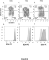


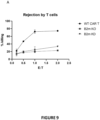


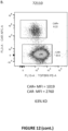
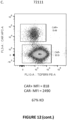


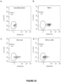
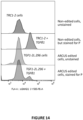


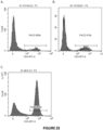


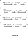


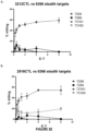
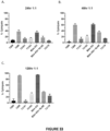

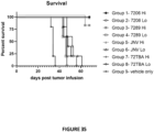

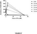


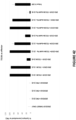

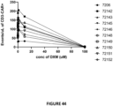
Classifications
-
- C—CHEMISTRY; METALLURGY
- C12—BIOCHEMISTRY; BEER; SPIRITS; WINE; VINEGAR; MICROBIOLOGY; ENZYMOLOGY; MUTATION OR GENETIC ENGINEERING
- C12N—MICROORGANISMS OR ENZYMES; COMPOSITIONS THEREOF; PROPAGATING, PRESERVING, OR MAINTAINING MICROORGANISMS; MUTATION OR GENETIC ENGINEERING; CULTURE MEDIA
- C12N15/00—Mutation or genetic engineering; DNA or RNA concerning genetic engineering, vectors, e.g. plasmids, or their isolation, preparation or purification; Use of hosts therefor
- C12N15/09—Recombinant DNA-technology
- C12N15/11—DNA or RNA fragments; Modified forms thereof; Non-coding nucleic acids having a biological activity
- C12N15/113—Non-coding nucleic acids modulating the expression of genes, e.g. antisense oligonucleotides; Antisense DNA or RNA; Triplex- forming oligonucleotides; Catalytic nucleic acids, e.g. ribozymes; Nucleic acids used in co-suppression or gene silencing
- C12N15/1138—Non-coding nucleic acids modulating the expression of genes, e.g. antisense oligonucleotides; Antisense DNA or RNA; Triplex- forming oligonucleotides; Catalytic nucleic acids, e.g. ribozymes; Nucleic acids used in co-suppression or gene silencing against receptors or cell surface proteins
-
- C—CHEMISTRY; METALLURGY
- C12—BIOCHEMISTRY; BEER; SPIRITS; WINE; VINEGAR; MICROBIOLOGY; ENZYMOLOGY; MUTATION OR GENETIC ENGINEERING
- C12N—MICROORGANISMS OR ENZYMES; COMPOSITIONS THEREOF; PROPAGATING, PRESERVING, OR MAINTAINING MICROORGANISMS; MUTATION OR GENETIC ENGINEERING; CULTURE MEDIA
- C12N5/00—Undifferentiated human, animal or plant cells, e.g. cell lines; Tissues; Cultivation or maintenance thereof; Culture media therefor
- C12N5/06—Animal cells or tissues; Human cells or tissues
- C12N5/0602—Vertebrate cells
- C12N5/0634—Cells from the blood or the immune system
- C12N5/0636—T lymphocytes
-
- C—CHEMISTRY; METALLURGY
- C12—BIOCHEMISTRY; BEER; SPIRITS; WINE; VINEGAR; MICROBIOLOGY; ENZYMOLOGY; MUTATION OR GENETIC ENGINEERING
- C12N—MICROORGANISMS OR ENZYMES; COMPOSITIONS THEREOF; PROPAGATING, PRESERVING, OR MAINTAINING MICROORGANISMS; MUTATION OR GENETIC ENGINEERING; CULTURE MEDIA
- C12N5/00—Undifferentiated human, animal or plant cells, e.g. cell lines; Tissues; Cultivation or maintenance thereof; Culture media therefor
- C12N5/06—Animal cells or tissues; Human cells or tissues
- C12N5/0602—Vertebrate cells
- C12N5/0634—Cells from the blood or the immune system
- C12N5/0635—B lymphocytes
-
- A—HUMAN NECESSITIES
- A61—MEDICAL OR VETERINARY SCIENCE; HYGIENE
- A61K—PREPARATIONS FOR MEDICAL, DENTAL OR TOILETRY PURPOSES
- A61K39/00—Medicinal preparations containing antigens or antibodies
- A61K39/46—Cellular immunotherapy
- A61K39/461—Cellular immunotherapy characterised by the cell type used
- A61K39/4611—T-cells, e.g. tumor infiltrating lymphocytes [TIL], lymphokine-activated killer cells [LAK] or regulatory T cells [Treg]
-
- A—HUMAN NECESSITIES
- A61—MEDICAL OR VETERINARY SCIENCE; HYGIENE
- A61K—PREPARATIONS FOR MEDICAL, DENTAL OR TOILETRY PURPOSES
- A61K39/00—Medicinal preparations containing antigens or antibodies
- A61K39/46—Cellular immunotherapy
- A61K39/463—Cellular immunotherapy characterised by recombinant expression
- A61K39/4631—Chimeric Antigen Receptors [CAR]
-
- A—HUMAN NECESSITIES
- A61—MEDICAL OR VETERINARY SCIENCE; HYGIENE
- A61K—PREPARATIONS FOR MEDICAL, DENTAL OR TOILETRY PURPOSES
- A61K39/00—Medicinal preparations containing antigens or antibodies
- A61K39/46—Cellular immunotherapy
- A61K39/464—Cellular immunotherapy characterised by the antigen targeted or presented
- A61K39/4643—Vertebrate antigens
- A61K39/4644—Cancer antigens
- A61K39/464402—Receptors, cell surface antigens or cell surface determinants
- A61K39/464411—Immunoglobulin superfamily
- A61K39/464412—CD19 or B4
-
- A—HUMAN NECESSITIES
- A61—MEDICAL OR VETERINARY SCIENCE; HYGIENE
- A61K—PREPARATIONS FOR MEDICAL, DENTAL OR TOILETRY PURPOSES
- A61K39/00—Medicinal preparations containing antigens or antibodies
- A61K39/46—Cellular immunotherapy
- A61K39/464—Cellular immunotherapy characterised by the antigen targeted or presented
- A61K39/4643—Vertebrate antigens
- A61K39/4644—Cancer antigens
- A61K39/464402—Receptors, cell surface antigens or cell surface determinants
- A61K39/464416—Receptors for cytokines
- A61K39/464417—Receptors for tumor necrosis factors [TNF], e.g. lymphotoxin receptor [LTR], CD30
-
- A—HUMAN NECESSITIES
- A61—MEDICAL OR VETERINARY SCIENCE; HYGIENE
- A61K—PREPARATIONS FOR MEDICAL, DENTAL OR TOILETRY PURPOSES
- A61K39/00—Medicinal preparations containing antigens or antibodies
- A61K39/46—Cellular immunotherapy
- A61K39/464—Cellular immunotherapy characterised by the antigen targeted or presented
- A61K39/4643—Vertebrate antigens
- A61K39/4644—Cancer antigens
- A61K39/46443—Growth factors
- A61K39/464434—Transforming growth factor [TGF]
-
- A—HUMAN NECESSITIES
- A61—MEDICAL OR VETERINARY SCIENCE; HYGIENE
- A61P—SPECIFIC THERAPEUTIC ACTIVITY OF CHEMICAL COMPOUNDS OR MEDICINAL PREPARATIONS
- A61P35/00—Antineoplastic agents
-
- C—CHEMISTRY; METALLURGY
- C07—ORGANIC CHEMISTRY
- C07K—PEPTIDES
- C07K14/00—Peptides having more than 20 amino acids; Gastrins; Somatostatins; Melanotropins; Derivatives thereof
- C07K14/435—Peptides having more than 20 amino acids; Gastrins; Somatostatins; Melanotropins; Derivatives thereof from animals; from humans
- C07K14/705—Receptors; Cell surface antigens; Cell surface determinants
- C07K14/70503—Immunoglobulin superfamily
- C07K14/7051—T-cell receptor (TcR)-CD3 complex
-
- C—CHEMISTRY; METALLURGY
- C07—ORGANIC CHEMISTRY
- C07K—PEPTIDES
- C07K14/00—Peptides having more than 20 amino acids; Gastrins; Somatostatins; Melanotropins; Derivatives thereof
- C07K14/435—Peptides having more than 20 amino acids; Gastrins; Somatostatins; Melanotropins; Derivatives thereof from animals; from humans
- C07K14/705—Receptors; Cell surface antigens; Cell surface determinants
- C07K14/70503—Immunoglobulin superfamily
- C07K14/70539—MHC-molecules, e.g. HLA-molecules
-
- C—CHEMISTRY; METALLURGY
- C12—BIOCHEMISTRY; BEER; SPIRITS; WINE; VINEGAR; MICROBIOLOGY; ENZYMOLOGY; MUTATION OR GENETIC ENGINEERING
- C12N—MICROORGANISMS OR ENZYMES; COMPOSITIONS THEREOF; PROPAGATING, PRESERVING, OR MAINTAINING MICROORGANISMS; MUTATION OR GENETIC ENGINEERING; CULTURE MEDIA
- C12N15/00—Mutation or genetic engineering; DNA or RNA concerning genetic engineering, vectors, e.g. plasmids, or their isolation, preparation or purification; Use of hosts therefor
- C12N15/09—Recombinant DNA-technology
- C12N15/11—DNA or RNA fragments; Modified forms thereof; Non-coding nucleic acids having a biological activity
- C12N15/111—General methods applicable to biologically active non-coding nucleic acids
-
- C—CHEMISTRY; METALLURGY
- C12—BIOCHEMISTRY; BEER; SPIRITS; WINE; VINEGAR; MICROBIOLOGY; ENZYMOLOGY; MUTATION OR GENETIC ENGINEERING
- C12N—MICROORGANISMS OR ENZYMES; COMPOSITIONS THEREOF; PROPAGATING, PRESERVING, OR MAINTAINING MICROORGANISMS; MUTATION OR GENETIC ENGINEERING; CULTURE MEDIA
- C12N15/00—Mutation or genetic engineering; DNA or RNA concerning genetic engineering, vectors, e.g. plasmids, or their isolation, preparation or purification; Use of hosts therefor
- C12N15/09—Recombinant DNA-technology
- C12N15/11—DNA or RNA fragments; Modified forms thereof; Non-coding nucleic acids having a biological activity
- C12N15/113—Non-coding nucleic acids modulating the expression of genes, e.g. antisense oligonucleotides; Antisense DNA or RNA; Triplex- forming oligonucleotides; Catalytic nucleic acids, e.g. ribozymes; Nucleic acids used in co-suppression or gene silencing
-
- C—CHEMISTRY; METALLURGY
- C12—BIOCHEMISTRY; BEER; SPIRITS; WINE; VINEGAR; MICROBIOLOGY; ENZYMOLOGY; MUTATION OR GENETIC ENGINEERING
- C12N—MICROORGANISMS OR ENZYMES; COMPOSITIONS THEREOF; PROPAGATING, PRESERVING, OR MAINTAINING MICROORGANISMS; MUTATION OR GENETIC ENGINEERING; CULTURE MEDIA
- C12N5/00—Undifferentiated human, animal or plant cells, e.g. cell lines; Tissues; Cultivation or maintenance thereof; Culture media therefor
- C12N5/06—Animal cells or tissues; Human cells or tissues
- C12N5/0602—Vertebrate cells
- C12N5/0634—Cells from the blood or the immune system
- C12N5/0645—Macrophages, e.g. Kuepfer cells in the liver; Monocytes
-
- C—CHEMISTRY; METALLURGY
- C12—BIOCHEMISTRY; BEER; SPIRITS; WINE; VINEGAR; MICROBIOLOGY; ENZYMOLOGY; MUTATION OR GENETIC ENGINEERING
- C12N—MICROORGANISMS OR ENZYMES; COMPOSITIONS THEREOF; PROPAGATING, PRESERVING, OR MAINTAINING MICROORGANISMS; MUTATION OR GENETIC ENGINEERING; CULTURE MEDIA
- C12N5/00—Undifferentiated human, animal or plant cells, e.g. cell lines; Tissues; Cultivation or maintenance thereof; Culture media therefor
- C12N5/06—Animal cells or tissues; Human cells or tissues
- C12N5/0602—Vertebrate cells
- C12N5/0634—Cells from the blood or the immune system
- C12N5/0646—Natural killers cells [NK], NKT cells
-
- A—HUMAN NECESSITIES
- A61—MEDICAL OR VETERINARY SCIENCE; HYGIENE
- A61K—PREPARATIONS FOR MEDICAL, DENTAL OR TOILETRY PURPOSES
- A61K39/00—Medicinal preparations containing antigens or antibodies
- A61K2039/80—Vaccine for a specifically defined cancer
- A61K2039/804—Blood cells [leukemia, lymphoma]
-
- A—HUMAN NECESSITIES
- A61—MEDICAL OR VETERINARY SCIENCE; HYGIENE
- A61K—PREPARATIONS FOR MEDICAL, DENTAL OR TOILETRY PURPOSES
- A61K2239/00—Indexing codes associated with cellular immunotherapy of group A61K39/46
- A61K2239/31—Indexing codes associated with cellular immunotherapy of group A61K39/46 characterized by the route of administration
-
- A—HUMAN NECESSITIES
- A61—MEDICAL OR VETERINARY SCIENCE; HYGIENE
- A61K—PREPARATIONS FOR MEDICAL, DENTAL OR TOILETRY PURPOSES
- A61K2239/00—Indexing codes associated with cellular immunotherapy of group A61K39/46
- A61K2239/38—Indexing codes associated with cellular immunotherapy of group A61K39/46 characterised by the dose, timing or administration schedule
-
- A—HUMAN NECESSITIES
- A61—MEDICAL OR VETERINARY SCIENCE; HYGIENE
- A61K—PREPARATIONS FOR MEDICAL, DENTAL OR TOILETRY PURPOSES
- A61K2239/00—Indexing codes associated with cellular immunotherapy of group A61K39/46
- A61K2239/46—Indexing codes associated with cellular immunotherapy of group A61K39/46 characterised by the cancer treated
- A61K2239/48—Blood cells, e.g. leukemia or lymphoma
-
- C—CHEMISTRY; METALLURGY
- C07—ORGANIC CHEMISTRY
- C07K—PEPTIDES
- C07K2319/00—Fusion polypeptide
- C07K2319/01—Fusion polypeptide containing a localisation/targetting motif
- C07K2319/03—Fusion polypeptide containing a localisation/targetting motif containing a transmembrane segment
-
- C—CHEMISTRY; METALLURGY
- C12—BIOCHEMISTRY; BEER; SPIRITS; WINE; VINEGAR; MICROBIOLOGY; ENZYMOLOGY; MUTATION OR GENETIC ENGINEERING
- C12N—MICROORGANISMS OR ENZYMES; COMPOSITIONS THEREOF; PROPAGATING, PRESERVING, OR MAINTAINING MICROORGANISMS; MUTATION OR GENETIC ENGINEERING; CULTURE MEDIA
- C12N2310/00—Structure or type of the nucleic acid
- C12N2310/10—Type of nucleic acid
- C12N2310/14—Type of nucleic acid interfering N.A.
- C12N2310/141—MicroRNAs, miRNAs
-
- C—CHEMISTRY; METALLURGY
- C12—BIOCHEMISTRY; BEER; SPIRITS; WINE; VINEGAR; MICROBIOLOGY; ENZYMOLOGY; MUTATION OR GENETIC ENGINEERING
- C12N—MICROORGANISMS OR ENZYMES; COMPOSITIONS THEREOF; PROPAGATING, PRESERVING, OR MAINTAINING MICROORGANISMS; MUTATION OR GENETIC ENGINEERING; CULTURE MEDIA
- C12N2310/00—Structure or type of the nucleic acid
- C12N2310/50—Physical structure
- C12N2310/53—Physical structure partially self-complementary or closed
- C12N2310/531—Stem-loop; Hairpin
-
- C—CHEMISTRY; METALLURGY
- C12—BIOCHEMISTRY; BEER; SPIRITS; WINE; VINEGAR; MICROBIOLOGY; ENZYMOLOGY; MUTATION OR GENETIC ENGINEERING
- C12N—MICROORGANISMS OR ENZYMES; COMPOSITIONS THEREOF; PROPAGATING, PRESERVING, OR MAINTAINING MICROORGANISMS; MUTATION OR GENETIC ENGINEERING; CULTURE MEDIA
- C12N2330/00—Production
- C12N2330/50—Biochemical production, i.e. in a transformed host cell
- C12N2330/51—Specially adapted vectors
-
- C—CHEMISTRY; METALLURGY
- C12—BIOCHEMISTRY; BEER; SPIRITS; WINE; VINEGAR; MICROBIOLOGY; ENZYMOLOGY; MUTATION OR GENETIC ENGINEERING
- C12N—MICROORGANISMS OR ENZYMES; COMPOSITIONS THEREOF; PROPAGATING, PRESERVING, OR MAINTAINING MICROORGANISMS; MUTATION OR GENETIC ENGINEERING; CULTURE MEDIA
- C12N2510/00—Genetically modified cells
Definitions
- the invention relates to the field of oncology, cancer immunotherapy, molecular biology and recombinant nucleic acid technology.
- the invention relates to genetically-modified human T cells comprising a microRNA-adapted shRNA (shRNAmiR) molecule that enables stable knockdown of a particular target gene.
- shRNAmiR microRNA-adapted shRNA
- T cell adoptive immunotherapy is a promising approach for cancer treatment.
- the immunotherapy treatment methods disclosed herein utilize isolated human T cells that have been genetically-modified to enhance their specificity for a specific tumor associated antigen. Genetic modification may involve the expression of a chimeric antigen receptor or an exogenous T cell receptor to graft antigen specificity onto the T cell. By contrast to exogenous T cell receptors, chimeric antigen receptors derive their specificity from the variable domains of a monoclonal antibody.
- T cells expressing chimeric antigen receptors induce tumor immunoreactivity in a major histocompatibility complex non-restricted manner.
- T cell adoptive immunotherapy has been utilized as a clinical therapy for a number of cancers, including B cell malignancies (e.g., acute lymphoblastic leukemia, B cell non-Hodgkin lymphoma, acute myeloid leukemia, and chronic lymphocytic leukemia), multiple myeloma, neuroblastoma, glioblastoma, advanced gliomas, ovarian cancer, mesothelioma, melanoma, prostate cancer, pancreatic cancer, and others.
- B cell malignancies e.g., acute lymphoblastic leukemia, B cell non-Hodgkin lymphoma, acute myeloid leukemia, and chronic lymphocytic leukemia
- multiple myeloma e.g., neuroblastoma, glioblastoma, advanced gliomas, ovarian cancer, mesothelioma, melanoma, prostate cancer, pancreatic cancer, and others.
- CAR T cells expressing an endogenous T cell receptor may recognize major and minor histocompatibility antigens following administration to an allogeneic patient, which can lead to the development of graft-versus-host-disease (GVHD).
- GVHD graft-versus-host-disease
- clinical trials have largely focused on the use of autologous CAR T cells, wherein a patient's T cells are isolated, genetically-modified to incorporate a chimeric antigen receptor, and then re-infused into the same patient.
- An autologous approach provides immune tolerance to the administered CAR T cells; however, this approach is constrained by both the time and expense necessary to produce patient-specific CAR T cells after a patient's cancer has been diagnosed.
- B2M beta-2 microglobulin
- MHC major histocompatibility complex
- the shRNA-coding sequence was included on a cassette that also comprised a CAR coding sequence, allowing for the production of CAR T cells that were TCR-negative, CAR-positive, and had a partial knockdown of cell-surface B2M.
- the data in this project demonstrated that these CAR T cells were indeed less susceptible to NK cell killing than CAR T cells that exhibited a complete knockout of B2M.
- the present invention provides genetically-modified human T cells (and populations thereof) expressing a microRNA-adapted shRNA (shRNAmiR) that reduces the expression of beta-2 microglobulin (B2M).
- shRNAmiRs for knocking down the expression of a target protein allows for stable knockdown of protein expression, which is ideal for those target proteins for which knockdown, and not knockout, is preferred.
- immune cells that express beta-2 microglobulin (B2M) at reduced levels via expression of B2M-targeted shRNAmiRs are less sensitive to cytolysis by natural killer (NK) cells than those cells in which B2M expression has been knocked out by gene inactivation.
- the invention provides a genetically-modified human T cell comprising in its genome a cassette comprising, from 5' to 3': (a) a promoter; (b) a nucleic acid sequence encoding a chimeric antigen receptor (CAR); (c) a 2A sequence; (d) a nucleic acid sequence encoding an HLA class I histocompatibility antigen, alpha chain E (HLA-E) fusion protein, wherein said HLA-E fusion protein comprises an amino acid sequence of SEQ ID NO:66, and wherein an intron sequence is positioned within said nucleic acid sequence encoding said HLA-E fusion protein, wherein said intron sequence comprises a nucleic acid sequence of SEQ ID NO:69, and wherein a nucleic acid sequence encoding a microRNA-adapted shRNA (shRNAmiR) is positioned within said intron sequence, wherein said nucleic acid sequence encoding said shRNAmiR comprises SEQ ID NO:46
- the shRNAmiR is expressed in the genetically-modified human T cell and reduces expression of B2M in the genetically-modified human T cell. A reduction in B2M expression is mediated by the binding of the shRNAmiR guide sequence to mRNA encoding B2M.
- the shRNAmiR comprises, from 5' to 3': (a) a 5' miR scaffold domain; (b) a 5' miR basal stem domain; (c) a passenger strand; (d) a miR loop domain; (e) a guide strand; (f) a 3' miR basal stem domain; and (g) a 3' miR scaffold domain.
- the miR loop domain is a miR-30a loop domain of SEQ ID NO: 3.
- the termination signal is a polyA sequence or a bovine growth hormone (BGH) termination signal.
- the polyA sequence comprises a nucleic acid sequence having at least 80%, at least 85%, at least 90%, at least 95%, or more, sequence identity to SEQ ID NO: 68.
- the polyA sequence comprises a nucleic acid sequence of SEQ ID NO: 68.
- the BGH termination signal comprises a nucleic acid sequence having least 80%, at least 85%, at least 90%, at least 95%, or more, sequence identity to SEQ ID NO: 71.
- the BGH termination signal comprises a nucleic acid sequence of SEQ ID NO: 71.
- the promoter comprises a nucleic acid sequence having at least 80%, at least 85%, at least 90%, at least 95%, or more, sequence identity to SEQ ID NO: 67.
- the promoter comprises a nucleic acid sequence of SEQ ID NO: 67.
- the 2A sequence is a P2A/furin site comprising a nucleic acid sequence having at least 80%, at least 85%, at least 90%,at least 95%, or more, sequence identity to SEQ ID NO: 70.
- the 2A sequence is a P2A/furin site comprising a nucleic acid sequence of SEQ ID NO: 70.
- the CAR comprises a signal peptide comprising an amino acid sequence having at least 80%, at least 85%, at least 90%, at least 95%, or more, sequence identity to SEQ ID NO: 73.
- the CAR comprises a signal peptide comprising an amino acid sequence of SEQ ID NO: 73.
- the genetically-modified human T cell comprises in its genome a cassette comprising, from 5' to 3': (a) a promoter comprising a nucleic acid sequence having at least 80%, at least 85%, at least 90%, at least 95%, or more, sequence identity to SEQ ID NO: 67; (b) the nucleic acid sequence encoding the CAR, wherein the CAR comprises a signal peptide comprising an amino acid sequence having at least 80%, at least 85%, at least 90%, at least 95%, or more, sequence identity to SEQ ID NO: 73; (c) a P2A/furin site comprising a nucleic acid sequence having at least 80%, at least 85%, at least 90%, at least 95%, or more, sequence identity to SEQ ID NO: 70; (d) the nucleic acid sequence encoding the HLA-E fusion protein, wherein the HLA-E fusion protein comprises an amino acid sequence of SEQ ID NO: 66, and wherein an intron sequence
- the genetically-modified human T cell comprises in its genome a cassette comprising, from 5' to 3': (a) a promoter comprising a nucleic acid sequence of SEQ ID NO: 67; (b) the nucleic acid sequence encoding the CAR, wherein the CAR comprises a signal peptide comprising an amino acid sequence of SEQ ID NO: 73; (c) a P2A/furin site comprising a nucleic acid sequence of SEQ ID NO: 70; (d) the nucleic acid sequence encoding the HLA-E fusion protein, wherein the HLA-E fusion protein comprises an amino acid sequence of SEQ ID NO: 66, and wherein an intron sequence is positioned within the nucleic acid sequence encoding the HLA-E fusion protein, wherein the intron sequence comprises a nucleic acid sequence of SEQ ID NO: 69, and wherein the nucleic acid sequence encoding the shRNAmiR is positioned within the intron sequence; and (e) a promote
- the genetically-modified human T cell comprises in its genome a cassette comprising a nucleic acid sequence having at least 80%, at least 85%, at least 90%, at least 95%, or more, sequence identity to SEQ ID NO: 74, wherein the cassette is positioned in the genome within a TCR alpha constant region gene.
- the genetically-modified human T cell comprises in its genome a cassette comprising a nucleic acid sequence of SEQ ID NO: 74, wherein the cassette is positioned in the genome within a TCR alpha constant region gene.
- the cassette comprises two or more nucleic acids encoding shRNAmiRs.
- the two or more nucleic acids can encode the same shRNAmiR.
- the two or more nucleic acids can encode different shRNAmiRs that reduce the expression of the same target protein.
- the two or more nucleic acids encode different shRNAmiRs that reduce the expression of different target proteins.
- the cassette can comprise two or more nucleic acids encoding different shRNAmiRs described herein.
- the cassette can comprise a nucleic acid sequence encoding a shRNAmiR that reduces the expression of B2M, and a nucleic acid sequence encoding a shRNAmiR that reduces the expression of CD52.
- the target protein is beta-2 microglobulin.
- cell surface expression of beta-2 microglobulin is reduced by at least about 10%, about 20%, about 30%, about 40%, about 50%, about 55%, about 60%, about 65%, about 70%, about 75%, about 80%, about 85%, about 90%, about 95%, or up to about 99% compared to a control cell.
- expression of MHC class I molecules is reduced on the cell surface by at least about 10%, about 20%, about 30%, about 40%, about 50%, about 55%, about 60%, about 65%, about 70%, about 75%, about 80%, about 85%, about 90%, about 95%, or up to about 99% compared to a control cell.
- the genetically-modified human T cell has reduced allogenicity compared to a control cell.
- a method of immunotherapy for treating a disease in a subject in need thereof comprising administering to the subject an effective amount of a pharmaceutical composition comprising a pharmaceutically-acceptable carrier and a plurality of genetically-modified human T cells described herein.
- the method is an immunotherapy for the treatment of a cancer in a subject in need thereof, wherein the CAR comprises an extracellular ligand-binding domain having specificity for a tumor-specific antigen.
- the genetically-modified human T cell has no detectable cell-surface expression of an endogenous TCR (e.g., an alpha/beta TCR).
- the cancer is selected from the group consisting of a cancer of carcinoma, lymphoma, sarcoma, blastomas, and leukemia.
- the cancer is selected from the group consisting of a cancer of B cell origin, breast cancer, gastric cancer, neuroblastoma, osteosarcoma, lung cancer, melanoma, prostate cancer, colon cancer, renal cell carcinoma, ovarian cancer, rhabdomyosarcoma, leukemia, and Hodgkin lymphoma.
- the cancer of B cell origin is selected from the group consisting of B-lineage acute lymphoblastic leukemia, B cell chronic lymphocytic leukemia, B cell non-Hodgkin lymphoma, and multiple myeloma.
- the subject can be a mammal, such as a human.
- a can mean one or more than one.
- a cell can mean a single cell or a multiplicity of cells.
- exogenous or heterologous in reference to a nucleotide sequence or amino acid sequence are intended to mean a sequence that is purely synthetic, that originates from a foreign species, or, if from the same species, is substantially modified from its native form in composition and/or genomic locus by deliberate human intervention.
- endogenous in reference to a nucleotide sequence or protein is intended to mean a sequence or protein that is naturally comprised within or expressed by a cell.
- nuclease and “endonuclease” are used interchangeably to refer to naturally-occurring or engineered enzymes which cleave a phosphodiester bond within a polynucleotide chain.
- RNA short hairpin RNA
- shRNA refers to an artificial RNA molecule comprising a hairpin that can be used to silence gene expression via RNA interference.
- miRNA or “microRNA” or “miR” refers to mature microRNAs (miRNAs) that are endogenously encoded ⁇ 22 nt long RNAs that post-transcriptionally reduce the expression of target genes. miRNAs are found in plants, animals, and some viruses and are generally expressed in a highly tissue- or developmental-stage-specific fashion.
- a “stem-loop structure” refers to a nucleic acid having a secondary structure that includes a region of nucleotides which are known or predicted to form a double strand (stem portion) that is linked on one side by a region of predominantly single-stranded nucleotides (loop portion), In some cases, the loop may also be very short and thereby not be recognized by Dicer, leading to Dicer-independent shRNAs (comparable to the endogenous miR0431).
- the term “hairpin” is also used herein to refer to stem-loop structures.
- the actual primary sequence of nucleotides within the stem-loop structure is not critical to the practice of the description as long as the secondary structure is present.
- the secondary structure does not require exact base-pairing.
- the stem may include one or more base mismatches.
- the base-pairing may be exact (i.e., not include any mismatches).
- shRNAmiR and "microRNA-adapted shRNA” refer to shRNA sequences embedded within a microRNA scaffold.
- a shRNAmiR molecule mimics naturally-occurring pri-miRNA molecules in that they comprise a hairpin flanked by sequences necessary for efficient processing and can be processed by the Drosha enzyme into pre-miRNAs, exported into the cytoplasm, and cleaved by Dicer, after which the mature miRNA can enter the RISC.
- the microRNA scaffold can be derived from naturally-occurring microRNA, pre-miRNAs, or pri-miRNAs or variants thereof.
- the shRNA sequences which the shRNAmiR is based upon is of a different length from miRNAs (which are 22 nucleotides long) and the miRNA scaffold must therefore be modified in order to accommodate the longer or shorter shRNA sequence length.
- microRNA flanking sequences refers to nucleotide sequences comprising microRNA processing elements.
- MicroRNA processing elements are the minimal nucleic acid sequences which contribute to the production of mature microRNA from primary microRNA or precursor microRNA. Often these elements are located within a 40 nucleotide sequence that flanks a microRNA stem-loop structure. In some instances, the microRNA processing elements are found within a stretch of nucleotide sequences of between 5 and 4,000 nucleotides in length that flank a microRNA stem-loop structure.
- MicroRNA flanking sequences used in the shRNAmiR molecules can be naturally-occurring sequences flanking naturally-occurring microRNA or can be variants thereof. MicroRNA flanking sequences include miR scaffold domains and miR basal stem domains.
- shRNAmiR molecules used in the presently disclosed compositions and methods can comprise in the 5' to 3' direction: (a) a 5' miR scaffold domain; (b) a 5' miR basal stem domain; (c) a passenger strand; (d) a miR loop domain; (e) a guide strand; (f) a 3' miR basal stem domain; and (g) a 3' miR scaffold domain.
- miR scaffold domain as it relates to a shRNAmiR refers to a nucleotide sequence that can flank either the 5' or 3' end of a microRNA or shRNA in a shRNAmiR molecule and can be derived from a naturally-occurring microRNA flanking sequence or a variant thereof.
- the miR basal stem domain sequence separates the shRNA sequence (passenger and guide strand, and miR loop domain) and the scaffold domains.
- the 5' miR scaffold domain can comprise a restriction enzyme (e.g., type IIS restriction enzyme) recognition sequence at or near its 3' end and the 3' miR scaffold domain can comprise a restriction enzyme recognition sequence at or near its 5' end, thus facilitating the insertion of a shRNA sequence.
- the secondary structure of the miR scaffold domain is more important than the actual sequence thereof.
- miR basal stem domain refers to sequences immediately flanking the passenger and guide strand sequences that comprise the base of the hairpin stem below the passenger:guide duplex.
- the 5' and 3' miR basal stem domains are complementary (fully or partially) in sequence to one another.
- the 5' and 3' miR basal stem domains comprise sequences that when hybridized together, form two mismatch bubbles, each comprising one or two mismatched base pairs.
- the term "passenger strand" as it relates to a shRNAmiR refers to the sequence of the shRNAmiR, which is complementary (fully or partially) to the guide sequence.
- guide strand refers to the sequence of the shRNAmiR that has complementarity (full or partial) with the target mRNA sequence for which a reduction in expression is desired.
- a "miR loop domain" as it relates to a shRNAmiR refers to the single-stranded loop sequence at one end of the passenger: guide duplex of the shRNAmiR.
- the miR loop domain can be derived from a naturally-occurring pre-microRNA loop sequence or a variant thereof.
- cleave or “cleavage” refer to the hydrolysis of phosphodiester bonds within the backbone of a recognition sequence within a target sequence that results in a double-stranded break within the target sequence, referred to herein as a "cleavage site”.
- the term "meganuclease” refers to an endonuclease that binds double-stranded DNA at a recognition sequence that is greater than 12 base pairs. In some embodiments, the recognition sequence for a meganuclease of the present disclosure is 22 base pairs.
- a meganuclease can be an endonuclease that is derived from I-CreI (SEQ ID NO: 123), and can refer to an engineered variant of I-Crel that has been modified relative to natural I-CreI with respect to, for example, DNA-binding specificity, DNA cleavage activity, DNA-binding affinity, or dimerization properties.
- a meganuclease as used herein binds to double-stranded DNA as a heterodimer.
- a meganuclease may also be a "single-chain meganuclease" in which a pair of DNA-binding domains is joined into a single polypeptide using a peptide linker.
- homing endonuclease is synonymous with the term “meganuclease.”
- Meganucleases of the present disclosure are substantially non-toxic when expressed in cells, particularly in human immune cells, such that cells can be transfected and maintained at 37°C without observing deleterious effects on cell viability or significant reductions in meganuclease cleavage activity when measured using the methods described herein.
- single-chain meganuclease refers to a polypeptide comprising a pair of nuclease subunits joined by a linker.
- a single-chain meganuclease has the organization: N-terminal subunit - Linker - C-terminal subunit.
- the two meganuclease subunits will generally be non-identical in amino acid sequence and will bind non-identical DNA sequences.
- single-chain meganucleases typically cleave pseudo-palindromic or non-palindromic recognition sequences.
- a single-chain meganuclease may be referred to as a "single-chain heterodimer” or “single-chain heterodimeric meganuclease” although it is not, in fact, dimeric.
- the term “meganuclease” can refer to a dimeric or single-chain meganuclease.
- linker refers to an exogenous peptide sequence used to join two nuclease subunits into a single polypeptide.
- a linker may have a sequence that is found in natural proteins or may be an artificial sequence that is not found in any natural protein.
- a linker may be flexible and lacking in secondary structure or may have a propensity to form a specific three-dimensional structure under physiological conditions.
- a linker can include, without limitation, those encompassed by U.S. Patent Nos. 8,445,251 , 9,340,777 , 9,434,931 , and 10,041,053 .
- a linker may have at least 80%, at least 85%, at least 90%, at least 91%, at least 92%, at least 93%, at least 94%, at least 95%, at least 96%, at least 97%, at least 98%, at least 99%, or more, sequence identity to residues 154-195 of SEQ ID NO: 65.
- a linker may have an amino acid sequence comprising residues 154-195 of SEQ ID NO: 65.
- TALEN refers to an endonuclease comprising a DNA-binding domain comprising a plurality of TAL domain repeats fused to a nuclease domain or an active portion thereof from an endonuclease or exonuclease, including but not limited to a restriction endonuclease, homing endonuclease, S1 nuclease, mung bean nuclease, pancreatic DNAse I, micrococcal nuclease, and yeast HO endonuclease. See, for example, Christian et al. (2010) Genetics 186:757-761 .
- Nuclease domains useful for the design of TALENs include those from a Type IIs restriction endonuclease, including but not limited to FokI, FoM, StsI, HhaI, HindIII, Nod, BbvCI, EcoRI, BglI, and AlwI. Additional Type IIs restriction endonucleases are described in International Publication No. WO 2007/014275 .
- the nuclease domain of the TALEN is a FokI nuclease domain or an active portion thereof.
- TAL domain repeats can be derived from the TALE (transcription activator-like effector) family of proteins used in the infection process by plant pathogens of the Xanthomonas genus.
- TAL domain repeats are 33-34 amino acid sequences with divergent 12th and 13th amino acids. These two positions, referred to as the repeat variable dipeptide (RVD), are highly variable and show a strong correlation with specific nucleotide recognition. Each base pair in the DNA target sequence is contacted by a single TAL repeat with the specificity resulting from the RVD.
- the TALEN comprises 16-22 TAL domain repeats. DNA cleavage by a TALEN requires two DNA recognition regions (i.e., "half-sites") flanking a nonspecific central region (i.e., the "spacer").
- spacer in reference to a TALEN refers to the nucleic acid sequence that separates the two nucleic acid sequences recognized and bound by each monomer constituting a TALEN.
- the TAL domain repeats can be native sequences from a naturally-occurring TALE protein or can be redesigned through rational or experimental means to produce a protein that binds to a predetermined DNA sequence (see, for example, Boch et al. (2009) Science 326(5959):1509-1512 and Moscou and Bogdanove (2009) Science 326(5959):1501 ). See also, U.S. Publication No. 20110145940 and International Publication No.
- each nuclease (e.g., FokI) monomer can be fused to a TAL effector sequence that recognizes and binds a different DNA sequence, and only when the two recognition sites are in close proximity do the inactive monomers come together to create a functional enzyme.
- TALEN can refer to a single TALEN protein or, alternatively, a pair of TALEN proteins (i.e., a left TALEN protein and a right TALEN protein) which bind to the upstream and downstream half-sites adjacent to the TALEN spacer sequence and work in concert to generate a cleavage site within the spacer sequence.
- upstream and downstream half-sites can be identified using a number of programs known in the art ( Kornel Labun; Tessa G. Montague; James A. Gagnon; Summer B. Thyme; Eivind Valen. (2016).
- CHOPCHOP v2 a web tool for the next generation of CRISPR genome engineering. Nucleic Acids Research; doi:10.1093/nar/gkw398 ; Tessa G. Montague; Jose M. Cruz; James A. Gagnon; George M. Church; Eivind Valen. (2014). CHOPCHOP: a CRISPR/Cas9 and TALEN web tool for genome editing. Nucleic Acids Res. 42. W401-W407 ). It is also understood that a TALEN recognition sequence can be defined as the DNA binding sequence (i.e., half-site) of a single TALEN protein or, alternatively, a DNA sequence comprising the upstream half-site, the spacer sequence, and the downstream half-site.
- compact TALEN refers to an endonuclease comprising a DNA-binding domain with one or more TAL domain repeats fused in any orientation to any portion of the I-TevI homing endonuclease or any of the endonucleases listed in Table 2 in U.S. Application No. 20130117869 , including but not limited to MmeI, EndA, End1, I-BasI, I-TevII, I-TevIII, I-TwoI, MspI, MvaI, NucA, and NucM.
- Compact TALENs do not require dimerization for DNA processing activity, alleviating the need for dual target sites with intervening DNA spacers.
- the compact TALEN comprises 16-22 TAL domain repeats.
- megaTAL refers to a single-chain endonuclease comprising a transcription activator-like effector (TALE) DNA binding domain with an engineered, sequence-specific homing endonuclease.
- TALE transcription activator-like effector
- zinc finger nuclease or “ZFN” refers to a chimeric protein comprising a zinc finger DNA-binding domain fused to a nuclease domain from an endonuclease or exonuclease, including but not limited to a restriction endonuclease, homing endonuclease, S1 nuclease, mung bean nuclease, pancreatic DNAse I, micrococcal nuclease, and yeast HO endonuclease.
- endonuclease including but not limited to a restriction endonuclease, homing endonuclease, S1 nuclease, mung bean nuclease, pancreatic DNAse I, micrococcal nuclease, and yeast HO endonuclease.
- Nuclease domains useful for the design of zinc finger nucleases include those from a Type IIs restriction endonuclease, including but not limited to FokI, FoM, and StsI restriction enzyme. Additional Type IIs restriction endonucleases are described in International Publication No. WO 2007/014275 .
- the structure of a zinc finger domain is stabilized through coordination of a zinc ion.
- DNA binding proteins comprising one or more zinc finger domains bind DNA in a sequence-specific manner.
- the zinc finger domain can be a native sequence or can be redesigned through rational or experimental means to produce a protein which binds to a pre-determined DNA sequence ⁇ 18 basepairs in length, comprising a pair of nine basepair half-sites separated by 2-10 basepairs. See, for example, U.S. Pat. Nos. 5,789,538 , 5,925,523 , 6,007,988 , 6,013,453 , 6,200,759 , and International Publication Nos.
- the DNA binding domains typically recognize an 18-bp recognition sequence comprising a pair of nine basepair "half-sites" separated by a 2-10 basepair "spacer sequence", and cleavage by the nuclease creates a blunt end or a 5' overhang of variable length (frequently four basepairs).
- zinc finger nuclease can refer to a single zinc finger protein or, alternatively, a pair of zinc finger proteins (i.e., a left ZFN protein and a right ZFN protein) that bind to the upstream and downstream half-sites adjacent to the zinc finger nuclease spacer sequence and work in concert to generate a cleavage site within the spacer sequence.
- upstream and downstream half-sites can be identified using a number of programs known in the art ( Mandell JG, Barbas CF 3rd. Zinc Finger Tools: custom DNA-binding domains for transcription factors and nucleases. Nucleic Acids Res.
- a zinc finger nuclease recognition sequence can be defined as the DNA binding sequence (i.e., half-site) of a single zinc finger nuclease protein or, alternatively, a DNA sequence comprising the upstream half-site, the spacer sequence, and the downstream half-site.
- CRISPR nuclease or “CRISPR system nuclease” refers to a CRISPR (clustered regularly interspaced short palindromic repeats)-associated (Cas) endonuclease or a variant thereof, such as Cas9, that associates with a guide RNA that directs nucleic acid cleavage by the associated endonuclease by hybridizing to a recognition site in a polynucleotide.
- the CRISPR nuclease is a class 2 CRISPR enzyme.
- the CRISPR nuclease is a class 2, type II enzyme, such as Cas9.
- the CRISPR nuclease is a class 2, type V enzyme, such as Cpf1.
- the guide RNA comprises a direct repeat and a guide sequence (often referred to as a spacer in the context of an endogenous CRISPR system), which is complementary to the target recognition site.
- the CRISPR system further comprises a tracrRNA (trans-activating CRISPR RNA) that is complementary (fully or partially) to the direct repeat sequence (sometimes referred to as a tracr-mate sequence) present on the guide RNA.
- the CRISPR nuclease can be mutated with respect to a corresponding wild-type enzyme such that the enzyme lacks the ability to cleave one strand of a target polynucleotide, functioning as a nickase, cleaving only a single strand of the target DNA.
- CRISPR enzymes that function as a nickase include Cas9 enzymes with a D10A mutation within the RuvC I catalytic domain, or with a H840A, N854A, or N863A mutation. Given a predetermined DNA locus, recognition sequences can be identified using a number of programs known in the art ( Kornel Labun; Tessa G.
- CHOPCHOP v2 a web tool for the next generation of CRISPR genome engineering. Nucleic Acids Research; doi:10.1093/nar/gkw398 ; Tessa G. Montague; Jose M. Cruz; James A. Gagnon; George M. Church; Eivind Valen. (2014). CHOPCHOP: a CRISPR/Cas9 and TALEN web tool for genome editing. Nucleic Acids Res. 42. W401-W407 ).
- template nucleic acid refers to a nucleic acid (i.e., a polynucleotide) that is desired to be inserted into a cleavage site within a cell's genome.
- the terms “recombinant” or “engineered,” with respect to a protein means having an altered amino acid sequence as a result of the application of genetic engineering techniques to nucleic acids that encode the protein and cells or organisms that express the protein.
- the term “recombinant” or “engineered” means having an altered nucleic acid sequence as a result of the application of genetic engineering techniques. Genetic engineering techniques include, but are not limited to, PCR and DNA cloning technologies; transfection, transformation, and other gene transfer technologies; homologous recombination; site-directed mutagenesis; and gene fusion.
- a protein having an amino acid sequence identical to a naturally-occurring protein, but produced by cloning and expression in a heterologous host is not considered recombinant or engineered.
- wild-type refers to the most common naturally occurring allele (i.e., polynucleotide sequence) in the allele population of the same type of gene, wherein a polypeptide encoded by the wild-type allele has its original functions.
- wild-type also refers to a polypeptide encoded by a wild-type allele. Wild-type alleles (i.e., polynucleotides) and polypeptides are distinguishable from mutant or variant alleles and polypeptides, which comprise one or more mutations and/or substitutions relative to the wild-type sequence(s).
- Wild-type nucleases are distinguishable from recombinant or non-naturally-occurring nucleases.
- the term "wild-type" can also refer to a cell, an organism, and/or a subject which possesses a wild-type allele of a particular gene, or a cell, an organism, and/or a subject used for comparative purposes.
- the term “genetically-modified” refers to a cell or organism in which, or in an ancestor of which, a genomic DNA sequence has been deliberately modified by recombinant technology. As used herein, the term “genetically-modified” encompasses the term "transgenic.”
- modification means any insertion, deletion, or substitution of an amino acid residue in the recombinant sequence relative to a reference sequence (e.g., a wild-type or a native sequence).
- a recognition sequence or “recognition site” refers to a DNA sequence that is bound and cleaved by a nuclease.
- a recognition sequence comprises a pair of inverted, 9 basepair "half sites" which are separated by four basepairs.
- the N-terminal domain of the protein contacts a first half-site and the C-terminal domain of the protein contacts a second half-site. Cleavage by a meganuclease produces four basepair 3' overhangs.
- “Overhangs,” or “sticky ends” are short, single-stranded DNA segments that can be produced by endonuclease cleavage of a double-stranded DNA sequence.
- the overhang comprises bases 10-13 of the 22 basepair recognition sequence.
- the recognition sequence comprises a first CNNNGN sequence that is recognized by the I-TevI domain, followed by a non-specific spacer 4-16 basepairs in length, followed by a second sequence 16-22 bp in length that is recognized by the TAL-effector domain (this sequence typically has a 5' T base).
- Cleavage by a compact TALEN produces two basepair 3' overhangs.
- the recognition sequence is the sequence, typically 16-24 basepairs, to which the guide RNA binds to direct cleavage. Full complementarity between the guide sequence and the recognition sequence is not necessarily required to effect cleavage.
- Cleavage by a CRISPR nuclease can produce blunt ends (such as by a class 2, type II CRISPR nuclease) or overhanging ends (such as by a class 2, type V CRISPR nuclease), depending on the CRISPR nuclease.
- cleavage by the CRISPR complex comprising the same will result in 5' overhangs and in certain embodiments, 5 nucleotide 5' overhangs.
- Each CRISPR nuclease enzyme also requires the recognition of a PAM (protospacer adjacent motif) sequence that is near the recognition sequence complementary to the guide RNA.
- PAM protospacer adjacent motif
- the precise sequence, length requirements for the PAM, and distance from the target sequence differ depending on the CRISPR nuclease enzyme, but PAMs are typically 2-5 base pair sequences adjacent to the target/recognition sequence.
- PAM sequences for particular CRISPR nuclease enzymes are known in the art (see, for example, U.S. Patent No.
- PAM sequences for novel or engineered CRISPR nuclease enzymes can be identified using methods known in the art, such as a PAM depletion assay (see, for example, Karvelis et al. (2017) Methods 121-122:3-8 ).
- a PAM depletion assay see, for example, Karvelis et al. (2017) Methods 121-122:3-8 .
- the DNA binding domains typically recognize an 18-bp recognition sequence comprising a pair of nine basepair "half-sites" separated by 2-10 basepairs and cleavage by the nuclease creates a blunt end or a 5' overhang of variable length (frequently four basepairs).
- target site or "target sequence” refers to a region of the chromosomal DNA of a cell comprising a recognition sequence for a nuclease.
- DNA-binding affinity or “binding affinity” means the tendency of a nuclease to non-covalently associate with a reference DNA molecule (e.g., a recognition sequence or an arbitrary sequence). Binding affinity is measured by a dissociation constant, Kd. As used herein, a nuclease has "altered” binding affinity if the Kd of the nuclease for a reference recognition sequence is increased or decreased by a statistically significant percent change relative to a reference nuclease.
- the term "specificity" means the ability of a nuclease to bind and cleave double-stranded DNA molecules only at a particular sequence of base pairs referred to as the recognition sequence, or only at a particular set of recognition sequences.
- the set of recognition sequences will share certain conserved positions or sequence motifs but may be degenerate at one or more positions.
- a highly-specific nuclease is capable of cleaving only one or a very few recognition sequences. Specificity can be determined by any method known in the art.
- homologous recombination refers to the natural, cellular process in which a double-stranded DNA-break is repaired using a homologous DNA sequence as the repair template (see, e.g. Cahill et al. (2006), Front. Biosci. 11:1958-1976 ).
- the homologous DNA sequence may be an endogenous chromosomal sequence or an exogenous nucleic acid that was delivered to the cell.
- non-homologous end-joining refers to the natural, cellular process in which a double-stranded DNA-break is repaired by the direct joining of two non-homologous DNA segments (see, e.g. Cahill et al. (2006), Front. Biosci. 11:1958-1976 ).
- DNA repair by non-homologous end-joining is error-prone and frequently results in the untemplated addition or deletion of DNA sequences at the site of repair. In some instances, cleavage at a target recognition sequence results in NHEJ at a target recognition site.
- Nuclease-induced cleavage of a target site in the coding sequence of a gene followed by DNA repair by NHEJ can introduce mutations into the coding sequence, such as frameshift mutations, that disrupt gene function.
- engineered nucleases can be used to effectively knock-out a gene in a population of cells.
- the term “disrupted” or “disrupts” or “disrupts expression” or “disrupting a target sequence” refers to the introduction of a mutation (e.g., frameshift mutation) that interferes with the gene function and prevents expression and/or function of the polypeptide/expression product encoded thereby.
- a mutation e.g., frameshift mutation
- nuclease-mediated disruption of a gene can result in the expression of a truncated protein and/or expression of a protein that does not retain its wild-type function.
- introduction of a template nucleic acid into a gene can result in no expression of an encoded protein, expression of a truncated protein, and/or expression of a protein that does not retain its wild-type function.
- chimeric antigen receptor refers to an engineered receptor that confers or grafts specificity for an antigen onto an immune effector cell (e.g., a human T cell).
- a chimeric antigen receptor comprises at least an extracellular ligand-binding domain or moiety, a transmembrane domain, and an intracellular domain that comprises one or more signaling domains and/or co-stimulatory domains.
- the extracellular ligand-binding domain or moiety is an antibody, or antibody fragment.
- antibody fragment can refer to at least one portion of an antibody, that retains the ability to specifically interact with (e.g., by binding, steric hindrance, stabilizing/destabilizing, spatial distribution) an epitope of an antigen.
- antibody fragments include, but are not limited to, Fab, Fab', F(ab')2, Fv fragments, scFv antibody fragments, disulfide-linked Fvs (sdFv), a Fd fragment consisting of the VH and CH1 domains, linear antibodies, single domain antibodies such as sdAb (either VL or VH), camelid VHH domains, multi-specific antibodies formed from antibody fragments such as a bivalent fragment comprising two Fab fragments linked by a disulfide bridge at the hinge region, and an isolated CDR or other epitope binding fragments of an antibody.
- An antigen binding fragment can also be incorporated into single domain antibodies, maxibodies, minibodies, nanobodies, intrabodies, diabodies, triabodies, tetrabodies, v-NAR and bis-scFv (see, e.g., Hollinger and Hudson, Nature Biotechnology 23:1126-1136, 2005 ).
- Antigen binding fragments can also be grafted into scaffolds based on polypeptides such as a fibronectin type III (Fn3) (see U.S. Pat. No. 6,703,199 , which describes fibronectin polypeptide minibodies).
- Fn3 fibronectin type III
- the extracellular ligand-binding domain or moiety is in the form of a single-chain variable fragment (scFv) derived from a monoclonal antibody, which provides specificity for a particular epitope or antigen (e.g., an epitope or antigen preferentially present on the surface of a cell, such as a cancer cell or other disease-causing cell or particle).
- scFv single-chain variable fragment
- the scFv is attached via a linker sequence.
- the scFv is murine, humanized, or fully human.
- the extracellular ligand-binding domain of a chimeric antigen receptor can also comprise an autoantigen (see, Payne et al. (2016), Science 353 (6295): 179-184 ), that can be recognized by autoantigen-specific B cell receptors on B lymphocytes, thus directing T cells to specifically target and kill autoreactive B lymphocytes in antibody-mediated autoimmune diseases.
- CARs can be referred to as chimeric autoantibody receptors (CAARs), and their use is encompassed by the invention.
- the extracellular ligand-binding domain of a chimeric antigen receptor can also comprise a naturally-occurring ligand for an antigen of interest, or a fragment of a naturally-occurring ligand which retains the ability to bind the antigen of interest.
- the intracellular stimulatory domain can include one or more cytoplasmic signaling domains that transmit an activation signal to the T cell following antigen binding.
- cytoplasmic signaling domains can include, without limitation, a CD3 zeta signaling domain.
- the intracellular stimulatory domain can also include one or more intracellular co-stimulatory domains that transmit a proliferative and/or cell-survival signal after ligand binding.
- intracellular co-stimulatory domains can be any of those known in the art and can include, without limitation, those co-stimulatory domains disclosed in WO 2018/067697 including, for example, Novel 6.
- Further examples of co-stimulatory domains can include 4-1BB (CD137), CD27, CD28, CD8, OX40, CD30, CD40, PD-1, ICOS, lymphocyte function-associated antigen-1 (LFA-1), CD2, CD7, LIGHT, NKG2C, B7-H3, a ligand that specifically binds with CD83, or any combination thereof.
- a chimeric antigen receptor further includes additional structural elements, including a transmembrane domain that is attached to the extracellular ligand-binding domain via a hinge or spacer sequence.
- the transmembrane domain can be derived from any membrane-bound or transmembrane protein.
- the transmembrane polypeptide can be a subunit of the T-cell receptor (e.g., an ⁇ , ⁇ , ⁇ or ⁇ , polypeptide constituting CD3 complex), IL2 receptor p55 (a chain), p75 ( ⁇ chain) or ⁇ chain, subunit chain of Fc receptors (e.g., Fcy receptor III) or CD proteins such as the CD8 alpha chain.
- the transmembrane domain is a CD8 alpha domain.
- the transmembrane domain can be synthetic and can comprise predominantly hydrophobic residues such as leucine and valine.
- the hinge region refers to any oligo- or polypeptide that functions to link the transmembrane domain to the extracellular ligand-binding domain.
- a hinge region may comprise up to 300 amino acids, preferably 10 to 100 amino acids and most preferably 25 to 50 amino acids.
- Hinge regions may be derived from all or part of naturally occurring molecules, such as from all or part of the extracellular region of CD8, CD4 or CD28, or from all or part of an antibody constant region.
- the hinge region may be a synthetic sequence that corresponds to a naturally occurring hinge sequence or may be an entirely synthetic hinge sequence.
- a hinge domain can comprise a part of a human CD8 alpha chain, FcyRllla receptor or IgGl.
- the hinge region can be a CD8 alpha domain.
- exogenous T cell receptor refers to a TCR whose sequence is introduced into the genome of an immune cell (e.g., a human T cell) that may or may not endogenously express the TCR.
- an exogenous TCR on an immune cell can confer specificity for a specific epitope or antigen (e.g., an epitope or antigen preferentially present on the surface of a cancer cell or other disease-causing cell or particle).
- exogenous T cell receptors can comprise alpha and beta chains or, alternatively, may comprise gamma and delta chains.
- Exogenous TCRs useful in the invention may have specificity to any antigen or epitope of interest.
- HLA class I histocompatibility antigen, alpha chain E fusion protein or "HLA-E fusion protein” refers to a protein comprising an HLA-E protein fused to at least one additional protein that enables expression of the HLA-E protein on the cell-surface.
- HLA-E proteins can include, for example, an HLA-E-01:01 or HLA-E-01:03 protein (e.g., SEQ ID NO: 118).
- An HLA-E fusion protein can comprise, for example, an HLA-E protein fused to a beta-2 microglobulin protein (e.g., SEQ ID NO: 119) that enables expression of the HLA-E protein on the cell-surface.
- the HLA-E fusion protein can comprise an HLA-E protein fused to both a beta-2 microglobulin protein and an additional protein that is loaded into the HLA-E protein for presentation such as, for example, an HLA-G leader peptide (e.g., SEQ ID NO: 120) and others known in the art.
- the proteins of the HLA-E fusion protein can be fused by polypeptide linkers such as, for example, a linker comprising SEQ ID NO: 121 (i.e., a (GGGGS)3 linker) or SEQ ID NO: 122 (i.e., a (GGGGS)4 linker).
- the term "reduced expression" in reference to a target protein refers to any reduction in the expression of the endogenous protein by a genetically-modified cell when compared to a control cell.
- the term reduced can also refer to a reduction in the percentage of cells in a population of cells that express wild-type levels of an endogenous protein targeted by a shRNAmiR of the disclosure when compared to a population of control cells.
- Such a reduction in the percentage of cells in a population that fully express the targeted endogenous protein may be up to 5%, 10%, 20%, 30%, 40%, 50%, 60%, 70%, 80%, 90%, 95%, 96%, 97%, 98%, 99%, or up to 100%.
- reduced encompasses a partial or incomplete knockdown of a target or endogenous protein, and is distinguished from a complete knockdown, such as that achieved by gene inactivation by a nuclease.
- sequence similarity refers to a measure of the degree of similarity of two sequences based upon an alignment of the sequences that maximizes similarity between aligned amino acid residues or nucleotides, and which is a function of the number of identical or similar residues or nucleotides, the number of total residues or nucleotides, and the presence and length of gaps in the sequence alignment.
- a variety of algorithms and computer programs are available for determining sequence similarity using standard parameters.
- sequence similarity is measured using the BLASTp program for amino acid sequences and the BLASTn program for nucleic acid sequences, both of which are available through the National Center for Biotechnology Information (www.ncbi.nlm.nih.gov/), and are described in, for example, Altschul et al. (1990), J. Mol. Biol. 215:403-410 ; Gish and States (1993), Nature Genet. 3:266-272 ; Madden et al. (1996), Meth. Enzymol.266:131-141 ; Altschul et al. (1997), Nucleic Acids Res. 25:33 89-3402 ); Zhang et al. (2000), J. Comput. Biol.
- the term "corresponding to" with respect to modifications of two proteins or amino acid sequences is used to indicate that a specified modification in the first protein is a substitution of the same amino acid residue as in the modification in the second protein, and that the amino acid position of the modification in the first protein corresponds to or aligns with the amino acid position of the modification in the second protein when the two proteins are subjected to standard sequence alignments (e.g., using the BLASTp program).
- the modification of residue "X" to amino acid "A” in the first protein will correspond to the modification of residue "Y” to amino acid "A” in the second protein if residues X and Y correspond to each other in a sequence alignment and despite the fact that X and Y may be different numbers.
- T cell receptor alpha gene or "TCR alpha gene” refer to the locus in a T cell which encodes the T cell receptor alpha subunit.
- the T cell receptor alpha gene can refer to NCBI Gene ID number 6955, before or after rearrangement. Following rearrangement, the T cell receptor alpha gene comprises an endogenous promoter, rearranged V and J segments, the endogenous splice donor site, an intron, the endogenous splice acceptor site, and the T cell receptor alpha constant region locus, which comprises the subunit coding exons.
- T cell receptor alpha constant region or "TCR alpha constant region” refers to the coding sequence of the T cell receptor alpha gene.
- the TCR alpha constant region includes the wild-type sequence, and functional variants thereof, identified by NCBI Gene ID NO. 28755.
- T cell receptor beta gene or "TCR beta gene” refers to the locus in a T cell which encodes the T cell receptor beta subunit.
- the T cell receptor beta gene can refer to NCBI Gene ID number 6957.
- recombinant DNA construct As used herein, the term “recombinant DNA construct,” “recombinant construct,” “cassette,” “expression cassette,” “expression construct,” “chimeric construct,” “construct,” and “recombinant DNA fragment” are used interchangeably herein and are single or double-stranded polynucleotides.
- a recombinant construct comprises an artificial combination of nucleic acid fragments, including, without limitation, regulatory and coding sequences that are not found together in nature.
- a recombinant DNA construct may comprise regulatory sequences and coding sequences that are derived from different sources, or regulatory sequences and coding sequences derived from the same source and arranged in a manner different than that found in nature. Such a construct may be used by itself or may be used in conjunction with a vector.
- vector or "recombinant DNA vector” may be a construct that includes a replication system and sequences that are capable of transcription and translation of a polypeptide-encoding sequence in a given host cell. If a vector is used, then the choice of vector is dependent upon the method that will be used to transform host cells as is well known to those skilled in the art.
- Vectors can include, without limitation, plasmid vectors and recombinant AAV vectors, or any other vector known in the art suitable for delivering a gene to a target cell. The skilled artisan is well aware of the genetic elements that must be present on the vector in order to successfully transform, select and propagate host cells comprising any of the isolated nucleotides or nucleic acid sequences of the invention.
- a "vector” can also refer to a viral vector (i.e., a recombinant virus).
- Viral vectors can include, without limitation, retroviral vectors (i.e., recombinant retroviruses), lentiviral vectors (i.e., recombinant lentiviruses), adenoviral vectors (i.e., recombinant adenoviruses), and adeno-associated viral vectors (AAV) (i.e., recombinant AAVs).
- retroviral vectors i.e., recombinant retroviruses
- lentiviral vectors i.e., recombinant lentiviruses
- adenoviral vectors i.e., recombinant adenoviruses
- AAV adeno-associated viral vectors
- immune cell refers to any cell that is part of the immune system (innate and/or adaptive) and is of hematopoietic origin.
- Non-limiting examples of immune cells include lymphocytes, B cells, T cells, monocytes, macrophages, dendritic cells, granulocytes, megakaryocytes, monocytes, macrophages, natural killer cells, myeloid-derived suppressor cells, innate lymphoid cells, platelets, red blood cells, thymocytes, leukocytes, neutrophils, mast cells, eosinophils, basophils, and granulocytes.
- a "human T cell” or “T cell” or “isolated human T cell” refers to a T cell isolated from a donor, particularly a human donor.
- T cells, and cells derived therefrom include isolated T cells that have not been passaged in culture, T cells that have been passaged and maintained under cell culture conditions without immortalization, and T cells that have been immortalized and can be maintained under cell culture conditions indefinitely.
- a "human NK cell” or “NK cell” refers to a NK cell isolated from a donor, particularly a human donor.
- NK cells, and cells derived therefrom include isolated NK cells that have not been passaged in culture, NK cells that have been passaged and maintained under cell culture conditions without immortalization, and NK cells that have been immortalized and can be maintained under cell culture conditions indefinitely.
- a "human B cell” or “B cell” refers to a B cell isolated from a donor, particularly a human donor.
- B cells, and cells derived therefrom include isolated T cells that have not been passaged in culture, B cells that have been passaged and maintained under cell culture conditions without immortalization, and B cells that have been immortalized and can be maintained under cell culture conditions indefinitely.
- control refers to a cell that provides a reference point for measuring changes in genotype or phenotype of a genetically-modified cell.
- a control cell may comprise, for example: (a) a wild-type cell, i.e., of the same genotype as the starting material for the genetic alteration which resulted in the genetically-modified cell; (b) a cell of the same genotype as the genetically-modified cell but which has been transformed with a null construct (i.e., with a construct which has no known effect on the trait of interest); or, (c) a cell genetically identical to the genetically-modified cell but which is not exposed to conditions or stimuli or further genetic modifications that would induce expression of altered genotype or phenotype.
- treatment refers to the administration of a genetically-modified immune cell or population of genetically-modified immune cells of the invention to a subject having a disease.
- the subject can have a disease such as cancer, and treatment can represent immunotherapy for the treatment of the disease.
- Desirable effects of treatment include, but are not limited to, preventing occurrence or recurrence of disease, alleviation of symptoms, diminishment of any direct or indirect pathological consequences of the disease, decreasing the rate of disease progression, amelioration or palliation of the disease state, and remission or improved prognosis.
- a genetically-modified immune cell or population of genetically-modified immune cells described herein is administered during treatment in the form of a pharmaceutical composition of the invention.
- an effective amount of a genetically-modified immune cell or population of genetically-modified immune cells of the invention, or pharmaceutical compositions disclosed herein reduces at least one symptom of a disease in a subject.
- an effective amount of the pharmaceutical compositions disclosed herein reduces the level of proliferation or metastasis of cancer, causes a partial or full response or remission of cancer, or reduces at least one symptom of cancer in a subject.
- cancer should be understood to encompass any neoplastic disease (whether invasive or metastatic) which is characterized by abnormal and uncontrolled cell division causing malignant growth or tumor.
- carcinoma refers to a malignant growth made up of epithelial cells.
- leukemia refers to malignancies of the hematopoietic organs/systems and is generally characterized by an abnormal proliferation and development of leukocytes and their precursors in the blood and bone marrow.
- the term "sarcoma” refers to a tumor which is made up of a substance like the embryonic connective tissue and is generally composed of closely packed cells embedded in a fibrillary, heterogeneous, or homogeneous substance.
- melanoma refers to a tumor arising from the melanocytic system of the skin and other organs.
- lymphocytes refers to a group of blood cell tumors that develop from lymphocytes.
- blastoma refers to a type of cancer that is caused by malignancies in precursor cells or blasts (immature or embryonic tissue).
- variable As used herein, the recitation of a numerical range for a variable is intended to convey that the invention may be practiced with the variable equal to any of the values within that range. Thus, for a variable which is inherently discrete, the variable can be equal to any integer value within the numerical range, including the end-points of the range. Similarly, for a variable which is inherently continuous, the variable can be equal to any real value within the numerical range, including the end-points of the range.
- a variable which is described as having values between 0 and 2 can take the values 0, 1 or 2 if the variable is inherently discrete, and can take the values 0.0, 0.1, 0.01, 0.001, or any other real values ⁇ 0 and ⁇ 2 if the variable is inherently continuous.
- the present invention is based, in part, on the discovery that microRNA-adapted shRNA (shRNAmiR) molecules can be used to generate genetically-modified immune cells having a stable reduction in expression of an endogenous protein. It is demonstrated herein that the insertion of a nucleic acid sequence encoding a shRNAmiR into the genome of a T cell provides stable knockdown of a spectrum of endogenous proteins including, for example, the stable and effective knockdown of beta-2 microglobulin (B2M), CS1, transforming growth factor beta receptor II (TGFBR2), Cbl proto-oncogene B (CBL-B), deoxycytidine kinase (DCK), glucocorticoid (GR), and cluster of differentiation 52 (CD52).
- B2M beta-2 microglobulin
- CS1 transforming growth factor beta receptor II
- CBL-B Cbl proto-oncogene B
- DCK deoxycytidine kinase
- GR gluco
- Knockdown of endogenous proteins can confer properties that, in some instances, can be advantageous compared to knockout by gene inactivation.
- B2M knockdown by shRNAmiR produces CAR T cells that are less allogeneic and less susceptible to natural killer (NK) cell killing than CAR T cells exhibiting a complete knockout of B2M.
- NK natural killer
- compositions and methods can be used to stably knockdown the expression at various degrees of not only the B2M protein, but any endogenous protein of interest, within an immune cell.
- RNA interference or RNA silencing refers to a process by which gene expression is negatively regulated by non-coding RNAs such as microRNAs.
- the negative regulation can result from one or more of three possible mechanisms: (1) by repressing the translation of target mRNAs, (2) through deadenylation and destabilization of transcripts, and (3) through cleavage and degradation of mRNAs.
- RNAi is normally triggered by double-stranded RNA (dsRNA) or endogenous microRNA precursors (pri-miRNAs/pre-miRNAs).
- pri-mRNA primary miRNA
- Poly II RNA polymerase II
- pri-miRNA RNA polymerase II
- pri-miRNA can contain from one to six pre-miRNAs.
- Pre-miRNAs are hairpin loop structures composed of about 70 nucleotides, with each hairpin being flanked by sequences necessary for efficient processing.
- the enzyme Drosha liberates hairpins from the pri-miRNAs by cleaving RNA about 11 nucleotides from the hairpin base.
- Pre-miRNAs that are generated by Drosha cleavage comprise a 2 nucleotide overhang at the 3' end.
- This 2 nucleotide overhang is bound by the Exportin-5 protein, which mediates export of the pre-miRNA from the nucleus into the cytoplasm.
- pre-miRNA hairpins are cleaved by Dicer through interactions with the 5' and 3' ends of the hairpin.
- Dicer cleaves the pre-miRNA hairpin in the loop region to produce an imperfect miRNA: miRNA duplex, which is about 22 nucleotides in length.
- a single strand of the miRNA:miRNA duplex (mature miRNA) is incorporated into the RNA-induced silencing complex (RISC) where the miRNA and its mRNA target interact.
- RISC RNA-induced silencing complex
- RNAi Since its discovery, RNAi has emerged as a powerful genetic tool for suppressing gene expression in mammalian cells. Gene knockdown can be achieved by expression of synthetic short hairpin RNAs (shRNAs) that mimic pre-miRNAs and are processed by Dicer and fed into the RISC. However, as described herein, shRNAs may not allow for prolonged reduction of protein expression in immune cells. In contrast, expression of the microRNA-adapted shRNA (shRNAmiR) molecules of the present invention result in persistent reduction of protein expression and reduced toxicity effects.
- shRNAs synthetic short hairpin RNAs
- shRNAmiR microRNA-adapted shRNA
- the shRNAmiR molecules mimic pri-miRNA molecules in that they comprise a hairpin flanked by sequences necessary for efficient processing, and can be processed by the Drosha enzyme into pri-miRNAs, exported into the cytoplasm, and cleaved by Dicer, after which the mature miRNA can enter the RISC.
- the present invention provides genetically-modified human T cells expressing a shRNAmiR molecule that reduces the abundance of endogenous B2M.
- the shRNAmiR molecule comprised a microRNA scaffold in that the structure of the shRNAmiR molecule mimics that of a naturally-occurring microRNA (or pri-miRNA or pre-miRNA) or a variant thereof. Sequences of microRNAs (and pri-miRNAs and pre-miRNAs) are known in the art.
- miR scaffolds include miR-E, miR-30 (e.g., miR-30a), miR-15, miR-16, miR-155, miR-22, miR-103, and miR-107.
- the shRNAmiR used in the presently disclosed compositions and methods comprises a mir-E scaffold.
- the mir-E scaffold is a synthetically-derived variant of miR-30a and its genesis is described in International Publication No. WO 2014/117050 .
- the presently disclosed shRNAmiR molecules comprise the following domains in the 5' to 3' direction: (a) a 5' miR scaffold domain; (b) a 5' miR basal stem domain; (c) a passenger strand; (d) a miR loop domain; (e) a guide strand; (f) a 3' miR basal stem domain; and (g) a 3' miR scaffold domain.
- the miR scaffold domains and basal stem domains flank the miRNA stem-loop and are referred to herein as microRNA flanking sequences that comprise the microRNA processing elements (the minimal nucleic acid sequences which contribute to the production of mature microRNA from primary microRNA or precursor microRNA).
- the shRNAmiR molecules of the presently disclosed compositions and methods comprise a stem-loop structure, wherein the stem is comprised of the hybridized passenger and guide strands and the loop is single-stranded.
- the shRNAmiR comprises a miR-30a loop domain, the sequence of which is set forth as SEQ ID NO: 3.
- the guide strand of the shRNAmiR is the sequence that targets the mRNA, leading to reduction in abundance of the protein encoded by the mRNA. After the guide strand binds to its target mRNA, RISC either degrades the target transcript and/or prevents the target transcript from being loaded into the ribsome for translation.
- the guide strand is of sufficient complementarity with the target mRNA in order to lead to reduced expression of the target mRNA.
- the passenger strand of the shRNAmiR is the sequence that is fully or partially complementary with the guide strand sequence.
- any program known in the art that models the predicted secondary structure of a RNA molecule can be used, including but not limited to Mfold, RNAfold, and UNAFold.
- Any program known in the art that can predict the efficiency of a shRNA or miRNA guide/passenger sequence to target a particular mRNA can be used to select suitable guide/passenger strand sequences, including but not limited to those disclosed in Agarwal et al. (2015) eLife 4:e05005 ; and Knott et al. (2014) Mol Cell 56(6):796-807 .
- the invention provides genetically-modified human T cells and populations thereof.
- Immune cells can be obtained from a number of sources, including peripheral blood mononuclear cells, bone marrow, lymph node tissue, cord blood, thymus tissue, tissue from a site of infection, ascites, pleural effusion, spleen tissue, and tumors.
- any number of T cell lines available in the art may be used.
- T cells are obtained from a unit of blood collected from a subject using any number of techniques known to the skilled artisan.
- cells from the circulating blood of an individual are obtained by apheresis.
- T cells of the invention can also be induced pluripotent stem cell (iPSC)-derived cells that have been differentiated into functional T cells.
- iPSC induced pluripotent stem cell
- the genetically-modified human T cells of the presently disclosed compositions and methods comprise in the cells' genome a nucleic acid sequence encoding a shRNAmiR, leading to the reduction of expression of B2M.
- the expression of the endogenous B2M is reduced by at least about 10%, about 20%, about 30%, about 40%, about 50%, about 55%, about 60%, about 65%, about 70%, about 75%, about 80%, about 85%, about 90%, about 95%, or up to about 99% compared to a control cell (e.g., a cell not expressing a shRNAmiR).
- a control cell e.g., a cell not expressing a shRNAmiR.
- Any method known in the art can be used to determine the expression level of endogenous B2M targeted by a shRNAmiR, including but not limited to, ELISA, flow cytometry, Western blot, immunocytochemistry, and immunoprecipitation.
- Expressing a shRNAmiR by cells within a population can lead to a reduction in the percentage of cells in the population of cells that fully express endogenous B2M to which the shRNAmiR is targeted when compared to a population of control cells.
- Such a reduction may be up to 5%, 10%, 20%, 30%, 40%, 50%, 60%, 70%, 80%, 90%, 95%, 96%, 97%, 98%, 99%, or up to 100% of cells in the population.
- a nucleic acid sequence encoding a shRNAmiR is present in the genome of the genetically-modified human T cell in a cassette.
- cassettes can be inserted into the genome, for example, by introducing a template nucleic acid by targeted insertion into a selected site (e.g., by nuclease-mediated targeted insertion).
- Cassettes comprising the shRNAmiR-encoding sequence include nucleic acid sequences encoding the additional proteins described herein (e.g., CARs, fusion proteins), and also include control sequences such as promoters and termination sequences.
- the fusion protein transgene present in the cassette or template nucleic acid comprises an intron that is positioned within the transgene sequence.
- positioned is intended to mean that the intron sequence is inserted into the transgene sequence, such that the resulting sequence comprises a 5' portion of the transgene, the intron sequence, and a 3' portion of the transgene.
- the nucleic acid sequence encoding the shRNAmiR is positioned within such an intron.
- positioned is intended to mean that the shRNAmiR-encoding sequence is inserted into the intron sequence, such that the resulting sequence comprises a 5' portion of the intron sequence, the shRNAmiR-encoding sequence, and a 3' portion of the intron sequence. In such cases, the shRNAmiR is expressed and the intron sequence is spliced out by the cell when the transgene is expressed.
- the synthetic intron used for the invention is encoded by SEQ ID NO: 69.
- the shRNAmiR-encoding sequence is operably linked to the same promoter as the nucleic acid sequence encoding the CAR and the nucleic acid sequence encoding the HLA-E fusion protein.
- nucleic acid sequences encoding a CAR, a shRNAmiR, and an HLA-E fusion protein are operably linked to the same promoter and are separated by a 2A sequence, an element known in the art to allow for the translation of two or more genes (i.e., cistrons) from the same nucleic acid molecule.
- Such elements can include, but are not limited to, a T2A element, a P2A element (e.g., P2A/furin), an E2A element, and an F2A element.
- the genetically-modified human T cells comprise a nucleic acid sequence encoding a chimeric antigen receptor (CAR).
- a CAR of the present disclosure will comprise at least an extracellular domain, a transmembrane domain, and an intracellular domain.
- the extracellular domain comprises a target-specific binding element otherwise referred to as an extracellular ligand-binding domain or moiety.
- the intracellular domain, or cytoplasmic domain comprises at least one co-stimulatory domain and one or more signaling domains.
- a CAR useful in the invention comprises an extracellular ligand-binding domain.
- the choice of ligand-binding domain depends upon the type and number of ligands that define the surface of a target cell.
- the ligand-binding domain may be chosen to recognize a ligand that acts as a cell surface marker on target cells associated with a particular disease state.
- cell surface markers that may act as ligands for the ligand-binding domain in a CAR can include those associated with viruses, bacterial and parasitic infections, autoimmune disease, and cancer cells.
- a CAR is engineered to target a cancer-specific antigen of interest by way of engineering a desired ligand-binding moiety that specifically binds to an antigen on a cancer (i.e., tumor) cell.
- cancer antigen tumor antigen
- cancer-specific antigen tumor-specific antigen
- tumor-specific antigen refer to antigens that are common to specific hyperproliferative disorders such as cancer.
- the extracellular ligand-binding domain of the CAR is specific for any antigen or epitope of interest, particularly any tumor antigen or epitope of interest.
- the antigen of the target is a tumor-associated surface antigen, such as ErbB2 (HER2/neu), carcinoembryonic antigen (CEA), epithelial cell adhesion molecule (EpCAM), epidermal growth factor receptor (EGFR), EGFR variant III (EGFRvIII), CD19, CD20, CD22, CD30, CD40, CD79B, IL1RAP, glypican 3 (GPC3), CLL-1, disialoganglioside GD2, ductal-epithelial mucine, gp36, TAG-72, glycosphingolipids, glioma-associated antigen, B-human chorionic gonadotropin, alphafetoprotein (AFP), lectin-reactive AFP, thyroglobulin
- HER2/neu tumor-associated surface antigen
- the extracellular ligand-binding domain or moiety is an antibody, or antibody fragment.
- An antibody fragment can, for example, be at least one portion of an antibody, that retains the ability to specifically interact with (e.g., by binding, steric hindrance, stabilizing/destabilizing, spatial distribution) an epitope of an antigen.
- antibody fragments include, but are not limited to, Fab, Fab', F(ab')2, Fv fragments, scFv antibody fragments, disulfide-linked Fvs (sdFv), a Fd fragment consisting of the VH and CH1 domains, linear antibodies, single domain antibodies such as sdAb (either VL or VH), camelid VHH domains, multi-specific antibodies formed from antibody fragments such as a bivalent fragment comprising two Fab fragments linked by a disulfide bridge at the hinge region, and an isolated CDR or other epitope binding fragments of an antibody.
- An antigen binding fragment can also be incorporated into single domain antibodies, maxibodies, minibodies, nanobodies, intrabodies, diabodies, triabodies, tetrabodies, v-NAR and bis-scFv (see, e.g., Hollinger and Hudson, Nature Biotechnology 23:1126-1136, 2005 ).
- Antigen binding fragments can also be grafted into scaffolds based on polypeptides such as a fibronectin type III (Fn3) (see U.S. Pat. No. 6,703,199 , which describes fibronectin polypeptide minibodies).
- Fn3 fibronectin type III
- the extracellular ligand-binding domain or moiety is in the form of a single-chain variable fragment (scFv) derived from a monoclonal antibody, which provides specificity for a particular epitope or antigen (e.g., an epitope or antigen preferentially present on the surface of a cell, such as a cancer cell or other disease-causing cell or particle).
- the scFv can comprise a heavy chain variable (VH) domain and a light chain variable (VL) domain from a monoclonal antibody having specificity for an antigen.
- the scFv is attached via a linker sequence.
- the scFv is murine, humanized, or fully human.
- the extracellular ligand-binding domain of a chimeric antigen receptor can also comprise an autoantigen (see, Payne et al. (2016), Science 353 (6295): 179-184 ), that can be recognized by autoantigen-specific B cell receptors on B lymphocytes, thus directing T cells to specifically target and kill autoreactive B lymphocytes in antibody-mediated autoimmune diseases.
- CARs can be referred to as chimeric autoantibody receptors (CAARs), and their use is encompassed by the invention.
- the extracellular domain of a chimeric antigen receptor can comprise a naturally-occurring ligand for an antigen of interest, or a fragment of a naturally-occurring ligand which retains the ability to bind the antigen of interest.
- a CAR can comprise a transmembrane domain which links the extracellular ligand-binding domain with the intracellular signaling and co-stimulatory domains via a hinge region or spacer sequence.
- the transmembrane domain can be derived from any membrane-bound or transmembrane protein.
- the transmembrane polypeptide can be a subunit of the T-cell receptor (e.g., an ⁇ , ⁇ , ⁇ or ⁇ , polypeptide constituting CD3 complex), IL2 receptor p55 (a chain), p75 ( ⁇ chain) or ⁇ chain, subunit chain of Fc receptors (e.g., Fcy receptor III) or CD proteins such as the CD8 alpha chain.
- the transmembrane domain is a CD8 alpha domain.
- the transmembrane domain can be synthetic and can comprise predominantly hydrophobic residues such as leucine and valine.
- the hinge region refers to any oligo- or polypeptide that functions to link the transmembrane domain to the extracellular ligand-binding domain.
- a hinge region may comprise up to 300 amino acids, preferably 10 to 100 amino acids and most preferably 25 to 50 amino acids.
- Hinge regions may be derived from all or part of naturally occurring molecules, such as from all or part of the extracellular region of CD8, CD4 or CD28, or from all or part of an antibody constant region.
- the hinge region may be a synthetic sequence that corresponds to a naturally occurring hinge sequence or may be an entirely synthetic hinge sequence.
- a hinge domain can comprise a part of a human CD8 alpha chain, FcyRllla receptor or IgGl.
- the hinge region can be a CD8 alpha domain.
- Intracellular signaling domains of a CAR are responsible for activation of at least one of the normal effector functions of the cell in which the CAR has been placed and/or activation of proliferative and cell survival pathways.
- effector function refers to a specialized function of a cell. Effector function of a T cell, for example, may be cytolytic activity or helper activity including the secretion of cytokines.
- the intracellular stimulatory domain can include one or more cytoplasmic signaling domains that transmit an activation signal to the T cell following antigen binding. Such cytoplasmic signaling domains can include, without limitation, a CD3 zeta signaling domain.
- the intracellular stimulatory domain can also include one or more intracellular co-stimulatory domains that transmit a proliferative and/or cell-survival signal after ligand binding.
- the co-stimulatory domain can comprise one or more TRAF-binding domains.
- Such intracellular co-stimulatory domains can be any of those known in the art and can include, without limitation, those co-stimulatory domains disclosed in WO 2018/067697 including, for example, Novel 6 ("N6").
- co-stimulatory domains can include 4-1BB (CD137), CD27, CD28, CD8, OX40, CD30, CD40, PD-1, ICOS, lymphocyte function-associated antigen-1 (LFA-1), CD2, CD7, LIGHT, NKG2C, B7-H3, a ligand that specifically binds with CD83, or any combination thereof.
- the co-stimulatory domain is an N6 domain.
- the co-stimulatory domain is a 4-1BB co-stimulatory domain.
- the CAR can be specific for any type of cancer cell.
- cancers can include, without limitation, carcinoma, lymphoma, sarcoma, blastomas, leukemia, cancers ofB cell origin, breast cancer, gastric cancer, neuroblastoma, osteosarcoma, lung cancer, melanoma, prostate cancer, colon cancer, renal cell carcinoma, ovarian cancer, rhabdomyosarcoma, leukemia, and Hodgkin lymphoma.
- cancers and disorders include but are not limited to pre-B ALL (pediatric indication), adult ALL, mantle cell lymphoma, diffuse large B cell lymphoma, salvage post allogenic bone marrow transplantation, and the like.
- a genetically-modified immune cell or population thereof of the present disclosure targets carcinomas, lymphomas, sarcomas, melanomas, blastomas, leukemias, and germ cell tumors, including but not limited to cancers of B-cell origin, neuroblastoma, osteosarcoma, prostate cancer, renal cell carcinoma, liver cancer, gastric cancer, bone cancer, pancreatic cancer, skin cancer, cancer of the head or neck, breast cancer, lung cancer, cutaneous or intraocular malignant melanoma, renal cancer, uterine cancer, ovarian cancer, colorectal cancer, colon cancer, rectal cancer, cancer of the anal region, stomach cancer, testicular cancer, uterine cancer, carcinoma of the fallopian tubes, carcinoma of the endometrium, carcinoma of the cervix, carcinoma of the vagina, carcinoma of the vulva, non-
- cancers of B-cell origin include, without limitation, B-lineage acute lymphoblastic leukemia, B-cell chronic lymphocytic leukemia, B-cell lymphoma, diffuse large B cell lymphoma, pre-B ALL (pediatric indication), mantle cell lymphoma, follicular lymphoma, marginal zone lymphoma, Burkitt's lymphoma, multiple myeloma, and B-cell non-Hodgkin lymphoma.
- cancers can include, without limitation, cancers of B cell origin or multiple myeloma.
- the cancer of B cell origin is acute lymphoblastic leukemia (ALL), chronic lymphocytic leukemia (CLL), small lymphocytic lymphoma (SLL), or non-Hodgkin lymphoma (NHL).
- ALL acute lymphoblastic leukemia
- CLL chronic lymphocytic leukemia
- SLL small lymphocytic lymphoma
- NHL non-Hodgkin lymphoma
- the cancer of B cell origin is mantle cell lymphoma (MCL) or diffuse large B cell lymphoma (DLBCL).
- genetically-modified human T cells of the invention comprise an inactivated TCR alpha gene and/or an inactivated TCR beta gene.
- Inactivation of the TCR alpha gene and/or TCR beta gene to generate the genetically-modified cells of the present invention occurs in at least one or both alleles where the TCR alpha gene and/or TCR beta gene is being expressed. Accordingly, inactivation of one or both genes prevents expression of the endogenous TCR alpha chain or the endogenous TCR beta chain protein. Expression of these proteins is required for assembly of the endogenous alpha/beta TCR on the cell surface.
- inactivation of the TCR alpha gene and/or the TCR beta gene results in genetically-modified immune that have no detectable cell surface expression of the endogenous alpha/beta TCR.
- the endogenous alpha/beta TCR incorporates CD3. Therefore, cells with an inactivated TCR alpha gene and/or TCR beta chain can have no detectable cell surface expression of CD3.
- the inactivated gene is a TCR alpha constant region (TRAC) gene.
- the TCR alpha gene, the TRAC gene, or the TCR beta gene is inactivated by insertion of a template nucleic acid into a cleavage site in the gene. Insertion of the template nucleic acid disrupts expression of the endogenous TCR alpha chain or TCR beta chain and, therefore, prevents assembly of an endogenous alpha/beta TCR on the T cell surface.
- the template nucleic acid is inserted into the TRAC gene.
- a template nucleic acid is inserted into the TRAC gene at an engineered meganuclease recognition sequence comprising SEQ ID NO: 58 (i.e., the TRC 1-2 recognition sequence).
- the CAR transgene is inserted into SEQ ID NO: 58 between nucleotide positions 13 and 14.
- the genetically-modified immune cell expresses a CAR
- such cells have no detectable cell-surface expression of an endogenous T cell receptor (e.g., an alpha/beta T cell receptor).
- the population can include a plurality of genetically-modified human T cells of the invention which express a CAR (i.e., are CAR+) and have no cell-surface expression of an endogenous T cell receptor (i.e., are TCR-).
- detecttable cell-surface expression of an endogenous TCR refers to the ability to detect one or more components of the TCR complex (e.g., an alpha/beta TCR complex) on the cell surface of an immune cell using standard experimental methods. Such methods can include, for example, immunostaining and/or flow cytometry specific for components of the TCR itself, such as a TCR alpha or TCR beta chain, or for components of the assembled cell-surface TCR complex, such as CD3.
- Methods for detecting cell-surface expression of an endogenous TCR (e.g., an alpha/beta TCR) on an immune cell include those described in the examples herein, and, for example, those described in MacLeod et al. (2017) Molecular Therapy 25(4): 949-961 .
- the genetically-modified human T cells of the presently disclosed compositions and methods can comprise and express a shRNAmiR that reduces the expression of beta-2 microglobulin (B2M).
- B2M beta-2 microglobulin
- B2M is a component of the major histocompatibility complex (MHC) class I molecule, which will not assemble on the cell surface without B2M present.
- MHC class I molecules are comprised of ⁇ 1, ⁇ 2, and ⁇ 3 proteins, in addition to B2M.
- the B2M protein is situated beside the ⁇ 3 protein and below the ⁇ 1 protein on the cell surface.
- B2M lacks a transmembrane region and is necessary for the stability of the peptide-binding groove of MHC class I molecules.
- B2M mRNA and protein sequences are known in the art.
- a non-limiting example of a B2M mRNA sequence is NCBI Acc. No. NM_004048.3 and a B2M protein sequence is NCBI Acc. No. NP_004039.1.
- the cell surface expression of B2M is reduced by at least about 10%, about 20%, about 30%, about 40%, about 50%, about 55%, about 60%, about 65%, about 70%, about 75%, about 80%, about 85%, about 90%, about 95%, or up to about 99% compared to a control cell (e.g., a cell not expressing a B2M-targeted shRNAmiR).
- a control cell e.g., a cell not expressing a B2M-targeted shRNAmiR.
- cells with reduced expression of B2M also exhibit a reduction in MHC class I molecules on the cell surface.
- the expression of MHC class I molecules is reduced on the cell surface by at least about 10%, about 20%, about 30%, about 40%, about 50%, about 55%, about 60%, about 65%, about 70%, about 75%, about 80%, about 85%, about 90%, about 95%, or up to about 99% compared to a control cell (e.g., a cell not expressing a B2M-targeted shRNAmiR).
- Cells with reduced levels of B2M and MHC class I molecules can exhibit reduced allogenicity compared to a control cell (e.g., a cell not expressing a B2M-targeted shRNAmiR).
- allogenicity refers to the ability of a cell to be recognized and acted upon by the immune system as "other" or not autologous. Allogenicity can be measured using any method known in the art, including those methods described elsewhere herein wherein the percentage of living cells were quantitated after incubation with primed alloantigen-specific CTLs or NK cells.
- the generation of the genetically-modified human T cells of the present invention involves reducing the expression of endogenous B2M in a human T cell by introducing into the cell a template nucleic acid comprising the cassette as defined in claim 1, whereby the template nucleic acid is inserted into the genome and expressed.
- the template nucleic acid can be inserted into a target gene by nuclease-mediated targeted insertion, wherein an engineered nuclease has specificity for a recognition sequence in the genome of the human T cell and generates a cleavage site at the recognition sequence, allowing for the insertion of the template nucleic acid into the genome of the immune cell at the cleavage site.
- Any engineered nuclease can be used for targeted insertion of the template nucleic acid, including an engineered meganuclease, a zinc finger nuclease, a TALEN, a compact TALEN, a CRISPR system nuclease, or a megaTAL.
- ZFNs zinc-finger nucleases
- ZFNs can be engineered to recognize and cut predetermined sites in a genome.
- ZFNs are chimeric proteins comprising a zinc finger DNA-binding domain fused to a nuclease domain from an endonuclease or exonuclease (e.g., Type IIs restriction endonuclease, such as the FokI restriction enzyme).
- the zinc finger domain can be a native sequence or can be redesigned through rational or experimental means to produce a protein which binds to a pre-determined DNA sequence ⁇ 18 basepairs in length.
- ZFNs have been used extensively to target gene addition, removal, and substitution in a wide range of eukaryotic organisms (reviewed in S. Durai et al., Nucleic Acids Res 33, 5978 (2005 )).
- TAL-effector nucleases can be generated to cleave specific sites in genomic DNA.
- a TALEN comprises an engineered, site-specific DNA-binding domain fused to an endonuclease or exonuclease (e.g., Type IIs restriction endonuclease, such as the FokI restriction enzyme) (reviewed in Mak, et al. (2013) Curr Opin Struct Biol. 23:93-9 ).
- the DNA binding domain comprises a tandem array of TAL-effector domains, each of which specifically recognizes a single DNA basepair.
- Compact TALENs are an alternative endonuclease architecture that avoids the need for dimerization ( Beurdeley, et al. (2013) Nat Commun. 4:1762 ).
- a Compact TALEN comprises an engineered, site-specific TAL-effector DNA-binding domain fused to the nuclease domain from the I-TevI homing endonuclease or any of the endonucleases listed in Table 2 in U.S. Application No. 20130117869 .
- Compact TALENs do not require dimerization for DNA processing activity, so a Compact TALEN is functional as a monomer.
- a CRISPR system comprises two components: (1) a CRISPR nuclease; and (2) a short "guide RNA" comprising a ⁇ 20 nucleotide targeting sequence that directs the nuclease to a location of interest in the genome.
- the CRISPR system may also comprise a tracrRNA.
- a meganuclease can be an endonuclease that is derived from I-CreI and can refer to an engineered variant of I-CreI that has been modified relative to natural I-CreI with respect to, for example, DNA-binding specificity, DNA cleavage activity, DNA-binding affinity, or dimerization properties. Methods for producing such modified variants of I-CreI are known in the art (e.g. WO 2007/047859 ).
- a meganuclease as used herein binds to double-stranded DNA as a heterodimer.
- a meganuclease may also be a "single-chain meganuclease" in which a pair of DNA-binding domains is joined into a single polypeptide using a peptide linker.
- Nucleases referred to as megaTALs are single-chain endonucleases comprising a transcription activator-like effector (TALE) DNA binding domain with an engineered, sequencespecific homing endonuclease.
- TALE transcription activator-like effector
- the target gene is the TCR alpha constant region (TRAC) gene.
- the target gene is the TRAC gene and the recognition sequence is the TRC 1-2 recognition sequence set forth in SEQ ID NO: 58.
- the insertion of the template nucleic acid into the target gene leads to disruption of expression of the full-length endogenous protein encoded by the target gene.
- the genetically-modified human T cell does not have detectable cell-surface expression of an endogenous TCR, such as an alpha/beta TCR, because the endogenous TCR will not properly assemble at the cell surface in the absence of the endogenous proteins encoded by these genes.
- nucleases for disrupting expression of an endogenous TCR has been disclosed, including the use of zinc finger nucleases (ZFNs), transcription activator-like effector nucleases (TALENs), megaTALs, and CRISPR systems (e.g., Osborn et al. (2016), Molecular Therapy 24(3): 570-581 ; Eyquem et al. (2017), Nature 543: 113-117 ; U.S. Patent No. 8,956,828 ; U.S. Publication No. US2014/0301990 ; U.S. Publication No. US2012/0321667 ).
- the specific use of engineered meganucleases for cleaving DNA targets in the human TRAC gene has also been previously disclosed. For example, International Publication No. WO 2014/191527 , which disclosed variants of the I-OnuI meganuclease that were engineered to target a recognition sequence within exon 1 of the TCR alpha constant region gene.
- TRC 1-2 meganucleases which have specificity for the TRC 1-2 recognition sequence (SEQ ID NO: 58) in exon 1 of the TRAC gene.
- the ⁇ 439 and ⁇ 451 publications also disclosed methods for targeted insertion of a CAR coding sequence or an exogenous TCR coding sequence into the TCR 1-2 meganuclease cleavage site.
- the nucleases used to practice the invention are single-chain meganucleases.
- a single-chain meganuclease comprises an N-terminal subunit and a C-terminal subunit joined by a linker peptide.
- Each of the two domains recognizes half of the recognition sequence (i.e., a recognition half-site) and the site of DNA cleavage is at the middle of the recognition sequence near the interface of the two subunits.
- DNA strand breaks are offset by four base pairs such that DNA cleavage by a meganuclease generates a pair of four base pair, 3' single-strand overhangs.
- Engineered nucleases can be delivered into a cell in the form of protein or, preferably, as a nucleic acid encoding the engineered nuclease.
- Such nucleic acids can be DNA (e.g., circular or linearized plasmid DNA or PCR products) or RNA (e.g., mRNA).
- RNA e.g., mRNA
- the engineered nuclease coding sequence is delivered in DNA form, it should be operably linked to a promoter to facilitate transcription of the nuclease gene.
- Mammalian promoters suitable for the invention include constitutive promoters such as the cytomegalovirus early (CMV) promoter ( Thomsen et al. (1984), Proc Natl Acad Sci USA.
- CMV cytomegalovirus early
- a nucleic acid encoding an engineered nuclease can also be operably linked to a synthetic promoter.
- Synthetic promoters can include, without limitation, the JeT promoter ( WO 2002/012514 ).
- a nucleic acid sequence encoding an engineered nuclease is delivered on a recombinant DNA construct or expression cassette.
- the recombinant DNA construct can comprise an expression cassette (i.e., "cassette") comprising a promoter and a nucleic acid sequence encoding an engineered nuclease described herein.
- mRNA encoding the engineered nuclease is delivered to the cell because this reduces the likelihood that the gene encoding the engineered nuclease will integrate into the genome of the cell.
- the mRNA encoding an engineered nuclease can be produced using methods known in the art such as in vitro transcription.
- the mRNA comprises a modified 5' cap.
- modified 5' caps are known in the art and can include, without limitation, an antireverse cap analogs (ARCA) ( US7074596 ), 7-methyl-guanosine, CleanCap ® analogs, such as Cap 1 analogs (Trilink; San Diego, CA), or enzymatically capped using, for example, a vaccinia capping enzyme or the like.
- the mRNA may be polyadenylated.
- the mRNA may contain various 5' and 3' untranslated sequence elements to enhance expression of the encoded engineered nuclease and/or stability of the mRNA itself.
- Such elements can include, for example, posttranslational regulatory elements such as a woodchuck hepatitis virus posttranslational regulatory element.
- the mRNA may contain nucleoside analogs or naturally-occurring nucleosides, such as pseudouridine, 5-methylcytidine, N6-methyladenosine, 5-methyluridine, or 2-thiouridine. Additional nucleoside analogs include, for example, those described in US 8,278,036 .
- a nucleic acid encoding an engineered nuclease can be introduced into the cell using a single-stranded DNA template.
- the single-stranded DNA can further comprise a 5' and/or a 3' AAV inverted terminal repeat (ITR) upstream and/or downstream of the sequence encoding the engineered nuclease.
- the single-stranded DNA can further comprise a 5' and/or a 3' homology arm upstream and/or downstream of the sequence encoding the engineered nuclease.
- genes encoding a nuclease can be introduced into a cell using a linearized DNA template.
- a plasmid DNA encoding a nuclease can be digested by one or more restriction enzymes such that the circular plasmid DNA is linearized prior to being introduced into a cell.
- Purified nuclease proteins can be delivered into cells to cleave genomic DNA, which allows for homologous recombination or non-homologous end-joining at the cleavage site with a sequence of interest, by a variety of different mechanisms known in the art, including those further detailed herein below.
- nuclease proteins are coupled to a cell penetrating peptide or targeting ligand to facilitate cellular uptake.
- cell penetrating peptides known in the art include poly-arginine ( Jearawiriyapaisarn, et al. (2008) Mol Ther. 16: 1624-9 ), TAT peptide from the HIV virus ( Hudecz et al. (2005), Med. Res. Rev. 25: 679-736 ), MPG ( Simeoni, et al. (2003) Nucleic Acids Res. 31:2717-2724 ), Pep-1 ( Deshayes et al.
- nuclease proteins are coupled covalently or non-covalently to an antibody that recognizes a specific cell-surface receptor expressed on target cells such that the nuclease protein/DNA/mRNA binds to and is internalized by the target cells.
- nuclease protein/DNA/mRNA can be coupled covalently or non-covalently to the natural ligand (or a portion of the natural ligand) for such a cell-surface receptor.
- nuclease proteins, or DNA/mRNA encoding nucleases are coupled covalently or, preferably, non-covalently to a nanoparticle or encapsulated within such a nanoparticle using methods known in the art ( Sharma, et al. (2014) Biomed Res Int. 2014 ).
- a nanoparticle is a nanoscale delivery system whose length scale is ⁇ 1 ⁇ m, preferably ⁇ 100 nm.
- Such nanoparticles may be designed using a core composed of metal, lipid, polymer, or biological macromolecule, and multiple copies of the nuclease proteins, mRNA, or DNA can be attached to or encapsulated with the nanoparticle core.
- Nanoparticles may be further modified with polymers or lipids (e.g., chitosan, cationic polymers, or cationic lipids) to form a core-shell nanoparticle whose surface confers additional functionalities to enhance cellular delivery and uptake of the payload ( Jian et al. (2012) Biomaterials. 33(30): 7621-30 ). Nanoparticles may additionally be advantageously coupled to targeting molecules to direct the nanoparticle to the appropriate cell type and/or increase the likelihood of cellular uptake. Examples of such targeting molecules include antibodies specific for cell-surface receptors and the natural ligands (or portions of the natural ligands) for cell surface receptors.
- targeting molecules include antibodies specific for cell-surface receptors and the natural ligands (or portions of the natural ligands) for cell surface receptors.
- the nuclease proteins or DNA/mRNA encoding the nucleases are encapsulated within liposomes or complexed using cationic lipids (see, e.g., LipofectamineTM, Life Technologies Corp., Carlsbad, CA ; Zuris et al. (2015) Nat Biotechnol. 33: 73-80 ; Mishra et al. (2011) J Drug Deliv. 2011:863734 ).
- the liposome and lipoplex formulations can protect the payload from degradation, and facilitate cellular uptake and delivery efficiency through fusion with and/or disruption of the cellular membranes of the target cells.
- nuclease proteins are encapsulated within polymeric scaffolds (e.g., PLGA) or complexed using cationic polymers (e.g., PEI, PLL) ( Tamboli et al. (2011) Ther Deliv. 2(4): 523-536 ).
- Polymeric scaffolds e.g., PLGA
- cationic polymers e.g., PEI, PLL
- Polymeric carriers can be designed to provide tunable drug release rates through control of polymer erosion and drug diffusion, and high drug encapsulation efficiencies can offer protection of the therapeutic payload until intracellular delivery to the desired target cell population.
- nuclease proteins or DNA/mRNA encoding recombinant nucleases, are combined with amphiphilic molecules that self-assemble into micelles ( Tong et al. (2007) J Gene Med. 9(11): 956-66 ).
- Polymeric micelles may include a micellar shell formed with a hydrophilic polymer (e.g., polyethyleneglycol) that can prevent aggregation, mask charge interactions, and reduce nonspecific interactions.
- nuclease proteins or DNA/mRNA encoding meganucleases, are formulated into an emulsion or a nanoemulsion (i.e., having an average particle diameter of ⁇ 1 nm) for administration and/or delivery to the target cell.
- emulsion refers to, without limitation, any oil-in-water, water-in-oil, water-in-oil-in-water, or oil-in-water-in-oil dispersions or droplets, including lipid structures that can form as a result of hydrophobic forces that drive apolar residues (e.g., long hydrocarbon chains) away from water and polar head groups toward water, when a water immiscible phase is mixed with an aqueous phase.
- lipid structures include, but are not limited to, unilamellar, paucilamellar, and multilamellar lipid vesicles, micelles, and lamellar phases.
- Emulsions are composed of an aqueous phase and a lipophilic phase (typically containing an oil and an organic solvent). Emulsions also frequently contain one or more surfactants. Nanoemulsion formulations are well known, e.g., as described in US Patent Application Nos. 2002/0045667 and 2004/0043041 , and US Pat. Nos. 6,015,832 , 6,506,803 , 6,635,676 , and 6,559,189 .
- nuclease proteins are covalently attached to, or non-covalently associated with, multifunctional polymer conjugates, DNA dendrimers, and polymeric dendrimers ( Mastorakos et al. (2015) Nanoscale. 7(9): 3845-56 ; Cheng et al. (2008) J Pharm Sci. 97(1): 123-43 ).
- the dendrimer generation can control the payload capacity and size, and can provide a high drug payload capacity.
- display of multiple surface groups can be leveraged to improve stability, reduce nonspecific interactions, and enhance cell-specific targeting and drug release.
- genes encoding a nuclease are delivered using a viral vector (i.e., a recombinant virus).
- a viral vector i.e., a recombinant virus
- Such vectors are known in the art and include retroviral vectors, lentiviral vectors, adenoviral vectors, and adeno-associated virus (AAV) vectors (reviewed in Vannucci, et al. (2013 New Microbiol. 36:1-22 ).
- Recombinant AAV vectors useful in the invention can have any serotype that allows for transduction of the virus into the cell and insertion of the nuclease gene into the cell genome.
- recombinant AAV vectors have a serotype of AAV2 or AAV6.
- AAV vectors can also be self-complementary such that they do not require second-strand DNA synthesis in the host cell ( McCarty, et al. (2001) Gene Ther. 8:1248-54 ).
- Polynucleotides delivered by recombinant AAV vectors can include left (5') and right (3') inverted terminal repeats.
- nuclease genes are delivered in DNA form (e.g. plasmid) and/or via a viral vector (e.g. AAV) they must be operably linked to a promoter.
- a viral promoter such as endogenous promoters from the viral vector (e.g. the LTR of a lentiviral vector) or the well-known cytomegalovirus- or SV40 virus-early promoters.
- nuclease genes are operably linked to a promoter that drives gene expression preferentially in the target cell (e.g., a T cell).
- the CAR coding sequence and/or the HLA-E fusion protein coding sequence can further comprise additional control sequences.
- the sequence can include homologous recombination enhancer sequences, Kozak sequences, polyadenylation sequences, transcriptional termination sequences, selectable marker sequences (e.g., antibiotic resistance genes), origins of replication, and the like.
- Sequences encoding engineered nucleases can also include at least one nuclear localization signal. Examples of nuclear localization signals are known in the art (see, e.g., Lange etal., J. Biol. Chem., 2007, 282:5101-5105 ).
- the template nucleic acid comprises a 5' homology arm and a 3' homology arm flanking the elements of the insert.
- Such homology arms have sequence homology to corresponding sequences 5' upstream and 3' downstream of the nuclease recognition sequence where a cleavage site is produced.
- homology arms can have a length of at least 50 base pairs, preferably at least 100 base pairs, and up to 2000 base pairs or more, and can have at least 90%, preferably at least 95%, or more, sequence homology to their corresponding sequences in the genome.
- a template nucleic acid disclosed herein (encoding a shRNAmiR, a nucleic acid encoding a CAR, and an HLA-E fusion protein), can be introduced into the cell by any of the means previously discussed.
- the template nucleic acid is introduced by way of a viral vector (i.e., a recombinant virus), such as a recombinant lentivirus , a recombinant retrovirus, a recombinant adenovirus, or preferably a recombinant AAV vector (i.e., a recombinant AAV).
- Recombinant AAV vectors useful for introducing an exogenous nucleic acid can have any serotype that allows for transduction of the virus into the cell and insertion of the exogenous nucleic acid sequence into the cell genome.
- the recombinant AAV vectors have a serotype of AAV2 or AAV6.
- the recombinant AAV vectors can also be self-complementary such that they do not require second-strand DNA synthesis in the host cell.
- the template nucleic acid disclosed herein (encoding a shRNAmiR, a nucleic acid encoding a CAR, andr an HLA-E fusion protein), can be introduced into the cell using a single-stranded DNA template.
- the single-stranded DNA can comprise the exogenous sequence of interest and, in preferred embodiments, can comprise 5' and 3' homology arms to promote insertion of the nucleic acid sequence into the meganuclease cleavage site by homologous recombination.
- the single-stranded DNA can further comprise a 5' AAV inverted terminal repeat (ITR) sequence 5' upstream of the 5' homology arm, and a 3' AAV ITR sequence 3' downstream of the 3' homology arm.
- ITR inverted terminal repeat
- the template nucleic acid disclosed herein (encoding a shRNAmiR, a nucleic acid encoding a CAR, and an HLA-E fusion protein) can be introduced into the cell by transfection with a linearized DNA template.
- a plasmid DNA can be digested by one or more restriction enzymes such that the circular plasmid DNA is linearized prior to transfection into the cell.
- T cells modified by the present invention may require activation prior to introduction of a nuclease and/or an exogenous sequence of interest.
- T cells can be contacted with anti-CD3 and anti-CD28 antibodies that are soluble or conjugated to a support (i.e., beads) for a period of time sufficient to activate the cells.
- a suicide gene can encode a cytotoxic polypeptide, a polypeptide that has the ability to convert a non-toxic pro-drug into a cytotoxic drug, and/or a polypeptide that activates a cytotoxic gene pathway within the cell. That is, a suicide gene is a nucleic acid that encodes a product that causes cell death by itself or in the presence of other compounds. A representative example of such a suicide gene is one that encodes thymidine kinase of herpes simplex virus.
- genes that encode thymidine kinase of varicella zoster virus and the bacterial gene cytosine deaminase that can convert 5-fluorocytosine to the highly toxic compound 5-fluorouracil are also include as non-limiting examples genes that encode caspase-9, caspase-8, or cytosine deaminase. In some examples, caspase-9 can be activated using a specific chemical inducer of dimerization (CID).
- a suicide gene can also encode a polypeptide that is expressed at the surface of the cell that makes the cells sensitive to therapeutic and/or cytotoxic monoclonal antibodies.
- a suicide gene can encode recombinant antigenic polypeptide comprising an antigenic motif recognized by the anti-CD20 mAb Rituximab and an epitope that allows for selection of cells expressing the suicide gene.
- the RQR8 polypeptide described in WO2013153391 which comprises two Rituximab-binding epitopes and a QBEnd10-binding epitope.
- Rituximab can be administered to a subject to induce cell depletion when needed.
- a suicide gene may include a QBEnd10-binding epitope expressed in combination with a truncated EGFR polypeptide.
- variants can be used in the presently disclosed compositions and methods.
- variants is intended to mean substantially similar sequences.
- a “variant” polypeptide is intended to mean a polypeptide derived from the "native" polypeptide by deletion or addition of one or more amino acids at one or more internal sites in the native protein and/or substitution of one or more amino acids at one or more sites in the native polypeptide.
- a “native" polynucleotide or polypeptide comprises a parental sequence from which variants are derived.
- Variant polypeptides encompassed by the embodiments are biologically active.
- Biologically active variants of a native polypeptide will have at least about 40%, about 45%, about 50%, about 55%, about 60%, about 65%, about 70%, about 75%, about 80%, about 85%, about 90%, about 91%, about 92%, about 93%, about 94%, about 95%, about 96%, about 97%, about 98%, or about 99%, sequence identity to the amino acid sequence of the native polypeptide, as determined by sequence alignment programs and parameters described elsewhere herein.
- a biologically active variant of a polypeptide may differ from that polypeptide or subunit by as few as about 1-40 amino acid residues, as few as about 1-20, as few as about 1-10, as few as about 5, as few as 4, 3, 2, or even 1 amino acid residue.
- polypeptides may be altered in various ways including amino acid substitutions, deletions, truncations, and insertions. Methods for such manipulations are generally known in the art. For example, amino acid sequence variants can be prepared by mutations in the DNA. Methods for mutagenesis and polynucleotide alterations are well known in the art. See, for example, Kunkel (1985) Proc. Natl. Acad. Sci. USA 82:488-492 ; Kunkel et al. (1987) Methods in Enzymol. 154:367-382 ; U.S. Pat. No. 4,873,192 ; Walker and Gaastra, eds.
- a "variant" comprises a deletion and/or addition of one or more nucleotides at one or more sites within the native polynucleotide.
- variants of the nucleic acids of the embodiments will be constructed such that the open reading frame is maintained.
- conservative variants include those sequences that, because of the degeneracy of the genetic code, encode the amino acid sequence of one of the polypeptides of the embodiments.
- Variant polynucleotides include synthetically derived polynucleotides, such as those generated, for example, by using site-directed mutagenesis but which still encode a polypeptide or RNA.
- variants of a particular polynucleotide of the embodiments will have at least about 40%, about 45%, about 50%, about 55%, about 60%, about 65%, about 70%, about 75%, about 80%, about 85%, about 90%, about 91%, about 92%, about 93%, about 94%, about 95%, about 96%, about 97%, about 98%, about 99% or more sequence identity to that particular polynucleotide as determined by sequence alignment programs and parameters described elsewhere herein.
- Variants of a particular polynucleotide can also be evaluated by comparison of the percent sequence identity between the polypeptide encoded by a variant polynucleotide and the polypeptide encoded by the reference polynucleotide.
- deletions, insertions, and substitutions of the protein sequences encompassed herein are not expected to produce radical changes in the characteristics of the polypeptide. However, when it is difficult to predict the exact effect of the substitution, deletion, or insertion in advance of doing so, one skilled in the art will appreciate that the effect will be evaluated by screening the polypeptide for its biological activity.
- compositions comprising a genetically-modified immune cell of the invention, or a population of genetically-modified immune cells of the invention, and a pharmaceutically-acceptable carrier.
- Such pharmaceutical compositions can be prepared in accordance with known techniques. See, e.g., Remington, The Science and Practice of Pharmacy (21st ed. 2005 ).
- cells are typically admixed with a pharmaceutically acceptable carrier and the resulting composition is administered to a subject.
- the carrier must, of course, be acceptable in the sense of being compatible with any other ingredients in the formulation and must not be deleterious to the subject.
- pharmaceutical compositions can further comprise one or more additional agents useful in the treatment of a disease in the subject.
- compositions can further include biological molecules, such as cytokines (e.g., IL-2, IL-7, IL-15, and/or IL-21), which promote in vivo cell proliferation and engraftment of genetically-modified T cells.
- cytokines e.g., IL-2, IL-7, IL-15, and/or IL-21
- Pharmaceutical compositions comprising genetically-modified immune cells of the invention can be administered in the same composition as an additional agent or biological molecule or, alternatively, can be co-administered in separate compositions.
- the present disclosure also provides genetically-modified immune cells, or populations thereof, described herein for use as a medicament.
- the present disclosure further provides the use of genetically-modified immune cells or populations thereof described herein in the manufacture of a medicament for treating a disease in a subject in need thereof.
- the medicament is useful for cancer immunotherapy in subjects in need thereof.
- compositions can be useful for treating any disease state that can be targeted by adoptive immunotherapy, and particularly T cell adoptive immunotherapy.
- the pharmaceutical compositions and medicaments are useful in the treatment of cancer.
- cancers which may be treated with the pharmaceutical compositions and medicaments of the present disclosure include, without limitation, various types of cancers described herein that can be targeted by a CAR or exogenous TCR.
- the subject administered the genetically-modified immune cells or populations thereof is further administered an additional therapeutic, such as radiation, surgery, or a chemotherapeutic agent.
- the invention further provides a population of genetically-modified immune cells comprising a plurality of genetically-modified human T cells described herein.
- a population of immune cells is provided wherein at least 10%, at least 15%, at least 20%, at least 25%, at least 30%, at least 35%, at least 40%, at least 45%, at least 50%, at least 55%, at least 60%, at least 65%, at least 70%, at least 75%, at least 80%, at least 85%, at least 90%, at least 95%, at least 96%, at least 97%, at least 98%, at least 99%, or up to 100%, of cells in the population are a genetically-modified human T cell described herein.
- a population of immune cells wherein about 10%, about 15%, about 20%, about 25%, about 30%, about 35%, about 40%, about 45%, about 50%, about 55%, about 60%, about 65%, about 70%, about 75%, about 80%, about 85%, about 90%, about 95%, about 96%, about 97%, about 98%, about 99%, or up to 100%, of cells in the population are a genetically-modified human T cell described herein.
- Another aspect disclosed herein is the administration of an effective amount of the genetically-modified immune cells, or populations thereof, of the present disclosure to a subject in need thereof.
- the pharmaceutical compositions described herein are administered to a subject in need thereof.
- an effective amount of a population of cells can be administered to a subject having a disease.
- the disease can be cancer, and administration of the genetically-modified immune cells of the invention represent an immunotherapy.
- the administered cells are able to reduce the proliferation, reduce the number, or kill target cells in the recipient.
- genetically-modified immune cells of the present disclosure are able to replicate and expand in vivo, resulting in longterm persistence that can lead to sustained control of a disease.
- parenteral e.g., intravenous (IV), intramuscular (IM), intradermal, subcutaneous (SC), or infusion
- IV intravenous
- IM intramuscular
- SC subcutaneous
- infusion administration
- the administration may be by continuous infusion or by single or multiple boluses.
- the agent is infused over a period of less than about 12 hours, 6 hours, 4 hours, 3 hours, 2 hours, or 1 hour.
- the infusion occurs slowly at first and then is increased over time.
- a genetically-modified immune cell or population thereof of the present disclosure targets a tumor antigen for the purposes of treating cancer.
- Such cancers can include, without limitation, various types of cancers described herein that can be targeted by a CAR or exogenous TCR.
- a pharmaceutical composition comprising the genetically-modified immune cells or populations thereof described herein is administered at a dosage of 10 4 to 10 9 cells/kg body weight, including all integer values within those ranges. In further embodiments, the dosage is 10 5 to 10 6 cells/kg body weight, including all integer values within those ranges. In some embodiments, cell compositions are administered multiple times at these dosages.
- the cells can be administered by using infusion techniques that are commonly known in immunotherapy (see, e.g., Rosenberg et al., New Eng. J. of Med. 319:1676, 1988 ).
- the optimal dosage and treatment regime for a particular patient can readily be determined by one skilled in the art of medicine by monitoring the patient for signs of disease and adjusting the treatment accordingly.
- administration of genetically-modified immune cells or populations thereof of the present disclosure reduce at least one symptom of a target disease or condition.
- administration of genetically-modified T cells or populations thereof of the present disclosure can reduce at least one symptom of a cancer.
- Symptoms of cancers are well known in the art and can be determined by known techniques.
- beta-2 microglobulin B2M
- T cell receptor alpha constant (TRAC) locus A gene that is co-delivered to the T cell receptor alpha constant (TRAC) locus with the CAR gene.
- An apheresis sample was drawn from a healthy donor, and the T cells were enriched using the CD3 positive selection kit II in accordance with the manufacturer's instructions (Stem Cell Technologies).
- T cells were activated using ImmunoCult TM T cell stimulator (anti-CD2/CD3/CD28, Stem Cell Technologies) in X-VIVO TM 15 medium (Lonza) supplemented with 5% fetal bovine serum and 10ng/ml IL-2 (Gibco).
- AAV 7056 encodes a CAR (composed of the anti-CD19 FMC63 scFv, CD8 hinge and transmembrane domains, a Novel 6 (N6) co-stimulatory domain, and a CD3 zeta intracellular signaling domain) oriented opposite of the TRAC open reading frame.
- AAV 7056 contains shRNA sequence TRCN0000381472, abbreviated as shRNA472, the sequence of which is set forth as SEQ ID NO: 6.
- Cell cultures were maintained for up to 10 additional days in X-VIVO TM 15 medium supplemented with 5% FBS and 30ng/ml of IL-2. On days 4, 7, and/or 10 post-nucleofection, the cultures were sampled and analyzed for surface expression of CD3 (anti-CD3-PE, BioLegend), CAR (anti-FMC63 anti-CAR, clone VM16 conjugated to AlexaFluor488), B2M (anti-B2M-APC, or PE, clone TU-99 BD Biosciences), and HLA-A, B, and C (clone W6/32, BV605, BioLegend).
- CD3 anti-CD3-PE, BioLegend
- CAR anti-FMC63 anti-CAR, clone VM16 conjugated to AlexaFluor488)
- B2M anti-B2M-APC, or PE, clone TU-99 BD Biosciences
- Flow cytometry data were acquired on a Beckman-Coulter CytoFLEX-LX.
- the detection of cell-surface CD3 is an indicator of the endogenous T cell receptor on the cell surface. Accordingly, it is understood that genetically-modified cells which are CD3+ or CD3-are TCR+ and TCR-, respectively.
- B2M and HLA-ABC levels were measured in samples expressing construct 7056 and control populations.
- Figure 1A shows the B2M surface levels in CD3-/CAR+ cells compared to TRAC-edited cells expressing no shRNA from a control culture.
- Figure 1B shows B2M levels on CD3-/CAR+ versus CD3+/CAR- populations in the same culture.
- Figures 1C and 1D make the same respective comparisons in displays of HLA-ABC surface levels.
- the CD3-/CAR+ fraction of cells transduced with AAV-7056 displayed levels of B2M and HLA-ABC that are reduced by greater than 90% compared to control populations.
- CAR+ cells display a 90-95% reduction in surface levels of HLA-ABC and B2M, dependent on the marker and the comparison. As the culture is carried out for longer periods of time, there is a reduction in the frequency of CAR+ events and there is a recovery of B2M surface expression to near normal levels ( Figures 2A-2F ).
- the transgenic insert in the genomes of surviving CAR+ cells were sequenced ten days post-transduction (d10). Sequencing reactions were primed using oligonucleotides that hybridize in the TRAC homology arms in the 7056 sequence. The sequence of the self-complimentary hairpin structure intended to target the B2M transcript was not recovered in d10 genomes. However, this sequence was recovered from reactions in which the AAV preparation or the packaging plasmid was used as templates. Sequence loss appeared to be restricted to the hairpin sequence, as adjacent sequences, including the U6 promoter and the cPPT were not perturbed.
- shRNA472 was excised from genomes and is not suitable for single integrated copy-mediated knockdown of B2M or potentially other endogenous proteins.
- shRNA-adapted shRNA or shRNAmiR This amalgam of shRNA and miRNA technology is referred to herein as a microRNA-adapted shRNA or shRNAmiR.
- miR-E The miR scaffold selected to generate the shRNAmiR is called miR-E, which is an engineered derivative of a naturally-occurring miR in the human genome called miR-30 (see International Publication No. WO 2014/117050 ).
- Micro-RNAs enter the cell's RNAi pathway in the nucleus, where they are processed by Drosha and exported into the cytosol by exportin complexes before interacting with Dicer and Argonaut, and loading into the RNA-induced silencing complex (RISC).
- RISC RNA-induced silencing complex
- SEQ ID NO: 1 5' miR-E scaffold domain
- SEQ ID NO: 2 5' miR-E basal stem domain
- SEQ ID NO: 3 mir-30a loop domain
- SEQ ID NO: 4 3' miR-E basal stem domain
- SEQ ID NO: 5 3' miR-E scaffold domain
- B2M-targeting passenger and guide strand sequences which are set forth as SEQ ID NOs: 7 and 8, respectively, were cloned into an anti-CD19 CAR (same as described in Example 1) vector downstream of the stop codon, but upstream of the poly-A transcriptional terminator. This vector (7282) was packaged into AAV6 capsids and used in a study to determine the magnitude and duration of B2M knockdown.
- T cells were enriched using the CD3 positive selection kit II in accordance with the manufacturer's instructions (Stem Cell Technologies). T cells were activated using ImmunoCult TM T cell stimulator (anti-CD2/CD3/CD28, Stem Cell Technologies) in X-VIVO TM 15 medium (Lonza) supplemented with 5% fetal bovine serum and 10ng/ml IL-2 (Gibco).
- RNA encoding the TRC 1-2L.1592 meganuclease which recognizes and cleaves the TRC 1-2 recognition sequence in the TRAC gene, and were transduced with AAV packaged with construct 7282 at an MOI of 25,000 viral genomes/cell.
- CAR T cells with no RNAi feature (7206) or with shRNA-472 (7056) were included as controls.
- CAR T cells produced using AAV 7282 compared favorably with AAV 7206 in terms of CAR+ frequency, while cultures produced using AAV 7056 contain a smaller frequency of CAR+ cells.
- the frequency of CD3-/CAR+ cells increased or stayed the same in cultures produced with AAV 7206 or AAV 7282, but were reduced to less than 5% of total cells in cultures transduced with AAV 7056.
- shRNAmiR to interfere with B2M expression advantageously resulted in slower knockdown kinetics and a slightly lower knockdown magnitude, but a more stable phenotype and greatly reduced toxicity when compared to the previously evaluated shRNA described in Example 1.
- the superior results observed in this study provide an initial proof-of-concept for the use of shRNAmiRs to downregulate endogenous protein expression in CAR T cells, which may be advantageous over gene knockout in certain situations.
- B2M guide and passenger strand sequences were identified and cloned into the miR-E backbone and inserted into the anti-CD19 CAR construct described in Example 2 between the stop codon of the CAR and the poly-A transcriptional terminator.
- the passenger and guide strands of the B2M 7285 shRNAmiR are set forth as SEQ ID NOs: 9 and 10, respectively.
- the passenger and guide strands of the B2M 7286 shRNAmiR are set forth as SEQ ID NOs: 11 and 12, respectively.
- the passenger and guide strands of the B2M 7287 shRNAmiR are set forth as SEQ ID NOs: 13 and 14, respectively.
- the passenger and guide strands of the B2M 7288 shRNAmiR are set forth as SEQ ID NOs: 15 and 16, respectively.
- the passenger and guide strands of the B2M 7289 shRNAmiR are set forth as SEQ ID NOs: 17 and 18, respectively.
- the passenger and guide strands of the B2M 7290 shRNAmiR are set forth as SEQ ID NOs: 19 and 20, respectively.
- T cells were enriched using the CD3 positive selection kit II in accordance with the manufacturer's instructions (Stem Cell Technologies). T cells were activated using ImmunoCult TM T cell stimulator (anti-CD2/CD3/CD28, Stem Cell Technologies) in X-VIVO TM 15 medium (Lonza) supplemented with 5% fetal bovine serum and 10ng/ml IL-2 (Gibco).
- RNA encoding the TRC 1-2L.1592 meganuclease which recognizes and cleaves the TRC 1-2 recognition sequence in the T cell receptor alpha constant locus.
- the six CAR-shRNAmiR constructs (constructs 7285-7290) were delivered to T cells as linearized DNA (2 ⁇ g/sample), simultaneously with the TRC1-2 RNA during nucleofection.
- T cells were nucleofected with the B2M 13-14x.479 nuclease, which recognizes and cleaves the B2M 13-14 recognition sequence (SEQ ID NO: 60) in the human B2M gene (see, WO 2017112859 ). This was included to contextualize the amount of B2M surface signal present in the shRNAmiR knockdowns by defining the background signal present on a B2M knockout cell.
- samples of the cultures were stained with anti-CD3-BB515 (clone SK7, BD Biosciences), anti-FMC63-AlexaFluor647 (clone VM16, produced in-house), anti-B2M-PE (using clone TU-99, BD Biosciences).
- B2M levels were compared between CD3-/CAR+ populations and CD3+/CAR- populations of cells from the same culture.
- MFI mean fluorescence intensity
- CTLs cytotoxic lymphocytes
- Apheresis samples were drawn from two unrelated healthy donors, and the T cells were enriched using the CD3 positive selection kit II in accordance with the manufacturer's instructions (Stem Cell Technologies).
- To prepare dendritic cells unfractionated mononuclear cells from the apheresis samples were plated in polystyrene cell culture flasks and allowed to adhere for 1-2 hours. Nonadherent cells were discarded and adherent cells were cultured for 6 days in a cytokine mixture that differentiates monocytes into dendritic cell-like cells (800U/ml GM-CSF and 500U/ml IL-4, both sourced from PeproTech).
- a cytokine mixture that differentiates monocytes into dendritic cell-like cells (800U/ml GM-CSF and 500U/ml IL-4, both sourced from PeproTech).
- DCs Dendritic cells
- CD8+ T cells were enriched by depleting CD4+ T cells using Miltenyi CliniMACS CD4 microbeads and an LS column. Purity was assessed using CD3-PE (Clone UCHT1, BioLegend) and CD8-BV421 (Clone RPA-T8, BioLegend). Primed CTLs were then co-cultured with target T cells from the same donor from which the DCs were made.
- the target T cells were activated using ImmunoCult TM T cell stimulator (anti-CD2/CD3/CD28, Stem Cell Technologies) in X-VIVO TM 15 medium (Lonza) supplemented with 5% fetal bovine serum and 10ng/ml IL-2 (Gibco). After 3 days of stimulation, target T cells were either edited at the B2M locus using the meganuclease B2M13-14x.479 (B2M KO) or edited at the TRAC locus using TRC1-2L. 1592. TRAC-edited target T cells were transduced with either AAV 7206 (control CART) or AAV 7289 (B2M shRNAmiR) at an MOI of 25,000 viral genomes/cell.
- B2M- or CAR+ fractions were FACS-sorted to >99% purity using a FACSMelody cell sorter (Becton-Dickinson) and anti-B2M-PE (clone TU-99, BD Biosciences), CD3-PE (clone UCHT1, BioLegend), and anti-FMC63-BV421 (clone VM16 produced in house).
- Sorted target cells were labeled with 1 ⁇ M Cell Trace Violet (ThermoFisher) and placed into culture with alloantigen-sensitized CTLs at effector:target (E:T) ratios ranging from 1:5 through 5:1. 18 hours following culture setup, samples were analyzed for live dye-positive target cells and percent killing was calculated by comparing the number of surviving target cells to a "no effector" control.
- NK cells were magnetically enriched from PBMC samples using a CD56 positive selection kit (Stem Cell Technologies). Enriched NK cells were co-cultured for 18h with Cell Trace Violet labeled CAR-T cells (produced with AAV6-7206), B2M knockdown CAR-T cells (produced with AAV6-7289 containing a B2M shRNAmiR), or B2M knockout T cells. After 18h of co-culture, surviving dye-positive target cells were enumerated and percent killing was calculated.
- CAR T cells with normal levels of B2M surface expression were killed by primed alloantigen-specific CTLs ( Figure 6A ).
- the percent killing increased accordingly, with a maximum of 74% killing at a 2:1 E:T ratio.
- T cells that were genetically edited at the B2M locus, and totally lacked surface expression of B2M protein were killed less efficiently than B2M+ controls at each E:T ratio.
- the levels of killing observed in the CAR T cells encoding a B2M shRNAmiR, and expressing 5-10% of normal B2M surface levels, were likewise killed inefficiently by primed CTLs, and at a lower percentage than that observed by B2M KO.
- B2M knockout cells were killed by NK cells in correlation to E:T ratio, reaching a maximum of 50% killing at 5:1 E:T ratio ( Figure 6B ).
- CAR T cells with unmanipulated levels of B2M expression were not efficiently killed by NK cells, although some background killing was observed at the highest E:T ratio.
- B2M knockdown cells were inefficiently killed by NK cells, although some killing (28%) is observed at the highest E:T ratio.
- B2M expression with a meganuclease resulted in cells that were highly resistant to killing by primed alloantigen-specific CTLs; however, they were readily killed by NK cells.
- Producing a stable knockdown of endogenous B2M to 5-10% of normal level using the shRNAmiR approach resulted in cells that were similarly resistant to CTLs, but substantially less sensitive to NK cytolysis. This suggests that these cells would be more likely to evade NK cell killing in vivo than allogeneic CAR T cells having a complete knockout of B2M.
- HLA-E alpha chain E
- T cells were enriched using the CD3 positive selection kit II in accord with the manufacturer's instructions (Stem Cell Technologies). T cells were activated using ImmunoCult T cell stimulator (anti-CD2/CD3/CD28 - Stem Cell Technologies) in X-VIVO 15 medium (Lonza) supplemented with 5% fetal bovine serum and 10ng/ml IL-2 (Gibco).
- ImmunoCult T cell stimulator anti-CD2/CD3/CD28 - Stem Cell Technologies
- RNA encoding the B2M13-14x.479 meganuclease which recognizes and cleaves the B2M 13-14 recognition sequence (SEQ ID NO: 60) in the B2M locus.
- the first polypeptide is a nonamer comprising an HLA-G leader peptide (SEQ ID NO: 120) followed by a (GGGGS)3 linker (SEQ ID NO: 121).
- the second polypeptide is a full-length codon-optimized human B2M gene (encoding SEQ ID NO: 119) followed by a (GGGGS)4 linker (SEQ ID NO: 122).
- the third polypeptide is the human HLA-E-01:03 sequence (SEQ ID NO: 118).
- the transgene is under the control of the JeT promoter and is terminated by a bi-directional poly-A sequence.
- the transgene is flanked by homology arms directing the transgene to insert at the B2M13-14 cut site.
- HLA-ABC -PE BioLegend, clone W6/32 - to detect B2M-edited cells
- anti-HLA-E-BV421 BioLegend clone 3D12
- Cells were analyzed for knockout/knock-in frequencies using a Beckman-Coulter CytoFLEX-S or LX.
- HLA-ABC - HLAE + cells were purified by FACS using a Beckton-Dickinson FACSMelody.
- Non-transduced HLA-ABC - (B2M-edited, no AAV) were sorted as a control. Sorted cells were measured for susceptibility to killing by CTLs and NK cells as described previously in this example.
- T cells transfected with B2M13-14x.479 RNA developed a population that lacks display of the canonical MHC I proteins HLA-A, B, and C as well as HLA-E ( Figure 7A ).
- B2M-edited cells are transduced with AAV7346, a population of HLA-ABC- cells that expresses high levels of the HLA-E transgene are visible ( Figure 7B ). These populations were sorted to >99% purity ( Figure 8 ).
- CD3+ T cells were thawed, rested, and activated as previously described. On day 3 post-activation, cells were electroporated with mRNA encoding the TRAC-specific nuclease (TRC1-2L.1592) and immediately transduced with the 7206 AAV vector carrying the CD19-directed CAR sequence or the 7289 AAV vector carrying the CD19 CAR with a B2M-targeting shRNAmiR encoded in between the stop codon for the CAR and the polyadenylation sequence. Residual CD3+ (unedited) CAR T cells from both groups were depleted using a CD3 magnetic enrichment kit and discarded, and cells from both groups were cryopreserved after expansion post-depletion as previously described.
- TRAC-specific nuclease TRAC-specific nuclease
- Luciferase activity was measured in live animals using IVIS Spectrum (Perkin Elmer, MA) imaging system equipped with a CCD camera mounted on a light-tight specimen chamber. On the day of imaging, animals were injected with Luciferin substrate and placed in anesthesia induction chamber. Upon sedation, animals were placed in the dorsal position in the imaging chamber, equipped with stage heated at physiological temperature, for image acquisition at regular intervals post-luciferin substrate. The acquisition time was automatically determined by LivingImage software. Regions of interest were drawn around each mouse, and flux was quantified and reported as photons per second (p/s). Data was analyzed and exported using Living Image software 4.5.2. (Perkin Elmer, MA).
- mice engrafted with NALM-6 cells and treated with vehicle demonstrated increasing tumor growth over the course of the study with a steady increase in total flux over time as measured by bioluminescence imaging.
- mice treated with either CD19-directed CAR T cells (dark gray line with circles) or CD19-directed CAR T cells with an integrated B2M-targeting shRNAmiR mediated rapid and durable anti-tumor activity over the course of the study, as observed by decreased total flux compared to the vehicle treated animals.
- CAR T cells from both groups were able to reduce tumor burden in a xenograft model of leukemia.
- Blood samples taken from the mice confirmed that the human T cells with the B2M shRNAmiR included had lower levels of B2M expression on the cell surface than T cells in the blood of mice from the control CD 19 CAR T cell group, indicating that the knockdown of B2M is stable even in cells that have been activated and have mediated killing of target cells in a relevant mouse model of human leukemia.
- CS1 could be stably knocked down using shRNAmiR sequences.
- Three candidate guide and passenger strand sequences for a CS1/SLAMF7 shRNAmiR were built into the miR-E scaffold and positioned after the stop codon of a BCMA-specific CAR (comprising a BCMA-specific scFv, CD8 hinge and transmembrane domains, an N6 co-stimulatory domain, and a CD3 zeta intracellular signaling domain). These were designated constructs 72101, 72102, and 72103.
- T cells were enriched using the CD3 positive selection kit II in accord with the manufacturer's instructions (Stem Cell Technologies). T cells were activated using ImmunoCult T cell stimulator (anti-CD2/CD3/CD28 - Stem Cell Technologies) in X-VIVO 15 medium (Lonza) supplemented with 5% fetal bovine serum and 10ng/ml IL-2 (Gibco).
- ImmunoCult T cell stimulator anti-CD2/CD3/CD28 - Stem Cell Technologies
- RNA encoding the TRC 1-2L.1592 meganuclease which recognizes and cleaves the TRC 1-2 recognition sequence in the TRAC gene.
- Nucleofection was carried out in the presence of 2ug/1e6 cells of linearized DNA encoding the CAR and one of the candidate CS1/SLAMF7 shRNAmiRs. In this experiment a separate sample was nucleofected with TRC1-2L.1592 as above, and a BCMA CAR construct that does not encode an RNAi feature.
- TGFBR2 Transforming Growth Factor Beta Receptor 2
- TGFBR2 an additional endogenous gene, TGFBR2
- TGFBR2 shRNAmiR Candidate guide and passenger strand sequences for a TGFBR2 shRNAmiR were identified and incorporated into the miR-E scaffold, and positioned after the stop codon of the FMC63-based anti-CD19 CAR (as described in Example 1).
- the passenger and guide strands of the TGFBR2 72110 shRNAmiR are set forth as SEQ ID NOs: 27 and 28, respectively.
- the passenger and guide strands of the TGFBR2 72111 shRNAmiR are set forth as SEQ ID NOs: 29 and 30, respectively.
- the passenger and guide strands of the TGFBR2 72112 shRNAmiR are set forth as SEQ ID NOs: 31 and 32, respectively.
- the passenger and guide strands of the TGFBR2 72113 shRNAmiR are set forth as SEQ ID NOs: 33 and 34, respectively.
- the passenger and guide strands of the TGFBR2 72114 shRNAmiR are set forth as SEQ ID NOs: 35 and 36, respectively.
- T cells were enriched using the CD3 positive selection kit II in accordance with the manufacturer's instructions (Stem Cell Technologies). T cells were activated using ImmunoCult TM T cell stimulator (anti-CD2/CD3/CD28, Stem Cell Technologies) in X-VIVO TM 15 medium (Lonza) supplemented with 5% fetal bovine serum and 10ng/ml IL-2 (Gibco).
- RNA encoding the TRC 1-2L.1592 meganuclease which recognizes and cleaves the TRC 1-2 recognition sequence in the TRAC gene.
- Nucleofection was carried out in the presence of 2 ⁇ g/1 ⁇ 10 6 cells of linearized DNA encoding the CAR and one of the candidate TGFBR2 shRNAmiRs.
- a separate sample was nucleofected with TRC1-2L.1592 as above, and transduced with AAV 7206, which encodes the FMC63 anti-CD19 CAR, but does not contain an RNAi feature.
- CAR+ cells were analyzed for TGFBR2 expression at d7, 10, and 14 post-nucleofection using the anti-TGFBR2 antibody MM0056-4F14 (Abcam), and an anti-mouse kappa light chain secondary antibody conjugated to PE (BioLegend).
- CAR+ cells were identified using anti-FMC63-Alx647, produced in house.
- the mean fluorescence intensity (MFI) of TGFBR2 signal on the CAR+ was compared to that on the CAR- cells and a percent knockdown was calculated.
- the frequency of CAR+ events was consistent across the different constructs, ranging from 4.9% to 6.7% ( Figure 12 ).
- TGFBR2 surface levels varied from sample to sample in the CAR+ population, but were relatively consistent in the CAR- population, which did not have the CAR-shRNAmiR construct incorporated into the TRAC locus. Sequences 72111, 72112, and 72113 appeared to support the most robust knockdown throughout the experiment (measured by TGFBR2 MFI and summarized below in Table 2). These three sequences were selected for further study. Table 2. Knockdown of TGFBR2 by candidate sequences. Construct % knockdown (d7) % knockdown (d14) 7206 0 0 72110 55 63 72111 64 67 72112 64 75 72113 65 63 72114 10 52
- TGFBR2-specific shRNAmiR sequences demonstrated that CAR T cells could be prepared having a stable knockdown of TGFBR2 at various levels.
- constructs 72111, 72112, and 72113 supported robust reduction of surface TGFBR2 in the cells into which the CAR-shRNAmiR sequence was successfully incorporated.
- T cells were enriched using the CD3 positive selection kit II in accord with the manufacturer's instructions (Stem Cell Technologies). T cells were activated using ImmunoCult T cell stimulator (anti-CD2/CD3/CD28 - Stem Cell Technologies) in X-VIVO 15 medium (Lonza) supplemented with 5% fetal bovine serum and 10ng/ml IL-2 (Gibco).
- ImmunoCult T cell stimulator anti-CD2/CD3/CD28 - Stem Cell Technologies
- Electroporated cells were cultured for 6 days in complete X-VIVO15 medium supplemented with 30ng/ml IL-2 prior to an analysis of editing efficiency.
- T cells electroporated with TRC1-2L.1592 were immediately transduced with AAV72112 (encoding the anti-CD19 FMC63 CAR described in Example 1 and a TGFBR2-specific shRNAmiR inserted after the CAR stop codon and before a polyA sequence) or AAV7206 (encoding a control FMC63 CAR with no RNAi).
- AAV72112 encoding the anti-CD19 FMC63 CAR described in Example 1 and a TGFBR2-specific shRNAmiR inserted after the CAR stop codon and before a polyA sequence
- AAV7206 encoding a control FMC63 CAR with no RNAi
- Sorted cells were rested in serum free medium overnight before being stimulated with 500ng/ml of recombinant human TGF ⁇ 1, the ligand for TGFBR2 (PeproTech). Thirty minutes following TGF ⁇ 1 addition, cells were harvested and immediately prepared for staining using Phos-Flow lyse-fix buffer and Phos-Flow Fix-Perm buffer III (BD Biosciences) according to the manufacturer's instructions. Fixed cells were stained with anti-pSMAD2/3-PE (BD Biosciences) and data were acquired on a Beckman-Coulter CytoFLEX-LX.
- Mock nucleofected T cells were stained with the anti-TGF ⁇ RII antibody and the secondary antibody or the secondary antibody alone to serve as postitive and negative controls, respectively, for surface detection of TGF ⁇ RII.
- TGF1-2x.5 resulted in TGFBR2 knockout in 38.6% of cells in the culture
- TGF 1-2L.296 resulted in a higher knockout frequency of 69% ( Figure 13 ).
- T cells with disruptions in TGF ⁇ RII expression were sorted and stimulated with TGF ⁇ 1 to assess phosphorylation of SMAD2/3, which are downstream signal transducers of the TGF ⁇ R.
- TGFBR2 KO cells Compared to TRAC-edited T cells, which exhibit low pSMAD2/3 signal in the absence of TGF ⁇ 1 and higher pSMAD signal after TGF ⁇ 1 exposure, TGFBR2 KO cells largely fail to respond to ligand exposure ( Figure 14 ).
- a small population of events in the TGFBR2-edited sample exhibits SMAD2/3 phosphorylation following cytokine exposure, although this likely represents a sort impurity.
- Each of the groups of CAR T cells were prepared using the TRC1-2L.1592 meganuclease, which recognizes and cleaves the TRC 1-2 recognition sequence, and a DNA construct encoding a BCMA-specific CAR was inserted into this recognition sequence as described above.
- BCMA CAR T cells having knock out of TGFBR2 were prepared with the TGF 1-2L.296 meganuclease and cells having knock down of TGFBR2 were prepared with the 72112 TGFBR2 shRNAmiR as described above.
- the respective BCMA CAR T cell groups were treated with TGF ⁇ 1 (500 ng/ml) and all cell groups were harvested and analyzed as described above. For the TGFBR2 knock out cells, flow cytometry was gated on the TGFBR2-negative cell population.
- untreated BCMA CAR T cells had low levels of pSMAD2/3, whereas treatment with TGF ⁇ 1 increased these levels.
- Knock out of TGFBR2 in BCMA CAR T cells with the TGF 1-2L.296 meganuclease decreased pSMAD2/3 to levels comparable to, or lower than, the untreated BCMA CAR T cell control.
- Knock down of TGFBR2 in BCMA CAR T cells with the 72112 TGFBR2 shRNAmiR also decreased pSMAD2/3 to levels comparable to the untreated BCMA CAR T cell control.
- TGFBR2 disruptions either by gene editing or shRNAmiR, result in reduced ability of CAR T cells to phosphorylate SMAD2/3.
- No SMAD phosphorylation was detected in CAR T cells with a disabled TGFBR2 gene, while cells expressing a shRNAmiR are heterogeneous, with cells phosphorylating SMAD2/3 to varying degrees.
- CAR T cells were produced from healthy compensated donor T cells using a BCMA-specific CAR (as described in Example 6) inserted into the TRC 1-2 site in the TRAC gene (as described elsewhere).
- a sequence encoding a shRNAmiR was also introduced at the TRC 1-2 site in the same cassette as the CAR (positioned between the CAR stop codon and a polyA sequence).
- the shRNAmiR was a TGFBR2-specific shRNAmiR (construct 72154) while in another variant a shRNAmiR that is irrelevant to TGF ⁇ function (targeting B2M - construct 72155) was introduced.
- CAR T cells in the TGFBR2 KO group contained 60% KO.
- Each group of CAR T cells was challenged with a variety of tumor targets: K562 negative control cells, K562 cells transfected to stably express BCMA, and K562 cells stably expressing BCMA and constitutively secreting active TGF ⁇ 1 (C223S C225S point mutations).
- CAR T cells and targets were plated at a 1:1 ratio.
- T cells and any surviving targets were enumerated, and fresh targets were added to the culture so that a 1:1 ratio was re-established at each time point.
- the number of T cells in culture with respect to time are shown in Figure 17 .
- CAR T cells were not observed expanding in response to negative control K562 cells.
- CAR T cells with normal levels of TGFBR2 expression expanded >15-fold by day 6 of co-culture when challenged with BCMA+ targets, but only 5-fold in the presence of TGFB-secreting target cells.
- TGFBR2 knockdown cells expand similarly in response to BCMA+ targets, but are less inhibited by TGFB-secreting targets (15-fold vs. 10-fold).
- CAR T cells were plated with targets at a 1:9 E:T ratio, and surviving target cells were enumerated on day 5 and day 7. By day 7, TGFBR2 knockdown cells had virtually eradicated BCMA+ target cells, regardless of their TGFb-secretion capacity.
- Control CAR T cells only eradicated control BCMA+ targets and did not eliminate TGFb-secreting BCMA+ targets ( Figure 18 ).
- CAR T cells that were edited with TGF1-2L296 nuclease to knockout TGFBR2 displayed a functional advantage over control or TGBFRII knockdown CAR T cells that was not related to TGFb secretion by the target cells.
- cultures containing TGFBR2 knockout CAR T cells exhibited continuous expansion for the 17-day duration of the experiment. This was associated with an elevated and sustained CD4:CD8 ratio in the cultures containing TGFBR2 knockout CAR T cells ( Figure 20 ).
- the CAR T variants described in Example 8 were also challenged repeatedly (as above) with the multiple myeloma cell line U266, or with U266 cells engineered to secrete active TGFb1. Consistent with the findings from the K562 experiments, the peak expansion of control CAR T cells was approximately 50% reduced in response to TGFb-secreting U266 targets compared to control U266 targets ( Figure 21 ). CAR T cells expressing a TGFBR2-specific shRNAmiR were not inhibited by TGFb1-secreting U266 targets. CAR T cells edited with the TGF1-2L296 nuclease to knockout TGFBR2 expanded to higher numbers than other CAR T variants and maintained high numbers for the 19-day duration of this experiment ( Figure 22 ).
- CAR T cells with TGFb resistance (expressing the TGFBR2 shRNAmiR or edited with the TGF1-2L296 nuclease) were able to eliminate TGFb-secreting U266 targets.
- Control CAR T cells became unable to kill TGFb-secreting U266 targets and they grew to represent over 90% of the co-culture from day 13 to day 16.
- the passenger and guide strands of the CD52 72123 shRNAmiR are set forth as SEQ ID NOs: 37 and 38, respectively.
- the passenger and guide strands of the CD52 72124 shRNAmiR are set forth as SEQ ID NOs: 39 and 40, respectively.
- T cells were enriched using the CD3 positive selection kit II in accordance with the manufacturer's instructions (Stem Cell Technologies). T cells were activated using ImmunoCult TM T cell stimulator (anti-CD2/CD3/CD28, Stem Cell Technologies) in X-VIVO TM 15 medium (Lonza) supplemented with 5% fetal bovine serum and 10ng/ml IL-2 (Gibco). After 3 days of stimulation, cells were collected and samples of 1 ⁇ 10 6 cells were electroporated with 1 ⁇ g of RNA encoding the TRC 1-2L.1592 meganuclease, which recognizes and cleaves the TRC 1-2 recognition sequence in the TRAC gene.
- ImmunoCult TM T cell stimulator anti-CD2/CD3/CD28, Stem Cell Technologies
- X-VIVO TM 15 medium Libco
- CD52 intensity was compared between CD3-CAR+ cells and unedited CD3+/CAR- cells.
- CD52 intensity on TRAC-edited CAR+ cells was plotted against CD52 intensity on non-edited (CD3+ CAR-) cells in the same culture.
- the CAR+ populations (unshaded histograms) from cultures nucleofected with constructs 72123 and 72124 expressed approximately one log lower CD52 signal than corresponding reference populations (shaded histograms), representing a stable reduction of approximately 90% in the CAR+ populations ( Figure 25 ).
- T cells were enriched using the CD3 positive selection kit II in accordance with the manufacturer's instructions (Stem Cell Technologies). T cells were activated using ImmunoCult TM T cell stimulator (anti-CD2/CD3/CD28, Stem Cell Technologies) in X-VIVO TM 15 medium (Lonza) supplemented with 5% fetal bovine serum and 10ng/ml IL-2 (Gibco).
- RNA encoding the TRC 1-2L.1592 meganuclease which recognizes and cleaves the TRC 1-2 recognition sequence in the TRAC gene.
- clone 7290 contains a B2M-specific shRNAmiR in the 3' untranslated region (UTR) of the CAR.
- a second construct referred to as 72124 contains a CD52-specific shRNAmiR at the same location, and a third construct referred to as 72156 contains the CD52 shRNAmiR followed directly by the B2M shRNAmiR in the 3' UTR of the CAR.
- T cells were incubated for 7 days in complete X-VIVO15 supplemented with 30ng/ml IL-2. At this time, T cells were analyzed for TRAC editing and CAR insertion, as well as CD52 and B2M expression as detailed above.
- CAR + CD3 - cells were compared against non-edited CD3 + CAR - cells in each sample for CD52 and B2M expression.
- CAR T cells exhibited a 91% reduction in B2M surface levels (measured by mean fluorescence intensity) compared to CD3+CAR- cells. Both populations displayed equivalent levels of surface CD52.
- the CAR+ population exhibited a 90% reduction in CD52 levels compared to the nonedited population but did not exhibit decreased B2M levels.
- CAR + cells containing 72156 DNA demonstrated reduced levels of both CD52 (89%) and B2M (88%) relative to the reference population ( Figure 26 ).
- biotinylated anti-CD52 reagent could be used in a negative selection approach to deplete CAR- cells which still express high levels of CD52, and enrich the population of CAR+ cells which have reduced CD52 expression ( Figure 27 ).
- each construct contained a CD19-specific CAR gene (as described in Example 1, but modified to comprise the signal peptide of SEQ ID NO: 73) for tumor antigen targeting, a B2M-specific shRNAmiR (same used in the 7289 construct, previously described) optimized to reduce MHC I expression and evade alloreactive T cells, and an HLA-E fusion protein gene for inhibiting NK cytolysis.
- Constructs 73161-73164, shown in Figure 28 incorporate these three elements in different configurations that vary in terms of promoter usage, and transcriptional termination.
- Construct 73161 uses a JeT promoter (SEQ ID NO: 67) to drive expression of the CAR, HLA-E fusion protein (SEQ ID NO: 66), and shRNAmiR genes as a single transcript that is terminated with a bidirectional SV40 polyA signal (labeled BipA; SEQ ID NO: 68).
- the shRNAmiR has been encoded in a synthetic intron (SEQ ID NO: 69) inserted at an exon junction in the HLA-E 03-01 allele.
- the intron will be spliced out and processed by nuclear microRNA biogenesis machinery while the remainder of the CAR-HLA-E transcript will be exported to the cytosol and translated into proteins.
- a P2A/furin site (SEQ ID NO: 70) enables the separation of the CAR and HLA-E fusion polypeptides.
- Constructs 73162-164 do not use a P2A/furin cleavage site but rely on separate promoters to drive CAR and HLA-E expression.
- the shRNAmiR intron has been moved into the CAR gene, both of which are controlled by a JeT promoter and terminated by BipA, while HLA-E is controlled by a separate JeT promoter and terminated by a bovine growth hormone (BGH) polyA signal (SEQ ID NO: 71).
- BGH bovine growth hormone
- the CAR gene is controlled by the JeT promoter and terminated by BipA, while HLA-E expression is controlled by the EF1 ⁇ core promoter (SEQ ID NO: 72) and the BGH terminator.
- both genes are controlled by separate JeT promoters and either BipA (CAR) or BGH (HLA-E) terminators.
- the CAR gene contains a signal peptide (SEQ ID NO: 73) that was optimized to increase CAR density on the surface of edited T cells.
- Cryopreserved CD3+ T cells were thawed, rested, and activated as previously described. On day 3 post-activation, cells were electroporated with mRNA encoding the TRAC-specific nuclease (TRC 1-2L.1592) and immediately transduced with an AAV vector at an MOI of 25000 viral genomes/cell. Cells received AAV7206, or one of 73161-73164. Additionally, other samples of stimulated T cells were electroporated with mRNA encoding both TRC 1-2L.1592 and nuclease B2M 13-14x.479, which targets the endogenous B2M locus.
- TRC 1-2L.1592 the TRAC-specific nuclease
- constructs 73161 and 73163 Compared to the 7206 control, which only expresses a CD19-specific CAR, constructs 73161 and 73163 compared favorably, while constructs 73162 and 73164 produced CD3-/CAR+ populations but not as efficiently. All experimental constructs supported a higher expression level of CAR on the surface than 7206 (MFI listed in arbitrary fluorescence units as well as a percentage of the 7206 signal). This is potentially ascribable to improvements made to the leader peptide sequence.
- each of these multi-component constructs can be inserted into a single locus in the genome using a single nuclease, and avoids the need for multiplex gene editing to insert a CAR gene into the TRAC locus using a first nuclease, and to separately insert an HLA-E gene into the B2M locus with a second nuclease.
- the signal intensity of HLA-E staining was found to be greater in the multigenic experimental samples (constructs 73161-73164) than with our previously described dKO dKI cells, where the HLA-E gene is inserted at the B2M13-14 site (see, MFI reported in table of Figure 30 , 5th column). Knowing that we can achieve protection from NK cytolysis with the HLA-E expression level observed in dKO/dKI cells, we expect that the HLA-E expression levels supported by constructs 73161 and 73163 will confer protection as well.
- CAR T cells were prepared and assessed for surface expression of CAR, HLA-ABC, and HLA-E as described in Example 12.
- constructs 7206, 73161, or 73163 were inserted into the TRC 1-2 site in the TRAC gene.
- Additional control cells dKO/dKI
- These control dKO/dKI cells had the CD19-CAR 7206 construct inserted at the TRC1-2 site in the TRAC gene and the HLA-E construct inserted at the B2M 13-14 site in the B2M gene.
- vectors 73161 and 73163 support higher CAR expression than 7206, a high degree of HLA-ABC knockdown, and higher HLA-E levels than dKO/dKI approaches, which may confer greater avoidance from NK cell killing in vivo.
- CAR T cells were produced using vectors 7206, 7289, 73161, or 73163 as described elsewhere.
- B2M KO T cells and B2M KO/HLA-E KI cells were produced using a B2M-specific meganuclease and a B2M-specific repair vector encoding the HLA-E fusion protein driven by a JeT promoter. All CAR T variants were produced from cells collected from the same donor (HC6366).
- naive T cells from two unrelated donors were sensitized against H6366 alloantigens.
- monocytes from HC6366 were cultured in the presence of recombinant human GM-CSF (800U/ml- PeproTech) and IL-4 (400U/ml - PeproTech) for 6 days to differentiate them into dendritic cell-like APCs.
- APCs were collected and co-cultured with naive T cells from K3212 and K2916 at a T:APC ratio of 5:1.
- IL-2 Gibco was added to the culture after 24h to a final concentration of 10ng/ml.
- CTLs were enriched by CD4 depletion (CliniMACS CD4 microbeads, Miltenyi).
- CAR T variants were labeled with 1uM CellTrace Violet (ThermoFisher) and then plated with alloantigen-primed CTLs from each donor at the ratios indicated in Figure 32 .
- This co-culture was carried out for 20-24 hours at which time the samples were labeled with 1ug/ml of propidium iodide (Sigma) and the number of live dye-positive cells in each sample were enumerated using a Beckman-Coulter CytoFLEX-S. Percent killing was determined using a zero-effector control.
- NK cells were magnetically enriched from PBMCs from the same donor (HC6366) and an unrelated donor (K799) using a CD56 positive selection kit (StemCell Technologies) and cultured for 48h in 10ng/ml IL-15 (Gibco).
- CAR T target cells were labeled with Cell Trace Violet (as above) and plated with NK cells at the ratios specified in Figure 33 .
- Co-cultures were carried out for 4 to 5 days in XVIVO15 + 5% FBS and 10ng/ml IL-2. Four to five days after plating, cultures were labeled with 1ug/ml propidium iodide and surviving CAR T targets were enumerated as above.
- B2M KO cells and 7289 CAR T cells were nearly eradicated (>90% killing) by both donor-matched and mismatched NK cells, associating killing with the lack of either normal HLA-ABC levels or an HLA-E transgene.
- NK killing of CAR T cells with normal HLA-ABC expression (7206) was very low ( ⁇ 10%) or not detected.
- CAR T variants lacking HLA-ABC but expressing an HLA-E transgene varied in their susceptibility to NK cytolysis.
- 73163 CAR T cells and B2MKO/HLA-E+ cells were moderately protected (30-40% killing) while 73161 CAR T cells were robustly protected ( ⁇ 10% killing).
- constructs 73161 and 73163 confer protection against alloantigen-specific CTLs without sensitizing the cells to NK killing. Furthermore, this protection can be afforded by a single recombinant AAV vector which is inserted at the TRAC locus using a single gene edit.
- mice were treated with either 1 ⁇ 10 6 or 5 ⁇ 10 6 CAR T cells (produced as described elsewhere) carrying either the 7206 control transgene (CD19 CAR only), the 7289 transgene, the 73161 transgene, or the 73163 transgene.
- One group of animals received vehicle control (saline supplemented with 2% human serum albumin). Mice were monitored longitudinally for luminescence and body weight. IACUC-approved humane endpoints were used to determine survival times. Luminescence measurements appear in Figure 34 and survival is plotted in Figure 35 . Animals receiving vehicle control exhibited increasing luminescence after treatment until end point was reached at approximately day 27 after NALM/6 engraftment.
- mice receiving CAR T intervention exhibited temporarily reduced luminescence with a duration and magnitude that is proportional to the CAR T dose. This is accompanied by survival benefits, with mice receiving 1 ⁇ 10 6 CAR T cells surviving 35-42 days and mice receiving 5 ⁇ 10 6 cells surviving from 52 days to beyond 60 days. No significant contributions to luminescence or survival between treatment groups were observed other than due to dose size. This study indicates that CAR T cells produced using constructs 73161 and 73163 have comparable efficacy as 7206 CAR T cells.
- the transgene utilized in this study comprised a JeT promoter driving the expression of a CD 19 CAR (previously described in Example 1) and a shRNAmiR gene as a single transcript, that is terminated with a bidirectional SV40 polyA signal.
- the transgene was flanked on either side by homology arms directing the transgene to insert at the TRC1-2 cut site in the TRAC gene.
- For the shRNAmiRs five DCK guide and passenger strand sequences were identified and cloned into a miR-E backbone and inserted into the CD19-CAR construct between the stop codon of the CAR and the bidirectional SV40 polyA transcriptional terminator.
- DCK shRNAmiRs evaluated in this study exhibited reductions of 70% (72136), 40% (72137), 35% (72138), 60% (72140), when compared to endogenous DCK levels of control cells expressing a CD19 CAR that did not comprise a DCK-targeting shRNAmiR (7206). These DCK shRNAmiR sequences were tested for their ability to enrich for CD3-/CAR+ population when treated with fludarabine.
- CD3+ T cells were activated using ImmunoCult TM T cell stimulator (anti-CD2/CD3/CD28, Stem Cell Technologies) in X-VIVO TM 15 medium (Lonza) supplemented with 5% fetal bovine serum and 10ng/ml IL-2 (Gibco). After 3 days of stimulation, cells were collected and samples of 1 ⁇ 10 6 cells were electroporated with 1 ⁇ g of RNA encoding the TRC 1-2L.1592 meganuclease, which recognizes and cleaves the TRC 1-2 recognition sequence in the TRAC gene.
- ImmunoCult TM T cell stimulator anti-CD2/CD3/CD28, Stem Cell Technologies
- X-VIVO TM 15 medium Libco
- the five CAR-shRNAmiR constructs (constructs 72136-72140) were delivered to T cells as linearized DNA (1 ⁇ g/1 ⁇ 10 6 cells.), simultaneously with the TRC 1-2 nuclease RNA during nucleofection. Electroporated cells were cultured in X-VIV0 TM 15 medium supplemented with 5% fetal bovine serum and 30ng/ml IL-2.
- Figure 36 shows the percent KI of KO plotted at different doses of fludarabine tested by flow cytometry and is also listed below the corresponding shRNAmiR in Table 3.
- Percent KI of KO is defined as: (% CD3-/CAR+) / (% Total CD3-) ⁇ 100.
- Tabulated data were acquired at day 10 post-treatment with fludarabine.
- CAR expressing DCK shRNAmiRs Treatment with increasing doses of fludarabine resulted in enrichment of CD3-/CAR+ population and can be observed in Table 3 by an increase in Percent KI of KO.
- Except 72139, all DCK knockdown constructs showed more than 70% KI of KO at dose 5uM of fludarabine.
- Figure 37 shows events/uL of CD3-/CAR+ population versus concentration of fludarabine in uM and corelates with percent KI of KO. Highest percent KI of KO and events/uL were seen at dose 5uM Fludarabine. Table 3. Percent KI of KO seen by knockdown of DCK by candidate sequences, post treatment with fludarabine.
- DCK shRNAmiR could allow for the continued administration of fludarabine to a patient after administration of allogeneic fludarabine-resistant CAR Ts, thus suppressing the host immune response and allowing these drug resistant CAR T cells to have greater proliferation and persistence in vivo.
- a potential clinical benefit of knocking down DCK could allow for an extended therapeutic window of activity of allogeneic drug resistant CAR T cells, while also potentially allowing for synergistic anti-tumor activity of fludarabine and the CAR T therapy.
- CD3+ T cells were thawed, rested, and activated as previously described. On day 3 post-activation, cells were electroporated with mRNA encoding the TRAC-specific nuclease TRC 1-2L. 1592 and immediately transduced with an AAV vector at an MOI of 20000 viral genomes/cell. Cells received AAV7206, or 72136 or 72138 or no AAV (only received TRC 1-2L.1592).
- CAR T phenotype was studied by staining with anti-CD3-PE (BioLegend, Clone UCHT1), anti-FMC63-AlexFluor647 (clone VM16, produced in-house), anti-CD4-BV711 (BioLegend, clone OKT4), anti-CD8a-BV785 (BioLegend, clone SK1), anti-CD62L-BS515 (BD Pharmingen, clone DREG-56), anti-CD45RO-PE/Cyanine7 (BioLegend, clone UCHL1), anti-CD27-BV421 (BioLegend, clone 0323). Samples were tested by flow cytometry and data was acquired on CytoFLEX-S.
- 2.5e5 viable cells were treated with the following doses of fludarabine in a 48 well plate; 12uM, 6uM, 3uM and an untreated control in complete Xuri medium supplemented with 10ng/mL of IL-15, IL-21 (Gibco).
- Untreated control received DMSO at volume equal to highest dose of fludarabine.
- Day 4 post treatment cell counts were taken (shown in Figure 38 ) and 1e6 viable cells were spun down, supernatant was discarded (except samples TRC and 7206 treated with 6 uM and 12 uM fludarabine), and the cells were moved to a new 48 well plate and treated with fresh complete Xuri media, cytokines, +/- fludarabine.
- ACEA killing assay was setup using 72136 or 72138 (DCK knockdown CD19-CAR T) or 7206 (CD19 CAR T) untreated or treated with fludarabine at 3 or 6 uM as effector cells and HEK 293 expressing CD19 as target cells.
- Triton X-100 was used as a positive control for cytolysis and TRC was used as a negative control.
- Experiment was setup at Effector: Target (E:T) ratio of 2:1, 1:2 and 1:4.
- CAR T phenotype on day 3 post-transduction was tested via flow cytometry and is summarized in Table 4 below.
- Percent KI of KO is defined as (% CD3-/CAR+) / (% Total CD3-) *100.
- CD4:CD8 ratios and frequencies for T naive (Tn), T central memory (Tcm)/T transition memory (Ttm), T effector memory (Tern) are reported in Table 4.
- Tn are CD62L+CD45RO- (low)
- Tcm are CD62L+, CD45RO+ (mid)
- Ttm are CD62L+, CD45RO+ (high)
- Tern are CD62L-, CD45RO+.
- CAR T phenotype before treatment with fludarabine remains unchanged between 7206 (CD19-CAR) and 72136, 72138 (CD19-CAR expressing DCK shRNAmiRs) as shown below.
- Table 4 CAR T phenotype based on CD45RO and CD62L gating (pretreatment with fludarabine) T cell phenotype derived by staining and gating with CD62L, CD45RO CD4 CD8 AAV Percent KI of KO CD4:CD8 Tn Tern Ttm Tern Tn Tern Ttm Tern 7206 67.87 1.1 0.74 83.56 11.55 2.88 2.01 94.74 3.22 0.55 72136 69.31 1.2 0.56 84.22 11.17 2.94 1.98 95.65 2.86 0.55 72138 68.25 1.19 0.7 84.66 10.98 2.53 2.65 95.76 2.92 0.38
- Figure 38 shows viable cell count/mL taken on day 4 post treatment with fludarabine.
- TRC only (no AAV) or 7206 (CD19-CAR AAV)
- a 50% or higher decrease in cell count was seen post treatment with increasing dose of fludarabine compared to 72136 and 72138 (CD19-CAR AAV expressing DCK shRNAmiRs).
- CAR T phenotype on day 8 post treatment with fludarabine was tested via flow cytometry and is reported in Table 5 below.
- Figure 39 shows viable cell counts/mL taken on day 8 post treatment with fludarabine. Due to DCK knockdown, the CAR T cells expressing DCK shRNAmiRs were more viable in the presence of fludarabine compared to TRC nuclease only (no AAV) and 7206 (CD19-CAR AAV). With CAR T expressing DCK shRNAmiRs, treatment with increasing doses of fludarabine resulted in enrichment of CD3-/CAR+ population and can be observed in Table 5 by an increase in Percent KI of KO.
- CD4:CD8 ratios and frequencies for T naive (Tn), T central memory (Tcm)/T transition memory (Ttm), T effector memory (Tern) are also reported. No significant differences were observed in CAR T phenotype of CD19-CAR expressing DCK shRNAmiR's between untreated or samples treated with fludarabine. However, with the CD19 CAR (7206) the CD4 and CD8 phenotype upon treatment with fludarabine shifts from central memory to transition and effector memory phenotype. This shows that DCK knockdown and treatment with fludarabine did not have any significant effect on the CAR T cell phenotype. Table 5.
- Figure 40 shows % cytolysis (normalized to TRC) using 7206 (CD19-CAR), untreated or treated with 3 and 6uM fludarabine, as effector cells in an ACEA assay.
- HEK 293 expressing CD 19 were used as target cells.
- Triton was used as a positive control for cytolysis and TRC was used as a negative control. Irrespective of +/- treatment with fludarabine, most efficient killing was seen at E:T ratio of 2:1. At E:T ratios of 1:2, 1:4, less efficient killing was seen in presence of increasing doses of fludarabine compared to untreated.
- Figure 41 shows % cytolysis (normalized to TRC) using 72138 (CD19-CAR expressing DCK shRNAmiR), untreated or treated with 3uM or 6uM fludarabine, as effector cells in an ACEA assay.
- HEK 293 expressing CD19 were used as target cells.
- E:T ratio 2:1, 1:2 and 1:4.
- Triton was used as a positive control for cytolysis and TRC was used as a negative control. It was observed that irrespective of treatment with or without fludarabine, most efficient killing was seen at E:T ratio of 2:1 followed by 1:2 and then 1:4.
- Figure 42 shows % cytolysis (normalized to TRC) using 72136 (CD19-CAR expressing DCK shRNAmiR), untreated or treated with 3uM or 6uM fludarabine, as effector cells in an ACEA assay.
- HEK 293 expressing CD19 were used as target cells.
- E:T ratio 2:1, 1:2 and 1:4.
- Triton was used as a positive control for cytolysis and TRC was used as a negative control. It was observed that irrespective of treatment with or without fludarabine, most efficient killing was seen at E:T ratios of 2:1 followed by 1:2 and then 1:4.
- CD19-CAR expressing DCK shRNAmiRs 72136, 72138
- DCK shRNAmiRs 72136, 72138
- GR glucocorticoid receptor
- the transgene utilized in this study comprised a JeT promoter driving the expression of a CD19-CAR and a shRNAmiR gene as a single transcript, that is terminated with a bidirectional SV40 polyA signal.
- the transgene was flanked on either side by homology arms directing the transgene to insert at the TRC1-2 cut site in the TRAC gene.
- shRNAmiRs nine GR guide and passenger strand sequences were identified and cloned into a miR-E backbone and inserted into the CD19-CAR construct between the stop codon of the CAR and the bidirectional SV40 polyA transcriptional terminator.
- GR shRNAmiRs evaluated in this study exhibited reductions of 37% (72142), 45% (72143), 50% (72145), and 56% (72149), when compared to endogenous GR levels of control cells that expressed a CD19 CAR but did not comprise a GR-targeting shRNAmiR (7206). These GR shRNAmiR sequences were tested for their ability to enrich for CD3-/CAR+ population when treated with dexamethasone.
- T cells were activated using ImmunoCult TM T cell stimulator (anti-CD2/CD3/CD28, Stem Cell Technologies) in X-VIVO TM 15 medium (Lonza) supplemented with 5% fetal bovine serum and 10ng/ml IL-2 (Gibco). After 3 days of stimulation, cells were collected and samples of 1 ⁇ 10 6 cells were electroporated with 1 ⁇ g of RNA encoding the TRC 1-2L.1592 meganuclease, which recognizes and cleaves the TRC 1-2 recognition sequence in the T cell receptor alpha constant gene.
- the nine CAR-shRNAmiR constructs (constructs 72142, 72143, 72145, 72146, 72148-72152) were delivered to T cells as linearized DNA (1 ⁇ g/1 ⁇ 10 6 cells.), simultaneously with the TRC1-2 nuclease RNA during nucleofection. Electroporated cells were cultured in X-VIVO TM 15 medium supplemented with 5% fetal bovine serum and 30ng/ml IL-2.
- Figure 43 shows percent KI of KO at different doses of dexamethasone tested by flow cytometry and is listed below the corresponding shRNAmiR in Table 6. Percent KI of KO is defined as: (% CD3-/CAR+) / (% Total CD3-) ⁇ 100. Tabulated data were acquired at day 7 post-treatment with dexamethasone. With CAR expressing GR shRNAmiRs, treatment with increasing doses of dexamethasone resulted in enrichment of CD3-/CAR+ population and can be observed in Table 6 by an increase in percent KI of KO. Except for 72148, all GR knockdown constructs showed between 35-50% KI of KO at dose 1, 10 uM of dexamethasone.
- Figure 44 shows events/uL of CD3-/CAR+ plotted versus concentration of dexamethasone in uM. Percent KI of KO corelates with the events/uL. Table 6: Percent KI of KO seen by knockdown of GR by candidate sequences, post treatment with dexamethasone.
Claims (9)
- Cellule T humaine génétiquement modifiée comprenant dans son génome une cassette comprenant, de 5' à 3' :(a) un promoteur ;(b) une séquence d'acide nucléique codant pour un récepteur antigénique chimérique (CAR) ;(c) une séquence 2A ;(d) une séquence d'acide nucléique codant pour une protéine de fusion d'antigène d'histocompatibilité HLA de classe I, chaîne alpha E (HLA-E), dans laquelle ladite protéine de fusion HLA-E comprend une séquence d'acides aminés de SEQ ID NO : 66, et dans laquelle une séquence d'intron est positionnée à l'intérieur de ladite séquence d'acide nucléique codant pour ladite protéine de fusion HLA-E, dans laquelle ladite séquence d'intron comprend une séquence d'acide nucléique de SEQ ID NO : 69, et dans laquelle une séquence d'acide nucléique codant pour un petit ARN en épingle à cheveux adapté au microARN (shRNAmiR) est positionnée à l'intérieur de ladite séquence d'intron, dans laquelle ladite séquence d'acide nucléique codant pour ledit shRNAmiR comprend SEQ ID NO : 46 ; et(e) un signal de terminaison ;dans laquelle ladite séquence d'acide nucléique codant pour ledit CAR, ladite séquence d'acide nucléique codant pour ladite protéine de fusion HLA-E, et ladite séquence d'acide nucléique codant pour ledit shRNAmiR sont liées de manière fonctionnelle audit promoteur,et dans laquelle ladite cassette est positionnée à l'intérieur d'un gène de région constante de récepteur de cellule T (TCR) alpha.
- Cellule T humaine génétiquement modifiée selon la revendication 1, dans laquelle ladite cassette est positionnée à l'intérieur d'une séquence consistant en SEQ ID NO : 58 à l'intérieur dudit gène de région constante de TCR alpha.
- Cellule T humaine génétiquement modifiée selon la revendication 2, dans laquelle ladite cassette est positionnée entre les positions de nucléotide 13 et 14 de SEQ ID NO : 58.
- Cellule T humaine génétiquement modifiée comprenant dans son génome une cassette comprenant, de 5' à 3' :(a) un promoteur comprenant une séquence d'acide nucléique de SEQ ID NO : 67 ;(b) une séquence d'acide nucléique codant pour un récepteur antigénique chimérique (CAR), dans laquelle ledit CAR comprend un peptide signal comprenant une séquence d'acides aminés de SEQ ID NO : 73 ;(c) un site P2A/furine comprenant une séquence d'acide nucléique de SEQID NO : 70;(d) une séquence d'acide nucléique codant pour une protéine de fusion d'antigène d'histocompatibilité HLA de classe I, chaîne alpha E (HLA-E), dans laquelle ladite protéine de fusion HLA-E comprend une séquence d'acides aminés de SEQ ID NO : 66, et dans laquelle une séquence d'intron est positionnée à l'intérieur de ladite séquence d'acide nucléique codant pour ladite protéine de fusion HLA-E, dans laquelle ladite séquence d'intron comprend une séquence d'acide nucléique de SEQ ID NO : 69, et dans laquelle une séquence d'acide nucléique codant pour un petit ARN en épingle à cheveux adapté au microARN (shRNAmiR) qui réduit l'expression de la microglobuline bêta-2 endogène est positionnée à l'intérieur de ladite séquence d'intron, dans laquelle ladite séquence d'acide nucléique codant pour ledit shRNAmiR comprend SEQ ID NO : 46 ; et(e) un signal de terminaison comprenant une séquence d'acide nucléique de SEQ ID NO : 68 ;dans laquelle ladite séquence d'acide nucléique codant pour ledit CAR, ladite séquence d'acide nucléique codant pour ladite protéine de fusion HLA-E, et ladite séquence d'acide nucléique codant pour ledit shRNAmiR sont liées de manière fonctionnelle audit promoteur,et dans laquelle ladite cassette est positionnée à l'intérieur d'un gène de région constante de récepteur de cellule T (TCR) alpha.
- Cellule T humaine génétiquement modifiée selon la revendication 4, dans laquelle ladite cassette est positionnée à l'intérieur d'une séquence consistant en SEQ ID NO : 58 à l'intérieur dudit gène de région constante de TCR alpha.
- Cellule T humaine génétiquement modifiée selon la revendication 5, dans laquelle ladite cassette est positionnée entre les positions de nucléotide 13 et 14 de SEQ ID NO : 58.
- Cellule T humaine génétiquement modifiée comprenant dans son génome une cassette comprenant la séquence d'acide nucléique de SEQ ID NO : 74, dans laquelle ladite cassette est positionnée à l'intérieur d'un gène de région constante de récepteur de cellule T (TCR) alpha.
- Cellule T humaine génétiquement modifiée selon la revendication 7, dans laquelle ladite cassette est positionnée à l'intérieur d'une séquence consistant en SEQ ID NO : 58 à l'intérieur dudit gène de région constante de TCR alpha.
- Cellule T humaine génétiquement modifiée selon la revendication 8, dans laquelle ladite cassette est positionnée entre les positions de nucléotide 13 et 14 de SEQ ID NO : 58.
Priority Applications (1)
| Application Number | Priority Date | Filing Date | Title |
|---|---|---|---|
| EP23202634.4A EP4332115A2 (fr) | 2019-04-03 | 2020-04-03 | Cellules immunitaires génétiquement modifiées comprenant un arnsh adapté au microarn (shrnamir) |
Applications Claiming Priority (6)
| Application Number | Priority Date | Filing Date | Title |
|---|---|---|---|
| US201962828794P | 2019-04-03 | 2019-04-03 | |
| US201962843804P | 2019-05-06 | 2019-05-06 | |
| US201962900126P | 2019-09-13 | 2019-09-13 | |
| US201962930905P | 2019-11-05 | 2019-11-05 | |
| US202063000774P | 2020-03-27 | 2020-03-27 | |
| PCT/US2020/026571 WO2020206248A1 (fr) | 2019-04-03 | 2020-04-03 | Cellules immunitaires génétiquement modifiées comprenant un arnsh adapté au microarn (shrnamir) |
Related Child Applications (1)
| Application Number | Title | Priority Date | Filing Date |
|---|---|---|---|
| EP23202634.4A Division EP4332115A2 (fr) | 2019-04-03 | 2020-04-03 | Cellules immunitaires génétiquement modifiées comprenant un arnsh adapté au microarn (shrnamir) |
Publications (3)
| Publication Number | Publication Date |
|---|---|
| EP3947682A1 EP3947682A1 (fr) | 2022-02-09 |
| EP3947682B1 true EP3947682B1 (fr) | 2023-10-11 |
| EP3947682C0 EP3947682C0 (fr) | 2023-10-11 |
Family
ID=70476389
Family Applications (2)
| Application Number | Title | Priority Date | Filing Date |
|---|---|---|---|
| EP23202634.4A Pending EP4332115A2 (fr) | 2019-04-03 | 2020-04-03 | Cellules immunitaires génétiquement modifiées comprenant un arnsh adapté au microarn (shrnamir) |
| EP20722776.0A Active EP3947682B1 (fr) | 2019-04-03 | 2020-04-03 | Cellules immunitaires génétiquement modifiées comprenant un arnsh adapté au microarn (shrnamir) |
Family Applications Before (1)
| Application Number | Title | Priority Date | Filing Date |
|---|---|---|---|
| EP23202634.4A Pending EP4332115A2 (fr) | 2019-04-03 | 2020-04-03 | Cellules immunitaires génétiquement modifiées comprenant un arnsh adapté au microarn (shrnamir) |
Country Status (10)
| Country | Link |
|---|---|
| US (6) | US11008548B2 (fr) |
| EP (2) | EP4332115A2 (fr) |
| JP (2) | JP7286796B2 (fr) |
| KR (1) | KR20210148293A (fr) |
| CN (2) | CN116200342A (fr) |
| AU (1) | AU2020253538A1 (fr) |
| CA (1) | CA3135799A1 (fr) |
| IL (3) | IL299249B2 (fr) |
| MX (1) | MX2021012110A (fr) |
| WO (1) | WO2020206248A1 (fr) |
Families Citing this family (15)
| Publication number | Priority date | Publication date | Assignee | Title |
|---|---|---|---|---|
| EP3757120B8 (fr) * | 2016-10-04 | 2022-06-15 | Precision Biosciences, Inc. | Domaines co-stimulateurs destinés à être utilisés dans des cellules génétiquement modifiées |
| EP4139458A1 (fr) * | 2020-04-23 | 2023-03-01 | AZTherapies, Inc. | Ablation cellulaire d'antigène hla du cmh de classe i |
| CN112725283B (zh) * | 2021-01-10 | 2023-01-06 | 武汉科技大学 | 靶向cd30和cd24的双靶点car-t细胞的构建方法及应用 |
| WO2022165111A1 (fr) * | 2021-01-28 | 2022-08-04 | Precision Biosciences, Inc. | Modulation de la signalisation du tgf bêta dans des cellules eucaryotes génétiquement modifiées |
| JP2024507129A (ja) | 2021-02-16 | 2024-02-16 | エー2 バイオセラピューティクス, インコーポレイテッド | Her2陽性がんを治療するための組成物及び方法 |
| WO2022192286A1 (fr) * | 2021-03-09 | 2022-09-15 | TCR2 Therapeutics Inc. | Compositions et méthodes de reprogrammation de tcr utilisant des protéines de fusion et une interférence par arn |
| US20220348930A1 (en) * | 2021-04-23 | 2022-11-03 | Baylor College Of Medicine | CAR NKTs Expressing Artificial Micro RNA-Embedded shRNA for Downregulation of MHC Class I & II Expression |
| WO2022272088A1 (fr) * | 2021-06-24 | 2022-12-29 | Thunder Biotech, Inc. | Procédé de ciblage de cellules et compositions associées |
| WO2023007373A1 (fr) * | 2021-07-26 | 2023-02-02 | Crispr Therapeutics Ag | Procédés de fabrication de cellules car-t génétiquement modifiées |
| WO2023064872A1 (fr) | 2021-10-14 | 2023-04-20 | Precision Biosciences, Inc. | Associations de lymphocytes t car anti-bcma et d'inhibiteurs de gamma secrétase |
| WO2023081767A1 (fr) * | 2021-11-05 | 2023-05-11 | Precision Biosciences, Inc. | Procédés d'immunothérapie |
| WO2023111913A1 (fr) * | 2021-12-15 | 2023-06-22 | Crispr Therapeutics Ag | Cellule anti-liv1 modifiée avec lyse de régnase-1 et/ou tgfbrii |
| GB202201927D0 (en) * | 2022-02-14 | 2022-03-30 | Kings College | Artificial microrna construct |
| EP4282963A1 (fr) | 2022-05-23 | 2023-11-29 | Eberhard Karls Universität Tübingen, Medizinische Fakultät | Cellule biologique modifiée par acide nucléique avec expression génique dépendante de l'expansion |
| WO2024059618A2 (fr) * | 2022-09-13 | 2024-03-21 | Arsenal Biosciences, Inc. | Cellules immunitaires possédant des arnsh tgfbr co-exprimés |
Family Cites Families (65)
| Publication number | Priority date | Publication date | Assignee | Title |
|---|---|---|---|---|
| US4873192A (en) | 1987-02-17 | 1989-10-10 | The United States Of America As Represented By The Department Of Health And Human Services | Process for site specific mutagenesis without phenotypic selection |
| US5574205A (en) | 1989-07-25 | 1996-11-12 | Cell Genesys | Homologous recombination for universal donor cells and chimeric mammalian hosts |
| CA2181548C (fr) | 1994-01-18 | 2009-11-03 | Carlos F. Barbas, Iii | Derives de proteine a doigts zinciques et procedes associes |
| US6140466A (en) | 1994-01-18 | 2000-10-31 | The Scripps Research Institute | Zinc finger protein derivatives and methods therefor |
| GB9824544D0 (en) | 1998-11-09 | 1999-01-06 | Medical Res Council | Screening system |
| USRE45795E1 (en) | 1994-08-20 | 2015-11-10 | Gendaq, Ltd. | Binding proteins for recognition of DNA |
| US5789538A (en) | 1995-02-03 | 1998-08-04 | Massachusetts Institute Of Technology | Zinc finger proteins with high affinity new DNA binding specificities |
| US5925523A (en) | 1996-08-23 | 1999-07-20 | President & Fellows Of Harvard College | Intraction trap assay, reagents and uses thereof |
| GB9710809D0 (en) | 1997-05-23 | 1997-07-23 | Medical Res Council | Nucleic acid binding proteins |
| EP0985039B1 (fr) | 1997-06-12 | 2008-02-20 | Novartis International Pharmaceutical Ltd. | Polypeptides d'anticorps artificiels |
| US6015832A (en) | 1997-12-31 | 2000-01-18 | The Regents Of The University Of Michigan | Methods of inactivating bacteria including bacterial spores |
| US6559189B2 (en) | 1999-04-28 | 2003-05-06 | Regents Of The University Of Michigan | Non-toxic antimicrobial compositions and methods of use |
| US6635676B2 (en) | 1999-04-28 | 2003-10-21 | Regents Of The University Of Michigan | Non-toxic antimicrobial compositions and methods of use |
| US7767216B2 (en) | 1999-04-28 | 2010-08-03 | The Regents Of The University Of Michigan | Antimicrobial compositions and methods of use |
| US6506803B1 (en) | 1999-04-28 | 2003-01-14 | Regents Of The University Of Michigan | Methods of preventing and treating microbial infections |
| US20020061512A1 (en) | 2000-02-18 | 2002-05-23 | Kim Jin-Soo | Zinc finger domains and methods of identifying same |
| US20030044787A1 (en) | 2000-05-16 | 2003-03-06 | Joung J. Keith | Methods and compositions for interaction trap assays |
| US6555674B2 (en) | 2000-08-09 | 2003-04-29 | Nsgene A/S | JeT promoter |
| GB0108491D0 (en) | 2001-04-04 | 2001-05-23 | Gendaq Ltd | Engineering zinc fingers |
| CA2474486C (fr) | 2002-01-23 | 2013-05-14 | The University Of Utah Research Foundation | Mutagenese chromosomique ciblee au moyen de nucleases en doigt a zinc |
| US20030232410A1 (en) | 2002-03-21 | 2003-12-18 | Monika Liljedahl | Methods and compositions for using zinc finger endonucleases to enhance homologous recombination |
| US7074596B2 (en) | 2002-03-25 | 2006-07-11 | Board Of Supervisors Of Louisiana State University And Agricultural And Mechanical College | Synthesis and use of anti-reverse mRNA cap analogues |
| EP2806025B1 (fr) | 2002-09-05 | 2019-04-03 | California Institute of Technology | Utilisation de nucléases à doigt de zinc pour stimuler le ciblage de gènes |
| US8409861B2 (en) | 2003-08-08 | 2013-04-02 | Sangamo Biosciences, Inc. | Targeted deletion of cellular DNA sequences |
| US7888121B2 (en) | 2003-08-08 | 2011-02-15 | Sangamo Biosciences, Inc. | Methods and compositions for targeted cleavage and recombination |
| KR101419729B1 (ko) | 2005-07-26 | 2014-07-17 | 상가모 바이오사이언스 인코포레이티드 | 외래 핵산 서열의 표적화된 통합 및 발현 |
| US20070036773A1 (en) | 2005-08-09 | 2007-02-15 | City Of Hope | Generation and application of universal T cells for B-ALL |
| DK2578685T3 (da) | 2005-08-23 | 2019-06-03 | Univ Pennsylvania | Rna indeholende modificerede nukleosider og fremgangsmåder til anvendelse deraf |
| US8021867B2 (en) | 2005-10-18 | 2011-09-20 | Duke University | Rationally-designed meganucleases with altered sequence specificity and DNA-binding affinity |
| WO2008102199A1 (fr) | 2007-02-20 | 2008-08-28 | Cellectis | Variants de méganucléase clivant une séquence cible d'adn provenant du gène de la bêta-2-microglobuline et utilisations de ceux-ci |
| AU2008318430A1 (en) | 2007-10-31 | 2009-05-07 | Precision Biosciences, Inc. | Rationally-designed single-chain meganucleases with non-palindromic recognition sequences |
| FR2931485B1 (fr) | 2008-05-23 | 2011-06-17 | Centre Nat Rech Scient | Vaccin antitumoral comprenant des cellules tumorales modifiees |
| EP2206723A1 (fr) | 2009-01-12 | 2010-07-14 | Bonas, Ulla | Domaines modulaires de liaison à l'ADN |
| WO2011059836A2 (fr) | 2009-10-29 | 2011-05-19 | Trustees Of Dartmouth College | Compositions de lymphocytes t déficientes en récepteurs de lymphocytes t |
| US8956828B2 (en) | 2009-11-10 | 2015-02-17 | Sangamo Biosciences, Inc. | Targeted disruption of T cell receptor genes using engineered zinc finger protein nucleases |
| SG181601A1 (en) | 2009-12-10 | 2012-07-30 | Univ Minnesota | Tal effector-mediated dna modification |
| EP3320910A1 (fr) | 2011-04-05 | 2018-05-16 | Cellectis | Procédé de génération de nucléases tale compactes et leurs utilisations |
| US20140134195A1 (en) * | 2011-04-20 | 2014-05-15 | University Of Washington Through Its Center For Commercialization | Beta-2 microglobulin-deficient cells |
| WO2013074916A1 (fr) | 2011-11-18 | 2013-05-23 | Board Of Regents, The University Of Texas System | Lymphocytes t car+ génétiquement modifiés pour éliminer l'expression du récepteur des lymphocytes t et/ou le système hla |
| GB201206559D0 (en) | 2012-04-13 | 2012-05-30 | Ucl Business Plc | Polypeptide |
| MA37681B2 (fr) | 2012-05-25 | 2020-07-29 | Cellectis | Procédés pour modifier des lymphocytes t résistants allogéniques et immunosuppresseurs pour l'immunothérapie |
| US8697359B1 (en) | 2012-12-12 | 2014-04-15 | The Broad Institute, Inc. | CRISPR-Cas systems and methods for altering expression of gene products |
| WO2014117050A2 (fr) | 2013-01-26 | 2014-07-31 | Mirimus, Inc. | Arnmi modifié en tant qu'échafaudage pour de l'arnsh |
| EP2971288A4 (fr) | 2013-03-14 | 2016-12-07 | Elwha Llc | Compositions, procédés et systèmes informatiques associés à la production et l'administration de lymphocytes t modifiés |
| US9937207B2 (en) | 2013-03-21 | 2018-04-10 | Sangamo Therapeutics, Inc. | Targeted disruption of T cell receptor genes using talens |
| EP3789487A1 (fr) | 2013-04-03 | 2021-03-10 | Memorial Sloan Kettering Cancer Center | Génération efficace de lymphocytes t ciblant une tumeur dérivés de cellules souches pluripotentes |
| HUE048312T2 (hu) | 2013-05-03 | 2020-07-28 | Ohio State Innovation Foundation | CS-1 specifikus kiméra antigén receptor módosított immuneffektor sejtek |
| EP3004338B1 (fr) | 2013-05-31 | 2019-01-02 | Cellectis SA | Endonucléase de homing de la famille laglidadg clivant le gène alpha du récepteur aux lymphocytes t, et utilisations associées |
| US10836998B2 (en) | 2014-02-14 | 2020-11-17 | Cellectis | Cells for immunotherapy engineered for targeting antigen present both on immune cells and pathological cells |
| ES2782125T3 (es) | 2014-03-11 | 2020-09-10 | Cellectis | Método para generar linfocitos T compatibles para trasplante alogénico |
| WO2016061232A2 (fr) * | 2014-10-14 | 2016-04-21 | Texas Tech University System | Sharn multiplexés et leurs utilisations |
| EP3215166B1 (fr) | 2014-10-31 | 2024-04-24 | The Trustees of the University of Pennsylvania | Modification de l'expression génétique dans les cellules car-t et utilisations associées |
| RU2727451C2 (ru) | 2014-12-05 | 2020-07-21 | Сити Оф Хоуп | Т-клетки, модифицированные химерным рецептором антигена, нацеленным на cs1 |
| CA2986060A1 (fr) | 2015-05-29 | 2016-12-08 | Valerie Odegard | Composition et procedes de regulation des interactions inhibitrices dans les cellules genetiquement modifiees |
| US9790490B2 (en) | 2015-06-18 | 2017-10-17 | The Broad Institute Inc. | CRISPR enzymes and systems |
| EP3940070A1 (fr) | 2015-10-05 | 2022-01-19 | Precision Biosciences, Inc. | Variantes de méganucléase clivant une séquence cible d'adn dans les domaines constants alpha de récepteur de lymphocyte t |
| EP4108255A1 (fr) | 2015-10-05 | 2022-12-28 | Precision Biosciences, Inc. | Cellules génétiquement modifiées comprenant un gène modifié de région constante alpha de récepteur de lymphocytes t humains |
| CN108368520B (zh) * | 2015-11-04 | 2023-01-17 | 菲特治疗公司 | 多能细胞的基因组工程改造 |
| US20180362975A1 (en) * | 2015-12-04 | 2018-12-20 | Novartis Ag | Compositions and methods for immunooncology |
| EP3394253B1 (fr) | 2015-12-23 | 2021-09-01 | Precision Biosciences, Inc. | Méganucléases génétiquement modifiées comportant des séquences de reconnaissance que l'on trouve dans le gène de la microglobuline bêta-2 humaine |
| CA3036722A1 (fr) * | 2016-09-14 | 2018-03-22 | Benitec Biopharma Limited | Reactifs pour la production de lymphocytes t comprenant des recepteurs de lymphocytes t non fonctionnels (tcr), compositions les comprenant et utilisation correspondante |
| JP2019532640A (ja) * | 2016-09-29 | 2019-11-14 | ナントクエスト インコーポレイテッド | 免疫原性が低下したhlaクラスi欠損nk−92細胞 |
| EP3757120B8 (fr) | 2016-10-04 | 2022-06-15 | Precision Biosciences, Inc. | Domaines co-stimulateurs destinés à être utilisés dans des cellules génétiquement modifiées |
| JP7170666B2 (ja) * | 2017-05-08 | 2022-11-14 | プレシジョン バイオサイエンシズ,インク. | 操作された抗原受容体をコードする核酸分子及び阻害性核酸分子、並びにそれらの使用方法 |
| JP2020530997A (ja) * | 2017-08-08 | 2020-11-05 | サンガモ セラピューティクス, インコーポレイテッド | キメラ抗原受容体媒介性細胞標的化 |
-
2020
- 2020-04-03 WO PCT/US2020/026571 patent/WO2020206248A1/fr unknown
- 2020-04-03 CA CA3135799A patent/CA3135799A1/fr active Pending
- 2020-04-03 IL IL299249A patent/IL299249B2/en unknown
- 2020-04-03 MX MX2021012110A patent/MX2021012110A/es unknown
- 2020-04-03 CN CN202211544982.1A patent/CN116200342A/zh active Pending
- 2020-04-03 KR KR1020217035888A patent/KR20210148293A/ko active IP Right Grant
- 2020-04-03 CN CN202080041015.6A patent/CN113993999B/zh active Active
- 2020-04-03 JP JP2021559084A patent/JP7286796B2/ja active Active
- 2020-04-03 EP EP23202634.4A patent/EP4332115A2/fr active Pending
- 2020-04-03 AU AU2020253538A patent/AU2020253538A1/en active Pending
- 2020-04-03 EP EP20722776.0A patent/EP3947682B1/fr active Active
- 2020-06-22 US US16/908,030 patent/US11008548B2/en active Active
-
2021
- 2021-04-08 US US17/225,788 patent/US11186822B2/en active Active
- 2021-10-03 IL IL286896A patent/IL286896B2/en unknown
- 2021-10-26 US US17/511,022 patent/US11384335B2/en active Active
-
2022
- 2022-03-14 US US17/694,210 patent/US11453861B2/en active Active
- 2022-08-17 US US17/820,425 patent/US11851680B2/en active Active
-
2023
- 2023-05-24 JP JP2023085262A patent/JP2023116510A/ja active Pending
- 2023-07-20 IL IL304624A patent/IL304624A/en unknown
- 2023-11-08 US US18/504,548 patent/US20240084252A1/en active Pending
Also Published As
| Publication number | Publication date |
|---|---|
| AU2020253538A1 (en) | 2021-11-18 |
| US11453861B2 (en) | 2022-09-27 |
| IL286896B2 (en) | 2023-05-01 |
| US11186822B2 (en) | 2021-11-30 |
| US11384335B2 (en) | 2022-07-12 |
| CA3135799A1 (fr) | 2020-10-08 |
| US11008548B2 (en) | 2021-05-18 |
| US20220041987A1 (en) | 2022-02-10 |
| IL286896B1 (en) | 2023-01-01 |
| US20210254002A1 (en) | 2021-08-19 |
| EP4332115A2 (fr) | 2024-03-06 |
| JP7286796B2 (ja) | 2023-06-05 |
| KR20210148293A (ko) | 2021-12-07 |
| EP3947682A1 (fr) | 2022-02-09 |
| US20200370014A1 (en) | 2020-11-26 |
| CN116200342A (zh) | 2023-06-02 |
| WO2020206248A1 (fr) | 2020-10-08 |
| US20230279349A1 (en) | 2023-09-07 |
| CN113993999B (zh) | 2022-11-22 |
| IL299249B1 (en) | 2023-09-01 |
| IL304624A (en) | 2023-09-01 |
| IL299249A (en) | 2023-02-01 |
| IL299249B2 (en) | 2024-01-01 |
| JP2022526590A (ja) | 2022-05-25 |
| US20240084252A1 (en) | 2024-03-14 |
| MX2021012110A (es) | 2022-01-06 |
| CN113993999A (zh) | 2022-01-28 |
| IL286896A (en) | 2021-10-31 |
| EP3947682C0 (fr) | 2023-10-11 |
| US11851680B2 (en) | 2023-12-26 |
| US20220204929A1 (en) | 2022-06-30 |
| JP2023116510A (ja) | 2023-08-22 |
Similar Documents
| Publication | Publication Date | Title |
|---|---|---|
| EP3947682B1 (fr) | Cellules immunitaires génétiquement modifiées comprenant un arnsh adapté au microarn (shrnamir) | |
| JP2023022005A (ja) | 操作された抗原受容体をコードする核酸分子及び阻害性核酸分子、並びにそれらの使用方法 | |
| EP3774871B1 (fr) | Nucléases modifiées optimisées ayant une spécificité pour le gène de région constante du récepteur alpha des lymphocytes t humain | |
| WO2019005957A1 (fr) | Lymphocytes t génétiquement modifiés comprenant un intron modifié dans le gène alpha du récepteur des lymphocytes t | |
| EP4017526A1 (fr) | Schémas posologiques de lymphodéplétion pour immunothérapies cellulaires | |
| US20230190780A1 (en) | Methods for immunotherapy | |
| WO2022165111A1 (fr) | Modulation de la signalisation du tgf bêta dans des cellules eucaryotes génétiquement modifiées | |
| WO2023081767A1 (fr) | Procédés d'immunothérapie | |
| US20230192807A1 (en) | Methods for immunotherapy |
Legal Events
| Date | Code | Title | Description |
|---|---|---|---|
| STAA | Information on the status of an ep patent application or granted ep patent |
Free format text: STATUS: UNKNOWN |
|
| STAA | Information on the status of an ep patent application or granted ep patent |
Free format text: STATUS: THE INTERNATIONAL PUBLICATION HAS BEEN MADE |
|
| PUAI | Public reference made under article 153(3) epc to a published international application that has entered the european phase |
Free format text: ORIGINAL CODE: 0009012 |
|
| STAA | Information on the status of an ep patent application or granted ep patent |
Free format text: STATUS: REQUEST FOR EXAMINATION WAS MADE |
|
| 17P | Request for examination filed |
Effective date: 20211028 |
|
| AK | Designated contracting states |
Kind code of ref document: A1 Designated state(s): AL AT BE BG CH CY CZ DE DK EE ES FI FR GB GR HR HU IE IS IT LI LT LU LV MC MK MT NL NO PL PT RO RS SE SI SK SM TR |
|
| DAV | Request for validation of the european patent (deleted) | ||
| DAX | Request for extension of the european patent (deleted) | ||
| REG | Reference to a national code |
Ref country code: HK Ref legal event code: DE Ref document number: 40068688 Country of ref document: HK |
|
| STAA | Information on the status of an ep patent application or granted ep patent |
Free format text: STATUS: EXAMINATION IS IN PROGRESS |
|
| 17Q | First examination report despatched |
Effective date: 20221111 |
|
| GRAP | Despatch of communication of intention to grant a patent |
Free format text: ORIGINAL CODE: EPIDOSNIGR1 |
|
| STAA | Information on the status of an ep patent application or granted ep patent |
Free format text: STATUS: GRANT OF PATENT IS INTENDED |
|
| INTG | Intention to grant announced |
Effective date: 20230502 |
|
| GRAS | Grant fee paid |
Free format text: ORIGINAL CODE: EPIDOSNIGR3 |
|
| GRAA | (expected) grant |
Free format text: ORIGINAL CODE: 0009210 |
|
| STAA | Information on the status of an ep patent application or granted ep patent |
Free format text: STATUS: THE PATENT HAS BEEN GRANTED |
|
| AK | Designated contracting states |
Kind code of ref document: B1 Designated state(s): AL AT BE BG CH CY CZ DE DK EE ES FI FR GB GR HR HU IE IS IT LI LT LU LV MC MK MT NL NO PL PT RO RS SE SI SK SM TR |
|
| REG | Reference to a national code |
Ref country code: GB Ref legal event code: FG4D |
|
| REG | Reference to a national code |
Ref country code: CH Ref legal event code: EP |
|
| REG | Reference to a national code |
Ref country code: DE Ref legal event code: R096 Ref document number: 602020019058 Country of ref document: DE |
|
| REG | Reference to a national code |
Ref country code: IE Ref legal event code: FG4D |
|
| U01 | Request for unitary effect filed |
Effective date: 20231017 |
|
| U07 | Unitary effect registered |
Designated state(s): AT BE BG DE DK EE FI FR IT LT LU LV MT NL PT SE SI Effective date: 20231025 |
|
| PG25 | Lapsed in a contracting state [announced via postgrant information from national office to epo] |
Ref country code: GR Free format text: LAPSE BECAUSE OF FAILURE TO SUBMIT A TRANSLATION OF THE DESCRIPTION OR TO PAY THE FEE WITHIN THE PRESCRIBED TIME-LIMIT Effective date: 20240112 |
|
| PG25 | Lapsed in a contracting state [announced via postgrant information from national office to epo] |
Ref country code: IS Free format text: LAPSE BECAUSE OF FAILURE TO SUBMIT A TRANSLATION OF THE DESCRIPTION OR TO PAY THE FEE WITHIN THE PRESCRIBED TIME-LIMIT Effective date: 20240211 |
|
| PG25 | Lapsed in a contracting state [announced via postgrant information from national office to epo] |
Ref country code: ES Free format text: LAPSE BECAUSE OF FAILURE TO SUBMIT A TRANSLATION OF THE DESCRIPTION OR TO PAY THE FEE WITHIN THE PRESCRIBED TIME-LIMIT Effective date: 20231011 |
|
| PG25 | Lapsed in a contracting state [announced via postgrant information from national office to epo] |
Ref country code: IS Free format text: LAPSE BECAUSE OF FAILURE TO SUBMIT A TRANSLATION OF THE DESCRIPTION OR TO PAY THE FEE WITHIN THE PRESCRIBED TIME-LIMIT Effective date: 20240211 Ref country code: GR Free format text: LAPSE BECAUSE OF FAILURE TO SUBMIT A TRANSLATION OF THE DESCRIPTION OR TO PAY THE FEE WITHIN THE PRESCRIBED TIME-LIMIT Effective date: 20240112 Ref country code: ES Free format text: LAPSE BECAUSE OF FAILURE TO SUBMIT A TRANSLATION OF THE DESCRIPTION OR TO PAY THE FEE WITHIN THE PRESCRIBED TIME-LIMIT Effective date: 20231011 |