EP3849608B1 - Nouveaux anticorps anti-lilrb4 et leurs utilisations - Google Patents
Nouveaux anticorps anti-lilrb4 et leurs utilisations Download PDFInfo
- Publication number
- EP3849608B1 EP3849608B1 EP19860440.7A EP19860440A EP3849608B1 EP 3849608 B1 EP3849608 B1 EP 3849608B1 EP 19860440 A EP19860440 A EP 19860440A EP 3849608 B1 EP3849608 B1 EP 3849608B1
- Authority
- EP
- European Patent Office
- Prior art keywords
- cell
- antibody
- cells
- malignant
- antigen
- Prior art date
- Legal status (The legal status is an assumption and is not a legal conclusion. Google has not performed a legal analysis and makes no representation as to the accuracy of the status listed.)
- Active
Links
- 210000004027 cell Anatomy 0.000 claims description 398
- 230000027455 binding Effects 0.000 claims description 190
- 238000009739 binding Methods 0.000 claims description 189
- 239000000427 antigen Substances 0.000 claims description 146
- 108091007433 antigens Proteins 0.000 claims description 145
- 102000036639 antigens Human genes 0.000 claims description 145
- 208000031261 Acute myeloid leukaemia Diseases 0.000 claims description 129
- 102100025578 Leukocyte immunoglobulin-like receptor subfamily B member 4 Human genes 0.000 claims description 120
- 238000000034 method Methods 0.000 claims description 119
- 206010028980 Neoplasm Diseases 0.000 claims description 114
- 108090000623 proteins and genes Proteins 0.000 claims description 102
- 239000012634 fragment Substances 0.000 claims description 99
- 210000001744 T-lymphocyte Anatomy 0.000 claims description 90
- 102000004169 proteins and genes Human genes 0.000 claims description 88
- 201000011510 cancer Diseases 0.000 claims description 64
- 208000032839 leukemia Diseases 0.000 claims description 61
- 108090000765 processed proteins & peptides Proteins 0.000 claims description 56
- 125000003275 alpha amino acid group Chemical group 0.000 claims description 47
- 238000000684 flow cytometry Methods 0.000 claims description 46
- 210000001185 bone marrow Anatomy 0.000 claims description 45
- 230000003211 malignant effect Effects 0.000 claims description 40
- 150000007523 nucleic acids Chemical class 0.000 claims description 37
- 102000039446 nucleic acids Human genes 0.000 claims description 33
- 108020004707 nucleic acids Proteins 0.000 claims description 33
- 210000004369 blood Anatomy 0.000 claims description 30
- 239000008280 blood Substances 0.000 claims description 30
- 239000003795 chemical substances by application Substances 0.000 claims description 28
- 238000001514 detection method Methods 0.000 claims description 27
- 239000003814 drug Substances 0.000 claims description 26
- 102000004190 Enzymes Human genes 0.000 claims description 25
- 108090000790 Enzymes Proteins 0.000 claims description 25
- 108010019670 Chimeric Antigen Receptors Proteins 0.000 claims description 22
- 210000000130 stem cell Anatomy 0.000 claims description 22
- 210000004408 hybridoma Anatomy 0.000 claims description 20
- 229940079593 drug Drugs 0.000 claims description 19
- 210000004185 liver Anatomy 0.000 claims description 19
- 201000009030 Carcinoma Diseases 0.000 claims description 18
- 239000003112 inhibitor Substances 0.000 claims description 18
- 238000002965 ELISA Methods 0.000 claims description 17
- 210000002966 serum Anatomy 0.000 claims description 16
- 208000036762 Acute promyelocytic leukaemia Diseases 0.000 claims description 15
- UHDGCWIWMRVCDJ-CCXZUQQUSA-N Cytarabine Chemical compound O=C1N=C(N)C=CN1[C@H]1[C@@H](O)[C@H](O)[C@@H](CO)O1 UHDGCWIWMRVCDJ-CCXZUQQUSA-N 0.000 claims description 15
- 208000033826 Promyelocytic Acute Leukemia Diseases 0.000 claims description 15
- 238000000338 in vitro Methods 0.000 claims description 15
- 206010035226 Plasma cell myeloma Diseases 0.000 claims description 14
- 208000009956 adenocarcinoma Diseases 0.000 claims description 14
- 208000033776 Myeloid Acute Leukemia Diseases 0.000 claims description 13
- 230000004927 fusion Effects 0.000 claims description 12
- 210000000822 natural killer cell Anatomy 0.000 claims description 12
- 230000008569 process Effects 0.000 claims description 12
- 239000013598 vector Substances 0.000 claims description 12
- 102000004127 Cytokines Human genes 0.000 claims description 11
- 108090000695 Cytokines Proteins 0.000 claims description 11
- 229960000684 cytarabine Drugs 0.000 claims description 11
- 238000003127 radioimmunoassay Methods 0.000 claims description 11
- 239000007787 solid Substances 0.000 claims description 11
- 102100020718 Receptor-type tyrosine-protein kinase FLT3 Human genes 0.000 claims description 10
- 229940045799 anthracyclines and related substance Drugs 0.000 claims description 10
- 210000002540 macrophage Anatomy 0.000 claims description 10
- 210000004881 tumor cell Anatomy 0.000 claims description 10
- 238000001262 western blot Methods 0.000 claims description 10
- 102100040678 Programmed cell death protein 1 Human genes 0.000 claims description 9
- 101710089372 Programmed cell death protein 1 Proteins 0.000 claims description 9
- SHGAZHPCJJPHSC-YCNIQYBTSA-N all-trans-retinoic acid Chemical compound OC(=O)\C=C(/C)\C=C\C=C(/C)\C=C\C1=C(C)CCCC1(C)C SHGAZHPCJJPHSC-YCNIQYBTSA-N 0.000 claims description 9
- 238000001943 fluorescence-activated cell sorting Methods 0.000 claims description 9
- 238000002347 injection Methods 0.000 claims description 9
- 239000007924 injection Substances 0.000 claims description 9
- 229930002330 retinoic acid Natural products 0.000 claims description 9
- STQGQHZAVUOBTE-UHFFFAOYSA-N 7-Cyan-hept-2t-en-4,6-diinsaeure Natural products C1=2C(O)=C3C(=O)C=4C(OC)=CC=CC=4C(=O)C3=C(O)C=2CC(O)(C(C)=O)CC1OC1CC(N)C(O)C(C)O1 STQGQHZAVUOBTE-UHFFFAOYSA-N 0.000 claims description 8
- 201000003793 Myelodysplastic syndrome Diseases 0.000 claims description 8
- STQGQHZAVUOBTE-VGBVRHCVSA-N daunorubicin Chemical compound O([C@H]1C[C@@](O)(CC=2C(O)=C3C(=O)C=4C=CC=C(C=4C(=O)C3=C(O)C=21)OC)C(C)=O)[C@H]1C[C@H](N)[C@H](O)[C@H](C)O1 STQGQHZAVUOBTE-VGBVRHCVSA-N 0.000 claims description 8
- 229960000975 daunorubicin Drugs 0.000 claims description 8
- 206010025323 Lymphomas Diseases 0.000 claims description 7
- 241000283973 Oryctolagus cuniculus Species 0.000 claims description 7
- 210000004978 chinese hamster ovary cell Anatomy 0.000 claims description 7
- 229960003297 gemtuzumab ozogamicin Drugs 0.000 claims description 7
- PPZMYIBUHIPZOS-UHFFFAOYSA-N histamine dihydrochloride Chemical compound Cl.Cl.NCCC1=CN=CN1 PPZMYIBUHIPZOS-UHFFFAOYSA-N 0.000 claims description 7
- 238000003364 immunohistochemistry Methods 0.000 claims description 7
- 210000004962 mammalian cell Anatomy 0.000 claims description 7
- 201000001441 melanoma Diseases 0.000 claims description 7
- XAUDJQYHKZQPEU-KVQBGUIXSA-N 5-aza-2'-deoxycytidine Chemical compound O=C1N=C(N)N=CN1[C@@H]1O[C@H](CO)[C@@H](O)C1 XAUDJQYHKZQPEU-KVQBGUIXSA-N 0.000 claims description 6
- 206010003571 Astrocytoma Diseases 0.000 claims description 6
- 210000000988 bone and bone Anatomy 0.000 claims description 6
- 210000004556 brain Anatomy 0.000 claims description 6
- 229960003603 decitabine Drugs 0.000 claims description 6
- 239000002502 liposome Substances 0.000 claims description 6
- BMGQWWVMWDBQGC-IIFHNQTCSA-N midostaurin Chemical compound CN([C@H]1[C@H]([C@]2(C)O[C@@H](N3C4=CC=CC=C4C4=C5C(=O)NCC5=C5C6=CC=CC=C6N2C5=C43)C1)OC)C(=O)C1=CC=CC=C1 BMGQWWVMWDBQGC-IIFHNQTCSA-N 0.000 claims description 6
- 229950010895 midostaurin Drugs 0.000 claims description 6
- 210000003491 skin Anatomy 0.000 claims description 6
- HJTAZXHBEBIQQX-UHFFFAOYSA-N 1,5-bis(chloromethyl)naphthalene Chemical compound C1=CC=C2C(CCl)=CC=CC2=C1CCl HJTAZXHBEBIQQX-UHFFFAOYSA-N 0.000 claims description 5
- 108010047041 Complementarity Determining Regions Proteins 0.000 claims description 5
- 239000002177 L01XE27 - Ibrutinib Substances 0.000 claims description 5
- 208000015914 Non-Hodgkin lymphomas Diseases 0.000 claims description 5
- 108700025316 aldesleukin Proteins 0.000 claims description 5
- 229940100198 alkylating agent Drugs 0.000 claims description 5
- 239000002168 alkylating agent Substances 0.000 claims description 5
- GOLCXWYRSKYTSP-UHFFFAOYSA-N arsenic trioxide Inorganic materials O1[As]2O[As]1O2 GOLCXWYRSKYTSP-UHFFFAOYSA-N 0.000 claims description 5
- 239000002458 cell surface marker Substances 0.000 claims description 5
- 201000010902 chronic myelomonocytic leukemia Diseases 0.000 claims description 5
- 238000012258 culturing Methods 0.000 claims description 5
- 239000003937 drug carrier Substances 0.000 claims description 5
- 229960001507 ibrutinib Drugs 0.000 claims description 5
- XYFPWWZEPKGCCK-GOSISDBHSA-N ibrutinib Chemical compound C1=2C(N)=NC=NC=2N([C@H]2CN(CCC2)C(=O)C=C)N=C1C(C=C1)=CC=C1OC1=CC=CC=C1 XYFPWWZEPKGCCK-GOSISDBHSA-N 0.000 claims description 5
- 210000003734 kidney Anatomy 0.000 claims description 5
- 239000008194 pharmaceutical composition Substances 0.000 claims description 5
- 201000003076 Angiosarcoma Diseases 0.000 claims description 4
- 201000000274 Carcinosarcoma Diseases 0.000 claims description 4
- 208000005243 Chondrosarcoma Diseases 0.000 claims description 4
- 201000008808 Fibrosarcoma Diseases 0.000 claims description 4
- 108010067060 Immunoglobulin Variable Region Proteins 0.000 claims description 4
- 102000017727 Immunoglobulin Variable Region Human genes 0.000 claims description 4
- 108010002350 Interleukin-2 Proteins 0.000 claims description 4
- 102000000588 Interleukin-2 Human genes 0.000 claims description 4
- 206010027145 Melanocytic naevus Diseases 0.000 claims description 4
- 201000010133 Oligodendroglioma Diseases 0.000 claims description 4
- 206010061332 Paraganglion neoplasm Diseases 0.000 claims description 4
- 206010039491 Sarcoma Diseases 0.000 claims description 4
- 230000002707 ameloblastic effect Effects 0.000 claims description 4
- 239000002246 antineoplastic agent Substances 0.000 claims description 4
- 238000001574 biopsy Methods 0.000 claims description 4
- WDDPHFBMKLOVOX-AYQXTPAHSA-N clofarabine Chemical compound C1=NC=2C(N)=NC(Cl)=NC=2N1[C@@H]1O[C@H](CO)[C@@H](O)[C@@H]1F WDDPHFBMKLOVOX-AYQXTPAHSA-N 0.000 claims description 4
- 229960000928 clofarabine Drugs 0.000 claims description 4
- 208000035250 cutaneous malignant susceptibility to 1 melanoma Diseases 0.000 claims description 4
- 239000003534 dna topoisomerase inhibitor Substances 0.000 claims description 4
- 229960004931 histamine dihydrochloride Drugs 0.000 claims description 4
- 210000004072 lung Anatomy 0.000 claims description 4
- 239000002105 nanoparticle Substances 0.000 claims description 4
- 208000007312 paraganglioma Diseases 0.000 claims description 4
- BASFCYQUMIYNBI-UHFFFAOYSA-N platinum Chemical compound [Pt] BASFCYQUMIYNBI-UHFFFAOYSA-N 0.000 claims description 4
- 239000003528 protein farnesyltransferase inhibitor Substances 0.000 claims description 4
- 229940044693 topoisomerase inhibitor Drugs 0.000 claims description 4
- 239000003053 toxin Substances 0.000 claims description 4
- 231100000765 toxin Toxicity 0.000 claims description 4
- 108700012359 toxins Proteins 0.000 claims description 4
- 229940121358 tyrosine kinase inhibitor Drugs 0.000 claims description 4
- 239000005483 tyrosine kinase inhibitor Substances 0.000 claims description 4
- NMUSYJAQQFHJEW-KVTDHHQDSA-N 5-azacytidine Chemical compound O=C1N=C(N)N=CN1[C@H]1[C@H](O)[C@H](O)[C@@H](CO)O1 NMUSYJAQQFHJEW-KVTDHHQDSA-N 0.000 claims description 3
- 206010008583 Chloroma Diseases 0.000 claims description 3
- 208000036566 Erythroleukaemia Diseases 0.000 claims description 3
- 208000006168 Ewing Sarcoma Diseases 0.000 claims description 3
- 208000001258 Hemangiosarcoma Diseases 0.000 claims description 3
- 208000017604 Hodgkin disease Diseases 0.000 claims description 3
- 208000010747 Hodgkins lymphoma Diseases 0.000 claims description 3
- 108010054477 Immunoglobulin Fab Fragments Proteins 0.000 claims description 3
- 102000001706 Immunoglobulin Fab Fragments Human genes 0.000 claims description 3
- 208000018142 Leiomyosarcoma Diseases 0.000 claims description 3
- 101710145798 Leukocyte immunoglobulin-like receptor subfamily B member 4 Proteins 0.000 claims description 3
- 206010029260 Neuroblastoma Diseases 0.000 claims description 3
- 208000021841 acute erythroid leukemia Diseases 0.000 claims description 3
- 229940041181 antineoplastic drug Drugs 0.000 claims description 3
- 210000001124 body fluid Anatomy 0.000 claims description 3
- 239000010839 body fluid Substances 0.000 claims description 3
- 208000009060 clear cell adenocarcinoma Diseases 0.000 claims description 3
- 210000003608 fece Anatomy 0.000 claims description 3
- 208000005017 glioblastoma Diseases 0.000 claims description 3
- 201000005787 hematologic cancer Diseases 0.000 claims description 3
- 208000024200 hematopoietic and lymphoid system neoplasm Diseases 0.000 claims description 3
- 229940075628 hypomethylating agent Drugs 0.000 claims description 3
- WIJZXSAJMHAVGX-DHLKQENFSA-N ivosidenib Chemical compound FC1=CN=CC(N([C@H](C(=O)NC2CC(F)(F)C2)C=2C(=CC=CC=2)Cl)C(=O)[C@H]2N(C(=O)CC2)C=2N=CC=C(C=2)C#N)=C1 WIJZXSAJMHAVGX-DHLKQENFSA-N 0.000 claims description 3
- 229940043355 kinase inhibitor Drugs 0.000 claims description 3
- 201000009020 malignant peripheral nerve sheath tumor Diseases 0.000 claims description 3
- 210000000066 myeloid cell Anatomy 0.000 claims description 3
- 201000005987 myeloid sarcoma Diseases 0.000 claims description 3
- 208000029974 neurofibrosarcoma Diseases 0.000 claims description 3
- 201000008968 osteosarcoma Diseases 0.000 claims description 3
- 239000003757 phosphotransferase inhibitor Substances 0.000 claims description 3
- 201000009410 rhabdomyosarcoma Diseases 0.000 claims description 3
- 210000000717 sertoli cell Anatomy 0.000 claims description 3
- 210000001550 testis Anatomy 0.000 claims description 3
- 150000004917 tyrosine kinase inhibitor derivatives Chemical class 0.000 claims description 3
- 210000002700 urine Anatomy 0.000 claims description 3
- LQBVNQSMGBZMKD-UHFFFAOYSA-N venetoclax Chemical compound C=1C=C(Cl)C=CC=1C=1CC(C)(C)CCC=1CN(CC1)CCN1C(C=C1OC=2C=C3C=CNC3=NC=2)=CC=C1C(=O)NS(=O)(=O)C(C=C1[N+]([O-])=O)=CC=C1NCC1CCOCC1 LQBVNQSMGBZMKD-UHFFFAOYSA-N 0.000 claims description 3
- 229960001183 venetoclax Drugs 0.000 claims description 3
- YPBKTZBXSBLTDK-PKNBQFBNSA-N (3e)-3-[(3-bromo-4-fluoroanilino)-nitrosomethylidene]-4-[2-(sulfamoylamino)ethylamino]-1,2,5-oxadiazole Chemical compound NS(=O)(=O)NCCNC1=NON\C1=C(N=O)/NC1=CC=C(F)C(Br)=C1 YPBKTZBXSBLTDK-PKNBQFBNSA-N 0.000 claims description 2
- RNOAOAWBMHREKO-QFIPXVFZSA-N (7S)-2-(4-phenoxyphenyl)-7-(1-prop-2-enoylpiperidin-4-yl)-4,5,6,7-tetrahydropyrazolo[1,5-a]pyrimidine-3-carboxamide Chemical compound C(C=C)(=O)N1CCC(CC1)[C@@H]1CCNC=2N1N=C(C=2C(=O)N)C1=CC=C(C=C1)OC1=CC=CC=C1 RNOAOAWBMHREKO-QFIPXVFZSA-N 0.000 claims description 2
- FSPQCTGGIANIJZ-UHFFFAOYSA-N 2-[[(3,4-dimethoxyphenyl)-oxomethyl]amino]-4,5,6,7-tetrahydro-1-benzothiophene-3-carboxamide Chemical compound C1=C(OC)C(OC)=CC=C1C(=O)NC1=C(C(N)=O)C(CCCC2)=C2S1 FSPQCTGGIANIJZ-UHFFFAOYSA-N 0.000 claims description 2
- 208000016557 Acute basophilic leukemia Diseases 0.000 claims description 2
- 206010000871 Acute monocytic leukaemia Diseases 0.000 claims description 2
- 206010000890 Acute myelomonocytic leukaemia Diseases 0.000 claims description 2
- 208000004804 Adenomatous Polyps Diseases 0.000 claims description 2
- 208000012791 Alpha-heavy chain disease Diseases 0.000 claims description 2
- 102100021569 Apoptosis regulator Bcl-2 Human genes 0.000 claims description 2
- 229940080328 Arginase inhibitor Drugs 0.000 claims description 2
- 229940122815 Aromatase inhibitor Drugs 0.000 claims description 2
- 206010065869 Astrocytoma, low grade Diseases 0.000 claims description 2
- 108010074708 B7-H1 Antigen Proteins 0.000 claims description 2
- 208000032791 BCR-ABL1 positive chronic myelogenous leukemia Diseases 0.000 claims description 2
- 229940124291 BTK inhibitor Drugs 0.000 claims description 2
- 206010004146 Basal cell carcinoma Diseases 0.000 claims description 2
- 101001042041 Bos taurus Isocitrate dehydrogenase [NAD] subunit beta, mitochondrial Proteins 0.000 claims description 2
- 208000007690 Brenner tumor Diseases 0.000 claims description 2
- 208000003170 Bronchiolo-Alveolar Adenocarcinoma Diseases 0.000 claims description 2
- 101150013553 CD40 gene Proteins 0.000 claims description 2
- 108010021064 CTLA-4 Antigen Proteins 0.000 claims description 2
- 102000008203 CTLA-4 Antigen Human genes 0.000 claims description 2
- 229940045513 CTLA4 antagonist Drugs 0.000 claims description 2
- 201000009047 Chordoma Diseases 0.000 claims description 2
- 208000006332 Choriocarcinoma Diseases 0.000 claims description 2
- 208000010833 Chronic myeloid leukaemia Diseases 0.000 claims description 2
- 208000037162 Ductal Breast Carcinoma Diseases 0.000 claims description 2
- 208000007033 Dysgerminoma Diseases 0.000 claims description 2
- 108010024212 E-Selectin Proteins 0.000 claims description 2
- 102100023471 E-selectin Human genes 0.000 claims description 2
- 201000009051 Embryonal Carcinoma Diseases 0.000 claims description 2
- 206010014958 Eosinophilic leukaemia Diseases 0.000 claims description 2
- 206010014967 Ependymoma Diseases 0.000 claims description 2
- 208000031637 Erythroblastic Acute Leukemia Diseases 0.000 claims description 2
- 201000006107 Familial adenomatous polyposis Diseases 0.000 claims description 2
- 229940124226 Farnesyltransferase inhibitor Drugs 0.000 claims description 2
- 208000004463 Follicular Adenocarcinoma Diseases 0.000 claims description 2
- 206010017708 Ganglioneuroblastoma Diseases 0.000 claims description 2
- 208000008999 Giant Cell Carcinoma Diseases 0.000 claims description 2
- 208000002966 Giant Cell Tumor of Bone Diseases 0.000 claims description 2
- 208000005234 Granulosa Cell Tumor Diseases 0.000 claims description 2
- 208000006050 Hemangiopericytoma Diseases 0.000 claims description 2
- 102100034458 Hepatitis A virus cellular receptor 2 Human genes 0.000 claims description 2
- 208000002291 Histiocytic Sarcoma Diseases 0.000 claims description 2
- 101000971171 Homo sapiens Apoptosis regulator Bcl-2 Proteins 0.000 claims description 2
- 101001068133 Homo sapiens Hepatitis A virus cellular receptor 2 Proteins 0.000 claims description 2
- 101000960234 Homo sapiens Isocitrate dehydrogenase [NADP] cytoplasmic Proteins 0.000 claims description 2
- 101000599886 Homo sapiens Isocitrate dehydrogenase [NADP], mitochondrial Proteins 0.000 claims description 2
- 101000868279 Homo sapiens Leukocyte surface antigen CD47 Proteins 0.000 claims description 2
- 101001137987 Homo sapiens Lymphocyte activation gene 3 protein Proteins 0.000 claims description 2
- 101000831007 Homo sapiens T-cell immunoreceptor with Ig and ITIM domains Proteins 0.000 claims description 2
- 206010048643 Hypereosinophilic syndrome Diseases 0.000 claims description 2
- 208000007866 Immunoproliferative Small Intestinal Disease Diseases 0.000 claims description 2
- 102100040061 Indoleamine 2,3-dioxygenase 1 Human genes 0.000 claims description 2
- 102100039905 Isocitrate dehydrogenase [NADP] cytoplasmic Human genes 0.000 claims description 2
- 102100037845 Isocitrate dehydrogenase [NADP], mitochondrial Human genes 0.000 claims description 2
- 201000008869 Juxtacortical Osteosarcoma Diseases 0.000 claims description 2
- 208000007766 Kaposi sarcoma Diseases 0.000 claims description 2
- 102000017578 LAG3 Human genes 0.000 claims description 2
- 206010024305 Leukaemia monocytic Diseases 0.000 claims description 2
- 102100032913 Leukocyte surface antigen CD47 Human genes 0.000 claims description 2
- 208000000265 Lobular Carcinoma Diseases 0.000 claims description 2
- 208000028018 Lymphocytic leukaemia Diseases 0.000 claims description 2
- 208000007054 Medullary Carcinoma Diseases 0.000 claims description 2
- 208000002030 Merkel cell carcinoma Diseases 0.000 claims description 2
- 201000009574 Mesenchymal Chondrosarcoma Diseases 0.000 claims description 2
- 206010054949 Metaplasia Diseases 0.000 claims description 2
- 208000035489 Monocytic Acute Leukemia Diseases 0.000 claims description 2
- 206010057269 Mucoepidermoid carcinoma Diseases 0.000 claims description 2
- 208000010357 Mullerian Mixed Tumor Diseases 0.000 claims description 2
- 208000034578 Multiple myelomas Diseases 0.000 claims description 2
- 241001529936 Murinae Species 0.000 claims description 2
- 208000033835 Myelomonocytic Acute Leukemia Diseases 0.000 claims description 2
- 201000007224 Myeloproliferative neoplasm Diseases 0.000 claims description 2
- 208000007871 Odontogenic Tumors Diseases 0.000 claims description 2
- 206010073261 Ovarian theca cell tumour Diseases 0.000 claims description 2
- 229940116355 PI3 kinase inhibitor Drugs 0.000 claims description 2
- 229940049937 Pgp inhibitor Drugs 0.000 claims description 2
- 208000009077 Pigmented Nevus Diseases 0.000 claims description 2
- 208000019262 Pilomatrix carcinoma Diseases 0.000 claims description 2
- 208000007641 Pinealoma Diseases 0.000 claims description 2
- 206010036790 Productive cough Diseases 0.000 claims description 2
- 102100024216 Programmed cell death 1 ligand 1 Human genes 0.000 claims description 2
- 101710151245 Receptor-type tyrosine-protein kinase FLT3 Proteins 0.000 claims description 2
- 201000000582 Retinoblastoma Diseases 0.000 claims description 2
- 241000283984 Rodentia Species 0.000 claims description 2
- 208000003252 Signet Ring Cell Carcinoma Diseases 0.000 claims description 2
- 208000009574 Skin Appendage Carcinoma Diseases 0.000 claims description 2
- 206010042553 Superficial spreading melanoma stage unspecified Diseases 0.000 claims description 2
- 102100024834 T-cell immunoreceptor with Ig and ITIM domains Human genes 0.000 claims description 2
- 206010043276 Teratoma Diseases 0.000 claims description 2
- 201000009365 Thymic carcinoma Diseases 0.000 claims description 2
- 102100040245 Tumor necrosis factor receptor superfamily member 5 Human genes 0.000 claims description 2
- 208000008383 Wilms tumor Diseases 0.000 claims description 2
- WDENQIQQYWYTPO-IBGZPJMESA-N acalabrutinib Chemical compound CC#CC(=O)N1CCC[C@H]1C1=NC(C=2C=CC(=CC=2)C(=O)NC=2N=CC=CC=2)=C2N1C=CN=C2N WDENQIQQYWYTPO-IBGZPJMESA-N 0.000 claims description 2
- 229950009821 acalabrutinib Drugs 0.000 claims description 2
- 208000006336 acinar cell carcinoma Diseases 0.000 claims description 2
- 208000011912 acute myelomonocytic leukemia M4 Diseases 0.000 claims description 2
- 208000002517 adenoid cystic carcinoma Diseases 0.000 claims description 2
- 201000008395 adenosquamous carcinoma Diseases 0.000 claims description 2
- 208000020990 adrenal cortex carcinoma Diseases 0.000 claims description 2
- 208000007128 adrenocortical carcinoma Diseases 0.000 claims description 2
- 229960005310 aldesleukin Drugs 0.000 claims description 2
- 229930013930 alkaloid Natural products 0.000 claims description 2
- 150000003797 alkaloid derivatives Chemical class 0.000 claims description 2
- 206010065867 alveolar rhabdomyosarcoma Diseases 0.000 claims description 2
- 208000006431 amelanotic melanoma Diseases 0.000 claims description 2
- 208000010029 ameloblastoma Diseases 0.000 claims description 2
- 239000005557 antagonist Substances 0.000 claims description 2
- 230000000340 anti-metabolite Effects 0.000 claims description 2
- 229940100197 antimetabolite Drugs 0.000 claims description 2
- 239000002256 antimetabolite Substances 0.000 claims description 2
- 239000003972 antineoplastic antibiotic Substances 0.000 claims description 2
- 201000007436 apocrine adenocarcinoma Diseases 0.000 claims description 2
- 239000003886 aromatase inhibitor Substances 0.000 claims description 2
- 229910052785 arsenic Inorganic materials 0.000 claims description 2
- RQNWIZPPADIBDY-UHFFFAOYSA-N arsenic atom Chemical compound [As] RQNWIZPPADIBDY-UHFFFAOYSA-N 0.000 claims description 2
- 201000005476 astroblastoma Diseases 0.000 claims description 2
- 229960002756 azacitidine Drugs 0.000 claims description 2
- 201000007551 basophilic adenocarcinoma Diseases 0.000 claims description 2
- 201000011143 bone giant cell tumor Diseases 0.000 claims description 2
- 210000000481 breast Anatomy 0.000 claims description 2
- 201000003714 breast lobular carcinoma Diseases 0.000 claims description 2
- 201000011054 breast malignant phyllodes tumor Diseases 0.000 claims description 2
- 230000002490 cerebral effect Effects 0.000 claims description 2
- 201000002891 ceruminous adenocarcinoma Diseases 0.000 claims description 2
- 208000024188 ceruminous carcinoma Diseases 0.000 claims description 2
- 210000003679 cervix uteri Anatomy 0.000 claims description 2
- 230000008859 change Effects 0.000 claims description 2
- 208000006990 cholangiocarcinoma Diseases 0.000 claims description 2
- 201000005217 chondroblastoma Diseases 0.000 claims description 2
- 201000010240 chromophobe renal cell carcinoma Diseases 0.000 claims description 2
- 208000021668 chronic eosinophilic leukemia Diseases 0.000 claims description 2
- 208000029664 classic familial adenomatous polyposis Diseases 0.000 claims description 2
- 210000001072 colon Anatomy 0.000 claims description 2
- 208000011588 combined hepatocellular carcinoma and cholangiocarcinoma Diseases 0.000 claims description 2
- 208000002445 cystadenocarcinoma Diseases 0.000 claims description 2
- 229940094488 cytarabine liposome Drugs 0.000 claims description 2
- 201000009409 embryonal rhabdomyosarcoma Diseases 0.000 claims description 2
- DYLUUSLLRIQKOE-UHFFFAOYSA-N enasidenib Chemical compound N=1C(C=2N=C(C=CC=2)C(F)(F)F)=NC(NCC(C)(O)C)=NC=1NC1=CC=NC(C(F)(F)F)=C1 DYLUUSLLRIQKOE-UHFFFAOYSA-N 0.000 claims description 2
- 229950010133 enasidenib Drugs 0.000 claims description 2
- 229950006370 epacadostat Drugs 0.000 claims description 2
- 201000010877 epithelioid cell melanoma Diseases 0.000 claims description 2
- 210000003238 esophagus Anatomy 0.000 claims description 2
- 201000001169 fibrillary astrocytoma Diseases 0.000 claims description 2
- 201000008825 fibrosarcoma of bone Diseases 0.000 claims description 2
- 208000015419 gastrin-producing neuroendocrine tumor Diseases 0.000 claims description 2
- 201000000052 gastrinoma Diseases 0.000 claims description 2
- SFNSLLSYNZWZQG-VQIMIIECSA-N glasdegib Chemical compound N([C@@H]1CCN([C@H](C1)C=1NC2=CC=CC=C2N=1)C)C(=O)NC1=CC=C(C#N)C=C1 SFNSLLSYNZWZQG-VQIMIIECSA-N 0.000 claims description 2
- 229950003566 glasdegib Drugs 0.000 claims description 2
- 201000002264 glomangiosarcoma Diseases 0.000 claims description 2
- 239000002748 glycoprotein P inhibitor Substances 0.000 claims description 2
- 201000007574 granular cell carcinoma Diseases 0.000 claims description 2
- 201000009277 hairy cell leukemia Diseases 0.000 claims description 2
- 210000003128 head Anatomy 0.000 claims description 2
- 208000006359 hepatoblastoma Diseases 0.000 claims description 2
- 206010073071 hepatocellular carcinoma Diseases 0.000 claims description 2
- 231100000844 hepatocellular carcinoma Toxicity 0.000 claims description 2
- 208000029824 high grade glioma Diseases 0.000 claims description 2
- 239000003667 hormone antagonist Substances 0.000 claims description 2
- 238000001794 hormone therapy Methods 0.000 claims description 2
- 230000002757 inflammatory effect Effects 0.000 claims description 2
- 206010073096 invasive lobular breast carcinoma Diseases 0.000 claims description 2
- 229950010738 ivosidenib Drugs 0.000 claims description 2
- 208000022013 kidney Wilms tumor Diseases 0.000 claims description 2
- 125000003473 lipid group Chemical group 0.000 claims description 2
- 206010024627 liposarcoma Diseases 0.000 claims description 2
- 201000000014 lung giant cell carcinoma Diseases 0.000 claims description 2
- 208000012804 lymphangiosarcoma Diseases 0.000 claims description 2
- 230000000527 lymphocytic effect Effects 0.000 claims description 2
- 201000010953 lymphoepithelioma-like carcinoma Diseases 0.000 claims description 2
- 208000003747 lymphoid leukemia Diseases 0.000 claims description 2
- 208000025036 lymphosarcoma Diseases 0.000 claims description 2
- 239000006249 magnetic particle Substances 0.000 claims description 2
- 208000018013 malignant glomus tumor Diseases 0.000 claims description 2
- 201000004102 malignant granular cell myoblastoma Diseases 0.000 claims description 2
- 201000006812 malignant histiocytosis Diseases 0.000 claims description 2
- 206010061526 malignant mesenchymoma Diseases 0.000 claims description 2
- 201000002338 malignant struma ovarii Diseases 0.000 claims description 2
- 208000027202 mammary Paget disease Diseases 0.000 claims description 2
- 208000000516 mast-cell leukemia Diseases 0.000 claims description 2
- 201000008749 mast-cell sarcoma Diseases 0.000 claims description 2
- 208000023356 medullary thyroid gland carcinoma Diseases 0.000 claims description 2
- 206010027191 meningioma Diseases 0.000 claims description 2
- 230000002503 metabolic effect Effects 0.000 claims description 2
- 230000015689 metaplastic ossification Effects 0.000 claims description 2
- 201000006894 monocytic leukemia Diseases 0.000 claims description 2
- 201000010879 mucinous adenocarcinoma Diseases 0.000 claims description 2
- 208000010492 mucinous cystadenocarcinoma Diseases 0.000 claims description 2
- 201000005962 mycosis fungoides Diseases 0.000 claims description 2
- 208000025113 myeloid leukemia Diseases 0.000 claims description 2
- 208000001611 myxosarcoma Diseases 0.000 claims description 2
- 208000014761 nasopharyngeal type undifferentiated carcinoma Diseases 0.000 claims description 2
- 210000001989 nasopharynx Anatomy 0.000 claims description 2
- 210000003739 neck Anatomy 0.000 claims description 2
- 201000008026 nephroblastoma Diseases 0.000 claims description 2
- 208000027831 neuroepithelial neoplasm Diseases 0.000 claims description 2
- 230000001272 neurogenic effect Effects 0.000 claims description 2
- OSTGTTZJOCZWJG-UHFFFAOYSA-N nitrosourea Chemical compound NC(=O)N=NO OSTGTTZJOCZWJG-UHFFFAOYSA-N 0.000 claims description 2
- 239000002777 nucleoside Substances 0.000 claims description 2
- 150000003833 nucleoside derivatives Chemical class 0.000 claims description 2
- 208000027825 odontogenic neoplasm Diseases 0.000 claims description 2
- 230000002611 ovarian Effects 0.000 claims description 2
- 210000001672 ovary Anatomy 0.000 claims description 2
- 229960001756 oxaliplatin Drugs 0.000 claims description 2
- DWAFYCQODLXJNR-BNTLRKBRSA-L oxaliplatin Chemical compound O1C(=O)C(=O)O[Pt]11N[C@@H]2CCCC[C@H]2N1 DWAFYCQODLXJNR-BNTLRKBRSA-L 0.000 claims description 2
- 210000000496 pancreas Anatomy 0.000 claims description 2
- 208000004019 papillary adenocarcinoma Diseases 0.000 claims description 2
- 201000010198 papillary carcinoma Diseases 0.000 claims description 2
- 201000010210 papillary cystadenocarcinoma Diseases 0.000 claims description 2
- 208000024641 papillary serous cystadenocarcinoma Diseases 0.000 claims description 2
- 201000001494 papillary transitional carcinoma Diseases 0.000 claims description 2
- 208000031101 papillary transitional cell carcinoma Diseases 0.000 claims description 2
- 208000028591 pheochromocytoma Diseases 0.000 claims description 2
- 239000002935 phosphatidylinositol 3 kinase inhibitor Substances 0.000 claims description 2
- 208000024724 pineal body neoplasm Diseases 0.000 claims description 2
- 201000004123 pineal gland cancer Diseases 0.000 claims description 2
- 208000021857 pituitary gland basophilic carcinoma Diseases 0.000 claims description 2
- 208000031223 plasma cell leukemia Diseases 0.000 claims description 2
- 229910052697 platinum Inorganic materials 0.000 claims description 2
- 210000002307 prostate Anatomy 0.000 claims description 2
- 201000008520 protoplasmic astrocytoma Diseases 0.000 claims description 2
- 210000003296 saliva Anatomy 0.000 claims description 2
- 201000007416 salivary gland adenoid cystic carcinoma Diseases 0.000 claims description 2
- 208000014212 sarcomatoid carcinoma Diseases 0.000 claims description 2
- 201000008407 sebaceous adenocarcinoma Diseases 0.000 claims description 2
- 201000008123 signet ring cell adenocarcinoma Diseases 0.000 claims description 2
- 201000002078 skin pilomatrix carcinoma Diseases 0.000 claims description 2
- 208000000649 small cell carcinoma Diseases 0.000 claims description 2
- 210000003802 sputum Anatomy 0.000 claims description 2
- 208000024794 sputum Diseases 0.000 claims description 2
- 206010041823 squamous cell carcinoma Diseases 0.000 claims description 2
- 210000002784 stomach Anatomy 0.000 claims description 2
- 208000028210 stromal sarcoma Diseases 0.000 claims description 2
- 208000030457 superficial spreading melanoma Diseases 0.000 claims description 2
- 206010042863 synovial sarcoma Diseases 0.000 claims description 2
- 210000001138 tear Anatomy 0.000 claims description 2
- 208000001644 thecoma Diseases 0.000 claims description 2
- 208000030901 thyroid gland follicular carcinoma Diseases 0.000 claims description 2
- 208000015191 thyroid gland papillary and follicular carcinoma Diseases 0.000 claims description 2
- 210000002105 tongue Anatomy 0.000 claims description 2
- 208000029335 trabecular adenocarcinoma Diseases 0.000 claims description 2
- 206010044412 transitional cell carcinoma Diseases 0.000 claims description 2
- 210000003932 urinary bladder Anatomy 0.000 claims description 2
- 210000004291 uterus Anatomy 0.000 claims description 2
- 229950007153 zanubrutinib Drugs 0.000 claims description 2
- 208000002250 Hematologic Neoplasms Diseases 0.000 claims 2
- 206010018338 Glioma Diseases 0.000 claims 1
- 208000006644 Malignant Fibrous Histiocytoma Diseases 0.000 claims 1
- 208000007369 Malignant Mixed Tumor Diseases 0.000 claims 1
- 206010072448 Malignant blue naevus Diseases 0.000 claims 1
- 206010025566 Malignant haemangiopericytoma Diseases 0.000 claims 1
- 206010027193 Meningioma malignant Diseases 0.000 claims 1
- 208000033761 Myelogenous Chronic BCR-ABL Positive Leukemia Diseases 0.000 claims 1
- 208000015778 Undifferentiated pleomorphic sarcoma Diseases 0.000 claims 1
- 208000017733 acquired polycythemia vera Diseases 0.000 claims 1
- 208000019493 atypical carcinoid tumor Diseases 0.000 claims 1
- 201000003444 follicular lymphoma Diseases 0.000 claims 1
- 208000030316 grade III meningioma Diseases 0.000 claims 1
- 208000021608 granular cell cancer Diseases 0.000 claims 1
- 201000007055 malignant Leydig cell tumor Diseases 0.000 claims 1
- 201000007054 malignant Sertoli cell tumor Diseases 0.000 claims 1
- 201000011614 malignant glioma Diseases 0.000 claims 1
- 208000006178 malignant mesothelioma Diseases 0.000 claims 1
- 208000021810 malignant mixed neoplasm Diseases 0.000 claims 1
- 208000026267 malignant phyllodes tumor Diseases 0.000 claims 1
- 201000000289 malignant teratoma Diseases 0.000 claims 1
- 208000010569 mesonephric adenocarcinoma Diseases 0.000 claims 1
- 208000037244 polycythemia vera Diseases 0.000 claims 1
- 208000010576 undifferentiated carcinoma Diseases 0.000 claims 1
- 101000984186 Homo sapiens Leukocyte immunoglobulin-like receptor subfamily B member 4 Proteins 0.000 description 118
- 235000018102 proteins Nutrition 0.000 description 84
- 208000031422 Lymphocytic Chronic B-Cell Leukemia Diseases 0.000 description 61
- 238000011282 treatment Methods 0.000 description 55
- 241000699670 Mus sp. Species 0.000 description 54
- 235000001014 amino acid Nutrition 0.000 description 53
- 229940024606 amino acid Drugs 0.000 description 50
- 208000037265 diseases, disorders, signs and symptoms Diseases 0.000 description 50
- 239000000203 mixture Substances 0.000 description 50
- 150000001413 amino acids Chemical class 0.000 description 48
- 201000010099 disease Diseases 0.000 description 45
- 229920001184 polypeptide Polymers 0.000 description 40
- 102000004196 processed proteins & peptides Human genes 0.000 description 40
- 102000025171 antigen binding proteins Human genes 0.000 description 38
- 108091000831 antigen binding proteins Proteins 0.000 description 38
- 210000002865 immune cell Anatomy 0.000 description 35
- 230000014509 gene expression Effects 0.000 description 32
- 210000001519 tissue Anatomy 0.000 description 30
- 208000024893 Acute lymphoblastic leukemia Diseases 0.000 description 29
- 230000000694 effects Effects 0.000 description 29
- 239000000523 sample Substances 0.000 description 28
- 208000014697 Acute lymphocytic leukaemia Diseases 0.000 description 27
- 241000699666 Mus <mouse, genus> Species 0.000 description 27
- 208000006664 Precursor Cell Lymphoblastic Leukemia-Lymphoma Diseases 0.000 description 27
- 238000003556 assay Methods 0.000 description 27
- 238000002512 chemotherapy Methods 0.000 description 27
- 125000005647 linker group Chemical group 0.000 description 26
- 210000003719 b-lymphocyte Anatomy 0.000 description 25
- 239000003446 ligand Substances 0.000 description 24
- 208000024891 symptom Diseases 0.000 description 24
- 238000002560 therapeutic procedure Methods 0.000 description 24
- 229940088598 enzyme Drugs 0.000 description 23
- SHZGCJCMOBCMKK-DHVFOXMCSA-N L-fucopyranose Chemical compound C[C@@H]1OC(O)[C@@H](O)[C@H](O)[C@@H]1O SHZGCJCMOBCMKK-DHVFOXMCSA-N 0.000 description 22
- SHZGCJCMOBCMKK-UHFFFAOYSA-N D-mannomethylose Natural products CC1OC(O)C(O)C(O)C1O SHZGCJCMOBCMKK-UHFFFAOYSA-N 0.000 description 21
- 102100024952 Protein CBFA2T1 Human genes 0.000 description 21
- 239000012636 effector Substances 0.000 description 21
- 230000004083 survival effect Effects 0.000 description 20
- 101150037123 APOE gene Proteins 0.000 description 19
- 210000000265 leukocyte Anatomy 0.000 description 19
- YBJHBAHKTGYVGT-ZKWXMUAHSA-N (+)-Biotin Chemical compound N1C(=O)N[C@@H]2[C@H](CCCCC(=O)O)SC[C@@H]21 YBJHBAHKTGYVGT-ZKWXMUAHSA-N 0.000 description 18
- 102100029470 Apolipoprotein E Human genes 0.000 description 18
- 241001465754 Metazoa Species 0.000 description 18
- 238000004458 analytical method Methods 0.000 description 18
- 102000005962 receptors Human genes 0.000 description 18
- 108020003175 receptors Proteins 0.000 description 18
- 239000000126 substance Substances 0.000 description 18
- 238000012360 testing method Methods 0.000 description 18
- 238000013459 approach Methods 0.000 description 17
- 230000013595 glycosylation Effects 0.000 description 17
- 238000006206 glycosylation reaction Methods 0.000 description 17
- 210000001616 monocyte Anatomy 0.000 description 17
- 210000005259 peripheral blood Anatomy 0.000 description 17
- 239000011886 peripheral blood Substances 0.000 description 17
- 210000000952 spleen Anatomy 0.000 description 17
- 238000003745 diagnosis Methods 0.000 description 16
- 230000006870 function Effects 0.000 description 16
- 230000004048 modification Effects 0.000 description 16
- 238000012986 modification Methods 0.000 description 16
- 210000000056 organ Anatomy 0.000 description 16
- 239000000243 solution Substances 0.000 description 16
- 102100021723 Arginase-1 Human genes 0.000 description 15
- 101710129000 Arginase-1 Proteins 0.000 description 15
- 101100128416 Homo sapiens LILRB4 gene Proteins 0.000 description 15
- 108060003951 Immunoglobulin Proteins 0.000 description 15
- 238000011161 development Methods 0.000 description 15
- 230000018109 developmental process Effects 0.000 description 15
- LOKCTEFSRHRXRJ-UHFFFAOYSA-I dipotassium trisodium dihydrogen phosphate hydrogen phosphate dichloride Chemical compound P(=O)(O)(O)[O-].[K+].P(=O)(O)([O-])[O-].[Na+].[Na+].[Cl-].[K+].[Cl-].[Na+] LOKCTEFSRHRXRJ-UHFFFAOYSA-I 0.000 description 15
- 102000056823 human LILRB4 Human genes 0.000 description 15
- 102000018358 immunoglobulin Human genes 0.000 description 15
- 230000008595 infiltration Effects 0.000 description 15
- 238000001764 infiltration Methods 0.000 description 15
- 238000004519 manufacturing process Methods 0.000 description 15
- 239000002953 phosphate buffered saline Substances 0.000 description 15
- 208000015181 infectious disease Diseases 0.000 description 14
- 230000035772 mutation Effects 0.000 description 14
- 230000015572 biosynthetic process Effects 0.000 description 13
- 208000010839 B-cell chronic lymphocytic leukemia Diseases 0.000 description 12
- UHOVQNZJYSORNB-UHFFFAOYSA-N Benzene Chemical compound C1=CC=CC=C1 UHOVQNZJYSORNB-UHFFFAOYSA-N 0.000 description 12
- 230000001965 increasing effect Effects 0.000 description 12
- 201000000050 myeloid neoplasm Diseases 0.000 description 12
- 241000894007 species Species 0.000 description 12
- -1 APOE-POPC Proteins 0.000 description 11
- 108091003079 Bovine Serum Albumin Proteins 0.000 description 11
- 108020004414 DNA Proteins 0.000 description 11
- 101000914514 Homo sapiens T-cell-specific surface glycoprotein CD28 Proteins 0.000 description 11
- 102100027213 T-cell-specific surface glycoprotein CD28 Human genes 0.000 description 11
- 230000000735 allogeneic effect Effects 0.000 description 11
- 238000004820 blood count Methods 0.000 description 11
- 230000003247 decreasing effect Effects 0.000 description 11
- 239000012528 membrane Substances 0.000 description 11
- 239000004971 Cross linker Substances 0.000 description 10
- ZTOKCBJDEGPICW-UHFFFAOYSA-N Man3GlcNAc2 Natural products OC1C(NC(=O)C)C(O)OC(CO)C1OC1C(NC(C)=O)C(O)C(OC2C(C(OC3C(C(O)C(O)C(CO)O3)O)C(O)C(COC3C(C(O)C(O)C(CO)O3)O)O2)O)C(CO)O1 ZTOKCBJDEGPICW-UHFFFAOYSA-N 0.000 description 10
- 239000004698 Polyethylene Substances 0.000 description 10
- ZTOKCBJDEGPICW-GWPISINRSA-N alpha-D-Manp-(1->3)-[alpha-D-Manp-(1->6)]-beta-D-Manp-(1->4)-beta-D-GlcpNAc-(1->4)-beta-D-GlcpNAc Chemical compound O[C@@H]1[C@@H](NC(=O)C)[C@H](O)O[C@H](CO)[C@H]1O[C@H]1[C@H](NC(C)=O)[C@@H](O)[C@H](O[C@H]2[C@H]([C@@H](O[C@@H]3[C@H]([C@@H](O)[C@H](O)[C@@H](CO)O3)O)[C@H](O)[C@@H](CO[C@@H]3[C@H]([C@@H](O)[C@H](O)[C@@H](CO)O3)O)O2)O)[C@@H](CO)O1 ZTOKCBJDEGPICW-GWPISINRSA-N 0.000 description 10
- 239000000562 conjugate Substances 0.000 description 10
- 238000011534 incubation Methods 0.000 description 10
- 230000006698 induction Effects 0.000 description 10
- 230000003993 interaction Effects 0.000 description 10
- 210000004379 membrane Anatomy 0.000 description 10
- 238000006467 substitution reaction Methods 0.000 description 10
- 238000005406 washing Methods 0.000 description 10
- NFGXHKASABOEEW-UHFFFAOYSA-N 1-methylethyl 11-methoxy-3,7,11-trimethyl-2,4-dodecadienoate Chemical compound COC(C)(C)CCCC(C)CC=CC(C)=CC(=O)OC(C)C NFGXHKASABOEEW-UHFFFAOYSA-N 0.000 description 9
- PEDCQBHIVMGVHV-UHFFFAOYSA-N Glycerine Chemical compound OCC(O)CO PEDCQBHIVMGVHV-UHFFFAOYSA-N 0.000 description 9
- OVRNDRQMDRJTHS-UHFFFAOYSA-N N-acelyl-D-glucosamine Natural products CC(=O)NC1C(O)OC(CO)C(O)C1O OVRNDRQMDRJTHS-UHFFFAOYSA-N 0.000 description 9
- MBLBDJOUHNCFQT-LXGUWJNJSA-N N-acetylglucosamine Natural products CC(=O)N[C@@H](C=O)[C@@H](O)[C@H](O)[C@H](O)CO MBLBDJOUHNCFQT-LXGUWJNJSA-N 0.000 description 9
- 108091008874 T cell receptors Proteins 0.000 description 9
- 239000011324 bead Substances 0.000 description 9
- 229960002685 biotin Drugs 0.000 description 9
- 235000020958 biotin Nutrition 0.000 description 9
- 239000011616 biotin Substances 0.000 description 9
- 208000032852 chronic lymphocytic leukemia Diseases 0.000 description 9
- 210000003743 erythrocyte Anatomy 0.000 description 9
- 238000002474 experimental method Methods 0.000 description 9
- 230000002401 inhibitory effect Effects 0.000 description 9
- 210000001165 lymph node Anatomy 0.000 description 9
- 239000003550 marker Substances 0.000 description 9
- 239000002609 medium Substances 0.000 description 9
- 239000002773 nucleotide Substances 0.000 description 9
- 125000003729 nucleotide group Chemical group 0.000 description 9
- 230000002829 reductive effect Effects 0.000 description 9
- 238000010186 staining Methods 0.000 description 9
- PNNNRSAQSRJVSB-SLPGGIOYSA-N Fucose Natural products C[C@H](O)[C@@H](O)[C@H](O)[C@H](O)C=O PNNNRSAQSRJVSB-SLPGGIOYSA-N 0.000 description 8
- 102000003886 Glycoproteins Human genes 0.000 description 8
- 108090000288 Glycoproteins Proteins 0.000 description 8
- OVRNDRQMDRJTHS-RTRLPJTCSA-N N-acetyl-D-glucosamine Chemical compound CC(=O)N[C@H]1C(O)O[C@H](CO)[C@@H](O)[C@@H]1O OVRNDRQMDRJTHS-RTRLPJTCSA-N 0.000 description 8
- 125000000539 amino acid group Chemical group 0.000 description 8
- 230000008901 benefit Effects 0.000 description 8
- 210000000601 blood cell Anatomy 0.000 description 8
- 238000006243 chemical reaction Methods 0.000 description 8
- 230000002559 cytogenic effect Effects 0.000 description 8
- 238000012217 deletion Methods 0.000 description 8
- 230000037430 deletion Effects 0.000 description 8
- 229960000390 fludarabine Drugs 0.000 description 8
- GIUYCYHIANZCFB-FJFJXFQQSA-N fludarabine phosphate Chemical compound C1=NC=2C(N)=NC(F)=NC=2N1[C@@H]1O[C@H](COP(O)(O)=O)[C@@H](O)[C@@H]1O GIUYCYHIANZCFB-FJFJXFQQSA-N 0.000 description 8
- 108010003374 fms-Like Tyrosine Kinase 3 Proteins 0.000 description 8
- 238000009472 formulation Methods 0.000 description 8
- 239000000499 gel Substances 0.000 description 8
- 230000012010 growth Effects 0.000 description 8
- 238000003384 imaging method Methods 0.000 description 8
- 229940127121 immunoconjugate Drugs 0.000 description 8
- 239000000463 material Substances 0.000 description 8
- 239000011159 matrix material Substances 0.000 description 8
- 210000004985 myeloid-derived suppressor cell Anatomy 0.000 description 8
- 230000001225 therapeutic effect Effects 0.000 description 8
- 102000013918 Apolipoproteins E Human genes 0.000 description 7
- 108010025628 Apolipoproteins E Proteins 0.000 description 7
- 102100035360 Cerebellar degeneration-related antigen 1 Human genes 0.000 description 7
- CMSMOCZEIVJLDB-UHFFFAOYSA-N Cyclophosphamide Chemical compound ClCCN(CCCl)P1(=O)NCCCO1 CMSMOCZEIVJLDB-UHFFFAOYSA-N 0.000 description 7
- 101000946889 Homo sapiens Monocyte differentiation antigen CD14 Proteins 0.000 description 7
- 102100035877 Monocyte differentiation antigen CD14 Human genes 0.000 description 7
- 238000000692 Student's t-test Methods 0.000 description 7
- 102000016266 T-Cell Antigen Receptors Human genes 0.000 description 7
- 229940049595 antibody-drug conjugate Drugs 0.000 description 7
- WYUKJASPBYYQRJ-VSJOFRJTSA-N beta-D-GlcpNAc-(1->2)-alpha-D-Manp-(1->3)-[beta-D-GlcpNAc-(1->2)-alpha-D-Manp-(1->6)]-beta-D-Manp-(1->4)-beta-GlcpNAc-(1->4)-beta-D-GlcpNAc Chemical compound O[C@@H]1[C@@H](NC(=O)C)[C@H](O)O[C@H](CO)[C@H]1O[C@H]1[C@H](NC(C)=O)[C@@H](O)[C@H](O[C@H]2[C@H]([C@@H](O[C@@H]3[C@H]([C@@H](O)[C@H](O)[C@@H](CO)O3)O[C@H]3[C@@H]([C@@H](O)[C@H](O)[C@@H](CO)O3)NC(C)=O)[C@H](O)[C@@H](CO[C@@H]3[C@H]([C@@H](O)[C@H](O)[C@@H](CO)O3)O[C@H]3[C@@H]([C@@H](O)[C@H](O)[C@@H](CO)O3)NC(C)=O)O2)O)[C@@H](CO)O1 WYUKJASPBYYQRJ-VSJOFRJTSA-N 0.000 description 7
- 210000001772 blood platelet Anatomy 0.000 description 7
- 239000002775 capsule Substances 0.000 description 7
- 229960004397 cyclophosphamide Drugs 0.000 description 7
- 230000007423 decrease Effects 0.000 description 7
- 210000004700 fetal blood Anatomy 0.000 description 7
- 230000028993 immune response Effects 0.000 description 7
- 210000003819 peripheral blood mononuclear cell Anatomy 0.000 description 7
- 210000004986 primary T-cell Anatomy 0.000 description 7
- 239000000047 product Substances 0.000 description 7
- 238000001959 radiotherapy Methods 0.000 description 7
- 150000003839 salts Chemical class 0.000 description 7
- 230000028327 secretion Effects 0.000 description 7
- 230000011664 signaling Effects 0.000 description 7
- 239000007790 solid phase Substances 0.000 description 7
- 208000031404 Chromosome Aberrations Diseases 0.000 description 6
- 208000000059 Dyspnea Diseases 0.000 description 6
- 206010013975 Dyspnoeas Diseases 0.000 description 6
- DHMQDGOQFOQNFH-UHFFFAOYSA-N Glycine Chemical compound NCC(O)=O DHMQDGOQFOQNFH-UHFFFAOYSA-N 0.000 description 6
- 101000934338 Homo sapiens Myeloid cell surface antigen CD33 Proteins 0.000 description 6
- 108010008281 Recombinant Fusion Proteins Proteins 0.000 description 6
- 102000007056 Recombinant Fusion Proteins Human genes 0.000 description 6
- 230000004913 activation Effects 0.000 description 6
- 208000007502 anemia Diseases 0.000 description 6
- 230000000903 blocking effect Effects 0.000 description 6
- 239000000969 carrier Substances 0.000 description 6
- 206010016256 fatigue Diseases 0.000 description 6
- 239000012091 fetal bovine serum Substances 0.000 description 6
- 230000036541 health Effects 0.000 description 6
- 238000009396 hybridization Methods 0.000 description 6
- 230000002163 immunogen Effects 0.000 description 6
- 229940072221 immunoglobulins Drugs 0.000 description 6
- 230000001976 improved effect Effects 0.000 description 6
- 230000003834 intracellular effect Effects 0.000 description 6
- 230000037361 pathway Effects 0.000 description 6
- 238000002360 preparation method Methods 0.000 description 6
- 238000004393 prognosis Methods 0.000 description 6
- 238000000746 purification Methods 0.000 description 6
- 230000005855 radiation Effects 0.000 description 6
- 238000011476 stem cell transplantation Methods 0.000 description 6
- 230000001629 suppression Effects 0.000 description 6
- 238000002054 transplantation Methods 0.000 description 6
- XLYOFNOQVPJJNP-UHFFFAOYSA-N water Substances O XLYOFNOQVPJJNP-UHFFFAOYSA-N 0.000 description 6
- 108091032973 (ribonucleotides)n+m Proteins 0.000 description 5
- 206010000830 Acute leukaemia Diseases 0.000 description 5
- 102100022005 B-lymphocyte antigen CD20 Human genes 0.000 description 5
- 241000283707 Capra Species 0.000 description 5
- 108091026890 Coding region Proteins 0.000 description 5
- 208000032843 Hemorrhage Diseases 0.000 description 5
- 101000897405 Homo sapiens B-lymphocyte antigen CD20 Proteins 0.000 description 5
- 101000738771 Homo sapiens Receptor-type tyrosine-protein phosphatase C Proteins 0.000 description 5
- 108010001336 Horseradish Peroxidase Proteins 0.000 description 5
- OUYCCCASQSFEME-QMMMGPOBSA-N L-tyrosine Chemical compound OC(=O)[C@@H](N)CC1=CC=C(O)C=C1 OUYCCCASQSFEME-QMMMGPOBSA-N 0.000 description 5
- 102100037422 Receptor-type tyrosine-protein phosphatase C Human genes 0.000 description 5
- FAPWRFPIFSIZLT-UHFFFAOYSA-M Sodium chloride Chemical compound [Na+].[Cl-] FAPWRFPIFSIZLT-UHFFFAOYSA-M 0.000 description 5
- 230000006052 T cell proliferation Effects 0.000 description 5
- 108700019146 Transgenes Proteins 0.000 description 5
- 102100033019 Tyrosine-protein phosphatase non-receptor type 11 Human genes 0.000 description 5
- 101710116241 Tyrosine-protein phosphatase non-receptor type 11 Proteins 0.000 description 5
- 230000002159 abnormal effect Effects 0.000 description 5
- 238000010521 absorption reaction Methods 0.000 description 5
- 238000007792 addition Methods 0.000 description 5
- 239000002671 adjuvant Substances 0.000 description 5
- 230000003321 amplification Effects 0.000 description 5
- 230000010056 antibody-dependent cellular cytotoxicity Effects 0.000 description 5
- 229940098773 bovine serum albumin Drugs 0.000 description 5
- 238000004422 calculation algorithm Methods 0.000 description 5
- 239000003153 chemical reaction reagent Substances 0.000 description 5
- 239000011248 coating agent Substances 0.000 description 5
- 238000000576 coating method Methods 0.000 description 5
- 230000000295 complement effect Effects 0.000 description 5
- 230000001419 dependent effect Effects 0.000 description 5
- 208000035475 disorder Diseases 0.000 description 5
- 239000000284 extract Substances 0.000 description 5
- 239000012530 fluid Substances 0.000 description 5
- 210000003958 hematopoietic stem cell Anatomy 0.000 description 5
- 229910052739 hydrogen Inorganic materials 0.000 description 5
- 239000001257 hydrogen Substances 0.000 description 5
- 230000003053 immunization Effects 0.000 description 5
- 238000002649 immunization Methods 0.000 description 5
- 238000003018 immunoassay Methods 0.000 description 5
- 238000001990 intravenous administration Methods 0.000 description 5
- 230000007246 mechanism Effects 0.000 description 5
- 238000003199 nucleic acid amplification method Methods 0.000 description 5
- 239000004033 plastic Substances 0.000 description 5
- 229920003023 plastic Polymers 0.000 description 5
- 229920002981 polyvinylidene fluoride Polymers 0.000 description 5
- 125000002924 primary amino group Chemical group [H]N([H])* 0.000 description 5
- 238000011002 quantification Methods 0.000 description 5
- 230000004044 response Effects 0.000 description 5
- 238000000926 separation method Methods 0.000 description 5
- 238000007920 subcutaneous administration Methods 0.000 description 5
- 239000006228 supernatant Substances 0.000 description 5
- OUYCCCASQSFEME-UHFFFAOYSA-N tyrosine Natural products OC(=O)C(N)CC1=CC=C(O)C=C1 OUYCCCASQSFEME-UHFFFAOYSA-N 0.000 description 5
- LPMXVESGRSUGHW-UHFFFAOYSA-N Acolongiflorosid K Natural products OC1C(O)C(O)C(C)OC1OC1CC2(O)CCC3C4(O)CCC(C=5COC(=O)C=5)C4(C)CC(O)C3C2(CO)C(O)C1 LPMXVESGRSUGHW-UHFFFAOYSA-N 0.000 description 4
- IJGRMHOSHXDMSA-UHFFFAOYSA-N Atomic nitrogen Chemical compound N#N IJGRMHOSHXDMSA-UHFFFAOYSA-N 0.000 description 4
- 108090001008 Avidin Proteins 0.000 description 4
- 208000034656 Contusions Diseases 0.000 description 4
- 241000196324 Embryophyta Species 0.000 description 4
- ZHNUHDYFZUAESO-UHFFFAOYSA-N Formamide Chemical compound NC=O ZHNUHDYFZUAESO-UHFFFAOYSA-N 0.000 description 4
- 241000233866 Fungi Species 0.000 description 4
- 108010021625 Immunoglobulin Fragments Proteins 0.000 description 4
- 102000008394 Immunoglobulin Fragments Human genes 0.000 description 4
- XEEYBQQBJWHFJM-UHFFFAOYSA-N Iron Chemical compound [Fe] XEEYBQQBJWHFJM-UHFFFAOYSA-N 0.000 description 4
- DCXYFEDJOCDNAF-REOHCLBHSA-N L-asparagine Chemical compound OC(=O)[C@@H](N)CC(N)=O DCXYFEDJOCDNAF-REOHCLBHSA-N 0.000 description 4
- KDXKERNSBIXSRK-UHFFFAOYSA-N Lysine Natural products NCCCCC(N)C(O)=O KDXKERNSBIXSRK-UHFFFAOYSA-N 0.000 description 4
- 239000004472 Lysine Substances 0.000 description 4
- QPCDCPDFJACHGM-UHFFFAOYSA-N N,N-bis{2-[bis(carboxymethyl)amino]ethyl}glycine Chemical compound OC(=O)CN(CC(O)=O)CCN(CC(=O)O)CCN(CC(O)=O)CC(O)=O QPCDCPDFJACHGM-UHFFFAOYSA-N 0.000 description 4
- 239000000020 Nitrocellulose Substances 0.000 description 4
- LPMXVESGRSUGHW-GHYGWZAOSA-N Ouabain Natural products O([C@@H]1[C@@H](O)[C@@H](O)[C@@H](O)[C@H](C)O1)[C@H]1C[C@@H](O)[C@@]2(CO)[C@@](O)(C1)CC[C@H]1[C@]3(O)[C@@](C)([C@H](C4=CC(=O)OC4)CC3)C[C@@H](O)[C@H]21 LPMXVESGRSUGHW-GHYGWZAOSA-N 0.000 description 4
- 239000002202 Polyethylene glycol Substances 0.000 description 4
- 208000025316 Richter syndrome Diseases 0.000 description 4
- MTCFGRXMJLQNBG-UHFFFAOYSA-N Serine Natural products OCC(N)C(O)=O MTCFGRXMJLQNBG-UHFFFAOYSA-N 0.000 description 4
- 108010090804 Streptavidin Chemical class 0.000 description 4
- 244000166550 Strophanthus gratus Species 0.000 description 4
- 241000700605 Viruses Species 0.000 description 4
- 239000000611 antibody drug conjugate Substances 0.000 description 4
- 235000009697 arginine Nutrition 0.000 description 4
- 230000001588 bifunctional effect Effects 0.000 description 4
- 230000000740 bleeding effect Effects 0.000 description 4
- 239000000872 buffer Substances 0.000 description 4
- 210000004899 c-terminal region Anatomy 0.000 description 4
- 238000005119 centrifugation Methods 0.000 description 4
- 238000004590 computer program Methods 0.000 description 4
- 238000009108 consolidation therapy Methods 0.000 description 4
- 239000003431 cross linking reagent Substances 0.000 description 4
- 238000005520 cutting process Methods 0.000 description 4
- 230000006378 damage Effects 0.000 description 4
- 230000004069 differentiation Effects 0.000 description 4
- 238000005516 engineering process Methods 0.000 description 4
- 238000009093 first-line therapy Methods 0.000 description 4
- 239000007789 gas Substances 0.000 description 4
- 230000002496 gastric effect Effects 0.000 description 4
- 239000001963 growth medium Substances 0.000 description 4
- 238000011134 hematopoietic stem cell transplantation Methods 0.000 description 4
- 230000002209 hydrophobic effect Effects 0.000 description 4
- FDGQSTZJBFJUBT-UHFFFAOYSA-N hypoxanthine Chemical compound O=C1NC=NC2=C1NC=N2 FDGQSTZJBFJUBT-UHFFFAOYSA-N 0.000 description 4
- 210000000987 immune system Anatomy 0.000 description 4
- 230000036039 immunity Effects 0.000 description 4
- 230000005847 immunogenicity Effects 0.000 description 4
- 238000002513 implantation Methods 0.000 description 4
- 239000000543 intermediate Substances 0.000 description 4
- 150000002500 ions Chemical class 0.000 description 4
- 239000007788 liquid Substances 0.000 description 4
- 230000007774 longterm Effects 0.000 description 4
- 210000004698 lymphocyte Anatomy 0.000 description 4
- HQKMJHAJHXVSDF-UHFFFAOYSA-L magnesium stearate Chemical compound [Mg+2].CCCCCCCCCCCCCCCCCC([O-])=O.CCCCCCCCCCCCCCCCCC([O-])=O HQKMJHAJHXVSDF-UHFFFAOYSA-L 0.000 description 4
- 238000005259 measurement Methods 0.000 description 4
- 125000002496 methyl group Chemical group [H]C([H])([H])* 0.000 description 4
- 230000005012 migration Effects 0.000 description 4
- 238000013508 migration Methods 0.000 description 4
- 239000013642 negative control Substances 0.000 description 4
- 230000007935 neutral effect Effects 0.000 description 4
- 210000000440 neutrophil Anatomy 0.000 description 4
- 229920001220 nitrocellulos Polymers 0.000 description 4
- LPMXVESGRSUGHW-HBYQJFLCSA-N ouabain Chemical compound O[C@@H]1[C@H](O)[C@@H](O)[C@H](C)O[C@H]1O[C@@H]1C[C@@]2(O)CC[C@H]3[C@@]4(O)CC[C@H](C=5COC(=O)C=5)[C@@]4(C)C[C@@H](O)[C@@H]3[C@@]2(CO)[C@H](O)C1 LPMXVESGRSUGHW-HBYQJFLCSA-N 0.000 description 4
- 229960003343 ouabain Drugs 0.000 description 4
- 239000002245 particle Substances 0.000 description 4
- 229960003330 pentetic acid Drugs 0.000 description 4
- 239000012071 phase Substances 0.000 description 4
- 229920001223 polyethylene glycol Polymers 0.000 description 4
- 229920000642 polymer Polymers 0.000 description 4
- 102000040430 polynucleotide Human genes 0.000 description 4
- 108091033319 polynucleotide Proteins 0.000 description 4
- 239000002157 polynucleotide Substances 0.000 description 4
- 235000013930 proline Nutrition 0.000 description 4
- 238000006722 reduction reaction Methods 0.000 description 4
- 229960004641 rituximab Drugs 0.000 description 4
- 239000006152 selective media Substances 0.000 description 4
- 208000013220 shortness of breath Diseases 0.000 description 4
- 239000011780 sodium chloride Substances 0.000 description 4
- 210000001082 somatic cell Anatomy 0.000 description 4
- 230000009870 specific binding Effects 0.000 description 4
- 229940124597 therapeutic agent Drugs 0.000 description 4
- 125000003396 thiol group Chemical group [H]S* 0.000 description 4
- 238000012546 transfer Methods 0.000 description 4
- 230000004614 tumor growth Effects 0.000 description 4
- MTCFGRXMJLQNBG-REOHCLBHSA-N (2S)-2-Amino-3-hydroxypropansäure Chemical compound OC[C@H](N)C(O)=O MTCFGRXMJLQNBG-REOHCLBHSA-N 0.000 description 3
- AGNGYMCLFWQVGX-AGFFZDDWSA-N (e)-1-[(2s)-2-amino-2-carboxyethoxy]-2-diazonioethenolate Chemical compound OC(=O)[C@@H](N)CO\C([O-])=C\[N+]#N AGNGYMCLFWQVGX-AGFFZDDWSA-N 0.000 description 3
- TVZGACDUOSZQKY-LBPRGKRZSA-N 4-aminofolic acid Chemical compound C1=NC2=NC(N)=NC(N)=C2N=C1CNC1=CC=C(C(=O)N[C@@H](CCC(O)=O)C(O)=O)C=C1 TVZGACDUOSZQKY-LBPRGKRZSA-N 0.000 description 3
- 229920001817 Agar Polymers 0.000 description 3
- GUBGYTABKSRVRQ-XLOQQCSPSA-N Alpha-Lactose Chemical compound O[C@@H]1[C@@H](O)[C@@H](O)[C@@H](CO)O[C@H]1O[C@@H]1[C@@H](CO)O[C@H](O)[C@H](O)[C@H]1O GUBGYTABKSRVRQ-XLOQQCSPSA-N 0.000 description 3
- 239000004475 Arginine Substances 0.000 description 3
- DCXYFEDJOCDNAF-UHFFFAOYSA-N Asparagine Natural products OC(=O)C(N)CC(N)=O DCXYFEDJOCDNAF-UHFFFAOYSA-N 0.000 description 3
- 208000025321 B-lymphoblastic leukemia/lymphoma Diseases 0.000 description 3
- 241000894006 Bacteria Species 0.000 description 3
- 206010008805 Chromosomal abnormalities Diseases 0.000 description 3
- BWGNESOTFCXPMA-UHFFFAOYSA-N Dihydrogen disulfide Chemical compound SS BWGNESOTFCXPMA-UHFFFAOYSA-N 0.000 description 3
- IAZDPXIOMUYVGZ-UHFFFAOYSA-N Dimethylsulphoxide Chemical compound CS(C)=O IAZDPXIOMUYVGZ-UHFFFAOYSA-N 0.000 description 3
- 239000006144 Dulbecco’s modified Eagle's medium Substances 0.000 description 3
- LFQSCWFLJHTTHZ-UHFFFAOYSA-N Ethanol Chemical compound CCO LFQSCWFLJHTTHZ-UHFFFAOYSA-N 0.000 description 3
- 206010073306 Exposure to radiation Diseases 0.000 description 3
- 108010087819 Fc receptors Proteins 0.000 description 3
- 102000009109 Fc receptors Human genes 0.000 description 3
- WQZGKKKJIJFFOK-GASJEMHNSA-N Glucose Natural products OC[C@H]1OC(O)[C@H](O)[C@@H](O)[C@@H]1O WQZGKKKJIJFFOK-GASJEMHNSA-N 0.000 description 3
- 239000004471 Glycine Substances 0.000 description 3
- 241000282412 Homo Species 0.000 description 3
- 101000797623 Homo sapiens Protein AMBP Proteins 0.000 description 3
- 241000701044 Human gammaherpesvirus 4 Species 0.000 description 3
- DGAQECJNVWCQMB-PUAWFVPOSA-M Ilexoside XXIX Chemical compound C[C@@H]1CC[C@@]2(CC[C@@]3(C(=CC[C@H]4[C@]3(CC[C@@H]5[C@@]4(CC[C@@H](C5(C)C)OS(=O)(=O)[O-])C)C)[C@@H]2[C@]1(C)O)C)C(=O)O[C@H]6[C@@H]([C@H]([C@@H]([C@H](O6)CO)O)O)O.[Na+] DGAQECJNVWCQMB-PUAWFVPOSA-M 0.000 description 3
- ZDXPYRJPNDTMRX-VKHMYHEASA-N L-glutamine Chemical compound OC(=O)[C@@H](N)CCC(N)=O ZDXPYRJPNDTMRX-VKHMYHEASA-N 0.000 description 3
- HNDVDQJCIGZPNO-YFKPBYRVSA-N L-histidine Chemical compound OC(=O)[C@@H](N)CC1=CN=CN1 HNDVDQJCIGZPNO-YFKPBYRVSA-N 0.000 description 3
- FBOZXECLQNJBKD-ZDUSSCGKSA-N L-methotrexate Chemical compound C=1N=C2N=C(N)N=C(N)C2=NC=1CN(C)C1=CC=C(C(=O)N[C@@H](CCC(O)=O)C(O)=O)C=C1 FBOZXECLQNJBKD-ZDUSSCGKSA-N 0.000 description 3
- GUBGYTABKSRVRQ-QKKXKWKRSA-N Lactose Natural products OC[C@H]1O[C@@H](O[C@H]2[C@H](O)[C@@H](O)C(O)O[C@@H]2CO)[C@H](O)[C@@H](O)[C@H]1O GUBGYTABKSRVRQ-QKKXKWKRSA-N 0.000 description 3
- 241000282567 Macaca fascicularis Species 0.000 description 3
- 208000033833 Myelomonocytic Chronic Leukemia Diseases 0.000 description 3
- 108091028043 Nucleic acid sequence Proteins 0.000 description 3
- 108091034117 Oligonucleotide Proteins 0.000 description 3
- 241000288906 Primates Species 0.000 description 3
- ONIBWKKTOPOVIA-UHFFFAOYSA-N Proline Natural products OC(=O)C1CCCN1 ONIBWKKTOPOVIA-UHFFFAOYSA-N 0.000 description 3
- 102100032859 Protein AMBP Human genes 0.000 description 3
- 206010037549 Purpura Diseases 0.000 description 3
- 206010037660 Pyrexia Diseases 0.000 description 3
- 241000700159 Rattus Species 0.000 description 3
- 238000012300 Sequence Analysis Methods 0.000 description 3
- 229920002472 Starch Polymers 0.000 description 3
- ZMANZCXQSJIPKH-UHFFFAOYSA-N Triethylamine Chemical compound CCN(CC)CC ZMANZCXQSJIPKH-UHFFFAOYSA-N 0.000 description 3
- 239000002253 acid Substances 0.000 description 3
- 239000008272 agar Substances 0.000 description 3
- 229960003896 aminopterin Drugs 0.000 description 3
- ODKSFYDXXFIFQN-UHFFFAOYSA-N arginine Natural products OC(=O)C(N)CCCNC(N)=N ODKSFYDXXFIFQN-UHFFFAOYSA-N 0.000 description 3
- 235000009582 asparagine Nutrition 0.000 description 3
- 229960001230 asparagine Drugs 0.000 description 3
- 229950011321 azaserine Drugs 0.000 description 3
- WQZGKKKJIJFFOK-VFUOTHLCSA-N beta-D-glucose Chemical compound OC[C@H]1O[C@@H](O)[C@H](O)[C@@H](O)[C@@H]1O WQZGKKKJIJFFOK-VFUOTHLCSA-N 0.000 description 3
- 230000004071 biological effect Effects 0.000 description 3
- 239000012472 biological sample Substances 0.000 description 3
- 210000003969 blast cell Anatomy 0.000 description 3
- 208000034158 bleeding Diseases 0.000 description 3
- 238000009534 blood test Methods 0.000 description 3
- 150000001720 carbohydrates Chemical class 0.000 description 3
- 238000004113 cell culture Methods 0.000 description 3
- 230000010261 cell growth Effects 0.000 description 3
- 230000012292 cell migration Effects 0.000 description 3
- 230000001413 cellular effect Effects 0.000 description 3
- 210000003169 central nervous system Anatomy 0.000 description 3
- 239000002738 chelating agent Substances 0.000 description 3
- 239000003593 chromogenic compound Substances 0.000 description 3
- 238000003776 cleavage reaction Methods 0.000 description 3
- 238000002648 combination therapy Methods 0.000 description 3
- 238000012875 competitive assay Methods 0.000 description 3
- 230000009137 competitive binding Effects 0.000 description 3
- 239000002299 complementary DNA Substances 0.000 description 3
- 230000003750 conditioning effect Effects 0.000 description 3
- 238000011109 contamination Methods 0.000 description 3
- 125000000151 cysteine group Chemical group N[C@@H](CS)C(=O)* 0.000 description 3
- 230000001472 cytotoxic effect Effects 0.000 description 3
- 239000000032 diagnostic agent Substances 0.000 description 3
- 229940039227 diagnostic agent Drugs 0.000 description 3
- 238000010494 dissociation reaction Methods 0.000 description 3
- 230000005593 dissociations Effects 0.000 description 3
- 238000009826 distribution Methods 0.000 description 3
- 230000002255 enzymatic effect Effects 0.000 description 3
- 125000000524 functional group Chemical group 0.000 description 3
- 230000002068 genetic effect Effects 0.000 description 3
- ZDXPYRJPNDTMRX-UHFFFAOYSA-N glutamine Natural products OC(=O)C(N)CCC(N)=O ZDXPYRJPNDTMRX-UHFFFAOYSA-N 0.000 description 3
- 239000003102 growth factor Substances 0.000 description 3
- 230000003394 haemopoietic effect Effects 0.000 description 3
- 229960001340 histamine Drugs 0.000 description 3
- HNDVDQJCIGZPNO-UHFFFAOYSA-N histidine Natural products OC(=O)C(N)CC1=CN=CN1 HNDVDQJCIGZPNO-UHFFFAOYSA-N 0.000 description 3
- 230000001900 immune effect Effects 0.000 description 3
- 238000009169 immunotherapy Methods 0.000 description 3
- 238000001727 in vivo Methods 0.000 description 3
- 238000001802 infusion Methods 0.000 description 3
- 239000004615 ingredient Substances 0.000 description 3
- 230000004068 intracellular signaling Effects 0.000 description 3
- 238000007918 intramuscular administration Methods 0.000 description 3
- 239000008101 lactose Substances 0.000 description 3
- 230000036210 malignancy Effects 0.000 description 3
- 230000001404 mediated effect Effects 0.000 description 3
- 229960000485 methotrexate Drugs 0.000 description 3
- 210000005087 mononuclear cell Anatomy 0.000 description 3
- HNQXDLYBFNWFEE-VHZSLYHRSA-N n-[(2r,3r,4r,5s,6r)-2-[(2r,3r,4s,5r)-2-acetamido-5-[(2r,3s,4s,5r,6r)-5-hydroxy-6-(hydroxymethyl)-3,4-bis[[(2r,3s,4s,5s,6r)-3,4,5-trihydroxy-6-(hydroxymethyl)oxan-2-yl]oxy]oxan-2-yl]oxy-1-oxo-4-[(2s,3s,4s,5s,6r)-3,4,5-trihydroxy-6-(hydroxymethyl)oxan-2-yl] Chemical compound O([C@H]([C@H](C=O)NC(=O)C)[C@H](O[C@H]1[C@H]([C@@H](O)[C@H](O)[C@@H](CO)O1)O)[C@@H](CO[C@@H]1[C@H]([C@@H](O)[C@H](O)[C@@H](CO)O1)O)O[C@@H]1[C@H]([C@@H](O[C@@H]2[C@H]([C@@H](O)[C@H](O)[C@@H](CO)O2)O)[C@H](O)[C@@H](CO)O1)O[C@@H]1[C@H]([C@@H](O)[C@H](O)[C@@H](CO)O1)O)[C@@H]1O[C@H](CO)[C@@H](O)[C@H](O)[C@H]1NC(C)=O HNQXDLYBFNWFEE-VHZSLYHRSA-N 0.000 description 3
- 102000013415 peroxidase activity proteins Human genes 0.000 description 3
- 108040007629 peroxidase activity proteins Proteins 0.000 description 3
- 206010034754 petechiae Diseases 0.000 description 3
- 230000026731 phosphorylation Effects 0.000 description 3
- 238000006366 phosphorylation reaction Methods 0.000 description 3
- 238000002264 polyacrylamide gel electrophoresis Methods 0.000 description 3
- 229920000136 polysorbate Polymers 0.000 description 3
- NLKNQRATVPKPDG-UHFFFAOYSA-M potassium iodide Chemical compound [K+].[I-] NLKNQRATVPKPDG-UHFFFAOYSA-M 0.000 description 3
- 239000000843 powder Substances 0.000 description 3
- 208000017426 precursor B-cell acute lymphoblastic leukemia Diseases 0.000 description 3
- 150000003141 primary amines Chemical group 0.000 description 3
- XOJVVFBFDXDTEG-UHFFFAOYSA-N pristane Chemical compound CC(C)CCCC(C)CCCC(C)CCCC(C)C XOJVVFBFDXDTEG-UHFFFAOYSA-N 0.000 description 3
- 229940087463 proleukin Drugs 0.000 description 3
- 238000011321 prophylaxis Methods 0.000 description 3
- 230000002285 radioactive effect Effects 0.000 description 3
- 230000009257 reactivity Effects 0.000 description 3
- 230000009467 reduction Effects 0.000 description 3
- 230000007017 scission Effects 0.000 description 3
- 210000004989 spleen cell Anatomy 0.000 description 3
- 239000008107 starch Substances 0.000 description 3
- 235000019698 starch Nutrition 0.000 description 3
- 230000008961 swelling Effects 0.000 description 3
- 238000003786 synthesis reaction Methods 0.000 description 3
- 238000002626 targeted therapy Methods 0.000 description 3
- 230000008685 targeting Effects 0.000 description 3
- 229960001612 trastuzumab emtansine Drugs 0.000 description 3
- GKSPIZSKQWTXQG-UHFFFAOYSA-N (2,5-dioxopyrrolidin-1-yl) 4-[1-(pyridin-2-yldisulfanyl)ethyl]benzoate Chemical group C=1C=C(C(=O)ON2C(CCC2=O)=O)C=CC=1C(C)SSC1=CC=CC=N1 GKSPIZSKQWTXQG-UHFFFAOYSA-N 0.000 description 2
- DLKUYSQUHXBYPB-NSSHGSRYSA-N (2s,4r)-4-[[2-[(1r,3r)-1-acetyloxy-4-methyl-3-[3-methylbutanoyloxymethyl-[(2s,3s)-3-methyl-2-[[(2r)-1-methylpiperidine-2-carbonyl]amino]pentanoyl]amino]pentyl]-1,3-thiazole-4-carbonyl]amino]-2-methyl-5-(4-methylphenyl)pentanoic acid Chemical class N([C@@H]([C@@H](C)CC)C(=O)N(COC(=O)CC(C)C)[C@H](C[C@@H](OC(C)=O)C=1SC=C(N=1)C(=O)N[C@H](C[C@H](C)C(O)=O)CC=1C=CC(C)=CC=1)C(C)C)C(=O)[C@H]1CCCCN1C DLKUYSQUHXBYPB-NSSHGSRYSA-N 0.000 description 2
- VBICKXHEKHSIBG-UHFFFAOYSA-N 1-monostearoylglycerol Chemical compound CCCCCCCCCCCCCCCCCC(=O)OCC(O)CO VBICKXHEKHSIBG-UHFFFAOYSA-N 0.000 description 2
- VGIRNWJSIRVFRT-UHFFFAOYSA-N 2',7'-difluorofluorescein Chemical compound OC(=O)C1=CC=CC=C1C1=C2C=C(F)C(=O)C=C2OC2=CC(O)=C(F)C=C21 VGIRNWJSIRVFRT-UHFFFAOYSA-N 0.000 description 2
- MIJDSYMOBYNHOT-UHFFFAOYSA-N 2-(ethylamino)ethanol Chemical compound CCNCCO MIJDSYMOBYNHOT-UHFFFAOYSA-N 0.000 description 2
- WOVKYSAHUYNSMH-RRKCRQDMSA-N 5-bromodeoxyuridine Chemical compound C1[C@H](O)[C@@H](CO)O[C@H]1N1C(=O)NC(=O)C(Br)=C1 WOVKYSAHUYNSMH-RRKCRQDMSA-N 0.000 description 2
- 102100033350 ATP-dependent translocase ABCB1 Human genes 0.000 description 2
- 102000002260 Alkaline Phosphatase Human genes 0.000 description 2
- 108020004774 Alkaline Phosphatase Proteins 0.000 description 2
- 102100021266 Alpha-(1,6)-fucosyltransferase Human genes 0.000 description 2
- QGZKDVFQNNGYKY-UHFFFAOYSA-O Ammonium Chemical compound [NH4+] QGZKDVFQNNGYKY-UHFFFAOYSA-O 0.000 description 2
- 108010083359 Antigen Receptors Proteins 0.000 description 2
- 102000006306 Antigen Receptors Human genes 0.000 description 2
- 208000006820 Arthralgia Diseases 0.000 description 2
- 208000019838 Blood disease Diseases 0.000 description 2
- 206010006002 Bone pain Diseases 0.000 description 2
- 241000283690 Bos taurus Species 0.000 description 2
- 238000011740 C57BL/6 mouse Methods 0.000 description 2
- 102100024217 CAMPATH-1 antigen Human genes 0.000 description 2
- 108010065524 CD52 Antigen Proteins 0.000 description 2
- 102100029382 CMRF35-like molecule 6 Human genes 0.000 description 2
- OYPRJOBELJOOCE-UHFFFAOYSA-N Calcium Chemical compound [Ca] OYPRJOBELJOOCE-UHFFFAOYSA-N 0.000 description 2
- 241000189662 Calla Species 0.000 description 2
- 108010051152 Carboxylesterase Proteins 0.000 description 2
- 102000013392 Carboxylesterase Human genes 0.000 description 2
- 102000014914 Carrier Proteins Human genes 0.000 description 2
- 206010010356 Congenital anomaly Diseases 0.000 description 2
- 241000557626 Corvus corax Species 0.000 description 2
- FBPFZTCFMRRESA-KVTDHHQDSA-N D-Mannitol Chemical compound OC[C@@H](O)[C@@H](O)[C@H](O)[C@H](O)CO FBPFZTCFMRRESA-KVTDHHQDSA-N 0.000 description 2
- 102100033215 DNA nucleotidylexotransferase Human genes 0.000 description 2
- AOJJSUZBOXZQNB-TZSSRYMLSA-N Doxorubicin Chemical compound O([C@H]1C[C@@](O)(CC=2C(O)=C3C(=O)C=4C=CC=C(C=4C(=O)C3=C(O)C=21)OC)C(=O)CO)[C@H]1C[C@H](N)[C@H](O)[C@H](C)O1 AOJJSUZBOXZQNB-TZSSRYMLSA-N 0.000 description 2
- KCXVZYZYPLLWCC-UHFFFAOYSA-N EDTA Chemical compound OC(=O)CN(CC(O)=O)CCN(CC(O)=O)CC(O)=O KCXVZYZYPLLWCC-UHFFFAOYSA-N 0.000 description 2
- LYCAIKOWRPUZTN-UHFFFAOYSA-N Ethylene glycol Chemical compound OCCO LYCAIKOWRPUZTN-UHFFFAOYSA-N 0.000 description 2
- 206010053487 Exposure to toxic agent Diseases 0.000 description 2
- 229920001917 Ficoll Polymers 0.000 description 2
- WSFSSNUMVMOOMR-UHFFFAOYSA-N Formaldehyde Chemical compound O=C WSFSSNUMVMOOMR-UHFFFAOYSA-N 0.000 description 2
- 108010019236 Fucosyltransferases Proteins 0.000 description 2
- 229910052688 Gadolinium Inorganic materials 0.000 description 2
- 239000004366 Glucose oxidase Substances 0.000 description 2
- 108010015776 Glucose oxidase Proteins 0.000 description 2
- SXRSQZLOMIGNAQ-UHFFFAOYSA-N Glutaraldehyde Chemical compound O=CCCCC=O SXRSQZLOMIGNAQ-UHFFFAOYSA-N 0.000 description 2
- 101000990034 Homo sapiens CMRF35-like molecule 6 Proteins 0.000 description 2
- 101001046686 Homo sapiens Integrin alpha-M Proteins 0.000 description 2
- 102000018251 Hypoxanthine Phosphoribosyltransferase Human genes 0.000 description 2
- 108010091358 Hypoxanthine Phosphoribosyltransferase Proteins 0.000 description 2
- UGQMRVRMYYASKQ-UHFFFAOYSA-N Hypoxanthine nucleoside Natural products OC1C(O)C(CO)OC1N1C(NC=NC2=O)=C2N=C1 UGQMRVRMYYASKQ-UHFFFAOYSA-N 0.000 description 2
- 206010061598 Immunodeficiency Diseases 0.000 description 2
- 102100022338 Integrin alpha-M Human genes 0.000 description 2
- 239000007760 Iscove's Modified Dulbecco's Medium Substances 0.000 description 2
- QNAYBMKLOCPYGJ-REOHCLBHSA-N L-alanine Chemical compound C[C@H](N)C(O)=O QNAYBMKLOCPYGJ-REOHCLBHSA-N 0.000 description 2
- CKLJMWTZIZZHCS-REOHCLBHSA-N L-aspartic acid Chemical compound OC(=O)[C@@H](N)CC(O)=O CKLJMWTZIZZHCS-REOHCLBHSA-N 0.000 description 2
- WHUUTDBJXJRKMK-VKHMYHEASA-N L-glutamic acid Chemical compound OC(=O)[C@@H](N)CCC(O)=O WHUUTDBJXJRKMK-VKHMYHEASA-N 0.000 description 2
- AGPKZVBTJJNPAG-WHFBIAKZSA-N L-isoleucine Chemical compound CC[C@H](C)[C@H](N)C(O)=O AGPKZVBTJJNPAG-WHFBIAKZSA-N 0.000 description 2
- ROHFNLRQFUQHCH-YFKPBYRVSA-N L-leucine Chemical compound CC(C)C[C@H](N)C(O)=O ROHFNLRQFUQHCH-YFKPBYRVSA-N 0.000 description 2
- AYFVYJQAPQTCCC-GBXIJSLDSA-N L-threonine Chemical compound C[C@@H](O)[C@H](N)C(O)=O AYFVYJQAPQTCCC-GBXIJSLDSA-N 0.000 description 2
- KZSNJWFQEVHDMF-BYPYZUCNSA-N L-valine Chemical compound CC(C)[C@H](N)C(O)=O KZSNJWFQEVHDMF-BYPYZUCNSA-N 0.000 description 2
- 241000713666 Lentivirus Species 0.000 description 2
- ROHFNLRQFUQHCH-UHFFFAOYSA-N Leucine Natural products CC(C)CC(N)C(O)=O ROHFNLRQFUQHCH-UHFFFAOYSA-N 0.000 description 2
- 208000008771 Lymphadenopathy Diseases 0.000 description 2
- 206010025280 Lymphocytosis Diseases 0.000 description 2
- 229930195725 Mannitol Natural products 0.000 description 2
- 208000025205 Mantle-Cell Lymphoma Diseases 0.000 description 2
- 108010047230 Member 1 Subfamily B ATP Binding Cassette Transporter Proteins 0.000 description 2
- 206010027476 Metastases Diseases 0.000 description 2
- 102000014842 Multidrug resistance proteins Human genes 0.000 description 2
- 108050005144 Multidrug resistance proteins Proteins 0.000 description 2
- 101000984191 Mus musculus Leukocyte immunoglobulin-like receptor subfamily B member 3 Proteins 0.000 description 2
- 102100038610 Myeloperoxidase Human genes 0.000 description 2
- 108090000235 Myeloperoxidases Proteins 0.000 description 2
- OVRNDRQMDRJTHS-FMDGEEDCSA-N N-acetyl-beta-D-glucosamine Chemical compound CC(=O)N[C@H]1[C@H](O)O[C@H](CO)[C@@H](O)[C@@H]1O OVRNDRQMDRJTHS-FMDGEEDCSA-N 0.000 description 2
- 108010057466 NF-kappa B Proteins 0.000 description 2
- 102000003945 NF-kappa B Human genes 0.000 description 2
- 108090000028 Neprilysin Proteins 0.000 description 2
- 102000003729 Neprilysin Human genes 0.000 description 2
- 102100022678 Nucleophosmin Human genes 0.000 description 2
- 229920003356 PDX® Polymers 0.000 description 2
- 102000004160 Phosphoric Monoester Hydrolases Human genes 0.000 description 2
- 108090000608 Phosphoric Monoester Hydrolases Proteins 0.000 description 2
- 102100036031 Podocalyxin Human genes 0.000 description 2
- ZLMJMSJWJFRBEC-UHFFFAOYSA-N Potassium Chemical compound [K] ZLMJMSJWJFRBEC-UHFFFAOYSA-N 0.000 description 2
- 239000004365 Protease Substances 0.000 description 2
- 108010076504 Protein Sorting Signals Proteins 0.000 description 2
- 238000003559 RNA-seq method Methods 0.000 description 2
- 240000004808 Saccharomyces cerevisiae Species 0.000 description 2
- 108010071390 Serum Albumin Proteins 0.000 description 2
- 102000007562 Serum Albumin Human genes 0.000 description 2
- 206010041660 Splenomegaly Diseases 0.000 description 2
- YCUVUDODLRLVIC-UHFFFAOYSA-N Sudan black B Chemical compound C1=CC(=C23)NC(C)(C)NC2=CC=CC3=C1N=NC(C1=CC=CC=C11)=CC=C1N=NC1=CC=CC=C1 YCUVUDODLRLVIC-UHFFFAOYSA-N 0.000 description 2
- NINIDFKCEFEMDL-UHFFFAOYSA-N Sulfur Chemical compound [S] NINIDFKCEFEMDL-UHFFFAOYSA-N 0.000 description 2
- 230000006044 T cell activation Effects 0.000 description 2
- AYFVYJQAPQTCCC-UHFFFAOYSA-N Threonine Natural products CC(O)C(N)C(O)=O AYFVYJQAPQTCCC-UHFFFAOYSA-N 0.000 description 2
- 239000004473 Threonine Substances 0.000 description 2
- IQFYYKKMVGJFEH-XLPZGREQSA-N Thymidine Chemical compound O=C1NC(=O)C(C)=CN1[C@@H]1O[C@H](CO)[C@@H](O)C1 IQFYYKKMVGJFEH-XLPZGREQSA-N 0.000 description 2
- 102000040945 Transcription factor Human genes 0.000 description 2
- 108091023040 Transcription factor Proteins 0.000 description 2
- 101000980463 Treponema pallidum (strain Nichols) Chaperonin GroEL Proteins 0.000 description 2
- 102100021125 Tyrosine-protein kinase ZAP-70 Human genes 0.000 description 2
- 102100021657 Tyrosine-protein phosphatase non-receptor type 6 Human genes 0.000 description 2
- XSQUKJJJFZCRTK-UHFFFAOYSA-N Urea Chemical compound NC(N)=O XSQUKJJJFZCRTK-UHFFFAOYSA-N 0.000 description 2
- 108010046334 Urease Proteins 0.000 description 2
- 102000003990 Urokinase-type plasminogen activator Human genes 0.000 description 2
- 108090000435 Urokinase-type plasminogen activator Proteins 0.000 description 2
- KZSNJWFQEVHDMF-UHFFFAOYSA-N Valine Natural products CC(C)C(N)C(O)=O KZSNJWFQEVHDMF-UHFFFAOYSA-N 0.000 description 2
- 108010046882 ZAP-70 Protein-Tyrosine Kinase Proteins 0.000 description 2
- 230000009471 action Effects 0.000 description 2
- 239000013543 active substance Substances 0.000 description 2
- 230000001154 acute effect Effects 0.000 description 2
- 230000033289 adaptive immune response Effects 0.000 description 2
- 238000001042 affinity chromatography Methods 0.000 description 2
- 239000007801 affinity label Substances 0.000 description 2
- 238000011366 aggressive therapy Methods 0.000 description 2
- 235000004279 alanine Nutrition 0.000 description 2
- 229960000548 alemtuzumab Drugs 0.000 description 2
- 102000012086 alpha-L-Fucosidase Human genes 0.000 description 2
- 108010061314 alpha-L-Fucosidase Proteins 0.000 description 2
- 239000003708 ampul Substances 0.000 description 2
- 210000004102 animal cell Anatomy 0.000 description 2
- 230000003110 anti-inflammatory effect Effects 0.000 description 2
- 210000000628 antibody-producing cell Anatomy 0.000 description 2
- 230000000890 antigenic effect Effects 0.000 description 2
- 230000004596 appetite loss Effects 0.000 description 2
- 125000000613 asparagine group Chemical group N[C@@H](CC(N)=O)C(=O)* 0.000 description 2
- 229940009098 aspartate Drugs 0.000 description 2
- 125000004429 atom Chemical group 0.000 description 2
- 210000003651 basophil Anatomy 0.000 description 2
- GRHWEVYJIHXESA-HBHDJDHDSA-N beta-D-Galp-(1->4)-beta-D-GlcpNAc-(1->2)-alpha-D-Manp-(1->3)-[beta-D-Galp-(1->4)-beta-D-GlcpNAc-(1->2)-alpha-D-Manp-(1->6)]-beta-D-Manp-(1->4)-beta-D-GlcpNAc-(1->4)-beta-D-GlcpNAc Chemical compound O[C@@H]1[C@@H](NC(=O)C)[C@H](O)O[C@H](CO)[C@H]1O[C@H]1[C@H](NC(C)=O)[C@@H](O)[C@H](O[C@H]2[C@H]([C@@H](O[C@@H]3[C@H]([C@@H](O)[C@H](O)[C@@H](CO)O3)O[C@H]3[C@@H]([C@@H](O)[C@H](O[C@H]4[C@@H]([C@@H](O)[C@@H](O)[C@@H](CO)O4)O)[C@@H](CO)O3)NC(C)=O)[C@H](O)[C@@H](CO[C@@H]3[C@H]([C@@H](O)[C@H](O)[C@@H](CO)O3)O[C@H]3[C@@H]([C@@H](O)[C@H](O[C@H]4[C@@H]([C@@H](O)[C@@H](O)[C@@H](CO)O4)O)[C@@H](CO)O3)NC(C)=O)O2)O)[C@@H](CO)O1 GRHWEVYJIHXESA-HBHDJDHDSA-N 0.000 description 2
- 230000008827 biological function Effects 0.000 description 2
- 238000005415 bioluminescence Methods 0.000 description 2
- 230000029918 bioluminescence Effects 0.000 description 2
- 229960000455 brentuximab vedotin Drugs 0.000 description 2
- 239000007853 buffer solution Substances 0.000 description 2
- 239000011575 calcium Substances 0.000 description 2
- 229910052791 calcium Inorganic materials 0.000 description 2
- 235000014633 carbohydrates Nutrition 0.000 description 2
- 230000004663 cell proliferation Effects 0.000 description 2
- 238000002659 cell therapy Methods 0.000 description 2
- 239000003638 chemical reducing agent Substances 0.000 description 2
- 238000009104 chemotherapy regimen Methods 0.000 description 2
- 210000001612 chondrocyte Anatomy 0.000 description 2
- 230000002759 chromosomal effect Effects 0.000 description 2
- 238000003501 co-culture Methods 0.000 description 2
- 229910017052 cobalt Inorganic materials 0.000 description 2
- 239000010941 cobalt Substances 0.000 description 2
- GUTLYIVDDKVIGB-UHFFFAOYSA-N cobalt atom Chemical compound [Co] GUTLYIVDDKVIGB-UHFFFAOYSA-N 0.000 description 2
- 150000001875 compounds Chemical class 0.000 description 2
- 238000007596 consolidation process Methods 0.000 description 2
- 239000000356 contaminant Substances 0.000 description 2
- 230000002596 correlated effect Effects 0.000 description 2
- 230000000875 corresponding effect Effects 0.000 description 2
- 239000007822 coupling agent Substances 0.000 description 2
- 230000009260 cross reactivity Effects 0.000 description 2
- 239000013078 crystal Substances 0.000 description 2
- 235000018417 cysteine Nutrition 0.000 description 2
- XUJNEKJLAYXESH-UHFFFAOYSA-N cysteine Natural products SCC(N)C(O)=O XUJNEKJLAYXESH-UHFFFAOYSA-N 0.000 description 2
- 231100000433 cytotoxic Toxicity 0.000 description 2
- 238000011393 cytotoxic chemotherapy Methods 0.000 description 2
- 230000003013 cytotoxicity Effects 0.000 description 2
- 231100000135 cytotoxicity Toxicity 0.000 description 2
- 230000034994 death Effects 0.000 description 2
- 231100000517 death Toxicity 0.000 description 2
- 210000004443 dendritic cell Anatomy 0.000 description 2
- 239000008121 dextrose Substances 0.000 description 2
- 238000002405 diagnostic procedure Methods 0.000 description 2
- 230000029087 digestion Effects 0.000 description 2
- 239000000539 dimer Substances 0.000 description 2
- 238000006471 dimerization reaction Methods 0.000 description 2
- 229960003722 doxycycline Drugs 0.000 description 2
- XQTWDDCIUJNLTR-CVHRZJFOSA-N doxycycline monohydrate Chemical compound O.O=C1C2=C(O)C=CC=C2[C@H](C)[C@@H]2C1=C(O)[C@]1(O)C(=O)C(C(N)=O)=C(O)[C@@H](N(C)C)[C@@H]1[C@H]2O XQTWDDCIUJNLTR-CVHRZJFOSA-N 0.000 description 2
- 239000006196 drop Substances 0.000 description 2
- 229940000406 drug candidate Drugs 0.000 description 2
- 239000000975 dye Substances 0.000 description 2
- 229960001484 edetic acid Drugs 0.000 description 2
- 239000012893 effector ligand Substances 0.000 description 2
- 239000003995 emulsifying agent Substances 0.000 description 2
- 230000007613 environmental effect Effects 0.000 description 2
- 210000003979 eosinophil Anatomy 0.000 description 2
- 230000001747 exhibiting effect Effects 0.000 description 2
- 239000013604 expression vector Substances 0.000 description 2
- 238000002509 fluorescent in situ hybridization Methods 0.000 description 2
- 238000007710 freezing Methods 0.000 description 2
- 125000002446 fucosyl group Chemical group C1([C@@H](O)[C@H](O)[C@H](O)[C@@H](O1)C)* 0.000 description 2
- 102000037865 fusion proteins Human genes 0.000 description 2
- 108020001507 fusion proteins Proteins 0.000 description 2
- 101150023212 fut8 gene Proteins 0.000 description 2
- UIWYJDYFSGRHKR-UHFFFAOYSA-N gadolinium atom Chemical compound [Gd] UIWYJDYFSGRHKR-UHFFFAOYSA-N 0.000 description 2
- 108010074605 gamma-Globulins Proteins 0.000 description 2
- 238000001502 gel electrophoresis Methods 0.000 description 2
- 229940116332 glucose oxidase Drugs 0.000 description 2
- 235000019420 glucose oxidase Nutrition 0.000 description 2
- 229930195712 glutamate Natural products 0.000 description 2
- RWSXRVCMGQZWBV-WDSKDSINSA-N glutathione Chemical compound OC(=O)[C@@H](N)CCC(=O)N[C@@H](CS)C(=O)NCC(O)=O RWSXRVCMGQZWBV-WDSKDSINSA-N 0.000 description 2
- 150000004676 glycans Chemical class 0.000 description 2
- 210000002175 goblet cell Anatomy 0.000 description 2
- 208000014951 hematologic disease Diseases 0.000 description 2
- 208000018706 hematopoietic system disease Diseases 0.000 description 2
- 210000005260 human cell Anatomy 0.000 description 2
- 125000004435 hydrogen atom Chemical class [H]* 0.000 description 2
- 150000004679 hydroxides Chemical class 0.000 description 2
- IFSDAJWBUCMOAH-HNNXBMFYSA-N idelalisib Chemical compound C1([C@@H](NC=2C=3N=CNC=3N=CN=2)CC)=NC2=CC=CC(F)=C2C(=O)N1C1=CC=CC=C1 IFSDAJWBUCMOAH-HNNXBMFYSA-N 0.000 description 2
- 229960003445 idelalisib Drugs 0.000 description 2
- 239000012216 imaging agent Substances 0.000 description 2
- 230000003100 immobilizing effect Effects 0.000 description 2
- 230000002519 immonomodulatory effect Effects 0.000 description 2
- 238000003119 immunoblot Methods 0.000 description 2
- APFVFJFRJDLVQX-AHCXROLUSA-N indium-111 Chemical compound [111In] APFVFJFRJDLVQX-AHCXROLUSA-N 0.000 description 2
- 230000001939 inductive effect Effects 0.000 description 2
- 206010022000 influenza Diseases 0.000 description 2
- 238000011221 initial treatment Methods 0.000 description 2
- 238000003780 insertion Methods 0.000 description 2
- 230000037431 insertion Effects 0.000 description 2
- 238000007912 intraperitoneal administration Methods 0.000 description 2
- 238000010253 intravenous injection Methods 0.000 description 2
- 230000005865 ionizing radiation Effects 0.000 description 2
- 229910052742 iron Inorganic materials 0.000 description 2
- 229960000310 isoleucine Drugs 0.000 description 2
- AGPKZVBTJJNPAG-UHFFFAOYSA-N isoleucine Natural products CCC(C)C(N)C(O)=O AGPKZVBTJJNPAG-UHFFFAOYSA-N 0.000 description 2
- QWTDNUCVQCZILF-UHFFFAOYSA-N isopentane Chemical compound CCC(C)C QWTDNUCVQCZILF-UHFFFAOYSA-N 0.000 description 2
- JJWLVOIRVHMVIS-UHFFFAOYSA-N isopropylamine Chemical compound CC(C)N JJWLVOIRVHMVIS-UHFFFAOYSA-N 0.000 description 2
- 108010045069 keyhole-limpet hemocyanin Proteins 0.000 description 2
- 230000002147 killing effect Effects 0.000 description 2
- 108010025001 leukocyte-associated immunoglobulin-like receptor 1 Proteins 0.000 description 2
- 210000005229 liver cell Anatomy 0.000 description 2
- 208000019017 loss of appetite Diseases 0.000 description 2
- 235000021266 loss of appetite Nutrition 0.000 description 2
- 238000009593 lumbar puncture Methods 0.000 description 2
- 108010026228 mRNA guanylyltransferase Proteins 0.000 description 2
- 235000019359 magnesium stearate Nutrition 0.000 description 2
- 239000000594 mannitol Substances 0.000 description 2
- 235000010355 mannitol Nutrition 0.000 description 2
- 210000003584 mesangial cell Anatomy 0.000 description 2
- 108020004999 messenger RNA Proteins 0.000 description 2
- 230000009401 metastasis Effects 0.000 description 2
- 238000001768 microscale thermophoresis Methods 0.000 description 2
- 238000010369 molecular cloning Methods 0.000 description 2
- 230000001400 myeloablative effect Effects 0.000 description 2
- 229950006780 n-acetylglucosamine Drugs 0.000 description 2
- 210000000581 natural killer T-cell Anatomy 0.000 description 2
- 230000017128 negative regulation of NF-kappaB transcription factor activity Effects 0.000 description 2
- 229910052757 nitrogen Inorganic materials 0.000 description 2
- 231100000252 nontoxic Toxicity 0.000 description 2
- 230000003000 nontoxic effect Effects 0.000 description 2
- 239000003921 oil Substances 0.000 description 2
- 235000019198 oils Nutrition 0.000 description 2
- 238000007500 overflow downdraw method Methods 0.000 description 2
- 239000007800 oxidant agent Substances 0.000 description 2
- 239000006179 pH buffering agent Substances 0.000 description 2
- 238000002638 palliative care Methods 0.000 description 2
- 230000005298 paramagnetic effect Effects 0.000 description 2
- 230000036961 partial effect Effects 0.000 description 2
- 230000001575 pathological effect Effects 0.000 description 2
- 238000002823 phage display Methods 0.000 description 2
- 239000000546 pharmaceutical excipient Substances 0.000 description 2
- CTRLRINCMYICJO-UHFFFAOYSA-N phenyl azide Chemical class [N-]=[N+]=NC1=CC=CC=C1 CTRLRINCMYICJO-UHFFFAOYSA-N 0.000 description 2
- 239000006187 pill Substances 0.000 description 2
- 229920002401 polyacrylamide Polymers 0.000 description 2
- 239000011591 potassium Substances 0.000 description 2
- 229910052700 potassium Inorganic materials 0.000 description 2
- 230000003389 potentiating effect Effects 0.000 description 2
- 239000002243 precursor Substances 0.000 description 2
- 230000035935 pregnancy Effects 0.000 description 2
- MFDFERRIHVXMIY-UHFFFAOYSA-N procaine Chemical compound CCN(CC)CCOC(=O)C1=CC=C(N)C=C1 MFDFERRIHVXMIY-UHFFFAOYSA-N 0.000 description 2
- 229960004919 procaine Drugs 0.000 description 2
- 230000035755 proliferation Effects 0.000 description 2
- 238000001742 protein purification Methods 0.000 description 2
- 150000003212 purines Chemical class 0.000 description 2
- 210000003289 regulatory T cell Anatomy 0.000 description 2
- 230000001105 regulatory effect Effects 0.000 description 2
- 238000011160 research Methods 0.000 description 2
- 230000000717 retained effect Effects 0.000 description 2
- PYWVYCXTNDRMGF-UHFFFAOYSA-N rhodamine B Chemical compound [Cl-].C=12C=CC(=[N+](CC)CC)C=C2OC2=CC(N(CC)CC)=CC=C2C=1C1=CC=CC=C1C(O)=O PYWVYCXTNDRMGF-UHFFFAOYSA-N 0.000 description 2
- 238000012216 screening Methods 0.000 description 2
- 239000011734 sodium Substances 0.000 description 2
- 229910052708 sodium Inorganic materials 0.000 description 2
- FVAUCKIRQBBSSJ-UHFFFAOYSA-M sodium iodide Inorganic materials [Na+].[I-] FVAUCKIRQBBSSJ-UHFFFAOYSA-M 0.000 description 2
- 239000000758 substrate Substances 0.000 description 2
- 239000003826 tablet Substances 0.000 description 2
- ABZLKHKQJHEPAX-UHFFFAOYSA-N tetramethylrhodamine Chemical compound C=12C=CC(N(C)C)=CC2=[O+]C2=CC(N(C)C)=CC=C2C=1C1=CC=CC=C1C([O-])=O ABZLKHKQJHEPAX-UHFFFAOYSA-N 0.000 description 2
- 210000001685 thyroid gland Anatomy 0.000 description 2
- 230000009258 tissue cross reactivity Effects 0.000 description 2
- 230000001988 toxicity Effects 0.000 description 2
- 231100000419 toxicity Toxicity 0.000 description 2
- 238000001890 transfection Methods 0.000 description 2
- 230000005945 translocation Effects 0.000 description 2
- GETQZCLCWQTVFV-UHFFFAOYSA-N trimethylamine Chemical compound CN(C)C GETQZCLCWQTVFV-UHFFFAOYSA-N 0.000 description 2
- 239000004474 valine Substances 0.000 description 2
- 239000003981 vehicle Substances 0.000 description 2
- 230000003612 virological effect Effects 0.000 description 2
- 210000001835 viscera Anatomy 0.000 description 2
- 230000004580 weight loss Effects 0.000 description 2
- 208000016261 weight loss Diseases 0.000 description 2
- 238000009736 wetting Methods 0.000 description 2
- 239000000080 wetting agent Substances 0.000 description 2
- NAWDYIZEMPQZHO-UHFFFAOYSA-N ytterbium Chemical compound [Yb] NAWDYIZEMPQZHO-UHFFFAOYSA-N 0.000 description 2
- KYRUKRFVOACELK-UHFFFAOYSA-N (2,5-dioxopyrrolidin-1-yl) 3-(4-hydroxyphenyl)propanoate Chemical compound C1=CC(O)=CC=C1CCC(=O)ON1C(=O)CCC1=O KYRUKRFVOACELK-UHFFFAOYSA-N 0.000 description 1
- JWDFQMWEFLOOED-UHFFFAOYSA-N (2,5-dioxopyrrolidin-1-yl) 3-(pyridin-2-yldisulfanyl)propanoate Chemical compound O=C1CCC(=O)N1OC(=O)CCSSC1=CC=CC=N1 JWDFQMWEFLOOED-UHFFFAOYSA-N 0.000 description 1
- PNDPGZBMCMUPRI-HVTJNCQCSA-N 10043-66-0 Chemical compound [131I][131I] PNDPGZBMCMUPRI-HVTJNCQCSA-N 0.000 description 1
- WUAPFZMCVAUBPE-NJFSPNSNSA-N 188Re Chemical compound [188Re] WUAPFZMCVAUBPE-NJFSPNSNSA-N 0.000 description 1
- 150000003923 2,5-pyrrolediones Chemical class 0.000 description 1
- UQBIAGWOJDEOMN-UHFFFAOYSA-N 2-O-(2-O-(alpha-D-mannopyranosyl)-alpha-D-mannopyranosyl)-D-mannopyranose Natural products OC1C(O)C(CO)OC(O)C1OC1C(OC2C(C(O)C(O)C(CO)O2)O)C(O)C(O)C(CO)O1 UQBIAGWOJDEOMN-UHFFFAOYSA-N 0.000 description 1
- FAWLNURBQMTKEB-URDPEVQOSA-N 213546-53-3 Chemical compound N([C@@H](C)C(=O)N[C@H](C(=O)N[C@@H](CC(C)C)C(=O)N[C@@H](CC(C)C)C(=O)N[C@@H](C)C(=O)N[C@@H](CC(C)C)C(=O)N[C@@H](CC(C)C)C(=O)N[C@@H](C)C(=O)N1CCC[C@H]1C(=O)N[C@H](C(=O)N[C@@H](CCC(N)=O)C(=O)N[C@@H](CCCNC(N)=N)C(=O)N[C@@H](CCCCN)C(=O)N[C@@H](CCCNC(N)=N)C(=O)N[C@@H](CCC(N)=O)C(=O)N[C@@H](CCCCN)C(=O)N[C@@H](CC(C)C)C(=O)N[C@@H](CCSC)C(=O)N1[C@@H](CCC1)C(O)=O)C(C)C)C(C)C)C(=O)[C@@H]1CCCN1C(=O)[C@H](CC(C)C)NC(=O)[C@H](CC(C)C)NC(=O)[C@H](C)NC(=O)[C@@H](NC(=O)[C@H](C)NC(=O)[C@H](C)N)C(C)C FAWLNURBQMTKEB-URDPEVQOSA-N 0.000 description 1
- ZOOGRGPOEVQQDX-UUOKFMHZSA-N 3',5'-cyclic GMP Chemical compound C([C@H]1O2)OP(O)(=O)O[C@H]1[C@@H](O)[C@@H]2N1C(N=C(NC2=O)N)=C2N=C1 ZOOGRGPOEVQQDX-UUOKFMHZSA-N 0.000 description 1
- JWEQLWMZHJSMEC-AFJTUFCWSA-N 4-[8-amino-3-[(2S)-1-but-2-ynoylpyrrolidin-2-yl]imidazo[1,5-a]pyrazin-1-yl]-N-pyridin-2-ylbenzamide (Z)-but-2-enedioic acid Chemical compound OC(=O)\C=C/C(O)=O.CC#CC(=O)N1CCC[C@H]1c1nc(-c2ccc(cc2)C(=O)Nc2ccccn2)c2c(N)nccn12 JWEQLWMZHJSMEC-AFJTUFCWSA-N 0.000 description 1
- AGFIRQJZCNVMCW-UAKXSSHOSA-N 5-bromouridine Chemical compound O[C@@H]1[C@H](O)[C@@H](CO)O[C@H]1N1C(=O)NC(=O)C(Br)=C1 AGFIRQJZCNVMCW-UAKXSSHOSA-N 0.000 description 1
- IDLISIVVYLGCKO-UHFFFAOYSA-N 6-carboxy-4',5'-dichloro-2',7'-dimethoxyfluorescein Chemical compound O1C(=O)C2=CC=C(C(O)=O)C=C2C21C1=CC(OC)=C(O)C(Cl)=C1OC1=C2C=C(OC)C(O)=C1Cl IDLISIVVYLGCKO-UHFFFAOYSA-N 0.000 description 1
- BZTDTCNHAFUJOG-UHFFFAOYSA-N 6-carboxyfluorescein Chemical compound C12=CC=C(O)C=C2OC2=CC(O)=CC=C2C11OC(=O)C2=CC=C(C(=O)O)C=C21 BZTDTCNHAFUJOG-UHFFFAOYSA-N 0.000 description 1
- XZIIFPSPUDAGJM-UHFFFAOYSA-N 6-chloro-2-n,2-n-diethylpyrimidine-2,4-diamine Chemical compound CCN(CC)C1=NC(N)=CC(Cl)=N1 XZIIFPSPUDAGJM-UHFFFAOYSA-N 0.000 description 1
- YXHLJMWYDTXDHS-IRFLANFNSA-N 7-aminoactinomycin D Chemical compound C[C@H]1OC(=O)[C@H](C(C)C)N(C)C(=O)CN(C)C(=O)[C@@H]2CCCN2C(=O)[C@@H](C(C)C)NC(=O)[C@H]1NC(=O)C1=C(N)C(=O)C(C)=C2OC(C(C)=C(N)C=C3C(=O)N[C@@H]4C(=O)N[C@@H](C(N5CCC[C@H]5C(=O)N(C)CC(=O)N(C)[C@@H](C(C)C)C(=O)O[C@@H]4C)=O)C(C)C)=C3N=C21 YXHLJMWYDTXDHS-IRFLANFNSA-N 0.000 description 1
- 108700012813 7-aminoactinomycin D Proteins 0.000 description 1
- 206010000060 Abdominal distension Diseases 0.000 description 1
- 206010000748 Acute febrile neutrophilic dermatosis Diseases 0.000 description 1
- 208000008190 Agammaglobulinemia Diseases 0.000 description 1
- 108010088751 Albumins Proteins 0.000 description 1
- 102000009027 Albumins Human genes 0.000 description 1
- 101710146120 Alpha-(1,6)-fucosyltransferase Proteins 0.000 description 1
- 102100033715 Apolipoprotein A-I Human genes 0.000 description 1
- 101100406574 Arabidopsis thaliana ORTH3 gene Proteins 0.000 description 1
- 102000004452 Arginase Human genes 0.000 description 1
- 108700024123 Arginases Proteins 0.000 description 1
- 240000003291 Armoracia rusticana Species 0.000 description 1
- 235000011330 Armoracia rusticana Nutrition 0.000 description 1
- 206010003445 Ascites Diseases 0.000 description 1
- 208000003950 B-cell lymphoma Diseases 0.000 description 1
- 208000028564 B-cell non-Hodgkin lymphoma Diseases 0.000 description 1
- 102100038080 B-cell receptor CD22 Human genes 0.000 description 1
- 102100024222 B-lymphocyte antigen CD19 Human genes 0.000 description 1
- 208000032800 BCR-ABL1 positive blast phase chronic myelogenous leukemia Diseases 0.000 description 1
- 229940125814 BTK kinase inhibitor Drugs 0.000 description 1
- 208000035143 Bacterial infection Diseases 0.000 description 1
- 208000035821 Benign schwannoma Diseases 0.000 description 1
- DWRXFEITVBNRMK-UHFFFAOYSA-N Beta-D-1-Arabinofuranosylthymine Natural products O=C1NC(=O)C(C)=CN1C1C(O)C(O)C(CO)O1 DWRXFEITVBNRMK-UHFFFAOYSA-N 0.000 description 1
- 208000004860 Blast Crisis Diseases 0.000 description 1
- 206010065553 Bone marrow failure Diseases 0.000 description 1
- BTBUEUYNUDRHOZ-UHFFFAOYSA-N Borate Chemical compound [O-]B([O-])[O-] BTBUEUYNUDRHOZ-UHFFFAOYSA-N 0.000 description 1
- 102100027951 Brain and acute leukemia cytoplasmic protein Human genes 0.000 description 1
- 208000026310 Breast neoplasm Diseases 0.000 description 1
- 206010073258 Brenner tumour Diseases 0.000 description 1
- 102100034808 CCAAT/enhancer-binding protein alpha Human genes 0.000 description 1
- 210000001266 CD8-positive T-lymphocyte Anatomy 0.000 description 1
- 239000004215 Carbon black (E152) Substances 0.000 description 1
- 206010007275 Carcinoid tumour Diseases 0.000 description 1
- 108010078791 Carrier Proteins Proteins 0.000 description 1
- 206010008479 Chest Pain Diseases 0.000 description 1
- ZAMOUSCENKQFHK-UHFFFAOYSA-N Chlorine atom Chemical compound [Cl] ZAMOUSCENKQFHK-UHFFFAOYSA-N 0.000 description 1
- 208000035865 Chronic mast cell leukemia Diseases 0.000 description 1
- 208000032170 Congenital Abnormalities Diseases 0.000 description 1
- 206010011224 Cough Diseases 0.000 description 1
- 241000699800 Cricetinae Species 0.000 description 1
- JPVYNHNXODAKFH-UHFFFAOYSA-N Cu2+ Chemical compound [Cu+2] JPVYNHNXODAKFH-UHFFFAOYSA-N 0.000 description 1
- 102100039498 Cytotoxic T-lymphocyte protein 4 Human genes 0.000 description 1
- 210000004128 D cell Anatomy 0.000 description 1
- WQZGKKKJIJFFOK-QTVWNMPRSA-N D-mannopyranose Chemical compound OC[C@H]1OC(O)[C@@H](O)[C@@H](O)[C@@H]1O WQZGKKKJIJFFOK-QTVWNMPRSA-N 0.000 description 1
- HMFHBZSHGGEWLO-SOOFDHNKSA-N D-ribofuranose Chemical compound OC[C@H]1OC(O)[C@H](O)[C@@H]1O HMFHBZSHGGEWLO-SOOFDHNKSA-N 0.000 description 1
- 230000033616 DNA repair Effects 0.000 description 1
- 238000001712 DNA sequencing Methods 0.000 description 1
- 201000004624 Dermatitis Diseases 0.000 description 1
- 238000009007 Diagnostic Kit Methods 0.000 description 1
- 201000010374 Down Syndrome Diseases 0.000 description 1
- 229910052692 Dysprosium Inorganic materials 0.000 description 1
- 206010059186 Early satiety Diseases 0.000 description 1
- 108010000912 Egg Proteins Proteins 0.000 description 1
- 102000002322 Egg Proteins Human genes 0.000 description 1
- 241000283073 Equus caballus Species 0.000 description 1
- 229910052691 Erbium Inorganic materials 0.000 description 1
- 241000206602 Eukaryota Species 0.000 description 1
- CWYNVVGOOAEACU-UHFFFAOYSA-N Fe2+ Chemical compound [Fe+2] CWYNVVGOOAEACU-UHFFFAOYSA-N 0.000 description 1
- 241000282326 Felis catus Species 0.000 description 1
- 206010053717 Fibrous histiocytoma Diseases 0.000 description 1
- 241000724791 Filamentous phage Species 0.000 description 1
- 206010055690 Foetal death Diseases 0.000 description 1
- 102000006471 Fucosyltransferases Human genes 0.000 description 1
- 210000000712 G cell Anatomy 0.000 description 1
- 108010001496 Galectin 2 Proteins 0.000 description 1
- 108010001515 Galectin 4 Proteins 0.000 description 1
- 102100021735 Galectin-2 Human genes 0.000 description 1
- 102100039556 Galectin-4 Human genes 0.000 description 1
- GYHNNYVSQQEPJS-OIOBTWANSA-N Gallium-67 Chemical compound [67Ga] GYHNNYVSQQEPJS-OIOBTWANSA-N 0.000 description 1
- 241000287828 Gallus gallus Species 0.000 description 1
- 101001077417 Gallus gallus Potassium voltage-gated channel subfamily H member 6 Proteins 0.000 description 1
- 108010010803 Gelatin Proteins 0.000 description 1
- 208000034951 Genetic Translocation Diseases 0.000 description 1
- 108010024636 Glutathione Proteins 0.000 description 1
- 108700023372 Glycosyltransferases Proteins 0.000 description 1
- 206010019233 Headaches Diseases 0.000 description 1
- 208000002125 Hemangioendothelioma Diseases 0.000 description 1
- 208000035186 Hemolytic Autoimmune Anemia Diseases 0.000 description 1
- 208000021519 Hodgkin lymphoma Diseases 0.000 description 1
- 101000819490 Homo sapiens Alpha-(1,6)-fucosyltransferase Proteins 0.000 description 1
- 101000733802 Homo sapiens Apolipoprotein A-I Proteins 0.000 description 1
- 101000884305 Homo sapiens B-cell receptor CD22 Proteins 0.000 description 1
- 101000980825 Homo sapiens B-lymphocyte antigen CD19 Proteins 0.000 description 1
- 101000697853 Homo sapiens Brain and acute leukemia cytoplasmic protein Proteins 0.000 description 1
- 101000945515 Homo sapiens CCAAT/enhancer-binding protein alpha Proteins 0.000 description 1
- 101000889276 Homo sapiens Cytotoxic T-lymphocyte protein 4 Proteins 0.000 description 1
- 101000840258 Homo sapiens Immunoglobulin J chain Proteins 0.000 description 1
- 101001078133 Homo sapiens Integrin alpha-2 Proteins 0.000 description 1
- 101000599852 Homo sapiens Intercellular adhesion molecule 1 Proteins 0.000 description 1
- 101001002657 Homo sapiens Interleukin-2 Proteins 0.000 description 1
- 101001133056 Homo sapiens Mucin-1 Proteins 0.000 description 1
- 101000979342 Homo sapiens Nuclear factor NF-kappa-B p105 subunit Proteins 0.000 description 1
- 101001109719 Homo sapiens Nucleophosmin Proteins 0.000 description 1
- 101001095089 Homo sapiens PML-RARA-regulated adapter molecule 1 Proteins 0.000 description 1
- 101000616502 Homo sapiens Phosphatidylinositol 3,4,5-trisphosphate 5-phosphatase 1 Proteins 0.000 description 1
- 101000617830 Homo sapiens Sterol O-acyltransferase 1 Proteins 0.000 description 1
- 101001010792 Homo sapiens Transcriptional regulator ERG Proteins 0.000 description 1
- 101000617285 Homo sapiens Tyrosine-protein phosphatase non-receptor type 6 Proteins 0.000 description 1
- 206010020751 Hypersensitivity Diseases 0.000 description 1
- 206010020983 Hypogammaglobulinaemia Diseases 0.000 description 1
- 102100026120 IgG receptor FcRn large subunit p51 Human genes 0.000 description 1
- 101710177940 IgG receptor FcRn large subunit p51 Proteins 0.000 description 1
- 102000037982 Immune checkpoint proteins Human genes 0.000 description 1
- 108091008036 Immune checkpoint proteins Proteins 0.000 description 1
- 102100029571 Immunoglobulin J chain Human genes 0.000 description 1
- 102000013463 Immunoglobulin Light Chains Human genes 0.000 description 1
- 108010065825 Immunoglobulin Light Chains Proteins 0.000 description 1
- UGQMRVRMYYASKQ-KQYNXXCUSA-N Inosine Chemical class O[C@@H]1[C@H](O)[C@@H](CO)O[C@H]1N1C2=NC=NC(O)=C2N=C1 UGQMRVRMYYASKQ-KQYNXXCUSA-N 0.000 description 1
- 102100025305 Integrin alpha-2 Human genes 0.000 description 1
- 102100037877 Intercellular adhesion molecule 1 Human genes 0.000 description 1
- 208000005168 Intussusception Diseases 0.000 description 1
- ZCYVEMRRCGMTRW-AHCXROLUSA-N Iodine-123 Chemical compound [123I] ZCYVEMRRCGMTRW-AHCXROLUSA-N 0.000 description 1
- XUJNEKJLAYXESH-REOHCLBHSA-N L-Cysteine Chemical compound SC[C@H](N)C(O)=O XUJNEKJLAYXESH-REOHCLBHSA-N 0.000 description 1
- 102000003855 L-lactate dehydrogenase Human genes 0.000 description 1
- 108700023483 L-lactate dehydrogenases Proteins 0.000 description 1
- FFEARJCKVFRZRR-BYPYZUCNSA-N L-methionine Chemical compound CSCC[C@H](N)C(O)=O FFEARJCKVFRZRR-BYPYZUCNSA-N 0.000 description 1
- COLNVLDHVKWLRT-QMMMGPOBSA-N L-phenylalanine Chemical compound OC(=O)[C@@H](N)CC1=CC=CC=C1 COLNVLDHVKWLRT-QMMMGPOBSA-N 0.000 description 1
- QIVBCDIJIAJPQS-VIFPVBQESA-N L-tryptophane Chemical compound C1=CC=C2C(C[C@H](N)C(O)=O)=CNC2=C1 QIVBCDIJIAJPQS-VIFPVBQESA-N 0.000 description 1
- 239000005517 L01XE01 - Imatinib Substances 0.000 description 1
- 239000002147 L01XE04 - Sunitinib Substances 0.000 description 1
- 101150073221 LILRB4 gene Proteins 0.000 description 1
- 102100038609 Lactoperoxidase Human genes 0.000 description 1
- 108010023244 Lactoperoxidase Proteins 0.000 description 1
- 241000283953 Lagomorpha Species 0.000 description 1
- 208000031671 Large B-Cell Diffuse Lymphoma Diseases 0.000 description 1
- 241000209499 Lemna Species 0.000 description 1
- 206010053180 Leukaemia cutis Diseases 0.000 description 1
- 206010024404 Leukostasis Diseases 0.000 description 1
- 201000004462 Leydig Cell Tumor Diseases 0.000 description 1
- 208000030289 Lymphoproliferative disease Diseases 0.000 description 1
- 102000043129 MHC class I family Human genes 0.000 description 1
- 108091054437 MHC class I family Proteins 0.000 description 1
- 108700018351 Major Histocompatibility Complex Proteins 0.000 description 1
- 208000035771 Malignant Sertoli-Leydig cell tumor of the ovary Diseases 0.000 description 1
- WAEMQWOKJMHJLA-UHFFFAOYSA-N Manganese(2+) Chemical compound [Mn+2] WAEMQWOKJMHJLA-UHFFFAOYSA-N 0.000 description 1
- 208000035490 Megakaryoblastic Acute Leukemia Diseases 0.000 description 1
- 206010027406 Mesothelioma Diseases 0.000 description 1
- 206010027906 Monocytosis Diseases 0.000 description 1
- 102100034256 Mucin-1 Human genes 0.000 description 1
- 241000711408 Murine respirovirus Species 0.000 description 1
- 241000187479 Mycobacterium tuberculosis Species 0.000 description 1
- 208000026305 Myelodysplastic-Myeloproliferative disease Diseases 0.000 description 1
- 208000014767 Myeloproliferative disease Diseases 0.000 description 1
- NQTADLQHYWFPDB-UHFFFAOYSA-N N-Hydroxysuccinimide Chemical compound ON1C(=O)CCC1=O NQTADLQHYWFPDB-UHFFFAOYSA-N 0.000 description 1
- GXCLVBGFBYZDAG-UHFFFAOYSA-N N-[2-(1H-indol-3-yl)ethyl]-N-methylprop-2-en-1-amine Chemical compound CN(CCC1=CNC2=C1C=CC=C2)CC=C GXCLVBGFBYZDAG-UHFFFAOYSA-N 0.000 description 1
- SQVRNKJHWKZAKO-PFQGKNLYSA-N N-acetyl-beta-neuraminic acid Chemical compound CC(=O)N[C@@H]1[C@@H](O)C[C@@](O)(C(O)=O)O[C@H]1[C@H](O)[C@H](O)CO SQVRNKJHWKZAKO-PFQGKNLYSA-N 0.000 description 1
- NXTVQNIVUKXOIL-UHFFFAOYSA-N N-chlorotoluene-p-sulfonamide Chemical compound CC1=CC=C(S(=O)(=O)NCl)C=C1 NXTVQNIVUKXOIL-UHFFFAOYSA-N 0.000 description 1
- 208000007256 Nevus Diseases 0.000 description 1
- VEQPNABPJHWNSG-UHFFFAOYSA-N Nickel(2+) Chemical compound [Ni+2] VEQPNABPJHWNSG-UHFFFAOYSA-N 0.000 description 1
- 102100023050 Nuclear factor NF-kappa-B p105 subunit Human genes 0.000 description 1
- 108091005461 Nucleic proteins Proteins 0.000 description 1
- 108010025568 Nucleophosmin Proteins 0.000 description 1
- AWZJFZMWSUBJAJ-UHFFFAOYSA-N OG-514 dye Chemical compound OC(=O)CSC1=C(F)C(F)=C(C(O)=O)C(C2=C3C=C(F)C(=O)C=C3OC3=CC(O)=C(F)C=C32)=C1F AWZJFZMWSUBJAJ-UHFFFAOYSA-N 0.000 description 1
- 206010073310 Occupational exposures Diseases 0.000 description 1
- 206010030113 Oedema Diseases 0.000 description 1
- 240000007594 Oryza sativa Species 0.000 description 1
- 235000007164 Oryza sativa Nutrition 0.000 description 1
- 208000010191 Osteitis Deformans Diseases 0.000 description 1
- 108010058846 Ovalbumin Proteins 0.000 description 1
- 239000012828 PI3K inhibitor Substances 0.000 description 1
- 101150038998 PLAUR gene Proteins 0.000 description 1
- 108091008121 PML-RARA Proteins 0.000 description 1
- 102100037019 PML-RARA-regulated adapter molecule 1 Human genes 0.000 description 1
- 208000027868 Paget disease Diseases 0.000 description 1
- 206010033546 Pallor Diseases 0.000 description 1
- 206010033661 Pancytopenia Diseases 0.000 description 1
- 108090000526 Papain Proteins 0.000 description 1
- 235000019483 Peanut oil Nutrition 0.000 description 1
- 241001494479 Pecora Species 0.000 description 1
- 102000057297 Pepsin A Human genes 0.000 description 1
- 108090000284 Pepsin A Proteins 0.000 description 1
- 102000035195 Peptidases Human genes 0.000 description 1
- 108091005804 Peptidases Proteins 0.000 description 1
- 241000009328 Perro Species 0.000 description 1
- 102100021797 Phosphatidylinositol 3,4,5-trisphosphate 5-phosphatase 1 Human genes 0.000 description 1
- OAICVXFJPJFONN-UHFFFAOYSA-N Phosphorus Chemical compound [P] OAICVXFJPJFONN-UHFFFAOYSA-N 0.000 description 1
- 206010035664 Pneumonia Diseases 0.000 description 1
- 241000276498 Pollachius virens Species 0.000 description 1
- 239000004793 Polystyrene Substances 0.000 description 1
- 102100022807 Potassium voltage-gated channel subfamily H member 2 Human genes 0.000 description 1
- 208000033766 Prolymphocytic Leukemia Diseases 0.000 description 1
- 102000004245 Proteasome Endopeptidase Complex Human genes 0.000 description 1
- 108090000708 Proteasome Endopeptidase Complex Proteins 0.000 description 1
- 108010029485 Protein Isoforms Proteins 0.000 description 1
- 102000001708 Protein Isoforms Human genes 0.000 description 1
- 108700020978 Proto-Oncogene Proteins 0.000 description 1
- 102000052575 Proto-Oncogene Human genes 0.000 description 1
- 239000012980 RPMI-1640 medium Substances 0.000 description 1
- 208000035977 Rare disease Diseases 0.000 description 1
- 108020004511 Recombinant DNA Proteins 0.000 description 1
- 208000035415 Reinfection Diseases 0.000 description 1
- 108091028664 Ribonucleotide Proteins 0.000 description 1
- PYMYPHUHKUWMLA-LMVFSUKVSA-N Ribose Natural products OC[C@@H](O)[C@@H](O)[C@@H](O)C=O PYMYPHUHKUWMLA-LMVFSUKVSA-N 0.000 description 1
- 238000011579 SCID mouse model Methods 0.000 description 1
- 108091006629 SLC13A2 Proteins 0.000 description 1
- BUGBHKTXTAQXES-UHFFFAOYSA-N Selenium Chemical compound [Se] BUGBHKTXTAQXES-UHFFFAOYSA-N 0.000 description 1
- 229920005654 Sephadex Polymers 0.000 description 1
- 239000012507 Sephadex™ Substances 0.000 description 1
- 208000000097 Sertoli-Leydig cell tumor Diseases 0.000 description 1
- VYPSYNLAJGMNEJ-UHFFFAOYSA-N Silicium dioxide Chemical compound O=[Si]=O VYPSYNLAJGMNEJ-UHFFFAOYSA-N 0.000 description 1
- 108010003723 Single-Domain Antibodies Proteins 0.000 description 1
- VMHLLURERBWHNL-UHFFFAOYSA-M Sodium acetate Chemical compound [Na+].CC([O-])=O VMHLLURERBWHNL-UHFFFAOYSA-M 0.000 description 1
- 239000005708 Sodium hypochlorite Substances 0.000 description 1
- 102100021993 Sterol O-acyltransferase 1 Human genes 0.000 description 1
- 208000005718 Stomach Neoplasms Diseases 0.000 description 1
- 101000697584 Streptomyces lavendulae Streptothricin acetyltransferase Proteins 0.000 description 1
- 229930006000 Sucrose Natural products 0.000 description 1
- CZMRCDWAGMRECN-UGDNZRGBSA-N Sucrose Chemical compound O[C@H]1[C@H](O)[C@@H](CO)O[C@@]1(CO)O[C@@H]1[C@H](O)[C@@H](O)[C@H](O)[C@@H](CO)O1 CZMRCDWAGMRECN-UGDNZRGBSA-N 0.000 description 1
- 239000005864 Sulphur Substances 0.000 description 1
- 241000282898 Sus scrofa Species 0.000 description 1
- 208000010265 Sweet syndrome Diseases 0.000 description 1
- 108700042075 T-Cell Receptor Genes Proteins 0.000 description 1
- 229940126547 T-cell immunoglobulin mucin-3 Drugs 0.000 description 1
- 208000001871 Tachycardia Diseases 0.000 description 1
- FEWJPZIEWOKRBE-UHFFFAOYSA-N Tartaric Acid Chemical class [H+].[H+].[O-]C(=O)C(O)C(O)C([O-])=O FEWJPZIEWOKRBE-UHFFFAOYSA-N 0.000 description 1
- GKLVYJBZJHMRIY-OUBTZVSYSA-N Technetium-99 Chemical compound [99Tc] GKLVYJBZJHMRIY-OUBTZVSYSA-N 0.000 description 1
- 229910052771 Terbium Inorganic materials 0.000 description 1
- 101710120037 Toxin CcdB Proteins 0.000 description 1
- GYDJEQRTZSCIOI-UHFFFAOYSA-N Tranexamic acid Chemical compound NCC1CCC(C(O)=O)CC1 GYDJEQRTZSCIOI-UHFFFAOYSA-N 0.000 description 1
- 208000008963 Transient myeloproliferative syndrome Diseases 0.000 description 1
- QIVBCDIJIAJPQS-UHFFFAOYSA-N Tryptophan Natural products C1=CC=C2C(CC(N)C(O)=O)=CNC2=C1 QIVBCDIJIAJPQS-UHFFFAOYSA-N 0.000 description 1
- 102100022153 Tumor necrosis factor receptor superfamily member 4 Human genes 0.000 description 1
- 101710165473 Tumor necrosis factor receptor superfamily member 4 Proteins 0.000 description 1
- 206010053614 Type III immune complex mediated reaction Diseases 0.000 description 1
- 101710128901 Tyrosine-protein phosphatase non-receptor type 6 Proteins 0.000 description 1
- 206010047700 Vomiting Diseases 0.000 description 1
- 229910052769 Ytterbium Inorganic materials 0.000 description 1
- VWQVUPCCIRVNHF-OUBTZVSYSA-N Yttrium-90 Chemical compound [90Y] VWQVUPCCIRVNHF-OUBTZVSYSA-N 0.000 description 1
- 241000212749 Zesius chrysomallus Species 0.000 description 1
- PNDPGZBMCMUPRI-XXSWNUTMSA-N [125I][125I] Chemical compound [125I][125I] PNDPGZBMCMUPRI-XXSWNUTMSA-N 0.000 description 1
- JLCPHMBAVCMARE-UHFFFAOYSA-N [3-[[3-[[3-[[3-[[3-[[3-[[3-[[3-[[3-[[3-[[3-[[5-(2-amino-6-oxo-1H-purin-9-yl)-3-[[3-[[3-[[3-[[3-[[3-[[5-(2-amino-6-oxo-1H-purin-9-yl)-3-[[5-(2-amino-6-oxo-1H-purin-9-yl)-3-hydroxyoxolan-2-yl]methoxy-hydroxyphosphoryl]oxyoxolan-2-yl]methoxy-hydroxyphosphoryl]oxy-5-(5-methyl-2,4-dioxopyrimidin-1-yl)oxolan-2-yl]methoxy-hydroxyphosphoryl]oxy-5-(6-aminopurin-9-yl)oxolan-2-yl]methoxy-hydroxyphosphoryl]oxy-5-(6-aminopurin-9-yl)oxolan-2-yl]methoxy-hydroxyphosphoryl]oxy-5-(6-aminopurin-9-yl)oxolan-2-yl]methoxy-hydroxyphosphoryl]oxy-5-(6-aminopurin-9-yl)oxolan-2-yl]methoxy-hydroxyphosphoryl]oxyoxolan-2-yl]methoxy-hydroxyphosphoryl]oxy-5-(5-methyl-2,4-dioxopyrimidin-1-yl)oxolan-2-yl]methoxy-hydroxyphosphoryl]oxy-5-(4-amino-2-oxopyrimidin-1-yl)oxolan-2-yl]methoxy-hydroxyphosphoryl]oxy-5-(5-methyl-2,4-dioxopyrimidin-1-yl)oxolan-2-yl]methoxy-hydroxyphosphoryl]oxy-5-(5-methyl-2,4-dioxopyrimidin-1-yl)oxolan-2-yl]methoxy-hydroxyphosphoryl]oxy-5-(6-aminopurin-9-yl)oxolan-2-yl]methoxy-hydroxyphosphoryl]oxy-5-(6-aminopurin-9-yl)oxolan-2-yl]methoxy-hydroxyphosphoryl]oxy-5-(4-amino-2-oxopyrimidin-1-yl)oxolan-2-yl]methoxy-hydroxyphosphoryl]oxy-5-(4-amino-2-oxopyrimidin-1-yl)oxolan-2-yl]methoxy-hydroxyphosphoryl]oxy-5-(4-amino-2-oxopyrimidin-1-yl)oxolan-2-yl]methoxy-hydroxyphosphoryl]oxy-5-(6-aminopurin-9-yl)oxolan-2-yl]methoxy-hydroxyphosphoryl]oxy-5-(4-amino-2-oxopyrimidin-1-yl)oxolan-2-yl]methyl [5-(6-aminopurin-9-yl)-2-(hydroxymethyl)oxolan-3-yl] hydrogen phosphate Polymers Cc1cn(C2CC(OP(O)(=O)OCC3OC(CC3OP(O)(=O)OCC3OC(CC3O)n3cnc4c3nc(N)[nH]c4=O)n3cnc4c3nc(N)[nH]c4=O)C(COP(O)(=O)OC3CC(OC3COP(O)(=O)OC3CC(OC3COP(O)(=O)OC3CC(OC3COP(O)(=O)OC3CC(OC3COP(O)(=O)OC3CC(OC3COP(O)(=O)OC3CC(OC3COP(O)(=O)OC3CC(OC3COP(O)(=O)OC3CC(OC3COP(O)(=O)OC3CC(OC3COP(O)(=O)OC3CC(OC3COP(O)(=O)OC3CC(OC3COP(O)(=O)OC3CC(OC3COP(O)(=O)OC3CC(OC3COP(O)(=O)OC3CC(OC3COP(O)(=O)OC3CC(OC3COP(O)(=O)OC3CC(OC3COP(O)(=O)OC3CC(OC3CO)n3cnc4c(N)ncnc34)n3ccc(N)nc3=O)n3cnc4c(N)ncnc34)n3ccc(N)nc3=O)n3ccc(N)nc3=O)n3ccc(N)nc3=O)n3cnc4c(N)ncnc34)n3cnc4c(N)ncnc34)n3cc(C)c(=O)[nH]c3=O)n3cc(C)c(=O)[nH]c3=O)n3ccc(N)nc3=O)n3cc(C)c(=O)[nH]c3=O)n3cnc4c3nc(N)[nH]c4=O)n3cnc4c(N)ncnc34)n3cnc4c(N)ncnc34)n3cnc4c(N)ncnc34)n3cnc4c(N)ncnc34)O2)c(=O)[nH]c1=O JLCPHMBAVCMARE-UHFFFAOYSA-N 0.000 description 1
- 238000000367 ab initio method Methods 0.000 description 1
- 210000001015 abdomen Anatomy 0.000 description 1
- 230000005856 abnormality Effects 0.000 description 1
- 230000002378 acidificating effect Effects 0.000 description 1
- 208000013593 acute megakaryoblastic leukemia Diseases 0.000 description 1
- 208000020700 acute megakaryocytic leukemia Diseases 0.000 description 1
- 208000026784 acute myeloblastic leukemia with maturation Diseases 0.000 description 1
- 230000001464 adherent effect Effects 0.000 description 1
- 210000001789 adipocyte Anatomy 0.000 description 1
- 238000011467 adoptive cell therapy Methods 0.000 description 1
- 230000001919 adrenal effect Effects 0.000 description 1
- 210000004504 adult stem cell Anatomy 0.000 description 1
- 238000003314 affinity selection Methods 0.000 description 1
- 230000002776 aggregation Effects 0.000 description 1
- 238000004220 aggregation Methods 0.000 description 1
- 230000001270 agonistic effect Effects 0.000 description 1
- HAXFWIACAGNFHA-UHFFFAOYSA-N aldrithiol Chemical compound C=1C=CC=NC=1SSC1=CC=CC=N1 HAXFWIACAGNFHA-UHFFFAOYSA-N 0.000 description 1
- HMFHBZSHGGEWLO-UHFFFAOYSA-N alpha-D-Furanose-Ribose Natural products OCC1OC(O)C(O)C1O HMFHBZSHGGEWLO-UHFFFAOYSA-N 0.000 description 1
- GLEIMNFBCWCWPW-QOTBAUSGSA-N alpha-D-Man-(1->2)-alpha-D-Man-(1->2)-alpha-D-Man-(1->3)-[alpha-D-Man-(1->3)-[alpha-D-Man-(1->6)]-alpha-D-Man-(1->6)]-beta-D-Man-(1->4)-beta-D-GlcNAc-(1->4)-D-GlcNAc Chemical compound O[C@@H]1[C@@H](NC(=O)C)C(O)O[C@H](CO)[C@H]1O[C@H]1[C@H](NC(C)=O)[C@@H](O)[C@H](O[C@H]2[C@H]([C@@H](O[C@@H]3[C@H]([C@@H](O)[C@H](O)[C@@H](CO)O3)O[C@@H]3[C@H]([C@@H](O)[C@H](O)[C@@H](CO)O3)O[C@@H]3[C@H]([C@@H](O)[C@H](O)[C@@H](CO)O3)O)[C@H](O)[C@@H](CO[C@@H]3[C@H]([C@@H](O[C@@H]4[C@H]([C@@H](O)[C@H](O)[C@@H](CO)O4)O)[C@H](O)[C@@H](CO[C@@H]4[C@H]([C@@H](O)[C@H](O)[C@@H](CO)O4)O)O3)O)O2)O)[C@@H](CO)O1 GLEIMNFBCWCWPW-QOTBAUSGSA-N 0.000 description 1
- 230000004075 alteration Effects 0.000 description 1
- WNROFYMDJYEPJX-UHFFFAOYSA-K aluminium hydroxide Chemical compound [OH-].[OH-].[OH-].[Al+3] WNROFYMDJYEPJX-UHFFFAOYSA-K 0.000 description 1
- 210000001132 alveolar macrophage Anatomy 0.000 description 1
- 210000002383 alveolar type I cell Anatomy 0.000 description 1
- 210000002588 alveolar type II cell Anatomy 0.000 description 1
- 229950010817 alvocidib Drugs 0.000 description 1
- BIIVYFLTOXDAOV-YVEFUNNKSA-N alvocidib Chemical compound O[C@@H]1CN(C)CC[C@@H]1C1=C(O)C=C(O)C2=C1OC(C=1C(=CC=CC=1)Cl)=CC2=O BIIVYFLTOXDAOV-YVEFUNNKSA-N 0.000 description 1
- 210000001053 ameloblast Anatomy 0.000 description 1
- 150000001408 amides Chemical class 0.000 description 1
- 150000001412 amines Chemical class 0.000 description 1
- 125000003277 amino group Chemical group 0.000 description 1
- 125000002344 aminooxy group Chemical group [H]N([H])O[*] 0.000 description 1
- BFNBIHQBYMNNAN-UHFFFAOYSA-N ammonium sulfate Chemical compound N.N.OS(O)(=O)=O BFNBIHQBYMNNAN-UHFFFAOYSA-N 0.000 description 1
- 229910052921 ammonium sulfate Inorganic materials 0.000 description 1
- 235000011130 ammonium sulphate Nutrition 0.000 description 1
- 230000033115 angiogenesis Effects 0.000 description 1
- 150000001450 anions Chemical class 0.000 description 1
- 230000001093 anti-cancer Effects 0.000 description 1
- 229940124650 anti-cancer therapies Drugs 0.000 description 1
- 230000003466 anti-cipated effect Effects 0.000 description 1
- 230000000719 anti-leukaemic effect Effects 0.000 description 1
- 238000011319 anticancer therapy Methods 0.000 description 1
- 230000030741 antigen processing and presentation Effects 0.000 description 1
- 210000000612 antigen-presenting cell Anatomy 0.000 description 1
- 239000003443 antiviral agent Substances 0.000 description 1
- 230000001640 apoptogenic effect Effects 0.000 description 1
- 210000004396 apud cell Anatomy 0.000 description 1
- 239000012736 aqueous medium Substances 0.000 description 1
- 150000001484 arginines Chemical class 0.000 description 1
- 238000000149 argon plasma sintering Methods 0.000 description 1
- 239000002635 aromatic organic solvent Substances 0.000 description 1
- RYXHOMYVWAEKHL-OUBTZVSYSA-N astatine-211 Chemical compound [211At] RYXHOMYVWAEKHL-OUBTZVSYSA-N 0.000 description 1
- 210000001130 astrocyte Anatomy 0.000 description 1
- 201000000448 autoimmune hemolytic anemia Diseases 0.000 description 1
- OHDRQQURAXLVGJ-HLVWOLMTSA-N azane;(2e)-3-ethyl-2-[(e)-(3-ethyl-6-sulfo-1,3-benzothiazol-2-ylidene)hydrazinylidene]-1,3-benzothiazole-6-sulfonic acid Chemical compound [NH4+].[NH4+].S/1C2=CC(S([O-])(=O)=O)=CC=C2N(CC)C\1=N/N=C1/SC2=CC(S([O-])(=O)=O)=CC=C2N1CC OHDRQQURAXLVGJ-HLVWOLMTSA-N 0.000 description 1
- 125000000852 azido group Chemical group *N=[N+]=[N-] 0.000 description 1
- 230000001580 bacterial effect Effects 0.000 description 1
- 208000022362 bacterial infectious disease Diseases 0.000 description 1
- 239000003855 balanced salt solution Substances 0.000 description 1
- 229960002707 bendamustine Drugs 0.000 description 1
- YTKUWDBFDASYHO-UHFFFAOYSA-N bendamustine Chemical compound ClCCN(CCCl)C1=CC=C2N(C)C(CCCC(O)=O)=NC2=C1 YTKUWDBFDASYHO-UHFFFAOYSA-N 0.000 description 1
- 230000009286 beneficial effect Effects 0.000 description 1
- 208000001119 benign fibrous histiocytoma Diseases 0.000 description 1
- HFACYLZERDEVSX-UHFFFAOYSA-N benzidine Chemical compound C1=CC(N)=CC=C1C1=CC=C(N)C=C1 HFACYLZERDEVSX-UHFFFAOYSA-N 0.000 description 1
- IQFYYKKMVGJFEH-UHFFFAOYSA-N beta-L-thymidine Natural products O=C1NC(=O)C(C)=CN1C1OC(CO)C(O)C1 IQFYYKKMVGJFEH-UHFFFAOYSA-N 0.000 description 1
- 210000002228 beta-basophil Anatomy 0.000 description 1
- 239000011230 binding agent Substances 0.000 description 1
- 108091008324 binding proteins Proteins 0.000 description 1
- 238000004166 bioassay Methods 0.000 description 1
- 239000013060 biological fluid Substances 0.000 description 1
- 239000000090 biomarker Substances 0.000 description 1
- 238000001815 biotherapy Methods 0.000 description 1
- 230000007698 birth defect Effects 0.000 description 1
- 229910052797 bismuth Inorganic materials 0.000 description 1
- JCXGWMGPZLAOME-UHFFFAOYSA-N bismuth atom Chemical compound [Bi] JCXGWMGPZLAOME-UHFFFAOYSA-N 0.000 description 1
- 229960003008 blinatumomab Drugs 0.000 description 1
- 208000007047 blue nevus Diseases 0.000 description 1
- 238000007470 bone biopsy Methods 0.000 description 1
- 210000002449 bone cell Anatomy 0.000 description 1
- 238000009583 bone marrow aspiration Methods 0.000 description 1
- 210000002798 bone marrow cell Anatomy 0.000 description 1
- 238000010322 bone marrow transplantation Methods 0.000 description 1
- 210000000233 bronchiolar non-ciliated Anatomy 0.000 description 1
- 238000004364 calculation method Methods 0.000 description 1
- 208000035269 cancer or benign tumor Diseases 0.000 description 1
- 239000012830 cancer therapeutic Substances 0.000 description 1
- 230000000711 cancerogenic effect Effects 0.000 description 1
- 239000004202 carbamide Substances 0.000 description 1
- 229910052799 carbon Inorganic materials 0.000 description 1
- 125000003178 carboxy group Chemical group [H]OC(*)=O 0.000 description 1
- 231100000315 carcinogenic Toxicity 0.000 description 1
- 208000002458 carcinoid tumor Diseases 0.000 description 1
- 210000003321 cartilage cell Anatomy 0.000 description 1
- 239000005018 casein Substances 0.000 description 1
- BECPQYXYKAMYBN-UHFFFAOYSA-N casein, tech. Chemical compound NCCCCC(C(O)=O)N=C(O)C(CC(O)=O)N=C(O)C(CCC(O)=N)N=C(O)C(CC(C)C)N=C(O)C(CCC(O)=O)N=C(O)C(CC(O)=O)N=C(O)C(CCC(O)=O)N=C(O)C(C(C)O)N=C(O)C(CCC(O)=N)N=C(O)C(CCC(O)=N)N=C(O)C(CCC(O)=N)N=C(O)C(CCC(O)=O)N=C(O)C(CCC(O)=O)N=C(O)C(COP(O)(O)=O)N=C(O)C(CCC(O)=N)N=C(O)C(N)CC1=CC=CC=C1 BECPQYXYKAMYBN-UHFFFAOYSA-N 0.000 description 1
- 235000021240 caseins Nutrition 0.000 description 1
- 150000001768 cations Chemical class 0.000 description 1
- 230000000453 cell autonomous effect Effects 0.000 description 1
- 239000006143 cell culture medium Substances 0.000 description 1
- 230000022131 cell cycle Effects 0.000 description 1
- 230000032823 cell division Effects 0.000 description 1
- 230000022534 cell killing Effects 0.000 description 1
- 239000008004 cell lysis buffer Substances 0.000 description 1
- 210000000170 cell membrane Anatomy 0.000 description 1
- 239000006285 cell suspension Substances 0.000 description 1
- 210000002421 cell wall Anatomy 0.000 description 1
- 230000005889 cellular cytotoxicity Effects 0.000 description 1
- 230000005754 cellular signaling Effects 0.000 description 1
- 239000001913 cellulose Substances 0.000 description 1
- 229920002678 cellulose Polymers 0.000 description 1
- 210000000250 cementoblast Anatomy 0.000 description 1
- 238000012512 characterization method Methods 0.000 description 1
- 150000004697 chelate complex Chemical class 0.000 description 1
- 238000011342 chemoimmunotherapy Methods 0.000 description 1
- 239000012829 chemotherapy agent Substances 0.000 description 1
- 208000011654 childhood malignant neoplasm Diseases 0.000 description 1
- 229960004630 chlorambucil Drugs 0.000 description 1
- JCKYGMPEJWAADB-UHFFFAOYSA-N chlorambucil Chemical compound OC(=O)CCCC1=CC=C(N(CCCl)CCCl)C=C1 JCKYGMPEJWAADB-UHFFFAOYSA-N 0.000 description 1
- 239000000460 chlorine Substances 0.000 description 1
- 229910052801 chlorine Inorganic materials 0.000 description 1
- 210000003737 chromaffin cell Anatomy 0.000 description 1
- 238000004587 chromatography analysis Methods 0.000 description 1
- 229910052804 chromium Inorganic materials 0.000 description 1
- 239000011651 chromium Substances 0.000 description 1
- BFGKITSFLPAWGI-UHFFFAOYSA-N chromium(3+) Chemical compound [Cr+3] BFGKITSFLPAWGI-UHFFFAOYSA-N 0.000 description 1
- 230000001684 chronic effect Effects 0.000 description 1
- 229960002286 clodronic acid Drugs 0.000 description 1
- ACSIXWWBWUQEHA-UHFFFAOYSA-L clondronate(2-) Chemical compound OP([O-])(=O)C(Cl)(Cl)P(O)([O-])=O ACSIXWWBWUQEHA-UHFFFAOYSA-L 0.000 description 1
- 239000013599 cloning vector Substances 0.000 description 1
- 238000011198 co-culture assay Methods 0.000 description 1
- 238000000749 co-immunoprecipitation Methods 0.000 description 1
- 239000000701 coagulant Substances 0.000 description 1
- XLJKHNWPARRRJB-UHFFFAOYSA-N cobalt(2+) Chemical compound [Co+2] XLJKHNWPARRRJB-UHFFFAOYSA-N 0.000 description 1
- 206010009887 colitis Diseases 0.000 description 1
- 239000003086 colorant Substances 0.000 description 1
- 238000004737 colorimetric analysis Methods 0.000 description 1
- 238000009096 combination chemotherapy Methods 0.000 description 1
- 238000011284 combination treatment Methods 0.000 description 1
- 230000007748 combinatorial effect Effects 0.000 description 1
- 230000002301 combined effect Effects 0.000 description 1
- 230000006957 competitive inhibition Effects 0.000 description 1
- 230000004154 complement system Effects 0.000 description 1
- 238000002591 computed tomography Methods 0.000 description 1
- 239000012141 concentrate Substances 0.000 description 1
- 230000001268 conjugating effect Effects 0.000 description 1
- 230000021615 conjugation Effects 0.000 description 1
- 238000011441 consolidation chemotherapy Methods 0.000 description 1
- 239000000470 constituent Substances 0.000 description 1
- 230000001276 controlling effect Effects 0.000 description 1
- 238000001816 cooling Methods 0.000 description 1
- NKLPQNGYXWVELD-UHFFFAOYSA-M coomassie brilliant blue Chemical compound [Na+].C1=CC(OCC)=CC=C1NC1=CC=C(C(=C2C=CC(C=C2)=[N+](CC)CC=2C=C(C=CC=2)S([O-])(=O)=O)C=2C=CC(=CC=2)N(CC)CC=2C=C(C=CC=2)S([O-])(=O)=O)C=C1 NKLPQNGYXWVELD-UHFFFAOYSA-M 0.000 description 1
- RYGMFSIKBFXOCR-AKLPVKDBSA-N copper-67 Chemical compound [67Cu] RYGMFSIKBFXOCR-AKLPVKDBSA-N 0.000 description 1
- 230000000139 costimulatory effect Effects 0.000 description 1
- 230000008878 coupling Effects 0.000 description 1
- 238000010168 coupling process Methods 0.000 description 1
- 238000005859 coupling reaction Methods 0.000 description 1
- 238000004132 cross linking Methods 0.000 description 1
- 238000009109 curative therapy Methods 0.000 description 1
- WZHCOOQXZCIUNC-UHFFFAOYSA-N cyclandelate Chemical compound C1C(C)(C)CC(C)CC1OC(=O)C(O)C1=CC=CC=C1 WZHCOOQXZCIUNC-UHFFFAOYSA-N 0.000 description 1
- 230000000093 cytochemical effect Effects 0.000 description 1
- 230000009089 cytolysis Effects 0.000 description 1
- 238000004163 cytometry Methods 0.000 description 1
- 208000024389 cytopenia Diseases 0.000 description 1
- 230000002435 cytoreductive effect Effects 0.000 description 1
- 230000001086 cytosolic effect Effects 0.000 description 1
- 210000001151 cytotoxic T lymphocyte Anatomy 0.000 description 1
- 229940127089 cytotoxic agent Drugs 0.000 description 1
- 239000002254 cytotoxic agent Substances 0.000 description 1
- 231100000599 cytotoxic agent Toxicity 0.000 description 1
- 230000007402 cytotoxic response Effects 0.000 description 1
- 238000002784 cytotoxicity assay Methods 0.000 description 1
- 231100000263 cytotoxicity test Toxicity 0.000 description 1
- 230000007547 defect Effects 0.000 description 1
- 230000007812 deficiency Effects 0.000 description 1
- 230000002950 deficient Effects 0.000 description 1
- 230000022811 deglycosylation Effects 0.000 description 1
- 238000000432 density-gradient centrifugation Methods 0.000 description 1
- 239000005547 deoxyribonucleotide Substances 0.000 description 1
- 125000002637 deoxyribonucleotide group Chemical group 0.000 description 1
- 238000001212 derivatisation Methods 0.000 description 1
- 239000003599 detergent Substances 0.000 description 1
- 238000002059 diagnostic imaging Methods 0.000 description 1
- 238000012631 diagnostic technique Methods 0.000 description 1
- 238000003748 differential diagnosis Methods 0.000 description 1
- 206010012818 diffuse large B-cell lymphoma Diseases 0.000 description 1
- 210000002249 digestive system Anatomy 0.000 description 1
- PGUYAANYCROBRT-UHFFFAOYSA-N dihydroxy-selanyl-selanylidene-lambda5-phosphane Chemical compound OP(O)([SeH])=[Se] PGUYAANYCROBRT-UHFFFAOYSA-N 0.000 description 1
- 238000007865 diluting Methods 0.000 description 1
- 239000003085 diluting agent Substances 0.000 description 1
- 238000010790 dilution Methods 0.000 description 1
- 239000012895 dilution Substances 0.000 description 1
- AFABGHUZZDYHJO-UHFFFAOYSA-N dimethyl butane Natural products CCCC(C)C AFABGHUZZDYHJO-UHFFFAOYSA-N 0.000 description 1
- 230000003292 diminished effect Effects 0.000 description 1
- NAGJZTKCGNOGPW-UHFFFAOYSA-K dioxido-sulfanylidene-sulfido-$l^{5}-phosphane Chemical compound [O-]P([O-])([S-])=S NAGJZTKCGNOGPW-UHFFFAOYSA-K 0.000 description 1
- 208000009190 disseminated intravascular coagulation Diseases 0.000 description 1
- 208000002173 dizziness Diseases 0.000 description 1
- 239000002552 dosage form Substances 0.000 description 1
- 238000003110 dot immunobinding assay Methods 0.000 description 1
- 229960004679 doxorubicin Drugs 0.000 description 1
- 230000009977 dual effect Effects 0.000 description 1
- KBQHZAAAGSGFKK-UHFFFAOYSA-N dysprosium atom Chemical compound [Dy] KBQHZAAAGSGFKK-UHFFFAOYSA-N 0.000 description 1
- 230000008482 dysregulation Effects 0.000 description 1
- 230000000431 effect on proliferation Effects 0.000 description 1
- 235000013601 eggs Nutrition 0.000 description 1
- 239000003792 electrolyte Substances 0.000 description 1
- 238000001962 electrophoresis Methods 0.000 description 1
- 238000004520 electroporation Methods 0.000 description 1
- 210000001671 embryonic stem cell Anatomy 0.000 description 1
- 239000000839 emulsion Substances 0.000 description 1
- 210000000750 endocrine system Anatomy 0.000 description 1
- 210000002889 endothelial cell Anatomy 0.000 description 1
- 210000003038 endothelium Anatomy 0.000 description 1
- 210000003989 endothelium vascular Anatomy 0.000 description 1
- 210000002322 enterochromaffin cell Anatomy 0.000 description 1
- 210000004188 enterochromaffin-like cell Anatomy 0.000 description 1
- 210000003158 enteroendocrine cell Anatomy 0.000 description 1
- 230000009483 enzymatic pathway Effects 0.000 description 1
- 210000002919 epithelial cell Anatomy 0.000 description 1
- UYAHIZSMUZPPFV-UHFFFAOYSA-N erbium Chemical compound [Er] UYAHIZSMUZPPFV-UHFFFAOYSA-N 0.000 description 1
- XJRPTMORGOIMMI-UHFFFAOYSA-N ethyl 2-amino-4-(trifluoromethyl)-1,3-thiazole-5-carboxylate Chemical compound CCOC(=O)C=1SC(N)=NC=1C(F)(F)F XJRPTMORGOIMMI-UHFFFAOYSA-N 0.000 description 1
- 210000003527 eukaryotic cell Anatomy 0.000 description 1
- OGPBJKLSAFTDLK-IGMARMGPSA-N europium-152 Chemical compound [152Eu] OGPBJKLSAFTDLK-IGMARMGPSA-N 0.000 description 1
- 238000011156 evaluation Methods 0.000 description 1
- 230000017188 evasion or tolerance of host immune response Effects 0.000 description 1
- 238000002270 exclusion chromatography Methods 0.000 description 1
- 238000000556 factor analysis Methods 0.000 description 1
- 230000002349 favourable effect Effects 0.000 description 1
- 210000000604 fetal stem cell Anatomy 0.000 description 1
- 210000002950 fibroblast Anatomy 0.000 description 1
- 238000001914 filtration Methods 0.000 description 1
- 210000003495 flagella Anatomy 0.000 description 1
- 235000013312 flour Nutrition 0.000 description 1
- GNBHRKFJIUUOQI-UHFFFAOYSA-N fluorescein Chemical compound O1C(=O)C2=CC=CC=C2C21C1=CC=C(O)C=C1OC1=CC(O)=CC=C21 GNBHRKFJIUUOQI-UHFFFAOYSA-N 0.000 description 1
- MHMNJMPURVTYEJ-UHFFFAOYSA-N fluorescein-5-isothiocyanate Chemical compound O1C(=O)C2=CC(N=C=S)=CC=C2C21C1=CC=C(O)C=C1OC1=CC(O)=CC=C21 MHMNJMPURVTYEJ-UHFFFAOYSA-N 0.000 description 1
- 230000004907 flux Effects 0.000 description 1
- 230000003325 follicular Effects 0.000 description 1
- 238000005194 fractionation Methods 0.000 description 1
- 230000008014 freezing Effects 0.000 description 1
- 230000002538 fungal effect Effects 0.000 description 1
- 210000004475 gamma-delta t lymphocyte Anatomy 0.000 description 1
- 206010017758 gastric cancer Diseases 0.000 description 1
- 210000002618 gastric chief cell Anatomy 0.000 description 1
- 210000001035 gastrointestinal tract Anatomy 0.000 description 1
- 238000001641 gel filtration chromatography Methods 0.000 description 1
- 239000008273 gelatin Substances 0.000 description 1
- 229920000159 gelatin Polymers 0.000 description 1
- 235000019322 gelatine Nutrition 0.000 description 1
- 235000011852 gelatine desserts Nutrition 0.000 description 1
- 108091008053 gene clusters Proteins 0.000 description 1
- 238000003209 gene knockout Methods 0.000 description 1
- 238000007429 general method Methods 0.000 description 1
- 238000010353 genetic engineering Methods 0.000 description 1
- 239000011521 glass Substances 0.000 description 1
- 210000002165 glioblast Anatomy 0.000 description 1
- 239000008103 glucose Substances 0.000 description 1
- 229960003180 glutathione Drugs 0.000 description 1
- YQEMORVAKMFKLG-UHFFFAOYSA-N glycerine monostearate Natural products CCCCCCCCCCCCCCCCCC(=O)OC(CO)CO YQEMORVAKMFKLG-UHFFFAOYSA-N 0.000 description 1
- SVUQHVRAGMNPLW-UHFFFAOYSA-N glycerol monostearate Natural products CCCCCCCCCCCCCCCCC(=O)OCC(O)CO SVUQHVRAGMNPLW-UHFFFAOYSA-N 0.000 description 1
- 102000045442 glycosyltransferase activity proteins Human genes 0.000 description 1
- 108700014210 glycosyltransferase activity proteins Proteins 0.000 description 1
- 125000003630 glycyl group Chemical group [H]N([H])C([H])([H])C(*)=O 0.000 description 1
- CBMIPXHVOVTTTL-UHFFFAOYSA-N gold(3+) Chemical compound [Au+3] CBMIPXHVOVTTTL-UHFFFAOYSA-N 0.000 description 1
- 239000008187 granular material Substances 0.000 description 1
- 210000003714 granulocyte Anatomy 0.000 description 1
- 210000002768 hair cell Anatomy 0.000 description 1
- 229910052736 halogen Inorganic materials 0.000 description 1
- 150000002367 halogens Chemical class 0.000 description 1
- 238000003306 harvesting Methods 0.000 description 1
- 231100000869 headache Toxicity 0.000 description 1
- 238000003505 heat denaturation Methods 0.000 description 1
- 210000002443 helper t lymphocyte Anatomy 0.000 description 1
- 210000003494 hepatocyte Anatomy 0.000 description 1
- 239000008241 heterogeneous mixture Substances 0.000 description 1
- 239000012145 high-salt buffer Substances 0.000 description 1
- 210000003630 histaminocyte Anatomy 0.000 description 1
- SCKNFLZJSOHWIV-UHFFFAOYSA-N holmium(3+) Chemical compound [Ho+3] SCKNFLZJSOHWIV-UHFFFAOYSA-N 0.000 description 1
- 229920001519 homopolymer Polymers 0.000 description 1
- 229930195733 hydrocarbon Natural products 0.000 description 1
- 150000002430 hydrocarbons Chemical class 0.000 description 1
- 235000011167 hydrochloric acid Nutrition 0.000 description 1
- WGCNASOHLSPBMP-UHFFFAOYSA-N hydroxyacetaldehyde Natural products OCC=O WGCNASOHLSPBMP-UHFFFAOYSA-N 0.000 description 1
- 229910052588 hydroxylapatite Inorganic materials 0.000 description 1
- 238000012872 hydroxylapatite chromatography Methods 0.000 description 1
- 230000003463 hyperproliferative effect Effects 0.000 description 1
- 239000005457 ice water Substances 0.000 description 1
- KTUFNOKKBVMGRW-UHFFFAOYSA-N imatinib Chemical compound C1CN(C)CCN1CC1=CC=C(C(=O)NC=2C=C(NC=3N=C(C=CN=3)C=3C=NC=CC=3)C(C)=CC=2)C=C1 KTUFNOKKBVMGRW-UHFFFAOYSA-N 0.000 description 1
- 229960002411 imatinib Drugs 0.000 description 1
- 230000016178 immune complex formation Effects 0.000 description 1
- 239000012642 immune effector Substances 0.000 description 1
- 238000013115 immunohistochemical detection Methods 0.000 description 1
- 230000002055 immunohistochemical effect Effects 0.000 description 1
- 229940121354 immunomodulator Drugs 0.000 description 1
- 238000013394 immunophenotyping Methods 0.000 description 1
- 230000006872 improvement Effects 0.000 description 1
- 210000004263 induced pluripotent stem cell Anatomy 0.000 description 1
- 230000028709 inflammatory response Effects 0.000 description 1
- 230000005764 inhibitory process Effects 0.000 description 1
- 208000014674 injury Diseases 0.000 description 1
- 230000015788 innate immune response Effects 0.000 description 1
- 150000007529 inorganic bases Chemical class 0.000 description 1
- 210000001613 integumentary system Anatomy 0.000 description 1
- 238000011368 intensive chemotherapy Methods 0.000 description 1
- 230000002452 interceptive effect Effects 0.000 description 1
- 230000009878 intermolecular interaction Effects 0.000 description 1
- 230000000968 intestinal effect Effects 0.000 description 1
- 238000001361 intraarterial administration Methods 0.000 description 1
- 238000010212 intracellular staining Methods 0.000 description 1
- 230000008863 intramolecular interaction Effects 0.000 description 1
- 238000007913 intrathecal administration Methods 0.000 description 1
- 230000002601 intratumoral effect Effects 0.000 description 1
- 230000009545 invasion Effects 0.000 description 1
- 238000011835 investigation Methods 0.000 description 1
- 238000004255 ion exchange chromatography Methods 0.000 description 1
- 238000001155 isoelectric focusing Methods 0.000 description 1
- 150000002540 isothiocyanates Chemical class 0.000 description 1
- 238000005304 joining Methods 0.000 description 1
- 210000002510 keratinocyte Anatomy 0.000 description 1
- 230000003907 kidney function Effects 0.000 description 1
- 210000001865 kupffer cell Anatomy 0.000 description 1
- 238000009533 lab test Methods 0.000 description 1
- 238000002372 labelling Methods 0.000 description 1
- 229940057428 lactoperoxidase Drugs 0.000 description 1
- 210000001756 lactotroph Anatomy 0.000 description 1
- 230000001381 lactotroph Effects 0.000 description 1
- CZMAIROVPAYCMU-UHFFFAOYSA-N lanthanum(3+) Chemical compound [La+3] CZMAIROVPAYCMU-UHFFFAOYSA-N 0.000 description 1
- 230000002045 lasting effect Effects 0.000 description 1
- 239000010410 layer Substances 0.000 description 1
- GOTYRUGSSMKFNF-UHFFFAOYSA-N lenalidomide Chemical compound C1C=2C(N)=CC=CC=2C(=O)N1C1CCC(=O)NC1=O GOTYRUGSSMKFNF-UHFFFAOYSA-N 0.000 description 1
- 229960004942 lenalidomide Drugs 0.000 description 1
- 206010024378 leukocytosis Diseases 0.000 description 1
- 201000002364 leukopenia Diseases 0.000 description 1
- 231100001022 leukopenia Toxicity 0.000 description 1
- 210000002332 leydig cell Anatomy 0.000 description 1
- 230000000670 limiting effect Effects 0.000 description 1
- 150000002632 lipids Chemical class 0.000 description 1
- 210000003141 lower extremity Anatomy 0.000 description 1
- 238000004020 luminiscence type Methods 0.000 description 1
- 239000008176 lyophilized powder Substances 0.000 description 1
- 125000003588 lysine group Chemical group [H]N([H])C([H])([H])C([H])([H])C([H])([H])C([H])([H])C([H])(N([H])[H])C(*)=O 0.000 description 1
- 229920002521 macromolecule Polymers 0.000 description 1
- 210000001730 macula densa epithelial cell Anatomy 0.000 description 1
- ZLNQQNXFFQJAID-UHFFFAOYSA-L magnesium carbonate Chemical compound [Mg+2].[O-]C([O-])=O ZLNQQNXFFQJAID-UHFFFAOYSA-L 0.000 description 1
- 239000001095 magnesium carbonate Substances 0.000 description 1
- 229910000021 magnesium carbonate Inorganic materials 0.000 description 1
- 238000009115 maintenance therapy Methods 0.000 description 1
- 238000013178 mathematical model Methods 0.000 description 1
- 230000035800 maturation Effects 0.000 description 1
- 238000010297 mechanical methods and process Methods 0.000 description 1
- 238000005399 mechanical ventilation Methods 0.000 description 1
- 210000003593 megakaryocyte Anatomy 0.000 description 1
- MIKKOBKEXMRYFQ-WZTVWXICSA-N meglumine amidotrizoate Chemical compound C[NH2+]C[C@H](O)[C@@H](O)[C@H](O)[C@H](O)CO.CC(=O)NC1=C(I)C(NC(C)=O)=C(I)C(C([O-])=O)=C1I MIKKOBKEXMRYFQ-WZTVWXICSA-N 0.000 description 1
- 210000002752 melanocyte Anatomy 0.000 description 1
- 238000002844 melting Methods 0.000 description 1
- 230000008018 melting Effects 0.000 description 1
- 229910052751 metal Inorganic materials 0.000 description 1
- 239000002184 metal Substances 0.000 description 1
- 150000001455 metallic ions Chemical class 0.000 description 1
- ORZHZQZYWXEDDL-UHFFFAOYSA-N methanesulfonic acid;2-methyl-1-[[4-[6-(trifluoromethyl)pyridin-2-yl]-6-[[2-(trifluoromethyl)pyridin-4-yl]amino]-1,3,5-triazin-2-yl]amino]propan-2-ol Chemical compound CS(O)(=O)=O.N=1C(C=2N=C(C=CC=2)C(F)(F)F)=NC(NCC(C)(O)C)=NC=1NC1=CC=NC(C(F)(F)F)=C1 ORZHZQZYWXEDDL-UHFFFAOYSA-N 0.000 description 1
- 229930182817 methionine Natural products 0.000 description 1
- DYQNRMCKBFOWKH-UHFFFAOYSA-N methyl 4-hydroxybenzenecarboximidate Chemical compound COC(=N)C1=CC=C(O)C=C1 DYQNRMCKBFOWKH-UHFFFAOYSA-N 0.000 description 1
- 239000011325 microbead Substances 0.000 description 1
- 244000005700 microbiome Species 0.000 description 1
- 210000000274 microglia Anatomy 0.000 description 1
- 238000000386 microscopy Methods 0.000 description 1
- 210000000110 microvilli Anatomy 0.000 description 1
- 235000013336 milk Nutrition 0.000 description 1
- 239000008267 milk Substances 0.000 description 1
- 210000004080 milk Anatomy 0.000 description 1
- 239000002480 mineral oil Substances 0.000 description 1
- 235000010446 mineral oil Nutrition 0.000 description 1
- 150000007522 mineralic acids Chemical class 0.000 description 1
- 230000011278 mitosis Effects 0.000 description 1
- 201000010225 mixed cell type cancer Diseases 0.000 description 1
- 208000029638 mixed neoplasm Diseases 0.000 description 1
- 238000002156 mixing Methods 0.000 description 1
- 102000035118 modified proteins Human genes 0.000 description 1
- 108091005573 modified proteins Proteins 0.000 description 1
- 238000001565 modulated differential scanning calorimetry Methods 0.000 description 1
- 239000002991 molded plastic Substances 0.000 description 1
- 238000012544 monitoring process Methods 0.000 description 1
- 230000000877 morphologic effect Effects 0.000 description 1
- 238000010172 mouse model Methods 0.000 description 1
- 210000004877 mucosa Anatomy 0.000 description 1
- 210000002894 multi-fate stem cell Anatomy 0.000 description 1
- 210000000663 muscle cell Anatomy 0.000 description 1
- 210000001167 myeloblast Anatomy 0.000 description 1
- 230000002071 myeloproliferative effect Effects 0.000 description 1
- 210000000107 myocyte Anatomy 0.000 description 1
- 239000007923 nasal drop Substances 0.000 description 1
- 229940100662 nasal drops Drugs 0.000 description 1
- 239000006199 nebulizer Substances 0.000 description 1
- 230000014040 negative regulation of leukocyte activation Effects 0.000 description 1
- QEFYFXOXNSNQGX-UHFFFAOYSA-N neodymium atom Chemical compound [Nd] QEFYFXOXNSNQGX-UHFFFAOYSA-N 0.000 description 1
- 210000005170 neoplastic cell Anatomy 0.000 description 1
- 230000001613 neoplastic effect Effects 0.000 description 1
- 210000000653 nervous system Anatomy 0.000 description 1
- 208000007538 neurilemmoma Diseases 0.000 description 1
- 210000001719 neurosecretory cell Anatomy 0.000 description 1
- 238000006386 neutralization reaction Methods 0.000 description 1
- 208000004235 neutropenia Diseases 0.000 description 1
- 230000009871 nonspecific binding Effects 0.000 description 1
- 210000001331 nose Anatomy 0.000 description 1
- 102000026415 nucleotide binding proteins Human genes 0.000 description 1
- 108091014756 nucleotide binding proteins Proteins 0.000 description 1
- 230000004145 nucleotide salvage Effects 0.000 description 1
- 231100000675 occupational exposure Toxicity 0.000 description 1
- 229960002450 ofatumumab Drugs 0.000 description 1
- 210000004248 oligodendroglia Anatomy 0.000 description 1
- 229920001542 oligosaccharide Polymers 0.000 description 1
- 150000002482 oligosaccharides Chemical class 0.000 description 1
- 231100000590 oncogenic Toxicity 0.000 description 1
- 230000002246 oncogenic effect Effects 0.000 description 1
- 238000011275 oncology therapy Methods 0.000 description 1
- 238000010397 one-hybrid screening Methods 0.000 description 1
- 210000000287 oocyte Anatomy 0.000 description 1
- 238000005457 optimization Methods 0.000 description 1
- 150000007524 organic acids Chemical class 0.000 description 1
- 235000005985 organic acids Nutrition 0.000 description 1
- 150000007530 organic bases Chemical class 0.000 description 1
- 230000008520 organization Effects 0.000 description 1
- 210000004798 organs belonging to the digestive system Anatomy 0.000 description 1
- 210000000963 osteoblast Anatomy 0.000 description 1
- 210000002997 osteoclast Anatomy 0.000 description 1
- 210000004409 osteocyte Anatomy 0.000 description 1
- 229940092253 ovalbumin Drugs 0.000 description 1
- 208000012221 ovarian Sertoli-Leydig cell tumor Diseases 0.000 description 1
- 230000002018 overexpression Effects 0.000 description 1
- 238000012261 overproduction Methods 0.000 description 1
- 210000004681 ovum Anatomy 0.000 description 1
- 210000001711 oxyntic cell Anatomy 0.000 description 1
- 210000003889 oxyphil cell of parathyroid gland Anatomy 0.000 description 1
- 239000006174 pH buffer Substances 0.000 description 1
- VYNDHICBIRRPFP-UHFFFAOYSA-N pacific blue Chemical compound FC1=C(O)C(F)=C2OC(=O)C(C(=O)O)=CC2=C1 VYNDHICBIRRPFP-UHFFFAOYSA-N 0.000 description 1
- 210000003134 paneth cell Anatomy 0.000 description 1
- 238000004091 panning Methods 0.000 description 1
- 229940055729 papain Drugs 0.000 description 1
- 235000019834 papain Nutrition 0.000 description 1
- 239000012188 paraffin wax Substances 0.000 description 1
- 230000000849 parathyroid Effects 0.000 description 1
- 210000002655 parathyroid chief cell Anatomy 0.000 description 1
- 238000007911 parenteral administration Methods 0.000 description 1
- 239000013610 patient sample Substances 0.000 description 1
- 239000000312 peanut oil Substances 0.000 description 1
- XYJRXVWERLGGKC-UHFFFAOYSA-D pentacalcium;hydroxide;triphosphate Chemical compound [OH-].[Ca+2].[Ca+2].[Ca+2].[Ca+2].[Ca+2].[O-]P([O-])([O-])=O.[O-]P([O-])([O-])=O.[O-]P([O-])([O-])=O XYJRXVWERLGGKC-UHFFFAOYSA-D 0.000 description 1
- 229940111202 pepsin Drugs 0.000 description 1
- 210000003668 pericyte Anatomy 0.000 description 1
- KHIWWQKSHDUIBK-UHFFFAOYSA-N periodic acid Chemical compound OI(=O)(=O)=O KHIWWQKSHDUIBK-UHFFFAOYSA-N 0.000 description 1
- 230000003836 peripheral circulation Effects 0.000 description 1
- 210000003200 peritoneal cavity Anatomy 0.000 description 1
- 230000002085 persistent effect Effects 0.000 description 1
- 239000003208 petroleum Substances 0.000 description 1
- 239000008177 pharmaceutical agent Substances 0.000 description 1
- 229940124531 pharmaceutical excipient Drugs 0.000 description 1
- 230000000144 pharmacologic effect Effects 0.000 description 1
- 125000001997 phenyl group Chemical group [H]C1=C([H])C([H])=C(*)C([H])=C1[H] 0.000 description 1
- COLNVLDHVKWLRT-UHFFFAOYSA-N phenylalanine Natural products OC(=O)C(N)CC1=CC=CC=C1 COLNVLDHVKWLRT-UHFFFAOYSA-N 0.000 description 1
- 210000004214 philadelphia chromosome Anatomy 0.000 description 1
- 229940043441 phosphoinositide 3-kinase inhibitor Drugs 0.000 description 1
- PTMHPRAIXMAOOB-UHFFFAOYSA-N phosphoramidic acid Chemical compound NP(O)(O)=O PTMHPRAIXMAOOB-UHFFFAOYSA-N 0.000 description 1
- 235000011007 phosphoric acid Nutrition 0.000 description 1
- 150000003016 phosphoric acids Chemical class 0.000 description 1
- 229910052698 phosphorus Inorganic materials 0.000 description 1
- 239000011574 phosphorus Substances 0.000 description 1
- 238000006303 photolysis reaction Methods 0.000 description 1
- 230000015843 photosynthesis, light reaction Effects 0.000 description 1
- 239000002504 physiological saline solution Substances 0.000 description 1
- 210000000793 pinealocyte Anatomy 0.000 description 1
- 230000001817 pituitary effect Effects 0.000 description 1
- 210000002381 plasma Anatomy 0.000 description 1
- 239000013600 plasmid vector Substances 0.000 description 1
- 210000001778 pluripotent stem cell Anatomy 0.000 description 1
- 210000004043 pneumocyte Anatomy 0.000 description 1
- 210000000557 podocyte Anatomy 0.000 description 1
- 238000003752 polymerase chain reaction Methods 0.000 description 1
- 229920001282 polysaccharide Polymers 0.000 description 1
- 239000005017 polysaccharide Substances 0.000 description 1
- 229920001296 polysiloxane Polymers 0.000 description 1
- 229920002223 polystyrene Polymers 0.000 description 1
- 238000010837 poor prognosis Methods 0.000 description 1
- 239000011148 porous material Substances 0.000 description 1
- DTBMTXYWRJNBGK-UHFFFAOYSA-L potassium;sodium;phthalate Chemical compound [Na+].[K+].[O-]C(=O)C1=CC=CC=C1C([O-])=O DTBMTXYWRJNBGK-UHFFFAOYSA-L 0.000 description 1
- 238000001556 precipitation Methods 0.000 description 1
- 229960005205 prednisolone Drugs 0.000 description 1
- OIGNJSKKLXVSLS-VWUMJDOOSA-N prednisolone Chemical compound O=C1C=C[C@]2(C)[C@H]3[C@@H](O)C[C@](C)([C@@](CC4)(O)C(=O)CO)[C@@H]4[C@@H]3CCC2=C1 OIGNJSKKLXVSLS-VWUMJDOOSA-N 0.000 description 1
- 239000003755 preservative agent Substances 0.000 description 1
- 210000001948 pro-b lymphocyte Anatomy 0.000 description 1
- 238000011165 process development Methods 0.000 description 1
- 230000002250 progressing effect Effects 0.000 description 1
- 210000001236 prokaryotic cell Anatomy 0.000 description 1
- 230000002062 proliferating effect Effects 0.000 description 1
- 125000001500 prolyl group Chemical class [H]N1C([H])(C(=O)[*])C([H])([H])C([H])([H])C1([H])[H] 0.000 description 1
- 210000004765 promyelocyte Anatomy 0.000 description 1
- 230000000644 propagated effect Effects 0.000 description 1
- QQONPFPTGQHPMA-UHFFFAOYSA-N propylene Natural products CC=C QQONPFPTGQHPMA-UHFFFAOYSA-N 0.000 description 1
- 125000004805 propylene group Chemical group [H]C([H])([H])C([H])([*:1])C([H])([H])[*:2] 0.000 description 1
- 235000019419 proteases Nutrition 0.000 description 1
- 238000000159 protein binding assay Methods 0.000 description 1
- 238000000455 protein structure prediction Methods 0.000 description 1
- 230000006337 proteolytic cleavage Effects 0.000 description 1
- 210000000512 proximal kidney tubule Anatomy 0.000 description 1
- 239000002213 purine nucleotide Substances 0.000 description 1
- 230000006825 purine synthesis Effects 0.000 description 1
- 150000003230 pyrimidines Chemical class 0.000 description 1
- 238000003908 quality control method Methods 0.000 description 1
- 229950001626 quizartinib Drugs 0.000 description 1
- CVWXJKQAOSCOAB-UHFFFAOYSA-N quizartinib Chemical compound O1C(C(C)(C)C)=CC(NC(=O)NC=2C=CC(=CC=2)C=2N=C3N(C4=CC=C(OCCN5CCOCC5)C=C4S3)C=2)=N1 CVWXJKQAOSCOAB-UHFFFAOYSA-N 0.000 description 1
- 238000003259 recombinant expression Methods 0.000 description 1
- 230000007115 recruitment Effects 0.000 description 1
- 238000000611 regression analysis Methods 0.000 description 1
- 230000010076 replication Effects 0.000 description 1
- 230000001850 reproductive effect Effects 0.000 description 1
- 210000004994 reproductive system Anatomy 0.000 description 1
- 210000002345 respiratory system Anatomy 0.000 description 1
- 210000001995 reticulocyte Anatomy 0.000 description 1
- 238000004366 reverse phase liquid chromatography Methods 0.000 description 1
- 238000003757 reverse transcription PCR Methods 0.000 description 1
- 230000002441 reversible effect Effects 0.000 description 1
- 238000012552 review Methods 0.000 description 1
- WUAPFZMCVAUBPE-IGMARMGPSA-N rhenium-186 Chemical compound [186Re] WUAPFZMCVAUBPE-IGMARMGPSA-N 0.000 description 1
- 239000002336 ribonucleotide Substances 0.000 description 1
- 125000002652 ribonucleotide group Chemical group 0.000 description 1
- 235000009566 rice Nutrition 0.000 description 1
- CVHZOJJKTDOEJC-UHFFFAOYSA-N saccharin Chemical compound C1=CC=C2C(=O)NS(=O)(=O)C2=C1 CVHZOJJKTDOEJC-UHFFFAOYSA-N 0.000 description 1
- DOSGOCSVHPUUIA-UHFFFAOYSA-N samarium(3+) Chemical compound [Sm+3] DOSGOCSVHPUUIA-UHFFFAOYSA-N 0.000 description 1
- 238000005070 sampling Methods 0.000 description 1
- 238000003118 sandwich ELISA Methods 0.000 description 1
- 206010039667 schwannoma Diseases 0.000 description 1
- 230000003248 secreting effect Effects 0.000 description 1
- 229910052711 selenium Inorganic materials 0.000 description 1
- 239000011669 selenium Substances 0.000 description 1
- JRPHGDYSKGJTKZ-UHFFFAOYSA-K selenophosphate Chemical compound [O-]P([O-])([O-])=[Se] JRPHGDYSKGJTKZ-UHFFFAOYSA-K 0.000 description 1
- 230000035945 sensitivity Effects 0.000 description 1
- 239000012679 serum free medium Substances 0.000 description 1
- 206010040400 serum sickness Diseases 0.000 description 1
- 239000008159 sesame oil Substances 0.000 description 1
- 235000011803 sesame oil Nutrition 0.000 description 1
- 239000000741 silica gel Substances 0.000 description 1
- 229910002027 silica gel Inorganic materials 0.000 description 1
- 235000020183 skimmed milk Nutrition 0.000 description 1
- 210000004927 skin cell Anatomy 0.000 description 1
- 206010040882 skin lesion Diseases 0.000 description 1
- 231100000444 skin lesion Toxicity 0.000 description 1
- 238000002791 soaking Methods 0.000 description 1
- 239000001632 sodium acetate Substances 0.000 description 1
- 235000017281 sodium acetate Nutrition 0.000 description 1
- SUKJFIGYRHOWBL-UHFFFAOYSA-N sodium hypochlorite Chemical compound [Na+].Cl[O-] SUKJFIGYRHOWBL-UHFFFAOYSA-N 0.000 description 1
- 235000009518 sodium iodide Nutrition 0.000 description 1
- RYYKJJJTJZKILX-UHFFFAOYSA-M sodium octadecanoate Chemical compound [Na+].CCCCCCCCCCCCCCCCCC([O-])=O RYYKJJJTJZKILX-UHFFFAOYSA-M 0.000 description 1
- 239000008247 solid mixture Substances 0.000 description 1
- 210000001764 somatotrope Anatomy 0.000 description 1
- 238000000527 sonication Methods 0.000 description 1
- 229940035044 sorbitan monolaurate Drugs 0.000 description 1
- 238000001179 sorption measurement Methods 0.000 description 1
- 239000003549 soybean oil Substances 0.000 description 1
- 235000012424 soybean oil Nutrition 0.000 description 1
- 125000006850 spacer group Chemical group 0.000 description 1
- 238000010911 splenectomy Methods 0.000 description 1
- 238000010561 standard procedure Methods 0.000 description 1
- 210000004500 stellate cell Anatomy 0.000 description 1
- 239000008227 sterile water for injection Substances 0.000 description 1
- 150000003431 steroids Chemical class 0.000 description 1
- 235000013547 stew Nutrition 0.000 description 1
- 230000000638 stimulation Effects 0.000 description 1
- 201000011549 stomach cancer Diseases 0.000 description 1
- 125000001424 substituent group Chemical group 0.000 description 1
- 239000005720 sucrose Substances 0.000 description 1
- 229910052717 sulfur Inorganic materials 0.000 description 1
- 239000011593 sulfur Substances 0.000 description 1
- WINHZLLDWRZWRT-ATVHPVEESA-N sunitinib Chemical compound CCN(CC)CCNC(=O)C1=C(C)NC(\C=C/2C3=CC(F)=CC=C3NC\2=O)=C1C WINHZLLDWRZWRT-ATVHPVEESA-N 0.000 description 1
- 229960001796 sunitinib Drugs 0.000 description 1
- 239000013589 supplement Substances 0.000 description 1
- 230000009469 supplementation Effects 0.000 description 1
- 230000020382 suppression by virus of host antigen processing and presentation of peptide antigen via MHC class I Effects 0.000 description 1
- 239000002344 surface layer Substances 0.000 description 1
- 238000001356 surgical procedure Methods 0.000 description 1
- 239000000725 suspension Substances 0.000 description 1
- 238000013268 sustained release Methods 0.000 description 1
- 239000012730 sustained-release form Substances 0.000 description 1
- 208000011580 syndromic disease Diseases 0.000 description 1
- 230000006794 tachycardia Effects 0.000 description 1
- 239000000454 talc Substances 0.000 description 1
- 229910052623 talc Inorganic materials 0.000 description 1
- 235000002906 tartaric acid Nutrition 0.000 description 1
- 229910052713 technetium Inorganic materials 0.000 description 1
- GKLVYJBZJHMRIY-UHFFFAOYSA-N technetium atom Chemical compound [Tc] GKLVYJBZJHMRIY-UHFFFAOYSA-N 0.000 description 1
- 230000002123 temporal effect Effects 0.000 description 1
- 210000002435 tendon Anatomy 0.000 description 1
- GZCRRIHWUXGPOV-UHFFFAOYSA-N terbium atom Chemical compound [Tb] GZCRRIHWUXGPOV-UHFFFAOYSA-N 0.000 description 1
- MPLHNVLQVRSVEE-UHFFFAOYSA-N texas red Chemical compound [O-]S(=O)(=O)C1=CC(S(Cl)(=O)=O)=CC=C1C(C1=CC=2CCCN3CCCC(C=23)=C1O1)=C2C1=C(CCC1)C3=[N+]1CCCC3=C2 MPLHNVLQVRSVEE-UHFFFAOYSA-N 0.000 description 1
- 231100001274 therapeutic index Toxicity 0.000 description 1
- 208000016595 therapy related acute myeloid leukemia and myelodysplastic syndrome Diseases 0.000 description 1
- CNHYKKNIIGEXAY-UHFFFAOYSA-N thiolan-2-imine Chemical compound N=C1CCCS1 CNHYKKNIIGEXAY-UHFFFAOYSA-N 0.000 description 1
- 150000003573 thiols Chemical group 0.000 description 1
- RYYWUUFWQRZTIU-UHFFFAOYSA-K thiophosphate Chemical compound [O-]P([O-])([O-])=S RYYWUUFWQRZTIU-UHFFFAOYSA-K 0.000 description 1
- 206010043554 thrombocytopenia Diseases 0.000 description 1
- 229940104230 thymidine Drugs 0.000 description 1
- 210000001541 thymus gland Anatomy 0.000 description 1
- 230000003867 tiredness Effects 0.000 description 1
- 208000016255 tiredness Diseases 0.000 description 1
- 239000003104 tissue culture media Substances 0.000 description 1
- 238000004448 titration Methods 0.000 description 1
- 238000000954 titration curve Methods 0.000 description 1
- 230000000699 topical effect Effects 0.000 description 1
- 231100000331 toxic Toxicity 0.000 description 1
- 230000002588 toxic effect Effects 0.000 description 1
- 238000010361 transduction Methods 0.000 description 1
- 230000026683 transduction Effects 0.000 description 1
- 230000009466 transformation Effects 0.000 description 1
- 230000001052 transient effect Effects 0.000 description 1
- 230000008733 trauma Effects 0.000 description 1
- 210000002014 trichocyte Anatomy 0.000 description 1
- 238000002604 ultrasonography Methods 0.000 description 1
- 210000003606 umbilical vein Anatomy 0.000 description 1
- 210000002444 unipotent stem cell Anatomy 0.000 description 1
- 230000003827 upregulation Effects 0.000 description 1
- 230000002485 urinary effect Effects 0.000 description 1
- 229960005356 urokinase Drugs 0.000 description 1
- 229960005486 vaccine Drugs 0.000 description 1
- 238000010200 validation analysis Methods 0.000 description 1
- 229910052720 vanadium Inorganic materials 0.000 description 1
- LEONUFNNVUYDNQ-UHFFFAOYSA-N vanadium atom Chemical compound [V] LEONUFNNVUYDNQ-UHFFFAOYSA-N 0.000 description 1
- 235000013311 vegetables Nutrition 0.000 description 1
- 210000003462 vein Anatomy 0.000 description 1
- 230000035899 viability Effects 0.000 description 1
- 229940065658 vidaza Drugs 0.000 description 1
- 229960004528 vincristine Drugs 0.000 description 1
- OGWKCGZFUXNPDA-XQKSVPLYSA-N vincristine Chemical compound C([N@]1C[C@@H](C[C@]2(C(=O)OC)C=3C(=CC4=C([C@]56[C@H]([C@@]([C@H](OC(C)=O)[C@]7(CC)C=CCN([C@H]67)CC5)(O)C(=O)OC)N4C=O)C=3)OC)C[C@@](C1)(O)CC)CC1=C2NC2=CC=CC=C12 OGWKCGZFUXNPDA-XQKSVPLYSA-N 0.000 description 1
- OGWKCGZFUXNPDA-UHFFFAOYSA-N vincristine Natural products C1C(CC)(O)CC(CC2(C(=O)OC)C=3C(=CC4=C(C56C(C(C(OC(C)=O)C7(CC)C=CCN(C67)CC5)(O)C(=O)OC)N4C=O)C=3)OC)CN1CCC1=C2NC2=CC=CC=C12 OGWKCGZFUXNPDA-UHFFFAOYSA-N 0.000 description 1
- 238000001429 visible spectrum Methods 0.000 description 1
- 230000008673 vomiting Effects 0.000 description 1
- 239000002699 waste material Substances 0.000 description 1
- 238000002424 x-ray crystallography Methods 0.000 description 1
- 238000012447 xenograft mouse model Methods 0.000 description 1
Images


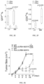
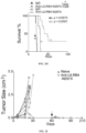
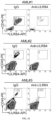


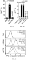


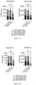


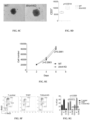

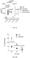


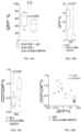


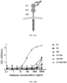


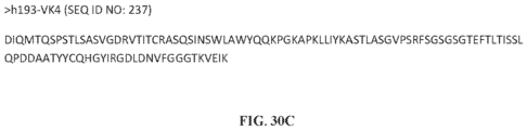


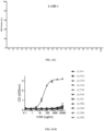


Classifications
-
- C—CHEMISTRY; METALLURGY
- C07—ORGANIC CHEMISTRY
- C07K—PEPTIDES
- C07K16/00—Immunoglobulins [IGs], e.g. monoclonal or polyclonal antibodies
- C07K16/18—Immunoglobulins [IGs], e.g. monoclonal or polyclonal antibodies against material from animals or humans
- C07K16/28—Immunoglobulins [IGs], e.g. monoclonal or polyclonal antibodies against material from animals or humans against receptors, cell surface antigens or cell surface determinants
- C07K16/2803—Immunoglobulins [IGs], e.g. monoclonal or polyclonal antibodies against material from animals or humans against receptors, cell surface antigens or cell surface determinants against the immunoglobulin superfamily
-
- A—HUMAN NECESSITIES
- A61—MEDICAL OR VETERINARY SCIENCE; HYGIENE
- A61K—PREPARATIONS FOR MEDICAL, DENTAL OR TOILETRY PURPOSES
- A61K39/00—Medicinal preparations containing antigens or antibodies
- A61K39/395—Antibodies; Immunoglobulins; Immune serum, e.g. antilymphocytic serum
- A61K39/39533—Antibodies; Immunoglobulins; Immune serum, e.g. antilymphocytic serum against materials from animals
- A61K39/3955—Antibodies; Immunoglobulins; Immune serum, e.g. antilymphocytic serum against materials from animals against proteinaceous materials, e.g. enzymes, hormones, lymphokines
-
- A—HUMAN NECESSITIES
- A61—MEDICAL OR VETERINARY SCIENCE; HYGIENE
- A61K—PREPARATIONS FOR MEDICAL, DENTAL OR TOILETRY PURPOSES
- A61K45/00—Medicinal preparations containing active ingredients not provided for in groups A61K31/00 - A61K41/00
- A61K45/06—Mixtures of active ingredients without chemical characterisation, e.g. antiphlogistics and cardiaca
-
- A—HUMAN NECESSITIES
- A61—MEDICAL OR VETERINARY SCIENCE; HYGIENE
- A61K—PREPARATIONS FOR MEDICAL, DENTAL OR TOILETRY PURPOSES
- A61K47/00—Medicinal preparations characterised by the non-active ingredients used, e.g. carriers or inert additives; Targeting or modifying agents chemically bound to the active ingredient
- A61K47/50—Medicinal preparations characterised by the non-active ingredients used, e.g. carriers or inert additives; Targeting or modifying agents chemically bound to the active ingredient the non-active ingredient being chemically bound to the active ingredient, e.g. polymer-drug conjugates
- A61K47/51—Medicinal preparations characterised by the non-active ingredients used, e.g. carriers or inert additives; Targeting or modifying agents chemically bound to the active ingredient the non-active ingredient being chemically bound to the active ingredient, e.g. polymer-drug conjugates the non-active ingredient being a modifying agent
- A61K47/68—Medicinal preparations characterised by the non-active ingredients used, e.g. carriers or inert additives; Targeting or modifying agents chemically bound to the active ingredient the non-active ingredient being chemically bound to the active ingredient, e.g. polymer-drug conjugates the non-active ingredient being a modifying agent the modifying agent being an antibody, an immunoglobulin or a fragment thereof, e.g. an Fc-fragment
- A61K47/6801—Drug-antibody or immunoglobulin conjugates defined by the pharmacologically or therapeutically active agent
-
- A—HUMAN NECESSITIES
- A61—MEDICAL OR VETERINARY SCIENCE; HYGIENE
- A61K—PREPARATIONS FOR MEDICAL, DENTAL OR TOILETRY PURPOSES
- A61K47/00—Medicinal preparations characterised by the non-active ingredients used, e.g. carriers or inert additives; Targeting or modifying agents chemically bound to the active ingredient
- A61K47/50—Medicinal preparations characterised by the non-active ingredients used, e.g. carriers or inert additives; Targeting or modifying agents chemically bound to the active ingredient the non-active ingredient being chemically bound to the active ingredient, e.g. polymer-drug conjugates
- A61K47/51—Medicinal preparations characterised by the non-active ingredients used, e.g. carriers or inert additives; Targeting or modifying agents chemically bound to the active ingredient the non-active ingredient being chemically bound to the active ingredient, e.g. polymer-drug conjugates the non-active ingredient being a modifying agent
- A61K47/68—Medicinal preparations characterised by the non-active ingredients used, e.g. carriers or inert additives; Targeting or modifying agents chemically bound to the active ingredient the non-active ingredient being chemically bound to the active ingredient, e.g. polymer-drug conjugates the non-active ingredient being a modifying agent the modifying agent being an antibody, an immunoglobulin or a fragment thereof, e.g. an Fc-fragment
- A61K47/6835—Medicinal preparations characterised by the non-active ingredients used, e.g. carriers or inert additives; Targeting or modifying agents chemically bound to the active ingredient the non-active ingredient being chemically bound to the active ingredient, e.g. polymer-drug conjugates the non-active ingredient being a modifying agent the modifying agent being an antibody, an immunoglobulin or a fragment thereof, e.g. an Fc-fragment the modifying agent being an antibody or an immunoglobulin bearing at least one antigen-binding site
- A61K47/6849—Medicinal preparations characterised by the non-active ingredients used, e.g. carriers or inert additives; Targeting or modifying agents chemically bound to the active ingredient the non-active ingredient being chemically bound to the active ingredient, e.g. polymer-drug conjugates the non-active ingredient being a modifying agent the modifying agent being an antibody, an immunoglobulin or a fragment thereof, e.g. an Fc-fragment the modifying agent being an antibody or an immunoglobulin bearing at least one antigen-binding site the antibody targeting a receptor, a cell surface antigen or a cell surface determinant
-
- A—HUMAN NECESSITIES
- A61—MEDICAL OR VETERINARY SCIENCE; HYGIENE
- A61K—PREPARATIONS FOR MEDICAL, DENTAL OR TOILETRY PURPOSES
- A61K47/00—Medicinal preparations characterised by the non-active ingredients used, e.g. carriers or inert additives; Targeting or modifying agents chemically bound to the active ingredient
- A61K47/50—Medicinal preparations characterised by the non-active ingredients used, e.g. carriers or inert additives; Targeting or modifying agents chemically bound to the active ingredient the non-active ingredient being chemically bound to the active ingredient, e.g. polymer-drug conjugates
- A61K47/51—Medicinal preparations characterised by the non-active ingredients used, e.g. carriers or inert additives; Targeting or modifying agents chemically bound to the active ingredient the non-active ingredient being chemically bound to the active ingredient, e.g. polymer-drug conjugates the non-active ingredient being a modifying agent
- A61K47/68—Medicinal preparations characterised by the non-active ingredients used, e.g. carriers or inert additives; Targeting or modifying agents chemically bound to the active ingredient the non-active ingredient being chemically bound to the active ingredient, e.g. polymer-drug conjugates the non-active ingredient being a modifying agent the modifying agent being an antibody, an immunoglobulin or a fragment thereof, e.g. an Fc-fragment
- A61K47/6835—Medicinal preparations characterised by the non-active ingredients used, e.g. carriers or inert additives; Targeting or modifying agents chemically bound to the active ingredient the non-active ingredient being chemically bound to the active ingredient, e.g. polymer-drug conjugates the non-active ingredient being a modifying agent the modifying agent being an antibody, an immunoglobulin or a fragment thereof, e.g. an Fc-fragment the modifying agent being an antibody or an immunoglobulin bearing at least one antigen-binding site
- A61K47/6851—Medicinal preparations characterised by the non-active ingredients used, e.g. carriers or inert additives; Targeting or modifying agents chemically bound to the active ingredient the non-active ingredient being chemically bound to the active ingredient, e.g. polymer-drug conjugates the non-active ingredient being a modifying agent the modifying agent being an antibody, an immunoglobulin or a fragment thereof, e.g. an Fc-fragment the modifying agent being an antibody or an immunoglobulin bearing at least one antigen-binding site the antibody targeting a determinant of a tumour cell
- A61K47/6867—Medicinal preparations characterised by the non-active ingredients used, e.g. carriers or inert additives; Targeting or modifying agents chemically bound to the active ingredient the non-active ingredient being chemically bound to the active ingredient, e.g. polymer-drug conjugates the non-active ingredient being a modifying agent the modifying agent being an antibody, an immunoglobulin or a fragment thereof, e.g. an Fc-fragment the modifying agent being an antibody or an immunoglobulin bearing at least one antigen-binding site the antibody targeting a determinant of a tumour cell the tumour determinant being from a cell of a blood cancer
-
- A—HUMAN NECESSITIES
- A61—MEDICAL OR VETERINARY SCIENCE; HYGIENE
- A61K—PREPARATIONS FOR MEDICAL, DENTAL OR TOILETRY PURPOSES
- A61K47/00—Medicinal preparations characterised by the non-active ingredients used, e.g. carriers or inert additives; Targeting or modifying agents chemically bound to the active ingredient
- A61K47/50—Medicinal preparations characterised by the non-active ingredients used, e.g. carriers or inert additives; Targeting or modifying agents chemically bound to the active ingredient the non-active ingredient being chemically bound to the active ingredient, e.g. polymer-drug conjugates
- A61K47/69—Medicinal preparations characterised by the non-active ingredients used, e.g. carriers or inert additives; Targeting or modifying agents chemically bound to the active ingredient the non-active ingredient being chemically bound to the active ingredient, e.g. polymer-drug conjugates the conjugate being characterised by physical or galenical forms, e.g. emulsion, particle, inclusion complex, stent or kit
- A61K47/6905—Medicinal preparations characterised by the non-active ingredients used, e.g. carriers or inert additives; Targeting or modifying agents chemically bound to the active ingredient the non-active ingredient being chemically bound to the active ingredient, e.g. polymer-drug conjugates the conjugate being characterised by physical or galenical forms, e.g. emulsion, particle, inclusion complex, stent or kit the form being a colloid or an emulsion
- A61K47/6911—Medicinal preparations characterised by the non-active ingredients used, e.g. carriers or inert additives; Targeting or modifying agents chemically bound to the active ingredient the non-active ingredient being chemically bound to the active ingredient, e.g. polymer-drug conjugates the conjugate being characterised by physical or galenical forms, e.g. emulsion, particle, inclusion complex, stent or kit the form being a colloid or an emulsion the form being a liposome
- A61K47/6913—Medicinal preparations characterised by the non-active ingredients used, e.g. carriers or inert additives; Targeting or modifying agents chemically bound to the active ingredient the non-active ingredient being chemically bound to the active ingredient, e.g. polymer-drug conjugates the conjugate being characterised by physical or galenical forms, e.g. emulsion, particle, inclusion complex, stent or kit the form being a colloid or an emulsion the form being a liposome the liposome being modified on its surface by an antibody
-
- A—HUMAN NECESSITIES
- A61—MEDICAL OR VETERINARY SCIENCE; HYGIENE
- A61K—PREPARATIONS FOR MEDICAL, DENTAL OR TOILETRY PURPOSES
- A61K51/00—Preparations containing radioactive substances for use in therapy or testing in vivo
- A61K51/02—Preparations containing radioactive substances for use in therapy or testing in vivo characterised by the carrier, i.e. characterised by the agent or material covalently linked or complexing the radioactive nucleus
- A61K51/04—Organic compounds
- A61K51/08—Peptides, e.g. proteins, carriers being peptides, polyamino acids, proteins
- A61K51/10—Antibodies or immunoglobulins; Fragments thereof, the carrier being an antibody, an immunoglobulin or a fragment thereof, e.g. a camelised human single domain antibody or the Fc fragment of an antibody
- A61K51/1027—Antibodies or immunoglobulins; Fragments thereof, the carrier being an antibody, an immunoglobulin or a fragment thereof, e.g. a camelised human single domain antibody or the Fc fragment of an antibody against receptors, cell-surface antigens or cell-surface determinants
-
- A—HUMAN NECESSITIES
- A61—MEDICAL OR VETERINARY SCIENCE; HYGIENE
- A61K—PREPARATIONS FOR MEDICAL, DENTAL OR TOILETRY PURPOSES
- A61K51/00—Preparations containing radioactive substances for use in therapy or testing in vivo
- A61K51/02—Preparations containing radioactive substances for use in therapy or testing in vivo characterised by the carrier, i.e. characterised by the agent or material covalently linked or complexing the radioactive nucleus
- A61K51/04—Organic compounds
- A61K51/08—Peptides, e.g. proteins, carriers being peptides, polyamino acids, proteins
- A61K51/10—Antibodies or immunoglobulins; Fragments thereof, the carrier being an antibody, an immunoglobulin or a fragment thereof, e.g. a camelised human single domain antibody or the Fc fragment of an antibody
- A61K51/1045—Antibodies or immunoglobulins; Fragments thereof, the carrier being an antibody, an immunoglobulin or a fragment thereof, e.g. a camelised human single domain antibody or the Fc fragment of an antibody against animal or human tumor cells or tumor cell determinants
- A61K51/1069—Antibodies or immunoglobulins; Fragments thereof, the carrier being an antibody, an immunoglobulin or a fragment thereof, e.g. a camelised human single domain antibody or the Fc fragment of an antibody against animal or human tumor cells or tumor cell determinants the tumor cell being from blood cells, e.g. the cancer being a myeloma
-
- A—HUMAN NECESSITIES
- A61—MEDICAL OR VETERINARY SCIENCE; HYGIENE
- A61P—SPECIFIC THERAPEUTIC ACTIVITY OF CHEMICAL COMPOUNDS OR MEDICINAL PREPARATIONS
- A61P35/00—Antineoplastic agents
-
- G—PHYSICS
- G01—MEASURING; TESTING
- G01N—INVESTIGATING OR ANALYSING MATERIALS BY DETERMINING THEIR CHEMICAL OR PHYSICAL PROPERTIES
- G01N33/00—Investigating or analysing materials by specific methods not covered by groups G01N1/00 - G01N31/00
- G01N33/48—Biological material, e.g. blood, urine; Haemocytometers
- G01N33/50—Chemical analysis of biological material, e.g. blood, urine; Testing involving biospecific ligand binding methods; Immunological testing
- G01N33/53—Immunoassay; Biospecific binding assay; Materials therefor
- G01N33/574—Immunoassay; Biospecific binding assay; Materials therefor for cancer
- G01N33/57407—Specifically defined cancers
- G01N33/57426—Specifically defined cancers leukemia
-
- G—PHYSICS
- G01—MEASURING; TESTING
- G01N—INVESTIGATING OR ANALYSING MATERIALS BY DETERMINING THEIR CHEMICAL OR PHYSICAL PROPERTIES
- G01N33/00—Investigating or analysing materials by specific methods not covered by groups G01N1/00 - G01N31/00
- G01N33/48—Biological material, e.g. blood, urine; Haemocytometers
- G01N33/50—Chemical analysis of biological material, e.g. blood, urine; Testing involving biospecific ligand binding methods; Immunological testing
- G01N33/53—Immunoassay; Biospecific binding assay; Materials therefor
- G01N33/574—Immunoassay; Biospecific binding assay; Materials therefor for cancer
- G01N33/57484—Immunoassay; Biospecific binding assay; Materials therefor for cancer involving compounds serving as markers for tumor, cancer, neoplasia, e.g. cellular determinants, receptors, heat shock/stress proteins, A-protein, oligosaccharides, metabolites
- G01N33/57492—Immunoassay; Biospecific binding assay; Materials therefor for cancer involving compounds serving as markers for tumor, cancer, neoplasia, e.g. cellular determinants, receptors, heat shock/stress proteins, A-protein, oligosaccharides, metabolites involving compounds localized on the membrane of tumor or cancer cells
-
- A—HUMAN NECESSITIES
- A61—MEDICAL OR VETERINARY SCIENCE; HYGIENE
- A61K—PREPARATIONS FOR MEDICAL, DENTAL OR TOILETRY PURPOSES
- A61K39/00—Medicinal preparations containing antigens or antibodies
- A61K2039/505—Medicinal preparations containing antigens or antibodies comprising antibodies
-
- A—HUMAN NECESSITIES
- A61—MEDICAL OR VETERINARY SCIENCE; HYGIENE
- A61P—SPECIFIC THERAPEUTIC ACTIVITY OF CHEMICAL COMPOUNDS OR MEDICINAL PREPARATIONS
- A61P35/00—Antineoplastic agents
- A61P35/02—Antineoplastic agents specific for leukemia
-
- C—CHEMISTRY; METALLURGY
- C07—ORGANIC CHEMISTRY
- C07K—PEPTIDES
- C07K2317/00—Immunoglobulins specific features
- C07K2317/20—Immunoglobulins specific features characterized by taxonomic origin
- C07K2317/24—Immunoglobulins specific features characterized by taxonomic origin containing regions, domains or residues from different species, e.g. chimeric, humanized or veneered
-
- C—CHEMISTRY; METALLURGY
- C07—ORGANIC CHEMISTRY
- C07K—PEPTIDES
- C07K2317/00—Immunoglobulins specific features
- C07K2317/30—Immunoglobulins specific features characterized by aspects of specificity or valency
- C07K2317/33—Crossreactivity, e.g. for species or epitope, or lack of said crossreactivity
-
- C—CHEMISTRY; METALLURGY
- C07—ORGANIC CHEMISTRY
- C07K—PEPTIDES
- C07K2317/00—Immunoglobulins specific features
- C07K2317/30—Immunoglobulins specific features characterized by aspects of specificity or valency
- C07K2317/34—Identification of a linear epitope shorter than 20 amino acid residues or of a conformational epitope defined by amino acid residues
-
- C—CHEMISTRY; METALLURGY
- C07—ORGANIC CHEMISTRY
- C07K—PEPTIDES
- C07K2317/00—Immunoglobulins specific features
- C07K2317/50—Immunoglobulins specific features characterized by immunoglobulin fragments
- C07K2317/56—Immunoglobulins specific features characterized by immunoglobulin fragments variable (Fv) region, i.e. VH and/or VL
- C07K2317/565—Complementarity determining region [CDR]
-
- C—CHEMISTRY; METALLURGY
- C07—ORGANIC CHEMISTRY
- C07K—PEPTIDES
- C07K2317/00—Immunoglobulins specific features
- C07K2317/60—Immunoglobulins specific features characterized by non-natural combinations of immunoglobulin fragments
- C07K2317/62—Immunoglobulins specific features characterized by non-natural combinations of immunoglobulin fragments comprising only variable region components
- C07K2317/622—Single chain antibody (scFv)
-
- C—CHEMISTRY; METALLURGY
- C07—ORGANIC CHEMISTRY
- C07K—PEPTIDES
- C07K2317/00—Immunoglobulins specific features
- C07K2317/70—Immunoglobulins specific features characterized by effect upon binding to a cell or to an antigen
- C07K2317/76—Antagonist effect on antigen, e.g. neutralization or inhibition of binding
-
- C—CHEMISTRY; METALLURGY
- C07—ORGANIC CHEMISTRY
- C07K—PEPTIDES
- C07K2317/00—Immunoglobulins specific features
- C07K2317/90—Immunoglobulins specific features characterized by (pharmaco)kinetic aspects or by stability of the immunoglobulin
- C07K2317/92—Affinity (KD), association rate (Ka), dissociation rate (Kd) or EC50 value
Definitions
- the present disclosure relates generally to the fields of medicine, oncology, and immunology. More particular, the disclosure relates to antibodies that bind to LILRB4 and can treat cancers, including leukemia.
- AML Acute myeloid leukaemia
- Current treatment for AML involves intensive cytotoxic chemotherapy, often times followed by myeloablative conditioning and stem cell transplant.
- myeloablative conditioning and stem cell transplant despite treatment, most patients relapse or succumb to disease within 5 years.
- new molecular targets and therapeutic approaches must be identified.
- LILRBs inhibitory leukocyte immunoglobulin-like receptors
- ITIM immunoreceptor tyrosine-based inhibitory motif
- ITIM-containing receptors are expressed on a wide range of immune cells and transduce signals by recruitment of phosphatases SHP-1, SHP-2, or SHIP, leading to negative regulation of immune cell activation. Similar to CTLA4 and PD-1, LILRBs are considered immune checkpoint factors.
- LILRBs may inhibit activities of a number of immune cell types facilitating tumour immune escape.
- LILRB4 is expressed on monocytes, macrophages, and dendritic cells and can inhibit innate immunity in a cell-autonomous manner and suppress T cell activation through an indirect mechanism.
- LILRB4 is a specific marker for monocytic AML including refractory and relapsed disease.
- LILRB1-5 are primate and human specific, while there are two mouse orthologues: paired immunoglobulin-like receptor B (PirB) and gp49B1.
- the related immunoreceptor tyrosine-based inhibitory motif (ITIM)-containing receptor, LAIR1 has both human and mouse versions of the protein. Because of the limited value of mouse models and the fact that ligands for several LILRBs including LILRB4 are unknown, the biological function and clinical significance of these receptors remain poorly understood.
- the present disclosure provides an isolated monoclonal antibody or an antigen-binding fragment thereof that binds specifically to LILRB4 comprising:
- each CDR is defined in accordance with Kabat definition, the Chothia definition, the combination of Kabat definition and Chothia definition, the AbM definition, or the contact definition of CDR.
- the antibody comprises the heavy chain variable region comprising: a HC-CDR1 having the amino acid sequence set forth in SEQ ID NO: 100, a HC-CDR2 having the amino acid sequence set forth in SEQ ID NO: 101, and a HC-CDR3 having the amino acid sequence set forth in SEQ ID NO: 102, 224 or 227.
- the antibody comprises the light chain variable region comprising: a LC-CDR1 having the amino acid sequence set forth in SEQ ID NO: 104, a LC-CDR2 having the amino acid sequence of KAS or the amino acid sequence set forth in SEQ ID NO: 233, and a LC-CDR3 having the amino acid sequence set forth in SEQ ID NOS: 105 or 234.
- the antibody is characterized by clone-paired heavy chain and light chain having amino acid sequences at least 90%, or 95% identity to the clone-paired sequences from FIGS. 28A-28C and 30A-30C.
- the antibody comprises a heavy chain variable region having the amino acid sequence set forth in SEQ ID NO: 99, 223, 225, 226, 228, 229, 230 or 231.
- the antibody comprises a light chain variable region having the amino acid sequence set forth in SEQ ID NO: 103, 232, 235, 236 or 237.
- the epitope bound by the antibody or antigen-binding fragment is located within the linker region between the D1 and D2 domain of human LILRB4.
- the epitope comprises at least one amino acid within one or more of the amino acid sequences of LILRB4 listed in Table 9.
- the epitope comprises at least one amino acid within one or more of the amino acid sequences selected from W18, G96, A97, Y98, S99, K100, Q122, S123, R124, S125, P126, H153 and Q154 of SEQ ID NO: 238 (D1 and D2 domains of human LILRB4 protein).
- the isolated monoclonal antibody described herein is a chimeric, humanized, or human antibody. In certain embodiments, isolated monoclonal antibody described herein is of the IgG1, IgG2, IgG3 or IgG4 type. In certain embodiments, the antigen-binding fragment described herein is a recombinant ScFv (single chain fragment variable) antibody, Fab fragment, F(ab')2 fragment, or Fv fragment.
- ScFv single chain fragment variable
- composition comprising an isolated monoclonal antibody or an antigen-binding fragment thereof as provided herein, and at least one pharmaceutically acceptable carrier.
- an isolated nucleic acid that encodes the isolated monoclonal antibody or an antigen-binding fragment thereof as provided herein.
- a vector comprising the isolated nucleic acid as provided herein.
- a host cell comprising the vector as provided herein.
- the host cell may be a mammalian cell.
- the host cell may be a CHO cell.
- hybridoma encoding or producing the isolated monoclonal antibody as provided herein.
- a process of producing an antibody may comprise culturing the host cell as provided herein under conditions suitable for expressing the antibody and recovering the antibody.
- CAR chimeric antigen receptor
- nucleic acid that encodes a CAR protein as provided herein.
- an engineered cell comprising the isolated nucleic acid as provided herein.
- the cell is a T cell, NK cell, or myeloid cell.
- a use in a method of treating or ameliorating the effects of a cancer in a subject comprises administering to the subject a therapeutically effective amount of the antibody or an antigen-binding fragment thereof or an engineered cell as provided herein.
- the cancer is acute myeloid leukemia.
- the antibody or an antigen-binding fragment thereof is administered intravenously, intra-arterially, intra-tumorally or subcutaneously.
- the antibody or an antigen-binding fragment thereof comprises an antitumor drug (e.g ., a toxin, a radioisotope, a cytokine, or an enzyme) linked thereto.
- the isolated monoclonal antibody or an antigen binding fragment thereof is conjugated to a liposome or nanoparticle.
- a use in a method, or an in vitro method, of detecting a cancer cell or cancer stem cell in a sample or subject comprises contacting a subject or a sample from the subject with the antibody or an antigen-binding fragment thereof as provided herein and detecting binding of said antibody to a cancer cell or cancer stem cell in said subject or sample.
- the sample can be a body fluid or biopsy.
- the sample can be blood, sputum, tears, saliva, mucous, serum, urine or feces.
- the detection comprises immunohistochemistry, flow cytometry, FACS, ELISA, RIA or Western blot.
- isolated monoclonal antibody or an antigen binding fragment thereof further comprises a label (e.g ., a peptide tag, an enzyme, a magnetic particle, a chromophore, a fluorescent molecule, a chemo-luminescent molecule, or a dye).
- a label e.g ., a peptide tag, an enzyme, a magnetic particle, a chromophore, a fluorescent molecule, a chemo-luminescent molecule, or a dye.
- the isolated monoclonal antibody or an antigen binding fragment thereof is conjugated to a liposome or nanoparticle.
- the inventors have isolated a panel of novel monoclonal antibodies recognizing LILRB4 protein, an ITIM-containing receptor, which can be used for cancer treatment.
- LILRB4 is upregulated on some tumour cells especially leukemia cells and promotes tumour growth.
- the anti-human II,IRB4 antibodies identified block LILRB4 signalling and can modulate immunity against cancer.
- each numerical parameter should at least be construed in light of the number of reported significant digits and by applying ordinary rounding techniques. Notwithstanding that the numerical ranges and parameters setting forth the broad scope of the invention are approximations, the numerical values set forth in the specific examples are reported as precisely as possible. Any numerical value, however, inherently contain certain errors necessarily resulting from the standard deviation found in their respective testing measurements.
- antibody refers to an intact immunoglobulin of any isotype, or a fragment thereof that can compete with the intact antibody for specific binding to the target antigen, and includes, for instance, chimeric, humanized, fully human, and bispecific antibodies.
- An "antibody” is a species of an antigen binding protein.
- An intact antibody will generally comprise at least two full-length heavy chains and two full-length light chains, but in some instances can include fewer chains such as antibodies naturally occurring in camelids which can comprise only heavy chains.
- Antibodies can be derived solely from a single source, or can be "chimeric,” that is, different portions of the antibody can be derived from two different antibodies as described further below.
- antigen binding proteins, antibodies, or binding fragments can be produced in hybridomas, by recombinant DNA techniques, or by enzymatic or chemical cleavage of intact antibodies.
- antibody includes, in addition to antibodies comprising two full-length heavy chains and two full-length light chains, derivatives, variants, fragments, and muteins thereof, examples of which are described below.
- antibodies include monoclonal antibodies, bispecific antibodies, minibodies, domain antibodies, synthetic antibodies (sometimes referred to herein as "antibody mimetics"), chimeric antibodies, humanized antibodies, human antibodies, antibody fusions (sometimes referred to herein as "antibody conjugates”), and fragments thereof, respectively.
- the term also encompasses peptibodies.
- Naturally occurring antibody structural units typically comprise a tetramer.
- Each such tetramer typically is composed of two identical pairs of polypeptide chains, each pair having one full-length "light” (in certain embodiments, about 25 kDa) and one full-length "heavy” chain (in certain embodiments, about 50-70 kDa).
- the amino-terminal portion of each chain typically includes a variable region of about 100 to 110 or more amino acids that typically is responsible for antigen recognition.
- the carboxy-terminal portion of each chain typically defines a constant region that can be responsible for effector function.
- Human light chains are typically classified as kappa and lambda light chains.
- Heavy chains are typically classified as mu, delta, gamma, alpha, or epsilon, and define the antibody's isotype as IgM, IgD, IgG, IgA, and IgE, respectively.
- IgG has several subclasses, including, but not limited to, IgG1, IgG2, IgG3, and IgG4.
- IgM has subclasses including, but not limited to, IgM1 and IgM2.
- IgA is similarly subdivided into subclasses including, but not limited to, IgA1 and IgA2.
- variable and constant regions are joined by a "J" region of about 12 or more amino acids, with the heavy chain also including a "D” region of about 10 more amino acids. See, e.g ., Fundamental Immunology, Ch. 7 (Paul, W., ed., 2nd ed. Raven Press, N.Y. (1989)).
- the variable regions of each light/heavy chain pair typically form the antigen binding site.
- variable region refers to a portion of the light and/or heavy chains of an antibody, typically including approximately the amino-terminal 120 to 130 amino acids in the heavy chain and about 100 to 110 amino terminal amino acids in the light chain.
- variable regions of different antibodies differ extensively in amino acid sequence even among antibodies of the same species.
- the variable region of an antibody typically determines specificity of a particular antibody for its target.
- variable regions typically exhibit the same general structure of relatively conserved framework regions (FR) joined by three hyper variable regions, also called complementarity determining regions or CDRs.
- the CDRs from the two chains of each pair typically are aligned by the framework regions, which can enable binding to a specific epitope.
- both light and heavy chain variable regions typically comprise the domains FR1, CDR1, FR2, CDR2, FR3, CDR3 and FR4.
- the assignment of amino acids to each domain is typically in accordance with the definitions of Kabat Sequences of Proteins of Immunological Interest (National Institutes of Health, Bethesda, Md. (1987 and 1991 )), Chothia & Lesk, J. Mol. Biol., 196:901-917 (1987 ) or Chothia et al., Nature, 342:878-883 (1989 ).
- definitive delineation of a CDR and identification of residues comprising the binding site of an antibody is accomplished by solving the structure of the antibody and/or solving the structure of the antibody-ligand complex. In certain embodiments, that can be accomplished by any of a variety of techniques known to those skilled in the art, such as X-ray crystallography. In certain embodiments, various methods of analysis can be employed to identify or approximate the CDR regions. Examples of such methods include, but are not limited to, the Kabat definition, the Chothia definition, the AbM definition and the contact definition.
- the Kabat definition is a standard for numbering the residues in an antibody and is typically used to identify CDR regions. See, e.g ., Johnson & Wu, Nucleic Acids Res., 28: 214-8 (2000 ).
- the Chothia definition is similar to the Kabat definition, but the Chothia definition takes into account positions of certain structural loop regions. See, e.g., Chothia et al., J. Mol. Biol., 196: 901-17 (1986 ); Chothia et al., Nature, 342: 877-83 (1989 ).
- the AbM definition uses an integrated suite of computer programs produced by Oxford Molecular Group that model antibody structure.
- the AbM definition models the tertiary structure of an antibody from primary sequence using a combination of knowledge databases and ab initio methods, such as those described by Samudrala et al., "Ab Initio Protein Structure Prediction Using a Combined Hierarchical Approach," in PROTEINS, Structure, Function and Genetics Suppl., 3:194-198 (1999 ).
- the contact definition is based on an analysis of the available complex crystal structures. See, e.g ., MacCallum et al., J. Mol. Biol., 5:732-45 (1996 ).
- the CDR regions in the heavy chain are typically referred to as H1, H2, and H3 and are numbered sequentially in the direction from the amino terminus to the carboxy terminus.
- the CDR regions in the light chain are typically referred to as L1, L2, and L3 and are numbered sequentially in the direction from the amino terminus to the carboxy terminus.
- light chain includes a full-length light chain and fragments thereof having sufficient variable region sequence to confer binding specificity.
- a full-length light chain includes a variable region domain, VL, and a constant region domain, CL.
- the variable region domain of the light chain is at the amino-terminus of the polypeptide.
- Light chains include kappa chains and lambda chains.
- heavy chain includes a full-length heavy chain and fragments thereof having sufficient variable region sequence to confer binding specificity.
- a full-length heavy chain includes a variable region domain, VH, and three constant region domains, CH1, CH2, and CH3.
- the VH domain is at the amino-terminus of the polypeptide, and the CH domains are at the carboxyl-terminus, with the CH3 being closest to the carboxy-terminus of the polypeptide.
- Heavy chains can be of any isotype, including IgG (including IgG1, IgG2, IgG3 and IgG4 subtypes), IgA (including IgA1 and IgA2 subtypes), IgM and IgE.
- a bispecific or bifunctional antibody typically is an artificial hybrid antibody having two different heavy/light chain pairs and two different binding sites.
- Bispecific antibodies can be produced by a variety of methods including, but not limited to, fusion of hybridomas or linking of Fab' fragments. See, e.g., Songsivilai et al., Clin. Exp. Immunol., 79: 315-321 (1990 ); Kostelny et al., J. Immunol., 148:1547-1553 (1992 ).
- an antigen refers to a substance capable of inducing adaptive immune responses.
- an antigen is a substance which serves as a target for the receptors of an adaptive immune response.
- an antigen is a molecule that binds to antigen-specific receptors but cannot induce an immune response in the body by itsself.
- Antigens are usually proteins and polysaccharides, less frequently also lipids. Suitable antigens include without limitation parts of bacteria (coats, capsules, cell walls, flagella, fimbrai, and toxins), viruses, and other microorganisms.
- Antigens also include tumor antigens, e.g ., antigens generated by mutations in tumors.
- antigens also include immunogens and haptens.
- an “antigen binding protein” as used herein means any protein that binds a specified target antigen.
- the specified target antigen is the LILRB protein or fragment thereof.
- Antigen binding protein includes but is not limited to antibodies and antigen-binding fragment thereof. Peptibodies are another example of antigen binding proteins.
- antigen-binding fragment refers to a portion of a protein which is capable of binding specifically to an antigen.
- the antigen-binding fragment is derived from an antibody comprising six CDRs, or any other antibody fragment that binds to an antigen but does not comprise an intact native antibody structure.
- antigen-binding fragment examples include, without limitation, a diabody, a Fab, a Fab', a F(ab') 2 , an Fv fragment, a disulfide stabilized Fv fragment (dsFv), a (dsFv) 2 , a bispecific dsFv (dsFv-dsFv'), a disulfide stabilized diabody (ds diabody), a single-chain antibody molecule (scFv), an scFv dimer (bivalent diabody), a multispecific antibody, a single domain antibody (sdAb), a camelid antibody or a nanobody, a domain antibody, and a bivalent domain antibody.
- a diabody examples include, without limitation, a diabody, a Fab, a Fab', a F(ab') 2 , an Fv fragment, a disulfide stabilized Fv fragment (dsFv), a (dsFv) 2
- an antigen-binding fragment is capable of binding to the same antigen to which the parent antibody binds.
- an antigen-binding fragment may comprise one or more CDRs from a particular human antibody grafted to a framework region from one or more different human antibodies.
- a "Fab fragment” comprises one light chain and the CH1 and variable regions of one heavy chain.
- the heavy chain of a Fab molecule cannot form a disulfide bond with another heavy chain molecule.
- a “Fab' fragment” comprises one light chain and a portion of one heavy chain that contains the VH domain and the CH1 domain and also the region between the CH1 and CH2 domains, such that an interchain disulfide bond can be formed between the two heavy chains of two Fab' fragments to form an F(ab') 2 molecule.
- a “F(ab') 2 fragment” contains two light chains and two heavy chains containing a portion of the constant region between the CH1 and CH2 domains, such that an interchain disulfide bond is formed between the two heavy chains.
- a F(ab') 2 fragment thus is composed of two Fab' fragments that are held together by a disulfide bond between the two heavy chains.
- An "Fc” region comprises two heavy chain fragments comprising the CH1 and CH2 domains of an antibody.
- the two heavy chain fragments are held together by two or more disulfide bonds and by hydrophobic interactions of the CH3 domains.
- the "Fv region” comprises the variable regions from both the heavy and light chains but lacks the constant regions.
- Single-chain antibodies are Fv molecules in which the heavy and light chain variable regions have been connected by a flexible linker to form a single polypeptide chain, which forms an antigen binding region.
- Single chain antibodies are discussed in detail in International Patent Application Publication No. WO 88/01649 and U.S. Pat. No. 4,946,778 and No. 5,260,203 .
- a “domain antibody” is an immunologically functional immunoglobulin fragment containing only the variable region of a heavy chain or the variable region of a light chain.
- two or more VH regions are covalently joined with a peptide linker to create a bivalent domain antibody.
- the two VH regions of a bivalent domain antibody can target the same or different antigens.
- a “bivalent antigen binding protein” or “bivalent antibody” comprises two antigen binding sites. In some instances, the two binding sites have the same antigen specificities. Bivalent antigen binding proteins and bivalent antibodies can be bispecific, see, infra. A bivalent antibody other than a “multispecific” or “multifunctional” antibody, in certain embodiments, typically is understood to have each of its binding sites identical.
- a “multispecific antigen binding protein” or “multispecific antibody” is one that targets more than one antigen or epitope.
- a "bispecific,” “dual-specific” or “bifunctional” antigen binding protein or antibody is a hybrid antigen binding protein or antibody, respectively, having two different antigen binding sites.
- Bispecific antigen binding proteins and antibodies are a species of multispecific antigen binding protein antibody and can be produced by a variety of methods including, but not limited to, fusion of hybridomas or linking of Fab' fragments. See, e.g ., Songsivilai and Lachmann, 1990, Clin. Exp. Immunol. 79:315-321 ; Kostelny et al., 1992, J. Immunol. 148:1547-1553 .
- the two binding sites of a bispecific antigen binding protein or antibody will bind to two different epitopes, which can reside on the same or different protein targets.
- Binding affinity generally refers to the strength of the sum total of non-covalent interactions between a single binding site of a molecule (e.g ., an antibody) and its binding partner (e.g ., an antigen). Unless indicated otherwise, as used herein, "binding affinity” refers to intrinsic binding affinity that reflects a 1:1 interaction between members of a binding pair ( e.g. , antibody and antigen).
- the affinity of a molecule X for its partner Y can generally be represented by the dissociation constant (Kd). Affinity can be measured by common methods known in the art, including those described herein.
- Low-affinity antibodies generally bind antigen slowly and tend to dissociate readily, whereas high-affinity antibodies generally bind antigen faster and tend to remain bound longer.
- a variety of methods of measuring binding affinity are known in the art, any of which can be used for purposes of the present invention. Specific illustrative and exemplary embodiments for measuring binding affinity are described in the following.
- an antibody that "specifically binds to” or is “specific for” a particular polypeptide or an epitope on a particular polypeptide is one that binds to that particular polypeptide or epitope on a particular polypeptide without substantially binding to any other polypeptide or polypeptide epitope.
- the LILRB4 specific antibodies of the present invention are specific to LILRB4.
- the antibody that binds to LILRB4 has a dissociation constant (Kd) of ⁇ 100 nM, ⁇ 10 nM, ⁇ 1 nM, ⁇ 0.1 nM, ⁇ 0.01 nM, or ⁇ 0.001 nM ( e.g., 10 -8 M or less, e.g., from 10 -8 M to 10 -13 M, e.g., from 10 -9 M to 10 -13 M).
- Kd dissociation constant
- antigen binding proteins e.g ., atnibody or antigen-binding fragment thereof
- competition between antigen binding proteins as determined by an assay in which the antigen binding protein (e.g ., antibody or antigen-binding fragment thereof) being tested prevents or inhibits ( e.g ., reduces) specific binding of a reference antigen binding protein (e.g., a ligand, or a reference antibody) to a common antigen (e.g. , LILRB or a fragment thereof).
- a reference antigen binding protein e.g., a ligand, or a reference antibody
- RIA solid phase direct or indirect radioimmunoassay
- EIA solid phase direct or indirect enzyme immunoassay
- sandwich competition assay see, e.g., Stahli et al., 1983, Methods in Enzymology 9:242-253
- solid phase direct biotin-avidin EIA see, e.g., Kirkland et al., 1986, J. Immunol.
- solid phase direct labeled assay solid phase direct labeled sandwich assay (see, e.g ., Harlow and Lane, 1988, Antibodies, A Laboratory Manual, Cold Spring Harbor Press ); solid phase direct label RIA using 1-125 label (see, e.g., Morel et al., 1988, Molec. Immunol. 25:7-15 ); solid phase direct biotin-avidin EIA (see, e.g., Cheung, et al., 1990, Virology 176:546-552 ); and direct labeled RIA ( Moldenhauer et al., 1990, Scand. J. Immunol. 32:77-82 ).
- such an assay involves the use of purified antigen bound to a solid surface or cells bearing either of these, an unlabelled test antigen binding protein and a labeled reference antigen binding protein.
- Competitive inhibition is measured by determining the amount of label bound to the solid surface or cells in the presence of the test antigen binding protein.
- the test antigen binding protein is present in excess.
- Antigen binding proteins identified by competition assay include antigen binding proteins binding to the same epitope as the reference antigen binding proteins and antigen binding proteins binding to an adjacent epitope sufficiently proximal to the epitope bound by the reference antigen binding protein for steric hindrance to occur. Additional details regarding methods for determining competitive binding are provided in the examples herein.
- a competing antigen binding protein when present in excess, it will inhibit (e.g ., reduce) specific binding of a reference antigen binding protein to a common antigen by at least 40-45%, 45-50%, 50-55%, 55-60%, 60-65%, 65-70%, 70-75% or 75% or more. In some instances, binding is inhibited by at least 80-85%, 85-90%, 90-95%, 95-97%, or 97% or more.
- epitope refers to the specific group of atoms or amino acids on an antigen to which an antibody binds.
- the epitope can be either linear epitope or a conformational epitope.
- a linear epitope is formed by a continuous sequence of amino acids from the antigen and interacts with an antibody based on their primary structure.
- a conformational epitope is composed of discontinuous sections of the antigen's amino acid sequence and interacts with the antibody based on the 3D structure of the antigen.
- an epitope is approximately five or six amino acid in length. Two antibodies may bind the same epitope within an antigen if they exhibit competitive binding for the antigen.
- a "cell”, as used herein, can be prokaryotic or eukaryotic.
- a prokaryotic cell includes, for example, bacteria.
- a eukaryotic cell includes, for example, a fungus, a plant cell, and an animal cell.
- the types of an animal cell includes, for example, a cell from circulatory/immune system or organ, e.g., a B cell, a T cell (cytotoxic T cell, natural killer T cell, regulatory T cell, T helper cell), a natural killer cell, a granulocyte (e.g ., basophil granulocyte, an eosinophil granulocyte, a neutrophil granulocyte and a hypersegmented neutrophil), a monocyte or macrophage, a red blood cell ( e.g., reticulocyte), a mast cell, a thrombocyte or megakaryocyte, and a dendritic cell;
- astrocyte and oligodendrocyte a microglia, a magnocellular neurosecretory cell, a stellate cell, a boettcher cell, and a pituitary cell (e.g., gonadotrope, corticotrope, thyrotrope, somatotrope, and lactotroph); a cell from a respiratory system or organ, e.g., a pneumocyte (a type I pneumocyte and a type II pneumocyte), a clara cell, a goblet cell, and an alveolar macrophage; a cell from circular system or organ ( e.g ., myocardiocyte and pericyte); a cell from digestive system or organ, e.g., a gastric chief cell, a parietal cell, a goblet cell, a paneth cell, a G cell, a D cell, an ECL cell, an I cell, a K cell, an S cell, an enteroendocrine
- a cell can be normal, healthy cell; or a diseased or unhealthy cell (e.g., a cancer cell).
- a cell further includes a non-human mammalian zygote.
- a cell further includes a stem cell which include an embryonic stem cell, a fetal stem cell, an induced pluripotent stem cell, and an adult stem cell.
- a stem cell is a cell that is capable of undergoing cycles of cell division while maintaining an undifferentiated state and differentiating into specialized cell types.
- a stem cell can be a non-human omnipotent stem cell, which may be induced from a somatic cell.
- a stem cell can be a pluripotent stem cell, a multipotent stem cell, an oligopotent stem cell and a unipotent stem cell, any of which may be induced from a somatic cell.
- a stem cell may also include a cancer stem cell.
- a mammalian cell can be a rodent cell, e.g., a mouse, rat, hamster cell.
- a mammalian cell can be a lagomorpha cell, e.g., a rabbit cell.
- a mammalian cell can also be a primate cell, e.g., a human cell.
- chimeric antigen receptor or "CAR” as used herein refers to an artificially constructed hybrid protein or polypeptide containing an antigen binding domain of an antibody (e.g ., a single chain variable fragment (scFv)) linked to a domain or signaling, e.g., T-cell signaling or T-cell activation domains, that activates an immune cell, e.g., a T cell or a NK cell (see, e.g ., Kershaw et al., supra, Eshhar et al., Proc. Natl. Acad. Sci. USA, 90(2): 720-724 (1993 ), and Sadelain et al., Curr. Opin. Immunol.
- an antibody e.g ., a single chain variable fragment (scFv)
- a domain or signaling e.g., T-cell signaling or T-cell activation domains
- CARs are capable of redirecting the immune cell specificity and reactivity toward a selected target in a non-MHC-restricted manner, taking advantage of the antigen-binding properties of monoclonal antibodies.
- the non-MHC-restricted antigen recognition confers immune cells expressing CARs on the ability to recognize an antigen independent of antigen processing, thus bypassing a major mechanism of tumor escape.
- CARs when expressed in T-cells, CARs advantageously do not dimerize with endogenous T-cell receptor (TCR) alpha and beta chains.
- essentially free in terms of a specified component, is used herein to mean that none of the specified component has been purposefully formulated into a composition and/or is present only as a contaminant or in trace amounts.
- the total amount of the specified component resulting from any unintended contamination of a composition is therefore well below 0.05%, preferably below 0.01%.
- Most preferred is a composition in which no amount of the specified component can be detected with standard analytical methods.
- the term "host cell” means a cell that has been transformed, or is capable of being transformed, with a nucleic acid sequence and thereby expresses a gene of interest.
- the term includes the progeny of the parent cell, whether or not the progeny is identical in morphology or in genetic make-up to the original parent cell, so long as the gene of interest is present.
- identity refers to a relationship between the sequences of two or more polypeptide molecules or two or more nucleic acid molecules, as determined by aligning and comparing the sequences. "Percent identity” means the percent of identical residues between the amino acids or nucleotides in the compared molecules and is calculated based on the size of the smallest of the molecules being compared. For these calculations, gaps in alignments (if any) are preferably addressed by a particular mathematical model or computer program (i.e ., an "algorithm”). Methods that can be used to calculate the identity of the aligned nucleic acids or polypeptides include those described in Computational Molecular Biology, (Lesk, A.
- the sequences being compared are typically aligned in a way that gives the largest match between the sequences.
- One example of a computer program that can be used to determine percent identity is the GCG program package, which includes GAP ( Devereux et al., 1984, Nucl. Acid Res. 12:387 ; Genetics Computer Group, University of Wisconsin, Madison, Wis.).
- GAP Devereux et al., 1984, Nucl. Acid Res. 12:387 ; Genetics Computer Group, University of Wisconsin, Madison, Wis.
- the computer algorithm GAP is used to align the two polypeptides or polynucleotides for which the percent sequence identity is to be determined.
- the sequences are aligned for optimal matching of their respective amino acid or nucleotide (the "matched span", as determined by the algorithm).
- a gap opening penalty (which is calculated as 3 ⁇ the average diagonal, wherein the "average diagonal” is the average of the diagonal of the comparison matrix being used; the “diagonal” is the score or number assigned to each perfect amino acid match by the particular comparison matrix) and a gap extension penalty (which is usually 1/10 times the gap opening penalty), as well as a comparison matrix such as PAM 250 or BLOSUM 62 are used in conjunction with the algorithm.
- a standard comparison matrix (see, Dayhoff et al., 1978, Atlas of Protein Sequence and Structure 5:345-352 for the PAM 250 comparison matrix; Henikoff et al., 1992, Proc. Natl. Acad. Sci. U.S.A. 89:10915-10919 for the BLOSUM 62 comparison matrix) is also used by the algorithm.
- Certain alignment schemes for aligning two amino acid sequences may result in matching of only a short region of the two sequences, and this small aligned region may have very high sequence identity even though there is no significant relationship between the two full-length sequences. Accordingly, the selected alignment method (GAP program) can be adjusted if so desired to result in an alignment that spans at least 50 or other number of contiguous amino acids of the target polypeptide.
- link refers to the association via intramolecular interaction, e.g. , covalent bonds, metallic bonds, and/or ionic bonding, or intermolecular interaction, e.g. , hydrogen bond or noncovalent bonds.
- LILRB4 Leukocyte immunoglobulin-like receptor subfamily B member 4
- LIR leukocyte immunoglobulin-like receptor
- the encoded protein belongs to the subfamily B class of LIR receptors which contain two or four extracellular immunoglobulin domains, a transmembrane domain, and two to four cytoplasmic immunoreceptor tyrosine-based inhibitory motifs (ITIMs).
- ITIMs cytoplasmic immunoreceptor tyrosine-based inhibitory motifs
- the receptor can also function in antigen capture and presentation. It is thought to control inflammatory responses and cytotoxicity to help focus the immune response and limit autoreactivity.
- LILRB4 is also expressed in human gastric cancer cells and may enhance tumor growth. Multiple transcript variants encoding different isoforms have been found for this gene. LILRB4 has been shown to interact with PTPN6.
- operably linked refers to an arrangement of elements wherein the components so described are configured so as to perform their usual function.
- a given signal peptide that is operably linked to a polypeptide directs the secretion of the polypeptide from a cell.
- a promoter that is operably linked to a coding sequence will direct the expression of the coding sequence.
- the promoter or other control elements need not be contiguous with the coding sequence, so long as they function to direct the expression thereof. For example, intervening untranslated yet transcribed sequences can be present between the promoter sequence and the coding sequence and the promoter sequence can still be considered “operably linked" to the coding sequence.
- polynucleotide or “nucleic acid” includes both single-stranded and double-stranded nucleotide polymers.
- the nucleotides comprising the polynucleotide can be ribonucleotides or deoxyribonucleotides or a modified form of either type of nucleotide.
- Said modifications include base modifications such as bromouridine and inosine derivatives, ribose modifications such as 2',3'-dideoxyribose, and internucleotide linkage modifications such as phosphorothioate, phosphorodithioate, phosphoroselenoate, phosphorodiselenoate, phosphoroanilothioate, phoshoraniladate and phosphoroamidate.
- polypeptide or "protein” means a macromolecule having the amino acid sequence of a native protein, that is, a protein produced by a naturally-occurring and non-recombinant cell; or it is produced by a genetically-engineered or recombinant cell, and comprise molecules having the amino acid sequence of the native protein, or molecules having deletions from, additions to, and/or substitutions of one or more amino acids of the native sequence.
- the term also includes amino acid polymers in which one or more amino acids are chemical analogs of a corresponding naturally-occurring amino acid and polymers.
- polypeptide and protein specifically encompass LII,RB antigen binding proteins, antibodies, or sequences that have deletions from, additions to, and/or substitutions of one or more amino acid of antigen-binding protein.
- polypeptide fragment refers to a polypeptide that has an amino-terminal deletion, a carboxyl-terminal deletion, and/or an internal deletion as compared with the full-length native protein. Such fragments can also contain modified amino acids as compared with the native protein. In certain embodiments, fragments are about five to 500 amino acids long. For example, fragments can be at least 5, 6, 8, 10, 14, 20, 50, 70, 100, 110, 150, 200, 250, 300, 350, 400, or 450 amino acids long.
- Useful polypeptide fragments include immunologically functional fragments of antibodies, including binding domains.
- useful fragments include but are not limited to a CDR region, a variable domain of a heavy and/or light chain, a portion of an antibody chain or just its variable region including two CDRs, and the like.
- compositions and formulations suitable for pharmaceutical delivery of the fusion proteins herein disclosed are conventional.
- Remington's Pharmaceutical Sciences, by E. W. Martin, Mack Publishing Co., Easton, PA, 15th Edition (1975 ) describes compositions and formulations suitable for pharmaceutical delivery of the fusion proteins herein disclosed.
- the nature of the carrier will depend on the particular mode of administration being employed.
- parenteral formulations usually comprise injectable fluids that include pharmaceutically and physiologically acceptable fluids such as water, physiological saline, balanced salt solutions, aqueous dextrose, glycerol or the like as a vehicle.
- non-toxic solid carriers can include, for example, pharmaceutical grades of mannitol, lactose, starch or magnesium stearate.
- pharmaceutical compositions to be administered can contain minor amounts of non-toxic auxiliary substances, such as wetting or emulsifying agents, preservatives, and pH buffering agents and the like, for example sodium acetate or sorbitan monolaurate.
- the term "subject” refers to a human or any non-human animal (e.g ., mouse, rat, rabbit, dog, cat, cattle, swine, sheep, horse or primate).
- a human includes pre- and post-natal forms.
- a subject is a human being.
- a subject can be a patient, which refers to a human presenting to a medical provider for diagnosis or treatment of a disease.
- the term "subject” is used herein interchangeably with "individual” or "patient.”
- a subject can be afflicted with or is susceptible to a disease or disorder but may or may not display symptoms of the disease or disorder.
- a therapeutically effective amount refers to the dosage or concentration of a drug effective to treat a disease or condition.
- a therapeutically effective amount is the dosage or concentration of the monoclonal antibody or antigen-binding fragment thereof capable of reducing the tumor volume, eradicating all or part of a tumor, inhibiting or slowing tumor growth or cancer cell infiltration into other organs, inhibiting growth or proliferation of cells mediating a cancerous condition, inhibiting or slowing tumor cell metastasis, ameliorating any symptom or marker associated with a tumor or cancerous condition, preventing or delaying the development of a tumor or cancerous condition, or some combination thereof.
- Treating" or “treatment” of a condition as used herein includes preventing or alleviating a condition, slowing the onset or rate of development of a condition, reducing the risk of developing a condition, preventing or delaying the development of symptoms associated with a condition, reducing or ending symptoms associated with a condition, generating a complete or partial regression of a condition, curing a condition, or some combination thereof.
- a "vector” refers to a nucleic acid molecule as introduced into a host cell, thereby producing a transformed host cell.
- a vector may include nucleic acid sequences that permit it to replicate in the host cell, such as an origin of replication.
- a vector may also include one or more therapeutic genes and/or selectable marker genes and other genetic elements known in the art.
- a vector can transduce, transform or infect a cell, thereby causing the cell to express nucleic acids and/or proteins other than those native to the cell.
- a vector optionally includes materials to aid in achieving entry of the nucleic acid into the cell, such as a viral particle, liposome, protein coating or the like.
- While hyperproliferative diseases can be associated with any disease which causes a cell to begin to reproduce uncontrollably, the prototypical example is cancer.
- cancer One of the key elements of cancer is that the cell's normal apoptotic cycle is interrupted and thus agents that interrupt the growth of the cells are important as therapeutic agents for treating these diseases.
- the tubulysin analogs described herein may be used to lead to decreased cell counts and as such can potentially be used to treat a variety of types of cancer lines.
- the tubulysin analogs described herein may be used to treat virtually any malignancy.
- the only requirement is the presence of LILRBs on the surface of the cancer cell, and in particular on the surface of cancer stem cells.
- Cancer cells that may be treated according to the present disclosure include but are not limited to cells from the bladder, blood, bone, bone marrow, brain, breast, colon, esophagus, gastrointestine, gum, head, kidney, liver, lung, nasopharynx, neck, ovary, prostate, skin, stomach, pancreas, testis, tongue, cervix, or uterus.
- the cancer may specifically be of the following histological type, though it is not limited to these: neoplasm, malignant; carcinoma; carcinoma, undifferentiated; giant and spindle cell carcinoma; small cell carcinoma; papillary carcinoma; squamous cell carcinoma; lymphoepithelial carcinoma; basal cell carcinoma; pilomatrix carcinoma; transitional cell carcinoma; papillary transitional cell carcinoma; adenocarcinoma; gastrinoma, malignant; cholangiocarcinoma; hepatocellular carcinoma; combined hepatocellular carcinoma and cholangiocarcinoma; trabecular adenocarcinoma; adenoid cystic carcinoma; adenocarcinoma in adenomatous polyp; adenocarcinoma, familial polyposis coli; solid carcinoma; carcinoid tumor, malignant; branchiolo-alveolar adenocarcinoma; papillary adenocarcinoma; chromophobe carcinoma; acid
- the tumor may comprise an osteosarcoma, angiosarcoma, rhabdosarcoma, leiomyosarcoma, Ewing sarcoma, glioblastoma, neuroblastoma, or leukemia.
- AML Acute myeloid leukemia
- ANLL acute nonlymphocytic leukemia
- AML is a cancer of the myeloid line of blood cells, characterized by the rapid growth of abnormal white blood cells that accumulate in the bone marrow and interfere with the production of normal blood cells.
- AML is the most common acute leukemia affecting adults, and its incidence increases with age.
- AML is a relatively rare disease, accounting for approximately 1.2% of cancer deaths in the United States, its incidence is expected to increase as the population ages.
- AML AML acute myelogenous leukemia
- leukemic cells which causes a drop in red blood cells, platelets, and normal white blood cells.
- These symptoms include fatigue, shortness of breath, easy bruising and bleeding, and increased risk of infection.
- risk factors and chromosomal abnormalities have been identified, but the specific cause is not clear.
- AML progresses rapidly and is typically fatal within weeks or months if left untreated.
- AML has several subtypes; treatment and prognosis varies among subtypes. Five-year survival varies from 15-70%, and relapse rate varies from 33-78%, depending on subtype. AML is treated initially with chemotherapy aimed at inducing a remission; patients may go on to receive additional chemotherapy or a hematopoietic stem cell transplant. Recent research into the genetics of AML has resulted in the availability of tests that can predict which drug or drugs may work best for a particular patient, as well as how long that patient is likely to survive.
- AML A lack of normal white blood cell production makes the patient susceptible to infections; while the leukemic cells themselves are derived from white blood cell precursors, they have no infection-fighting capacity.
- a drop in red blood cell count (anemia) can cause fatigue, paleness, and shortness of breath.
- a lack of platelets can lead to easy bruising or bleeding with minor trauma.
- AML AML-associated hypertension
- Some generalized symptoms include fever, fatigue, weight loss or loss of appetite, shortness of breath, anemia, easy bruising or bleeding, petechiae (flat, pin-head sized spots under the skin caused by bleeding), bone and joint pain, and persistent or frequent infections.
- Enlargement of the spleen may occur in AML, but it is typically mild and asymptomatic. Lymph node swelling is rare in AML, in contrast to acute lymphoblastic leukemia. The skin is involved about 10% of the time in the form of leukemia cutis. Rarely, Sweet's syndrome, a paraneoplastic inflammation of the skin, can occur with AML.
- Some patients with AML may experience swelling of the gums because of infiltration of leukemic cells into the gum tissue.
- the first sign of leukemia may be the development of a solid leukemic mass or tumor outside of the bone marrow, called a chloroma.
- a person may show no symptoms, and the leukemia may be discovered incidentally during a routine blood test.
- a number of risk factors for developing AML have been identified, including: other blood disorders, chemical exposures, ionizing radiation, and genetics.
- Preleukemic blood disorders such as myelodysplastic syndrome or myeloproliferative disease
- Other chemotherapy agents specifically epipodophyllotoxins and anthracyclines, have also been associated with treatment-related leukemia. These treatment-related leukemias are often associated with specific chromosomal abnormalities in the leukemic cells. Occupational chemical exposure to benzene and other aromatic organic solvents is controversial as a cause of AML.
- Benzene and many of its derivatives are known to be carcinogenic in vitro. While some studies have suggested a link between occupational exposure to benzene and increased risk of AML, others have suggested the attributable risk, if any, is slight. High amounts of ionizing radiation exposure can increase the risk of AML. A hereditary risk for AML appears to exist. Multiple cases of AML developing in a family at a rate higher than predicted by chance alone have been reported. Several congenital conditions may increase the risk of leukemia; the most common is probably Down syndrome, which is associated with a 10- to 18-fold increase in the risk of AML.
- the first clue to a diagnosis of AML is typically an abnormal result on a complete blood count. While an excess of abnormal white blood cells (leukocytosis) is a common finding, and leukemic blasts are sometimes seen, AML can also present with isolated decreases in platelets, red blood cells, or even with a low white blood cell count (leukopenia). While a presumptive diagnosis of AML can be made via examination of the peripheral blood smear when there are circulating leukemic blasts, a definitive diagnosis usually requires an adequate bone marrow aspiration and biopsy.
- Marrow or blood is examined via light microscopy, as well as flow cytometry, to diagnose the presence of leukemia, to differentiate AML from other types of leukemia (e.g ., acute lymphoblastic leukemia - ALL), and to classify the subtype of disease (see below).
- a sample of marrow or blood is typically also tested for chromosomal abnormalities by routine cytogenetics or fluorescent in situ hybridization. Genetic studies may also be performed to look for specific mutations in genes such as FMS-like tyrosine kinase 3 (FLT3), nucleophosmin, and KIT, which may influence the outcome of the disease.
- Cytochemical stains on blood and bone marrow smears are helpful in the distinction of AML from ALL, and in subclassification of AML.
- the combination of a myeloperoxidase or Sudan black stain and a nonspecific esterase stain will provide the desired information in most cases.
- the myeloperoxidase or Sudan black reactions are most useful in establishing the identity of AML and distinguishing it from ALL.
- the nonspecific esterase stain is used to identify a monocytic component in AMLs and to distinguish a poorly differentiated monoblastic leukemia from ALL.
- AML AML-derived hematopathologist
- diagnosis can be challenging and should be performed by a qualified hematopathologist or hematologist.
- the presence of certain morphologic features (such as Auer rods) or specific flow cytometry results can distinguish AML from other leukemias; however, in the absence of such features, diagnosis may be more difficult.
- the diagnosis of AML is established by demonstrating involvement of more than 20% of the blood and/or bone marrow by leukemic myeloblasts.
- the French-American-British (FAB) classification is a bit more stringent, requiring a blast percentage of at least 30% in bone marrow (BM) or peripheral blood (PB) for the diagnosis of AML.
- AML must be carefully differentiated from "preleukemic" conditions such as myelodysplastic or myeloproliferative syndromes, which are treated differently.
- APL acute promyelocytic leukemia
- First-line treatment of AML consists primarily of chemotherapy and is divided into two phases: induction and post-remission (or consolidation) therapy.
- induction therapy is to achieve a complete remission by reducing the number of leukemic cells to an undetectable level;
- consolidation therapy is to eliminate any residual undetectable disease and achieve a cure.
- Hematopoietic stem cell transplantation is usually considered if induction chemotherapy fails or after a patient relapses, although transplantation is also sometimes used as front-line therapy for patients with high-risk disease.
- FAB subtypes except M3 are usually given induction chemotherapy with cytarabine (ara-C) and an anthracycline (most often daunorubicin).
- This induction chemotherapy regimen is known as "7+3" (or “3+7"), because the cytarabine is given as a continuous IV infusion for seven consecutive days while the anthracycline is given for three consecutive days as an IV push. Up to 70% of patients will achieve a remission with this protocol.
- Other alternative induction regimens including high-dose cytarabine alone, FLAG-like regimens or investigational agents, may also be used. Because of the toxic effects of therapy, including myelosuppression and an increased risk of infection, induction chemotherapy may not be offered to the very elderly, and the options may include less intense chemotherapy or palliative care.
- AML acute promyelocytic leukemia
- ATRA drug all- trans- retinoic acid
- APL is eminently curable, with well-documented treatment protocols.
- the goal of the induction phase is to reach a complete remission.
- Complete remission does not mean the disease has been cured; rather, it signifies no disease can be detected with available diagnostic methods.
- Complete remission is obtained in about 50%-75% of newly diagnosed adults, although this may vary based on the prognostic factors described above.
- the length of remission depends on the prognostic features of the original leukemia. In general, all remissions will fail without additional consolidation therapy.
- the specific type of post-remission therapy is individualized based on a patient's prognostic factors (see above) and general health.
- prognostic factors i.e ., inv(16), t(8;21), and t(15; 17)
- patients will typically undergo an additional three to five courses of intensive chemotherapy, known as consolidation chemotherapy.
- intensive chemotherapy e.g ., those with high-risk cytogenetics, underlying MDS, or therapy-related AML
- allogeneic stem cell transplantation is usually recommended if the patient is able to tolerate a transplant and has a suitable donor.
- the only proven potentially curative therapy is a hematopoietic stem cell transplant, if one has not already been performed.
- the monoclonal antibody-linked cytotoxic agent gemtuzumab ozogamicin (Mylotarg) was approved in the United States for patients aged more than 60 years with relapsed AML who are not candidates for high-dose chemotherapy. This drug was voluntarily withdrawn from the market by its manufacturer, Pfizer in 2010. Since treatment options for relapsed AML are so limited, palliative care may be offered.
- AML patients with relapsed AML who are not candidates for stem cell transplantation, or who have relapsed after a stem cell transplant, may be offered treatment in a clinical trial, as conventional treatment options are limited.
- Agents under investigation include cytotoxic drugs such as clofarabine, as well as targeted therapies, such as farnesyl transferase inhibitors, decitabine, and inhibitors of MDR1 (multidrug-resistance protein).
- cytotoxic drugs such as clofarabine
- targeted therapies such as farnesyl transferase inhibitors, decitabine, and inhibitors of MDR1 (multidrug-resistance protein).
- MDR1 multidrug-resistance protein
- arsenic trioxide has been tested in trials and approved by the U.S. FDA. Like ATRA, arsenic trioxide does not work with other subtypes of AML.
- AML acute myeloid leukemia
- prognostic factor cytogenetics
- cytogenetic abnormalities are associated with very good outcomes (for example, the (15:17) translocation in acute promyelocytic leukemia).
- 15.:17 the (15:17) translocation in acute promyelocytic leukemia.
- About half of AML patients have "normal" cytogenetics; they fall into an intermediate risk group.
- a number of other cytogenetic abnormalities are known to associate with a poor prognosis and a high risk of relapse after treatment.
- AML which arises from a pre-existing myelodysplastic syndrome (MDS) or myeloproliferative disease (so-called secondary AML) has a worse prognosis, as does treatment-related AML arising after chemotherapy for another previous malignancy. Both of these entities are associated with a high rate of unfavorable cytogenetic abnormalities.
- MDS myelodysplastic syndrome
- secondary AML myeloproliferative disease
- FLT3 internal tandem duplications have been shown to confer a poorer prognosis in AML. Treating these patients with more aggressive therapy, such as stem-cell transplantation in first remission, has not been shown to enhance long-term survival. ITDs of FLT3 may be associated with leukostasis.
- the FLT3 inhibitor quizartinib showed positive phase II trial results in AML patients with FLT3-ITD mutations.
- the FLT3 inhibitor Rydapt ® (midostaurin, formerly PKC412) was approved by FDA for the treatment of newly diagnosed AML patients who are FLT3 mutation-positive (FLT3+), as detected by an FDA-approved test, in combination with chemotherapy.
- CMML Chronic Myelomonocytic Leukemia
- CMMI is a malignant hematopoietic stem cell disorder with clinical and pathological features of both a myeloproliferative neoplasm and a myelodysplastic syndrome. Patients may present with symptoms or complications resulting from a previously unrecognized cytopenia (e.g ., infection, fatigue, dyspnea, petechiae, hemorrhage), skin lesions, or symptoms related to splenomegaly ( e.g ., early satiety, abdominal fullness).
- cytopenia e.g ., infection, fatigue, dyspnea, petechiae, hemorrhage
- skin lesions e.g ., skin lesions
- symptoms related to splenomegaly e.g ., early satiety, abdominal fullness.
- CMMI is characterized by a peripheral blood monocytosis accompanied by bone marrow dysplasia. In the USA, approximately 2,000 new cases of CMML
- ALL Acute Lymphoblastic Leukemia
- Acute lymphoblastic leukemia or acute lymphoid leukemia is an acute form of leukemia, or cancer of the white blood cells, characterized by the overproduction of cancerous, immature white blood cells-known as lymphoblasts.
- lymphoblasts are overproduced in the bone marrow and continuously multiply, causing damage and death by inhibiting the production of normal cells-such as red and white blood cells and platelets-in the bone marrow and by infiltrating to other organs.
- ALL is most common in childhood with a peak incidence at 2-5 years of age, and another peak in old age.
- the symptoms of ALL are indicative of a reduced production of functional blood cells, because the leukemia wastes the resources of the bone marrow, which are normally used to produce new, functioning blood cells.
- These symptoms can include fever, increased risk of infection (especially bacterial infections like pneumonia, due to neutropenia; symptoms of such an infection include shortness of breath, chest pain, cough, vomiting, changes in bowel or bladder habits), increased tendency to bleed (due to thrombocytopenia) and signs indicative of anemia including pallor, tachycardia (high heart rate), fatigue and headache.
- the symptoms are not specific to ALL but worsen to the point that medical help is sought. They result from the lack of normal and healthy blood cells because they are crowded out by malignant and immature leukocytes (white blood cells). Therefore, people with ALL experience symptoms from malfunctioning of their erythrocytes (red blood cells), leukocytes, and platelets. Laboratory tests that might show abnormalities include blood count tests, renal function tests, electrolyte tests, and liver enzyme tests.
- the signs and symptoms of ALL are variable but follow from bone marrow replacement and/or organ infiltration, and include generalized weakness and fatigue, anemia, dizziness, frequent or unexplained fever and infection, weight loss and/or loss of appetite, excessive and unexplained bruising, bone pain, joint pain (caused by the spread of "blast” cells to the surface of the bone or into the joint from the marrow cavity), breathlessness, enlarged lymph nodes, liver and/or spleen, pitting edema (swelling) in the lower limbs and/or abdomen, and petechiae, which are tiny red spots or lines in the skin due to low platelet levels.
- cancer is caused by damage to DNA that leads to uncontrolled cellular growth and spreads throughout the body, either by increasing chemical signals that cause growth or by interrupting chemical signals that control growth. Damage can be caused through the formation of fusion genes, as well as the dysregulation of a proto-oncogene via juxtaposition of it to the promoter of another gene, e.g. , the T-cell receptor gene. This damage may be caused by environmental factors such as chemicals, drugs or radiation, and occurs naturally during mitosis or other normal processes (although cells have numerous mechanisms of DNA repair that help to reduce this).
- ALL is associated with exposure to radiation and chemicals in animals and humans.
- High level radiation exposure is a known risk factor for developing leukemia, as found by studies of survivors of atom bomb exposure in Hiroshima and Nagasaki.
- exposure to benzene and other chemicals can cause leukemia.
- Epidemiological studies have associated leukemia with workplace exposure to chemicals, but these studies are not as conclusive.
- Diagnosing ALL begins with a medical history, physical examination, complete blood count, and blood smears. Because the symptoms are so general, many other diseases with similar symptoms must be excluded. Typically, the higher the white blood cell count the worse the prognosis. Blast cells are seen on blood smear in the majority of cases (blast cells are precursors (stem cells) to all immune cell lines). A bone marrow biopsy is conclusive proof of ALL. A lumbar puncture (also known as a spinal tap) will indicate if the spinal column and brain have been invaded.
- RNA testing can establish how aggressive the disease is; different mutations have been associated with shorter or longer survival.
- Immunohistochemical testing may reveal TdT or CALLA antigens on the surface of leukemic cells.
- TdT is a protein expressed early in the development of pre-T and pre-B cells
- CALLA is an antigen found in 80% of ALL cases and also in the "blast crisis" of CML.
- Medical imaging (such as ultrasound or CT scanning) can find invasion of other organs commonly the lung, liver, spleen, lymph nodes, brain, kidneys, and reproductive organs.
- the aim is to induce a lasting remission, defined as the absence of detectable cancer cells in the body (usually less than 5% blast cells in the bone marrow).
- Treatment for acute leukemia can include chemotherapy, steroids, radiation therapy, intensive combined treatments (including bone marrow or stem cell transplants), and growth factors.
- Chemotherapy is the initial treatment of choice. Most ALL patients will receive a combination of different treatments. There are no surgical options, due to the body-wide distribution of the malignant cells. In general, cytotoxic chemotherapy for ALL combines multiple antileukemic drugs in various combinations. Chemotherapy for ALL consists of three phases: remission induction, intensification, and maintenance therapy.
- GMALL UKALL, HyperCVAD or CALGB protocols for ALL about 3 years, 2 months for males on COG protocols; 2 years, 2 months for females - longer for males, as testicles are a potential reservoir
- many patients have an intravenous catheter inserted into a large vein (termed a central venous catheter or a Hickman line), or a Portacath, a cone-shaped port with a silicone nose that is surgically planted under the skin, usually near the collar bone, and the most effective product available, due to low infection risks and the long-term viability of a portacath.
- Radiation therapy is used on painful bony areas, in high disease burdens, or as part of the preparations for a bone marrow transplant (total body irradiation). Radiation in the form of whole-brain radiation is also used for central nervous system prophylaxis, to prevent recurrence of leukemia in the brain.
- Whole-brain prophylaxis radiation used to be a common method in treatment of children's ALL. Recent studies showed that CNS chemotherapy provided results as favorable but with less developmental side-effects. As a result, the use of whole-brain radiation has been more limited. Most specialists in adult leukemia have abandoned the use of radiation therapy for CNS prophylaxis, instead using intrathecal chemotherapy.
- Chimeric antigen receptors have been developed as a promising therapy for ALL.
- This technology uses a single chain variable fragment (scFv) designed to recognize the cell surface marker CD19 as a method of treating ALL.
- CD19 is a molecule found on all B-cells and can be used as a means of distinguishing the potentially malignant B-cell population in the patient.
- mice are immunized with the CD19 antigen and produce anti-CD19 antibodies.
- Hybridomas developed from the mouse spleen cells fused to a myeloma cell line can be developed as a source for the cDNA encoding the CD19 specific antibody.
- the cDNA is sequenced and the sequence encoding the variable heavy and variable light chains of these antibodies are cloned together using a small peptide linker. This resulting sequence encodes the scFv. This can be cloned into a transgene encoding what will become the endodomain of the CAR. There are varying arrangements of subunits used as the endodomain but they generally consist of the hinge region that attaches to the scFv, a transmembrane region, the intracellular region of a costimulatory molecule such as CD28, and the intracellular domain of CD3-zeta containing ITAM repeats. Other sequences frequently included are: 4-1bb and OX40.
- the final transgene sequence containing the scFv and endodomain sequences is then inserted into immune effector cells that are obtained from the patient and expanded in vitro. In previous trials these have been a type of T-cell capable of cytotoxicity. Inserting the DNA into the effector cell can be accomplished by several methods. Most commonly, this is done using a lentivirus which encodes the transgene. Pseudotyped, self-inactivating lentiviruses have been shown to be an effective method for the stable insertion of a desired transgene into the target cell genomic DNA. Other methods include electroporation and transfection, but these are limited in their efficacy as transgene expression will diminish over time.
- the gene-modified effector cells are then transplanted back into the patient.
- this process is done in conjunction with a conditioning regiment such as cyclophosphamide which has been shown to potentiate the effects of infused T-cells.
- a conditioning regiment such as cyclophosphamide which has been shown to potentiate the effects of infused T-cells.
- This effect has been attributed to the creation of an immunologic space niche.
- the process as a whole results in an effector cell, typically a T-cell that can recognize a tumor cell antigen in a major histocompatibility complex independent manner and initiate a cytotoxic response.
- CLL Chronic Lymphoblastic Leukemia
- B-cell chronic lymphocytic leukemia also known as chronic lymphoid leukemia (CLL)
- CLL chronic lymphoid leukemia
- B-CLL affects B cell lymphocytes, which originate in the bone marrow, develop in the lymph nodes, and normally fight infection by producing antibodies.
- B cells grow out of control and accumulate in the bone marrow and blood, where they crowd out healthy blood cells.
- CLL is a stage of small lymphocytic lymphoma (SLL), a type of B-cell lymphoma, which presents primarily in the lymph nodes.
- SLL and SLL are considered the same underlying disease, just with different appearances.
- CLL is a disease of adults. Most (>75%) people newly diagnosed with CLL are over the age of 50, and the majority are men. However, in rare cases, it can occur in teenagers and occasionally in children. Some of these may relate to an inherited predisposition.
- CLL results in swollen lymph nodes, spleen, and liver, and eventually anemia and infections.
- Early CLL is not treated, and late CLL is treated with chemotherapy and monoclonal antibodies.
- CLL DNA analysis has distinguished two major types of CLL, with different survival times.
- CLL that is positive for the marker ZAP-70 has an average survival of 8 years, while CLL negative for ZAP-70 has an average survival of more than 25 years.
- Many patients, especially older ones, with slowly progressing disease can be reassured and may not need any treatment in their lifetimes.
- lymphocytic lymphoma Most people are diagnosed without symptoms as the result of a routine blood test that returns a high white blood cell count. Less commonly, CLL may present with enlarged lymph nodes without a high white blood cell count or no evidence of the disease in the blood. This is referred to as small lymphocytic lymphoma. In some individuals the disease comes to light only after the neoplastic cells overwhelm the bone marrow resulting in anemia producing tiredness or weakness.
- CLL is usually first suspected by the presence of lymphocytosis, an increase in a type of white blood cell, on a complete blood count (CBC) test. This frequently is an incidental finding on a routine physician visit. Most often the lymphocyte count is greater than 4000 cells per microliter ( ⁇ l) of blood but can be much higher.
- CBC complete blood count
- CLL CLL
- the diagnosis of CLL is based on the demonstration of an abnormal population of B lymphocytes in the blood, bone marrow, or tissues that display an unusual but characteristic pattern of molecules on the cell surface.
- This atypical molecular pattern includes the coexpression of cells surface markers cluster of differentiation 5 (CD5) and cluster of differentiation 23 (CD23).
- CD5 cluster of differentiation 5
- CD23 cluster of differentiation 23
- all the CLL cells within one individual are clonal, that is, genetically identical. In practice, this is inferred by the detection of only one of the mutually exclusive antibody light chains, kappa or lambda, on the entire population of the abnormal B cells.
- Normal B lymphocytes consist of a stew of different antibody-producing cells, resulting in a mixture of both kappa and lambda expressing cells.
- the lack of the normal distribution of kappa and lambda producing B cells is one basis for demonstrating clonality, the key element for establishing a diagnosis of any B cell malignancy (B
- CLL CLL
- B cell lineage expressing marker molecules cluster of differentiation 19 (CD19) and CD20
- CD5 and CD23 characteristically express the marker molecules CD5 and CD23.
- Matutes's CLL score allows the identification of a homogeneous subgroup of classical CLL, that differs from atypical/mixed CLL for the five markers' expression (CD5, CD23, FMC7, CD22 and immunoglobulin light chain)
- Matutes's CLL scoring system is very helpful for the differential diagnosis between classical CLL and the other B cell chronic lymphoproliferative disorders, but not for the immunological distinction between mixed/atypical CLL and mantle cell lymphoma (MCL malignant B cells).
- Discrimination between CLL and MCL can be improved by adding non-routine markers such as CD54 and CD200.
- routine markers the most discriminating feature is the CD20/CD23 mean fluorescence intensity ratio.
- FMC7 expression can surprisingly be misleading for borderline cases.
- Staging determining the extent of the disease, is done with the Rai staging system or the Binet classification (see details) and is based primarily on the presence of a low platelet or red cell count. Early stage disease does not need to be treated.
- CLL treatment focuses on controlling the disease and its symptoms rather than on an outright cure.
- CLL is treated by chemotherapy, radiation therapy, biological therapy, or bone marrow transplantation. Symptoms are sometimes treated surgically (splenectomy removal of enlarged spleen) or by radiation therapy ("de-bulking" swollen lymph nodes).
- Initial CLL treatments vary depending on the exact diagnosis and the progression of the disease, and even with the preference and experience of the health care practitioner. Dozens of agents are used for CLL therapy.
- An initial treatment regimen that contains fludarabine, cyclophosphamide, and rituximab (known as FCR) has demonstrated higher overall response rates and complete response rates.
- Leukemia is rarely associated with pregnancy, affecting only about 1 in 10,000 pregnant women. Treatment for chronic lymphocytic leukemias can often be postponed until after the end of the pregnancy. If treatment is necessary, then giving chemotherapy during the second or third trimesters is less likely to result in pregnancy loss or birth defects than treatment during the first trimester.
- CLL While generally considered incurable, CLL progresses slowly in most cases. Many people with CLL lead normal and active lives for many years-in some cases for decades. Because of its slow onset, early-stage CLL is, in general, not treated since it is believed that early CLL intervention does not improve survival time or quality of life. Instead, the condition is monitored over time to detect any change in the disease pattern.
- the decision to start CLL treatment is taken when the patient's clinical symptoms or blood counts indicate that the disease has progressed to a point where it may affect the patient's quality of life.
- Clinical "staging systems" such as the Rai 4-stage system and the Binet classification can help to determine when and how to treat the patient. Determining when to start treatment and by what means is often difficult; studies have shown there is no survival advantage to treating the disease too early.
- the National Cancer Institute Working Group has issued guidelines for treatment, with specific markers that should be met before it is initiated.
- Combination chemotherapy regimens are effective in both newly diagnosed and relapsed CLL.
- Combinations of fludarabine with alkylating agents produce higher response rates and a longer progression-free survival than single agents: FC (fludarabine with cyclophosphamide); FR (fludarabine with rituximab); FCR (fludarabine, cyclophosphamide, and rituximab); and CHOP (cyclophosphamide, doxorubicin, vincristine and prednisolone).
- fludarabine Although the purine analogue fludarabine was shown to give superior response rates to chlorambucil as primary therapy, there is no evidence early use of fludarabine improves overall survival, and some clinicians prefer to reserve fludarabine for relapsed disease.
- Chemoimmunotherapy with FCR has shown to improve response rates, progression-free survival and overall survival in a large randomized trial in CLL patients selected for good physical fitness. This has been the first clinical trial demonstrating that the choice of a first line therapy can improve the overall survival of patients with CLL.
- Alkylating agents approved for CLL include bendamustine and cyclophosphamide.
- Targeted therapy attacks cancer cells at a specific target, with the aim of not harming normal cells.
- Monoclonal antibodies such as alemtuzumab (directed against CD52), and rituximab and ofatumumab (directed against CD20), are used in CLL.
- Tyrosine kinase inhibitor therapy can also be used in CLL.
- Ibrutinib is a Bruton's tyrosine kinase (BTK) inhibitor.
- BTK Bruton's tyrosine kinase
- Idelalisib is a PI3K inhibitor that targets the PI3K ⁇ pathway. It is taken orally.
- HSCT allogeneic hematopoietic stem cell transplantation
- Myeloablative (bone marrow killing) forms of allogeneic stem cell transplantation a high-risk treatment using blood cells from a healthy donor, may be curative, but treatment-related toxicity is significant.
- An intermediate level, called reduced-intensity conditioning allogeneic stem cell transplantation may be better tolerated by older or frail patients.
- Refractory CLL is a disease that no longer responds favorably to treatment.
- more aggressive therapies including lenalidomide, flavopiridol, and bone marrow (stem cell) transplantation, are considered.
- the monoclonal antibody, alemtuzumab (directed against CD52) may be used in patients with refractory, bone marrow-based disease.
- Complications include Richter's syndrome, hypogammaglobulinemia leading to recurrent infection, warm autoimmune hemolytic anemia in 10-15% of patients, transformation to high grade lymphoma.
- Chronic lymphocytic leukemia may transform into Richter's syndrome, the development of fast-growing diffuse large B cell lymphoma, prolymphocytic leukemia, Hodgkin's lymphoma, or acute leukemia in a patient who has chronic lymphocytic leukemia. Its incidence is estimated to be around 5 percent in patients with CLL.
- Gastrointestinal (GI) involvement can rarely occur with chronic lymphocytic leukemia. Some of the reported manifestations include intussusception, small intestinal bacterial contamination, colitis and others. Usually, GI complications with CLL occur after Richter transformation. There have been two case reports to date of GI involvement in chronic lymphocytic leukemia without Richter's transformation.
- the monoclonal antibodies described herein can be prepared using standard methods, followed by screening, characterization and functional assessment. Variable regions can be sequenced and then subcloned into a human expression vector to produce the chimeric antibody genes, which are then expressed and purified. These chimeric antibodies can be tested for antigen binding, signaling blocking, and in xenograft experiments.
- monoclonal antibodies binding to LILRB4 will have several applications. These include the production of diagnostic kits for use in detecting and diagnosing cancer, as well as for cancer therapies. In these contexts, one may link such antibodies to diagnostic or therapeutic agents, use them as capture agents or competitors in competitive assays, or use them individually without additional agents being attached thereto. The antibodies may be mutated or modified, as discussed further below. Methods for preparing and characterizing antibodies are well known in the art (see, e.g., Antibodies: A Laboratory Manual, Cold Spring Harbor Laboratory, 1988 ; U.S. Patent 4,196,265 ).
- the methods for generating monoclonal antibodies generally begin along the same lines as those for preparing polyclonal antibodies.
- the first step for both these methods is immunization of an appropriate host.
- a given composition for immunization may vary in its immunogenicity. It is often necessary therefore to boost the host immune system, as may be achieved by coupling a peptide or polypeptide immunogen to a carrier.
- exemplary and preferred carriers are keyhole limpet hemocyanin (KLH) and bovine serum albumin (BSA).
- KLH keyhole limpet hemocyanin
- BSA bovine serum albumin
- Other albumins such as ovalbumin, mouse serum albumin or rabbit serum albumin can also be used as carriers.
- Means for conjugating a polypeptide to a carrier protein are well known in the art and include glutaraldehyde, m-maleimidobencoyl-N-hydroxysuccinimide ester, carbodiimyde and bis-biazotized benzidine.
- the immunogenicity of a particular immunogen composition can be enhanced by the use of non-specific stimulators of the immune response, known as adjuvants.
- adjuvants include complete Freund's adjuvant (a non-specific stimulator of the immune response containing killed Mycobacterium tuberculosis), incomplete Freund's adjuvants and aluminum hydroxide adjuvant.
- the amount of immunogen composition used in the production of polyclonal antibodies varies upon the nature of the immunogen as well as the animal used for immunization.
- a variety of routes can be used to administer the immunogen (subcutaneous, intramuscular, intradermal, intravenous and intraperitoneal).
- the production of polyclonal antibodies may be monitored by sampling blood of the immunized animal at various points following immunization. A second, booster injection, also may be given. The process of boosting and titering is repeated until a suitable titer is achieved.
- the immunized animal can be bled and the serum isolated and stored, and/or the animal can be used to generate MAbs.
- somatic cells with the potential for producing antibodies, specifically B lymphocytes (B cells), are selected for use in the MAb generating protocol. These cells may be obtained from biopsied spleens or lymph nodes, or from circulating blood. The antibody-producing B lymphocytes from the immunized animal are then fused with cells of an immortal myeloma cell, generally one of the same species as the animal that was immunized or human or human/mouse chimeric cells.
- B lymphocytes B lymphocytes
- Myeloma cell lines suited for use in hybridoma-producing fusion procedures preferably are non-antibody-producing, have high fusion efficiency, and enzyme deficiencies that render then incapable of growing in certain selective media which support the growth of only the desired fused cells (hybridomas). Any one of a number of myeloma cells may be used, as are known to those of skill in the art (Goding, pp. 65-66, 1986; Campbell, pp. 75-83, 1984).
- Methods for generating hybrids of antibody-producing spleen or lymph node cells and myeloma cells usually comprise mixing somatic cells with myeloma cells in a 2:1 proportion, though the proportion may vary from about 20:1 to about 1:1, respectively, in the presence of an agent or agents (chemical or electrical) that promote the fusion of cell membranes.
- Fusion methods using Sendai virus have been described by Kohler and Milstein (1975; 1976), and those using polyethylene glycol (PEG), such as 37% (v/v) PEG, by Gefter et al. (1977).
- PEG polyethylene glycol
- the use of electrically induced fusion methods also is appropriate (Goding, pp. 71-74, 1986).
- Fusion procedures usually produce viable hybrids at low frequencies, about 1 ⁇ 10 -6 to 1 ⁇ 10 -8 . However, this does not pose a problem, as the viable, fused hybrids are differentiated from the parental, infused cells (particularly the infused myeloma cells that would normally continue to divide indefinitely) by culturing in a selective medium.
- the selective medium is generally one that contains an agent that blocks the de novo synthesis of nucleotides in the tissue culture media.
- Exemplary and preferred agents are aminopterin, methotrexate, and azaserine. Aminopterin and methotrexate block de novo synthesis of both purines and pyrimidines, whereas azaserine blocks only purine synthesis.
- the media is supplemented with hypoxanthine and thymidine as a source of nucleotides (HAT medium).
- HAT medium a source of nucleotides
- azaserine is used, the media is supplemented with hypoxanthine.
- Ouabain is added if the B cell source is an Epstein Barr virus (EBV) transformed human B cell line, in order to eliminate EBV transformed lines that have not fused to the myeloma.
- EBV Epstein Barr virus
- the preferred selection medium is HAT or HAT with ouabain. Only cells capable of operating nucleotide salvage pathways are able to survive in HAT medium.
- the myeloma cells are defective in key enzymes of the salvage pathway, e.g., hypoxanthine phosphoribosyl transferase (HPRT), and they cannot survive.
- HPRT hypoxanthine phosphoribosyl transferase
- the B cells can operate this pathway, but they have a limited life span in culture and generally die within about two weeks. Therefore, the only cells that can survive in the selective media are those hybrids formed from myeloma and B cells.
- EBV-transformed B cells When the source of B cells used for fusion is a line of EBV-transformed B cells, as here, ouabain is also used for drug selection of hybrids as EBV-transformed B cells are susceptible to drug killing, whereas the myeloma partner used is chosen to be ouabain resistant.
- Culturing provides a population of hybridomas from which specific hybridomas are selected. Typically, selection of hybridomas is performed by culturing the cells by single-clone dilution in microtiter plates, followed by testing the individual clonal supernatants (after about two to three weeks) for the desired reactivity.
- the assay should be sensitive, simple and rapid, such as radioimmunoassays, enzyme immunoassays, cytotoxicity assays, plaque assays dot immunobinding assays, and the like.
- the selected hybridomas are then serially diluted or single-cell sorted by flow cytometric sorting and cloned into individual antibody-producing cell lines, which clones can then be propagated indefinitely to provide mAbs.
- the cell lines may be exploited for MAb production in two basic ways.
- a sample of the hybridoma can be injected (often into the peritoneal cavity) into an animal (e.g ., a mouse).
- the animals are primed with a hydrocarbon, especially oils such as pristane (tetramethylpentadecane) prior to injection.
- pristane tetramethylpentadecane
- the injected animal develops tumors secreting the specific monoclonal antibody produced by the fused cell hybrid.
- the body fluids of the animal such as serum or ascites fluid, can then be tapped to provide MAbs in high concentration.
- the individual cell lines could also be cultured in vitro, where the MAbs are naturally secreted into the culture medium from which they can be readily obtained in high concentrations.
- human hybridoma cells lines can be used in vitro to produce immunoglobulins in cell supernatant.
- the cell lines can be adapted for growth in serum-free medium to optimize the ability to recover human monoclonal immunoglobulins of high purity.
- MAbs produced by either means may be further purified, if desired, using filtration, centrifugation and various chromatographic methods such as FPLC or affinity chromatography.
- Fragments of the monoclonal antibodies of the disclosure can be obtained from the purified monoclonal antibodies by methods which include digestion with enzymes, such as pepsin or papain, and/or by cleavage of disulfide bonds by chemical reduction.
- monoclonal antibody fragments encompassed by the present disclosure can be synthesized using an automated peptide synthesizer.
- RNA can be isolated from the hybridoma line and the antibody genes obtained by RT-PCR and cloned into an immunoglobulin expression vector.
- combinatorial immunoglobulin phagemid libraries are prepared from RNA isolated from the cell lines and phagemids expressing appropriate antibodies are selected by panning using viral antigens.
- U.S. patents that teach the production of antibodies useful in the present disclosure include U.S. Patent 5,565,332 , which describes the production of chimeric antibodies using a combinatorial approach; U.S. Patent 4,816,567 which describes recombinant immunoglobulin preparations; and U.S. Patent 4,867,973 which describes antibody-therapeutic agent conjugates.
- Antibodies or antigen-binding fragments thereof according to the present disclosure are defined, in the first instance, by their binding specificity, which in this case is for LILRB4. Those of skill in the art, by assessing the binding specificity/affinity of a given antibody using techniques well known to those of skill in the art, can determine whether such antibodies fall within the scope of the instant claims.
- antibodies and antigen-binding fragments specifically bind to LILRB4.
- such antibodies when bound to LELRB4, such antibodies modulate the activation of LILRB4.
- the antibody or antigen-binding fragment, when bound to LELRB4, activates LILRB4.
- the antibody or antigen-binding fragment, when bound to LELRB4, suppresses activation of LILRB4.
- the antibody or antigen-binding fragment, when bound to LELRB4, can specifically interfere with, block or reduce the interaction between ApoE and LILRB4.
- the antibody or antigen-binding fragment provided herein is capable of inhibiting ApoE-mediated activity of LILRB4.
- the antibodies or antigen-binding fragments provided herein specifically or selectively bind to human LILRB4.
- the antibodies or antigen-binding fragments bind specifically to human LILRB4 and/or substantially inhibits binding of human LILRB4 to ApoE by at least about 20%-40%, 40-60%, 60-80%, 80-85%, or more (for example, by an assay as disclosed in the Example).
- the antibody or antigen-binding fragment has a Kd of less (binding more tightly) than 10 -7 , 10 -8 , 10 -9 , 10 -10 , 10 -11 , 10 -12 , 10 -13 M.
- the antibody or antigen-binding fragment has an IC50 for blocking the binding of ApoE to LILRB4 of less than 1uM, 1000 nM to 100 nM, 100 nM to 10 nM, 10 nM to 1 nM, 1000 pM to 500 pM, 500 pM to 200 pM, less than 200 pM, 200 pM to 150 pM, 200 pM to 100 pM, 100 pM to 10 pM, 10 pM to 1 pM.
- each CDR is defined in accordance with Kabat definition, the Chothia definition, the combination of Kabat definition and Chothia definition, the AbM definition, or the contact definition of CDR.
- the antibody or antigen-binding fragment is characterized by clone-paired heavy and light chain CDR sequences as claimed from Tables 1 and 2.
- the antibodies may be defined by their variable sequence, which include additional "framework" regions.
- the antibody is characterized by clone-paired heavy chain and light chain amino acid sequences from FIGS. 28A-28C and FIGS. 30A-30B .
- the antibodies sequences may vary from these sequences, particularly in regions outside the CDRs.
- the amino acids may vary from those set out above by a given percentage, e.g., 90%, 91%, 92%, 93%, 94%, 95%, 96%, 97%, 98% or 99% homology, or the amino acids may vary from those set out above by permitting conservative substitutions (discussed below).
- the antibody derivatives of the present disclosure comprise VL and VH domains having up to 0, 1, 2, 3, 4, 5, 6, 7, 8, 9, 10 or more conservative or non-conservative amino acid substitutions, while still exhibiting the desired binding and functional properties.
- the antibodies of the present disclosure were generated as IgG's, it may be useful to modify the constant regions to alter their function.
- the constant regions of the antibodies typically mediate the binding of the antibody to host tissues or factors, including various cells of the immune system (e.g ., effector cells) and the first component (Clq) of the classical complement system.
- the term "antibody” includes intact immunoglobulins of types IgA, IgG, IgE, IgD, IgM (as well as subtypes thereof), wherein the light chains of the immunoglobulin may be of types kappa or lambda.
- variable and constant regions are joined by a 35 "J" region of about 12 or more amino acids, with the heavy chain also including a "D” region of about 10 more amino acids.
- the present disclosure further comprises nucleic acids which hybridize to nucleic acids encoding the antibodies disclosed herein.
- the nucleic acids hybridize under moderate or high stringency conditions to nucleic acids that encode antibodies disclosed herein and also encode antibodies that maintain the ability to specifically bind to an LILRB4.
- a first nucleic acid molecule is "hybridizable" to a second nucleic acid molecule when a single stranded form of the first nucleic acid molecule can anneal to the second nucleic acid molecule under the appropriate conditions of temperature and solution ionic strength (see Sambrook et al., MOLECULAR CLONING: A LABORATORY MANUAL, 3rd ed., Cold Spring Harbor Press, Cold Spring Harbor, N.Y.
- the conditions of temperature and ionic strength determine the "stringency" of the hybridization.
- Typical moderate stringency hybridization conditions are 40% formamide, with 5X or 6X SSC and 0.1% SDS at 42°C.
- High stringency hybridization conditions are 50% formamide, 5X or 6X SSC (0.15M NaC1 and 0.015M Na-citrate) at 42°C or, optionally, at a higher temperature ( e.g ., 57°C, 59°C, 60°C, 62°C, 63°C, 65°C or 68°C).
- Hybridization requires that the two nucleic acids contain complementary sequences, although, depending on the stringency of the hybridization, mismatches between bases are possible.
- the appropriate stringency for hybridizing nucleic acids depends on the length of the nucleic acids and the degree of complementation, variables well known in the art. The greater the degree of similarity or homology between two nucleotide sequences, the higher the stringency under which the nucleic acids may hybridize. For hybrids of greater than 100 nucleotides in length, equations for calculating the melting temperature have been derived (see Sambrook et al., supra ) . For hybridization with shorter nucleic acids, e.g ., oligonucleotides, the position of mismatches becomes more important, and the length of the oligonucleotide determines its specificity (see Sambrook et al., supra ) . Table 1.
- HCDR1 SEQ ID NO: HCDR2 SEQ ID NO: HCDR3 SEQ ID NO: B4-15-1 GFTINSAH 2 STTGGPS 3 ARDGPGNNIDMDL 4 B4-15-2 GIDLTNYA 9 ITGSSNT 10
- ASNPDSHNANGV 11 B4-116-1 GFSFSSTYC 16 IHGVSTNNR 17 ARSDTDYEWGLSL 18 B4-116-2 GFSFSSTYC 23 IHGVSTNNR 24 ARSDTDYEWGLSL 25 B4-116-3 GFSLSNNG 30 IYVGSGTT 31 ARGFGVGDWQEWFFDL 32 B4-116-4 GFSLSNNG 37 IYVGSGTT 38 ARGFGVGDWQEWFFDL 39 B4-55-1 GFSLSSYA 44 IGTGTTT 45 VRNDVYWAFNL 46 B4-55-2 GFSLSSYA 51 IGTGTTT 52 VRNDVYWAFNL 53 B4-19 GFSISTYA 58 IGTGGSA 59 ARND
- Kappa light chain CDRs amino acid sequences Antibody/ Chain LCDR1 SEQ ID NO: LCDR2 SEQ ID NO: LCDR3 SEQ ID NO: B4-15-1 QNRGGS 6 SAS na QSTIYSISDIGA 7 B4-15-2 QSVYDNN 13 SAS na QSYGITNKNNYNS 14 B4-116-1 ESIGSR 20 KAS na QCAGQSSTWA 21 B4-116-2 ESIGSR 27 KAS na QCAGQSSTWA 28 B4-116-3 ESISNY 34 KAS na QAYWGTSTMA 35 B4-116-4 ESISNY 41 KAS na QAYWGTSTMA 42 B4-55-1 QSVVNNNA 48 KAS na QGGYYIGISDYP 49 B4-55-2 QSVVNNNA 55 KAS na QGGYYIGISDYP 56 B4-19 EGIRNW 62 GAS na QGGVYSSSIYGYP 63 B4-49 QSTGIR 69
- B4-15-1 1 (2, 3, 4) 5 (6, SAS, 7) 106 (107, 108, 109) 110 (111, tctgcatcc, 112) B4-15-2 8 (9, 10, 11) 12 (13, SAS, 14) 113 (114, 115, 116) 117 (118, tctgcatcc, 119) B4-116-1 15 (16, 17, 18) 19 (20, KAS, 21) 120 (121, 122, 123) 124 (125, aaggcatcc, 126) B4-116-2 22 (23, 24, 25) 26 (27, KAS, 28) 127 (128, 129, 130) 131 (132, aaggcatcc, 133) B4-116-3 29 (30, 31, 32) 33 (34, KAS, 35) 134 (135, 136, 137) 138 (139, aaggcatcc, 140) B4-116-4 36 (37, 38, 39) 40 (41, KAS, 42) 141 (142, 143, 144) 145 (146
- the present disclosure provides epitopes to which anti-LILRB4 antibodies bind.
- Epitopes that are bound by the antibodies described herein are useful.
- An epitope provided herein can be utilized to isolate antibodies or antigen binding proteins that bind to LILRB4.
- An epitope provided herein can be utilized to generate antibodies or antigen binding proteins which bind to LILRB4.
- An epitope or a sequence comprising an epitope provided herein can be utilized as an immunogen to generate antibodies or antigen binding proteins that bind to LILRB4.
- An epitope described herein or a sequence comprising an epitope described herein can be utilized to interfere with biological activity of LILRB4.
- antibodies or antigen-binding fragments thereof that bind to any of the epitopes are particularly useful.
- an epitope provided herein, when bound by an antibody modulates the biological activity of LILRB4.
- an epitope provided herein, when bound by an antibody activates LILRB4.
- an epitope provided herein, when bound by an antibody suppress the activation of LILRB4.
- an epitope provided herein, when bound by an antibody block the interaction between ApoE and LILRB4.
- the domain(s)/region(s) containing residues that are in contact with or are buried by an antibody can be identified by mutating specific residues in LILRB4 and determining whether the antibody can bind the mutated LILRB4 protein.
- residues that play a direct role in binding or that are in sufficiently close proximity to the antibody such that a mutation can affect binding between the antibody and antigen can be identified.
- the domain(s) or region(s) of the antigen that contain residues in contact with the antigen binding protein or covered by the antibody can be elucidated.
- Such a domain can include the binding epitope of an antigen binding protein.
- reasons such as improved expression, improved cross-reactivity or diminished off-target binding.
- the following is a general discussion of relevant techniques for antibody engineering.
- Hybridomas may be cultured, then cells lysed, and total RNA extracted. Random hexamers may be used with RT to generate cDNA copies of RNA, and then PCR performed using a multiplex mixture of PCR primers expected to amplify all human variable gene sequences. PCR product can be cloned into pGEM-T Easy vector, then sequenced by automated DNA sequencing using standard vector primers. Assay of binding and neutralization may be performed using antibodies collected from hybridoma supernatants and purified by FPLC, using Protein G columns.
- Recombinant full-length IgG antibodies may be generated by subcloning heavy and light chain Fv DNAs from the cloning vector into an IgG plasmid vector, transfected into 293 Freestyle cells or CHO cells, and antibodies collected a purified from the 293 or CHO cell supernatant.
- Lonza has developed a generic method using pooled transfectants grown in CDACF medium, for the rapid production of small quantities (up to 50 g) of antibodies in CHO cells. Although slightly slower than a true transient system, the advantages include a higher product concentration and use of the same host and process as the production cell line.
- Antibody molecules will comprise fragments (such as F(ab'), F(ab') 2 ) that are produced, for example, by the proteolytic cleavage of the mAbs, or single-chain immunoglobulins producible, for example, via recombinant means. Such antibody derivatives are monovalent. In one embodiment, such fragments can be combined with one another, or with other antibody fragments or receptor ligands to form "chimeric" binding molecules. Significantly, such chimeric molecules may contain substituents capable of binding to different epitopes of the same molecule.
- the antibody is a derivative of the disclosed antibodies, e.g., an antibody comprising the CDR sequences identical to those in the disclosed antibodies (e.g., a chimeric, or CDR-grafted antibody).
- an antibody comprising the CDR sequences identical to those in the disclosed antibodies (e.g., a chimeric, or CDR-grafted antibody).
- modifications such as introducing conservative changes into an antibody molecule.
- the hydropathic index of amino acids may be considered.
- the importance of the hydropathic amino acid index in conferring interactive biologic function on a protein is generally understood in the art (Kyte and Doolittle, 1982). It is accepted that the relative hydropathic character of the amino acid contributes to the secondary structure of the resultant protein, which in turn defines the interaction of the protein with other molecules, for example, enzymes, substrates, receptors, DNA, antibodies, antigens, and the like.
- Patent 4,554,101 the following hydrophilicity values have been assigned to amino acid residues: basic amino acids: arginine (+3.0), lysine (+3.0), and histidine (-0.5); acidic amino acids: aspartate (+3.0 ⁇ 1), glutamate (+3.0 ⁇ 1), asparagine (+0.2), and glutamine (+0.2); hydrophilic, nonionic amino acids: serine (+0.3), asparagine (+0.2), glutamine (+0.2), and threonine (-0.4), sulfur containing amino acids: cysteine (-1.0) and methionine (-1.3); hydrophobic, nonaromatic amino acids: valine (-1.5), leucine (-1.8), isoleucine (-1.8), proline (-0.5 ⁇ 1), alanine (-0.5), and glycine (0); hydrophobic, aromatic amino acids: tryptophan (-3.4), phenylalanine (-2.5), and tyrosine (-2.3).
- an amino acid can be substituted for another having a similar hydrophilicity and produce a biologically or immunologically modified protein.
- substitution of amino acids whose hydrophilicity values are within ⁇ 2 is preferred, those that are within ⁇ 1 are particularly preferred, and those within ⁇ 0.5 are even more particularly preferred.
- amino acid substitutions generally are based on the relative similarity of the amino acid side-chain substituents, for example, their hydrophobicity, hydrophilicity, charge, size, and the like.
- Exemplary substitutions that take into consideration the various foregoing characteristics are well known to those of skill in the art and include: arginine and lysine; glutamate and aspartate; serine and threonine; glutamine and asparagine; and valine, leucine and isoleucine.
- the present disclosure also contemplates isotype modification.
- isotype modification By modifying the Fc region to have a different isotype, different functionalities can be achieved. For example, changing to IgG 1 can increase antibody dependent cell cytotoxicity, switching to class A can improve tissue distribution, and switching to class M can improve valency.
- Modified antibodies may be made by any technique known to those of skill in the art, including expression through standard molecular biological techniques, or the chemical synthesis of polypeptides. Methods for recombinant expression are addressed elsewhere in this document.
- the antibodies disclosed herein can also be engineered to include modifications within the Fc region, typically to alter one or more functional properties of the antibody, such as serum half-life, complement fixation, Fc receptor binding, and/or effector function (e.g ., antigen-dependent cellular cytotoxicity).
- the antibodies disclosed herein can be chemically modified (e.g., one or more chemical moieties can be attached to the antibody) or be modified to alter its glycosylation, again to alter one or more functional properties of the antibody.
- the numbering of residues in the Fc region is that of the EU index of Kabat.
- the antibodies disclosed herein also include antibodies with modified (or blocked) Fc regions to provide altered effector functions.
- Alterations of the Fc region include amino acid changes (substitutions, deletions and insertions), glycosylation or deglycosylation, and adding multiple Fc. Changes to the Fc can also alter the half-life of antibodies in therapeutic antibodies, enabling less frequent dosing and thus increased convenience and decreased use of material. This mutation has been reported to abolish the heterogeneity of inter-heavy chain disulfide bridges in the hinge region.
- the hinge region of CH1 is modified such that the number of cysteine residues in the hinge region is increased or decreased.
- This approach is described further in U.S. Patent 5,677,425 .
- the number of cysteine residues in the hinge region of CH1 is altered, for example, to facilitate assembly of the light and heavy chains or to increase or decrease the stability of the antibody.
- the antibody is modified to increase its biological half-life.
- one or more of the following mutations can be introduced: T252L, T254S, T256F, as described in U.S. Patent 6,277,375 .
- the antibody can be altered within the CH1 or CL region to contain a salvage receptor binding epitope taken from two loops of a CH2 domain of an Fc region of an IgG, as described in U.S. Patents 5,869,046 and 6,121,022 .
- the Fc region is altered by replacing at least one amino acid residue with a different amino acid residue to alter the effector function(s) of the antibodies.
- one or more amino acids selected from amino acid residues 234, 235, 236, 237, 297, 318, 320 and 322 can be replaced with a different amino acid residue such that the antibody has an altered affinity for an effector ligand but retains the antigen binding ability of the parent antibody.
- the effector ligand to which affinity is altered can be, for example, an Fc receptor or the C1 component of complement. This approach is described in further detail in U.S. Patents 5,624,821 and 5,648,260 .
- one or more amino acid residues within amino acid positions 231 and 239 are altered to thereby alter the ability of the antibody to fix complement. This approach is described further in PCT Publication WO 94/29351 .
- the Fc region is modified to increase or decrease the ability of the antibodies to mediate antibody dependent cellular cytotoxicity (ADCC) and/or to increase or decrease the affinity of the antibodies for an Fcy receptor by modifying one or more amino acids at the following positions: 238, 239, 243, 248, 249, 252, 254, 255, 256, 258, 264, 265, 267, 268, 269, 270, 272, 276, 278, 280, 283, 285, 286, 289, 290, 292, 293, 294, 295, 296, 298, 301, 303, 305, 307, 309, 312, 315, 320, 322, 324, 326, 327, 329, 330, 331, 333, 334, 335, 337, 338, 340,
- the Fc region is modified to decrease the ability of the antibodies to mediate effector function and/or to increase anti-inflammatory properties by modifying residues 243 and 264.
- the Fc region of the antibody is modified by changing the residues at positions 243 and 264 to alanine.
- the Fc region is modified to decrease the ability of the antibody to mediate effector function and/or to increase anti-inflammatory properties by modifying residues 243, 264, 267 and 328.
- the antibody comprises a particular glycosylation pattern. For example, an aglycosylated antibody can be made ( i.e., the antibody lacks glycosylation).
- the glycosylation pattern of an antibody may be altered to, for example, increase the affinity or avidity of the antibody for an antigen. Such modifications can be accomplished by, for example, altering one or more of the glycosylation sites within the antibody sequence. For example, one or more amino acid substitutions can be made that result removal of one or more of the variable region framework glycosylation sites to thereby eliminate glycosylation at that site. Such aglycosylation may increase the affinity or avidity of the antibody for antigen. See, e.g., U.S. Patents 5,714,350 and 6,350,861 .
- an antibody may also be made in which the glycosylation pattern includes hypofucosylated or afucosylated glycans, such as a hypofucosylated antibodies or afucosylated antibodies have reduced amounts of fucosyl residues on the glycan.
- the antibodies may also include glycans having an increased amount of bisecting GlcNac structures. Such altered glycosylation patterns have been demonstrated to increase the ADCC ability of antibodies. Such modifications can be accomplished by, for example, expressing the antibodies in a host cell in which the glycosylation pathway was been genetically engineered to produce glycoproteins with particular glycosylation patterns.
- the cell lines Ms704, Ms705, and Ms709 lack the fucosyltransferase gene, FUT8 ( ⁇ (1,6)-fucosyltransferase), such that antibodies expressed in the Ms704, Ms705, and Ms709 cell lines lack fucose on their carbohydrates.
- the Ms704, Ms705, and Ms709 FUT8-/- cell lines were created by the targeted disruption of the FUT8 gene in CHO/DG44 cells using two replacement vectors ( see U.S. Patent Publication No. 20040110704 .
- EP 1 176 195 describes a cell line with a functionally disrupted FUT8 gene, which encodes a fucosyl transferase, such that antibodies expressed in such a cell line exhibit hypofucosylation by reducing or eliminating the ⁇ -1,6 bond-related enzyme.
- EP 1 176 195 also describes cell lines which have a low enzyme activity for adding fucose to the N-acetylglucosamine that binds to the Fc region of the antibody or does not have the enzyme activity, for example the rat myeloma cell line YB2/0 (ATCC CRL 1662).
- PCT Publication WO 03/035835 describes a variant CHO cell line, Lec13 cells, with reduced ability to attach fucose to Asn(297)-linked carbohydrates, also resulting in hypofucosylation of antibodies expressed in that host cell.
- Antibodies with a modified glycosylation profile can also be produced in chicken eggs, as described in PCT Publication WO 06/089231 .
- antibodies with a modified glycosylation profile can be produced in plant cells, such as Lemna ( US Patent 7,632,983 ). Methods for production of antibodies in a plant system are disclosed in the U.S. Patents 6,998,267 and 7,388,081 .
- PCT Publication WO 99/54342 describes cell lines engineered to express glycoprotein-modifying glycosyl transferases (e.g ., ⁇ (1,4)-N-acetylglucosaminyltransferase III (GnTIII)) such that antibodies expressed in the engineered cell lines exhibit increased bisecting GlcNac structures which results in increased ADCC activity of the antibodies.
- glycoprotein-modifying glycosyl transferases e.g ., ⁇ (1,4)-N-acetylglucosaminyltransferase III (GnTIII)
- the fucose residues of the antibodies can be cleaved off using a fucosidase enzyme; e.g., the fucosidase ⁇ -L-fucosidase removes fucosyl residues from antibodies.
- a fucosidase enzyme e.g., the fucosidase ⁇ -L-fucosidase removes fucosyl residues from antibodies.
- Antibodies disclosed herein further include those produced in lower eukaryote host cells, in particular fungal host cells such as yeast and filamentous fungi have been genetically engineered to produce glycoproteins that have mammalian- or human-like glycosylation patterns.
- a particular advantage of these genetically modified host cells over currently used mammalian cell lines is the ability to control the glycosylation profile of glycoproteins that are produced in the cells such that compositions of glycoproteins can be produced wherein a particular N-glycan structure predominates (see, e.g., U.S. Patents 7,029,872 and 7,449,308 ).
- These genetically modified host cells have been used to produce antibodies that have predominantly particular N -glycan structures.
- fungi such as yeast or filamentous fungi lack the ability to produce fucosylated glycoproteins
- antibodies produced in such cells will lack fucose unless the cells are further modified to include the enzymatic pathway for producing fucosylated glycoproteins (See for example, PCT Publication WO2008112092 ).
- the antibodies disclosed herein further include those produced in lower eukaryotic host cells and which comprise fucosylated and nonfucosylated hybrid and complex N -glycans, including bisected and multiantennary species, including but not limited to N- glycans such as GlcNAc(1-4)Man3GlcNAc2; Gal(1-4)GlcNAc(1-4)Man3GlcNAc2; NANA(1-4)Gal(1-4)GlcNAc(1-4)Man3GlcNAc2.
- N- glycans such as GlcNAc(1-4)Man3GlcNAc2; Gal(1-4)GlcNAc(1-4)Man3GlcNAc2; NANA(1-4)Gal(1-4)GlcNAc(1-4)Man3GlcNAc2.
- the antibody compositions provided herein may comprise antibodies having at least one hybrid N-glycan selected from the group consisting of GlcNAcMan5GlcNAc2; GalGlcNAcMan5GlcNAc2; and NANAGalGlcNAcMan5GlcNAc2.
- the hybrid N -glycan is the predominant N -glycan species in the composition.
- the hybrid N -glycan is a particular N -glycan species that comprises about 30%, 40%, 50%, 60%, 70%, 80%, 90%, 95%, 97%, 98%, 99%, or 100% of the hybrid N -glycans in the composition.
- the antibody compositions provided herein comprise antibodies having at least one complex N -glycan selected from the group consisting of GlcNAcMan3GlcNAc2; GalGlcNAcMan3GlcNAc2; NANAGalGlcNAcMan3GlcNAc2; GlcNAc2Man3GlcNAc2; Gal GlcNAc2Man3 GlcNAc2; Gal2GlcNAc2Man3GlcNAc2; NANAGal2GlcNAc2Man3GlcNAc2; and NANA2Gal2GlcNAc2Man3GlcNAc2.
- N -glycan selected from the group consisting of GlcNAcMan3GlcNAc2; GalGlcNAcMan3GlcNAc2; NANAGalGlcNAcMan3GlcNAc2; and NANA2Gal2GlcNAc2Man3GlcNAc2.
- the complex N -glycan is the predominant N -glycan species in the composition.
- the complex N -glycan is a particular N -glycan species that comprises about 30%, 40%, 50%, 60%, 70%, 80%, 90%, 95%, 97%, 98%, 99%, or 100% of the complex N- glycans in the composition.
- the N -glycan is fusosylated.
- the fucose is in an ⁇ 1,3-linkage with the GlcNAc at the reducing end of the N -glycan, an ⁇ 1,6-linkage with the GlcNAc at the reducing end of the N -glycan, an ⁇ 1,2-linkage with the Gal at the non-reducing end of the N -glycan, an ⁇ 1,3-linkage with the GlcNac at the non-reducing end of the N -glycan, or an ⁇ 1,4-linkage with a GlcNAc at the non-reducing end of the N -glycan.
- the glycoform is in an ⁇ 1,3-linkage or ⁇ 1,6-linkage fucose to produce a glycoform selected from the group consisting of Man5GlcNAc2(Fuc), GlcNAcMan5GlcNAc2(Fuc), Man3GlcNAc2(Fuc), GlcNAcMan3GlcNAc2(Fuc), GlcNAc2Man3GlcNAc2(Fuc), GalGlcNAc2Man3GlcNAc2(Fuc), Gal2GlcNAc2Man3GlcNAc2(Fuc), NANAGal2GlcNAc2Man3GlcNAc2(Fuc), and NANA2Gal2GlcNAc2Man3GlcNAc2(Fuc); in an ⁇ 1,3-linkage or ⁇ 1,4-linkage fucose to produce a glycoform selected from the group
- the antibodies comprise high mannose N -glycans, including but not limited to, Man8GlcNAc2, Man7GlcNAc2, Man6GlcNAc2, Man5GlcNAc2, Man4GlcNAc2, or N -glycans that consist of the Man3GlcNAc2 N -glycan structure.
- the complex N -glycans further include fucosylated and non-fucosylated bisected and multiantennary species.
- N -glycan and “glycoform” are used interchangeably and refer to an N -linked oligosaccharide, for example, one that is attached by an asparagine- N -acetylglucosamine linkage to an asparagine residue of a polypeptide.
- N -linked glycoproteins contain an N -acetylglucosamine residue linked to the amide nitrogen of an asparagine residue in the protein.
- a Single Chain Variable Fragment is a fusion of the variable regions of the heavy and light chains of immunoglobulins, linked together with a short (usually serine, glycine) linker.
- This chimeric molecule retains the specificity of the original immunoglobulin, despite removal of the constant regions and the introduction of a linker peptide. This modification usually leaves the specificity unaltered.
- These molecules were created historically to facilitate phage display where it is highly convenient to express the antigen binding domain as a single peptide.
- scFv can be created directly from subcloned heavy and light chains derived from a hybridoma.
- Single chain variable fragments lack the constant Fc region found in complete antibody molecules, and thus, the common binding sites (e.g ., protein A/G) used to purify antibodies. These fragments can often be purified/immobilized using Protein L since Protein L interacts with the variable region of kappa light chains.
- Flexible linkers generally are comprised of helix- and turn-promoting amino acid residues such as alaine, serine and glycine. However, other residues can function as well.
- Tang et al. (1996) used phage display as a means of rapidly selecting tailored linkers for single-chain antibodies (scFvs) from protein linker libraries.
- a random linker library was constructed in which the genes for the heavy and light chain variable domains were linked by a segment encoding an 18-amino acid polypeptide of variable composition.
- the scFv repertoire (approx. 5 ⁇ 10 6 different members) was displayed on filamentous phage and subjected to affinity selection with hapten. The population of selected variants exhibited significant increases in binding activity but retained considerable sequence diversity.
- the recombinant antibodies of the present disclosure may also involve sequences or moieties that permit dimerization or multimerization of the receptors.
- sequences include those derived from IgA, which permit formation of multimers in conjunction with the J-chain.
- Another multimerization domain is the Gal4 dimerization domain.
- the chains may be modified with agents such as biotin/avidin, which permit the combination of two antibodies.
- a single-chain antibody can be created by joining receptor light and heavy chains using a non-peptide linker or chemical unit.
- the light and heavy chains will be produced in distinct cells, purified, and subsequently linked together in an appropriate fashion (i.e ., the N-terminus of the heavy chain being attached to the C-terminus of the light chain via an appropriate chemical bridge).
- Cross-linking reagents are used to form molecular bridges that tie functional groups of two different molecules, e.g., a stablizing and coagulating agent.
- a stablizing and coagulating agent e.g., a stablizing and coagulating agent.
- dimers or multimers of the same analog or heteromeric complexes comprised of different analogs can be created.
- hetero-bifunctional cross-linkers can be used that eliminate unwanted homopolymer formation.
- An exemplary hetero-bifunctional cross-linker contains two reactive groups: one reacting with primary amine group (e.g ., N-hydroxy succinimide) and the other reacting with a thiol group (e.g ., pyridyl disulfide, maleimides, halogens, etc. ) .
- primary amine group e.g ., N-hydroxy succinimide
- a thiol group e.g ., pyridyl disulfide, maleimides, halogens, etc.
- the cross-linker may react with the lysine residue(s) of one protein (e.g., the selected antibody or fragment) and through the thiol reactive group, the cross-linker, already tied up to the first protein, reacts with the cysteine residue (free sulfhydryl group) of the other protein (e.g ., the selective agent).
- cross-linker having reasonable stability in blood will be employed.
- Numerous types of disulfide-bond containing linkers are known that can be successfully employed to conjugate targeting and therapeutic/preventative agents. Linkers that contain a disulfide bond that is sterically hindered may prove to give greater stability in vivo, preventing release of the targeting peptide prior to reaching the site of action. These linkers are thus one group of linking agents.
- SMPT cross-linking reagent
- Another cross-linking reagent is SMPT, which is a bifunctional cross-linker containing a disulfide bond that is "sterically hindered" by an adjacent benzene ring and methyl groups. It is believed that steric hindrance of the disulfide bond serves a function of protecting the bond from attack by thiolate anions such as glutathione which can be present in tissues and blood, and thereby help in preventing decoupling of the conjugate prior to the delivery of the attached agent to the target site.
- thiolate anions such as glutathione which can be present in tissues and blood
- the SMPT cross-linking reagent lends the ability to cross-link functional groups such as the SH of cysteine or primary amines (e.g., the epsilon amino group of lysine).
- Another possible type of cross-linker includes the hetero-bifunctional photoreactive phenylazides containing a cleavable disulfide bond such as sulfosuccinimidyl-2-(p-azido salicylamido) ethyl-1,3'-dithiopropionate.
- the N-hydroxy-succinimidyl group reacts with primary amino groups and the phenylazide (upon photolysis) reacts non-selectively with any amino acid residue.
- non-hindered linkers also can be employed in accordance herewith.
- Other useful cross-linkers include SATA, SPDP and 2-iminothiolane (Wawrzynczak & Thorpe, 1987). The use of such cross-linkers is well understood in the art. Another embodiment involves the use of flexible linkers.
- U.S. Patent 4,680,338 describes bifunctional linkers useful for producing conjugates of ligands with amine-containing polymers and/or proteins, especially for forming antibody conjugates with chelators, drugs, enzymes, detectable labels and the like.
- U.S. Patents 5,141,648 and 5,563,250 disclose cleavable conjugates containing a labile bond that is cleavable under a variety of mild conditions. This linker is particularly useful in that the agent of interest may be bonded directly to the linker, with cleavage resulting in release of the active agent. Particular uses include adding a free amino or free sulfhydryl group to a protein, such as an antibody, or a drug.
- U.S. Patent 5,856,456 provides peptide linkers for use in connecting polypeptide constituents to make fusion proteins, e.g., single chain antibodies.
- the linker is up to about 50 amino acids in length, contains at least one occurrence of a charged amino acid (preferably arginine or lysine) followed by a proline, and is characterized by greater stability and reduced aggregation.
- U.S. Patent 5,880,270 discloses aminooxy-containing linkers useful in a variety of immunodiagnostic and separative techniques.
- the antibodies of the present disclosure may be purified.
- purified is intended to refer to a composition, isolatable from other components, wherein the protein is purified to any degree relative to its naturally-obtainable state.
- a purified protein therefore also refers to a protein, free from the environment in which it may naturally occur.
- substantially purified this designation will refer to a composition in which the protein or peptide forms the major component of the composition, such as constituting about 50%, about 60%, about 70%, about 80%, about 90%, about 95% or more of the proteins in the composition.
- Protein purification techniques are well known to those of skill in the art. These techniques involve, at one level, the crude fractionation of the cellular milieu to polypeptide and non-polypeptide fractions. Having separated the polypeptide from other proteins, the polypeptide of interest may be further purified using chromatographic and electrophoretic techniques to achieve partial or complete purification (or purification to homogeneity). Analytical methods particularly suited to the preparation of a pure peptide are ion-exchange chromatography, exclusion chromatography; polyacrylamide gel electrophoresis; isoelectric focusing.
- protein purification include, precipitation with ammonium sulfate, PEG, antibodies and the like or by heat denaturation, followed by centrifugation; gel filtration, reverse phase, hydroxylapatite and affinity chromatography; and combinations of such and other techniques.
- polypeptide In purifying an antibody of the present disclosure, it may be desirable to express the polypeptide in a prokaryotic or eukaryotic expression system and extract the protein using denaturing conditions.
- the polypeptide may be purified from other cellular components using an affinity column, which binds to a tagged portion of the polypeptide.
- affinity column which binds to a tagged portion of the polypeptide.
- antibodies are fractionated utilizing agents (i.e., protein A) that bind the Fc portion of the antibody.
- agents i.e., protein A
- antigens may be used to simultaneously purify and select appropriate antibodies.
- Such methods often utilize the selection agent bound to a support, such as a column, filter or bead.
- the antibodies is bound to a support, contaminants removed ( e.g ., washed away), and the antibodies released by applying conditions (salt, heat, etc. ) .
- compositions comprising anti-LII,RB antibodies and antigens for generating the same.
- Such compositions comprise a prophylactically or therapeutically effective amount of an antibody or a fragment thereof, and a pharmaceutically acceptable carrier.
- pharmaceutically acceptable means approved by a regulatory agency of the Federal or a state government or listed in the U.S. Pharmacopeia or other generally recognized pharmacopeia for use in animals, and more particularly in humans.
- carrier refers to a diluent, excipient, or vehicle with which the therapeutic is administered.
- Such pharmaceutical carriers can be sterile liquids, such as water and oils, including those of petroleum, animal, vegetable or synthetic origin, such as peanut oil, soybean oil, mineral oil, sesame oil and the like. Water is a particular carrier when the pharmaceutical composition is administered intravenously. Saline solutions and aqueous dextrose and glycerol solutions can also be employed as liquid carriers, particularly for injectable solutions.
- suitable pharmaceutical excipients include starch, glucose, lactose, sucrose, gelatin, malt, rice, flour, chalk, silica gel, sodium stearate, glycerol monostearate, talc, sodium chloride, dried skim milk, glycerol, propylene, glycol, water, ethanol and the like.
- compositions can also contain minor amounts of wetting or emulsifying agents, or pH buffering agents.
- These compositions can take the form of solutions, suspensions, emulsion, tablets, pills, capsules, powders, sustained-release formulations and the like.
- Oral formulations can include standard carriers such as pharmaceutical grades of mannitol, lactose, starch, magnesium stearate, sodium saccharine, cellulose, magnesium carbonate, etc. Examples of suitable pharmaceutical agents are described in "Remington's Pharmaceutical Sciences.”
- Such compositions will contain a prophylactically or therapeutically effective amount of the antibody or fragment thereof, preferably in purified form, together with a suitable amount of carrier so as to provide the form for proper administration to the patient.
- the formulation should suit the mode of administration, which can be oral, intravenous, intraarterial, intrabuccal, intranasal, nebulized, bronchial inhalation, or delivered by mechanical ventilation.
- Antibodies of the present disclosure can be formulated for parenteral administration, e.g ., formulated for injection via the intradermal, intravenous, intramuscular, subcutaneous, intra-tumoral or even intraperitoneal routes.
- the antibodies could alternatively be administered by a topical route directly to the mucosa, for example by nasal drops, inhalation, or by nebulizer.
- Pharmaceutically acceptable salts include the acid salts and those which are formed with inorganic acids such as, for example, hydrochloric or phosphoric acids, or such organic acids as acetic, oxalic, tartaric, mandelic, and the like.
- Salts formed with the free carboxyl groups may also be derived from inorganic bases such as, for example, sodium, potassium, ammonium, calcium, or ferric hydroxides, and such organic bases as isopropylamine, trimethylamine, 2-ethylamino ethanol, histidine, procaine, and the like.
- inorganic bases such as, for example, sodium, potassium, ammonium, calcium, or ferric hydroxides, and such organic bases as isopropylamine, trimethylamine, 2-ethylamino ethanol, histidine, procaine, and the like.
- the forms of antibody can be human or animal blood plasma or serum, as pooled human immunoglobulin for intravenous (IVIG) or intramuscular (IG) use, as high-titer human IVIG or IG from immunized or from donors recovering from disease, and as monoclonal antibodies (MAb).
- IVIG intravenous
- IG intramuscular
- MAb monoclonal antibodies
- Such immunity generally lasts for only a short period of time, and there is also a potential risk for hypersensitivity reactions, and serum sickness, especially from gamma globulin of non-human origin.
- passive immunity provides immediate protection.
- the antibodies will be formulated in a carrier suitable for injection, i.e ., sterile and syringeable.
- compositions of the disclosure are supplied either separately or mixed together in unit dosage form, for example, as a dry lyophilized powder or water-free concentrate in a hermetically sealed container such as an ampoule or sachette indicating the quantity of active agent.
- a hermetically sealed container such as an ampoule or sachette indicating the quantity of active agent.
- the composition is to be administered by infusion, it can be dispensed with an infusion bottle containing sterile pharmaceutical grade water or saline.
- an ampoule of sterile water for injection or saline can be provided so that the ingredients may be mixed prior to administration.
- compositions of the disclosure can be formulated as neutral or salt forms.
- Pharmaceutically acceptable salts include those formed with anions such as those derived from hydrochloric, phosphoric, acetic, oxalic, tartaric acids, etc., and those formed with cations such as those derived from sodium, potassium, ammonium, calcium, ferric hydroxides, isopropylamine, triethylamine, 2-ethylamino ethanol, histidine, procaine, etc.
- the present disclosure provides immune cells which express a chimeric antigen receptor (CAR).
- the CAR comprises an antigen-binding fragment provided herein.
- the CAR protein includes from the N-terminus to the C-terminus: a leader peptide, an anti-LILRB4 heavy chain variable domain, a linker domain, an anti-LILRB4 light chain variable domain, a human IgG1-CH2-CH3 domain, a spacer region, a CD28 transmembrane domain, a 4-1BB intracellular co-stimulatory signaling and a CD3 ⁇ intracellular T cell signaling domain.
- a medical disease or disorder is treated by transfer of an immune cell population that elicits an immune response.
- cancer or infection is treated by transfer of an immune cell population that elicits an immune response.
- methods for treating or delaying progression of cancer in an individual comprising administering to the individual an effective amount an antigen-specific cell therapy.
- the immune cells may be T cells (e.g ., regulatory T cells, CD4+ T cells, CD8+ T cells, or gamma-delta T cells), NK cells, invariant NK cells, NKT cells, or macrophages. Also provided herein are methods of producing and engineering the immune cells as well as methods of using and administering the cells for adoptive cell therapy, in which case the cells may be autologous or allogeneic. Thus, the immune cells may be used as immunotherapy, such as to target cancer cells.
- the immune cells may be isolated from subjects, particularly human subjects.
- the immune cells can be obtained from healthy human subjects, healthy volunteers, or healthy donors.
- the immune cells can be obtained from a subject of interest, such as a subject suspected of having a particular disease or condition, a subject suspected of having a predisposition to a particular disease or condition, or a subject who is undergoing therapy for a particular disease or condition.
- Immune cells can be collected from any location in which they reside in the subject including, but not limited to, blood, cord blood, spleen, thymus, lymph nodes, and bone marrow.
- the isolated immune cells may be used directly, or they can be stored for a period of time, such as by freezing.
- the immune cells may be enriched/purified from any tissue where they reside including, but not limited to, blood (including blood collected by blood banks or cord blood banks), spleen, bone marrow, tissues removed and/or exposed during surgical procedures, and tissues obtained via biopsy procedures. Tissues/organs from which the immune cells are enriched, isolated, and/or purified may be isolated from both living and non-living subjects, wherein the non-living subjects are organ donors.
- the immune cells are isolated from blood, such as peripheral blood or cord blood.
- immune cells isolated from cord blood have enhanced immunomodulation capacity, such as measured by CD4- or CD8-positive T cell suppression.
- the immune cells are isolated from pooled blood, particularly pooled cord blood, for enhanced immunomodulation capacity.
- the pooled blood may be from 2 or more sources, such as 3, 4, 5, 6, 7, 8, 9, 10 or more sources ( e.g ., donor subjects).
- the population of immune cells can be obtained from a subject in need of therapy or suffering from a disease associated with reduced immune cell activity. Thus, the cells will be autologous to the subject in need of therapy.
- the population of immune cells can be obtained from a donor, preferably a histocompatibility matched donor.
- the immune cell population can be harvested from the peripheral blood, cord blood, bone marrow, spleen, or any other organ/tissue in which immune cells reside in said subject or donor.
- the immune cells can be isolated from a pool of subjects and/or donors, such as from pooled cord blood.
- the donor is preferably allogeneic, provided the cells obtained are subject-compatible in that they can be introduced into the subject.
- Allogeneic donor cells are may or may not be human-leukocyte-antigen (HLA)-compatible.
- HLA human-leukocyte-antigen
- allogeneic cells can be treated to reduce immunogenicity.
- the immune cells can be genetically engineered to express antigen receptors such as engineered TCRs and/or chimeric antigen receptors (CARs).
- antigen receptors such as engineered TCRs and/or chimeric antigen receptors (CARs).
- the host cells e.g., autologous or allogeneic T-cells
- TCR T cell receptor
- NK cells are engineered to express a TCR.
- the NK cells may be further engineered to express a CAR.
- Multiple CARs and/or TCRs, such as to different antigens may be added to a single cell type, such as T cells or NK cells.
- the cells may be transduced to express a T cell receptor (TCR) having antigenic specificity for a cancer antigen using transduction techniques described in Heemskerk et al. (2008) and Johnson et al. (2009).
- TCR T cell receptor
- the cells comprise one or more nucleic acids introduced via genetic engineering that encode one or more antigen receptors, and genetically engineered products of such nucleic acids.
- the nucleic acids are heterologous, i.e., normally not present in a cell or sample obtained from the cell, such as one obtained from another organism or cell, which for example, is not ordinarily found in the cell being engineered and/or an organism from which such cell is derived.
- the nucleic acids are not naturally occurring, such as a nucleic acid not found in nature ( e.g., chimeric).
- This process may involve contacting the cells/subjects with the both agents/therapies at the same time, e.g., using a single composition or pharmacological formulation that includes both agents, or by contacting the cell/subject with two distinct compositions or formulations, at the same time, wherein one composition includes the antibody and the other includes the other agent.
- the antibody may precede or follow the other treatment by intervals ranging from minutes to weeks.
- a target cell or site with an antibody and at least one other therapy.
- These therapies would be provided in a combined amount effective to kill or inhibit proliferation of cancer cells. This process may involve contacting the cells/site/subj ect with the agents/therapies at the same time.
- Chemotherapy may include cytarabine (ara-C) and an anthracycline (most often daunorubicin), high-dose cytarabine alone, all- trans -retinoic acid (ATRA) in addition to induction chemotherapy, usually an anthracycline, histamine dihydrochloride (Ceplene) and interleukin 2 (Proleukin) after the completion of consolidation therapy, gemtuzumab ozogamicin (Mylotarg) for patients aged more than 60 years with relapsed AML who are not candidates for high-dose chemotherapy, clofarabine, as well as targeted therapies, such as kinase inhibitors, farnesyl transferase inhibitors, decitabine, and inhibitors of MDR1 (multidrug-resistance protein), or arsenic trioxide or relapsed acute promyelocytic leukemia
- ATRA all- trans -retinoic acid
- Mylotarg gemtuzumab ozogamici
- the agents for combination therapy are one or more drugs selected from the group consisting of a topoisomerase inhibitor, an anthracycline topoisomerase inhibitor, an anthracycline, a daunorubicin, a nucleoside metabolic inhibitor, a cytarabine, a hypomethylating agent, a low dose cytarabine (LDAC), a combination of daunorubicin and cytarabine, a daunorubicin and cytarabine liposome for injection, Vyxeos ® , an azacytidine, Vidaza ® , a decitabine, an all-trans-retinoic acid (ATRA), an arsenic, an arsenic trioxide, a histamine dihydrochloride, Ceplene ® , an interleukin-2, an aldesleukin, Proleukin ® , a gemtuzumab ozogamicin, Mylotarg
- Antibodies of the present disclosure may be linked to at least one agent to form an antibody conjugate.
- it is conventional to link or covalently bind or complex at least one desired molecule or moiety.
- a molecule or moiety may be, but is not limited to, at least one effector or reporter molecule.
- Effector molecules comprise molecules having a desired activity, e.g., cytotoxic activity.
- Non-limiting examples of effector molecules which have been attached to antibodies include toxins, anti-tumor agents, therapeutic enzymes, radionuclides, antiviral agents, chelating agents, cytokines, growth factors, and oligo- or polynucleotides.
- reporter molecule is defined as any moiety which may be detected using an assay.
- reporter molecules which have been conjugated to antibodies include enzymes, radiolabels, haptens, fluorescent labels, phosphorescent molecules, chemiluminescent molecules, chromophores, photoaffinity molecules, colored particles or ligands, such as biotin.
- Antibody-drug conjugates have emerged as a breakthrough approach to the development of cancer therapeutics.
- Antibody-drug conjugates comprise monoclonal antibodies (MAbs) that are covalently linked to cell-killing drugs. This approach combines the high specificity of MAbs against their antigen targets with highly potent cytotoxic drugs, resulting in "armed" MAbs that deliver the payload (drug) to tumor cells with enriched levels of the antigen. Targeted delivery of the drug also minimizes its exposure in normal tissues, resulting in decreased toxicity and improved therapeutic index.
- ADCETRIS ® (brentuximab vedotin) in 2011
- KADCYLA ® tacuzumab emtansine or T-DM1
- Antibody conjugates are also preferred for use as diagnostic agents.
- Antibody diagnostics generally fall within two classes, those for use in in vitro diagnostics, such as in a variety of immunoassays, and those for use in vivo diagnostic protocols, generally known as "antibody-directed imaging.”
- Many appropriate imaging agents are known in the art, as are methods for their attachment to antibodies (see, for e.g., U.S. Patents 5,021,236 , 4,938,948 , and 4,472,509 ).
- the imaging moieties used can be paramagnetic ions, radioactive isotopes, fluorochromes, NMR-detectable substances, and X-ray imaging agents.
- paramagnetic ions such as chromium (III), manganese (II), iron (III), iron (II), cobalt (II), nickel (II), copper (II), neodymium (III), samarium (III), ytterbium (III), gadolinium (III), vanadium (II), terbium (III), dysprosium (III), holmium (III) and/or erbium (III), with gadolinium being particularly preferred.
- Ions useful in other contexts, such as X-ray imaging include but are not limited to lanthanum (III), gold (III), lead (II), and especially bismuth (III).
- radioactive isotopes for therapeutic and/or diagnostic application, one might mention astatine 211 , 14 carbon, 51 chromium, 36 chlorine, 57 cobalt, 58 cobalt, copper 67 , 152 Eu, gallium 67 , 3 hydrogen, iodine 123 , iodine 125 , iodine 131 , indium 111 , 59 iron, 32 phosphorus, rhenium 186 , rhenium 188 , 75 selenium, 35 sulphur, technicium 99m and/or yttrium 90 .
- Radioactively labeled monoclonal antibodies of the present disclosure may be produced according to well-known methods in the art. For instance, monoclonal antibodies can be iodinated by contact with sodium and/or potassium iodide and a chemical oxidizing agent such as sodium hypochlorite, or an enzymatic oxidizing agent, such as lactoperoxidase.
- Monoclonal antibodies according to the disclosure may be labeled with technetium 99m by ligand exchange process, for example, by reducing pertechnate with stannous solution, chelating the reduced technetium onto a Sephadex column and applying the antibody to this column.
- direct labeling techniques may be used, e.g., by incubating pertechnate, a reducing agent such as SNCl 2 , a buffer solution such as sodium-potassium phthalate solution, and the antibody.
- Intermediary functional groups which are often used to bind radioisotopes which exist as metallic ions to antibody are diethylenetriaminepentaacetic acid (DTPA) or ethylene diaminetetracetic acid (EDTA).
- DTPA diethylenetriaminepentaacetic acid
- EDTA ethylene diaminetetracetic acid
- fluorescent labels contemplated for use as conjugates include Alexa 350, Alexa 430, AMCA, BODIPY 630/650, BODIPY 650/665, BODIPY-FL, BODIPY-R6G, BODIPY-TMR, BODIPY-TRX, Cascade Blue, Cy3, Cy5,6-FAM, Fluorescein Isothiocyanate, HEX, 6-JOE, Oregon Green 488, Oregon Green 500, Oregon Green 514, Pacific Blue, REG, Rhodamine Green, Rhodamine Red, Renographin, ROX, TAMRA, TET, Tetramethylrhodamine, and/or Texas Red.
- Another type of antibody conjugate contemplated in the present disclosure are those intended primarily for use in vitro, where the antibody is linked to a secondary binding ligand and/or to an enzyme (an enzyme tag) that will generate a colored product upon contact with a chromogenic substrate.
- suitable enzymes include urease, alkaline phosphatase, (horseradish) hydrogen peroxidase or glucose oxidase.
- Preferred secondary binding ligands are biotin and avidin and streptavidin compounds. The use of such labels is well known to those of skill in the art and are described, for example, in U.S. Patents 3,817,837 , 3,850,752 , 3,939,350 , 3,996,345 , 4,277,437 , 4,275,149 and 4,366,241 .
- hapten-based affinity labels react with amino acids in the antigen binding site, thereby destroying this site and blocking specific antigen reaction. However, this may not be advantageous since it results in loss of antigen binding by the antibody conjugate.
- Molecules containing azido groups may also be used to form covalent bonds to proteins through reactive nitrene intermediates that are generated by low intensity ultraviolet light (Potter and Haley, 1983).
- 2- and 8-azido analogues of purine nucleotides have been used as site-directed photoprobes to identify nucleotide binding proteins in crude cell extracts (Owens & Haley, 1987; Atherton et al., 1985).
- the 2- and 8-azido nucleotides have also been used to map nucleotide binding domains of purified proteins (Khatoon et al., 1989; King et al., 1989; Dholakia et al., 1989) and may be used as antibody binding agents.
- Some attachment methods involve the use of a metal chelate complex employing, for example, an organic chelating agent such as diethylenetriaminepentaacetic acid anhydride (DTPA); ethylenetriaminetetraacetic acid; N-chloro-p-toluenesulfonamide; and/or tetrachloro-3 ⁇ -6 ⁇ -diphenylglycouril-3 attached to the antibody ( U.S. Patents 4,472,509 and 4,938,948 ).
- DTPA diethylenetriaminepentaacetic acid anhydride
- ethylenetriaminetetraacetic acid N-chloro-p-toluenesulfonamide
- tetrachloro-3 ⁇ -6 ⁇ -diphenylglycouril-3 attached to the antibody
- Monoclonal antibodies may also be reacted with an enzyme in the presence of a coupling agent such as glutaraldehyde or periodate.
- Conjugates with fluorescein markers are prepared in the presence of these coupling agents or by reaction with an isothiocyanate.
- imaging of breast tumors is achieved using monoclonal antibodies and the detectable imaging moieties are bound to the antibody using linkers such as methyl-p-hydroxybenzimidate or N-succinimidyl-3-(4-hydroxyphenyl)propionate.
- derivatization of immunoglobulins by selectively introducing sulfhydryl groups in the Fc region of an immunoglobulin, using reaction conditions that do not alter the antibody combining site are contemplated.
- Antibody conjugates produced according to this methodology are disclosed to exhibit improved longevity, specificity and sensitivity ( U.S. Patent 5,196,066 ).
- Site-specific attachment of effector or reporter molecules, wherein the reporter or effector molecule is conjugated to a carbohydrate residue in the Fc region have also been disclosed in the literature (O'Shannessy et al., 1987). This approach has been reported to produce diagnostically and therapeutically promising antibodies which are currently in clinical evaluation.
- the present disclosure concerns immunodetection methods for binding, purifying, removing, quantifying and otherwise generally detecting LILRB-related cancers. While such methods can be applied in a traditional sense, another use will be in quality control and monitoring of vaccine and other virus stocks, where antibodies according to the present disclosure can be used to assess the amount or integrity (i.e. , long term stability) of H1 antigens in viruses. Alternatively, the methods may be used to screen various antibodies for appropriate/desired reactivity profiles.
- Some immunodetection methods include enzyme linked immunosorbent assay (ELISA), radioimmunoassay (RIA), immunoradiometric assay, fluoroimmunoassay, chemiluminescent assay, bioluminescent assay, and Western blot to mention a few.
- ELISA enzyme linked immunosorbent assay
- RIA radioimmunoassay
- immunoradiometric assay fluoroimmunoassay
- fluoroimmunoassay fluoroimmunoassay
- chemiluminescent assay chemiluminescent assay
- bioluminescent assay bioluminescent assay
- Western blot Western blot to mention a few.
- a competitive assay for the detection and quantitation of LILRBs also is provided.
- the steps of various useful immunodetection methods have been described in the scientific literature, such as, e.g., Doolittle and Ben-Zeev (1999), G
- These methods include methods for detecting or purifying LII,RBs or LILRB-related cancer cells from a sample.
- the antibody will preferably be linked to a solid support, such as in the form of a column matrix, and the sample suspected of containing the LILRB-related cancer cells will be applied to the immobilized antibody. The unwanted components will be washed from the column, leaving the LILRB-expressing cells immunocomplexed to the immobilized antibody, which is then collected by removing the organism or antigen from the column.
- the immunobinding methods also include methods for detecting and quantifying the amount of LILRB-related cancer cells or related components in a sample and the detection and quantification of any immune complexes formed during the binding process.
- a sample suspected of containing LILRB-related cancer cells and contact the sample with an antibody that binds LILRBs or components thereof, followed by detecting and quantifying the amounts of immune complexes formed under the specific conditions.
- the biological sample analyzed may be any sample that is suspected of containing LILRB-related cancers, such as a tissue section or specimen, a homogenized tissue extract, a biological fluid, including blood and serum, or a secretion, such as feces or urine.
- the chosen biological sample with the antibody under effective conditions and for a period of time sufficient to allow the formation of immune complexes is generally a matter of simply adding the antibody composition to the sample and incubating the mixture for a period of time long enough for the antibodies to form immune complexes with, i.e., to bind to LILRBs.
- the sample-antibody composition such as a tissue section, ELISA plate, dot blot or Western blot, will generally be washed to remove any non-specifically bound antibody species, allowing only those antibodies specifically bound within the primary immune complexes to be detected.
- the antibody employed in the detection may itself be linked to a detectable label, wherein one would then simply detect this label, thereby allowing the amount of the primary immune complexes in the composition to be determined.
- the first antibody that becomes bound within the primary immune complexes may be detected by means of a second binding ligand that has binding affinity for the antibody.
- the second binding ligand may be linked to a detectable label.
- the second binding ligand is itself often an antibody, which may thus be termed a "secondary" antibody.
- the primary immune complexes are contacted with the labeled, secondary binding ligand, or antibody, under effective conditions and for a period of time sufficient to allow the formation of secondary immune complexes.
- the secondary immune complexes are then generally washed to remove any non-specifically bound labeled secondary antibodies or ligands, and the remaining label in the secondary immune complexes is then detected.
- Further methods include the detection of primary immune complexes by a two-step approach.
- a second binding ligand such as an antibody that has binding affinity for the antibody, is used to form secondary immune complexes, as described above.
- the secondary immune complexes are contacted with a third binding ligand or antibody that has binding affinity for the second antibody, again under effective conditions and for a period of time sufficient to allow the formation of immune complexes (tertiary immune complexes).
- the third ligand or antibody is linked to a detectable label, allowing detection of the tertiary immune complexes thus formed. This system may provide for signal amplification if this is desired.
- One method of immunodetection uses two different antibodies.
- a first biotinylated antibody is used to detect the target antigen, and a second antibody is then used to detect the biotin attached to the complexed biotin.
- the sample to be tested is first incubated in a solution containing the first step antibody. If the target antigen is present, some of the antibody binds to the antigen to form a biotinylated antibody/antigen complex.
- the antibody/antigen complex is then amplified by incubation in successive solutions of streptavidin (or avidin), biotinylated DNA, and/or complementary biotinylated DNA, with each step adding additional biotin sites to the antibody/antigen complex.
- the amplification steps are repeated until a suitable level of amplification is achieved, at which point the sample is incubated in a solution containing the second step antibody against biotin.
- This second step antibody is labeled, as for example with an enzyme that can be used to detect the presence of the antibody/antigen complex by histoenzymology using a chromogen substrate.
- a conjugate can be produced which is macroscopically visible.
- PCR Polymerase Chain Reaction
- the PCR method is similar to the Cantor method up to the incubation with biotinylated DNA, however, instead of using multiple rounds of streptavidin and biotinylated DNA incubation, the DNA/biotin/streptavidin/antibody complex is washed out with a low pH or high salt buffer that releases the antibody. The resulting wash solution is then used to carry out a PCR reaction with suitable primers with appropriate controls.
- the enormous amplification capability and specificity of PCR can be utilized to detect a single antigen molecule.
- Immunoassays in their most simple and direct sense, are binding assays. Certain preferred immunoassays are the various types of enzyme linked immunosorbent assays (ELISAs) and radioimmunoassays (RIA) known in the art. Immunohistochemical detection using tissue sections is also particularly useful. However, it will be readily appreciated that detection is not limited to such techniques, and western blotting, dot blotting, FACS analyses, and the like may also be used.
- the antibodies of the disclosure are immobilized onto a selected surface exhibiting protein affinity, such as a well in a polystyrene microtiter plate. Then, a test composition suspected of containing the LILRB-related cancer cells is added to the wells. After binding and washing to remove non-specifically bound immune complexes, the bound antigen may be detected. Detection may be achieved by the addition of another anti-LILRB antibody that is linked to a detectable label.
- ELISA is a simple "sandwich ELISA.” Detection may also be achieved by the addition of a second anti-LILRB4 antibody, followed by the addition of a third antibody that has binding affinity for the second antibody, with the third antibody being linked to a detectable label.
- the samples suspected of containing the LILRB4-related cancer cells are immobilized onto the well surface and then contacted with the anti- LILRB4 antibodies of the disclosure. After binding and washing to remove non-specifically bound immune complexes, the bound anti-LILRB4 antibodies are detected. Where the initial anti-LILRB4 antibodies are linked to a detectable label, the immune complexes may be detected directly. Again, the immune complexes may be detected using a second antibody that has binding affinity for the first anti-LILRB4 antibody, with the second antibody being linked to a detectable label.
- ELISAs have certain features in common, such as coating, incubating and binding, washing to remove non-specifically bound species, and detecting the bound immune complexes. These are described below.
- a plate with either antigen or antibody In coating a plate with either antigen or antibody, one will generally incubate the wells of the plate with a solution of the antigen or antibody, either overnight or for a specified period of hours. The wells of the plate will then be washed to remove incompletely adsorbed material. Any remaining available surfaces of the wells are then "coated" with a nonspecific protein that is antigenically neutral with regard to the test antisera. These include bovine serum albumin (BSA), casein or solutions of milk powder.
- BSA bovine serum albumin
- the coating allows for blocking of nonspecific adsorption sites on the immobilizing surface and thus reduces the background caused by nonspecific binding of antisera onto the surface.
- a secondary or tertiary detection means rather than a direct procedure.
- the immobilizing surface is contacted with the biological sample to be tested under conditions effective to allow immune complex (antigen/antibody) formation. Detection of the immune complex then requires a labeled secondary binding ligand or antibody, and a secondary binding ligand or antibody in conjunction with a labeled tertiary antibody or a third binding ligand.
- Under conditions effective to allow immune complex (antigen/antibody) formation means that the conditions preferably include diluting the antigens and/or antibodies with solutions such as BSA, bovine gamma globulin (BGG) or phosphate buffered saline (PBS)/Tween. These added agents also tend to assist in the reduction of nonspecific background.
- suitable conditions also mean that the incubation is at a temperature or for a period of time sufficient to allow effective binding. Incubation steps are typically from about 1 to 2 to 4 hours or so, at temperatures preferably on the order of 25°C to 27°C or may be overnight at about 4°C or so.
- the contacted surface is washed so as to remove non-complexed material.
- a preferred washing procedure includes washing with a solution such as PBS/Tween, or borate buffer. Following the formation of specific immune complexes between the test sample and the originally bound material, and subsequent washing, the occurrence of even minute amounts of immune complexes may be determined.
- the second or third antibody will have an associated label to allow detection.
- this will be an enzyme that will generate color development upon incubating with an appropriate chromogenic substrate.
- a urease, glucose oxidase, alkaline phosphatase or hydrogen peroxidase-conjugated antibody for a period of time and under conditions that favor the development of further immune complex formation (e.g ., incubation for 2 hours at room temperature in a PBS-containing solution such as PBS-Tween).
- the amount of label is quantified, e.g., by incubation with a chromogenic substrate such as urea, or bromocresol purple, or 2,2'-azino-di-(3-ethyl-benzthiazoline-6-sulfonic acid (ABTS), or H 2 O 2 , in the case of peroxidase as the enzyme label. Quantification is then achieved by measuring the degree of color generated, e.g., using a visible spectra spectrophotometer.
- a chromogenic substrate such as urea, or bromocresol purple, or 2,2'-azino-di-(3-ethyl-benzthiazoline-6-sulfonic acid (ABTS), or H 2 O 2 , in the case of peroxidase as the enzyme label.
- Quantification is then achieved by measuring the degree of color generated, e.g., using a visible spectra spectrophotometer.
- the Western blot is an analytical technique used to detect specific proteins in a given sample of tissue homogenate or extract. It uses gel electrophoresis to separate native or denatured proteins by the length of the polypeptide (denaturing conditions) or by the 3-D structure of the protein (native/ non-denaturing conditions). The proteins are then transferred to a membrane (typically nitrocellulose or PVDF), where they are probed (detected) using antibodies specific to the target protein.
- a membrane typically nitrocellulose or PVDF
- Samples may be taken from whole tissue or from cell culture. In most cases, solid tissues are first broken down mechanically using a blender (for larger sample volumes), using a homogenizer (smaller volumes), or by sonication. Cells may also be broken open by one of the above mechanical methods. However, it should be noted that bacteria, virus or environmental samples can be the source of protein and thus Western blotting is not restricted to cellular studies only. Assorted detergents, salts, and buffers may be employed to encourage lysis of cells and to solubilize proteins. Protease and phosphatase inhibitors are often added to prevent the digestion of the sample by its own enzymes. Tissue preparation is often done at cold temperatures to avoid protein denaturing.
- the proteins of the sample are separated using gel electrophoresis. Separation of proteins may be by isoelectric point (pI), molecular weight, electric charge, or a combination of these factors. The nature of the separation depends on the treatment of the sample and the nature of the gel. This is a very useful way to determine a protein. It is also possible to use a two-dimensional (2-D) gel which spreads the proteins from a single sample out in two dimensions. Proteins are separated according to isoelectric point (pH at which they have neutral net charge) in the first dimension, and according to their molecular weight in the second dimension.
- isoelectric point pH at which they have neutral net charge
- the proteins In order to make the proteins accessible to antibody detection, they are moved from within the gel onto a membrane made of nitrocellulose or polyvinylidene difluoride (PVDF).
- PVDF polyvinylidene difluoride
- the membrane is placed on top of the gel, and a stack of filter papers placed on top of that. The entire stack is placed in a buffer solution which moves up the paper by capillary action, bringing the proteins with it.
- Another method for transferring the proteins is called electroblotting and uses an electric current to pull proteins from the gel into the PVDF or nitrocellulose membrane.
- the proteins move from within the gel onto the membrane while maintaining the organization they had within the gel. As a result of this blotting process, the proteins are exposed on a thin surface layer for detection (see below).
- Both varieties of membrane are chosen for their non-specific protein binding properties (i.e ., binds all proteins equally well). Protein binding is based upon hydrophobic interactions, as well as charged interactions between the membrane and protein. Nitrocellulose membranes are cheaper than PVDF but are far more fragile and do not stand up well to repeated probings. The uniformity and overall effectiveness of transfer of protein from the gel to the membrane can be checked by staining the membrane with Coomassie Brilliant Blue or Ponceau S dyes. Once transferred, proteins are detected using labeled primary antibodies, or unlabeled primary antibodies followed by indirect detection using labeled protein A or secondary labeled antibodies binding to the Fc region of the primary antibodies.
- the antibodies of the present disclosure may also be used in conjunction with both fresh-frozen and/or formalin-fixed, paraffin-embedded tissue blocks prepared for study by immunohistochemistry (IHC).
- IHC immunohistochemistry
- the method of preparing tissue blocks from these particulate specimens has been successfully used in previous IHC studies of various prognostic factors and is well known to those of skill in the art (Brown et al., 1990; Abbondanzo et al., 1990; Allred et al., 1990).
- frozen-sections may be prepared by rehydrating 50 ng of frozen "pulverized” tissue at room temperature in phosphate buffered saline (PBS) in small plastic capsules; pelleting the particles by centrifugation; resuspending them in a viscous embedding medium (OCT); inverting the capsule and/or pelleting again by centrifugation; snap-freezing in -70°C isopentane; cutting the plastic capsule and/or removing the frozen cylinder of tissue; securing the tissue cylinder on a cryostat microtome chuck; and/or cutting 25-50 serial sections from the capsule.
- whole frozen tissue samples may be used for serial section cuttings.
- Permanent-sections may be prepared by a similar method involving rehydration of the 50 mg sample in a plastic microfuge tube; pelleting; resuspending in 10% formalin for 4 hours fixation; washing/pelleting; resuspending in warm 2.5% agar; pelleting; cooling in ice water to harden the agar; removing the tissue/agar block from the tube; infiltrating and/or embedding the block in paraffin; and/or cutting up to 50 serial permanent sections. Again, whole tissue samples may be substituted.
- the present disclosure concerns immunodetection kits for use with the immunodetection methods described above.
- the antibodies may be used to detect LILRB-related cancer cells, the antibodies may be included in the kit.
- the immunodetection kits will thus comprise, in suitable container means, a first antibody that binds to an LILRB, and optionally an immunodetection reagent.
- the antibody may be pre-bound to a solid support, such as a column matrix and/or well of a microtitre plate.
- the immunodetection reagents of the kit may take any one of a variety of forms, including those detectable labels that are associated with or linked to the given antibody. Detectable labels that are associated with or attached to a secondary binding ligand are also contemplated. Exemplary secondary ligands are those secondary antibodies that have binding affinity for the first antibody.
- suitable immunodetection reagents for use in the present kits include the two-component reagent that comprises a secondary antibody that has binding affinity for the first antibody, along with a third antibody that has binding affinity for the second antibody, the third antibody being linked to a detectable label.
- a number of exemplary labels are known in the art and all such labels may be employed in connection with the present disclosure.
- kits may further comprise a suitably aliquoted composition of LILRBs, whether labeled or unlabeled, as may be used to prepare a standard curve for a detection assay.
- the kits may contain antibody-label conjugates either in fully conjugated form, in the form of intermediates, or as separate moieties to be conjugated by the user of the kit.
- the components of the kits may be packaged either in aqueous media or in lyophilized form.
- the container means of the kits will generally include at least one vial, test tube, flask, bottle, syringe or other container means, into which the antibody may be placed, or preferably, suitably aliquoted.
- the kits of the present disclosure will also typically include a means for containing the antibody, antigen, and any other reagent containers in close confinement for commercial sale.
- Such containers may include injection or blow-molded plastic containers into which the desired vials are retained.
- the antibodies of the present disclosure may also be used in flow cytometry or FACS.
- Flow cytometry is a laser- or impedance-based technology employed in many detection assays, including cell counting, cell sorting, biomarker detection and protein engineering. The technology suspends cells in a stream of fluid and passing them through an electronic detection apparatus, which allows simultaneous multiparametric analysis of the physical and chemical characteristics of up to thousands of particles per second.
- Flow cytometry is routinely used in the diagnosis disorders, especially blood cancers, but has many other applications in basic research, clinical practice and clinical trials.
- Fluorescence-activated cell sorting is a specialized type of cytometry. It provides a method for sorting a heterogenous mixture of biological cells into two or more containers, one cell at a time, based on the specific light scattering and fluorescent characteristics of each cell.
- the technology involves a cell suspension entrained in the center of a narrow, rapidly flowing stream of liquid. The flow is arranged so that there is a large separation between cells relative to their diameter.
- a vibrating mechanism causes the stream of cells to break into individual droplets. Just before the stream breaks into droplets, the flow passes through a fluorescence measuring station where the fluorescence of each cell is measured.
- An electrical charging ring is placed just at the point where the stream breaks into droplets.
- a charge is placed on the ring based immediately prior to fluorescence intensity being measured, and the opposite charge is trapped on the droplet as it breaks form the stream.
- the charged droplets then fall through an electrostatic deflection system that diverts droplets into containers based upon their charge.
- the antibodies of the present disclosure are labeled with fluorophores and then allowed to bind to the cells of interest, which are analyzed in a flow cytometer or sorted by a FACS machine.
- mice C57 BL/6J and NOD-SCID IL2Ry null (NSG) mice were purchased from and maintained at the animal core facility of University of Texas Southwestern Medical Center (UTSW). Apoe -Knockout ( apoe-KO, Apoe tmlUnc ) mice as previously described 31 were purchased from the Jackson Laboratory. Animal work has been approved and conducted under the oversight of the UT Southwestern Institutional Animal Care and Use Committee (IACUC). For each experiment, the same sex and age-matched (4-8 weeks) mice were used and randomly allocated to each group; and for tumor size measurement and in vivo lumina imaging experiments, treatment conditions of the mice were blinded. The minimum number of mice in each group was calculated based on results from the prior relevant studies. For the subcutaneous tumor model, the tumor size was calculated by (width x width x length) cm 3 . The maximal tumor measurement permitted by UTSW IACUC is 2 cm in diameter of tumor.
- DMEM Dulbecco's modified Eagle's medium
- FBS fetal bovine serum
- HAVECs Human umbilical vein/vascular endothelium cells (ATCC, CRL-1730) were cultured in endothelial cell growth medium plus growth factor, cytokines and supplements (EGM-BulletKit, Lonza) at 37 °C in 5% CO2 and the normal level of O 2 .
- Human monocytic AML cells THP-1 (ATCC, TIB-202), MV4-11 (ATCC, CRL-9591), and U937 (ATCC, CRL-1593.2), and mouse AML cells, WEHI-3 (ATCC, TIB-68), were cultured in Roswell Park Memorial Institute (RPMI) 1640 supplemented with 10% FBS at 37 °C in 5% CO 2 and the normal level of O 2 .
- Mouse AML cells, C1498 (ATCC, TIB-49) were cultured in DMEM supplemented with 10% FBS at 37 °C in 5% CO 2 and the normal level of O 2 . All cell lines were routinely tested using a mycoplasma-contamination kit (R&D Systems).
- Human normal monocytes and macrophages Human normal monocytes and macrophages.
- Human normal monocytes (CD14 + cells) were isolated by the AutoMACS Pro Separation System (Miltenyi Biotech, Auburn, CA) from the mononuclear cells fraction of normal peripheral blood. Briefly, buffy coat was purchased from Interstate Blood Bank (Memphis, TN) and the mononuclear cell layer was separated by Ficoll Hypaque (17144003, GE Lifesciences) density gradient separation. Mononuclear cells were treated with red blood cell lysis buffer to remove red blood cells and then incubated with CD14 microbead-conjugated antibody (130-050-201, Miltenyi Biotech, Auburn, CA) for 15 min at 4°C.
- CD14 microbead-conjugated antibody 130-050-201, Miltenyi Biotech, Auburn, CA
- CD14 positive cells were then isolated using the positive selection program according to the manufacturer's protocol.
- One million CD14 + cells were plated in macrophage culture media, Iscove's modified Dulbecco's medium (IMDM) (12440053, Thermo fisher) supplemented with 10% human AB serum (MT35060CI, Fisher Scientific), 1% NEAA (11-140-050, Fisher), 2 ⁇ M L-alanine-L-glutamine (SH3003402, Fisher), per each well of a 6-well plate and cultured for 7 days. After incubation, most of the cells were adherent to the plastic surface and stained positive for CD14 and other markers specific for macrophages.
- IMDM Iscove's modified Dulbecco's medium
- human primary AML cells were stained for the surface expression of LILRB4 (anti-LILRB4-Alexa Fluor 647, Biolegend, ZM4.1, 1:100) and CD33 (anti-human CD33-FITC, Biolegend, HIM3-4, 1:100) and fixable cell viability dye eFluor 450 (Bioscience, Cat#65-0863-14, 1:100) followed by fixation (IC fixation buffer, eBioscience, Cat#00-8222) and methanol treatment.
- LILRB4 anti-LILRB4-Alexa Fluor 647, Biolegend, ZM4.1, 1:100
- CD33 anti-human CD33-FITC, Biolegend, HIM3-4, 1:100
- fixable cell viability dye eFluor 450 Bioscience, Cat#65-0863-14, 1:100
- fixation IC fixation buffer, eBioscience, Cat#00-8222
- cells were stained for intracellular antigens by anti-p-SHP-2 (Y580)-PE (Cell signaling, Cat#13328S, 1:100), anti-pIKK ⁇ / ⁇ (S176/180) (16A6) (Cell signaling, Cat#2697, 1:100), anti-NFxB (S529)-PE (eBioscience, B33B4WP, 1:100), anti-uPAR-PE (Biolegend, VIM5, 1:100), anti-Arginase-1 (D4E3M) (Cell signaling, Cat#93668, 1:100), rabbit IgG Isotype control-PE (Cell signaling, Cat#5742, 1:100), mouse IgG Isotype control-PE (eBioscience, m2a-15F8, 1:100) and anti-rabbit IgG-PE (Jackson Immunoresearch Lab, Cat#111-116-144, 1:400) for flow cytometry analysis.
- anti-p-SHP-2 (Y580)-PE Cell signaling, Cat
- plasmid constructs XZ201-IRES-GFP and XZ201-human lilrb4 (h lilrb4 )-IRES-GFP were mixed with PCL-ECO (2:1), followed by transfection into 293T cells using Lipofectamine 2000 (Invitrogen).
- Scramble control sgRNA (sgRNA 5'-GAACGACTAGTTAGGCGTGTA -3' (SEQ ID NO: 211)), lilrb4 targeting sgRNA (sgRNA1 5'- TGTTACTATCGCAGCCCTGT -3' (SEQ ID NO: 212); sgRNA2 5'-GTAGGTCCCCCCGTGCACTG -3' (SEQ ID NO: 213); sgRNA3 5'-CCTGTGACCTCAGTGCACGG -3' (SEQ ID NO: 214)), apoe targeting sgRNA (sgRNA1 5'- CTTTTGGGATTACCTGCGC -3' (SEQ ID NO: 215); sgRNA2 5'-AACTGGCACTGGGTCGCTTT -3' (SEQ ID NO: 216)), shp-1 targeting sgRNA (sgRNA1 5'- TAAGACCTACATCGCCAGCC -3' (SEQ ID NO: 217); sgRNA2 5'-GA
- doxycycline (Sigma, Cat#PHR1789) for 1 week, these cells were staining with anti-LILRB4 antibody and the LILRB4 negative cells were sorted as lilrb4 -knockout cells.
- GFP + cells were sorted into a 96-well plate as single cell per well. After cell expanded, knockout cells were verified by western blotting.
- the inventors fed mice with doxycycline as described.
- mice were treat with 2 mg/mouse of doxycycline via gavage daily for 5 days to achieve Cas9 expression in engrafted leukemia cells.
- the knockout was validated by flow cytometry.
- Leukemia cell and T cell co-culture assay Leukemia cell and T cell co-culture assay.
- human T cells (5 ⁇ 10 4 per well) isolated from health donor peripheral blood (PB009-1-0, AllCells) were mixed with irradiated (28 Gy) indicated human leukemia cells in a U-bottom 96 well-plate.
- leukemia cells were cultured in the upper chamber of transwell inserts (pore size, 3 ⁇ M, #09-761-80, Thermo Fisher) in U-bottom 96 well-plate. T cells isolated from healthy donors were placed in the lower chambers of a 96-well transwell plate.
- indicated antibodies proteins and reagents.
- representative cells were photographed using an inverted microscope, and T cells were stained with anti-CD3 antibodies and analyzed by flow cytometry.
- human CD8 + T cells (5 ⁇ 10 4 per well) isolated from PBMCs of a healthy donor were stimulated with anti-CD3/CD28/CD137-coated beads (11163D, Thermo Fisher) for 2 days in a 96-well plate. Then, indicated 5 ⁇ 10 3 leukemia cells and 50 to 500 ⁇ g/ml anti-LILRB4 antibodies or control IgG were added. Cell numbers were determined on day 7 in triplicate wells. Or indicated leukemia cells in indicated E:T ratios were cultured with T cells for 4 ⁇ 6 hrs in triplicate wells. Anti-CD3 and anti-CD8 were used to detect human CTL cells; indicated live THP-1 cells were positive for GFP and negative for PI. Cell supernatants from co-cultures of stimulated CTL cells and THP-1 cells treated with anti-LILRB4 or IgG were used to examine cytokine production using human cytokine arrays (AAH-CYT-6, RayBiotech).
- mice leukemia/T cells co-culture For mouse leukemia/T cells co-culture, spleen cells from wild-type C57bl/6 were co-cultured with 2.5 ⁇ 10 4 irradiated (28 Gy) mouse leukemia C1498 cells in a U-bottom 96 well-plate for 60 hrs. Anti-CD3/CD28-coated beads (11452D, Thermo Fisher), 50 U/ml recombinant human IL-2, and 5% serum from wild-type C57bl/6 mice or that from apoe- KO mice were added to the medium. In some experiments, 50 ⁇ g/ml lipid-bound APOE proteins (APOE-POPC) were added to the medium. The lipidation of APOE recombinant protein was conducted as described.
- Transendothelial migration assays To measure the ability of AML cells to migrate through endothelial cells, 3 ⁇ 10 5 HUVEC cells were cultured on the transwell membrane (pore size is 8 ⁇ m). After 3 days, 1 ⁇ 10 5 indicated leukemia cells were seeded in the upper chamber. In indicated experiments, leukemia cells were treated with antibodies or proteins in the upper chamber. After 18 h, cells in lower chamber were counted.
- HSPCs Short-term infiltration assay of leukemia cells and homing assay of hematopoietic stem/progenitor cells.
- Cells (5 ⁇ 10 6 cells per mouse) were injected intravenously into NSG mice. Animals were treated with 10 mg/kg of anti-LILRB4 antibodies or control IgG immediately after injection of leukemia cells. Mice were sacrificed after 20 hrs. Peripheral blood, bone marrow, liver, and spleen were harvested, and single-cell suspensions were examined by flow cytometry. CFSE, GFP or indicated markers such as anti-human CD45 and anti-human CD33 was used to detect target leukemia cells in indicated experiments. Numbers of leukemia cells in recipient liver, spleen, and bone marrow are reported as a ratio relative to cell numbers in peripheral blood.
- C1498-GFP-hLILRB4 cells or C1498-GFP were injected intravenously into wild-type C57BL/6J or APOE-null mice. Mice were sacrificed after 20 h. GFP was used to detect leukemia cells by flow cytometry. The number of leukemia cells in recipient liver, spleen, and bone marrow were normalized to numbers in peripheral blood and are reported as a ratio.
- mice 1 ⁇ 10 7 human cord blood mononuclear cells were injected intravenously into an NSG mouse. Mice were treated with 10 mg/kg of anti-LILRB4 antibodies or control IgG immediately after injection of mononuclear cells and were sacrificed after 20 hrs. Anti-human CD45 and anti-human CD34 were used to detect human HSPCs by flow cytometry.
- 5 ⁇ 10 6 CD14-positive selected monocyte from health donor PBMC were labeled by CFSE and injected intravenously into an NSG mouse. Mice were treated with 10 mg/kg of anti-LILRB4 antibodies or control IgG immediately after injection of monocytes and were sacrificed after 20 hrs. CFSE-positive cells were analyzed by flow cytometry.
- NK cell depletion was done by i.p. injection of 50 ⁇ l anti-asialo GM1 antibodies (CL8955, Cedarlane) 3 days before leukemia cell implantation, which resulted in >90% depletion of CD45+CD49b+ NK cells in the circulation of NSG mice.
- Macrophages were depleted by treating NSG mice with clodronate (dichloromethylene bisphosphonate) liposomes (SKU8909, Clodrosome) (200 ⁇ l of stock solution 3 days before leukemia cell implantation), resulting in >70% depletion of CD45+CD11b+F4/80+ macrophages in the circulation of NSG mice.
- NSG mice were rendered neutropenic by i.p. injection of 200 ⁇ g anti-Ly-6G mAb (BP0075-1, Bioxcell) on days -3, -2, - 1, and 0 post leukemia cell implantation, resulting in >80% depletion of CD45.1 + CD11b + CD11c - neutrophils in the circulation of NSG mice.
- Xenografts were performed essentially as described 2,3,6,7 . Briefly, 6-8 week-old NSG mice were used for transplantation. 1 ⁇ 10 6 human leukemia cells were resuspended in 200 ⁇ l PBS for each mouse i.v. injection. Mice were immediately given 10 mg/kg of anti-LILRB4 antibodies or control IgG intravenously. Three to four weeks after transplantation, the peripheral blood, bone marrow, spleen, and liver were assessed for the engraftment. Leukemia growth was monitored over time by luminescence imaging (Max, 3 ⁇ 10 8 p/sec/cm 2 /sr; Min, 5 ⁇ 10 6 p/sec/cm 2 /sr).
- mice were euthanized.
- PDX primary patient-derived xenograft
- each NSG mouse was given 5 to 10 ⁇ 10 6 human primary peripheral blood or bone marrow mononuclear cells, which contain leukemia cells and other normal compartments such as normal hematopoietic stem progenitor cells and autologous T cells, via tail-vein injection.
- Mice were immediately given 10 mg/kg of anti-LILRB4 antibodies or control IgG intravenously and were treated twice a week until euthanization.
- mice were given 10 mg/kg of anti-LILRB4 antibodies or control IgG intravenously 7 days after leukemia cell implantation and were treated twice a week until euthanization. Leukemia growth was monitored over time by flow cytometry of human cells in peripheral blood. More than 1% of human leukemia cells in mouse tissue were considered successful engraftment of primary AML cells. One to four months after transplantation, the peripheral blood, bone marrow, spleen, and liver were assessed for the engraftment.
- mice had 30 to 50% engraftment of human T cells.
- 1 ⁇ 10 6 human AML THP-1 cells, including wild-type, lilrb4- KO THP-1 cells or THP-1 cells stably express luciferase (THP-1-Luc-GFP cells) were subcutaneously implanted. Mice were immediately given 10 mg/kg of anti-LILRB4 antibodies or control IgG intravenously and were treated twice a week until euthanization.
- Tumor growth was monitored over time by luminescence imaging (Max, 1 ⁇ 10 8 p/sec/cm 2 /sr; Min, 5 ⁇ 10 6 p/sec/cm 2 /sr). Tumor sizes were determined by caliper measure (width ⁇ width ⁇ length).
- luminescence imaging Max, 1 ⁇ 10 8 p/sec/cm 2 /sr; Min, 5 ⁇ 10 6 p/sec/cm 2 /sr. Tumor sizes were determined by caliper measure (width ⁇ width ⁇ length).
- 1 ⁇ 10 6 Cas9/ lilrb4- sgRNA-transfected THP-1 cells were injected in each NSG mouse by i.v., immediately followed by i.v. injection of 0.5 ⁇ 10 6 isolated human normal T cells from health donors.
- mice 7 days after THP-1 and T cell implantation, mice were treated with 2mg/mouse of doxycycline via gavage daily for 5 days to achieve Cas9 expression in engrafted THP-1 cells.
- the peripheral blood, bone marrow, spleen, and liver were assessed for the engraftment.
- mice For the human Cord blood (hCB)-xenograft model, 2 ⁇ 10 4 human CD34 + hCB cells were injected intravenously into each NSG mouse. Six weeks after implantation, mice had 10 to 50% engraftment of human cells. 1 ⁇ 10 6 THP-1 cells that stably express luciferase (THP-1-Luc-GFP cells) were intravenously implanted. Mice were immediately given 10 mg/kg of anti-LILRB4 antibodies or control IgG intravenously. Tumor growth was monitored over time by luminescence imaging (Max, 1 ⁇ 10 8 p/sec/cm 2 /sr; Min, 5 ⁇ 10 6 p/sec/cm 2 /sr). Lineages of human normal blood cells were analyzed by flow cytometry.
- Mouse AML allograft The procedure of mouse AML allograft was similar to that of human AML xenograft. Briefly, 6-8 week-old wild-type C57bl/6 mice were used for transplantation. 1 ⁇ 10 6 mouse leukemia cells expressing human LILRB4 were resuspended in 200 ⁇ l PBS for each mouse intravenously or subcutaneously implantation. Mice were given 10 mg/kg of anti-LII,RB4-N297A antibodies or control IgG intravenously 7 days after leukemia cell implantation and were treated twice a week until euthanization. Three weeks after transplantation, the peripheral blood, bone marrow, spleen, and liver were assessed for the engraftment.
- mice For subcutaneously implant mice, tumor sizes were determined by caliper measure (width ⁇ width ⁇ length). For survival curve experiments, the death of mice was recorded when the moribund animals were euthanized.
- 10mg/kg anti-CD8 antibodies (YTS 169.4.2, Bioxcell) were i.v. injected 3 days after leukemia cell implantation and were treated for additional two time every 3 days.
- anti-LELRB4 antibody treatment generates tumor-specific memory T cells against the tumor or against LILRB4
- the inventors conducted adoptive transfer of spleen cells (5 ⁇ 10 6 /mouse) from anti-LILRB4 treated mice into normal recipient C57bl/6 mice.
- mice Four out of five transplanted mice rejected the control C1498-GFP mouse leukemia cells, and these mice were not susceptible to rechallenge with 3-fold higher numbers (3 ⁇ 10 6 /mouse) of C1498-GFP leukemia cells. While none of 5 mice with adoptive transfer of spleen cells from naive mice reject the control C1498-GFP mouse leukemia cells.
- Chimeric receptor reporter assay A stable chimeric receptor reporter cell system was constructed as described to test the ability of a ligand to bind to the ECD of individual LILRBs, PirB, gp49B1 and LILRB4 site mutants and to trigger the activation or inhibition of the chimerically fused intracellular domain of paired immunoglobulin-like receptor ⁇ , which signals through the adaptor DAP-12 to activate the NFAT promoter. If an agonist or antagonist binds the ECD and activates or suppresses the chimeric signaling domain, an increase or decrease, respectively, in GFP expression is observed.
- a competition assay was used to screen LILRB4 blocking antibodies.
- APOE proteins CI02, Novoprotein; 10 ⁇ g/ml
- human AB serum 10%, diluted in PBS
- 2 ⁇ 10 4 LELRB4 reporter cells were seeded in each well; meanwhile, indicated anti-LILRB4 antibodies were added into culture media.
- the percentage of GFP + reporter cells was analyzed by flow cytometry. The threshold of activation is 2 times of negative control treatment.
- Recombinant or purified proteins used for validation were ZA2G (MBS145455, MyBioSource), AMBP (13141-H08H1, Sino Biological Inc), TTHY (12091-H08H, Sino Biological Inc), PEDF (11104-H08H, Sino Biological Inc), A2MG (MBS173010, MyBioSource), HEMO (MBS143111, MyBioSource), ANGT (MBS173525, MyBioSource), A1AT (MBS173006, MyBioSource), S100A9 (pro-814, Prospecbio), HORN (EBP08267, Biotrend USA), VTDB (CSB-EP009306HU, Biotrend USA), LRG1 (pro-141, Prospecbio), A1BG (RPE570Hu01, Cloud-Clone Corp), CRSP3 (RD172262100, BioVendor), APOA1 (16-16-120101-LEL, Athens Research & Technology), APOA2 (16-16-120102, Athens Research & Technology),
- SPR Surface plasmon resonance
- the protein A surface was regenerated by the injection of the regeneration solution containing 10 mM Na 3 PO 4 (pH 2.5) and 500 mM NaCl. All captured LILRB4-hFc, with and without APOE bound, was completely removed, and another cycle begun. All measurements were performed at 25°C with a flow rate of 30 ⁇ L/min.
- MST Microscale thermophoresis
- MST experiments were performed on a Monolith NT.115 system (NanoTemper Technologies) using 80% LED and 20% IR-laser power. Laser on and off times were set at 30 s and 5 s, respectively.
- Recombinant LILRB4-ECD protein (SinoBio) was labeled with 4488-NHS (NanoTemper Technologies) and applied at a final concentration of 5.9 nM.
- a two-fold dilution series was prepared for unlabeled His-APOE (CI06, Novoprotein) in PBS, and each dilution point was similarly transferred to LII,RB4-ECD solution. The final concentrations of His-APOE ranged from 0.36 nM to 12 ⁇ M. Samples were filled into standard-treated capillaries (NanoTemper Technologies) for measurement.
- Anti-SHP-1 Cell signaling, 3759, 1:1000
- anti-phospho-SHP-1 Tyr564 Cell signaling, 8849, 1:500
- anti-phospho-SHP-1 Tyr564 Invitrogen, PA537708, 1:500
- anti-SHP-2 Cell signaling, 3397, 1:1000
- anti-phospho-SHP-2 Tyr580 Cell signaling, 3703, 1:500
- anti-SHIP1 Cell signaling, 2727, 1:1000
- anti-phospho-SHIP1 Tyr1020 Cell signaling, 3941, 1:500
- anti-NFxB p65 Cell signaling, 8242, 1:1000
- anti-IKK ⁇ Cell signaling, 11930, 1:1000
- anti-IKK ⁇ Cell signaling, 8943, 1:1000
- anti-phospho-IKK ⁇ / ⁇ Ser176/180 Cell signaling, 2697, 1:500
- anti-I ⁇ B ⁇ Cell signaling, 4814, 1:1000
- anti-phospho-I ⁇ Cell signaling, 4814, 1:1
- Cytokine antibody array and arginase activity assay were assessed for the secreted protein from leukemia cells.
- condition media were applied to a human cytokine antibody array (AAH-CYT-1000, RayBio) for the semi-quantitative detection of 120 human proteins.
- Image J was used for quantification.
- Arginase activity was determined in condition media of indicated leukemia cells by a QuantiChrom Arginase assay kit (DARG-100, BioAssay system).
- RNA-seq analysis RNA was purified from sorted cells with Qiagen RNeasy Mini Kit and then reverse-transcribed with SuperScript III Reverse Transcriptase (Invitrogen) according to the manufacturer's instructions. RNA-seq was performed at the UTSW Genomics and Microarray Core Facility. The cDNA was sonicated using a Covaris S2 ultrasonicator, and libraries were prepared with the KAPA High Throughput Library Preparation Kit. Samples were end-repaired, and the 3' ends were adenylated and barcoded with multiplex adapters. PCR-amplified libraries were purified with AmpureXP beads and validated on the Agilent 2100 Bioanalyzer.
- LILRB4 Molecular docking of LILRB4 with APOE. Docking of LILRB4 with APOE was performed on ZDOCKpro module of the Insight II package.
- the general protocol for running ZDOCK includes two consecutive steps of calculation described as geometry search and energy search, running in program ZDOCK and RDOCK, respectively.
- LILRB4 crystal structure (3P2T) and APOE3 structure (2L7B) were obtained from PDB database. Top 50 ZDOCK poses were submitted to RDOCK refinement. Poses with high score both in ZDOCK and RDOCK were selected as candidate complex for LILRB4/APOE interaction analysis.
- LILRB4 and h193 complex Preparation of the LILRB4 and h193 complex.
- the DNA encoding the extracellular domain of LILRB4 was cloned in the vector p ET21a (Novagen) with NdeI and XhoI restriction sites and expressed in E. coli strain BL21 (DE3) (Novagen).
- the bacteria were cultivated in LB medium containing the corresponding antibiotics (100 ⁇ g/ml ampicillin) in a shaker incubator at 37 °C.
- IPTG isopropyl ⁇ -D-1-thiogalactopyranoside
- OD 600 0.8-1.0
- Centrifuged cells were suspended in PBS buffer (20 mM Na 3 PO 4 , 280 mM NaCl, 6 mM KCl, pH 7.4), and were disrupted using a homogenizer (JNBIO, China). The inclusion bodies of the recombinant proteins were purified and refolded as described with some modifications.
- refolding buffer 100 mM Tris-HCl, 2 mM EDTA, 400 mM L-arginine, 0.5 mM oxidized glutathione and 5 mM reduced glutathione, pH 8.0
- the refolded protein was concentrated and buffer-exchanged using an Amicon 8400 concentrator to the solution containing 20 mM Tris-HCl and 50 mM NaCl, pH 8.0.
- h193-scFv was constructed as VL-(GGGGS)4-VH and cloned into the vector p ET21a (Novagen) with NdeI and XhoI restriction sites.
- the scFv was over expressed in Escherichia coli as inclusion bodies and subsequently refolded and purified as described above as the LILRB4 protein.
- the LILRB4 and h193-scFv were then mixed together at a molar ratio of 1:2 and incubated for 1 h on ice.
- the LILRB4 and h193-scFv complex was further purified by HiLoad ® 16/60 Superdex ® 75 PG (GE Healthcare) chromatography to purify the LILRB4 / Hu193-scFv complex from any excess LILRB4.
- Crystallization of the LII,RB4/h193 complex The LILRB4 / h193-scFv complex crystals were grown by vapor diffusion in sitting drops. A total of 1 ⁇ l of complex protein solution at 5 mg/ml or 10 mg/ml was mixed with an equal volume of reservoir solution. Crystals were grown in 1% w/v Tryptone, 0.05 M HEPES sodium pH 7.0, 20% w/v Polyethylene glycol 3,350 at 18 °C. Crystals were frozen in liquid nitrogen in reservoir solution supplemented with 17% glycerol (vol/vol) as a cryoprotectant. X-ray diffraction data were collected at 100 K and indexed, integrated and scaled with HKL2000.
- the complex structure was solved by the molecular replacement method using Phaser from the CCP4 program suite, with the structures of LILRB4 (PDB: 3P2T) and the antibody (PDB: 4OQT) as the search models, respectively.
- Initial restrained rigid-body refinement and manual model building were performed using REFMAC5 and COOT, respectively, and were further refined using Phenix.
- LILRB4 expressed on leukemia cells suppresses T cell proliferation.
- the inventors analyzed the relationship between gene expression of known co-stimulating and co-inhibitory receptors and the overall survival of AML patients as documented in the TCGA database.
- LILRB4 leukocyte immunoglobulin-like receptor B4
- FAB M4 and M5 AML subtypes monocytic AML cells
- FIGS. 2A and 6A LILRB4 protein levels were higher on the surface of neoplastic monocytes than that of normal monocytes ( FIG. 2B ).
- LILRB4 inhibited T cell activities.
- LILRB4 expressed on AML cells has T cell-suppressive function.
- the inventors co-cultured LILRB4-positive leukemia cells, LILRB4-negative leukemia cells, or normal hematopoietic cells with either autologous T cells or T cells from healthy donors. Only LILRB4-positive monocytic AML cells significantly suppressed T cell proliferation ( FIGS. 2C and 7A -7F ).
- the inventors then deleted lilrb4 from human monocytic AML THP-1 and MV4-11 cells and found that the T cell suppressive ability of AML cells was significantly reduced upon lilrb4 knockout ( lilrb4 -KO) and was restored by forced expression of wild-type lilrb4 (as lilrb4 -KO-wt), but not by a mutant lilrb4 with deleted intracellular domain (as lilrb 4-KO-int ⁇ ) ( FIGS. 2D and 8A -8E).
- FIGS. 8F-8L Blocking LILRB4 resulted in increase of cytotoxicity T cells, decreased AML cells, and increased T cell cytokine releasing ( FIGS. 8M-8O ).
- FIGS. 9A-9G Doxycycline-induced LELRB4 deletion in an established disseminated leukemia model in humanized mice also impaired leukemia development and restored T cells.
- the inventors subcutaneously implanted human LILRB4-expressing mouse C1498 AML cells (h lilrb4 -C1498) into C57BL/6 mice to establish a syngeneic immunocompetent mouse model.
- the inventors treated tumor-bearing mice with anti-LILRB4 with the Fc glycosylation site N297A mutation (Ha et al., 2011).
- LILRB4 blockade effectively lowered tumor burden and prolonged survival; depleting CD8 + T cells eliminated the anti-tumor effects of the anti-LILRB4 antibody ( FIGS. 2G-2H ).
- the anti-LILRB4 antibody treatment generated tumor-specific memory T cells ( FIG. 2I ). Similar results were obtained in disseminated h lilrb4 -C1498 syngeneic mouse model ( FIGS. 10A-10D ). Finally, the blockade of LILRB4 significantly reduced leukemia development in primary human monocytic AML-derived xenografts ( FIGS. 2J-2K ) and increased the numbers of engraftable autologous human T cells. Together, the in vitro and in vivo results indicate that LILRB4 signaling in monocytic AML cells suppresses T cell-mediated anti-tumor immunity.
- LILRB4 promotes AML cell migration and infiltration.
- One of the characteristic features of monocytic AML is enhanced extramedullary infiltration of tumor cells (Straus et al., 1980).
- the inventors observed that the antibody blockade of LILRB4 results in significant decrease of leukemic infiltration into internal organs, including bone marrow, liver, and brain.
- anti-LILRB4 antibody treatment did not reduce the size of subcutaneous C1498 tumors in C57BL/6 mice depleted of CD8 + T cells ( FIG. 2G )
- treatment with anti-LILRB4 antibody did lead to decreased leukemia cell infiltration into liver ( FIG. 10A ).
- Antibody-mediated LILRB4 blockade showed the same effect as lilrb4 knockout in LILRB4-expressing AML cells ( FIGS. 12E-12G ). This effect was depended on LILRB4 expression and its intracellular signaling in leukemia cells but not the Fc effector functions of the antibody ( FIGS. 3A and 12H -12J ). Furthermore, LILRB4 blockade reduced infiltration ability of primary monocytic AML cells ( FIGS. 3B-3D and 13A-13C ) .
- APOE is an extracellular binding protein of LILRB4.
- the anti-LILRB4 antibody blockade of immune inhibitory and migration functions of AML cells suggests that those functions of LILRB4 are ligand dependent.
- Integrin- ⁇ v ⁇ 3 was previously identified as the ligand for gp49B 1, the mouse LELRB4 orthologue (Castells et al., 2001). However, different integrin- ⁇ complexes did not activate human LELRB4 reporter cells. Surprisingly, human serum and mouse serum were capable of activating the LILRB4 reporter but not reporters for other LILRBs ( FIG. 4A ).
- APOE specifically activated the reporters of LILRB4 and mouse PirB ( FIG. 4B ).
- the serum from wild-type but not APOE -null mice activated the LILRB4 reporter ( FIG. 4C ).
- both liposome-reconstituted APOE protein (APOE-POPC) and lipid-free APOE activated LILRB4 reporter cells ( FIG. 4D ).
- the binding of APOE to THP-1 cells was significantly decreased by lilrb4-KO ( FIG. 4E ).
- the percentage of T cells in co-culture was significantly lower when the LILRB4-ectopically-expressing C1498 cells were treated with wild-type mouse serum compared to those treated with apoe -knockout mouse serum ( FIGS. 4J-4K ).
- Addition of liposome-reconstituted APOE to co-culture of mouse spleen cells and LILRB4-expressing AML cells decreased the T cell percentage ( FIG. 4L ).
- expression of LILRB4 significantly increased C1498 cells infiltrating to bone marrow and liver in wild-type mice but not in APOE- null recipients ( FIG. 4M ).
- LILRB4-mediated intracellular signaling controls AML cell migration and T cell suppression.
- the inventors sought to identify the signaling downstream of LILRB4 required for T cell suppression and leukemia infiltration.
- Phosphatases SHP-1, SHP-2, and SHIP can be recruited to the intracellular domain of LILRB (Kang et al., 2016).
- the level of phosphorylation of SHP-2 but not of SHP-1 or SHIP was lower in lilrb4-KO AML cells than in wild-type cells ( FIGS. 5A and 15A ).
- Loss of SHP-2, but not loss of SHP-1 or SHIP, reversed T cell suppression by THP-1 cells FIGS. 5B and 15B
- decreased short-term and long-term infiltration of THP-1 cells FIGS. 5C-5D .
- the results suggest that SHP-2 is a mediator of LILRB4 signaling.
- uPAR is highly expressed by monocytic AML cells (Bene et al., 2004). uPAR, an NF- ⁇ B target, is well known to promote cancer invasion, metastasis, survival, and angiogenesis (Su et al., 2016; Wang et al., 2000).
- Arginase-1 like uPAR, was significantly lower in lilrb4-KO AML cells than in wild-type cells ( FIGS. 16C-16D ).
- ARG1 is up-regulated by uPAR-mediated signaling and inhibits T cell proliferation (Hu et al., 2014; Ilkovitch and Lopez, 2009), and can be elevated by APOE (Baitsch et al., 2011) and NF- ⁇ B (Hagemann et al., 2008) for immune-suppressive functions.
- ARG1 can be secreted by AML cells to inhibit T cell activity 27 .
- Recombinant ARG1 decreased T cell proliferation in the co-culture with lilrb4 -KO, apoe -KO, and shp-2 -KO AML or primary AML, cells ( FIGS. 5L and 16E- 16G ).
- addition and overexpression of either uPAR or ARG1 rescued the migration ability of lilrb4-KO AML cells in vitro and in vivo, respectively ( FIGS. 16H and 5M ).
- LILRB4/SHP-2/NF- ⁇ B/uPAR/ARG1 is a signaling pathway in monocytic AML cells ( FIGS. 17-18 ) that suppresses immune activity and supports leukemia migration.
- LILRB4 may not only lead to specific attack of monocytic AML cells, but also reactivate multiple immune cell types including T cells and perhaps monocytes/macrophages (Kang et al., 2016). Importantly, because LILRB4 is restrictively expressed on normal monocytic cells (Kang et al., 2016) in which the LILRB4 signaling may differ from that in leukemia cells ( FIG. 19 ) and LILRB4 blockade did not significantly interfere normal hematopoietic function ( FIGS. 20A-20B ), LILRB4 targeting may have minimal toxicity.
- LELRB4 is also expressed on certain other types of cancers and myeloid-derived suppressor cells, tolerogenic dendritic cells, and tumor-associated macrophages (Kang et al., 2016; de Goeje et al., 2015; Chang et al., 2002; Suciu-Foca et al., 2007). Targeting LILRB4 may thus enable combination of immunotherapy and targeted therapy in cancer treatment.
- LILRB4 is expressed on myeloid-derived suppressor cells (MDSCs) isolated form solid cancer patients ( FIG. 21A ).
- the expression of LELRB4 is positively correlated with autologous T cell suppressive ability of MDSCs ( FIG. 21B ).
- the inventors then generated a batch of rabbit monoclonal antibodies (mAbs) against human LILRB4 extracellular domain (ECD).
- the amino acid and nucleic acid sequences of the heavy chain and light chain variable regions of exemplary LILRB4 antibodies are shown in FIGS. 28A-28C , 29A-29C , 30A-30C and 31A-31C.
- the ELISA binding results for eight exemplary LILRB4 mAbs binding to LILRB4 ECD are shown in FIG. 22 and Table 6.
- the kinetic binding measurements (sensor-grams) for exemplary LILRB4 antibodies determined using Octet are shown in FIG. 26 and Table 7.
- the LILRB4 ECD has two Ig domains D1 and D2.
- FIG. 23B among the eight exemplary mAbs binding to LILRB4 ECD, seven mAbs except B4-193 bind to the D1 domain of LILRB4. Table 6.
- the inventors then determined the binding domains of B4-193 using LILRB4 D1 domain (D1), D2 domain (D2), stalk region (SR), D1+D2, D2+SR and full-length ECD (D1+D2+SR) of human LILRB4 recombinant proteins. As shown in FIG. 24B , B4-193 only binds to full-length ECD of human LILRB4 in this particular ELISA assay.
- the inventors then determined the contribution of amino acid Y121 on LILRB4 to the binding of B4-193 using wild-type and Y121A mutated human LILRB4 ECD recombinant proteins. As shown in FIG. 25 , the Y121A mutation of LILRB4 significantly decreased the binding of B4-193 to LILRB4.
- the inventors measured the effects of the exemplary LILRB4 mAbs in modulating LILRB4 activity using the APOE-competition assay ( FIG. 32 ), the LILRB4-reporter cells/K562-co-culture assay ( FIG. 33 ), and the LILRB4-reporter cell assay ( FIGS. 34 and 35 ).
- the inventors also tested the cross-reactivity with LILRB family members, LILRA family members as well as cynomolgus monkey LILRB4 (cynoB4) by exemplary anti-LILRB4 antibodies ( FIGS. 36 and 37 ).
- the exemplary anti-LILRB4 antibody B4-193 rescues T cell suppression by THP-1 cells.
- the exemplary anti-LILRB4 antibodies 128-3 and B4-193 inhibit leukemia development in THP-1 xenograft mice without significant adverse effect on the mouse body weight.
- the inventors further generated a group of humanized anti-LILRB4 antibodies based on B4-193.
- the amino acid sequences of the heavy chain and light chain variable region of humanized B4-193 antibodies (h193) and CDRs thereof are shown in FIGS. 28B-28C , 30B-30C , Tables 4 and 5.
- the inventors measured the binding of the h193 antibodies to human LILRB4 using flow cytometry, ELISA and Octet assays, the results of which are shown in FIG. 40 and Table 8.
- Table 8. h193 antibodies bind to human LILRB4 Antibody No. (shown in FIGS.
- the inventors also tested the cross-reactivity with LILRB family members, LILRA family members as well as cynomolgus monkey LELRB4 (cynoB4) by h193 antibodies ( FIGS. 41A-41M and 42 ) .
- the results showed that h193 antibodies does not cross-react with LILRA family members or LII,RB family members except LILRB4.
- H193 antibodies can also bind to cynomolgus monkey LELRB4 protein to different extent.
- the inventors further measured the antagonistic and agonistic effect of the h193 antibodies using ApoE competition assay and K562 co-culture assay. As shown in FIGS. 43A (ApoE competition assay) and 43B (K562 cell co-culture assay), most h193 antibodies inhibited the LILRB4 activity induced by ApoE.
- the inventors also measured the effects of h193 antibody in modulating cytokine release using a cytokine array assay ( FIGS. 44-47 ).
- human PBMC 1.5 x 10 6 cells/ml
- h193 antibody (20 ug/ml)
- human PBMC were co-cultured with THP-1 cells (0.3 x 10 6 cells/ml) before treated with h193 antibody.
- Human IgG were used as a negative control.
- h193 antibody treatment decreased secretion of certain immunoregulatory and inflammatory process cytokines in PBMC only.
- FIGS. 45A-45D h193 antibody treatment increased secretion of certain immunoregulatory and inflammatory process cytokines in PBMC co-cultured with THP-1 cells.
- h193 antibody suppressed leukemia development ( FIG. 48 ).
- This example illustrates the molecular basis of the interaction between LILRB4 and h193 antibody through crystallization of LILRB4 / h193 complex.
- the D1 and D2 domains of LILRB4 and the single chain Fv fragment (scFv) of h193 antibody were expressed as inclusion bodies in E. coli, and obtained the soluble proteins by in vitro refolding method, respectively.
- the LILRB4-D1D2 / h193 scFv complex was subsequently prepared for crystal screening and the complex structure was determined by molecular replacement at a resolution of 2.0 ⁇ .
- the overall structure reveals that the h193 antibody mainly binds to the linker region between D1 and D2 of LILRB4 ( FIG. 49A ).
- the antibody utilizes both heavy chain (V H ) and light chain (V L ) to interact with LILRB4, involving in all three CDR loops in the h193 antibody V H and V L regions except CDR2 loop in V L ( FIG. 49A and Table 9).
- the h193 antibody binds to the D1D2 hinge loop, and the BC and C'E loops of the D2 domain of LILRB4 molecule.
- Residues K100, G96 and S99 in the hinge loop of LILRB4 D1 and D2 domain forms three hydrogen bonds with residues both in HCDR1 (S32) and HCDR2 (S59 and D53) regions ( FIG. 49B ), while residue R124 in the BC loop of the D2 domain not only interacts with residue H101 from HCDR3 in V H by forming two hydrogen bonds, but also contributes one hydrogen bond with residues I93 (LCDR3) in V L ( FIG. 49C ).
- H153 in the BC loop of LILRB4-D2 contacts with R94 of HCDR3 loop by forming two hydrogen bonds ( FIG. 49C ).
- Residues Q122 in the BC loop of LILRB4-D2 and Q154 in the C'E loop form three hydrogen bonds with residues W32 and N30 from LCDR1 loop ( FIG. 49C ).
- the three loops of LILRB4 are targeted by h193 antibody through series contacts including multiple hydrogen bond interactions.
- residues Q122 and H153, contributing important contacts with h193 antibody binding is unique in LILRB4 among all LILRs molecules. This may contribute to the specific binding of h193 antibody to LILRB4.
- Numbers represent the number of atom-to-atom contacts between the antibody residues and the LILRB4 residues, which were analyzed by the Contact program in CCP4 suite (the distance cutoff is 4.5 ⁇ ).
- Numbers in the parentheses represent the number of hydrogen bonds between the antibody residues and the LILRB4 residues.
Landscapes
- Health & Medical Sciences (AREA)
- Life Sciences & Earth Sciences (AREA)
- Chemical & Material Sciences (AREA)
- Immunology (AREA)
- Medicinal Chemistry (AREA)
- General Health & Medical Sciences (AREA)
- Engineering & Computer Science (AREA)
- Pharmacology & Pharmacy (AREA)
- Veterinary Medicine (AREA)
- Public Health (AREA)
- Animal Behavior & Ethology (AREA)
- Hematology (AREA)
- Cell Biology (AREA)
- Epidemiology (AREA)
- Organic Chemistry (AREA)
- Proteomics, Peptides & Aminoacids (AREA)
- Molecular Biology (AREA)
- Bioinformatics & Cheminformatics (AREA)
- Oncology (AREA)
- Biochemistry (AREA)
- Urology & Nephrology (AREA)
- Biomedical Technology (AREA)
- Physics & Mathematics (AREA)
- Microbiology (AREA)
- Genetics & Genomics (AREA)
- Biophysics (AREA)
- Biotechnology (AREA)
- General Physics & Mathematics (AREA)
- Pathology (AREA)
- Analytical Chemistry (AREA)
- Optics & Photonics (AREA)
- Hospice & Palliative Care (AREA)
- Nuclear Medicine, Radiotherapy & Molecular Imaging (AREA)
- General Chemical & Material Sciences (AREA)
- Chemical Kinetics & Catalysis (AREA)
- Food Science & Technology (AREA)
- Dispersion Chemistry (AREA)
- Endocrinology (AREA)
- Mycology (AREA)
- Peptides Or Proteins (AREA)
Claims (13)
- Anticorps monoclonal isolé ou fragment de celui-ci se liant à l'antigène qui se lie spécifiquement à l'élément 4 de la sous-famille des récepteurs de type immunoglobuline leucocytaire B (LILRB4) comprenant :(a) une région variable (VH) de chaîne lourde (HC) comprenant :une région déterminant la complémentarité de la chaîne lourde (HC-CDR) 1 ayant la séquence d'acides aminés présentée dans SEQ ID NO: 100,une HC-CDR2 ayant la séquence d'acides aminés présentée dans SEQ ID NO: 101, etune HC-CDR3 ayant la séquence d'acides aminés présentée dans SEQ ID NO: 102, 224 ou 227 ; et(b) une région variable (VL) de chaîne légère (LC) comprenant :une région déterminant la complémentarité de la chaîne légère (LC-CDR) ayant la séquence d'acides aminés présentée dans SEQ ID NO : 104,une LC-CDR2 ayant la séquence d'acides aminés de KAS ou présentée dans SEQ ID NO: 233, etune LC-CDR3 ayant la séquence d'acides aminés présentée dans SEQ ID NO: 105 ou 234.
- Anticorps monoclonal isolé ou fragment de celui-ci se liant à l'antigène selon la revendication 1, dans lequel :(i) l'anticorps monoclonal isolé est un anticorps murin, de rongeur, de lapin, chimérique, humanisé ou humain ;(ii) le fragment se liant à l'antigène est un anticorps recombinant ScFv (fragment variable à chaîne unique), un fragment Fab, un fragment F(ab')2 ou un fragment Fv ;(iii) l'anticorps monoclonal isolé est un anticorps de lapin ou un anticorps chimérique,
éventuellement dans lequel(a) la VH a la séquence d'acides aminés au moins identique à 90 % à SEQ ID NO: 99, et la VL a la séquence d'acides aminés au moins identique à 90 % à SEQ ID NO: 103, ou(b) la VH a la séquence d'acides aminés présentée dans SEQ ID NO: 99, et la VL a la séquence d'acides aminés présentée dans SEQ ID NO: 103 ; et/ou(iv) l'anticorps monoclonal isolé est un anticorps humanisé ;
éventuellement dans lequel(a) la VH a la séquence d'acides aminés au moins identique à 90 % à SEQ ID NO: 223, 225, 226, 228, 229, 230 ou 231, et la VL a la séquence d'acides aminés au moins identique à 90 % à SEQ ID NO: 232, 235, 236 ou 237, ou(b) la VH a la séquence d'acides aminés présentée dans SEQ ID NO: 223, 225, 226, 228, 229, 230 ou 231, et la VL a la séquence d'acides aminés présentée dans SEQ ID NO: 232, 235, 236 ou 237. - Composition pharmaceutique comprenant l'anticorps monoclonal isolé ou un fragment de celui-ci se liant à l'antigène selon la revendication 1 ou 2, et un support pharmaceutiquement acceptable.
- Protéine de récepteur d'antigène chimérique (CAR) comprenant un fragment se liant à l'antigène selon la revendication 1 ou 2.
- Acide nucléique isolé qui :(a) code pour l'anticorps monoclonal isolé selon la revendication 1 ou 2 ; ou(b) code pour une protéine CAR selon la revendication 4.
- Vecteur comprenant :(a) l'acide nucléique isolé de la partie (a) selon la revendication 5 ; ou(b) l'acide nucléique isolé de la partie (b) selon la revendication 5.
- Cellule sélectionnée dans le groupe constitué :(a) d'une cellule hôte comprenant le vecteur de la partie (a) selon la revendication 6, éventuellement dans laquelle la cellule hôte est une cellule de mammifère, par exemple une cellule CHO ;(b) un hybridome codant pour ou produisant l'anticorps monoclonal isolé selon la revendication 1 ou 2 ; ou(c) une cellule modifiée génétiquement comprenant l'acide nucléique isolé de la partie (b) selon la revendication 5, éventuellement dans laquelle la cellule est une cellule T, une cellule NK ou un macrophage.
- Processus de production d'un anticorps, comprenant la culture de la cellule hôte de la partie (a) selon la revendication 7 dans des conditions appropriées pour l'expression de l'anticorps, et la récupération de l'anticorps.
- Anticorps ou fragment de celui-ci se liant à l'antigène selon la revendication 1 ou 2, ou cellule modifiée génétiquement de la partie (c) selon la revendication 7, destiné(e) à être utilisé(e) dans un procédé de traitement d'un cancer chez un sujet, le procédé comprenant l'étape d'administration au sujet d'une quantité thérapeutiquement efficace de l'anticorps ou d'un fragment de celui-ci se liant à l'antigène ou de la cellule modifiée génétiquement.
- Anticorps ou fragment de celui-ci se liant à l'antigène, ou cellule modifiée génétiquement, destiné(e) à être utilisé(e) selon la revendication 9, dans lequel :(i) le procédé réduit ou éradique la charge tumorale chez le sujet ;(ii) le procédé réduit le nombre de cellules tumorales ;(iii) le procédé réduit la taille de la tumeur ;(iv) le procédé éradique la tumeur chez le sujet ;(v) les cellules cancéreuses du cancer sont des cellules de la vessie, du sang, des os, de la moelle osseuse, du cerveau, du sein, du côlon, de l'œsophage, du tube digestif, des gencives, de la tête, des reins, du foie, des poumons, du nasopharynx, du cou, des ovaires, de la prostate, de la peau, de l'estomac, du pancréas, des testicules, de la langue, du col de l'utérus ou de l'utérus ;(vi) le cancer est un type histologique choisi dans le groupe constitué du néoplasme malin, du carcinome, du carcinome indifférencié, du carcinome à cellules géantes et fusiformes, du carcinome à petites cellules, du carcinome papillaire, du carcinome épidermoïde, du carcinome lymphoépithélial, du carcinome basocellulaire, du carcinome pilomatrix, du carcinome à cellules transitionnelles, du carcinome papillaire à cellules transitionnelles, de l'adénocarcinome, du gastrinome malin, du cholangiocarcinome, du carcinome hépatocellulaire, du carcinome hépatocellulaire et du cholangiocarcinome combinés, de l'adénocarcinome trabéculaire, du carcinome adénoïde kystique, de l'adénocarcinome dans un polype adénomateux, de l'adénocarcinome à polypose familiale, du carcinome solide, de la tumeur carcinoïde maligne, de l'adénocarcinome bronchoalvéolaire, de l'adénocarcinome papillaire, du carcinome chromophobe, du carcinome acidophile, de l'adénocarcinome oxyphile, du carcinome basophile, de l'adénocarcinome à cellules claires, du carcinome à cellules granuleuses, de l'adénocarcinome folliculaire, de l'adénocarcinome papillaire et folliculaire, du carcinome sclérosant non encapsulant, du carcinome corticosurrénalien, du carcinome endométrioïde, du carcinome des annexes cutanées, de l'adénocarcinome apocrine, de l'adénocarcinome sébacé, de l'adénocarcinome cérumineux, du carcinome mucoépidermoïde, du cystadénocarcinome, du cystadénocarcinome papillaire, du cystadénocarcinome séreux papillaire, du cystadénocarcinome mucineux, de l'adénocarcinome mucineux, du carcinome à cellules en anneau, du carcinome canalaire infiltrant, du carcinome médullaire, du carcinome lobulaire, du carcinome inflammatoire, de la maladie de Paget mammaire, du carcinome à cellules acineuses, du carcinome adénosquameux, de l'adénocarcinome avec métaplasie squameuse, du thymome malin, de la tumeur maligne du stroma de l'ovaire, du thécome malin, de la tumeur maligne des cellules de la granulosa, de l'androblastome malin, du carcinome à cellules de Sertoli, de la tumeur maligne à cellules de Leydig, de la tumeur maligne à cellules lipidiques, du paragangliome malin, du paragangliome malin extramammaire, du phéochromocytome, du glomangiosarcome, du mélanome malin, du mélanome amélanotique, du mélanome à propagation superficielle, du mélanome malin sur naevus pigmenté géant, du mélanome à cellules épithélioïdes, du naevus bleu malin, du sarcome, du fibrosarcome, de l'histiocytome fibreux malin, du myxosarcome, du liposarcome, du léiomyosarcome, du rhabdomyosarcome, du rhabdomyosarcome embryonnaire, du rhabdomyosarcome alvéolaire, du sarcome stromal, de la tumeur mixte maligne, de la tumeur mixte mullérienne, du néphroblastome, de l'hépatoblastome, du carcinosarcome, du mésenchyme malin, de la tumeur maligne de Brenner, de la tumeur phyllode maligne, du sarcome synovial, du mésothéliome malin, du dysgerminome, du carcinome embryonnaire, du tératome malin, de la Struma ovarii maligne, du choriocarcinome, du mésonéphrome malin, de l'hémangiosarcome, de l'hémangioendothéliome malin, du sarcome de Kaposi, de l'hémangiopéricytome malin, du lymphangiosarcome, de l'ostéosarcome, de l'ostéosarcome juxtacortical, du chondrosarcome, du chondroblastome malin, du chondrosarcome mésenchymateux, de la tumeur osseuse à cellules géantes, du sarcome d'Ewing, de la tumeur odontogène maligne, de l'odontosarcome améloblastique, de l'améloblastome malin, du fibrosarcome améloblastique, du pinéalome malin, du chordome, du gliome malin, de l'épendymome, de l'astrocytome, de l'astrocytome protoplasmique, de l'astrocytome fibrillaire, de l'astroblastome, du glioblastome, de l'oligodendrogliome, de l'oligodendroblastome, de la tumeur neuroectodermique primitive, du sarcome cérébelleux, du ganglioneuroblastome, du neuroblastome, du rétinoblastome, de la tumeur neurogène olfactive, du méningiome malin, du neurofibrosarcome, du neurilemmome malin, de la tumeur maligne à cellules granuleuses, du lymphome malin, de la maladie de Hodgkin, du paragranulome, du lymphome malin à petites cellules lymphocytaires, du lymphome malin diffus à grandes cellules, du lymphome malin folliculaire, du mycosis fongoïde, d'autres lymphomes non hodgkiniens précisés, de l'histiocytose maligne, du myélome multiple, du sarcome à mastocytes, de la maladie immunoproliférative de l'intestin grêle, de la leucémie, de la leucémie lymphoïde, de la leucémie à plasmocytes, de l'érythroleucémie, de la leucémie à lymphosarcome, de la leucémie myéloïde, de la leucémie basophile, de la leucémie à éosinophiles, de la leucémie monocytaire, de la leucémie à mastocytes, de la leucémie mégacaryoblastique, du sarcome myéloïde et de la leucémie à tricholeucocytes ;(vii) le cancer est une hémopathie maligne, éventuellement dans lequel l'hémopathie maligne est choisie dans le groupe constitué des syndromes myélodysplasiques, des néoplasmes myéloprolifératifs, de la leucémie myélomonocytaire chronique (CMML), de la leucémie myéloïde chronique ou de la leucémie myéloïde aiguë (LAM), de la leucémie promyélocytaire aiguë (APL) ou LAM M3, de la leucémie myélomonocytaire aiguë ou LAM M4, de la leucémie monocytaire aiguë ou LAM M5, de la leucémie aiguë myéloblastique et de la polycythémie vraie ;(viii) l'anticorps ou un fragment de celui-ci se liant à l'antigène est administré par voie intraveineuse, intra-artérielle, intra-tumorale ou sous-cutanée ;(ix) le procédé comprend en outre l'étape d'administration au sujet d'un ou de plusieurs médicaments choisis dans le groupe constitué d'un inhibiteur de la topoisomérase, d'un inhibiteur de l'anthracycline topoisomérase, d'une anthracycline, d'une daunorubicine, d'un inhibiteur métabolique nucléosidique, d'une cytarabine, d'un agent hypométhylant, d'une cytarabine à faible dose (LDAC), d'une combinaison de daunorubicine et de cytarabine, d'un liposome injectable de daunorubicine et de cytarabine, d'une azacytidine, d'une décitabine, d'un acide tout-trans-rétinoïque (ATRA) , d'un arsenic, d'un trioxyde d'arsenic, d'une histamine dichlorhydrate, d'une interleukine-2, d'une aldesleukine, d'un gemtuzumab ozogamicine, d'un inhibiteur de FLT-3, d'une midostaurine, d'une clofarabine, d'un inhibiteur de farnésyl transférase, d'une décitabine, d'un inhibiteur d'IDH1, d'un ivosidenib, d'un inhibiteur d'IDH2, d'un enasidenib, d'un inhibiteur Smoothened (SMO), d'un glasdegib, d'un inhibiteur d'arginase, d'un inhibiteur d'IDO, d'un epacadostat, d'un inhibiteur de BCL-2, d'un vénétoclax, d'un dérivé du complexe de platine, de l'oxaliplatine, d'un inhibiteur de kinase, d'un inhibiteur de tyrosine kinase, d'un inhibiteur de PI3 kinase, d'un inhibiteur de BTK, d'un ibrutinib, d'un acalabrutinib, d'un zanubrutinib, d'un anticorps PD-1, d'un anticorps PD-L1, d'un anticorps CTLA-4, d'un anticorps LAG3, d'un anticorps ICOS, d'un anticorps TIGIT, d'un anticorps TIM3, d'un anticorps CD40, d'un anticorps 4-1BB, d'un anticorps CD47, d'un anticorps SIRP1α ou une protéine de fusion, d'un antagoniste de la sélectine E, d'un anticorps se liant à un antigène tumoral, d'un anticorps se liant à un marqueur de surface des cellules T, d'un anticorps se liant à une cellule myéloïde ou un marqueur de surface des cellules NK, d'un agent alkylant, d'un agent nitrosourée, d'un antimétabolite, d'un antibiotique antitumoral, d'un alcaloïde dérivé d'une plante, d'un médicament d'hormonothérapie, d'un antagoniste hormonal, d'un inhibiteur de l'aromatase et d'un inhibiteur de la glycoprotéine P ;(x) ledit anticorps monoclonal isolé ou fragment de celui-ci se liant à l'antigène comprend en outre un médicament antitumoral lié à celui-ci,
éventuellement dans lequel ledit médicament antitumoral :(a) est lié audit anticorps par un lieur photolabile,(b) est lié audit anticorps par un lieur clivé par voie enzymatique, ou(c) est une toxine, un radio-isotope, une cytokine ou une enzyme ; et/ou(xi) ledit anticorps monoclonal isolé ou fragment de celui-ci se liant à l'antigène est conjugué à un liposome ou une nanoparticule. - Anticorps ou fragment de celui-ci se liant à l'antigène selon la revendication 1 ou 2 destiné à être utilisé dans un procédé de détection d'une cellule cancéreuse ou d'une cellule souche cancéreuse chez un sujet comprenant :(a) la mise en contact d'un sujet avec l'anticorps ou un fragment de celui-ci se liant à l'antigène ; et(b) la détection de la liaison dudit anticorps ou fragment de celui-ci se liant à l'antigène à une cellule cancéreuse ou à une cellule souche cancéreuse chez ledit sujet.
- Procédé in vitro de détection d'une cellule cancéreuse ou d'une cellule souche cancéreuse dans un échantillon comprenant :(a) la mise en contact de l'échantillon avec l'anticorps ou un fragment de celui-ci se liant à l'antigène selon la revendication 1 ou 2 ; et(b) la détection de la liaison dudit anticorps ou fragment de celui-ci se liant à l'antigène à une cellule cancéreuse ou une cellule souche cancéreuse dans ledit échantillon ;éventuellement, dans lequel l'échantillon est un fluide corporel ou une biopsie, ou dans lequel l'échantillon est du sang, de la moelle osseuse, des crachats, des larmes, de la salive, du mucus, du sérum, de l'urine ou des matières fécales.
- Anticorps ou fragment de celui-ci se liant à l'antigène destiné à être utilisé selon la revendication 11, ou procédé in vitro selon la revendication 12, dans lequel :(i) la détection comprend l'immunohistochimie, la cytométrie en flux, le FACS, l'ELISA, le RIA ou le Western blot ;(ii) le procédé comprend en outre l'exécution des étapes (a) et (b) une seconde fois et la détermination d'un changement des niveaux de détection par rapport à la première fois ;(iii) ledit anticorps monoclonal isolé ou fragment de celui-ci se liant à l'antigène comprend en outre un marqueur,
éventuellement dans lequel ledit marqueur est une étiquette peptidique, une enzyme, une particule magnétique, un chromophore, une molécule fluorescente, une molécule chimioluminescente ou un colorant ; et/ou(iv) ledit anticorps monoclonal isolé ou fragment de celui-ci se liant à l'antigène est conjugué à un liposome ou une nanoparticule.
Applications Claiming Priority (2)
| Application Number | Priority Date | Filing Date | Title |
|---|---|---|---|
| US201862730715P | 2018-09-13 | 2018-09-13 | |
| PCT/US2019/050727 WO2020056077A1 (fr) | 2018-09-13 | 2019-09-12 | Nouveaux anticorps anti-lilrb4 et leurs utilisations |
Publications (4)
| Publication Number | Publication Date |
|---|---|
| EP3849608A1 EP3849608A1 (fr) | 2021-07-21 |
| EP3849608A4 EP3849608A4 (fr) | 2022-06-15 |
| EP3849608B1 true EP3849608B1 (fr) | 2023-08-30 |
| EP3849608C0 EP3849608C0 (fr) | 2023-08-30 |
Family
ID=69777846
Family Applications (1)
| Application Number | Title | Priority Date | Filing Date |
|---|---|---|---|
| EP19860440.7A Active EP3849608B1 (fr) | 2018-09-13 | 2019-09-12 | Nouveaux anticorps anti-lilrb4 et leurs utilisations |
Country Status (10)
| Country | Link |
|---|---|
| US (1) | US20210371518A1 (fr) |
| EP (1) | EP3849608B1 (fr) |
| JP (1) | JP2022500084A (fr) |
| KR (1) | KR20210061341A (fr) |
| CN (1) | CN112672760A (fr) |
| AU (1) | AU2019339469A1 (fr) |
| CA (1) | CA3109366A1 (fr) |
| TW (1) | TW202019480A (fr) |
| WO (1) | WO2020056077A1 (fr) |
| ZA (1) | ZA202100943B (fr) |
Families Citing this family (9)
| Publication number | Priority date | Publication date | Assignee | Title |
|---|---|---|---|---|
| CN115551894A (zh) * | 2020-03-12 | 2022-12-30 | 以明生物免疫医疗公司 | 新型抗lilrb4抗体和衍生产物 |
| WO2022081900A1 (fr) * | 2020-10-15 | 2022-04-21 | Bioincept, Llc | Systèmes, compositions et méthodes de détermination de la viabilité d'embryons à l'aide de ceux-ci |
| TW202346337A (zh) | 2022-03-29 | 2023-12-01 | 美商恩格姆生物製藥公司 | Ilt3及cd3結合劑以及其使用方法 |
| WO2023209716A1 (fr) | 2022-04-25 | 2023-11-02 | Biond Biologics Ltd. | Anticorps anti-ilt3 et leur utilisation |
| WO2023235699A1 (fr) * | 2022-05-31 | 2023-12-07 | Jounce Therapeutics, Inc. | Anticorps dirigés contre lilrb4 et leurs utilisations |
| WO2023236891A1 (fr) * | 2022-06-06 | 2023-12-14 | Antengene (Hangzhou) Biologics Co., Ltd. | Nouveaux anticorps anti-lilrb4 et leurs utilisations |
| CN114716554B (zh) * | 2022-06-08 | 2022-08-26 | 尚健单抗(北京)生物技术有限公司 | 抗原结合蛋白及其应用 |
| CN115925941A (zh) * | 2022-06-08 | 2023-04-07 | 北京科诺信诚科技有限公司 | 靶向人lilrb4的纳米抗体及其应用 |
| WO2024026019A1 (fr) * | 2022-07-28 | 2024-02-01 | Merck Sharp & Dohme Llc | Méthodes de traitement de la leucémie myélomonocytaire chronique avec des anticorps anti-ilt3 |
Family Cites Families (50)
| Publication number | Priority date | Publication date | Assignee | Title |
|---|---|---|---|---|
| NL154598B (nl) | 1970-11-10 | 1977-09-15 | Organon Nv | Werkwijze voor het aantonen en bepalen van laagmoleculire verbindingen en van eiwitten die deze verbindingen specifiek kunnen binden, alsmede testverpakking. |
| US3817837A (en) | 1971-05-14 | 1974-06-18 | Syva Corp | Enzyme amplification assay |
| US3939350A (en) | 1974-04-29 | 1976-02-17 | Board Of Trustees Of The Leland Stanford Junior University | Fluorescent immunoassay employing total reflection for activation |
| US3996345A (en) | 1974-08-12 | 1976-12-07 | Syva Company | Fluorescence quenching with immunological pairs in immunoassays |
| US4196265A (en) | 1977-06-15 | 1980-04-01 | The Wistar Institute | Method of producing antibodies |
| US4275149A (en) | 1978-11-24 | 1981-06-23 | Syva Company | Macromolecular environment control in specific receptor assays |
| US4277437A (en) | 1978-04-05 | 1981-07-07 | Syva Company | Kit for carrying out chemically induced fluorescence immunoassay |
| US4366241A (en) | 1980-08-07 | 1982-12-28 | Syva Company | Concentrating zone method in heterogeneous immunoassays |
| US4554101A (en) | 1981-01-09 | 1985-11-19 | New York Blood Center, Inc. | Identification and preparation of epitopes on antigens and allergens on the basis of hydrophilicity |
| US4957939A (en) | 1981-07-24 | 1990-09-18 | Schering Aktiengesellschaft | Sterile pharmaceutical compositions of gadolinium chelates useful enhancing NMR imaging |
| US4867973A (en) | 1984-08-31 | 1989-09-19 | Cytogen Corporation | Antibody-therapeutic agent conjugates |
| US4472509A (en) | 1982-06-07 | 1984-09-18 | Gansow Otto A | Metal chelate conjugated monoclonal antibodies |
| US4816567A (en) | 1983-04-08 | 1989-03-28 | Genentech, Inc. | Recombinant immunoglobin preparations |
| US4938948A (en) | 1985-10-07 | 1990-07-03 | Cetus Corporation | Method for imaging breast tumors using labeled monoclonal anti-human breast cancer antibodies |
| US4680338A (en) | 1985-10-17 | 1987-07-14 | Immunomedics, Inc. | Bifunctional linker |
| US4946778A (en) | 1987-09-21 | 1990-08-07 | Genex Corporation | Single polypeptide chain binding molecules |
| WO1988001649A1 (fr) | 1986-09-02 | 1988-03-10 | Genex Corporation | Molecules de liaison de chaines de polypeptide simples |
| JP3101690B2 (ja) | 1987-03-18 | 2000-10-23 | エス・ビィ・2・インコーポレイテッド | 変性抗体の、または変性抗体に関する改良 |
| US5677425A (en) | 1987-09-04 | 1997-10-14 | Celltech Therapeutics Limited | Recombinant antibody |
| US5141648A (en) | 1987-12-02 | 1992-08-25 | Neorx Corporation | Methods for isolating compounds using cleavable linker bound matrices |
| US5563250A (en) | 1987-12-02 | 1996-10-08 | Neorx Corporation | Cleavable conjugates for the delivery and release of agents in native form |
| JPH049249A (ja) | 1990-04-27 | 1992-01-14 | Kusuda:Kk | 塗型剤吹き付け機 |
| US5565332A (en) | 1991-09-23 | 1996-10-15 | Medical Research Council | Production of chimeric antibodies - a combinatorial approach |
| US5714350A (en) | 1992-03-09 | 1998-02-03 | Protein Design Labs, Inc. | Increasing antibody affinity by altering glycosylation in the immunoglobulin variable region |
| AU5670194A (en) | 1992-11-20 | 1994-06-22 | Enzon, Inc. | Linker for linked fusion polypeptides |
| WO1994029351A2 (fr) | 1993-06-16 | 1994-12-22 | Celltech Limited | Anticorps |
| US5869046A (en) | 1995-04-14 | 1999-02-09 | Genentech, Inc. | Altered polypeptides with increased half-life |
| US6121022A (en) | 1995-04-14 | 2000-09-19 | Genentech, Inc. | Altered polypeptides with increased half-life |
| WO1996040662A2 (fr) | 1995-06-07 | 1996-12-19 | Cellpro, Incorporated | Composes de liaison contenant un groupe aminooxy et leur utilisation pour la formation de conjugues |
| US6277375B1 (en) | 1997-03-03 | 2001-08-21 | Board Of Regents, The University Of Texas System | Immunoglobulin-like domains with increased half-lives |
| DK2180007T4 (da) | 1998-04-20 | 2017-11-27 | Roche Glycart Ag | Glycosyleringsteknik for antistoffer til forbedring af antistofafhængig cellecytotoxicitet |
| DK1137789T3 (da) | 1998-12-09 | 2010-11-08 | Phyton Holdings Llc | Fremgangsmåde til fremstilling af et glycoprotein med glycosylering af human type |
| MXPA01007170A (es) | 1999-01-15 | 2002-07-30 | Genentech Inc | Variantes de polipeptidos con funcion efectora alterada. |
| EP2275540B1 (fr) | 1999-04-09 | 2016-03-23 | Kyowa Hakko Kirin Co., Ltd. | Procédé de contrôle de l'activité de molécule fonctionnelle immunologique |
| EP2339013B1 (fr) | 2000-06-28 | 2014-07-02 | GlycoFi, Inc. | Expression de mannosidase de classe 2 et mannosidase de classe 3 dans des cellules eucaryotes inférieures |
| US7449308B2 (en) | 2000-06-28 | 2008-11-11 | Glycofi, Inc. | Combinatorial DNA library for producing modified N-glycans in lower eukaryotes |
| US7632983B2 (en) | 2000-07-31 | 2009-12-15 | Biolex Therapeutics, Inc. | Expression of monoclonal antibodies in duckweed |
| BR0213761A (pt) | 2001-10-25 | 2005-04-12 | Genentech Inc | Composições, preparação farmacêutica, artigo industrializado, método de tratamento de mamìferos, célula hospedeira, método para a produção de uma glicoproteìna e uso da composição |
| WO2003085107A1 (fr) | 2002-04-09 | 2003-10-16 | Kyowa Hakko Kogyo Co., Ltd. | Cellules à génome modifié |
| US20070134247A9 (en) | 2002-04-12 | 2007-06-14 | Ramot At Tel Aviv University Ltd. | Prevention of brain inflammation as a result of induced autoimmune response |
| US7410483B2 (en) | 2003-05-23 | 2008-08-12 | Novare Surgical Systems, Inc. | Hand-actuated device for remote manipulation of a grasping tool |
| WO2005120571A2 (fr) | 2004-06-07 | 2005-12-22 | Ramot At Tel Aviv University Ltd. | Procede d'immunisation passive contre des maladies ou des troubles caracterise(e)s par agregation amyloide a risque diminue de neuroinflammation |
| EP1851251A2 (fr) | 2005-02-18 | 2007-11-07 | Medarex, Inc. | Anticorps monoclonaux diriges contre l'antigene d'enveloppe specifique de la prostate (psma) depourvus de residus fucosyle |
| AU2007345745C1 (en) * | 2006-06-19 | 2013-05-23 | Merck Sharp & Dohme Corp. | ILT3 binding molecules and uses therefor |
| CN101679934B (zh) | 2007-03-07 | 2014-04-02 | 格利科菲公司 | 具有经修饰的岩藻糖基化的糖蛋白的生产 |
| US9696312B2 (en) * | 2011-09-02 | 2017-07-04 | The Trustees Of Columbia University In The City Of New York | Diagnosis and treatment of cancer expressing ILT3 or ILT3 ligand |
| WO2013181438A2 (fr) * | 2012-05-30 | 2013-12-05 | Icahn School Of Medicine At Mount Sinai | Compositions et procédés pour moduler une réponse immunitaire pro-inflammatoire |
| AU2016229201B2 (en) * | 2015-03-06 | 2021-01-28 | The Board Of Regents Of The University Of Texas System | Anti-LILRB antibodies and their use in detecting and treating cancer |
| WO2018089300A1 (fr) * | 2016-11-10 | 2018-05-17 | Merck Sharp & Dohme Corp. | Ligand ilt3 |
| EP3580239A4 (fr) * | 2017-02-09 | 2021-07-28 | Bluefin Biomedicine, Inc. | Anticorps anti-ilt3 et conjugués anticorps-médicament |
-
2019
- 2019-09-12 TW TW108133096A patent/TW202019480A/zh unknown
- 2019-09-12 US US17/275,838 patent/US20210371518A1/en active Pending
- 2019-09-12 KR KR1020217007352A patent/KR20210061341A/ko unknown
- 2019-09-12 CA CA3109366A patent/CA3109366A1/fr active Pending
- 2019-09-12 EP EP19860440.7A patent/EP3849608B1/fr active Active
- 2019-09-12 AU AU2019339469A patent/AU2019339469A1/en active Pending
- 2019-09-12 JP JP2021539336A patent/JP2022500084A/ja active Pending
- 2019-09-12 CN CN201980059865.6A patent/CN112672760A/zh active Pending
- 2019-09-12 WO PCT/US2019/050727 patent/WO2020056077A1/fr unknown
-
2021
- 2021-02-11 ZA ZA2021/00943A patent/ZA202100943B/en unknown
Also Published As
| Publication number | Publication date |
|---|---|
| TW202019480A (zh) | 2020-06-01 |
| US20210371518A1 (en) | 2021-12-02 |
| EP3849608A4 (fr) | 2022-06-15 |
| AU2019339469A1 (en) | 2021-03-11 |
| EP3849608A1 (fr) | 2021-07-21 |
| CA3109366A1 (fr) | 2020-03-19 |
| EP3849608C0 (fr) | 2023-08-30 |
| CN112672760A (zh) | 2021-04-16 |
| KR20210061341A (ko) | 2021-05-27 |
| WO2020056077A1 (fr) | 2020-03-19 |
| JP2022500084A (ja) | 2022-01-04 |
| ZA202100943B (en) | 2022-07-27 |
Similar Documents
| Publication | Publication Date | Title |
|---|---|---|
| US11498963B2 (en) | Anti-LILRB antibodies and their use in detecting and treating cancer | |
| EP3849608B1 (fr) | Nouveaux anticorps anti-lilrb4 et leurs utilisations | |
| KR102617853B1 (ko) | 항-lair1 항체 및 그의 용도 | |
| US20230235078A1 (en) | Novel ddr1 antibodies and uses thereof | |
| US20240101667A1 (en) | Methods for identifying lilrb-blocking antibodies |
Legal Events
| Date | Code | Title | Description |
|---|---|---|---|
| STAA | Information on the status of an ep patent application or granted ep patent |
Free format text: STATUS: THE INTERNATIONAL PUBLICATION HAS BEEN MADE |
|
| PUAI | Public reference made under article 153(3) epc to a published international application that has entered the european phase |
Free format text: ORIGINAL CODE: 0009012 |
|
| STAA | Information on the status of an ep patent application or granted ep patent |
Free format text: STATUS: REQUEST FOR EXAMINATION WAS MADE |
|
| 17P | Request for examination filed |
Effective date: 20210413 |
|
| AK | Designated contracting states |
Kind code of ref document: A1 Designated state(s): AL AT BE BG CH CY CZ DE DK EE ES FI FR GB GR HR HU IE IS IT LI LT LU LV MC MK MT NL NO PL PT RO RS SE SI SK SM TR |
|
| DAV | Request for validation of the european patent (deleted) | ||
| DAX | Request for extension of the european patent (deleted) | ||
| A4 | Supplementary search report drawn up and despatched |
Effective date: 20220512 |
|
| RIC1 | Information provided on ipc code assigned before grant |
Ipc: A61K 39/00 20060101ALI20220506BHEP Ipc: C07K 16/28 20060101ALI20220506BHEP Ipc: C12P 21/08 20060101ALI20220506BHEP Ipc: C12N 15/13 20060101ALI20220506BHEP Ipc: C07K 19/00 20060101ALI20220506BHEP Ipc: C07K 16/46 20060101ALI20220506BHEP Ipc: A61K 39/395 20060101AFI20220506BHEP |
|
| GRAP | Despatch of communication of intention to grant a patent |
Free format text: ORIGINAL CODE: EPIDOSNIGR1 |
|
| STAA | Information on the status of an ep patent application or granted ep patent |
Free format text: STATUS: GRANT OF PATENT IS INTENDED |
|
| INTG | Intention to grant announced |
Effective date: 20220919 |
|
| RAP3 | Party data changed (applicant data changed or rights of an application transferred) |
Owner name: IMMUNE-ONC THERAPEUTICS, INC. Owner name: THE BOARD OF REGENTS OF THE UNIVERSITY OF TEXAS SYSTEM |
|
| GRAJ | Information related to disapproval of communication of intention to grant by the applicant or resumption of examination proceedings by the epo deleted |
Free format text: ORIGINAL CODE: EPIDOSDIGR1 |
|
| STAA | Information on the status of an ep patent application or granted ep patent |
Free format text: STATUS: REQUEST FOR EXAMINATION WAS MADE |
|
| INTC | Intention to grant announced (deleted) | ||
| GRAP | Despatch of communication of intention to grant a patent |
Free format text: ORIGINAL CODE: EPIDOSNIGR1 |
|
| STAA | Information on the status of an ep patent application or granted ep patent |
Free format text: STATUS: GRANT OF PATENT IS INTENDED |
|
| INTG | Intention to grant announced |
Effective date: 20230310 |
|
| GRAS | Grant fee paid |
Free format text: ORIGINAL CODE: EPIDOSNIGR3 |
|
| GRAA | (expected) grant |
Free format text: ORIGINAL CODE: 0009210 |
|
| STAA | Information on the status of an ep patent application or granted ep patent |
Free format text: STATUS: THE PATENT HAS BEEN GRANTED |
|
| AK | Designated contracting states |
Kind code of ref document: B1 Designated state(s): AL AT BE BG CH CY CZ DE DK EE ES FI FR GB GR HR HU IE IS IT LI LT LU LV MC MK MT NL NO PL PT RO RS SE SI SK SM TR |
|
| REG | Reference to a national code |
Ref country code: GB Ref legal event code: FG4D |
|
| REG | Reference to a national code |
Ref country code: CH Ref legal event code: EP |
|
| REG | Reference to a national code |
Ref country code: DE Ref legal event code: R096 Ref document number: 602019036420 Country of ref document: DE |
|
| REG | Reference to a national code |
Ref country code: IE Ref legal event code: FG4D |
|
| PGFP | Annual fee paid to national office [announced via postgrant information from national office to epo] |
Ref country code: GB Payment date: 20230901 Year of fee payment: 5 |
|
| U01 | Request for unitary effect filed |
Effective date: 20230928 |
|
| U07 | Unitary effect registered |
Designated state(s): AT BE BG DE DK EE FI FR IT LT LU LV MT NL PT SE SI Effective date: 20231011 |
|
| U20 | Renewal fee paid [unitary effect] |
Year of fee payment: 5 Effective date: 20231012 |
|
| PG25 | Lapsed in a contracting state [announced via postgrant information from national office to epo] |
Ref country code: GR Free format text: LAPSE BECAUSE OF FAILURE TO SUBMIT A TRANSLATION OF THE DESCRIPTION OR TO PAY THE FEE WITHIN THE PRESCRIBED TIME-LIMIT Effective date: 20231201 |
|
| PG25 | Lapsed in a contracting state [announced via postgrant information from national office to epo] |
Ref country code: IS Free format text: LAPSE BECAUSE OF FAILURE TO SUBMIT A TRANSLATION OF THE DESCRIPTION OR TO PAY THE FEE WITHIN THE PRESCRIBED TIME-LIMIT Effective date: 20231230 |
|
| PG25 | Lapsed in a contracting state [announced via postgrant information from national office to epo] |
Ref country code: RS Free format text: LAPSE BECAUSE OF FAILURE TO SUBMIT A TRANSLATION OF THE DESCRIPTION OR TO PAY THE FEE WITHIN THE PRESCRIBED TIME-LIMIT Effective date: 20230830 Ref country code: NO Free format text: LAPSE BECAUSE OF FAILURE TO SUBMIT A TRANSLATION OF THE DESCRIPTION OR TO PAY THE FEE WITHIN THE PRESCRIBED TIME-LIMIT Effective date: 20231130 Ref country code: IS Free format text: LAPSE BECAUSE OF FAILURE TO SUBMIT A TRANSLATION OF THE DESCRIPTION OR TO PAY THE FEE WITHIN THE PRESCRIBED TIME-LIMIT Effective date: 20231230 Ref country code: HR Free format text: LAPSE BECAUSE OF FAILURE TO SUBMIT A TRANSLATION OF THE DESCRIPTION OR TO PAY THE FEE WITHIN THE PRESCRIBED TIME-LIMIT Effective date: 20230830 Ref country code: GR Free format text: LAPSE BECAUSE OF FAILURE TO SUBMIT A TRANSLATION OF THE DESCRIPTION OR TO PAY THE FEE WITHIN THE PRESCRIBED TIME-LIMIT Effective date: 20231201 |
|
| PG25 | Lapsed in a contracting state [announced via postgrant information from national office to epo] |
Ref country code: PL Free format text: LAPSE BECAUSE OF FAILURE TO SUBMIT A TRANSLATION OF THE DESCRIPTION OR TO PAY THE FEE WITHIN THE PRESCRIBED TIME-LIMIT Effective date: 20230830 |
|
| PG25 | Lapsed in a contracting state [announced via postgrant information from national office to epo] |
Ref country code: ES Free format text: LAPSE BECAUSE OF FAILURE TO SUBMIT A TRANSLATION OF THE DESCRIPTION OR TO PAY THE FEE WITHIN THE PRESCRIBED TIME-LIMIT Effective date: 20230830 |
|
| PG25 | Lapsed in a contracting state [announced via postgrant information from national office to epo] |
Ref country code: SM Free format text: LAPSE BECAUSE OF FAILURE TO SUBMIT A TRANSLATION OF THE DESCRIPTION OR TO PAY THE FEE WITHIN THE PRESCRIBED TIME-LIMIT Effective date: 20230830 Ref country code: RO Free format text: LAPSE BECAUSE OF FAILURE TO SUBMIT A TRANSLATION OF THE DESCRIPTION OR TO PAY THE FEE WITHIN THE PRESCRIBED TIME-LIMIT Effective date: 20230830 Ref country code: ES Free format text: LAPSE BECAUSE OF FAILURE TO SUBMIT A TRANSLATION OF THE DESCRIPTION OR TO PAY THE FEE WITHIN THE PRESCRIBED TIME-LIMIT Effective date: 20230830 Ref country code: CZ Free format text: LAPSE BECAUSE OF FAILURE TO SUBMIT A TRANSLATION OF THE DESCRIPTION OR TO PAY THE FEE WITHIN THE PRESCRIBED TIME-LIMIT Effective date: 20230830 Ref country code: SK Free format text: LAPSE BECAUSE OF FAILURE TO SUBMIT A TRANSLATION OF THE DESCRIPTION OR TO PAY THE FEE WITHIN THE PRESCRIBED TIME-LIMIT Effective date: 20230830 |
|
| REG | Reference to a national code |
Ref country code: CH Ref legal event code: PL |