EP3828280A1 - Biological nanopores for biopolymer sensing and sequencing based on frac actinoporin - Google Patents
Biological nanopores for biopolymer sensing and sequencing based on frac actinoporin Download PDFInfo
- Publication number
- EP3828280A1 EP3828280A1 EP20206642.9A EP20206642A EP3828280A1 EP 3828280 A1 EP3828280 A1 EP 3828280A1 EP 20206642 A EP20206642 A EP 20206642A EP 3828280 A1 EP3828280 A1 EP 3828280A1
- Authority
- EP
- European Patent Office
- Prior art keywords
- frac
- analyte
- nanopore
- protein
- biopolymer
- Prior art date
- Legal status (The legal status is an assumption and is not a legal conclusion. Google has not performed a legal analysis and makes no representation as to the accuracy of the status listed.)
- Pending
Links
- 229920001222 biopolymer Polymers 0.000 title claims abstract description 14
- 238000012163 sequencing technique Methods 0.000 title claims description 9
- 101000777504 Actinia fragacea DELTA-actitoxin-Afr1a Proteins 0.000 claims abstract description 139
- 108090000623 proteins and genes Proteins 0.000 claims abstract description 57
- 102000004169 proteins and genes Human genes 0.000 claims abstract description 49
- 239000012491 analyte Substances 0.000 claims description 38
- 238000000034 method Methods 0.000 claims description 27
- 108090000765 processed proteins & peptides Proteins 0.000 claims description 25
- 150000001413 amino acids Chemical class 0.000 claims description 23
- 239000000232 Lipid Bilayer Substances 0.000 claims description 22
- UKDDQGWMHWQMBI-UHFFFAOYSA-O 1,2-diphytanoyl-sn-glycero-3-phosphocholine Chemical compound CC(C)CCCC(C)CCCC(C)CCCC(C)CC(=O)OCC(COP(O)(=O)OCC[N+](C)(C)C)OC(=O)CC(C)CCCC(C)CCCC(C)CCCC(C)C UKDDQGWMHWQMBI-UHFFFAOYSA-O 0.000 claims description 21
- 102000004196 processed proteins & peptides Human genes 0.000 claims description 20
- 229920001184 polypeptide Polymers 0.000 claims description 19
- 108010038807 Oligopeptides Proteins 0.000 claims description 18
- 102000015636 Oligopeptides Human genes 0.000 claims description 18
- 150000002500 ions Chemical class 0.000 claims description 13
- 239000007788 liquid Substances 0.000 claims description 10
- 230000005684 electric field Effects 0.000 claims description 7
- 239000000126 substance Substances 0.000 claims description 6
- WTJKGGKOPKCXLL-RRHRGVEJSA-N phosphatidylcholine Chemical compound CCCCCCCCCCCCCCCC(=O)OC[C@H](COP([O-])(=O)OCC[N+](C)(C)C)OC(=O)CCCCCCCC=CCCCCCCCC WTJKGGKOPKCXLL-RRHRGVEJSA-N 0.000 claims description 4
- 108010011170 Ala-Trp-Arg-His-Pro-Gln-Phe-Gly-Gly Proteins 0.000 claims description 2
- 230000000704 physical effect Effects 0.000 claims description 2
- 108020005087 unfolded proteins Proteins 0.000 claims 2
- 230000005945 translocation Effects 0.000 abstract description 24
- 238000004458 analytical method Methods 0.000 abstract description 15
- 101000777487 Actinia fragacea DELTA-actitoxin-Afr1b Proteins 0.000 abstract description 7
- 101000777484 Actinia fragacea DELTA-actitoxin-Afr1c Proteins 0.000 abstract description 7
- 101000777502 Actinia fragacea DELTA-actitoxin-Afr1d Proteins 0.000 abstract description 7
- 101000777500 Actinia fragacea DELTA-actitoxin-Afr1e Proteins 0.000 abstract description 7
- 238000005259 measurement Methods 0.000 abstract description 7
- 229920002521 macromolecule Polymers 0.000 abstract description 2
- WCUXLLCKKVVCTQ-UHFFFAOYSA-M Potassium chloride Chemical compound [Cl-].[K+] WCUXLLCKKVVCTQ-UHFFFAOYSA-M 0.000 description 56
- FAPWRFPIFSIZLT-UHFFFAOYSA-M Sodium chloride Chemical compound [Na+].[Cl-] FAPWRFPIFSIZLT-UHFFFAOYSA-M 0.000 description 56
- 108020004414 DNA Proteins 0.000 description 50
- 102000053602 DNA Human genes 0.000 description 50
- 235000018102 proteins Nutrition 0.000 description 43
- 239000011148 porous material Substances 0.000 description 39
- 108010087904 neutravidin Proteins 0.000 description 32
- 101800004490 Endothelin-1 Proteins 0.000 description 30
- 102100033902 Endothelin-1 Human genes 0.000 description 30
- KRKNYBCHXYNGOX-UHFFFAOYSA-N citric acid Chemical compound OC(=O)CC(O)(C(O)=O)CC(O)=O KRKNYBCHXYNGOX-UHFFFAOYSA-N 0.000 description 30
- 239000001103 potassium chloride Substances 0.000 description 28
- 235000011164 potassium chloride Nutrition 0.000 description 28
- 239000011780 sodium chloride Substances 0.000 description 28
- LENZDBCJOHFCAS-UHFFFAOYSA-N tris Chemical compound OCC(N)(CO)CO LENZDBCJOHFCAS-UHFFFAOYSA-N 0.000 description 28
- QKNYBSVHEMOAJP-UHFFFAOYSA-N 2-amino-2-(hydroxymethyl)propane-1,3-diol;hydron;chloride Chemical compound Cl.OCC(N)(CO)CO QKNYBSVHEMOAJP-UHFFFAOYSA-N 0.000 description 24
- 102220530042 N-chimaerin_D10R_mutation Human genes 0.000 description 23
- 239000007983 Tris buffer Substances 0.000 description 23
- 101150109497 D10R gene Proteins 0.000 description 22
- 101100008576 Vaccinia virus (strain Western Reserve) VACWR115 gene Proteins 0.000 description 22
- 235000001014 amino acid Nutrition 0.000 description 22
- 239000000872 buffer Substances 0.000 description 22
- 239000000178 monomer Substances 0.000 description 22
- 229940024606 amino acid Drugs 0.000 description 21
- 230000035772 mutation Effects 0.000 description 21
- 239000000090 biomarker Substances 0.000 description 18
- 108090000317 Chymotrypsin Proteins 0.000 description 17
- 229960002376 chymotrypsin Drugs 0.000 description 17
- 239000000203 mixture Substances 0.000 description 17
- 230000002949 hemolytic effect Effects 0.000 description 16
- RAXXELZNTBOGNW-UHFFFAOYSA-N imidazole Natural products C1=CNC=N1 RAXXELZNTBOGNW-UHFFFAOYSA-N 0.000 description 15
- OFBQJSOFQDEBGM-UHFFFAOYSA-N Pentane Chemical compound CCCCC OFBQJSOFQDEBGM-UHFFFAOYSA-N 0.000 description 14
- 150000002632 lipids Chemical class 0.000 description 14
- 239000002502 liposome Substances 0.000 description 14
- 239000000243 solution Substances 0.000 description 14
- 102000003965 Endothelin-2 Human genes 0.000 description 13
- 108090000387 Endothelin-2 Proteins 0.000 description 13
- 229920001519 homopolymer Polymers 0.000 description 13
- 125000000539 amino acid group Chemical group 0.000 description 12
- 102220483155 von Hippel-Lindau disease tumor suppressor_K159E_mutation Human genes 0.000 description 12
- 210000004027 cell Anatomy 0.000 description 11
- 238000005070 sampling Methods 0.000 description 11
- 238000009826 distribution Methods 0.000 description 10
- 230000000694 effects Effects 0.000 description 10
- MLFJHYIHIKEBTQ-IYRKOGFYSA-N endothelin 2 Chemical compound C([C@@H](C(=O)N[C@@H](CC(C)C)C(=O)N[C@@H](CC(O)=O)C(=O)N[C@@H]([C@@H](C)CC)C(=O)N[C@@H]([C@@H](C)CC)C(=O)N[C@@H](CC=1C2=CC=CC=C2NC=1)C(O)=O)NC(=O)[C@H]1NC(=O)[C@H](CC=2C=CC=CC=2)NC(=O)[C@H](CC=2C=CC(O)=CC=2)NC(=O)[C@H](C(C)C)NC(=O)[C@@H]2CSSC[C@@H](C(N[C@@H](CO)C(=O)N[C@@H](CO)C(=O)N[C@@H](CC=3C4=CC=CC=C4NC=3)C(=O)N[C@@H](CC(C)C)C(=O)N[C@@H](CC(O)=O)C(=O)N[C@@H](CCCCN)C(=O)N[C@@H](CCC(O)=O)C(=O)N2)=O)NC(=O)[C@H](CO)NC(=O)[C@@H](N)CSSC1)C1=CNC=N1 MLFJHYIHIKEBTQ-IYRKOGFYSA-N 0.000 description 10
- 239000013612 plasmid Substances 0.000 description 10
- 230000001447 compensatory effect Effects 0.000 description 9
- 239000008188 pellet Substances 0.000 description 9
- 230000015572 biosynthetic process Effects 0.000 description 8
- 102000039446 nucleic acids Human genes 0.000 description 8
- 108020004707 nucleic acids Proteins 0.000 description 8
- 150000007523 nucleic acids Chemical class 0.000 description 8
- 239000011347 resin Substances 0.000 description 8
- 229920005989 resin Polymers 0.000 description 8
- 238000006467 substitution reaction Methods 0.000 description 8
- 239000011534 wash buffer Substances 0.000 description 8
- 241000894006 Bacteria Species 0.000 description 7
- 108091034117 Oligonucleotide Proteins 0.000 description 7
- AVKUERGKIZMTKX-NJBDSQKTSA-N ampicillin Chemical compound C1([C@@H](N)C(=O)N[C@H]2[C@H]3SC([C@@H](N3C2=O)C(O)=O)(C)C)=CC=CC=C1 AVKUERGKIZMTKX-NJBDSQKTSA-N 0.000 description 7
- 229960000723 ampicillin Drugs 0.000 description 7
- 230000003247 decreasing effect Effects 0.000 description 7
- SYELZBGXAIXKHU-UHFFFAOYSA-N dodecyldimethylamine N-oxide Chemical compound CCCCCCCCCCCC[N+](C)(C)[O-] SYELZBGXAIXKHU-UHFFFAOYSA-N 0.000 description 7
- 238000005370 electroosmosis Methods 0.000 description 7
- 238000005421 electrostatic potential Methods 0.000 description 7
- 238000002474 experimental method Methods 0.000 description 7
- DCAYPVUWAIABOU-UHFFFAOYSA-N hexadecane Chemical compound CCCCCCCCCCCCCCCC DCAYPVUWAIABOU-UHFFFAOYSA-N 0.000 description 7
- 102400000344 Angiotensin-1 Human genes 0.000 description 6
- 101800000734 Angiotensin-1 Proteins 0.000 description 6
- LFQSCWFLJHTTHZ-UHFFFAOYSA-N Ethanol Chemical compound CCO LFQSCWFLJHTTHZ-UHFFFAOYSA-N 0.000 description 6
- TWRXJAOTZQYOKJ-UHFFFAOYSA-L Magnesium chloride Chemical compound [Mg+2].[Cl-].[Cl-] TWRXJAOTZQYOKJ-UHFFFAOYSA-L 0.000 description 6
- 125000003275 alpha amino acid group Chemical group 0.000 description 6
- 239000002609 medium Substances 0.000 description 6
- 238000000746 purification Methods 0.000 description 6
- 239000000758 substrate Substances 0.000 description 6
- 239000006228 supernatant Substances 0.000 description 6
- KCXVZYZYPLLWCC-UHFFFAOYSA-N EDTA Chemical compound OC(=O)CN(CC(O)=O)CCN(CC(O)=O)CC(O)=O KCXVZYZYPLLWCC-UHFFFAOYSA-N 0.000 description 5
- -1 I171T 6 I9T Proteins 0.000 description 5
- 239000006142 Luria-Bertani Agar Substances 0.000 description 5
- 238000006243 chemical reaction Methods 0.000 description 5
- 239000013078 crystal Substances 0.000 description 5
- 238000004925 denaturation Methods 0.000 description 5
- 230000036425 denaturation Effects 0.000 description 5
- 238000011534 incubation Methods 0.000 description 5
- 239000006166 lysate Substances 0.000 description 5
- 102000040430 polynucleotide Human genes 0.000 description 5
- 108091033319 polynucleotide Proteins 0.000 description 5
- 239000002157 polynucleotide Substances 0.000 description 5
- 238000011533 pre-incubation Methods 0.000 description 5
- 239000000047 product Substances 0.000 description 5
- 239000000725 suspension Substances 0.000 description 5
- 230000001052 transient effect Effects 0.000 description 5
- YBJHBAHKTGYVGT-ZKWXMUAHSA-N (+)-Biotin Chemical group N1C(=O)N[C@@H]2[C@H](CCCCC(=O)O)SC[C@@H]21 YBJHBAHKTGYVGT-ZKWXMUAHSA-N 0.000 description 4
- 102000016928 DNA-directed DNA polymerase Human genes 0.000 description 4
- 108010014303 DNA-directed DNA polymerase Proteins 0.000 description 4
- 101000851176 Homo sapiens Pro-epidermal growth factor Proteins 0.000 description 4
- CKLJMWTZIZZHCS-REOHCLBHSA-N L-aspartic acid Chemical compound OC(=O)[C@@H](N)CC(O)=O CKLJMWTZIZZHCS-REOHCLBHSA-N 0.000 description 4
- XSQUKJJJFZCRTK-UHFFFAOYSA-N Urea Chemical compound NC(N)=O XSQUKJJJFZCRTK-UHFFFAOYSA-N 0.000 description 4
- ORWYRWWVDCYOMK-HBZPZAIKSA-N angiotensin I Chemical compound C([C@@H](C(=O)N[C@@H]([C@@H](C)CC)C(=O)N[C@@H](CC=1NC=NC=1)C(=O)N1[C@@H](CCC1)C(=O)N[C@@H](CC=1C=CC=CC=1)C(=O)N[C@@H](CC=1NC=NC=1)C(=O)N[C@@H](CC(C)C)C(O)=O)NC(=O)[C@@H](NC(=O)[C@H](CCCN=C(N)N)NC(=O)[C@@H](N)CC(O)=O)C(C)C)C1=CC=C(O)C=C1 ORWYRWWVDCYOMK-HBZPZAIKSA-N 0.000 description 4
- 210000003050 axon Anatomy 0.000 description 4
- 239000011324 bead Substances 0.000 description 4
- 102000015736 beta 2-Microglobulin Human genes 0.000 description 4
- 108010081355 beta 2-Microglobulin Proteins 0.000 description 4
- 238000005119 centrifugation Methods 0.000 description 4
- OPTASPLRGRRNAP-UHFFFAOYSA-N cytosine Chemical compound NC=1C=CNC(=O)N=1 OPTASPLRGRRNAP-UHFFFAOYSA-N 0.000 description 4
- 238000004520 electroporation Methods 0.000 description 4
- 210000003743 erythrocyte Anatomy 0.000 description 4
- 239000013613 expression plasmid Substances 0.000 description 4
- 230000014509 gene expression Effects 0.000 description 4
- 238000002156 mixing Methods 0.000 description 4
- GVUGOAYIVIDWIO-UFWWTJHBSA-N nepidermin Chemical compound C([C@@H](C(=O)N[C@@H]([C@@H](C)CC)C(=O)NCC(=O)N[C@@H](CCC(O)=O)C(=O)N[C@@H](CCCNC(N)=N)C(=O)N[C@@H](CS)C(=O)N[C@@H](CCC(N)=O)C(=O)N[C@@H](CC=1C=CC(O)=CC=1)C(=O)N[C@@H](CCCNC(N)=N)C(=O)N[C@@H](CC(O)=O)C(=O)N[C@@H](CC(C)C)C(=O)N[C@@H](CCCCN)C(=O)N[C@@H](CC=1C2=CC=CC=C2NC=1)C(=O)N[C@@H](CC=1C2=CC=CC=C2NC=1)C(=O)N[C@@H](CCC(O)=O)C(=O)N[C@@H](CC(C)C)C(=O)N[C@@H](CCCNC(N)=N)C(O)=O)NC(=O)CNC(=O)[C@@H](NC(=O)[C@@H](NC(=O)[C@H](CS)NC(=O)[C@H](CC(N)=O)NC(=O)[C@H](CS)NC(=O)[C@H](C)NC(=O)[C@H](CC=1C=CC(O)=CC=1)NC(=O)[C@H](CCCCN)NC(=O)[C@H](CC(O)=O)NC(=O)[C@H](CC(C)C)NC(=O)[C@H](C)NC(=O)[C@H](CCC(O)=O)NC(=O)[C@@H](NC(=O)[C@H](CC=1C=CC(O)=CC=1)NC(=O)[C@H](CCSC)NC(=O)[C@H](CS)NC(=O)[C@@H](NC(=O)CNC(=O)[C@H](CC(O)=O)NC(=O)[C@H](CC=1NC=NC=1)NC(=O)[C@H](CC(C)C)NC(=O)[C@H](CS)NC(=O)[C@H](CC=1C=CC(O)=CC=1)NC(=O)CNC(=O)[C@H](CC(O)=O)NC(=O)[C@H](CC=1NC=NC=1)NC(=O)[C@H](CO)NC(=O)[C@H](CC(C)C)NC(=O)[C@H]1N(CCC1)C(=O)[C@H](CS)NC(=O)[C@H](CCC(O)=O)NC(=O)[C@H](CO)NC(=O)[C@H](CC(O)=O)NC(=O)[C@H](CO)NC(=O)[C@@H](N)CC(N)=O)C(C)C)[C@@H](C)CC)C(C)C)C(C)C)C1=CC=C(O)C=C1 GVUGOAYIVIDWIO-UFWWTJHBSA-N 0.000 description 4
- 239000002773 nucleotide Substances 0.000 description 4
- 125000003729 nucleotide group Chemical group 0.000 description 4
- 230000002018 overexpression Effects 0.000 description 4
- 238000002360 preparation method Methods 0.000 description 4
- 238000012216 screening Methods 0.000 description 4
- 239000002904 solvent Substances 0.000 description 4
- 238000000527 sonication Methods 0.000 description 4
- 108091032973 (ribonucleotides)n+m Proteins 0.000 description 3
- 229930024421 Adenine Natural products 0.000 description 3
- GFFGJBXGBJISGV-UHFFFAOYSA-N Adenine Chemical compound NC1=NC=NC2=C1N=CN2 GFFGJBXGBJISGV-UHFFFAOYSA-N 0.000 description 3
- 102000004190 Enzymes Human genes 0.000 description 3
- 108090000790 Enzymes Proteins 0.000 description 3
- 206010018910 Haemolysis Diseases 0.000 description 3
- 102220530238 Lymphocyte cytosolic protein 2_F52Y_mutation Human genes 0.000 description 3
- 102000016943 Muramidase Human genes 0.000 description 3
- 108010014251 Muramidase Proteins 0.000 description 3
- 108010062010 N-Acetylmuramoyl-L-alanine Amidase Proteins 0.000 description 3
- 108091028043 Nucleic acid sequence Proteins 0.000 description 3
- 238000012181 QIAquick gel extraction kit Methods 0.000 description 3
- 230000002378 acidificating effect Effects 0.000 description 3
- 229960000643 adenine Drugs 0.000 description 3
- 238000000137 annealing Methods 0.000 description 3
- 229940009098 aspartate Drugs 0.000 description 3
- 210000004556 brain Anatomy 0.000 description 3
- 239000008366 buffered solution Substances 0.000 description 3
- 238000004113 cell culture Methods 0.000 description 3
- 238000012512 characterization method Methods 0.000 description 3
- 238000004587 chromatography analysis Methods 0.000 description 3
- 238000004040 coloring Methods 0.000 description 3
- 230000007831 electrophysiology Effects 0.000 description 3
- 238000002001 electrophysiology Methods 0.000 description 3
- 229940088598 enzyme Drugs 0.000 description 3
- 239000000499 gel Substances 0.000 description 3
- 230000008588 hemolysis Effects 0.000 description 3
- BPHPUYQFMNQIOC-NXRLNHOXSA-N isopropyl beta-D-thiogalactopyranoside Chemical compound CC(C)S[C@@H]1O[C@H](CO)[C@H](O)[C@H](O)[C@H]1O BPHPUYQFMNQIOC-NXRLNHOXSA-N 0.000 description 3
- 229960000274 lysozyme Drugs 0.000 description 3
- 239000004325 lysozyme Substances 0.000 description 3
- 235000010335 lysozyme Nutrition 0.000 description 3
- 229910001629 magnesium chloride Inorganic materials 0.000 description 3
- 230000007935 neutral effect Effects 0.000 description 3
- 238000006384 oligomerization reaction Methods 0.000 description 3
- 229920000642 polymer Polymers 0.000 description 3
- 229920001343 polytetrafluoroethylene Polymers 0.000 description 3
- 239000004810 polytetrafluoroethylene Substances 0.000 description 3
- 238000002708 random mutagenesis Methods 0.000 description 3
- 102220149612 rs371367255 Human genes 0.000 description 3
- 102220042923 rs587777442 Human genes 0.000 description 3
- 238000004088 simulation Methods 0.000 description 3
- 239000007858 starting material Substances 0.000 description 3
- 229920000936 Agarose Polymers 0.000 description 2
- 239000004475 Arginine Substances 0.000 description 2
- 108020004705 Codon Proteins 0.000 description 2
- 102000009024 Epidermal Growth Factor Human genes 0.000 description 2
- 241000588724 Escherichia coli Species 0.000 description 2
- VEXZGXHMUGYJMC-UHFFFAOYSA-N Hydrochloric acid Chemical compound Cl VEXZGXHMUGYJMC-UHFFFAOYSA-N 0.000 description 2
- 229910019142 PO4 Inorganic materials 0.000 description 2
- 101710098940 Pro-epidermal growth factor Proteins 0.000 description 2
- 229910021607 Silver chloride Inorganic materials 0.000 description 2
- 108700005078 Synthetic Genes Proteins 0.000 description 2
- 241000394605 Viola striata Species 0.000 description 2
- JLCPHMBAVCMARE-UHFFFAOYSA-N [3-[[3-[[3-[[3-[[3-[[3-[[3-[[3-[[3-[[3-[[3-[[5-(2-amino-6-oxo-1H-purin-9-yl)-3-[[3-[[3-[[3-[[3-[[3-[[5-(2-amino-6-oxo-1H-purin-9-yl)-3-[[5-(2-amino-6-oxo-1H-purin-9-yl)-3-hydroxyoxolan-2-yl]methoxy-hydroxyphosphoryl]oxyoxolan-2-yl]methoxy-hydroxyphosphoryl]oxy-5-(5-methyl-2,4-dioxopyrimidin-1-yl)oxolan-2-yl]methoxy-hydroxyphosphoryl]oxy-5-(6-aminopurin-9-yl)oxolan-2-yl]methoxy-hydroxyphosphoryl]oxy-5-(6-aminopurin-9-yl)oxolan-2-yl]methoxy-hydroxyphosphoryl]oxy-5-(6-aminopurin-9-yl)oxolan-2-yl]methoxy-hydroxyphosphoryl]oxy-5-(6-aminopurin-9-yl)oxolan-2-yl]methoxy-hydroxyphosphoryl]oxyoxolan-2-yl]methoxy-hydroxyphosphoryl]oxy-5-(5-methyl-2,4-dioxopyrimidin-1-yl)oxolan-2-yl]methoxy-hydroxyphosphoryl]oxy-5-(4-amino-2-oxopyrimidin-1-yl)oxolan-2-yl]methoxy-hydroxyphosphoryl]oxy-5-(5-methyl-2,4-dioxopyrimidin-1-yl)oxolan-2-yl]methoxy-hydroxyphosphoryl]oxy-5-(5-methyl-2,4-dioxopyrimidin-1-yl)oxolan-2-yl]methoxy-hydroxyphosphoryl]oxy-5-(6-aminopurin-9-yl)oxolan-2-yl]methoxy-hydroxyphosphoryl]oxy-5-(6-aminopurin-9-yl)oxolan-2-yl]methoxy-hydroxyphosphoryl]oxy-5-(4-amino-2-oxopyrimidin-1-yl)oxolan-2-yl]methoxy-hydroxyphosphoryl]oxy-5-(4-amino-2-oxopyrimidin-1-yl)oxolan-2-yl]methoxy-hydroxyphosphoryl]oxy-5-(4-amino-2-oxopyrimidin-1-yl)oxolan-2-yl]methoxy-hydroxyphosphoryl]oxy-5-(6-aminopurin-9-yl)oxolan-2-yl]methoxy-hydroxyphosphoryl]oxy-5-(4-amino-2-oxopyrimidin-1-yl)oxolan-2-yl]methyl [5-(6-aminopurin-9-yl)-2-(hydroxymethyl)oxolan-3-yl] hydrogen phosphate Polymers Cc1cn(C2CC(OP(O)(=O)OCC3OC(CC3OP(O)(=O)OCC3OC(CC3O)n3cnc4c3nc(N)[nH]c4=O)n3cnc4c3nc(N)[nH]c4=O)C(COP(O)(=O)OC3CC(OC3COP(O)(=O)OC3CC(OC3COP(O)(=O)OC3CC(OC3COP(O)(=O)OC3CC(OC3COP(O)(=O)OC3CC(OC3COP(O)(=O)OC3CC(OC3COP(O)(=O)OC3CC(OC3COP(O)(=O)OC3CC(OC3COP(O)(=O)OC3CC(OC3COP(O)(=O)OC3CC(OC3COP(O)(=O)OC3CC(OC3COP(O)(=O)OC3CC(OC3COP(O)(=O)OC3CC(OC3COP(O)(=O)OC3CC(OC3COP(O)(=O)OC3CC(OC3COP(O)(=O)OC3CC(OC3COP(O)(=O)OC3CC(OC3CO)n3cnc4c(N)ncnc34)n3ccc(N)nc3=O)n3cnc4c(N)ncnc34)n3ccc(N)nc3=O)n3ccc(N)nc3=O)n3ccc(N)nc3=O)n3cnc4c(N)ncnc34)n3cnc4c(N)ncnc34)n3cc(C)c(=O)[nH]c3=O)n3cc(C)c(=O)[nH]c3=O)n3ccc(N)nc3=O)n3cc(C)c(=O)[nH]c3=O)n3cnc4c3nc(N)[nH]c4=O)n3cnc4c(N)ncnc34)n3cnc4c(N)ncnc34)n3cnc4c(N)ncnc34)n3cnc4c(N)ncnc34)O2)c(=O)[nH]c1=O JLCPHMBAVCMARE-UHFFFAOYSA-N 0.000 description 2
- 230000003321 amplification Effects 0.000 description 2
- 150000001450 anions Chemical class 0.000 description 2
- 238000013459 approach Methods 0.000 description 2
- ODKSFYDXXFIFQN-UHFFFAOYSA-N arginine Natural products OC(=O)C(N)CCCNC(N)=N ODKSFYDXXFIFQN-UHFFFAOYSA-N 0.000 description 2
- 229960002685 biotin Drugs 0.000 description 2
- 235000020958 biotin Nutrition 0.000 description 2
- 239000011616 biotin Substances 0.000 description 2
- 210000004369 blood Anatomy 0.000 description 2
- 239000008280 blood Substances 0.000 description 2
- 239000004202 carbamide Substances 0.000 description 2
- 238000010367 cloning Methods 0.000 description 2
- 238000010276 construction Methods 0.000 description 2
- 230000001186 cumulative effect Effects 0.000 description 2
- 230000001351 cycling effect Effects 0.000 description 2
- 229940104302 cytosine Drugs 0.000 description 2
- 230000002939 deleterious effect Effects 0.000 description 2
- 238000001514 detection method Methods 0.000 description 2
- 239000012149 elution buffer Substances 0.000 description 2
- 239000012139 lysis buffer Substances 0.000 description 2
- 239000000463 material Substances 0.000 description 2
- 238000012544 monitoring process Methods 0.000 description 2
- 238000002703 mutagenesis Methods 0.000 description 2
- 231100000350 mutagenesis Toxicity 0.000 description 2
- 238000003199 nucleic acid amplification method Methods 0.000 description 2
- 230000003287 optical effect Effects 0.000 description 2
- 108010030416 proteoliposomes Proteins 0.000 description 2
- 230000005588 protonation Effects 0.000 description 2
- 230000002441 reversible effect Effects 0.000 description 2
- 102220066018 rs147551003 Human genes 0.000 description 2
- 150000003839 salts Chemical class 0.000 description 2
- HKZLPVFGJNLROG-UHFFFAOYSA-M silver monochloride Chemical compound [Cl-].[Ag+] HKZLPVFGJNLROG-UHFFFAOYSA-M 0.000 description 2
- RWQNBRDOKXIBIV-UHFFFAOYSA-N thymine Chemical compound CC1=CNC(=O)NC1=O RWQNBRDOKXIBIV-UHFFFAOYSA-N 0.000 description 2
- XLYOFNOQVPJJNP-UHFFFAOYSA-N water Substances O XLYOFNOQVPJJNP-UHFFFAOYSA-N 0.000 description 2
- 241000915772 Actinia fragacea Species 0.000 description 1
- DCXYFEDJOCDNAF-UHFFFAOYSA-N Asparagine Natural products OC(=O)C(N)CC(N)=O DCXYFEDJOCDNAF-UHFFFAOYSA-N 0.000 description 1
- 241000283690 Bos taurus Species 0.000 description 1
- 238000009010 Bradford assay Methods 0.000 description 1
- 108091028026 C-DNA Proteins 0.000 description 1
- ZUBDGKVDJUIMQQ-VVTNISDDSA-N CC[C@H](C)[C@H](NC(=O)[C@H](CC(O)=O)NC(=O)[C@H](CC(C)C)NC(=O)[C@H](Cc1c[nH]cn1)NC(=O)[C@@H]1CSSC[C@H](N)C(=O)N[C@@H](CO)C(=O)N[C@H]2CSSC[C@H](NC(=O)[C@H](CCC(O)=O)NC(=O)[C@H](CCCCN)NC(=O)[C@H](CC(O)=O)NC(=O)[C@H](CCSC)NC(=O)[C@H](CC(C)C)NC(=O)[C@H](CO)NC(=O)[C@H](CO)NC2=O)C(=O)N[C@@H](C(C)C)C(=O)N[C@H](Cc2ccc(O)cc2)C(=O)N[C@H](Cc2ccccc2)C(=O)N1)C(=O)N[C@@H]([C@@H](C)CC)C(=O)N[C@@H](Cc1c[nH]c2ccccc12)C(O)=O Chemical compound CC[C@H](C)[C@H](NC(=O)[C@H](CC(O)=O)NC(=O)[C@H](CC(C)C)NC(=O)[C@H](Cc1c[nH]cn1)NC(=O)[C@@H]1CSSC[C@H](N)C(=O)N[C@@H](CO)C(=O)N[C@H]2CSSC[C@H](NC(=O)[C@H](CCC(O)=O)NC(=O)[C@H](CCCCN)NC(=O)[C@H](CC(O)=O)NC(=O)[C@H](CCSC)NC(=O)[C@H](CC(C)C)NC(=O)[C@H](CO)NC(=O)[C@H](CO)NC2=O)C(=O)N[C@@H](C(C)C)C(=O)N[C@H](Cc2ccc(O)cc2)C(=O)N[C@H](Cc2ccccc2)C(=O)N1)C(=O)N[C@@H]([C@@H](C)CC)C(=O)N[C@@H](Cc1c[nH]c2ccccc12)C(O)=O ZUBDGKVDJUIMQQ-VVTNISDDSA-N 0.000 description 1
- 102000004594 DNA Polymerase I Human genes 0.000 description 1
- 108010017826 DNA Polymerase I Proteins 0.000 description 1
- 230000004544 DNA amplification Effects 0.000 description 1
- 102000007260 Deoxyribonuclease I Human genes 0.000 description 1
- 108010008532 Deoxyribonuclease I Proteins 0.000 description 1
- 102000016911 Deoxyribonucleases Human genes 0.000 description 1
- 108010053770 Deoxyribonucleases Proteins 0.000 description 1
- 108060002716 Exonuclease Proteins 0.000 description 1
- WHUUTDBJXJRKMK-UHFFFAOYSA-N Glutamic acid Natural products OC(=O)C(N)CCC(O)=O WHUUTDBJXJRKMK-UHFFFAOYSA-N 0.000 description 1
- 101000925493 Homo sapiens Endothelin-1 Proteins 0.000 description 1
- 206010020802 Hypertensive crisis Diseases 0.000 description 1
- DCXYFEDJOCDNAF-REOHCLBHSA-N L-asparagine Chemical compound OC(=O)[C@@H](N)CC(N)=O DCXYFEDJOCDNAF-REOHCLBHSA-N 0.000 description 1
- FFEARJCKVFRZRR-BYPYZUCNSA-N L-methionine Chemical compound CSCC[C@H](N)C(O)=O FFEARJCKVFRZRR-BYPYZUCNSA-N 0.000 description 1
- 102000003960 Ligases Human genes 0.000 description 1
- 108090000364 Ligases Proteins 0.000 description 1
- 239000004472 Lysine Substances 0.000 description 1
- KDXKERNSBIXSRK-UHFFFAOYSA-N Lysine Natural products NCCCCC(N)C(O)=O KDXKERNSBIXSRK-UHFFFAOYSA-N 0.000 description 1
- 229910021380 Manganese Chloride Inorganic materials 0.000 description 1
- GLFNIEUTAYBVOC-UHFFFAOYSA-L Manganese chloride Chemical compound Cl[Mn]Cl GLFNIEUTAYBVOC-UHFFFAOYSA-L 0.000 description 1
- 206010029888 Obliterative bronchiolitis Diseases 0.000 description 1
- 108700026244 Open Reading Frames Proteins 0.000 description 1
- 208000000407 Pancreatic Cyst Diseases 0.000 description 1
- 208000005764 Peripheral Arterial Disease Diseases 0.000 description 1
- 208000030831 Peripheral arterial occlusive disease Diseases 0.000 description 1
- 102220557642 Sperm acrosome-associated protein 5_D10N_mutation Human genes 0.000 description 1
- QAOWNCQODCNURD-UHFFFAOYSA-L Sulfate Chemical compound [O-]S([O-])(=O)=O QAOWNCQODCNURD-UHFFFAOYSA-L 0.000 description 1
- 239000004809 Teflon Substances 0.000 description 1
- 229920006362 Teflon® Polymers 0.000 description 1
- 108020004566 Transfer RNA Proteins 0.000 description 1
- 230000003044 adaptive effect Effects 0.000 description 1
- 108010014387 aerolysin Proteins 0.000 description 1
- 125000003295 alanine group Chemical group N[C@@H](C)C(=O)* 0.000 description 1
- 125000000637 arginyl group Chemical group N[C@@H](CCCNC(N)=N)C(=O)* 0.000 description 1
- 235000009582 asparagine Nutrition 0.000 description 1
- 229960001230 asparagine Drugs 0.000 description 1
- CKLJMWTZIZZHCS-REOHCLBHSA-L aspartate group Chemical group N[C@@H](CC(=O)[O-])C(=O)[O-] CKLJMWTZIZZHCS-REOHCLBHSA-L 0.000 description 1
- 238000003556 assay Methods 0.000 description 1
- 230000001580 bacterial effect Effects 0.000 description 1
- 230000004888 barrier function Effects 0.000 description 1
- 201000003848 bronchiolitis obliterans Diseases 0.000 description 1
- 208000023367 bronchiolitis obliterans with obstructive pulmonary disease Diseases 0.000 description 1
- 239000007853 buffer solution Substances 0.000 description 1
- 210000004899 c-terminal region Anatomy 0.000 description 1
- 238000004364 calculation method Methods 0.000 description 1
- 150000001768 cations Chemical class 0.000 description 1
- 239000003795 chemical substances by application Substances 0.000 description 1
- 208000020832 chronic kidney disease Diseases 0.000 description 1
- 230000000295 complement effect Effects 0.000 description 1
- 230000001419 dependent effect Effects 0.000 description 1
- 230000008021 deposition Effects 0.000 description 1
- 239000003599 detergent Substances 0.000 description 1
- 230000029087 digestion Effects 0.000 description 1
- 238000010494 dissociation reaction Methods 0.000 description 1
- 230000005593 dissociations Effects 0.000 description 1
- NLEBIOOXCVAHBD-QKMCSOCLSA-N dodecyl beta-D-maltoside Chemical compound O[C@@H]1[C@@H](O)[C@H](OCCCCCCCCCCCC)O[C@H](CO)[C@H]1O[C@@H]1[C@H](O)[C@@H](O)[C@H](O)[C@@H](CO)O1 NLEBIOOXCVAHBD-QKMCSOCLSA-N 0.000 description 1
- 229940079593 drug Drugs 0.000 description 1
- 239000003814 drug Substances 0.000 description 1
- 238000005516 engineering process Methods 0.000 description 1
- 239000003344 environmental pollutant Substances 0.000 description 1
- 238000006911 enzymatic reaction Methods 0.000 description 1
- 238000001704 evaporation Methods 0.000 description 1
- 230000008020 evaporation Effects 0.000 description 1
- 102000013165 exonuclease Human genes 0.000 description 1
- 238000013213 extrapolation Methods 0.000 description 1
- 238000001914 filtration Methods 0.000 description 1
- 239000012737 fresh medium Substances 0.000 description 1
- 238000011331 genomic analysis Methods 0.000 description 1
- 102000034238 globular proteins Human genes 0.000 description 1
- 108091005896 globular proteins Proteins 0.000 description 1
- 235000013922 glutamic acid Nutrition 0.000 description 1
- 239000004220 glutamic acid Substances 0.000 description 1
- 238000013383 initial experiment Methods 0.000 description 1
- 230000010220 ion permeability Effects 0.000 description 1
- 239000011565 manganese chloride Substances 0.000 description 1
- 230000001404 mediated effect Effects 0.000 description 1
- 239000012528 membrane Substances 0.000 description 1
- 229930182817 methionine Natural products 0.000 description 1
- 210000000496 pancreas Anatomy 0.000 description 1
- NBIIXXVUZAFLBC-UHFFFAOYSA-K phosphate Chemical compound [O-]P([O-])([O-])=O NBIIXXVUZAFLBC-UHFFFAOYSA-K 0.000 description 1
- 239000010452 phosphate Substances 0.000 description 1
- 231100000719 pollutant Toxicity 0.000 description 1
- 125000002924 primary amino group Chemical group [H]N([H])* 0.000 description 1
- 238000000734 protein sequencing Methods 0.000 description 1
- 239000003725 proteoliposome Substances 0.000 description 1
- 238000000575 proteomic method Methods 0.000 description 1
- 230000009467 reduction Effects 0.000 description 1
- 102220064491 rs763399323 Human genes 0.000 description 1
- 239000000523 sample Substances 0.000 description 1
- 238000013515 script Methods 0.000 description 1
- 229910052709 silver Inorganic materials 0.000 description 1
- 239000004332 silver Substances 0.000 description 1
- 238000005063 solubilization Methods 0.000 description 1
- 230000007928 solubilization Effects 0.000 description 1
- UNFWWIHTNXNPBV-WXKVUWSESA-N spectinomycin Chemical compound O([C@@H]1[C@@H](NC)[C@@H](O)[C@H]([C@@H]([C@H]1O1)O)NC)[C@]2(O)[C@H]1O[C@H](C)CC2=O UNFWWIHTNXNPBV-WXKVUWSESA-N 0.000 description 1
- 239000011550 stock solution Substances 0.000 description 1
- 238000010257 thawing Methods 0.000 description 1
- 229960003766 thrombin (human) Drugs 0.000 description 1
- 229940113082 thymine Drugs 0.000 description 1
- 230000007704 transition Effects 0.000 description 1
- 230000001960 triggered effect Effects 0.000 description 1
- 125000000430 tryptophan group Chemical group [H]N([H])C(C(=O)O*)C([H])([H])C1=C([H])N([H])C2=C([H])C([H])=C([H])C([H])=C12 0.000 description 1
- 238000000108 ultra-filtration Methods 0.000 description 1
- 210000002700 urine Anatomy 0.000 description 1
Images


Classifications
-
- C—CHEMISTRY; METALLURGY
- C12—BIOCHEMISTRY; BEER; SPIRITS; WINE; VINEGAR; MICROBIOLOGY; ENZYMOLOGY; MUTATION OR GENETIC ENGINEERING
- C12Q—MEASURING OR TESTING PROCESSES INVOLVING ENZYMES, NUCLEIC ACIDS OR MICROORGANISMS; COMPOSITIONS OR TEST PAPERS THEREFOR; PROCESSES OF PREPARING SUCH COMPOSITIONS; CONDITION-RESPONSIVE CONTROL IN MICROBIOLOGICAL OR ENZYMOLOGICAL PROCESSES
- C12Q1/00—Measuring or testing processes involving enzymes, nucleic acids or microorganisms; Compositions therefor; Processes of preparing such compositions
- C12Q1/68—Measuring or testing processes involving enzymes, nucleic acids or microorganisms; Compositions therefor; Processes of preparing such compositions involving nucleic acids
- C12Q1/6869—Methods for sequencing
-
- C—CHEMISTRY; METALLURGY
- C07—ORGANIC CHEMISTRY
- C07K—PEPTIDES
- C07K14/00—Peptides having more than 20 amino acids; Gastrins; Somatostatins; Melanotropins; Derivatives thereof
- C07K14/435—Peptides having more than 20 amino acids; Gastrins; Somatostatins; Melanotropins; Derivatives thereof from animals; from humans
- C07K14/43504—Peptides having more than 20 amino acids; Gastrins; Somatostatins; Melanotropins; Derivatives thereof from animals; from humans from invertebrates
- C07K14/43595—Peptides having more than 20 amino acids; Gastrins; Somatostatins; Melanotropins; Derivatives thereof from animals; from humans from invertebrates from coelenteratae, e.g. medusae
-
- G—PHYSICS
- G01—MEASURING; TESTING
- G01N—INVESTIGATING OR ANALYSING MATERIALS BY DETERMINING THEIR CHEMICAL OR PHYSICAL PROPERTIES
- G01N27/00—Investigating or analysing materials by the use of electric, electrochemical, or magnetic means
- G01N27/26—Investigating or analysing materials by the use of electric, electrochemical, or magnetic means by investigating electrochemical variables; by using electrolysis or electrophoresis
- G01N27/416—Systems
- G01N27/447—Systems using electrophoresis
- G01N27/44756—Apparatus specially adapted therefor
- G01N27/44791—Microapparatus
-
- G—PHYSICS
- G01—MEASURING; TESTING
- G01N—INVESTIGATING OR ANALYSING MATERIALS BY DETERMINING THEIR CHEMICAL OR PHYSICAL PROPERTIES
- G01N33/00—Investigating or analysing materials by specific methods not covered by groups G01N1/00 - G01N31/00
- G01N33/48—Biological material, e.g. blood, urine; Haemocytometers
- G01N33/483—Physical analysis of biological material
- G01N33/487—Physical analysis of biological material of liquid biological material
- G01N33/48707—Physical analysis of biological material of liquid biological material by electrical means
- G01N33/48721—Investigating individual macromolecules, e.g. by translocation through nanopores
-
- B—PERFORMING OPERATIONS; TRANSPORTING
- B82—NANOTECHNOLOGY
- B82Y—SPECIFIC USES OR APPLICATIONS OF NANOSTRUCTURES; MEASUREMENT OR ANALYSIS OF NANOSTRUCTURES; MANUFACTURE OR TREATMENT OF NANOSTRUCTURES
- B82Y5/00—Nanobiotechnology or nanomedicine, e.g. protein engineering or drug delivery
-
- C—CHEMISTRY; METALLURGY
- C07—ORGANIC CHEMISTRY
- C07K—PEPTIDES
- C07K2319/00—Fusion polypeptide
-
- C—CHEMISTRY; METALLURGY
- C07—ORGANIC CHEMISTRY
- C07K—PEPTIDES
- C07K2319/00—Fusion polypeptide
- C07K2319/01—Fusion polypeptide containing a localisation/targetting motif
- C07K2319/03—Fusion polypeptide containing a localisation/targetting motif containing a transmembrane segment
-
- C—CHEMISTRY; METALLURGY
- C07—ORGANIC CHEMISTRY
- C07K—PEPTIDES
- C07K2319/00—Fusion polypeptide
- C07K2319/20—Fusion polypeptide containing a tag with affinity for a non-protein ligand
- C07K2319/21—Fusion polypeptide containing a tag with affinity for a non-protein ligand containing a His-tag
-
- C—CHEMISTRY; METALLURGY
- C07—ORGANIC CHEMISTRY
- C07K—PEPTIDES
- C07K2319/00—Fusion polypeptide
- C07K2319/20—Fusion polypeptide containing a tag with affinity for a non-protein ligand
- C07K2319/22—Fusion polypeptide containing a tag with affinity for a non-protein ligand containing a Strep-tag
Definitions
- the invention relates generally to the field of nanopores and the use thereof in various applications, such as analysis of biopolymers and macromolecules, typically by making electrical measurements during translocation through a nanopores.
- Nanopores represent an attractive way to analyse biopolymers, for example to determine the identity of a polypeptide or polynucleotide, or to estimate the identity of individual building blocks in the polymer for sequencing purposes. This is because the method is label-free, provides measurements dependent on small numbers or even single molecules, and generates an electric signal that is highly scalable.
- a measurement system utilising a nanopore, some property of the system depends on the nucleotides in the nanopore, and electrical measurements of that property are taken.
- a measurement system be created by placing a nanopore in an insulating membrane and measuring voltage-driven ion flow through the nanopore in the presence of nucleotides of the polynucleotide.
- Nanopores have emerged as a powerful approach for single-molecule monitoring of chemical and enzymatic reactions, detection of proteins and sequencing of nucleic acids.[1],[2]. Phi29[3] as well as ClyA[4] have been shown to allow translocation of double stranded DNA. Recently, aerolysin was used to discriminate homopolymers consisting of adenine but differing in length. [5] To date, only ⁇ HL and MspA have been shown to discriminate nucleic acids. Both nanopores have ⁇ -sheets in their transmembrane region. When DNA is threaded through MspA, the current blocked levels are affected by four or more nucleotides[6] while ⁇ HL has three sensing zones in its barrel-shape structure, which accommodates about 20 nucleobases.[7]
- the present invention provides novel nanopores with different structures and recognition sites which offer improvements in sequencing accuracy and/or provide different error profiles.
- Fragaceatoxin C Fragaceatoxin C
- DPhPC 1,2-diphytanoyl-sn-glycero-3-phosphocholine
- ReFraC ReFraC that permitted capture and translocation of ssDNA and discriminated homopolymers of adenine, thymine and cytosine immobilized with neutravidin (NA). Strikingly, dsDNA could be translocated through FraC, most probably via the elastically deformed ⁇ -helical constriction of the nanopore.
- the FraC nanopore has an ideal shape for protein sequencing and folded protein analysis. It is shown herein below that the electro-osmotic flow is the dominant force that induces the entry of proteins and polypeptides inside the nanopore. By tuning the inner surface of the nanopore, either by precise engineering the constriction of the nanopore or by changing the solution pH, the translocation of both positively and negatively charged polypeptides could be observed. This is remarkable, because it shows that it is possible to induce an electro-osmotic flow that is strong enough to transport both positive and negative residues at a fixed applied potential. It was remarkably found that a series of (unfolded) proteins of different size, e.g.
- the inventors devised a method to reconstitute pre-oligomerized FraC nanopores in sphingomyelin-free planar lipid bilayers.
- the ReFraC nanopore was engineered to allow electrophoretic DNA capture and showed discrimination among ssDNA homopolymers.
- FraC has a ⁇ -helical V-shaped transmembrane region which is advantageous to fine-tune nucleobase discrimination. This is because amino acids substitutions at different positions within the transmembrane ⁇ -helices may modulate both the size and chemical composition of the constriction.
- ReFraC induces the unzipping of DNA duplexes at low applied potentials or allows their translocation at high applied potential.
- the unzipping of DNA hairpins or higher-ordered DNA structures has been investigated using the ⁇ HL nanopore. [20], [21] However, the cis vestibule of ReFraC (5.5 nm) is wider than that of ⁇ HL (2.6 nm)[13] or MspA (4.8 nm),[22] indicating that FraC can be advantageously employed to study larger higher-order dsDNA structures, such as G-quadruplexes, or folded RNA structures such as tRNAs.
- the invention provides a system comprising a funnel-shaped proteinaceous nanopore comprising an ⁇ -helical pore-forming toxin that is a member from the actinoporin protein family. More specifically, the ⁇ -helical pore-forming toxin is Fragaceatoxin C (FraC), a mutant FraC, a FraC paralog, or a FraC homolog. FraC may be fused, preferably at its C-terminus, to a protein affinity tag, like a His-tag or Strep-tag.
- FraC Fragaceatoxin C
- FraC FraC
- FraC may be fused, preferably at its C-terminus, to a protein affinity tag, like a His-tag or Strep-tag.
- the mutant FraC comprises at least one substitution of a negatively charged amino acid residue in the narrow part of the pore into a neutral or positively charged amino acid residue, and/or at least one substitution of a neutral amino acid residue in the narrow part of the pore into a positively charged amino acid residue.
- the mutant comprises at least one mutation in the transmembrane helices. Preferably, at least one negatively charged amino acid residue is changed into a positively charged amino acid residue.
- the mutant FraC comprises a mutation at position 10, preferably mutation Asp10Arg or Asp10Lys (residue numbering as in crystal structure PDB ID 4TSY).
- the mutant FraC may further comprise one or more compensatory mutation(s) to recover the hemolytic activity of FraC, preferably wherein said compensatory mutation is present at position 2, 9, 34, 52, 112, 150, 153 and/or 159, and/or preferably at position 159.
- said compensatory mutation is selected from the group consisting of A2S, I9T, A34V, F52Y, W112L, T1501, G153D, K159E, I171T and any combination thereof.
- the mutant FraC (further) comprises a mutation at position 159, preferably Lys159Glu.
- the FraC double mutant D10R/K159E is used.
- the nanopore may be is positioned between a first liquid medium and a second liquid medium, wherein at least one liquid medium comprises an analyte, and wherein the system is operative to detect a property of the analyte.
- the system is operative to translocate the analyte through the tunnel.
- the system is operative to detect a property of the analyte comprises subjecting the nanopore to an electric field such that the analyte interacts with the nanopore.
- the applied potential (which creates the electric field) is necessary to have a current.
- the current is the output signal.
- the system is operative to detect a property of the analyte comprises subjecting the nanopore to an electric field such that the analyte electrophoretically and/or electroosmotically translocates through and/or is trapped in the nanopore.
- the property may be an electrical, chemical, or physical property of the analyte.
- the nanopore is comprised in a (planar) lipid bilayer.
- the lipid bilayer comprises or consists of phosphatidylcholine (PC), preferably 1,2-diphytanoyl- sn -glycero-3-phosphocholine.
- the invention provides a method for providing a system according to the invention, comprising the steps of
- the invention provides a method comprising applying an electric field to a system as herein disclosed, wherein the funnel-shaped nanopore comprising an ⁇ -helical pore-forming toxin is positioned between a first conductive liquid medium and a second conductive liquid medium. At least one of the conductive liquid media may comprise an analyte.
- the method may further comprise the step of detecting the analyte in a method comprising measuring an ion current as the analyte interacts with the nanopore to provide a current pattern, wherein the appearance of a blockade in the current pattern indicates the presence of the analyte.
- the method may further comprise identifying the analyte, for example by comparing the current pattern to a known current pattern obtained using a known analyte under the same conditions.
- the analyte can be a nucleotide, a nucleic acid, an amino acid, a peptide, a protein, a polymer, a drug, an ion, a pollutant, a nanoscopic object, or a biological warfare agent.
- the analyte is a polymer, such as a protein, a peptide, or a nucleic acid.
- the analyte is a nucleic acid, like ssDNA, dsDNA, RNA, or a combination thereof.
- the analyte is a protein, a polypeptide or an oligopeptide, e.g. having a size of from about 1 to about 40 kDa, preferably about 1 to about 30 kDa.
- the analyte is an oligopeptide ( ⁇ 10 or fewer amino acids), polypeptide (>10 amino acids) or folded protein (> 50 amino acids).
- the FraC nanopore is preferably a mutant FraC nanopore.
- the conductance through the tunnel of the mutant FraC nanopore is typically higher than the conductance through its corresponding wild-type FraC nanopore.
- a still further aspect relates to a mutant Fragaceatoxin C (FraC) nanopore comprising at least a first mutant FraC monomer comprising at least one substitution of a negatively charged amino acid residue in the narrow part of the pore and/or at least one substitution of a neutral amino acid residue in the narrow part of the pore into a positively charged amino acid residue.
- the mutant comprises at least one mutation in the transmembrane helices.
- at least one negatively charged amino acid residue is changed into a positively charged amino acid residue.
- the mutant may comprise a substitution at position 3 or position 10, or both positions 3 and 10.
- the mutant FraC comprises a mutation at position 10, preferably mutation Asp10Arg or Asp10Lys (residue numbering as in crystal structure PDB ID 4TSY).
- the mutant FraC may further comprise one or more compensatory mutation(s) to recover the hemolytic activity of FraC, preferably wherein said compensatory mutation is present at position 2, 9, 34, 52, 112, 150, 153 and/or 159, and/or preferably at position 159.
- said compensatory mutation is selected from the group consisting of A2S, I9T, A34V, F52Y, W112L, T150I, G153D, K159E, I171T and any combination thereof.
- mutant FraC comprises a mutation at position 159, preferably Lys159Glu.
- Very good results can be obtained with the FraC double mutant D10R/K159E.
- Specifically preferred mutants include those of Table 3 herein below.
- the mutant FraC nanopore may further comprise at least a second monomer selected from the group consisting of a wild-type FraC monomer, a second mutant FraC monomer, a wild-type FraC paralog or homolog monomer, and a mutant FraC paralog or homolog monomer, wherein the second mutant FraC monomer may be the same or different than the first mutant FraC monomer.
- the second monomer is a wild-type FraC paralog or homolog monomer.
- the first mutant FraC monomer of the mutant FraC nanopore comprises mutation D10R, preferably wherein the mutant is selected from those depicted in Table 3.
- the mutant FraC nanopore preferably has a conductance through the tunnel that is higher than the conductance through the tunnel of its corresponding wild-type FraC nanopore.
- the mutant FraC nanopore may further comprise a molecular motor, wherein the molecular motor is capable of moving an analyte into or through the nanopore with an average translocation velocity that is less than the average translocation velocity at which the analyte translocates into or through the nanopore in the absence of the molecular motor.
- the molecular motor is an enzyme, like a polymerase, an exonuclease, or a Klenow fragment.
- EEF electroosmotic flow
- the translocation of amino acids of the same charge as the constriction can be obtained by manipulation of the pH of the analyte solution.
- one or more unnatural amino acids that maintain a negative charge at low pH values ( ⁇ 4.5) for example sulfate (SO 4 2- ), or phosphate (PO 4 2- ) moieties are suitable amino acid side chain groups.
- SO 4 2- sulfate
- PO 4 2- phosphate
- a nanopore that is engineered to have a strong electroosmotic flow from the cis to trans compartment under negative applied potential (pH 4.5 or lower).
- the invention also provides the use of a system as herein disclosed, or a mutant FraC nanopore according to the invention, for biopolymer sensing and/or biopolymer sequencing.
- said biopolymer is a protein, a peptide, or a nucleic acid.
- a system as herein disclosed, or a mutant FraC nanopore according to the invention for sensing and/or sequencing of a nucleic acid, like ssDNA, dsDNA, RNA, or protein or polypeptide, or a combination thereof.
- a FraC nanopore is advantageously used to recognize protein, polypeptide and oligopeptide biomarkers.
- Once inside the nanopore proteins and polypeptides of different sizes can be distinguished by ionic currents.
- individual amino acids could not be identified on-the-fly during translocation, we showed that two small (unfolded) oligopeptides endothelin 1 and endothelin 2 (2,5 kDa), which differ by just one tryptophan residue could be differentiated by ionic current recordings. Therefore, if the translocation of a polypeptide can be controlled e.g. by the use of enzymes, FraC nanopores allow for the identification of specific sequence features in translocating polypeptides.
- a (mutant) FraC nanopore is used as a sensor in single-molecule proteomic analysis.
- proteins are recognized amino acid-by-amino acid as they translocate linearly through a nanopore. Since the sequence of proteins and oligopeptides in an organism is known from genomic analysis, proteins might be recognized simply by comparing a specific protein blockade during the unfolded translocation across a nanopore with a database of known protein blockades. Alternatively, folded proteins could be recognized as they lodge inside the nanopore vestibule with or without translocating the nanopore.
- a Nco I restriction site (CCATGG) was introduced at the beginning of the DNA sequence (5' end) corresponding to mature WtFraC from A. fragacea. To maintain the reading frame an additional two bases were inserted after the Nco I site, resulting in an additional alanine residue after the starting methionine.
- CCATGG Nco I restriction site
- a His9 affinity tag was attached via a flexible glycine-serine-alanine linker and the open reading frame was terminated by two consecutive stop codons, followed by a Hind III restriction site (3' end).
- the gene was amplified by Phire Hot Start II DNA polymerase (Finnzymes) using 6 ⁇ M of primers Frf and Frr (Table 2) in 300 ⁇ L volume.
- the PCR protocol was as follows: a pre-incubation step at 98°C for 30 s was followed by 30 cycles of denaturation at 98°C for 5 s and extension at 72°C for 1 min.
- the resulting PCR product containing the Hi9-tagged WtFraC gene was purified with QIAquick PCR Purification Kit (Qiagen) and digested with Nco I and Hind III (FastDigest, Fermentas).
- the gel purified insert (QIAquick Gel Extraction Kit, Qiagen) was cloned under control of the T7 promoter into the pT7-SC1 expression plasmid using sticky-end ligation (T4 ligase, Fermentas) via Nco I (5') and Hind III (3') sites.
- T4 ligase sticky-end ligation
- Nco I 5'
- Hind III 3'
- telomere sequence a sequence of DNA sequences located at the ends of the telomere sequence.
- the PCR reaction cycling protocol was as follows: a pre-incubation step at 98°C for 30 s was followed by 30 cycles of denaturation at 98°C for 5 s and extension at 72°C for 1 min.
- the PCR product was gel purified (QIAquick Gel Extraction Kit, Qiagen) and cloned into a pT7 expression plasmid (pT7-SC1) by MEGAWHOP procedure [1] : about 500 ng of the purified PCR product was mixed with about 300 ng of the pT7-SC1 plasmid containing WtFraC gene and the amplification was carried out with Phire Hot Start II DNA polymerase (Finnzymes) in 50 ⁇ L final volume (pre-incubation at 98°C for 30s, then 30 cycles of: denaturation at 98°C for 5 s, extension at 72°C for 1.5 min).
- Phire Hot Start II DNA polymerase Frinnzymes
- the circular template was eliminated by incubation with Dpn I(1 FDU) for 2 hr at 37°C.
- 0.6 ⁇ L was transformed into E. cloni® 10G cells (Lucigen) by electroporation.
- the transformed bacteria were grown overnight at 37°C on ampicillin (100 ⁇ g/ml) containing LB agar plates. The identity of the clones was confirmed by sequencing.
- mutagenesis In the first round of mutagenesis we used a plasmid containing the synthetic gene encoding for 10R FraC as a template. In the second mutagenesis round we used the pool of DNA plasmids from the clones with highest activity identified in the first round of screening.
- DNA amplification was performed by error prone PCR: 400 ⁇ L of PCR mix (200 ⁇ l of REDTaq ReadyMix, 6 ⁇ M T7 promoter and T7 terminator primers, ⁇ 400 ng of plasmid template) was split into 8 reaction volumes containing from 0 to 0.2 mM of MnCl 2 and cycled for 27 times (pre-incubation at 95°C for 3 min, then cycling: denaturation at 95°C for 15 s, annealing at 55°C for 15 s, extension at 72°C for 3 min). These conditions typically yielded 1-4 mutations per gene in the final library.
- PCR products were pooled together, gel purified (QIAquick Gel Extraction Kit, Qiagen) and cloned into a pT7 expression plasmid (pT7-SC1) by MEGAWHOP procedure: ⁇ 500 ng of the purified PCR product was mixed with ⁇ 300 ng of pT7-SC1 plasmid containing 10R FraC gene and the amplification was carried out with Phire Hot Start II DNA polymerase (Finnzymes) in 50 ⁇ L final volume (pre-incubation at 98°C for 30s followed by 30 cycles: denaturation at 98°C for 5 s, extension at 72 °C for 1.5 min).
- Phire Hot Start II DNA polymerase Phire Hot Start II DNA polymerase
- the circular template was eliminated by incubation with Dpn I (1 FDU) for 2 hr at 37°C.
- Dpn I (1 FDU) for 2 hr at 37°C.
- 0.6 ⁇ L was transformed into E. cloni® 10G cells (Lucigen) by electroporation.
- the transformed bacteria were grown overnight at 37°C on ampicillin (100 ⁇ g/ml) LB agar plates typically resulting in >10 5 ) colonies which were harvested for plasmid DNA library preparation.
- the solution was centrifuged again and the pellet resuspended in the same buffer.
- the hemolytic activity was monitored by the decrease in OD at 650 nm over time (Multiskan GO Microplate Spectrophotometer, Thermo Scientific).
- D10R amino acid substitution resulted in ⁇ 5 fold decrease in hemolytic activity of FraC ( Figure 5 ).
- D10R FraC still could be oligomerized and reconstituted in planar bilayers made from 1,2-diphytanoyl- sn -glycero-3-phosphocholine (DPhPC) we searched for compensatory mutations that recovered the hemolytic activity of D10R FraC back to the WtFraC level. Maintaining the hemolytic activity of FraC is important for two reasons: firstly, it reflects the ability to assemble into oligomeric pores on targeted lipid bilayers and, therefore, may translate into more efficient preparation of oligomeric nanopores.
- DPhPC 1,2-diphytanoyl- sn -glycero-3-phosphocholine
- hemolytic activity offers a convenient way to screen the functionality of variants with arbitrary amino acid sequences changes and thus will facilitate future engineering efforts on the FraC nanopore.
- a random mutagenesis library based on D10R FraC gene, transformed it into Bl21 DE3 E. coli and screened individual variants for hemolytic activity against horse erythrocytes in crude lysates after overexpression, using WtFraC as reference.
- Sequence changes that occurred in the corresponding genes are summarized in Table 3. Table 3. Sequence changes that compensate for deleterious effects of D10R mutation in FraC.
- variants were oligomerized in sphingomyelin:DPhPC (1:1) liposomes and solubilized in 0.6% LDAO. After exchanging the detergent to 0.02% DDM by Ni-NTA chromatography, oligomeric proteins were tested for pore-forming activity in planar lipid bilayers composed of DPhPC. Initially, we identified variants named 3, 4 and ReFraC (Table 3) as the most promising pore-formers. However, nanopores made by variant 3 were heterogeneous (less than 50% yielded octameric pores), while pore-forming activity of variant 4 was decaying within days when stored at 4°C.
- Oligomeric ReFraC has maintained pore-forming activity for months when stored at 4°C and formed nanopores nearly as uniform as a WtFraC while being able to capture ssDNA. Therefore, we picked ReFraC for further DNA analysis in this study. Further, we replaced aspartate 10 with asparagine in ReFraC yielding a D10N K159E variant, but could not detect ssDNA entry.
- E. cloni® EXPRESS BL21 (DE3) cells were transformed with the pT7-SC1 plasmid containing the FraC gene. Transformants were selected after overnight growth at 37°C on LB agar plates supplemented with 100 mg/L ampicillin. The resulting colonies were inoculated into 2xYT medium (Sigma) containing 100 mg/L of ampicillin. The culture was grown at 37°C, with shaking at 200 rpm, until it reached an OD 600 of ⁇ 0.8. The expression of FraC was then induced by the addition of 0.5 mM IPTG and the growth was continued at 25°C.
- bacteria were harvested by centrifugation at 6000 x g for 25 min and pellets were stored at -80°C.
- the pellets (derived from 50-100 ml of bacterial culture) containing monomeric FraC were thawed and resuspended in 40 ml of 15 mM Tris.HCl pH 7.5, 1 mM MgCl2 and 0.05 units/mL of DNase I (Fermentas).
- bacteria suspension was supplemented with 0.2 mg/ml lysozyme and 2 M urea (to prevent debris formation) and was subjected to vigorous shaking at ambient temperature for 40 min. The remaining bacteria were disrupted by probe sonication.
- the crude lysates were clarified by centrifugation at 6000 x g for 20 min and supernatant mixed with 200 ⁇ L (bead volume) of Ni-NTA resin (Qiagen) that was pre-equilibrated with wash buffer (10 mM Imidazole 150 mM NaCl, 15 mM Tris.HCl pH 7.5). After 1 hour of gentle mixing at ambient temperature, the resin was loaded onto a column (Micro Bio Spin, Bio-Rad) and washed with ⁇ 5 ml of wash buffer. FraC was eluted with approximately -0.5 mL of wash buffer containing 300 mM imidazole. Protein concentration was determined by the Bradford assay. FraC monomers were stored at 4°C until further use.
- Defibrinated horse blood (bioMérieux Benelux) was washed with 150 mM NaCl, 15 mM Tris.HCl pH 7.5 until the supernatant was clear. The erythrocytes were then resuspended with the same buffer to ⁇ 1% concentration (OD 650nm 0.6 - 0.8). The suspension was then mixed with 200 nM of FraC. Hemolytic activity was measured by monitoring the decrease in OD650 using a MultiskanTM GO Microplate spectrophotometer (Thermoscientific). The rate of hemolysis was determined as one over the time elapsed till 50% decrease in turbidity.
- the lipid film was then resuspended in 150 mM NaCl, 15 mM Tris.HCl pH 7.5 (final concentration of the total lipid 10 mg/ml) using a sonication bath (5-10 minutes at ambient temperature). Obtained liposomes were stored at - 20°C.
- Monomeric FraC was mixed with liposomes (lipid/protein mass ratio 10:1) in 150 mM NaCl, 15 mM Tris.HCl pH 7.5 buffer. The mixture was briefly sonicated (sonication bath) and incubated for 30 min at 37°C. Proteoliposomes were then solubilized with 0.6% LDAO and incubated for 5 min and the mixture was diluted 20-fold with DDM-containing wash buffer (0.02% DDM 150 mM NaCl, 15 mM Tris.HCl pH 7.5) and mixed with ⁇ 100 ⁇ l (bead volume) of Ni-NTA agarose resin (Qiagen) that was pre-equilibrated with DDM-containing wash buffer.
- DDM-containing wash buffer 0.02% DDM 150 mM NaCl, 15 mM Tris.HCl pH 7.5
- Ni-NTA agarose resin Qiagen
- FraC was eluted from the column with 50 ⁇ l of elution buffer (200 mM EDTA, 0.02 % DDM, pH 8 - alternatively we could use 1M imidazole 0.02% DDM, however, EDTA proved more efficient). Purified FraC oligomers were stored at 4°C.
- FraC oligomers can be formed by mixing FraC monomers with liposomes formed by sphingomyelin alone (1hr at 37°C and then 4°C overnight). Next day, 5 mM EDTA and 1% DDM (final) is added to the proteoliposomes and incubated for 15 minutes at room temperature. The solution is then diluted to 1 ml volume containing 5mM EDTA, 0.05% DDM 15 mM Tris HCl 7.5 150 mM NaCl. The solution is then concentrated to ⁇ 100ul with 100 kDa cutoff ultrafiltration device.
- the applied potential refers to the potential of the trans electrode. FraC nanopores were inserted into lipid bilayers from the cis compartment, which was connected to the ground electrode. The two compartments were separated by a 25 ⁇ m thick polytetrafluoroethylene film (Goodfellow Cambridge Limited) containing an orifice of ⁇ 100 ⁇ m in diameter. The aperture was pretreated with ⁇ 5 ⁇ l of 10% hexadecane in pentane and a bilayer was formed by the addition of ⁇ 10 ⁇ L of 1,2-diphytanoyl- sn -glycero-3-phosphocholine (DPhPC) in pentane (10 mg/mL) to both electrophysiology chambers.
- DPhPC 1,2-diphytanoyl- sn -glycero-3-phosphocholine
- oligomeric FraC oligomeric FraC
- WtFraC nanopores displayed a higher open pore current at positive than at negative applied potentials, which provided a useful tool to determine the orientation of the pore.
- Electrical recordings were carried out in 1M (initial characterization of the FraC nanopores) and in 3M NaCl (for polynucleotide analysis to increase amplitudes), 15 mM Tris.HCl pH 7.5.
- Residual current values were calculated from blocked pore current values (I B ) and open pore current values I B and I O were determined from Gaussian fits to amplitude histograms of events. In case of events showing stepwise current enhancements, residual current levels were calculated from Gaussian fits to whole point current histograms. To determine event lifetimes, event dwell times (toff) were binned together as cumulative distributions and fitted to a single exponential. Frequency of events that show stepwise current enhancements ( Figure 3A ) and rotaxane forming blockades were calculated manually. Graphs were made with Origin (OriginLab Corporation) or Clampfit software (Molecular Devices).
- Example 1 Reconstitution of wild type FraC pores in planar lipid bilayers.
- LDAO N,N-Dimethyldodecylamine N-oxide
- D10R FraC showed a low pore-forming activity ( Figure 5 ).
- SI hemolytic activity of obtained variants
- K159E compensatory mutation lysine 159 to glutamic acid
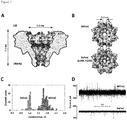
- Figure 1A The double mutant D10R, K159E of FraC (ReFraC) displayed near wild type-levels of hemolytic activity ( Figure 5 ) and yielded uniform pores ( Figure 1C ), albeit with altered I-V relationship compared to WtFraC ( Figure 4B and Figure 1D , bottom).
- dsDNA double stranded DNA
- oligo I with a biotin group attached at the 5'-terminus with the sequence bio-5'-A20-GTGCTACGACTCTCTGTGTG-C20-3' and a short oligo II with reverse complement sequence to the underlined part of oligo I. Annealing yielded an A(dsDNA)C substrate: a 20 base pair long central segment of dsDNA, flanked by A20 and C20 ssDNA segments.
- NA:A(dsDNA)C blockades became permanent both at
- Section A herein above shows that an ⁇ -helical pore-forming toxin from an actinoporin protein family Fragaceatoxin C (FraC) is advantageously used for polynucleotide analysis.
- Fragaceatoxin C Fragaceatoxin C
- Section B demonstrates that FraC nanopores are also suitable to recognize proteins, e.g. biomarkers, in the form of oligopeptides ( ⁇ 10 or fewer amino acids), polypeptides (>10 amino acids) and folded proteins (> 50 amino acids).
- proteins e.g. biomarkers
- EGF( ⁇ 98%, CYT-217) was obtained from PROSPEC. 1,2-diphytanoyl- sn -glycero-3-phosphocholine(DPhPC, 850356P) and sphingomyelin (Brain, Porcine, 860062) were purchased from Avanti Polar lipids. Potassium chloride( ⁇ 99%, Lot#BCBL9989V) was bought from Fluka. Citric acid( ⁇ 99%, Lot#A0365028) was obtained from ACROS. All polypeptide biomarkers and chemicals were used directly without further purification.
- Tris, pH 7.5 buffer below used was prepared with the formula from Trizma® protocol: 1.902 g Trizma® HCl and 0.354 g Trizma® Base dissolved in 1 liter H 2 O to be 15 mM Tris, pH 7.5.
- a gene encoding FraC with a C-terminal His6 tag was cloned into a pT7-SC1 expression plasmid 1 using NcoI and HindIII restriction digestion sites.
- the plasmid was transferred into E.cloni ® EXPRESS BL21(DE3) competent cell by electroporation.
- Transformants were harvested from the LB agar plate containing 100 mg/l ampicillin after overnight incubation at 37°C, and inoculated into 200 ml fresh liquid 2-YT media with 100 mg/l ampicillin.
- the cell culture was grown at 37°C, with 220 rpm shaking to an optical density at 600 nm of 0.8, then 0.5 mM IPTG was added to the cell culture.
- Cells were recovered by 4,000 RPM centrifugation for 30 minutes at 4°C and the cell pellets were kept at -80°C.
- 50-100 ml of cell culture pellet was thawed at room temperature, resuspended with 30 ml lysis buffer (15 mM Tris pH 7.5, 1 mM MgCl 2 , 4 M Urea, 0.2 mg/ml lysozyme and 0.05 units/ml DNase) and mixed vigorously with a vertex shaker for 1 hour.
- the suspension was sonicated for 2 minutes (duty cycle 10%, output control 3 of a Branson Sonifier 450).
- the crude lysate was then centrifuged at 6,500 RPM, 20 minutes at 4°C.
- the supernatant (containing FraC monomers) was transferred to a 50 ml falcon tube containing a 100 ⁇ l of Ni-NTA resin (Qiagen, stored at 4°C, and suspended before pipetting out 100 ⁇ l), which was pre-washed with 3 ml of washing buffer (10 mM imidazole, 150 mM NaCl, 15 mM Tris, pH 7.5), and incubated at room temperature for 1 hour with gentle mixing. The resin was spun down at 4,000 RPM for 5 minutes at 4°C.
- Ni-NTA resin within ⁇ 5 ml of buffer was transferred to a Micro Bio Spin column (Bio-Rad) at RT.
- the Ni-NTA beads were washed with 10 ml wash buffer and the protein was eluded with 500 ⁇ l of 300 mM imidazole. Protein concentration was determined with NanoDrop 2000 (Thermo Scientific). The monomers were stored at 4°C.
- sphingomyelin 20 mg sphingomyelin (Brain, Porcine, Avanti Polar lipids) was mixed with 20 mg of 1,2-diphytanoyl- sn -glycero-3-phosphocholine (DPhPC, Avanti Polar lipids) and dissolved in 4 ml pentane (Sigma) containing 0.5% v/v ethanol. This lipid mixture was placed to a round flask and rotated slowly near a hair dryer to disperse the lipid well around the wall to evaporate the solvent. The flask was kept open at room temperature for another 30 minutes to let the solvent to evaporate completely.
- DPhPC 1,2-diphytanoyl- sn -glycero-3-phosphocholine
- Frozen liposomes were sonicated after thawing and mixed with monomeric FraC in a mass ratio 1:1.
- the FraC and liposome mixture was sonicated in a water bath for ⁇ 30 seconds and then kept at 37°C for 30 minutes.
- the proteo-liposome was solubilized with 0.6% LADO( N,N- Dimethyldodecylamine N -oxide, 5% w/v stock solution in water), then transferred to a 50 ml falcon tube and diluted 20 times with buffer (150 mM NaCl, 15 mM Tris, pH 7.5, 0.02% DDM). 100 ⁇ l of pre-washed Ni-NTA resin (Qiagen) was added to the diluted protein/liposome mixture.
- Planar bilayer recordings were collected using a patch clamp amplifier (Axopatch 200B, Axon Instruments) and the data digitized with a Digidata 1440 A/D converter (Axon Instruments). Data were acquired by using Clampex 10.4 software (Molecular Devices) and the subsequent analysis was carried out with Clampfit software (Molecular Devices). Events duration (dwell time), time between two events (inter-event time), blocked current levels and open pore levels were detected by "single channel search" function. The current levels of blockades were referred as I B , while the open pore current was referred as I o . Ires%, defined as (I B /I O ))*100, was used to describe the extent of blockade caused by different biomarkers. Average Inter-event times were calculated by binning the inter-event times and applying a single exponential fit to cumulative distributions.
- Ion permeability ratio K + /Cl -
- K + /Cl - the Goldman-Hodgkin-Katz equation
- V r the reverse potential
- the solutions were mixed and I-V curves collected from -30 mV to 30 mV with 1 mV steps.
- the directionality of the ion selectivity was also tested by using high KCl concentration in trans chamber and low KCl concentration in the cis chamber.
- Ag/AgCl electrodes were surrounded with 2.5% agarose bridges containing 2.5 M NaCl.
- Endothelin 1 blockades started to be appear at pH 6.4, and their capture frequency increased linearly with decreasing the pH (from 0.6 ⁇ 0.2 events s -1 ⁇ M -1 at pH 6.4 to 10.8 ⁇ 2.3 events s -1 ⁇ M -1 at pH 4.4).
- Protein blockades were observed at -50 mV in pH 7.5 buffer (1 M KCl, 15 mM Tris), although they became homogeneous when we increased the potential to -100 mV (45.2 ⁇ 19.1 events s -1 ⁇ M -1 , dwell time: 12.0 ⁇ 5.7 ms), while no capture was observed at positive applied potentials ( Figure 10C ).
- Example 6 Ion selectivity and electrostatic potential of FraC nanopores.
- Example 7 Biomarker detection with the WtFraC nanopore.
- the WtFraC nanopores were used to detect a larger range of protein biomarkers including ⁇ 2-microglobulin, a 11.6 kDa (99 amino acids) biomarker for peripheral arterial disease (17), human EGF, a 6.2 kDa (53 amino acids) biomarker for chronic kidney disease (18), and angiotensin I, a 1.3kD (10 amino acids) biomarker for hypertensive crisis (19).
- Example 8 Near-isoform oligopeptide discrimination.
Landscapes
- Health & Medical Sciences (AREA)
- Chemical & Material Sciences (AREA)
- Life Sciences & Earth Sciences (AREA)
- Engineering & Computer Science (AREA)
- Organic Chemistry (AREA)
- Physics & Mathematics (AREA)
- Molecular Biology (AREA)
- Proteomics, Peptides & Aminoacids (AREA)
- General Health & Medical Sciences (AREA)
- Biochemistry (AREA)
- Zoology (AREA)
- Biophysics (AREA)
- Biomedical Technology (AREA)
- Analytical Chemistry (AREA)
- Immunology (AREA)
- Wood Science & Technology (AREA)
- Genetics & Genomics (AREA)
- Medicinal Chemistry (AREA)
- General Physics & Mathematics (AREA)
- Pathology (AREA)
- General Engineering & Computer Science (AREA)
- Bioinformatics & Cheminformatics (AREA)
- Microbiology (AREA)
- Biotechnology (AREA)
- Tropical Medicine & Parasitology (AREA)
- Toxicology (AREA)
- Food Science & Technology (AREA)
- Urology & Nephrology (AREA)
- Hematology (AREA)
- Gastroenterology & Hepatology (AREA)
- Nanotechnology (AREA)
- Spectroscopy & Molecular Physics (AREA)
- Dispersion Chemistry (AREA)
- Electrochemistry (AREA)
- Chemical Kinetics & Catalysis (AREA)
- Peptides Or Proteins (AREA)
- Measuring Or Testing Involving Enzymes Or Micro-Organisms (AREA)
- Investigating Or Analyzing Materials By The Use Of Electric Means (AREA)
- Investigating Or Analysing Biological Materials (AREA)
- Apparatus Associated With Microorganisms And Enzymes (AREA)
Applications Claiming Priority (3)
| Application Number | Priority Date | Filing Date | Title |
|---|---|---|---|
| US201662360999P | 2016-07-12 | 2016-07-12 | |
| PCT/NL2017/050331 WO2018012963A1 (en) | 2016-07-12 | 2017-05-24 | Biological nanopores for biopolymer sensing and sequencing based on frac actinoporin |
| EP17734851.3A EP3485029B1 (en) | 2016-07-12 | 2017-05-24 | Biological nanopores for biopolymer sensing and sequencing based on frac actinoporin |
Related Parent Applications (1)
| Application Number | Title | Priority Date | Filing Date |
|---|---|---|---|
| EP17734851.3A Division EP3485029B1 (en) | 2016-07-12 | 2017-05-24 | Biological nanopores for biopolymer sensing and sequencing based on frac actinoporin |
Publications (1)
| Publication Number | Publication Date |
|---|---|
| EP3828280A1 true EP3828280A1 (en) | 2021-06-02 |
Family
ID=59270080
Family Applications (2)
| Application Number | Title | Priority Date | Filing Date |
|---|---|---|---|
| EP20206642.9A Pending EP3828280A1 (en) | 2016-07-12 | 2017-05-24 | Biological nanopores for biopolymer sensing and sequencing based on frac actinoporin |
| EP17734851.3A Active EP3485029B1 (en) | 2016-07-12 | 2017-05-24 | Biological nanopores for biopolymer sensing and sequencing based on frac actinoporin |
Family Applications After (1)
| Application Number | Title | Priority Date | Filing Date |
|---|---|---|---|
| EP17734851.3A Active EP3485029B1 (en) | 2016-07-12 | 2017-05-24 | Biological nanopores for biopolymer sensing and sequencing based on frac actinoporin |
Country Status (8)
| Country | Link |
|---|---|
| US (2) | US11312755B2 (https=) |
| EP (2) | EP3828280A1 (https=) |
| JP (4) | JP7019665B2 (https=) |
| CN (2) | CN109890980A (https=) |
| AU (3) | AU2017295442B2 (https=) |
| CA (1) | CA3030704A1 (https=) |
| ES (1) | ES2843099T3 (https=) |
| WO (1) | WO2018012963A1 (https=) |
Families Citing this family (16)
| Publication number | Priority date | Publication date | Assignee | Title |
|---|---|---|---|---|
| LU100795B1 (en) * | 2018-05-14 | 2019-11-14 | Diatron MI PLC | Immunoassay for whole blood samples |
| WO2020055246A1 (en) | 2018-09-11 | 2020-03-19 | Rijksuniversiteit Groningen | Biological nanopores having tunable pore diameters and uses thereof as analytical tools |
| NL2023062B1 (en) | 2019-05-03 | 2020-11-30 | Univ Delft Tech | Protein current trace signal acquisition using a nanopore |
| EP3771687A1 (en) | 2019-07-30 | 2021-02-03 | Rijksuniversiteit Groningen | Reversible photo-controlled frac nanopores, modified frac monomers, methods for preparing the same, and uses thereof |
| US20220412948A1 (en) | 2019-11-19 | 2022-12-29 | Rijksuniversiteit Groningen | Artificial nanopores and uses and methods relating thereto |
| US20230176034A1 (en) | 2020-03-11 | 2023-06-08 | Rijksuniversiteit Groningen | Engineered PlyAB Nanopores and Uses Thereof |
| CN112480204A (zh) * | 2020-04-13 | 2021-03-12 | 南京大学 | 一种采用Aerolysin纳米孔道的蛋白质/多肽测序方法 |
| CN111413383B (zh) * | 2020-04-20 | 2021-03-02 | 深圳市梅丽纳米孔科技有限公司 | 一种构建双识别位点纳米孔的方法 |
| WO2022038144A1 (en) * | 2020-08-18 | 2022-02-24 | Kemijski inštitut | Novel nanopore-forming polypeptides |
| US20240377380A1 (en) * | 2021-03-31 | 2024-11-14 | Illumina, Inc. | Nanopore sensor devices |
| EP4326257A4 (en) * | 2021-04-19 | 2025-03-12 | ChemoCentryx, Inc. | AZETIDINYLACETAMIDES AS CXCR7 INHIBITORS |
| WO2024078621A1 (zh) * | 2022-10-14 | 2024-04-18 | 北京普译生物科技有限公司 | 一种pht纳米孔突变体蛋白及其应用 |
| KR20250116014A (ko) | 2022-10-28 | 2025-07-31 | 리엑스유니버시테이트 그로닝겐 | 나노기공-기반 단백질 분석 |
| EP4662489A1 (en) * | 2023-02-07 | 2025-12-17 | Oxford University Innovation Limited | Method of characterising a peptide, polypeptide or protein using a nanopore |
| CN116223583B (zh) * | 2023-02-28 | 2026-04-28 | 中国科学院重庆绿色智能技术研究院 | 基于固态纳米孔技术的抗衰寡肽的单分子检测方法 |
| GB202305524D0 (en) * | 2023-04-14 | 2023-05-31 | Oxford Nanopore Tech Plc | Novel pore monomers and pores |
Citations (1)
| Publication number | Priority date | Publication date | Assignee | Title |
|---|---|---|---|---|
| WO2010034018A2 (en) * | 2008-09-22 | 2010-03-25 | University Of Washington | Msp nanopores and related methods |
Family Cites Families (2)
| Publication number | Priority date | Publication date | Assignee | Title |
|---|---|---|---|---|
| US6362002B1 (en) * | 1995-03-17 | 2002-03-26 | President And Fellows Of Harvard College | Characterization of individual polymer molecules based on monomer-interface interactions |
| WO2012009578A2 (en) | 2010-07-14 | 2012-01-19 | The Curators Of The University Of Missouri | Nanopore-facilitated single molecule detection of nucleic acids |
-
2017
- 2017-05-24 EP EP20206642.9A patent/EP3828280A1/en active Pending
- 2017-05-24 US US16/317,119 patent/US11312755B2/en active Active
- 2017-05-24 CN CN201780052410.2A patent/CN109890980A/zh active Pending
- 2017-05-24 AU AU2017295442A patent/AU2017295442B2/en active Active
- 2017-05-24 EP EP17734851.3A patent/EP3485029B1/en active Active
- 2017-05-24 ES ES17734851T patent/ES2843099T3/es active Active
- 2017-05-24 JP JP2019500879A patent/JP7019665B2/ja active Active
- 2017-05-24 CA CA3030704A patent/CA3030704A1/en active Pending
- 2017-05-24 WO PCT/NL2017/050331 patent/WO2018012963A1/en not_active Ceased
- 2017-05-24 CN CN202411913696.7A patent/CN119753114A/zh active Pending
-
2022
- 2022-02-02 JP JP2022014867A patent/JP2022064983A/ja active Pending
- 2022-03-17 US US17/696,949 patent/US20220242922A1/en active Pending
-
2024
- 2024-01-24 AU AU2024200435A patent/AU2024200435A1/en not_active Withdrawn
- 2024-01-24 AU AU2024200450A patent/AU2024200450A1/en active Pending
- 2024-05-14 JP JP2024078970A patent/JP2024105542A/ja active Pending
-
2025
- 2025-10-29 JP JP2025182445A patent/JP2026021428A/ja active Pending
Patent Citations (1)
| Publication number | Priority date | Publication date | Assignee | Title |
|---|---|---|---|---|
| WO2010034018A2 (en) * | 2008-09-22 | 2010-03-25 | University Of Washington | Msp nanopores and related methods |
Non-Patent Citations (24)
| Title |
|---|
| A. BARLICI. GUTIERREZ-AGUIRREJ. M. CAAVEIROA. CRUZM. B. RUIZ-ARGUELLOJ. PEREZ-GILJ. M. GONZALEZ-MANAS, J BIOL CHEM, vol. 279, 2004, pages 34209 - 34216 |
| A. H. LASZLOI. M. DERRINGTONB. C. ROSSH. BRINKERHOFFA. ADEYI. C. NOVAJ. M. CRAIGK. W. LANGFORDJ. M. SAMSONR. DAZA, NAT BIOTECHNOL, vol. 32, 2014, pages 829 - 833 |
| B. BAKRACI. GUTIERREZ-AGUIRREZ. PODLESEKA. F. SONNENR. J. GILBERTP. MACEKJ. H. LAKEYG. ANDERLUH, J BIOL CHEM, vol. 283, 2008, pages 18665 - 18677 |
| BELLOMIO A ET AL: "Purification, cloning and characterization of fragaceatoxin C, a novel actinoporin from the sea anemone Actinia fragacea", TOXICON, ELMSFORD, NY, US, vol. 54, no. 6, 1 November 2009 (2009-11-01), pages 869 - 880, XP026499390, ISSN: 0041-0101, [retrieved on 20090627], DOI: 10.1016/J.TOXICON.2009.06.022 * |
| C. CAOY. L. YINGZ. L. HUD. F. LIAOH. TIANY. T. LONG, NATURE NANOTECHNOLOGY, 2016 |
| D. STODDARTA. J. HERONE. MIKHAILOVAG. MAGLIAH. BAYLEY, PROC NATL ACAD SCI USA, vol. 106, 2009, pages 7702 - 7707 |
| D. STODDARTA. J. HERONJ. KLINGELHOEFERE. MIKHAILOVAG. MAGLIAH. BAYLEY, NANO LETT, vol. 10, 2010, pages 3633 - 3637 |
| D. STODDARTG. MAGLIAE. MIKHAILOVAA. J. HERONH. BAYLEY, ANGEW CHEM INT ED ENGL, vol. 49, 2010, pages 556 - 559 |
| D. WENDELLP. JINGJ. GENGV. SUBRAMANIAMT. J. LEEC. MONTEMAGNOP. GUO, NATURE NANOTECHNOLOGY, vol. 4, 2009, pages 765 - 772 |
| E. A. MANRAOI. M. DERRINGTONM. PAVLENOKM. NIEDERWEISJ. H. GUNDLACH, PLOS ONE, vol. 6, 2011, pages e25723 |
| J. J. KASIANOWICZE. BRANDIND. BRANTOND. W. DEAMER, PROC NATL ACAD SCI U S A, vol. 93, 1996, pages 13770 - 13773 |
| J. MATHEH. VISRAMV. VIASNOFFY. RABINA. MELLER, BIOPHYS J, vol. 87, 2004, pages 3205 - 3212 |
| K. TANAKAJ. M. CAAVEIROK. MORANTEJ. M. GONZALEZ-MANASK. TSUMOTO, NATURE COMMUNICATIONS, vol. 6, 2015, pages 6337 |
| K. TANAKAJ. M. CAAVEIROK. TSUMOTO, BIOCHEMISTRY, vol. 54, 2015, pages 6863 - 6866 |
| KOJI TANAKA ET AL: "Structural basis for self-assembly of a cytolytic pore lined by protein and lipid", NATURE COMMUNICATIONS, vol. 6, 26 February 2015 (2015-02-26), pages 6337, XP055401446, DOI: 10.1038/ncomms7337 * |
| KOLDO MORANTE ET AL: "A Pore-Forming Toxin Requires a Specific Residue for Its Activity in Membranes with Particular Physicochemical Properties", JOURNAL OF BIOLOGICAL CHEMISTRY, vol. 290, no. 17, 24 April 2015 (2015-04-24), US, pages 10850 - 10861, XP055401448, ISSN: 0021-9258, DOI: 10.1074/jbc.M114.615211 * |
| L. FRANCESCHINIM. SOSKINEA. BIESEMANSG. MAGLIA, NATURE COMMUNICATIONS, vol. 4, 2013, pages 2415 |
| M. AKESOND. BRANTONJ. J. KASIANOWICZE. BRANDIND. W. DEAMER, BIOPHYS J, vol. 77, 1999, pages 3227 - 3233 |
| M. FALLERM. NIEDERWEISG. E. SCHULZ, SCIENCE, vol. 303, 2004, pages 1189 - 1192 |
| M. SOSKINEA. BIESEMANSM. DE MAEYERG. MAGLIA, J AM CHEM SOC, vol. 135, 2013, pages 13456 - 13463 |
| N. ANA. M. FLEMINGE. G. MIDDLETONC. J. BURROWS, PROC NATL ACAD SCI USA, vol. 111, 2014, pages 14325 - 14331 |
| P. SCHONA. J. GARCIA-SAEZP. MALOVRHK. BACIAG. ANDERLUHP. SCHWILLE, BIOPHYS J, vol. 95, 2008, pages 691 - 698 |
| R. F. PURNELLJ. J. SCHMIDT, ACS NANO, vol. 3, 2009, pages 2533 - 2538 |
| T. Z. BUTLERM. PAVLENOKI. M. DERRINGTONM. NIEDERWEISJ. H. GUNDLACH, PROC NATL ACAD SCI U S A, vol. 105, 2008, pages 20647 - 20652 |
Also Published As
| Publication number | Publication date |
|---|---|
| CN109890980A (zh) | 2019-06-14 |
| JP2026021428A (ja) | 2026-02-10 |
| US20220242922A1 (en) | 2022-08-04 |
| US11312755B2 (en) | 2022-04-26 |
| JP2024105542A (ja) | 2024-08-06 |
| AU2017295442B2 (en) | 2023-10-26 |
| EP3485029A1 (en) | 2019-05-22 |
| ES2843099T3 (es) | 2021-07-15 |
| WO2018012963A1 (en) | 2018-01-18 |
| US20190292235A1 (en) | 2019-09-26 |
| EP3485029B1 (en) | 2020-11-11 |
| AU2024200450A1 (en) | 2024-02-15 |
| CN119753114A (zh) | 2025-04-04 |
| AU2017295442A1 (en) | 2019-02-07 |
| AU2017295442A2 (en) | 2019-02-21 |
| JP7019665B2 (ja) | 2022-02-15 |
| AU2024200435A1 (en) | 2024-02-08 |
| JP2019533637A (ja) | 2019-11-21 |
| BR112019000680A2 (pt) | 2019-04-24 |
| CA3030704A1 (en) | 2018-01-18 |
| JP2022064983A (ja) | 2022-04-26 |
Similar Documents
| Publication | Publication Date | Title |
|---|---|---|
| US20220242922A1 (en) | Biological Nanopores for Biopolymer Sensing and Sequencing Based on FRAC Actinoporin | |
| US20240345073A9 (en) | Nanopore biosensors for detection of proteins and nucleic acids | |
| JP6776366B2 (ja) | 変異体ポア | |
| CN109072295B (zh) | 修饰的纳米孔,包含其的组合物及其应用 | |
| JP2022500074A (ja) | 調節可能な孔直径を有する生物学的ナノポアと分析ツールとしてのその使用 | |
| JP2020530276A (ja) | 新規タンパク質細孔 | |
| JP2022518095A (ja) | 細孔 | |
| HK40052928A (en) | Biological nanopores for biopolymer sensing and sequencing based on frac actinoporin | |
| BR112019000680B1 (pt) | Nanoporos biológicos para detecção de biopolímeros e sequenciamento baseado em actinoporina fragaceatoxina c | |
| Class et al. | Patent application title: NANOPORE BIOSENSORS FOR DETECTION OF PROTEINS AND NUCLEIC ACIDS |
Legal Events
| Date | Code | Title | Description |
|---|---|---|---|
| PUAI | Public reference made under article 153(3) epc to a published international application that has entered the european phase |
Free format text: ORIGINAL CODE: 0009012 |
|
| STAA | Information on the status of an ep patent application or granted ep patent |
Free format text: STATUS: REQUEST FOR EXAMINATION WAS MADE |
|
| 17P | Request for examination filed |
Effective date: 20201110 |
|
| AC | Divisional application: reference to earlier application |
Ref document number: 3485029 Country of ref document: EP Kind code of ref document: P |
|
| AK | Designated contracting states |
Kind code of ref document: A1 Designated state(s): AL AT BE BG CH CY CZ DE DK EE ES FI FR GB GR HR HU IE IS IT LI LT LU LV MC MK MT NL NO PL PT RO RS SE SI SK SM TR |
|
| REG | Reference to a national code |
Ref country code: HK Ref legal event code: DE Ref document number: 40052928 Country of ref document: HK |
|
| STAA | Information on the status of an ep patent application or granted ep patent |
Free format text: STATUS: EXAMINATION IS IN PROGRESS |
|
| 17Q | First examination report despatched |
Effective date: 20230217 |
|
| P01 | Opt-out of the competence of the unified patent court (upc) registered |
Effective date: 20230526 |