EP3822360B1 - Verfahren zur bestimmung der sensibilität eines bakterienstamms gegenüber einem bakteriophagen virus - Google Patents
Verfahren zur bestimmung der sensibilität eines bakterienstamms gegenüber einem bakteriophagen virus Download PDFInfo
- Publication number
- EP3822360B1 EP3822360B1 EP20207254.2A EP20207254A EP3822360B1 EP 3822360 B1 EP3822360 B1 EP 3822360B1 EP 20207254 A EP20207254 A EP 20207254A EP 3822360 B1 EP3822360 B1 EP 3822360B1
- Authority
- EP
- European Patent Office
- Prior art keywords
- bacteriophages
- sample
- bacteria
- image
- interest
- Prior art date
- Legal status (The legal status is an assumption and is not a legal conclusion. Google has not performed a legal analysis and makes no representation as to the accuracy of the status listed.)
- Active
Links
Images




Classifications
-
- C—CHEMISTRY; METALLURGY
- C12—BIOCHEMISTRY; BEER; SPIRITS; WINE; VINEGAR; MICROBIOLOGY; ENZYMOLOGY; MUTATION OR GENETIC ENGINEERING
- C12Q—MEASURING OR TESTING PROCESSES INVOLVING ENZYMES, NUCLEIC ACIDS OR MICROORGANISMS; COMPOSITIONS OR TEST PAPERS THEREFOR; PROCESSES OF PREPARING SUCH COMPOSITIONS; CONDITION-RESPONSIVE CONTROL IN MICROBIOLOGICAL OR ENZYMOLOGICAL PROCESSES
- C12Q1/00—Measuring or testing processes involving enzymes, nucleic acids or microorganisms; Compositions therefor; Processes of preparing such compositions
- C12Q1/02—Measuring or testing processes involving enzymes, nucleic acids or microorganisms; Compositions therefor; Processes of preparing such compositions involving viable microorganisms
- C12Q1/04—Determining presence or kind of microorganism; Use of selective media for testing antibiotics or bacteriocides; Compositions containing a chemical indicator therefor
-
- C—CHEMISTRY; METALLURGY
- C12—BIOCHEMISTRY; BEER; SPIRITS; WINE; VINEGAR; MICROBIOLOGY; ENZYMOLOGY; MUTATION OR GENETIC ENGINEERING
- C12Q—MEASURING OR TESTING PROCESSES INVOLVING ENZYMES, NUCLEIC ACIDS OR MICROORGANISMS; COMPOSITIONS OR TEST PAPERS THEREFOR; PROCESSES OF PREPARING SUCH COMPOSITIONS; CONDITION-RESPONSIVE CONTROL IN MICROBIOLOGICAL OR ENZYMOLOGICAL PROCESSES
- C12Q1/00—Measuring or testing processes involving enzymes, nucleic acids or microorganisms; Compositions therefor; Processes of preparing such compositions
- C12Q1/02—Measuring or testing processes involving enzymes, nucleic acids or microorganisms; Compositions therefor; Processes of preparing such compositions involving viable microorganisms
- C12Q1/18—Testing for antimicrobial activity of a material
-
- C—CHEMISTRY; METALLURGY
- C12—BIOCHEMISTRY; BEER; SPIRITS; WINE; VINEGAR; MICROBIOLOGY; ENZYMOLOGY; MUTATION OR GENETIC ENGINEERING
- C12Q—MEASURING OR TESTING PROCESSES INVOLVING ENZYMES, NUCLEIC ACIDS OR MICROORGANISMS; COMPOSITIONS OR TEST PAPERS THEREFOR; PROCESSES OF PREPARING SUCH COMPOSITIONS; CONDITION-RESPONSIVE CONTROL IN MICROBIOLOGICAL OR ENZYMOLOGICAL PROCESSES
- C12Q1/00—Measuring or testing processes involving enzymes, nucleic acids or microorganisms; Compositions therefor; Processes of preparing such compositions
- C12Q1/70—Measuring or testing processes involving enzymes, nucleic acids or microorganisms; Compositions therefor; Processes of preparing such compositions involving virus or bacteriophage
-
- G—PHYSICS
- G01—MEASURING; TESTING
- G01N—INVESTIGATING OR ANALYSING MATERIALS BY DETERMINING THEIR CHEMICAL OR PHYSICAL PROPERTIES
- G01N21/00—Investigating or analysing materials by the use of optical means, i.e. using sub-millimetre waves, infrared, visible or ultraviolet light
- G01N21/17—Systems in which incident light is modified in accordance with the properties of the material investigated
- G01N21/47—Scattering, i.e. diffuse reflection
- G01N21/49—Scattering, i.e. diffuse reflection within a body or fluid
- G01N21/53—Scattering, i.e. diffuse reflection within a body or fluid within a flowing fluid, e.g. smoke
- G01N21/534—Scattering, i.e. diffuse reflection within a body or fluid within a flowing fluid, e.g. smoke by measuring transmission alone, i.e. determining opacity
-
- Y—GENERAL TAGGING OF NEW TECHNOLOGICAL DEVELOPMENTS; GENERAL TAGGING OF CROSS-SECTIONAL TECHNOLOGIES SPANNING OVER SEVERAL SECTIONS OF THE IPC; TECHNICAL SUBJECTS COVERED BY FORMER USPC CROSS-REFERENCE ART COLLECTIONS [XRACs] AND DIGESTS
- Y02—TECHNOLOGIES OR APPLICATIONS FOR MITIGATION OR ADAPTATION AGAINST CLIMATE CHANGE
- Y02A—TECHNOLOGIES FOR ADAPTATION TO CLIMATE CHANGE
- Y02A50/00—TECHNOLOGIES FOR ADAPTATION TO CLIMATE CHANGE in human health protection, e.g. against extreme weather
- Y02A50/30—Against vector-borne diseases, e.g. mosquito-borne, fly-borne, tick-borne or waterborne diseases whose impact is exacerbated by climate change
Definitions
- the technical field of the invention is the characterization of the ability of a bacterial strain to be infected, then lysed, by a bacteriophage virus.
- Phage therapy is the use of bacteriophage viruses, commonly referred to as phages, to treat bacterial infections. It involves the use of lytic phages, which are capable of infecting certain bacteria and then lysing them. This is a relatively old treatment, discovered and practiced before the emergence of antibiotics. However, in most countries, this treatment has been somewhat neglected, in favor of the use of antibiotics. Currently, phage therapy is considered a promising therapeutic approach for treating certain bacterial infections, particularly when bacteria are resistant to antibiotics.
- phage therapy has been demonstrated for the treatment of infections by bacteria such as Pseudomonas aeruginosa, Escherichia coli, or Staphilococcus Aureus, in applications such as skin grafts for major burns or pathologies linked to diabetes.
- bacteria such as Pseudomonas aeruginosa, Escherichia coli, or Staphilococcus Aureus
- the phages used in phage therapy applications are lytic phages, meaning that when a bacterium has been infected by a phage, it is lysed. Lysis results from the production of endolysins.
- the metabolism of the bacterium leads to a replication of the number of phages, in the form of virions. These are released following the lysis of the bacterium. An infection is therefore followed by an increase in the number of phages capable of infecting and then lysing other bacteria. This results in a reduction or even a halt in bacterial proliferation.
- phagograms tests, usually referred to as "phagograms,” are performed in the laboratory. This involves testing the ability of a bacterial strain to be infected, then lysed, by a phage.
- Laboratory tests require testing different viral strains, with different phage-to-bacteria ratios.
- the number of viral strains can range from 5 to 100, while 5 to 10 phage-to-bacteria ratios are typically tested for each viral strain. Therefore, the number of tests required is high.
- the inventors propose a simple method for analyzing the sensitivity of a bacterial strain to a viral strain.
- the method allows for monitoring bacterial development and early detection of bacterial lysis by phages. It can also simultaneously address several combinations between quantities of phages, possibly from different viral strains, and quantities of bacteria, possibly from different bacterial strains.
- the objective is to reduce the time spent detecting the sensitivity of a bacterial strain to a viral strain.
- An object of the invention is a method for determining the sensitivity of a bacterial strain to a viral strain of bacteriophages, according to claim 1.
- the emission spectral bandwidth is preferably less than 100 nm or 50 nm.
- the distance between the sample and the image sensor is preferably less than 5 cm or less than 1 cm.
- the texturing descriptor can be determined based on one or more regions of interest in the image.
- each spatial zone is associated with a region of interest of each acquired image, two different spatial zones being associated with two different regions of interest, so that the analysis of the same acquired image makes it possible to obtain information relating to the sensitivity of a bacterial strain with regard to a viral strain of bacteriophages, and this in different spatial zones.
- the sample may comprise more than 10, or even more than 100, different spatial zones, separated from each other, each spatial zone being parameterized by three parameters corresponding respectively to the bacterial strain of interest, to the viral strain of the bacteriophages and to the concentration of the bacteriophages, at least one parameter of two different spatial zones being different.
- the sample can be distributed in different fluidic chambers, of a fluidic map, each fluidic chamber corresponding to a spatial area of the sample.
- the sample may be formed by depositing drops on the surface of an agar plate at different positions, the drops being spaced apart from each other, such that a spatial area is defined by each drop position.
- the agar plate may contain bacteria and the drops may contain bacteriophages. Alternatively, the agar plate may contain bacteriophages and the drops may contain bacteria.
- the image defocus distance and/or the object defocus distance is preferably less than 5 mm, or even less than 1 mm, or even less than 500 ⁇ m.
- the sample plane is a plane extending into the sample. It is preferably the closest surface of the sample of the image sensor. According to this embodiment, during step c), the image is acquired in a defocused configuration.
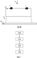
- FIG. 1 shows a device for implementing the invention.
- a sample 20 is placed between a light source 10 and an image sensor 30.
- the light source 10 produces an incident light wave 11, the latter propagating to the sample.
- the incident light wave 11 reaches the sample in the form of a plane wave, or considered to be plane.
- the incident light wave 11 extends along an illumination spectral band ⁇ extending into the visible range. It is preferably between 450 nm and 650 nm, and more preferably between 500 nm and 600 nm. It is in fact between 500 nm and 600 nm that the scattering of the incident light wave 11 by the bacteria is maximum.
- the spectral width of the illumination spectral band is less than 50 nm, or even less than 40 nm.
- spectral width is meant the width at half-maximum, usually designated by the acronym FWHM (Full Width at Half Maximum).
- the light source 10 is for example a light-emitting diode or a laser diode. It can also be a liquid crystal screen, the advantage being to have an extended luminous surface, from a few cm 2 to a few tens of cm 2 , producing uniform illumination.
- a band-pass filter 16 can be interposed between the light source 10 and the sample 20, so as to adjust the spectral band of the light wave reaching the sample.
- the distance D between the light source 10 and the sample 20 is adjusted such that the light source is considered to be point-like. It may be between 5 cm and 20 or 30 cm.
- a diaphragm 17, defining an opening with a diameter less than 100 ⁇ m or 200 ⁇ m, may be interposed between the light source 10 and the sample 20.
- an optical fiber may be interposed between the light source and the sample, so as to form a point-like light source.
- the image sensor 30 may be a CCD or CMOS type sensor.
- the detection surface is greater than 10 mm 2 , or even greater than 1 cm 2 .
- the inventors used a sensor whose detection surface is 14.9 mm x 22.3 mm, or of the order of 3 cm 2 . This makes it possible to obtain an image with a high field of observation.
- the distance d between the sample 20 and the image sensor 30 is as small as possible. It is preferably less than 1 cm.
- the sample 20 is placed in contact with the image sensor, that is to say in contact with a protective glass of the image sensor, or a few millimeters from the latter.
- the image sensor 30 is arranged in a lensless imaging configuration. This makes it possible to maximize the field of observation of the image sensor. Each image then makes it possible to address a large volume of sample.
- the image sensor can be a monochrome sensor or a color sensor. It allows formation of an image representative of the exposure wave 14.
- the image sensor 30 is connected to a processing unit 40, receiving the images acquired by the image sensor.
- the processing unit 40 is connected to a memory 42 in which instructions are stored for implementing certain image processing steps described below.
- the thickness ⁇ of the sample 20 can vary between 100 ⁇ m and 1 cm or 2 cm.
- the configuration of the sample, and its thickness, can vary according to the embodiments described below.
- the sample comprises a support, in which bacteria, from at least one bacterial source of interest, are brought into contact with phages, of a viral strain. The invention is thus used to determine the viral strain and/or the concentration of phages allowing lysis of the bacteria of the bacterial strain of interest.
- the sample comprises a mixture of bacteria and phages mixed in a solution, preferably an aqueous solution.
- the aqueous solution comprises nutrients, allowing the bacteria to develop.
- the aqueous solution comprises a liquid nutrient medium, preferably non-specific, usually referred to as "broth". This may, for example, be trypticase soy, a Columbia medium, or a Luria-Bertani type medium.
- Each 22 i fluidic chamber contains an elementary sample 20 i which may be different from another elementary sample in another fluidic chamber.
- each elementary sample 20 i may thus contain phages of the same viral strain, according to a predefined concentration.
- At least one parameter is different, except in the case of replicates.
- the fluidic card 22 is for example made, at least in part, from a transparent plastic material, preferably biocompatible, for example COC (Cyclic Olefin Copolymer) or PMMA (Polymethylmetacrylate). It comprises transparent transverse walls 22 t , which confine each elementary chamber 22 i . The transverse walls preferably extend perpendicular to an axis Z along which the light wave 11 emitted by the light source 10 propagates.
- a transparent plastic material preferably biocompatible, for example COC (Cyclic Olefin Copolymer) or PMMA (Polymethylmetacrylate). It comprises transparent transverse walls 22 t , which confine each elementary chamber 22 i .
- the transverse walls preferably extend perpendicular to an axis Z along which the light wave 11 emitted by the light source 10 propagates.
- Each elementary sample contains an initial quantity of bacteria 2.
- the bacteria proliferate.
- this case corresponds to the fluidic chamber 22 1 .
- the phages are able to infect the bacteria of interest, and to cause their lysis, the number of bacteria of interest decreases, which corresponds to the fluidic chamber 22 2 .
- the increase or decrease in bacteria leads to an evolution in the transport of light through each elementary sample.
- the image sensor comprises pixels 31. Groups of pixels 31 i can be defined, each pixel of the same group being exposed to an exposure light wave 14 i propagating from a fluidic chamber 22 i . Thus, each group of pixels 31 i is associated with a fluidic chamber 22 i and with an elementary sample 20 i .
- each group of pixels 31 i corresponds to a region of interest ROI i of the image acquired by the image sensor 30.
- region of interest it is meant parts of the image whose intensity is considered homogeneous or representative of the sample, or of a part of the sample.
- Each sample was collected in 9 mL of Tryptone Soy Broth nutrient medium (usually referred to by the acronym TSB - Trypcase Soy Broth), into which 250 ⁇ L of a culture solution of Pseudomonas putida ATCC12633, and 100 ⁇ L of a suspension of Pseudomonas virus gh1 phages had been introduced.
- a control chamber was not subjected to the addition of phages.
- a reference chamber was filled with the liquid nutrient medium, without bacteriophages or bacteria.
- the bacteriophage suspensions had an infectious titer equal to 6.10 9 pfu/mL, 6.10 8 pfu /mL and 6.10 7 pfu /mL respectively.
- the pfu unit is a unit known to those skilled in the art meaning plaque-forming unit, and usually referred to as plaque-forming unit.
- the determination of the titer of each phage suspension was carried out according to a reference method, using an agar medium. Each chamber was duplicated, except for the control chamber and the reference chamber.
- the fourth column C4 includes the reference chamber C4-1 and the control chamber C4-2 containing no phage.
- control chamber C4-2 the image is darkened. This is due to the proliferation of bacteria in the absence of phage.
- the constant 0.055 was added so that the optical density at the initial time corresponds to that measured by a spectrophotometer.
- the invention allows real-time monitoring of the action of a phage on bacteria, and this simultaneously on several elementary samples 20 i . It is possible to define, for each sample, a detection threshold, the crossing of which means that bacterial proliferation is suspected in the sample.
- a detection threshold can be defined from the average optical density measured on the control sample, as well as a dispersion indicator ⁇ , for example the standard deviation, at a measurement time or at different measurement times.
- the detection threshold can be equal to ⁇ + k ⁇ , k being a strictly positive real number, preferably greater than 1 or 2, and for example equal to 5.
- the establishment of such a detection threshold is easily automated and makes it possible to determine the presence of bacterial proliferation early, for example between 5 a.m. and 10 a.m.
- Other thresholds based on a comparison between the control sample and samples containing phages, can be defined.
- FIG. 2E schematizes a fluidic card 22 allowing an implementation of the invention.
- the fluidic card comprises fluidic chambers 22 i , as previously described, respectively closed by two transverse walls 22 t .
- One transverse wall can take the form of a transparent adhesive film.
- the other transverse wall 22 t can be made using a transparent material, preferably permeable to oxygen, so as to allow oxygenation of the fluidic chambers 22 i , necessary for the development of certain bacteria.
- at least one transverse wall delimiting each fluidic chamber is permeable to oxygen. It can for example be PDMS (Polydimethylsiloxane) or TPX (Polymethylpentene), the latter being described in the document EP0745667 .
- each fluid chamber 22 i is connected, by a fluid channel, to an inlet 22 i , and vent 22 e .
- the filling of each fluid chamber is carried out with a syringe, by introducing the solution containing the bacteria through the inlet 22 in .
- the vent 22 e allows air to be expelled when filling the fluid chamber.
- a filter blocking liquids and permeable to gases can be arranged at the vent 22 e .
- the mixture is then filtered, so as to retain the bacteria, the filtration size being able to be of the order of 0.2 ⁇ m.
- the bacterial strains are then distributed into different fluidic chambers, so as to end up with a single bacterial strain per well.
- the filtered solution is injected into each fluidic chamber, so as to identify the bacterial strain sensitive to the phage.
- This embodiment can be adapted to test the effect of different viral strains on the same bacterial strain.
- different fluid chambers can respectively contain phages of different phage strains.
- phages of different phage strains Within a fluid chamber containing a bacterial strain sensitive to the phage, no bacterial proliferation is observed, and the phage is specifically amplified.
- the bacterial strain in a chamber is not sensitive to the phage, bacterial proliferation is observed.
- an optical density or, more generally, a change in a light intensity transmitted by each elementary sample 20 i and detected by the image sensor is measured.
- the light source 10 it is preferable for the light source 10 to be an extended source, for example a liquid crystal screen. This promotes the compactness of the device.
- a point light source 10 can be used, provided that it is sufficiently far from the sample.
- FIGS. 3A and 3B illustrate a variant of the first embodiment, in which each elementary sample is present, in the liquid state, in a fluidic chamber.
- the presence of microorganisms is quantified by an analysis of the texture of the image.
- a texturing indicator is established, so as to characterize the elementary sample 20 i corresponding to the region of interest.
- the texturing indicator can be a vector or a scalar quantity.
- This embodiment is mainly intended for samples of low thickness, for example between 100 ⁇ m and 1 mm, and preferably between 100 ⁇ m and 250 ⁇ m.
- the regions of interest considered have been enlarged.
- the texturing of the image results from the formation of interference patterns on the image sensor, between the light wave 12 transmitted by the sample, and the light wave 13 resulting from the scattering of the incident light wave 11 by the sample.
- the light source it is preferable for the light source to be spatially coherent (point-like), and sufficiently far from the detector so that the incident light wave 11 reaches the sample in the form of plane waves, or considered as such.
- the fluidic chamber is not compartmentalized. There is only one sample analyzed.
- the sample was collected in 9 mL of Tryptone Soy Broth (TSB) nutrient medium, into which 250 ⁇ L of a culture solution of Pseudomonas putida ATCC12633, diluted by a factor of 10 2 , had been introduced.
- 30 ⁇ L of sample was collected and introduced into the fluidic card 22.
- a detection threshold the crossing of which means that bacterial proliferation is suspected in the sample.
- a threshold is determined from a mean (or median) value ⁇ and a dispersion indicator, for example the standard deviation ⁇ , of the texture descriptor, during a short time period after the initial instant, for example 1 hour or 2 hours.
- the detection threshold can be equal to ⁇ + k ⁇ , k being a strictly positive real number, preferably greater than 1 or 2, and for example equal to 5.
- the establishment of such a detection threshold is easily automated and makes it possible to determine the presence of bacterial proliferation early, for example between 5 a.m. and 10 a.m.
- FIGS. 4A to 4E illustrate a second embodiment, in which the bacteria and phages are arranged in an agar plate, comprising a nutrient medium.
- the nutrient medium is transparent and is preferably non-specific.
- the agar plate comprises phages, in the form of virions. The latter infect the bacteria and multiply following the lysis of the latter. The lysis of bacteria thus spreads locally, in the vicinity of the locations of virions in the initial state. The propagation allows a formation of a lysis zone, corresponding to a part of the agar plate depleted in bacteria. As a result, at the level of the lysis zones, the transmission of light is increased compared to the other parts of the sample, in which the bacteria proliferate.
- Such an agar can be made by mixing a conventional agar nutrient medium, brought above its supercooling temperature, for example between 40°C and 60°C, with an aqueous solution containing bacteria and phages.
- the sample contains a soft agar, comprising bacteria and phages.
- the thickness of the sample can be between 2 mm and 5 mm. Having bacteria and phages at such a depth allows better demonstration of bacterial lysis.
- the mass fraction of agar is for example between 0.5% and 1.4%, which is usually referred to as "soft agar".
- part of the sample thickness is formed from a standard nutrient medium, without bacteria or phage.
- a standard nutrient medium may comprise agar-agar in a mass fraction of 1.5%.
- the function of this nutrient medium is to form a nutrient reservoir for the bacteria.
- the sample analysis process is rapid, in the order of a few hours or a few dozen hours, the presence of such a nutrient reservoir is not necessary. The absence of such a reservoir is considered advantageous, as it prevents light from being scattered by the reservoir.
- FIGS. 4A to 4B schematize a sample 20 respectively at an initial time, considered as corresponding to the formation of the surface layer 20s, and at a time subsequent to the initial time.
- bacteria are lysed, and the lysis of the bacteria spreads from near to far, under the effect of the replication of the phages.
- Depleted zones 23, or lysis plaques form in the surface layer 20s of the sample. The transmission of light through the lysis plaques is greater than through the rest of the sample.
- groups of pixels 31, located opposite the depleted zones 23, collect a quantity of light greater than the amount of light captured by the other pixels.
- each clear region of interest corresponds to a lysis plaque. Counting the clear regions of interest allows an estimation of an infectious titer of the solution containing the bacteriophages. This infectious titer can be expressed in number of plaque-forming units (pfu).
- the distribution of biomass in the dish is considered homogeneous.
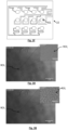
- lysis patches form, which correspond to light spots in images 4C, 4D and 4E.
- images 4C, 4D and 4E In the figure 4D , some bright spots are indicated by arrows. These images were formed from the green pixels of the image sensor. The gradual formation of bright regions of interest is observed in the images.
- a compartmentalized fluidic card is used, as described in connection with the first embodiment.
- the fluidic card comprises fluidic chambers 22 i separated from each other.
- Each fluidic chamber has a mixture of phages and a freeze-dried cold gelling agent.
- Each fluidic chamber can be supplied with a solution comprising bacteria.
- Agar is then formed, comprising a mixture of bacteria and phages.
- the cold gelling agent is water-soluble. It is configured to gel when it is brought to the contact with an aqueous solution, at room temperature, then forming a hydrogel. It may be a water-soluble polysaccharide.
- the cold gelling agent may be chosen from carboxymethylcellulose, guar gum, gum arabic, gellan gum, xanthan gum, or a gelling agent obtained from animal bones, for example pork, beef or chicken.
- the fluidic card is brought, during its filling, to a supercooling temperature of the agar containing the bacteria.
- the agar containing the bacteria can fill each fluidic chamber, so as to mix, in each chamber, with the phages previously placed in a lyophilized state. This avoids the use of a cold gelling agent, the latter being able to diffuse.
- a third embodiment is shown in the Figures 5A to 5D .
- the bacteria are initially present in an agar, or in a surface layer of a solidified agar. Drops of a solution comprising phages 3 are deposited on the surface of the latter. The drops are separated from each other.
- lysis plaques form in the sample, as described in connection with the second embodiment.
- An advantage of this embodiment is that the position of each deposited drop can be known, which makes it possible to search for the development of lysis plaques facing each of these positions. This facilitates the image processing step.
- FIGS. 5A And 5B schematize this embodiment, respectively when the drops are deposited on the agar, and following the formation of lysis plaques when the bacteria are sensitive to the viral strain of the phages present in the drops.
- the drops deposited may respectively contain different viral strains, and/or different concentrations of phages of the same viral strain.
- this embodiment makes it possible to carry out, in parallel, tests of different phages or different concentrations.
- FIG. 5B a formation of two lysis areas has been schematized. As in the second embodiment, under the effect of the lysis of the bacteria, the lysis areas appear, through which the transmission of light is higher than in the rest of the sample.
- the pixels 31 located opposite the lysis areas collect a signal whose intensity is higher. This results in the appearance of light spots on the image formed by the image sensor.
- a thin layer (between 0.1 mm and 1 mm) of oil 25 can be placed on the sample, so as to avoid drying.
- the oil 25 can be silicone oil, allowing diffusion of oxygen to the nutrient medium.
- each ellipse e1 represents the drop deposition area and each ellipse e2 corresponds to a region of image analysis.
- the ellipses e1 materialized on the left and right correspond respectively to the deposition of the phage and the pharmaceutical diluent solution.
- Such an embodiment makes it possible to divide the sample into different spatial zones, each spatial zone corresponding to a position at which a drop was deposited.
- the image acquired by the image sensor makes it possible to analyze several spatial zones simultaneously.
- a fourth embodiment is shown diagrammatically on the Figures 6A And 6B .
- an agar plate comprising phages 3 is provided. Drops of a solution comprising bacteria are deposited on the surface of the latter. The drops are separated from each other. When the bacteria are not sensitive to the viral strain, they develop on the surface of the sample, forming colonies. When the bacteria contained in the drops are sensitive to the viral strain of the bacteriophages present in the agar plate, no colony formation is observed on the surface of the sample.
- An advantage of this embodiment is that the position and composition of each deposited drop can be known, which makes it possible to search for the development of colonies at each of these positions. This facilitates the image processing step.
- FIG. 6A And 6B schematize this embodiment, respectively when the drops are deposited on the agar, and following the formation of colonies when the bacteria present in a drop are little or not sensitive to the phage present in the agar.
- the drops deposited may respectively contain different strains of bacteria, or different concentrations of the same strain.
- This embodiment makes it possible to carry out, in parallel, tests of different bacterial strains or different concentrations of the same bacterial strain.
- FIG. 6B we have schematized a formation of two colonies. The pixels 31 located opposite the colonies collect a signal whose intensity is lower. This results in the appearance of dark spots on the image formed by the image sensor.
- Such an embodiment makes it possible to divide the sample into different spatial zones, each spatial zone corresponding to a location at which a drop was deposited.
- the image acquired by the image sensor makes it possible to analyze several spatial zones simultaneously.
- the image defocus distance and/or the object defocus distance is preferably less than 1 mm, or even less than 500 ⁇ m.
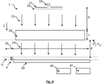
- a defocused configuration is represented, in which the object plane P o is merged with the sample plane P 20 .
- the image plane is offset from the detection plane by an image defocusing distance ⁇ .
- the defocused configuration is preferred when seeking to quantify a texturing of the image.
- the lensless configuration is preferred because it allows the use of a compact and inexpensive device.
Landscapes
- Chemical & Material Sciences (AREA)
- Life Sciences & Earth Sciences (AREA)
- Health & Medical Sciences (AREA)
- Organic Chemistry (AREA)
- Engineering & Computer Science (AREA)
- Wood Science & Technology (AREA)
- Zoology (AREA)
- Proteomics, Peptides & Aminoacids (AREA)
- Immunology (AREA)
- General Health & Medical Sciences (AREA)
- Biochemistry (AREA)
- Analytical Chemistry (AREA)
- Physics & Mathematics (AREA)
- Molecular Biology (AREA)
- Microbiology (AREA)
- Biotechnology (AREA)
- Biophysics (AREA)
- Bioinformatics & Cheminformatics (AREA)
- General Engineering & Computer Science (AREA)
- Genetics & Genomics (AREA)
- Toxicology (AREA)
- Virology (AREA)
- General Physics & Mathematics (AREA)
- Pathology (AREA)
- Measuring Or Testing Involving Enzymes Or Micro-Organisms (AREA)
Claims (16)
- Verfahren zur Bestimmung der Empfindlichkeit eines Bakterienstamms gegenüber einem Bakteriophagen-Virusstamm (3), wobei das Verfahren Folgendes umfasst:a) Herstellung einer Probe (20), umfassend das Inkontaktbringen von Bakterien (2) des Bakterienstamms mit Bakteriophagen (3), wobei jeder Bakteriophage demselben Virusstamm angehört, wobei die Bakterien sich entweder in einem flüssigen Medium oder in einem Agarmedium befinden,b) Einbringen der Probe zwischen eine Lichtquelle (10) und einen Bildsensor (30), wobei die Lichtquelle eine Lichtwelle (11) in einem Emissionsspektralbereich zwischen 500 nm und 600 nm emittiert, wobei der Bildsensor sich entlang einer Nachweisebene erstreckt,c) Belichten der Probe mithilfe der Lichtquelle und Aufnahme mindestens eines Bildes der Probe durch den Bildsensor im Emissionsspektralbereich, wobei der Bildsensor:- gemäß einer Bildgebungskonfiguration ohne Linse platziert ist, wobei keine Vergrößerungs- oder Bildaufnahmeoptik zwischen der Probe und dem Bildsensor angeordnet ist,- gemäß einer defokussierten Bildgebungskonfiguration platziert ist, wobei ein optisches System, eine Linse oder ein Objektiv, zwischen der Probe und dem Bildsensor angeordnet ist, wobei das optische System eine Objektebene und eine Bildebene definiert, wobei die Objektebene in Bezug auf die Nachweisebene versetzt ist oder die Bildebene in Bezug auf die Probe versetzt ist,d) Bestimmung, ausgehend von jedem aufgenommenen Bild, einer Empfindlichkeit des Bakterienstamms gegenüber dem Bakteriophagen-Virusstamm,wobei das Verfahren dergestalt ist, dass:- in Schritt a) die Bakterien und die Bakteriophagen in einer wässrigen Lösung gemischt werden,- Schritt c) die Aufnahme von mindestens zwei Bildern zu aufeinanderfolgenden Messzeitpunkten umfasst,- Schritt d) die Bestimmung einer von dem gesamten Bildsensor oder einem Teil davon auf jedem zu dem jeweiligen Messzeitpunkt (t) aufgenommenen Bild nachgewiesenen Lichtintensität derart umfasst, dass der Bakterienstamm als:• wenig oder gar nicht empfindlich gegenüber dem Bakteriophagen-Virusstamm betrachtet wird, wenn die nachgewiesene Lichtintensität zwischen zwei aufeinanderfolgenden Messzeitpunkten abnimmt,• oder als empfindlich gegenüber dem Bakteriophagen-Virusstamm betrachtet wird, wenn die nachgewiesene Lichtintensität zwischen zwei aufeinanderfolgenden Messzeitpunkten nicht abnimmt oder ansteigt.
- Verfahren nach Anspruch 1, wobei die Breite des Emissionsspektralbereichs kleiner als 50 nm ist.
- Verfahren nach einem der vorhergehenden Ansprüche, wobei der Abstand zwischen der Probe (20) und dem Bildsensor (30) kleiner als 5 cm oder kleiner als 1 cm ist.
- Verfahren nach einem der vorhergehenden Ansprüche, wobei Schritt d) die Bestimmung einer Abschwächung des von der Lichtquelle ausgestrahlten Lichts durch die Probe derart umfasst, dass der Bakterienstamm (2) als:• wenig oder gar nicht empfindlich gegenüber dem Bakteriophagen-Virusstamm angesehen wird, wenn die Abschwächung zwischen zwei aufeinanderfolgenden Messzeitpunkten zunimmt,• oder als empfindlich gegenüber dem Bakteriophagen-Virusstamm angesehen wird, wenn die Abschwächung zwischen zwei aufeinanderfolgenden Messzeitpunkten nicht zunimmt.
- Verfahren nach einem der vorhergehenden Ansprüche, wobei:- in Schritt a) die Bakterien und die Bakteriophagen in einer wässrigen Lösung gemischt werden,- Schritt c) die Aufnahme von mindestens zwei Bildern zu aufeinanderfolgenden Messzeitpunkten umfasst,- Schritt d) die Bestimmung eines Deskriptors der Texturierung jedes aufgenommenen Bildes derart umfasst, dass der Bakterienstamm als:• gar nicht oder wenig empfindlich gegenüber dem Bakteriophagen-Virusstamm angesehen wird, wenn die Veränderung des Texturierungsdeskriptors eine Zunahme der Lichtstreuung durch die Probe zwischen zwei aufeinanderfolgenden Messzeitpunkten widerspiegelt,• oder als empfindlich gegenüber dem Bakteriophagen-Virusstamm angesehen wird, wenn die Veränderung des Texturierungsdeskriptors eine Abnahme oder eine Stagnation der Lichtstreuung durch die Probe zwischen zwei aufeinanderfolgenden Messzeitpunkten widerspiegelt.
- Verfahren nach einem der Ansprüche 1 bis 4, wobei- in Schritt a) die Bakterien und die Bakteriophagen in einem Agar gemischt werden,- Schritt c) die Aufnahme mindestens eines Bildes nach Schritt a) umfasst,- Schritt d) eine Analyse jedes aufgenommenen Bildes umfasst, um klare Regionen von Interesse zu identifizieren, wobei jede klare Region von Interesse einer Infektion von Bakterien durch die Bakteriophagen entspricht, die ein Lyseplaque bildet, wobei jede klare Region von Interesse auf Empfindlichkeit des Bakterienstamms gegenüber dem Bakteriophagen-Virusstamm hindeutet.
- Verfahren nach Anspruch 6, das Folgendes umfasst:- Zählen der Anzahl der klaren Regionen von Interesse auf mindestens einem aufgenommenen Bild,- Schätzung des infektiösen Titers der Bakteriophagen in der Probe anhand der Anzahl der gezählten klaren Regionen von Interesse.
- Verfahren nach einem der Ansprüche 1 bis 4, wobei:- in Schritt a) die Bakterien in einen Agar eingebracht werden und die Bakteriophagen in eine Lösung eingebracht werden, wobei Schritt a) das Aufbringen mindestens eines Tropfens der Lösung auf den Agar umfasst,- Schritt c) die Aufnahme mindestens eines Bildes nach Schritt a) umfasst,- Schritt d) eine Analyse jedes aufgenommenen Bildes umfasst, um klare Regionen von Interesse zu identifizieren, wobei jede klare Region von Interesse einer Infektion von Bakterien durch die Bakteriophagen entspricht, die ein Lyseplaque bildet, derart, dass das Auftreten jeder klaren Region von Interesse auf Empfindlichkeit des Bakterienstamms gegenüber dem Bakteriophagen-Virusstamm hindeutet.
- Verfahren nach Anspruch 8, wobei Schritt a) das Aufbringen mehrerer Tropfen umfasst, wobei die Tropfen voneinander beabstandet sind, wobei zwei verschiedene Tropfen jeweils:- Bakteriophagen verschiedener Virusstämme- und/oder verschiedene Bakteriophagen-Konzentrationen desselben Virusstamms umfassen.
- Verfahren nach einem der Ansprüche 1 bis 4, wobei:- in Schritt a) die Bakteriophagen in einen Agar eingebracht werden und die Bakterien in eine Lösung eingebracht werden, wobei Schritt a) das Aufbringen eines Tropfens der Lösung auf den Agar umfasst,- Schritt c) die Aufnahme mindestens eines Bildes nach Schritt a) umfasst,- Schritt d) eine Analyse jedes aufgenommenen Bildes umfasst, um Folgendes zu identifizieren:• klare Regionen von Interesse, wobei jede klare Region von Interesse einer Infektion von Bakterien mit Bakteriophagen entspricht, so dass jede klare Region von Interesse auf Empfindlichkeit des Bakterienstamms gegenüber dem Bakteriophagen-Virusstamm hindeutet,• oder dunkle Regionen von Interesse, wobei jede dunkle Region von Interesse einem Bakterienwachstum in Gegenwart von Bakteriophagen entspricht, so dass jede dunkle Region von Interesse auf Unempfindlichkeit oder geringe Empfindlichkeit des Bakterienstamms von Interesse gegenüber dem Bakteriophagen-Virusstamm hindeutet.
- Verfahren nach Anspruch 10, wobei Schritt a) das Aufbringen mehrerer Tropfen umfasst, wobei die Tropfen voneinander beabstandet sind, wobei zwei verschiedene Tropfen jeweils:- Bakterien verschiedener Bakterienstämme- und/oder verschiedene Konzentrationen von Bakterien desselben Bakterienstamms umfassen.
- Verfahren nach einem der vorhergehenden Ansprüche, wobei die Probe in verschiedene räumliche Zonen (20i) unterteilt ist, die voneinander getrennt sind, derart, dass:- mindestens zwei verschiedene räumliche Zonen jeweils denselben Bakterienstamm und eine andere Konzentration von Bakteriophagen desselben Virusstamms umfassen- und/oder mindestens zwei verschiedene räumliche Zonen jeweils denselben Bakterienstamm und Bakteriophagen verschiedener Virusstämme umfassen- und/oder mindestens zwei verschiedene räumliche Zonen jeweils Bakteriophagen desselben Virusstamms und verschiedene Bakterienstämme umfassen,wobei das Verfahren derart ist, dass jede räumliche Zone mit einer Region von Interesse (ROIl) jedes aufgenommenen Bildes assoziiert ist, wobei zwei verschiedene räumliche Zonen mit zwei verschiedenen Regionen von Interessen assoziiert sind, so dass es die Analyse desselben aufgenommenen Bildes gestattet, eine Information hinsichtlich der Empfindlichkeit eines Bakterienstamms gegenüber einem Bakteriophagen-Virusstamm und dies in verschiedenen räumlichen Zonen zu erhalten.
- Verfahren nach Anspruch 12, wobei die Probe mehr als 10 oder sogar mehr als 100 voneinander getrennte, verschiedene räumliche Zonen umfasst, wobei jede räumliche Zone durch drei Parameter parametriert wird, die dem Bakterienstamm von Interesse, dem Bakteriophagen-Virusstamm bzw. der Konzentration der Bakteriophagen entsprechen, wobei mindestens ein Parameter zweier verschiedener räumlicher Zonen unterschiedlich ist.
- Verfahren nach einem der Ansprüche 12 oder 13, wobei die Probe in verschiedene Fluidkammern (22l) eines Fluidchips verteilt wird, wobei jede Fluidkammer einer räumlichen Zone der Probe entspricht.
- Verfahren nach einem der Ansprüche 12 oder 13, wobei die Probe nach dem Aufbringen von Tropfen auf der Oberfläche eines Agars an verschiedenen Positionen gebildet wird, wobei die Tropfen voneinander beabstandet sind, so dass durch jede Tropfenposition eine räumliche Zone definiert wird.
- Verfahren nach einem der vorhergehenden Ansprüche, umfassend vor Schritt a) einen Anreicherungsschritt, der Folgendes umfasst:- ein Gemisch von Bakteriophagen desselben Virusstamms oder verschiedener Virusstämme in einer wässrigen Lösung, die mindestens einen Bakterienstamm umfasst,- eine Inkubation,- eine Filtration des Gemischs, um die Bakterien zurückzuhalten und eine an Bakteriophagen angereicherte Lösung zu erhalten,so dass in Schritt a) das Inkontaktbringen der Bakteriophagen mit den Bakterien unter Verwendung der an Bakteriophagen angereicherten Lösung durchgeführt wird.
Applications Claiming Priority (1)
| Application Number | Priority Date | Filing Date | Title |
|---|---|---|---|
| FR1912804A FR3103198B1 (fr) | 2019-11-15 | 2019-11-15 | Procédé de détermination de la sensibilité d'une souche bactérienne à l'égard d'un virus bactériophage. |
Publications (2)
| Publication Number | Publication Date |
|---|---|
| EP3822360A1 EP3822360A1 (de) | 2021-05-19 |
| EP3822360B1 true EP3822360B1 (de) | 2025-05-14 |
Family
ID=70295198
Family Applications (1)
| Application Number | Title | Priority Date | Filing Date |
|---|---|---|---|
| EP20207254.2A Active EP3822360B1 (de) | 2019-11-15 | 2020-11-12 | Verfahren zur bestimmung der sensibilität eines bakterienstamms gegenüber einem bakteriophagen virus |
Country Status (3)
| Country | Link |
|---|---|
| US (1) | US12275980B2 (de) |
| EP (1) | EP3822360B1 (de) |
| FR (1) | FR3103198B1 (de) |
Families Citing this family (1)
| Publication number | Priority date | Publication date | Assignee | Title |
|---|---|---|---|---|
| FR3146694B1 (fr) | 2023-03-14 | 2025-02-07 | Commissariat Energie Atomique | Procédé de détection d'une sensibilité de phages ou d'antibiotiques vis-à-vis d'une souche bactérienne. |
Citations (2)
| Publication number | Priority date | Publication date | Assignee | Title |
|---|---|---|---|---|
| EP2669678A1 (de) * | 2012-06-01 | 2013-12-04 | Commissariat à l'Énergie Atomique et aux Énergies Alternatives | Verfahren und System zur Charakterisierung der Geschwindigkeitsverteilung und Koagulation von Partikeln, die in einer Flüssigkeit enthalten sind, wie beispielsweise Blutpartikeln |
| FR3046239A1 (fr) * | 2015-12-28 | 2017-06-30 | Commissariat Energie Atomique | Dispositif et procede d’observation bimodale d’un objet |
Family Cites Families (8)
| Publication number | Priority date | Publication date | Assignee | Title |
|---|---|---|---|---|
| US5800778A (en) | 1995-05-31 | 1998-09-01 | Biomerieux Vitek, Inc. | Sealant for sample holder |
| US20080193418A1 (en) * | 2004-06-04 | 2008-08-14 | University Of Northern Iowa Research Foundation | Bacteriophages that Infect Bacillus Bacteria (Anthrax) |
| US20090081173A1 (en) * | 2007-08-10 | 2009-03-26 | The Board Of Regents Of The University Of Texas System | Methods and compositions involving bacteriophage isolates |
| ES2340121B1 (es) | 2008-04-09 | 2011-04-08 | Consejo Superior De Investigaciones Cientificas (Csic) | Procedimiento y sistema para detectar y/o cuantificar bacteriofagos, uso de un dispositivo microelectronico sensor para detectar dichos bacteriofagos y dispositivo microelectronico sensor para llevar a cabo dicho procedimiento. |
| US9569664B2 (en) * | 2010-10-26 | 2017-02-14 | California Institute Of Technology | Methods for rapid distinction between debris and growing cells |
| PL217019B1 (pl) * | 2011-08-25 | 2014-06-30 | Proteon Pharmaceuticals Spółka Akcyjna | Sposób otrzymywania szczepu bakteriofaga, specyficzne szczepy bakteriofagowe oraz ich zastosowanie |
| WO2019018886A1 (en) * | 2017-07-23 | 2019-01-31 | Future Biosolutions Pty Ltd | METHOD FOR RAPID DETECTION AND ENUMERATION OF VIRUSES, BACTERIOPHAGES AND / OR BACTERIA |
| WO2019188886A1 (ja) | 2018-03-30 | 2019-10-03 | パイオニア株式会社 | 端末装置、情報処理方法、プログラム、及び、記憶媒体 |
-
2019
- 2019-11-15 FR FR1912804A patent/FR3103198B1/fr active Active
-
2020
- 2020-11-12 EP EP20207254.2A patent/EP3822360B1/de active Active
- 2020-11-13 US US17/097,687 patent/US12275980B2/en active Active
Patent Citations (2)
| Publication number | Priority date | Publication date | Assignee | Title |
|---|---|---|---|---|
| EP2669678A1 (de) * | 2012-06-01 | 2013-12-04 | Commissariat à l'Énergie Atomique et aux Énergies Alternatives | Verfahren und System zur Charakterisierung der Geschwindigkeitsverteilung und Koagulation von Partikeln, die in einer Flüssigkeit enthalten sind, wie beispielsweise Blutpartikeln |
| FR3046239A1 (fr) * | 2015-12-28 | 2017-06-30 | Commissariat Energie Atomique | Dispositif et procede d’observation bimodale d’un objet |
Non-Patent Citations (1)
| Title |
|---|
| ANONYMOUS: "Protocol: Relative Viral Titering with a Resazurin [alamarBlue ] Cell Viability Assay", 1 September 2016 (2016-09-01), pages 1 - 5, XP093188860, Retrieved from the Internet <URL:https://portals.broadinstitute.org/gpp/public/dir/download?dirpath=protocols/production&filename=AlamarBlue_virus_titering_sept2016.pdf> [retrieved on 20240722] * |
Also Published As
| Publication number | Publication date |
|---|---|
| US12275980B2 (en) | 2025-04-15 |
| EP3822360A1 (de) | 2021-05-19 |
| FR3103198B1 (fr) | 2024-07-26 |
| US20210147899A1 (en) | 2021-05-20 |
| FR3103198A1 (fr) | 2021-05-21 |
Similar Documents
| Publication | Publication Date | Title |
|---|---|---|
| DE19903506C2 (de) | Verfahren, Gefäß und Vorrichtung zur Überwachung der Stoffwechselaktivität von Zellkulturen in flüssigen Medien | |
| Siemons et al. | Robust adaptive optics for localization microscopy deep in complex tissue | |
| EP3167285B1 (de) | Vorrichtung und verfahren zur nicht-invasiven bestimmung der fruchtbarkeit und/oder des geschlechts von eiern | |
| CN111133295B (zh) | 拉曼成像技术测定抗生素敏感性的方法 | |
| EP3257947B1 (de) | Verfahren und system zur identifizierung des gramtyps einer bakterie | |
| WO2009117683A3 (en) | Method and apparatus for analyzing individual cells or particulates using fluorescent quenching and/or bleaching | |
| FR2968921A1 (fr) | Procede de localisation d'un marqueur optique dans un milieu diffusant | |
| FR3075825A1 (fr) | Procede et systeme d'identificationdu type de gram d'une bacterie | |
| WO2019122734A1 (fr) | Procede d'identification d'une levure ou d'une bacterie | |
| EP3822360A1 (de) | Verfahren zur bestimmung der sensibilität eines bakterienstamms gegenüber einem bakteriophagen virus | |
| FR3066503A1 (fr) | Procede d'analyse de microorganismes | |
| FR3024778A1 (fr) | Procede pour le depistage de la drepanocytose et kit pour sa mise en oeuvre | |
| WO2016001555A1 (fr) | Procede de detection d'une presence ou d'une absence de particules biologiques | |
| FR3086951A1 (fr) | Methode et dispositif pour la detection d'au moins un microorganisme selon sa cinetique de marquage, et support de detection | |
| EP4217713A1 (de) | Verfahren zur analyse einer biologischen probe mit bestimmung der räumlichen verteilung von biomasse entlang der optischen achse | |
| EP3054284B1 (de) | Verfahren zum korrigieren eines von einer probe zurückgestreuten raman signals, und entsprechende vorrichtung | |
| WO2006024772A1 (fr) | Dispositif de lecture pour lames portant des micro depots supports de reaction biologique | |
| EP4374158B1 (de) | Verfahren zur quantifizierung von endotoxinen in einer biologischen probe | |
| JP2012008042A (ja) | 寄生虫卵の検出方法およびシステム | |
| EP1682889A2 (de) | Automatische analyse von zellproben | |
| EP4446010B1 (de) | Verfahren zum nachweis der empfindlichkeit von phagen oder antibiotika gegenüber bakterienstamm | |
| JP2009145104A (ja) | エバネッセント波発生装置及びそれを用いた観察装置 | |
| EP3221689B1 (de) | Verfahren zur beobachtung mehrerer proben und zugehörige vorrichtung | |
| FR3106897A1 (fr) | Procédé de détection de microorganismes dans un échantillon | |
| Tian et al. | A composite optical filter greenly fabricated for living algae detection |
Legal Events
| Date | Code | Title | Description |
|---|---|---|---|
| PUAI | Public reference made under article 153(3) epc to a published international application that has entered the european phase |
Free format text: ORIGINAL CODE: 0009012 |
|
| STAA | Information on the status of an ep patent application or granted ep patent |
Free format text: STATUS: REQUEST FOR EXAMINATION WAS MADE |
|
| 17P | Request for examination filed |
Effective date: 20201120 |
|
| AK | Designated contracting states |
Kind code of ref document: A1 Designated state(s): AL AT BE BG CH CY CZ DE DK EE ES FI FR GB GR HR HU IE IS IT LI LT LU LV MC MK MT NL NO PL PT RO RS SE SI SK SM TR |
|
| STAA | Information on the status of an ep patent application or granted ep patent |
Free format text: STATUS: EXAMINATION IS IN PROGRESS |
|
| 17Q | First examination report despatched |
Effective date: 20230922 |
|
| RAP3 | Party data changed (applicant data changed or rights of an application transferred) |
Owner name: UNIVERSITE GRENOBLE ALPES Owner name: CENTRE NATIONAL DE LA RECHERCHE SCIENTIFIQUE Owner name: COMMISSARIAT A L'ENERGIE ATOMIQUE ET AUX ENERGIESALTERNATIVES |
|
| GRAP | Despatch of communication of intention to grant a patent |
Free format text: ORIGINAL CODE: EPIDOSNIGR1 |
|
| STAA | Information on the status of an ep patent application or granted ep patent |
Free format text: STATUS: GRANT OF PATENT IS INTENDED |
|
| INTG | Intention to grant announced |
Effective date: 20241206 |
|
| GRAS | Grant fee paid |
Free format text: ORIGINAL CODE: EPIDOSNIGR3 |
|
| GRAA | (expected) grant |
Free format text: ORIGINAL CODE: 0009210 |
|
| STAA | Information on the status of an ep patent application or granted ep patent |
Free format text: STATUS: THE PATENT HAS BEEN GRANTED |
|
| AK | Designated contracting states |
Kind code of ref document: B1 Designated state(s): AL AT BE BG CH CY CZ DE DK EE ES FI FR GB GR HR HU IE IS IT LI LT LU LV MC MK MT NL NO PL PT RO RS SE SI SK SM TR |
|
| REG | Reference to a national code |
Ref country code: GB Ref legal event code: FG4D Free format text: NOT ENGLISH |
|
| REG | Reference to a national code |
Ref country code: CH Ref legal event code: EP |
|
| REG | Reference to a national code |
Ref country code: DE Ref legal event code: R096 Ref document number: 602020051191 Country of ref document: DE |
|
| REG | Reference to a national code |
Ref country code: IE Ref legal event code: FG4D Free format text: LANGUAGE OF EP DOCUMENT: FRENCH |
|
| REG | Reference to a national code |
Ref country code: NL Ref legal event code: MP Effective date: 20250514 |
|
| PG25 | Lapsed in a contracting state [announced via postgrant information from national office to epo] |
Ref country code: ES Free format text: LAPSE BECAUSE OF FAILURE TO SUBMIT A TRANSLATION OF THE DESCRIPTION OR TO PAY THE FEE WITHIN THE PRESCRIBED TIME-LIMIT Effective date: 20250514 Ref country code: PT Free format text: LAPSE BECAUSE OF FAILURE TO SUBMIT A TRANSLATION OF THE DESCRIPTION OR TO PAY THE FEE WITHIN THE PRESCRIBED TIME-LIMIT Effective date: 20250915 Ref country code: FI Free format text: LAPSE BECAUSE OF FAILURE TO SUBMIT A TRANSLATION OF THE DESCRIPTION OR TO PAY THE FEE WITHIN THE PRESCRIBED TIME-LIMIT Effective date: 20250514 |
|
| REG | Reference to a national code |
Ref country code: LT Ref legal event code: MG9D |
|
| PG25 | Lapsed in a contracting state [announced via postgrant information from national office to epo] |
Ref country code: GR Free format text: LAPSE BECAUSE OF FAILURE TO SUBMIT A TRANSLATION OF THE DESCRIPTION OR TO PAY THE FEE WITHIN THE PRESCRIBED TIME-LIMIT Effective date: 20250815 Ref country code: NO Free format text: LAPSE BECAUSE OF FAILURE TO SUBMIT A TRANSLATION OF THE DESCRIPTION OR TO PAY THE FEE WITHIN THE PRESCRIBED TIME-LIMIT Effective date: 20250814 |
|
| PG25 | Lapsed in a contracting state [announced via postgrant information from national office to epo] |
Ref country code: NL Free format text: LAPSE BECAUSE OF FAILURE TO SUBMIT A TRANSLATION OF THE DESCRIPTION OR TO PAY THE FEE WITHIN THE PRESCRIBED TIME-LIMIT Effective date: 20250514 Ref country code: PL Free format text: LAPSE BECAUSE OF FAILURE TO SUBMIT A TRANSLATION OF THE DESCRIPTION OR TO PAY THE FEE WITHIN THE PRESCRIBED TIME-LIMIT Effective date: 20250514 |
|
| REG | Reference to a national code |
Ref country code: AT Ref legal event code: MK05 Ref document number: 1794827 Country of ref document: AT Kind code of ref document: T Effective date: 20250514 |
|
| PG25 | Lapsed in a contracting state [announced via postgrant information from national office to epo] |
Ref country code: BG Free format text: LAPSE BECAUSE OF FAILURE TO SUBMIT A TRANSLATION OF THE DESCRIPTION OR TO PAY THE FEE WITHIN THE PRESCRIBED TIME-LIMIT Effective date: 20250514 |
|
| PG25 | Lapsed in a contracting state [announced via postgrant information from national office to epo] |
Ref country code: HR Free format text: LAPSE BECAUSE OF FAILURE TO SUBMIT A TRANSLATION OF THE DESCRIPTION OR TO PAY THE FEE WITHIN THE PRESCRIBED TIME-LIMIT Effective date: 20250514 |
|
| PG25 | Lapsed in a contracting state [announced via postgrant information from national office to epo] |
Ref country code: AT Free format text: LAPSE BECAUSE OF FAILURE TO SUBMIT A TRANSLATION OF THE DESCRIPTION OR TO PAY THE FEE WITHIN THE PRESCRIBED TIME-LIMIT Effective date: 20250514 |
|
| PG25 | Lapsed in a contracting state [announced via postgrant information from national office to epo] |
Ref country code: RS Free format text: LAPSE BECAUSE OF FAILURE TO SUBMIT A TRANSLATION OF THE DESCRIPTION OR TO PAY THE FEE WITHIN THE PRESCRIBED TIME-LIMIT Effective date: 20250814 |
|
| PG25 | Lapsed in a contracting state [announced via postgrant information from national office to epo] |
Ref country code: IS Free format text: LAPSE BECAUSE OF FAILURE TO SUBMIT A TRANSLATION OF THE DESCRIPTION OR TO PAY THE FEE WITHIN THE PRESCRIBED TIME-LIMIT Effective date: 20250914 |
|
| PG25 | Lapsed in a contracting state [announced via postgrant information from national office to epo] |
Ref country code: LV Free format text: LAPSE BECAUSE OF FAILURE TO SUBMIT A TRANSLATION OF THE DESCRIPTION OR TO PAY THE FEE WITHIN THE PRESCRIBED TIME-LIMIT Effective date: 20250514 |
|
| PGFP | Annual fee paid to national office [announced via postgrant information from national office to epo] |
Ref country code: DE Payment date: 20251118 Year of fee payment: 6 |
|
| PGFP | Annual fee paid to national office [announced via postgrant information from national office to epo] |
Ref country code: GB Payment date: 20251125 Year of fee payment: 6 |
|
| PG25 | Lapsed in a contracting state [announced via postgrant information from national office to epo] |
Ref country code: SM Free format text: LAPSE BECAUSE OF FAILURE TO SUBMIT A TRANSLATION OF THE DESCRIPTION OR TO PAY THE FEE WITHIN THE PRESCRIBED TIME-LIMIT Effective date: 20250514 Ref country code: DK Free format text: LAPSE BECAUSE OF FAILURE TO SUBMIT A TRANSLATION OF THE DESCRIPTION OR TO PAY THE FEE WITHIN THE PRESCRIBED TIME-LIMIT Effective date: 20250514 |
|
| PGFP | Annual fee paid to national office [announced via postgrant information from national office to epo] |
Ref country code: FR Payment date: 20251125 Year of fee payment: 6 |
|
| PG25 | Lapsed in a contracting state [announced via postgrant information from national office to epo] |
Ref country code: CZ Free format text: LAPSE BECAUSE OF FAILURE TO SUBMIT A TRANSLATION OF THE DESCRIPTION OR TO PAY THE FEE WITHIN THE PRESCRIBED TIME-LIMIT Effective date: 20250514 |
|
| PG25 | Lapsed in a contracting state [announced via postgrant information from national office to epo] |
Ref country code: EE Free format text: LAPSE BECAUSE OF FAILURE TO SUBMIT A TRANSLATION OF THE DESCRIPTION OR TO PAY THE FEE WITHIN THE PRESCRIBED TIME-LIMIT Effective date: 20250514 |
|
| PG25 | Lapsed in a contracting state [announced via postgrant information from national office to epo] |
Ref country code: RO Free format text: LAPSE BECAUSE OF FAILURE TO SUBMIT A TRANSLATION OF THE DESCRIPTION OR TO PAY THE FEE WITHIN THE PRESCRIBED TIME-LIMIT Effective date: 20250514 Ref country code: SK Free format text: LAPSE BECAUSE OF FAILURE TO SUBMIT A TRANSLATION OF THE DESCRIPTION OR TO PAY THE FEE WITHIN THE PRESCRIBED TIME-LIMIT Effective date: 20250514 |
|
| PG25 | Lapsed in a contracting state [announced via postgrant information from national office to epo] |
Ref country code: IT Free format text: LAPSE BECAUSE OF FAILURE TO SUBMIT A TRANSLATION OF THE DESCRIPTION OR TO PAY THE FEE WITHIN THE PRESCRIBED TIME-LIMIT Effective date: 20250514 |
|
| REG | Reference to a national code |
Ref country code: DE Ref legal event code: R097 Ref document number: 602020051191 Country of ref document: DE |
|
| PLBE | No opposition filed within time limit |
Free format text: ORIGINAL CODE: 0009261 |
|
| STAA | Information on the status of an ep patent application or granted ep patent |
Free format text: STATUS: NO OPPOSITION FILED WITHIN TIME LIMIT |
|
| REG | Reference to a national code |
Ref country code: CH Ref legal event code: L10 Free format text: ST27 STATUS EVENT CODE: U-0-0-L10-L00 (AS PROVIDED BY THE NATIONAL OFFICE) Effective date: 20260325 |
|
| 26N | No opposition filed |
Effective date: 20260217 |