EP3750988A1 - Procédé amélioré de production de lymphocytes t traités alpha bêta - Google Patents
Procédé amélioré de production de lymphocytes t traités alpha bêta Download PDFInfo
- Publication number
- EP3750988A1 EP3750988A1 EP19751544.8A EP19751544A EP3750988A1 EP 3750988 A1 EP3750988 A1 EP 3750988A1 EP 19751544 A EP19751544 A EP 19751544A EP 3750988 A1 EP3750988 A1 EP 3750988A1
- Authority
- EP
- European Patent Office
- Prior art keywords
- cancer
- cells
- cell
- processed
- virus
- Prior art date
- Legal status (The legal status is an assumption and is not a legal conclusion. Google has not performed a legal analysis and makes no representation as to the accuracy of the status listed.)
- Pending
Links
- 238000004519 manufacturing process Methods 0.000 title claims abstract description 13
- 210000004027 cell Anatomy 0.000 claims abstract description 266
- 206010028980 Neoplasm Diseases 0.000 claims abstract description 86
- 201000011510 cancer Diseases 0.000 claims abstract description 80
- 239000003814 drug Substances 0.000 claims abstract description 36
- 238000000034 method Methods 0.000 claims abstract description 28
- 210000001744 T-lymphocyte Anatomy 0.000 claims description 67
- 101001109501 Homo sapiens NKG2-D type II integral membrane protein Proteins 0.000 claims description 38
- 102100022680 NKG2-D type II integral membrane protein Human genes 0.000 claims description 38
- WQZGKKKJIJFFOK-QTVWNMPRSA-N D-mannopyranose Chemical compound OC[C@H]1OC(O)[C@@H](O)[C@@H](O)[C@@H]1O WQZGKKKJIJFFOK-QTVWNMPRSA-N 0.000 claims description 35
- 108010002350 Interleukin-2 Proteins 0.000 claims description 24
- 239000003446 ligand Substances 0.000 claims description 24
- 241000894006 Bacteria Species 0.000 claims description 14
- 210000003819 peripheral blood mononuclear cell Anatomy 0.000 claims description 14
- 239000008194 pharmaceutical composition Substances 0.000 claims description 14
- KHGNFPUMBJSZSM-UHFFFAOYSA-N Perforine Natural products COC1=C2CCC(O)C(CCC(C)(C)O)(OC)C2=NC2=C1C=CO2 KHGNFPUMBJSZSM-UHFFFAOYSA-N 0.000 claims description 13
- 210000000822 natural killer cell Anatomy 0.000 claims description 13
- 229930192851 perforin Natural products 0.000 claims description 13
- 230000001172 regenerating effect Effects 0.000 claims description 13
- VRYALKFFQXWPIH-PBXRRBTRSA-N (3r,4s,5r)-3,4,5,6-tetrahydroxyhexanal Chemical compound OC[C@@H](O)[C@@H](O)[C@H](O)CC=O VRYALKFFQXWPIH-PBXRRBTRSA-N 0.000 claims description 12
- SHZGCJCMOBCMKK-UHFFFAOYSA-N D-mannomethylose Natural products CC1OC(O)C(O)C(O)C1O SHZGCJCMOBCMKK-UHFFFAOYSA-N 0.000 claims description 12
- 102000001398 Granzyme Human genes 0.000 claims description 10
- 108060005986 Granzyme Proteins 0.000 claims description 10
- 201000003793 Myelodysplastic syndrome Diseases 0.000 claims description 10
- 230000002265 prevention Effects 0.000 claims description 10
- 229910019142 PO4 Inorganic materials 0.000 claims description 9
- 241000700605 Viruses Species 0.000 claims description 9
- 239000010452 phosphate Substances 0.000 claims description 9
- PMMURAAUARKVCB-UHFFFAOYSA-N alpha-D-ara-dHexp Natural products OCC1OC(O)CC(O)C1O PMMURAAUARKVCB-UHFFFAOYSA-N 0.000 claims description 8
- 238000012258 culturing Methods 0.000 claims description 7
- 238000000338 in vitro Methods 0.000 claims description 7
- 210000003071 memory t lymphocyte Anatomy 0.000 claims description 7
- AOYNUTHNTBLRMT-SLPGGIOYSA-N 2-deoxy-2-fluoro-aldehydo-D-glucose Chemical compound OC[C@@H](O)[C@@H](O)[C@H](O)[C@@H](F)C=O AOYNUTHNTBLRMT-SLPGGIOYSA-N 0.000 claims description 6
- 208000031261 Acute myeloid leukaemia Diseases 0.000 claims description 6
- 241000598436 Human T-cell lymphotropic virus Species 0.000 claims description 6
- 241000701044 Human gammaherpesvirus 4 Species 0.000 claims description 6
- 208000005016 Intestinal Neoplasms Diseases 0.000 claims description 6
- 208000005718 Stomach Neoplasms Diseases 0.000 claims description 6
- 208000024313 Testicular Neoplasms Diseases 0.000 claims description 6
- 206010057644 Testis cancer Diseases 0.000 claims description 6
- 206010017758 gastric cancer Diseases 0.000 claims description 6
- 208000015181 infectious disease Diseases 0.000 claims description 6
- 201000002313 intestinal cancer Diseases 0.000 claims description 6
- 201000011549 stomach cancer Diseases 0.000 claims description 6
- 201000003120 testicular cancer Diseases 0.000 claims description 6
- 241000725303 Human immunodeficiency virus Species 0.000 claims description 5
- 238000002360 preparation method Methods 0.000 claims description 5
- 150000003839 salts Chemical class 0.000 claims description 5
- 206010005003 Bladder cancer Diseases 0.000 claims description 4
- 208000011691 Burkitt lymphomas Diseases 0.000 claims description 4
- 206010009944 Colon cancer Diseases 0.000 claims description 4
- 241000711549 Hepacivirus C Species 0.000 claims description 4
- 206010058467 Lung neoplasm malignant Diseases 0.000 claims description 4
- 208000007097 Urinary Bladder Neoplasms Diseases 0.000 claims description 4
- 208000029742 colonic neoplasm Diseases 0.000 claims description 4
- 208000032839 leukemia Diseases 0.000 claims description 4
- 201000005202 lung cancer Diseases 0.000 claims description 4
- 208000020816 lung neoplasm Diseases 0.000 claims description 4
- 201000001441 melanoma Diseases 0.000 claims description 4
- 201000008968 osteosarcoma Diseases 0.000 claims description 4
- 201000005112 urinary bladder cancer Diseases 0.000 claims description 4
- AOYNUTHNTBLRMT-KVTDHHQDSA-N (2s,3s,4r,5r)-2-fluoro-3,4,5,6-tetrahydroxyhexanal Chemical compound OC[C@@H](O)[C@@H](O)[C@H](O)[C@H](F)C=O AOYNUTHNTBLRMT-KVTDHHQDSA-N 0.000 claims description 3
- MBPFNOMGYSRGQZ-PBXRRBTRSA-N 2-deoxy-D-glucose 6-phosphate Chemical compound OP(=O)(O)OC[C@@H](O)[C@@H](O)[C@H](O)CC=O MBPFNOMGYSRGQZ-PBXRRBTRSA-N 0.000 claims description 3
- 208000024893 Acute lymphoblastic leukemia Diseases 0.000 claims description 3
- 208000014697 Acute lymphocytic leukaemia Diseases 0.000 claims description 3
- 206010073478 Anaplastic large-cell lymphoma Diseases 0.000 claims description 3
- 201000003076 Angiosarcoma Diseases 0.000 claims description 3
- 208000010839 B-cell chronic lymphocytic leukemia Diseases 0.000 claims description 3
- 208000003950 B-cell lymphoma Diseases 0.000 claims description 3
- 208000032791 BCR-ABL1 positive chronic myelogenous leukemia Diseases 0.000 claims description 3
- 206010004446 Benign prostatic hyperplasia Diseases 0.000 claims description 3
- 206010005949 Bone cancer Diseases 0.000 claims description 3
- 208000018084 Bone neoplasm Diseases 0.000 claims description 3
- 208000003174 Brain Neoplasms Diseases 0.000 claims description 3
- 206010006187 Breast cancer Diseases 0.000 claims description 3
- 208000026310 Breast neoplasm Diseases 0.000 claims description 3
- 201000009030 Carcinoma Diseases 0.000 claims description 3
- 206010008342 Cervix carcinoma Diseases 0.000 claims description 3
- 208000010833 Chronic myeloid leukaemia Diseases 0.000 claims description 3
- 241000701022 Cytomegalovirus Species 0.000 claims description 3
- 206010014733 Endometrial cancer Diseases 0.000 claims description 3
- 206010014759 Endometrial neoplasm Diseases 0.000 claims description 3
- 201000001342 Fallopian tube cancer Diseases 0.000 claims description 3
- 208000013452 Fallopian tube neoplasm Diseases 0.000 claims description 3
- 201000008808 Fibrosarcoma Diseases 0.000 claims description 3
- 206010016935 Follicular thyroid cancer Diseases 0.000 claims description 3
- 208000032612 Glial tumor Diseases 0.000 claims description 3
- 206010018338 Glioma Diseases 0.000 claims description 3
- 208000001258 Hemangiosarcoma Diseases 0.000 claims description 3
- 241000700721 Hepatitis B virus Species 0.000 claims description 3
- 208000017604 Hodgkin disease Diseases 0.000 claims description 3
- 208000021519 Hodgkin lymphoma Diseases 0.000 claims description 3
- 208000010747 Hodgkins lymphoma Diseases 0.000 claims description 3
- 241000701806 Human papillomavirus Species 0.000 claims description 3
- 208000007766 Kaposi sarcoma Diseases 0.000 claims description 3
- 206010023347 Keratoacanthoma Diseases 0.000 claims description 3
- 208000008839 Kidney Neoplasms Diseases 0.000 claims description 3
- 208000032004 Large-Cell Anaplastic Lymphoma Diseases 0.000 claims description 3
- 201000011062 Li-Fraumeni syndrome Diseases 0.000 claims description 3
- 208000031422 Lymphocytic Chronic B-Cell Leukemia Diseases 0.000 claims description 3
- 206010025538 Malignant ascites Diseases 0.000 claims description 3
- 206010027406 Mesothelioma Diseases 0.000 claims description 3
- 208000034578 Multiple myelomas Diseases 0.000 claims description 3
- 208000033761 Myelogenous Chronic BCR-ABL Positive Leukemia Diseases 0.000 claims description 3
- 208000033776 Myeloid Acute Leukemia Diseases 0.000 claims description 3
- 208000001894 Nasopharyngeal Neoplasms Diseases 0.000 claims description 3
- 206010061306 Nasopharyngeal cancer Diseases 0.000 claims description 3
- 206010029260 Neuroblastoma Diseases 0.000 claims description 3
- 208000015914 Non-Hodgkin lymphomas Diseases 0.000 claims description 3
- 206010061534 Oesophageal squamous cell carcinoma Diseases 0.000 claims description 3
- 206010033128 Ovarian cancer Diseases 0.000 claims description 3
- 206010061535 Ovarian neoplasm Diseases 0.000 claims description 3
- 206010061902 Pancreatic neoplasm Diseases 0.000 claims description 3
- 206010035226 Plasma cell myeloma Diseases 0.000 claims description 3
- 208000006664 Precursor Cell Lymphoblastic Leukemia-Lymphoma Diseases 0.000 claims description 3
- 206010060862 Prostate cancer Diseases 0.000 claims description 3
- 208000004403 Prostatic Hyperplasia Diseases 0.000 claims description 3
- 208000000236 Prostatic Neoplasms Diseases 0.000 claims description 3
- 206010038389 Renal cancer Diseases 0.000 claims description 3
- 208000016624 Retinal neoplasm Diseases 0.000 claims description 3
- 208000004337 Salivary Gland Neoplasms Diseases 0.000 claims description 3
- 206010061934 Salivary gland cancer Diseases 0.000 claims description 3
- 208000000277 Splenic Neoplasms Diseases 0.000 claims description 3
- 208000036765 Squamous cell carcinoma of the esophagus Diseases 0.000 claims description 3
- 208000024770 Thyroid neoplasm Diseases 0.000 claims description 3
- 208000006105 Uterine Cervical Neoplasms Diseases 0.000 claims description 3
- 208000002495 Uterine Neoplasms Diseases 0.000 claims description 3
- 201000005969 Uveal melanoma Diseases 0.000 claims description 3
- 208000033559 Waldenström macroglobulinemia Diseases 0.000 claims description 3
- CMLYNMGULQGGIA-SLPGGIOYSA-N [(2r,3r,4s,5r)-5-fluoro-2,3,4-trihydroxy-6-oxohexyl] dihydrogen phosphate Chemical compound OP(=O)(O)OC[C@@H](O)[C@@H](O)[C@H](O)[C@@H](F)C=O CMLYNMGULQGGIA-SLPGGIOYSA-N 0.000 claims description 3
- CMLYNMGULQGGIA-KVTDHHQDSA-N [(2r,3r,4s,5s)-5-fluoro-2,3,4-trihydroxy-6-oxohexyl] dihydrogen phosphate Chemical compound OP(=O)(O)OC[C@@H](O)[C@@H](O)[C@H](O)[C@H](F)C=O CMLYNMGULQGGIA-KVTDHHQDSA-N 0.000 claims description 3
- WQZGKKKJIJFFOK-PHYPRBDBSA-N alpha-D-galactose Chemical compound OC[C@H]1O[C@H](O)[C@H](O)[C@@H](O)[C@H]1O WQZGKKKJIJFFOK-PHYPRBDBSA-N 0.000 claims description 3
- 201000009036 biliary tract cancer Diseases 0.000 claims description 3
- 208000020790 biliary tract neoplasm Diseases 0.000 claims description 3
- 208000002458 carcinoid tumor Diseases 0.000 claims description 3
- 201000010881 cervical cancer Diseases 0.000 claims description 3
- 208000032852 chronic lymphocytic leukemia Diseases 0.000 claims description 3
- 201000010918 connective tissue cancer Diseases 0.000 claims description 3
- 208000007276 esophageal squamous cell carcinoma Diseases 0.000 claims description 3
- 210000000285 follicular dendritic cell Anatomy 0.000 claims description 3
- 229930182830 galactose Natural products 0.000 claims description 3
- 201000011243 gastrointestinal stromal tumor Diseases 0.000 claims description 3
- 208000005017 glioblastoma Diseases 0.000 claims description 3
- 201000010536 head and neck cancer Diseases 0.000 claims description 3
- 208000014829 head and neck neoplasm Diseases 0.000 claims description 3
- 206010073071 hepatocellular carcinoma Diseases 0.000 claims description 3
- 231100000844 hepatocellular carcinoma Toxicity 0.000 claims description 3
- 201000010982 kidney cancer Diseases 0.000 claims description 3
- 201000007270 liver cancer Diseases 0.000 claims description 3
- 208000014018 liver neoplasm Diseases 0.000 claims description 3
- 208000015486 malignant pancreatic neoplasm Diseases 0.000 claims description 3
- 239000010445 mica Substances 0.000 claims description 3
- 229910052618 mica group Inorganic materials 0.000 claims description 3
- 210000003205 muscle Anatomy 0.000 claims description 3
- 208000025113 myeloid leukemia Diseases 0.000 claims description 3
- 201000002120 neuroendocrine carcinoma Diseases 0.000 claims description 3
- 208000008443 pancreatic carcinoma Diseases 0.000 claims description 3
- 201000002628 peritoneum cancer Diseases 0.000 claims description 3
- 201000008933 retinal cancer Diseases 0.000 claims description 3
- 201000009410 rhabdomyosarcoma Diseases 0.000 claims description 3
- 239000012453 solvate Substances 0.000 claims description 3
- 201000002471 spleen cancer Diseases 0.000 claims description 3
- 208000001608 teratocarcinoma Diseases 0.000 claims description 3
- 201000002510 thyroid cancer Diseases 0.000 claims description 3
- 208000030901 thyroid gland follicular carcinoma Diseases 0.000 claims description 3
- 241000701161 unidentified adenovirus Species 0.000 claims description 3
- 241000712461 unidentified influenza virus Species 0.000 claims description 3
- 206010046766 uterine cancer Diseases 0.000 claims description 3
- 101000991061 Homo sapiens MHC class I polypeptide-related sequence B Proteins 0.000 claims description 2
- 102100030300 MHC class I polypeptide-related sequence B Human genes 0.000 claims description 2
- 101100506192 Mus musculus H60b gene Proteins 0.000 claims description 2
- 101100506193 Mus musculus H60c gene Proteins 0.000 claims description 2
- 108040006849 interleukin-2 receptor activity proteins Proteins 0.000 claims description 2
- 210000001151 cytotoxic T lymphocyte Anatomy 0.000 abstract description 9
- 229940079593 drug Drugs 0.000 abstract description 9
- 230000001472 cytotoxic effect Effects 0.000 description 35
- 230000014509 gene expression Effects 0.000 description 32
- 239000000047 product Substances 0.000 description 27
- 102000000588 Interleukin-2 Human genes 0.000 description 23
- 239000002609 medium Substances 0.000 description 20
- 241000282414 Homo sapiens Species 0.000 description 18
- 230000002414 glycolytic effect Effects 0.000 description 18
- 238000002659 cell therapy Methods 0.000 description 17
- 239000002953 phosphate buffered saline Substances 0.000 description 17
- 241000699670 Mus sp. Species 0.000 description 15
- 102000000802 Galectin 3 Human genes 0.000 description 14
- 108010001517 Galectin 3 Proteins 0.000 description 14
- 230000000694 effects Effects 0.000 description 14
- 239000000427 antigen Substances 0.000 description 13
- 102000036639 antigens Human genes 0.000 description 13
- 108091007433 antigens Proteins 0.000 description 13
- 238000001943 fluorescence-activated cell sorting Methods 0.000 description 12
- 238000005259 measurement Methods 0.000 description 12
- 102000010789 Interleukin-2 Receptors Human genes 0.000 description 11
- 108010038453 Interleukin-2 Receptors Proteins 0.000 description 11
- WQZGKKKJIJFFOK-JFNONXLTSA-N L-mannopyranose Chemical compound OC[C@@H]1OC(O)[C@H](O)[C@H](O)[C@H]1O WQZGKKKJIJFFOK-JFNONXLTSA-N 0.000 description 11
- 210000002865 immune cell Anatomy 0.000 description 11
- 230000000638 stimulation Effects 0.000 description 11
- 230000001629 suppression Effects 0.000 description 11
- 230000006870 function Effects 0.000 description 10
- 239000003112 inhibitor Substances 0.000 description 10
- 238000005406 washing Methods 0.000 description 10
- 230000006907 apoptotic process Effects 0.000 description 9
- MHMNJMPURVTYEJ-UHFFFAOYSA-N fluorescein-5-isothiocyanate Chemical compound O1C(=O)C2=CC(N=C=S)=CC=C2C21C1=CC=C(O)C=C1OC1=CC(O)=CC=C21 MHMNJMPURVTYEJ-UHFFFAOYSA-N 0.000 description 9
- 239000012636 effector Substances 0.000 description 8
- 210000005259 peripheral blood Anatomy 0.000 description 8
- 239000011886 peripheral blood Substances 0.000 description 8
- 239000000243 solution Substances 0.000 description 8
- PRRZDZJYSJLDBS-UHFFFAOYSA-N 3-bromo-2-oxopropanoic acid Chemical compound OC(=O)C(=O)CBr PRRZDZJYSJLDBS-UHFFFAOYSA-N 0.000 description 7
- 108091003079 Bovine Serum Albumin Proteins 0.000 description 7
- KCXVZYZYPLLWCC-UHFFFAOYSA-N EDTA Chemical compound OC(=O)CN(CC(O)=O)CCN(CC(O)=O)CC(O)=O KCXVZYZYPLLWCC-UHFFFAOYSA-N 0.000 description 7
- OVRNDRQMDRJTHS-UHFFFAOYSA-N N-acetylhexosamine Chemical compound CC(=O)NC1C(O)OC(CO)C(O)C1O OVRNDRQMDRJTHS-UHFFFAOYSA-N 0.000 description 7
- 239000012980 RPMI-1640 medium Substances 0.000 description 7
- 239000012091 fetal bovine serum Substances 0.000 description 7
- 230000013595 glycosylation Effects 0.000 description 7
- 238000006206 glycosylation reaction Methods 0.000 description 7
- 230000003834 intracellular effect Effects 0.000 description 7
- 210000004698 lymphocyte Anatomy 0.000 description 7
- SOWBFZRMHSNYGE-UHFFFAOYSA-N oxamic acid Chemical compound NC(=O)C(O)=O SOWBFZRMHSNYGE-UHFFFAOYSA-N 0.000 description 7
- 238000002560 therapeutic procedure Methods 0.000 description 7
- 102000004127 Cytokines Human genes 0.000 description 6
- 108090000695 Cytokines Proteins 0.000 description 6
- 241001465754 Metazoa Species 0.000 description 6
- 108010004729 Phycoerythrin Proteins 0.000 description 6
- ZTOKCBJDEGPICW-MYRNNJPSSA-N alpha-D-Manp-(1->3)-[alpha-D-Manp-(1->6)]-beta-D-Manp-(1->4)-beta-D-GlcpNAc-(1->4)-D-GlcpNAc Chemical compound O[C@@H]1[C@@H](NC(=O)C)C(O)O[C@H](CO)[C@H]1O[C@H]1[C@H](NC(C)=O)[C@@H](O)[C@H](O[C@H]2[C@H]([C@@H](O[C@@H]3[C@H]([C@@H](O)[C@H](O)[C@@H](CO)O3)O)[C@H](O)[C@@H](CO[C@@H]3[C@H]([C@@H](O)[C@H](O)[C@@H](CO)O3)O)O2)O)[C@@H](CO)O1 ZTOKCBJDEGPICW-MYRNNJPSSA-N 0.000 description 6
- 210000004369 blood Anatomy 0.000 description 6
- 239000008280 blood Substances 0.000 description 6
- 150000001875 compounds Chemical class 0.000 description 6
- 230000003013 cytotoxicity Effects 0.000 description 6
- 231100000135 cytotoxicity Toxicity 0.000 description 6
- 238000005516 engineering process Methods 0.000 description 6
- 239000000306 component Substances 0.000 description 5
- 239000012228 culture supernatant Substances 0.000 description 5
- 210000004443 dendritic cell Anatomy 0.000 description 5
- 230000034659 glycolysis Effects 0.000 description 5
- 230000006698 induction Effects 0.000 description 5
- 230000004083 survival effect Effects 0.000 description 5
- 102000007563 Galectins Human genes 0.000 description 4
- 108010046569 Galectins Proteins 0.000 description 4
- 108010074328 Interferon-gamma Proteins 0.000 description 4
- 102000017095 Leukocyte Common Antigens Human genes 0.000 description 4
- 108010013709 Leukocyte Common Antigens Proteins 0.000 description 4
- 210000000662 T-lymphocyte subset Anatomy 0.000 description 4
- 238000004458 analytical method Methods 0.000 description 4
- 230000000259 anti-tumor effect Effects 0.000 description 4
- 238000004113 cell culture Methods 0.000 description 4
- 238000009826 distribution Methods 0.000 description 4
- 210000005260 human cell Anatomy 0.000 description 4
- 230000005917 in vivo anti-tumor Effects 0.000 description 4
- 239000000463 material Substances 0.000 description 4
- 230000007246 mechanism Effects 0.000 description 4
- 230000028327 secretion Effects 0.000 description 4
- CZMRCDWAGMRECN-UHFFFAOYSA-N 2-{[3,4-dihydroxy-2,5-bis(hydroxymethyl)oxolan-2-yl]oxy}-6-(hydroxymethyl)oxane-3,4,5-triol Chemical compound OCC1OC(CO)(OC2OC(CO)C(O)C(O)C2O)C(O)C1O CZMRCDWAGMRECN-UHFFFAOYSA-N 0.000 description 3
- 108090000672 Annexin A5 Proteins 0.000 description 3
- 102000004121 Annexin A5 Human genes 0.000 description 3
- 102100036301 C-C chemokine receptor type 7 Human genes 0.000 description 3
- 238000008157 ELISA kit Methods 0.000 description 3
- 101000716065 Homo sapiens C-C chemokine receptor type 7 Proteins 0.000 description 3
- 101001023379 Homo sapiens Lysosome-associated membrane glycoprotein 1 Proteins 0.000 description 3
- 102100037850 Interferon gamma Human genes 0.000 description 3
- 102100035133 Lysosome-associated membrane glycoprotein 1 Human genes 0.000 description 3
- 108091008874 T cell receptors Proteins 0.000 description 3
- 102000016266 T-Cell Antigen Receptors Human genes 0.000 description 3
- 210000004102 animal cell Anatomy 0.000 description 3
- 210000000612 antigen-presenting cell Anatomy 0.000 description 3
- 238000002619 cancer immunotherapy Methods 0.000 description 3
- 210000000170 cell membrane Anatomy 0.000 description 3
- 230000004637 cellular stress Effects 0.000 description 3
- 210000004748 cultured cell Anatomy 0.000 description 3
- 231100000433 cytotoxic Toxicity 0.000 description 3
- 230000006378 damage Effects 0.000 description 3
- 238000001514 detection method Methods 0.000 description 3
- 210000003162 effector t lymphocyte Anatomy 0.000 description 3
- 239000007924 injection Substances 0.000 description 3
- 238000002347 injection Methods 0.000 description 3
- 238000004895 liquid chromatography mass spectrometry Methods 0.000 description 3
- 238000004949 mass spectrometry Methods 0.000 description 3
- 230000001404 mediated effect Effects 0.000 description 3
- PHEDXBVPIONUQT-RGYGYFBISA-N phorbol 13-acetate 12-myristate Chemical compound C([C@]1(O)C(=O)C(C)=C[C@H]1[C@@]1(O)[C@H](C)[C@H]2OC(=O)CCCCCCCCCCCCC)C(CO)=C[C@H]1[C@H]1[C@]2(OC(C)=O)C1(C)C PHEDXBVPIONUQT-RGYGYFBISA-N 0.000 description 3
- 239000002504 physiological saline solution Substances 0.000 description 3
- 108090000623 proteins and genes Proteins 0.000 description 3
- 230000002829 reductive effect Effects 0.000 description 3
- 210000001082 somatic cell Anatomy 0.000 description 3
- 230000035882 stress Effects 0.000 description 3
- 108091032973 (ribonucleotides)n+m Proteins 0.000 description 2
- 208000031295 Animal disease Diseases 0.000 description 2
- 208000035143 Bacterial infection Diseases 0.000 description 2
- 238000012286 ELISA Assay Methods 0.000 description 2
- 101001132524 Homo sapiens Retinoic acid early transcript 1E Proteins 0.000 description 2
- GUBGYTABKSRVRQ-QKKXKWKRSA-N Lactose Natural products OC[C@H]1O[C@@H](O[C@H]2[C@H](O)[C@@H](O)C(O)O[C@@H]2CO)[C@H](O)[C@@H](O)[C@H]1O GUBGYTABKSRVRQ-QKKXKWKRSA-N 0.000 description 2
- 241000699666 Mus <mouse, genus> Species 0.000 description 2
- 102000004503 Perforin Human genes 0.000 description 2
- 108010056995 Perforin Proteins 0.000 description 2
- 102100033964 Retinoic acid early transcript 1E Human genes 0.000 description 2
- 108060008682 Tumor Necrosis Factor Proteins 0.000 description 2
- 102000000852 Tumor Necrosis Factor-alpha Human genes 0.000 description 2
- 208000036142 Viral infection Diseases 0.000 description 2
- 230000002159 abnormal effect Effects 0.000 description 2
- 230000010056 antibody-dependent cellular cytotoxicity Effects 0.000 description 2
- 239000002246 antineoplastic agent Substances 0.000 description 2
- 208000022362 bacterial infectious disease Diseases 0.000 description 2
- 230000015572 biosynthetic process Effects 0.000 description 2
- 230000000903 blocking effect Effects 0.000 description 2
- 239000006285 cell suspension Substances 0.000 description 2
- 238000005119 centrifugation Methods 0.000 description 2
- 239000002299 complementary DNA Substances 0.000 description 2
- 238000007796 conventional method Methods 0.000 description 2
- 230000003247 decreasing effect Effects 0.000 description 2
- 229940029030 dendritic cell vaccine Drugs 0.000 description 2
- 208000037265 diseases, disorders, signs and symptoms Diseases 0.000 description 2
- 210000001671 embryonic stem cell Anatomy 0.000 description 2
- 239000007850 fluorescent dye Substances 0.000 description 2
- 150000004676 glycans Chemical class 0.000 description 2
- 210000004263 induced pluripotent stem cell Anatomy 0.000 description 2
- 230000001939 inductive effect Effects 0.000 description 2
- 238000001802 infusion Methods 0.000 description 2
- 230000005764 inhibitory process Effects 0.000 description 2
- PGHMRUGBZOYCAA-ADZNBVRBSA-N ionomycin Chemical compound O1[C@H](C[C@H](O)[C@H](C)[C@H](O)[C@H](C)/C=C/C[C@@H](C)C[C@@H](C)C(/O)=C/C(=O)[C@@H](C)C[C@@H](C)C[C@@H](CCC(O)=O)C)CC[C@@]1(C)[C@@H]1O[C@](C)([C@@H](C)O)CC1 PGHMRUGBZOYCAA-ADZNBVRBSA-N 0.000 description 2
- PGHMRUGBZOYCAA-UHFFFAOYSA-N ionomycin Natural products O1C(CC(O)C(C)C(O)C(C)C=CCC(C)CC(C)C(O)=CC(=O)C(C)CC(C)CC(CCC(O)=O)C)CCC1(C)C1OC(C)(C(C)O)CC1 PGHMRUGBZOYCAA-UHFFFAOYSA-N 0.000 description 2
- 230000002147 killing effect Effects 0.000 description 2
- 239000008101 lactose Substances 0.000 description 2
- 239000007788 liquid Substances 0.000 description 2
- 239000003550 marker Substances 0.000 description 2
- 239000012528 membrane Substances 0.000 description 2
- 210000001616 monocyte Anatomy 0.000 description 2
- 244000052769 pathogen Species 0.000 description 2
- 230000033064 perforin production Effects 0.000 description 2
- 238000012545 processing Methods 0.000 description 2
- 102000004169 proteins and genes Human genes 0.000 description 2
- 238000003753 real-time PCR Methods 0.000 description 2
- 102000005962 receptors Human genes 0.000 description 2
- 108020003175 receptors Proteins 0.000 description 2
- 210000002966 serum Anatomy 0.000 description 2
- 210000001988 somatic stem cell Anatomy 0.000 description 2
- 239000000126 substance Substances 0.000 description 2
- 238000012360 testing method Methods 0.000 description 2
- 230000001225 therapeutic effect Effects 0.000 description 2
- 230000009385 viral infection Effects 0.000 description 2
- TZCPCKNHXULUIY-RGULYWFUSA-N 1,2-distearoyl-sn-glycero-3-phosphoserine Chemical compound CCCCCCCCCCCCCCCCCC(=O)OC[C@H](COP(O)(=O)OC[C@H](N)C(O)=O)OC(=O)CCCCCCCCCCCCCCCCC TZCPCKNHXULUIY-RGULYWFUSA-N 0.000 description 1
- QTBSBXVTEAMEQO-UHFFFAOYSA-M Acetate Chemical compound CC([O-])=O QTBSBXVTEAMEQO-UHFFFAOYSA-M 0.000 description 1
- 102000007469 Actins Human genes 0.000 description 1
- 108010085238 Actins Proteins 0.000 description 1
- 108050008874 Annexin Proteins 0.000 description 1
- 102000000412 Annexin Human genes 0.000 description 1
- 229920001342 Bakelite® Polymers 0.000 description 1
- 210000001239 CD8-positive, alpha-beta cytotoxic T lymphocyte Anatomy 0.000 description 1
- 102000011727 Caspases Human genes 0.000 description 1
- 108010076667 Caspases Proteins 0.000 description 1
- 102000000844 Cell Surface Receptors Human genes 0.000 description 1
- 108010001857 Cell Surface Receptors Proteins 0.000 description 1
- KRKNYBCHXYNGOX-UHFFFAOYSA-K Citrate Chemical compound [O-]C(=O)CC(O)(CC([O-])=O)C([O-])=O KRKNYBCHXYNGOX-UHFFFAOYSA-K 0.000 description 1
- 239000006144 Dulbecco’s modified Eagle's medium Substances 0.000 description 1
- 241000588724 Escherichia coli Species 0.000 description 1
- 108010040476 FITC-annexin A5 Proteins 0.000 description 1
- 238000012413 Fluorescence activated cell sorting analysis Methods 0.000 description 1
- GHASVSINZRGABV-UHFFFAOYSA-N Fluorouracil Chemical compound FC1=CNC(=O)NC1=O GHASVSINZRGABV-UHFFFAOYSA-N 0.000 description 1
- BDAGIHXWWSANSR-UHFFFAOYSA-M Formate Chemical compound [O-]C=O BDAGIHXWWSANSR-UHFFFAOYSA-M 0.000 description 1
- ZWZWYGMENQVNFU-UHFFFAOYSA-N Glycerophosphorylserin Natural products OC(=O)C(N)COP(O)(=O)OCC(O)CO ZWZWYGMENQVNFU-UHFFFAOYSA-N 0.000 description 1
- 108090000288 Glycoproteins Proteins 0.000 description 1
- 102000003886 Glycoproteins Human genes 0.000 description 1
- 102100028976 HLA class I histocompatibility antigen, B alpha chain Human genes 0.000 description 1
- 102100029360 Hematopoietic cell signal transducer Human genes 0.000 description 1
- 108010088652 Histocompatibility Antigens Class I Proteins 0.000 description 1
- 101001009603 Homo sapiens Granzyme B Proteins 0.000 description 1
- 101000990188 Homo sapiens Hematopoietic cell signal transducer Proteins 0.000 description 1
- 101000809875 Homo sapiens TYRO protein tyrosine kinase-binding protein Proteins 0.000 description 1
- 101000607316 Homo sapiens UL-16 binding protein 5 Proteins 0.000 description 1
- 101000607306 Homo sapiens UL16-binding protein 1 Proteins 0.000 description 1
- 101000607320 Homo sapiens UL16-binding protein 2 Proteins 0.000 description 1
- 101000607318 Homo sapiens UL16-binding protein 3 Proteins 0.000 description 1
- VEXZGXHMUGYJMC-UHFFFAOYSA-N Hydrochloric acid Chemical compound Cl VEXZGXHMUGYJMC-UHFFFAOYSA-N 0.000 description 1
- 229940076838 Immune checkpoint inhibitor Drugs 0.000 description 1
- 102000037984 Inhibitory immune checkpoint proteins Human genes 0.000 description 1
- 108091008026 Inhibitory immune checkpoint proteins Proteins 0.000 description 1
- 102100034343 Integrase Human genes 0.000 description 1
- 102000008070 Interferon-gamma Human genes 0.000 description 1
- 102000043129 MHC class I family Human genes 0.000 description 1
- 108091054437 MHC class I family Proteins 0.000 description 1
- 102000018697 Membrane Proteins Human genes 0.000 description 1
- 108010052285 Membrane Proteins Proteins 0.000 description 1
- 241000551546 Minerva Species 0.000 description 1
- 241000186359 Mycobacterium Species 0.000 description 1
- CDOJPCSDOXYJJF-CBTAGEKQSA-N N,N'-diacetylchitobiose Chemical compound O[C@@H]1[C@@H](NC(=O)C)C(O)O[C@H](CO)[C@H]1O[C@H]1[C@H](NC(C)=O)[C@@H](O)[C@H](O)[C@@H](CO)O1 CDOJPCSDOXYJJF-CBTAGEKQSA-N 0.000 description 1
- ZDZOTLJHXYCWBA-VCVYQWHSSA-N N-debenzoyl-N-(tert-butoxycarbonyl)-10-deacetyltaxol Chemical compound O([C@H]1[C@H]2[C@@](C([C@H](O)C3=C(C)[C@@H](OC(=O)[C@H](O)[C@@H](NC(=O)OC(C)(C)C)C=4C=CC=CC=4)C[C@]1(O)C3(C)C)=O)(C)[C@@H](O)C[C@H]1OC[C@]12OC(=O)C)C(=O)C1=CC=CC=C1 ZDZOTLJHXYCWBA-VCVYQWHSSA-N 0.000 description 1
- FDJKUWYYUZCUJX-KVNVFURPSA-N N-glycolylneuraminic acid Chemical compound OC[C@H](O)[C@H](O)[C@@H]1O[C@](O)(C(O)=O)C[C@H](O)[C@H]1NC(=O)CO FDJKUWYYUZCUJX-KVNVFURPSA-N 0.000 description 1
- 229930012538 Paclitaxel Natural products 0.000 description 1
- 108010092799 RNA-directed DNA polymerase Proteins 0.000 description 1
- 102000012479 Serine Proteases Human genes 0.000 description 1
- 108010022999 Serine Proteases Proteins 0.000 description 1
- 102100038717 TYRO protein tyrosine kinase-binding protein Human genes 0.000 description 1
- 108091005956 Type II transmembrane proteins Proteins 0.000 description 1
- 102100040010 UL-16 binding protein 5 Human genes 0.000 description 1
- 102100040012 UL16-binding protein 1 Human genes 0.000 description 1
- 102100039989 UL16-binding protein 2 Human genes 0.000 description 1
- 102100040011 UL16-binding protein 3 Human genes 0.000 description 1
- 108091005764 adaptor proteins Proteins 0.000 description 1
- 102000035181 adaptor proteins Human genes 0.000 description 1
- 238000007259 addition reaction Methods 0.000 description 1
- KJZMZIMBDAXZCX-XNRWUJQLSA-N alpha-D-Manp-(1->3)-[alpha-D-Manp-(1->6)]-alpha-D-Manp Chemical compound O[C@H]1[C@@H](O)[C@H](O)[C@@H](CO)O[C@@H]1OC[C@@H]1[C@@H](O)[C@H](O[C@@H]2[C@H]([C@@H](O)[C@H](O)[C@@H](CO)O2)O)[C@H](O)[C@@H](O)O1 KJZMZIMBDAXZCX-XNRWUJQLSA-N 0.000 description 1
- 230000033115 angiogenesis Effects 0.000 description 1
- 210000003719 b-lymphocyte Anatomy 0.000 description 1
- 230000008901 benefit Effects 0.000 description 1
- FYGDTMLNYKFZSV-UHFFFAOYSA-N beta-D-Galactopyranosyl-(1->4)-beta-D-galactopyranosyl-(1->4)-D-galactose Chemical compound OC1C(O)C(O)C(CO)OC1OC1C(CO)OC(OC2C(OC(O)C(O)C2O)CO)C(O)C1O FYGDTMLNYKFZSV-UHFFFAOYSA-N 0.000 description 1
- 239000012503 blood component Substances 0.000 description 1
- 150000001720 carbohydrates Chemical class 0.000 description 1
- 235000014633 carbohydrates Nutrition 0.000 description 1
- 230000005779 cell damage Effects 0.000 description 1
- 208000037887 cell injury Diseases 0.000 description 1
- 230000036755 cellular response Effects 0.000 description 1
- 230000008859 change Effects 0.000 description 1
- 239000003153 chemical reaction reagent Substances 0.000 description 1
- 238000002512 chemotherapy Methods 0.000 description 1
- DQLATGHUWYMOKM-UHFFFAOYSA-L cisplatin Chemical compound N[Pt](N)(Cl)Cl DQLATGHUWYMOKM-UHFFFAOYSA-L 0.000 description 1
- 229960004316 cisplatin Drugs 0.000 description 1
- 238000003501 co-culture Methods 0.000 description 1
- 230000016396 cytokine production Effects 0.000 description 1
- 210000004395 cytoplasmic granule Anatomy 0.000 description 1
- 230000004665 defense response Effects 0.000 description 1
- 238000000432 density-gradient centrifugation Methods 0.000 description 1
- 230000006866 deterioration Effects 0.000 description 1
- LOKCTEFSRHRXRJ-UHFFFAOYSA-I dipotassium trisodium dihydrogen phosphate hydrogen phosphate dichloride Chemical compound P(=O)(O)(O)[O-].[K+].P(=O)(O)([O-])[O-].[Na+].[Na+].[Cl-].[K+].[Cl-].[Na+] LOKCTEFSRHRXRJ-UHFFFAOYSA-I 0.000 description 1
- 201000010099 disease Diseases 0.000 description 1
- 208000035475 disorder Diseases 0.000 description 1
- 101150042537 dld1 gene Proteins 0.000 description 1
- 229960003668 docetaxel Drugs 0.000 description 1
- 231100000673 dose–response relationship Toxicity 0.000 description 1
- 230000008030 elimination Effects 0.000 description 1
- 238000003379 elimination reaction Methods 0.000 description 1
- 230000006571 energy metabolism pathway Effects 0.000 description 1
- 210000002919 epithelial cell Anatomy 0.000 description 1
- 238000011156 evaluation Methods 0.000 description 1
- 230000017188 evasion or tolerance of host immune response Effects 0.000 description 1
- 238000002474 experimental method Methods 0.000 description 1
- 230000006539 extracellular acidification Effects 0.000 description 1
- 238000000684 flow cytometry Methods 0.000 description 1
- 229960002949 fluorouracil Drugs 0.000 description 1
- 230000004907 flux Effects 0.000 description 1
- 229960005277 gemcitabine Drugs 0.000 description 1
- SDUQYLNIPVEERB-QPPQHZFASA-N gemcitabine Chemical compound O=C1N=C(N)C=CN1[C@H]1C(F)(F)[C@H](O)[C@@H](CO)O1 SDUQYLNIPVEERB-QPPQHZFASA-N 0.000 description 1
- 238000001415 gene therapy Methods 0.000 description 1
- GPRLSGONYQIRFK-UHFFFAOYSA-N hydron Chemical compound [H+] GPRLSGONYQIRFK-UHFFFAOYSA-N 0.000 description 1
- 238000003384 imaging method Methods 0.000 description 1
- 210000002861 immature t-cell Anatomy 0.000 description 1
- 230000028993 immune response Effects 0.000 description 1
- 239000012274 immune-checkpoint protein inhibitor Substances 0.000 description 1
- 238000011575 immunodeficient mouse model Methods 0.000 description 1
- 230000016784 immunoglobulin production Effects 0.000 description 1
- 238000011503 in vivo imaging Methods 0.000 description 1
- 230000002401 inhibitory effect Effects 0.000 description 1
- 210000005007 innate immune system Anatomy 0.000 description 1
- 229910017053 inorganic salt Inorganic materials 0.000 description 1
- 229960003130 interferon gamma Drugs 0.000 description 1
- 230000003902 lesion Effects 0.000 description 1
- 150000002632 lipids Chemical class 0.000 description 1
- 108020004999 messenger RNA Proteins 0.000 description 1
- 230000004060 metabolic process Effects 0.000 description 1
- 239000000203 mixture Substances 0.000 description 1
- 238000012544 monitoring process Methods 0.000 description 1
- 210000005087 mononuclear cell Anatomy 0.000 description 1
- 210000000581 natural killer T-cell Anatomy 0.000 description 1
- 230000017074 necrotic cell death Effects 0.000 description 1
- 231100000957 no side effect Toxicity 0.000 description 1
- -1 organic acid salt Chemical class 0.000 description 1
- 230000036542 oxidative stress Effects 0.000 description 1
- 229960001592 paclitaxel Drugs 0.000 description 1
- 230000001717 pathogenic effect Effects 0.000 description 1
- 230000008823 permeabilization Effects 0.000 description 1
- 239000000546 pharmaceutical excipient Substances 0.000 description 1
- 239000000825 pharmaceutical preparation Substances 0.000 description 1
- 150000003904 phospholipids Chemical class 0.000 description 1
- 239000013600 plasmid vector Substances 0.000 description 1
- 238000003752 polymerase chain reaction Methods 0.000 description 1
- 239000002243 precursor Substances 0.000 description 1
- 230000002035 prolonged effect Effects 0.000 description 1
- XJMOSONTPMZWPB-UHFFFAOYSA-M propidium iodide Chemical compound [I-].[I-].C12=CC(N)=CC=C2C2=CC=C(N)C=C2[N+](CCC[N+](C)(CC)CC)=C1C1=CC=CC=C1 XJMOSONTPMZWPB-UHFFFAOYSA-M 0.000 description 1
- 238000001959 radiotherapy Methods 0.000 description 1
- 230000008439 repair process Effects 0.000 description 1
- 238000012552 review Methods 0.000 description 1
- 238000010186 staining Methods 0.000 description 1
- 239000000021 stimulant Substances 0.000 description 1
- 239000004094 surface-active agent Substances 0.000 description 1
- 238000001356 surgical procedure Methods 0.000 description 1
- 208000024891 symptom Diseases 0.000 description 1
- 238000003786 synthesis reaction Methods 0.000 description 1
- RCINICONZNJXQF-MZXODVADSA-N taxol Chemical compound O([C@@H]1[C@@]2(C[C@@H](C(C)=C(C2(C)C)[C@H](C([C@]2(C)[C@@H](O)C[C@H]3OC[C@]3([C@H]21)OC(C)=O)=O)OC(=O)C)OC(=O)[C@H](O)[C@@H](NC(=O)C=1C=CC=CC=1)C=1C=CC=CC=1)O)C(=O)C1=CC=CC=C1 RCINICONZNJXQF-MZXODVADSA-N 0.000 description 1
- 210000001519 tissue Anatomy 0.000 description 1
- 239000013598 vector Substances 0.000 description 1
- 210000003462 vein Anatomy 0.000 description 1
- XRASPMIURGNCCH-UHFFFAOYSA-N zoledronic acid Chemical compound OP(=O)(O)C(P(O)(O)=O)(O)CN1C=CN=C1 XRASPMIURGNCCH-UHFFFAOYSA-N 0.000 description 1
- 229960004276 zoledronic acid Drugs 0.000 description 1
Images


Classifications
-
- C—CHEMISTRY; METALLURGY
- C12—BIOCHEMISTRY; BEER; SPIRITS; WINE; VINEGAR; MICROBIOLOGY; ENZYMOLOGY; MUTATION OR GENETIC ENGINEERING
- C12N—MICROORGANISMS OR ENZYMES; COMPOSITIONS THEREOF; PROPAGATING, PRESERVING, OR MAINTAINING MICROORGANISMS; MUTATION OR GENETIC ENGINEERING; CULTURE MEDIA
- C12N5/00—Undifferentiated human, animal or plant cells, e.g. cell lines; Tissues; Cultivation or maintenance thereof; Culture media therefor
- C12N5/06—Animal cells or tissues; Human cells or tissues
- C12N5/0602—Vertebrate cells
- C12N5/0634—Cells from the blood or the immune system
- C12N5/0636—T lymphocytes
-
- A—HUMAN NECESSITIES
- A61—MEDICAL OR VETERINARY SCIENCE; HYGIENE
- A61K—PREPARATIONS FOR MEDICAL, DENTAL OR TOILETRY PURPOSES
- A61K35/00—Medicinal preparations containing materials or reaction products thereof with undetermined constitution
- A61K35/12—Materials from mammals; Compositions comprising non-specified tissues or cells; Compositions comprising non-embryonic stem cells; Genetically modified cells
- A61K35/14—Blood; Artificial blood
- A61K35/17—Lymphocytes; B-cells; T-cells; Natural killer cells; Interferon-activated or cytokine-activated lymphocytes
-
- A—HUMAN NECESSITIES
- A61—MEDICAL OR VETERINARY SCIENCE; HYGIENE
- A61K—PREPARATIONS FOR MEDICAL, DENTAL OR TOILETRY PURPOSES
- A61K39/00—Medicinal preparations containing antigens or antibodies
- A61K39/46—Cellular immunotherapy
- A61K39/461—Cellular immunotherapy characterised by the cell type used
- A61K39/4611—T-cells, e.g. tumor infiltrating lymphocytes [TIL], lymphokine-activated killer cells [LAK] or regulatory T cells [Treg]
-
- A—HUMAN NECESSITIES
- A61—MEDICAL OR VETERINARY SCIENCE; HYGIENE
- A61K—PREPARATIONS FOR MEDICAL, DENTAL OR TOILETRY PURPOSES
- A61K39/00—Medicinal preparations containing antigens or antibodies
- A61K39/46—Cellular immunotherapy
- A61K39/464—Cellular immunotherapy characterised by the antigen targeted or presented
- A61K39/4643—Vertebrate antigens
- A61K39/4644—Cancer antigens
- A61K39/464402—Receptors, cell surface antigens or cell surface determinants
- A61K39/464429—Molecules with a "CD" designation not provided for elsewhere
-
- A—HUMAN NECESSITIES
- A61—MEDICAL OR VETERINARY SCIENCE; HYGIENE
- A61P—SPECIFIC THERAPEUTIC ACTIVITY OF CHEMICAL COMPOUNDS OR MEDICINAL PREPARATIONS
- A61P35/00—Antineoplastic agents
-
- C—CHEMISTRY; METALLURGY
- C12—BIOCHEMISTRY; BEER; SPIRITS; WINE; VINEGAR; MICROBIOLOGY; ENZYMOLOGY; MUTATION OR GENETIC ENGINEERING
- C12N—MICROORGANISMS OR ENZYMES; COMPOSITIONS THEREOF; PROPAGATING, PRESERVING, OR MAINTAINING MICROORGANISMS; MUTATION OR GENETIC ENGINEERING; CULTURE MEDIA
- C12N5/00—Undifferentiated human, animal or plant cells, e.g. cell lines; Tissues; Cultivation or maintenance thereof; Culture media therefor
- C12N5/06—Animal cells or tissues; Human cells or tissues
- C12N5/0602—Vertebrate cells
- C12N5/0634—Cells from the blood or the immune system
- C12N5/0636—T lymphocytes
- C12N5/0638—Cytotoxic T lymphocytes [CTL] or lymphokine activated killer cells [LAK]
-
- A—HUMAN NECESSITIES
- A61—MEDICAL OR VETERINARY SCIENCE; HYGIENE
- A61K—PREPARATIONS FOR MEDICAL, DENTAL OR TOILETRY PURPOSES
- A61K2239/00—Indexing codes associated with cellular immunotherapy of group A61K39/46
- A61K2239/31—Indexing codes associated with cellular immunotherapy of group A61K39/46 characterized by the route of administration
-
- A—HUMAN NECESSITIES
- A61—MEDICAL OR VETERINARY SCIENCE; HYGIENE
- A61K—PREPARATIONS FOR MEDICAL, DENTAL OR TOILETRY PURPOSES
- A61K2239/00—Indexing codes associated with cellular immunotherapy of group A61K39/46
- A61K2239/38—Indexing codes associated with cellular immunotherapy of group A61K39/46 characterised by the dose, timing or administration schedule
-
- A—HUMAN NECESSITIES
- A61—MEDICAL OR VETERINARY SCIENCE; HYGIENE
- A61K—PREPARATIONS FOR MEDICAL, DENTAL OR TOILETRY PURPOSES
- A61K2239/00—Indexing codes associated with cellular immunotherapy of group A61K39/46
- A61K2239/46—Indexing codes associated with cellular immunotherapy of group A61K39/46 characterised by the cancer treated
- A61K2239/48—Blood cells, e.g. leukemia or lymphoma
-
- C—CHEMISTRY; METALLURGY
- C12—BIOCHEMISTRY; BEER; SPIRITS; WINE; VINEGAR; MICROBIOLOGY; ENZYMOLOGY; MUTATION OR GENETIC ENGINEERING
- C12N—MICROORGANISMS OR ENZYMES; COMPOSITIONS THEREOF; PROPAGATING, PRESERVING, OR MAINTAINING MICROORGANISMS; MUTATION OR GENETIC ENGINEERING; CULTURE MEDIA
- C12N2500/00—Specific components of cell culture medium
- C12N2500/30—Organic components
- C12N2500/34—Sugars
-
- C—CHEMISTRY; METALLURGY
- C12—BIOCHEMISTRY; BEER; SPIRITS; WINE; VINEGAR; MICROBIOLOGY; ENZYMOLOGY; MUTATION OR GENETIC ENGINEERING
- C12N—MICROORGANISMS OR ENZYMES; COMPOSITIONS THEREOF; PROPAGATING, PRESERVING, OR MAINTAINING MICROORGANISMS; MUTATION OR GENETIC ENGINEERING; CULTURE MEDIA
- C12N2501/00—Active agents used in cell culture processes, e.g. differentation
- C12N2501/20—Cytokines; Chemokines
- C12N2501/23—Interleukins [IL]
- C12N2501/2302—Interleukin-2 (IL-2)
-
- C—CHEMISTRY; METALLURGY
- C12—BIOCHEMISTRY; BEER; SPIRITS; WINE; VINEGAR; MICROBIOLOGY; ENZYMOLOGY; MUTATION OR GENETIC ENGINEERING
- C12N—MICROORGANISMS OR ENZYMES; COMPOSITIONS THEREOF; PROPAGATING, PRESERVING, OR MAINTAINING MICROORGANISMS; MUTATION OR GENETIC ENGINEERING; CULTURE MEDIA
- C12N2501/00—Active agents used in cell culture processes, e.g. differentation
- C12N2501/50—Cell markers; Cell surface determinants
- C12N2501/515—CD3, T-cell receptor complex
-
- C—CHEMISTRY; METALLURGY
- C12—BIOCHEMISTRY; BEER; SPIRITS; WINE; VINEGAR; MICROBIOLOGY; ENZYMOLOGY; MUTATION OR GENETIC ENGINEERING
- C12N—MICROORGANISMS OR ENZYMES; COMPOSITIONS THEREOF; PROPAGATING, PRESERVING, OR MAINTAINING MICROORGANISMS; MUTATION OR GENETIC ENGINEERING; CULTURE MEDIA
- C12N2501/00—Active agents used in cell culture processes, e.g. differentation
- C12N2501/998—Proteins not provided for elsewhere
-
- C—CHEMISTRY; METALLURGY
- C12—BIOCHEMISTRY; BEER; SPIRITS; WINE; VINEGAR; MICROBIOLOGY; ENZYMOLOGY; MUTATION OR GENETIC ENGINEERING
- C12N—MICROORGANISMS OR ENZYMES; COMPOSITIONS THEREOF; PROPAGATING, PRESERVING, OR MAINTAINING MICROORGANISMS; MUTATION OR GENETIC ENGINEERING; CULTURE MEDIA
- C12N2501/00—Active agents used in cell culture processes, e.g. differentation
- C12N2501/999—Small molecules not provided for elsewhere
Definitions
- the present invention relates to a production method for cytotoxic T cells with enhanced cytotoxicity against cancer cells, a cancer treatment drug comprising said cytotoxic T cells, and a treatment method using said cancer treatment drug.
- Immune cell therapy is one of cancer immunotherapies that aim to treat cancer cells in the body by the immune response by expanding T cells collected from a patient in vitro and returning the cells to the body of the patient.
- the immune cell therapy is mainly classified as follows: 1) dendritic cell vaccine therapy; 2) ⁇ T cell therapy; 3) cytotoxic T lymphocyte (CTL) therapy; 4) ⁇ T cell therapy; 5) natural killer (NK) cell therapy; and the like, and these therapies have been actively performed (NON-PATENT LITERATURE 1).
- the dendritic cell vaccine therapy refers to a method in which the cancer cells to be targeted are phagocytosed by dendritic cells (DCs) differentiated from monocytes in peripheral blood, the resultant cells are returned to the body, and the cells function as "antigen presenting cells (APCs)" that play a role of transmitting markers (antigens) of cancer or a pathogen to cells (mainly T cells) having an ability to damage the cancer cells or to B cells having an antibody production function, and thus the cancer is treated.
- DCs dendritic cells
- APCs antigen presenting cells
- the ⁇ T cell therapy refers to a treatment method in which lymphocytes contained in peripheral blood are cultured for around 2 weeks by using interleukin-2 (IL-2) and anti-CD3 antibodies, and the whole cells is activated and proliferated, and then returned to the body.
- IL-2 interleukin-2
- Most of the T lymphocytes in peripheral blood are ⁇ T cells having ⁇ -type T cell receptors (TCRs), and most of the proliferated cells are also ⁇ T cells.
- the cytotoxic T lymphocyte (CTL) therapy is a treatment method which generally performed with T cells stimulated by DCs that have incorporated cancer cells or cancer antigens outside the body, which lead to expand T cells having cancer antigen-specific cytotoxicity, and then thus expanded cells are returned to the body.
- CTL cytotoxic T lymphocyte
- the ⁇ T cells are major cells responsible for the biological defense response called lymphocyte stress (cell injury) monitor mechanism (lymphoid stress-surveillance).
- the ⁇ T cell therapy refers to a treatment method in which ⁇ T cells having ⁇ -type T cell receptors (V ⁇ 9V ⁇ 2 receptors) that are present in only a few numbers (1 to 5%) in the peripheral blood collected from a patient are selectively proliferated, and thus proliferated cells are returned to the body.
- the ⁇ T cells With the recognition, by the ⁇ T cells, of a molecule or the like that is relatively commonly expressed on a surface of a cancer cell, which is different from "antigen" recognized by an ⁇ T cell, the ⁇ T cells acquire an "antigen non-specific" anti-tumor effect for killing cancer (PATENT LITERATURE 1 and NON-PATENT LITERATURE 2).
- the NK (natural killer) cell therapy refers to a method in which cells that have high ability to damage abnormal cells, such as NK cells contained in peripheral blood are activated and proliferated by using multiple stimulants such as IL-2, and thus activated and proliferated cells are returned to the body to treat cancer.
- the NK cell is a kind of lymphocytes that are responsible for innate immune system.
- the NK cell has strong cytotoxic ability, and is, in particular, an important cell for elimination of virus-infected cells because the NK cell has a mechanism of recognizing and killing a cell in which the expression of MHC class I molecule is decreased or disappeared, and functions as a main effector cell in antibody-dependent cell-mediated cytotoxicity (ADCC).
- ADCC antibody-dependent cell-mediated cytotoxicity
- the immune cell therapy has an advantage of almost no side effects, as compared with the three conventional major therapies of surgery, radiotherapy, and chemotherapy, and is a treatment method that is actually performed as advanced medical care for a patient suffering from lung cancer as the fourth cancer treatment method.
- immune cells which: 1) enhance the cytotoxic function and can maintain such function in the body for a long period of time; 2) have high cancer specificity; and 3) do not cause functional deterioration even in the cancer tissue, are required.
- an immune cell therapy that is more aggressive against cancer, focusing on the ⁇ T cell therapy that has been conventionally performed, among the immune cell therapies. Furthermore, provided herein is an immune cell therapy that is effective also against a disorder causing other cellular stress (for example, viral infection, bacterial infection, drug stress, oxidative stress, etc.) (NON-PATENT LITERATURE 5).
- a disorder causing other cellular stress for example, viral infection, bacterial infection, drug stress, oxidative stress, etc.
- the ⁇ T cell is presented with a cancer antigen by a dendritic cell or the like, and attacks a cancer cell presenting the antigen as a marker.
- a mechanism having antigen non-specific cancer-cytotoxic activity NON-PATENT LITERATURE 6
- 2DG was administered sometimes as it is as a medicine.
- the 2DG simultaneously acts also on a cancer cell and inhibits cell surface expression of a NKG2D ligand that is a target molecule of an immune cell, and therefore, there is a possibility that cancer cells escape from the recognition of NK cells or T cells.
- NON-PATENT LITERATURE 4 the invention of the present application can provide cancer immunotherapy specifically and selectively to the cancer expressing a specific NKG2D ligand by using 2DG during in-vitro culture of immune cells.
- the 2DG is administered to the body, by altering the glycosylation on a cell surface of the ⁇ T cell, it can be expected to avoid the suppression of effector function by cancer cells.
- the present invention includes the following from [1] to [15]:
- peripheral blood mononuclear cells as a cell source for ⁇ T cells are prepared.
- PBMCs peripheral blood mononuclear cells
- the method for preparing PBMCs is not particularly limited.
- the PBMCs can be obtained by subjecting the peripheral blood obtained by blood collection to density gradient centrifugation.
- the volume of collected blood at one time may be appropriately set depending on the patient to be subjected to the ⁇ T cell therapy, but is, for example, around 24 to 72 mL.
- the collected PBMCs are suspended in a culture solution (medium), and into the obtained cell suspension, IL-2, anti-CD3 antibodies, and 2DG are added to culture ⁇ T cells.
- IL-2, anti-CD3 antibodies, and 2DG are added to culture ⁇ T cells.
- the ⁇ T cells are selectively expanded and activated, and a cell population containing the activated ⁇ T cells in high purity can be prepared.
- ⁇ T processed cells without the addition of 2DG, around 94% is T cells, around 6% is NK cells, and a cell population containing T cells around 86% of which is ⁇ T cells (around 69% of CD8 positive T cells + around 31% of CD4 positive T cells); and around 14% of which is ⁇ T cells was prepared.
- the anti-CD3 antibodies may be added to a medium or immobilized on a culture container, but it is known that more suitable culture can be achieved by inoculating lymphocytes in a culture container such as a flask on which the anti-CD3 antibodies are immobilized (for example, JP-B-3056230 ). It is preferable that the IL-2 is added to a medium so that the concentration of IL-2 is 100 to 2000 IU/mL.
- the culture is conducted at 34 to 38°C and preferably 37°C under the condition of CO 2 of 2 to 10% and preferably 5%, and the culture period is 1 to 20 days, and particularly preferably around 1 to 2 weeks.
- the medium to be used is not particularly limited, and a commercially available medium used for cell culture, such as AIM-V medium (Invitrogen), RPMI-1640 medium (Invitrogen), Dulbecco's modified Eagle's medium (Invitrogen), Iscove's medium (Invitrogen), KBM medium (Kohjin Bio Co., Ltd.), or ALyS medium (Cell Science & Technology Institute, Inc.) can be used.
- a medium for human peripheral blood T cells for example, ALyS505N: Cell Science & Technology Institute, Inc.
- IL-2 has been contained beforehand
- into the medium 5 to 20% of bovine serum, fetal bovine serum (FBS), human serum, human plasma, or the like can be added as needed.
- 2-deoxy-d-glucose refers to a compound represented by the following formula, or a pharmaceutically acceptable salt or a solution thereof.
- Examples of the compound capable of being used in the same way as 2DG include a publicly known derivative of 2DG, and an analog thereof.
- Examples of the compound include, but are not limited to, 2-deoxymannose-6-phosphate, 2-deoxymannose-1-phosphate and GDP-2-deoxymannose, 2-fluoro-2-deoxyglucose, 2-fluorodeoxyglucose, 2-fluoro-deoxyglucose-6-phosphate, 2-fluoro-deoxyglucose-1-phosphate, 2-fluoro-deoxymannose, 2-fluoro-deoxymannose-6-phosphate, 2-fluoro-deoxymannose-1-phosphate, or a sugar having a six-membered ring such as galactose or mannose, and a derivative thereof.
- the above compound may be a free body, or a physiologically acceptable salt or a solvate thereof.
- the compound may also be a hydrate, or a non-hydrate.
- the salt include, but are not limited to, an inorganic salt such as a hydrochloride, and an organic acid salt such as an acetate, a citrate, or a formate.
- the concentration of the drug to be added at the time of culturing PBMCs is preferably 5 mM or less, and the drug is used at a concentration of 2 mM in Examples herein.
- a cell population containing a large amount of ⁇ T processed cells can be obtained.
- the obtained cell population can be used as the cells to be administered to a patient in ⁇ T cell therapy as described later.
- the cell population can also be used as a product for regenerative medicine or the like.
- NKG2D also known as CD314, or Killer Cell Lectin Like Receptor K1 (KLRK1)
- KLRK1 Killer Cell Lectin Like Receptor K1
- NKG2D is a cell surface receptor (type II transmembrane protein) of the NKG2D family, and is expressed on a NK cell, a CD8 + ⁇ T cell, and a ⁇ T cell.
- the NKG2D induces the cytotoxic activity of NK cells and the conjugate stimulation on a specific T cell population via the associated adaptor protein DAP10, DAP12 or the like.
- the ligand of the NKG2D is a stress-inducible MHC class I-related molecule, such as MIC A, MIC B, RAET1E, RAET1G, ULBP1, ULBP2, ULBP3, ULBP4 or the like, and such a ligand is mainly expressed in an epithelial cell, or numerous cancer cells.
- the expression of the NKG2D ligand is induced on a cell surface by cellular stress other than cancer cells.
- the expression is induced by not only cancer cells, but also other cellular stress, for example, bacterial infection, viral infection, or the like, and the cells infected with bacteria or viruses may be targets of the ⁇ T processed cells according to the present invention (NON-PATENT LITERATURE 5).
- cancer cell expressing the NKG2D ligand examples include, but are not limited to, cells of osteosarcoma, breast cancer, cervical cancer, ovarian cancer, endometrial cancer, bladder cancer, lung cancer, pancreas cancer, colon cancer, prostate cancer, leukemia, acute lymphocytic leukemia, chronic lymphocytic leukemia, B-cell lymphoma, Burkitt's lymphoma, multiple myeloma, Hodgkin's lymphoma, non-Hodgkin's lymphoma, myeloid leukemia, acute myeloid leukemia (AML), chronic myeloid leukemia, thyroid cancer, follicular thyroid cancer, myelodysplastic syndrome (MDS), fibrosarcoma, rhabdomyosarcoma, melanoma, uveal melanoma, teratocarcinoma, neuroblastoma, glioma, glioblastoma, keratoa
- Examples of the bacterium inducing the NKG2D ligand by infection include, but are not limited to, a Gram-negative bacterium such as E. coli, and a Gram-positive bacterium such as Mycobacterium.
- virus inducing the NKG2D ligand by infection examples include, but are not limited to, cytomegalovirus, influenza virus, Epstein-Barr virus (EBV), adenovirus, hepatitis B virus (HBV), hepatitis C virus (HCV), human papillomavirus, human T-lymphotropic virus (HTLV), and HIV.
- EBV Epstein-Barr virus
- HBV hepatitis B virus
- HCV hepatitis C virus
- HTLV human T-lymphotropic virus
- the medicament of the present invention is a medicament containing the ⁇ T processed cells that are produced by the above-described production method of the present invention, for the treatment or prevention of cancer.
- the medicament of the present invention is, for example, an injection (cell suspension) that is prepared by suspending the ⁇ T processed cells produced by the production method of the present invention in a liquid (for example, physiological saline solution) capable of being used as a medicament.
- a liquid for example, physiological saline solution
- This injection may be injected intravenously, intradermally, subcutaneously, or the like, may be directly injected into a lesion site, or may be administered systemically as an infusion.
- the medicament of the present invention may contain a physiologically acceptable carrier or excipient.
- the medicament of the present invention contains the ⁇ T cells produced by the production method of the present invention as an essential component, and may contain other components as an optional component.
- the medicament of the present invention may contain a cytokine such as IL-2, and may also contain an anticancer agent and the like. These drugs may be administered simultaneously, or may be administered continuously at regular intervals.
- anticancer agent examples include, but are not limited to, gemcitabine, 5-FU, cisplatin, docetaxel, paclitaxel, zoledronic acid, and an immune checkpoint inhibitor.
- the number of the ⁇ T processed cells contained in the medicament of the present invention can be appropriately set depending on the administration method, the kind of disease, the symptom of the patient, and the like.
- the number of the ⁇ T processed cells may be set so as to be 10 8 to 10 12 cells/person (preferably 10 9 cells/person).
- the production method of the medicament of the present invention is not particularly limited.
- the medicament of the present invention can be produced as an infusion capable of being administered intravenously by: 1) collecting the ⁇ T processed cells obtained by the production method for ⁇ T processed cells according to the present invention by centrifugation or the like; 2) washing the collected ⁇ T processed cells with a washing solution (for example, physiological saline solution, or PBS); 3) collecting the washed ⁇ T processed cells by centrifugation or the like; and 4) suspending the recovered ⁇ T processed cells in a liquid (for example, physiological saline solution) capable of being used as a medicament.
- a washing solution for example, physiological saline solution, or PBS
- PBMCs Peripheral blood mononuclear cells
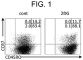
- Example 2 Distribution analysis of CD8 + T-cell subset using flow cytometry
- the ⁇ T processed cells prepared in Example 1 were analyzed by using a flow cytometer FACS CantoII (BD Biosciences). After washing the cells with PBS, the washed cells were centrifuged, and the obtained cells were resuspended in PBS containing 2% FBS and 5 mM of ethylenediaminetetraacetic acid (EDTA). The obtained resuspension was passed through a cell strainer to remove aggregates.
- EDTA ethylenediaminetetraacetic acid
- the ⁇ T processed cells treated with 2DG showed the phenotype of memory T cells (CCR7 ⁇ CD45RO+).
- the immature T cells are lymphocytes that have not yet been stimulated with antigens and have expressed CD45RA antigens on the cell surfaces thereof, and are activated by encountering antigen presenting cells such as dendritic cells, so that they become effector T cells.
- the effector T cells express CD45RO antigens in place of the CD45RA antigens on the cell surfaces thereof. Furthermore, some of the activated T cells (effector T cells) become memory T cells after antigens such as pathogens are eliminated.
- the memory T cells are lymphocytes that have already been stimulated with antigens and expressed the CD45RO antigens, regardless of whether the stimulation is specific stimulation or non-specific stimulation, and are maintained in the body for a long period of time while maintaining the memory of specific or non-specific antigens.
- the memory T cells can be divided into effector memory (EM) T cells (CCR7 negative CD45RO positive), and central memory (CM) T cells (CCR7 positive CD45RO positive).
- a pharmaceutical preparation containing a memory T cell group as a main component is maintained in the body for a long period of time, and thus has a high possibility of obtaining a high therapeutic effect in immune cell therapy.
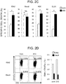
- the cytokine production ability of the ⁇ T processed cells prepared in Example 1 was determined by using Bio-plex "human cytokine G1 27 plex panel" (Bio-Rad Laboratories, Inc.) ( FIG. 2A ). Specifically, the ⁇ T processed cells prepared in Example 1 were replaced in a medium containing RPMI 1640 and 10% FCS in the absence of IL-2, and cultured. The culture supernatant after 24 hours from the start of the culture was recovered, and subjected to measurement.
- perforin and granzyme B in the culture supernatant after the degranulation due to the PMA 5 ng/ml
- ionomycin 0.5 ⁇ g/ml
- FIG. 2B perforin and granzyme B in the culture supernatant after the degranulation due to the PMA (5 ng/ml) and ionomycin (0.5 ⁇ g/ml) stimulation were determined by using Human Perforin ELISA Kit and Human Granzyme B ELISA Kit (Abcam plc.), respectively ( FIG. 2B ).
- IL-2 activates T cells, IFN- ⁇ and TNF ⁇ are produced also from the T cells, and an anti-tumor effect is exerted.
- Perforin is a glycoprotein that is present in cytoplasmic granules of a killer T cell or a NK cell, and causes cytotoxicity by entering the membrane lipid of a target cell and making a hole in there.
- Granzyme B which is one type of serine proteases, invades the inside of a target cell via perforin activates the caspase cascade to induce apoptosis or necrosis in the target cell.
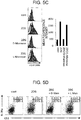
- the cytotoxic activity test by fluorescent staining was performed by using Terascan VPC2 (Minerva Tech K.K.). Cancer cell line K562 (leukemia cell line), Daudi (Burkitt's lymphoma cell line), and HOS (osteosarcoma cell line), and DLD1 (colon cancer cell line) were labeled for 30 minutes with 2.55 to 5 ⁇ g/mL of fluorescent dye Cellstein R-Calcein-AM solution (DOJINDO LABORATORIES). The labeled cancer cells and the ⁇ T processed cells were mixed at a ratio of 1 : 25, and the mixed cells were incubated for 2 to 4 hours in RPMI 1640 and 10% FBS (in the absence of IL-2).
- the cytotoxic activity was calculated by using changes in the fluorescence intensity of the cancer cells co-cultured with the ⁇ T processed cells as an index, assuming the fluorescence decay rate when treated with 1% NP40 (surfactant) as 100% ( FIG. 2C ).
- the ⁇ T processed cells and cancer cells were mixed at a ratio of 25: 1, and the mixed cells were cultured for 6 hours. After the washing of the cultured cells, the washed cells were stained with PE-labeled anti-CD8 antibodies (Beckman Coulter, Inc.). The cells thus stained were subjected to measurement by using a flow cytometer FACS CantoII (BD Biosciences), and the obtained data were analyzed by using Kaluza (Beckman Coulter, Inc.) software ( FIG. 2D ).
- the resultant material was incubated for 1 hour on ice, together with PE-labeled anti-NKG2D antibodies, FITC-labeled anti-CD3 antibodies, and PC5-labeled anti-CD8 antibodies (Beckman Coulter, Inc.), and after the washing of the incubated material, the measurement was performed.
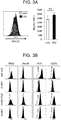
- the data were analyzed by using Kaluza (Beckman Coulter, Inc.) software ( FIG. 3A ).
- the obtained cancer cells were incubated for 1 hour on ice, together with anti-MIC A/B antibodies (BioLegend, Inc.), anti-ULBP1 antibodies, anti-ULBP2/5/6 antibodies, and anti-ULBP3 antibodies (R&D Systems, Inc.). After the washing of the incubated cancer cells, the cancer cells were reacted with FITC-labeled anti-mouse IgG antibodies (Jackson ImmunoResearch Laboratories, Inc.) on ice for 30 minutes, and the obtained cancer cells were subjected to washing, and then the measurement was performed. The data were analyzed by using Kaluza (Beckman Coulter, Inc.) software ( FIG. 3B ).
- NKG2D ligands As a result, high expression of NKG2D ligands was observed in the cells other than Daudi cells, and it was suggested that the binding of NKG2D and a NKG2D ligand was related to the cytotoxic activity.
- the ⁇ T processed cells were reacted for 60 minutes with anti-human NKG2D blocking antibodies (R&D Systems, Inc.) or mouse IgG1 (BioLegend, Inc.), which is isotype antibody, as a control, before co-culturing of the ⁇ T processed cells with cancer cells.
- the cytotoxic activity was determined in accordance with the description of 3.2 ( FIG. 3C ).
- Example 2 When the ⁇ T processed cells of Example 1 were prepared, oxamate (2 mM) or bromo pyruvate (10 ⁇ M), which is a glycolytic inhibitor, was added in place of 2DG, and the ⁇ T processed cells were prepared.
- the prepared ⁇ T processed cells were subjected to distribution analysis of CD8 + T-cell subset by the method described in Example 2.
- the 2DG is a glycolytic inhibitor
- oxamate or bromo pyruvate which is a glycolytic inhibitor other than 2DG
- Example 3 By using the method described in 3.2. of Example 3, the cytotoxic activity against each of cancer cells, by the ⁇ T processed cells treated with oxamate (2 mM) or bromo pyruvate (10 ⁇ M), which is a glycolytic inhibitor, was examined.
- 2DG inhibits the addition reaction of mannose consisting of N-linked glycans by 2DG's having a structure similar to that of the mannose. Therefore, in order to verify whether or not the effect of 2DG was due to the influence on the glycosylation of the ⁇ T processed cells, the changes in the sugar chains of the ⁇ T processed cells treated with 2DG were verified. Furthermore, it was verified whether or not the effect of 2DG were suppressed with the addition of mannose.
- sugar chains were purified and labeled by using BlotGlyco (Sumitomo Bakelite Co., Ltd.), and then the purified and labeled sugar chains were subjected to LC-MS mass spectrometry analysis ( FIG. 5A ).
- the amount of sugar chains (fmol) per cell of the peak number and the estimated structure of sugar chains are shown in Table 1.
- Table 1 Peak Estimated glycan composition Control 2 D G Proportion (2DG/Control) 1 (HexNAc)2(Sulph)1 + (Man)3(GlcNAc)2 420.
- Example 1 In the preparation of Example 1, ⁇ T processed cells were prepared with the addition of 2 mM of D-mannose (Sigma-Aldrich Co. LLC) or L-mannose (Tokyo Chemical Industry Co., Ltd.), and the prepared ⁇ T processed cells were subjected to the measurement of cytotoxic activity by the method described in item 3.2.
- D-mannose Sigma-Aldrich Co. LLC
- L-mannose Tokyo Chemical Industry Co., Ltd.
- ⁇ T processed cells were prepared with the addition of 2 mM of D-mannose (Sigma-Aldrich Co. LLC) or L-mannose (Tokyo Chemical Industry Co., Ltd.), the prepared cells were treated with IntraPrep Permeabilization Reagent (Beckman Coulter, Inc.), and the expression level of the intracellular perforin was determined by staining the cells with APC-labeled anti-perforin antibodies.
- the stained cells were subjected to measurement by using a flow cytometer FACS CantoII (BD Biosciences), and the data were analyzed by using Kaluza (Beckman Coulter, Inc.) software.
- the Mean Fluorescence Intensity (MFI) value is mean fluorescence intensity, and reflects the expression level.
- the ⁇ T processed cells prepared in item 7.3. were washed with PBS, and then the washed cells were centrifuged, and the obtained cells were resuspended in PBS containing 2% FBS and 5 mM of EDTA. The obtained resuspension was passed through a cell strainer to remove aggregates.
- the obtained cells were incubated for 1 hour on ice, together with PE/Cy5-labeled anti-IL-2R antibodies (BioLegend, Inc.), and FITC-labeled anti-CD3 antibodies (Beckman Coulter, Inc.), and after the washing of the incubated cells, the measurement was performed by using a flow cytometer FACS CantoII (BD Biosciences). The data were analyzed by using Kaluza (Beckman Coulter, Inc.) software.
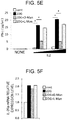
- the ⁇ T processed cells cultured in the absence of 2DG (cont), in the presence of 2DG (2DG), in the presence of 2DG and D-mannose (2DG + D-Man), or in the presence of 2DG and L-mannose (2DG + L-Man) were collected, the collected cells were precultured for 24 hours in RPMI 1640 medium and 10% FCS in the absence of IL-2, and then the precultured cells were again cultured in RPMI 1640 medium and 10% FCS with IL-2 (0, 50, or 250 IU/ml, NIPRO CORPORATION). The culture supernatant after the lapse of 24 hours from the start of the culture was collected, and yield of the IFN- ⁇ was determined by ELISA Kit (Abcam plc.) ( FIG. 5E ).
- the expression level of IL-2 receptor ⁇ in each sample was corrected with endogenous control ⁇ -actin, and then the relative value was graphed by assuming the cont as 1 ( FIG. 5F ).
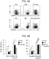
- Galectin-3 which is a member of the galectin family that has one or more of carbohydrate recognition domains with an affinity for ⁇ -galactoside, is secreted from a cancer cell, and not only induces angiogenesis but also binds to a glycosylated receptor on a CD8 positive T cell to induce unresponsiveness or apoptosis of the cell. Since the effect of 2DG is allowed to alter the glycosylation on a cell surface of the ⁇ T processed cell, it was examined whether or not there was a change in the influence of galectin-3 on the ⁇ T processed cell treated with 2DG.
- the ⁇ T processed cells treated with 2DG had lower binding ability of galectin as compared with that in the case of the absence of 2DG (cont), and thus it was able to suggest that the binding ability of galectin-3 was reduced in a dose-dependent manner of 2DG ( FIG. 6A ).
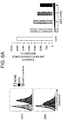
- the ⁇ T processed cells were stimulated with 3 ⁇ M recombinant galectin-3 (BBI Solutions) under the condition of 0.1 M lactose ( ⁇ ) (Sigma-Aldrich Co. LLC). After washing the cells with PBS, the cells were stained with Propidium Iodide (PI, for detection of dead cells) and FITC-labeled Annexin V (for detection of apoptosis) by using Annexin V-FITC Apoptosis Detection Kit (NACALAI TESQUE, INC.). The stained cells were subjected to measurement by using a flow cytometer FACS CantoII (BD Biosciences), and the data were analyzed by using Kaluza (Beckman Coulter, Inc.) software.
- PI Propidium Iodide
- FITC-labeled Annexin V for detection of apoptosis
- NACALAI TESQUE Annexin V-FITC Apoptosis Detection Kit
- Fluorescein isothiocyanate (FITC)-Annexin V has a strong affinity for membrane phospholipids (phosphatidylserine, PS) existing inside the cell membrane, and therefore, when the PS is exposed to a surface of the cell membrane by apoptosis, Annexin V binds to the PS. In a case where there is no damage in the cell membrane, PI that binds to DNA is not incorporated into the cell. Apoptosis cells at a late stage were stained with both Annexin V and PI.
- PS membrane phospholipids
- the ⁇ T processed cells treated with 2DG can be expected to avoid the galectin-mediated immune-escape function of cancer cells even in the tumor environment, and can also be expected to exert a more anti-tumor effect in the tumor than the conventional ⁇ T processed cells.
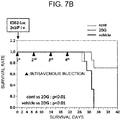
- mice 6 to 8 weeks old were purchased from CHARLES RIVER LABORATORIES JAPAN, INC., and were raised under the specific pathogen-free condition in accordance with the guidelines of the Animal Experiment Committee of Osaka University. All of the mice were injected via the tail vein on day 0 with 2 million human cancer cells (K562-Luc) in 200 ⁇ l of PBS. Subsequently, the ⁇ T processed cells treated with 2DG (2DG group in FIG. 7 , seven NOG mice), or the ⁇ T processed cells not treated with 2DG (cont group in FIG. 7 , six NOG mice) were injected intravenously once a week (day 0, day 7, day 14, and day 21 (shown by the symbol of ⁇ in FIG. 7B )) at a dose of 20 million cells per mouse. The vehicle group (six NOG mice) was injected intravenously with 200 ⁇ l of PBS on day 0, day 7, day 14, and day 21.
- the size of the tumor was measured on day 28 by In vivo Imaging System (IVIS) (Summit Pharmaceuticals International Corporation), and compared and investigated ( FIG. 7A ). In this regard, because one of the mice in the cont group died on day 27, the IVIS measurement of the cont group was performed on 5 mice. Furthermore, the survival time was also compared and investigated ( FIG. 7B ). In this regard, the changes in the survival rates of the cont group and vehicle group on day 28 and subsequent days showed the same trend, and the survival rates became 0 on day 32.
- IVIS In vivo Imaging System
- mice injected with the ⁇ T processed cells treated with 2DG (2DG group) showed significantly smaller tumor size than the NOG mice treated with PBS (vehicle group) ( FIG. 7A ). Furthermore, the injection of the ⁇ T processed cells treated with 2DG significantly prolonged the survival of NOG mice, as compared with that of the mice by the treatment of the ⁇ T processed cells not treated with 2DG (cont) and of the mice treated with PBS (vehicle) ( FIG. 7B ). Therefore, the ⁇ T processed cells treated with 2DG promoted the in vivo anti-tumor cell effect ( FIGS. 7A and 7B ).
- cancer immunotherapy can be more specifically and selectively provided to the cancer that has expressed specific NKG2D ligands, than the direct administration of the drug.
- by altering the glycosylation on a cell surface of an ⁇ T processed cell it can be expected to avoid the suppression of effector function in the tumor environment in a case where it is administered to the body.
Landscapes
- Health & Medical Sciences (AREA)
- Life Sciences & Earth Sciences (AREA)
- Engineering & Computer Science (AREA)
- Immunology (AREA)
- Chemical & Material Sciences (AREA)
- Biomedical Technology (AREA)
- Organic Chemistry (AREA)
- General Health & Medical Sciences (AREA)
- Zoology (AREA)
- Biotechnology (AREA)
- Cell Biology (AREA)
- Bioinformatics & Cheminformatics (AREA)
- Genetics & Genomics (AREA)
- Wood Science & Technology (AREA)
- Pharmacology & Pharmacy (AREA)
- Veterinary Medicine (AREA)
- Public Health (AREA)
- Animal Behavior & Ethology (AREA)
- Medicinal Chemistry (AREA)
- Microbiology (AREA)
- Hematology (AREA)
- Epidemiology (AREA)
- General Engineering & Computer Science (AREA)
- Biochemistry (AREA)
- Nuclear Medicine, Radiotherapy & Molecular Imaging (AREA)
- Chemical Kinetics & Catalysis (AREA)
- General Chemical & Material Sciences (AREA)
- Mycology (AREA)
- Developmental Biology & Embryology (AREA)
- Virology (AREA)
- Oncology (AREA)
- Medicines Containing Material From Animals Or Micro-Organisms (AREA)
- Micro-Organisms Or Cultivation Processes Thereof (AREA)
- Peptides Or Proteins (AREA)
- Preparation Of Compounds By Using Micro-Organisms (AREA)
Applications Claiming Priority (3)
| Application Number | Priority Date | Filing Date | Title |
|---|---|---|---|
| JP2018022488 | 2018-02-09 | ||
| JP2018184167 | 2018-09-28 | ||
| PCT/JP2019/004357 WO2019156145A1 (fr) | 2018-02-09 | 2019-02-07 | PROCÉDÉ AMÉLIORÉ DE PRODUCTION DE LYMPHOCYTES T TRAITÉS αβ |
Publications (2)
| Publication Number | Publication Date |
|---|---|
| EP3750988A1 true EP3750988A1 (fr) | 2020-12-16 |
| EP3750988A4 EP3750988A4 (fr) | 2021-11-10 |
Family
ID=67548264
Family Applications (1)
| Application Number | Title | Priority Date | Filing Date |
|---|---|---|---|
| EP19751544.8A Pending EP3750988A4 (fr) | 2018-02-09 | 2019-02-07 | Procédé amélioré de production de lymphocytes t traités alpha bêta |
Country Status (5)
| Country | Link |
|---|---|
| US (1) | US20210002611A1 (fr) |
| EP (1) | EP3750988A4 (fr) |
| JP (1) | JP7374434B2 (fr) |
| CN (1) | CN111936616A (fr) |
| WO (1) | WO2019156145A1 (fr) |
Family Cites Families (4)
| Publication number | Priority date | Publication date | Assignee | Title |
|---|---|---|---|---|
| WO2015112793A2 (fr) * | 2014-01-27 | 2015-07-30 | St. Jude Children's Research Hospital, Inc. | Procédés d'expansion ex vivo de lymphocytes tueurs naturels (nkt) et leurs utilisations thérapeutiques |
| MA41538A (fr) * | 2014-10-17 | 2017-12-26 | Baylor College Medicine | Cellules immunitaires bipartites et tripartites de signalisation |
| US20170056448A1 (en) * | 2015-09-01 | 2017-03-02 | Ifm Therapeutics, Inc | Immune cells having increased immunity or resistance to an immunosuppressive cytokine and use of the same |
| WO2017142014A1 (fr) * | 2016-02-17 | 2017-08-24 | 医療法人社団 滉志会 | Médicament pour une maladie inflammatoire |
-
2019
- 2019-02-07 CN CN201980012157.7A patent/CN111936616A/zh active Pending
- 2019-02-07 US US16/968,146 patent/US20210002611A1/en active Pending
- 2019-02-07 JP JP2019570793A patent/JP7374434B2/ja active Active
- 2019-02-07 WO PCT/JP2019/004357 patent/WO2019156145A1/fr unknown
- 2019-02-07 EP EP19751544.8A patent/EP3750988A4/fr active Pending
Also Published As
| Publication number | Publication date |
|---|---|
| CN111936616A (zh) | 2020-11-13 |
| JPWO2019156145A1 (ja) | 2021-01-28 |
| EP3750988A4 (fr) | 2021-11-10 |
| WO2019156145A1 (fr) | 2019-08-15 |
| JP7374434B2 (ja) | 2023-11-07 |
| US20210002611A1 (en) | 2021-01-07 |
Similar Documents
| Publication | Publication Date | Title |
|---|---|---|
| Chu et al. | Natural killer cells: a promising immunotherapy for cancer | |
| JP6940551B2 (ja) | 免疫を強化する抗cd47因子の使用 | |
| JP6858128B2 (ja) | 癌治療のための免疫療法とサイトカイン制御療法との組み合わせ | |
| JP2022548523A (ja) | 癌治療に向けた癌療法とサイトカイン制御療法との組み合わせ | |
| JP2021119197A (ja) | 癌治療のための併用免疫療法及びサイトカイン制御療法 | |
| EP3232199B1 (fr) | Procédé d'amélioration de la fonction des cellules immunitaires et procédé d'évaluation de la multifonctionnalité des cellules immunitaires | |
| US11376272B2 (en) | Methods of modulating immune activity | |
| JP6906520B2 (ja) | チェックポイント阻害剤と組みわせて2−デオキシ−2−フルオロ−l−フコースを用いる癌治療 | |
| JP2021506304A (ja) | 人工抗原提示細胞および使用方法 | |
| EP1930414A1 (fr) | Procede de traitement d'activation d'une cellule presentant l'antigene | |
| JP2022065022A (ja) | 改変ヒト初代血液樹状細胞株を生成するための方法 | |
| JP2018531022A6 (ja) | 改変ヒト初代血液樹状細胞株を生成するための方法 | |
| US11834677B2 (en) | Expansion and use of expanded NK cell fractions | |
| Hu et al. | A promising sword of tomorrow: human γδ T cell strategies reconcile allo-HSCT complications | |
| AU2019310855A2 (en) | Method for treating tumor using immune effector cell | |
| JP2022512161A (ja) | 免疫療法のための組成物及び方法 | |
| AU2019206724A1 (en) | Methods of administering chimeric antigen receptor immunotherapy in combination with 4-1BB agonist | |
| EP3750988A1 (fr) | Procédé amélioré de production de lymphocytes t traités alpha bêta | |
| KR20200015580A (ko) | 감소된 표면 푸코실화를 갖는 t 세포 및 이를 생산하고 사용하는 방법 | |
| Miyamoto et al. | Circulating cells and exosomes in acute myelogenous leukemia and their role in disease progression and survival | |
| Einsele et al. | OPEN ACCESS EDITED BY |
Legal Events
| Date | Code | Title | Description |
|---|---|---|---|
| STAA | Information on the status of an ep patent application or granted ep patent |
Free format text: STATUS: THE INTERNATIONAL PUBLICATION HAS BEEN MADE |
|
| PUAI | Public reference made under article 153(3) epc to a published international application that has entered the european phase |
Free format text: ORIGINAL CODE: 0009012 |
|
| STAA | Information on the status of an ep patent application or granted ep patent |
Free format text: STATUS: REQUEST FOR EXAMINATION WAS MADE |
|
| 17P | Request for examination filed |
Effective date: 20200806 |
|
| AK | Designated contracting states |
Kind code of ref document: A1 Designated state(s): AL AT BE BG CH CY CZ DE DK EE ES FI FR GB GR HR HU IE IS IT LI LT LU LV MC MK MT NL NO PL PT RO RS SE SI SK SM TR |
|
| AX | Request for extension of the european patent |
Extension state: BA ME |
|
| DAV | Request for validation of the european patent (deleted) | ||
| DAX | Request for extension of the european patent (deleted) | ||
| A4 | Supplementary search report drawn up and despatched |
Effective date: 20211011 |
|
| RIC1 | Information provided on ipc code assigned before grant |
Ipc: C12N 5/0783 20100101ALI20211005BHEP Ipc: C12N 1/00 20060101ALI20211005BHEP Ipc: A61P 35/02 20060101ALI20211005BHEP Ipc: A61P 35/00 20060101ALI20211005BHEP Ipc: A61P 31/20 20060101ALI20211005BHEP Ipc: A61P 31/18 20060101ALI20211005BHEP Ipc: A61P 31/16 20060101ALI20211005BHEP Ipc: A61P 31/14 20060101ALI20211005BHEP Ipc: A61P 31/12 20060101ALI20211005BHEP Ipc: A61P 31/04 20060101ALI20211005BHEP Ipc: A61K 35/17 20150101ALI20211005BHEP Ipc: C12N 5/078 20100101AFI20211005BHEP |
|
| STAA | Information on the status of an ep patent application or granted ep patent |
Free format text: STATUS: EXAMINATION IS IN PROGRESS |
|
| 17Q | First examination report despatched |
Effective date: 20230905 |