EP3741381B1 - Peptide pour l'utilisation pour la prévention et le traitement d'une lésion rénale aiguë - Google Patents
Peptide pour l'utilisation pour la prévention et le traitement d'une lésion rénale aiguë Download PDFInfo
- Publication number
- EP3741381B1 EP3741381B1 EP20165569.3A EP20165569A EP3741381B1 EP 3741381 B1 EP3741381 B1 EP 3741381B1 EP 20165569 A EP20165569 A EP 20165569A EP 3741381 B1 EP3741381 B1 EP 3741381B1
- Authority
- EP
- European Patent Office
- Prior art keywords
- renal
- dye
- group
- composition
- contrast
- Prior art date
- Legal status (The legal status is an assumption and is not a legal conclusion. Google has not performed a legal analysis and makes no representation as to the accuracy of the status listed.)
- Active
Links
- 108090000765 processed proteins & peptides Proteins 0.000 title claims description 128
- 208000009304 Acute Kidney Injury Diseases 0.000 title claims description 104
- 238000011282 treatment Methods 0.000 title description 72
- 230000002265 prevention Effects 0.000 title description 15
- 238000000034 method Methods 0.000 claims description 69
- 210000003734 kidney Anatomy 0.000 claims description 64
- 208000028867 ischemia Diseases 0.000 claims description 41
- 239000000203 mixture Substances 0.000 claims description 36
- DQLATGHUWYMOKM-UHFFFAOYSA-L cisplatin Chemical compound N[Pt](N)(Cl)Cl DQLATGHUWYMOKM-UHFFFAOYSA-L 0.000 claims description 28
- 229960004316 cisplatin Drugs 0.000 claims description 28
- 239000003814 drug Substances 0.000 claims description 24
- 229940079593 drug Drugs 0.000 claims description 21
- NTHXOOBQLCIOLC-UHFFFAOYSA-N iohexol Chemical compound OCC(O)CN(C(=O)C)C1=C(I)C(C(=O)NCC(O)CO)=C(I)C(C(=O)NCC(O)CO)=C1I NTHXOOBQLCIOLC-UHFFFAOYSA-N 0.000 claims description 14
- 201000002793 renal fibrosis Diseases 0.000 claims description 13
- 230000001988 toxicity Effects 0.000 claims description 13
- 231100000419 toxicity Toxicity 0.000 claims description 13
- 229960001025 iohexol Drugs 0.000 claims description 12
- YVPYQUNUQOZFHG-UHFFFAOYSA-N amidotrizoic acid Chemical compound CC(=O)NC1=C(I)C(NC(C)=O)=C(I)C(C(O)=O)=C1I YVPYQUNUQOZFHG-UHFFFAOYSA-N 0.000 claims description 11
- 229960005423 diatrizoate Drugs 0.000 claims description 11
- XQZXYNRDCRIARQ-LURJTMIESA-N iopamidol Chemical compound C[C@H](O)C(=O)NC1=C(I)C(C(=O)NC(CO)CO)=C(I)C(C(=O)NC(CO)CO)=C1I XQZXYNRDCRIARQ-LURJTMIESA-N 0.000 claims description 10
- GGGDNPWHMNJRFN-UHFFFAOYSA-N metrizoic acid Chemical compound CC(=O)N(C)C1=C(I)C(NC(C)=O)=C(I)C(C(O)=O)=C1I GGGDNPWHMNJRFN-UHFFFAOYSA-N 0.000 claims description 6
- UXIGWFXRQKWHHA-UHFFFAOYSA-N Iotalamic acid Chemical compound CNC(=O)C1=C(I)C(NC(C)=O)=C(I)C(C(O)=O)=C1I UXIGWFXRQKWHHA-UHFFFAOYSA-N 0.000 claims description 5
- 229960004647 iopamidol Drugs 0.000 claims description 5
- DGAIEPBNLOQYER-UHFFFAOYSA-N iopromide Chemical compound COCC(=O)NC1=C(I)C(C(=O)NCC(O)CO)=C(I)C(C(=O)N(C)CC(O)CO)=C1I DGAIEPBNLOQYER-UHFFFAOYSA-N 0.000 claims description 5
- 229940029378 iothalamate Drugs 0.000 claims description 5
- 229960000554 metrizamide Drugs 0.000 claims description 5
- GNOGSFBXBWBTIG-UHFFFAOYSA-N Acetrizoic acid Chemical compound CC(=O)NC1=C(I)C=C(I)C(C(O)=O)=C1I GNOGSFBXBWBTIG-UHFFFAOYSA-N 0.000 claims description 4
- 229930183010 Amphotericin Natural products 0.000 claims description 4
- QGGFZZLFKABGNL-UHFFFAOYSA-N Amphotericin A Natural products OC1C(N)C(O)C(C)OC1OC1C=CC=CC=CC=CCCC=CC=CC(C)C(O)C(C)C(C)OC(=O)CC(O)CC(O)CCC(O)C(O)CC(O)CC(O)(CC(O)C2C(O)=O)OC2C1 QGGFZZLFKABGNL-UHFFFAOYSA-N 0.000 claims description 4
- PMATZTZNYRCHOR-CGLBZJNRSA-N Cyclosporin A Chemical compound CC[C@@H]1NC(=O)[C@H]([C@H](O)[C@H](C)C\C=C\C)N(C)C(=O)[C@H](C(C)C)N(C)C(=O)[C@H](CC(C)C)N(C)C(=O)[C@H](CC(C)C)N(C)C(=O)[C@@H](C)NC(=O)[C@H](C)NC(=O)[C@H](CC(C)C)N(C)C(=O)[C@H](C(C)C)NC(=O)[C@H](CC(C)C)N(C)C(=O)CN(C)C1=O PMATZTZNYRCHOR-CGLBZJNRSA-N 0.000 claims description 4
- 108010036949 Cyclosporine Proteins 0.000 claims description 4
- 229930182566 Gentamicin Natural products 0.000 claims description 4
- CEAZRRDELHUEMR-URQXQFDESA-N Gentamicin Chemical compound O1[C@H](C(C)NC)CC[C@@H](N)[C@H]1O[C@H]1[C@H](O)[C@@H](O[C@@H]2[C@@H]([C@@H](NC)[C@@](C)(O)CO2)O)[C@H](N)C[C@@H]1N CEAZRRDELHUEMR-URQXQFDESA-N 0.000 claims description 4
- AMDBBAQNWSUWGN-UHFFFAOYSA-N Ioversol Chemical compound OCCN(C(=O)CO)C1=C(I)C(C(=O)NCC(O)CO)=C(I)C(C(=O)NCC(O)CO)=C1I AMDBBAQNWSUWGN-UHFFFAOYSA-N 0.000 claims description 4
- BAQCROVBDNBEEB-UBYUBLNFSA-N Metrizamide Chemical compound CC(=O)N(C)C1=C(I)C(NC(C)=O)=C(I)C(C(=O)N[C@@H]2[C@H]([C@H](O)[C@@H](CO)OC2O)O)=C1I BAQCROVBDNBEEB-UBYUBLNFSA-N 0.000 claims description 4
- 229940009444 amphotericin Drugs 0.000 claims description 4
- APKFDSVGJQXUKY-INPOYWNPSA-N amphotericin B Chemical compound O[C@H]1[C@@H](N)[C@H](O)[C@@H](C)O[C@H]1O[C@H]1/C=C/C=C/C=C/C=C/C=C/C=C/C=C/[C@H](C)[C@@H](O)[C@@H](C)[C@H](C)OC(=O)C[C@H](O)C[C@H](O)CC[C@@H](O)[C@H](O)C[C@H](O)C[C@](O)(C[C@H](O)[C@H]2C(O)=O)O[C@H]2C1 APKFDSVGJQXUKY-INPOYWNPSA-N 0.000 claims description 4
- CZTQZXZIADLWOZ-CRAIPNDOSA-N cefaloridine Chemical compound O=C([C@@H](NC(=O)CC=1SC=CC=1)[C@H]1SC2)N1C(C(=O)[O-])=C2C[N+]1=CC=CC=C1 CZTQZXZIADLWOZ-CRAIPNDOSA-N 0.000 claims description 4
- 229960003866 cefaloridine Drugs 0.000 claims description 4
- 229960001265 ciclosporin Drugs 0.000 claims description 4
- 229930182912 cyclosporin Natural products 0.000 claims description 4
- 229960002518 gentamicin Drugs 0.000 claims description 4
- VVDGWALACJEJKG-UHFFFAOYSA-N iodamide Chemical compound CC(=O)NCC1=C(I)C(NC(C)=O)=C(I)C(C(O)=O)=C1I VVDGWALACJEJKG-UHFFFAOYSA-N 0.000 claims description 4
- IUNJANQVIJDFTQ-UHFFFAOYSA-N iopentol Chemical compound COCC(O)CN(C(C)=O)C1=C(I)C(C(=O)NCC(O)CO)=C(I)C(C(=O)NCC(O)CO)=C1I IUNJANQVIJDFTQ-UHFFFAOYSA-N 0.000 claims description 4
- 229960002603 iopromide Drugs 0.000 claims description 4
- 229960004712 metrizoic acid Drugs 0.000 claims description 4
- 229960004901 iodamide Drugs 0.000 claims description 3
- 229960000824 iopentol Drugs 0.000 claims description 3
- 229960004537 ioversol Drugs 0.000 claims description 3
- SFVLTCAESLKEHH-WKAQUBQDSA-N (2s)-6-amino-2-[[(2s)-2-[[(2r)-2-amino-5-(diaminomethylideneamino)pentanoyl]amino]-3-(4-hydroxy-2,6-dimethylphenyl)propanoyl]amino]-n-[(2s)-1-amino-1-oxo-3-phenylpropan-2-yl]hexanamide Chemical compound CC1=CC(O)=CC(C)=C1C[C@H](NC(=O)[C@H](N)CCCN=C(N)N)C(=O)N[C@@H](CCCCN)C(=O)N[C@H](C(N)=O)CC1=CC=CC=C1 SFVLTCAESLKEHH-WKAQUBQDSA-N 0.000 description 140
- DDRJAANPRJIHGJ-UHFFFAOYSA-N creatinine Chemical compound CN1CC(=O)NC1=N DDRJAANPRJIHGJ-UHFFFAOYSA-N 0.000 description 134
- 102000004196 processed proteins & peptides Human genes 0.000 description 93
- PEDCQBHIVMGVHV-UHFFFAOYSA-N Glycerine Chemical compound OCC(O)CO PEDCQBHIVMGVHV-UHFFFAOYSA-N 0.000 description 90
- 239000000975 dye Substances 0.000 description 89
- 241000700159 Rattus Species 0.000 description 84
- 239000002872 contrast media Substances 0.000 description 68
- 229940109239 creatinine Drugs 0.000 description 67
- 210000002966 serum Anatomy 0.000 description 51
- 108090000623 proteins and genes Proteins 0.000 description 49
- 102000004169 proteins and genes Human genes 0.000 description 49
- 230000000694 effects Effects 0.000 description 47
- 210000004027 cell Anatomy 0.000 description 44
- 230000003907 kidney function Effects 0.000 description 43
- 241001465754 Metazoa Species 0.000 description 39
- 238000010171 animal model Methods 0.000 description 37
- 235000011187 glycerol Nutrition 0.000 description 36
- 229960005150 glycerol Drugs 0.000 description 36
- 230000002485 urinary effect Effects 0.000 description 34
- 208000004608 Ureteral Obstruction Diseases 0.000 description 33
- 235000002639 sodium chloride Nutrition 0.000 description 31
- 238000010186 staining Methods 0.000 description 28
- 230000006378 damage Effects 0.000 description 27
- 210000002700 urine Anatomy 0.000 description 26
- 229940039231 contrast media Drugs 0.000 description 25
- 239000003981 vehicle Substances 0.000 description 25
- 239000000994 contrast dye Substances 0.000 description 24
- VZGDMQKNWNREIO-UHFFFAOYSA-N tetrachloromethane Chemical compound ClC(Cl)(Cl)Cl VZGDMQKNWNREIO-UHFFFAOYSA-N 0.000 description 24
- FAPWRFPIFSIZLT-UHFFFAOYSA-M Sodium chloride Chemical compound [Na+].[Cl-] FAPWRFPIFSIZLT-UHFFFAOYSA-M 0.000 description 22
- 150000001413 amino acids Chemical class 0.000 description 21
- 239000003795 chemical substances by application Substances 0.000 description 21
- 238000002474 experimental method Methods 0.000 description 21
- 230000001965 increasing effect Effects 0.000 description 21
- 210000000110 microvilli Anatomy 0.000 description 21
- 239000011780 sodium chloride Substances 0.000 description 21
- 230000006907 apoptotic process Effects 0.000 description 20
- 230000007423 decrease Effects 0.000 description 20
- 230000037396 body weight Effects 0.000 description 19
- 230000010410 reperfusion Effects 0.000 description 18
- 210000005239 tubule Anatomy 0.000 description 18
- 206010016654 Fibrosis Diseases 0.000 description 17
- 210000004369 blood Anatomy 0.000 description 17
- 239000008280 blood Substances 0.000 description 17
- 208000037265 diseases, disorders, signs and symptoms Diseases 0.000 description 17
- 230000004761 fibrosis Effects 0.000 description 17
- 238000002347 injection Methods 0.000 description 17
- 229940090044 injection Drugs 0.000 description 17
- 239000007924 injection Substances 0.000 description 17
- 208000017169 kidney disease Diseases 0.000 description 17
- 208000027418 Wounds and injury Diseases 0.000 description 16
- 230000003247 decreasing effect Effects 0.000 description 16
- 208000014674 injury Diseases 0.000 description 16
- 230000002829 reductive effect Effects 0.000 description 16
- 230000017074 necrotic cell death Effects 0.000 description 15
- ZCYVEMRRCGMTRW-UHFFFAOYSA-N 7553-56-2 Chemical compound [I] ZCYVEMRRCGMTRW-UHFFFAOYSA-N 0.000 description 13
- 206010039020 Rhabdomyolysis Diseases 0.000 description 13
- 206010012601 diabetes mellitus Diseases 0.000 description 13
- 210000002540 macrophage Anatomy 0.000 description 13
- 238000001000 micrograph Methods 0.000 description 13
- 108010018924 Heme Oxygenase-1 Proteins 0.000 description 12
- 102000002737 Heme Oxygenase-1 Human genes 0.000 description 12
- 230000001640 apoptogenic effect Effects 0.000 description 12
- 150000001875 compounds Chemical class 0.000 description 12
- 201000010099 disease Diseases 0.000 description 12
- CGIGDMFJXJATDK-UHFFFAOYSA-N indomethacin Chemical compound CC1=C(CC(O)=O)C2=CC(OC)=CC=C2N1C(=O)C1=CC=C(Cl)C=C1 CGIGDMFJXJATDK-UHFFFAOYSA-N 0.000 description 12
- 210000003470 mitochondria Anatomy 0.000 description 12
- ZSJLQEPLLKMAKR-GKHCUFPYSA-N streptozocin Chemical compound O=NN(C)C(=O)N[C@H]1[C@@H](O)O[C@H](CO)[C@@H](O)[C@@H]1O ZSJLQEPLLKMAKR-GKHCUFPYSA-N 0.000 description 12
- 239000000126 substance Substances 0.000 description 12
- XLYOFNOQVPJJNP-UHFFFAOYSA-N water Chemical compound O XLYOFNOQVPJJNP-UHFFFAOYSA-N 0.000 description 11
- 108010088751 Albumins Proteins 0.000 description 10
- 102000009027 Albumins Human genes 0.000 description 10
- WSFSSNUMVMOOMR-UHFFFAOYSA-N Formaldehyde Chemical compound O=C WSFSSNUMVMOOMR-UHFFFAOYSA-N 0.000 description 10
- KCWZGJVSDFYRIX-YFKPBYRVSA-N N(gamma)-nitro-L-arginine methyl ester Chemical compound COC(=O)[C@@H](N)CCCN=C(N)N[N+]([O-])=O KCWZGJVSDFYRIX-YFKPBYRVSA-N 0.000 description 10
- 230000008859 change Effects 0.000 description 10
- 210000002950 fibroblast Anatomy 0.000 description 10
- -1 foscamet Chemical compound 0.000 description 10
- 230000008595 infiltration Effects 0.000 description 10
- 238000001764 infiltration Methods 0.000 description 10
- 239000011630 iodine Substances 0.000 description 10
- 229910052740 iodine Inorganic materials 0.000 description 10
- 239000000178 monomer Substances 0.000 description 10
- 230000009467 reduction Effects 0.000 description 10
- 230000008085 renal dysfunction Effects 0.000 description 10
- 150000003839 salts Chemical class 0.000 description 10
- 208000033626 Renal failure acute Diseases 0.000 description 9
- 201000011040 acute kidney failure Diseases 0.000 description 9
- 230000002238 attenuated effect Effects 0.000 description 9
- 231100000417 nephrotoxicity Toxicity 0.000 description 9
- 239000003642 reactive oxygen metabolite Substances 0.000 description 9
- 108010061642 Cystatin C Proteins 0.000 description 8
- 102000012192 Cystatin C Human genes 0.000 description 8
- 206010029155 Nephropathy toxic Diseases 0.000 description 8
- 238000000692 Student's t-test Methods 0.000 description 8
- 239000000539 dimer Substances 0.000 description 8
- 230000003589 nefrotoxic effect Effects 0.000 description 8
- 230000007694 nephrotoxicity Effects 0.000 description 8
- 230000004792 oxidative damage Effects 0.000 description 8
- 239000012188 paraffin wax Substances 0.000 description 8
- 210000000512 proximal kidney tubule Anatomy 0.000 description 8
- 239000000243 solution Substances 0.000 description 8
- WQZGKKKJIJFFOK-GASJEMHNSA-N Glucose Natural products OC[C@H]1OC(O)[C@H](O)[C@@H](O)[C@@H]1O WQZGKKKJIJFFOK-GASJEMHNSA-N 0.000 description 7
- 230000001684 chronic effect Effects 0.000 description 7
- 208000020832 chronic kidney disease Diseases 0.000 description 7
- 230000008021 deposition Effects 0.000 description 7
- 238000000151 deposition Methods 0.000 description 7
- 238000009472 formulation Methods 0.000 description 7
- 230000024924 glomerular filtration Effects 0.000 description 7
- 239000008103 glucose Substances 0.000 description 7
- 230000001976 improved effect Effects 0.000 description 7
- 231100000381 nephrotoxic Toxicity 0.000 description 7
- 230000035755 proliferation Effects 0.000 description 7
- 210000005234 proximal tubule cell Anatomy 0.000 description 7
- 210000005084 renal tissue Anatomy 0.000 description 7
- 238000001356 surgical procedure Methods 0.000 description 7
- 208000024891 symptom Diseases 0.000 description 7
- 210000004926 tubular epithelial cell Anatomy 0.000 description 7
- 102000008186 Collagen Human genes 0.000 description 6
- 108010035532 Collagen Proteins 0.000 description 6
- 102000004190 Enzymes Human genes 0.000 description 6
- 108090000790 Enzymes Proteins 0.000 description 6
- OKKJLVBELUTLKV-UHFFFAOYSA-N Methanol Chemical compound OC OKKJLVBELUTLKV-UHFFFAOYSA-N 0.000 description 6
- TZCXTZWJZNENPQ-UHFFFAOYSA-L barium sulfate Chemical compound [Ba+2].[O-]S([O-])(=O)=O TZCXTZWJZNENPQ-UHFFFAOYSA-L 0.000 description 6
- 239000006172 buffering agent Substances 0.000 description 6
- 229920001436 collagen Polymers 0.000 description 6
- 229940088598 enzyme Drugs 0.000 description 6
- 229960000905 indomethacin Drugs 0.000 description 6
- 238000000386 microscopy Methods 0.000 description 6
- 230000035699 permeability Effects 0.000 description 6
- 229920001184 polypeptide Polymers 0.000 description 6
- 238000007920 subcutaneous administration Methods 0.000 description 6
- 230000002407 ATP formation Effects 0.000 description 5
- 208000003918 Acute Kidney Tubular Necrosis Diseases 0.000 description 5
- 208000007788 Acute Liver Failure Diseases 0.000 description 5
- 206010000804 Acute hepatic failure Diseases 0.000 description 5
- 241000282412 Homo Species 0.000 description 5
- MHAJPDPJQMAIIY-UHFFFAOYSA-N Hydrogen peroxide Chemical compound OO MHAJPDPJQMAIIY-UHFFFAOYSA-N 0.000 description 5
- DGAQECJNVWCQMB-PUAWFVPOSA-M Ilexoside XXIX Chemical compound C[C@@H]1CC[C@@]2(CC[C@@]3(C(=CC[C@H]4[C@]3(CC[C@@H]5[C@@]4(CC[C@@H](C5(C)C)OS(=O)(=O)[O-])C)C)[C@@H]2[C@]1(C)O)C)C(=O)O[C@H]6[C@@H]([C@H]([C@@H]([C@H](O6)CO)O)O)O.[Na+] DGAQECJNVWCQMB-PUAWFVPOSA-M 0.000 description 5
- 241000124008 Mammalia Species 0.000 description 5
- 208000001647 Renal Insufficiency Diseases 0.000 description 5
- 206010062237 Renal impairment Diseases 0.000 description 5
- 206010061481 Renal injury Diseases 0.000 description 5
- 206010038540 Renal tubular necrosis Diseases 0.000 description 5
- 230000002159 abnormal effect Effects 0.000 description 5
- 231100000836 acute liver failure Toxicity 0.000 description 5
- 208000012998 acute renal failure Diseases 0.000 description 5
- 208000022831 chronic renal failure syndrome Diseases 0.000 description 5
- 208000035475 disorder Diseases 0.000 description 5
- 230000006870 function Effects 0.000 description 5
- NBQNWMBBSKPBAY-UHFFFAOYSA-N iodixanol Chemical compound IC=1C(C(=O)NCC(O)CO)=C(I)C(C(=O)NCC(O)CO)=C(I)C=1N(C(=O)C)CC(O)CN(C(C)=O)C1=C(I)C(C(=O)NCC(O)CO)=C(I)C(C(=O)NCC(O)CO)=C1I NBQNWMBBSKPBAY-UHFFFAOYSA-N 0.000 description 5
- 201000006370 kidney failure Diseases 0.000 description 5
- 210000004379 membrane Anatomy 0.000 description 5
- 239000012528 membrane Substances 0.000 description 5
- 231100000637 nephrotoxin Toxicity 0.000 description 5
- 230000007935 neutral effect Effects 0.000 description 5
- 230000005298 paramagnetic effect Effects 0.000 description 5
- 231100000857 poor renal function Toxicity 0.000 description 5
- 201000001474 proteinuria Diseases 0.000 description 5
- 238000011084 recovery Methods 0.000 description 5
- 229910052708 sodium Inorganic materials 0.000 description 5
- 239000003381 stabilizer Substances 0.000 description 5
- 230000009885 systemic effect Effects 0.000 description 5
- 230000001225 therapeutic effect Effects 0.000 description 5
- 210000001519 tissue Anatomy 0.000 description 5
- 231100000331 toxic Toxicity 0.000 description 5
- 230000002588 toxic effect Effects 0.000 description 5
- 210000003462 vein Anatomy 0.000 description 5
- QGZKDVFQNNGYKY-UHFFFAOYSA-N Ammonia Chemical compound N QGZKDVFQNNGYKY-UHFFFAOYSA-N 0.000 description 4
- BPYKTIZUTYGOLE-IFADSCNNSA-N Bilirubin Chemical compound N1C(=O)C(C)=C(C=C)\C1=C\C1=C(C)C(CCC(O)=O)=C(CC2=C(C(C)=C(\C=C/3C(=C(C=C)C(=O)N\3)C)N2)CCC(O)=O)N1 BPYKTIZUTYGOLE-IFADSCNNSA-N 0.000 description 4
- 238000009010 Bradford assay Methods 0.000 description 4
- 238000004458 analytical method Methods 0.000 description 4
- 239000000427 antigen Substances 0.000 description 4
- 102000036639 antigens Human genes 0.000 description 4
- 108091007433 antigens Proteins 0.000 description 4
- 238000004166 bioassay Methods 0.000 description 4
- 230000015572 biosynthetic process Effects 0.000 description 4
- 230000036770 blood supply Effects 0.000 description 4
- 230000000747 cardiac effect Effects 0.000 description 4
- 230000006735 deficit Effects 0.000 description 4
- 230000018044 dehydration Effects 0.000 description 4
- 238000006297 dehydration reaction Methods 0.000 description 4
- 210000002919 epithelial cell Anatomy 0.000 description 4
- 125000002887 hydroxy group Chemical group [H]O* 0.000 description 4
- 230000004054 inflammatory process Effects 0.000 description 4
- 230000003993 interaction Effects 0.000 description 4
- 229960004359 iodixanol Drugs 0.000 description 4
- 230000000302 ischemic effect Effects 0.000 description 4
- 230000003908 liver function Effects 0.000 description 4
- 238000002595 magnetic resonance imaging Methods 0.000 description 4
- 238000004519 manufacturing process Methods 0.000 description 4
- 230000002438 mitochondrial effect Effects 0.000 description 4
- 210000000056 organ Anatomy 0.000 description 4
- 230000000737 periodic effect Effects 0.000 description 4
- 239000011734 sodium Substances 0.000 description 4
- 229960001052 streptozocin Drugs 0.000 description 4
- 230000008961 swelling Effects 0.000 description 4
- 238000012360 testing method Methods 0.000 description 4
- 238000002054 transplantation Methods 0.000 description 4
- HUHDYASLFWQVOL-WZTVWXICSA-N 3-[[2-[[3-[acetyl(methyl)amino]-2,4,6-triiodo-5-(methylcarbamoyl)benzoyl]amino]acetyl]amino]-5-(2-hydroxyethylcarbamoyl)-2,4,6-triiodobenzoic acid;(2r,3r,4r,5s)-6-(methylamino)hexane-1,2,3,4,5-pentol Chemical compound CNC[C@H](O)[C@@H](O)[C@H](O)[C@H](O)CO.CNC(=O)C1=C(I)C(N(C)C(C)=O)=C(I)C(C(=O)NCC(=O)NC=2C(=C(C(=O)NCCO)C(I)=C(C(O)=O)C=2I)I)=C1I HUHDYASLFWQVOL-WZTVWXICSA-N 0.000 description 3
- 206010001580 Albuminuria Diseases 0.000 description 3
- 108010067770 Endopeptidase K Proteins 0.000 description 3
- 229910052688 Gadolinium Inorganic materials 0.000 description 3
- 206010061218 Inflammation Diseases 0.000 description 3
- XUHXFSYUBXNTHU-UHFFFAOYSA-N Iotrolan Chemical compound IC=1C(C(=O)NC(CO)C(O)CO)=C(I)C(C(=O)NC(CO)C(O)CO)=C(I)C=1N(C)C(=O)CC(=O)N(C)C1=C(I)C(C(=O)NC(CO)C(O)CO)=C(I)C(C(=O)NC(CO)C(O)CO)=C1I XUHXFSYUBXNTHU-UHFFFAOYSA-N 0.000 description 3
- MBBZMMPHUWSWHV-BDVNFPICSA-N N-methylglucamine Chemical compound CNC[C@H](O)[C@@H](O)[C@H](O)[C@H](O)CO MBBZMMPHUWSWHV-BDVNFPICSA-N 0.000 description 3
- ZSJLQEPLLKMAKR-UHFFFAOYSA-N Streptozotocin Natural products O=NN(C)C(=O)NC1C(O)OC(CO)C(O)C1O ZSJLQEPLLKMAKR-UHFFFAOYSA-N 0.000 description 3
- 238000012288 TUNEL assay Methods 0.000 description 3
- PNNCWTXUWKENPE-UHFFFAOYSA-N [N].NC(N)=O Chemical compound [N].NC(N)=O PNNCWTXUWKENPE-UHFFFAOYSA-N 0.000 description 3
- 238000010521 absorption reaction Methods 0.000 description 3
- 230000001154 acute effect Effects 0.000 description 3
- 150000001298 alcohols Chemical class 0.000 description 3
- 239000003963 antioxidant agent Substances 0.000 description 3
- 230000003078 antioxidant effect Effects 0.000 description 3
- QVGXLLKOCUKJST-UHFFFAOYSA-N atomic oxygen Chemical compound [O] QVGXLLKOCUKJST-UHFFFAOYSA-N 0.000 description 3
- 210000000170 cell membrane Anatomy 0.000 description 3
- 230000001413 cellular effect Effects 0.000 description 3
- OSASVXMJTNOKOY-UHFFFAOYSA-N chlorobutanol Chemical compound CC(C)(O)C(Cl)(Cl)Cl OSASVXMJTNOKOY-UHFFFAOYSA-N 0.000 description 3
- KRKNYBCHXYNGOX-UHFFFAOYSA-N citric acid Chemical compound OC(=O)CC(O)(C(O)=O)CC(O)=O KRKNYBCHXYNGOX-UHFFFAOYSA-N 0.000 description 3
- 230000007850 degeneration Effects 0.000 description 3
- 230000001419 dependent effect Effects 0.000 description 3
- 238000010790 dilution Methods 0.000 description 3
- 239000012895 dilution Substances 0.000 description 3
- UIWYJDYFSGRHKR-UHFFFAOYSA-N gadolinium atom Chemical compound [Gd] UIWYJDYFSGRHKR-UHFFFAOYSA-N 0.000 description 3
- 230000001434 glomerular Effects 0.000 description 3
- 210000000585 glomerular basement membrane Anatomy 0.000 description 3
- 238000003306 harvesting Methods 0.000 description 3
- 235000009200 high fat diet Nutrition 0.000 description 3
- 238000003364 immunohistochemistry Methods 0.000 description 3
- 231100000268 induced nephrotoxicity Toxicity 0.000 description 3
- 230000028709 inflammatory response Effects 0.000 description 3
- 238000010255 intramuscular injection Methods 0.000 description 3
- 238000001990 intravenous administration Methods 0.000 description 3
- LAYLQVBQIBQVLL-UHFFFAOYSA-N iofendylate Chemical compound CCOC(=O)CCCCCCCCC(C)C1=CC=C(I)C=C1 LAYLQVBQIBQVLL-UHFFFAOYSA-N 0.000 description 3
- 229960003182 iotrolan Drugs 0.000 description 3
- 239000000463 material Substances 0.000 description 3
- 230000007246 mechanism Effects 0.000 description 3
- 229960003194 meglumine Drugs 0.000 description 3
- MIKKOBKEXMRYFQ-WZTVWXICSA-N meglumine amidotrizoate Chemical compound C[NH2+]C[C@H](O)[C@@H](O)[C@H](O)[C@H](O)CO.CC(=O)NC1=C(I)C(NC(C)=O)=C(I)C(C([O-])=O)=C1I MIKKOBKEXMRYFQ-WZTVWXICSA-N 0.000 description 3
- 230000002503 metabolic effect Effects 0.000 description 3
- 229910021645 metal ion Inorganic materials 0.000 description 3
- 239000003921 oil Substances 0.000 description 3
- 239000001301 oxygen Substances 0.000 description 3
- 229910052760 oxygen Inorganic materials 0.000 description 3
- 230000036284 oxygen consumption Effects 0.000 description 3
- 239000002245 particle Substances 0.000 description 3
- 238000005192 partition Methods 0.000 description 3
- 239000008194 pharmaceutical composition Substances 0.000 description 3
- 239000000546 pharmaceutical excipient Substances 0.000 description 3
- 239000012071 phase Substances 0.000 description 3
- 125000001997 phenyl group Chemical group [H]C1=C([H])C([H])=C(*)C([H])=C1[H] 0.000 description 3
- 230000008569 process Effects 0.000 description 3
- 230000000069 prophylactic effect Effects 0.000 description 3
- 239000007845 reactive nitrogen species Substances 0.000 description 3
- 238000003757 reverse transcription PCR Methods 0.000 description 3
- 230000002441 reversible effect Effects 0.000 description 3
- 238000012453 sprague-dawley rat model Methods 0.000 description 3
- 239000004094 surface-active agent Substances 0.000 description 3
- 238000003786 synthesis reaction Methods 0.000 description 3
- 229940124597 therapeutic agent Drugs 0.000 description 3
- 230000032258 transport Effects 0.000 description 3
- 210000005233 tubule cell Anatomy 0.000 description 3
- RYSMHWILUNYBFW-UHFFFAOYSA-N 4-amino-2-[6-(dimethylamino)purin-9-yl]-5-(hydroxymethyl)oxolan-3-ol Chemical compound C1=NC=2C(N(C)C)=NC=NC=2N1C1OC(CO)C(N)C1O RYSMHWILUNYBFW-UHFFFAOYSA-N 0.000 description 2
- 101710103970 ADP,ATP carrier protein Proteins 0.000 description 2
- 101710133192 ADP,ATP carrier protein, mitochondrial Proteins 0.000 description 2
- 208000010444 Acidosis Diseases 0.000 description 2
- 102000002260 Alkaline Phosphatase Human genes 0.000 description 2
- 108020004774 Alkaline Phosphatase Proteins 0.000 description 2
- 206010002091 Anaesthesia Diseases 0.000 description 2
- 244000105975 Antidesma platyphyllum Species 0.000 description 2
- 201000003126 Anuria Diseases 0.000 description 2
- 206010005003 Bladder cancer Diseases 0.000 description 2
- 102000019034 Chemokines Human genes 0.000 description 2
- 108010012236 Chemokines Proteins 0.000 description 2
- 229930105110 Cyclosporin A Natural products 0.000 description 2
- 102100030497 Cytochrome c Human genes 0.000 description 2
- 108010075031 Cytochromes c Proteins 0.000 description 2
- 102000004127 Cytokines Human genes 0.000 description 2
- 108090000695 Cytokines Proteins 0.000 description 2
- FBPFZTCFMRRESA-KVTDHHQDSA-N D-Mannitol Chemical compound OC[C@@H](O)[C@@H](O)[C@H](O)[C@H](O)CO FBPFZTCFMRRESA-KVTDHHQDSA-N 0.000 description 2
- ZMJOVJSTYLQINE-UHFFFAOYSA-N Dichloroacetylene Chemical group ClC#CCl ZMJOVJSTYLQINE-UHFFFAOYSA-N 0.000 description 2
- AOJJSUZBOXZQNB-TZSSRYMLSA-N Doxorubicin Chemical compound O([C@H]1C[C@@](O)(CC=2C(O)=C3C(=O)C=4C=CC=C(C=4C(=O)C3=C(O)C=21)OC)C(=O)CO)[C@H]1C[C@H](N)[C@H](O)[C@H](C)O1 AOJJSUZBOXZQNB-TZSSRYMLSA-N 0.000 description 2
- LYCAIKOWRPUZTN-UHFFFAOYSA-N Ethylene glycol Chemical compound OCCO LYCAIKOWRPUZTN-UHFFFAOYSA-N 0.000 description 2
- 102000010834 Extracellular Matrix Proteins Human genes 0.000 description 2
- 108010037362 Extracellular Matrix Proteins Proteins 0.000 description 2
- 102000016359 Fibronectins Human genes 0.000 description 2
- 108010067306 Fibronectins Proteins 0.000 description 2
- WZUVPPKBWHMQCE-UHFFFAOYSA-N Haematoxylin Chemical compound C12=CC(O)=C(O)C=C2CC2(O)C1C1=CC=C(O)C(O)=C1OC2 WZUVPPKBWHMQCE-UHFFFAOYSA-N 0.000 description 2
- 102100028006 Heme oxygenase 1 Human genes 0.000 description 2
- 101001079623 Homo sapiens Heme oxygenase 1 Proteins 0.000 description 2
- 208000002682 Hyperkalemia Diseases 0.000 description 2
- 206010021143 Hypoxia Diseases 0.000 description 2
- VLHUSFYMPUDOEL-WZTVWXICSA-N Iothalamate meglumine Chemical compound CNC[C@H](O)[C@@H](O)[C@H](O)[C@H](O)CO.CNC(=O)C1=C(I)C(NC(C)=O)=C(I)C(C(O)=O)=C1I VLHUSFYMPUDOEL-WZTVWXICSA-N 0.000 description 2
- UQSXHKLRYXJYBZ-UHFFFAOYSA-N Iron oxide Chemical compound [Fe]=O UQSXHKLRYXJYBZ-UHFFFAOYSA-N 0.000 description 2
- YQEZLKZALYSWHR-UHFFFAOYSA-N Ketamine Chemical compound C=1C=CC=C(Cl)C=1C1(NC)CCCCC1=O YQEZLKZALYSWHR-UHFFFAOYSA-N 0.000 description 2
- 208000000913 Kidney Calculi Diseases 0.000 description 2
- LRQKBLKVPFOOQJ-YFKPBYRVSA-N L-norleucine Chemical group CCCC[C@H]([NH3+])C([O-])=O LRQKBLKVPFOOQJ-YFKPBYRVSA-N 0.000 description 2
- JVTAAEKCZFNVCJ-UHFFFAOYSA-M Lactate Chemical compound CC(O)C([O-])=O JVTAAEKCZFNVCJ-UHFFFAOYSA-M 0.000 description 2
- WHXSMMKQMYFTQS-UHFFFAOYSA-N Lithium Chemical compound [Li] WHXSMMKQMYFTQS-UHFFFAOYSA-N 0.000 description 2
- FYYHWMGAXLPEAU-UHFFFAOYSA-N Magnesium Chemical compound [Mg] FYYHWMGAXLPEAU-UHFFFAOYSA-N 0.000 description 2
- 229930195725 Mannitol Natural products 0.000 description 2
- 206010027417 Metabolic acidosis Diseases 0.000 description 2
- 206010027525 Microalbuminuria Diseases 0.000 description 2
- 206010029148 Nephrolithiasis Diseases 0.000 description 2
- MWUXSHHQAYIFBG-UHFFFAOYSA-N Nitric oxide Chemical compound O=[N] MWUXSHHQAYIFBG-UHFFFAOYSA-N 0.000 description 2
- 206010030302 Oliguria Diseases 0.000 description 2
- 102000003992 Peroxidases Human genes 0.000 description 2
- 229920001213 Polysorbate 20 Polymers 0.000 description 2
- ROSXARVHJNYYDO-UHFFFAOYSA-N Propyliodone Chemical compound CCCOC(=O)CN1C=C(I)C(=O)C(I)=C1 ROSXARVHJNYYDO-UHFFFAOYSA-N 0.000 description 2
- 206010060862 Prostate cancer Diseases 0.000 description 2
- 208000000236 Prostatic Neoplasms Diseases 0.000 description 2
- LOUPRKONTZGTKE-WZBLMQSHSA-N Quinine Chemical compound C([C@H]([C@H](C1)C=C)C2)C[N@@]1[C@@H]2[C@H](O)C1=CC=NC2=CC=C(OC)C=C21 LOUPRKONTZGTKE-WZBLMQSHSA-N 0.000 description 2
- 101001079613 Rattus norvegicus Heme oxygenase 1 Proteins 0.000 description 2
- 206010063897 Renal ischaemia Diseases 0.000 description 2
- 102000003929 Transaminases Human genes 0.000 description 2
- 108090000340 Transaminases Proteins 0.000 description 2
- 102000004887 Transforming Growth Factor beta Human genes 0.000 description 2
- 108090001012 Transforming Growth Factor beta Proteins 0.000 description 2
- XSQUKJJJFZCRTK-UHFFFAOYSA-N Urea Chemical compound NC(N)=O XSQUKJJJFZCRTK-UHFFFAOYSA-N 0.000 description 2
- 208000007097 Urinary Bladder Neoplasms Diseases 0.000 description 2
- 206010047139 Vasoconstriction Diseases 0.000 description 2
- 241000251539 Vertebrata <Metazoa> Species 0.000 description 2
- 238000009825 accumulation Methods 0.000 description 2
- 239000000654 additive Substances 0.000 description 2
- 230000002411 adverse Effects 0.000 description 2
- 230000001668 ameliorated effect Effects 0.000 description 2
- 229910021529 ammonia Inorganic materials 0.000 description 2
- 230000037005 anaesthesia Effects 0.000 description 2
- 238000002583 angiography Methods 0.000 description 2
- 150000001450 anions Chemical class 0.000 description 2
- 239000007864 aqueous solution Substances 0.000 description 2
- 108010033284 arginyl-2,'6'-dimethyltyrosyl-lysyl-phenylalaninamide Proteins 0.000 description 2
- 230000008499 blood brain barrier function Effects 0.000 description 2
- 230000017531 blood circulation Effects 0.000 description 2
- 230000036772 blood pressure Effects 0.000 description 2
- 210000001218 blood-brain barrier Anatomy 0.000 description 2
- 210000001124 body fluid Anatomy 0.000 description 2
- 239000010839 body fluid Substances 0.000 description 2
- 125000003178 carboxy group Chemical group [H]OC(*)=O 0.000 description 2
- 239000000969 carrier Substances 0.000 description 2
- 150000001768 cations Chemical class 0.000 description 2
- 230000030833 cell death Effects 0.000 description 2
- 229960004926 chlorobutanol Drugs 0.000 description 2
- MYSWGUAQZAJSOK-UHFFFAOYSA-N ciprofloxacin Chemical compound C12=CC(N3CCNCC3)=C(F)C=C2C(=O)C(C(=O)O)=CN1C1CC1 MYSWGUAQZAJSOK-UHFFFAOYSA-N 0.000 description 2
- ZPUCINDJVBIVPJ-LJISPDSOSA-N cocaine Chemical compound O([C@H]1C[C@@H]2CC[C@@H](N2C)[C@H]1C(=O)OC)C(=O)C1=CC=CC=C1 ZPUCINDJVBIVPJ-LJISPDSOSA-N 0.000 description 2
- 230000034994 death Effects 0.000 description 2
- 238000001514 detection method Methods 0.000 description 2
- 230000006866 deterioration Effects 0.000 description 2
- ZGSPNIOCEDOHGS-UHFFFAOYSA-L disodium [3-[2,3-di(octadeca-9,12-dienoyloxy)propoxy-oxidophosphoryl]oxy-2-hydroxypropyl] 2,3-di(octadeca-9,12-dienoyloxy)propyl phosphate Chemical compound [Na+].[Na+].CCCCCC=CCC=CCCCCCCCC(=O)OCC(OC(=O)CCCCCCCC=CCC=CCCCCC)COP([O-])(=O)OCC(O)COP([O-])(=O)OCC(OC(=O)CCCCCCCC=CCC=CCCCCC)COC(=O)CCCCCCCC=CCC=CCCCCC ZGSPNIOCEDOHGS-UHFFFAOYSA-L 0.000 description 2
- 239000002934 diuretic Substances 0.000 description 2
- 230000004064 dysfunction Effects 0.000 description 2
- 230000007705 epithelial mesenchymal transition Effects 0.000 description 2
- 238000011156 evaluation Methods 0.000 description 2
- 230000029142 excretion Effects 0.000 description 2
- 210000002744 extracellular matrix Anatomy 0.000 description 2
- 210000001105 femoral artery Anatomy 0.000 description 2
- 238000001914 filtration Methods 0.000 description 2
- 239000012530 fluid Substances 0.000 description 2
- RWSXRVCMGQZWBV-WDSKDSINSA-N glutathione Chemical compound OC(=O)[C@@H](N)CCC(=O)N[C@@H](CS)C(=O)NCC(O)=O RWSXRVCMGQZWBV-WDSKDSINSA-N 0.000 description 2
- 125000003630 glycyl group Chemical group [H]N([H])C([H])([H])C(*)=O 0.000 description 2
- PCHJSUWPFVWCPO-UHFFFAOYSA-N gold Chemical compound [Au] PCHJSUWPFVWCPO-UHFFFAOYSA-N 0.000 description 2
- 239000010931 gold Substances 0.000 description 2
- 229910052737 gold Inorganic materials 0.000 description 2
- 235000009424 haa Nutrition 0.000 description 2
- 238000007490 hematoxylin and eosin (H&E) staining Methods 0.000 description 2
- 208000006750 hematuria Diseases 0.000 description 2
- 230000002440 hepatic effect Effects 0.000 description 2
- 230000002209 hydrophobic effect Effects 0.000 description 2
- TUJKJAMUKRIRHC-UHFFFAOYSA-N hydroxyl Chemical compound [OH] TUJKJAMUKRIRHC-UHFFFAOYSA-N 0.000 description 2
- QWPPOHNGKGFGJK-UHFFFAOYSA-N hypochlorous acid Chemical compound ClO QWPPOHNGKGFGJK-UHFFFAOYSA-N 0.000 description 2
- 239000012216 imaging agent Substances 0.000 description 2
- 230000001771 impaired effect Effects 0.000 description 2
- 230000003834 intracellular effect Effects 0.000 description 2
- 239000000193 iodinated contrast media Substances 0.000 description 2
- 150000008040 ionic compounds Chemical class 0.000 description 2
- 229940029407 ioxaglate Drugs 0.000 description 2
- TYYBFXNZMFNZJT-UHFFFAOYSA-N ioxaglic acid Chemical compound CNC(=O)C1=C(I)C(N(C)C(C)=O)=C(I)C(C(=O)NCC(=O)NC=2C(=C(C(=O)NCCO)C(I)=C(C(O)=O)C=2I)I)=C1I TYYBFXNZMFNZJT-UHFFFAOYSA-N 0.000 description 2
- UUMLTINZBQPNGF-UHFFFAOYSA-N ioxilan Chemical compound OCC(O)CN(C(=O)C)C1=C(I)C(C(=O)NCCO)=C(I)C(C(=O)NCC(O)CO)=C1I UUMLTINZBQPNGF-UHFFFAOYSA-N 0.000 description 2
- 229960003299 ketamine Drugs 0.000 description 2
- 238000011862 kidney biopsy Methods 0.000 description 2
- 208000037806 kidney injury Diseases 0.000 description 2
- JVTAAEKCZFNVCJ-UHFFFAOYSA-N lactic acid Chemical compound CC(O)C(O)=O JVTAAEKCZFNVCJ-UHFFFAOYSA-N 0.000 description 2
- 210000000265 leukocyte Anatomy 0.000 description 2
- 229960001078 lithium Drugs 0.000 description 2
- 229910052744 lithium Inorganic materials 0.000 description 2
- 229910052749 magnesium Inorganic materials 0.000 description 2
- 239000000594 mannitol Substances 0.000 description 2
- 235000010355 mannitol Nutrition 0.000 description 2
- 229960001855 mannitol Drugs 0.000 description 2
- 239000011159 matrix material Substances 0.000 description 2
- 238000005259 measurement Methods 0.000 description 2
- 230000001404 mediated effect Effects 0.000 description 2
- 108020004999 messenger RNA Proteins 0.000 description 2
- 229910052751 metal Inorganic materials 0.000 description 2
- 239000002184 metal Substances 0.000 description 2
- MYWUZJCMWCOHBA-VIFPVBQESA-N methamphetamine Chemical compound CN[C@@H](C)CC1=CC=CC=C1 MYWUZJCMWCOHBA-VIFPVBQESA-N 0.000 description 2
- 235000013336 milk Nutrition 0.000 description 2
- 239000008267 milk Substances 0.000 description 2
- 210000004080 milk Anatomy 0.000 description 2
- 230000005787 mitochondrial ATP synthesis coupled electron transport Effects 0.000 description 2
- 230000004065 mitochondrial dysfunction Effects 0.000 description 2
- 230000006540 mitochondrial respiration Effects 0.000 description 2
- 238000010369 molecular cloning Methods 0.000 description 2
- 210000000651 myofibroblast Anatomy 0.000 description 2
- 239000012246 nephrotoxicant Substances 0.000 description 2
- 231100001115 nephrotoxicant Toxicity 0.000 description 2
- 239000003690 nonionic contrast media Substances 0.000 description 2
- 239000002736 nonionic surfactant Substances 0.000 description 2
- 235000019198 oils Nutrition 0.000 description 2
- 210000004789 organ system Anatomy 0.000 description 2
- 150000002894 organic compounds Chemical class 0.000 description 2
- 238000012261 overproduction Methods 0.000 description 2
- 230000003647 oxidation Effects 0.000 description 2
- 238000007254 oxidation reaction Methods 0.000 description 2
- 230000010627 oxidative phosphorylation Effects 0.000 description 2
- 230000036542 oxidative stress Effects 0.000 description 2
- 238000010647 peptide synthesis reaction Methods 0.000 description 2
- 108040007629 peroxidase activity proteins Proteins 0.000 description 2
- 230000002085 persistent effect Effects 0.000 description 2
- 239000000256 polyoxyethylene sorbitan monolaurate Substances 0.000 description 2
- 235000010486 polyoxyethylene sorbitan monolaurate Nutrition 0.000 description 2
- 239000010491 poppyseed oil Substances 0.000 description 2
- 238000012545 processing Methods 0.000 description 2
- 230000000750 progressive effect Effects 0.000 description 2
- 229960003927 propyliodone Drugs 0.000 description 2
- 230000001681 protective effect Effects 0.000 description 2
- 230000035939 shock Effects 0.000 description 2
- ZEYOIOAKZLALAP-UHFFFAOYSA-M sodium amidotrizoate Chemical compound [Na+].CC(=O)NC1=C(I)C(NC(C)=O)=C(I)C(C([O-])=O)=C1I ZEYOIOAKZLALAP-UHFFFAOYSA-M 0.000 description 2
- XZNXVSDNACTASG-RZNNTOFGSA-M sodium;3,5-diacetamido-2,4,6-triiodobenzoate;3,5-diacetamido-2,4,6-triiodobenzoic acid;(2r,3r,4r,5s)-6-(methylamino)hexane-1,2,3,4,5-pentol Chemical compound [Na+].CNC[C@H](O)[C@@H](O)[C@H](O)[C@H](O)CO.CC(=O)NC1=C(I)C(NC(C)=O)=C(I)C(C(O)=O)=C1I.CC(=O)NC1=C(I)C(NC(C)=O)=C(I)C(C([O-])=O)=C1I XZNXVSDNACTASG-RZNNTOFGSA-M 0.000 description 2
- WCIMWHNSWLLELS-UHFFFAOYSA-M sodium;3-acetamido-2,4,6-triiodo-5-(methylcarbamoyl)benzoate Chemical compound [Na+].CNC(=O)C1=C(I)C(NC(C)=O)=C(I)C(C([O-])=O)=C1I WCIMWHNSWLLELS-UHFFFAOYSA-M 0.000 description 2
- 239000007790 solid phase Substances 0.000 description 2
- 239000000758 substrate Substances 0.000 description 2
- 239000006228 supernatant Substances 0.000 description 2
- 230000004083 survival effect Effects 0.000 description 2
- 238000013268 sustained release Methods 0.000 description 2
- 239000012730 sustained-release form Substances 0.000 description 2
- 239000008399 tap water Substances 0.000 description 2
- 235000020679 tap water Nutrition 0.000 description 2
- 229940126585 therapeutic drug Drugs 0.000 description 2
- LWIHDJKSTIGBAC-UHFFFAOYSA-K tripotassium phosphate Chemical compound [K+].[K+].[K+].[O-]P([O-])([O-])=O LWIHDJKSTIGBAC-UHFFFAOYSA-K 0.000 description 2
- 230000010024 tubular injury Effects 0.000 description 2
- 208000037978 tubular injury Diseases 0.000 description 2
- 238000002604 ultrasonography Methods 0.000 description 2
- 210000000626 ureter Anatomy 0.000 description 2
- 201000005112 urinary bladder cancer Diseases 0.000 description 2
- 230000025033 vasoconstriction Effects 0.000 description 2
- 239000013598 vector Substances 0.000 description 2
- BPICBUSOMSTKRF-UHFFFAOYSA-N xylazine Chemical compound CC1=CC=CC(C)=C1NC1=NCCCS1 BPICBUSOMSTKRF-UHFFFAOYSA-N 0.000 description 2
- 229960001600 xylazine Drugs 0.000 description 2
- 239000008096 xylene Substances 0.000 description 2
- LJRDOKAZOAKLDU-UDXJMMFXSA-N (2s,3s,4r,5r,6r)-5-amino-2-(aminomethyl)-6-[(2r,3s,4r,5s)-5-[(1r,2r,3s,5r,6s)-3,5-diamino-2-[(2s,3r,4r,5s,6r)-3-amino-4,5-dihydroxy-6-(hydroxymethyl)oxan-2-yl]oxy-6-hydroxycyclohexyl]oxy-4-hydroxy-2-(hydroxymethyl)oxolan-3-yl]oxyoxane-3,4-diol;sulfuric ac Chemical compound OS(O)(=O)=O.N[C@@H]1[C@@H](O)[C@H](O)[C@H](CN)O[C@@H]1O[C@H]1[C@@H](O)[C@H](O[C@H]2[C@@H]([C@@H](N)C[C@@H](N)[C@@H]2O)O[C@@H]2[C@@H]([C@@H](O)[C@H](O)[C@@H](CO)O2)N)O[C@@H]1CO LJRDOKAZOAKLDU-UDXJMMFXSA-N 0.000 description 1
- 108091032973 (ribonucleotides)n+m Proteins 0.000 description 1
- UKAUYVFTDYCKQA-UHFFFAOYSA-N -2-Amino-4-hydroxybutanoic acid Natural products OC(=O)C(N)CCO UKAUYVFTDYCKQA-UHFFFAOYSA-N 0.000 description 1
- PEXYYFGIUMDEHV-UHFFFAOYSA-N 2-iodo-2-phenylundecanoic acid Chemical compound CCCCCCCCCC(I)(C(O)=O)C1=CC=CC=C1 PEXYYFGIUMDEHV-UHFFFAOYSA-N 0.000 description 1
- SBVRILUQXIOGSI-WZTVWXICSA-N 3-acetamido-2,4,6-triiodo-5-[[2-(methylamino)-2-oxoethyl]carbamoyl]benzoic acid;(2r,3r,4r,5s)-6-(methylamino)hexane-1,2,3,4,5-pentol Chemical compound CNC[C@H](O)[C@@H](O)[C@H](O)[C@H](O)CO.CNC(=O)CNC(=O)C1=C(I)C(NC(C)=O)=C(I)C(C(O)=O)=C1I SBVRILUQXIOGSI-WZTVWXICSA-N 0.000 description 1
- BAQCROVBDNBEEB-KSDLJXBFSA-N 3-acetamido-5-[acetyl(methyl)amino]-2,4,6-triiodo-n-[(2s,3r,4r,5s,6r)-2,4,5-trihydroxy-6-(hydroxymethyl)oxan-3-yl]benzamide Chemical compound CC(=O)N(C)C1=C(I)C(NC(C)=O)=C(I)C(C(=O)N[C@@H]2[C@H]([C@H](O)[C@@H](CO)O[C@@H]2O)O)=C1I BAQCROVBDNBEEB-KSDLJXBFSA-N 0.000 description 1
- TXKOGNLDVKUFSI-WZTVWXICSA-N 3-acetamido-5-[acetyl(methyl)amino]-2,4,6-triiodobenzoic acid;(2r,3r,4r,5s)-6-(methylamino)hexane-1,2,3,4,5-pentol Chemical compound CNC[C@H](O)[C@@H](O)[C@H](O)[C@H](O)CO.CC(=O)N(C)C1=C(I)C(NC(C)=O)=C(I)C(C(O)=O)=C1I TXKOGNLDVKUFSI-WZTVWXICSA-N 0.000 description 1
- FWMNVWWHGCHHJJ-SKKKGAJSSA-N 4-amino-1-[(2r)-6-amino-2-[[(2r)-2-[[(2r)-2-[[(2r)-2-amino-3-phenylpropanoyl]amino]-3-phenylpropanoyl]amino]-4-methylpentanoyl]amino]hexanoyl]piperidine-4-carboxylic acid Chemical compound C([C@H](C(=O)N[C@H](CC(C)C)C(=O)N[C@H](CCCCN)C(=O)N1CCC(N)(CC1)C(O)=O)NC(=O)[C@H](N)CC=1C=CC=CC=1)C1=CC=CC=C1 FWMNVWWHGCHHJJ-SKKKGAJSSA-N 0.000 description 1
- OXEUETBFKVCRNP-UHFFFAOYSA-N 9-ethyl-3-carbazolamine Chemical compound NC1=CC=C2N(CC)C3=CC=CC=C3C2=C1 OXEUETBFKVCRNP-UHFFFAOYSA-N 0.000 description 1
- 239000005541 ACE inhibitor Substances 0.000 description 1
- ZKHQWZAMYRWXGA-KQYNXXCUSA-N Adenosine triphosphate Chemical compound C1=NC=2C(N)=NC=NC=2N1[C@@H]1O[C@H](COP(O)(=O)OP(O)(=O)OP(O)(O)=O)[C@@H](O)[C@H]1O ZKHQWZAMYRWXGA-KQYNXXCUSA-N 0.000 description 1
- 206010002198 Anaphylactic reaction Diseases 0.000 description 1
- 108010089941 Apoptosomes Proteins 0.000 description 1
- IJGRMHOSHXDMSA-UHFFFAOYSA-N Atomic nitrogen Chemical compound N#N IJGRMHOSHXDMSA-UHFFFAOYSA-N 0.000 description 1
- 239000005552 B01AC04 - Clopidogrel Substances 0.000 description 1
- 239000005528 B01AC05 - Ticlopidine Substances 0.000 description 1
- 238000009020 BCA Protein Assay Kit Methods 0.000 description 1
- 108091003079 Bovine Serum Albumin Proteins 0.000 description 1
- 102100021943 C-C motif chemokine 2 Human genes 0.000 description 1
- 101710155857 C-C motif chemokine 2 Proteins 0.000 description 1
- 102000000905 Cadherin Human genes 0.000 description 1
- 108050007957 Cadherin Proteins 0.000 description 1
- 101100298998 Caenorhabditis elegans pbs-3 gene Proteins 0.000 description 1
- OYPRJOBELJOOCE-UHFFFAOYSA-N Calcium Chemical compound [Ca] OYPRJOBELJOOCE-UHFFFAOYSA-N 0.000 description 1
- 241000283707 Capra Species 0.000 description 1
- 206010048610 Cardiotoxicity Diseases 0.000 description 1
- 229930186147 Cephalosporin Natural products 0.000 description 1
- VWFCHDSQECPREK-LURJTMIESA-N Cidofovir Chemical compound NC=1C=CN(C[C@@H](CO)OCP(O)(O)=O)C(=O)N=1 VWFCHDSQECPREK-LURJTMIESA-N 0.000 description 1
- 235000001258 Cinchona calisaya Nutrition 0.000 description 1
- JPVYNHNXODAKFH-UHFFFAOYSA-N Cu2+ Chemical compound [Cu+2] JPVYNHNXODAKFH-UHFFFAOYSA-N 0.000 description 1
- 108050006400 Cyclin Proteins 0.000 description 1
- 102000000634 Cytochrome c oxidase subunit IV Human genes 0.000 description 1
- 108090000365 Cytochrome-c oxidases Proteins 0.000 description 1
- 108020004414 DNA Proteins 0.000 description 1
- 229920002307 Dextran Polymers 0.000 description 1
- 208000003870 Drug Overdose Diseases 0.000 description 1
- KCXVZYZYPLLWCC-UHFFFAOYSA-N EDTA Chemical compound OC(=O)CN(CC(O)=O)CCN(CC(O)=O)CC(O)=O KCXVZYZYPLLWCC-UHFFFAOYSA-N 0.000 description 1
- 238000012286 ELISA Assay Methods 0.000 description 1
- VTLYFUHAOXGGBS-UHFFFAOYSA-N Fe3+ Chemical compound [Fe+3] VTLYFUHAOXGGBS-UHFFFAOYSA-N 0.000 description 1
- 108010010803 Gelatin Proteins 0.000 description 1
- 108010024636 Glutathione Proteins 0.000 description 1
- 102100031181 Glyceraldehyde-3-phosphate dehydrogenase Human genes 0.000 description 1
- DHMQDGOQFOQNFH-UHFFFAOYSA-N Glycine Chemical compound NCC(O)=O DHMQDGOQFOQNFH-UHFFFAOYSA-N 0.000 description 1
- 108010015899 Glycopeptides Proteins 0.000 description 1
- 102000002068 Glycopeptides Human genes 0.000 description 1
- 101710088172 HTH-type transcriptional regulator RipA Proteins 0.000 description 1
- 208000010496 Heart Arrest Diseases 0.000 description 1
- 208000032843 Hemorrhage Diseases 0.000 description 1
- 108010001336 Horseradish Peroxidase Proteins 0.000 description 1
- PMMYEEVYMWASQN-DMTCNVIQSA-N Hydroxyproline Chemical compound O[C@H]1CN[C@H](C(O)=O)C1 PMMYEEVYMWASQN-DMTCNVIQSA-N 0.000 description 1
- 206010021137 Hypovolaemia Diseases 0.000 description 1
- 102000014150 Interferons Human genes 0.000 description 1
- 108010050904 Interferons Proteins 0.000 description 1
- 102000000588 Interleukin-2 Human genes 0.000 description 1
- 108010002350 Interleukin-2 Proteins 0.000 description 1
- 206010065973 Iron Overload Diseases 0.000 description 1
- UKAUYVFTDYCKQA-VKHMYHEASA-N L-homoserine Chemical group OC(=O)[C@@H](N)CCO UKAUYVFTDYCKQA-VKHMYHEASA-N 0.000 description 1
- FFEARJCKVFRZRR-BYPYZUCNSA-N L-methionine Chemical group CSCC[C@H](N)C(O)=O FFEARJCKVFRZRR-BYPYZUCNSA-N 0.000 description 1
- QEFRNWWLZKMPFJ-ZXPFJRLXSA-N L-methionine (R)-S-oxide Chemical group C[S@@](=O)CC[C@H]([NH3+])C([O-])=O QEFRNWWLZKMPFJ-ZXPFJRLXSA-N 0.000 description 1
- QEFRNWWLZKMPFJ-UHFFFAOYSA-N L-methionine sulphoxide Chemical group CS(=O)CCC(N)C(O)=O QEFRNWWLZKMPFJ-UHFFFAOYSA-N 0.000 description 1
- GUBGYTABKSRVRQ-QKKXKWKRSA-N Lactose Natural products OC[C@H]1O[C@@H](O[C@H]2[C@H](O)[C@@H](O)C(O)O[C@@H]2CO)[C@H](O)[C@@H](O)[C@H]1O GUBGYTABKSRVRQ-QKKXKWKRSA-N 0.000 description 1
- 206010067125 Liver injury Diseases 0.000 description 1
- 239000002616 MRI contrast agent Substances 0.000 description 1
- 208000026680 Metabolic Brain disease Diseases 0.000 description 1
- 102000005741 Metalloproteases Human genes 0.000 description 1
- 108010006035 Metalloproteases Proteins 0.000 description 1
- 229930192392 Mitomycin Natural products 0.000 description 1
- NWIBSHFKIJFRCO-WUDYKRTCSA-N Mytomycin Chemical compound C1N2C(C(C(C)=C(N)C3=O)=O)=C3[C@@H](COC(N)=O)[C@@]2(OC)[C@@H]2[C@H]1N2 NWIBSHFKIJFRCO-WUDYKRTCSA-N 0.000 description 1
- 206010028980 Neoplasm Diseases 0.000 description 1
- 208000003510 Nephrogenic Fibrosing Dermopathy Diseases 0.000 description 1
- 206010067467 Nephrogenic systemic fibrosis Diseases 0.000 description 1
- 239000000020 Nitrocellulose Substances 0.000 description 1
- 108091005461 Nucleic proteins Proteins 0.000 description 1
- CTQNGGLPUBDAKN-UHFFFAOYSA-N O-Xylene Chemical compound CC1=CC=CC=C1C CTQNGGLPUBDAKN-UHFFFAOYSA-N 0.000 description 1
- 206010061876 Obstruction Diseases 0.000 description 1
- 206010053159 Organ failure Diseases 0.000 description 1
- 206010033296 Overdoses Diseases 0.000 description 1
- 238000009004 PCR Kit Methods 0.000 description 1
- 239000002033 PVDF binder Substances 0.000 description 1
- 229930182555 Penicillin Natural products 0.000 description 1
- 108091005804 Peptidases Proteins 0.000 description 1
- 102000035195 Peptidases Human genes 0.000 description 1
- 206010057249 Phagocytosis Diseases 0.000 description 1
- RVGRUAULSDPKGF-UHFFFAOYSA-N Poloxamer Chemical compound C1CO1.CC1CO1 RVGRUAULSDPKGF-UHFFFAOYSA-N 0.000 description 1
- 239000002202 Polyethylene glycol Substances 0.000 description 1
- 208000035965 Postoperative Complications Diseases 0.000 description 1
- ZLMJMSJWJFRBEC-UHFFFAOYSA-N Potassium Chemical compound [K] ZLMJMSJWJFRBEC-UHFFFAOYSA-N 0.000 description 1
- 102000009339 Proliferating Cell Nuclear Antigen Human genes 0.000 description 1
- 108091036333 Rapid DNA Proteins 0.000 description 1
- 101000916679 Rattus norvegicus Cystatin-C Proteins 0.000 description 1
- 108020004511 Recombinant DNA Proteins 0.000 description 1
- 206010040047 Sepsis Diseases 0.000 description 1
- 229920002472 Starch Polymers 0.000 description 1
- 235000021355 Stearic acid Nutrition 0.000 description 1
- 229930006000 Sucrose Natural products 0.000 description 1
- CZMRCDWAGMRECN-UGDNZRGBSA-N Sucrose Chemical compound O[C@H]1[C@H](O)[C@@H](CO)O[C@@]1(CO)O[C@@H]1[C@H](O)[C@@H](O)[C@H](O)[C@@H](CO)O1 CZMRCDWAGMRECN-UGDNZRGBSA-N 0.000 description 1
- OUUQCZGPVNCOIJ-UHFFFAOYSA-M Superoxide Chemical compound [O-][O] OUUQCZGPVNCOIJ-UHFFFAOYSA-M 0.000 description 1
- QJJXYPPXXYFBGM-LFZNUXCKSA-N Tacrolimus Chemical compound C1C[C@@H](O)[C@H](OC)C[C@@H]1\C=C(/C)[C@@H]1[C@H](C)[C@@H](O)CC(=O)[C@H](CC=C)/C=C(C)/C[C@H](C)C[C@H](OC)[C@H]([C@H](C[C@H]2C)OC)O[C@@]2(O)C(=O)C(=O)N2CCCC[C@H]2C(=O)O1 QJJXYPPXXYFBGM-LFZNUXCKSA-N 0.000 description 1
- 208000007536 Thrombosis Diseases 0.000 description 1
- 206010052779 Transplant rejections Diseases 0.000 description 1
- FNYLWPVRPXGIIP-UHFFFAOYSA-N Triamterene Chemical compound NC1=NC2=NC(N)=NC(N)=C2N=C1C1=CC=CC=C1 FNYLWPVRPXGIIP-UHFFFAOYSA-N 0.000 description 1
- XSTXAVWGXDQKEL-UHFFFAOYSA-N Trichloroethylene Chemical group ClC=C(Cl)Cl XSTXAVWGXDQKEL-UHFFFAOYSA-N 0.000 description 1
- 108060008682 Tumor Necrosis Factor Proteins 0.000 description 1
- 102000000852 Tumor Necrosis Factor-alpha Human genes 0.000 description 1
- 108010059993 Vancomycin Proteins 0.000 description 1
- RRDRHWJDBOGQHN-JWCTVYNTSA-N [2-[(2s,5r,8s,11s,14r,17s,22s)-17-[(1r)-1-hydroxyethyl]-22-[[(2s)-2-[[(2s,3r)-3-hydroxy-2-[[(2s)-2-[6-methyloctanoyl(sulfomethyl)amino]-4-(sulfomethylamino)butanoyl]amino]butyl]amino]-4-(sulfomethylamino)butanoyl]amino]-5,8-bis(2-methylpropyl)-3,6,9,12,15 Chemical compound CCC(C)CCCCC(=O)N(CS(O)(=O)=O)[C@@H](CCNCS(O)(=O)=O)C(=O)N[C@H]([C@@H](C)O)CN[C@@H](CCNCS(O)(=O)=O)C(=O)N[C@H]1CCNC(=O)[C@H]([C@@H](C)O)NC(=O)[C@@H](CCNCS(O)(=O)=O)NC(=O)[C@H](CCNCS(O)(=O)=O)NC(=O)[C@H](CC(C)C)NC(=O)[C@@H](CC(C)C)NC(=O)[C@H](CCNCS(O)(=O)=O)NC1=O RRDRHWJDBOGQHN-JWCTVYNTSA-N 0.000 description 1
- 208000002223 abdominal aortic aneurysm Diseases 0.000 description 1
- 230000003187 abdominal effect Effects 0.000 description 1
- 238000002835 absorbance Methods 0.000 description 1
- 229960005216 acetrizoic acid Drugs 0.000 description 1
- 229960004150 aciclovir Drugs 0.000 description 1
- 230000004913 activation Effects 0.000 description 1
- 239000008186 active pharmaceutical agent Substances 0.000 description 1
- 230000000996 additive effect Effects 0.000 description 1
- 239000000695 adrenergic alpha-agonist Substances 0.000 description 1
- 208000019269 advanced heart failure Diseases 0.000 description 1
- 238000000246 agarose gel electrophoresis Methods 0.000 description 1
- 230000032683 aging Effects 0.000 description 1
- 229960004821 amikacin Drugs 0.000 description 1
- LKCWBDHBTVXHDL-RMDFUYIESA-N amikacin Chemical compound O([C@@H]1[C@@H](N)C[C@H]([C@@H]([C@H]1O)O[C@@H]1[C@@H]([C@@H](N)[C@H](O)[C@@H](CO)O1)O)NC(=O)[C@@H](O)CCN)[C@H]1O[C@H](CN)[C@@H](O)[C@H](O)[C@H]1O LKCWBDHBTVXHDL-RMDFUYIESA-N 0.000 description 1
- 125000003277 amino group Chemical group 0.000 description 1
- 229940126575 aminoglycoside Drugs 0.000 description 1
- 229940124326 anaesthetic agent Drugs 0.000 description 1
- 230000036783 anaphylactic response Effects 0.000 description 1
- 208000003455 anaphylaxis Diseases 0.000 description 1
- 229940044094 angiotensin-converting-enzyme inhibitor Drugs 0.000 description 1
- 210000004102 animal cell Anatomy 0.000 description 1
- 238000000137 annealing Methods 0.000 description 1
- 230000003092 anti-cytokine Effects 0.000 description 1
- 230000000798 anti-retroviral effect Effects 0.000 description 1
- 239000002220 antihypertensive agent Substances 0.000 description 1
- 229940127088 antihypertensive drug Drugs 0.000 description 1
- 229940045719 antineoplastic alkylating agent nitrosoureas Drugs 0.000 description 1
- 229940124522 antiretrovirals Drugs 0.000 description 1
- 229940027983 antiseptic and disinfectant quaternary ammonium compound Drugs 0.000 description 1
- 210000000709 aorta Anatomy 0.000 description 1
- 238000003782 apoptosis assay Methods 0.000 description 1
- 230000005775 apoptotic pathway Effects 0.000 description 1
- 238000013459 approach Methods 0.000 description 1
- 239000007900 aqueous suspension Substances 0.000 description 1
- 230000001174 ascending effect Effects 0.000 description 1
- 238000003556 assay Methods 0.000 description 1
- 229910052788 barium Inorganic materials 0.000 description 1
- DSAJWYNOEDNPEQ-UHFFFAOYSA-N barium atom Chemical compound [Ba] DSAJWYNOEDNPEQ-UHFFFAOYSA-N 0.000 description 1
- 229960000686 benzalkonium chloride Drugs 0.000 description 1
- 150000001558 benzoic acid derivatives Chemical class 0.000 description 1
- CADWTSSKOVRVJC-UHFFFAOYSA-N benzyl(dimethyl)azanium;chloride Chemical compound [Cl-].C[NH+](C)CC1=CC=CC=C1 CADWTSSKOVRVJC-UHFFFAOYSA-N 0.000 description 1
- 230000004071 biological effect Effects 0.000 description 1
- 238000005415 bioluminescence Methods 0.000 description 1
- 230000029918 bioluminescence Effects 0.000 description 1
- 239000000090 biomarker Substances 0.000 description 1
- 230000000903 blocking effect Effects 0.000 description 1
- 229940098773 bovine serum albumin Drugs 0.000 description 1
- 230000036471 bradycardia Effects 0.000 description 1
- 208000006218 bradycardia Diseases 0.000 description 1
- 229940046731 calcineurin inhibitors Drugs 0.000 description 1
- 229910052791 calcium Inorganic materials 0.000 description 1
- 239000011575 calcium Substances 0.000 description 1
- CJZGTCYPCWQAJB-UHFFFAOYSA-L calcium stearate Chemical compound [Ca+2].CCCCCCCCCCCCCCCCCC([O-])=O.CCCCCCCCCCCCCCCCCC([O-])=O CJZGTCYPCWQAJB-UHFFFAOYSA-L 0.000 description 1
- 235000013539 calcium stearate Nutrition 0.000 description 1
- 239000008116 calcium stearate Substances 0.000 description 1
- 201000011510 cancer Diseases 0.000 description 1
- 239000004202 carbamide Substances 0.000 description 1
- 229910052799 carbon Inorganic materials 0.000 description 1
- UHBYWPGGCSDKFX-UHFFFAOYSA-N carboxyglutamic acid Chemical compound OC(=O)C(N)CC(C(O)=O)C(O)=O UHBYWPGGCSDKFX-UHFFFAOYSA-N 0.000 description 1
- 238000012754 cardiac puncture Methods 0.000 description 1
- 231100000259 cardiotoxicity Toxicity 0.000 description 1
- 230000015556 catabolic process Effects 0.000 description 1
- 229960004261 cefotaxime Drugs 0.000 description 1
- AZZMGZXNTDTSME-JUZDKLSSSA-M cefotaxime sodium Chemical compound [Na+].N([C@@H]1C(N2C(=C(COC(C)=O)CS[C@@H]21)C([O-])=O)=O)C(=O)\C(=N/OC)C1=CSC(N)=N1 AZZMGZXNTDTSME-JUZDKLSSSA-M 0.000 description 1
- 238000004113 cell culture Methods 0.000 description 1
- 230000005779 cell damage Effects 0.000 description 1
- 208000037887 cell injury Diseases 0.000 description 1
- 230000005754 cellular signaling Effects 0.000 description 1
- 238000005119 centrifugation Methods 0.000 description 1
- 229940124587 cephalosporin Drugs 0.000 description 1
- 150000001780 cephalosporins Chemical class 0.000 description 1
- 239000013522 chelant Substances 0.000 description 1
- 238000007385 chemical modification Methods 0.000 description 1
- 239000012707 chemical precursor Substances 0.000 description 1
- 239000003153 chemical reaction reagent Substances 0.000 description 1
- 229940112822 chewing gum Drugs 0.000 description 1
- 235000015218 chewing gum Nutrition 0.000 description 1
- HEDRZPFGACZZDS-UHFFFAOYSA-N chloroform Substances ClC(Cl)Cl HEDRZPFGACZZDS-UHFFFAOYSA-N 0.000 description 1
- BFGKITSFLPAWGI-UHFFFAOYSA-N chromium(3+) Chemical compound [Cr+3] BFGKITSFLPAWGI-UHFFFAOYSA-N 0.000 description 1
- 229960000724 cidofovir Drugs 0.000 description 1
- LOUPRKONTZGTKE-UHFFFAOYSA-N cinchonine Natural products C1C(C(C2)C=C)CCN2C1C(O)C1=CC=NC2=CC=C(OC)C=C21 LOUPRKONTZGTKE-UHFFFAOYSA-N 0.000 description 1
- 229960003405 ciprofloxacin Drugs 0.000 description 1
- 229940105442 cisplatin injection Drugs 0.000 description 1
- 239000007979 citrate buffer Substances 0.000 description 1
- 239000004927 clay Substances 0.000 description 1
- 238000010367 cloning Methods 0.000 description 1
- 229960003009 clopidogrel Drugs 0.000 description 1
- GKTWGGQPFAXNFI-HNNXBMFYSA-N clopidogrel Chemical compound C1([C@H](N2CC=3C=CSC=3CC2)C(=O)OC)=CC=CC=C1Cl GKTWGGQPFAXNFI-HNNXBMFYSA-N 0.000 description 1
- 229960003920 cocaine Drugs 0.000 description 1
- 229940108538 colistimethate Drugs 0.000 description 1
- 108700028201 colistinmethanesulfonic acid Proteins 0.000 description 1
- 230000037319 collagen production Effects 0.000 description 1
- 238000002591 computed tomography Methods 0.000 description 1
- 239000000356 contaminant Substances 0.000 description 1
- 238000013270 controlled release Methods 0.000 description 1
- 238000007796 conventional method Methods 0.000 description 1
- 238000012937 correction Methods 0.000 description 1
- 229940111134 coxibs Drugs 0.000 description 1
- 239000003255 cyclooxygenase 2 inhibitor Substances 0.000 description 1
- 238000006731 degradation reaction Methods 0.000 description 1
- 238000004925 denaturation Methods 0.000 description 1
- 230000036425 denaturation Effects 0.000 description 1
- 238000001739 density measurement Methods 0.000 description 1
- 238000013461 design Methods 0.000 description 1
- NIJJYAXOARWZEE-UHFFFAOYSA-N di-n-propyl-acetic acid Natural products CCCC(C(O)=O)CCC NIJJYAXOARWZEE-UHFFFAOYSA-N 0.000 description 1
- 238000002405 diagnostic procedure Methods 0.000 description 1
- 230000005292 diamagnetic effect Effects 0.000 description 1
- 235000005911 diet Nutrition 0.000 description 1
- 230000037213 diet Effects 0.000 description 1
- 235000014113 dietary fatty acids Nutrition 0.000 description 1
- 230000010339 dilation Effects 0.000 description 1
- 239000003085 diluting agent Substances 0.000 description 1
- 229910001873 dinitrogen Inorganic materials 0.000 description 1
- 230000001882 diuretic effect Effects 0.000 description 1
- 229940030606 diuretics Drugs 0.000 description 1
- PMMYEEVYMWASQN-UHFFFAOYSA-N dl-hydroxyproline Natural products OC1C[NH2+]C(C([O-])=O)C1 PMMYEEVYMWASQN-UHFFFAOYSA-N 0.000 description 1
- 230000002222 downregulating effect Effects 0.000 description 1
- 229960004679 doxorubicin Drugs 0.000 description 1
- 239000003937 drug carrier Substances 0.000 description 1
- 238000012377 drug delivery Methods 0.000 description 1
- 231100000725 drug overdose Toxicity 0.000 description 1
- 238000013399 early diagnosis Methods 0.000 description 1
- 230000002497 edematous effect Effects 0.000 description 1
- 230000002526 effect on cardiovascular system Effects 0.000 description 1
- 238000001378 electrochemiluminescence detection Methods 0.000 description 1
- 208000028208 end stage renal disease Diseases 0.000 description 1
- 201000000523 end stage renal failure Diseases 0.000 description 1
- 230000003511 endothelial effect Effects 0.000 description 1
- 210000003989 endothelium vascular Anatomy 0.000 description 1
- 230000002708 enhancing effect Effects 0.000 description 1
- 230000007613 environmental effect Effects 0.000 description 1
- ZMMJGEGLRURXTF-UHFFFAOYSA-N ethidium bromide Chemical compound [Br-].C12=CC(N)=CC=C2C2=CC=C(N)C=C2[N+](CC)=C1C1=CC=CC=C1 ZMMJGEGLRURXTF-UHFFFAOYSA-N 0.000 description 1
- 229960005542 ethidium bromide Drugs 0.000 description 1
- 125000004494 ethyl ester group Chemical group 0.000 description 1
- 238000000605 extraction Methods 0.000 description 1
- 235000013861 fat-free Nutrition 0.000 description 1
- 229930195729 fatty acid Natural products 0.000 description 1
- 239000000194 fatty acid Substances 0.000 description 1
- 150000004665 fatty acids Chemical class 0.000 description 1
- 210000003608 fece Anatomy 0.000 description 1
- 210000003191 femoral vein Anatomy 0.000 description 1
- LZPBLUATTGKZBH-UHFFFAOYSA-L fenoprofen calcium Chemical compound O.O.[Ca+2].[O-]C(=O)C(C)C1=CC=CC(OC=2C=CC=CC=2)=C1.[O-]C(=O)C(C)C1=CC=CC(OC=2C=CC=CC=2)=C1 LZPBLUATTGKZBH-UHFFFAOYSA-L 0.000 description 1
- 239000007850 fluorescent dye Substances 0.000 description 1
- 238000013467 fragmentation Methods 0.000 description 1
- 238000006062 fragmentation reaction Methods 0.000 description 1
- PGBHMTALBVVCIT-VCIWKGPPSA-N framycetin Chemical compound N[C@@H]1[C@@H](O)[C@H](O)[C@H](CN)O[C@@H]1O[C@H]1[C@@H](O)[C@H](O[C@H]2[C@@H]([C@@H](N)C[C@@H](N)[C@@H]2O)O[C@@H]2[C@@H]([C@@H](O)[C@H](O)[C@@H](CN)O2)N)O[C@@H]1CO PGBHMTALBVVCIT-VCIWKGPPSA-N 0.000 description 1
- 150000002251 gadolinium compounds Chemical class 0.000 description 1
- 230000005251 gamma ray Effects 0.000 description 1
- 239000000499 gel Substances 0.000 description 1
- 238000001502 gel electrophoresis Methods 0.000 description 1
- 239000008273 gelatin Substances 0.000 description 1
- 229920000159 gelatin Polymers 0.000 description 1
- 239000007903 gelatin capsule Substances 0.000 description 1
- 235000019322 gelatine Nutrition 0.000 description 1
- 235000011852 gelatine desserts Nutrition 0.000 description 1
- 239000003193 general anesthetic agent Substances 0.000 description 1
- 230000002068 genetic effect Effects 0.000 description 1
- 210000005086 glomerual capillary Anatomy 0.000 description 1
- 229960003180 glutathione Drugs 0.000 description 1
- 108020004445 glyceraldehyde-3-phosphate dehydrogenase Proteins 0.000 description 1
- 150000002314 glycerols Chemical class 0.000 description 1
- 150000002334 glycols Chemical class 0.000 description 1
- 239000003102 growth factor Substances 0.000 description 1
- 230000036541 health Effects 0.000 description 1
- 230000000004 hemodynamic effect Effects 0.000 description 1
- 231100000753 hepatic injury Toxicity 0.000 description 1
- 235000008216 herbs Nutrition 0.000 description 1
- 239000005556 hormone Substances 0.000 description 1
- 229940088597 hormone Drugs 0.000 description 1
- 239000001257 hydrogen Substances 0.000 description 1
- 229910052739 hydrogen Inorganic materials 0.000 description 1
- 125000004435 hydrogen atom Chemical group [H]* 0.000 description 1
- WGCNASOHLSPBMP-UHFFFAOYSA-N hydroxyacetaldehyde Natural products OCC=O WGCNASOHLSPBMP-UHFFFAOYSA-N 0.000 description 1
- 229960002591 hydroxyproline Drugs 0.000 description 1
- 230000007954 hypoxia Effects 0.000 description 1
- 230000001146 hypoxic effect Effects 0.000 description 1
- HOMGKSMUEGBAAB-UHFFFAOYSA-N ifosfamide Chemical compound ClCCNP1(=O)OCCCN1CCCl HOMGKSMUEGBAAB-UHFFFAOYSA-N 0.000 description 1
- 229960001101 ifosfamide Drugs 0.000 description 1
- 238000003384 imaging method Methods 0.000 description 1
- 210000001822 immobilized cell Anatomy 0.000 description 1
- 238000002991 immunohistochemical analysis Methods 0.000 description 1
- 238000011532 immunohistochemical staining Methods 0.000 description 1
- 229960003444 immunosuppressant agent Drugs 0.000 description 1
- 239000003018 immunosuppressive agent Substances 0.000 description 1
- 238000000338 in vitro Methods 0.000 description 1
- 238000001727 in vivo Methods 0.000 description 1
- 238000011065 in-situ storage Methods 0.000 description 1
- 238000011534 incubation Methods 0.000 description 1
- 229960001936 indinavir Drugs 0.000 description 1
- CBVCZFGXHXORBI-PXQQMZJSSA-N indinavir Chemical compound C([C@H](N(CC1)C[C@@H](O)C[C@@H](CC=2C=CC=CC=2)C(=O)N[C@H]2C3=CC=CC=C3C[C@H]2O)C(=O)NC(C)(C)C)N1CC1=CC=CN=C1 CBVCZFGXHXORBI-PXQQMZJSSA-N 0.000 description 1
- 230000001939 inductive effect Effects 0.000 description 1
- 208000015181 infectious disease Diseases 0.000 description 1
- 210000004969 inflammatory cell Anatomy 0.000 description 1
- 230000002757 inflammatory effect Effects 0.000 description 1
- 230000004941 influx Effects 0.000 description 1
- 238000001802 infusion Methods 0.000 description 1
- 230000005764 inhibitory process Effects 0.000 description 1
- 230000000977 initiatory effect Effects 0.000 description 1
- 230000002452 interceptive effect Effects 0.000 description 1
- 229940079322 interferon Drugs 0.000 description 1
- 210000002570 interstitial cell Anatomy 0.000 description 1
- 238000013152 interventional procedure Methods 0.000 description 1
- 238000007918 intramuscular administration Methods 0.000 description 1
- PNDPGZBMCMUPRI-UHFFFAOYSA-N iodine Chemical compound II PNDPGZBMCMUPRI-UHFFFAOYSA-N 0.000 description 1
- 239000002611 ionic contrast media Substances 0.000 description 1
- 150000002500 ions Chemical class 0.000 description 1
- 229940029393 iophendylate Drugs 0.000 description 1
- 229940029379 iothalamate meglumine Drugs 0.000 description 1
- 229960002611 ioxilan Drugs 0.000 description 1
- 230000002427 irreversible effect Effects 0.000 description 1
- 208000037906 ischaemic injury Diseases 0.000 description 1
- 239000007951 isotonicity adjuster Substances 0.000 description 1
- 239000004310 lactic acid Substances 0.000 description 1
- 235000014655 lactic acid Nutrition 0.000 description 1
- 239000008101 lactose Substances 0.000 description 1
- 201000010260 leiomyoma Diseases 0.000 description 1
- 230000003902 lesion Effects 0.000 description 1
- 231100001231 less toxic Toxicity 0.000 description 1
- 231100000518 lethal Toxicity 0.000 description 1
- 230000001665 lethal effect Effects 0.000 description 1
- 230000000670 limiting effect Effects 0.000 description 1
- 230000003859 lipid peroxidation Effects 0.000 description 1
- 150000002632 lipids Chemical class 0.000 description 1
- 239000007788 liquid Substances 0.000 description 1
- 210000004185 liver Anatomy 0.000 description 1
- 210000005228 liver tissue Anatomy 0.000 description 1
- 239000012160 loading buffer Substances 0.000 description 1
- 231100000053 low toxicity Toxicity 0.000 description 1
- 210000004698 lymphocyte Anatomy 0.000 description 1
- 239000012139 lysis buffer Substances 0.000 description 1
- 238000010841 mRNA extraction Methods 0.000 description 1
- 229920002521 macromolecule Polymers 0.000 description 1
- 239000011777 magnesium Substances 0.000 description 1
- HQKMJHAJHXVSDF-UHFFFAOYSA-L magnesium stearate Substances [Mg+2].CCCCCCCCCCCCCCCCCC([O-])=O.CCCCCCCCCCCCCCCCCC([O-])=O HQKMJHAJHXVSDF-UHFFFAOYSA-L 0.000 description 1
- 235000019359 magnesium stearate Nutrition 0.000 description 1
- 210000004962 mammalian cell Anatomy 0.000 description 1
- 238000007726 management method Methods 0.000 description 1
- KBOPZPXVLCULAV-UHFFFAOYSA-N mesalamine Chemical compound NC1=CC=C(O)C(C(O)=O)=C1 KBOPZPXVLCULAV-UHFFFAOYSA-N 0.000 description 1
- 229960004963 mesalazine Drugs 0.000 description 1
- 229930182817 methionine Chemical group 0.000 description 1
- LSDPWZHWYPCBBB-UHFFFAOYSA-O methylsulfide anion Chemical compound [SH2+]C LSDPWZHWYPCBBB-UHFFFAOYSA-O 0.000 description 1
- 230000004898 mitochondrial function Effects 0.000 description 1
- 229960004857 mitomycin Drugs 0.000 description 1
- 230000011278 mitosis Effects 0.000 description 1
- 230000004048 modification Effects 0.000 description 1
- 238000012986 modification Methods 0.000 description 1
- 108091005601 modified peptides Proteins 0.000 description 1
- 239000003846 monomeric ionic contrast media Substances 0.000 description 1
- BQJCRHHNABKAKU-KBQPJGBKSA-N morphine Chemical class O([C@H]1[C@H](C=C[C@H]23)O)C4=C5[C@@]12CCN(C)[C@@H]3CC5=CC=C4O BQJCRHHNABKAKU-KBQPJGBKSA-N 0.000 description 1
- 230000000877 morphologic effect Effects 0.000 description 1
- 238000009608 myelography Methods 0.000 description 1
- 229940031182 nanoparticles iron oxide Drugs 0.000 description 1
- 230000037125 natural defense Effects 0.000 description 1
- 229940053050 neomycin sulfate Drugs 0.000 description 1
- 230000001537 neural effect Effects 0.000 description 1
- 238000006396 nitration reaction Methods 0.000 description 1
- 229920001220 nitrocellulos Polymers 0.000 description 1
- 239000000041 non-steroidal anti-inflammatory agent Substances 0.000 description 1
- 231100000252 nontoxic Toxicity 0.000 description 1
- 230000003000 nontoxic effect Effects 0.000 description 1
- 238000007899 nucleic acid hybridization Methods 0.000 description 1
- 108020004707 nucleic acids Proteins 0.000 description 1
- 102000039446 nucleic acids Human genes 0.000 description 1
- 150000007523 nucleic acids Chemical class 0.000 description 1
- 230000000414 obstructive effect Effects 0.000 description 1
- QIQXTHQIDYTFRH-UHFFFAOYSA-N octadecanoic acid Chemical compound CCCCCCCCCCCCCCCCCC(O)=O QIQXTHQIDYTFRH-UHFFFAOYSA-N 0.000 description 1
- OQCDKBAXFALNLD-UHFFFAOYSA-N octadecanoic acid Natural products CCCCCCCC(C)CCCCCCCCC(O)=O OQCDKBAXFALNLD-UHFFFAOYSA-N 0.000 description 1
- 108010007425 oligomycin sensitivity conferring protein Proteins 0.000 description 1
- 238000002515 oligonucleotide synthesis Methods 0.000 description 1
- 229920001542 oligosaccharide Polymers 0.000 description 1
- 150000002482 oligosaccharides Chemical class 0.000 description 1
- 235000008390 olive oil Nutrition 0.000 description 1
- 239000004006 olive oil Substances 0.000 description 1
- 230000000771 oncological effect Effects 0.000 description 1
- 238000001543 one-way ANOVA Methods 0.000 description 1
- 230000008816 organ damage Effects 0.000 description 1
- 239000007800 oxidant agent Substances 0.000 description 1
- 230000001590 oxidative effect Effects 0.000 description 1
- 230000020477 pH reduction Effects 0.000 description 1
- 230000036407 pain Effects 0.000 description 1
- FIKAKWIAUPDISJ-UHFFFAOYSA-L paraquat dichloride Chemical compound [Cl-].[Cl-].C1=C[N+](C)=CC=C1C1=CC=[N+](C)C=C1 FIKAKWIAUPDISJ-UHFFFAOYSA-L 0.000 description 1
- 230000001575 pathological effect Effects 0.000 description 1
- 230000007170 pathology Effects 0.000 description 1
- 230000007310 pathophysiology Effects 0.000 description 1
- 229960001639 penicillamine Drugs 0.000 description 1
- 150000002960 penicillins Chemical class 0.000 description 1
- XDRYMKDFEDOLFX-UHFFFAOYSA-N pentamidine Chemical compound C1=CC(C(=N)N)=CC=C1OCCCCCOC1=CC=C(C(N)=N)C=C1 XDRYMKDFEDOLFX-UHFFFAOYSA-N 0.000 description 1
- 229960004448 pentamidine Drugs 0.000 description 1
- 230000010412 perfusion Effects 0.000 description 1
- 238000005502 peroxidation Methods 0.000 description 1
- 239000000575 pesticide Substances 0.000 description 1
- 230000008782 phagocytosis Effects 0.000 description 1
- DDBREPKUVSBGFI-UHFFFAOYSA-N phenobarbital Chemical compound C=1C=CC=CC=1C1(CC)C(=O)NC(=O)NC1=O DDBREPKUVSBGFI-UHFFFAOYSA-N 0.000 description 1
- 229960002695 phenobarbital Drugs 0.000 description 1
- WVDDGKGOMKODPV-ZQBYOMGUSA-N phenyl(114C)methanol Chemical compound O[14CH2]C1=CC=CC=C1 WVDDGKGOMKODPV-ZQBYOMGUSA-N 0.000 description 1
- BZQFBWGGLXLEPQ-REOHCLBHSA-N phosphoserine Chemical compound OC(=O)[C@@H](N)COP(O)(O)=O BZQFBWGGLXLEPQ-REOHCLBHSA-N 0.000 description 1
- 230000001766 physiological effect Effects 0.000 description 1
- 230000035479 physiological effects, processes and functions Effects 0.000 description 1
- 239000006187 pill Substances 0.000 description 1
- 230000036470 plasma concentration Effects 0.000 description 1
- 229920001993 poloxamer 188 Polymers 0.000 description 1
- 229920001223 polyethylene glycol Polymers 0.000 description 1
- 229920000642 polymer Polymers 0.000 description 1
- 238000003752 polymerase chain reaction Methods 0.000 description 1
- 235000010482 polyoxyethylene sorbitan monooleate Nutrition 0.000 description 1
- 229920002503 polyoxyethylene-polyoxypropylene Polymers 0.000 description 1
- 229920000136 polysorbate Polymers 0.000 description 1
- 229950008882 polysorbate Drugs 0.000 description 1
- 229920000053 polysorbate 80 Polymers 0.000 description 1
- 229920002981 polyvinylidene fluoride Polymers 0.000 description 1
- 230000001323 posttranslational effect Effects 0.000 description 1
- 229910052700 potassium Inorganic materials 0.000 description 1
- 239000011591 potassium Substances 0.000 description 1
- 229910000160 potassium phosphate Inorganic materials 0.000 description 1
- 235000011009 potassium phosphates Nutrition 0.000 description 1
- 230000002206 pro-fibrotic effect Effects 0.000 description 1
- 239000000047 product Substances 0.000 description 1
- 230000005522 programmed cell death Effects 0.000 description 1
- 230000007425 progressive decline Effects 0.000 description 1
- 230000000770 proinflammatory effect Effects 0.000 description 1
- 230000002035 prolonged effect Effects 0.000 description 1
- 230000006318 protein oxidation Effects 0.000 description 1
- 229940024999 proteolytic enzymes for treatment of wounds and ulcers Drugs 0.000 description 1
- 230000004088 pulmonary circulation Effects 0.000 description 1
- 238000011002 quantification Methods 0.000 description 1
- 150000003856 quaternary ammonium compounds Chemical class 0.000 description 1
- 229960000948 quinine Drugs 0.000 description 1
- 239000002516 radical scavenger Substances 0.000 description 1
- 238000010814 radioimmunoprecipitation assay Methods 0.000 description 1
- ZAHRKKWIAAJSAO-UHFFFAOYSA-N rapamycin Natural products COCC(O)C(=C/C(C)C(=O)CC(OC(=O)C1CCCCN1C(=O)C(=O)C2(O)OC(CC(OC)C(=CC=CC=CC(C)CC(C)C(=O)C)C)CCC2C)C(C)CC3CCC(O)C(C3)OC)C ZAHRKKWIAAJSAO-UHFFFAOYSA-N 0.000 description 1
- 239000011541 reaction mixture Substances 0.000 description 1
- 230000009719 regenerative response Effects 0.000 description 1
- 230000001105 regulatory effect Effects 0.000 description 1
- 238000007634 remodeling Methods 0.000 description 1
- 210000002254 renal artery Anatomy 0.000 description 1
- 238000011160 research Methods 0.000 description 1
- 230000029058 respiratory gaseous exchange Effects 0.000 description 1
- 238000012552 review Methods 0.000 description 1
- 229960001225 rifampicin Drugs 0.000 description 1
- JQXXHWHPUNPDRT-WLSIYKJHSA-N rifampicin Chemical compound O([C@](C1=O)(C)O/C=C/[C@@H]([C@H]([C@@H](OC(C)=O)[C@H](C)[C@H](O)[C@H](C)[C@@H](O)[C@@H](C)\C=C\C=C(C)/C(=O)NC=2C(O)=C3C([O-])=C4C)C)OC)C4=C1C3=C(O)C=2\C=N\N1CC[NH+](C)CC1 JQXXHWHPUNPDRT-WLSIYKJHSA-N 0.000 description 1
- 230000003248 secreting effect Effects 0.000 description 1
- 230000028327 secretion Effects 0.000 description 1
- 208000026775 severe diarrhea Diseases 0.000 description 1
- 238000002603 single-photon emission computed tomography Methods 0.000 description 1
- 229960002930 sirolimus Drugs 0.000 description 1
- QFJCIRLUMZQUOT-HPLJOQBZSA-N sirolimus Chemical compound C1C[C@@H](O)[C@H](OC)C[C@@H]1C[C@@H](C)[C@H]1OC(=O)[C@@H]2CCCCN2C(=O)C(=O)[C@](O)(O2)[C@H](C)CC[C@H]2C[C@H](OC)/C(C)=C/C=C/C=C/[C@@H](C)C[C@@H](C)C(=O)[C@H](OC)[C@H](O)/C(C)=C/[C@@H](C)C(=O)C1 QFJCIRLUMZQUOT-HPLJOQBZSA-N 0.000 description 1
- UCPVOMHRDXMAIZ-UHFFFAOYSA-M sodium acetrizoate Chemical compound [Na+].CC(=O)NC1=C(I)C=C(I)C(C([O-])=O)=C1I UCPVOMHRDXMAIZ-UHFFFAOYSA-M 0.000 description 1
- 125000004436 sodium atom Chemical group 0.000 description 1
- 238000002415 sodium dodecyl sulfate polyacrylamide gel electrophoresis Methods 0.000 description 1
- 239000001488 sodium phosphate Substances 0.000 description 1
- 229910000162 sodium phosphate Inorganic materials 0.000 description 1
- 159000000000 sodium salts Chemical class 0.000 description 1
- RMLUKZWYIKEASN-UHFFFAOYSA-M sodium;2-amino-9-(2-hydroxyethoxymethyl)purin-6-olate Chemical compound [Na+].O=C1[N-]C(N)=NC2=C1N=CN2COCCO RMLUKZWYIKEASN-UHFFFAOYSA-M 0.000 description 1
- 239000007787 solid Substances 0.000 description 1
- 239000002904 solvent Substances 0.000 description 1
- 235000019698 starch Nutrition 0.000 description 1
- 239000008107 starch Substances 0.000 description 1
- 239000008117 stearic acid Substances 0.000 description 1
- 238000003860 storage Methods 0.000 description 1
- 230000035882 stress Effects 0.000 description 1
- 239000005720 sucrose Substances 0.000 description 1
- 235000000346 sugar Nutrition 0.000 description 1
- 150000005846 sugar alcohols Chemical class 0.000 description 1
- 229940124530 sulfonamide Drugs 0.000 description 1
- 150000003456 sulfonamides Chemical class 0.000 description 1
- 239000000725 suspension Substances 0.000 description 1
- 230000002194 synthesizing effect Effects 0.000 description 1
- 239000006188 syrup Substances 0.000 description 1
- 235000020357 syrup Nutrition 0.000 description 1
- 239000003826 tablet Substances 0.000 description 1
- 229960001967 tacrolimus Drugs 0.000 description 1
- QJJXYPPXXYFBGM-SHYZHZOCSA-N tacrolimus Natural products CO[C@H]1C[C@H](CC[C@@H]1O)C=C(C)[C@H]2OC(=O)[C@H]3CCCCN3C(=O)C(=O)[C@@]4(O)O[C@@H]([C@H](C[C@H]4C)OC)[C@@H](C[C@H](C)CC(=C[C@@H](CC=C)C(=O)C[C@H](O)[C@H]2C)C)OC QJJXYPPXXYFBGM-SHYZHZOCSA-N 0.000 description 1
- 239000000454 talc Substances 0.000 description 1
- 235000012222 talc Nutrition 0.000 description 1
- 229910052623 talc Inorganic materials 0.000 description 1
- 201000005060 thrombophlebitis Diseases 0.000 description 1
- 229960005001 ticlopidine Drugs 0.000 description 1
- PHWBOXQYWZNQIN-UHFFFAOYSA-N ticlopidine Chemical compound ClC1=CC=CC=C1CN1CC(C=CS2)=C2CC1 PHWBOXQYWZNQIN-UHFFFAOYSA-N 0.000 description 1
- 208000037816 tissue injury Diseases 0.000 description 1
- 229960000707 tobramycin Drugs 0.000 description 1
- NLVFBUXFDBBNBW-PBSUHMDJSA-S tobramycin(5+) Chemical compound [NH3+][C@@H]1C[C@H](O)[C@@H](C[NH3+])O[C@@H]1O[C@H]1[C@H](O)[C@@H](O[C@@H]2[C@@H]([C@@H]([NH3+])[C@H](O)[C@@H](CO)O2)O)[C@H]([NH3+])C[C@@H]1[NH3+] NLVFBUXFDBBNBW-PBSUHMDJSA-S 0.000 description 1
- 230000000699 topical effect Effects 0.000 description 1
- FGMPLJWBKKVCDB-UHFFFAOYSA-N trans-L-hydroxy-proline Natural products ON1CCCC1C(O)=O FGMPLJWBKKVCDB-UHFFFAOYSA-N 0.000 description 1
- 238000013518 transcription Methods 0.000 description 1
- 230000035897 transcription Effects 0.000 description 1
- 238000012546 transfer Methods 0.000 description 1
- 238000013519 translation Methods 0.000 description 1
- 230000007723 transport mechanism Effects 0.000 description 1
- 229960001288 triamterene Drugs 0.000 description 1
- YNJBWRMUSHSURL-UHFFFAOYSA-N trichloroacetic acid Chemical compound OC(=O)C(Cl)(Cl)Cl YNJBWRMUSHSURL-UHFFFAOYSA-N 0.000 description 1
- UBOXGVDOUJQMTN-UHFFFAOYSA-N trichloroethylene Natural products ClCC(Cl)Cl UBOXGVDOUJQMTN-UHFFFAOYSA-N 0.000 description 1
- 230000001960 triggered effect Effects 0.000 description 1
- 230000007306 turnover Effects 0.000 description 1
- 210000001635 urinary tract Anatomy 0.000 description 1
- 238000007487 urography Methods 0.000 description 1
- MSRILKIQRXUYCT-UHFFFAOYSA-M valproate semisodium Chemical compound [Na+].CCCC(C(O)=O)CCC.CCCC(C([O-])=O)CCC MSRILKIQRXUYCT-UHFFFAOYSA-M 0.000 description 1
- 229960000604 valproic acid Drugs 0.000 description 1
- MYPYJXKWCTUITO-LYRMYLQWSA-N vancomycin Chemical compound O([C@@H]1[C@@H](O)[C@H](O)[C@@H](CO)O[C@H]1OC1=C2C=C3C=C1OC1=CC=C(C=C1Cl)[C@@H](O)[C@H](C(N[C@@H](CC(N)=O)C(=O)N[C@H]3C(=O)N[C@H]1C(=O)N[C@H](C(N[C@@H](C3=CC(O)=CC(O)=C3C=3C(O)=CC=C1C=3)C(O)=O)=O)[C@H](O)C1=CC=C(C(=C1)Cl)O2)=O)NC(=O)[C@@H](CC(C)C)NC)[C@H]1C[C@](C)(N)[C@H](O)[C@H](C)O1 MYPYJXKWCTUITO-LYRMYLQWSA-N 0.000 description 1
- 229960003165 vancomycin Drugs 0.000 description 1
- MYPYJXKWCTUITO-UHFFFAOYSA-N vancomycin Natural products O1C(C(=C2)Cl)=CC=C2C(O)C(C(NC(C2=CC(O)=CC(O)=C2C=2C(O)=CC=C3C=2)C(O)=O)=O)NC(=O)C3NC(=O)C2NC(=O)C(CC(N)=O)NC(=O)C(NC(=O)C(CC(C)C)NC)C(O)C(C=C3Cl)=CC=C3OC3=CC2=CC1=C3OC1OC(CO)C(O)C(O)C1OC1CC(C)(N)C(O)C(C)O1 MYPYJXKWCTUITO-UHFFFAOYSA-N 0.000 description 1
- 230000002792 vascular Effects 0.000 description 1
- 208000019553 vascular disease Diseases 0.000 description 1
- 230000024883 vasodilation Effects 0.000 description 1
- 235000015112 vegetable and seed oil Nutrition 0.000 description 1
- 235000019871 vegetable fat Nutrition 0.000 description 1
- 235000012431 wafers Nutrition 0.000 description 1
- 238000005406 washing Methods 0.000 description 1
- 239000002699 waste material Substances 0.000 description 1
- 238000001262 western blot Methods 0.000 description 1
- 150000003738 xylenes Chemical class 0.000 description 1
Images
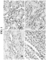
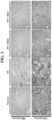
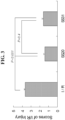

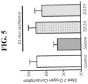
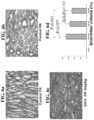


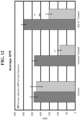


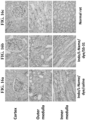


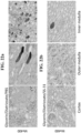


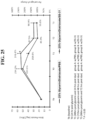
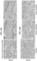
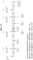

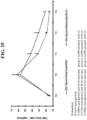


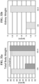

Classifications
-
- A—HUMAN NECESSITIES
- A61—MEDICAL OR VETERINARY SCIENCE; HYGIENE
- A61K—PREPARATIONS FOR MEDICAL, DENTAL OR TOILETRY PURPOSES
- A61K38/00—Medicinal preparations containing peptides
- A61K38/04—Peptides having up to 20 amino acids in a fully defined sequence; Derivatives thereof
- A61K38/07—Tetrapeptides
-
- A—HUMAN NECESSITIES
- A61—MEDICAL OR VETERINARY SCIENCE; HYGIENE
- A61K—PREPARATIONS FOR MEDICAL, DENTAL OR TOILETRY PURPOSES
- A61K38/00—Medicinal preparations containing peptides
- A61K38/04—Peptides having up to 20 amino acids in a fully defined sequence; Derivatives thereof
- A61K38/06—Tripeptides
-
- A—HUMAN NECESSITIES
- A61—MEDICAL OR VETERINARY SCIENCE; HYGIENE
- A61P—SPECIFIC THERAPEUTIC ACTIVITY OF CHEMICAL COMPOUNDS OR MEDICINAL PREPARATIONS
- A61P13/00—Drugs for disorders of the urinary system
- A61P13/02—Drugs for disorders of the urinary system of urine or of the urinary tract, e.g. urine acidifiers
-
- A—HUMAN NECESSITIES
- A61—MEDICAL OR VETERINARY SCIENCE; HYGIENE
- A61P—SPECIFIC THERAPEUTIC ACTIVITY OF CHEMICAL COMPOUNDS OR MEDICINAL PREPARATIONS
- A61P13/00—Drugs for disorders of the urinary system
- A61P13/12—Drugs for disorders of the urinary system of the kidneys
-
- C—CHEMISTRY; METALLURGY
- C07—ORGANIC CHEMISTRY
- C07K—PEPTIDES
- C07K5/00—Peptides containing up to four amino acids in a fully defined sequence; Derivatives thereof
- C07K5/04—Peptides containing up to four amino acids in a fully defined sequence; Derivatives thereof containing only normal peptide links
- C07K5/10—Tetrapeptides
- C07K5/1002—Tetrapeptides with the first amino acid being neutral
- C07K5/1016—Tetrapeptides with the first amino acid being neutral and aromatic or cycloaliphatic
-
- C—CHEMISTRY; METALLURGY
- C07—ORGANIC CHEMISTRY
- C07K—PEPTIDES
- C07K5/00—Peptides containing up to four amino acids in a fully defined sequence; Derivatives thereof
- C07K5/04—Peptides containing up to four amino acids in a fully defined sequence; Derivatives thereof containing only normal peptide links
- C07K5/10—Tetrapeptides
- C07K5/1019—Tetrapeptides with the first amino acid being basic
-
- A—HUMAN NECESSITIES
- A61—MEDICAL OR VETERINARY SCIENCE; HYGIENE
- A61K—PREPARATIONS FOR MEDICAL, DENTAL OR TOILETRY PURPOSES
- A61K38/00—Medicinal preparations containing peptides
Definitions
- ARI Acute Renal Injury
- ARF acute renal failure
- AKI acute kidney injury
- ARI is a serious medical condition of the kidneys.
- ARI is characterized by a decline of glomerular filtration rate, urine output, or both.
- ARI is accompanied by an inflammatory response that if unchecked can lead to renal fibrosis and chronic renal failure.
- ARI usually occurs over a period of hours or days and is potentially reversible.
- ARI is a common complication in hospitalized patients, and its incidence has risen significantly in the past 15 years.
- a particular type of ARI, contrast-induced nephropathy is caused by the toxic effects of some radiocontrast agents.
- the present invention provides a composition for use in a method for protecting a kidney from renal fibrosis associated with prerenal acute renal injury or intrinsic acute renal injury in a subject in need thereof, the composition comprising an effective amount of a peptide selected from the group consisting of Phe-D-Arg-Phe-Lys-NHz and D-Arg-2'6'Dmt-Lys-Phe-NH 2 .
- the present invention also provides a composition for use in a method for treating or preventing renal fibrosis associated with prerenal acute renal injury or intrinsic acute renal injury in a subject in need thereof, the composition comprising an effective amount of a peptide selected from the group consisting of Phe-D-Arg-Phe-Lys-NH 2 and D-Arg-2'6'Dmt-Lys-Phe-NH 2 .
- the acute renal injury is associated with exposure of the subject to a nephrotoxic agent.
- the peptide is to be administered to the subject prior to or simultaneously with the exposure to the nephrotoxic agent.
- the nephrotoxic agent is a radiocontrast dye, e.g., acetrizoate; diatrizoate; iodamide; ioglicate; iothalamate; ioxithalamate; metrizoate; metrizamide; iohexol; iopamidol; iopentol; iopromide; and ioversol.
- a radiocontrast dye e.g., acetrizoate; diatrizoate; iodamide; ioglicate; iothalamate; ioxithalamate; metrizoate; metrizamide; iohexol; iopamidol; iopentol; iopromide; and ioversol.
- the nephrotoxic agent is a drug or chemical.
- the drug or chemical is one or more of the compounds selected from the group consisting of: cisplatin; gentamicin; cephaloridine; cyclosporine; amphotericin; carbon tetrachloride; trichloroethylene; and dichloroacetylene.
- aromatic-cationic peptides can prevent and/or treat acute organ failure, e.g., acute renal injury or acute liver failure in a subject.
- aromatic-cationic peptides can prevent and/or treat chronic organ damage, e.g., chronic renal injury. It is to be appreciated that certain aspects, modes, embodiments, variations and features of the invention are described below in various levels of detail in order to provide a substantial understanding of the present invention.
- phrases such as element A is "associated with" element B mean both elements exist, but should not be interpreted as meaning one element necessarily is causally linked to the other.
- the "administration" of an agent, drug, or peptide to a subject includes any route of introducing or delivering to a subject a compound to perform its intended function. Administration can be carried out by any suitable route, including orally, intranasally, parenterally (intravenously, intramuscularly, intraperitoneally, or subcutaneously), rectally, or topically. Administration includes self-administration and the administration by another.
- amino acid includes naturally-occurring amino acids and synthetic amino acids, as well as amino acid analogs and amino acid mimetics that function in a manner similar to the naturally-occurring amino acids.
- Naturally-occurring amino acids are those encoded by the genetic code, as well as those amino acids that are later modified, e.g., hydroxyproline, ⁇ -carboxyglutamate, and O-phosphoserine.
- Amino acid analogs refers to compounds that have the same basic chemical structure as a naturally-occurring amino acid, i.e., an ⁇ -carbon that is bound to a hydrogen, a carboxyl group, an amino group, and an R group, e.g., homoserine, norleucine, methionine sulfoxide, methionine methyl sulfonium.
- Such analogs have modified R groups (e.g ., norleucine) or modified peptide backbones, but retain the same basic chemical structure as a naturally-occurring amino acid.
- Amino acid mimetics refers to chemical compounds that have a structure that is different from the general chemical structure of an amino acid, but that functions in a manner similar to a naturally-occurring amino acid. Amino acids can be referred to herein by either their commonly known three letter symbols or by the one-letter symbols recommended by the IUPAC-IUB Biochemical Nomenclature Commission.
- an "isolated” or “purified” polypeptide or peptide is substantially free of cellular material or other contaminating polypeptides from the cell or tissue source from which the agent is derived, or substantially free from chemical precursors or other chemicals when chemically synthesized.
- an isolated aromatic-cationic peptide would be free of materials that would interfere with diagnostic or therapeutic uses of the agent.
- Such interfering materials may include enzymes, hormones and other proteinaceous and nonproteinaceous solutes.
- a medical condition includes, but is not limited to, any condition or disease manifested as one or more physical and/or psychological symptoms for which treatment and/or prevention is desirable, and includes previously and newly identified diseases and other disorders.
- a medical condition may be acute renal injury or acute liver failure or any associated symptoms or complications.
- the medical condition with which the present invention is concerned is, specially, renal fibrosis associated with prerenal acute renal injury or intrinsic acute renal injury.
- polypeptide As used herein, the terms “polypeptide”, “peptide” and “protein” are used interchangeably herein to mean a polymer comprising two or more amino acids joined to each other by peptide bonds or modified peptide bonds, i.e., peptide isosteres.
- Polypeptide refers to both short chains, commonly referred to as peptides, glycopeptides or oligomers, and to longer chains, generally referred to as proteins.
- Polypeptides may contain amino acids other than the 20 gene-encoded amino acids.
- Polypeptides include amino acid sequences modified either by natural processes, such as post-translational processing, or by chemical modification techniques that are well known in the art. Such modifications are well described in basic texts and in more detailed monographs, as well as in a voluminous research literature.
- the word "protect” or “protecting” refers to decreasing the likelihood and/or risk that the subject treated with a peptide will develop a given disease or disorder, e.g., acute renal injury or acute liver failure.
- the likelihood of developing the disease or disorder is considered to be reduced if the likelihood is decreased by at least about 10%, preferably at least about 25%, more preferably at least about 50%, even more preferably at least about 75%, and most preferably at least about 90%, in comparison to the likelihood and/or risk that the same subject untreated with a peptide will develop tissue injury, e.g., acute renal injury or acute liver failure.
- the peptides protect a subject from acute renal injury caused by a contrast agent (i.e ., contrast-induced nephropathy) when the peptides are administered prior to or simultaneously with the contrast agent.
- a contrast agent i.e ., contrast-induced nephropathy
- subject refers to a member of any vertebrate species.
- the presently disclosed subject matter is particularly useful for warm-blooded vertebrates.
- the subject is a human.
- treating or “treatment” or “alleviation” refers to both therapeutic treatment and prophylactic or preventative measures, wherein the object is to prevent or slow down (lessen) the targeted pathologic condition or disorder.
- a subject is successfully “treated” for a disease or condition if, after receiving a therapeutic amount of the aromatic-cationic peptides according to the present invention, the subject shows observable and/or measurable reduction in or absence of one or more signs and symptoms of a particular disease or condition.
- treatment or prevention may include a reduction in metabolic acidosis, hyperkalaemia, oliguria or anuria, restoration in body fluid balance, and improved effects on other organ systems.
- Kidney function may also be assessed by measuring serum creatinine levels, serum creatinine clearance, or blood urea nitrogen levels. It is also to be appreciated that the various modes of treatment or prevention of medical conditions as described are intended to mean “substantial”, which includes total but also less than total treatment or prevention, and wherein some biologically or medically relevant result is achieved.
- aromatic-cationic peptides useful in the present invention are water-soluble and highly polar. Despite these properties, the peptides can readily penetrate cell membranes.
- the peptide is D-Arg-2'6'Dmt-Lys-Phe-NH 2 (referred to herein as SS-31).
- the peptide is Phe-D-Arg-Phe-Lys-NH 2 (referred to herein as SS-20).
- amino acids of the peptides may be in either the L- or the D- configuration.
- the peptides useful in the present invention may be chemically synthesized by any of the methods well known in the art. Suitable methods for synthesizing the protein include, for example those described by Stuart and Young in “Solid Phase Peptide Synthesis,” Second Edition, Pierce Chemical Company (1984 ), and in “ Solid Phase Peptide Synthesis,” Methods Enzymol. 289, Academic Press, Inc, New York (1997 ).
- Acute renal injury refers to a reduction of renal function to excrete waste from a patient's blood.
- ARI is typically characterized as including a decline of glomerular filtration rate (GFR) to a level so low that little or no urine is formed. Therefore, substances that the kidney usually eliminates remain in the body.
- GFR glomerular filtration rate
- ARI causes of ARI are multifactorial, but they may be classified into three categories: (1) prerenal ARI, in which the kidneys fail to receive adequate blood supply, e.g., due to fall in systemic blood pressure as in shock/cardiac arrest, or subsequent to hemorrhage; (2) intrinsic ARI, in which the failure occurs within the kidney, e.g., due to drug-induced toxicity; and (3) postrenal ARI, caused by impairment of urine flow out of the kidney, as in ureteral obstruction due to kidney stones or bladder/prostate cancer. ARI may, in general, be associated with any one or a combination of these categories. The present invention is, however, concerned specifically with the clinical situations recited in the appended claims.
- Chronic renal failure refers to a progressive loss of renal function and irreversible kidney damage.
- Methods for assessing renal function include, but are not limited to, measurements of blood systemic and glomerular capillary pressure, proteinuria (e.g ., albuminuria), microscopic and macroscopic hematuria, serum creatinine level (e.g., one formula for estimating renal function in humans equates a creatinine level of 2.0 mg/dl to 50 percent of normal kidney function and 4.0 mg/dl to 25 percent), decline in the glomerular filtration rate (GFR) (e.g., rate of creatinine clearance), and degree of tubular damage.
- proteinuria e.g ., albuminuria
- microscopic and macroscopic hematuria e.g., serum creatinine level
- serum creatinine level e.g., one formula for estimating renal function in humans equates a creatinine level of 2.0 mg/dl to 50 percent of normal kidney function and 4.0 mg/dl to 25 percent
- GFR glomerular filtration rate
- such assessment may include evaluating at least one kidney function using biological and/or physiological parameters such as serum creatinine level, creatinine clearance rate, 24-hour urinary protein secretion, glomerular filtration rate (GFR), urinary albumin creatinine ratio, albumin excretion rate, and renal biopsy.
- biological and/or physiological parameters such as serum creatinine level, creatinine clearance rate, 24-hour urinary protein secretion, glomerular filtration rate (GFR), urinary albumin creatinine ratio, albumin excretion rate, and renal biopsy.
- Methods for assessing deterioration of renal structure are also well known. Illustrative methods are described in the Examples. Such methods include renal imaging (e.g ., MRI, ultrasound), or histological evaluation of renal biopsy. In some embodiments, the methods reduce deterioration of renal structure as judged, for example, by the extent of tubulointerstitial or glomerular damage and/or the degree of renal fibrosis (e.g., deposition of collagen and fibronectin).
- Ischemia An example of a condition in which kidneys fail to receive adequate blood supply to the kidney is ischemia. Ischemia is a major cause of ARI. Ischemia of one or both kidneys is a common problem experienced during aortic surgery, renal transplantation, or during cardiovascular anesthesia. Surgical procedures involving clamping of the aorta and/or renal arteries, e.g., surgery for supra- and juxtarenal abdominal aortic aneurysms and renal transplantation, are also particularly liable to produce renal ischemia, leading to significant postoperative complications and early allograft rejection. In high-risk patients undergoing these forms of surgery, the incidence of renal dysfunction has been reported to be as high as 50%.
- Ischemia may be caused by loss of blood, loss of fluid from the body as a result of severe diarrhea or burns, shock, and ischemia associated with storage of the donor kidney prior to transplantation.
- the blood flow to the kidney may be reduced to a dangerously low level for a time period great enough to cause ischemic injury to the tubular epithelial cells, sloughing off of the epithelial cells into the tubular lumen, obstruction of tubular flow that leads to loss of glomerular filtration and acute renal injury.
- Subjects may also become vulnerable to ARI after receiving anesthesia, surgery, or ⁇ -adrenergic agonists because of related systemic or renal vasoconstriction. Additionally, systemic vasodilation caused by anaphylaxis, and anti-hypertensive drugs, sepsis or drug overdose may also cause ARI because the body's natural defense is to shut down, i.e., vasoconstriction of non-essential organs such as the kidneys.
- a subject at risk for ARI may be a subject undergoing an interruption or reduction of blood supply or blood pressure to the kidney. These subjects may be administered the aromatic-cationic peptides prior to or simultaneously with such interruption or reduction of blood supply. Likewise, aromatic-cationic peptides may be administered after the therapeutic agent to treat ischemia.
- Nephrotoxins can cause direct toxicity on tubular epithelial cells.
- Nephrotoxins include, but are not limited to, therapeutic drugs, e.g., cisplatin, gentamicin, cephaloridine, cyclosporin, amphotericin, radiocontrast dye (described in further detail below), pesticides (e.g ., paraquat), and environmental contaminants (e.g ., trichloriethylene and dichloroacetylene).
- PAN puromycin aminonucleoside
- aminoglycosides such as gentamicin
- cephalosporins such as cephaloridine
- calcineurin inhibitors such as tacrolimus or sirolimus.
- Drug-induced nephrotoxicity may also be caused by non-steroidal anti-inflammatories, anti-retrovirals, anti-cytokines, immunosuppressants, oncological drugs or ACE inhibitors.
- the drug-induced nephrotoxicity may further be caused by nalgesic abuse, ciprofloxacin, clopidogrel, cocaine, cox-2 inhibitors, diuretics, foscamet, gold, ifosfamide, immunoglobin, Chinese herbs, interferon, lithium, mannitol, mesalamine, mitomycin, nitrosoureas, penicillamine, penicillins, pentamidine, quinine, rifampin, streptozocin, sulfonamides, ticlopidine, triamterene, valproic acid, doxorubicin, glycerol, cidofovir, tobramycin, neomycin sulfate, colistimethate, vancomycin, amikacin, cefotaxime, cisplatin, acyclovir, lithium, interleukin-2, cyclosporin or indinavir.
- nephrotoxins In addition to direct toxicity on tubular epithelial cells, some nephrotoxins also reduce renal perfusion, causing injury to zones known to have limited oxygen availability (inner medullary region). Such nephrotoxins include amphotericin and radiocontrast dye. Renal failure can result even from clinically relevant doses of these drugs when combined with ischemia, volume depletion, obstruction, or infection. An example is the use of radiocontrast dye in patients with impaired renal function. The incidence of contrast-dye nephropathy (CIN) is 3-8% in the normal patient, but increases to 25% for patients with diabetes mellitus. Most cases of ARI occur in patients with predisposing comorbidities ( McCombs, P.R. & Roberts, B. Surg Gynecol Obstet, 148:175-178 (1979 )).
- CIN contrast-dye nephropathy
- a subject at risk for ARI may be receiving one or more therapeutic drugs that have a nephrotoxic effect. These subjects may be administered the aromatic-cationic peptides prior to or simultaneously with such therapeutic agents. Likewise, aromatic-cationic peptides may be administered after the therapeutic agent to treat nephrotoxicity.
- the aromatic-cationic peptides are to be administered to a subject at risk for contrast-induced nephropathy (CIN), in order to protect the subject from this condition.
- CIN contrast-induced nephropathy
- CIN is an important cause of acute renal failure.
- CIN is defined as acute renal failure occurring within 48 h of exposure to intravascular radiographic contrast material.
- CIN remains a common complication of radiographic procedures.
- CIN arises when a subject is exposed to radiocontrast dye, such as during coronary, cardiac, or neuro-angiography procedures. Contrast dye is essential for many diagnostic and interventional procedures because it enables doctors to visualize blocked body tissues. Subjects at greatest risk for CIN are the elderly, and those with diabetes, chronic kidney disease, or advanced heart failure.
- the serum creatinine test can be used to monitor the onset of CIN, monitor treatment, as well as monitor the effectiveness of the peptides in protecting subjects from the onset of CIN.
- the aromatic-cationic peptides are to be administered to a subject prior to or simultaneously with the administration of a contrast agent in order to provide protection against CIN.
- the subject may receive the peptides from about 1 to 2 hours, about 1 to 6 hours, about 1 to 12 hours, about 1 to 24 hours, or about 1 to 48 hours prior to receiving the contrast agent.
- the subject may be administered the peptides at about the same time as the contrast agent.
- administration of the peptides to the subject may continue following administration of the contrast agent.
- the subject may continue to receive the peptide at intervals of about 1, 2, 3, 4, 5, 6, 7, 8, 12, 24, and 48 hours following administration of the contrast agent, in order to provide a protective or prophylactic effect against CIN.
- the aromatic-cationic peptides are to be administered to a subject after administration of a contrast agent in order to treat CIN.
- the subject may receive the peptides from about 1 to 2 hours, about 1 to 6 hours, about 1 to 12 hours, about 1 to 24 hours, about 1 to 48 hours, or about 1 to 72 hours after receiving the contrast agent.
- the subject may exhibit one or more signs or symptoms of CIN prior to receiving the peptides, such as increased serum creatinine levels and/or decreased urine volume.
- Administration of the peptides improves one or more of these indicators of kidney function in the subject compared to a control subject not administered the peptides.
- Contrast Agents The incidence of CIN is influenced by the chemical structure and particularly by the osmolarity and the ionic (or non-ionic) structure of the contrast agent used ( See Zirogiannis et al., "Contrast media induced nephropathy in patients undergoing cardiac catheterization.” Hellenic J Cardiol, 45:07-113 (2004 )).
- Imaging agent is also referred to herein as an “imaging agent.”
- Imaging agents include those known in the art, such as dyes, fluorescent dyes, gold particles, iron oxide particles and other contrast agents including paramagnetic molecules, x-ray attenuating compounds (for CT and x-ray), contrast agents for ultrasound, ⁇ -ray emitting isotopes (Scintigraphy), and positron-emitting isotopes (PET).
- Contrast agents can be administered to the subject by, for example, parenteral injection (e.g ., intravenously, intra-arterially, intra-thecally, intra-abdominally, subcutaneously, intramuscularly), orally ( e.g ., as a tablet or a drink), rectally, or via inhalation.
- parenteral injection e.g ., intravenously, intra-arterially, intra-thecally, intra-abdominally, subcutaneously, intramuscularly
- orally e.g ., as a tablet or a drink
- an X-ray contrast agent can comprise barium sulfate, or can comprise iodine in an organic (non-ionic) compound or in an ionic compound.
- iodine contrast agents include those shown in Table 7. TABLE 7.
- Exemplary Iodinated Contrast Agents Name Type Iodine Content Osmolality Level Diatrizoate (Hypaque 50) Ionic Monomer 300 1550 High Osmolar Metrizoate (Isopaque Coronar 370) Ionic 370 2100 High Osmolar Ioxaglate (Hexabrix) Ionic dimer 320 580 Low Osmolar Iopamidol (Isovue 370) Non-ionic monomer 370 796 Low Osmolar Iohexol (Omnipaque 350) Non-ionic 350 884 Low Osmolar Ioxilan (Oxilan) Non-ionic Low Osmolar Iopromide Non-ionic Low Osmolar Iodixanol (Visipaque 320) Non-ionic dimer 320 290 Iso Osmolar
- An MRI contrast agent can comprise a paramagnetic contrast agent (such as a gadolinium compound), a super paramagnetic contrast agent (such as iron oxide nanoparticles), a diamagnetic agent (such as barium sulfate), and combinations thereof.
- a paramagnetic contrast agent such as a gadolinium compound
- a super paramagnetic contrast agent such as iron oxide nanoparticles
- a diamagnetic agent such as barium sulfate
- Metal ions preferred for MRI include those with atomic numbers 21-29, 39-47, or 57-83, and, more preferably, a paramagnetic form of a metal ion with atomic numbers 21-29, 42, 44, or 57-83.
- paramagnetic metal ions are selected from the group consisting of Gd(III), Fe(III), Mn(II and III), Cr(III), Cu(II), Dy(III), Tb(III and IV), Ho(III), Er(III), Pr(III) and Eu(II and III).
- Gd(III) is particularly useful. Note that as used herein, the term "Gd” is meant to convey the ionic form of the metal gadolinium; such an ionic form can be written as GD(III), GD3+, etc. with no difference in ionic form contemplated.
- a CT contrast agent can comprise iodine (ionic or non-ionic formulations), barium, barium sulfate, Gastrografin (a diatrizoate meglumine and diatrizoate sodium solution), and combinations thereof.
- a PET or SPECT contrast agent can comprise a metal chelate.
- contrast agents include, but are not limited to, molecules bearing one or more iodine moieties.
- Iodine may be bound either in an organic compound or an ionic compound.
- the earliest contrast agents were ionic, containing a sodium atom that dissociated from the iodine in aqueous solution.
- Organic compounds have fewer side effects as they do not dissociate into component molecules.
- Iodinated contrast media may be divided into water-soluble, water-insoluble, and oily contrast media.
- Water-insoluble contrast media include aqueous suspension of propyliodone (Dionosil ® ), used in bronchography.
- Oily contrast media include Lipiodol, a stable compound of 40% iodine in poppy seed oil, and Lipiodol Ultra ® Fluid and Ethiodol ® , ethyl esters of iodinated fatty acids of poppy seed oil containing 48% and 37% iodine, respectively. These oils are used for lymphography, and by some also for hysterosalpingography.
- Iodophenylundecylic acid is a contrast medium for oil myelography (brand names: Pantopaque ® , Myodil ® ).
- the water-soluble contrast media represent by far the largest group of iodinated contrast media. These contrast media may be classified into high-osmolality, low-osmolality, and iso-osmolality contrast media. The osmolality is related to some of the adverse events of these contrast media.
- Monomeric ionic contrast media constitute two main groups, oral cholegraphic contrast media and uro/angiographic media.
- Examples of these media include: acetrizoate (Diaginol ® , Urokon ® ); diatrizoate (Angiografin ® , Hypaque ® , Renografin ® , Urografin ® , Urovison ® ); iodamide (Uromiro ® ); ioglicate (Rayvist ® ); iothalamate (Conray ® ); ioxithalamate (Telebrix ® ); and metrizoate (Isopaque ® , Triosil ® ).
- the uro/angiographic media are all salts of derivatives of tri-iodinated benzoic acid, and differ only in the side-chains at position 3 and 5.
- the cations of the salts are mainly either sodium or meglumine, or a mixture of both.
- Sodium salts are generally more toxic to vascular endothelium and to blood-brain barrier and neural tissue than meglumine.
- a mixture of sodium and meglumine has lower cardiotoxicity than either salts alone.
- Some manufacturers have partially replaced sodium with calcium and magnesium to reduce toxicity.
- the ionic monomeric contrast media for intravascular use are so-called high-osmolar contrast media (HOCM), having an osmolality seven to eight times that of plasma in ordinary clinical use.
- HOCM high-osmolar contrast media
- This hyperosmolality is responsible for several subjective and objective adverse effects such as pain, endothelial damage, thrombosis and thrombophlebitis, disturbance of the blood-brain barrier, bradycardia in cardioangiography and increased pressure in the pulmonary circulation.
- LOCM low-osmolar contrast media
- Osmolality is dependent upon the number of molecules per volume unit solution.
- the ionic monomeric agents are salts that dissociate into two molecules, one anion containing the radiopaque property due to three iodine atoms, and one cation without radiopaque properties. These agents are also called 3:2 or ratio 1.5 agents (three iodine atoms per two molecules).
- Non-ionic contrast media do not dissociate and their water-solubility is generally achieved by several hydrophilic hydroxyl groups.
- Tri-iodinated non-ionic monomers have approximately half the osmolality of that of ionic monomers (they are 3:1 or ratio 3 agents).
- agents are available in convenient ready-to-use solutions of non-ionic monomeric contrast media have appeared, and include: metrizamide (Amipaque ® ); iohexol (Omnipaque ® ); iopamidol (Iopamiro ® , Isovue ® , Niopam ® , Solutrast ® ); iopentol (Imagopaque ® ); iopromide (Ultravist ® ); and ioversol (Optiray ® ).
- contrast media have hydrophilic hydroxyl groups attached to all three side chains, a feature that has also contributed to reducing their toxicity.
- toxicity is also dependent upon chemical structure, an intrinsic toxicity sometimes named chemotoxicity.
- chemotoxicity is related to the protein-binding capacity of the medium, which is caused by non-specific weak interactions between the contrast medium and surrounding biological molecules (e.g ., enzymes). The interaction is mediated through electrical forces, which are present only for ionic agents, and through interaction between hydrophobic portions of the molecule, which for contrast media is mainly the benzene ring.
- the many hydrophilic side groups of the second generation non-ionic monomers protect the inner hydrophobic benzene ring from interaction, thereby reducing chemotoxicity.
- the relative hydrophobicity/hydrophilicity of the molecule is called its partition coefficient, a low partition coefficient being advantageous because a high hydrophilicity contributes to low protein binding.
- Dimeric contrast media with two tri-iodinated benzene rings constitute three main groups; ionic intravenous cholegraphic contrast media, monoacidic ionic contrast media, and non-ionic contrast media.
- the two latter groups both belong to the so-called low-osmolar contrast media.
- ioxaglate Owing partly to its ionic character, partly to its fewer hydroxyl groups, ioxaglate is more toxic than the non-ionic monomers with similar osmolality (but less toxic than the ionic monomers owing to lower osmolality).
- non-ionic dimers yield six iodine atoms per molecule (ratio 6 agents).
- non-ionic dimer contrast media include: iotrolan (Isovist ® ) and iodixanol (Visipaque ® ). Iotrolan is nearly iso-osmolar to plasma (0.32 osm/kg H 2 O), while iodixanol is actually hypo-osmolar to plasma. Iso-osmolarity to plasma (0.29 osm/kg H 2 O) is achieved at any iodine concentration by addition of an appropriate amount of saline. These agents have a very low toxicity owing partly to their iso-osmolarity, partly their non-ionic character, and partly to the fact that they possess a very large number of hydroxyl groups (low partition coefficient).
- the various water-soluble contrast media for urography and angiography are all extracellular contrast media, and are excreted unmetabolized by glomerular filtration. Approximately 85-90% of the injected dose is found in urine within the first 6 hours; 95-100% within the first 24 hours. Generally less than 2% may be found in feces.
- Non-ionic contrast agents (containing 6 iodine anions in their molecule) were believed to be less nephrotoxic than ionic contrast agents (containing 3 iodine anions in their molecule).
- ionic contrast agents containing 3 iodine anions in their molecule.
- CIN CIN
- a subject in need thereof may be a subject having impairment of urine flow.
- Obstruction to the flow of urine can occur anywhere in the urinary tract and has many different possible causes. Such causes include kidney stones or bladder/prostate cancer.
- Unilateral ureteral obstruction (UUO) is a common clinical disorder associated with obstructed urine flow. It is also associated with tubular cell apoptosis, macrophage infiltration, and interstitial fibrosis. Interstitial fibrosis leads to a hypoxic environment and contributes to progressive decline in renal function despite surgical correction.
- UUO Unilateral ureteral obstruction
- a subject having or at risk for UUO may be administered the aromatic-cationic peptides to protect or treat the subject from ARI.
- the method is a method for protecting a kidney from renal fibrosis in a mammal in need thereof.
- the method comprises administering to the mammal an effective amount of an aromatic-cationic peptide as described above.
- the peptides described above can be administered to a mammal in need thereof, as described above, by any method known to those skilled in the art.
- fibrosis refers to abnormal processing of fibrous tissue, or fibroid or fibrous degeneration. Fibrosis can result from various injuries or diseases, and can often result from chronic transplant rejection relating to the transplantation of various organs. Fibrosis typically involves the abnormal production, accumulation, or deposition of extracellular matrix components, including overproduction and increased deposition of, for example, collagen and fibronectin.
- renal fibrosis refers to fibrosis of the kidney.
- renal fibrosis is associated with overproduction or abnormal deposition of extracellular matrix components, particularly collagen, leading to the degradation or impairment of kidney function.
- the methods may be particularly useful in patients with renal insufficiency, renal failure, or end-stage renal disease attributable at least in part to a nephrotoxicity of an drug or chemical.
- Other indications may include creatinine clearance levels of lower than 97 (men) and 88 (women) ml/min, blood urea of 20-25 mg/dl or higher.
- the treatment may be useful in patients with microalbuminuria, macroalbuminuria, and/or proteinuria levels of over 1, 2, 3, 4, 5, 6, 7, 8, 9, or 10 g or more per a 24 hour period, and/or serum creatinine levels of about 1.0, 1.5, 2.0, 2.5, 3, 3.5, 4.0, 4.5, 5, 5.5, 6, 7, 8, 9, 10 mg/dl or higher.
- the methods can be used to slow or reverse the progression of renal disease in patients whose renal function is below normal by 25%, 40%, 50%, 60%, 75%, 80%, 90% or more, relative to control subjects.
- the methods slow the loss of renal function by at least 10%, 20%, 30%, 40%, 50%, 60%, 70%, 80%, 90%, 100% or more, relative to control subjects.
- the methods improve the patient's serum creatinine levels, proteinuria, and/or urinary albumin excretion by at least 10%, 20%, 30%, 40%, 50%, 60%, 70%, or more, relative to control subjects.
- Non-limiting illustrative methods for assessing renal function are described herein and, for example, in WO 01/66140 .
- a method for treating acute renal injury in a subject in need thereof comprises administering to the subject an effective amount of an aromatic-cationic peptide as described above.
- the peptides described above can be administered to a subject in need thereof, as described above, by any method known to those skilled in the art.
- Subjects in need of protection from or treatment of acute renal injury also include subjects suffering from a disease, condition or treatment associated with oxidative damage.
- the oxidative damage is caused by free radicals, such as reactive oxygen species (ROS) and/or reactive nitrogen species (RNS).
- ROS and RNS include hydroxyl radical (HO•), superoxide anion radical (O 2 • - ), nitric oxide (NO•), hydrogen peroxide (H 2 O 2 ), hypochlorous acid (HOCI) and peroxynitrite anion (ONOO - ).
- ATN acute tubular necrosis
- GFR glomerular filtration rate
- sublethal or lethal tubular epithelial injury The established or persistent phase of ATN is characterized by persistent reduction in GFR.
- Sublethal tubular injury includes loss of brush border, vacuolization and flattening of proximal tubules, and dilation of tubules. Cellular necrosis may be seen in the proximal tubule.
- the renal interstitium may be edematous, whereas leukocytic infiltration may be observed in the medulla.
- the distal tubular lumen may be occluded by casts, and distal, rather than proximal, tubular epithelial cells are more likely involved by apoptosis.
- Such structural and functional derangements resolve during the recovery or diuretic phase wherein reparative and regenerative responses restore renal architecture and normalize GFR.
- RPTCs renal proximal tubular cell
- RPTCs are highly specialized both in terms of morphology and function allowing for efficient transport of water, ions and macromolecules via specific transport mechanisms.
- RPTCs contain very high density of mitochondria, and oxygen is used to sustain oxidative phosphorylation and synthesis of ATP, which is needed in large quantities for renal tubular transport processes.
- oxygen is used to sustain oxidative phosphorylation and synthesis of ATP, which is needed in large quantities for renal tubular transport processes.
- ischemia these transport processes are severely disrupted due to loss of intracellular ATP. Restoration of blood flow will help to limit cell death, but recovery of mitochondrial function depends on the duration of ischemia.
- ischemia With prolonged ischemia, there is evidence that the rate of oxidative phosphorylation continues to deteriorate despite reperfusion. Progressive ischemia leads to inhibition of several components of the mitochondrial electron transport chain, including complex I, complex V, and the adenine nucleotide translocator, and decrease in ATP production. In addition, ischemia decreases cardiolipin and cytochrome c content in cardiac mitochondria, both of which will inhibit cytochrome c oxidase activity. Thus, mitochondria can suffer damage during ischemia that would limit their capacity to generate ATP upon restoration of oxygen and substrates with reperfusion.
- ROS ROS can initiate damage to nucleic acids, proteins and lipids in mitochondria. Protein oxidation and nitration result in altered function of many enzymes in the mitochondrial electron transport chain, while oxidation of the adenine nucleotide translocator impairs the influx of ADP into the matrix for ATP synthesis. ROS have been shown to damage renal mitochondria by enhancing membrane permeability, decreasing F 0 F 1 ATPase activity and reducing ATP production.
- Mitochondrial dysfunction may therefore lead to necrosis or apoptosis.
- Necrosis is characterized by cell swelling and disruption of the cell membrane, leading to release of cellular contents, especially proteolytic enzymes, which may result in destruction of neighboring cells.
- Apoptosis is defined as programmed cell death wherein the organism eliminates senescent, abnormal cells without affecting surrounding cells, and is deemed to be preferable for the survival of the organism since it eliminates dying cells by phagocytosis. The decision step between death by apoptosis or necrosis appears to be dependent on intracellular ATP content. Apoptosis involves energy-requiring steps, especially in the formation of the apoptosome complex between Apaf-1 and cytochrome c. Thus in the event of significant cellular ATP depletion, death can only occur by necrosis.
- epithelial cells When epithelial cells are damaged, they release inflammatory mediators, including IL-1 ⁇ and TNF ⁇ , that lead to an intense inflammatory response initiated by the infiltration of leukocytes and the release of proinflammatory cytokines, chemokines and ROS.
- Leukocyte infiltration is observed with renal ischemia-reperfusion, ureteral obstruction, and nephrotoxins, and the inflammatory process contribute to ARI.
- Infiltrating lymphocytes and macrophages become activated and begin secreting profibrotic cytokines and growth factors, such as TGF ⁇ , IL-13 and PDGF, which further activate macrophages and fibroblasts.
- TGF ⁇ plays a pivotal role in epithelial-mesenchymal transition (EMT) by down-regulating E-cadherin and up-regulating ⁇ -SMA expression, and increasing collagen production. Fibrosis occurs when the synthesis of new collagen by myofibroblasts exceeds the rate at which it is degraded, such that the total amount of collagen increases over time.
- the chemokines, such as MCP-1 also play a role in fibrosis by recruiting macrophages and myofibroblasts to the site of injury. Collagen turnover and ECM remodeling is regulated by various matrix metalloproteases (MMPs).
- the term "effective amount” or “pharmaceutically effective amount” or “therapeutically effective amount” of a composition is a quantity sufficient to achieve a desired therapeutic and/or prophylactic effect, e.g., an amount which results in the prevention of, or a decrease in, the symptoms associated with ARI or ALF.
- the amount of a composition administered to the subject will depend on the type and severity of the disease and on the characteristics of the individual, such as general health, age, sex, body weight and tolerance to drugs. It will also depend on the degree, severity and type of disease. The skilled artisan will be able to determine appropriate dosages depending on these and other factors.
- the compositions of the present invention can also be administered in combination with one or more additional therapeutic compounds.
- the aromatic-cationic peptides may be administered to a subject having one or more signs of ARI caused by a disease or condition.
- Administration of an effective amount of the aromatic-cationic peptides may improve at least one sign or symptom of ARI in the subject, e.g., metabolic acidosis (acidification of the blood), hyperkalaemia (elevated potassium levels), oliguria or anuria (decrease or cessation of urine production), changes in body fluid balance, and effects on other organ systems.
- a "therapeutically effective amount" of the aromatic-cationic peptides is meant levels in which the physiological effects of acute renal failure are, at a minimum, ameliorated.
- the efficacy of the biological effect is measured in comparison to a subject or class of subjects not administered the peptides.
- the effective amount is determined during pre-clinical trials and clinical trials by methods familiar to physicians and clinicians.
- An effective amount of a peptide useful in the present invention, preferably in a pharmaceutical composition, may be administered to a subject in need thereof by any of a number of well-known methods for administering pharmaceutical compounds.
- the peptide may be to be administered systemically or locally. In one embodiment, the peptide is to be administered intravenously.
- the aromatic-cationic peptides useful in the methods may be to be administered via rapid intravenous bolus injection. Preferably, however, the peptide is to be administered as a constant rate intravenous infusion.
- the peptide may also be to be administered orally, topically, intranasally, intramuscularly, intraperitoneally, subcutaneously, or transdermally.
- the peptides useful in the methods may also be to be administered to subjects by sustained release, as is known in the art.
- Sustained release administration is a method of drug delivery to achieve a certain level of the drug over a particular period of time. The level typically is measured by serum or plasma concentration.
- a description of methods for delivering a compound by controlled release can be found in PCT Application No. WO 02/083106 .
- any formulation known in the art of pharmacy is suitable for administration of the aromatic-cationic peptides useful in the methods.
- liquid or solid formulations may be used.
- Some examples of formulations include tablets, gelatin capsules, pills, troches, elixirs, suspensions, syrups, wafers, chewing gum and the like.
- the peptides can be mixed with a suitable pharmaceutical carrier (vehicle) or excipient as understood by practitioners in the art.
- suitable pharmaceutical carrier include starch, milk, sugar, certain types of clay, gelatin, lactic acid, stearic acid or salts thereof, including magnesium or calcium stearate, talc, vegetable fats or oils, gums and glycols.
- formulations of the aromatic-cationic peptides useful in the methods of the present inventions may utilize conventional diluents, carriers, or excipients etc., such as are known in the art can be employed to deliver the peptides.
- the formulations may comprise one or more of the following: a stabilizer, a surfactant, preferably a nonionic surfactant, and optionally a salt and/or a buffering agent.
- the peptide may be delivered in the form of an aqueous solution, or in a lyophilized form.
- the stabilizer may, for example, be an amino acid, such as for instance, glycine; or an oligosaccharide, such as for example, sucrose, tetralose, lactose or a dextran.
- the stabilizer may be a sugar alcohol, such as for instance, mannitol; or a combination thereof.
- the stabilizer or combination of stabilizers constitutes from about 0.1% to about 10% weight for weight of the peptide.
- the surfactant is preferably a nonionic surfactant, such as a polysorbate.
- suitable surfactants include Tween20, Tween80; a polyethylene glycol or a polyoxyethylene polyoxypropylene glycol, such as Pluronic F-68 at from about 0.001% (w/v) to about 10% (w/v).
- the salt or buffering agent may be any salt or buffering agent, such as for example, sodium chloride, or sodium/potassium phosphate, respectively.
- the buffering agent maintains the pH of the pharmaceutical composition in the range of about 5.5 to about 7.5.
- the salt and/or buffering agent is also useful to maintain the osmolality at a level suitable for administration to a human or an animal.
- the salt or buffering agent may be present at a roughly isotonic concentration of about 150 mM to about 300 mM.
- the formulations of the peptides useful in the methods may additionally contain one or more conventional additive.
- additives include a solubilizer such as, for example, glycerol; an antioxidant such as for example, benzalkonium chloride (a mixture of quaternary ammonium compounds, known as "quats"), benzyl alcohol, chloretone or chlorobutanol; anaesthetic agent such as for example a morphine derivative; or an isotonic agent etc., such as described above.
- the pharmaceutical compositions may be stored under nitrogen gas in vials sealed with impermeable stoppers.
- Sprague Dawley rats (250 ⁇ 300g) were assigned to four groups: (1) sham surgery group without I/R; (2) I/R + saline vehicle treatment; (3) I/R + SS-20 treatment; (4) I/R + SS-31 treatment.
- Each experimental group consisted of 8 rats.
- SS-20 and SS-31 (3 mg/kg, dissolved in saline) were administered to rats 30 min before ischemia and immediately before onset of reperfusion. The control rats were given saline alone on the same schedule.
- Rats were anesthetized with a mixture of ketamine (90 mg/kg, i.p.) and xylazine (4 mg/kg, i.p.).
- the left renal vascular pedicle was occluded temporarily using a micro-clamp for 30 or 45 min.
- reperfusion was established by removing of the clamp.
- the contralateral right kidney was removed.
- animals were sacrificed and blood samples were obtained by cardiac puncture. Renal function was determined by blood urea nitrogen (BUN) and serum creatinine (BioAssay Systems DIUR-500 and DICT-500).
- Kidneys were fixed in 10% neutral-buffered formalin and embedded in paraffin wax. Three micron sections were stained with hematoxylin-eosin (H&E) and periodic acid-Schiff (PAS) and analyzed by light microscopy. Lesions were scored using the following criteria: [1] mitosis and necrosis of individual cells; [2] necrosis of all cells in adjacent proximal convoluted tubules with survival of surrounding tubules; [3] necrosis confined to the distal third of the proximal convoluted tubule with a band of necrosis extending across the inner cortex; and [4] necrosis affecting all three segments of the proximal convoluted tubule.
- H&E hematoxylin-eosin
- PAS periodic acid-Schiff
- Renal tissue sections were deparaffinized and rehydrated by xylenes, graded alcohol series and deionized H 2 O, and then incubated in 20 ⁇ g/ml proteinase K for 20 min at RT.
- In situ cell death detection POD kit (Roche, IN, USA) was used according to the manufacturer's instructions. Briefly, endogenous peroxidase activity in the kidney sections was blocked by incubation for 10 min with 0.3% H 2 O 2 in methanol. The sections were then incubated in a humidified chamber in the dark for 30 min at 37°C with TUNEL reaction mixture.
- the slides were incubated with 50-100 ⁇ l Converter-POD in a humidified chamber for 30 min at RT.
- the slides were incubated in DAB solution (1-3 min), and counterstained with hemotoxylin, dehydrated through a graded series of alcohol, and mounted in Permount for microscopy.
- Immunohistochemistry was then performed using primary antibody against heme oxygenase-1 (HO-1) (rat anti-HO-1/HMOX1/HSP32 monoclonal antibody (R&D Systems, MN, USA) at 1:200 dilution)) and secondary antibody (HRP-conjugated goat anti-rat IgG, VECTASTAIN ABC (VECTOR Lab Inc. MI, USA)).
- HO-1 heme oxygenase-1
- secondary antibody HRP-conjugated goat anti-rat IgG, VECTASTAIN ABC (VECTOR Lab Inc. MI, USA
- AEC alpha-1
- Hematoxylin was used for counterstaining.
- Kidney tissue was homogenized in 2 ml of RIPA lysis buffer (Santa Cruz, CA, USA) on ice and centrifuged at 500x g for 30 min to remove cell debris. Aliquots of the supernatants were stored at -80°C. Thirty (30) ⁇ g of protein of each sample was suspended in loading buffer and boiled for 5 min then each sample was subjected to a 10% SDS-PAGE gel electrophoresis. The resolved proteins were transferred to a PVDF membrane.
- the nitrocellulose blot was incubated with a 1:2000 diluted anti-HO-1/HMOX1/HSP32 or a 1:1000 diluted anti-AMPK ⁇ -1, monoclonal antibodies (R&D Systems, MN, USA) then incubated with horseradish peroxidase-conjugated secondary antibodies.
- the protein bands in the blot were detected with the use of an Enhanced Chemi Luminescence detection system (Cell Signaling, MA, USA) and X-ray film. Relative density measurements provide quantification.
- ATP Content Assay The kidney tissue was immediately put into 10 ml 5% trichloroacetic acid with 10 mM DTT, 2 mM EDTA and cut into small pieces. The tissue was homogenized on ice, incubated for another 10 min, centrifuged for 10 min at 2000xg, and neutralized with 10N KOH to pH 7.6. After centrifugation for 10 min at 2000 ⁇ g, aliquots of the resulting supernatant were stored at -80°C. ATP was measured by bioluminescence (ATP bioluminescent kit, Sigma, MO, USA).
- the above table shows treatment with either SS-20 or SS-31 improved BUN and serum creatinine values in rats after 45 min ischemia and 24 h reperfusion.
- BUN and serum creatinine were assayed using kits from BioAssay Systems (DIUR-500 and DICT-500).
- DIUR-500 and DICT-500 The * indicates that p ⁇ 0.05 from the sham group.
- FIG. 1 shows that treatment with either SS-20 or SS-31 prevented tubular cell apoptosis after 30 min ischemia and 24 h reperfusion. Renal sections were stained for apoptotic cells using TUNEL. Very few TUNEL-positive cells were observed in the medulla region of animals not subjected to I/R ( FIG. 1a ). A large number of TUNEL-positive epithelial cells were observed in the medulla of animals after 30 min ischemia and 24 h reperfusion ( FIG. 1b ). Treatment with SS-20 (3 mg/kg) or SS-31 (3 mg/kg) 30 min before onset of 30 min ischemia and just prior to reperfusion significantly prevented TUNEL-positive cells in the medulla ( FIG. 1c and FIG. 1d ).
- FIG. 2 shows that treatment with either SS-20 or SS-31 prevented tubular cell injury after 45 min ischemia and 24 h reperfusion. Renal sections were stained with H&E ( FIG. 2a-2d ) and PAS ( FIG. 2e-2h ). Ischemia-reperfusion resulted in loss of brush border, vacuolization and necrosis in the proximal tubules in the inner medulla, and the distal tubules were occluded by casts ( FIG. 2b ). Pretreatment with either SS-20 or SS-31 protected brush border and prevented necrosis in the proximal tubules ( FIG. 2c and FIG. 2d ).
- FIG. 3 shows that treatment with SS-20 or SS-31 significantly improved histopathological score resulting from 45 min ischemia and 24 h reperfusion. Data are presented as the mean ⁇ SD. A one-way ANOVA and Scheffe's tests were used for multiple pairwise comparisons.
- FIG. 4 shows that treatment with SS-20 or SS-31 significantly increased rate of ATP production after reperfusion.
- ATP content in renal tissue was determined at the end of 15 min ischemia ( FIG. 4a ), and also after 1 h reperfusion following 45 min ischemia ( FIG. 4b ).
- ATP was already significantly reduced after 15 min ischemia, and this was not altered by pretreatment with either SS-20 or SS-31.
- ATP content was still very low after 1 h reperfusion in the vehicle control group, but was significantly higher in the animals treated with SS-20 or SS-31.
- FIG. 5 shows the results of pretreatment with SS-20 or SS-31 significantly improved renal mitochondrial respiration after 45 min ischemia. State 3 respiration was initiated with the addition of ADP. Oxygen consumption was significantly reduced in mitochondria isolated from rats after 45 min ischemia. Pretreatment with SS-20 or SS-31 significantly improved oxygen consumption such that ischemic mitochondria were no different from non-ischemic controls.
- Sprague-Dawley rats underwent unilateral ureteral ligation (UUO) with 4-0 silk suture through a midline abdominal incision under sterile conditions. Ureteral obstruction was carried out by ligating the left ureter at the end of lower ureter, just above the ureterovesical junction.
- Renal Histology Trichrome sections of paraffin embedded specimens were examined by a board-certified pathologist (SVS, renal pathology specialist), and fibrosis scored on a scale of 0 - +++.
- Immunohistochemical staining for macrophages was carried out using a monoclonal antibody to ED-1 (Serotec) as previously described. Macrophages were counted in 10 high-power fields (x400) by two different independent investigators in a blinded fashion.
- the TUNEL assay was performed as described in Example 1. The presence of fibroblasts was examined using immunohistochemistry, as described in Example 1.
- the antibody utilized was DAKO # S100-A4 (1:100 dilution).
- the S100-A4 antigen is also known as FSP-1 (fibroblast specific protein). Antigen was retrieved by incubating cells with Proteinase K for 20 min in an oven.
- the remaining immunoperoxidase protocol was carried out according to routine procedures. Staining for S100-A4 was found in spindle-shaped interstitial cells, and also in cells which were round, and were identified as inflammatory cells by the pathologist. Only spindle-shaped cells were included in the counts. Samples incubated without primary antibody exhibited no staining. 8-OH dG staining was carried using Proteinase K for antigen retrieval. The antibody used was from the Japan Institute Control of Aging, and was used at a dilution of 1:200 - 1:500.
- PCR for heme oxygenase-1 (HO-1) isolated from kidneys was performed as follows. Rat kidneys were harvested and were kept at -80°C until use. Total RNA was extracted using the Trizol ® -Chloroform extraction procedure. mRNA was purified using the Oligotex mRNA extraction kit (Qiagen, Valencia, California) according to manufacturer's instructions. mRNA concentration and purity were determined by measuring absorbance at 260 nm. RT-PCR was preformed using Qiagen One-step PCR kit (Qiagen, Valencia, California).
- PCR was performed in an automated thermal cycler ThermoHybrid, PX2 with an initial activation step for 15 min at 95°C followed by 35 cycles of denaturation for 45 s at 94°C, annealing for 30 s at 60°C, extension for 60 s at 72° C.
- PCR products were separated by a 2% agarose gel electrophoresis. Bands on gels were visualized by ethidium bromide staining and analyzed using Image J densitometric analysis software.
- Primers for HO-1 were: CTG AAG AAG ATT GCG CAG AA (SEQ ID NO: 1) and ATG GCA TAA ATT CCC ACT GC (SEQ ID NO: 2) [427 bp].
- GAPDH was used as a control.
- Contralateral unobstructed kidney ( FIG. 6a , CK) showed very little, if any, inflammation or fibrosis in tubules, glomeruli or interstitium (data not shown).
- the obstructed kidney ( FIG. 6b , OK) of the control vehicle treatment group showed moderate (1 - 2 +) medullary trichrome staining, along with areas of focal peripelvic 1+ staining.
- the cortex showed less fibrosis than the medulla.
- OK also showed moderate inflammation, generally scored as 1+ in the cortex and 2+ in the medulla.
- SS-31 treated kidneys ( FIG.
- Fibroblasts were visualized by immunoperoxidase for fibroblast-specific protein (FSP-1). Increased expression of FSP-1 was found in the OK group (16.9 ⁇ 2.3 cells/HPF), with only a small number of interstitial fibroblasts present in the control CK group (1.1 ⁇ 0.3 FSP+ cells/HPF) ( FIG. 7 ).
- SS-31 (1 mg/kg) significantly decreased the amount of fibroblast infiltration in the OK to 43.4% of control; SS-31 (3 mg/kg) treatment further decreased the amount of fibroblast infiltration in the OK to 28.0% of the untreated OK.
- SS-31 decreases fibroblast expression in a 14-day UUO model.
- the obstructed kidney was associated with increased proliferation of renal tubular cells, as visualized by immunoperoxidase for PCNA.
- SS-31 caused a significant increase in renal tubular proliferation in the OK.
- Tubular proliferation was increased 2-fold at the 1 mg/kg dose and 3.5 -fold at 3 mg/kg at the 3 mg/kg dose ( FIG. 10 ).
- SS-31 increases tubular proliferation in a 14-day UUO model.
- the obstructed kidney showed elevated oxidative damage, as assessed by increased expression of heme oxygenase-1 (HO-1) and 8-OH dG.
- RT-PCR was used for HO-1 expression UUO was associated with an increase in HO-1 expression (HO-1/GADPH 1.09 versus 0.07 in the unobstructed control) ( FIG. 11a ).
- Treatment with SS-31 decreased HO-1 expression in the obstructed kidney.
- 8-OH dG staining was detected in both tubular and interstitial compartments of the obstructed kidney ( FIG. 11b ).
- the number of 8-OH dG positive cells/ HPF was significantly increased in the OK compared to the CK (5.0 ⁇ 1.4 cells/HPF vs. 1.4 ⁇ 0.1 cells/HPF), and this was significantly reduced with SS-31 treatment ( FIG. 11c ).
- SS-31 decreases oxidative damage in a 14-day UUO model.
- the peptide SS-31 is effective in reducing interstitial fibrosis, tubular apoptosis, macrophage infiltration and tubular proliferation in a animal model of API caused by ureteral obstruction.
- the aromatic-cationic peptides are useful in methods of protecting a subject from acute renal injury caused by ureteral obstruction.
- Radiocontrast dye is generally non-toxic when administered to animals with normal renal function.
- radiocontrast dye can induce ARI in animals with impaired renal function.
- impaired renal function was induced by the administration of indomethacin (10 mg/kg) and L-NAME (10 mg/kg). The animals were divided into 3 groups:
- Rats were kept in metabolic cages (Nalge Co., Rochester, NY) with free access to tap water and standard rat chow. After a baseline 24-h urinary collection, the rats were anesthetized with a mixture of ketamine (90 mg/kg, i.p.) and xylazine (4 mg/kg, i.p.). The left femoral vein and artery were cannulated and a baseline blood sample drawn (1 ml) for measurement of creatinine level. The animals then received the various drug treatments as indicated above. Iothalamate Meglumine 60% (contrast dye; AngioConray 6 ml/kg) was then injected through the arterial cannula.
- the left femoral artery was then ligated and the cannula removed. Rats were then returned to the metabolic cages for another 24-h urine collection. At the end of this period, a blood sample was drawn from the tail vein (1 ml). After this, the animals were anesthetized, kidneys were removed, and the animals euthanized.
- Renal Function was assessed by determining GFR at baseline and 24 h following dye administration. GFR was determined by creatinine clearance which was estimated over a 24 h interval before and after dye administration. Creatinine clearance was analyzed by measuring plasma (Pcr), urinary creatinine (Ucr) levels (Bioassay Systems; DICT-500) and urine volume.
- Kidneys were fixed in 10% neutral-buffered formalin and embedded in paraffin wax. Three micron sections were stained with hematoxylin-eosin (H&E) and periodic acid-Schiff (PAS) and analyzed by light microscopy by a board certified pathologist.
- H&E hematoxylin-eosin
- PAS periodic acid-Schiff
- the TUNEL stain was used to visualize apoptotic renal tubules.
- Control kidneys showed few apoptotic cells/hpf ( FIG. 14a ).
- Vehicle-treated, dye-injected kidneys had numerous apoptotic cells/hpf ( FIG. 14b ).
- This effect was greatly attenuated in SS-31-treated, dye injected kidneys ( FIG. 14c ).
- SS-31 prevented renal tubular apoptosis induced by radiocontrast dye injury.
- the peptide SS-31 is effective in reducing renal dysfunction protecting renal tubules from radiocontrast dye injury.
- the aromatic-cationic peptides are useful in methods of protecting a subject from acute renal injury caused by contrast agents.
- Renal function was assessed by determining serum and urinary creatinine using DICT-kit from Bioassays Systems (Hayward, CA) at baseline and 26 h following dye administration. Urinary protein concentration was also determined at baseline and 26 h following dye administration by BCA Protein Assay kit (Thermo Scientific, Rockford, IL). Samples were analyzed by students t-test and differences were considered significant at p ⁇ 0.05.
- Kidneys were fixed in 10% neutral-buffered formalin and embedded in paraffin wax. Three micron sections were stained with hematoxylin-eosin (H&E) and periodic acid-Schiff (PAS) and analyzed by light microscopy by a board certified pathologist.
- H&E hematoxylin-eosin
- PAS periodic acid-Schiff
- Results In accordance with the procedures just described, the effects of the aromatic-cationic peptides in protecting a subject from CIN were investigated in an animal model of ARI caused by radiocontrast dye administration. The results are as follows. TABLE 9. Results of Renal Function Analysis Baseline 1 da y A: Indo/L-Name/Dye/PBS No. of animals 9 7 Body Weight (g) 278 ⁇ 2.5 263 ⁇ 2.2 SCr (mg/dL) 0.61 ⁇ 0.02 0.85 ⁇ 0.07 Ucr (mg/dL) 11.88 ⁇ 3.11 58.97 ⁇ 11.62 U vol (ml) N.A. N.A. U protein (mg/ml) Bradford N.A. N.A.
- the administration of contrast dye induced a serum creatinine increase from the baseline level 0.61 ⁇ 0.02 (mg/dL) to 0.85 ⁇ 0.07 (mg/dL); and the urinary protein concentration increased from 4.32 ⁇ 1.65 (mg/ml) to 21.72 ⁇ 4.63 (mg/ml) at 26 h.
- FIG. 16a PAS staining revealed that dye treatment resulted in a loss of the characteristic renal brush border in proximal tubule cells, as well as some vacuolization.
- FIG. 16b PAS staining in these samples revealed intact brush borders and normal glomeruli ( FIG. 16b).
- FIG. 16c shows the control rat kidneys with normal morphology, demonstrated by intact brush borders on proximal tubules.
- the peptide SS-31 is effective in reducing renal dysfunction protecting renal tubules from radiocontrast dye injury.
- the aromatic-cationic peptides are useful in methods of protecting a subject from acute renal injury caused by contrast agents.
- the animals were divided into 2 groups. Group 1 rats were fed administered iohexol and SS-31; Group 2 rats were administered iohexol and a control vehicle. On day 1, serum samples were collected from the rats in each group and total protein in urine was measured using a Bradford assay. On days 2 and 3, 3 mg/kg SS-31 (Group 1) or a vehicle (Group 2) was administered s.c. 30 min prior to the dye injection (6 mL/kg i.v. tail vein). SS-31 (Group 1) or PBS (Group 2) administration was repeated at both 2 h and 24 h post-dye administration. Serum and urine samples were collected at days 4 and 5. The rats were euthaniszed on day 5 to harvest kidneys and vital organs. Samples were analyzed by students t-test and differences were considered significant at p ⁇ 0.05.
- Renal function was assessed by determining serum and urinary creatinine using Jiancheng Cr kit (Nanjing, P.R.C) at baseline, 48 h and 72h following dye administration. The creatinine clearance was calculated based on the Scr, Ucr and Urinary volume. Urinary protein concentration was determined by Bradford Protein Assay kit (Sigma, St. Louis, MO). The Cystatin C was measured Westang Rat Cystatin C kit (Shanghai, P.R.C.)
- SS-31 (Group 1) or PBS (Group 2) administration was repeated at 2 h, 1 day, 2, days, 3 days, 4 days, and 5 day post-dye administration. On day 8, serum and urine samples were collected and the rats were euthaniszed to harvest kidneys and vital organs.
- Renal function was assessed by determining serum and urinary creatinine using Jiancheng Cr kit (Nanjing, P.R.C) at baseline, 24 h, 3 day and 6 days following contrast dye (Iohexol) administration. The creatinine clearance was calculated based on the Scr, Ucr and Urinary volume. Samples were analyzed by students t-test and differences were considered significant at p ⁇ 0.05.
- mice 20 SD rats were fed with high fat diet for 6 weeks, followed by the injection with low dose streptozotocin (35 mg/kg). After 15 more weeks, the rats were subjected to contrast induced nephropathy study.
- the HFD/STZ animals were divided into 2 groups with the doses, dosing schedules and study protocol presented in FIG. 19 .
- a normal control group was included in which 9 rats from the same batch on regular chow diet were subjected to the same protocol.
- Renal function was assessed by determining serum and urinary creatinine using Jiancheng Cr kit (Nanjing, P.R.C) at baseline, 24 h, 3 day and 6 days following contrast dye (Iohexol) administration. The creatinine clearance was calculated based on the Scr, Ucr and urinary volume. Samples were analyzed by students t-test and differences were considered significant at p ⁇ 0.05.
- EXAMPLE 5 Treatment and Prevention of CIN in a Glycerol-Induced Rhabdomyolysis Animal Model.
- Renal function was assessed by determining serum and urinary creatinine using Jiancheng Cr kit (Nanjing, P.R.C) at baseline, 24 h after dehydration and 48 h post the contrast dye administration. The creatinine clearance was calculated based on the Scr, Ucr and urinary volume. Urinary microalbumin concentration was determined by a competition ELISA assay.
- Albuminuria is an indicator of increased permeability of the glomerular membrane, and it can be induced by contrast dye.
- T0 the urinary microalbumin level was 385.1 ⁇ 59.8 ( ⁇ g/24 h) for Gly/Dye/PBS group and 757.7 ⁇ 462.9 ( ⁇ g/24 h) for Gly/Dye/SS-31 group (no peptide treatment at this time point) ( FIG. 21b ).
- T1 the urinary microalbumin level increased substantially in both groups to due to the glycerol-induced rhabdomyolysis.
- the total albuminuria in the SS-31 treated group decreased significantly compared to the vehicle treated group, which suggests a protective effect for SS-31 on the permeability of the glomerular basement membrane caused by the contrast dye in this glycerol-induced rhabdomyolysis model.
- PAS staining revealed the dye treatment resulted in a loss of the characteristic renal brush border in proximal tubule cells, swelling glomeruli and marked protein cast deposition in the renal tubule cells ( FIG. 22a ). These effects were attenuated with SS-31 treatment because PAS staining in these samples revealed intact brush borders, normal glomeruli and minimum protein cast in the tubular cells ( FIG. 22b ). Thus, SS-31 protected renal tubules from radiocontrast dye injury.
- SD rats with body weight of 300-400 grams were dehydrated for 24 h followed by i.m. injection with 25% glycerol solution (v/v) at the dose of 10 ml/kg. Twenty-four hours later, the rats were grouped and subjected to contrast induced nephropathy study protocol. Sixteen SD rats with body weight of 300-350 g were randomized into three groups with the doses, dosing schedules and study protocol presented in FIG. 23 . The effects of SS-31 on ARI can be examined by comparing the renal functions in animals from group A with that from group B. Samples were analyzed by Student's t-test and differences were considered significant at p ⁇ 0.05.
- Renal function was assessed by determining serum and urinary creatinine using Jiancheng Cr kit (Nanjing, P.R.C) at baseline, 24 h after dehydration and 48 h post the contrast dye administration. The creatinine clearance was calculated based on the Scr, Ucr and urinary volume. Urinary protein concentration was determined by Bradford Protein Assay kit (Sigma, St. Louis, MO).
- Proteinuria is a sign of renal damage, and the presence of excess protein in urine indicates either an insufficiency of absorption or impaired filtration. It was previously reported that glycerol and contrast media can increase the permeability of the glomerular membrane and cause excessive amount of urinary protein leaking ( Thomsen H.S. et al., Acta Radiologica, 1989, 30, 217-222 ).
- Tb (baseline) 15.8 ⁇ 1.6 mg
- T0 (24 h post glycerin) 52.9 ⁇ 9.1 mg
- T1 (48 h post glycerin and 24 h post dye) 54.1 ⁇ 5.8 mg
- T2 (72 h post glycerin and 48 h post dye) 29.7 ⁇ 3.6 mg
- T3 (96 h post glycerin and 72 h post dye) 30.2 ⁇ 5.1 mg ( FIG. 25 ).
- the 24 h urinary protein level at Tb (15.5 ⁇ 1.5 mg) and T0 (50.2 ⁇ 8.6 mg) was comparable to the vehicle control group, when there was no peptide administration at these time points.
- T1 48 h post glycerin and 24 hrs post dye
- the 24 h urinary protein in the SS-31 treated group was reduced to 39.5 ⁇ 3.5 mg ( FIG. 25 ).
- PAS staining revealed the dye treatment resulted in a loss of the characteristic renal brush border in proximal tubule cells, swelling glomeruli and marked protein cast deposition in the renal tubule cells ( FIG. 26a ). These effects were attenuated with SS-31 treatment because PAS staining in these samples revealed intact brush borders, normal glomeruli and minimum protein cast in the tubular cells ( FIG. 26b ). Thus, SS-31 protected renal tubules from radiocontrast dye injury.
- SD rats with body weight of 300 - 400 grams were dehydrated for 24 h followed by i.m. injection with 25% glycerol solution (v/v) at the dose of 10 ml/kg. Twenty-four hours (24 h) later, the rats were grouped and subjected to contrast induced nephropathy study protocol. Eighteen SD rats with body weight of 350 - 400 g were randomized into three groups with the doses, dosing schedules and study protocol presented in FIG. 27 . The effects of SS-31 on ARI can be examined by comparing the renal functions in animals from group A with that from group B.
- Renal function was assessed by determining serum and urinary creatinine using Jiancheng Cr kit (Nanjing, P.R.C) at baseline, 24 h after dehydration and 48 h post the contrast dye administration. The creatinine clearance was calculated based on the Scr, Ucr and Urinary volume. Urinary protein concentration was determined by Bradford Protein Assay kit (Sigma, U.S.A.). Samples were analyzed by students t-test and differences were considered significant at p ⁇ 0.05.
- proteinuria is a sign of renal damage, and the presence of excess protein in urine indicates either an insufficiency of absorption or impaired filtration. Comparing the protein level at different time point in the vehicle treated group with the SS-31 treated group, the peptide treated group recovered faster than the vehicle group ( FIG. 29 ). This demonstrated that SS-31 peptide accelerated the recovery of the glomerular basement membrane permeability dysfunction in this CIN animal model.
- the aromatic-cationic peptides are useful in methods for the treatment of ARI caused by radiocontrast dye.
- PAS staining revealed the dye treatment resulted in a loss of the characteristic renal brush border in proximal tubule cells, swelling glomeruli and marked protein cast deposition in the renal tubule cells ( FIG. 30a ). These effects were attenuated with SS-31 treatment because PAS staining in these samples revealed intact brush borders, normal glomeruli and minimum protein cast in the tubular cells ( FIG. 30b ). Thus, SS-31 protected renal tubules from radiocontrast dye injury.
- Kidneys were fixed in 10% neutral-buffered formalin and embedded in paraffin wax. Three micron sections were stained with hematoxylin-eosin (H&E) and analyzed by light microscopy by a certified pathologist.
- H&E hematoxylin-eosin
- FIG. 31a tubular epithelial cell degeneration and necrosis
- FIG. 31b the renal histology of SS-31 treated animals
- FIG. 31c the renal histology of SS-31 treated animals
- Sprague-Dawley rats (350-400g) were given a single dose of cisplatin (7 mg/kg) intraperitoneally on Day 1.
- Renal function was assessed by blood urea nitrogen (BUN), serum creatinine, urine creatinine, and urine protein (Beckman ALX Chemical Analyzer). GFR was estimated from creatinine clearance which was determined from urinary creatinine, urine flow rate, and serum creatinine.
- BUN blood urea nitrogen
- serum creatinine serum creatinine
- urine creatinine urine creatinine
- urine protein Beckman ALX Chemical Analyzer
- Kidneys were fixed in 10% neutral-buffered formalin and embedded in paraffin wax. Three micron sections were stained with periodic acid-Schiff (PAS) and analyzed by light microscopy.
- PAS periodic acid-Schiff
- SS-31 protected kidneys from damage caused by cisplatin.
- the aromatic-cationic peptides are useful in methods of protecting a subject from acute renal injury caused by nephrotoxic agents.
- aromatic-cationic peptides are tested on animal models of ALF. Suitable animal models induce ALF in experimental animals using surgical procedures, toxic liver injury, or a combination of both ( See Belanger and Butterworth, "Acute Liver Failure: A Critical Appraisal of Available Animal Models.” Metabolic Brain Disease, 20:409-423 (2005 )).
- the peptides are administered prior to or simultaneously with the drug or surgical insult.
- a comparison of hepatic function following the insult is made between subjects who received the peptides and those subjects who did not receive the peptides.
- Hepatic function is assessed using one or more indicators such as levels of serum hepatic enzymes (transaminases, alkaline phosphatase), serum bilirubin, serum ammonia, serum glucose, serum lactate, or serum creatinine.
- Efficacy of the aromatic-cationic peptides in preventing ALF is indicated by a reduction in the occurrence or severity of the ALF (indicated by the markers above) as compared to control subjects.
- the peptides are administered following the drug or surgical insult used to induce the ALF in the animal model. Following a course of treatment (ranging from several hours to several days), a comparison of hepatic function is made between subjects who received the peptides and those subjects which did not receive the peptides. Hepatic function is assessed using one or more indicators such as levels of serum hepatic enzymes (transaminases, alkaline phosphatase), serum bilirubin, serum ammonia, serum glucose, serum lactate, or serum creatinine. Efficacy of the aromatic-cationic peptides in treating ALF is indicated by a reduction in one or more signs or symptoms of ALF (indicated by the markers above) as compared to control subjects.
- a range includes each individual member.
- a group having 1-3 cells refers to groups having 1, 2, or 3 cells.
- a group having 1-5 cells refers to groups having 1, 2, 3, 4, or 5 cells, and so forth.
Claims (14)
- Composition pour l'utilisation dans une méthode pour la protection d'un rein de fibrose rénale associée à une lésion rénale aiguë pré-rénale ou lésion rénale aiguë intrinsèque chez un sujet en ayant besoin, la composition comprenant une quantité efficace d'un peptide sélectionné parmi le groupe constitué de Phe-D-Arg-Phe-Lys-NH2 et D-Arg-2'6'Dmt-Lys-Phe-NH2.
- Composition pour l'utilisation dans une méthode pour le traitement ou la prévention de fibrose rénale associée à une lésion rénale aiguë pré-rénale ou lésion rénale aiguë intrinsèque chez un sujet en ayant besoin, la composition comprenant une quantité efficace d'un peptide sélectionné parmi le groupe constitué de Phe-D-Arg-Phe-Lys-NH2 et D-Arg-2'6'Dmt-Lys-Phe-NH2.
- Composition pour l'utilisation selon la revendication 1 ou 2, dans laquelle la fibrose rénale est associée à une lésion rénale aiguë pré-rénale.
- Composition pour l'utilisation selon la revendication 3, dans laquelle la lésion rénale aiguë pré-rénale est causée par ischémie.
- Composition pour l'utilisation selon la revendication 3, dans laquelle la lésion rénale aiguë pré-rénale est causée par ischémie-reperfusion.
- Composition pour l'utilisation selon la revendication 1 ou 2, dans laquelle la fibrose rénale est associée à une lésion rénale aiguë intrinsèque.
- Composition pour l'utilisation selon la revendication 6, dans laquelle la lésion rénale aiguë intrinsèque est due à une toxicité induite par médicament.
- Composition pour l'utilisation selon la revendication 7, dans laquelle la toxicité induite par médicament est causée par un médicament sélectionné parmi le groupe constitué de : cisplatine, gentamicine, céphaloridine, cyclosporine et amphotéricine.
- Composition pour l'utilisation selon la revendication 7, dans laquelle la toxicité induite par médicament est causée par un colorant de contraste radiologique.
- Composition pour l'utilisation selon la revendication 9, dans laquelle le colorant de contraste radiologique est sélectionné parmi le groupe constitué de : acétrizoate ; diatrizoate ; iodamide ; ioglicate ; iothalamate ; ioxithalamate ; métrizoate ; métrizamide ; iohexol ; iopamidol ; iopentol ; iopromide ; et ioversol.
- Composition pour l'utilisation selon l'une quelconque des revendications précédentes, dans laquelle le peptide est Phe-D-Arg-Phe-Lys-NH2.
- Composition pour l'utilisation selon l'une quelconque des revendications 1 à 10, dans laquelle le peptide est D-Arg-2'6'Dmt-Lys-Phe-NH2.
- Composition pour l'utilisation selon l'une quelconque des revendications précédentes, dans laquelle le peptide est administré par voie orale, topique, intranasale, intramusculaire, intrapéritonéale, sous-cutanée, ou transdermique.
- Composition pour l'utilisation selon l'une quelconque des revendications précédentes, dans laquelle le sujet est un humain.
Applications Claiming Priority (4)
| Application Number | Priority Date | Filing Date | Title |
|---|---|---|---|
| US3158508P | 2008-02-26 | 2008-02-26 | |
| EP17158513.6A EP3243521B1 (fr) | 2008-02-26 | 2009-02-25 | Peptide pour l'utilisation pour la prévention et le traitement d'une lésion rénale aiguë |
| EP09713874.7A EP2262520B1 (fr) | 2008-02-26 | 2009-02-25 | Compositions pour la prévention et le traitement des lésions rénales |
| PCT/US2009/035132 WO2009108695A2 (fr) | 2008-02-26 | 2009-02-25 | Méthodes de prévention et de traitement des lésions rénales aiguës |
Related Parent Applications (3)
| Application Number | Title | Priority Date | Filing Date |
|---|---|---|---|
| EP17158513.6A Division-Into EP3243521B1 (fr) | 2008-02-26 | 2009-02-25 | Peptide pour l'utilisation pour la prévention et le traitement d'une lésion rénale aiguë |
| EP17158513.6A Division EP3243521B1 (fr) | 2008-02-26 | 2009-02-25 | Peptide pour l'utilisation pour la prévention et le traitement d'une lésion rénale aiguë |
| EP09713874.7A Division EP2262520B1 (fr) | 2008-02-26 | 2009-02-25 | Compositions pour la prévention et le traitement des lésions rénales |
Publications (2)
| Publication Number | Publication Date |
|---|---|
| EP3741381A1 EP3741381A1 (fr) | 2020-11-25 |
| EP3741381B1 true EP3741381B1 (fr) | 2024-04-24 |
Family
ID=41013650
Family Applications (3)
| Application Number | Title | Priority Date | Filing Date |
|---|---|---|---|
| EP17158513.6A Active EP3243521B1 (fr) | 2008-02-26 | 2009-02-25 | Peptide pour l'utilisation pour la prévention et le traitement d'une lésion rénale aiguë |
| EP20165569.3A Active EP3741381B1 (fr) | 2008-02-26 | 2009-02-25 | Peptide pour l'utilisation pour la prévention et le traitement d'une lésion rénale aiguë |
| EP09713874.7A Active EP2262520B1 (fr) | 2008-02-26 | 2009-02-25 | Compositions pour la prévention et le traitement des lésions rénales |
Family Applications Before (1)
| Application Number | Title | Priority Date | Filing Date |
|---|---|---|---|
| EP17158513.6A Active EP3243521B1 (fr) | 2008-02-26 | 2009-02-25 | Peptide pour l'utilisation pour la prévention et le traitement d'une lésion rénale aiguë |
Family Applications After (1)
| Application Number | Title | Priority Date | Filing Date |
|---|---|---|---|
| EP09713874.7A Active EP2262520B1 (fr) | 2008-02-26 | 2009-02-25 | Compositions pour la prévention et le traitement des lésions rénales |
Country Status (7)
| Country | Link |
|---|---|
| US (9) | US8143219B2 (fr) |
| EP (3) | EP3243521B1 (fr) |
| JP (5) | JP5677096B2 (fr) |
| CN (4) | CN104056248A (fr) |
| DK (1) | DK2262520T3 (fr) |
| HK (2) | HK1202429A1 (fr) |
| WO (1) | WO2009108695A2 (fr) |
Families Citing this family (50)
| Publication number | Priority date | Publication date | Assignee | Title |
|---|---|---|---|---|
| PL2604285T3 (pl) | 2003-05-01 | 2015-02-27 | Cornell Res Foundation Inc | Sposób i kompleksy nośnikowe do dostarczania cząsteczek do komórek |
| CN104324360A (zh) | 2005-09-16 | 2015-02-04 | 科内尔研究基金会 | 用于减少cd36表达的方法 |
| CN104056248A (zh) * | 2008-02-26 | 2014-09-24 | 康奈尔大学 | 用于防止和治疗急性肾损伤的方法 |
| JP2012521355A (ja) * | 2009-03-20 | 2012-09-13 | ザ ジェネラル ホスピタル コーポレイション ディー ビー エイ マサチューセッツ ジェネラル ホスピタル | 熱傷及び二次性合併症の予防及び治療方法 |
| CN107412724A (zh) * | 2009-08-12 | 2017-12-01 | 康奈尔大学 | 预防或治疗代谢综合症的方法 |
| ES2964555T3 (es) * | 2009-08-24 | 2024-04-08 | Stealth Biotherapeutics Inc | Péptido para uso en la prevención o tratamiento de la degeneración macular |
| EP3120860B1 (fr) * | 2009-10-05 | 2019-06-26 | Cornell University | Procédés pour la prévention ou le traitement de l'insuffisance cardiaque |
| WO2011082324A1 (fr) * | 2009-12-31 | 2011-07-07 | Stealth Peptides International, Inc. | Méthodes de prévention ou de traitement d'une lésion d'occlusion de vaisseau |
| AU2010339414A1 (en) * | 2009-12-31 | 2012-07-19 | Stealth Peptides International, Inc. | Methods for performing a coronary artery bypass graft procedure |
| EP2528934A4 (fr) * | 2010-01-25 | 2013-08-21 | Univ Cornell | Peptides aromatiques-cationiques et leurs utilisations |
| US8382672B2 (en) * | 2010-03-12 | 2013-02-26 | Claire Andrews | Macro/micro duty cycle devices, systems, and methods employing low-frequency ultrasound or other cyclical pressure energies |
| US8697657B2 (en) | 2010-03-15 | 2014-04-15 | Stealth Peptides International, Inc. | Combination therapies using cyclosporine and aromatic cationic peptides |
| EP3130599A1 (fr) * | 2010-05-03 | 2017-02-15 | Stealth Peptides International, Inc. | Peptides aromatiques-cationiques et utilisations de ceux-ci |
| WO2012092238A1 (fr) * | 2010-12-31 | 2012-07-05 | Zeus Scientific, Inc. | Procédés améliorés pour déterminer la viabilité des cellules à l'aide de techniques basées sur des acides nucléiques moléculaires |
| JP2014520246A (ja) * | 2011-03-24 | 2014-08-21 | コーネル ユニヴァーシティー | 芳香族カチオン性ペプチド及びその使用 |
| WO2012158518A1 (fr) * | 2011-05-13 | 2012-11-22 | The Penn State Research Foundation | Traitement de lésion rénale |
| EP2742948A1 (fr) * | 2011-08-10 | 2014-06-18 | Digna Biotech, S.L. | Utilisation de la cardiotrophine-1 pour le traitement de maladies rénales |
| US9988422B2 (en) * | 2011-09-29 | 2018-06-05 | Stealth Biotherapeutics Corp | Aromatic-cationic peptides and methods for using same |
| CN106039282A (zh) * | 2011-10-17 | 2016-10-26 | 康奈尔大学 | 芳香族阳离子肽及其用途 |
| US9353171B2 (en) | 2011-11-17 | 2016-05-31 | The Administrators Of The Tulane Educational Fund | Use of pituitary adenylate cyclase-activating polypeptide (PACAP) and PACAP analogs for treating contrast-induced nephropathy |
| EP3009141A1 (fr) * | 2011-12-09 | 2016-04-20 | Stealth Peptides International, Inc. | Peptides aromatiques-cationiques et utilisations de ceux-ci |
| CN105343869A (zh) * | 2012-02-22 | 2016-02-24 | 康德生物医疗技术公司 | 用于预防或治疗眼科病状的方法和组合物 |
| EP2817021B1 (fr) * | 2012-02-23 | 2019-01-02 | Cornell University | Peptides aromatiques-cationiques et leurs utilisations |
| EP3406257B1 (fr) * | 2012-03-30 | 2020-04-22 | Stealth Peptides International, Inc. | Procédés et compositions pour la prévention et le traitement de la neuropathie |
| EP3586862B1 (fr) * | 2012-10-22 | 2021-12-08 | Stealth Peptides International, Inc. | Procédés de réduction des risques associés à l'insuffisance cardiaque et facteurs associés |
| CA2916884C (fr) * | 2013-03-01 | 2021-02-09 | Stealth Biotherapeutics Corp | Methodes de traitement d'une maladie mitochondriale |
| CN105407906A (zh) | 2013-03-01 | 2016-03-16 | 康德生物医疗技术公司 | 预防或治疗巴斯综合征的方法和组合物 |
| WO2014209905A2 (fr) | 2013-06-26 | 2014-12-31 | Stealth Peptides International, Inc. | Méthodes et compositions destinées à détecter et à diagnostiquer des maladies et des affections |
| WO2015089316A1 (fr) * | 2013-12-12 | 2015-06-18 | Cornell University | Méthodes permettant de prévenir et de traiter les cancers de la bouche |
| JP6728536B2 (ja) * | 2014-03-31 | 2020-07-22 | ゾール メディカル コーポレイションZOLL Medical Corporation | 蘇生後症候群を治療するための炎症性バイオマーカーの血液濾過 |
| US10125164B2 (en) | 2014-06-25 | 2018-11-13 | Flamma S.P.A. | Process for preparing D-arginyl-2,6-dimethyl-L-tyrosyl-L-lysyl-L-phenylalaninamide |
| SI3160985T1 (sl) | 2014-06-30 | 2021-11-30 | Flamma S.P.A. | Postopek za pripravo D-arginil-2,6-dimetil-L-tirosil-L-lisil-L- fenilalaninamida |
| EP3903802A1 (fr) * | 2014-08-21 | 2021-11-03 | Stealth Biotherapeutics Corp | Peptide d-arg-2',6'-dmt-lys-phe-nh2 pour le traitement du syndrome alport |
| CA3020393A1 (fr) | 2016-04-11 | 2017-10-19 | Carnot, Llc | Peptides chiraux |
| CN107644976B (zh) * | 2016-07-22 | 2020-09-29 | 清华大学 | 锂离子电池电极 |
| JP7474463B2 (ja) | 2016-11-21 | 2024-04-25 | サーモス・セラピューティクス・インコーポレイテッド | 造影剤誘発急性腎障害の予防および/または処置 |
| WO2019070236A1 (fr) | 2017-10-03 | 2019-04-11 | The United States Of America, As Represented By The Secretary, Department Of Health And Human Services | Conjugués chimiques de dérivés du bleu d'evans et leur utilisation comme agents de radiothérapie et d'imagerie |
| WO2020018381A1 (fr) * | 2018-07-18 | 2020-01-23 | The Regents Of The University Of California | Traitement de lésions rénales |
| CN109946139A (zh) * | 2019-02-28 | 2019-06-28 | 东南大学 | 一种石蜡切片免疫组化套染pas试剂盒及其染色方法和应用 |
| CN111686239B (zh) * | 2019-03-11 | 2021-12-24 | 中国科学院微生物研究所 | 抗真菌化合物的应用 |
| WO2020214518A1 (fr) * | 2019-04-18 | 2020-10-22 | Arcuate Therapeutics, Inc. | Procédés de traitement d'affections rénales faisant appel à des peptides qui améliorent la fonction mitochondriale |
| CN110250110A (zh) * | 2019-07-11 | 2019-09-20 | 南京医科大学附属逸夫医院 | 一种顺铂所致急性肾损伤的小鼠模型的构建方法 |
| EP3771467A1 (fr) | 2019-07-30 | 2021-02-03 | Fundacio Institut de Recerca de l'Hospital de la Santa Creu i sant Pau | Ss-31 pour la prévention et/ou le traitement de l'anévrisme |
| CN113209294B (zh) * | 2020-02-05 | 2023-06-13 | 复旦大学 | Dor激动剂在制备对抗肾脏纤维化药物中的应用 |
| CN111419860A (zh) * | 2020-03-19 | 2020-07-17 | 长春市儿童医院 | 一种肾小球分叶状肾病造模方法 |
| KR102308991B1 (ko) | 2020-04-08 | 2021-10-06 | 압타바이오 주식회사 | 조영제 유발 급성 신부전 치료제 |
| CN115413239A (zh) | 2020-04-08 | 2022-11-29 | 阿普塔生物治疗公司 | 用于治疗造影引起的急性肾脏损伤的试剂 |
| CN111933006B (zh) * | 2020-07-30 | 2022-06-03 | 四川大学华西医院 | 一种模拟巨噬细胞改善小鼠急性肾损伤实验的方法及系统 |
| GB2614162A (en) | 2020-09-09 | 2023-06-28 | Social Profit Network | Methods and compositions for delivery of biotin to mitochondria |
| MX2023002929A (es) * | 2020-09-11 | 2023-05-22 | Renibus Therapeutics Inc | Método para tratamiento del cáncer con protección renal. |
Family Cites Families (33)
| Publication number | Priority date | Publication date | Assignee | Title |
|---|---|---|---|---|
| US4522811A (en) | 1982-07-08 | 1985-06-11 | Syntex (U.S.A.) Inc. | Serial injection of muramyldipeptides and liposomes enhances the anti-infective activity of muramyldipeptides |
| EP0350221B1 (fr) | 1988-06-30 | 1996-04-17 | Astra Aktiebolag | Analogues de dermorphine, leur méthode de préparation, compositions pharmaceutiques et méthodes de traitement thérapeutiques les utilisant |
| US5602100A (en) | 1988-06-30 | 1997-02-11 | Astra Ab | Dermorphin analogs having pharmacological activity |
| US5652122A (en) | 1989-12-21 | 1997-07-29 | Frankel; Alan | Nucleic acids encoding and methods of making tat-derived transport polypeptides |
| US5858784A (en) | 1991-12-17 | 1999-01-12 | The Regents Of The University Of California | Expression of cloned genes in the lung by aerosol- and liposome-based delivery |
| US5674534A (en) | 1992-06-11 | 1997-10-07 | Alkermes, Inc. | Composition for sustained release of non-aggregated erythropoietin |
| US5716644A (en) | 1992-06-11 | 1998-02-10 | Alkermes, Inc. | Composition for sustained release of non-aggregated erythropoietin |
| WO1994028012A1 (fr) * | 1993-05-28 | 1994-12-08 | Warner-Lambert Company | Hydroxamates inhibiteurs de l'enzyme de conversion de l'endotheline |
| IS4261A (is) | 1994-02-21 | 1995-08-22 | Astra Aktiebolag | Nýir peptíð-ópíóíðar til meðhöndlunar á verkjum og notkun þeirra |
| DE69630918T2 (de) | 1995-06-09 | 2004-10-28 | Hisamitsu Pharmaceutical Co., Inc., Tosu | Matrix für Iontophorese |
| US5849761A (en) | 1995-09-12 | 1998-12-15 | Regents Of The University Of California | Peripherally active anti-hyperalgesic opiates |
| US5885958A (en) | 1997-03-25 | 1999-03-23 | Administrators Of The Tulane Educational Fund | Mu-opiate receptor peptides |
| US5989463A (en) | 1997-09-24 | 1999-11-23 | Alkermes Controlled Therapeutics, Inc. | Methods for fabricating polymer-based controlled release devices |
| JP2002505077A (ja) | 1997-12-10 | 2002-02-19 | ワシントン大学 | 抗病原体システムおよびその使用方法 |
| EP1071414A1 (fr) | 1998-04-24 | 2001-01-31 | Mitokor | Composes et methodes de traitement de maladies liees aux mitochondries |
| US6245740B1 (en) | 1998-12-23 | 2001-06-12 | Amgen Inc. | Polyol:oil suspensions for the sustained release of proteins |
| SE9900961D0 (sv) | 1999-03-16 | 1999-03-16 | Astra Ab | Novel compounds |
| CA2386540A1 (fr) | 1999-10-04 | 2001-04-12 | University Of Medicine And Dentistry Of New Jersey | Noueaux carbamates et urees |
| US6759520B1 (en) | 1999-10-28 | 2004-07-06 | The New England Medical Center Hospitals, Inc. | Chimeric analgesic peptides |
| US20050192215A1 (en) | 2000-01-21 | 2005-09-01 | Malabika Ghosh | Methods and materials relating to novel polypeptides and polynucleotides |
| ES2398039T3 (es) | 2000-03-09 | 2013-03-13 | Genzyme Corporation | Uso de anticuerpos antagonistas anti-TGF-beta para tratar o prevenir la pérdida de función renal |
| CA2416475C (fr) | 2000-07-18 | 2011-10-11 | Cornell Research Foundation, Inc. | Utilisations medicales d'agonistes vis-a-vis de recepteur mu-opioide |
| US6900178B2 (en) | 2000-09-12 | 2005-05-31 | University Of Kentucky Research Foundation | Protection against ischemia and reperfusion injury |
| EP1383483A4 (fr) | 2001-04-05 | 2006-08-09 | Collagenex Pharm Inc | Delivrance controlee de composes de tetracycline et de derives de tetracycline |
| US20070015146A1 (en) * | 2001-05-22 | 2007-01-18 | Gene Logic, Inc. | Molecular nephrotoxicology modeling |
| AU2004209663B2 (en) | 2003-02-04 | 2009-01-15 | Clinical Research Institute Of Montreal | Methods for preventing mitochondrial permeability transition |
| PL2604285T3 (pl) | 2003-05-01 | 2015-02-27 | Cornell Res Foundation Inc | Sposób i kompleksy nośnikowe do dostarczania cząsteczek do komórek |
| US7232824B2 (en) | 2003-09-30 | 2007-06-19 | Scios, Inc. | Quinazoline derivatives as medicaments |
| RU2376028C2 (ru) * | 2004-01-23 | 2009-12-20 | Корнелл Рисеч Фаундейшн, Инк. | Способ уменьшения окислительного повреждения (варианты) |
| US20050272101A1 (en) * | 2004-06-07 | 2005-12-08 | Prasad Devarajan | Method for the early detection of renal injury |
| CN104324360A (zh) | 2005-09-16 | 2015-02-04 | 科内尔研究基金会 | 用于减少cd36表达的方法 |
| US20080027082A1 (en) * | 2006-06-19 | 2008-01-31 | Berthold Hocher | Use of adenosine a1 antagonists in radiocontrast media induced nephropathy |
| CN104056248A (zh) * | 2008-02-26 | 2014-09-24 | 康奈尔大学 | 用于防止和治疗急性肾损伤的方法 |
-
2009
- 2009-02-25 CN CN201410319216.4A patent/CN104056248A/zh active Pending
- 2009-02-25 US US12/392,565 patent/US8143219B2/en active Active
- 2009-02-25 CN CN201610848820.5A patent/CN106975070A/zh active Pending
- 2009-02-25 WO PCT/US2009/035132 patent/WO2009108695A2/fr active Application Filing
- 2009-02-25 CN CN201711096798.4A patent/CN107802821A/zh active Pending
- 2009-02-25 EP EP17158513.6A patent/EP3243521B1/fr active Active
- 2009-02-25 EP EP20165569.3A patent/EP3741381B1/fr active Active
- 2009-02-25 DK DK09713874.7T patent/DK2262520T3/en active
- 2009-02-25 JP JP2010547861A patent/JP5677096B2/ja active Active
- 2009-02-25 EP EP09713874.7A patent/EP2262520B1/fr active Active
- 2009-02-25 CN CN2009801062159A patent/CN101951936A/zh active Pending
-
2012
- 2012-02-22 US US13/402,620 patent/US8592373B2/en active Active
-
2013
- 2013-10-25 US US14/063,792 patent/US8940696B2/en active Active
-
2014
- 2014-12-26 JP JP2014263863A patent/JP2015063561A/ja active Pending
-
2015
- 2015-01-15 US US14/598,036 patent/US20150353602A1/en not_active Abandoned
- 2015-03-24 HK HK15102987.4A patent/HK1202429A1/xx unknown
-
2016
- 2016-04-25 JP JP2016087350A patent/JP2016175924A/ja active Pending
- 2016-06-03 US US15/172,397 patent/US20170112893A1/en not_active Abandoned
-
2017
- 2017-08-02 US US15/667,087 patent/US20180177840A1/en not_active Abandoned
- 2017-09-20 JP JP2017179838A patent/JP2017226697A/ja active Pending
-
2018
- 2018-05-15 HK HK18106280.6A patent/HK1246673A1/zh unknown
- 2018-12-25 JP JP2018241524A patent/JP2019059785A/ja active Pending
-
2019
- 2019-02-01 US US16/265,601 patent/US20190388492A1/en not_active Abandoned
-
2020
- 2020-04-17 US US16/851,382 patent/US11628198B2/en active Active
-
2023
- 2023-08-30 US US18/115,847 patent/US20230398172A1/en active Pending
Also Published As
Similar Documents
| Publication | Publication Date | Title |
|---|---|---|
| EP3741381B1 (fr) | Peptide pour l'utilisation pour la prévention et le traitement d'une lésion rénale aiguë | |
| Andreucci et al. | Acute kidney injury by radiographic contrast media: pathogenesis and prevention | |
| Neumayer et al. | Prevention of radiocontrast-media-induced nephrotoxicity by the calcium channel blocker nitrendipine: a prospective randomised clinical trial | |
| Singh et al. | Animal models of acute renal failure | |
| TWI816417B (zh) | 氧化膽固醇硫酸鹽(ocs)之用途 | |
| Lindor et al. | Advances in primary sclerosing cholongitis | |
| CN106620645A (zh) | 预防或治疗缺血/再灌注损伤之后无复流的方法 | |
| EP3785711A1 (fr) | Utilisation de la cilastatine dans l'inhibition d'un trouble rénal | |
| JP2014522879A (ja) | 腎疾患の治療のためのカルジオトロフィン−1の使用 | |
| Katayama et al. | Accumulation of oxidative stress around the stroke-like lesions of MELAS patients | |
| Noto et al. | Tacrolimus (FK506) limits accumulation of granulocytes and platelets and protects against brain damage after transient focal cerebral ischemia in rat | |
| US20040132666A1 (en) | Administration of free radical scavengers to prevent or treat ischemia-reperfusion injuries | |
| Christoforidis et al. | Feasibility of therapeutic endoscopic retrograde cholangiopancreatography for bile duct stones in nonagenarians: a single unit audit | |
| Nielsen et al. | Impaired glomerular and tubular function as a short-term effect of sirolimus treatment in the rat | |
| Haidara et al. | Inhibition of Paracetamol-Induced Acute Kidney Damage in Rats Using a Combination of Resveratrol and Quercetin. |
Legal Events
| Date | Code | Title | Description |
|---|---|---|---|
| PUAI | Public reference made under article 153(3) epc to a published international application that has entered the european phase |
Free format text: ORIGINAL CODE: 0009012 |
|
| STAA | Information on the status of an ep patent application or granted ep patent |
Free format text: STATUS: THE APPLICATION HAS BEEN PUBLISHED |
|
| AC | Divisional application: reference to earlier application |
Ref document number: 3243521 Country of ref document: EP Kind code of ref document: P Ref document number: 2262520 Country of ref document: EP Kind code of ref document: P |
|
| AK | Designated contracting states |
Kind code of ref document: A1 Designated state(s): AT BE BG CH CY CZ DE DK EE ES FI FR GB GR HR HU IE IS IT LI LT LU LV MC MK MT NL NO PL PT RO SE SI SK TR |
|
| STAA | Information on the status of an ep patent application or granted ep patent |
Free format text: STATUS: REQUEST FOR EXAMINATION WAS MADE |
|
| 17P | Request for examination filed |
Effective date: 20210518 |
|
| RBV | Designated contracting states (corrected) |
Designated state(s): AT BE BG CH CY CZ DE DK EE ES FI FR GB GR HR HU IE IS IT LI LT LU LV MC MK MT NL NO PL PT RO SE SI SK TR |
|
| STAA | Information on the status of an ep patent application or granted ep patent |
Free format text: STATUS: EXAMINATION IS IN PROGRESS |
|
| 17Q | First examination report despatched |
Effective date: 20230609 |
|
| GRAP | Despatch of communication of intention to grant a patent |
Free format text: ORIGINAL CODE: EPIDOSNIGR1 |
|
| STAA | Information on the status of an ep patent application or granted ep patent |
Free format text: STATUS: GRANT OF PATENT IS INTENDED |
|
| INTG | Intention to grant announced |
Effective date: 20231213 |
|
| GRAS | Grant fee paid |
Free format text: ORIGINAL CODE: EPIDOSNIGR3 |
|
| GRAA | (expected) grant |
Free format text: ORIGINAL CODE: 0009210 |
|
| STAA | Information on the status of an ep patent application or granted ep patent |
Free format text: STATUS: THE PATENT HAS BEEN GRANTED |
|
| P01 | Opt-out of the competence of the unified patent court (upc) registered |
Effective date: 20240226 |
|
| AC | Divisional application: reference to earlier application |
Ref document number: 2262520 Country of ref document: EP Kind code of ref document: P Ref document number: 3243521 Country of ref document: EP Kind code of ref document: P |
|
| AK | Designated contracting states |
Kind code of ref document: B1 Designated state(s): AT BE BG CH CY CZ DE DK EE ES FI FR GB GR HR HU IE IS IT LI LT LU LV MC MK MT NL NO PL PT RO SE SI SK TR |
|
| REG | Reference to a national code |
Ref country code: GB Ref legal event code: FG4D |
|
| REG | Reference to a national code |
Ref country code: CH Ref legal event code: EP |