EP3735591B1 - Verfahren zur messung des endozytischen vitamin-d-status - Google Patents
Verfahren zur messung des endozytischen vitamin-d-status Download PDFInfo
- Publication number
- EP3735591B1 EP3735591B1 EP19700082.1A EP19700082A EP3735591B1 EP 3735591 B1 EP3735591 B1 EP 3735591B1 EP 19700082 A EP19700082 A EP 19700082A EP 3735591 B1 EP3735591 B1 EP 3735591B1
- Authority
- EP
- European Patent Office
- Prior art keywords
- vitamin
- dbp
- megalin
- fragment
- binding
- Prior art date
- Legal status (The legal status is an assumption and is not a legal conclusion. Google has not performed a legal analysis and makes no representation as to the accuracy of the status listed.)
- Active
Links
Images






Classifications
-
- G—PHYSICS
- G01—MEASURING; TESTING
- G01N—INVESTIGATING OR ANALYSING MATERIALS BY DETERMINING THEIR CHEMICAL OR PHYSICAL PROPERTIES
- G01N33/00—Investigating or analysing materials by specific methods not covered by groups G01N1/00 - G01N31/00
- G01N33/48—Biological material, e.g. blood, urine; Haemocytometers
- G01N33/50—Chemical analysis of biological material, e.g. blood, urine; Testing involving biospecific ligand binding methods; Immunological testing
- G01N33/82—Chemical analysis of biological material, e.g. blood, urine; Testing involving biospecific ligand binding methods; Immunological testing involving vitamins or their receptors
Definitions
- the present application relates to assays involving biological materials of a specific nature and in particular to an assay for measuring the endocytable vitamin D concentration in a biological sample of a subject, notably blood or serum, and for detection or diagnosis of diseases or conditions related to vitamin D status in blood or serum (G01N 2333/00, G01N2800/00)
- vitamin D 3 cholecalciferol
- VD 3 cholecalciferol
- vitamin D 2 ergocalciferol
- Both vitamin D isomers are metabolized in the liver to 25-hydroxyvitamin D [25(OH)D] which is also the major circulatory form (prohormone).
- This second step is catalyzed by a cytochrome P450 enzyme, a NAPH-hemoprotein reductase, while the identity of the hepatic 25-hydroxylase still require more elucidation.
- the 25(OH)D then becomes 1a-hydroxylated in the kidney to 1 ⁇ ,25-dihydroxyvitamin D or calcitriol which is the physiologically active form (D-hormone).
- Calcitriol regulates the absorption of calcium in the intestines, the mineralization of the bones, the differentiation of osteoblasts, the synthesis of bone matrix and neuromuscular functions.
- a vitamin D deficiency may be caused by gastro-intestinal diseases, liver dysfunction, malabsorption drug-induced heightened metabolism, genetic defects or insufficient exposure to sunlight. Vitamin D deficiency is a known risk factor for senile osteoporosis.
- Vitamin D binding protein is a member of the albuminoid superfamily.
- the 56-58 kDa glucoprotein can bind vitamin D metabolites as well as fatty acids and other endotoxins. It is thought that DBP acts as a reservoir in situations of deficiency, increasing the half-life of vitamin D, but also protects against vitamin D intoxication.
- the concentration of DBP in blood is stably maintained within a relatively narrow range (323-460 mg/L (5.52-7.93 mol/L)) in normal subjects, even in pathological conditions and disorders of the calcium metabolism. Except in pregnancy, no high DBP concentrations have been observed.
- DBP in serum can be measured by numerous methods, including immunoturbidimetry ( Hamashima et al, Clinica Chimica Acta 321 (2002) 23-28 ). DBP is known to produce chemokinetic effects on neutrophil granulocytes, activate macrophages and sequester actin upon tissue damage.
- the DBP level in serum is about 20-fold higher than of vitamin D and therefore 2 to 5% only will be occupied by vitamin D metabolites.
- the vitamin D metabolites are released from DBP by enzymatic digestion, denaturing and/or ligand displacement ( EP 2 126 586 B1 , WO 99/67211 ; EP 0 753 743 , WO 2004/063704 ).
- the releasing agents include all types of detergents and surfactants (EP 2 955 516 B1 ) as well as structural analogs of vitamin D such as warfarin, salicylic compounds, certain sulfonic acids, toluene sulfonic acids, naphthalene sulfonic acid, anilinonaphthalene sulfonic acids, etc ( WO 03/023391 ).
- the release of vitamin D from DBP is a decisive step in most laboratory procedures, in particular as the vitamin D metabolites are hydrophobic and cholesterol-like, bound by numerous serum proteins, and so their quantitative determination is technically difficult and results open to interpretation.
- the confusion as to the vitamin D status gave rise to collaborative initiative led by the Office of Dietary Supplements of the U.S.
- Calcium and phosphorus are essential minerals required for many critical biologic functions including cell signaling, energy metabolism, skeletal growth and integrity. Calcium and phosphate homeostasis are maintained primarily by regulation of epithelial calcium and phosphate cotransport in the kidney and intestine, processes that are tightly regulated by hormones including calcitriol, fibroblast growth factor 23 (FGF23) and parathyroid hormone (PTH). In patients with chronic kidney disease (CKD), as renal function declines, disruption of feedback loops between these hormones have adverse consequences on several organ systems, including the skeleton, heart and vascular system. Complications include vascular calcification, stroke, skeletal fracture and increased risk of death. Increased FGF23 and PTH concentrations, and vitamin D deficiency contributes to the pathogenesis.
- hormones including calcitriol, fibroblast growth factor 23 (FGF23) and parathyroid hormone (PTH).
- CKD chronic kidney disease
- Complications include vascular calcification, stroke, skeletal fracture and increased risk of death. Increased
- the application provides an assay and a method of measuring the effective vitamin D level (including the vitamin D metabolites of the storage form) in a sample of bodily fluid in the presence of vitamin D binding protein (DBP), comprising the steps of a) contacting said sample with megalin/LRP2 ( low-density lipoprotein related protein 2) and/or a soluble fragment thereof under binding conditions to form a complex containing DBP, vitamin D or a metabolite thereof and megalin/LRP2 or a fragment thereof; b) determining the amount of DBP:vitamin D and/or any one of its components; and c) relating the amount of megalin-bound complex of DBP:vitamin D to the effective vitamin D status in said subject.
- DBP vitamin D binding protein
- the assay and method comprise the use of a fragment of megalin which binds none of the other ligands of megalin/LRP2, and/or a fusion protein with said fragment of megalin which can bind the DBP:VD complex.
- a preferred embodiment of said method comprises a differential measurement of hydroxylated cholecalciferol (25-hydroxyvitamin D 3 ) in the presence of hydroxylated ergocalciferol (25-hydroxyvitamin D 2 ), 24,25-dihydroxyvitamin D 2 , and/or 24,25-dihydroxyvitamin D 3 .
- the disclosure encompasses the use of the endogenous DBP present in serum but may include the addition of DBP to obtain a standard concentration of DBP in the test samples.
- the disclosure may further encompass contacting additionally said sample with cubilin and/or a soluble fragment thereof as cubilin is known to facilitate the endocytic process ( Nykjaer A, et al Cubilin dysfunction causes abnormal metabolism of the steroid hormone 25(OH) vitamin D3.
- the amount of DBP:VD bound by megalin is determined by turbidimetry or nephelometry or, notably, by an immunoassay for DBP.
- the megalin and/or said soluble fragment thereof may be bound to particles or beads having diameters ranging from 50 to 200 nm.
- the disclosure teaches for the sake of completion an immunoassay selected from the group ELISA (enzyme-linked immunosorbent assay), RIA (radioimmunoassay), FIA (fluorescence immunoassay), LIA (luminescence immunoassay), or ILMA.
- the method may comprise the steps of (a) providing a defined amount of megalin and/or a fragment thereof coupled to a solid phase; (b) contacting the sample with a solid phase; (c) creating conditions to allow binding of megalin and/or a fragment thereof to the complex formed between DBP and vitamin D metabolite, wherein DBP alone is not bound by megalin and/or a fragment thereof; and washing the solid phase; (e) providing an antibody recognizing the complex of DBP:VD or, facultatively, megalin or a soluble fragment thereof; (f) contacting said megalin and/or fragment thereof bound to DBP:VD with an anti-DBP-antibody, and optionally, immobilizing the complex on the solid phase; and (g) determining the amount of antibody bound to the solid phase, and quantitating the vitamin D status in plasma or serum by correlation with a reference.
- the disclosure also provides a test kit which comprises an antibody specific for DBP and a binding partner comprising megalin or a fragment thereof.
- the antibody may be one binding to DBP or vitamin D metabolite in said complex.
- the test kit may comprise nanoparticles having bound megalin and/or soluble fragments thereof.
- the stated object is achieved because the disclosure provides a method for direct quantitative determination of the effective vitamin D status in serum or plasma.
- the status is based on the major circulating vitamin D metabolite which is ready for endocytosis and internalization into cells of the kidney, or into cells having an endocytic megalin transport pathway, so that the prohormone (25(OH)D) will be 1a-hydroxylated to the physiologically active D-hormone (calcitriol).
- the disclosed method provides a status of prohormone capable of becoming the active hormone.
- Vitamin D metabolites such as 24,25(dihydroxy)vitamin-D 3 , while they can still form a complex with DBP, are either much less bound by megalin or their concentration is too low in the circulation to impact the determined effective vitamin D status.
- the DBP:VD 3 complex only is endocytosed by the megalin cubulin pathway into cells where the P450 1a-hydroxylase (Cyp27B1) is located on the outside of the mitochondrial membrane. Consequently, other vitamin D metabolites will either not become activated or their concentration in the circulation is too low for being relevant with respect to the measured vitamin D status. This applies in particular to 25-hydroxylated ergocalciferol [25(OH)D 2 ] which complex with DBP is not or much less bound by megalin (see Fig. 8B ).
- the present disclosure therefore contradicts the free hormone hypothesis according to which only the non-protein-bound fraction (the free fraction) of vitamin D metabolites can enter cells and exert biologic effects.
- the determination of the effective vitamin D status can be done in aqueous solution despite of the lipophilic nature of the vitamin D metabolites.
- a reliable discrimination of vitamin binding protein having bound vitamin D is provided. Megalin or fragments thereof are used to discern the vitamin D status based on the endocytic DBP:VD complex which we submit represents the solely bioactivatable prohormone in the circulation.
- Soluble megalin can be produced in mammalian cells by recombinant methods and purified by affinity chromatography. Megalin fragments soluble in aqueous solution (serum, plasma) are preferred as they allow conditions for the formation of DBP:VD complex close to physiological.
- the present disclosure is further in conformity with clinical reports that 25-hydroxyvitamin D 3 is more effective than 25-hydroxyvitamin D 2 .
- the disclosure provides an effective status on basis of the endocytable prohormone which will be available for activation. While the 25-hydroxylated vitamin D 2 and D 3 isomers are equally bound by the DBP the provided method makes use of the different binding affinity of megalin to DBP when in interaction with 25-hydroxyvitamin D 3 . The different binding affinity is striking. It is submitted that a fundamental biological mechanism has been discovered which physiological relevance cannot be overlooked.
- the present method allows a discrimination of the activatable circulatory vitamin D metabolites and therefore provides a status of the readily activatable vitamin D metabolite (prohormone).
- vitamin D supplementation therapies can be adapted to the true physiological status of the subject, avoiding toxicity or inefficient therapies.
- the instant description provides a method of determining the amount of biologically effective vitamin D in a sample of bodily fluid from a subject.
- the method comprises the steps of contacting said sample with DBP and megalin or a functional fragment thereof under binding conditions to form a complex of DBP:vitamin D (or a metabolite of vitamin D) which will be specifically bound by megalin, or a functional fragment thereof, to form a ternary complex comprising DBP:vitamin D:megalin.
- the amount of formed ternary complex in said sample of bodily fluid can then be correlated to the biologically effective amount of vitamin D in said sample of bodily fluid.
- biologically effective vitamin D or “effective vitamin D” defines and comprises all structural vitamin D molecules which form a complex with DBP that is recognized and bound by megalin or a functional fragment thereof to form a ternary complex.
- Those structural vitamin D molecules therefore comprise not only vitamin D (chole- and ergocalciferol) but also the 25-hydroxylated vitamin D metabolites, including the respective epimers.
- 25(OH)D 25-hydroxyvitamin D
- the vitamin D status is not only essential for normal kidney function and bone health but can also be linked to cancer development and some autoimmune diseases. Given the impact of the vitamin D status on human health, reliable methods are required for clinical practice.
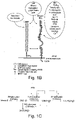
- the present method makes use of that a ternary complex of vitamin D, DBP and megalin must form before the vitamin D metabolite can become internalized by endocytosis, either directly or following interaction with cubilin (cf. Fig. 1A-C).
- vitamin D cholesterolcalciferol or ergocalciferol
- the active D-hormone [1,25(OH) 2 D] is synthesized by the enzyme 1 ⁇ -hydroxylase (Cyp27B1 - cytochrome P450) which is located within cells on the outside of the mitochondrial membrane (cf Fig. 1C ).
- the 25-hydroxylated vitamin D metabolite must be first transported from the liver to the respective cells in the kidney.
- the kidney is the major source of calcitriol (1,25(OH) 2 D) but also extrarenal cells, including lymphocytes, macrophages, keratinocytes, and cells of the parathyroid gland and pancreas can generate calcitriol.
- the present disclosure provides a discrimination of endocytable DBP, say DBP having bound a vitamin D 3 metabolite. Megalin or fragments thereof are used to discern the physiologically activatable vitamin D 3 metabolite. Recombinant megalin fragments can be produced in mammalian cell lines and purified by affinity chromatography. A direct and fast determination of a vitamin D status is provided and there is no need for any additional pre-treatment or sample preparation. The assay's time-scale is, thus, reduced while providing physiological accurate and valid readings.
- the disclosed approach can easily be adapted to different platforms such as ELISA, turbidimetry and nephelometry.
- the novel vitamin D status corresponds to the physiologically activated, endocytable vitamin D, in particular, to the 25-hydroxyvitamin D 3 in serum or plasma.
- concentrations of the other endocytable vitamin D metabolites are much lower.
- 25-hydroxylated vitamin D 2 and D 3 isomers are equally bound by DBP ( Fig. 8A )
- the higher binding affinity of megalin to DBP:25(OH)VD 3 compared to DBP:25(OH)VD 2 and 24,25-hydroxyvitamin D is striking.
- a special role seems to be taken by the C3-epimer of 25-hydroxyvitamin D 3 [3-epi-25(OH)D 3 ], for which there is a near-total lack of data regarding its clinical significance.
- the method comprises the use of a soluble fragment of megalin and/or a fusion of said soluble megalin fragment which binds the complex of DBP:VD metabolite but none or less of the numerous other ligands of LPR2/megalin. More precisely, a soluble fragment of megalin which has no affinity for albumin or anti-DBP antibody (no Ab cross-reactivity!).
- Said embodiment may comprise a surface-bound DBP-binding megalin fragment or a fusion protein thereof which contains an epitope comprising :
- Preferred embodiments may comprise a determining of the megalin-bound complex of DBP:VD metabolites. This may be done using an antibody recognizing the complex of DBP:VD or after isolation of DBP, vitamin D or its metabolites or megalin or the soluble fragment thereof.
- the method may comprise providing mixtures of vitamin D metabolites and DBP for establishing standard samples.
- the disclosure may comprise contacting said sample with an amount of added DBP to create standard conditions or a constant excess of DBP for a binding of the prohormone.
- the disclosure may further encompass contacting the sample with cubilin and/or a soluble fragment thereof.
- Said embodiment may comprise a surface-bound DBP-binding megalin fragment or a fusion protein thereof which contains an epitope having any one or more of the amino acid sequence SEQ ID NO: 01 through SEQ ID NO: 10.
- the method of the disclosure may relate to a turbidimetric or nephelometric immunoassay.
- Said megalin and/or a soluble fragment thereof may be bound to nanoparticles having diameters ranging from 50 to 200 nm, so that the complex of DBP:VD is bound to said nanoparticles.
- the disclosure also provides an immunoassay selected from the group ELISA (enzyme-linked immunosorbent assay), RIA (radioimmunoassay), FIA (fluorescence immunoassay), L1A (luminescence immunoassay), or ILMA.
- the method may comprise the steps of (a) providing a defined amount of megalin and/or a soluble fragment thereof coupled to a solid phase; (b) contacting the sample with the solid phase having coupled megalin and/or a soluble fragment thereof; (c) creating conditions to allow binding of the complex formed by DBP and vitamin D metabolite, wherein DBP alone does not bind to megalin and/or a soluble fragment thereof; and washing the solid phase; (e) providing an antibody recognizing the ternary complex comprising DBP, vitamin D or its metabolite; (f) contacting said ternary complex with an antibody, optionally an antibody against DBP, and immobilizing the immunocomplex on the solid phase; and (g) determining the amount of antibody bound to the solid phase, and correlating the amount of bound antibody to the endocytic or activatable vitamin D status in blood, plasma, or serum by reference with a standard.
- the instant disclosure further comprises a test kit for use in a method of measuring the effective vitamin D status which comprises an antibody specific for the ternary complex, megalin or a fragment thereof or DBP, or vitamin D and its metabolites.
- the disclosure also relates to a test kit for use in a method of measuring vitamin D metabolites in a sample of bodily fluid, comprising nanoparticles having bound megalin and/or soluble fragments thereof.
- the achieved object is a simple and reliable method for a direct quantitative determination of the effective status of vitamin D and its metabolites in a sample.
- the bodily fluid is blood, serum or plasma
- the status describes the endocytable fraction of vitamin D metabolites in the circulation, say the fraction which can be processed innercellularly to the active hormone by respective target cells and tissues as needed.
- the novel vitamin D status can be determined in aqueous solution despite the highly lipophilic nature of vitamin D.
- Vitamin D 3 is absorbed and processed by the organism easily and considered more potent than vitamin D 2 which has a shorter half life and binds with less affinity to the vitamin D receptor (VDR).
- 1 ⁇ ,25(OH)D 3 and parathyroid hormone (PTH) influence the vitamin D metabolism by positive or negative regulation of the activity of the la-hydroxylase and 24-hydroxylase (see Fig. 1C ).
- vitamin D is 25-hydroxylated in the liver to 25(OH)D 3 or 25(OH)D 2 , and further hydroxylated in kidney cells to its biologically active form 1a,25(OH)D.
- 24,25(OH)D is a further metabolite of 25(OH)D 3 , but inactive and destined for excretion.
- vitamin D metabolites are tightly bound to DBP. Smaller amounts are bound to albumin and lipoproteins.
- Megalin (also known as Low Density Lipoprotein receptor-related Protein 2, LRP2) is a multiligand binding cell receptor with structural similarities to the LDL receptor (LDLR). Megalin can be found in numerous cells and tissues, notably in the plasma membrane of absorptive epithelial cells ( Farquhar MG et al, Soc. Nephrol. 6 (1): 35-47 ). LRP2/megalin is known to mediate the endocytosis of its ligands and can form complexes with cubilin which cubulin:megalin complexes are again able to (re)absorb molecules.
- LRP2/megalin is known to mediate the endocytosis of its ligands and can form complexes with cubilin which cubulin:megalin complexes are again able to (re)absorb molecules.
- the cubilin:megalin complex is inter alia responsible for the cellular uptake of lipids, VLDL, certain proteins (albumin, lactoferrin), cobalamin (vitamin B 12 ), and calcidiol.
- the instant disclosure proposes determining the amount of DBP:VD which is bound by megalin. This can be done for example by an ELISA against human DBP after "isolation or separation" of said ternary complex. This is preferred and the ELISA is already commercially available (Immundiagnostik AG, Bensheim).
- the term "effective vitamin D” describes formed complex of DBP:VD which has been bound by megalin (or a fragment thereof). The complex with megalin is considered to interact with cubulin so that the "activated vitamin D metabolite" will be endocytosed and subsequently 1a-hydroxylated to become the active hormone.
- the effective vitamin D status represents an improvement over the prior art since it offers physiological information and a reading of the concentration of circulating vitamin D that will be processed to the D hormone. This status is different to the major circulating storage form because less active forms such as 25(OH)D 2 will not or much less contribute to the effective vitamin D status.
- the present disclosure provides a vitamin D status based on the amount DBP:VD, selectively and discriminably bound by megalin and/or cubulin. This endocytic complex therefore corresponds to the status of the effective vitamin D in serum or plasma.
- bioavailable as used in the prior art however refers to "total free" vitamin D which is speculative and is based on an assumed diffusion across the plasma membrane of cells.
- Steroid hormones and sterols such as vitamin D are lipophilic and commonly require a carrier protein for effective delivery.
- ligand-specific serum carriers of steroid hormones and sterols including corticosteroid-binding globulin (CBG) (glucocorticoids, mineralocorticoids), vitamin A (retinol)-binding protein, vitamin D-binding protein (DBP), sex hormone-binding globulin (SHBG) (estrogens, androgens), and thyroid hormone-binding globulin.
- CBG and SHBG not only act as high affinity serum transporters, but also bind to cell membranes in their ligand forms, suggesting alternative actions as signal transducers.
- DBP is a macrophage-activating factor (MAF) and actin-binder, which functions seem independent from the binding to vitamin D metabolites.
- MAF macrophage-activating factor
- actin-binder actsin-binder
- Total 25(OH)D is currently measured by LC-MS/MS ( Institute of Standards and Technology and the Centers for Disease Control and Prevention ).
- a variety of immunoassays are used to determine concentrations of total 25(OH)D and other vitamin D metabolites.
- all these methods produce highly variable results, likely due to the need for a release of the vitamin D metabolites from their carrier proteins. This comprises a risk of a loss of vitamin D through binding to vessel surfaces so that a falsely (low) vitamin D level is determined.
- Affinity chromatography studies using immobilized DBP and solubilized rabbit kidney membranes have identified the co-receptors for the uptake of 25(OH)D 3 -DBP complex in kidney tubules, namely a 600-kDa protein (megalin) and a 460-kDa protein (cubilin). These studies show a Ca 2 -dependent binding of DBP to cubilin.
- Megalin has three domains: a 4400 amino acid extracellular amino-terminal domain, a 22 amino acid transmembrane domain and a 213 amino acid carboxy-terminal cytoplasmic tail, indicating that it is a type I cell-surface receptor.
- the extracellular domain contains four cysteine rich clusters of LDLR type A (complement-type) repeats.
- the complement type repeat consists of approximately 40 amino acids containing six cysteine residues and the SDE (Ser-Asp-Glu) motif responsible for high-affinity binding of positively charged sequences in ligands for LDLR.
- the four cysteine-rich clusters are flanked by epidermal growth factor (EGF)-type repeats and spacer regions containing YWTD (Tyr-Trp-Thr-Asp) motifs which are responsible for pH-dependent dissociation of ligands in endosomal compartments.
- EGF epidermal growth factor
- the cytoplasmic domain of megalin contains three tetra-amino-acid NPXY motifs, which are essential for endocytosis of the ligand-receptor complex via clathrin-coated pits.
- Megalin has diverse types of ligands: vitamin-binding proteins and other binding proteins, apolipoproteins, hormones and hormone precursors, drugs and toxins, enzyme and enzyme inhibitors, immune- and stress-response-related proteins, and others including calcium. Megalin knockout mice are unable to recover DBP from the glomerular filtrate, and lose it together with its vitamin D cargo in urine. As a consequence, megalin knockout mice are unable to adequately metabolize 25(OH)D to 1,25(OH) 2 D resulting in a bone phenotype that resembles vitamin D-deficient rickets.
- Cubilin has been identified as a receptor for intrinsic factor-B12 (IF-B) complex in the terminal ileum. It is a 460 kDa receptor with no transmembrane domain and no signals for endocytosis. Cubilin contains 27 CUB domains responsible for the ligand binding and eight EGF-type repeats preceded by a stretch of 110 amino acids, where the N-terminal region appears essential for membrane anchoring.
- IF-B intrinsic factor-B12
- megalin-dependent uptake of DBP has a clear role in renal vitamin D endocrinology, it is not yet clear whether a similar mechanism is present in other vitamin D target tissues.
- megalin is expressed by several tissues including the placenta, mammary gland and parathyroid glands, which are known to have 1a-hydroxylase activity, suggesting an extra-renal DBP-megalin interaction.
- the present disclosure describes a method of determining a vitamin D status based on the detection and quantitation of a complex comprising DBP, vitamin D and either one of megalin and cubulin or both.
- the present method requires no specific release of the vitamin D metabolites from DBP and only the fraction of vitamin D metabolites will be evaluated that is subject to endocytosis and activation.
- the measurement of DBP bound to megalin provides a parameter for endocytable vitamin D metabolites.
- the provided method can therefore be used to establish a vitamin D status which corresponds to the physiologically active and endocytable vitamin D concentration in serum or plasma.
- the various vitamin D metabolites in particular 25(OH)D 3 can be discriminated as well as the changes in total serum concentrations of 25OHD, 1,25(OH) 2 D and DBP in subjects following supplementation with either cholecalciferol (vitamin D 3 ) or ergocalciferol (vitamin D 2 ). Supplementation will no longer interfere with the measurement of active vitamin D molecules that will be subject to endocytosis and hydroxylation. The fraction of truly bioavailable vitamin D molecules can therefoe easily be discerned from the supplemented vitamin D 2 or vitamin D molecules bound by other proteins in the circulation.
- Immunonephelometry can be used to measure the DBP concentration in a sample. Immunonephelometry quantifies the scattering of an incident light source by large soluble antigen-antibody complexes under conditions of a moderate excess of antibody. Under these conditions, the complexes form a stable lattice, and a direct linear relationship is established between an increasing concentration of antigen and the increase in scattered light intensity. Automated nephelometers provide sensitive and precise measurements of DBP concentration in a rapid manner and with minimal requirements for technical skill. These can be used in clinical chemistry laboratories to analyze DBP concentrations. In a preferred embodiment, only the ternary complex formed by DBP, vitamin D and megalin is recognized by the antibody.
- immunoturbidimetry can be used to measure the DBP concentration in a sample.
- Turbidimetry is the process of measuring the loss of intensity of transmitted light due to the scattering effect of particles suspended in it. Light is passed through a filter creating a light of known wavelength which is then passed through, for example, a cuvette containing a solution. A photoelectric cell collects the light which passes through the cuvette. A measurement is then given for the amount of absorbed light.
- Immunoturbidimetry is a variant in which an antigen-antibody reaction takes place. The antigen-antibody complexes are particles which can be optically detected by a photometer. In a preferred embodiment, the antibody recognizes a ternary complex formed by DBP, vitamin D and megalin.
- nanoparticles coated with megalin or fragments thereof are contacted with a sample containing DBP/vitamin D complexes.
- Increasing concentrations of DBP/vitamin D/megalin ternary complex in the sample result in increased turbidity.
- Protein-protein interaction networks An important class of protein-protein interaction corresponds to peptide-mediated interactions, in which a short peptide stretch from one partner interacts with a large protein surface from the other partner. Protein-peptide interactions are typically of low affinity and involved in regulatory mechanisms, dynamically reshaping protein interaction networks. Due to the relatively small interaction surface, modulation of protein-peptide interactions has been considered feasible and highly attractive, for example, for therapeutic purposes. Unfortunately, the number of available 3D structures of protein-peptide interfaces is very limited.
- cysteine-rich complement-type ligand binding repeats (LDLR class A) for analysis of DBP-megalin interaction. EGF-like modules were also included as they are considered important for receptor folding and dissociation of ligands in endosomal compartment.
- a signal peptide (M1-G25) was introduced at the N-termini of complement-type repeats to allow sorting to the secretory pathway.
- a C-terminal 6xHis-tag was added for affinity purification and protein analysis by co-immunoprecipitation and Western blotting.
- the respective cDNA sequences were obtained by PCR and inserted into the mammalian expression vector pcDNA3.1.
- HEK cells were stable transfected with megalin cDNA sequences (M1, M2, M3) as they have endogenous calcium channels. Protein purification was done using the His-tag and a commercially available Ni-NTA resin. The cloning strategy for megalin fragments (M1, M2, M3) into the mammalian expression vector pcDNA3.1 is shown in Figs. 2A-B.
- Megalin M1 cDNA encoded the sequence of amino acids 26 to 386 of human LRP2/megalin (SEQ ID NO:08).
- the construct included cDNA encoding for an N-terminal 25 amino acid signal peptide and a C-terminal 6x histidine tag.
- the predicted molecular weight was about 43 kDa for the monomer and about 86 kDa for the dimer.
- the HEK-expressed and secreted recombinant megalin M1 fragment had the following amino acid sequence (without signal sequences): SEQ ID NO: 08
- SEQ ID NO. 10 Megalin M3 cDNA encoded the sequence of amino acids 2698 to 3192 of human LRP2/megalin (SEQ ID NO: 10).
- the cDNA encoded an 25 amino acid signal peptide and a C-terminal 6x histidine tag.
- the predicted molecular weight was about 60 kDa for the monomer and 120 kDa for the dimer.
- the construct was expressed in HEK 293 mammalian cells.
- SEQ ID NO.10 corresponds to following amino acid sequence (without signal sequences):
- the Western blots show that megalin M1, M2, M3 fragments were expressed and secreted by HEK cells.
- the megalin fragments could be kept in solution. No precipitation was observed.
- the megalin fragments showed a tendency to form dimers but the molecular weights were all consistent with the predicted sizes.
- 2X Laemmli and 2X Urea sample buffers were used for analysis of megalin monomer and dimer formation but no difference was observed by the use of these buffers.
- the M2 fragment was the one which could be mostly easily dissociated to the monomer.
- HEK cells were transfected with above cDNA constructs (M1, M2, M3) and cell lysates used for co-immunoprecipation.
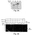
- Fig. 3A shows a Western blot of cell lysates (20 ⁇ g) of HEK cells expressing M1, M2 and M3. Detection with an antibody against the His-tag.
- Fig. 3B shows the results of a co-immunoprecipitation with DBP (0,5 ⁇ g and VD 3 (0,2 ⁇ g).
- the serum samples included DBP alone or in the presence of added vitamin D 3 .
- Each sample was contacted with cell lysate containing megalin fragment. After incubation, the samples were contacted with beads coated with an antibody against His-tag and the DBP pulled down by centrifugation.
- the bound ligands (DBP) were dissociated from the beads and analyzed by Western blotting for the expression of DBP using an anti-DBP-antibody.
- pure DBP alone was analyzed.
- Fig. 3B confirms that DBP could be pulled down with megalin M1 and M2 fragments. Notably, DBP could only be pulled down in the presence (+) of vitamin D 3 .
- Figs. 4 A,B show the in vitro interaction between DBP and Ni-NTA agarose purified soluble megalin M1, M2, M3 fragments (3 ⁇ g) in a similar set-up as above.
- the results are shown in Figs. 4 A,B.
- Fig. 4A is a Western blot of purified megalin M1, M2, M3 fragments from culture supernatants (3 ⁇ g), secreted by HEK cells transfected with cDNA for megalin M1, M2 and M3 fragments; detection by anti-His-antibodies.
- Fig. 4 B shows a western blot with the results of a co-immunoprecipitation with purified megalin M1, M2, M3 fragments.
- the serum samples contained DBP alone or in the presence (+) of vitamin D 3 .
- the samples were contacted with beads coated with an antibody against 6xHis-tag and pulled down by centrifugation. Bound proteins were dissociated from the beads and analyzed by Western blotting using an antibody against vitamin D binding protein.
- Fig. 4B confirms that DBP could be pull down with purified soluble M1 and M2 fragments.
- DBP was pulled down only in the presence (+) of vitamin D 3 . This indicates that soluble purified megalin fragments M1 and M2 do not interact with DBP unless occupied with vitamin D 3 .
- Ternary complexes comprising DBP, vitamin D 3 and purified megalin M1 or M2 fragments were formed in vitro. No interaction or binding was observed between soluble purified megalin M1 or M2 fragments and DBP alone.
- Microscale thermophoresis examines the directed movement of particles in a microscopic temperature gradient (thermophoresis). Any change of the hydration shell of biomolecules due to changes in their conformation results in a relative change of the movement along the temperature gradient. This principle can be used to determine the binding affinity of two molecules. This technique allows in particular an examination of interactions in solution without any immobilization on a surface. A spatial temperature difference leads to a depletion of molecule concentration in the region of elevated temperature, which can be then determined. Thermophoresis is usually performed with fluorescently labeled molecules.
- thermophoresis can further be used to quantify the binding strength under constant buffer conditions.
- the thermophoretic movement of the fluorescently labeled molecule is measured by monitoring the fluorescence distribution inside a capillary.
- the microscopic temperature gradient is generated by an IR-Laser, which is focused into the capillary and absorbed in water. The temperature of the aqueous solution in the laser spot region therefore increases.
- a homogeneous fluorescence distribution is observed inside the capillary prior the IR-Laser is switched on. When the IR-Laser is switched on, a new fluorescence distribution is established.
- the thermal relaxation time is fast and induces a binding-dependent drop in the fluorescence of the dye due to its local environmental-dependent response to the temperature step increase. Molecules move then from the locally heated region to the outer cold regions. The local concentration of molecules decreases in the heated region until it reaches a steady-state distribution.
- the normalized fluorescence measures a concentration ratio, with consideration of the temperature step increase. Due to the linearity of the fluorescence intensity and the thermophoretic depletion, the normalized fluorescence from the unbound molecule and the bound complex superpose linearly. Quantitative binding parameters were obtained using serial dilutions of the binding substrate. By plotting Fnorm against the logarithm of the different concentrations of the dilution series, a sigmoidal binding curve is obtained. This binding curve can directly be fitted with the non-linear solution of the law of mass action, with the dissociation constant Kd as result.
- purified DBP was labeled with the red fluorescent dye NT-647 using Monolith Protein Labeling Kit Red (NanoTemper Technologies, Kunststoff, Germany). Ni-NTA-purified soluble megalin fragments M1, M2, and M3 fragments were titrated in the range from 0.488 to 1000 nmol/L.
- a Monolith NT.115 device (NanoTemper Technologies) and the NT Analysis software version 1.427 (NanoTemper Technologies) were used for measurements.
- a microtiter plate was coated with purified soluble megalin M1 or M2 fragment (1 ⁇ g/100 ⁇ l PBS).
- Mixtures of DBP and 25(OH)D 3 were prepared using 1 ⁇ g DBP and serial dilutions of 25(OH)D 3 (VD 3 ) in PBS (0, 0.3125, 0.625, 1.25, 2.5, 5, 10, 20 ⁇ g).
- the binding of soluble megalin M1 and M2 fragments to DBP was dependent on the 25-hydroxy-vitamin D 3 . More precisely, the absorbance is linear proportional to the vitamin D 3 concentration as shown in Fig. 9A and Fig. 10A and this relation is not impacted by increasing concentrations of 25(OH)VDBP or 24,25(OH)VD (cf. Fig. 9B , 10B). Importantly, these experiments demonstrate a proof of principle for easy and reliable vitamin D measurements based on the ternary complex.
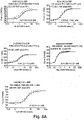
- Fig. 6A The binding properties of the various megalin soluble fragments were further analyzed by co-immunoprecipitation using samples of serum and plasma from human subjects. The results are shown in Fig. 6A .
- Purified soluble megalin M1 protein was mixed with two human serum or plasma samples to allow interaction between soluble megalin M1 fragment and endogenous DBP present in the sample. Samples were incubated with Ni-NTA resin to allow interaction of megalin M1/DBP to the resin. After washing the resin, bound complex was eluted from the resin and assayed by western blotting.
- Fig. 6A (right) shows that DBP was present in both samples and could be detected using a specific antibody.
- FIG. 6A shows that purified soluble megalin M1 fragment interacted in solution with DBP in all samples, as demonstrated by the presence of pulled down DBP.
- a control sample containing serum only but no megalin protein no DBP was detectable.
- the presence of purified megalin M1 protein was detected in all samples using an anti-6xHis-tag antibody ( Fig. 6A , left, lower blot).
- the co-immunoprecipitations are proof that the complex of DBP and 25(OH)D 3 can specifically be bound and isolated from plasma or serum using a suitable soluble megalin fragment.
- the megalin portion with amino acids 26-386 and 1024-1429 have been shown to be involved in the formation of a ternary complex with DBP and 25(OH)D 3 .
- the external megalin region with amino acids 2698-R3192 did not participate in the binding under the described conditions.
- the present application comprises representative amino acid sequences of megalin which can be used in the binding of DBP. Those can further be used for determining the status of endocytic or activatable vitamin D in bodily fluids.
- the status of endocytable vitamin D overrules any status for "free vitamin D” or “total vitamin D” as there will be no need for distinguishing between “free” or “total” from the "physiological status of vitamin D available for endocytosis and 1 ⁇ -hydroxylation".
- Conventional methods usually do not determine the physiologically relevant vitamin D status since they cannot analyze the metabolites ready for processing to the active hormone. Thus, the conventional methods are insufficient whereas the present method is directed to the status of circulating prohormone which can and will be hydroxylated in the kidney, and probably in other tissues, giving rise to the active hormone.
- DBP total DBP
- 25(OH)D 3 ternary complex of DBP:25(OH)VD:M2.
- DBP was determined by ELISA.
- the 25(OH)D 3 content was quantified conventionally by an independent laboratory.
- the ternary complex DBP:25(OH)VD:M2 megalin was analyzed by a DBP:VD3:megalin ELISA according to the disclosure. Table 1 below summarizes the results for comparison.
- sample S3 was considered to have normal levels of 25(OH)D 3 ; S9, insufficient; S8 and S6, deficient levels. In other words, from higher to lower levels of 25(OH)D 3 : S3>S9>S8>S6. From the results it could be concluded that the level of total DBP seemed not to correlate with the level of 25(OH)D 3 level in human serum samples.
- the linearity range of the interaction between megalin and DBP was examined using the claimed method for 25-hydroxyvitamin D 3 (25(OH)VD 3 ), 25-hydroxyvitamin D 2 (25(OH)VDBP) and 24,25-dihydroxyvitamin D (24,25(OH)VD).
- ELISAs for the complex of DBP:VD:megalin were performed as disclosed. In brief: a microtitre plate coated with purified megalin M2 fragment was incubated with a mixture of DBP and serial dilutions of 25(OH)VD 2 , 25(OH)VD 3 or 24,25(OH)VD.
- a polyclonal rabbit anti-DBP antibody and a HRP-conjugated donkey anti-rabbit antibody were used. The results are shown in Fig. 7C .
- the assay was linear up to 50 ng/ml.
- the determined sensitivity limit was 2.0 ng/ml.
- the affinity of DBP when interacting with 25(OH)VD 3 , 25(OH)VD 2 , or 24,25-24,25(OH)VD, 3C-epimer of 25(OH)D 3 and megalin M2 was also analyzed by microscale thermophoresis. The results are shown in Fig. 8B .
- Purified DBP (50nM) was labeled with the red fluorescent dye NT-647 and mixed with either vitamin D metabolite (37.8 nM).
- Purified megalin shown in Fig. 7C For all vitamin D metabolites, the assay was linear up to 50 ng/ml. The determined sensitivity limit was 2.0 ng/ml.
- the affinity of DBP when interacting with 25(OH)VD 3 , 25(OH)VD 2 , or 24,25-24,25(OH)VD, 3C-epimer of 25(OH)D 3 and megalin M2 was also analyzed by microscale thermophoresis. The results are shown in Fig. 8B .
- Purified DBP (50nM) was labeled with the red fluorescent dye NT-647 and mixed with either vitamin D metabolite (37.8 nM).
- Purified megalin M2 was titrated in different concentrations. The dissociation constant Kd was calculated for every condition. The binding affinity was assessed by the dissociation constant Kd.
- the described megalin fragments can easily be produced by recombinant methods and even by chemically synthesized.
- the described megalin fragments remained soluble in aqueous solution so that the conditions close to physiological can be used.
- no solvents or surfactants for displacement of the vitamin D from its binding partners are needed. Accordingly, the measurement of the vitamin D status will not be interfered by non-physiological chemicals.
- No purification steps or time consuming and costly techniques such as LC-MS are necessary.
- a direct and fast determination of the effective vitamin D status can be done immediately after sample collection.
- the disclosed principles can further be easily adapted to available platforms and automats.
- Megalin (LRP2) cDNA fragments were amplified by RT-PCR using mRNA from Caco-2 cells (human colon carcinoma epithelial cells).
- LRP2 human colon carcinoma epithelial cells
- For cDNA transcription synthesis Maxima H Minus First strand cDNA synthesis kit (Thermo Scientific) was used. This kit allows synthess of cDNA up to 20 kb. Oligo dt18 primer and 65C were used.
- Platinum PCR Supermix high fidelity PCR kit (Invitrogen) was used. Megalin M1 cDNA: 1158 bp; Megalin M2 cDNA: 1215 bp; Megalin M3 cDNA: 1482 bp.
- Stable cell line selection was carried out using neomycin (G418, 800 ⁇ g/ml).
- Cell culture supernatants or cell lysates were purified by Ni-NTA resin.
- Analysis of megalin protein M1, M2, M3 fragments having a 6xHis-Tag was performed by Western blot with an antibody against 6xHis-tag (Cohesion Biosciences).
- Cell culture supernantants or cell lysates were purified with Ni-NTA agarose (Thermo Fischer Scientific). Cell lysates or culture supernatant (0.5 - 2 mL) were incubated with 50 ⁇ l of pre-equilibrated Ni-NTA resin at 4°C overnight.
- the resin was washed 3 times with H-buffer + 20mM imidazole. Megalin protein complex was eluted with H-buffer containing 200mM imidazole and buffer exchanged with PBS 1X. Protein solutions were lyophilized or kept at 4°C prior use.
- the fragment M2 was further mapped for major binding epitopes of Megalin ligands within the M2 region, the region of megalin which binds human DBP.
- the binding sites of other megalin ligands were removed and the epitopes for binding of human DBP identified. It is imporant that the minimun epitopes do not overlap with the binding epitopes of other ligands off megalin. The results are shown in the Tables below.
- Microtiter plates were coated with soluble purified megalin M1 or M2 fragment (1 ⁇ g/100 ⁇ l diluted in PBS1X) by incubation at RT for 2 h.
- Serial dilutions of 25-hydroxvitamin D 3 (VD 3 ) 0, 0.3125, 0.625, 1.25, 2.5, 5, 10, 20 ng in 100 ⁇ l PBS were mixed each with 1 ⁇ g DBP (DBP). The mixture was incubated at 37°C for 1h. Unspecific binding sites were blocked with blocking buffer (1% BSA in PBS1X) at RT for 1 h.
- the DBP-VD 3 mixture or serum samples were added to the megalin-coated wells and incubated at 4°C overnight. After washing with PBST-buffer (0.05% Tween 20 in PBS) 100 ⁇ l rabbit-anti-DBP antibody was added (1: 3500 diluted in blocking buffer) and incubated at R.T for 2 h. After washing with PBST, 100 ⁇ l of HRP- conjugated donkey anti-rabbit 2nd antibody (1: 500 diluted in blocking buffer) was added and incubated at 37°C for 1 h. 100 ⁇ l substrate reagent A+B (1:1) (R&D) was added and incubated at R.T for 30min. Then, 100 ⁇ l stop solution was added to the wells. Absorbance was read at 450 nm. The values were compared to standard values of known 25-hydroxvitamin D 3 concentration.
- Ni-NTA purified soluble megalin M1 or M2 protein fragment (3 ⁇ g) was first mixed with human serum or plasma samples (30 ⁇ l). These samples were incubated with 50 ⁇ l of pre-equilibrated Ni-NTA resin at 4°C overnight. The resin was washed 3 times with H-buffer + 20mM imidazole. Bound complex was eluted with H-buffer containing 200mM imidazole and analyzed by Western blot with a polyclonal rabbit antibody against DBP (Abcam).
- Ni-NTA purified soluble megalin M1 or M2 protein fragment (3 ⁇ g) is first mixed with human serum or plasma samples (30 ⁇ l). Samples are incubated with 50 ⁇ l of pre-equilibrated Ni-NTA resin at 4°C overnight. The resin is washed 3 times with H-buffer + 20mM imidazole. Bound complex is eluted with H-buffer containing 200 mM imidazole. The eluted complex is contacted in aqueous solution with an antibody against vitamin D binding protein. The increase in turbidity is measured with a standard turbidimeter and compared with standard values of known 25-hydroxvitamin D 3 concentration.
- nanoparticles for example, latex nanoparticles (aprox. 150 nm) are coated with megalin M1 or M2 fragment and incubated with a serum or plasma sample. The increase of turbidity is then measured and compared to standard values of known 25-hydroxvitamin D 3 concentration.
- ELISA binding assay for linearity range determination A microtiter plate was coated with purified megalin M1 or M2 protein fragment (1 ⁇ g/100 ⁇ l diluted in PBS1X) by incubation at RT for 2 h.
- Mixtures of DBP and 25-hydroxvitamin D 3 (VD 3 ), 25-hydroxvitamin D 2 (VD 2 ) or 24,25-hydroxvitamin D (24,25VD) were prepared by mixing 20 ⁇ g DBP and serial dilution of VD (0, 0.78, 1,56, 3,125, 6.25, 12.5, 25, 50 ng) in 100 ⁇ l PBS. The mixture was incubated at 37°C for 1h.
- the above prepared DBP-VD mixture was added to megalin-coated wells and incubated at 4°C, overnight. Then, the plate was incubated with 100 ⁇ l diluted rabbit-anti-DBP antibody (1:1000 diluted in blocking buffer) at RT for 2 h, followed by incubation with 100 ⁇ l of HRP-conjugated donkey anti-rabbit antibody (1: 500 diluted in blocking buffer) at 37°C, 1 h. After substrate reaction, the absorbance was read at 450 nm. The assay was linear up to 50 ng/ml. The sensitivity limit was determined to be 2.0 ng/ml.
- Microscale Thermophoresis assay Purified DBP (Merck, 345802) was labeled with the red fluorescent dye NT-647 by using a Monolith Protein Labeling Kit Red (NanoTemper Technologies, Kunststoff, DE). Ni-NTA-purified megalin fragments (M1, M2) were titrated in the range of 0.488 to 1000 nmol/L. Purified DBP (Merck, 345802) was likewas labeled with the red fluorescent dye NT-647 by using the Monolith Protein Labeling Kit Red. 25-hydroxvitamin D 3 (VD 3 ) was titrated in concentrations in the range of 0.0488 to 100 nmol/L.
- 25-hydroxvitamin D 2 (VD 2 ), 24,25-hydroxvitamin D (24,25VD) and 3epi25(OH)VD 3 were titrated in concentrations in the range of 0.0163 to 37 nmol/L. 37.8 nM of VD 3 , VD 2 and 24,25VD was respectively added to 50nM of NT-647 labeled DBP.
- a Monolith NT.115 device (NanoTemper Technologies) was used for measurements.
- NT Analysis software version 1.427 was used for analysis. Parameters: laser power, 100%; LED, 80; laser on-time, 30 seconds; laser off-time, 5 seconds; temperature, 25°C.
- FNorm normalized fluorescence
- Kd dissociation constant.
- the dissociation constants Kd of the vitamin D metabolites to DBP gave the following ranking of the binding affinities. 3epi25(OH) 2 VD3 > 25(OH) 2 VD3 ⁇ 25(OH) 2 VD 2 > 24,25(OH) 2 VD3 > 1,25(OH) 2 VD3.
- the binding affinity of DBP-VD 2 is comparable to Kd of DBP-VD 3 .
- the binding of 24,25(OH)VD to DBP is marginally lower.
- the high binding affinity of the 3C epimer of 25(OH)D 3 is surprising and will require further investigation as this epimer seems therefore particularly useful as food supplement, if not toxic for other reasons.
Landscapes
- Life Sciences & Earth Sciences (AREA)
- Health & Medical Sciences (AREA)
- Engineering & Computer Science (AREA)
- Molecular Biology (AREA)
- Biomedical Technology (AREA)
- Chemical & Material Sciences (AREA)
- Hematology (AREA)
- Immunology (AREA)
- Urology & Nephrology (AREA)
- Cell Biology (AREA)
- Microbiology (AREA)
- Biotechnology (AREA)
- Food Science & Technology (AREA)
- Medicinal Chemistry (AREA)
- Physics & Mathematics (AREA)
- Analytical Chemistry (AREA)
- Biochemistry (AREA)
- General Health & Medical Sciences (AREA)
- General Physics & Mathematics (AREA)
- Pathology (AREA)
- Investigating Or Analysing Biological Materials (AREA)
- Peptides Or Proteins (AREA)
Claims (13)
- Verfahren zur Messung von Vitamin D und dessen Metaboliten in einer Probe mit Körperflüssigkeit des Probanden, wobei die Probe endogenes Vitamin-D-bindendes Protein (DBP) enthalten kann, umfassend die Schritte:a) In-Kontakt-bringen der Probe mit Megalin und/oder einem Fragment davon unter Bindungsbedingungen, so dass man einen ternären Komplex erhält, der DBP, einen Vitamin-D-Metaboliten und Megalin oder dessen Fragment umfasst;(b) Bestimmen der Menge an DBP, die von Megalin oder dessen Fragment gebunden wird; und(c) Beziehen der Menge des an Megalin gebundenen DBP mit dem Vitamin-D-Status des Probanden.
- Verfahren nach Anspruch 1, wobei das Fragment des Megalins eine der Aminosäuresequenzen von SEQ ID NO:01 bis SEQ ID NO:10 umfasst.
- Verfahren nach Anspruch 1 oder Anspruch 2, wobei das Fragment des Megalins die Aminosäuresequenz SEQ ID NO:01 oder SEQ ID NO:02 oder Kombinationen davon umfasst.
- Verfahren nach einem der Ansprüche 1 bis 3, wobei das Fragment des Megalins ausschließlich Vitamin-D-bindendes Protein (DBP) und keinen der anderen Liganden von Megalin bindet.
- Verfahren nach einem der Ansprüche 1 bis 4, umfassend die Bestimmung von gebundenen Vitamin-D-Metaboliten.
- Verfahren nach einem der Ansprüche 1 bis 5, umfassend eine differenziale Bestimmung von 25-Hydroxyvitamin D3 in Gegenwart von 25-Hydroxyvitamin D2, 24,25-Dihydroxyvitamin D2 oder 24,25-Dihydroxyvitamin D3
- Verfahren nach einem der Ansprüche 1 bis 6, umfassend die Zugabe einer bekannten Menge an Vitamin-D-bindendem Protein zu der Probe.
- Verfahren nach einem der Ansprüche 1 bis 7, wobei die Probe ferner mit Cubulin und/oder einem löslichen Fragment davon in Kontakt gebracht wird.
- Verfahren nach einem der Ansprüche 1 bis 8, wobei der Komplex mit DBP durch partikelverstärkte Immunoturbidimetrie oder Nephelometrie bestimmt wird.
- Verfahren nach einem der Ansprüche 1 bis 8, wobei der Komplex mit DBP bestimmt wird durch einen Immunoassay, ausgewählt aus der Gruppe mit ELISA (Enzyme-linked Immunosorbensassay), RIA (Radioimmunoassay), FIA (Fluoreszenz-Immunoassay), LIA (Lumineszenz-Immunoassay) oder ILMA,
- Verfahren nach einem der Ansprüche 1 bis 10, das die folgenden Schritte umfasst:(a) Bereitstellen einer definierten Menge von Megalin und/oder eines löslichen Fragments davon, gekoppelt an eine feste Phase;(b) in Kontakt bringen der Probe mit der festen Phase mit immobilisiertem Megalin und/oder einem löslichen Fragment davon;(c) Erzeugen von Bedingungen, die eine Bindung von Megalin und/oder einem löslichen Fragment davon an den durch das Vitamin-D-bindende Protein (DBP) und Vitamin-D-Metaboliten gebildeten Komplex ermöglichen, wobei das Vitamin-D-bindende Protein (DBP) allein nicht an Megalin und/oder ein lösliches Fragment davon bindet; und Waschen der festen Phase;(e) Bereitstellen eines Antikörperkomplexes, der den Komplex aus DBP, Vitamin D oder dessen Metaboliten und Megalin oder einem löslichen Fragment davon erkennt; (f) in Kontakt bringen des Megalins und/oder eines löslichen Fragments davon, das an das Vitamin-D-bindende Protein (DBP) und die Vitamin-D-Metaboliten gebunden ist, mit dem Antikörper gegen das Vitamin-D-bindende Protein (DBP) und Immobilisieren des Komplexes an der festen Phase; und(g) Bestimmung der Menge des an die feste Phase gebundenen Antikörpers und Quantifizierung der Vitamin-D-Metaboliten in Blutplasma oder Serum durch Korrelation mit Standardproben.
- Testkit zur Verwendung in einem Verfahren zur Bestimmung des Vitamin-D-Status in einer Probe einer Körperflüssigkeit, umfassend einen Antikörper, der DBP bindet, und ein Fragment oder eine Aminosäuresequenz von Megalin, gegebenenfalls gekoppelt an einen Träger.
- Zusammensetzung zur Messung von Vitamin-D-Metaboliten in einer Probe mit Körperflüssigkeit, die Antikörper gegen ein Epitop von DBP und/oder ein Fragment des Megalins enthält.
Applications Claiming Priority (2)
| Application Number | Priority Date | Filing Date | Title |
|---|---|---|---|
| DE102018100096 | 2018-01-03 | ||
| PCT/EP2019/050121 WO2019134948A1 (en) | 2018-01-03 | 2019-01-03 | Method of measuring the endocytic vitamin d status |
Publications (2)
| Publication Number | Publication Date |
|---|---|
| EP3735591A1 EP3735591A1 (de) | 2020-11-11 |
| EP3735591B1 true EP3735591B1 (de) | 2022-06-15 |
Family
ID=65003393
Family Applications (1)
| Application Number | Title | Priority Date | Filing Date |
|---|---|---|---|
| EP19700082.1A Active EP3735591B1 (de) | 2018-01-03 | 2019-01-03 | Verfahren zur messung des endozytischen vitamin-d-status |
Country Status (7)
| Country | Link |
|---|---|
| US (1) | US20210072263A1 (de) |
| EP (1) | EP3735591B1 (de) |
| JP (1) | JP7300452B2 (de) |
| CA (1) | CA3087430A1 (de) |
| DK (1) | DK3735591T3 (de) |
| ES (1) | ES2927217T3 (de) |
| WO (1) | WO2019134948A1 (de) |
Families Citing this family (1)
| Publication number | Priority date | Publication date | Assignee | Title |
|---|---|---|---|---|
| CN115078578A (zh) * | 2022-06-16 | 2022-09-20 | 广州市妇女儿童医疗中心 | 一种基于液质联用的血清维生素d相关代谢物的检测方法 |
Family Cites Families (12)
| Publication number | Priority date | Publication date | Assignee | Title |
|---|---|---|---|---|
| US5821020A (en) | 1995-07-14 | 1998-10-13 | Nhh Biologics | Vitamin D assay |
| AU763458B2 (en) | 1998-06-25 | 2003-07-24 | Biomedica Gmbh | Functional vitamin D derivatives and a method for determining 25-hydroxy-vitamin D and 1alpha,25-dihydroxy-vitamin D |
| DE10144905C2 (de) | 2001-09-12 | 2003-07-31 | Immundiagnostik Ag | Bestimmung von Vitamin-D direkt in Serum oder Plasma |
| US20040132104A1 (en) | 2003-01-07 | 2004-07-08 | Sackrison James L. | Vitamin D assay |
| EP1462119A1 (de) * | 2003-03-24 | 2004-09-29 | Schering AG | Modulatoren der Megalin-vermittelten Aufnahme von Radiotherapeutika und/oder Radiodiagnostica in die Nierenzellen und ihre Verwendung in Therapie und Diagnose |
| JP4865377B2 (ja) * | 2006-03-28 | 2012-02-01 | 国立大学法人 新潟大学 | ヒトメガリンの測定方法 |
| ATE472734T1 (de) * | 2007-02-01 | 2010-07-15 | Immundiagnostik Ag | Direktbestimmung von vitamin d im serum oder plasma |
| GB0903469D0 (en) | 2009-03-02 | 2009-04-08 | Axis Shield Diagnostics Ltd | Assay |
| JP5424702B2 (ja) * | 2009-04-27 | 2014-02-26 | 国立大学法人 新潟大学 | 尿中ヒトメガリンを測定することを含む腎疾患検出方法 |
| US20140038203A1 (en) * | 2012-07-09 | 2014-02-06 | Musc Foundation For Research Development | Methods for detecting or predicting kidney disease |
| KR101840525B1 (ko) | 2013-02-06 | 2018-03-20 | 후지레비오 가부시키가이샤 | 비타민 d 의 측정 방법 및 측정용 키트 |
| ES2714565T3 (es) | 2014-02-20 | 2019-05-29 | Immundiagnostik Ag | Diagnóstico in vitro y pronóstico de nefropatía inducida por contraste (nic) en pacientes sometidos a angiografía coronaria |
-
2019
- 2019-01-03 EP EP19700082.1A patent/EP3735591B1/de active Active
- 2019-01-03 DK DK19700082.1T patent/DK3735591T3/da active
- 2019-01-03 WO PCT/EP2019/050121 patent/WO2019134948A1/en not_active Ceased
- 2019-01-03 CA CA3087430A patent/CA3087430A1/en active Pending
- 2019-01-03 JP JP2020536633A patent/JP7300452B2/ja active Active
- 2019-01-03 US US16/958,850 patent/US20210072263A1/en not_active Abandoned
- 2019-01-03 ES ES19700082T patent/ES2927217T3/es active Active
Also Published As
| Publication number | Publication date |
|---|---|
| ES2927217T3 (es) | 2022-11-03 |
| JP2021509952A (ja) | 2021-04-08 |
| US20210072263A1 (en) | 2021-03-11 |
| EP3735591A1 (de) | 2020-11-11 |
| CA3087430A1 (en) | 2019-07-11 |
| JP7300452B2 (ja) | 2023-06-29 |
| DK3735591T3 (da) | 2022-09-12 |
| WO2019134948A1 (en) | 2019-07-11 |
Similar Documents
| Publication | Publication Date | Title |
|---|---|---|
| JP6750066B2 (ja) | ビタミンd結合タンパク質からビタミンdを解離するための溶液、その関連検出法及び使用 | |
| Farrell et al. | Determination of vitamin D and its metabolites | |
| Silverberg et al. | Clinical utility of an immunoradiometric assay for parathyroid hormone (1–84) in primary hyperparathyroidism | |
| KR101838328B1 (ko) | 비타민 d 방출 시약 | |
| Moon et al. | Comparison of four current 25-hydroxyvitamin D assays | |
| Hocher et al. | Measuring parathyroid hormone (PTH) in patients with oxidative stress–do we need a fourth generation parathyroid hormone assay? | |
| Otieno et al. | Cancer diagnostics via ultrasensitive multiplexed detection of parathyroid hormone-related peptides with a microfluidic immunoarray | |
| Bachali et al. | Vitamin d deficiency and insulin resistance in normal and type 2 diabetes subjects | |
| EP2126586B1 (de) | Direktbestimmung von vitamin d im serum oder plasma | |
| Ursem et al. | Oxidation of parathyroid hormone | |
| US20140162294A1 (en) | Methods and compositions for assaying vitamin d | |
| US20210072264A1 (en) | Use of 1,25-Dihydroxyvitamin D Values in Ratio with PTH as a Prognostic Biomarker | |
| US10073103B1 (en) | Rapid measurement of total vitamin D in blood | |
| Evenepoel et al. | Skeletal parathyroid hormone hyporesponsiveness: a neglected, but clinically relevant reality in chronic kidney disease | |
| JP7300452B2 (ja) | エンドサイトーシスビタミンdの状態の測定方法 | |
| Ilkovska et al. | Serum hepcidin reference range, gender differences, menopausal dependence and biochemical correlates in healthy subjects | |
| JP6012108B2 (ja) | 腎不全の予後判定方法 | |
| Inaba et al. | Significance of Bio-intact PTH (1–84) assay in hemodialysis patients: Bio-intact PTH (1–84) assay in HD patients | |
| US20240319212A1 (en) | Method of measuring the endocytic vitamin d status | |
| Bikle | Assay of calcium-regulating hormones | |
| Lu et al. | Non-oxidized PTH (n-oxPTH) is associated with graft loss in kidney transplant recipients | |
| Thienpont et al. | Traceability to a common standard for protein measurements by immunoassay for in-vitro diagnostic purposes | |
| Cavalier et al. | Evaluation of different bone markers in hemodialyzed patients | |
| Arya et al. | Parathyroid Hormone (PTH) assays and applications to bone disease: Overview on methodology | |
| Nili et al. | Proteomic analysis and molecular modelling characterize the iron-regulatory protein haemojuvelin/repulsive guidance molecule c |
Legal Events
| Date | Code | Title | Description |
|---|---|---|---|
| STAA | Information on the status of an ep patent application or granted ep patent |
Free format text: STATUS: UNKNOWN |
|
| STAA | Information on the status of an ep patent application or granted ep patent |
Free format text: STATUS: THE INTERNATIONAL PUBLICATION HAS BEEN MADE |
|
| PUAI | Public reference made under article 153(3) epc to a published international application that has entered the european phase |
Free format text: ORIGINAL CODE: 0009012 |
|
| STAA | Information on the status of an ep patent application or granted ep patent |
Free format text: STATUS: REQUEST FOR EXAMINATION WAS MADE |
|
| 17P | Request for examination filed |
Effective date: 20200630 |
|
| AK | Designated contracting states |
Kind code of ref document: A1 Designated state(s): AL AT BE BG CH CY CZ DE DK EE ES FI FR GB GR HR HU IE IS IT LI LT LU LV MC MK MT NL NO PL PT RO RS SE SI SK SM TR |
|
| AX | Request for extension of the european patent |
Extension state: BA ME |
|
| DAV | Request for validation of the european patent (deleted) | ||
| DAX | Request for extension of the european patent (deleted) | ||
| GRAJ | Information related to disapproval of communication of intention to grant by the applicant or resumption of examination proceedings by the epo deleted |
Free format text: ORIGINAL CODE: EPIDOSDIGR1 |
|
| GRAP | Despatch of communication of intention to grant a patent |
Free format text: ORIGINAL CODE: EPIDOSNIGR1 |
|
| GRAP | Despatch of communication of intention to grant a patent |
Free format text: ORIGINAL CODE: EPIDOSNIGR1 |
|
| STAA | Information on the status of an ep patent application or granted ep patent |
Free format text: STATUS: GRANT OF PATENT IS INTENDED |
|
| INTG | Intention to grant announced |
Effective date: 20211028 |
|
| RIN1 | Information on inventor provided before grant (corrected) |
Inventor name: SCHAEFER, LILIANA Inventor name: ARMBRUSTER, FRANZ PAUL |
|
| GRAJ | Information related to disapproval of communication of intention to grant by the applicant or resumption of examination proceedings by the epo deleted |
Free format text: ORIGINAL CODE: EPIDOSDIGR1 |
|
| GRAL | Information related to payment of fee for publishing/printing deleted |
Free format text: ORIGINAL CODE: EPIDOSDIGR3 |
|
| GRAS | Grant fee paid |
Free format text: ORIGINAL CODE: EPIDOSNIGR3 |
|
| STAA | Information on the status of an ep patent application or granted ep patent |
Free format text: STATUS: REQUEST FOR EXAMINATION WAS MADE |
|
| GRAP | Despatch of communication of intention to grant a patent |
Free format text: ORIGINAL CODE: EPIDOSNIGR1 |
|
| STAA | Information on the status of an ep patent application or granted ep patent |
Free format text: STATUS: GRANT OF PATENT IS INTENDED |
|
| INTC | Intention to grant announced (deleted) | ||
| INTG | Intention to grant announced |
Effective date: 20220331 |
|
| GRAA | (expected) grant |
Free format text: ORIGINAL CODE: 0009210 |
|
| STAA | Information on the status of an ep patent application or granted ep patent |
Free format text: STATUS: THE PATENT HAS BEEN GRANTED |
|
| AK | Designated contracting states |
Kind code of ref document: B1 Designated state(s): AL AT BE BG CH CY CZ DE DK EE ES FI FR GB GR HR HU IE IS IT LI LT LU LV MC MK MT NL NO PL PT RO RS SE SI SK SM TR |
|
| REG | Reference to a national code |
Ref country code: CH Ref legal event code: EP Ref country code: GB Ref legal event code: FG4D |
|
| REG | Reference to a national code |
Ref country code: IE Ref legal event code: FG4D |
|
| REG | Reference to a national code |
Ref country code: DE Ref legal event code: R096 Ref document number: 602019015891 Country of ref document: DE |
|
| REG | Reference to a national code |
Ref country code: AT Ref legal event code: REF Ref document number: 1498680 Country of ref document: AT Kind code of ref document: T Effective date: 20220715 |
|
| REG | Reference to a national code |
Ref country code: DK Ref legal event code: T3 Effective date: 20220907 |
|
| REG | Reference to a national code |
Ref country code: FI Ref legal event code: FGE |
|
| REG | Reference to a national code |
Ref country code: SE Ref legal event code: TRGR |
|
| REG | Reference to a national code |
Ref country code: NL Ref legal event code: FP |
|
| REG | Reference to a national code |
Ref country code: LT Ref legal event code: MG9D |
|
| PG25 | Lapsed in a contracting state [announced via postgrant information from national office to epo] |
Ref country code: NO Free format text: LAPSE BECAUSE OF FAILURE TO SUBMIT A TRANSLATION OF THE DESCRIPTION OR TO PAY THE FEE WITHIN THE PRESCRIBED TIME-LIMIT Effective date: 20220915 Ref country code: LT Free format text: LAPSE BECAUSE OF FAILURE TO SUBMIT A TRANSLATION OF THE DESCRIPTION OR TO PAY THE FEE WITHIN THE PRESCRIBED TIME-LIMIT Effective date: 20220615 Ref country code: HR Free format text: LAPSE BECAUSE OF FAILURE TO SUBMIT A TRANSLATION OF THE DESCRIPTION OR TO PAY THE FEE WITHIN THE PRESCRIBED TIME-LIMIT Effective date: 20220615 Ref country code: GR Free format text: LAPSE BECAUSE OF FAILURE TO SUBMIT A TRANSLATION OF THE DESCRIPTION OR TO PAY THE FEE WITHIN THE PRESCRIBED TIME-LIMIT Effective date: 20220916 Ref country code: BG Free format text: LAPSE BECAUSE OF FAILURE TO SUBMIT A TRANSLATION OF THE DESCRIPTION OR TO PAY THE FEE WITHIN THE PRESCRIBED TIME-LIMIT Effective date: 20220915 |
|
| REG | Reference to a national code |
Ref country code: ES Ref legal event code: FG2A Ref document number: 2927217 Country of ref document: ES Kind code of ref document: T3 Effective date: 20221103 |
|
| PG25 | Lapsed in a contracting state [announced via postgrant information from national office to epo] |
Ref country code: RS Free format text: LAPSE BECAUSE OF FAILURE TO SUBMIT A TRANSLATION OF THE DESCRIPTION OR TO PAY THE FEE WITHIN THE PRESCRIBED TIME-LIMIT Effective date: 20220615 Ref country code: LV Free format text: LAPSE BECAUSE OF FAILURE TO SUBMIT A TRANSLATION OF THE DESCRIPTION OR TO PAY THE FEE WITHIN THE PRESCRIBED TIME-LIMIT Effective date: 20220615 |
|
| PG25 | Lapsed in a contracting state [announced via postgrant information from national office to epo] |
Ref country code: SM Free format text: LAPSE BECAUSE OF FAILURE TO SUBMIT A TRANSLATION OF THE DESCRIPTION OR TO PAY THE FEE WITHIN THE PRESCRIBED TIME-LIMIT Effective date: 20220615 Ref country code: SK Free format text: LAPSE BECAUSE OF FAILURE TO SUBMIT A TRANSLATION OF THE DESCRIPTION OR TO PAY THE FEE WITHIN THE PRESCRIBED TIME-LIMIT Effective date: 20220615 Ref country code: RO Free format text: LAPSE BECAUSE OF FAILURE TO SUBMIT A TRANSLATION OF THE DESCRIPTION OR TO PAY THE FEE WITHIN THE PRESCRIBED TIME-LIMIT Effective date: 20220615 Ref country code: PT Free format text: LAPSE BECAUSE OF FAILURE TO SUBMIT A TRANSLATION OF THE DESCRIPTION OR TO PAY THE FEE WITHIN THE PRESCRIBED TIME-LIMIT Effective date: 20221017 Ref country code: EE Free format text: LAPSE BECAUSE OF FAILURE TO SUBMIT A TRANSLATION OF THE DESCRIPTION OR TO PAY THE FEE WITHIN THE PRESCRIBED TIME-LIMIT Effective date: 20220615 |
|
| PG25 | Lapsed in a contracting state [announced via postgrant information from national office to epo] |
Ref country code: PL Free format text: LAPSE BECAUSE OF FAILURE TO SUBMIT A TRANSLATION OF THE DESCRIPTION OR TO PAY THE FEE WITHIN THE PRESCRIBED TIME-LIMIT Effective date: 20220615 Ref country code: IS Free format text: LAPSE BECAUSE OF FAILURE TO SUBMIT A TRANSLATION OF THE DESCRIPTION OR TO PAY THE FEE WITHIN THE PRESCRIBED TIME-LIMIT Effective date: 20221015 |
|
| REG | Reference to a national code |
Ref country code: DE Ref legal event code: R097 Ref document number: 602019015891 Country of ref document: DE |
|
| PG25 | Lapsed in a contracting state [announced via postgrant information from national office to epo] |
Ref country code: AL Free format text: LAPSE BECAUSE OF FAILURE TO SUBMIT A TRANSLATION OF THE DESCRIPTION OR TO PAY THE FEE WITHIN THE PRESCRIBED TIME-LIMIT Effective date: 20220615 |
|
| PLBE | No opposition filed within time limit |
Free format text: ORIGINAL CODE: 0009261 |
|
| STAA | Information on the status of an ep patent application or granted ep patent |
Free format text: STATUS: NO OPPOSITION FILED WITHIN TIME LIMIT |
|
| 26N | No opposition filed |
Effective date: 20230316 |
|
| PG25 | Lapsed in a contracting state [announced via postgrant information from national office to epo] |
Ref country code: SI Free format text: LAPSE BECAUSE OF FAILURE TO SUBMIT A TRANSLATION OF THE DESCRIPTION OR TO PAY THE FEE WITHIN THE PRESCRIBED TIME-LIMIT Effective date: 20220615 |
|
| PG25 | Lapsed in a contracting state [announced via postgrant information from national office to epo] |
Ref country code: LU Free format text: LAPSE BECAUSE OF NON-PAYMENT OF DUE FEES Effective date: 20230103 |
|
| PG25 | Lapsed in a contracting state [announced via postgrant information from national office to epo] |
Ref country code: IE Free format text: LAPSE BECAUSE OF NON-PAYMENT OF DUE FEES Effective date: 20230103 |
|
| REG | Reference to a national code |
Ref country code: AT Ref legal event code: UEP Ref document number: 1498680 Country of ref document: AT Kind code of ref document: T Effective date: 20220615 |
|
| PG25 | Lapsed in a contracting state [announced via postgrant information from national office to epo] |
Ref country code: MC Free format text: LAPSE BECAUSE OF FAILURE TO SUBMIT A TRANSLATION OF THE DESCRIPTION OR TO PAY THE FEE WITHIN THE PRESCRIBED TIME-LIMIT Effective date: 20220615 |
|
| PG25 | Lapsed in a contracting state [announced via postgrant information from national office to epo] |
Ref country code: MC Free format text: LAPSE BECAUSE OF FAILURE TO SUBMIT A TRANSLATION OF THE DESCRIPTION OR TO PAY THE FEE WITHIN THE PRESCRIBED TIME-LIMIT Effective date: 20220615 |
|
| PG25 | Lapsed in a contracting state [announced via postgrant information from national office to epo] |
Ref country code: BG Free format text: LAPSE BECAUSE OF FAILURE TO SUBMIT A TRANSLATION OF THE DESCRIPTION OR TO PAY THE FEE WITHIN THE PRESCRIBED TIME-LIMIT Effective date: 20220615 |
|
| PG25 | Lapsed in a contracting state [announced via postgrant information from national office to epo] |
Ref country code: BG Free format text: LAPSE BECAUSE OF FAILURE TO SUBMIT A TRANSLATION OF THE DESCRIPTION OR TO PAY THE FEE WITHIN THE PRESCRIBED TIME-LIMIT Effective date: 20220615 |
|
| PG25 | Lapsed in a contracting state [announced via postgrant information from national office to epo] |
Ref country code: CY Free format text: LAPSE BECAUSE OF FAILURE TO SUBMIT A TRANSLATION OF THE DESCRIPTION OR TO PAY THE FEE WITHIN THE PRESCRIBED TIME-LIMIT; INVALID AB INITIO Effective date: 20190103 |
|
| PG25 | Lapsed in a contracting state [announced via postgrant information from national office to epo] |
Ref country code: HU Free format text: LAPSE BECAUSE OF FAILURE TO SUBMIT A TRANSLATION OF THE DESCRIPTION OR TO PAY THE FEE WITHIN THE PRESCRIBED TIME-LIMIT; INVALID AB INITIO Effective date: 20190103 |
|
| PG25 | Lapsed in a contracting state [announced via postgrant information from national office to epo] |
Ref country code: TR Free format text: LAPSE BECAUSE OF FAILURE TO SUBMIT A TRANSLATION OF THE DESCRIPTION OR TO PAY THE FEE WITHIN THE PRESCRIBED TIME-LIMIT Effective date: 20220615 |
|
| REG | Reference to a national code |
Ref country code: CH Ref legal event code: U11 Free format text: ST27 STATUS EVENT CODE: U-0-0-U10-U11 (AS PROVIDED BY THE NATIONAL OFFICE) Effective date: 20260219 |
|
| PGFP | Annual fee paid to national office [announced via postgrant information from national office to epo] |
Ref country code: NL Payment date: 20260218 Year of fee payment: 8 |
|
| PGFP | Annual fee paid to national office [announced via postgrant information from national office to epo] |
Ref country code: SE Payment date: 20260218 Year of fee payment: 8 |
|
| PGFP | Annual fee paid to national office [announced via postgrant information from national office to epo] |
Ref country code: GB Payment date: 20260219 Year of fee payment: 8 |
|
| PGFP | Annual fee paid to national office [announced via postgrant information from national office to epo] |
Ref country code: ES Payment date: 20260217 Year of fee payment: 8 |
|
| PGFP | Annual fee paid to national office [announced via postgrant information from national office to epo] |
Ref country code: DE Payment date: 20260217 Year of fee payment: 8 Ref country code: DK Payment date: 20260212 Year of fee payment: 8 |
|
| PGFP | Annual fee paid to national office [announced via postgrant information from national office to epo] |
Ref country code: AT Payment date: 20260216 Year of fee payment: 8 |
|
| PGFP | Annual fee paid to national office [announced via postgrant information from national office to epo] |
Ref country code: BE Payment date: 20260218 Year of fee payment: 8 Ref country code: FI Payment date: 20260212 Year of fee payment: 8 Ref country code: IT Payment date: 20260227 Year of fee payment: 8 |
|
| PGFP | Annual fee paid to national office [announced via postgrant information from national office to epo] |
Ref country code: FR Payment date: 20260219 Year of fee payment: 8 |
|
| PGFP | Annual fee paid to national office [announced via postgrant information from national office to epo] |
Ref country code: CH Payment date: 20260219 Year of fee payment: 8 Ref country code: CZ Payment date: 20260218 Year of fee payment: 8 |