EP3723591B1 - Vorrichtung zur registrierung und zur zerstörung einzelner tumorzellen, tumorzellencluster und mikrometastasen im blutkreislauf - Google Patents
Vorrichtung zur registrierung und zur zerstörung einzelner tumorzellen, tumorzellencluster und mikrometastasen im blutkreislauf Download PDFInfo
- Publication number
- EP3723591B1 EP3723591B1 EP18847188.2A EP18847188A EP3723591B1 EP 3723591 B1 EP3723591 B1 EP 3723591B1 EP 18847188 A EP18847188 A EP 18847188A EP 3723591 B1 EP3723591 B1 EP 3723591B1
- Authority
- EP
- European Patent Office
- Prior art keywords
- radiation
- tumor cells
- tumor
- bloodstream
- registering
- Prior art date
- Legal status (The legal status is an assumption and is not a legal conclusion. Google has not performed a legal analysis and makes no representation as to the accuracy of the status listed.)
- Active
Links
Images

Classifications
-
- A—HUMAN NECESSITIES
- A61—MEDICAL OR VETERINARY SCIENCE; HYGIENE
- A61B—DIAGNOSIS; SURGERY; IDENTIFICATION
- A61B5/00—Measuring for diagnostic purposes; Identification of persons
- A61B5/0059—Measuring for diagnostic purposes; Identification of persons using light, e.g. diagnosis by transillumination, diascopy, fluorescence
-
- A—HUMAN NECESSITIES
- A61—MEDICAL OR VETERINARY SCIENCE; HYGIENE
- A61B—DIAGNOSIS; SURGERY; IDENTIFICATION
- A61B5/00—Measuring for diagnostic purposes; Identification of persons
- A61B5/0059—Measuring for diagnostic purposes; Identification of persons using light, e.g. diagnosis by transillumination, diascopy, fluorescence
- A61B5/0071—Measuring for diagnostic purposes; Identification of persons using light, e.g. diagnosis by transillumination, diascopy, fluorescence by measuring fluorescence emission
-
- A—HUMAN NECESSITIES
- A61—MEDICAL OR VETERINARY SCIENCE; HYGIENE
- A61B—DIAGNOSIS; SURGERY; IDENTIFICATION
- A61B5/00—Measuring for diagnostic purposes; Identification of persons
- A61B5/0059—Measuring for diagnostic purposes; Identification of persons using light, e.g. diagnosis by transillumination, diascopy, fluorescence
- A61B5/0075—Measuring for diagnostic purposes; Identification of persons using light, e.g. diagnosis by transillumination, diascopy, fluorescence by spectroscopy, i.e. measuring spectra, e.g. Raman spectroscopy, infrared absorption spectroscopy
-
- A—HUMAN NECESSITIES
- A61—MEDICAL OR VETERINARY SCIENCE; HYGIENE
- A61B—DIAGNOSIS; SURGERY; IDENTIFICATION
- A61B5/00—Measuring for diagnostic purposes; Identification of persons
- A61B5/0059—Measuring for diagnostic purposes; Identification of persons using light, e.g. diagnosis by transillumination, diascopy, fluorescence
- A61B5/0082—Measuring for diagnostic purposes; Identification of persons using light, e.g. diagnosis by transillumination, diascopy, fluorescence adapted for particular medical purposes
- A61B5/0084—Measuring for diagnostic purposes; Identification of persons using light, e.g. diagnosis by transillumination, diascopy, fluorescence adapted for particular medical purposes for introduction into the body, e.g. by catheters
- A61B5/0086—Measuring for diagnostic purposes; Identification of persons using light, e.g. diagnosis by transillumination, diascopy, fluorescence adapted for particular medical purposes for introduction into the body, e.g. by catheters using infrared radiation
-
- A—HUMAN NECESSITIES
- A61—MEDICAL OR VETERINARY SCIENCE; HYGIENE
- A61B—DIAGNOSIS; SURGERY; IDENTIFICATION
- A61B5/00—Measuring for diagnostic purposes; Identification of persons
- A61B5/145—Measuring characteristics of blood in vivo, e.g. gas concentration or pH-value ; Measuring characteristics of body fluids or tissues, e.g. interstitial fluid or cerebral tissue
- A61B5/14503—Measuring characteristics of blood in vivo, e.g. gas concentration or pH-value ; Measuring characteristics of body fluids or tissues, e.g. interstitial fluid or cerebral tissue invasive, e.g. introduced into the body by a catheter or needle or using implanted sensors
-
- A—HUMAN NECESSITIES
- A61—MEDICAL OR VETERINARY SCIENCE; HYGIENE
- A61B—DIAGNOSIS; SURGERY; IDENTIFICATION
- A61B5/00—Measuring for diagnostic purposes; Identification of persons
- A61B5/145—Measuring characteristics of blood in vivo, e.g. gas concentration or pH-value ; Measuring characteristics of body fluids or tissues, e.g. interstitial fluid or cerebral tissue
- A61B5/14546—Measuring characteristics of blood in vivo, e.g. gas concentration or pH-value ; Measuring characteristics of body fluids or tissues, e.g. interstitial fluid or cerebral tissue for measuring analytes not otherwise provided for, e.g. ions, cytochromes
-
- A—HUMAN NECESSITIES
- A61—MEDICAL OR VETERINARY SCIENCE; HYGIENE
- A61B—DIAGNOSIS; SURGERY; IDENTIFICATION
- A61B5/00—Measuring for diagnostic purposes; Identification of persons
- A61B5/145—Measuring characteristics of blood in vivo, e.g. gas concentration or pH-value ; Measuring characteristics of body fluids or tissues, e.g. interstitial fluid or cerebral tissue
- A61B5/1455—Measuring characteristics of blood in vivo, e.g. gas concentration or pH-value ; Measuring characteristics of body fluids or tissues, e.g. interstitial fluid or cerebral tissue using optical sensors, e.g. spectral photometrical oximeters
- A61B5/1459—Measuring characteristics of blood in vivo, e.g. gas concentration or pH-value ; Measuring characteristics of body fluids or tissues, e.g. interstitial fluid or cerebral tissue using optical sensors, e.g. spectral photometrical oximeters invasive, e.g. introduced into the body by a catheter
-
- A—HUMAN NECESSITIES
- A61—MEDICAL OR VETERINARY SCIENCE; HYGIENE
- A61B—DIAGNOSIS; SURGERY; IDENTIFICATION
- A61B5/00—Measuring for diagnostic purposes; Identification of persons
- A61B5/48—Other medical applications
- A61B5/4836—Diagnosis combined with treatment in closed-loop systems or methods
-
- A—HUMAN NECESSITIES
- A61—MEDICAL OR VETERINARY SCIENCE; HYGIENE
- A61B—DIAGNOSIS; SURGERY; IDENTIFICATION
- A61B5/00—Measuring for diagnostic purposes; Identification of persons
- A61B5/68—Arrangements of detecting, measuring or recording means, e.g. sensors, in relation to patient
- A61B5/6846—Arrangements of detecting, measuring or recording means, e.g. sensors, in relation to patient specially adapted to be brought in contact with an internal body part, i.e. invasive
- A61B5/6847—Arrangements of detecting, measuring or recording means, e.g. sensors, in relation to patient specially adapted to be brought in contact with an internal body part, i.e. invasive mounted on an invasive device
- A61B5/6852—Catheters
-
- A—HUMAN NECESSITIES
- A61—MEDICAL OR VETERINARY SCIENCE; HYGIENE
- A61B—DIAGNOSIS; SURGERY; IDENTIFICATION
- A61B5/00—Measuring for diagnostic purposes; Identification of persons
- A61B5/68—Arrangements of detecting, measuring or recording means, e.g. sensors, in relation to patient
- A61B5/6846—Arrangements of detecting, measuring or recording means, e.g. sensors, in relation to patient specially adapted to be brought in contact with an internal body part, i.e. invasive
- A61B5/6867—Arrangements of detecting, measuring or recording means, e.g. sensors, in relation to patient specially adapted to be brought in contact with an internal body part, i.e. invasive specially adapted to be attached or implanted in a specific body part
- A61B5/6876—Blood vessel
-
- A—HUMAN NECESSITIES
- A61—MEDICAL OR VETERINARY SCIENCE; HYGIENE
- A61N—ELECTROTHERAPY; MAGNETOTHERAPY; RADIATION THERAPY; ULTRASOUND THERAPY
- A61N5/00—Radiation therapy
- A61N5/06—Radiation therapy using light
- A61N5/0613—Apparatus adapted for a specific treatment
- A61N5/062—Photodynamic therapy, i.e. excitation of an agent
-
- A—HUMAN NECESSITIES
- A61—MEDICAL OR VETERINARY SCIENCE; HYGIENE
- A61B—DIAGNOSIS; SURGERY; IDENTIFICATION
- A61B2560/00—Constructional details of operational features of apparatus; Accessories for medical measuring apparatus
- A61B2560/04—Constructional details of apparatus
- A61B2560/0431—Portable apparatus, e.g. comprising a handle or case
-
- A—HUMAN NECESSITIES
- A61—MEDICAL OR VETERINARY SCIENCE; HYGIENE
- A61B—DIAGNOSIS; SURGERY; IDENTIFICATION
- A61B2562/00—Details of sensors; Constructional details of sensor housings or probes; Accessories for sensors
- A61B2562/14—Coupling media or elements to improve sensor contact with skin or tissue
- A61B2562/146—Coupling media or elements to improve sensor contact with skin or tissue for optical coupling
-
- A—HUMAN NECESSITIES
- A61—MEDICAL OR VETERINARY SCIENCE; HYGIENE
- A61N—ELECTROTHERAPY; MAGNETOTHERAPY; RADIATION THERAPY; ULTRASOUND THERAPY
- A61N5/00—Radiation therapy
- A61N5/06—Radiation therapy using light
- A61N5/0601—Apparatus for use inside the body
- A61N2005/0602—Apparatus for use inside the body for treatment of blood vessels
-
- A—HUMAN NECESSITIES
- A61—MEDICAL OR VETERINARY SCIENCE; HYGIENE
- A61N—ELECTROTHERAPY; MAGNETOTHERAPY; RADIATION THERAPY; ULTRASOUND THERAPY
- A61N5/00—Radiation therapy
- A61N5/06—Radiation therapy using light
- A61N2005/0626—Monitoring, verifying, controlling systems and methods
-
- A—HUMAN NECESSITIES
- A61—MEDICAL OR VETERINARY SCIENCE; HYGIENE
- A61N—ELECTROTHERAPY; MAGNETOTHERAPY; RADIATION THERAPY; ULTRASOUND THERAPY
- A61N5/00—Radiation therapy
- A61N5/06—Radiation therapy using light
- A61N2005/065—Light sources therefor
- A61N2005/0651—Diodes
- A61N2005/0652—Arrays of diodes
-
- A—HUMAN NECESSITIES
- A61—MEDICAL OR VETERINARY SCIENCE; HYGIENE
- A61N—ELECTROTHERAPY; MAGNETOTHERAPY; RADIATION THERAPY; ULTRASOUND THERAPY
- A61N5/00—Radiation therapy
- A61N5/06—Radiation therapy using light
- A61N2005/0658—Radiation therapy using light characterised by the wavelength of light used
- A61N2005/0662—Visible light
Definitions
- the invention relates to a device for registering and destroying individual tumor cells, tumor cell clusters and micrometastases in the bloodstream that have been enriched with a photosensitive substance.
- tumors can be detected directly if they have an average tumor mass of 1 have g. This corresponds to approximately 1 billion (10 9 ) tumor cells. Tissue samples can only be taken directly from this size (biopsy) and used for reliable diagnostics.
- MRT nuclear magnetic resonance
- PET positron emission tomography
- US ultrasound
- CT computed tomography
- X-ray X-ray
- a tumor mass of 1 g therefore represents the currently valid diagnostic threshold.
- the findings determined at this diagnostic threshold are considered oncologically to be "early detection" of tumor growth or metastasis.
- tumors can be detected that have a tumor mass of 1 mg and consist of approximately 1 million (10 6 ) tumor cells. These methods are indirect in nature since they are based on the determination of marker substances.
- Tumor markers are substances that are formed directly by malignant tumor cells or whose synthesis in non-tumor cells is induced by tumor cells and that also occur in healthy people.
- tumor markers occur in increased concentrations in the blood or in other body fluids (humoral tumor markers) or in or on cells (cellular tumor markers), they allow conclusions to be drawn about the presence, course and prognosis of a tumor disease. These tumor markers are generally non-specific.
- a high concentration of the tumor marker CEA in the blood count indicates that a lung carcinoma or a liver carcinoma or a colon carcinoma or a thyroid carcinoma or a breast carcinoma or a stomach carcinoma could be present.
- micrometastases small cell nests
- ultrasound or computer or magnetic resonance tomography ultrasound or computer or magnetic resonance tomography
- the document US2009213362 discloses a device that enables diagnostic detection of circulating tumor cells in humans and mammals as well as their in situ therapeutic elimination using the same radiation source.
- the document US2014031647 describes a device for detecting tumor cells in blood using two radiation sources arranged in the direction of blood flow.
- the basis of the present invention is photodynamic diagnostics (PDD) and photodynamic therapy (PDT), which have been successfully used internationally in cancer therapy and are recognized by conventional medicine.
- PDD and PDT are based on the optical stimulation of photosensitive substances that are administered to the organism topically or systemically by infusion and that accumulate in tumor cells, since these always have a higher cell metabolism rate than normal, healthy cells, regardless of the type of tumor.
- the Jablonski diagram states that the process of formation of reactive oxidative species is accompanied and characterized by the formation and emission of photons. Fluorescence radiation is produced during the relaxation of the respective light-sensitive molecule from a first excited state (single state) to the ground state, phosphorescence radiation during the transition from a further excited state (triplet state) to the ground state and during the relaxation of excited singlet oxygen to molecular oxygen characteristic luminescent radiation.
- the intensity of the emitted radiation is directly proportional to the number of photodynamically activated tumor cells.
- High-gain detectors are used to detect single photons. These include photomultipliers (PMT) and avalanche photodiodes (APD).
- PMT photomultipliers
- APD avalanche photodiodes
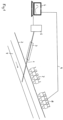
- a device in the embodiment according to 1 has an optical waveguide 1, which is positioned with its entrance in a vein 4 close to the surface by means of a peripheral venous catheter 3 (e.g. a brown tube) fixed on the skin 2 of a patient.
- the output of the optical waveguide 1 is connected to a highly sensitive fluorescence spectrometer or Ramann spectrometer 5 which is matched in terms of its spectral position and sensitivity.
- the spectrometer 5 is in turn connected to a computer 6 .
- An LD/LED field 7 for example 6 cm long and 1.5 cm wide, is arranged on the outside of the skin 2 along the vein 4 in such a way that the input surface of the optical waveguide 1 is approx. 10 mm into the radiation field of the LD/LED field 7 protrudes.
- ICG Indocyan green
- An amount of ICG adapted to the patient's body weight is infused into the bloodstream through a peripheral infusion catheter. If tumor cells are still present in the bloodstream, they will accumulate the photosensitizer more than normal blood cells because they have a higher metabolic rate.
- the peripheral venous catheter 3 is placed, e.g. in the vena basilica or another superficial arm or hand vein 4.
- the optical waveguide 1 is inserted into the venous catheter 3 and placed as described above.
- ICG is optically excited by a radiation with a wavelength of 780 nm.
- the LD/LED field 7 emits infrared radiation with precisely this wavelength. If this is switched on, the ICG distributed evenly in the bloodstream is activated.
- the resulting fluorescence, phosphorescence and luminescence radiation enters the radiation entry surface of the optical waveguide 1 and is conducted from there to the spectrometer 5 .
- This radiation is analyzed in the spectrometer 5 and presented as a functional relationship between the radiation intensity and the wavelength.
- the base spectrum generated in this way is subtracted from itself by means of suitable software, so that a difference spectrum is generated and displayed in the connected computer 6, the intensity of which is equal to zero as a function of the wavelength.
- a tumor cell enriched with ICG or a tumor cell cluster flows into the radiation area of the LD/LED field 7, it emits fluorescence, phosphorescence and luminescence radiation, the intensities of which are always greater than zero and therefore appear as intensity peaks in the spectrum.
- the spectrum is measured continuously over a period of time t1, which is chosen such that the entire blood volume has passed the optical waveguide 1 and the radiation field of the LD/LED field 7 several times, preferably two to three times.
- the spectrum registered for the measurement period t1 shows peaks at 830 nm (fluorescence radiation) and/or 945 nm (phosphorescence radiation) and/or 1245 nm (luminescence radiation), this indicates the presence of vagrant tumor cells in the bloodstream.
- the photodynamic diagnostics (PDD) of the blood flow is thus completed.
- PDT photodynamic therapy
- the emission of which corresponds exactly to the wavelength of the fluorescence radiation detected in the PDD, namely 830 nm is set.
- the activation of the LD/LED field 8 for a treatment period t1 takes place under computer control when a radiation peak is registered in the computer 6, for which purpose the computer 6 and the LD/LED field 8 are connected to one another via a line 9.
- the 830 nm radiation which is only slightly absorbed in the skin 2 , enters the bloodstream with almost full intensity and destroys the tumor cells vagrant there.
- a radiation field of the LD/LED field 8 with a length of approx. 10 cm results from scattering processes.
- an average venous flow rate of approx. 10 cm/s e.g. vena femoralis superior
- Assuming an average venous flow rate of 400-900 ml/min. results in a treatment time t1 7-15 min for one run, ie an effective treatment time of 15-30 min.
- the result of the PDT can then be checked immediately by performing the blood spectral analysis (PDD) described above again.
- PDT blood spectral analysis
- the diagnosed tumor cells can of course also be removed in a later treatment step using the treatment methods that have been introduced in oncology (chemotherapy, radiotherapy).
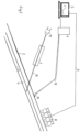
- a device has an optical waveguide 10, which is positioned with its outlet in a vein 4 close to the surface by means of a peripheral venous catheter 3 (e.g. a brown tube) fixed on the skin 2 of a patient.
- An LD or LED 11 is integrated into the input of the optical waveguide 10 as a radiation source.
- the output of the optical waveguide 12 is connected to a highly sensitive fluorescence spectrometer or Ramann spectrometer 5 which is adapted according to the invention in terms of its spectral position and sensitivity.
- the spectrometer 5 is in turn connected to a computer 6 .
- Chlorin e6 is used as a photosensitizer.
- a quantity of chlorin e6 adapted to the patient's body weight is infused into the bloodstream through a peripheral infusion catheter. If tumor cells are still present in the bloodstream, they will accumulate the photosensitizer more than normal blood cells because they have a higher metabolic rate.
- the peripheral venous catheter 3 is placed, e.g. in the vena basilica or another superficial arm or hand vein 4.
- the optical waveguide 10 is inserted into the venous catheter 3 and placed as described above.
- chlorin e6 is optically excited by light having a wavelength of 405 nm (purple blue).
- the LD/LED 11 integrated into the optical waveguide 10 emits light of this wavelength at a power of 50 mW. If this is switched on, the chlorin e6, which is evenly distributed in the bloodstream, is activated.
- the resulting fluorescence, phosphorescence and luminescence radiation enters the input surface of the optical waveguide 12 and is conducted from there to the spectrometer 5 .
- This radiation is analyzed in the spectrometer 5 and presented as a functional relationship between the radiation intensity and the wavelength.
- the base spectrum generated in this way is subtracted from itself using suitable software, so that a difference spectrum is generated and displayed in a connected computer 6, the intensity of which as a function of wavelength is equal to zero.
- a tumor cell enriched with chlorin e6 or a tumor cell cluster flows into the intravascular 405 nm radiation field of the optical waveguide 10, it emits fluorescence, phosphorescence and luminescence radiation, the intensities of which are always greater than zero and consequently appear as intensity peaks in the spectrum.
- the spectrum is measured continuously over a period of time t1, which is selected in such a way that the entire blood volume has passed the intravascular 405 nm radiation field several times (two to three times).
- the spectrum registered for the measurement period t1 shows peaks at 660 nm (fluorescence radiation) and/or 840 nm (phosphorescence radiation) and/or 1245 nm (luminescence radiation), this indicates the presence of vagrant tumor cells in the bloodstream.
- the photodynamic diagnostics (PDD) of the blood flow is thus completed.
- PDT photodynamic therapy
- the emission of which corresponds exactly to the wavelength of the fluorescence radiation detected in the PDD, namely 660 nm is set.
- the activation of the LD/LED field 8 for a treatment period t1 takes place under computer control when a radiation peak is registered in the computer 6, for which purpose the computer 6 and the LD/LED field 8 are connected to one another via a line 9.
- the 660 nm radiation which is only slightly absorbed in the skin 2 , enters the bloodstream with almost full intensity and destroys the tumor cells vagrant there.
- a radiation field of the LD/LED field 8 with a length of approx. 10 cm results from scattering processes.
- an average venous flow rate of approx. 10 cm/s (eg vena femoralis superior) results in a dwell time of a tumor cell in the radiation field of 1 s.
- the result of the PDT can then be checked immediately by performing the blood spectral analysis (PDD) described above again.
- PDT blood spectral analysis
- the diagnosed tumor cells can of course also be removed in a later treatment step using the treatment methods that have been introduced in oncology (chemotherapy, radiotherapy).
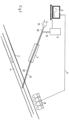
- a device in the embodiment according to 3 has a first optical waveguide 10 which is positioned with its outlet in a vein 4 close to the surface by means of a peripheral venous catheter 3 (e.g. a brown tube) fixed on the skin 2 of a patient.
- An LD or LED 11 is integrated into the input of the first optical waveguide 10 as a radiation source.
- a second optical fiber 13 is positioned intravascularly in the vein 4 parallel to the first optical fiber 10 such that its entrance is in the light field of the first optical fiber 10 and absorbs the radiation produced by optical stimulation.
- the output of the optical waveguide 13 is connected to a highly sensitive fluorescence spectrometer or Ramann spectrometer 5 which is adapted according to the invention in terms of its spectral position and sensitivity.
- the spectrometer 5 is in turn connected to a computer 6 .
- 5-alpha levulinic acid is used as a photosensitizer.
- a quantity of 5-ALA adapted to the patient's body weight is infused into the bloodstream through a peripheral infusion catheter. If tumor cells are still present in the bloodstream, they will accumulate the photosensitizer more than normal blood cells because they have a higher metabolic rate.
- the peripheral venous catheter 3 is placed, e.g. in the vena basilica or other superficial arm or hand veins 4.
- the two optical waveguides 10 and 13 are inserted into the venous catheter 3 and placed as described above.
- 5-ALA is optically excited by light having a wavelength of 405 nm (purple blue).
- the LD/LED 11 integrated into the optical waveguide 10 emits light of this wavelength at a power of 50 mW. If this is switched on, the 5-ALA distributed evenly in the bloodstream is activated.
- the resulting fluorescence, phosphorescence and luminescence radiation enters the input surface of the optical waveguide 13 and is conducted from there to the spectrometer 5 .
- This radiation is analyzed in the spectrometer 5 and presented as a functional relationship between the radiation intensity and the wavelength.
- the base spectrum generated in this way is subtracted from itself using suitable software, so that a difference spectrum is generated and displayed in the connected computer 6, the intensity of which as a function of the wavelength is zero.
- a tumor cell enriched with 5-ALA or a tumor cell cluster flows into the intravascular 405 nm radiation field, it emits fluorescence, phosphorescence and luminescence radiation, the intensities of which are always greater than zero and consequently appear as intensity peaks in the spectrum.
- the spectrum is measured continuously over a period of time t1, which is chosen such that the entire blood volume has passed the intravascular 405 nm radiation field several times (two to three times) (see above between 15 min-30 min).
- the spectrum registered for the measurement period t1 shows peaks at 630 nm (fluorescence radiation) and/or 780 nm (phosphorescence radiation) and/or 1245 nm (luminescence radiation), this indicates the presence of vagrant tumor cells in the bloodstream.
- the photodynamic diagnostics (PDD) of the blood flow is thus completed.
- PDT photodynamic therapy
- the 630 nm radiation of high power density enters the bloodstream and destroys the vagrant tumor cells.
- the result of the PDT can then be checked immediately by performing the blood spectral analysis (PDD) described above again.
- PDT blood spectral analysis
- the diagnosed tumor cells can of course also be removed in a later treatment step using the treatment methods that have been introduced in oncology (chemotherapy, radiotherapy).
- a device in the embodiment according to 4 has an LD/LED field 7 arranged along a vein 4 on the outside of the skin 2 of a patient, for example 6 cm long and 1.5 cm wide.
- the entrance of an optical waveguide 12 sits on the skin 2.
- the entrance is designed conically in order to enlarge the radiation entry surface.
- the output of the optical waveguide 12 is connected to a highly sensitive fluorescence spectrometer or Ramann spectrometer 5 which is adapted according to the invention in terms of its spectral position and sensitivity.
- the spectrometer 5 is in turn connected to a computer 6 .
- a further LD/LED field 8 is arranged downstream of the optical waveguide 12 in the blood flow direction and is connected to the computer 5 via a line 9 .
- a tumor cell enriched with a photosensitizer is stimulated by means of the LD/LED field 7, which emits a corresponding radiation.
- the resulting fluorescence, phosphorescence and luminescence radiation enters the input surface of the optical waveguide 12 and is conducted from there to the spectrometer 5 .
- the diagnostic detection of a tumor cell and its elimination is carried out in the same way as described above.
- One of the photosensitizers mentioned above or another suitable photosensitizer can be used as the photosensitizer. This also applies to the exemplary embodiments described above, which are not limited to the photosensitizers mentioned.
Landscapes
- Health & Medical Sciences (AREA)
- Life Sciences & Earth Sciences (AREA)
- Physics & Mathematics (AREA)
- Engineering & Computer Science (AREA)
- Biomedical Technology (AREA)
- Veterinary Medicine (AREA)
- Animal Behavior & Ethology (AREA)
- Biophysics (AREA)
- Public Health (AREA)
- General Health & Medical Sciences (AREA)
- Pathology (AREA)
- Molecular Biology (AREA)
- Surgery (AREA)
- Medical Informatics (AREA)
- Heart & Thoracic Surgery (AREA)
- Spectroscopy & Molecular Physics (AREA)
- Optics & Photonics (AREA)
- Nuclear Medicine, Radiotherapy & Molecular Imaging (AREA)
- Radiology & Medical Imaging (AREA)
- Vascular Medicine (AREA)
- Investigating, Analyzing Materials By Fluorescence Or Luminescence (AREA)
- Radiation-Therapy Devices (AREA)
Description
- Die Erfindung betrifft eine Vorrichtung zur Registrierung und zur Zerstörung einzelner Tumorzellen, Tumorzellencluster und Mikrometastasen im Blutkreislauf, die mit einer photoempfindlichen Substanz angereichert sind.
- Mit den heute in der onkologischen Routinediagnostik zur Verfügung stehenden biophysikalischen Methoden, wie Kernspinresonanz (MRT; fMRI), Positronenemissiontomographie (PET), Ultraschall (US), Computertomographie (CT) und Röntgen sind Tumoren direkt nachweisbar, wenn sie im Mittel eine Tumormasse von 1 g aufweisen. Dies entspricht ca. 1 Milliarde (10 9) Tumorzellen. Erst ab dieser Größe lassen sich auch Gewebeproben direkt entnehmen (Biopsie) und zu gesicherter Diagnostik verwenden.
- Eine Tumormasse von 1 g stellt mithin die derzeit gültige diagnostische Schwelle dar. Die an dieser diagnostischen Schwelle ermittelten Befunde gelten onkologisch als "Früherkennung" von Tumorwachstum bzw. Metastasierung.
- Mittels immunologischer Methoden lassen sich Tumoren nachweisen, die eine Tumormasse von 1 mg aufweisen und entsprechend aus ca. 1 Million (106) Tumorzellen bestehen. Diese Methoden sind indirekter Natur, da sie auf der Bestimmung von Markersubstanzen beruhen. Tumormarker sind Substanzen, die von malignen Tumorzellen direkt gebildet werden oder deren Synthese in Nicht-Tumorzellen durch Tumorzellen induziert wird und die auch bei gesunden Personen vorkommen.
- Treten Tumormarker in erhöhter Konzentration im Blut oder in anderen Körperflüssigkeiten (humorale Tumormarker) bzw. in oder auf Zellen (zelluläre Tumormarker) auf, ermöglichen sie Rückschlüsse auf das Vorliegen, den Verlauf und die Prognose einer Tumorerkrankung. Diese Tumormarker sind generell unspezifisch. So indiziert eine hohe Konzentration des Tumormarkers CEA im Blutbild, dass ein Lungenkarzinom oder ein Leberkarzinom oder ein Dickdarmkarzinom oder ein Schilddrüsenkarzinom oder ein Mammakarzinom oder ein Magenkarzinom vorliegen könnte.
- Die nach einer Operation praktisch unvermeidbar noch im Körper vorhanden Tumorzellen bzw. Mikrometastasen können später Rückfälle verursachen. Weder während einer Operation noch danach lassen sich mittels Ultraschall oder Computer-oder Magnetresonanztomographie nach gegenwärtigem Stand der Technik solche kleinen Zellnester (so genannte Mikrometastasen) nachweisen .Die Erfahrung besagt jedoch, dass in diesem Krankheitsstadium, also nach beendeter Chemotherapie, das Risiko für einen Tumorrückfall und Metastasenbildung für praktisch alle Krebspatienten von relevanter Größenordnung und keineswegs vernachlässigbar klein ist. Die Medizin, insbesondere die Onkologie, bietet gegenwärtig für diese Krankheitsphase lediglich das Prinzip "hoffnungsvolle Beobachtung" an. Therapeutische Optionen sind nicht vorhanden, da eine diagnostische Grundlage dafür fehlt.
- Das Dokument
US2009213362 offenbart eine Vorrichtung, die eine diagnostische Erkennung von Tumorzellen im Blutkreislauf von Menschen und Säugetieren als auch deren therapeutische in-situ-Eliminierung unter Verwendung derselben Stahlungsquelle ermöglicht. Das DokumentUS2014031647 beschreibt eine Vorrichtung zur Erkennung von Tumorzellen im Blut unter Verwendung von zwei in Blutflussrichtung angeordneten Strahlungsquellen. - Es ist daher Aufgabe der vorliegenden Erfindung, eine Vorrichtung zur Verfügung zu stellen, die sowohl die sichere diagnostische Erkennung einzelner Tumorzellen im Blutkreislauf von Menschen und Säugetieren als auch deren therapeutische in-situ-Eliminierung ermöglicht.
- Grundlage der vorliegenden Erfindung ist die in der Krebstherapie international erfolgreich eingesetzte, schulmedizinisch anerkannte photodynamische Diagnostik (PDD) sowie die photodynamische Therapie (PDT).
- Die PDD und auch die PDT beruhen auf der optischen Anregung photoempfindlicher Substanzen, die dem Organismus topisch oder systemisch per Infusion zugeführt werden und die sich in Tumorzellen anreichern, da diese, unabhängig von der Tumorart, immer eine höhere Zellstoffwechselrate als normale, gesunde Zellen aufweisen.
- Die optische Anregung derartiger lichtempfindlicher Substanzen (Photosensitizer) führt zur Bildung von reaktivem Sauerstoff (Singulet-Sauerstoff) in der Tumorzelle und zur oxidativen Zerstörung von essentiellen Zellbestandteilen (Mitochondrien, Zellwände) und endet mit einem nekrotischen oder apoptotischen Zelluntergang. Dieser Vorgang wird theoretisch durch das Jablonski-Diagramm beschrieben. (A. Jablonski: Efficiency of anti-Stokes fluorecence in dyes, Nature Bd 131, (1933), 839-840)
- Das Jablonski-Diagramm besagt, dass der Prozess der Bildung reaktiver oxidativer Spezies durch die Entstehung und Emission von Photonen begleitet und charakterisiert ist. So entsteht bei der Relaxation des jeweiligen lichtempfindlichen Moleküls vom einem ersten angeregten Zustand (single state) in den Grundzustand Fluoreszenzstrahlung, beim Übergang vom einem weiteren angeregten Zustand (triplet state) in den Grundzustand Phosphoreszenzstrahlung und bei der Relaxation von angeregtem Singulet-Sauerstoff zu molekularem Sauerstoff charakteristische Lumineszenzstrahlung.
- Diese drei verschiedenen Photonenemissionen stellen ein Charakteristikum für einen photodynamischen Prozess (PDD und PDT) dar und sind der eindeutige physikalische Nachweis eines aktivierten photodynamischen Vorgangs. Dieser Vorgang spielt sich in jeder Tumorzelle ab, die mit einem Photosensitizer angereichert und optisch aktiviert wurde, darüber hinaus natürlich auch in jeder Mikrometastase und in jedem makroskopischen Tumor, unabhängig von der Tumorart.
- Die Intensität der emittierten Strahlung ist direkt proportional zu der Anzahl der photodynamisch aktivierten Tumorzellen.
- Mittels moderner optischer Detektoren ist beim gegenwärtigen Stand der Technik der Nachweis einzelner Photonen möglich (A. Korneev, G. Goltsman, W. H. P. Pernice "Single Photon Counting: Photonic integration meets single photon detection" LaserFocus World V.51Is.5.05/05/2015 ).
- Für den Nachweis von Einzelphotonen werden Detektoren mit hohem Gain eingesetzt. Dazu gehören Photomultiplier (PMT) und Avalanche Photodioden (APD).
- Die oben gestellte Aufgabe wird erfindungsgemäß mit einer Vorrichtung gelöst, die die Merkmale des Anspruchs 1 aufweist.
- Damit wird die diagnostische Schwelle der Tumoridentifikation und Tumortherapie drastisch abgesenkt, da die Diagnose und Therapie von einzelnen Tumorzellen oder Mikrometastasen möglich ist. Es kann eine Tumorfrüherkennung im wahrsten Wortsinne durchgeführt werden. Dies gilt generell für alle Patienten mit erhöhtem familiärem Krebsrisiko im Sinne einer prophylaktischen Therapie als auch für alle Tumorpatienten nach adjuvanter oder palliativer Chemo-/Strahlentherapie und für die Veterinärmedizin.
- Die Erfindung wird nachstehend an Ausführungsbeispielen näher erläutert. In der dazu gehörigen Zeichnung zeigt in schematischer Weise:
- Fig. 1
- eine erste Ausführungsform einer erfindungsgemäßen Vorrichtung mit extravasaler Anregung von Tumorzellen und intravasaler Detektion der emittierten Strahlung,
- Fig. 2
- eine zweite Ausführungsform einer erfindungsgemäßen Vorrichtung mit intravasaler Anregung von Tumorzellen und extravasaler Detektion der emittierten Strahlung,
- Fig. 3
- eine dritte Ausführungsform einer erfindungsgemäßen Vorrichtung, mit intravasaler Anregung von Tumorzellen und intravasaler Detektion der emittierten Strahlung, und
- Fig. 4
- eine vierte Ausführungsform einer erfindungsgemäßen Vorrichtung, mit extravasaler Anregung von Tumorzellen und extravasaler Detektion der emittierten Strahlung.
- In der Ausführungsform gemäß
Fig. 1 weist eine erfindungsgemäße Vorrichtung einen Lichtwellenleiter 1 auf, der mittels eines geeigneten auf der Haut 2 eines Patienten festgelegten peripheren Venenkatheters 3 (z. B. einer Braunüle) mit seinem Eingang in einer oberflächennahen Vene 4 positioniert ist. Der Ausgang des Lichtwellenleiters 1 ist mit einem hinsichtlich seiner spektralen Lage und Empfindlichkeit angepassten, hochempfindlichen Fluoreszenz-Spektrometer oder Ramann-Spektrometer 5 verbunden. Das Spektrometer 5 ist seinerseits an einen Computer 6 angeschlossen. Entlang der Vene 4 ist außen auf der Haut 2 ein beispielsweise 6 cm langes und 1,5 cm breites LD/LED-Feld 7 so angeordnet, dass die Eingangsfläche des Lichtwellenleiters 1 ca. 10 mm in das Strahlungsfeld des LD/LED-Feldes 7 hineinragt. - Gearbeitet wird mit der beschriebenen Vorrichtung wie folgt:
Als Photosensitizer kommt Indocyangrün (ICG) zum Einsatz. Durch einen peripheren Infusionskatheter wird eine dem Körpergewicht des Patienten angepasste Menge ICG in den Blutkreislauf infundiert. Sind in dem Blutkreislauf noch Tumorzellen vorhanden, werden diese den Photosensitizer im Vergleich zu den normalen Blutzellen stärker anreichern, da sie über eine höhere Stoffwechselrate verfügen. - Nach einer Einwirkzeit von ca. 3 Stunden wird der periphere Venenkatheter 3 gelegt, bspw. in die Vena basilica oder eine andere oberflächliche Arm- oder Handvene 4. In den Venenkatheter 3 wird der Lichtwellenleiter 1 eingeführt und wie oben beschrieben plaziert.
- Es ist bekannt, dass ICG durch eine Strahlung mit einer Wellenlänge von 780 nm optisch angeregt wird. Eine Infrarotstrahlung mit eben dieser Wellenlänge emittiert das LD/LED-Feld 7. Wird dieses eingeschaltet, wird das im Blutstrom gleichmäßig verteilte ICG aktiviert. Die dabei entstehende Fluoreszenz-, Phosphoreszenz-, und Lumineszenzstrahlung tritt in die Strahlungseintrittsfläche des Lichtwellenleiters 1 ein und wird von dort zum Spektrometer 5 geleitet. Im Spektrometer 5 wird diese Strahlung analysiert und als funktionaler Zusammenhang der Strahlungsintensität über der Wellenlänge dargestellt. Das auf dies Weise generierte Basisspektrum wird mittels einer geeigneten Software von sich selbst subtrahiert, so dass ein Differenzspektrum erzeugt und in dem angeschlossenen Computer 6 angezeigt wird, dessen Intensität als Funktion der Wellenlänge gleich Null ist.
- Strömt nun eine mit ICG angereicherte Tumorzelle oder ein Tumorzellencluster in den Strahlungsbereich des LD/LED-Feldes 7 ein, emittiert diese Fluoreszenz-, Phosphoreszenz- und Lumineszenzstrahlung, deren Intensitäten in jedem Fall größer sind als Null und demzufolge als Intensitätspeaks im Spektrum erscheinen.
- Die Messung des Spektrums erfolgt kontinuierlich über einen Zeitraum t1, der so gewählt wird, dass das gesamte Blutvolumen mehrere Mal, vorzugsweise zwei- bis dreimal, den Lichtwellenleiter 1 und das Strahlungsfeld des LD/LED- Feldes 7 passiert hat.
- Weist das für den Messzeitraum t1 registrierte Spektrum keine Peaks auf, ist das ein sicheres Zeichen, dass keine über der Basisintensität liegende Strahlung detektiert wurde und demzufolge keine im Blutstrom vagabundierenden Tumorzellen vorhanden sind.
- Weist das für den Messzeitraum t1 registrierte Spektrum dagegen Peaks bei 830 nm (Fluoreszenzstrahlung) und/oder 945 nm (Phosphoreszenzstrahlung) und/oder 1245 nm (Lumineszenzstrahlung) auf, deutet dies auf das Vorhandensein von vagabundierenden Tumorzellen im Blutstrom hin. Die photodynamische Diagnostik (PDD) des Blutstromes ist damit beendet.
- Sind in der PDD Strahlungspeaks nachgewiesen, wird zur Zerstörung dieser Tumorzellen die photodynamische Therapie (PDT) angewendet, indem ein weiteres, beispielsweise. 6 cm langes und 1,5 cm breites LD/LED-Feld 8 außen, entlang der Vene 4 in Blutflussrichtung hinter dem LD/LED-Feld 7 angeordnet wird, dessen Emission genau auf die Wellenlänge der in der PDD detektierten Fluoreszenzstrahlung, nämlich 830 nm, eingestellt ist. Die Aktivierung des LD/LED-Feld 8 für einen Behandlungszeitraum t1 erfolgt rechnergesteuert bei Registrierung eines Strahlungspeaks im Computer 6, wozu der Computer 6 und das LD/LED-Feld 8 über eine Leitung 9 miteinander verbunden sind. Die nur wenig in der Haut 2 absorbierte 830 nm Strahlung tritt mit nahezu voller Intensität in die Blutbahn ein und zerstört die dort vagabundierenden Tumorzellen.
- Durch Streuvorgänge resultiert ein Strahlungsfeld des LD/LED-Feldes 8 von ca. 10 cm Länge. Bei einer mittleren venösen Fließgeschwindigkeit von ca. 10 cm/s ( z.B. vena femoralis superior ) resultiert eine Aufenthaltszeit einer Tumorzelle im Strahlungsfeld von 1 s. Bei einer angenommenen mittleren venösen Flussrate von 400 -900 ml/min. resultiert ein Behandlungszeitraum t1 = 7-15 min für einen Durchlauf, d.h. eine effektive Behandlungszeit von 15 -30 min.
- Das Ergebnis der PDT kann danach unmittelbar überprüft werden, indem die oben beschriebene spektrale Blutanalyse (PDD) erneut durchgeführt wird.
- Alternativ kann die Entfernung der diagnostizierten Tumorzellen natürlich auch in einem späteren Behandlungsschritt mit den Behandlungsmethoden erfolgen, die in der Onkologie eingeführt sind (Chemotherapie, Strahlentherapie).
- In der Ausführungsform gemäß
Fig. 2 weist eine erfindungsgemäße Vorrichtung einen Lichtwellenleiter 10 auf, der mittels eines geeigneten auf der Haut 2 eines Patienten festgelegten peripheren Venenkatheters 3 (z. B. einer Braunüle) mit seinem Ausgang in einer oberflächennahen Vene 4 positioniert ist. In den Eingang des Lichtwellenleiters 10 ist eine LD oder LED 11 als Strahlungsquelle eingebunden. Über dem Ausgang des intravenösen Lichtwellenleiters 10 ist auf der Haut 2 des Patienten der Eingang eines weiteren Lichtwellenleiters 12 fixiert, der konisch gestaltet ist, um die Strahlungseintrittssläche zu vergrößern. Der Ausgang des Lichtwellenleiters 12 ist mit einem hinsichtlich seiner spektralen Lage und Empfindlichkeit erfindungsgemäß angepassten, hochempfindlichen Fluoreszenz-Spektrometer oder Ramann-Spektrometer 5 verbunden. Das Spektrometer 5 ist seinerseits an einen Computer 6 angeschlossen. - Gearbeitet wird mit der beschriebenen Vorrichtung wie folgt:
Als Photosensitizer kommt Chlorin e6 zum Einsatz. Durch einen peripheren Infusionskatheter wird eine dem Körpergewicht des Patienten angepasste Menge Chlorin e6 in den Blutkreislauf infundiert. Sind in dem Blutkreislauf noch Tumorzellen vorhanden, werden diese den Photosensitizer im Vergleich zu den normalen Blutzellen stärker anreichern, da sie über eine höhere Stoffwechselrate verfügen. - Nach einer Einwirkzeit von ca. 3 Stunden wird der periphere Venenkatheter 3 gelegt, bspw. in die Vena basilica oder eine andere oberflächliche Arm- oder Handvene 4. In den Venenkatheter 3 wird der Lichtwellenleiter 10 eingeführt und wie oben beschrieben plaziert.
- Es ist bekannt, dass Chlorin e6 durch Licht mit einer Wellenlänge von 405 nm (purpurblau) optisch angeregt wird. Licht dieser Wellenlänge emittiert die in den Lichtwellenleiter 10 eingebundene LD/LED 11 bei einer Leistung von 50 mW. Wird diese eingeschaltet, wird das im Blutstrom gleichmäßig verteilte Chlorin e6 aktiviert. Die dabei entstehende Fluoreszenz-, Phosphoreszenz-,und Lumineszenzstrahlung tritt in die Eingangsfläche des Lichtwellenleiters 12 ein und wird von dort zum Spektrometer 5 geleitet. Im Spektrometer 5 wird diese Strahlung analysiert und als funktionaler Zusammenhang der Strahlungsintensität über der Wellenlänge dargestellt. Das auf dies Weise generierte Basisspektrum wird mittels einer geeigneten Software von sich selbst subtrahiert, so dass ein Differenzspektrum erzeugt und in einem angeschlossenen Computer 6 angezeigt wird, dessen Intensität als Funktion der Wellenlänge gleich Null ist.
- Strömt nun eine mit Chlorin e6 angereicherte Tumorzelle oder ein Tumorzellencluster in das intravasale 405 nm Strahlungsfeld des Lichtwellenleiters 10 ein , emittiert diese Fluoreszenz-, Phosphoreszenz-und Lumineszenzstrahlung, deren Intensitäten in jedem Fall größer sind als Null und demzufolge als Intensitätspeaks im Spektrum erscheinen.
- Die Messung des Spektrums erfolgt kontinuierlich über einen Zeitraum t1, der so gewählt wird, dass das gesamte Blutvolumen mehrere Male (zweibis dreimal) das intravasale 405nm Strahlungsfeld passiert hat.
- Weist das für den Messzeitraum t1 registrierte Spektrum keine Peaks auf, ist das ein sicheres Zeichen, dass keine über der Basisintensität liegende Strahlung detektiert wurde und demzufolge keine im Blutstrom vagabundierenden Tumorzellen vorhanden sind.
- Weist das für den Messzeitraum t1 registrierte Spektrum dagegen Peaks bei 660 nm (Fluoreszenzstrahlung) und/oder 840 nm (Phosphoreszenzstrahlung) und/oder 1245 nm (Lumineszenzstrahlung) auf, deutet dies auf das Vorhandensein von vagabundierenden Tumorzellen im Blutstrom hin. Die photodynamische Diagnostik (PDD) des Blutstromes ist damit beendet.
- Sind in der PDD Strahlungspeaks nachgewiesen, wird zur Zerstörung dieser Tumorzellen die photodynamische Therapie (PDT) angewendet, indem ein beispielsweise. 6 cm langes und 1,5 cm breites LD/LED-Feld 8 au-ßen, entlang der Vene 4 in Blutflussrichtung hinter dem Strahlungsfeld des Lichtwellenleiters 10 angeordnet wird, dessen Emission genau auf die Wellenlänge der in der PDD detektierten Fluoreszenzstrahlung, nämlich 660 nm, eingestellt ist. Die Aktivierung des LD/LED-Feld 8 für einen Behandlungszeitraum t1 erfolgt rechnergesteuert bei Registrierung eines Strahlungspeaks im Computer 6, wozu der Computer 6 und das LD/LED-Feld 8 über eine Leitung 9 miteinander verbunden sind. Die nur wenig in der Haut 2 absorbierte 660 nm Strahlung tritt mit nahezu voller Intensität in die Blutbahn ein und zerstört die dort vagabundierenden Tumorzellen.
- Durch Streuvorgänge resultiert ein Strahlungsfeld des LD/LED-Feldes 8 von ca. 10 cm Länge. Bei einer mittleren venösen Fließgeschwindigkeit von ca. 10 cm/s (z.B. vena femoralis superior) resultiert eine Aufenthaltszeit einer Tumorzelle im Strahlungsfeld von 1 s. Bei einer angenommenen mittleren venösen Flussrate von 400 -900 ml/min. resultiert ein Behandlungszeitraum t1 = 7-15 min für einen Durchlauf, d.h. eine effektive Behandlungszeit von 15 bis 30 min.
- Das Ergebnis der PDT kann danach unmittelbar überprüft werden, indem die oben beschriebene spektrale Blutanalyse (PDD) erneut durchgeführt wird.
- Alternativ kann die Entfernung der diagnostizierten Tumorzellen natürlich auch in einem späteren Behandlungsschritt mit den Behandlungsmethoden erfolgen, die in der Onkologie eingeführt sind (Chemotherapie, Strahlentherapie).
- In der Ausführungsform gemäß
Fig. 3 weist eine erfindungsgemäße Vorrichtung einen ersten Lichtwellenleiter 10 auf, der mittels eines geeigneten auf der Haut 2 eines Patienten festgelegten peripheren Venenkatheters 3 (z. B. einer Braunüle) mit seinem Ausgang in einer oberflächennahen Vene 4 positioniert ist. In den Eingang des ersten Lichtwellenleiters 10 ist eine LD oder LED 11 als Strahlungsquelle eingebunden. Mittels des auf der Haut 2 des Patienten festgelegten peripheren Venenkatheters 3 ist ein zweiter Lichtwellenleiter 13 parallel zum ersten Lichtwellenleiter 10 intravasal derart in der Vene 4 positioniert, dass sein Eingang im Lichtfeld des ersten Lichtwellenleiters 10 liegt und die durch optische Anregung entstehende Strahlung aufnimmt. - Der Ausgang des Lichtwellenleiters 13 ist mit einem hinsichtlich seiner spektralen Lage und Empfindlichkeit erfindungsgemäß angepassten, hochempfindlichen Fluoreszenz-Spektrometer oder Ramann-Spektrometer 5 verbunden. Das Spektrometer 5 ist seinerseits an einen Computer 6 angeschlossen.
- Gearbeitet wird mit der beschriebenen Vorrichtung wie folgt:
Als Photosensitizer kommt 5-alpha Lävulinsäure (5-ALA) zum Einsatz. Durch einen peripheren Infusionskatheter wird eine dem Körpergewicht des Patienten angepasste Menge 5-ALA in den Blutkreislauf infundiert. Sind in dem Blutkreislauf noch Tumorzellen vorhanden, werden diese den Photosensitizer im Vergleich zu den normalen Blutzellen stärker anreichern, da sie über eine höhere Stoffwechselrate verfügen. - Nach einer Einwirkzeit von ca. 3 Stunden wird der periphere Venenkatheter 3 gelegt, bspw. in die Vena basilica oder andere, oberflächliche Arm- oder Handvenen 4. In den Venenkatheter 3 werden die beiden Lichtwellenleiter 10 und 13 eingeführt und wie oben beschrieben platziert.
- Es ist bekannt, dass 5-ALA durch Licht mit einer Wellenlänge von 405 nm (purpurblau) optisch angeregt wird. Licht dieser Wellenlänge emittiert die in den Lichtwellenleiter 10 eingebunden LD/LED 11 bei einer Leistung von 50 mW. Wird diese eingeschaltet, wird das im Blutstrom gleichmäßig verteilte 5-ALA aktiviert. Die dabei entstehende Fluoreszenz-, Phosphoreszenz- und Lumineszenzstrahlung tritt in die Eingangsfläche des Lichtwellenleiters 13 ein und wird von dort zum Spektrometer 5 geleitet. Im Spektrometer 5 wird diese Strahlung analysiert und als funktionaler Zusammenhang der Strahlungsintensität über der Wellenlänge dargestellt. Das auf dies Weise generierte Basisspektrum wird mittels einer geeigneten Software von sich selbst subtrahiert, so dass ein Differenzspektrum erzeugt und in dem angeschlossenen Computer 6 angezeigt wird, dessen Intensität als Funktion der Wellenlänge = Null ist.
- Strömt nun eine mit 5-ALA angereicherte Tumorzelle oder ein Tumorzellencluster in das intravasale 405 nm Strahlungsfeld ein , emittiert diese Fluoreszenz-, Phosphoreszenz- und Lumineszenzstrahlung, deren Intensitäten in jedem Fall größer sind als Null und demzufolge als Intensitätspeaks im Spektrum erscheinen.
- Die Messung des Spektrums erfolgt kontinuierlich über einen Zeitraum t1, der so gewählt wird, dass das gesamte Blutvolumen mehrere Male (zwei bis dreimal) das intravasale 405 nm Strahlungsfeld passiert hat (siehe oben zwischen 15 min-30 min).
- Weist das für den Messzeitraum t1 registrierte Spektrum keine Peaks auf, ist das ein sicheres Zeichen, dass keine über der Basisintensität liegende Strahlung detektiert wurde und demzufolge keine im Blutstrom vagabundierenden Tumorzellen vorhanden sind.
- Weist das für den Messzeitraum t1 registrierte Spektrum dagegen Peaks bei 630 nm (Fluoreszenzstrahlung) und/oder 780 nm (Phosphoreszenzstrahlung) und/oder 1245 nm (Lumineszenzstrahlung) auf, deutet dies auf das Vorhandensein von vagabundierenden Tumorzellen im Blutstrom hin. Die photodynamische Diagnostik (PDD) des Blutstromes ist damit beendet.
- Sind in der PDD Strahlungspeaks nachgewiesen, wird zur Zerstörung dieser Tumorzellen die photodynamische Therapie (PDT) angewandt, indem ein bspw. 6 cm langes und 1,5 cm breites extravasal außen, entlang der gro-ßen, oberflächlichen Vene 4 in Blutflussrichtung nach dem intravasalen 405 nm Strahlungsfeld des Lichtwellenleiters 10 angeordnetes LD/LED-Feld 8 mit einer Emissionswellenlänge von 630 nm und einer Leistungsdichte von 200 mW/cm2 für einen Behandlungszeitraum = t1 aktiviert wird. Die 630 nm Strahlung hoher Leistungsdichte tritt in die Blutbahn ein und zerstört die dort vagabundierenden Tumorzellen.
- Das Ergebnis der PDT kann danach unmittelbar überprüft werden, indem die oben beschriebene spektrale Blutanalyse (PDD) erneut durchgeführt wird.
- Alternativ kann die Entfernung der diagnostizierten Tumorzellen natürlich auch in einem späteren Behandlungsschritt mit den Behandlungsmethoden erfolgen, die in der Onkologie eingeführt sind ( Chemotherapie, Strahlentherapie ).
- In der Ausführungsform gemäß
Fig. 4 weist eine erfindungsgemäße Vorrichtung ein entlang einer Vene 4 außen auf der Haut 2 eines Patienten angeordnetes beispielsweise 6 cm langes und 1,5 cm breites LD/LED-Feld 7 auf. In Blutflussrichtung gesehen nach dem LD/LED-Feld 7 sitzt der Eingang eines Lichtwellenleiters 12 auf der Haut 2. Der Eingang ist konisch ausgeführt, um die Strahlungseintrittsfläche zu vergrößern. Der Ausgang des Lichtwellenleiters 12 ist mit einem hinsichtlich seiner spektralen Lage und Empfindlichkeit erfindungsgemäß angepassten, hochempfindlichen Fluoreszenz-Spektrometer oder Ramann-Spektrometer 5 verbunden. Das Spektrometer 5 ist seinerseits an einen Computer 6 angeschlossen. In Blutflussrichtung nach dem Lichtwellenleiter 12 ist ein weiteres LD/LED-Feld 8 angeordnet, welches über eine Leitung 9 mit dem Computer 5 verbunden ist. Die Anregung einer mit einem Photosensitizer angereicherten Tumorzelle erfolgt mittels des LD/LED-Feldes 7, welches eine entsprechende Strahlung emittiert. Die dabei entstehende Fluoreszenz-, Phosphoreszenz-, und Lumineszenzstrahlung tritt in die Eingangsfläche des Lichtwellenleiters 12 ein und wird von dort zum Spektrometer 5 geleitet. - Der diagnostische Nachweis einer Tumorzelle und ihre Eliminierung erfolgt in gleicher Weise wie oben beschrieben. Als Photosensitizer kann einer der oben genannten oder ein anderer geeigneter Photosensitizer zum Einsatz kommen. Das gilt auch für die zuvor beschriebenen Ausführungsbeispiele, die auf die genannten Photosensitizer nicht beschränkt sind.
Claims (2)
- Vorrichtung zur Registrierung und zur Zerstörung von einzelnen Tumorzellen, Tumorzellenclustern und Mikrometastasen im Blutkreislauf, die mit einer photoempfindlichen Substanz angereichert sind, wobei die Vorrichtung aufweist- eine Strahlungsquelle (7, 11), die zu intravasaler oder extravasaler Anregung der photoempfindlichen Substanz ausgelegt ist,- einen Lichtwellenleiter (1, 12, 13) zur intravasaler oder extravasaler Leitung einer Fluoreszenz- und/oder Phosphorenz- und/oder Lumineszenzstrahlung der angeregten Tumorzellen und/oder Tumorzellencluster und/oder Mikrometastasen- ein mit dem Lichtwellenleiter (1, 12, 13) verbundenes Fluoreszenzspektrometer (5) hoher Empfindlichkeit, das zur Registrierung der emittierten Strahlung ausgelegt ist,- einen mit dem Spektrometer (5) verbundenen Computer (6), der dazu ausgelegt ist, die empfangenen Peaks der emittierten Strahlung als Funktion der Zeit aufzuzeichnen,
wobei die Vorrichtung- eine weitere intravasal oder extravasal wirkende Strahlungsquelle (8) aufweist, die in Blutflussrichtung hinter dem Lichtwellenleiter (1, 12, 13) angeordnet und mit dem Computer (6) verbunden ist, der dazu ausgelegt ist, bei Registrierung eines Strahlungspeaks ein Einschaltsignal an die zweite Strahlungsquelle (8) auszugeben, wobei diese weitere Strahlungsquelle (8) zur Zerstörung von Tumorzellen und/oder Tumorzellencluster und/oder Mikrometastasen mittels photodynamischer Therapie durch Emission von Licht in der Wellenlänge des Strahlungspeaks ausgelegt ist. - Vorrichtung nach Anspruch 1, dadurch gekennzeichnet, dass der Lichtwellenleiter (1, 12, 13) einen konischen Eingang (14) zur Vergrößerung der Strahlungseintrittsfläche hat.
Applications Claiming Priority (2)
| Application Number | Priority Date | Filing Date | Title |
|---|---|---|---|
| DE102017129971.8A DE102017129971A1 (de) | 2017-12-14 | 2017-12-14 | Vorrichtung zur Registrierung und zur Zerstörung einzelner Tumorzellen, Tumorzellencluster und Mikrometastasen im Blutkreislauf |
| PCT/DE2018/101015 WO2019114883A1 (de) | 2017-12-14 | 2018-12-13 | Vorrichtung zur registrierung und zur zerstörung einzelner tumorzellen, tumorzellencluster und mikrometastasen im blutkreislauf |
Publications (2)
| Publication Number | Publication Date |
|---|---|
| EP3723591A1 EP3723591A1 (de) | 2020-10-21 |
| EP3723591B1 true EP3723591B1 (de) | 2023-06-14 |
Family
ID=65411692
Family Applications (1)
| Application Number | Title | Priority Date | Filing Date |
|---|---|---|---|
| EP18847188.2A Active EP3723591B1 (de) | 2017-12-14 | 2018-12-13 | Vorrichtung zur registrierung und zur zerstörung einzelner tumorzellen, tumorzellencluster und mikrometastasen im blutkreislauf |
Country Status (6)
| Country | Link |
|---|---|
| US (1) | US20210076940A1 (de) |
| EP (1) | EP3723591B1 (de) |
| CN (1) | CN111629657A (de) |
| DE (1) | DE102017129971A1 (de) |
| PL (1) | PL3723591T3 (de) |
| WO (1) | WO2019114883A1 (de) |
Families Citing this family (1)
| Publication number | Priority date | Publication date | Assignee | Title |
|---|---|---|---|---|
| AU2022234898A1 (en) | 2021-03-12 | 2023-09-28 | Stryker European Operations Limited | Neurosurgical methods and systems for detecting and removing tumorous tissue |
Family Cites Families (18)
| Publication number | Priority date | Publication date | Assignee | Title |
|---|---|---|---|---|
| WO1989002718A1 (en) * | 1987-09-24 | 1989-04-06 | Massachusetts Institute Of Technology | Catheter system for imaging |
| US5411016A (en) * | 1994-02-22 | 1995-05-02 | Scimed Life Systems, Inc. | Intravascular balloon catheter for use in combination with an angioscope |
| US6577391B1 (en) * | 1999-03-25 | 2003-06-10 | Spectrx, Inc. | Apparatus and method for determining tissue characteristics |
| DE19925210C2 (de) * | 1999-06-01 | 2001-12-20 | Alexander Hohla | Fasersonde |
| JP3862582B2 (ja) * | 2001-06-29 | 2006-12-27 | 富士フイルムホールディングス株式会社 | 蛍光画像取得方法および装置並びにプログラム |
| DE10154436A1 (de) * | 2001-11-07 | 2003-05-15 | Rodotech Gmbh | pyro-Bakteriopheophorbidderivate |
| JP4311607B2 (ja) * | 2002-05-27 | 2009-08-12 | 富士フイルム株式会社 | 蛍光診断情報生成方法および装置 |
| RU2271789C1 (ru) * | 2004-09-23 | 2006-03-20 | Государственное учреждение Межотраслевой научно-технический комплекс "Микрохирургия глаза" им. акад. С.Н. Федорова Министерства Здравоохранения Российской Федерации | Способ удаления внутриглазных новообразований |
| US20070038117A1 (en) * | 2005-07-26 | 2007-02-15 | Bala John L | Multi-spectral imaging endoscope system |
| JP4785480B2 (ja) * | 2005-09-22 | 2011-10-05 | 三鷹光器株式会社 | 光学計測システム |
| US20110060232A1 (en) * | 2007-05-04 | 2011-03-10 | The General Hospital Corporation | Retinal flow cytometry |
| US8574859B2 (en) * | 2008-03-27 | 2013-11-05 | The General Hospital Corporation | In vivo flow cytometry based on cellular autofluorescence |
| DE102008034313A1 (de) * | 2008-07-23 | 2010-02-04 | Siemens Aktiengesellschaft | Verfahren zur Durchführung einer bildgebenden Untersuchungsmethode |
| US20100198081A1 (en) * | 2009-02-02 | 2010-08-05 | John Harold Hanlin | Scanning light imager |
| US20120259154A1 (en) * | 2011-04-05 | 2012-10-11 | IVDiagnostics, Inc. | In Vivo Immunomagnetic Hyperthermia Platform for Any Cell or Virus Having a Target Surface Receptor |
| LT5823B (lt) * | 2011-06-06 | 2012-04-25 | Vilniaus Universiteto Onkologijos Institutas, , | Intraarterinė fotodinaminė diagnostika |
| LT6038B (lt) * | 2012-10-22 | 2014-06-25 | I. Į. Lazerių Ir Bangų Medicinos Mokslinė Klinika | Onkologinės patologijos sąlygotų audinių skysčių fotodinaminė diagnostika |
| US10492709B2 (en) * | 2015-11-19 | 2019-12-03 | Verily Life Sciences Llc | Magnetic probes for in vivo capture and detection of extracellular vesicles |
-
2017
- 2017-12-14 DE DE102017129971.8A patent/DE102017129971A1/de not_active Withdrawn
-
2018
- 2018-12-13 PL PL18847188.2T patent/PL3723591T3/pl unknown
- 2018-12-13 WO PCT/DE2018/101015 patent/WO2019114883A1/de not_active Ceased
- 2018-12-13 CN CN201880087241.0A patent/CN111629657A/zh active Pending
- 2018-12-13 US US16/772,338 patent/US20210076940A1/en not_active Abandoned
- 2018-12-13 EP EP18847188.2A patent/EP3723591B1/de active Active
Also Published As
| Publication number | Publication date |
|---|---|
| US20210076940A1 (en) | 2021-03-18 |
| EP3723591A1 (de) | 2020-10-21 |
| WO2019114883A1 (de) | 2019-06-20 |
| CN111629657A (zh) | 2020-09-04 |
| DE102017129971A1 (de) | 2019-06-19 |
| PL3723591T3 (pl) | 2023-11-06 |
Similar Documents
| Publication | Publication Date | Title |
|---|---|---|
| Chin et al. | Optical imaging as an expansion of nuclear medicine: Cerenkov-based luminescence vs fluorescence-based luminescence | |
| Jiang et al. | Photoacoustic imaging of nanoparticle transport in the kidneys at high temporal resolution | |
| Cao et al. | Tissue pO2 distributions in xenograft tumors dynamically imaged by Cherenkov-excited phosphorescence during fractionated radiation therapy | |
| DE69133325T2 (de) | Verwendung von 5,10,15,20-tetrakis(r-carboxyphenyl)porphin zum nachweis und zur behandlung von lungenkrebs | |
| DE69723894T2 (de) | Vorrichtung zur Messung von Strahlungen mit einem Katheter | |
| Rice et al. | In vivo tomographic imaging of deep-seated cancer using fluorescence lifetime contrast | |
| Kuntner et al. | Quantitative preclinical PET imaging: opportunities and challenges | |
| DE69532108T2 (de) | Krebsdiagnose durch laserinduzierte differential-normierte fluoreszenz | |
| DE3323365C2 (de) | Verfahren und Vorrichtung zur Ausleuchtung von Hohlräumen | |
| Byrne et al. | Vascular-targeted photodynamic therapy with BF2-chelated Tetraaryl-Azadipyrromethene agents: a multi-modality molecular imaging approach to therapeutic assessment | |
| DE10202050A1 (de) | Optisches Bildgebungsverfahren und Vorrichtung zur optischen Bildgebung | |
| Li et al. | Measurements of cerebral microvascular blood flow, oxygenation, and morphology in a mouse model of whole-brain irradiation-induced cognitive impairment by two-photon microscopy and optical coherence tomography: evidence for microvascular injury in the cerebral white matter | |
| Ma et al. | Cerenkov radiation: a multi-functional approach for biological sciences | |
| Yamamoto et al. | Photodynamic Diagnosis Using 5‐Aminolevulinic Acid in 41 Biopsies for Primary Central Nervous System Lymphoma | |
| Lin et al. | Comparison of Cherenkov excited fluorescence and phosphorescence molecular sensing from tissue with external beam irradiation | |
| EP3723591A1 (de) | Vorrichtung zur registrierung und zur zerstörung einzelner tumorzellen, tumorzellencluster und mikrometastasen im blutkreislauf | |
| US8401621B2 (en) | Method and device of detecting, locating and/or analyzing a radioactive source(s) in a material, e.g. a biological tissue | |
| Prajapati et al. | Near-infrared imaging of injured tissue in living subjects using IR-820 | |
| DE102010024732A1 (de) | Verfahren und Vorrichtung zur Erkennung von tumorbehaftetem Gewebe im Gastrointestinaltrakt mit Hilfe einer Endokapsel | |
| Rasmussen et al. | The role of lymphatics in cancer as assessed by near-infrared fluorescence imaging | |
| Mc Larney et al. | Shortwave infrared detection of medical radioisotope Cerenkov luminescence. | |
| Takuwa et al. | Development of a simultaneous optical/PET imaging system for awake mice | |
| WO2021078950A1 (de) | Verfahren, vorrichtung und markersubstanz-kit zur multiparametrischen röntgenfluoreszenz-bildgebung | |
| DE10332301B4 (de) | Vorrichtung für die medizinische Diagnose | |
| US9662080B2 (en) | Method of tracking specific cells in vivo |
Legal Events
| Date | Code | Title | Description |
|---|---|---|---|
| STAA | Information on the status of an ep patent application or granted ep patent |
Free format text: STATUS: UNKNOWN |
|
| STAA | Information on the status of an ep patent application or granted ep patent |
Free format text: STATUS: THE INTERNATIONAL PUBLICATION HAS BEEN MADE |
|
| PUAI | Public reference made under article 153(3) epc to a published international application that has entered the european phase |
Free format text: ORIGINAL CODE: 0009012 |
|
| STAA | Information on the status of an ep patent application or granted ep patent |
Free format text: STATUS: REQUEST FOR EXAMINATION WAS MADE |
|
| 17P | Request for examination filed |
Effective date: 20200618 |
|
| AK | Designated contracting states |
Kind code of ref document: A1 Designated state(s): AL AT BE BG CH CY CZ DE DK EE ES FI FR GB GR HR HU IE IS IT LI LT LU LV MC MK MT NL NO PL PT RO RS SE SI SK SM TR |
|
| AX | Request for extension of the european patent |
Extension state: BA ME |
|
| DAV | Request for validation of the european patent (deleted) | ||
| DAX | Request for extension of the european patent (deleted) | ||
| RAP3 | Party data changed (applicant data changed or rights of an application transferred) |
Owner name: WEBER, MICHAEL Owner name: SCHIKORA, DETLEF |
|
| RIN1 | Information on inventor provided before grant (corrected) |
Inventor name: WEBER, MICHAEL Inventor name: SCHIKORA, DETLEF |
|
| GRAP | Despatch of communication of intention to grant a patent |
Free format text: ORIGINAL CODE: EPIDOSNIGR1 |
|
| STAA | Information on the status of an ep patent application or granted ep patent |
Free format text: STATUS: GRANT OF PATENT IS INTENDED |
|
| RAP3 | Party data changed (applicant data changed or rights of an application transferred) |
Owner name: WEBER, MICHAEL Owner name: SCHIKORA, DETLEF |
|
| RIN1 | Information on inventor provided before grant (corrected) |
Inventor name: WEBER, MICHAEL Inventor name: SCHIKORA, DETLEF |
|
| INTG | Intention to grant announced |
Effective date: 20230207 |
|
| GRAS | Grant fee paid |
Free format text: ORIGINAL CODE: EPIDOSNIGR3 |
|
| GRAA | (expected) grant |
Free format text: ORIGINAL CODE: 0009210 |
|
| STAA | Information on the status of an ep patent application or granted ep patent |
Free format text: STATUS: THE PATENT HAS BEEN GRANTED |
|
| AK | Designated contracting states |
Kind code of ref document: B1 Designated state(s): AL AT BE BG CH CY CZ DE DK EE ES FI FR GB GR HR HU IE IS IT LI LT LU LV MC MK MT NL NO PL PT RO RS SE SI SK SM TR |
|
| REG | Reference to a national code |
Ref country code: CH Ref legal event code: EP |
|
| REG | Reference to a national code |
Ref country code: DE Ref legal event code: R096 Ref document number: 502018012498 Country of ref document: DE |
|
| REG | Reference to a national code |
Ref country code: AT Ref legal event code: REF Ref document number: 1578613 Country of ref document: AT Kind code of ref document: T Effective date: 20230715 |
|
| REG | Reference to a national code |
Ref country code: LT Ref legal event code: MG9D |
|
| REG | Reference to a national code |
Ref country code: NL Ref legal event code: MP Effective date: 20230614 |
|
| PG25 | Lapsed in a contracting state [announced via postgrant information from national office to epo] |
Ref country code: SE Free format text: LAPSE BECAUSE OF FAILURE TO SUBMIT A TRANSLATION OF THE DESCRIPTION OR TO PAY THE FEE WITHIN THE PRESCRIBED TIME-LIMIT Effective date: 20230614 Ref country code: NO Free format text: LAPSE BECAUSE OF FAILURE TO SUBMIT A TRANSLATION OF THE DESCRIPTION OR TO PAY THE FEE WITHIN THE PRESCRIBED TIME-LIMIT Effective date: 20230914 Ref country code: ES Free format text: LAPSE BECAUSE OF FAILURE TO SUBMIT A TRANSLATION OF THE DESCRIPTION OR TO PAY THE FEE WITHIN THE PRESCRIBED TIME-LIMIT Effective date: 20230614 |
|
| PG25 | Lapsed in a contracting state [announced via postgrant information from national office to epo] |
Ref country code: RS Free format text: LAPSE BECAUSE OF FAILURE TO SUBMIT A TRANSLATION OF THE DESCRIPTION OR TO PAY THE FEE WITHIN THE PRESCRIBED TIME-LIMIT Effective date: 20230614 Ref country code: NL Free format text: LAPSE BECAUSE OF FAILURE TO SUBMIT A TRANSLATION OF THE DESCRIPTION OR TO PAY THE FEE WITHIN THE PRESCRIBED TIME-LIMIT Effective date: 20230614 Ref country code: LV Free format text: LAPSE BECAUSE OF FAILURE TO SUBMIT A TRANSLATION OF THE DESCRIPTION OR TO PAY THE FEE WITHIN THE PRESCRIBED TIME-LIMIT Effective date: 20230614 Ref country code: LT Free format text: LAPSE BECAUSE OF FAILURE TO SUBMIT A TRANSLATION OF THE DESCRIPTION OR TO PAY THE FEE WITHIN THE PRESCRIBED TIME-LIMIT Effective date: 20230614 Ref country code: HR Free format text: LAPSE BECAUSE OF FAILURE TO SUBMIT A TRANSLATION OF THE DESCRIPTION OR TO PAY THE FEE WITHIN THE PRESCRIBED TIME-LIMIT Effective date: 20230614 Ref country code: GR Free format text: LAPSE BECAUSE OF FAILURE TO SUBMIT A TRANSLATION OF THE DESCRIPTION OR TO PAY THE FEE WITHIN THE PRESCRIBED TIME-LIMIT Effective date: 20230915 |
|
| PG25 | Lapsed in a contracting state [announced via postgrant information from national office to epo] |
Ref country code: FI Free format text: LAPSE BECAUSE OF FAILURE TO SUBMIT A TRANSLATION OF THE DESCRIPTION OR TO PAY THE FEE WITHIN THE PRESCRIBED TIME-LIMIT Effective date: 20230614 |
|
| PG25 | Lapsed in a contracting state [announced via postgrant information from national office to epo] |
Ref country code: SK Free format text: LAPSE BECAUSE OF FAILURE TO SUBMIT A TRANSLATION OF THE DESCRIPTION OR TO PAY THE FEE WITHIN THE PRESCRIBED TIME-LIMIT Effective date: 20230614 |
|
| PG25 | Lapsed in a contracting state [announced via postgrant information from national office to epo] |
Ref country code: IS Free format text: LAPSE BECAUSE OF FAILURE TO SUBMIT A TRANSLATION OF THE DESCRIPTION OR TO PAY THE FEE WITHIN THE PRESCRIBED TIME-LIMIT Effective date: 20231014 |
|
| PG25 | Lapsed in a contracting state [announced via postgrant information from national office to epo] |
Ref country code: SM Free format text: LAPSE BECAUSE OF FAILURE TO SUBMIT A TRANSLATION OF THE DESCRIPTION OR TO PAY THE FEE WITHIN THE PRESCRIBED TIME-LIMIT Effective date: 20230614 Ref country code: SK Free format text: LAPSE BECAUSE OF FAILURE TO SUBMIT A TRANSLATION OF THE DESCRIPTION OR TO PAY THE FEE WITHIN THE PRESCRIBED TIME-LIMIT Effective date: 20230614 Ref country code: RO Free format text: LAPSE BECAUSE OF FAILURE TO SUBMIT A TRANSLATION OF THE DESCRIPTION OR TO PAY THE FEE WITHIN THE PRESCRIBED TIME-LIMIT Effective date: 20230614 Ref country code: PT Free format text: LAPSE BECAUSE OF FAILURE TO SUBMIT A TRANSLATION OF THE DESCRIPTION OR TO PAY THE FEE WITHIN THE PRESCRIBED TIME-LIMIT Effective date: 20231016 Ref country code: IS Free format text: LAPSE BECAUSE OF FAILURE TO SUBMIT A TRANSLATION OF THE DESCRIPTION OR TO PAY THE FEE WITHIN THE PRESCRIBED TIME-LIMIT Effective date: 20231014 Ref country code: EE Free format text: LAPSE BECAUSE OF FAILURE TO SUBMIT A TRANSLATION OF THE DESCRIPTION OR TO PAY THE FEE WITHIN THE PRESCRIBED TIME-LIMIT Effective date: 20230614 Ref country code: CZ Free format text: LAPSE BECAUSE OF FAILURE TO SUBMIT A TRANSLATION OF THE DESCRIPTION OR TO PAY THE FEE WITHIN THE PRESCRIBED TIME-LIMIT Effective date: 20230614 |
|
| REG | Reference to a national code |
Ref country code: DE Ref legal event code: R097 Ref document number: 502018012498 Country of ref document: DE |
|
| PLBE | No opposition filed within time limit |
Free format text: ORIGINAL CODE: 0009261 |
|
| STAA | Information on the status of an ep patent application or granted ep patent |
Free format text: STATUS: NO OPPOSITION FILED WITHIN TIME LIMIT |
|
| PG25 | Lapsed in a contracting state [announced via postgrant information from national office to epo] |
Ref country code: DK Free format text: LAPSE BECAUSE OF FAILURE TO SUBMIT A TRANSLATION OF THE DESCRIPTION OR TO PAY THE FEE WITHIN THE PRESCRIBED TIME-LIMIT Effective date: 20230614 |
|
| PGFP | Annual fee paid to national office [announced via postgrant information from national office to epo] |
Ref country code: DE Payment date: 20240209 Year of fee payment: 6 |
|
| PG25 | Lapsed in a contracting state [announced via postgrant information from national office to epo] |
Ref country code: SI Free format text: LAPSE BECAUSE OF FAILURE TO SUBMIT A TRANSLATION OF THE DESCRIPTION OR TO PAY THE FEE WITHIN THE PRESCRIBED TIME-LIMIT Effective date: 20230614 |
|
| 26N | No opposition filed |
Effective date: 20240315 |
|
| PG25 | Lapsed in a contracting state [announced via postgrant information from national office to epo] |
Ref country code: SI Free format text: LAPSE BECAUSE OF FAILURE TO SUBMIT A TRANSLATION OF THE DESCRIPTION OR TO PAY THE FEE WITHIN THE PRESCRIBED TIME-LIMIT Effective date: 20230614 Ref country code: IT Free format text: LAPSE BECAUSE OF FAILURE TO SUBMIT A TRANSLATION OF THE DESCRIPTION OR TO PAY THE FEE WITHIN THE PRESCRIBED TIME-LIMIT Effective date: 20230614 |
|
| PGFP | Annual fee paid to national office [announced via postgrant information from national office to epo] |
Ref country code: PL Payment date: 20240304 Year of fee payment: 6 |
|
| REG | Reference to a national code |
Ref country code: CH Ref legal event code: PL |
|
| PG25 | Lapsed in a contracting state [announced via postgrant information from national office to epo] |
Ref country code: LU Free format text: LAPSE BECAUSE OF NON-PAYMENT OF DUE FEES Effective date: 20231213 |
|
| PG25 | Lapsed in a contracting state [announced via postgrant information from national office to epo] |
Ref country code: MC Free format text: LAPSE BECAUSE OF FAILURE TO SUBMIT A TRANSLATION OF THE DESCRIPTION OR TO PAY THE FEE WITHIN THE PRESCRIBED TIME-LIMIT Effective date: 20230614 |
|
| GBPC | Gb: european patent ceased through non-payment of renewal fee |
Effective date: 20231213 |
|
| REG | Reference to a national code |
Ref country code: BE Ref legal event code: MM Effective date: 20231231 |
|
| PG25 | Lapsed in a contracting state [announced via postgrant information from national office to epo] |
Ref country code: MC Free format text: LAPSE BECAUSE OF FAILURE TO SUBMIT A TRANSLATION OF THE DESCRIPTION OR TO PAY THE FEE WITHIN THE PRESCRIBED TIME-LIMIT Effective date: 20230614 Ref country code: LU Free format text: LAPSE BECAUSE OF NON-PAYMENT OF DUE FEES Effective date: 20231213 |
|
| REG | Reference to a national code |
Ref country code: IE Ref legal event code: MM4A |
|
| PG25 | Lapsed in a contracting state [announced via postgrant information from national office to epo] |
Ref country code: IE Free format text: LAPSE BECAUSE OF NON-PAYMENT OF DUE FEES Effective date: 20231213 |
|
| PG25 | Lapsed in a contracting state [announced via postgrant information from national office to epo] |
Ref country code: GB Free format text: LAPSE BECAUSE OF NON-PAYMENT OF DUE FEES Effective date: 20231213 |
|
| PG25 | Lapsed in a contracting state [announced via postgrant information from national office to epo] |
Ref country code: BE Free format text: LAPSE BECAUSE OF NON-PAYMENT OF DUE FEES Effective date: 20231231 |
|
| PG25 | Lapsed in a contracting state [announced via postgrant information from national office to epo] |
Ref country code: FR Free format text: LAPSE BECAUSE OF NON-PAYMENT OF DUE FEES Effective date: 20231231 |
|
| PG25 | Lapsed in a contracting state [announced via postgrant information from national office to epo] |
Ref country code: CH Free format text: LAPSE BECAUSE OF NON-PAYMENT OF DUE FEES Effective date: 20231231 |
|
| PG25 | Lapsed in a contracting state [announced via postgrant information from national office to epo] |
Ref country code: IE Free format text: LAPSE BECAUSE OF NON-PAYMENT OF DUE FEES Effective date: 20231213 Ref country code: GB Free format text: LAPSE BECAUSE OF NON-PAYMENT OF DUE FEES Effective date: 20231213 Ref country code: FR Free format text: LAPSE BECAUSE OF NON-PAYMENT OF DUE FEES Effective date: 20231231 Ref country code: CH Free format text: LAPSE BECAUSE OF NON-PAYMENT OF DUE FEES Effective date: 20231231 Ref country code: BE Free format text: LAPSE BECAUSE OF NON-PAYMENT OF DUE FEES Effective date: 20231231 |
|
| PG25 | Lapsed in a contracting state [announced via postgrant information from national office to epo] |
Ref country code: BG Free format text: LAPSE BECAUSE OF FAILURE TO SUBMIT A TRANSLATION OF THE DESCRIPTION OR TO PAY THE FEE WITHIN THE PRESCRIBED TIME-LIMIT Effective date: 20230614 |
|
| PG25 | Lapsed in a contracting state [announced via postgrant information from national office to epo] |
Ref country code: BG Free format text: LAPSE BECAUSE OF FAILURE TO SUBMIT A TRANSLATION OF THE DESCRIPTION OR TO PAY THE FEE WITHIN THE PRESCRIBED TIME-LIMIT Effective date: 20230614 |
|
| REG | Reference to a national code |
Ref country code: AT Ref legal event code: MM01 Ref document number: 1578613 Country of ref document: AT Kind code of ref document: T Effective date: 20231213 |
|
| PG25 | Lapsed in a contracting state [announced via postgrant information from national office to epo] |
Ref country code: AT Free format text: LAPSE BECAUSE OF NON-PAYMENT OF DUE FEES Effective date: 20231213 |
|
| REG | Reference to a national code |
Ref country code: DE Ref legal event code: R119 Ref document number: 502018012498 Country of ref document: DE |
|
| PG25 | Lapsed in a contracting state [announced via postgrant information from national office to epo] |
Ref country code: CY Free format text: LAPSE BECAUSE OF FAILURE TO SUBMIT A TRANSLATION OF THE DESCRIPTION OR TO PAY THE FEE WITHIN THE PRESCRIBED TIME-LIMIT; INVALID AB INITIO Effective date: 20181213 |
|
| PG25 | Lapsed in a contracting state [announced via postgrant information from national office to epo] |
Ref country code: HU Free format text: LAPSE BECAUSE OF FAILURE TO SUBMIT A TRANSLATION OF THE DESCRIPTION OR TO PAY THE FEE WITHIN THE PRESCRIBED TIME-LIMIT; INVALID AB INITIO Effective date: 20181213 |
|
| PG25 | Lapsed in a contracting state [announced via postgrant information from national office to epo] |
Ref country code: DE Free format text: LAPSE BECAUSE OF NON-PAYMENT OF DUE FEES Effective date: 20250701 |
|
| PG25 | Lapsed in a contracting state [announced via postgrant information from national office to epo] |
Ref country code: TR Free format text: LAPSE BECAUSE OF FAILURE TO SUBMIT A TRANSLATION OF THE DESCRIPTION OR TO PAY THE FEE WITHIN THE PRESCRIBED TIME-LIMIT Effective date: 20230614 |