EP3711039B1 - Trainingsmodell für chirurgische nahtfähigkeiten - Google Patents
Trainingsmodell für chirurgische nahtfähigkeiten Download PDFInfo
- Publication number
- EP3711039B1 EP3711039B1 EP18816356.2A EP18816356A EP3711039B1 EP 3711039 B1 EP3711039 B1 EP 3711039B1 EP 18816356 A EP18816356 A EP 18816356A EP 3711039 B1 EP3711039 B1 EP 3711039B1
- Authority
- EP
- European Patent Office
- Prior art keywords
- layer
- markings
- suture
- training model
- silicone
- Prior art date
- Legal status (The legal status is an assumption and is not a legal conclusion. Google has not performed a legal analysis and makes no representation as to the accuracy of the status listed.)
- Active
Links
Images
Classifications
-
- G—PHYSICS
- G09—EDUCATION; CRYPTOGRAPHY; DISPLAY; ADVERTISING; SEALS
- G09B—EDUCATIONAL OR DEMONSTRATION APPLIANCES; APPLIANCES FOR TEACHING, OR COMMUNICATING WITH, THE BLIND, DEAF OR MUTE; MODELS; PLANETARIA; GLOBES; MAPS; DIAGRAMS
- G09B23/00—Models for scientific, medical, or mathematical purposes, e.g. full-sized devices for demonstration purposes
- G09B23/28—Models for scientific, medical, or mathematical purposes, e.g. full-sized devices for demonstration purposes for medicine
- G09B23/30—Anatomical models
-
- A—HUMAN NECESSITIES
- A61—MEDICAL OR VETERINARY SCIENCE; HYGIENE
- A61B—DIAGNOSIS; SURGERY; IDENTIFICATION
- A61B17/00—Surgical instruments, devices or methods
- A61B17/04—Surgical instruments, devices or methods for suturing wounds; Holders or packages for needles or suture materials
- A61B17/0469—Suturing instruments for use in minimally invasive surgery, e.g. endoscopic surgery
-
- G—PHYSICS
- G09—EDUCATION; CRYPTOGRAPHY; DISPLAY; ADVERTISING; SEALS
- G09B—EDUCATIONAL OR DEMONSTRATION APPLIANCES; APPLIANCES FOR TEACHING, OR COMMUNICATING WITH, THE BLIND, DEAF OR MUTE; MODELS; PLANETARIA; GLOBES; MAPS; DIAGRAMS
- G09B23/00—Models for scientific, medical, or mathematical purposes, e.g. full-sized devices for demonstration purposes
- G09B23/28—Models for scientific, medical, or mathematical purposes, e.g. full-sized devices for demonstration purposes for medicine
- G09B23/285—Models for scientific, medical, or mathematical purposes, e.g. full-sized devices for demonstration purposes for medicine for injections, endoscopy, bronchoscopy, sigmoidscopy, insertion of contraceptive devices or enemas
-
- A—HUMAN NECESSITIES
- A61—MEDICAL OR VETERINARY SCIENCE; HYGIENE
- A61B—DIAGNOSIS; SURGERY; IDENTIFICATION
- A61B17/00—Surgical instruments, devices or methods
- A61B2017/00681—Aspects not otherwise provided for
- A61B2017/00707—Dummies, phantoms; Devices simulating patient or parts of patient
Definitions
- This application relates to surgical training models having a sheet of simulated tissue material for teaching and practicing various surgical techniques and procedures related but not limited to laparoscopic, endoscopic and minimally invasive surgery.
- Minimally invasive surgery involves the learning of skills not inherent to open surgery. While some skills are transferrable (bimanual dexterity, steadiness, etc.) others must be acquired through deliberate practice and training. These include basic psychomotor skills such as tissue handling, needle manipulation, knot tying, etc. Such psychomotor skills are required in order for surgeons to convert to a minimally invasive practice. It is imperative for surgical trainees to demonstrate proficiency in these psychomotor skills prior to applying them to their practice in the operating room. For this reason, surgical simulation trainers and simulation models provide a valuable, safe, and effective means to develop and fine-tune minimally invasive surgical skills.
- model organs or simulated tissue elements that are likely to be encountered and that can be used in practicing endoscopic and laparoscopic, minimally invasive surgical procedures.
- a small incision as small as 5-10 mm is made through which a trocar or cannula is inserted to access a body cavity and to create a channel for the insertion of a camera, such as a laparoscope.
- the camera provides a live video feed capturing images that are then displayed to the surgeon on one or more monitors.
- At least one additional small incision is made through which another trocar/cannula is inserted to create a pathway through which surgical instruments can be passed for performing procedures observed on the monitor.
- the targeted tissue location such as the abdomen is typically enlarged by delivering carbon dioxide gas to insufflate the body cavity and create a working space large enough to safely accommodate the scope and instruments used by the surgeon.
- the insufflation pressure in the tissue cavity is maintained by using specialized trocars.
- Laparoscopic surgery offers a number of advantages when compared with an open procedure. These advantages include reduced pain, reduced bleeding and shorter recovery times due to smaller incisions.
- Laparoscopic or endoscopic minimally invasive surgery requires an increased level of skill compared to open surgery because the target tissue is not directly observed by the clinician.
- the target tissue is observed on monitors displaying a portion of the surgical site that is accessed through a small opening. Therefore, clinicians need to practice visually determining tissue planes, three-dimensional depth perception on a two-dimensional viewing screen, hand-to-hand transfer of instruments, suturing, precision cutting and tissue and instrument manipulation.
- models simulating a particular anatomy or procedure are placed in a simulated pelvic trainer where the anatomical model is obscured from direct visualization by the practitioner.
- Simulated pelvic trainers provide a functional, inexpensive and practical means to train surgeons and residents the basic skills and typical techniques used in laparoscopic surgery such as grasping, manipulating, cutting, knot tying, suturing, stapling, cauterizing as well as how to perform specific surgical procedures that utilize these basic skills. Simulated pelvic trainers are also effective sales tools for demonstrating medical devices required to perform these laparoscopic procedures.
- Laparoscopy and specifically laparoscopic suturing is a surgical skill in which there is a need for a model that will allow this skill to be learned and practiced. There is a need for this training to be performed on a physical model that allows for the practicing surgeon or surgical resident to have haptic feedback for tissue reactions. This haptic feedback is important for a trainee to learn the appropriate level of force to apply on tissue being sutured.
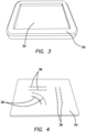
- FIG. 2 illustrates an embodiment of suture pad 10.
- the suture pad 10 is a sheet of simulated tissue material having an upper surface 12 and a lower surface 14 defining a thickness therebetween. The thickness is substantially uniform across the pad and approximately 5 millimeters. The thickness and properties of the pad 10 are ideal to simulate tissue re-approximation with skilled laparoscopic suturing.
- the suture pad 10 has a perimeter 18 which, in one variation, defines a rectangular shape as shown in FIG. 2 .
- the perimeter shape can be any suitable shape.
- the suture pad 10 is made of silicone, low durometer silicone or other suitable polymeric material such as KRATON ® or thermoplastic elastomer. The low durometer allows for the suture pad 10 to have the haptic feedback to simulate soft tissue. Material is chosen to simulate the elasticity, needle resistance, and handling characteristics of live tissue that may be encountered in the operating room.
- the material can also be other rubber-like materials, or thermoset plastic materials that have a soft durometer.
- the suture pad 10 includes a layer of fabric or mesh embedded within the thickness of the pad.
- the fabric or mesh is preferably a 2-way or 4-way stretch material such as stretch nylon or spandex or a stretch nylon/spandex blend mesh or fabric.
- the fabric or mesh material is stretchable and porous and weighs approximately 79 grams per square yard.
- the suture pad 10 may include, in addition to or in lieu of the mesh or fabric material, a reinforcement material, fiber, dye and surface texturing.
- the upper and lower surfaces 12, 14 of FIG. 2 are smooth and without surface texturing.
- the suture pad 10 is flexible and can stretch.
- the mesh, fabric, fiber or other filler material provides reinforcement to the silicone such that the sheet can hold a suture or be stretched without tearing when being manipulated or connected to a base.
- the mesh, fabric, fiber or other filler material may be omitted to create a less resilient and more sensitive pad that is more easily torn when manipulated, thereby, increasing the difficulty of the practice.
- the suture pad 10 includes at least one cut 20.
- the cut 20 is a simulated laceration formed in the suture pad 10.
- FIG. 2 illustrates three cuts 20a, 20b, 20c in the suture pad 10.
- a cut 20 extends from the upper surface 12 to the lower surface 14 across the thickness of the pad 10.
- a partial cut 20 that does not extend across the entire thickness may also be employed for one or more of the cuts.
- the cut 20 may be any shape.
- the cut 20 may be a straight line or curve.
- the curve may be a closed curve or an open curve and multiple cuts 20 may be employed in conjunction with each other to define a suture line of varying difficulty or to define varying practice orientations.
- Lacerations are strategically placed in a variety of orientations so that the learner may adapt their skillset to various tissue orientations encountered in the operating room.
- the suture pad 10 in FIG. 2 includes two straight cuts 20a, 20b and one curved cut 20c oriented with respect to each other to provide various practice orientations.
- the two straight cuts 20a, 20b are shown to be perpendicular to each other but the invention is not so limited.
- the two cuts 20 can be at any angle with respect to each other or mimic actual suture lines associated with a particular organ or otherwise encountered in real surgery or designed to teach a particular skill, train hand dominance, practice different suture run styles and the like.
- Each cut 20 is substantially perpendicular to the upper and lower surfaces 12, 14 and includes two oppositely disposed inner surfaces that face each other.
- the two inner surfaces of a cut 20 are in close juxtaposition and the cut may be difficult to discern by the user.
- the suture pad 10 When mounted on a base, the suture pad 10 may be stretched and, as a result, the cut 20 may open up and define a greater space between the inner surfaces which would require greater force to approximate the inner surfaces while suturing.
- the first cut 20a is approximately 4.0 centimeters in length
- the second cut 20b is approximately 3.5 centimeters in length
- the third cut 20c is approximately 4.0 centimeters in length.
- FIG 2 is not drawn to scale.
- the cuts 20 are pre-formed.
- the suture pad 10 may further include small pre-formed apertures 22 near the perimeter sized and configured for mounting the pad 10 onto a base.
- the variation in FIG. 2 includes four apertures 22 in each corner of the rectangular pad.
- the suture pad 10 further includes a plurality of markings 24 arranged on either side of a cut 20.
- the markings 24 are arranged in a first row 26 along the length and on one side of the cut 20 and in a second row 28 along the length and one the opposite side of the cut 20.
- the first row 26 of markings 24 is directly opposite the second row 28 of markings 24.
- the markings 24 in each row are equally spaced apart from each other. In particular, each marking 24 is spaced from each other by approximately 5 millimeters.
- the center-to-center distance between each marking in the same row is approximately 5 millimeters.
- the distance between the two rows 26, 28 across the laceration is approximately 10 millimeters. Each row is approximately 5 millimeters away from the laceration.
- the markings 24 on the inside of the curve will naturally be spaced closer together relative to the markings 24 on the outside of the curve as can be seen in FIG. 2 .
- the number of markings 24 along a laceration will vary and depends on the length of the cut 20. For example, a cut 20 that has a length of approximately 4 centimeters will have 10 markings in each row for 9 suture runs (10 including knot) for a total of 20 markings 24. A cut 20 that has a length of approximately 3.5 centimeters will have 9 markings in each row for 8 suture runs (9 including knot) for a total of 18 markings 24.
- the knot and final suture pass including the markings 24 at the ends of the cut 20 will extend slightly beyond the laceration length.
- the marking dots serve as precision targets through which the end user should drive the needle.
- the distance between each marking pairing creates a standard distance for a suture run to be made along with a standard distance between a suture bite to close the incision.
- the markings 24 are small circular dots having a diameter of approximately 0,16 cm (1/16 inch).
- the markings 24 are not limited to having a circular shape.
- the markings 24 may be X-shaped, filled circles, empty circles, boxes, star-shaped or any suitable shape that communicates a target with substantial precision for the length and size of the cut.
- the markings 24 are dark in color or any suitable color that provides a visible contrast against the color of the pad 10 to the user.
- the markings have a color that creates a high color contrast with the silicone portion of the suture pad 10 containing the pre-made incisions. Color contrasts between each part of the suture pad 10 are black dot pairings with a light, flesh-tone colored rectangular footprint.
- the markings 24 may be applied to the pad 10 in any number of suitable ways.
- the markings 24 may be drawn in ink, stamped, printed and the like.
- the markings 24 are just beneath the upper surface in a visible manner.
- the markings may be printed on the embedded fabric layer or on an intermediate silicone layer prior to casting a final silicone layer and visible through transparent or translucent silicone in which it is embedded. Another method of applying the markings 24 will now be described.
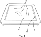
- the mold 30 includes a well 32 that is sized and shaped to correspond to the desired size and shape of the suture pad 10.
- a marking mold 34 shown in FIG. 4 is provided.
- the marking mold 34 includes a plurality of holes 36 formed in the marking mold 34.
- the holes 36 are sized and shaped to correspond to the desired size and shape of the resulting markings 24 to be formed on the pad 10.
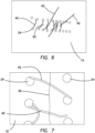
- the marking mold 34 is sized to fit inside the well 32 of the mold 30 as shown in FIG. 5.
- FIG. 5 shows the marking mold 34 placed inside the well 32 of the mold 30.
- wet silicone having a dark/contrast color to the color of the pad 10 is applied onto marking mold 34 such that the wet silicone enters each of the holes 36. Excess wet silicone is wiped away and the silicone inside the holes 36 is allowed to cure.
- the marking mold 34 with the cured silicone inside the holes 36 is then placed/nested into the well 32 of the mold 30 and wet flesh-tone silicone for the pad is then cast into the well 32 on top of and or over the marking mold with the cured black colored silicone and allowed to cure. As the uncured silicone of the pad 10 cures it adheres itself to the cured silicone inside the holes 36.
- the flesh-tone layer is not filled up to the top of the mold in order to allow a mesh layer or markings to be added after the first layer of flesh-toned silicone has cured.
- a mesh/fabric/reinforcement and or markings layer is added, more flesh-toned silicone is added forming a second layer of flesh-tone silicone to complete the thickness of the suture pad.
- the silicone-to-silicone adhesion properties allow the flesh tone layers to adhere together, therefore, encapsulating the mesh layer in between.
- the mesh layer serves as a reinforcement to hold a variety of sutures that are pulled with varying forces.
- the second layer of silicone is transparent and or translucent so that any markings may be seen therethrough.
- the pad is removed from the mold 30 with the markings 24 adhered. If markings are embedded within the layer, printed on a fabric layer or first layer of silicone, the marking mold is not employed.
- the adhesion properties of silicone allow the silicone flesh tone layer to be adhered to the black dots. Silicone adhesive can be additionally used on top of each black dot pairing to further reinforce the adhesion between the dots and the flesh-tone layer.
- the markings 24 serve as targets for passing a suture and are intended to guide the learner toward ideal needle insertion points around the laceration.
- the user will practice suturing across the laceration by passing the suture needle and suture through the center of the marking 24.
- An assessment of a user's skills is easily performed by observing whether the suture needle and suture has passed through the marking, its center or not.
- the markings 10 also serve as a means for easily evaluating the user's skills.
- the marking placements allow for reflective assessment of suture performance based on target accuracy.
- An exemplary suture 42 placement is shown in FIG. 6.
- FIG. 7 illustrates a suture pad after completed suture practice.
- the suture has hit and missed the target markings 24.
- a typical 2-O coated vicryl (polyglactin) dyed suture approximately 70 cm in length can be used and cut to four times the laceration length for ideal handling with the suture pad 10.
- a variety of suture and needle types and lengths can be utilized with the suture pads described herein. The user can employ a simple continuous suture run style and perform suture on all three lacerations/orientations.
- the flesh tone portion of the suture pad 10 can be textured.
- the textured surface allows the suture pad 10 to be easily grasped and manipulated.
- the mesh layer can be removed in order to simulate training on more fragile tissue.
- the durometer used for the flesh tone portion of the suture pad 10 can be made from a mixture of at least two different silicone durometers, a low and high, in order to get a realistic haptic feedback response that is similar to tissues within the abdominal cavity.
- the rectangular footprint of the suture pad 10 can be re-shaped to variety of sizes or geometries if a particular procedure involving suturing is being trained.
- the incisions 20 can include a straight incision that is angled.
- the suture pad can include any number of incisions.
- the incision lengths can also be less than 4.0 cm or 3.5 cm.
- Colors of the suture pad 10 are not limited to flesh tone and black. The color of the targets 24 and the underlying silicone layer, however, should provide a contrasting difference from one another so that the end user is able to distinguish between each feature.
- the markings 24 are recessed into the suture pad 10
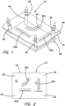
- the suture pad 10 is connected to the posts 34 and in essence suspended from the base by a distance.
- the suture pad 10 is mounted in tension being slightly stretched between and connected to the posts 40 as shown in FIG. 1 .
- the tension of the suture pad 10 may be adjusted by angulating the posts 40 or by stretching and piercing the suture pad 10 in locations closer together along the simulated tissue portion.
- the placement of the suture pad 10 onto the base advantageously allows for additional manipulation of tissue orientation.
- the use of silicone for the suture pad allows it to be flexible such that, when it is assembled on the base, the pad can be oriented in the vertical position or horizontal position and any angle in between. This creates multiple planar views for the suture pad to allow for spatial challenge training for this skill.
- the suture pad 10 may be employed by itself or mounted on the base 38. In either case, the suture pad 10 by itself or mounted on the base 38 may be placed inside a surgical training device for the practice of suturing in a laparoscopic environment.
- the surgical training device is typically configured to mimic the torso of a patient such as the abdominal region.
- An example of a surgical training device is described in U.S. Patent No. 8,764,452 .
- the surgical training device provides a body cavity substantially obscured from the user and configured for receiving the suture pad inside the cavity. The body cavity and the suture pad disposed therein are accessed via a tissue simulation region in the trainer that is penetrated by the user employing laparoscopic devices.
- the surgical trainer is a useful tool for teaching, practicing and demonstrating various surgical procedures and their related instruments in simulation of a patient undergoing a surgical procedure.
- Surgical instruments are inserted into the cavity through the tissue simulation region as well as through preestablished apertures in the top cover.
- the suture pad 10 may be connected to the trainer with clips.
- a base 38 If a base 38 is employed, it may be retained with a patch of hook-and-loop type fastening material (VELCRO ® ) affixed to the base and to the floor of the trainer.
- a video display monitor provides the user with a view of the mock surgical field inside the cavity of the trainer via a camera such as an endoscope.
- a user will mount at least one suture pad 10 onto the posts 40 connected to the base 38. If the suture pad 10 includes preformed apertures 22 then mounting the suture pad 10 includes placing the apertures 22 over each post 34 and sliding the simulated tissue portion 36 to rest within one of the at least one notches 42 formed in the post 34.
- the suture pad 10 is mounted on all four posts 40. Fewer posts may be employed to suspend the suture pad 10.
- the notches advantageously permit the entire suture pad 10 to be mounted at an angle such that one side or at least one corner of the suture pad 40 is mounted on a higher or lower notch relative to the other corners and posts.
- the tapered distal ends of the posts 40 can be used to puncture apertures 22 anywhere into the pad 10.
- the tension in the suture pad 10 can be selected by the user when the user mounts the suture pad 10 onto the posts 40.
- the suture pad 10 when the suture pad 10 is mounted by piercing an aperture 22 into the suture pad 10, it can then be selectively stretched making the suture pad 10 as tense or loose as the user wishes before piercing at least a second aperture 22 to mount the suture pad 10 on another post 40 and so forth.
- the fabric reinforced silicone material prevents the aperture 22 from propagating.
- the suture pad 10 provides a realistic platform for presenting simulated suturable tissue for training in a laparoscopic environment.
- the clinician practices certain techniques such as cutting and suturing, the clinician will use certain instruments such as graspers, cutters, suture needles, sutures, laparoscopes, endoscopes, trocars and the like.
- the simulated tissue structure will give and flex under the force, deflecting a certain degree depending upon the tension with which it is mounted.
- This dynamism of the suture pad advantageously mimics real live tissue that gives way, moves and flexes upon manipulation in real life.
- cutting and suturing feels differently when performed on a suture pad that is suspended, that is in tension and that allows for a certain amount of deflection.
- These simulation advantages are provided by the suture pad 10 of the present invention and are particularly useful when practicing laparoscopic surgical techniques that allow the user to fine tune depth perception and tissue manipulation skills while suturing, cutting and puncturing in a simulated laparoscopic environment.
- the present invention provides a model for guided suture placement for the development of laparoscopic suturing skillsets through deliberate practice.
- the suture pad has a reaction that simulates soft tissue found within the abdominal cavity. Since this pad allows for laparoscopic suture training, the suture pad 10 can be grasped and manipulated with laparoscopic instruments such as laparoscopic scissors, graspers, and Maryland dissectors. Additionally, this pad is tough enough to hold various types of sutures that could be encountered within a surgical procedure. Although the suture pad 10 is tough enough to withstand suture, it is also fragile enough such that the strength and force required to create a suture is similar to tissue reaction encountered during laparoscopic procedures. Since suturing is the laparoscopic target skill for the suture pad, the suture pad allows for multiple suture orientations to be learned and practiced.
- the suture pad allows for a laparoscopic running suture, such as a simple continuous suture, to be made.
- the suture pad is able to be fixed on the base to have a front or angled face orientation.
- the suture pad contains multiple lacerations or incisions to enable multiple suture runs.
- the suture pad allows the end user to create a knot and final suture.
- the suture pad has multiple lacerations or incision orientations to be sutured to facilitate learning multiple suturing orientations. This allows for visual and movement challenges in difficulty levels for the trainee. Additionally, the suture pad permits the practicing surgeon or surgical resident to demonstrate dexterity and precision through their movement of suturing.
- the suture pad that contains precision targets for suturing allows the trainee to practice on their laparoscopic dexterity. Additionally, the suture pad allows for the targets to serve as a metric for the trainee's dexterity of laparoscopic suturing that is assessable.
- the present suture pad has targets that are well-defined and consistent in spacing between the laceration or incision and the spacing between each consecutive pair of precision target markings on the pad. The spacing between the targets is selected to facilitate a strong suture placement. Deviation from suture targets may result in suture that lacks integrity and may fail to re-approximate tissue fully.
- material properties of the suture pad are such that the user can practice applying the appropriate amount of tension on their suture. If the user runs suture with too much force or tension, the tissue will cinch, or over-approximate. If the suture is run too loose, the user will identify that the laceration remains open, and tissue fails to re-approximate.
Landscapes
- Engineering & Computer Science (AREA)
- Health & Medical Sciences (AREA)
- Physics & Mathematics (AREA)
- General Physics & Mathematics (AREA)
- General Health & Medical Sciences (AREA)
- Medical Informatics (AREA)
- Business, Economics & Management (AREA)
- Theoretical Computer Science (AREA)
- Computational Mathematics (AREA)
- Medicinal Chemistry (AREA)
- Mathematical Analysis (AREA)
- Mathematical Optimization (AREA)
- Mathematical Physics (AREA)
- Pure & Applied Mathematics (AREA)
- Chemical & Material Sciences (AREA)
- Educational Administration (AREA)
- Educational Technology (AREA)
- Algebra (AREA)
- Pulmonology (AREA)
- Radiology & Medical Imaging (AREA)
- Surgery (AREA)
- Life Sciences & Earth Sciences (AREA)
- Nuclear Medicine, Radiotherapy & Molecular Imaging (AREA)
- Biomedical Technology (AREA)
- Heart & Thoracic Surgery (AREA)
- Molecular Biology (AREA)
- Animal Behavior & Ethology (AREA)
- Public Health (AREA)
- Veterinary Medicine (AREA)
- Instructional Devices (AREA)
Claims (15)
- Chirurgisches Trainingsmodell zum Trainieren von Nahttechniken, wobei das Modell Folgendes umfasst:ein Blatt aus simuliertem Gewebematerial, das ein Nahtkissen (10) definiert, wobei das Blatt Folgendes umfasst:eine erste Materialschicht, die eine Vielzahl von Markierungen (24) umfasst;eine zweite Materialschicht, die auf der ersten Materialschicht ausgehärtet ist; undmindestens einen Schnitt (20), der in dem Blatt aus simuliertem Gewebematerial gebildet ist,wobei die Vielzahl von Markierungen (24) auf beiden Seiten des mindestens einen Schnitts (20) unterhalb der zweiten Materialschicht ausgelegt ist.
- Chirurgisches Trainingsmodell nach Anspruch 1, wobei die erste Schicht des Materials eine erste Farbe hat, wobei die zweite Materialschicht eine zweite Farbe hat, und wobei die erste Farbe mit der zweiten Farbe kontrastiert.
- Chirurgisches Trainingsmodell nach Anspruch 2, wobei die zweite Materialschicht aus einer fleischfarbenen Farbe gebildet ist und die erste Materialschicht aus einer dunklen Farbe gebildet ist, die mit der fleischfarbenen Farbe kontrastiert.
- Chirurgisches Trainingsmodell nach Anspruch 1, wobei die erste Schicht eine Gewebeschicht ist, die unter der zweiten Materialschicht vorgesehen ist, und wobei die Vielzahl von Markierungen auf die Gewebeschicht gedruckt ist.
- Chirurgisches Trainingsmodell nach Anspruch 1, wobei die Vielzahl von Markierungen (24) eine Vielzahl von kreisförmigen gefüllten Punkten umfasst.
- Chirurgisches Trainingsmodell nach Anspruch 1, wobei das Blatt aus simuliertem Gewebematerial ein Silikonmaterial umfasst.
- Chirurgisches Trainingsmodell nach Anspruch 1, wobei die zweite Schicht des Materials texturiert ist.
- Chirurgisches Trainingsmodell nach Anspruch 1, das ferner eine Vielzahl von Pfosten (40) umfasst, die von einer Basis (38) ausgehen und wobei das Blatt aus simuliertem Gewebematerial ferner eine Vielzahl von darin gebildeten Öffnungen (22) umfasst, wobei jeder Pfosten der Vielzahl von Pfosten (40) durch die entsprechende Öffnung der Vielzahl von Öffnungen (22) ausfahrbar ist, wobei das Blatt aus simuliertem Gewebematerial entlang jedes Pfostens der Vielzahl von Pfosten (40) bewegbar ist, um mindestens einen Teil des Blattes aus simuliertem Gewebematerial relativ zu mindestens einem Pfosten (40), anderen Teilen des Blattes aus simuliertem Gewebematerial und der Basis (38) abzuwinkeln.
- Verfahren zur Herstellung eines chirurgischen Trainingsmodells für das Training von Nahttechniken, wobei das Verfahren folgenden Schritt umfasst:
Bereitstellen einer Form (30), die eine Vertiefung umfasst, die so bemessen und geformt ist, dass sie dem chirurgischen Übungsmodell entspricht, wobei das Verfahren dadurch gekennzeichnet ist, dass es ferner folgende Schritte umfasst:Bereitstellen einer Markierungsform (34), die so bemessen ist, dass sie in die Vertiefung der Form (30) passt, wobei die Markierungsform (34) eine Vielzahl von Löchern (36) enthält, die in ihr gebildet sind;Auftragen von nassem Silikon auf die Markierungsform (34), so dass das nasse Silikon in die Löcher (36) der Markierungsform eindringt und das nasse Silikon in den Löchern (36) aushärten kann, wodurch eine Vielzahl von Markierungen (24) entsteht;Positionieren der Markierungsform (34) innerhalb der Vertiefung der Form (30); undGießen von nassem Silikon über die Markierungsform (34), die sich in der Form (30) befindet, wodurch die Vielzahl von Markierungen (24) unter einer Schicht von Silikon positioniert werden, indem das gegossene nasse Silikon aushärtet und an dem ausgehärteten Silikon innerhalb der Löcher (36) der Markierungsform (34) haftet. - Verfahren nach Anspruch 9, das ferner folgende Schritte umfasst:Positionieren einer Netzschicht in der Vertiefung der Form (30); undHinzufügen einer Silikonschicht auf die Netzschicht.
- Verfahren nach Anspruch 9, wobei das Auftragen von nassem Silikon auf die Markierungsform (34) das Auftragen von nassem Silikon mit einer dunklen Farbe auf die Markierungsform (34) umfasst, und wobei das Gießen von nassem Silikon auf die Markierungsform (34) das Gießen von nassem Silikon mit einer fleischfarbenen Farbe umfasst.
- Chirurgisches Trainingsmodell nach Anspruch 1, wobei die zweite Materialschicht transparent oder lichtdurchlässig ist, wodurch die Vielzahl der Markierungen (24) unter der zweiten Schicht sichtbar wird.
- Chirurgisches Trainingsmodell nach Anspruch 1, wobei die Vielzahl der Markierungen (24) als eine Silikonzwischenschicht geformt ist, die unter der zweiten Materialschicht vorgesehen ist.
- Chirurgisches Trainingsmodell nach Anspruch 1, wobei die Vielzahl von Markierungen (24) in zwei oder mehr verschiedenen Gruppen von Markierungen organisiert sind, so dass ein Benutzer angewiesen wird, eine Naht entlang eines vorbestimmten Pfades zu führen, der durch eine ausgewählte Gruppe von Markierungen definiert ist.
- Chirurgischer Trainer, der eine Körperhöhle umfasst, die für einen Benutzer im Wesentlichen unsichtbar ist, wobei der chirurgische Trainer das chirurgische Trainingsmodell nach Anspruch 1 umfasst, das in der Körperhöhle aufgenommen ist, wobei die Körperhöhle des chirurgischen Trainingsmodells über einen Gewebesimulationsbereich zugänglich ist.
Applications Claiming Priority (2)
| Application Number | Priority Date | Filing Date | Title |
|---|---|---|---|
| US201762586369P | 2017-11-15 | 2017-11-15 | |
| PCT/US2018/061275 WO2019099665A1 (en) | 2017-11-15 | 2018-11-15 | Suturing skills surgical training model |
Publications (3)
| Publication Number | Publication Date |
|---|---|
| EP3711039A1 EP3711039A1 (de) | 2020-09-23 |
| EP3711039C0 EP3711039C0 (de) | 2024-10-23 |
| EP3711039B1 true EP3711039B1 (de) | 2024-10-23 |
Family
ID=64664438
Family Applications (1)
| Application Number | Title | Priority Date | Filing Date |
|---|---|---|---|
| EP18816356.2A Active EP3711039B1 (de) | 2017-11-15 | 2018-11-15 | Trainingsmodell für chirurgische nahtfähigkeiten |
Country Status (8)
| Country | Link |
|---|---|
| US (2) | US11501662B2 (de) |
| EP (1) | EP3711039B1 (de) |
| JP (2) | JP7320504B2 (de) |
| KR (1) | KR102670319B1 (de) |
| AU (1) | AU2018369906B2 (de) |
| CA (1) | CA3082777A1 (de) |
| ES (1) | ES2991949T3 (de) |
| WO (1) | WO2019099665A1 (de) |
Families Citing this family (7)
| Publication number | Priority date | Publication date | Assignee | Title |
|---|---|---|---|---|
| US11636782B2 (en) * | 2019-05-31 | 2023-04-25 | Caroline A. Glicksman | Breast and abdominal augmentation and reconstruction teaching model |
| CN110379275B (zh) * | 2019-07-18 | 2024-07-02 | 中山大学肿瘤防治中心(中山大学附属肿瘤医院、中山大学肿瘤研究所) | 缝皮练习器 |
| CO2019010338A1 (es) * | 2019-09-25 | 2019-12-10 | Pontificia Univ Javeriana | Modelos morfológicos para el entrenamiento de cirugía mínimamente invasiva, su método de fabricación y sistema para la práctica |
| CN110610642A (zh) * | 2019-10-21 | 2019-12-24 | 海口市人民医院 | 一种手术缝合训练器 |
| CN110974326B (zh) * | 2019-12-30 | 2021-09-17 | 延安大学附属医院 | 一种减小瘢痕形成的方法和系统 |
| RU2768594C1 (ru) * | 2021-03-12 | 2022-03-24 | Константин Викторович Пучков | Комплект симуляционных тренажеров для обучения методике экстракорпорального и интракорпорального шва |
| US12579907B2 (en) | 2021-06-20 | 2026-03-17 | Gonzalo Juan Vitagliano | Medical training device and method to use it in teaching laparoscopic and robotic partial nephrectomy |
Family Cites Families (37)
| Publication number | Priority date | Publication date | Assignee | Title |
|---|---|---|---|---|
| US2678505A (en) | 1951-07-06 | 1954-05-18 | Horace J Munson | Doll for playing at surgery |
| US3775865A (en) | 1972-07-24 | 1973-12-04 | R Rowan | Simulator for teaching suturing techniques |
| US4195420A (en) * | 1974-02-07 | 1980-04-01 | Sandra Fields | Epistotomy repair model |
| US4386917A (en) | 1981-09-16 | 1983-06-07 | Forrest Leonard E | Suturing training device and method |
| US4596528A (en) | 1984-07-02 | 1986-06-24 | Lewis Leonard A | Simulated skin and method |
| US4789340A (en) | 1987-08-18 | 1988-12-06 | Zikria Bashir A | Surgical student teaching aid |
| US5518407A (en) | 1993-11-02 | 1996-05-21 | Greenfield; Cathy L. | Anatomically correct artificial organ replicas for use as teaching aids |
| GB0215051D0 (en) | 2002-06-28 | 2002-08-07 | Limbs And Things Ltd | Simulated body tissue |
| US7575434B2 (en) | 2006-08-01 | 2009-08-18 | Palakodeti Ratna K | Surgery practice kit |
| US20080064017A1 (en) | 2006-08-29 | 2008-03-13 | Grundmeyer Ramond Iii | Suture training device |
| CN201066541Y (zh) * | 2007-08-15 | 2008-05-28 | 宁夏医学院 | 一种外科手术缝合线路练习板 |
| US9959785B2 (en) | 2010-08-24 | 2018-05-01 | Vti Medical, Inc. | Apparatus and method for laparoscopic skills training |
| EP2622594B1 (de) | 2010-10-01 | 2018-08-22 | Applied Medical Resources Corporation | Tragbarer laparoskopischer trainer |
| US20120115117A1 (en) * | 2010-11-08 | 2012-05-10 | Marshall M Blair | Suture training device |
| US20120115118A1 (en) | 2010-11-08 | 2012-05-10 | Marshall M Blair | Suture training device |
| JP2014512025A (ja) | 2011-04-05 | 2014-05-19 | タケダ・ナイコメッド・アーエス | 医療用訓練装置および方法 |
| GB2492115B (en) | 2011-06-22 | 2014-03-05 | Royal Brompton & Harefield Nhs Foundation Trust | Simulation apparatus |
| WO2013051918A1 (es) | 2011-10-06 | 2013-04-11 | Quirarte Catano Cesar | Dispositivo simulador de tejidos, para el aprendizaje y entrenamiento de técnicas básicas de cirugía laparoscópica, endoscópica o de mínima invasión |
| KR101963610B1 (ko) | 2011-10-21 | 2019-03-29 | 어플라이드 메디컬 리소시스 코포레이션 | 수술 트레이닝용 모의 조직 구조 |
| US8641422B2 (en) | 2012-03-15 | 2014-02-04 | Vincent Francavilla | Bone augmentation training system |
| US10553130B2 (en) | 2012-05-03 | 2020-02-04 | Regents Of The University Of Minnesota | Systems and methods for analyzing surgical techniques |
| US20140030682A1 (en) | 2012-07-26 | 2014-01-30 | William Jackson THILENIUS | Training device and method for spaying and/or suturing animals |
| CA2880482C (en) * | 2012-09-27 | 2020-03-10 | Applied Medical Resources Corporation | Surgical training model for laparoscopic procedures |
| US20140212861A1 (en) | 2013-01-29 | 2014-07-31 | Peter Joseph Romano | Educational suturing apparatus |
| EP3660816B1 (de) | 2013-03-01 | 2021-10-13 | Applied Medical Resources Corporation | Moderne konstruktionen und verfahren für chirurgische simulationen |
| JP6517201B2 (ja) * | 2013-07-24 | 2019-05-22 | アプライド メディカル リソーシーズ コーポレイション | ファーストエントリーモデル |
| KR102581212B1 (ko) * | 2014-03-26 | 2023-09-21 | 어플라이드 메디컬 리소시스 코포레이션 | 시뮬레이션된 절개가능 조직 |
| WO2015177926A1 (ja) | 2014-05-23 | 2015-11-26 | 株式会社レジーナ | 縫合練習用人工皮膚及びその製造方法 |
| JP5759055B1 (ja) | 2014-05-26 | 2015-08-05 | サンアロー株式会社 | 臓器モデル |
| WO2015189954A1 (ja) | 2014-06-12 | 2015-12-17 | 株式会社レジーナ | 縫合練習用透明性人工皮膚及びその製造方法 |
| US9895212B2 (en) * | 2014-10-31 | 2018-02-20 | Prevent Patch LLC | Devices and methods for preventing incisional hernias |
| EP3218892B1 (de) * | 2014-11-13 | 2019-10-23 | Applied Medical Resources Corporation | Simulierte gewebemodelle und verfahren |
| US9520073B2 (en) | 2015-01-26 | 2016-12-13 | Ethicon, Inc. | Ex-vivo anatomic tissue specimen wound closure simulation model |
| ES2732722T3 (es) | 2015-02-19 | 2019-11-25 | Applied Med Resources | Estructuras tisulares simuladas y métodos |
| JP5870349B1 (ja) * | 2015-03-28 | 2016-02-24 | 株式会社ナースあい | 注射トレーニング器具 |
| US10083629B2 (en) | 2015-06-30 | 2018-09-25 | University Of South Florida | Synthetic vaginal cuff model and method of simulating vaginal cuff closure |
| CA3249585A1 (en) * | 2015-07-16 | 2025-02-24 | Applied Medical Resources Corporation | Simulated dissectable tissue |
-
2018
- 2018-11-15 AU AU2018369906A patent/AU2018369906B2/en active Active
- 2018-11-15 EP EP18816356.2A patent/EP3711039B1/de active Active
- 2018-11-15 KR KR1020207017174A patent/KR102670319B1/ko active Active
- 2018-11-15 CA CA3082777A patent/CA3082777A1/en active Pending
- 2018-11-15 WO PCT/US2018/061275 patent/WO2019099665A1/en not_active Ceased
- 2018-11-15 ES ES18816356T patent/ES2991949T3/es active Active
- 2018-11-15 JP JP2020526529A patent/JP7320504B2/ja active Active
- 2018-11-15 US US16/191,965 patent/US11501662B2/en active Active
-
2022
- 2022-11-14 US US17/986,441 patent/US12327489B2/en active Active
-
2023
- 2023-07-24 JP JP2023120039A patent/JP7639068B2/ja active Active
Also Published As
| Publication number | Publication date |
|---|---|
| JP2023159071A (ja) | 2023-10-31 |
| US20190147767A1 (en) | 2019-05-16 |
| US11501662B2 (en) | 2022-11-15 |
| CA3082777A1 (en) | 2019-05-23 |
| JP2021503102A (ja) | 2021-02-04 |
| US20230070953A1 (en) | 2023-03-09 |
| KR102670319B1 (ko) | 2024-05-30 |
| EP3711039C0 (de) | 2024-10-23 |
| AU2018369906A1 (en) | 2020-05-07 |
| US12327489B2 (en) | 2025-06-10 |
| AU2018369906B2 (en) | 2024-09-19 |
| JP7320504B2 (ja) | 2023-08-03 |
| KR20200088855A (ko) | 2020-07-23 |
| ES2991949T3 (es) | 2024-12-05 |
| WO2019099665A1 (en) | 2019-05-23 |
| EP3711039A1 (de) | 2020-09-23 |
| JP7639068B2 (ja) | 2025-03-04 |
Similar Documents
| Publication | Publication Date | Title |
|---|---|---|
| US12327489B2 (en) | Suturing skills surgical training model | |
| US11990055B2 (en) | Surgical training model for laparoscopic procedures | |
| US9959786B2 (en) | Surgical training model for laparoscopic procedures | |
| EP3467805B1 (de) | Chirurgisches schulungsmodell für transluminale laparoskopische eingriffe |
Legal Events
| Date | Code | Title | Description |
|---|---|---|---|
| STAA | Information on the status of an ep patent application or granted ep patent |
Free format text: STATUS: UNKNOWN |
|
| STAA | Information on the status of an ep patent application or granted ep patent |
Free format text: STATUS: THE INTERNATIONAL PUBLICATION HAS BEEN MADE |
|
| PUAI | Public reference made under article 153(3) epc to a published international application that has entered the european phase |
Free format text: ORIGINAL CODE: 0009012 |
|
| STAA | Information on the status of an ep patent application or granted ep patent |
Free format text: STATUS: REQUEST FOR EXAMINATION WAS MADE |
|
| 17P | Request for examination filed |
Effective date: 20200415 |
|
| AK | Designated contracting states |
Kind code of ref document: A1 Designated state(s): AL AT BE BG CH CY CZ DE DK EE ES FI FR GB GR HR HU IE IS IT LI LT LU LV MC MK MT NL NO PL PT RO RS SE SI SK SM TR |
|
| AX | Request for extension of the european patent |
Extension state: BA ME |
|
| RIN1 | Information on inventor provided before grant (corrected) |
Inventor name: RAYGAN, OSCAR Inventor name: CARTER, BRIAN Inventor name: HOFSTETTER, GREGORY, K. |
|
| DAV | Request for validation of the european patent (deleted) | ||
| DAX | Request for extension of the european patent (deleted) | ||
| STAA | Information on the status of an ep patent application or granted ep patent |
Free format text: STATUS: EXAMINATION IS IN PROGRESS |
|
| 17Q | First examination report despatched |
Effective date: 20211110 |
|
| GRAP | Despatch of communication of intention to grant a patent |
Free format text: ORIGINAL CODE: EPIDOSNIGR1 |
|
| STAA | Information on the status of an ep patent application or granted ep patent |
Free format text: STATUS: GRANT OF PATENT IS INTENDED |
|
| INTG | Intention to grant announced |
Effective date: 20240515 |
|
| RIN1 | Information on inventor provided before grant (corrected) |
Inventor name: RAYGAN, OSCAR Inventor name: CARTER, BRIAN Inventor name: HOFSTETTER, GREGORY K. |
|
| GRAS | Grant fee paid |
Free format text: ORIGINAL CODE: EPIDOSNIGR3 |
|
| GRAA | (expected) grant |
Free format text: ORIGINAL CODE: 0009210 |
|
| STAA | Information on the status of an ep patent application or granted ep patent |
Free format text: STATUS: THE PATENT HAS BEEN GRANTED |
|
| AK | Designated contracting states |
Kind code of ref document: B1 Designated state(s): AL AT BE BG CH CY CZ DE DK EE ES FI FR GB GR HR HU IE IS IT LI LT LU LV MC MK MT NL NO PL PT RO RS SE SI SK SM TR |
|
| REG | Reference to a national code |
Ref country code: GB Ref legal event code: FG4D |
|
| REG | Reference to a national code |
Ref country code: CH Ref legal event code: EP |
|
| REG | Reference to a national code |
Ref country code: DE Ref legal event code: R096 Ref document number: 602018075790 Country of ref document: DE |
|
| REG | Reference to a national code |
Ref country code: IE Ref legal event code: FG4D |
|
| U01 | Request for unitary effect filed |
Effective date: 20241023 |
|
| U07 | Unitary effect registered |
Designated state(s): AT BE BG DE DK EE FI FR IT LT LU LV MT NL PT RO SE SI Effective date: 20241029 |
|
| REG | Reference to a national code |
Ref country code: ES Ref legal event code: FG2A Ref document number: 2991949 Country of ref document: ES Kind code of ref document: T3 Effective date: 20241205 |
|
| U20 | Renewal fee for the european patent with unitary effect paid |
Year of fee payment: 7 Effective date: 20241127 |
|
| PG25 | Lapsed in a contracting state [announced via postgrant information from national office to epo] |
Ref country code: IS Free format text: LAPSE BECAUSE OF FAILURE TO SUBMIT A TRANSLATION OF THE DESCRIPTION OR TO PAY THE FEE WITHIN THE PRESCRIBED TIME-LIMIT Effective date: 20250223 Ref country code: HR Free format text: LAPSE BECAUSE OF FAILURE TO SUBMIT A TRANSLATION OF THE DESCRIPTION OR TO PAY THE FEE WITHIN THE PRESCRIBED TIME-LIMIT Effective date: 20241023 |
|
| PG25 | Lapsed in a contracting state [announced via postgrant information from national office to epo] |
Ref country code: NO Free format text: LAPSE BECAUSE OF FAILURE TO SUBMIT A TRANSLATION OF THE DESCRIPTION OR TO PAY THE FEE WITHIN THE PRESCRIBED TIME-LIMIT Effective date: 20250123 |
|
| PG25 | Lapsed in a contracting state [announced via postgrant information from national office to epo] |
Ref country code: GR Free format text: LAPSE BECAUSE OF FAILURE TO SUBMIT A TRANSLATION OF THE DESCRIPTION OR TO PAY THE FEE WITHIN THE PRESCRIBED TIME-LIMIT Effective date: 20250124 |
|
| PG25 | Lapsed in a contracting state [announced via postgrant information from national office to epo] |
Ref country code: PL Free format text: LAPSE BECAUSE OF FAILURE TO SUBMIT A TRANSLATION OF THE DESCRIPTION OR TO PAY THE FEE WITHIN THE PRESCRIBED TIME-LIMIT Effective date: 20241023 |
|
| PG25 | Lapsed in a contracting state [announced via postgrant information from national office to epo] |
Ref country code: RS Free format text: LAPSE BECAUSE OF FAILURE TO SUBMIT A TRANSLATION OF THE DESCRIPTION OR TO PAY THE FEE WITHIN THE PRESCRIBED TIME-LIMIT Effective date: 20250123 |
|
| REG | Reference to a national code |
Ref country code: CH Ref legal event code: PL |
|
| PG25 | Lapsed in a contracting state [announced via postgrant information from national office to epo] |
Ref country code: SM Free format text: LAPSE BECAUSE OF FAILURE TO SUBMIT A TRANSLATION OF THE DESCRIPTION OR TO PAY THE FEE WITHIN THE PRESCRIBED TIME-LIMIT Effective date: 20241023 |
|
| PG25 | Lapsed in a contracting state [announced via postgrant information from national office to epo] |
Ref country code: MC Free format text: LAPSE BECAUSE OF FAILURE TO SUBMIT A TRANSLATION OF THE DESCRIPTION OR TO PAY THE FEE WITHIN THE PRESCRIBED TIME-LIMIT Effective date: 20241023 |
|
| REG | Reference to a national code |
Ref country code: CH Ref legal event code: PL |
|
| PG25 | Lapsed in a contracting state [announced via postgrant information from national office to epo] |
Ref country code: CH Free format text: LAPSE BECAUSE OF NON-PAYMENT OF DUE FEES Effective date: 20241130 |
|
| PG25 | Lapsed in a contracting state [announced via postgrant information from national office to epo] |
Ref country code: SK Free format text: LAPSE BECAUSE OF FAILURE TO SUBMIT A TRANSLATION OF THE DESCRIPTION OR TO PAY THE FEE WITHIN THE PRESCRIBED TIME-LIMIT Effective date: 20241023 |
|
| PG25 | Lapsed in a contracting state [announced via postgrant information from national office to epo] |
Ref country code: CZ Free format text: LAPSE BECAUSE OF FAILURE TO SUBMIT A TRANSLATION OF THE DESCRIPTION OR TO PAY THE FEE WITHIN THE PRESCRIBED TIME-LIMIT Effective date: 20241023 |
|
| PLBE | No opposition filed within time limit |
Free format text: ORIGINAL CODE: 0009261 |
|
| STAA | Information on the status of an ep patent application or granted ep patent |
Free format text: STATUS: NO OPPOSITION FILED WITHIN TIME LIMIT |
|
| 26N | No opposition filed |
Effective date: 20250724 |
|
| U20 | Renewal fee for the european patent with unitary effect paid |
Year of fee payment: 8 Effective date: 20251126 |
|
| PGFP | Annual fee paid to national office [announced via postgrant information from national office to epo] |
Ref country code: GB Payment date: 20251127 Year of fee payment: 8 |
|
| PGFP | Annual fee paid to national office [announced via postgrant information from national office to epo] |
Ref country code: IE Payment date: 20251127 Year of fee payment: 8 |
|
| PGFP | Annual fee paid to national office [announced via postgrant information from national office to epo] |
Ref country code: ES Payment date: 20251201 Year of fee payment: 8 |
|
| PG25 | Lapsed in a contracting state [announced via postgrant information from national office to epo] |
Ref country code: HU Free format text: LAPSE BECAUSE OF FAILURE TO SUBMIT A TRANSLATION OF THE DESCRIPTION OR TO PAY THE FEE WITHIN THE PRESCRIBED TIME-LIMIT; INVALID AB INITIO Effective date: 20181115 |
|
| PG25 | Lapsed in a contracting state [announced via postgrant information from national office to epo] |
Ref country code: CY Free format text: LAPSE BECAUSE OF FAILURE TO SUBMIT A TRANSLATION OF THE DESCRIPTION OR TO PAY THE FEE WITHIN THE PRESCRIBED TIME-LIMIT; INVALID AB INITIO Effective date: 20181115 |